KR20180098412A - Profiling of deep-seated sequences of tumors - Google Patents
Profiling of deep-seated sequences of tumors Download PDFInfo
- Publication number
- KR20180098412A KR20180098412A KR1020187023352A KR20187023352A KR20180098412A KR 20180098412 A KR20180098412 A KR 20180098412A KR 1020187023352 A KR1020187023352 A KR 1020187023352A KR 20187023352 A KR20187023352 A KR 20187023352A KR 20180098412 A KR20180098412 A KR 20180098412A
- Authority
- KR
- South Korea
- Prior art keywords
- sample
- sequencing
- genomic material
- capture
- amplification
- Prior art date
- Legal status (The legal status is an assumption and is not a legal conclusion. Google has not performed a legal analysis and makes no representation as to the accuracy of the status listed.)
- Granted
Links
Images


Classifications
-
- C—CHEMISTRY; METALLURGY
- C12—BIOCHEMISTRY; BEER; SPIRITS; WINE; VINEGAR; MICROBIOLOGY; ENZYMOLOGY; MUTATION OR GENETIC ENGINEERING
- C12Q—MEASURING OR TESTING PROCESSES INVOLVING ENZYMES, NUCLEIC ACIDS OR MICROORGANISMS; COMPOSITIONS OR TEST PAPERS THEREFOR; PROCESSES OF PREPARING SUCH COMPOSITIONS; CONDITION-RESPONSIVE CONTROL IN MICROBIOLOGICAL OR ENZYMOLOGICAL PROCESSES
- C12Q1/00—Measuring or testing processes involving enzymes, nucleic acids or microorganisms; Compositions therefor; Processes of preparing such compositions
- C12Q1/68—Measuring or testing processes involving enzymes, nucleic acids or microorganisms; Compositions therefor; Processes of preparing such compositions involving nucleic acids
- C12Q1/6869—Methods for sequencing
-
- C—CHEMISTRY; METALLURGY
- C12—BIOCHEMISTRY; BEER; SPIRITS; WINE; VINEGAR; MICROBIOLOGY; ENZYMOLOGY; MUTATION OR GENETIC ENGINEERING
- C12Q—MEASURING OR TESTING PROCESSES INVOLVING ENZYMES, NUCLEIC ACIDS OR MICROORGANISMS; COMPOSITIONS OR TEST PAPERS THEREFOR; PROCESSES OF PREPARING SUCH COMPOSITIONS; CONDITION-RESPONSIVE CONTROL IN MICROBIOLOGICAL OR ENZYMOLOGICAL PROCESSES
- C12Q1/00—Measuring or testing processes involving enzymes, nucleic acids or microorganisms; Compositions therefor; Processes of preparing such compositions
- C12Q1/68—Measuring or testing processes involving enzymes, nucleic acids or microorganisms; Compositions therefor; Processes of preparing such compositions involving nucleic acids
- C12Q1/6806—Preparing nucleic acids for analysis, e.g. for polymerase chain reaction [PCR] assay
-
- C—CHEMISTRY; METALLURGY
- C12—BIOCHEMISTRY; BEER; SPIRITS; WINE; VINEGAR; MICROBIOLOGY; ENZYMOLOGY; MUTATION OR GENETIC ENGINEERING
- C12Q—MEASURING OR TESTING PROCESSES INVOLVING ENZYMES, NUCLEIC ACIDS OR MICROORGANISMS; COMPOSITIONS OR TEST PAPERS THEREFOR; PROCESSES OF PREPARING SUCH COMPOSITIONS; CONDITION-RESPONSIVE CONTROL IN MICROBIOLOGICAL OR ENZYMOLOGICAL PROCESSES
- C12Q1/00—Measuring or testing processes involving enzymes, nucleic acids or microorganisms; Compositions therefor; Processes of preparing such compositions
- C12Q1/68—Measuring or testing processes involving enzymes, nucleic acids or microorganisms; Compositions therefor; Processes of preparing such compositions involving nucleic acids
- C12Q1/6869—Methods for sequencing
- C12Q1/6874—Methods for sequencing involving nucleic acid arrays, e.g. sequencing by hybridisation
-
- C—CHEMISTRY; METALLURGY
- C12—BIOCHEMISTRY; BEER; SPIRITS; WINE; VINEGAR; MICROBIOLOGY; ENZYMOLOGY; MUTATION OR GENETIC ENGINEERING
- C12Q—MEASURING OR TESTING PROCESSES INVOLVING ENZYMES, NUCLEIC ACIDS OR MICROORGANISMS; COMPOSITIONS OR TEST PAPERS THEREFOR; PROCESSES OF PREPARING SUCH COMPOSITIONS; CONDITION-RESPONSIVE CONTROL IN MICROBIOLOGICAL OR ENZYMOLOGICAL PROCESSES
- C12Q2527/00—Reactions demanding special reaction conditions
- C12Q2527/146—Concentration of target or template
-
- C—CHEMISTRY; METALLURGY
- C12—BIOCHEMISTRY; BEER; SPIRITS; WINE; VINEGAR; MICROBIOLOGY; ENZYMOLOGY; MUTATION OR GENETIC ENGINEERING
- C12Q—MEASURING OR TESTING PROCESSES INVOLVING ENZYMES, NUCLEIC ACIDS OR MICROORGANISMS; COMPOSITIONS OR TEST PAPERS THEREFOR; PROCESSES OF PREPARING SUCH COMPOSITIONS; CONDITION-RESPONSIVE CONTROL IN MICROBIOLOGICAL OR ENZYMOLOGICAL PROCESSES
- C12Q2531/00—Reactions of nucleic acids characterised by
- C12Q2531/10—Reactions of nucleic acids characterised by the purpose being amplify/increase the copy number of target nucleic acid
- C12Q2531/113—PCR
Landscapes
- Chemical & Material Sciences (AREA)
- Life Sciences & Earth Sciences (AREA)
- Proteomics, Peptides & Aminoacids (AREA)
- Organic Chemistry (AREA)
- Health & Medical Sciences (AREA)
- Wood Science & Technology (AREA)
- Engineering & Computer Science (AREA)
- Zoology (AREA)
- Analytical Chemistry (AREA)
- Biochemistry (AREA)
- General Health & Medical Sciences (AREA)
- Molecular Biology (AREA)
- Physics & Mathematics (AREA)
- Immunology (AREA)
- Biotechnology (AREA)
- Biophysics (AREA)
- Bioinformatics & Cheminformatics (AREA)
- General Engineering & Computer Science (AREA)
- Microbiology (AREA)
- Genetics & Genomics (AREA)
- Chemical Kinetics & Catalysis (AREA)
- Measuring Or Testing Involving Enzymes Or Micro-Organisms (AREA)
- Investigating Or Analysing Biological Materials (AREA)
- Orthopedics, Nursing, And Contraception (AREA)
- Respiratory Apparatuses And Protective Means (AREA)
- Prostheses (AREA)
Abstract
본 개시내용의 일 측면은 서열분석 이전에 증폭 주기가 최소로 이용되거나 또는 전혀 이용되지 않게 게놈 재료의 충분한 양을 포함하는 투입 샘플을 제공하는 표적화된 서열분석 작업흐름이다.One aspect of the disclosure is a targeted sequencing workflow that provides an injected sample that contains a sufficient amount of genomic material with minimal or no amplification period prior to sequencing.
Description
관련 relation 출원에 대한 교차 참조Cross reference to application
본 출원은 2016년 11월 1일 출원된 미국 가출원 제62/415,952호의 출원일의 이득 및 2016년 1월 15일 출원된 미국 가출원 제62/279,126호의 출원일의 이득을 청구하며, 이의 개시내용은 그들 전문을 본 명세서에 참조로 포함한다.This application claims benefit of the filing date of U.S. Provisional Application No. 62 / 415,952, filed November 1, 2016, and the filing date of U.S. Provisional Application No. 62 / 279,126, filed January 15, 2016, Are incorporated herein by reference.
대상 개시내용의 분야Field of disclosure
본 개시내용은 표적화된 표상적 (representational) 서열분석 작업흐름을 제공한다.This disclosure provides a targeted, representative, sequence analysis workflow.
현행 진단 종양학은 종양의 분획으로부터 얻어진 정보를 이용하고 종양은 그들 조성이 균일한 세포로 구성된다는 가정 하에 예측된다. 많은 종양은, 조성이 균일하기보다는, 불균질하다. 실제로, 일부 고형 종양은 균일하기보다는, 다수의 유전적으로 구별되고, 공간적으로 분리된 암 세포의 개체군으로 구성된다고 보고되었다. 문헌 [Gerlinger et al., NEJM (2012) 366:883-92] 및 [Yachida et al. Nature (2010) 467(7319):1114-1117]을 참조한다. 통상적인 조직학적 방법론은 예를 들어 형태학 및 다른 특징을 기반으로, 분석을 위해 다수의 생검 샘플을 선택하여 이러한 불균질성을 해결한다. 예를 들어, 생검 샘플을 종양의 다수 영역에서 채취하며, 채취된 각 샘플은 약 0.1 입방 센티미터의 조직을 포함한다. 이들 방법은 더 많은 종양 조직 및 종양의 상이한 공간적 영역을 조사하지만, 이러한 방법을 사용하여 검정된 대량의 다수 종양은 샘플이 되지 못한 채로 남겨지게 된다. 유사하게, 통상의 방법은 암 환자로부터 림프절의 오직 적은 부분만이 샘플로 하며 그 조직의 대부분은 샘플이 되지 않는다. 또한 작은 크기의 이들 샘플은 서열분석과 같은 이용되는 추가의 진단 단계에서 제한일 수 있다.Current diagnostic oncology uses information obtained from tumor fractions, and tumors are predicted on the assumption that they consist of cells of uniform composition. Many tumors are heterogeneous, rather than homogeneous in composition. Indeed, it has been reported that some solid tumors are composed of a population of cancer cells separated by a plurality of genetically distinct, spatially separated rather than homogeneous. Gerlinger et al., NEJM (2012) 366: 883-92 and Yachida et al. Nature (2010) 467 (7319): 1114-1117. Conventional histological methodologies solve this heterogeneity by selecting multiple biopsy samples for analysis, for example based on morphology and other features. For example, a biopsy sample is taken from multiple regions of the tumor, and each sample collected contains tissue of about 0.1 cubic centimeters. These methods examine more tumor tissue and different spatial regions of the tumor, but using this method, a large number of verified masses of tumors remain un sampled. Similarly, the conventional method samples only a small portion of the lymph nodes from a cancer patient, and most of the tissue is not sampled. Also, these small samples may be limited in the additional diagnostic steps used, such as sequencing.
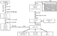
고형 종양은 3차원적 종양 덩어리 전체에 걸쳐 공간적으로 분리된 수백 내지 수천의 돌연변이체 대립유전자를 함유한다. 서열 포획을 위한 전통적인 방법은 도 2에 도시된 바와 같이, 포르말린 고정된, 파라핀 포매된 조직 절편 (예를 들어, 생검 표본 유래)으로부터 단리된 극도로 소량의 투입 DNA (약 5 내지 약 200 나노그램)를 이용한다. 전형적인 서열 포획 방법은 오늘날 임상 병리학 실험실에서 투입 DNA 요건을 맞추도록 진화되었다. 서열 포획 작업흐름의 몇몇 단계에서 DNA의 손실, 및 소량의 투입 DNA에 기인하여, DNA 단편은 증폭되어야만 하거나 또는 수행하려는 서열분석을 위한 포획 작업흐름의 종료시에 너무 적게 남겨질 것이다. 이러한 증폭은 일반적으로 2회 수행되는데, 특이적 프로브 포획 이전에 제1회, 및 선택된 표적의 특이적 프로브 포획 이후의 제2회이다 (도 1 및 2 참조). 이러한 증폭이 후속 프로토콜 단계에 이용할 수 있는 DNA의 절대 질량을 증가시키는데 유용하지만, 존재하는 정보의 양을 증가시키지는 않는다. 어느 정도, 임의의 특정 이론에 국한하려는 것은 아니나, 상이한 DNA 단편의 개체군이 동일한 반응으로 증폭되는 경우 (즉, 멀티플렉스 PCR), 증폭 방법은 본래 샘플 내에 함유된 정보를 변경시킬 수 있다. 예를 들어, 2종의 상이한 DNA 단편, A 및 B가 각각 하나의 카피 (1:1 수치 비율)로 샘플에 초기에 존재한다면, PCR은 1,000 카피의 DNA 단편 A 및 2,000 카피의 DNA 단편 B (1:2 수치 비율)를 함유하는 증폭된 샘플을 야기할 수 있다. 더 적은 수의 개별 분자가 증폭 방법의 투입물로서 사용되는 경우 및 증폭량이 증가되는 경우 (즉, 더 큰 횟수의 PCT 주기 적용), 본래 정보에 편향성이 도입될 위험성이 증가된다고 여겨진다.Solid tumors contain hundreds to thousands of mutant alleles spatially separated throughout a three-dimensional tumor mass. A conventional method for sequencing is to add an extremely small amount of input DNA (from about 5 to about 200 nanograms) isolated from formalin-fixed, paraffin-embedded tissue sections (e. G., From biopsy specimens) ). A typical sequence sequencing method has evolved to meet input DNA requirements in today's clinical pathology laboratories. Due to the loss of DNA at some stages of the sequencing workflow, and a small amount of injected DNA, the DNA fragment must be amplified or left too little at the end of the capture workflow for sequencing to be performed. This amplification is generally performed twice, the first time prior to the specific probe capture, and the second time after the specific probe capture of the selected target (see FIGS. 1 and 2). This amplification is useful for increasing the absolute mass of DNA available for subsequent protocol steps, but does not increase the amount of information present. While not intending to be limited to any particular theory, to some extent, when the population of different DNA fragments is amplified in the same reaction (i.e., multiplex PCR), the amplification method can inherently alter the information contained within the sample. For example, if two different DNA fragments, A and B, are present initially in the sample with one copy (1: 1 ratio) each, PCR will generate 1,000 copies of DNA fragment A and 2,000 copies of DNA fragment B 1: 2 < / RTI > numerical ratio). It is believed that when fewer individual molecules are used as inputs to the amplification method and when the amount of amplification is increased (i.e., applying a larger number of PCT cycles), the risk of introducing bias into the original information is increased.
본 개시내용의 일 측면은 충분한 양의 게놈 재료를 포함하는 투입 샘플이 제공되어, 서열분석 이전에 증폭 과정이 최소로 요구되거나 또는 전혀 요구되지 않는 표적화된 서열분석 작업흐름이다. 일부 구체예에서, 투입 샘플은 온전한 종양 또는 림프절로부터 유래된다. 일부 구체예에서, 투입 샘플은 본 명세서에서 더욱 기술하는 바와 같이 환자 또는 포유동물 대상체로부터 수득된 하나 이상의 림프절 및/또는 온전한 종양 샘플 (전체 또는 일부분)의 균질화를 통해 수득된다. 일부 구체예에서, 투입 샘플은 전혈 또는 이의 임의 분획을 포함하는 충분한 양의 혈액으로부터 유래된다. 일부 구체예에서, 투입 샘플은 암성 조직으로부터 유래된다. 일부 구체예에서, 투입 샘플은 전암성 조직으로부터 유래된다.One aspect of the present disclosure is a targeted sequencing workflow in which an input sample comprising a sufficient amount of genomic material is provided, wherein the amplification process is minimally required or not required prior to sequencing. In some embodiments, the input sample is derived from a whole tumor or lymph node. In some embodiments, the input sample is obtained through homogenization of one or more lymph nodes and / or intact tumor samples (whole or in part) obtained from a patient or mammalian subject, as further described herein. In some embodiments, the input sample is derived from a sufficient amount of blood, including whole blood or any fraction thereof. In some embodiments, the input sample is from a cancerous tissue. In some embodiments, the input sample is from a pre-cancerous tissue.
일부 구체예에서, 표적화된 서열분석 작업흐름은 서열분석 이전에 하나 이상의 증폭 단계 (예를 들어, 포획 전 증폭 단계, 포획 후 증폭 단계)를 포함하고, 서열분석 이전의 각각의 증폭 단계는 0 내지 3회의 증폭 주기를 포함하며, 서열분석 이전의 증폭 주기의 총 횟수는 4회를 넘지 않는다. 다른 구체예에서, 표적화된 서열분석 작업흐름은 서열분석 이전에 하나 이상의 증폭 단계 (예를 들어, 포획 전 증폭 단계, 포획 후 증폭 단계 )를 포함하고, 서열분석 이전의 각 증폭 단계는 0 내지 2회의 증폭 주기를 포함하며, 서열분석 이전 증폭 주기의 총 횟수는 3을 넘지 않는다. 또 다른 구체예에서, 표적화된 서열분석 작업흐름은 서열분석 이전에 하나의 증폭 단계 (예를 들어, 포획 전 증폭 단계 또는 포획 후 증폭 단계)를 포함하고, 서열분석 이전의 단일 증폭 단계는 0 내지 3회의 증폭 주기를 포함한다. 추가의 구체예에서, 표적화된 서열분석 작업흐름은 서열분석 이전에 하나의 증폭 단계를 포함하고, 서열분석 이전의 단일 증폭 단계는 1 내지 3회 주기를 포함한다. 역시 추가의 구체예에서, 표적화된 서열분석 작업흐름은 서열분석 이전에 하나의 증폭 단계를 포함하고, 서열분석 이전의 단일 증폭 단계는 1회 주기를 포함한다. 보다 추가의 구체예에서, 표적화된 서열분석 작업흐름은 서열분석 이전에 하나의 증폭 단계를 포함하고, 서열분석 이전의 단일 증폭 단계는 2회의 주기를 포함한다. 일부 구체예에서, 서열분석 이전의 포획 전 증폭 단계 또는 포획 후 증폭 단계 중 하나 또는 둘 모두는 LM-PCR을 이용한다.In some embodiments, the targeted sequencing workflow comprises one or more amplification steps (e. G., Pre-capture amplification step, post-capture amplification step) prior to sequencing, Including three amplification cycles, the total number of amplification cycles prior to sequencing does not exceed four. In other embodiments, the targeted sequencing workflow comprises one or more amplification steps (e. G., Pre-capture amplification step, post-capture amplification step) prior to sequencing, each amplification step prior to sequencing comprising 0 to 2 And the total number of amplification cycles before the sequence analysis does not exceed 3. In another embodiment, the targeted sequencing workflow comprises one amplification step (e. G., Pre-capture amplification step or post-capture amplification step) prior to sequencing, Includes three amplification cycles. In a further embodiment, the targeted sequencing workflow comprises one amplification step prior to sequencing, and the single amplification step prior to sequencing comprises one to three cycles. In still further embodiments, the targeted sequencing workflow comprises one amplification step prior to sequencing, and the single amplification step prior to sequencing comprises a single cycle. In a further embodiment, the targeted sequencing workflow comprises one amplification step prior to sequencing, and the single amplification step prior to sequencing comprises two cycles. In some embodiments, one or both of the pre-capture amplification step or post-capture amplification step prior to sequencing uses LM-PCR.
일부 구체예에서, 투입 샘플은 종양 샘플, 림프절 샘플, 혈액 샘플, 또는 이의 임의 조합으로부터 유래된 세포의 대표적인 샘플링 (sampling)을 포함한다. 일부 구체예에서, 투입 샘플은 암으로 진단된 환자 또는 포유동물 대상체 유래의 종양 샘플, 림프절 샘플, 혈액 샘플, 또는 이의 임의 조합으로부터 유래된 세포의 대표적인 샘플을 포함한다. 일부 구체예에서, 투입 샘플은 암을 갖는 것으로 의심되는 환자 또는 포유동물 대상체 유래의 종양 샘플, 림프절 샘플, 혈액 샘플, 또는 이의 임의 조합으로부터 유래된 세포의 대표적인 샘플을 포함한다. 일부 구체예에서, 투입 샘플은 암이 발생될 위험성이 있는 환자 또는 포유동물 대상체 유래의 종양 샘플, 림프절 샘플, 혈액 샘플, 또는 이의 임의 조합으로부터 유래된 세포의 대표적인 샘플을 포함한다. 일부 구체예에서, 투입 샘플은 암의 재발 또는 재출현이 알려지거나 또는 의심되는 환자 또는 포유동물 대상체 유래의 종양 샘플, 림프절 샘플, 혈액 샘플, 또는 이의 임의 조합 내 세포의 대표적인 샘플을 포함한다.In some embodiments, the input sample comprises representative sampling of cells derived from a tumor sample, a lymph node sample, a blood sample, or any combination thereof. In some embodiments, the input sample comprises representative samples of cells derived from a tumor sample, a lymph node sample, a blood sample, or any combination thereof derived from a patient diagnosed with cancer or a mammalian subject. In some embodiments, the input sample comprises representative samples of cells derived from a tumor sample, a lymph node sample, a blood sample, or any combination thereof from a patient suspected of having cancer or a mammalian subject. In some embodiments, the input sample comprises a representative sample of cells from a tumor sample, a lymph node sample, a blood sample, or any combination thereof from a subject at risk of developing cancer or a mammalian subject. In some embodiments, the input sample comprises representative samples of cells in a tumor sample, a lymph node sample, a blood sample, or any combination thereof from a patient or a mammalian subject in whom recurrence or re-emergence of cancer is known or suspected.
일부 구체예에서, 투입 샘플은 종양 샘플, 림프절 샘플, 또는 혈액 샘플로부터 유래되는 세포의 불균질성 개체군을 포함한다. 일부 구체예에서, 투입 샘플은 종양 샘플, 림프절 샘플, 또는 혈액 샘플 내에서 유래되는 소수의 특정 종양 세포 개체군을 대표하는 서브클론 (즉, 종양 불안정성의 결과로서 발생되는 상이한 종양 세포 개체군)을 포함한다. 일부 구체예에서, 방법은 희귀 게놈 변이체, 예컨대 투입 샘플 중 2% 미만의 대립유전자 빈도를 갖는 것을 검출 및/또는 서열분석할 수 있게 한다. 일부 구체예에서, 방법은 희귀 게놈 변이체, 예컨대 투입 샘플 중 1% 미만의 대립유전자 빈도를 갖는 것의 검출 및/또는 서열분석을 가능하게 한다.In some embodiments, the input sample comprises a heterogeneous population of cells derived from a tumor sample, a lymph node sample, or a blood sample. In some embodiments, the input sample comprises a tumor sample, a lymph node sample, or a subclone (i.e., a different tumor cell population that is generated as a result of tumor instability) that represents a small number of specific tumor cell populations derived from a blood sample . In some embodiments, the method allows detection and / or sequencing of rare genomic variants, such as having an allele frequency of less than 2% in the input sample. In some embodiments, the method enables detection and / or sequencing of rare genomic variants, such as those having an allele frequency of less than 1% in the input sample.
일부 구체예에서, 투입 샘플은 예를 들어 다수의 조직학적 절편 및/또는 다수의 생검 샘플로부터 수득된, 충분한 양의 조직학적 절편 및/또는 생검 샘플로부터 유래된다. 일부 구체예에서, 조직학적 절편 및/또는 생검 샘플로부터 유래된 투입 샘플은 적어도 0.5 마이크로그램의 게놈 재료를 포함한다. 다른 구체예에서, 조직학적 절편 및/또는 생검 샘플로부터 유래된 투입 샘플은 적어도 1 마이크로그램의 게놈 재료를 포함한다. 다른 구체예에서, 조직학적 절편 및/또는 생검 샘플로부터 유래된 투입 샘플은 적어도 5 마이크로그램의 게놈 재료를 포함한다. 다른 구체예에서, 조직학적 절편 및/또는 생검 샘플로부터 유래된 투입 샘플은 적어도 10 마이크로그램의 게놈 재료를 포함한다.In some embodiments, the input sample is derived from a sufficient amount of a histological section and / or biopsy sample obtained from, for example, multiple histological sections and / or multiple biopsy samples. In some embodiments, the input sample from the histological section and / or the biopsy sample comprises at least 0.5 microgram of genomic material. In another embodiment, the input sample from the histological section and / or the biopsy sample comprises at least one microgram of genomic material. In other embodiments, the input sample from the histological section and / or the biopsy sample comprises at least 5 micrograms of genomic material. In other embodiments, the input sample from the histological section and / or the biopsy sample comprises at least 10 micrograms of genomic material.
일부 구체예에서, 개시된 방법으로 사용하기 위한 투입 샘플 내 게놈 재료의 양은 전통적인 서열 포획 방법에서 사용하기 위한 투입 샘플 내 재료의 양보다 적어도 10배를 초과한다. 일부 구체예에서, 개시된 방법으로 사용하기 위한 투입 샘플 내 게놈 재료의 양은 전통적인 서열 포획 방법에서 사용하기 위한 투입 샘플 내 재료의 양보다 적어도 100배를 초과한다. 일부 구체예에서, 개시된 방법으로 사용하기 위한 투입 샘플 내 게놈 재료의 양은 전통적인 서열 포획 방법으로 사용하기 위한 투입 샘플 내 재료의 양보다 적어도 250배를 초과한다. 일부 구체예에서, 개시된 방법으로 사용하기 위한 투입 샘플 내 게놈 재료의 양은 전통적인 서열 포획 방법으로 사용하기 위한 투입 샘플 내 재료의 양보다 적어도 500배를 초과한다. 일부 구체예에서, 개시된 방법으로 사용하기 위한 투입 샘플 내 게놈 재료의 양은 전통적인 서열 포획 방법으로 사용하기 위한 투입 샘플 내 재료의 양보다 적어도 1000배를 초과한다. 일부 구체예에서, 개시된 방법으로 사용하기 위한 투입 샘플 내 게놈 재료의 양은 전통적인 서열 포획 방법으로 사용하기 위한 투입 샘플 내 재료의 양보다 약 1000배를 초과한다.In some embodiments, the amount of genomic material in the input sample for use in the disclosed method is at least 10 times greater than the amount of material in the input sample for use in a conventional sequence capture method. In some embodiments, the amount of genomic material in the input sample for use in the disclosed method is at least 100 times greater than the amount of material in the input sample for use in a conventional sequence capture method. In some embodiments, the amount of genomic material in the input sample for use in the disclosed method is at least 250 times greater than the amount of material in the input sample for use in a conventional sequence capture method. In some embodiments, the amount of genomic material in the input sample for use in the disclosed method is at least 500 times greater than the amount of material in the input sample for use in a conventional sequence capture method. In some embodiments, the amount of genomic material in the input sample for use in the disclosed method is at least 1000 times greater than the amount of material in the input sample for use in a conventional sequence capture method. In some embodiments, the amount of genomic material in the input sample for use in the disclosed method is greater than about 1000 times the amount of material in the input sample for use in a conventional sequencing method.
본 개시내용의 다른 측면은 종양 샘플 및/또는 림프절 샘플을 균질화하여 균질화된 샘플을 제공하는 단계; 균질화된 샘플로부터 적어도 0.5 마이크로그램의 게놈 재료를 단리하는 단계; 서열분석을 위해 적어도 0.5 마이크로그램의 단리된 게놈 재료를 제조하는 단계; 및 제조된 게놈 재료를 서열분석하는 단계를 포함하는, 샘플 내 게놈 재료의 서열분석 방법이다. 일부 구체예에서, 방법은 서열분석 이전에 임의의 증폭 단계를 포함하지 않는다. 일부 구체예에서, 방법은 적어도 하나의 포획 전 또는 포획 후 증폭 단계를 포함하고, 적어도 하나의 포획 전 또는 포획 후 증폭 단계 동안 수행되는 증폭 주기의 총 횟수는 최대 4회 주기이다. 일부 구체예에서, 증폭 주기의 총 횟수는 3회이다. 일부 구체예에서, 증폭 주기의 총 횟수는 2회다. 일부 구체예에서, 서열분석을 위해 적어도 0.5 마이크로그램의 단리된 게놈 재료를 제조하는 단계는 적어도 0.5 마이크로그램의 단리된 게놈을 포획 프로브와 혼성화시키고 혼성화된 게놈 재료를 포획하는 단계를 포함한다. 일부 구체예에서, 포획된 게놈 재료의 양은 약 90 ng 내지 약 900 ng 범위이다. 일부 구체예에서, 1 또는 2회 증폭 주기는 포획된 게놈 재료에 대해 수행된다. 일부 구체예에서, 균질화된 샘플은 세포의 대표적인 샘플링을 포함한다. 일부 구체예에서, 적어도 1 마이크로그램의 게놈 재료가 균질화된 샘플로부터 단리된다. 일부 구체예에서, 적어도 5 마이크로그램의 게놈 재료가 균질화된 샘플로부터 단리된다. 일부 구체예에서, 적어도 10 마이크로그램의 게놈 재료가 균질화된 샘플로부터 단리된다.Another aspect of the disclosure provides a method of treating a tumor and / or lymph node sample comprising homogenizing a tumor sample and / or a lymph node sample to provide a homogenized sample; Isolating at least 0.5 micrograms of genomic material from the homogenized sample; Preparing at least 0.5 microgram of isolated genomic material for sequencing; And sequencing the genomic material produced. ≪ Desc / Clms Page number 12 > In some embodiments, the method does not include any amplification steps prior to sequencing. In some embodiments, the method comprises at least one pre- or post-capture amplification step, wherein the total number of amplification cycles performed during at least one pre-capture or post-capture amplification step is a maximum of four cycles. In some embodiments, the total number of amplification cycles is three. In some embodiments, the total number of amplification cycles is two. In some embodiments, preparing at least 0.5 micrograms of the isolated genomic material for sequencing comprises hybridizing at least 0.5 microgram of the isolated genome with the capture probe and capturing the hybridized genomic material. In some embodiments, the amount of captured genomic material ranges from about 90 ng to about 900 ng. In some embodiments, one or two amplification cycles are performed on the captured genomic material. In some embodiments, the homogenized sample comprises representative sampling of the cells. In some embodiments, at least one microgram of genomic material is isolated from a homogenized sample. In some embodiments, at least 5 micrograms of genomic material is isolated from the homogenized sample. In some embodiments, at least 10 micrograms of genomic material is isolated from the homogenized sample.
본 개시내용의 다른 측면은 적어도 0.5 마이크로그램의 DNA를 혈액 샘플로부터 단리하는 단계; 서열분석을 위해 적어도 0.5 마이크로그램의 단리된 DNA를 제조하는 단계, 및 제조된 DNA를 서열분석하는 단계를 포함하는 샘플 내 DNA의 서열분석 방법이다. 일부 구체예에서, 방법은 서열분석 이전에 0회의 증폭 단계를 포함한다. 일부 구체예에서, 서열분석을 위해 적어도 0.5 마이크로그램의 단리된 DNA를 제조하는 단계는 적어도 0.5 마이크로그램의 단리된 게놈을 포획 프로브와 혼성화시키고 혼성화된 게놈 재료를 포획하는 단계를 포함한다. 일부 구체예에서, 포획된 게놈 재료의 양은 약 90 ng 내지 약 900 ng 범위이다. 일부 구체예에서, 1 또는 2회의 증폭 주기가 포획된 게놈 재료에 대해 수행된다. 일부 구체예에서, 적어도 1 마이크로그램의 DNA가 혈액 샘플로부터 단리된다.Another aspect of the disclosure provides a method comprising isolating at least 0.5 micrograms of DNA from a blood sample; Preparing at least 0.5 microgram of the isolated DNA for sequencing, and sequencing the DNA thus prepared. In some embodiments, the method comprises zero amplification steps prior to sequencing. In some embodiments, preparing at least 0.5 microgram of isolated DNA for sequencing comprises hybridizing at least 0.5 microgram of the isolated genome with the capture probe and capturing the hybridized genomic material. In some embodiments, the amount of captured genomic material ranges from about 90 ng to about 900 ng. In some embodiments, one or two amplification cycles are performed on the captured genomic material. In some embodiments, at least 1 microgram of DNA is isolated from a blood sample.
본 개시내용의 다른 측면은 (i) 적어도 종양의 일부분, 하나 이상의 전체 또는 부분 림프절, 또는 이의 임의 조합을 균질화하여 균질화된 샘플을 제공하는 단계; (ii) 균질화된 샘플로부터 게놈 재료를 추출하는 단계; (iii) 추출된 게놈 재료를 비드 상에서 포획하는 단계; 및 (iv) 포획된 게놈 재료를 서열분석하는 단계를 포함하는 표적화된 표상적 서열분석 방법이고, 표적화된 표상적 서열분석은 포획된 게놈 재료의 서열분석 이전에 최대 4회의 증폭 주기를 수행하는 단계를 포함한다. 일부 구체예에서, 최대 3회 증폭 주기가 추출된 게놈 재료의 포획 이전 또는 추출된 게놈 재료의 포획 이후, 또는 이의 임의 조합에서 수행될 수 있다. 일부 구체예에서, 포획 전 증폭 주기가 수행되지 않는다. 일부 구체예에서, 포획된 게놈 재료의 양은 약 90 ng 내지 약 900 ng 범위이다. 일부 구체예에서, 1회 내지 3회의 증폭 주기가 추출된 게놈 재료의 포획 이후, 그러나 서열분석 이전에 수행된다. 일부 구체예에서, 적어도 0.5 마이크로그램의 게놈 재료가 균질화된 샘플로부터 추출된다. 일부 구체예에서, 4회 초과의 증폭 주기를 요구하는 서열분석 방법에서 사용되는 투입 재료의 양과 비교하여, 적어도 100배 더 많은 게놈 재료가 균질화된 샘플로부터 유래된다. Another aspect of the disclosure is directed to a method of treating a subject comprising: (i) homogenizing at least a portion of a tumor, one or more whole or partial lymph nodes, or any combination thereof to provide a homogenized sample; (ii) extracting genomic material from the homogenized sample; (iii) capturing the extracted genome material on a bead; And (iv) sequencing the captured genomic material, wherein the targeted representative sequence analysis comprises performing a maximum of four amplification cycles prior to sequencing of the captured genomic material . In some embodiments, up to three amplification cycles may be performed prior to capture of the extracted genomic material or after capture of the extracted genomic material, or in any combination thereof. In some embodiments, no pre-capture amplification cycle is performed. In some embodiments, the amount of captured genomic material ranges from about 90 ng to about 900 ng. In some embodiments, one to three amplification cycles are performed after capture of the extracted genomic material, but prior to sequencing. In some embodiments, at least 0.5 micrograms of genomic material is extracted from the homogenized sample. In some embodiments, at least 100-fold more genomic material is derived from the homogenized sample compared to the amount of input material used in the sequencing method requiring more than four amplification cycles.
본 개시내용의 다른 측면은 적어도 0.5 마이크로그램의 투입 게놈 재료, 적어도 0.5 마이크로그램의, 종양 샘플, 림프절 샘플, 또는 혈액 샘플로부터 유래된 게놈 재료를 제공하는 단계, 투입 게놈 샘플로부터 DNA를 단리하는 단계, 서열분석을 위해 단리된 DNA를 제조하는 단계, 및 제조된 DNA를 서열분석하는 단계를 포함하는 샘플 내 DNA의 서열분석 방법이고, 방법은 임의의 증폭 단계를 포함하지 않는다. 일부 구체예에서, 적어도 0.5 마이크로그램의 투입 게놈 재료는 다수의 조직학 및/또는 생검 표본으로부터 유래된다. 일부 구체예에서, 적어도 0.5 마이크로그램의 투입 게놈 재료는 균질화된 종양 샘플로부터 유래된다. 일부 구체예에서, 적어도 0.5 마이크로그램의 투입 게놈 재료는 균질화된 림프절 샘플로부터 유래된다. 일부 구체예에서, 적어도 0.5 마이크로그램의 투입 게놈 재료는 유래된 종양 샘플, 림프절 샘플, 또는 혈액 샘플의 대표적인 샘플링이다. 일부 구체예에서, 서열분석은 차세대 서열분석 방법을 사용하여 수행된다. 일부 구체예에서, 서열분석은 합성 서열분석 방법론을 사용하여 수행된다.Another aspect of the present disclosure is to provide a genomic material derived from an input genomic material of at least 0.5 micrograms, at least 0.5 micrograms of a tumor sample, a lymph node sample, or a blood sample, isolating DNA from the input genome sample , Preparing isolated DNA for sequencing, and sequencing the prepared DNA, and the method does not include any amplification step. In some embodiments, at least 0.5 microgram of input genomic material is derived from a plurality of histological and / or biopsy specimens. In some embodiments, at least 0.5 microgram of input genomic material is derived from a homogenized tumor sample. In some embodiments, at least 0.5 microgram of the input genomic material is derived from a homogenized lymph node sample. In some embodiments, at least 0.5 microgram of the input genomic material is representative sampling of a derived tumor sample, lymph node sample, or blood sample. In some embodiments, sequence analysis is performed using a next generation sequencing method. In some embodiments, sequence analysis is performed using synthetic sequence analysis methodology.
본 개시내용의 다른 측면은 충분한 양의 게놈 재료를 포함하는 샘플로부터 DNA를 단리하는 단계; 서열분석을 위해 단리된 DNA를 제조하는 단계; 및 제조된 DNA를 분석하는 단계를 포함하는, 서열분석 동안 PCR-도입된 돌연변이를 감소시키는 방법이고, 방법은 서열분석 이전에 최대 3회의 증폭 주기를 포함한다. 일부 구체예에서, 방법은 서열분석 이전에 1회 또는 2회의 증폭 주기를 포함한다. 일부 구체예에서, 투입 게놈 재료의 충분한 양은 포획 전 증폭 주기가 이용되지 않게 하는 양이다. 일부 구체예에서, 샘플은 암을 갖는 것으로 의심되는 환자로부터 유래된다. 일부 구체예에서, 샘플은 암으로 진단된 환자로부터 유래된다. 일부 구체예에서, 샘플은 암이 발생될 위험성이 있는 환자로부터 유래된다. 일부 구체예에서, 샘플은 건강한 조직 샘플로부터 유래된다. 일부 구체예에서, 0.5 마이크로그램의 DNA가 샘플로부터 단리된다. 일부 구체예에서, 적어도 1 마이크로그램의 게놈 재료가 샘플로부터 단리된다. 일부 구체예에서, 적어도 5 마이크로그램의 게놈 재료가 샘플로부터 단리된다. 일부 구체예에서, 적어도 10 마이크로그램의 게놈 재료가 샘플로부터 단리된다.Another aspect of the disclosure provides a method comprising: isolating DNA from a sample comprising a sufficient amount of genomic material; Preparing isolated DNA for sequencing; And analyzing the prepared DNA, wherein the method comprises up to three cycles of amplification prior to sequencing. In some embodiments, the method comprises one or two amplification cycles prior to sequencing. In some embodiments, a sufficient amount of the input genomic material is such that the pre-capture amplification period is not utilized. In some embodiments, the sample is from a patient suspected of having cancer. In some embodiments, the sample is from a patient diagnosed with cancer. In some embodiments, the sample is from a patient at risk of developing cancer. In some embodiments, the sample is from a healthy tissue sample. In some embodiments, 0.5 microgram of DNA is isolated from the sample. In some embodiments, at least one microgram of genomic material is isolated from the sample. In some embodiments, at least 5 micrograms of the genomic material is isolated from the sample. In some embodiments, at least 10 micrograms of genomic material is isolated from the sample.
본 개시내용의 다른 측면은 PCR-도입된 돌연변이가 감소되는 서열분석 방법이고, 이 서열분석 방법은 적어도 0.05 마이크로그램의 게놈 재료를 포획하는 단계, 및 서열분석 이전에 0 내지 2회의 증폭 주기를 수행하는 단계를 포함한다. 일부 구체예에서, 0회의 증폭 주기가 수행된다. 다른 구체예에서, 1회의 증폭 주기가 수행된다. 또 다른 구체예에서, 2회의 증폭 주기가 수행된다.Another aspect of the disclosure is a method of sequencing analysis in which a PCR-introduced mutation is reduced, the method comprising capturing at least 0.05 micrograms of genomic material and performing an amplification cycle of 0 to 2 prior to sequencing . In some embodiments, zero amplification cycles are performed. In another embodiment, one amplification cycle is performed. In another embodiment, two amplification cycles are performed.
본 개시내용의 다른 측면은 게놈 내용물의 비례적인 표상 (representation) 내 PCR-도입된 편향성이 감소되는 서열 포획 방법이고, 이 서열분석 방법은 적어도 0.5 마이크로그램의 게놈 재료를 포함하는 투입 샘플을 제공하는 단계를 포함하고, 서열 포획 방법은 서열분석 이전에 0 내지 2회의 증폭 주기를 수행하는 단계를 포함한다. 일부 구체예에서, 0회 증폭 주기가 수행된다. 다른 구체예에서, 1회의 증폭 주기가 수행된다. 또 다른 구체예에서, 2회의 증폭 주기가 수행된다. 일부 구체예에서, 투입 샘플은 적어도 1 마이크로그램의 게놈 재료를 포함한다. 일부 구체예에서, 투입 샘플은 적어도 5 마이크로그램의 게놈 재료를 포함한다. 일부 구체예에서, 투입 샘플은 적어도 10 마이크로그램의 게놈 재료를 포함한다. Another aspect of the disclosure is a method of sequencing which reduces the PCR-introduced bias in a proportional representation of the genomic content, and wherein the sequencing method provides an injected sample comprising at least 0.5 microgram of genomic material Wherein the sequence capture method comprises performing 0 to 2 amplification cycles prior to sequencing. In some embodiments, a zero amplification cycle is performed. In another embodiment, one amplification cycle is performed. In another embodiment, two amplification cycles are performed. In some embodiments, the input sample comprises at least one microgram of genomic material. In some embodiments, the input sample comprises at least 5 micrograms of genomic material. In some embodiments, the input sample comprises at least 10 micrograms of genomic material.
본 개시내용의 다른 측면은 PCR-도입된 돌연변이가 제거된 서열 포획 방법이고, 서열 포획 방법은 적어도 0.5 마이크로그램의 게놈 재료를 포함하는 투입 샘플을 제조하는 단계를 포함한다. 일부 구체예에서, 투입 샘플은 적어도 1 마이크로그램의 게놈 재료를 포함한다. 일부 구체예에서, 투입 샘플은 적어도 5 마이크로그램의 게놈 재료를 포함한다. 일부 구체예에서, 투입 샘플은 적어도 10 마이크로그램의 게놈 재료를 포함한다. Another aspect of the present disclosure is a PCR-introduced mutagenized sequence capture method, wherein the sequence capture method comprises the step of producing an injected sample comprising at least 0.5 microgram of genomic material. In some embodiments, the input sample comprises at least one microgram of genomic material. In some embodiments, the input sample comprises at least 5 micrograms of genomic material. In some embodiments, the input sample comprises at least 10 micrograms of genomic material.
본 개시내용의 다른 측면은 서열분석 이전에 PCR-중복 판독치를 제거하는 단계가 제거된 서열 포획 방법이고, 서열 포획 방법은 적어도 0.5 마이크로그램의 게놈 재료를 포함하는 투입 샘플을 제공하는 단계를 포함한다. 일부 구체예에서, 투입 샘플은 적어도 1 마이크로그램의 게놈 재료를 포함한다. 일부 구체예에서, 투입 샘플은 적어도 5 마이크로그램의 게놈 재료를 포함한다. 일부 구체예에서, 투입 샘플은 적어도 10 마이크로그램의 게놈 재료를 포함한다.Another aspect of the present disclosure is a method of sequencing that removes the step of removing PCR-duplicate readouts prior to sequencing, and the sequencing method comprises providing an input sample comprising at least 0.5 micrograms of genomic material . In some embodiments, the input sample comprises at least one microgram of genomic material. In some embodiments, the input sample comprises at least 5 micrograms of genomic material. In some embodiments, the input sample comprises at least 10 micrograms of genomic material.
본 개시내용의 다른 측면은 PCR-도입된 돌연변이가 사실상 제거된 서열분석 방법이고, 이 서열분석 방법은 적어도 0.05 마이크로그램의 게놈 재료를 포획하는 단계를 포함한다. 일부 구체예에서, 약 0.05 마이크로그램의 게놈 재료는 게놈 재료의 포획 이후에 제공된다. 일부 구체예에서, 1회 또는 2회의 포획 후 증폭 주기가 서열분석 이전에 수행된다.Another aspect of the present disclosure is a sequencing method in which PCR-introduced mutations are substantially eliminated, the sequencing method comprising capturing at least 0.05 micrograms of genomic material. In some embodiments, about 0.05 micrograms of genomic material is provided after capture of the genomic material. In some embodiments, one or two post capture cycles of amplification are performed prior to sequencing.
본 개시내용의 다른 측면은 특정 처치 또는 활성 약학 성분에 반응하는 암 아형을 확인하여 암을 치료하는 방법이고, 암 아형은 종양, 림프절, 또는 혈액의 대표적인 샘플링을 포함하는 투입 샘플을 서열분석하여 확인되며; 투입 샘플은 충분한 양의 게놈 재료를 포함하고, 서열분석 단계는 최대 3회의 증폭 주기를 요구한다.Another aspect of the disclosure is a method of treating cancer by identifying a cancerous subtype responsive to a particular treatment or active pharmaceutical ingredient and the cancerous subtype is determined by sequencing a putative sample comprising a representative sample of the tumor, lymph node, ; The input sample contains a sufficient amount of genomic material and the sequencing step requires up to three amplification cycles.
본 명세서에서 언급하는 바와 같이, 전통적인 서열분석 작업흐름은 특정 편향성을 도입시킬 수 있다. 일례에서, 증폭된 DNA 샘플의 정보 내용 중 PCR-도입된 편향성은, 샘플을 차세대 서열분석 (NGS) 방법을 사용하여 서열분석할 때 유지될 수 있다. 따라서 증폭된 DNA 단편의 개체군에 대한 NGS의 적용은 2가지 단점을 초래하는데, 즉 (1) 다수의 서열분석 판독치가 동일한 본래 단편의 카피의 중복 서열분석에 소비되어, 비용 효율적이지 않고, (2) 증폭 과정에 의해 도입된 수치적 편향성은 본래의 미증폭된 샘플에 존재하는 정보의 부실표시를 초래할 수 있고, 이는 표적화 서열분석 검정의 주요 목적이 샘플 중 상이한 DNA 서열의 존재 및 상대적 빈도를 정확하게 결정하기 위한 것인 경우에 특히 중요하다. NGS 이전에 DNA 단편의 PCR 증폭에 의한 추가적인 단점은 PCR 과정이 본래 단편을 카피하면서 서열 오류를 생성시킬 수 있다는 것이고, 이들은 이후에 본래 샘플에 존재했던 것으로 해석될 수 있다.As mentioned herein, traditional sequence analysis workflows can introduce specific biases. In one example, the PCR-introduced bias in the information content of the amplified DNA sample can be maintained when the sample is sequenced using the next generation sequencing (NGS) method. Thus, the application of NGS to the population of amplified DNA fragments results in two disadvantages: (1) multiple sequencing readers are consumed for duplicate sequence analysis of copies of the same original fragment, which is not cost effective and ) The numerical bias introduced by the amplification procedure can lead to the misrepresentation of the information present in the original unamplified sample and this is because the main purpose of the targeted sequence analysis assay is to accurately identify the presence and relative frequency of the different DNA sequences in the sample It is especially important if you are to decide. An additional disadvantage of PCR amplification of DNA fragments prior to NGS is that the PCR process can generate sequence errors while copying the original fragments, which can then be interpreted as if they were present in the original sample.
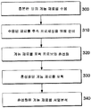
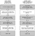
출원인은 (i) 서열분석 이전에 이용되는 증폭 주기의 횟수를 최소화시키거나, 또는 (ii) 서열분석 이전에 완전히 증폭 단계를 방지함으로써 상기에 언급된 단점을 개선시키거나 또는 완화시킨 서열 포획 작업흐름을 개발하였다. 본 명세서에서 제시하는 종양의 표적화된 표상적 서열분석을 위한 방법은 작업흐름 동안 그 DNA를 증폭시킬 필요를 제거하거나 또는 상당히 감소시키기 위해, 충분한 양의 투입 게놈 DNA, 및/또는 효율적인 효소적 단편화-기반 라이브러리 제조를 이용한다 (도 3a, 3b, 및 4 참조). 이후에 이것은 샘플의 비용 효율적인 특징규명을 용이하게 하고 (서열분석 판독치가 중복된 DNA 단편의 서열분석으로 낭비되지 않기 때문), 출력 서열 데이타에 증폭 유도된 편향성의 기회를 감소시키고, 및/또는 서열분석 데이타에 위양성을 초래하는 PCR 유도된 오차의 기회를 감소시킨다고 여겨진다. 실제로, 출원인은 작업흐름으로부터 포획 전 및 포획 후 PCR의 감소 또는 제거가 (i) 표적화된 서열분석의 비용을 절감하고 (PCR 프라이머, PCR 반응 완충액, 및 PCR 효소에 대한 비용 제거); (ii) 검정 시간을 단축하고; (iii) 샘플 대 샘플 오염의 위험성 (PCR 과정의 잘 알려진 위험성)을 감소시키고; (iv) 표적화된 단편의 차등적 증폭에 기인한 서열분석 데이타 내 표상적인 편향성의 위험성을 감소 또는 완화시키고; (v) PCR 증폭 동안 중합효소 오류에 의해 야기되는 위양성 서열 변이의 위험성을 제거 또는 완화시키고; 및/또는 (vi) 보다 단순하고, 더 신속하고, 및/또는 적은 오류 가능성의 데이타 분석 및 해석을 용이하게 한다는 것을 예상치 않게 발견하였다.The Applicant has found that either (i) minimizing the number of amplification cycles used prior to sequencing, or (ii) improving the above mentioned disadvantages by avoiding a complete amplification step prior to sequencing, . Methods for targeted, representative, sequential analysis of tumors presented herein can be used to identify a sufficient amount of input genomic DNA and / or efficient enzymatic fragmentation- Based library production (see Figures 3a, 3b, and 4). This in turn facilitates cost-effective characterization of the sample (because the sequencing analyte is not wasted in sequencing of duplicate DNA fragments), reduces the chance of amplification-induced bias in the output sequence data, and / It is believed to reduce the chance of PCR induced errors that result in false positives in the analysis data. Indeed, the Applicant has found that the reduction or elimination of pre- and post-capture PCR from the work flow can (i) reduce the cost of targeted sequencing (cost reduction for PCR primers, PCR reaction buffers, and PCR enzymes); (ii) shortening the test time; (iii) reduce the risk of sample-to-sample contamination (a well-known risk of the PCR process); (iv) reducing or alleviating the risk of indicative bias in sequence analysis data due to differential amplification of the targeted fragment; (v) eliminating or alleviating the risk of false-positive sequence variations caused by polymerase errors during PCR amplification; And / or (vi) facilitates data analysis and interpretation of simpler, faster, and / or less error probabilities.
출원인은 또한 본 명세서에 개시된 방법이 도 1에 예시된 방법을 통해 도입될 수 있는 것과 같이, 증폭을 통해 달리 도입될 수도 있는 서열 포괄범위 (coverage) 중 대립유전자 및 유전자좌 편향성을 예상치 않게 감소시키거나 또는 방지한다는 것을 추가로 제안한다. 따라서, 출원인은 지금 개시되는 방법이 암 유전체학에서 대립유전자 빈도 및 카피수 변이를 측정하는 우수한 방법 (즉, 보다 정확한 방법)을 제공한다고 믿는다. 출원인은 또한 본 명세서에 개시된 방법이, 서열 데이타의 분석에서 불필요한 서열 판독치를 확인하고 제거할 필요성이 감소된 서열분석을 가능하게 한다고 제안한다. 이러한 인자들은 암 환자의 게놈 DNA에 존재하는 체세포 대립유전자 빈도 및 카피수 변이의 정확한 측정을 위해 특히 중요하다.Applicants have also found that it is possible to unexpectedly reduce both allele and locus bias among sequence coverage coverage that may otherwise be introduced through amplification, such as may be introduced through the methods illustrated in FIG. 1 Or prevention of < / RTI > The Applicant therefore believes that the presently disclosed method provides an excellent method (i.e., a more accurate method) for measuring allele frequency and copy number variation in cancer genomics. Applicants also suggest that the methods disclosed herein enable reduced sequence analysis to identify and eliminate unnecessary sequence readings in the analysis of sequence data. These factors are particularly important for accurate measurement of somatic allele frequency and copy number variation present in the genomic DNA of cancer patients.
비제한적이고 비배타적인 구체예를 하기 도면을 참조하여 설명한다.
도 1은 2회의 증폭 단계를 도입하는 서열 포획 작업흐름을 기재한다.
도 2는 개시된 서열 포획 방법과 비교한 전통적인 서열 포획 방법의 비교를 제공한다.
도 3a는 개시된 서열 포획 방법의 단계들을 예시하는 흐름도를 기재하고, 특히 증폭 단계가 서열분석 이전에 수행되지 않는다.
도 3b는 개시된 서열 포획 방법의 단계들을 예시하는 흐름도를 기재하고, 특히 선택적 증폭 단계가 서열분석 이전에 수행될 수 있다.
도 4는 개시된 표적화된 표상적 서열분석 작업흐름과 비교하여 전통적인 표적화된 서열분석 작업흐름의 추가 비교를 제공한다. 현행의 표적화된 서열분석 프로토콜 (좌측 컬럼), 예컨대 바이오틴화된 포획 올리고뉴클레오티드와의 혼성화에 의존적인 것들은 작업흐름 동안 샘플 DNA 질량을 증가시키기 위해 많은 PCR 증폭 주기 (이 예에서는 총 21회 주기)를 도입한다. 표적화된 표상적 서열분석 작업흐름 (우측 컬럼)은 본 명세서의 다른 구체예에 도시되거나 (0-2회의 증폭 주기) 또는 기술된 바와 같은 작업흐름 동안 전체 증폭을 감소시킨다. 작업흐름에서 PCR 증폭 단계는 흰색 박스로 표시된다.
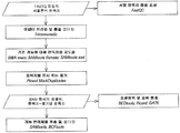
도 5는 기본 SeqCap EZ 서열 포획 데이타 분석 작업흐름의 개략도를 도시한다. 서열 포획 실험으로부터의 서열분석 판독치는 광범위하게 사용되는 "FASTQ" 파일 형식으로 체계화된다. 서열 판독 품질은 데이타가 분석을 계속하는데 충분한 품질인지를 결정하기 위해 프로그램 "FastQC"를 사용하여 평가한다. 임의의 서열분석 어댑터 및 불충분한 품질 판독치는 나머지 판독치를 프로그램 "BWA mem"을 사용하여 기준 게놈에 대해 효율적으로 지도화시킬 수 있도록 프로그램 "Trimmomatic"을 사용하여 필터링된다. "SAMtools fixmate" 프로그램은 쌍형성된 양쪽 판독치에 대해 일관적인 정보가 보이도록 보장한다. "SAMtools sort" 프로그램이 이후에 사용되어 게놈 분류 순서에 따라서 출력 파일을 정리한다. 지도화 이후, "Picard MarkDuplicates" 명령을 사용하여 변이체 호출 중 대립유전자 증폭 편향성을 방지하기 위해 PCR 중복을 제거하거나 또는 표시한다. 증폭 연관된 중복이 제거된 지도화된 판독치를 이후에 후속 분석을 위해 "BAM" 형식으로 전환시킨다. 서열 포괄범위 및 포획 통계를 프로그램 "BEDtools", "Picard", 및 "GATK"를 사용하여 생성시키면서 게놈 서열 변이체를 "SAMtools" 및 "BCFtools"을 사용하여 호출하고 필터링한다. 이들 방법의 상세한 설명은 "How to Evaluate NimbleGen SeqCap EZ Target Enrichment Data"라는 명칭의 Roche 기술 노트 문서에 기술되어 있다 (8월 25일, 이 개시내용은 그 전문을 본 명세서에 참조로 포함함).
도 6은 게놈에 대해 지도화되고 포획 표적 ("표적 상 (On-target)")에 대해 정렬되거나, 또는 포획 표적의 100 염기쌍 이내 ("표적 근처 (Near Target)")에 위치된 모든 비중복 서열분석된 염기의 백분율이 0, 1, 2, 4, 6, 10 또는 14회 주기의 포획 후 증폭을 이용했는지에 따라 실질적으로 상이하지 않다는 것을 보여준다. 도시된 어떠한 실험도 포획 전 증폭 단계를 포함하지 않았다. 표적 상 또는 표적 근처에 있는 서열분석된 염기를 사용하여 포획 실험에서 서열 변이체를 확인하였다. 실험에서 표적 상 또는 표적 근처 염기의 백분율의 감소는 서열 변이체를 확인하기 위해 유용한 데이타의 동일한 절대량을 획득하기 위해 값비싼 추가의 보충적인 서열분석이 필요할 수 있다. 증폭을 명시하는 프로토콜과 비교하여, 양호한 표적 상 비율을 유지하기 위한 증폭-비포함 포획 프로토콜의 예상치않은 수용력은, 보충적인 서열분석에 대한 비용 상승의 발생 없이 증폭 단계에 대한 비용 절감 및 시간 절약을 촉진할 것이라는 것을 시사한다.
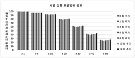
도 7은 일부 최소 판독 심도 (≥1, ≥5, ≥10, ≥20, ≥30, ≥40, 및 ≥50)로 포괄되는 포획 표적을 포함하는 모든 염기의 백분율은 실험이 0, 1, 2, 4, 6, 10 또는 14회 주기의 포획 후 증폭을 이용하는지 간에 실질적으로 상이하지 않았음을 보여준다. 서열 심도 포괄 분포는 전체 포획 표적 전체에서 서열 변이체를 검출하기 위한 검정의 감도의 핵심 결정인자이다. 따라서, 데이타는 증폭-비포함 포획 프로토콜이 증폭을 명시하는 포획 프로토콜과 유사하게 서열 변이체를 검출하는 감도를 가져야한다는 것을 시사한다.
도 8은 반수체 게놈 크기 (∼3,000,000,000 염기쌍)를 포획 표적 크기 (4,571,289 염기쌍)로 나누고 포획 표적 내에 지도화된 서열분석된 염기의 백분율을 곱해서 계산된 (전체 7회 실험의 평균 = 0.667), 전체 기준 게놈에 대한 포획 표적 중 서열의 농축 배수를 도시한다.
도 9는 기준 게놈의 서열 대비, 도 5에 기술된 데이타 분석 파이프라인에 의해 호출된 단일 뉴클레오티드 다형성 (SNP)의 총 개수를 도시한다. 데이타는 증폭-비포함 포획 프로토콜이 증폭을 명시하는 포획 프로토콜과 비교하여 유사한 개수의 SNP를 생성시켰음을 시사한다.
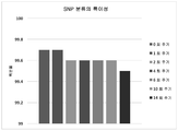
도 10은 수행한 포획 실험에서 확인된 이러한 특정 DNA 샘플 (NA1281, 국제 HapMap 프로젝트에 의해 이전에 유전자형 분석됨)의 포획 표적에 존재하는 것으로 알려진 SNP의 백분율을 보여준다. 0.903 내지 0.919 범위의 감도가 모든 7회 실험 중에서 계산되었고, PCR-비포함 포획 프로토콜의 감도는 0.911로 계산되어, 다른 것들 중에서 중간이었다.
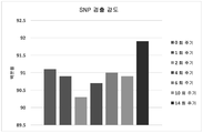
도 11은 7회 포획 실험에서 SNP 분류의 특이성을 도시한다. 샘플 (NA12891)에서 검출된 공지된 변이체들의 경우, SNP 분류의 특이성은 올바른 접합성 (동형접합 대 이형접합)을 갖는 백분율로서 정의된다. SNP 분류의 특이성의 감소는 증폭-관련 대립유전자 편향성의 예측 결과이다 (예를 들어, 이형접합 유전자형이 아마도 더 동형접합 유전자형으로서 나타날 수 있음). 증폭-비포함 포획 프로토콜은 정의상, 증폭-관련 대립유전자 편향성의 부재와 일관되게, 증폭을 명시하는 포획 프로토콜과 유사하거나 또는 그를 초과하는 SNP 분류의 특이성을 입증하였다.Non-limiting and non-exclusive embodiments will be described with reference to the following drawings.
Figure 1 describes a sequence capture workflow that introduces two amplification steps.
Figure 2 provides a comparison of traditional sequence capture methods compared to the disclosed sequence capture methods.
Figure 3A illustrates a flow chart illustrating the steps of the disclosed sequence capture method, particularly where the amplification step is not performed prior to sequencing.
Figure 3b illustrates a flow chart illustrating the steps of the disclosed sequence capture method, and in particular, a selective amplification step may be performed prior to sequencing.
Figure 4 provides additional comparisons of the traditional targeted sequencing workflow compared to the disclosed targeted representative sequence analysis workflow. Current targeted sequencing protocols (left columns), such as those that rely on hybridization with biotinylated capture oligonucleotides, require many PCR amplification cycles (a total of 21 cycles in this example) to increase sample DNA mass during the work- . The targeted representational sequencing workflow (right column) is shown in other embodiments herein (0-2 times of amplification) or reduces overall amplification during the workflow as described. The PCR amplification step in the work flow is indicated by a white box.
Figure 5 shows a schematic diagram of a basic SeqCap EZ sequence capture data analysis workflow. Sequence analysis readings from sequencing experiments are organized in the widely used "FASTQ" file format. Sequence read quality is assessed using the program "FastQC" to determine if the data is of sufficient quality to continue the analysis. Any sequencing adapter and insufficient quality readings are filtered using the program "Trimmomatic" to efficiently map the remaining readings to the reference genome using the program "BWA mem. &Quot; The "SAMtools fixmate" program ensures that consistent information is displayed for both pairs of readings. The "SAMtools sort" program will be used later to organize the output files according to the genome classification sequence. After mapping, use the "Picard MarkDuplicates" command to remove or display PCR duplicates to prevent allele amplification bias during mutant calls. The amplified associated deduplicated mapped readings are then converted to the "BAM" format for subsequent analysis. The genomic sequence variants are called and filtered using "SAMtools" and "BCFtools", generating sequence coverage and capture statistics using the programs "BEDtools", "Picard", and "GATK". A detailed description of these methods is described in the Roche Technical Note document entitled " How to Evaluate NimbleGen SeqCap EZ Target Enrichment Data "(Aug. 25, the disclosure of which is incorporated herein by reference in its entirety).
Figure 6 depicts all non-overlapping (or " near-target ") positions that are mapped to the genome and aligned with the capture target ("Target-Target" It is shown that the percentages of the sequenced base are not substantially different depending on whether they used post-capture amplification of 0, 1, 2, 4, 6, 10 or 14 cycles. None of the experiments shown contained pre-capture amplification steps. Sequence variants were identified in capture experiments using sequenced bases in or near the target. Decreasing the percentage of bases in the target phase or near the target in the experiment may require costly additional sequential assays to obtain the same absolute amount of data useful for identifying the sequence variants. The unexpected capacity of an amplification-free inclusion capture protocol to maintain a good target phase ratio, compared to a protocol that specifies amplification, reduces cost and time savings for the amplification step without incurring cost increases for complementary sequence analysis .
Figure 7 shows that the percentages of all bases, including capture targets encompassed by some minimum read depth (≥1, ≥5, ≥10, ≥20, ≥30, ≥40, and ≥50) , Post-capture amplification of 4, 6, 10 or 14 cycles, respectively. Sequence depth coverage is a key determinant of sensitivity of assays to detect sequence variants across the entire capture target. Thus, the data suggests that the amplification-free capture protocol should have sensitivity to detect sequence variants similar to capture protocols that specify amplification.
Figure 8 is a graph showing the calculated total haploid genome size (~ 3,000,000,000 base pairs) divided by the capture target size (4,571,289 basepairs) and multiplied by the percentage of sequenced base sequenced mapped within the capture target (average of all seven experiments = 0.667) Lt; / RTI > shows the concentration multiple of the sequence in the capture target for the genome.
Figure 9 shows the total number of single nucleotide polymorphisms (SNPs) invoked by the data analysis pipeline described in Figure 5 versus the sequence of the reference genome. The data suggest that the amplification-free capture protocol generated a similar number of SNPs as compared to capture protocols that specify amplification.
Figure 10 shows the percentage of SNPs known to be present in the capture target of this particular DNA sample (NA1281, previously genotyped by the international HapMap project) identified in the capture experiment performed. Sensitivity in the range of 0.903 to 0.919 was calculated in all 7 experiments and the sensitivity of the PCR-free inclusion protocol was calculated to be 0.911, among others.
Figure 11 shows the specificity of the SNP classification in the 7-fold capture experiment. For known variants detected in the sample (NA12891), the specificity of the SNP classification is defined as the percentage with the correct conjugation (homozygous versus heterozygous). A reduction in the specificity of the SNP classification is the result of an amplification-related allele biased prediction (for example, a heterozygous genotype may appear as a more homozygous genotype). The amplification-free inclusion capture protocol, by definition, consistently demonstrated the specificity of SNP classifications similar to or exceeding capture protocols that specify amplification, consistent with the absence of amplification-related allele biases.
일반적으로, 본 개시내용은 증폭 주기의 횟수가 전통적인 서열분석 방법과 비교하여 적어도 최소화된 표적화된 표상적 서열분석 작업흐름을 제공한다. 임의의 특정 이론에 국한하려는 것은 아니나, 서열분석 이전에 포획 전 및/또는 포획 후 PCR 증폭 주기의 횟수를 감소시키는 한가지 방식은 본 명세서에서 더욱 개시하는 바와 같이, 시스템으로 제공되는 투입 DNA의 양을 증가시키는 것이라고 여겨진다. 출원인은 본 서열분석 작업흐름이 (i) 고유한 낮은 비율의 뉴클레오티드의 오편입 (mis-incorporation)에 기인한 돌연변이의 도입, 및 (ii) PCR 증폭 편향성에 기인한 표적 서열의 변경된 표상 (representation)의 위험성을 감소시킨다고 제안한다.Generally, the present disclosure provides a targeted, representative sequence analysis workflow in which the number of amplification cycles is at least minimized compared to conventional sequencing methods. One way of reducing the number of PCR amplification cycles before and / or after capture prior to sequencing, although not limited to any particular theory, is to increase the amount of input DNA provided to the system, as described further herein . The Applicant believes that the present sequence analysis workflow is based on (i) the introduction of a mutation due to inherent mis-incorporation of a low percentage of nucleotides, and (ii) a modified representation of the target sequence due to PCR amplification bias. And the risk of the disease is reduced.
본 명세서에서 사용되는 바와 같은 단수 용어 "한", "하나", 및 "그"는 달리 명확하게 내용에서 표시하지 않으면 다수 지시 대상을 포함한다. 유사하게, 단어 "또는"은 달리 내용에서 명확하게 표시하지 않으면 "및"을 포함하고자 한다.The singular terms "a," "an ", and" the ", as used herein, unless otherwise expressly stated to the contrary, include multiple referents. Similarly, the word "or" is intended to include the words "and" unless explicitly indicated otherwise in the context.
용어 "포함하는", "포괄하는", "가지는" 등은 상호교환적으로 사용되고 동일한 의미를 갖는다. 유사하게, "포함하다", "포괄하다", "가지다" 등은 상호교환적으로 사용되고 동일한 의미를 갖는다. 특히, 각각의 용어는 "포함하는"의 일반 미국 특허법 정의와 일관되게 정의되며 따라서 "적어도 다음의 것"을 의미하는 열린 용어로 해석되어야 하고, 또한 추가적인 특성, 제한, 측면 등을 배제하는 것으로 해석되지 않는다. 따라서, 예를 들어 "성분 a, b, 및 c를 가지는 장치"는 그 장치가 적어도 성분 a, b 및 c를 포함하는 것을 의미한다. 유사하게, 어구 "단계 a, b, 및 c"를 포함하는 방법"은 그 방법이 적어도 단계 a, b, 및 c를 포함한다는 의미이다. 더욱이, 단계 및 방법이 특정 순서로 본 명세서에서 요약될 수 있지만, 당업자는 순서화된 단계 및 방법이 가변적일 수 있다는 것을 인식할 것이다.The terms " including ", "including "," having ", and the like are used interchangeably and have the same meaning. Similarly, the terms "include", "include", "have", etc. are used interchangeably and have the same meaning. In particular, each term is defined as being consistent with the general US patent law definition of "comprising " and thus should be interpreted as an open term meaning" at least the following, " It does not. Thus, for example, "a device having components a, b, and c" means that the device comprises at least components a, b, and c. Similarly, the phrase "a method comprising steps a, b, and c" means that the method includes at least steps a, b, and c. , Those skilled in the art will recognize that the ordered steps and methods may vary.
본 명세서에서 사용되는 바와 같은 용어 "증폭"은 본래 핵산의 더 많은 양을 얻기 위해서 핵산 주형의 본래 양을 배가시키는 과정을 의미한다.The term "amplification" as used herein refers to the process of doubling the original amount of nucleic acid template to obtain a greater amount of the original nucleic acid.
유사하게, 용어 "증폭시키는"은 핵산의 일부분을 예를 들어 임의의 광범위한 프라이머 연장 반응을 사용하여 복제하는 과정을 의미한다. 예시적인 프라이머 연장 반응은 제한없이, 중합효소 연쇄 반응 (polymerase chain reaction) (PCR)을 포함한다. 달리 특별히 명시하지 않으면, "증폭시키는"은 산술적, 대수적 또는 지수적 증폭, 또는 단일 복제를 의미한다. 일반적으로, PCR은 클로닝 또는 정제없이 게놈 DNA의 혼합물 중 표적 서열의 절편의 농도를 증가시키기 위한 방법이다. 표적 서열을 증폭시키기 위한 이러한 방법은 바람직한 표적 서열을 함유하는 DNA 혼합물에 과량의 2종 올리고뉴클레오티드 프라이머 도입 후, DNA 중합효소의 존재 하에서 정확한 배열의 열적 순환으로 이루어진다. 2종의 프라이머는 그들의 이중 가닥 표적 서열의 개별 가닥에 상보적이다. 증폭을 실시하기 위해서, 혼합물을 변성시키고 이후에 프라이머를 표적 분자 내 그들 상보적 서열에 어닐링시킨다. 어닐링 이후, 프라이머는 새로운 쌍의 상보성 가닥을 형성하도록 중합효소 (예를 들어, DNA 중합효소)로 연장된다. 변성, 프라이머 어닐링 및 중합효소 연장의 단계들은 바람직한 표적 서열의 증폭된 절편 (앰플리콘)을 고농도로 수득하도록 수회 반복될 수 있다 (즉, 변성, 어닐링 및 연장 단계는 하나의 "주기"로 구성되고, 수많은 "주기들"이 존재할 수 있음). 바람직한 표적 서열의 증폭된 절편의 길이는 서로에 대한 프라이머의 상대적 위치에 의해 결정되므로, 이 길이는 제어가능한 변수이다. 중합효소 연쇄 반응 ("PCR")은 예를 들어, 미국 특허 제4,683,202호; 미국 특허 제4,683,195호; 미국 특허 제4,000,159호; 미국 특허 제4,965,188호; 미국 특허 제5,176,995호에 기술되어 있고, 이들 각각의 개시내용은 그들 전문을 본 명세서에서 참조로 포함한다.Similarly, the term "amplifying" refers to the process of replicating a portion of a nucleic acid, for example, using any of a wide range of primer extension reactions. Exemplary primer extension reactions include, without limitation, polymerase chain reaction (PCR). Unless otherwise stated, "amplifying" means arithmetic, algebraic, or exponential amplification, or single replication. Generally, PCR is a method for increasing the concentration of a fragment of a target sequence in a mixture of genomic DNA without cloning or purification. This method for amplifying the target sequence consists of thermal cycling of the precise sequence in the presence of DNA polymerase after introduction of an excess of the two oligonucleotide primers into a DNA mixture containing the desired target sequence. The two primers are complementary to individual strands of their double-stranded target sequence. To perform the amplification, the mixture is denatured and then the primers are annealed to their complementary sequences in the target molecule. After annealing, the primer is extended with a polymerase (e. G., DNA polymerase) to form a new pair of complementary strands. The steps of denaturation, primer annealing and polymerase extension can be repeated several times to obtain high concentrations of amplified fragments (amplicon) of the desired target sequence (i. E., The denaturation, annealing and extension steps consist of one & , There may be numerous "cycles"). Since the length of the amplified fragment of the desired target sequence is determined by the relative position of the primers to each other, this length is a controllable variable. Polymerase chain reaction ("PCR") is described, for example, in U.S. Patent Nos. 4,683,202; U.S. Patent No. 4,683,195; U.S. Patent No. 4,000,159; U.S. Patent No. 4,965,188; U.S. Patent No. 5,176,995, the disclosures of each of which are incorporated herein by reference in their entirety.
어구 "게놈 내용물의 비례적인 표상 내 편향성"은 중합효소가 카피하는 것이 더욱 어려운 부분들과 같이, 증폭 이후에 게놈의 일부분이 적게 표시되는 (underrepresented) 경향을 의미한다.The phrase "proportionally biased bias of the genomic content" means that a portion of the genome tends to underrepresented after amplification, such as in parts where the polymerase is more difficult to copy.
본 명세서에서 사용되는 바와 같은 용어 "혼성화"는 DNA의 4개의 천연 핵염기 (아데닌, 구아닌, 티민 및 시토신) 중 하나와 보편적인 핵염기의 쌍형성 또는 왓슨 (Watson)및 크릭 (Crick)의 염기 쌍형성을 통해 이중 가닥 분자를 형성하도록 DNA 및 RNA 중 각각 하나, 또는 DNA의 2개의 상보성 가닥을 연결하는 과정을 의미한다.The term "hybridization " as used herein refers to the formation of a pair of universal nucleobases with one of the four natural nucleobases (adenine, guanine, thymine and cytosine) of DNA or the base of Watson and Crick Refers to the process of connecting two complementary strands of DNA, either DNA or RNA, or DNA, to form double stranded molecules through pairing.
용어 "차세대 서열분석 (next generation sequencing) (NGS)"은 전통적인 생어 (Sanger) 및 모세관 전기영동 기반 접근법과 비교하여 고수율 서열분석을 구비한 서열분석 기술을 의미하며, 여기서 서열분석 과정은 동시에 수행되어, 예를 들어 한번에 수천 또는 수백만의 비교적 적은 서열 판독치를 생성한다. 차세대 서열분석 기술의 일부 예는 제한없이, 합성에 의한 서열분석, 결찰 (ligation)에 의한 서열분석, 및 혼성화에 의한 서열분석을 포함한다. 이들 기술은 보다 짧은 판독치 (어디든지 25 내지 500 bp)이지만 비교적 짧은 기간 내에 수십만 또는 수백만 판독치를 생성시킨다. 용어 "차세대 서열분석"은 Illumina, Life Technologies, 및 Roche 등이 현재 이용하는 소위 병행되는 합성에 의한 서열분석 또는 결찰에 의한 서열분석 플랫폼을 의미한다. 차세대 서열분석 방법은 또한 Life Technologies가 상품화시킨 이온 토렌트 기술 (Ion Torrent technology)과 같은 나노포어 서열분석 방법 또는 전자-검출 기반 방법을 포함할 수 있다.The term "next generation sequencing (NGS)" refers to a sequencing technique with high yield sequencing compared to traditional Sanger and capillary electrophoresis based approaches, wherein sequence analysis is performed concurrently , Resulting in relatively few sequence readings, for example, thousands or millions at a time. Some examples of next generation sequencing techniques include, without limitation, sequencing by synthesis, sequencing by ligation, and sequencing by hybridization. These techniques produce shorter readings (anywhere from 25 to 500 bp) but hundreds or even millions of readings in a relatively short period of time. The term "next generation sequence analysis" refers to a sequencing or ligation sequence analysis platform by so-called parallel synthesis currently used by Illumina, Life Technologies, and Roche et al. Next-generation sequencing methods can also include nano-pore sequencing methods such as Ion Torrent technology, commercialized by Life Technologies, or electron-based detection methods.
본 명세서에서 사용되는 바와 같은 용어 "핵산"은 유전자 정보를 전달하는 뉴클레오티드 사슬로 구성된 고분자량 생화학적 거대분자를 의미한다. 가장 일반적인 핵산은 데옥시리보핵산 (DNA) 및 리보핵산 (RNA)이다. 핵산을 구성하는 단량체는 뉴클레오티드라고 한다. 각각의 뉴클레오티드는 3개의 성분들, 즉 질소성 헤테로시클릭 염기, 퓨린 또는 피리미딘 (핵염기라고도 알려짐), 및 펜토스 당으로 이루어진다. 상이한 핵산 유형은 그들 뉴클레오티드 내 당의 구조가 상이한데, DNA는 2-데옥시리보스를 함유하는데 반해 RNA는 리보스를 함유한다.The term "nucleic acid " as used herein refers to a high molecular weight biochemical macromolecule composed of a nucleotide chain that carries genetic information. The most common nucleic acids are deoxyribonucleic acid (DNA) and ribonucleic acid (RNA). The monomer constituting the nucleic acid is called a nucleotide. Each nucleotide consists of three components: a nitrogenous heterocyclic base, a purine or pyrimidine (also known as a nucleobase), and a pentose sugar. Different nucleic acid types differ in the structure of the sugar in their nucleotides, DNA contains 2-deoxyribose whereas RNA contains ribose.
본 명세서에서 사용되는 바와 같은 용어 "중합효소"는 핵산의 복제 과정을 촉매하는 효 소를 의미한다. 보다 구체적으로, DNA 중합효소는 DNA 중합효소가 "판독"하고 주형으로서 사용하는, DNA 가닥을 따라서 데옥시리보뉴클레오티드의 중합반응을 촉매한다. 새롭게 중합된 분자는 주형 가닥에 상보적이며 주형의 파트너 가닥과 동일하다.The term "polymerase" as used herein refers to an enzyme that catalyzes the replication process of a nucleic acid. More specifically, DNA polymerase catalyzes the polymerization of deoxyribonucleotides along the DNA strand, which DNA polymerase "reads" and uses as a template. The newly polymerized molecule is complementary to the template strand and is identical to the partner strand of the template.
본 명세서에서 사용되는 바와 같은 "서열분석" 또는 "DNA 서열분석"은 DNA 올리고뉴클레오티드 내에서 뉴클레오티드 염기, 아데닌, 구아닌, 시토신, 및 티민의 순서를 결정하기 위한 생화학적 방법을 의미한다. 본 명세서에서 사용되는 용어로서 서열분석은 제한없이, 동시 서열분석 또는 당업자에게 공지된 임의의 다른 서열분석 방법, 예를 들어, 사슬-종결 방법, 급속 DNA 서열분석 방법, 원더링-스팟 (wandering-spot) 분석, 맥심-길버트 (Maxam-Gilbert) 서열분석, 염료-말단인자 서열분석, 또는 임의의 다른 현대적 자동화 DNA 서열분석 장비의 사용을 포함한다."Sequence analysis" or "DNA sequence analysis" as used herein refers to a biochemical method for determining the order of nucleotide bases, adenine, guanine, cytosine, and thymine in a DNA oligonucleotide. As used herein, sequence analysis includes, but is not limited to, simultaneous sequencing or any other sequence analysis method known to those skilled in the art, such as chain-termination methods, rapid DNA sequencing methods, wandering-spot ) Analysis, Maxam-Gilbert sequencing, dye-end sequence sequencing, or any other modern automated DNA sequencing instrument.
용어 "서열분석 라이브러리"는 균등한 NGS를 위해 일정 길이로 전단되어 양쪽 말단 상에 어댑터 및 인덱스 서열이 첨가된, 게놈 유래의 핵산 단편의 컬렉션을 의미한다.The term "sequencing library" refers to a collection of genomic-derived nucleic acid fragments that have been sheared to constant length for even NGS and have adapter and index sequences added on both ends.
본 명세서에서 사용되는 바와 같은 어구 "표적 서열"은 증폭하거나, 검출하거나, 또는 아니면 분석하려는 핵산의 영역을 의미한다.As used herein, the phrase "target sequence" refers to an area of a nucleic acid to be amplified, detected, or otherwise analyzed.
투입 샘플Input sample
일반적으로, 본 명세서에서 개시되는 서열분석 작업흐름의 일부로서 이용되는 투입 샘플은 종양 샘플, 예를 들어 온전한 종양, 및/또는 림프절로부터 유래되거나 또는 그로부터 제조된다. 용어 "종양 샘플"은 종양, 또는 암 세포를 잠재적으로 포함하거나 또는 포함할 것으로 의심되거나, 또는 암 세포의 잠재적 존재에 대해 시험하려는 샘플, 예컨대 림프절로부터 제조된 샘플을 포함한다. 일부 구체예에서, 투입 샘플은 본 명세서에서 더욱 기술하는 바와 같이 환자 또는 포유동물 대상체로부터 수득된 하나 이상의 림프절 및/또는 종양 샘플 (전체 또는 일부분)을 (본 명세서에 기술한 바와 같이) 균질화하여 유래된다. 다른 구체예에서, 투입 샘플은 혈액, 예를 들어 전혈 또는 전혈의 구성 성분 일부로부터 유래된다. 일부 구체예에서, 투입 샘플은 조직학적 절편 또는 생검 샘플, 예를 들어 다수의 조직학적 절편 또는 다수의 생검 샘플로부터 유래된다.Generally, the input sample used as part of the sequence analysis workflow disclosed herein is derived from or is produced from a tumor sample, such as intact tumor, and / or lymph node. The term "tumor sample" includes a sample suspected of containing or suspending a tumor, or cancer cell, or a sample to be tested for the potential presence of a cancer cell, such as a lymph node. In some embodiments, the injected sample is obtained by homogenizing (as described herein) one or more lymph nodes and / or tumor samples (as described herein) obtained from a patient or mammalian subject, as further described herein do. In other embodiments, the input sample is derived from a portion of the blood, e. G., Whole blood or whole blood. In some embodiments, the input sample is derived from a histological section or a biopsy sample, such as multiple histological sections or multiple biopsy samples.
일부 구체예에서, 투입 샘플은 종양 (예를 들어, 종양 샘플), 림프절, 또는 혈액 내 세포의 대표적인 샘플링이다. 본 명세서에서 사용되는 바와 같은 용어 "대표적인 샘플" 및 "대표적인 샘플링"은 전체의 성분을 정확하게 반영하는 샘플 (또는 샘플의 서브셋)을 의미하고, 따라서 샘플은 전체 개체군의 편향되지 않은 지표이다. 일반적으로, 이것은 대표적인 샘플 또는 이의 일부분 내 상이한 유형의 세포 및 그들의 상대적인 비율 또는 백분율이 전체 조직 표본, 일반적으로 고형 종양 또는 이의 일부분 내 이들 세포 유형의 상대적 비율 또는 백분율을 본질적으로 정확하게 반영하거나 또는 모방한다는 것을 의미한다. 샘플링은 후속 분석을 위해 대상의 일부분을 확보하는 작업이다. 대표적인 샘플은 연구되는 대상의 타당하게 근접한 지식을 수득할 수 있는 방식으로 생성된다. 대조적으로, 통상의 무작위 샘플링 방법은 일반적으로, "대표적인 샘플"을 발생시키지 않는다. 보다 큰 샘플로부터 보다 작은 개별 하위샘플의 선택이 선택되는 영역을 기반으로 편향될 수 있지만, 거대 샘플, 예를 들어 전체 종양 또는 림프절을 균질화하여 그 결과 공간적으로 분리된 성분들이 샘플 전반에서 균질하게 분산된다.In some embodiments, the input sample is representative sampling of a tumor (e.g., a tumor sample), a lymph node, or a cell in the blood. As used herein, the terms "representative sample" and "representative sampling" refer to a sample (or a subset of samples) that accurately reflects the entire composition, and thus the sample is an unbiased indicator of the entire population. In general, this means that they essentially reflect or mimic the relative proportions or percentages of these cell types within a representative sample, or a portion thereof, and their relative proportions or percentages are within the entire tissue sample, typically a solid tumor or a portion thereof . Sampling is the task of securing a portion of the object for further analysis. Representative samples are generated in such a way as to obtain reasonably close knowledge of the object being studied. In contrast, conventional random sampling methods generally do not generate "representative samples. &Quot; Selection of smaller individual subsamples from a larger sample can be biased based on the selected region, but homogenizing a large sample, e. G., An entire tumor or lymph node such that the spatially separated components are homogeneously dispersed throughout the sample do.
일부 구체예에서, 투입 샘플은 암으로 진단받은 환자 또는 포유동물 대상체로부터의 종양 샘플, 림프절 샘플, 혈액 샘플, 또는 이의 임의 조합으로부터 유래된 세포의 대표적인 샘플을 포함한다. 일부 구체예에서, 투입 샘플은 암을 갖는 것으로 의심되는 환자 또는 포유동물 대상체로부터의 종양 샘플, 림프절 샘플, 혈액 샘플, 또는 이의 임의 조합으로부터 유래된 세포의 대표적인 샘플을 포함한다. 일부 구체예에서, 투입 샘플은 암이 발생될 위험성에 있는 환자 또는 포유동물 대상체로부터의 종양 샘플, 림프절 샘플, 혈액 샘플, 또는 이의 임의 조합으로부터 유래된 세포의 대표적인 샘플을 포함한다. 일부 구체예에서, 투입 샘플은 암의 재발 또는 재출현이 알려져 있거나 또는 의심되는 환자 또는 포유동물 대상체로부터의 종양 샘플, 림프절 샘플, 혈액 샘플, 또는 이의 임의 조합 내 세포의 대표적인 샘플을 포함한다. 일부 구체예에서, 투입 샘플은 암이 발생될 위험성에 있는 환자로부터의 종양 샘플, 림프절 샘플, 또는 혈액 샘플 내 세포의 대표적인 샘플링을 포함한다. 일부 구체예에서, 투입 샘플은 건강한 환자로부터의 조직 샘플 또는 혈액 샘플 내 세포의 대표적인 샘플링을 포함한다. 일부 구체예에서 투입 샘플은 요구량의 DNA를 정제하기 위해 충분한 다수의 조직학적 절편을 포함한다.In some embodiments, the input sample comprises representative samples of cells from a tumor sample, a lymph node sample, a blood sample, or any combination thereof from a patient diagnosed with cancer or a mammalian subject. In some embodiments, the input sample comprises a representative sample of cells from a tumor sample, a lymph node sample, a blood sample, or any combination thereof from a patient suspected of having cancer or a mammalian subject. In some embodiments, the input sample comprises representative samples of cells from a tumor sample, a lymph node sample, a blood sample, or any combination thereof from a patient or mammalian subject at risk of developing cancer. In some embodiments, the input sample comprises representative samples of cells in a tumor sample, a lymph node sample, a blood sample, or any combination thereof from a patient or a suspected mammalian subject for whom recurrence or re-emergence of cancer is known or suspected. In some embodiments, the input sample comprises representative samples of a tumor sample, a lymph node sample, or a cell in a blood sample from a patient at risk of developing cancer. In some embodiments, the input sample comprises representative samples of tissue samples from a healthy patient or cells in a blood sample. In some embodiments, the input sample comprises multiple histological sections sufficient to purify the DNA of the required amount.
일 구체예에서, 본 명세서에 개시된 대표적인 예는 대상체로부터 수득된 거대 부피 또는 양의 종양 샘플 (예컨대 임상적 종양 샘플) 또는 림프절의 균질화에 의해 수득된다. 예를 들어, 전체 종양 또는 이의 실질적인 부분이 대표적인 샘플이 생성되는 투입 재료로서 사용될 수 있다. 일부 구체예에서, 종양 또는 림프절 (또는 다른 진단 검사의 수행을 위해 일부분의 제거, 예컨대 통상의 FFPE 샘플의 제조를 위해 이용가능한 부분의 제거 이후 잔존하는 이의 일부분)의 적어도 40%가 균질화에 이용된다. 다른 구체예에서, 종양 또는 림프절의 적어도 50%가 균질화에 이용된다. 다른 구체예에서, 종양 또는 림프절의 적어도 60%가 균질화에 이용된다. 다른 구체예에서, 종양 또는 림프절의 적어도 70%가 균질화에 이용된다. 다른 구체예에서, 종양 또는 림프절의 적어도 80%가 균질화에 이용된다. 다른 구체예에서, 종양 또는 림프절의 적어도 90%가 균질화에 이용된다. 다른 구체예에서, 종양 또는 림프절의 적어도 95%가 균질화에 이용된다. 또 다른 구체예에서, 전체 종양, 전체 림프절, 또는 림프절의 전체 개체군 (또는 다른 진단 검사의 수행을 위해 일부의 제거, 예컨대 통상적인 FFPE 샘플의 제조를 위해 이용가능한 일부분의 제거 이후 잔존하는 이의 일부분)이 균질화에 사용된다.In one embodiment, representative examples disclosed herein are obtained by homogenization of large volume or positive tumor samples (e.g., clinical tumor samples) or lymph nodes obtained from a subject. For example, an entire tumor or a substantial portion thereof may be used as the input material from which a representative sample is generated. In some embodiments, at least 40% of the tumor or lymph node (or a portion thereof remaining after removal of a portion for removal of a portion for the performance of other diagnostic tests, e.g., a conventional FFPE sample, for removal of the available portion) is used for homogenization . In another embodiment, at least 50% of the tumor or lymph node is used for homogenization. In another embodiment, at least 60% of the tumor or lymph node is used for homogenization. In another embodiment, at least 70% of the tumor or lymph node is used for homogenization. In another embodiment, at least 80% of the tumor or lymph node is used for homogenization. In another embodiment, at least 90% of the tumor or lymph node is used for homogenization. In another embodiment, at least 95% of the tumor or lymph node is used for homogenization. In yet another embodiment, the entire population of whole tumors, whole lymph nodes, or lymph nodes (or a portion thereof that remains after removal of a portion for removal of a portion for the purpose of performing other diagnostic tests, e.g., a conventional FFPE sample) Is used for homogenization.
대표적인 샘플은 고형 종양 유래의 온전한 종양 생검 샘플로부터 생성될 수 있다. 일부 구체예에서, 생검 샘플은 적어도 약 100 내지 200개 세포를 포함한다. 다른 구체예에서, 생검 샘플은 적어도 약 200 내지 1,000개 세포를 포함한다. 또 다른 구체예에서, 생검 샘플은 적어도 약 1,000 내지 5,000개 세포를 포함한다. 추가의 구체예에서, 생검 샘플은 적어도 약 10,000 내지 100,000개 세포를 포함한다. 보다 추가의 구체예에서, 생검 샘플은 적어도 약 100,000 내지 1,000,000개 또는 그 이상의 세포를 포함한다. 일부 구체예에서, 세포는 종양의 공간적으로 구별되는 영역으로부터 수득된다. 다른 구체예에서, 본 명세서에 개시된 대표적인 예는 예를 들어, 유전자 돌연변이 또는 이전의 암 때문에 암이 발생될 위험성이 있는 것을 포함하여, 암이 발생될 위험성이 있는 환자 또는 포유동물 대상체로부터 유래된, 하나 이상의 추정 정상 조직 표본의 균질화에 의해 수득된다. 본 명세서에서 사용되는 바와 같은 용어 "공간적으로 구별되는"은 상이한 공간 영역에 분포되는 성분들을 의미한다. 일 구체예에서, 대표적인 샘플을 생성시키기 위해 사용되는 종양 생검 샘플은 종양 샘플의 상이한 영역으로부터 채취된다. 예를 들어, 전체 종양 내에서 다양성을 포획하기 위한 시도로서, 종양의 근부 대 원부 영역, 종양의 상이한 면, 종양의 상이한 층 등이다.Representative samples can be generated from solid tumor biopsy samples from solid tumors. In some embodiments, the biopsy sample comprises at least about 100 to 200 cells. In another embodiment, the biopsy sample comprises at least about 200 to 1,000 cells. In another embodiment, the biopsy sample comprises at least about 1,000 to 5,000 cells. In a further embodiment, the biopsy sample comprises at least about 10,000 to 100,000 cells. In a further embodiment, the biopsy sample comprises at least about 100,000 to 1,000,000 or more cells. In some embodiments, cells are obtained from spatially distinct regions of the tumor. In other embodiments, representative examples disclosed herein include, but are not limited to, those derived from patients or mammalian subjects at risk of developing cancer, including those at risk of developing cancer due to gene mutation or previous cancer, Is obtained by homogenization of one or more estimated normal tissue specimens. The term "spatially distinct " as used herein means components distributed in different spatial regions. In one embodiment, a tumor biopsy sample used to generate a representative sample is taken from a different region of the tumor sample. For example, in an attempt to capture diversity within the entire tumor, it may be the root apical region of the tumor, different sides of the tumor, different layers of the tumor, and the like.
용어 "균질화하는" 또는 "균질화"는 생물학적 샘플이, 샘플의 모든 분획이 조성이 균등하게 되는 상태가 되게 하는 과정 (예컨대 기계적 과정 및/또는 생화학적 과정)을 의미한다. 대표적인 샘플 (상기에 정의된 바와 같음)은 균질화된 샘플의 일부분의 제거에 의해 제조될 수 있다. 균질화된 샘플 ("균질물")은 샘플의 일부분 (분취액)의 제거가 남아있는 샘플의 전체 구성을 실질적으로 변경시키지 않으며 제거된 분취액의 성분이 남아있는 샘플의 성분과 실질적으로 동일하도록 충분히 혼합된다. 본 개시내용에서, "균질화"는 일반적으로, 샘플 내 대부분의 세포의 무결성을 보존하게 되며, 예를 들어, 샘플 중 세포의 적어도 50%가 균질화 과정의 결과로서 용해되거나 또는 파열되지 않을 것이다. 다른 구체예에서, 균질화는 샘플 중 세포의 적어도 80%의 무결성을 보존할 것이다. 다른 구체예에서, 균질화는 샘플 중 세포의 적어도 85%의 무결성을 보존할 것이다. 다른 구체예에서, 균질화는 샘플 중 세포의 적어도 90%의 무결성을 보존할 것이다. 다른 구체예에서, 균질화는 샘플 중 세포의 적어도 95%의 무결성을 보존할 것이다. 다른 구체예에서, 균질화는 샘플 중 세포의 적어도 96%의 무결성을 보존할 것이다. 다른 구체예에서, 균질화는 샘플 중 세포의 적어도 97%의 무결성을 보존할 것이다. 다른 구체예에서, 균질화는 샘플 중 세포의 적어도 98%의 무결성을 보존할 것이다. 다른 구체예에서, 균질화는 샘플 중 세포의 적어도 99%의 무결성을 보존할 것이다. 다른 구체예에서, 균질화는 샘플 중 세포의 적어도 99.9%의 무결성을 보존할 것이다. 균질물은 개별 세포 (또는 세포의 클러스터)로 실질적으로 해리될 수 있고 최종 균질물 또는 균질물들은 실질적으로 균질하다 (전체적으로 균일하거나 또는 유사한 성분으로 이루어지거나 또는 구성됨).The term " homogenizing "or" homogenization "means a process (e.g., mechanical and / or biochemical process) in which a biological sample is brought into a state in which all fractions of the sample become homogeneous in composition. Representative samples (as defined above) can be prepared by removal of a portion of the homogenized sample. The homogenized sample ("homogenate") does not substantially change the overall configuration of the sample in which the removal of a portion of the sample (aliquot) remains and is sufficient to substantially equal the components of the sample Mixed. In this disclosure, "homogenization" will generally preserve the integrity of most cells in the sample, e.g., at least 50% of the cells in the sample will not dissolve or rupture as a result of the homogenization process. In other embodiments, the homogenization will preserve the integrity of at least 80% of the cells in the sample. In other embodiments, the homogenization will preserve the integrity of at least 85% of the cells in the sample. In other embodiments, the homogenization will preserve at least 90% integrity of the cells in the sample. In other embodiments, the homogenization will preserve the integrity of at least 95% of the cells in the sample. In other embodiments, the homogenization will preserve the integrity of at least 96% of the cells in the sample. In other embodiments, the homogenization will preserve the integrity of at least 97% of the cells in the sample. In other embodiments, the homogenization will preserve the integrity of at least 98% of the cells in the sample. In other embodiments, the homogenization will preserve the integrity of at least 99% of the cells in the sample. In another embodiment, the homogenization will preserve at least 99.9% integrity of the cells in the sample. The homogenate can be substantially dissociated into individual cells (or clusters of cells), and the final homogenate or homogenates are substantially homogeneous (made entirely of homogeneous or similar components, or constructed).
일부 구체예에서, 종양 샘플, 림프절 샘플, 또는 다른 조직 샘플은 기계적 전단 장치, 예를 들어 블렌더 또는 초음파기에 샘플을 위치시킴으로써 균질화된다. 균질화는 각각, 아마도 정상 분포로 맞춰진, 수천 내지 수백개의 세포 유래의 일정 범위의 조직 단편을 생성시킨다. 조직 단편 크기의 중간값은 블렌더 (또는 다른 적합한 장치)의 에너지에 반비례적으로 상관관계가 있어, 고에너지에서 조직 단편이 매우 작다. 블렌더 에너지와 가장 상관되는 조직의 성분은 콜라겐 함량인데, 피부가 완전한 해리에 상당한 에너지를 요구하기 때문이다. 블렌딩 시간이 또한 중요하지만, 가장 효과적인 임상적 적용분야에는 전체 종양이 수 분 내에 해리되는 것이 요구된다. 블렌딩 시간이 고정되면, 바람직한 시간 제한 하에서 종양 해리에 도달하는데 요구되는 에너지를 쉽게 결정할 수 있다. 종양 샘플 또는 림프절 샘플을 제조하는 다른 방법은 공계류중인 미국 가출원, 즉 가출원 제62/252,153호 (2015년 11월 6일 출원), 62/279,405 (2016년 1월 15일 출원) 및 62/354,622 (2016년 6월 24일 출원) (각각 Ventana Medical Systems, Inc. (Tucson, AZ)에 양도됨)에 개시되어 있고, 이들의 개시내용은 그들 전문 각각을 참조로 본 명세서에 포함한다. 시험 샘플은 본 명세서에 기술된 서열분석 작업흐름에서 사용을 위해, 즉 게놈 재료를 포함하는 투입 샘플로서, 균질화된 샘플로부터 채취될 수 있다.In some embodiments, the tumor sample, lymph node sample, or other tissue sample is homogenized by placing the sample in a mechanical shearing device, such as a blender or sonicator. Homogenization, respectively, produces a range of tissue fragments, perhaps from thousands to hundreds of cells, tailored to a normal distribution. The median tissue fragment size is inversely proportional to the energy of the blender (or other suitable device), and tissue fragments are very small at high energies. The component of the tissue most correlated with the blender energy is the collagen content, because the skin requires considerable energy for complete dissociation. Although blending time is also important, the most effective clinical application requires total tumor dissociation within minutes. Once the blending time is fixed, the energy required to reach the tumor dissociation under the desired time limit can be readily determined. Other methods of producing tumor samples or lymph node samples are described in co-pending U.S. Provisional Application No. 62 / 252,153 (filed November 6, 2015), 62 / 279,405 (filed January 15, 2016), and 62 / 354,622 (Filed June 24, 2016), each assigned to Ventana Medical Systems, Inc. (Tucson, AZ), the disclosures of which are incorporated herein by reference in their entireties. The test sample may be taken from the homogenized sample for use in the sequencing workflow described herein, i. E. As an input sample containing the genomic material.
종양, 림프절, 또는 다른 조직 샘플을 해리시키기 위한 충분한 기계적 전단 이후, 본래 공간적으로 분리된 종양 세포의 하위개체군의 전부는 새롭게 균질화된 샘플 전체로 분포된다. 즉, 종양 샘플의 균질화 (또는 림프절의 균질화) 결과로서, 종양 내 세포의 임의의 불균질성은 생성 균질물 또는 이의 일부분 또는 분획 내에서 실질적으로 균질하게 (균일하게) 분포되어, 균질물 (또는 이의 임의 분획)이 투입물인 종양 생검 샘플의 불균질성을 실질적으로 균질하게 발현시킨다. 이의 전체로 종양을 대표하는 샘플 (또는 균질물)을 생성시키기 위해 종양 또는 림프절을 균질화하여, 종양의 풍경 (예컨대 불균질성)을 특징규명하고/하거나 전체적으로 함유된 상이한 게놈 하위개체군 각각을 서열분석하는 것이 가능하다.After sufficient mechanical shear to dissociate the tumor, lymph node, or other tissue sample, all of the subspecies of the original spatially separated tumor cells are distributed throughout the freshly homogenized sample. That is, as a result of the homogenization of the tumor sample (or the homogenization of the lymph nodes), any heterogeneity of the cells in the tumor is distributed substantially homogeneously (uniformly) within the resulting homogenate or a fraction or fraction thereof, Fraction) substantially homogenize the heterogeneity of the tumor biopsy sample as an input. Homogenizing the tumor or lymph node to produce a sample (or homogenate) representative of the tumor as a whole, characterizing the landscape of the tumor (e.g., heterogeneity) and / or sequencing each of the different genomic subpopulations as a whole It is possible.
일부 구체예에서, 투입 샘플은 종양 샘플, 림프절 샘플, 또는 혈액 샘플로부터 유래된 세포의 불균질 개체군을 포함한다. 일부 구체예에서, 투입 샘플은 종양 샘플, 림프절 샘플, 또는 혈액 샘플 내에서 유래된 소수의 일정 종양 세포 개체군을 대표하는 서브클론 (즉, 종양 불안정성의 결과로서 발생된 상이한 종양 세포 개체군)을 포함한다. 일부 구체예에서, 방법은 희귀 게놈 변이체, 예컨대 투입 샘플 중 2% 미만의 대립유전자 빈도를 갖는 것을 검출 및/또는 서열분석할 수 있게 한다. 일부 구체예에서, 방법은 희귀 게놈 변이체, 예컨대 투입 샘플 중 1% 미만의 대립유전자 빈도를 갖는 것을 검출 및/또는 서열분석할 수 있게 한다.In some embodiments, the input sample comprises a heterogeneous population of cells derived from a tumor sample, a lymph node sample, or a blood sample. In some embodiments, the input sample comprises a tumor sample, a lymph node sample, or a subclone (i.e., a different tumor cell population generated as a result of tumor instability) that represents a small number of constant tumor cell populations derived from a blood sample . In some embodiments, the method allows detection and / or sequencing of rare genomic variants, such as having an allele frequency of less than 2% in the input sample. In some embodiments, the method allows detection and / or sequencing of rare genomic variants, such as having an allele frequency of less than 1% in an input sample.
일부 구체예에서, 균질화된 샘플은 예컨대 세포 또는 게놈 재료를 분리하여, 서열분석 작업흐름에서 사용하기 이전에 추가 가공된다. 일부 구체예에서, 균질화된 샘플은 먼저 여과된다.In some embodiments, the homogenized sample is further processed, e. G., By separating the cell or genomic material, prior to use in the sequencing workflow. In some embodiments, the homogenized sample is first filtered.
일부 구체예에서, 균질화된 샘플, 또는 여과된 균질화된 샘플 내 세포를 용해시켜, 세포 성분들을 방출시킨다. 예를 들어, 세포는 프렌치 프레스 또는 유사한 유형의 용해 장치, 미세유동화기, 그라인딩, 밀링, 화학적 또는 효소적 용해를 사용하고/하거나, 당분야에 공지된 다른 기술을 사용하여 용해될 수 있다. 일부 구체예에서, 막 지질 및 단백질 (히스톤 포함)은 (예를 들어, 계면활성제 또는 효소 (프로테아제)의 첨가에 의해) 세포 성분을 함유하는 샘플로부터 제거된다. 또한, RNA는 (예를 들어, 효소 예컨대 RNase의 사용에 의해) 세포 성분을 함유하는 샘플로부터 제거될 수 있다.In some embodiments, the homogenized sample, or the cells in the filtered homogenized sample, is lysed to release cellular components. For example, cells can be lysed using a French press or similar type dissolving apparatus, microfluidizer, grinding, milling, chemical or enzymatic dissolution and / or using other techniques known in the art. In some embodiments, membrane lipids and proteins (including histones) are removed from a sample containing cellular components (e.g., by the addition of a surfactant or enzyme (protease)). In addition, RNA can be removed from a sample containing cellular components (e.g., by the use of an enzyme, e.g., RNase).
일부 구체예에서, DNA는 당업자에게 공지된 수단에 의해 단리되거나, 추출되거나, 또는 정제될 수 있다. 예를 들어, DNA는 에탄올 침전 또는 페놀-클로로포름 추출을 통해 추출된 후 펠렛을 형성하도록 원심분리될 수 있다. 일부 구체예에서, DNA는 고체상 컬럼 상에서 단리되거나 또는 추출될 수 있다. 일부 구체예에서, DNA는 핵산-결합 비드를 사용하여 단리되거나 또는 추출될 수 있다. 일부 구체예에서, DNA는 물리적, 화학적 또는 전기적 특성을 기반으로, 다공성 매트릭스를 통한 선택적 통과에 의해 단리되거나 또는 추출될 수 있다.In some embodiments, the DNA can be isolated, extracted, or purified by means known to those skilled in the art. For example, DNA can be centrifuged to form pellets after ethanol precipitation or phenol-chloroform extraction. In some embodiments, the DNA can be isolated or extracted on a solid-phase column. In some embodiments, the DNA can be isolated or extracted using nucleic acid-conjugated beads. In some embodiments, the DNA can be isolated or extracted by selective passage through a porous matrix, based on physical, chemical, or electrical properties.
추출된 DNA (게놈 재료)는 완충액, 예를 들어 알칼리 완충액에 용해될 수 있고, 본 명세서에서 더욱 설명하는 바와 같이, 서열분석을 위한 투입 샘플로서 도입될 수 있다.The extracted DNA (genomic material) can be dissolved in a buffer, for example, an alkaline buffer, and introduced as an input sample for sequencing, as described further herein.
서열분석 작업흐름Sequence workflow
도 3a 및 3b를 참조하여, 본 개시내용의 서열분석 방법에 따른 제1 단계는 상기 기재된 바와 같이, 예컨대 투입 샘플 유래의, 게놈 재료 (300)를 수용하는 것이다. 일부 구체예에서, 본 개시내용은 서열분석 이전에 증폭 주기의 횟수가 최소화되도록 게놈 재료의 충분한 양을 포함하는 투입 샘플을 제공하는 서열분석 작업흐름을 제공한다. 일부 구체예에서, 재료의 "충분한 양"은 임의의 포획 전 증폭 주기없이 서열분석 작업흐름이 진행될 수 있게 하는 양이다. 다른 구체예에서, 재료의 "충분한 양"은 1 또는 2회의 포획 전 증폭 주기로 서열분석 작업흐름이 진행될 수 있게 하는 양이다. 또 다른 구체예에서, 재료의 "충분한 양"은 서열분석 이전에 포획 전 증폭 주기는 없고 포획 후 증폭 주기는 오직 최소 횟수로 서열분석 작업흐름을 진행할 수 있게 하는 양이다. 또 다른 구체예에서, 재료의 "충분한 양"은 서열분석 이전에 포획 전 증폭 주기는 없고 포획 후 증폭 주기는 약 1 내지 약 4회로 서열분석 작업흐름을 진행할 수 있게 하는 양이다. 추가의 구체예에서, 재료의 "충분한 양"은 서열분석 이전에 포획 전 증폭 주기는 없고 포획 후 증폭 주기는 약 1 내지 약 2회로 서열분석 작업흐름을 진행할 수 있게 하는 양이다. 일부 구체예에서, 재료의 "충분한 양"은 포획 전 증폭 주기가 없고 포획 후 증폭 주기도 없이 서열분석 작업흐름을 진행할 수 있게 하는 양이다.Referring to Figures 3a and 3b, the first step in accordance with the sequence analysis method of the present disclosure is to accommodate the
일부 구체예에서, 임의의 투입 샘플의 양은 적어도 약 0.5 마이크로그램이다. 다른 구체예에서, 투입 샘플의 양은 적어도 약 1 마이크로그램이다. 다른 구체예에서, 투입 샘플의 양은 적어도 약 2.5 마이크로그램이다. 다른 구체예에서, 투입 샘플의 양은 적어도 약 5 마이크로그램이다. 다른 구체예에서, 투입 샘플의 양은 적어도 약 7.5 마이크로그램이다. 일부 구체예에서, 투입 샘플의 양은 적어도 약 9 마이크로그램이다. 일부 구체예에서, 투입 샘플의 양은 적어도 약 10 마이크로그램이다. 다른 구체예에서, 투입 샘플의 양은 적어도 약 50 마이크로그램이다. 또 다른 구체예에서, 투입 샘플의 양은 약 10 마이크로그램 내지 약 100 마이크로그램 범위이다. 또 다른 구체예에서, 투입 샘플의 양은 약 10 마이크로그램 내지 약 250 마이크로그램 범위이다. 추가의 구체예에서, 투입 샘플의 양은 약 100 마이크로그램 내지 약 250 마이크로그램 범위이다.In some embodiments, the amount of any input sample is at least about 0.5 micrograms. In another embodiment, the amount of the input sample is at least about 1 microgram. In another embodiment, the amount of input sample is at least about 2.5 micrograms. In another embodiment, the amount of the input sample is at least about 5 micrograms. In other embodiments, the amount of input sample is at least about 7.5 micrograms. In some embodiments, the amount of input sample is at least about 9 micrograms. In some embodiments, the amount of the input sample is at least about 10 micrograms. In other embodiments, the amount of input sample is at least about 50 micrograms. In yet another embodiment, the amount of the input sample ranges from about 10 micrograms to about 100 micrograms. In another embodiment, the amount of the input sample ranges from about 10 micrograms to about 250 micrograms. In further embodiments, the amount of the input sample ranges from about 100 micrograms to about 250 micrograms.
일부 구체예에서, 개시된 방법에서 사용하기 위한 투입 샘플 내 게놈 재료의 양은 전통적인 서열 포획 방법에서 사용하기 위한 투입 샘플 내 재료의 양보다 적어도 5배를 초과한다. 일부 구체예에서, 개시된 방법에서 사용하기 위한 투입 샘플 내 게놈 재료의 양은 전통적인 서열 포획 방법에서 사용하기 위한 투입 샘플 내 재료의 양보다 적어도 10배를 초과한다. 일부 구체예에서, 개시된 방법에서 사용하기 위한 투입 샘플 내 게놈 재료의 양은 전통적인 서열 포획 방법에서 사용하기 위한 투입 샘플 내 재료의 양보다 적어도 100배를 초과한다. 일부 구체예에서, 개시된 방법에서 사용하기 위한 투입 샘플 내 게놈 재료의 양은 전통적인 서열 포획 방법에서 사용하기 위한 투입 샘플 내 재료의 양보다 적어도 250배를 초과한다. 일부 구체예에서, 개시된 방법에서 사용하기 위한 투입 샘플 내 게놈 재료의 양은 전통적인 서열 포획 방법에서 사용하기 위한 투입 샘플 내 재료의 양보다 적어도 500배를 초과한다. 일부 구체예에서, 개시된 방법에서 사용하기 위한 투입 샘플 내 게놈 재료의 양은 전통적인 서열 포획 방법에서 사용하기 위한 투입 샘플 내 재료의 양보다 적어도 1000배를 초과한다. 일부 구체예에서, 개시된 방법에서 사용하기 위한 투입 샘플 내 게놈 재료의 양은 전통적인 서열 포획 방법에서 사용하기 위한 투입 샘플 내 재료의 양보다 적어도 약 1000배를 초과한다.In some embodiments, the amount of genomic material in the input sample for use in the disclosed method is at least 5 times greater than the amount of material in the input sample for use in a conventional sequence capture method. In some embodiments, the amount of genomic material in the input sample for use in the disclosed method is at least 10 times greater than the amount of material in the input sample for use in a conventional sequence capture method. In some embodiments, the amount of genomic material in the input sample for use in the disclosed method is at least 100 times greater than the amount of material in the input sample for use in a conventional sequence capture method. In some embodiments, the amount of genomic material in the input sample for use in the disclosed method is at least 250 times greater than the amount of material in the input sample for use in a conventional sequence capture method. In some embodiments, the amount of genomic material in the input sample for use in the disclosed method is at least 500 times greater than the amount of material in the input sample for use in a conventional sequence capture method. In some embodiments, the amount of genomic material in the input sample for use in the disclosed method is at least 1000 times greater than the amount of material in the input sample for use in a conventional sequence capture method. In some embodiments, the amount of genomic material in the input sample for use in the disclosed method is at least about 1000 times greater than the amount of material in the input sample for use in a conventional sequence capture method.
다시 도 3a 및 3b를 참조하여, 게놈 재료 (300)의 수용 이후에, 표적 핵산 분자를 포함하는 게놈 재료를 더욱 가공할 수 있다 (310). 일부 구체예에서, 게놈 재료를 단편화하여, 단편화된 게놈 샘플을 제공한다. 일부 구체예에서, 투입 샘플은 예를 들어 초음파처리, 또는 핵산을 단편화할 수 있는 다른 방법에 의해 단편화된다. 일부 구체예에서, 투입 샘플은 약 100 bp 내지 약 500 bp의 평균 크기로 단편화된다. 일부 구체예에서, 투입 샘플은 약 500 bp 내지 약 1,000 bp의 평균 크기로 단편화된다. 다른 구체예에서, 투입 샘플은 약 1,000 bp 내지 약 10,000 bp의 평균 크기로 단편화된다.Referring again to Figures 3a and 3b, after the acceptance of the
일부 구체예에서, 게놈 재료의 단편화 이후에 단편화된 게놈 재료의 말단을 복구하거나 또는 "연마"하는 것이 후속된다. 이를 달성하기 위해서, 게놈 재료 내 이중 가닥 표적 분자는, 예를 들어 dNTP의 존재 하에서 DNA 중합효소 예컨대 T4 DNA 중합효소 또는 클레노우 (Klenow) 중합효소에 의한 채움 (fill-in) 반응 처리되어, 그 결과로 블런트 말단의 표적 분자가 생성된다. 또한, 단편의 말단은 어댑터의 결찰 이전에 단편의 5' 말단에 포스페이트 기를 첨가하기 위해 T4 폴리뉴클레오티드 키나제 및 당업자에게 공지된 방법을 사용하여 인산화된다 (예를 들어, 문헌 [Molecular Cloning: A Laboratory Manual, Eds. Sambrook et al., Cold Spring Harbour Press]을 참조하고, 이 전문을 참조로 본 명세서에 포함함). 연마되고, 인산화된 표적 DNA 상에서의 어댑터 (예를 들어, 약 3 내지 20 염기쌍의 짧은 이중 가닥 블런트 말단 DNA 올리고뉴클레오티드)의 후속 결찰은 당분야에 공지된 임의 방법에 따라서, 예를 들어 T4 DNA 리가제 반응에 의해 수행될 수 있다.In some embodiments, it is followed by restoring or "polishing" the ends of the fragmented genome material after fragmentation of the genomic material. To achieve this, the double-stranded target molecule in the genomic material is subjected to a fill-in reaction with a DNA polymerase such as T4 DNA polymerase or Klenow polymerase in the presence of, for example, dNTP, As a result, a target molecule at the blunt end is generated. Also, the ends of the fragments are phosphorylated using T4 polynucleotide kinase and methods known to those skilled in the art to add phosphate groups to the 5 ' end of the fragment prior to ligation of the adapter (see, e.g., Molecular Cloning: A Laboratory Manual Sambrook et al., Cold Spring Harbor Press, incorporated herein by reference in its entirety). Subsequent ligation of the adapter (e.g., about 3 to 20 base pairs of short double-stranded blunt-ended DNA oligonucleotide) on the polished, phosphorylated target DNA can be accomplished according to any method known in the art, for example, Can be carried out by a conventional reaction.
일 특정 구체예에서, 단편화된 게놈 재료의 말단을 연마하기 위한 반응은 단편화된 게놈 재료, T4 DNA 중합효소, T4 DNA 중합효소 반응 믹스, 및 물을 포함한다. 일부 구체예에서, 반응물은 일정 기간 (예를 들어, 20분 내지 60분) 동안 인큐베이션되게 한다. 게놈 재료는 이후에 예컨대 페놀/클로로포름에 의한 추출과 에탄올 침전을 후속하여, 혼합물로부터 회수된다.In one particular embodiment, the reaction to polish the ends of the fragmented genomic material comprises a fragmented genomic material, a T4 DNA polymerase, a T4 DNA polymerase reaction mix, and water. In some embodiments, the reactants are allowed to incubate for a period of time (e.g., 20 minutes to 60 minutes). The genomic material is subsequently recovered from the mixture, for example by extraction with phenol / chloroform and ethanol precipitation.
일부 구체예에서, 단편화된 핵산 샘플 (예를 들어, 단편화된 게놈 DNA, cDNA 등)은 5' 및 3' 말단 중 하나 또는 둘 모두 상의 어댑터에 결찰시켜 변형된다. 일부 구체예에서, 한가지 유형의 어댑터 분자 (예를 들어, 어댑터 분자 A)는 결찰되어 그 결과 단편의 양쪽 말단에서 동일한 말단 서열을 갖는 단편의 개체군을 생성시킨다. 다른 구체예에서, 2가지 유형의 어댑터 분자, A 및 B가 사용된다. 이 결과로 3가지 상이한 유형, 즉 (i) 한 말단에 하나의 어댑터 (A) 및 다른 말단에 다른 어댑터 (B)를 갖는 단편, (ii) 양쪽 말단에 어댑터 (A)를 갖는 단편, 및 (iii) 양쪽 말단에 어댑터 (B)를 갖는 단편으로 구성된 분자의 개체군이 생성된다. 다른 구체예에서, 어댑터는 그들이 단편화된 핵산 샘플에 결찰된 후, 핵산 단편의 각각의 개별 가닥이 한쪽 말단에 하나의 어댑터 (A) 및 다른 말단에 다른 어댑터 (B)를 갖게 되도록 구축된다.In some embodiments, a fragmented nucleic acid sample (e. G., Fragmented genomic DNA, cDNA, etc.) is modified by ligating to an adapter on one or both of the 5 ' and 3 ' ends. In some embodiments, one type of adapter molecule (e. G., Adapter molecule A) is ligated, resulting in a population of fragments having the same terminal sequence at both ends of the fragment. In another embodiment, two types of adapter molecules, A and B, are used. This results in three different types: (i) one adapter A at one end and the other adapter B at the other end, (ii) a segment with adapter A at both ends, and iii) A population of molecules consisting of fragments with adapters (B) at both ends is generated. In another embodiment, the adapters are constructed such that after they have been ligated to the fragmented nucleic acid sample, each individual strand of the nucleic acid fragment has one adapter (A) at one end and the other adapter (B) at the other end.
일 특정 구체예에서, 링커와의 결찰은 단편화 (및 말단 복구) 게놈 재료를 링커, T4 DNA 리가제, 결찰 완충액, 및 물과 반응시켜 수행된다. 이후 게놈 재료는 당업자에게 공지된 방법에 의해 정제 및 또는 크기-선택될 수 있다.In one particular embodiment, ligation with a linker is performed by reacting the fragmented (and endorib) genomic material with a linker, T4 DNA ligase, ligation buffer, and water. The genomic material may then be purified and / or size-selected by methods known to those skilled in the art.
도 1, 2 및 4에 도시된 바와 같이, 전통적인 서열분석 방법과 비교하여, 본 발명의 방법은 증폭이 어댑터의 도입 이후 및 혼성화 이전에 필요하지 않거나 (예를 들어, 도 3a 참조), 또는 어댑터의 도입 이후 및 혼성화 이전에 최소 횟수의 증폭 주기가 필요하도록 (예를 들어, 도 3b 참조), 투입 샘플 중 게놈 재료의 더 높은 양을 활용한다. 일부 구체예에서, 선택적인 포획 전 증폭 단계가 도입되고, 여기서 포획 전 증폭 단계는 1 내지 3회의 증폭 주기를 포함한다. 다른 구체예에서, 선택적 포획 전 증폭 단계가 도입되고, 이 경우 포획 전 증폭 단계는 1 또는 2회 증폭 주기를 포함한다. 또 다른 구체예에서, 선택적 포획 전 증폭 단계가 도입되고, 이 경우 포획 전 증폭 단계는 1회 증폭 주기를 포함한다. 보다 추가의 구체예에서, 증폭 주기가 포획 전에 수행되지 않는다.As shown in Figures 1, 2 and 4, in comparison to traditional sequencing methods, the methods of the present invention are particularly useful when amplification is not necessary after introduction of the adapter and prior to hybridization (see, e.g., Figure 3A) (See, e.g., FIG. 3B), a higher number of genomic material in the input sample is utilized after the introduction of the hybridization and prior to hybridization requiring a minimum number of amplification cycles. In some embodiments, a selective pre-amplification step is introduced wherein the pre-capture amplification step comprises one to three amplification cycles. In other embodiments, a selective pre-amplification step is introduced, in which case the pre-capture amplification step comprises one or two amplification cycles. In another embodiment, a selective pre-amplification step is introduced, in which case the pre-capture amplification step comprises a single amplification period. In a further embodiment, the amplification period is not performed prior to capture.
이후에 게놈 재료는 당업자에게 공지된 절차에 따라서 상보성 DNA 가닥을 분리하도록 변성된다. 변성된 게놈 재료에 대해 이어서 혼성화 반응 (320)이 수행되며, 혼성화 반응 혼합물은 예를 들어, 게놈 재료 내 표적에 대한 핵산 서열에 상보적인 DNA 포획 프로브, Cot1 분획 차단 DNA (비특이적 혼성화를 차단하기 위함), 및 차단 올리고뉴클레오티드를 포함한다. DNA 포획 프로브는 스트렙타비딘 코팅된 비드 또는 표면을 사용하여 후속 고정화를 위해 바이오틴화될 수 있거나, 또는 고형 지지체 예컨대 마이크로어레이에 직접 고정될 수 있다. 혼성화 (320) 이후에, 비표적화 및 미결합 핵산은 고형 지지체로부터 세척되고, 결합된 표적화 핵산은 당분야에 공지된 프로토콜에 따라서 마이크로어레이 또는 포획 비드 또는 포획 표면으로부터 용리된다. 일부 구체예에서, 혼성화 단계 (320)는 Roche SeqCap EZ 프로브 풀을 이용한다. Roche SeqCap EZ 프로브 풀은 개별 올리고뉴클레오티드의 길이가 약 50개 뉴클레오티드 내지 약 100개 뉴클레오티드의 범위일 수 있고 전형적인 크기는 약 75개 뉴클레오티드인, 각각 특이적 서열을 갖는, 용액 중 수십개 내지 수백만개의 상이한 바이오틴화된 단일 가닥 DNA 올리고뉴클레오티드의 혼합물로 이루어진다. Roche SeqCap EZ 프로브 풀은 DNA 서열분석 라이브러리의 표적화된 상보성 단편과 혼성화시키고 따라서 서열분석 이전에 동일한 DNA 서열분석 라이브러리의 비표적화된 단편에 비해 그들을 포획하여 농축시키기 위해 서열 포획 실험에서 사용될 수 있다. DNA 서열분석 라이브러리는 게놈 분석을 위해 게놈 DNA로부터, 또는 전사체 분석을 위해 RNA 또는 mRNA로부터 제조된 cDNA로부터 구축될 수 있으며, 이들 핵산이 추출될 수 있는 임의의 유기체 종의 DNA 또는 cDNA로부터 구축될 수 있다.The genomic material is then modified to isolate complementary DNA strands according to procedures known to those skilled in the art. The
일부 구체예에서, 혼성화는 고형 지지체 상에서 일어난다. 일부 구체예에서, 고형 지지체는 비드를 포함하는 반면, 비드는 예를 들어 튜브 또는 다른 이러한 용기 중 용액에 존재하거나, 또는 예를 들어 검정 플레이트의 웰 (예를 들어, 12웰, 24웰, 96웰, 384웰 등)에 분취된다.In some embodiments, hybridization occurs on a solid support. In some embodiments, the solid support comprises beads while the beads are present, for example, in a solution in a tube or other such vessel, or in a well of a test plate (e.g., 12 wells, 24 wells, 96 Well, 384 well, etc.).
일부 구체예에서, 바이오틴화된 DNA 포획 프로브와 게놈 재료의 혼성화 (320) 이후에, 스트렙타비딘 코팅된 비드를 혼성화된 게놈 재료와 인큐베이션하여 혼성화된 게놈 재료는 스트렙타비딘-바이오틴 결합을 통해 고정되고 임의의 비표적화 게놈 재료는 세척에 의해 제거된다 (비드 포획, 330) (도 3a, 3b, 및 4 참조). 이후에 포획된 게놈 재료는 용리되어 서열분석에 제공되거나 또는 포획된 게놈 재료는 서열분석 이전에 먼저 증폭된다.In some embodiments, the hybridization of the streptavidin-coated beads with the hybridized genomic material, after hybridization of the biotinylated DNA capture probe and the genomic material (320), results in hybridization of the genomic material through the streptavidin- And any untargeted genomic material is removed by washing (bead capturing, 330) (see Figures 3A, 3B, and 4). The captured genomic material is then eluted and provided for sequencing or the captured genomic material is first amplified prior to sequencing.
일부 구체예에서, 도 1에서 확인되는 절차와 대조적으로, 비드 포획 이후 및 서열분석 이전에 게놈 재료의 용리 이후에 추가의 증폭 단계가 수행되지 않는다. 임의의 특정 이론에 국한하려는 것은 아니나, 단계 (300)에서 투입 게놈 재료의 충분한 양을 제공하여, 단계 (330) 및/또는 (340)에서 존재하는 포획된 재료의 양은, 전통적인 방법에 의해 제공되는 것과 유사한 양이다 (이러한 전통적인 방법에 따르면, 2회의 별개의 증폭 단계가 게놈 재료의 양을 증가시키는데 필요함) (도 2 및 또한 도 1에 기재된 비교 참조). 본 명세서에 개시된 방법의 혁신, 예를 들어 대표적인 샘플링의 제조는 PCR의 필요성을 제거하는 것으로 여겨지나, 단 재료의 충분한 양이 초기에 제공되고, 재료의 충분한 양이 개시된 서열분석 방법의 각 단계를 통해 전파되는 것을 전제로 한다.In some embodiments, in contrast to the procedure identified in FIG. 1, no further amplification steps are performed after bead capture and after elution of genomic material prior to sequencing. Without wishing to be bound by any particular theory, it is believed that by providing a sufficient amount of input genomic material in
대안적으로, 최소 횟수의 증폭 주기 (예를 들어, 1 내지 4회 증폭 주기, 또는 1 내지 3회 증폭 주기)가 포획 후 및 서열분석 이전에 수행되고 (도 3b 및 4 참조), 이러한 최소 주기는 전통적인 서열분석 작업흐름과 대략 동일한 양의 재료를 제공하도록 포획 후 이용할 수 있는 재료의 양을 더욱 증가시킬 수 있다 (도 2 참조). 일부 구체예에서, 1 또는 2회의 증폭 주기는 포획 후, 그러나 서열분석 이전에 수행된다. 다른 구체예에서, 1회 증폭 주기가 포획 후, 그러나 서열분석 이전에 수행된다. 또 다른 구체예에서, 2회 증폭 주기가 포획 후, 그러나 서열분석 이전에 수행된다.Alternatively, a minimum number of amplification cycles (e. G., One to four amplification cycles, or one to three amplification cycles) is performed after capture and prior to sequencing (see Figures 3b and 4) Can further increase the amount of material available after capture to provide approximately the same amount of material as a traditional sequence analysis workflow (see Figure 2). In some embodiments, one or two amplification cycles are performed after capture but prior to sequencing. In other embodiments, a single amplification cycle is performed after capture, but prior to sequencing. In another embodiment, the two amplification cycles are performed after capture but prior to sequencing.
하나 이상의 증폭 과정 또는 단계 (포획 전 또는 포획 후)가 본 개시내용의 작업흐름에 포함되는 경우, 증폭 주기의 총 횟수, 즉 포획 전 증폭 주기 및 포획 후 증폭 주기 (그러나 서열분석 이전)의 합은 4회 주기를 넘지 않는다. 예를 들어, 1회 증폭 주기가 포획 전에 수행될 수 있고 2회 증폭 주기가 포획 후에 수행될 수 있다. 다른 구체예에서, 서열분석 이전의 증폭 주기의 총 횟수는 3회 주기를 넘지 않는다. 또 다른 구체예에서, 서열분석 이전의 증폭 주기의 총 횟수는 2회 주기를 넘지 않는다. 또 추가의 구체예에서, 오직 단일 증폭 주기가 서열분석 이전에 작업흐름에 포함된다.If more than one amplification process or step (pre- or post-capture) is included in the workflow of this disclosure, the total number of amplification cycles, i.e. the sum of pre-capture amplification period and post-capture amplification period (but prior to sequencing) It does not exceed 4 cycles. For example, one amplification cycle can be performed before capture and two amplification cycles can be performed after capture. In other embodiments, the total number of amplification cycles prior to sequencing does not exceed three cycles. In another embodiment, the total number of amplification cycles prior to sequencing does not exceed two cycles. In yet another embodiment, only a single amplification cycle is included in the workflow prior to sequencing.
일부 구체예에서, 게놈 재료 내 표적 핵산은 게놈의 특이적 영역 또는 특이적 영역들에 대한 분포된 핵산 프로브를 포함하는 마이크로어레이에 대해 표적 핵산 샘플을 혼성화하여 농축된다. 혼성화 이후에, 게놈 샘플에 존재하는 표적 핵산 서열은 어레이를 세척하고 어레이로부터 혼성화된 게놈 핵산을 용리하여 농축된다. 다른 구체예에서, 본 개시내용은 표적 핵산 분자를 제공하는 단계, 고정된 핵산 프로브와 다수의 표적 핵산 서열 간 혼성화를 지원하기 위한 조건 하에서 고정된 핵산 프로브를 포함하는 지지체와 샘플을 혼성화시키는 단계 (상기 고정된 핵산 프로브는 상기 다수의 표적 핵산 서열에 상보적이고, 상기 고정된 핵산 프로브는 상기 다수의 표적 핵산 서열 중에 균일한 혼성화를 제공함), 및 비혼성화된 핵산 서열을 혼성화된 표적 핵산 서열로부터 분리하여 투입 샘플 중 핵산 분자의 개체군을 농축시키는 단계를 포함하는, 게놈 재료의 샘플 내 표적 핵산 분자의 개체군의 균일한 농축을 위한 방법을 포함한다.In some embodiments, the target nucleic acid in the genomic material is enriched by hybridizing the target nucleic acid sample to a microarray comprising a nucleic acid probe distributed over a specific region or specific regions of the genome. After hybridization, the target nucleic acid sequences present in the genomic sample are concentrated by washing the array and eluting the hybridized genomic nucleic acid from the array. In another embodiment, the disclosure is directed to a method comprising the steps of providing a target nucleic acid molecule, hybridizing the sample with a support comprising a nucleic acid probe immobilized under conditions to support hybridization between a fixed nucleic acid probe and a plurality of target nucleic acid sequences Wherein the immobilized nucleic acid probe is complementary to the plurality of target nucleic acid sequences and wherein the immobilized nucleic acid probe provides homogenous hybridization to the plurality of target nucleic acid sequences and separating the unhybridized nucleic acid sequence from the hybridized target nucleic acid sequence And concentrating the population of nucleic acid molecules in the input sample so as to concentrate the population of target nucleic acid molecules in the sample of genomic material.
서열분석 (340)은 당업자에게 공지된 임의 방법에 따라 수행될 수 있다. 일부 구체예에서, 서열분석 방법은 생어 (Sanger) 서열분석 및 염료-말단인자 서열분석을 비롯하여, 차세대 서열분석 기술 예컨대 파이로서열분석, 나노포어 서열분석, 마이크로포어-기반 서열분석, 나노볼 서열분석, MPSS, SOLiD, Illumina, Ion Torrent, Starlite, SMRT, tSMS, 합성에 의한 서열분석, 결찰에 의한 서열분석, 질량 분광법 서열분석, 중합효소 서열분석, RNA 중합효소 (RNAP) 서열분석, 현미경-기반 서열분석, 미세유동학적 생어 서열분석, 현미경-기반 서열분석, RNAP 서열분석, 터널링 전류 DNA 서열분석, 및 시험관내 바이러스 서열분석을 포함한다. WO2014144478, WO2015058093, WO2014106076 및 WO2013068528을 참조하고, 이들 각각은 그 전문이 참조로 본 명세서에 포함된다.
일부 구체예에서, 서열분석 (340)은 다수의 상이한 방법에 의해, 예컨대 합성 기술에 의한 서열분석을 적용하여 수행될 수 있다. 종래 기술에 따른 합성에 의한 서열분석은 서열분석 반응 동안 특이적 데옥시뉴클레오시드-트리포스페이트의 도입시 부산물의 생성을 모니터링하는 임의의 서열분석 방법으로서 정의된다 (Hyman, 1988, Anal. Biochem. 174:423-436; Rhonaghi et al., 1998, Science 281:363-365). 합성 반응에 의한 서열분석의 한가지 중요한 구체예는 파이로포스페이트 서열분석 방법이다. 이 경우에서, 뉴클레오티드 도입 동안 파이로포스페이트의 발생은 화학-발광 신호의 발생을 초래하는 효소 캐스캐이드에 의해 모니터링되고 (Roche Applied Science cat. No. 04 760 085 001), 합성에 의한 서열의 예는 파이로포스페이트 서열분석 기술을 기반으로 한다. 454 GS20 또는 454 FLX 장비 상에서의 서열분석의 경우, 평균 게놈 DNA 단편 크기는 제품 문헌에 기술된 바와 같이, 각각 200 bp 또는 600 bp 범위이다.In some embodiments,
일부 구체예에서, 합성 반응에 의한 서열분석은 대안적으로 염료 말단인자 유형의 서열분석 반응을 기반으로 할 수 있다. 이 경우에서, 도입되는 염료 데옥시뉴클레오트리포스페이트 (ddNTP) 빌딩 블록은 바람직하게는, 초기 DNA 가닥의 추가 연장을 방지하는 형광발광성 표지인, 검출가능한 표지를 포함한다. 이후에 표지는 제거되고 예를 들어 3'-5' 엑소뉴클레아제 또는 프루프리딩 활성을 포함하는 DNA 중합효소를 사용하여 주형/프라이머 연장 하이브리드에 ddNTP 빌딩 블록 도입시 검출된다.In some embodiments, sequencing by synthetic reactions may alternatively be based on sequencing reactions of the dye end-of-print type. In this case, the introduced dye deoxynucleotryphosphate (ddNTP) building block preferably comprises a detectable label which is a fluorescent label which prevents further extension of the initial DNA strand. The label is then removed and detected upon introduction of the ddNTP building block into the template / primer extension hybrid using, for example, 3'-5 'exonuclease or DNA polymerase containing the probing activity.
일부 구체예에서, 게놈 서열분석기 (Genome Sequencer) 작업흐름 (Roche Applied Science 카탈로그 번호 04 896 548 001)의 경우, 제1 단계에서, (클로날) 증폭이 에멀션 PCR에 의해 수행된다. 따라서, 증폭 단계가 에멀션 PCR 방법에 의해 수행되는 것은 또한 본 개시내용의 범주 내에 있다. 클로날 증폭된 표적 핵산을 보유하는 비드는 이후에 제조사의 프로토콜에 따라서 임의로 피코타이터 플레이트에 옮겨지게 되고 서열 결정을 위한 파이로포스페이트 서열분석 반응 처리된다.In some embodiments, in the case of the Genome Sequencer workflow (Roche Applied Science catalog # 04 896 548 001), in the first step, (clonal) amplification is performed by emulsion PCR. Thus, it is also within the scope of this disclosure that the amplification step is performed by the emulsion PCR method. The beads carrying the clonal amplified target nucleic acid are then optionally transferred to a photocytometer plate according to the manufacturer's protocol and subjected to pyrophosphate sequencing reactions for sequencing.
일부 구체예에서, 서열분석은 Illumina, Inc.가 제공하는 것과 같은 차세대 서열분석 방법 ("Illumina Sequencing Method")을 사용하여 수행된다. 임의의 특정 이론에 국한하려는 것은 아니나, Illumina 차세대 서열분석 기술은 클로날 증폭, 및 합성에 의한 서열분석 (sequencing by synthesis) (SBS) 화학을 사용하여 신속하고, 정확한 서열분석이 가능하다. 이 방법은 핵산 사슬에 그들을 도입시키면서 DNA 염기를 동시에 확인한다. 각각의 염기는 성장하는 가닥에 첨가되면서 고유한 형광발광 신호를 방출하여, DNA 서열의 순서를 결정하는데 사용된다.In some embodiments, sequence analysis is performed using a next-generation sequencing method such as that provided by Illumina, Inc. ("Illumina Sequencing Method"). Without wishing to be bound by any particular theory, Illumina's next generation sequencing techniques allow for rapid and accurate sequencing using cloning amplification, and sequencing by synthesis (SBS) chemistry. This method simultaneously identifies DNA bases while introducing them into the nucleic acid chain. Each base is added to the growing strand and used to determine the sequence of the DNA sequence, releasing a unique fluorescent light signal.
일부 구체예에서, 서열분석은 단일-분자 실시간 서열분석, 예컨대 Pacific Biosciences of California, Inc.에서 입수할 수 있는 PacBio를 사용하여 수행된다.In some embodiments, sequencing is performed using single-molecule real-time sequencing, such as PacBio available from Pacific Biosciences of California, Inc.
키트Kit
일 구체에에서, 본 개시내용은 하나 이상의 용기, 및 표적 핵산 서열의 혼성화, 세척, 및 용리를 위한 하나 이상의 시약을 포함하는 표적 핵산 서열의 균일한 농축을 수행하기 위한 키트를 제공하는데, 여기서 상기 하나 이상의 용기는 고정된 핵산 프로브를 포함하는 고형 지지체를 포함하고, 상기 프로브는 다수의 표적 핵산 서열과 혼성화할 수 있는 다수의 프로브로 이루어진 군으로부터 선택되고, 상기 프로브는 상기 다수의 표적 핵산 서열의 균일한 농축을 제공하며, 적어도 약 10 마이크로그램의 게놈 재료를 포함하는 투입 샘플을 수용하고 처리하도록 각 키트 성분의 충분한 양이 제공된다.In one embodiment, the present disclosure provides a kit for performing homogeneous enrichment of a target nucleic acid sequence comprising one or more containers, and one or more reagents for hybridization, washing, and elution of a target nucleic acid sequence, Wherein the at least one container comprises a solid support comprising a fixed nucleic acid probe and wherein the probe is selected from the group consisting of a plurality of probes capable of hybridizing to a plurality of target nucleic acid sequences, A sufficient amount of each kit component is provided to receive and process the input sample, which provides uniform concentration and contains at least about 10 micrograms of genomic material.
다른 구체예에서, 본 개시내용은 하나 이상의 용기, 및 표적 핵산 서열의 혼성화, 세척, 및 용리를 수행하기 위한 하나 이상의 시약을 포함하는 표적 핵산 서열의 균일한 농축을 수행하기 위한 키트를 제공하는데, 여기서 상기 하나 이상의 용기는 고정된 핵산 프로브를 포함하는 고형 지지체를 포함하고, 상기 프로브는 다수의 표적 핵산 서열과 혼성화할 수 있는 다수의 프로브로 이루어진 군으로부터 선택되고, 상기 프로브는 상기 다수의 표적 핵산 서열의 균일한 농축을 제공하고, 서열분석 이전에 수행되는 증폭 주기의 횟수가 최소화되도록 각 키트 성분의 충분한 양이 제공된다.In another embodiment, the disclosure provides a kit for performing homogeneous enrichment of a target nucleic acid sequence comprising one or more containers, and one or more reagents for performing hybridization, washing, and elution of a target nucleic acid sequence, Wherein the at least one container comprises a solid support comprising a fixed nucleic acid probe and wherein the probe is selected from the group consisting of a plurality of probes capable of hybridizing to a plurality of target nucleic acid sequences, A sufficient amount of each kit component is provided to provide uniform concentration of the sequence and to minimize the number of amplification cycles performed prior to sequencing.
일 구체예에서, 본 개시내용은 하나 이상의 용기, 및 표적 핵산 서열의 혼성화, 세척 및 용리를 수행하기 위한 하나 이상의 시약을 포함하는 표적 핵산 서열의 균일한 농축을 수행하기 위한 키트를 제공하는데, 여기서 상기 하나 이상의 용기는 바이오틴화된 핵산 프로브를 포함하고, 상기 프로브는 다수의 표적 핵산 서열과 혼성화할 수 있는 다수의 프로브로 이루어진 군으로부터 선택되고, 상기 프로브는 상기 다수의 표적 핵산 서열의 균일한 농축을 제공하며, 적어도 약 10 마이크로그램의 게놈 재료를 포함하는 투입 샘플을 수용하고 처리하도록 각 키트 성분의 충분한 양이 제공된다.In one embodiment, the present disclosure provides a kit for performing homogeneous enrichment of a target nucleic acid sequence comprising one or more containers, and one or more reagents for performing hybridization, washing and elution of a target nucleic acid sequence, wherein Wherein the at least one container comprises a biotinylated nucleic acid probe and wherein the probe is selected from the group consisting of a plurality of probes capable of hybridizing to a plurality of target nucleic acid sequences, And a sufficient amount of each kit component is provided to receive and process an input sample comprising at least about 10 micrograms of genomic material.
일부 구체예에서, 키트는 지시서 및/또는 종양 샘플 또는 림프절 샘플을 균질화하기 위한 다른 성분, 및/또는 임의의 생성 균질물의 정제를 위한 성분을 포함한다. 일부 구체예에서, 키트는 종양 샘플 또는 림프절로부터 유래된 균질물로부터 게놈 재료를 단리, 추출, 및/또는 정제하기 위한 성분을 포함한다.In some embodiments, the kit comprises an indicator and / or other component for homogenizing the tumor sample or lymph node sample, and / or a component for the purification of any product homogenate. In some embodiments, the kit comprises a component for isolating, extracting, and / or purifying genomic material from a homogenate derived from a tumor sample or lymph node.
일부 구체예에서, 키트는 전체 혈액 샘플을 안정화 (예를 들어, 혈액의 응고를 방지)시키거나 또는 전혈 샘플로부터 게놈 재료를 추출하기 위한 성분을 포함한다.In some embodiments, the kit comprises a component for stabilizing an entire blood sample (e. G., Preventing the coagulation of blood) or extracting genomic material from a whole blood sample.
일부 구체예에서, 키트는 적어도 약 10 마이크로그램의 게놈 재료로부터 서열분석 라이브러리를 제조하기 위한 지시서를 포함한다.In some embodiments, the kit comprises instructions for producing a sequencing library from at least about 10 micrograms of genomic material.
일부 구체예에서, 키트는 포획 전 및/또는 포획 후 증폭 (예를 들어, 결찰-매개 PCR에 의함)을 수행할 수 있도록 프로브 및 프라이머를 포함한다.In some embodiments, the kit comprises a probe and a primer so as to perform pre-capture and / or post-capture amplification (e.g., by ligation-mediated PCR).
추가의 구체예Additional embodiments
본 개시내용의 다른 측면은 종양 샘플 및/또는 림프절 샘플을 균질화하여 균질화된 샘플을 제공하는 단계; 균질화된 샘플로부터 적어도 10 마이크로그램의 게놈 재료를 단리하는 단계; 서열분석을 위해 적어도 10 마이크로그램의 단리된 게놈 재료를 제조하는 단계; 및 제조된 게놈 재료를 서열분석하는 단계를 포함하는 샘플 내 게놈 재료를 서열분석하는 방법이다. 일부 구체예에서, 방법은 서열분석 이전에 임의의 증폭 단계를 포함하지 않는다. 일부 구체예에서, 방법은 적어도 하나의 포획 전 또는 포획 후 증폭 단계를 포함하고, 적어도 하나의 포획 전 또는 포획 후 증폭 단계 동안 수행되는 증폭 주기의 총 횟수는 최대 4회 주기이다. 일부 구체예에서, 증폭 주기의 총 횟수는 3회이다. 일부 구체예에서, 증폭 주기의 총 횟수는 2회이다. 일부 구체예에서, 서열분석을 위해 적어도 10 마이크로그램의 단리된 게놈 재료를 제조하는 단계는 적어도 10 마이크로그램의 단리된 게놈을 포획 프로브와 혼성화시키고 혼성화된 게놈 재료를 포획하는 단계를 포함한다. 일부 구체예에서, 포획된 게놈 재료의 양은 약 90 ng 내지 약 900 ng의 범위이다. 일부 구체예에서, 1회 또는 2회 증폭 주기가 포획된 게놈 재료에 대해 수행된다. 일부 구체예에서, 균질화된 샘플은 세포의 대표적인 샘플링을 포함한다.Another aspect of the disclosure provides a method of treating a tumor and / or lymph node sample comprising homogenizing a tumor sample and / or a lymph node sample to provide a homogenized sample; Isolating at least 10 micrograms of genomic material from the homogenized sample; Producing at least 10 micrograms of the isolated genomic material for sequencing; And sequencing the genomic material produced. ≪ Desc / Clms Page number 3 > In some embodiments, the method does not include any amplification steps prior to sequencing. In some embodiments, the method comprises at least one pre- or post-capture amplification step, wherein the total number of amplification cycles performed during at least one pre-capture or post-capture amplification step is a maximum of four cycles. In some embodiments, the total number of amplification cycles is three. In some embodiments, the total number of amplification cycles is two. In some embodiments, preparing at least 10 micrograms of the isolated genomic material for sequencing comprises hybridizing at least 10 micrograms of the isolated genome with the capture probe and capturing the hybridized genomic material. In some embodiments, the amount of captured genomic material ranges from about 90 ng to about 900 ng. In some embodiments, one or two amplification cycles are performed on the captured genomic material. In some embodiments, the homogenized sample comprises representative sampling of the cells.
본 개시내용의 다른 측면은 적어도 10 마이크로그램의 DNA를 혈액 샘플로부터 단리하는 단계; 서열분석을 위해 적어도 10 마이크로그램의 단리된 DNA를 제조하는 단계, 및 제조된 DNA를 서열분석하는 단계를 포함하는 샘플 내 DNA를 서열분석하는 방법이다. 일부 구체예에서, 방법은 서열분석 이전에 0회의 증폭 단계를 포함한다. 일부 구체예에서, 서열분석을 위해 적어도 10 마이크로그램의 단리된 DNA를 제조하는 단계는 적어도 10 마이크로그램의 단리된 게놈을 포획 프로브와 혼성화시키고 혼성화된 게놈 재료를 포획하는 단계를 포함한다. 일부 구체예에서, 포획된 게놈 재료의 양은 약 90 ng 내지 약 900 ng의 범위이다. 일부 구체예에서, 1회 또는 2회 증폭 주기가 포획된 게놈 재료에 대해 수행된다.Another aspect of the disclosure provides a method comprising isolating at least 10 micrograms of DNA from a blood sample; Preparing at least 10 micrograms of the isolated DNA for sequencing, and sequencing the DNA prepared. In some embodiments, the method comprises zero amplification steps prior to sequencing. In some embodiments, preparing at least 10 micrograms of isolated DNA for sequencing comprises hybridizing at least 10 micrograms of the isolated genome with the capture probe and capturing the hybridized genomic material. In some embodiments, the amount of captured genomic material ranges from about 90 ng to about 900 ng. In some embodiments, one or two amplification cycles are performed on the captured genomic material.
본 개시내용의 다른 측면은 (i) 적어도 종양의 일부분, 하나 이상의 전체 또는 부분 림프절, 또는 이의 임의 조합을 균질화하여 균질화된 샘플을 제공하는 단계; (ii) 균질화된 샘플로부터 게놈 재료를 추출하는 단계; (iii) 비드 상에서 추출된 게놈 재료를 포획하는 단계; 및 (iv) 포획된 게놈 재료를 서열분석하는 단계를 포함하는 표적화된 표상적 서열분석 방법이고, 표적화된 표상적 서열분석은 포획된 게놈 재료의 서열분석 이전에 최대 4회 증폭 주기를 수행하는 단계를 포함한다. 일부 구체예에서, 최대 3회 증폭 주기가 추출된 게놈 재료의 포획 이전 또는 추출된 게놈 재료의 포획 이후, 또는 이의 임의 조합에서 수행될 수 있다. 일부 구체예에서, 포획 전 증폭 주기가 수행되지 않는다. 일부 구체예에서, 포획된 게놈 재료의 양은 약 90 ng 내지 약 900 ng의 범위이다. 일부 구체예에서, 1회 내지 3회의 증폭 주기가 추출된 게놈 재료의 포획 이후, 그러나 서열분석 이전에 수행된다. 일부 구체예에서, 적어도 9 마이크로그램의 게놈 재료가 균질화된 샘플로부터 추출된다. 일부 구체예에서, 4회 초과의 증폭 주기를 요구하는 서열분석 방법에서 사용되는 투입 재료의 양과 비교하여 적어도 100배 더 많은 게놈 재료가 균질화된 샘플로부터 유래된다.Another aspect of the disclosure is directed to a method of treating a subject comprising: (i) homogenizing at least a portion of a tumor, one or more whole or partial lymph nodes, or any combination thereof to provide a homogenized sample; (ii) extracting genomic material from the homogenized sample; (iii) capturing the genomic material extracted on the bead; And (iv) sequencing the captured genomic material, wherein the targeted representative sequence analysis comprises performing a maximum of four amplification cycles prior to sequencing of the captured genomic material, . In some embodiments, up to three amplification cycles may be performed prior to capture of the extracted genomic material or after capture of the extracted genomic material, or in any combination thereof. In some embodiments, no pre-capture amplification cycle is performed. In some embodiments, the amount of captured genomic material ranges from about 90 ng to about 900 ng. In some embodiments, one to three amplification cycles are performed after capture of the extracted genomic material, but prior to sequencing. In some embodiments, at least 9 micrograms of genomic material is extracted from the homogenized sample. In some embodiments, at least 100-fold more genomic material is derived from the homogenized sample compared to the amount of input material used in the sequencing method requiring more than four amplification cycles.
본 개시내용의 다른 측면은 적어도 10 마이크로그램의 투입 게놈 재료 (적어도 10 마이크로그램의 게놈 재료는 종양 샘플, 림프절 샘플, 또는 혈액 샘플로부터 유래됨)를 제공하는 단계, 투입 게놈 샘플로부터 DNA를 단리하는 단계, 서열분석을 위해 단리된 DNA를 제조하는 단계, 및 제조된 DNA를 서열분석하는 단계를 포함하는 샘플 내 DNA를 서열분석하는 방법이고, 방법은 임의의 증폭 단계를 포함하지 않는다. 일부 구체예에서, 적어도 10 마이크로그램의 투입 게놈 재료는 다수의 조직학 및/또는 생검 표본으로부터 유래된다. 일부 구체예에서, 적어도 10 마이크로그램의 투입 게놈 재료는 균질화된 종양 샘플로부터 유래된다. 일부 구체예에서, 적어도 10 마이크로그램의 투입 게놈 재료는 균질화된 림프절 샘플로부터 유래된다. 일부 구체예에서, 적어도 10 마이크로그램의 투입 게놈 재료는 이것이 유래되는 종양 샘플, 림프절 샘플, 또는 혈액 샘플의 대표적인 샘플링이다. 일부 구체예에서, 서열분석은 차세대 서열분석 방법을 사용하여 수행된다. 일부 구체예에서, 서열분석은 합성 서열분석 방법론을 사용하여 수행된다.Another aspect of the present disclosure is to provide a method for isolating DNA from at least 10 micrograms of input genomic material (at least 10 micrograms of genomic material is derived from a tumor sample, a lymph node sample, or a blood sample) Preparing a DNA isolated for sequencing, and sequencing the DNA prepared, and the method does not include any amplification step. In some embodiments, at least 10 micrograms of input genomic material is derived from multiple histological and / or biopsy specimens. In some embodiments, at least 10 micrograms of input genomic material is derived from a homogenized tumor sample. In some embodiments, at least 10 micrograms of input genomic material is derived from a homogenized lymph node sample. In some embodiments, at least 10 micrograms of the input genomic material is representative of the tumor sample, lymph node sample, or blood sample from which it is derived. In some embodiments, sequence analysis is performed using a next generation sequencing method. In some embodiments, sequence analysis is performed using synthetic sequence analysis methodology.
본 개시내용의 다른 측면은 게놈 재료의 충분한 양을 포함하는 샘플로부터 DNA를 단리하는 단계; 서열분석을 위해 단리된 DNA를 제조하는 단계; 및 제조된 DNA를 서열분석하는 단계를 포함하는 서열분석 동안 PCR-도입된 돌연변이를 감소시키는 방법이고, 방법은 서열분석 이전에 최대 3회 증폭 주기를 포함한다. 일부 구체예에서, 방법은 서열분석 이전에 1회 또는 2회 증폭 주기를 포함한다. 일부 구체예에서, 투입 게놈 재료의 충분한 양은 포획 전 증폭 주기가 이용되지 않도록 하는 양이다. 일부 구체예에서, 샘플은 암을 갖는 것으로 의심되는 환자로부터 유래된다. 일부 구체예에서, 샘플은 암으로 진단된 환자로부터 유래된다. 일부 구체예에서, 샘플은 암이 발생될 위험성이 있는 환자로부터 유래된다. 일부 구체예에서, 샘플은 건강한 조직 샘플로부터 유래된다. 일부 구체예에서, 10 마이크로그램의 DNA가 샘플로부터 단리된다.Another aspect of the disclosure provides a method comprising isolating DNA from a sample comprising a sufficient amount of genomic material; Preparing isolated DNA for sequencing; And a step of sequencing the prepared DNA, wherein the method comprises a maximum of three amplification cycles prior to the sequencing. In some embodiments, the method comprises one or two amplification cycles prior to sequencing. In some embodiments, a sufficient amount of the input genomic material is an amount such that the pre-capture amplification period is not utilized. In some embodiments, the sample is from a patient suspected of having cancer. In some embodiments, the sample is from a patient diagnosed with cancer. In some embodiments, the sample is from a patient at risk of developing cancer. In some embodiments, the sample is from a healthy tissue sample. In some embodiments, 10 micrograms of DNA is isolated from the sample.
본 개시내용의 다른 측면은 PCR-도입된 돌연변이가 감소된 서열분석 방법이고, 서열분석 방법은 적어도 0.05 마이크로그램의 게놈 재료를 포획하는 단계, 및 서열분석 이전에 0 내지 2회의 증폭 주기를 수행하는 단계를 포함한다. 일부 구체예에서, 0회의 증폭 주기가 수행된다. 다른 구체예에서, 1회의 증폭 주기가 수행된다. 또 다른 구체예에서, 2회의 증폭 주기가 수행된다.Another aspect of the present disclosure is a PCR-introduced mutation-reduced sequence analysis method, wherein the sequencing method comprises capturing at least 0.05 micrograms of the genomic material, and performing an amplification cycle of 0 to 2 prior to sequencing . In some embodiments, zero amplification cycles are performed. In another embodiment, one amplification cycle is performed. In another embodiment, two amplification cycles are performed.
본 개시내용의 다른 측면은 게놈 내용물의 비례적인 표상 내 PCR-도입된 편향성이 감소된 서열 포획 방법이고, 서열분석 방법은 적어도 10 마이크로그램의 게놈 재료를 포함하는 투입 샘플을 제공하는 단계를 포함하고, 서열 포획 방법은 서열분석 이전에 0 내지 2회의 증폭 주기를 수행하는 단계를 포함한다. 일부 구체예에서, 0회의 증폭 주기가 수행된다. 다른 구체예에서, 1회의 증폭 주기가 수행된다. 또 다른 구체예에서, 2회의 증폭 주기가 수행된다.Another aspect of the present disclosure is a PCR-introduced biased reduced sequence capture method in a proportional representation of the genomic content, wherein the sequencing method comprises providing an input sample comprising at least 10 micrograms of genomic material , The sequencing method comprises performing 0 to 2 amplification cycles prior to sequencing. In some embodiments, zero amplification cycles are performed. In another embodiment, one amplification cycle is performed. In another embodiment, two amplification cycles are performed.
본 개시내용의 다른 측면은 PCR-도입된 돌연변이가 제거된 서열 포획 방법이고, 서열 포획 방법은 적어도 10 마이크로그램의 게놈 재료를 포함하는 투입 샘플을 제조하는 단계를 포함한다.Another aspect of the present disclosure is a PCR-introduced mutagenized sequence capture method, wherein the sequencing method comprises the step of producing a put sample comprising at least 10 micrograms of genomic material.
본 개시내용의 다른 측면은 서열분석 이전에 PCR-중복 판독치를 제거하는 단계가 제거된 서열 포획 방법이고, 서열 포획 방법은 적어도 10 마이크로그램의 게놈 재료를 포함하는 투입 샘플을 제공하는 단계를 포함한다.Another aspect of the disclosure is a sequence capture method wherein the step of removing PCR-duplicate readings prior to sequencing is removed, and the sequence capture method comprises providing an input sample comprising at least 10 micrograms of genomic material .
본 개시내용의 다른 측면은 적어도 10 마이크로그램의 게놈 재료를 포함하는 투입 샘플이 제공되는 서열분석 작업흐름이고, 작업흐름은 서열분석 이전에 0 내지 2회의 증폭 주기를 수행하는 단계를 포함한다. 일부 구체예에서, 게놈 재료의 양은 약 10 마이크로그램 내지 약 1,000 마이크로그램의 범위이다.Another aspect of the present disclosure is a sequencing workflow wherein at least 10 micrograms of a genomic material is provided, and the workflow comprises performing 0 to 2 amplification cycles prior to sequencing. In some embodiments, the amount of genomic material ranges from about 10 micrograms to about 1,000 micrograms.
본 개시내용의 다른 측면은 PCR-도입된 돌연변이가 감소된 서열분석 방법이고, 서열분석 방법은 적어도 9 마이크로그램의 게놈 재료를 갖는 서열분석 라이브러리를 제조하는 단계를 포함한다. 일부 구체예에서, 방법은 서열분석 이전에 1 내지 3회의 증폭 주기를 수행하는 단계를 포함하고, 증폭 주기는 포획 전, 포획 후, 또는 포획 전 및 포획 후 둘 모두에서 수행될 수 있다. 일부 구체예에서, 1 내지 3회의 증폭 주기가 포획 후에 수행된다.Another aspect of the present disclosure is a PCR-introduced mutation-reduced sequence analysis method, wherein the sequencing method comprises preparing a sequencing library having at least 9 micrograms of genomic material. In some embodiments, the method comprises performing one to three amplification cycles prior to sequencing, wherein the amplification period can be performed before capture, after capture, or both before capture and after capture. In some embodiments, one to three amplification cycles are performed after capture.
본 개시내용의 다른 측면은 적어도 10 마이크로그램의 게놈 재료를 포함하는 투입 샘플이 제공되는 서열 포획 작업흐름이고, 작업흐름은 서열분석 이전에 1 내지 3회의 증폭 주기를 수행하는 단계를 포함하고, 증폭 주기는 포획 전, 포획 후, 또는 이의 임의 조합에서 수행될 수 있다. 일부 구체예에서, 1 내지 3회의 증폭 주기는 포획 후에 수행된다. 일부 구체예에서, 게놈 재료의 양은 약 10 마이크로그램 내지 약 1,000 마이크로그램의 범위이다. 일부 구체예에서, 투입 샘플은 약 10 마이크로그램의 재료를 포함한다.Another aspect of the present disclosure is a sequence capture workflow in which an input sample comprising at least 10 micrograms of genomic material is provided and the workflow comprises performing one to three amplification cycles prior to sequencing, The cycle may be performed before, after, or any combination thereof. In some embodiments, one to three amplification cycles are performed after capture. In some embodiments, the amount of genomic material ranges from about 10 micrograms to about 1,000 micrograms. In some embodiments, the input sample comprises about 10 micrograms of material.
본 개시내용의 다른 측면은 전체 또는 부분 종양 또는 림프절을 균질화하여 균질화된 샘플을 제공하는 단계, 균질화된 샘플로부터 게놈 재료를 포획하는 단계, 및 포획된 게놈 재료를 서열분석하는 단계를 포함하는 게놈 재료를 서열분석하는 방법이고, 방법은 서열분석 이전에 최대 4회의 증폭 주기를 요구한다. 일부 구체예에서, 방법은 포획 후, 그러나 서열분석 이전에, 1회 또는 2회의 증폭 주기를 수행하는 단계를 포함한다. 일부 구체예에서, 투입 샘플은 약 10 내지 약 100 마이크로그램의 재료를 포함한다. 일부 구체예에서, 투입 샘플은 적어도 10 마이크로그램의 재료를 포함한다. 일부 구체예에서, 투입 샘플은 적어도 150만개의 세포를 포함한다.Another aspect of the present disclosure provides a method for detecting a genomic material comprising homogenizing a whole or partial tumor or lymph node to provide a homogenized sample, capturing the genomic material from the homogenized sample, and sequencing the captured genomic material. , And the method requires a maximum of four amplification cycles prior to sequencing. In some embodiments, the method comprises performing one or two amplification cycles after capture but prior to sequencing. In some embodiments, the input sample comprises about 10 to about 100 micrograms of material. In some embodiments, the input sample comprises at least 10 micrograms of material. In some embodiments, the input sample comprises at least 1.5 million cells.
본 개시내용의 다른 측면은 전혈 또는 이의 분획의 샘플을 수득하는 단계, 샘플로부터 게놈 재료를 포획하는 단계, 및 게놈 재료를 서열분석하는 단계를 포함하는 게놈 재료를 서열분석하는 방법이고, 방법은 서열분석 이전에 최대 4회의 증폭 주기를 요구한다. 일부 구체예에서, 방법은 포획 후, 그러나 서열분석 이전에 1회 또는 2회의 증폭 주기를 수행하는 단계를 포함한다. 일부 구체예에서, 투입 샘플은 약 10 내지 약 100 마이크로그램의 재료를 포함한다. 일부 구체예에서, 투입 샘플은 적어도 10 마이크로그램의 재료를 포함한다. 일부 구체예에서, 투입 샘플은 적어도 150만개의 세포를 포함한다.Another aspect of the disclosure is a method of sequencing genomic material comprising obtaining a sample of whole blood or a fraction thereof, capturing a genomic material from the sample, and sequencing the genomic material, Up to four amplification cycles are required prior to analysis. In some embodiments, the method comprises performing one or two amplification cycles after capture but prior to sequencing. In some embodiments, the input sample comprises about 10 to about 100 micrograms of material. In some embodiments, the input sample comprises at least 10 micrograms of material. In some embodiments, the input sample comprises at least 1.5 million cells.
본 개시내용의 다른 측면은 PCR-비포함 서열 포획 작업흐름이고, 여기서 투입 샘플은 게놈 재료의 충분한 양을 포함한다. 일부 구체예에서, 적어도 10 마이크로그램의 게놈 재료를 포함하는 투입 샘플이 제공되고 서열분석 이전에 증폭 과정이 요구되지 않는다. 일부 구체예에서, 게놈 재료의 양은 약 10 마이크로그램 내지 약 1,000 마이크로그램의 범위이다.Another aspect of the present disclosure is a PCR-free sequence sequencing workflow wherein the input sample comprises a sufficient amount of genomic material. In some embodiments, an input sample comprising at least 10 micrograms of genomic material is provided and no amplification process is required prior to sequencing. In some embodiments, the amount of genomic material ranges from about 10 micrograms to about 1,000 micrograms.
본 개시내용의 다른 측면은 PCR-비포함 서열 포획 작업흐름이고, 투입 샘플은 전통적인 서열 포획 방법에서 사용하기 위한 투입 샘플 내 재료의 양보다 적어도 5배를 초과한다.Another aspect of the present disclosure is a PCR-free sequence sequencing workflow wherein the input sample is at least 5 times more than the amount of material in the input sample for use in a conventional sequence capture method.
본 개시내용의 다른 측면은 전체 또는 부분 종양 또는 림프절을 균질화하여 균질화된 샘플을 제공하는 단계, 균질화된 샘플로부터 게놈 재료를 포획하는 단계, 및 포획된 게놈 재료를 서열분석하는 단계를 포함하는 게놈 재료를 서열분석하는 방법이고, 방법은 서열분석 이전에 게놈 재료의 증폭을 요구하지 않는다. 일부 구체예에서, 투입 샘플은 적어도 10 마이크로그램의 재료를 포함한다. 일부 구체예에서, 투입 샘플은 적어도 150만개의 세포를 포함한다.Another aspect of the present disclosure provides a method for detecting a genomic material comprising homogenizing a whole or partial tumor or lymph node to provide a homogenized sample, capturing the genomic material from the homogenized sample, and sequencing the captured genomic material. , And the method does not require amplification of the genomic material prior to sequencing. In some embodiments, the input sample comprises at least 10 micrograms of material. In some embodiments, the input sample comprises at least 1.5 million cells.
본 개시내용의 다른 측면은 전혈 또는 이의 분획의 샘플을 수득하여 투입 샘플을 제공하는 단계, 투입 샘플로부터 게놈 재료를 포획하는 단계, 및 게놈 재료를 서열분석하는 단계를 포함하는 게놈 재료를 서열분석하는 방법이고, 방법은 서열분석 이전에 게놈 재료의 증폭을 요구하지 않는다. 일부 구체예에서, 투입 샘플은 적어도 10 마이크로그램의 재료를 포함한다. 일부 구체예에서, 투입 샘플은 적어도 150만개의 세포를 포함한다.Another aspect of the disclosure is a method of sequencing genomic material comprising obtaining a sample of whole blood or a fraction thereof to provide an input sample, capturing the genomic material from the input sample, and sequencing the genomic material Method, and the method does not require amplification of the genomic material prior to sequencing. In some embodiments, the input sample comprises at least 10 micrograms of material. In some embodiments, the input sample comprises at least 1.5 million cells.
본 개시내용의 다른 측면은 게놈 재료를 포함하는 투입 샘플의 충분한 양을 수득하는 단계, 혼성화를 위해 게놈 재료를 제조하는 단계, 제조된 게놈 재료를 포획 프로브와 혼성화시키는 단계, 투입 샘플로부터 게놈 재료를 포획하는 단계, 및 게놈 재료를 서열분석하는 단계를 포함하는 게놈 재료를 서열분석하는 방법이고, 방법은 일부 특정한 증폭 형태 (예를 들어, Illumina 플로우셀 (flowcell) 상에서의 브릿지 PCR, 이온 토렌트 서열분석에서의 에멀션 PCR)가 서열 포획이 완료된 후 서열분석의 일부로서 또는 서열분석 장비 내에서 수행되는 경우를 제외하고, 작업흐름의 임의 단계에서 게놈 재료의 증폭을 요구하지 않는다. 일부 구체예에서, 적어도 약 10 마이크로그램의 게놈 재료가 혼성화에 제공된다.Another aspect of the present disclosure provides a method for producing a genomic material comprising the steps of: obtaining a sufficient amount of an input sample comprising genomic material; preparing a genomic material for hybridization; hybridizing the prepared genomic material with a capture probe; And sequencing the genomic material, the method comprising the steps of sequencing a genomic material, including, but not limited to, some specific amplification types (e. G., Bridge PCR on Illumina flowcells, ion torrent sequencing Does not require amplification of the genomic material at any stage of the work flow, except when the sequence capture is done as part of sequencing or in a sequencing instrument. In some embodiments, at least about 10 micrograms of genomic material is provided for hybridization.
본 개시내용의 다른 측면은 특정 처치 또는 활성 약학 성분에 반응하는 암 아형을 확인하여 암을 치료하는 방법이고, 암 아형은 종양, 림프절 또는 혈액의 대표적인 샘플링을 포함하는 투입 샘플을 서열분석하여 확인된다. 일부 구체예에서, 처치는 표적화 처치, 예를 들어 항체-기반 처치이다. 일부 구체예에서, 처치는 하나 이상의 활성 약학 성분을 사용하는 화학요법을 포함한다.Another aspect of the disclosure is a method of treating cancer by identifying a cancerous subtype responsive to a particular treatment or active pharmaceutical ingredient, and the cancerous subtype is identified by sequencing an injected sample that includes representative sampling of the tumor, lymph node, or blood . In some embodiments, the treatment is a targeted treatment, e. G., An antibody-based treatment. In some embodiments, the treatment comprises chemotherapy using one or more active pharmaceutical ingredients.
실시예Example
표적화된Targeted 표상적Representational 서열분석을 위한 프로토콜 Protocol for sequencing
실시예 1 내지 5는 표적화된 표상적 서열분석을 위한 프로토콜을 기재한다. 실시예는 특정 실험실 장비 및/또는 소모품을 언급할 수 있다. 이러한 장비 및 소모품의 예를 적절한 경우, 공급처 및 카탈로그 번호와 함께, 표 1 및 2에 기재한다. 본 명세서에 기술된 방법에 따라서, 당업자는 실시예 4에 언급된 단계들이 선택적이라는 것을 이해할 것이다. 당업자는 본 명세서에 기술된 프로토콜이 기술된 것보다 적거나 또는 많은 게놈 DNA의 투입량을 수용하도록 조정될 수 있다는 것을 이해할 것이다. 당업자는 또한 추가의 포획 전 증폭 단계가 이 프로토콜에 포함될 수 있지만, 이러한 포획 전 증폭 단계는 본 명세서에서 언급하는 바와 같이 선택적이라는 것을 이해할 것이다.Examples 1-5 describe protocols for targeted, representative sequence analysis. Embodiments may refer to specific laboratory equipment and / or supplies. Examples of such equipment and consumables are listed in Tables 1 and 2, where appropriate, with supplier and catalog number. According to the methods described herein, those skilled in the art will appreciate that the steps referred to in Example 4 are optional. Those skilled in the art will appreciate that the protocols described herein can be tailored to accommodate doses of genomic DNA that are less or more than those described. Those skilled in the art will also appreciate that although additional pre-capture amplification steps may be included in this protocol, such pre-capture amplification steps are optional as mentioned herein.
표 1: Table 1: 실시예Example 1 내지 5에서 언급된 실험실 장비 Laboratory equipment mentioned in 1 to 5
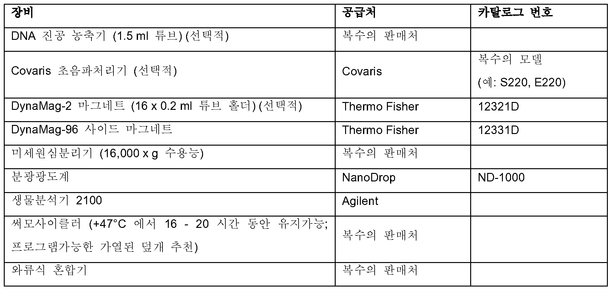
표 2: Table 2: 실시예Example 1 내지 5에서 언급된 소모품 Consumables mentioned in 1 to 5
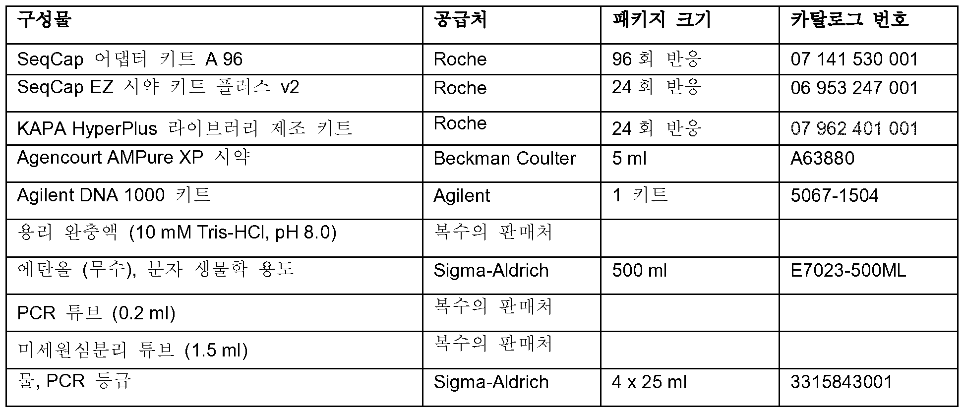
실시예 1 ― KAPA 하이퍼플러스 (HyperPlus) 라이브러리 제조물을 사용한 샘플 라이브러리 제조Example 1 - Preparation of sample library using KAPA HyperPlus library product
1.1. 동결건조된 인덱스 어댑터를 재현탁한다 (어댑터 키트 A).1.1. Resuspend the lyophilized index adapter (adapter kit A).
1.1.1. 간단히 내용물이 튜브 바닥에서 펠렛이 되도록, SeqCap 어댑터 키트 A 및/또는 B에 함유된, 동결건조된 인덱스 어댑터를 스피닝한다.1.1.1. Spin the lyophilized index adapter contained in the SeqCap adapter kits A and / or B, so that the contents are simply pellets at the bottom of the tube.
1.1.2. SeqCap 어댑터 키트 A 및/또는 B에서 'SeqCap 인덱스 어댑터'로 표지된 12개 튜브 각각에 50 마이크로리터의 차가운 PCR-등급의 물을 첨가한다. 어댑터를 얼음 상에 유지시킨다.1.1.2. Add 50 microliters of cold PCR-grade water to each of the 12 tubes labeled 'SeqCap Index Adapter' in SeqCap Adapter Kit A and / or B. Keep the adapter on ice.
1.1.3. 간단히 PCR-등급의 물이 더해진 인덱스 어댑터를 와류시키고 재현탁된 인덱스 어댑터 튜브를 스핀 다운한다.1.1.3. Simply vortex the index adapter with PCR-grade water and spin down the re-indexed index adapter tube.
1.1.4. 재현탁된 인덱스 어댑터의 튜브는 -15 내지 -25℃에 보관해야한다.1.1.4. Tubes of re-indexed index adapters should be stored at -15 to -25 ° C.
1.2. 샘플 라이브러리를 제조한다. 1.2. A sample library is prepared.
1.2.1. 라이브러리 구축에 사용하려는 gDNA (약 100 ng 내지 약 1 마이크로그램)를 10 mM Tris-HCl (pH 8.0) 중에 35 마이크로리터의 총 부피로 0.2 ml 튜브 또는 PCR 플레이트의 웰 내에 희석시킨다.1.2.1. The gDNA (from about 100 ng to about 1 microgram) to be used for library construction is diluted in a 0.2 ml tube or well of a PCR plate in a total volume of 35 microliters in 10 mM Tris-HCl (pH 8.0).
1.2.2. 표시된 순서로 성분들을 첨가하여 얼음 상에서 각 단편화 반응물을 합한다:1.2.2. Add the components in the order shown to combine each fragmented reaction on ice:
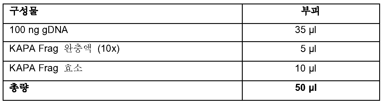
1.2.3. 단편화 반응물을 완전하게 혼합한다.1.2.3. Fragment Reaction Mix thoroughly.
1.2.4. 4℃에서 즉시 인큐베이션으로 설정된 사전 냉각된 써모사이클러에 위치시킨다. 다음으로 하기 개략된 프로그램을 사용하여 샘플을 인큐베이션한다: 1.2.4. Lt; RTI ID = 0.0 > 4 C < / RTI > Next, the sample is incubated using the program outlined below:
1.2.4.1. 단계 1: +37℃에서 20분. 1.2.4.1. Step 1: 20 minutes at + 37 ° C.
1.2.4.2. 단계 2: +4℃에서 유지. 1.2.4.2. Step 2: Keep at + 4 ° C.
1.2.5. 반응물을 얼음에 옮기고 즉시 다음 단계로 진행한다.1.2.5. The reaction is transferred to ice and immediately proceed to the next step.
1.2.6. 다음과 같이 말단 복구 및 A-꼬리화 (tailing) 반응을 수행한다:1.2.6. End-repair and A-tailing reactions are performed as follows:
1.2.6.1. 다음 시약들의 마스터 믹스를 제조한다:1.2.6.1. A master mix of the following reagents is prepared:
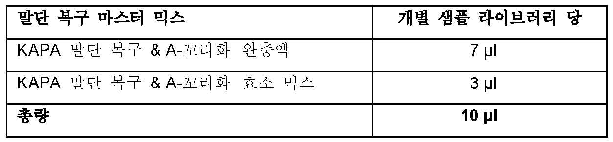
1.2.6.2. 각각의 50 마이크로리터 단편화된 샘플에 10 마이크로리터의 말단 복구 및 A-꼬리화 마스터 믹스를 첨가한다.1.2.6.2. Add 10 microliters of end-repair and A-tailed master mix to each 50 microliter fragmented sample.
1.2.6.3. 말단 복구 및 A-꼬리화 반응물을 완전하게 혼합한다.1.2.6.3. End recovery and A-tailed reactants are mixed thoroughly.
1.2.6.4. 얼음 상 위치시키고 즉시 다음 단계로 진행한다.1.2.6.4. Place on ice and proceed immediately to the next step.
1.2.6.5. 다음의 프로그램을 사용하여 가열된 덮개가 구비된 써모사이클러에서 말단 복구 및 A-꼬리화 인큐베이션을 수행한다:1.2.6.5. The following programs are used to perform end recovery and A-tailed incubation in a thermocycler with a heated lid:
1.2.6.5.1. 단계 1: +65℃에서 30분. 1.2.6.5.1. Step 1: 30 min at + 65 ° C.
1.2.6.5.2. 단계 2: +4℃에서 유지. 1.2.6.5.2. Step 2: Keep at + 4 ° C.
1.2.6.6. 30분간 인큐베이션 후, 즉시 다음 단계로 진행한다.1.2.6.6. After 30 minutes of incubation, proceed immediately to the next step.
1.2.7. 어댑터 결찰 반응 설정을 진행한다: 1.2.7. Proceed with adapter ligation response setup:
1.2.7.1. 다음의 시약들의 마스터 믹스를 제조한다: 1.2.7.1. A master mix of the following reagents is prepared:
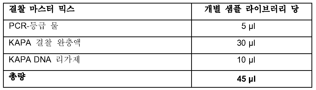
1.2.7.2. 5 ul의 SeqCap 라이브러리 어댑터 (바람직한 인덱스 포함)를 말단 복구 및 A-꼬리화 믹스와 DNA가 함유된 샘플 웰에 첨가한다. 각 샘플에 대해 사용된 인덱스를 반드시 기록한다.1.2.7.2. 5 ul of the SeqCap library adapter (including the preferred index) is added to the sample recovery wells containing the end-repair and A-tailed mix and DNA. Be sure to record the index used for each sample.
1.2.7.3. 65 마이크로리터의 말단 복구 및 A-꼬리화 믹스/DNA/어댑터를 함유하는 각 샘플 웰에, 45 마이크로리터의 결찰 마스터 믹스를 첨가하여, 그 결과 전체 부피가 110 마이크로리터가 되게 한다.1.2.7.3. To each sample well containing 65 microliter of the end recovery and A-tailed mix / DNA / adapter, 45 microliter of ligating master mix is added, resulting in a total volume of 110 microliters.
1.2.7.4. 결찰 반응물을 완전하게 혼합한다.1.2.7.4. The ligature reaction is thoroughly mixed.
1.2.7.5. +20℃에서 15분 동안 결찰 반응물을 인큐베이션한다.1.2.7.5. Incubate the ligation reaction at + 20 ° C for 15 minutes.
1.2.7.6. 인큐베이션 후, 즉시 다음 단계로 진행한다.1.2.7.6. After the incubation, proceed immediately to the next step.
1.2.8. 다음과 같이 결찰 후 세정을 수행한다:1.2.8. After the ligation, the cleaning is carried out as follows:
1.2.8.1. 각각의 결찰 반응물에, 완전하게 재현탁시킨 88 마이크로리터의 실온 AMPure XP 시약을 첨가한다.1.2.8.1. To each ligation reaction, 88 microliters of a room temperature AMPure XP reagent, completely resuspended, is added.
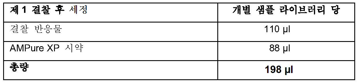
1.2.8.2. 결찰 반응 생성물 및 AMPure XP 시약을 완전하게 혼합한다.1.2.8.2. The ligation reaction product and the AMPure XP reagent are mixed thoroughly.
1.2.8.3. DNA가 비드에 결합할 수 있도록 5분 동안 실온에서 샘플을 인큐베이션한다.1.2.8.3. The samples are incubated at room temperature for 5 minutes so that the DNA can bind to the beads.
1.2.8.4. 비드를 포획하기 위해서 자성 입자 수집기에 샘플을 위치시킨다. 액체가 투명해질 때까지 인큐베이션한다.1.2.8.4. The sample is placed in a magnetic particle collector to capture the beads. Incubate until the liquid becomes clear.
1.2.8.5. 상청액을 조심스럽게 제거하여 버린다.1.2.8.5. Carefully remove the supernatant.
1.2.8.6. 샘플을 자성 입자 수집기에 유지시키고, 200 마이크로리터의 신선하게 제조된 80% 에탄올을 첨가한다. 1.2.8.6. The sample is held in a magnetic particle collector and 200 microliters of freshly prepared 80% ethanol is added.
1.2.8.7. ≥ 30초 동안 실온에서 샘플을 인큐베이션한다.1.2.8.7. Incubate the sample at room temperature for ≥ 30 seconds.
1.2.8.8. 에탄올을 조심스럽게 제거하여 버린다.1.2.8.8. Carefully remove the ethanol.
1.2.8.9. 샘플을 자성 입자 수집기에 유지시키고, 200 마이크로리터의 신선하게 제조된 80% 에탄올을 첨가한다. 1.2.8.9. The sample is held in a magnetic particle collector and 200 microliters of freshly prepared 80% ethanol is added.
1.2.8.10. ≥ 30초 동안 실온에서 샘플을 인큐베이션한다.1.2.8.10. Incubate the sample at room temperature for ≥ 30 seconds.
1.2.8.11. 조심스럽게 에탄올을 제거하여 버린다. 비드를 방해하지 않고 모든 잔류 에탄올을 제거하도록 노력한다. 1.2.8.11. Carefully remove the ethanol and discard. Try to remove any residual ethanol without interfering with the beads.
1.2.8.12. 모든 에탄올이 증발되도록 충분하게, 실온에서 비드를 건조시킨다. 1.2.8.12. Dry the beads at room temperature sufficiently to evaporate all of the ethanol.
1.2.8.13. 자성 입자 수집기로부터 샘플을 제거한다.1.2.8.13. The sample is removed from the magnetic particle collector.
1.2.8.14. 53 마이크로리터의 용리 완충액 (10 mM Tris-HCl, pH 8.0) 중에 비드를 완전하게 재현탁시킨다.1.2.8.14. The beads are completely resuspended in 53 microliters of elution buffer (10 mM Tris-HCl, pH 8.0).
1.2.8.15. DNA가 비드로부터 용리될 수 있도록 2분 동안 실온에서 샘플을 인큐베이션한다. 1.2.8.15. The sample is incubated at room temperature for 2 minutes so that the DNA can be eluted from the beads.
1.2.8.16. 비드를 포획하기 위해 자성 입자 수집기 상에 샘플을 위치시킨다. 액체가 투명해질 때까지 인큐베이션한다. 1.2.8.16. Place the sample on the magnetic particle collector to capture the beads. Incubate until the liquid becomes clear.
1.2.8.17. 50 마이크로리터 상청액을 새로운 튜브/웰에 옮긴다.1.2.8.17.
실시예Example 2 - 샘플 및 2 - sample and SeqCapSeqCap EZEZ 프로브Probe 풀 pool 혼성화Hybridization
2.1.1. AMPure XP 시약을 사용 전에 적어도 30분 동안 실온으로 가온시킨다.2.1.1. The AMPure XP reagent is allowed to warm to room temperature for at least 30 minutes before use.
2.1.2. SeqCap EZ 악세서리 키트 v2에 함유된, 5 마이크로리터의 COT 인간 DNA (1 mg/ml)를 새로운 튜브/웰에 첨가한다.2.1.2. Add 5 microliters of COT human DNA (1 mg / ml) contained in the SeqCap EZ Accessory Kit v2 to the new tube / well.
2.1.3. 3 μg의 DNA 샘플 라이브러리를 5 마이크로리터의 COT 인간 DNA를 함유하는 샘플에 첨가한다. 동일한 샘플로부터 구축된 다수 라이브러리를 이 목적을 위해 모을 수 있다. 2.1.3. 3 μg of the DNA sample library is added to the sample containing 5 μl of COT human DNA. Multiple libraries built from the same sample can be collected for this purpose.
2.1.4. 2,000 pmol (또는 2 마이크로리터)의 혼성화 향상 올리고 (1 마이크로리터의 1,000 pmol SeqCap HE 유니버설 올리고 및 샘플 라이브러리 어댑터 인덱스와 일치되는 1 마이크로리터의 1,000 pmol SeqCap HE 인덱스 올리고)를 COT 인간 DNA가 더해진 DNA 샘플 라이브러리에 첨가한다.2.1.4. 2,000 pmol (or 2 microliters) of hybridization enhancing oligos (1 microliter of 1,000 pmol SeqCap HE Index Oligor corresponding to 1 microliter of 1,000 pmol SeqCap HE Universal Oligos and sample library adapter index) Add to the library.
2.1.5. COT 인간 DNA, DNA 샘플 라이브러리 풀, SeqCap HE 유니버설 올리고 및 SeqCap HE 인덱스 올리고 풀의 투입 부피를 첨가하여 상기 혼합물의 총 부피를 결정한다.2.1.5. COT human DNA, DNA sample library pool, SeqCap HE universal oligo and SeqCap HE index Oligo grass are added to determine the total volume of the mixture.
2.1.6. 상기 혼합물에 2 부피의 AMPure XP 시약 (실온으로 평형화시키고 완전하게 재현탁시킴)을 첨가한다. 완전하게 혼합한다.2.1.6. To the mixture is added 2 volumes of AMPure XP reagent (equilibrated to room temperature and resuspended completely). Mix thoroughly.
2.1.7. 샘플을 실온에서 10분간 인큐베이션하여 샘플 라이브러리가 비드에 결합하도록 한다.2.1.7. The sample is incubated at room temperature for 10 minutes to allow the sample library to bind to the bead.
2.1.8. 샘플을 자성 입자 수집기에 위치시켜 비드를 포획한다. 용액이 투명하게 되도록 한다.2.1.8. The sample is placed in a magnetic particle collector to capture the beads. Allow the solution to become clear.
2.1.9. 투명해지면, 비드를 방해하지 않도록 조심스럽게 상청액을 제거하여 버린다.2.1.9. When transparent, the supernatant is carefully removed so as not to interfere with the beads.
2.1.10. 비드-결합된 DNA 샘플을 함유하는 샘플에 190 마이크로리터의 80% 에탄올을 첨가한다. 샘플을 이 단계 동안 자성 입자 수집기 상에 남겨두어야 한다.2.1.10. 190 microliters of 80% ethanol is added to the sample containing the bead-bound DNA sample. The sample should be left on the magnetic particle collector during this step.
2.1.11. 실온에서 ≥ 30초 동안 인큐베이션한다.2.1.11. Incubate for 30 seconds at room temperature.
2.1.12. 조심스럽게 80% 에탄올을 제거하여 버린다. 비드를 방해하지 않고 모든 잔류 에탄올을 제거하도록 노력한다.2.1.12. Carefully remove 80% ethanol. Try to remove any residual ethanol without interfering with the beads.
2.1.13. 튜브 덮개를 5분 동안 (또는 건조까지) 열어서 실온에서 비드를 건조시킨다.2.1.13. Open the tube cover for 5 minutes (or until dry) and dry the beads at room temperature.
2.1.14. 다음의 시약의 마스터 믹스를 제조하고, 포획 수를 반영하도록 확대한다:2.1.14. The master mix of the following reagents is prepared and expanded to reflect the capture:
2.1.14.1. 7.5 마이크로리터의 2X 혼성화 완충액 (바이알 5).2.1.14.1. 7.5 microliters of 2X hybridization buffer (vial 5).
2.1.14.2. 3 마이크로리터의 혼성화 성분 A (바이알 6).2.1.14.2. 3 microliters of hybridization component A (vial 6).
2.1.15. 이전 단계로부터의 10.5 마이크로리터의 혼성화 완충액/혼성화 성분 A 믹스를 비드-결합된 DNA 샘플에 첨가한다.2.1.15. 10.5 microliters of the hybridization buffer / hybridization component A mix from the previous step is added to the bead-bound DNA sample.
2.1.16. 자성 입자 수집기로부터 샘플을 제거하고 완전하게 혼합한다. 이 단계에서 충분한 혼합을 수행하여 균질한 혼합물이 산출되는 것이 중요하다.2.1.16. Remove the sample from the magnetic particle collector and mix thoroughly. It is important that sufficient mixing is performed at this stage to yield a homogeneous mixture.
2.1.17. 실온에서 2분간 정치시킨다.2.1.17. And allowed to stand at room temperature for 2 minutes.
2.1.18. 샘플을 자성 입자 수집기 상에 위치시킨다.2.1.18. The sample is placed on a magnetic particle collector.
2.1.19. 액체가 투명해진 후, 10.5 마이크로리터의 상청액 (전체 부피)을 제거하여 4.5 ul의 SeqCap EZ 프로브 풀을 함유하는 새로운 튜브/웰에 위치시킨다. 완전하게 혼합한다.2.1.19. After the liquid becomes clear, 10.5 microliters of the supernatant (total volume) is removed and placed into a new tube / well containing 4.5 ul of the SeqCap EZ probe pool. Mix thoroughly.
2.1.20. 블록 온도보다 10℃ 높게 설정된 가열된 덮개가 구비된 써모사이클러에서 다음의 프로그램을 사용하여 혼성화 인큐베이션을 수행한다:2.1.20. A hybridization incubation is performed in a thermocycler equipped with a heated lid set to 10 DEG C higher than the block temperature using the following program:
2.1.20.1. 95℃에서 5 분.2.1.20.1. 5 minutes at 95 ° C.
2.1.20.2. 47℃에서 16 내지 20시간.2.1.20.2. 16-20 hours at 47 < 0 > C.
2.1.21. 47℃에서 16시간 내지 20시간 동안의 인큐베이션을 위해서, 써모사이클러의 가열된 덮개가 켜져 있고 혼성화 온도보다 10℃ 높은 온도를 유지하도록 설정되는 것이 중요하다 (+57℃). 샘플은 단계 3.3에서 포획 비드로 전달될 때까지 47℃로 유지되어야만 한다.2.1.21. For incubation at 47 ° C for 16-20 hours, it is important that the heated lid of the thermocycler is turned on and set to maintain a
실시예Example 3 - 세척하고 포획된 DNA 샘플 라이브러리 회수 3 - Wash and capture captured DNA sample library
3.1. SeqCap 혼성화 및 세척 키트에 함유된, 10X 세척 완충액 (I, II, III 및 엄격) 및 2.5X 비드 세척 완충액을 희석하여 1X 작용 용액을 생성시킨다. 하기 열거된 부피가 1회 포획에 충분하다.3.1. Dilute the 10X Wash Buffer (I, II, III and Strict) and 2.5X bead wash buffer contained in the SeqCap hybridization and wash kit to create a 1X working solution. The volumes listed below are sufficient for one take.
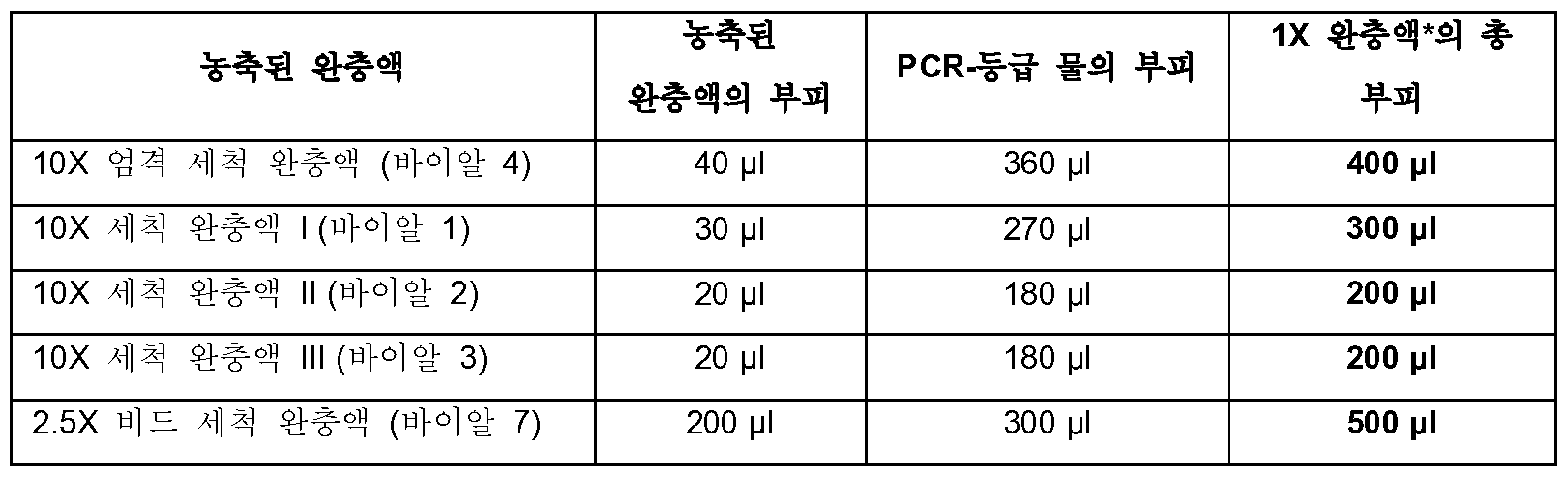
3.2. 포획 비드를 제조한다.3.2. Capture beads are prepared.
3.2.1. SeqCap 퓨어 포획 비드 키드에 함유된, 포획 비드를 사용 전에 30분간 실온으로 평형화되도록 한다.3.2.1. The capture beads in the SeqCap Pure Capture Bead Kid are allowed to equilibrate to room temperature for 30 minutes before use.
3.2.2. 비드의 균질한 혼합물이 보장되도록 사용전에 15초 동안 포획 비드를 와류시킨다.3.2.2. The capture bead is vortexed for 15 seconds before use to ensure a homogeneous mixture of beads.
3.2.3. 각각의 포획을 위해 50 마이크로리터의 비드를 0.2 ml 또는 1.5 ml 튜브로 분취한다 (즉, 1회 포획 사용을 위해서는 50 마이크로리터의 비드, 및 4회 포획 사용을 위해서는 200 마이크로리터 비드 등). 2회 포획 및 12회 포획에 충분한 비드가 각각 단일한 0.2 ml 튜브 및 1.5 ml 튜브에 제조될 수 있다.3.2.3. For each capture, 50 microliters of beads are aliquoted into 0.2 ml or 1.5 ml tubes (i.e., 50 microliters of beads for one-time use and 200 microliters of beads for four times of capture). Sufficient beads for 2-fold capture and 12-fold capture can be prepared in a single 0.2 ml tube and 1.5 ml tube, respectively.
3.2.4. 튜브를 자성 입자 수집기에 위치시킨다. 용액이 투명하게 되도록 한다 (5분 미만으로 걸려야 함).3.2.4. Place the tube in the magnetic particle collector. Make the solution clear (it should take less than 5 minutes).
3.2.5. 비드를 방해하지 않도록 조심스럽게 상청액을 제거하여 버린다. 임의의 잔존하는 미량의 액체는 후속 세척 단계에서 제거할 것이다.3.2.5. Carefully remove the supernatant to avoid interfering with the beads. Any remaining trace amounts of liquid will be removed in a subsequent wash step.
3.2.6. 튜브가 자성 입자 수집기 상에 위치되는 동안, 비드의 초기 부피의 2배의 1X 비드 세척 완충액을 첨가한다 (즉, 1회 포획 사용을 위해서는 100 마이크로리터의 완충액, 및 4회 포획 사용을 위해서는 400 마이크로리터 완충액 등).3.2.6. While the tube is positioned on the magnetic particle collector, add 1X bead wash buffer twice as much as the initial volume of bead (i.e., 100 microliter buffer for one-time use and 400 microliters Liter buffer, etc.).
3.2.7. 자성 입자 수집기로부터 튜브를 제거하고 와류 또는 상하 파이펫팅에 의해 완전하게 혼합한다.3.2.7. Remove the tube from the magnetic particle collector and mix thoroughly by vortexing or top and bottom pipetting.
3.2.8. 비드를 결합시키기 위해 다시 튜브를 자성 입자 수집기에 위치시킨다.3.2.8. Place the tube back into the magnetic particle collector to combine the beads.
3.2.9. 투명해지면, 액체를 제거하여 버린다.3.2.9. When it becomes transparent, liquid is removed and discarded.
3.2.10. 총 2회 세척 동안 단계 2.6 - 2.9를 반복한다.3.2.10. Repeat steps 2.6 - 2.9 for a total of two washes.
3.2.11. 제2 세척 후 완충액을 제거한 후, 비드의 초기 부피의 1X의 1X 비드 세척 완충액을 첨가한다 (즉, 포획 당 50 마이크로리터 완충액).3.2.11. After removal of the buffer after the second wash, 1X 1X bead wash buffer of the initial volume of bead is added (i. E., 50 microliter buffer per capture).
3.2.12. 자성 입자 수집기로부터 뉴브를 제거하여 완전하게 혼합한다.3.2.12. The nebul is removed from the magnetic particle collector and mixed thoroughly.
3.2.13. 50 마이크로리터의 재현탁된 비드를 각 포획을 위해 새로운 튜브/웰 내에 분취한다.3.2.13. 50 microlitres of resuspended beads are collected into a new tube / well for each capture.
3.2.14. 비드를 결합시키기 위해 자성 입자 수집기 상에 튜브를 위치시킨다. 용액이 투명해지도록 둔다.3.2.14. Place the tube on the magnetic particle collector to bond the beads. Allow the solution to become clear.
3.2.15. 투명해지면, 상청액을 제거하여 버린다.3.2.15. When it becomes transparent, the supernatant is removed.
3.2.16. 포획 비드가 이제 포획된 DNA에 결합되도록 준비된다. 즉시 다음 단계로 진행한다.3.2.16. The capture beads are now ready to bind to the captured DNA. Proceed immediately to the next step.
3.3. DNA를 포획 비드에 결합시킨다.3.3. DNA is bound to capture beads.
3.3.1. 하나의 혼성화 샘플을 이전 단계로부터의 포획 비드의 단일한 제조된 튜브/웰에 옮긴다.3.3.1. One hybridization sample is transferred to a single fabricated tube / well of the capture bead from the previous step.
3.3.2. 완전하게 혼합한다.3.3.2. Mix thoroughly.
3.3.3. 15분간 +47℃로 설정된 써모사이클러에 샘플을 위치시켜, 비드에 포 획된 샘플을 결합시킨다 (가열된 덮개는 +57℃로 설정).3.3.3. Place the sample in a thermocycler set to + 47 ° C for 15 minutes and combine the sample on the bead (set the heated lid to + 57 ° C).
3.4. 비드-결합된 DNA가 더해진 포획 비드를 세척한다.3.4. Wash capture beads with bead-bound DNA added.
3.4.1. 15분간 인큐베이션 이후, 써모사이클러로부터 샘플을 제거한다.3.4.1. After incubation for 15 minutes, the sample is removed from the thermocycler.
3.4.2. 써모사이클러는 다음 단계를 위해 47℃로 유지되어야 한다 (가열된 덮개는 켜지고 +57℃를 유지하도록 설정됨).3.4.2. The thermocycler should be maintained at 47 ° C for the next step (the heated lid is turned on and set to maintain + 57 ° C).
3.4.3. 100 마이크로리터의 1X 세척 완충액 I을 15 마이크로리터의 비드-결합된 DNA가 더해진 포획 비드에 첨가한다.3.4.3. 100 microliters of 1X Wash Buffer I is added to the capture beads to which 15 microliters of bead-bound DNA has been added.
3.4.4. 완전하게 혼합한다.3.4.4. Mix thoroughly.
3.4.5. 비드를 포획하도록 자성 입자 수집기 상에 샘플을 위치시킨다. 용액이 투명해지도록 한다.3.4.5. The sample is placed on the magnetic particle collector to capture the beads. Make the solution clear.
3.4.6. 투명해지면, 비드를 방해하지 않도록 조심스럽게 상청액을 제거하여 버린다.3.4.6. When transparent, the supernatant is carefully removed so as not to interfere with the beads.
3.4.7. 200 마이크로리터의 1X 엄격 세척 완충액을 각 샘플에 첨가한다.3.4.7. Add 200 microliters of 1X Strictly Washing Buffer to each sample.
3.4.8. 자성 입자 수집기로부터 샘플을 제거한다.3.4.8. The sample is removed from the magnetic particle collector.
3.4.9. 상하로 파이펫팅하여 균질하게 혼합한다.3.4.9. Pipet up and down to mix homogeneously.
3.4.10. +47℃로 예열된 써모사이클러에 위치시키고, 덮개 (+57℃로 설정)를 덮고 5분 동안 인큐베이션한다.3.4.10. Place in a thermocycler preheated to + 47 ° C, cover (set at + 57 ° C) and incubate for 5 minutes.
3.4.11. 5분 동안 인큐베이션 후, 샘플을 써모사이클러로부터 제거하고 비드를 포획하기 위해 자성 입자 수집기 상에 위치시킨다. 용액이 투명해지도록 한다.3.4.11. After incubation for 5 minutes, the sample is removed from the thermocycler and placed on a magnetic particle collector to capture the beads. Make the solution clear.
3.4.12. 투명해지면, 비드를 방해하지 않도록 조심하면서 상청액을 제거하여 버린다.3.4.12. When it becomes transparent, the supernatant is removed while being careful not to disturb the beads.
3.4.13. 1X 엄격 세척 완충액을 사용하여 총 2회의 세척 동안 단계 4.6 - 4.11을 반복한다.3.4.13. Repeat steps 4.6 - 4.11 for two total washes with 1X stringent wash buffer.
3.4.14. 200 마이크로리터의 실온 1X 세척 완충액 I을 첨가한다.3.4.14. Add 200 microliters of 1X Wash Buffer I at room temperature.
3.4.15. 10초 동안 와류 또는 10회 상하 파이펫팅에 의해 완전하게 혼합한다. 혼합물이 균질하도록 보장한다.3.4.15. Mix thoroughly for 10 seconds by vortexing or 10 times top and bottom pipetting. Ensure that the mixture is homogeneous.
3.4.16. 1분 동안 실온에서 인큐베이션한다.3.4.16. Incubate for 1 minute at room temperature.
3.4.17. 비드를 포획하기 위해서 자성 입자 수집기 상에 샘플을 위치시킨다. 용액이 투명해지도록 한다.3.4.17. The sample is placed on a magnetic particle collector to capture the beads. Make the solution clear.
3.4.18. 투명해지면, 비드를 방해하지 않도록 조심하면서 상청액을 제거하여 버린다.3.4.18. When it becomes transparent, the supernatant is removed while being careful not to disturb the beads.
3.4.19. 200 마이크로리터의 실온 1X 세척 완충액 II를 첨가한다.3.4.19. Add 200 microliters of room temperature IX Wash Buffer II.
3.4.20. 10초 동안 와류 또는 10회 상하 파이펫팅에 의해 완전하게 혼합한다. 혼합물이 균질하도록 보장한다.3.4.20. Mix thoroughly for 10 seconds by vortexing or 10 times top and bottom pipetting. Ensure that the mixture is homogeneous.
3.4.21. 1분 동안 실온에서 인큐베이션한다.3.4.21. Incubate for 1 minute at room temperature.
3.4.22. 비드를 포획하기 위해서 자성 입자 수집기 상에 샘플을 위치시킨다. 용액이 투명해지도록 한다.3.4.22. The sample is placed on a magnetic particle collector to capture the beads. Make the solution clear.
3.4.23. 투명해지면, 비드를 방해하지 않도록 조심하면서 상청액을 제거하여 버린다.3.4.23. When it becomes transparent, the supernatant is removed while being careful not to disturb the beads.
3.4.24. 200 마이크로리터의 실온 1X 세척 완충액 III을 첨가한다.3.4.24. Add 200 microliters of 1X Wash Buffer III at room temperature.
3.4.25. 10초 동안 와류 또는 10회 상하 파이펫팅에 의해 완전하게 혼합한다. 혼합물이 균질하도록 보장한다.3.4.25. Mix thoroughly for 10 seconds by vortexing or 10 times top and bottom pipetting. Ensure that the mixture is homogeneous.
3.4.26. 실온에서 1분 동안 인큐베이션한다.3.4.26. Incubate for 1 minute at room temperature.
3.4.27. 비드를 포획하기 위해 자성 입자 수집기 상에 샘플을 위치시킨다. 용액이 투명해지도록 한다.3.4.27. Place the sample on the magnetic particle collector to capture the beads. Make the solution clear.
3.4.28. 투명해지면, 비드를 방해하지 않도록 조심하면서 상청액을 제거하여 버린다.3.4.28. When it becomes transparent, the supernatant is removed while being careful not to disturb the beads.
3.4.29. 자성 입자 수집기로부터 샘플을 제거한다.3.4.29. The sample is removed from the magnetic particle collector.
3.4.30. 15 마이크로리터 PCR-등급의 물을 비드-결합된 DNA 샘플의 각 튜브/플레이트 웰에 첨가한다.3.4.30. 15 microliters PCR-grade water is added to each tube / plate well of the bead-bound DNA sample.
실시예Example 4 - 포획 전 4 - Capture I 결찰Ligature -매개 -medium PCRPCR (LM- (LM- PCRPCR )을 사용하여 포획된 샘플 라이브러리 증폭) To capture the captured sample library
4.1. 후-LM-PCR 올리고를 재현탁시킨다.4.1. Post-LM-PCR oligos.
4.1.1. 간략하게 SeqCap EZ 악세서리 키트 v2에 함유된, 동결건조된 후-LM-PCR 올리고 1 및 2를 스피닝하여 내용물이 튜브 바닥에 펠렛화되게 한다. 양쪽 올리고는 단일 튜브 내에 함유되도록 주의한다.4.1.1. Briefly spin the lyophilized post-LM-PCR Oligo 1 and 2 contained in SeqCap EZ Accessory Kit v2 to pellet the contents into the bottom of the tube. Make sure that both sides are contained in a single tube.
4.1.2. 480 마이크로리터의 PCR-등급 물을 원심분리된 올리고의 튜브에 첨가한다.4.1.2. Add 480 microliters of PCR-grade water to the tube of centrifuged oligo.
4.1.3. 간략하게 재현탁된 올리고를 와류시킨다.4.1.3. Briefly resuspended oligo is vortexed.
4.1.4. 내용물이 수집되도록 튜브를 스핀 다운시킨다.4.1.4. Spin down the tube to collect the contents.
4.1.5. 재현탁된 올리고 튜브는 -15 내지 -25℃에 보관해야한다.4.1.5. The resuspended oligotube should be stored at -15 to -25 ° C.
4.2. 포획 후 LM-PCR 마스터 믹스를 제조한다.4.2. After capture, the LM-PCR master mix is prepared.
4.2.1. 다음의 시약들의 마스터 믹스를 제조한다.4.2.1. A master mix of the following reagents is prepared.

4.2.2. 30 마이크로리터의 포획 후 LM-PCR 마스터 믹스를 0.2 ml 튜브 또는 PCR 플레이트의 웰에 첨가한다.4.2.2. After 30 microliters of capture, the LM-PCR master mix is added to the wells of a 0.2 ml tube or PCR plate.
4.2.3. 단계 3.3으로부터의 비드-결합된 DNA를 완전하게 혼합한다.4.2.3. The bead-bound DNA from step 3.3 is thoroughly mixed.
4.2.4. 20 마이크로리터의 주형으로서의 비드-결합된 DNA를 30 ul의 포획 후 LM-PCR 마스터 믹스와 튜브/웰 내에 분취한다 (음성 대조군을 수행하는 경우, 20 ul PCR-등굽 물을 이 튜브/웰에 첨가함).4.2.4. The bead-bound DNA as a 20 microliter template is collected in a tube / well with LM-PCR mastermix after capturing 30 ul (20 [mu] l PCR-Rinse is added to the tube / well box).
4.2.5. 수 회 상하 파이펫팅에 의해 완전하게 혼합한다.4.2.5. Mix thoroughly by pipetting up and down several times.
4.3. 포획 후 PCR 증폭을 수행한다.4.3. After capture, PCR amplification is performed.
4.3.1. 써모사이클러 샘플을 위치시킨다. 증폭 단계 동안 인큐베이션 온도보다 +10℃ 초과를 추적하기 위해 써모사이클러의 가열된 덮개를 설정하도록 권고된다.4.3.1. Place the thermocycler sample. It is recommended to set the thermocycler heated cover to track + 10 ° C above the incubation temperature during the amplification step.
4.3.2. 다음의 추적 후 LM-PCR 프로그램을 사용하여 포획된 DNA를 증폭시킨다:4.3.2. The captured DNA is amplified using the LM-PCR program after the following traces:
4.3.2.1. 단계 1: +98℃에서 45초.4.3.2.1. Step 1: 45 seconds at + 98 ° C.
4.3.2.2. 단계 2: +98℃에서 15초.4.3.2.2. Step 2: 15 seconds at + 98 ° C.
4.3.2.3. 단계 3: +60℃에서 30초.4.3.2.3. Step 3: 30 seconds at + 60 ° C.
4.3.2.4. 단계 4: +72℃에서 30초.4.3.2.4. Step 4: 30 seconds at + 72 ° C.
4.3.2.5. 단계 5: 단계 2로 가서 0 또는 1회 반복 (총 1 또는 2회 주기에 대해).4.3.2.5. Step 5: Go to step 2 and
4.3.2.6. 단계 6: +72℃에서 1분.4.3.2.6. Step 6: 1 minute at + 72 ° C.
4.3.2.7. 단계 7: +4℃에서 유지.4.3.2.7. Step 7: Keep at + 4 ° C.
4.3.2.8. 최대 72시간까지 정제를 위해 준비될 때까지 +2 내지 +8℃에서 반응물을 보관한다.4.3.2.8. Store the reaction at +2 to + 8 ° C until ready for purification up to 72 hours.
4.4. Agencourt AMPure XP 비드를 사용하여 증폭된 포획 DNA 샘플을 정제한다.4.4. Purify the amplified capture DNA sample using Agencourt AMPure XP beads.
4.4.1. SeqCap 퓨어 포획 비드 키트에 함유된 AMPure XP 비드를 사용 전 적어도 30분 동안 실온으로 가온시킨다.4.4.1. The AMPure XP beads contained in the SeqCap Pure Capture Bead Kit are allowed to warm to room temperature for at least 30 minutes prior to use.
4.4.2. 비드의 균질한 혼합물을 보장하도록 사용 전에 10초 동안 AMPure XP 비드를 와류시킨다.4.4.2. AMPure XP beads are vortexed for 10 seconds before use to ensure a homogeneous mixture of beads.
4.4.3. 90 마이크로리터의 AMPure XP 비드를 50 마이크로리터의 증폭된 포획 DNA 샘플 라이브러리에 첨가한다.4.4.3. 90 microliters of AMPure XP beads are added to a 50 microliter amplified capture DNA sample library.
4.4.4. 다수 회 상하로 파이펫팅 또는 와류하여 완전하게 혼합한다.4.4.4. Mix thoroughly by pipetting or vortexing several times up and down.
4.4.5. 5분 동안 실온에서 인큐베이션하여 포획된 샘플 라이브러리가 비드에 결합하도록 한다.4.4.5. Incubate at room temperature for 5 minutes to allow the captured sample library to bind to the beads.
4.4.6. 비드-결합된 DNA를 함유하는 샘플을 자성 입자 수집기 상에 위치시켜 비드를 포획한다. 용액이 투명해지도록 한다.4.4.6. A sample containing bead-bound DNA is placed on a magnetic particle collector to capture the beads. Make the solution clear.
4.4.7. 투명해지면, 비드를 방해하지 않도록 조심하면서 상청액을 제거하여 버린다.4.4.7. When it becomes transparent, the supernatant is removed while being careful not to disturb the beads.
4.4.8. 200 마이크로리터의 신선하게 제조된 80% 에탄올을 샘플 라이브러리와 비드를 함유하는 샘플에 첨가한다. 샘플은 이 단계 동안 자성 입자 수집기에 남아있어야 한다.4.4.8. 200 microliters of freshly prepared 80% ethanol is added to the sample library and sample containing the beads. The sample should remain in the magnetic particle collector during this step.
4.4.9. 실온에서 ≥ 30초 동안 인큐베이션한다.4.4.9. Incubate for 30 seconds at room temperature.
4.4.10. 80% 에탄올을 제거하여 버린다.4.4.10. 80% ethanol is removed and discarded.
4.4.11. 샘플을 자성 입자 수집기 상에 유지시키고, 200 마이크로리터의 신선하게 제조된 80% 에탄올을 첨가한다.4.4.11. The sample is held on a magnetic particle collector and 200 microliters of freshly prepared 80% ethanol is added.
4.4.12. 실온에서 ≥ 30초 동안 샘플을 인큐베이션한다.4.4.12. Incubate the sample for ≥ 30 seconds at room temperature.
4.4.13. 조심스럽게 에탄올을 제거하여 버린다. 비드를 방해하지 않고 모든 잔류 에탄올을 제거하도록 노력한다.4.4.13. Carefully remove the ethanol and discard. Try to remove any residual ethanol without interfering with the beads.
4.4.14. 5분 동안 (또는 건조까지) 튜브 덮개를 열어서 실온에서 비드가 건조되도록 한다.4.4.14. Allow the beads to dry at room temperature by opening the tube cover for 5 minutes (or until dry).
4.4.15. 자성 입자 수집기로부터 샘플을 제거한다.4.4.15. The sample is removed from the magnetic particle collector.
4.4.16. 53 마이크로리터의 10 mM Tris-HCl, pH 8.0 또는 PCR-등급의 물을 사용하여 DNA를 재현탁시킨다.4.4.16. The DNA is resuspended using 53 microliters of 10 mM Tris-HCl, pH 8.0 or PCR-grade water.
4.4.17. 모든 비드가 재현탁되도록 보장하기 위해 믹스를 10회 상하 파이펫팅한다.4.4.17. Peel the mix up and down 10 times to ensure that all beads are resuspended.
4.4.18. 2분 동안 실온에서 인큐베이션한다.4.4.18. Incubate at room temperature for 2 minutes.
4.4.19. 자성 입자 수집기 상에 다시 샘플을 위치시키고 용액이 투명해지도록 한다.4.4.19. Place the sample again on the magnetic particle collector and make the solution clear.
4.4.20. 이제 증폭된 포획 DNA 샘플 라이브러리 풀을 함유하는 50 마이크로리터의 상청액을 제거하여 새로운 튜브/웰에 옮긴다.4.4.20. Now 50 microliters of the supernatant containing the amplified capture DNA sample library pool is removed and transferred to a new tube / well.
4.5. 증폭된 포획 DNA 샘플의 농도, 크기 분포, 및 품질을 결정한다.4.5. The concentration, size distribution, and quality of the amplified capture DNA sample are determined.
4.5.1. DNA 농도를 정량하고 증폭된 포획 DNA 및 음성 대조군의 A260/A280 비율을 측정한다.4.5.1. The DNA concentration is quantified and the A260 / A280 ratio of the amplified capture DNA and negative control is determined.
4.5.1.1. A260/A280 비율은 1.7 내지 2.0이어야 한다.4.5.1.1. The A260 / A280 ratio should be 1.7 to 2.0.
4.5.1.2. LM-PCR 수율은 대략 500 ng이어야 한다.4.5.1.2. The LM-PCR yield should be approximately 500 ng.
4.5.1.3. 음성 대조군은 오염을 의미할 수 있는, 상당한 증폭을 보이지 않아야 한다.4.5.1.3. Negative controls should not show significant amplification, which could mean contamination.
4.5.2. 1 마이크로리터의 증폭된 포획 DNA 샘플 및 음성 대조군을 Agilent Bioanalyzer DNA 1000 칩을 사용하여 실행시킨다. 제조사의 지시서에 따라 칩을 실행시킨다. 증폭된 포획 DNA는 150 내지 500 bp의 평균 단편 길이를 나타내야 한다.4.5.2. One microliter of the amplified capture DNA sample and the negative control are run using an
4.5.3. 증폭된 포획 DNA는 서열분석을 위해 준비된다.4.5.3. The amplified capture DNA is prepared for sequencing.
실시예Example 5 - 5 - 포획된 샘플 라이브러리 서열분석Captured sample library sequencing
5.1. 제조사의 지시서에 따라서, Illumina 서열분석 장치를 사용하여 증폭된 포획 DNA를 서열분석한다.5.1. The amplified capture DNA is sequenced using the Illumina sequencer according to the manufacturer's instructions.
실시예Example 6 - 6 - 표적화Targeting 서열분석에 대한 증폭 주기의Of the amplification cycle for sequencing 횟수 효과의 비교 Comparison of frequency effects
본 명세서의 실시예 1 내지 5에 기재된 프로토콜을 사용하여, 7회의 실험을 동일한 공급원 (세포주 인간 게놈 DNA, NA12891, Coriell Institutes)으로부터 수득된, 동일한 양의 투입 게놈 DNA (3 마이크로그램)를 사용하여 수행하였다. 실험에 사용된 바이오틴화된 올리고뉴클레오티드 프로브는 암에 연관된 578개 유전자의 엑손을 표적으로 하였고, 누적 포획 표적은 4,571,289 염기쌍이다 (Design ID: 120522_HG19_Onco_R_EZ, Roche NimbleGen, Inc.). 포획 전 PCR 증폭은 7회 실험 중 어디에서도 수행하지 않았지만, 포획 후 PCR 증폭 주기의 횟수는 0 내지 14회 (0, 1, 2, 4, 6, 10, 및 14회)로 다양하였다. Using the protocol described in Examples 1 to 5 herein, seven experiments were performed using the same amount of input genomic DNA (3 micrograms) obtained from the same source (cell line human genomic DNA, NA12891, Coriell Institutes) Respectively. The biotinylated oligonucleotide probes used in the experiments targeted exon of 578 genes associated with cancer and the cumulative capture target was 4,571,289 base pairs (Design ID: 120522_HG19_Onco_R_EZ, Roche NimbleGen, Inc.). The pre-capture PCR amplification was not performed in any of the 7 experiments, but the number of PCR amplification cycles after capture varied from 0 to 14 (0, 1, 2, 4, 6, 10, and 14 times).
결과적으로 발생한 증폭된 포획 DNA는 제조사의 지시서에 따라서, 2 x 100 쌍형성 말단 서열분석으로, Illumina MiSeq 서열분석 장치를 사용하여 서열분석하였다. 7회 실험 각각에 대해, 결과적 판독치는 동량의 데이타를 사용하는 검정 성능의 비교를 용이하게 하기 위해서 175만 판독쌍 (3,500,000 판독치)으로 무작위하게 샘플링하였다. 데이타 분석은 "How to Evaluate NimbleGen SeqCap EZ Target Enrichment Data"라는 명칭의 Roche 기술 노트 문서 (2015년 8월, 이 개시내용은 그 전문을 참조로 본 명세서에 포함함)에 기술된 표준 생물정보학 방법을 사용하여 수행하였다. 분석 작업흐름의 개략도는 도 5에 도시하였다.The resulting amplified capture DNA was sequenced using the Illumina MiSeq sequencing device, in accordance with the manufacturer ' s instructions, with 2 x 100 pairing end sequencing. For each of the 7 trials, the resulting readings were randomly sampled to 175,000 read pairs (3,500,000 readings) to facilitate comparison of assay performance using the same amount of data. Data analysis is based on the standard bioinformatics method described in the Roche Technical Note document entitled "How to Evaluate NimbleGen SeqCap EZ Target Enrichment Data" (August 2015, the disclosure of which is incorporated herein by reference) . A schematic diagram of the analysis workflow is shown in FIG.
실험 결과는 6종의 빈번하게 사용된 표적화 서열분석 검정 성능 매트릭스에 대해 도 6 내지 11에 나타내었다. 포획 표적 또는 포획 표적 근처로 지도화된 서열분석된 염기의 백분율에 대한 값 (도 6), 포획 표적에 걸친 서열 심도 포괄범위의 분포 (도 7), 게놈에 대한 표적화된 서열의 농축 배수 (도 8), 호출된 단일 뉴클레오티드 다형성 (SNP)의 총 개수 (도 9), SNP 검출의 감도 (도 10), 및 SNP 분류의 특이성 (도 11)는 0, 1, 2, 4, 6, 10 또는 14회 주기의 PCR 증폭을 이용하는 실험간에 유사하였다. 바이오틴화된 올리고뉴클레오티드 프로브와의 혼성화를 통한 표적화 서열분석을 위한 현행 방법은 작업흐름 내에서 다횟수 주기의 PCR 증폭의 사용을 요구하고, 전형적으로 4회 초과의 증폭 주기를 요구한다 (도 1 및 4 참조). 여기에 제시된 결과들은 개시된 표적화된 표상적 서열분석 방법이 검정 성능에 임의의 두드러진 감소를 발생시키지 않고, 본 명세서에 기술된 바와 같이, 최소의 증폭 주기로 또는 증폭 주기 없이 표적화 서열분석을 수행할 수 있게 한다는 것을 예상치 않게 입증하였다.Experimental results are shown in Figs. 6 to 11 for six frequently used targeting sequencing assay performance matrices. (Figure 6), the distribution of the sequence depth coverage over the capture target (Figure 7), the concentration multiple of the targeted sequence for the genome (Figure 6), the value for the percentage of the sequenced base mapped near the capture target or capture target 8), the total number of single nucleotide polymorphisms (SNPs) called (FIG. 9), the sensitivity of SNP detection (FIG. 10), and the SNP classification specificity (FIG. 11) are 0, 1, 2, 4, The experiments using PCR amplification of 14 cycles were similar. Current methods for targeting sequencing through hybridization with biotinylated oligonucleotide probes require the use of multiple rounds of PCR amplification in the workflow and typically require more than four amplification cycles (Figures 1 and 2) 4). The results presented herein demonstrate that the disclosed targeted representative sequence analysis method does not cause any noticeable reduction in assay performance and can be used to perform targeted sequence analysis with minimal or no amplification cycle as described herein .
산업상 이용가능성에 관한 진술STATEMENT REGARDING INDUSTRIAL AVAILABILITY
본 개시내용은 의학 및 진단학 분야에서 산업상 이용가능성을 갖는다.The present disclosure has industrial applicability in the medical and diagnostic fields.
Claims (50)
Applications Claiming Priority (5)
| Application Number | Priority Date | Filing Date | Title |
|---|---|---|---|
| US201662279126P | 2016-01-15 | 2016-01-15 | |
| US62/279,126 | 2016-01-15 | ||
| US201662415952P | 2016-11-01 | 2016-11-01 | |
| US62/415,952 | 2016-11-01 | ||
| PCT/US2016/060835 WO2017123316A1 (en) | 2016-01-15 | 2016-11-07 | Deep sequencing profiling of tumors |
Publications (2)
| Publication Number | Publication Date |
|---|---|
| KR20180098412A true KR20180098412A (en) | 2018-09-03 |
| KR102398465B1 KR102398465B1 (en) | 2022-05-13 |
Family
ID=57396824
Family Applications (1)
| Application Number | Title | Priority Date | Filing Date |
|---|---|---|---|
| KR1020187023352A Active KR102398465B1 (en) | 2016-01-15 | 2016-11-07 | In-depth sequencing profiling of tumors |
Country Status (12)
| Country | Link |
|---|---|
| US (2) | US11649492B2 (en) |
| EP (1) | EP3402896B1 (en) |
| JP (2) | JP7232643B2 (en) |
| KR (1) | KR102398465B1 (en) |
| CN (1) | CN108463559A (en) |
| AU (1) | AU2016386032B2 (en) |
| CA (1) | CA3011342C (en) |
| IL (1) | IL260304B2 (en) |
| SA (1) | SA518392005B1 (en) |
| SG (1) | SG11201805600VA (en) |
| WO (1) | WO2017123316A1 (en) |
| ZA (1) | ZA201804322B (en) |
Families Citing this family (12)
| Publication number | Priority date | Publication date | Assignee | Title |
|---|---|---|---|---|
| ES2986436T3 (en) | 2011-04-15 | 2024-11-11 | Univ Johns Hopkins | Safe sequencing system |
| ES2886507T5 (en) | 2012-10-29 | 2024-11-15 | Univ Johns Hopkins | Pap test for ovarian and endometrial cancers |
| US11286531B2 (en) | 2015-08-11 | 2022-03-29 | The Johns Hopkins University | Assaying ovarian cyst fluid |
| SG11201805600VA (en) * | 2016-01-15 | 2018-07-30 | Ventana Med Syst Inc | Deep sequencing profiling of tumors |
| CN111868260B (en) | 2017-08-07 | 2025-02-21 | 约翰斯霍普金斯大学 | Methods and materials for evaluating and treating cancer |
| US20200004928A1 (en) | 2018-06-29 | 2020-01-02 | Roche Sequencing Solutions, Inc. | Computing device with improved user interface for interpreting and visualizing data |
| CN109234356B (en) * | 2018-09-18 | 2021-10-08 | 南京迪康金诺生物技术有限公司 | Method for constructing hybridization capture sequencing library and application |
| CN113383089A (en) * | 2018-11-29 | 2021-09-10 | 文塔纳医疗系统公司 | Personalized ctDNA disease monitoring via representative DNA sequencing |
| WO2020136133A1 (en) | 2018-12-23 | 2020-07-02 | F. Hoffmann-La Roche Ag | Tumor classification based on predicted tumor mutational burden |
| WO2020249704A1 (en) | 2019-06-13 | 2020-12-17 | F. Hoffmann-La Roche Ag | Systems and methods with improved user interface for interpreting and visualizing longitudinal data |
| EP4103748A4 (en) | 2020-02-14 | 2024-03-13 | The Johns Hopkins University | Methods and materials for assessing nucleic acids |
| JPWO2022244856A1 (en) | 2021-05-20 | 2022-11-24 |
Citations (5)
| Publication number | Priority date | Publication date | Assignee | Title |
|---|---|---|---|---|
| WO2009099602A1 (en) * | 2008-02-04 | 2009-08-13 | Massachusetts Institute Of Technology | Selection of nucleic acids by solution hybridization to oligonucleotide baits |
| WO2012024658A2 (en) * | 2010-08-20 | 2012-02-23 | IntegenX, Inc. | Integrated analysis system |
| US20150126379A1 (en) * | 2013-10-28 | 2015-05-07 | The Translational Genomics Research Institute | Long insert-based whole genome sequencing |
| WO2017123316A1 (en) * | 2016-01-15 | 2017-07-20 | Ventana Medical Systems, Inc. | Deep sequencing profiling of tumors |
| EP3564395A1 (en) * | 2010-12-30 | 2019-11-06 | Foundation Medicine, Inc. | Optimization of multigene analysis of tumor samples |
Family Cites Families (16)
| Publication number | Priority date | Publication date | Assignee | Title |
|---|---|---|---|---|
| US4000159A (en) | 1974-06-28 | 1976-12-28 | Phillips Petroleum Company | Preparation of n,n-disubstituted thioamides |
| US5176995A (en) | 1985-03-28 | 1993-01-05 | Hoffmann-La Roche Inc. | Detection of viruses by amplification and hybridization |
| US4683202A (en) | 1985-03-28 | 1987-07-28 | Cetus Corporation | Process for amplifying nucleic acid sequences |
| US4965188A (en) | 1986-08-22 | 1990-10-23 | Cetus Corporation | Process for amplifying, detecting, and/or cloning nucleic acid sequences using a thermostable enzyme |
| US4683195A (en) | 1986-01-30 | 1987-07-28 | Cetus Corporation | Process for amplifying, detecting, and/or-cloning nucleic acid sequences |
| US20080119367A1 (en) * | 2004-12-17 | 2008-05-22 | Mayo Foundation For Medical Education And Research | Prognosis of Renal Cell Carcinoma |
| WO2009061840A1 (en) * | 2007-11-05 | 2009-05-14 | Complete Genomics, Inc. | Methods and oligonucleotide designs for insertion of multiple adaptors employing selective methylation |
| US8592150B2 (en) * | 2007-12-05 | 2013-11-26 | Complete Genomics, Inc. | Methods and compositions for long fragment read sequencing |
| EP2227563B1 (en) | 2007-12-05 | 2012-06-06 | Complete Genomics, Inc. | Efficient base determination in sequencing reactions |
| JP5757572B2 (en) | 2009-07-03 | 2015-07-29 | 国立大学法人 東京大学 | Method for determining the presence or absence of cancer cells and method for determining the prognosis of cancer patients |
| DK2539450T3 (en) * | 2010-02-25 | 2016-05-30 | Advanced Liquid Logic Inc | PROCEDURE FOR PREPARING NUCLEIC ACID LIBRARIES |
| GB2497510A (en) | 2011-11-10 | 2013-06-19 | Harry Cuppens | Methods for determining mononucleotide sequence repeats |
| US10138519B2 (en) | 2012-12-28 | 2018-11-27 | Quest Diagnostics Investments Incorporated | Universal sanger sequencing from next-gen sequencing amplicons |
| EP2971122B1 (en) * | 2013-03-15 | 2020-08-26 | Memorial Sloan Kettering Cancer Center | Biomarkers for response to rapamycin analogs |
| US20140278461A1 (en) | 2013-03-15 | 2014-09-18 | Memorial Sloan-Kettering Cancer Center | System and method for integrating a medical sequencing apparatus and laboratory system into a medical facility |
| KR20160062763A (en) | 2013-10-18 | 2016-06-02 | 세븐 브릿지스 지노믹스 인크. | Methods and systems for genotyping genetic samples |
-
2016
- 2016-11-07 SG SG11201805600VA patent/SG11201805600VA/en unknown
- 2016-11-07 AU AU2016386032A patent/AU2016386032B2/en active Active
- 2016-11-07 CA CA3011342A patent/CA3011342C/en active Active
- 2016-11-07 CN CN201680078974.9A patent/CN108463559A/en active Pending
- 2016-11-07 WO PCT/US2016/060835 patent/WO2017123316A1/en not_active Ceased
- 2016-11-07 EP EP16801647.5A patent/EP3402896B1/en active Active
- 2016-11-07 IL IL260304A patent/IL260304B2/en unknown
- 2016-11-07 JP JP2018536830A patent/JP7232643B2/en active Active
- 2016-11-07 KR KR1020187023352A patent/KR102398465B1/en active Active
-
2018
- 2018-06-27 ZA ZA2018/04322A patent/ZA201804322B/en unknown
- 2018-07-11 SA SA518392005A patent/SA518392005B1/en unknown
- 2018-07-13 US US16/034,392 patent/US11649492B2/en active Active
-
2021
- 2021-07-02 JP JP2021110459A patent/JP7379418B2/en active Active
-
2023
- 2023-04-06 US US18/131,577 patent/US20230250476A1/en active Pending
Patent Citations (5)
| Publication number | Priority date | Publication date | Assignee | Title |
|---|---|---|---|---|
| WO2009099602A1 (en) * | 2008-02-04 | 2009-08-13 | Massachusetts Institute Of Technology | Selection of nucleic acids by solution hybridization to oligonucleotide baits |
| WO2012024658A2 (en) * | 2010-08-20 | 2012-02-23 | IntegenX, Inc. | Integrated analysis system |
| EP3564395A1 (en) * | 2010-12-30 | 2019-11-06 | Foundation Medicine, Inc. | Optimization of multigene analysis of tumor samples |
| US20150126379A1 (en) * | 2013-10-28 | 2015-05-07 | The Translational Genomics Research Institute | Long insert-based whole genome sequencing |
| WO2017123316A1 (en) * | 2016-01-15 | 2017-07-20 | Ventana Medical Systems, Inc. | Deep sequencing profiling of tumors |
Non-Patent Citations (1)
| Title |
|---|
| BMC Genomics, 16(1):589 (2015.08.07.)* * |
Also Published As
| Publication number | Publication date |
|---|---|
| IL260304A (en) | 2018-08-30 |
| US11649492B2 (en) | 2023-05-16 |
| EP3402896B1 (en) | 2021-03-31 |
| JP2019503182A (en) | 2019-02-07 |
| EP3402896A1 (en) | 2018-11-21 |
| US20230250476A1 (en) | 2023-08-10 |
| AU2016386032B2 (en) | 2021-08-12 |
| KR102398465B1 (en) | 2022-05-13 |
| WO2017123316A1 (en) | 2017-07-20 |
| CA3011342A1 (en) | 2017-07-20 |
| AU2016386032A1 (en) | 2018-07-12 |
| BR112018014349A2 (en) | 2019-02-12 |
| JP7232643B2 (en) | 2023-03-03 |
| ZA201804322B (en) | 2022-10-26 |
| SA518392005B1 (en) | 2022-02-08 |
| CA3011342C (en) | 2023-03-14 |
| US20180320229A1 (en) | 2018-11-08 |
| IL260304B2 (en) | 2025-04-01 |
| CN108463559A (en) | 2018-08-28 |
| SG11201805600VA (en) | 2018-07-30 |
| JP7379418B2 (en) | 2023-11-14 |
| JP2021176302A (en) | 2021-11-11 |
| IL260304B1 (en) | 2024-12-01 |
Similar Documents
| Publication | Publication Date | Title |
|---|---|---|
| JP7379418B2 (en) | Deep sequencing profiling of tumors | |
| JP5986572B2 (en) | Direct capture, amplification, and sequencing of target DNA using immobilized primers | |
| US9133513B2 (en) | High throughput methylation detection method | |
| JP7653924B2 (en) | Methods and compositions for proximity ligation | |
| TWI864378B (en) | Highly sensitive method for accurate parallel quantification of nucleic acids | |
| JP2024035109A (en) | Methods for accurate parallel detection and quantification of nucleic acids | |
| CA3208896A1 (en) | Highly sensitive methods for accurate parallel quantification of variant nucleic acids | |
| US20060121461A1 (en) | Methods for identifying and isolating unique nucleic acid sequences | |
| US20180073023A1 (en) | Circular single-stranded nucleic acid, method for preparing the same, and method for using the same | |
| HK1260078A1 (en) | Deep sequencing profiling of tumors | |
| CN117265116A (en) | Detection method, kit and probe library of TFE3 fusion gene | |
| HK40072445A (en) | Highly sensitive methods for accurate parallel quantification of nucleic acids | |
| EP3696279A1 (en) | Methods for noninvasive prenatal testing of fetal abnormalities | |
| JP2024543250A (en) | Target enrichment and quantification using isothermal linear amplified probes | |
| CN119932164A (en) | A highly sensitive method for accurate and parallel quantification of nucleic acids | |
| BR112018014349B1 (en) | TARGETED REPRESENTATIONAL SEQUENCING METHOD | |
| JP2022521209A (en) | Improved Nucleic Acid Target Concentration and Related Methods |
Legal Events
| Date | Code | Title | Description |
|---|---|---|---|
| PA0105 | International application |
Patent event date: 20180813 Patent event code: PA01051R01D Comment text: International Patent Application |
|
| PG1501 | Laying open of application | ||
| A201 | Request for examination | ||
| PA0201 | Request for examination |
Patent event code: PA02012R01D Patent event date: 20200110 Comment text: Request for Examination of Application |
|
| E902 | Notification of reason for refusal | ||
| PE0902 | Notice of grounds for rejection |
Comment text: Notification of reason for refusal Patent event date: 20210220 Patent event code: PE09021S01D |
|
| E902 | Notification of reason for refusal | ||
| PE0902 | Notice of grounds for rejection |
Comment text: Notification of reason for refusal Patent event date: 20210824 Patent event code: PE09021S01D |
|
| E701 | Decision to grant or registration of patent right | ||
| PE0701 | Decision of registration |
Patent event code: PE07011S01D Comment text: Decision to Grant Registration Patent event date: 20220322 |
|
| GRNT | Written decision to grant | ||
| PR0701 | Registration of establishment |
Comment text: Registration of Establishment Patent event date: 20220511 Patent event code: PR07011E01D |
|
| PR1002 | Payment of registration fee |
Payment date: 20220511 End annual number: 3 Start annual number: 1 |
|
| PG1601 | Publication of registration |