KR20180098402A - Activation of Calcium Control of Platelets by Electrical Stimulation - Google Patents
Activation of Calcium Control of Platelets by Electrical Stimulation Download PDFInfo
- Publication number
- KR20180098402A KR20180098402A KR1020187022044A KR20187022044A KR20180098402A KR 20180098402 A KR20180098402 A KR 20180098402A KR 1020187022044 A KR1020187022044 A KR 1020187022044A KR 20187022044 A KR20187022044 A KR 20187022044A KR 20180098402 A KR20180098402 A KR 20180098402A
- Authority
- KR
- South Korea
- Prior art keywords
- sample
- prp
- calcium ion
- calcium
- platelet
- Prior art date
- Legal status (The legal status is an assumption and is not a legal conclusion. Google has not performed a legal analysis and makes no representation as to the accuracy of the status listed.)
- Granted
Links
Images


Classifications
-
- A—HUMAN NECESSITIES
- A61—MEDICAL OR VETERINARY SCIENCE; HYGIENE
- A61K—PREPARATIONS FOR MEDICAL, DENTAL OR TOILETRY PURPOSES
- A61K41/00—Medicinal preparations obtained by treating materials with wave energy or particle radiation ; Therapies using these preparations
- A61K41/0004—Homeopathy; Vitalisation; Resonance; Dynamisation, e.g. esoteric applications; Oxygenation of blood
-
- C—CHEMISTRY; METALLURGY
- C12—BIOCHEMISTRY; BEER; SPIRITS; WINE; VINEGAR; MICROBIOLOGY; ENZYMOLOGY; MUTATION OR GENETIC ENGINEERING
- C12N—MICROORGANISMS OR ENZYMES; COMPOSITIONS THEREOF; PROPAGATING, PRESERVING, OR MAINTAINING MICROORGANISMS; MUTATION OR GENETIC ENGINEERING; CULTURE MEDIA
- C12N13/00—Treatment of microorganisms or enzymes with electrical or wave energy, e.g. magnetism, sonic waves
-
- A—HUMAN NECESSITIES
- A61—MEDICAL OR VETERINARY SCIENCE; HYGIENE
- A61K—PREPARATIONS FOR MEDICAL, DENTAL OR TOILETRY PURPOSES
- A61K35/00—Medicinal preparations containing materials or reaction products thereof with undetermined constitution
- A61K35/12—Materials from mammals; Compositions comprising non-specified tissues or cells; Compositions comprising non-embryonic stem cells; Genetically modified cells
- A61K35/14—Blood; Artificial blood
- A61K35/19—Platelets; Megacaryocytes
-
- A—HUMAN NECESSITIES
- A61—MEDICAL OR VETERINARY SCIENCE; HYGIENE
- A61K—PREPARATIONS FOR MEDICAL, DENTAL OR TOILETRY PURPOSES
- A61K38/00—Medicinal preparations containing peptides
- A61K38/16—Peptides having more than 20 amino acids; Gastrins; Somatostatins; Melanotropins; Derivatives thereof
- A61K38/17—Peptides having more than 20 amino acids; Gastrins; Somatostatins; Melanotropins; Derivatives thereof from animals; from humans
- A61K38/18—Growth factors; Growth regulators
-
- A—HUMAN NECESSITIES
- A61—MEDICAL OR VETERINARY SCIENCE; HYGIENE
- A61K—PREPARATIONS FOR MEDICAL, DENTAL OR TOILETRY PURPOSES
- A61K41/00—Medicinal preparations obtained by treating materials with wave energy or particle radiation ; Therapies using these preparations
-
- A—HUMAN NECESSITIES
- A61—MEDICAL OR VETERINARY SCIENCE; HYGIENE
- A61P—SPECIFIC THERAPEUTIC ACTIVITY OF CHEMICAL COMPOUNDS OR MEDICINAL PREPARATIONS
- A61P17/00—Drugs for dermatological disorders
- A61P17/02—Drugs for dermatological disorders for treating wounds, ulcers, burns, scars, keloids, or the like
-
- C—CHEMISTRY; METALLURGY
- C12—BIOCHEMISTRY; BEER; SPIRITS; WINE; VINEGAR; MICROBIOLOGY; ENZYMOLOGY; MUTATION OR GENETIC ENGINEERING
- C12N—MICROORGANISMS OR ENZYMES; COMPOSITIONS THEREOF; PROPAGATING, PRESERVING, OR MAINTAINING MICROORGANISMS; MUTATION OR GENETIC ENGINEERING; CULTURE MEDIA
- C12N5/00—Undifferentiated human, animal or plant cells, e.g. cell lines; Tissues; Cultivation or maintenance thereof; Culture media therefor
- C12N5/06—Animal cells or tissues; Human cells or tissues
- C12N5/0602—Vertebrate cells
- C12N5/0634—Cells from the blood or the immune system
- C12N5/0644—Platelets; Megakaryocytes
-
- C—CHEMISTRY; METALLURGY
- C12—BIOCHEMISTRY; BEER; SPIRITS; WINE; VINEGAR; MICROBIOLOGY; ENZYMOLOGY; MUTATION OR GENETIC ENGINEERING
- C12Q—MEASURING OR TESTING PROCESSES INVOLVING ENZYMES, NUCLEIC ACIDS OR MICROORGANISMS; COMPOSITIONS OR TEST PAPERS THEREFOR; PROCESSES OF PREPARING SUCH COMPOSITIONS; CONDITION-RESPONSIVE CONTROL IN MICROBIOLOGICAL OR ENZYMOLOGICAL PROCESSES
- C12Q1/00—Measuring or testing processes involving enzymes, nucleic acids or microorganisms; Compositions therefor; Processes of preparing such compositions
- C12Q1/56—Measuring or testing processes involving enzymes, nucleic acids or microorganisms; Compositions therefor; Processes of preparing such compositions involving blood clotting factors, e.g. involving thrombin, thromboplastin, fibrinogen
-
- G—PHYSICS
- G01—MEASURING; TESTING
- G01N—INVESTIGATING OR ANALYSING MATERIALS BY DETERMINING THEIR CHEMICAL OR PHYSICAL PROPERTIES
- G01N33/00—Investigating or analysing materials by specific methods not covered by groups G01N1/00 - G01N31/00
- G01N33/15—Medicinal preparations ; Physical properties thereof, e.g. dissolubility
-
- G—PHYSICS
- G01—MEASURING; TESTING
- G01N—INVESTIGATING OR ANALYSING MATERIALS BY DETERMINING THEIR CHEMICAL OR PHYSICAL PROPERTIES
- G01N33/00—Investigating or analysing materials by specific methods not covered by groups G01N1/00 - G01N31/00
- G01N33/48—Biological material, e.g. blood, urine; Haemocytometers
- G01N33/50—Chemical analysis of biological material, e.g. blood, urine; Testing involving biospecific ligand binding methods; Immunological testing
- G01N33/86—Chemical analysis of biological material, e.g. blood, urine; Testing involving biospecific ligand binding methods; Immunological testing involving blood coagulating time or factors, or their receptors
-
- A—HUMAN NECESSITIES
- A61—MEDICAL OR VETERINARY SCIENCE; HYGIENE
- A61K—PREPARATIONS FOR MEDICAL, DENTAL OR TOILETRY PURPOSES
- A61K33/00—Medicinal preparations containing inorganic active ingredients
- A61K33/06—Aluminium, calcium or magnesium; Compounds thereof, e.g. clay
Landscapes
- Health & Medical Sciences (AREA)
- Life Sciences & Earth Sciences (AREA)
- Engineering & Computer Science (AREA)
- Chemical & Material Sciences (AREA)
- General Health & Medical Sciences (AREA)
- Zoology (AREA)
- Bioinformatics & Cheminformatics (AREA)
- Hematology (AREA)
- Biomedical Technology (AREA)
- Immunology (AREA)
- Medicinal Chemistry (AREA)
- Organic Chemistry (AREA)
- Pharmacology & Pharmacy (AREA)
- Biotechnology (AREA)
- Veterinary Medicine (AREA)
- Animal Behavior & Ethology (AREA)
- Public Health (AREA)
- Wood Science & Technology (AREA)
- Epidemiology (AREA)
- Genetics & Genomics (AREA)
- Biochemistry (AREA)
- Microbiology (AREA)
- Cell Biology (AREA)
- Proteomics, Peptides & Aminoacids (AREA)
- General Engineering & Computer Science (AREA)
- Gastroenterology & Hepatology (AREA)
- Molecular Biology (AREA)
- Virology (AREA)
- Developmental Biology & Embryology (AREA)
- Physics & Mathematics (AREA)
- Analytical Chemistry (AREA)
- General Physics & Mathematics (AREA)
- Biophysics (AREA)
- Pathology (AREA)
- Urology & Nephrology (AREA)
- Food Science & Technology (AREA)
- Alternative & Traditional Medicine (AREA)
- Dermatology (AREA)
- Chemical Kinetics & Catalysis (AREA)
- General Chemical & Material Sciences (AREA)
Abstract
본 개시는, 응괴의 존재 또는 부재, (존재한다면) 응괴 형성의 타이밍, 및/또는 (존재한다면) 응괴의 기계적 강도 중 하나 이상을 활성화 과정 동안 칼슘 이온의 존재 또는 농도에 의해 제어하는, 활성화된 혈소판 생성물의 생성에 관한 것이다. 특정 실시형태에서, 칼슘 이온 농도는 활성화 과정의 일부로서 펄스 전기장 또는 화학적 활성화제(예를 들어, 트롬빈)의 존재 하에 제어된다.The present disclosure relates to a process for the preparation of an activated (at least one) active site that controls at least one of the presence or absence of coagulation, the timing of coagulation formation (if present), and / To the production of platelet products. In certain embodiments, the calcium ion concentration is controlled in the presence of a pulsed electric field or chemical activator (e. G., Thrombin) as part of the activation process.
Description
본원에 개시된 대상은 일반적으로 수술 또는 외상을 대한 치료법과 같은 다양한 의학적 적용에서 이용되는 혈소판 요법에 관한 것이다. 특정 실시형태는, 펄스 전기장의 존재 및 특성과 칼슘 존재 및 농도를 포함하나 이들에 한정되지 않는 다양한 활성화 조건을 변경하는 것에 의한 혈소판 활성화 및 응고 제어에 관한 것이다.The subjects disclosed herein generally relate to platelet therapy that is used in a variety of medical applications such as surgery or trauma therapy. Certain embodiments relate to platelet activation and coagulation control by altering various activation conditions including, but not limited to, the presence and characteristics of a pulsed electric field and the presence and concentration of calcium.
혈소판 겔("활성화된 혈소판 농축 혈장"으로도 불림)의 사용은 (예컨대, 수술 후의) 상처 치유 및 생체항상성을 촉진하는 것을 비롯한 다양한 용도를 위해 클리닉 또는 다른 건강 관리 시설에서 이용될 수 있는 새로 떠오르는 치료적 접근법이다. 특히, 신경 상해, 건염, 골관절염, 심장 근육 손상 및 골 수복 및 재생과 같은 여러 유형의 상해 및 상태에 대해, 상처 치유 처치로서의 혈소판 요법의 용도에 있어서의 관심이 존재한다. 또한, 환자에게 사용되는 혈소판 겔의 유도는 자가 유래일 수 있으며, 이는 혈소판이 환자 자신의 조직 및/또는 체액으로부터 유도된다는 것을 의미한다. 따라서, 환자의 혈액 샘플을 사용하여 환자를 치료하는 데 사용되는 혈소판 겔을 유도할 수 있다.The use of platelet gels (also referred to as "activated platelet-rich plasma") can be used in clinics or other healthcare facilities for a variety of uses, including wound healing and post- It is a therapeutic approach. In particular, there are concerns in the use of platelet therapy as wound healing treatments for various types of injuries and conditions, such as nerve injury, tendinitis, osteoarthritis, cardiac muscle damage and bone repair and regeneration. In addition, the induction of the platelet gel used in the patient may be autologous, which means that platelets are derived from the patient ' s own tissues and / or body fluids. Thus, a blood sample of a patient can be used to induce a platelet gel used to treat the patient.
예를 들어, 의사는 환자로부터 혈액을 채취할 수 있다. 그 후, 혈액을 원심 분리하여 혈소판 농축 혈장(platelet-rich plasma; PRP)을 만들 수 있다. 혈소판이 활성화되면, 혈액 내의 혈소판은 상처 치유 캐스케이드를 용이하게 하고 촉진시키는 단백질 및 성장 인자를 방출한다. 따라서, 임상 작업 과정에는 환자로부터 혈액을 채취하고, 그 혈액을 원심분리하여 혈소판을 분리해 내고, 소 트롬빈을 사용하는 등의 생체외 혈소판 활성화를 수행하는 것을 포함할 수 있다. 그 후, 활성화된 혈소판 또는 혈소판 겔은 상처 또는 다른 치료 부위에 적용될 수 있다. 생체내 혈소판 활성화가 대신 이용되는 경우, 의사는 혈소판 활성화제를 첨가하지 않고 PRP를 그 부위에 적용할 수 있다. 성장 인자 방출 및 응고를 포함하는 혈소판 활성화는 일반적으로 결합 조직 내의 콜라겐에 의해 유도된다.For example, a doctor may take blood from a patient. The blood can then be centrifuged to produce platelet-rich plasma (PRP). When platelets are activated, platelets in the blood release proteins and growth factors that facilitate and promote wound healing cascades. Thus, clinical procedures may include taking blood from a patient, centrifuging the blood to isolate the platelets, and performing in vitro platelet activation, such as using bovine thrombin. Activated platelets or platelet gels can then be applied to the wound or other treatment site. If in vivo platelet activation is used instead, the physician may apply the PRP to the site without adding platelet activating agents. Platelet activation, including growth factor release and clotting, is generally induced by collagen in connective tissue.
트롬빈(예를 들어, 소 트롬빈)이 혈소판 활성화를 유도하는 데 사용되는 그러한 생체외 적용예에 있어서, 결과적인 성장 인자 수준은 생물학적 반응에 기초하여 고정될 수 있다. 즉, 상이한 성장 인자의 양 및/또는 각각의 비 또는 비율은 트롬빈에 기초한 활성화의 성질에 의해 결정된다. 따라서, 그러한 반응에서, 임상의는 상이한 성장 인자의 각각의 양 또는 비율을 조절하거나 조작할 수 없고, 대신에 종래의 활성화 조성물로 해야만 한다. 또한, 특정 임상 시나리오에서는, 응괴를 갖는 것 또는 갖지 않는 것 및/또는 응괴가 존재하는 경우, 그 응괴의 기계적 강도 또는 다른 특성을 제어하는 것이 바람직할 수 있다.In such an in vitro application where thrombin (e.g., bovine thrombin) is used to induce platelet activation, the resulting growth factor levels may be fixed based on the biological response. That is, the amount and / or the ratio or ratio of the different growth factors is determined by the nature of the thrombin-based activation. Thus, in such a response, the clinician should not be able to control or manipulate the respective amounts or ratios of the different growth factors, but instead must be a conventional activated composition. Also, in certain clinical scenarios, it may be desirable to control the mechanical strength or other properties of the clot, with and without clot and / or clot, if present.
일 실시형태에서, 활성화된 생성물을 생성하는 방법을 제공한다. 이 방법에 따르면, 활성화를 위해 혈소판 농축 혈장(PRP) 샘플을 제조한다. PRP 샘플은 칼슘 이온을 각각의 농도로 첨가함으로써 제조한다. 각각의 농도는, 응괴가 PRP 샘플을 사용하여 생성한 활성화된 생성물 조성물 중에 존재해야 하는지 여부, 그리고 응고가 존재해야 한다면, 응괴 형성까지의 시간 또는 응괴의 기계적 강도 중 하나 이상을 기초로 선택된다. PRP 샘플은 전자기 자극 장치의 전극에 대하여 위치시킨다. 전기 펄스 파라미터의 세트를 지정한다. PRP 샘플을 파라미터 값에 따라 생성된 하나 이상의 전기 펄스에 노출시킨다. PRP 샘플은, 하나 이상의 전기 펄스에 노출될 경우, 1종 이상의 성장 인자를 포함하고 지정된 응고 특성을 갖는 활성화된 생성물 조성물을 산출한다.In one embodiment, a method of producing an activated product is provided. According to this method, a platelet-rich plasma (PRP) sample is prepared for activation. PRP samples are prepared by adding calcium ions at respective concentrations. Each concentration is selected on the basis of at least one of whether the coagulation should be in the activated product composition produced using the PRP sample and the time to coagulation formation or the mechanical strength of the coagulation if coagulation should be present. The PRP sample is placed against the electrode of the electromagnetic stimulator. Specifies a set of electrical pulse parameters. The PRP samples are exposed to one or more electrical pulses generated according to the parameter values. A PRP sample, when exposed to one or more electrical pulses, produces an activated product composition comprising one or more growth factors and having the specified coagulation properties.
다른 실시형태에서, 활성화된 생성물을 생성하는 방법을 제공한다. 이 방법에 따르면, 활성화를 위해, 항응고제 처리된 혈소판 농축 혈장(PRP) 샘플을 제조한다. 다양한 가능한 농도로부터 선택되는 칼슘 이온 농도에 도달하도록, PRP 샘플에 칼슘 이온을 첨가한다. 농도는, PRP 샘플을 사용하여 생성한 활성화된 생성물 조성물 중에 존재하는 1종 이상의 성장 인자의 목표 수준을 기초로 선택된다. PRP 샘플을 전기적 활성화 자극에 노출시킨다. PRP 샘플은, 전기적 활성화 자극에 노출될 경우, 1종 이상의 성장 인자를 목표 수준으로 포함하는 활성화된 생성물 조성물을 산출한다. 전기적 활성화 자극을 변경하지 않고 칼슘 이온 농도를 변경하는 것이 1종 이상의 성장 인자의 절대 또는 상대 수준 중 하나 또는 둘 다를 변화시킨다.In another embodiment, a method of producing an activated product is provided. According to this method, an anticoagulated platelet-rich plasma (PRP) sample is prepared for activation. Calcium ions are added to the PRP sample to reach a selected calcium ion concentration from various possible concentrations. The concentration is selected based on the target level of one or more growth factors present in the activated product composition produced using the PRP sample. The PRP sample is exposed to an electrically activated stimulus. The PRP sample, when exposed to an electrically activated stimulus, produces an activated product composition comprising at least one growth factor at the target level. Altering the calcium ion concentration without altering the electrically activated stimulus alters one or both of the absolute or relative levels of one or more growth factors.
추가적인 실시형태에서, 혈소판 겔에 있어서의 응괴의 기계적 강도를 제어하는 방법을 제공한다. 이 방법에 따르면, 혈소판 겔에서 생성되는 하나 이상의 응괴의 잠재적 기계적 강도를 결정한다. 잠재적 기계적 강도는 트롬빈만을 사용하여 혈소판 겔을 생성함으로써 관찰하게 되는 강도보다 더 크다. 잠재적 기계적 강도를 기초로, 복수의 칼슘 이온 농도 중에서 잠재적 기계적 강도에 상응하는 칼슘 이온 농도를 선택한다. 혈소판 겔은, 선택된 칼슘 이온 농도로 칼슘 이온을 포함하는 혈소판 농축 혈장(PRP) 샘플을 활성화시킴으로써 생성한다. PRP 샘플은 전기 자극을 이용하여 활성화시킨다. 혈소판 겔은, 형성되면, 잠재적 기계적 강도를 갖는 응괴를 포함한다.In a further embodiment, a method is provided for controlling the mechanical strength of the clot in a platelet gel. According to this method, the potential mechanical strength of one or more clumps produced in the platelet gel is determined. Potential mechanical strength is greater than the intensity observed by producing platelet gel using only thrombin. Based on the potential mechanical strength, the calcium ion concentration corresponding to the potential mechanical strength among the plurality of calcium ion concentrations is selected. Platelet gels are produced by activating platelet-rich plasma (PRP) samples containing calcium ions at a selected calcium ion concentration. PRP samples are activated using electrical stimulation. Platelet gels, when formed, include coagulation with potential mechanical strength.
본 발명의 이러한, 그리고 다른 특징들, 양태들 및 이점들은, 하기 상세한 설명을, 첨부하는 도면을 참조하여 읽을 때 더 잘 이해될 것이며, 도면 전반에 있어서, 유사한 부호는 유사한 부분을 나타낸다.
도 1은 본 발명의 양태들에 따른 펄스 생성 시스템의 개략도이다.
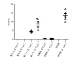
도 2는, 본 발명의 양태들에 따른, 연구 결과의 세트에 대한 초기 응괴 형성까지의 시간(응고 시간)을 그래프로 도시한다.
도 3은, 본 발명의 양태들에 따른, 연구 결과의 세트에 대한 응고 키네틱(응고 속도)을 그래프로 도시한다.
도 4는, 본 발명의 양태들에 따른, 연구 결과의 세트에 대한 피브리노겐 수준을 그래프로 도시한다.
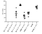
도 5는, 본 발명의 양태들에 따른, 연구 결과의 세트에 대한 응괴 강도를 그래프로 도시한다.
도 6은, 본 발명의 양태들에 따른, 한 연구 참여자에 대한 시간에 따라 관찰한 응괴의 기계적 강도(MA)를 보여주는 대표적인 TEG 트레이싱을 도시한다.
도 7은, 본 발명의 양태들에 따른, 연구 결과의 세트에 대한 PF4 수준을 그래프로 도시한다.
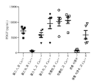
도 8은, 본 발명의 양태들에 따른, 연구 결과의 세트에 대한 PDGF 수준을 그래프로 도시한다.
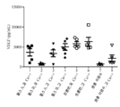
도 9는, 본 발명의 양태들에 따른, 연구 결과의 세트에 대한 VEGF 수준을 그래프로 도시한다.
도 10은, 본 발명의 양태들에 따른, 연구 결과의 세트에 대한 EGF 수준을 도시한다.These and other features, aspects, and advantages of the present invention will become better understood when the following detailed description is read with reference to the accompanying drawings, wherein like reference numerals designate like parts.
1 is a schematic diagram of a pulse generation system in accordance with aspects of the present invention.
Figure 2 graphically illustrates the time to initial coagulation formation (coagulation time) for a set of studies, in accordance with aspects of the present invention.
Figure 3 graphically illustrates the clot kinetic (coagulation rate) for a set of studies, in accordance with aspects of the present invention.
Figure 4 graphically illustrates fibrinogen levels for a set of studies, in accordance with aspects of the present invention.
Figure 5 graphically illustrates the cohesion strength for a set of studies, in accordance with aspects of the present invention.
Figure 6 illustrates a representative TEG tracing showing the mechanical strength (MA) of the observed clusters over time for one study participant, in accordance with aspects of the present invention.
Figure 7 graphically illustrates the level of PF4 for a set of studies, in accordance with aspects of the present invention.
Figure 8 graphically illustrates PDGF levels for a set of studies, in accordance with aspects of the present invention.
Figure 9 graphically illustrates VEGF levels for a set of studies, in accordance with aspects of the present invention.
Figure 10 shows EGF levels for a set of studies, in accordance with aspects of the present invention.
본 발명의 주제의 하나 이상의 특정 실시형태가 아래에서 설명될 것이다. 이들 실시형태의 간결한 설명을 제공하기 위해, 실제 구현의 모든 특징이 명세서에 기술되지 않을 수도 있다. 임의의 엔지니어링 또는 설계 프로젝트에서와 같이 임의의 그러한 실제 구현을 개발할 때, 시스템 관련 및 비즈니스 관련 제약 조건 준수와 같이, 개발자의 구체적인 목표를 달성하기 위해 많은 구현 특이적 결정을 내려야 하며, 이것은 구현마다 다를 수 있다는 것이 이해되어야 한다. 또한, 이러한 개발 노력은 복잡하고 시간 소모적일 수 있지만, 그럼에도 불구하고 본 개시의 혜택을 갖는 통상의 기술자에게는 설계, 제작 및 제조의 일상적인 실시일 수 있음을 이해해야 한다.One or more specific embodiments of the subject matter of the present invention will be described below. In order to provide a concise description of these embodiments, not all features of an actual implementation may be described in the specification. When developing any such actual implementation, such as in any engineering or design project, many implementation-specific decisions must be made to achieve the developer's specific goals, such as compliance with system-related and business-related constraints, It should be understood. It should also be appreciated that such a development effort may be complex and time consuming, but nevertheless a routine undertaking of design, fabrication, and manufacture to one of ordinary skill in the art having the benefit of this disclosure.
본원에 제시된 기술들은, 현재의 기술 분야를 명백하게 개선하고, 따라서 추상적이거나 순수한 이론이 아닌 실용적 성질의 물질 대상 및 구체적인 실례를 참조하고 적용한다.The techniques presented herein clearly improve the present technical field and, therefore, reference and apply material objects and concrete examples of practical properties that are not abstract or pure theories.
본 발명의 다양한 실시형태들의 요소들을 소개할 때, "하나", "하나의", "그" 및 "상기"라는 용어는 하나 이상의 요소가 있음을 의미하는 것으로 의도된다. "포함하는", "함유하는" 및 "갖는"이라는 용어는 포괄적인 것을 의도하며, 열거된 요소 이외에 추가적인 요소가 있을 수 있음을 의미한다.The terms "a", "an", "the", and "the" are intended to mean that there are one or more of the elements in the elements of the various embodiments of the present invention. The terms "comprising", "containing" and "having" are intended to be inclusive and mean that there may be additional elements other than the listed elements.
혈소판 활성화 및/또는 응집은 생체내 및/또는 생체외에서 상처를 치료하는데 사용될 수 있다. 생체내 혈소판 활성화를 위해, 활성화되지 않은 혈소판 농축 혈장(PRP)이 손상 부위에 적용되거나 주입되고, 이것이 결합 조직 내에 존재하는 콜라겐과 같은 신체 내의 자연 발생 화합물에 의해 활성화된다.Platelet activation and / or aggregation can be used to treat wounds in vivo and / or ex vivo. For in vivo platelet activation, non-activated platelet rich plasma (PRP) is applied or injected into the damaged area, which is activated by naturally occurring compounds in the body, such as collagen, present in the connective tissue.
통상적인 생체외 과정 동안, 인출되고 분리된 혈액 중의 혈소판은 성장 인자(예를 들어, 혈소판 유래 성장 인자(PDGF))의 방출을 유도하는 트롬빈과 같은 혈소판 활성화 화합물에 노출된다. 예를 들어, 생체외 혈소판 활성화를 위해, 의사는 환자로부터 혈액을 채취하고 혈액 샘플을 원심분리하여 혈소판 농축 혈장(PRP) 샘플을 얻을 수 있다. 트롬빈과 같은 혈소판 활성화 화합물과 칼슘 이온 공급원을 PRP 샘플에 첨가하여 혈소판 활성화를 유발하고 성장 인자를 함유하는 겔을 형성한 후, 이것을 상처에 적용할 수 있다.During a typical in vitro process, platelets in drawn and separated blood are exposed to platelet activating compounds, such as thrombin, which induce the release of growth factors (e. G., Platelet derived growth factor (PDGF)). For example, for in vitro platelet activation, a physician can take blood from a patient and centrifuge the blood sample to obtain a platelet-rich plasma (PRP) sample. A platelet activating compound such as thrombin and a calcium ion source may be added to the PRP sample to induce platelet activation and form a gel containing growth factors, which can then be applied to the wound.
본원에서 기술된 접근법은 상이한 농도의 칼슘 이온(Ca++)의 존재 하에서의 생체외 혈소판(또는 다른 세포) 활성화 및 성장 인자 방출에 관한 것이며, 칼슘 이온은 활성화 혼합물에 염의 형태(예를 들어, CaCl2)(예컨대 2.5 mM, 5.0 mM, 7.5 mM, 10 mM, 15 mM, 20 mM 및 25 mM을 포함하지만 이에 한정되지 않는 2.5 mM 내지 20 mM의 농도 범위로 제공되는 CaCl2)로 도입될 수 있다. 첨가된 칼슘은 샘플 중의 칼슘의 최종 농도와 혼동되어서는 안 된다. 또한, 특정 구현예에서, 칼슘이 첨가되는 PRP 샘플은 첨가되는 칼슘의 양의 결정에 고려될 수 있는 기지량의 항응고제(예를 들어, ACD-A)를 포함할 수 있음을 이해해야 한다. 또한, 활성화 혼합물은 하나 이상의 맞춤형 에너지 노출 프로토콜(예를 들어, 하나 이상의 전기 펄스)에 노출될 수 있다. 에너지 노출의 유무에 관계없이 상이한 농도의 칼슘 이온의 존재는, 본원에서 기술된 특정 구현예에 따라, 혈소판 활성화 과정으로부터 발생하는 응괴 형성을 제어하거나 변화시키는 데 이용될 수 있으며, 일부 구현예에서는 응고를 완전히 방지할 수 있다. 응괴 형성에 대한 이러한 제어는, 활성화된 혈소판 생성물이 주사될 수 있는 적용예(응괴가 부적절한 적용예)에 대비하여 활성화된 혈소판 생성물이 국소적으로 적용될 수 있는 상황(응고가 바람직할 수 있는 상황)에서 유용할 수 있다. 또한, 응괴의 기계적 강도를, 본원에서 기술된 바와 같이, 활성화 혼합물의 칼슘 이온 농도 또는 에너지 노출(예를 들어, 전기) 파라미터 중 하나 또는 둘 다를 제어함으로써 변화시킬 수 있다. 예를 들어, 본원에서 기술된 바와 같이, 활성화 과정에서 칼슘 이온을 첨가함으로써, 활성화된 혈소판 생성물에서 형성된 응괴의 결과적인 기계적 강도는 트롬빈에 기초한 활성화 단독(즉, 칼슘 이온의 첨가 없이)에서 볼 수 있는 것보다 크다. 마지막으로, 본원에서 기술된 바와 같이, 활성화 혼합물의 칼슘 이온 농도 또는 에너지 노출(예를 들어, 전기) 파라미터 중 하나 또는 둘 다의 조작은, 얻어진 활성화된 혈소판 생성물 중의 1종 이상의 성장 인자의 방출 또는 상대 비율을 제어하거나 맞춤화하거나 최적화하는 데 이용될 수 있다.The approach described herein relates to the activation of in vitro platelets (or other cells) and growth factor release in the presence of different concentrations of calcium ions (Ca < ++ >) and the calcium ions are added to the activation mixture in the form of a salt 2 ) (such as CaCl 2 provided in a concentration range of 2.5 mM to 20 mM, including but not limited to 2.5 mM, 5.0 mM, 7.5 mM, 10 mM, 15 mM, 20 mM and 25 mM) . The added calcium should not be confused with the final concentration of calcium in the sample. It should also be appreciated that, in certain embodiments, the PRP sample to which calcium is added may comprise a known amount of an anticoagulant (e.g., ACD-A) that may be considered in determining the amount of calcium added. In addition, the activation mixture may be exposed to one or more customized energy exposure protocols (e.g., one or more electrical pulses). The presence of different concentrations of calcium ions, with or without energy exposure, can be used to control or change the formation of clumping resulting from the platelet activation process, according to the specific embodiments described herein, and in some embodiments, Can be completely prevented. This control of coagulation formation is particularly important in situations where the activated platelet product can be applied locally (such as where coagulation may be desirable) in response to an application where an activated platelet product can be injected (an application in which clotting is inadequate) . ≪ / RTI > In addition, the mechanical strength of the coagulation can be varied by controlling either or both of the calcium ion concentration or energy exposure (e.g., electrical) parameters of the activated mixture, as described herein. For example, as described herein, by adding calcium ions during the activation process, the resulting mechanical strength of the clot formed in the activated platelet product can be seen in the thrombin-based activation single (i.e., without the addition of calcium ions) It is bigger than it is. Finally, as described herein, manipulation of one or both of the calcium ion concentration or the energy exposure (e.g., electrical) parameters of the activated mixture may result in the release of one or more growth factors in the resulting activated platelet product, Can be used to control, customize, or optimize relative ratios.
따라서, 예를 들어, 본원에서 언급된 생체외 혈소판 활성화는 PRP 샘플과 같은 혈액 샘플 또는 혈소판을 함유하는 임의의 현탁액(예를 들어, 혈소판 현탁액 또는 전혈)을 Ca++ 이온의 존재 하에 전기 펄스(예를 들어, 펄스 전기장에 대한 노출) 또는 다른 적절한 활성화 에너지에 노출시켜 혈소판 활성화를 유발하는 것을 포함할 수 있다. 특정 구현예에서, 전기 자극 또는 활성화는, 상이한 전기적 파라미터(예를 들어, 진폭, 전압, 전기장, 에너지 밀도, 전류, 펄스 폭, 펄스수 등)를 이용하여, 상이한 성장 인자 수준에 도달하기 위해 및/또는 응괴 형성의 양상을 제어하기 위해, 다양한 파라미터로 또는 지정된 Ca++ 농도와 함께 파라미터들의 조합을 이용하여 적용할 수 있다. 그 결과, 특정 성장 인자 및 응고 특성을 갖는 활성화된 조성물이 생성될 수 있고, 반대로, 특정 성장 인자 및/또는 응고 특성을 갖는 활성화된 조성물의 생성은 소정의 세포 조성물을 활성화하는 데 이용되는 전기 펄스 파라미터 및 Ca++ 농도를 결정할 수 있다.Thus, for example, electrical pulses in vitro platelet activation referred to herein may be any of a suspension containing a sample of blood or platelets, such as PRP sample (e.g., whole blood or platelet suspension) in the presence of Ca ++ ions ( For example, exposure to a pulsed electric field) or other appropriate activation energy to induce platelet activation. In certain embodiments, electrical stimulation or activation may be used to reach different growth factor levels using different electrical parameters (e.g., amplitude, voltage, electric field, energy density, current, pulse width, / or clot to control the aspects of form, it may be applied using a combination of parameters with a variety of parameters or specified concentration Ca ++. As a result, an activated composition having a specific growth factor and coagulation properties can be produced, and conversely, the production of an activated composition having a specific growth factor and / or coagulation properties can be achieved by the use of electric pulses Parameters and Ca < ++ > concentration can be determined.
상기에 기술된 바와 같이, 활성화 혼합물 중의 Ca++ 농도의 조절은 본원에서 기술된 다양한 구현을 위한 기초이다. 이들 구현예 중 일부는 또한 활성화 프로토콜의 일부로서 펄스 전기장을 이용할 수 있다. 이를 염두에 두고, 도 1은 특정 성장 인자 프로파일 및 응고 특성을 갖는 조성물을 생성하기에 적합한 Ca++ 농도를 이용하는 생체외 혈소판 활성화를 위한 펄스 생성 시스템(10)을 개략적으로 도시한다. 시스템(10)은 펄스 생성 회로(12) 및 대향 전극(또는 전극 어레이)(14 및 16)을 포함한다. 도시된 예에서, 전극(14 및 16)은 큐벳(18)의 대향면 상에 이격되어있다. 즉, 큐벳(18)은 전극들 사이에 배치되고 전극(14 및 16)은 접촉부(20)를 통해 펄스 생성 회로에 연결된다. 즉, 도전 결합(즉, 접촉 결합)이 도시된 예에서 보여진다. 그러나, 이 접촉 결합 예는 단지 설명을 용이하게 하고 본 접근법을 설명하기 위한 유용한 상황을 제공하기 위해서 제공된 것이며, 샘플(본원에서 기술된 것과 같은)을 활성화 에너지에 노출시키는 유일한 적합한 메커니즘은 아니다. 예를 들어, 다른 구현예에서, 기술된 에너지 결합을 달성하기 위해 (용량 또는 유도 결합 기술과 같은) 비접촉 결합 기술이 이용될 수 있다. 따라서, 본원에서 기술된 바와 같이, 혈소판 현탁액에의 에너지 결합은, 샘플 용기와 도관 및 전극(이 예에서 도시된 바와 같은) 사이의 접촉을 이용하거나, 유도 또는 용량 효과를 이용하고 그러한 접촉이 없거나 간에, 임의의 적절한 메커니즘을 통해 일어나는 것으로 이해되어야 한다.As described above, the modulation of the Ca < ++ > concentration in the activated mixture is the basis for the various implementations described herein. Some of these implementations may also utilize a pulsed electric field as part of the activation protocol. With this in mind, Figure 1 schematically illustrates a
물리적 또는 구조적 구현에 관계 없이, 펄스 생성 회로(12)는, 작동 시에, 큐벳(18) 내의 Ca++의 존재 하에 혈액, 혈액 성분 또는 혈소판 현탁액 샘플(22)을 전기적으로 자극 또는 활성화시켜, 혈소판 또는 활성화되거나 자극될 때 단백질 및/또는 성장 인자를 방출하는 샘플(22) 내의 다른 세포 유형을 활성화시킨다. 본원에서 기술된 바와 같이, 이것은, 전극(14, 16) 및 큐벳(18)이 물리적으로 일체화되거나 인터페이스되는 방식에 관계 없이, 펄스 생성 회로(12)가 작동할 때 큐벳(18) 내에 포함된 샘플에 펄스 전기장을 인가하는 형태를 취할 수 있다. 시스템(10)은 상이한 직경 또는 폭의 큐벳과 같은 상이한 크기의 큐벳을 수용 또는 보유하도록 구성될 수 있다.Regardless of the physical or structural implementation, the
큐벳(18)은 전극(14 및 16)을 도입한 샘플 홀더(24)로부터 1회용으로 사용될 수 있고/있거나 제거 가능할 수 있다. 샘플 홀더(24) 내로의 큐벳(18)의 삽입 및 전극(14 및 16)과 접촉부(20)의 접촉은 펄스 생성 회로(12)가 샘플(22)을 가로질러 전기 펄스를 발생시킬 수 있게 한다. 이해할 수 있는 바와 같이, 상기 큐벳(18)은 적절한 샘플 용기의 일례에 불과하며, 상기 샘플(22)을 수용하고, 상기 전극(14 및 16)과 접촉하며, 전기 펄스를 전도하도록 구성된 다른 유형의 용기가 시스템(10)과 함께 사용될 수 있다. 본 명세서에서 기술된 바와 같이, 전극(14 및 16) 사이의 간격은, 인가된 전압과 큐벳 갭 거리의 비로서 정의되는 펄스의 전기장 강도에 영향을 미칠 수 있다. 예를 들어, 1 cm 폭의 큐벳을 1 kV 펄스에 노출시키면 1 kV/cm의 전기장 강도가 발생한다. 발생된 전기 펄스와 관련된 전계 강도, 전극 분리 거리 및 다른 파라미터는, 본 명세서에서 기술된 바와 같이, 활성화 절차 동안 서로에 대하여 성장 인자 수준을 변화시키도록 변경되거나 조절될 수 있는 인자이다.The
이해할 수 있는 바와 같이, 도시된 큐벳 또는 용기 기반 활성화 시스템은 배치식 처리 환경에 적합하다. 그러나, 그 대신 도관이, 도관의 대향면 상에 있거나 도관을 에워싸는 전극(14 및 16)을 통과하는 플로우-스루형 처리 환경이 대안으로 이용될 수 있다. 이러한 플로우-스루 배열은, 도관을 통해 연속적으로 흐르는 샘플이 활성화를 위해 펄스 전기장에 노출되도록 하면서, 활성화된 생성물은 연속적 또는 반연속적으로 수집될 수 있도록 한다. 전극들에서의 전기 파라미터들 및/또는 전극들(14 및 16) 사이의 폭에 부가하여, 또는 그 대신에, 다른 파라미터들이 또한 활성화 과정을 구성하도록 조정될 수 있다. 예를 들어, 도관을 통과하는 샘플(예를 들어, 혈소판 현탁액)의 유량 및/또는 도관의 직경이 또한 활성화 과정의 인자 또는 파라미터로서 고려되거나 조절될 수 있다. 즉, 전극에 대해 지정된 전기적 파라미터 이외에, 유량 및 전극 간격 중 하나 또는 둘 모두가 활성화 동안 샘플이 겪는 전기장 노출(또는 필드 밀도 노출)을 결정할 수 있다.As can be appreciated, the illustrated cuvette or container based activation system is suitable for a batch processing environment. However, a flow-through type processing environment may alternatively be used in which the conduit passes through the
시스템은 제어 및 입력 회로를 포함할 수 있고, 전용 하우징에서 구현될 수 있거나, 컴퓨터 또는 다른 프로세서 기반 제어 시스템에 연결될 수 있다. 예를 들어, 시스템(10)은 펄스 생성 회로(12)를 제어하는 프로세서(26)를 포함하거나 또는이와 통신할 수 있다. 시스템(10)의 추가 구성요소는 프로세서(26)에 의해 실행되는 명령을 저장하는 메모리(28)를 포함할 수 있다. 이러한 명령은 펄스 생성 회로(12)를 이용하여 전기 펄스를 생성하기 위한 프로토콜 및/또는 파라미터를 포함 할 수 있다. 프로세서(26)는, 예를 들어, 범용 단일 또는 다중 칩 마이크로프로세서를 포함할 수 있다. 또한, 프로세서(26)는 애플리케이션 특이적 프로세서 또는 회로와 같은 임의의 종래의 특수 목적 프로세서일 수 있다. 메모리(28)는 랜덤 액세스 메모리, 대용량 저장 장치, 솔리드 스테이트 메모리 장치 또는 이동식 메모리와 같은 임의의 적절한 비일시적인 컴퓨터 판독 가능 매체일 수 있다. 또한, 디스플레이(30)가 시스템(10)의 작동과 관련된 오퍼레이터에게 지시를 제공할 수 있다. 시스템(10)은 펄스 생성 회로(12)를 활성화하거나, 적절한 펄스 파라미터를 선택 또는 지정하거나, 또는 다수의 그러한 프로파일(예를 들어, 각각 상이한 활성화된 생성물 조성물 또는 특성에 대응하는 프로파일(예를 들어, 성장 인자 프로파일, 응괴 강도, 응고의 유무 등) 중에서 미리 구성한 펄스 프로파일을 선택하기 위한 사용자 입력 장치(32)(예를 들어, 키보드, 마우스, 터치 스크린, 트랙볼, PDA와 같은 핸드 헬드 디바이스 또는 키보드)를 포함할 수 있다.The system may include control and input circuitry, may be implemented in a dedicated housing, or may be coupled to a computer or other processor-based control system. For example, the
본 명세서에서 기술된 펄스 생성 시스템(10)은, 혈소판 또는 다른 세포 유형 활성화를 위한 단일 목적 장치로서 또는 혈소판(또는 다른 세포 유형) 활성화에 더하여, 일렉트로포레이션, 전기 자극에의 노출을 통한 가속화된 세포 성장과 같이 다른 전기장 노출 적용에 사용될 수 있는 다목적 장치로서 구현될 수 있다. 또한, 시스템(10)은 하나 이상의 정의된 프로토콜에 따라 및/또는 상이한 특성을 갖는 활성화된 생성물을 생성하도록 변경될 수 있는 하나 이상의 파라미터를 이용하여 전기 펄스를 생성하도록 구성될 수 있다. 다양한 전기 펄스 인자 또는 파라미터와 관련하여, 이들 인자는 큐벳 간격(즉, 펄스가 인가되는 큐벳(18)의 폭), 유량(플로우-스루 구현에서), 전압, 전기장(예를 들어, 강도 또는 밀도), 전류, 펄스 폭, 펄스 지속 시간 및 인가된 펄스 수를 포함하나, 이들에 한정되지 않는다.The
프로토콜은 사용자 입력에 의해 생성될 수 있고/있거나, 예컨대 리스트 또는 메뉴로부터, 사용자에 의해 선택되도록 메모리(28)에 저장될 수 있다. 펄스 생성 회로(12)는 특정 전계 강도, Ca++ 농도, 펄스 길이, 총 노출 시간, 유량(플로우-스루 구현을 위한) 또는 다른 특성을 이용하는 프로토콜을 구현하기 위해 프로세서(26)의 제어 하에 작동하여, 맞춤화된 활성화된 세포 조성물을 생성할 수 있다. 그러한 프로토콜은, 원하는 임상적 용도에 상응하도록 하는 것과 같이, 경험적 또는 이론적 연구에 의해 결정될 수 있다. 다른 구현예에서, 시스템(10)은 전계 강도, Ca++ 농도, 펄스 길이, 유량 및/또는 총 노출 시간 중 하나 이상과 관련된 사용자 입력을 수신하도록 구성될 수 있으며, 즉, 사용자가 이들 작동 파라미터 중 하나 이상을 변경하거나 지정할 수 있다. 또한, 시스템(10)은 특정 펄스 형상을 생성하거나 사용자 입력 및/또는 저장된 프로토콜 설정에 따라 서로 다를 수 있는 일련의 펄스를 생성하도록 구성될 수 있다.The protocol may be generated by user input and / or may be stored in
예를 들어, 시스템(10)에 의해 생성된 펄스는, 적용에 따라, 약 1 나노초 내지 약 100 마이크로초의 지속 기간 및 약 0.1 kV/cm 내지 약 350 kV/cm의 전계 강도를 가질 수 있다. 전술한 바와 같이, 펄스의 전계 강도는 인가된 전압을 전극 (14 및 16) 사이의 거리로 나눈 값이다. 시스템(10)에 의해 생성된 펄스는 전형적으로 0.1 kV/cm 이상의 전계 강도를 갖지만, 펄스는 전형적으로 세포를 포함하는 현탁액의 파괴 전계를 초과하지 않을 것이다.For example, the pulse generated by the
펄스 생성 시스템(10)은 또한 감지 기능을 포함할 수 있다. 즉, 펄스 생성 시스템(10)은 샘플(22)을 감지 신호에 노출시키도록 구성될 수 있으며, 감지 신호는 세포 활성화를 위해 사용되는 전기 펄스의 전기장 강도보다 낮은 전기장 강도를 갖는 전기 펄스일 수 있다. 펄스 생성 시스템(10)은, 도 1에 도시된 바와 같이, 전도성 및 유전율을 포함하나 이에 한정되지 않는, 샘플(22)의 전기적 또는 화학적 특성의 일부를 평가하기 위해 감지 신호를 포착 및/또는 처리할 수 있는 전류 감지 회로(34)를 포함한다. 실제로, 이러한 감지 회로는, 예컨대 Ca++ 농도가 선택된 프로토콜에 부합하는지 확인하고/하거나 샘플 중에 존재하는 관찰된 또는 측정된 Ca++ 농도에 기초하여 작동 파라미터를 조정하기 위해 처리되는 샘플 내의 C1++ 농도를 확인하는 데 사용될 수 있다.The
전류 감지 회로(34)는 감지 신호의 생성 및 처리를 제어할 수 있는 프로세서(26)에 연결될 수 있으며, 처리의 일부를 수행할 수 있다. 다른 구현들에서, 전류 감지 회로(34)는 감지 신호의 처리를 제어하기 위한 전용 프로세서를 포함할 수 있고, 결과를 보고하기 위해 프로세서(26)와 통신할 수 있다. 대안적으로, 전류 감지 회로(34)는 펄스 생성 회로(12)와 일체형이어서, 후속 활성화 전기 펄스의 생성에 사용되는 입력을 제공할 수 있다. 또 다른 구현예에서, 감지 신호의 처리는 전술한 전용 프로세서 또는 프로세서(26)에 의해 수행될 수 있다.The
연구 설계 - 한 연구에서, 이들 파라미터의 조합을 다른 대조군 또는 활성화 시나리오와 함께 테스트하였다. 이 연구에서는, 농축된 혈소판 농축 혈장(PRP)을 연구를 위해 제조하였다. 공여자는 18세 이상이고, 아스피린이나 다른 항혈소판 약물을 10일 이상 복용하지 않았고, 다른 모든 비스테로이드성 항염증약을 3일 이상 동안 복용하지 않았을 경우 참여 자격이 있었다. 5명의 지원 공여자 각각으로부터 혈액을 채취하여 2 mL를 버린 후 혈액 120 mL를 1/10 부피의 산-시트레이트-덱스트로스 용액 A(ACD-A)에 모았다. PRP는 Harvest SmartPreP2 System(미국 매사추세츠주 플리머스 소재의 Harvest Technologies 제품)을 이용하여 2개의 60 mL 카트리지를 사용하여 제조업체의 권장사항에 따라 제조하였다. 얻어진 PRP를 추가 처리하기 전에 모았다. Sysmex XN Hematology Analyzer에서 ACD-항응고제 처리된 전혈과 농축된 PRP에 대해 완전한 혈구 수를 측정하였다.Study design - In one study, the combination of these parameters was tested with another control or activation scenario. In this study, concentrated platelet-rich plasma (PRP) was prepared for study. The donor was eligible if he or she was over 18 years old, did not take aspirin or other antiplatelet drugs for more than 10 days, and did not take any other nonsteroidal anti-inflammatory drug for more than 3 days. Blood was collected from each of the five donors and 2 mL was discarded and 120 mL of blood was collected in 1/10 volume of acid-citrate-dextrose solution A (ACD-A). The PRP was manufactured using two 60 mL cartridges using the Harvest SmartPreP2 System (Harvest Technologies, Plymouth, Mass.) According to manufacturer's recommendations. The obtained PRP was collected before further treatment. Complete blood counts were measured for ACD-anticoagulated whole blood and concentrated PRP on a Sysmex XN Hematology Analyzer.
그 후, 제조된 PRP는 2개의 펄스 전기장(PEF), 즉 PEF A 또는 PEF B 중 하나를 이용하거나, 또는 소 트롬빈(1 U/mL 최종 농도, 미국 유타주 블러프데일 소재의 Biopharm Laboratories LLC 제품) 존재 하에 활성화시켰다. PEF 또는 트롬빈으로 활성화시키기 전에, PRP 샘플을 1/100 부피의 CaCl2(2.5 mM 또는 20 mM 최종 농도, 미국 캘리포니아주 토런스 소재의 Bachem 제품)를 첨가하여 칼슘재첨가(re-calcification)를 행하였다. 총 2 mL의 농축된 PRP를 도 1과 관련하여 기재된 시스템에 상응하는 시스템에서 2 mm 일렉트로포레이션 큐벳(미국 캘리포니아주 샌 디에이고 소재의 Molecular BioProducts 제품)에서 각 조건 하에 처리하였다. PEF A는 다음과 같이 파라미터화되었다: 하나의 펄스; 펄스 폭은 약 5 마이크로초, ∼ 3.3 kV 전압 진폭 및 300 A 초과의 전류. PEF B는 다음과 같이 파라미터화되었다: 120 양극성 펄스, 초당 1 펄스, ∼ 800 V(전압) 및 ∼ 70 A(전류). 대조군은 높은 CaCl2(즉, CaCl2 첨가 수준 20 mM)만을 함유하는 완충액 단독(식염수) 및 저농도(2.5 mM) 및 고농도(20 mM)의 CaCl2가 첨가된 트롬빈으로 처리된 PRP를 포함하였다.The prepared PRP was then used either in two pulsed electric fields (PEF), PEF A or PEF B, or in bovine thrombin (1 U / mL final concentration, Biopharm Laboratories LLC, Lt; / RTI > Prior to activation with PEF or thrombin, the PRP sample was subjected to re-calcification by the addition of 1/100 volume of CaCl 2 (2.5 mM or 20 mM final concentration, Bachem product, Torrance, CA) Respectively. A total of 2 mL of concentrated PRP was treated under each condition in a 2 mm electroporation cuvette (Molecular BioProducts, San Diego, CA) in a system corresponding to the system described in connection with FIG. PEF A was parameterized as follows: one pulse; The pulse width is about 5 microseconds, a voltage amplitude of ~ 3.3 kV, and a current in excess of 300 A. PEF B was parameterized as follows: 120 bipolar pulses, 1 pulse per second, ~ 800 V (voltage) and ~ 70 A (current). The control group contained buffer alone (saline) containing only high CaCl 2 (ie, CaCl 2 addition level 20 mM) and thrombin treated PRP supplemented with low (2.5 mM) and high (20 mM) CaCl 2 .
하기 표 1에, 연구 파라미터의 다양한 조합이 요약되어 있다.In Table 1 below, various combinations of study parameters are summarized.

측정된 평가변수는 (1) 트롬보엘라스토그래피(thromboelastography; TEG) 및 프로트롬빈 단편 F1.2 생성에 의한 응괴 형성 키네틱(kinetics) 및 강도, (2) 유세포 분석에 의해 측정되는 알파 과립(P-셀렉틴) 및 T 과립(톨 유사 수용체 9(TLR9)), 단백질 디설파이드 이소머라제(PDI) 마커의 혈소판 표면 발현, (3) 혈소판으로부터 상청액으로 방출되는 인자(EGF, PDGF, VEGF, PF4, PDI)를 포함하였다. TEG 이외의 모든 평가변수는 활성화 후 15분에 채취한 샘플에서 측정하였다The measured evaluation variables were (1) kinetics and intensity of clot formation due to thromboelastography (TEG) and production of prothrombin fragment F1.2, (2) alpha-granules (P- (EGF, PDGF, VEGF, PF4, PDI) released from the platelets, and (3) platelet surface expression of the protein disulfide isomerase (PDI) . All evaluation variables except TEG were measured in samples taken at 15 min after activation
트롬보엘라스토그래피 및 혈장 프로트롬빈의 활성화 - 활성화된 생성물(즉, 혈소판 겔) 내의 응괴 형성을 분석하기 위해, 360 μL의 활성화된 PRP를 TEG 컵으로 옮기고, PRP를 활성화 조건에 노출시킨 후 바로 기록을 시작하였다. 측정은 TEG 5000 Hemostasis Analyzer System(미국 매사추세츠주 브레인트리 소재의 Haemonetics Corporation 제품)을 이용하여 행하였다. 응고 키네틱 및 특성을 30분 동안 관찰하였다.To analyze the formation of clots in the activated -activated product (i. E., Platelet gel) of thromboelastography and plasma prothrombin , 360 [mu] L of activated PRP was transferred to a TEG cup and the PRP was immediately exposed . Measurements were performed using a
칼슘, PEF, 트롬빈 및 대조군 조건이 혈장 프로트롬빈의 활성 트롬빈으로의 전환에 기여하는 정도를 측정하기 위해, 샘플을 원심분리하고, 상청액을 회수하고, PRP 활성화 후 15분 동안 수집한 상청액으로 판매자의 매뉴얼에 따라 F1.2를 ELISA(Enzygnost, 독일 마르부르크 소재의 Siemens 제품)로 측정하였다.To determine the extent to which calcium, PEF, thrombin, and control conditions contribute to the conversion of plasma prothrombin to active thrombin, the samples were centrifuged, the supernatant was collected, and the supernatant collected for 15 min after PRP activation F1.2 was measured by ELISA (Enzygnost, Siemens, Germany).
혈소판 알파 과립 및 T 과립 방출 - PRP의 활성화 후, 각 샘플의 일부를 피브린 중합이 응괴가 되는 것을 막는 펩티드 Gly-Pro-Arg-Pro(GPRP)와 혼합하여, 유세포 분석에 의한 혈소판 표면 마커의 유동 세포 분석이 가능하게 하였다. 특히, 유세포 분석법을 이용하여, 혈소판 표면 P-셀렉틴(CD62P)(알파 과립의 경우) 및 혈소판 표면 PDI 및 TLR9(T 과립의 경우)의 변화에 의해 측정되는 혈소판 과립 및 과립 내용물의 차등적 방출을 평가하였다. 유세포 분석을 위한 샘플은, 10 mM HEPES, 0.15 M NaCl, pH 7.4 중 등량의 2% 포름알데히드를 첨가하여 활성화한 후 15분에 고정시켰다. 샘플을 HEPES-식염수 완충액(10 mM HEPES, 0.15 M NaCl, pH 7.4, 미국 미주리주 세인트 루이스 소재의 Sigma의 화학물질)에서 12배 희석한 후, FITC에 접합된 항-TLR9(클론 5G5, 미국 매사추세츠주 캠브리지 소재의 Abcam 제품), 파이코에리트린(PE)에 접합된 P-셀렉틴(클론 AK4, 미국 캘리포니아주 샌 디에이고 소재의 BD Pharmingen 제품) 및 CD41-PerCP-Cy5.5(클론 HIP8, 미국 캘리포니아주 샌 디에이고 소재의 BD Pharmingen 제품)의 혼합물, 또는 FITC에 접합된 항-P-셀렉틴(클론 AK4, 미국 캘리포니아주 샌 디에이고 소재의 BD Pharmingen 제품), PE에 접합된 PDI(클론 1D3, 미국 매사추세츠주 캠브리지 소재의 Abcam 제품) 및 CD41-PerCP-Cy5.5의 혼합물에 첨가하였다. 이소타입 매칭된 FITC, 이소타입 매칭된 PE 및 PerCP-Cy5.5 접합 정상 Ig의 혼합물과 반응된 샘플을 사용하여 비특이적인 염색을 병행하여 측정하였다. 실온에서 30분 동안 염색한 후, HEPES-식염수 완충액 중 1% 포름알데히드 400 μL를 첨가하였다. 유세포 분석은 캘리브레이션된 Becton Dickinson FACSCalibur에서 수행하였다. Platelet alpha granules and T granule release - After activation of PRP, a portion of each sample was mixed with the peptide Gly-Pro-Arg-Pro (GPRP) to prevent fibrin polymerization from clotting and the flow of platelet surface markers Cell analysis was made possible. In particular, flow cytometry can be used to determine the differential release of platelet granules and granule contents as measured by platelet surface P-selectin (CD62P) (for alpha granules) and platelet surface PDI and TLR9 (for T granules) Respectively. Samples for flow cytometry were activated by the addition of an equal volume of 2% formaldehyde in 10 mM HEPES, 0.15 M NaCl, pH 7.4 and fixed at 15 minutes. Samples were diluted 12-fold in HEPES-saline buffer (10 mM HEPES, 0.15 M NaCl, pH 7.4, Sigma, St. Louis, Mo.) and then incubated with FITC-conjugated anti-TLR9 (clone 5G5, (Clon AK4, BD Pharmingen, San Diego, Calif.) And CD41-PerCP-Cy5.5 (clone HIP8, available from Abcam, Calif., USA), picofarin (Clon AK4, BD Pharmingen product, San Diego, CA) conjugated to FITC, PDI conjugated to PE (clone 1D3, USA) or a mixture of FITC conjugated anti- Abcam product from Cambridge) and a mixture of CD41-PerCP-Cy5.5. Non-specific staining was performed in parallel using samples reacted with a mixture of isotype matched FITC, isotype matched PE and PerCP-Cy5.5 conjugated normal Ig. After staining for 30 minutes at room temperature, 400 μL of 1% formaldehyde in HEPES-saline buffer was added. Flow cytometry was performed on a calibrated Becton Dickinson FACSCalibur.
T 과립 방출은, PRP 활성화 후 15분에 수집된 상청액으로 판매자의 매뉴얼에 따라 ELISA(미국 텍사스주 휴스턴 소재의 Cloud-Clone Corp. 제품)에 의해 PDI를 측정하여 추가로 평가하였다.T granulation release was further assessed by measuring PDI by ELISA (Cloud-Clone Corp., Houston, TX) according to the vendor's manual as the supernatant collected at 15 minutes after PRP activation.
혈소판 과립 및 성장 인자의 차등적 방출 - 처리된 PRP의 상청액 중의 상피 성장 인자(EGF), 혈소판 유래 성장 인자(PDGF), 혈관 내피 성장 인자(VEGF) 및 혈소판 인자 4(PF4)의 수준을 시판되는 ELISA 키트(EGF 및 PDGF, 미국 미네소타주 미네아폴리스 소재의 R&D Systems 제품; VEGF, 미국 뉴헴프셔주 내슈아 소재의 Eagle Biosciences 제품; PF4, 영국 캠브리지 소재의 Abcam 제품)를 이용하여 측정하였다.Commercially available the level of the epithelium in the supernatant of the treated PRP growth factor (EGF), platelet-derived growth factor (PDGF), vascular endothelial growth factor (VEGF) and platelet factor 4 (PF4) - platelets granules and differential release of the growth factors ELISA kits (EGF and PDGF, R & D Systems products of Minneapolis, Minn., USA; VEGF, Eagle Biosciences products of Nashua, New York; PF4, Abcam products of Cambridge, UK).
분석 - 관찰된 데이터를 GraphPad Prism 버전 5.0a(GraphPad Software, 미국 캘리포니아주 라 호야 소재)를 이용하여 분석하였다. (D'Agostino 및 Pearson 옴니버스 정규성 검정에 의해 판단된 바와 같이) 정상적으로 분포된 데이터는, 나타낸 바와 같이, 평균±표준 편차 또는 평균±평균의 표준 오차로 요약한다. 비모수 데이터는 중앙값 및 사분범위 또는 중앙값 및 범위로 보고된다. 일원 분산 분석이, 개별 비교를 위한 Tukey의 다중 비교 사후 검정과 함께, 3개 이상의 그룹을 비교하는 데 이용되었다.Analysis-The observed data was analyzed using GraphPad Prism version 5.0a (GraphPad Software, Lahoa, CA). Normally distributed data (as judged by D'Agostino and Pearson Omnibus normality test) are summarized as mean ± standard deviation or mean ± standard error of mean, as indicated. Nonparametric data are reported as median and quartile or median and range. One-way ANOVA was used to compare three or more groups, with Tukey's multiple comparison post-test for individual comparisons.
결과 - 응고 키네틱 및 강도(TEG 및 트롬빈 생성) - 트롬보엘라스토그래프에 의해 측정된 응고 속도 및 응고 강도에 대한 소 트롬빈 대 펄스 전기장(PEF) 조건 및 칼슘 수준의 비교가 하기 표 2에 제시되어 있다.Results - Coagulation Kinetic and Strength (TEG and Thrombin Generation) - A comparison of the bovine thrombin to pulsed electric field (PEF) conditions and calcium levels for the solidification rate and coagulation strength as measured by thromboelastograph is shown in Table 2 below have.

TEG 결과와 관련하여, 각각의 연구 인자에 대한 초기 응괴 형성 시간까지의 시간(R)이 도 2에 그래프로 도시되어 있다. 구체적으로, TEG R은 응고의 효소적 부분을 나타내며, 혈액이 TEG 분석기에 배치될 때부터 초기 피브린이 형성될 때까지의 대기 시간(분)이다. TEG K는 미리 정해진 수준의 응고 강도에 도달하는 데 필요한 시간이고, 응고 속도를 나타낸다(도 3에 그래프로 도시됨). TEG 각은 피브린 축적 및 가교결합의 신속성에 기인한 시간 경과에 따른 진폭 변화에 대한 기울기(°)이며, 피브리노겐 수준을 나타낸다(도 4에 그래프로 도시됨). TEG 최대 진폭(MA) (도 5에 그래프로 도시됨)은 밀리미터 단위의 최대 진폭이며, 피브린 및 혈소판 결합의 최대 동적 특성 및 피브린 응괴의 최종 강도의 직접적인 함수이다. TEG MA는 혈소판 기능 및/또는 응집을 나타내는 것으로 간주된다. 도 2 내지 4 각각에, 개별 결과들이 평균 및 평균의 표준 오차(SEM)와 함께 플로팅되어 있다.With respect to the TEG results, the time (R) to the initial coagulation time for each study factor is graphically shown in Fig. Specifically, TEG R represents the enzymatic portion of the coagulation and is the waiting time (minutes) from when blood is placed in the TEG analyzer to when the initial fibrin is formed. TEG K is the time required to reach a predetermined level of solidification strength and represents the solidification rate (graphically shown in FIG. 3). The TEG angle is the slope ([deg.]) Over the amplitude change over time due to the rapidity of fibrin accumulation and cross-linking, indicating the fibrinogen level (graphically shown in Figure 4). The TEG maximum amplitude (MA) (shown graphically in FIG. 5) is the maximum amplitude in millimeters and is a direct function of the maximal dynamic characteristics of fibrin and platelet binding and the final strength of fibrin clot. TEG MA is considered to represent platelet function and / or aggregation. In each of Figures 2 to 4, individual results are plotted with mean and standard error of mean (SEM).
표 2에 나타낸 바와 같이(그리고 도 2에 그래프로 도시된 바와 같이), 초기 응괴 형성까지의 시간은 트롬빈 처리된 샘플(실행 5 및 6)의 경우, 1분 내에 발생하여 가장 짧았으며, 반대로, 실행 3(PEF A , 20 mM CaCl2)은 약 4분에 응고를 시작하고, 실행 4(PEF B, 20 mM CaCl2)는 약 9분에 응고를 시작하였으며, 고칼슘 단독(실행 8)은 약 13분에 응고를 시작하였다. 응괴는 실행 1(PEF A, 2.5 mM CaCl2), 실행 2(PEF B, 2.5 mM CaCl2) 또는 실행 7(즉, 완충액(칼슘 없음)으로 처리)에서 활성화 후 30분까지 TEG에 의해 검출되지 않았으며, 이것은 표 2에 표시되지 않았다.As shown in Table 2 (and as shown graphically in Figure 2), the time to initial coagulation formation occurred within 1 minute for the thrombin treated samples (
이러한 결과들은, 고칼슘 단독(실행 8)으로 인큐베이트한 PRP에 대해 가장 높은 수준의 F1.2가 관찰된 반면, 칼슘 조절이 없는 경우(실행 7)와 실행 1 및 2(PEF A, 2.5 mM CaCl2 및 PEF B, 2.5 mM CaCl2) 둘 다에 대해 F1.2가 검출되지 않았던 트롬빈 생성 결과와 일치하였다. 이것은, 칼슘의 부재 시(실행 7) 또는 PEF A, 2.5 mM CaCl2 및 PEF B, 2.5mM CaCl2(실행 1 및 2)로 30분 처리한 후 검출 가능한 응고를 보이지 않았던 TEG 연구 결과와 일치하였다.These results show that the highest level of F1.2 was observed for PRP incubated with high calcium alone (Run 8), whereas
따라서, 이들 결과에서 볼 수 있듯이, 테스트된 펄스 전기장은 어느 것도 저 Ca++(2.5 mM CaCl2) 시나리오에서 응고된 활성화된 생성물을 산출하지 않았다. 다른 시나리오에서는, 활성화 접근법에 따라 응고 시간이 달라지는데, PEF A와 고 Ca++(20 mM CaCl2)에서 응고는 약 4 분 내에, PEF B 및 고 Ca++에서 응고는 약 8분 내에, 고 또는 저 Ca++ 존재 하의 트롬빈에서 응고는 약 1분 내에 발생하였다. 알 수 있는 바와 같이, 이것의 의의는, 활성화 접근법과 함께 Ca++ 수준을 이용하면, 응고 발생 여부를 제어할 수 있고, 응고가 발생한다면, 초기 응괴 형성까지의 시간(예를 들어, 응고 시간)을 제어할 수 있다는 것이다.Thus, as can be seen from these results, none of the tested pulsed electric fields produced activated coagulated products in low Ca ++ (2.5 mM CaCl 2 ) scenarios. In other scenarios, the coagulation time varies with the activation approach, with coagulation in PEF A and high Ca ++ (20 mM CaCl 2 ) within approximately 4 minutes, coagulation within approximately 8 minutes in PEF B and high Ca ++ Or coagulation in thrombin in the presence of low Ca < ++ > occurred within about 1 minute. As can be appreciated, this means that, using the Ca ++ level in conjunction with the activation approach, it is possible to control the occurrence of clotting and, if clotting occurs, the time to initial clot formation (e.g., ). ≪ / RTI >
초기 응괴 형성이 발생하는 데 더 많은 시간이 걸렸지만, 응괴 강도(MA)(도 5에 도시됨)는 실행 4(PEF B, 20 mM CaCl2) 및 칼슘 단독 대조군(실행 8)에서 가장 높았다. 응괴 강도(MA)는 실행 6(트롬빈, 20 mM CaCl2) 및 실행 3(PEF A, 20 mM CaCl2)에서 유사하였지만, 응괴 강도는 실행 3에서 더 가변적인 것으로 나타난다. 일반적으로, 활성화 과정에서의 칼슘 이온의 첨가는, 칼슘을 첨가하지 않고 트롬빈을 단독으로 사용하는 활성화 과정에서 보이는 것보다 큰 기계적 강도를 갖는 응괴를 갖는 생성물을 산출하였다. 추가적인 예로서, 도 6은 지원자 중 1명에 대해 시간 경과에 따라 관찰한 응괴의 기계적 강도(MA)를 보여주는 대표적인 TEG 트레이싱을 도시한다.The cohesive strength (MA) (shown in FIG. 5) was highest in Run 4 (PEF B, 20 mM CaCl 2 ) and calcium alone control (Run 8), although the initial coagulation took more time to occur. The cohesive strength (MA) was similar for Run 6 (thrombin, 20 mM CaCl 2 ) and Run 3 (PEF A, 20 mM CaCl 2 ), but the cohesion strength appears to be more variable in Run 3. In general, the addition of calcium ions during the activation process yielded a product with a coagulum with greater mechanical strength than is seen in the activation process using thrombin alone, without the addition of calcium. As a further example, Figure 6 shows a representative TEG tracing showing the mechanical strength (MA) of the coagulation observed over time for one of the volunteers.
이러한 결과와 관련하여, 요약하면, 20 mM CaCl2를 사용한 ACD-항응고제 처리된 PRP의 칼슘재첨가는 5 mM의 추정 유리 Ca++ 농도(시트레이트와 칼슘 이온 사이의 전체 결합 상수에 기초하여 계산함)를 산출하고, F1.2 생성에 의해 확인되는 바와 같이 트롬빈 생성을 유도하며, 강도(TMP 최대 진폭으로부터 유도된 탄성률)가 트롬빈의 직접적인 첨가 후에 형성된 응괴의 강도보다 큰 응괴의 형성을 유도하였다. 20 mM CaCl2를 사용한 PRP의 칼슘재첨가 직후 PEF A 또는 PEF B 처리를 부가하면, 응고를 시작하는데 필요한 시간이 단축되었고, 여전히 강한(PEF A, 20 mM CaCl2) 또는 트롬빈, 20 mM CaCl2에 의해 생성된 것보다 더 강한(PEF B, 20 mM CaCl2) 응괴를 산출하였다. 따라서, 20 mM CaCl2 및 PEF A 및 PEF B에 의해 형성된 혈소판 겔의 강도는, 재료의 조작 및 상처에의 적소 배치를 가능하게 한다. 2.5 mM CaCl2(추정 유리 Ca++ 농도 90 μM)가 보충된 PRP를 PEF A 또는 PEF B로 자극할 때 응괴가 형성되지 않았는데, 이러한 조건은 상처 치유를 위한 혈소판 겔의 제조에는 적합하지 않지만, 방출된 인자를 쉽게 분리할 수 있게 한다.With respect to these results, in summary, calcium re-addition of ACD-anticoagulated PRP with 20 mM CaCl 2 resulted in an estimated free Ca ++ concentration (calculated based on total binding constants between citrate and calcium ions) of 5 mM ), Induces thrombin production as confirmed by F1.2 generation, and the intensity (the modulus derived from the TMP maximum amplitude) induced the formation of clumps larger than the intensity of the clumps formed after the direct addition of thrombin . Addition of PEF A or PEF B treatment immediately after calcium supplementation with 20 mM CaCl 2 shortened the time required to initiate coagulation and was still strong (PEF A, 20 mM CaCl 2 ) or thrombin, 20 mM CaCl 2 stronger than that produced by (PEF B, 20 mM CaCl 2 ) was calculated to clot. Thus, the strength of the platelet gel formed by 20 mM CaCl 2 and PEF A and PEF B allows for manipulation of the material and proper placement into the wound. No clots were formed when PRP supplemented with 2.5 mM CaCl 2 (estimated free Ca ++ concentration 90 μM) was stimulated with PEF A or PEF B, which is not suitable for the preparation of platelet gels for wound healing, Allowing the released factors to be easily separated.
이들 결과의 트롬빈 생성 양상과 관련하여, 저 또는 고 칼슘과 병용되는 트롬빈에 비해 고칼슘 단독일 때의 F1.2의 수준이 높은 현재의 결과는, 트롬빈이 없는 칼슘의 첨가는, 아마도 조직 인자 경로를 통한, 인자 Xa(프로트롬빈으로부터 트롬빈의 활성화에 관여함)의 생성을 촉진함을 시사한다. 고칼슘 존재 하에서의 PEF A 및 PEF B는 소 트롬빈의 직접 첨가에 의해 생성되는 것보다 높지만 고칼슘 단독에 의해 생성되는 것보다 낮은 F1.2 수준을 유도하며, 이것은 칼슘에 의해 매개된 인자 Xa 생산성이 PEF A 및 B 존재 하에서는 덜 효율적이지만 저 또는 고 칼슘 및 트롬빈 존재 하에서 생성되는 것보다 높다는 것을 시사한다.With respect to the resulting thrombin production pattern, the current results of a high level of F1.2 when high calcium alone, compared to thrombin combined with low or high calcium, suggest that the addition of thrombin-free calcium probably leads to a tissue factor pathway , Promoting the production of factor Xa (involved in the activation of thrombin from prothrombin). PEF A and PEF B in the presence of high calcium induce a level of F1.2 lower than that produced by direct addition of bovine thrombin but higher than that produced by high calcium alone, indicating that the factor Xa productivity mediated by calcium is PEF A And in the presence of B, but less than that produced in the presence of low or high calcium and thrombin.
혈소판 알파 과립 및 T 과립 마커의 차등적 노출 - 연구의 이 부분에 대한 결과를 하기 표 3 및 4에 기재하였다. Differential exposure of platelet alpha granules and T granular markers - The results for this part of the study are shown in Tables 3 and 4 below.
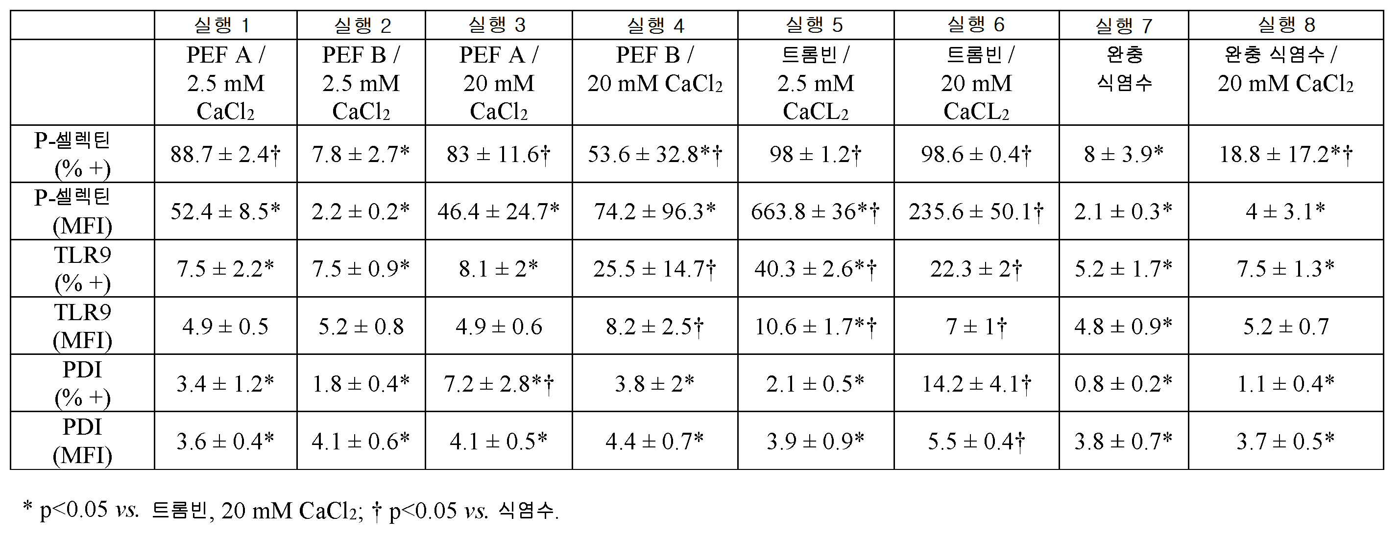
본 연구의 혈소판 알파 과립 및 T 과립 마커 부분에서, 표 3에 나타낸 바와 같이, 실행 1 및 3(즉, PEF A, 2.5 mM 또는 PEF A, 20 mM CaCl2)에서뿐만 아니라 실행 5 및 6(2.5 mM 또는 20 mM CaCl2와 트롬빈)에서 80% 초과의 혈소판이 혈소판 알파 과립 막 단백질인 P-셀렉틴에 대해 양성인 반면, 실행 4(PEF B, 20 mM CaCl2)에서는 더 낮은 비율(53.5±33, 평균±SD)의 혈소판이 양성이었다. 고칼슘 단독(실행 8)은 실행 2(PEF B, 2.5 mM CaCl2) 및 무칼슘 대조군(실행 7)에 비해 P-셀렉틴 양성 혈소판의 비율을 약간 증가시켰다. PEF 처리에 의한 입자당 P-셀렉틴의 평균 형광 강도(MFI)는 트롬빈 및 저 또는 고 CaCl2에서 관찰된 것보다 낮았다.In platelet alpha granules and T granule marker portions of this study,
혈소판 표면 톨 유사 수용체 9(TLR9)는 실행 5(트롬빈, 2.5 mM CaCl2)에서 가장 높았다. 실행 4(PEF B, 20 mM CaCl2)에서 더 낮은 TLR9 및 매우 가변적인 수준의 TLR9가 관찰되었고, 실행 1 및 3(PEF A, 2.5 mM CaCl2 및 PEF A, 20 mM CaCl2), 실행 2(PEF B, 2.5 mM CaCl2), 그리고 무칼슘 및 고칼슘 단독 대조군(실행 7 및 8)에서는 무시할 만한 TLR9 발현이 관찰되었다. 전반적으로, TLR9의 수준은 P-셀렉틴보다 낮았지만, TLR9 발현의 패턴은 P-셀렉틴에서 관찰되는 것과 뚜렷이 다르지는 않았다. 대조적으로, PDI 발현 패턴은 실행 5(트롬빈, 2.5 mM CaCl2) 대신에 실행 6(트롬빈, 20 mM CaCl2)에서 최고 수준이 관찰되었다는 점에서 독특했다.Platelet surface Toll-like receptor 9 (TLR9) was highest in Run 5 (thrombin, 2.5 mM CaCl 2 ). Lower TLR9 and very variable levels of TLR9 were observed in Run 4 (PEF B, 20 mM CaCl 2 ) and Runs 1 and 3 (PEF A, 2.5 mM CaCl 2 and PEF A, 20 mM CaCl 2 ), Run 2 (PEF B, 2.5 mM CaCl 2 ), and negligible TLR9 expression in the calcium and high calcium single controls (
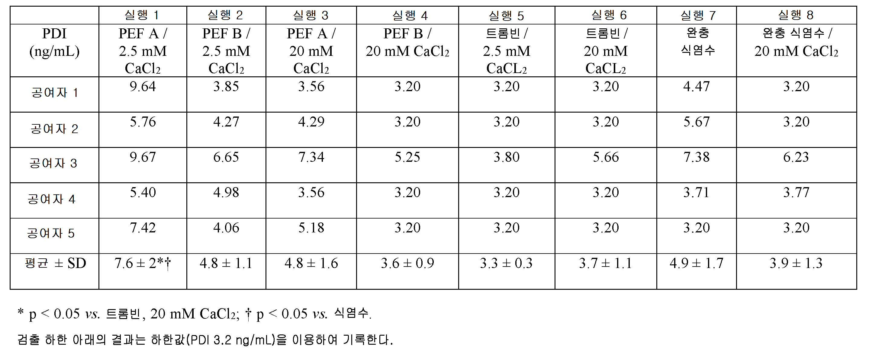
표 4를 보면, T 과립 방출은 또한 ELISA에 의해 측정된 바와 같이 처리된 PRP 샘플의 상청액으로의 PDI의 방출에 의해 평가하였다. PDI 수준은, PRP를 PEF A 및 2.5 mM CaCl2로 활성화하였을 때, 실행 1의 상청액에서 가장 높았으며, 반면에 트롬빈으로 처리된 샘플(실행 5 및 6)에 대해서는 수준이 어세이의 검출 한계에 가깝거나 그보다 낮았다. 이것은, 실행 6(트롬빈, 20 mM CaCl2)에서 가장 높았던, 유세포 분석에 의해 의해 측정된 혈소판 표면 PDI와 대조된다. 따라서, 표면 결합 PDI에 대비한 유리 PDI와 관련하여 활성화 조건 간에 차이가 있는 것으로 보인다. 흥미롭게도, 가용성 PDI 수준은 방출된 EGF 수준과 가장 관련이 있었다(r=0.566, p=0.0001).Referring to Table 4, T granular release was also assessed by the release of PDI into the supernatant of treated PRP samples as measured by ELISA. PDI levels were highest in the supernatant of
혈소판 과립 및 성장 인자의 차등적 노출 - 연구의 이 부분에 대한 결과가 표 5 및 6에 제시되어 있다. Differential exposure of platelet granules and growth factors - The results for this part of the study are presented in Tables 5 and 6.
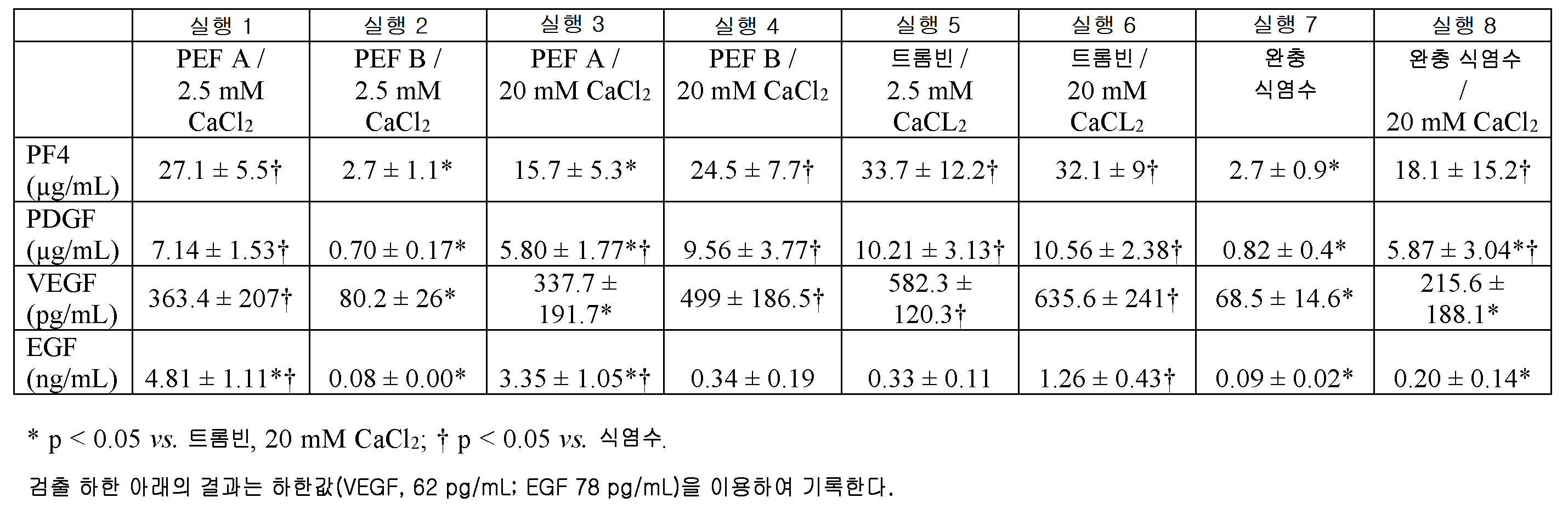
각 처리 조건에 의해 방출된 성장 인자의 양은 광범위하게 변했지만, 처리에 의해 방출된 혈소판 인자 4(PF4), 혈소판 유래 성장 인자(PDGF) 및 혈관 내피 성장 인자(VEGF)의 전체 패턴은 유사하였다.The total amount of platelet factor 4 (PF4), platelet derived growth factor (PDGF) and vascular endothelial growth factor (VEGF) released by treatment was similar, although the amount of growth factor released by each treatment condition varied widely.
구체적으로, PRP의 상청액 중의 PF4, PDGF 및 VEGF의 양을 증가시키는 처리 조건의 상대 능력은, 가장 큰 것부터 가장 작은 것 순으로 다음과 같았다: 실행 6(트롬빈, 20 mM CaCl2) ![]()
![]()
![]()
![]()
![]()
![]()
대조적으로, EGF를 가장 많이 방출시키는 것부터 가장 적게 방출시키는 조건의 순서는 다음과 같다: 실행 1(PEF A, 2.5 mM CaCl2) >> 실행 3(PEF A, 20 mM CaCl2) >> 실행 6(트롬빈, 20 mM CaCl2) > 실행 5(트롬빈, 2.5 mM CaCl2) ![]()
![]()
![]()
![]()
![]()
![]()

이러한 결과에 대하여 요약하면, PEF A, 2.5 mM CaCl2가 응괴를 생성하지 못하였음에도 불구하고, 이것은 P-셀렉틴 양성 혈소판 백분율의 현저한 증가 및 PDI, PF4, PDGF, VEGF의 유의한 방출을 유도하였고, 모든 나머지 조건의 EGF 방출 수준을 최고로 유도하였다. 대조적으로, PEF B, 2.5 mM CaCl2는 완충액에 비해 P-셀렉틴, TLR9, PDI 또는 임의의 성장 인자의 현저한 증가를 유도하지 못하였다. 20 mM CaCl2가 보충된 PRP에서, PEF A와 PEF B 모두 알파 과립 마커인 P-셀렉틴의 노출을 20 mM CaCl2보다 더 많이 증가시켰지만, 트롬빈, 20 mM CaCl2보다 적게 증가시켰다. EGF가 아닌 PF4, PDGF 및 VEGF의 수준은 혈소판 표면 P-셀렉틴 발현(MFI)에 의해 나타나는 바와 같은 혈소판 알파 과립 방출과, 혈소판 표면 TLR9에 의해 나타나는 바와 같은 T 과립 방출과 상관관계가 있었지만, PF4, PDGF, 또는 VEGF는 아니고 EGF 수준이 활성화 후 상청액에 존재하는 PDI 수준과 상관관계가 있었다. 따라서, EGF 방출의 패턴은 PF4, PDGF 및 VEGF의 패턴과 다르며, 이는 또한 혈소판 내 EGF의 분포가 이들 다른 인자의 분포와 구별된다는 것을 시사한다. 가용성 EGF와 함께 방출된 가용성 PDI와의 상관관계에 대한 가능한 설명은 선택된 혈소판 과립에서의 공동 국재화 또는 방출이 유사한 자극에 의해 촉발되는 별개의 과립에서의 국재화를 포함한다. PEF A에 의한 EGF의 향상된 방출은, EGF가 상처 치유의 상피화 단계에 중요하다는 점을 고려하면, 임상 상황에서 잠재적으로 유용하다.To summarize these results, despite the fact that PEF A, 2.5 mM CaCl 2 did not produce clotting, it resulted in a significant increase in the percentage of P-selectin positive platelets and a significant release of PDI, PF4, PDGF, and VEGF, The highest level of EGF release was obtained for all remaining conditions. In contrast, PEF B, 2.5 mM CaCl 2 , did not induce a significant increase in P-selectin, TLR9, PDI or any growth factor compared to buffer. In PRP supplemented with 20 mM CaCl 2 , both PEF A and PEF B increased the exposure of P-selectin, an alpha granule marker, to greater than 20 mM CaCl 2 , but less than thrombin, 20 mM CaCl 2 . The levels of PF4, PDGF, and VEGF, but not EGF, correlated with platelet alpha granule release, as evidenced by platelet surface P-selectin expression (MFI), and T granular release, as demonstrated by platelet surface TLR9, PDGF, or VEGF, but the level of EGF was correlated with the level of PDI present in the supernatant after activation. Thus, the pattern of EGF release is different from that of PF4, PDGF, and VEGF, suggesting that the distribution of EGF in platelets is also distinct from the distribution of these other factors. A possible explanation for the correlation with soluble PDI released with soluble EGF involves localization in separate granules triggered by co-localization or release of similar stimuli in selected platelet granules. Improved release of EGF by PEF A is potentially useful in clinical situations, given that EGF is important for the epithelialization phase of wound healing.
전술한 것들을 염두에 둘 때, 존재하는 성장 인자 및 이들의 상대적인 비율뿐만 아니라 응고의 존재 또는 부재 및/또는 응괴 형성의 타이밍의 측면에서 맞춤화된 활성화된 혈소판 생성물을 생성하는 것이 가능하다. 즉, 본원에 기술된 응고 결과를 고려할 때, 응고의 존재 또는 응괴 형성 개시까지의 시간은 성장 인자 방출과 함께 최적화될 수 있다는 것을 이해할 수 있다. 예를 들어, 고 Ca++(20 mM CaCl2)와 병용되는 PEF A와 PEF B는 상대 비율을 다르게 함에도 불구하고 테스트한 모든 성장 인자를 대조군보다 많이 방출시킨다. 또한, 위의 결과에서 볼 수 있듯이, 저 Ca++(2.5 mM CaCl2)와 병용되는 PEF A는, 응고되지는 않지만, 테스트된 모든 성장 인자가 방출되는 활성화된 생성물을 산출한다. 반대로, 저 Ca++와 병용되는 PEF B는 응고되지 않고 테스트된 성장 인자가 뚜렷하게 방출되지 않는 활성화된 생성물을 산출한다. 그러므로, 전기 자극과 첨가된 Ca의 적절한 조합은 방출된 성장 인자의 수준과 응고 시간을 제어 및/또는 조정하는 데 이용될 수 있고, 응고가 필요 없는 경우, 전기 자극과 첨가된 Ca의 적절한 조합을 이용하여, 원하는 수준 - 고방출 또는 저방출로 성장 인자를 방출시킬 수 있다.With the foregoing in mind, it is possible to produce customized activated platelet products in terms of the presence or absence of coagulation and / or the timing of coagulation formation as well as the relative growth factors and their relative proportions. That is, in view of the coagulation results described herein, it is understood that the time to coagulation or the start of coagulation formation can be optimized with growth factor release. For example, PEF A and PEF B in combination with high Ca ++ (20 mM CaCl 2 ) release more of all tested growth factors than the control group, although the relative ratios are different. In addition, as seen in the above results, PEF A in combination with low Ca ++ (2.5 mM CaCl 2 ) yields an activated product that is not clotted but releases all tested growth factors. Conversely, PEF B in combination with low Ca < ++ > yields an activated product that is not coagulated and the tested growth factor is not evident. Therefore, an appropriate combination of electrical stimulation and added Ca can be used to control and / or adjust the level of the released growth factor and the clotting time, and if appropriate, a proper combination of electrical stimulation and added Ca May be used to release the growth factor to a desired level-high release or low release.
통합 및 추가적인 Ca 및 전기 조건 결과 - 전술한 설명과 결과의 추가적인 뒷받침으로서, 관찰된 응고 시간, 응괴 강도, 성장 인자 방출 및 용혈과 함께 다양한 전기 조건 및 칼슘 이온 농도를 나타내는 추가 데이터 세트가 표 7에 기재되어 있다. 이러한 추가적인 예에서 알 수 있는 바와 같이, 칼슘 이온 농도 및 전기 파라미터를, 서로 다른 관심 축(예를 들어, 응고 시간, 응괴 강도, 방출시키려는 성장 인자(복수의 독립적으로 조작 가능한 성장 인자에 대해), 용혈 등에 따른 다양한 반응을 얻을 수 있도록 함께 변경할 수 있다. 이러한 방식으로, 칼슘 이온 농도 및/또는 하나 이상의 전기 펄스 파라미터 중 하나 또는 둘 다를 적절하게 선택함으로써 다중 속성을 갖는 활성화된 생성물을 구성할 수 있다. Integration and additional Ca and electrocondition results - Additional data sets showing various electrical conditions and calcium ion concentrations with observed coagulation time, coagulation intensity, growth factor release and hemolysis as additional support of the above description and results are shown in Table 7 . As can be seen from this additional example, the calcium ion concentration and electrical parameters can be adjusted to different interest axes (e.g., coagulation time, coagulation strength, growth factor to be released (for a plurality of independently operable growth factors) Hemolysis, etc. In this way, an activated product with multiple properties can be constructed by appropriately selecting one or both of the calcium ion concentration and / or one or more electric pulse parameters .
표 7의 한 예로서, 샘플 2, 3, 4 및 5는 동일한 유형의 전기 자극을 갖지만, 첨가된 칼슘의 수준이 서로 다르다. 응고 시간은 첨가된 Ca의 함수로서 10.2분에서 17.4분까지 다양할 수 있으며, 한 Ca 농도에서는 응고가 발생하지 않는다. 이것은, 단순히 Ca 농도를 조정함으로써, 이들 샘플에 대해, 응고/무응고와, 추가로 실제 응고 시간을 제어하기 위한 수단을 제공한다. 첨가된 Ca의 수준이 증가함에 따라, 이들 샘플(2, 3, 4 및 5)에 대한 EGF 방출 수준이 증가하여, Ca 수준을 조정함으로써 방출되는 성장 인자를 조정하는 수단을 제공하며, 이 경우 동일한 유형의 전기 자극이 유지된다. 유사한 방식으로, 용혈 수준을 조정할 수 있다.As an example of Table 7,
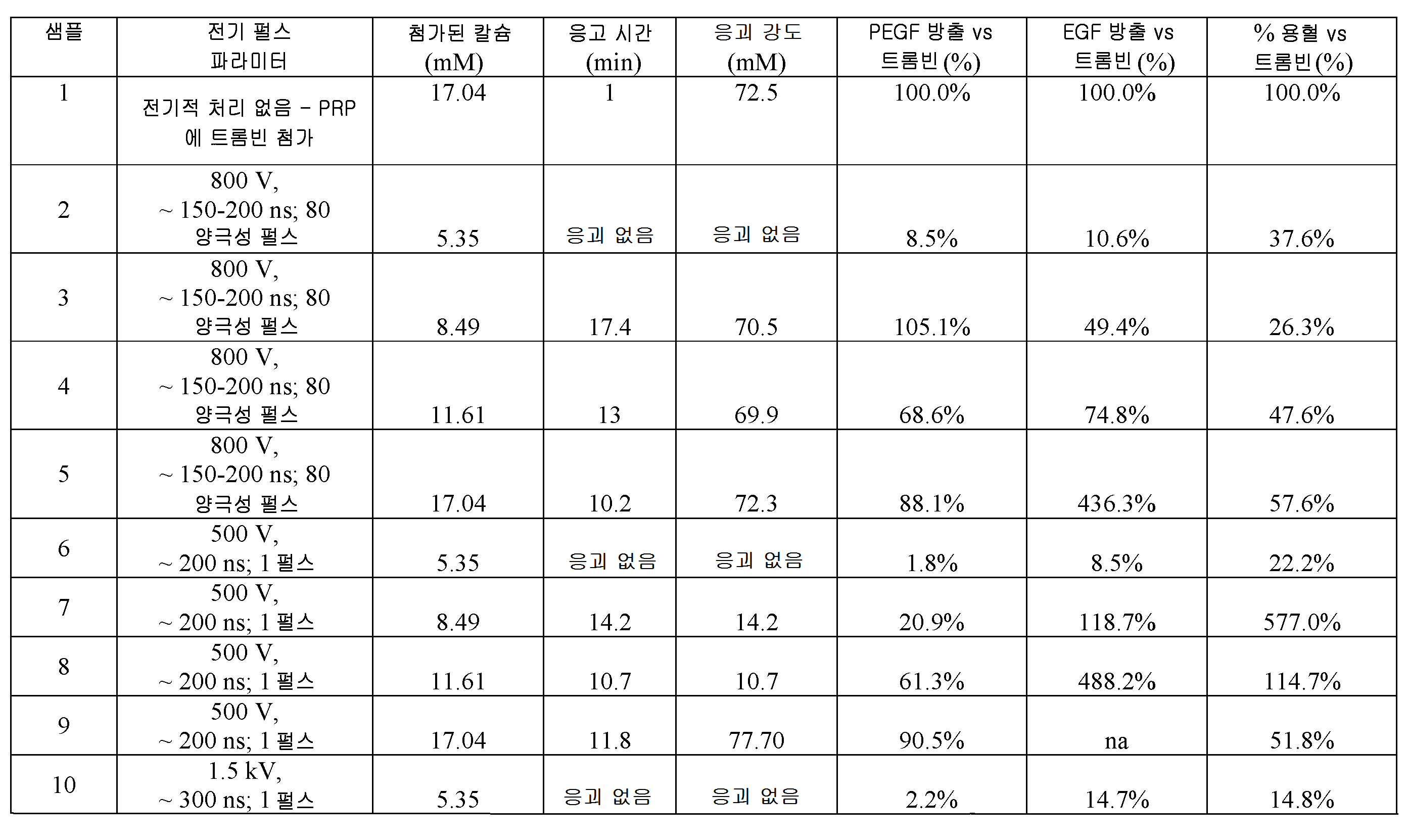
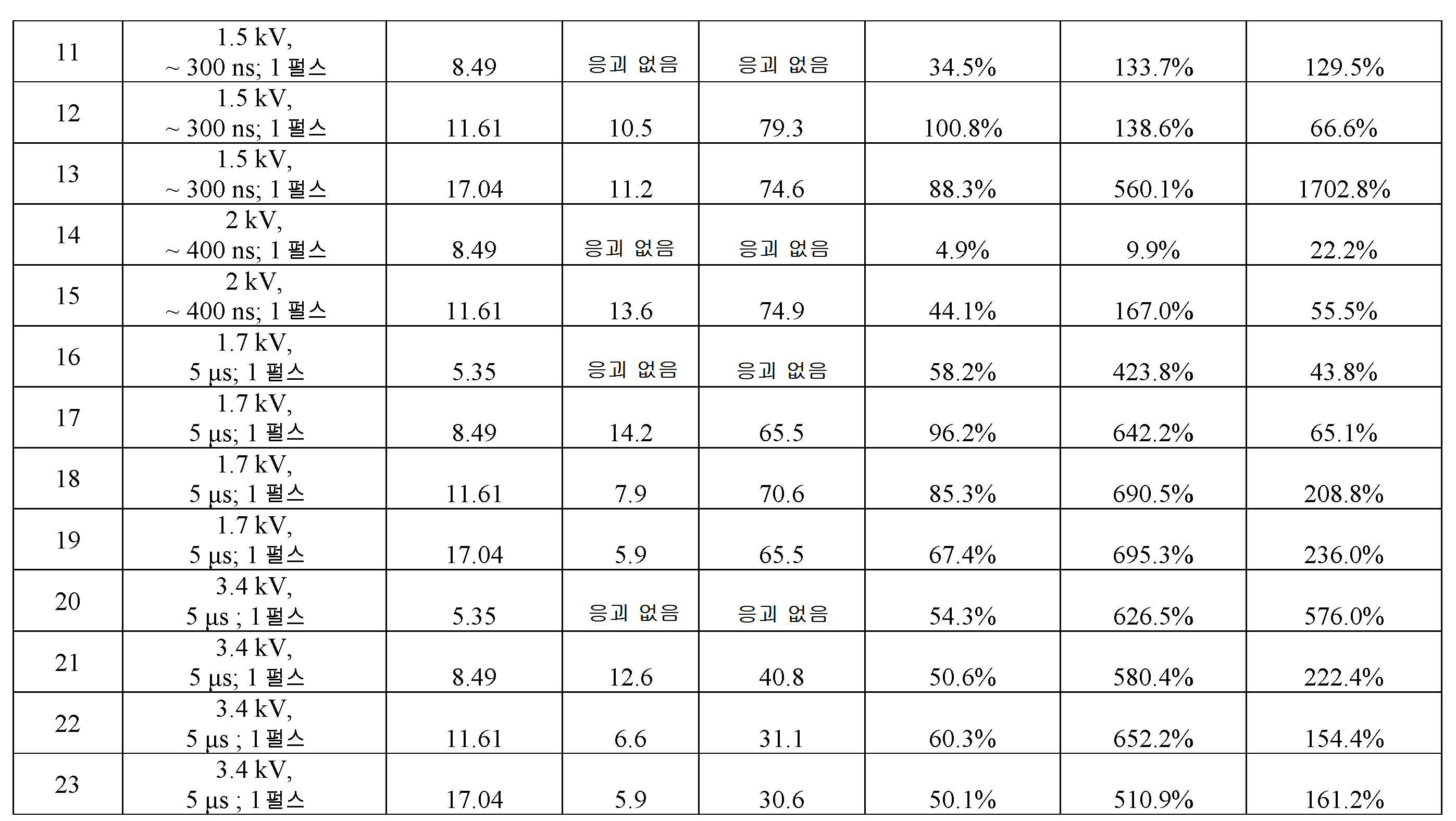
따라서, 표 7에 나타낸 바와 같이, 성장 인자 방출 및 용혈의 개별적인 수준을 여전히 제어하면서, 다양한 상이한 응고 시간 및 강도를 달성하기 위해 다양한 상이한 전기적 특성 및 펄스 파라미터를 칼슘 이온 농도와 함께 변화시킬 수 있다.Thus, as shown in Table 7, various different electrical characteristics and pulse parameters can be varied along with the calcium ion concentration to achieve a variety of different coagulation times and intensities while still controlling the individual levels of growth factor release and hemolysis.
본 발명의 기술적 효과는 응괴의 존재 또는 부재, (존재한다면) 응괴 형성의 타이밍, 및/또는 (존재한다면) 응괴의 기계적 강도 중 하나 이상을 활성화 과정 동안 칼슘 이온의 존재 또는 농도에 의해 제어하는, 활성화된 혈소판 생성물의 생성을 포함한다. 칼슘 이온 농도는 활성화 과정의 일부로서 펄스 전기장 또는 화학적 활성화제(예를 들어, 트롬빈)의 존재 하에 제어되고 지정된다.The technical effect of the present invention is to control at least one of the presence or absence of coagulation, the timing of coagulation formation (if present), and / or the mechanical strength of coagulation (if present) by the presence or concentration of calcium ions during the activation process, And the production of activated platelet products. The calcium ion concentration is controlled and designated in the presence of a pulsed electric field or chemical activator (e. G., Thrombin) as part of the activation process.
서술된 설명은, 최선의 양태를 포함하여 본 발명을 개시하고, 임의의 장치 또는 시스템을 제작 및 사용하고 임의의 통합된 방법을 수행하는 것을 포함하여 당업자가 본 발명을 실시할 수 있게 하기 위해 실시예를 이용한다. 본 발명의 특허 가능한 범위는 청구범위에 의해 규정되며, 당업자가 알 수 있는 다른 실시예를 포함할 수 있다. 이러한 다른 실시예는, 청구범위의 문자 그대로의 언어와 다른 구조적 요소를 갖는 경우 또는 청구범위의 문자 그대로의 언어와 실질적이지 않은 차이를 갖는 동등한 구조적 요소를 포함하는 경우, 청구항의 범위 내에 포함되는 것으로 의도된다.The written description discloses the invention, including the best mode, and is implemented to enable those skilled in the art to practice the invention, including making and using any device or system and performing any integrated method Use the example. The patentable scope of the invention is defined by the claims, and may include other embodiments which will be apparent to those skilled in the art. These and other embodiments are to be accorded the broadest interpretation so as to encompass within the scope of the following claims when they have an element other than the literal language of the claims or if they contain equivalent structural elements which are not material to the literal language of the claims It is intended.
본 명세서의 끝부분에 첨부된 청구범위가 "[기능]을 [수행]하기 위한 수단 ..."또는 "[기능]을 [수행]하기 위한 단계..."로 나타내어지는 하나 이상의 요소를 포함하는 경우, 이것은 그러한 요소들이 35 U.S.C. 112(f)에 따라 해석되어야 한다는 것을 의도한다. 그러나, 임의의 다른 방식으로 나타내어지는 요소들을 포함하는 임의의 청구항에 대해서는, 그러한 요소들이 35 U.S.C. 112(f)에 따라 해석되어서는 안 된다.The appended claims at the end of this specification include one or more elements designated as " means for performing [function] ... "or" step for [performing] , This means that such factors will not exceed 35 USC It should be interpreted according to 112 (f). However, for any claim involving elements represented in any other way, such elements are not limited to those of 35 U.S.C. It should not be construed in accordance with 112 (f).
Claims (20)
전자기 자극 장치의 전극에 대해 PRP 샘플을 위치시키는 단계;
전기 펄스 파라미터의 세트를 지정하는 단계; 및
파라미터 값에 따라 생성된 하나 이상의 전기 펄스에 상기 PRP 샘플을 노출시키는 단계로서, 상기 PRP 샘플은, 하나 이상의 전기 펄스에 노출될 경우, 1종 이상의 성장 인자를 포함하며 지정된 응고 특성을 갖는 활성화된 생성물 조성물을 산출하는 것인 단계
를 포함하는, 활성화된 생성물을 생성하는 방법.Preparing a platelet-rich plasma (PRP) sample for activation, wherein the PRP sample is prepared by adding calcium ions at respective concentrations, each concentration being generated using a PRP sample Is selected on the basis of at least one of whether the coagulation should be present in one activated product composition and the time to coagulation formation or the mechanical strength of the coagulation if coagulation should be present;
Positioning a PRP sample against an electrode of an electromagnetic stimulator;
Specifying a set of electrical pulse parameters; And
Exposing the PRP sample to one or more electric pulses generated according to a parameter value, wherein the PRP sample comprises one or more growth factors when exposed to one or more electric pulses, wherein the activated product ≪ / RTI >
≪ / RTI >
다양한 가능한 농도로부터 선택되는 칼슘 이온 농도에 도달하도록, PRP 샘플에 칼슘 이온을 첨가하는 단계로서, 상기 농도는, PRP 샘플을 사용하여 생성한 활성화된 생성물 조성물 중에 존재하는 1종 이상의 성장 인자의 목표 수준을 기초로 선택되는 것인 단계;
PRP 샘플을 전기적 활성화 자극에 노출시키는 단계로서, 상기 PRP 샘플은, 전기적 활성화 자극에 노출될 경우, 1종 이상의 성장 인자를 목표 수준으로 포함하는 활성화된 생성물 조성물을 산출하며, 전기적 활성화 자극을 변경하지 않고 칼슘 이온 농도를 변경하는 것이 1종 이상의 성장 인자의 절대 또는 상대 수준 중 하나 또는 둘 다를 변화시키는 것인 단계
를 포함하는, 활성화된 생성물을 생성하는 방법.For activation, preparing an anticoagulated platelet-rich plasma (PRP) sample;
Adding calcium ions to a PRP sample such that a calcium ion concentration selected from a variety of possible concentrations is reached, wherein the concentration is at a target level of one or more growth factors present in the activated product composition produced using the PRP sample ≪ / RTI >
Exposing the PRP sample to an electrically activated stimulus, wherein the PRP sample, when exposed to an electrically activated stimulus, produces an activated product composition comprising at least one growth factor at a target level, Wherein altering the calcium ion concentration changes one or both of the absolute or relative levels of one or more growth factors
≪ / RTI >
잠재적 기계적 강도를 기초로, 복수의 칼슘 이온 농도 중에서 잠재적 기계적 강도에 상응하는 칼슘 이온 농도를 선택하는 단계;
선택된 칼슘 이온 농도로 칼슘 이온을 포함하는 혈소판 농축 혈장(PRP) 샘플을 활성화시킴으로써 혈소판 겔을 생성하는 단계로서, PRP 샘플은 전기 자극을 이용하여 활성화시키고, 혈소판 겔은, 형성되면, 잠재적 기계적 강도를 갖는 응괴를 포함하는 것인 단계
를 포함하는, 혈소판 겔의 응괴의 기계적 강도를 제어하는 방법.Determining the potential mechanical strength of the at least one clot generated in the platelet gel, wherein the potential mechanical strength is greater than the intensity observed by producing platelet gel using only thrombin;
Selecting a calcium ion concentration corresponding to a potential mechanical strength of the plurality of calcium ion concentrations based on the potential mechanical strength;
Generating a platelet gel by activating a platelet concentrated plasma (PRP) sample containing calcium ions at a selected calcium ion concentration, wherein the PRP sample is activated using electrical stimulation and the platelet gel, when formed, Lt; RTI ID = 0.0 >
≪ / RTI > wherein the platelet gel has a mean particle size of less than about < RTI ID = 0.0 &
Applications Claiming Priority (3)
| Application Number | Priority Date | Filing Date | Title |
|---|---|---|---|
| US14/984,988 | 2015-12-30 | ||
| US14/984,988 US10633645B2 (en) | 2015-12-30 | 2015-12-30 | Calcium controlled activation of platelets via electrical stimulation |
| PCT/US2016/069197 WO2017117385A2 (en) | 2015-12-30 | 2016-12-29 | Calcium controlled activation of platelets via electrical stimulation |
Publications (2)
| Publication Number | Publication Date |
|---|---|
| KR20180098402A true KR20180098402A (en) | 2018-09-03 |
| KR102722475B1 KR102722475B1 (en) | 2024-10-25 |
Family
ID=57868356
Family Applications (1)
| Application Number | Title | Priority Date | Filing Date |
|---|---|---|---|
| KR1020187022044A Active KR102722475B1 (en) | 2015-12-30 | 2016-12-29 | Calcium-regulated activation of platelets by electrical stimulation |
Country Status (10)
| Country | Link |
|---|---|
| US (2) | US10633645B2 (en) |
| EP (2) | EP3738604B1 (en) |
| JP (1) | JP7050680B2 (en) |
| KR (1) | KR102722475B1 (en) |
| CN (2) | CN108697763B (en) |
| AU (1) | AU2016380342B2 (en) |
| BR (1) | BR112018013444A2 (en) |
| MX (1) | MX392919B (en) |
| RU (1) | RU2742937C2 (en) |
| WO (1) | WO2017117385A2 (en) |
Cited By (1)
| Publication number | Priority date | Publication date | Assignee | Title |
|---|---|---|---|---|
| WO2021137358A1 (en) * | 2020-01-02 | 2021-07-08 | 주식회사 엠엔 | Device for supporting height growth by using pulsed micro-electromagnetic field, and method for driving device |
Families Citing this family (2)
| Publication number | Priority date | Publication date | Assignee | Title |
|---|---|---|---|---|
| US10968423B2 (en) | 2018-02-26 | 2021-04-06 | General Electric Company | System and method for electric pulse based activation of biological samples |
| TW202527970A (en) * | 2024-01-05 | 2025-07-16 | 艾瑞生醫股份有限公司 | Methods for production of exosomes from human platelet |
Citations (2)
| Publication number | Priority date | Publication date | Assignee | Title |
|---|---|---|---|---|
| WO2015108778A1 (en) * | 2014-01-17 | 2015-07-23 | General Electric Company | Platelet activation and growth factor release using electric pulses |
| WO2015168015A1 (en) | 2014-04-28 | 2015-11-05 | T2 Biosystems, Inc. | Systems and methods for identifying coagulopathies |
Family Cites Families (22)
| Publication number | Priority date | Publication date | Assignee | Title |
|---|---|---|---|---|
| JP3180824B2 (en) * | 1991-08-30 | 2001-06-25 | シスメックス株式会社 | Blood coagulation reagent |
| US5293772A (en) * | 1992-01-17 | 1994-03-15 | Center For Innovative Technology | Instrumentation and method for evaluating platelet performance during clotting and dissolution of blood clots and for evaluating erythrocyte flexibility |
| US6322785B1 (en) | 1999-03-02 | 2001-11-27 | Natrex Technologies | Methods and compositions for bone graft implants |
| US6326711B1 (en) | 1999-09-07 | 2001-12-04 | Tokyo Parts Industrial Co., Ltd. | DC brushless motor having eccentric rotor |
| ATE368229T1 (en) * | 2002-05-01 | 2007-08-15 | Synapse Bv | DIAGNOSTIC TEST FOR DETERMINING THE CONCENTRATION OF TRANSIENT PROTEOLYTIC ACTIVITY IN COMPOSITE BIOLOGICAL MEDIA |
| US7565201B2 (en) | 2004-12-17 | 2009-07-21 | Eastern Virginia Medical School | Activation of calcium-mediated cell functions in cells and tissues, including aggregation of human platelets. by nanosecond pulsed electric fields |
| CN101151039A (en) * | 2005-04-04 | 2008-03-26 | 加利福尼亚大学董事会 | Inorganic materials for hemostasis regulation and wound healing therapy |
| EP1717588A1 (en) * | 2005-04-29 | 2006-11-02 | Synapse B.V. | Measuring thrombin activity in whole blood |
| US7837941B2 (en) * | 2006-04-07 | 2010-11-23 | Agamatrix, Inc. | Method and apparatus for monitoring alteration of flow characteristics in a liquid sample |
| US8034014B2 (en) | 2007-03-06 | 2011-10-11 | Biomet Biologics, Llc | Angiogenesis initation and growth |
| WO2010042658A1 (en) | 2008-10-07 | 2010-04-15 | Bioparadox, Llc | Use of platelet rich plasma composition in the treatment of cardiac conduction abnormalities |
| WO2010057021A1 (en) * | 2008-11-13 | 2010-05-20 | Eastern Virginia Medical School | Activation and aggregation of human platelets and formation of platelet gels by nanosecond pulsed electric fields |
| KR101114712B1 (en) * | 2009-10-23 | 2012-02-29 | 세원셀론텍(주) | A Platelet rich plasma using regeneration constituent manufacturing method thereof |
| ES2402144T3 (en) * | 2010-05-25 | 2013-04-29 | Gwowei Technology Co., Ltd. | Inactivated platelet lysate containing depleted growth factors in PDGF and VEGF and their preparation procedure |
| US9314609B2 (en) | 2011-10-28 | 2016-04-19 | Martin Brown | Device for providing electrical stimulation of a human knee |
| CN103110947A (en) * | 2011-11-16 | 2013-05-22 | 玛旺干细胞医学生物科技股份有限公司 | Additive for activating blood platelets |
| ES2883580T3 (en) * | 2013-03-21 | 2021-12-09 | Collplant Ltd | Compositions comprising collagen and PRP for tissue regeneration and its production method |
| US9238808B2 (en) | 2013-06-06 | 2016-01-19 | General Electric Company | Modular adjustable pulse generator |
| US9078862B2 (en) | 2013-06-06 | 2015-07-14 | General Electric Company | Platelet activation using long electric field pulses |
| US9708597B2 (en) | 2014-01-17 | 2017-07-18 | General Electric Company | Electric pulse generation systems using capacitive coupling |
| EP3107590B1 (en) | 2014-02-20 | 2021-11-17 | Ortho Regenerative Technologies Inc. | Freeze-dried polymer compositions for mixing with platelet rich plasma to form implants for tissue repair and/or compositions for therapeutic intra-articular injections |
| US9752120B2 (en) | 2015-03-31 | 2017-09-05 | General Electric Company | Activated platelet composition with tunable growth factor level |
-
2015
- 2015-12-30 US US14/984,988 patent/US10633645B2/en active Active
-
2016
- 2016-12-29 WO PCT/US2016/069197 patent/WO2017117385A2/en not_active Ceased
- 2016-12-29 RU RU2018121952A patent/RU2742937C2/en active
- 2016-12-29 CN CN201680082886.6A patent/CN108697763B/en active Active
- 2016-12-29 JP JP2018533697A patent/JP7050680B2/en active Active
- 2016-12-29 KR KR1020187022044A patent/KR102722475B1/en active Active
- 2016-12-29 EP EP20184433.9A patent/EP3738604B1/en active Active
- 2016-12-29 EP EP16829375.1A patent/EP3397268B1/en active Active
- 2016-12-29 MX MX2018008146A patent/MX392919B/en unknown
- 2016-12-29 CN CN202211454001.4A patent/CN115873793A/en active Pending
- 2016-12-29 AU AU2016380342A patent/AU2016380342B2/en active Active
- 2016-12-29 BR BR112018013444-9A patent/BR112018013444A2/en not_active Application Discontinuation
-
2020
- 2020-04-16 US US16/850,727 patent/US11591590B2/en active Active
Patent Citations (2)
| Publication number | Priority date | Publication date | Assignee | Title |
|---|---|---|---|---|
| WO2015108778A1 (en) * | 2014-01-17 | 2015-07-23 | General Electric Company | Platelet activation and growth factor release using electric pulses |
| WO2015168015A1 (en) | 2014-04-28 | 2015-11-05 | T2 Biosystems, Inc. | Systems and methods for identifying coagulopathies |
Non-Patent Citations (1)
| Title |
|---|
| ZHANG et al., Archives of Biochemistry and Biophysics, Academic Press, 2007, 471, 240-248. * |
Cited By (1)
| Publication number | Priority date | Publication date | Assignee | Title |
|---|---|---|---|---|
| WO2021137358A1 (en) * | 2020-01-02 | 2021-07-08 | 주식회사 엠엔 | Device for supporting height growth by using pulsed micro-electromagnetic field, and method for driving device |
Also Published As
| Publication number | Publication date |
|---|---|
| JP7050680B2 (en) | 2022-04-08 |
| KR102722475B1 (en) | 2024-10-25 |
| WO2017117385A3 (en) | 2017-08-10 |
| EP3397268B1 (en) | 2020-07-08 |
| AU2016380342A1 (en) | 2018-07-12 |
| US20200239868A1 (en) | 2020-07-30 |
| US11591590B2 (en) | 2023-02-28 |
| EP3397268A2 (en) | 2018-11-07 |
| RU2018121952A3 (en) | 2020-04-08 |
| CN108697763B (en) | 2022-12-09 |
| US20170191052A1 (en) | 2017-07-06 |
| US10633645B2 (en) | 2020-04-28 |
| RU2742937C2 (en) | 2021-02-11 |
| CN108697763A (en) | 2018-10-23 |
| AU2016380342B2 (en) | 2023-04-13 |
| CN115873793A (en) | 2023-03-31 |
| EP3738604A3 (en) | 2021-01-13 |
| MX392919B (en) | 2025-03-24 |
| RU2018121952A (en) | 2020-01-30 |
| CA3009523A1 (en) | 2017-07-06 |
| WO2017117385A2 (en) | 2017-07-06 |
| EP3738604B1 (en) | 2023-09-13 |
| BR112018013444A2 (en) | 2019-02-12 |
| MX2018008146A (en) | 2018-12-06 |
| JP2019501911A (en) | 2019-01-24 |
| EP3738604A2 (en) | 2020-11-18 |
Similar Documents
| Publication | Publication Date | Title |
|---|---|---|
| TWI673054B (en) | Platelet activation and growth factor release using electric pulses | |
| US11591590B2 (en) | Calcium controlled activation of platelets via electrical stimulation | |
| JP2013502929A (en) | Methods and compositions for delivering interleukin-1 receptor antagonists | |
| JP2013536841A (en) | Methods and compositions for delivering interleukin-1 receptor antagonists | |
| JP2013503182A (en) | Osteolysis treatment | |
| Frelinger III et al. | Tunable activation of therapeutic platelet-rich plasma by pulse electric field: Differential effects on clot formation, growth factor release, and platelet morphology | |
| CA3009523C (en) | Calcium controlled activation of platelets via electrical stimulation | |
| US9752120B2 (en) | Activated platelet composition with tunable growth factor level | |
| KR102516995B1 (en) | Activated platelet composition with tunable growth factor levels | |
| RU2790831C2 (en) | Method for obtaining hemostatic composition | |
| JP7248721B2 (en) | Activated platelet composition capable of modulating growth factor levels | |
| RU2781788C2 (en) | Method for production of hemostatic composition | |
| CA3018505C (en) | Activated platelet composition with tunable growth factor level | |
| Grewal et al. | Perioperative Fibrinogen Supplementation: Safety and Efficacy | |
| Siegemund et al. | Individual Therapy of Hemophilia—New Laboratory Methods Considering Platelets |
Legal Events
| Date | Code | Title | Description |
|---|---|---|---|
| PA0105 | International application |
St.27 status event code: A-0-1-A10-A15-nap-PA0105 |
|
| PG1501 | Laying open of application |
St.27 status event code: A-1-1-Q10-Q12-nap-PG1501 |
|
| A201 | Request for examination | ||
| PA0201 | Request for examination |
St.27 status event code: A-1-2-D10-D11-exm-PA0201 |
|
| E902 | Notification of reason for refusal | ||
| PE0902 | Notice of grounds for rejection |
St.27 status event code: A-1-2-D10-D21-exm-PE0902 |
|
| E13-X000 | Pre-grant limitation requested |
St.27 status event code: A-2-3-E10-E13-lim-X000 |
|
| P11-X000 | Amendment of application requested |
St.27 status event code: A-2-2-P10-P11-nap-X000 |
|
| P13-X000 | Application amended |
St.27 status event code: A-2-2-P10-P13-nap-X000 |
|
| E902 | Notification of reason for refusal | ||
| PE0902 | Notice of grounds for rejection |
St.27 status event code: A-1-2-D10-D21-exm-PE0902 |
|
| P11-X000 | Amendment of application requested |
St.27 status event code: A-2-2-P10-P11-nap-X000 |
|
| P13-X000 | Application amended |
St.27 status event code: A-2-2-P10-P13-nap-X000 |
|
| E701 | Decision to grant or registration of patent right | ||
| PE0701 | Decision of registration |
St.27 status event code: A-1-2-D10-D22-exm-PE0701 |
|
| GRNT | Written decision to grant | ||
| PR0701 | Registration of establishment |
St.27 status event code: A-2-4-F10-F11-exm-PR0701 |
|
| PR1002 | Payment of registration fee |
St.27 status event code: A-2-2-U10-U12-oth-PR1002 Fee payment year number: 1 |
|
| PG1601 | Publication of registration |
St.27 status event code: A-4-4-Q10-Q13-nap-PG1601 |
|
| R18 | Changes to party contact information recorded |
Free format text: ST27 STATUS EVENT CODE: A-5-5-R10-R18-OTH-X000 (AS PROVIDED BY THE NATIONAL OFFICE) |
|
| R18-X000 | Changes to party contact information recorded |
St.27 status event code: A-5-5-R10-R18-oth-X000 |