KR20170133507A - Humanized influenza monoclonal antibodies and methods for their use - Google Patents
Humanized influenza monoclonal antibodies and methods for their use Download PDFInfo
- Publication number
- KR20170133507A KR20170133507A KR1020177032200A KR20177032200A KR20170133507A KR 20170133507 A KR20170133507 A KR 20170133507A KR 1020177032200 A KR1020177032200 A KR 1020177032200A KR 20177032200 A KR20177032200 A KR 20177032200A KR 20170133507 A KR20170133507 A KR 20170133507A
- Authority
- KR
- South Korea
- Prior art keywords
- antibody
- seq
- acid sequence
- virus
- amino acid
- Prior art date
Links
- 238000000034 method Methods 0.000 title claims abstract description 94
- 206010022000 influenza Diseases 0.000 title claims abstract description 38
- 230000027455 binding Effects 0.000 claims abstract description 146
- 108090000623 proteins and genes Proteins 0.000 claims abstract description 101
- 241000712461 unidentified influenza virus Species 0.000 claims abstract description 70
- 102000004169 proteins and genes Human genes 0.000 claims abstract description 68
- 241000282414 Homo sapiens Species 0.000 claims description 103
- 241000700605 Viruses Species 0.000 claims description 87
- 108090000765 processed proteins & peptides Proteins 0.000 claims description 51
- 125000003275 alpha amino acid group Chemical group 0.000 claims description 42
- 208000037265 diseases, disorders, signs and symptoms Diseases 0.000 claims description 36
- 239000000203 mixture Substances 0.000 claims description 36
- 206010057190 Respiratory tract infections Diseases 0.000 claims description 33
- 239000012634 fragment Substances 0.000 claims description 33
- 102000004196 processed proteins & peptides Human genes 0.000 claims description 33
- 239000013598 vector Substances 0.000 claims description 32
- 229920001184 polypeptide Polymers 0.000 claims description 23
- 238000006386 neutralization reaction Methods 0.000 claims description 22
- 241000712431 Influenza A virus Species 0.000 claims description 19
- 201000010099 disease Diseases 0.000 claims description 18
- 208000035475 disorder Diseases 0.000 claims description 18
- 150000007523 nucleic acids Chemical group 0.000 claims description 17
- 150000001413 amino acids Chemical class 0.000 claims description 15
- 101710154606 Hemagglutinin Proteins 0.000 claims description 12
- 101710093908 Outer capsid protein VP4 Proteins 0.000 claims description 12
- 101710135467 Outer capsid protein sigma-1 Proteins 0.000 claims description 12
- 101710176177 Protein A56 Proteins 0.000 claims description 12
- 239000003814 drug Substances 0.000 claims description 12
- 239000000185 hemagglutinin Substances 0.000 claims description 10
- 108020004707 nucleic acids Proteins 0.000 claims description 9
- 102000039446 nucleic acids Human genes 0.000 claims description 9
- 230000002265 prevention Effects 0.000 claims description 9
- 230000001850 reproductive effect Effects 0.000 claims description 8
- 101001037139 Homo sapiens Immunoglobulin heavy variable 3-30 Proteins 0.000 claims description 7
- 102100040219 Immunoglobulin heavy variable 3-30 Human genes 0.000 claims description 7
- 108091028043 Nucleic acid sequence Proteins 0.000 claims description 6
- 150000003384 small molecules Chemical class 0.000 claims description 6
- 229940124597 therapeutic agent Drugs 0.000 claims description 6
- 239000003053 toxin Substances 0.000 claims description 5
- 231100000765 toxin Toxicity 0.000 claims description 5
- FWMNVWWHGCHHJJ-SKKKGAJSSA-N 4-amino-1-[(2r)-6-amino-2-[[(2r)-2-[[(2r)-2-[[(2r)-2-amino-3-phenylpropanoyl]amino]-3-phenylpropanoyl]amino]-4-methylpentanoyl]amino]hexanoyl]piperidine-4-carboxylic acid Chemical compound C([C@H](C(=O)N[C@H](CC(C)C)C(=O)N[C@H](CCCCN)C(=O)N1CCC(N)(CC1)C(O)=O)NC(=O)[C@H](N)CC=1C=CC=CC=1)C1=CC=CC=C1 FWMNVWWHGCHHJJ-SKKKGAJSSA-N 0.000 claims description 4
- 102000004127 Cytokines Human genes 0.000 claims description 4
- 108090000695 Cytokines Proteins 0.000 claims description 4
- 101001054838 Homo sapiens Immunoglobulin lambda variable 1-44 Proteins 0.000 claims description 4
- 102100026921 Immunoglobulin lambda variable 1-44 Human genes 0.000 claims description 4
- 239000003443 antiviral agent Substances 0.000 claims description 4
- 108020004459 Small interfering RNA Proteins 0.000 claims description 2
- 238000005520 cutting process Methods 0.000 claims description 2
- 239000000427 antigen Substances 0.000 abstract description 80
- 108091007433 antigens Proteins 0.000 abstract description 69
- 102000036639 antigens Human genes 0.000 abstract description 69
- 230000003472 neutralizing effect Effects 0.000 abstract description 34
- 229960005486 vaccine Drugs 0.000 abstract description 13
- 238000004519 manufacturing process Methods 0.000 abstract description 11
- 230000009385 viral infection Effects 0.000 abstract description 8
- 229940124873 Influenza virus vaccine Drugs 0.000 abstract 2
- 230000004043 responsiveness Effects 0.000 abstract 1
- 210000004027 cell Anatomy 0.000 description 110
- 235000018102 proteins Nutrition 0.000 description 66
- 150000001875 compounds Chemical class 0.000 description 55
- 238000012360 testing method Methods 0.000 description 29
- 238000003556 assay Methods 0.000 description 27
- 108020004414 DNA Proteins 0.000 description 23
- 241000699670 Mus sp. Species 0.000 description 22
- 239000003795 chemical substances by application Substances 0.000 description 22
- 238000002474 experimental method Methods 0.000 description 22
- 238000011282 treatment Methods 0.000 description 22
- 108060003951 Immunoglobulin Proteins 0.000 description 20
- 238000004458 analytical method Methods 0.000 description 20
- 102000018358 immunoglobulin Human genes 0.000 description 20
- 238000000338 in vitro Methods 0.000 description 20
- 102100035824 Unconventional myosin-Ig Human genes 0.000 description 19
- 210000001806 memory b lymphocyte Anatomy 0.000 description 19
- 239000000126 substance Substances 0.000 description 18
- 230000010056 antibody-dependent cellular cytotoxicity Effects 0.000 description 17
- 208000015181 infectious disease Diseases 0.000 description 17
- 230000003993 interaction Effects 0.000 description 17
- 239000000523 sample Substances 0.000 description 17
- 241001465754 Metazoa Species 0.000 description 16
- 108010006232 Neuraminidase Proteins 0.000 description 16
- 235000001014 amino acid Nutrition 0.000 description 16
- 102000005348 Neuraminidase Human genes 0.000 description 15
- 108020001507 fusion proteins Proteins 0.000 description 15
- 102000037865 fusion proteins Human genes 0.000 description 15
- 230000005847 immunogenicity Effects 0.000 description 15
- YBJHBAHKTGYVGT-ZKWXMUAHSA-N (+)-Biotin Chemical compound N1C(=O)N[C@@H]2[C@H](CCCCC(=O)O)SC[C@@H]21 YBJHBAHKTGYVGT-ZKWXMUAHSA-N 0.000 description 14
- 108010047041 Complementarity Determining Regions Proteins 0.000 description 14
- 102000004190 Enzymes Human genes 0.000 description 14
- 108090000790 Enzymes Proteins 0.000 description 14
- 108090000631 Trypsin Proteins 0.000 description 14
- 102000004142 Trypsin Human genes 0.000 description 14
- 229940024606 amino acid Drugs 0.000 description 14
- 210000003719 b-lymphocyte Anatomy 0.000 description 14
- 150000001720 carbohydrates Chemical class 0.000 description 14
- 229940088598 enzyme Drugs 0.000 description 14
- 210000004408 hybridoma Anatomy 0.000 description 14
- 239000012588 trypsin Substances 0.000 description 14
- 239000012472 biological sample Substances 0.000 description 13
- 230000035772 mutation Effects 0.000 description 13
- 239000005089 Luciferase Substances 0.000 description 12
- 238000003776 cleavage reaction Methods 0.000 description 12
- LOKCTEFSRHRXRJ-UHFFFAOYSA-I dipotassium trisodium dihydrogen phosphate hydrogen phosphate dichloride Chemical compound P(=O)(O)(O)[O-].[K+].P(=O)(O)([O-])[O-].[Na+].[Na+].[Cl-].[K+].[Cl-].[Na+] LOKCTEFSRHRXRJ-UHFFFAOYSA-I 0.000 description 12
- 239000002609 medium Substances 0.000 description 12
- 239000002953 phosphate buffered saline Substances 0.000 description 12
- 230000008707 rearrangement Effects 0.000 description 12
- 230000007017 scission Effects 0.000 description 12
- 239000006228 supernatant Substances 0.000 description 12
- 206010069754 Acquired gene mutation Diseases 0.000 description 11
- 108060001084 Luciferase Proteins 0.000 description 11
- 241000699666 Mus <mouse, genus> Species 0.000 description 11
- VHJLVAABSRFDPM-QWWZWVQMSA-N dithiothreitol Chemical compound SC[C@@H](O)[C@H](O)CS VHJLVAABSRFDPM-QWWZWVQMSA-N 0.000 description 11
- 230000004927 fusion Effects 0.000 description 11
- -1 glutaraldehyde) Chemical class 0.000 description 11
- 238000001727 in vivo Methods 0.000 description 11
- 230000037439 somatic mutation Effects 0.000 description 11
- 230000008859 change Effects 0.000 description 10
- 230000001419 dependent effect Effects 0.000 description 10
- 238000001514 detection method Methods 0.000 description 10
- 238000010494 dissociation reaction Methods 0.000 description 10
- 230000005593 dissociations Effects 0.000 description 10
- 230000000694 effects Effects 0.000 description 10
- 230000003053 immunization Effects 0.000 description 10
- 230000001965 increasing effect Effects 0.000 description 10
- 208000037798 influenza B Diseases 0.000 description 10
- 239000002502 liposome Substances 0.000 description 10
- 210000003819 peripheral blood mononuclear cell Anatomy 0.000 description 10
- 210000002966 serum Anatomy 0.000 description 10
- 239000013638 trimer Substances 0.000 description 10
- 241000282412 Homo Species 0.000 description 9
- 238000005859 coupling reaction Methods 0.000 description 9
- 210000004602 germ cell Anatomy 0.000 description 9
- 238000002649 immunization Methods 0.000 description 9
- 238000011534 incubation Methods 0.000 description 9
- 238000011081 inoculation Methods 0.000 description 9
- 238000003032 molecular docking Methods 0.000 description 9
- 230000004224 protection Effects 0.000 description 9
- 230000002285 radioactive effect Effects 0.000 description 9
- 238000012216 screening Methods 0.000 description 9
- 239000000243 solution Substances 0.000 description 9
- 230000001225 therapeutic effect Effects 0.000 description 9
- 210000001519 tissue Anatomy 0.000 description 9
- 230000003612 virological effect Effects 0.000 description 9
- 108010090804 Streptavidin Proteins 0.000 description 8
- 230000000890 antigenic effect Effects 0.000 description 8
- 239000012131 assay buffer Substances 0.000 description 8
- 230000008878 coupling Effects 0.000 description 8
- 238000010168 coupling process Methods 0.000 description 8
- 239000012636 effector Substances 0.000 description 8
- 230000002209 hydrophobic effect Effects 0.000 description 8
- 108700010900 influenza virus proteins Proteins 0.000 description 8
- 238000002372 labelling Methods 0.000 description 8
- DHMQDGOQFOQNFH-UHFFFAOYSA-N Glycine Chemical compound NCC(O)=O DHMQDGOQFOQNFH-UHFFFAOYSA-N 0.000 description 7
- 241001112090 Pseudovirus Species 0.000 description 7
- 241000700584 Simplexvirus Species 0.000 description 7
- 239000011324 bead Substances 0.000 description 7
- 230000015572 biosynthetic process Effects 0.000 description 7
- 229960002685 biotin Drugs 0.000 description 7
- 235000020958 biotin Nutrition 0.000 description 7
- 239000011616 biotin Substances 0.000 description 7
- 210000000170 cell membrane Anatomy 0.000 description 7
- 239000013604 expression vector Substances 0.000 description 7
- 238000009472 formulation Methods 0.000 description 7
- 230000001404 mediated effect Effects 0.000 description 7
- 230000001717 pathogenic effect Effects 0.000 description 7
- 239000013612 plasmid Substances 0.000 description 7
- 230000009467 reduction Effects 0.000 description 7
- 238000007423 screening assay Methods 0.000 description 7
- 239000007790 solid phase Substances 0.000 description 7
- 239000002904 solvent Substances 0.000 description 7
- LMDZBCPBFSXMTL-UHFFFAOYSA-N 1-ethyl-3-(3-dimethylaminopropyl)carbodiimide Chemical compound CCN=C=NCCCN(C)C LMDZBCPBFSXMTL-UHFFFAOYSA-N 0.000 description 6
- CSCPPACGZOOCGX-UHFFFAOYSA-N Acetone Chemical compound CC(C)=O CSCPPACGZOOCGX-UHFFFAOYSA-N 0.000 description 6
- 208000002979 Influenza in Birds Diseases 0.000 description 6
- DNIAPMSPPWPWGF-UHFFFAOYSA-N Propylene glycol Chemical compound CC(O)CO DNIAPMSPPWPWGF-UHFFFAOYSA-N 0.000 description 6
- FAPWRFPIFSIZLT-UHFFFAOYSA-M Sodium chloride Chemical compound [Na+].[Cl-] FAPWRFPIFSIZLT-UHFFFAOYSA-M 0.000 description 6
- 230000004913 activation Effects 0.000 description 6
- 238000001042 affinity chromatography Methods 0.000 description 6
- 239000012491 analyte Substances 0.000 description 6
- 206010064097 avian influenza Diseases 0.000 description 6
- 230000008901 benefit Effects 0.000 description 6
- 238000006243 chemical reaction Methods 0.000 description 6
- 230000000295 complement effect Effects 0.000 description 6
- 210000004443 dendritic cell Anatomy 0.000 description 6
- 230000001900 immune effect Effects 0.000 description 6
- 238000003018 immunoassay Methods 0.000 description 6
- 230000002163 immunogen Effects 0.000 description 6
- 230000001976 improved effect Effects 0.000 description 6
- 230000002458 infectious effect Effects 0.000 description 6
- 230000002401 inhibitory effect Effects 0.000 description 6
- 239000000463 material Substances 0.000 description 6
- 230000034217 membrane fusion Effects 0.000 description 6
- 239000008194 pharmaceutical composition Substances 0.000 description 6
- 239000002243 precursor Substances 0.000 description 6
- 238000002360 preparation method Methods 0.000 description 6
- 102000005962 receptors Human genes 0.000 description 6
- 108020003175 receptors Proteins 0.000 description 6
- 102200007903 rs796065047 Human genes 0.000 description 6
- RPENMORRBUTCPR-UHFFFAOYSA-M sodium;1-hydroxy-2,5-dioxopyrrolidine-3-sulfonate Chemical compound [Na+].ON1C(=O)CC(S([O-])(=O)=O)C1=O RPENMORRBUTCPR-UHFFFAOYSA-M 0.000 description 6
- 230000004083 survival effect Effects 0.000 description 6
- 230000001960 triggered effect Effects 0.000 description 6
- 241000272525 Anas platyrhynchos Species 0.000 description 5
- 241000271566 Aves Species 0.000 description 5
- 108010041986 DNA Vaccines Proteins 0.000 description 5
- 229940021995 DNA vaccine Drugs 0.000 description 5
- KCXVZYZYPLLWCC-UHFFFAOYSA-N EDTA Chemical compound OC(=O)CN(CC(O)=O)CCN(CC(O)=O)CC(O)=O KCXVZYZYPLLWCC-UHFFFAOYSA-N 0.000 description 5
- PEDCQBHIVMGVHV-UHFFFAOYSA-N Glycerine Chemical compound OCC(O)CO PEDCQBHIVMGVHV-UHFFFAOYSA-N 0.000 description 5
- 206010035226 Plasma cell myeloma Diseases 0.000 description 5
- 229920002684 Sepharose Polymers 0.000 description 5
- WLJPJRGQRNCIQS-ZLUOBGJFSA-N Ser-Ser-Asn Chemical compound [H]N[C@@H](CO)C(=O)N[C@@H](CO)C(=O)N[C@@H](CC(N)=O)C(O)=O WLJPJRGQRNCIQS-ZLUOBGJFSA-N 0.000 description 5
- 239000002671 adjuvant Substances 0.000 description 5
- 210000000612 antigen-presenting cell Anatomy 0.000 description 5
- 230000000903 blocking effect Effects 0.000 description 5
- 210000004369 blood Anatomy 0.000 description 5
- 239000008280 blood Substances 0.000 description 5
- 239000000969 carrier Substances 0.000 description 5
- 239000007822 coupling agent Substances 0.000 description 5
- 239000013078 crystal Substances 0.000 description 5
- 229940079593 drug Drugs 0.000 description 5
- 238000005516 engineering process Methods 0.000 description 5
- XKUKSGPZAADMRA-UHFFFAOYSA-N glycyl-glycyl-glycine Natural products NCC(=O)NCC(=O)NCC(O)=O XKUKSGPZAADMRA-UHFFFAOYSA-N 0.000 description 5
- 230000028993 immune response Effects 0.000 description 5
- 208000037797 influenza A Diseases 0.000 description 5
- 230000005764 inhibitory process Effects 0.000 description 5
- 231100000636 lethal dose Toxicity 0.000 description 5
- 239000003550 marker Substances 0.000 description 5
- 238000005259 measurement Methods 0.000 description 5
- 230000007246 mechanism Effects 0.000 description 5
- 239000012528 membrane Substances 0.000 description 5
- 201000000050 myeloid neoplasm Diseases 0.000 description 5
- 239000000047 product Substances 0.000 description 5
- 230000000069 prophylactic effect Effects 0.000 description 5
- 238000002818 protein evolution Methods 0.000 description 5
- 230000004044 response Effects 0.000 description 5
- 238000002864 sequence alignment Methods 0.000 description 5
- 238000013207 serial dilution Methods 0.000 description 5
- 238000010561 standard procedure Methods 0.000 description 5
- 108010061238 threonyl-glycine Proteins 0.000 description 5
- 238000002255 vaccination Methods 0.000 description 5
- 108010073969 valyllysine Proteins 0.000 description 5
- 239000013603 viral vector Substances 0.000 description 5
- CIWBSHSKHKDKBQ-JLAZNSOCSA-N Ascorbic acid Chemical compound OC[C@H](O)[C@H]1OC(=O)C(O)=C1O CIWBSHSKHKDKBQ-JLAZNSOCSA-N 0.000 description 4
- 241000196324 Embryophyta Species 0.000 description 4
- WHUUTDBJXJRKMK-UHFFFAOYSA-N Glutamic acid Natural products OC(=O)C(N)CCC(O)=O WHUUTDBJXJRKMK-UHFFFAOYSA-N 0.000 description 4
- POJJAZJHBGXEGM-YUMQZZPRSA-N Gly-Ser-Lys Chemical compound C(CCN)C[C@@H](C(=O)O)NC(=O)[C@H](CO)NC(=O)CN POJJAZJHBGXEGM-YUMQZZPRSA-N 0.000 description 4
- 101150039660 HA gene Proteins 0.000 description 4
- WHUUTDBJXJRKMK-VKHMYHEASA-N L-glutamic acid Chemical compound OC(=O)[C@@H](N)CCC(O)=O WHUUTDBJXJRKMK-VKHMYHEASA-N 0.000 description 4
- 241000880493 Leptailurus serval Species 0.000 description 4
- KDXKERNSBIXSRK-UHFFFAOYSA-N Lysine Natural products NCCCCC(N)C(O)=O KDXKERNSBIXSRK-UHFFFAOYSA-N 0.000 description 4
- 206010028980 Neoplasm Diseases 0.000 description 4
- 239000012980 RPMI-1640 medium Substances 0.000 description 4
- MTCFGRXMJLQNBG-UHFFFAOYSA-N Serine Natural products OCC(N)C(O)=O MTCFGRXMJLQNBG-UHFFFAOYSA-N 0.000 description 4
- ANHVRCNNGJMJNG-BZSNNMDCSA-N Tyr-Tyr-Cys Chemical compound C1=CC(=CC=C1C[C@@H](C(=O)N[C@@H](CC2=CC=C(C=C2)O)C(=O)N[C@@H](CS)C(=O)O)N)O ANHVRCNNGJMJNG-BZSNNMDCSA-N 0.000 description 4
- 208000036142 Viral infection Diseases 0.000 description 4
- 238000010521 absorption reaction Methods 0.000 description 4
- 108010086434 alanyl-seryl-glycine Proteins 0.000 description 4
- 108010047857 aspartylglycine Proteins 0.000 description 4
- 230000037396 body weight Effects 0.000 description 4
- 239000000872 buffer Substances 0.000 description 4
- 201000011510 cancer Diseases 0.000 description 4
- 239000003153 chemical reaction reagent Substances 0.000 description 4
- 229940127089 cytotoxic agent Drugs 0.000 description 4
- 230000003013 cytotoxicity Effects 0.000 description 4
- 231100000135 cytotoxicity Toxicity 0.000 description 4
- 238000010586 diagram Methods 0.000 description 4
- 239000006185 dispersion Substances 0.000 description 4
- 239000003937 drug carrier Substances 0.000 description 4
- RWSXRVCMGQZWBV-WDSKDSINSA-N glutathione Chemical compound OC(=O)[C@@H](N)CCC(=O)N[C@@H](CS)C(=O)NCC(O)=O RWSXRVCMGQZWBV-WDSKDSINSA-N 0.000 description 4
- 239000001963 growth medium Substances 0.000 description 4
- 210000000987 immune system Anatomy 0.000 description 4
- 238000001114 immunoprecipitation Methods 0.000 description 4
- 239000004615 ingredient Substances 0.000 description 4
- 239000003112 inhibitor Substances 0.000 description 4
- 239000007924 injection Substances 0.000 description 4
- 238000002347 injection Methods 0.000 description 4
- 238000001990 intravenous administration Methods 0.000 description 4
- 238000002955 isolation Methods 0.000 description 4
- 108010054155 lysyllysine Proteins 0.000 description 4
- 210000004962 mammalian cell Anatomy 0.000 description 4
- 239000011159 matrix material Substances 0.000 description 4
- 239000003094 microcapsule Substances 0.000 description 4
- 239000002773 nucleotide Substances 0.000 description 4
- 239000002245 particle Substances 0.000 description 4
- 244000052769 pathogen Species 0.000 description 4
- 108010051242 phenylalanylserine Proteins 0.000 description 4
- 238000000746 purification Methods 0.000 description 4
- 230000009257 reactivity Effects 0.000 description 4
- 238000003757 reverse transcription PCR Methods 0.000 description 4
- 239000007787 solid Substances 0.000 description 4
- 208000024891 symptom Diseases 0.000 description 4
- 108700012359 toxins Proteins 0.000 description 4
- 238000001890 transfection Methods 0.000 description 4
- XLYOFNOQVPJJNP-UHFFFAOYSA-N water Substances O XLYOFNOQVPJJNP-UHFFFAOYSA-N 0.000 description 4
- 230000004580 weight loss Effects 0.000 description 4
- GKSPIZSKQWTXQG-UHFFFAOYSA-N (2,5-dioxopyrrolidin-1-yl) 4-[1-(pyridin-2-yldisulfanyl)ethyl]benzoate Chemical compound C=1C=C(C(=O)ON2C(CCC2=O)=O)C=CC=1C(C)SSC1=CC=CC=N1 GKSPIZSKQWTXQG-UHFFFAOYSA-N 0.000 description 3
- NHBKXEKEPDILRR-UHFFFAOYSA-N 2,3-bis(butanoylsulfanyl)propyl butanoate Chemical compound CCCC(=O)OCC(SC(=O)CCC)CSC(=O)CCC NHBKXEKEPDILRR-UHFFFAOYSA-N 0.000 description 3
- YYSWCHMLFJLLBJ-ZLUOBGJFSA-N Ala-Ala-Ser Chemical compound C[C@H](N)C(=O)N[C@@H](C)C(=O)N[C@@H](CO)C(O)=O YYSWCHMLFJLLBJ-ZLUOBGJFSA-N 0.000 description 3
- KQFRUSHJPKXBMB-BHDSKKPTSA-N Ala-Ala-Trp Chemical compound C1=CC=C2C(C[C@H](NC(=O)[C@H](C)NC(=O)[C@@H](N)C)C(O)=O)=CNC2=C1 KQFRUSHJPKXBMB-BHDSKKPTSA-N 0.000 description 3
- VNYMOTCMNHJGTG-JBDRJPRFSA-N Ala-Ile-Ser Chemical compound [H]N[C@@H](C)C(=O)N[C@@H]([C@@H](C)CC)C(=O)N[C@@H](CO)C(O)=O VNYMOTCMNHJGTG-JBDRJPRFSA-N 0.000 description 3
- RTZCUEHYUQZIDE-WHFBIAKZSA-N Ala-Ser-Gly Chemical compound C[C@H](N)C(=O)N[C@@H](CO)C(=O)NCC(O)=O RTZCUEHYUQZIDE-WHFBIAKZSA-N 0.000 description 3
- 235000006576 Althaea officinalis Nutrition 0.000 description 3
- ATABBWFGOHKROJ-GUBZILKMSA-N Arg-Pro-Ser Chemical compound [H]N[C@@H](CCCNC(N)=N)C(=O)N1CCC[C@H]1C(=O)N[C@@H](CO)C(O)=O ATABBWFGOHKROJ-GUBZILKMSA-N 0.000 description 3
- QLSRIZIDQXDQHK-RCWTZXSCSA-N Arg-Val-Thr Chemical compound [H]N[C@@H](CCCNC(N)=N)C(=O)N[C@@H](C(C)C)C(=O)N[C@@H]([C@@H](C)O)C(O)=O QLSRIZIDQXDQHK-RCWTZXSCSA-N 0.000 description 3
- WONGRTVAMHFGBE-WDSKDSINSA-N Asn-Gly-Gln Chemical compound C(CC(=O)N)[C@@H](C(=O)O)NC(=O)CNC(=O)[C@H](CC(=O)N)N WONGRTVAMHFGBE-WDSKDSINSA-N 0.000 description 3
- MKJBPDLENBUHQU-CIUDSAMLSA-N Asn-Ser-Leu Chemical compound [H]N[C@@H](CC(N)=O)C(=O)N[C@@H](CO)C(=O)N[C@@H](CC(C)C)C(O)=O MKJBPDLENBUHQU-CIUDSAMLSA-N 0.000 description 3
- MRQQMVZUHXUPEV-IHRRRGAJSA-N Asp-Arg-Phe Chemical compound [H]N[C@@H](CC(O)=O)C(=O)N[C@@H](CCCNC(N)=N)C(=O)N[C@@H](CC1=CC=CC=C1)C(O)=O MRQQMVZUHXUPEV-IHRRRGAJSA-N 0.000 description 3
- WVDDGKGOMKODPV-UHFFFAOYSA-N Benzyl alcohol Chemical compound OCC1=CC=CC=C1 WVDDGKGOMKODPV-UHFFFAOYSA-N 0.000 description 3
- 101800001415 Bri23 peptide Proteins 0.000 description 3
- 101800000655 C-terminal peptide Proteins 0.000 description 3
- 102400000107 C-terminal peptide Human genes 0.000 description 3
- 108091026890 Coding region Proteins 0.000 description 3
- 239000006144 Dulbecco’s modified Eagle's medium Substances 0.000 description 3
- LFQSCWFLJHTTHZ-UHFFFAOYSA-N Ethanol Chemical compound CCO LFQSCWFLJHTTHZ-UHFFFAOYSA-N 0.000 description 3
- LYCAIKOWRPUZTN-UHFFFAOYSA-N Ethylene glycol Chemical compound OCCO LYCAIKOWRPUZTN-UHFFFAOYSA-N 0.000 description 3
- 238000012413 Fluorescence activated cell sorting analysis Methods 0.000 description 3
- ZBKUIQNCRIYVGH-SDDRHHMPSA-N Gln-Leu-Pro Chemical compound CC(C)C[C@@H](C(=O)N1CCC[C@@H]1C(=O)O)NC(=O)[C@H](CCC(=O)N)N ZBKUIQNCRIYVGH-SDDRHHMPSA-N 0.000 description 3
- WZZSKAJIHTUUSG-ACZMJKKPSA-N Glu-Ala-Asp Chemical compound OC(=O)C[C@@H](C(O)=O)NC(=O)[C@H](C)NC(=O)[C@@H](N)CCC(O)=O WZZSKAJIHTUUSG-ACZMJKKPSA-N 0.000 description 3
- JWNZHMSRZXXGTM-XKBZYTNZSA-N Glu-Ser-Thr Chemical compound [H]N[C@@H](CCC(O)=O)C(=O)N[C@@H](CO)C(=O)N[C@@H]([C@@H](C)O)C(O)=O JWNZHMSRZXXGTM-XKBZYTNZSA-N 0.000 description 3
- TWTPDFFBLQEBOE-IUCAKERBSA-N Gly-Leu-Gln Chemical compound [H]NCC(=O)N[C@@H](CC(C)C)C(=O)N[C@@H](CCC(N)=O)C(O)=O TWTPDFFBLQEBOE-IUCAKERBSA-N 0.000 description 3
- FFJQHWKSGAWSTJ-BFHQHQDPSA-N Gly-Thr-Ala Chemical compound [H]NCC(=O)N[C@@H]([C@@H](C)O)C(=O)N[C@@H](C)C(O)=O FFJQHWKSGAWSTJ-BFHQHQDPSA-N 0.000 description 3
- YGHSQRJSHKYUJY-SCZZXKLOSA-N Gly-Val-Pro Chemical compound CC(C)[C@@H](C(=O)N1CCC[C@@H]1C(=O)O)NC(=O)CN YGHSQRJSHKYUJY-SCZZXKLOSA-N 0.000 description 3
- 239000004471 Glycine Substances 0.000 description 3
- 108010001336 Horseradish Peroxidase Proteins 0.000 description 3
- 102000018251 Hypoxanthine Phosphoribosyltransferase Human genes 0.000 description 3
- 108010091358 Hypoxanthine Phosphoribosyltransferase Proteins 0.000 description 3
- CDGLBYSAZFIIJO-RCOVLWMOSA-N Ile-Gly-Gly Chemical compound CC[C@H](C)[C@H]([NH3+])C(=O)NCC(=O)NCC([O-])=O CDGLBYSAZFIIJO-RCOVLWMOSA-N 0.000 description 3
- FBGXMKUWQFPHFB-JBDRJPRFSA-N Ile-Ser-Cys Chemical compound CC[C@H](C)[C@@H](C(=O)N[C@@H](CO)C(=O)N[C@@H](CS)C(=O)O)N FBGXMKUWQFPHFB-JBDRJPRFSA-N 0.000 description 3
- 108010021625 Immunoglobulin Fragments Proteins 0.000 description 3
- 102000008394 Immunoglobulin Fragments Human genes 0.000 description 3
- 102000006496 Immunoglobulin Heavy Chains Human genes 0.000 description 3
- 108010019476 Immunoglobulin Heavy Chains Proteins 0.000 description 3
- LZDNBBYBDGBADK-UHFFFAOYSA-N L-valyl-L-tryptophan Natural products C1=CC=C2C(CC(NC(=O)C(N)C(C)C)C(O)=O)=CNC2=C1 LZDNBBYBDGBADK-UHFFFAOYSA-N 0.000 description 3
- YVKSMSDXKMSIRX-GUBZILKMSA-N Leu-Glu-Asn Chemical compound [H]N[C@@H](CC(C)C)C(=O)N[C@@H](CCC(O)=O)C(=O)N[C@@H](CC(N)=O)C(O)=O YVKSMSDXKMSIRX-GUBZILKMSA-N 0.000 description 3
- LCNASHSOFMRYFO-WDCWCFNPSA-N Leu-Thr-Gln Chemical compound CC(C)C[C@H](N)C(=O)N[C@@H]([C@@H](C)O)C(=O)N[C@H](C(O)=O)CCC(N)=O LCNASHSOFMRYFO-WDCWCFNPSA-N 0.000 description 3
- 241001529936 Murinae Species 0.000 description 3
- 101100068676 Neurospora crassa (strain ATCC 24698 / 74-OR23-1A / CBS 708.71 / DSM 1257 / FGSC 987) gln-1 gene Proteins 0.000 description 3
- YMIZSYUAZJSOFL-SRVKXCTJSA-N Phe-Ser-Asn Chemical compound [H]N[C@@H](CC1=CC=CC=C1)C(=O)N[C@@H](CO)C(=O)N[C@@H](CC(N)=O)C(O)=O YMIZSYUAZJSOFL-SRVKXCTJSA-N 0.000 description 3
- 239000002202 Polyethylene glycol Substances 0.000 description 3
- RMODQFBNDDENCP-IHRRRGAJSA-N Pro-Lys-Leu Chemical compound [H]N1CCC[C@H]1C(=O)N[C@@H](CCCCN)C(=O)N[C@@H](CC(C)C)C(O)=O RMODQFBNDDENCP-IHRRRGAJSA-N 0.000 description 3
- FDMKYQQYJKYCLV-GUBZILKMSA-N Pro-Pro-Ser Chemical compound OC[C@@H](C(O)=O)NC(=O)[C@@H]1CCCN1C(=O)[C@H]1NCCC1 FDMKYQQYJKYCLV-GUBZILKMSA-N 0.000 description 3
- 240000004808 Saccharomyces cerevisiae Species 0.000 description 3
- YQHZVYJAGWMHES-ZLUOBGJFSA-N Ser-Ala-Ser Chemical compound OC[C@H](N)C(=O)N[C@@H](C)C(=O)N[C@@H](CO)C(O)=O YQHZVYJAGWMHES-ZLUOBGJFSA-N 0.000 description 3
- HJEBZBMOTCQYDN-ACZMJKKPSA-N Ser-Glu-Asp Chemical compound [H]N[C@@H](CO)C(=O)N[C@@H](CCC(O)=O)C(=O)N[C@@H](CC(O)=O)C(O)=O HJEBZBMOTCQYDN-ACZMJKKPSA-N 0.000 description 3
- UIGMAMGZOJVTDN-WHFBIAKZSA-N Ser-Gly-Ser Chemical compound OC[C@H](N)C(=O)NCC(=O)N[C@@H](CO)C(O)=O UIGMAMGZOJVTDN-WHFBIAKZSA-N 0.000 description 3
- SFTZWNJFZYOLBD-ZDLURKLDSA-N Ser-Gly-Thr Chemical compound C[C@@H](O)[C@@H](C(O)=O)NC(=O)CNC(=O)[C@@H](N)CO SFTZWNJFZYOLBD-ZDLURKLDSA-N 0.000 description 3
- HEMHJVSKTPXQMS-UHFFFAOYSA-M Sodium hydroxide Chemical compound [OH-].[Na+] HEMHJVSKTPXQMS-UHFFFAOYSA-M 0.000 description 3
- 241000282898 Sus scrofa Species 0.000 description 3
- MXDOAJQRJBMGMO-FJXKBIBVSA-N Thr-Pro-Gly Chemical compound C[C@@H](O)[C@H](N)C(=O)N1CCC[C@H]1C(=O)NCC(O)=O MXDOAJQRJBMGMO-FJXKBIBVSA-N 0.000 description 3
- 108010087924 alanylproline Proteins 0.000 description 3
- 125000000539 amino acid group Chemical group 0.000 description 3
- 239000003242 anti bacterial agent Substances 0.000 description 3
- 239000002246 antineoplastic agent Substances 0.000 description 3
- 238000013459 approach Methods 0.000 description 3
- 108010068265 aspartyltyrosine Proteins 0.000 description 3
- DQXBYHZEEUGOBF-UHFFFAOYSA-N but-3-enoic acid;ethene Chemical compound C=C.OC(=O)CC=C DQXBYHZEEUGOBF-UHFFFAOYSA-N 0.000 description 3
- 150000001718 carbodiimides Chemical class 0.000 description 3
- 235000014633 carbohydrates Nutrition 0.000 description 3
- 238000005119 centrifugation Methods 0.000 description 3
- 238000010367 cloning Methods 0.000 description 3
- 230000009918 complex formation Effects 0.000 description 3
- 239000002254 cytotoxic agent Substances 0.000 description 3
- 231100000599 cytotoxic agent Toxicity 0.000 description 3
- 238000002784 cytotoxicity assay Methods 0.000 description 3
- 231100000263 cytotoxicity test Toxicity 0.000 description 3
- 230000029087 digestion Effects 0.000 description 3
- 125000005442 diisocyanate group Chemical group 0.000 description 3
- 239000002612 dispersion medium Substances 0.000 description 3
- 238000009510 drug design Methods 0.000 description 3
- 239000005038 ethylene vinyl acetate Substances 0.000 description 3
- 230000002349 favourable effect Effects 0.000 description 3
- 238000000684 flow cytometry Methods 0.000 description 3
- 239000012530 fluid Substances 0.000 description 3
- 230000006870 function Effects 0.000 description 3
- 239000000499 gel Substances 0.000 description 3
- XBGGUPMXALFZOT-UHFFFAOYSA-N glycyl-L-tyrosine hemihydrate Natural products NCC(=O)NC(C(O)=O)CC1=CC=C(O)C=C1 XBGGUPMXALFZOT-UHFFFAOYSA-N 0.000 description 3
- 108010010147 glycylglutamine Proteins 0.000 description 3
- 108010050848 glycylleucine Proteins 0.000 description 3
- 108010087823 glycyltyrosine Proteins 0.000 description 3
- 238000003384 imaging method Methods 0.000 description 3
- 229940124452 immunizing agent Drugs 0.000 description 3
- 230000001939 inductive effect Effects 0.000 description 3
- 230000002452 interceptive effect Effects 0.000 description 3
- 239000010410 layer Substances 0.000 description 3
- 230000000670 limiting effect Effects 0.000 description 3
- 150000002632 lipids Chemical class 0.000 description 3
- 210000004698 lymphocyte Anatomy 0.000 description 3
- 108010017391 lysylvaline Proteins 0.000 description 3
- 125000003729 nucleotide group Chemical group 0.000 description 3
- PGZUMBJQJWIWGJ-ONAKXNSWSA-N oseltamivir phosphate Chemical compound OP(O)(O)=O.CCOC(=O)C1=C[C@@H](OC(CC)CC)[C@H](NC(C)=O)[C@@H](N)C1 PGZUMBJQJWIWGJ-ONAKXNSWSA-N 0.000 description 3
- 108010012581 phenylalanylglutamate Proteins 0.000 description 3
- 229920001200 poly(ethylene-vinyl acetate) Polymers 0.000 description 3
- 229920001223 polyethylene glycol Polymers 0.000 description 3
- 239000000843 powder Substances 0.000 description 3
- 230000001681 protective effect Effects 0.000 description 3
- 208000009305 pseudorabies Diseases 0.000 description 3
- 238000011160 research Methods 0.000 description 3
- 230000002441 reversible effect Effects 0.000 description 3
- 230000001932 seasonal effect Effects 0.000 description 3
- 238000012163 sequencing technique Methods 0.000 description 3
- 125000003607 serino group Chemical group [H]N([H])[C@]([H])(C(=O)[*])C(O[H])([H])[H] 0.000 description 3
- 239000011780 sodium chloride Substances 0.000 description 3
- 238000003860 storage Methods 0.000 description 3
- 239000003826 tablet Substances 0.000 description 3
- 229940061367 tamiflu Drugs 0.000 description 3
- 230000009261 transgenic effect Effects 0.000 description 3
- 238000003146 transient transfection Methods 0.000 description 3
- 241000701161 unidentified adenovirus Species 0.000 description 3
- 229940125575 vaccine candidate Drugs 0.000 description 3
- MTCFGRXMJLQNBG-REOHCLBHSA-N (2S)-2-Amino-3-hydroxypropansäure Chemical compound OC[C@H](N)C(O)=O MTCFGRXMJLQNBG-REOHCLBHSA-N 0.000 description 2
- QKNYBSVHEMOAJP-UHFFFAOYSA-N 2-amino-2-(hydroxymethyl)propane-1,3-diol;hydron;chloride Chemical compound Cl.OCC(N)(CO)CO QKNYBSVHEMOAJP-UHFFFAOYSA-N 0.000 description 2
- VPFUWHKTPYPNGT-UHFFFAOYSA-N 3-(3,4-dihydroxyphenyl)-1-(5-hydroxy-2,2-dimethylchromen-6-yl)propan-1-one Chemical compound OC1=C2C=CC(C)(C)OC2=CC=C1C(=O)CCC1=CC=C(O)C(O)=C1 VPFUWHKTPYPNGT-UHFFFAOYSA-N 0.000 description 2
- LSLIRHLIUDVNBN-CIUDSAMLSA-N Ala-Asp-Lys Chemical compound C[C@H](N)C(=O)N[C@@H](CC(O)=O)C(=O)N[C@H](C(O)=O)CCCCN LSLIRHLIUDVNBN-CIUDSAMLSA-N 0.000 description 2
- HYIDEIQUCBKIPL-CQDKDKBSSA-N Ala-Phe-His Chemical compound C[C@@H](C(=O)N[C@@H](CC1=CC=CC=C1)C(=O)N[C@@H](CC2=CN=CN2)C(=O)O)N HYIDEIQUCBKIPL-CQDKDKBSSA-N 0.000 description 2
- 108020004774 Alkaline Phosphatase Proteins 0.000 description 2
- 102000002260 Alkaline Phosphatase Human genes 0.000 description 2
- PPMTUXJSQDNUDE-CIUDSAMLSA-N Asn-Glu-Arg Chemical compound NC(=O)C[C@H](N)C(=O)N[C@@H](CCC(O)=O)C(=O)N[C@H](C(O)=O)CCCN=C(N)N PPMTUXJSQDNUDE-CIUDSAMLSA-N 0.000 description 2
- ALHMNHZJBYBYHS-DCAQKATOSA-N Asn-Lys-Arg Chemical compound [H]N[C@@H](CC(N)=O)C(=O)N[C@@H](CCCCN)C(=O)N[C@@H](CCCNC(N)=N)C(O)=O ALHMNHZJBYBYHS-DCAQKATOSA-N 0.000 description 2
- YUUIAUXBNOHFRJ-IHRRRGAJSA-N Asn-Phe-Met Chemical compound [H]N[C@@H](CC(N)=O)C(=O)N[C@@H](CC1=CC=CC=C1)C(=O)N[C@@H](CCSC)C(O)=O YUUIAUXBNOHFRJ-IHRRRGAJSA-N 0.000 description 2
- GHWWTICYPDKPTE-NGZCFLSTSA-N Asn-Val-Pro Chemical compound CC(C)[C@@H](C(=O)N1CCC[C@@H]1C(=O)O)NC(=O)[C@H](CC(=O)N)N GHWWTICYPDKPTE-NGZCFLSTSA-N 0.000 description 2
- QXHVOUSPVAWEMX-ZLUOBGJFSA-N Asp-Asp-Ser Chemical compound OC(=O)C[C@H](N)C(=O)N[C@@H](CC(O)=O)C(=O)N[C@@H](CO)C(O)=O QXHVOUSPVAWEMX-ZLUOBGJFSA-N 0.000 description 2
- IJGRMHOSHXDMSA-UHFFFAOYSA-N Atomic nitrogen Chemical compound N#N IJGRMHOSHXDMSA-UHFFFAOYSA-N 0.000 description 2
- 241000972773 Aulopiformes Species 0.000 description 2
- 108090001008 Avidin Proteins 0.000 description 2
- 102100024222 B-lymphocyte antigen CD19 Human genes 0.000 description 2
- 241000894006 Bacteria Species 0.000 description 2
- 241000283690 Bos taurus Species 0.000 description 2
- 102100027207 CD27 antigen Human genes 0.000 description 2
- CURLTUGMZLYLDI-UHFFFAOYSA-N Carbon dioxide Chemical compound O=C=O CURLTUGMZLYLDI-UHFFFAOYSA-N 0.000 description 2
- TVYMKYUSZSVOAG-ZLUOBGJFSA-N Cys-Ala-Ala Chemical compound [H]N[C@@H](CS)C(=O)N[C@@H](C)C(=O)N[C@@H](C)C(O)=O TVYMKYUSZSVOAG-ZLUOBGJFSA-N 0.000 description 2
- 108020003215 DNA Probes Proteins 0.000 description 2
- 239000003298 DNA probe Substances 0.000 description 2
- 101710088194 Dehydrogenase Proteins 0.000 description 2
- 238000002965 ELISA Methods 0.000 description 2
- 241000233866 Fungi Species 0.000 description 2
- 241000287828 Gallus gallus Species 0.000 description 2
- 108010010803 Gelatin Proteins 0.000 description 2
- IHSGESFHTMFHRB-GUBZILKMSA-N Gln-Lys-Ala Chemical compound OC(=O)[C@H](C)NC(=O)[C@H](CCCCN)NC(=O)[C@@H](N)CCC(N)=O IHSGESFHTMFHRB-GUBZILKMSA-N 0.000 description 2
- WQZGKKKJIJFFOK-GASJEMHNSA-N Glucose Chemical compound OC[C@H]1OC(O)[C@H](O)[C@@H](O)[C@@H]1O WQZGKKKJIJFFOK-GASJEMHNSA-N 0.000 description 2
- SXRSQZLOMIGNAQ-UHFFFAOYSA-N Glutaraldehyde Chemical compound O=CCCCC=O SXRSQZLOMIGNAQ-UHFFFAOYSA-N 0.000 description 2
- 108010024636 Glutathione Proteins 0.000 description 2
- YMUFWNJHVPQNQD-ZKWXMUAHSA-N Gly-Ala-Ile Chemical compound CC[C@H](C)[C@@H](C(O)=O)NC(=O)[C@H](C)NC(=O)CN YMUFWNJHVPQNQD-ZKWXMUAHSA-N 0.000 description 2
- JVWPPCWUDRJGAE-YUMQZZPRSA-N Gly-Asn-Leu Chemical compound [H]NCC(=O)N[C@@H](CC(N)=O)C(=O)N[C@@H](CC(C)C)C(O)=O JVWPPCWUDRJGAE-YUMQZZPRSA-N 0.000 description 2
- QPDUVFSVVAOUHE-XVKPBYJWSA-N Gly-Gln-Val Chemical compound CC(C)[C@H](NC(=O)[C@H](CCC(N)=O)NC(=O)CN)C(O)=O QPDUVFSVVAOUHE-XVKPBYJWSA-N 0.000 description 2
- HQRHFUYMGCHHJS-LURJTMIESA-N Gly-Gly-Arg Chemical compound NCC(=O)NCC(=O)N[C@H](C(O)=O)CCCN=C(N)N HQRHFUYMGCHHJS-LURJTMIESA-N 0.000 description 2
- UQJNXZSSGQIPIQ-FBCQKBJTSA-N Gly-Gly-Thr Chemical compound C[C@@H](O)[C@@H](C(O)=O)NC(=O)CNC(=O)CN UQJNXZSSGQIPIQ-FBCQKBJTSA-N 0.000 description 2
- NSTUFLGQJCOCDL-UWVGGRQHSA-N Gly-Leu-Arg Chemical compound NCC(=O)N[C@@H](CC(C)C)C(=O)N[C@H](C(O)=O)CCCN=C(N)N NSTUFLGQJCOCDL-UWVGGRQHSA-N 0.000 description 2
- WNZOCXUOGVYYBJ-CDMKHQONSA-N Gly-Phe-Thr Chemical compound C[C@H]([C@@H](C(=O)O)NC(=O)[C@H](CC1=CC=CC=C1)NC(=O)CN)O WNZOCXUOGVYYBJ-CDMKHQONSA-N 0.000 description 2
- WNGHUXFWEWTKAO-YUMQZZPRSA-N Gly-Ser-Leu Chemical compound CC(C)C[C@@H](C(O)=O)NC(=O)[C@H](CO)NC(=O)CN WNGHUXFWEWTKAO-YUMQZZPRSA-N 0.000 description 2
- BZKDJRSZWLPJNI-SRVKXCTJSA-N His-His-Ser Chemical compound [H]N[C@@H](CC1=CNC=N1)C(=O)N[C@@H](CC1=CNC=N1)C(=O)N[C@@H](CO)C(O)=O BZKDJRSZWLPJNI-SRVKXCTJSA-N 0.000 description 2
- 101000980825 Homo sapiens B-lymphocyte antigen CD19 Proteins 0.000 description 2
- 101000914511 Homo sapiens CD27 antigen Proteins 0.000 description 2
- 241000701044 Human gammaherpesvirus 4 Species 0.000 description 2
- 241000725303 Human immunodeficiency virus Species 0.000 description 2
- VEXZGXHMUGYJMC-UHFFFAOYSA-N Hydrochloric acid Chemical compound Cl VEXZGXHMUGYJMC-UHFFFAOYSA-N 0.000 description 2
- PARSHQDZROHERM-NHCYSSNCSA-N Ile-Lys-Gly Chemical compound CC[C@H](C)[C@@H](C(=O)N[C@@H](CCCCN)C(=O)NCC(=O)O)N PARSHQDZROHERM-NHCYSSNCSA-N 0.000 description 2
- AYLAAGNJNVZDPY-CYDGBPFRSA-N Ile-Met-Met Chemical compound CC[C@H](C)[C@@H](C(=O)N[C@@H](CCSC)C(=O)N[C@@H](CCSC)C(=O)O)N AYLAAGNJNVZDPY-CYDGBPFRSA-N 0.000 description 2
- NGKPIPCGMLWHBX-WZLNRYEVSA-N Ile-Tyr-Thr Chemical compound CC[C@H](C)[C@@H](C(=O)N[C@@H](CC1=CC=C(C=C1)O)C(=O)N[C@@H]([C@@H](C)O)C(=O)O)N NGKPIPCGMLWHBX-WZLNRYEVSA-N 0.000 description 2
- 101900222562 Influenza A virus Nucleoprotein Proteins 0.000 description 2
- 108010002350 Interleukin-2 Proteins 0.000 description 2
- IBMVEYRWAWIOTN-UHFFFAOYSA-N L-Leucyl-L-Arginyl-L-Proline Natural products CC(C)CC(N)C(=O)NC(CCCN=C(N)N)C(=O)N1CCCC1C(O)=O IBMVEYRWAWIOTN-UHFFFAOYSA-N 0.000 description 2
- DCXYFEDJOCDNAF-REOHCLBHSA-N L-asparagine Chemical compound OC(=O)[C@@H](N)CC(N)=O DCXYFEDJOCDNAF-REOHCLBHSA-N 0.000 description 2
- GUBGYTABKSRVRQ-QKKXKWKRSA-N Lactose Natural products OC[C@H]1O[C@@H](O[C@H]2[C@H](O)[C@@H](O)C(O)O[C@@H]2CO)[C@H](O)[C@@H](O)[C@H]1O GUBGYTABKSRVRQ-QKKXKWKRSA-N 0.000 description 2
- XBBKIIGCUMBKCO-JXUBOQSCSA-N Leu-Ala-Thr Chemical compound [H]N[C@@H](CC(C)C)C(=O)N[C@@H](C)C(=O)N[C@@H]([C@@H](C)O)C(O)=O XBBKIIGCUMBKCO-JXUBOQSCSA-N 0.000 description 2
- RVVBWTWPNFDYBE-SRVKXCTJSA-N Leu-Glu-Arg Chemical compound [H]N[C@@H](CC(C)C)C(=O)N[C@@H](CCC(O)=O)C(=O)N[C@@H](CCCNC(N)=N)C(O)=O RVVBWTWPNFDYBE-SRVKXCTJSA-N 0.000 description 2
- RXGLHDWAZQECBI-SRVKXCTJSA-N Leu-Leu-Ser Chemical compound CC(C)C[C@H](N)C(=O)N[C@@H](CC(C)C)C(=O)N[C@@H](CO)C(O)=O RXGLHDWAZQECBI-SRVKXCTJSA-N 0.000 description 2
- BJWKOATWNQJPSK-SRVKXCTJSA-N Leu-Met-Glu Chemical compound CC(C)C[C@@H](C(=O)N[C@@H](CCSC)C(=O)N[C@@H](CCC(=O)O)C(=O)O)N BJWKOATWNQJPSK-SRVKXCTJSA-N 0.000 description 2
- NJNRBRKHOWSGMN-SRVKXCTJSA-N Lys-Leu-Asn Chemical compound [H]N[C@@H](CCCCN)C(=O)N[C@@H](CC(C)C)C(=O)N[C@@H](CC(N)=O)C(O)=O NJNRBRKHOWSGMN-SRVKXCTJSA-N 0.000 description 2
- OIQSIMFSVLLWBX-VOAKCMCISA-N Lys-Leu-Thr Chemical compound [H]N[C@@H](CCCCN)C(=O)N[C@@H](CC(C)C)C(=O)N[C@@H]([C@@H](C)O)C(O)=O OIQSIMFSVLLWBX-VOAKCMCISA-N 0.000 description 2
- 241000124008 Mammalia Species 0.000 description 2
- 241000282341 Mustela putorius furo Species 0.000 description 2
- 102100038895 Myc proto-oncogene protein Human genes 0.000 description 2
- 101710135898 Myc proto-oncogene protein Proteins 0.000 description 2
- WUGMRIBZSVSJNP-UHFFFAOYSA-N N-L-alanyl-L-tryptophan Natural products C1=CC=C2C(CC(NC(=O)C(N)C)C(O)=O)=CNC2=C1 WUGMRIBZSVSJNP-UHFFFAOYSA-N 0.000 description 2
- XMBSYZWANAQXEV-UHFFFAOYSA-N N-alpha-L-glutamyl-L-phenylalanine Natural products OC(=O)CCC(N)C(=O)NC(C(O)=O)CC1=CC=CC=C1 XMBSYZWANAQXEV-UHFFFAOYSA-N 0.000 description 2
- 230000004988 N-glycosylation Effects 0.000 description 2
- 108010066427 N-valyltryptophan Proteins 0.000 description 2
- 108091061960 Naked DNA Proteins 0.000 description 2
- 102000011931 Nucleoproteins Human genes 0.000 description 2
- 108010061100 Nucleoproteins Proteins 0.000 description 2
- CSYVXYQDIVCQNU-QWRGUYRKSA-N Phe-Asp-Gly Chemical compound [H]N[C@@H](CC1=CC=CC=C1)C(=O)N[C@@H](CC(O)=O)C(=O)NCC(O)=O CSYVXYQDIVCQNU-QWRGUYRKSA-N 0.000 description 2
- ISWSIDIOOBJBQZ-UHFFFAOYSA-N Phenol Chemical compound OC1=CC=CC=C1 ISWSIDIOOBJBQZ-UHFFFAOYSA-N 0.000 description 2
- 206010035664 Pneumonia Diseases 0.000 description 2
- WCNVGGZRTNHOOS-ULQDDVLXSA-N Pro-Lys-Tyr Chemical compound C1C[C@H](NC1)C(=O)N[C@@H](CCCCN)C(=O)N[C@@H](CC2=CC=C(C=C2)O)C(=O)O WCNVGGZRTNHOOS-ULQDDVLXSA-N 0.000 description 2
- 239000004365 Protease Substances 0.000 description 2
- REFJWTPEDVJJIY-UHFFFAOYSA-N Quercetin Chemical compound C=1C(O)=CC(O)=C(C(C=2O)=O)C=1OC=2C1=CC=C(O)C(O)=C1 REFJWTPEDVJJIY-UHFFFAOYSA-N 0.000 description 2
- 108020004511 Recombinant DNA Proteins 0.000 description 2
- QYSFWUIXDFJUDW-DCAQKATOSA-N Ser-Leu-Arg Chemical compound [H]N[C@@H](CO)C(=O)N[C@@H](CC(C)C)C(=O)N[C@@H](CCCNC(N)=N)C(O)=O QYSFWUIXDFJUDW-DCAQKATOSA-N 0.000 description 2
- KCNSGAMPBPYUAI-CIUDSAMLSA-N Ser-Leu-Asn Chemical compound [H]N[C@@H](CO)C(=O)N[C@@H](CC(C)C)C(=O)N[C@@H](CC(N)=O)C(O)=O KCNSGAMPBPYUAI-CIUDSAMLSA-N 0.000 description 2
- LGIMRDKGABDMBN-DCAQKATOSA-N Ser-Val-Lys Chemical compound CC(C)[C@@H](C(=O)N[C@@H](CCCCN)C(=O)O)NC(=O)[C@H](CO)N LGIMRDKGABDMBN-DCAQKATOSA-N 0.000 description 2
- XXNLGZRRSKPSGF-HTUGSXCWSA-N Thr-Gln-Phe Chemical compound C[C@H]([C@@H](C(=O)N[C@@H](CCC(=O)N)C(=O)N[C@@H](CC1=CC=CC=C1)C(=O)O)N)O XXNLGZRRSKPSGF-HTUGSXCWSA-N 0.000 description 2
- NJGMALCNYAMYCB-JRQIVUDYSA-N Thr-Tyr-Asn Chemical compound [H]N[C@@H]([C@@H](C)O)C(=O)N[C@@H](CC1=CC=C(O)C=C1)C(=O)N[C@@H](CC(N)=O)C(O)=O NJGMALCNYAMYCB-JRQIVUDYSA-N 0.000 description 2
- CURFABYITJVKEW-QTKMDUPCSA-N Thr-Val-His Chemical compound C[C@H]([C@@H](C(=O)N[C@@H](C(C)C)C(=O)N[C@@H](CC1=CN=CN1)C(=O)O)N)O CURFABYITJVKEW-QTKMDUPCSA-N 0.000 description 2
- 108090000190 Thrombin Proteins 0.000 description 2
- IQFYYKKMVGJFEH-XLPZGREQSA-N Thymidine Chemical compound O=C1NC(=O)C(C)=CN1[C@@H]1O[C@H](CO)[C@@H](O)C1 IQFYYKKMVGJFEH-XLPZGREQSA-N 0.000 description 2
- 102220470359 Thymosin beta-10_H14A_mutation Human genes 0.000 description 2
- 101710150448 Transcriptional regulator Myc Proteins 0.000 description 2
- ZHDQRPWESGUDST-JBACZVJFSA-N Trp-Phe-Gln Chemical compound C([C@H](NC(=O)[C@H](CC=1C2=CC=CC=C2NC=1)N)C(=O)N[C@@H](CCC(N)=O)C(O)=O)C1=CC=CC=C1 ZHDQRPWESGUDST-JBACZVJFSA-N 0.000 description 2
- TZXFLDNBYYGLKA-BZSNNMDCSA-N Tyr-Asp-Tyr Chemical compound C([C@H](N)C(=O)N[C@@H](CC(O)=O)C(=O)N[C@@H](CC=1C=CC(O)=CC=1)C(O)=O)C1=CC=C(O)C=C1 TZXFLDNBYYGLKA-BZSNNMDCSA-N 0.000 description 2
- WVGKPKDWYQXWLU-BZSNNMDCSA-N Tyr-His-Lys Chemical compound C1=CC(=CC=C1C[C@@H](C(=O)N[C@@H](CC2=CN=CN2)C(=O)N[C@@H](CCCCN)C(=O)O)N)O WVGKPKDWYQXWLU-BZSNNMDCSA-N 0.000 description 2
- KZKMBGXCNLPYKD-YEPSODPASA-N Val-Gly-Thr Chemical compound CC(C)[C@H](N)C(=O)NCC(=O)N[C@@H]([C@@H](C)O)C(O)=O KZKMBGXCNLPYKD-YEPSODPASA-N 0.000 description 2
- CKTMJBPRVQWPHU-JSGCOSHPSA-N Val-Phe-Gly Chemical compound CC(C)[C@@H](C(=O)N[C@@H](CC1=CC=CC=C1)C(=O)NCC(=O)O)N CKTMJBPRVQWPHU-JSGCOSHPSA-N 0.000 description 2
- 238000002835 absorbance Methods 0.000 description 2
- 239000004480 active ingredient Substances 0.000 description 2
- 230000009824 affinity maturation Effects 0.000 description 2
- 239000000556 agonist Substances 0.000 description 2
- 108010024078 alanyl-glycyl-serine Proteins 0.000 description 2
- 108010047495 alanylglycine Proteins 0.000 description 2
- KOSRFJWDECSPRO-UHFFFAOYSA-N alpha-L-glutamyl-L-glutamic acid Natural products OC(=O)CCC(N)C(=O)NC(CCC(O)=O)C(O)=O KOSRFJWDECSPRO-UHFFFAOYSA-N 0.000 description 2
- 238000010171 animal model Methods 0.000 description 2
- 230000000844 anti-bacterial effect Effects 0.000 description 2
- 210000000628 antibody-producing cell Anatomy 0.000 description 2
- 229940121375 antifungal agent Drugs 0.000 description 2
- 239000003429 antifungal agent Substances 0.000 description 2
- 230000030741 antigen processing and presentation Effects 0.000 description 2
- 229940041181 antineoplastic drug Drugs 0.000 description 2
- 108010069926 arginyl-glycyl-serine Proteins 0.000 description 2
- 235000010323 ascorbic acid Nutrition 0.000 description 2
- 229960005070 ascorbic acid Drugs 0.000 description 2
- 239000011668 ascorbic acid Substances 0.000 description 2
- 108010077245 asparaginyl-proline Proteins 0.000 description 2
- CKLJMWTZIZZHCS-REOHCLBHSA-N aspartic acid group Chemical class N[C@@H](CC(=O)O)C(=O)O CKLJMWTZIZZHCS-REOHCLBHSA-N 0.000 description 2
- 230000009286 beneficial effect Effects 0.000 description 2
- 239000011230 binding agent Substances 0.000 description 2
- 239000013060 biological fluid Substances 0.000 description 2
- 230000005540 biological transmission Effects 0.000 description 2
- 108091006004 biotinylated proteins Proteins 0.000 description 2
- 210000001124 body fluid Anatomy 0.000 description 2
- 239000010839 body fluid Substances 0.000 description 2
- 239000002775 capsule Substances 0.000 description 2
- 238000012512 characterization method Methods 0.000 description 2
- 239000002738 chelating agent Substances 0.000 description 2
- OSASVXMJTNOKOY-UHFFFAOYSA-N chlorobutanol Chemical compound CC(C)(O)C(Cl)(Cl)Cl OSASVXMJTNOKOY-UHFFFAOYSA-N 0.000 description 2
- HVYWMOMLDIMFJA-DPAQBDIFSA-N cholesterol Chemical compound C1C=C2C[C@@H](O)CC[C@]2(C)[C@@H]2[C@@H]1[C@@H]1CC[C@H]([C@H](C)CCCC(C)C)[C@@]1(C)CC2 HVYWMOMLDIMFJA-DPAQBDIFSA-N 0.000 description 2
- 238000004587 chromatography analysis Methods 0.000 description 2
- 210000000349 chromosome Anatomy 0.000 description 2
- 238000000576 coating method Methods 0.000 description 2
- 230000021615 conjugation Effects 0.000 description 2
- 229920001577 copolymer Polymers 0.000 description 2
- 230000009260 cross reactivity Effects 0.000 description 2
- 239000012228 culture supernatant Substances 0.000 description 2
- 210000000805 cytoplasm Anatomy 0.000 description 2
- 230000034994 death Effects 0.000 description 2
- 239000003405 delayed action preparation Substances 0.000 description 2
- 239000003599 detergent Substances 0.000 description 2
- 238000003745 diagnosis Methods 0.000 description 2
- 239000003085 diluting agent Substances 0.000 description 2
- 125000002228 disulfide group Chemical group 0.000 description 2
- 238000012377 drug delivery Methods 0.000 description 2
- 210000001671 embryonic stem cell Anatomy 0.000 description 2
- 210000001163 endosome Anatomy 0.000 description 2
- 230000002255 enzymatic effect Effects 0.000 description 2
- 239000013613 expression plasmid Substances 0.000 description 2
- 238000001914 filtration Methods 0.000 description 2
- GNBHRKFJIUUOQI-UHFFFAOYSA-N fluorescein Chemical compound O1C(=O)C2=CC=CC=C2C21C1=CC=C(O)C=C1OC1=CC(O)=CC=C21 GNBHRKFJIUUOQI-UHFFFAOYSA-N 0.000 description 2
- MHMNJMPURVTYEJ-UHFFFAOYSA-N fluorescein-5-isothiocyanate Chemical compound O1C(=O)C2=CC(N=C=S)=CC=C2C21C1=CC=C(O)C=C1OC1=CC(O)=CC=C21 MHMNJMPURVTYEJ-UHFFFAOYSA-N 0.000 description 2
- 239000008273 gelatin Substances 0.000 description 2
- 229920000159 gelatin Polymers 0.000 description 2
- 235000019322 gelatine Nutrition 0.000 description 2
- 235000011852 gelatine desserts Nutrition 0.000 description 2
- 108010078144 glutaminyl-glycine Proteins 0.000 description 2
- 108010055341 glutamyl-glutamic acid Proteins 0.000 description 2
- 229960003180 glutathione Drugs 0.000 description 2
- 235000011187 glycerol Nutrition 0.000 description 2
- 108010090037 glycyl-alanyl-isoleucine Proteins 0.000 description 2
- 108010062266 glycyl-glycyl-argininal Proteins 0.000 description 2
- 108010067216 glycyl-glycyl-glycine Proteins 0.000 description 2
- 108010066198 glycyl-leucyl-phenylalanine Proteins 0.000 description 2
- 108010048994 glycyl-tyrosyl-alanine Proteins 0.000 description 2
- 108010015792 glycyllysine Proteins 0.000 description 2
- 230000012010 growth Effects 0.000 description 2
- UYTPUPDQBNUYGX-UHFFFAOYSA-N guanine Chemical compound O=C1NC(N)=NC2=C1N=CN2 UYTPUPDQBNUYGX-UHFFFAOYSA-N 0.000 description 2
- NAQMVNRVTILPCV-UHFFFAOYSA-N hexane-1,6-diamine Chemical compound NCCCCCCN NAQMVNRVTILPCV-UHFFFAOYSA-N 0.000 description 2
- 230000028996 humoral immune response Effects 0.000 description 2
- 210000004754 hybrid cell Anatomy 0.000 description 2
- 239000000017 hydrogel Substances 0.000 description 2
- FDGQSTZJBFJUBT-UHFFFAOYSA-N hypoxanthine Chemical compound O=C1NC=NC2=C1NC=N2 FDGQSTZJBFJUBT-UHFFFAOYSA-N 0.000 description 2
- 230000036039 immunity Effects 0.000 description 2
- 238000010166 immunofluorescence Methods 0.000 description 2
- 230000016784 immunoglobulin production Effects 0.000 description 2
- 229940072221 immunoglobulins Drugs 0.000 description 2
- 230000002637 immunotoxin Effects 0.000 description 2
- 239000002596 immunotoxin Substances 0.000 description 2
- 231100000608 immunotoxin Toxicity 0.000 description 2
- 229940051026 immunotoxin Drugs 0.000 description 2
- 230000006872 improvement Effects 0.000 description 2
- 229960003971 influenza vaccine Drugs 0.000 description 2
- 238000007912 intraperitoneal administration Methods 0.000 description 2
- 108010027338 isoleucylcysteine Proteins 0.000 description 2
- 210000003734 kidney Anatomy 0.000 description 2
- 239000008101 lactose Substances 0.000 description 2
- 108010083708 leucyl-aspartyl-valine Proteins 0.000 description 2
- 108010057821 leucylproline Proteins 0.000 description 2
- 210000000265 leukocyte Anatomy 0.000 description 2
- RGLRXNKKBLIBQS-XNHQSDQCSA-N leuprolide acetate Chemical compound CC(O)=O.CCNC(=O)[C@@H]1CCCN1C(=O)[C@H](CCCNC(N)=N)NC(=O)[C@H](CC(C)C)NC(=O)[C@@H](CC(C)C)NC(=O)[C@@H](NC(=O)[C@H](CO)NC(=O)[C@H](CC=1C2=CC=CC=C2NC=1)NC(=O)[C@H](CC=1N=CNC=1)NC(=O)[C@H]1NC(=O)CC1)CC1=CC=C(O)C=C1 RGLRXNKKBLIBQS-XNHQSDQCSA-N 0.000 description 2
- 239000003446 ligand Substances 0.000 description 2
- 210000002809 long lived plasma cell Anatomy 0.000 description 2
- 238000003670 luciferase enzyme activity assay Methods 0.000 description 2
- HQKMJHAJHXVSDF-UHFFFAOYSA-L magnesium stearate Chemical compound [Mg+2].CCCCCCCCCCCCCCCCCC([O-])=O.CCCCCCCCCCCCCCCCCC([O-])=O HQKMJHAJHXVSDF-UHFFFAOYSA-L 0.000 description 2
- 238000013507 mapping Methods 0.000 description 2
- 108020004999 messenger RNA Proteins 0.000 description 2
- OSWPMRLSEDHDFF-UHFFFAOYSA-N methyl salicylate Chemical compound COC(=O)C1=CC=CC=C1O OSWPMRLSEDHDFF-UHFFFAOYSA-N 0.000 description 2
- LXCFILQKKLGQFO-UHFFFAOYSA-N methylparaben Chemical compound COC(=O)C1=CC=C(O)C=C1 LXCFILQKKLGQFO-UHFFFAOYSA-N 0.000 description 2
- 230000005012 migration Effects 0.000 description 2
- 238000013508 migration Methods 0.000 description 2
- 239000000178 monomer Substances 0.000 description 2
- 210000002569 neuron Anatomy 0.000 description 2
- 229910052757 nitrogen Inorganic materials 0.000 description 2
- 239000000346 nonvolatile oil Substances 0.000 description 2
- 238000011275 oncology therapy Methods 0.000 description 2
- 230000037361 pathway Effects 0.000 description 2
- 238000002823 phage display Methods 0.000 description 2
- 239000000546 pharmaceutical excipient Substances 0.000 description 2
- 239000012071 phase Substances 0.000 description 2
- 108010070409 phenylalanyl-glycyl-glycine Proteins 0.000 description 2
- 238000013081 phylogenetic analysis Methods 0.000 description 2
- 210000004180 plasmocyte Anatomy 0.000 description 2
- 229920003023 plastic Polymers 0.000 description 2
- 239000004033 plastic Substances 0.000 description 2
- 231100000614 poison Toxicity 0.000 description 2
- 239000002574 poison Substances 0.000 description 2
- 229920000747 poly(lactic acid) Polymers 0.000 description 2
- 230000003389 potentiating effect Effects 0.000 description 2
- 244000144977 poultry Species 0.000 description 2
- 230000003449 preventive effect Effects 0.000 description 2
- 108700042769 prolyl-leucyl-glycine Proteins 0.000 description 2
- 230000002829 reductive effect Effects 0.000 description 2
- PYWVYCXTNDRMGF-UHFFFAOYSA-N rhodamine B Chemical compound [Cl-].C=12C=CC(=[N+](CC)CC)C=C2OC2=CC(N(CC)CC)=CC=C2C=1C1=CC=CC=C1C(O)=O PYWVYCXTNDRMGF-UHFFFAOYSA-N 0.000 description 2
- 235000019515 salmon Nutrition 0.000 description 2
- 108010048397 seryl-lysyl-leucine Proteins 0.000 description 2
- 239000002356 single layer Substances 0.000 description 2
- 238000002415 sodium dodecyl sulfate polyacrylamide gel electrophoresis Methods 0.000 description 2
- 239000012064 sodium phosphate buffer Substances 0.000 description 2
- 241000894007 species Species 0.000 description 2
- 238000010186 staining Methods 0.000 description 2
- 238000007920 subcutaneous administration Methods 0.000 description 2
- 238000006467 substitution reaction Methods 0.000 description 2
- 239000000758 substrate Substances 0.000 description 2
- 235000000346 sugar Nutrition 0.000 description 2
- 239000000725 suspension Substances 0.000 description 2
- 238000003786 synthesis reaction Methods 0.000 description 2
- 230000008685 targeting Effects 0.000 description 2
- AURFVNDXGLQSNN-UHFFFAOYSA-K trisodium 2-hydroxypropane-1,2,3-tricarboxylic acid phosphate Chemical compound [Na+].[Na+].[Na+].[O-]P([O-])([O-])=O.OC(=O)CC(O)(C(O)=O)CC(O)=O AURFVNDXGLQSNN-UHFFFAOYSA-K 0.000 description 2
- GPRLSGONYQIRFK-MNYXATJNSA-N triton Chemical compound [3H+] GPRLSGONYQIRFK-MNYXATJNSA-N 0.000 description 2
- 108010077037 tyrosyl-tyrosyl-phenylalanine Proteins 0.000 description 2
- 241001529453 unidentified herpesvirus Species 0.000 description 2
- 229940124856 vaccine component Drugs 0.000 description 2
- 210000002845 virion Anatomy 0.000 description 2
- 238000001262 western blot Methods 0.000 description 2
- NLEBIOOXCVAHBD-YHBSTRCHSA-N (2r,3r,4s,5s,6r)-2-[(2r,3s,4r,5r,6s)-6-dodecoxy-4,5-dihydroxy-2-(hydroxymethyl)oxan-3-yl]oxy-6-(hydroxymethyl)oxane-3,4,5-triol Chemical compound O[C@@H]1[C@@H](O)[C@@H](OCCCCCCCCCCCC)O[C@H](CO)[C@H]1O[C@@H]1[C@H](O)[C@@H](O)[C@H](O)[C@@H](CO)O1 NLEBIOOXCVAHBD-YHBSTRCHSA-N 0.000 description 1
- CWFMWBHMIMNZLN-NAKRPEOUSA-N (2s)-1-[(2s)-2-[[(2s,3s)-2-amino-3-methylpentanoyl]amino]propanoyl]pyrrolidine-2-carboxylic acid Chemical compound CC[C@H](C)[C@H](N)C(=O)N[C@@H](C)C(=O)N1CCC[C@H]1C(O)=O CWFMWBHMIMNZLN-NAKRPEOUSA-N 0.000 description 1
- XMQUEQJCYRFIQS-YFKPBYRVSA-N (2s)-2-amino-5-ethoxy-5-oxopentanoic acid Chemical compound CCOC(=O)CC[C@H](N)C(O)=O XMQUEQJCYRFIQS-YFKPBYRVSA-N 0.000 description 1
- 125000004066 1-hydroxyethyl group Chemical group [H]OC([H])([*])C([H])([H])[H] 0.000 description 1
- IIZPXYDJLKNOIY-JXPKJXOSSA-N 1-palmitoyl-2-arachidonoyl-sn-glycero-3-phosphocholine Chemical compound CCCCCCCCCCCCCCCC(=O)OC[C@H](COP([O-])(=O)OCC[N+](C)(C)C)OC(=O)CCC\C=C/C\C=C/C\C=C/C\C=C/CCCCC IIZPXYDJLKNOIY-JXPKJXOSSA-N 0.000 description 1
- ASNTZYQMIUCEBV-UHFFFAOYSA-N 2,5-dioxo-1-[6-[3-(pyridin-2-yldisulfanyl)propanoylamino]hexanoyloxy]pyrrolidine-3-sulfonic acid Chemical compound O=C1C(S(=O)(=O)O)CC(=O)N1OC(=O)CCCCCNC(=O)CCSSC1=CC=CC=N1 ASNTZYQMIUCEBV-UHFFFAOYSA-N 0.000 description 1
- GVJXGCIPWAVXJP-UHFFFAOYSA-N 2,5-dioxo-1-oxoniopyrrolidine-3-sulfonate Chemical compound ON1C(=O)CC(S(O)(=O)=O)C1=O GVJXGCIPWAVXJP-UHFFFAOYSA-N 0.000 description 1
- FATXTKJILXPNJL-UHFFFAOYSA-N 2-[[2-[2-[(2-amino-3-methylpentanoyl)amino]propanoylamino]acetyl]amino]-3-phenylpropanoic acid Chemical compound CCC(C)C(N)C(=O)NC(C)C(=O)NCC(=O)NC(C(O)=O)CC1=CC=CC=C1 FATXTKJILXPNJL-UHFFFAOYSA-N 0.000 description 1
- FZDFGHZZPBUTGP-UHFFFAOYSA-N 2-[[2-[bis(carboxymethyl)amino]-3-(4-isothiocyanatophenyl)propyl]-[2-[bis(carboxymethyl)amino]propyl]amino]acetic acid Chemical compound OC(=O)CN(CC(O)=O)C(C)CN(CC(O)=O)CC(N(CC(O)=O)CC(O)=O)CC1=CC=C(N=C=S)C=C1 FZDFGHZZPBUTGP-UHFFFAOYSA-N 0.000 description 1
- 125000000979 2-amino-2-oxoethyl group Chemical group [H]C([*])([H])C(=O)N([H])[H] 0.000 description 1
- BFSVOASYOCHEOV-UHFFFAOYSA-N 2-diethylaminoethanol Chemical compound CCN(CC)CCO BFSVOASYOCHEOV-UHFFFAOYSA-N 0.000 description 1
- XBBVURRQGJPTHH-UHFFFAOYSA-N 2-hydroxyacetic acid;2-hydroxypropanoic acid Chemical compound OCC(O)=O.CC(O)C(O)=O XBBVURRQGJPTHH-UHFFFAOYSA-N 0.000 description 1
- FPQQSJJWHUJYPU-UHFFFAOYSA-N 3-(dimethylamino)propyliminomethylidene-ethylazanium;chloride Chemical compound Cl.CCN=C=NCCCN(C)C FPQQSJJWHUJYPU-UHFFFAOYSA-N 0.000 description 1
- UMCMPZBLKLEWAF-BCTGSCMUSA-N 3-[(3-cholamidopropyl)dimethylammonio]propane-1-sulfonate Chemical compound C([C@H]1C[C@H]2O)[C@H](O)CC[C@]1(C)[C@@H]1[C@@H]2[C@@H]2CC[C@H]([C@@H](CCC(=O)NCCC[N+](C)(C)CCCS([O-])(=O)=O)C)[C@@]2(C)[C@@H](O)C1 UMCMPZBLKLEWAF-BCTGSCMUSA-N 0.000 description 1
- GUQQBLRVXOUDTN-XOHPMCGNSA-N 3-[dimethyl-[3-[[(4r)-4-[(3r,5s,7r,8r,9s,10s,12s,13r,14s,17r)-3,7,12-trihydroxy-10,13-dimethyl-2,3,4,5,6,7,8,9,11,12,14,15,16,17-tetradecahydro-1h-cyclopenta[a]phenanthren-17-yl]pentanoyl]amino]propyl]azaniumyl]-2-hydroxypropane-1-sulfonate Chemical compound C([C@H]1C[C@H]2O)[C@H](O)CC[C@]1(C)[C@@H]1[C@@H]2[C@@H]2CC[C@H]([C@@H](CCC(=O)NCCC[N+](C)(C)CC(O)CS([O-])(=O)=O)C)[C@@]2(C)[C@@H](O)C1 GUQQBLRVXOUDTN-XOHPMCGNSA-N 0.000 description 1
- TVZGACDUOSZQKY-LBPRGKRZSA-N 4-aminofolic acid Chemical compound C1=NC2=NC(N)=NC(N)=C2N=C1CNC1=CC=C(C(=O)N[C@@H](CCC(O)=O)C(O)=O)C=C1 TVZGACDUOSZQKY-LBPRGKRZSA-N 0.000 description 1
- CJIJXIFQYOPWTF-UHFFFAOYSA-N 7-hydroxycoumarin Natural products O1C(=O)C=CC2=CC(O)=CC=C21 CJIJXIFQYOPWTF-UHFFFAOYSA-N 0.000 description 1
- QTBSBXVTEAMEQO-UHFFFAOYSA-M Acetate Chemical compound CC([O-])=O QTBSBXVTEAMEQO-UHFFFAOYSA-M 0.000 description 1
- 108010022752 Acetylcholinesterase Proteins 0.000 description 1
- 102000012440 Acetylcholinesterase Human genes 0.000 description 1
- FXKNPWNXPQZLES-ZLUOBGJFSA-N Ala-Asn-Ser Chemical compound [H]N[C@@H](C)C(=O)N[C@@H](CC(N)=O)C(=O)N[C@@H](CO)C(O)=O FXKNPWNXPQZLES-ZLUOBGJFSA-N 0.000 description 1
- DECCMEWNXSNSDO-ZLUOBGJFSA-N Ala-Cys-Ala Chemical compound C[C@H](N)C(=O)N[C@@H](CS)C(=O)N[C@@H](C)C(O)=O DECCMEWNXSNSDO-ZLUOBGJFSA-N 0.000 description 1
- AWAXZRDKUHOPBO-GUBZILKMSA-N Ala-Gln-Lys Chemical compound C[C@H](N)C(=O)N[C@@H](CCC(N)=O)C(=O)N[C@@H](CCCCN)C(O)=O AWAXZRDKUHOPBO-GUBZILKMSA-N 0.000 description 1
- KXEVYGKATAMXJJ-ACZMJKKPSA-N Ala-Glu-Asp Chemical compound C[C@H](N)C(=O)N[C@@H](CCC(O)=O)C(=O)N[C@@H](CC(O)=O)C(O)=O KXEVYGKATAMXJJ-ACZMJKKPSA-N 0.000 description 1
- HXNNRBHASOSVPG-GUBZILKMSA-N Ala-Glu-Leu Chemical compound [H]N[C@@H](C)C(=O)N[C@@H](CCC(O)=O)C(=O)N[C@@H](CC(C)C)C(O)=O HXNNRBHASOSVPG-GUBZILKMSA-N 0.000 description 1
- LMFXXZPPZDCPTA-ZKWXMUAHSA-N Ala-Gly-Ile Chemical compound CC[C@H](C)[C@@H](C(O)=O)NC(=O)CNC(=O)[C@H](C)N LMFXXZPPZDCPTA-ZKWXMUAHSA-N 0.000 description 1
- SIGTYDNEPYEXGK-ZANVPECISA-N Ala-Gly-Trp Chemical compound C1=CC=C2C(C[C@H](NC(=O)CNC(=O)[C@@H](N)C)C(O)=O)=CNC2=C1 SIGTYDNEPYEXGK-ZANVPECISA-N 0.000 description 1
- IFKQPMZRDQZSHI-GHCJXIJMSA-N Ala-Ile-Asn Chemical compound [H]N[C@@H](C)C(=O)N[C@@H]([C@@H](C)CC)C(=O)N[C@@H](CC(N)=O)C(O)=O IFKQPMZRDQZSHI-GHCJXIJMSA-N 0.000 description 1
- CFPQUJZTLUQUTJ-HTFCKZLJSA-N Ala-Ile-Ile Chemical compound CC[C@H](C)[C@@H](C(O)=O)NC(=O)[C@H]([C@@H](C)CC)NC(=O)[C@H](C)N CFPQUJZTLUQUTJ-HTFCKZLJSA-N 0.000 description 1
- SDZRIBWEVVRDQI-CIUDSAMLSA-N Ala-Lys-Asp Chemical compound [H]N[C@@H](C)C(=O)N[C@@H](CCCCN)C(=O)N[C@@H](CC(O)=O)C(O)=O SDZRIBWEVVRDQI-CIUDSAMLSA-N 0.000 description 1
- PMQXMXAASGFUDX-SRVKXCTJSA-N Ala-Lys-Leu Chemical compound CC(C)C[C@@H](C(O)=O)NC(=O)[C@@H](NC(=O)[C@H](C)N)CCCCN PMQXMXAASGFUDX-SRVKXCTJSA-N 0.000 description 1
- IORKCNUBHNIMKY-CIUDSAMLSA-N Ala-Pro-Glu Chemical compound C[C@H](N)C(=O)N1CCC[C@H]1C(=O)N[C@@H](CCC(O)=O)C(O)=O IORKCNUBHNIMKY-CIUDSAMLSA-N 0.000 description 1
- VRTOMXFZHGWHIJ-KZVJFYERSA-N Ala-Thr-Arg Chemical compound [H]N[C@@H](C)C(=O)N[C@@H]([C@@H](C)O)C(=O)N[C@@H](CCCNC(N)=N)C(O)=O VRTOMXFZHGWHIJ-KZVJFYERSA-N 0.000 description 1
- VHAQSYHSDKERBS-XPUUQOCRSA-N Ala-Val-Gly Chemical compound C[C@H](N)C(=O)N[C@@H](C(C)C)C(=O)NCC(O)=O VHAQSYHSDKERBS-XPUUQOCRSA-N 0.000 description 1
- REWSWYIDQIELBE-FXQIFTODSA-N Ala-Val-Ser Chemical compound [H]N[C@@H](C)C(=O)N[C@@H](C(C)C)C(=O)N[C@@H](CO)C(O)=O REWSWYIDQIELBE-FXQIFTODSA-N 0.000 description 1
- 108010088751 Albumins Proteins 0.000 description 1
- 102000009027 Albumins Human genes 0.000 description 1
- HULHGJZIZXCPLD-FXQIFTODSA-N Arg-Ala-Cys Chemical compound C[C@@H](C(=O)N[C@@H](CS)C(=O)O)NC(=O)[C@H](CCCN=C(N)N)N HULHGJZIZXCPLD-FXQIFTODSA-N 0.000 description 1
- RVDVDRUZWZIBJQ-CIUDSAMLSA-N Arg-Asn-Glu Chemical compound [H]N[C@@H](CCCNC(N)=N)C(=O)N[C@@H](CC(N)=O)C(=O)N[C@@H](CCC(O)=O)C(O)=O RVDVDRUZWZIBJQ-CIUDSAMLSA-N 0.000 description 1
- PQWTZSNVWSOFFK-FXQIFTODSA-N Arg-Asp-Asn Chemical compound C(C[C@@H](C(=O)N[C@@H](CC(=O)O)C(=O)N[C@@H](CC(=O)N)C(=O)O)N)CN=C(N)N PQWTZSNVWSOFFK-FXQIFTODSA-N 0.000 description 1
- OQCWXQJLCDPRHV-UWVGGRQHSA-N Arg-Gly-Leu Chemical compound [H]N[C@@H](CCCNC(N)=N)C(=O)NCC(=O)N[C@@H](CC(C)C)C(O)=O OQCWXQJLCDPRHV-UWVGGRQHSA-N 0.000 description 1
- WVNFNPGXYADPPO-BQBZGAKWSA-N Arg-Gly-Ser Chemical compound NC(N)=NCCC[C@H](N)C(=O)NCC(=O)N[C@@H](CO)C(O)=O WVNFNPGXYADPPO-BQBZGAKWSA-N 0.000 description 1
- HIMXTOIXVXWHTB-DCAQKATOSA-N Arg-Met-Gln Chemical compound CSCC[C@@H](C(=O)N[C@@H](CCC(=O)N)C(=O)O)NC(=O)[C@H](CCCN=C(N)N)N HIMXTOIXVXWHTB-DCAQKATOSA-N 0.000 description 1
- SLQQPJBDBVPVQV-JYJNAYRXSA-N Arg-Phe-Val Chemical compound [H]N[C@@H](CCCNC(N)=N)C(=O)N[C@@H](CC1=CC=CC=C1)C(=O)N[C@@H](C(C)C)C(O)=O SLQQPJBDBVPVQV-JYJNAYRXSA-N 0.000 description 1
- FVBZXNSRIDVYJS-AVGNSLFASA-N Arg-Pro-Lys Chemical compound NCCCC[C@@H](C(O)=O)NC(=O)[C@@H]1CCCN1C(=O)[C@@H](N)CCCN=C(N)N FVBZXNSRIDVYJS-AVGNSLFASA-N 0.000 description 1
- AWMAZIIEFPFHCP-RCWTZXSCSA-N Arg-Pro-Thr Chemical compound [H]N[C@@H](CCCNC(N)=N)C(=O)N1CCC[C@H]1C(=O)N[C@@H]([C@@H](C)O)C(O)=O AWMAZIIEFPFHCP-RCWTZXSCSA-N 0.000 description 1
- RYQSYXFGFOTJDJ-RHYQMDGZSA-N Arg-Thr-Leu Chemical compound [H]N[C@@H](CCCNC(N)=N)C(=O)N[C@@H]([C@@H](C)O)C(=O)N[C@@H](CC(C)C)C(O)=O RYQSYXFGFOTJDJ-RHYQMDGZSA-N 0.000 description 1
- XHFXZQHTLJVZBN-FXQIFTODSA-N Asn-Arg-Asn Chemical compound C(C[C@@H](C(=O)N[C@@H](CC(=O)N)C(=O)O)NC(=O)[C@H](CC(=O)N)N)CN=C(N)N XHFXZQHTLJVZBN-FXQIFTODSA-N 0.000 description 1
- RCENDENBBJFJHZ-ACZMJKKPSA-N Asn-Asn-Gln Chemical compound [H]N[C@@H](CC(N)=O)C(=O)N[C@@H](CC(N)=O)C(=O)N[C@@H](CCC(N)=O)C(O)=O RCENDENBBJFJHZ-ACZMJKKPSA-N 0.000 description 1
- KXFCBAHYSLJCCY-ZLUOBGJFSA-N Asn-Asn-Ser Chemical compound [H]N[C@@H](CC(N)=O)C(=O)N[C@@H](CC(N)=O)C(=O)N[C@@H](CO)C(O)=O KXFCBAHYSLJCCY-ZLUOBGJFSA-N 0.000 description 1
- VKCOHFFSTKCXEQ-OLHMAJIHSA-N Asn-Asn-Thr Chemical compound [H]N[C@@H](CC(N)=O)C(=O)N[C@@H](CC(N)=O)C(=O)N[C@@H]([C@@H](C)O)C(O)=O VKCOHFFSTKCXEQ-OLHMAJIHSA-N 0.000 description 1
- CUQUEHYSSFETRD-ACZMJKKPSA-N Asn-Asp-Gln Chemical compound C(CC(=O)N)[C@@H](C(=O)O)NC(=O)[C@H](CC(=O)O)NC(=O)[C@H](CC(=O)N)N CUQUEHYSSFETRD-ACZMJKKPSA-N 0.000 description 1
- BHQQRVARKXWXPP-ACZMJKKPSA-N Asn-Asp-Glu Chemical compound C(CC(=O)O)[C@@H](C(=O)O)NC(=O)[C@H](CC(=O)O)NC(=O)[C@H](CC(=O)N)N BHQQRVARKXWXPP-ACZMJKKPSA-N 0.000 description 1
- KLKHFFMNGWULBN-VKHMYHEASA-N Asn-Gly Chemical compound NC(=O)C[C@H](N)C(=O)NCC(O)=O KLKHFFMNGWULBN-VKHMYHEASA-N 0.000 description 1
- PLVAAIPKSGUXDV-WHFBIAKZSA-N Asn-Gly-Cys Chemical compound C([C@@H](C(=O)NCC(=O)N[C@@H](CS)C(=O)O)N)C(=O)N PLVAAIPKSGUXDV-WHFBIAKZSA-N 0.000 description 1
- PBSQFBAJKPLRJY-BYULHYEWSA-N Asn-Gly-Ile Chemical compound CC[C@H](C)[C@@H](C(=O)O)NC(=O)CNC(=O)[C@H](CC(=O)N)N PBSQFBAJKPLRJY-BYULHYEWSA-N 0.000 description 1
- RAQMSGVCGSJKCL-FOHZUACHSA-N Asn-Gly-Thr Chemical compound C[C@@H](O)[C@@H](C(O)=O)NC(=O)CNC(=O)[C@@H](N)CC(N)=O RAQMSGVCGSJKCL-FOHZUACHSA-N 0.000 description 1
- BZWRLDPIWKOVKB-ZPFDUUQYSA-N Asn-Leu-Ile Chemical compound [H]N[C@@H](CC(N)=O)C(=O)N[C@@H](CC(C)C)C(=O)N[C@@H]([C@@H](C)CC)C(O)=O BZWRLDPIWKOVKB-ZPFDUUQYSA-N 0.000 description 1
- PBFXCUOEGVJTMV-QXEWZRGKSA-N Asn-Met-Val Chemical compound [H]N[C@@H](CC(N)=O)C(=O)N[C@@H](CCSC)C(=O)N[C@@H](C(C)C)C(O)=O PBFXCUOEGVJTMV-QXEWZRGKSA-N 0.000 description 1
- QXOPPIDJKPEKCW-GUBZILKMSA-N Asn-Pro-Arg Chemical compound C1C[C@H](N(C1)C(=O)[C@H](CC(=O)N)N)C(=O)N[C@@H](CCCN=C(N)N)C(=O)O QXOPPIDJKPEKCW-GUBZILKMSA-N 0.000 description 1
- GMUOCGCDOYYWPD-FXQIFTODSA-N Asn-Pro-Ser Chemical compound [H]N[C@@H](CC(N)=O)C(=O)N1CCC[C@H]1C(=O)N[C@@H](CO)C(O)=O GMUOCGCDOYYWPD-FXQIFTODSA-N 0.000 description 1
- UGXYFDQFLVCDFC-CIUDSAMLSA-N Asn-Ser-Lys Chemical compound NCCCC[C@@H](C(O)=O)NC(=O)[C@H](CO)NC(=O)[C@@H](N)CC(N)=O UGXYFDQFLVCDFC-CIUDSAMLSA-N 0.000 description 1
- HNXWVVHIGTZTBO-LKXGYXEUSA-N Asn-Ser-Thr Chemical compound C[C@@H](O)[C@@H](C(O)=O)NC(=O)[C@H](CO)NC(=O)[C@@H](N)CC(N)=O HNXWVVHIGTZTBO-LKXGYXEUSA-N 0.000 description 1
- NCXTYSVDWLAQGZ-ZKWXMUAHSA-N Asn-Ser-Val Chemical compound CC(C)[C@@H](C(O)=O)NC(=O)[C@H](CO)NC(=O)[C@@H](N)CC(N)=O NCXTYSVDWLAQGZ-ZKWXMUAHSA-N 0.000 description 1
- VBKIFHUVGLOJKT-FKZODXBYSA-N Asn-Thr Chemical compound C[C@@H]([C@@H](C(=O)O)NC(=O)[C@H](CC(=O)N)N)O VBKIFHUVGLOJKT-FKZODXBYSA-N 0.000 description 1
- JBDLMLZNDRLDIX-HJGDQZAQSA-N Asn-Thr-Leu Chemical compound [H]N[C@@H](CC(N)=O)C(=O)N[C@@H]([C@@H](C)O)C(=O)N[C@@H](CC(C)C)C(O)=O JBDLMLZNDRLDIX-HJGDQZAQSA-N 0.000 description 1
- AMGQTNHANMRPOE-LKXGYXEUSA-N Asn-Thr-Ser Chemical compound [H]N[C@@H](CC(N)=O)C(=O)N[C@@H]([C@@H](C)O)C(=O)N[C@@H](CO)C(O)=O AMGQTNHANMRPOE-LKXGYXEUSA-N 0.000 description 1
- UXHYOWXTJLBEPG-GSSVUCPTSA-N Asn-Thr-Thr Chemical compound [H]N[C@@H](CC(N)=O)C(=O)N[C@@H]([C@@H](C)O)C(=O)N[C@@H]([C@@H](C)O)C(O)=O UXHYOWXTJLBEPG-GSSVUCPTSA-N 0.000 description 1
- XZFONYMRYTVLPL-NHCYSSNCSA-N Asn-Val-His Chemical compound CC(C)[C@@H](C(=O)N[C@@H](CC1=CN=CN1)C(=O)O)NC(=O)[C@H](CC(=O)N)N XZFONYMRYTVLPL-NHCYSSNCSA-N 0.000 description 1
- LMIWYCWRJVMAIQ-NHCYSSNCSA-N Asn-Val-Lys Chemical compound CC(C)[C@@H](C(=O)N[C@@H](CCCCN)C(=O)O)NC(=O)[C@H](CC(=O)N)N LMIWYCWRJVMAIQ-NHCYSSNCSA-N 0.000 description 1
- XOQYDFCQPWAMSA-KKHAAJSZSA-N Asn-Val-Thr Chemical compound [H]N[C@@H](CC(N)=O)C(=O)N[C@@H](C(C)C)C(=O)N[C@@H]([C@@H](C)O)C(O)=O XOQYDFCQPWAMSA-KKHAAJSZSA-N 0.000 description 1
- IXIWEFWRKIUMQX-DCAQKATOSA-N Asp-Arg-Leu Chemical compound CC(C)C[C@@H](C(O)=O)NC(=O)[C@H](CCCN=C(N)N)NC(=O)[C@@H](N)CC(O)=O IXIWEFWRKIUMQX-DCAQKATOSA-N 0.000 description 1
- HTOZUYZQPICRAP-BPUTZDHNSA-N Asp-Arg-Trp Chemical compound C1=CC=C2C(=C1)C(=CN2)C[C@@H](C(=O)O)NC(=O)[C@H](CCCN=C(N)N)NC(=O)[C@H](CC(=O)O)N HTOZUYZQPICRAP-BPUTZDHNSA-N 0.000 description 1
- KNMRXHIAVXHCLW-ZLUOBGJFSA-N Asp-Asn-Ser Chemical compound C([C@@H](C(=O)N[C@@H](CC(=O)N)C(=O)N[C@@H](CO)C(=O)O)N)C(=O)O KNMRXHIAVXHCLW-ZLUOBGJFSA-N 0.000 description 1
- RDRMWJBLOSRRAW-BYULHYEWSA-N Asp-Asn-Val Chemical compound [H]N[C@@H](CC(O)=O)C(=O)N[C@@H](CC(N)=O)C(=O)N[C@@H](C(C)C)C(O)=O RDRMWJBLOSRRAW-BYULHYEWSA-N 0.000 description 1
- WCFCYFDBMNFSPA-ACZMJKKPSA-N Asp-Asp-Glu Chemical compound OC(=O)C[C@H](N)C(=O)N[C@@H](CC(O)=O)C(=O)N[C@H](C(O)=O)CCC(O)=O WCFCYFDBMNFSPA-ACZMJKKPSA-N 0.000 description 1
- VHQOCWWKXIOAQI-WDSKDSINSA-N Asp-Gln-Gly Chemical compound [H]N[C@@H](CC(O)=O)C(=O)N[C@@H](CCC(N)=O)C(=O)NCC(O)=O VHQOCWWKXIOAQI-WDSKDSINSA-N 0.000 description 1
- PMEHKVHZQKJACS-PEFMBERDSA-N Asp-Gln-Ile Chemical compound [H]N[C@@H](CC(O)=O)C(=O)N[C@@H](CCC(N)=O)C(=O)N[C@@H]([C@@H](C)CC)C(O)=O PMEHKVHZQKJACS-PEFMBERDSA-N 0.000 description 1
- QCVXMEHGFUMKCO-YUMQZZPRSA-N Asp-Gly-Leu Chemical compound CC(C)C[C@@H](C(O)=O)NC(=O)CNC(=O)[C@@H](N)CC(O)=O QCVXMEHGFUMKCO-YUMQZZPRSA-N 0.000 description 1
- KHGPWGKPYHPOIK-QWRGUYRKSA-N Asp-Gly-Phe Chemical compound [H]N[C@@H](CC(O)=O)C(=O)NCC(=O)N[C@@H](CC1=CC=CC=C1)C(O)=O KHGPWGKPYHPOIK-QWRGUYRKSA-N 0.000 description 1
- SNDBKTFJWVEVPO-WHFBIAKZSA-N Asp-Gly-Ser Chemical compound [H]N[C@@H](CC(O)=O)C(=O)NCC(=O)N[C@@H](CO)C(O)=O SNDBKTFJWVEVPO-WHFBIAKZSA-N 0.000 description 1
- NRIFEOUAFLTMFJ-AAEUAGOBSA-N Asp-Gly-Trp Chemical compound [H]N[C@@H](CC(O)=O)C(=O)NCC(=O)N[C@@H](CC1=CNC2=C1C=CC=C2)C(O)=O NRIFEOUAFLTMFJ-AAEUAGOBSA-N 0.000 description 1
- KRQFMDNIUOVRIF-KKUMJFAQSA-N Asp-Phe-His Chemical compound C1=CC=C(C=C1)C[C@@H](C(=O)N[C@@H](CC2=CN=CN2)C(=O)O)NC(=O)[C@H](CC(=O)O)N KRQFMDNIUOVRIF-KKUMJFAQSA-N 0.000 description 1
- ZBYLEBZCVKLPCY-FXQIFTODSA-N Asp-Ser-Arg Chemical compound [H]N[C@@H](CC(O)=O)C(=O)N[C@@H](CO)C(=O)N[C@@H](CCCNC(N)=N)C(O)=O ZBYLEBZCVKLPCY-FXQIFTODSA-N 0.000 description 1
- XXAMCEGRCZQGEM-ZLUOBGJFSA-N Asp-Ser-Asn Chemical compound [H]N[C@@H](CC(O)=O)C(=O)N[C@@H](CO)C(=O)N[C@@H](CC(N)=O)C(O)=O XXAMCEGRCZQGEM-ZLUOBGJFSA-N 0.000 description 1
- JSHWXQIZOCVWIA-ZKWXMUAHSA-N Asp-Ser-Val Chemical compound [H]N[C@@H](CC(O)=O)C(=O)N[C@@H](CO)C(=O)N[C@@H](C(C)C)C(O)=O JSHWXQIZOCVWIA-ZKWXMUAHSA-N 0.000 description 1
- MNQMTYSEKZHIDF-GCJQMDKQSA-N Asp-Thr-Ala Chemical compound [H]N[C@@H](CC(O)=O)C(=O)N[C@@H]([C@@H](C)O)C(=O)N[C@@H](C)C(O)=O MNQMTYSEKZHIDF-GCJQMDKQSA-N 0.000 description 1
- UTLCRGFJFSZWAW-OLHMAJIHSA-N Asp-Thr-Asn Chemical compound C[C@H]([C@@H](C(=O)N[C@@H](CC(=O)N)C(=O)O)NC(=O)[C@H](CC(=O)O)N)O UTLCRGFJFSZWAW-OLHMAJIHSA-N 0.000 description 1
- KNDCWFXCFKSEBM-AVGNSLFASA-N Asp-Tyr-Glu Chemical compound [H]N[C@@H](CC(O)=O)C(=O)N[C@@H](CC1=CC=C(O)C=C1)C(=O)N[C@@H](CCC(O)=O)C(O)=O KNDCWFXCFKSEBM-AVGNSLFASA-N 0.000 description 1
- VHUKCUHLFMRHOD-MELADBBJSA-N Asp-Tyr-Pro Chemical compound C1C[C@@H](N(C1)C(=O)[C@H](CC2=CC=C(C=C2)O)NC(=O)[C@H](CC(=O)O)N)C(=O)O VHUKCUHLFMRHOD-MELADBBJSA-N 0.000 description 1
- GYNUXDMCDILYIQ-QRTARXTBSA-N Asp-Val-Trp Chemical compound CC(C)[C@@H](C(=O)N[C@@H](CC1=CNC2=CC=CC=C21)C(=O)O)NC(=O)[C@H](CC(=O)O)N GYNUXDMCDILYIQ-QRTARXTBSA-N 0.000 description 1
- DCXYFEDJOCDNAF-UHFFFAOYSA-N Asparagine Natural products OC(=O)C(N)CC(N)=O DCXYFEDJOCDNAF-UHFFFAOYSA-N 0.000 description 1
- 241000416162 Astragalus gummifer Species 0.000 description 1
- 241000700663 Avipoxvirus Species 0.000 description 1
- 230000003844 B-cell-activation Effects 0.000 description 1
- DWRXFEITVBNRMK-UHFFFAOYSA-N Beta-D-1-Arabinofuranosylthymine Natural products O=C1NC(=O)C(C)=CN1C1C(O)C(O)C(CO)O1 DWRXFEITVBNRMK-UHFFFAOYSA-N 0.000 description 1
- 102100026189 Beta-galactosidase Human genes 0.000 description 1
- 102100021277 Beta-secretase 2 Human genes 0.000 description 1
- 208000031648 Body Weight Changes Diseases 0.000 description 1
- YFSNBQBDMHKKQP-UHFFFAOYSA-N C(C)(=O)O.C(C)(=O)O.C(C)(=O)O.C(C)(=O)O.C(C)(=O)O.N(=C=S)C1(CNCC(NCCN)C)CC=CC=C1 Chemical compound C(C)(=O)O.C(C)(=O)O.C(C)(=O)O.C(C)(=O)O.C(C)(=O)O.N(=C=S)C1(CNCC(NCCN)C)CC=CC=C1 YFSNBQBDMHKKQP-UHFFFAOYSA-N 0.000 description 1
- 108010029697 CD40 Ligand Proteins 0.000 description 1
- 102100032937 CD40 ligand Human genes 0.000 description 1
- 101100505161 Caenorhabditis elegans mel-32 gene Proteins 0.000 description 1
- 241000282465 Canis Species 0.000 description 1
- 101710158575 Cap-specific mRNA (nucleoside-2'-O-)-methyltransferase Proteins 0.000 description 1
- OKTJSMMVPCPJKN-NJFSPNSNSA-N Carbon-14 Chemical compound [14C] OKTJSMMVPCPJKN-NJFSPNSNSA-N 0.000 description 1
- 102000014914 Carrier Proteins Human genes 0.000 description 1
- 102100035882 Catalase Human genes 0.000 description 1
- 108010053835 Catalase Proteins 0.000 description 1
- 241000282693 Cercopithecidae Species 0.000 description 1
- 102000001327 Chemokine CCL5 Human genes 0.000 description 1
- 108010055166 Chemokine CCL5 Proteins 0.000 description 1
- 102000019034 Chemokines Human genes 0.000 description 1
- 108010012236 Chemokines Proteins 0.000 description 1
- VEXZGXHMUGYJMC-UHFFFAOYSA-M Chloride anion Chemical compound [Cl-] VEXZGXHMUGYJMC-UHFFFAOYSA-M 0.000 description 1
- KRKNYBCHXYNGOX-UHFFFAOYSA-K Citrate Chemical compound [O-]C(=O)CC(O)(CC([O-])=O)C([O-])=O KRKNYBCHXYNGOX-UHFFFAOYSA-K 0.000 description 1
- 102000008186 Collagen Human genes 0.000 description 1
- 108010035532 Collagen Proteins 0.000 description 1
- 208000035473 Communicable disease Diseases 0.000 description 1
- 108010060123 Conjugate Vaccines Proteins 0.000 description 1
- 229920002261 Corn starch Polymers 0.000 description 1
- 241000699800 Cricetinae Species 0.000 description 1
- 241000699802 Cricetulus griseus Species 0.000 description 1
- 108700032819 Croton tiglium crotin II Proteins 0.000 description 1
- 241000195493 Cryptophyta Species 0.000 description 1
- DCJNIJAWIRPPBB-CIUDSAMLSA-N Cys-Ala-Lys Chemical compound C[C@@H](C(=O)N[C@@H](CCCCN)C(=O)O)NC(=O)[C@H](CS)N DCJNIJAWIRPPBB-CIUDSAMLSA-N 0.000 description 1
- LHLSSZYQFUNWRZ-NAKRPEOUSA-N Cys-Arg-Ile Chemical compound [H]N[C@@H](CS)C(=O)N[C@@H](CCCNC(N)=N)C(=O)N[C@@H]([C@@H](C)CC)C(O)=O LHLSSZYQFUNWRZ-NAKRPEOUSA-N 0.000 description 1
- KIHRUISMQZVCNO-ZLUOBGJFSA-N Cys-Asp-Asp Chemical compound SC[C@H](N)C(=O)N[C@@H](CC(O)=O)C(=O)N[C@@H](CC(O)=O)C(O)=O KIHRUISMQZVCNO-ZLUOBGJFSA-N 0.000 description 1
- UCMIKRLLIOVDRJ-XKBZYTNZSA-N Cys-Gln-Thr Chemical compound C[C@H]([C@@H](C(=O)O)NC(=O)[C@H](CCC(=O)N)NC(=O)[C@H](CS)N)O UCMIKRLLIOVDRJ-XKBZYTNZSA-N 0.000 description 1
- BDWIZLQVVWQMTB-XKBZYTNZSA-N Cys-Glu-Thr Chemical compound C[C@H]([C@@H](C(=O)O)NC(=O)[C@H](CCC(=O)O)NC(=O)[C@H](CS)N)O BDWIZLQVVWQMTB-XKBZYTNZSA-N 0.000 description 1
- VBIIZCXWOZDIHS-ACZMJKKPSA-N Cys-Ile Chemical compound CC[C@H](C)[C@@H](C(O)=O)NC(=O)[C@@H](N)CS VBIIZCXWOZDIHS-ACZMJKKPSA-N 0.000 description 1
- ZMWOJVAXTOUHAP-ZKWXMUAHSA-N Cys-Ile-Gly Chemical compound CC[C@H](C)[C@@H](C(=O)NCC(=O)O)NC(=O)[C@H](CS)N ZMWOJVAXTOUHAP-ZKWXMUAHSA-N 0.000 description 1
- IDFVDSBJNMPBSX-SRVKXCTJSA-N Cys-Lys-Leu Chemical compound [H]N[C@@H](CS)C(=O)N[C@@H](CCCCN)C(=O)N[C@@H](CC(C)C)C(O)=O IDFVDSBJNMPBSX-SRVKXCTJSA-N 0.000 description 1
- QVLKXRMFNGHDRO-FXQIFTODSA-N Cys-Met-Asn Chemical compound SC[C@H](N)C(=O)N[C@@H](CCSC)C(=O)N[C@@H](CC(N)=O)C(O)=O QVLKXRMFNGHDRO-FXQIFTODSA-N 0.000 description 1
- OZSBRCONEMXYOJ-AVGNSLFASA-N Cys-Phe-Glu Chemical compound C1=CC=C(C=C1)C[C@@H](C(=O)N[C@@H](CCC(=O)O)C(=O)O)NC(=O)[C@H](CS)N OZSBRCONEMXYOJ-AVGNSLFASA-N 0.000 description 1
- HMWBPUDETPKSSS-DCAQKATOSA-N Cys-Pro-Lys Chemical compound C1C[C@H](N(C1)C(=O)[C@H](CS)N)C(=O)N[C@@H](CCCCN)C(=O)O HMWBPUDETPKSSS-DCAQKATOSA-N 0.000 description 1
- KVCJEMHFLGVINV-ZLUOBGJFSA-N Cys-Ser-Asn Chemical compound SC[C@H](N)C(=O)N[C@@H](CO)C(=O)N[C@H](C(O)=O)CC(N)=O KVCJEMHFLGVINV-ZLUOBGJFSA-N 0.000 description 1
- SRZZZTMJARUVPI-JBDRJPRFSA-N Cys-Ser-Ile Chemical compound CC[C@H](C)[C@@H](C(=O)O)NC(=O)[C@H](CO)NC(=O)[C@H](CS)N SRZZZTMJARUVPI-JBDRJPRFSA-N 0.000 description 1
- CLEFUAZULXANBU-MELADBBJSA-N Cys-Tyr-Pro Chemical compound C1C[C@@H](N(C1)C(=O)[C@H](CC2=CC=C(C=C2)O)NC(=O)[C@H](CS)N)C(=O)O CLEFUAZULXANBU-MELADBBJSA-N 0.000 description 1
- FBPFZTCFMRRESA-FSIIMWSLSA-N D-Glucitol Natural products OC[C@H](O)[C@H](O)[C@@H](O)[C@H](O)CO FBPFZTCFMRRESA-FSIIMWSLSA-N 0.000 description 1
- IGXWBGJHJZYPQS-SSDOTTSWSA-N D-Luciferin Chemical compound OC(=O)[C@H]1CSC(C=2SC3=CC=C(O)C=C3N=2)=N1 IGXWBGJHJZYPQS-SSDOTTSWSA-N 0.000 description 1
- FBPFZTCFMRRESA-KVTDHHQDSA-N D-Mannitol Chemical compound OC[C@@H](O)[C@@H](O)[C@H](O)[C@H](O)CO FBPFZTCFMRRESA-KVTDHHQDSA-N 0.000 description 1
- FBPFZTCFMRRESA-JGWLITMVSA-N D-glucitol Chemical compound OC[C@H](O)[C@@H](O)[C@H](O)[C@H](O)CO FBPFZTCFMRRESA-JGWLITMVSA-N 0.000 description 1
- SHZGCJCMOBCMKK-UHFFFAOYSA-N D-mannomethylose Natural products CC1OC(O)C(O)C(O)C1O SHZGCJCMOBCMKK-UHFFFAOYSA-N 0.000 description 1
- 238000011238 DNA vaccination Methods 0.000 description 1
- 102000004163 DNA-directed RNA polymerases Human genes 0.000 description 1
- 108090000626 DNA-directed RNA polymerases Proteins 0.000 description 1
- CYCGRDQQIOGCKX-UHFFFAOYSA-N Dehydro-luciferin Natural products OC(=O)C1=CSC(C=2SC3=CC(O)=CC=C3N=2)=N1 CYCGRDQQIOGCKX-UHFFFAOYSA-N 0.000 description 1
- 241000702421 Dependoparvovirus Species 0.000 description 1
- 229920002307 Dextran Polymers 0.000 description 1
- BWGNESOTFCXPMA-UHFFFAOYSA-N Dihydrogen disulfide Chemical compound SS BWGNESOTFCXPMA-UHFFFAOYSA-N 0.000 description 1
- 102000016607 Diphtheria Toxin Human genes 0.000 description 1
- 108010053187 Diphtheria Toxin Proteins 0.000 description 1
- 206010059866 Drug resistance Diseases 0.000 description 1
- 241000792859 Enema Species 0.000 description 1
- 108090000371 Esterases Proteins 0.000 description 1
- 101710082714 Exotoxin A Proteins 0.000 description 1
- 108010087819 Fc receptors Proteins 0.000 description 1
- 102000009109 Fc receptors Human genes 0.000 description 1
- 241000282326 Felis catus Species 0.000 description 1
- 101710189104 Fibritin Proteins 0.000 description 1
- 108090000331 Firefly luciferases Proteins 0.000 description 1
- BJGNCJDXODQBOB-UHFFFAOYSA-N Fivefly Luciferin Natural products OC(=O)C1CSC(C=2SC3=CC(O)=CC=C3N=2)=N1 BJGNCJDXODQBOB-UHFFFAOYSA-N 0.000 description 1
- PNNNRSAQSRJVSB-SLPGGIOYSA-N Fucose Natural products C[C@H](O)[C@@H](O)[C@H](O)[C@H](O)C=O PNNNRSAQSRJVSB-SLPGGIOYSA-N 0.000 description 1
- OYTPNWYZORARHL-XHNCKOQMSA-N Gln-Ala-Pro Chemical compound C[C@@H](C(=O)N1CCC[C@@H]1C(=O)O)NC(=O)[C@H](CCC(=O)N)N OYTPNWYZORARHL-XHNCKOQMSA-N 0.000 description 1
- DTMLKCYOQKZXKZ-HJGDQZAQSA-N Gln-Arg-Thr Chemical compound [H]N[C@@H](CCC(N)=O)C(=O)N[C@@H](CCCNC(N)=N)C(=O)N[C@@H]([C@@H](C)O)C(O)=O DTMLKCYOQKZXKZ-HJGDQZAQSA-N 0.000 description 1
- PCKOTDPDHIBGRW-CIUDSAMLSA-N Gln-Cys-Arg Chemical compound C(C[C@@H](C(=O)O)NC(=O)[C@H](CS)NC(=O)[C@H](CCC(=O)N)N)CN=C(N)N PCKOTDPDHIBGRW-CIUDSAMLSA-N 0.000 description 1
- GNMQDOGFWYWPNM-LAEOZQHASA-N Gln-Gly-Ile Chemical compound CC[C@H](C)[C@H](NC(=O)CNC(=O)[C@@H](N)CCC(N)=O)C(O)=O GNMQDOGFWYWPNM-LAEOZQHASA-N 0.000 description 1
- DAAUVRPSZRDMBV-KBIXCLLPSA-N Gln-Ile-Cys Chemical compound CC[C@H](C)[C@@H](C(=O)N[C@@H](CS)C(=O)O)NC(=O)[C@H](CCC(=O)N)N DAAUVRPSZRDMBV-KBIXCLLPSA-N 0.000 description 1
- HXOLDXKNWKLDMM-YVNDNENWSA-N Gln-Ile-Glu Chemical compound CC[C@H](C)[C@@H](C(=O)N[C@@H](CCC(=O)O)C(=O)O)NC(=O)[C@H](CCC(=O)N)N HXOLDXKNWKLDMM-YVNDNENWSA-N 0.000 description 1
- FTIJVMLAGRAYMJ-MNXVOIDGSA-N Gln-Ile-Leu Chemical compound CC(C)C[C@@H](C(O)=O)NC(=O)[C@H]([C@@H](C)CC)NC(=O)[C@@H](N)CCC(N)=O FTIJVMLAGRAYMJ-MNXVOIDGSA-N 0.000 description 1
- HWEINOMSWQSJDC-SRVKXCTJSA-N Gln-Leu-Arg Chemical compound [H]N[C@@H](CCC(N)=O)C(=O)N[C@@H](CC(C)C)C(=O)N[C@@H](CCCNC(N)=N)C(O)=O HWEINOMSWQSJDC-SRVKXCTJSA-N 0.000 description 1
- MSHXWFKYXJTLEZ-CIUDSAMLSA-N Gln-Met-Asn Chemical compound CSCC[C@@H](C(=O)N[C@@H](CC(=O)N)C(=O)O)NC(=O)[C@H](CCC(=O)N)N MSHXWFKYXJTLEZ-CIUDSAMLSA-N 0.000 description 1
- FALJZCPMTGJOHX-SRVKXCTJSA-N Gln-Met-Leu Chemical compound [H]N[C@@H](CCC(N)=O)C(=O)N[C@@H](CCSC)C(=O)N[C@@H](CC(C)C)C(O)=O FALJZCPMTGJOHX-SRVKXCTJSA-N 0.000 description 1
- FQCILXROGNOZON-YUMQZZPRSA-N Gln-Pro-Gly Chemical compound NC(=O)CC[C@H](N)C(=O)N1CCC[C@H]1C(=O)NCC(O)=O FQCILXROGNOZON-YUMQZZPRSA-N 0.000 description 1
- OTQSTOXRUBVWAP-NRPADANISA-N Gln-Ser-Val Chemical compound [H]N[C@@H](CCC(N)=O)C(=O)N[C@@H](CO)C(=O)N[C@@H](C(C)C)C(O)=O OTQSTOXRUBVWAP-NRPADANISA-N 0.000 description 1
- ZFBBMCKQSNJZSN-AUTRQRHGSA-N Gln-Val-Gln Chemical compound [H]N[C@@H](CCC(N)=O)C(=O)N[C@@H](C(C)C)C(=O)N[C@@H](CCC(N)=O)C(O)=O ZFBBMCKQSNJZSN-AUTRQRHGSA-N 0.000 description 1
- NCWOMXABNYEPLY-NRPADANISA-N Glu-Ala-Val Chemical compound [H]N[C@@H](CCC(O)=O)C(=O)N[C@@H](C)C(=O)N[C@@H](C(C)C)C(O)=O NCWOMXABNYEPLY-NRPADANISA-N 0.000 description 1
- WOMUDRVDJMHTCV-DCAQKATOSA-N Glu-Arg-Arg Chemical compound [H]N[C@@H](CCC(O)=O)C(=O)N[C@@H](CCCNC(N)=N)C(=O)N[C@@H](CCCNC(N)=N)C(O)=O WOMUDRVDJMHTCV-DCAQKATOSA-N 0.000 description 1
- CVPXINNKRTZBMO-CIUDSAMLSA-N Glu-Arg-Asn Chemical compound C(C[C@@H](C(=O)N[C@@H](CC(=O)N)C(=O)O)NC(=O)[C@H](CCC(=O)O)N)CN=C(N)N CVPXINNKRTZBMO-CIUDSAMLSA-N 0.000 description 1
- OBIHEDRRSMRKLU-ACZMJKKPSA-N Glu-Cys-Asp Chemical compound C(CC(=O)O)[C@@H](C(=O)N[C@@H](CS)C(=O)N[C@@H](CC(=O)O)C(=O)O)N OBIHEDRRSMRKLU-ACZMJKKPSA-N 0.000 description 1
- ZXLZWUQBRYGDNS-CIUDSAMLSA-N Glu-Cys-Met Chemical compound CSCC[C@@H](C(=O)O)NC(=O)[C@H](CS)NC(=O)[C@H](CCC(=O)O)N ZXLZWUQBRYGDNS-CIUDSAMLSA-N 0.000 description 1
- BUZMZDDKFCSKOT-CIUDSAMLSA-N Glu-Glu-Glu Chemical compound OC(=O)CC[C@H](N)C(=O)N[C@@H](CCC(O)=O)C(=O)N[C@@H](CCC(O)=O)C(O)=O BUZMZDDKFCSKOT-CIUDSAMLSA-N 0.000 description 1
- IQACOVZVOMVILH-FXQIFTODSA-N Glu-Glu-Ser Chemical compound [H]N[C@@H](CCC(O)=O)C(=O)N[C@@H](CCC(O)=O)C(=O)N[C@@H](CO)C(O)=O IQACOVZVOMVILH-FXQIFTODSA-N 0.000 description 1
- OAGVHWYIBZMWLA-YFKPBYRVSA-N Glu-Gly-Gly Chemical compound OC(=O)CC[C@H](N)C(=O)NCC(=O)NCC(O)=O OAGVHWYIBZMWLA-YFKPBYRVSA-N 0.000 description 1
- HPJLZFTUUJKWAJ-JHEQGTHGSA-N Glu-Gly-Thr Chemical compound [H]N[C@@H](CCC(O)=O)C(=O)NCC(=O)N[C@@H]([C@@H](C)O)C(O)=O HPJLZFTUUJKWAJ-JHEQGTHGSA-N 0.000 description 1
- ATVYZJGOZLVXDK-IUCAKERBSA-N Glu-Leu-Gly Chemical compound [H]N[C@@H](CCC(O)=O)C(=O)N[C@@H](CC(C)C)C(=O)NCC(O)=O ATVYZJGOZLVXDK-IUCAKERBSA-N 0.000 description 1
- MWMJCGBSIORNCD-AVGNSLFASA-N Glu-Leu-Leu Chemical compound [H]N[C@@H](CCC(O)=O)C(=O)N[C@@H](CC(C)C)C(=O)N[C@@H](CC(C)C)C(O)=O MWMJCGBSIORNCD-AVGNSLFASA-N 0.000 description 1
- IVGJYOOGJLFKQE-AVGNSLFASA-N Glu-Leu-Lys Chemical compound CC(C)C[C@@H](C(=O)N[C@@H](CCCCN)C(=O)O)NC(=O)[C@H](CCC(=O)O)N IVGJYOOGJLFKQE-AVGNSLFASA-N 0.000 description 1
- BCYGDJXHAGZNPQ-DCAQKATOSA-N Glu-Lys-Glu Chemical compound OC(=O)CC[C@H](N)C(=O)N[C@@H](CCCCN)C(=O)N[C@@H](CCC(O)=O)C(O)=O BCYGDJXHAGZNPQ-DCAQKATOSA-N 0.000 description 1
- ILWHFUZZCFYSKT-AVGNSLFASA-N Glu-Lys-Leu Chemical compound [H]N[C@@H](CCC(O)=O)C(=O)N[C@@H](CCCCN)C(=O)N[C@@H](CC(C)C)C(O)=O ILWHFUZZCFYSKT-AVGNSLFASA-N 0.000 description 1
- ZGEJRLJEAMPEDV-SRVKXCTJSA-N Glu-Lys-Met Chemical compound CSCC[C@@H](C(=O)O)NC(=O)[C@H](CCCCN)NC(=O)[C@H](CCC(=O)O)N ZGEJRLJEAMPEDV-SRVKXCTJSA-N 0.000 description 1
- QDMVXRNLOPTPIE-WDCWCFNPSA-N Glu-Lys-Thr Chemical compound [H]N[C@@H](CCC(O)=O)C(=O)N[C@@H](CCCCN)C(=O)N[C@@H]([C@@H](C)O)C(O)=O QDMVXRNLOPTPIE-WDCWCFNPSA-N 0.000 description 1
- GMAGZGCAYLQBKF-NHCYSSNCSA-N Glu-Met-Val Chemical compound [H]N[C@@H](CCC(O)=O)C(=O)N[C@@H](CCSC)C(=O)N[C@@H](C(C)C)C(O)=O GMAGZGCAYLQBKF-NHCYSSNCSA-N 0.000 description 1
- FGSGPLRPQCZBSQ-AVGNSLFASA-N Glu-Phe-Ser Chemical compound [H]N[C@@H](CCC(O)=O)C(=O)N[C@@H](CC1=CC=CC=C1)C(=O)N[C@@H](CO)C(O)=O FGSGPLRPQCZBSQ-AVGNSLFASA-N 0.000 description 1
- QCMVGXDELYMZET-GLLZPBPUSA-N Glu-Thr-Glu Chemical compound [H]N[C@@H](CCC(O)=O)C(=O)N[C@@H]([C@@H](C)O)C(=O)N[C@@H](CCC(O)=O)C(O)=O QCMVGXDELYMZET-GLLZPBPUSA-N 0.000 description 1
- DTLLNDVORUEOTM-WDCWCFNPSA-N Glu-Thr-Lys Chemical compound [H]N[C@@H](CCC(O)=O)C(=O)N[C@@H]([C@@H](C)O)C(=O)N[C@@H](CCCCN)C(O)=O DTLLNDVORUEOTM-WDCWCFNPSA-N 0.000 description 1
- HAGKYCXGTRUUFI-RYUDHWBXSA-N Glu-Tyr-Gly Chemical compound C1=CC(=CC=C1C[C@@H](C(=O)NCC(=O)O)NC(=O)[C@H](CCC(=O)O)N)O HAGKYCXGTRUUFI-RYUDHWBXSA-N 0.000 description 1
- 239000004366 Glucose oxidase Substances 0.000 description 1
- 108010015776 Glucose oxidase Proteins 0.000 description 1
- OVSKVOOUFAKODB-UWVGGRQHSA-N Gly-Arg-Leu Chemical compound CC(C)C[C@@H](C(O)=O)NC(=O)[C@@H](NC(=O)CN)CCCN=C(N)N OVSKVOOUFAKODB-UWVGGRQHSA-N 0.000 description 1
- GGEJHJIXRBTJPD-BYPYZUCNSA-N Gly-Asn-Gly Chemical compound NCC(=O)N[C@@H](CC(N)=O)C(=O)NCC(O)=O GGEJHJIXRBTJPD-BYPYZUCNSA-N 0.000 description 1
- XRTDOIOIBMAXCT-NKWVEPMBSA-N Gly-Asn-Pro Chemical compound C1C[C@@H](N(C1)C(=O)[C@H](CC(=O)N)NC(=O)CN)C(=O)O XRTDOIOIBMAXCT-NKWVEPMBSA-N 0.000 description 1
- SUDUYJOBLHQAMI-WHFBIAKZSA-N Gly-Asp-Cys Chemical compound NCC(=O)N[C@@H](CC(O)=O)C(=O)N[C@@H](CS)C(O)=O SUDUYJOBLHQAMI-WHFBIAKZSA-N 0.000 description 1
- QGZSAHIZRQHCEQ-QWRGUYRKSA-N Gly-Asp-Tyr Chemical compound NCC(=O)N[C@@H](CC(O)=O)C(=O)N[C@H](C(O)=O)CC1=CC=C(O)C=C1 QGZSAHIZRQHCEQ-QWRGUYRKSA-N 0.000 description 1
- PNMUAGGSDZXTHX-BYPYZUCNSA-N Gly-Gln Chemical compound NCC(=O)N[C@H](C(O)=O)CCC(N)=O PNMUAGGSDZXTHX-BYPYZUCNSA-N 0.000 description 1
- BIRKKBCSAIHDDF-WDSKDSINSA-N Gly-Glu-Cys Chemical compound NCC(=O)N[C@@H](CCC(O)=O)C(=O)N[C@@H](CS)C(O)=O BIRKKBCSAIHDDF-WDSKDSINSA-N 0.000 description 1
- HFXJIZNEXNIZIJ-BQBZGAKWSA-N Gly-Glu-Gln Chemical compound NCC(=O)N[C@@H](CCC(O)=O)C(=O)N[C@@H](CCC(N)=O)C(O)=O HFXJIZNEXNIZIJ-BQBZGAKWSA-N 0.000 description 1
- YWAQATDNEKZFFK-BYPYZUCNSA-N Gly-Gly-Ser Chemical compound NCC(=O)NCC(=O)N[C@@H](CO)C(O)=O YWAQATDNEKZFFK-BYPYZUCNSA-N 0.000 description 1
- BHPQOIPBLYJNAW-NGZCFLSTSA-N Gly-Ile-Pro Chemical compound CC[C@H](C)[C@@H](C(=O)N1CCC[C@@H]1C(=O)O)NC(=O)CN BHPQOIPBLYJNAW-NGZCFLSTSA-N 0.000 description 1
- HAXARWKYFIIHKD-ZKWXMUAHSA-N Gly-Ile-Ser Chemical compound NCC(=O)N[C@@H]([C@@H](C)CC)C(=O)N[C@@H](CO)C(O)=O HAXARWKYFIIHKD-ZKWXMUAHSA-N 0.000 description 1
- YIFUFYZELCMPJP-YUMQZZPRSA-N Gly-Leu-Cys Chemical compound NCC(=O)N[C@@H](CC(C)C)C(=O)N[C@@H](CS)C(O)=O YIFUFYZELCMPJP-YUMQZZPRSA-N 0.000 description 1
- TVUWMSBGMVAHSJ-KBPBESRZSA-N Gly-Leu-Phe Chemical compound NCC(=O)N[C@@H](CC(C)C)C(=O)N[C@H](C(O)=O)CC1=CC=CC=C1 TVUWMSBGMVAHSJ-KBPBESRZSA-N 0.000 description 1
- GMTXWRIDLGTVFC-IUCAKERBSA-N Gly-Lys-Glu Chemical compound [H]NCC(=O)N[C@@H](CCCCN)C(=O)N[C@@H](CCC(O)=O)C(O)=O GMTXWRIDLGTVFC-IUCAKERBSA-N 0.000 description 1
- PDUHNKAFQXQNLH-ZETCQYMHSA-N Gly-Lys-Gly Chemical compound NCCCC[C@H](NC(=O)CN)C(=O)NCC(O)=O PDUHNKAFQXQNLH-ZETCQYMHSA-N 0.000 description 1
- MHXKHKWHPNETGG-QWRGUYRKSA-N Gly-Lys-Leu Chemical compound [H]NCC(=O)N[C@@H](CCCCN)C(=O)N[C@@H](CC(C)C)C(O)=O MHXKHKWHPNETGG-QWRGUYRKSA-N 0.000 description 1
- RVGMVLVBDRQVKB-UWVGGRQHSA-N Gly-Met-His Chemical compound CSCC[C@@H](C(=O)N[C@@H](CC1=CN=CN1)C(=O)O)NC(=O)CN RVGMVLVBDRQVKB-UWVGGRQHSA-N 0.000 description 1
- FJWSJWACLMTDMI-WPRPVWTQSA-N Gly-Met-Val Chemical compound [H]NCC(=O)N[C@@H](CCSC)C(=O)N[C@@H](C(C)C)C(O)=O FJWSJWACLMTDMI-WPRPVWTQSA-N 0.000 description 1
- IBYOLNARKHMLBG-WHOFXGATSA-N Gly-Phe-Ile Chemical compound CC[C@H](C)[C@@H](C(O)=O)NC(=O)[C@@H](NC(=O)CN)CC1=CC=CC=C1 IBYOLNARKHMLBG-WHOFXGATSA-N 0.000 description 1
- JPVGHHQGKPQYIL-KBPBESRZSA-N Gly-Phe-Leu Chemical compound CC(C)C[C@@H](C(O)=O)NC(=O)[C@@H](NC(=O)CN)CC1=CC=CC=C1 JPVGHHQGKPQYIL-KBPBESRZSA-N 0.000 description 1
- JJGBXTYGTKWGAT-YUMQZZPRSA-N Gly-Pro-Glu Chemical compound NCC(=O)N1CCC[C@H]1C(=O)N[C@@H](CCC(O)=O)C(O)=O JJGBXTYGTKWGAT-YUMQZZPRSA-N 0.000 description 1
- GAAHQHNCMIAYEX-UWVGGRQHSA-N Gly-Pro-Lys Chemical compound NCCCC[C@@H](C(O)=O)NC(=O)[C@@H]1CCCN1C(=O)CN GAAHQHNCMIAYEX-UWVGGRQHSA-N 0.000 description 1
- YOBGUCWZPXJHTN-BQBZGAKWSA-N Gly-Ser-Arg Chemical compound NCC(=O)N[C@@H](CO)C(=O)N[C@H](C(O)=O)CCCN=C(N)N YOBGUCWZPXJHTN-BQBZGAKWSA-N 0.000 description 1
- JSLVAHYTAJJEQH-QWRGUYRKSA-N Gly-Ser-Phe Chemical compound NCC(=O)N[C@@H](CO)C(=O)N[C@H](C(O)=O)CC1=CC=CC=C1 JSLVAHYTAJJEQH-QWRGUYRKSA-N 0.000 description 1
- FKYQEVBRZSFAMJ-QWRGUYRKSA-N Gly-Ser-Tyr Chemical compound NCC(=O)N[C@@H](CO)C(=O)N[C@H](C(O)=O)CC1=CC=C(O)C=C1 FKYQEVBRZSFAMJ-QWRGUYRKSA-N 0.000 description 1
- FFALDIDGPLUDKV-ZDLURKLDSA-N Gly-Thr-Ser Chemical compound [H]NCC(=O)N[C@@H]([C@@H](C)O)C(=O)N[C@@H](CO)C(O)=O FFALDIDGPLUDKV-ZDLURKLDSA-N 0.000 description 1
- FOKISINOENBSDM-WLTAIBSBSA-N Gly-Thr-Tyr Chemical compound [H]NCC(=O)N[C@@H]([C@@H](C)O)C(=O)N[C@@H](CC1=CC=C(O)C=C1)C(O)=O FOKISINOENBSDM-WLTAIBSBSA-N 0.000 description 1
- PYFHPYDQHCEVIT-KBPBESRZSA-N Gly-Trp-Gln Chemical compound [H]NCC(=O)N[C@@H](CC1=CNC2=C1C=CC=C2)C(=O)N[C@@H](CCC(N)=O)C(O)=O PYFHPYDQHCEVIT-KBPBESRZSA-N 0.000 description 1
- SFOXOSKVTLDEDM-HOTGVXAUSA-N Gly-Trp-Leu Chemical compound C1=CC=C2C(C[C@@H](C(=O)N[C@@H](CC(C)C)C(O)=O)NC(=O)CN)=CNC2=C1 SFOXOSKVTLDEDM-HOTGVXAUSA-N 0.000 description 1
- IROABALAWGJQGM-OALUTQOASA-N Gly-Trp-Tyr Chemical compound C1=CC=C2C(=C1)C(=CN2)C[C@@H](C(=O)N[C@@H](CC3=CC=C(C=C3)O)C(=O)O)NC(=O)CN IROABALAWGJQGM-OALUTQOASA-N 0.000 description 1
- UIQGJYUEQDOODF-KWQFWETISA-N Gly-Tyr-Ala Chemical compound OC(=O)[C@H](C)NC(=O)[C@@H](NC(=O)CN)CC1=CC=C(O)C=C1 UIQGJYUEQDOODF-KWQFWETISA-N 0.000 description 1
- IZVICCORZOSGPT-JSGCOSHPSA-N Gly-Val-Tyr Chemical compound [H]NCC(=O)N[C@@H](C(C)C)C(=O)N[C@@H](CC1=CC=C(O)C=C1)C(O)=O IZVICCORZOSGPT-JSGCOSHPSA-N 0.000 description 1
- KSOBNUBCYHGUKH-UWVGGRQHSA-N Gly-Val-Val Chemical compound CC(C)[C@@H](C(O)=O)NC(=O)[C@H](C(C)C)NC(=O)CN KSOBNUBCYHGUKH-UWVGGRQHSA-N 0.000 description 1
- JZNWSCPGTDBMEW-UHFFFAOYSA-N Glycerophosphorylethanolamin Natural products NCCOP(O)(=O)OCC(O)CO JZNWSCPGTDBMEW-UHFFFAOYSA-N 0.000 description 1
- 108060003393 Granulin Proteins 0.000 description 1
- 108010017213 Granulocyte-Macrophage Colony-Stimulating Factor Proteins 0.000 description 1
- 102100039620 Granulocyte-macrophage colony-stimulating factor Human genes 0.000 description 1
- JBCLFWXMTIKCCB-UHFFFAOYSA-N H-Gly-Phe-OH Natural products NCC(=O)NC(C(O)=O)CC1=CC=CC=C1 JBCLFWXMTIKCCB-UHFFFAOYSA-N 0.000 description 1
- 208000031886 HIV Infections Diseases 0.000 description 1
- 241000711549 Hepacivirus C Species 0.000 description 1
- 241000700721 Hepatitis B virus Species 0.000 description 1
- MJNWEIMBXKKCSF-XVYDVKMFSA-N His-Ala-Asn Chemical compound C[C@@H](C(=O)N[C@@H](CC(=O)N)C(=O)O)NC(=O)[C@H](CC1=CN=CN1)N MJNWEIMBXKKCSF-XVYDVKMFSA-N 0.000 description 1
- NOQPTNXSGNPJNS-YUMQZZPRSA-N His-Asn-Gly Chemical compound [H]N[C@@H](CC1=CNC=N1)C(=O)N[C@@H](CC(N)=O)C(=O)NCC(O)=O NOQPTNXSGNPJNS-YUMQZZPRSA-N 0.000 description 1
- JBSLJUPMTYLLFH-MELADBBJSA-N His-His-Pro Chemical compound C1C[C@@H](N(C1)C(=O)[C@H](CC2=CN=CN2)NC(=O)[C@H](CC3=CN=CN3)N)C(=O)O JBSLJUPMTYLLFH-MELADBBJSA-N 0.000 description 1
- YAALVYQFVJNXIV-KKUMJFAQSA-N His-Leu-Leu Chemical compound CC(C)C[C@@H](C(O)=O)NC(=O)[C@H](CC(C)C)NC(=O)[C@@H](N)CC1=CN=CN1 YAALVYQFVJNXIV-KKUMJFAQSA-N 0.000 description 1
- YAEKRYQASVCDLK-JYJNAYRXSA-N His-Phe-Glu Chemical compound C1=CC=C(C=C1)C[C@@H](C(=O)N[C@@H](CCC(=O)O)C(=O)O)NC(=O)[C@H](CC2=CN=CN2)N YAEKRYQASVCDLK-JYJNAYRXSA-N 0.000 description 1
- NBWATNYAUVSAEQ-ZEILLAHLSA-N His-Thr-Thr Chemical compound C[C@H]([C@@H](C(=O)N[C@@H]([C@@H](C)O)C(=O)O)NC(=O)[C@H](CC1=CN=CN1)N)O NBWATNYAUVSAEQ-ZEILLAHLSA-N 0.000 description 1
- 102000018713 Histocompatibility Antigens Class II Human genes 0.000 description 1
- 101100005713 Homo sapiens CD4 gene Proteins 0.000 description 1
- 101000815628 Homo sapiens Regulatory-associated protein of mTOR Proteins 0.000 description 1
- 101000652747 Homo sapiens Target of rapamycin complex 2 subunit MAPKAP1 Proteins 0.000 description 1
- 101000648491 Homo sapiens Transportin-1 Proteins 0.000 description 1
- 108090000144 Human Proteins Proteins 0.000 description 1
- 102000003839 Human Proteins Human genes 0.000 description 1
- 102000008100 Human Serum Albumin Human genes 0.000 description 1
- 108091006905 Human Serum Albumin Proteins 0.000 description 1
- 241000701024 Human betaherpesvirus 5 Species 0.000 description 1
- 241000713772 Human immunodeficiency virus 1 Species 0.000 description 1
- UGQMRVRMYYASKQ-UHFFFAOYSA-N Hypoxanthine nucleoside Natural products OC1C(O)C(CO)OC1N1C(NC=NC2=O)=C2N=C1 UGQMRVRMYYASKQ-UHFFFAOYSA-N 0.000 description 1
- CYHYBSGMHMHKOA-CIQUZCHMSA-N Ile-Ala-Thr Chemical compound CC[C@H](C)[C@@H](C(=O)N[C@@H](C)C(=O)N[C@@H]([C@@H](C)O)C(=O)O)N CYHYBSGMHMHKOA-CIQUZCHMSA-N 0.000 description 1
- ZZHGKECPZXPXJF-PCBIJLKTSA-N Ile-Asn-Phe Chemical compound CC[C@H](C)[C@H](N)C(=O)N[C@@H](CC(N)=O)C(=O)N[C@H](C(O)=O)CC1=CC=CC=C1 ZZHGKECPZXPXJF-PCBIJLKTSA-N 0.000 description 1
- MTFVYKQRLXYAQN-LAEOZQHASA-N Ile-Glu-Gly Chemical compound [H]N[C@@H]([C@@H](C)CC)C(=O)N[C@@H](CCC(O)=O)C(=O)NCC(O)=O MTFVYKQRLXYAQN-LAEOZQHASA-N 0.000 description 1
- SPQWWEZBHXHUJN-KBIXCLLPSA-N Ile-Glu-Ser Chemical compound CC[C@H](C)[C@H](N)C(=O)N[C@@H](CCC(O)=O)C(=O)N[C@@H](CO)C(O)=O SPQWWEZBHXHUJN-KBIXCLLPSA-N 0.000 description 1
- LEHPJMKVGFPSSP-ZQINRCPSSA-N Ile-Glu-Trp Chemical compound C1=CC=C2C(C[C@H](NC(=O)[C@H](CCC(O)=O)NC(=O)[C@@H](N)[C@@H](C)CC)C(O)=O)=CNC2=C1 LEHPJMKVGFPSSP-ZQINRCPSSA-N 0.000 description 1
- LPFBXFILACZHIB-LAEOZQHASA-N Ile-Gly-Glu Chemical compound CC[C@H](C)[C@@H](C(=O)NCC(=O)N[C@@H](CCC(=O)O)C(=O)O)N LPFBXFILACZHIB-LAEOZQHASA-N 0.000 description 1
- LBRCLQMZAHRTLV-ZKWXMUAHSA-N Ile-Gly-Ser Chemical compound CC[C@H](C)[C@H](N)C(=O)NCC(=O)N[C@@H](CO)C(O)=O LBRCLQMZAHRTLV-ZKWXMUAHSA-N 0.000 description 1
- UAQSZXGJGLHMNV-XEGUGMAKSA-N Ile-Gly-Tyr Chemical compound CC[C@H](C)[C@@H](C(=O)NCC(=O)N[C@@H](CC1=CC=C(C=C1)O)C(=O)O)N UAQSZXGJGLHMNV-XEGUGMAKSA-N 0.000 description 1
- GLLAUPMJCGKPFY-BLMTYFJBSA-N Ile-Ile-Trp Chemical compound C1=CC=C2C(C[C@H](NC(=O)[C@H]([C@@H](C)CC)NC(=O)[C@@H](N)[C@@H](C)CC)C(O)=O)=CNC2=C1 GLLAUPMJCGKPFY-BLMTYFJBSA-N 0.000 description 1
- HPCFRQWLTRDGHT-AJNGGQMLSA-N Ile-Leu-Leu Chemical compound CC[C@H](C)[C@H](N)C(=O)N[C@@H](CC(C)C)C(=O)N[C@@H](CC(C)C)C(O)=O HPCFRQWLTRDGHT-AJNGGQMLSA-N 0.000 description 1
- PMMMQRVUMVURGJ-XUXIUFHCSA-N Ile-Leu-Pro Chemical compound CC[C@H](C)[C@H](N)C(=O)N[C@@H](CC(C)C)C(=O)N1CCC[C@H]1C(O)=O PMMMQRVUMVURGJ-XUXIUFHCSA-N 0.000 description 1
- ZUPJCJINYQISSN-XUXIUFHCSA-N Ile-Met-Lys Chemical compound CC[C@H](C)[C@@H](C(=O)N[C@@H](CCSC)C(=O)N[C@@H](CCCCN)C(=O)O)N ZUPJCJINYQISSN-XUXIUFHCSA-N 0.000 description 1
- SAVXZJYTTQQQDD-QEWYBTABSA-N Ile-Phe-Glu Chemical compound CC[C@H](C)[C@@H](C(=O)N[C@@H](CC1=CC=CC=C1)C(=O)N[C@@H](CCC(=O)O)C(=O)O)N SAVXZJYTTQQQDD-QEWYBTABSA-N 0.000 description 1
- ZDNNDIJTUHQCAM-MXAVVETBSA-N Ile-Ser-Phe Chemical compound CC[C@H](C)[C@@H](C(=O)N[C@@H](CO)C(=O)N[C@@H](CC1=CC=CC=C1)C(=O)O)N ZDNNDIJTUHQCAM-MXAVVETBSA-N 0.000 description 1
- SAEWJTCJQVZQNZ-IUKAMOBKSA-N Ile-Thr-Asn Chemical compound CC[C@H](C)[C@@H](C(=O)N[C@@H]([C@@H](C)O)C(=O)N[C@@H](CC(=O)N)C(=O)O)N SAEWJTCJQVZQNZ-IUKAMOBKSA-N 0.000 description 1
- WKSHBPRUIRGWRZ-KCTSRDHCSA-N Ile-Trp-Gly Chemical compound CC[C@H](C)[C@@H](C(=O)N[C@@H](CC1=CNC2=CC=CC=C21)C(=O)NCC(=O)O)N WKSHBPRUIRGWRZ-KCTSRDHCSA-N 0.000 description 1
- DTPGSUQHUMELQB-GVARAGBVSA-N Ile-Tyr-Ala Chemical compound CC[C@H](C)[C@H](N)C(=O)N[C@H](C(=O)N[C@@H](C)C(O)=O)CC1=CC=C(O)C=C1 DTPGSUQHUMELQB-GVARAGBVSA-N 0.000 description 1
- GVEODXUBBFDBPW-MGHWNKPDSA-N Ile-Tyr-Leu Chemical compound CC[C@H](C)[C@H](N)C(=O)N[C@H](C(=O)N[C@@H](CC(C)C)C(O)=O)CC1=CC=C(O)C=C1 GVEODXUBBFDBPW-MGHWNKPDSA-N 0.000 description 1
- ZGKVPOSSTGHJAF-HJPIBITLSA-N Ile-Tyr-Ser Chemical compound CC[C@H](C)[C@@H](C(=O)N[C@@H](CC1=CC=C(C=C1)O)C(=O)N[C@@H](CO)C(=O)O)N ZGKVPOSSTGHJAF-HJPIBITLSA-N 0.000 description 1
- 108010054477 Immunoglobulin Fab Fragments Proteins 0.000 description 1
- 102000001706 Immunoglobulin Fab Fragments Human genes 0.000 description 1
- 102000018071 Immunoglobulin Fc Fragments Human genes 0.000 description 1
- 108010091135 Immunoglobulin Fc Fragments Proteins 0.000 description 1
- 108700005091 Immunoglobulin Genes Proteins 0.000 description 1
- 102000013463 Immunoglobulin Light Chains Human genes 0.000 description 1
- 108010065825 Immunoglobulin Light Chains Proteins 0.000 description 1
- 206010062016 Immunosuppression Diseases 0.000 description 1
- 238000012695 Interfacial polymerization Methods 0.000 description 1
- PWWVAXIEGOYWEE-UHFFFAOYSA-N Isophenergan Chemical compound C1=CC=C2N(CC(C)N(C)C)C3=CC=CC=C3SC2=C1 PWWVAXIEGOYWEE-UHFFFAOYSA-N 0.000 description 1
- PMGDADKJMCOXHX-UHFFFAOYSA-N L-Arginyl-L-glutamin-acetat Natural products NC(=N)NCCCC(N)C(=O)NC(CCC(N)=O)C(O)=O PMGDADKJMCOXHX-UHFFFAOYSA-N 0.000 description 1
- SHZGCJCMOBCMKK-DHVFOXMCSA-N L-fucopyranose Chemical compound C[C@@H]1OC(O)[C@@H](O)[C@H](O)[C@@H]1O SHZGCJCMOBCMKK-DHVFOXMCSA-N 0.000 description 1
- LHSGPCFBGJHPCY-UHFFFAOYSA-N L-leucine-L-tyrosine Natural products CC(C)CC(N)C(=O)NC(C(O)=O)CC1=CC=C(O)C=C1 LHSGPCFBGJHPCY-UHFFFAOYSA-N 0.000 description 1
- QPRQGENIBFLVEB-BJDJZHNGSA-N Leu-Ala-Ile Chemical compound [H]N[C@@H](CC(C)C)C(=O)N[C@@H](C)C(=O)N[C@@H]([C@@H](C)CC)C(O)=O QPRQGENIBFLVEB-BJDJZHNGSA-N 0.000 description 1
- VCSBGUACOYUIGD-CIUDSAMLSA-N Leu-Asn-Asp Chemical compound [H]N[C@@H](CC(C)C)C(=O)N[C@@H](CC(N)=O)C(=O)N[C@@H](CC(O)=O)C(O)=O VCSBGUACOYUIGD-CIUDSAMLSA-N 0.000 description 1
- POJPZSMTTMLSTG-SRVKXCTJSA-N Leu-Asn-Lys Chemical compound CC(C)C[C@@H](C(=O)N[C@@H](CC(=O)N)C(=O)N[C@@H](CCCCN)C(=O)O)N POJPZSMTTMLSTG-SRVKXCTJSA-N 0.000 description 1
- XVSJMWYYLHPDKY-DCAQKATOSA-N Leu-Asp-Met Chemical compound [H]N[C@@H](CC(C)C)C(=O)N[C@@H](CC(O)=O)C(=O)N[C@@H](CCSC)C(O)=O XVSJMWYYLHPDKY-DCAQKATOSA-N 0.000 description 1
- QCSFMCFHVGTLFF-NHCYSSNCSA-N Leu-Asp-Val Chemical compound CC(C)C[C@H](N)C(=O)N[C@@H](CC(O)=O)C(=O)N[C@@H](C(C)C)C(O)=O QCSFMCFHVGTLFF-NHCYSSNCSA-N 0.000 description 1
- AXZGZMGRBDQTEY-SRVKXCTJSA-N Leu-Gln-Met Chemical compound [H]N[C@@H](CC(C)C)C(=O)N[C@@H](CCC(N)=O)C(=O)N[C@@H](CCSC)C(O)=O AXZGZMGRBDQTEY-SRVKXCTJSA-N 0.000 description 1
- QVFGXCVIXXBFHO-AVGNSLFASA-N Leu-Glu-Leu Chemical compound CC(C)C[C@H](N)C(=O)N[C@@H](CCC(O)=O)C(=O)N[C@@H](CC(C)C)C(O)=O QVFGXCVIXXBFHO-AVGNSLFASA-N 0.000 description 1
- HQUXQAMSWFIRET-AVGNSLFASA-N Leu-Glu-Lys Chemical compound CC(C)C[C@H](N)C(=O)N[C@@H](CCC(O)=O)C(=O)N[C@H](C(O)=O)CCCCN HQUXQAMSWFIRET-AVGNSLFASA-N 0.000 description 1
- FEHQLKKBVJHSEC-SZMVWBNQSA-N Leu-Glu-Trp Chemical compound C1=CC=C2C(C[C@H](NC(=O)[C@H](CCC(O)=O)NC(=O)[C@@H](N)CC(C)C)C(O)=O)=CNC2=C1 FEHQLKKBVJHSEC-SZMVWBNQSA-N 0.000 description 1
- FMEICTQWUKNAGC-YUMQZZPRSA-N Leu-Gly-Asn Chemical compound [H]N[C@@H](CC(C)C)C(=O)NCC(=O)N[C@@H](CC(N)=O)C(O)=O FMEICTQWUKNAGC-YUMQZZPRSA-N 0.000 description 1
- LAPSXOAUPNOINL-YUMQZZPRSA-N Leu-Gly-Asp Chemical compound CC(C)C[C@H](N)C(=O)NCC(=O)N[C@H](C(O)=O)CC(O)=O LAPSXOAUPNOINL-YUMQZZPRSA-N 0.000 description 1
- PDQDCFBVYXEFSD-SRVKXCTJSA-N Leu-Leu-Asp Chemical compound CC(C)C[C@H](N)C(=O)N[C@@H](CC(C)C)C(=O)N[C@@H](CC(O)=O)C(O)=O PDQDCFBVYXEFSD-SRVKXCTJSA-N 0.000 description 1
- QNBVTHNJGCOVFA-AVGNSLFASA-N Leu-Leu-Glu Chemical compound CC(C)C[C@H](N)C(=O)N[C@@H](CC(C)C)C(=O)N[C@H](C(O)=O)CCC(O)=O QNBVTHNJGCOVFA-AVGNSLFASA-N 0.000 description 1
- YOKVEHGYYQEQOP-QWRGUYRKSA-N Leu-Leu-Gly Chemical compound CC(C)C[C@H](N)C(=O)N[C@@H](CC(C)C)C(=O)NCC(O)=O YOKVEHGYYQEQOP-QWRGUYRKSA-N 0.000 description 1
- REPBGZHJKYWFMJ-KKUMJFAQSA-N Leu-Lys-His Chemical compound CC(C)C[C@@H](C(=O)N[C@@H](CCCCN)C(=O)N[C@@H](CC1=CN=CN1)C(=O)O)N REPBGZHJKYWFMJ-KKUMJFAQSA-N 0.000 description 1
- LZHJZLHSRGWBBE-IHRRRGAJSA-N Leu-Lys-Val Chemical compound [H]N[C@@H](CC(C)C)C(=O)N[C@@H](CCCCN)C(=O)N[C@@H](C(C)C)C(O)=O LZHJZLHSRGWBBE-IHRRRGAJSA-N 0.000 description 1
- YRRCOJOXAJNSAX-IHRRRGAJSA-N Leu-Pro-Lys Chemical compound CC(C)C[C@@H](C(=O)N1CCC[C@H]1C(=O)N[C@@H](CCCCN)C(=O)O)N YRRCOJOXAJNSAX-IHRRRGAJSA-N 0.000 description 1
- CHJKEDSZNSONPS-DCAQKATOSA-N Leu-Pro-Ser Chemical compound [H]N[C@@H](CC(C)C)C(=O)N1CCC[C@H]1C(=O)N[C@@H](CO)C(O)=O CHJKEDSZNSONPS-DCAQKATOSA-N 0.000 description 1
- KIZIOFNVSOSKJI-CIUDSAMLSA-N Leu-Ser-Cys Chemical compound CC(C)C[C@@H](C(=O)N[C@@H](CO)C(=O)N[C@@H](CS)C(=O)O)N KIZIOFNVSOSKJI-CIUDSAMLSA-N 0.000 description 1
- SVBJIZVVYJYGLA-DCAQKATOSA-N Leu-Ser-Val Chemical compound [H]N[C@@H](CC(C)C)C(=O)N[C@@H](CO)C(=O)N[C@@H](C(C)C)C(O)=O SVBJIZVVYJYGLA-DCAQKATOSA-N 0.000 description 1
- LJBVRCDPWOJOEK-PPCPHDFISA-N Leu-Thr-Ile Chemical compound [H]N[C@@H](CC(C)C)C(=O)N[C@@H]([C@@H](C)O)C(=O)N[C@@H]([C@@H](C)CC)C(O)=O LJBVRCDPWOJOEK-PPCPHDFISA-N 0.000 description 1
- DAYQSYGBCUKVKT-VOAKCMCISA-N Leu-Thr-Lys Chemical compound CC(C)C[C@H](N)C(=O)N[C@@H]([C@@H](C)O)C(=O)N[C@@H](CCCCN)C(O)=O DAYQSYGBCUKVKT-VOAKCMCISA-N 0.000 description 1
- ISSAURVGLGAPDK-KKUMJFAQSA-N Leu-Tyr-Asp Chemical compound [H]N[C@@H](CC(C)C)C(=O)N[C@@H](CC1=CC=C(O)C=C1)C(=O)N[C@@H](CC(O)=O)C(O)=O ISSAURVGLGAPDK-KKUMJFAQSA-N 0.000 description 1
- VHTIZYYHIUHMCA-JYJNAYRXSA-N Leu-Tyr-Gln Chemical compound [H]N[C@@H](CC(C)C)C(=O)N[C@@H](CC1=CC=C(O)C=C1)C(=O)N[C@@H](CCC(N)=O)C(O)=O VHTIZYYHIUHMCA-JYJNAYRXSA-N 0.000 description 1
- YIRIDPUGZKHMHT-ACRUOGEOSA-N Leu-Tyr-Tyr Chemical compound [H]N[C@@H](CC(C)C)C(=O)N[C@@H](CC1=CC=C(O)C=C1)C(=O)N[C@@H](CC1=CC=C(O)C=C1)C(O)=O YIRIDPUGZKHMHT-ACRUOGEOSA-N 0.000 description 1
- QESXLSQLQHHTIX-RHYQMDGZSA-N Leu-Val-Thr Chemical compound CC(C)C[C@H](N)C(=O)N[C@@H](C(C)C)C(=O)N[C@@H]([C@@H](C)O)C(O)=O QESXLSQLQHHTIX-RHYQMDGZSA-N 0.000 description 1
- 108010000817 Leuprolide Proteins 0.000 description 1
- 102100029193 Low affinity immunoglobulin gamma Fc region receptor III-A Human genes 0.000 description 1
- 101710099301 Low affinity immunoglobulin gamma Fc region receptor III-A Proteins 0.000 description 1
- DDWFXDSYGUXRAY-UHFFFAOYSA-N Luciferin Natural products CCc1c(C)c(CC2NC(=O)C(=C2C=C)C)[nH]c1Cc3[nH]c4C(=C5/NC(CC(=O)O)C(C)C5CC(=O)O)CC(=O)c4c3C DDWFXDSYGUXRAY-UHFFFAOYSA-N 0.000 description 1
- 102000008072 Lymphokines Human genes 0.000 description 1
- 108010074338 Lymphokines Proteins 0.000 description 1
- GAOJCVKPIGHTGO-UWVGGRQHSA-N Lys-Arg-Gly Chemical compound [H]N[C@@H](CCCCN)C(=O)N[C@@H](CCCNC(N)=N)C(=O)NCC(O)=O GAOJCVKPIGHTGO-UWVGGRQHSA-N 0.000 description 1
- NQCJGQHHYZNUDK-DCAQKATOSA-N Lys-Arg-Ser Chemical compound NCCCC[C@H](N)C(=O)N[C@H](C(=O)N[C@@H](CO)C(O)=O)CCCN=C(N)N NQCJGQHHYZNUDK-DCAQKATOSA-N 0.000 description 1
- FACUGMGEFUEBTI-SRVKXCTJSA-N Lys-Asn-Leu Chemical compound CC(C)C[C@@H](C(O)=O)NC(=O)[C@H](CC(N)=O)NC(=O)[C@@H](N)CCCCN FACUGMGEFUEBTI-SRVKXCTJSA-N 0.000 description 1
- FLCMXEFCTLXBTL-DCAQKATOSA-N Lys-Asp-Arg Chemical compound C(CCN)C[C@@H](C(=O)N[C@@H](CC(=O)O)C(=O)N[C@@H](CCCN=C(N)N)C(=O)O)N FLCMXEFCTLXBTL-DCAQKATOSA-N 0.000 description 1
- WGCKDDHUFPQSMZ-ZPFDUUQYSA-N Lys-Asp-Ile Chemical compound CC[C@H](C)[C@@H](C(O)=O)NC(=O)[C@H](CC(O)=O)NC(=O)[C@@H](N)CCCCN WGCKDDHUFPQSMZ-ZPFDUUQYSA-N 0.000 description 1
- QIJVAFLRMVBHMU-KKUMJFAQSA-N Lys-Asp-Phe Chemical compound [H]N[C@@H](CCCCN)C(=O)N[C@@H](CC(O)=O)C(=O)N[C@@H](CC1=CC=CC=C1)C(O)=O QIJVAFLRMVBHMU-KKUMJFAQSA-N 0.000 description 1
- MLLKLNYPZRDIQG-GUBZILKMSA-N Lys-Cys-Gln Chemical compound C(CCN)C[C@@H](C(=O)N[C@@H](CS)C(=O)N[C@@H](CCC(=O)N)C(=O)O)N MLLKLNYPZRDIQG-GUBZILKMSA-N 0.000 description 1
- ZXEUFAVXODIPHC-GUBZILKMSA-N Lys-Glu-Asn Chemical compound NCCCC[C@H](N)C(=O)N[C@@H](CCC(O)=O)C(=O)N[C@@H](CC(N)=O)C(O)=O ZXEUFAVXODIPHC-GUBZILKMSA-N 0.000 description 1
- GCMWRRQAKQXDED-IUCAKERBSA-N Lys-Glu-Gly Chemical compound [NH3+]CCCC[C@H]([NH3+])C(=O)N[C@@H](CCC([O-])=O)C(=O)NCC([O-])=O GCMWRRQAKQXDED-IUCAKERBSA-N 0.000 description 1
- DCRWPTBMWMGADO-AVGNSLFASA-N Lys-Glu-Leu Chemical compound [H]N[C@@H](CCCCN)C(=O)N[C@@H](CCC(O)=O)C(=O)N[C@@H](CC(C)C)C(O)=O DCRWPTBMWMGADO-AVGNSLFASA-N 0.000 description 1
- DUTMKEAPLLUGNO-JYJNAYRXSA-N Lys-Glu-Phe Chemical compound [H]N[C@@H](CCCCN)C(=O)N[C@@H](CCC(O)=O)C(=O)N[C@@H](CC1=CC=CC=C1)C(O)=O DUTMKEAPLLUGNO-JYJNAYRXSA-N 0.000 description 1
- GQZMPWBZQALKJO-UWVGGRQHSA-N Lys-Gly-Arg Chemical compound [H]N[C@@H](CCCCN)C(=O)NCC(=O)N[C@@H](CCCNC(N)=N)C(O)=O GQZMPWBZQALKJO-UWVGGRQHSA-N 0.000 description 1
- FHIAJWBDZVHLAH-YUMQZZPRSA-N Lys-Gly-Ser Chemical compound NCCCC[C@H](N)C(=O)NCC(=O)N[C@@H](CO)C(O)=O FHIAJWBDZVHLAH-YUMQZZPRSA-N 0.000 description 1
- ZMMDPRTXLAEMOD-BZSNNMDCSA-N Lys-His-Phe Chemical compound [H]N[C@@H](CCCCN)C(=O)N[C@@H](CC1=CNC=N1)C(=O)N[C@@H](CC1=CC=CC=C1)C(O)=O ZMMDPRTXLAEMOD-BZSNNMDCSA-N 0.000 description 1
- PRSBSVAVOQOAMI-BJDJZHNGSA-N Lys-Ile-Ser Chemical compound OC[C@@H](C(O)=O)NC(=O)[C@H]([C@@H](C)CC)NC(=O)[C@@H](N)CCCCN PRSBSVAVOQOAMI-BJDJZHNGSA-N 0.000 description 1
- PINHPJWGVBKQII-SRVKXCTJSA-N Lys-Leu-Cys Chemical compound CC(C)C[C@@H](C(=O)N[C@@H](CS)C(=O)O)NC(=O)[C@H](CCCCN)N PINHPJWGVBKQII-SRVKXCTJSA-N 0.000 description 1
- WRODMZBHNNPRLN-SRVKXCTJSA-N Lys-Leu-Ser Chemical compound [H]N[C@@H](CCCCN)C(=O)N[C@@H](CC(C)C)C(=O)N[C@@H](CO)C(O)=O WRODMZBHNNPRLN-SRVKXCTJSA-N 0.000 description 1
- LJADEBULDNKJNK-IHRRRGAJSA-N Lys-Leu-Val Chemical compound CC(C)C[C@H](NC(=O)[C@@H](N)CCCCN)C(=O)N[C@@H](C(C)C)C(O)=O LJADEBULDNKJNK-IHRRRGAJSA-N 0.000 description 1
- GOVDTWNJCBRRBJ-DCAQKATOSA-N Lys-Met-Asn Chemical compound CSCC[C@@H](C(=O)N[C@@H](CC(=O)N)C(=O)O)NC(=O)[C@H](CCCCN)N GOVDTWNJCBRRBJ-DCAQKATOSA-N 0.000 description 1
- ZCWWVXAXWUAEPZ-SRVKXCTJSA-N Lys-Met-Glu Chemical compound [H]N[C@@H](CCCCN)C(=O)N[C@@H](CCSC)C(=O)N[C@@H](CCC(O)=O)C(O)=O ZCWWVXAXWUAEPZ-SRVKXCTJSA-N 0.000 description 1
- JMNRXRPBHFGXQX-GUBZILKMSA-N Lys-Ser-Glu Chemical compound NCCCC[C@H](N)C(=O)N[C@@H](CO)C(=O)N[C@H](C(O)=O)CCC(O)=O JMNRXRPBHFGXQX-GUBZILKMSA-N 0.000 description 1
- JHNOXVASMSXSNB-WEDXCCLWSA-N Lys-Thr-Gly Chemical compound [H]N[C@@H](CCCCN)C(=O)N[C@@H]([C@@H](C)O)C(=O)NCC(O)=O JHNOXVASMSXSNB-WEDXCCLWSA-N 0.000 description 1
- XYLSGAWRCZECIQ-JYJNAYRXSA-N Lys-Tyr-Glu Chemical compound NCCCC[C@H](N)C(=O)N[C@H](C(=O)N[C@@H](CCC(O)=O)C(O)=O)CC1=CC=C(O)C=C1 XYLSGAWRCZECIQ-JYJNAYRXSA-N 0.000 description 1
- FPQMQEOVSKMVMA-ACRUOGEOSA-N Lys-Tyr-Tyr Chemical compound C1=CC(=CC=C1C[C@@H](C(=O)N[C@@H](CC2=CC=C(C=C2)O)C(=O)O)NC(=O)[C@H](CCCCN)N)O FPQMQEOVSKMVMA-ACRUOGEOSA-N 0.000 description 1
- OHXUUQDOBQKSNB-AVGNSLFASA-N Lys-Val-Arg Chemical compound NCCCC[C@H](N)C(=O)N[C@@H](C(C)C)C(=O)N[C@@H](CCCN=C(N)N)C(O)=O OHXUUQDOBQKSNB-AVGNSLFASA-N 0.000 description 1
- MDDUIRLQCYVRDO-NHCYSSNCSA-N Lys-Val-Asn Chemical compound NC(=O)C[C@@H](C(O)=O)NC(=O)[C@H](C(C)C)NC(=O)[C@@H](N)CCCCN MDDUIRLQCYVRDO-NHCYSSNCSA-N 0.000 description 1
- NYTDJEZBAAFLLG-IHRRRGAJSA-N Lys-Val-Lys Chemical compound NCCCC[C@H](N)C(=O)N[C@@H](C(C)C)C(=O)N[C@@H](CCCCN)C(O)=O NYTDJEZBAAFLLG-IHRRRGAJSA-N 0.000 description 1
- 239000004472 Lysine Substances 0.000 description 1
- 108091054438 MHC class II family Proteins 0.000 description 1
- 239000004907 Macro-emulsion Substances 0.000 description 1
- 229930195725 Mannitol Natural products 0.000 description 1
- 241000712079 Measles morbillivirus Species 0.000 description 1
- 102000018897 Membrane Fusion Proteins Human genes 0.000 description 1
- 108010027796 Membrane Fusion Proteins Proteins 0.000 description 1
- 102000018697 Membrane Proteins Human genes 0.000 description 1
- 108010052285 Membrane Proteins Proteins 0.000 description 1
- 244000246386 Mentha pulegium Species 0.000 description 1
- 235000016257 Mentha pulegium Nutrition 0.000 description 1
- 235000004357 Mentha x piperita Nutrition 0.000 description 1
- QGQGAIBGTUJRBR-NAKRPEOUSA-N Met-Ala-Ile Chemical compound CC[C@H](C)[C@@H](C(O)=O)NC(=O)[C@H](C)NC(=O)[C@@H](N)CCSC QGQGAIBGTUJRBR-NAKRPEOUSA-N 0.000 description 1
- MTBVQFFQMXHCPC-CIUDSAMLSA-N Met-Glu-Asp Chemical compound CSCC[C@H](N)C(=O)N[C@@H](CCC(O)=O)C(=O)N[C@@H](CC(O)=O)C(O)=O MTBVQFFQMXHCPC-CIUDSAMLSA-N 0.000 description 1
- RNAGAJXCSPDPRK-KKUMJFAQSA-N Met-Glu-Phe Chemical compound CSCC[C@H](N)C(=O)N[C@@H](CCC(O)=O)C(=O)N[C@H](C(O)=O)CC1=CC=CC=C1 RNAGAJXCSPDPRK-KKUMJFAQSA-N 0.000 description 1
- OSZTUONKUMCWEP-XUXIUFHCSA-N Met-Leu-Ile Chemical compound CC[C@H](C)[C@@H](C(O)=O)NC(=O)[C@H](CC(C)C)NC(=O)[C@@H](N)CCSC OSZTUONKUMCWEP-XUXIUFHCSA-N 0.000 description 1
- LCPUWQLULVXROY-RHYQMDGZSA-N Met-Lys-Thr Chemical compound CSCC[C@H](N)C(=O)N[C@@H](CCCCN)C(=O)N[C@@H]([C@@H](C)O)C(O)=O LCPUWQLULVXROY-RHYQMDGZSA-N 0.000 description 1
- HNQXYIVNRUXQLU-BPUTZDHNSA-N Met-Trp-Asp Chemical compound CSCC[C@H](N)C(=O)N[C@@H](Cc1c[nH]c2ccccc12)C(=O)N[C@@H](CC(O)=O)C(O)=O HNQXYIVNRUXQLU-BPUTZDHNSA-N 0.000 description 1
- OVTOTTGZBWXLFU-QXEWZRGKSA-N Met-Val-Asp Chemical compound CSCC[C@H](N)C(=O)N[C@@H](C(C)C)C(=O)N[C@H](C(O)=O)CC(O)=O OVTOTTGZBWXLFU-QXEWZRGKSA-N 0.000 description 1
- OTKQHDPECKUDSB-SZMVWBNQSA-N Met-Val-Trp Chemical compound C1=CC=C2C(C[C@H](NC(=O)[C@H](C(C)C)NC(=O)[C@@H](N)CCSC)C(O)=O)=CNC2=C1 OTKQHDPECKUDSB-SZMVWBNQSA-N 0.000 description 1
- 229920000168 Microcrystalline cellulose Polymers 0.000 description 1
- 244000302512 Momordica charantia Species 0.000 description 1
- 235000009811 Momordica charantia Nutrition 0.000 description 1
- 208000016285 Movement disease Diseases 0.000 description 1
- 229920000715 Mucilage Polymers 0.000 description 1
- 241000711386 Mumps virus Species 0.000 description 1
- 241000699660 Mus musculus Species 0.000 description 1
- YBAFDPFAUTYYRW-UHFFFAOYSA-N N-L-alpha-glutamyl-L-leucine Natural products CC(C)CC(C(O)=O)NC(=O)C(N)CCC(O)=O YBAFDPFAUTYYRW-UHFFFAOYSA-N 0.000 description 1
- XZFYRXDAULDNFX-UHFFFAOYSA-N N-L-cysteinyl-L-phenylalanine Natural products SCC(N)C(=O)NC(C(O)=O)CC1=CC=CC=C1 XZFYRXDAULDNFX-UHFFFAOYSA-N 0.000 description 1
- AJHCSUXXECOXOY-UHFFFAOYSA-N N-glycyl-L-tryptophan Natural products C1=CC=C2C(CC(NC(=O)CN)C(O)=O)=CNC2=C1 AJHCSUXXECOXOY-UHFFFAOYSA-N 0.000 description 1
- 206010060860 Neurological symptom Diseases 0.000 description 1
- 108090001074 Nucleocapsid Proteins Proteins 0.000 description 1
- 230000004989 O-glycosylation Effects 0.000 description 1
- 108020005187 Oligonucleotide Probes Proteins 0.000 description 1
- 108010038807 Oligopeptides Proteins 0.000 description 1
- 102000015636 Oligopeptides Human genes 0.000 description 1
- 241000712464 Orthomyxoviridae Species 0.000 description 1
- 229910019142 PO4 Inorganic materials 0.000 description 1
- 108090000526 Papain Proteins 0.000 description 1
- 206010033799 Paralysis Diseases 0.000 description 1
- 208000002606 Paramyxoviridae Infections Diseases 0.000 description 1
- 102000057297 Pepsin A Human genes 0.000 description 1
- 108090000284 Pepsin A Proteins 0.000 description 1
- 102000035195 Peptidases Human genes 0.000 description 1
- 108091005804 Peptidases Proteins 0.000 description 1
- 108010067902 Peptide Library Proteins 0.000 description 1
- OJUMUUXGSXUZJZ-SRVKXCTJSA-N Phe-Asp-Ser Chemical compound [H]N[C@@H](CC1=CC=CC=C1)C(=O)N[C@@H](CC(O)=O)C(=O)N[C@@H](CO)C(O)=O OJUMUUXGSXUZJZ-SRVKXCTJSA-N 0.000 description 1
- CUMXHKAOHNWRFQ-BZSNNMDCSA-N Phe-Asp-Tyr Chemical compound C([C@H](N)C(=O)N[C@@H](CC(O)=O)C(=O)N[C@@H](CC=1C=CC(O)=CC=1)C(O)=O)C1=CC=CC=C1 CUMXHKAOHNWRFQ-BZSNNMDCSA-N 0.000 description 1
- OYQBFWWQSVIHBN-FHWLQOOXSA-N Phe-Glu-Phe Chemical compound [H]N[C@@H](CC1=CC=CC=C1)C(=O)N[C@@H](CCC(O)=O)C(=O)N[C@@H](CC1=CC=CC=C1)C(O)=O OYQBFWWQSVIHBN-FHWLQOOXSA-N 0.000 description 1
- JJHVFCUWLSKADD-ONGXEEELSA-N Phe-Gly-Ala Chemical compound [H]N[C@@H](CC1=CC=CC=C1)C(=O)NCC(=O)N[C@@H](C)C(O)=O JJHVFCUWLSKADD-ONGXEEELSA-N 0.000 description 1
- RVRRHFPCEOVRKQ-KKUMJFAQSA-N Phe-His-Asn Chemical compound C1=CC=C(C=C1)C[C@@H](C(=O)N[C@@H](CC2=CN=CN2)C(=O)N[C@@H](CC(=O)N)C(=O)O)N RVRRHFPCEOVRKQ-KKUMJFAQSA-N 0.000 description 1
- FXYXBEZMRACDDR-KKUMJFAQSA-N Phe-His-Asp Chemical compound [H]N[C@@H](CC1=CC=CC=C1)C(=O)N[C@@H](CC1=CNC=N1)C(=O)N[C@@H](CC(O)=O)C(O)=O FXYXBEZMRACDDR-KKUMJFAQSA-N 0.000 description 1
- ZUQACJLOHYRVPJ-DKIMLUQUSA-N Phe-Lys-Ile Chemical compound CC[C@H](C)[C@@H](C(O)=O)NC(=O)[C@H](CCCCN)NC(=O)[C@@H](N)CC1=CC=CC=C1 ZUQACJLOHYRVPJ-DKIMLUQUSA-N 0.000 description 1
- OXKJSGGTHFMGDT-UFYCRDLUSA-N Phe-Phe-Arg Chemical compound C([C@H](N)C(=O)N[C@@H](CC=1C=CC=CC=1)C(=O)N[C@@H](CCCNC(N)=N)C(O)=O)C1=CC=CC=C1 OXKJSGGTHFMGDT-UFYCRDLUSA-N 0.000 description 1
- GMWNQSGWWGKTSF-LFSVMHDDSA-N Phe-Thr-Ala Chemical compound [H]N[C@@H](CC1=CC=CC=C1)C(=O)N[C@@H]([C@@H](C)O)C(=O)N[C@@H](C)C(O)=O GMWNQSGWWGKTSF-LFSVMHDDSA-N 0.000 description 1
- ZVJGAXNBBKPYOE-HKUYNNGSSA-N Phe-Trp-Gly Chemical compound C([C@H](N)C(=O)N[C@@H](CC=1C2=CC=CC=C2NC=1)C(=O)NCC(O)=O)C1=CC=CC=C1 ZVJGAXNBBKPYOE-HKUYNNGSSA-N 0.000 description 1
- WSAPMHXTQAOAQQ-BVSLBCMMSA-N Phe-Trp-Met Chemical compound CSCC[C@@H](C(=O)O)NC(=O)[C@H](CC1=CNC2=CC=CC=C21)NC(=O)[C@H](CC3=CC=CC=C3)N WSAPMHXTQAOAQQ-BVSLBCMMSA-N 0.000 description 1
- XBCOOBCTVMMQSC-BVSLBCMMSA-N Phe-Val-Trp Chemical compound C([C@H](N)C(=O)N[C@@H](C(C)C)C(=O)N[C@@H](CC=1C2=CC=CC=C2NC=1)C(O)=O)C1=CC=CC=C1 XBCOOBCTVMMQSC-BVSLBCMMSA-N 0.000 description 1
- 108010053210 Phycocyanin Proteins 0.000 description 1
- 101100413173 Phytolacca americana PAP2 gene Proteins 0.000 description 1
- 241000276498 Pollachius virens Species 0.000 description 1
- 229920002732 Polyanhydride Polymers 0.000 description 1
- 229920000954 Polyglycolide Polymers 0.000 description 1
- 108010039918 Polylysine Proteins 0.000 description 1
- 229920001710 Polyorthoester Polymers 0.000 description 1
- 229920002685 Polyoxyl 35CastorOil Polymers 0.000 description 1
- 229920001213 Polysorbate 20 Polymers 0.000 description 1
- 241000288906 Primates Species 0.000 description 1
- SSSFPISOZOLQNP-GUBZILKMSA-N Pro-Arg-Asp Chemical compound [H]N1CCC[C@H]1C(=O)N[C@@H](CCCNC(N)=N)C(=O)N[C@@H](CC(O)=O)C(O)=O SSSFPISOZOLQNP-GUBZILKMSA-N 0.000 description 1
- QCARZLHECSFOGG-CIUDSAMLSA-N Pro-Glu-Cys Chemical compound C1C[C@H](NC1)C(=O)N[C@@H](CCC(=O)O)C(=O)N[C@@H](CS)C(=O)O QCARZLHECSFOGG-CIUDSAMLSA-N 0.000 description 1
- WVOXLKUUVCCCSU-ZPFDUUQYSA-N Pro-Glu-Ile Chemical compound [H]N1CCC[C@H]1C(=O)N[C@@H](CCC(O)=O)C(=O)N[C@@H]([C@@H](C)CC)C(O)=O WVOXLKUUVCCCSU-ZPFDUUQYSA-N 0.000 description 1
- ZTVCLZLGHZXLOT-ULQDDVLXSA-N Pro-Glu-Trp Chemical compound C1C[C@H](NC1)C(=O)N[C@@H](CCC(=O)O)C(=O)N[C@@H](CC2=CNC3=CC=CC=C32)C(=O)O ZTVCLZLGHZXLOT-ULQDDVLXSA-N 0.000 description 1
- AQGUSRZKDZYGGV-GMOBBJLQSA-N Pro-Ile-Asp Chemical compound [H]N1CCC[C@H]1C(=O)N[C@@H]([C@@H](C)CC)C(=O)N[C@@H](CC(O)=O)C(O)=O AQGUSRZKDZYGGV-GMOBBJLQSA-N 0.000 description 1
- HFNPOYOKIPGAEI-SRVKXCTJSA-N Pro-Leu-Glu Chemical compound OC(=O)CC[C@@H](C(O)=O)NC(=O)[C@H](CC(C)C)NC(=O)[C@@H]1CCCN1 HFNPOYOKIPGAEI-SRVKXCTJSA-N 0.000 description 1
- FXGIMYRVJJEIIM-UWVGGRQHSA-N Pro-Leu-Gly Chemical compound OC(=O)CNC(=O)[C@H](CC(C)C)NC(=O)[C@@H]1CCCN1 FXGIMYRVJJEIIM-UWVGGRQHSA-N 0.000 description 1
- VTFXTWDFPTWNJY-RHYQMDGZSA-N Pro-Leu-Thr Chemical compound [H]N1CCC[C@H]1C(=O)N[C@@H](CC(C)C)C(=O)N[C@@H]([C@@H](C)O)C(O)=O VTFXTWDFPTWNJY-RHYQMDGZSA-N 0.000 description 1
- GNADVDLLGVSXLS-ULQDDVLXSA-N Pro-Phe-His Chemical compound [H]N1CCC[C@H]1C(=O)N[C@@H](CC1=CC=CC=C1)C(=O)N[C@@H](CC1=CNC=N1)C(O)=O GNADVDLLGVSXLS-ULQDDVLXSA-N 0.000 description 1
- SNGZLPOXVRTNMB-LPEHRKFASA-N Pro-Ser-Pro Chemical compound C1C[C@H](NC1)C(=O)N[C@@H](CO)C(=O)N2CCC[C@@H]2C(=O)O SNGZLPOXVRTNMB-LPEHRKFASA-N 0.000 description 1
- QDDJNKWPTJHROJ-UFYCRDLUSA-N Pro-Tyr-Tyr Chemical compound C([C@@H](C(=O)O)NC(=O)[C@H](CC=1C=CC(O)=CC=1)NC(=O)[C@H]1NCCC1)C1=CC=C(O)C=C1 QDDJNKWPTJHROJ-UFYCRDLUSA-N 0.000 description 1
- XDKKMRPRRCOELJ-GUBZILKMSA-N Pro-Val-Ala Chemical compound OC(=O)[C@H](C)NC(=O)[C@H](C(C)C)NC(=O)[C@@H]1CCCN1 XDKKMRPRRCOELJ-GUBZILKMSA-N 0.000 description 1
- 241001415846 Procellariidae Species 0.000 description 1
- 102000007327 Protamines Human genes 0.000 description 1
- 108010007568 Protamines Proteins 0.000 description 1
- 241000589517 Pseudomonas aeruginosa Species 0.000 description 1
- ZVOLCUVKHLEPEV-UHFFFAOYSA-N Quercetagetin Natural products C1=C(O)C(O)=CC=C1C1=C(O)C(=O)C2=C(O)C(O)=C(O)C=C2O1 ZVOLCUVKHLEPEV-UHFFFAOYSA-N 0.000 description 1
- 241000711798 Rabies lyssavirus Species 0.000 description 1
- 208000035415 Reinfection Diseases 0.000 description 1
- 241000725643 Respiratory syncytial virus Species 0.000 description 1
- 108091027981 Response element Proteins 0.000 description 1
- HWTZYBCRDDUBJY-UHFFFAOYSA-N Rhynchosin Natural products C1=C(O)C(O)=CC=C1C1=C(O)C(=O)C2=CC(O)=C(O)C=C2O1 HWTZYBCRDDUBJY-UHFFFAOYSA-N 0.000 description 1
- 102000004389 Ribonucleoproteins Human genes 0.000 description 1
- 108010081734 Ribonucleoproteins Proteins 0.000 description 1
- 108010039491 Ricin Proteins 0.000 description 1
- 241000283984 Rodentia Species 0.000 description 1
- 239000006146 Roswell Park Memorial Institute medium Substances 0.000 description 1
- 241000710799 Rubella virus Species 0.000 description 1
- QEDMOZUJTGEIBF-FXQIFTODSA-N Ser-Arg-Asp Chemical compound [H]N[C@@H](CO)C(=O)N[C@@H](CCCNC(N)=N)C(=O)N[C@@H](CC(O)=O)C(O)=O QEDMOZUJTGEIBF-FXQIFTODSA-N 0.000 description 1
- VAUMZJHYZQXZBQ-WHFBIAKZSA-N Ser-Asn-Gly Chemical compound OC[C@H](N)C(=O)N[C@@H](CC(N)=O)C(=O)NCC(O)=O VAUMZJHYZQXZBQ-WHFBIAKZSA-N 0.000 description 1
- VGNYHOBZJKWRGI-CIUDSAMLSA-N Ser-Asn-Lys Chemical compound NCCCC[C@@H](C(O)=O)NC(=O)[C@H](CC(N)=O)NC(=O)[C@@H](N)CO VGNYHOBZJKWRGI-CIUDSAMLSA-N 0.000 description 1
- TYYBJUYSTWJHGO-ZKWXMUAHSA-N Ser-Asn-Val Chemical compound [H]N[C@@H](CO)C(=O)N[C@@H](CC(N)=O)C(=O)N[C@@H](C(C)C)C(O)=O TYYBJUYSTWJHGO-ZKWXMUAHSA-N 0.000 description 1
- HEQPKICPPDOSIN-SRVKXCTJSA-N Ser-Asp-Tyr Chemical compound OC[C@H](N)C(=O)N[C@@H](CC(O)=O)C(=O)N[C@H](C(O)=O)CC1=CC=C(O)C=C1 HEQPKICPPDOSIN-SRVKXCTJSA-N 0.000 description 1
- MIJWOJAXARLEHA-WDSKDSINSA-N Ser-Gly-Glu Chemical compound OC[C@H](N)C(=O)NCC(=O)N[C@H](C(O)=O)CCC(O)=O MIJWOJAXARLEHA-WDSKDSINSA-N 0.000 description 1
- YMTLKLXDFCSCNX-BYPYZUCNSA-N Ser-Gly-Gly Chemical compound OC[C@H](N)C(=O)NCC(=O)NCC(O)=O YMTLKLXDFCSCNX-BYPYZUCNSA-N 0.000 description 1
- IOVHBRCQOGWAQH-ZKWXMUAHSA-N Ser-Gly-Ile Chemical compound [H]N[C@@H](CO)C(=O)NCC(=O)N[C@@H]([C@@H](C)CC)C(O)=O IOVHBRCQOGWAQH-ZKWXMUAHSA-N 0.000 description 1
- OQPNSDWGAMFJNU-QWRGUYRKSA-N Ser-Gly-Tyr Chemical compound OC[C@H](N)C(=O)NCC(=O)N[C@H](C(O)=O)CC1=CC=C(O)C=C1 OQPNSDWGAMFJNU-QWRGUYRKSA-N 0.000 description 1
- SFTZTYBXIXLRGQ-JBDRJPRFSA-N Ser-Ile-Ala Chemical compound [H]N[C@@H](CO)C(=O)N[C@@H]([C@@H](C)CC)C(=O)N[C@@H](C)C(O)=O SFTZTYBXIXLRGQ-JBDRJPRFSA-N 0.000 description 1
- FUMGHWDRRFCKEP-CIUDSAMLSA-N Ser-Leu-Ala Chemical compound [H]N[C@@H](CO)C(=O)N[C@@H](CC(C)C)C(=O)N[C@@H](C)C(O)=O FUMGHWDRRFCKEP-CIUDSAMLSA-N 0.000 description 1
- GJFYFGOEWLDQGW-GUBZILKMSA-N Ser-Leu-Gln Chemical compound CC(C)C[C@@H](C(=O)N[C@@H](CCC(=O)N)C(=O)O)NC(=O)[C@H](CO)N GJFYFGOEWLDQGW-GUBZILKMSA-N 0.000 description 1
- YUJLIIRMIAGMCQ-CIUDSAMLSA-N Ser-Leu-Ser Chemical compound [H]N[C@@H](CO)C(=O)N[C@@H](CC(C)C)C(=O)N[C@@H](CO)C(O)=O YUJLIIRMIAGMCQ-CIUDSAMLSA-N 0.000 description 1
- HDBOEVPDIDDEPC-CIUDSAMLSA-N Ser-Lys-Asn Chemical compound [H]N[C@@H](CO)C(=O)N[C@@H](CCCCN)C(=O)N[C@@H](CC(N)=O)C(O)=O HDBOEVPDIDDEPC-CIUDSAMLSA-N 0.000 description 1
- XUDRHBPSPAPDJP-SRVKXCTJSA-N Ser-Lys-Leu Chemical compound CC(C)C[C@@H](C(O)=O)NC(=O)[C@H](CCCCN)NC(=O)[C@@H](N)CO XUDRHBPSPAPDJP-SRVKXCTJSA-N 0.000 description 1
- VXYQOFXBIXKPCX-BQBZGAKWSA-N Ser-Met-Gly Chemical compound CSCC[C@@H](C(=O)NCC(=O)O)NC(=O)[C@H](CO)N VXYQOFXBIXKPCX-BQBZGAKWSA-N 0.000 description 1
- GDUZTEQRAOXYJS-SRVKXCTJSA-N Ser-Phe-Asn Chemical compound C1=CC=C(C=C1)C[C@@H](C(=O)N[C@@H](CC(=O)N)C(=O)O)NC(=O)[C@H](CO)N GDUZTEQRAOXYJS-SRVKXCTJSA-N 0.000 description 1
- UGTZYIPOBYXWRW-SRVKXCTJSA-N Ser-Phe-Asp Chemical compound [H]N[C@@H](CO)C(=O)N[C@@H](CC1=CC=CC=C1)C(=O)N[C@@H](CC(O)=O)C(O)=O UGTZYIPOBYXWRW-SRVKXCTJSA-N 0.000 description 1
- RRVFEDGUXSYWOW-BZSNNMDCSA-N Ser-Phe-Phe Chemical compound [H]N[C@@H](CO)C(=O)N[C@@H](CC1=CC=CC=C1)C(=O)N[C@@H](CC1=CC=CC=C1)C(O)=O RRVFEDGUXSYWOW-BZSNNMDCSA-N 0.000 description 1
- XZKQVQKUZMAADP-IMJSIDKUSA-N Ser-Ser Chemical compound OC[C@H](N)C(=O)N[C@@H](CO)C(O)=O XZKQVQKUZMAADP-IMJSIDKUSA-N 0.000 description 1
- SRSPTFBENMJHMR-WHFBIAKZSA-N Ser-Ser-Gly Chemical compound OC[C@H](N)C(=O)N[C@@H](CO)C(=O)NCC(O)=O SRSPTFBENMJHMR-WHFBIAKZSA-N 0.000 description 1
- AABIBDJHSKIMJK-FXQIFTODSA-N Ser-Ser-Met Chemical compound [H]N[C@@H](CO)C(=O)N[C@@H](CO)C(=O)N[C@@H](CCSC)C(O)=O AABIBDJHSKIMJK-FXQIFTODSA-N 0.000 description 1
- VGQVAVQWKJLIRM-FXQIFTODSA-N Ser-Ser-Val Chemical compound [H]N[C@@H](CO)C(=O)N[C@@H](CO)C(=O)N[C@@H](C(C)C)C(O)=O VGQVAVQWKJLIRM-FXQIFTODSA-N 0.000 description 1
- PURRNJBBXDDWLX-ZDLURKLDSA-N Ser-Thr-Gly Chemical compound C[C@H]([C@@H](C(=O)NCC(=O)O)NC(=O)[C@H](CO)N)O PURRNJBBXDDWLX-ZDLURKLDSA-N 0.000 description 1
- FLMYSKVSDVHLEW-SVSWQMSJSA-N Ser-Thr-Ile Chemical compound [H]N[C@@H](CO)C(=O)N[C@@H]([C@@H](C)O)C(=O)N[C@@H]([C@@H](C)CC)C(O)=O FLMYSKVSDVHLEW-SVSWQMSJSA-N 0.000 description 1
- NADLKBTYNKUJEP-KATARQTJSA-N Ser-Thr-Leu Chemical compound [H]N[C@@H](CO)C(=O)N[C@@H]([C@@H](C)O)C(=O)N[C@@H](CC(C)C)C(O)=O NADLKBTYNKUJEP-KATARQTJSA-N 0.000 description 1
- NERYDXBVARJIQS-JYBASQMISA-N Ser-Trp-Thr Chemical compound C[C@H]([C@@H](C(=O)O)NC(=O)[C@H](CC1=CNC2=CC=CC=C21)NC(=O)[C@H](CO)N)O NERYDXBVARJIQS-JYBASQMISA-N 0.000 description 1
- PQEQXWRVHQAAKS-SRVKXCTJSA-N Ser-Tyr-Asn Chemical compound NC(=O)C[C@@H](C(O)=O)NC(=O)[C@@H](NC(=O)[C@H](CO)N)CC1=CC=C(O)C=C1 PQEQXWRVHQAAKS-SRVKXCTJSA-N 0.000 description 1
- UBTNVMGPMYDYIU-HJPIBITLSA-N Ser-Tyr-Ile Chemical compound [H]N[C@@H](CO)C(=O)N[C@@H](CC1=CC=C(O)C=C1)C(=O)N[C@@H]([C@@H](C)CC)C(O)=O UBTNVMGPMYDYIU-HJPIBITLSA-N 0.000 description 1
- MFQMZDPAZRZAPV-NAKRPEOUSA-N Ser-Val-Ile Chemical compound CC[C@H](C)[C@@H](C(=O)O)NC(=O)[C@H](C(C)C)NC(=O)[C@H](CO)N MFQMZDPAZRZAPV-NAKRPEOUSA-N 0.000 description 1
- VYPSYNLAJGMNEJ-UHFFFAOYSA-N Silicium dioxide Chemical compound O=[Si]=O VYPSYNLAJGMNEJ-UHFFFAOYSA-N 0.000 description 1
- DWAQJAXMDSEUJJ-UHFFFAOYSA-M Sodium bisulfite Chemical compound [Na+].OS([O-])=O DWAQJAXMDSEUJJ-UHFFFAOYSA-M 0.000 description 1
- 238000002105 Southern blotting Methods 0.000 description 1
- 241000269319 Squalius cephalus Species 0.000 description 1
- 229920002472 Starch Polymers 0.000 description 1
- 229930006000 Sucrose Natural products 0.000 description 1
- CZMRCDWAGMRECN-UGDNZRGBSA-N Sucrose Chemical compound O[C@H]1[C@H](O)[C@@H](CO)O[C@@]1(CO)O[C@@H]1[C@H](O)[C@@H](O)[C@H](O)[C@@H](CO)O1 CZMRCDWAGMRECN-UGDNZRGBSA-N 0.000 description 1
- 230000024932 T cell mediated immunity Effects 0.000 description 1
- 108091008874 T cell receptors Proteins 0.000 description 1
- 102000016266 T-Cell Antigen Receptors Human genes 0.000 description 1
- 102100036011 T-cell surface glycoprotein CD4 Human genes 0.000 description 1
- 210000001744 T-lymphocyte Anatomy 0.000 description 1
- 102100030904 Target of rapamycin complex 2 subunit MAPKAP1 Human genes 0.000 description 1
- ZMZDMBWJUHKJPS-UHFFFAOYSA-M Thiocyanate anion Chemical compound [S-]C#N ZMZDMBWJUHKJPS-UHFFFAOYSA-M 0.000 description 1
- XSLXHSYIVPGEER-KZVJFYERSA-N Thr-Ala-Val Chemical compound [H]N[C@@H]([C@@H](C)O)C(=O)N[C@@H](C)C(=O)N[C@@H](C(C)C)C(O)=O XSLXHSYIVPGEER-KZVJFYERSA-N 0.000 description 1
- JTEICXDKGWKRRV-HJGDQZAQSA-N Thr-Asn-Lys Chemical compound C[C@H]([C@@H](C(=O)N[C@@H](CC(=O)N)C(=O)N[C@@H](CCCCN)C(=O)O)N)O JTEICXDKGWKRRV-HJGDQZAQSA-N 0.000 description 1
- OJRNZRROAIAHDL-LKXGYXEUSA-N Thr-Asn-Ser Chemical compound [H]N[C@@H]([C@@H](C)O)C(=O)N[C@@H](CC(N)=O)C(=O)N[C@@H](CO)C(O)=O OJRNZRROAIAHDL-LKXGYXEUSA-N 0.000 description 1
- GUZGCDIZVGODML-NKIYYHGXSA-N Thr-Gln-His Chemical compound C[C@H]([C@@H](C(=O)N[C@@H](CCC(=O)N)C(=O)N[C@@H](CC1=CN=CN1)C(=O)O)N)O GUZGCDIZVGODML-NKIYYHGXSA-N 0.000 description 1
- GKWNLDNXMMLRMC-GLLZPBPUSA-N Thr-Glu-Gln Chemical compound C[C@H]([C@@H](C(=O)N[C@@H](CCC(=O)O)C(=O)N[C@@H](CCC(=O)N)C(=O)O)N)O GKWNLDNXMMLRMC-GLLZPBPUSA-N 0.000 description 1
- SHOMROOOQBDGRL-JHEQGTHGSA-N Thr-Glu-Gly Chemical compound [H]N[C@@H]([C@@H](C)O)C(=O)N[C@@H](CCC(O)=O)C(=O)NCC(O)=O SHOMROOOQBDGRL-JHEQGTHGSA-N 0.000 description 1
- HJOSVGCWOTYJFG-WDCWCFNPSA-N Thr-Glu-Lys Chemical compound C[C@H]([C@@H](C(=O)N[C@@H](CCC(=O)O)C(=O)N[C@@H](CCCCN)C(=O)O)N)O HJOSVGCWOTYJFG-WDCWCFNPSA-N 0.000 description 1
- SIMKLINEDYOTKL-MBLNEYKQSA-N Thr-His-Ala Chemical compound C[C@H]([C@@H](C(=O)N[C@@H](CC1=CN=CN1)C(=O)N[C@@H](C)C(=O)O)N)O SIMKLINEDYOTKL-MBLNEYKQSA-N 0.000 description 1
- VUSAEKOXGNEYNE-PBCZWWQYSA-N Thr-His-Asn Chemical compound C[C@H]([C@@H](C(=O)N[C@@H](CC1=CN=CN1)C(=O)N[C@@H](CC(=O)N)C(=O)O)N)O VUSAEKOXGNEYNE-PBCZWWQYSA-N 0.000 description 1
- PAXANSWUSVPFNK-IUKAMOBKSA-N Thr-Ile-Asn Chemical compound CC[C@H](C)[C@@H](C(=O)N[C@@H](CC(=O)N)C(=O)O)NC(=O)[C@H]([C@@H](C)O)N PAXANSWUSVPFNK-IUKAMOBKSA-N 0.000 description 1
- ADPHPKGWVDHWML-PPCPHDFISA-N Thr-Ile-Leu Chemical compound CC[C@H](C)[C@@H](C(=O)N[C@@H](CC(C)C)C(=O)O)NC(=O)[C@H]([C@@H](C)O)N ADPHPKGWVDHWML-PPCPHDFISA-N 0.000 description 1
- FQPDRTDDEZXCEC-SVSWQMSJSA-N Thr-Ile-Ser Chemical compound [H]N[C@@H]([C@@H](C)O)C(=O)N[C@@H]([C@@H](C)CC)C(=O)N[C@@H](CO)C(O)=O FQPDRTDDEZXCEC-SVSWQMSJSA-N 0.000 description 1
- IMDMLDSVUSMAEJ-HJGDQZAQSA-N Thr-Leu-Asn Chemical compound [H]N[C@@H]([C@@H](C)O)C(=O)N[C@@H](CC(C)C)C(=O)N[C@@H](CC(N)=O)C(O)=O IMDMLDSVUSMAEJ-HJGDQZAQSA-N 0.000 description 1
- RRRRCRYTLZVCEN-HJGDQZAQSA-N Thr-Leu-Asp Chemical compound [H]N[C@@H]([C@@H](C)O)C(=O)N[C@@H](CC(C)C)C(=O)N[C@@H](CC(O)=O)C(O)=O RRRRCRYTLZVCEN-HJGDQZAQSA-N 0.000 description 1
- VTVVYQOXJCZVEB-WDCWCFNPSA-N Thr-Leu-Glu Chemical compound [H]N[C@@H]([C@@H](C)O)C(=O)N[C@@H](CC(C)C)C(=O)N[C@@H](CCC(O)=O)C(O)=O VTVVYQOXJCZVEB-WDCWCFNPSA-N 0.000 description 1
- IJVNLNRVDUTWDD-MEYUZBJRSA-N Thr-Leu-Tyr Chemical compound [H]N[C@@H]([C@@H](C)O)C(=O)N[C@@H](CC(C)C)C(=O)N[C@@H](CC1=CC=C(O)C=C1)C(O)=O IJVNLNRVDUTWDD-MEYUZBJRSA-N 0.000 description 1
- XKWABWFMQXMUMT-HJGDQZAQSA-N Thr-Pro-Glu Chemical compound [H]N[C@@H]([C@@H](C)O)C(=O)N1CCC[C@H]1C(=O)N[C@@H](CCC(O)=O)C(O)=O XKWABWFMQXMUMT-HJGDQZAQSA-N 0.000 description 1
- DEGCBBCMYWNJNA-RHYQMDGZSA-N Thr-Pro-Leu Chemical compound CC(C)C[C@@H](C(O)=O)NC(=O)[C@@H]1CCCN1C(=O)[C@@H](N)[C@@H](C)O DEGCBBCMYWNJNA-RHYQMDGZSA-N 0.000 description 1
- BBPCSGKKPJUYRB-UVOCVTCTSA-N Thr-Thr-Leu Chemical compound [H]N[C@@H]([C@@H](C)O)C(=O)N[C@@H]([C@@H](C)O)C(=O)N[C@@H](CC(C)C)C(O)=O BBPCSGKKPJUYRB-UVOCVTCTSA-N 0.000 description 1
- COYHRQWNJDJCNA-NUJDXYNKSA-N Thr-Thr-Thr Chemical compound C[C@@H](O)[C@H](N)C(=O)N[C@@H]([C@@H](C)O)C(=O)N[C@@H]([C@@H](C)O)C(O)=O COYHRQWNJDJCNA-NUJDXYNKSA-N 0.000 description 1
- OGOYMQWIWHGTGH-KZVJFYERSA-N Thr-Val-Ala Chemical compound C[C@@H](O)[C@H](N)C(=O)N[C@@H](C(C)C)C(=O)N[C@@H](C)C(O)=O OGOYMQWIWHGTGH-KZVJFYERSA-N 0.000 description 1
- BKIOKSLLAAZYTC-KKHAAJSZSA-N Thr-Val-Asn Chemical compound [H]N[C@@H]([C@@H](C)O)C(=O)N[C@@H](C(C)C)C(=O)N[C@@H](CC(N)=O)C(O)=O BKIOKSLLAAZYTC-KKHAAJSZSA-N 0.000 description 1
- 101710120037 Toxin CcdB Proteins 0.000 description 1
- 229920001615 Tragacanth Polymers 0.000 description 1
- 102000004357 Transferases Human genes 0.000 description 1
- 108090000992 Transferases Proteins 0.000 description 1
- 235000021307 Triticum Nutrition 0.000 description 1
- 244000098338 Triticum aestivum Species 0.000 description 1
- IQGJAHMZWBTRIF-UBHSHLNASA-N Trp-Asp-Asn Chemical compound C1=CC=C2C(=C1)C(=CN2)C[C@@H](C(=O)N[C@@H](CC(=O)O)C(=O)N[C@@H](CC(=O)N)C(=O)O)N IQGJAHMZWBTRIF-UBHSHLNASA-N 0.000 description 1
- VEYXZZGMIBKXCN-UBHSHLNASA-N Trp-Asp-Asp Chemical compound C1=CC=C2C(=C1)C(=CN2)C[C@@H](C(=O)N[C@@H](CC(=O)O)C(=O)N[C@@H](CC(=O)O)C(=O)O)N VEYXZZGMIBKXCN-UBHSHLNASA-N 0.000 description 1
- NKUIXQOJUAEIET-AQZXSJQPSA-N Trp-Asp-Thr Chemical compound C1=CC=C2C(C[C@H](N)C(=O)N[C@@H](CC(O)=O)C(=O)N[C@@H]([C@H](O)C)C(O)=O)=CNC2=C1 NKUIXQOJUAEIET-AQZXSJQPSA-N 0.000 description 1
- WPSYJHFHZYJXMW-JSGCOSHPSA-N Trp-Gln-Gly Chemical compound [H]N[C@@H](CC1=CNC2=C1C=CC=C2)C(=O)N[C@@H](CCC(N)=O)C(=O)NCC(O)=O WPSYJHFHZYJXMW-JSGCOSHPSA-N 0.000 description 1
- UKWSFUSPGPBJGU-VFAJRCTISA-N Trp-Leu-Thr Chemical compound C[C@H]([C@@H](C(=O)O)NC(=O)[C@H](CC(C)C)NC(=O)[C@H](CC1=CNC2=CC=CC=C21)N)O UKWSFUSPGPBJGU-VFAJRCTISA-N 0.000 description 1
- KYWBVMKEYAEDIX-BPUTZDHNSA-N Trp-Met-Cys Chemical compound C1=CC=C2C(C[C@H](N)C(=O)N[C@@H](CCSC)C(=O)N[C@@H](CS)C(O)=O)=CNC2=C1 KYWBVMKEYAEDIX-BPUTZDHNSA-N 0.000 description 1
- LVTKHGUGBGNBPL-UHFFFAOYSA-N Trp-P-1 Chemical compound N1C2=CC=CC=C2C2=C1C(C)=C(N)N=C2C LVTKHGUGBGNBPL-UHFFFAOYSA-N 0.000 description 1
- ABRICLFKFRFDKS-IHPCNDPISA-N Trp-Ser-Tyr Chemical compound C([C@H](NC(=O)[C@H](CO)NC(=O)[C@H](CC=1C2=CC=CC=C2NC=1)N)C(O)=O)C1=CC=C(O)C=C1 ABRICLFKFRFDKS-IHPCNDPISA-N 0.000 description 1
- JTMZSIRTZKLBOA-NWLDYVSISA-N Trp-Thr-Gln Chemical compound [H]N[C@@H](CC1=CNC2=C1C=CC=C2)C(=O)N[C@@H]([C@@H](C)O)C(=O)N[C@@H](CCC(N)=O)C(O)=O JTMZSIRTZKLBOA-NWLDYVSISA-N 0.000 description 1
- HTGJDTPQYFMKNC-VFAJRCTISA-N Trp-Thr-Leu Chemical compound C1=CC=C2C(C[C@H](N)C(=O)N[C@H](C(=O)N[C@@H](CC(C)C)C(O)=O)[C@@H](C)O)=CNC2=C1 HTGJDTPQYFMKNC-VFAJRCTISA-N 0.000 description 1
- PKZIWSHDJYIPRH-JBACZVJFSA-N Trp-Tyr-Gln Chemical compound [H]N[C@@H](CC1=CNC2=C1C=CC=C2)C(=O)N[C@@H](CC1=CC=C(O)C=C1)C(=O)N[C@@H](CCC(N)=O)C(O)=O PKZIWSHDJYIPRH-JBACZVJFSA-N 0.000 description 1
- MBLJBGZWLHTJBH-SZMVWBNQSA-N Trp-Val-Arg Chemical compound C1=CC=C2C(C[C@H](N)C(=O)N[C@@H](C(C)C)C(=O)N[C@@H](CCCN=C(N)N)C(O)=O)=CNC2=C1 MBLJBGZWLHTJBH-SZMVWBNQSA-N 0.000 description 1
- NOXKHHXSHQFSGJ-FQPOAREZSA-N Tyr-Ala-Thr Chemical compound C[C@@H](O)[C@@H](C(O)=O)NC(=O)[C@H](C)NC(=O)[C@@H](N)CC1=CC=C(O)C=C1 NOXKHHXSHQFSGJ-FQPOAREZSA-N 0.000 description 1
- JWHOIHCOHMZSAR-QWRGUYRKSA-N Tyr-Asp-Gly Chemical compound OC(=O)CNC(=O)[C@H](CC(O)=O)NC(=O)[C@@H](N)CC1=CC=C(O)C=C1 JWHOIHCOHMZSAR-QWRGUYRKSA-N 0.000 description 1
- JFDGVHXRCKEBAU-KKUMJFAQSA-N Tyr-Asp-Lys Chemical compound C1=CC(=CC=C1C[C@@H](C(=O)N[C@@H](CC(=O)O)C(=O)N[C@@H](CCCCN)C(=O)O)N)O JFDGVHXRCKEBAU-KKUMJFAQSA-N 0.000 description 1
- CRHFOYCJGVJPLE-AVGNSLFASA-N Tyr-Gln-Asn Chemical compound C1=CC(=CC=C1C[C@@H](C(=O)N[C@@H](CCC(=O)N)C(=O)N[C@@H](CC(=O)N)C(=O)O)N)O CRHFOYCJGVJPLE-AVGNSLFASA-N 0.000 description 1
- WVRUKYLYMFGKAN-IHRRRGAJSA-N Tyr-Glu-Glu Chemical compound OC(=O)CC[C@@H](C(O)=O)NC(=O)[C@H](CCC(O)=O)NC(=O)[C@@H](N)CC1=CC=C(O)C=C1 WVRUKYLYMFGKAN-IHRRRGAJSA-N 0.000 description 1
- NOOMDULIORCDNF-IRXDYDNUSA-N Tyr-Gly-Phe Chemical compound [H]N[C@@H](CC1=CC=C(O)C=C1)C(=O)NCC(=O)N[C@@H](CC1=CC=CC=C1)C(O)=O NOOMDULIORCDNF-IRXDYDNUSA-N 0.000 description 1
- KEANSLVUGJADPN-LKTVYLICSA-N Tyr-His-Ala Chemical compound C[C@@H](C(=O)O)NC(=O)[C@H](CC1=CN=CN1)NC(=O)[C@H](CC2=CC=C(C=C2)O)N KEANSLVUGJADPN-LKTVYLICSA-N 0.000 description 1
- SZEIFUXUTBBQFQ-STQMWFEESA-N Tyr-Pro-Gly Chemical compound [H]N[C@@H](CC1=CC=C(O)C=C1)C(=O)N1CCC[C@H]1C(=O)NCC(O)=O SZEIFUXUTBBQFQ-STQMWFEESA-N 0.000 description 1
- MWUYSCVVPVITMW-IGNZVWTISA-N Tyr-Tyr-Ala Chemical compound C([C@@H](C(=O)N[C@@H](C)C(O)=O)NC(=O)[C@@H](N)CC=1C=CC(O)=CC=1)C1=CC=C(O)C=C1 MWUYSCVVPVITMW-IGNZVWTISA-N 0.000 description 1
- TYGHOWWWMTWVKM-HJOGWXRNSA-N Tyr-Tyr-Phe Chemical compound C([C@H](N)C(=O)N[C@@H](CC=1C=CC(O)=CC=1)C(=O)N[C@@H](CC=1C=CC=CC=1)C(O)=O)C1=CC=C(O)C=C1 TYGHOWWWMTWVKM-HJOGWXRNSA-N 0.000 description 1
- RVGVIWNHABGIFH-IHRRRGAJSA-N Tyr-Val-Ser Chemical compound [H]N[C@@H](CC1=CC=C(O)C=C1)C(=O)N[C@@H](C(C)C)C(=O)N[C@@H](CO)C(O)=O RVGVIWNHABGIFH-IHRRRGAJSA-N 0.000 description 1
- ASQFIHTXXMFENG-XPUUQOCRSA-N Val-Ala-Gly Chemical compound CC(C)[C@H](N)C(=O)N[C@@H](C)C(=O)NCC(O)=O ASQFIHTXXMFENG-XPUUQOCRSA-N 0.000 description 1
- RUCNAYOMFXRIKJ-DCAQKATOSA-N Val-Ala-Lys Chemical compound CC(C)[C@H](N)C(=O)N[C@@H](C)C(=O)N[C@H](C(O)=O)CCCCN RUCNAYOMFXRIKJ-DCAQKATOSA-N 0.000 description 1
- COYSIHFOCOMGCF-WPRPVWTQSA-N Val-Arg-Gly Chemical compound CC(C)[C@H](N)C(=O)N[C@H](C(=O)NCC(O)=O)CCCN=C(N)N COYSIHFOCOMGCF-WPRPVWTQSA-N 0.000 description 1
- COYSIHFOCOMGCF-UHFFFAOYSA-N Val-Arg-Gly Natural products CC(C)C(N)C(=O)NC(C(=O)NCC(O)=O)CCCN=C(N)N COYSIHFOCOMGCF-UHFFFAOYSA-N 0.000 description 1
- UDNYEPLJTRDMEJ-RCOVLWMOSA-N Val-Asn-Gly Chemical compound CC(C)[C@@H](C(=O)N[C@@H](CC(=O)N)C(=O)NCC(=O)O)N UDNYEPLJTRDMEJ-RCOVLWMOSA-N 0.000 description 1
- JLFKWDAZBRYCGX-ZKWXMUAHSA-N Val-Asn-Ser Chemical compound CC(C)[C@@H](C(=O)N[C@@H](CC(=O)N)C(=O)N[C@@H](CO)C(=O)O)N JLFKWDAZBRYCGX-ZKWXMUAHSA-N 0.000 description 1
- DHINLYMWMXQGMQ-IHRRRGAJSA-N Val-His-His Chemical compound C([C@H](NC(=O)[C@@H](N)C(C)C)C(=O)N[C@@H](CC=1NC=NC=1)C(O)=O)C1=CN=CN1 DHINLYMWMXQGMQ-IHRRRGAJSA-N 0.000 description 1
- YTUABZMPYKCWCQ-XQQFMLRXSA-N Val-His-Pro Chemical compound CC(C)[C@@H](C(=O)N[C@@H](CC1=CN=CN1)C(=O)N2CCC[C@@H]2C(=O)O)N YTUABZMPYKCWCQ-XQQFMLRXSA-N 0.000 description 1
- VXDSPJJQUQDCKH-UKJIMTQDSA-N Val-Ile-Glu Chemical compound CC[C@H](C)[C@@H](C(=O)N[C@@H](CCC(=O)O)C(=O)O)NC(=O)[C@H](C(C)C)N VXDSPJJQUQDCKH-UKJIMTQDSA-N 0.000 description 1
- OVBMCNDKCWAXMZ-NAKRPEOUSA-N Val-Ile-Ser Chemical compound CC[C@H](C)[C@@H](C(=O)N[C@@H](CO)C(=O)O)NC(=O)[C@H](C(C)C)N OVBMCNDKCWAXMZ-NAKRPEOUSA-N 0.000 description 1
- XXWBHOWRARMUOC-NHCYSSNCSA-N Val-Lys-Asn Chemical compound CC(C)[C@@H](C(=O)N[C@@H](CCCCN)C(=O)N[C@@H](CC(=O)N)C(=O)O)N XXWBHOWRARMUOC-NHCYSSNCSA-N 0.000 description 1
- QRVPEKJBBRYISE-XUXIUFHCSA-N Val-Lys-Ile Chemical compound CC[C@H](C)[C@@H](C(=O)O)NC(=O)[C@H](CCCCN)NC(=O)[C@H](C(C)C)N QRVPEKJBBRYISE-XUXIUFHCSA-N 0.000 description 1
- ZRSZTKTVPNSUNA-IHRRRGAJSA-N Val-Lys-Leu Chemical compound CC(C)C[C@H](NC(=O)[C@H](CCCCN)NC(=O)[C@@H](N)C(C)C)C(O)=O ZRSZTKTVPNSUNA-IHRRRGAJSA-N 0.000 description 1
- JAKHAONCJJZVHT-DCAQKATOSA-N Val-Lys-Ser Chemical compound CC(C)[C@@H](C(=O)N[C@@H](CCCCN)C(=O)N[C@@H](CO)C(=O)O)N JAKHAONCJJZVHT-DCAQKATOSA-N 0.000 description 1
- RYQUMYBMOJYYDK-NHCYSSNCSA-N Val-Pro-Glu Chemical compound CC(C)[C@@H](C(=O)N1CCC[C@H]1C(=O)N[C@@H](CCC(=O)O)C(=O)O)N RYQUMYBMOJYYDK-NHCYSSNCSA-N 0.000 description 1
- UGFMVXRXULGLNO-XPUUQOCRSA-N Val-Ser-Gly Chemical compound CC(C)[C@H](N)C(=O)N[C@@H](CO)C(=O)NCC(O)=O UGFMVXRXULGLNO-XPUUQOCRSA-N 0.000 description 1
- NZYNRRGJJVSSTJ-GUBZILKMSA-N Val-Ser-Val Chemical compound CC(C)[C@H](N)C(=O)N[C@@H](CO)C(=O)N[C@@H](C(C)C)C(O)=O NZYNRRGJJVSSTJ-GUBZILKMSA-N 0.000 description 1
- TVGWMCTYUFBXAP-QTKMDUPCSA-N Val-Thr-His Chemical compound C[C@H]([C@@H](C(=O)N[C@@H](CC1=CN=CN1)C(=O)O)NC(=O)[C@H](C(C)C)N)O TVGWMCTYUFBXAP-QTKMDUPCSA-N 0.000 description 1
- HTONZBWRYUKUKC-RCWTZXSCSA-N Val-Thr-Val Chemical compound CC(C)[C@H](N)C(=O)N[C@@H]([C@@H](C)O)C(=O)N[C@@H](C(C)C)C(O)=O HTONZBWRYUKUKC-RCWTZXSCSA-N 0.000 description 1
- DOBHJKVVACOQTN-DZKIICNBSA-N Val-Tyr-Gln Chemical compound NC(=O)CC[C@@H](C(O)=O)NC(=O)[C@@H](NC(=O)[C@@H](N)C(C)C)CC1=CC=C(O)C=C1 DOBHJKVVACOQTN-DZKIICNBSA-N 0.000 description 1
- 240000001866 Vernicia fordii Species 0.000 description 1
- 229940118555 Viral entry inhibitor Drugs 0.000 description 1
- 238000012452 Xenomouse strains Methods 0.000 description 1
- 239000002250 absorbent Substances 0.000 description 1
- 230000002745 absorbent Effects 0.000 description 1
- 239000003070 absorption delaying agent Substances 0.000 description 1
- 238000009825 accumulation Methods 0.000 description 1
- OFLXLNCGODUUOT-UHFFFAOYSA-N acetohydrazide Chemical class C\C(O)=N\N OFLXLNCGODUUOT-UHFFFAOYSA-N 0.000 description 1
- 229940022698 acetylcholinesterase Drugs 0.000 description 1
- 239000002253 acid Substances 0.000 description 1
- 230000002378 acidificating effect Effects 0.000 description 1
- 230000009471 action Effects 0.000 description 1
- 239000013543 active substance Substances 0.000 description 1
- 230000001154 acute effect Effects 0.000 description 1
- 230000006978 adaptation Effects 0.000 description 1
- 239000000853 adhesive Substances 0.000 description 1
- 239000000443 aerosol Substances 0.000 description 1
- 108010008685 alanyl-glutamyl-aspartic acid Proteins 0.000 description 1
- 108010069020 alanyl-prolyl-glycine Proteins 0.000 description 1
- 108010011559 alanylphenylalanine Proteins 0.000 description 1
- 150000001299 aldehydes Chemical class 0.000 description 1
- 239000000783 alginic acid Substances 0.000 description 1
- 235000010443 alginic acid Nutrition 0.000 description 1
- 229920000615 alginic acid Polymers 0.000 description 1
- 229960001126 alginic acid Drugs 0.000 description 1
- 150000004781 alginic acids Chemical class 0.000 description 1
- 125000001931 aliphatic group Chemical group 0.000 description 1
- 125000005599 alkyl carboxylate group Chemical group 0.000 description 1
- 125000003277 amino group Chemical group 0.000 description 1
- 229960003896 aminopterin Drugs 0.000 description 1
- 230000000840 anti-viral effect Effects 0.000 description 1
- 230000005875 antibody response Effects 0.000 description 1
- 239000003963 antioxidant agent Substances 0.000 description 1
- 235000006708 antioxidants Nutrition 0.000 description 1
- 239000007864 aqueous solution Substances 0.000 description 1
- 108010008355 arginyl-glutamine Proteins 0.000 description 1
- 108010072041 arginyl-glycyl-aspartic acid Proteins 0.000 description 1
- 108010068380 arginylarginine Proteins 0.000 description 1
- 210000001106 artificial yeast chromosome Anatomy 0.000 description 1
- 229960001230 asparagine Drugs 0.000 description 1
- 235000009582 asparagine Nutrition 0.000 description 1
- 235000003704 aspartic acid Nutrition 0.000 description 1
- 108010069205 aspartyl-phenylalanine Proteins 0.000 description 1
- 108010038633 aspartylglutamate Proteins 0.000 description 1
- 108010092854 aspartyllysine Proteins 0.000 description 1
- 230000002238 attenuated effect Effects 0.000 description 1
- DMLAVOWQYNRWNQ-UHFFFAOYSA-N azobenzene Chemical compound C1=CC=CC=C1N=NC1=CC=CC=C1 DMLAVOWQYNRWNQ-UHFFFAOYSA-N 0.000 description 1
- 230000001580 bacterial effect Effects 0.000 description 1
- 230000004888 barrier function Effects 0.000 description 1
- 210000003323 beak Anatomy 0.000 description 1
- 235000019445 benzyl alcohol Nutrition 0.000 description 1
- WQZGKKKJIJFFOK-VFUOTHLCSA-N beta-D-glucose Chemical compound OC[C@H]1O[C@@H](O)[C@H](O)[C@@H](O)[C@@H]1O WQZGKKKJIJFFOK-VFUOTHLCSA-N 0.000 description 1
- 108010005774 beta-Galactosidase Proteins 0.000 description 1
- IQFYYKKMVGJFEH-UHFFFAOYSA-N beta-L-thymidine Natural products O=C1NC(=O)C(C)=CN1C1OC(CO)C(O)C1 IQFYYKKMVGJFEH-UHFFFAOYSA-N 0.000 description 1
- SQVRNKJHWKZAKO-UHFFFAOYSA-N beta-N-Acetyl-D-neuraminic acid Natural products CC(=O)NC1C(O)CC(O)(C(O)=O)OC1C(O)C(O)CO SQVRNKJHWKZAKO-UHFFFAOYSA-N 0.000 description 1
- OQFSQFPPLPISGP-UHFFFAOYSA-N beta-carboxyaspartic acid Natural products OC(=O)C(N)C(C(O)=O)C(O)=O OQFSQFPPLPISGP-UHFFFAOYSA-N 0.000 description 1
- 239000003833 bile salt Substances 0.000 description 1
- 229940093761 bile salts Drugs 0.000 description 1
- 108091008324 binding proteins Proteins 0.000 description 1
- 238000004166 bioassay Methods 0.000 description 1
- 229920000249 biocompatible polymer Polymers 0.000 description 1
- 230000004071 biological effect Effects 0.000 description 1
- 230000033228 biological regulation Effects 0.000 description 1
- 238000001574 biopsy Methods 0.000 description 1
- 238000007413 biotinylation Methods 0.000 description 1
- 230000006287 biotinylation Effects 0.000 description 1
- 238000009534 blood test Methods 0.000 description 1
- 230000004579 body weight change Effects 0.000 description 1
- 210000004556 brain Anatomy 0.000 description 1
- 238000004364 calculation method Methods 0.000 description 1
- 239000001569 carbon dioxide Substances 0.000 description 1
- 229910002092 carbon dioxide Inorganic materials 0.000 description 1
- 238000000423 cell based assay Methods 0.000 description 1
- 238000004113 cell culture Methods 0.000 description 1
- 230000030833 cell death Effects 0.000 description 1
- 230000024245 cell differentiation Effects 0.000 description 1
- 230000001413 cellular effect Effects 0.000 description 1
- 230000036755 cellular response Effects 0.000 description 1
- 239000003638 chemical reducing agent Substances 0.000 description 1
- 229960004926 chlorobutanol Drugs 0.000 description 1
- 235000012000 cholesterol Nutrition 0.000 description 1
- 230000004087 circulation Effects 0.000 description 1
- 238000005354 coacervation Methods 0.000 description 1
- 229940110456 cocoa butter Drugs 0.000 description 1
- 235000019868 cocoa butter Nutrition 0.000 description 1
- 229920001436 collagen Polymers 0.000 description 1
- 229940075614 colloidal silicon dioxide Drugs 0.000 description 1
- 238000012875 competitive assay Methods 0.000 description 1
- 238000010668 complexation reaction Methods 0.000 description 1
- 239000002131 composite material Substances 0.000 description 1
- 238000013329 compounding Methods 0.000 description 1
- 238000009833 condensation Methods 0.000 description 1
- 230000005494 condensation Effects 0.000 description 1
- 235000009508 confectionery Nutrition 0.000 description 1
- 229940031670 conjugate vaccine Drugs 0.000 description 1
- 230000001268 conjugating effect Effects 0.000 description 1
- 238000010276 construction Methods 0.000 description 1
- 238000013270 controlled release Methods 0.000 description 1
- 239000008120 corn starch Substances 0.000 description 1
- 239000006071 cream Substances 0.000 description 1
- 238000004132 cross linking Methods 0.000 description 1
- 239000003431 cross linking reagent Substances 0.000 description 1
- 238000012136 culture method Methods 0.000 description 1
- 125000000151 cysteine group Chemical group N[C@@H](CS)C(=O)* 0.000 description 1
- 108010060199 cysteinylproline Proteins 0.000 description 1
- 108010069495 cysteinyltyrosine Proteins 0.000 description 1
- 238000007405 data analysis Methods 0.000 description 1
- 230000002950 deficient Effects 0.000 description 1
- 230000001934 delay Effects 0.000 description 1
- 230000003111 delayed effect Effects 0.000 description 1
- 238000012217 deletion Methods 0.000 description 1
- 230000037430 deletion Effects 0.000 description 1
- 238000013461 design Methods 0.000 description 1
- 238000011161 development Methods 0.000 description 1
- 230000018109 developmental process Effects 0.000 description 1
- 239000008121 dextrose Substances 0.000 description 1
- 238000002405 diagnostic procedure Methods 0.000 description 1
- 238000000502 dialysis Methods 0.000 description 1
- 229930191339 dianthin Natural products 0.000 description 1
- UGMCXQCYOVCMTB-UHFFFAOYSA-K dihydroxy(stearato)aluminium Chemical compound CCCCCCCCCCCCCCCCCC(=O)O[Al](O)O UGMCXQCYOVCMTB-UHFFFAOYSA-K 0.000 description 1
- 238000010790 dilution Methods 0.000 description 1
- 239000012895 dilution Substances 0.000 description 1
- 125000000118 dimethyl group Chemical group [H]C([H])([H])* 0.000 description 1
- IYNJILVYWCLNBH-UHFFFAOYSA-N dimethylazanium;2-hydroxypropane-1-sulfonate Chemical compound C[NH2+]C.CC(O)CS([O-])(=O)=O IYNJILVYWCLNBH-UHFFFAOYSA-N 0.000 description 1
- 206010013023 diphtheria Diseases 0.000 description 1
- 150000002016 disaccharides Chemical class 0.000 description 1
- 239000007884 disintegrant Substances 0.000 description 1
- 238000004090 dissolution Methods 0.000 description 1
- 238000009826 distribution Methods 0.000 description 1
- ZWIBGKZDAWNIFC-UHFFFAOYSA-N disuccinimidyl suberate Chemical compound O=C1CCC(=O)N1OC(=O)CCCCCCC(=O)ON1C(=O)CCC1=O ZWIBGKZDAWNIFC-UHFFFAOYSA-N 0.000 description 1
- 231100000673 dose–response relationship Toxicity 0.000 description 1
- 238000009509 drug development Methods 0.000 description 1
- 241001493065 dsRNA viruses Species 0.000 description 1
- 230000009977 dual effect Effects 0.000 description 1
- 238000004520 electroporation Methods 0.000 description 1
- 230000008030 elimination Effects 0.000 description 1
- 238000003379 elimination reaction Methods 0.000 description 1
- 239000007920 enema Substances 0.000 description 1
- 229940079360 enema for constipation Drugs 0.000 description 1
- 210000002919 epithelial cell Anatomy 0.000 description 1
- 150000002148 esters Chemical class 0.000 description 1
- RTZKZFJDLAIYFH-UHFFFAOYSA-N ether Substances CCOCC RTZKZFJDLAIYFH-UHFFFAOYSA-N 0.000 description 1
- 238000011156 evaluation Methods 0.000 description 1
- 238000001704 evaporation Methods 0.000 description 1
- 239000000284 extract Substances 0.000 description 1
- 239000012997 ficoll-paque Substances 0.000 description 1
- 239000000796 flavoring agent Substances 0.000 description 1
- ZFKJVJIDPQDDFY-UHFFFAOYSA-N fluorescamine Chemical compound C12=CC=CC=C2C(=O)OC1(C1=O)OC=C1C1=CC=CC=C1 ZFKJVJIDPQDDFY-UHFFFAOYSA-N 0.000 description 1
- 239000007850 fluorescent dye Substances 0.000 description 1
- 150000002222 fluorine compounds Chemical class 0.000 description 1
- 235000013355 food flavoring agent Nutrition 0.000 description 1
- 235000003599 food sweetener Nutrition 0.000 description 1
- 238000004108 freeze drying Methods 0.000 description 1
- 230000002538 fungal effect Effects 0.000 description 1
- IECPWNUMDGFDKC-MZJAQBGESA-M fusidate Chemical class O[C@@H]([C@@H]12)C[C@H]3\C(=C(/CCC=C(C)C)C([O-])=O)[C@@H](OC(C)=O)C[C@]3(C)[C@@]2(C)CC[C@@H]2[C@]1(C)CC[C@@H](O)[C@H]2C IECPWNUMDGFDKC-MZJAQBGESA-M 0.000 description 1
- 230000000799 fusogenic effect Effects 0.000 description 1
- 108010006664 gamma-glutamyl-glycyl-glycine Proteins 0.000 description 1
- 239000007789 gas Substances 0.000 description 1
- 238000001502 gel electrophoresis Methods 0.000 description 1
- 239000007903 gelatin capsule Substances 0.000 description 1
- 230000002068 genetic effect Effects 0.000 description 1
- 239000011521 glass Substances 0.000 description 1
- 229940116332 glucose oxidase Drugs 0.000 description 1
- 235000019420 glucose oxidase Nutrition 0.000 description 1
- 229930182478 glucoside Natural products 0.000 description 1
- 229960002989 glutamic acid Drugs 0.000 description 1
- 125000005456 glyceride group Chemical group 0.000 description 1
- 150000002337 glycosamines Chemical group 0.000 description 1
- VPZXBVLAVMBEQI-UHFFFAOYSA-N glycyl-DL-alpha-alanine Natural products OC(=O)C(C)NC(=O)CN VPZXBVLAVMBEQI-UHFFFAOYSA-N 0.000 description 1
- 108010089804 glycyl-threonine Proteins 0.000 description 1
- 108010081551 glycylphenylalanine Proteins 0.000 description 1
- 108010037850 glycylvaline Proteins 0.000 description 1
- 239000003966 growth inhibitor Substances 0.000 description 1
- 230000036541 health Effects 0.000 description 1
- 208000002672 hepatitis B Diseases 0.000 description 1
- 239000000833 heterodimer Substances 0.000 description 1
- SYECJBOWSGTPLU-UHFFFAOYSA-N hexane-1,1-diamine Chemical compound CCCCCC(N)N SYECJBOWSGTPLU-UHFFFAOYSA-N 0.000 description 1
- FUZZWVXGSFPDMH-UHFFFAOYSA-N hexanoic acid Chemical compound CCCCCC(O)=O FUZZWVXGSFPDMH-UHFFFAOYSA-N 0.000 description 1
- 108010040030 histidinoalanine Proteins 0.000 description 1
- 125000000487 histidyl group Chemical group [H]N([H])C(C(=O)O*)C([H])([H])C1=C([H])N([H])C([H])=N1 0.000 description 1
- 108010025306 histidylleucine Proteins 0.000 description 1
- 108010085325 histidylproline Proteins 0.000 description 1
- 235000001050 hortel pimenta Nutrition 0.000 description 1
- 230000007236 host immunity Effects 0.000 description 1
- 230000004727 humoral immunity Effects 0.000 description 1
- 230000008348 humoral response Effects 0.000 description 1
- 238000009396 hybridization Methods 0.000 description 1
- ZMZDMBWJUHKJPS-UHFFFAOYSA-N hydrogen thiocyanate Natural products SC#N ZMZDMBWJUHKJPS-UHFFFAOYSA-N 0.000 description 1
- 230000007062 hydrolysis Effects 0.000 description 1
- 238000006460 hydrolysis reaction Methods 0.000 description 1
- OUUQCZGPVNCOIJ-UHFFFAOYSA-N hydroperoxyl Chemical compound O[O] OUUQCZGPVNCOIJ-UHFFFAOYSA-N 0.000 description 1
- 229920001600 hydrophobic polymer Polymers 0.000 description 1
- 238000012872 hydroxylapatite chromatography Methods 0.000 description 1
- 229920003063 hydroxymethyl cellulose Polymers 0.000 description 1
- 229940031574 hydroxymethyl cellulose Drugs 0.000 description 1
- 230000003100 immobilizing effect Effects 0.000 description 1
- 210000004201 immune sera Anatomy 0.000 description 1
- 229940042743 immune sera Drugs 0.000 description 1
- 239000002955 immunomodulating agent Substances 0.000 description 1
- 230000002584 immunomodulator Effects 0.000 description 1
- 229940121354 immunomodulator Drugs 0.000 description 1
- 230000002480 immunoprotective effect Effects 0.000 description 1
- 230000001506 immunosuppresive effect Effects 0.000 description 1
- 230000001024 immunotherapeutic effect Effects 0.000 description 1
- 238000009169 immunotherapy Methods 0.000 description 1
- 239000007943 implant Substances 0.000 description 1
- 238000000126 in silico method Methods 0.000 description 1
- 238000007901 in situ hybridization Methods 0.000 description 1
- 230000006698 induction Effects 0.000 description 1
- 239000003701 inert diluent Substances 0.000 description 1
- 230000004941 influx Effects 0.000 description 1
- 238000001802 infusion Methods 0.000 description 1
- 239000007972 injectable composition Substances 0.000 description 1
- 238000003780 insertion Methods 0.000 description 1
- 230000037431 insertion Effects 0.000 description 1
- 230000002687 intercalation Effects 0.000 description 1
- 238000009830 intercalation Methods 0.000 description 1
- 229940079322 interferon Drugs 0.000 description 1
- 238000007918 intramuscular administration Methods 0.000 description 1
- 239000007928 intraperitoneal injection Substances 0.000 description 1
- 230000002427 irreversible effect Effects 0.000 description 1
- 108010031424 isoleucyl-prolyl-proline Proteins 0.000 description 1
- 108010044374 isoleucyl-tyrosine Proteins 0.000 description 1
- 239000007951 isotonicity adjuster Substances 0.000 description 1
- MWDZOUNAPSSOEL-UHFFFAOYSA-N kaempferol Natural products OC1=C(C(=O)c2cc(O)cc(O)c2O1)c3ccc(O)cc3 MWDZOUNAPSSOEL-UHFFFAOYSA-N 0.000 description 1
- 238000012933 kinetic analysis Methods 0.000 description 1
- 150000002605 large molecules Chemical class 0.000 description 1
- 230000002045 lasting effect Effects 0.000 description 1
- 239000000787 lecithin Substances 0.000 description 1
- 235000010445 lecithin Nutrition 0.000 description 1
- 229940067606 lecithin Drugs 0.000 description 1
- 231100000518 lethal Toxicity 0.000 description 1
- 230000001665 lethal effect Effects 0.000 description 1
- 108010012058 leucyltyrosine Proteins 0.000 description 1
- 208000032839 leukemia Diseases 0.000 description 1
- 229960004338 leuprorelin Drugs 0.000 description 1
- 238000001638 lipofection Methods 0.000 description 1
- 239000007788 liquid Substances 0.000 description 1
- 230000005923 long-lasting effect Effects 0.000 description 1
- 239000007937 lozenge Substances 0.000 description 1
- 239000000314 lubricant Substances 0.000 description 1
- 238000004020 luminiscence type Methods 0.000 description 1
- HWYHZTIRURJOHG-UHFFFAOYSA-N luminol Chemical compound O=C1NNC(=O)C2=C1C(N)=CC=C2 HWYHZTIRURJOHG-UHFFFAOYSA-N 0.000 description 1
- 210000002751 lymph Anatomy 0.000 description 1
- 210000001165 lymph node Anatomy 0.000 description 1
- 108010003700 lysyl aspartic acid Proteins 0.000 description 1
- 108010064235 lysylglycine Proteins 0.000 description 1
- 229920002521 macromolecule Polymers 0.000 description 1
- 235000019359 magnesium stearate Nutrition 0.000 description 1
- 238000012423 maintenance Methods 0.000 description 1
- VZCYOOQTPOCHFL-UPHRSURJSA-N maleic acid Chemical compound OC(=O)\C=C/C(O)=O VZCYOOQTPOCHFL-UPHRSURJSA-N 0.000 description 1
- 239000000594 mannitol Substances 0.000 description 1
- 235000010355 mannitol Nutrition 0.000 description 1
- 230000035800 maturation Effects 0.000 description 1
- 230000010534 mechanism of action Effects 0.000 description 1
- 108010034507 methionyltryptophan Proteins 0.000 description 1
- DFTAZNAEBRBBKP-UHFFFAOYSA-N methyl 4-sulfanylbutanimidate Chemical compound COC(=N)CCCS DFTAZNAEBRBBKP-UHFFFAOYSA-N 0.000 description 1
- STZCRXQWRGQSJD-GEEYTBSJSA-M methyl orange Chemical compound [Na+].C1=CC(N(C)C)=CC=C1\N=N\C1=CC=C(S([O-])(=O)=O)C=C1 STZCRXQWRGQSJD-GEEYTBSJSA-M 0.000 description 1
- 229940012189 methyl orange Drugs 0.000 description 1
- 239000004292 methyl p-hydroxybenzoate Substances 0.000 description 1
- 235000010270 methyl p-hydroxybenzoate Nutrition 0.000 description 1
- 229960001047 methyl salicylate Drugs 0.000 description 1
- 229960002216 methylparaben Drugs 0.000 description 1
- 239000004530 micro-emulsion Substances 0.000 description 1
- 230000000813 microbial effect Effects 0.000 description 1
- 244000005700 microbiome Species 0.000 description 1
- 230000004089 microcirculation Effects 0.000 description 1
- 235000019813 microcrystalline cellulose Nutrition 0.000 description 1
- 239000008108 microcrystalline cellulose Substances 0.000 description 1
- 229940016286 microcrystalline cellulose Drugs 0.000 description 1
- 238000000520 microinjection Methods 0.000 description 1
- 239000004005 microsphere Substances 0.000 description 1
- 230000004048 modification Effects 0.000 description 1
- 238000012986 modification Methods 0.000 description 1
- 150000002772 monosaccharides Chemical class 0.000 description 1
- 238000010172 mouse model Methods 0.000 description 1
- 239000002324 mouth wash Substances 0.000 description 1
- 229940051866 mouthwash Drugs 0.000 description 1
- ZTLGJPIZUOVDMT-UHFFFAOYSA-N n,n-dichlorotriazin-4-amine Chemical compound ClN(Cl)C1=CC=NN=N1 ZTLGJPIZUOVDMT-UHFFFAOYSA-N 0.000 description 1
- SBWGZAXBCCNRTM-CTHBEMJXSA-N n-methyl-n-[(2s,3r,4r,5r)-2,3,4,5,6-pentahydroxyhexyl]octanamide Chemical compound CCCCCCCC(=O)N(C)C[C@H](O)[C@@H](O)[C@H](O)[C@H](O)CO SBWGZAXBCCNRTM-CTHBEMJXSA-N 0.000 description 1
- 239000002088 nanocapsule Substances 0.000 description 1
- 239000002105 nanoparticle Substances 0.000 description 1
- 239000007922 nasal spray Substances 0.000 description 1
- 229940097496 nasal spray Drugs 0.000 description 1
- 239000006218 nasal suppository Substances 0.000 description 1
- 230000004719 natural immunity Effects 0.000 description 1
- 239000006199 nebulizer Substances 0.000 description 1
- 239000002687 nonaqueous vehicle Substances 0.000 description 1
- HEGSGKPQLMEBJL-RKQHYHRCSA-N octyl beta-D-glucopyranoside Chemical compound CCCCCCCCO[C@@H]1O[C@H](CO)[C@@H](O)[C@H](O)[C@H]1O HEGSGKPQLMEBJL-RKQHYHRCSA-N 0.000 description 1
- 239000002674 ointment Substances 0.000 description 1
- KVWDHTXUZHCGIO-UHFFFAOYSA-N olanzapine Chemical compound C1CN(C)CCN1C1=NC2=CC=CC=C2NC2=C1C=C(C)S2 KVWDHTXUZHCGIO-UHFFFAOYSA-N 0.000 description 1
- 229960005017 olanzapine Drugs 0.000 description 1
- 239000002751 oligonucleotide probe Substances 0.000 description 1
- 229920001542 oligosaccharide Polymers 0.000 description 1
- 150000002482 oligosaccharides Chemical class 0.000 description 1
- 150000002894 organic compounds Chemical class 0.000 description 1
- VSZGPKBBMSAYNT-RRFJBIMHSA-N oseltamivir Chemical compound CCOC(=O)C1=C[C@@H](OC(CC)CC)[C@H](NC(C)=O)[C@@H](N)C1 VSZGPKBBMSAYNT-RRFJBIMHSA-N 0.000 description 1
- 229960003752 oseltamivir Drugs 0.000 description 1
- 210000001672 ovary Anatomy 0.000 description 1
- QUANRIQJNFHVEU-UHFFFAOYSA-N oxirane;propane-1,2,3-triol Chemical compound C1CO1.OCC(O)CO QUANRIQJNFHVEU-UHFFFAOYSA-N 0.000 description 1
- 238000004806 packaging method and process Methods 0.000 description 1
- 208000021090 palsy Diseases 0.000 description 1
- 238000004091 panning Methods 0.000 description 1
- 229940055729 papain Drugs 0.000 description 1
- 235000019834 papain Nutrition 0.000 description 1
- 229940111202 pepsin Drugs 0.000 description 1
- 229940023041 peptide vaccine Drugs 0.000 description 1
- 210000005259 peripheral blood Anatomy 0.000 description 1
- 239000011886 peripheral blood Substances 0.000 description 1
- 210000005105 peripheral blood lymphocyte Anatomy 0.000 description 1
- 229960003742 phenol Drugs 0.000 description 1
- 108010083476 phenylalanyltryptophan Proteins 0.000 description 1
- NBIIXXVUZAFLBC-UHFFFAOYSA-K phosphate Chemical compound [O-]P([O-])([O-])=O NBIIXXVUZAFLBC-UHFFFAOYSA-K 0.000 description 1
- 239000010452 phosphate Substances 0.000 description 1
- WTJKGGKOPKCXLL-RRHRGVEJSA-N phosphatidylcholine Chemical compound CCCCCCCCCCCCCCCC(=O)OC[C@H](COP([O-])(=O)OCC[N+](C)(C)C)OC(=O)CCCCCCCC=CCCCCCCCC WTJKGGKOPKCXLL-RRHRGVEJSA-N 0.000 description 1
- 150000008104 phosphatidylethanolamines Chemical class 0.000 description 1
- 230000004962 physiological condition Effects 0.000 description 1
- 239000002504 physiological saline solution Substances 0.000 description 1
- 239000006187 pill Substances 0.000 description 1
- 210000002381 plasma Anatomy 0.000 description 1
- 229920003229 poly(methyl methacrylate) Polymers 0.000 description 1
- 229920000728 polyester Polymers 0.000 description 1
- 239000008389 polyethoxylated castor oil Substances 0.000 description 1
- 239000004633 polyglycolic acid Substances 0.000 description 1
- 229920002338 polyhydroxyethylmethacrylate Polymers 0.000 description 1
- 239000004626 polylactic acid Substances 0.000 description 1
- 229920000656 polylysine Polymers 0.000 description 1
- 229920000642 polymer Polymers 0.000 description 1
- 239000004926 polymethyl methacrylate Substances 0.000 description 1
- 229920005862 polyol Polymers 0.000 description 1
- 150000003077 polyols Chemical class 0.000 description 1
- 239000000256 polyoxyethylene sorbitan monolaurate Substances 0.000 description 1
- 235000010486 polyoxyethylene sorbitan monolaurate Nutrition 0.000 description 1
- 229920001282 polysaccharide Polymers 0.000 description 1
- 239000005017 polysaccharide Substances 0.000 description 1
- 150000004804 polysaccharides Chemical class 0.000 description 1
- 229920002451 polyvinyl alcohol Polymers 0.000 description 1
- 239000011148 porous material Substances 0.000 description 1
- 230000008092 positive effect Effects 0.000 description 1
- 238000001556 precipitation Methods 0.000 description 1
- 230000037452 priming Effects 0.000 description 1
- 238000003672 processing method Methods 0.000 description 1
- 230000002035 prolonged effect Effects 0.000 description 1
- 108010029020 prolylglycine Proteins 0.000 description 1
- 108010053725 prolylvaline Proteins 0.000 description 1
- 239000003380 propellant Substances 0.000 description 1
- 229940048914 protamine Drugs 0.000 description 1
- 238000000159 protein binding assay Methods 0.000 description 1
- 235000004252 protein component Nutrition 0.000 description 1
- 210000001938 protoplast Anatomy 0.000 description 1
- 230000005180 public health Effects 0.000 description 1
- 235000011962 puddings Nutrition 0.000 description 1
- 229960001285 quercetin Drugs 0.000 description 1
- 235000005875 quercetin Nutrition 0.000 description 1
- 230000005855 radiation Effects 0.000 description 1
- 239000012857 radioactive material Substances 0.000 description 1
- 239000000941 radioactive substance Substances 0.000 description 1
- 239000002287 radioligand Substances 0.000 description 1
- 239000000376 reactant Substances 0.000 description 1
- 230000006798 recombination Effects 0.000 description 1
- 238000005215 recombination Methods 0.000 description 1
- 238000011084 recovery Methods 0.000 description 1
- 230000000306 recurrent effect Effects 0.000 description 1
- 239000012925 reference material Substances 0.000 description 1
- 230000010076 replication Effects 0.000 description 1
- 230000000241 respiratory effect Effects 0.000 description 1
- 230000008593 response to virus Effects 0.000 description 1
- 230000000717 retained effect Effects 0.000 description 1
- 230000001177 retroviral effect Effects 0.000 description 1
- 238000012552 review Methods 0.000 description 1
- 102200015462 rs121912296 Human genes 0.000 description 1
- CVHZOJJKTDOEJC-UHFFFAOYSA-N saccharin Chemical compound C1=CC=C2C(=O)NS(=O)(=O)C2=C1 CVHZOJJKTDOEJC-UHFFFAOYSA-N 0.000 description 1
- 229940081974 saccharin Drugs 0.000 description 1
- 235000019204 saccharin Nutrition 0.000 description 1
- 239000000901 saccharin and its Na,K and Ca salt Substances 0.000 description 1
- 150000003839 salts Chemical class 0.000 description 1
- 238000000926 separation method Methods 0.000 description 1
- 108010026333 seryl-proline Proteins 0.000 description 1
- SQVRNKJHWKZAKO-OQPLDHBCSA-N sialic acid Chemical compound CC(=O)N[C@@H]1[C@@H](O)C[C@@](O)(C(O)=O)OC1[C@H](O)[C@H](O)CO SQVRNKJHWKZAKO-OQPLDHBCSA-N 0.000 description 1
- 238000004088 simulation Methods 0.000 description 1
- 235000010267 sodium hydrogen sulphite Nutrition 0.000 description 1
- 239000006104 solid solution Substances 0.000 description 1
- 210000001082 somatic cell Anatomy 0.000 description 1
- 239000000600 sorbitol Substances 0.000 description 1
- 230000009870 specific binding Effects 0.000 description 1
- 210000004989 spleen cell Anatomy 0.000 description 1
- 230000002269 spontaneous effect Effects 0.000 description 1
- 239000007921 spray Substances 0.000 description 1
- 235000019698 starch Nutrition 0.000 description 1
- 230000003068 static effect Effects 0.000 description 1
- 239000007929 subcutaneous injection Substances 0.000 description 1
- 238000010254 subcutaneous injection Methods 0.000 description 1
- 229940031626 subunit vaccine Drugs 0.000 description 1
- KZNICNPSHKQLFF-UHFFFAOYSA-N succinimide Chemical class O=C1CCC(=O)N1 KZNICNPSHKQLFF-UHFFFAOYSA-N 0.000 description 1
- 239000005720 sucrose Substances 0.000 description 1
- 150000005846 sugar alcohols Polymers 0.000 description 1
- 150000008163 sugars Chemical class 0.000 description 1
- 239000000829 suppository Substances 0.000 description 1
- 239000002511 suppository base Substances 0.000 description 1
- 238000005211 surface analysis Methods 0.000 description 1
- 239000004094 surface-active agent Substances 0.000 description 1
- 238000013268 sustained release Methods 0.000 description 1
- 239000012730 sustained-release form Substances 0.000 description 1
- 239000003765 sweetening agent Substances 0.000 description 1
- 230000002194 synthesizing effect Effects 0.000 description 1
- 238000007910 systemic administration Methods 0.000 description 1
- 238000011285 therapeutic regimen Methods 0.000 description 1
- 229940021747 therapeutic vaccine Drugs 0.000 description 1
- RTKIYNMVFMVABJ-UHFFFAOYSA-L thimerosal Chemical compound [Na+].CC[Hg]SC1=CC=CC=C1C([O-])=O RTKIYNMVFMVABJ-UHFFFAOYSA-L 0.000 description 1
- 229940033663 thimerosal Drugs 0.000 description 1
- 150000007970 thio esters Chemical class 0.000 description 1
- 150000003568 thioethers Chemical class 0.000 description 1
- CNHYKKNIIGEXAY-UHFFFAOYSA-N thiolan-2-imine Chemical compound N=C1CCCS1 CNHYKKNIIGEXAY-UHFFFAOYSA-N 0.000 description 1
- 108010031491 threonyl-lysyl-glutamic acid Proteins 0.000 description 1
- 229960004072 thrombin Drugs 0.000 description 1
- 229940104230 thymidine Drugs 0.000 description 1
- 238000004448 titration Methods 0.000 description 1
- 230000000699 topical effect Effects 0.000 description 1
- 239000000196 tragacanth Substances 0.000 description 1
- 235000010487 tragacanth Nutrition 0.000 description 1
- 229940116362 tragacanth Drugs 0.000 description 1
- VZCYOOQTPOCHFL-UHFFFAOYSA-N trans-butenedioic acid Natural products OC(=O)C=CC(O)=O VZCYOOQTPOCHFL-UHFFFAOYSA-N 0.000 description 1
- 238000013518 transcription Methods 0.000 description 1
- 230000035897 transcription Effects 0.000 description 1
- 230000002463 transducing effect Effects 0.000 description 1
- 238000010361 transduction Methods 0.000 description 1
- 230000026683 transduction Effects 0.000 description 1
- 238000012546 transfer Methods 0.000 description 1
- 230000009466 transformation Effects 0.000 description 1
- 238000011830 transgenic mouse model Methods 0.000 description 1
- 108010080629 tryptophan-leucine Proteins 0.000 description 1
- 108010038745 tryptophylglycine Proteins 0.000 description 1
- 108010044292 tryptophyltyrosine Proteins 0.000 description 1
- ORHBXUUXSCNDEV-UHFFFAOYSA-N umbelliferone Chemical compound C1=CC(=O)OC2=CC(O)=CC=C21 ORHBXUUXSCNDEV-UHFFFAOYSA-N 0.000 description 1
- HFTAFOQKODTIJY-UHFFFAOYSA-N umbelliferone Natural products Cc1cc2C=CC(=O)Oc2cc1OCC=CC(C)(C)O HFTAFOQKODTIJY-UHFFFAOYSA-N 0.000 description 1
- 238000001291 vacuum drying Methods 0.000 description 1
- 108010009962 valyltyrosine Proteins 0.000 description 1
- 239000003981 vehicle Substances 0.000 description 1
- 229960004854 viral vaccine Drugs 0.000 description 1
- 230000024058 virion binding Effects 0.000 description 1
- 238000005406 washing Methods 0.000 description 1
- 239000008215 water for injection Substances 0.000 description 1
- ARAIBEBZBOPLMB-UFGQHTETSA-N zanamivir Chemical compound CC(=O)N[C@@H]1[C@@H](N=C(N)N)C=C(C(O)=O)O[C@H]1[C@H](O)[C@H](O)CO ARAIBEBZBOPLMB-UFGQHTETSA-N 0.000 description 1
- 229960001028 zanamivir Drugs 0.000 description 1
Images


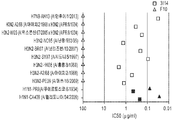


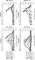


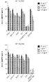
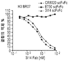


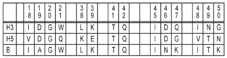


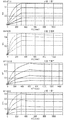
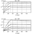


Classifications
-
- C—CHEMISTRY; METALLURGY
- C07—ORGANIC CHEMISTRY
- C07K—PEPTIDES
- C07K16/00—Immunoglobulins [IGs], e.g. monoclonal or polyclonal antibodies
- C07K16/08—Immunoglobulins [IGs], e.g. monoclonal or polyclonal antibodies against material from viruses
- C07K16/10—Immunoglobulins [IGs], e.g. monoclonal or polyclonal antibodies against material from viruses from RNA viruses
- C07K16/1018—Orthomyxoviridae, e.g. influenza virus
-
- A—HUMAN NECESSITIES
- A61—MEDICAL OR VETERINARY SCIENCE; HYGIENE
- A61K—PREPARATIONS FOR MEDICAL, DENTAL OR TOILETRY PURPOSES
- A61K39/00—Medicinal preparations containing antigens or antibodies
- A61K39/12—Viral antigens
- A61K39/145—Orthomyxoviridae, e.g. influenza virus
-
- A—HUMAN NECESSITIES
- A61—MEDICAL OR VETERINARY SCIENCE; HYGIENE
- A61P—SPECIFIC THERAPEUTIC ACTIVITY OF CHEMICAL COMPOUNDS OR MEDICINAL PREPARATIONS
- A61P31/00—Antiinfectives, i.e. antibiotics, antiseptics, chemotherapeutics
- A61P31/12—Antivirals
- A61P31/14—Antivirals for RNA viruses
- A61P31/16—Antivirals for RNA viruses for influenza or rhinoviruses
-
- A—HUMAN NECESSITIES
- A61—MEDICAL OR VETERINARY SCIENCE; HYGIENE
- A61K—PREPARATIONS FOR MEDICAL, DENTAL OR TOILETRY PURPOSES
- A61K39/00—Medicinal preparations containing antigens or antibodies
- A61K2039/505—Medicinal preparations containing antigens or antibodies comprising antibodies
-
- C—CHEMISTRY; METALLURGY
- C07—ORGANIC CHEMISTRY
- C07K—PEPTIDES
- C07K2317/00—Immunoglobulins specific features
- C07K2317/20—Immunoglobulins specific features characterized by taxonomic origin
- C07K2317/21—Immunoglobulins specific features characterized by taxonomic origin from primates, e.g. man
-
- C—CHEMISTRY; METALLURGY
- C07—ORGANIC CHEMISTRY
- C07K—PEPTIDES
- C07K2317/00—Immunoglobulins specific features
- C07K2317/30—Immunoglobulins specific features characterized by aspects of specificity or valency
- C07K2317/33—Crossreactivity, e.g. for species or epitope, or lack of said crossreactivity
-
- C—CHEMISTRY; METALLURGY
- C07—ORGANIC CHEMISTRY
- C07K—PEPTIDES
- C07K2317/00—Immunoglobulins specific features
- C07K2317/30—Immunoglobulins specific features characterized by aspects of specificity or valency
- C07K2317/34—Identification of a linear epitope shorter than 20 amino acid residues or of a conformational epitope defined by amino acid residues
-
- C—CHEMISTRY; METALLURGY
- C07—ORGANIC CHEMISTRY
- C07K—PEPTIDES
- C07K2317/00—Immunoglobulins specific features
- C07K2317/50—Immunoglobulins specific features characterized by immunoglobulin fragments
- C07K2317/56—Immunoglobulins specific features characterized by immunoglobulin fragments variable (Fv) region, i.e. VH and/or VL
- C07K2317/565—Complementarity determining region [CDR]
-
- C—CHEMISTRY; METALLURGY
- C07—ORGANIC CHEMISTRY
- C07K—PEPTIDES
- C07K2317/00—Immunoglobulins specific features
- C07K2317/70—Immunoglobulins specific features characterized by effect upon binding to a cell or to an antigen
- C07K2317/73—Inducing cell death, e.g. apoptosis, necrosis or inhibition of cell proliferation
- C07K2317/732—Antibody-dependent cellular cytotoxicity [ADCC]
-
- C—CHEMISTRY; METALLURGY
- C07—ORGANIC CHEMISTRY
- C07K—PEPTIDES
- C07K2317/00—Immunoglobulins specific features
- C07K2317/70—Immunoglobulins specific features characterized by effect upon binding to a cell or to an antigen
- C07K2317/76—Antagonist effect on antigen, e.g. neutralization or inhibition of binding
-
- C—CHEMISTRY; METALLURGY
- C07—ORGANIC CHEMISTRY
- C07K—PEPTIDES
- C07K2317/00—Immunoglobulins specific features
- C07K2317/90—Immunoglobulins specific features characterized by (pharmaco)kinetic aspects or by stability of the immunoglobulin
- C07K2317/92—Affinity (KD), association rate (Ka), dissociation rate (Kd) or EC50 value
Abstract
본 발명은 인플루엔자 바이러스의 HA 단백질의 스템 도메인에 결합하기 위해 중요한 구조적 결정자, 및 상기 결정자에 기초하여 고친화도 인플루엔자 중화 항체의 제조를 위한 그의 사용 방법을 제공한다. 본 발명은 또한 인플루엔자 바이러스 백신의 효능을 결정하기 위한 수단을 제공한다. 또한, 본 발명은 대상체에서 인플루엔자 바이러스 백신의 효능을 결정하기 위해, 또는 백신 또는 인플루엔자 바이러스 감염에 대한 사전 면역 노출 또는 항원 반응성을 예측하기 위해 유용한 분자 시그너쳐를 제공한다.The present invention provides a structural determinant important for binding to the stem domain of the HA protein of influenza virus, and a method for its use for the production of high affinity influenza neutralizing antibodies based on said determinant. The present invention also provides means for determining the efficacy of an influenza virus vaccine. The present invention also provides a molecular signature useful for determining the efficacy of an influenza virus vaccine in a subject, or for predicting pro-immune exposure or antigen responsiveness to a vaccine or influenza virus infection.

Description
관련 출원Related application
본원은 그 내용 전체가 본원에 참고로 포함된, 2016년 4월 8일 출원된 미국 가출원 제62/144,729호의 우선권 및 이익을 주장한다.This application claims priority and benefit of U.S. Provisional Application No. 62 / 144,729, filed April 8, 2016, the entire contents of which are incorporated herein by reference.
발명의 분야Field of invention
본 발명은 일반적으로 인플루엔자 중화 항체 및 그의 사용 방법에 관한 것이다.The present invention generally relates to influenza neutralizing antibodies and methods of use thereof.
정부 이익Government profit
본 발명은 미국 정부의 지원에 의해 이루어졌다. 미국 정부는 본 발명에 대해 특정 권리를 갖는다.The present invention was made with the support of the US government. The US Government has certain rights to the invention.
인플루엔자 대유행은 인체 건강에 대한 가장 심각한 급성 감염 위협 중 하나이다. 백신 접종은 계절성 및 대유행성 인플루엔자 및 그의 합병증을 예방하는 주요 수단으로 남아 있다. 인플루엔자 바이러스의 여러 아형에 대한 광범위한 면역을 유도하는 "보편적인" 인플루엔자 백신은 의학 연구에서 오랫동안 추구된 목표였다. HA의 스템(stem) 상의 고도로 보존된 소수성 포켓에 결합하는, 광범위하게 중화하는 인간 "이종아형(heterosubtypic)" 항체(sBnAb)의 최근 발견은 상기 백신 개발 노력을 재점화하였다. 그러나, 계절성 인플루엔자 또는 H5N1 백신의 혈청에서 또는 상업용 정맥내 이뮤노글로불린(IVIG) 제제에서 sBnAb의 매우 낮은 농도만이 검출된다.Influenza A pandemic is one of the most serious acute infection threats to human health. Vaccination remains a major means of preventing seasonal and pandemic influenza and its complications. The "universal" influenza vaccine, which leads to broad immunity against several subtypes of influenza virus, has long been a goal in medical research. The recent discovery of a broadly neutralizing human "heterosubtypic" antibody (sBnAb) that binds to highly conserved hydrophobic pockets on the stem of HA has rekindled this vaccine development effort. However, only very low concentrations of sBnAb are detected in sera of seasonal influenza or H5N1 vaccines or in commercial intravenous immunoglobulin (IVIG) preparations.
인플루엔자 바이러스에 대한 면역요법을 위한 모노클로날 항체(mAb) 및 약물을 생산하기 위한 지속적인 노력이 있다. 구체적으로, 다양한 인플루엔자 균주 모두를 중화하는 치료 화합물의 개발에 대한 노력이 진행되고 있다. 현재, 이 목표를 달성할 수 있는 소수의 mAb만이 보고되어 있다. 이들 mAb는 파지 항체 라이브러리를 패닝(panning)하고 백신 접종된 자원자로부터 B 세포를 스크리닝함으로써 단리하였다. 그러나, 광범위한 인플루엔자 중화 항체의 특성에 대한 이해의 증가는 인플루엔자 중화 항체의 광범위한 패널의 발견 및 생산을 위한 보다 합리적인 설계 방법에 특정 구조적 결정 요인을 도입하는데 유용할 수 있다.There is a continuing effort to produce monoclonal antibodies (mAbs) and drugs for immunotherapy against influenza virus. Specifically, efforts are being made to develop therapeutic compounds that neutralize all of the various influenza strains. At present, only a few mAbs are reported that can achieve this goal. These mAbs were isolated by panning the phage antibody library and screening B cells from vaccinated volunteers. However, an increased understanding of the properties of a broad range of influenza neutralizing antibodies may be useful in introducing specific structural determinants into more rational design methods for the discovery and production of a broad panel of influenza neutralizing antibodies.
따라서, 인플루엔자 바이러스를 광범위하게 중화할 수 있는 추가의 모노클로날 항체 및 합리적인 설계 방법을 통해 상기 항체의 친화도 또는 효능을 증가시키는 방법에 대한 필요성이 존재한다.Thus, there is a need for additional monoclonal antibodies capable of broadly neutralizing influenza viruses and methods of increasing the affinity or potency of such antibodies through a reasonable design method.
발명의 개요Summary of the Invention
본 발명은 다음 특징을 갖는, 재조합 생산된, 단리된 모노클로날 항체를 특징으로 한다: IGHV3-30 생식계열 유전자에 의해 코딩되는 중쇄 가변 영역; 헤마글루티닌(HA) 단백질의 스템 영역의 에피토프에 대한 결합; 및 그룹 1 및 그룹 2 인플루엔자 A 바이러스의 중화.The invention features a recombinantly produced, isolated monoclonal antibody having the following characteristics: a heavy chain variable region encoded by an IGHV3-30 reproductive gene; Binding to the epitope of the stem region of hemagglutinin (HA) protein; And neutralization of
한 측면에서, 모노클로날 항체는 다음 특징 중 하나 이상을 갖는다: IGLV1-44 생식계열 유전자에 의해 코딩되는 경쇄 가변 영역; 서열 번호 9의 아미노산 서열을 포함하는 중쇄 CDR3; 절단되지 않은 HA0에 대한 결합; HA0 절단 방지; 및/또는 서열 번호 18에 따라 넘버링될 때 HA2 폴리펩티드의 아미노산 18, 19, 20, 21, 36, 38, 39, 41, 42, 45, 45, 49 및 53에 의해 규정되는 구조적 에피토프(conformational epitope)에 대한 결합.In one aspect, the monoclonal antibody has one or more of the following characteristics: a light chain variable region encoded by the IGLV 1-44 reproductive gene; A heavy chain CDR3 comprising the amino acid sequence of SEQ ID NO: 9; Bond to uncut HA0; HA0 cutting prevention; And / or a structural epitope defined by the
본 발명은 서열 번호 7의 아미노산 서열을 포함하는 CDR1; 서열 번호 8의 아미노산 서열을 포함하는 CDR2 및 서열 번호 9의 아미노산 서열을 포함하는 CDR3을 포함하는 중쇄; 및 서열 번호 10 또는 서열 번호 14의 아미노산 서열을 포함하는 CDR1; 서열 번호 11의 아미노산 서열을 포함하는 CDR2 및 서열 번호 12 또는 서열 번호 13의 아미노산 서열을 포함하는 CDR3을 포함하는 경쇄를 포함하는, 인플루엔자 바이러스를 중화하는 단리된 모노클로날 항체를 특징으로 한다.The invention provides a CDR1 comprising the amino acid sequence of SEQ ID NO: 7; A heavy chain comprising CDR2 comprising the amino acid sequence of SEQ ID NO: 8 and CDR3 comprising the amino acid sequence of SEQ ID NO: 9; And CDR1 comprising the amino acid sequence of SEQ ID NO: 10 or SEQ ID NO: 14; A light chain comprising CDR2 comprising the amino acid sequence of SEQ ID NO: 11 and a light chain comprising CDR3 comprising the amino acid sequence of SEQ ID NO: 13 or SEQ ID NO: 13, or an isolated monoclonal antibody that neutralizes influenza virus.
본 발명은 또한 서열 번호 2의 VH 아미노산 서열 및 서열 번호 4 또는 서열 번호 6의 VL 아미노산 서열; 서열 번호 2의 VH 아미노산 서열 및 서열 번호 24의 VL 아미노산 서열; 또는 서열 번호 22의 VH 아미노산 서열 및 서열 번호 4 또는 6의 VL 아미노산 서열을 포함하는 단리된 모노클로날 항체를 특징으로 한다.The invention also encompasses a V H amino acid sequence of SEQ ID NO: 2 and a V L amino acid sequence of SEQ ID NO: 4 or SEQ ID NO: 6; A V H amino acid sequence of SEQ ID NO: 2 and a V L amino acid sequence of SEQ ID NO: 24; Or an isolated V H amino acid sequence of SEQ ID NO: 22 and a V L amino acid sequence of SEQ ID NOs: 4 or 6.
항체는 HA2 폴리펩티드의 아미노산 18, 19, 20, 21, 36, 38, 39, 41, 42, 45, 45, 49 및 53에 의해 규정된 구조적 에피토프에 결합한다.Antibodies bind to the structural epitopes defined by
한 측면에서, 항체는 단일쇄 Fv 항체, Fab 단편, Fab' 단편, 또는 F(ab')2 단편이다. 또 다른 측면에서, 항체는 치료제에 연결된다. 예를 들어, 치료제는 독소, 방사성 표지, siRNA, 소분자 또는 사이토카인이다.In one aspect, the antibody is a single chain Fv antibody, Fab fragment, Fab 'fragment, or F (ab') 2 fragment. In another aspect, the antibody is linked to a therapeutic agent. For example, the therapeutic agent is a toxin, a radioactive label, an siRNA, a small molecule, or a cytokine.
본 발명은 또한 본원에 개시된 임의의 항체 및 담체를 포함하는 조성물을 특징으로 한다.The invention also features a composition comprising any of the antibodies and carriers disclosed herein.
본 발명은 서열 번호 1, 3 및 5로부터 선택된 핵산 서열을 포함하는 핵산 서열을 제공한다. 또 다른 실시양태에서, 서열 번호 2, 4 및 6으로부터 선택된 아미노산 서열을 포함하는 폴리펩티드를 코딩하는 핵산 서열이 제공된다. 한 측면에서, 서열 번호 2, 4 및 6으로부터 선택된 아미노산 서열을 포함하는 폴리펩티드가 제공된다.The present invention provides nucleic acid sequences comprising nucleic acid sequences selected from SEQ ID NOS: 1, 3 and 5. In another embodiment, nucleic acid sequences encoding polypeptides comprising an amino acid sequence selected from SEQ ID NOS: 2, 4, and 6 are provided. In one aspect, polypeptides comprising an amino acid sequence selected from SEQ ID NOS: 2, 4, and 6 are provided.
본 발명은 서열 번호 1, 2, 3, 4, 5 및 6으로부터 선택된 핵산 서열을 포함하는 벡터를 제공한다. 한 측면에서, 본 발명은 핵산 서열 1, 2, 3, 4 또는 6을 포함하는 벡터를 포함하는 세포를 제공한다.The present invention provides a vector comprising a nucleic acid sequence selected from SEQ ID NOS: 1, 2, 3, 4, 5 and 6. In one aspect, the invention provides a cell comprising a vector comprising
본 발명은 또한 본원에 개시된 임의의 항체를 생산하는 세포를 제공한다.The present invention also provides cells that produce any of the antibodies disclosed herein.
본 발명은 추가로 인플루엔자 바이러스에 의해 유발된 질환 또는 장애를 앓을 위험이 있는 인간에게 본원에서 설명되는 임의의 모노클로날 항체의 치료 유효량을 투여함으로써 상기 질환 또는 장애를 치료하는 방법을 제공한다. 선택적으로, 상기 방법은 항바이러스제를 투여하는 단계를 추가로 포함한다.The present invention further provides a method of treating said disease or disorder by administering a therapeutically effective amount of any of the monoclonal antibodies described herein to a human being at risk of suffering from a disease or disorder caused by influenza virus. Optionally, the method further comprises administering an antiviral agent.
본 발명의 다른 특징 및 이점은 다음의 상세한 설명 및 청구범위로부터 명백할 것이고, 그에 의해 포함될 것이다.Other features and advantages of the invention will be apparent from and elucidated with reference to the following detailed description and claims.
도 1a는 인간 기억 B 세포 레퍼토리로부터 인플루엔자 바이러스에 대해 광범위하게 중화하는 Ab의 단리를 나타내는 개략도이다. 대표적인 형광 표지 세포 분류(FACS) 데이터도 제공된다. FACS 데이터는 총 PBMC로부터 단리된 H3-반응성 기억 B 세포의 빈도를 나타낸다. 도 1b는 아미노산 서열에 기초한 인플루엔자 A 바이러스의 18 HA 아형의 계통수(phylogenetic tree)를 도시한 것이다. 그룹 1 아형 및 그룹 2 아형이 개략도에 표시된다. 아미노산 거리 규모 막대는 0.1의 거리를 나타낸다.
도 2는 넓은 범위의 그룹 1 및 그룹 2 HA에 대한 3I14 결합을 보여주는 일련의 FACS 그래프이다. 293T 세포를 상이한 HA 발현 플라스미드로 일시적으로 형질감염시키고, 정제된 scFvFc 항체 및 APC 표지된 마우스 항-인간 Fc 항체로 염색하였다. 3I14 (적색 선), F10 (그룹 1 특이적, 녹색 선), CR8020 (그룹 2 특이적, 청색 선), FI6v3 (그룹 1 및 2 특이적, 보라색 선), CR9114 (그룹 1 & 2 특이적, 오렌지색 선) 및 무관한 mAb Fm-6 (항-SARS 바이러스, 회색의 채워진 히스토그램)의 결합을 유동 세포 계측법으로 분석하였다.
도 3은 그룹 1(적색) 또는 그룹 2(청색) 아형을 나타내는 재조합 HA에 대한 3I14 IgG1(A) 결합(Kd 값) 또는 scFvFc Ab(B) 결합(Kd 값)을 보여주는 일련의 그래프이다.
도 4는 그룹 1(적색) 또는 그룹 2(청색) 아형의 감염성 바이러스의 3I14 IgG1 중화(A)(IC50 값) 또는 3I14 scFvFc Ab 중화(IC50 값)을 보여주는 일련의 그래프이다. 3I14는 정사각형으로 표시하고; 항-그룹 1 mAb F10은 삼각형으로 나타내었다. IC50 값에 사용된 그래프는 2-3회의 독립적인 실험의 평균 중화 역가에 의해 결정하였다.
도 5는 그룹 1 또는 그룹 2 아형의 슈도바이러스(pseudovirus)의 3I14 IgG1 중화(A)(IC50 값) 또는 3I14 scFvFc Ab 중화(B)(IC50 값)을 보여주는 일련의 그래프이다. 이들 데이터는 2-3회의 독립적인 실험의 평균 중화 역가를 나타낸다. 도 5b에서, 항-그룹 1 mAb F10 scFvFc를 참고용으로 사용하였다.
도 6은 인플루엔자 바이러스에 노출되고 5, 20 또는 25 mg 중 하나의 농도에서 정제된 IgG를 복강내 투여한 마우스의 생존율(A)을 보여주는 일련의 그래프이다. 도 6b는 인플루엔자 바이러스에 노출되고 지시된 특정 항체 농도를 투여한 마우스에서 체중 조성의 변화를 보여주는 일련의 그래프이다. 이러한 분석을 위해, 5마리의 마우스의 그룹을, H7N7-NL219, H7N9-AH13, H3N2-BR07 또는 H5N1-VN04 인플루엔자 바이러스 (~10 LD50)의 i.n. 접종에 의한 치사 시험접종(challenge) 24시간 전에 20/25 또는 5 mg/kg 투여량의 정제된 IgG의 복강내 투여로 처리하였다. (a) bnAb 3I14(적색) 및 그룹 1 대조군 mAb F10(흑색)으로 처리한 마우스의 생존율(%) 및(b) 체중 변화(%).
도 7은 항체가 사용되지 않거나, 3I14 또는 Fm-6(IgG1 대조군)이 사용된 트립신 절단 억제 분석으로부터의 결과를 보여주는 겔이다. 이 분석을 위해, 0.4 ㎍의 재조합 H3-히스티딘(H3-BR07)을 100 ㎍/mL 트립신-울트라(New England Biolabs, 미국)를 함유하는 pH 8.0의 Tris-HCl 완충제 내에서 2.5 ㎍ 3I14 또는 Fm-6 IgG1의 존재 하에 또는 항체의 부재 하에 37℃에서 인큐베이팅하였다. 트립신 소화는 1% BSA의 첨가에 의해 여러 시점에서 억제되었다. 샘플을 12% 환원 SDS-PAGE에서 이동시키고, HisProbe-HRP Ab를 사용하여 블로팅하였다.
도 8은 3I14 IgG1이 표면 발현된 H3-A268 및 H3-BR07에서 낮은 pH에 의해 촉발된 구조적 재배열을 방지함을 보여주는 일련의 개략도 및 그래프이다. 표면 발현된 H3의 구조적 재배열은 3I14(솔리드 막대) 및 헤드 결합 대조군 mAb E730(오픈 막대)의 FACS 염색에 의해 검출되었다. 결합은 미처리된 HA(HA0)에 결합하는 백분율로 표현된다. 이 항체 억제 분석을 위해, H3를 mAb 처리 없이, 3I14로, 또는 대조군 Ab인 Fm-6 IgG1로 전처리한 후, 절단된 HA를 pH 4.9에 노출시켰다. 데이터는 3회의 독립적인 실험의 평균 + SD를 나타낸다.
도 9a 및 9b는 3I14가 항체 의존성 세포독성(ADCC)을 매개한다는 것을 입증한다. 도 9a는 ADCC 기반 분석의 결과를 보여주는 그래프이다. 3I14 및 다른 항-스템 bnAb인 FI6v3, CR9114, 39.29, F10 및 CR8020은 H3- 및 H5-발현 293T 세포에서 ADCC를 유도하였다. 이들 분석을 위해, 분석하기 전에 플레이트에 1x10⁴/웰 H3 또는 H5-발현 293T 세포를 부착시킨 다음, 배지를 낮은 IgG 혈청 분석 완충제(0.5%의 낮은 IgG FBS를 갖는 RPMI 1640)으로 대체하였다. 상이한 bnAb를 5, 1, 0.2 및 0.04 ㎍/㎖의 농도로 각각의 웰에 첨가하였다. 1시간 후에, 낮은 IgG 혈청 분석 완충제에서 플레이트를 분석하기 위해 Jurkat 이펙터 세포를 6.0x10⁴ 세포/웰로 첨가하고, 6시간 동안 인큐베이팅하였다. 상청액을 수거하고, 루시페라제 활성을 Bio-GloTM 루시페라제 분석 키트(Promega)를 사용하여 측정하였다. 도 9b는 H3- 및 H5-발현 293T 세포에서 3I14 및 다른 항-스템 bnAb인 FI6v3, CR9114, 39.29, F10 및 CR8020 유도 ADCC를 보여주는 일련의 그래프이다. 2x10⁴/웰 H3 또는 H5-발현 293T 세포를 분석 전에 플레이트에 부착시킨 다음, 배지를 낮은 IgG 혈청 분석 완충제(0.5%의 낮은 IgG FBS를 갖는 RPMI 1640)으로 교체하였다. 상이한 bnAb를 10, 5, 2.5 및 1.25 ㎍/ml의 농도로 각각의 웰에 첨가하였다. 1시간 후, 낮은 IgG 혈청 분석 완충제에서 플레이트를 분석하기 위해 PBMC를 1.2x10⁵ 세포/웰로 첨가하고, 6시간 동안 인큐베이팅하였다. 상청액을 수거하고, 루시페라제 활성을 LDH 세포독성 분석 키트(Pierce)를 사용하여 측정하였다. 데이터는 평균 ± S.E.M.을 나타낸다. 실험은 3회 수행하였고, 비슷한 결과를 얻었다. 데이터는 그룹당 3회 반복한 하나의 독립적인 실험을 나타낸다.
도 10은 3I14가 H3 또는 H5에 대한 결합을 위해 다른 항-스템 bnAb인 FI6v3, CR9114, 39.29, F10 및 CR8020과 교차 경쟁함을 보여주는 일련의 그래프이다. 이들 분석을 위해, 5 ㎍/ml H3-BR07 또는 H5-VN04 단백질을 ELISA 플레이트 상에 고정하고, 80 nM 내지 0.3 nM의 농도 범위의 3I14 Fab의 2배 연속 희석액과 함께 인큐베이팅하였고; 이들을 5 nM의 다른 scFvFc Ab와 추가로 혼합하였다. scFvFc Ab의 결합은 HRP 접합 항-인간 CH2 항체를 사용하여 검출되었다.
도 11은 3I14 및 다른 생식계열 중쇄(a, c) 또는 경쇄 영역(b, d)의 아미노산 서열 정렬을 도시한 것이다. 대응하는 V, D 및 J 서열은 IMGT 데이터베이스를 사용하여 결정하였고 비교를 위해 나타내었다.
도 12a 및 도 12b는 3개의 VH3-30 BnAb인 39.29(4KVN으로부터), FI6(3ZTJ로부터) 및 Mab3.1(4PY8)을 사용한 3I14 모델의 중첩을 보여주는 일련의 개략도이다. 3I14 항체의 중쇄 CDR뿐만 아니라 중쇄 및 경쇄의 상대적인 위치를 보여주는 단백질 모델 개략도가 제시된다. 중쇄는 녹색으로 표시되고, 경쇄는 회색으로 표시된다. 도 12의 중쇄 CDR은 다음과 같이 색상으로 구분된다: 3I14 HCDR은 청색, 다른 BnAb의 HCDR1은 적색, HCDR2는 자홍색, HCDR3은 청록색이다.
bnAb는 리본 방식으로 표시된다. 도 12b에서, 중쇄는 청색이고, 경쇄는 청록색이다. HCDR3은 3I14에서 적색으로, FI6v3에 자홍색으로, 39.29에서 황색으로, MAb 3.1에서 녹색으로 강조 표시된다.
도 13a-d는 3I14의 모델링 및 H3/H5과의 도킹을 보여주는 일련의 개략도이다. H3 삼량체 모델의 줄기 상의 3I14 에피토프 구조가 도 13a에 도시되어 있다. 도 13b는 HA와 함께 IGVH3-30 유래 bnAb의 복합체 구조를 도시한 것이다. HA상의 에피토프 잔기는 표면 제시로 도시되고, bnAb의 CDR 루프는 리본으로 표시된다. HA 단량체의 HA1은 밀에, HA2는 연어살색에 존재하고, 에피토프 잔기는 적색으로 표지된다. 핵심 잔기 L38 및 K39는 황색으로 착색되어 있다. BnAb의 중쇄 CDR은 청색이고, 경쇄 CDR은 청록색이다. 3I14는 바이오루미네이트(BioLuminate)의 항체 모델링 모듈을 사용하여 상동성을 모델링하였고, 모델은 로제타독(RosettaDock)과 도킹하기 전에 H3/FI6v3 복합체 구조에 중첩되었다. 다른 3개의 IGHV3-30 bnAb인 FI6v3, 39.29 및 MAb 3.1은 프로테인 데이터 뱅크(Protein Data Bank)에서 다운로드하였다. 도 13c는 3I14 내의 D94와 H3/H5의 상호작용을 도시한 것이다. H3은 청록색으로 표시되고 여기서 K39는 스틱으로 제시되고; H5는 녹색으로 표시되고 여기서 E39는 스틱으로 제시되고; 3I14는 H3/3I14 모델에서 오렌지색으로, H5/3I14 모델에서는 황색으로 표시되고 여기서 D94는 스틱으로 제시된다. 도 13d는 H3/3I14 복합 모델에서 3I14 경쇄의 G31과 H3의 상호작용을 도시한 것이다. H3의 HA2 도메인의 나선 A는 청록색 리본으로 표시되고; 3I14의 경쇄는 오렌지색으로 리본으로 표시되고; G31의 주쇄 원자는 스틱으로 표시되고 H3 HA2의 Q42 및 D46의 측쇄 원자는 막대기로 표시되고; G31과 H3 사이의 거리는 녹색 점선으로 표시되고 흑색으로 표지된다(PyMOL Molecular Graphics System, Version 0.99 rc6 Schroedinger, LLC).
도 14는 H3/3I14 및 H5/3I14 모델의 서열 정렬(a) 및 구조 중첩(b)을 도시한 것이다.
또한, 15a-15f는 재조합 H5-VN04(a) 및 H3-PE09(b)에 결합하는 3I14 WT 및 VLD94N IgG1 변이체의 Kd 결합 값을 보여주는 그래프 및 표이다. 녹색 또는 청색 곡선은 생물층 간섭 측정 실험으로부터 얻은 실험 기록이고, 적색 곡선은 Kd 값을 계산하기 위해 사용되는 데이터에 대한 전반적인 최적 적합도(best global fit)이다. 결합 곡선 친화도 측정채(Kd 값)는 표 4에 보고된다. 3I14 WT는 1.15 nm의 Kd 값으로 정제된 H5-VN04에 결합한 반면, 3I14 VLD94N 돌연변이체는 0.19 nM에서 10배 더 높은 친화도로 H5-VN04에 결합하였다. 도 15c-f는 재조합 Ha에 결합하는 3I14 scFvFc를 나타내는 일련의 그래프이다.
도 16a-d는 슈도타이핑된(pseudotyped) 바이러스 H5N1-VN04 및 감염성 바이러스 H3N2-BR07과 함께 인큐베이팅한 후, 3I14 WT 및 VLD94N 돌연변이체 IgG1의 중화 값을 나타내는 일련의 그래프이다. 도 16c 및 16d에서, 3I14(흑색) 및 VLD94N 변이체(적색)는 슈도타이핑된 바이러스 H5N1-VN04(c) 및 H3N2-BR07 바이러스(d)를 중화하였다. 이 데이터는 2-3회의 독립적인 실험의 평균 중화 역가를 나타낸다.
도 17은 3I14 scFvFc Ab가 인플루엔자 바이러스 감염 및 HA-슈도타이핑된 루시페라제 리포터 바이러스를 중화함을 나타내는 일련의 그래프이다. MAb 3I14(흑색) 및 항-그룹 1 mAb F10(적색)은 감염성 바이러스 및 슈도타이핑된 바이러스의 상이한 균주를 중화하였다. 데이터는 2-3회의 독립적인 실험의 평균 중화 역가를 나타낸다.
도 18은 조작된 3I14 효모-CDR 라이브러리의 H5에 대한 결합과 비교하여 3I14-WT 효모의 결합을 나타내는 일련의 유동 세포 계측 그래프이다. 3I14 효모-CDR 라이브러리는 H5에 대한 결합을 증가시키는 변이체에 대한 효모 디스플레이를 사용하여 조작되었다.
도 19는 재조합 H3-PE09의 전장 또는 HA1에 결합하는 3I14 scFvFc Ab를 나타내는 그래프이다.
도 20은 3I14 IgG1 변이체의 재조합 H1, H3 및 H5에 대한 결합을 나타내는 일련의 그래프이다. 청색 곡선은 생물층 간섭 측정 실험으로부터 얻은 실험 기록이고, 적색 곡선은 표 7에 제시된 Kd를 계산하기 위해 사용된 데이터에 대한 전반적인 최적 적합도이다.1A is a schematic diagram showing the isolation of Abs extensively neutralizing influenza viruses from human memory B cell repertoires. Representative fluorescence labeled cell sorting (FACS) data is also provided. FACS data shows the frequency of H3-reactive memory B cells isolated from total PBMC. Figure 1b shows the phylogenetic tree of the 18 HA subtype of influenza A virus based on amino acid sequence.
Figure 2 is a series of FACS graphs showing 3I14 binding for a broad range of
Figure 3 is a series of graphs showing Group 1 (Red) or group 2 (blue) 3I14 IgG1 (A) binding (K d value) or scFvFc Ab (B) binding (K d value) of the recombinant HA represents the subtype .
Figure 4 is a series of graphs showing Group 1 (Red) or group 2 (blue) 3I14 IgG1 neutralizing (A) (IC 50 value) of the infectious virus of subtype or 3I14 scFvFc neutralizing Ab (IC 50 value). 3I14 is represented by a square; Anti-group 1 mAb F10 is represented by a triangle. The graphs used for IC50 values were determined by the mean neutralization titers of 2-3 independent experiments.
Figure 5 is a series of graphs showing neutralization 3I14 IgG1 (A) (IC 50 value) or 3I14 scFvFc neutralizing Ab (B) (IC 50 values) of the pseudo-virus (pseudovirus) of
6 is a series of graphs showing the survival rate (A) of mice exposed to influenza virus and administered intraperitoneally with purified IgG at a concentration of either 5, 20, or 25 mg. Figure 6b is a series of graphs showing changes in body weight composition in mice exposed to influenza virus and given specific antibody concentrations indicated. For this analysis, groups of 5 mice were injected 20 hours prior to the lethal challenge challenge with the inoculation of H7N7-NL219, H7N9-AH13, H3N2-BR07 or H5N1-VN04 influenza virus (~ 10 LD50) / 25 or 5 mg / kg dose of purified IgG. (a) Survival (%) of mice treated with bnAb 3I14 (red) and
Figure 7 is a gel showing the results from trypsin cleavage inhibition assay where no antibody is used or 3I14 or Fm-6 (IgG1 control) is used. For this analysis, 0.4 μg of recombinant H3-histidine (H3-BR07) was mixed with 2.5 μg of 3I14 or Fm-1 in a Tris-HCl buffer of pH 8.0 containing 100 μg / mL trypsin- ultras (New England Biolabs, USA) 6 < RTI ID = 0.0 > IgG1 < / RTI > or in the absence of antibody. Trypsin digestion was inhibited at various points by the addition of 1% BSA. Samples were transferred on a 12% reduction SDS-PAGE and blotted using HisProbe-HRP Ab.
Figure 8 is a series of schematic and graphs showing that 3I14 IgGl prevents structural rearrangement triggered by low pH in surface expressed H3-A268 and H3-BR07. Structural rearrangement of surface expressed H3 was detected by FACS staining of 3I14 (solid rod) and head binding control mAb E730 (open rod). The binding is expressed as a percentage binding to untreated HA (HAO). For this antibody inhibition assay, H3 was pretreated with no mAb treatment, 3I14, or with control Ab, Fm-6 IgGl, and then cleaved HA was exposed to pH 4.9. Data represent the mean + SD of three independent experiments.
Figures 9a and 9b demonstrate that 3I14 mediates antibody dependent cellular cytotoxicity (ADCC). Figure 9A is a graph showing the results of the ADCC-based analysis. 3I14 and the other anti-stem bnAb, FI6v3, CR9114, 39.29, F10 and CR8020, induced ADCC in H3- and H5-expressing 293T cells. For these analyzes, was replaced with the adhered 1x10 4 / well H3 or H5- expressing 293T cells to the plate prior to analysis, and then, (RPMI 1640 with Low IgG FBS 0.5%) low serum IgG assay buffer of the medium. Different bnAbs were added to each well at concentrations of 5, 1, 0.2 and 0.04 [mu] g / ml. After 1 hour, Jurkat effector cells were added to 6.0x10 4 cells / well to analyze plates in low IgG serum assay buffer and incubated for 6 hours. The supernatant was collected and the luciferase activity was measured using the Bio-Glo luciferase assay kit (Promega). Figure 9b is a series of graphs showing FI3v3, CR9114, 39.29, F10, and CR8020 induced ADCC, 3I14 and other anti-stem bnAbs in H3- and H5-expressing 293T cells. 2x10 was replaced with 4 / well H3 or H5- expressing 293T cells were attached to the plate before the next analysis, (RPMI 1640 with low IgG FBS 0.5%), the medium low IgG serum assay buffer. Different bnAbs were added to each well at concentrations of 10, 5, 2.5 and 1.25 占 퐂 / ml. After 1 hour, PBMC were added to 1.2x10 5 cells / well to analyze the plates in low IgG serum assay buffer and incubated for 6 hours. The supernatant was collected and luciferase activity was measured using the LDH cytotoxicity assay kit (Pierce). Data represent the mean ± SEM. The experiment was carried out three times and a similar result was obtained. The data represent one independent experiment repeated three times per group.
Figure 10 is a series of graphs showing that 3I14 competes with FI6v3, CR9114, 39.29, F10 and CR8020, which are other anti-stem bnAb for binding to H3 or H5. For these assays, 5 [mu] g / ml H3-BR07 or H5-VNO4 protein was immobilized on ELISA plates and incubated with 2x serial dilutions of 3I14 Fab in a concentration range of 80 nM to 0.3 nM; These were further mixed with 5 nM of other scFvFc Ab. Binding of scFvFc Ab was detected using HRP conjugated anti-human CH2 antibody.
Figure 11 shows the amino acid sequence alignment of 3I14 and other germline heavy chain (a, c) or light chain region (b, d). Corresponding V, D and J sequences were determined using the IMGT database and are shown for comparison.
Figures 12a and 12b are a series of schematic diagrams showing the superposition of 3I14 models using 39.29 (from 4KVN), FI6 (from 3ZTJ) and Mab 3.1 (4PY8), which are three VH3-30 BnAbs. A protein model schematic showing the relative position of the heavy and light chains as well as the heavy chain CDR of the 3I14 antibody is presented. The heavy chain is indicated in green and the light chain is indicated in gray. The heavy chain CDRs of FIG. 12 are color-coded as follows: 3I14 HCDR is blue, HCDR1 of other BnAb is red, HCDR2 is magenta, and HCDR3 is cyan.
The bnAb is displayed in a ribbon fashion. 12B, the heavy chain is blue and the light chain is cyan. HCDR3 is highlighted from 3I14 to red, FI6v3 to magenta, 39.29 to yellow, and MAb 3.1 to green.
Figures 13a-d are a series of schematic diagrams illustrating modeling of 3I14 and docking with H3 / H5. The 3I14 epitope structure on the trunks of the H3 trimeric model is shown in Figure 13a. Figure 13b shows the complex structure of IGVH3-30 derived bnAb with HA. The epitope residues on HA are shown as a surface presentation, and the CDR loops of bnAb are labeled as ribbons. The HA monomer of HA monomer is in wheat, HA2 is in salmon color and the epitope residue is labeled red. Core residues L38 and K39 are colored yellow. The heavy chain CDR of BnAb is blue and the light chain CDR is cyan. 3I14 modeled homology using an antibody modeling module of BioLuminate and the model was superimposed on the H3 / FI6v3 complex structure before docking with RosettaDock. The other three IGHV3-30 bnAbs FI6v3, 39.29 and MAb 3.1 were downloaded from the Protein Data Bank. 13C shows the interaction of D94 and H3 / H5 in 3I14. H3 is shown in cyan, where K39 is presented as a stick; H5 is shown in green, where E39 is presented as a stick; 3I14 is displayed in orange on the H3 / 3I14 model and yellow in the H5 / 3I14 model, where D94 is presented as a stick. Figure 13d shows the interaction of G31 and H3 of the 3I14 light chain in the H3 / 3I14 composite model. Spiral A of the HA2 domain of H3 is represented by a cyan ribbon; The light chain of 3I14 is indicated by a ribbon in orange color; The backbone atoms of G31 are represented by sticks and the side chain atoms of Q42 and D46 of H3 HA2 are represented by a stick; The distance between G31 and H3 is indicated by a green dotted line and is labeled black (PyMOL Molecular Graphics System, Version 0.99 rc6 Schroedinger, LLC).
Figure 14 shows the sequence alignment (a) and structure overlay (b) of the H3 / 3I14 and H5 / 3I14 models.
15a-15f are graphs and tables showing K d binding values of 3I14 WT and VLD94N IgG1 variants binding to recombinant H5-VN04 (a) and H3-PE09 (b). The green or blue curve is the experimental record from the bio-layer interference measurement experiment and the red curve is the best global fit for the data used to calculate the K d value. Bond curve affinity measurements (K d values) are reported in Table 4. 3I14 WT bound to purified H5-VN04 with a K d value of 1.15 nm, while the 3I14 VLD94N mutant bound to H5-VN04 with a 10-fold higher affinity at 0.19 nM. Figures 15c-f are a series of graphs showing 3I14 scFvFc binding to recombinant Ha.
Figures 16a-d are a series of graphs showing neutralization values of 3I14 WT and VLD94N mutant IgG1 after incubation with the pseudotyped virus H5N1-VN04 and the infectious virus H3N2-BR07. In Figures 16c and 16d, the 3I14 (black) and VLD94N variants (red) neutralized the pseudotyped viruses H5N1-VN04 (c) and H3N2-BR07 virus (d). This data represents the mean neutralization titres of 2-3 independent experiments.
Figure 17 is a series of graphs showing that 3I14 scFvFc Ab neutralizes influenza virus infection and HA-pseudotyped luciferase reporter virus. MAb 3I14 (black) and
Figure 18 is a series of flow cytometry graphs showing binding of 3I14-WT yeast compared to binding of engineered 3I14 yeast-CDR library to H5. The 3I14 yeast-CDR library was engineered using yeast display for mutants that increase binding to H5.
19 is a graph showing the whole length of recombinant H3-PE09 or 3I14 scFvFc Ab binding to HA1.
Figure 20 is a series of graphs showing the binding of 3I14 IgGl variants to recombinant H1, H3 and H5. The blue curve is the experimental record from the bio-layer interference measurement experiment and the red curve is the overall best fit for the data used to calculate the K d shown in Table 7.
인플루엔자 A는 음성 단일 가닥 RNA 바이러스이고, 10개의 단백질을 코딩하는 8 개 분절의 게놈을 갖는다. 이것은 뉴클레오캡시드 및 매트릭스 단백질의 항원성에 의해 정의된 인플루엔자 바이러스 A, B 및 C의 속을 포함하는 오르토믹소비리대(Orthomyxoviridae)에 속한다. 일반적으로, 인플루엔자 A 바이러스는 인간의 보다 심각한 질환과 연관된다. 인플루엔자 A 바이러스는 2개의 표면 단백질, 즉 세포 진입을 위해 숙주 세포에 비리온을 부착시키는 헤마글루티닌(HA: hemagglutinin) 및 자손체 바이러스 또는 세포 표면에 부착된 숙주 시알산을 절단함으로써 자손체 바이러스의 전파를 촉진하는 뉴라미니다제(NA: neuraminidase)에 의해 추가로 하위 분류된다.Influenza A is a negative single-stranded RNA virus and has eight segments of the genome that encode 10 proteins. This belongs to Orthomyxoviridae, which contains the genus of influenza viruses A, B and C as defined by the antigenicity of the nucleocapsid and matrix proteins. In general, influenza A virus is associated with more serious human diseases. Influenza A virus has two surface proteins: hemagglutinin (HA), which attaches virions to host cells for cell entry, and progeny viruses, Which is further subclassified by neuraminidase (NA), which promotes the spread of neurons.
HA 및 NA의 다양한 조합에 의해 인플루엔자 A 바이러스의 모든 아형을 구성하는 16개의 HA 아형 및 9개의 NA 아형이 존재한다. 16개의 HA 및 9개의 NA 바이러스 아형의 모든 조합은 물새에서 발견된다. 수 백종의 조류 인플루엔자 A 바이러스 균주 중, 인간의 감염을 일으킨 것으로 알려진 바이러스는 오직 H5N1, H7N3, H7N7 및 H9N2 뿐이다. 일반적으로, 이들 바이러스에 의한 인간의 감염은 경증 증상 및 극히 적은 심각한 병을 일으켰고. H7N7에 의한 폐렴의 유일한 치명적인 사례가 있었다. 그러나, 인간에게 자연적인 면역이 없는 고병원성 H5N1 바이러스는 예외이다. RNA 폴리머라제의 부정확(infidelity) 및 숙주 면역의 선택 압력은 돌연변이의 축적 및 이들 단백질의 표면 항원성의 변화로 이어질 수 있다. 이 항원 변화는 항원 드리프트(antigenic drift)라고한다. 또한, 그의 분절화된 게놈의 결과로서, 인플루엔자 A 바이러스의 2개의 상이한 아형이 동일한 세포를 감염시키면 유전자 분절이 뒤섞일 수 있다. 예를 들어, 인간 H3N2 바이러스 및 조류 H5N1 바이러스가 인간 또는 포유동물 종의 다른 구성원을 동시에 감염시키면, 이러한 사건은 새로운 H5N2를 생성할 수 있다. 이 새로운 바이러스는 대부분의 유전자 분절이 인간의 바이러스로부터 유래하기 때문에 인간으로부터 인간으로 효율적으로 전염될 수 있다. 그러한 유전자 재편성(reassortment)은 항원 변위(antigenic shift)라고 불리는 중요한 항원 변화를 일으키는데, 이것은 전체 집단의 대부분이 재편성 바이러스에 대한 어떠한 중화 항체도 갖지 않을 것임을 의미한다. 이러한 상황은 인플루엔자 H5N1 폐렴의 높은 사망률과 함께, 공중 보건 분야의 가장 두려운 시나리오 중의 하나이다.There are 16 HA subtypes and 9 NA subtypes constituting all subtypes of influenza A virus by various combinations of HA and NA. All combinations of 16 HA and 9 NA virus subtypes are found in waterbirds. Of the hundreds of avian influenza A virus strains, only the viruses known to cause human infections are H5N1, H7N3, H7N7 and H9N2. In general, human infections by these viruses caused mild symptoms and very few serious illnesses. There was only one fatal case of pneumonia caused by H7N7. However, the exception is the highly pathogenic H5N1 virus, which has no natural immunity to humans. The infidelity of RNA polymerase and the selective pressure of host immunity can lead to mutation accumulation and altered surface antigenicity of these proteins. This antigen change is called antigenic drift. In addition, as a result of its segmented genome, the gene segments can be intermingled if two different subtypes of influenza A virus infect the same cell. For example, if the human H3N2 virus and the avian H5N1 virus infect a human or other member of a mammalian species at the same time, this event can produce a new H5N2. This new virus can be effectively spread from human to human because most of the gene segments are derived from human viruses. Such genetic reassortment leads to an important antigen change called antigenic shift, which means that most of the whole population will not have any neutralizing antibodies against the rearranged virus. This situation, together with the high mortality of influenza H5N1 pneumonia, is one of the most dreadful scenarios in the public health sector.
인플루엔자 바이러스 헤마글루티닌(HA)은 인플루엔자 바이러스의 가장 가변적 인 항원이고, 바이러스의 세포 내로의 유입을 담당한다. 이는 단일 디술피드 결합에 의해 연결된 2개의 폴리펩티드 HA1 및 HA2로 번역 후 절단되는 삼량체 전구체 폴리펩티드 HA0으로서 합성된다. HA의 HA1 사슬은 바이러스의 세포 표면에 대한 부착을 담당한다. HA2는 엔도솜에서 바이러스와 세포막의 융합을 매개하여, 리보핵단백질 복합체가 세포질로 방출되도록 한다. HA1과 달리, HA2 분자는 HA의 비교적 보존된 부분을 나타낸다. 제2 면역원성 인플루엔자 단백질은 뉴라미니다제(NA)이다. 이 사량체 당단백질은 생산자 세포에서 표면 시알산으로부터 비리온을 방출하는 역할을 하며, 기도의 표적 세포에 대한 접근을 촉진하는 역할을 할 수도 있다. NA에 대한 중화 항체가 동물 및 인간을 보호하지만, 그 작용 메커니즘에 대한 데이터가 부족하다. N 뉴라미니다제의 결정 구조에 관한 최근 보고서는 항체를 비롯하여 새로운 항인플루엔자 약물을 개발하기 위해 이용될 수 있는 활성 부위에 인접한 공동의 존재를 입증하였다. 이 발견은 H5N1 바이러스에 대한 약물인 오셀타미비르(타미플루) 및 자나미비르(렐렌자)에 대한 약물 내성의 출현에 비추어 특히 중요하다.Influenza virus Hemagglutinin (HA) is the most variable antigen of influenza viruses and is responsible for influx of virus into cells. It is synthesized as a trimeric precursor polypeptide HA0 which is post-translationally cleaved into two polypeptides HA1 and HA2 linked by a single disulfide bond. The HA1 chain of HA is responsible for attachment of the virus to the cell surface. HA2 mediates fusion of viruses and cell membranes in the endosome, allowing the ribonucleoprotein complex to release into the cytoplasm. Unlike HA1, the HA2 molecule represents a relatively conserved portion of HA. The second immunogenic influenza protein is neuraminidase (NA). This tetrameric protein plays a role in releasing virions from surface sialic acid in producer cells and may also serve to promote access to target cells in the airways. Neutralizing antibodies to NA protect animals and humans, but lack data on its mechanism of action. A recent report on the crystal structure of N-neuraminidase has demonstrated the presence of a cavity adjacent to the active site that can be used to develop new anti-influenza drugs, including antibodies. This finding is particularly important in light of the emergence of drug resistance to the drugs oseltamivir (Tamiflu) and zanamivir (olanzapine) for the H5N1 virus.
20여 년 전에, H3 아형의 HA 분자는 항원 드리프트 변이체의 HA의 서열을 결정함으로써 특성화되었고, 항원 에피토프는 분자의 3차원 구조 상에 매핑되었다. 그 이후, 조류 병원성 바이러스의 H1, H2, H5에 있는 항원 부위가 H3의 3차원 구조에 매핑되었다. 1997년 홍콩에서 인간에게 H5N1 감염이 발생하고 1999년에 인간 환자로부터 H9N2 바이러스가 단리된 후, 두 단백질의 X선 구조가 밝혀졌다. 그러나, 구조를 해결하기 위해 사용된 1997년 돼지 단리물(A/오리/싱가폴/3/97), 및 보다 최근에 단리된 고병원성 균주의 항원 드리프트가 중요하다. 실제로, 돼지 단리물(A/오리/싱가폴/3/97)과 HPAI H5N1 균주(A/베트남1203/04) 사이에는 28건의 사소한 변화 및 2건의 잠재적으로 큰 변화가 존재한다.Twenty years ago, the HA molecules of the H3 subtype were characterized by determining the HA sequence of the antigen-driven mutant, and the antigen epitope was mapped onto the three-dimensional structure of the molecule. Since then, the antigenic sites on H1, H2, and H5 of avian pathogenic viruses have been mapped to the three-dimensional structure of H3. After the H5N1 infection occurred in humans in Hong Kong in 1997 and the H9N2 virus was isolated from human patients in 1999, the X-ray structure of the two proteins was revealed. However, the antigenic drift of the 1997 swine isolate (A / duck / Singapore / 3/97) and more recently isolated highly pathogenic strains used to solve the structure is important. In fact, there are 28 minor changes and 2 potentially significant changes between pig isolate (A / duck / Singapore / 3/97) and HPAI H5N1 strain (A /
2004-2005년 발생한 H5 HA 유전자의 계통발생 분석은 클레이드(clade) 1 및 2로 지칭되는 HA 유전자의 2개의 상이한 계통을 보여주었다. HPAI H5N1 균주(A/베트남1203/04)는 클레드 1의 구성원이다. 이들 클레이드 각각의 바이러스는 아시아의 중첩되지 않는 지리적 영역에 분포한다. 인도차이나의 H5N1 바이러스는 클레이드 1 내에 밀집되어 있는 반면, 여러 주변 국가로부터 단리된 H5N1은 클레이드 1 단리물과 구별되며, 보다 분기하는 클레이드 2에 속한다. 클레이드 1 바이러스는 베트남, 태국, 캄보디아의 인간 및 조류로부터 단리되었지만, 라오스 및 말레이시아에서는 조류로부터만 단리되었다. 클레이드 2 바이러스는 중국, 인도네시아, 일본 및 한국의 조류로부터만 단리된 바이러스에서 발견되었다. 가장 최근의 역학 연구는 바이러스 도입, 유행성 및 진화와 관련된 질문을 해결하기 위해 공공 데이터베이스에서 이용 가능한 서열 데이터와 함께, 인도네시아 및 베트남 전역의 가금류로부터 단리된 82개의 H5N1 바이러스, 및 베트남 남부의 11개의 인간 단리물을 분석하였다36. 계통발생 분석은 인도네시아의 모든 바이러스가 H5N1 유전자형 Z 바이러스의 차별적인 방계(sublineage)를 형성함을 보여주었고, 이것은 상기 발생이 지난 2년 동안 전국에 걸친 확산을 통한 단일 도입으로부터 비롯된 것일 가능성이 있음을 시사한다. 인도네시아에서 계속된 바이러스 활동은 조류 이동에 의한 반복적인 도입보다는 전국적인 가금류 이동을 통한 전염에 기인하였다. 인도네시아 및 베트남 내에서, H5N1 바이러스는 시간이 지남에 따라 각국 내의 지리적으로 다른 그룹으로 진화하였다.Phylogenetic analysis of the H5 HA gene that occurred in 2004-2005 showed two distinct lines of the HA gene, referred to as
최근에, A/베트남1203/4의 HA 구조가 해결되었다. 클레이드 1 및 2 바이러스로부터의 HPAI 2004 및 2005 단리물의 HA 유전자와 그의 아미노산 서열의 비교는 수용체 결합 도메인 주위에 주로 클러스터링된 항원 변이의 13개 위치를 확인하고, 나머지는 잔존 에스테라제 도메인 내에 존재하였다. 항원 변이의 영역은 H1 및 H3 혈청형에서 확인되었다. H1의 경우, 이들 부위는 Sa, Sb, Ca 및 Cb로 지정되고, H3의 경우, 부위는 A, B, C 및 D로 지정된다. H5 HA의 탈출 돌연변이체는 3개의 에피토프로 클러스터링될 수 있다; 부위 1: H3의 항원 부위 A 및 H2의 Ca2에 중첩되는 노출 루프(HA1 140-145); 부위 2: H3 혈청형에서 항원 부위 B에 상응하는 HA1 잔기 156 및 157; 부위 3: H1 HA 및 H9 혈청형에서 Sa 부위로 제한되는 HA1 129-133. 스미스(Smith)에 의한 최근의 연구에서, 아미노산 수준에서의 양성 선택의 검출은 HA 단백질 내의 8개의 잔기가 양성 선택 하에 있음을 보여주었다. 이들 잔기는 항원 부위 A 및 E 내의 5개(위치 83, 86, 138, 140 및 141)를 포함하고; 2개는 수용체 결합에 관여하고(위치 129 및 175); 위치 156은 수용체 결합 부위 근처에 잠재적인 N-연결된 글리코실화를 위한 부위이다. 그 결과는 HA 내의 3개의 잔기(Val 86, Ser 129 및 Thr 156)가 닭 또는 오리 단리물에서보다 인간 단리물에서 더 자주 관찰되었고, 인간에 대한 H5N1 유전자형 Z의 조기 적응을 나타낼 가능성이 있음을 보여주었다. 상기 연구로부터 또 다른 중요한 발견은 인도네시아와 베트남 방계 사이의 계통발생적 차이가 또한 상기 두 그룹의 바이러스 사이의 항원 교차 반응성의 유의한 차이에 반영된다는 것이다. 구체적으로, 인도네시아의 바이러스는 A/베트남1203/04에 대한 흰족제비 항혈청에 반응하지 않았고, 베트남의 대표적인 바이러스는 인도네시아 바이러스 IDN/5/06 및 Dk/IDN/MS/04에 대한 흰족제비 항혈청과 반응하지 않았다. 이러한 발견은 균주가 계통발생학적으로뿐만 아니라 항원 상으로 구분된다는 면역 인간 혈청 및 인간 1997 및 2003 H5N1 바이러스에 대한 초기 연구들과 일치한다. 따라서, 자연적 변이 및 탈출 돌연변이체는 어떤 균주(들)가 수동 및 능동 면역화를 위해 사용되어야 하는지에 대한 결정에 바이러스의 지속적인 진화가 영향을 주어야 함을 시사한다.Recently, the HA structure of A /
scFVscFV 및 And 모노클로날Monoclonal 항체의 확인 및 특성 규명 Identification and Characterization of Antibodies
고친화도, 교차-아형, 광범위 중화 인간 항-HA mAb가 확인되었다. 항원 특이적 기억 B 세포는 사량체화된 H3(A/브리즈번/10/2007) 헤마글루티닌(HA) 삼량체를 사용하여 인간 PBMC로부터 단리되었다. H3 반응성 단일 기억 B 세포를 플레이트 내에 분류하고, 시험관 내에서 자극하였다. 40% 이상의 분류된 B 세포는 14일 후에 배양 상청액에서 평균 200 ng/ml IgG를 생산하였다. 팽창된 B 세포로부터의 상청액은 MSD 또는 고도로 민감한 중화 분석에 의해 그의 이종아형 결합 특이성 및 중화 활성에 대해 측정되었다. 선택적 클론으로부터의 항체 유전자를 단일 세포 RT-PCR에 의해 회수하였다.High affinity, cross-subtype, and broadly neutralizing human anti-HA mAb. Antigen-specific memory B cells were isolated from human PBMC using tetramerized H3 (A / Brisbane / 10/2007) hemagglutinin (HA) trimer. H3 responsive single memory B cells were plated into plates and stimulated in vitro. More than 40% of the sorted B cells produced an average of 200 ng / ml IgG in the culture supernatant after 14 days. Supernatants from expanded B cells were measured for their heterozygous binding specificity and neutralizing activity by MSD or highly sensitive neutralization assays. Antibody genes from selective clones were recovered by single cell RT-PCR.
7명의 개체로부터 2688개의 기억 B 클론의 스크리닝을 통해, 11%의 클론 가능한 기억 B 세포가 H3 헤마글루티닌과 반응하였다. 그 중, H3/H7, H3/H7/H1 및 H3/H7/H1/인플루엔자 B 이종아형 결합 집단은 각각 16%, 6.9% 및 0.35%이었다. 광범위하게 중화하는 새로운 Ab 3I14가 확인되었다. 3I14는 특성화되었고, 그룹 1 및 그룹 2 인플루엔자 A 바이러스 둘 모두에 대해 교차 반응성 결합 및 중화 활성을 갖는 것으로 나타났다. 이것은 그룹 1 또는 그룹 2 인플루엔자 A 바이러스를 중화하는 다른 알려진 항인플루엔자 항체, 예컨대 F10, CR6261, MAb 3.1 및 CR8020와는 대조적이다. 단지, 항인플루엔자 항체 FI6v3, CR9114, 39.39, MAb 1.12 및 CT149만이 그룹 1 및 그룹 2의 인간 인플루엔자 A 바이러스를 중화할 수 있다. 인간 형질 세포, 형질모세포 및 CD138+ HA 특이적 항체 분비 세포의 배양액으로부터 단리된 FI6v3, CR9114, 39.39, MAb 1.12 및 CT149와 대조적으로, 본 발명의 항체, 예를 들어 3I14는 기억 B 세포로부터 단리되었다. 바이러스 재감염 및 백신 접종에 대한 반응으로, 오래 생존하는 형질 세포는 특히 원래의 바이러스를 기억하는 중화 항체를 생산하는 반면, 기억 B 세포는 생식 센터에 다시 들어가 변이체 바이러스에 특이적인 고친화도 중화 항체를 생산함으로써 기여한다. 또한, 기억 B 세포의 체세포 돌연변이는 항체 분기(divergence) 및 선택의 반복된 사이클을 통해 노인에 축적될 수 있다. 따라서, 기억 B 세포는 오래 생존하는 형질 세포보다 항원 특이성의 보다 넓은 레퍼토리를 갖는다. 오래 지속되고 광범위하게 효과가 있는 백신이 안정적인 기억 B 세포 집단을 개발하고 강력한 bnAb 반응을 이끌어내는 것이 필수적인 것으로 생각된다. 따라서, 본 발명의 항체는 다른 공지된 항인플루엔자 항체보다 더 큰 치료 유용성을 가질 것이다.Through screening of 2688 memory B clones from 7 individuals, 11% of the cloneable memory B cells reacted with H3 hemagglutinin. Among them, H3 / H7, H3 / H7 / H1 and H3 / H7 / H1 / Influenza B heterozygotes were 16%, 6.9% and 0.35%, respectively. A new Ab3I14 that has been widely neutralized has been identified. 3I14 has been characterized and has been shown to have cross reactive binding and neutralizing activity against both
본 발명의 항체는 그룹 2(H3, H4, H7, H14 및 H15) 및 그룹 1(H1, H2, H5, H6, H8, H9, H11, H12 및 H16) 인플루엔자 A 둘 모두의 혈청형에 걸친 표면 발현된 HA에 결합한다. 구체적으로, 본 발명의 항체의 결합 친화도(Kd)는 약 1 pM 내지 1 μM, 약 1 pM 내지 1 nM 또는 약 1 nM 내지 1 nM이다. 예를 들어, 항체는 그룹 1(H1, H5 및 H9) 및 그룹 2(H3, H4, H7 및 H17)에 대해 약 1 pM 내지 1 μM의 결합 친화도를 갖는다. 바람직하게는, 그룹 1(H1, H5 및 H9) 및 그룹 2(H3, H4, H7 및 H17)에 대한 결합 친화도(Kd)는 약 0.01 nM 내지 10 nM이다. 일부 실시양태에서, 항체는 그룹 2 인플루엔자 A 바이러스 HA(H3, H4, H7 및 H14)에 대해 약 1 pM 내지 1 μM의 결합 친화도를 갖는다. 바람직하게는, 그룹 2 HA(H3, H4, H7 및 H14)에 대한 결합 친화도(Kd)는 <1 nM이다.The antibodies of the present invention can be used for the detection of influenza A viruses in the presence of influenza viruses that have a surface across the serotype of group 2 (H3, H4, H7, H14 and H15) and group 1 (H1, H2, H5, H6, H8, H9, H11, H12 and H16) Bind to the expressed HA. Specifically, the binding affinity (Kd) of the antibody of the present invention is about 1 pM to 1 μM, about 1 pM to 1 nM, or about 1 nM to 1 nM. For example, the antibody has a binding affinity of about 1 pM to 1 [mu] M for group 1 (H1, H5 and H9) and group 2 (H3, H4, H7 and H17). Preferably, the binding affinity (Kd) for group 1 (H1, H5 and H9) and group 2 (H3, H4, H7 and H17) is from about 0.01 nM to 10 nM. In some embodiments, the antibody has a binding affinity of about 1 pM to 1 [mu] M for the
구체적으로, 3I14는 그룹 2(H3, H4, H7 및 H14) 및 그룹 1(H1, H5 및 H9)에 속하는 상이한 아형의 정제된 HA 단백질에 0.01 nM 내지 10 nM의 해리 상수(Kd)로 및 시험된 모든 그룹 2 HA(H3, H4, H7 및 H14)에 높은 친화도(평균 Kd < 0.1 nM)로 결합하였다. 또한, 3I14는 높은 친화도로 그룹 1 H1 아형(H1-CA09, H1-SI06 및 H1-PR8)에 결합한 반면, 다른 그룹 1 아형(H5-VN04, H5-IN05 및 H9-HK99)에 대한 그의 친화도는 더 낮았다(각각 평균 Kd = 1.02, 1.05 및 5.23 nM). H5 인플루엔자 바이러스 아형에 대한 상기 더 낮은 결합 친화도는 FI6v3 및 39.29와 같이 이전에 설명된 다른 광범위하게 중화하는 항체와는 다르다.In particular, 3I14 is group 2 (H3, H4, H7 and H14) and
본 발명의 항체는 인플루엔자 A 바이러스를 중화한다. "중화하다" 또는 "중화"는 바이러스 입자에 대한 항체의 결합에 의한 바이러스 감염성의 감소를 유도하고, 따라서 바이러스에 의해 코딩되는 전사 또는 합성에 선행하는 바이러스의 복제 사이클의 단계를 차단함을 의미한다. 항체는 다양한 메커니즘에 의해 바이러스를 중화할 수 있고, 예를 들어, 항체는 수용체에 대한 비리온 결합을 방해하여 바이러스를 중화하거나, 세포 내로의 흡수를 차단하거나, 엔도솜에서 게놈의 탈각(coating)을 방지하거나, 바이러스 입자를 응집 또는 용해시킬 수 있다.The antibodies of the invention neutralize influenza A virus. "Neutralize" or "neutralization" refers to the reduction of viral infectivity by binding of an antibody to a viral particle, thus blocking the step of the replication cycle of the virus prior to transcription or synthesis coded by the virus . Antibodies can neutralize viruses by a variety of mechanisms including, for example, neutralizing viruses by interfering with virion binding to receptors, blocking the uptake into cells, or disrupting the genome from endosomes, Or to agglutinate or dissolve the virus particles.
본 발명의 항체는 그룹 2 및 그룹 1 인플루엔자 A 바이러스 둘 모두의 혈청형을 중화한다. 본 발명의 항체는 약 0.001 내지 5 ㎍/mL, 약 0.001 내지 4 ㎍/mL, 또는 약 0.001 내지 3 ㎍/mL의 50% 최대 억제 농도(IC50)를 갖는다. 바람직하게는, 항체는 약 0.03 내지 2 ㎍/mL, 약 0.03 내지 1.0 ㎍/mL의 IC50을 갖는다. 훨씬 더 바람직하게는, 항체는 약 0.001 내지 0.5 ㎍/mL, 약 0.001 내지 0.05 ㎍/mL, 또는 약 0.001 내지 0.03 ㎍/mL의 IC50을 갖는다. 훨씬 더 바람직하게는, 항체는 약 0.01 내지 0.5 ㎍/mL, 약 0.1 내지 0.5 ㎍/mL, 또는 약 0.2 내지 0.5 ㎍/mL의 IC50을 갖는다. 바람직하게는, 항체는 약 0.05 내지 0.008 ㎍/mL, 또는 약 0.04 내지 0.008 ㎍/mL의 IC50을 갖는다. 가장 바람직하게는, 항체는 약 0.03 내지 1.08 ㎍/mL, 약 0.007 내지 0.027 ㎍/mL, 약 0.225 내지 0.413 ㎍/mL, 또는 약 0.040 내지 0.008 ㎍/mL의 IC50을 갖는다.The antibodies of the invention neutralize serotypes of both
구체적으로, 본 발명의 항체는 그룹 2 바이러스(예를 들어, H3, H7, A/위스콘신/67/05(HA, NA) x A/푸에르토리코/8/34 및 A/아이치/2/68(HA, NA) x A/푸에르토리코/8/34 및 H7N9-AH13)를 중화한다. 본 발명의 항체는 또한 슈도바이러스 H7N1-FPN 및 H7N1-NL219 균주를 중화한다. 또한, 본 발명의 항체는 그룹 1 H1 균주(H1-CA09 및 H1-PR8) 및 슈도바이러스 H5-VN04 및 H5-HK97을 중화한다.Specifically, the antibodies of the present invention can be used in combination with
본 발명의 항체는 생체 내에서 그룹 1 및 그룹 2 인플루엔자 A 바이러스 둘 모두에 대한 예방 효능을 갖는다. 본 발명의 항체는 바이러스 감염에 대한 50%, 60%, 70%, 80%, 90%, 95% 또는 100%의 예방적 보호를 제공한다. 구체적으로, 본 발명의 항체는 H7N7-NL219 또는 H7N9-AH13 시험접종으로부터 완전한 보호, H3N2-BR07 시험접종에 대해 80%의 보호 및 H5N1-VN04 시험접종에 대해 60%의 보호를 제공한다.The antibodies of the present invention have an in vivo prophylactic efficacy against both
본 발명의 항체는 미성숙 HA0의 절단을 방지한다. HA0 단백질이 HA1 및 HA2를 형성하기 위해 절단되지 않으면, 바이러스-숙주 막 융합은 발생할 수 없다. 따라서, HA가 절단되지 않은 인플루엔자 바이러스는 전염성이 없다. 따라서, 본 발명의 항체는 인플루엔자 감염을 차단하는데 유용하며, 따라서 예를 들어 타미플루와 같은 다른 항바이러스제와 조합하여 사용될 수 있다.The antibodies of the invention prevent cleavage of immature HA0. If HA0 protein is not cleaved to form HA1 and HA2, virus-host membrane fusion can not occur. Therefore, influenza virus without HA cleavage is not contagious. Thus, the antibodies of the present invention are useful for blocking influenza infections and thus can be used in combination with other antiviral agents such as, for example, Tamiflu.
중요하게는, 본 발명의 항체는 절단되지 않은 HA 전구체(HA0) 단백질 및 2개의 성숙한 형태의 HA1 단백질 및 HA2 단백질에 결합한다.Importantly, the antibodies of the present invention bind to the un-truncated HA precursor (HA0) protein and to two mature forms of HA1 and HA2 proteins.
추가로, 본 발명의 항체는 낮은 pH에 의해 촉발된 HA 구조적 재배열을 방지한다.In addition, the antibodies of the present invention prevent HA structural rearrangement triggered by low pH.
항체는 Fc 의존성 바이러스 청소(clearance)를 매개한다. 일부 실시양태에서, 항체는 항체 의존성 세포독성(ADCC)을 증가시킨다. 대안으로, 항체는 생체내 보호를 위한 Fc 의존성 면역 매개 메커니즘에 참여한다.Antibodies mediate Fc-dependent clearance of viruses. In some embodiments, the antibody increases antibody dependent cytotoxicity (ADCC). Alternatively, the antibody participates in an Fc-dependent immune mediating mechanism for in vivo protection.
본 발명의 항체의 가변 중쇄는 IGHV3-30 생식계열 유전자에 의해 코딩된다. 항체의 가변 경쇄는 IGLV1-44 생식계열 유전자에 의해 코딩된다. IGHV3-30 항체는 HA 결합에 기여하는 소수성 코어를 형성하기 위해 HCDR3을 사용한다. 항체는 예컨대 긴 상보성 결정 영역 3(HCDR3)을 생성하기 위해 재배열된 중쇄를 갖는다. 긴 HCDR3의 길이는 약 12 내지 30개의 아미노산(예를 들어, 12, 13, 14, 15, 16, 17, 18, 19, 20, 21, 22, 23, 24, 25, 26, 27, 28, 29, 30개)일 수 있다. 바람직한 실시양태에서, 긴 HCDR3은 약 23개 아미노산 길이이다. 일부 실시양태에서, 긴 HCDR3은 VH 및 IGHJ4*02 접합부 둘 모두에서 큰 N-부가물이 측면에 있는 IGHD3-22*01 DH 분절을 사용한다.The variable heavy chain of the antibody of the invention is encoded by the IGHV3-30 reproductive gene. The variable light chain of the antibody is encoded by the IGLV1-44 reproductive gene. The IGHV3-30 antibody uses HCDR3 to form a hydrophobic core that contributes to HA binding. The antibody has a heavy chain rearranged to produce, for example, long complementarity determining region 3 (HCDR3). The length of the long HCDR3 can range from about 12 to 30 amino acids (e.g., 12,13,14,15,16,17,18,19,20,21,22,23,24,25,26,27,28, 29, 30). In a preferred embodiment, the long HCDR3 is about 23 amino acids in length. In some embodiments, the long HCDR3 uses the IGHD3-22 * 01 DH segment with a large N-adduct on both the VH and IGHJ4 * 02 junctions.
항체는 가변 중쇄 및/또는 가변 경쇄에 체세포 돌연변이를 갖는다. 가변 중쇄의 체세포 돌연변이의 수는 약 2 내지 30(예를 들어, 2, 3, 4, 5, 6, 7, 8, 9, 10, 11, 12, 13, 14, 15, 16, 17, 18, 19, 20, 21, 22, 23, 24, 25, 26, 27, 28, 29, 30)일 수 있다. 일부 실시양태에서, 가변 중쇄의 체세포 돌연변이의 수는 약 15이다. 가변 경쇄의 체세포 돌연변이의 수는 약 2 내지 15(예를 들어, 2, 3, 4, 5, 6, 7, 8, 9, 10, 11, 12, 13, 14, 15)일 수 있다. 일부 실시양태에서, 체세포 돌연변이의 수는 약 7이다.Antibodies have somatic mutations in the variable heavy and / or variable light chains. The number of somatic mutations in the variable heavy chain can range from about 2 to 30 (e.g., 2, 3, 4, 5, 6, 7, 8, 9, 10, 11, 12, 13, 14, 15, 16, 17, 18 , 19, 20, 21, 22, 23, 24, 25, 26, 27, 28, 29, 30). In some embodiments, the number of somatic mutations in the variable heavy chain is about 15. The number of somatic mutations in the variable light chain may be from about 2 to 15 (e.g., 2, 3, 4, 5, 6, 7, 8, 9, 10, 11, 12, 13, 14, 15). In some embodiments, the number of somatic mutations is about 7.
에피토프 매핑 및 경쟁 분석은 HA 스템에 위치하는 고도로 보존된 에피토프를 보여주었다. 예를 들어, 본 발명의 항체는 서열 번호 18에 따라 넘버링될 때 HA2의 아미노산 잔기 18, 19, 20, 21, 36, 38, 39, 41, 42, 45, 46, 49 및 53에 의해 규정되는 구조적 에피토프에 결합한다. 대안으로, 본 발명의 항체는 서열 번호 18에 따라 넘버링될 때 HA2의 아미노산 잔기 18, 19, 20, 21, 38, 39, 41, 42, 45, 46, 47, 48, 49 및 50에 의해 규정되는 구조적 에피토프에 결합한다. 선택적으로, 항체는 HA1에 결합한다.Epitope mapping and competitive assays showed highly conserved epitopes located in the HA stems. For example, an antibody of the present invention may comprise an amino acid sequence as defined by
보존된 에피토프 잔기 서열은 펩티드 잔기에 의해 규정된다.The conserved epitope residue sequence is defined by the peptide residue.
중등도의 아형 HA 균주에 대한 그의 효능을 개선하기 위해 3I14를 최적화하기 위해 구조-기반 항체 공학이 사용되어 왔다. 이 3I14의 고친화도 변이체는 본원에서 3I14VLD94N으로 지칭되고, 아미노산 위치 9에서 3I14 VH 내의 아스파르트산(D)의 아스파라긴(N)으로의 아미노산 치환에 의해 생성되었다.Structure-based antibody engineering has been used to optimize 3I14 to improve its efficacy against moderate subtype HA strains. This high affinity variant of 3I14 is referred to herein as 3I14V L D94N and was produced by amino acid substitution of aspartic acid (D) to asparagine (N) in
VLD94L 치환은 H5에 대한 항체의 결합을 허용하거나 증가시킨다. H5에 대한 결합 친화도의 증가는 야생형 3I14에 비해 약 5 내지 15배이다. H5-VN04에 대한 3I14VLD94N의 Kd는 약 0.2 nM 미만이다.VLD94L substitution allows or increases binding of the antibody to H5. The increase in binding affinity for H5 is about 5 to 15 times that of wild type 3I14. The Kd of 3I14V L D94N for H5-VNO4 is less than about 0.2 nM.
추가의 구조 기반 공학은 H5에 대한 결합 친화도를 증가시킬 수 있다. 구체적으로, H5에 대한 결합 친화도의 증가는 LCDR1의 잔기 31에서의 글리신(G)을 또 다른 아미노산으로 치환함으로써 달성된다. 예를 들어, 잔기 31의 글리신(G)은 세린(S)으로 치환될 수 있다.Additional structure-based engineering can increase the binding affinity for H5. Specifically, the increase in binding affinity for H5 is achieved by replacing glycine (G) at residue 31 of LCDR1 with another amino acid. For example, glycine (G) at residue 31 may be substituted with serine (S).
본 발명에 따른 중화 인플루엔자 항체의 핵산 및 아미노산 서열을 아래에 제시한다:The nucleic acid and amino acid sequences of the neutralizing influenza antibodies according to the present invention are shown below:


<표 8> IGHV3-03*18 및 IGLV1-44*01의 핵산 서열Table 8: Nucleotide sequences of IGHV3-03 * 18 and IGLV1-44 * 01

3I14 및 3A14VLD94N 중화 인플루엔자 항체의 중쇄 및 경쇄 상보성 결정 영역의 아미노산 서열을 하기 표 2에 제시한다.The amino acid sequences of the heavy and light chain complementarity determining regions of 3I14 and 3A14V L D94N neutralizing influenza antibody are shown in Table 2 below.
<표 2><Table 2>

1X는 글리신 이외의 다른 임의의 아미노산일 수 있다. 바람직하게는, X는 세린이다. 1 X may be any amino acid other than glycine. Preferably, X is serine.
<표 9> 헤마글루티닌 핵산 및 아미노산 서열Table 9 Hemagglutinin nucleic acid and amino acid sequence


본원에서 사용되는 바와 같이, 용어 "항체"는 이뮤노글로불린 분자 및 이뮤노글로불린(Ig) 분자의 면역학적 활성 부분, 즉 항원에 특이적으로 결합(항원과 면역 반응)하는 항원 결합 부위를 함유하는 분자를 의미한다. "특이적으로 결합하다" 또는 "와 면역반응하다"는 항체가 원하는 항원의 하나 이상의 항원 결정자와 반응하고 다른 폴리펩티드와는 반응하지 않음을 의미한다. 항체는 폴리클로날, 모노클로날, 키메라, dAb(도메인 항체), 단일쇄, Fab, Fab' 및 F(ab')2 단편, scFv 및 Fab 발현 라이브러리를 포함하고, 이로 제한되지 않는다.As used herein, the term "antibody" refers to an immunologically active portion of an immunoglobulin molecule and an immunoglobulin (Ig) molecule, that is, an antigen-binding site that specifically binds (antigen- Lt; / RTI > Means that an antibody that specifically binds or "immunoreacts with " reacts with one or more antigenic determinants of a desired antigen and does not react with other polypeptides. Antibodies include, but are not limited to, polyclonal, monoclonal, chimeric, dAb (domain antibody), single chain, F ab , F ab ' and F (ab') 2 fragments, scFv and F ab expression libraries .
단일쇄 Fv("scFv") 폴리펩티드 분자는 펩티드 코딩 링커에 의해 연결된 VH- 및 VL-코딩 유전자를 포함하는 유전자 융합체로부터 발현될 수 있는 공유 연결된 VH:VL 이종이량체이다(문헌 [Huston et al. (1988) Proc Nat Acad Sci USA 85(16):5879-5883] 참조). 항체 V 영역으로부터 자연적으로 응집되지만 화학적으로 분리된 경쇄 및 중쇄 폴리펩티드를, 항원 결합 부위의 구조와 실질적으로 유사한 3차원 구조로 폴딩될 scFv 분자로 전환하는 화학 구조를 식별하기 위한 많은 방법이 기술되어 있다. 예를 들어, 미국 특허 제5,091,513호; 제5,132,405호; 및 제4,946,778호를 참조한다.A single chain Fv ("scFv") polypeptide molecule is a covalently linked V H : V L heterodimer that can be expressed from a gene fusion comprising V H - and V L - coding genes linked by a peptide coding linker [ Huston et al. (1988) Proc Nat Acad Sci USA 85 (16): 5879-5883). A number of methods have been described for identifying chemical structures that convert naturally aggregated but chemically separated light and heavy chain polypeptides from the antibody V region to scFv molecules that will fold into a three dimensional structure substantially similar to that of the antigen binding site . See, for example, U.S. Patent Nos. 5,091,513; 5,132,405; And 4,946,778.
매우 큰 미자극(naive) 인간 scFv 라이브러리가 존재하고, 과다한 표적 분자에 대하여 재배열된 항체 유전자의 큰 공급원을 제공하기 위해 생성될 수 있다. 질환에 특이적인 항체를 단리하기 위해 감염성 질환이 있는 개체로부터 더 작은 라이브러리를 구축할 수 있다([Barbas et al., Proc. Natl. Acad. Sci. USA 89:9339-43 (1992)]; [Zebedee et al., Proc. Natl. Acad. Sci. USA 89:3175-79 (1992)] 참조).A very large naive human scFv library is present and can be generated to provide a large source of rearranged antibody genes for excess target molecules. Smaller libraries can be constructed from individuals with infectious diseases to isolate disease specific antibodies (Barbas et al., Proc. Natl. Acad. Sci. USA 89: 9339-43 (1992) Zebedee et al., Proc Natl Acad Sci USA 89: 3175-79 (1992)).
일반적으로, 인간으로부터 수득된 항체 분자는 분자 내에 존재하는 중쇄의 특성에 의해 서로 상이한 클래스 IgG, IgM, IgA, IgE 및 IgD 중 임의의 것에 관련된다. 특정 클래스는 IgG1, IgG2, IgG3, IgG4 등과 같은 하위클래스를 또한 갖는다. 또한, 인간에서, 경쇄는 카파 사슬 또는 람다 사슬일 수 있다. 바람직하게는, 항체는 IgG1 또는 IgG4이다.In general, antibody molecules obtained from humans are associated with any of the class IgG, IgM, IgA, IgE and IgD which are different from each other by the nature of the heavy chain present in the molecule. Certain class has also a subclass such as IgG 1, IgG 2, IgG 3 ,
항체는 키메라 항체이다. 키메라 항체는 성숙 항체 중쇄와 생식계열 경쇄(mHgL)를 페어링하거나 또는 생식계열 중쇄와 성숙 경쇄(gHmL)를 페어링함으로써 생산된다. 키메라 항체는 야생형(WT) 항체와 비교하여 증가된 결합 친화도(Kd)을 갖는다. 예를 들어, 특정 바이러스(예를 들어, H1-CA09)에 대한 mHgL 및 gHmL 키메라 변이체의 결합 친화도는 약 0.001 nM 미만일 수 있다. 대안으로, mHgL 및 gHmL 키메라 변이체의 결합 친화도(Kd)는 WT 항체(예를 들어, 바이러스 H5-VN04 및 H3-PE09에 대한)에서 발견된 것보다 더 작다. 선택적으로, mHgL 및 gHmL 키메라 변이체의 결합 친화도는 WT 항체의 결합 친화도와 거의 동일하다.The antibody is a chimeric antibody. Chimeric antibodies are produced by pairing the mature antibody heavy chain with the germline light chain (mHgL) or by pairing the germline heavy chain with the mature light chain (gHmL). Chimeric antibodies have increased binding affinity (Kd) as compared to wild-type (WT) antibodies. For example, the binding affinity of mHgL and gHmL chimeric variants for a particular virus (e. G., H1-CA09) may be less than about 0.001 nM. Alternatively, the binding affinities (Kd) of the mHgL and gHmL chimeric variants are smaller than those found in the WT antibodies (e.g., for the viruses H5-VN04 and H3-PE09). Alternatively, the binding affinity of the mHgL and gHmL chimeric variants is approximately the same as the binding affinity of the WT antibody.
"항원 결합 부위" 또는 "결합 부분"이라는 용어는 항원 결합에 참여하는 이뮤노글로불린 분자의 부분을 의미한다. 항원 결합 부위는 중쇄("H") 및 경쇄("L")의 N 말단 가변("V") 영역의 아미노산 잔기에 의해 형성된다. "초가변 영역"이라 불리는 중쇄 및 경쇄의 V 영역 내의 3개의 고도로 분기하는 스트레치(stretch)는 "프레임워크 영역" 또는 "FR"로 알려진 보다 보존된 인접 스트레치 사이에 삽입된다. 따라서, 용어 "FR"은 이뮤노글로불린에서 초가변 영역 사이에서 및 이에 인접하여 자연적으로 발견되는 아미노산 서열을 의미한다. 항체 분자에서, 경쇄의 초가변 영역 및 중쇄의 3개의 초가변 영역은 3차원 공간에서 서로에 대해 배치되어 항원 결합 표면을 형성한다. 항원 결합 표면은 결합된 항원의 3차원 표면에 상보성이고, 중쇄 및 경쇄 각각의 3개의 초가변 영역은 "상보성 결정 영역" 또는 "CDR"로 지칭된다.The term "antigen binding site" or "binding site" means a portion of an immunoglobulin molecule that participates in antigen binding. The antigen binding site is formed by the amino acid residues in the N-terminal variant ("V") region of the heavy chain ("H") and the light chain ("L" The three highly divergent stretches in the V region of the heavy and light chains referred to as "hypervariable regions" are inserted between more conserved adjacent stretches known as "framework regions" Thus, the term "FR" refers to an amino acid sequence that is found naturally in and between hypervariable regions in immunoglobulin. In the antibody molecule, the hypervariable regions of the light chain and the three hypervariable regions of the heavy chain are arranged relative to each other in a three-dimensional space to form an antigen-binding surface. The antigen binding surface is complementary to the three dimensional surface of the bound antigen, and the three hypervariable regions of each of the heavy and light chains are referred to as "complementarity determining regions" or "CDRs. &Quot;
본원에서 사용되는 바와 같이, 용어 "에피토프"는 이뮤노글로불린, scFv 또는 T 세포 수용체에 특이적으로 결합할 수 있는 임의의 단백질 결정자를 포함한다. 에피토프 결정자는 보통 아미노산 또는 당 측쇄와 같은 분자의 화학적 활성 표면 군으로 이루어지며, 일반적으로 특이적 전하 특성뿐만 아니라 특이적인 3차원 구조적 특성을 갖는다. 예를 들어, 항체는 폴리펩티드의 N-말단 또는 C-말단 펩티드에 대해 생성될 수 있다.As used herein, the term "epitope" includes any protein determinant capable of specifically binding to an immunoglobulin, scFv or T cell receptor. Epitope crystallizers usually consist of chemically active surface groups of molecules such as amino acids or sugar chains and generally have specific three dimensional structural properties as well as specific charge characteristics. For example, an antibody may be generated for the N-terminal or C-terminal peptide of the polypeptide.
본원에서 사용되는 바와 같이, 용어 "면역학적 결합" 및 "면역학적 결합 특성"은 이뮤노글로불린 분자와 이뮤노글로불린이 특이적인 항원 사이에서 발생하는 유형의 비공유 상호작용을 나타낸다. 면역학적 결합 상호작용의 강도 또는 친화도는 상호작용의 해리 상수(Kd)로 표현될 수 있고, 여기서 보다 작은 Kd는 보다 큰 친화도를 나타낸다. 선택된 폴리펩티드의 면역학적 결합 특성은 관련 기술 분야에 널리 알려된 방법을 사용하여 정량될 수 있다. 그러한 방법 중 하나는 항원 결합 부위/항원 복합체 형성 및 해리의 속도를 측정하는 것을 수반하며, 이들 속도는 복합체 파트너의 농도, 상호작용의 친화도 및 양 방향의 속도에 동등하게 영향을 미치는 기하학적 파라미터에 의존한다. 따라서, "온 레이트(on rate) 상수"(Kon) 및 "오프 레이트(off rate) 상수"(Koff)는 농도 및 회합 및 해리의 실제 속도를 계산함으로써 결정될 수 있다(문헌[Nature 361:186-87(1993)] 참조). Koff/Kon의 비율은 친화도와 관련이 없는 모든 파라미터의 취소를 가능하게 하며, 해리 상수 Kd와 같다(일반적으로 문헌 [Davies et al. (1990) Annual Rev Biochem 59:439-473] 참조). 본 발명의 항체는 방사성 리간드 결합 분석 또는 관련 기술 분야의 통상의 기술자에게 공지된 유사한 분석과 같은 분석에 의해 측정될 때, 평형 결합 상수(Kd)가 1 μM, 바람직하게는 100 nM, 보다 바람직하게는 10 nM, 가장 바람직하게는 100 pM 내지 약 1 pM 일 때 인플루엔자 에피토프에 특이적으로 결합한다고 언급된다.As used herein, the terms "immunological binding" and "immunological binding properties" refer to noncovalent interactions of the type in which immunoglobulin molecules and immunoglobulins occur between specific antigens. The strength or affinity of an immunological binding interaction can be expressed as the dissociation constant of interaction (K d ), where a smaller K d exhibits greater affinity. The immunological binding properties of the selected polypeptides can be quantified using methods well known in the art. One such method entails measuring the rate of antigen binding site / antigen complex formation and dissociation, and these rates depend on the geometric parameters that equally affect the concentration of the complex partner, the affinity of the interaction and the rate in both directions It depends. Thus, the "on rate constant" (K on ) and the "off rate constant" (K off ) can be determined by calculating the actual rate of concentration and association and dissociation (Nature 361: 186-87 (1993)). The ratio of K off / K on enables the cancellation of all parameters not related to affinity and is equal to the dissociation constant K d (see generally Davies et al. (1990) Annual Rev Biochem 59: 439-473 ). The antibodies of the present invention the radio ligand binding, the equilibrium binding constant (
본 발명의 인플루엔자 단백질(예를 들어, HA 또는 뉴라미니다제), 또는 그의 유도체, 단편, 유사체, 상동체 또는 오르톨로그(ortholog)는 이들 단백질 성분에 면역특이적으로 결합하는 항체의 생성에서 면역원으로서 이용될 수 있다.The influenza proteins of the present invention (e. G., HA or neuraminidase), or derivatives, fragments, analogs, homologs or orthologs thereof, are useful in the production of antibodies that immunospecifically bind to these protein components Can be used as an immunogen.
관련 기술 분야의 통상의 기술자는 본 발명의 인간 모노클로날 항체가 인플루엔자 바이러스의 HA 단백질에 결합하는 것을 인간 모노클로날 항체가 막는지의 여부를 확인함으로써, 인간 모노클로날 항체가 본 발명의 인간 모노클로날 항체와 동일한 특이성을 갖는지를 과도한 실험을 수행하지 않으면서 결정할 수 있다는 것을 인식할 것이다. 본 발명의 인간 모노클로날 항체에 의한 결합의 감소에 의해 제시되는 바와 같이, 시험되는 인간 모노클로날 항체가 본 발명의 인간 모노클로날 항체와 경쟁하는 경우, 2개의 모노클로날 항체는 동일하거나 또는 밀접하게 관련된 에피토프에 결합하는 것으로 보인다.A person skilled in the relevant art will recognize that a human monoclonal antibody is capable of inhibiting the binding of the human monoclonal antibody of the present invention to the HA protein of influenza virus, ≪ / RTI > and having the same specificity as a human antibody can be determined without undue experimentation. When the human monoclonal antibody to be tested compete with the human monoclonal antibody of the invention, as demonstrated by the reduction of binding by the human monoclonal antibody of the invention, the two monoclonal antibodies are identical Or closely related epitopes.
인간 모노클로날 항체가 본 발명의 인간 모노클로날 항체의 특이성을 갖는지의 여부를 결정하는 또 다른 방법은 본 발명의 인간 모노클로날 항체를 통상적으로 반응성 인 인플루엔자 HA 단백질과 미리 인큐베이팅한 후, 시험되는 인간 모노클로날 항체를 첨가하여 시험되는 인간 모노클로날 항체가 HA 단백질에 결합하는 그의 능력이 억제되는지의 여부를 결정하는 것이다. 시험되는 인간 모노클로날 항체가 억제된다면, 이 항체는 거의 틀림없이 본 발명의 모노클로날 항체와 동일하거나 기능적으로 동등한 에피토프 특이성을 갖는다. 본 발명의 인간 모노클로날 항체의 스크리닝은 또한 인플루엔자 바이러스를 사용하고 시험 모노클로날 항체가 인플루엔자 바이러스를 중화할 수 있는지의 여부를 결정함으로써 수행될 수 있다.Another method for determining whether a human monoclonal antibody has specificity of the human monoclonal antibody of the present invention is to preincubate the human monoclonal antibody of the present invention with the normally reactive influenza HA protein, To determine whether the ability of the human monoclonal antibody to be tested to bind to the HA protein is inhibited by the addition of a human monoclonal antibody. If the human monoclonal antibody to be tested is inhibited, the antibody will most likely have the same or functionally equivalent epitope specificity as the monoclonal antibody of the present invention. Screening of human monoclonal antibodies of the invention can also be performed by using influenza virus and determining whether the test monoclonal antibody is capable of neutralizing influenza virus.
관련 기술 분야에 공지된 다양한 절차가 본 발명의 단백질, 또는 그의 유도체, 단편, 유사체, 상동체 또는 오르톨로그에 대해 작용하는 폴리클로날 또는 모노클로날 항체의 제조에 사용될 수 있다(예를 들어, 본원에 참고로 포함된 문헌 [Antibodies: A Laboratory Manual, Harlow E, and Lane D, 1988, Cold Spring Harbor Laboratory Press, Cold Spring Harbor, NY] 참조).A variety of procedures known in the relevant art can be used to prepare polyclonal or monoclonal antibodies that act on the proteins of the invention, or derivatives, fragments, analogs, homologs or orthlogs of the invention (e. G. See, for example, Antibodies: A Laboratory Manual, Harlow E, and Lane D, 1988, Cold Spring Harbor Laboratory Press, Cold Spring Harbor, NY).
항체는 주로 면역 혈청의 IgG 분획을 제공하는 단백질 A 또는 단백질 G를 사용하는 친화도 크로마토그래피와 같은 잘 알려진 기술에 의해 정제될 수 있다. 후속적으로 또는 대안으로, 추구되는 이뮤노글로불린의 표적인 특정 항원 또는 그의 에피토프를 면역친화도 크로마토그래피에 의해 면역 특이적 항체를 정제하기 위해 컬럼에 고정할 수 있다. 이뮤노글로불린의 정제는 예를 들어 문헌 [D. Wilkinson, The Scientist, published by The Scientist, Inc., Philadelphia PA, Vol. 14, No. 8 (April 17, 2000), pp. 25-28]에서 논의된다.Antibodies can be purified by well-known techniques such as affinity chromatography using protein A or protein G, which provides an IgG fraction of the immunized sera. Subsequently or alternatively, the particular antigen or epitope thereof that is the target of the desired immunoglobulin can be immobilized on the column for purification of the immunospecific antibody by immunoaffinity chromatography. Purification of immunoglobulins is described, for example, in D. D. < RTI ID = 0.0 > Wilkinson, The Scientist, published by The Scientist, Inc., Philadelphia PA, Vol. 14, No. 8 (April 17, 2000), pp. 25-28.
본원에서 사용되는 용어 "모노클로날 항체" 또는 "MAb" 또는 "모노클로날 항체 조성물"은 특유한 경쇄 유전자 산물 및 특유한 중쇄 유전자 산물로 이루어진 항체 분자의 단지 하나의 분자 종만을 함유하는 항체 분자의 집단을 지칭한다. 특히, 모노클로날 항체의 상보성 결정 영역(CDR)은 집단의 모든 분자에서 동일하다. MAb는 특유한 결합 친화도를 특징으로 하는 항원의 특정 에피토프와 면역반응할 수 있는 항원 결합 부위를 함유한다.As used herein, the term "monoclonal antibody" or "MAb" or "monoclonal antibody composition" refers to a population of antibody molecules containing only one molecular species of an antibody molecule consisting of a specific light chain gene product and a specific heavy chain gene product Quot; In particular, the complementarity determining region (CDR) of the monoclonal antibody is the same in all molecules of the population. MAbs contain an antigen binding site capable of immunoreacting with a specific epitope of an antigen characterized by a specific binding affinity.
모노클로날 항체는 문헌 [Kohler and Milstein, Nature, 256:495 (1975)]에 기재된 것과 같은 하이브리도마 방법을 사용하여 제조할 수 있다. 하이브리도마 방법에서, 마우스, 햄스터 또는 다른 적절한 숙주 동물은 전형적으로 면역화제에 특이적으로 결합할 항체를 생산하거나 생산할 수 있는 림프구를 유도하기 위해 면역화제로 면역화된다. 다르게는, 림프구는 시험관 내에서 면역화할 수 있다.Monoclonal antibodies can be prepared using hybridoma methods such as those described in Kohler and Milstein, Nature, 256: 495 (1975). In a hybridoma method, a mouse, hamster or other suitable host animal is typically immunized with an immunizing agent to induce a lymphocyte capable of producing or producing an antibody that will specifically bind to the immunizing agent. Alternatively, lymphocytes can be immunized in vitro.
면역화제는 전형적으로 단백질 항원, 그의 단편 또는 그의 융합 단백질을 포함할 것이다. 일반적으로, 인간 기원 세포를 원할 경우 말초 혈액 림프구를 사용하거나, 비인간 포유동물 공급원을 원할 경우에는 비장 세포 또는 림프절 세포를 사용한다. 이어서, 림프구를 폴리에틸렌 글리콜과 같은 적절한 융합제를 사용하여 불멸화된 세포주와 융합시켜 하이브리도마 세포를 형성시킨다(Goding, Monoclonal Antibodies: Principles and Practice, Academic Press,(1986) pp. 59-103). 불멸화된 세포주는 일반적으로 형질전환된 포유동물 세포, 특히 설치류, 소 및 인간 기원의 골수종 세포이다. 보통, 래트 또는 마우스 골수종 세포주가 사용된다. 하이브리도마 세포는 바람직하게는 융합되지 않은 불멸화된 세포의 성장 또는 생존을 억제하는 하나 이상의 물질을 함유하는 적합한 배양 배지에서 배양될 수 있다. 예를 들어, 모 세포에 효소 히포크산틴 구아닌 포스포리보실 트랜스퍼라제(HGPRT 또는 HPRT)가 결여되면, 하이브리도마에 대한 배양 배지에는 전형적으로 HGPRT-결핍된 세포의 성장을 억제하는 히포크산틴, 아미노프테린 및 티미딘("HAT 배지")을 포함할 것이다.The immunizing agent will typically comprise a protein antigen, a fragment thereof, or a fusion protein thereof. Generally, peripheral blood lymphocytes are used when human origin cells are desired, or spleen cells or lymph node cells when a non-human mammal source is desired. The lymphocytes are then fused with an immortalized cell line using a suitable fusing agent such as polyethylene glycol to form hybridoma cells (Goding, Monoclonal Antibodies: Principles and Practice , Academic Press, (1986) pp. 59-103). Immortalized cell lines are generally transformed mammalian cells, particularly myeloma cells of rodent, bovine and human origin. Usually, a rat or mouse myeloma cell line is used. The hybridoma cells may preferably be cultured in a suitable culture medium containing one or more substances that inhibit the growth or survival of the non-fused immortalized cells. For example, if the parent cell lacks the enzyme hyphoxanthin guanine phospholibosyl transferase (HGPRT or HPRT), the culture medium for the hybridoma typically contains hypoxanthine, which inhibits the growth of HGPRT-deficient cells, Aminopterin and thymidine ("HAT medium").
바람직한 불멸화된 세포주는 효율적으로 융합되고 선택된 항체 생산 세포에 의한 항체의 안정한 고수준 발현을 지지하며 HAT 배지와 같은 배지에 민감한 것이다. 보다 바람직한 불멸화된 세포주는 뮤린 골수종 세포주이며, 예를 들어 미국 캘리포니아주 샌 디에고 소재의 솔크 인스티튜트 셀 디스프리뷰션 센터(Salk Institute Cell Distribution Center) 및 미국 버지니아주 매나사스 소재의 아메리칸 타입 컬쳐 컬렉션(American Type Culture Collection)으로부터 얻을 수 있다. 인간 골수종 및 마우스-인간 이종골수종 세포주가 또한 인간 모노클로날 항체의 생산을 위해 설명되었다(문헌 [Kozbor, J. Immunol., 133:3001 (1984)]; [Brodeur et al., Monoclonal Antibody Production Techniques and Applications, Marcel Dekker, Inc., New York, (1987) pp. 51-63)] 참조).Preferred immortalized cell lines are those that are efficiently fused and support stable high-level expression of the antibody by the selected antibody-producing cells and are sensitive to media such as HAT medium. More preferred immortalized cell lines are murine myeloma cell lines, for example the Salk Institute Cell Distribution Center, San Diego, CA and the American Type Culture Collection, Manassas, Va. Culture Collection. Human myeloma and mouse-human heteromyeloma cell lines have also been described for the production of human monoclonal antibodies (Kozbor, J. Immunol., 133: 3001 (1984); Brodeur et al., Monoclonal Antibody Production Techniques and Applications, Marcel Dekker, Inc., New York, (1987) pp. 51-63).
하이브리도마 세포가 배양된 배양 배지를 항원에 대해 작용하는 모노클로날 항체의 존재에 대해 분석할 수 있다. 바람직하게는, 하이브리도마 세포에 의해 생성된 모노클로날 항체의 결합 특이성은 면역침강 또는 시험관내 결합 분석, 예컨대 방사성 면면분석(RIA) 또는 효소 연결 면역흡착 분석(ELISA)에 의해 결정된다. 이러한 기술 및 분석은 관련 기술 분야에 알려져 있다. 모노클로날 항체의 결합 친화도는 예를 들어 문헌[Munson and Pollard, Anal. Biochem, 107:220(1980)]의 스캐챠드(Scatchard) 분석에 의해 결정될 수 있다. 또한, 모노클로날 항체의 치료적 적용에서, 표적 항원에 대한 고도의 특이성 및 높은 결합 친화도를 갖는 항체를 확인하는 것이 중요하다.The culture medium in which the hybridoma cells are cultured can be assayed for the presence of monoclonal antibodies acting against the antigen. Preferably, the binding specificity of the monoclonal antibody produced by the hybridoma cells is determined by immunoprecipitation or in vitro binding assays such as radioactive surface analysis (RIA) or enzyme linked immunosorbant assay (ELISA). Such techniques and analyzes are known in the art. The binding affinities of monoclonal antibodies are described, for example, in Munson and Pollard, Anal. Biochem, 107: 220 (1980)). ≪ / RTI > Also, in the therapeutic application of monoclonal antibodies, it is important to identify antibodies with high specificity and high binding affinity for the target antigen.
목적하는 하이브리도마 세포가 확인된 후, 클론은 제한 희석 절차에 의해 서브클로닝되고 표준 방법에 의해 성장될 수 있다(문헌 [Goding, Monoclonal Antibodies: Principles and Practice, Academic Press,(1986) pp. 59-103] 참조). 이를 위해 적절한 배지는 예를 들어, 둘베코 변형 이글 배지(Dulbecco's Modified Eagle's Medium) 및 RPMI-1640 배지를 포함한다. 다르게는, 하이브리도마 세포는 포유동물에서 복수로서 생체 내에서 성장할 수 있다.After the desired hybridoma cells are identified, the clones can be subcloned by limiting dilution procedures and grown by standard methods (Goding, Monoclonal Antibodies: Principles and Practice , Academic Press, (1986) pp. 59 -103]). Suitable media for this include, for example, Dulbecco's Modified Eagle's Medium and RPMI-1640 medium. Alternatively, the hybridoma cells can grow in vivo as a plurality in a mammal.
서브클론에 의해 분비된 모노클로날 항체는 통상적인 이뮤노글로불린 정제 절차, 예컨대 단백질 A-세파로스, 히드록실아파타이트 크로마토그래피, 겔 전기영동, 투석 또는 친화도 크로마토그래피에 의해 배양 배지 또는 복수액으로부터 단리되거나 정제될 수 있다.The monoclonal antibody secreted by the subclone can be isolated from the culture medium or from a plurality of solutions by conventional immunoglobulin purification procedures such as protein A-Sepharose, hydroxyl apatite chromatography, gel electrophoresis, dialysis or affinity chromatography Can be isolated or purified.
모노클로날 항체는 또한 미국 특허 제4,816,567호에 기재된 것과 같은 재조합 DNA 방법에 의해 제조될 수 있다. 본 발명의 모노클로날 항체를 코딩하는 DNA는 통상적 인 절차를 사용하여(예를 들어, 뮤린 항체의 중쇄 및 경쇄를 코딩하는 유전자에 특이적으로 결합할 수 있는 올리고뉴클레오티드 프로브를 사용함으로써) 용이하게 단리 및 서열결정될 수 있다. 본 발명의 하이브리도마 세포는 이러한 DNA의 바람직한 공급원으로 작용한다. 일단 단리된 DNA를 발현 벡터에 도입한 다음, 그렇지 않으면 이뮤노글로불린 단백질을 생산하지 않는 숙주 세포, 예컨대 원숭이 COS 세포, 차이니즈 햄스터 난소(CHO) 세포 또는 골수종 세포 내로 형질감염시켜 재조합 숙주 세포에서 모노클로날 항체를 수득한다. DNA는 또한 예를 들어 상동성 뮤린 서열 대신에 인간 중쇄 및 경쇄 불변 도메인에 대한 코딩 서열로 치환하거나(미국 특허 제4,816,567호; 문헌 [Morrison, Nature 368, 812-13(1994)] 참조) 또는 비-이뮤노글로불린 폴리펩티드의 코딩 서열의 전부 또는 일부를 이뮤노글로불린 코딩 서열에 공유 연결함으로써 변형될 수 있다. 상기 비-이뮤노글로불린 폴리펩티드는 본 발명의 항체의 불변 도메인을 치환할 수 있거나, 또는 키메라 2가 항체를 제조하기 위해 본 발명의 항체의 한 항원 조합 부위의 가변 도메인을 치환할 수 있다.Monoclonal antibodies can also be prepared by recombinant DNA methods such as those described in U.S. Patent No. 4,816,567. The DNA encoding the monoclonal antibody of the present invention can be readily (for example, by using an oligonucleotide probe that can specifically bind to the gene encoding the heavy and light chains of a murine antibody) using conventional procedures Isolated and sequenced. The hybridoma cells of the present invention serve as a desirable source of such DNA. Once the isolated DNA has been introduced into the expression vector, it is transfected into a host cell that does not otherwise produce an immunoglobulin protein, such as monkey COS cells, Chinese hamster ovary (CHO) cells or myeloma cells to produce monoclonal The antibody is obtained. DNA can also be obtained by substituting the coding sequence for the human heavy and light chain constant domains, for example in place of the homologous murine sequence (see U.S. Patent No. 4,816,567; see Morrison, Nature 368, 812-13 (1994) - all or part of the coding sequence of an immunoglobulin polypeptide to an immunoglobulin coding sequence. The non-immunoglobulin polypeptide may substitute for the constant domain of an antibody of the invention, or may substitute for the variable domain of one antigen combining site of an antibody of the invention to produce an
완전 인간 항체는 CDR을 비롯한 경쇄 및 중쇄 둘 모두의 전체 서열이 인간 유전자로부터 유래하는 항체 분자이다. 이러한 항체는 본원에서 "인간 항체" 또는 "완전 인간 항체"로 지칭된다. 인간 모노클로날 항체는 트리오(trioma) 기술; 인간 B 세포 하이브리도마 기술(문헌 [Kozbor, et al., 1983 Immunol Today 4: 72] 참조); 및 인간 모노클로날 항체를 생산하는 EBV 하이브리도마 기술(문헌 [Cole, et al., 1985 In: MONOCLONAL ANTIBODIES AND CANCER THERAPY, Alan R. Liss, Inc., pp 77-96] 참조)을 사용하여 제조할 수 있다. 인간 모노클로날 항체는 인간 하이브리도마(문헌 [Cote, et al., 1983, Proc Natl Acad Sci USA 80: 2026-2030] 참조)를 사용하거나 또는 시험관 내에서 엡스타인 바 바이러스로 인간 B 세포를 형질전환함으로써 생산될 수 있다 (문헌 [Cole, et al., 1985 In: Monoclonal Antibodies and Cancer Therapy, Alan R. Liss, Inc., pp. 77-96] 참조).A fully human antibody is an antibody molecule in which the entire sequence of both light and heavy chains, including CDRs, is derived from human genes. Such antibodies are referred to herein as "human antibodies" or "fully human antibodies. &Quot; Human monoclonal antibodies include trioma technology; Human B cell hybridoma technology (see Kozbor, et al., 1983 Immunol Today 4: 72); And an EBV hybridoma technique for producing human monoclonal antibodies (see Cole, et al., 1985 In: MONOCLONAL ANTIBODIES AND CANCER THERAPY, Alan R. Liss, Inc., pp 77-96) Can be manufactured. Human monoclonal antibodies can be obtained by using human hybridomas (see Cote, et al., 1983, Proc Natl Acad Sci USA 80: 2026-2030) or by in vitro transducing human B cells with Epstein Barr virus (Cole, et al., 1985 In: Monoclonal Antibodies and Cancer Therapy, Alan R. Liss, Inc., pp. 77-96).
또한, 인간 항체는 또한 파지 디스플레이 라이브러리를 포함하는 추가의 기술을 사용하여 생산될 수 있다(문헌 [Hoogenboom and Winter, J. Mol. Biol, 227:381(1991)]; [Marks et al., J. Mol. Biol, 222:581 (1991)] 참조). 유사하게, 인간 항체는 트랜스제닉(transgenic) 동물, 예를 들어, 내인성 이뮤노글로불린 유전자가 부분적으로 또는 완전히 불활성화된 마우스 내로 인간 이뮤노글로불린 유전자좌를 도입함으로써 제조될 수 있다. 시험접종시에, 인간 항체 생산이 관찰되고, 이것은 유전자 재배열, 조립 및 항체 레퍼토리를 포함하여 모든 측면에서 인간과 매우 유사하다. 이 방법은 예를 들어 미국 특허 번호 제5,545,807호; 제5,545,806호; 제5,569,825호; 제5,625,126호; 제5,633,425호; 제5,661,016호 및 문헌 [Marks et al., Bio/Technology 10, 779-783 (1992)]; [Lonberg et al., Nature 368 856-859 (1994)]; [Morrison, Nature 368, 812-13 (1994)]; [Fishwild et al., Nature Biotechnology 14, 845-51 (1996)]; [Neuberger, Nature Biotechnology 14, 826 (1996)]; 및 [Lonberg and Huszar, Intern. Rev. Immunol. 13 65-93 (1995)]에 기재되어 있다.Human antibodies can also be produced using additional techniques including phage display libraries (Hoogenboom and Winter, J. Mol. Biol, 227: 381 (1991); Marks et al., J Mol. Biol., 222: 581 (1991)). Similarly, human antibodies can be produced by introducing a human immunoglobulin locus into a transgenic animal, e. G., A mouse in which the endogenous immunoglobulin gene has been partially or completely inactivated. Upon inoculation, human antibody production is observed, which is very similar to humans in all respects including gene rearrangement, assembly and antibody repertoire. This method is described, for example, in U.S. Patent Nos. 5,545,807; 5,545,806; 5,569,825; 5,625,126; 5,633,425; 5,661, 016 and Marks et al., Bio /
인간 항체는 항원 시험접종에 대한 반응으로 동물의 내인성 항체가 아닌 완전 인간 항체를 생산하도록 변형된 트랜스제닉 비인간 동물을 사용하여 추가로 생산될 수 있다(PCT 공개 WO94/02602 참조). 비인간 숙주에서 중쇄 및 경쇄 이뮤노글로불린을 코딩하는 내인성 유전자는 무능화되고, 인간 중쇄 및 경쇄 이뮤노글로불린을 코딩하는 활성 유전자좌가 숙주의 게놈에 삽입된다. 인간 유전자는 예를 들어 필요한 인간 DNA 분절을 포함하는 효모 인공 염색체를 사용하여 통합된다. 모든 원하는 변형을 제공하는 동물은 이어서 변형의 전체 보완에 미치지 않는 변형을 함유하는 중간체 트랜스제닉 동물을 교배시킴으로써 자손체로서 얻어진다. 상기 비인간 동물의 바람직한 실시양태는 마우스이고, PCT 공개 WO 96/33735 및 WO 96/34096에 개시된 바와 같이 제노마우스(Xenomouse)™로 불린다. 이 동물은 완전 인간 이뮤노글로불린을 분비하는 B 세포를 생성한다. 항체는 예를 들어 폴리클로날 항체의 제제로서 관심 면역원에 의한 면역화 후 동물로부터 직접 또는 모노클로날 항체를 생성하는 하이브리도마와 같은 동물 유래의 불멸화된 B 세포로부터 수득될 수 있다. 추가로, 인간 가변 영역을 갖는 이뮤노글로불린을 코딩하는 유전자는 직접 항체를 얻기 위해 회수 및 발현되거나, 또는 예를 들어 단일쇄 Fv(scFv) 분자와 같은 항체의 유사체를 수득하기 위해 추가로 변형될 수 있다.Human antibodies can be additionally produced using transgenic non-human animals modified to produce fully human antibodies that are not endogenous antibodies of animals in response to antigen challenge inoculation (see PCT Publication No. WO 94/02602). The endogenous gene encoding the heavy and light chain immunoglobulins in the non-human host is disabled, and the active locus encoding the human heavy chain and light chain immunoglobulin is inserted into the genome of the host. Human genes are integrated using, for example, yeast artificial chromosomes containing the necessary human DNA segments. Animals providing all desired strains are then obtained as a progeny by crossing an intermediate transgenic animal containing strains that do not fall under the full complement of strains. A preferred embodiment of the non-human animal is a mouse and is referred to as Xenomouse (TM) as disclosed in PCT publications WO 96/33735 and WO 96/34096. This animal produces B cells that secrete fully human immunoglobulin. Antibodies can be obtained, for example, from animal-derived, immortalized B cells, such as hybridomas, directly from an animal following immunization with the immunogen of interest as a formulation of a polyclonal antibody or producing a monoclonal antibody. In addition, a gene encoding an immunoglobulin having a human variable region can be recovered and expressed to obtain a direct antibody, or further modified to obtain an analogue of an antibody such as, for example, a single chain Fv (scFv) molecule .
내인성 이뮤노글로불린 중쇄의 발현이 결여된 마우스로서 예시된 비인간 숙주를 생산하는 방법의 일례가 미국 특허 제5,939,598호에 개시되어 있다. 이것은 유전자좌의 재배치를 방지하고 재배열된 이뮤노글로불린 중쇄 유전자좌의 전사체의 형성을 방지하기 위해 배아 줄기세포에서 적어도 하나의 내인성 중쇄 유전자좌로부터 J 분절 유전자를 결실시키고, 여기서 결실은 선택가능 마커를 코딩하는 유전자를 함유하는 표적 벡터에 의해 실행되는 것인 단계; 그의 체세포 및 생식 세포가 선택가능 마커를 코딩하는 유전자를 함유하는 트랜스제닉 마우스를 배아 줄기세포로부터 생산하는 단계를 포함하는 방법에 의해 수득될 수 있다.An example of a method for producing the non-human host exemplified as a mouse lacking the expression of the endogenous immunoglobulin heavy chain is disclosed in U.S. Patent No. 5,939,598. This deletes the J segment gene from at least one endogenous heavy chain locus in embryonic stem cells to prevent rearrangement of the locus and prevent the formation of transcripts of the rearranged immunoglobulin heavy chain locus wherein the deletion encodes the selectable marker The target vector containing the gene of interest; Producing transgenic mice from embryonic stem cells, wherein the somatic cells and the germ cells contain a gene encoding a selectable marker.
관심 항체, 예컨대 인간 항체를 생산하는 한 방법은 미국 특허 제5,916,771호에 개시되어 있다. 이 방법은 중쇄를 코딩하는 뉴클레오티드 서열을 함유하는 발현 벡터를 배양 중의 하나의 포유동물 숙주 세포에 도입하는 단계, 경쇄를 코딩하는 뉴클레오티드 서열을 함유하는 발현 벡터를 또 다른 포유동물 숙주 세포에 도입하는 단계 및 두 세포를 융합시켜 하이브리드 세포를 형성하는 단계를 포함한다. 하이브리드 세포는 중쇄 및 경쇄를 함유하는 항체를 발현한다.One method of producing an antibody of interest, such as a human antibody, is disclosed in U.S. Patent No. 5,916,771. The method comprises the steps of introducing an expression vector containing a nucleotide sequence encoding a heavy chain into one mammalian host cell in culture, introducing an expression vector containing the nucleotide sequence encoding the light chain into another mammalian host cell And fusing the two cells to form hybrid cells. Hybrid cells express antibodies containing heavy and light chains.
이 절차의 추가의 개선으로, 면역원에서 임상적으로 관련있는 에피토프를 확인하는 방법, 및 높은 친화도로 관련 에피토프에 면역 특이적으로 결합하는 항체를 선택하는 상관관계가 있는 방법이 PCT 공개 WO 99/53049에 개시되어 있다.As a further refinement of this procedure, methods of identifying clinically relevant epitopes in immunogens and methods of correlating selecting antibodies that immunospecifically bind to high affinity pathway related epitopes are disclosed in PCT Publication No. WO 99/53049 .
항체는 상기 설명한 단일쇄 항체를 코딩하는 DNA 분절을 포함하는 벡터에 의해 발현될 수 있다.The antibody may be expressed by a vector comprising a DNA segment encoding the single chain antibody described above.
이들은 벡터, 리포솜, 네이키드(naked) DNA, 보조제-보조 DNA, 유전자 총, 카테테르 등을 포함할 수 있다. 벡터는 표적화 모이어티(moiety)(예를 들어, 세포 표면에 대한 리간드) 및 핵산 결합 모이어티(예를 들어, 폴리라이신)을 갖는 WO 93/64701에 기재된 바와 같은 화학적 접합체, 바이러스 벡터(예를 들어, DNA 또는 RNA 바이러스 벡터), 표적 모이어티(예를 들어, 표적 세포에 특이적인 항체) 및 핵산 결합 모이어티(예를 들어, 프로타민)을 포함하는 융합 단백질인 PCT/US 95/02140(WO 95/22618)에 기재된 융합 단백질, 플라스미드, 파지 등을 포함할 수 있다. 벡터는 염색체, 비-염색체 또는 합성 벡터일 수 있다.These may include vectors, liposomes, naked DNA, adjuvant-assisted DNA, gene guns, catheters, and the like. The vector may be a chemical conjugate as described in WO 93/64701 with a targeting moiety (e.g., a ligand to the cell surface) and a nucleic acid binding moiety (e. G., Polylysine), a viral vector Such as a fusion protein comprising a target moiety (e. G., An antibody specific for a target cell) and a nucleic acid binding moiety (e. G., Protamine), as described in PCT / US95 / 95/22618), plasmids, phages, and the like. The vector may be a chromosome, a non-chromosome or a synthetic vector.
바람직한 벡터는 바이러스 벡터, 융합 단백질 및 화학적 접합체를 포함한다. 레트로바이러스 벡터는 몰로니 뮤린 백혈병 바이러스를 포함한다. DNA 바이러스 벡터가 바람직하다. 이러한 벡터는 오르토폭스(orthopox) 또는 아비폭스(avipox) 벡터와 같은 폭스 벡터, 단순 포진 바이러스(HSV: herpes simplex I virus) 벡터와 같은 헤르페스바이러스 벡터(문헌 [Geller, A. I. et al., J. Neurochem, 64:487 (1995)]; [Lim, F., et al., in DNA Cloning: Mammalian Systems, D. Glover, Ed. (Oxford Univ. Press, Oxford England) (1995)]; [Geller, A. I. et al., Proc Natl. Acad. Sci.: U.S.A. 90:7603 (1993)]; [Geller, A. I., et al., Proc Natl. Acad. Sci USA 87:1149 (1990)] 참조), 아데노 바이러스 벡터(문헌 [LeGal LaSalle et al., Science, 259:988 (1993)]; [Davidson, et al., Nat. Genet 3:219 (1993)]; [Yang, et al., J. Virol. 69:2004 (1995)] 참조) 및 아데노 연관 바이러스 벡터(문헌 [Kaplitt, M. G., et al., Nat. Genet. 8:148 (1994)] 참조)를 포함한다.Preferred vectors include viral vectors, fusion proteins and chemical conjugates. Retroviral vectors include Molonimylin leukemia virus. DNA virus vectors are preferred. Such a vector may be a herpes simplex virus (HSV) vector (Geller, AI et al., J. Neurochem < RTI ID = 0.0 > , Oxford University Press, Oxford England (1995)); Geller, AI (1995)); [Lim, F., et al., In DNA Cloning: Mammalian Systems, D. Glover, (see, for example, Geller, AI, et al., Proc Natl Acad Sci USA 87: 1149 (1990)), adenovirus vectors [Yang, et al., J. Virol., 69: 493-509 (1993)); [Davidson, et al., Nat. Genet 3: 2004 (1995)) and adeno-associated viral vectors (see Kaplitt, MG, et al., Nat. Genet. 8: 148 (1994)).
폭스 바이러스 벡터는 유전자를 세포질에 도입한다. 아비폭스 바이러스 벡터는 단지 핵산의 단기적인 발현만을 유도한다. 아데노바이러스 벡터, 아데노 연관 바이러스 벡터 및 단순 포진 바이러스(HSV) 벡터가 핵산을 신경 세포에 도입하기에 바람직하다. 아데노 바이러스 벡터는 아데노 연관 바이러스(약 4개월; HSV 벡터보다 더 짧음)보다 더 짧은 기간의 발현(약 2개월)을 유도한다. 선택된 특정 벡터는 표적 세포 및 치료되는 병태에 따라 결정될 것이다. 도입은 표준 기술, 예를 들어 감염, 형질감염, 형질도입 또는 형질전환에 의한 것일 수 있다. 유전자 전달 방식의 예는 예를 들어 네이키드 DNA, CaPO4 침전, DEAE 덱스트란, 전기천공, 원형질체 융합, 리포펙션(lipofection), 세포 미세주입 및 바이러스 벡터를 포함한다.The poxvirus vector introduces the gene into the cytoplasm. The avipoxvirus vector only induces short-term expression of the nucleic acid. Adenovirus vectors, adeno-associated viral vectors and herpes simplex virus (HSV) vectors are preferred for introducing nucleic acids into neurons. The adenoviral vector induces a shorter duration of expression (approximately 2 months) than the adeno-associated virus (approximately 4 months; shorter than the HSV vector). The particular vector chosen will depend on the target cell and the condition being treated. The introduction may be by standard techniques, such as infection, transfection, transduction or transformation. Examples of gene transfer methods include, for example, naked DNA, CaPO 4 precipitation, DEAE dextran, electroporation, protoplast fusion, lipoic peksyeon (lipofection), cell microinjection, and viral vectors.
벡터는 본질적으로 임의의 목적하는 표적 세포를 표적화하는데 사용될 수 있다. 예를 들어, 정위적(stereotaxic) 주사는 벡터(예를 들어, 아데노바이러스, HSV)를 원하는 위치로 유도하기 위해 사용될 수 있다. 추가로, 입자는 신크로메드 인퓨젼 시스템(SynchroMed Infusion System)과 같은 미니펌프 주입 시스템을 사용한 뇌실내(icv) 주입에 의해 전달될 수 있다. 대류라는 대량 유동을 기반으로 한 방법은 뇌의 확장된 영역에 큰 분자를 전달하는 데 효과적인 것으로 입증되었고, 표적 세포에 벡터를 전달하는 데 유용할 수 있다([Bobo et al., Proc. Natl. Acad. Sci. USA 91:2076-2080 (1994)]; [Morrison et al., Am. J. Physiol. 266:292-305 (1994)]). 사용될 수 있는 다른 방법은 카테테르, 정맥내, 비경구, 복강내 및 피하 주사, 및 경구 또는 다른 공지된 투여 경로를 포함한다.Vectors can be used to essentially target any desired target cell. For example, stereotaxic scans can be used to induce a vector (e. G., Adenovirus, HSV) to a desired location. In addition, the particles may be delivered by intracerebral (icv) injection using a mini pump injection system such as the SynchroMed Infusion System. Convection-based mass flow-based methods have proven effective in delivering large molecules into the extended region of the brain and can be useful for delivering vectors to target cells (Bobo et al., Proc. Natl. (Morrison et al., Am. J. Physiol. 266: 292-305 (1994)). Other methods that may be used include catheter, intravenous, parenteral, intraperitoneal and subcutaneous injection, and oral or other known routes of administration.
이들 벡터는 다양한 방법으로 사용될 수 있는 다량의 항체를 발현하기 위해 사용될 수 있다. 예를 들어, 샘플에서 인플루엔자 바이러스의 존재를 감지한다. 항체는 또한 인플루엔자 바이러스 세포막 융합체에 결합하여 파괴시키려는 시도에 사용될 수 있다.These vectors can be used to express large quantities of antibodies that can be used in a variety of ways. For example, the sample detects the presence of influenza virus. Antibodies can also be used in attempts to bind and destroy influenza virus cell membrane fusions.
본 발명의 항원 단백질에 특이적인 단일쇄 항체의 제조를 위해 기술을 적용할 수 있다(예를 들어, 미국 특허 제4,946,778호 참조). 또한, 단백질 또는 그의 유도체, 단편, 유사체 또는 상동체에 대해 원하는 특이성을 갖는 모노클로날 Fab 단편의 신속하고 효과적인 확인을 가능하게 하기 위해, Fab 발현 라이브러리의 구축을 위해 방법을 적용할 수 있다(예를 들어, 문헌 [Huse, et al., 1989 Science 246: 1275-1281] 참조). 다음을 포함하고 이로 제한되지 않는, 단백질 항원에 대한 이디오타입(idiotype)을 함유하는 항체 단편은 관련 기술 분야에 공지된 기술에 의해 생산될 수 있다: (i) 항체 분자의 펩신 소화에 의해 생성된 F(ab')2 단편; (ii) F(ab')2 단편의 디술피드 다리를 환원시킴으로써 생성된 Fab 단편; (iii) 파파인 및 환원제로 항체 분자를 처리함으로써 생성된 Fab 단편 및 (iv) Fv 단편.Techniques can be applied for the production of single chain antibodies specific for the antigenic proteins of the present invention (see, for example, U.S. Patent No. 4,946,778). In addition, methods can be applied for the construction of F ab expression libraries to enable rapid and effective identification of monoclonal F ab fragments with the desired specificity for proteins or derivatives, fragments, analogs or homologs thereof (See, for example, Huse, et al., 1989 Science 246: 1275-1281). Antibody fragments containing idiotypes for protein antigens, including, but not limited to, can be produced by techniques known in the art: (i) production by the pepsin digestion of antibody molecules F (ab ' ) 2 fragment; (ii) an F ab fragment generated by reducing the disulfide bridge of the F (ab ') 2 fragment; (iii) F ab fragments generated by treating antibody molecules with papain and a reducing agent, and (iv) F v fragments.
이종접합체 항체가 또한 본 발명의 범위 내에 포함된다. 이종접합체 항체는 2개의 공유 연결된 항체로 이루어진다. 이러한 항체는 예를 들어, 면역계 세포를 원하지 않는 세포로 표적화하기 위해(미국 특허 제4,676,980호 참조) 및 HIV 감염의 치료를 위해(WO 91/00360; WO 92/200373; EP 03089 참조) 제안되었다. 항체는 가교결합제를 수반하는 것을 포함하는 합성 단백질 화학에 공지된 방법을 사용하여 시험관 내에서 제조될 수 있는 것이 고려된다. 예를 들어, 면역독소는 디술피드 교환 반응을 이용하거나 티오에테르 결합을 형성함으로써 제조될 수 있다. 이를 위해 적합한 시약의 예는 이미노티올레이트 및 메틸-4-머캅토부티르이미데이트, 및 예를 들어 미국 특허 제4,676,980호에 개시된 것을 포함한다.Heteroconjugate antibodies are also included within the scope of the present invention. A heterozygous antibody consists of two covalently linked antibodies. Such antibodies have been proposed, for example, in order to target immune system cells to undesired cells (see U.S. Patent No. 4,676,980) and HIV infections (WO 91/00360; WO 92/200373; EP 03089). It is contemplated that the antibody may be prepared in vitro using methods known in the art of synthetic protein chemistry, including involving cross-linking agents. For example, immunotoxins can be prepared by using disulfide exchange reactions or by forming thioether linkages. Examples of suitable reagents for this purpose include iminothiolate and methyl-4-mercaptobutyrimidate, and those disclosed, for example, in U.S. Patent No. 4,676,980.
예를 들어, 인플루엔자의 치료에서 항체의 유효성을 향상시키기 위해, 이펙터 기능에 관하여 본 발명의 항체를 변형시키는 것이 바람직할 수 있다. 예를 들어, 시스테인 잔기(들)가 Fc 영역 내로 도입될 수 있고, 이에 의해 상기 영역에서 사슬간 디술피드 결합의 형성이 가능해진다. 이렇게 생성된 동종이량체 항체는 개선된 내재화 능력 및/또는 증가된 보체 매개 세포 사멸 및 항체 의존성 세포독성(ADCC)을 가질 수 있다(문헌 [Caron et al., J. Exp Med., 176: 1191-1195 (1992)] 및 [Shopes, J. Immunol., 148: 2918-2922 (1992)] 참조). 대안으로, 이중 Fc 영역을 갖고 이에 의해 향상된 보체 용해 및 ADCC 능력을 가질 수 있는 항체를 조작할 수 있다(문헌 [Stevenson et al., Anti-Cancer Drug Design, 3: 219-230 (1989)] 참조).For example, in order to improve the effectiveness of an antibody in the treatment of influenza, it may be desirable to modify the antibody of the invention with respect to its effector function. For example, the cysteine residue (s) can be introduced into the Fc region, thereby enabling the formation of interchain disulfide bonds in the region. The thus produced homodimeric antibody may have improved internalization capability and / or increased complement mediated cell death and antibody dependent cellular cytotoxicity (ADCC) (Caron et al., J. Exp Med., 176: 1191 -1195 (1992) and Shopes, J. Immunol., 148: 2918-2922 (1992)). Alternatively, antibodies that have dual Fc regions and thereby may have improved complement dissolution and ADCC capabilities can be engineered (Stevenson et al., Anti-Cancer Drug Design, 3: 219-230 (1989)) ).
본 발명은 또한 세포독성제, 예컨대 독소(예를 들어, 박테리아, 진균, 식물 또는 동물 기원의 효소 활성을 갖는 독소, 또는 그의 단편) 또는 방사성 동위원소(즉, 방사성 접합체)에 접합된 항체를 포함하는 면역접합체에 관한 것이다.The invention also encompasses antibodies conjugated to cytotoxic agents such as toxins (e. G., Toxins having an enzymatic activity of a bacterial, fungal, plant or animal origin, or fragments thereof) or radioactive isotopes Lt; / RTI >
사용할 수 있는 효소 활성을 갖는 독소 및 그의 단편은 디프테리아 A 사슬, 디프테리아 독소의 비결합 활성 단편, 외독소 A 사슬(슈도모나스 아에루기노사(Pseudomonas aeruginosa) 유래), 리신 A 사슬, 아브린 A 사슬, 모데신 A 사슬, 알파-사르신, 알류리테스 포르디이(Aleurites fordii) 단백질, 디안틴 단백질, 피톨라카 아메리카나(Phytolaca americana) 단백질(PAPI, PAPII 및 PAP-S), 모모르디카 카란티아(momordica charantia) 억제제, 쿠르친, 크로틴, 사파오나리아 오피시날리스(sapaonaria officinalis) 억제제, 겔로닌, 미토겔린, 레스트릭토신, 페노마이신, 에노마이신 및 트리코테센을 포함한다. 다양한 방사성 핵종이 방사성 접합된 항체의 생산을 위해 이용가능하다. 그 예는 212Bi, 131I, 131In, 90Y 및 186Re를 포함한다.The toxins and their fragments with available enzymatic activity include the diphtheria A chain, the non-binding active fragment of the diphtheria toxin, the exotoxin A chain (from Pseudomonas aeruginosa ), lysine A chain, Aleurites fordii protein, Dianthin protein, Phytolaca americana protein (PAPI, PAPII and PAP-S), Momordica charantia Inhibitors, quercetin, crotin, sapaonaria officinalis inhibitors, gelonins, mitogelins, resorcinosine, penomesins, enomycins and tricothecenes. A variety of radionuclides are available for the production of radiolabeled antibodies. Examples include 212 Bi, 131 I, 131 In, 90 Y, and 186 Re.
항체 및 세포독성제의 접합체는 다양한 이기능성 단백질-커플링제, 예컨대 N-숙신이미딜-3-(2-피리딜디티올)프로피오네이트(SPDP), 이미노티올란(IT), 이미도에스테르의 이기능성 유도체(예컨대, 디메틸 아디피미데이트 HCL), 활성 에스테르(예컨대, 디숙신이미딜 수베레이트), 알데히드(예컨대, 글루타르알데히드), 비스-아지도 화합물(예컨대, 비스(p-아지도벤조일)헥산디아민), 비스-디아조늄 유도체(예컨대, 비스-(p-디아조늄벤조일)-에틸렌디아민), 디이소시아네이트(예컨대, 톨리엔 2,6-디이소시아네이트) 및 비스-활성 불소 화합물(예컨대, 1,5-디플루오로-2,4-디니트로벤젠)을 사용하여 제조된다. 예를 들어, 리신 면역독소는 문헌 [Vitetta et al., Science 238: 1098(1987)]에 기재된 바와 같이 제조될 수 있다. 탄소 14 표지된 1-이소티오시아나토벤질-3-메틸디에틸렌 트리아민펜타아세트산(MX-DTPA)은 방사성 뉴클레오티드를 항체에 접합시키기 위한 예시적인 킬레이팅제이다(WO94/11026 참조).Antibodies and cytotoxic agents may be conjugated to a variety of dihydric protein-coupling agents such as N-succinimidyl-3- (2-pyridyldithiol) propionate (SPDP), iminothiolane (IT) (For example, dimethyl adipiside HCL), an active ester (e.g., disuccinimidylsuberate), an aldehyde (e.g., glutaraldehyde), a bis-azido compound (e.g., bis Diisocyanates) and bis-active fluorine compounds (such as, for example, hexane diamine), bis- diazonium derivatives such as bis- (p- diazoniumbenzoyl) -ethylenediamine, diisocyanates such as
관련 기술 분야의 통상의 기술자는 매우 다양한 가능한 모이어티가 생성되는 항체 또는 본 발명의 다른 분자에 커플링될 수 있음을 인식할 것이다(예를 들어, 그 전체 내용이 본원에 참고로 포함된 문헌 ["Conjugate Vaccines", Contributions to Microbiology and Immunology, J. M. Cruse and R. E. Lewis, Jr (eds), Carger Press, New York, (1989)] 참조).It will be appreciated by those of ordinary skill in the art that a wide variety of possible moieties may be coupled to the resulting antibody or other molecule of the invention (see, e. G., The entire disclosure of which is incorporated herein by reference, Quot; Conjugate Vaccines ", Contributions to Microbiology and Immunology, JM Cruse and RE Lewis, Jr (eds), Carger Press, New York, (1989)).
커플링은 항체 및 다른 모이어티가 그 각각의 활성을 유지하는 한, 두 분자를 결합시킬 수 있는 임의의 화학 반응에 의해 달성될 수 있다. 이 연결은 예를 들어 공유 결합, 친화도 결합, 삽입(intercalation), 배위 결합 및 복합체화와 같은 많은 화학적 메커니즘을 포함할 수 있다. 그러나, 바람직한 결합은 공유 결합이다. 공유 결합은 기존 측쇄의 직접 축합 또는 외부 가교 분자의 도입에 의해 달성될 수 있다. 많은 2가 또는 다가 연결제가 본 발명의 항체와 같은 단백질 분자를 다른 분자에 결합시키는데 유용하다. 예를 들어, 대표적인 커플링제는 티오에스테르, 카르보디이미드, 숙신이미드 에스테르, 디이소시아네이트, 글루타르알데히드, 디아조벤젠 및 헥사메틸렌 디아민과 같은 유기 화합물을 포함할 수 있다. 이 목록은 관련 기술 분야에 공지된 다양한 클래스의 커플링제를 포괄하고자 하는 것이 아니라, 보다 일반적인 커플링제의 예를 제시하는 것이다(문헌 [Killen and Lindstrom, Jour. Immun. 133:1335-2549 (1984)]; [Jansen et al., Immunological Reviews 62:185-216 (1982)]; 및 [Vitetta et al., Science 238:1098 (1987)] 참조).Coupling can be accomplished by any chemical reaction capable of binding two molecules, so long as the antibody and other moieties retain their respective activities. This linkage may include many chemical mechanisms such as, for example, covalent bonds, affinity binding, intercalation, coordination and complexation. However, the preferred bond is a covalent bond. Covalent bonding can be achieved by direct condensation of existing side chains or by introduction of external crosslinking molecules. Many bivalent or multivalent linking agents are useful for binding protein molecules such as antibodies of the invention to other molecules. For example, representative coupling agents may include organic compounds such as thioesters, carbodiimides, succinimide esters, diisocyanates, glutaraldehyde, diazobenzene, and hexamethylenediamine. This list does not attempt to encompass various classes of coupling agents known in the pertinent art, but rather provides examples of the more common coupling agents (Killen and Lindstrom, Jour. Immun. 133: 1335-2549 (1984) (Jansen et al., Immunological Reviews 62: 185-216 (1982); and Vitetta et al., Science 238: 1098 (1987)).
바람직한 링커는 문헌에 기재되어 있다(예를 들어, MBS(M-말레이미도벤조일-N-히드록시숙신이미드 에스테르)의 사용을 기술하는 문헌 [Ramakrishnan, S. et al., Cancer Res. 44:201-208 (1984)] 참조). 또한, 올리고펩티드 링커에 의해 항체에 커플 링된 할로겐화된 아세틸 히드라지드 유도체의 사용을 기술한 미국 특허 제5,030,719호를 참조한다. 특히 바람직한 링커는 다음을 포함한다: (i) EDC(1-에틸-3-(3-디메틸아미노-프로필) 카르보디이미드 히드로클로라이드, (ii) SMPT(4-숙신이미딜옥시카르보닐-알파-메틸-알파-(2-피리딜-디티오)-톨루엔(Pierce Chem. Co., Cat. (21558G)); (iii) SPDP(숙신이미딜-6[3-(2-피리딜디티오)프로피온아미도]헥사노에이트(Pierce Chem. Co., Cat. #21558G); (iv) 술포-LC-SPDP(술포숙신이미딜 6 [3-(2-피리딜디티오)-프로피온아미드]헥사노에이트(Pierce Chem. Co. Cat. #2165-G) 및 (v) EDC에 접합된 술포-NHS(N-히드록시술포-숙신이미드: Pierce Chem. Co., Cat. # 24510).Preferred linkers are described in the literature (see, for example, Ramakrishnan, S. et al., Cancer Res. 44: 201-208 (1984)). See also U. S. Patent No. 5,030, 719, which describes the use of halogenated acetylhydrazide derivatives coupled to antibodies by oligopeptide linkers. Particularly preferred linkers include: (i) EDC (1-ethyl-3- (3-dimethylamino-propyl) carbodiimide hydrochloride, (ii) SMPT (4-succinimidyloxycarbonyl- Methyl-alpha- (2-pyridyl-dithio) -toluene (Pierce Chem. Co., Cat. Propionamido] hexanoate (Pierce Chem. Co., Cat. # 21558G); (iv) sulfo-LC-SPDP (sulfosuccinimidyl 6 [3- (2- pyridyldithio) (Pierce Chem. Co. Cat. # 2165-G) and (v) Sulfo-NHS (N-hydroxysulfo-succinimide conjugated to EDC: Pierce Chem.
상기 설명된 링커는 상이한 속성을 갖는 성분을 함유하고, 따라서 상이한 물리화학적 특성을 갖는 접합체를 유도한다. 예를 들어, 알킬 카르복실레이트의 술포-NHS 에스테르는 방향족 카르복실레이트의 술포-NHS 에스테르보다 더 안정하다. NHS-에스테르 함유 링커는 술포-NHS 에스테르보다 덜 가용성이다. 또한, 링커 SMPT는 입체 장애 디술피드 결합을 함유하고, 증가된 안정성을 갖는 접합체를 형성할 수 있다. 디술피드 연결은 일반적으로 시험관 내에서 절단되기 때문에 다른 결합보다 덜 안정하여 이용가능한 접합체가 더 적게 된다. 특히 Sulfo-NHS는 카르보디이드 커플링의 안정성을 향상시킬 수 있다. 술포-NHS와 함께 사용되는 경우 카르보디이미드 커플링(예를 들어, EDC)는 카르보디이미드 커플링 반응 단독보다 가수분해에 대한 내성이 더 강한 에스테르를 형성한다.The linkers described above contain components with different properties, thus leading to conjugates with different physicochemical properties. For example, sulfo-NHS esters of alkyl carboxylates are more stable than sulfo-NHS esters of aromatic carboxylates. NHS-ester containing linkers are less soluble than sulfo-NHS esters. Also, the linker SMPT contains a sterically hindered disulfide bond and can form a conjugate with increased stability. The disulfide linkage is generally less stable than the other linkages because it is cleaved in vitro, resulting in fewer available conjugates. In particular, Sulfo-NHS can improve the stability of carbodide coupling. When used in conjunction with sulfo-NHS, carbodiimide coupling (e. G., EDC) forms esters that are more resistant to hydrolysis than carbodiimide coupling reactions alone.
본원에 개시된 항체는 또한 면역리포솜으로서 제제화될 수 있다. 항체를 함유하는 리포솜은 관련 기술 분야에 알려진 방법, 예를 들어 문헌 [Epstein et al., Proc. Natl. Acad. Sci. USA, 82: 3688 (1985)]; [Hwang et al., Proc. Natl. Acad. Sci. USA, 77: 4030 (1980)]; 및 미국 특허 제4,485,045호 및 제4,544,545호에 기재된 방법에 의해 제조된다. 순환 시간이 향상된 리포솜은 미국 특허 제5,013,556호에 개시되어 있다.The antibodies disclosed herein may also be formulated as immuno-liposomes. Liposomes containing antibodies can be prepared by methods known in the art, for example in Epstein et al., Proc. Natl. Acad. Sci. USA, 82: 3688 (1985); [Hwang et al., Proc. Natl. Acad. Sci. USA, 77: 4030 (1980); And U.S. Patent Nos. 4,485,045 and 4,544,545. Liposomes with improved circulation time are disclosed in U.S. Patent No. 5,013,556.
특히 유용한 리포솜은 포스파티딜콜린, 콜레스테롤 및 PEG-유도체화 포스파티딜에탄올아민(PEG-PE)을 포함하는 지질 조성물로 역상 증발 방법에 의해 생성될 수 있다. 리포솜은 소정의 공극 크기의 필터를 통해 압출되어 원하는 직경의 리포솜을 생성한다. 본 발명의 항체의 Fab' 단편은 디술피드 교환 반응을 통해 문헌 [Martin et al., J. Biol. Chem., 257: 286-288 (1982)]에 기재된 바와 같이리포솜에 접합될 수 있다.Particularly useful liposomes can be produced by reverse phase evaporation methods with lipid compositions comprising phosphatidylcholine, cholesterol and PEG-derivatized phosphatidylethanolamine (PEG-PE). The liposomes are extruded through a filter of the desired pore size to produce liposomes of the desired diameter. Fab 'fragments of the antibodies of the present invention can be obtained via a disulfide exchange reaction as described in Martin et al., J. Biol. Chem., 257: 286-288 (1982)).
인플루엔자 바이러스에 대한 항체의 용도Uses of Antibodies to Influenza Virus
원하는 특이성을 갖는 항체의 스크리닝 방법은 효소 연결 면역흡착 분석(ELISA) 및 관련 기술 분야에 알려진 다른 면역학적 매개 기술을 포함하고 이로 제한되지 않는다.Screening methods for antibodies with the desired specificity include, but are not limited to, enzyme linked immunosorbant assay (ELISA) and other immunological mediator techniques known in the art.
HA(또는 그의 단편)와 같은 인플루엔자 바이러스 단백질에 대해 작용하는 항체는 인플루엔자 바이러스 단백질의 국재화 및/또는 정량에 관한 관련 기술 분야에 알려진 방법에 (예를 들어, 적절한 생리학적 샘플 내의 인플루엔자 바이러스 단백질의 수준 측정에 사용하기 위해, 진단 방법에 사용하기 위해, 단백질의 영상화에 사용하기 위해 등) 사용될 수 있다. 제시된 실시양태에서, 항체 유래 항원 결합 도메인을 함유하는, 인플루엔자 바이러스 단백질, 또는 그의 유도체, 단편, 유사체 또는 상동체에 특이적인 항체는 약리학적 활성 화합물(이하 "치료제"라 함)로서 이용된다.Antibodies that act against influenza virus proteins, such as HA (or fragments thereof), may be used in methods known in the art for localizing and / or quantifying influenza virus proteins (e. G., In the presence of influenza virus proteins in appropriate physiological samples For use in level measurement, for use in diagnostic methods, for use in imaging of proteins, etc.). In the embodiment presented, an antibody specific for an influenza virus protein, or derivative, fragment, analog or homologue thereof, containing an antibody-derived antigen binding domain is used as a pharmacologically active compound (hereinafter referred to as "therapeutic agent").
본 발명의 인플루엔자 바이러스 단백질에 특이적인 항체는 표준 기술, 예컨대 면역친화도, 크로마토그래피 또는 면역침전에 의해 인플루엔자 바이러스 폴리펩티드를 단리하기 위해 사용될 수 있다. 인플루엔자 바이러스 단백질(또는 그의 단편)에 대해 작용하는 항체는 예를 들어, 주어진 치료 요법의 효능을 결정하기 위한 임상 시험 절차의 일부로서 조직 내 단백질 수준을 모니터링하기 위해 진단학적으로 사용될 수 있다. 항체를 검출 가능한 물질에 커플링시킴으로써(즉, 물리적으로 연결함으로써) 검출을 용이하게 할 수 있다. 검출 가능한 물질의 예는 다양한 효소, 보결 분자단, 형광 물질, 발광 물질, 생물 발광 물질 및 방사성 물질을 포함한다. 적합한 효소의 예는 양고추냉이 퍼옥시다제, 알칼리성 포스파타제, β-갈락토시다제 또는 아세틸콜린에스테라제를 포함하고; 적합한 보결 분자단 복합체의 예는 스트렙타비딘/비오틴 및 아비딘/비오틴을 포함하고; 적합한 형광 물질의 예는 움벨리페론, 플루오레세인, 플루오레세인 이소티오시아네이트, 로다민, 디클로로트리아지닐아민 플루오레세인, 단실 클로라이드 또는 피코에리트린을 포함하고; 발광 물질의 예는 루미놀을 포함하고; 생물 발광 물질의 예는 루시페라제, 루시페린 및 에쿼린을 포함하고, 적합한 방사성 물질의 예는 125I, 131I, 35S 또는 3H를 포함한다.Antibodies specific for the influenza virus proteins of the present invention may be used to isolate influenza virus polypeptides by standard techniques, such as immunoaffinity, chromatography, or immunoprecipitation. Antibodies that act against influenza virus proteins (or fragments thereof) can be used diagnostically to monitor protein levels in tissues, for example, as part of a clinical trial procedure to determine the efficacy of a given therapeutic regimen. Detection may be facilitated by coupling the antibody to a detectable substance (i.e., by physically linking). Examples of detectable substances include various enzymes, complementary molecular chains, fluorescent substances, luminescent substances, bioluminescent substances and radioactive substances. Examples of suitable enzymes include horseradish peroxidase, alkaline phosphatase, beta -galactosidase or acetylcholinesterase; Examples of suitable molecular compartments include streptavidin / biotin and avidin / biotin; Examples of suitable fluorescent materials include umbelliferone, fluorescein, fluorescein isothiocyanate, rhodamine, dichlorotriazinylamine fluorescein, monocyl chloride or fcoerythrin; Examples of luminescent materials include luminol; Examples of bioluminescent materials include luciferase, luciferin and equaline, and examples of suitable radioactive materials include 125 I, 131 I, 35 S or 3 H.
폴리클로날, 모노클로날, 인간화 및 완전 인간 항체를 비롯한 본 발명의 항체는 치료제로서 사용될 수 있다. 그러한 작용제는 일반적으로 대상체에서 인플루엔자 바이러스 관련 질환 또는 병상(예를 들어, 조류 독감)을 치료 또는 예방하기 위해 사용될 것이다. 항체 제제, 바람직하게는 그의 표적 항원에 대해 높은 특이성 및 높은 친화도을 갖는 제제가 대상에게 투여되고, 일반적으로 그의 표적에 대한 결합으로 인해 효과를 나타낼 것이다. 항체의 투여는 바이러스의 세포 내로의 내재화를 폐기, 억제 또는 방해할 수 있다. 이 경우, 항체는 표적에 결합하고, 자연 발생 리간드의 결합 부위를 가려서 바이러스의 세포막에 대한 융합을 차단함으로써 바이러스의 내재화를 억제한다.Antibodies of the invention, including polyclonal, monoclonal, humanized and fully human antibodies, can be used as therapeutic agents. Such agents will generally be used to treat or prevent an influenza virus related disease or condition (e. G., Avian flu) in a subject. An agent having high specificity and high affinity for an antibody preparation, preferably its target antigen, will be administered to the subject and will generally have an effect due to binding to its target. Administration of the antibody can dispose, inhibit or interfere with the internalization of the virus into the cell. In this case, the antibody binds to the target, blocks the binding site of the naturally occurring ligand, and inhibits the internalization of the virus by blocking fusion of the virus to the cell membrane.
본 발명의 항체의 치료 유효량은 일반적으로 치료 목적을 달성하는데 필요한 양에 관련된다. 상기한 바와 같이, 이것은 특정 경우에 표적의 기능을 방해하는 항체와 그의 표적 항원 사이의 결합 상호작용일 수 있다. 투여될 필요가 있는 양은 또한 그의 특이적 항원에 대한 항체의 결합 친화도에 의존할 것이고, 또한 항체가 투여되는 다른 대상체로부터 투여된 항체가 고갈되는 속도에 의해 좌우될 것이다. 본 발명의 항체 또는 항체 단편의 치료상 효과적인 투여를 위한 통상적인 범위는 비제한적인 예로서 약 0.1 mg/kg(체중) 내지 약 50 mg/kg(체중)일 수 있다. 일반적인 투여 빈도는 예를 들어 1일 2회 내지 1주 1회일 수 있다.A therapeutically effective amount of an antibody of the invention generally relates to the amount necessary to achieve a therapeutic purpose. As noted above, this may in certain cases be a binding interaction between an antibody that interferes with the function of the target and its target antigen. The amount that needs to be administered will also depend on the binding affinity of the antibody to its specific antigen and will also depend on the rate at which the antibody administered from the other subject to which the antibody is administered is depleted. A typical range for therapeutically effective administration of an antibody or antibody fragment of the invention may be from about 0.1 mg / kg (body weight) to about 50 mg / kg (body weight), as non-limiting examples. The typical frequency of administration may be, for example, from twice daily to once a week.
본 발명의 인플루엔자 바이러스 단백질 또는 그의 단편에 특이적으로 결합하는 항체뿐만 아니라 본원에 개시된 스크리닝 분석에 의해 확인된 다른 분자는 인플루엔자 바이러스 관련 장애를 치료하기 위해 약학 조성물의 형태로 투여될 수 있다. 그러한 조성물의 제조에 관한 원리 및 고려 사항뿐만 아니라 성분의 선택에 대한 지침은 예를 들어 문헌 [Remington: The Science And Practice Of Pharmacy 19th ed. (Alfonso R. Gennaro, et al., editors) Mack Pub. Co., Easton, Pa., 1995]; [Drug Absorption Enhancement: Concepts, Possibilities, Limitations, And Trends, Harwood Academic Publishers, Langhorne, Pa., 1994]; 및 [Peptide And Protein Drug Delivery (Advances In Parenteral Sciences, Vol. 4), 1991, M. Dekker, New York]에 제시되어 있다.Antibodies specifically binding to the influenza virus proteins or fragments thereof of the invention, as well as other molecules identified by the screening assays disclosed herein, can be administered in the form of pharmaceutical compositions for the treatment of influenza virus related disorders. Guidance on the selection of ingredients as well as principles and considerations regarding the preparation of such compositions can be found, for example, in Remington: The Science And Practice Of Pharmacy 19th ed. (Alfonso R. Gennaro, et al., Editors) Mack Pub. Co., Easton, Pa., 1995]; [Drug Absorption Enhancement: Concepts, Possibilities, Limitations, And Trends, Harwood Academic Publishers, Langhorne, Pa., 1994]; And Peptide And Protein Drug Delivery (Advances In Parenteral Sciences, Vol. 4), 1991, M. Dekker, New York.
항체 단편이 사용되는 경우, 표적 단백질의 결합 도메인에 특이적으로 결합하는 가장 작은 억제 단편이 바람직하다. 예를 들어, 항체의 가변 영역 서열에 기초하여, 펩티드 분자는 표적 단백질 서열을 결합하는 능력을 보유하도록 설계될 수 있다. 이러한 펩티드는 화학적으로 합성되고/되거나 재조합 DNA 기술에 의해 생산될 수 있다(예를 들어, 문헌 [Marasco et al., Proc. Natl. Acad. Sci. USA, 90: 7889-7893 (1993)] 참조). 제제는 또한 치료되는 특정 징후에 필요한 하나 초과의 활성 화합물, 바람직하게는 서로 유해한 영향을 주지 않는 보완 활성을 갖는 활성 화합물을 함유할 수 있다. 대안으로, 또는 부가적으로, 조성물은 예를 들어 세포독성제, 사이토카인, 화학치료제 또는 성장 억제제와 같은 그의 기능을 향상시키는 작용제를 포함할 수 있다. 이러한 분자는 목적하는 목적에 효과적인 양으로 적절하게 조합하여 존재한다.When antibody fragments are used, the smallest inhibitory fragment that specifically binds to the binding domain of the target protein is preferred. For example, based on the variable region sequence of the antibody, the peptide molecule can be designed to retain the ability to bind the target protein sequence. Such peptides may be chemically synthesized and / or produced by recombinant DNA technology (see, for example, Marasco et al., Proc. Natl. Acad. Sci. USA, 90: 7889-7893 (1993)) ). The formulations may also contain more than one active compound required for the particular indication being treated, preferably an active compound with complementary activity that does not deleteriously affect each other. Alternatively, or additionally, the composition may include an agent that enhances its function, such as, for example, a cytotoxic agent, cytokine, chemotherapeutic agent or growth inhibitor. Such molecules are present in suitable combination in amounts effective for the intended purpose.
활성 성분은 또한 예를 들어 코아세르베이션(coacervation) 기술에 의해 또는 계면 중합에 의해 제조된 마이크로캡슐, 예를 들어 각각 히드록시메틸셀룰로스 또는 젤라틴-마이크로캡슐 및 폴리(메틸메타크릴레이트) 마이크로캡슐에, 콜로이드성 약물 전달 시스템(예를 들어, 리포솜, 알부민 미세구, 미세에멀젼, 나노 입자 및 나노캡슐)에 또는 거대에멀젼에 포획될 수 있다.The active ingredients may also be incorporated into microcapsules prepared, for example, by coacervation techniques or by interfacial polymerization, for example, hydroxymethylcellulose or gelatin-microcapsules and poly (methylmethacrylate) microcapsules, respectively, , Colloidal drug delivery systems (e. G., Liposomes, albumin microspheres, microemulsions, nanoparticles and nanocapsules) or in macroemulsions.
생체내 투여에 사용되는 제제는 멸균되어야 한다. 이것은 멸균 여과막을 통한 여과에 의해 용이하게 달성된다.The formulation used for in vivo administration should be sterilized. This is easily accomplished by filtration through a sterile filter membrane.
서방성 제제를 제조할 수 있다. 서방성 제제의 적합한 예는 항체를 함유하는 고체 소수성 중합체의 반투과성 매트릭스를 포함하고, 이 매트릭스는 필름 또는 미세캡슐과 같은 성형된 물품의 형태이다. 서방성 매트릭스의 예는 다음을 포함한다: 폴리에스테르, 히드로겔(예를 들어, 폴리(2-히드록시에틸-메타크릴레이트) 또는 폴리(비닐알콜)), 폴리락티드(미국 특허 제3,773,919호), L-글루탐산과 γ 에틸-L-글루타메이트, 비분해성 에틸렌-비닐 아세테이트, 분해성 락트산-글리콜산 공중합체, 예컨대 루프론 데포(LUPRON DEPOT)™(락트산-글리콜산 공중합체 및 류프롤리드 아세테이트로 이루어진 주사가능한 미소구) 및 폴리-D-(-)-3-히드록시 부티르산. 에틸렌-비닐 아세테이트 및 락트산-글리콜산과 같은 중합체는 100일 초과 동안 분자를 방출할 수 있지만, 특정 히드로겔은 보다 짧은 시간 동안 단백질을 방출한다.A sustained-release preparation can be produced. Suitable examples of sustained-release preparations include semipermeable matrices of solid hydrophobic polymers containing the antibody, which are in the form of shaped articles such as films or microcapsules. Examples of sustained release matrices include: polyesters, hydrogels (e.g., poly (2-hydroxyethyl-methacrylate) or poly (vinyl alcohol)), polylactides (U.S. Patent No. 3,773,919 ), L-glutamic acid and γ ethyl-L-glutamate, non-degradable ethylene-vinyl acetate, degradable lactic acid-glycolic acid copolymers such as LUPRON DEPOT ™ (lactic acid-glycolic acid copolymer and leuprolide acetate And poly-D - (-) - 3-hydroxybutyric acid. Polymers such as ethylene-vinyl acetate and lactic acid-glycolic acid can release molecules for more than 100 days, but certain hydrogels release proteins for shorter periods of time.
본 발명에 따른 항체는 샘플에서 인플루엔자 바이러스(또는 그의 단백질 또는 단백질 단편)의 존재를 검출하기 위한 작용제로서 사용될 수 있다. 바람직하게는, 항체는 검출 가능한 표지를 함유한다. 항체는 폴리클로날 또는 보다 바람직하게는 모노클로날 항체일 수 있다. 무손상 항체 또는 그의 단편(예를 들어, Fab, scFv 또는 F(ab)2)을 사용할 수 있다. 프로브 또는 항체와 관련하여 용어 "표지된"은 프로브 또는 항체에 대한 검출 가능한 물질의 커플링(즉, 물리적 연결)에 의한 프로브 또는 항체의 직접 표지뿐만 아니라 직접 표지된 또 다른 시약과의 반응성에 의한 프로브 또는 항체의 간접 표지를 포함하는 것이 의도된다. 간접 표지의 예는 형광 표지된 2차 항체를 사용하는 1차 항체의 검출 및 형광 표지된 스트렙타비딘으로 검출될 수 있도록 비오틴을 사용한 DNA 프로브의 말단 표지를 포함한다. "생물학적 샘플"이라는 용어는 대상체로부터 단리된 조직, 세포 및 생물학적 유체뿐만 아니라 대상체 내에 존재하는 조직, 세포 및 체액을 포함하도록 의도된다. 따라서 "생물학적 샘플"이라는 용어의 사용에는 혈액, 및 혈청, 혈장 또는 림프를 포함하는 혈액의 분획 또는 성분이 포함된다. 즉, 본 발명의 검출 방법은 시험관 내뿐만 아니라 생체 내에서 생물학적 샘플 내의 분석물 mRNA, 단백질 또는 게놈 DNA를 검출하기 위해 사용될 수 있다. 예를 들어, 분석물 mRNA를 검출하기 위한 시험관내 기술은 노던(Northern) 혼성화 및 인 시츄(in situ) 혼성화를 포함한다. 분석물 단백질의 검출을 위한 시험관내 기술은 효소 연결 면역흡착 분석(ELISA), 웨스턴(Western) 블롯, 면역침전 및 면역형광을 포함한다. 분석물 게놈 DNA를 검출하기 위한 시험관내 기술은 서던(Southern) 혼성화를 포함한다. 면역분석을 수행하는 절차는 예를 들어 문헌 ["ELISA: Theory and Practice: Methods in Molecular Biology", Vol. 42, J. R. Crowther (Ed.) Human Press, Totowa, NJ, 1995]; ["Immunoassay", E. Diamandis and T. Christopoulus, Academic Press, Inc., San Diego, CA, 1996]; 및 ["Practice and Theory of Enzyme Immunoassays", P. Tijssen, Elsevier Science Publishers, Amsterdam, 1985]에 기재되어 있다. 또한, 분석물 단백질의 검출을 위한 생체내 기술은 표지된 항-분석물 단백질 항체를 대상체에 도입하는 것을 포함한다. 예를 들어, 항체는 대상체 내의 그의 존재 및 위치가 표준 영상화 기술에 의해 검출될 수 있는 방사성 마커로 표지될 수 있다.An antibody according to the present invention can be used as an agent for detecting the presence of influenza virus (or a protein or protein fragment thereof) in a sample. Preferably, the antibody contains a detectable label. The antibody may be a polyclonal or more preferably a monoclonal antibody. Intact antibodies or fragments thereof (e.g., F ab , scFv or F (ab) 2 ) can be used. The term "labeled " in the context of a probe or antibody is intended to encompass direct labeling of the probe or antibody by coupling (i.e., physical linkage) of the detectable substance to the probe or antibody as well as by reactivity with another directly labeled reagent It is intended to include indirect labeling of the probe or antibody. Examples of indirect labeling include the detection of a primary antibody using a fluorescently labeled secondary antibody and the end label of a DNA probe using biotin so that it can be detected as a fluorescently labeled streptavidin. The term "biological sample" is intended to include tissues, cells and biological fluids isolated from a subject as well as tissues, cells and body fluids present in the subject. Thus, the use of the term "biological sample" includes blood, and fractions or components of blood, including serum, plasma or lymph. That is, the detection method of the present invention can be used not only in vitro but also in vivo to detect an analyte mRNA, protein, or genomic DNA in a biological sample. For example, in vitro techniques for detecting analyte mRNA include Northern hybridization and in situ hybridization. In vitro techniques for the detection of analyte proteins include enzyme linked immunosorbant assay (ELISA), Western blot, immunoprecipitation and immunofluorescence. In vitro techniques for detecting analyte genomic DNA include Southern hybridization. Procedures for performing immunoassays are described, for example, in "ELISA: Theory and Practice: Methods in Molecular Biology ", Vol. 42, JR Crowther (Ed.) Human Press, Totowa, N.J., 1995); ["Immunoassay", E. Diamandis and T. Christopoulus, Academic Press, Inc., San Diego, CA, 1996]; And "Practice and Theory of Enzyme Immunoassays ", P. Tijssen, Elsevier Science Publishers, Amsterdam, 1985. In addition, in vivo techniques for the detection of analyte proteins include introducing a labeled anti-analyte protein antibody into a subject. For example, the antibody can be labeled with a radioactive marker whose presence and location within the subject can be detected by standard imaging techniques.
약학 조성물Pharmaceutical composition
본 발명의 항체 또는 작용제(본원에서 "활성 화합물"로도 지칭함) 및 그의 유도체, 단편, 유사체 및 상동체는 투여에 적합한 약학 조성물에 혼입될 수 있다. 이러한 조성물은 전형적으로 항체 또는 작용제 및 약학적으로 허용되는 담체를 포함한다. 본원에서 사용되는 바와 같이, 용어 "약학적으로 허용되는 담체"는 약학적 투여와 상용성인 임의의 및 모든 용매, 분산매, 코팅, 항박테리아 및 항진균제, 등장성 흡수 지연제 등을 포함하는 것이 의도된다. 적합한 담체는 본원에 참고로 포함된, 이 분야의 표준 참고문헌 [Remington's Pharmaceutical Sciences]의 최신판에 기재되어 있다. 이러한 담체 또는 희석제의 바람직한 예는 물, 염수, 링거 용액, 덱스트로스 용액 및 5% 인간 혈청 알부민을 포함하고, 이로 제한되지 않는다. 리포솜 및 비수성 비히클, 예컨대 고정유가 또한 사용될 수 있다. 약학적 활성 물질에 대한 상기 매질 및 작용제의 사용은 관련 기술 분야에 잘 알려져 있다. 임의의 통상적인 매질 또는 작용제가 활성 화합물과 비상용성인 경우를 제외하고, 조성물에서 그의 사용이 고려된다. 보충 활성 화합물이 또한 조성물에 혼입될 수 있다.Antibodies or agents of the invention (also referred to herein as "active compounds") and derivatives, fragments, analogs and homologs thereof may be incorporated into pharmaceutical compositions suitable for administration. Such compositions typically include an antibody or agent and a pharmaceutically acceptable carrier. As used herein, the term "pharmaceutically acceptable carrier" is intended to include any and all solvents, dispersion media, coatings, antibacterial and antifungal agents compatible with pharmaceutical administration, isotonic absorption delaying agents and the like . Suitable carriers are described in the current edition of Remington ' s Pharmaceutical Sciences, a standard reference in this field, incorporated herein by reference. Preferred examples of such carriers or diluents include, but are not limited to, water, saline, Ringer's solution, dextrose solution and 5% human serum albumin. Liposomes and non-aqueous vehicles, such as fixed oils, may also be used. The use of such media and agents for pharmaceutically active substances is well known in the art. Except insofar as any conventional medium or agent is non-compatible with the active compound, its use in the composition is contemplated. Supplementary active compounds may also be incorporated into the compositions.
본 발명의 약학 조성물은 그의 의도된 투여 경로에 적합할 수 있도록 제제화된다. 투여 경로의 예는 비경구, 예를 들어 정맥내, 피내, 피하, 경구(예를 들어, 흡입), 경피(즉, 국소), 경점막 및 직장 투여를 포함한다. 비경구, 피내 또는 피하 적용에 사용되는 용액 또는 현탁액은 다음 성분을 포함할 수 있다: 멸균 희석제, 예컨대 주사용수, 염수 용액, 고정유, 폴리에틸렌 글리콜, 글리세린, 프로필렌 글리콜 또는 다른 합성 용매; 항박테리아제, 예컨대 벤질 알콜 또는 메틸 파라벤; 항산화제, 예컨대 아스코르브산 또는 아황산수소나트륨; 킬레이팅제, 예컨대 에틸렌디아민테트라아세트산(EDTA); 완충제, 예컨대 아세테이트, 시트레이트 또는 포스페이트, 및 긴장성 조절제, 예컨대 염화나트륨 또는 덱스트로스. pH는 산 또는 염기, 예컨대 염산 또는 수산화나트륨로 조절할 수 있다. 비경구 제제는 유리 또는 플라스틱으로 제조된 앰퓰, 일회용 주사기 또는 다중 용량 바이알에 넣을 수 있다.The pharmaceutical compositions of the present invention are formulated to be compatible with their intended route of administration. Examples of routes of administration include parenteral, for example, intravenous, intradermal, subcutaneous, oral (e.g., inhalation), transdermal (i.e. topical), transmucosal and rectal administration. Solutions or suspensions used in parenteral, intradermal or subcutaneous applications may include the following components: sterile diluents such as water for injection, saline solution, fixed oil, polyethylene glycol, glycerin, propylene glycol or other synthetic solvents; Antibacterial agents such as benzyl alcohol or methyl paraben; Antioxidants such as ascorbic acid or sodium bisulfite; Chelating agents such as ethylenediaminetetraacetic acid (EDTA); Buffers such as acetate, citrate or phosphate, and tonicity adjusting agents such as sodium chloride or dextrose. The pH can be adjusted with acid or base, such as hydrochloric acid or sodium hydroxide. Parenteral formulations may be placed in ampoules, disposable syringes or multi-dose vials made of glass or plastic.
주사가능한 용도에 적합한 약학 조성물은 멸균 주사가능 용액 또는 분산액의 즉석 제조를 위한 멸균 수용액(수용성인 경우) 또는 분산액 및 멸균 분말을 포함한다. 정맥내 투여를 위해, 적합한 담체는 생리학적 염수, 정균수, 크레모포르(Cremophor) EL(BASF, 미국 뉴저지주 파시파니) 또는 인산 완충 염수(PBS)를 포함한다. 모든 경우에, 조성물은 멸균되어야 하고, 용이한 주사를 실시할 수 있을 정도로 유동적이어야 한다. 이는 제조 및 저장 조건 하에서 안정해야 하고, 미생물, 예컨대 박테리아 및 진균의 오염 작용에 대해 보존되어야 한다. 담체는 예를 들어 물, 에탄올, 폴리올(예를 들어, 글리세롤, 프로필렌 글리콜 및 액체 폴리에틸렌 글리콜 등) 및 이들의 적합한 혼합물을 함유하는 용매 또는 분산매일 수 있다. 적절한 유동성은 예를 들어 레시틴과 같은 코팅의 사용, 분산액의 경우 요구되는 입자 크기의 유지 및 계면활성제의 사용에 의해 유지될 수 있다. 미생물 작용의 예방은 다양한 항박테리아 및 항진균제, 예를 들어 파라벤, 클로로부탄올, 페놀, 아스코르브산, 티메로살 등에 의해 달성될 수 있다. 많은 경우에, 등장성 물질, 예를 들어 당, 폴리알콜, 예컨대 만니톨, 소르비톨, 염화나트륨을 조성물에 포함시키는 것이 바람직할 것이다. 주사가능한 조성물의 장기 흡수는 조성물에 흡수를 지연시키는 작용제, 예를 들어 모노스테아르산알루미늄 및 젤라틴을 포함시킴으로써 달성될 수 있다.Pharmaceutical compositions suitable for injectable use include sterile aqueous solutions (if water soluble) or dispersions and sterile powders for the extemporaneous preparation of sterile injectable solutions or dispersions. For intravenous administration, suitable carriers include physiological saline, mucilage, Cremophor EL (BASF, Parsippany, NJ) or phosphate buffered saline (PBS). In all cases, the composition should be sterile and fluid enough to allow easy injection. It must be stable under the conditions of manufacture and storage and must be preserved against the contaminating action of microorganisms such as bacteria and fungi. The carrier may be, for example, a solvent or dispersion medium containing water, ethanol, a polyol (for example, glycerol, propylene glycol and liquid polyethylene glycol, etc.) and suitable mixtures thereof. Proper fluidity can be maintained, for example, by the use of coatings such as lecithin, by the maintenance of the required particle size in the case of dispersions, and by the use of surfactants. Prevention of microbial action can be achieved by various antibacterial and antifungal agents, for example, parabens, chlorobutanol, phenol, ascorbic acid, thimerosal, and the like. In many cases, it will be desirable to include isotonic agents, such as sugars, polyalcohols, such as mannitol, sorbitol, sodium chloride, in the composition. Prolonged absorption of the injectable composition may be achieved by including in the composition an agent that delays absorption, for example, aluminum monostearate and gelatin.
멸균 주사가능 용액은 적절한 용매 내의 필요한 양의 활성 화합물을 필요에 따라 상기 열거된 성분 중 하나 또는 조합물과 혼입하고, 이어서 여과 멸균함으로써 제조할 수 있다. 일반적으로, 분산액은 활성 화합물을 염기성 분산매 및 상기 열거된 것들 중 필요한 다른 성분을 함유하는 멸균 비히클에 혼입함으로써 제조된다. 멸균 주사가능 용액의 제조를 위한 멸균 분말의 경우, 제조 방법은 이전에 제조된 멸균 여과된 그의 용액으로부터 활성 성분 및 임의의 추가의 원하는 성분의 분말을 수득하는 진공 건조 및 동결건조이다.Sterile injectable solutions may be prepared by incorporating the active compound in the required amount in the appropriate solvent, optionally with one or a combination of the ingredients enumerated above, followed by filtration. Generally, dispersions are prepared by incorporating the active compound into a sterile vehicle containing a basic dispersion medium and the other required components of those enumerated above. In the case of sterile powders for the preparation of sterile injectable solutions, the preparation method is vacuum drying and lyophilization to obtain a powder of the active ingredient and any additional desired ingredients from the previously sterile-filtered solution thereof.
경구 조성물은 일반적으로 불활성 희석제 또는 식용 담체를 포함한다. 이들은 젤라틴 캡슐 내에 넣거나 압축하여 정제로 만들 수 있다. 경구 치료 투여를 위해, 활성 화합물은 부형제와 함께 혼입될 수 있고, 정제, 트로키제 또는 캡슐의 형태로 사용될 수 있다. 경구 조성물은 또한 구강 세정제로서 사용하기 위해 유체 담체를 사용하여 제조될 수 있고, 여기서 유체 담체 내의 화합물은 경구 적용되고 입에서 씻어내고 뱉어내거나 삼켜진다. 약학적으로 상용성인 결합제 및/또는 보조제 물질은 조성물의 일부로서 포함될 수 있다. 정제, 환제, 캡슐, 트로키제 등은 하기 성분 또는 유사한 성질의 화합물 중 임의의 것을 함유할 수 있다: 결합제, 예컨대 미정질 셀룰로스, 트라가칸트 고무 또는 젤라틴; 부형제, 예컨대 전분 또는 락토스, 붕해제, 예컨대 알긴산, 프리모겔(Primogel) 또는 옥수수 전분; 윤활제, 예컨대 스테아르산마그네슘 또는 스테로테트(Sterotes); 활주제, 예컨대 콜로이드성 이산화규소; 감미제, 예컨대 수크로스 또는 사카린; 또는 향미제, 예컨대 페퍼민트, 메틸 살리실레이트 또는 오렌지 향료.Oral compositions generally include an inert diluent or an edible carrier. They can be placed in gelatin capsules or compressed into tablets. For oral therapeutic administration, the active compounds may be incorporated with excipients and used in the form of tablets, troches, or capsules. Oral compositions may also be prepared using a fluid carrier for use as a mouthwash, wherein the compound in the fluid carrier is orally applied, washed out of the mouth, spit or swallowed. Pharmaceutically compatible binder and / or adjuvant materials may be included as part of the composition. Tablets, pills, capsules, troches and the like may contain any of the following or similar compounds: binders such as microcrystalline cellulose, tragacanth rubber or gelatin; Excipients such as starches or lactose, disintegrants such as alginic acid, Primogel or corn starch; Lubricants such as magnesium stearate or Sterotes; Sliding topics, such as colloidal silicon dioxide; Sweeteners such as sucrose or saccharin; Or flavoring agents, such as peppermint, methyl salicylate or orange flavoring.
흡입에 의한 투여를 위해, 화합물은 적절한 추진제, 예를 들어, 기체, 예컨대 이산화탄소를 함유한 가압된 용기 또는 분배기, 또는 네뷸라이저(nebulizer)로부터 에어로졸 스프레이 형태로 전달된다.For administration by inhalation, the compound is delivered in the form of an aerosol spray from a pressurized container or dispenser containing a suitable propellant, for example, a gas such as carbon dioxide, or a nebulizer.
전신 투여는 또한 경점막 또는 경피 수단에 의해서 시행될 수 있다. 경점막 또는 경피 투여를 위해, 침투되는 장벽에 적절한 침투제가 제제에 사용된다. 이러한 침투제는 일반적으로 관련 기술 분야에 알려져 있고, 예를 들어 경점막 투여를 위해 세제, 담즙산염, 및 푸시드산 유도체를 포함한다. 경점막 투여는 비강 스프레이 또는 좌제의 사용을 통해 달성될 수 있다. 경피 투여를 위해, 활성 화합물은 관련 기술 분야에 일반적으로 알려진 연고, 고약, 겔 또는 크림으로 제제화된다.Systemic administration can also be carried out by transmucosal or transdermal means. For transmucosal or transdermal administration, a suitable penetrator is used in the formulation to penetrate the barrier. Such penetrants are generally known in the relevant art and include, for example, detergents, bile salts, and fusidic acid derivatives for transmucosal administration. Transmucosal administration can be achieved through the use of a nasal spray or suppository. For transdermal administration, the active compounds are formulated into ointments, lozenges, gels or creams generally known in the relevant art.
화합물은 또한 직장 전달용 좌제(예를 들어, 코코아 버터 및 다른 글리세라이드와 같은 통상의 좌약 기제를 사용한) 또는 정체 관장제 형태로 제조될 수 있다.The compounds may also be prepared in the form of rectal delivery suppositories (e.g., using conventional suppository bases such as cocoa butter and other glycerides) or in the form of static enemas.
한 실시양태에서, 활성 화합물은 임플란트 및 미세캡슐화된 전달 시스템을 포함하는 제어 방출 제제와 같이 신체로부터의 신속한 제거로부터 화합물을 보호하는 담체로 제조된다. 에틸렌 비닐 아세테이트, 폴리안히드라이드, 폴리글리콜산, 콜라겐, 폴리오르토에스테르 및 폴리락트산과 같은 생분해성 생체적합성 중합체가 사용될 수 있다. 이러한 제제의 제조 방법은 통상의 기술자에게 명백할 것이다. 상기 물질은 또한 알자 코포레이션(Alza Corporation) 및 노바 파마슈티칼스, 인크.(Nova Pharmaceuticals, Inc.)로부터 상업적으로 입수할 수 있다. 현탁액(바이러스 항원에 대한 모노클로날 항체를 사용하여 감염된 세포로 표적화되는 리포솜 포함)을 또한 약학적으로 허용되는 담체로 사용할 수 있다. 이들은 미국 특허 4,522,811에 기재된 바와 같이 관련 기술 분야의 통상의 기술자에게 알려진 방법에 따라 제조될 수 있다.In one embodiment, the active compound is made into a carrier that protects the compound from rapid elimination from the body, such as a controlled release formulation comprising implants and a microencapsulated delivery system. Biodegradable biocompatible polymers such as ethylene vinyl acetate, polyanhydrides, polyglycolic acid, collagen, polyorthoesters and polylactic acid may be used. Methods of making such formulations will be apparent to those of ordinary skill in the art. The material is also commercially available from Alza Corporation and Nova Pharmaceuticals, Inc. Suspensions, including liposomes targeted to infected cells using monoclonal antibodies to viral antigens, can also be used as pharmaceutically acceptable carriers. They can be prepared according to methods known to those of ordinary skill in the relevant art, as described in U.S. Pat. No. 4,522,811.
투여의 용이성 및 투여량의 균일성을 위해 투여 단위 형태로 경구 또는 비경구 조성물을 제제화하는 것이 특히 바람직하다. 본원에서 사용된 투여 단위 형태는 치료될 대상체에 대한 단일 투여량으로 적합한 물리적으로 별개의 단위를 의미하고; 각각의 단위는 필요한 약학 담체와 함께 원하는 치료 효과를 생성하도록 계산된 미리 결정된 양의 활성 화합물을 함유한다. 본 발명의 투여 단위 형태에 대한 명세는 활성 화합물의 고유한 특성 및 달성되어야 할 특정 치료 효과, 및 개체의 치료를 위한 활성 화합물을 배합하는 기술에 내재된 한계에 의해 좌우되고 직접적으로 의존한다.It is particularly preferred to formulate oral or parenteral compositions in dosage unit form for ease of administration and uniformity of dosage. Dosage unit form as used herein refers to physically discrete units suitable as unitary dosages for the subject to be treated; Each unit contains a predetermined amount of the active compound calculated to produce the desired therapeutic effect with the required pharmaceutical carrier. The specifications for the dosage unit form of the invention depend directly and directly on the inherent characteristics of the active compound and the particular therapeutic effect to be achieved and the limitations inherent in the art of compounding the active compound for the treatment of the individual.
약학 조성물은 투여 지침과 함께 용기, 팩 또는 분배기에 포함될 수 있다.The pharmaceutical composition may be included in a container, pack or dispenser with instructions for administration.
스크리닝 방법Screening method
본 발명은 인플루엔자 바이러스의 세포막에 대한 융합을 조절하거나 달리 방해하는 조절자, 즉 후보 또는 시험 화합물 또는 작용제(예를 들어, 펩티드, 펩티드 모방체, 소분자 또는 다른 약물)를 확인하기 위한 방법(본원에서 "스크리닝 분석"으로도 불림)을 제공한다. 또한, 인플루엔자 감염의 치료에 유용한 화합물을 확인하는 방법도 제공된다. 본 발명은 또한 본원에서 설명되는 스크리닝 분석을 사용하여 확인된 화합물을 포함한다.The present invention provides a method for identifying an adjuvant or test compound or agent (e.g., a peptide, peptide mimetic, small molecule or other drug) that modulates or otherwise interferes with the fusion of an influenza virus to the cell membrane Quot; screening assay "). Methods of identifying compounds useful in the treatment of influenza infection are also provided. The present invention also encompasses compounds identified using the screening assay described herein.
예를 들어, 본 발명은 인플루엔자 바이러스와 세포막 사이의 상호작용을 조절하는 후보 또는 시험 화합물을 스크리닝하기 위한 분석을 제공한다. 본 발명의 시험 화합물은 생물학적 라이브러리; 공간적으로 다룰 수 있는 평행 고체상 또는 용액상 라이브러리; 디컨볼루션(deconvolution)을 필요로 하는 합성 라이브러리 방법; "하나의 비드 하나의 화합물(one-bead one-compound)" 라이브러리 방법; 및 친화도 크로마토그래피 선택을 이용한 합성 라이브러리 방법을 비롯하여 관련 기술 분야에 알려진 조합 라이브러리 방법에서의 많은 접근 방법 중 임의의 것을 사용하여 수득할 수 있다. 생물학적 라이브러리 방법은 펩티드 라이브러리로 제한되지만, 다른 4개의 방법은 펩티드, 비펩티드 올리고머 또는 화합물의 소분자 라이브러리에 적용할 수 있다(예를 들어, 문헌 [Lam, 1997. Anticancer Drug Design 12: 145] 참조).For example, the present invention provides assays for screening candidates or test compounds that regulate the interaction between influenza virus and cell membrane. The test compounds of the present invention can be used as biological libraries; A parallel solid-phase or solution-phase library that can be spatially handled; Synthetic library methods that require deconvolution; "One-bead one-compound" library method; And synthetic library methods using affinity chromatography selections, as well as any of the many approaches in combinatorial library methods known in the art. Although the biological library method is limited to peptide libraries, the other four methods can be applied to small molecule libraries of peptides, non-peptide oligomers or compounds (see, for example, Lam, 1997. Anticancer Drug Design 12: 145) .
본원에서 사용되는 "소분자"는 약 5 kD 미만, 가장 바람직하게는 약 4 kD 미만의 분자량을 갖는 조성물을 의미한다. 소분자는 예를 들어 핵산, 펩티드, 폴리펩티드, 펩티드 모방체, 탄수화물, 지질 또는 다른 유기 또는 무기 분자일 수 있다. 진균, 박테리아 또는 조류 추출물과 같은 화학적 및/또는 생물학적 혼합물의 라이브러리는 관련 기술 분야에 공지되어 있으며, 본 발명의 임의의 분석법으로 스크리닝될 수 있다.As used herein, "small molecule" means a composition having a molecular weight of less than about 5 kD, and most preferably less than about 4 kD. Small molecules may be, for example, nucleic acids, peptides, polypeptides, peptide mimetics, carbohydrates, lipids or other organic or inorganic molecules. Libraries of chemical and / or biological mixtures such as fungi, bacteria, or algae extracts are well known in the art and can be screened with any of the assays of the present invention.
분자 라이브러리의 합성 방법의 예는 관련 기술 분야에서, 예를 들어 다음 문헌에서 볼 수 있다: [DeWitt, et al., 1993. Proc. Natl. Acad. Sci. U.S.A. 90: 6909]; [Erb, et al., 1994. Proc. Natl. Acad. Sci. U.S.A. 91: 11422]; [Zuckermann, et al., 1994. J. Med. Chem. 37: 2678]; [Cho, et al., 1993. Science 261: 1303]; [Carrell, et al., 1994. Angew. Chem. Int. Ed. Engl. 33: 2059]; [Carell, et al., 1994. Angew. Chem. Int. Ed. Engl. 33: 2061]; 및 [Gallop, et al., 1994. J. Med. Chem. 37: 1233].Examples of methods for synthesizing molecular libraries can be found in the relevant art, for example in the following references: [DeWitt, et al., 1993. Proc. Natl. Acad. Sci. USA . 90: 6909; [Erb, et al., 1994. Proc. Natl. Acad. Sci. USA . 91: 11422; [Zuckermann, et al., 1994. J. Med. Chem. 37: 2678; [Cho, et al., 1993. Science 261: 1303]; [Carrell, et al., 1994. Angew. Chem. Int. Ed. Engl . 33: 2059; [Carell, et al., 1994. Angew. Chem. Int. Ed. Engl . 33: 2061; And Gallop, et al., 1994. J. Med. Chem . 37: 1233].
화합물의 라이브러리는 용액 내에(예를 들어, 문헌 [Houghten, 1992. Biotechniques 13: 412-421] 참조), 또는 비드 상에 (문헌 [Lam, 1991. Nature 354: 82-84] 참조), 칩 상에 (문헌 [Fodor, 1993. Nature 364: 555-556] 참조), 박테리아(미국 특허 제5,223,409호 참조), 포자(미국 특허 제5,233,409호 참조), 플라스미드(문헌 [Cull, et al., 1992. Proc. Natl. Acad. Sci. USA 89: 1865-1869] 참조) 또는 파지(문헌 [Scott and Smith, 1990. Science 249: 386-390]; [Devlin, 1990. Science 249: 404-406]; [Cwirla, et al., 1990. Proc. Natl. Acad. Sci. U.S.A. 87: 6378-6382]; [Felici, 1991. J. Mol. Biol. 222: 301-310]; 및 미국 특허 제5,233,409호 참조) 상에 제시될 수 있다.Libraries of compounds can be prepared in solution (see, for example, Houghten, 1992. Biotechniques 13: 412-421) or on beads (see Lam, 1991. Nature 354: 82-84) (See, for example, US Pat. No. 5,223,409), spores (see US Pat. No. 5,233,409), plasmids (see, Cull, et al., 1992. Fodor, 1993. Nature 364: 555-556) (Devlin, 1990. Science 249: 404-406); < RTI ID = 0.0 > Cwirla, et al ., 1990. Proc Natl Acad Sci USA 87: 6378-6382; [Felici, 1991. J. Mol. Biol . 222: 301-310] and U.S. Patent No. 5,233,409) Lt; / RTI >
한 실시양태에서, 후보 화합물을 항체-항원 복합체에 도입하고, 후보 화합물이 항체-항원 복합체를 파괴하는지 결정하는데, 이 복합체의 파괴는 후보 화합물이 인플루엔자 바이러스와 세포막 사이의 상호작용을 조절함을 나타낸다.In one embodiment, the candidate compound is introduced into an antibody-antigen complex and the candidate compound determines whether it will destroy the antibody-antigen complex, which indicates that the candidate compound modulates the interaction between the influenza virus and the cell membrane .
또 다른 실시양태에서, 적어도 하나의 중화 모노클로날 항체에 노출된 적어도 하나의 HA 단백질이 제공된다. 항체-항원 복합체의 형성이 검출되고, 하나 이상의 후보 화합물이 복합체에 도입된다. 하나 이상의 후보 화합물의 도입 후에 항체-항원 복합체가 파괴되면, 후보 화합물은 인플루엔자 바이러스 관련 질환 또는 장애, 예를 들어 조류 독감을 치료하는데 유용하다. 예를 들어, 적어도 하나의 인플루엔자 바이러스 단백질은 인플루엔자 바이러스 분자로서 제공될 수 있다.In another embodiment, at least one HA protein exposed to at least one neutralizing monoclonal antibody is provided. The formation of an antibody-antigen complex is detected and one or more candidate compounds are introduced into the complex. If the antibody-antigen complex is destroyed after the introduction of the one or more candidate compounds, the candidate compound is useful for treating an influenza virus related disease or disorder, for example avian flu. For example, at least one influenza virus protein may be provided as an influenza virus molecule.
항체-항원 복합체를 방해하거나 파괴하는 시험 화합물의 능력을 결정하는 것은 예를 들어 시험 화합물의 항원 또는 그의 생물학적 활성 부분에 대한 결합이 복합체 내의 표지된 화합물을 검출함으로써 결정될 수 있도록 시험 화합물을 방사성 동위 원소 또는 효소 표지와 커플링시킴으로써 달성될 수 있다. 예를 들어, 시험 화합물은 125I, 35S, 14C 또는 3H를 직접 또는 간접적으로 표지하고, 방사성 동위원소를 방사선 방출의 직접 계산 또는 섬광(scintillation) 계수에 의해 검출한다. 대안으로, 시험 화합물은 예를 들어 양고추냉이 퍼옥시다제, 알칼리성 포스파타제 또는 루시페라제로 효소적으로 표지될 수 있고, 효소 표지는 적합한 기질의 생성물로의 전환을 측정함으로써 검출될 수 있다.Determining the ability of a test compound to interfere with or destroy an antibody-antigen complex may be accomplished, for example, by contacting the test compound with a radioactive isotope such that binding of the test compound to the antigen or biologically active portion thereof can be determined by detecting the labeled compound Or by coupling with an enzyme label. For example, test compounds directly or indirectly label 125 I, 35 S, 14 C or 3 H and detect radioactive isotopes by direct calculation of radiation emission or scintillation count. Alternatively, the test compound can be enzymatically labeled with, for example, horseradish peroxidase, alkaline phosphatase, or luciferase, and the enzyme label can be detected by measuring the conversion of a suitable substrate to the product.
한 실시양태에서, 분석은 항체-항원 복합체를 시험 화합물과 접촉시키는 단계 및 항원과 상호작용하거나 기존의 항체-항원 복합체를 파괴하는 시험 화합물의 능력을 결정하는 단계를 포함한다. 이 실시양태에서, 시험 화합물이 항원과 상호작용하고/하거나 항체-항원 복합체를 파괴하는 능력을 결정하는 것은 시험 화합물이 항체에 비해 항원 또는 그의 생물학적 활성 부분에 우선적으로 결합하는 능력을 결정하는 것을 포함한다.In one embodiment, the assay comprises contacting the antibody-antigen complex with the test compound and determining the ability of the test compound to interact with the antigen or destroy the existing antibody-antigen complex. In this embodiment, determining the ability of a test compound to interact with and / or destroy an antibody-antigen complex includes determining the ability of the test compound to bind preferentially to an antigen or biologically active portion thereof relative to the antibody do.
또 다른 실시양태에서, 분석은 항체-항원 복합체를 시험 화합물과 접촉시키는 단계 및 항체-항원 복합체를 조절하는 시험 화합물의 능력을 측정하는 단계를 포함한다. 시험 화합물이 항체-항원 복합체를 조절하는 능력을 결정하는 것은 예를 들어 시험 화합물의 존재 하에 항원이 항체에 결합하거나 항체와 상호작용하는 능력을 결정함으로써 달성될 수 있다.In another embodiment, the assay comprises contacting the antibody-antigen complex with the test compound and determining the ability of the test compound to modulate the antibody-antigen complex. Determining the ability of a test compound to modulate an antibody-antigen complex can be accomplished, for example, by determining the ability of the antigen to bind to or interact with the antibody in the presence of the test compound.
관련 기술 분야의 통상의 기술자는 본원에 개시된 임의의 스크리닝 방법에서 항체가 인플루엔자 바이러스 중화 항체일 수 있음을 인식할 것이다. 또한, 항원은 HA 단백질 또는 그의 일부일 수 있다. 본원에 기재된 임의의 분석에서, 후보 화합물이 본 발명의 모노클로날 항체와 HA 단백질 사이의 결합을 방해하는 능력은 후보 화합물이 인플루엔자 바이러스와 세포막의 융합을 방해하거나 조절할 수 있음을 나타낸다. 또한, 세포에 대한 HA 단백질의 결합에 의해 인플루엔자 바이러스가 세포 내로 유입되기 때문에, 상기 후보 화합물은 또한 인플루엔자 바이러스 관련 질환 또는 장애, 예를 들어 조류 독감의 치료에 유용할 것이다.One of skill in the art will recognize that in any of the screening methods described herein, the antibody can be an influenza virus neutralizing antibody. The antigen may also be a HA protein or a part thereof. In any of the assays described herein, the ability of a candidate compound to interfere with the binding between the monoclonal antibody of the invention and the HA protein indicates that the candidate compound is capable of interfering with or modulating the fusion of influenza virus and cell membrane. In addition, the candidate compound will also be useful for the treatment of influenza virus related diseases or disorders, such as avian influenza, since influenza virus is introduced into cells by binding of HA protein to the cells.
본원에 개시된 스크리닝 방법은 세포-기반 분석 또는 무세포 분석으로서 수행될 수 있다. 본 발명의 무세포 분석에는 가용성 형태 또는 막 결합 형태의 HA 단백질 및 그의 단편 둘 모두의 사용이 가능하다. 막 결합 형태의 HA 단백질을 포함하는 무세포 분석의 경우, 막 결합 형태의 단백질이 용액 중에 유지되도록 가용화제를 이용하는 것이 바람직할 수 있다. 이러한 가용화제의 예는 비이온성 세제, 예컨대 n-옥틸글루코시드, n-도데실글루코시드, N-도데실말토시드, 옥타노일-N-메틸글루카미드, 데카노일-N-메틸글루카미드, 트리톤(Triton)® X-100, 트리톤® X-114, 테시트(Thesit)®, 이소트리데시폴리(에틸렌 글리콜 에테르)n, N-도데실-N,N-디메틸-3-암모니오-1-프로판 술포네이트, 3-(3-콜라미도프로필)디메틸암모니올-1-프로판 술포네이트(CHAPS), 또는 3-(3-콜라미도프로필)디메틸암모니올-2-히드록시-1-프로판 술포네이트(CHAPSO)를 포함한다.The screening methods disclosed herein may be performed as cell-based assays or cell-free assays. In the cell-free analysis of the present invention, it is possible to use both HA protein and its fragments in soluble or membrane bound form. For acellular assays involving HA proteins in membrane bound form, it may be desirable to use solubilizing agents to keep the membrane bound form of the protein in solution. Examples of such solubilizing agents are nonionic detergents such as n-octyl glucoside, n-dodecyl glucoside, N-dodecyl maltoside, octanoyl-N-methyl glucamide, decanoyl- , Triton 占 X-100, Triton 占 X-114, Thesit 占 isotridecipoly (ethylene glycol ether) n , N-dodecyl-N, N- 1-propane sulfonate (CHAPS), or 3- (3-chloramidopropyl) dimethylammonium 2-hydroxy-1-propane Sulfonate (CHAPSO).
하나 초과의 실시양태에서, 후보 화합물의 도입 후에 하나 또는 둘 모두의 복합체화되지 않은 형태로부터 복합체화된 형태의 분리를 용이하게 하고 분석의 자동화를 수용하기 위해 항체 또는 항원을 고정하는 것이 바람직할 수 있다. 후보 화합물의 존재 및 부재 하에서의 항체-항원 복합체의 관찰은 반응물을 포함하기에 적합한 임의의 용기에서 수행될 수 있다. 상기 용기의 예는 미량역가 플레이트, 시험관 및 미세 원심분리관을 포함한다. 한 실시양태에서, 단백질 중 하나 또는 둘 모두가 매트릭스에 결합되는 것을 허용하는 도메인을 부가하는 융합 단백질이 제공될 수 있다. 예를 들어, GST-항체 융합 단백질 또는 GST-항원 융합 단백질을 글루타티온 세파로스 비드(Sigma Chemical, 미국 미주리주 세이트 루이스 소재) 또는 글루타티온 유도체화된 미량역가 플레이트에 흡착시킨 후, 시험 화합물과 조합하고, 혼합물을 복합체 형성에 도움이 되는 조건(예를 들어, 염 및 pH의 생리학적 조건)에서 인큐베이팅한다. 인큐베이팅 후, 비드 또는 미량역가 플레이트 웰을 세척하여 결합되지 않은 임의의 성분, 비드의 경우에 고정된 매트릭스를 제거하고, 복합체는 직접 또는 간접적으로 결정된다. 다르게는, 복합체는 매트릭스로부터 해리될 수 있고, 항체-항원 복합체 형성 수준은 표준 기술을 사용하여 결정될 수 있다.In more than one embodiment, it may be desirable to immobilize the antibody or antigen to facilitate separation of the complexed form from one or both uncomplexed forms after introduction of the candidate compound and to accommodate automation of the assay have. Observations of the antibody-antigen complexes in the presence and absence of the candidate compound can be performed in any container suitable for containing the reactants. Examples of such vessels include microtiter plates, test tubes and microcirculation tubes. In one embodiment, a fusion protein may be provided that adds a domain that allows one or both of the proteins to bind to the matrix. For example, a GST-antibody fusion protein or GST-antigen fusion protein can be adsorbed to a glutathione sepharose bead (Sigma Chemical, Seth Lewis, Missouri, USA) or a glutathione derivatized microtiter plate, The mixture is incubated under conditions that favor complex formation (e. G., Physiological conditions of salt and pH). After incubation, the beads or microtiter plate wells are washed to remove any unbound ingredients, the fixed matrix in the case of beads, and the complex is determined directly or indirectly. Alternatively, the complexes can be dissociated from the matrix, and antibody-antigen complex formation levels can be determined using standard techniques.
매트릭스 상에 단백질을 고정하는 다른 기술도 또한 본 발명의 스크리닝 분석에 사용될 수 있다. 예를 들어, 항체 또는 항원은(예를 들어, 비오틴과 스트렙타비딘의 접합을 이용하여 고정될 수 있다. 비오티닐화된 항체 또는 항원 분자는 관련 기술 분야에 공지된 기술(비오티닐화 키트, Pierce Chemicals, 미국 일리노이주 록포드 소재)을 사용하여 비오틴-NHS(N-히드록시-숙신이미드)로부터 제조되고, 스트렙타비딘 코팅된 96웰 플레이트(Pierce Chemical)의 웰에 고정된다. 다르게는, 관심 항체 또는 항원과 반응하지만 관심 항체-항원 복합체의 형성은 방해하지 않는 다른 항체가 플레이트의 웰에 유도체화될 수 있고, 결합되지 않은 항체 또는 항원은 항체 접합에 의해 웰에 포획될 수 있다. GST 고정된 복합체에 대해 상기 설명된 방법에 추가하여, 상기 복합체를 검출하는 방법은 항체 또는 항원과 반응하는 상기 다른 항체를 사용한 복합체의 면역검출을 포함한다.Other techniques for immobilizing proteins on matrices can also be used in the screening assays of the present invention. For example, antibodies or antigens can be immobilized (e. G., Using conjugation of biotin and streptavidin). Biotinylated antibodies or antigen molecules can be prepared using techniques known in the art (biotinylation kits, (Pierce Chemicals, Rockford IL, USA) and fixed in the wells of streptavidin-coated 96-well plates (Pierce Chemical). , Other antibodies that react with the antibody or antigen of interest but do not interfere with the formation of the antibody-antigen complex of interest may be derivatized to the well of the plate and the unbound antibody or antigen may be captured into the well by antibody conjugation. In addition to the methods described above for the GST-fixed complexes, the method of detecting the complexes may include immunodetection of the complex using the antibody or another antibody that reacts with the antigen .
본 발명은 추가로 상기 언급된 스크리닝 분석 중 임의의 것으로 확인된 신규한 작용제 및 본원에서 설명되는 치료를 위한 그의 용도에 관련된다.The present invention further relates to novel agents identified as any of the above-mentioned screening assays and their use for treatment as described herein.
진단 분석Diagnostic analysis
본 발명의 항체는 적절한 분석, 예를 들어, 통상적인 유형의 면역분석법에 의해 검출될 수 있다. 예를 들어, 인플루엔자 단백질(HA1, HA2 또는 뉴라미니다제) 또는 그의 단편이 고상에 부착되는 분석을 수행할 수 있다. 샘플 중의 항체가 고상에 고정된 폴리펩티드에 결합할 수 있기에 충분한 시간 동안 인큐베이팅을 유지한다. 이 제1 인큐베이팅 후, 고상을 샘플로부터 분리한다. 고상을 세척하여, 샘플에 또한 존재할 수 있 비결합 물질 및 방해 물질, 예컨대 비특이적 단백질을 제거한다. 이어서, 고정된 폴리펩티드에 결합된 관심 항체를 함유하는 고상을 제2의 표지된 항체 또는 비오틴 또는 아비딘과 같은 커플링제에 결합된 항체와 함께 인큐베이팅한다. 상기 제2 항체는 또 다른 항인플루엔자 항체 또는 또 다른 항체일 수 있다. 항체에 대한 표지는 관련 기술 분야에 공지되어 있고, 방사성 핵종, 효소(예를 들어, 말레에이트 데히드로게나제, 양고추냉이 퍼옥시다제, 글루코스 옥시다제, 카탈라제), 형광 물질(플루오레세인 이소티오시아네이트, 로다민, 피코시아닌, 플루오레스카민), 비오틴 등을 포함한다. 표지된 항체를 고체와 함께 인큐베이팅되고, 고상에 결합된 표지가 측정된다. 이러한 면역분석 및 다른 면역분석은 관련 기술 분야의 통상의 기술자에 의해 용이하게 수행될 수 있다.The antibodies of the invention can be detected by appropriate assays, e. G., Immunoassays of conventional types. For example, an analysis can be performed in which influenza proteins (HA1, HA2 or neuraminidase) or fragments thereof are attached to the solid phase. The incubation is maintained for a sufficient time to allow the antibody in the sample to bind to the solid phase immobilized polypeptide. After this first incubation, the solid phase is separated from the sample. The solid phase is washed to remove non-binding and interfering substances, such as nonspecific proteins, which may also be present in the sample. The solid phase containing the antibody of interest bound to the immobilized polypeptide is then incubated with a second labeled antibody or an antibody conjugated to a coupling agent such as biotin or avidin. The second antibody may be another anti-influenza antibody or another antibody. Labeling of the antibody is well known in the art and may be carried out using radioactive nuclides, enzymes (e.g., maleate dehydrogenase, horseradish peroxidase, glucose oxidase, catalase), fluorescent substances Thiocyanate, rhodamine, phycocyanin, fluorescamine), biotin, and the like. The labeled antibody is incubated with the solid, and the label bound to the solid phase is measured. Such immunoassays and other immunoassays can be readily performed by those skilled in the art.
생물학적 샘플에서 인플루엔자 바이러스의 존재 또는 부재를 검출하기 위한 예시적인 방법은 시험 대상체로부터 생물학적 샘플을 수득하고, 인플루엔자 바이러스가 생물학적 샘플에서 검출되도록 생물학적 샘플을 본 발명에 따른 표지된 모노클로날 또는 scFv 항체와 접촉시키는 것을 수반한다.An exemplary method for detecting the presence or absence of an influenza virus in a biological sample comprises obtaining a biological sample from the test subject and comparing the biological sample with a labeled monoclonal or scFv antibody according to the present invention to detect the influenza virus in the biological sample Lt; / RTI >
프로브 또는 항체와 관련하여 본원에서 사용되는 용어 "표지된"은 프로브 또는 항체에 대한 검출 가능한 물질의 커플링(즉, 물리적 연결)에 의한 프로브 또는 항체의 직접 표지뿐만 아니라, 직접 표지된 또 다른 시약과의 반응성에 의한 프로브 또는 항체의 간접적인 표지를 포함하는 것이 의도된다. 간접 표지의 예는 형광 표지된 2차 항체를 사용하는 1차 항체의 검출 및 형광 표지된 스트렙타비딘으로 검출될 수 있도록 비오틴을 사용한 DNA 프로브의 말단 표지를 포함한다. "생물학적 샘플"이라는 용어는 대상체로부터 단리된 조직, 세포 및 생물학적 유체뿐만 아니라, 대상체 내에 존재하는 조직, 세포 및 체액을 포함하는 것이 의도된다. 즉, 본 발명의 검출 방법은 시험관 내에서뿐만 아니라 생체 내에서 생물학적 샘플 내의 인플루엔자 바이러스를 검출하기 위해 사용될 수 있다. 예를 들어, 인플루엔자 바이러스를 검출하기 위한 시험관내 기술은 효소 연결 면역흡착 분석(ELISA), 웨스턴 블롯, 면역침전 및 면역형광을 포함한다. 또한, 인플루엔자 바이러스의 검출을 위한 생체내 기술은 표지된 항인플루엔자 바이러스 항체를 대상체 내에 도입하는 것을 포함한다. 예를 들어, 항체는 대상체 내의 그의 존재 및 위치가 표준 영상화 기술에 의해 검출될 수 있는 방사성 마커로 표지될 수 있다.The term "labeled " as used herein in reference to a probe or antibody refers to a direct labeling of the probe or antibody by coupling (i.e., physical linkage) of the detectable substance to the probe or antibody, Lt; / RTI > is intended to include indirect labeling of the probe or antibody by its reactivity with < RTI ID = 0.0 > Examples of indirect labeling include the detection of a primary antibody using a fluorescently labeled secondary antibody and the end label of a DNA probe using biotin so that it can be detected as a fluorescently labeled streptavidin. The term "biological sample" is intended to include tissue, cells and biological fluids isolated from a subject, as well as tissues, cells and body fluids present in the subject. That is, the detection method of the present invention can be used not only in vitro but also in vivo to detect influenza viruses in biological samples. For example, in vitro techniques for detecting influenza virus include enzyme linked immunosorbant assay (ELISA), Western blot, immunoprecipitation and immunofluorescence. In addition, in vivo techniques for the detection of influenza viruses include the introduction of a labeled anti-influenza virus antibody into a subject. For example, the antibody can be labeled with a radioactive marker whose presence and location within the subject can be detected by standard imaging techniques.
한 실시양태에서, 생물학적 샘플은 시험 대상체로부터의 단백질 분자를 함유한다. 하나의 바람직한 생물학적 샘플은 대상체로부터 통상적인 수단에 의해 단리된 말초 혈액 백혈구 샘플이다.In one embodiment, the biological sample contains protein molecules from the test subject. One preferred biological sample is a peripheral blood leukocyte sample isolated from the subject by conventional means.
본 발명은 또한 생물학적 샘플에서 인플루엔자 바이러스의 존재를 검출하기 위한 키트를 포함한다. 예를 들어, 키트는 생물학적 샘플에서 인플루엔자 바이러스를 검출할 수 있는 표지된 화합물 또는 작용제(예를 들어, 항인플루엔자 scFv 또는 모노클로날 항체); 샘플에서 인플루엔자 바이러스의 양을 결정하는 수단; 및 샘플 중의 인플루엔자 바이러스의 양을 표준물질과 비교하는 수단을 포함한다. 화합물 또는 작용제는 적절한 용기에 포장될 수 있다. 키트는 샘플에서 인플루엔자 바이러스를 검출하기 위해 키트를 사용하기 위한 지침을 추가로 포함할 수 있다.The invention also includes a kit for detecting the presence of an influenza virus in a biological sample. For example, the kit may comprise a labeled compound or agent (e. G., Anti-influenza scFv or monoclonal antibody) capable of detecting influenza virus in a biological sample; Means for determining the amount of influenza virus in the sample; And means for comparing the amount of influenza virus in the sample with a reference material. The compound or agent may be packaged in a suitable container. The kit may further include instructions for using the kit to detect influenza viruses in the sample.
수동 면역Passive immunization
수동 면역은 바이러스 질환의 예방 및 치료를 위한 효과적이고 안전한 전략으로 입증되었다 (각각 본원에 참고로 포함된 문헌 [Keller et al., Clin. Microbiol. Rev. 13:602-14 (2000)]; [Casadevall, Nat. Biotechnol. 20:114 (2002)]; [Shibata et al., Nat. Med. 5:204-10 (1999)]; 및 [Igarashi et al., Nat. Med. 5:211-16 (1999)] 참조). 대안적이고 더 많은 시간이 소용되는 백신 및 신약 개발이 진행 중이지만, 중화 인간 모노클로날 항체를 사용하는 수동 면역은 조류 독감과 같은 인플루엔자의 긴급 예방 및 치료를 위한 즉각적인 치료 전략을 제공할 수 있다.Passive immunization has been proven to be an effective and safe strategy for the prevention and treatment of viral diseases (Keller et al., Clin. Microbiol. Rev. 13: 602-14 (2000) 5: 211-10 (1999)] and [Igarashi et al., Nat. Med. 5: 211-10 (1999)). While alternative and more time-consuming vaccine and drug development is underway, passive immunization using neutralizing human monoclonal antibodies can provide an immediate therapeutic strategy for emergency prevention and treatment of influenza, such as avian influenza.
서브유닛 백신은 종래의 면역원에 비해 유의한 이점을 잠재적으로 제공한다. 이들은 전통적인 사멸된 또는 약독화된 전체 병원체 백신의 생산, 유통 및 전달에 내재된 안전 위험을 회피한다. 또한, 이들은 확인된 보호 에피토프만을 포함하도록 합리적으로 설계될 수 있기 때문에, 억제성 T 에피토프(문헌 [Steward et al., J. Virol. 69:7668 (1995)] 참조) 또는 쓸데없는 비보호 반응을 유도함으로써 면역계를 파괴하는 면역우성 B 에피토프(예를 들어, "디코이(decoy)" 에피토프)(문헌 [Garrity et al., J. Immunol. 159:279 (1997)] 참조)를 회피할 수 있다.Subunit vaccines potentially provide significant advantages over conventional immunogens. They avoid the inherent safety hazards of the production, distribution and transmission of traditional killed or attenuated whole-pathogen vaccines. In addition, since they can be rationally designed to contain only the identified protective epitope, it is possible to induce an inhibitory T epitope (see Steward et al., J. Virol. 69: 7668 (1995) (See, for example, the "decoy" epitope) (see Garrity et al., J. Immunol. 159: 279 (1997)) that destroy the immune system.
또한, 관련 기술 분야의 통상의 기술자는 많은 상이한 바이러스, 시험접종 경로 및 동물 모델에 대해 시험관 내에서의 항체 중화 활성과 생체 내에서의 보호 사이에 양호한 상관관계가 존재함을 인식할 것이다(문헌 [Burton, Natl. Rev. Immunol. 2:706-13 (2002)]; [Parren et al., Adv. Immunol. 77:195-262 (2001)] 참조). 본원에서 제시되는 데이터는 D7, D8, F10, G17, H40, A66, D80, E88, E90 및 H98 인간 모노클로날 항체가 인플루엔자의 긴급 예방 및 치료를 위한 강력한 바이러스 진입 억제제로서의 그의 임상 유용성을 결정하기 위해 생체내 동물 실험에서 추가로 개발되어 시험될 수 있음을 보여준다.In addition, one of ordinary skill in the relevant art will recognize that there is a good correlation between antibody neutralizing activity in vitro and protection in vivo for many different viruses, test inoculation routes and animal models (see < RTI ID = 0.0 > Burton, Natl. Rev. Immunol. 2: 706-13 (2002)]; [Parren et al., Adv. Immunol. 77: 195-262 (2001)]). The data presented herein demonstrate that the human monoclonal antibody D7, D8, F10, G17, H40, A66, D80, E88, E90 and H98 determine its clinical utility as a potent viral entry inhibitor for the emergency prevention and treatment of influenza Can be further developed and tested in animal experiments in vivo.
백신 접종에서의 항원-Ig Antigen-Ig in vaccination 키메라chimera
처음 항체가 항원 결정자를 면역계에 효율적으로 제시하기 위한 스캐폴드로서 사용된 지 10년이 넘는 시간이 흘렀다(문헌 [Zanetti, Nature 355:476-77 (1992)]; [Zaghouani et al., Proc. Natl. Acad. Sci. USA 92:631-35 (1995)] 참조). 펩티드가 IgG 분자(예를 들어, 본원에 기재된 11A 또는 256 IgG 모노클로날 항체)의 필수 부분으로 포함되는 경우, 펩티드 에피토프의 항원성 및 면역원성은 자유 펩티드에 비해 크게 향상된다. 이러한 향상은 항원-IgG 키메라의 더 긴 반감기, 더 우수한 제시 및 그들의 천연 구조를 모방한 속박된(constrained) 구조에 기인한 것일 수 있다.It has been more than 10 years since the first antibody was used as a scaffold to efficiently present antigenic determinants to the immune system (Zanetti, Nature 355: 476-77 (1992); Zaghouani et al., Proc. Natl. Acad. Sci. USA 92: 631-35 (1995)). When a peptide is included as an integral part of an IgG molecule (e.g., the 11A or 256 IgG monoclonal antibody described herein), the antigenicity and immunogenicity of the peptide epitope are greatly enhanced relative to the free peptide. This improvement may be due to the longer half-life, better presentation of the antigen-IgG chimera, and constrained structure mimicking their natural structure.
또한, 항원-Ig 키메라를 사용하는 추가의 이점은 항원-Ig 키메라의 가변 또는 Fc 영역이 전문 항원 제시 세포(APC)를 표적화하기 위해 사용될 수 있다는 것이다. 현재까지, 중쇄 가변 유전자(VH)의 상보성 결정 영역(CDR)이 B 또는 T 세포에 의해 인식되는 다양한 항원 펩티드로 대체된 재조합 Ig가 생성되었다. 이러한 항원-Ig 키메라는 체액성 및 세포성 면역 반응 둘 모두를 유도하기 위해 사용되어 왔다(문헌 [Bona et al., Immunol. Today 19:126-33 (1998)] 참조).In addition, a further advantage of using an antigen-Ig chimera is that the variable or Fc region of the antigen-Ig chimera can be used to target professional antigen presenting cells (APCs). To date, recombinant Ig has been generated in which the complementarity determining region (CDR) of the heavy chain variable gene (V H ) has been replaced by various antigen peptides recognized by B or T cells. These antigen-Ig chimeras have been used to induce both humoral and cellular immune responses (see Bona et al., Immunol. Today 19: 126-33 (1998)).
CDR3 루프에 이식된 특정 에피토프를 갖는 키메라는 HIV-1 gp120 V3-루프 또는 인간 CD4 수용체의 제1 세포외 도메인(D1)에 대한 체액성 반응을 유도하기 위해 사용되어왔다(문헌 [Lanza et al., Proc. Natl. Acad. Sci. USA 90:11683-87 (1993)]; [Zaghouani et al., Proc. Natl. Acad. Sci. USA 92:631-35 (1995)] 참조). 면역 혈청은 HIV-1MN에 의한 CD4 SupT1 세포의 감염을 예방하거나(항-gp120 V3C) 합포체(syncytia) 형성을 억제할 수 있다(항-CD4-D1). CDR2 및 CDR3은 펩티드 에피토프로 동시에 대체될 수 있고, 삽입되는 펩티드의 길이는 최대 19개의 아미노산일 수 있다.Chimeras with specific epitopes implicated in the CDR3 loop have been used to induce humoral responses to the HIV-1 gpl20 V3-loop or the first extracellular domain (D1) of the human CD4 receptor (Lanza et al. , Proc Natl Acad Sci USA 90: 11683-87 (1993)]; Zaghouani et al., Proc Natl Acad Sci USA 92: 631-35 (1995)). Immune sera can prevent infection of CD4 SupT1 cells by HIV-1MN (anti-gp120 V3C) or inhibit syncytia formation (anti-CD4-D1). CDR2 and CDR3 can be simultaneously replaced with a peptide epitope, and the length of the inserted peptide can be up to 19 amino acids.
대안으로, 한 그룹은 펩티드 항원이 Ig 불변(C) 영역의 루프 내에 제시되고 키메라의 가변 영역이 B-세포의 표면상의 IgD 또는 B 세포, 수지상 세포(DC: dendritic cell) 및 대식세포를 포함하는 전문 APC 상의 MHC 클래스 II 분자를 표적화하기 위해 사용될 수 있는 "트로이바디(troybody)" 전략을 개발하였다(문헌 [Lunde et al., Biochem. Soc. Trans. 30:500-6 (2002)] 참조).Alternatively, a group may be characterized in that the peptide antigen is presented in a loop of the Ig constant (C) region and the variable region of the chimera comprises IgD or B cells on the surface of B-cells, dendritic cells (DC) (See Lunde et al., Biochem. Soc. Trans. 30: 500-6 (2002)), which can be used to target MHC class II molecules on specialized APCs .
항원-Ig 키메라는 또한 항원을 IgG 분자의 Fc 부분과 직접 융합시킴으로써 제조될 수 있다. 문헌 [You et al., Cancer Res. 61:3704-1121(2001)]에서는 이 방법을 사용하여 B형 간염 바이러스 코어 항원에 대한 매우 높은 수준의 항체를 비롯하여 특정 면역 반응의 모든 아암(arm)을 얻을 수 있었다.The antigen-Ig chimera can also be prepared by directly fusing the antigen with the Fc portion of the IgG molecule. You et al., Cancer Res. 61: 3704-1121 (2001) used this method to obtain all arms of a particular immune response, including very high levels of antibodies to hepatitis B viral core antigens.
DNA 백신 접종DNA vaccination
DNA 백신은 안정하고, 항원에게 자연적으로 처리될 기회를 제공할 수 있고, 보다 오래 지속되는 반응을 유도할 수 있다. 매우 매력적인 면역 전략이지만, DNA 백신은 종종 면역 반응을 유도할 수 있는 가능성이 매우 제한적이다. 수지상 세포(DC)와 같은 직업적인 APC에 의한 주입된 DNA의 불충분한 흡수가 이러한 제한의 주요 원인일 수 있다. 항원-Ig 키메라 백신과 조합하여, APC 항원 제시의 향상에 기초한 유망한 새로운 DNA 백신 전략이 보고되었고(문헌 [Casares, et al., Viral Immunol. 10:129-36 (1997)]; [Gerloni et al., Nat. Biotech. 15:876-81 (1997)]; [Gerloni et al., DNA Cell Biol. 16:611-25 (1997)]; [You et al., Cancer Res. 61:3704-11 (2001)] 참조), 이것은 DC 표면 상의 Fc 수용체(FcγR)의 존재를 이용한다.DNA vaccines are stable, can provide an opportunity for the antigen to be treated naturally, and can lead to longer lasting responses. Although very attractive immune strategies, DNA vaccines often have a very limited chance of inducing an immune response. Insufficient absorption of injected DNA by professional APCs such as dendritic cells (DCs) may be a major cause of this limitation. A promising new DNA vaccine strategy based on improved APC antigen presentation has been reported in combination with an antigen-Ig chimeric vaccine (Casares, et al., Viral Immunol. 10: 129-36 (1997); Gerloni et al (Gerloni et al., DNA Cell Biol. 16: 611-25 (1997)); [You et al., Cancer Res. 61: 3704-11 (2001)), which utilizes the presence of the Fc receptor (Fc [gamma] R) on the DC surface.
항원(Ag)-Ig 키메라를 코딩하는 DNA 백신을 생성하는 것이 가능하다. 면역화시에, Ag-Ig 융합 단백질은 DNA 분자를 흡수하는 세포에 의해 발현되고 분비될 것이다. 분비된 Ag-Ig 융합 단백질은 B 세포 반응을 유도하는 동안, DC 표면 상의 FcγR과 Fc 단편의 상호작용에 의해 포획되고 내재화될 수 있으며, 이것은 효율적인 항원 제시를 촉진하고 항원 특이적인 면역 반응을 크게 향상시킬 것이다. 동일한 원리를 적용하여, 기능성 항-MHC II 특이적 scFv 영역 유전자를 보유하는 항원-Ig 키메라를 코딩하는 DNA는 또한 면역원이 3가지 유형의 모든 APC를 표적화하도록 할 수 있다. 면역 반응은 필요한 경우, 시험관 내에서 생성된 동일한 단백질 항원을 사용하여 추가로 부스팅될 수 있다(즉, "프라이밍 및 부스팅"). 이 전략을 사용하면, 인플루엔자 바이러스의 감염에 대한 특이적인 세포성 및 체액성 면역 반응은 DNA 백신의 근내(i.m.) 주사를 통해 달성되었다(문헌 [Casares et al., Viral. Immunol. 10:129-36 (1997)] 참조).It is possible to generate a DNA vaccine encoding an antigen (Ag) -Ig chimera. Upon immunization, the Ag-Ig fusion protein will be expressed and secreted by cells that absorb DNA molecules. Secreted Ag-Ig fusion proteins can be captured and internalized by the interaction of FcγR and Fc fragments on the DC surface during induction of B cell responses, which facilitates efficient antigen presentation and greatly enhances antigen-specific immune responses . Applying the same principle, DNA encoding an antigen-Ig chimera carrying a functional anti-MHC II specific scFv region gene can also allow the immunogen to target all three types of APCs. The immune response may be further boosted (i. E., "Priming and boosting") using the same protein antigen generated in vitro, if necessary. Using this strategy, specific cellular and humoral immune responses to infection of influenza viruses were achieved through intramuscular (im) injection of DNA vaccines (Casares et al., Viral. Immunol. 10: 129- 36 (1997)).
백신 조성물Vaccine composition
하나 이상의 모노클로날 항체 또는 ScFv 및 이들의 조합물의 혼합물을 일반적으로 포함하는 치료 또는 예방 조성물이 본원에서 제공된다. 예방 백신은 인플루엔자 바이러스 감염을 예방하기 위해 사용할 수 있고, 치료 백신은 인플루엔자 바이러스 감염 후에 개체를 치료하기 위해 사용할 수 있다. 예방적 사용은 예방 접종 대상체에서 인플루엔자 바이러스에 대한 증가된 항체 역가를 제공하는 것을 포함한다. 이러한 방식으로, 인플루엔자를 보유할 위험이 높은 대상체에게는 인플루엔자 바이러스에 대한 수동 면역이 제공될 수 있다.Therapeutic or prophylactic compositions that generally comprise a mixture of one or more monoclonal antibodies or ScFv and combinations thereof are provided herein. The preventive vaccine may be used to prevent influenza virus infection, and the therapeutic vaccine may be used to treat an individual after influenza virus infection. Prophylactic use includes providing increased antibody titer against influenza virus in vaccinated subjects. In this way, subjects at high risk of having influenza can be provided with passive immunity against influenza virus.
이들 백신 조성물은 보조 면역조절제, 예를 들어 IL-2, 변형된 IL-2(Cys125 → Ser125), GM-CSF, IL-12, γ-인터페론, IP-10, MIP1β, 및 RANTES를 포함하고 이로 제한되지 않는 사이토카인, 림포카인 및 케모카인과 함께 투여될 수 있다.These vaccine compositions comprise an adjuvant immunomodulator such as IL-2, modified IL-2 (Cys125? Ser125), GM-CSF, IL-12,? -Interferon, IP-10, MIP1 ?, and RANTES, May be administered with non-limiting cytokines, lymphokines and chemokines.
면역화 방법Immunization method
본 발명의 백신은 다른 항바이러스 백신보다 더 우수한 면역보호 및 면역치료 특성을 갖는다.The vaccine of the present invention has better immunoprotective and immunotherapeutic properties than other antiviral vaccines.
본 발명은 대상체의 면역화, 예를 들어, 면역 반응의 유도 방법을 제공한다. 대상체는 병원성 외피 바이러스의 막 융합 단백질을 함유하는 조성물을 대상체에게 투여함으로써 면역화된다. 상기 융합 단백질은 생물학적으로 적합한 매트릭스에 코팅되거나 포매된다.The present invention provides methods of immunizing a subject, for example, an immune response. The subject is immunized by administering to the subject a composition containing the membrane fusion protein of the pathogenic envelope virus. The fusion protein is coated or embedded in a biologically compatible matrix.
융합 단백질은 글리코실화되고, 예를 들어, 탄수화물 모이어티를 함유한다. 탄수화물 모이어티는 단당류, 이당류(들), 올리고당(들), 다당류(들), 또는 이들의 유도체(예를 들어, 술포- 또는 포스포-치환된)의 형태일 수 있다. 탄수화물은 선형 또는 분 지형이다. 탄수화물 모이어티는 폴리펩티드에 N-연결 또는 O-연결한다. N-연결된 글리코실화는 아스파라긴 측쇄의 아미드 질소에 대한 것이고, O-연결된 글리코실화는 세린 및 트레오닌 측쇄의 히드록시 산소에 대한 것이다.The fusion protein is glycosylated and contains, for example, a carbohydrate moiety. The carbohydrate moiety may be in the form of a monosaccharide, disaccharide (s), oligosaccharide (s), polysaccharide (s), or derivatives thereof (e.g., sulpho- or phospho-substituted). Carbohydrates are linear or branched. The carbohydrate moiety is N-linked or O-linked to the polypeptide. The N-linked glycosylation is for the amid nitrogen of the asparagine side chain and the O-linked glycosylation is for the hydroxy oxygen of the serine and threonine side chains.
탄수화물 모이어티는 백신 접종되는 대상체에 내인성이다. 대안으로, 탄수화물 모이어티는 백신 접종되는 대상체에 외인성이다. 탄수화물 모이어티는 전형적으로 백신 접종되는 대상체의 폴리펩티드 상에 발현되지 않는 탄수화물 모이어티이다. 예를 들어, 탄수화물 모이어티는 식물 특이적 탄수화물이다. 식물 특이적 탄수화물 모이어티는 예를 들어 코어 결합된 α1,3 푸코스 또는 코어 결합된 β1,2 크실로스를 갖는 N-연결된 글리칸을 포함한다. 대안으로, 탄수화물 모이어티는 백신 접종되는 대상체의 폴리펩티드 또는 지질 상에 발현되는 탄수화물 모이어티이다. 예를 들어, 많은 숙주 세포가 인간 유사 당 부착물을 갖는 인간 단백질을 생산하도록 유전적으로 조작되었다.The carbohydrate moiety is endogenous to the subject being vaccinated. Alternatively, the carbohydrate moiety is exogenous to the subject being vaccinated. Carbohydrate moieties are typically carbohydrate moieties that are not expressed on the polypeptide of the subject being vaccinated. For example, a carbohydrate moiety is a plant-specific carbohydrate. Plant-specific carbohydrate moieties include, for example, N-linked glycans having a core linked? 1,3 fucose or core linked? 1,2 xxylose. Alternatively, the carbohydrate moiety is a polypeptide of the subject to be vaccinated or a carbohydrate moiety that is expressed on the lipid. For example, many host cells have been genetically engineered to produce human proteins with human like sugar attachments.
예를 들어, 융합 단백질은 삼량체 헤마글루티닌 단백질이다. 선택적으로, 헤마글루티닌 단백질은 식물 세포와 같은 비포유동물 세포에서 생산된다.For example, the fusion protein is a trimeric hemagglutinin protein. Alternatively, the hemagglutinin protein is produced in non-mammalian cells such as plant cells.
대상체는 바이러스 감염이 발생할 위험이 있거나 바이러스 감염을 앓을 위험이 있다. 외피 바이러스는 엡스타인-바 바이러스, 단순 포진 바이러스 1형 및 2형, 인간 사이토메갈로바이러스, 인간 헤르페스바이러스 8형, 수두 대상포진 바이러스, B형 간염 바이러스, C형 간염 바이러스, 인간 면역결핍 바이러스, 인플루엔자 바이러스, 홍역 바이러스, 유행성 이하선염 바이러스, 파라인플루엔자 바이러스, 호흡기 세포융합 바이러스, 광견병 바이러스 및 풍진 바이러스를 포함한다.The subject may be at risk for a viral infection or at risk for a viral infection. The envelope virus may be selected from the group consisting of Epstein-Barr virus, herpes
본원에서 설명되는 방법은 바이러스 감염의 하나 이상의 증상의 중증도 감소 또는 완화를 유도한다. 감염은 표준 방법을 사용하여 의사에 의해 진단되고/되거나 모니터링된다. 면역화를 필요로 하는 대상체는 관련 기술 분야에 공지된 방법에 의해 확인된다. 예를 들어, 대상체는 CDC의 면역화에 대한 일반적인 권고안(General Recommendation on Immunization)(51(RR02) pp1-36)에 개관된 바와 같이 면역화된다. 암은 신체 검사, 생검, 혈액 검사 또는 엑스레이로 진단된다.The methods described herein lead to a reduction or attenuation of the severity of one or more symptoms of a viral infection. Infection is diagnosed and / or monitored by a physician using standard methods. Subjects requiring immunization are identified by methods known in the art. For example, the subject is immunized as outlined in the General Recommendation on Immunization of CDC (51 (RR02) pp1-36). Cancer is diagnosed by physical examination, biopsy, blood test or x-ray.
대상체는 예를 들어 임의의 포유동물, 예를 들어 인간, 영장류, 마우스, 래트, 개, 고양이, 소, 말, 돼지, 어류 또는 조류이다. 치료는 감염 진단 전에 시행된다. 다르게는, 치료는 진단 후에 시행된다.A subject is, for example, any mammal, for example, a human, a primate, a mouse, a rat, a dog, a cat, a cow, a horse, a pig, Treatment is performed prior to infection diagnosis. Alternatively, treatment is performed after diagnosis.
치료의 효능은 특정 장애 또는 감염을 진단 또는 치료하기 위한 임의의 공지된 방법과 관련하여 결정된다. 장애의 하나 이상의 징상의 완화는 화합물이 임상적 이익을 부여함을 나타낸다.The efficacy of the treatment is determined in relation to any known method for diagnosing or treating a particular disorder or infection. Alleviation of one or more symptoms of a disorder indicates that the compound imparts a clinical benefit.
백신 잠재력에 대한 항원 단백질 단편(Antigen protein fragments for vaccine potential ( APFAPF )의 평가)
체액성 면역을 목표로 하는 백신 후보는 성공하기 위해서 다음과 같은 적어도 3가지 기준을 충족시켜야 한다: 강력한 항체 반응을 촉발하여야 하고("면역원성"); 백신 후보가 촉발하는 항체의 상당 분획은 병원체와 교차 반응을 가져야 하고("면역원성 적합성"); 백신 후보가 촉발하는 항체는 보호성이어야 한다. 면역원성은 종종 보조제 또는 담체를 사용하여 향상될 수 있지만, 면역원성 적합성 및 보호 유도 능력(중화에 의해 입증되는)은 백신 성분으로서의 항원의 성공을 최종적으로 결정할 항원의 고유한 특성이다.Vaccine candidates targeting humoral immunity should meet at least three criteria to be successful: trigger a strong antibody response ("immunogenicity"); Significant fractions of antibodies triggered by vaccine candidates should cross react with pathogens ("immunogenic fitness"); Antibodies triggered by vaccine candidates should be protective. Although immunogenicity can often be improved using adjuvants or carriers, immunogenicity and protection-inducing abilities (evidenced by neutralization) are unique characteristics of the antigen that will ultimately determine the success of the antigen as a vaccine component.
면역원성 평가Immunogenicity evaluation
"면역원성 적합성"은 병원체와 교차 반응하는 항원에 의해 유도된 항체의 분획으로서 정의된다(문헌 [Matthews et al., J. Immunol.169:837 (2002)] 참조). 이것은 병원체와 교차 반응하지 않는 항체를 포함하는, 항원에 의해 유도된 모든 항체의 역가에 의해 측정되는 면역원성과는 구별된다. 부적절한 면역원성 적합성은 아마도 현재까지 펩티드 백신의 실망스러운 실적에 기여했을 것이다. 항체에 높은 친화도로 결합하여 높은 항체 역가를 촉발하는 펩티드는 종종 적절한 면역원성 적합성이 결여되고, 따라서 잠재적인 백신 성분으로서 실패하게 된다. 따라서, 인플루엔자 백신 후보를 선택하는 기준의 하나로서 면역원성 적합성을 포함시키는 것이 중요한다."Immunogenicity fitness" is defined as the fraction of an antibody induced by an antigen that cross-reacts with a pathogen (see Matthews et al., J. Immunol.169: 837 (2002)). This distinguishes immunogenic activities measured by the titer of all antibodies induced by the antigen, including antibodies that do not cross react with the pathogen. Improper immunogenicity may have contributed to the disappointing performance of the peptide vaccine to date. Peptides that bind to antibodies in high affinity and trigger high antibody titers often lack proper immunogenicity suitability and thus fail as potential vaccine components. Therefore, it is important to include immunogenicity as one of the criteria for selecting influenza vaccine candidates.
낮은 면역원성 적합성에 대한 공통적인 설명은 대부분의 짧은 펩티드의 구조적 유연성이다. 특히, 유연한 펩티드는 환자의 항체와 잘 결합할 수 있고, 미자극 대상체에서 실질적인 항체 역가를 유도할 수 있다. 그러나, 펩티드가 구조의 큰 레퍼토리를 갖는다면, 펩티드가 미자극 대상체에서 유도하는 항체의 우세는 무손상 병원체 상의 상응하는 천연 에피토프와 교차 반응하지 못할 수 있다.A common explanation for low immunogenicity suitability is the structural flexibility of most short peptides. In particular, the flexible peptides can bind well to the antibody of the patient and can induce substantial antibody titer in the unstimulated subject. However, if the peptides have a large repertoire of structures, the predominance of antibodies derived by the peptides in the unstimulated subject may not cross-react with the corresponding natural epitopes on the intact pathogen.
짧은 펩티드와 마찬가지로, 일부 APF는 매우 유연할 수 있고, 따라서 백신 성분으로서 실패할 수 있다. 가장 면역학적으로 적합한 APF는 전체 단백질의 구조(context) 외부에서 본질적으로 속박되는 자가 폴딩 단백질 하위도메인으로 이루어질 수 있다.Like short peptides, some APFs can be very flexible and thus fail as a vaccine component. The most immunologically relevant APFs can consist of self-folding protein subdomains that are essentially confined outside the context of the entire protein.
면역원성 적합성은 1차적으로 APF 자체의 특성이고 반응하는 면역계의 특성이 아니기 때문에, 궁극적으로 APF가 인간에서 수행되어야 하더라도 면역원성 적합성은 동물 모델(예를 들어, 마우스)에서 평가할 수 있다.Since immunogenicity is primarily a property of the APF itself and is not a characteristic of the responding immune system, immunogenicity may be assessed in animal models (e. G., Mice), although ultimately APF should be performed in humans.
APF에 의해 달성된 면역원성 적합성은 문헌 [Matthews et al., J. Immunol. 169:837 (2002)]에 기재된 것과 유사한 절차를 통해 정제된 스파이크 또는 막 단백질로 항-APF 혈청을 면역흡수시킴으로써 평가된다. IgG는 면역화된 마우스로부터 수집된 혈청으로부터 정제된다. 정제된 비오티닐화된 단백질(마우스가 면역화된 특정 APF에 따라 적절한)을 마우스 IgG와 혼합하고, 인큐베이팅한다. 이어서, 스트렙타비딘으로 코팅된 세파로스 비드를 임의의 결합된 IgG와 함께 모든 비오티닐화된 단백질을 포획하기에 충분한 양으로 첨가한다. 스트렙타비딘으로 코팅된 비드를 미세 원심분리기에서 13,000 rpm으로 원심분리함으로써 제거하여, 각각 단백질에 대해 작용하는 항체들이 고갈된 IgG를 얻는다. 모의 면역흡수는 비오티닐화된 BSA가 모의 흡수제로서 인플루엔자 단백질을 대체하는 것을 제외하고는 동일한 방식으로 병행하여 수행된다.Immunogenicity assays achieved by APF are described in Matthews et al., J. Immunol. 169: 837 (2002)). ≪ / RTI > IgG is purified from serum collected from immunized mice. The purified biotinylated protein (appropriate according to the specific APF to which the mouse is immunized) is mixed with mouse IgG and incubated. Subsequently, streptavidin-coated Sepharose beads are added together with any bound IgG in an amount sufficient to capture all biotinylated proteins. Streptavidin-coated beads are removed by centrifugation at 13,000 rpm in a microcentrifuge to obtain IgG depleted antibodies that act on the proteins, respectively. The simulated immunosuppression is performed in parallel in the same manner except that the biotinylated BSA replaces the influenza protein as a simulated absorbent.
APF의 면역원성 적합성을 측정하기 위해, 흡수된 항체 및 모의 흡수된 항체의 역가를 면역화 APF에 대한 ELISA에서 동시에 측정한다. 파지 디스플레이 NPL로부터 선택된 APF 친화도에 대해, 이들 ELISA에 대한 항원은 정제된 APF-GST 융합 단백질일 것이다. 포유동물 세포 디스플레이 NPL로부터의 잠재적으로 글리코실화된 APF에 대한 이들 ELISA에 대한 항원은 포유동물 세포에 의해 분비된 APF-Fc 융합 단백질이고 단백질 A로 정제될 것이다. 모의 흡수된 항체에 비해 흡수된 항체의 항-APF 역가의 감소 비율은 APF의 면역원성 적합성에 대한 척도를 제공할 것이다.To measure the immunogenicity of APF, the titer of absorbed and simulated absorbed antibodies is measured simultaneously in an ELISA for immunized APF. For APF affinities selected from phage display NPL, the antigen for these ELISAs will be the purified APF-GST fusion protein. The antigen for these ELISAs against potentially glycosylated APF from mammalian cell display NPL will be the APF-Fc fusion protein secreted by mammalian cells and will be purified with protein A. The reduced rate of anti-APF titer of the absorbed antibody relative to the simulated absorbed antibody will provide a measure of the immunogenicity of the APF.
처리 방법Processing method
본 발명은 인플루엔자 바이러스 관련 질환 또는 장애의 위험이 있는(또는 감염되기 쉬운) 대상체를 처리하는 예방 및 치료 방법을 제공한다. 그러한 질환 또는 장애는 예를 들어 조류 독감을 포함하고, 이로 제한되지 않는다.The present invention provides a method of preventing and treating a subject at risk of (or susceptible to) an influenza virus related disease or disorder. Such diseases or disorders include, but are not limited to, for example, avian influenza.
예방 방법How to prevent it
한 측면에서, 본 발명은 본 발명의 모노클로날 항체 또는 scFv 항체 또는 본 발명의 방법에 따라 확인된 작용제를 대상체에 투여함으로써 대상체에서 인플루엔자 바이러스 관련 질환 또는 장애를 예방하는 방법을 제공한다. 예를 들어, scFv 및/또는 모노클로날 항체는 치료 유효량으로 투여될 수 있다. 선택적으로, 두 가지 이상의 항인플루엔자 항체가 공동 투여된다.In one aspect, the invention provides a method of preventing an influenza virus related disease or disorder in a subject by administering to the subject a monoclonal antibody or scFv antibody of the invention or an agent identified in accordance with the methods of the invention. For example, the scFv and / or monoclonal antibody may be administered in a therapeutically effective amount. Optionally, two or more anti-influenza antibodies are co-administered.
인플루엔자 바이러스 관련 질환 또는 장애의 위험이 있는 대상체는 감염된 인간과 접촉하거나 일부 다른 방식으로 인플루엔자 바이러스에 노출된 환자를 포함한다. 예방제의 투여는 질환 또는 장애가 예방되거나, 대안으로 그의 진행이 지연되도록 인플루엔자 바이러스 관련 질환 또는 장애의 특징적인 증상이 나타나기 전에 시행될 수 있다.Subjects at risk for an influenza virus-related disease or disorder include those who have been exposed to an influenza human or are otherwise exposed to the influenza virus. The administration of the prophylactic agent may be carried out before the disease or disorder is prevented or alternatively the progression of the disorder is delayed before the characteristic symptoms of the influenza virus-related disease or disorder appear.
적절한 작용제는 본원에서 설명되는 스크리닝 분석에 기초하여 결정될 수 있다. 대안으로, 또는 부가적으로, 투여되는 작용제는 본 발명의 방법에 따라 확인된 인플루엔자 바이러스를 중화하는 scFv 또는 모노클로날 항체이다.Appropriate agonists can be determined based on the screening assay described herein. Alternatively, or additionally, the agent to be administered is an scFv or monoclonal antibody that neutralizes the influenza virus identified according to the methods of the present invention.
치료 방법Treatment method
본 발명의 또 다른 측면은 환자에서 인플루엔자 바이러스 관련 질환 또는 장애를 치료하는 방법에 관한 것이다. 한 실시양태에서, 상기 방법은 작용제(예를 들어, 본원에서 설명되는 스크리닝 분석에 의해 확인된 작용제 및/또는 본 발명의 방법에 따라 확인된 scFv 항체 또는 모노클로날 항체), 또는 인플루엔자를 중화하는 작용제의 조합물을 질환 또는 장애를 앓고 있는 환자에게 투여하는 것을 수반한다. 예를 들어, 본 발명의 항체는 다른 항바이러스제, 예를 들어 타미플루와 조합하여 사용될 수 있다.Another aspect of the invention relates to a method of treating an influenza virus related disease or disorder in a patient. In one embodiment, the methods comprise administering an agent (e. G., An agent identified by a screening assay described herein and / or an scFv antibody or monoclonal antibody identified according to the methods of the invention), or an agent that neutralizes influenza Lt; RTI ID = 0.0 > a < / RTI > combination of agonists to a patient suffering from a disease or disorder. For example, the antibodies of the invention can be used in combination with other antiviral agents, such as, for example, Tamiflu.
본 발명은 청구범위에 기재된 본 발명의 범위를 제한하지 않는 하기 실시예에서 추가로 설명될 것이다.The present invention will be further described in the following examples which do not limit the scope of the invention described in the claims.
실시예Example
실시예Example 1: 단일 기억 B 세포의 배양액으로부터 인플루엔자 바이러스에 대한 BnAb의 단리 1: isolation of BnAb against influenza virus from culture of single memory B cells
광범위한 중화 항체(BnAb) 단리 과정의 개략도가 도 1에 도시되어 있다. 인간 기억 B 세포 레퍼토리로부터 인플루엔자 바이러스에 대한 bnAb를 단리하기 위해, 본 발명자들은 시험관 내에서 인간 기억 B 세포 활성화 및 분화를 허용하는 신속하고 신뢰할 수 있는 배양 방법을 확립하였다. 항원 특이적 인간 기억 B 세포(CD19+CD27+)는 사량체화된 H3(A/브리즈번/10/07) 삼량체를 사용하여 7명의 건강한 공여자의 말초 혈액 단핵 세포(PBMC)로부터 단리되었고; 기억 B 세포의 단지 0.19%-1.08%만이 H3와 반응성이었다(표 3). 이들 B 세포를 웰당 하나의 세포의 밀도로 384웰 플레이트 내에 분류하고, 방사선 조사된 CD40L 형질감염된 세포의 존재 하에 배양하였다. 14일 후, 7명의 공여자로부터 유래된 1051개(2688 배앵약 내의 39.1%)의 배양 상청액은 IgG 또는 IgM을 분비하는 것으로 밝혀졌고, H3(A/브리즈번/10/07), H7(A/캐나다/RV444/04), H1(A/캘리포니아/04/09) 및 인플루엔자 B(B/말레이지아/2506/04)의 HA와의 반응성에 대해 순차적으로 시험되었다. 상기 스크린을 통해, 237개(22.55%)의 팽창된 기억 B 세포가 H3에 결합된 Ig를 분비하는 것으로 밝혀졌다(표 3). 이 시험관내 팽창 단계는 분류 및 RT-PCR만으로 얻은 0.61%의 회수에 비해 37배 증가한 H3 반응성 B 세포의 회수를 보였다(데이터는 나타내지 않았다). 그룹 2 균주 H3/H7 내의 교차 반응성 클론의 평균 백분율은 18.14%이었다. 놀랍게도, H3 결합 클론의 13.08% 및 8.44%는 각각 그룹 1 H1 균주 및 H7/H1 균주에 이종아형 결합을 나타내었다. 단지 3.38%의 H3 반응성(H3+) 클론만이 또한 인플루엔자 B에 결합하는 것으로 밝혀졌다. 다음으로, 이종아형 결합을 보인 기억 B 세포 클론의 상청액을 H3N2(A/브리즈번/10/07)에 대한 미세중화를 시험하였다. H3/H7/H1 교차반응성 및 중화를 나타낸 공여자 3으로부터 유래된 하나의 bnAb인 3I14에 대한 특성이 추가로 분석되었다.A schematic diagram of a broad neutralizing antibody (BnAb) isolation procedure is shown in FIG. To isolate the bnAb for the influenza virus from the human memory B cell repertoire, the inventors have established a rapid and reliable culture method that allows for human memory B cell activation and differentiation in vitro. Antigen-specific human memory B cells (CD19 + CD27 +) were isolated from peripheral blood mononuclear cells (PBMC) from 7 healthy donors using tetramerized H3 (A / Brisbane / 10/07) trimer; Only 0.19% -1.08% of memory B cells were reactive with H3 (Table 3). These B cells were sorted into 384-well plates at the density of one cell per well and cultured in the presence of irradiated CD40L transfected cells. After 14 days, 1051 culture supernatants (39.1% in 2688-fold candies) derived from 7 donors were found to secrete IgG or IgM, and H3 (A / Brisbane / 10/07), H7 / RV444 / 04), H1 (A / California / 04/09) and influenza B (B / Malaysia / 2506/04). Through this screen, 237 (22.55%) of expanded memory B cells were found to secrete Ig bound to H3 (Table 3). This in vitro expansion step showed a 37-fold increase in the number of H3-reactive B cells compared to the 0.61% recovery obtained by sorting and RT-PCR alone (data not shown). The average percentage of cross reactive clones in
<표 3> 7명의 건강한 공여자에서 팽창된 기억 B 세포(mB)<Table 3> Expanded memory B cells (mB) in 7 healthy donors

3I14는3I14 고도로 highly 돌연변이된Mutated IGHV3IGHV3 -30 코딩 항체이다.-30 coding antibody.
친화도 성숙에 대한 체세포 돌연변이의 기여를 평가하기 위해, 본 발명자들은 3I14 VH 및 VL 생식계열 버전(3I14-GL), 및 생식계열(g) 경쇄와 짝을 이루는 성숙한(m) 3I14 중쇄에 의해 형성된 키메라 항체(3I14-mHgL) 및 그 역으로 형성된 항체(3I14-gHmL)를 생성하였다(도 11). 3I14 변이체 항체를 인간 IgG1로 발현시키고, H1, H5 및 H3에 대한 이들의 결합 친화도를 평가하였다(표 4 및 도 20). 놀랍게도, 3I14-GL 변이체는 각각 H3에 대한 결합 친화도는 >15배 감소하고 H1에 대한 결합 친화도는 4.7배 증가하면서, 여전히 H3 및 H1에 대해 nM 및 nM 미만 범위로 결합하였다(표 4). H3 및 H1에 대한 3I14-GL 결합 친화도의 이러한 변화는 Koff의 각각 13.9배 또는 7.5배의 증가 및 감소에 주로 기인하였다. 흥미로운 것은, 3I14-GL은 이러한 분석 조건 하에서 H5에 결합하지 않았다는 것이다.To assess the contribution of somatic mutations to affinity maturation, the present inventors have identified 3I14 VH and VL germline versions (3I14-GL) and the mature (m) 3I14 heavy chain paired with the germline (g) Chimeric antibody (3I14-mHgL) and the reverse formed antibody (3I14-gHmL) (FIG. 11). 3I14 variant antibodies were expressed with human IgG1 and their binding affinities for H1, H5 and H3 were assessed (Table 4 and Figure 20). Surprisingly, the 3I14-GL mutants still bound in the ranges of nM and nM for H3 and H1, respectively, with a binding affinity for H3 of > 15 fold and a binding affinity for H1 of 4.7 fold increased (Table 4) . This change in 3I14-GL binding affinity for H3 and H1 was mainly due to an increase and decrease of 13.9 or 7.5 times, respectively, of K off . Interestingly, 3I14-GL did not bind to H5 under these assay conditions.

2개의 키메라 형태를 야생형(WT) 3I14와 비교하면, 3I14의 VH 및 VL 모두에 존재하는 체세포 돌연변이가 H3 결합에 동등하게 기여하는 것으로 보인다(Kd: 0.658 nM 대 0.733 nM). 또한, 중쇄 및 경쇄 키메라 둘 모두 Koff < 1.0E-7s-1로 H1에 대한 본질적 비가역적인 결합을 유도하였다. 그러나, H5의 경우, VL 돌연변이는 각각 Koff의 5.2배 및 2.2배 감소 때문에 VH 돌연변이보다 친화도 향상에 더 많이 기여한다(7.5배 대 1.9배). 이 연구에서, 본 발명자들은 3I14-GL이 H1에 대한 더 높은 친화도 결합 및 H3에 대한 중등도의 친화도를 나타내었으면, Koff의 변화가 3I14-WT와 비교시의 동역학 차이의 큰 원인이 되는 것으로 결론내렸다. H5 결합의 경우, 3I14의 체세포 돌연변이는 VL 돌연변이와의 결합에 절대적으로 필요하며, VH 변화보다 결합에 더 큰 기여를 제공한다. H1, H3 및 H5에 대한 결합 친화도의 모든 변화는 주로 해리 속도(Koff) 상수의 변화의 결과이다.Comparing two chimeric forms with wild-type (WT) 3I14, somatic mutations present in both VH and VL of 3I14 appear to contribute equally to H3 binding (K d : 0.658 nM vs. 0.733 nM). In addition, both heavy and light chain chimeras induced intrinsic irreversible binding to H1 with K off <1.0E-7s -1 . However, in the case of H5, the VL mutation contributes more to the affinity improvement than the VH mutation (7.5-fold versus 1.9-fold) due to 5.2 and 2.2-fold decrease in K off , respectively. In this study, the inventors have found that if 3I14-GL exhibits a higher affinity binding to H1 and a moderate affinity for H3, then the change in K off is a major cause of the kinetic difference in comparison to 3I14-WT . In the case of H5 binding, the somatic mutation of 3I14 is absolutely necessary for binding to the VL mutation and provides a greater contribution to binding than the VH change. All changes in the binding affinity for H1, H3 and H5 are mainly the result of a change in the dissociation rate ( Koff ) constant.
중쇄(VH) 및 경쇄(VL)의 가변 영역의 서열을 RT-PCR을 사용하여 팽창된 단일 세포 배양물로부터 회수하였다. 3I14는 IGHV3-30*18 및 IGLV1-44*01 생식계열 유전자에 의해 코딩된다. 재배열된 중쇄는 긴 상보성 결정 영역 3(HCDR3)(23개 아미노산)을 보유하고, VH 및 IGHJ4*02 접합부 둘 모두에 큰 N-부가가 측면에 있는 IGHD3-22*01 DH 분절을 사용한다(도 11c 및 11d). 3I14 mAb는 프레임워크 및 CDR 둘 모두에서 관찰되는 프라이머 인접 영역을 제외한 15개의 가변 중쇄 및 7개의 가변 경쇄 체세포 돌연변이를 갖는다.The sequences of the variable regions of heavy chain (VH) and light chain (VL) were recovered from expanded single cell cultures using RT-PCR. 3I14 is encoded by the IGHV3-30 * 18 and IGLV1-44 * 01 reproductive line genes. The rearranged heavy chain uses the IGHD3-22 * 01 DH segment with long complementarity crystal region 3 (HCDR3) (23 amino acids) and a large N-addition on both VH and IGHJ4 * 02 junctions 11C and 11D). The 3I14 mAb has 15 variable heavy chains and 7 variable light chain somatic mutations except for the primer flanking regions observed in both the framework and the CDRs.
실시예Example
2: 3I14는2: 3I14
그룹 1 및 그룹 2 인플루엔자 바이러스 둘 Both
3I14는 유동 세포 계측법에 의해 그룹 2(H3, H4, H7, H14 및 H15) 및 그룹 1(H1, H2, H5, H6, H8, H9, H11, H12 및 H16) 인플루엔자 A 바이러스의 혈청형에 걸쳐 세포 표면 발현된 HA에 결합하였다(도 2). 3I14는 또한 그룹 2(H3, H4, H7 및 H14) 및 그룹 1(H1, H5 및 H9)에 속하는 상이한 아형의 정제된 HA 단백질에 0.01 nM 내지 10 nM의 해리 상수(Kd)로 결합하였다(도 3 및 도 15c). 3I14는 높은 친화도(평균 Kd < 0.1 nM)로 시험된 모든 그룹 2 HA(H3, H4, H7 및 H14)에 결합하였다. 또한, 3I14는 그룹 1 H1 아형(H1-CA09, H1-SI06 및 H1-PR8)에는 높은 친화도로 결합한 반면, 다른 그룹 1 아형(H5-VN04, H5-IN05 및 H9-HK99)에 대한 친화도는 낮았다(평균 Kd = 각각 1.02, 1.05 및 5.23 nM).3I14 spans the serotypes of group 2 (H3, H4, H7, H14 and H15) and Group 1 (H1, H2, H5, H6, H8, H9, H11, H12 and H16) influenza A virus by flow cytometry Bound to HA expressed on the cell surface (Fig. 2). 3I14 also binds to different subtypes of purified HA protein belonging to group 2 (H3, H4, H7 and H14) and group 1 (H1, H5 and H9) with a dissociation constant (Kd) of 0.01 nM to 10
3I14는 2개의 재조합 바이러스 균주(A/위스콘신/67/05(HA, NA) x A/푸에르토리코/8/34 및 A/아이치/2/68(HA, NA) x A/푸에르토리코/8/34) 및 새로운 H7N9-AH13 균주를 포함하는 다수의 그룹 2(H3 및 H7) 바이러스를 강력하게 중화하였고, 50% 최대 억제 농도(IC50) 값은 0.032 내지 1.074 ㎍/ml이었다(도 4 및 도 17). 이것은 또한 슈도바이러스 H7N1-FPN 및 H7N1-NL219 균주를 0.007 내지 0.027 ㎍/ml 범위의 IC50 값으로 중화하였다(도 5b 및 도 17). 또한, 3I14는 그룹 1 H1 균주(H1-CA09 및 H1-PR8)을 0.225 및 0.413 ㎍/ml의 IC50 값으로 중화하고(도 5b 및 도 10), 슈도바이러스 H5-VN04 및 H5-HK97을 각각 0.040 및 0.008 ㎍/ml의 IC50 값으로 중화하였다(도 5b 및 도 17).3I14 is a recombinant vector containing two recombinant virus strains (A / Wisconsin / 67/05 (HA, NA) x A / Puerto Rico / 8/34 and A / Aichi / 2/68 (HA, NA) x A / Puerto Rico / 34 and the new H7N9-AH13 strain, and the 50% maximum inhibitory concentration (IC50) values ranged from 0.032 to 1.074 g / ml (Figures 4 and 17 ). It also neutralizes the pseudoviruses H7N1-FPN and H7N1-NL219 to an IC50 value ranging from 0.007 to 0.027 [mu] g / ml (Fig. 5b and Fig. 17). 3I14 also neutralizes the
실시예Example 3: 3I143: 3I14 IgG1IgG1 결합( Combination( KdKd 값) value)
재조합 HA 삼량체에 결합하는 bnAb의 동역학 분석은 옥테트(Octet)® RED96 기기(ForteBio, Inc.)를 사용하여 25℃에서 생물층 간섭 측정기에서 수행되었다. 5 nM의 bnAb IgG1은 피어스 단백질(Pierce Protein) 미함유 차단 완충제(Tween-20 함유 PBS) 내에서 항인간 IgG Fc 바이오센서에 180초 동안 포획되었다. 재조합 전장 HA는 6.25 내지 100 nM 범위의 농도에서 로딩되었다. 모든 실험은 HA와 항-인간 IgG Fc 사이의 잠재적인 비특이적 상호작용에 대해 시험하는 추가적인 항-인간 IgG Fc 항체 바이오센서를 포함하였다. kon의 측정을 위해, 센서를 최대 20 농도의 HA에 노출시킴으로써 3I14 IgG1의 회합을 600초 동안 측정하였다. koff의 측정을 위해, 3I14 IgG1의 해리를 900 초 동안 측정하였다. 친화도 상수(Kd)는 포르테바이오(ForteBio) 데이터 분석 7을 사용하여 계산하였다.The kinetic analysis of the bnAb binding to the recombinant HA trimer was performed in a bio-layer interferometer at 25 ° C using an Octet® RED 96 instrument (ForteBio, Inc.). 5 nM of bnAb IgG1 was captured for 180 seconds in an anti-human IgG Fc biosensor in Pierce Protein-free blocking buffer (PBS containing Tween-20). Recombinant full-length HA was loaded at concentrations ranging from 6.25 to 100 nM. All experiments included additional anti-human IgG Fc antibody biosensors to test for potential non-specific interactions between HA and anti-human IgG Fc. For the determination of kon, the association of 3I14 IgG1 was measured for 600 seconds by exposing the sensor to up to 20 concentrations of HA. For determination of koff, dissociation of 3I14 IgG1 was measured for 900 seconds. The affinity constant (Kd) was calculated using
3I14는 0.01 nM 내지 10 nM 범위의 Kd 값으로 그룹 1(H1 및 H5) 및 그룹 2(H3, H4, H7 및 H14)에 속하는 상이한 아형의 정제된 HA 단백질에 결합하였다(도 3). 3I14는 높은 친화도(평균 Kd < 0.1 nM)로 대부분의 그룹 2 HA(H3, H4, H7 및 H14)에 결합하였다. 이와 대조적으로, 3I14는 높은 친화도로 H1 아형(H1-CA409, H1-SI06 및 H1-PR8)에 결합한 반면, 다른 그룹 1 아형(H5-VN04 및 H5-IN05)에 대한 그룹 친화도는 상당히 더 낮았다(평균 Kd > 1 n M).3I14 bound to different subtypes of purified HA proteins belonging to group 1 (H1 and H5) and group 2 (H3, H4, H7 and H14) with Kd values ranging from 0.01 nM to 10 nM (FIG. 3). 3I14 bound to most of the
H1 아형 A/캘리포니아/04/09(H1-CA409), A/솔로몬 아일랜드/3/06(H1-SI06) 및 A/푸에르토리코/8/34(H1-PR8); H3 아형 A/퍼쓰/16/09(H3-PE09), A/우루과이/716/07(H3-UY07), A/위스콘신/67/05(H3-WI05), A/브리즈번/10/07(H3-BR07), A/뉴욕/55/04(H3-NY04) 및 A/빅토리아/341/11(H3-VIC11); H5 A/베트남/1203/04(H5-VN04), A/홍콩/213/03(H5-HK03) 및 A/인도네시아/05/05(H5-ID05); H7 A/네덜란드/219/03(H7-NL219), A/캐나다/RV444/04(H7-CA444) 및 A/안후이/1/13(H7-AH13)의 재조합 전장 HA 단백질(rHA)은 NIH BEIR 보관소(NIAID, NIH)로부터 얻었다. 재조합 전장 H3 A/위스콘신/12/2010(H3-WI10)은 인플루엔자 리에이전트 리소시즈(IRR: Influenza Reagent Resources, 미국 매나사스 소재)을 통해 입수하였다. 아형 H3 A/아이치/2/68(H3-A268), H4 A/청둥 오리/네덜란드/2/05(H4-NL05) 및 H14 A/청둥오리/아스트라칸/263/82(H14-AS82)의 재조합 전장 HA는 리딩턴(R.C. Liddington) 박사(Burnham Institute for Medical Research, 미국 캘리포니아주 소재)로부터 친절하게 기부받았다.H1 Subtype A / California / 04/09 (H1-CA409), A / Solomon Island / 3/06 (H1-SI06) and A / Puerto Rico / 8/34 (H1-PR8); H3-U07, A / Wisconsin / 67/05 (H3-WI05), A / Brisbane / 10/07 (H3-PE09), A / Uruguay / -BR07), A / New York / 55/04 (H3-NY04) and A / Victoria / 341/11 (H3-VIC11); H5 A / Vietnam / 1203/04 (H5-VN04), A / Hong Kong / 213/03 (H5-HK03) and A / Indonesia / 05/05 (H5-ID05); The recombinant full-length HA protein (rHA) of H7A / Netherlands / 219/03 (H7-NL219), A / Canada / RV444 / 04 (H7- CA444) and A / (NIAID, NIH). The recombinant battlefield H3A / Wisconsin / 12/2010 (H3-WI10) was obtained from influenza reagent resources (IRR: Manassas, USA). (H4-NL05) and H14A / Mallard / Astrakhan / 263/82 (H14-AS82) in the subtypes H3A / Aichi / 2/68 The recombinant battlefield HA was kindly donated by Dr. RC Liddington (Burnham Institute for Medical Research, CA, USA).
실시예Example 4. 4. 3I143I14 IgG1IgG1 중화(IC50 값) Neutralization (IC50 value)
IC50 그래프는 2-3회의 독립적인 실험의 평균 중화 역가를 나타낸다. 3I14를 정사각형으로 나타내고, 항-그룹 1 mAb F10 IgG1(삼각형으로 표시)을 대조군으로 사용하였다(도 4 및 도 5).The IC50 graph represents the mean neutralization titres of 2-3 independent experiments. 3I14 were represented by squares and
MDCK 세포(1.5 x 10⁴ 세포/웰)를 96웰 조직 배양 플레이트에 접종하고, PBS로 2회 세척한 후, 2 ㎍/mL 트립신 및 0.5% BSA로 보충된 DMEM에서 인큐베이팅하였다. 바이러스의 100 TCID50(중간 조직 배양 감염량)을 96웰 플레이트에서 Ab 또는 항체 함유 상청액의 2배 연속 희석액과 동일한 부피로 혼합하고, 37℃에서 1시간 동안 인큐베이팅하였다. 인큐베이팅 후, Ab-바이러스 혼합물을 융합성(confluent) MDCK 단일층에 이중으로 옮기고, 37℃에서 21시간 동안 인큐베이팅하였다. 세포를 PBS로 세척하고, 아세톤에 고정하고, 인플루엔자 A 바이러스 핵단백질(NP)(클론 A3, BEI)에 대한 mAb를 사용한 간접 ELISA에 의해 바이러스 항원을 검출하였다. 50% 최대 억제 농도(IC50)는 배경 차감 후 바이러스 대조군을 포함하는 웰에 비해 효과가 50% 감소한 Ab 농도이다.MDCK cells (1.5 x 10 4 cells / well) were inoculated into 96 well tissue culture plates, washed twice with PBS, and then incubated in DMEM supplemented with 2 ug / mL trypsin and 0.5% BSA. 100 TCID50 of the virus (medium tissue culture infected amount) was mixed in 96-well plates in the same volume as the 2-fold serial dilution of Ab or antibody-containing supernatant, and incubated at 37 ° C for 1 hour. After incubation, the Ab-virus mixture was double transferred to a confluent MDCK monolayer and incubated at 37 [deg.] C for 21 hours. Cells were washed with PBS, fixed in acetone and virus antigens were detected by indirect ELISA using mAbs against influenza A virus nucleoprotein (NP) (clone A3, BEI). The 50% maximal inhibitory concentration (IC50) is the Ab concentration, which is 50% less effective than the well containing virus control after background subtraction.
3I14는 시험관 내에서 그룹 1(H1) 및 그룹 2(H3 및 H7) 바이러스를 중화하였다. 3I14는 0.032 내지 1.336 ㎍/ml의 IC50 값으로 상이한 H1, H3 및 H7 바이러스를 강력하게 중화하였다(도 4 및 5).3I14 neutralized group 1 (H1) and group 2 (H3 and H7) viruses in vitro. 3I14 strongly neutralized the different H1, H3 and H7 viruses with IC50 values of 0.032 to 1.336 [mu] g / ml (Figs. 4 and 5).
실시예Example
5. 그룹 1 및 그룹 2 아형을 나타내는 5. Represent
IC50 그래프는 2-3회의 독립적인 실험의 평균 중화 역가를 나타낸다. 3I14는 정사각형으로 나타내었고, 항-그룹 1 mAb F10 IgG1(삼각형으로 나타냄)을 대조군으로 사용하였다.The IC50 graph represents the mean neutralization titres of 2-3 independent experiments. 3I14 was expressed as a square, and
3I14는 0.032 내지 1.336 ㎍/ml 범위의 IC50 값으로, 둘 모두 그룹 2 슈도바이러스인 H7N1-FPN 및 H7N1-NL219를 강력하게 중화하였다. 또한, 각각 2.137 및 4.601 ㎍/ml의 IC50 값으로 그룹 1 슈도바이러스 H5-VN04 및 H5-HK97을 중화하였다(도 5).3I14 strongly neutralized the
실시예Example
6. 마우스에서 그룹 2 및 1 인플루엔자 바이러스에 대한 6. For
본 발명자들은 BALB/c 마우스 모델에서 H5N1, H3N2, H7N7 및 H7N9 감염에 대한 보호 효능을 평가하기 위해 3I14를 전장 인간 IgG1로 전환하였다(도 6). 항-그룹 1 Ab인 F1012가 균주 특이적 대조군으로서 사용되었다. H7N7-NL219, H7N9-AH13, H3N2-BR07-ma 및 H5N1-VN04 바이러스의 치사 용량으로 시험접종하기 1일 전에 3I14 및 F10 IgG1을 다양한 용량으로 처리하였다. 5 mg/kg의 3I14 IgG1을 사용한 예방은 H7N7-NL219 또는 H7N9-AH13 시험접종 후에 14일 내지 18일에 최소 체중 감소를 보이면서 마우스를 완전히 보호하였다(도 6a). 25 mg/kg 3I14의 용량에서, IgG1은 H3N2-BR07에 대해 80%의 보호를, H5N1-VN04에 대해 60%의 보호를 나타냈다. 모든 생존 마우스는 관찰 기간이 끝날 때까지 체중 감소의 역전을 보였다(도 6b). H3N2 BR07, H5N1 VN04, H7N9 AU13 또는 H7N7 NL219 인플루엔자 바이러스의 i.n. 접종에 의해 시험접종되어 치사 전에 24시간 동안 5마리의 마우스 그룹을 25 또는 5 mg/kg의 정제된 IgG로 복강 내로 처리하였다. bnAb 3I14(적색), 그룹 1 대조군 mAb F10(흑색) 및 그룹 2 대조군 mAb A533(청색)으로 처리한 마우스의 생존율(%)(A) 및 체중 변화(%)(B).We converted 3I14 to full length human IgG1 to assess the protective efficacy against H5N1, H3N2, H7N7 and H7N9 infections in the BALB / c mouse model (FIG. 6). Anti-Group 1 Ab, F1012, was used as strain-specific control. 3I14 and F10 IgG1 were treated at various doses one day prior to test dosing with the lethal doses of H7N7-NL219, H7N9-AH13, H3N2-BR07-ma and H5N1-VN04 viruses. Prevention with 5 mg / kg of 3I14 IgG1 completely protected the mice from 14 to 18 days after the H7N7-NL219 or H7N9-AH13 test inoculation with minimal weight loss (Fig. 6A). At a dose of 25 mg / kg 3I14, IgG1 showed 80% protection against H3N2-BR07 and 60% against H5N1-VN04. All surviving mice showed reversal of weight loss until the end of the observation period (Fig. 6b). H3N2 BR07, H5N1 VN04, H7N9 AU13, or H7N7 NL219 influenza virus i.n. Prior to mortality, 5 mice were treated intraperitoneally with 25 or 5 mg / kg of purified IgG for 24 hours prior to inoculation. Survival (%) (A) and body weight change (%) of mice treated with bnAb 3I14 (red),
실험 1일 전에, 5마리의 암컷 8-10주령 BALB/c 마우스 그룹에게 각각 0.5 mL 부피로 복강내(i.p.) 경로에 의해 3I14, F10 및 A533-IgG1을 5 mg/kg의 저농도 및 20 또는 25 mg/kg의 고농도로 주입하였다. 6개의 마우스 그룹을 마우스 적합화된 그룹 A/베트남/1203/04(H5N1), A/브리즈번/10/07(H3N2), A/네덜란드/219/03(H7N7) 또는 A/안후이/1/13(H7N9)의 10 LD50을 비내 감염시켰다. 마우스는 바이러스 시험접종 당일에 체중을 측정한 다음, 생존을 모니터링하고, 14일 또는 18일 동안 매일 체중을 측정하였다. 동물 연구는 승인된 기관 동물 관리 및 사용 위원회 규약(Institutional Animal Care and Use Committee protocols)에 따라 수행되었다.One day prior to the experiment, 5 mice were treated with 3 I14, F10 and A533-IgG1 at a low concentration of 5 mg / kg and 20 or 25 mg / kg, respectively, by the intraperitoneal (ip) route in 0.5 mL volumes each to the 8-10 week old BALB / mg / kg. Six mouse groups were assigned to the mouse-adapted group A / Vietnam / 1203/04 (H5N1), A / Brisbane / 10/07 (H3N2), A / Netherlands / 219/03 (H7N7) (H7N9) were infected intranasally. Mice were weighed on the day of virus challenge and then monitored for survival and weighed daily for 14 or 18 days. Animal studies were conducted in accordance with the approved Institutional Animal Care and Use Committee protocols.
치사량의 H5N1-VN04, H3N2-BR07, H7N7-NL219 및 H7N9-AU13 바이러스로 시험접종하기 1일 전에 마우스를 3I14, F10(그룹 1 대조군 Ab) 및 A533(그룹 2 대조군 Ab) IgG1의 다양한 용량으로 처리하였다. 5 mg/kg 이상의 3I14 IgG1을 사용한 예방은 관찰 기간 동안 최소 체중 감소와 함께 H7N7-NL219 또는 H7N9-AU13 투여 후에 사망으로부터 마우스를 완전히 보호하였다. 이러한 결과는 3I14 IgG1이 치사량의 H3N2-BR07로 시험접종된 마우스를 효과적으로 보호하고(60-80%), 치사량의 H5N1-VN04로 시험접종된 마우스를 부분적으로 보호함(20-60%)을 나타낸다(도 6a). 5 mg/kg의 3I14 IgG1의 용량은 H3N2 및 H5N1에 의한 사망을 예방하기 위해 단지 부분적으로만 보호하지만, 모든 생존 마우스는 5 또는 25 mg/kg의 용량에서 관찰 기간 종료시에 체중 감소의 역전을 보였다 (도 6b).Mice were treated with different doses of 3I14, F10 (
실시예Example 7: 3I14는7: 3I14 트립신 매개 HA 성숙 및 pH 의존성 Trypsin mediated HA maturation and pH dependence 구고적Old record 변화를 차단한다. It blocks change.
스템에 대해 작용하는 bnAb는 HA의 pH 의존성 구조적 변화 및 막 융합을 방해하는 것으로 알려져 있다12 , 14, 16. 전구체 HA0의 절단은 산성 엔도솜 환경에서의 막 융합의 후속 활성화를 위해 HA를 프라이밍한다. 미성숙 HA0은 일반적으로 호흡 상피 세포 상의 표면 프로테아제에 의해 HA1 및 HA2로 처리되고28 , 29, 이것은 트립신에 의한 HA0 처리에 의해 실험적으로 모방된다30. 3I14가 HA0 절단 부위 및 HA2 N-말단 융합 펩티드를 포함하는 HA의 스템 도메인을 표적으로 하기 때문에, 본 발명자들은 3I14가 또한 HA0의 트립신 절단 활성화를 차단할 수 있는지 또는 HA 매개 바이러스-숙주 막 융합을 방해하는지의 여부를 시험하였다. 도 7은 대조군 항-HSS IgG1(Fm-6)은 그렇지 않지만 3I14 IgG1은 미성숙 HA0의 절단을 방지함을 보여준다. 본 발명자들은 또한 표면 발현된 H3-A2/68 및 H3-BR07을 이용하여 3I14가 낮은 pH에 의해 촉발되는 구조적 재배열을 방지함을 분석하였다. 도 8(상부)은 3I14가 트립신 활성화 단독 후에(좌측 중간) 또는 낮은 pH 촉발이 이어질 때(우측 중간) 미절단된 HA 전구체(HA0)(좌측)와 2개의 성숙 형태(HA) 둘 모두에 결합함을 보여주다. 대조적으로, 3I14는 DTT 환원에 의해 매개되는 해리된 HA2에는 결합하지 않았다(우측). 3I14가 낮은 pH 촉발 전에 성숙한 HA에 미리 결합될 때, 항체는 DTT 처리 후에 결합을 유지하였고, 이것은 3I14가 pH 의존성 HA 재배열을 억제함을 나타낸다(도 8, 하부). 또한, 3I14의 사전 결합은, E730 Ab(항-HA1)의 결합이 DTT 처리 후에 보존되었기 때문에 HA1-HA2 해리를 방지하였다(도 8, 하부). 이러한 데이터로부터, 본 발명자들은 HA 스템 에피토프에 대한 3I14 결합이 HA0 절단 및 pH 의존성 구조적 변화의 억제를 유도한다고 결론을 내렸다.The bnAb acting on the stem is known to interfere with the pH-dependent structural change and membrane fusion of HA 12 , 14, 16 . Cleavage of precursor HAO primes HA for subsequent activation of membrane fusion in an acidic endosomal environment. Immature HA0 is generally treated with HA1 and HA2 by surface proteases on respiratory epithelial cells 28 , 29 , which is empirically imitated by treatment with HA0 by trypsin 30 . Because 3I14 targets the stem domain of HA comprising the HAO cleavage site and the HA2 N-terminal fusion peptide, the inventors believe that 3I14 can also block the trypsin cleavage activation of HAO or interfere with HA mediated viral-host membrane fusion Were tested. Figure 7 shows that 3I14 IgGl prevents the cleavage of immature HA0, although not the control anti-HSS IgG1 (Fm-6). The present inventors have also analyzed using surface expressed H3-A2 / 68 and H3-BR07 to prevent structural rearrangement triggered by low pH of 3I14. Figure 8 (top) shows the binding of 3I14 to both unmyelinated HA precursor HA0 (left) and two mature forms (HA) after trypsin activation alone (middle left) or low pH spontaneous To show. In contrast, 3I14 did not bind to dissociated HA2 mediated by DTT reduction (right). When 3I14 was pre-bound to mature HA before low pH triggering, the antibody retained binding after DTT treatment, indicating that 3I14 inhibits pH-dependent HA rearrangement (Fig. 8, bottom). Also, the pre-binding of 3I14 prevented HA1-HA2 dissociation (Fig. 8, bottom) because binding of E730 Ab (anti-HA1) was preserved after DTT treatment. From these data, we conclude that 3 I14 binding to the HA stem epitope induces inhibition of HA0 cleavage and pH dependent structural changes.
실시예Example 8: 3I148: 3I14 IgG1은IgG1 is 표면 발현된 H3-A268 및 H3-BR07에 대한, 낮은 pH에 의해 촉발된 구조적 재배열을 억제한다. Inhibit structural rearrangements triggered by low pH for surface expressed H3-A268 and H3-BR07.
표면 발현된 H3의 구조적 재배열은 3I14(솔리드 막대) 및 헤드 결합 대조군 mAb E730(오픈 막대)의 FACS 염색에 의해 검출되었다(도 8). 다양한 구조는 상응하는 그래프 위에 표시되고, 다음과 같았다: 절단되지 않은 전구체(HA0); 트립신 활성화, 절단(HA); 낮은 pH 촉발, 절단(pH 4.9); 및 DTT 환원, 삼량체 HA2(tHA2). 결합은 미처리된 HA(HA0)에 결합하는 백분율로 표현된다. 항체 억제 분석을 위해, H3를 mAb 없이, 3I14로, 또는 대조군 Ab인 Fm-6으로 전처리한 후, 절단된 HA를 pH 4.9에 노출시켰다. 데이터는 3회의 독립적인 실험의 평균 + SD를 나타낸다.Structural rearrangement of surface expressed H3 was detected by FACS staining of 3I14 (solid rod) and head binding control mAb E730 (open rod) (FIG. 8). The various structures are represented on the corresponding graphs and are as follows: uncut precursor (HAO); Trypsin activation, cleavage (HA); Low pH triggering, cleavage (pH 4.9); And DTT reduction, trimeric HA2 (tHA2). The binding is expressed as a percentage binding to untreated HA (HAO). For antibody inhibition analysis, H3 was pretreated with no mAb, 3I14, or with control Ab, Fm-6, and then cleaved HA was exposed to pH 4.9. Data represent the mean + SD of three independent experiments.
도 8에 도시된 바와 같이, 3I14는 트립신 활성화 및 낮은 pH 촉발 후에 미절단된 HA 전구체(HA0) 및 성숙 형태(HA) 둘 모두에 결합하였지만, DTT 환원에 의해 매개되는 해리된 HA2에는 결합하지 않았다. 3I14가 낮은 pH 촉발 전에 성숙 HA에 미리 결합되는 동안, 항체는 DTT 처리 후에도 결합된 상태로 유지되고, 이것은 3I14가 pH 의존성 HA 재배열 및 후속 막 융합을 억제함을 나타낸다. 또한, 3I14의 사전 결합은, DTT 처리 후에 E730 Ab(항-HA1)의 결합이 보존되었기 때문에 HA1이 HA2로부터 해리되는 것을 방지하였다.As shown in Figure 8, 3I14 bound to both the unmutated HA precursor (HAO) and mature form (HA) after trypsin activation and low pH triggering but did not bind to dissociated HA2 mediated by DTT reduction . While 3I14 is pre-bound to mature HA before low pH triggering, the antibody remains bound after DTT treatment, indicating that 3I14 inhibits pH-dependent HA rearrangement and subsequent membrane fusion. Furthermore, the pre-binding of 3I14 prevented HA1 from dissociating from HA2 because the binding of E730 Ab (anti-HA1) was conserved after DTT treatment.
MDCK 세포를 전장 재조합 인플루엔자 A pcDNA3.1-H3-A268 및 H3-BR07 플라스미드로 형질감염시켰다. 형질감염 30시간 후, 세포를 0.2% EDTA/PBS를 사용하여 플라스틱 지지체로부터 분리하였다. 상이한 HA 구조 형태 및 구조에 대한 mAb 결합을 측정하기 위해, 세포 샘플을 분할하고, 각각의 처리 단계 후에 3I14 또는 E730 IgG1(항-H3 헤드)로 염색하였다. 분리된 세포는 이후 실온에서 5분 동안 트립신(TrypLETM Select Enzyme, Gibco)으로 처리하고, 1% BSA/PBS로 세척하고, 시트르산-인산나트륨 완충제(pH 4.9)에서 15분 동안 인큐베이팅하고, 세척한 후, PBS 내의 50 mM 디티오트레이톨(DTT)과 함께 실온에서 20분 동안 인큐베이팅하였다. 다르게는, 낮은 pH 단계 전에 5 ㎍ 3I14 또는 Fm-6 IgG1을 첨가하였다. 후속 처리의 샘플을 APC 접합된 항-인간 Fc(BioLegend, Inc.)로 염색하였다. 염색된 세포는 FACS Diva 소프트웨어(Becton Dickinson)로 BD FACSAriaTM II를 사용하여 분석하였다.MDCK cells were transfected with the battlefield recombinant influenza A pcDNA3.1-H3-A268 and H3-BR07 plasmids. After 30 hours of transfection, the cells were separated from the plastic support using 0.2% EDTA / PBS. To measure mAb binding to different HA structure types and structures, cell samples were split and stained with 3I14 or E730 IgG1 (anti-H3 head) after each treatment step. The isolated cells were then treated with trypsin (TrypLETM Select Enzyme, Gibco) for 5 minutes at room temperature, washed with 1% BSA / PBS, incubated for 15 minutes in citric acid-sodium phosphate buffer (pH 4.9) , And 50 mM dithiothreitol (DTT) in PBS for 20 minutes at room temperature. Alternatively, 5 3 3I14 or Fm-6 IgG1 was added before the low pH step. Samples of subsequent treatment were stained with APC conjugated anti-human Fc (BioLegend, Inc.). The stained cells were analyzed using BD FACSAria (TM) II as FACS Diva software (Becton Dickinson).
실시예Example 9: 3I14은9: 3I14 FCFC 의존성 바이러스 청소를 매개한다. Dependency mediates virus cleanup.
항-스템 bnAb는 mAb 매개 항바이러스 청소의 주요 메커니즘으로 여겨지는, 인플루엔자 바이러스 감염 세포의 FcγR 의존성 세포독성을 효율적으로 매개하는 것으로 보고되었다31. 3I14 및 다른 항-스템 bnAb에 의한 항체 의존성 세포독성(ADCC)의 특성을 조사하기 위해, 본 발명자들은 표적으로서 HA 발현 293T 세포와 함께 인간 FcγIIIa 및 활성화된 T 세포의 핵 인자(NFAT) 유도 루시페라제32를 안정적으로 발현하는 조작된 Jurkat 이펙터 세포를 사용하여 시험관 내에서 대리 리포터(surrogate reporter) 기반 ADCC 분석을 수행하였다. H3 발현 293T 표적 세포와 함께 인큐베이팅한 후, 3I14는 FI6v3, CR9114, 39.29 및 그룹 2 mAb CR8020을 포함하는 다른 항-스템 bnAb와 대등한 수준으로 용량 의존적 방식으로 Jurkat 리포터 세포에서 유의한 루시페라제 반응을 유도하였다(도 9). 이 분석의 특이성은 항-그룹 1 mAb인 F10으로부터의 반응의 부재에 의해 증명되었다. 3I14는 또한 H5 발현 293T 세포에 대한 루시페라제 반응을 특이적으로 유도하였지만, FI6v3, CR9114, 39.29 및 F10보다는 낮은 수준으로 유도하였다. 본 발명자들은 H5 발현 293T 표적 세포에 대한 CR8020의 낮은 반응성을 관찰하였다(도 9). 이들 데이터는 3I14가 또한 생체내 보호를 위한 Fc 의존성 면역 매개 메커니즘을 관여할 가능성이 있음을 지지한다.Anti-stem bnAb has been reported to efficiently mediate FcγR-dependent cytotoxicity of influenza virus-infected cells, which is considered to be a major mechanism of mAb-mediated antiviral clearing 31 . In order to investigate the characteristics of antibody dependent cellular cytotoxicity (ADCC) by 3I14 and other anti-stem bnAbs, we used human Fc [gamma] IIIa and nuclear factor (NFAT) induced Luciferase A surrogate reporter-based ADCC assay was performed in vitro using engineered Jurkat effector cells stably expressing Thirty-two . After incubation with H3 expressing 293T target cells, 3I14 showed a significant luciferase response in Jurkat reporter cells in a dose-dependent manner comparable to other anti-stem bnAbs including FI6v3, CR9114, 39.29 and
실시예Example 10. 10. 3I14는3I14 H3 또는 H5와 결합하기 위해 다른 항-줄기 Other anti-stem < RTI ID = 0.0 > bnAb인bnAb FI6, CR9114, 39.29, F10 및 CR8020과 교차 FI6, CR9114, 39.29, F10 and CR8020 경쟁한다.Compete.
ELISA 플레이트 상에 고정된 5 ㎍/ml H3-BR07 또는 H5-VN04 단백질을 80 nM 내지 0.3 nM의 3I14 Fab의 2배 연속 희석액과 함께 인큐베이팅하고, 5 nM에서 다른 scFvFc Ab와 혼합하였다. 1시간 동안 동시 인큐베이팅한 후, HRP 접합된 항-인간 CH2 항체를 사용하여 scFvFc Ab의 결합을 검출하였다. 3I14 Fab는 H3-BR07에 결합하기 위해 CR8020, CR9114, FI6 및 39.29를 비롯한 다른 항-줄기 Ab와 교차 경쟁하지만, 항-HA1 항체인 E730과는 경쟁하지 않았다(도 10a-d). 3I14는 또한 H5-VN04에 대한 39.29 및 F10의 결합을 억제하지만, 항-헤드 항체 2A의 결합은 억제하지 않았다(도 10e-f). 이러한 결과는 3I14가 다른 항-줄기 bnAb의 알려진 에피토프와 중복되거나 매우 근접한 HA 스템 영역 내의 에피토프를 표적화함을 제시한다.5 [mu] g / ml H3-BR07 or H5-VN04 protein immobilized on ELISA plate was incubated with two-fold serial dilutions of 80 nM to 0.3 nM of 3I14 Fab and mixed with other scFvFc Ab at 5 nM. After incubation for 1 hour concurrently, binding of scFvFc Ab was detected using HRP conjugated anti-human CH2 antibody. The 3I14 Fab competed with other anti-stem Ab, including CR8020, CR9114, FI6 and 39.29, to bind H3-BR07, but did not compete with the anti-HA1 antibody E730 (Fig. 10a-d). 3I14 also inhibited binding of 39.29 and F10 to H5-VN04, but did not inhibit binding of
실시예Example 11. 시험관 내에서의 구조 기반 친화도 성숙 11. Structure-based affinity maturation in vitro
H3 및 H5에 대한 동일하지 않은 결합 강도에 대한 분자 기초를 특성화하고 H5N1 균주에 대한 개선된 친화도를 갖는 3I14를 조작하기 위해, 본 발명자들은 먼저 3I14 구조의 인 실리코(in silico) 시뮬레이션을 위한 항체 구조 예측 프로그램인 바이오루미네이트(BioLuminate)33를 사용하였다. 다른 3개의 IGHV3-30 bnAb인 FI6v3, 39.29 및 MAb 3.1과 3I14 모델의 중첩은 도 12b에 도시된다. 이러한 항체 간의 주요 차이점은 HA와 접촉하는 루프 구조를 형성하는 FI6v3의 LCDR1이 더 긴 것을 제외하고는 HCDR3의 구조임이 분명하다.In order to characterize the molecular basis for unequal binding strengths for H3 and H5 and to engineer 3I14 with improved affinity for the H5N1 strain, the inventors first identified antibodies for in silico simulation of a 3I14 structure Structural prediction program BioLuminate 33 was used. The overlap of the other three IGHV3-30 bnAb, FI6v3, 39.29, and MAb 3.1 and 3I14 models is shown in Figure 12b. It is clear that the major difference between these antibodies is the structure of HCDR3, except for the longer LCDR1 of FI6v3 that forms the loop structure in contact with HA.
다음으로, 3I14 모델을 로제타독 서버34를 사용하여 H3 삼량체 구조에 도킹시켰다. 3I14는 H3 및 H5에 결합하기 위해 FI6v3 및 39.29와 경쟁하고, MAb 3.1은 FI6v3 및 39.29와 동일한 보존된 에피토프를 점유하기 때문에18, 본 발명자들은 3I14가 H3/H5와 상호작용하기 위해 FI6v3, 39.29 및 MAb 3.1과 유사한 결합 방식을 채택할 것이라는 가설을 세웠다. 상기 3개의 Ab-HA 공결정(co-crystal) 구조에 대해서, HCDR3은 융합 펩티드 및 나선 A와 소수성 코어를 형성하는데 중요한 역할을 수행한다15 , 17, 18. HA와 유의한 상호작용을 하기보다는, HCDR1 및 HCDR2는 결합을 촉진하기 위해 HCDR3 루프를 안정화하는 것으로 보인다. 친수성 경쇄 CDR 잔기는 또한 HA와 상호작용하고 소수성 코어를 둘러싸지만, 경쇄의 배향은 보존되지 않고 결합에 관련된 잔기도 아니다. 이러한 관찰은 HCDR3을 에피토프와 상호작용하는 최적의 위치로 배향시킴으로써 경쇄가 주로 결합에 기여한다는 것을 제시한다.Next, the 3I14 model was docked to the H3 trimer structure using a Rosetta dog server 34. [ Since 3I14 is to compete with FI6v3 and 39.29 for binding to the H3 and H5 and, MAb 3.1 are occupying the same conserved epitope as FI6v3 and 39.29 18, the inventors FI6v3, 39.29 and for 3I14 is to interact with the H3 / H5 We hypothesized that it would adopt a similar approach as MAb 3.1. The three-HA Ab ball crystal (co-crystal) with respect to structure, HCDR3 plays an important role in forming the fusion peptide and the A helix and the hydrophobic cores 15, 17, 18. Rather than having a significant interaction with HA, HCDR1 and HCDR2 appear to stabilize the HCDR3 loop to facilitate binding. The hydrophilic light chain CDR residues also interact with HA and surround the hydrophobic core, but the orientation of the light chain is not conserved and is not a residue associated with binding. This observation suggests that light chains primarily contribute to binding by orienting HCDR3 to the optimal position to interact with the epitope.
이러한 해결된 공결정 구조에 기초하여, 본 발명자들은 3I14/H3(도 13b) 및 3I14/H5 복합체(제시되지 않음)의 가장 유사한 결합 모델을 1000개의 디코이로부터 선택하였다. 두 복합체의 경면에 대한 철저한 분석은 3I14가 H5보다 H3/H1에 더 강하게 결합하는 이유를 이해하기 위해 수행되었다(표 5). 활발한 계산35은 전기적 반발로 인해 E39가 H5/3I14 모델에서 D94로부터 멀리 회전하면서 염 다리를 형성할 수 있고 H5 결합에 유리하지 않을 수 있는, H3 모델에서 3I14 경쇄의 D94와 K39 사이의 매우 유리한 결합 기여를 보여준다(도 13c). H5 이외에, E39 아미노산 변화는 또한 그룹 1 H2, H6, H11 및 H13 균주에서도 발견된다(표 6). 또 다른 놀라운 변이는 H3 위치 L38에 있고, 여기서 상기 잔기는 일부 그룹 1 균주에 대해 K/R38로 변경된다(표 6). 그러나, 결합 기여는 두 모델에서 L/K38이 유리한 내지 매우 유리한 결합(총 유리한 자유 에너지의 ~70%)으로 HCDR3 잔기 Y104, F105 및 F109에 접촉함을 보여주고, 따라서 본 발명자들은 이들 잔기가 두 HA에 대한 결합에 긍정적인 영향을 갖는다고 간주하였다Based on this solved co-crystal structure, we selected the closest binding model of 3I14 / H3 (FIG. 13B) and 3I14 / H5 complex (not shown) from 1000 decoys. A thorough analysis of the mirror surfaces of the two complexes was performed to understand why 3I14 binds more strongly to H3 / H1 than to H5 (Table 5). Active count 35 is a very favorable combination between D94 and K39 of the 3I14 light chain in the H3 model, where E39 can form salt bridges while rotating away from D94 in the H5 / 3I14 model due to electrical repulsion and may not be beneficial to H5 binding (Fig. 13C). In addition to H5, the E39 amino acid changes are also found in the


실시예Example 12: H3 12: H3 삼량체Trimer 모델의 줄기 상의 On the stem of the model 3I143I14 에피토프의Epitope 구조. rescue.
도 13은 H3의 줄기 상의 3I14 에피토프의 개략도이다. 도면에서, 3I14의 중쇄는 청색으로 표시되고, 경쇄는 자홍색으로 표시된다. H3 삼량체의 줄기는 연어살색, 녹색 및 청록색이다. 잔기 넘버링은 H3 또는 Ab 서열에 기초하여 철저히 이루어진다.Figure 13 is a schematic view of the 3 I14 epitope on the truncation of H3. In the figure, the heavy chain of 3I14 is displayed in blue, and the light chain is displayed in magenta. The stem of H3 trimer is salmon colored, green and cyan. Residue numbering is done thoroughly based on the H3 or Ab sequence.
3I143I14 /H3 도킹/ H3 docking
3I14 모델은 본 발명자들의 리눅스 머신에 설치된 독립형 소프트웨어로서 로제타독을 사용하여 H3 삼량체 구조에 도킹되었다. 로제타독은 국소 고해상도 도킹을 처리하고 여분의 회전이성질체(rotamer) 및 루프 재배열을 허용하는 그의 기능 때문에 선택된다. 도킹 전에 3I14 모델은 H3/39.29 복합체 구조 내에서 39.29에 중첩되었다. 여분의 측쇄 회전이성질체가 첨가되었고, 고해상도 단독 프로토콜이 수행되었다. 1000개의 디코이가 생성되었고, 최상의 점수를 가진 클러스터링된 모델을 PyMol로 철저히 분석하였다. 3I14가 H3에 결합하기 위해 FI6과 경쟁한다는 것을 감안하여, 본 발명자들은 3I14가 H3와 상호작용하기 위해 39.29, FI6 및 Mab3.1과 동일한 방식을 채택할 것이라는 가설을 세웠다. 따라서, 최종 모델을 선택할 때 다음의 기준이 적용되었다: HCDR3 및 H3의 HA2의 융합 펩티드 및 나선 A 상의 소수성 잔기는 계면에서 소수성 코어를 형성하기 위해 긴밀하게 접촉하여야 하고; HCDR2 및 HCDR1 잔기는 다른 복합체와 마찬가지로 HCDR3와 유사한 상호작용을 하고; 경쇄 CDR은 H3과 주로 친수성 상호작용을 한다. 1000개의 디코이로부터 상위 10개 모델 중에서, 6개 모델이 이 기준에 적합하며, 서로 매우 유사하다. 따라서, 6점 이내의 최고 점수를 갖는 모델이 추가의 분석을 위해 선택되었다.The 3I14 model was docked to the H3 trimer structure using Rosetta dock as standalone software installed on our Linux machines. The rosetta poison is chosen because of its ability to handle local high-resolution docking and allow for redundant rotamers and loop rearrangements. Before docking, the 3I14 model was superimposed at 39.29 within the H3 / 39.29 complex structure. Extra branched chain rotamers were added, and a high resolution single protocol was performed. 1000 decoys were created, and the clustered models with the best scores were thoroughly analyzed with PyMol. Given that 3I14 competes with FI6 for binding to H3, we hypothesized that 3I14 would adopt the same approach as 39.29, FI6 and Mab3.1 to interact with H3. Thus, the following criteria were applied when selecting the final model: the fusion peptide of HA2 of HCDR3 and H3 and the hydrophobic moiety on helical A should be in intimate contact to form a hydrophobic core at the interface; HCDR2 and HCDR1 residues have similar interactions with HCDR3 as do other complexes; Light chain CDRs have mostly hydrophilic interactions with H3. Of the top 10 models from 1000 decoys, 6 models fit this standard and are very similar to each other. Therefore, a model with a maximum score of 6 or less was selected for further analysis.
실시예Example 13 H3/ 13 H3 / 3I143I14 및 H5/ And H5 / 3I143I14 모델의 서열 정렬 및 구조 중첩. Sequence alignment and structure nesting in models.
도 14a는 H3, H5 및 인플루엔자 B의 스템 에피토프의 서열 정렬을 도시한 것이다. 도 14b는 잔기 38 및 39에서 H3/3I14 및 H5/3I14 모델의 구조 중첩을 도시한 것이다. H3은 청록색으로 표시되고, H5는 황색으로 표시되고; H3/3I14 모델의 3I14는 청색(중쇄) 및 황색(경쇄)으로 표시되며, H5/3I14 모델의 3I14는 오렌지색으로 표시된다. H3 잔기 Leu38 및 Lys39는 표지된다. 중쇄의 잔기 F100F 및 경쇄의 D93은 각각 38 및 39와 상호작용하며, 이들 또한 표지된다.14A shows a sequence alignment of the stem epitopes of H3, H5 and influenza B. Fig. 14B shows the structural superposition of the H3 / 3I14 and H5 / 3I14 models at
3I143I14 /H5 도킹/ H5 docking
H5/3I14 및 인플루엔자 B/3I14 복합체를 3I14/H3 복합체와 동일한 방식으로 모델링하였다. 3I14 모델과 H5 삼량체 또는 H3 삼량체 모두 H3/39.29 복합체 구조와 중첩되고, 2개의 구조 파일은 도킹의 초기 모델로서 하나의 3I14/H5 복합체 또는 3I14/인플루엔자 B 복합체로 병합되었다. 흥미롭게도, H3/3I14 복합 모델을 위해 선택된 모델과 유사한 모델은 H5/3I14 및 인플루엔자 B/3I14 복합체 둘 모두를 위한 최상의 모델 중 하나이다. 따라서, 이러한 유사한 모델을 추가 분석을 위한 최종 모델로 선택하였다.The H5 / 3I14 and influenza B / 3I14 complexes were modeled in the same manner as the 3I14 / H3 complex. Both the 3I14 model and the H5 trimer or H3 trimer overlapped with the H3 / 39.29 complex structure, and the two structural files were merged into one 3I14 / H5 complex or 3I14 / influenza B complex as the initial model of docking. Interestingly, a model similar to that selected for the H3 / 3I14 complex model is one of the best models for both H5 / 3I14 and influenza B / 3I14 complexes. Therefore, this similar model was chosen as the final model for further analysis.
보다 양호한Better H5 결합을 위한 For H5 binding 3I143I14 조작 Operation
H3/3I14 및 H5/3I14 계면의 철저한 분석은 3I14가 H5보다 H3에 더 강력하게 결합하는 이유를 이해하기 위해 수행되었다. 모든 에피토프 잔기의 서열 정렬은 가장 현저한 변이가 H3 내의 L38-K39가 H5에서 K38-39E로 되는 것임을 보여주었다(도 14a). L38은 H3/3I14 모델에서 소수성 코어의 일부이며, 3I14의 HCDR3으로부터의 F100F와 상호작용한다(도 14b). 놀랍게도, H5/3I14 모델의 K38은 하전된 아민기를 용매로 향하게 하고 지방족 사슬을 F100F에 대면하게 함으로써 동일한 접촉을 이룰 수 있다. 모델에 따르면,이 돌연변이는 결합 친화도에 영향을 줄 수 있는 것으로 보이지 않는다. 이와 반대로, K39는 H3/3I14 모델에서 3I14의 경쇄로부터의 D94와 접촉하고, E39는 전기적 반발로 인해 H5/3I14 모델에서 D94로부터 멀리 회전한다(도 14b). 명백하게, K39E 돌연변이는 H5 결합에 유리하지 않고, 아마도 이것이 3I14가 H3에 비해 H5에 더 약하게 결합하는 이유이다. 이 가설을 시험하기 위해, 상이한 아형으로부터의 HA의 위치 38-39의 잔기가 3I14에 결합하는 그의 능력과 비교하여 조사되었다. L38 및 K39를 갖는 HA가 3I14에 강하게 결합하고 K38 및 E39를 갖는 HA는 보다 약한 결합을 보이는 강한 상관 관계가 밝혀질 수 있다. 종합적으로, 본 발명자들은 3I14의 경쇄 내의 D94K 돌연변이가 H5에 대한 3I14의 결합 선호도를 역전시키고 H3에 대한 결합 친화도를 낮출 것이라는 가설을 제시한다. 또한, 본 발명자들은 D94N 3I14 변이체가 H3 및 H5 둘 모두에 똑같이 잘 결합할 것이라는 가설을 제시한다. 본 발명자들은 상호작용을 위한 주요 원동력이 HCDR3으로부터의 소수성 상호작용이기 때문에, D94N 돌연변이가 H3 결합의 약화를 유발할 것으로 기대하지 않는다. 이 위치에서의 상호작용이 반발적이지 않으면, 그것은 친화도에 유의한 영향을 분명히 주지 않는다.A thorough analysis of the H3 / 3I14 and H5 / 3I14 interfaces was performed to understand why 3I14 binds more strongly to H3 than H5. Sequence alignment of all epitope residues showed that the most prominent variation was that L38-K39 in H3 was from H5 to K38-39E (Fig. 14A). L38 is part of the hydrophobic core in the H3 / 3I14 model and interacts with F100F from 3I14 HCDR3 (Fig. 14B). Surprisingly, K38 in the H5 / 3I14 model can achieve the same contact by directing the charged amine group to the solvent and facing the aliphatic chain to F100F. According to the model, this mutation does not appear to influence binding affinity. Conversely, K39 contacts D94 from the light chain of 3I14 in the H3 / 3I14 model, and E39 rotates away from D94 in the H5 / 3I14 model due to electrical repulsion (Fig. 14B). Apparently, the K39E mutation is not beneficial to H5 binding, perhaps this is why 3I14 binds weaker to H5 than H3. To test this hypothesis, residues at positions 38-39 of HA from different subtypes were investigated compared to their ability to bind 3I14. HA with L38 and K39 bind strongly to 3I14 and HA with K38 and E39 show a stronger association showing weaker binding. Collectively, we hypothesize that the D94K mutation in the light chain of 3I14 will reverse the binding preference of 3I14 to H5 and lower the binding affinity for H3. In addition, the present inventors hypothesize that the D94N 3I14 variant will equally well bind to both H3 and H5. We do not expect that the D94N mutation will induce attenuation of H3 binding, since the main driving force for interaction is the hydrophobic interaction from HCDR3. If the interaction at this location is not counterproductive, it does not give a significant impact on affinity.
실시예Example 14: 재조합 H5-VN04(A) 및 H3-PE09(B)에 대한 14: For recombinant H5-VN04 (A) and H3-PE09 (B) 3I143I14 WT 및 WT and VLD94NVLD94N 돌연변이 IgG1의 결합(Kd 값). Binding (Kd value) of mutant IgG1.
3I143I14 VLD94NVLD94N 변이체는The mutant H5에 대한 결합 및Bond to H5 and 중화 활성을 개선한다. Thereby improving the neutralizing activity.
E39 및 D94의 제안된 반발 효과를 제거하기 위해, 본 발명자들은 부위에서 음전하 상실을 유도하는 단일 Asp→Asn(D→N) 돌연변이가 H3 및 H5 둘 모두에 동일하게 잘 결합할 것이라는 가설을 세웠다. 이러한 구조 기반 변형을 조사하기 위해, 본 발명자들은 먼저 WT 3I14 및 VLD94N 변이체 IgG1 둘 모두의 결합 친화도를 평가하였다. 표 3에 나타낸 바와 같이, VLD94N 변이체는 H5에 대한 결합 친화도를 거의 10배 증가시켰지만, H3에 대한 결합에서 어떠한 유의한 변화도 일으키지 않았다. 흥미롭게도, H5에 대한 더 높은 친화도는 또한 해리 속도의 감소 때문인 반면, 회합률은 동일하였다(표 7 및 도 15).In order to eliminate the proposed repulsion effect of E39 and D94, we hypothesized that a single Asp → Asn (D → N) mutation inducing negative charge loss at the site would equally well bind both H3 and H5. To investigate this structure-based variation, the inventors first assessed the binding affinities of both WT 3I14 and VLD94N variant IgG1. As shown in Table 3, the VLD94N mutant increased the binding affinity for H5 nearly 10-fold, but did not cause any significant changes in binding to H3. Interestingly, the higher affinity for H5 was also due to the decrease in dissociation rate, while the association rates were the same (Table 7 and Figure 15).

본 발명자들은 또한 H5 슈도타이핑된 또는 H3 감염성 바이러스에 대한 3I14 VLD94N 변이체의 활성을 평가하기 위해 중화 분석을 수행하였다(도 16c 및 16d). 3I14에 비해, VLD94N 변이체는 10배 더 높은 효능으로 H5-VN04의 슈도바이러스를 중화하였다(IC5O: 8.65 ng/ml 대 81.58 ng/ml)(도 16c). 한편, H3-BR07에 대한 중화 활성은 VLD94N 변이체에서 그대로 유지되었다(IC5O: 336.6 ng/ml 대 305.4 ng/ml)(도 16d). 이러한 결과는 최적화된 3I14 VLD94N 변이체가 H3에 대한 그의 효능을 유지하면서 H5에 대한 결합 및 중화 능력을 증가시킨다는 것을 입증한다.We also performed neutralization assays to assess the activity of the 3I14 VLD94N variant against the H5 pseudotyped or H3 infectious virus (Figures 16c and 16d). Than 3I14, VLD94N variant is 10 times as high as the pseudo efficacy was neutralized virus of H5-VN04 (IC 5O: 8.65 ng / ml vs. 81.58 ng / ml) (Fig. 16c). The other hand, the neutralizing activity of the H3-BR07 was maintained at VLD94N variant (IC 5O: 336.6 ng / ml versus 305.4 ng / ml) (Fig. 16d). These results demonstrate that the optimized 3I14 VLD94N variant increases its binding and neutralizing capacity to H5 while maintaining its efficacy against H3.
추가의 실험은 3I14가 1.96 nM의 Kd로 H3-PE09에 결합한 반면, H3-PE09에 대한 3I14 VLD94N 돌연변이체의 친화도는 유사함(평균 Kd = 2.34 nM)을 입증하였다. D94N 돌연변이는 H3 결합의 약화를 유도하지는 않지만, H5에 대한 결합 친화도의 증가를 유도한다(도 15a 및 15b).Further experiments demonstrated that 3I14 binds to H3-PE09 with a K d of 1.96 nM, while the affinity of 3I14 VLD94N mutants to H3-PE09 is similar (mean K d = 2.34 nM). The D94N mutation does not induce attenuation of H3 binding but induces an increase in binding affinity for H5 (FIGS. 15A and 15B).
실시예Example 15: 3I1415: 3I14 WT 및 WT and VLD94NVLD94N 돌연변이체는 The mutant IgG1이IgG1 슈도타이핑Pudding 바이러스 virus H5N1H5N1 -VN04 및 감염성 바이러스 -VN04 and infectious virus H3N2H3N2 -BR07을 중화한다.Neutralize -BR07.
도 16은 H5N1-VN04 및 H3N2-BR07 감염 바이러스의 중화를 나타내는 일련의 그래프이다. 3I14 WT(흑색) 및 VLD94N 돌연변이체(적색)는 슈도타이핑된 바이러스 H5N1-VN04(A) 및 H3N2 BR07 바이러스(B)를 중화하였다. 항-그룹 1 mAb F10(청색)을 대조군으로 사용하였다. 이들 데이터는 2-3회의 독립적인 실험의 평균 중화 역가를 나타낸다.Figure 16 is a series of graphs showing the neutralization of H5N1-VN04 and H3N2-BR07 infectious virus. The 3I14 WT (black) and VLD94N mutants (red) neutralized the pseudotyped viruses H5N1-VN04 (A) and H3N2 BR07 virus (B). Anti-group 1 mAb F10 (blue) was used as a control. These data represent the mean neutralization titres of 2-3 independent experiments.
3I14 VLD94N 돌연변이체 IgG1은 3I14 WT보다 더 높은 IC50 값으로 슈도바이러스 H5N1-VN04를 중화하고, 또한 유사한 IC50 값으로 H3N2 바이러스도 중화하였다.The 3I14 VLD94N mutant IgG1 neutralized the pseudovirus H5N1-VN04 with an IC50 value higher than 3 I14 WT, and also neutralized the H3N2 virus with similar IC50 values.
실시예Example 16: H5에 대한 결합이 16: Coupling to H5 증가된Increased 3I143I14 변이체의Mutant 단리를 위한 조작된 효모 디스플레이. Manipulated yeast display for isolation.
7개의 효모 디스플레이 라이브러리는 HCDR1, HCDR2, HCDR3, LCDR1, LCDR2, LCDR3 및 LCDR4의 잔기를 무작위로 선발함으로써 생성되었다. 이들 효모 디스플레이 라이브러리는, 선택되고 효소 디스플레이 벡터 pCTCON2 내로 클로닝되는 단일쇄 3I14 변이체의 풀을 생성하기 위해 사용되었다. 단리된 구축물은 제시용 마커로서 작용하기 위해 항체의 C-말단에 c-Myc 태그를 부착할 것이다. 항체 발현 및 표면 디스플레이는 24-48시간 동안 20도에서 SGCAA 배지에서 라이브러리를 성장시킴으로써 유도될 것이다. 3I14 변이체의 성공적인 제시는 항-c-Myc FITC 표지에 의해 검출될 것이다. H5 HA는 형광 표지로 표지되고, 1시간 동안 염색에 첨가된다. 결합되지 않은 시약은 씻어내고, 표지된 라이브러리는 H5 HA 양성 클론에 대해 분류될 것이다.Seven yeast display libraries were generated by randomly selecting residues of HCDR1, HCDR2, HCDR3, LCDR1, LCDR2, LCDR3 and LCDR4. These yeast display libraries were used to generate pools of single chain 3I14 variants that were selected and cloned into the enzyme display vector pCTCON2. The isolated construct will attach the c-Myc tag to the C-terminus of the antibody to serve as a presentation marker. Antibody expression and surface display will be induced by growing the library in SGCAA medium at 20 degrees for 24-48 hours. Successful presentation of 3I14 variants will be detected by anti-c-Myc FITC labeling. H5 HA is labeled with a fluorescent label and added to the stain for 1 hour. Unbound reagents will be washed off and the labeled library will be sorted for H5 HA positive clones.
도 18에 도시된 바와 같이, FACS 분석에 의해 입증된 바와 같이, 3I14 WT 효모 및 CDR 라이브러리 둘 모두는 C-myc 및 H5에 대해 양성이었다. 이중 양성 H5 및 c-myc 양성 집단은 3I14-WT-효모 라이브러리와 비교하여 3I14 효모 CDR 라이브러리에서 0.039%로부터 0.090%로 증가되었다.As shown in Fig. 18, both of the 3I14 WT yeast and CDR libraries were positive for C-myc and H5, as evidenced by FACS analysis. Double positive H5 and c-myc positive populations were increased from 0.039% to 0.090% in the 3I14 yeast CDR library as compared to the 3I14-WT-yeast library.
향후 연구에서, 양성 클론을 배양하고, 양성 집단을 풍부하게 하기 위해 3회 다시 분류할 것이다. 양성 클론은 FACS 분석에 의해 확인되고, 서열결정으로 확인될 것이다. FACS 분류와 조합한 효모 디스플레이는 항체공학을 위해 성공적인 것으로 입증되었고, 인플루엔자 B HA에 결합할 수 있는 3I14 변이체 클론을 단리하기 위해 사용될 것이다. 인플루엔자 B HA에 대한 초기 결합제가 확인되면, 후속 라운드의 스크리닝이 다색 분류를 사용하여 수행될 것이고, 즉 H3, H5 및 인플루엔자 B HA가 상이한 형광 표지로 표지되고 삼중 양성 3I14 변이체가 분류될 것이다.In future studies, positive clones will be cultured and will be reclassified three times to enrich the positive population. Positive clones will be identified by FACS analysis and identified as sequencing. Yeast displays in combination with the FACS classification have been shown to be successful for antibody engineering and will be used to isolate 3I14 variant clones that can bind to influenza B HA. Once the initial binding for influenza B HA has been identified, screening of subsequent rounds will be performed using multicolor classification, i.e. H3, H5 and influenza B HA will be labeled with different fluorescent labels and triplet 3I14 variants will be sorted.
실시예Example 17: 17: 에피토프Epitope 매핑Mapping 및 결합 경쟁 And combine competition
3I14 인식을 위한 HA의 에피토프를 조사하기 위해, 본 발명자들은 옥테트® RED96 기기에서 HA 또는 HA1 서브유닛의 전장에 대한 그의 결합 활성을 평가하였다. 3I14는 삼량체 전장 H3 균주인 A/퍼쓰/16/09(PE-09)에 결합하지만, 그의 HA1 서브유닛에는 결합하지 않았다(도 19). 본 발명자들은 3I14와 다른 스템에 대해 작용하는 bnAb인 FI6v3, CR9114, 39.29, F10 및 CR8020 사이의 결합 경쟁 분석을 추가로 수행하였다(도 2). 3I14 Fab는 H3-BR07에 대한 다른 항-스템 Ab인 CR9114, FI6v3 및 39.29의 결합을 강하게 억제하지만, 헤드에 대해 작용하는 항-H3 mAb E730(미공개된 항체 서열)은 억제하지 않는다(도 10). 또한, 3I14는 보다 막에 근접한 에피토프 14에 대해 작용하는 CR8020과 경쟁한다. 3I14는 H5-VN04에 대한 39.29 및 F10의 결합을 부분적으로 억제하지만, 항-H5 헤드 항체 2A12의 결합은 억제하지 않는다(도 2e, f). 이 결과는 3I14가 다른 bnAb의 알려진 스템 에피토프와 겹치거나 매우 가깝다는 것을 입증한다. 또한, 3I14는 H3의 강력한 억제제이며 H5의 중등도의 억제제이다. 이러한 결과는 H3 및 H5에 대한 3I14 결합의 친화도 측정치와 일치한다.To investigate the epitope of HA for 3I14 recognition, we evaluated its binding activity to the full length of the HA or HA1 subunit in Octet® RED96 instrument. 3I14 binds to the trimer full-length H3 strain A / Perth / 16/09 (PE-09), but not to its HA1 subunit (FIG. We further performed a binding competition analysis between FIlv3, CR9114, 39.29, F10 and CR8020, the bnAbs acting on 3I14 and other stems (Figure 2). The 3I14 Fab strongly inhibits the binding of the other anti-stem Ab, CR9114, FI6v3 and 39.29 to H3-BR07, but does not inhibit anti-H3 mAb E730 (undisclosed antibody sequence) acting against the head (Figure 10) . In addition, 3I14 competes with CR8020 acting against
실시예Example 18: 물질 및 방법 18: Materials and methods
세포cell
최근의 계절성 인플루엔자 백신 접종을 보고하지 않은 7명의 건강한 성인의 신선한 PBMC는 IRB가 승인한 인간 프로토콜에 따라 2012년 12월 DFCI 크래프트 패밀리 블러드 도너 센터(Kraft Family Blood Donor Center)에서 백혈구 성분 채집술 동안 수집된 버려진 "칼라(collar)"를 사용하여 얻었다. 아메리칸 타입 컬쳐 컬렉션(미국 버지니아주 매나사스 소재)으로부터 마딘-다비 개 신장(MDCK: Madin-Darby canine kidney) 세포, 293T 및 293F 세포를 얻었다.Fresh PBMCs from seven healthy adults who did not report recent seasonal influenza vaccination were collected during leukocyte collection at the DFCI Kraft Family Blood Donor Center in December 2012, according to an IRB approved human protocol Abandoned "collar ". Madin-Darby canine kidney (MDCK) cells, 293T and 293F cells were obtained from the American Type Culture Collection (Manassas, Va.).
재조합 Recombination 헤마글루티닌의Hemagglutinin 제조 Produce
H3(A/브리즈번/10/2007)의 세포외 도메인, 잔기 17 내지 531은 아비태그(Avitag)(아미노산 서열: GGGLNDIFEAQKIEWHE), 트롬빈 절단 부위, 삼량체화 T4 피브리틴 폴돈(fibritin foldon) 및 6개의 히스티딘 잔기를 함유하는 C 말단 펩티드를 포함하는 융합 단백질로서 발현되었다. 융합 단백질 H3-ATTH는 293F 세포에서 발현되고, Ni-NTA 친화도 크로마토그래피에 의해 상청액으로부터 정제되었다. 정제된 재조합 HA 단백질은 트롬빈 효소(Novagen, 독일 다름슈타트 소재)에 의해 절단되고, 제조사의 지시에 따라 BirA 효소(Avidity, 미국 콜로라도주 오로라 소재)로 비오티닐화하였다.(Amino acid sequence: GGGLNDIFEAQKIEWHE), thrombin cleavage site, trimerized T4 fibritin foldon, and six of the six extracellular domains of H3 (A / Brisbane / 10/2007) Lt; / RTI > peptide as a fusion protein containing a C-terminal peptide containing a histidine residue. The fusion protein H3-ATTH was expressed in 293F cells and purified from the supernatant by Ni-NTA affinity chromatography. The purified recombinant HA protein was cleaved by thrombin enzyme (Novagen, Darmstadt, Germany) and biotinylated with the BirA enzyme (Avidity, Aurora, CO) according to the manufacturer's instructions.
A/뉴욕/18/09(H1-NY09), A/텍사스/05/09(H1-TX09), A/일본/305/57(H2-JP57), A/아이치/2/68(H3-A2/68), A/브리즈번/10/07(H3-BR07), A/네덜란드/2/2005(H4-NL05), A/베트남/1203/04(H5-VN04), A/홍콩/1073/99(H5-HK99), A/닭/뉴욕/14677-13/98(H6-NY98), A/네덜란드/219/03(H7-NL219), A/칠면조/온타리오/6118/68(H8-ON68), A/홍콩/1073/99(H9-HK99), A/오리/멤피스/546/74(H11-MEM74), A/오리/알버타/60/76(H12-AB76), A/청둥오리/아스트라칸/263/1982(H14-AS82), A/쉬어워터/웨스트 오스트레일리아/2576/79(H15-WA79) 및 A/붉은부리 갈매기/스웨덴/2/99(H16-SE06)의 전장 HA 유전자를 pcDNA3.1 벡터에 클로닝하고, 세포 표면 발현된 HA를 생산하기 위해 293T/17 세포에 형질감염시켰다.A / New York / 18/09 (H1-NY09), A / Texas / 05/09 (H1-TX09), A / Japan / 305 / A / Hong Kong / 1073/99 / A / Brisbane / 10/07 (H3-BR07), A / Netherlands / 2/2005 (H4-NL05) (H5-HK99), A / Chicken / New York / 14677-13 / 98 (H6-NY98), A / Netherlands / 219/03, H7- , A / Hong Kong / 1073/99 (H9-HK99), A / Duck / Memphis / 546/74 (H11-MEM74), A / Duck / Alberta / 60/76 (H12-AB76) The full-length HA gene of A / Shearwater / West Australia / 2576/79 (H15-WA79) and A / red beak / Sweden / 2/99 (H16-SE06) .1 < / RTI > vector, and transfected into 293T / 17 cells to produce HA expressed on the cell surface.
H1 아형 A/캘리포니아/04/09(H1-CA09), A/솔로몬 아일랜드/3/06(H1-SI06) 및 A/푸에르토리코/8/34(H1-PR8); H3 A/퍼쓰/16/09(H3-PE09), A/우루과이/716/07(H3-UY07) 및 A/빅토리아/341/11(H3-VIC11); H5 A/베트남/1203/04(H5-VN04) 및 A/인도네시아/05/05(H5-ID05); H7 A/네덜란드/219/03(H7-NL219), A/캐나다/RV444/04(H7-CA444) 및 A/안후이/1/13(H7-AH13); H9 A/홍콩/1073/99(H9-HK99)의 재조합 전장 HA 단백질은 NIH BEIR 보관소(NIH, 미국 버지니아주 매나사스 소재)에서 입수하였다. 아형 H4 A/청둥오리/네덜란드/2/05(H4-NL05) 및 H14 A/청둥오리/아스트라칸/263/82(H14-AS82)의 재조합 전장 HA는 리딩턴 박사(Burnham Institute for Medical Research, 미국 캘리포니아주 소재)로부터 친절하게 기부받았다.H1 Subtype A / California / 04/09 (H1-CA09), A / Solomon Island / 3/06 (H1-SI06) and A / Puerto Rico / 8/34 (H1-PR8); H3 A / Perth / 16/09 (H3-PE09), A / Uruguay / 716/07 (H3-UY07) and A / Victoria / 341/11 (H3-VIC11); H5 A / Vietnam / 1203/04 (H5-VN04) and A / Indonesia / 05/05 (H5-ID05); H7 A / Netherlands / 219/03 (H7-NL219), A / Canada / RV444 / 04 (H7-CA444) and A / Anhui / 1/13 (H7-AH13); Recombinant full-length HA protein of H9 A / Hong Kong / 1073/99 (H9-HK99) was obtained from NIH BEIR storage (NIH, Manassas, Virginia, USA). The recombinant battle HA of the subtype H4A / Mallard / Holland / 2/05 (H4-NL05) and H14A / Mallard / Astrakan / H14- AS82 was obtained from the Burnham Institute for Medical Research, California, USA).
인플루엔자 바이러스 및 HA Influenza virus and HA 슈도타이핑된Pseudotyped 바이러스의 제조 Production of viruses
야생형 인플루엔자 바이러스 A/캘리포니아/4/09(H1N1-CA09), A/푸에르토리코/8/34(H1N1-PR8), A/퍼쓰/16/09(H3N2-PE09), A/아이치/2/68(H3N2-A2/68), A/홍콩/8/68(H3N2-HK68), A/시드니/5/97(H3N2-SY97), A/브리즈번/10/07(H3N2-BR07), A/위스콘신/67/05(HA, NA) x A/푸에르토리코/8/34(H3N2), A/아이치/2/68(HA, NA) x A/푸에르토리코/8/34(H3N2) 및 A/난창/993/95(H3N2-NC95)는 NIH BEIR 보관소(NIH, 미국 버지니아주 매나사스 소재)에서 얻은 후, 마딘-다비 개 신장(MDCK) 세포에서 표준 바이러스 배양 기술에 의해 성장시켰다. 동물 시험접종 연구에 사용된 A/브리즈번/10/2007-ma(H3N2)는 PR8 재조합(reassortant) 바이러스 x-17146에서 유래된 마우스 적응 바이러스이다.H1N1-CA09, A / Puerto Rico / 8/34 (H1N1-PR8), A / Perth / 16/09 (H3N2-PE09), A / Aichi / 2/68 A / Brisbane / 10/07 (H3N2-BR07), A / Wisconsin / A / Sydney / 5/97 (H3N2- A / Aichi / 2/68 (HA, NA) x A / Puerto Rico / 8/34 (H3N2) and A / / 993/95 (H3N2-NC95) was obtained from NIH BEIR storage (NIH, Manassas, Virginia, USA) and then grown by standard virus culture techniques in Madin-Darby kidney (MDCK) cells. A / Brisbane / 10/2007-ma (H3N2) used in the animal test inoculation study is a mouse adapted virus derived from the PR8 reassortant virus x-171 46 .
A/베트남/1203/04(H5-VN04), A/홍콩/156/97(H5-HK97), A/네덜란드/219/07(H7-NL219), A/FPV/로스톡/1934(H7-FPV)의 전장 HA 유전자 및 H5-VN04의 뉴라미니다제 유전자 N1(Genbank 수탁 번호 AAW80723)을 별개로 pcDNA3.1 플라스미드에 클로닝하였다. Env-슈도타이핑된-루시페라제 리포터 바이러스는 이전에 설명된 바와 같이 293T/17 세포에서 생산되었다12. 간단히 설명하면, pcDNA3.1-H5-VN04, H5-HK97, H7-NL219 또는 H7-FPV 플라스미드를 N1 발현 플라스미드 pcDNA3.1-N1-VN04, HIV 패키징 벡터 pCMVR8.2 및 리포터 벡터 pHIV-Luc와 함께 293T/17 세포에 별개로 동시 형질감염시켰다 . 바이러스 상청액을 형질감염 48시간 후에 수거하였다. 바이러스 적정은 POLARstar 오메가(Omega) 미세플레이트 판독기(BMG LABTECH, 독일 오르텐베르크 소재)를 사용하여 루시페라제 활성을 측정함으로써 평가하였다.A / Vietnam / 1203/04 (H5-VN04), A / Hong Kong / 156/97 (H5-HK97), A / Netherlands / 219/07 (H7-NL219), A / FPV / Rostock / 1934 ) And the neuraminidase gene N1 of H5-VN04 (Genbank Accession No. AAW80723) were separately cloned into the pcDNA3.1 plasmid. Env-pseudotyped-luciferase reporter virus was produced in 293T / 17 cells as previously described 12 . Briefly, pcDNA3.1-H5-VN04, H5-HK97, H7-NL219 or H7-FPV plasmids were co-transfected with N1 expression plasmid pcDNA3.1-N1-VN04, HIV packaging vector pCMVR8.2 and reporter vector pHIV-
H3 결합 기억 세포의 Of H3 binding memory cells FACSFACS 분류 Classification
피콜-파크(Ficoll-Paque) 구배(GE HealthCare)를 사용하여 수집된 혈액으로부터 신선한 PBMC를 단리하였다. CD19+/CD27+ B 세포는 비오티닐화 H3-ATTH 및 알로피코시아닌(APC) 표지된 스트렙타비딘으로 염색되었다. 단일 H3 반응성 기억 B 세포를 384웰 플레이트 내에 분류하였다. 14일간의 팽창 후, 상청액을 재조합 H1(H1-CA09), H3(H3-BR07) 및 H7(H7-CA444) HA 단백질에 대한 반응성에 대해 시험하고, 메조 스케일 디스커버리 멀티플렉스(MSD: Meso Scale Discovery multiplex, 미국 메릴랜드주 록빌 소재)에 의해 분석하였다. 이어서, 반응 상청액에 대해 H3N2-BR07에 대한 시험관내 중화 활성을 측정하였다. 모든 H3N2 중화 항체는 이전에 설명된 바와 같이47 프라이머를 사용한 단일 세포 RT-PCR에 의해 구조되었다.Fresh PBMCs were isolated from the blood collected using a Ficoll-Paque gradient (GE HealthCare). CD19 + / CD27 + B cells were stained with biotinylated H3-ATTH and allopycocyanin (APC) labeled streptavidin. Single H3 reactive memory B cells were sorted into 384 well plates. After 14 days of expansion, the supernatant was tested for reactivity to recombinant H1 (H1-CA09), H3 (H3-BR07) and H7 (H7-CA444) HA proteins and meso scale discovery (MSD) multiplex, Rockville, Md.). Then, the in vitro neutralizing activity of H3N2-BR07 against the reaction supernatant was measured. All H3N2 neutralizing antibodies were rescued by single cell RT-PCR using 47 primers as previously described.
3I143I14 scFvscFv 및 And IgGIgG 항체의 발현 및 정제 Expression and purification of antibodies
본 발명자들은 3I14 Ab를 pcDNA3.1-힌지 scFvFc 미니바디(minibody) 발현 벡터 내로 초기에 이동시켜, scFv를 인간 IgG1의 힌지, CH2 및 CH3 도메인과의 융합 생성물로서 생성하는 신속한 단일 단계 클로닝 절차를 사용하였다12. 정제된 3I14 scFvFc를 사용하여 상이한 아형의 다중 HA 및 바이러스에 대한 결합 및 중화 활성을 평가하였다(도 1a-b 및 도 2). 전체 인간 IgG1에 대해, scFv의 유전자 단편을 인간 IgG1 발현 벡터 TCAE648에 별개로 서브클로닝하였다. scFvFc 또는 IgG1은 일시적인 형질감염에 의해 293F 세포에서 발현되고, 단백질 A 세파로스 친화도 크로마토그래피에 의해 정제되었다.The inventors have rapid single-step cloning procedure to move the 3I14 Ab initially into pcDNA3.1- hinge scFvFc mini body (minibody) expression vector, creating a scFv as a hinge, CH 2 and CH 3 domains in the fusion product of the human IgG1 12 was used. Purified 3I14 scFvFc was used to evaluate binding and neutralizing activity to multiple HA and viruses of different subtypes (Fig. La-b and Fig. 2). For whole human IgG1, the gene fragment of scFv was subcloned separately into the human IgG1 expression vector TCAE6 48 . scFvFc or IgG1 was expressed in 293F cells by transient transfection and purified by Protein A Sepharose affinity chromatography.
동역학 및 Dynamics and KK dd 결정 decision
본 발명자들은 3I14 Ab를 pcDNA3.1-힌지 scFvFc 미니바디 발현 벡터 내로 초기에 이동시켜, scFv를 인간 IgG1의 힌지, CH2 및 CH3 도메인과의 융합 생성물로서 생성하는 신속한 단일 단계 클로닝 절차를 사용하였다12. 정제된 3I14 scFvFc를 사용하여 상이한 아형의 다중 HA 및 바이러스에 대한 결합 및 중화 활성을 평가하였다(도 1a-b 및 도 2). 전체 인간 IgG1에 대해, scFv의 유전자 단편을 인간 IgG1 발현 벡터 TCAE648에 별개로 서브클로닝하였다. scFvFc 또는 IgG1은 일시적인 형질감염에 의해 293F 세포에서 발현되고, 단백질 A 세파로스 친화도 크로마토그래피에 의해 정제되었다.The inventors have used a rapid single-step cloning procedure to generate, by moving the scFv Ab 3I14 initially into pcDNA3.1- hinge body scFvFc mini expression vector as a fusion product of the hinge, CH 2 and CH 3 domains of human IgG1 12 . Purified 3I14 scFvFc was used to evaluate binding and neutralizing activity to multiple HA and viruses of different subtypes (Fig. La-b and Fig. 2). For whole human IgG1, the gene fragment of scFv was subcloned separately into the human IgG1 expression vector TCAE6 48 . scFvFc or IgG1 was expressed in 293F cells by transient transfection and purified by Protein A Sepharose affinity chromatography.
미세중화 분석Fine neutralization analysis
실험 전에, MDCK 세포(1.5 x 10⁴ 세포/웰)를 96웰 조직 배양 플레이트에 접종하고, PBS로 2회 세척한 후, 2 ㎍/㎖의 트립신 및 0.5% BSA로 보충된 DMEM 배지에서 인큐베이팅하였다. 바이러스의 100 TCID50(중간 조직 배양 감염량)을 96웰 플레이트에서 Ab 또는 항체 함유 상청액의 2배 연속 희석액과 동일한 부피로 혼합하고, 37℃에서 1시간 동안 인큐베이팅하였다. 인큐베이팅 후, Ab-바이러스 혼합물을 융합성 MDCK 단일층에 이중으로 옮기고, 37℃에서 21시간 동안 인큐베이팅하였다. 세포를 PBS로 세척하고, 80% 아세톤에 고정하고, 인플루엔자 A 바이러스 핵단백질(NP)(클론 A3, BEI)에 대한 mAb를 사용한 간접 ELISA에 의해 바이러스 항원을 검출하였다.Prior to the experiment, MDCK cells (1.5 x 10 4 cells / well) were inoculated into 96 well tissue culture plates, washed twice with PBS, and then incubated in DMEM medium supplemented with 2 ug / ml trypsin and 0.5% BSA . 100 TCID 50 of virus (medium tissue culture infected amount) of virus was mixed in 96-well plates in the same volume as Ab or 2 times serial dilutions of antibody-containing supernatant, and incubated for 1 hour at 37 ° C. After incubation, the Ab-viral mixture was transferred to a monolayer of fusogenic MDCK in duplicate and incubated at 37 [deg.] C for 21 hours. Cells were washed with PBS, fixed in 80% acetone and virus antigens were detected by indirect ELISA using mAbs against Influenza A virus nucleoprotein (NP) (clone A3, BEI).
마우스에서의 예방 연구Prevention studies in mice
바이러스 시험접종 24시간 전에 5마리의 암컷 8-10주령 BALB/c 마우스의 그룹에게 3I14 및 F10 IgG1을 저용량 (5 mg/kg) 또는 고용량(20 또는 25 mg/kg)으로 각각 0.5 mL 부피로 복강내(i.p.) 경로로 투여하였다. 마우스의 모든 그룹(n=6)은 10 LD50 (중간 치사 용량)의 A/베트남/1203/04(H5N1), A/브리즈번/10/07-ma(H3N2), A/네덜란드/219/03(H7N7) 또는 A/안후이/1/13(H7N9)로 비내 감염시켰다. 마우스는 바이러스 시험접종 당일에 체중을 측정한 후, 14일 또는 18일 동안 매일 임상 징후를 모니터링하고 체중을 기록하였다. 초기 체중에 비해 체중 25% 이상 감소하거나 임상 징후(무반응 또는 심각한 신경학적 증상, 예컨대 뒷다리 마비, 운동 장애) 지수에 대한 점수 4를 생존 종점으로 사용하였다. 동물 연구는 승인된 기관 동물 관리 및 사용 위원회 규약에 따라 수행되었다.24 hours prior to challenge with the virus, groups of 8-10 week old female BALB / c mice were treated with 3 I14 and F10 IgG1 at a low dose (5 mg / kg) or high dose (20 or 25 mg / kg) (Ip) route of administration. All groups of mice (n = 6) received 10 LD 50 (medium lethal dose) of A / Vietnam / 1203/04 (H5N1), A / Brisbane / 10/07-ma (H3N2) (H7N7) or A / Anhui / 1/13 (H7N9). Mice were weighed on the day of vaccination and monitored for clinical signs daily and recorded for 14 or 18 days. A score of 4 was used as a survival endpoint for a decrease in body weight of 25% or more compared to the initial weight or for clinical indications (unresponsive or severe neurological symptoms such as hind palsy, movement disorder). Animal studies were conducted in accordance with the Approved Institutional Animal Care and Use Committee Regulations.
항체 결합 경쟁Antibody-binding competition
ELISA 플레이트에 고정된 5 ㎍/ml H3-BR07 또는 H5-VN04 단백질을 80 nM 내지 0.3 nM의 3I14 Fab의 2배 연속 희석액과 함께 인큐베이팅하고, 5 nM에서 다른 scFvFc Ab와 혼합하였다. 1시간 동안 동시 인큐베이팅한 후, HRP 접합된 항-인간 CH2 항체(Life Technologies, 미국 뉴욕주 그랜드 아일랜드 소재)를 사용하여 scFvFc Ab의 결합을 검출하고, POLARstar 오메가 마이크로 플레이트 리더(BMG LABTECH, 독일 오르텐베르크 소재)에서 수퍼 아쿠아블루(Super AquaBlue) ELISA 기질(ebioscience, 미국 캘리포니아주 샌디에고 소재)을 사용하여 측정하였다.5 [mu] g / ml H3-BR07 or H5-VN04 protein immobilized on ELISA plates was incubated with 2-fold serial dilutions of 80 nM to 0.3 nM of 3I14 Fab and mixed with other scFvFc Ab at 5 nM. After incubation for 1 hour, binding of scFvFc Ab was detected using HRP conjugated anti-human CH 2 antibody (Life Technologies, Grand Island, NY, USA) and analyzed using a POLARstar Omega microplate reader (BMG LABTECH, (TENBERG) using a Super AquaBlue ELISA substrate (ebioscience, San Diego, CA).
트립신 절단 억제 분석Trypsin cleavage inhibition assay
0.4 ㎍의 재조합 H3-히스티딘(H3-ATTH) 단백질을 37℃에서 100 ㎍/ml 트립신-울트라(New England Biolabs, 미국 매사추세츠주 입스위치)를 함유하는 pH 8.0의 트리스-HCl 완충제 내에서 2.5 ㎍의 3I14 또는 항-SARS Fm-6 IgG1의 존재 하에, 또는 항체의 부재 하에 인큐베이팅하였다. 트립신 소화는 1% BSA의 첨가에 의해 여러 시점에서 억제되었다. 샘플을 환원 조건 하에서 12% 환원 SDS-PAGE 겔 상에서 이동시키고, 히스프로브(HisProbe)-HRP 및 수퍼시그날 웨스트 히스프로브(SuperSignal West HisProbe) 키트(Pierce Biotechnology, 미국 일리노이주 록포드 소재)를 사용하여 블로팅하였다.0.4 μg of recombinant H3-histidine (H3-ATTH) protein was incubated at 37 ° C in a Tris-HCl buffer at pH 8.0 containing 100 μg / ml Trypsin-Ultra (New England Biolabs, Ipswich, 3I14 or anti-SARS Fm-6 IgG1, or in the absence of antibody. Trypsin digestion was inhibited at various points by the addition of 1% BSA. Samples were transferred on a 12% reduced SDS-PAGE gel under reducing conditions and blotted using a HisProbe-HRP and a SuperSignal West HisProbe kit (Pierce Biotechnology, Rockford, IL) .
구조적 변화 Structural change FACSFACS 분석 analysis
293T/17 세포를 전장 재조합 인플루엔자 A pcDNA3.1-H3-A2/68 및 H3-BR07 플라스미드로 형질감염시켰다. 형질감염 후 ~30시간에, 세포를 0.2% 에틸렌디아민테트라아세트산(EDTA)을 사용하여 배양 용기로부터 분리하였다. 상이한 HA 구조적 입체형태에 대한 mAb 결합을 측정하기 위해, 세포 샘플을 상이한 처리에 노출시키고, 분취하고, 3I14 또는 E730 scFvFc Ab로 염색하였다. 분리된 세포를 트립신(Gibco, 미국 뉴욕주 그랜드 아일랜드 소재)으로 연속적으로 5분간 실온에서 처리하고, 1% BSA/PBS로 세척하고, 시트르산-인산나트륨 완충제(pH 4.9)에서 15분 동안 인큐베이팅하고, 세척한 후, 실온에서 PBS 내의 50 mM 디티오트레이톨(DTT)과 함께 20분 동안 인큐베이팅하였다. 대안으로, 5 ㎍의 3I14 또는 항-SARS Ab Fm-6 IgG1을 낮은 pH 단계 전에 첨가하였다. 연속 처리 샘플을 APC 접합된 항-인간 Fc(BioLegend, 미국 캘리포니아주 샌디에고 소재)로 염색하였다. 염색된 세포는 FACS 디바(Diva) 소프트웨어(Becton Dickinson, 미국 뉴욕주 프랭클린 레이크스 소재)로 BD FACSAria™ II를 사용하여 분석하였다.293T / 17 cells were transfected with the plasmid recombinant influenza A pcDNA3.1-H3-A2 / 68 and H3-BR07 plasmids. At 30 hours after transfection, the cells were separated from the culture vessel using 0.2% ethylenediaminetetraacetic acid (EDTA). To determine mAb binding for different HA structural conformations, cell samples were exposed to different treatments, aliquoted and stained with 3I14 or E730 scFv Fc Ab. Separated cells were treated with trypsin (Gibco, Grand Island, NY, USA) for 5 min at room temperature, washed with 1% BSA / PBS, incubated for 15 min in citric acid-sodium phosphate buffer (pH 4.9) After washing, they were incubated with 50 mM dithiothreitol (DTT) in PBS for 20 minutes at room temperature. Alternatively, 5 mu g of 3I14 or anti-SARS Ab Fm-6 IgG1 was added prior to the low pH step. The serially treated samples were stained with APC conjugated anti-human Fc (BioLegend, San Diego, CA). The stained cells were analyzed using BD FACSAria (TM) II with FACS Diva software (Becton Dickinson, Franklin Lakes, NY).
항체 의존성 세포독성 분석Antibody-dependent cytotoxicity assay
ADCC 리포터 생물학적 분석은 이펙터 세포(Promega)로서 FcγRIIIa 수용체, V158(고친화도) 변이체, 및 반딧불이 루시페라제의 발현을 유도하는 NFAT 반응 요소를 안정하게 발현하는 조작된 Jurkat 세포를 사용한다. ADCC에서의 항체 생물학적 활성은 NFAT 경로 활성화의 결과로서 생성된 루시페라제를 통해 정량되고; 이펙터 세포에서 루시페라제 활성은 발광 판독값으로 정량된다. 표적 세포로서, 1 x 10⁴/웰 H3- 또는 H5-발현 293T 세포를 분석하기 전에 평평 바닥 96웰 플레이트에 부착한 후, 배지를 낮은 IgG 혈청 분석 완충제(0.5%의 낮은 IgG FBS를 함유하는 RPMI 1640)로 교체하였다. scFvFc 항체를 각각의 웰에 1, 0.2 및 0.04 ㎍/ml의 최종 농도로 첨가하였다. 1시간 후에, Jurkat 이펙터 세포를 낮은 IgG 혈청 분석 완충제 내에서 분석 플레이트에 6.0x10⁴/웰로 첨가하고, 6시간 동안 인큐베이팅하였다. 300 x g에서 원심분리하여 상청액을 회수하고, POLARstar 오메가 마이크로 플레이트 리더(BMG LABTECH, 독일 오르텐베르크 소재)에 의해 490 nm에서 바이오-글로™ 루시페라제(Bio-Glo Luciferase) 분석 키트(Promega, 미국 위스콘신주 매디슨 소재)를 사용하여 측정하였다.ADCC Reporter Biological Assays use engineered Jurkat cells that stably express the FcγRIIIa receptor, V158 (high affinity) variant as an effector cell (Promega), and the NFAT response element that induces the expression of firefly luciferase. The antibody biological activity in ADCC is quantitated through the luciferase produced as a result of NFAT pathway activation; Luciferase activity in effector cells is quantified as luminescence readings. As target cells, 1 x 10 4 / well H3- or H5-expressing 293T cells were attached to flat bottom 96 well plates prior to analysis and the medium was washed with low IgG serum assay buffer (RPMI containing 0.5% low IgG FBS 1640). scFvFc antibody was added to each well at a final concentration of 1, 0.2 and 0.04 [mu] g / ml. After 1 hour, the Jurkat effector cells were added to the assay plate in a low IgG serum assay buffer at 6.0 x 10 < 4 > / well and incubated for 6 hours. The supernatant was recovered by centrifugation at 300 xg and analyzed by a Bio-Glo Luciferase assay kit (Promega, USA) at 490 nm by a POLARstar Omega microplate reader (BMG LABTECH, Ortenberg, Germany) ≪ / RTI > Madison, Wis.).
도 9b의 추가의 ADCC 방법이 바로 설명된다. ADCC 분석은 건강한 인간 공여자로부터의 신선한 PBMC를 사용하여 HA 발현 293T 세포에서 수행되었다. ADCC 활성은 락토스 데히드로게나제(LDH) 방출 분석(Pierce Biotechnology, 미국 일리노이주 록포드 소재)에 의해 결정되었다. 피콜-파크 구배(GE HealthCare)를 사용하여 수집된 혈액으로부터 이펙터 세포로서 신선한 PBMC를 단리하였다. 표적 세포로서, 분석 전에 2x10⁴/웰의 H3- 또는 H5-발현 293T 세포를 단단한 둥근 바닥 96웰 플레이트에 부착한 후, 배지를 낮은 IgG 혈청 분석 완충제(0.5% 낮은 IgG FBS를 함유하는 RPMI 1640)로 교체하였다. scFvFc 항체를 각각의 웰에 10, 5, 2.5 및 1.25 ㎍/ml의 최종 농도로 첨가하였다. 1시간 후에, PBMC를 낮은 IgG 혈청 분석 완충제 내에서 분석 플레이트에 1.2x10⁵/웰로 첨가하고, 6시간 동안 인큐베이팅하였다. 300 x g에서 원심분리하여 상청액을 회수하고, 벤치마크 플러스 리더(Benchmark Plus Reader)(Bio-Rad, 미국 캘리포니아주 허큘레스 소재)에 의해 490 nm 및 680 nm에서 LDH 세포독성 분석 키트(Pierce Biotechnology, 미국 일리노이주 록포드 소재)를 사용하여 측정하였다. LDH 활성은 490 nm 흡광도 판독값으로부터 680 nm 흡광도 값(배경)을 차감함으로써 결정되었다. 세포독성 %는 다음과 같이 계산되었다: 세포독성 % = 100 x (E - SE - ST)/(M - ST) (여기서, E는 항체와 함께 배양한 E/T 배양물로부터 방출된 LDH이고; SE는 이펙터로부터 자연 방출된 LDH이고; ST는 표적으로부터 자연 방출된 LDH이고; M은 용해된 표적으로부터 방출된 최대 LDH임). 데이터는 3회의 독립적인 실험으로부터 대표적인 실험을 나타내고, 모든 시험은 3중으로 수행되었다. 데이터는 3회의 독립적인 실험으로부터의 대표적인 실험을 나타내고, 모든 시험은 삼중으로 수행되었다.The additional ADCC method of FIG. 9b is described immediately. ADCC assays were performed on HA-expressing 293T cells using fresh PBMC from healthy human donors. ADCC activity was determined by lactose dehydrogenase (LDH) release assay (Pierce Biotechnology, Rockford, Ill.). Fresh PBMCs as effector cells were isolated from the blood collected using a Pichol-Park gradient (GE HealthCare). As target cells, 2x10 4 / well of H3- or H5-expressing 293T cells were attached to a hard round bottom 96 well plate before analysis and the medium was washed with low IgG serum assay buffer (RPMI 1640 containing 0.5% low IgG FBS) . scFvFc antibody was added to each well at a final concentration of 10, 5, 2.5 and 1.25 [mu] g / ml. After 1 hour, PBMC were added to the assay plate in a low IgG serum assay buffer at 1.2 x 10 < 5 > / well and incubated for 6 hours. The supernatant was recovered by centrifugation at 300 xg and analyzed by LDH cytotoxicity assay kit (Pierce Biotechnology, IL, USA) at 490 nm and 680 nm by a Benchmark Plus Reader (Bio-Rad, Hercules, Rockford, Mass.). The LDH activity was determined by subtracting the 680 nm absorbance value (background) from the 490 nm absorbance readout. The% cytotoxicity was calculated as: Cytotoxicity% = 100 x (E-SE-ST) / (M-ST) where E is the LDH released from the E / T cultures incubated with the antibody; SE is the naturally released LDH from the effector; ST is the naturally released LDH from the target; and M is the maximum LDH released from the dissolved target). Data represent representative experiments from three independent experiments, and all tests were performed in triplicate. Data represent representative experiments from three independent experiments, and all tests were performed in triplicate.
서열 분석Sequencing
전장 인플루엔자 A HA 서열은 NCBI(National Center for Biotechnology Information) 데이터베이스의 인플루엔자 바이러스 리소스(Influenza Virus Resource)에서 다운로드되었다. 계통수(PHYML)는 Geneious 소프트웨어를 사용한 그들의 아미노산 서열 비교를 기초로 한다. 새로운 bnAb인 3I14는 IMGT 데이터베이스(http://imgt.cines.fr)를 사용하여 생식계열 유전자 사용, 체세포 돌연변이, N-뉴클레오티드 삽입 및 동족체 가변 중쇄(VH) 및 경쇄(VL) 유전자 쌍에 대해 분석되었다. 단일 또는 다중 생식계열 돌연변이가 생식계열로 복귀한 항체 변이체는 합성(Genewiz, 미국 뉴저지주 사우쓰 플레인필드 소재)에 의해 생성되고 서열결정에 의해 확인되었다. F10, FI6v3, CR9114, CR8020 및 39.29의 VH 및 VK 서열은 프로테인 데이터 뱅크(PDB 접근 코드)를 통해 얻었고, 상응하는 유전자를 합성하고 일시적 형질감염에 의해 발현시켰다.The battlefield influenza A HA sequence was downloaded from the Influenza Virus Resource in the National Center for Biotechnology Information (NCBI) database. Phylogenetic trees (PHYML) are based on their amino acid sequence comparison using Geneious software. The new bnAb, 3I14, uses the IMGT database (http://imgt.cines.fr) to analyze germline gene use, somatic mutation, N-nucleotide insertion and homologous variable heavy (VH) and light chain (VL) . Antibody variants with single or multiple germline mutations reverting to the germ line were generated by synthesis (Genewiz, South Plainfield, NJ) and confirmed by sequencing. The VH and VK sequences of F10, FI6v3, CR9114, CR8020 and 39.29 were obtained via the Protein Data Bank (PDB access code) and the corresponding genes were synthesized and expressed by transient transfection.
인 sign 실리코Silico 구조 rescue 모델링modelling
3I14는 바이오루미네이트의 항체 모델링 모듈을 사용하여 상동성을 모델링하였다. 모델은 로제타독과 도킹하기 전에 H3/FI6 복합체 구조에 중첩되었다. 측쇄 및 루프 재배열을 허용하면서 고해상도 도킹만 수행하였다. 1000개의 디코이가 각각의 도킹에 대해 생성되었고, RMSD 값을 기초로 하여 클러스터링되었다. 최종 모델은 결과 세션에서 설명된 클러스터 크기 및 기준에 따라 선택되었다.3I14 modeled homology using an antibody modeling module of bioriminate. The model was superimposed on the H3 / FI6 complex structure before docking with Rosetta poison. Only high-resolution docking was performed while allowing side-by-side and loop rearrangement. 1000 decoys were generated for each dock and clustered based on the RMSD value. The final model was selected based on the cluster size and criteria described in the resulting session.
다른 실시양태Other embodiments
본 발명은 그의 상세한 설명과 관련하여 설명되었지만, 상기 설명은 첨부된 청구범위에 의해 한정되는 본 발명의 범위를 예시하기 위한 것이고 그 범위를 제한하는 것이 아니다. 다른 측면, 이점 및 변형은 하기 청구범위의 범주 내에 포함된다.While the invention has been described in conjunction with the detailed description thereof, the description is intended to illustrate and not limit the scope of the invention, which is defined by the appended claims. Other aspects, advantages, and modifications are within the scope of the following claims.
SEQUENCE LISTING <110> Dana-Farber Cancer Institute, Inc. <120> HUMANIZED INFLUENZA MONOCLONAL ANTIBODIES AND METHODS OF USE THEREOF <130> DFCI-109/01WO 322270-2476 <150> US 62/144,729 <151> 2016-04-08 <160> 25 <170> PatentIn version 3.5 <210> 1 <211> 390 <212> DNA <213> Homo sapiens <400> 1 caggtgcagc tgttggagtc tgggggaggc gtggtccagc ctgggaggtc cctgagactc 60 tcctgtgcag cctctggatt caccttcagt aactatggca tgcactgggt ccgccaggct 120 ccaggcaagg ggctggagtg ggtggcaatt atatcatttg atggaagtaa aaaatattat 180 gcaaactccg tgaagggccg atccaccatc tccagagaca attccaagaa cacgctgtct 240 ctgcaaatga acagcctggg acctgaggac acggctctat attactgtgc gaaactgccc 300 tccccgtatt actttgatag tcggttcgtg tgggtcgccg ccagcgcatt tcacttctgg 360 ggccagggaa tcctggtcac cgtctcttca 390 <210> 2 <211> 130 <212> PRT <213> Homo sapiens <400> 2 Gln Val Gln Leu Leu Glu Ser Gly Gly Gly Val Val Gln Pro Gly Arg 1 5 10 15 Ser Leu Arg Leu Ser Cys Ala Ala Ser Gly Phe Thr Phe Ser Asn Tyr 20 25 30 Gly Met His Trp Val Arg Gln Ala Pro Gly Lys Gly Leu Glu Trp Val 35 40 45 Ala Ile Ile Ser Phe Asp Gly Ser Lys Lys Tyr Tyr Ala Asn Ser Val 50 55 60 Lys Gly Arg Ser Thr Ile Ser Arg Asp Asn Ser Lys Asn Thr Leu Ser 65 70 75 80 Leu Gln Met Asn Ser Leu Gly Pro Glu Asp Thr Ala Leu Tyr Tyr Cys 85 90 95 Ala Lys Leu Pro Ser Pro Tyr Tyr Phe Asp Ser Arg Phe Val Trp Val 100 105 110 Ala Ala Ser Ala Phe His Phe Trp Gly Gln Gly Ile Leu Val Thr Val 115 120 125 Ser Ser 130 <210> 3 <211> 330 <212> DNA <213> Homo sapiens <400> 3 aattttatgc tgactcagcc accctcagcg tctgggaccc ccgggcagag ggtcaccatc 60 tcttgctctg gaagcagctc caacatcgga ggtaatactg tacactggtt ccagcagctc 120 ccaggaacgg cccccaaact cctcatctat actaatagtc tgcggccctc aggggtccct 180 gaccgattct ctggctccaa gtctggcacc tcagcctccc tggccatcag tgggctccag 240 tctgaggatg aggctgatta ttactgtgca gcatgggatg acagcctaaa tggtcaggtg 300 ttcggcggag ggaccaagct gaccgtccta 330 <210> 4 <211> 110 <212> PRT <213> Homo sapiens <400> 4 Asn Phe Met Leu Thr Gln Pro Pro Ser Ala Ser Gly Thr Pro Gly Gln 1 5 10 15 Arg Val Thr Ile Ser Cys Ser Gly Ser Ser Ser Asn Ile Gly Gly Asn 20 25 30 Thr Val His Trp Phe Gln Gln Leu Pro Gly Thr Ala Pro Lys Leu Leu 35 40 45 Ile Tyr Thr Asn Ser Leu Arg Pro Ser Gly Val Pro Asp Arg Phe Ser 50 55 60 Gly Ser Lys Ser Gly Thr Ser Ala Ser Leu Ala Ile Ser Gly Leu Gln 65 70 75 80 Ser Glu Asp Glu Ala Asp Tyr Tyr Cys Ala Ala Trp Asp Asp Ser Leu 85 90 95 Asn Gly Gln Val Phe Gly Gly Gly Thr Lys Leu Thr Val Leu 100 105 110 <210> 5 <211> 330 <212> DNA <213> Artificial Sequence <220> <223> Modified VL chain of 3I14VLD94N nucleic acid <400> 5 aattttatgc tgactcagcc accctcagcg tctgggaccc ccgggcagag ggtcaccatc 60 tcttgctctg gaagcagctc caacatcgga ggtaatactg tacactggtt ccagcagctc 120 ccaggaacgg cccccaaact cctcatctat actaatagtc tgcggccctc aggggtccct 180 gaccgattct ctggctccaa gtctggcacc tcagcctccc tggccatcag tgggctccag 240 tctgaggatg aggctgatta ttactgtgca gcatgggata acagcctaaa tggtcaggtg 300 ttcggcggag ggaccaagct gaccgtccta 330 <210> 6 <211> 110 <212> PRT <213> Artificial Sequence <220> <223> Modified VL chain of 3I14VLD94N amino acid <400> 6 Asn Phe Met Leu Thr Gln Pro Pro Ser Ala Ser Gly Thr Pro Gly Gln 1 5 10 15 Arg Val Thr Ile Ser Cys Ser Gly Ser Ser Ser Asn Ile Gly Gly Asn 20 25 30 Thr Val His Trp Phe Gln Gln Leu Pro Gly Thr Ala Pro Lys Leu Leu 35 40 45 Ile Tyr Thr Asn Ser Leu Arg Pro Ser Gly Val Pro Asp Arg Phe Ser 50 55 60 Gly Ser Lys Ser Gly Thr Ser Ala Ser Leu Ala Ile Ser Gly Leu Gln 65 70 75 80 Ser Glu Asp Glu Ala Asp Tyr Tyr Cys Ala Ala Trp Asp Asn Ser Leu 85 90 95 Asn Gly Gln Val Phe Gly Gly Gly Thr Lys Leu Thr Val Leu 100 105 110 <210> 7 <211> 8 <212> PRT <213> Homo sapiens <400> 7 Gly Phe Thr Phe Ser Asn Tyr Gly 1 5 <210> 8 <211> 8 <212> PRT <213> Homo sapiens <400> 8 Ile Ser Phe Asp Gly Ser Lys Lys 1 5 <210> 9 <211> 25 <212> PRT <213> Homo sapiens <400> 9 Cys Ala Lys Leu Pro Ser Pro Tyr Tyr Phe Asp Ser Arg Phe Val Trp 1 5 10 15 Val Ala Ala Ser Ala Phe His Phe Trp 20 25 <210> 10 <211> 8 <212> PRT <213> Homo sapiens <400> 10 Ser Ser Asn Ile Gly Gly Asn Thr 1 5 <210> 11 <211> 3 <212> PRT <213> Homo sapiens <400> 11 Thr Asn Ser 1 <210> 12 <211> 13 <212> PRT <213> Homo sapiens <400> 12 Cys Ala Ala Trp Asp Asp Ser Leu Asn Gly Gln Val Phe 1 5 10 <210> 13 <211> 13 <212> PRT <213> Artificial Sequence <220> <223> Modified 3I14VLD94N Light CDR3 <400> 13 Cys Ala Ala Trp Asp Asn Ser Leu Asn Gly Gln Val Phe 1 5 10 <210> 14 <211> 8 <212> PRT <213> Artificial Sequence <220> <223> Modified 3I14VLD94N Light CDR1 <220> <221> MISC_FEATURE <222> (6)..(6) <223> Xaa = Any amino acid other the glycine; preferably X is serine <400> 14 Ser Ser Asn Ile Gly Xaa Asn Thr 1 5 <210> 15 <211> 324 <212> PRT <213> Influenza A Virus <400> 15 Asp Gln Ile Cys Ile Gly Tyr His Ala Asn Asn Ser Thr Glu Lys Val 1 5 10 15 Asp Thr Asn Leu Glu Arg Asn Val Thr Val Thr His Ala Lys Asp Ile 20 25 30 Leu Glu Lys Thr His Asn Gly Lys Leu Cys Lys Leu Asn Gly Ile Pro 35 40 45 Pro Leu Glu Leu Gly Asp Cys Ser Ile Ala Gly Trp Leu Leu Gly Asn 50 55 60 Pro Glu Cys Asp Arg Leu Leu Ser Val Pro Glu Trp Ser Tyr Ile Met 65 70 75 80 Glu Lys Glu Asn Pro Arg Asp Gly Leu Cys Tyr Pro Gly Ser Phe Asn 85 90 95 Asp Tyr Glu Glu Leu Lys His Leu Leu Ser Ser Val Lys His Phe Glu 100 105 110 Lys Val Lys Ile Leu Pro Lys Asp Arg Trp Thr Gln His Thr Thr Thr 115 120 125 Gly Gly Ser Arg Ala Cys Ala Val Ser Gly Asn Pro Ser Phe Phe Arg 130 135 140 Asn Met Val Trp Leu Thr Lys Glu Gly Ser Asp Tyr Pro Val Ala Lys 145 150 155 160 Gly Ser Tyr Asn Asn Thr Ser Gly Glu Gln Met Leu Ile Ile Trp Gly 165 170 175 Val His His Pro Ile Asp Glu Thr Glu Gln Arg Thr Leu Tyr Gln Asn 180 185 190 Val Gly Thr Tyr Val Ser Val Gly Thr Ser Thr Leu Asn Lys Arg Ser 195 200 205 Thr Pro Glu Ile Ala Thr Arg Pro Lys Val Asn Gly Gln Gly Gly Arg 210 215 220 Met Glu Phe Ser Trp Thr Leu Leu Asp Met Trp Asp Thr Ile Asn Phe 225 230 235 240 Glu Ser Thr Gly Asn Leu Ile Ala Pro Glu Tyr Gly Phe Lys Ile Ser 245 250 255 Lys Arg Gly Ser Ser Gly Ile Met Lys Thr Glu Gly Thr Leu Glu Asn 260 265 270 Cys Glu Thr Lys Cys Gln Thr Pro Leu Gly Ala Ile Asn Thr Thr Leu 275 280 285 Pro Phe His Asn Val His Pro Leu Thr Ile Gly Glu Cys Pro Lys Tyr 290 295 300 Val Lys Ser Glu Lys Leu Val Leu Ala Thr Gly Leu Arg Asn Val Pro 305 310 315 320 Gln Ile Glu Ser <210> 16 <211> 974 <212> DNA <213> Influenza A Virus <400> 16 gaccagatat gcattggata ccatgccaat aattccacag agatggtcga cacaattcta 60 gagcggaacg tcactgtgac tcatgccaag gacattcttg agaagaccca taacggaaag 120 ttatgcaaac taaacggaat ccctccactt gaactagggg actgtagcat tgccggatgg 180 ctccttggaa atccagaatg tgataggctt ctaagtgtgc cagaatggtc ctatataatg 240 gagaaagaaa acccgagaga cggtttgtgt tatccaggca gcttcaatga ttatgaagaa 300 ttgaaacatc tcctcagcag cgtgaaacat ttcgagaaag taaagattct gcccaaagat 360 agatggacac agcatacaac aactggaggt tcacgggcct gcgcggtgtc tggtaatcca 420 tcattcttca ggaacatggt ctggctgaca aagaaaggat cagattatcc ggttgccaaa 480 ggatcgtaca acaatacaag cggagaacaa atgctaataa tttggggggt gcaccatccc 540 aatgatgaga cagaacaaag aacattgtac cagaatgtgg gaacctatgt ttccgtaggc 600 acatcaacat tgaacaaaag gtcaacccca gaaatagcaa caaggcttaa agtgaatgga 660 caaggaggta gaatggaatt ctcttggacc ctcttggata tgtgggacac cataaatttt 720 gagagtactg gtaatctaat tgcaccagag tatggattca aaatatcgaa aagaggtagt 780 tcagggatca tgaaaacaga aggaacactt gagaactgtg agaccaaatg ccaaactcct 840 ttgggagcaa taaatacaac attgcctttt cacaatgtcc acccactgac aataggtgag 900 tgccccaaat atgtaaaatc ggagaagttg gtcttagcaa caggactaag gaatgttccc 960 cagattgaat caag 974 <210> 17 <211> 222 <212> PRT <213> Influenza A Virus <400> 17 Gly Leu Phe Gly Ala Ile Ala Gly Phe Ile Glu Gly Gly Trp Gln Gly 1 5 10 15 Met Val Asp Gly Trp Tyr Gly Tyr His His Ser Asn Asp Gln Gly Ser 20 25 30 Gly Tyr Ala Ala Asp Lys Glu Ser Thr Gln Lys Ala Phe Asp Gly Ile 35 40 45 Thr Asn Lys Val Asn Ser Val Ile Glu Lys Met Asn Thr Gln Phe Glu 50 55 60 Ala Val Gly Lys Glu Phe Gly Asn Leu Glu Arg Arg Leu Glu Asn Leu 65 70 75 80 Asn Lys Arg Met Glu Asp Gly Phe Leu Asp Val Trp Thr Tyr Asn Ala 85 90 95 Glu Leu Leu Val Leu Met Glu Asn Glu Arg Thr Leu Asp Phe His Asp 100 105 110 Ser Asn Val Lys Asn Leu Tyr Asp Lys Val Arg Met Gln Leu Arg Asp 115 120 125 Asn Val Lys Glu Leu Gly Asn Gly Cys Phe Glu Phe Tyr His Lys Cys 130 135 140 Asp Asp Glu Cys Met Asn Ser Val Lys Asn Gly Thr Tyr Asp Tyr Pro 145 150 155 160 Lys Tyr Glu Glu Glu Ser Lys Leu Asn Arg Asn Glu Ile Lys Gly Val 165 170 175 Lys Leu Ser Ser Met Gly Val Tyr Gln Ile Leu Ala Ile Tyr Ala Thr 180 185 190 Val Ala Gly Ser Leu Ser Leu Ala Ile Met Met Ala Gly Ile Ser Phe 195 200 205 Trp Met Cys Ser Asn Gly Ser Leu Gln Cys Arg Ile Cys Ile 210 215 220 <210> 18 <211> 668 <212> DNA <213> Influenza A Virus <400> 18 gattgtttgg ggcaatagct ggttttatag aaggaggatg gcaaggaatg gttgatggtt 60 ggtatggata ccatcacagc aatgaccagg gatcagggta tgcagcagac aaagaatcca 120 ctcaaaaggc atttgatgga atcaccaaca aggtaaattc tgtgattgaa aagatgaaca 180 cccaatttga agctgttggg aaagaattca gtaacttaga gagaagactg gagaacttga 240 acaaaaagat ggaagacggg tttctagatg tgtggacata caatgctgag cttctagttc 300 tgatggaaaa tgagaggaca cttgactttc atgattctaa tgtcaagaat ctgtatgata 360 aagtcagaat gcagttgaga gacaacgtca aagaactagg aaatggatgt tttgaatttt 420 atcacaaatg tgatgatgaa tgcatgaata gtgtgaaaac cgggacgtat gattatccca 480 agtatgaaga agagtctaaa ctaaatagaa atgaaatcaa aggggtaaaa ttgagcagca 540 tgggggttta tcaaatcctt gccatttatg ctacagtagc aggttctctg tcactggcaa 600 tcatgatggc tgggatctct ttctggatgt gctccaacgg gtctctgcag tgcaggatct 660 gcatatga 668 <210> 19 <211> 562 <212> PRT <213> Influenza A Virus <400> 19 Met Ala Ile Ile Tyr Leu Ile Leu Leu Phe Thr Ala Val Arg Gly Asp 1 5 10 15 Gln Ile Cys Ile Gly Tyr His Ala Asn Asn Ser Thr Glu Met Val Asp 20 25 30 Thr Ile Leu Glu Arg Asn Val Thr Val Thr His Ala Lys Asp Ile Leu 35 40 45 Glu Lys Thr His Asn Gly Lys Leu Cys Lys Leu Asn Gly Ile Pro Pro 50 55 60 Leu Glu Leu Gly Asp Cys Ser Ile Ala Gly Trp Leu Leu Gly Asn Pro 65 70 75 80 Glu Cys Asp Arg Leu Leu Ser Val Pro Glu Trp Ser Tyr Ile Met Glu 85 90 95 Lys Glu Asn Pro Arg Asp Gly Leu Cys Tyr Pro Gly Ser Phe Asn Asp 100 105 110 Tyr Glu Glu Leu Lys His Leu Leu Ser Ser Val Lys His Phe Glu Lys 115 120 125 Val Lys Ile Leu Pro Lys Asp Arg Trp Thr Gln His Thr Thr Thr Gly 130 135 140 Gly Ser Arg Ala Cys Ala Val Ser Gly Asn Pro Ser Phe Phe Arg Asn 145 150 155 160 Met Val Trp Leu Thr Lys Lys Gly Ser Asp Tyr Pro Val Ala Lys Gly 165 170 175 Ser Tyr Asn Asn Thr Ser Gly Glu Gln Met Leu Ile Ile Trp Gly Val 180 185 190 His His Pro Asn Asp Glu Thr Glu Gln Arg Thr Leu Tyr Gln Asn Val 195 200 205 Gly Thr Tyr Val Ser Val Gly Thr Ser Thr Leu Asn Lys Arg Ser Thr 210 215 220 Pro Glu Ile Ala Thr Arg Leu Lys Val Asn Gly Gln Gly Gly Arg Met 225 230 235 240 Glu Phe Ser Trp Thr Leu Leu Asp Met Trp Asp Thr Ile Asn Phe Glu 245 250 255 Ser Thr Gly Asn Leu Ile Ala Pro Glu Tyr Gly Phe Lys Ile Ser Lys 260 265 270 Arg Gly Ser Ser Gly Ile Met Lys Thr Glu Gly Thr Leu Glu Asn Cys 275 280 285 Glu Thr Lys Cys Gln Thr Pro Leu Gly Ala Ile Asn Thr Thr Leu Pro 290 295 300 Phe His Asn Val His Pro Leu Thr Ile Gly Glu Cys Pro Lys Tyr Val 305 310 315 320 Lys Ser Glu Lys Leu Val Leu Ala Thr Gly Leu Arg Asn Val Pro Gln 325 330 335 Ile Glu Ser Arg Gly Leu Phe Gly Ala Ile Ala Gly Phe Ile Glu Gly 340 345 350 Gly Trp Gln Gly Met Val Asp Gly Trp Tyr Gly Tyr His His Ser Asn 355 360 365 Asp Gln Gly Ser Gly Tyr Ala Ala Asp Lys Glu Ser Thr Gln Lys Ala 370 375 380 Phe Asp Gly Ile Thr Asn Lys Val Asn Ser Val Ile Glu Lys Met Asn 385 390 395 400 Thr Gln Phe Glu Ala Val Gly Lys Glu Phe Ser Asn Leu Glu Arg Arg 405 410 415 Leu Glu Asn Leu Asn Lys Lys Met Glu Asp Gly Phe Leu Asp Val Trp 420 425 430 Thr Tyr Asn Ala Glu Leu Leu Val Leu Met Glu Asn Glu Arg Thr Leu 435 440 445 Asp Phe His Asp Ser Asn Val Lys Asn Leu Tyr Asp Lys Val Arg Met 450 455 460 Gln Leu Arg Asp Asn Val Lys Glu Leu Gly Asn Gly Cys Phe Glu Phe 465 470 475 480 Tyr His Lys Cys Asp Asp Glu Cys Met Asn Ser Val Lys Thr Gly Thr 485 490 495 Tyr Asp Tyr Pro Lys Tyr Glu Glu Glu Ser Lys Leu Asn Arg Asn Glu 500 505 510 Ile Lys Gly Val Lys Leu Ser Ser Met Gly Val Tyr Gln Ile Leu Ala 515 520 525 Ile Tyr Ala Thr Val Ala Gly Ser Leu Ser Leu Ala Ile Met Met Ala 530 535 540 Gly Ile Ser Phe Trp Met Cys Ser Asn Gly Ser Leu Gln Cys Arg Ile 545 550 555 560 Cys Ile <210> 20 <211> 1689 <212> DNA <213> Influenza A Virus <400> 20 atggccatca tttatctcat tctcctgttc acagcagtga gaggggacca gatatgcatt 60 ggataccatg ccaataattc cacagagatg gtcgacacaa ttctagagcg gaacgtcact 120 gtgactcatg ccaaggacat tcttgagaag acccataacg gaaagttatg caaactaaac 180 ggaatccctc cacttgaact aggggactgt agcattgccg gatggctcct tggaaatcca 240 gaatgtgata ggcttctaag tgtgccagaa tggtcctata taatggagaa agaaaacccg 300 agagacggtt tgtgttatcc aggcagcttc aatgattatg aagaattgaa acatctcctc 360 agcagcgtga aacatttcga gaaagtaaag attctgccca aagatagatg gacacagcat 420 acaacaactg gaggttcacg ggcctgcgcg gtgtctggta atccatcatt cttcaggaac 480 atggtctggc tgacaaagaa aggatcagat tatccggttg ccaaaggatc gtacaacaat 540 acaagcggag aacaaatgct aataatttgg ggggtgcacc atcccaatga tgagacagaa 600 caaagaacat tgtaccagaa tgtgggaacc tatgtttccg taggcacatc aacattgaac 660 aaaaggtcaa ccccagaaat agcaacaagg cttaaagtga atggacaagg aggtagaatg 720 gaattctctt ggaccctctt ggatatgtgg gacaccataa attttgagag tactggtaat 780 ctaattgcac cagagtatgg attcaaaata tcgaaaagag gtagttcagg gatcatgaaa 840 acagaaggaa cacttgagaa ctgtgagacc aaatgccaaa ctcctttggg agcaataaat 900 acaacattgc cttttcacaa tgtccaccca ctgacaatag gtgagtgccc caaatatgta 960 aaatcggaga agttggtctt agcaacagga ctaaggaatg ttccccagat tgaatcaaga 1020 ggattgtttg gggcaatagc tggttttata gaaggaggat ggcaaggaat ggttgatggt 1080 tggtatggat accatcacag caatgaccag ggatcagggt atgcagcaga caaagaatcc 1140 actcaaaagg catttgatgg aatcaccaac aaggtaaatt ctgtgattga aaagatgaac 1200 acccaatttg aagctgttgg gaaagaattc agtaacttag agagaagact ggagaacttg 1260 aacaaaaaga tggaagacgg gtttctagat gtgtggacat acaatgctga gcttctagtt 1320 ctgatggaaa atgagaggac acttgacttt catgattcta atgtcaagaa tctgtatgat 1380 aaagtcagaa tgcagttgag agacaacgtc aaagaactag gaaatggatg ttttgaattt 1440 tatcacaaat gtgatgatga atgcatgaat agtgtgaaaa ccgggacgta tgattatccc 1500 aagtatgaag aagagtctaa actaaataga aatgaaatca aaggggtaaa attgagcagc 1560 atgggggttt atcaaatcct tgccatttat gctacagtag caggttctct gtcactggca 1620 atcatgatgg ctgggatctc tttctggatg tgctccaacg ggtctctgca gtgcaggatc 1680 tgcatatga 1689 <210> 21 <211> 755 <212> DNA <213> Homo sapiens <400> 21 catggagttt gggctgagct gggttttcct cgttgctctt ttaagaggtg attcatggag 60 aaatagagag actgagtgtg agtgaacatg agtgagaaaa actggatttg tgtggcattt 120 tctgataacg gtgtccttct gtttgcaggt gtccagtgtc aggtgcagct ggtggagtct 180 gggggaggcg tggtccagcc tgggaggtcc ctgagactct cctgtgcagc ctctggattc 240 accttcagta gctatggcat gcactgggtc cgccaggctc caggcaaggg gctggagtgg 300 gtggcagtta tatcatatga tggaagtaat aaatactatg cagactccgt gaagggccga 360 ttcaccatct ccagagacaa ttccaagaac acgctgtatc tgcaaatgaa cagcctgaga 420 gctgaggaca cggctgtgta ttactgtgcg aaagacacag tgaggggaag tcattgtgcg 480 cccagacaca aacctccctg caggaacgct ggcgggaaat cagcggcagg gggcgctcag 540 gagccactga tcagagtcag ccctagaggc aggtgcagat ggaggctgtt tcctgtcagg 600 atgtgggact ttgtcttctt ctgacagttc cccaaggaac ctcttaaatt tagaaaactg 660 tgcctaacaa tgtcttctct atgcatatga ggaccttttc tccctagcac aaaatgcaga 720 ttgacgctga cacggatgaa aattcctcaa ccatg 755 <210> 22 <211> 66 <212> PRT <213> Homo sapiens <400> 22 Val Ile Ser Tyr Asp Gly Ser Asn Lys Tyr Tyr Ala Asp Ser Val Lys 1 5 10 15 Gly Arg Leu Thr Ile Ser Arg Asp Asn Ser Lys Asn Thr Leu Tyr Leu 20 25 30 Gln Met Asn Ser Leu Arg Ala Glu Asp Thr Ala Val Tyr Tyr Cys Ala 35 40 45 Lys Asp Phe Gly Pro Lys Arg Pro Thr Gly Asp Tyr Phe Asp Tyr Trp 50 55 60 Gly Gln 65 <210> 23 <211> 296 <212> DNA <213> Homo sapiens <400> 23 cagtctgtgc tgactcagcc accctcagcg tctgggaccc ccgggcagag ggtcaccatc 60 tcttgttctg gaagcagctc caacatcgga agtaatactg taaactggta ccagcagctc 120 ccaggaacgg cccccaaact cctcatctat agtaataatc agcggccctc aggggtccct 180 gaccgattct ctggctccaa gtctggcacc tcagcctccc tggccatcag tgggctccag 240 tctgaggatg aggctgatta ttactgtgca gcatgggatg acagcctgaa tggtcc 296 <210> 24 <211> 98 <212> PRT <213> Homo sapiens <400> 24 Gln Ser Val Leu Thr Gln Pro Pro Ser Ala Ser Gly Thr Pro Gly Gln 1 5 10 15 Arg Val Thr Ile Ser Cys Ser Gly Ser Ser Ser Asn Ile Gly Ser Asn 20 25 30 Thr Val Asn Trp Tyr Gln Gln Leu Pro Gly Thr Ala Pro Lys Leu Leu 35 40 45 Ile Tyr Ser Asn Asn Gln Arg Pro Ser Gly Val Pro Asp Arg Phe Ser 50 55 60 Gly Ser Lys Ser Gly Thr Ser Ala Ser Leu Ala Ile Ser Gly Leu Gln 65 70 75 80 Ser Glu Asp Glu Ala Asp Tyr Tyr Cys Ala Ala Trp Asp Asp Ser Leu 85 90 95 Asn Gly <210> 25 <211> 17 <212> PRT <213> Artificial Sequence <220> <223> C-terminal peptide tag <400> 25 Gly Gly Gly Leu Asn Asp Ile Phe Glu Ala Gln Lys Ile Glu Trp His 1 5 10 15 Glu SEQUENCE LISTING <110> Dana-Farber Cancer Institute, Inc. <120> HUMANIZED INFLUENZA MONOCLONAL ANTIBODIES AND METHODS OF USE THEREOF ≪ 130 > DFCI-109 / 01WO 322270-2476 <150> US 62 / 144,729 <151> 2016-04-08 <160> 25 <170> PatentIn version 3.5 <210> 1 <211> 390 <212> DNA <213> Homo sapiens <400> 1 caggtgcagc tgttggagtc tgggggaggc gtggtccagc ctgggaggtc cctgagactc 60 tcctgtgcag cctctggatt caccttcagt aactatggca tgcactgggt ccgccaggct 120 ccaggcaagg ggctggagtg ggtggcaatt atatcatttg atggaagtaa aaaatattat 180 gcaaactccg tgaagggccg atccaccatc tccagagaca attccaagaa cacgctgtct 240 ctgcaaatga acagcctggg acctgaggac acggctctat attactgtgc gaaactgccc 300 tccccgtatt actttgatag tcggttcgtg tgggtcgccg ccagcgcatt tcacttctgg 360 ggccagggaa tcctggtcac cgtctcttca 390 <210> 2 <211> 130 <212> PRT <213> Homo sapiens <400> 2 Gln Val Gln Leu Leu Glu Ser Gly Gly Gly Val Val Gln Pro Gly Arg 1 5 10 15 Ser Leu Arg Leu Ser Cys Ala Ala Ser Gly Phe Thr Phe Ser Asn Tyr 20 25 30 Gly Met His Trp Val Arg Gln Ala Pro Gly Lys Gly Leu Glu Trp Val 35 40 45 Ala Ile Ile Ser Phe Asp Gly Ser Lys Lys Tyr Tyr Ala Asn Ser Val 50 55 60 Lys Gly Arg Ser Thr Ile Ser Arg Asp Asn Ser Lys Asn Thr Leu Ser 65 70 75 80 Leu Gln Met Asn Ser Leu Gly Pro Glu Asp Thr Ala Leu Tyr Tyr Cys 85 90 95 Ala Lys Leu Pro Ser Pro Tyr Tyr Phe Asp Ser Arg Phe Val Trp Val 100 105 110 Ala Ala Ser Ala Phe His Phe Trp Gly Gln Gly Ile Leu Val Thr Val 115 120 125 Ser Ser 130 <210> 3 <211> 330 <212> DNA <213> Homo sapiens <400> 3 aattttatgc tgactcagcc accctcagcg tctgggaccc ccgggcagag ggtcaccatc 60 tcttgctctg gaagcagctc caacatcgga ggtaatactg tacactggtt ccagcagctc 120 ccaggaacgg cccccaaact cctcatctat actaatagtc tgcggccctc aggggtccct 180 gaccgattct ctggctccaa gtctggcacc tcagcctccc tggccatcag tgggctccag 240 tctgaggatg aggctgatta ttactgtgca gcatgggatg acagcctaaa tggtcaggtg 300 ttcggcggag ggaccaagct gaccgtccta 330 <210> 4 <211> 110 <212> PRT <213> Homo sapiens <400> 4 Asn Phe Met Leu Thr Gln Pro Pro Ser Ala Ser Gly Thr Pro Gly Gln 1 5 10 15 Arg Val Thr Ile Ser Cys Ser Gly Ser Ser Ser Asn Ile Gly Gly Asn 20 25 30 Thr Val His Trp Phe Gln Gln Leu Pro Gly Thr Ala Pro Lys Leu Leu 35 40 45 Ile Tyr Thr Asn Ser Leu Arg Pro Ser Gly Val Pro Asp Arg Phe Ser 50 55 60 Gly Ser Lys Ser Gly Thr Ser Ala Ser Leu Ala Ile Ser Gly Leu Gln 65 70 75 80 Ser Glu Asp Glu Ala Asp Tyr Tyr Cys Ala Ala Trp Asp Asp Ser Leu 85 90 95 Asn Gly Gln Val Phe Gly Gly Gly Thr Lys Leu Thr Val Leu 100 105 110 <210> 5 <211> 330 <212> DNA <213> Artificial Sequence <220> <223> Modified VL chain of 3I14VLD94N nucleic acid <400> 5 aattttatgc tgactcagcc accctcagcg tctgggaccc ccgggcagag ggtcaccatc 60 tcttgctctg gaagcagctc caacatcgga ggtaatactg tacactggtt ccagcagctc 120 ccaggaacgg cccccaaact cctcatctat actaatagtc tgcggccctc aggggtccct 180 gaccgattct ctggctccaa gtctggcacc tcagcctccc tggccatcag tgggctccag 240 tctgaggatg aggctgatta ttactgtgca gcatgggata acagcctaaa tggtcaggtg 300 ttcggcggag ggaccaagct gaccgtccta 330 <210> 6 <211> 110 <212> PRT <213> Artificial Sequence <220> <223> Modified VL chain of 3I14VLD94N amino acid <400> 6 Asn Phe Met Leu Thr Gln Pro Pro Ser Ala Ser Gly Thr Pro Gly Gln 1 5 10 15 Arg Val Thr Ile Ser Cys Ser Gly Ser Ser Ser Asn Ile Gly Gly Asn 20 25 30 Thr Val His Trp Phe Gln Gln Leu Pro Gly Thr Ala Pro Lys Leu Leu 35 40 45 Ile Tyr Thr Asn Ser Leu Arg Pro Ser Gly Val Pro Asp Arg Phe Ser 50 55 60 Gly Ser Lys Ser Gly Thr Ser Ala Ser Leu Ala Ile Ser Gly Leu Gln 65 70 75 80 Ser Glu Asp Glu Ala Asp Tyr Tyr Cys Ala Ala Trp Asp Asn Ser Leu 85 90 95 Asn Gly Gln Val Phe Gly Gly Gly Thr Lys Leu Thr Val Leu 100 105 110 <210> 7 <211> 8 <212> PRT <213> Homo sapiens <400> 7 Gly Phe Thr Phe Ser Asn Tyr Gly 1 5 <210> 8 <211> 8 <212> PRT <213> Homo sapiens <400> 8 Ile Ser Phe Asp Gly Ser Lys Lys 1 5 <210> 9 <211> 25 <212> PRT <213> Homo sapiens <400> 9 Cys Ala Lys Leu Pro Ser Pro Tyr Tyr Phe Asp Ser Arg Phe Val Trp 1 5 10 15 Val Ala Ala Ser Ala Phe His Phe Trp 20 25 <210> 10 <211> 8 <212> PRT <213> Homo sapiens <400> 10 Ser Ser Asn Ile Gly Gly Asn Thr 1 5 <210> 11 <211> 3 <212> PRT <213> Homo sapiens <400> 11 Thr Asn Ser One <210> 12 <211> 13 <212> PRT <213> Homo sapiens <400> 12 Cys Ala Ala Trp Asp Asp Ser Leu Asn Gly Gln Val Phe 1 5 10 <210> 13 <211> 13 <212> PRT <213> Artificial Sequence <220> <223> Modified 3I14VLD94N Light CDR3 <400> 13 Cys Ala Ala Trp Asp Asn Ser Leu Asn Gly Gln Val Phe 1 5 10 <210> 14 <211> 8 <212> PRT <213> Artificial Sequence <220> <223> Modified 3I14VLD94N Light CDR1 <220> <221> MISC_FEATURE <222> (6) Xaa = Any amino acid other than the glycine; Preferably X is serine <400> 14 Ser Ser Asn Ile Gly Xaa Asn Thr 1 5 <210> 15 <211> 324 <212> PRT <213> Influenzae Virus <400> 15 Asp Gln Ile Cys Ile Gly Tyr His Ala Asn Asn Ser Thr Glu Lys Val 1 5 10 15 Asp Thr Asn Leu Glu Arg Asn Val Thr Val Thr His Ala Lys Asp Ile 20 25 30 Leu Glu Lys Thr His Asn Gly Lys Leu Cys Lys Leu Asn Gly Ile Pro 35 40 45 Pro Leu Glu Leu Gly Asp Cys Ser Ile Ala Gly Trp Leu Leu Gly Asn 50 55 60 Pro Glu Cys Asp Arg Leu Leu Ser Val Pro Glu Trp Ser Tyr Ile Met 65 70 75 80 Glu Lys Glu Asn Pro Arg Asp Gly Leu Cys Tyr Pro Gly Ser Phe Asn 85 90 95 Asp Tyr Glu Glu Leu Lys His Leu Leu Ser Ser Val Lys His Phe Glu 100 105 110 Lys Val Lys Ile Leu Pro Lys Asp Arg Trp Thr Gln His Thr Thr Thr 115 120 125 Gly Gly Ser Arg Ala Cys Ala Val Ser Gly Asn Pro Ser Phe Phe Arg 130 135 140 Asn Met Val Trp Leu Thr Lys Glu Gly Ser Asp Tyr Pro Val Ala Lys 145 150 155 160 Gly Ser Tyr Asn Asn Thr Ser Gly Glu Gln Met Leu Ile Ile Trp Gly 165 170 175 Val His His Pro Ile Asp Glu Thr Glu Gln Arg Thr Leu Tyr Gln Asn 180 185 190 Val Gly Thr Tyr Val Ser Val Gly Thr Ser Thr Leu Asn Lys Arg Ser 195 200 205 Thr Pro Glu Ile Ala Thr Arg Pro Lys Val Asn Gly Gln Gly Gly Arg 210 215 220 Met Glu Phe Ser Trp Thr Leu Leu Asp Met Trp Asp Thr Ile Asn Phe 225 230 235 240 Glu Ser Thr Gly Asn Leu Ile Ala Pro Glu Tyr Gly Phe Lys Ile Ser 245 250 255 Lys Arg Gly Ser Ser Gly Ile Met Lys Thr Glu Gly Thr Leu Glu Asn 260 265 270 Cys Glu Thr Lys Cys Gln Thr Pro Leu Gly Ala Ile Asn Thr Thr Leu 275 280 285 Pro Phe His Asn Val His Pro Leu Thr Ile Gly Glu Cys Pro Lys Tyr 290 295 300 Val Lys Ser Glu Lys Leu Val Leu Ala Thr Gly Leu Arg Asn Val Pro 305 310 315 320 Gln Ile Glu Ser <210> 16 <211> 974 <212> DNA <213> Influenzae Virus <400> 16 gaccagatat gcattggata ccatgccaat aattccacag agatggtcga cacaattcta 60 gagcggaacg tcactgtgac tcatgccaag gacattcttg agaagaccca taacggaaag 120 ttatgcaaac taaacggaat ccctccactt gaactagggg actgtagcat tgccggatgg 180 ctccttggaa atccagaatg tgataggctt ctaagtgtgc cagaatggtc ctatataatg 240 gagaaagaaa acccgagaga cggtttgtgt tatccaggca gcttcaatga ttatgaagaa 300 ttgaaacatc tcctcagcag cgtgaaacat ttcgagaaag taaagattct gcccaaagat 360 agatggacac agcatacaac aactggaggt tcacgggcct gcgcggtgtc tggtaatcca 420 tcattcttca ggaacatggt ctggctgaca aagaaaggat cagattatcc ggttgccaaa 480 ggatcgtaca acaatacaag cggagaacaa atgctaataa tttggggggt gcaccatccc 540 aatgatgaga cagaacaaag aacattgtac cagaatgtgg gaacctatgt ttccgtaggc 600 acatcaacat tgaacaaaag gtcaacccca gaaatagcaa caaggcttaa agtgaatgga 660 caaggaggta gaatggaatt ctcttggacc ctcttggata tgtgggacac cataaatttt 720 gagagtactg gtaatctaat tgcaccagag tatggattca aaatatcgaa aagaggtagt 780 tcagggatca tgaaaacaga aggaacactt gagaactgtg agaccaaatg ccaaactcct 840 ttgggagcaa taaatacaac attgcctttt cacaatgtcc acccactgac aataggtgag 900 tgccccaaat atgtaaaatc ggagaagttg gtcttagcaa caggactaag gaatgttccc 960 cagattgaat caag 974 <210> 17 <211> 222 <212> PRT <213> Influenzae Virus <400> 17 Gly Leu Phe Gly Ala Ile Ala Gly Phe Ile Glu Gly Gly Trp Gln Gly 1 5 10 15 Met Val Asp Gly Trp Tyr Gly Tyr His His Ser Asn Asp Gln Gly Ser 20 25 30 Gly Tyr Ala Ala Asp Lys Glu Ser Thr Gln Lys Ala Phe Asp Gly Ile 35 40 45 Thr Asn Lys Val Asn Ser Val Ile Glu Lys Met Asn Thr Gln Phe Glu 50 55 60 Ala Val Gly Lys Glu Phe Gly Asn Leu Glu Arg Arg Leu Glu Asn Leu 65 70 75 80 Asn Lys Arg Met Glu Asp Gly Phe Leu Asp Val Trp Thr Tyr Asn Ala 85 90 95 Glu Leu Leu Val Leu Met Glu Asn Glu Arg Thr Leu Asp Phe His Asp 100 105 110 Ser Asn Val Lys Asn Leu Tyr Asp Lys Val Arg Met Gln Leu Arg Asp 115 120 125 Asn Val Lys Glu Leu Gly Asn Gly Cys Phe Glu Phe Tyr His Lys Cys 130 135 140 Asp Asp Glu Cys Met Asn Ser Val Lys Asn Gly Thr Tyr Asp Tyr Pro 145 150 155 160 Lys Tyr Glu Glu Glu Ser Lys Leu Asn Arg Asn Glu Ile Lys Gly Val 165 170 175 Lys Leu Ser Ser Met Gly Val Tyr Gln Ile Leu Ala Ile Tyr Ala Thr 180 185 190 Val Ala Gly Ser Leu Ser Leu Ale Ile Met Met Ala Gly Ile Ser Phe 195 200 205 Trp Met Cys Ser Asn Gly Ser Leu Gln Cys Arg Ile Cys Ile 210 215 220 <210> 18 <211> 668 <212> DNA <213> Influenzae Virus <400> 18 gattgtttgg ggcaatagct ggttttatag aaggaggatg gcaaggaatg gttgatggtt 60 ggtatggata ccatcacagc aatgaccagg gatcagggta tgcagcagac aaagaatcca 120 ctcaaaaggc atttgatgga atcaccaaca aggtaaattc tgtgattgaa aagatgaaca 180 cccaatttga agctgttggg aaagaattca gtaacttaga gagaagactg gagaacttga 240 acaaaaagat ggaagacggg tttctagatg tgtggacata caatgctgag cttctagttc 300 tgatggaaaa tgagaggaca cttgactttc atgattctaa tgtcaagaat ctgtatgata 360 aagtcagaat gcagttgaga gacaacgtca aagaactagg aaatggatgt tttgaatttt 420 atcacaaatg tgatgatgaa tgcatgaata gtgtgaaaac cgggacgtat gattatccca 480 agtatgaaga agagtctaaa ctaaatagaa atgaaatcaa aggggtaaaa ttgagcagca 540 tgggggttta tcaaatcctt gccatttatg ctacagtagc aggttctctg tcactggcaa 600 tcatgatggc tgggatctct ttctggatgt gctccaacgg gtctctgcag tgcaggatct 660 gcatatga 668 <210> 19 <211> 562 <212> PRT <213> Influenzae Virus <400> 19 Met Ala Ile Ile Tyr Leu Ile Leu Leu Phe Thr Ala Val Arg Gly Asp 1 5 10 15 Gln Ile Cys Ile Gly Tyr His Ala Asn Asn Ser Thr Glu Met Val Asp 20 25 30 Thr Ile Leu Glu Arg Asn Val Thr Val Thr His Ala Lys Asp Ile Leu 35 40 45 Glu Lys Thr His Asn Gly Lys Leu Cys Lys Leu Asn Gly Ile Pro Pro 50 55 60 Leu Glu Leu Gly Asp Cys Ser Ile Ala Gly Trp Leu Leu Gly Asn Pro 65 70 75 80 Glu Cys Asp Arg Leu Leu Ser Val Pro Glu Trp Ser Tyr Ile Met Glu 85 90 95 Lys Glu Asn Pro Arg Asp Gly Leu Cys Tyr Pro Gly Ser Phe Asn Asp 100 105 110 Tyr Glu Glu Leu Lys His Leu Leu Ser Ser Val Lys His Phe Glu Lys 115 120 125 Val Lys Ile Leu Pro Lys Asp Arg Trp Thr Gln His Thr Thr Thr Gly 130 135 140 Gly Ser Arg Ala Cys Ala Val Ser Gly Asn Pro Ser Phe Phe Arg Asn 145 150 155 160 Met Val Trp Leu Thr Lys Lys Gly Ser Asp Tyr Pro Val Ala Lys Gly 165 170 175 Ser Tyr Asn Asn Thr Ser Gly Glu Gln Met Leu Ile Ile Trp Gly Val 180 185 190 His His Pro Asn Asp Glu Thr Glu Gln Arg Thr Leu Tyr Gln Asn Val 195 200 205 Gly Thr Tyr Val Ser Val Gly Thr Ser Thr Leu Asn Lys Arg Ser Thr 210 215 220 Pro Glu Ile Ala Thr Arg Leu Lys Val Asn Gly Gln Gly Gly Arg Met 225 230 235 240 Glu Phe Ser Trp Thr Leu Leu Asp Met Trp Asp Thr Ile Asn Phe Glu 245 250 255 Ser Thr Gly Asn Leu Ile Ala Pro Glu Tyr Gly Phe Lys Ile Ser Lys 260 265 270 Arg Gly Ser Ser Gly Ile Met Lys Thr Glu Gly Thr Leu Glu Asn Cys 275 280 285 Glu Thr Lys Cys Gln Thr Pro Leu Gly Ala Ile Asn Thr Thr Leu Pro 290 295 300 Phe His Asn Val His Pro Leu Thr Ile Gly Glu Cys Pro Lys Tyr Val 305 310 315 320 Lys Ser Glu Lys Leu Val Leu Ala Thr Gly Leu Arg Asn Val Pro Gln 325 330 335 Ile Glu Ser Arg Gly Leu Phe Gly Ala Ile Ala Gly Phe Ile Glu Gly 340 345 350 Gly Trp Gln Gly Met Val Asp Gly Trp Tyr Gly Tyr His His Ser Asn 355 360 365 Asp Gln Gly Ser Gly Tyr Ala Ala Asp Lys Glu Ser Thr Gln Lys Ala 370 375 380 Phe Asp Gly Ile Thr Asn Lys Val Asn Ser Val Ile Glu Lys Met Asn 385 390 395 400 Thr Gln Phe Glu Ala Val Gly Lys Glu Phe Ser Asn Leu Glu Arg Arg 405 410 415 Leu Glu Asn Leu Asn Lys Lys Met Glu Asp Gly Phe Leu Asp Val Trp 420 425 430 Thr Tyr Asn Ala Glu Leu Leu Val Leu Met Glu Asn Glu Arg Thr Leu 435 440 445 Asp Phe His Asp Ser Asn Val Lys Asn Leu Tyr Asp Lys Val Arg Met 450 455 460 Gln Leu Arg Asp Asn Val Lys Glu Leu Gly Asn Gly Cys Phe Glu Phe 465 470 475 480 Tyr His Lys Cys Asp Asp Glu Cys Met Asn Ser Val Lys Thr Gly Thr 485 490 495 Tyr Asp Tyr Pro Lys Tyr Glu Glu Glu Ser Lys Leu Asn Arg Asn Glu 500 505 510 Ile Lys Gly Val Lys Leu Ser Ser Met Gly Val Tyr Gln Ile Leu Ala 515 520 525 Ile Tyr Ala Thr Val Ala Gly Ser Leu Ser Leu Ala Ile Met Met Ala 530 535 540 Gly Ile Ser Phe Trp Met Cys Ser Asn Gly Ser Leu Gln Cys Arg Ile 545 550 555 560 Cys Ile <210> 20 <211> 1689 <212> DNA <213> Influenzae Virus <400> 20 atggccatca tttatctcat tctcctgttc acagcagtga gaggggacca gatatgcatt 60 ggataccatg ccaataattc cacagagatg gtcgacacaa ttctagagcg gaacgtcact 120 gtgactcatg ccaaggacat tcttgagaag acccataacg gaaagttatg caaactaaac 180 ggaatccctc cacttgaact aggggactgt agcattgccg gatggctcct tggaaatcca 240 gaatgtgata ggcttctaag tgtgccagaa tggtcctata taatggagaa agaaaacccg 300 agagacggtt tgtgttatcc aggcagcttc aatgattatg aagaattgaa acatctcctc 360 agcagcgtga aacatttcga gaaagtaaag attctgccca aagatagatg gacacagcat 420 acaacaactg gaggttcacg ggcctgcgcg gtgtctggta atccatcatt cttcaggaac 480 atggtctggc tgacaaagaa aggatcagat tatccggttg ccaaaggatc gtacaacaat 540 acaagcggag aacaaatgct aataatttgg ggggtgcacc atcccaatga tgagacagaa 600 caaagaacat tgtaccagaa tgtgggaacc tatgtttccg taggcacatc aacattgaac 660 aaaaggtcaa ccccagaaat agcaacaagg cttaaagtga atggacaagg aggtagaatg 720 gaattctctt ggaccctctt ggatatgtgg gacaccataa attttgagag tactggtaat 780 ctaattgcac cagagtatgg attcaaaata tcgaaaagag gtagttcagg gatcatgaaa 840 acagaaggaa cacttgagaa ctgtgagacc aaatgccaaa ctcctttggg agcaataaat 900 acaacattgc cttttcacaa tgtccaccca ctgacaatag gtgagtgccc caaatatgta 960 aaatcggaga agttggtctt agcaacagga ctaaggaatg ttccccagat tgaatcaaga 1020 ggattgtttg gggcaatagc tggttttata gaaggaggat ggcaaggaat ggttgatggt 1080 tggtatggat accatcacag caatgaccag ggatcagggt atgcagcaga caaagaatcc 1140 actcaaaagg catttgatgg aatcaccaac aaggtaaatt ctgtgattga aaagatgaac 1200 acccaatttg aagctgttgg gaaagaattc agtaacttag agagaagact ggagaacttg 1260 aacaaaaaga tggaagacgg gtttctagat gtgtggacat acaatgctga gcttctagtt 1320 ctgatggaaa atgagaggac acttgacttt catgattcta atgtcaagaa tctgtatgat 1380 aaagtcagaa tgcagttgag agacaacgtc aaagaactag gaaatggatg ttttgaattt 1440 tatcacaaat gtgatgatga atgcatgaat agtgtgaaaa ccgggacgta tgattatccc 1500 aagtatgaag aagagtctaa actaaataga aatgaaatca aaggggtaaa attgagcagc 1560 atgggggttt atcaaatcct tgccatttat gctacagtag caggttctct gtcactggca 1620 atcatgatgg ctgggatctc tttctggatg tgctccaacg ggtctctgca gtgcaggatc 1680 tgcatatga 1689 <210> 21 <211> 755 <212> DNA <213> Homo sapiens <400> 21 catggagttt gggctgagct gggttttcct cgttgctctt ttaagaggtg attcatggag 60 aaatagagag actgagtgtg agtgaacatg agtgagaaaa actggatttg tgtggcattt 120 tctgataacg gtgtccttct gtttgcaggt gtccagtgtc aggtgcagct ggtggagtct 180 gggggaggcg tggtccagcc tgggaggtcc ctgagactct cctgtgcagc ctctggattc 240 accttcagta gctatggcat gcactgggtc cgccaggctc caggcaaggg gctggagtgg 300 gtggcagtta tatcatatga tggaagtaat aaatactatg cagactccgt gaagggccga 360 ttcaccatct ccagagacaa ttccaagaac acgctgtatc tgcaaatgaa cagcctgaga 420 gctgaggaca cggctgtgta ttactgtgcg aaagacacag tgaggggaag tcattgtgcg 480 cccagacaca aacctccctg caggaacgct ggcgggaaat cagcggcagg gggcgctcag 540 gagccactga tcagagtcag ccctagaggc aggtgcagat ggaggctgtt tcctgtcagg 600 atgtgggact ttgtcttctt ctgacagttc cccaaggaac ctcttaaatt tagaaaactg 660 tgcctaacaa tgtcttctct atgcatatga ggaccttttc tccctagcac aaaatgcaga 720 ttgacgctga cacggatgaa aattcctcaa ccatg 755 <210> 22 <211> 66 <212> PRT <213> Homo sapiens <400> 22 Val Ile Ser Tyr Asp Gly Ser Asn Lys Tyr Tyr Ala Asp Ser Val Lys 1 5 10 15 Gly Arg Leu Thr Ile Ser Arg Asp Asn Ser Lys Asn Thr Leu Tyr Leu 20 25 30 Gln Met Asn Ser Leu Arg Ala Glu Asp Thr Ala Val Tyr Tyr Cys Ala 35 40 45 Lys Asp Phe Gly Pro Lys Arg Pro Thr Gly Asp Tyr Phe Asp Tyr Trp 50 55 60 Gly Gln 65 <210> 23 <211> 296 <212> DNA <213> Homo sapiens <400> 23 cagtctgtgc tgactcagcc accctcagcg tctgggaccc ccgggcagag ggtcaccatc 60 tcttgttctg gaagcagctc caacatcgga agtaatactg taaactggta ccagcagctc 120 ccaggaacgg cccccaaact cctcatctat agtaataatc agcggccctc aggggtccct 180 gaccgattct ctggctccaa gtctggcacc tcagcctccc tggccatcag tgggctccag 240 tctgaggatg aggctgatta ttactgtgca gcatgggatg acagcctgaa tggtcc 296 <210> 24 <211> 98 <212> PRT <213> Homo sapiens <400> 24 Gln Ser Val Leu Thr Gln Pro Pro Ser Ala Ser Gly Thr Pro Gly Gln 1 5 10 15 Arg Val Thr Ile Ser Cys Ser Gly Ser Ser Ser Asn Ile Gly Ser Asn 20 25 30 Thr Val Asn Trp Tyr Gln Gln Leu Pro Gly Thr Ala Pro Lys Leu Leu 35 40 45 Ile Tyr Ser Asn Asn Gln Arg Pro Ser Gly Val Pro Asp Arg Phe Ser 50 55 60 Gly Ser Lys Ser Gly Thr Ser Ala Ser Leu Ala Ile Ser Gly Leu Gln 65 70 75 80 Ser Glu Asp Glu Ala Asp Tyr Tyr Cys Ala Ala Trp Asp Asp Ser Leu 85 90 95 Asn Gly <210> 25 <211> 17 <212> PRT <213> Artificial Sequence <220> <223> C-terminal peptide tag <400> 25 Gly Gly Gly Leu Asn Asp Ile Phe Glu Ala Gln Lys Ile Glu Trp His 1 5 10 15 Glu
Claims (20)
a. IGHV3-30 생식계열 유전자에 의해 코딩되는 중쇄 가변 영역;
b. 헤마글루티닌(HA) 단백질의 스템 영역의 에피토프에 대한 결합; 및
c. 그룹 1 및 그룹 2 인플루엔자 A 바이러스의 중화
의 특징을 갖는 것인 재조합 생산된, 단리된 모노클로날 항체.As recombinantly produced, isolated monoclonal antibodies, the antibody
a. A heavy chain variable region encoded by an IGHV3-30 reproductive gene;
b. Binding to the epitope of the stem region of hemagglutinin (HA) protein; And
c. Neutralization of Group 1 and Group 2 influenza A viruses
≪ RTI ID = 0.0 > 1, < / RTI >
d. IGLV1-44 생식계열 유전자에 의해 코딩되는 경쇄 가변 영역;
e. 서열 번호 9의 아미노산 서열을 포함하는 중쇄 CDR3;
f. 절단되지 않은 HA0에 대한 결합;
g. HA0 절단 방지; 및/또는
h. 서열 번호 18에 따라 넘버링될 때 HA2 폴리펩티드의 아미노산 18, 19, 20, 21, 36, 38, 39, 41, 42, 45, 45, 49 및 53에 의해 규정되는 구조적 에피토프에 대한 결합
의 특징 중 하나 이상을 갖는 것인 모노클로날 항체.The method of claim 1, wherein the antibody
d. A light chain variable region encoded by an IGLV1-44 reproductive gene;
e. A heavy chain CDR3 comprising the amino acid sequence of SEQ ID NO: 9;
f. Bond to uncut HA0;
g. HA0 cutting prevention; And / or
h. Binding to the structural epitope defined by the amino acids 18, 19, 20, 21, 36, 38, 39, 41, 42, 45, 45, 49 and 53 of the HA2 polypeptide when numbered according to SEQ ID NO:
≪ RTI ID = 0.0 > and / or < / RTI >
b. 서열 번호 10 또는 서열 번호 14의 아미노산 서열을 포함하는 CDR1; 서열 번호 11의 아미노산 서열을 포함하는 CDR2 및 서열 번호 12 또는 서열 번호 13의 아미노산 서열을 포함하는 CDR3을 포함하는 경쇄
를 포함하는, 인플루엔자 바이러스를 중화하는 단리된 모노클로날 항체.a. A CDR1 comprising the amino acid sequence of SEQ ID NO: 7; A heavy chain comprising CDR2 comprising the amino acid sequence of SEQ ID NO: 8 and CDR3 comprising the amino acid sequence of SEQ ID NO: 9; And
b. A CDR1 comprising the amino acid sequence of SEQ ID NO: 10 or SEQ ID NO: 14; CDR2 comprising the amino acid sequence of SEQ ID NO: 11 and light chain comprising CDR3 comprising the amino acid sequence of SEQ ID NO: 13 or SEQ ID NO:
≪ / RTI > wherein the antibody is an anti-influenza virus.
a. 서열 번호 2의 VH 아미노산 서열 및 서열 번호 4 또는 서열 번호 6의 VL 아미노산 서열;
b. 서열 번호 2의 VH 아미노산 서열 및 서열 번호 24의 VL 아미노산 서열; 또는
c. 서열 번호 22의 VH 아미노산 서열 및 서열 번호 4 또는 6의 VL 아미노산 서열
을 포함하는 것인 단리된 모노클로날 항체.As the isolated monoclonal antibody, the antibody
a. A V H amino acid sequence of SEQ ID NO: 2 and a V L amino acid sequence of SEQ ID NO: 4 or SEQ ID NO: 6;
b. A V H amino acid sequence of SEQ ID NO: 2 and a V L amino acid sequence of SEQ ID NO: 24; or
c. A V H amino acid sequence of SEQ ID NO: 22 and a V L amino acid sequence of SEQ ID NO: 4 or 6
Lt; RTI ID = 0.0 > monoclonal < / RTI > antibody.
Applications Claiming Priority (3)
| Application Number | Priority Date | Filing Date | Title |
|---|---|---|---|
| US201562144729P | 2015-04-08 | 2015-04-08 | |
| US62/144,729 | 2015-04-08 | ||
| PCT/US2016/026800 WO2016164835A1 (en) | 2015-04-08 | 2016-04-08 | Humanized influenza monoclonal antibodies and methods of use thereof |
Publications (1)
| Publication Number | Publication Date |
|---|---|
| KR20170133507A true KR20170133507A (en) | 2017-12-05 |
Family
ID=55754481
Family Applications (1)
| Application Number | Title | Priority Date | Filing Date |
|---|---|---|---|
| KR1020177032200A KR20170133507A (en) | 2015-04-08 | 2016-04-08 | Humanized influenza monoclonal antibodies and methods for their use |
Country Status (9)
| Country | Link |
|---|---|
| US (2) | US11135282B2 (en) |
| EP (1) | EP3280730B1 (en) |
| JP (3) | JP6960856B2 (en) |
| KR (1) | KR20170133507A (en) |
| CN (1) | CN107750253B (en) |
| AU (2) | AU2016246065B2 (en) |
| CA (1) | CA2981969C (en) |
| IL (2) | IL254951B (en) |
| WO (1) | WO2016164835A1 (en) |
Families Citing this family (10)
| Publication number | Priority date | Publication date | Assignee | Title |
|---|---|---|---|---|
| EP3119804A2 (en) * | 2014-03-19 | 2017-01-25 | Dana-Farber Cancer Institute, Inc. | Immunogenetic restriction on elicitation of antibodies |
| US20200017588A1 (en) | 2016-10-14 | 2020-01-16 | Dana-Farber Cancer Institute, Inc. | Modular tetravalent bispecific antibody platform |
| JP7372925B2 (en) * | 2018-01-26 | 2023-11-01 | リジェネロン・ファーマシューティカルズ・インコーポレイテッド | Human antibodies against influenza hemagglutinin |
| CA3106997A1 (en) | 2018-08-13 | 2020-02-20 | Regeneron Pharmaceuticals, Inc. | Therapeutic protein selection in simulated in vivo conditions |
| CN109448781B (en) * | 2018-11-06 | 2021-09-14 | 云南大学 | Prediction method for influenza virus antigen change |
| CN111423507B (en) * | 2019-01-10 | 2022-04-15 | 中国科学院分子细胞科学卓越创新中心 | Fully human antibodies that neutralize influenza viruses in a broad spectrum |
| AU2020248404A1 (en) | 2019-03-25 | 2021-09-30 | Visterra, Inc. | Compositions and methods for treating and preventing influenza |
| CN115551885A (en) * | 2019-12-11 | 2022-12-30 | 威特拉公司 | Compositions and methods for treating and preventing influenza |
| WO2023081471A1 (en) | 2021-11-05 | 2023-05-11 | Dana-Farber Cancer Institute, Inc. | Human broadly crossreactive influenza monoclonal antibodies and methods of use thereof |
| CN114366809A (en) * | 2022-01-12 | 2022-04-19 | 广东粤港澳大湾区国家纳米科技创新研究院 | Aluminum nanocrystal delivery system and combined vaccine antigen molecule self-assembled particle adjuvant vaccine thereof |
Family Cites Families (40)
| Publication number | Priority date | Publication date | Assignee | Title |
|---|---|---|---|---|
| US3773919A (en) | 1969-10-23 | 1973-11-20 | Du Pont | Polylactide-drug mixtures |
| FR2413974A1 (en) | 1978-01-06 | 1979-08-03 | David Bernard | DRYER FOR SCREEN-PRINTED SHEETS |
| US4485045A (en) | 1981-07-06 | 1984-11-27 | Research Corporation | Synthetic phosphatidyl cholines useful in forming liposomes |
| US4522811A (en) | 1982-07-08 | 1985-06-11 | Syntex (U.S.A.) Inc. | Serial injection of muramyldipeptides and liposomes enhances the anti-infective activity of muramyldipeptides |
| US4816567A (en) | 1983-04-08 | 1989-03-28 | Genentech, Inc. | Recombinant immunoglobin preparations |
| US4544545A (en) | 1983-06-20 | 1985-10-01 | Trustees University Of Massachusetts | Liposomes containing modified cholesterol for organ targeting |
| US4676980A (en) | 1985-09-23 | 1987-06-30 | The United States Of America As Represented By The Secretary Of The Department Of Health And Human Services | Target specific cross-linked heteroantibodies |
| DE3788058T2 (en) | 1986-08-28 | 1994-04-21 | Teijin Ltd | CELL TOXIC ANTIBODY COMPLEX AND METHOD FOR THE PRODUCTION THEREOF. |
| US4946778A (en) | 1987-09-21 | 1990-08-07 | Genex Corporation | Single polypeptide chain binding molecules |
| US5132405A (en) | 1987-05-21 | 1992-07-21 | Creative Biomolecules, Inc. | Biosynthetic antibody binding sites |
| US5091513A (en) | 1987-05-21 | 1992-02-25 | Creative Biomolecules, Inc. | Biosynthetic antibody binding sites |
| US5223409A (en) | 1988-09-02 | 1993-06-29 | Protein Engineering Corp. | Directed evolution of novel binding proteins |
| GB8823869D0 (en) | 1988-10-12 | 1988-11-16 | Medical Res Council | Production of antibodies |
| ATE144793T1 (en) | 1989-06-29 | 1996-11-15 | Medarex Inc | BISPECIFIC REAGENTS FOR AIDS THERAPY |
| US5013556A (en) | 1989-10-20 | 1991-05-07 | Liposome Technology, Inc. | Liposomes with enhanced circulation time |
| ATE139258T1 (en) | 1990-01-12 | 1996-06-15 | Cell Genesys Inc | GENERATION OF XENOGENE ANTIBODIES |
| DE69132709T2 (en) | 1990-06-29 | 2002-06-20 | Large Scale Biology Corp | MELANINE PRODUCTION BY TRANSFORMED MICROORGANISMS |
| US5633425A (en) | 1990-08-29 | 1997-05-27 | Genpharm International, Inc. | Transgenic non-human animals capable of producing heterologous antibodies |
| US5625126A (en) | 1990-08-29 | 1997-04-29 | Genpharm International, Inc. | Transgenic non-human animals for producing heterologous antibodies |
| US5661016A (en) | 1990-08-29 | 1997-08-26 | Genpharm International Inc. | Transgenic non-human animals capable of producing heterologous antibodies of various isotypes |
| KR100272077B1 (en) | 1990-08-29 | 2000-11-15 | 젠팜인터내셔날,인코포레이티드 | Transgenic non-human animals capable of producing heterologous antibodies |
| US5545806A (en) | 1990-08-29 | 1996-08-13 | Genpharm International, Inc. | Ransgenic non-human animals for producing heterologous antibodies |
| WO1992020373A1 (en) | 1991-05-14 | 1992-11-26 | Repligen Corporation | Heteroconjugate antibodies for treatment of hiv infection |
| US5233409A (en) | 1992-02-25 | 1993-08-03 | Schwab Karl W | Color analysis of organic constituents in sedimentary rocks for thermal maturity |
| SG48760A1 (en) | 1992-07-24 | 2003-03-18 | Abgenix Inc | Generation of xenogenetic antibodies |
| DE122004000036I1 (en) | 1992-11-13 | 2005-07-07 | Biogen Idec Inc | Therapeutic use of chimeric and labeled antibodies to human B lymphocyte limited differentiation antigen for the treatment of B-cell lymphoma. |
| WO1995022618A1 (en) | 1994-02-22 | 1995-08-24 | Dana-Farber Cancer Institute | Nucleic acid delivery system, method of synthesis and uses thereof |
| EP0822830B1 (en) | 1995-04-27 | 2008-04-02 | Amgen Fremont Inc. | Human anti-IL-8 antibodies, derived from immunized xenomice |
| EP0823941A4 (en) | 1995-04-28 | 2001-09-19 | Abgenix Inc | Human antibodies derived from immunized xenomice |
| US5916771A (en) | 1996-10-11 | 1999-06-29 | Abgenix, Inc. | Production of a multimeric protein by cell fusion method |
| US20020029391A1 (en) | 1998-04-15 | 2002-03-07 | Claude Geoffrey Davis | Epitope-driven human antibody production and gene expression profiling |
| CN102448986B (en) * | 2009-05-11 | 2015-11-25 | 克鲁塞尔荷兰公司 | The human binding molecules of energy neutralized stream Influenza Virus H3N2 and application thereof |
| EA029198B1 (en) * | 2010-03-26 | 2018-02-28 | Помона Ричерка С.Р.Л. | FULL-LENGTH IMMUNOGLOBULINS OF THE IgG ISOTYPE CAPABLE OF RECOGNIZING A HETEROSUBTYPE NEUTRALIZING EPITOPE ON THE HEMAGGLUTININ STEM REGION AND THEIR USE AS ANTI-INFLUENZA MEDICAMENT |
| US9534042B2 (en) | 2010-09-03 | 2017-01-03 | Fujita Health University | Influenza virus-neutralizing antibody and screening method therefor |
| IN2014CN00953A (en) | 2011-07-14 | 2015-04-10 | Crucell Holland Bv | |
| EP2734545B1 (en) * | 2011-07-18 | 2019-03-27 | Institute for Research in Biomedicine | Neutralizing anti-influenza a virus antibodies and uses thereof |
| EP2762491B1 (en) * | 2011-09-30 | 2017-07-05 | Celltrion, Inc. | Binding molecule having influenza a virus-neutralizing activity produced from human b cell |
| US9969794B2 (en) * | 2012-05-10 | 2018-05-15 | Visterra, Inc. | HA binding agents |
| TWI657095B (en) | 2012-11-13 | 2019-04-21 | 美商建南德克公司 | Anti-hemagglutinin antibodies and methods of use |
| US9111624B2 (en) | 2013-03-22 | 2015-08-18 | Katsuyuki Fujita | Semiconductor memory device |
-
2016
- 2016-04-08 EP EP16716806.1A patent/EP3280730B1/en active Active
- 2016-04-08 WO PCT/US2016/026800 patent/WO2016164835A1/en active Application Filing
- 2016-04-08 US US15/564,897 patent/US11135282B2/en active Active
- 2016-04-08 CA CA2981969A patent/CA2981969C/en active Active
- 2016-04-08 KR KR1020177032200A patent/KR20170133507A/en active IP Right Grant
- 2016-04-08 AU AU2016246065A patent/AU2016246065B2/en active Active
- 2016-04-08 CN CN201680033270.XA patent/CN107750253B/en active Active
- 2016-04-08 JP JP2017553107A patent/JP6960856B2/en active Active
-
2017
- 2017-10-09 IL IL254951A patent/IL254951B/en unknown
-
2021
- 2021-05-18 JP JP2021083700A patent/JP2021121204A/en active Pending
- 2021-08-18 US US17/405,748 patent/US20220054624A1/en active Pending
- 2021-09-03 AU AU2021225240A patent/AU2021225240A1/en not_active Abandoned
- 2021-09-09 IL IL286208A patent/IL286208A/en unknown
-
2023
- 2023-07-18 JP JP2023116535A patent/JP2023130499A/en not_active Withdrawn
Also Published As
| Publication number | Publication date |
|---|---|
| IL254951B (en) | 2021-09-30 |
| AU2016246065B2 (en) | 2021-06-03 |
| CN107750253B (en) | 2022-10-04 |
| AU2016246065A1 (en) | 2017-10-26 |
| WO2016164835A1 (en) | 2016-10-13 |
| EP3280730B1 (en) | 2024-01-03 |
| JP2018518940A (en) | 2018-07-19 |
| JP2021121204A (en) | 2021-08-26 |
| CN107750253A (en) | 2018-03-02 |
| IL286208A (en) | 2021-10-31 |
| IL254951A0 (en) | 2017-12-31 |
| JP6960856B2 (en) | 2021-11-05 |
| CA2981969C (en) | 2023-12-05 |
| US20180099040A1 (en) | 2018-04-12 |
| US20220054624A1 (en) | 2022-02-24 |
| EP3280730A1 (en) | 2018-02-14 |
| EP3280730C0 (en) | 2024-01-03 |
| US11135282B2 (en) | 2021-10-05 |
| JP2023130499A (en) | 2023-09-20 |
| AU2021225240A1 (en) | 2021-09-30 |
| CA2981969A1 (en) | 2016-10-13 |
Similar Documents
| Publication | Publication Date | Title |
|---|---|---|
| US20220054624A1 (en) | Humanized influenza monoclonal antibodies and methods of use thereof | |
| JP2021176841A (en) | Antibodies against influenza virus and methods for use thereof | |
| KR20160145813A (en) | Middle east respiratory syndrome coronavirus neutralizing antibodies and methods of use thereof | |
| JP2011506344A5 (en) | ||
| AU2019204310B2 (en) | Antibodies against influenza virus and methods of use thereof | |
| WO2023081471A1 (en) | Human broadly crossreactive influenza monoclonal antibodies and methods of use thereof |
Legal Events
| Date | Code | Title | Description |
|---|---|---|---|
| A201 | Request for examination | ||
| E902 | Notification of reason for refusal | ||
| E701 | Decision to grant or registration of patent right |