KR20170101998A - Human adipose-derived stem cells used for the proliferation of serum-derived hepatitis C virus and uses thereof - Google Patents
Human adipose-derived stem cells used for the proliferation of serum-derived hepatitis C virus and uses thereof Download PDFInfo
- Publication number
- KR20170101998A KR20170101998A KR1020177022015A KR20177022015A KR20170101998A KR 20170101998 A KR20170101998 A KR 20170101998A KR 1020177022015 A KR1020177022015 A KR 1020177022015A KR 20177022015 A KR20177022015 A KR 20177022015A KR 20170101998 A KR20170101998 A KR 20170101998A
- Authority
- KR
- South Korea
- Prior art keywords
- hcv
- cells
- hadscs
- hadsc
- cell
- Prior art date
Links
Images


Classifications
-
- C—CHEMISTRY; METALLURGY
- C12—BIOCHEMISTRY; BEER; SPIRITS; WINE; VINEGAR; MICROBIOLOGY; ENZYMOLOGY; MUTATION OR GENETIC ENGINEERING
- C12N—MICROORGANISMS OR ENZYMES; COMPOSITIONS THEREOF; PROPAGATING, PRESERVING, OR MAINTAINING MICROORGANISMS; MUTATION OR GENETIC ENGINEERING; CULTURE MEDIA
- C12N7/00—Viruses; Bacteriophages; Compositions thereof; Preparation or purification thereof
-
- C—CHEMISTRY; METALLURGY
- C12—BIOCHEMISTRY; BEER; SPIRITS; WINE; VINEGAR; MICROBIOLOGY; ENZYMOLOGY; MUTATION OR GENETIC ENGINEERING
- C12N—MICROORGANISMS OR ENZYMES; COMPOSITIONS THEREOF; PROPAGATING, PRESERVING, OR MAINTAINING MICROORGANISMS; MUTATION OR GENETIC ENGINEERING; CULTURE MEDIA
- C12N5/00—Undifferentiated human, animal or plant cells, e.g. cell lines; Tissues; Cultivation or maintenance thereof; Culture media therefor
- C12N5/06—Animal cells or tissues; Human cells or tissues
- C12N5/0602—Vertebrate cells
- C12N5/0652—Cells of skeletal and connective tissues; Mesenchyme
- C12N5/0662—Stem cells
- C12N5/0667—Adipose-derived stem cells [ADSC]; Adipose stromal stem cells
-
- C—CHEMISTRY; METALLURGY
- C12—BIOCHEMISTRY; BEER; SPIRITS; WINE; VINEGAR; MICROBIOLOGY; ENZYMOLOGY; MUTATION OR GENETIC ENGINEERING
- C12Q—MEASURING OR TESTING PROCESSES INVOLVING ENZYMES, NUCLEIC ACIDS OR MICROORGANISMS; COMPOSITIONS OR TEST PAPERS THEREFOR; PROCESSES OF PREPARING SUCH COMPOSITIONS; CONDITION-RESPONSIVE CONTROL IN MICROBIOLOGICAL OR ENZYMOLOGICAL PROCESSES
- C12Q1/00—Measuring or testing processes involving enzymes, nucleic acids or microorganisms; Compositions therefor; Processes of preparing such compositions
- C12Q1/70—Measuring or testing processes involving enzymes, nucleic acids or microorganisms; Compositions therefor; Processes of preparing such compositions involving virus or bacteriophage
- C12Q1/701—Specific hybridization probes
- C12Q1/706—Specific hybridization probes for hepatitis
- C12Q1/707—Specific hybridization probes for hepatitis non-A, non-B Hepatitis, excluding hepatitis D
-
- G—PHYSICS
- G01—MEASURING; TESTING
- G01N—INVESTIGATING OR ANALYSING MATERIALS BY DETERMINING THEIR CHEMICAL OR PHYSICAL PROPERTIES
- G01N33/00—Investigating or analysing materials by specific methods not covered by groups G01N1/00 - G01N31/00
- G01N33/48—Biological material, e.g. blood, urine; Haemocytometers
- G01N33/50—Chemical analysis of biological material, e.g. blood, urine; Testing involving biospecific ligand binding methods; Immunological testing
- G01N33/5005—Chemical analysis of biological material, e.g. blood, urine; Testing involving biospecific ligand binding methods; Immunological testing involving human or animal cells
- G01N33/5008—Chemical analysis of biological material, e.g. blood, urine; Testing involving biospecific ligand binding methods; Immunological testing involving human or animal cells for testing or evaluating the effect of chemical or biological compounds, e.g. drugs, cosmetics
- G01N33/5044—Chemical analysis of biological material, e.g. blood, urine; Testing involving biospecific ligand binding methods; Immunological testing involving human or animal cells for testing or evaluating the effect of chemical or biological compounds, e.g. drugs, cosmetics involving specific cell types
- G01N33/5073—Stem cells
-
- G—PHYSICS
- G01—MEASURING; TESTING
- G01N—INVESTIGATING OR ANALYSING MATERIALS BY DETERMINING THEIR CHEMICAL OR PHYSICAL PROPERTIES
- G01N33/00—Investigating or analysing materials by specific methods not covered by groups G01N1/00 - G01N31/00
- G01N33/48—Biological material, e.g. blood, urine; Haemocytometers
- G01N33/50—Chemical analysis of biological material, e.g. blood, urine; Testing involving biospecific ligand binding methods; Immunological testing
- G01N33/53—Immunoassay; Biospecific binding assay; Materials therefor
- G01N33/576—Immunoassay; Biospecific binding assay; Materials therefor for hepatitis
- G01N33/5767—Immunoassay; Biospecific binding assay; Materials therefor for hepatitis non-A, non-B hepatitis
-
- C—CHEMISTRY; METALLURGY
- C12—BIOCHEMISTRY; BEER; SPIRITS; WINE; VINEGAR; MICROBIOLOGY; ENZYMOLOGY; MUTATION OR GENETIC ENGINEERING
- C12N—MICROORGANISMS OR ENZYMES; COMPOSITIONS THEREOF; PROPAGATING, PRESERVING, OR MAINTAINING MICROORGANISMS; MUTATION OR GENETIC ENGINEERING; CULTURE MEDIA
- C12N2502/00—Coculture with; Conditioned medium produced by
- C12N2502/70—Non-animal cells
-
- C—CHEMISTRY; METALLURGY
- C12—BIOCHEMISTRY; BEER; SPIRITS; WINE; VINEGAR; MICROBIOLOGY; ENZYMOLOGY; MUTATION OR GENETIC ENGINEERING
- C12N—MICROORGANISMS OR ENZYMES; COMPOSITIONS THEREOF; PROPAGATING, PRESERVING, OR MAINTAINING MICROORGANISMS; MUTATION OR GENETIC ENGINEERING; CULTURE MEDIA
- C12N2770/00—MICROORGANISMS OR ENZYMES; COMPOSITIONS THEREOF; PROPAGATING, PRESERVING, OR MAINTAINING MICROORGANISMS; MUTATION OR GENETIC ENGINEERING; CULTURE MEDIA ssRNA viruses positive-sense
- C12N2770/00011—Details
- C12N2770/24011—Flaviviridae
- C12N2770/24211—Hepacivirus, e.g. hepatitis C virus, hepatitis G virus
- C12N2770/24251—Methods of production or purification of viral material
-
- C—CHEMISTRY; METALLURGY
- C12—BIOCHEMISTRY; BEER; SPIRITS; WINE; VINEGAR; MICROBIOLOGY; ENZYMOLOGY; MUTATION OR GENETIC ENGINEERING
- C12Q—MEASURING OR TESTING PROCESSES INVOLVING ENZYMES, NUCLEIC ACIDS OR MICROORGANISMS; COMPOSITIONS OR TEST PAPERS THEREFOR; PROCESSES OF PREPARING SUCH COMPOSITIONS; CONDITION-RESPONSIVE CONTROL IN MICROBIOLOGICAL OR ENZYMOLOGICAL PROCESSES
- C12Q2600/00—Oligonucleotides characterized by their use
- C12Q2600/136—Screening for pharmacological compounds
-
- G—PHYSICS
- G01—MEASURING; TESTING
- G01N—INVESTIGATING OR ANALYSING MATERIALS BY DETERMINING THEIR CHEMICAL OR PHYSICAL PROPERTIES
- G01N2333/00—Assays involving biological materials from specific organisms or of a specific nature
- G01N2333/005—Assays involving biological materials from specific organisms or of a specific nature from viruses
- G01N2333/08—RNA viruses
- G01N2333/18—Togaviridae; Flaviviridae
- G01N2333/183—Flaviviridae, e.g. pestivirus, mucosal disease virus, bovine viral diarrhoea virus, classical swine fever virus (hog cholera virus) or border disease virus
- G01N2333/186—Hepatitis C; Hepatitis NANB
Landscapes
- Health & Medical Sciences (AREA)
- Life Sciences & Earth Sciences (AREA)
- Engineering & Computer Science (AREA)
- Chemical & Material Sciences (AREA)
- Immunology (AREA)
- Biomedical Technology (AREA)
- Organic Chemistry (AREA)
- Biotechnology (AREA)
- Zoology (AREA)
- Wood Science & Technology (AREA)
- Bioinformatics & Cheminformatics (AREA)
- Microbiology (AREA)
- Biochemistry (AREA)
- Genetics & Genomics (AREA)
- Molecular Biology (AREA)
- General Health & Medical Sciences (AREA)
- Hematology (AREA)
- Urology & Nephrology (AREA)
- Medicinal Chemistry (AREA)
- Cell Biology (AREA)
- Physics & Mathematics (AREA)
- Communicable Diseases (AREA)
- General Engineering & Computer Science (AREA)
- Analytical Chemistry (AREA)
- Virology (AREA)
- Food Science & Technology (AREA)
- Pathology (AREA)
- General Physics & Mathematics (AREA)
- Developmental Biology & Embryology (AREA)
- Proteomics, Peptides & Aminoacids (AREA)
- Rheumatology (AREA)
- Biophysics (AREA)
- Toxicology (AREA)
- Tropical Medicine & Parasitology (AREA)
- Micro-Organisms Or Cultivation Processes Thereof (AREA)
- Measuring Or Testing Involving Enzymes Or Micro-Organisms (AREA)
Abstract
간외 부위에서의 C형 간염 바이러스 복제가 제안되었지만, 완전한 바이러스 복제는 간세포에서만 확인되었다. 여기서, 본 발명자들은 HCV에 감염된 개체로부터 새로 단리된 인간 지방생성 DLK-1+ 줄기세포 (hADSC)가 생체 내에서 바이러스 전사체, 복제 중간체 및 바이러스 항원을 포함하고, 장기간의 생체외 배양시 바이러스 전사체가 상청액에서 증가함을 제시한다. 또한, HCV(-) 개체로부터 단리된 비처리 hADSC는 시험관 내에서 임상 단리물의 완전한 복제를 지지하고, 감염은 세포에 대해서는 공여자-비특이적이고 바이러스에 대해서는 교차-유전자형이다. 바이러스 감염/복제는 CD81, LDL-R, SR-B1, EGFR, 아포지단백질 E, 오클루딘, 클라우딘-1, NPC1L1 및 디아실글리세롤 아세틸트랜스퍼라제-1을 통해 매개되고, 항-바이러스성 약물에 의해 억제될 수 있다. 또한, hADSC-증식 바이러스 입자의 물리적 성질은 JFH1/HCVcc보다 임상 단리물과 유사하고, 시험관 내에서 감염된 hADSC에 의해 증식되는 바이러스는 1차 인간 간세포에 대해 감염성이다. 따라서, hADSC는 생체내 HCV 저장소이고, 임상 바이러스-숙주 상호작용의 새로운 장소를 나타낸다. hADSC는 또한 임상 단리물을 증식시키기 위해 생리학적으로 관련된 1차 세포 배양 시스템으로 활용될 수 있다.Hepatitis C virus replication at the extrahepatic site has been proposed, but complete viral replication has been identified only in hepatocytes. Here, the present inventors have found that newly isolated human adipogenic DLK-1 + stem cells (hADSCs) from an individual infected with HCV include viral transcripts, replication intermediates and viral antigens in vivo, and viral transcription Suggesting that the body is increased in the supernatant. In addition, untreated hADSCs isolated from HCV (-) individuals support complete replication of the clinical isolate in vitro and the infection is donor-nonspecific for cells and cross-genotypes for viruses. Viral infection / replication is mediated through CD81, LDL-R, SR-Bl, EGFR, apogee protein E, occludin, claudin-1, NPC1L1 and diacylglycerol acetyltransferase- Can be suppressed. In addition, the physical properties of hADSC-proliferating virus particles are similar to those of clinical isolates than JFH1 / HCVcc, and viruses propagated by infected hADSCs in vitro are infectious to primary human hepatocytes. Thus, hADSC is an in vivo HCV repository and represents a new location for clinical virus-host interaction. hADSCs can also be used as primary physiologically relevant cell culture systems to propagate clinical isolates.
Description
본 발명은 C형 간염 바이러스 (HCV)를 증식시키기 위한 시스템 및 방법, 및 그의 용도에 관한 것이다.The present invention relates to a system and method for propagating hepatitis C virus (HCV), and uses thereof.
HCV는 플라비비리대(Flaviviridae) 과의 외피형 양성 가닥 RNA 바이러스이다. 이것은 비번역 영역 (5'-UTR)에서 시작하는 9.6 kb 게놈, 이어서 구조 단백질 (코어, E1 및 E2) 및 p7, NS2, NS3, NS4 및 NS5를 포함하는 비-구조 (NS) 단백질을 코딩하는 서열을 함유한다 (검토를 위해, 참고문헌 1, 2 참조). B형 간염 바이러스 (HBV) 감염과는 달리, HCV 감염은 간 및 간외 징후를 모두 갖는 다면적인 질환이고3, HCV는 바이러스의 지속성 및 활성화에서 중요한 역할을 할 수 있는 비-간 세포에 상주할 수 있다4 -6. 그러나, HCV의 간외 복제를 조사하기 위한 시험관내 시스템은 매우 제한된다. 1차 세포의 측면에서, 혈청 매개 HCV (HCVser)의 직접 감염은 배양에서 유지하기 어렵고 세포 특성에서 유의한 공여자-공여자 변이를 갖는 또한 인간 및 침팬지 간세포에서만 입증되었다. 또한, 대부분의 시험관내 세포 배양 방법은 천연 바이러스가 아니라 분자 클론을 사용한다 (검토를 위해 참고문헌 7 참조). 이 모델은 또한 최근 임상 HCV 단리물의 감염을 지지하는 것으로 보고된 HLZ01 간종양 세포주를 포함하는 비-1차 세포에서 바이러스를 성장시킨다8. 따라서, 데이터가 실제 임상 바이러스-숙주 상호작용에 얼마나 외삽될 수 있는지가 여전히 관심 사항이다. HCV is an enveloped positive strand RNA virus with Flaviviridae . (NS) protein comprising p7, NS2, NS3, NS4 and NS5, followed by a structural protein (core, E1 and E2) (For review, see
따라서, 혈청 매개 C형 간염 바이러스 (HCV)를 증식시키는 대체 시스템 및 방법이 여전히 필요하다.Therefore, there is still a need for alternative systems and methods for propagating serum-mediated hepatitis C virus (HCV).
발명의 요약SUMMARY OF THE INVENTION
이 요약은 발명의 특질 및 내용을 간략하게 나타내는 발명 요약을 제시하기 위해 제공된다. 이것은 청구항의 범위 또는 의미를 해석하거나 제한하는 데 사용되지 않을 것이라는 이해 하에 제출된다.This summary is provided to provide a summary of the invention, which briefly describes the nature and content of the invention. It is submitted with the understanding that it will not be used to interpret or limit the scope or meaning of the claims.
한 측면에서, 본 개시내용은 인간 지방-유래 줄기세포 (hADSC), hADSC 배양에 적합한 배양 배지 및 C형 간염 바이러스 (HCV)를 포함하는, HCV를 증식시키기 위한 hADSC-기반 시스템을 제공한다. 일부 실시양태에서, hADSC는 1차 세포 또는 계대배양된 세포, 바람직하게는 계대배양 1-15 세포, 보다 바람직하게는 계대배양 1-6 세포이다. 일부 실시양태에서, hADSC는 특이적 마커 DLK-1 (즉, Pref-1)에 대해 양성이다. 일부 실시양태에서, HCV는 HCV에 감염된 개체의 혈액, 혈청, 혈장 또는 체액으로부터 유래되거나, 임상 HCV 단리물이다. 일부 실시양태에서, HCV는 1a, 1b, 2a, 2b, 2c, 2d, 3a, 3b, 3c, 3d, 3e, 3f, 4a, 4b, 4c, 4d, 4e, 4f, 4g, 4h, 4i, 4j, 5a 및 6a, 및 이들의 임의의 조합, 바람직하게는 유전자형 1a, 1b, 2a, 2b, 또는 혼합된 2a+2b로 이루어지는 군으로부터 선택되는 유전자형을 갖는다. 일부 실시양태에서, 시스템은 감염성 바이러스의 생산을 비롯하여 HCV의 완전한 복제를 지지한다.In one aspect, the disclosure provides hADSC-based systems for the proliferation of HCV, including human fat-derived stem cells (hADSCs), culture media suitable for hADSC cultures, and hepatitis C virus (HCV). In some embodiments, the hADSC is a primary cell or subcultured cell, preferably a subculture 1-15 cell, more preferably a subculture 1-6 cell. In some embodiments, the hADSC is positive for the specific marker DLK-1 (i.e., Pref-1). In some embodiments, the HCV is derived from the blood, serum, plasma or body fluids of an individual infected with HCV, or is a clinical HCV isolate. In some embodiments, the HCVs are selected from the group consisting of 1a, 1b, 2a, 2b, 2c, 2d, 3a, 3b, 3c, 3d, 3e, 3f, 4a, 4b, 4c, 4d, 4e, 4f, 4g, 4h, , 5a and 6a, and any combination thereof, preferably genotype 1a, 1b, 2a, 2b, or mixed 2a + 2b. In some embodiments, the system supports complete replication of HCV, including production of infectious virus.
또 다른 측면에서, 본 개시내용은 C형 간염 바이러스 (HCV)의 복제에 적합한 조건 하에서 hADSC를 배양하기에 적합한 배양 배지에서 HCV를 증식시키기 위해 hADSC를 사용하는 것을 포함하는, HCV를 증식시키는 방법을 제공한다. 일부 실시양태에서, hADSC는 1차 세포 또는 계대배양된 세포, 바람직하게는 계대배양 1-15 세포, 보다 바람직하게는 계대배양 1-6 세포이다. 일부 실시양태에서, hADSC는 특이적 마커 DLK-1 (즉, Pref-1)에 대해 양성이다. 일부 실시양태에서, HCV는 HCV에 감염된 개체의 혈액, 혈청, 혈장 또는 체액으로부터 유래되거나, 임상 HCV 단리물이다. 일부 실시양태에서, HCV는 1a, 1b, 2a, 2b, 2c, 2d, 3a, 3b, 3c, 3d, 3e, 3f, 4a, 4b, 4c, 4d, 4e, 4f, 4g, 4h, 4i, 4j, 5a 및 6a, 및 이들의 임의의 조합, 바람직하게는 유전자형 1a, 1b, 2a, 2b, 또는 혼합된 2a+2b로 이루어지는 군으로부터 선택되는 유전자형을 갖는다. 일부 실시양태에서, 상기 방법은 HCV의 완전한 복제를 지지한다.In another aspect, the present disclosure provides a method of proliferating HCV comprising using hADSCs to proliferate HCV in a culture medium suitable for culturing hADSCs under conditions suitable for replication of hepatitis C virus (HCV) to provide. In some embodiments, the hADSC is a primary cell or subcultured cell, preferably a subculture 1-15 cell, more preferably a subculture 1-6 cell. In some embodiments, the hADSC is positive for the specific marker DLK-1 (i.e., Pref-1). In some embodiments, the HCV is derived from the blood, serum, plasma or body fluids of an individual infected with HCV, or is a clinical HCV isolate. In some embodiments, the HCVs are selected from the group consisting of 1a, 1b, 2a, 2b, 2c, 2d, 3a, 3b, 3c, 3d, 3e, 3f, 4a, 4b, 4c, 4d, 4e, 4f, 4g, 4h, , 5a and 6a, and any combination thereof, preferably genotype 1a, 1b, 2a, 2b, or mixed 2a + 2b. In some embodiments, the method supports complete replication of HCV.
또 다른 측면에서, 본 개시내용은 HCV를 증식시키거나, HCV 생활 주기 분석을 수행하거나, HCV 감염을 진단하거나, 항-바이러스 화합물을 스크리닝하거나, 또는 상기 HCV에 감염된 대상체의 HCV를 특성화하기 위한 hADSC의 용도를 제공한다.In yet another aspect, the present disclosure provides a method for inhibiting HCV infection, comprising administering to a subject in need thereof an effective amount of a hADSC for the proliferation of HCV, performing HCV life cycle analysis, diagnosing HCV infection, screening anti- Lt; / RTI >
또 다른 측면에서, 본 개시내용은 HCV 감염을 진단하거나, HCV를 증식시키거나, HCV 생활 주기 분석을 수행하거나, 항-바이러스 화합물을 스크리닝하거나, 또는 상기 HCV에 감염된 대상체의 HCV를 특성화하기 위한 키트의 제조에서의 hADSC의 용도를 제공한다. In another aspect, the disclosure provides a kit for diagnosing HCV infection, proliferating HCV, performing HCV life cycle analysis, screening for anti-viral compounds, or for characterizing HCV of a subject infected with the HCV Lt; RTI ID = 0.0 > of hADSCs < / RTI >
또 다른 측면에서, 본 개시내용은 In yet another aspect,
a) hADSC를 제공하는 단계,a) providing hADSCs,
b) hADSC를 배양하기에 적합한 배양 배지에서 대상체로부터 수득한 생물학적 샘플과 함께 hADSC를 인큐베이팅하는 단계,b) incubating the hADSCs with the biological sample obtained from the subject in a culture medium suitable for culturing hADSCs,
c) HCV 복제를 허용하기에 충분한 시간 동안 상기 hADSC를 배양하는 단계, 및c) culturing the hADSCs for a time sufficient to allow HCV replication, and
d) HCV 복제 수준을 검출하는 단계d) detecting the level of HCV replication
를 포함하고, 여기서 HCV 복제의 검출은 상기 대상체가 HCV에 감염되었음을 나타내는 것인, 대상체의 HCV 감염을 진단하기 위한 방법을 제공한다.Wherein the detection of HCV replication is indicative of the subject being infected with HCV.
일부 실시양태에서, 상기 생물학적 샘플은 혈액, 혈청, 혈장 또는 체액으로부터 유래된다.In some embodiments, the biological sample is derived from blood, serum, plasma or body fluids.
또 다른 측면에서, 본 개시내용은 In yet another aspect,
a). 후보 화합물의 부재 하에 제1 용기 내의 배양 배지 내의 hADSC를 HCV와 접촉시키는 단계;a). Contacting the hADSC in the culture medium in the first container with HCV in the absence of the candidate compound;
b). 후보 화합물의 부재 하에 제1 용기 내의 배양 배지 내의 HCV 수준을 결정하는 단계;b). Determining the level of HCV in the culture medium in the first container in the absence of the candidate compound;
c). 후보 화합물의 존재 하에 제2 용기 내의 배양 배지 내의 hADSC를 HCV와 접촉시키는 단계;c). Contacting hADSCs in a culture medium in a second container with HCV in the presence of a candidate compound;
d). 후보 화합물의 존재 하에 제2 용기 내의 배양 배지의 HCV 수준을 결정하는 단계;d). Determining the HCV level of the culture medium in the second container in the presence of the candidate compound;
e). 후보 화합물의 존재 하의 HCV 수준을 후보 화합물의 부재 하의 HCV 수준과 비교하는 단계; 및e). Comparing the HCV level in the presence of the candidate compound to the HCV level in the absence of the candidate compound; And
f). 후보 화합물의 존재 하의 HCV 수준이 후보 화합물의 부재 하의 HCV 수준보다 낮을 때, 후보 화합물을 항-HCV 화합물로서 확인하는 단계f). Confirming the candidate compound as an anti-HCV compound when the HCV level in the presence of the candidate compound is lower than the HCV level in the absence of the candidate compound
를 포함하는, 항-HCV 화합물을 스크리닝하는 방법을 제공한다.RTI ID = 0.0 > HCV < / RTI >
일부 실시양태에서, HCV의 수준은 HCV 역가, HCV 핵산의 수준 또는 HCV 폴리펩티드의 수준을 측정함으로써 결정된다.In some embodiments, the level of HCV is determined by measuring the HCV titer, the level of the HCV nucleic acid, or the level of the HCV polypeptide.
일부 실시양태에서, 후보 화합물은 화학적 화합물, 단백질, 펩티드, 펩티드모방체 (peptidomemetic), 항체, 핵산, 안티센스 핵산, shRNA, 리보자임 및 소분자 화학적 화합물로 이루어지는 군으로부터 선택되는 적어도 하나이다.In some embodiments, the candidate compound is at least one selected from the group consisting of a chemical compound, a protein, a peptide, a peptidomemetic, an antibody, a nucleic acid, an antisense nucleic acid, an shRNA, a ribozyme and a small molecule chemical compound.
일부 실시양태에서, HCV는 유전자형 1a, 1b, 2a, 2b, 2c, 2d, 3a, 3b, 3c, 3d, 3e, 3f, 4a, 4b, 4c, 4d, 4e, 4f, 4g, 4h, 4i, 4j, 5a 및 6a, 및 이들의 임의의 조합, 바람직하게는 유전자형 1a, 1b, 2a, 2b, 및 혼합된 2a+2b로 이루어지는 군으로부터 선택되는 HCV 유전자형 중 적어도 하나이다.In some embodiments, the HCV is genotyped 1, 1b, 2a, 2b, 2c, 2d, 3a, 3b, 3c, 3d, 3e, 3f, 4a, 4b, 4c, 4d, 4e, 4f, 4g, 4h, 4i, 4b, 5a and 6a, and any combination thereof, preferably genotype 1a, 1b, 2a, 2b, and mixed 2a + 2b.
다른 측면은 아래에서 설명된다.Other aspects are described below.
상기 요약 및 아래의 상세한 설명은 제한이 아닌 예로서 포함되는 첨부 도면과 함께 판독될 때 더 잘 이해된다.
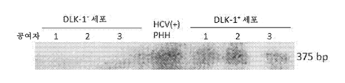
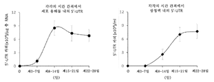
도 1. 인간의 지방-유래 DLK -1 + 줄기세포는 생체 내에서 HCV에 의해 표 적화된다. (a) 3명의 HCV(+) 개체 (표 1)의 지방 조직을 수술 상처로부터 수거하고, HCV-특이적 5'-UTR (223 bp)의 RT-PCR을 위해 RNA를 추출하였다. 3명의 HCV(-) 개체로부터 수거한 지방 조직도 비교를 위해 병행하여 연구하였고, HCV(+) 혈청을 양성 대조군으로 사용하였다. (b) HCV(+) 개체의 지방 조직을 분쇄하고, 균질화하고, 부유물, 완충제 및 세포 펠렛으로 원심분리하였다. 세포 펠렛의 적혈구를 추가로 용해시켜 SVF를 수거하였다. 각각의 세포 집단으로부터 RNA를 추출하고, 5'-UTR에 대한 RT-PCR을 실시하였다. 바이러스 전사체는 SVF 세포에서 검출되었지만, 부유물에서는 검출되지 않았다 (왼쪽 패널). SVF 세포를 DLK-1- 및 DLK-1+ 세포로 추가로 면역-분리하고, RNA를 RT-PCR을 위해 별개로 추출하였다. 바이러스 전사체는 DLK-1+ 세포에는 존재하지만, DLK-1- 세포에는 존재하지 않았다 (오른쪽 패널). 데이터는 3명의 HCV(+) 공여자로부터의 세포를 사용한 3회의 실험을 나타낸다. (c) 3명의 HCV 감염 개체부터 단리된 DLK-1- 및 DLK-1+ 세포의 RNA를 HCV 음성 가닥 RNA의 RT-PCR을 위해 추출하였다. HCV(+) 개체로부터 단리된 1차 인간 간세포 (PHH)를 양성 대조군으로 사용하였다. (d) HCV(-) 및 HCV(+) 개체로부터 단리된 DLK-1- (패널 b & e) 및 DLK-1+ 세포 (패널 c & f)를 바이러스 NS5에 대한 면역세포화학 (c에서 갈색 라벨) 및 헤마톡실린 염색 (핵에서 파란색 라벨)을 위해 세포원심분리 (cytospin) 슬라이드에 원심분리하였다. 마우스 IgG1 대조군 항체 (패널 a & d)를 사용한 미분획화된 SVF 세포의 염색을 음성 대조군으로 사용하였다. 데이터는 3명의 HCV(+) 및 3명의 HCV(-) 공여자로부터의 샘플을 나타낸다. (e) 동일한 섹션 상의 HCV(+) 또는 HCV(-) 개체로부터의 지방 조직을 DLK-1 (적색 라벨, 패널 b, c, h의 화살표) 및 바이러스 NS5 (갈색 라벨, 패널 e & f의 화살표) 및 헤마톡실린 (파란색 라벨, 패널 d-f 및 i-j). HCV(+) 지방 조직에서, DLK-1+ 세포 (적색 라벨, 화살표, 패널 b & c)는 NS5 Ag (갈색 라벨, 화살표, 패널 e & f)를 동시 발현하였다. HCV(-) 지방 조직에서, DLK-1+ 세포 (패널 h, 화살표)는 NS5를 발현하지 않았다 (패널 j, 화살표; h vs j). 토끼 IgG (패널 a & g) 또는 마우스 IgG1 (패널 d & i)을 이용한 염색을 음성 대조군으로 사용하였다. 패널 b, e 및 c, f는 별개의 공여자로부터의 것이었다. 데이터는 3명의 HCV(+) 및 3명의 HCV(-) 공여자를 나타낸다. (f) 4명의 HCV(+) 개체의 지방 조직으로부터 단리된 DLK-1+ 세포를 49일 동안 생체 외에서 배양하고, 상청액을 5'-UTR의 qRT-PCR을 위해 7일마다 수집하였다. 데이터는 각각의 시점에서 3회 반복 시험의 평균 ± 표준 편차로 나타내었다.
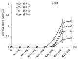
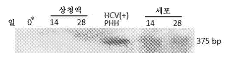
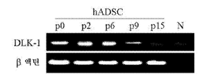
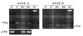
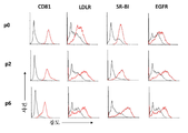
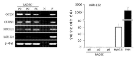
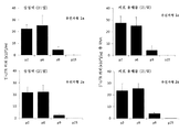
도 2. 비처리 (naive) DLK -1 + hADSC는 시험관 내에서 혈청 매개 HCV 감염을 허용한다. (a) 1차 인간 간세포 (PHH)를 인간 HCV(-) 공여자로부터 단리하고, HCVser (레인 "+", 좌측 패널) 또는 HCV(-) 대조군 혈청 (레인 "-", 좌측)으로 펄싱하기 전에 3일 동안 접시에 두었다. 3시간 펄싱한 후, PHH를 추가로 5일 동안 배양하고, RNA를 5'-UTR의 RT-PCR을 위해 추출하였다 (왼쪽 패널). 이와 병행하여, HCV(-) 공여자로부터 단리된 p-3 또는 p-4 DLK-1+ hADSC를 7일마다 배지 교환을 통해 0.2 moi로 HCVser에 의한 현탁액에 펄싱하였다. 지시된 시점에서, 상청액 및 세포를 별개로 수거하고, RNA를 5'-UTR의 RT-PCR을 위해 추출하였다. 데이터는 10회의 실험을 나타낸다. HCV(+) 혈청의 RNA는 또한 대조군으로서 RT-PCR을 위해 추출하였다. (b) d14 및 d28 HCVser-1b 감염된 hADSC의 상청액 및 세포를 수집하고, HCV-특이적 음성 가닥 RNA의 RT-PCR을 위해 RNA를 추출하였다. 데이터는 4회의 실험을 나타낸다. *: HCVser 감염 후 세척 1시간 후에 수거한 제0일 배양 상청액. (c) HCVser-1b 또는 HCV(-) 대조군 혈청에 의해 펄싱된 d14 hADSC를 동일한 섹션 상의 토끼 항-인간 DLK-1 항체 (적색 라벨, 패널 b & f), 이어서 마우스 항-NS5 항체 (패널 d & h, d의 갈색 라벨) 및 헤마톡실린 (파란색 라벨, c, d, g 및 h)으로 3번 염색하기 위해 세포원심분리 슬라이드에 원심분리하였다. HCVser에 의해 감염된 DLK-1+ hADSC는 NS5A (패널 b 대 d)를 포함하는 반면, HCV(-) 대조군 혈청에 의해 펄싱된 세포는 NS5 없이 DLK-1을 발현하였다 (패널 f 대 h). 토끼 IgG 및 마우스 IgG1을 사용한 염색을 음성 대조군으로 사용하였다 (각각 패널 a & e, 및 c & g). 결과는 4가지 실험을 나타낸다. (d) d14 (패널 b & c) 및 d21 (패널 e & f) HCVser-1b 감염된 hADSC의 투과 전자현미경 사진. HCV(-) 대조군 혈청에 노출된 D14 및 D21 세포는 패널 a 및 d에 나타났다. 패널 b-f의 삽입도는 노란색 정사각형 또는 노란색 화살표의 확대도이다. 패널 b, c, e & f의 삽입도에 있는 흰색 화살표는 바이러스 입자를 나타내고, 패널 d의 삽입도에 있는 흰색 화살표는 감염되지 않은 hADSC의 양파 모양 막 구조를 나타낸다. 데이터는 HCVser-1b에 의한 감염으로부터의 2회 실험 및 HCVser-2a에 의한 감염으로부터의 1회 실험을 나타낸다. (e) HCVser-1b 감염된 hADSC의 세포 용해물 (좌측 패널) 및 상청액 (우측 패널)에서 바이러스 5'-UTR의 qRT-PCR을 7일마다 수집하였다. 데이터는 3회 실험으로부터 평균 ± 표준 편차로 표현한다. (f) 바이러스 5'-UTR 카피 (qRT-PCR에 의한)을 1 내지 4주 동안 연속 배양된 HCVser-1b 감염된 hADSC의 상청액에서 정량하였다. 데이터는 4회 실험으로부터의 평균 ± 표준 편차로 표현된다.
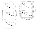
도 3. HCVser 감염은 세포에 대해서는 공여자 비특이적이고 바이러스에 대해서는 교차 유전자형이고, miR-122를 제외한 다양한 숙주 인자에 의해 매개된다. (a) "공여자 1"의 P-2 hADSC를 HCVser-1b로 감염시키고, 21일 상청액 ("HCVadsc(1)"로 표시됨)을 0.22 ㎛ 세공 필터를 통해 여과하고, 그의 21일 상청액 ("HCVadsc(2)"로 표시됨)이 "공여자 3"의 p-6 hADSC를 감염시키는 데 사용된 "공여자 2"의 p-3 hADSC를 감염시키는 데 사용하였다. 21일 상청액 중의 바이러스 카피수를 qRT-PCR에 의해 정량하였다. 데이터는 감염된 hADSC의 각각의 배치의 3회 실험으로부터 평균 ± SD로 표현된다. (b) p2, p6, p9 및 p15의 hADSC를 HCVser-1b로 감염시키고, 21일 상청액 (왼쪽) 및 세포 용해물 (오른쪽)의 바이러스 전사체를 qRT-PCR로 결정하였다. 데이터는 3회의 독립적인 실험으로부터의 평균 ± 표준 편차로 표현된다. (c) p0, p2, p6, p9 및 p15 hADSC의 DLK-1 발현에 대한 RT-PCR. 모든 세포 배치는 동일한 공여자의 것이었다. N: 음성 대조군으로서 역전사효소 없음. 데이터는 3명의 공여자로부터의 세포를 사용하는 실험을 나타낸다. (d) p5의 hADSC를 혼합된 유전자형 2a+2b의 HCVser로 감염시키고, 연속적으로 배양하였다. d21 및 d56에서, 유전자형 특이적 코어 항원 (유전자형 2a는 174 bp, 왼쪽, 및 유전자형 2b는 123 bp, 오른쪽)을 코딩하는 mRNA를 검출하기 위한 RT-PCR을 위해 세포를 수집하였다. N: 음성 대조군으로서 HCV(-) 대조군 혈청에 의해 펄싱된 d21 세포. P: 양성 대조군으로서의 HCV(+) 혈청 자체 (혼합된 유전자형 2a+2b). M: 마커. 데이터는 2개의 HCVser 유전자형 2a+2b 감염된 공여자로부터의 혈청을 사용한 실험을 나타낸다. (e) p0, p2 및 p6 hADSC의 CD81, LDL-R, SR-B1 및 EGFR의 표면 발현을 위한 유동 세포 계측법. 흑색 선: 이소형 대조군 Ab. 적색 선: 항-CD81, -LDL-R, -SR-B1 또는 -EGFR Ab. 데이터는 세포의 각각의 계대배양을 위한 3회의 실험을 나타낸다. (f) p0, p2 및 p6 hADSC의 오클루딘 (OCLN), 클라우딘-1 (CLDN1), NPC1L1 및 miR-122의 발현에 대한 RT-PCR (왼쪽 패널). 데이터는 3명의 공여자로부터의 hADSC를 나타낸다. N: 음성 대조군으로서의 역전사효소 없음. P: 양성 대조군으로서 HCV(-) 개체로부터 단리된 1차 인간 간세포. 또한, miR-122 발현은 Huh7.5 또는 HCV(-) 1차 인간 간세포 (PHH, 오른쪽 패널)와 비교하여 p0, p2, p6 hADSC에서 qRT-PCR에 의해 결정되었다. 데이터는 각각 3명의 상이한 공여자로부터의 hADSC 및 PHH를 사용하여 평균 ± SD로 p0 hADSC에 대한 상대적인 발현 수준으로 표현된다. (g) HCVser-1b에 의한 펄스 전 1시간 동안 나타낸 농도의 차단 모노클로날 항-CD81 (클론 JS-81), 항-LDL-R (클론 C7), 항-EGFR Ab (클론 LA1) 또는 폴리클로날 항-SR-B1 Ab로 p2 hADSC를 전처리하였다. Apo-E를 차단하기 위해, 나타낸 농도의 항-ApoE 항체 (클론 E6D10)를 HCVser에 첨가하고, hADSC와 3시간 동안 인큐베이팅하였다. 21일 상청액의 바이러스 5'-UTR 전사체를 정량하였다. 이소형: 이소형 대조군 항체 (100 μg/ml) 처리. 데이터는 3회 실험으로부터의 억제 비율의 평균 ± SD (이소형 Ab 처리에 대한)로 표현된다. 각각의 처리 후의 세포 생존율을 트리판 블루 (trypan blue)로 평가하였다. (h) p2 hADSC를 OCLN, CLDN1, NPC1L1 또는 DGAT-1에 특이적인 siRNA로 형질감염시켰다. 스크램블드 (scrambled) RNA를 사용한 형질감염을 대조군으로 사용하였다. 형질감염 48시간 후, 세포를 세척하고, HCVser-1b로 감염시키고, 21일 상청액 중의 바이러스 5'-UTR 카피를 정량하였다. OCLN 및 CLDN1의 낙다운 (knock-down)은 동일한 실험에서 수행되었지만, NPC1L1 또는 DGAT-1의 낙다운은 별개의 실험에서 수행되었다. 각각의 형질감염 후 트리판 블루 배제 검정에 의해 세포 생존율을 결정하였고, 스크램블된 siRNA에 의한 형질감염에 비해 유의하게 변하지 않았다. 데이터는 3회의 독립적인 실험의 평균 ± SD로 표현된다. (i) 부착성 p4-p5 hADSC를 HCVser-1b로 감염시키고, 리바비린, 텔라프레비르 및 시클로스포린 A의 등급이 매겨진 용량을 연속적으로 21일 동안 배양물에 첨가하였다. hADSC는 또한 HCVser-1b에 노출되기 전에 16시간 동안 IFNα의 등급이 매겨진 농도와 함께 인큐베이팅하였다. 세포 생존율은 각각의 처리 후에 트리판 블루에 의해 평가되었다. 데이터는 3회의 독립적인 실험의 평균 ± SD로 표현된다.
도 4. HCVadsc는 JFH1/HCVcc와 물리적 특성이 다르고, 1차 인간 간세포에 감염성이다. (a) HCVadsc (유전자형 2a)를 21일 HCVser-2a 감염된 hADSC 상청액으로부터 수거하고, 10 내지 40% 아이오딕사놀 내에서 평형 원심분리에 의해 밀도 구배 검정을 수행하였다. 비교를 위해, HCVser (유전자형 2a) 및 JFH1/HCVcc의 밀도 구배 검정도 동시에 수행하였다. 데이터는 5개 실험을 나타낸다. (b) HCVcc 분획 13은 바이러스 카피당 중량 (ng)으로 표현될 때 가장 적은 양의 HDL 및 LDL/VLDL을 가졌다. 데이터는 3명의 공여자로부터의 HCVser 및 그의 대응하는 HCVadsc를 나타낸다. (c) ApoE 및 B의 측정은 HCVser의 주요 분획이 가장 높은 ApoE 함량을 갖고, HCVadsc가 뒤따르는 반면, HCVcc 분획 13은 간신히 검출가능한 ApoE를 가짐을 입증하였다 (왼쪽 패널). 이와 대조적으로, ApoB는 HCVser 분획 2에서만 검출되었다 (우측 패널). 데이터는 HCVser (유전자형 2a) 및 그의 대응하는 HCVadsc (즉, HCVadsc는 HCVser-감염된 hADSC에 의해 생성됨)를 사용한 실험의 것이고, 3명의 HCVser 공여자를 나타낸다. (d) 6 cm 페트리 접시에 있는 P2 hADSC를 HCVser (유전자형 2a) 또는 HCVcc에 노출시켰다. 감염된 hADSC를 14일 또는 21일 동안 연속적으로 (배지 교체 없이) 배양하고, 배양 종료시에 상청액 (좌측 패널) 및 세포 용해물 (우측 패널) 내의 5'-UTR 카피수를 qRT-PCR로 정량하였다. 결과는 HCVcc가 hADSC에서 효율적으로 감염/복제하지 않음을 보여주었다. 데이터는 3회 실험의 평균 ± SD로 나타내었다. (e) HCVcc 감염된 또는 감염되지 않은 Huh7.5 배양액의 상청액 내의 및 HCVcc- 또는 HCVser-감염된 hADSC의 21일 상청액 내의 바이러스 5'-UTR에 대한 RT-PCR. 데이터는 3회 실험을 나타낸다. (f) HCV(-) 환자로부터 단리된 PHH는 부착을 허용하기 위해 3일간 방치한 후, HCVser-1b 또는 HCVser-2b에 감염된 hADSC에서 수집한 3h 내지 21일의 상청액에 노출시키거나, 대조군 혈청으로 펄싱하였다. 감염 5일 후에, 5'-UTR의 RT-PCR을 위해 세포 RNA를 추출하였다. HCVser-1b 자체를 양성 대조군으로 사용하였다. 데이터는 3회 실험을 나타낸다. (g) 3명의 HCV(-) 공여자 (공여자 1, 2 및 3)로부터 단리된 1x10⁴/웰 PHH를 6-웰 플레이트에 3일 동안 플레이팅하여 세포 부착을 허용하고, 제4일에 PHH를 HCVser-1b의 3개의 상이한 배치 (3명의 별개의 공여자로부터 수집) 중의 하나로 별개로 감염시켰다. HCVser-1b는 상응하는 HCVadsc를 생성하는 데 사용되었고, 이 HCVadsc는 또한 PHH를 병행하여 감염시키는데 사용되었다. HCVser 및 그의 대응하는 HCVadsc는 동일한 PHH 배치를 감염시키기 위해 쌍을 이루었다. 감염 5일 후에, PHH 상청액을 수집하고, 5'-UTR 카피를 정량하였다. HCV(-) 대조군 혈청 (또한 3명의 다른 공여자로부터의)에 대한 PHH의 노출을 음성 대조군으로 사용하였다. 데이터는 각각의 감염의 3회 실험의 평균 ± SD로 표현된다.
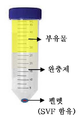
도 5. 원심분리에 의한 인간 지방 조직의 균질물의 상이한 층으로의 분리. 문헌 9에 기재된 바와 같이, HCV(+) 개체의 지방 조직을 원심분리하고, 부유물, 완충제 및 세포 펠렛 (왼쪽 패널) 층으로 분리하였다. 부유 집단은 성숙 지방세포를 포함한다. 세포 펠렛을 수집하고, RBC 용해 완충제로 처리하여 적혈구를 용해시켜 간질 혈관 분획 (SVF) 세포를 수거하고, SVF 세포를 1차 (계대배양-0) hADSC로 간주될 수 있는 DLK-1+ 인간 지방 생성 줄기세포 (hADSC)의 면역 선택에 적용하였다.
도 6. DLK - 1에 대한 선택된 분획 및 RT- PCR의 순도. (a) DLK-1+ (왼쪽) 및 DLK-1- (오른쪽) 분획 내의 DLK-1+ 세포의 비율은 MoFlo XDP 유동 세포 계측기 (벡톤 코울터 (Becton Coulter), 미국 캘리포니아주)를 사용하여 결정하고, 서브밋 (Submit) 소프트웨어를 사용하여 분석하였다. DLK-1+ 및 DLK-1- 분획의 99.5% 자가형광의 제거를 각각의 분획에 양성으로 염색된 세포를 결정하기 위한 게이트를 설정하기 위해 사용되었다. 이소형 대조군 항체 (토끼 IgG)와 함께 인큐베이팅된 비분획화된 SVF 세포를 사용하여 유사한 결과를 얻었다. 데이터는 6회 실험을 나타낸다. (b) 미분획화된 SVF 세포로부터 RNA를 추출하고, DLK-1- 및 DLK-1+ 세포를 선택하고10, DLK-1에 대한 RT-PCR (문헌 11에 기재된 프라이머)에 적용하였다. DLK-1- 세포는 DLK-1+ 세포에서의 풍부한 발현과는 달리 DLK-1 mRNA를 거의 발현하지 않았다. 데이터는 4회 실험을 나타낸다.
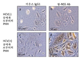
도 7. HCV 감염된 1차 인간 간세포 (PHH)는 NS5 항원을 발현하였다. PHH는 HCV(-) 또는 HCV(+) 공여자 (각각 패널 a & b, 및 c & d)로부터 수거하고, 배양을 위해 콜라겐-I로 코팅된 웰에 씨딩하였다. 대조 실험으로서, 본 발명자들은 HCV(+) 또는 HCV(-) 공여자로부터 새로 단리된 1차 인간 간세포 (PHH; 1x10⁴ 세포/웰의 씨딩 수)에 대한 NS5 염색의 특이성을 조사하고, 아르기닌이 없는 윌리암스 (Williams) E 배지 (인비트로겐 (Invitrogen), 미국 캘리포니아주)에서 배양하였다. 제9일에, 세포를 세척하고, 세포원심분리 슬라이드 (참조 "방법") 상의 세포 염색에 대해 설명한 것과 유사한 방법을 사용하여 웰에서 면역세포화학에 적용하였다. 데이터는 3명의 HCV(+) 및 3명의 HCV(-) 공여자로부터의 PHH를 사용하는 3회 실험을 나타낸다.
주: 역사적으로 감염된 간 조직에서 HCV 항원을 검출하기가 어려웠다. 그러나, 단리된 세포의 염색은 발색 시간이 잘 조절되는 한, 간 조직 염색과 같이 비특이적인 것으로 보이지 않는다 ("방법" 참조).
도 8. HCV에 감염된 개체의 지방 조직 내의 DLK -1 + 세포는 HCV NS5 항원을 동시 발현한다. 지방 조직을 HCV 감염 개체 (표 1)로부터 수거하고, 방법에서 설명되는 바와 같이 동일한 섹션 상에서 항-DLK-1 Ab (적색 라벨, 백색 화살표, 패널 a 및 b), 이어서 항-NS5 Ab (갈색 라벨, 백색 화살표, 패널 c & d) 및 헤마톡실린 (패널 c & d의 파란색 라벨)으로 면역염색하였다. 검사된 HCV(+) 지방 조직의 모든 섹션에서, 약 0-4개의 DLK-1+NS5+ 세포가 고배율 영역 (400X)에서 검출될 수 있었다. 패널 a 및 c는 공여자 2로부터, 패널 b 및 d는 공여자 3으로부터 얻은 것이었다. 이소형 대조군 항체를 사용한 염색의 영상은 도 1e에 제시된 것과 유사하였다.
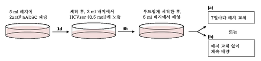
도 9. 현탁액에서 hADSC를 감염시키기 위한 프로토콜. hADSC는 HCV(-) 개체로부터 준비하고, 문헌 12, 13에 기재된 바와 같이 배양물에서 계대배양하였다. 계대배양 2 내지 6의 hADSC를 0.2 moi (1x10⁵ HCV 5'-UTR 카피수 대 5x10⁵ hADSC 세포)로 HCVser (표 2)에 노출시키고, 신선한 배지로 HCV(+) 혈청을 희석함으로써 조정하였다. 3시간 후, 세포를 PBS로 5회 세척한 후, 6-ml의 신선한 배지에서 배양하였다. 제7, 14, 21 및 28일에 상청액 및 세포 용해물을 수거하고, 5'-UTR의 RT-PCR을 위해 RNA를 추출하였다.
도 10. HCVser를 이용한, 플라스틱에 부착된 hADSC의 감염. hADSC를 세포 부착을 허용하기 위해 1일 동안 6-cm 페트리 접시에 플레이팅하였다. 이어서, HCVser를 최종 부피 2 ml 배지에 첨가하여 0.5 moi (1x10⁵ 5'-UTR 카피수 대 2x10⁵ hADSC 세포)에서 3시간 동안 세포를 인큐베이팅하였다. 부드럽게 세척한 후, HCVser에 감염된 hADSC를 7일마다 배지를 교체하거나 하지 않으면서 5 ml의 신선한 배지에서 배양하였다 (각각 경로 a 또는 b).
도 11. p2 및 p6 hADSC의 바이러스 복제 효율은 p9 및 p15 세포의 바이러스 복제 효율보다 우수하다. hADSC는 HCVser-1a 또는 -2a에 현탁액에서 노출되었다. 감염 후, 21일 상청액 및 세포 용해물 (연속 배양액)의 바이러스 5'-UTR 전사체를 qRT-PCR로 결정하였다. 결과는 p9 및 p15 세포가 p2 및 p6 세포와 대조적으로 세포 용해물 및 상청액에서 상당히 더 적은 바이러스 카피를 갖는다는 것을 보여주었다. 데이터는 3회 실험의 평균 ± SD로 표현된다.
도 12. CD81, LDLR , SR-B1 및 EGFR의 차단 및 ApoE의 중화는 21일 HCVser 감염 hADSC 배양물의 세포 용해물에서 바이러스 복제를 감소시켰다. P-2 hADSC는 1시간 동안 CD81 (클론 JS-81), LDL-R (클론 C7), EGFR (클론 LA1)에 대한 모노클로날 Ab, 또는 SR-B1에 대한 폴리클로날 Ab의 등급이 매겨진 용량으로 전처리한 후, HCVser-1b로 펄싱하기 전에 세척하였다. ApoE 차단을 위해, 다양한 농도의 항-ApoE 항체 (클론 E6D10)를 hADSC와 함께 3시간 동안 인큐베이팅을 위해 사용하기 전에, 문헌 14에 기재된 바와 같이 실온에서 1시간 동안 HCV(+) 혈청에 첨가하였다. 상청액에서의 측정과 일치하게 (도 3g), HCV(+) 혈청에서 hADSC의 CD81, LDL-R, SR-B1, EGFR의 차단 및 ApoE의 중화는 세포 용해물 내의 바이러스 전사체의 양을 용량 의존적인 방식으로 유의하게 감소시켰다. 한편, 항체 자체의 처리는 hADSC 생존율에 유의한 영향을 주지 않았다. 데이터는 각각의 처리에 대해 3회 실험의 평균 ± SD로 표현된다.
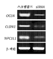
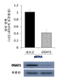
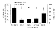
도 13. 오클루딘 ( OCLN ), 클라우딘 -1 ( CLDN1 ), NPC1L1 또는 DGAT -1의 낙다운 후의 21일 세포 용해물에서 siRNA 형질감염 후 RT- PCR 및 바이러스 카피수. (a) 오클루딘, 클라우딘-1 또는 NPC1L1의 낙다운은 문헌 15, 16에 기재된 바와 같이 수행하였고, RT-PCR은 문헌 16, 17에 기재된 바와 같이 수행하였다. OCLN 및 CLDN1의 낙다운은 동일한 실험에서 수행되었지만, NPC1L1의 낙다운은 별개의 실험에서 수행되었다. 데이터는 3회 실험을 나타낸다. (b) DGAT1에 대한 siRNA 프로브는 앰비온 (Ambion)의 미리 설계된 siRNA (카탈로그 번호 11782 및 11784)이었다. DGAT1에 특이적인 스크램블드 RNA 및 siRNA의 형질감염, qRT-PCR 및 웨스턴 블롯 분석은 문헌 18에 기재된 바와 같이 수행되었다. 데이터는 4회 실험으로부터 평균 ± SD로 표현되고, 18S rRNA로 표준화된 상대 비율로서 제시된다. (c) 낙다운 후, hADSC를 HCVser-1b에 노출시키고, qRT-PCR에 의해 21일 세포 용해물의 5'-UTR 카피수를 결정하였다. siRNA 형질감염 자체는 형질감염 48시간 후에 트리판 블루 배제 시험에 의해 결정될 때 스크램블드 siRNA 형질감염에 비해 세포 생존율에 유의한 영향을 미치지 않았다. 명백하게, DGAT1 낙다운은 세포보다 상청액에서 더 현저한 억제 효과를 나타냈다 (패널 C 대도 3h). 이것은 DGAT1 낙다운이 세포 내 복제 및 방출을 모두 손상시킨다는 발견과 일치하지만, 바이러스 방출이 더 현저하게 영향을 받는 것으로 보였다19. 데이터는 4회 실험으로부터 평균 ± SD로 표현되고, 스크램블드 대조군에 의한 형질감염에 대한 상대 비로서 제시된다.
도 14. hADSC에서 시클로필린 A 발현을 위한 RT- PCR . 시클로필린 A (Cyp A)에 대한 RT-PCR은 문헌 20에 기재된 바와 같이 p0-p6 hADSC에서 수행하였다. 비처리 Huh7.5 세포를 양성 대조군으로 사용하였다. 데이터는 4회 실험을 나타낸다.
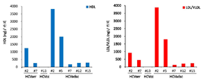
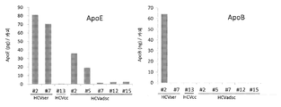
도 15. HCVser , JFH1 / HCVcc 및 HCVadsc의 주요 분획 (밀도 구배에 의한)의 총 지질 함량. HDL 및 LDL/VLDL의 수준은 HDL 및 LDL/VLDL 콜레스테롤 검정 키트 (압캄 (Abcam))에 의해 검출되었다. ApoB 및 ApoE의 양은 콴티킨(Quantikine)® ELISA 인간 ApoB 면역검정 키트 및 콴티킨® ELISA 인간 ApoE 면역검정 키트 (R&D)에 의해 제조사의 지시에 따라 측정되었다. 상이한 분획물에서 HDL 및 LDL/VLDL, ApoB 및 ApoE의 양을 또한 카피당 중량으로 표준화하였다. (a) HCVser (특히 분획 2)는 HCVcc 또는 HCVadsc보다 더 많은 HDL 및 LDL/VLDL의 "총량"을 가졌다. (b) 및 (C) HCVser 및 HCVadsc의 또 다른 두 배치의 분석은 지질 및 ApoE/B 함량의 동일한 패턴을 보여주었다.
도 16. JFH1 / HCVcc는 Huh7 .5를 효율적으로 감염시킨다. 4x10⁵ Huh7.5 세포/웰을 6-웰 플레이트에 밤새 플레이팅하였다. 1일 후, 세포를 세척하고, HCVcc 바이러스 접종물 (0.5 moi)과 함께 3시간 동안 인큐베이팅하였다. 세척 후, 세포를 10% 소 태아 혈청 (FBS) 및 1% 비필수 아미노산이 보충된 DMEM에서 배양하였다. 감염 후 제3일에, 상청액을 수거하고, 5'-UTR 전사체의 qRT-PCR을 위한 RNA를 추출하였다. 바이러스 접종물에 노출시키지 않은 비처리 Huh7.5 세포를 음성 대조군으로 사용하였다. 데이터는 3회 실험의 평균 ± SD로 표현된다. 이 실험은 hADSC의 감염에 사용된 HCVcc 접종물의 감염성을 확인하였다 (도 4d).The above summary and the following detailed description are better understood when read in conjunction with the accompanying drawings, which are included by way of example and not of limitation.
Figure 1. Human fat-derived stem cells are DLK -1 + table optimization by HCV in vivo. (a) Adipose tissue from three HCV (+) individuals (Table 1) was harvested from the surgical wound and RNA was extracted for RT-PCR of HCV-specific 5'-UTR (223 bp). HCV (+) sera were used as a positive control for the comparison of lipid profiles collected from three HCV (-) individuals. (b) The adipose tissue of the HCV (+) individual was pulverized, homogenized, and centrifuged with suspensions, buffer and cell pellets. SVF was collected by further dissolving the red blood cells of the cell pellet. RNA was extracted from each cell population and subjected to RT-PCR for 5'-UTR. Virus transcripts were detected in SVF cells, but not in suspension (left panel). SVF cells were further immunoprecipitated into DLK-1 - and DLK-1 + cells and RNA was extracted separately for RT-PCR. Viral transcripts are present, DLK-1 + cells, however, DLK-1 - did not exist in cells (right panel). Data represent three experiments using cells from three HCV (+) donors. (c) RNAs of isolated DLK-1 - and DLK-1 + cells from three HCV infected individuals were extracted for RT-PCR of HCV negative strand RNA. Primary human hepatocytes (PHH) isolated from HCV (+) individuals were used as positive control. (d) HCV (-) and HCV (+) isolated from objects DLK-1 - (panel b & e), and DLK-1 + cells Immunocytochemistry for the (panel c & f) with a virus NS5 (brown c Labeling) and hematoxylin staining (blue labeling in the nuclei) on a cytospin slide. Staining of undifferentiated SVF cells using mouse IgG1 control antibody (panel a & d) was used as negative control. Data represent samples from three HCV (+) and three HCV (-) donors. (e) Adipose tissue from HCV (+) or HCV (-) individuals on the same section was labeled with DLK-1 (red label, arrows in panels b, c and h) and virus NS5 ) And hematoxylin (blue label, panels df and ij). In HCV (+) adipose tissue, DLK-1 + cells (red label, arrow, panel b & c) co-expressed NS5 Ag (brown label, arrow, panel e & f). In HCV (-) adipose tissue, DLK-1 + cells (panel h, arrow) did not express NS5 (panel j, arrow; h vs j). Staining with rabbit IgG (panel a & g) or mouse IgG1 (panel d & i) was used as negative control. Panels b, e and c, f were from separate donors. Data represent three HCV (+) and three HCV (-) donors. (f) DLK-1 + cells isolated from adipose tissue of 4 HCV (+) individuals were cultured in vitro for 49 days and supernatants were collected every 7 days for qRT-PCR of 5'-UTR. Data are presented as mean ± standard deviation of three replicate tests at each time point.
Figure 2. untreated (naive) DLK -1 + hADSC allows serum-mediated HCV infection in vitro . (a) Primary human hepatic cells (PHH) were isolated from human HCV (-) donors and incubated before pulsing with HCVser (lane "+", left panel) or HCV (-) control serum It was placed on a plate for 3 days. After pulsing for 3 hours, PHH was incubated for an additional 5 days and RNA was extracted for RT-PCR of 5'-UTR (left panel). In parallel, isolated p-3 or p-4 DLK-1 + hADSCs from HCV (-) donors were pulsed into suspension by HCVser at 0.2 moi every 7 days via medium exchange. At the indicated time points, supernatant and cells were collected separately and RNA was extracted for RT-PCR of 5'-UTR. Data represent 10 experiments. RNA from HCV (+) sera was also extracted for RT-PCR as a control. (b) Supernatants and cells of d14 and d28 HCVser-lb infected hADSCs were collected and RNA was extracted for RT-PCR of HCV-specific negative strand RNA. Data represent four experiments. * :
Figure 3. HCVser infection is nonspecific donors for cells and cross-genotypes for viruses and is mediated by a variety of host factors except miR-122. (a) P-2 hADSCs of "
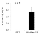
Figure 4. HCVadsc is different in physical properties from JFH1 / HCVcc and is infectious to primary human hepatocytes. (a) HCVadsc (genotype 2a) was collected from 21 day HCVser-2a infected hADSC supernatants and density gradient assay was performed by equilibrium centrifugation in 10-40% iodixanol. For comparison, density gradient assays of HCVser (genotype 2a) and JFHl / HCVcc were also performed simultaneously. The data represent five experiments. (b)
Figure 5. Separation of homogenates of human adipose tissue into different layers by centrifugation . As described in
Figure 6. Purity of selected fractions and RT- PCR for DLK - 1 . (a) The ratio of DLK-1 + cells in the DLK-1 + (left) and DLK-1 - (right) fractions was determined using a MoFlo XDP flow cytometer (Becton Coulter, And analyzed using submit software. The removal of 99.5% autofluorescence of the DLK-1 + and DLK-1 - fractions was used to set the gates to determine cells stained positively in each fraction. Similar results were obtained using unfractionated SVF cells incubated with an isotype control antibody (rabbit IgG). Data represent six experiments. (b) RNA was extracted from undifferentiated SVF cells and DLK-1 - and DLK-1 + cells were selected and applied to 10 , RT-PCR for DLK-1 (the primer described in document 11). DLK-1 - cells showed almost no DLK-1 mRNA, unlike the rich expression in DLK-1 + cells. Data represent four experiments.
Figure 7. HCV-infected primary human hepatic cells (PHH) expressed NS5 antigen. PHH was collected from HCV (-) or HCV (+) donors (panels a & b, and c & d respectively) and seeded into collagen-I coated wells for culture. As a control experiment, we investigated the specificity of NS5 staining for HCV (+) or HCV (-) donors to newly isolated primary human hepatocytes (PHH; seeding number of 1 x 10 4 cells / well) And cultured in Williams E medium (Invitrogen, Calif., USA). On
Note: It has been difficult to detect HCV antigen in historically infected liver tissue. However, the staining of isolated cells does not appear to be nonspecific, such as liver tissue staining, as long as the color development time is well controlled (see "Methods ").
Figure 8. DLK- 1 + cells in adipose tissue of HCV- infected individuals co- express the HCV NS5 antigen . Adipose tissue was collected from HCV infected individuals (Table 1) and labeled with anti-DLK-1 Ab (red label, white arrow, panels a and b), followed by anti-NS5 Ab , White arrow, panel c & d) and hematoxylin (blue label of panel c & d). In all sections of the examined HCV (+) adipose tissue, about 0-4 DLK-1 + NS5 + cells could be detected in the high magnification region (400X). Panels a and c were from
Figure 9. Protocol for infecting hADSCs in suspension . hADSCs were prepared from HCV (-) individuals and subcultured in culture as described in
Figure 10. Plasma- attached hADSCs using HCVser infection. hADSCs were plated in 6-cm Petri dishes for 1 day to allow for cell attachment. Is then added to the HCVser
11. The viral replication efficiency of p2 and p6 hADSCs is superior to that of p9 and p15 cells . hADSCs were exposed in suspension to HCVser-1a or -2a. After infection, the virus 5'-UTR transcripts of supernatant and cell lysate (continuous culture) for 21 days were determined by qRT-PCR. The results showed that p9 and p15 cells had significantly fewer viral copies in cell lysates and supernatants as opposed to p2 and p6 cells. Data are expressed as mean ± SD of three experiments.
12. Blocking of CD81, LDLR , SR-B1 and EGFR and neutralization of ApoE reduced viral replication in the cell lysates of 21 day HCVser infected hADSC cultures . P-2 hADSCs were stained for monoclonal Ab for CD81 (clone JS-81), LDL-R (clone C7), EGFR (clone LA1) or polyclonal Ab for SR- And washed before pulsing with HCVser-1b. For ApoE blockade, various concentrations of anti-ApoE antibody (clone E6D10) were added to HCV (+) serum for 1 hour at room temperature as described in
Figure 13. Eau Ruthin (OCLN), keulrawoodin -1 (CLDN1), NPC1L1 or DGAT -1 camel down after 21 days from the cell lysate for the RT- PCR after siRNA transfection and viral copy number. (a) Falling down of occludin, claudin-1 or NPC1L1 was performed as described in
Figure 14. In hADSC RT- PCR for cyclophilin A expression . RT-PCR for cyclophilin A (Cyp A) was performed in p0-p6 hADSC as described in
Figure 15. Total lipid content of major fractions (by density gradient ) of HCVser , JFH1 / HCVcc and HCVadsc . The levels of HDL and LDL / VLDL were detected by HDL and LDL / VLDL cholesterol assay kit (Abcam). The amounts of ApoB and ApoE were measured according to the manufacturer's instructions by Quantikine® ELISA human ApoB immunoassay kit and Quantkin® ELISA human ApoE immunoassay kit (R & D). The amounts of HDL and LDL / VLDL, ApoB and ApoE in the different fractions were also normalized to the weight per copy. (a) HCVser (especially fraction 2) had more "total amount" of HDL and LDL / VLDL than HCVcc or HCVadsc. (b) and (C) Analysis of two other batches of HCVser and HCVadsc showed the same pattern of lipid and ApoE / B content.
Figure 16. JFH1 / HCVcc The Huh7 .5 thereby efficiently infection. 4 x 10⁵ Huh7.5 cells / well were plated in 6-well plates overnight. One day later, cells were washed and incubated with HCVcc virus inoculum (0.5 moi) for 3 hours. After washing, cells were cultured in DMEM supplemented with 10% fetal bovine serum (FBS) and 1% non-essential amino acids. On the third day after infection, the supernatant was collected and RNA for qRT-PCR of the 5'-UTR transcript was extracted. Untreated Huh7.5 cells not exposed to the virus inoculum were used as negative control. Data are expressed as mean ± SD of three experiments. This experiment confirmed the infectivity of the HCVcc inoculum used for infection of hADSC (Fig. 4d).
본 발명의 여러 측면은 예시를 위해 예시된 적용을 참조하여 아래에서 설명된다. 본 발명의 완전한 이해를 제공하기 위해 많은 구체적인 세부 사항, 관계 및 방법이 제시됨을 이해하여야 한다. 그러나, 관련 기술 분야의 통상의 기술자는 본 발명이 하나 이상의 구체적인 세부 사항 또는 다른 방법 없이 실시될 수 있음을 쉽게 인식할 것이다. 본 발명은 행위 또는 사건의 순서에 의해 제한되지 않고, 일부 행위는 상이한 순서로 및/또는 다른 행위 또는 사건과 동시에 발생할 수 있다. 또한, 본 발명에 따라 방법을 구현하기 위해 모든 예시된 행위 또는 사건이 요구되는 것은 아니다.Various aspects of the invention are described below with reference to an application illustrated for illustrative purposes. It should be understood that many specific details, relationships, and methods are set forth in order to provide a thorough understanding of the present invention. However, one of ordinary skill in the pertinent art will readily recognize that the present invention may be practiced without one or more of the specific details or with other methods. The invention is not limited by the order of acts or events, and some acts may occur in different orders and / or concurrently with other acts or events. Moreover, not all illustrated acts or events are required to implement a method in accordance with the present invention.
정의Justice
달리 정의되지 않는 한, 본원에서 사용되는 과학적 및 기술적 용어 및 명명법은 본 발명이 속하는 기술 분야의 통상의 기술자에 의해 일반적으로 이해되는 것과 동일한 의미를 갖는다. 일반적으로, 세포 배양, 감염, 분자생물학적 방법 등에 대한 절차는 관련 기술 분야에서 사용되는 일반적인 방법이다. 이러한 표준 기술은 참고 매뉴얼, 예를 들어 문헌 [Sambrook et al. (1989, Molecular Cloning - A Laboratory Manual, Cold Spring Harbor Laboratories)] 및 [Ausubel et al. (1994, Current Protocols in Molecular Biology, Wiley, New York)]에서 볼 수 있다.Unless defined otherwise, the scientific and technical terms and nomenclature used herein have the same meaning as commonly understood by one of ordinary skill in the art to which this invention belongs. In general, procedures for cell culture, infection, molecular biology and the like are common methods used in the related art. These standard techniques are described in reference manuals, for example in Sambrook et al. (1989, Molecular Cloning - A Laboratory Manual, Cold Spring Harbor Laboratories) and Ausubel et al. (1994, Current Protocols in Molecular Biology, Wiley, New York).
본원에서 사용되는 단수 형태 "a", "an" 및 "the"는 문맥상 명백하게 다르게 나타내지 않는 한, 복수 형태를 포함하는 것이 의도된다. 또한, "포함하는", "포함하다", "갖는", "갖다", "함께" 또는 이들의 변형된 용어가 상세한 설명 및/또는 청구범위에서 사용되는 정도로, 이들 용어는 용어 "포괄하는"과 유사한 방식으로 포괄적인 것이 의도된다.As used herein, the singular forms "a", "an" and "the" are intended to include the plural forms, unless the context clearly indicates otherwise. Also, to the extent that the terms "comprises", "includes", "having", "having", "together", or variations thereof are used in the detailed description and / And is intended to be comprehensive in a similar manner.
양, 시간 기간 등과 같은 측정가능한 값을 언급할 때 본원에서 사용되는 용어 "약"은 ±20% 또는 ±10%, 보다 바람직하게는 ±5%, 훨씬 더 바람직하게는 ±1%, 더욱 더 바람직하게는 ±0.1%의 변화를 포함하는 것을 의미하고, 이것은 상기 변화가 개시된 방법을 수행하기에 적합하기 때문이다.The term "about" as used herein when referring to a measurable value such as an amount, time period, etc. is ± 20% or ± 10%, more preferably ± 5%, even more preferably ± 1% Is meant to include a variation of < RTI ID = 0.0 > + 0.1%, < / RTI >
본원에서 사용되는 용어 "지방 조직"은 백색 지방, 황색 지방 또는 갈색 지방으로 구성된 1차 대사상 중요한 널리 분산된 기관을 규정한다. 지방 조직은 지방세포 및 간질을 갖는다. 지방 조직은 동물의 몸 전체에 걸쳐 발견된다. 예를 들어, 포유동물에서 지방 조직은 장막 (omentum), 골수, 피하 공간 및 대부분의 주변 장기에 존재한다.The term "adipose tissue" as used herein defines a widely dispersed organ which is important in the first order, consisting of white fat, yellow fat or brown fat. Adipose tissue has adipocytes and epilepsy. Fat tissue is found throughout the body of an animal. For example, in mammals, adipose tissue is present in the omentum, bone marrow, subcutaneous space, and most peripheral organs.
본원에서 사용되는 바와 같이, 용어 "줄기세포"는 그 자체 및 추가로 분화된 자손체 세포를 생성할 수 있는 성숙한 미분화 세포를 규정한다.As used herein, the term "stem cell" defines a mature undifferentiated cell that is capable of producing itself and further differentiated progenitor cells.
"인간 지방-유래 줄기세포", "hADSC", "인간 지방-유래 DLK-1+ 줄기세포" 및 "인간 지방 형성 DLK-1+ 세포"는 교환가능하게 사용되고, 본원에서 사용되는 바와 같이 지방 조직 함유 조직 공급원으로부터 얻은 모 세포이거나 이 모 세포를 갖는 인간 성체 줄기세포이다. 이들 세포는 표피 성장 인자-유사 패밀리21의 구성원이고 지방 형성에 중요한 특이적 마커 DLK-1 (즉, Pref-1)을 발현하고, 발현은 성숙 지방 세포에서 완전히 폐지된다22 -24."Human fat-derived stem cells", "hADSC", "human fat-derived DLK-1 + stem cells" and "human adipogenic DLK-1 + cells" are used interchangeably and, as used herein, Or a human adult stem cell having this mother cell. These cells are the epidermal growth factor-expressing relevant specific marker DLK-1 (i.e., Pref-1) a member of a local formation of a similar family 21, and the expression is completely abolished in the mature adipocytes 22-24.
본원에서 사용되는 용어 "1차 세포"는 개체의 세포 또는 조직으로부터 직접 유래된 세포를 의미한다. 본원에서 사용되는 "계대배양된 세포"는 1차 세포로부터 계대배양된 세포를 말한다. 본원에서 사용되는 "계대배양 번호"는 세포가 1차 세포로부터 계대배양된 횟수를 나타낸다. 예를 들어, 계대배양 1의 세포 (P1 세포)는 1차 세포를 직접 계대배양하여 얻은 세포이고, 계대배양 2의 세포 (P2 세포)는 계대배양 1의 세포를 직접 계대배양하여 얻는 세포 등을 말한다.The term "primary cell" as used herein means a cell that is derived directly from a cell or tissue of an individual. "Subcultured cells" as used herein refers to cells that have been subcultured from primary cells. As used herein, "subculture number" refers to the number of times that a cell has been subcultured from a primary cell. For example, the cells (P1 cells) in the
본원에서 사용되는 용어 "배양", "배양하는", "성장", "증식" 및 "증식하는"은 교환가능하게 사용되고, 제조된 배지에서 시험관 내에서의 세포의 성장을 지칭한다. 본원에서 사용되는 용어 "배양 시스템", "배양하는 시스템", "증식 시스템" 및 "증식하는 시스템"은 교환가능하게 사용되고, 바이러스 입자를 생성하는 세포를 포함하는 세포 배양물을 지칭한다. 특히, 본 발명의 배양 시스템은 HCV를 생성하는 배양물 내의 hADSC를 포함한다. 이 시스템은 감염성 바이러스, 특히 바이러스 진입, (-) 및 (+) 가닥 합성을 포함하는 복제, 바이러스 단백질 합성, 바이러스 조립, 바이러스 수송 또는 바이러스 방출을 포함하는 HCV의 완전한 복제 (예를 들어, 부착, 세포 내 진입, 복제, 성숙 등)를 지지한다.As used herein, the terms "culturing", "culturing", "growing", "proliferating" and "proliferating" are used interchangeably and refer to the growth of cells in vitro in the prepared culture medium. As used herein, the terms "culture system," "culture system," "proliferation system," and "proliferation system" are used interchangeably and refer to a cell culture comprising cells that produce viral particles. In particular, the culture system of the present invention comprises hADSCs in cultures that produce HCV. This system is intended to be a complete replication of HCV, including replication, virus protein synthesis, viral assembly, viral transport, or viral release including infectious viruses, particularly viral entry, (-) and (+ Intracellular entry, replication, maturation, etc.).
본원에서 사용되는 용어 "샘플" 또는 "생물학적 샘플"은 대상체로부터 또는 시험관내 배양물로부터 단리된 생물학적 물질을 의미한다. 생물학적 샘플은 대상체 또는 시험관내 세포 배양물에서 생물학적, 생리학적 또는 병리학적 과정의 핵산, 폴리펩티드 또는 다른 마커를 검출하기에 적합한 임의의 생물학적 물질을 함유할 수 있고, 배양 배지, 체액, 조직, 및 대상체 또는 시험관내 세포 배양물로부터 수득된 세포 및/또는 비-세포 물질을 포함할 수 있다.The term "sample" or "biological sample" as used herein means a biological material isolated from a subject or from an in vitro culture. The biological sample may contain any biological material suitable for detecting nucleic acids, polypeptides or other markers of a biological, physiological or pathological process in a subject or in vitro cell culture, and may be in the form of a culture medium, a body fluid, a tissue, Or cell and / or non-cell material obtained from an in vitro cell culture.
본원에서 사용되는 용어 "진단"은 질환 또는 장애의 존재를 결정하는 것을 의미한다. 본 발명의 일부 실시양태에서, 특정 질환 또는 장애의 존재를 결정할 수 있는 진단 방법이 제공된다.The term "diagnosis" as used herein means to determine the presence of a disease or disorder. In some embodiments of the invention, diagnostic methods are provided that can determine the presence of a particular disease or disorder.
본 명세서에서 사용되는 바와 같이, "환자", "대상체", "개체" 등의 용어는 교환가능하게 사용되고, 본원에서 설명되는 방법에 적용가능한 임의의 동물을 지칭한다. 비-제한적인 특정 실시양태에서, 환자, 대상체 또는 개체는 인간이다.As used herein, the terms "patient," "subject," "subject," and the like are used interchangeably and refer to any animal applicable to the methods described herein. In certain non-limiting embodiments, the patient, subject, or individual is a human.
설명Explanation
본 발명은 hADSC가 HCV에 의한 감염을 허용한다는 발견에 관한 것이다.The present invention relates to the discovery that hADSC allows infection by HCV.
hADSChADSC
인간 지방-유래 줄기세포는 중배엽 기원의 다능성 성체 줄기세포이고, 대량으로 쉽게 얻을 수 있다9. 이 세포들은 표피 성장 인자-유사 패밀리21의 구성원이고 지방 형성에 중요한 특이적 마커 DLK-1 (즉, Pref-1)을 발현하고, 발현은 성숙 지방 세포에서 완전히 폐지된다22 - 24. 증가하는 증거는 인간의 지방 형성 DLK-1+ 세포 (hADSC)가 여러 세포 계통으로 분화하여 (검토를 위해 참조문헌 25, 26 참고), hADSC를 재생 요법을 고안하기 위한 유망한 도구를 만들 수 있음을 보여주었다. 또한, 다양한 해부학적 구획에 있는 중간엽 줄기세포가 바이러스 감염에 감수성이 보고되었다27 - 32. 그러나, 바이러스 질환에서 hADSC의 역할은 아직 연구되지 않았다.Human fat-derived stem cells are pluripotent adult stem cells of mesodermal origin and can be easily obtained in large quantities 9 . These cells are the epidermal growth factor-a member of the family 21 and similar to the expression of a key specific marker DLK-1 (i.e., Pref-1) in lipogenesis, expression is completely abolished in mature fat cells 22 - 24. Increasing evidence suggests that human adipose-derived DLK-1 + cells (hADSCs) differentiate into multiple cell lines (see references 25 and 26 for review), making hADSC a promising tool for devising regenerative therapy . In addition, various anatomical compartments mesenchymal stem cells that were susceptible to the viral infection reported in 27-32. However, the role of hADSC in viral disease has not been studied yet.
일반적으로, hADSC는 임의의 이용가능한 공급원에서 얻을 수 있다. 한 실시양태에서, hADSC는 적합한 조직 공급원으로부터 분리된다. hADSC의 적합한 조직 공급원은 임의의 지방 함유 조직, 예를 들어 갈색 또는 백색 지방 조직, 예컨대 피하 백색 지방 조직을 포함하고 이로 제한되지 않는다. 전형적으로, 인간 지방 조직은 수술적 절제술 또는 지방 흡입술을 사용하여 살아있는 공여자로부터 얻는다. 일부 실시양태에서, 지방 조직은 대상체의 미리 선택된 영역, 즉 복부, 둔부, 서혜 (inguen) 및 복막, 또는 이들의 임의의 조합으로부터 얻는다.In general, hADSCs can be obtained from any available source. In one embodiment, the hADSC is isolated from a suitable tissue source. Suitable tissue sources for hADSCs include, but are not limited to, any fat containing tissue, e.g., brown or white fat tissue, such as subcutaneous white fat tissue. Typically, human adipose tissue is obtained from a live donor using surgical resection or liposuction. In some embodiments, the adipose tissue is obtained from preselected areas of the subject, i. E., Abdomen, buttocks, inguin and peritoneum, or any combination thereof.
한 실시양태에서, hADSC는 복부 또는 둔부 피하 지방 조직으로부터 단리된다. 한 실시양태에서, hADSC는 1차 세포, 즉 개체의 지방 조직으로부터 직접 유래된 세포이다. 또 다른 실시양태에서, hADSC는 계대배양 1-15 세포, 바람직하게는 계대배양 1-6 세포와 같은 계대배양된 세포이다.In one embodiment, the hADSCs are isolated from the abdominal or subcutaneous fat subcutaneous tissue. In one embodiment, the hADSC is a primary cell, i. E., A cell derived directly from the adipose tissue of an individual. In another embodiment, the hADSC is a subcultured cell, such as subculture 1-15 cells, preferably subculture 1-6 cells.
hADSC와 같은 ADSC를 분리, 단리 및 팽창시키는 방법은 관련 기술 분야에 공지되어 있고, 예를 들어, 미국 특허 6,391,2971B1, 6,777,231B1, 미국 특허 5,786,207, 미국 특허 출원 공개 2005/0076396A1; 문헌 [Burris et al. (1999) Mol Endocrinol 13:410-7]; [Erickson et al. (2002) Biochem Biophys Res Commun. Jan. 18, 2002; 290(2):763-9]; [Gronthos et al. (2001) Journal of Cellular Physiology, 189:54-63]; [Halvorsen et al. (2001) Metabolism 50:407-413]; [Halvorsen et al. (2001) Tissue Eng. 7(6):729-41]; [Harp et al. (2001) Biochem Biophys Res Commun 281:907-912]; [Saladin et al. (1999) Cell Growth & Diff 10:43-48]; [Sen et al. (2001) Journal of Cellular Biochemistry 81:312-319]; [Zhou et al. (1999) Biotechnol. Techniques 13: 513-517]; [Erickson et al. (2002) Biochem Biophys Res Commun. Jan. 18, 2002; 290(2):763-9]; [Gronthos et al. (2001) Journal of Cellular Physiology, 189:54-63]; [Halvorsen et al. (2001) Metabolism 50:407-413]; [Halvorsen et al. (2001) Tissue Eng. Dec. 7, 2001; (6):729-41]; [Harp et al. (2001) Biochem Biophys Res Commun 281:907-912]; [Saladin et al. (1999) Cell Growth & Diff 10:43-48]; [Sen et al. (2001) Journal of Cellular Biochemistry 81:312-319]; [Zhou et al. (1999) Biotechnol. Techniques 13:513-517]; [ZuIc et al. (2001) Tissue Eng. 7: 211-228]; [Hauner et al. (1987) J. Clin. Endocrinol. Metabol. 64: 832-835]; [Katz et al. (1999) Clin. Plast. Surg. 26: 587-603]에 기재되어 있다.Methods for isolating, isolating and expanding ADSCs such as hADSCs are known in the art and are described, for example, in U.S. Patent Nos. 6,391, 2971 B1, 6,777, 231 B1, 5,786, 207, U.S. Patent Application Publication 2005/0076396 A1; Burris et al. (1999) Mol Endocrinol 13: 410-7; [Erickson et al. (2002) Biochem Biophys Res Commun. Jan. 18, 2002; 290 (2): 763-9); [Gronthos et al. (2001) Journal of Cellular Physiology, 189: 54-63; [Halvorsen et al. (2001) Metabolism 50: 407-413; [Halvorsen et al. (2001) Tissue Eng. 7 (6): 729-41); [Harp et al. (2001) Biochem Biophys Res Commun 281: 907-912; [Saladin et al. (1999) Cell Growth & Diff 10: 43-48; [Take your meat. (2001) Journal of Cellular Biochemistry 81: 312-319; [Zhou et al. (1999) Biotechnol. Techniques 13: 513-517; [Erickson et al. (2002) Biochem Biophys Res Commun. Jan. 18, 2002; 290 (2): 763-9); [Gronthos et al. (2001) Journal of Cellular Physiology, 189: 54-63; [Halvorsen et al. (2001) Metabolism 50: 407-413; [Halvorsen et al. (2001) Tissue Eng. Dec. 7, 2001; (6): 729-41); [Harp et al. (2001) Biochem Biophys Res Commun 281: 907-912; [Saladin et al. (1999) Cell Growth & Diff 10: 43-48; [Take your meat. (2001) Journal of Cellular Biochemistry 81: 312-319; [Zhou et al. (1999) Biotechnol. Techniques 13: 513-517; [ZuIc et al. (2001) Tissue Eng. 7: 211-228; [Hauner et al. (1987) J. Clin. Endocrinol. Metabol. 64: 832-835; [Katz et al. (1999) Clin. Plast. Surg. 26: 587-603.
단지 예시를 위해, 여러 형태학적, 생화학적 또는 분자 기반 방법을 사용하여 세포를 단리할 수 있다. 한 측면에서, hADSC는 작은 과립상이기 때문에, 세포 크기 및 입상에 기초하여 hADSC가 단리된다. 별법으로, 줄기세포는 분화된 세포보다 더 긴 텔로미어를 갖는 경향이 있기 때문에, 텔로미어의 길이를 검정하거나 텔로머라제 활성을 검정하여 hADSC를 단리할 수 있다.For illustrative purposes only, cells may be isolated using a variety of morphological, biochemical, or molecular-based methods. In one aspect, since hADSC is a small granular phase, hADSCs are isolated based on cell size and granulation. Alternatively, since the stem cells tend to have longer telomeres than the differentiated cells, the length of the telomeres can be assayed or the telomerase activity can be assayed to isolate the hADSCs.
별법으로, hADSC는 hADSC-특이적 세포 마커를 선택하여 면역조직화학적으로 다른 세포로부터 분리할 수 있다. hADSC는 중간엽 줄기세포 마커 CD10, CD13, CD29, CD34, CD44, CD54, CD71, CD90, CD105, CD106, CD117 및 STRO-1을 발현한다. 이들은 조혈 계통 마커 CD45, CD14, CD16, CD56, CD61, CD62E, CD104 및 CD106에 대해 및 내피 세포 (EC) 마커 CD31, CD144 및 폰 빌레브란트 (von Willebrand) 인자 ([Zuk et al., Mol Biol Cell 13(12):4279-4295, 2002]; [Musina et al., Bull Exp Biol Med 139(4):504-509, 2005]; [Romanov et al., Bull Exp Biol Med 140(1):138-143, 2005])에 대해 음성이다. 형태학적으로, 이들은 섬유모세포와 유사하고, 시험관 내에서 팽창 후에 그의 모양을 유지한다 ([Zuk et al., Mol Biol Cell 13(12):4279-4295, 2002]; [Arrigoni et al., Cell Tissue Res 338(3):401-411, 2009]; [Zannettino et al., J Cell Physiol 214(2):413-421, 2008]). 다양한 측면에서, hADSC는 DLK-1+의 면역-선택에 의해 단리된다.Alternatively, hADSCs can be selected from hADSC-specific cell markers and immunohistochemically separated from other cells. hADSC expresses the mesenchymal stem cell markers CD10, CD13, CD29, CD34, CD44, CD54, CD71, CD90, CD105, CD106, CD117 and STRO-1. They were tested for the hematopoietic lineage markers CD45, CD14, CD16, CD56, CD61, CD62E, CD104 and CD106 and against the endothelial cell EC31 markers CD31, CD144 and von Willebrand factor (Zuk et al., Mol Biol Cell 13 (12): 4279-4295, 2002); [Musina et al., Bull Exp Biol Med. 139 (4): 504-509, 2005]; [Romanov et al., Bull Exp Biol Med 140 (1): 138-143, 2005). Morphologically, they resemble fibroblasts and retain their shape after expansion in vitro (Zuk et al., Mol Biol Cell 13 (12): 4279-4295, 2002); [Arrigoni et al., Cell Tissue Res 338 (3): 401-411, 2009]; [Zannettino et al., J Cell Physiol 214 (2): 413-421, 2008). In various aspects, hADSCs are isolated by immuno-selection of DLK-1 + .
또 다른 실시양태에서, hADSC는 상업적으로 이용가능한 공급원 또는 hADSC의 확립된 세포주로부터 수득된다. 이러한 hADSC의 비-제한적인 예는 포이에틱스(Poietics)™ 인간 지방-유래 줄기세포 (카탈로그 # PT-5006, 론자 그룹 엘티디.), 및 ATCC® PCS-500-011™이다.In another embodiment, the hADSC is obtained from an established cell line of commercially available sources or hADSCs. The ratio of these hADSC-limiting examples trunnion etikseu (Poietics) ™ human fat-derived stem cells is (catalog # PT-5006, El tidi Lonza Group.), And ATCC ® PCS-500-011 ™.
세포 배양Cell culture
일반적으로, hADSC는 관련 기술 분야에서 이용가능하고 공지되어있는 배양 배지에서 유지 및 팽창될 수 있다. 그러한 배지는 케라티노사이트(Keratinocyte)-SFM (K- 배지), 둘베코 변형 이글 배지(Dulbecco's Modified Eagle's Medium)® (DMEM), DMEM F12 배지®, 이글 최소 필수 배지(Eagle's Minimum Essential Medium)®, F-12K 배지®, 이스코브 변형 둘베코 배지(Iscove's Modified Dulbecco's Medium)® RPMI-1640 배지, 중간엽 줄기세포 기초 배지 (ATCC® PCS-500-030™) 및 중간엽 줄기세포 성장 키트-저 혈청 (ATCC® PCS-500-040™)을 포함하고, 이로 제한되지 않는다.In general, hADSCs can be maintained and expanded in culture media that are available and known in the relevant art. Such a medium is keratinocyte (Keratinocyte) -SFM (K- medium), Dulbecco's modified Eagle's medium (Dulbecco's Modified Eagle's Medium) ® (DMEM), DMEM F12 medium ®, Eagle minimum essential medium (Eagle's Minimum Essential Medium) ® , F-12K medium ®, device Cove modified Dulbecco's medium (Iscove's modified Dulbecco's medium) ® RPMI-1640 culture medium, the mesenchymal stem cell basal medium (ATCC ® PCS-500-030 ™) and mesenchymal stem cell growth kit-low serum include (ATCC ® PCS-500-040 ™), and without limitation.
또한, 본 발명에서 포유동물의 혈청, 바람직하게는 태아 송아지 혈청을 세포 배양 배지에 보충하는 것이 고려된다. 본 발명의 일부 실시양태에서, 이러한 포유동물 혈청 농도는 0 부피% 내지 20 부피%, 바람직하게는 5 부피% 내지 15 부피%, 보다 바람직하게는 10 부피%이다. 혈청의 예는 태아 소 혈청 (FBS), 소 혈청 (BS), 송아지 혈청 (CS), 태아 송아지 혈청 (FCS), 신생 송아지 혈청 (NCS), 염소 혈청 (GS), 말 혈청 (HS), 인간 혈청, 닭 혈청, 돼지 혈청, 양 혈청, 토끼 혈청, 혈청 대체물 및 소의 배아 유체를 포함한다.It is also contemplated in the present invention to supplement the serum of a mammal, preferably a fetal calf serum, into a cell culture medium. In some embodiments of the invention, such mammalian serum concentration is from 0 vol% to 20 vol%, preferably from 5 vol% to 15 vol%, more preferably 10 vol%. Examples of sera include fetal bovine serum (FBS), bovine serum (BS), calf serum (CS), fetal calf serum (FCS), neonatal calf serum (NCS), goat serum (GS) Serum, chicken serum, porcine serum, sheep serum, rabbit serum, serum substitute and bovine embryo fluid.
성장 인자, 호르몬, 아미노산, 지질, 미네랄 등과 같은 추가의 보충물도 최적 성장 및 팽창을 위해 필요한 미량 요소를 세포에 공급하기 위해 유리하게 사용될 수 있다. 이러한 보충물은 상업적으로 이용가능하다. 관련 기술 분야의 통상의 기술자는 본 보충물의 적절한 농도를 쉽게 결정할 수 있다.Additional supplements such as growth factors, hormones, amino acids, lipids, minerals and the like can also be advantageously used to supply the cells with the necessary trace elements for optimal growth and expansion. Such supplements are commercially available. Those skilled in the art will readily be able to determine the appropriate concentration of the supplement.
HCVHCV
본 발명에 따른 HCV는 hADSC를 감염시킬 수 있는 임의의 HCV 또는 HCV 감염 개체로부터 분리될 수 있는 임의의 HCV일 수 있다. 한 실시양태에서, HCV는 유전자형 1a, 1b, 2a, 2b, 2c, 2d, 3a, 3b, 3c, 3d, 3e, 3f, 4a, 4b, 4c, 4d, 4e, 4f, 4g, 4h, 4i, 4j, 5a 및 6a, 또는 이들의 임의의 조합으로 이루어진 군으로부터 선택된 적어도 하나의 HCV 유전자형이다. 또 다른 실시양태에서, HCV는 유전자형 1a, 1b, 2a, 2b 및 혼합된 2a+2b로 이루어진 군으로부터 선택된 적어도 하나의 HCV 유전자형이다.An HCV according to the present invention may be any HCV capable of infecting hADSCs or any HCV capable of being isolated from an HCV infected subject. In one embodiment, the HCV is
한 측면에서, 본 발명은 hADSC를 포함하는, HCV를 증식시키기 위한 hADSC-기반 시스템을 포함한다. 또 다른 측면에서, 본 발명은 HCV를 증식시키거나 HCV 생활 주기 분석을 수행하거나, HCV 감염을 진단하거나, 항-바이러스 화합물을 스크리닝하거나, HCV로 감염된 대상체의 HCV의 특성을 결정하기 위해 본 발명의 hADSC 또는 HCV 배양 시스템을 사용하는 방법을 포함한다. In one aspect, the invention includes a hADSC-based system for the proliferation of HCV, including hADSCs. In yet another aspect, the invention provides a method of inhibiting HCV infection, comprising administering to a subject in need thereof an effective amount of a compound of the present invention to proliferate HCV, perform HCV life cycle analysis, diagnose HCV infection, screen anti- hADSC or HCV culture system.
HCV의 수준은 관련 기술 분야의 임의의 공지 기술에 의해 결정될 수 있다. 그러한 기술은 HCV 단백질을 시험하는 항-HCV ELISA 검정 (효소 결합 면역 흡착 분석)을 포함할 수 있다. 증폭 시험된 RNA (예를 들어, 폴리머라제 연쇄 반응 또는 PCR, 측쇄 DNA 검정)에 의한 HCV 복제에 대한 시험이 사용될 수 있다. HCV의 RNA 합성은 실제로 실시간 PCR을 위해 설계된 장치를 사용하는 단일 단계의 RT-PCR에 의해 또는 HCV-특이적 방사성 프로브를 사용한 필터 상에서 RNA의 혼성화에 의해 분석될 수 있다. 예를 들어, 단리된 RNA는 HCV 게놈의 증폭을 가능하게 하는 특정 올리고뉴클레오티드 프라이머를 사용하여 역전사 및 폴리머라제 연쇄 반응에 의한 증폭 (RT-PCR)과 같은 결합된 역전사 및 증폭이 적용될 수 있다. 이어서, 상기 대상체를 감염시킨 HCV의 유전자형을 결정하기 위해 직접 서열결정을 수행할 수 있다.The level of HCV may be determined by any known technique in the relevant art. Such techniques may include an anti-HCV ELISA assay (enzyme-linked immunosorbent assay) to test HCV proteins. Tests for HCV replication by amplified tested RNA (e. G., Polymerase chain reaction or PCR, side-chain DNA assay) can be used. RNA synthesis of HCV can be analyzed by single step RT-PCR using devices designed for real-time PCR or by hybridization of RNA on filters using HCV-specific radioactive probes. For example, the isolated RNA may be subjected to combined reverse transcription and amplification, such as reverse transcription and amplification by polymerase chain reaction (RT-PCR), using specific oligonucleotide primers that enable amplification of the HCV genome. Sequencing can then be performed directly to determine the genotype of the HCV that infected the subject.
다양한 실시양태에서, HCV의 수준은 HCV 역가, HCV 핵산의 수준, 또는 HCV 폴리펩티드의 수준을 측정함으로써 결정된다.In various embodiments, the level of HCV is determined by measuring the HCV titer, the level of the HCV nucleic acid, or the level of the HCV polypeptide.
다양한 실시양태에서, 후보 화합물의 부재 하에 관찰된 HCV 수준에 비해 후보 화합물의 존재 하에 관찰된 HCV 수준의 감소는 후보 화합물의 억제 활성을 나타낸다.In various embodiments, a decrease in the HCV level observed in the presence of the candidate compound relative to the HCV level observed in the absence of the candidate compound indicates the inhibitory activity of the candidate compound.
본 발명에 따르면, 후보 화합물은 비제한적으로 화학적 화합물, 단백질, 펩티드, 펩티드모방체, 항체, 핵산, 안티센스 핵산, shRNA, 리보자임 및 소분자 화학적 화합물을 포함한다.According to the present invention, the candidate compounds include, but are not limited to, chemical compounds, proteins, peptides, peptide mimetics, antibodies, nucleic acids, antisense nucleic acids, shRNAs, ribozymes and small molecule chemical compounds.
본원에 개시된 바와 같이, 스크리닝 방법을 사용하여 확인된 항-HCV 화합물을 감수성 동물 모델에서 추가로 시험할 수 있다.As disclosed herein, the identified anti-HCV compounds can be further tested in susceptible animal models using screening methods.
키트Kit
관련 측면에서, 본 발명은 또한 본원에서 설명되는 바와 같은 hADSC 및 hADSC 배양에 적합한 배양 배지를 포함하는, HCV의 증식, HCV 생활 주기 분석의 수행, HCV 감염의 진단, 항-바이러스 화합물의 스크리닝, 또는 HCV에 감염된 대상체의 HCV 특성화를 위한 키트를 제공한다.In a related aspect, the present invention also relates to methods for proliferating HCV, performing HCV life cycle analysis, diagnosing HCV infection, screening for anti-viral compounds, or for the treatment of HCV infection, including a culture medium suitable for hADSC and hADSC cultures as described herein And provides a kit for HCV characterization of an HCV-infected subject.
본 발명은 하기 실시예에 의해 추가로 설명된다. 이들 실시예는 본 발명을 예시하기 위한 것일뿐, 본 발명의 범위를 제한하려는 것이 아니다. 하기 실시예에서의 실험 방법에서, 이들은 통상적인 조건, 예를 들어 문헌 [Sambrook. et al., Molecule Clone: A Laboratory Manual, New York: Cold Spring Harbor Laboratory Press, 1989]에 기재된 조건 하에서, 또는 달리 명시하지 않는 한 제조자가 지시한 바와 같이 수행되었다.The present invention is further illustrated by the following examples. These examples are only for illustrating the present invention and are not intended to limit the scope of the present invention. In the experimental methods in the following examples, they can be carried out under conventional conditions, for example in Sambrook. < / RTI > et al., Molecule Clone: A Laboratory Manual, New York: Cold Spring Harbor Laboratory Press, 1989 or otherwise as otherwise directed by the manufacturer.
실시예Example
물질 및 방법Materials and methods
임상 샘플Clinical sample
모든 임상 지방 조직 및 간 샘플은 기관 연구 위원회 (KMUH-IRB-960477, KMUH-IRB-960343 및 KMUH-IRB-20120404)의 승인을 받아 가오슝 의대 병원 (Kaohsiung Medical University Hospital)에서 입수하였다. 절차에 앞서 모든 공여자로부터 서면 동의를 얻었다.All clinical adipose tissue and liver samples were obtained from the Kaohsiung Medical University Hospital with approval from the Institutional Research Committee (KMUH-IRB-960477, KMUH-IRB-960343 and KMUH-IRB-20120404). Written consent was obtained from all donors prior to proceeding.
신선한 지방 조직의 Of fresh fat tissue 분획화Fractionation 및 And DLKDLK -1-One ++ 세포의 배양 Cell culture
HCV(+) 지방 조직은 간세포 암종을 절제하기 위한 수술 상처 (개복술)로부터 얻었다. HCV(-) 지방 조직의 경우, 문헌 13에 기재된 바와 같이 유방암에 대한 유방 절제술 직후 유방 재건술을 받은 여성의 횡복직근 피판으로부터 샘플을 얻었다. HCV(-) 지방 조직은 또한 지방 흡입을 받는 비만인으로부터 얻었다 (표 1). 수술 전 유방암 관리를 위한 보조 화학요법 또는 방사선 요법을 받은 환자는 없었다.HCV (+) adipose tissue was obtained from surgical wounds (laparotomy) to resect hepatocellular carcinoma. For HCV (-) adipose tissue, a sample was obtained from the transverse rectus flap of a woman who underwent breast reconstruction immediately after a mastectomy for breast cancer, as described in
샘플 수거 후, 조직을 멸균 정상 염수로 세척하고, 표본을 멸균 주머니에 넣고, 즉시 준비를 위해 보냈다. 신선한 지방 조직은 면역조직화학을 위해 고정되거나 또는 문헌 12, 13, 33에 기술된 바와 같이 상부의 부유층 (부유물) (성숙 지방세포 및 결합 조직을 포함), 중간의 완충층 및 하부의 침강된 세포 펠렛으로 원심분리 (800 g, 10 min)에 의한 분획화를 위해 사용되었다. 간단히 설명하면, 지방 조직을 가위로 잘게 완전히 썬 다음, 칼슘 및 마그네슘이 없는 PBS로 세척한 후, PBS 내에서 37℃에서 30분 동안 일정하게 교반하여 0.075% 콜라게나제 (37.5 mg/mL; 시그마-알드리치 (Sigma-Aldrich))로 소화시켰다. 하부층의 세포 펠렛 (즉, SVF 세포)을 수집하고, 문헌 12, 34에 기재된 바와 같이 RBC 용해 용액 (적혈구를 용해하기 위한)으로 처리한 다음, 100-㎛ 스터리플립 (Steriflip) (밀리포어 (Millipore)) 필터로 여과하였다. SVF 세포의 수 및 생존율은 트리판 블루로 염색한 후, 카운테스 (Countess) 세포 계수기 (인비트로겐)를 사용하여 결정하였다. 이어서, SVF 세포를 문헌 10에 기재된 바와 같이 면역 자기 비드에 의해 DLK-1+ 세포에 대한 양성 선택에 적용하고, RT-PCR 또는 qRT-PCR을 위해 세포 RNA를 추출하였다. DLK-1+ 세포는 또한 면역세포화학 (바이러스 NS5 항원에 대해)을 적용하였다.After collection of the samples, the tissues were washed with sterile normal saline, the specimens were placed in sterile bags and immediately sent for preparation. Fresh adipose tissue may be fixed for immunohistochemistry or may be fixed for the upper float (suspension) (including mature adipocytes and connective tissue), intermediate buffer layer and lowered sedimented cell pellet, as described in
모든 샘플에 대해, 지방 샘플링으로부터 세포 단리까지의 간격은 3시간 이하이었다. DLK-1+ 세포를 배양하기 위해, 문헌 13에 기재된 바와 같이 1x10⁵개의 세포를 6 cm 페트리 접시에 49일 동안 놓고, 상청액을 RNA 추출을 위해 7일마다 수거하였다 (배지 교체와 동시에).For all samples, the interval from fat sampling to isolation was less than 3 hours. To culture DLK-1 + cells, 1x10 5 cells were placed in a 6 cm Petri dish for 49 days as described in
DLKDLK -1-One ++ 세포의 면역 선택 Immune selection of cells
HCV에 감염되었거나 감염되지 않은 개체로부터 준비된 RBC-용해된 비분획화된 SVF 세포를 폴리클로날 토끼 항-DLK1 항체 (압캄, USA)와 함께 4℃에서 30분 동안 인큐베이팅하였다. 세포를 0.8 mmol/L MgCl2, 20 mmol/L HEPES, 100 U/mL 페니실린 및 100 μg/mL 스트렙토마이신을 함유하는 HBSS에서 2회 세척하고, 문헌 10에 기재된 바와 같이 자기 마이크로비드에 결합된 염소 항-토끼 IgG (밀테니이 바이오텍 인크 (Miltenyi Biotec Inc), 미국 캘리포니아주 오번)와 함께 4℃에서 30분 동안 인큐베이팅하였다. 세포 현탁액을 세척하고, MidiMACS 분리기 (밀테니이 바이오텍)에서 컬럼을 통과시켜, DLK-1- 세포는 통과시키면서 DLK-1+ 세포는 컬럼에 유지시켰다. 두 세포 분획을 HBSS에서 2회 세척하였다. 세포 생존율은 DLK-1+ 및 DLK-1- 분획물에 대해 각각 >96% 및 >97%이었다. RT-PCR 또는 qRT-PCR을 위해 RNA를 추출하였다. 별개의 실험에서, 세포는 면역세포화학을 위해 세포원심분리 슬라이드에 고정되었다 (5-7 x 10³ 세포/슬라이드). 시험관내 감염 실험에서, DLK-1+ 세포를 배양하고, HCV(+) 혈청 (HCVser)에 노출시킨 후 표시된 시간 동안 계대배양하였다 (문헌 13에 기재된 바와 같은 계대배양). 이어서, 바이러스 5'-UTR 전사체의 RT-PCR 또는 qRT-PCR을 위해 RNA를 추출하였다. RBC-lysed unfractionated SVF cells prepared from HCV-infected or uninfected individuals were incubated with polyclonal rabbit anti-DLK1 antibody (Abkam, USA) at 4 ° C for 30 minutes. Cells were washed twice in HBSS containing 0.8 mmol / L MgCl 2 , 20 mmol / L HEPES, 100 U / mL penicillin and 100 μg / mL streptomycin and the chlorine bound to the magnetic microbeads Were incubated with anti-rabbit IgG (Miltenyi Biotec Inc, Auburn, CA, USA) for 30 min at 4 ° C. The cell suspension was washed and passed through a column in a MidiMACS separator (Miltenyi Biotech), and DLK-1 + cells were maintained in the column while passing through DLK-1 - cells. Both cell fractions were washed twice in HBSS. Cell viability was> 96% and> 97% for the DLK-1 + and DLK-1 - fractions, respectively. RNA was extracted for RT-PCR or qRT-PCR. In a separate experiment, the cells were fixed on a cell centrifuge slide (5-7
HCVser에On HCVser 의한 by hADSChADSC 감염 infection
이 연구에서는 두 가지 프로토콜이 채택되었다.Two protocols have been adopted in this study.
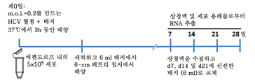
(1) 프로토콜 1, 현탁액에 의한 감염 - 본 발명자들은 HCVser 감염을 위해 비처리 hADSC의 계대배양-2 (p-2) 내지 계대배양-6을 사용하였다. 에펜도르프 (Eppendorf) 관에서 0.2의 감염 다중도 (MOI)로 800 ㎕의 새로운 배양 배지에 현탁된 5x10⁵ hADSC에 총 200 ㎕의 HCV 혈청 (1x10⁵개의 5'-UTR 카피 포함)을 첨가한 후, 37℃에서 3시간 동안 배양하였다. 세포를 PBS로 3회 세척하고, 7일, 14일, 21일 및 28일 동안 추가로 배양하고, 상청액 및 세포 용해물 내의 RNA를 5'-UTR의 RT-PCR을 위해 수거하였다. 면역세포화학 및 투과 전자현미경 (TEM) 연구를 위해 세포를 수집하였다. (1)
(2) 프로토콜 2, 부착성 형태의 감염 - p-2 내지 p-6 비처리 hADSC를 6 cm 페트리 접시에 1일간 플레이팅하여 세포 부착을 허용하고, HCVser를 2 ml 배지의 최종 부피에 0.5 moi (1x10⁵ 5'-UTR 카피 대 2x10⁵ hADSC 세포)에서 3시간 동안 첨가하였다. 부드럽게 세척한 후, 세포를 7일마다 배지를 교체하거나 교체하지 않으면서 5 ml의 신선한 배지에서 배양하였다. (2)
5'-5'- UTR에UTR 대한 RT- For RT- PCRPCR 및 정량적 RT- And quantitative RT- PCRPCR
RNA는 140 ㎕의 HCV(+) 혈청 또는 HCVser에 감염된 hADSC 배양물의 상청액으로부터 QIAamp® 바이러스 RNA 미니 키트 (퀴아겐 (Qiagen), 스위스 바젤)로 추출하였다. 제조사의 지시에 따라 PureLink® RNA 미니 키트 (앰비온, 미국 캘리포니아주 칼스바드)를 사용하여 세포 용해물로부터 RNA를 단리하였다. 이어서, RNA를 고용량 cDNA 역전사 키트를 사용하여 단일 가닥 cDNA로 전환한 후, GoTaq 매스터 믹스 (Master Mix) (프로메가 (Promega), 미국 위스콘신주)로 PCR을 수행하였다. 바이러스 카피를 정량하기 위해, 어플라이드 바이오시스템즈(Applied Biosystems)® ViiA™ 7 실시간 PCR 시스템을 사용하여 C형 간염 바이러스 고급 키트 (Advanced kit) (프라이머디자인 엘티디. (PrimerDesign Ltd.), 영국)를 사용하여 PCR을 수행하였다. HCV-특이적 역전사 및 증폭 프라이머는 ABI 프라이머 3.0 익스프레스 소프트 워드 (Express Soft Word)에 따라 설계되었다. 프라이머 5'-ACTCGCAAGCACCCTATCAG-3'을 역전사에 사용하였고, PCR 및 실시간 PCR에 사용된 프라이머는 문헌 35에 기재된 바와 같이 상이한 HCV 유전자형의 고도로 보존된 5'-비번역 영역 (UTR)에 매치되었다.RNA was extracted from the supernatants of hADSC cultures infected with 140 [mu] l HCV (+) serum or HCVser into a QIAamp virus RNA mini kit (Qiagen, Switzerland Basel). RNA was isolated from the cell lysate using a PureLink® RNA mini kit (Ambion, Carlsbad, Calif., USA) according to the manufacturer's instructions. The RNA was then converted to single stranded cDNA using a high capacity cDNA reverse kit and PCR was performed with GoTaq Master Mix (Promega, Wisconsin, USA). To quantitate viral copies, the Applied Biosystems
인간 지방 조직에 대한 면역조직화학 염색 (IHC)Immunohistochemical staining for human adipose tissue (IHC)
신선한 지방 조직을 수술 상처로부터 수거하고, 포르말린에 고정하고, 파라핀에 포매하였다. 조직을 5 ㎛ 절편으로 잘라내고, 왁스 제거한 후 시트레이트 완충제 (10 mM 시트르산, pH 6.0)에 담그고, 항원 회수를 위해 마이크로파로 가열하였다. 실온에서 30분 동안 5% BSA로 차단한 후, 폴리클로날 토끼 항-DLK1 항체 (1:150, 압캄, Cat. No. ab21682, 압캄, USA) 또는 토끼 IgG Ab (1:150, Cat. No. AB-105-C, R&D)를 4℃에서 밤새 적용하고, 실온에서 1시간 동안 알칼리성 포스파타제-접합된 항-토끼 IgG 2차 항체 (1:500; 잭슨 이뮤노리서치 (Jason ImmunoResearch))에 적용한 후, 빠른 적색 기질 시스템 (시그마-알드리치)를 사용하여 발색시켰다. 순차적 NS5 염색을 위해, 샘플을 10분 동안 PBS에 담가 커버 슬립을 제거하고, 내인성 퍼옥시다제로부터 비-특이적 배경을 감소시키기 위해 슬라이드를 0.3% H2O2에서 실온에서 30분 동안 인큐베이팅하였다. 실온에서 30분간 5% BSA로 차단한 후, 마우스 항-NS5 항체 (1:200, 클론 BGN/1246/5G7, Cat. No. 0200-0423, AbD 세로텍 (Serotec)) 또는 마우스 IgG1 이소형 Ab (1:100, Cat. No. 14-4714, 이바이오사이언스 (eBioscience))를 4℃에서 밤새 첨가하였다. 본 발명자들의 경험에 따르면, 샘플이 전에 파라핀에 포매되었기 때문에, 세포 침투 과정은 NS5가 지방 조직에 염색되는데 필요하지 않았다. 이어서, 절편을 실온에서 7분간 양고추냉이 퍼옥시다제 중합체 콴토 (Quanto) 시약 (항-마우스, 즉시 사용 가능, 써모 사이언티픽 (Thermo Scientific))과 함께 인큐베이팅하고, 울트라비젼 콴토 (UltraVision Quanto) 검출 시스템 (발색용 DAB 기질 함유; 써모 사이언티픽)으로 발색시켰다. 이어서, 절편을 헤마톡실린으로 염색하고, 커버 슬립을 다시 배치하여 세포 외형을 약간 변화시켰다 (도 1e에 제시). DLK1 및 NS5A 염색의 전자 영상은 고품질의 현미경 (자이스 (Zeiss)), 빠른 컴퓨터 하드웨어 및 TissueFAXS 스캐닝 소프트웨어 (티슈그로스틱스 (TissueGnostics))를 사용하는 고해상도 스크린으로 캡쳐하였다. 영상의 동일한 필드는 공존 (colocalization)에 대한 비교 및 분석을 위해 이후에 선택되고 가시화되었다. 지방 조직에서 (도 1e), HCV 감염 상태와 상관없이, 상이한 공여자 (즉, 공여자-공여자 변이)로부터의 조직 중에서 발색에 필요한 최적의 시간이 현저히 변하였다. 본 발명자들은 이 방법을 여러 번 변경하고, 발색 시간이 중요하다는 것을 발견하였다. 본 발명자들이 따른 원리는 먼저 이소형 Ab 염색 (토끼 IgG 또는 마우스 IgG1)에 대한 최소 색상 신호를 생성하는 발색을 위한 최대 시간을 결정한 다음, 이 시간을 항-DLK-1 Ab 또는 항-NS5 Ab 염색의 발색에 적용하는 것이었다. 이는 기질의 과도한 발달에 의한 위양성이 거의 없고 이소형 Ab 염색을 위한 낮은 배경을 확보하도록 보장하는 것이었다.Fresh adipose tissue was collected from the surgical wound, fixed in formalin, and embedded in paraffin. The tissue was cut into 5 탆 sections, wax-free, immersed in citrate buffer (10 mM citric acid, pH 6.0) and heated with microwave for antigen recovery. (1: 150, Cat. No. ab21682, Abkam, USA) or rabbit IgG Ab (1: 150, Cat. No (1: 500; Jackson ImmunoResearch) at room temperature for 1 hour at room temperature and at room temperature for 1 hour at room temperature, And then developed using a fast red substrate system (Sigma-Aldrich). For subsequent NS5 dyeing, the
HCV(+) 공여자 1의 지방 조직에 대한 연구에서 (표 1), 토끼 IgG 염색의 발색을 위한 최대 시간은 유의한 색상 신호가 나타나기 전에 40-50초이었고, 따라서 DLK-1 염색에 대한 발색은 40-50초 동안 설정되었다. 이와 유사하게, 유의한 색상 신호 없이 마우스 IgG1 염색의 발색을 위한 최대 시간은 15초 정도로 짧았고, 이는 항-NS5 Ab 염색을 위한 발색 시간으로 설정되었다. 이와 대조적으로, 공여자 2 및 공여자 3의 지방 조직에서, 토끼 IgG 염색을 위한 최적의 발색 시간은 30초이었고, 마우스 IgG1 염색의 경우는 단지 10초였기 때문에, 이들 시간은 각각 항-DLK-1 및 항-NS5 Ab 염색의 발색을 위해 설정되었다. HCV(-) 샘플을 염색할 때에도 유사한 원리를 따랐다.In the study of adipose tissue of HCV (+) donor 1 (Table 1), the maximum time for color development of rabbit IgG staining was 40-50 seconds before a significant color signal appeared, and thus the color development for DLK-1 staining It was set for 40-50 seconds. Similarly, the maximum time for color development of mouse IgG1 staining without significant color signals was as short as 15 seconds, which was set as the color development time for anti-NS5 Ab staining. In contrast, in
면역세포화학 (ICC)Immunocytochemistry (ICC)
미분획화된 SVF 세포 또는 DLK-1+ hADSC, 또는 DLK-1- 세포의 면역세포화학을 위해, IHC와 유사한 원리를 따르고, 발색을 위한 최적 시간은 상기한 바와 같이 모든 대조 실험에서 미리 결정되었다. 세포를 지시된 시점에서 수집하고, 세포원심분리에 의해 폴리리신-코팅된 유리 슬라이드에 부착시킨 후, 20분 동안 4% 포르말린으로 고정하였다. DLK-1 염색을 위해, 세포를 0.05% 트립신 용액으로 37℃에서 30분간 항원 회수에 적용한 후, DDW로 3회 세척하였다. 울트라 (Ultra) V 차단 완충제 (울트라비젼 콴토 검출 시스템 내의 시약, 써모, USA)로 5분간 차단한 후, 세포를 토끼 항-DLK1 항체 또는 토끼 IgG와 함께 4℃에서 밤새, 이어서 알칼리성 포스파타제-접합된 항-토끼 IgG 2차 항체와 함께 실온에서 1시간 동안 인큐베이팅하고, 신속한 적색 기질 시스템 (시그마-알드리치)으로 5-6분 (대부분의 경우) 동안 발색시켰다. HCV-특이적 NS5의 단일 또는 순차 염색을 위해 (도 2c에 나타낸 바와 동일한 슬라이드에서 염색됨), 슬라이드를 10분 동안 PBS에 넣어 커버 슬립을 제거하고, 위에 있는 배지를 세척하고, 세포를 투과시키고, 울트라비젼 콴토 검출 시스템 (써모 사이언티픽, 미국 캘리포니아주 프레몬트)을 적용하기 전에 0.3 % Tritox-100 및 1% BSA를 함유하는 PBS로 30분간 차단하였다. 이어서, 세포를 과산화수소 블록 (Hydrogen Peroxide Block) (압캄, 미국 매사추세츠주)에서 10분 동안 인큐베이팅하여 내인성 퍼옥시다제로 인한 비특이적인 배경 염색을 감소시켰다. 세척 후, 세포를 울트라 V 차단 완충제 (써모 사이언티픽, 미국 매사추세츠주)로 5분간 인큐베이팅하여 비특이적인 배경 염색을 차단하고, 마우스 모노클로날 항-NS5 항체를 함유하는 희석 완충제와 함께 4℃에서 밤새 인큐베이팅하였다. 마우스 IgG1을 사용한 염색을 음성 대조군으로 사용하였다. 다음날, 세포를 1차 항체 증폭기 콴토 용액과 함께 또 다른 10분 동안, HRP 중합체 콴토와 함께 10분 동안 인큐베이팅한 후, 각각의 시약 적용 사이에 PBS로 세척하였다. 마지막으로, 30 ㎕의 DAB 콴토 색소원 (Chromogen)을 1 ml의 DAB 콴토 기질에 넣고, 소용돌이를 일으키면서 혼합하고, 발색을 위해 2-3분 동안 (대부분의 경우) 적용하였다. 세척 후, 슬라이드에 영구 봉입제 (permanent mounting medium)를 올리고, 커버 슬립으로 덮고, TissueFAXS 현미경 (자이스)을 사용하여 가시화하고, 사진을 찍었다.For immunocytochemistry of undifferentiated SVF cells or DLK-1 + hADSC, or DLK-1 - cells, the principles similar to IHC were followed and the optimal time for color development was predetermined in all control experiments as described above. Cells were collected at indicated time points, attached to polylysine-coated glass slides by cell centrifugation, and fixed with 4% formalin for 20 minutes. For DLK-1 staining, cells were treated with 0.05% trypsin solution at 37 ° C for 30 minutes for antigen recovery and then washed three times with DDW. After blocking for 5 minutes with an Ultra V blocking buffer (reagent in an UltraVision Quanto detection system, Thermo, USA), cells were incubated overnight at 4 ° C with rabbit anti-DLK1 antibody or rabbit IgG, followed by alkaline phosphatase- Incubated for 1 hour at room temperature with anti-rabbit IgG secondary antibody and developed with a rapid red substrate system (Sigma-Aldrich) for 5-6 minutes (in most cases). For single or sequential staining of HCV-specific NS5 (stained on the same slide as shown in Fig. 2C), the slides were placed in PBS for 10 minutes to remove cover slips, wash the overlying media, , And blocked with PBS containing 0.3% Tritox-100 and 1% BSA for 30 minutes before applying the UltraVision Quanto Detection System (Thermo Scientific, Fremont, CA). The cells were then incubated in the Hydrogen Peroxide Block (Abkam, MA) for 10 minutes to reduce nonspecific background staining due to endogenous peroxidase. After washing, cells were incubated with Ultra V blocking buffer (Thermo Scientific, Mass., USA) for 5 minutes to block non-specific background staining and incubated overnight at 4 [deg.] C with dilution buffer containing mouse monoclonal anti- NS5 antibody Lt; / RTI > Staining with mouse IgG1 was used as a negative control. The next day, the cells were incubated with the primary antibody amplifier Quanto solution for another 10 minutes with HRP polymer quanta for 10 minutes and then washed with PBS between each reagent application. Finally, 30 μl of DAB Quanto Chromogen was added to 1 ml of DAB Quanto substrate, mixed with vortexing, and applied for 2-3 minutes (in most cases) for color development. After washing, permanent mounting medium was placed on the slides, covered with cover slips, visualized using a TissueFAXS microscope (Zeiss), and pictures were taken.
혈청 serum HCVHCV -감염된 - Infected hADSC에on hADSC 대한 투과 전자현미경 Transmission electron microscope
TEM 연구를 위해, 문헌 36에 기재된 바와 같이 hADSC를 수집하고 준비하여 투과 전자현미경 (JEM2000 EXII; JEOL, 일본 도꾜)으로 검사하였다.For TEM studies, hADSCs were harvested and prepared as described in document 36 and examined with a transmission electron microscope (
HCVHCV 2a 및 2b의 핵심 항원을 코딩하는 Lt; RTI ID = 0.0 > 2a < / RTI & mRNA에mRNA 대한 RT- For RT- PCRPCR
hADSC의 RNA는 PureLink® RNA 미니 키트 (앰비온, 미국 캘리포니아주 칼스바드)를 사용하여 제조사의 지시에 따라 단리하였다. RNA는 고용량 cDNA 역전사 키트 (어플라이드 바이오시스템즈)를 사용하여 단일 가닥 cDNA로 전환되었다. 역전사에 사용된 특이적 프라이머는 5'-ATGTACCCCATGAGGTCGGC-3'이었다. PCR에 사용된 프라이머는 다른 HCV 유전자형의 핵심 단백질 영역에 매치되었다. (전방향) 5'-CGCGCGACTAGGAAGACTTC-3' 및 (역방향) 5'-CGCGCGACGCGTAAAACTTC-3'을 포함하는 프라이머 혼합물을 제1 PCR에 사용하고, 열 프로파일은 다음 설정으로 적용하였다: 94℃ 2분, 이어서 94℃ 45초, 55℃ 45초 및 72℃ 90초의 35 사이클, 및 이어서 최종 연장을 위한 72℃ 7분. 제2 PCR에서 유전자형 확인을 위해 사용된 유형-특이적 안티센스 프라이머는 5'-CCAAGAGGGACGGGAACCTC-3 '(유형 2a) 및 5'-ACCCTCGTTTCCGTACAGAG-3'(유형 2b)이었고, 열 프로파일은 다음 설정으로 적용하였다: 95℃ 2분, 이어서 95℃ 30초, 60℃ 30초 및 72℃ 30초의 30 사이클, 및 이어서 최종 연장을 위한 72℃ 7분37,38.The RNA of hADSC was isolated using the PureLink® RNA mini kit (Ambion, Carlsbad, Calif., USA) according to the manufacturer's instructions. RNA was converted to single stranded cDNA using a high capacity cDNA reverse transcription kit (Applied Biosystems). The specific primer used for reverse transcription was 5'-ATGTACCCCATGAGGTCGGC-3 '. The primers used in the PCR matched the core protein regions of the other HCV genotypes. A primer mixture containing (forward) 5'-CGCGCGACTAGGAAGACTTC-3 'and (reverse) 5'-CGCGCGACGCGTAAAACTTC-3' was used in the first PCR and the thermal profile was applied with the following settings: 94 ° C for 2 minutes, 35 cycles of 94 < 0 > C for 45 seconds, 55 [deg.] C for 45 seconds and 72 [deg.] C for 90 seconds, and then 72 [deg.] C for 7 minutes for final extension. The type-specific antisense primers used for the genotyping in the second PCR were 5'-CCAAGAGGGACGGGAACCTC-3 '(type 2a) and 5'-ACCCTCGTTTCCGTACAGAG-3' (type 2b) and the thermal profile was applied with the following settings : 30 cycles of 95 캜 for 2 minutes, followed by 95 캜 for 30 seconds, 60 캜 for 30 seconds and 72 캜 for 30 seconds, and then 72 캜 for 7 minutes at 37 캜 for final extension.
유동 세포 계측 및 차단 실험Flow cytometry and blocking experiments
현탁액 중의 상이한 계대배양에서 0.5-1x10⁵ hADSC를 마우스 항-인간 CD81 모노클로날 Ab (클론 JS-81, BD 바이오사이언시즈), 항-LDL-R Ab (클론 C7, 밀리포어), 항-EGFR Ab (클론 LA1, 밀리포어) 또는 토끼 폴리클로날 항-SRB1 Ab (노부스 바이올로지칼스 (Novus Biologicals))로 1시간 동안 4℃에서 염색하였다. 각각의 대조군은 마우스 IgG1 또는 폴리클로날 토끼 IgG이었다. 세척 후, 세포를 플루오레세인 이소티오시아 네이트 (FITC)-접합된 2차 Ab (잭슨 이뮤노리서치, 미국 펜실베니아주)와 함께 추가로 인큐베이팅하고, 세포 콴타™ SC 고해상도 유동 세포 계측법 (베크만 코울터 풀러튼 (Beckman Coulter Fullerton), 미국 캘리포니아주)에 의해 분석하였다. 세포 표면 분자를 차단하기 위해, 37℃에서 나타낸 용량 (1-100 μg/ml)의 항체를 함유하는 1 ml의 무혈청 K-배지를 사용하여 2x10⁵ hADSC (웰내 부착물)를 전처리하였다. 각각의 이소형 항체 처리를 대조군으로 사용하였다. 1시간 후, 항체의 존재 하에 3시간 인큐베이션을 위해 MOI 0.2를 만들기 위해 희석되지 않은 HCV(+) 혈청을 에펜도르프 관에 첨가하였다. 그 후, 세포를 세척하고, 연속 배양을 위해 6-cm 페트리 접시에 플레이팅하였다. ApoE 차단을 위해, 다양한 농도의 항-ApoE 항체 (클론 E6D10)를 문헌 14에 기재된 바와 같이 hADSC와 3시간 인큐베이션하기 전에 HCV(+) 혈청에 실온에서 1시간 동안 첨가하였다. 상청액 및 세포를 21일간 연속 배양한 후 수집하고, RNA를 추출하여 바이러스 5'-UTR 전사체를 정량하였다. 별개의 실험에서, 웰 내의 hADSC는 유전자형 1a, 1b, 2a 및 2b의 HCV(+) 혈청에 노출되기 전에, K 배지에서 16시간 동안 표시된 용량의 IFNα (시그마-알드리치, 미국 미주리주)로 삼중으로 전처리되었다. 21일 후, 세포 용해물 중의 5'-UTR 전사체를 qRT-PCR에 의해 정량하고, 결과를 비히클 (PBS) 대조군으로 처리한 세포와 비교하여 억제 비율로서 계산하였다.0.5-1x10 5 hADSCs were incubated with mouse anti-human CD81 monoclonal Ab (clone JS-81, BD Biosciences), anti-LDL-R Ab (clone C7, Millipore), anti-EGFR Were stained with Ab (clone LA1, Millipore) or rabbit polyclonal anti-SRB1 Ab (Novus Biologicals) for 1 hour at 4 ° C. Each control group was mouse IgG1 or polyclonal rabbit IgG. After washing, the cells were further incubated with a fluorescein isothiocyanate (FITC) -conjugated secondary Ab (Jackson ImmunoResearch, Pa., USA) and analyzed by CellQuant ™ SC high resolution flow cytometry (Beckman Coulter Beckman Coulter Fullerton, CA, USA). To block cell surface molecules, 2x10 5 hADSCs (attachment in the wells) were pretreated with 1 ml of serum-free K-medium containing the antibody (1-100 μg / ml) as indicated at 37 ° C. Each isoform antibody treatment was used as a control. After 1 hour, undiluted HCV (+) serum was added to the Eppendorf tube to make a MOI of 0.2 for 3 hours incubation in the presence of antibody. The cells were then washed and plated in a 6-cm Petri dish for continuous culture. For ApoE blocking, various concentrations of anti-ApoE antibody (clone E6D10) were added to HCV (+) serum for 1 hour at room temperature before incubation with hADSC for 3 hours as described in
miRmiR -122의 RT--122 RT- PCR을PCR 위한 RNA 추출 RNA extraction for
miR-122의 RT-PCR용 프라이머를 문헌 39에 기재된 바와 같이 준비하고, 수행하였다. 세포의 총 RNA를 RNA 추출 시약 REzol™ C&T (프로테크 (Protech), 대만 타이페이)를 사용하여 단리하였다. miR-122 수준을 결정하기 위해, 본 발명자들은 TaqMan MicroRNA 리버스 트랜스크립셔나스 (Reverse Transcriptionas) 키트 (어플라이드 바이오시스템즈)를 사용하여 추출한 RNA를 역전사하고, cDNA를 miR-122를 위한 TaqMan MicroRNA 검정을 사용하는 실시간 PCR 분석을 위한 주형으로 사용하였다.Primers for RT-PCR of miR-122 were prepared and performed as described in document 39. [ The total RNA of cells was isolated using RNA extraction reagent REzol (TM) C & T (Protech, Taipei, Taiwan). To determine miR-122 levels, we reverse-transcribed RNA extracted using TaqMan MicroRNA Reverse Transcription Kits (Applied Biosystems) and used cDNA for TaqMan MicroRNA assays for miR-122 Were used as templates for real-time PCR analysis.
합성 synthesis siRNAsiRNA 및 유전자 침묵 And gene silencing
오클루딘 및 클라우딘-1에 특이적인 siRNA는 다마콘 (Dharmacon)에 의해 문헌 15, 40, 41에 기재된 바와 같이 합성하였다. 그들의 각각의 표적 서열은 UAACAUUAGGACCUUAGAA (클라우딘-1) 및 GUGAAGAGUACAUGGCUGC (오클루딘)이다. NPC1L1은 문헌 16에 기재된 바와 같이 제조하고, DGAT-1에 대한 siRNA는 문헌 19에 기재된 바와 같이 제조하였다. 특이적 siRNA를 Xfect 형질감염 시약 (클론테크 (Clontech))를 사용하여 6-웰 세포 플레이트의 웰에서 hADSC 내로로 형질감염시켰다. HCV 감염은 siRNA 감염된 세포를 HCVser와 함께 37℃에서 3시간 동안 인큐베이팅하여 수행한 후, HCVser를 PBS로 세척하였다. 형질감염 48시간 후에 세포를 문헌 15, 19, 40, 41에 기재된 바와 같이 유전자 침묵 정도를 결정하기 위해 RT-PCR을 위해 용해시켰다. HCVser에 감염된 hADSC의 세포 용해물 및 상청액을 5'-UTR의 qRT-PCR을 위해 21일 배양 후에 수거하였다.SiRNA specific for acrodine and claudin-1 were synthesized by Dharmacon as described in
JFH1JFH1 // HCVccHCVcc 및 And Huh7Huh7 .5 세포.5 cells
Huh7.5 세포를 10% 열 불활성화된 태아 송아지 혈청 (인비트로겐) 및 0.1 mM 비-필수 아미노산 (인비트로겐)을 함유하는 DMEM (인비트로겐)에서 배양하였다. 시험관내 게놈 JFH-1 RNA를 전사시키고, 문헌 42에 기재된 바와 같이 전기영동에 의해 세포로 전달하였다. 이어서, 형질감염된 세포를 완전한 DMEM으로 옮기고, 3-4일마다 계대배양하였다. 통상적인 방법으로, 전장 JFH1 cDNA로 형질감염된 세포로부터 조건화된 배지를 10분간 원심분리 (3,000 x g)에 의해 정화하고, 사용하기 전에 멸균 여과 (0.2 ㎛ 셀룰로오스 아세테이트, 밀리포어)하였다. 장기 보관을 위해, HCVcc를 분취하고, -80℃에서 보관하였다. 4℃에서 밤새 인큐베이팅하기 위해 PBS에 4분의 1 부피의 멸균 여과된 40% (w/v) 폴리에틸렌 글리콜-8000을 첨가하여 바이러스를 농축하였다. 바이러스 침전물을 원심분리 (8,000 x g, 15분)에 의해 수집하고, 문헌 43에 기재된 바와 같이 PBS에 재현탁하였다.Huh7.5 cells were cultured in DMEM (Invitrogen) containing 10% heat-inactivated fetal calf serum (Invitrogen) and 0.1 mM non-essential amino acid (Invitrogen). The in vitro genomic JFH-1 RNA was transcribed and transferred to cells by electrophoresis as described in document 42. The transfected cells were then transferred to complete DMEM and subcultured every 3-4 days. In a conventional manner, conditioned media from cells transfected with full-length JFHl cDNA was purified by centrifugation (3,000 x g) for 10 min and sterile filtered (0.2 쨉 m cellulose acetate, Millipore) before use. For long-term storage, HCVcc was aliquoted and stored at -80 ° C. The virus was concentrated by the addition of 4 volumes of sterilized 40% (w / v) polyethylene glycol-8000 in PBS for incubation overnight at 4 ° C. The viral precipitate was collected by centrifugation (8,000 x g, 15 min) and resuspended in PBS as described in document 43.
약물 억제 검정Drug inhibition assay
항-바이러스 약물인 리바비린, 시클로스포린 A 및 IFNα는 모두 시그마-알드리치에서 생산되었다. 텔라프레비르는 셀렉 케미칼스 (Selleck Chemicals, 미국 매사추세츠주) 제품이었다. 리바비린, 텔라프레비르 또는 시클로스포린 A의 등급이 매겨진 용량을 제0일에 페트리 접시 내의 HCVser에 감염된 hADSC의 배지에 첨가하고, 21일 동안 배양하였다. IFNα 처리를 위해, hADSC를 HCVser과 함께 인큐베이팅하기 전에 16시간 동안 나타낸 용량의 IFNα로 전처리하였다. 이어서, 세포 용해물의 바이러스 5'-UTR 전사체를 정량하고, 비히클 대조군으로 처리된 세포와 비교하여 억제 비율로서 계산하였다. 비히클 대조군은 리바비린 및 IFNα의 경우 PBS 및 시클로스포린 A 및 텔라프레비르의 경우 0.1% DMSO이었다.The anti-viral drugs ribavirin, cyclosporin A and IFNa were all produced in Sigma-Aldrich. Tellafevir was a product of Selleck Chemicals (Massachusetts, USA). The graded doses of ribavirin, telaprevir or cyclosporin A were added to the medium of hADSC infected with HCVser in Petri dishes at
HCVserHCVser , , HCVadscHCVadsc 및 And HCVcc의Of HCVcc 부유 밀도 Floating density
HCVcc 및 HCVadsc의 배지는 상기한 바와 같이 PEG-8000에 의해 농축되었다. 모든 샘플을 500 ㎕의 무혈청 배지에 재현탁하고, 문헌 44에 기재된 바와 같이 10 mM Hepes (pH 7.55), 150 mM의 NaCl, 및 0.02 % BSA를 함유하는 용액으로 제조한 10%로부터 40%의 아이오딕사놀 (각각 0.5 ml)의 연속적인 아이오딕사놀 (OptiPrep, 악시스-쉴드 (Axis-Shield), 노르웨이) 밀도 구배 상에 적층하였다. 구배를 SW-41 로터 (베크만 코울터 (Beckman Coulter))에서 4℃에서 6시간 동안 40,000 rpm으로 초원심분리하였다. 초원심분리 후, 17개의 분획을 구배의 상부로부터 수집하였다 (각각의 분획은 500 ㎕를 함유함). 마지막으로, QIAamp® 바이러스 RNA 미니 키트 (퀴아겐, 스위스 바젤)를 사용하여 각각의 분획으로부터 총 RNA를 단리하였다. RNA를 정량적 RT-PCR에 의한 HCV RNA 검출에 사용하였다.The medium of HCVcc and HCVadsc was enriched by PEG-8000 as described above. All samples were resuspended in 500 μl of serum-free medium and were resuspended in 10% to 40% solution of 10 mM Hepes (pH 7.55), 150 mM NaCl, and 0.02% BSA as described in document 44 (OptiPrep, Axis-Shield, Norway) density gradient of Iodixanol (0.5 ml each). The gradient was ultracentrifuged at 40,000 rpm for 6 hours at 4 DEG C in an SW-41 rotor (Beckman Coulter). After ultracentrifugation, 17 fractions were collected from the top of the gradient (each fraction containing 500 [mu] l). Finally, total RNA was isolated from each fraction using a QIAamp ( R) viral RNA mini kit (Qiagen, Switzerland Basel). RNA was used for HCV RNA detection by quantitative RT-PCR.
ApoBApoB , , ApoEApoE 및 콜레스테롤의 결정 And determination of cholesterol
콴티킨® ELISA 인간 아포지단백질 B/ApoB 면역검정 키트 및 콴티킨® ELISA 인간 아포지단백질 E/ApoE 면역검정 키트 (R&D 시스템즈)를 사용하여 ApoB 및 ApoE 발현을 제조업체의 설명에 따라 검출하였다. HDL 및 LDL/VLDL의 콜레스테롤은 HDL 및 LDL/VLDL 콜레스테롤 분석 키트 (압캄)에 의해 검출되었다. 부유 밀도의 다른 분획에서의 HDL 및 LDL/VLDL의 ApoB, ApoE 및 콜레스테롤의 발현 수준을 카피 시험 수준으로 표준화하였다.Kwan tikin ® ELISA human apolipoprotein B / ApoB ELISA immunoassay kits and Quan tikin ® human apolipoprotein E / ApoE and ApoB ApoE expression by using immunoassay kit (R & D Systems) was detected according to the manufacturer's instructions. HDL and LDL / VLDL cholesterol were detected by HDL and LDL / VLDL cholesterol assay kit (Abkam). The levels of expression of ApoB, ApoE and cholesterol of HDL and LDL / VLDL in the other fractions of floating density were normalized to copy test levels.
HCVserHCVser 또는 or HCVadsc를HCVadsc 이용한 인간 1차 간세포 ( Human primary human hepatocytes ( PHHPHH )의 감염) Infection
문헌 45에 기재된 바와 같이, 신선한 비-종양 간 조직은 HCV 관련 또는 비관련 간세포 암종에 대해 수술에 의해 절제된 간 표본으로부터 채취하고, PHH를 단리하고 배양하였다. PHH를 콜라겐 I-코팅된 6-웰 플레이트 상에 대한 세포 부착을 허용하기 위해 3일 동안 플레이팅하였다. 제4일에, 세포를 부드럽게 세척하고, 3시간 동안 0.5ml의 최종 부피에서 HCVser 또는 그의 대응하는 d21 hADSC-증식 HCV(+) 상청액 (1x10⁴ HCV 5'-UTR 카피)과 혼합된 아르기닌 미함유 윌리엄스 (Williams) E 배지 (인비트로겐, 미국 캘리포니아주)에서 배양하였다. 감염 후, 세포를 세척하고, 감염 후 d5까지 신선한 배지로 매일 교체하면서 1 ml의 배지에서 추가로 배양하였다. 이어서, 5'-UTR의 RT-PCR을 위한 RNA 추출을 위해 세포를 용해시켰다.As described in document 45, fresh non-tumor liver tissue was harvested from surgically resected liver specimens for HCV-related or unrelated hepatocellular carcinoma and PHH isolated and cultured. PHH was plated for 3 days to allow cell attachment on collagen I-coated 6-well plates. The d21 hADSC- proliferation HCV (+) arginine-free supernatant was mixed with the (1x10 4 HCV 5'-UTR copy) to 4, for washing the cells and gently, or a corresponding HCVser in a final volume of 0.5ml for 3 hours And cultivated in Williams E medium (Invitrogen, Calif., USA). After infection, the cells were washed and further cultured in 1 ml of medium with daily changes to fresh medium until d5 after infection. Cells were then lysed for RNA extraction for RT-PCR of 5'-UTR.
실시예Example 1 One hADSC는hADSC 생체 내에서 In vivo HCV에On HCV 의해 due to 표적화된다Be targeted ..
임상적으로, HCV 감염의 흥미로운 특징은 HCV(+) 환자가 만성 감염된 간에서 과도한 지방 축적, 즉 간 지방증이 있을 수 있고46 , 47, 간 지방증의 심각성은 간 섬유화의 비율과 상관관계가 있는 것으로 보인다는 것이다48. 최근의 연구는 또한 HCV RNA 복제가 포화 지방산의 이용가능성을 증가시킴으로써 자극될 수 있고, 다가 불포화 지방산 또는 지방산 합성 억제제에 의해 억제된다는 것을 보여준다49 , 50. 이러한 발견은 지방 대사가 HCV의 생활 주기에서 중요한 역할을 한다는 것을 시사한다. 따라서, 본 발명자들은 지방 조직의 세포 성분이 생체 내에서 HCV 감염과 관련이 있다고 가정하였다.Clinically, an interesting feature of HCV infection is that HCV (+) patients may have excessive fat accumulation in the chronic infected liver, that is, hepatic steatosis, 46 , 47 , and the severity of hepatic steatosis correlates with the rate of hepatic fibrosis 48 . Recent studies also show that HCV RNA replication can be stimulated by increasing the availability of saturated fatty acids, polyhydric inhibited by unsaturated fatty acids or fatty acid synthesis inhibitors 49, 50. This finding suggests that fat metabolism plays an important role in the life cycle of HCV. Therefore, the present inventors have assumed that the cellular components of adipose tissue are related to HCV infection in vivo.
본 발명자들의 가설을 시험하기 위해, 본 발명자들은 HCV에 감염되거나 감염되지 않은 개체 (표 1)로부터 피하 지방 조직을 수거하고, HCVser 유전자형 1b (HCVser-1b)를 양성 대조군으로 사용하여 HCV-특이적 5'-UTR 전사체의 RT-PCT을 위한 RNA를 추출하였다.To test our hypothesis, we used subcutaneous adipose tissue from HCV-infected or uninfected subjects (Table 1) and collected HCV serotype 1b (HCVser-1b) as a positive control to determine HCV-specific RNA for RT-PCT of the 5'-UTR transcript was extracted.
<표 1><Table 1>
A. 피하 지방 조직 및 간 조직에 대한 HCV(+) 공여자. 모든 환자는 간세포 암종의 수술 절제를 위해 개복술이 실시되었다. 지방 조직 [~(2-2.5 cm)3]을 수술 상처로부터 수거하고 (개복술), 간 조직을 절제된 간의 비-종양 부분 (병변으로부터 먼)으로부터 수거하였다. 환자 번호는 도 1f에 나타낸 바와 동일하였다.A. HCV (+) donors for subcutaneous adipose tissue and liver tissue. All patients underwent laparotomy for resection of hepatocellular carcinoma. Adipose tissue [~ (2-2.5 cm) 3 ] was collected from the surgical wound (laparotomy) and liver tissue was collected from the resected non-tumor portion (far from the lesion). The patient number was the same as shown in Fig.

주: 환자 1-3의 샘플을 도 1a-e에 사용하였고, 환자 1-4의 샘플을 도 1f에 사용하였다. 환자 4의 샘플 크기는 요청한 것보다 작았고 (~(0.6-1.3 cm)3), 이 샘플의 단리된 부착성 SVF 세포는 연장된 배양 실험에서만 사용되었다 (도 1f).Note: Samples 1-3 were used in Figures 1a-e and Samples 1-4 were used in Figure 1f. The sample size of
B. HCV(-) 간 조직의 공여자. 환자는 HBV 관련 또는 비-B 비-C 간세포 암종의 희생자였고, 간이 절제되었다.B. Donors of HCV (-) liver tissue. The patient was a victim of HBV-related or non-B non-C hepatocellular carcinoma and liver resection.

주: 표 1A 및 1B의 간염 바이러스 감염의 진단은 간세포 암종이 진단된 시점에 수행되었고, 조직 수거 후 항-바이러스 처리가 실시되었다.Note: The diagnosis of hepatitis virus infection in Tables 1A and 1B was performed at the time of diagnosis of hepatocellular carcinoma, and anti-virus treatment was performed after tissue collection.
흥미롭게도, HCV(+) 개체의 지방 조직 (유전자형 1b 또는 2a의 환자 번호 1-3, 표 1)은 HCV(-) 개체와 대조적으로, 바이러스 5'-UTR (223 bp, 도 1a)를 함유하였다. 혈액으로부터 가능한 오염 물질을 제외하고 바이러스 전사체를 발현하는 세포 공급원을 규정하기 위해, 본 발명자들은 성숙한 지방 세포를 함유하는 상부의 부유층 (부유물), 중간의 완충층 및 하부의 침강된 세포 펠렛으로, 문헌 12, 13, 33에 기재된 바와 같이 원심분리에 의해 지방 조직을 분획화하였다 (도 5). 세포 펠렛을 수거하고, RBC 용해 완충제로 추가로 처리하여 적혈구를 용해시켜 간질 혈관 분획 (SVF) 세포를 수거하였다. 이어서, 부유물 층 및 RBC-용해된 SVF 세포로부터 RNA를 별개로 추출하였지만, RNA는 완충층에서 거의 추출될 수 없었다 (데이터 비제시). RT-PCR은 부유물 층에서 바이러스 5'-UTR이 검출되지 않았지만, SVF 세포에서 바이러스 전사체가 검출되었음을 보여주었다 (왼쪽 패널, 도 1b).Interestingly, the adipose tissue of HCV (+) individuals (patient numbers 1-3, Table 1 in genotype 1b or 2a), in contrast to the HCV (-) individual, contains the virus 5'-UTR (223 bp, Respectively. In order to define the cell source expressing the viral transcript except for the possible contaminants from the blood, the present inventors have used the upper floating layer (suspension) containing mature adipocytes, the intermediate buffer layer and the lower settled cell pellet, 12, 13, and 33. The fat tissue was fractionated by centrifugation (FIG. 5). Cell pellets were harvested and further treated with RBC lysis buffer to dissolve red blood cells to collect interstitial blood vessel fraction (SVF) cells. RNA was then separately extracted from the suspension layer and RBC-lysed SVF cells, but RNA could hardly be extracted from the buffer layer (data not shown). RT-PCR showed that virus 5'-UTR was not detected in the supernatant layer, but viral transcripts were detected in SVF cells (left panel, FIG. 1B).
본 발명자들은 문헌 10에 기재된 바와 같이 면역 자기 비드에 의해 SVF 세포로부터 DLK-1+ 세포를 양성 선택하고, 유동 세포 계측법을 통해 99.7% 초과의 양성 선택된 세포가 DLK-1을 발현함을 확인하였다 (도 6). RNA는 이후에 RT-PCR을 위해 별개로 DLK-1+ 및 DLK-1- 세포로부터 추출하고, RT-PCR을 통해 바이러스 전사체가 DLK-1+ 세포에는 존재하지만, DLK-1- 세포에는 존재하지 않음이 확인되었다 (오른쪽 패널, 도 1b). 또한, 본 발명자들은 HCV(+) 1차 인간 간세포 (PHH)로부터 추출한 RNA를 양성 대조군으로 사용하여 HCV-특이적 음성 가닥 RNA51에 대한 RT-PCR을 수행하였다. 데이터는 HCV 복제 중간체가 3명의 모든 HCV(+) 공여자의 DLK-1+ 세포에는 존재하지만, DLK-1- 세포에는 존재하지 않음을 확인해주었다 (375 bp; 도 1c). 마우스 항-HCV NS5 항체 (클론 BGN/1246/5G7) 및 헤마톡실린 (세포핵을 표시함)을 사용한 면역세포화학을 위해 DLK-1+ 및 DLK-1- 세포를 세포원심분리 슬라이드 상에 별개로 원심분리하였다. HCV(-) 개체로부터 단리된 세포도 비교를 위해 염색되었다. 5'-UTR의 mRNA 발현과 일치하게, HCV(+) 공여자로부터 단리된 DLK-1+ 세포는 NS5 항원을 발현하는 반면 (갈색 라벨, 패널 c, 도 1e), DLK-1- 세포의 NS5 발현은 HCV 감염 상태와 상관없이 배경에 근접하였다 (패널 b & e). 이소형 항체 (마우스 IgG1)를 사용한 미분획화된 HCV(+) 또는 HCV(-) SVF 세포의 염색은 대조군으로 사용되었다 (패널 a & d, 도 1e). 헤마톡실린 염색 (파란색 라벨)을 통해 세포 파편보다는 NS5+ 세포가 진짜 유핵 세포임을 확인하였다 (패널 c, 도 1e). 항-NS5 항체의 특이성은 HCV(+) PHH의 염색에서 확인되었다 (도 7).We have found positive selection of DLK-1 + cells from SVF cells by immunomagnetic beads as described in
생체내 검증을 위해, 본 발명자들은 HCV 감염되거나 감염되지 않은 개체로부터 수거한 피하 지방 조직에 대해 면역조직화학 검사를 수행하였다. 조직 절편을 먼저 항-DLK-1 Ab로 염색하였다. 침지 및 PBS 세척 후, 동일한 절편을 항-NS5 Ab 및 헤마톡실린으로 염색하였다. 결과는 DLK-1이 마우스 지방 조직에서 유사하게 설명된 위치에서52 HCV 감염 및 비감염 개체 (각각 적색 라벨, 백색 화살표, 패널 b 및 c, 및 h, 도 1e) 둘 다로부터 수거된 지방 조직에서 검출되었음을 보여주었다. 또한 NS5+ 세포는 HCV(+) 지방 조직 (갈색 라벨, 흰색 화살표, 패널 e & f, 도 1e)에서 시각화되었지만, HCV(-) 샘플 (패널 j)에서는 시각화되지 않았다. 특히, 바이러스 NS5는 HCV(+) 샘플에서 DLK-1 발현과 동시에 위치하였다 (패널 b 대 e 및 c 대 f, 도 1e; b & e 및 c & g는 별개의 공여자로부터 얻어졌다; 또한 도 8 참조). 검사된 모든 절편에서, HCV(+) 지방 조직의 고배율 필드 (400X)당 약 0-4개의 DLK-1+NS5+ 세포가 발견되었다.For in vivo assays, we performed immunohistochemistry on subcutaneous adipose tissue collected from HCV-infected or non-infected individuals. Tissue sections were first stained with anti-DLK-1 Ab. After soaking and washing with PBS, the same sections were stained with anti-NS5 Ab and hematoxylin. The results show that DLK-1 is detected in adipose tissue collected from both 52 HCV infected and non-infected individuals (red labels, white arrows, panels b and c, and h, . NS5 + cells were also visualized in HCV (+) adipose tissue (brown label, white arrow, panel e & f, FIG. 1e) but not in HCV (-) samples (panel j). In particular, the virus NS5 was co-located with DLK-1 expression in HCV (+) samples (panel b versus e and c versus f, FIG. 1 e; b & e and c & g were obtained from separate donors; Reference). In all the sections examined, approximately 0-4 DLK-1 + NS5 + cells were found per high-field (400X) of HCV (+) adipose tissue.
HCV(+) 개체의 hADSC가 바이러스를 생산하는지 여부를 결정하기 위해, 본 발명자들은 HCV(+) 환자로부터 단리된 DLK-1+ 세포를 배양하고, 7일마다 상청액에서 바이러스 카피수를 정량하였다. 흥미롭게도, 처음 4주 동안 바이러스 전사체가 거의 검출되지 않았지만, d28-d35 이후의 상청액에서는 검출가능해졌고, 시간 경과에 따라 카피수가 증가하였다 (d49로 가면서; 도 1f). 또한, 장기간 배양시의 바이러스 전사체의 양은 세포가 그로부터 단리된 각각의 환자의 혈청 바이러스 역가와 관련이 있는 것으로 보였다 (도 1f 및 표 1).To determine whether the hADSCs of HCV (+) individuals produce the virus, we incubated isolated DLK-1 + cells from HCV (+) patients and quantified the viral copy number in supernatants every 7 days. Interestingly, although virus transcripts were scarcely detected during the first 4 weeks, they became detectable in supernatants after d28-d35, and the number of copies increased over time (going to d49; Fig. 1F). In addition, the amount of viral transcripts during long-term culture appeared to be related to the serum viral titer of each patient from which the cells were isolated (Fig. 1f and Table 1).
종합적으로, 본 발명자들의 데이터는 hADSC가 생체 내에서 HCV에 의해 표적화된다는 증거를 제공한다.Collectively, our data provide evidence that hADSCs are targeted by HCV in vivo.
실시예Example 2 비처리2 untreated HCVHCV (-) (-) hADSC는hADSC 시험관 내에서 In vitro HCVserHCVser 감염 및 복제에 감수성이다. It is susceptible to infection and replication.
비처리 HCV(-) hADSC가 시험관 내에서 HCVser 감염 및 복제에 감수성인지 조사하기 위해, 본 발명자들은 HCV(-) 개체로부터 hADSC를 준비하고, 배양액에서 계대배양하였다. 현탁액 (에펜도르프 관 내) 내의 계대배양-3 (p-3) 또는 p-4 세포를 1 ml의 최종 부피로 0.2 moi로 HCVser (표 2)와 함께 인큐베이팅하였다 (즉, 1x10⁵ 5'-UTR 카피수 대 5x10⁵ hADSC 세포).To investigate whether untreated HCV (-) hADSCs are susceptible to HCVser infection and replication in vitro, we prepared hADSCs from HCV (-) individuals and subcultured in culture medium. Suspension subculture in (Eppendorf tube within) -3 (p-3) or p-4 cells in a final volume of 1 ml was incubated with HCVser (Table 2) with 0.2 moi (i.e., 1x10 5 5'-UTR 5x10 5 hADSC cells versus copy number).
<표 2><Table 2>
이 연구에 사용된 HCV(+) 혈청의 HCV 유전자형 및 5'-UTR 카피수. 모든 환자는 HIV 또는 B형 간염 바이러스의 감염에 대한 증거가 없었다. 이들은 또한 급성 전염성 질환의 징후가 없었다. 혈청은 2011년 9월부터 2014년 2월까지 수집하여 즉시 사용하거나 사용하기 전까지 -80℃에서 보관하였다. 환자 번호 1-5의 의 혈청은 도 2a-d에서 사용되었고, 나머지는 도 2e-f, 도 3 및 도 4에서 사용되었다.HCV genotype and 5'-UTR copy number of HCV (+) sera used in this study. All patients had no evidence of infection with HIV or hepatitis B virus. They also had no signs of acute infectious disease. Serum was collected from September 2011 to February 2014 and stored at -80 ° C until used or used immediately. Serum from Patient Nos. 1-5 was used in Figures 2a-d and the remainder in Figures 2e-f, 3 and 4.

3시간 후, 세포를 세척하고, 7일마다 배지를 교체하면서 배양을 위해 6-cm 페트리 접시에 옮기고, 상청액 및 세포 용해물을 RNA 추출을 위해 제7일, 14일, 21일 및 28일에 수거하였다 (도 9의 프로토콜). HCVser 감염성에 대한 대조군으로서, 비-종양 간 조직으로부터 단리된 HCV(-) PHH (문헌 45 및 표 1에 기재된 바와 같이)를 웰에 3일간 배양하여 세포 부착을 허용한 다음, HCVser 또는 HCV(-) 대조군 혈청 (혈액형 AB)과 함께 제4일에 3시간 동안 인큐베이팅하고, 세척 후에 PHH를 세포 RNA 추출 전에 5일 동안 추가로 배양하였다. 결과는 HCVser에 노출된 PHH가 실제로 바이러스 5'UTR ("+"로 표시, 왼쪽, 도 2a)을 발현하는 반면, HCV(-) 대조군 혈청에 노출된 세포는 발현하지 않음 ("-"로 표시)을 보여주었다.After 3 hours, the cells were washed, transferred to a 6-cm Petri dish for culture while replacing the medium every 7 days, and the supernatant and cell lysate were collected on
감염 후 배양물에서, 바이러스 전사체는 d7-상청액 또는 d7-세포 용해물에서 검출할 수 없었지만, 모든 실험에서 d14-세포 용해물에서 및 또한 18개의 실험 중 10개의 상청액에서 검출가능하였다 (오른쪽, 도 2a; 8명의 공여자로부터의 hADSC를 사용한 총 18회의 실험, 표 3). 한편, 5'-UTR은 모든 실험의 세포 용해물뿐만 아니라 d21 및 d28 상청액 둘 모두에서 일관되게 검출되었다 (오른쪽, 도 2a).In the post-infection cultures, the viral transcripts were not detectable in d7-supernatant or d7-cell lysates, but were detectable in d14-cell lysates in all experiments and also in 10 supernatants of 18 experiments (right, Figure 2a: A total of 18 experiments using hADSCs from 8 donors, Table 3). On the other hand, 5'-UTR was consistently detected in both the d21 and d28 supernatants as well as the cell lysates of all experiments (right, Fig. 2a).
본 발명자들은 또한 HCV-특이적 음성 가닥 RNA를 조사한 결과, d14 및 d28 HCVser-1b에 감염된 hADSC 둘 다가 예상대로 상청액에 존재하지 않는 복제 중간체를 발현한다는 것을 확인하였다 (도 2b). HCV 감염 환자로부터 단리된 PHH를 양성 대조군으로 사용하였다.We also examined HCV-specific negative strand RNAs and found that both h14 and d28 HCVser-1b-infected hADSC express replication intermediates that are not present in the supernatant as expected (Fig. 2b). Isolated PHH from patients with HCV infection was used as a positive control.
추가 확인을 위해, 감염된 hADSC를 순차적 면역세포화학 연구를 위해 유리 슬라이드에 원심분리하였다. 세포를 항-DLK-1 항체로 먼저 염색한 다음, 항-NS5 항체 및 헤마톡실린으로 동일한 절편을 염색하였다. 결과는 d14 HCVser-1b에 감염된 hADSC가 실제로 DLK-1 (적색 라벨, 패널 b, 도 2c)을 발현하고, RT-PCR 발견과 일치하게 이들은 또한, DLK-1 (패널 f)을 발현하지만 NS5 (패널 h)를 발현하지 않는 대조군 혈청-펄싱된 hADSC와 대조적으로 NS5 (갈색 라벨, 패널 d, 도 2c)를 발현함을 보여주었다. 이소형 항체 토끼 IgG 및 마우스 IgG1 (각각 항-DLK-1 및 항-NS5 항체에 대한 대조군)을 사용한 염색은 배경색을 나타냈다 (각각 패널 a 및 c, 및 e & g). 실질적으로 모든 HCVser-감염 DLK-1+ 세포가 NS5를 발현하였기 때문에 (패널 b 대 d), 임상 단리물의 감염에 대한 hADSC의 허용성은 단지 세포의 하위세트(들)에만 제한되는 것이 아니라 일반화된 특성인 것으로 보였다.For further confirmation, infected hADSCs were centrifuged on glass slides for sequential immunocytochemical studies. Cells were first stained with anti-DLK-1 antibody and then stained with the same anti-NS5 antibody and hematoxylin. The results show that hADSCs infected with d14 HCVser-1b actually express DLK-1 (red label, panel b, Figure 2c) and in agreement with RT-PCR detection they also express DLK-1 (panel f) Panel d, FIG. 2c) in contrast to control serum-pulsed hADSCs that did not express panel h). Staining with isotype antibody rabbit IgG and mouse IgG1 (control for anti-DLK-1 and anti-NS5 antibodies, respectively) revealed a background color (panels a and c, and e & g respectively). Because virtually all HCVser-infected DLK-1 + cells expressed NS5 (panel b vs. d), the acceptability of hADSCs for infection of clinical isolates is not limited solely to the subset (s) of cells, Respectively.
d14 및 d21 HCVser-hADSC는 또한 투과 전자현미경으로 연구되었다. HCV(-) 대조군 혈청에 노출된 hADSC (패널 a 및 d, 도 2d)와 비교하여, 직경이 약 50-60 nm인 바이러스 입자를 hADSC의 외부 (삽입도에서 흰색 화살표, 패널 b 및 e, 도 2d) 및 내부 (삽입도의 흰색 화살표, 패널 c & f, 도 2d) 가시화할 수 있었다. 세포 외부에서 확인된 바이러스 입자는 이중 막 (노란색 화살표 및 삽입도에서 확대됨, 패널 b) 또는 전자 밀도가 높은 막 구조 (노란색 화살표 및 삽입도에서 확대됨, 패널 e)로 이루어진 막 모양의 소포로 감싸였다. 이와 대조적으로, 세포 내부에서 발견된 바이러스 입자는 막으로 감싸이지 않은 자유 형태 (노란색 정사각형 및 삽입도에서 확대됨, 패널 c; 흰색 화살표로 표시된 바이러스 입자) 또는 다수의 환상 동심원 막으로 둘러싸였다 ("양파 형태", 노란색 정사각형 및 삽입도에서 확대됨, 패널 f; 흰색 화살표로 표시된 바이러스 입자).d14 and d21 HCVser-hADSC were also studied by transmission electron microscopy. Virus particles with a diameter of about 50-60 nm, compared to hADSCs exposed to HCV (-) control serum (panels a and d, Fig. 2d) 2d) and inside (white arrows in the inset, panel c & f, Fig. 2d). Virus particles identified outside the cell were enveloped in a membrane vesicle composed of a bilayer membrane (enlarged in yellow arrow and inset, panel b) or a membrane structure with high electron density (yellow arrow and enlarged in inset, panel e) . In contrast, viral particles found inside cells were surrounded by a free form that was not enveloped by the membrane (yellow squares and enlarged in the inset, panel c (viral particles marked with white arrows) or multiple annular concentric membranes Shape ", yellow squares and enlarged in the inset, panel f; viral particles indicated by white arrows).
현탁액에 hADSC를 감염시키는 것 이외에 (도 2, a-d), 본 발명자들은 또한 hADSC를 6 cm 페트리 접시에 1일 동안 플레이팅하여 부착 감염시킨 다음, 세포를 3시간 동안 2 ml의 최종 부피로 HCVser로 0.5 moi (1x10⁵ 5'-UTR 카피 대 2x10⁵ hADSC 세포)로 펄싱하였다. 부드럽게 세척한 후, 배양액을 7일마다 배지를 교체하면서 5 ml의 신선한 배지에서 배양하였다 (도 10). 상청액 및 세포 용해물의 RNA를 제7일, 14일, 21일 및 28일에 추출하고, qRT-PCR을 실시하였다. 결과는 바이러스 전사체가 상청액에 존재하지 않음에도 불구하고 제7일에 세포 용해물에서는 일관되게 검출될 수 있음을 보여주었다 (왼쪽 패널 대 오른쪽 패널, 도 2e). 또한, 세포 용해물 내의 바이러스 카피는 제2주에 가장 현저하게 증가한 반면 (왼쪽 패널, 도 2e), 상청액 내의 증가는 1주 지연, 즉 제3주에 가장 두드러졌다 (우측 패널, 도 2e).In addition to infecting the suspension with hADSCs (Fig. 2, ad), we also plated hADSCs in a 6 cm Petri dish for 1 day to infect and infect the cells with HCVser in a final volume of 2 ml for 3 hours It was pulsed with 0.5 moi (1x10 5 5'-UTR for the copy hADSC 2x10 5 cells). After gentle washing, the culture was incubated in 5 ml of fresh medium with changing medium every 7 days (Fig. 10). RNA of supernatants and cell lysates were extracted at
이 시스템에 의해 생성된 총 바이러스 카피를 측정하기 위해, 본 발명자들은 배지를 교체하지 않으면서 HCVser에 감염된 hADSC를 지속적으로 배양하고 (도 10), 표시된 배양 기간이 끝날 때 상청액을 수집하였다. qRT-PCR을 통해, 가장 실질적인 바이러스 방출 (상청액 내로의)이 제3주에 발생하고, 28일 배양 후 총 카피수는 ~3x10⁵/ml임이 확인되었다 (도 2f).To determine the total viral copy produced by this system, we continuously cultured HCVser-infected hADSCs without replacing the medium (Figure 10) and collected supernatants at the end of the indicated incubation period. Through qRT-PCR, the most substantial virus release (into the supernatant) occurred in the third week, and after 28 days of culture, the total copy number was found to be ~ 3x10 5 / ml (FIG. 2f).
<표 3><Table 3>
비처리 hADSC의 공여자의 특성Characteristics of Donor of untreated hADSC

*TRAM 피판: 횡복직근 피판 * TRAM flap: transverse rectus muscle flap
실시예Example 3 3 hADSChADSC -생산 -production 비리온은Virion 임상 clinical 단리물의Isolated 생물학적 성질을 나타내는 진정한 "비리온"이다. It is a true "virion" that exhibits biological properties.
hADSC-생산 비리온 ("HCVadsc"로 표시됨)의 감염성을 조사하기 위해, 본 발명자들은 HCVser-1b로 "공여자 1"의 p2 hADSC를 감염시키고, 제21일에 상청액을 수집하였다 ("HCVadsc(1)"로 표시). 0.22 ㎛ 세공 필터를 통해 여과한 후, HCVadsc(1)을 사용하여 "공여자 2"의 hADSC를 감염시켜 HCVadsc(2)를 만들고, 이를 "공여자 3"의 hADSC를 감염시키기 위해 사용하였다. 결과는 HCVadsc가 HCVser에 의한 초기 감염에서 볼 수 있는 비교적 일관된 복제 효율로 다른 공여자의 비처리 hADSC에 대한 감염성을 갖는다는 것을 확인해주었다 (도 3a). 초기에 HCVser-1a, -2a 및 -2b로 감염된 hADSC의 상청액으로부터 유래된 HCVadsc에 대해서도 유사한 관찰이 이루어졌다 (데이터는 나타내지 않음).To investigate the infectivity of hADSC-production virion (designated "HCVadsc"), we infected p2 hADSCs of "
다음으로, 본 발명자들은 HCVser-1b로 p2, p6, p9 및 p15 hADSC를 감염시키고 21일의 연속 배양 후에 바이러스 카피를 측정함으로써 상이한 계대배양 번호에서 hADSC의 허용성을 연구하였다. 결과는 p2 및 p6 세포와 대조적으로, p9 및 p15 hADSC는 상청액 및 세포 용해물 둘 모두에서 매우 낮은 수준의 바이러스 전사체를 가짐을 보여주었다 (각각 왼쪽 및 오른쪽 패널, 도 3b). HCVser-1a 및 -2a 감염에 대해서도 유사한 관찰이 이루어졌다 (도 11). 비처리 [HCV(-)] hADSC의 DLK-1 발현을 규정하는 실험에서, 본 발명자들은 DLK-1 발현이 HCV 감염의 허용성에 대해 언급된 유사한 경향을 따라 p0에서 p6까지 검출가능하지만, p9에서 감소하고, p15에서는 검출되지 않음을 발견하였다 (도 3c).Next, we investigated the acceptability of hADSCs in different subculture numbers by infecting p2, p6, p9 and p15 hADSCs with HCVser-1b and measuring viral copy after 21 consecutive days of incubation. The results showed that, in contrast to p2 and p6 cells, p9 and p15 hADSCs had very low levels of viral transcripts in both supernatant and cell lysate (left and right panels, respectively, Fig. 3b). Similar observations were made for HCVser-1a and -2a infections (Fig. 11). In experiments that define DLK-1 expression of untreated [HCV (-)] hADSC, we have found that DLK-1 expression is detectable from p0 to p6 along similar trends mentioned for tolerance of HCV infection, , And was not detected at p15 (Fig. 3C).
또한, hADSC는 유전자형 1 또는 2의 감염을 선호하지 않는 것으로 보인다 (도 3b 및 도 11). 이를 더 연구하기 위해, 본 발명자들은 p5 hADSC를 혼합된 유전자형 2a+2b의 HCVser로 감염시키고, 유전자형-특이적 코어 항원을 코딩하는 mRNA를 검출하기 위해 RT-PCR을 위한 세포를 수집하였다. 실제로, 혼합된 2a+2b HCVser 자체 (양성 대조군으로서)는 유전자형 2a 및 2b (각각 174 및 123 bp, 레인 "P", 좌측 및 우측 패널, 도 3d) 둘 모두의 코어 항원을 코딩하는 mRNA를 발현하였고, 이들은 또한 유전자형 2a+2b로 감염된 hADSC의 d21 및 d56 세포 용해물에서 검출되었다. 이 실험에서, HCV(-) 대조군 혈청에 의해 펄싱된 세포를 음성 대조군 ("N"으로 표시됨)으로 사용하였다. 따라서, 임상 단리물에 의한 감염에 대한 hADSC의 허용성은 적어도 유전자형 1a, 1b, 2a 및 2b에 대해서는 교차-유전자형이다.In addition, hADSC does not seem to prefer infection with
테트라스파닌 CD81, LDL-R, SR-B1, 상피 성장 인자 수용체 (EGFR), 아포지단백질 (Apo) E, 오클루딘, 클라우딘-1, 니만-픽(Niemann-Pick) C1-유사 1 (NPC1L1) 콜레스테롤 흡수 수용체 및 디아실글리세롤 아세틸트랜스퍼라제-1 (DGAT-1)을 포함하는 숙주 인자는 바이러스 부착 또는 부착후 단계에서 인간 간세포 또는 간종암 세포주에서 HCV 감염/복제를 매개하는 것으로 밝혀졌다15 , 16, 19, 53- 58. 본 발명자들은 유동 세포 계측법 또는 RT-PCR에 의해 hADSC에서 이들 분자의 발현을 검사하였다.(EGFR), Apogeoprotein (Apo) E, Occludin, Claudin-1, Niemann-Pick C1-like 1 (NPC1L1), Tetrapanin CD81, LDL-R, SR-Bl, epithelial growth factor receptor cholesterol absorption receptor and diacylglycerol acetyltransferase host of factors, including the cyclase -1 (DGAT-1) have been shown to mediate HCV infection / replication in human liver cells or liver cell lines jongam in virus attachment or after the attachment step 15, 16 , 19, 53-58. We examined the expression of these molecules in hADSC by flow cytometry or RT-PCR.
유동 세포 계측법은 p0 (즉, 부착성 SVF 세포), p2 및 p6 hADSC가 CD81, LDL-R, SR-B1 및 EGFR을 분명하게 발현한다는 것을 보여주었다 (도 3e). 또한, RT-PCR은 오클루딘 (OCLN), 클라우딘-1 (CLDN1) 및 NPC1L1을 코딩하는 mRNA의 발현을 확인했지만, miR-122은 확인하지 못하였다 (도 3f). 이와 대조적으로, miR-122는 Huh7.5 간종양 세포에서 풍부하게 발현되었고 (hADSC보다 ~60배 더 높음), PHH에서 훨씬 풍부하였다 (~2000배 더 높음, 오른쪽, 도 3f).Flow cytometry showed that p0 (ie, adherent SVF cells), p2 and p6 hADSCs express CD81, LDL-R, SR-B1 and EGFR clearly (Figure 3e). RT-PCR also confirmed the expression of mcl-encoding ocurudin (OCLN), claudin-1 (CLDN1) and NPC1L1, but did not identify miR-122 (Figure 3f). In contrast, miR-122 was abundantly expressed in Huh7.5 liver tumor cells (~ 60-fold higher than hADSC) and much more abundant in PHH (~2000-fold higher, right, Figure 3f).
이들 분자의 역할을 결정하기 위해, 본 발명자들은 p2 hADSC를 HCVser-1b에 의한 펄싱 1시간 전에 CD81 (클론 JS-81), LDL-R (클론 C7), EGFR (클론 LA-1)에 대한 모노클로날 Ab 또는 SR-B1에 대한 폴리클로날 Ab의 등급이 매겨진 용량으로 전처리하였다. ApoE 차단을 위해, 문헌 14에 기재된 바와 같이 다양한 농도의 항-ApoE 항체 (클론 E6D10)를 감염을 위해 사용하기 전에 HCV(+) 혈청에 실온에서 1시간 동안 첨가하였다. 21일 상청액 내의 바이러스 전사체의 정량은 HCV(+) 혈청 내의 CD81, LDL-R, SR-B1, EGFR의 차단 및 또한 ApoE의 중화가 용량 의존적 방식으로 바이러스 카피의 양을 현저하게 감소시킨 반면, 처리 자체는 세포 생존율에 유의한 영향을 미치지 않았음을 보여주었다 (도 3g). 세포 용해물의 바이러스 카피의 유사한 용량-반응 감소가 또한 관찰되었다 (도 12).To determine the role of these molecules we used p2 hADSCs to express monoclonal antibodies against CD81 (clone JS-81), LDL-R (clone C7), EGFR (clone LA-1) 1 hour before pulsing with HCVser- Treated with a graded dose of polyclonal Ab against Ronald Ab or SR-Bl. For ApoE blocking, various concentrations of anti-ApoE antibody (clone E6D10) were added to HCV (+) serum for 1 hour at room temperature prior to use for infection as described in
본 발명자들은 또한 문헌 15, 16에 기재된 바와 같이, HCVser-1b에 의한 감염 전에 오클루딘 또는 클라우딘-1에 특이적인 또는 별개의 실험에서 NPC1L1에 특이적인 siRNA로 p2 hADSC를 형질감염시켰다. 본 발명자들은 또한 HCV 뉴클레오캡시드 코어를 지질 소적으로 수송하는데 필요하고 간종양 세포주에서 HCV 생산에 중요한 역할을 하는 분자인 DGAT-1의 역할을 조사하였다19. RT-PCR을 통해, 21일 배양 후 상청액 (도 3h) 및 세포 용해물 (도 13)에서 바이러스 카피를 감소시키는 mRNA 낙다운 (도 13)의 효과를 확인하였다.We also transfected p2 hADSCs with NPC1L1-specific siRNAs in a specific or separate experiment on occludin or claudin-1 prior to infection with HCVser-1b, as described in
마지막으로, 본 발명자들은 항-바이러스 약물의 억제 효과를 조사하였다. p4-5의 세포를 웰에 넣고, HCVser-1b에 노출시키고, 리바비린, 텔라프레비르 또는 시클로스포린 A (시클로필린 A 억제제)를 포함하는 등급이 매겨진 농도의 항-바이러스 약물을 배양 배지에 첨가하였다. IFNα 치료를 위해, hADSC를 HCVser과 함께 인큐베이팅하기 전에 16시간 동안 나타낸 용량의 IFNα로 전처리하였다. 21일 세포 용해물의 바이러스 전사체를 비히클 대조군으로 처리한 세포와 비교하여 억제 비율로서 측정하고 계산하였다. 결과는 HCV 복제가 리바비린, 텔라프레비르, 시클로스포린 A 및 IFNα에 의해 용량 반응 방식으로 억제됨을 보여주었다 (도 3i). p0, p2 및 p6 hADSC에서 시클로필린 A의 발현은 본 발명자들의 예비 실험에서 확인되었다 (도 14). 따라서, HCVadsc는 임상 단리물의 생물학적 성질을 나타내는 진정한 "비리온"이다.Finally, we investigated the inhibitory effect of anti-viral drugs. Cells of p4-5 were placed in wells, exposed to HCVser-lb, and anti-viral drugs in graded concentrations, including ribavirin, telaprevir or cyclosporin A (cyclophilin A inhibitor), were added to the culture medium . For IFN [alpha] therapy, hADSCs were pretreated with a dose of IFN [alpha] shown for 16 h before incubation with HCVser. The 21 day cell lysate virus transcript was measured and calculated as the inhibition rate compared to the vehicle treated cells. The results showed that HCV replication was suppressed in a dose-response manner by ribavirin, telaprevir, cyclosporin A and IFNa (Fig. 3i). Expression of cyclophilin A in p0, p2 and p6 hADSCs was confirmed in our preliminary experiments (Fig. 14). Thus, HCVadsc is a true "virion" indicating the biological properties of the clinical isolate.
실시예Example 4 4 hADSC는hADSC 완전한 complete HCVHCV 복제를 허용하는 To allow replication 생체내In vivo HCVHCV 저장소이다. It is a repository.
HCVadsc의 물리적 성질을 특성화하기 위해, 본 발명자들은 문헌 43에 기재된 바와 같이, HCVser, HCVcc 및 HCVadsc의 부유 밀도 프로파일을 평형 원심분리에 의해 비교하였다. 연구된 모든 바이러스는 유전자형 2a에서 유래되었다. 이전의 보고와 일치하게43, 59, 60, HCVser는 보다 낮은 밀도 1.039 (분획 2) 및 1.080 (분획 7)의 분획에서 많은 양의 RNA를 보유하고 있었지만, HCVcc의 RNA는 1.132 (분획 13; 도 4a)에서 최대치를 보였다. HCVadsc에 대한 RNA의 최대량은 밀도 1.080 (분획 7)에서 관찰되었고, 다음으로 보다 높은 밀도 1.124 및 1.156 (분획 12 및 15)에서 2개의 피크가 나타났다. 흥미롭게도, HCVadsc의 1.080에서의 피크는 HCVser의 피크와 동일하였다 (도 4a). 따라서, HCVadsc의 물리적 특성은 HCVcc보다 임상 단리물과 유사하다.In order to characterize the physical properties of HCVadsc, we compared the floating density profiles of HCVser, HCVcc and HCVadsc by equilibrium centrifugation as described in document 43. All the viruses studied were derived from genotype 2a. Consistent with the previous report, HCVsar retained large amounts of RNA at fractions of 43, 59 and 60 , HCVs, at lower densities of 1.039 (fraction 2) and 1.080 (fraction 7), while HCVcc RNA had 1.132 4a) showed the maximum value. The maximum amount of RNA for HCVadsc was observed at a density of 1.080 (fraction 7), followed by two peaks at higher densities of 1.124 and 1.156 (
본 발명자들은 또한 HDL, VLDL/LDL, ApoE 및 ApoB를 비롯한, 각각의 주요 분획의 지질 및 아포지단백질 (Apo) 프로파일을 결정하였다. HCVser은 HCVcc 및 HCVadsc와 비교하여 총 지질량이 가장 많은 것으로 나타났는데(도 15), 이것은 바이러스 증식을 위한 미세 환경 (혈청 대 배양 배지)이 다르기 때문에 예상하지 못한 것이었다. 또한, 바이러스 카피당 중량 (ng)으로 나타낼 때, HCVcc 분획 13은 가장 낮은 HDL 및 LDL/VLDL 함량을 가졌다 (도 4b). HCVser의 주요 분획물도 ApoE 함량이 가장 높았고 (pg/카피 기준), HCVadsc가 그 다음으로 많은 ApoE 함량을 가졌고, HCVcc 분획 13에서는 ApoE 수준이 거의 검출되지 않았다 (도 4c). 따라서, HCVadsc는 또한 바이러스 입자-관련 지질 함량에서 HCVcc보다 HCVser에 더 많은 유사성을 나타낸다. 흥미롭게도, ApoB는 HCVadsc의 어느 분획에서도 검출되지 않았고, 이것은 ApoB가 ApoE와 달리 (도 3g), hADSC 감염을 위해 필요하지 않을 수 있음을 시사한다.We also determined the lipid and apolipoprotein (Apo) profiles of each major fraction, including HDL, VLDL / LDL, ApoE and ApoB. HCVser was found to have the highest total lipid content compared to HCVcc and HCVadsc (Fig. 15), which was unexpected because of the different microenvironment (serum versus culture medium) for virus multiplication. Also, when expressed as weight (ng) per viral copy,
본 발명자들은 또한 HCVser와 함께 p2 hADSC를 JFH1/HCVcc의 바이러스 접종으로 감염시킴으로써 hADSC에 대한 다양한 바이러스의 감염성을 비교하였다. Huh7.5 세포에서의 HCVcc 복제는 대조군으로서 병행하여 수행되었다. 본 발명자들은 Huh7.5 세포에서 HCVcc의 효율적인 복제와 달리 (도 16), HCVcc 감염 hADSC는 14일 또는 21일 배양 후 상청액 또는 세포 용해물에서 5'-UTR 전사체를 거의 생산하지 않고 (도 4d), hADSC의 HCVser 감염은 이전과 유사한 복제 동역학을 나타낸다는 것을 발견하였다 (도 4d 대 3a). 또한, 비처리 Huh7.5 및 HCVcc에 감염된 Huh7.5 세포의 21일 상청액, 및 HCVcc 또는 HCVser에 감염된 hADSC의 21일 상청액을 RNA 추출을 위해 수집하고, RT-PCR에 적용하였다. 결과는 Huh7.5의 HCVcc 감염 및 hADSC의 HCVser 감염과 대조적으로 (각각 레인 2 및 4, 도 4e), HCVcc에 감염된 hADSC 상청액에서 바이러스 전사체가 검출되지 않음을 확인해주었다 (레인 3, 도 4e).We also compared the infectivity of various viruses against hADSCs by infecting p2 hADSCs with HCVser with the inoculation of JFH1 / HCVcc. HCVcc replication in Huh7.5 cells was performed in parallel as a control. Unlike the efficient cloning of HCVcc in Huh7.5 cells (Fig. 16), we found that HCVcc-infected hADSC produced little 5 ' -UTR transcript in supernatants or cell lysates after 14 or 21 days of culture ), HCVser infections of hADSCs exhibited similar replication kinetics as before (Fig. 4d vs. 3a). In addition, 21-day supernatants from Huh7.5 cells infected with untreated Huh7.5 and HCVcc and 21-day supernatants from hAdSCs infected with HCVcc or HCVser were collected for RNA extraction and applied to RT-PCR. The results confirmed that no viral transcripts were detected in HCVcc-infected hADSC supernatants (
본 발명자들은 이어서 비처리 PHH에 대한 HCVadsc의 감염성을 조사하였다. PHH는 문헌 45에 기재된 바와 같이 HCV(-) 개체로부터 단리하고, 3일 동안 배양하여 (1x10⁴ 세포/접시) 세포 부착을 허용한 후, HCVser-1b 감염된 hADCS 배양액의 21일 상청액으로부터 제조된 HCVadsc에 노출시켰다. RT-PCR을 위해 감염 5일 후에 세포 RNA를 추출하였다. 결과는 대조군 혈청-펄싱된 hADSC의 상청액에 의한 감염 (음성 대조군으로서, 레인 "1", 도 4f)과 대조적으로, HCVadsc에 감염된 PHH가 바이러스 5'-UTR ("2" & "3"으로 표시됨, 도 4f)을 실제로 발현함을 보여주었다.The inventors then investigated the infectivity of HCVadsc against untreated PHH. PHH was isolated from HCV (-) individuals as described in document 45 and allowed to adhere to the cells (1 x 10 4 cells / dish) for 3 days (1 x 10 4 cells / dish), followed by incubation with HCVadsc prepared from the 21 day supernatant of HCVser- Lt; / RTI > Cell-RNA was extracted 5 days after infection for RT-PCR. The results show that the PHH infected with HCVadsc is expressed as the virus 5'-UTR ("2"&"3") in contrast to the infection with the supernatant of control serum-pulsed hADSCs (negative control as lane "1" , Fig. 4f).
마지막으로, 3명의 다른 공여자로부터의 PHH를 준비하고, 이전에 기재된 바와 같이 웰에 씨딩하였다. 제4일에, 세포를 HCV(-) 대조군 혈청 (3명의 상이한 개체로부터의), HCVser-1b (3명의 별개의 공여자로부터의) 및 그의 대응하는 HCVadsc로 감염시켰다. HCVser 및 대응하는 HCVadsc는 동일한 PHH 배치를 감염시키기 위해 쌍을 이루었다. 상청액을 감염 5일 후에 수집하고, 5'-UTR 카피를 정량하였다. HCV(-) 대조군 혈청에 대한 PHH의 노출을 음성 대조군으로 사용하였다. 결과는 HCVser의 감염이 이전에 임상 단리물을 사용한 PHH 감염에서 보고된 바와 같이61 매우 다양한 복제 동역학을 유발한다는 것을 보여주었다. HCVadsc에 의한 감염은 또한 HCVser 감염의 경우와 같이 매우 가변적인 바이러스 역가의 증가를 유도하였다 (도 4g). 이러한 발견은 간세포를 향한 HCVadsc의 감염성을 확인해주었다.Finally, PHH from three different donors was prepared and seeded into wells as previously described. On
요약하면, hADSC는 완전한 HCV 복제를 허용하는 생체내 HCV 저장소이고, 임상 HCV-숙주 상호작용에 대해 이전에는 인식할 수 없는 장소를 나타낸다. 또한, hADSC는 HCV 생활 주기를 해독하고 항-바이러스 전략의 개발을 촉진하는 새로운 도구가 될 수 있는 임상 HCV 단리물의 시험관내 증식을 허용하는 최초의 1차 비-간세포 종류이다.In summary, hADSC is an in vivo HCV reservoir that allows complete HCV replication and represents a previously unrecognized location for clinical HCV-host interaction. In addition, hADSC is the first primary non-hepatic cell type that allows in vitro proliferation of clinical HCV isolates, which can be a new tool to decipher the HCV life cycle and promote the development of anti-viral strategies.
본 명세서에서 언급된 모든 간행물 및 특허는 본원에 참고로 포함된다. 본 발명의 설명된 방법 및 시스템의 다양한 변경 및 변형은 본 발명의 범위 및 사상을 벗어나지 않으면서 통상의 기술자에게 명백할 것이다. 본 발명은 구체적인 바람직한 실시양태와 관련하여 설명되었지만, 특허 청구된 본 발명이 이러한 구체적인 실시양태로 부당하게 제한되지 않아야 함을 이해하여야 한다. 실제로, 세포 배양, 분자 생물학, 생화학 또는 관련 분야의 통상의 기술자에게 명백한, 본 발명을 수행하기 위한 설명된 방식의 다양한 변형은 첨부된 청구범위의 범주 내에 있는 것으로 의도된다.All publications and patents mentioned herein are incorporated herein by reference. Various modifications and variations of the described methods and systems of the invention will be apparent to one of ordinary skill in the art without departing from the scope and spirit of the invention. While the invention has been described in connection with specific preferred embodiments, it is to be understood that the invention as claimed should not be unduly limited to such specific embodiments. Indeed, various modifications of the described methods for carrying out the invention, which are obvious to those of ordinary skill in the art of cell culture, molecular biology, biochemistry or related fields, are intended to be within the scope of the appended claims.
참고문헌references


Claims (18)
b) hADSC를 배양하기에 적합한 배양 배지에서 대상체로부터 수득한 생물학적 샘플과 함께 hADSC를 인큐베이팅하는 단계,
c) HCV 복제를 허용하기에 충분한 시간 동안 상기 hADSC를 배양하는 단계, 및
d) HCV 복제 수준을 검출하는 단계
를 포함하고, 여기서 HCV 복제의 검출은 상기 대상체가 HCV에 감염되었음을 나타내는 것인, 대상체의 HCV 감염을 진단하기 위한 방법.a) providing hADSCs,
b) incubating the hADSCs with the biological sample obtained from the subject in a culture medium suitable for culturing hADSCs,
c) culturing the hADSCs for a time sufficient to allow HCV replication, and
d) detecting the level of HCV replication
Wherein the detection of HCV replication indicates that the subject has been infected with HCV.
b). 후보 화합물의 부재 하에 제1 용기 내의 배양 배지 내의 HCV 수준을 결정하는 단계;
c). 후보 화합물의 존재 하에 제2 용기 내의 배양 배지 내의 hADSC를 HCV와 접촉시키는 단계;
d). 후보 화합물의 존재 하에 제2 용기 내의 배양 배지의 HCV 수준을 결정하는 단계;
e). 후보 화합물의 존재 하의 HCV 수준을 후보 화합물의 부재 하의 HCV 수준과 비교하는 단계; 및
f). 후보 화합물의 존재 하의 HCV 수준이 후보 화합물의 부재 하의 HCV 수준보다 낮을 때, 후보 화합물을 항-HCV 화합물로서 확인하는 단계
를 포함하는, 항-HCV 화합물을 스크리닝하는 방법.a). Contacting the hADSC in the culture medium in the first container with HCV in the absence of the candidate compound;
b). Determining the level of HCV in the culture medium in the first container in the absence of the candidate compound;
c). Contacting hADSCs in a culture medium in a second container with HCV in the presence of a candidate compound;
d). Determining the HCV level of the culture medium in the second container in the presence of the candidate compound;
e). Comparing the HCV level in the presence of the candidate compound to the HCV level in the absence of the candidate compound; And
f). Confirming the candidate compound as an anti-HCV compound when the HCV level in the presence of the candidate compound is lower than the HCV level in the absence of the candidate compound
RTI ID = 0.0 > HCV < / RTI >
Applications Claiming Priority (1)
| Application Number | Priority Date | Filing Date | Title |
|---|---|---|---|
| PCT/CN2015/070243 WO2016109947A1 (en) | 2015-01-07 | 2015-01-07 | Employing human adipose-derived stem cells to propagate serum-derived hepatitis c virus and use thereof |
Publications (1)
| Publication Number | Publication Date |
|---|---|
| KR20170101998A true KR20170101998A (en) | 2017-09-06 |
Family
ID=56355408
Family Applications (1)
| Application Number | Title | Priority Date | Filing Date |
|---|---|---|---|
| KR1020177022015A KR20170101998A (en) | 2015-01-07 | 2015-01-07 | Human adipose-derived stem cells used for the proliferation of serum-derived hepatitis C virus and uses thereof |
Country Status (10)
| Country | Link |
|---|---|
| US (1) | US10273461B2 (en) |
| EP (1) | EP3242932A4 (en) |
| JP (1) | JP6670322B2 (en) |
| KR (1) | KR20170101998A (en) |
| CN (1) | CN107208060B (en) |
| AU (1) | AU2015376561A1 (en) |
| CA (1) | CA2973119A1 (en) |
| HK (1) | HK1246824A1 (en) |
| TW (1) | TWI616528B (en) |
| WO (1) | WO2016109947A1 (en) |
Families Citing this family (3)
| Publication number | Priority date | Publication date | Assignee | Title |
|---|---|---|---|---|
| IL272145A (en) * | 2020-01-20 | 2021-07-29 | Stem Cell Medicine Ltd | Cosmetic compositions with protein concentrate from conditioned medium of adipose-derived stem cells |
| IL278473A (en) | 2020-11-03 | 2022-06-01 | Yeda Res & Dev | Methods of prognosing, determining treatment course and treating multiple myeloma |
| CN114686517A (en) * | 2020-12-25 | 2022-07-01 | 中国医学科学院病原生物学研究所 | Preparation method of HCV four-receptor transgenic combined STAT1 knockout mouse |
Family Cites Families (9)
| Publication number | Priority date | Publication date | Assignee | Title |
|---|---|---|---|---|
| US5786207A (en) | 1997-05-28 | 1998-07-28 | University Of Pittsburgh | Tissue dissociating system and method |
| AU1615599A (en) | 1997-12-02 | 1999-06-16 | Zen Bio, Inc. | Differentiation of adipose stromal cells into osteoblasts and uses thereof |
| US20050076396A1 (en) | 1999-03-10 | 2005-04-07 | Katz Adam J. | Adipose-derived stem cells and lattices |
| US6777231B1 (en) | 1999-03-10 | 2004-08-17 | The Regents Of The University Of California | Adipose-derived stem cells and lattices |
| JP2009153383A (en) * | 2006-04-03 | 2009-07-16 | Eci Inc | Method for producing mature hepatocyte-like cell |
| KR101224504B1 (en) * | 2010-10-01 | 2013-01-22 | 가톨릭대학교 산학협력단 | Novel use of cryptotanshinone |
| GB201119335D0 (en) * | 2011-11-09 | 2011-12-21 | Univ Leuven Kath | Hepatitis virus infectable stem cells |
| US20140179775A1 (en) * | 2012-02-29 | 2014-06-26 | Mead Johnson Nutrition Company | Neurogenesis screening method and uses thereof |
| US9402710B2 (en) * | 2012-07-16 | 2016-08-02 | The Board Of Trustees For The Leland Stanford Junior University | Macroporous 3-D scaffolds for tissue engineering |
-
2015
- 2015-01-07 AU AU2015376561A patent/AU2015376561A1/en not_active Abandoned
- 2015-01-07 CN CN201580072851.XA patent/CN107208060B/en active Active
- 2015-01-07 EP EP15876456.3A patent/EP3242932A4/en not_active Withdrawn
- 2015-01-07 KR KR1020177022015A patent/KR20170101998A/en not_active Application Discontinuation
- 2015-01-07 WO PCT/CN2015/070243 patent/WO2016109947A1/en active Application Filing
- 2015-01-07 JP JP2017554627A patent/JP6670322B2/en active Active
- 2015-01-07 CA CA2973119A patent/CA2973119A1/en not_active Abandoned
- 2015-01-07 US US15/541,913 patent/US10273461B2/en active Active
- 2015-05-19 TW TW104115953A patent/TWI616528B/en active
-
2018
- 2018-05-15 HK HK18106252.0A patent/HK1246824A1/en unknown
Also Published As
| Publication number | Publication date |
|---|---|
| EP3242932A4 (en) | 2018-05-30 |
| EP3242932A1 (en) | 2017-11-15 |
| AU2015376561A1 (en) | 2017-07-27 |
| TW201625786A (en) | 2016-07-16 |
| TWI616528B (en) | 2018-03-01 |
| CN107208060B (en) | 2021-06-18 |
| US20180023058A1 (en) | 2018-01-25 |
| WO2016109947A1 (en) | 2016-07-14 |
| JP6670322B2 (en) | 2020-03-18 |
| HK1246824A1 (en) | 2018-09-14 |
| JP2018501820A (en) | 2018-01-25 |
| US10273461B2 (en) | 2019-04-30 |
| CN107208060A (en) | 2017-09-26 |
| CA2973119A1 (en) | 2016-07-14 |
Similar Documents
| Publication | Publication Date | Title |
|---|---|---|
| Yu et al. | Exosomes derived from microRNA-199a-overexpressing mesenchymal stem cells inhibit glioma progression by down-regulating AGAP2 | |
| Roelandt et al. | Human pluripotent stem cell-derived hepatocytes support complete replication of hepatitis C virus | |
| Zhai et al. | Hepatitis C virus induces MDSCs-like monocytes through TLR2/PI3K/AKT/STAT3 signaling | |
| Li et al. | Dissecting the roles of the 5′ exoribonucleases Xrn1 and Xrn2 in restricting hepatitis C virus replication | |
| Jing et al. | Oxygen tension plays a critical role in the hematopoietic microenvironment in vitro | |
| Shimizu et al. | Lipoprotein lipase and hepatic triglyceride lipase reduce the infectivity of hepatitis C virus (HCV) through their catalytic activities on HCV-associated lipoproteins | |
| Wang et al. | TIM-1 promotes hepatitis C virus cell attachment and infection | |
| Banaudha et al. | Primary hepatocyte culture supports hepatitis C virus replication: a model for infection‐associated hepatocarcinogenesis | |
| Yuan et al. | A chimeric humanized mouse model by engrafting the human induced pluripotent stem cell-derived hepatocyte-like cell for the chronic hepatitis B virus infection | |
| JP2010142249A (en) | Composition and method useful for hcv infection | |
| Yang et al. | Ex vivo organ culture of adipose tissue for in situ mobilization of adipose‐derived stem cells and defining the stem cell niche | |
| Parent et al. | An immortalized human liver endothelial sinusoidal cell line for the study of the pathobiology of the liver endothelium | |
| Azandeh et al. | Improvement of mesenchymal stem cell differentiation into the endoderm lineage by four step sequential method in biocompatible biomaterial | |
| KR20170101998A (en) | Human adipose-derived stem cells used for the proliferation of serum-derived hepatitis C virus and uses thereof | |
| Wakamatsu et al. | Establishment of organoids from human epithelioid sarcoma with the air-liquid interface organoid cultures | |
| Ning et al. | DSCAM-AS1 accelerates cell proliferation and migration in osteosarcoma through miR-186-5p/GPRC5A signaling | |
| Li et al. | Mouse mesenchymal stem cells from bone marrow differentiate into smooth muscle cells by induction of plaque-derived smooth muscle cells | |
| KR102089477B1 (en) | Methods for producing cells having a phenotype of a primary human hepatocytes and compositions | |
| Huang et al. | miR-96 regulates liver tumor-initiating cells expansion by targeting TP53INP1 and predicts Sorafenib resistance | |
| Gailhouste | Isolation and purification method of mouse fetal hepatoblasts | |
| WO2023180122A1 (en) | Use of human allogenic liver-derived progenitor cells for treating and/or preventing cellular senescence | |
| Lu et al. | Tree shrew bone marrow–derived mesenchymal stem cells express CD81, OCLN, and miR‐122, facilitating the entire hepatitis C virus life cycle | |
| KR20180015954A (en) | Use of complement proteins for liver cancer diagnosis | |
| WO2014025046A1 (en) | Promoter of differentiation from hepatic progenitor cell into hepatic cell, and use thereof | |
| Chen et al. | Continuous zebularine treatment enhances hepatic differentiation of mesenchymal stem cells under liver-specific factors induction in vitro |
Legal Events
| Date | Code | Title | Description |
|---|---|---|---|
| WITN | Application deemed withdrawn, e.g. because no request for examination was filed or no examination fee was paid |