KR20150036102A - Bag3 as biochemical serum and tissue marker - Google Patents
Bag3 as biochemical serum and tissue marker Download PDFInfo
- Publication number
- KR20150036102A KR20150036102A KR20157001228A KR20157001228A KR20150036102A KR 20150036102 A KR20150036102 A KR 20150036102A KR 20157001228 A KR20157001228 A KR 20157001228A KR 20157001228 A KR20157001228 A KR 20157001228A KR 20150036102 A KR20150036102 A KR 20150036102A
- Authority
- KR
- South Korea
- Prior art keywords
- bag3
- seq
- rna
- cancer
- nucleotide
- Prior art date
Links
Images


Classifications
-
- G—PHYSICS
- G01—MEASURING; TESTING
- G01N—INVESTIGATING OR ANALYSING MATERIALS BY DETERMINING THEIR CHEMICAL OR PHYSICAL PROPERTIES
- G01N33/00—Investigating or analysing materials by specific methods not covered by groups G01N1/00 - G01N31/00
- G01N33/48—Biological material, e.g. blood, urine; Haemocytometers
- G01N33/50—Chemical analysis of biological material, e.g. blood, urine; Testing involving biospecific ligand binding methods; Immunological testing
- G01N33/68—Chemical analysis of biological material, e.g. blood, urine; Testing involving biospecific ligand binding methods; Immunological testing involving proteins, peptides or amino acids
- G01N33/6893—Chemical analysis of biological material, e.g. blood, urine; Testing involving biospecific ligand binding methods; Immunological testing involving proteins, peptides or amino acids related to diseases not provided for elsewhere
-
- C—CHEMISTRY; METALLURGY
- C12—BIOCHEMISTRY; BEER; SPIRITS; WINE; VINEGAR; MICROBIOLOGY; ENZYMOLOGY; MUTATION OR GENETIC ENGINEERING
- C12Q—MEASURING OR TESTING PROCESSES INVOLVING ENZYMES, NUCLEIC ACIDS OR MICROORGANISMS; COMPOSITIONS OR TEST PAPERS THEREFOR; PROCESSES OF PREPARING SUCH COMPOSITIONS; CONDITION-RESPONSIVE CONTROL IN MICROBIOLOGICAL OR ENZYMOLOGICAL PROCESSES
- C12Q1/00—Measuring or testing processes involving enzymes, nucleic acids or microorganisms; Compositions therefor; Processes of preparing such compositions
- C12Q1/68—Measuring or testing processes involving enzymes, nucleic acids or microorganisms; Compositions therefor; Processes of preparing such compositions involving nucleic acids
- C12Q1/6876—Nucleic acid products used in the analysis of nucleic acids, e.g. primers or probes
- C12Q1/6883—Nucleic acid products used in the analysis of nucleic acids, e.g. primers or probes for diseases caused by alterations of genetic material
-
- C—CHEMISTRY; METALLURGY
- C07—ORGANIC CHEMISTRY
- C07K—PEPTIDES
- C07K14/00—Peptides having more than 20 amino acids; Gastrins; Somatostatins; Melanotropins; Derivatives thereof
- C07K14/435—Peptides having more than 20 amino acids; Gastrins; Somatostatins; Melanotropins; Derivatives thereof from animals; from humans
- C07K14/46—Peptides having more than 20 amino acids; Gastrins; Somatostatins; Melanotropins; Derivatives thereof from animals; from humans from vertebrates
- C07K14/47—Peptides having more than 20 amino acids; Gastrins; Somatostatins; Melanotropins; Derivatives thereof from animals; from humans from vertebrates from mammals
-
- C—CHEMISTRY; METALLURGY
- C07—ORGANIC CHEMISTRY
- C07K—PEPTIDES
- C07K16/00—Immunoglobulins [IGs], e.g. monoclonal or polyclonal antibodies
- C07K16/18—Immunoglobulins [IGs], e.g. monoclonal or polyclonal antibodies against material from animals or humans
-
- C—CHEMISTRY; METALLURGY
- C12—BIOCHEMISTRY; BEER; SPIRITS; WINE; VINEGAR; MICROBIOLOGY; ENZYMOLOGY; MUTATION OR GENETIC ENGINEERING
- C12Q—MEASURING OR TESTING PROCESSES INVOLVING ENZYMES, NUCLEIC ACIDS OR MICROORGANISMS; COMPOSITIONS OR TEST PAPERS THEREFOR; PROCESSES OF PREPARING SUCH COMPOSITIONS; CONDITION-RESPONSIVE CONTROL IN MICROBIOLOGICAL OR ENZYMOLOGICAL PROCESSES
- C12Q1/00—Measuring or testing processes involving enzymes, nucleic acids or microorganisms; Compositions therefor; Processes of preparing such compositions
- C12Q1/68—Measuring or testing processes involving enzymes, nucleic acids or microorganisms; Compositions therefor; Processes of preparing such compositions involving nucleic acids
- C12Q1/6844—Nucleic acid amplification reactions
- C12Q1/686—Polymerase chain reaction [PCR]
-
- C—CHEMISTRY; METALLURGY
- C12—BIOCHEMISTRY; BEER; SPIRITS; WINE; VINEGAR; MICROBIOLOGY; ENZYMOLOGY; MUTATION OR GENETIC ENGINEERING
- C12Q—MEASURING OR TESTING PROCESSES INVOLVING ENZYMES, NUCLEIC ACIDS OR MICROORGANISMS; COMPOSITIONS OR TEST PAPERS THEREFOR; PROCESSES OF PREPARING SUCH COMPOSITIONS; CONDITION-RESPONSIVE CONTROL IN MICROBIOLOGICAL OR ENZYMOLOGICAL PROCESSES
- C12Q1/00—Measuring or testing processes involving enzymes, nucleic acids or microorganisms; Compositions therefor; Processes of preparing such compositions
- C12Q1/68—Measuring or testing processes involving enzymes, nucleic acids or microorganisms; Compositions therefor; Processes of preparing such compositions involving nucleic acids
- C12Q1/6876—Nucleic acid products used in the analysis of nucleic acids, e.g. primers or probes
- C12Q1/6883—Nucleic acid products used in the analysis of nucleic acids, e.g. primers or probes for diseases caused by alterations of genetic material
- C12Q1/6886—Nucleic acid products used in the analysis of nucleic acids, e.g. primers or probes for diseases caused by alterations of genetic material for cancer
-
- G—PHYSICS
- G01—MEASURING; TESTING
- G01N—INVESTIGATING OR ANALYSING MATERIALS BY DETERMINING THEIR CHEMICAL OR PHYSICAL PROPERTIES
- G01N33/00—Investigating or analysing materials by specific methods not covered by groups G01N1/00 - G01N31/00
- G01N33/48—Biological material, e.g. blood, urine; Haemocytometers
- G01N33/50—Chemical analysis of biological material, e.g. blood, urine; Testing involving biospecific ligand binding methods; Immunological testing
- G01N33/53—Immunoassay; Biospecific binding assay; Materials therefor
- G01N33/574—Immunoassay; Biospecific binding assay; Materials therefor for cancer
- G01N33/57407—Specifically defined cancers
- G01N33/57434—Specifically defined cancers of prostate
-
- G—PHYSICS
- G01—MEASURING; TESTING
- G01N—INVESTIGATING OR ANALYSING MATERIALS BY DETERMINING THEIR CHEMICAL OR PHYSICAL PROPERTIES
- G01N33/00—Investigating or analysing materials by specific methods not covered by groups G01N1/00 - G01N31/00
- G01N33/48—Biological material, e.g. blood, urine; Haemocytometers
- G01N33/50—Chemical analysis of biological material, e.g. blood, urine; Testing involving biospecific ligand binding methods; Immunological testing
- G01N33/53—Immunoassay; Biospecific binding assay; Materials therefor
- G01N33/574—Immunoassay; Biospecific binding assay; Materials therefor for cancer
- G01N33/57407—Specifically defined cancers
- G01N33/57438—Specifically defined cancers of liver, pancreas or kidney
-
- G—PHYSICS
- G01—MEASURING; TESTING
- G01N—INVESTIGATING OR ANALYSING MATERIALS BY DETERMINING THEIR CHEMICAL OR PHYSICAL PROPERTIES
- G01N33/00—Investigating or analysing materials by specific methods not covered by groups G01N1/00 - G01N31/00
- G01N33/48—Biological material, e.g. blood, urine; Haemocytometers
- G01N33/50—Chemical analysis of biological material, e.g. blood, urine; Testing involving biospecific ligand binding methods; Immunological testing
- G01N33/53—Immunoassay; Biospecific binding assay; Materials therefor
- G01N33/574—Immunoassay; Biospecific binding assay; Materials therefor for cancer
- G01N33/57484—Immunoassay; Biospecific binding assay; Materials therefor for cancer involving compounds serving as markers for tumor, cancer, neoplasia, e.g. cellular determinants, receptors, heat shock/stress proteins, A-protein, oligosaccharides, metabolites
- G01N33/57496—Immunoassay; Biospecific binding assay; Materials therefor for cancer involving compounds serving as markers for tumor, cancer, neoplasia, e.g. cellular determinants, receptors, heat shock/stress proteins, A-protein, oligosaccharides, metabolites involving intracellular compounds
-
- G—PHYSICS
- G01—MEASURING; TESTING
- G01N—INVESTIGATING OR ANALYSING MATERIALS BY DETERMINING THEIR CHEMICAL OR PHYSICAL PROPERTIES
- G01N33/00—Investigating or analysing materials by specific methods not covered by groups G01N1/00 - G01N31/00
- G01N33/48—Biological material, e.g. blood, urine; Haemocytometers
- G01N33/50—Chemical analysis of biological material, e.g. blood, urine; Testing involving biospecific ligand binding methods; Immunological testing
- G01N33/68—Chemical analysis of biological material, e.g. blood, urine; Testing involving biospecific ligand binding methods; Immunological testing involving proteins, peptides or amino acids
- G01N33/6887—Chemical analysis of biological material, e.g. blood, urine; Testing involving biospecific ligand binding methods; Immunological testing involving proteins, peptides or amino acids from muscle, cartilage or connective tissue
-
- C—CHEMISTRY; METALLURGY
- C07—ORGANIC CHEMISTRY
- C07K—PEPTIDES
- C07K2317/00—Immunoglobulins specific features
- C07K2317/20—Immunoglobulins specific features characterized by taxonomic origin
- C07K2317/21—Immunoglobulins specific features characterized by taxonomic origin from primates, e.g. man
-
- C—CHEMISTRY; METALLURGY
- C12—BIOCHEMISTRY; BEER; SPIRITS; WINE; VINEGAR; MICROBIOLOGY; ENZYMOLOGY; MUTATION OR GENETIC ENGINEERING
- C12Q—MEASURING OR TESTING PROCESSES INVOLVING ENZYMES, NUCLEIC ACIDS OR MICROORGANISMS; COMPOSITIONS OR TEST PAPERS THEREFOR; PROCESSES OF PREPARING SUCH COMPOSITIONS; CONDITION-RESPONSIVE CONTROL IN MICROBIOLOGICAL OR ENZYMOLOGICAL PROCESSES
- C12Q2527/00—Reactions demanding special reaction conditions
- C12Q2527/101—Temperature
-
- C—CHEMISTRY; METALLURGY
- C12—BIOCHEMISTRY; BEER; SPIRITS; WINE; VINEGAR; MICROBIOLOGY; ENZYMOLOGY; MUTATION OR GENETIC ENGINEERING
- C12Q—MEASURING OR TESTING PROCESSES INVOLVING ENZYMES, NUCLEIC ACIDS OR MICROORGANISMS; COMPOSITIONS OR TEST PAPERS THEREFOR; PROCESSES OF PREPARING SUCH COMPOSITIONS; CONDITION-RESPONSIVE CONTROL IN MICROBIOLOGICAL OR ENZYMOLOGICAL PROCESSES
- C12Q2600/00—Oligonucleotides characterized by their use
- C12Q2600/118—Prognosis of disease development
-
- C—CHEMISTRY; METALLURGY
- C12—BIOCHEMISTRY; BEER; SPIRITS; WINE; VINEGAR; MICROBIOLOGY; ENZYMOLOGY; MUTATION OR GENETIC ENGINEERING
- C12Q—MEASURING OR TESTING PROCESSES INVOLVING ENZYMES, NUCLEIC ACIDS OR MICROORGANISMS; COMPOSITIONS OR TEST PAPERS THEREFOR; PROCESSES OF PREPARING SUCH COMPOSITIONS; CONDITION-RESPONSIVE CONTROL IN MICROBIOLOGICAL OR ENZYMOLOGICAL PROCESSES
- C12Q2600/00—Oligonucleotides characterized by their use
- C12Q2600/158—Expression markers
-
- G—PHYSICS
- G01—MEASURING; TESTING
- G01N—INVESTIGATING OR ANALYSING MATERIALS BY DETERMINING THEIR CHEMICAL OR PHYSICAL PROPERTIES
- G01N2333/00—Assays involving biological materials from specific organisms or of a specific nature
- G01N2333/435—Assays involving biological materials from specific organisms or of a specific nature from animals; from humans
- G01N2333/46—Assays involving biological materials from specific organisms or of a specific nature from animals; from humans from vertebrates
- G01N2333/47—Assays involving proteins of known structure or function as defined in the subgroups
-
- G—PHYSICS
- G01—MEASURING; TESTING
- G01N—INVESTIGATING OR ANALYSING MATERIALS BY DETERMINING THEIR CHEMICAL OR PHYSICAL PROPERTIES
- G01N2800/00—Detection or diagnosis of diseases
- G01N2800/32—Cardiovascular disorders
- G01N2800/324—Coronary artery diseases, e.g. angina pectoris, myocardial infarction
-
- G—PHYSICS
- G01—MEASURING; TESTING
- G01N—INVESTIGATING OR ANALYSING MATERIALS BY DETERMINING THEIR CHEMICAL OR PHYSICAL PROPERTIES
- G01N2800/00—Detection or diagnosis of diseases
- G01N2800/32—Cardiovascular disorders
- G01N2800/325—Heart failure or cardiac arrest, e.g. cardiomyopathy, congestive heart failure
-
- G—PHYSICS
- G01—MEASURING; TESTING
- G01N—INVESTIGATING OR ANALYSING MATERIALS BY DETERMINING THEIR CHEMICAL OR PHYSICAL PROPERTIES
- G01N2800/00—Detection or diagnosis of diseases
- G01N2800/70—Mechanisms involved in disease identification
- G01N2800/7095—Inflammation
Landscapes
- Health & Medical Sciences (AREA)
- Life Sciences & Earth Sciences (AREA)
- Chemical & Material Sciences (AREA)
- Engineering & Computer Science (AREA)
- Immunology (AREA)
- Molecular Biology (AREA)
- Urology & Nephrology (AREA)
- Biomedical Technology (AREA)
- Proteomics, Peptides & Aminoacids (AREA)
- Organic Chemistry (AREA)
- Hematology (AREA)
- Analytical Chemistry (AREA)
- General Health & Medical Sciences (AREA)
- Biochemistry (AREA)
- Pathology (AREA)
- Physics & Mathematics (AREA)
- Microbiology (AREA)
- Biotechnology (AREA)
- Medicinal Chemistry (AREA)
- Genetics & Genomics (AREA)
- Zoology (AREA)
- Cell Biology (AREA)
- Wood Science & Technology (AREA)
- Food Science & Technology (AREA)
- General Physics & Mathematics (AREA)
- Hospice & Palliative Care (AREA)
- Oncology (AREA)
- Biophysics (AREA)
- General Engineering & Computer Science (AREA)
- Bioinformatics & Cheminformatics (AREA)
- Gastroenterology & Hepatology (AREA)
- Toxicology (AREA)
- Chemical Kinetics & Catalysis (AREA)
- Measuring Or Testing Involving Enzymes Or Micro-Organisms (AREA)
- Peptides Or Proteins (AREA)
- Investigating Or Analysing Biological Materials (AREA)
- Apparatus Associated With Microorganisms And Enzymes (AREA)
- Preparation Of Compounds By Using Micro-Organisms (AREA)
Abstract
본 발명은 진단 생물학적 마커 분야에 대한 것이다. 특히 본 발명은 병리 상태의 진단용 생물학적 마커로서 BAG3 RNA 또는 그 단편의 용도에 대한 것이다. 게다가, 본 발명은 생물학적 샘플에서 BAG3 RNA 또는 그 단편의 레벨의 검출 및/또는 평가를 위한 특정 키트 및 방법에 대한 것이다. The present invention is directed to the field of diagnostic biological markers. In particular, the present invention relates to the use of BAG3 RNA or a fragment thereof as a diagnostic biological marker in pathological conditions. In addition, the invention is directed to specific kits and methods for the detection and / or evaluation of the level of BAG3 RNA or fragments thereof in a biological sample.
Description
본 발명은 병리 상태의 진단에서 생화학 마커로서의 용도인 BAG3 RNA 또는 그 단편에 대한 것이다.The invention is directed to BAG3 RNA or fragments thereof for use as biochemical markers in the diagnosis of pathological conditions.
BAG3 (RefSeq: NP_ 004272; Gene ID 9531)는 특히 조면 소포체(rough endoplasmic reticulum)내에 농축된 74 kDa의 세포질 단백질(cytoplasmic protein)이다. BAG3 단백질은 BAG 도메인 (110-124 아미노산)으로 알려진 구조적 도메인을 통하여 열충격 단백질(heat shock protein) HSP70의 ATPase 도메인과 상호작용하는 코-샤페론(co-chaperones) 패밀리에 속한다. 상기 BAG 도메인에 추가하여, BAG3는 WW 도메인과 프롤린-풍부 반복 (proline-rich repeat, PXXP)을 포함하여 다른 단백질들과의 결합을 매개할 수 있다. 게다가, 두 개의 보존된 IPV (Ile-Pro-Val) 모티프(motif)는 상기 WW 및 PXXP 영역 사이에 위치하여 BAG3의 분자 샤페론의 HspB 패밀리의 멤버인 HspB8에의 결합을 매개할 수 있다. 그러므로, BAG3는, 그 멀티도메인 구조의 어댑터 특성(adaptor nature)으로 인하여, 다른 파트너 단백질과 상호작용할 수 있다. bag3 유전자 발현은 근세포(myocytes)를 포함하는 몇몇 정상 세포 타입(normal cell type)에서 항시적(constitutive)이며, 몇몇 일차 종양(primary tumour) 또는 종양 세포주(tumour cell line)에서 항시적이다. 게다가, 다양한 스트레스 인자(stressors)에 의하여 유도될 수 있다: 스트레스가 많은 자극은 스테레스-활성화 유전자들, 예를 들어 bag3의 발현에 관여하는 열충격 전사인자(heat shock transcription factor, HSF) 1을 활성화시킨다(Rosati A, Graziano V, De Laurenzi V, Pascale M, Turco MC. BAG3: a multifaceted protein that regulates major cell pathways. Cell Death Dis. 2011; 2: e141). BAG3는 세포 컨택스트(cell context)에 따라, Hsp70- 의존 또는 -비의존 방식으로, IKKγ, Bax 또는 BRAF와 같은 세포사멸(apoptosis)-조절 단백질의 레벨 또는 위치(localisation)를 조절함으로써 세포 생존 유지 역할을 수행하는 것이 밝혀졌다. BAG3 (RefSeq: NP - 004272; Gene ID 9531) is a 74 kDa cytoplasmic protein enriched in a rough endoplasmic reticulum. The BAG3 protein belongs to the co-chaperones family, which interacts with the ATPase domain of the heat shock protein HSP70 through a structural domain known as the BAG domain (110-124 amino acids). In addition to the BAG domain, BAG3 can mediate binding with other proteins, including the WW domain and proline-rich repeat (PXXP). In addition, two conserved IPV (Ile-Pro-Val) motifs can be located between the WW and PXXP regions to mediate binding to HspB8, a member of the HspB family of molecular chaperons of BAG3. Therefore, BAG3 can interact with other partner proteins due to the adapter nature of its multi-domain structure. Bag3 gene expression is constitutive in several normal cell types, including myocytes, and is constant in some primary tumors or tumor cell lines. In addition, it can be induced by a variety of stressors: Stressful stimuli activate stress-activated genes, such as heat shock transcription factor (HSF) 1, which is involved in the expression of bag3 (Rosati A, Graziano V, De Laurenzi V, Pascale M, Turco MC BAG 3: a multifaceted protein that regulates major cell pathways. BAG3 regulates the level or localization of apoptosis-regulating proteins such as IKK gamma, Bax or BRAF, in a Hsp70-dependent or -specific manner, depending on the cell context, . ≪ / RTI >
BAG3 단백질은 심근모세포 분화(cardiomyoblasts differentiation) 동안 발현되고 미오게닌(myogenin) 발현을 유지하는 것으로 보여진다. 이러한 발견은 후반 심장 발전(late heart development)에서 BAG3의 관여를 의미한다 (De Marco M, Turco MC, Rosati A. BAG3 protein is induced during cardiomyoblast differentiation and modulates myogenin expression. Cell Cycle. 2011; 10: 850-852). 게다가, 심장근육세포(cardiomyocytes)에서 BAG3는 Z-판(Z-disc)에서 위치하고, 근원섬유(myofibril) 구조를 안정화시키고 기계적 스트레스 동안 근원섬유의 인테그리티(myofibrillar integrity)를 가능한 한 보존시키는 액틴 캐핑 단백질(actin capping protein), CapZβ1과 상호작용한다. BAG3 돌연변이는 Z-판 어셈블리를 손상시킬 수 있고 스트레스-유도 세포사멸에 대한 민감성을 증가시킬 수 있다. 심장세포(cardiocytes) 및, 일반적으로 근육 세포(muscle cells)에서의 생존 및 근원섬유의 인테그리티(myofibrillar integrity)에 있어서의 BAG3의 역할과 부합하게, bag3 유전자에서의 돌연변이는 근원섬유 근질환(myofibrillar myopathy) 및 확장성 심근병증(dilated cardiomyopathy)의 몇몇 형태와 관련되어 있다. BAG3 protein is expressed during cardiomyoblast differentiation and appears to maintain myogenin expression. This finding implies the involvement of BAG3 in late late heart development (De Marco M, Turco MC, Rosati A. BAG3 protein is induced during cardiomyoblast differentiation and modulates myogenin expression. Cell Cycle. 2011; 10: 850- 852). In addition, in cardiomyocytes, BAG3 is located in the Z-disc, stabilizes the myofibril structure, and protects the myofibrillar integrity of the myofibrillar during mechanical stress, (actin capping protein), and CapZ beta. BAG3 mutations can damage the Z-plate assembly and increase susceptibility to stress-induced cell death. Consistent with the role of BAG3 in survival and myofibrillar integrity of cardiocytes and generally in muscle cells, mutations in the bag3 gene have been implicated in myofibrillar myopathy ) And some forms of dilated cardiomyopathy.
지금까지, 세포질 BAG3 및 BAG3의 가용성 시리얼 형태 모두가 일반적으로 세포 생존뿐만 아니라 상이한 병리와 관련되는 것으로 발견되었다. To date, all of the soluble serial forms of cytoplasmic BAG3 and BAG3 have been found to be associated with different pathologies as well as cell survival in general.
놀랍도록 특이적이고 민감한 방법에서 침습적 진단과 관련된 단점을 가지지 않고/않거나 그 병리의 조기 검출, 치료 효과 모니터, 합병증의 위험 예측, 유익한 후속 조치 수행이 가능하도록 하는, 그러한 병리의 빠른 동정이 가능하도록 하는 생물학적 마커의 동적에 대한 요구 및 중요성이 증가된다. To enable rapid identification of such pathologies that do not have the disadvantages associated with invasive diagnosis in an amazingly specific and sensitive way, or that enable early detection of the pathology, monitoring of treatment effects, risk prediction of complications, and beneficial follow-up The demand and importance of dynamic of biological markers is increased.
본 발명은 병리 상태(pathological state)의 진단에서 생화학 마커로서의 용도인 BAG3 RNA 또는 그 단편에 대한 것이다.The present invention is directed to BAG3 RNA or fragments thereof for use as biochemical markers in the diagnosis of a pathological state.
바람직하게, 상기 BAG3 RNA는 BAG3 mRNA이다.Preferably, the BAG3 RNA is BAG3 mRNA.
본 발명의 바람직한 실시예에 따라 상기 진단은 인비트로(in vitro) 또는 엑스비보(ex vivo)이다. According to a preferred embodiment of the present invention the diagnosis is in vitro or ex vivo.
본 발명의 다른 측면은 상기 진단의 수용체는 포유동물, 바람직하게 인간이다. Another aspect of the invention is that the receptor of the diagnosis is a mammal, preferably a human.
본 발명의 상세 설명에서 설명될 것이지만, 본 발명의 BAG-3 RNA 또는 그 단편의 용도는 심장병, 암, 당뇨병, 염증 및 피부, 신경, 뼈, 혈관 및 연결 조직의 염증관련질환으로부터 구성된 군으로부터 선택되는 병리 상태에 특이적인 이점을 가진다. As will be described in the detailed description of the present invention, the use of the BAG-3 RNA or fragment thereof of the present invention is selected from the group consisting of heart disease, cancer, diabetes, inflammation and inflammation related diseases of the skin, nerves, bones, Specific pathological conditions.
본 발명의 바람직한 실시예에 따라, 상기 심장병은 다음으로부터 선택된다: 협심증(angina pectoris), 경색전 협심증(pre-infarction angina), 심근경색증( myocardial infarction), 심부전(heart failure), 국소 빈혈(ischemia), 급성 관상동맥 질환(acute coronary disease), 급성심부전(acute heart failure), 만성심부전(chronic heart failure), 및 의인성 심장질환(iatrogenic heart disease). According to a preferred embodiment of the present invention, the heart disease is selected from angina pectoris, pre-infarction angina, myocardial infarction, heart failure, ischemia ), Acute coronary disease, acute heart failure, chronic heart failure, and iatrogenic heart disease.
본 발명의 다른 바람직한 실시예에 따라 상기 암은 다음으로부터 선택된다: 췌장암, 방광암 및 전립선암.According to another preferred embodiment of the present invention said cancer is selected from: pancreatic cancer, bladder cancer and prostate cancer.
본 발명의 다른 실시예는 생물학적 샘플에서 BAG3 유전자 발현의 검출 및/또는 정량을 위한 키트로서, bag3 뉴클레오티드 서열 (SEQ ID NO 18)에 포함된 한 쌍의 단일-가닥 올리고뉴클레오티드 프라이머를 포함하는 것을 특징으로 한다.Another embodiment of the present invention is a kit for detection and / or quantification of BAG3 gene expression in a biological sample, characterized by comprising a pair of single-stranded oligonucleotide primers contained in the bag3 nucleotide sequence (SEQ ID NO 18) .
바람직하게 상기 키트는 상기 bag3 뉴클레오티드 서열의 뉴클레오티드 1 내지 뉴클레오티드 360, 뉴클레오티드 466 내지 뉴클레오티드 1570, 또는 뉴클레오티드 1801 내지 뉴클레오티드 2533 중 어느 한 영역에 포함되는 뉴클레오티드 서열을 가지는 것으로부터 선택된 한 쌍의 프라이머를 포함한다. Preferably, the kit comprises a pair of primers selected from the group consisting of
본 발명의 더 바람직한 실시예는 상기 키트는 다음 서열을 갖는 것으로부터 선택된 한 쌍의 프라이머를 포함한다: SEQ ID NO. 1 및 SEQ ID NO. 2; SEQ ID NO. 3 및 SEQ ID NO. 4; SEQ ID NO. 5 및 SEQ ID NO. 6; SEQ ID NO. 7 및 SEQ ID NO. 8; SEQ ID NO. 9 및 SEQ ID NO. 10.A more preferred embodiment of the invention is that the kit comprises a pair of primers selected from those having the following sequence: SEQ ID NO. 1 and SEQ ID NO. 2; SEQ ID NO. 3 and SEQ ID NO. 4; SEQ ID NO. 5 and SEQ ID NO. 6; SEQ ID NO. 7 and SEQ ID NO. 8; SEQ ID NO. 9 and SEQ ID NO. 10.
바람직하게, 상기 프라이머는 60 내지 75℃, 바람직하게 72℃의 어닐링 온도(annealing temperature)를 가진다.Preferably, the primer has an annealing temperature of 60 to 75 캜, preferably 72 캜.
바람직하게, 상기 프라이머 세트는 정량적 실시간 RT-PCR(quantitative real-time RT-PCR)에 의하여 BAG3 RNA 또는 그 단편의 검출 및/또는 정량에 적절하다. Preferably, the primer set is suitable for detection and / or quantitation of BAG3 RNA or fragments thereof by quantitative real-time RT-PCR.
본 발명의 바람직한 실시예에서, 상기 생물학적 샘플은 혈청, 혈장, 소변, 타액 또는 조직 샘플이다. In a preferred embodiment of the present invention, the biological sample is serum, plasma, urine, saliva, or tissue sample.
바람직하게, 상기 혈청, 혈장, 소변, 타액 또는 조직 샘플은 포유동물, 바람직하게 인간으로부터 획득된다.Preferably, the serum, plasma, urine, saliva or tissue sample is obtained from a mammal, preferably a human.
본 발명의 다른 실시예에 따라, 상기 키트는 심장병, 암, 당뇨병, 염증, 및 피부, 신경, 뼈, 혈관 및 결합조직(connective tissues)의 염증관련질환으로 구성된 군으로부터 선택된 병리 상태의 진단에 특이적이다. According to another embodiment of the invention, the kit is specific for the diagnosis of a pathological condition selected from the group consisting of heart disease, cancer, diabetes, inflammation, and inflammation related diseases of the skin, nerves, bones, blood vessels and connective tissues It is enemy.
본 발명의 바람직한 실시예에 따라, 상기 심장병은 다음으로부터 선택된다: 협심증(angina pectoris), 경색전 협심증(pre-infarction angina), 심근경색증( myocardial infarction), 심부전(heart failure), 국소 빈혈(ischemia), 급성 관상동맥 질환(acute coronary disease), 급성심부전(acute heart failure), 만성심부전(chronic heart failure), 및 의인성 심장질환(iatrogenic heart disease). According to a preferred embodiment of the present invention, the heart disease is selected from angina pectoris, pre-infarction angina, myocardial infarction, heart failure, ischemia ), Acute coronary disease, acute heart failure, chronic heart failure, and iatrogenic heart disease.
본 발명의 다른 바람직한 실시예에 따라, 상기 암은 다음으로부터 선택된다: 췌장암, 방광암 및 전립선암. According to another preferred embodiment of the present invention, said cancer is selected from: pancreatic cancer, bladder cancer and prostate cancer.
본 발명의 다른 실시예는 생물학적 샘플에서 BAG3 RNA 또는 그 단편의 레벨을 검출 및 정량화 방법이고, 이는 다음 단계들을 포함한다: Another embodiment of the present invention is a method for detecting and quantifying the level of BAG3 RNA or a fragment thereof in a biological sample, comprising the steps of:
a. 혈청, 혈장, 소변, 타액 또는 조직샘플로 구성된 생물학적 샘플 획득단계.a. A biological sample acquisition step consisting of serum, plasma, urine, saliva or tissue samples.
b. 상기 생물학적 샘플로부터 전체 RNA(total RNA) 추출 단계.b. And extracting total RNA (total RNA) from the biological sample.
c. 정량적 실시간 RT-PCR에 의하여 상기 BAG3 RNA 또는 그 단편을 정량하는 단계.c. Quantitating the BAG3 RNA or fragment thereof by quantitative real-time RT-PCR.
바람직한 실시예에 따라, 본 발명의 방법은 다음의 추가 단계를 더 포함한다:According to a preferred embodiment, the method further comprises the following additional step:
c. 생물학적 샘플로부터 획득된 수치를 참조 수치 또는 건강한 기증자로부터 획득된 수치와 비교하는 단계.c. Comparing the numerical value obtained from the biological sample with a reference value or a numerical value obtained from a healthy donor.
본 발명의 다른 바람직한 실시예에 따라 상기 혈청, 혈장, 소변, 타액 또는 조직샘플은 포유동물, 바람직하게 인간으로부터 획득된다.According to another preferred embodiment of the present invention said serum, plasma, urine, saliva or tissue sample is obtained from a mammal, preferably a human.
본 발명의 방법에서 바람직한 실시예에 따라 상기 BAG3 RNA 또는 그 단편의 존재는 병리 상태와 관련된다.According to a preferred embodiment of the method of the invention, the presence of said BAG3 RNA or fragment thereof is associated with a pathological condition.
바람직하게, 상기 병리 상태는 심장병, 암, 당뇨병, 염증 및 피부, 신경, 뼈, 혈관, 및 결합조직의 염증관련질환으로 구성된 군으로부터 선택된다.Preferably, the pathology is selected from the group consisting of heart disease, cancer, diabetes, inflammation and inflammation related diseases of the skin, nerves, bones, blood vessels, and connective tissue.
바람직하게 상기 심장병은 다음으로부터 선택된다: 협심증(angina pectoris), 경색전 협심증(pre-infarction angina), 심근경색증( myocardial infarction), 심부전(heart failure), 국소 빈혈(ischemia), 급성 관상동맥 질환(acute coronary disease), 급성심부전(acute heart failure), 만성심부전(chronic heart failure), 및 의인성 심장질환(iatrogenic heart disease). Preferably the heart disease is selected from the group consisting of angina pectoris, pre-infarction angina, myocardial infarction, heart failure, ischemia, acute coronary artery disease acute coronary disease, acute heart failure, chronic heart failure, and iatrogenic heart disease.
바람직하게 상기 암 질환은 다음으로부터 선택된다: 췌장암, 방광암 및 전립선암.Preferably, the cancer disease is selected from: pancreatic cancer, bladder cancer and prostate cancer.
본 발명은 생물학적 샘플에서 BAG3 단백질의 검출을 위한 면역조직화학(IHC) 키트에 대한 것으로서, 여기에서 상기 생물학적 샘플은 바람직하게 BAG3-특이적 항체 및 상기 염색에 요구되는 프로브를 포함하는 시약을 포함하는 조직샘플이다.The present invention is directed to an immunohistochemistry (IHC) kit for the detection of BAG3 protein in a biological sample, wherein said biological sample preferably comprises a reagent comprising a BAG3-specific antibody and a probe required for said staining Tissue sample.
본 발명의 특징 및 효과는 설명적이고 비-제한적 목적으로 주어된 실시예들로부터, 그리고 첨부된 도 1-9으로부터 하기 기재된 상세 설명에서 더 자세히 기재될 것이고, 여기에서:
도 1.
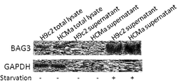
도 1a: 배양된 심장근육세포(cardiomyocytes)의 상청액(supernatant)에서 BAG3 단백질의 검출. 80%의 컨플루언시(confluency)로 인간(HCMa) 및 래트(H9c2) 심장근육세포가 10% FBS 존재 또는 부존재로 5% CO2 분위기, 37℃에서 16시간 동안 배양되었다. 상청액은 50 mM NaCl 및 0.05% IGEPAL 함유 버퍼에서 투석되고(dialyzed), 동결건조되고(lyophilized), 1 ml의 RIPA 버퍼(50 mM Tris HCl pH 7.6, 150 mM 염화나트륨(sodium chloride), 2 mM 소듐 오르소바나데이트(sodium orthovanadate), 4 mM EDTA, 10mM 소듐 피로포스페이트(sodium pyrophosphate), 1% NP-40, 0.1% 소듐 디옥시콜레이트(sodium deoxycholat))에서 재현탁되고, 웨스턴 블로팅(western blotting)에 의하여 항-BAG3 또는 항-GAPDH 항체로 분석되었다.
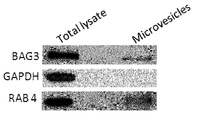
도 1b: 엑소사이트 소포(exocytic vesicle)에서 BAG3 단백질의 검출. H9c2 세포로부터 획득된 상청액은 순차적 원심분리가 수행되었다: (i) 세포 제거를 위하여, 2’000×g로 15분; (ii) 세포 파편(cellular debris)의 제거를 위하여, 10’000×g로 30분; (iii) 엑소사이트 소포를 침전시키기 위하여, 150’000×g로 90분. 상기 침전물은 150’000×g에서 90분 동안 PBS에서 한번 세척되고 웨스턴 블럿에 의하여 전체-세포 용해물(whole-cell lysate)와 비교로 항-BAG3 TOS-2 폴리클로날 항체로 분석되었다. Rab-4는 엑소사이트 소포를 위한 마커로서 분석되었다. 시토졸 단백질(cytosolic protein)인 GAPDH이 대조군으로 분석되었다.
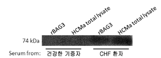
도 1c: 두 명의 건강한 기증자(donor) 및 두 명의 만성심부전 환자의 혈청이 웨스턴 블로팅에서 항-BAG3 항체 TOS-2 폴리클로날 항체로 분석되었다.
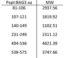
도 1d: 두 명의 만성심부전 환자에서 획득된 밴드(Bands)가 겔로부터 추출되고 프로그램 MASCOT를 이용하여 질량분석(mass spectrometry)에 의하여 그것의 아이덴티티(identity)가 분석되었다.
도 2.
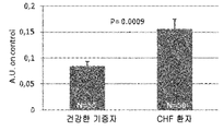
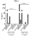
도 2a: CHF 환자로부터의 혈청에서 BAG3 단백질의 검출. BAG3 재조합 단백질 및 HCMa 세포의 전체 세포 용해물(whole-cell lysate)이 심부전 환자로부터 획득된 혈청 (1:40)으로 웨스턴 블로팅에 의하여 분석되었다. 건강한 기증자로부터의 혈청으로의 분석이 음성 대조군으로 수행되었다.
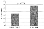
도 2b: ELISA 테스트에 의한 항-BAG3 항체의 검출. 50명의 (박출률 < 60%) CHF 환자들의 혈청을 특정 ELISA 테스트에서 항-BAG3 항체의 존재를 확인하기 위하여 50명의 건강한 기증자로부터의 혈청과 비교하였다. 결과는 임의 단위(arbitrary units)로서 표시되었다.
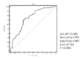
도 2c: ELISA 테스트 결과의 ROC 분석. 0.083 A.U. 에서의 컷-오프(cut-off)는 74% 민감도(sensitivity) 및 68% 특이성(specificity)으로 결론났다.
도 3.
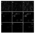
도 3a: HCMa 세포 (a, b, c) 및 J774 A1 세포 (d, e, f, g, h, i)에 결합하는 rBAG3-FITC의 검출을 위하여 수행된 직접 형광 공초점 현미경 분석(Confocal microscopy analysis). BAG3 재조합 단백질 및 정제된 BSA (SIGMA로부터 구입된 소혈청으로부터의 알부민)이 SIGMA로부터 구입된 FluoroTag FITC Conjugation Kit를 이용하여 제조자 지시에 따라 FITC에 결합되었다. 제조자 지시에 따라 계산된, 동량의 rBAG3-FITC (b, e) 및 BSA-FITC (h) 단백질이 0.1 % NaN3를 갖는 HCM 및 J774 A1 배양 미디어에 1시간 동안 첨가되었다. β-인테그린(integrin)이 대조군(a, d, g)으로서 분석되었다. 세포가 Zeiss LSM 공초점 현미경으로 분석되었다. 합성 이미지가 c, f 및 i으로 도시된다.
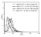
도 3b: BAG3는 대식세포(macrophages)에 결합한다. J774 A1 대식세포 (1×10⁶ cells/ml)가 다른 농도의 Fitc-BAG3 단백질(7, 14 및 70 nm)로 배양되었다. FITC-BSA (70 nM)가 음성 대조군(회색)으로서 사용되었다. 세포 형광이 흐름세포측정(flow cytometry)으로 분석되었다.
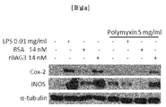
도 3c[패널 a] - BAG3로 배양된 J774 A1 대식세포에서 Cox-2 및 iNOS 레벨 분석. 80% 컨플루언시(confluency)에서 J774 A1 세포는 대조 배지(medium), BSA, LPS 또는 rBAG3으로 20시간 동안 배양되었다. 폴리믹신(Polymixin)이 대장균(E. coli)-유래 rBAG3의 효과가 내독소 오염의 존재로부터 독립적임을 증명하기 위하여 추가되었다. Cox-2 및 iNOS 발현은 웨스턴 블로팅에 의하여 세포 용해물(cell lysates)에서 분석되었다.
도 3d[패널 b] - BAG3로 배양된 J774 A1 대식세포로부터 방출된 아질산염(Nitrite)의 분석. 80% 컨플루언시에서 J774 A1 세포가 대조 배지, BSA, LPS 또는 rBAG3로 24시간 동안 배양되었다. 각 샘플로부터 100 μl의 상청액을 100 μl의 그리스 시약(Griess reagent)으로 배양하였고; 550 nm(OD550)에서 광학 밀도가 Beckman DU62 분광 측광기(spectrophotometer)로 측정되었다. 아질산염(Nitrite) 농도는 샘플의 OD550과 아질산나트륨(sodium nitrite)의 표준 곡선의 그것과 비교하여 평가하였다.
도 3e[패널 c] - BAG3로 배양된 J774 A1 대식세포로부터 방출된 IL-6의 분석. 80% 컨플루언시에서 J774 A1 세포가 대조 배지, BSA, LPS 또는 rBAG3로 5시간 동안 배양되었다. BAG3 펩티드 (펩티드 1, 펩티드 2, 펩티드 3, 펩티드 4, 또는 스크램블 펩티드(scrambled peptide)) 625 nM 가 추가되어 BAG3 활성 차단에 대한 그들의 능력을 증명하였다. IL-6 생산이 ELISA 테스트를 이용하여 세포 배양 배지에서 측정되었다. IL-6 농도는 샘플의 OD와 재조합 IL-6의 표준 곡선의 그것과 비교함으로써 평가되었다.
도 4.
도 4a:
정상 췌장 조직에서 모노클로날 항-BAG3 항체 AC-1을 이용한 BAG3 염색의 대표 이미지. 섹션들이 헤마톡실린로 대비염색되었다. 염색이 랑게르한스섬(Langerhans islets)의 보통의 확실성(moderate positivity)을 보여주고, 반면 정상 췌관(pancreatic ducts) 및 췌장 소엽 세포(pancreatic acinar cells)은 어떠한 BAG3 발현을 가지지 않았다.
도 4b: 바이오틴 부착 2차 항체(biotinylated secondary antibody)로 드러난 모노클로날 항-BAG3 항체를 이용하여 염색된 BAG3 낮은 양성(low positive) 및 BAG3 높은 양성(high positive) 종양 세포의 대표 이미지. 섹션들이 헤마톡실린로 대비염색되었다. 두 개의 서로 다른 확대배율이 도시된다: 100×(좌측 패널) 및 400× (우측 패널). 400× 확대배율을 이용하여 10개의 오버랩핑되지 않는 필드에서 전체 암 세포에 대한 양성 세포의 개수를 카운팅함으로써 상기 샘플에서 양성 암 세포의 비율에 기초하여 점수를 할당하였다. 상기 기재한 것처럼 계산된, BAG3 양성 세포의 중간 퍼센티지는 40%였고, 이 수치는 낮은 및 높은 양성 샘플을 분리하기 위한 컷-오프(cut-off)로서 이용되었다.
도 4c:
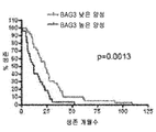
생존 곡선(Survival curves)이 낮은 BAG3 염색(양성 세포의 ≤40%)을 갖는 39명의 환자와 높은 BAG3 염색(양성 세포의 >40%)을 갖는 27명의 환자를 비교하여 작성되었다. 모든 환자들에 대하여 상기 췌장 선암 세포(pancreatic adenocarcinoma)의 RO 절제(R0 resection)가 수행되어 분석되었다. 생존 중앙값(Median survival)은 높은 양성 그룹에서 12개월부터 낮은 양성 그룹에서 23개월까지 증가한다. 로그-랭크 테스트 p-수치=0.0013
도 5.
몇몇 류마티스 관절염의 활액막 조직(synovial tissues)에서 염색된 BAG3 대표 이미지. BAG3 양성이 활액막 섬유아세포(synovial fibroblasts) 및 염증 침윤(inflammatory infiltrates)에서 관찰되었다. 섹션들이 헤마톡실린로 대비염색되었다.
도 6.
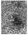
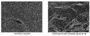
음성 판정된 정상 유로시스티스(normal urocystis)에서, 그리고 종양 세포의 세포질의 BAG3에 대하여 높은 양성으로 판정된 이행 세포 방광 암종(transitional cell bladder carcinoma)에서 BAG3 염색의 대표 이미지. 섹션들이 헤마톡실린로 대비염색되었다.
도 7.
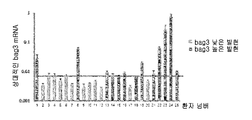
도 7a.
qRT-PCRd 의하여 평가된 bag3 mRNA 상대적 발현이 그래프에서 도시된다. 여기서 값은 평균 + S.D로 보고된다. 푸른선은 계산된 중앙값을 대표한다.
도 7b.
생존 분석이 모든 환자들에 있어서 qRT-PCRd로 분석되었다. 높은 bag3 발현을 갖는 13명의 환자들이 낮은 bag3 발현을 갖는 12명의 환자들(생존 중앙값 = 32.0 개월)에 비하여 더 짧은 생존 (생존 중앙값 = 19.0 개월)을 가졌다. 로그-랭크 테스트 p-수치 = 0.0198.
도 8.
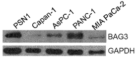
도 8a:
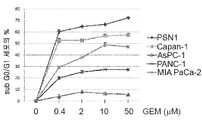
췌장암 세포주(PSN1, Capan-1, AsPC-1, PANC-1 및 MIA PaCa-2)가 그래프에서 표시된 것처럼 서로 다른 농도의 젬사이타빈(gemcitabine)으로 처리되었다. 48시간 후에, 아포프토시스 세포사멸(apoptotic cell death)이 분석되었다. 그래프는 Sub G0/G1 세포 (± S.D.)의 평균 퍼센티지를 도시한다. 데이터는 세 개의 독립된 실험을 대표한다.
도 8b:
췌장암 세포주에서 BAG3의 웨스턴 블럿 분석; GAPDH 관리 단백질(housekeeping protein) 컨텐츠가 동일 로딩 조건을 모니터링하기 위하여 사용되었다.
도 8c:
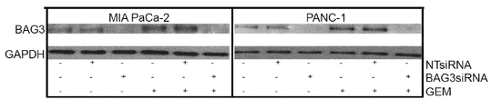
MIA PaCa-2 및 PANC-1 세포주는 지시된 시간 동안 2 μM 젬사이타빈(GEM)으로 처리되어 BAG3 단백질 발현 레벨이 웨스턴 블럿으로 모니터링되었다.
도 8d:
bag3 mRNA 레벨이 RT-PCR에 의하여 분석되었다; 그래프는 상대적인 bag3 mRNA 레벨 (± S.D.)을 나타내고 데이터는 세 개의 독립된 실험을 대표한다.
도 8e:
MIA PaCa-2 및 PANC-1 세포주는 72시간 동안 BAG3 siRNA 또는 비-타켓 siRNA (NTsiRNA)로 세포 감염시키고, 그 후 24시간 동안 2 μM 젬사이타빈(GEM)으로 처리되었다. BAG3 레벨이 웨스턴 블럿으로 분석되고 GAPDH 레벨이 동일 로딩 조건을 모니터링하기 위하여 측정되었다.
도 8f:
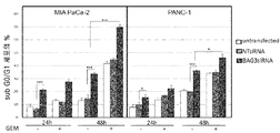
MIA PaCa-2 및 PANC-1 세포주는 상기 기재한 바와 같이 감염(transfected)되었고 24시간 또는 48시간 동안 2 μM 젬사이타빈(GEM)으로 처리되었다. 아포프토시스 세포사멸(apoptotic cell death) 상기 기재한 것과 같이 분석되었다. 그래프는 Sub G0/G1 세포 (± S.D.)의 평균 퍼센티지를 나타낸다. 데이터는 세 개의 독립된 실험을 대표한다.
도 9.
도 9a: ELISA 테스트에 의한 BAG3 특이적 면역-복합체의 검출. 특이적 ELISA 테스트에서 BAG3 특이적 면역-복합체의 존재를 위하여, 55명의 췌장 선암(pancreatic adenocarcinoma) 환자의 혈청이 51명의 건강한 기증자의 혈청과 비교하였다. 결과는 임의 단위 +S. E.로서 표시되었다.
도 9b: ELISA 테스트 결과의 ROC 분석. 0.183 A.U.의 컷-오프(Cut-off)는 65% 민감도 및 78% 특이성으로 나타났다.
도 10.
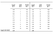
Bag3 점수는 내생 GAPDHdml 발현을 위하여 표준화(normalizing)한 후에, 상기 비교방법을 이용하여 상대 발현 레벨으로서, 각 샘플에서 계산되었다. 상기 bag3 점수의 평균은 2 그룹의 환자들에서 평가되었다: 낮은 PCA3 점수 환자들 및 높은 PCA3 점수 환자들. 상기 두 그룹 사이의 차이는 스튜던트 t 테스트(student t test)에 의하여 측정되어 중요하게 결론지어졌다: P = 0.001.The features and advantages of the present invention will be more fully described in the detailed description set forth below and in the accompanying drawings, which are given by way of illustration and non-limiting examples and in which:
Fig.
Figure 1a: Detection of BAG3 protein in supernatant of cultured cardiomyocytes. Human (HCMa) and rat (H9c2) cardiac muscle cells were incubated for 16 hours at 37 ° C in 5% CO 2 atmosphere with or without 10% FBS at 80% confluency. The supernatant was dialyzed, lyophilized and resuspended in 1 ml of RIPA buffer (50 mM Tris HCl pH 7.6, 150 mM sodium chloride, 2 mM sodium orchid) in a buffer containing 50 mM NaCl and 0.05% IGEPAL Resuspended in sodium orthovanadate, 4 mM EDTA, 10 mM sodium pyrophosphate, 1% NP-40, 0.1% sodium deoxycholate), western blotting, ≪ / RTI > anti-BAG3 or anti-GAPDH antibody.
Figure 1b: Detection of BAG3 protein in exocytic vesicles. The supernatant obtained from H9c2 cells was subjected to sequential centrifugation: (i) 15 min at 2'000 x g for cell removal; (ii) 30 min at 10'000 x g for removal of cellular debris; (iii) 90 minutes at 150'000 x g to precipitate exocytose vesicles. The precipitate was washed once in PBS for 90 minutes at 150'000 x g and analyzed for anti-BAG3 TOS-2 polyclonal antibody by Western blot compared to whole-cell lysate. Rab-4 was analyzed as a marker for exocytose vesicles. GAPDH, a cytosolic protein, was analyzed as a control.
Figure 1c: Serum from two healthy donors and two chronic heart failure patients was analyzed with anti-BAG3 antibody TOS-2 polyclonal antibody in Western blotting.
1d: Bands obtained from two chronic heart failure patients were extracted from the gel and its identity was analyzed by mass spectrometry using the program MASCOT.
Fig.
Figure 2a: Detection of BAG3 protein in serum from CHF patients. Whole-cell lysate of BAG3 recombinant protein and HCMa cells was analyzed by western blotting with serum (1:40) obtained from heart failure patients. Analysis from sera from healthy donors was performed as negative control.
Figure 2b: Detection of anti-BAG3 antibody by ELISA test. Serum of 50 (dilution <60%) CHF patients was compared to serum from 50 healthy donors to confirm the presence of anti-BAG3 antibody in a specific ELISA test. Results are expressed as arbitrary units.
Figure 2c: ROC analysis of ELISA test results. The cut-off at 0.083 AU resulted in 74% sensitivity and 68% specificity.
3.
Figure 3a: Direct fluorescence microscopy analysis performed for the detection of rBAG3-FITC binding to HCMa cells (a, b, c) and J774 A1 cells (d, e, f, g, h, analysis). BAG3 recombinant protein and purified BSA (albumin from bovine serum purchased from SIGMA) were bound to FITC using the FluoroTag FITC Conjugation Kit purchased from SIGMA according to the manufacturer's instructions. It is calculated according to the manufacturer's instructions, the same amount of FITC-rBAG3 (b, e) and BSA-FITC (h) proteins were added for 1 hour in the HCM and J774 A1 culture media with 0.1% NaN 3. beta -induced integrin was analyzed as a control (a, d, g). Cells were analyzed with a Zeiss LSM confocal microscope. The composite image is shown as c, f, and i.
Figure 3b: BAG3 binds to macrophages. J774 A1 macrophages (1 x 10⁶ cells / ml) were incubated with different concentrations of Fitc-BAG3 protein (7, 14 and 70 nM). FITC-BSA (70 nM) was used as a negative control (gray). Cell fluorescence was analyzed by flow cytometry.
Figure 3c [Panel a] - Analysis of Cox-2 and iNOS levels in J774 A1 macrophages cultured with BAG3. At 80% confluency, J774 A1 cells were cultured with the control medium, BSA, LPS or rBAG3 for 20 hours. Polymixin was added to demonstrate that the effect of E. coli-derived rBAG3 is independent of the presence of endotoxin contamination. Cox-2 and iNOS expression was analyzed in cell lysates by Western blotting.
Figure 3d [Panel b] - Analysis of nitrite released from J774 A1 macrophages cultured with BAG3. At 80% confluence, J774 A1 cells were incubated with control medium, BSA, LPS or rBAG3 for 24 hours. From each sample, 100 μl of supernatant was incubated with 100 μl of Griess reagent; Optical density at 550 nm (OD550) was measured with a Beckman DU62 spectrophotometer. The nitrite concentration was evaluated by comparison with that of a standard curve of the sample OD550 and sodium nitrite.
Figure 3e [Panel c] - Analysis of IL-6 released from J774 A1 macrophages cultured with BAG3. At 80% confluence, J774 A1 cells were incubated with control medium, BSA, LPS or rBAG3 for 5 hours. 625 nM of BAG3 peptide (
FIG.
4a:
A representative image of BAG3 staining using monoclonal anti-BAG3 antibody AC-1 in normal pancreatic tissue. Sections were stained contrasted with hematoxylin. The staining showed the moderate positivity of Langerhans islets, while the pancreatic ducts and pancreatic acinar cells did not have any BAG3 expression.
Figure 4b: Representative image of BAG3 low positive and BAG3 high positive tumor cells stained with a monoclonal anti-BAG3 antibody revealed as a biotinylated secondary antibody. Sections were stained contrasted with hematoxylin. Two different magnification factors are shown: 100 x (left panel) and 400 x (right panel). Scores were assigned based on the proportion of benign cancer cells in the sample by counting the number of benign cells for all cancer cells in 10 non-overlapping fields using a 400x magnification. The median percentage of BAG3-positive cells, calculated as described above, was 40%, which was used as a cut-off for separating low and high positive samples.
4c:
We compared 39 patients with low BAG3 staining (≤40% of benign cells) and 27 patients with high BAG3 staining (> 40% of benign cells) with low survival curves. RO resection of the pancreatic adenocarcinoma was performed and analyzed for all patients. Median survival increases from 12 months in the high positive group to 23 months in the low positive group. Log-rank test p-value = 0.0013
Figure 5.
Representative images of BAG3 stained in synovial tissues of several rheumatoid arthritis. BAG3 positive was observed in synovial fibroblasts and inflammatory infiltrates. Sections were stained contrasted with hematoxylin.
6.
A representative image of BAG3 staining in transitional cell bladder carcinoma determined to be highly positive for normal urocystis negative and negative for BAG3 in the cytoplasm of tumor cells. Sections were stained contrasted with hematoxylin.
7.
7a.
The relative expression of bag3 mRNA evaluated by qRT-PCRd is shown in the graph. Where the value is reported as mean + SD. The blue line represents the calculated median value.
7b.
Survival analysis was analyzed by qRT-PCRd in all patients. Thirteen patients with high bag3 expression had shorter survival (median survival = 19.0 months) than 12 patients with low bag3 expression (median survival = 32.0 months). Log-rank test p-value = 0.0198.
Figure 8.
8a:
Pancreatic cancer cell lines (PSN1, Capan-1, AsPC-1, PANC-1 and MIA PaCa-2) were treated with different concentrations of gemcitabine as indicated in the graph. After 48 hours, apoptotic cell death was analyzed. The graph shows the average percentage of Sub G0 / G1 cells (+ - SD). The data represent three independent experiments.
8b:
Western blot analysis of BAG3 in pancreatic cancer cell lines; GAPDH housekeeping protein content was used to monitor the same loading conditions.
8c:
MIA PaCa-2 and PANC-1 cell lines were treated with 2 [mu] M gemcitabine (GEM) for the indicated times and BAG3 protein expression levels were monitored by Western blot.
8d:
bag3 mRNA levels were analyzed by RT-PCR; The graph shows relative bag3 mRNA levels (± SD) and data represent three independent experiments.
8E:
MIA PaCa-2 and PANC-1 cell lines were infected with BAG3 siRNA or non-target siRNA (NTsiRNA) for 72 hours and then treated with 2 [mu] M gemcitabine (GEM) for 24 hours. BAG3 levels were analyzed by Western blot and GAPDH levels were measured to monitor the same loading conditions.
8f:
MIA PaCa-2 and PANC-1 cell lines were transfected as described above and treated with 2 [mu] M gemcitabine (GEM) for 24 or 48 hours. Apoptotic cell death was analyzed as described above. The graph represents the average percentage of Sub G0 / G1 cells (± SD). The data represent three independent experiments.
Figure 9.
Figure 9a: Detection of BAG3 specific immunoconjugate by ELISA test. For the presence of BAG3 specific immunoconjugates in a specific ELISA test, sera from 55 patients with pancreatic adenocarcinoma were compared to serum from 51 healthy donors. The results are expressed as arbitrary units + SE.
Figure 9b: ROC analysis of ELISA test results. The cut-off of 0.183 AU was 65% sensitive and 78% specific.
10.
Bag3 scores were normalized for endogenous GAPDHdml expression and then calculated for each sample as relative expression levels using the above comparison method. The mean of the bag3 scores was assessed in two groups of patients: those with low PCA3 scores and those with high PCA3 scores. The difference between the two groups was determined by student t test and was significantly concluded: P = 0.001.
본 발명은 병리 상태의 진단에서 생화학적 마커로서의 용도로서 BAG3 RNA 또는 그 단편에 대한 것이다.The present invention is directed to BAG3 RNA or fragments thereof for use as biochemical markers in the diagnosis of pathological conditions.
본 발명에서 용어 "BAG3 RNA 단편(BAG3 RNA fragment)"은 150-300 뉴클레오티드의 단편을 의미한다.The term "BAG3 RNA fragment" in the present invention means a fragment of 150-300 nucleotides.
본 발명에서 용어 "진단(diagnosis)" 은 가능한 질병 또는 질환을 결정 또는 확인하기 위하여 시도되는 절차를 의미하는 의료적 진단(종종 단순히 진단으로 기재됨)을 의미한다. 본 발명의 진단은 또한 초기 진단(early diagnosis)을 포함한다. The term "diagnosis" in the present invention means a medical diagnosis (often simply referred to as diagnosis), which means a procedure attempted to determine or identify a possible disease or disorder. The diagnosis of the present invention also includes an early diagnosis.
용어 "초기 진단"은 특이적 또는 비특이전 증상 전에 병리 상태를 식별하기 위한 테스트의 능력을 의미한다. The term "early diagnosis" refers to the ability of a test to identify a pathological condition prior to a specific or non-specific symptom.
항-BAG3 항체는 혈청에서 현저하게 검출되었다. 지금까지 그러한 항체들이 생리학적 또는 병리 상태의 혈청에서 발견되지 않았다. 혈청 내의 항-BAG3 항체의 검출은 치료 모니터링 및 식별을 위한 툴로서, 진단, 초기 진단 및 예후 목적(prognostic purposes), 예후 판정(risk stratification)을 위하여 이용되는 빠르고 비-침습적 기술의 이점을 가진다. Anti-BAG3 antibody was detected significantly in serum. Until now, such antibodies have not been found in physiological or pathological serum. The detection of anti-BAG3 antibodies in serum is a tool for treatment monitoring and identification and has the advantage of fast, non-invasive techniques used for diagnosis, initial diagnosis and prognostic purposes, and for risk stratification.
본 발명에 있어서, 혈청(serum)은 혈액 세포(blood cell) 또는 응혈 인자(clotting factor)가 아닌 혈액의 성분인 것으로 의도된다; 그것은 피브리노겐(fibrinogens)이 제거된 혈장(blood plasma)이다. 혈청은 혈액 응고(blood clotting, coagulation)에 사용되지 않는 모든 단백질과 모든 전해액(electrolytes), 항체, 항원, 호르몬 및 어떠한 외인성 물질(exogenous substances)을 포함한다. In the present invention, serum is intended to be a constituent of blood, not a blood cell or clotting factor; It is a blood plasma from which fibrinogens are removed. Serum contains all proteins and all electrolytes, antibodies, antigens, hormones and some exogenous substances that are not used for blood clotting (coagulation).
본 발명에 있어서, 혈장(plasma)은 상청액의 전체 혈액에서 상기 혈액 세포를 정상적으로 가지는 혈액의 밀짚-컬러/옅은-황색(straw-colored/pale-yellow) 액체 성분인 것으로 의도된다. 이는 피브리노겐과 같은 응혈 인자를 포함한다. In the present invention, plasma is intended to be a straw-colored / pale-yellow liquid component of blood normally containing the blood cells in the whole blood of the supernatant. It contains a coagulation factor such as fibrinogen.
다른 실시예에서, 본 발명은 생화학적 마커로서 항-BAG3 항체의 용도를 제공하고, 여기에서 상기 항-BAG3 항체는 가용성 BAG3(soluble BAG3)에 결합하여 면역 복합체를 형성한다. In another embodiment, the invention provides the use of an anti-BAG3 antibody as a biochemical marker, wherein said anti-BAG3 antibody binds to soluble BAG3 to form an immune complex.
가용성 BAG3에 결합하거나 결합하지 않고 면역 복합체를 형성하는 항-BAG3 항체는 생물학적 샘플에서 지금 효과적으로 검출될 수 있고, 병리 상태의 진단에서 마커로서 사용될 수 있다. 생물학적 샘플 내의 그러한 항체 및/또는 면역 복합체의 검출은 또한 진단 및/또는 예후 목적을 위한 빠르고 비-침습적 기술의 효과를 가진다. Anti-BAG3 antibodies that bind immune complexes with or without binding to soluble BAG3 can now be effectively detected in biological samples and can be used as markers in the diagnosis of pathological conditions. The detection of such antibodies and / or immunoconjugates in a biological sample also has the effect of rapid, non-invasive techniques for diagnostic and / or prognostic purposes.
본 발명에서, 면역 복합체 또는 단백질/항체 복합체는 항체의 가용성 항원에대한 필수적 결합으로 의도되고, 상기 결합된 항원은 특이적 에피토프(epitope)로서 작용하고, 항체로의 결합은 단일 면역 복합체로 지칭된다.In the present invention, the immunoconjugate or protein / antibody complex is intended as an essential binding to the soluble antigen of the antibody, and the bound antigen acts as a specific epitope, and binding to the antibody is referred to as a single immunoconjugate .
그러한 면역 복합체는 항체 검출을 위한 것으로 보여진 것과 동일한 이점을 가지는데, 혈청의 매우 적은 양이 상기 면역 복합체의 검출에 요구되기 때문이다. 상기 가용성 BAG3의 검출에 요구되는 혈청의 양은, 상기 단백질/항체 (면역) 복합체의 검출에 요구되는 것보다 훨씬 더 크다. 자유 항체뿐만 아니라 면역 복합체는 특이적 질환의 초기 단계에서 검출가능하고 합병증의 위험을 효과적으로 예측가능하며 치료의 효과를 모니터링할 수 있도록 한다. Such immunoconjugates have the same advantages as those shown for antibody detection since a very small amount of serum is required for the detection of the immunoconjugate. The amount of serum required for the detection of soluble BAG3 is much greater than that required for the detection of the protein / antibody (immune) complex. Free antibodies as well as immune complexes are detectable at the early stages of a specific disease, effectively predict the risk of complications, and monitor the effectiveness of treatment.
본 발명의 또 다른 실시예는 항-BAg3 항체 또는 상기 면역 복합체의 병리 상태의 생물학적 마커로서의 용도로서, 여기에서 상기 병리 상태는 심장병, 암, 당뇨병, 염증 및 피부, 신경, 뼈, 혈관 및 연결조직의 염증관련질환이다. Another embodiment of the invention is the use of an anti-BAg3 antibody or a biological marker of the pathological state of the immunoconjugate wherein the pathological condition is selected from the group consisting of heart disease, cancer, diabetes, inflammation and skin, nerves, Of inflammation related diseases.
바람직하게 상기 심장병은 다음으로 구성되는 군으로부터 선택된다: 협심증(angina pectoris), 경색전 협심증(pre-infarction angina), 국소 빈혈(ischemia), 심근경색증( myocardial infarction), 심부전(heart failure), 급성 관상동맥 질환(acute coronary disease), 급성심부전(acute heart failure), 만성심부전(chronic heart failure), 및 의인성 심장질환(iatrogenic heart disease).Preferably the heart disease is selected from the group consisting of: angina pectoris, pre-infarction angina, ischemia, myocardial infarction, heart failure, acute Acute coronary disease, acute heart failure, chronic heart failure, and iatrogenic heart disease.
본 발명의 다른 실시예는 생물학적 샘플에서 항-BAG3 항체 또는 가용성 BAG3에 결합되어 면역 복합체를 형성하는 항-BAG3 항체의 존재 검출 방법으로서 다음 단계들을 포함한다:Another embodiment of the invention includes the following steps as a method for detecting the presence of an anti-BAG3 antibody in a biological sample or the presence of an anti-BAG3 antibody that binds to soluble BAG3 to form an immune complex:
a. 혈청, 혈장으로 구성된 생물학적 샘플을 획득하는 단계,a. Obtaining a biological sample composed of serum and plasma,
b. 상기 생물학적 샘플에서 항-BAG3 또는 BAG3 관련 항체의 존재를 결정하는 단계.
b. Determining the presence of an anti-BAG3 or BAG3 related antibody in said biological sample.
본 발명에 따른 방법은 건강한 대상자와 BAG3-관련 질환(병리)에 의하여 영향받은 환자 사이에서 항-BAG3 항체 및/또는 BAG3/항체 복합체 사이의 현저한 차이점을 검출하도록 하는 이점을 갖는다. 상기 제안된 분석 방법은 건강한 사람이 속한 집단으로부터 심장병 환자(cardiac patients) 집단을 통계적으로 현저히 구분이 되도록 한다. 또한 그러한 심장 질환을 가진 환자들을 위험도(심부전, HF)가 높은 서브 그룹의 환자들로 계층화할 수 있다.The method according to the present invention has the advantage of detecting significant differences between anti-BAG3 antibodies and / or BAG3 / antibody complexes between healthy subjects and patients affected by BAG3-related diseases (pathologies). The proposed methodology allows a statistically significant distinction of groups of cardiac patients from a population of healthy persons. Patients with such heart disease can also be stratified into subgroups of patients with high risk (heart failure, HF).
다른 측면에서, 본 발명은 생물학적 샘플 내에서 항-BAG3 항체 또는 가용성 BAG3에 결합되어 면역 복합체를 형성하는 항-BAG3 항체의 존재 검출 방법으로서 다음 단계를 포함한다: In another aspect, the invention includes a method for detecting the presence of an anti-BAG3 antibody or an anti-BAG3 antibody that binds to soluble BAG3 in a biological sample to form an immune complex, comprising the steps of:
c. 상기 생물학적 샘플로부터 획득된 수치를 참조 수치 또는 건강한 기증자로부터 획득된 수치와 비교하는 단계. c. Comparing the numerical value obtained from said biological sample with a reference value or a numerical value obtained from a healthy donor.
바람직한 측면에서, 본 발명에 따른 방법은, 상기 결정 단계 b는 ELISA 테스트에 의하여 수행되는 방법이다.
In a preferred aspect, the method according to the present invention is a method wherein said determination step b is carried out by an ELISA test.
본 발명에 따른 방법은 질환(병증, 또는 병리, pathologies)의 평가, 질환 위험 및/또는 그들의 합병증, 및 치료 모니터링을 가능하도록 하는 생물학적 마커의 빠르고 비-침습적 검출이 가능하도록 하는 이점을 가진다. The method according to the present invention has the advantage of enabling rapid, non-invasive detection of biological markers that enable evaluation of diseases (pathology, pathologies), disease risk and / or their complications, and therapeutic monitoring.
다른 측면에 따르면, 본 발명은 검출 방법에 대한 것으로서, 여기에서 상기 항-BAG3 항체 또는 가용성 BAG3에 결합되어 면역 복합체를 형성하는 상기 항-BAG3 항체의 존재는 병리 상태(pathological condition)와 관련된다. According to another aspect, the present invention is directed to a detection method, wherein the presence of said anti-BAG3 antibody or said anti-BAG3 antibody which binds to soluble BAG3 to form an immune complex is associated with pathological conditions.
바람직한 실시예에서, 상기 병리 상태는 심장병, 암, 당뇨병, 염증 및 피부, 신경, 뼈, 혈관 및 연결 조직의 염증 관련 질환으로 구성된 군으로부터 선택된다. 특히, 상기 심장병은 다음으로 구성되는 군으로부터 선택된다: 협심증(angina pectoris), 경색전 협심증(pre-infarction angina), 심근경색증( myocardial infarction), 심부전(heart failure), 급성 관상동맥 질환(acute coronary disease), 급성심부전(acute heart failure), 만성심부전(chronic heart failure), 또는 의인성 심장질환(iatrogenic heart disease).In a preferred embodiment, the pathology is selected from the group consisting of heart disease, cancer, diabetes, inflammation and inflammation related diseases of the skin, nerves, bones, blood vessels and connective tissue. In particular, the heart disease is selected from the group consisting of: angina pectoris, pre-infarction angina, myocardial infarction, heart failure, acute coronary disease acute heart failure, chronic heart failure, or iatrogenic heart disease.
본 발명은 또한 생물학적 샘플 내에서 항-BAG3 항체 또는 가용성 BAG3에 결합되어 면역 복합체를 형성하는 항-BAG3 항체의 검출을 위한 ELISA 키트에 대한 것이다. The invention also relates to an ELISA kit for the detection of an anti-BAG3 antibody or an anti-BAG3 antibody that binds to soluble BAG3 to form an immune complex in a biological sample.
본 발명에 따른 상기 ELISA 키트는 가용성 BAG3 및 인간 면역글로불린의 인식 가능한 항체를 캡쳐하기 위하여, 항-BAG3 항체 또는 BAG3-특이적 마우스 모노클로날 항체 AC-1, AC-2, AC-3, AC-4 및 AC-5의 캡쳐를 위한 BAG3 재조합 단백질을 포함한다.The ELISA kit according to the present invention can be used to detect antibodies BAG3 and BAG3-specific mouse monoclonal antibodies AC-1, AC-2, AC-3, AC -4 and AC-5. ≪ / RTI >
상기 항체는 인간 면역글로불린의 인식 가능한 효소-링크 항체일 수 있다.The antibody may be a recognizable enzyme-linked antibody of a human immunoglobulin.
본 발명은 또한 생물학적 샘플에서 BAG3-관련 항체의 검출을 위한 키트에 대한 것이고, 인간 면역글로불린을 인식할 수 있는 검출을 위하여 가용성 BAG3 및 효소-링크 항체를 캡쳐링하는, BAG3-특이적 마우스 모노클로날 항체 AC-1, AC-2, AC-3, AC-4 및 AC-5로 ELISA에 의하여 수행된다. The invention also relates to a kit for the detection of a BAG3-related antibody in a biological sample, which comprises a BAG3-specific mouse monoclonal antibody that captures soluble BAG3 and an enzyme-linked antibody for detection capable of recognizing human immunoglobulin It is carried out by ELISA with antibodies AC-1, AC-2, AC-3, AC-4 and AC-5.
본 발명은 또한 생물학적 샘플 내에서 BAG3 단백질의 검출을 위한 면역조직화학(Immunohistochemistry, IHC) 키트에 대한 것으로서, 여기에서 상기 생물학적 샘플은 바람직하게 조직 샘플이다. 조직 샘플은 생검(biopsies), 동결 조직(frozen tissues), 파라핀 포매 조직(paraffin embedded tissues)이다. The present invention also relates to an immunohistochemistry (IHC) kit for the detection of BAG3 protein in a biological sample, wherein said biological sample is preferably a tissue sample. Tissue samples are biopsies, frozen tissues, and paraffin embedded tissues.
본 발명에 따른 상기 IHC 키트는 BAG3-특이적 항체 및 상기 염색에 요구되는 프로브를 포함하는 시약을 포함한다. The IHC kit according to the present invention comprises a reagent comprising a BAG3-specific antibody and a probe required for said staining.
상기 BAG3-특이적 항체는 상기 검출을 위하여 마우스 면역글로불린을 인식할 수 있는 검출을 위한 마우스 모노클로날 항체(mouse monoclonal antibody) AC-1, AC-2 및 AC-3 및/또는 효소-링크 항체(enzyme-linked antibody)일 수 있다. The BAG3-specific antibody may be a mouse monoclonal antibody AC-1, AC-2 and AC-3 and / or an enzyme-linked antibody for detection capable of recognizing mouse immunoglobulin for the detection (enzyme-linked antibody).
특히, 상기 IHC 키트는 췌장 절제(pancreas resection)를 겪은 환자로부터의 췌장 암종(pancreatic carcinoma) 조직 샘플 100%에서 BAG3 단백질을 효과적으로 드러내도록 하고, 방광 암종(bladder carcinoma) 샘플에서 가장 잘 발현된다.In particular, the IHC kit effectively reveals BAG3 protein in 100% of pancreatic carcinoma tissue samples from patients undergoing pancreatic resection, and is best expressed in bladder carcinoma samples.
게다가, BAG3 단백질은, 또한 랑게르한스섬(Langerhans islet)에 있는 정상 췌장 조직 내에서 IHC에 의한 BAG3 검출을 위한 키트로 드러나질 수 있는데, 반면, 다른 정상 조직(예를 들어 정상 유로시스티스(urocystis))은 음성으로 나타난다.In addition, the BAG3 protein can also be expressed as a kit for BAG3 detection by IHC in normal pancreatic tissue in the Langerhans islet, whereas other normal tissues (e.g. normal urocystis) Appears as a voice.
BAG3 양성은 또한 시노비알 섬유아세포(synovial fibroblasts) 및 류마티스 관절염(rheumatoid arthritis) 조직 샘플의 염증 침윤물(inflammatory infiltrates)에서 관찰될 수 있다.BAG3 positivity can also be observed in inflammatory infiltrates of synovial fibroblasts and rheumatoid arthritis tissue samples.
PDAC이 발병된 환자들의 장기간 생존이 매우 미미하다: 오직 환자의 약 4%가 진단 후 5년 생존할 것이다. 정말로, 외과적 절제가 현재 유일한 치료 방법이고, 그러나 약 20%의 환자들만이 절제가능한 질환(resectable disease)으로 진단된다; 게다가, 그러한 환자들의 큰 부분 집합(약 80%)에서 전이 과정이 이미 진단에서 일어나고, 외과적 절제 후에 정말로 원격 전이(distant metastases)가 나타난다. 그러므로 췌장암의 발전에서 초기 단계에 대한 더 나은 이해가 요구되고 그들을 검출하도록 할 수 있는 분자를 동정할 필요가 있다. 또한, 더 나은 예후(prognosis)와 치료 선택을 도와줄 수 있는 마커들이 많이 요구된다. Long-term survival of patients with PDAC is very small: only about 4% of patients will survive 5 years after diagnosis. Indeed, surgical resection is currently the only treatment, but only about 20% of patients are diagnosed with resectable disease; Furthermore, in a large subset (about 80%) of such patients, the metastatic process already occurs in the diagnosis and indeed distant metastases appear after surgical resection. Therefore, a better understanding of the early stages of development of pancreatic cancer is required and it is necessary to identify molecules that can detect them. There are also a number of markers that can help with better prognosis and treatment choices.
상기 BAG3 IHC 키트는 PDAC 환자의 예후(prognosis)를 효과적으로 식별하도록 한다. IHC에 의하여 식별된 BAG3 발현의 강도는 환자의 생존과 상관관계 있는 것으로 확인되었다. 그러므로 이는 예후 및 치료 선택을 위하여 사용될 수 있다. The BAG3 IHC kit allows effective identification of the prognosis of PDAC patients. The intensity of BAG3 expression identified by IHC was found to be correlated with patient survival. It can therefore be used for prognosis and treatment selection.
본 발명의 다른 실시예는 병리 상태의 진단에서 생화학적 마커로서의 용도를 가지는 BAG3 RNA 또는 그 단편이다. Another embodiment of the invention is BAG3 RNA or a fragment thereof having utility as a biochemical marker in the diagnosis of pathological conditions.
바람직하게, 상기 BAG3 RNA는 BAG3 mRNA이다.Preferably, the BAG3 RNA is BAG3 mRNA.
상기 BAG3 RNA는 SEQ ID N. 18(Reference: NCBI PubMed, XM 055575.1 Homo sapiens BCL2-associated athanogene 3 (BAG3) mRNA)의 BAG3 뉴클레오티드 서열에 대응한다.The BAG3 RNA corresponds to the BAG3 nucleotide sequence of SEQ ID N. 18 (Reference: NCBI Pubmed, XM 055575.1 Homo sapiens BCL2-associated athanogene 3 (BAG3) mRNA).
SEQ ID NO: 18.SEQ ID NO: 18.
1gcggagctcc gcatccaacc ccgggccgcg gccaactttt1gcggagctcc gcatccaacc ccgggccgcg gccaactttt
ttggactgga ccagaagttt ctagccggcc agttgctacc ttggactgga ccagaagttt ctagccggcc agttgctacc
tccctttatc tcctccttcc cctctggcag cgaggaggcttccctttatc tcctccttcc cctctggcag cgaggaggct
atttccagac acttccaccc ctctctggcc acgtcacccc atttccagac acttccaccc ctctctggcc acgtcacccc
cgcctttaat tcataaaggt gcccggcgcc ggcttcccgg cgcctttaat tcataaaggt gcccggcgcc ggcttcccgg
acacgtcggc ggcggagagg ggcccacggc ggcggcccggacacgtcggc ggcggagagg ggcccacggc ggcggcccgg
ccagagactc ggcgcccgga gccagcgccc cgcacccgcg ccagagactc ggcgcccgga gccagcgccc cgcacccgcg
ccccagcggg cagaccccaa cccagcatga gcgccgccac ccccagcggg cagaccccaa cccagcatga gcgccgccac
ccactcgccc atgatgcagg tggcgtccgg caacggtgacccactcgccc atgatgcagg tggcgtccgg caacggtgac
cgcgaccctt tgccccccgg atgggagatc aagatcgacc cgcgaccctt tgccccccgg atgggagatc aagatcgacc
cgcagaccgg ctggcccttc ttcgtggacc acaacagccg cgcagaccgg ctggcccttc ttcgtggacc acaacagccg
caccactacg tggaacgacc cgcgcgtgcc ctctgagggccaccactacg tggaacgacc cgcgcgtgcc ctctgagggc
cccaaggaga ctccatcctc tgccaatggc ccttcccggg cccaaggaga ctccatcctc tgccaatggc ccttcccggg
agggctctag gctgccgcct gctagggaag gccaccctgt agggctctag gctgccgcct gctagggaag gccaccctgt
gtacccccag ctccgaccag gctacattcc cattcctgtggtacccccag ctccgaccag gctacattcc cattcctgtg
ctccatgaag gcgctgagaa ccggcaggtg caccctttcc ctccatgaag gcgctgagaa ccggcaggtg caccctttcc
atgtctatcc ccagcctggg atgcagcgat tccgaactga atgtctatcc ccagcctggg atgcagcgat tccgaactga
ggcggcagca gcggctcctc agaggtccca gtcacctctgggcggcagca gcggctcctc agaggtccca gtcacctctg
cggggcatgc cagaaaccac tcagccagat aaacagtgtg cggggcatgc cagaaaccac tcagccagat aaacagtgtg
gacaggtggc agcggcggcg gcagcccagc ccccagcctc gacaggtggc agcggcggcg gcagcccagc ccccagcctc
ccacggacct gagcggtccc agtctccagc tgcctctgacccacggacct gagcggtccc agtctccagc tgcctctgac
tgctcatcct catcctcctc ggccagcctg ccttcctccg tgctcatcct catcctcctc ggccagcctg ccttcctccg
gcaggagcag cctgggcagt caccagctcc cgcgggggta gcaggagcag cctgggcagt caccagctcc cgcgggggta
catctccatt ccggtgatac acgagcagaa cgttacccggcatctccatt ccggtgatac acgagcagaa cgttacccgg
ccagcagccc agccctcctt ccaccaagcc cagaagacgc ccagcagccc agccctcctt ccaccaagcc cagaagacgc
actacccagc gcagcagggg gagtaccaga cccaccagcc actacccagc gcagcagggg gagtaccaga cccaccagcc
tgtgtaccac aagatccagg gggatgactg ggagccccggtgtgtaccac aagatccagg gggatgactg ggagccccgg
cccctgcggg cggcatcccc gttcaggtca tctgtccagg cccctgcggg cggcatcccc gttcaggtca tctgtccagg
gtgcatcgag ccgggagggc tcaccagcca ggagcagcac gtgcatcgag ccgggagggc tcaccagcca ggagcagcac
gccactccac tccccctcgc ccatccgtgt gcacaccgtggccactccac tccccctcgc ccatccgtgt gcacaccgtg
gtcgacaggc ctcagcagcc catgacccat cgagaaactg gtcgacaggc ctcagcagcc catgacccat cgagaaactg
cacctgtttc ccagcctgaa aacaaaccag aaagtaagcc cacctgtttc ccagcctgaa aacaaaccag aaagtaagcc
aggcccagtt ggaccagaac tccctcctgg acacatcccaaggcccagtt ggaccagaac tccctcctgg acacatccca
attcaagtga tccgcaaaga ggtggattct aaacctgttt attcaagtga tccgcaaaga ggtggattct aaacctgttt
cccagaagcc cccacctccc tctgagaagg tagaggtgaa cccagaagcc cccacctccc tctgagaagg tagaggtgaa
agttccccct gctccagttc cttgtcctcc tcccagccctagttccccct gctccagttc cttgtcctcc tcccagccct
ggcccttctg ctgtcccctc ttcccccaag agtgtggcta ggcccttctg ctgtcccctc ttcccccaag agtgtggcta
cagaagagag ggcagccccc agcactgccc ctgcagaagc cagaagagag ggcagccccc agcactgccc ctgcagaagc
tacacctcca aaaccaggag aagccgaggc tcccccaaaatacacctcca aaaccaggag aagccgaggc tcccccaaaa
catccaggag tgctgaaagt ggaagccatc ctggagaagg catccaggag tgctgaaagt ggaagccatc ctggagaagg
tgcaggggct ggagcaggct gtagacaact ttgaaggcaa tgcaggggct ggagcaggct gtagacaact ttgaaggcaa
gaagactgac aaaaagtacc tgatgatcga agagtatttggaagactgac aaaaagtacc tgatgatcga agagtatttg
accaaagagc tgctggccct ggattcagtg gaccccgagg accaaagagc tgctggccct ggattcagtg gaccccgagg
gacgagccga tgtgcgtcag gccaggagag acggtgtcag gacgagccga tgtgcgtcag gccaggagag acggtgtcag
gaaggttcag accatcttgg aaaaacttga acagaaagccgaaggttcag accatcttgg aaaaacttga acagaaagcc
attgatgtcc caggtcaagt ccaggtctat gaactccagc attgatgtcc caggtcaagt ccaggtctat gaactccagc
ccagcaacct tgaagcagat cagccactgc aggcaatcat ccagcaacct tgaagcagat cagccactgc aggcaatcat
ggagatgggt gccgtggcag cagacaaggg caagaaaaatggagatgggt gccgtggcag cagacaaggg caagaaaaat
gctggaaatg cagaagatcc ccacacagaa acccagcagc gctggaaatg cagaagatcc ccacacagaa acccagcagc
cagaagccac agcagcagcg acttcaaacc ccagcagcat cagaagccac agcagcagcg acttcaaacc ccagcagcat
gacagacacc cctggtaacc cagcagcacc gtagcctctggacagacacc cctggtaacc cagcagcacc gtagcctctg
ccctgtaaaa atcagactcg gaaccgatgt gtgctttagg ccctgtaaaa atcagactcg gaaccgatgt gtgctttagg
gaattttaag ttgcatgcat ttcagagact ttaagtcagt gaattttaag ttgcatgcat ttcagagact ttaagtcagt
tggtttttat tagctgcttg gtatgcagta acttgggtggtggtttttat tagctgcttg gtatgcagta acttgggtgg
aggcaaaaca ctaataaaag ggctaaaaag gaaaatgatg aggcaaaaca ctaataaaag ggctaaaaag gaaaatgatg
cttttcttct atattcttac tctgtacaaa taaagaagtt cttttcttct atattcttac tctgtacaaa taaagaagtt
gcttgttgtt tcagaagttt aaccccgttg cttgttctgcgcttgttgtt tcagaagttt aaccccgttg cttgttctgc
agccctgtct acttgggcac ccccaccacc tgttagctgt agccctgtct acttgggcac ccccaccacc tgttagctgt
ggttgtgcac tgtcttttgt agctctggac tggaggggta ggttgtgcac tgtcttttgt agctctggac tggaggggta
gatggggagt caattaccca tcacataaat atgaaacattgatggggagt caattaccca tcacataaat atgaaacatt
tatcagaaat gttgccattt taatgagatg attttcttca tatcagaaat gttgccattt taatgagatg attttcttca
tctcataatt aaaatacctg actttagaga gagtaaaatg tctcataatt aaaatacctg actttagaga gagtaaaatg
tgccaggagc cataggaata tctgtatgtt ggatgacttttgccaggagc cataggaata tctgtatgtt ggatgacttt
aatgctacat tttaatgctacat ttt
BAG3 RNA 또는 그 단편은 혈청, 혈장, 소변, 타액 또는 조직 샘플에서 유리하게 검출된다. 지금까지 그러한 BAG3 RNA는 생리학적 또는 병리 상태에서 상기 언급된 생물학적 샘플에서 발견되지 못하였다. 혈청, 및 다른 생물학적 샘플, 예를 들어 혈장, 소변, 타액 또는 조직에서 BAG3 RNA 또는 그 단편의 검출은 상기 식별의 툴로서, 진단적, 초기 진단 및 예후 목적, 위험 계층 및 치료 모니터링을 위하여 활용되는 빠르고 비-침습적 기술의 이점을 가진다. BAG3 RNA or fragments thereof are advantageously detected in serum, plasma, urine, saliva, or tissue samples. To date, such BAG3 RNA has not been found in the above-mentioned biological samples in physiological or pathological conditions. The detection of BAG3 RNA or fragments thereof in serum, and other biological samples, such as plasma, urine, saliva, or tissue, is a tool for such identification, utilized for diagnostic, early diagnosis and prognostic purposes, It has the advantage of fast, non-invasive techniques.
BAG3 RNA 또는 그 단편의 검출 및 정량의 다른 이점은 매우 적은 양의 샘플이 상기 검출에 요구되는 점이다.Another advantage of detection and quantification of BAG3 RNA or fragments thereof is that very small amounts of sample are required for this detection.
본 발명의 바람직한 실시예에 따라 상기 진단은 인비트로(in vitro) 또는 엑스비보(ex vivo)이다. According to a preferred embodiment of the present invention the diagnosis is in vitro or ex vivo.
본 발명의 다른 측면은 상기 진단의 수용자는 포유동물, 바람직하게 인간이다. Another aspect of the invention is that the recipient of the diagnosis is a mammal, preferably a human.
상세한 설명에 더 기재될 것이나, 본 발명의 BAG-3 RNA 또는 그 단편의 용도는 심장병, 암, 당뇨병, 염증, 및 피부, 신경, 뼈, 혈관 및 결합조직의 염증관련질환으로 구성된 군으로부터 선택되는 병리 상태에 특이적인 효과를 가진다. It will be further described in the detailed description that the use of the BAG-3 RNA or fragment thereof of the present invention is selected from the group consisting of heart disease, cancer, diabetes, inflammation, and inflammation related diseases of the skin, nerves, It has a specific effect on the pathological condition.
본 발명의 바람직한 실시예에 따라, 상기 심장병은 다음으로부터 선택된다: 협심증(angina pectoris), 경색전 협심증(pre-infarction angina), 심근경색증( myocardial infarction), 심부전(heart failure), 국소빈혈(ischemia), 급성 관상동맥 질환(acute coronary disease), 급성심부전(acute heart failure), 만성심부전(chronic heart failure), 및 의인성 심장질환(iatrogenic heart disease).According to a preferred embodiment of the present invention, the heart disease is selected from angina pectoris, pre-infarction angina, myocardial infarction, heart failure, ischemia ), Acute coronary disease, acute heart failure, chronic heart failure, and iatrogenic heart disease.
본 발명의 다른 바람직한 실시예에 따라, 상기 암은 다음으로부터 선택된다: 췌장암, 방광암, 및 전립선암. According to another preferred embodiment of the present invention, said cancer is selected from: pancreatic cancer, bladder cancer, and prostate cancer.
본 발명의 다른 실시예는, bag3 뉴클레오티드 서열 (SEQ ID NO 18)에 포함된 한 쌍의 단일-가닥 올리고뉴클레오티드 프라이머를 포함하는 것을 특징으로 하는 생물학적 샘플에서 BAG3 유전자 발현의 검출 및/또는 정량을 위한 키트이다. Another embodiment of the invention is a kit for detecting and / or quantifying BAG3 gene expression in a biological sample, characterized in that it comprises a pair of single-stranded oligonucleotide primers contained in the bag3 nucleotide sequence (SEQ ID NO 18) It is a kit.
바람직하게 상기 키트는, 상기 bag3 뉴클레오티드 서열의 뉴클레오티드 1 내지 뉴클레오티드 360, 뉴클레오티드 466 내지 뉴클레오티드 1570, 또는 뉴클레오티드 1801 내지 뉴클레오티드 2533 중 어느 하나의 영역(region)에 포함된 뉴클레오티드 서열을 가지는 것으로부터 선택된 한 쌍의 프라이머를 포함한다. Preferably, the kit comprises a pair of selected from the group consisting of
본 발명의 다른 바람직한 실시예는, 상기 키트는 다음 서열을 가지는 것으로부터 선택되는 한 쌍의 프라이머를 포함한다: SEQ ID NO. 1 및 SEQ ID NO. 2; SEQ ID NO. 3 및 SEQ ID NO. 4; SEQ ID NO. 5 및 SEQ ID NO. 6; SEQ ID NO. 7 및 SEQ ID NO. 8; SEQ ID NO. 9 및 SEQ ID NO. 10.In another preferred embodiment of the present invention, the kit comprises a pair of primers selected from those having the following sequence: SEQ ID NO. 1 and SEQ ID NO. 2; SEQ ID NO. 3 and SEQ ID NO. 4; SEQ ID NO. 5 and SEQ ID NO. 6; SEQ ID NO. 7 and SEQ ID NO. 8; SEQ ID NO. 9 and SEQ ID NO. 10.
하기 기재되고 SEQ ID NO. 1 내지 SEQ ID NO. 10에 의하여 식별되는, 프라이머 세트 1 내지 5에 따른 특이적 프라이머 bag3 프라이머는 정량적 실시간 PCR(quantitative real-time PCR)에 의하여 BAG3 발현의 검출 및 정량이 가능하도록 한다. Lt; / RTI > 1 to SEQ ID NO. 10, the
프라이머 세트 1
fw: SEQ ID NO. 1: AACGGTGACCGCGACCCTTT; fw: SEQ ID NO. 1: AACGGTGACCGCGACCCTTT;
rev: SEQ ID NO. 2: CCTTCCCTAGCAGGCGGCAGrev: SEQ ID NO. 2: CCTTCCCTAGCAGGCGGCAG
프라이머 세트 2
fw: SEQ ID NO. 3: CCGGCTGGCCCTTCTTCGTG; fw: SEQ ID NO. 3: CCGGCTGGCCCTTCTTCGTG;
rev: SEQ ID NO. 4: CAGCCTAGAGCCCTCCCGGGrev: SEQ ID NO. 4: CAGCCTAGAGCCCTCCCGGG
프라이머 세트 3
fw: SEQ ID NO. 5: GTCACCTCTGCGGGGCATGC; fw: SEQ ID NO. 5: GTCACCTCTGCGGGGCATGC;
rev: SEQ ID NO. 6: GGTGACTGCCCAGGCTGCTCrev: SEQ ID NO. 6: GGTGACTGCCCAGGCTGCTC
Primer set 4
fw: SEQ ID NO. 7: CCAGCCTCCCACGGACCTGA; fw: SEQ ID NO. 7: CCAGCCTCCCACGGACCTGA;
rev: SEQ ID NO. 8: CTGGTGACTGCCCAGGCTGCrev: SEQ ID NO. 8: CTGGTGACTGCCCAGGCTGC
프라이머 세트 5
fw: SEQ ID NO. 9: CAGGAGCAGCACGCCACTCC; fw: SEQ ID NO. 9: CAGGAGCAGCACGCCACTCC;
rev: SEQ ID NO. 10: TGGTCCAACTGGGCCTGGCT.rev: SEQ ID NO. 10: TGGTCCAACTGGGCCTGGCT.
생물학적 샘플에서 bag3 mRNA 검출을 위한 RT-PCR 키트는, 환자의 생존과 bag3 유전자 발현 레벨을 연관짓도록 하며, 예후 및 치료 선택을 위하여 사용될 수 있다. 바람직하게 상기 생물학적 샘플은 혈청, 혈장, 소변, 타액 및 조직 샘플이다.RT-PCR kits for the detection of bag3 mRNA in biological samples allow to correlate patient survival with bag3 gene expression levels and can be used for prognosis and therapeutic selection. Preferably, the biological sample is serum, plasma, urine, saliva, and tissue samples.
바람직하게, 상기 프라이머는 60 내지 75℃, 더욱 바람직하게 72℃의 어닐링 온도(annealing temperature)를 가진다.Preferably, the primer has an annealing temperature of 60 to 75 占 폚, more preferably 72 占 폚.
바람직하게, 상기 프라이머 세트는 정량적 실시간 RT-PCR에 의하여 BAG3 RNA 또는 그 단편의 검출 및/또는 정량에 적절하다. Preferably, the primer set is suitable for detection and / or quantification of BAG3 RNA or fragments thereof by quantitative real-time RT-PCR.
본 발명의 바람직한 실시예에서, 상기 생물학적 샘플은 혈청, 혈장, 소변, 타액 또는 조직 샘플이다.In a preferred embodiment of the present invention, the biological sample is serum, plasma, urine, saliva, or tissue sample.
바람직하게, 상기 혈청, 혈장, 소변, 타액 또는 조직 샘플은 포유동물, 바람직하게 인간으로부터 획득된다. Preferably, the serum, plasma, urine, saliva or tissue sample is obtained from a mammal, preferably a human.
본 발명의 다른 실시예에 따라, 상기 키트는 심장병, 암, 당뇨병, 염증, 피부, 신경, 뼈, 혈관 및 결합조직의 염증관련질환으로 구성되는 군으로부터 선택된 병리 상태의 진단에 특이적이다. According to another embodiment of the present invention, the kit is specific for the diagnosis of a pathological condition selected from the group consisting of heart disease, cancer, diabetes, inflammation, skin, nerve, bone, blood vessels and inflammation related diseases of connective tissue.
본 발명의 바람직한 실시예에 따라, 상기 심장병은 다음으로부터 선택된다: 협심증(angina pectoris), 경색전 협심증(pre-infarction angina), 심근경색증( myocardial infarction), 심부전(heart failure), 국소 빈혈(ischemia), 급성 관상 동맥질환(acute coronary disease), 급성심부전(acute heart failure), 만성심부전(chronic heart failure), 및 의인성 심장질환(iatrogenic heart disease).According to a preferred embodiment of the present invention, the heart disease is selected from angina pectoris, pre-infarction angina, myocardial infarction, heart failure, ischemia ), Acute coronary disease, acute heart failure, chronic heart failure, and iatrogenic heart disease.
본 발명의 다른 바람직한 실시예에 따라, 상기 암은 다음으로부터 선택된다: 췌장암, 방광암 및 전립선암. According to another preferred embodiment of the present invention, said cancer is selected from: pancreatic cancer, bladder cancer and prostate cancer.
본 발명의 다른 실시예는, 생물학적 샘플에서 BAG3 RNA 또는 그 단편의 레벨을 검출 및 정량하는 방법으로서, 다음 단계들을 포함한다: Another embodiment of the present invention is a method for detecting and quantifying the level of BAG3 RNA or a fragment thereof in a biological sample comprising the steps of:
a. 혈청, 혈장, 소변, 타액 또는 조직으로 구성된 생물학적 샘플을 획득하는 단계.a. Obtaining a biological sample composed of serum, plasma, urine, saliva, or tissue.
b. 상기 생물학적 샘플로부터 전체 RNA를 추출하는 단계.b. Extracting the total RNA from the biological sample.
c. 정량적 실시간 RT-PCR에 의하여 상기 BAG3 RNA 또는 그 단편을 정량하는 단계. c. Quantitating the BAG3 RNA or fragment thereof by quantitative real-time RT-PCR.
분자 생물학에서, 생물학저 샘플에서 상기 RNA 레벨의 검출 또는 정량을 위한 다른 방법이 존재한다: 노던 블럿(Northern blot), RT-PCR, qRT-PCR, DNA-마이크로어레이(DNA-microarray).In molecular biology, there are other methods for the detection or quantification of such RNA levels in biological low samples: Northern blot, RT-PCR, qRT-PCR, DNA-microarray.
실시간 폴리머라아제 연쇄 반응(real-time polymerase chain reaction), 또한 소위 정량적 실시간 폴리머라아제 연쇄 반응(quantitative real time polymerase chain reaction, qPCR) 또는 키네틱 폴리머라아제 연쇄 반응(kinetic polymerase chain reaction)은 폴리머라아제 연쇄 반응(PCR)에 기초한 실험실 기술로서, 타겟 DNA 분자의 증폭 및 동시에 정량하기 위하여 사용된다. DNA 샘플에서 하나 이상의 특이적 서열의 경우, 실시간-PCR은 검출 및 정량이 모두 가능하다. 상기 정량은 카피(copy)의 절대적인 개수 또는 DNA 입력 또는 추가적인 정규화 유전자로 정규화될 때 상대적인 양일 수 있다. A real-time polymerase chain reaction, or a so-called quantitative real time polymerase chain reaction (qPCR) or kinetic polymerase chain reaction, It is a laboratory technique based on the azide chain reaction (PCR), which is used to amplify and quantitate target DNA molecules simultaneously. For one or more specific sequences in a DNA sample, real-time PCR can be detected and quantified. The quantification may be an absolute number of copies or a relative amount when normalized to a DNA input or an additional normalization gene.
상기 절차는 폴리머라아제 연쇄 반응의 일반적인 원칙을 따른다; 그 핵심 특징은 증폭된 DNA는 실시간으로 반응 진행으로서 검출된다. 이는 상기 반응의 산물이 그 마지막에 검출되는 표준 PCR과 비교하여 새로운 접근이다. 실시간 PCR에서 산물의 검출을 위한 두 가지 일반적인 방법은: (1) 어떠한 이중-가닥 DNA에 삽입되는 비-특이적 형광 염색 및 (2) 상보적 DNA 타겟을 갖는 프로브의 혼성화(hybridization) 후에 오직 검출을 허여하는 형광 리포터로 레이블링된 올리고뉴클레오티드를 구성하는 서열-특이적 DNA 프로브이다.The procedure follows the general principles of polymerase chain reaction; The key feature is that the amplified DNA is detected as a reaction progress in real time. This is a new approach compared to the standard PCR in which the product of the reaction is finally detected. Two common methods for detection of the product in real-time PCR are: (1) non-specific fluorescent staining inserted into any double-stranded DNA and (2) detection only after hybridization of the probe with a complementary DNA target Specific DNA probe that constitutes an oligonucleotide labeled with a fluorescent reporter that permits the expression of the oligonucleotide.
빈번하게, 실시간 PCR은 세포, 조직 또는 생물학적 샘플 내에서 메신저 RNA(mRNA) 및 비-코딩 RNA을 정량하기 위하여 역전사(reverse transcription)와 결합된다. Frequently, real-time PCR is combined with reverse transcription to quantify messenger RNA (mRNA) and non-coding RNA in a cell, tissue or biological sample.
실시간 역전사 PCR은 다음으로 나타낸다: qRT-PCR. 상기 약어 "RT-PCR"은 일반적으로 역전자 폴리머라아제 연쇄 반응을 의미한다. Real-time RT PCRs are shown as follows: qRT-PCR. The abbreviation "RT-PCR" generally refers to an inverse electropolymerase chain reaction.
바람직한 실시예에 따라, 본 발명의 방법은 다음의 추가 단계를 더 포함한다:According to a preferred embodiment, the method further comprises the following additional step:
d. 생물학적 샘플로부터 획득된 수치를 참조 수치 또는 건강한 기증자로부터 획득된 수치와 비교하는 단계.d. Comparing the numerical value obtained from the biological sample with a reference value or a numerical value obtained from a healthy donor.
본 발명의 다른 실시예에 따라, 상기 혈청, 혈장, 소변, 타액 또는 조직 샘플은 포유동물, 바람직하게 인간으로부터 획득된다.According to another embodiment of the invention, the serum, plasma, urine, saliva or tissue sample is obtained from a mammal, preferably a human.
본 발명의 방법에서 바람직한 실시예에 따라, 상기 BAG3 RNA 또는 그 단편의 존재는 병리 상태와 관련된다. According to a preferred embodiment of the method of the invention, the presence of said BAG3 RNA or fragment thereof is associated with a pathological condition.
바람직하게, 상기 병리 상태는 심장병, 암, 당뇨병, 염증 및 피부, 신경, 뼈, 혈관 및 결합조직의 염증관련질환으로 구성된 군으로부터 선택된다.Preferably, the pathology is selected from the group consisting of heart disease, cancer, diabetes, inflammation and inflammation related diseases of the skin, nerves, bones, blood vessels and connective tissue.
바람직하게 상기 심장병은 다음으로부터 선택된다: 협심증(angina pectoris), 경색전 협심증(pre-infarction angina), 심근경색증( myocardial infarction), 심부전(heart failure), 국소 빈혈(ischemia), 급성 관상동맥 질환(acute coronary disease), 급성심부전(acute heart failure), 만성심부전(chronic heart failure), 및 의인성 심장질환(iatrogenic heart disease).Preferably the heart disease is selected from the group consisting of angina pectoris, pre-infarction angina, myocardial infarction, heart failure, ischemia, acute coronary artery disease acute coronary disease, acute heart failure, chronic heart failure, and iatrogenic heart disease.
바람직하게 상기 암은 다음으로부터 선택된다: 췌장암, 방광암 및 전립선암. Preferably the cancer is selected from: pancreatic cancer, bladder cancer and prostate cancer.
본 발명의 또 다른 측면은 항-BAG3 모노클로날 항체, 그 단편, 및 대식세포 활동을 차단할 수 있는 BAG3 단백질의 특이적 아미노산 서열에 대응하는 펩티드로 대표되고, 이에 따라 염증, 종양, 대식세포 활동 관련 다른 질환의 치료를 위하여 사용될 수 있다. 특히 도 3 및 표 I을 참조하라. Another aspect of the invention is an anti-BAG3 monoclonal antibody, a fragment thereof, and a peptide represented by a peptide corresponding to a specific amino acid sequence of the BAG3 protein capable of blocking macrophage activity and thus being useful for the treatment of inflammation, tumor, macrophage activity Can be used for the treatment of other related diseases. See Figure 3 and Table I in particular.
본 발명은 BAG3-특이적 마우스 모노클로날 항체 AC-1, AC-2 및 AC-3 또는 F(ab), F(ab’)2, F(ab)로 변형되거나 인간화(humanized)된 상기 항체들; 또는 다음과 같은 서열을 포함하는 펩티들의 용도에 대한 것이다: The present invention is directed to a method of treating or preventing a disease or condition selected from the group consisting of BAG3-specific mouse monoclonal antibodies AC-1, AC-2 and AC-3 or antibodies or antibodies that have been modified or humanized with F (ab) field; Or for the use of peptides comprising the sequence:
PEP 1: DRDPLPPGWEIKIDPQ (SEQ ID NO. 11)PEP 1: DRDPLPPGWEIKIDPQ (SEQ ID NO. 11)
PEP2: SSPKSVATEERAAPS (SEQ ID NO. 12)PEP2: SSPKSVATEERAAPS (SEQ ID NO. 12)
PEP3: DKGKKNAGNAEDPHT (SEQ ID NO. 13)PEP3: DKGKKNAGNAEDPHT (SEQ ID NO: 13)
PEP4: NPSSMTDTPGNPAAP (SEQ ID NO. 14)PEP4: NPSSMTDTPGNPAAP (SEQ ID NO. 14)
로서 가용성 BAG3 효과에 결합하거나 차단할 수 있는 분자들이다.
Are molecules capable of binding or blocking soluble BAG3 effects.
실시예들Examples
실시예 1.Example 1.
배양된 인간 주요 심장근육세포(human primary cardiomyocyte) 및 래트(rat) 심장근육세포 세포주 H9c2에서의 혈청 박탈(Serum deprivation)-유도 스트레스Serum deprivation in cultured human primary cardiomyocyte and rat heart muscle cell line H9c2 - induced stress
심장근육세포(Cardiomyocytes)는 스트레스 요소에 대한 반응을 증가시키는 보호 요소를 방출하는 것으로 알려져 있다. 스트레스-유도 단백질, 예를 들어 Hsp70, Hp27, Hsp90 및 다른 것들이 비록 세포내 활성을 행사하더라도 스트레스에 대한 응답으로 분비될 수 있기에, 심장근육세포에 의한 BAG3 방출은 스트레스 조건에서 분석되었다. 이러한 목적을 위하여, 우리는 배양된 인간 주요 심장근육세포 또는 래트 심장근육세포 세포주 H9c2에서 혈청 박탈-유도 스트레스의 효과를 분석하였다. 도 1에서 도시된 바와 같이, 우리는 16시간 동안 혈청 박탈에 노출된 심장근육세포의 상청액에서 BAG3 단백질을 검출할 수 있었다(도 1a). 그 시점에, 세포 생존은 혈청 박탈에 의하여 영향을 받지 않았기에(결과 미기재), 우리는 BAG3 방출이 세포 괴사(cell necrosis) 때문일 것이라는 가설을 버렸다. 그럼에 따라, 우리는 엑소사이트 소포(exocytic vesicles)에 BAG3가 존재하는지 여부를 확인하였다. 분별 원심분리(differential centrifugation) 절차 (16)를 통하여 세포외 소포(extracellular vesicle)를 분리함으로써, 우리는 그들이 BAG3 단백질을 포함하고 있음을 발견하였다(도 1b). Cardiomyocytes are known to release protective elements that increase the response to stressors. Since stress-induced proteins such as Hsp70, Hp27, Hsp90, and others can be secreted in response to stress, even if they exert intracellular activity, BAG3 release by cardiac muscle cells was analyzed under stress conditions. For this purpose, we analyzed the effect of serum deprivation-induced stress on cultured human cardiac muscle cells or rat heart muscle cell line H9c2. As shown in FIG. 1, we were able to detect BAG3 protein in the supernatant of heart muscle cells exposed to serum deprivation for 16 hours (FIG. 1A). At that point, the cell survival was not affected by serum deprivation (resultant microorganism), so we discarded the hypothesis that BAG3 release would be due to cell necrosis. Accordingly, we confirmed whether BAG3 is present in exocytic vesicles. By separating extracellular vesicles through a
BAG3의 가용성 형태의 존재를 증명하기 위하여, 우리는 만성 심부전(chronic heart failure, CHF)을 앓고 있는 환자들의 두 개의 혈청에서 그 존재를 조사하였다. 웨스턴 블럿 분석을 통하여, 우리는 항-BAG3 항체에 의하여 인식되는 밴드를 식별할 수 있었다. 우리는 상기 밴드를 잘라내어 그것을 질량 분석을 수행하여 그것의 정체를 확인하였다(도 1c). 이 증거는 상기 단백질이 세포외 형태(extracellular form)에서 검출될 수 있음을 확인하였다. 우리는 상기 단백질을 건강한 기증자의 혈청에선 검출할 수 없었다(도 1c). To demonstrate the presence of soluble forms of BAG3, we examined its presence in two sera from patients with chronic heart failure (CHF). Through Western blot analysis, we were able to identify the band recognized by the anti-BAG3 antibody. We cut the band and performed mass analysis to confirm its identity (Figure 1c). This evidence confirms that the protein can be detected in extracellular form. We were unable to detect the protein in the serum of healthy donors (FIG. 1c).
상기 전체 BAG3 단백질 서열 상에서 질량 분석에 의하여 인식되고 매치된 펩티들은 굵게 표시되었다: The peptides recognized and matched by mass spectrometry on the whole BAG3 protein sequence are indicated in bold:
MSAATHSPMM QVASGNGDRD PLPPGWEIKI DPQTGWPFFV MSAATHSPMM QVASGNGDRD PLPPGWEIKI DPQTGWPFFV
DHNSRTTTWN DPRVPSEGPK ETPSSANGPS REGSRLPPAR DHNSRTTTWN DPRVPSEGPK ETPSSANGPS REGSRLPPAR
EGHPVYPQLR PGYIPIPVLH EGAENRQVHP FHVYPQPGMQ EGHPVYPQLR PGYIPIPVLH EGAENRQVHP FHVYPQPGMQ
RFRTEAAAAA PQRSQSPLRG MPETTQPDKQ CGQVAAAAAA R FRTEAAAAA PQRSQSPLR G MPETTQPDK Q CGQVAAAAAA
QPPASHGPER SQSPAASDCS SSSSSASLPS SGRSSLGSHQ QPPASHGPER SQSPAASDCS SSSSSASLPS SGRSSLGSHQ
LPRGYISIPV IHEQNVTRPA AQPSFHQAQK THYPAQQGEY LPRGYISIPV IHEQNVTRPA AQPSFHQAQK THYPAQQGEY
QTHQPVYHKI QGDDWEPRPL RAASPFRSSV QGASSREGSP QTHQPVYHK I QGDDWEPRPL RAASPFRSSV QGASSREGSP
ARSSTPLHSP SPIRVHTVVD RPQQPMTHRE TAPVSQPENK ARSSTPLHSP SPIRVHTVVD RPQQPMTHRE TAPVSQPENK
PESKPGPVGP ELPPGHIPIQ VIRKEVDSKP VSQKPPPPSE PESKPGPVGP ELPPGHIPIQ VIRKEVDSKP VSQKPPPPSE
KVEVKVPPAP VPCPPPSPGP SAVPSSPKSV ATEERAAPST KVEVKVPPAP VPCPPPSPGP SAVPSSPKSV ATEERAAPST
APAEATPPKP GEAEAPPKHP GVLKVEAILE KVQGLEQAVD APAEATPPKP GEAEAPPKHP GVLKVEAILE KVQGLEQAVD
NFEGKKTDKK YLMIEEYLTK ELLALDSVDP EGRADVRQAR NFEGKKTDKK YLMIEEYLTK ELLALDSVDP EGRADVRQAR
RDGVRKVQTI LEKLEQKAID VPGQVQVYEL QPSNLEADQP RDGVRKVQTI LEK LEQKAID VPGQVQVYEL QPSNLEADQP
LQAIMEMGAV AADKGKKNAG NAEDPHTETQ QPEATAAATS LQAIMEMGAV AADKGK K NAG NAEDPHTETQ QPEATAAATS
NPSSMTDTPG NPAAP(SEQ ID NO: 15). NPSSMTDTPG NPAAP (SEQ ID NO: 15).
인간 BAG3 단백질은 SEQ ID NO:15에 따른 아미노산 서열을 가진다.
The human BAG3 protein has the amino acid sequence according to SEQ ID NO: 15.
실시예 2.Example 2.
CHF 환자들의 혈청에서 항-BAG3 항체Anti-BAG3 antibody in serum of CHF patients
우리는 제2 항체로서 항-인간 IgG를 이용하여, CHF 환자들의 혈청이 웨스턴 블럿팅으로 BAG3 단백질을 인식함을 발견하였다(세 명의 환자들로부터의 혈청에 대한 대표 실험의 결과가 도 2a에 도시된다). 상기 결과는 CHF 환자들의 혈청에서 항-BAG3 항체의 존재를 지시한다. 이 발견을 확인하기 위하여, 우리는 특정 ELISA 테스트로 항-BAG3 항체의 존재를 위하여, 50명의 건강한 기증자로부터의 혈청과 50 명의 CHF 환자들의 혈청(박출율 < 60%)를 분석하였다. 도 2b에서 도시된 바와 같이, 우리는 대조군의 혈청에 대비하여 환자들에서 현저하게 높은 항-BAG3 항체의 수치를 검출하였다. 컷-오프로서 0.083 A.U.를 이용하여, ELISA 테스트의 ROC 분석 결과는 74% 민감도 및 68% 특이성을 보였다(도 2c).
Using anti-human IgG as the second antibody, we found that the sera of CHF patients recognize the BAG3 protein by Western blotting (the results of a representative experiment on sera from three patients are shown in Figure 2a do). The results indicate the presence of anti-BAG3 antibody in the serum of CHF patients. To confirm this finding, we analyzed serum from 50 healthy donors and serum of 50 CHF patients (dilution <60%) for the presence of anti-BAG3 antibody in a specific ELISA test. As shown in FIG. 2B, we detected a significantly higher level of anti-BAG3 antibody in patients than in the control serum. Using 0.083 AU as cut-off, the ROC analysis of the ELISA test showed 74% sensitivity and 68% specificity (FIG. 2C).
실시예 3.Example 3.
대식세포에 결합하는 BAG3BAG3 binding to macrophages
우리는 심장근육세포에 의한 BAG3 방출의 기능적 중요성을 다루었다. 우리는 상기 단백질이 자가분비경로(autocrine pathway)에 관련될 수 있는 것을 배제하였다. 왜냐하면, 그것은 명확히 상기 심장근육세포의 표면에 결합하지 않았기에 우리는 형광 이소티오시아네이트(fluorescein isothiocyanate, FITC)-접합 BAG3를 이용한 실험에서 평가하였다(도 3a). 그러므로 우리는 BAG3가 혈액 세포와 상호작용할 수 있는지 여부를 조사하였다. 우리는 세포주 J774의 대식세포에 결합된 FITC-BAG3를 발견하였다(도 3b). 대식세포에 결합된 BAG3는 특이적으로 BAG3 펩티드 경쟁에 의하여 또는 항-BAG3 모노클로날 항체로부터 BAG3-격리 F(ab’)2 단편에 의하여 손상되었다 (표 I).We have addressed the functional significance of BAG3 release by cardiac myocytes. We have ruled out that the protein may be involved in the autocrine pathway. Because it was not specifically bound to the surface of the cardiac myocytes, we evaluated it in experiments with fluorescein isothiocyanate (FITC) -conjugated BAG3 (Fig. 3a). Therefore, we investigated whether BAG3 could interact with blood cells. We found FITC-BAG3 bound to the macrophage of cell line J774 (FIG. 3B). BAG3 bound to macrophages was specifically injured by competition of BAG3 peptides or by BAG3-isolating F (ab ') 2 fragments from anti-BAG3 monoclonal antibodies (Table I).
표 ITable I

특히 J774 세포는 14 nM FITC-BAG3 단백질과 625nM의 BAG3 단백질 (펩티드 1, 펩티드 2, 펩티드 3, 펩티드 4 또는 스크램블 펩티드), 또는 항-BAG3 모노클로날 및 폴리클로날 항체(마우스 모노클로날 AC1, AC2 및 래빗 폴리클로날 TOS2)의 F(ab')2 단편 420nM으로 배양되었다. 마우스 IgG의 F(ab')2 단편 또는 래빗 IgG의 F(ab’)2 단편이 음성 대조군으로서 이용되었다. Particularly, J774 cells express 14 nM FITC-BAG3 protein and 625 nM BAG3 protein (
대식세포에 결합하는 BAG3의 기능적 결과를 알아보기 위하여, 우리는 상기 세포에서 유도성 일산화질소 합성효소(inducible nitric oxide synthase, iNOS) 및 시클로옥시게나아제(cyclooxygenase, Cox)-2의 발현에 대한 재조합 BAG3의 효과를 시험하였다. 도 3c[패널 a]에서 도시된 바와 같이, 이들 효소들의 레벨이 BAG3-처리 대식세포에서 강화되었다. 게다가 BAG3는, 아질산염(nitrite) 및 인터루킨(IL)-6의 방출(도 3d[패널 b] 및 도 3e[패널 c])을 유도하였고, 이 결과는 상기 단백질에 대한 그들의 결합에 따라 대식세포가 활성되는 것을 확인시켜주었다.In order to investigate the functional results of BAG3 binding to macrophages, we performed recombinant expression of inducible nitric oxide synthase (iNOS) and cyclooxygenase (Cox) -2 in these cells The effect of BAG3 was tested. As shown in Figure 3c [panel a], levels of these enzymes were enhanced in BAG3-treated macrophages. In addition, BAG3 induced the release of nitrite and interleukin (IL) -6 (Figure 3d [panel b] and Figure 3e [panel c]), Lt; / RTI >
CHF 환자들로부터의 혈청에서 우리는 항-BAG3 항체의 현저한 양을 검출할 수 있었다(도 2a, b). 자가항체 생산은, 예를 들어 항-트로포닌 자가항체(anti- troponin autoantibodies)를 생산하는 만성 국소빈혈 환자들에서 일어나는 것처럼, 정상적으로 세포내 단백질의 세포외 방출에 관련될 가능성이 있다. CHF 환자들의 혈청에서 항-BAG3 항체의 ELISA 수치는 건강한 대조군 혈청에서 검출된 것보다 현저히 높다. In sera from CHF patients we were able to detect a significant amount of anti-BAG3 antibody (Fig. 2a, b). Autoantibody production is likely to be normally associated with extracellular release of intracellular proteins, as occurs, for example, in patients with chronic ischemia producing anti-troponin autoantibodies. ELISA levels of anti-BAG3 antibodies in serum of CHF patients are significantly higher than those detected in healthy control sera.
그러므로, ELISA에 의하여 검출된, 항-BAG3 항체의 생산은 만성 심부전의 바이오마커가 된다. 위험 계층화(risk stratification) 및 치료 모니터링에 대한 그 유용성이 매우 가치있는 것으로 조사되었다. Therefore, the production of anti-BAG3 antibody, detected by ELISA, becomes a biomarker of chronic heart failure. Its usefulness for risk stratification and treatment monitoring has been found to be very valuable.
스트레스받은 심장근육세포에 의한 BAG3 방출 및 대식세포의 연이은 활성이 NO의 국소적 방출을 이끌어 심장 허혈(heart ischemia)에서 보호회로(protective circuit)를 구성할 수 있다. 혈관확장(vasodilation), 혈관신생(neoangiogenesis) 및 리모델링이 타겟될 수 있다. BAG3 방출 및 그 일시적 또는 만성적 효과는 조사될 만하고 국소빈혈(ischemia) 및 다른 심장 스트레스 상태의 우리의 이해에 도움을 줄 수 있다. BAG3 release by stressed cardiac muscle cells and subsequent activation of macrophages can lead to local release of NO to constitute a protective circuit in heart ischemia. Vasodilation, neoangiogenesis and remodeling can be targeted. BAG3 release and its transient or chronic effects are well documented and can aid our understanding of ischemia and other cardiac stress conditions.
게다가, BAG3-특이적 마우스 모노클로날 항체 AC-1, AC-2 및 AC-3 및/또는 F(ab), F(ab’)2, F(ab) 또는 인간화로 변형된 동일한 또는 다른 항체들; PEP 1 내지 4의 서열을 포함하는 펩티드 및/또는 다른 것들이 가용성 BAG3 효과에 결합 및/또는 차단할 수 있는 분자로서 염증, 종양, 대식세포 활동 관련 다른 질환의 치료를 위하여 사용될 수 있다.
In addition, BAG3-specific mouse monoclonal antibodies AC-1, AC-2 and AC-3 and / or F (ab), F (ab ') 2, F field; Molecules capable of binding and / or blocking soluble BAG3 effects, such as peptides comprising sequences of
실시예 4.Example 4.
면역조직화학(immunohistochemistry)에 의한 PDAC에서 BAG3 발현Expression of BAG3 in PDAC by immunohistochemistry
우리는 우리의 항-BAG3 모노클로날 항체를 포함하여 면역조직화학(IHC)에 의하여 BAG3 단백질의 검출이 가능한, 면역조직화학(IHC) 키트를 개발하였다. 상기 키트는 우리가 분석했던 모든 346 PDAC 생검(100%)에서 BAG3 발현을 드러냈다. 정상 췌장관(pancreatic ducts) 및 췌장 소엽(pancreatic acinar) 세포는 BAG3 발현을 가지지 않는 반면, BAG3 염색(staining)이 랑게르한스섬의 보통의 양성을 드러냈다. 이는 정상 췌장 및 종양 덩어리(tumor mass)에 인접한 비-종양(non-neoplastic) 췌장 조직에서도 마찬가지이다. BAG3 염색이 종양 세포의 세포질에서 우세하게 관찰되었다. BAG3 염색의 강도는 양성 암 세포의 개수만큼이나 다양하다. 게다가, 랑게르한스섬(Langerhans insulae)이 양성이고 IHC의 좋은 내부 대조군을 구성한다(도 4a). 그러므로 우리 키트는 IHC에 의하여 PDAC에서 BAG3 단백질의 검출이 가능하도록 한다. 게다가, 그것은 또한 다른 종양 또는 정상 조직에서 IHC에 의하여 BAG3 단백질이 검출되도록 한다. We have developed an immunohistochemistry (IHC) kit capable of detecting BAG3 protein by immunohistochemistry (IHC), including our anti-BAG3 monoclonal antibody. The kit revealed BAG3 expression in all 346 PDAC biopsies (100%) that we analyzed. Normal pancreatic ducts and pancreatic acinar cells did not have BAG3 expression, whereas BAG3 staining revealed normal positive Langerhans islets. This is true of non-neoplastic pancreatic tissue adjacent to the normal pancreas and tumor mass. BAG3 staining was predominantly observed in the cytoplasm of tumor cells. The intensity of BAG3 staining is as varied as the number of benign cancer cells. In addition, Langerhans insulae are positive and constitute a good internal control for IHC (Fig. 4A). Therefore, our kit allows the detection of BAG3 protein in PDAC by IHC. In addition, it also allows BAG3 protein to be detected by IHC in other tumors or normal tissues.
우리는 또한 환자의 생존과의 상관관계에서, 치료에 대한 응답에서 BAG3의 발현을 조사하였다. 우리는 면역조직화학에 의하여 분석된 모든 종양 샘플의 데이터 및 생존 데이터로 분석된 RO 환자들의 서브그룹의 데이터를 표현하는, 같은 수의 환자들로부터 346 PDAC 샘플 집단(표 II)을 분석하였다; 우리는 400× 배율을 이용하여 10개의 비-오버랩 필드에서 총 암 세포에 대한 양성 세포의 개수를 카운팅함으로써 상기 샘플에서 양성 암 세포의 비율에 기초하여 점수를 할당하였다. We also examined the expression of BAG3 in response to treatment, in relation to patient survival. We analyzed 346 PDAC sample populations (Table II) from the same number of patients, representing data from all tumor samples analyzed by immunohistochemistry and data from subgroups of RO patients analyzed by survival data; We assigned scores based on the proportion of benign cancer cells in the sample by counting the number of benign cells for total cancer cells in 10 non-overlapping fields using a 400X magnification.
표 IITable II

상기 기재한 것처럼 계산된 BAG3 양성 세포의 중앙 퍼센티지(median percentage)는 40%였고 이 수치는 낮은 및 높은 양성 샘플에 컷-오프로서 이용되었다. 이 분류에 기초하여 190개의 환자 샘플들 (55%)이 낮은 양성으로서 분류되고 (≤40%의 양성세포), 156 (45%)가 높은 양성으로 분류되었다(>40%의 양성 세포) (도 4, 패널 B). 상기 생존 분석은 시험된 모든 병변이 종양 세포 (RO)로부터 절제 경계가 없는 66명의 환자 집단에서 수행되었고, 오직 3.7%만이 떨어진 기관에 대한 전이(metastases)를 보였다 (표 II). 획득 데이터는 높은 BAG3 발현을 갖는 환자들이 낮은 BAG3 발현을 갖는 이들(생존 중앙값=23.0개월)(p=0.0013)보다 현저하게 짧은 생존 (생존 중앙값 = 12.0개월)을 보였다(도 4, 패널 C). 콕스 비례 분석(Cox proportional analysis)에 기초하여 높은 BAG3 발현이 두 배 이상의 더 높은 사망 위험과 관련되었다(표 III).The median percentage of BAG3 positive cells calculated as described above was 40% and this value was used as a cut-off for low and high positive samples. Based on this classification, 190 patient samples (55%) were classified as low positive (≤40% positive cells) and 156 (45%) were classified as highly positive (> 40% positive cells) 4, panel B). The survival analysis was performed in 66 patient groups with no resection margin from tumor cells (RO), and only 3.7% showed metastases to distant organs (Table II). Acquisition data showed that patients with high BAG3 expression had a significantly shorter survival (median survival = 12.0 months) than those with low BAG3 expression (median survival = 23.0 months) (p = 0.0013) (Fig. Based on the Cox proportional analysis, high BAG3 expression was associated with more than twice the risk of death (Table III).
표 IIITable III

실시예 5.Example 5.
치료에 대한 응답에서 In response to treatment BAG3BAG3 단백질 protein
췌장암의 치료로 제1-라인 화학요법(first-line chemotherapy)이 젬시타빈(gemcitabine)이다. 치료에 따른 BAG3 단백질의 역할을 조사하기 위하여, 우리는 인간 PDAC 세포에서 BAG3-다운 조절의 효과를 분석하였다. 우리는 상기 세포를 bag3 mRNA을 타겟팅하는 특이적 siRNA로, 또는 비특이적(NT) siRNA로 감염시키고, 지시된 시간 동안 젬시타빈으로 세포를 처리하였다. BAG3 침묵이 상기 의약에 대한 응답에서 세포 사멸(cell apoptosis)을 강화하였다(도 8).First-line chemotherapy as a treatment for pancreatic cancer is gemcitabine. To investigate the role of BAG3 protein in treatment, we analyzed the effect of BAG3-down regulation in human PDAC cells. We infected the cells with a specific siRNA targeting bag3 mRNA or with nonspecific (NT) siRNA and treated cells with gemcitabine for the indicated time. BAG3 silencing enhanced cell apoptosis in response to the drug (Fig. 8).
이러한 결과는, 췌장 선암(pancreatic adenocarcinoma)에서 BAG3 단백질 및 mRNA의 과발현을 나타내고, 높은 발현 레벨은 높은 사망 위험과 관계 있음을 나타내며, 따라서 BAG3는 예후 및 치료 선택에 유용한 마커의 역할을 담당한다. 게다가 이러한 결과는, BAG3 다운 조절이 PDAC 세포에서 사멸을 증대시킨다는 점을 보여준다. 모든 테스트된 병변에서 그것의 넓은 발현 및 췌장암 세포 생존의 유지에서의 그것의 관련성때문에, BAG3는 PDAC에서 혁신적인 치료를 위한 가치있는 타겟을 대표한다.
These results indicate overexpression of BAG3 protein and mRNA in pancreatic adenocarcinoma, and high expression levels are associated with high mortality risk, and thus BAG3 plays a role as a marker for prognosis and treatment selection. Furthermore, these results show that BAG3 down regulation increases the death in PDAC cells. Because of its broad expression in all tested lesions and its relevance in the maintenance of pancreatic cancer cell survival, BAG3 represents a valuable target for innovative therapy in PDAC.
실시예 6.Example 6.
췌장암 환자들의 혈청에서 BAG3 단백질In the serum of pancreatic cancer patients, BAG3 protein
췌장아 환자들에서 그것의 넓은 발현으로 인하여, 우리는 BAG3가 췌장암 환자들의 형청에 존재하는지 여부를 조사하였다. 우리는 BAG3가 검출될 수 있음을 발견하였다. 또한 비록 널리 BAG3와 복합되지만 항-BAG3 항체가 검출가능하였다. 그러므로 우리는 BAG3/항체 복합체를 검출하기 위하여 ELISA를 개발하였다. 우리는 51명의 건강한 기증자로부터의 혈청과 PDAC를 앓고 있는 55명의 환자의 혈청을 분석하였다(표 IV).Due to its widespread expression in pancreatic patients, we investigated whether BAG3 is present in the locus of pancreatic cancer patients. We have found that BAG3 can be detected. Also, anti-BAG3 antibody was detectable although it is widely complexed with BAG3. Therefore, we developed an ELISA to detect the BAG3 / antibody complex. We analyzed the serum of 55 patients with serum and PDAC from 51 healthy donors (Table IV).
표 IVTable IV

도 9a에서 도시된 바와 같이, (임의 단위로 측정된) 면역 복합체가 건강한 기증자로부터의 혈청에서보다 환자로부터의 혈청에서 현저하게 높았다. 게다가 ELISA 테스트의 ROC 분석 결과는, 컷-오프 0.183 A.U.를 이용하여, 65% 민감도 및 78% 특이성을 보였다(도 9b).
As shown in FIG. 9A, immunoconjugates (measured in arbitrary units) were significantly higher in serum from patients than in sera from healthy donors. In addition, the ROC analysis of the ELISA test showed 65% sensitivity and 78% specificity using cut-off 0.183 AU (Fig. 9b).
실시예 7. Example 7.
정량적 실시간 Quantitative real-time PCRPCR (( quantitativequantitative realreal -- timetime PCRPCR )에 의한 )On by BAG3BAG3 발현 Expression
BAG3 발현에 대한 면역조직화학 데이터가 또한 25개의 PDAC 조직 샘플에서 bag3 mRNA 레벨을 측정하여 확인되었다 (표 V). Immunohistochemistry data for BAG3 expression was also confirmed by measuring bag3 mRNA levels in 25 PDAC tissue samples (Table V).
표 VTable V

특히, 상기 분석 시에, 26명의 환자 중 16명이 생존한 반면, 췌장암 진행으로 9명의 환자가 사망하였다. 분석된 종양에서 bag3 mRNA의 발현 중앙값은 0.0068 (Q1=0.004; Q3=0.010)으로 세팅되었다(도 7, 패널 A). In particular, at the time of the analysis, 16 of the 26 patients survived, while 9 patients died due to pancreatic cancer progression. The median expression of bag3 mRNA in the analyzed tumors was set to 0.0068 (Q1 = 0.004; Q3 = 0.010) (Fig. 7, panel A).
PDAC 환자들의 고려된 모든 통계 및 임상 특징이 bag3 mRNA 레벨과 관련이 없었다. 그러므로, 생존과의 상호관련성이 평가되었고 PDAC 샘플에서 bag3 발현 레벨의 중앙값이 컷-오프로서 이용되어 높은 bag3 발현을 갖는 이들로부터 낮은 발현을 갖는 환자들을 구분하였다. 13개의 샘플 (52%)이 높은 bag3 양성으로 분류되고 12개의 샘플(48%)이 낮은 bag3 양성으로서 분류되었다. 높은 bag3 발현을 갖는 환자들은 낮은 bag3 발현을 갖는 이들(생존 중앙값 = 32.0 개월)보다 더 짧은 생존 (생존 중앙값 = 19.0 개월)을 가졌고, p-수치=0.0198이다 (도 7, 패널 B). 콕스 비례 분석에 기초하여 높은 bag3 발현은 6배 이상의 더 높은 사망 위험과 관련되었다(univariate: HR=6.094; 95% CI=1.105-33.597, p=0.038).All the considered statistical and clinical features of PDAC patients were not related to bag3 mRNA levels. Therefore, the correlation with survival was assessed and the median of bag3 expression levels in PDAC samples was used as cut-off to distinguish patients with low expression from those with high bag3 expression. Thirteen samples (52%) were classified as high bag3 positive and twelve samples (48%) were classified as low bag3. Patients with high bag3 expression had a shorter survival (median survival = 19.0 months) than those with low bag3 expression (median survival = 32.0 months) and a p-value = 0.0198 (Figure 7, panel B). Based on the Cox proportional analysis, high bag3 expression was associated with a higher risk of death (univariate: HR = 6.094; 95% CI = 1.105-33.597, p = 0.038).
정량적 실시간 RT-PCR에 의한 BAG3 발현을 검출하는 bag3 프라이머. 우리는 bage mRNA 검출 및 정량을 위하여 특이적 프라이머를 함유하는 RT-PCR 키트를 개발하였다. Bag3 primers that detect BAG3 expression by quantitative real-time RT-PCR. We developed an RT-PCR kit containing specific primers for detection and quantification of bage mRNA.
실시예Example 8 8
전립샘마사지(prostatic massage) 후에 1ml의 소변을 전립선암으로 진단된 남성 환자로부터 수집하였다. 전체 RNA는 소변으로부터 획득되고 bag3 RNA 레벨은 RT-PCR에 의하여 측정되었다. Bag3 점수는 내생 GAPDH의 발현을 위하여 정규화한 후에 비교법을 이용하여 상대적인 발현 레벨으로서 각 샘플에서 측정되었다. 보고된 표에서 bag3는 다른 PCA3 점수를 나타내는 두 개의 그룹 사이에서 비교되었다. After prostatic massage, 1 ml of urine was collected from male patients diagnosed with prostate cancer. Total RNA was obtained from the urine and bag3 RNA levels were measured by RT-PCR. The Bag3 score was normalized for expression of endogenous GAPDH and then measured in each sample as a relative expression level using a comparative method. In the reported table, bag3 was compared between two groups representing different PCA3 scores.
PCA3는 전립선암 조직에서 특이적으로 과발현되는 전립선 특이적 비-코딩 mRNA이다. PCA3 is a prostate-specific non-coding mRNA that is specifically over-expressed in prostate cancer tissues.
비뇨기의 PCA3 RNA 레벨은 전립선암 진단에서 중요한 역할을 제시하는 전립선암 등급과 관련되어 있다. PCA3 점수는 정량 폴리머라아제 연쇄 반응(qPCR) 기술을 이용하여 계산되었다; 특히 PCA3 및 PSA (전립선 특이적 항원, Prostate Specific Antigen) mRNA 분자는 증폭되어 상기 PCA3 점수는 1000 x [mRNA PCA3]/[mRNA PSA]와 동일하게 계산되었다 (참조: Bussemakers MJ, van Bokhoven A, Verhaegh GW, Smit FP, Karthaus HF, Schalken JA et al. DD3: a new prostate-specific gene, highly overexpressed in prostate cancer. Cancer Res 1999;59: 5975-9).The urinary PCA3 RNA level is associated with a prostate cancer grade that suggests an important role in the diagnosis of prostate cancer. PCA3 scores were calculated using the quantitative polymerase chain reaction (qPCR) technique; In particular, PCA3 and PSA (Prostate Specific Antigen) mRNA molecules were amplified and the PCA3 score was calculated to be equal to 1000 x [mRNA PCA3] / [mRNA PSA] (see Bussemakers MJ, van Bokhoven A, Verhaegh GW, Smit FP, Karthaus HF, Schalken JA et al., DD3: a new prostate-specific gene, highly overexpressed in prostate cancer. Cancer Res 1999; 59: 5975-9).
PCA3 점수 ≥ 35를 가지는 환자는 PCA를 가질 개연성이 높으며, 반복적 생검이 추천될 수 있다. 만약 결과가 음성 (PCA3 점수 < 35) 이면, 상기 환자는 PCA를 가질 개연성이 낮으며 반복적 생검이 환자의 적극적 감시/후속 조치(follow-up)로 지연될 수 있다. BAG3 점수는 bag3 점수 및 PCA3 점수 사이에서 유사한 트랜드를 보여준 두 개의 샘플 그룹에서 분석되고, 낮은 PCA3 점수를 갖는 환자들이 1.20의 BAG3 점수 평균과 관련있는 반면, 높은 PCA3 점수 환자들이 2.88의 BAG3 점수 평균과 관련된다. 상기 두 그룹 사이의 차이는 스튜던트 t 테스트(student t test)에 의하여 측정되고 현저하게 결론났다: P = 0.001. Patients with a PCA3 score ≥35 are likely to have PCA, and recurrent biopsy may be recommended. If the outcome is negative (PCA3 score <35), the patient has a low likelihood of having PCA and repeated biopsy may be delayed due to patient's active surveillance / follow-up. BAG3 scores were analyzed in two sample groups with similar trends between bag3 scores and PCA3 scores, and patients with low PCA3 scores were associated with an average BAG3 score of 1.20, whereas those with high PCA3 scores had an average BAG3 score of 2.88 . The difference between the two groups was measured by student t test and concluded markedly: P = 0.001.
상기 결론은 소변 샘플에서 bag3 RNA 결정이 전립선암으로 발전되는 위험 평가를 위한 신규한 툴을 대표할 수 있는 가능성을 제시한다. This conclusion suggests the possibility of representing a novel tool for the risk assessment that bag3 RNA crystals develop into prostate cancer in urine samples.
방법들Methods
세포 배양(Cell culture ( CellCell culturescultures ))
HCMa (Human Cardiac Myocytes-adult)는 사이언셀 연구소(Sciencell Research Laboratories, San Diego, CA)로부터 구입하여 심장 근세포 메디엄(Cardiac Myocyte Medium) (CMM, FBS 5%, 심장 근세포 성장 보충제(Cardiac Myocyte Growth Supplement) 1%, 페니실린/스트렙토마이신 용액 1%) (Sciencell Research Laboratories, San Diego, CA)에서 성장시켰다. 모든 실험들이 낮은-통과 세포 배양(low-passage cell cultures)으로 수행되었다. 배아 랫트(embryonic rat) 심근세포(라인 H9c2)는 American Type Culture Collection (ATCC, Manassas, VA, USA)로부터 구입하여 10% FBS(fetal bovine serum), 100 U/mL 페니실린 및 100 μg/mL 스트렙토마이신으로 보충된 DMEM (Dulbecco's Modified Eagle's Medium)에서 성장시켰다. J774A.1, 쥐과의 단핵구 대식세포주 (murine monocyte macrophage cell line ) (ATCC, Manassas, VA, USA), 는 10% FBS (fetal bovine serum), 25mM HEPES, 2mM 글루타민, 100u/mL 페니실린 및 100 μg/mL 스트렙토마이신으로 보충된 DMEM에서 성장시켰다. Human Cardiac Myocytes-Adult (HCMa) was purchased from Sciencell Research Laboratories (San Diego, Calif.) And cultured in Cardiac Myocyte Medium (CMM,
췌장암 세포주 (MIA PaCa-2, AsPC-1, PSN1, Capan-1 및 PANC-1)는 ATCC 세포 뱅크(American Type Culture Collection; Manassas, VA)로 부터 수취하였다. MIA PaCa-2 세포는 10% FBS 및 2.5% 말 혈청으로 보충된 DMEM (Dulbecco’s Modified Eagle’s Medium)으로 배양되었다. AsPC-1 및 PSN1 세포는 10% FBS로 보충된 RPMI-1640 메디엄에서 성장시켰다. PANC-1가 10% FBS로 보충된 DMEM에서 배양되는 반면, Capan-1는 20% FBS를 함유하는 RPMI-1640에서 배양되었다. Pancreatic cancer cell lines (MIA PaCa-2, AsPC-1, PSN1, Capan-1 and PANC-1) were received from the ATCC cell bank (Manassas, Va.). MIA PaCa-2 cells were cultured in DMEM (Dulbecco's Modified Eagle's Medium) supplemented with 10% FBS and 2.5% horse serum. AsPC-1 and PSN1 cells were grown in RPMI-1640 Medi supplemented with 10% FBS. Capan-1 was cultured in RPMI-1640 containing 20% FBS, while PANC-1 was cultured in DMEM supplemented with 10% FBS.
상기 세포주를 위한 모든 배지(media)는 BioWhittaker-Lonza (Bergamo, Italy) MediaTech (Manassas, VA)로부터 구입하였고, 100 유닛(units)의 페니실린/mL 및 2 μg 스트렙토마이신/mL (Sigma-Aldrich, St. Louis, MO)으로 보충하였다. 상기 세포들은 5% CO2 환경에서 37℃에서 배양되었다. 세포들은 지시된 농도로 Eli Lilly (Sesto Fiorentino, Italy)에 의하여 공급된 젬시타빈 (2′,2′-difluorodeoxycytidine; GEM, Gemzar®)으로 처리되었다. All media for the cell lines were purchased from BioWhittaker-Lonza (Bergamo, Italy) MediaTech (Manassas, Va.) And 100 units of penicillin / mL and 2 μg streptomycin / mL (Sigma-Aldrich, St. Louis, MO). The cells were incubated at 37 [deg.] C in a 5% CO 2 environment. Cells were treated with gemcitabine (2 ', 2'-difluorodeoxycytidine; GEM, Gemzar®) supplied by Eli Lilly (Sesto Fiorentino, Italy) at the indicated concentrations.
인간 혈청에서 BAG3 항체의 분리Isolation of BAG3 Antibody from Human Serum
혈청은 분리 버퍼 (1.5% BSA 및 0.2M 글리신-아세테이트 pH 2.5를 갖는 PBS)로 1:40으로 희석되어 500㎕의 최종 부피가 되고 실온에서 20분 동안 배양되었다. 상기 혈청은 마이크로콘 원심분리 필터 장치(Microcon centrifugal filter device), YM-100 (100,000 MW cut-off; Millipore, Billerica, MA, USA)로 피펫팅되고 실온에서 20분 동안 14,100 rpm으로 원심분리되었다. 상기 샘플 저장기(reservoir)가 통과액(flow through)으로부터 분리되어 제2 튜브 내로 정반대로 위치시켜 실온에서 3분 동안 5,000rpm에서 원심분리하였다. 분리된 항체를 함유하는 수집 용액이 1M 트리스 버퍼(Tris buffer), pH 9.0로 pH 7.0로 조정되었다. 농축 부피(retentate volume)는 희석 버퍼(PBS with 1.5% BSA and 0.1% Tween-20). 14로 초기 부피(500㎕)로 재구성되었다. 면역블로팅(immunoblotting)에 의하여 BAG3 단백질의 검출을 위하여, 상기 분리된 항체들은 5% 소 혈청 알부민(bovine serum albumin)을 함유하는 TBST로 1:200으로 희석되어 4℃에서 하룻밤 두었다.
Serum was diluted 1:40 with isolation buffer (PBS with 1.5% BSA and 0.2 M glycine-acetate pH 2.5) to a final volume of 500 μl and incubated at room temperature for 20 minutes. The serum was pipetted onto a Microcon centrifugal filter device, YM-100 (100,000 MW cut-off; Millipore, Billerica, MA, USA) and centrifuged at 14,100 rpm for 20 minutes at room temperature. The sample reservoir was separated from the flow through and placed diametrically opposite the second tube and centrifuged at 5,000 rpm for 3 minutes at room temperature. The collection solution containing the separated antibody was adjusted to pH 7.0 with 1 M Tris buffer, pH 9.0. The retentate volume was diluted buffer (PBS with 1.5% BSA and 0.1% Tween-20). 14 < / RTI > For the detection of BAG3 protein by immunoblotting, the separated antibodies were diluted 1: 200 with TBST containing 5% bovine serum albumin and placed overnight at 4 < 0 > C.
웨스턴 블럿 분석Western blot analysis
세포들을 수확하여 20mM HEPEs (pH 7.5), 150 mM NaCl, 프로티아제 억제제 칵테일(protease inhibitors cocktail) (1 mM 페닐메틸술포닐 플루오라이드(phenylmethylsulfonyl fluoride), 1 mg/ml 펩스타틴(pepstatin) A, 2 mg/ml 아프로티닌(aprotinin))으로 보충된 0.1% 트리톤(Triton) (TNN buffer)를 함유하는 버퍼에서 3회 동결 및 해동하여 용해시켰다. 가용성 단백질은 15분 동안 10,000g에서 원심분리한 후 수집되고, 그 양이 브래드포드 분석(Bradford assay) (Bio-Rad, Hercules, CA)에 의하여 결정되었다. 총 단백질의 25㎍ 및 혈청 샘플 (PBS-T 0.05%에서 1:2)을 8% 또는 10% SDS-PAGE 겔 상에서 러닝시켜 전기영동적으로 니트로섬유소막(nitrocellulose membrane)으로 트랜스퍼시켰다. 니트로셀룰로스 블럿이 TBST buffer (20 mM Tris-HCl pH 7.4, 500 mM NaCl, 및 0.1% Tween 20)에서 10% 무-지방 건조 우유로 차단되고 5% 소혈청 알부민 또는 5% 무-지방 건조 우유를 함유하는 TBST에서 1차 항체(primary antibodies)로, 4℃에서 밤새도록 배양시켰다. Cells were harvested and resuspended in 20 mM HEPEs (pH 7.5), 150 mM NaCl, protease inhibitors cocktail (1 mM phenylmethylsulfonyl fluoride, 1 mg / ml pepstatin A, (Triton) (TNN buffer) supplemented with 2 mg / ml aprotinin). Soluble proteins were collected after centrifugation at 10,000 g for 15 minutes and the amount was determined by Bradford assay (Bio-Rad, Hercules, Calif.). 25 μg of total protein and serum samples (1: 2 in PBS-T 0.05%) were run on an 8% or 10% SDS-PAGE gel and electrophoretically transferred to a nitrocellulose membrane. The nitrocellulose blot was blocked with 10% non-fat dry milk in TBST buffer (20 mM Tris-HCl pH 7.4, 500 mM NaCl, and 0.1% Tween 20) and incubated with 5% bovine serum albumin or 5% Lt; RTI ID = 0.0 > 4 C < / RTI > overnight.
면역반응성이 아머샴 생명과학(Amersham Life Sciences Inc.) (Arlington Heights, IL, USA)으로부터 구입한 ECL 검출 시약 및 피얼스(Pierce) (Rockford, IL)로부터 구입한 서양고추냉이 과산화수소(horseradish peroxidase)-접합 2차 항체로 연속 배양에 의하여 측정되었다. ECL detection reagents purchased from Amersham Life Sciences Inc. (Arlington Heights, Ill., USA) and horseradish peroxidase purchased from Pierce (Rockford, IL) - conjugated secondary antibody.
상기 밴드의 농도계측 스캐닝(Scanning densitometry)이 이미지 스캔 (SnapScan 1212; Agfa-Gevaert NV)으로 수행되었다. 각 밴드와 관련된 곡선 아래의 영역이 Gimp2 소프트웨어를 이용하여 결정되었다. 배경을 계산된 수치에서 뺐다.Scanning densitometry of the bands was performed with an image scan (SnapScan 1212; Agfa-Gevaert NV). The area under the curve associated with each band was determined using the Gimp2 software. The background was subtracted from the calculated figure.
질량 분석(Mass spectrometry)Mass spectrometry
단백질 밴드가 추출되었고 겔 피스가 연이어 밀리큐 워터(MilliQ Water) 및 아세토나이트릴(Acetonitrile)로 세척되었고, 상기 단백질이 세우첸코 프로토콜(Shevchenko protocol)에 기재된 것처럼 인시츄(in situ)로 소화되었다. 간단히, 겔 슬라이스는 1,4-디티오트레이톨(1,4-dithiothreitol) (10mM) 및 아킬화된 요오드아세트아미드(iodoacetamide) (50 mM)에서 환원(reduced)되고, 1시간 동안 얼음에서 트립신 용액 (12 ng/μL)에서 세척 및 재수화되었다. 30㎕의 중탄산 암모늄(ammonium bicarbonate) (10 mM, pH 7.5)의 첨가 후에, 샘플들이 25℃에서 하룻밤 동안 소화되었다. 상기 획득된 펩티드 혼합물의 5㎕를 나노 액큐이티 LC 시스템 (nano Acquity LC system) (Waters Corp. Manchester, United Kingdom) 상으로 주입시켰다. 상기 펩티드는 1.7㎛ BEH C-18 컬럼 (Waters Corp. Manchester, United Kingdom) 상에서 유속 200 nl/min으로 분리되었다. 기울기 (용액 A: 0.1% 포름산, 용액 B: 0.1% 포름산, 100% ACN)은 5%에서 시작되고 55분 후에 50% B에서 종료되었다. MS 및 MS/MS 데이터는 Q-TOF 프리미어 질량 분석기 (Waters Corp., Micromass, Manchester, United Kingdom)를 이용하여 획득되었다. 두 배 및 세 배로 충진된 펩티드-이온이 매스린스 소프트웨어(MassLynx software)에 의하여 자동적으로 선택되어 분획되었다. MS 데이터는 자동적으로 처리되어 데이터베이스 서치에 의한 단백질 식별을 위한 피크리스트가 프로테인린스 소프트웨어(ProteinLynx software)에 의하여 생성되었다. 데이터베이스 서치는 스위스포트 단백질 데이터베이스(SwissProt protein database)을 이용하여 MASCOT 서버로 수행되었다. 상기 스위스포트 인간 데이터베이스 (405506 서열; 146166984 잔기)가 1 미스된 굴곡(cleavage), 고정된 변형으로서 카바미도메틸(carbamidomethyl)(C )을 허용하면서 서치되었다. 상기 펩티드의 톨러런스는 60ppm으로 세팅되고 MS/MS 톨러런스는 0.8Da이다. Protein bands were extracted and the gels were washed sequentially with MilliQ Water and Acetonitrile and the proteins were digested in situ as described in the Shevchenko protocol. Briefly, the gel slice was reduced in 1, 4-dithiothreitol (10 mM) and acylated iodoacetamide (50 mM) and incubated for 1 hour with trypsin Solution (12 ng / [mu] L). After the addition of 30 ammonium of ammonium bicarbonate (10 mM, pH 7.5), the samples were digested overnight at 25 캜. 5 [mu] l of the obtained peptide mixture was injected onto a nano Acquity LC system (Waters Corp. Manchester, United Kingdom). The peptides were separated on a 1.7 [mu] M BEH C-18 column (Waters Corp. Manchester, United Kingdom) at a flow rate of 200 nl / min. The slope (solution A: 0.1% formic acid, solution B: 0.1% formic acid, 100% ACN) started at 5% and ended at 50% B after 55 min. MS and MS / MS data were acquired using a Q-TOF Premier Mass Spectrometer (Waters Corp., Micromass, Manchester, United Kingdom). Double- and triple-filled peptide-ions were automatically selected and fractionated by MassLynx software. MS data was automatically processed and a peak list for protein identification by database search was generated by ProteinLynx software. The database search was performed with a MASCOT server using the SwissProt protein database. The Swiss pot human database (405506 sequence; 146166984 residue) was searched while allowing for carbamidomethyl (C) as a 1 missed cleavage, fixed strain. The tolerance of the peptide is set to 60 ppm and the MS / MS tolerance is 0.8 Da.
분별 초원심분리(differential ultracentrifugation)에 의한 엑소사이트 소포(exocytic vesicles)의 정제 Purification of exocytic vesicles by differential ultracentrifugation
H9c2의 무혈청 메디엄(medium)에서 4℃에서 연속 원심분리 (2000 ×g 15분, 10,000 ×g 30분)에 의하여 세포 및 큰 데브리스가 제거되었다. 최초 두 번의 원심분리 각각 이후에, 펠릿들이 제거되고, 상청액(supernatant)이 다음 단계를 위하여 취하여졌다. 최종 상청액은 4℃에서 90분 동안 150,000×g에서 원심분리(SW50.1 rotor, 및 옵티머 L-90K 초원심분리기(Optima L-90K Ultracentrifuge), Beckman Coulter)되어 엑소섬(exosome)으로 펠릿화되었다. 상기 펠릿은 PBS로 세척되어 오염 단백질을 제거하고 4℃에서 90분 동안 150,000×g에서 마지막으로 원심분리되었다. 세척 후에, 상기 펠릿(엑소섬)은 20㎕의 PBS로 재현탁되고 웨스턴 블럿에 의하여 전체-세포 용해물과 비교하여 항-BAG3 TOS-2 폴리클로날 항체로 분석되었다. Rab-4가 엑소사이트 소포를 위한 마커로서 분석되었다. Cells and large debris were removed by continuous centrifugation (2000 x g 15 min, 10,000
FACS 분석FACS analysis
rBAG3 결합 - J774 A.1 세포가 얼음 상에서 PBS에서 2% FBS + 0.1% NaN3로 15분 동안 블러킹되고 상이한 농도의 FITC-rBAG3 단백질 (7, 14, 및 70nm) 또는 2% FBS + 0.1% NaN3을 함유하는 PBS에서 FITC-BSA (70nM)로 암실에서 4℃에서 30분 동안 배양되었다. PBS로 세척한 후, 상기 세포는 PBS + 2% FBS + 0.1% NaN3에서 재현탁시켜 FACScan (BD Biosceiences) 플로우 사이토미터로 분석되었다. rBAG3 binding-J774 A.1 cells were blocked with ice-cold PBS in 2% FBS + 0.1% NaN 3 for 15 min and different concentrations of FITC-rBAG3 protein (7, 14 and 70 nm) or 2% FBS + 0.1% NaN 3 was incubated with FITC-BSA (70 nM) in a dark room at 4 ° C for 30 minutes. After washing with PBS, the cells was re-suspended in PBS + 2% FBS + 0.1
경쟁(competition) - J774 A.1 세포 (2.5x10⁵/100㎕)는 625nM의 BAG3 펩티드(펩티드 1, 펩티드 2, 펩티드 3, 펩티드 4 또는 스크램블 펩티드)로 또는 항-BAG3 모노클로날 및 폴리클로날 항체 (마우스 모노클로날 AC1, AC2 및 래빗 폴리클로날 TOS2)로부터 F(ab')2 단편 또는 마우스 IgG로부터 F(ab’)2 단편 또는 래빗 IgG로부터의 F(ab’)2 단편의 420nM로 2% FBS + 0.1% NaN3를 함유하는 PBS에서 얼음 상에서 30분동안 배양시켰다. 배양 후에, 상기 세포는 PBS로 세척되고 FITC-rBAG3 단백질 (14 nM)로 2% FBS + 0.1% NaN3를 함유하는 PBS에서 암실의 4℃에서 배양되었고, 플로우 사이토미터 (BD Biosciences)에 의하여 분석되었다. Competition (competition) - J774 A.1 cells (2.5x10 5 / 100㎕) BAG3 is of 625nM peptide (
ELISA에 의한 IL6 검출 IL6 detection by ELISA
IL6는 폴리믹신 B 설페이트(polymyxin B sulfate) (5㎍/ml)의 존재 또는 부존재 하에서 LPS(10 ng/ml) 또는 rBAG3 (14 nM) 또는 BSA (14 nM)로 10시간 또는 20시간 동안 처리된 J774 A.1 세포 (5x10⁴/96-웰 마이크로플레이트에서)의 상청액에서 측정되었다. 상기 처리 후에, 50㎕의 세포 배양 배지가 수집되고 마우스 IL6 키트 (eBioscience)로 세 차례 분석되었다. IL6 was treated with LPS (10 ng / ml) or rBAG3 (14 nM) or BSA (14 nM) for 10 or 20 hours in the presence or absence of polymyxin B sulfate (5 ug / ml) It was measured in the supernatant of J774 A.1 cells (5x10 4 / 96- well micro plate). After this treatment, 50 [mu] l of cell culture medium was collected and analyzed three times with mouse IL6 kit (eBioscience).
형광(Fluorescence)Fluorescence
세포들이 60-70% 컨플루언시까지 6-웰 플레이트에서 커버슬립(coverslips) 상에서 배양되고 동량의 rBAG3-FITC 및 BSA-FITC 단백질이 0.1 % NaN3가 포함된 HCMa 및 J774 A1 배양 메디어(media)에 1시간 동안 첨가되었다. 커버슬립이 1x PBS로 세척되고, 실온에서 30분 동안 1x PBS에서 3.7% 포름알데히드에서 고정시키고, 1x PBS 0.1M 글리신으로 5분 동안 배양시켰다. Cells were cultured on coverslips in 6-well plates until 60-70% confluence and equimolar amounts of rBAG3-FITC and BSA-FITC proteins were incubated with HCMa and J774 A1 media containing 0.1% NaN 3 ) For 1 hour. Cover slips were washed with 1x PBS, fixed in 3.7% formaldehyde in 1x PBS for 30 minutes at room temperature, and incubated with 1x PBS 0.1M glycine for 5 minutes.
4℃에서 항-β-인터그린(integrin) 모노클로날 항체의 1:100 희석으로 뱅양하고, 커버슬립은 1x PBS로 3회 세척되었다. 염소 항-마우스 IgG DyLight 594-접합 항체 (Jackson ImmunoResearch, West Grove, PA, USA)의 1:500 희석으로 실온에서 45분 동안 배양한 후, 커버스립은 다시 1x PBS로 3회 세척되었다. 실온에서 10분 동안 Hoechst 33342 (Sigma Aldrich, 2 μg/ml)으로 배양하고, 커버슬립은 다시 PBS로 3회 세척되고 증류수로 세척되었다. 상기 커버슬립은 47% (v/v) 글리세롤 함유 인터스페이스로 슬라이드 상에 마운트시켰다. 샘플들은 공초점 레이저 스캐닝 현미경(Zeiss LSM confocal microscope, Germany)을 이용하여 분석되었다. 이미지는 실험 및 대조 물질을 비교할 때 상기 동일한 자료취득 변수(레이저 강도, 광전자 배증관 이득(gain photomultipliers), 핀홀 구멍(pinhole aperture), 오브젝티브 63X, 줌 2)를 이용하여 연속 스캔 모드에서 획득되었다. 도면의 획득을 위하여, 이미지의 밝기 및 콘트라스트는 가장 낮은 형광 강도 특징의 시각적 공감 및 상이한 실험 그룹들 사이에서의 비교를 돕기 위하여 빛 세포 형광 배경을 남겨놓으면서 조정되었다. 최종 도면은 아도비 포토샵 7 및 아도비 일러스트레이트 10을 이용하여 집합시켰다. 레이카 Q9 공초점 소프트웨어 및 이미지J(Leica Q9 Confocal Software and ImageJ)가 데이터 분석을 위하여 사용되었다. At 4 [deg.] C, a 1: 100 dilution of the anti-beta-integrin monoclonal antibody was performed and the coverslip was washed three times with 1x PBS. After incubation for 45 minutes at room temperature with a 1: 500 dilution of a goat anti-mouse IgG DyLight 594-conjugated antibody (Jackson ImmunoResearch, West Grove, Pa., USA), the coverslip was again washed 3 times with 1x PBS. Incubated for 10 min at room temperature with Hoechst 33342 (Sigma Aldrich, 2 μg / ml), and the cover slips were washed 3 times with PBS again and washed with distilled water. The cover slip was mounted on a slide with a 47% (v / v) glycerol containing interspace. Samples were analyzed using a confocal laser scanning microscope (Zeiss LSM confocal microscope, Germany). Images were acquired in continuous scan mode using the same data acquisition parameters (laser intensity, gain photomultipliers, pinhole aperture, objective 63X, zoom 2) when comparing experimental and control materials. For the sake of drawing, the brightness and contrast of the image were adjusted while leaving a light cell fluorescence background to aid visual compromise of the lowest fluorescence intensity characteristics and comparison between different experimental groups. The final drawing was assembled using
ELISA에 의한 항체 역가(antibody titers)의 측정Measurement of antibody titers by ELISA
NUNC 맥시소르프 96 웰 ELISA 플레이트(NUNC Maxisorp 96 well ELISA)는 pH7, PBS에서 재조합 BAG3 단백질 1μg/ml (50μl/well)로 코팅되어 4℃에서 하룻밤 동안 배양되었다. 플레이트는 세척 버퍼(PBS + 0.05% Tween-20)로 2회 세척하고 PBS에서 0.5% 피쉬 겔라틴으로 실온에서 1시간 동안 차단되었다(150 μl/well). 차단 이후, 상기 플레이트는 세척 버퍼로 2회 세척되고 혈청은 세척 버퍼에서 0.5% 피쉬 겔라틴으로 1:170으로 희석되어 세 배로 적용되고(50 μl/well) 2시간 동안 실온에서 배양되었다. 상기 플레이트는 세척 버퍼로 6회 세척되었다. 항-인간 IgG (H+L) 항체 (Sigma Aldrich)가 세척 버퍼에서 0.5% 피쉬 겔라틴으로 1:20,000으로 희석되고, 50 μl/well로 첨가되어 30분 동안 4℃에서 배양되었다. 배양 후, 상기 플레이트는 6회 세척되고 TMB (50 μl/well) (eBioscience)으로 디벨로프시키고, 상기 반응은 4.5 M 황산 (25 μl/well)으로 중단시키고 상기 플레이트는 450nm에서 분광측색적으로 분석되었다. NUNC maxisorp 96 well ELISA plates (NUNC Maxisorp 96 well ELISA) were coated with 1 μg / ml recombinant BAG3 protein (50 μl / well) in PBS at
NONO 22 -- 분석analysis
상기 배양 상청액에서 세포로부터 방출되는 NO의 안정한 대사산물인 아질산염 함량(NO2 -)을 폴리믹신 B 설페이트(Sigma-Aldrich, St. Louis, MO, USA) 5㎍/ml의 존재 또는 부존재에서 LPS (10 ng/ml) 또는 rBAG3 (7, 14 및 28 nM) 또는 BSA (28 nM)로 24시간 동안 처리된 J774 A.1 세포 (5x10⁴/ 96-웰 마이크로플레이트에서)에서 측정되었다. NO2 - 양은 그리스 반응(Griess reaction)으로 측정되었다. 간단히 100 μL의 세포 배양 메디엄이 100 μL 의 그리스 시약 - 5% (v:v) 인산(phosphoric acid) 및 0.1% (w:v) 나프틸에틸렌디아민-HCl 에서 동량의 1% (w:v) 술파닐아미드(sulphanilamide)-로 혼합되었고, 마이크로플레이트 리더 티터텍 (Dasit, Cornaredo, Milan, Italy)에서 550nm에서 흡광도가 측정되었다. 상기 샘플들에서 NO2 - 의 양(γM로)은 아질산나트륨 표준곡선으로부터 계산되었다. The nitrite content (NO 2 - ), a stable metabolite of NO released from cells in the culture supernatant, was measured in the presence or absence of 5 μg / ml of polyamicin B sulfate (Sigma-Aldrich, St. Louis, MO, USA) It was measured at 10 ng / ml) or rBAG3 (7, 14 and 28 nM) or BSA (28 nM) J774 A.1 cells (5x10 4 / 96- well micro plate) treated for 24 hours. NO 2 - amount was measured by Griess reaction. Briefly, 100 μL of cell culture medium is diluted to 1% (w: v) of the same amount in 100 μL of a 5% (v: v) phosphoric acid and 0.1% (w: v) naphthylethylenediamine- ) Sulphanilamide - and the absorbance at 550 nm was measured in a microplate reader Titertek (Dasit, Cornaredo, Milan, Italy). The amount of NO 2 - (in M) in these samples was calculated from the sodium nitrite standard curve.
ELISA에 의한 BAG3/항체 면역복합체의 측정Measurement of BAG3 / antibody immune complex by ELISA
NUNC 맥시소르프 96 웰 ELISA 플레이트(NUNC Maxisorp 96 well ELISA)는 pH 7, PBS에서 항-BAG3 모노클로날 항체 AC-1, AC-2 또는 AC-3로 코팅시켜 4℃에서 하룻밤동안 배양시켰다. 플레이트는 세척 버퍼 (PBS + 0.05% Tween-20)로 2회 세척되고, PBS에서 0.5% 피쉬 겔라틴으로 실온에서 한 시간 동안 차단되었다(150 μl/well). 차단 후, 상기 플레이트는 세척 버퍼로 2회 세척되고 혈청은 세척 버퍼에서 0.5% 피쉬 겔라틴으로 1:70으로 희석시키고 3배 적용시켜 (50 μl/well) 두 시간 동안 실온에서 배양시켰다. 상기 플레이트는 세척 버퍼로 6회 세척되었다. 항-인간 IgG (H+L) 항체 (Sigma Aldrich)는 세척 버퍼에서 0.5% 피쉬 겔라틴으로 1:20,000으로 희석시켜 50 μl/well에서 첨가하고 30분 동안 4℃에서 배양시켰다. 배양 후에, 상기 플레이트는 6회 세척되고 TMB (50 μl/well) (eBioscience)으로 디벨로프시키고, 상기 반응은 4.5 M 황산 (25 μl/well)으로 중단시키고 상기 플레이트는 450nm에서 분광측색적으로 분석되었다. NUNC maxisorp 96 well ELISA plates (NUNC Maxisorp 96 well ELISA) were coated with anti-BAG3 monoclonal antibody AC-1, AC-2 or AC-3 in PBS at
면역조직화학(Immunohistochemistry)Immunohistochemistry
면역조직화학 프로토콜은 다음을 포함한다: 크실렌(xylene)에서 탈파라핀(deparaffination), 알콜의 농도를 순수 물까지 하향시켜 재수화, pH 6.0, 구연산염 버퍼(citrate buffer)에서 95℃에서 30분 동안, 비-효소 항원 회수 및 20분 동안 메탄올에서 H2O2으로 내생 페록시다아제(endogenous peroxidase) 담금질(quenching). PBS로 린스 후, 상기 샘플은 0.1% PBS/BSA에서 5% 정상 말 형청으로 차단되었다. BAG3 검출을 위하여, 샘플들은 3 ㎍/ml의 농도에서 BAG3 모노클로날 항체 AC-1, AC-2 또는 AC-3로 실온에서 1시간 동안 배양되었다. PBS로 거칠게 세척 후, 섹션들은 20분 동안 바이오티닐부착 2차 항-마우스 IgG로 배양되고, 린스된 후, 아비딘-비오틴-복합체 페록시다아제(avidin-biotin-complexes peroxidase) (Novocastra-Leica Microsystems, Milano, IT에서 구입)로 배양시키고, 다이아미노벤지딘 (Sigma-Aldrich, St. Louis,MO)으로 디벨로프시켰다. 마지막으로, 상기 섹션들은 헤마톡실린(hematoxylin)으로 대비염색되고, 알코올로 탈수되고, 크실렌으로 세척되고, 퍼마운트(Permount)(Fisher Scientific, Milan, IT)로 마운트시켰다.
Immunohistochemistry protocols include the following: deparaffination in xylene, rehydration by lowering the alcohol concentration to pure water, pH 6.0, citrate buffer at 95 ° C for 30 min, Non-enzyme antigen recovery and endogenous peroxidase quenching with H 2 O 2 in methanol for 20 min. After rinsing with PBS, the sample was blocked with 5% normal horse morphology in 0.1% PBS / BSA. For BAG3 detection, samples were incubated with BAG3 monoclonal antibody AC-1, AC-2 or AC-3 at room temperature for 1 hour at a concentration of 3 [mu] g / ml. After rinsing with PBS, the sections were incubated with biotinyl-conjugated secondary anti-mouse IgG for 20 min, rinsed, and then stained with avidin-biotin-complexes peroxidase (Novocastra-Leica Microsystems , Milano, < / RTI > IT) and were developed with diaminobenzidine (Sigma-Aldrich, St. Louis, Mo.). Finally, the sections were contrast dyed with hematoxylin, dehydrated with alcohol, washed with xylene, and mounted with Permount (Fisher Scientific, Milan, IT).
정량적 실시간 Quantitative real-time RTRT -PCR(-PCR ( QuantitativeQuantitative RealReal -- timetime RTRT -- PCRPCR ))
절개된 췌장암의 조직 생검을 취하여 즉시 액체 질소에서 동결시켜 RNA 추출때까지 -80℃에서 보관하였다. 총 RNA가 페놀 추출의 수단으로 동결 조직 및 췌장암세포로부터 분리되었다 (TRIzol Reagent, Invitrogen Corporation, Carlsbad, CA, USA). 조직 샘플에서 암 세포질(Cancer cellularity)은 동결 절편(cryostat sectioning) 및 대부분의 세포 영역의 절개로 강화되었다. RNA 농도 및 순도 (A260:A280>2.0; A260/A230>1.8)는 나노드롭 분광광도계 (NanoDrop Spectrophotometer) (Thermo Fisher, Waltham, MA, USA)에 의하여 입증되었다. 총 RNA의 1.0 μg 은 제조자의 지시에 따라 고-용량 cDNA 역전사 키트(High-Capacity cDNA Reverse Transcription Kit)(Applied Biosystems, Applera, Foster City, CA, USA)을 이용하여 역-전사시켰다. 정량적 실시간 PCR 분석은 종양 조직 샘플에서 BAG3의 차분 발현을 평가하기 위하여 사용되었다. 인간 bag3 유전자을 위한 프라이머는 Primm srl (Milano, Italy) (정방향 프라이머(foward primer): (SEQ ID NO:16) CCT GTT AGC TGT GGT TG; 역방향 프라이머(reverse primer): (SEQ ID NO:17) AAC ATA CAG ATA TTC CTA TGG C)에 의하여 합성되었다.Tissue biopsies of dissected pancreatic cancer were taken and immediately frozen in liquid nitrogen and stored at -80 ° C until RNA extraction. Total RNA was isolated from frozen tissue and pancreatic cancer cells by means of phenol extraction (TRIzol Reagent, Invitrogen Corporation, Carlsbad, CA, USA). Cancer cellularity in tissue samples was enhanced by cryostat sectioning and incision of most cell regions. RNA concentration and purity (A260: A280> 2.0; A260 / A230> 1.8) was verified by a NanoDrop Spectrophotometer (Thermo Fisher, Waltham, MA, USA). 1.0 μg of total RNA was reverse-transcribed using a High-Capacity cDNA Reverse Transcription Kit (Applied Biosystems, Applera, Foster City, CA, USA) according to the manufacturer's instructions. Quantitative real time PCR analysis was used to evaluate the differential expression of BAG3 in tumor tissue samples. (Reverse primer: (SEQ ID NO: 17) AAC (SEQ ID NO: 16) CCT GTT AGC TGT GGT TG primer for the human bag3 gene is primer primer (Milano, Italy) ATA CAG ATA TTC CTA TGG C).
소변 샘플의 경우, 전립선마사지 후 취득되고 RNA 프로텍터 버퍼로 안정화된 1ml의 소변이 15ml의 팔콘 튜브(falcon tube)로 옮기고 2ml의 트리졸 시약(Trizol Reagent ®)과 1ml의 클로로포름(chloroform)과 혼합하였다. 샘플들은 4℃, 8000 rpm에서 15분 동안 원심분리되었다. 수층(Aqueous phase)은 새로운 팔콘 튜브로 옮기고 1ml의 순수 에탄올을 첨가하였다. 샘플들이 혼합되고 RNAeasy 퀴아겐 컬럼(RNAeasy qiagen column) 상으로 (4단계로) 옮기고, RNA 정제에 적절한 공급 프로토콜을 따랐다. RNA는 15㎕의 유출 버퍼(elution buffer)에서 유출시켰다. 12㎕의 유출된 RNA는 최종 부피 20㎕에서 역전사 반응(Quantitect reverse transcription kit, Qiagen)을 수행하기 위하여 사용되었다. 2㎕의 합성 cDNA는 라이트 사이클러 480 로쉐 장비(Light Cycler 480 Roche instrument) 상에서 실시간 PCR 스크리닝을 위하여 사용되었다. For urine samples, 1 ml of urine taken after prostate massaging and stabilized with RNA protector buffer was transferred to a 15 ml falcon tube and mixed with 2 ml of Trizol Reagent (R) and 1 ml of chloroform . Samples were centrifuged at 4 DEG C for 15 minutes at 8000 rpm. The aqueous phase was transferred to a fresh Falcon tube and 1 ml of pure ethanol was added. Samples were mixed and transferred onto an RNAeasy qiagen column (in four steps), followed by an appropriate feed protocol for RNA purification. RNA was spilled out in 15 μl of elution buffer. 12 [mu] l of the exported RNA was used to carry out a reverse transcription reaction (Quantit reverse transcription kit, Qiagen) in a final volume of 20 [mu] l. 2 [mu] l of synthetic cDNA was used for real-time PCR screening on a Light Cycler 480 Roche instrument (Light Cycler 480 Roche instrument).
Bag3 프라이머는 다음과 같다: fw 5’-CAGGAGCAGCACGCCACTCC-3’(SEQ ID NO:16); rev 5’-TGGTCCAACTGGGCCTGGCT-3’(SEQ ID NO:17). The Bag3 primer is as follows: fw 5'-CAGGAGCAGCACGCCACTCC-3 '(SEQ ID NO: 16); rev 5'-TGGTCCAACTGGGCCTGGCT-3 '(SEQ ID NO: 17).
GAPDH mRNA는 다음 프라이머로 측정된 bag3 RNA 수치를 정규화하기 위하여 측정되었다 : fw 5’- AGCCTCCCGCTTCGCTCTCT-3’; rev 5’- CCAGGCGCCCAATACGACCA-3’.GAPDH mRNA was measured to normalize bag3 RNA levels measured with the following primers: fw 5'- AGCCTCCCGCTTCGCTCTCT-3 '; rev 5'-CCAGGCGCCCAATACGACCA-3 '.
모든 qPCRs는 QuantiFast SYBR 그린 PCR 키트(QIAGEN, Hamburg, Germany)를 이용하여 25-㎕ 최종 부피에서 수행되고, 샘플 당 3회 반복되고, 다음 조건을 따라서 ABI PRISM® 7700 서열 검출 시스템(Applied Biosystems, Applera, Foster City, CA, USA)에서 또는 라이트 사이클러 480 로쉐 장비(Light Cycler 480 Roche instrument)에서 러닝된다: 95℃에서 5분, 95℃ 에서 10분 동안 40 사이클 및 60℃ 에서 30분. 데이터는 S.D.S 소프트웨어 v 2.1을 이용하여 임계 사이클(Ct) 수치로서 획득되었다. 각 샘플에서, bag3 mRNA 상대 발현 레벨은 상기 내생 GAPDH의 발현을 위하여 정규화한 후, 상기 비교법을 이용하여 획득되었다.All qPCRs were performed in a 25-μl final volume using a QuantiFast SYBR Green PCR kit (QIAGEN, Hamburg, Germany), repeated three times per sample and were subjected to the ABI PRISM® 7700 Sequence Detection System (Applied Biosystems, Applera , Or in a Light Cycler 480 Roche instrument: 5 minutes at 95 ° C, 40 cycles of 10 minutes at 95 ° C, and 30 minutes at 60 ° C. Data were obtained as critical cycle (Ct) values using S.D.S. software v 2.1. In each sample, the relative expression levels of bag3 mRNA were normalized for expression of the endogenous GAPDH and then obtained using the above comparison.
상기 기재 및 상기 설명된 실시예들로부터, 본 발명에 따라 기재되고 획득된 생물학적 마커에 의하여 이루어지는 효과는 명백하다.From the above description and the above described embodiments, the effect achieved by the biological markers described and obtained according to the present invention is obvious.
SEQUENCE LISTING <110> Biouniversa S.r.l. <120> BAG3 RNA as biochemical markers <130> 13MG06E <150> EP12172531.1 <151> 2012-06-19 <160> 20 <170> BiSSAP 1.2 <210> 1 <211> 20 <212> DNA <213> Homo sapiens <220> <221> source <222> 1..20 <223> /mol_type="unassigned DNA" /note="Forward primer" /organism="Homo sapiens" <400> 1 aacggtgacc gcgacccttt 20 <210> 2 <211> 20 <212> DNA <213> Homo sapiens <220> <221> source <222> 1..20 <223> /mol_type="unassigned DNA" /note="Reverse primer" /organism="Homo sapiens" <400> 2 ccttccctag caggcggcag 20 <210> 3 <211> 20 <212> DNA <213> Homo sapiens <220> <221> source <222> 1..20 <223> /mol_type="unassigned DNA" /note="Forward primer" /organism="Homo sapiens" <400> 3 ccggctggcc cttcttcgtg 20 <210> 4 <211> 20 <212> DNA <213> Homo sapiens <220> <221> source <222> 1..20 <223> /mol_type="unassigned DNA" /note="Reverse primer" /organism="Homo sapiens" <400> 4 cagcctagag ccctcccggg 20 <210> 5 <211> 20 <212> DNA <213> Homo sapiens <220> <221> source <222> 1..20 <223> /mol_type="unassigned DNA" /note="forward primer" /organism="Homo sapiens" <400> 5 gtcacctctg cggggcatgc 20 <210> 6 <211> 20 <212> DNA <213> Homo sapiens <220> <221> source <222> 1..20 <223> /mol_type="unassigned DNA" /note="Reverse primer" /organism="Homo sapiens" <400> 6 ggtgactgcc caggctgctc 20 <210> 7 <211> 20 <212> DNA <213> Homo sapiens <220> <221> source <222> 1..20 <223> /mol_type="unassigned DNA" /note="Forward primer" /organism="Homo sapiens" <400> 7 ccagcctccc acggacctga 20 <210> 8 <211> 20 <212> DNA <213> Homo sapiens <220> <221> source <222> 1..20 <223> /mol_type="unassigned DNA" /note="Reverse primer" /organism="Homo sapiens" <400> 8 ctggtgactg cccaggctgc 20 <210> 9 <211> 20 <212> DNA <213> Homo sapiens <220> <221> source <222> 1..20 <223> /mol_type="unassigned DNA" /note="Forward primer" /organism="Homo sapiens" <400> 9 caggagcagc acgccactcc 20 <210> 10 <211> 20 <212> DNA <213> Homo sapiens <220> <221> source <222> 1..20 <223> /mol_type="unassigned DNA" /note="Reverse primer" /organism="Homo sapiens" <400> 10 tggtccaact gggcctggct 20 <210> 11 <211> 16 <212> PRT <213> Homo sapiens <220> <223> protein <400> 11 Asp Arg Asp Pro Leu Pro Pro Gly Trp Glu Ile Lys Ile Asp Pro Gln 1 5 10 15 <210> 12 <211> 15 <212> PRT <213> Homo sapiens <220> <223> Protein <400> 12 Ser Ser Pro Lys Ser Val Ala Thr Glu Glu Arg Ala Ala Pro Ser 1 5 10 15 <210> 13 <211> 15 <212> PRT <213> Homo sapiens <220> <223> Protein <400> 13 Asp Lys Gly Lys Lys Asn Ala Gly Asn Ala Glu Asp Pro His Thr 1 5 10 15 <210> 14 <211> 15 <212> PRT <213> Homo sapiens <220> <223> Protein <400> 14 Asn Pro Ser Ser Met Thr Asp Thr Pro Gly Asn Pro Ala Ala Pro 1 5 10 15 <210> 15 <211> 575 <212> PRT <213> Homo sapiens <220> <223> BAG3 <400> 15 Met Ser Ala Ala Thr His Ser Pro Met Met Gln Val Ala Ser Gly Asn 1 5 10 15 Gly Asp Arg Asp Pro Leu Pro Pro Gly Trp Glu Ile Lys Ile Asp Pro 20 25 30 Gln Thr Gly Trp Pro Phe Phe Val Asp His Asn Ser Arg Thr Thr Thr 35 40 45 Trp Asn Asp Pro Arg Val Pro Ser Glu Gly Pro Lys Glu Thr Pro Ser 50 55 60 Ser Ala Asn Gly Pro Ser Arg Glu Gly Ser Arg Leu Pro Pro Ala Arg 65 70 75 80 Glu Gly His Pro Val Tyr Pro Gln Leu Arg Pro Gly Tyr Ile Pro Ile 85 90 95 Pro Val Leu His Glu Gly Ala Glu Asn Arg Gln Val His Pro Phe His 100 105 110 Val Tyr Pro Gln Pro Gly Met Gln Arg Phe Arg Thr Glu Ala Ala Ala 115 120 125 Ala Ala Pro Gln Arg Ser Gln Ser Pro Leu Arg Gly Met Pro Glu Thr 130 135 140 Thr Gln Pro Asp Lys Gln Cys Gly Gln Val Ala Ala Ala Ala Ala Ala 145 150 155 160 Gln Pro Pro Ala Ser His Gly Pro Glu Arg Ser Gln Ser Pro Ala Ala 165 170 175 Ser Asp Cys Ser Ser Ser Ser Ser Ser Ala Ser Leu Pro Ser Ser Gly 180 185 190 Arg Ser Ser Leu Gly Ser His Gln Leu Pro Arg Gly Tyr Ile Ser Ile 195 200 205 Pro Val Ile His Glu Gln Asn Val Thr Arg Pro Ala Ala Gln Pro Ser 210 215 220 Phe His Gln Ala Gln Lys Thr His Tyr Pro Ala Gln Gln Gly Glu Tyr 225 230 235 240 Gln Thr His Gln Pro Val Tyr His Lys Ile Gln Gly Asp Asp Trp Glu 245 250 255 Pro Arg Pro Leu Arg Ala Ala Ser Pro Phe Arg Ser Ser Val Gln Gly 260 265 270 Ala Ser Ser Arg Glu Gly Ser Pro Ala Arg Ser Ser Thr Pro Leu His 275 280 285 Ser Pro Ser Pro Ile Arg Val His Thr Val Val Asp Arg Pro Gln Gln 290 295 300 Pro Met Thr His Arg Glu Thr Ala Pro Val Ser Gln Pro Glu Asn Lys 305 310 315 320 Pro Glu Ser Lys Pro Gly Pro Val Gly Pro Glu Leu Pro Pro Gly His 325 330 335 Ile Pro Ile Gln Val Ile Arg Lys Glu Val Asp Ser Lys Pro Val Ser 340 345 350 Gln Lys Pro Pro Pro Pro Ser Glu Lys Val Glu Val Lys Val Pro Pro 355 360 365 Ala Pro Val Pro Cys Pro Pro Pro Ser Pro Gly Pro Ser Ala Val Pro 370 375 380 Ser Ser Pro Lys Ser Val Ala Thr Glu Glu Arg Ala Ala Pro Ser Thr 385 390 395 400 Ala Pro Ala Glu Ala Thr Pro Pro Lys Pro Gly Glu Ala Glu Ala Pro 405 410 415 Pro Lys His Pro Gly Val Leu Lys Val Glu Ala Ile Leu Glu Lys Val 420 425 430 Gln Gly Leu Glu Gln Ala Val Asp Asn Phe Glu Gly Lys Lys Thr Asp 435 440 445 Lys Lys Tyr Leu Met Ile Glu Glu Tyr Leu Thr Lys Glu Leu Leu Ala 450 455 460 Leu Asp Ser Val Asp Pro Glu Gly Arg Ala Asp Val Arg Gln Ala Arg 465 470 475 480 Arg Asp Gly Val Arg Lys Val Gln Thr Ile Leu Glu Lys Leu Glu Gln 485 490 495 Lys Ala Ile Asp Val Pro Gly Gln Val Gln Val Tyr Glu Leu Gln Pro 500 505 510 Ser Asn Leu Glu Ala Asp Gln Pro Leu Gln Ala Ile Met Glu Met Gly 515 520 525 Ala Val Ala Ala Asp Lys Gly Lys Lys Asn Ala Gly Asn Ala Glu Asp 530 535 540 Pro His Thr Glu Thr Gln Gln Pro Glu Ala Thr Ala Ala Ala Thr Ser 545 550 555 560 Asn Pro Ser Ser Met Thr Asp Thr Pro Gly Asn Pro Ala Ala Pro 565 570 575 <210> 16 <211> 17 <212> DNA <213> Homo sapiens <220> <221> source <222> 1..17 <223> /mol_type="unassigned DNA" /note="Forward primer" /organism="Homo sapiens" <400> 16 cctgttagct gtggttg 17 <210> 17 <211> 22 <212> DNA <213> Homo sapiens <220> <221> source <222> 1..22 <223> /mol_type="unassigned DNA" /note="Reverse primer" /organism="Homo sapiens" <400> 17 aacatacaga tattcctatg gc 22 <210> 18 <211> 2533 <212> RNA <213> Homo sapiens <220> <221> source <222> 1..2533 <223> /mol_type="mRNA" /note="Homo sapiens BCL2-associated athanogene 3 (BAG3) mRNA" /organism="Homo sapiens" <400> 18 gcggagctcc gcatccaacc ccgggccgcg gccaactttt ttggactgga ccagaagttt 60 ctagccggcc agttgctacc tccctttatc tcctccttcc cctctggcag cgaggaggct 120 atttccagac acttccaccc ctctctggcc acgtcacccc cgcctttaat tcataaaggt 180 gcccggcgcc ggcttcccgg acacgtcggc ggcggagagg ggcccacggc ggcggcccgg 240 ccagagactc ggcgcccgga gccagcgccc cgcacccgcg ccccagcggg cagaccccaa 300 cccagcatga gcgccgccac ccactcgccc atgatgcagg tggcgtccgg caacggtgac 360 cgcgaccctt tgccccccgg atgggagatc aagatcgacc cgcagaccgg ctggcccttc 420 ttcgtggacc acaacagccg caccactacg tggaacgacc cgcgcgtgcc ctctgagggc 480 cccaaggaga ctccatcctc tgccaatggc ccttcccggg agggctctag gctgccgcct 540 gctagggaag gccaccctgt gtacccccag ctccgaccag gctacattcc cattcctgtg 600 ctccatgaag gcgctgagaa ccggcaggtg caccctttcc atgtctatcc ccagcctggg 660 atgcagcgat tccgaactga ggcggcagca gcggctcctc agaggtccca gtcacctctg 720 cggggcatgc cagaaaccac tcagccagat aaacagtgtg gacaggtggc agcggcggcg 780 gcagcccagc ccccagcctc ccacggacct gagcggtccc agtctccagc tgcctctgac 840 tgctcatcct catcctcctc ggccagcctg ccttcctccg gcaggagcag cctgggcagt 900 caccagctcc cgcgggggta catctccatt ccggtgatac acgagcagaa cgttacccgg 960 ccagcagccc agccctcctt ccaccaagcc cagaagacgc actacccagc gcagcagggg 1020 gagtaccaga cccaccagcc tgtgtaccac aagatccagg gggatgactg ggagccccgg 1080 cccctgcggg cggcatcccc gttcaggtca tctgtccagg gtgcatcgag ccgggagggc 1140 tcaccagcca ggagcagcac gccactccac tccccctcgc ccatccgtgt gcacaccgtg 1200 gtcgacaggc ctcagcagcc catgacccat cgagaaactg cacctgtttc ccagcctgaa 1260 aacaaaccag aaagtaagcc aggcccagtt ggaccagaac tccctcctgg acacatccca 1320 attcaagtga tccgcaaaga ggtggattct aaacctgttt cccagaagcc cccacctccc 1380 tctgagaagg tagaggtgaa agttccccct gctccagttc cttgtcctcc tcccagccct 1440 ggcccttctg ctgtcccctc ttcccccaag agtgtggcta cagaagagag ggcagccccc 1500 agcactgccc ctgcagaagc tacacctcca aaaccaggag aagccgaggc tcccccaaaa 1560 catccaggag tgctgaaagt ggaagccatc ctggagaagg tgcaggggct ggagcaggct 1620 gtagacaact ttgaaggcaa gaagactgac aaaaagtacc tgatgatcga agagtatttg 1680 accaaagagc tgctggccct ggattcagtg gaccccgagg gacgagccga tgtgcgtcag 1740 gccaggagag acggtgtcag gaaggttcag accatcttgg aaaaacttga acagaaagcc 1800 attgatgtcc caggtcaagt ccaggtctat gaactccagc ccagcaacct tgaagcagat 1860 cagccactgc aggcaatcat ggagatgggt gccgtggcag cagacaaggg caagaaaaat 1920 gctggaaatg cagaagatcc ccacacagaa acccagcagc cagaagccac agcagcagcg 1980 acttcaaacc ccagcagcat gacagacacc cctggtaacc cagcagcacc gtagcctctg 2040 ccctgtaaaa atcagactcg gaaccgatgt gtgctttagg gaattttaag ttgcatgcat 2100 ttcagagact ttaagtcagt tggtttttat tagctgcttg gtatgcagta acttgggtgg 2160 aggcaaaaca ctaataaaag ggctaaaaag gaaaatgatg cttttcttct atattcttac 2220 tctgtacaaa taaagaagtt gcttgttgtt tcagaagttt aaccccgttg cttgttctgc 2280 agccctgtct acttgggcac ccccaccacc tgttagctgt ggttgtgcac tgtcttttgt 2340 agctctggac tggaggggta gatggggagt caattaccca tcacataaat atgaaacatt 2400 tatcagaaat gttgccattt taatgagatg attttcttca tctcataatt aaaatacctg 2460 actttagaga gagtaaaatg tgccaggagc cataggaata tctgtatgtt ggatgacttt 2520 aatgctacat ttt 2533 <210> 19 <211> 20 <212> DNA <213> Homo sapiens <220> <221> source <222> 1..20 <223> /mol_type="unassigned DNA" /note="Forward primer" /organism="Homo sapiens" <400> 19 agcctcccgc ttcgctctct 20 <210> 20 <211> 20 <212> DNA <213> Homo sapiens <220> <221> source <222> 1..20 <223> /mol_type="unassigned DNA" /note="Reverse primer" /organism="Homo sapiens" <400> 20 ccaggcgccc aatacgacca 20 SEQUENCE LISTING <110> Biouniversa S.r.l. <120> BAG3 RNA as biochemical markers <130> 13MG06E <150> EP12172531.1 <151> 2012-06-19 <160> 20 <170> BiSSAP 1.2 <210> 1 <211> 20 <212> DNA <213> Homo sapiens <220> <221> source <222> 1..20 <223> / mol_type = "unassigned DNA" / note = "Forward primer" / organism = "Homo sapiens" <400> 1 aacggtgacc gcgacccttt 20 <210> 2 <211> 20 <212> DNA <213> Homo sapiens <220> <221> source <222> 1..20 <223> / mol_type = "unassigned DNA" / note = "Reverse primer" / organism = "Homo sapiens" <400> 2 ccttccctag caggcggcag 20 <210> 3 <211> 20 <212> DNA <213> Homo sapiens <220> <221> source <222> 1..20 <223> / mol_type = "unassigned DNA" / note = "Forward primer" / organism = "Homo sapiens" <400> 3 ccggctggcc cttcttcgtg 20 <210> 4 <211> 20 <212> DNA <213> Homo sapiens <220> <221> source <222> 1..20 <223> / mol_type = "unassigned DNA" / note = "Reverse primer" / organism = "Homo sapiens" <400> 4 cagcctagag ccctcccggg 20 <210> 5 <211> 20 <212> DNA <213> Homo sapiens <220> <221> source <222> 1..20 <223> / mol_type = "unassigned DNA" / note = "forward primer" / organism = "Homo sapiens" <400> 5 gtcacctctg cggggcatgc 20 <210> 6 <211> 20 <212> DNA <213> Homo sapiens <220> <221> source <222> 1..20 <223> / mol_type = "unassigned DNA" / note = "Reverse primer" / organism = "Homo sapiens" <400> 6 ggtgactgcc caggctgctc 20 <210> 7 <211> 20 <212> DNA <213> Homo sapiens <220> <221> source <222> 1..20 <223> / mol_type = "unassigned DNA" / note = "Forward primer" / organism = "Homo sapiens" <400> 7 ccagcctccc acggacctga 20 <210> 8 <211> 20 <212> DNA <213> Homo sapiens <220> <221> source <222> 1..20 <223> / mol_type = "unassigned DNA" / note = "Reverse primer" / organism = "Homo sapiens" <400> 8 ctggtgactg cccaggctgc 20 <210> 9 <211> 20 <212> DNA <213> Homo sapiens <220> <221> source <222> 1..20 <223> / mol_type = "unassigned DNA" / note = "Forward primer" / organism = "Homo sapiens" <400> 9 caggagcagc acgccactcc 20 <210> 10 <211> 20 <212> DNA <213> Homo sapiens <220> <221> source <222> 1..20 <223> / mol_type = "unassigned DNA" / note = "Reverse primer" / organism = "Homo sapiens" <400> 10 tggtccaact gggcctggct 20 <210> 11 <211> 16 <212> PRT <213> Homo sapiens <220> <223> protein <400> 11 Asp Arg Asp Pro Leu Pro Pro Gly Trp Glu Ile Lys Ile Asp Pro Gln 1 5 10 15 <210> 12 <211> 15 <212> PRT <213> Homo sapiens <220> <223> Protein <400> 12 Ser Ser Pro Lys Ser Val Ala Thr Glu Glu Arg Ala Ala Pro Ser 1 5 10 15 <210> 13 <211> 15 <212> PRT <213> Homo sapiens <220> <223> Protein <400> 13 Asp Lys Gly Lys Lys Asn Ala Gly Asn Ala Glu Asp Pro His Thr 1 5 10 15 <210> 14 <211> 15 <212> PRT <213> Homo sapiens <220> <223> Protein <400> 14 Asn Pro Ser Ser Met Thr Asp Thr Pro Gly Asn Pro Ala Ala Pro 1 5 10 15 <210> 15 <211> 575 <212> PRT <213> Homo sapiens <220> <223> BAG3 <400> 15 Met Ser Ala Ala Thr His Ser Pro Met Met Gln Val Ala Ser Gly Asn 1 5 10 15 Gly Asp Arg Asp Pro Leu Pro Pro Gly Trp Glu Ile Lys Ile Asp Pro 20 25 30 Gln Thr Gly Trp Pro Phe Phe Val Asp His Asn Ser Arg Thr Thr Thr 35 40 45 Trp Asn Pro Arg Val Ser Ser Glu Gly Pro Lys Glu Thr Pro Ser 50 55 60 Ser Ala Asn Gly Pro Ser Arg Glu Gly Ser Arg Leu Pro Pro Ala Arg 65 70 75 80 Glu Gly His Pro Val Tyr Pro Gln Leu Arg Pro Gly Tyr Ile Pro Ile 85 90 95 Pro Val Leu His Glu Gly Ala Glu Asn Arg Gln Val His Pro Phe His 100 105 110 Val Tyr Pro Gln Pro Gly Met Gln Arg Phe Arg Thr Glu Ala Ala Ala 115 120 125 Ala Ala Pro Gln Arg Ser Gln Ser Pro Leu Arg Gly Met Pro Glu Thr 130 135 140 Thr Gln Pro Asp Lys Gln Cys Gly Gln Val Ala Ala Ala Ala Ala 145 150 155 160 Gln Pro Pro Ala Ser His Gly Pro Glu Arg Ser Gln Ser Pro Ala Ala 165 170 175 Ser Asp Cys Ser Ser Ser Ser Ser Ser Ser Ser Ser Ser Ser 180 185 190 Arg Ser Ser Leu Gly Ser His Gln Leu Pro Arg Gly Tyr Ile Ser Ile 195 200 205 Pro Val Ile His Glu Gln Asn Val Thr Arg Pro Ala Ala Gln Pro Ser 210 215 220 Phe His Gln Ala Gln Lys Thr His Tyr Pro Ala Gln Gln Gly Glu Tyr 225 230 235 240 Gln Thr His Gln Pro Val Tyr His Lys Ile Gln Gly Asp Asp Trp Glu 245 250 255 Pro Arg Pro Leu Arg Ala Ser Ser Pro Phe Arg Ser Ser Val Gln Gly 260 265 270 Ala Ser Ser Arg Glu Gly Ser Pro Ala Arg Ser Ser Thr Pro Leu His 275 280 285 Ser Pro Ser Ile Arg Val His Thr Val Val Asp Arg Pro Gln Gln 290 295 300 Pro Met Thr His Arg Glu Thr Ala Pro Val Ser Gln Pro Glu Asn Lys 305 310 315 320 Pro Glu Ser Lys Pro Gly Pro Val Gly Pro Glu Leu Pro Pro Gly His 325 330 335 Ile Pro Ile Gln Val Ile Arg Lys Glu Val Asp Ser Lys Pro Val Ser 340 345 350 Gln Lys Pro Pro Pro Ser Glu Lys Val Glu Val Lys Val Pro Pro 355 360 365 Ala Pro Val Pro Cys Pro Pro Pro Ser Pro Gly Pro Ser Ala Val Pro 370 375 380 Ser Ser Pro Lys Ser Val Ala Thr Glu Glu Arg Ala Ala Pro Ser Thr 385 390 395 400 Ala Pro Ala Glu Ala Thr Pro Pro Lys Pro Gly Glu Ala Glu Ala Pro 405 410 415 Pro Lys His Pro Gly Val Leu Lys Val Glu Ala Ile Leu Glu Lys Val 420 425 430 Gln Gly Leu Glu Gln Ala Val Asp Asn Phe Glu Gly Lys Lys Thr Asp 435 440 445 Lys Lys Tyr Leu Met Ile Glu Glu Tyr Leu Thr Lys Glu Leu Leu Ala 450 455 460 Leu Asp Ser Val Asp Pro Glu Gly Arg Ala Asp Val Arg Gln Ala Arg 465 470 475 480 Arg Asp Gly Val Arg Lys Val Gln Thr Ile Leu Glu Lys Leu Glu Gln 485 490 495 Lys Ala Ile Asp Val Pro Gly Gln Val Gln Val Tyr Glu Leu Gln Pro 500 505 510 Ser Asn Leu Glu Ala Asp Glu Pro Leu Gln Ala Ile Met Glu Met Gly 515 520 525 Ala Val Ala Ala Asp Lys Gly Lys Lys Asn Ala Gly Asn Ala Glu Asp 530 535 540 Pro His Thr Glu Thr Gln Gln Pro Glu Ala Thr Ala Ala Ala Thr Ser 545 550 555 560 Asn Pro Ser Ser Met Thr Asp Thr Pro Gly Asn Pro Ala Ala Pro 565 570 575 <210> 16 <211> 17 <212> DNA <213> Homo sapiens <220> <221> source <222> 1..17 <223> / mol_type = "unassigned DNA" / note = "Forward primer" / organism = "Homo sapiens" <400> 16 cctgttagct gtggttg 17 <210> 17 <211> 22 <212> DNA <213> Homo sapiens <220> <221> source <222> 1..22 <223> / mol_type = "unassigned DNA" / note = "Reverse primer" / organism = "Homo sapiens" <400> 17 aacatacaga tattcctatg gc 22 <210> 18 <211> 2533 <212> RNA <213> Homo sapiens <220> <221> source <222> 1..2533 <223> / mol_type = "mRNA" / note = "Homo sapiens BCL2-associated athanogene 3 (BAG3) mRNA" / organism = "Homo sapiens" <400> 18 gcggagctcc gcatccaacc ccgggccgcg gccaactttt ttggactgga ccagaagttt 60 ctagccggcc agttgctacc tccctttatc tcctccttcc cctctggcag cgaggaggct 120 atttccagac acttccaccc ctctctggcc acgtcacccc cgcctttaat tcataaaggt 180 gcccggcgcc ggcttcccgg acacgtcggc ggcggagagg ggcccacggc ggcggcccgg 240 ccagagactc ggcgcccgga gccagcgccc cgcacccgcg ccccagcggg cagaccccaa 300 cccagcatga gcgccgccac ccactcgccc atgatgcagg tggcgtccgg caacggtgac 360 cgcgaccctt tgccccccgg atgggagatc aagatcgacc cgcagaccgg ctggcccttc 420 ttcgtggacc acaacagccg caccactacg tggaacgacc cgcgcgtgcc ctctgagggc 480 cccaaggaga ctccatcctc tgccaatggc ccttcccggg agggctctag gctgccgcct 540 gctagggaag gccaccctgt gtacccccag ctccgaccag gctacattcc cattcctgtg 600 ctccatgaag gcgctgagaa ccggcaggtg caccctttcc atgtctatcc ccagcctggg 660 atgcagcgat tccgaactga ggcggcagca gcggctcctc agaggtccca gtcacctctg 720 cggggcatgc cagaaaccac tcagccagat aaacagtgtg gacaggtggc agcggcggcg 780 gcagcccagc ccccagcctc ccacggacct gagcggtccc agtctccagc tgcctctgac 840 tgctcatcct catcctcctc ggccagcctg ccttcctccg gcaggagcag cctgggcagt 900 caccagctcc cgcgggggta catctccatt ccggtgatac acgagcagaa cgttacccgg 960 ccagcagccc agccctcctt ccaccaagcc cagaagacgc actacccagc gcagcagggg 1020 gagtaccaga cccaccagcc tgtgtaccac aagatccagg gggatgactg ggagccccgg 1080 cccctgcggg cggcatcccc gttcaggtca tctgtccagg gtgcatcgag ccgggagggc 1140 tcaccagcca ggagcagcac gccactccac tccccctcgc ccatccgtgt gcacaccgtg 1200 gtcgacaggc ctcagcagcc catgacccat cgagaaactg cacctgtttc ccagcctgaa 1260 aacaaaccag aaagtaagcc aggcccagtt ggaccagaac tccctcctgg acacatcca 1320 attcaagtga tccgcaaaga ggtggattct aaacctgttt cccagaagcc cccacctccc 1380 tctgagaagg tagaggtgaa agttccccct gctccagttc cttgtcctcc tcccagccct 1440 ggcccttctg ctgtcccctc ttcccccaag agtgtggcta cagaagagag ggcagccccc 1500 agcactgccc ctgcagaagc tacacctcca aaaccaggag aagccgaggc tcccccaaaa 1560 catccaggag tgctgaaagt ggaagccatc ctggagaagg tgcaggggct ggagcaggct 1620 gtagacaact ttgaaggcaa gaagactgac aaaaagtacc tgatgatcga agagtatttg 1680 accaaagagc tgctggccct ggattcagtg gaccccgagg gacgagccga tgtgcgtcag 1740 gccaggagag acggtgtcag gaaggttcag accatcttgg aaaaacttga acagaaagcc 1800 attgatgtcc caggtcaagt ccaggtctat gaactccagc ccagcaacct tgaagcagat 1860 cagccactgc aggcaatcat ggagatgggt gccgtggcag cagacaaggg caagaaaaat 1920 gctggaaatg cagaagatcc ccacacagaa acccagcagc cagaagccac agcagcagcg 1980 acttcaaacc ccagcagcat gacagacacc cctggtaacc cagcagcacc gtagcctctg 2040 ccctgtaaaa atcagactcg gaaccgatgt gtgctttagg gaattttaag ttgcatgcat 2100 ttcagagact ttaagtcagt tggtttttat tagctgcttg gtatgcagta acttgggtgg 2160 aggcaaaaca ctaataaaag ggctaaaaag gaaaatgatg cttttcttct atattcttac 2220 tctgtacaaa taaagaagtt gcttgttgtt tcagaagttt aaccccgttg cttgttctgc 2280 agccctgtct acttgggcac ccccaccacc tgttagctgt ggttgtgcac tgtcttttgt 2340 agctctggac tggaggggta gatggggagt caattaccca tcacataaat atgaaacatt 2400 tatcagaaat gttgccattt taatgagatg attttcttca tctcataatt aaaatacctg 2460 actttagaga gagtaaaatg tgccaggagc cataggaata tctgtatgtt ggatgacttt 2520 aatgctacat ttt 2533 <210> 19 <211> 20 <212> DNA <213> Homo sapiens <220> <221> source <222> 1..20 <223> / mol_type = "unassigned DNA" / note = "Forward primer" / organism = "Homo sapiens" <400> 19 agcctcccgc ttcgctctct 20 <210> 20 <211> 20 <212> DNA <213> Homo sapiens <220> <221> source <222> 1..20 <223> / mol_type = "unassigned DNA" / note = "Reverse primer" / organism = "Homo sapiens" <400> 20 ccaggcgccc aatacgacca 20
Claims (25)
상기 BAG3 RNA는,
BAG3 mRNA인 것인, BAG3 RNA 또는 그 단편의 용도.The method according to claim 1,
The BAG3 RNA,
BAG3 < / RTI > mRNA, or a fragment thereof.
상기 진단은,
인비트로(in vitro) 또는 엑스비보(ex vivo)인 것인, BAG3 RNA 또는 그 단편의 용도.The method according to claim 1,
In the diagnosis,
The use of BAG3 RNA or fragments thereof, which is in vitro or ex vivo.
상기 진단의 수용자(recipient)는,
포유동물, 바람직하게 인간인 것인, BAG3 RNA 또는 그 단편의 용도.4. The method according to any one of claims 1 to 3,
The recipient of the diagnosis,
The use of BAG3 RNA or a fragment thereof, wherein the BAG3 RNA is a mammal, preferably a human.
상기 병리 상태는,
심장병, 암, 당뇨병, 염증 및 피부, 신경, 뼈, 혈관 및 결합조직의 염증관련질환 중에서 선택되는 것인 BAG3 RNA 또는 그 단편의 용도.5. The method according to any one of claims 1 to 4,
The pathological condition may include,
Wherein the disease is selected from heart disease, cancer, diabetes, inflammation and inflammation related diseases of the skin, nerves, bones, blood vessels and connective tissues.
상기 심장병은,
협심증(angina pectoris), 경색전 협심증(pre-infarction angina), 심근경색증(myocardial infarction), 심부전(heart failure), 국소 빈혈(ischemia), 급성 관상동맥 질환(acute coronary disease), 급성심부전(acute heart failure), 만성심부전(chronic heart failure), 및 의인성 심장질환(iatrogenic heart disease) 중에서 선택되는 것인, BAG3 RNA 또는 그 단편의 용도.6. The method of claim 5,
The heart disease,
Angina pectoris, pre-infarction angina, myocardial infarction, heart failure, ischemia, acute coronary disease, acute heart failure, wherein the BAG3 RNA is selected from the group consisting of heart failure, chronic heart failure, and iatrogenic heart disease.
상기 암은,
췌장암, 방광암 및 전립선암 중에서 선택되는 것인, BAG3 RNA 또는 그 단편의 용도.6. The method of claim 5,
The arm
Pancreatic cancer, bladder cancer, and prostate cancer.
bag3 뉴클레오티드 서열 (SEQ ID NO 18)에 포함된 한 쌍의 단일-가닥 올리고뉴클레오티드 프라이머를 포함하는 것을 특징으로 하는, 키트. A kit for detecting and / or quantifying BAG3 gene expression in a biological sample,
strand oligonucleotide primer contained in the nucleotide sequence < RTI ID = 0.0 > (SEQ ID < / RTI > NO 18).
상기 bag3 뉴클레오티드 서열의 뉴클레오티드 1 내지 뉴클레오티드 360, 뉴클레오티드 466 내지 뉴클레오티드 1570 또는 뉴클레오티드 1801 내지 뉴클레오티드 2533의 영역 중 어느 하나에 포함된 뉴클레오티드 서열을 가지는 것으로부터 선택되는 한 쌍의 프라이머를 포함하는 것을 특징으로 하는, 키트.9. The method of claim 8,
A nucleotide sequence selected from the group consisting of nucleotide 1 to nucleotide 360, nucleotide 466 to nucleotide 1570, nucleotide 1801 to nucleotide 2533 of the bag3 nucleotide sequence, and a pair of primers selected from those having a nucleotide sequence contained in any one of the nucleotide 1801 to nucleotide 2533 regions. Kits.
다음 서열을 가지는 것들로부터 선택된 한 쌍의 프라이머를 포함하는 것을 특징으로 하는 키트:
SEQ ID NO. 1 및 SEQ ID NO. 2; SEQ ID NO. 3 및 SEQ ID NO. 4; SEQ ID NO. 5 및 SEQ ID NO. 6; SEQ ID NO. 7 및 SEQ ID NO. 8; SEQ ID NO. 9 및 SEQ ID NO. 10.10. The method according to claim 8 or 9,
A kit comprising a pair of primers selected from those having the following sequence:
SEQ ID NO. 1 and SEQ ID NO. 2; SEQ ID NO. 3 and SEQ ID NO. 4; SEQ ID NO. 5 and SEQ ID NO. 6; SEQ ID NO. 7 and SEQ ID NO. 8; SEQ ID NO. 9 and SEQ ID NO. 10.
상기 프라이머는,
60 내지 75℃, 바람직하게는 72℃의 어닐링 온도(annealing temperature)를 가지는 것을 특징으로 하는, 키트.11. The method according to any one of claims 8 to 10,
The above-
Characterized in that it has an annealing temperature of 60 to 75 占 폚, preferably 72 占 폚.
상기 프라이머 세트는,
정량적 실시간 RT-PCR에 의하여 BAG3 RNA 또는 그 단편의 검출 및/또는 정량에 적절한 것인, 키트.The method according to any one of claims 8 to 11,
The primer set includes:
Wherein the kit is suitable for detection and / or quantitation of BAG3 RNA or fragments thereof by quantitative real-time RT-PCR.
상기 생물학적 샘플은,
혈청(serum), 혈장(plasma), 소변(urine), 타액(saliva) 또는 조직 샘플(tissue sample)인 것인, 키트.9. The method of claim 8,
Wherein the biological sample comprises:
Wherein the sample is a serum, plasma, urine, saliva or tissue sample.
상기 혈청, 혈장, 소변, 타액 또는 조직 샘플은,
포유동물, 바람직하게 인간으로부터 획득되는 것인, 키트.14. The method of claim 13,
The serum, plasma, urine, saliva,
Lt; / RTI > is obtained from a mammal, preferably a human.
상기 키트는,
병리 상태의 진단에서 사용되는 것인 키트.15. The method according to any one of claims 8 to 14,
The kit includes:
A kit for use in the diagnosis of pathological conditions.
상기 병리 상태는,
심장병, 암, 당뇨병, 염증 및 피부, 신경, 뼈, 혈관 및 결합조직의 염증관련질환 중에서 선택되는 것인, 키트.16. The method of claim 15,
The pathological condition may include,
Heart disease, cancer, diabetes, inflammation and inflammation related diseases of the skin, nerves, bones, blood vessels and connective tissues.
상기 심장병은,
협심증(angina pectoris), 경색전 협심증(pre-infarction angina), 심근경색증(myocardial infarction), 심부전(heart failure), 국소 빈혈(ischemia), 급성 관상동맥 질환(acute coronary disease), 급성심부전(acute heart failure), 만성심부전(chronic heart failure), 및 의인성 심장질환(iatrogenic heart disease) 중에서 선택되는 것인, 키트.17. The method of claim 16,
The heart disease,
Angina pectoris, pre-infarction angina, myocardial infarction, heart failure, ischemia, acute coronary disease, acute heart failure, failure, chronic heart failure, and iatrogenic heart disease.
상기 암은,
췌장암, 방광암, 및 전립선암 중에서 선택되는 것인, 키트17. The method of claim 16,
The arm
Pancreatic cancer, bladder cancer, and prostate cancer.
a. 혈청, 혈장, 소변, 타액 또는 조직으로 구성된 생물학적 샘플을 획득하는단계;
b. 상기 생물학적 샘플로부터 전체 RNA를 추출하는 단계;
c. 정량적 실시간 RT-PCR(quantitative real-time RT-PCR)에 의하여 상기 BAG3 RNA 또는 그 단편을 정량하는 단계;를 포함하는, 방법.A method for detecting and / or quantifying the level of BAG3 RNA or a fragment thereof in a biological sample,
a. Obtaining a biological sample composed of serum, plasma, urine, saliva, or tissue;
b. Extracting total RNA from the biological sample;
c. Quantitating said BAG3 RNA or fragment thereof by quantitative real-time RT-PCR.
d. 상기 생물학적 샘플로부터 획득된 수치를 참조 수치 또는 건강한 기증자로부터 획득된 수치와의 비교하는 단계;를 더 포함하는, 방법.20. The method of claim 19,
d. Comparing the value obtained from said biological sample with a reference value or a value obtained from a healthy donor.
상기 혈청, 혈장, 소변, 타액 또는 조직 샘플은 포유동물, 바람직하게 인간으로부터 획득되는 것인, 방법.21. The method according to claim 19 or 20,
Wherein the serum, plasma, urine, saliva or tissue sample is obtained from a mammal, preferably a human.
상기 BAG3 RNA 또는 그 단편의 존재는 병리 상태와 관련되는 것인, 방법.22. The method according to any one of claims 19 to 21,
Wherein the presence of said BAG3 RNA or fragment thereof is associated with a pathological condition.
상기 병리 상태는,
심장병, 암, 당뇨병, 염증 및 피부, 신경, 뼈, 혈관 및 결합조직의 염증관련질환 중에서 선택되는 것인, 방법.23. The method of claim 22,
The pathological condition may include,
Heart disease, cancer, diabetes, inflammation and inflammation related diseases of the skin, nerves, bones, blood vessels and connective tissues.
상기 심장병은,
협심증(angina pectoris), 경색전 협심증(pre-infarction angina), 심근경색증(myocardial infarction), 심부전(heart failure), 국소 빈혈(ischemia), 급성 관상동맥 질환(acute coronary disease), 급성심부전(acute heart failure), 만성심부전(chronic heart failure), 및 의인성 심장질환(iatrogenic heart disease) 중에서 선택되는 것인, 방법.24. The method of claim 23,
The heart disease,
Angina pectoris, pre-infarction angina, myocardial infarction, heart failure, ischemia, acute coronary disease, acute heart failure, failure, chronic heart failure, and iatrogenic heart disease.
상기 암은,
췌장암, 방광암 및 전립선암 중에서 선택되는 것인, 방법.
24. The method of claim 23,
The arm
Pancreatic cancer, bladder cancer, and prostate cancer.
Applications Claiming Priority (3)
| Application Number | Priority Date | Filing Date | Title |
|---|---|---|---|
| EP12172531.1A EP2676966A1 (en) | 2012-06-19 | 2012-06-19 | BAG3 as biochemical serum and tissue marker |
| EP12172531.1 | 2012-06-19 | ||
| PCT/EP2013/061971 WO2013189775A1 (en) | 2012-06-19 | 2013-06-11 | Bag3 as biochemical serum and tissue marker |
Publications (1)
| Publication Number | Publication Date |
|---|---|
| KR20150036102A true KR20150036102A (en) | 2015-04-07 |
Family
ID=48607259
Family Applications (2)
| Application Number | Title | Priority Date | Filing Date |
|---|---|---|---|
| KR1020157001313A KR102150903B1 (en) | 2012-06-19 | 2013-06-11 | Bag3 as biochemical serum and tissue marker |
| KR20157001228A KR20150036102A (en) | 2012-06-19 | 2013-06-11 | Bag3 as biochemical serum and tissue marker |
Family Applications Before (1)
| Application Number | Title | Priority Date | Filing Date |
|---|---|---|---|
| KR1020157001313A KR102150903B1 (en) | 2012-06-19 | 2013-06-11 | Bag3 as biochemical serum and tissue marker |
Country Status (12)
| Country | Link |
|---|---|
| US (2) | US20150132764A1 (en) |
| EP (3) | EP2676966A1 (en) |
| JP (2) | JP2015521475A (en) |
| KR (2) | KR102150903B1 (en) |
| CN (2) | CN104507965B (en) |
| AU (2) | AU2013279604B2 (en) |
| BR (2) | BR112014031958A2 (en) |
| CA (2) | CA2876922C (en) |
| ES (1) | ES2795981T3 (en) |
| IN (2) | IN2014DN10561A (en) |
| PL (1) | PL2861618T3 (en) |
| WO (2) | WO2013189778A1 (en) |
Families Citing this family (7)
| Publication number | Priority date | Publication date | Assignee | Title |
|---|---|---|---|---|
| ITMI20130403A1 (en) * | 2013-03-18 | 2014-09-19 | Biouniversa Srl | ANTI-BAG3 ANTIBODIES FOR THERAPEUTIC USE |
| CN105021825B (en) * | 2015-07-09 | 2016-08-17 | 陈勇 | A kind of ELISA kit detecting cancer of pancreas related polypeptide DAP44 |
| CN105769900A (en) * | 2016-03-22 | 2016-07-20 | 山西大学 | Application of BAG3 gene to preparation of bladder cancer resisting medicines |
| IT201600069391A1 (en) * | 2016-07-04 | 2016-10-04 | Univ Degli Studi Di Salerno | USE OF BAG3 PROTEIN AND ITS PEPTIDAL FRAGMENTS FOR THE CONTROL OF VASCULAR HOMEOSTASIS |
| WO2021140173A1 (en) | 2020-01-10 | 2021-07-15 | Biouniversa S.R.L. | Methods and uses for treating fibrotic solid tumors with bags inhibitors |
| US11940450B2 (en) | 2021-03-16 | 2024-03-26 | University Of Connecticut | Biomarker panel for non-invasive diagnosis of congenital renal dysfunction |
| CN113862361B (en) * | 2021-10-25 | 2023-08-15 | 中山大学孙逸仙纪念医院 | Molecular marker HSF1 for diagnosing and treating bladder cancer and application thereof |
Family Cites Families (9)
| Publication number | Priority date | Publication date | Assignee | Title |
|---|---|---|---|---|
| US5652223A (en) * | 1994-03-14 | 1997-07-29 | The United States Of America As Represented By The Secretary Of The Department Of Health And Human Services | DNA encoding CAI resistance proteins and uses thereof |
| CA2379264A1 (en) * | 1999-07-09 | 2001-01-18 | The Burnham Institute | A method for determining the prognosis of cancer patients by measuring levels of bag expression |
| US20030073623A1 (en) | 2001-07-30 | 2003-04-17 | Drmanac Radoje T. | Novel nucleic acid sequences obtained from various cDNA libraries |
| US7718387B2 (en) * | 2001-09-20 | 2010-05-18 | Board Of Regents, The University Of Texas System | Measuring circulating therapeutic antibody, antigen and antigen/antibody complexes using ELISA assays |
| EP1323733A1 (en) * | 2001-12-28 | 2003-07-02 | Arturo Leone | BAG3 nucleotide and protein sequences to be used in research, diagnostics and therapy for cell death-involving diseases |
| US20090047689A1 (en) * | 2007-06-20 | 2009-02-19 | John Kolman | Autoantigen biomarkers for early diagnosis of lung adenocarcinoma |
| WO2011044927A1 (en) * | 2009-10-12 | 2011-04-21 | INSERM (Institut National de la Santé et de la Recherche Médicale) | A method for the diagnosis or prognosis of an advanced heart failure |
| IT1397083B1 (en) * | 2009-12-04 | 2012-12-28 | Biouniversa Srl | SERIOUS BIOCHEMICAL MARKER |
| GB201017520D0 (en) * | 2010-10-15 | 2010-12-01 | Sense Proteomic Ltd | Biomarkers |
-
2012
- 2012-06-19 EP EP12172531.1A patent/EP2676966A1/en not_active Withdrawn
-
2013
- 2013-06-11 WO PCT/EP2013/061976 patent/WO2013189778A1/en active Application Filing
- 2013-06-11 EP EP13728180.4A patent/EP2861618B1/en active Active
- 2013-06-11 CA CA2876922A patent/CA2876922C/en active Active
- 2013-06-11 BR BR112014031958A patent/BR112014031958A2/en not_active IP Right Cessation
- 2013-06-11 KR KR1020157001313A patent/KR102150903B1/en active IP Right Grant
- 2013-06-11 JP JP2015517669A patent/JP2015521475A/en active Pending
- 2013-06-11 EP EP13728375.0A patent/EP2861754A1/en not_active Ceased
- 2013-06-11 PL PL13728180T patent/PL2861618T3/en unknown
- 2013-06-11 CA CA2876872A patent/CA2876872A1/en not_active Abandoned
- 2013-06-11 CN CN201380032138.3A patent/CN104507965B/en not_active Expired - Fee Related
- 2013-06-11 CN CN201380032196.6A patent/CN104619861A/en active Pending
- 2013-06-11 KR KR20157001228A patent/KR20150036102A/en not_active Application Discontinuation
- 2013-06-11 ES ES13728180T patent/ES2795981T3/en active Active
- 2013-06-11 JP JP2015517672A patent/JP6654432B2/en not_active Expired - Fee Related
- 2013-06-11 BR BR112014031957A patent/BR112014031957A2/en not_active IP Right Cessation
- 2013-06-11 AU AU2013279604A patent/AU2013279604B2/en not_active Ceased
- 2013-06-11 WO PCT/EP2013/061971 patent/WO2013189775A1/en active Application Filing
- 2013-06-11 AU AU2013279601A patent/AU2013279601A1/en not_active Abandoned
-
2014
- 2014-12-11 IN IN10561DEN2014 patent/IN2014DN10561A/en unknown
- 2014-12-11 IN IN10562DEN2014 patent/IN2014DN10562A/en unknown
- 2014-12-16 US US14/572,445 patent/US20150132764A1/en not_active Abandoned
- 2014-12-16 US US14/572,654 patent/US10359433B2/en active Active
Also Published As
| Publication number | Publication date |
|---|---|
| EP2676966A1 (en) | 2013-12-25 |
| CA2876922A1 (en) | 2013-12-27 |
| US20150132764A1 (en) | 2015-05-14 |
| EP2861618A1 (en) | 2015-04-22 |
| AU2013279604A1 (en) | 2014-12-18 |
| JP2015529633A (en) | 2015-10-08 |
| EP2861618B1 (en) | 2020-03-11 |
| AU2013279604B2 (en) | 2018-03-29 |
| JP6654432B2 (en) | 2020-02-26 |
| CN104507965A (en) | 2015-04-08 |
| CN104507965B (en) | 2019-04-26 |
| WO2013189775A1 (en) | 2013-12-27 |
| JP2015521475A (en) | 2015-07-30 |
| AU2013279601A1 (en) | 2015-01-15 |
| KR102150903B1 (en) | 2020-09-03 |
| US20150147767A1 (en) | 2015-05-28 |
| ES2795981T3 (en) | 2020-11-25 |
| KR20150036111A (en) | 2015-04-07 |
| BR112014031958A2 (en) | 2017-08-01 |
| WO2013189778A1 (en) | 2013-12-27 |
| CN104619861A (en) | 2015-05-13 |
| CA2876872A1 (en) | 2013-12-27 |
| EP2861754A1 (en) | 2015-04-22 |
| BR112014031957A2 (en) | 2017-08-01 |
| CA2876922C (en) | 2022-09-20 |
| US10359433B2 (en) | 2019-07-23 |
| IN2014DN10562A (en) | 2015-08-28 |
| PL2861618T3 (en) | 2020-09-21 |
| IN2014DN10561A (en) | 2015-08-28 |
Similar Documents
| Publication | Publication Date | Title |
|---|---|---|
| KR20150036102A (en) | Bag3 as biochemical serum and tissue marker | |
| WO2015089575A1 (en) | Method of diagnosis and treatment | |
| JP2011501741A (en) | TAZ / WWTR1 for cancer diagnosis and treatment | |
| CN106461681B (en) | Biomarker and application thereof for diagnosing vascular diseases | |
| US20210077577A1 (en) | Blockade of ccl18 signaling via ccr6 as a therapeutic option in fibrotic diseases and cancer | |
| WO2020081556A2 (en) | Non-canonical swi/snf complex and uses thereof | |
| JP7550191B2 (en) | Biomarkers | |
| Lin et al. | Cancer/testis antigen CSAGE is concurrently expressed with MAGE in chondrosarcoma | |
| EP3217175B1 (en) | Arteriosclerosis detection method using deoxyhypusine synthase gene as indicator | |
| KR101983435B1 (en) | Use of RNF20 for diagnosis, treatment and screen for therapeutic agents of renal cell carcinoma or hepatocellular carcinoma | |
| EP2799546B1 (en) | Radiation exposure diagnostic marker igfbp-5 and method for diagnosing radiation exposure by measuring the expression level of the marker | |
| US20080219981A1 (en) | Diagnostic Kit for Solid Cancer and Medicament for Solid Cancer Therapy | |
| KR102258977B1 (en) | A method for screening a therapeutic agent for cancer using binding inhibitor of Cyclin-dependent kinase 1(CDK1) - Cyclin B1 | |
| KR102138229B1 (en) | A method for screening a therapeutic agent for cancer using binding inhibitor of Cyclin-dependent kinase 1(CDK1) - Cyclin B1 | |
| CN104630338B (en) | Application of RRM2B gene or protein thereof in liver cancer metastasis | |
| EP2816354B1 (en) | Biomarker for complex regional pain syndrome (CRPS) | |
| WO2017023649A1 (en) | Detection of elevated levels of phosphorylated mcm and method of increasing mcm phosphorylation capacity | |
| JP2007267660A (en) | Method for screening anti-cancer agent | |
| KR101179651B1 (en) | NQO-1, a marker for diagnosing cutaneous Squamous Cell Carcinoma and uses thereof | |
| CN115976196A (en) | Application of ErbB3 as pulmonary hypertension biomarker and therapeutic target | |
| WO2012174498A2 (en) | Biomarkers for epithelial cancer diagnosis and treatment | |
| JP2007139656A (en) | Esophagus cancer diagnosis kit | |
| KR20210073226A (en) | Composition for early diagnosing hepatocellular carcinoma using WASF2 autoantibody | |
| TW200827718A (en) | Alpha-enolase specific antibody and method of use |
Legal Events
| Date | Code | Title | Description |
|---|---|---|---|
| WITN | Application deemed withdrawn, e.g. because no request for examination was filed or no examination fee was paid |