KR20130057998A - Methods, system and apparatus for the detection, diagnosis and treatment of biological rhythm disorders - Google Patents
Methods, system and apparatus for the detection, diagnosis and treatment of biological rhythm disorders Download PDFInfo
- Publication number
- KR20130057998A KR20130057998A KR1020127029390A KR20127029390A KR20130057998A KR 20130057998 A KR20130057998 A KR 20130057998A KR 1020127029390 A KR1020127029390 A KR 1020127029390A KR 20127029390 A KR20127029390 A KR 20127029390A KR 20130057998 A KR20130057998 A KR 20130057998A
- Authority
- KR
- South Korea
- Prior art keywords
- beats
- activation
- beat
- activation onset
- unidentifiable
- Prior art date
Links
Images


Classifications
-
- A—HUMAN NECESSITIES
- A61—MEDICAL OR VETERINARY SCIENCE; HYGIENE
- A61B—DIAGNOSIS; SURGERY; IDENTIFICATION
- A61B5/00—Measuring for diagnostic purposes; Identification of persons
- A61B5/72—Signal processing specially adapted for physiological signals or for diagnostic purposes
- A61B5/7235—Details of waveform analysis
-
- A—HUMAN NECESSITIES
- A61—MEDICAL OR VETERINARY SCIENCE; HYGIENE
- A61B—DIAGNOSIS; SURGERY; IDENTIFICATION
- A61B5/00—Measuring for diagnostic purposes; Identification of persons
- A61B5/24—Detecting, measuring or recording bioelectric or biomagnetic signals of the body or parts thereof
- A61B5/316—Modalities, i.e. specific diagnostic methods
- A61B5/318—Heart-related electrical modalities, e.g. electrocardiography [ECG]
- A61B5/346—Analysis of electrocardiograms
- A61B5/349—Detecting specific parameters of the electrocardiograph cycle
-
- A—HUMAN NECESSITIES
- A61—MEDICAL OR VETERINARY SCIENCE; HYGIENE
- A61B—DIAGNOSIS; SURGERY; IDENTIFICATION
- A61B5/00—Measuring for diagnostic purposes; Identification of persons
- A61B5/24—Detecting, measuring or recording bioelectric or biomagnetic signals of the body or parts thereof
- A61B5/25—Bioelectric electrodes therefor
- A61B5/279—Bioelectric electrodes therefor specially adapted for particular uses
- A61B5/28—Bioelectric electrodes therefor specially adapted for particular uses for electrocardiography [ECG]
- A61B5/283—Invasive
- A61B5/287—Holders for multiple electrodes, e.g. electrode catheters for electrophysiological study [EPS]
-
- A—HUMAN NECESSITIES
- A61—MEDICAL OR VETERINARY SCIENCE; HYGIENE
- A61B—DIAGNOSIS; SURGERY; IDENTIFICATION
- A61B5/00—Measuring for diagnostic purposes; Identification of persons
- A61B5/02—Detecting, measuring or recording pulse, heart rate, blood pressure or blood flow; Combined pulse/heart-rate/blood pressure determination; Evaluating a cardiovascular condition not otherwise provided for, e.g. using combinations of techniques provided for in this group with electrocardiography or electroauscultation; Heart catheters for measuring blood pressure
- A61B5/024—Detecting, measuring or recording pulse rate or heart rate
- A61B5/0255—Recording instruments specially adapted therefor
-
- A—HUMAN NECESSITIES
- A61—MEDICAL OR VETERINARY SCIENCE; HYGIENE
- A61B—DIAGNOSIS; SURGERY; IDENTIFICATION
- A61B5/00—Measuring for diagnostic purposes; Identification of persons
- A61B5/24—Detecting, measuring or recording bioelectric or biomagnetic signals of the body or parts thereof
- A61B5/316—Modalities, i.e. specific diagnostic methods
- A61B5/318—Heart-related electrical modalities, e.g. electrocardiography [ECG]
- A61B5/333—Recording apparatus specially adapted therefor
-
- A—HUMAN NECESSITIES
- A61—MEDICAL OR VETERINARY SCIENCE; HYGIENE
- A61B—DIAGNOSIS; SURGERY; IDENTIFICATION
- A61B5/00—Measuring for diagnostic purposes; Identification of persons
- A61B5/24—Detecting, measuring or recording bioelectric or biomagnetic signals of the body or parts thereof
- A61B5/316—Modalities, i.e. specific diagnostic methods
- A61B5/318—Heart-related electrical modalities, e.g. electrocardiography [ECG]
- A61B5/339—Displays specially adapted therefor
- A61B5/341—Vectorcardiography [VCG]
-
- A—HUMAN NECESSITIES
- A61—MEDICAL OR VETERINARY SCIENCE; HYGIENE
- A61B—DIAGNOSIS; SURGERY; IDENTIFICATION
- A61B5/00—Measuring for diagnostic purposes; Identification of persons
- A61B5/24—Detecting, measuring or recording bioelectric or biomagnetic signals of the body or parts thereof
- A61B5/316—Modalities, i.e. specific diagnostic methods
- A61B5/318—Heart-related electrical modalities, e.g. electrocardiography [ECG]
- A61B5/346—Analysis of electrocardiograms
- A61B5/349—Detecting specific parameters of the electrocardiograph cycle
- A61B5/35—Detecting specific parameters of the electrocardiograph cycle by template matching
-
- A—HUMAN NECESSITIES
- A61—MEDICAL OR VETERINARY SCIENCE; HYGIENE
- A61B—DIAGNOSIS; SURGERY; IDENTIFICATION
- A61B5/00—Measuring for diagnostic purposes; Identification of persons
- A61B5/24—Detecting, measuring or recording bioelectric or biomagnetic signals of the body or parts thereof
- A61B5/316—Modalities, i.e. specific diagnostic methods
- A61B5/318—Heart-related electrical modalities, e.g. electrocardiography [ECG]
- A61B5/346—Analysis of electrocardiograms
- A61B5/349—Detecting specific parameters of the electrocardiograph cycle
- A61B5/352—Detecting R peaks, e.g. for synchronising diagnostic apparatus; Estimating R-R interval
-
- A—HUMAN NECESSITIES
- A61—MEDICAL OR VETERINARY SCIENCE; HYGIENE
- A61B—DIAGNOSIS; SURGERY; IDENTIFICATION
- A61B5/00—Measuring for diagnostic purposes; Identification of persons
- A61B5/24—Detecting, measuring or recording bioelectric or biomagnetic signals of the body or parts thereof
- A61B5/316—Modalities, i.e. specific diagnostic methods
- A61B5/318—Heart-related electrical modalities, e.g. electrocardiography [ECG]
- A61B5/346—Analysis of electrocardiograms
- A61B5/349—Detecting specific parameters of the electrocardiograph cycle
- A61B5/361—Detecting fibrillation
-
- A—HUMAN NECESSITIES
- A61—MEDICAL OR VETERINARY SCIENCE; HYGIENE
- A61B—DIAGNOSIS; SURGERY; IDENTIFICATION
- A61B5/00—Measuring for diagnostic purposes; Identification of persons
- A61B5/24—Detecting, measuring or recording bioelectric or biomagnetic signals of the body or parts thereof
- A61B5/316—Modalities, i.e. specific diagnostic methods
- A61B5/318—Heart-related electrical modalities, e.g. electrocardiography [ECG]
- A61B5/346—Analysis of electrocardiograms
- A61B5/349—Detecting specific parameters of the electrocardiograph cycle
- A61B5/363—Detecting tachycardia or bradycardia
-
- A—HUMAN NECESSITIES
- A61—MEDICAL OR VETERINARY SCIENCE; HYGIENE
- A61B—DIAGNOSIS; SURGERY; IDENTIFICATION
- A61B5/00—Measuring for diagnostic purposes; Identification of persons
- A61B5/24—Detecting, measuring or recording bioelectric or biomagnetic signals of the body or parts thereof
- A61B5/316—Modalities, i.e. specific diagnostic methods
- A61B5/318—Heart-related electrical modalities, e.g. electrocardiography [ECG]
- A61B5/346—Analysis of electrocardiograms
- A61B5/349—Detecting specific parameters of the electrocardiograph cycle
- A61B5/366—Detecting abnormal QRS complex, e.g. widening
-
- A—HUMAN NECESSITIES
- A61—MEDICAL OR VETERINARY SCIENCE; HYGIENE
- A61B—DIAGNOSIS; SURGERY; IDENTIFICATION
- A61B5/00—Measuring for diagnostic purposes; Identification of persons
- A61B5/48—Other medical applications
- A61B5/4857—Indicating the phase of biorhythm
-
- A—HUMAN NECESSITIES
- A61—MEDICAL OR VETERINARY SCIENCE; HYGIENE
- A61B—DIAGNOSIS; SURGERY; IDENTIFICATION
- A61B5/00—Measuring for diagnostic purposes; Identification of persons
- A61B5/68—Arrangements of detecting, measuring or recording means, e.g. sensors, in relation to patient
- A61B5/6846—Arrangements of detecting, measuring or recording means, e.g. sensors, in relation to patient specially adapted to be brought in contact with an internal body part, i.e. invasive
- A61B5/6847—Arrangements of detecting, measuring or recording means, e.g. sensors, in relation to patient specially adapted to be brought in contact with an internal body part, i.e. invasive mounted on an invasive device
- A61B5/6852—Catheters
-
- A—HUMAN NECESSITIES
- A61—MEDICAL OR VETERINARY SCIENCE; HYGIENE
- A61B—DIAGNOSIS; SURGERY; IDENTIFICATION
- A61B5/00—Measuring for diagnostic purposes; Identification of persons
- A61B5/72—Signal processing specially adapted for physiological signals or for diagnostic purposes
- A61B5/7221—Determining signal validity, reliability or quality
-
- A—HUMAN NECESSITIES
- A61—MEDICAL OR VETERINARY SCIENCE; HYGIENE
- A61B—DIAGNOSIS; SURGERY; IDENTIFICATION
- A61B5/00—Measuring for diagnostic purposes; Identification of persons
- A61B5/72—Signal processing specially adapted for physiological signals or for diagnostic purposes
- A61B5/7235—Details of waveform analysis
- A61B5/7264—Classification of physiological signals or data, e.g. using neural networks, statistical classifiers, expert systems or fuzzy systems
-
- A—HUMAN NECESSITIES
- A61—MEDICAL OR VETERINARY SCIENCE; HYGIENE
- A61B—DIAGNOSIS; SURGERY; IDENTIFICATION
- A61B5/00—Measuring for diagnostic purposes; Identification of persons
- A61B5/72—Signal processing specially adapted for physiological signals or for diagnostic purposes
- A61B5/7271—Specific aspects of physiological measurement analysis
- A61B5/7278—Artificial waveform generation or derivation, e.g. synthesising signals from measured signals
-
- A—HUMAN NECESSITIES
- A61—MEDICAL OR VETERINARY SCIENCE; HYGIENE
- A61B—DIAGNOSIS; SURGERY; IDENTIFICATION
- A61B5/00—Measuring for diagnostic purposes; Identification of persons
- A61B5/72—Signal processing specially adapted for physiological signals or for diagnostic purposes
- A61B5/7235—Details of waveform analysis
- A61B5/7253—Details of waveform analysis characterised by using transforms
- A61B5/726—Details of waveform analysis characterised by using transforms using Wavelet transforms
Abstract
환자의 심장 관련 복합 리듬 장애를 나타내는 심장 정보의 재구성을 용이하게 하여 심장 리듬 장애의 소스를 표시하는 시스템, 어셈블리 및 방법이 제공된다. 복합 리듬 장애는 리듬 장애의 소스를 변경하는 에너지의 인가에 의해 처치될 수 있다.Systems, assemblies, and methods are provided for facilitating the reconstruction of cardiac information indicative of a patient's heart related complex rhythm disorder to indicate the source of the heart rhythm disorder. Complex rhythm disorders can be treated by the application of energy to alter the source of the rhythm disorders.
Description
정부 권리Government Rights
본 발명은 미국국립보건원이 부여한 Grants 제ROl HL83359호 및 제HL83359-S1호 하에서 정부의 지원으로 고안되었다. 정부는 본 발명에 대해 일정 권리를 갖는다.The present invention is designed with government support under Grants ROl HL83359 and HL83359-S1 granted by the National Institutes of Health. The government has certain rights to the invention.
분야Field
본 발명은 일반적으로 의료 분야에 관한 것으로, 더 구체적으로는 생체 리듬의 불규칙성 및 다른 신체장애에 대한 소스를 진단하고 찾아내며 이를 치료하는 방법, 시스템 및 장치에 관한 것이다. 특히, 본 발명은 최소한 신체장애를 검출하고, 진단하며, 치료하기 위한 칩습성 기법 또는 외과적 기법에 적용될 수 있다. 본 발명의 일 실시형태는 심장 리듬 장애에 관한 것이고, 다른 실시형태는 두뇌 및 신경 계통의 전기적 장애에 관한 것이며, 또 다른 실시형태는 위장 및 비뇨 생식기 계통의 평활근의 전기적 또는 수축성 장애에 관한 것이다.FIELD OF THE INVENTION The present invention relates generally to the medical field, and more particularly to methods, systems, and apparatus for diagnosing, finding, and treating sources of irregularities in biorhythms and other physical disorders. In particular, the present invention can be applied to at least a chipping technique or a surgical technique for detecting, diagnosing, and treating at least a physical disorder. One embodiment of the present invention relates to a heart rhythm disorder, another embodiment relates to an electrical disorder of the brain and nervous system, and another embodiment relates to an electrical or contractile disorder of smooth muscle of the gastrointestinal and urogenital system.
심장 리듬 장애는 미국에서 매우 보편적이며, 이환율(morbidity), 직장에서 일하지 못한 날들, 및 사망의 중요한 원인이다. 심장 리듬 장애는 많은 형태로 존재하는데, 이들 중 치료하기에 가장 복잡하고 어려운 것이 심방 세동(atrial fibrillation: AF), 심실성 빈맥(ventricular tachycardia: VT) 및 심실 세동(ventricular fibrillation: VF)이다. 심방 빈맥(atrial tachycardia: AT), 심실상성 빈맥(supraventricular tachycardia: SVT), 심방 조동(atrial flutter: AFL), 심방 조기 수축/박동(premature atrial complexes/beats: SVE) 및 조기 심실 수축/박동(premature ventricular complexes/beats: PVC)을 포함하는 다른 리듬들은 치료하기에 더 간단하지만, 역시 임상적으로 중요할 수도 있다. 특정 조건 하에서, 정상 동상 결절(normal sinus node)의 신속한 기동은 부적절한 동빈맥(sinus tachycardia) 또는 동상 결절 회귀(sinus node reentry)의 심장 리듬 장애를 야기할 수 있다.Cardiac rhythm disorders are very common in the United States, and are an important cause of morbidity, days not working in the workplace, and death. Atrial fibrillation (AF), ventricular tachycardia (VT) and ventricular fibrillation (VF) are the most complex and difficult to treat. Atrial tachycardia (AT), supraventricular tachycardia (SVT), atrial flutter (AFL), premature atrial complexes / beats (SVE) and premature ventricular contraction / Other rhythms, including ventricular complexes / beats (PVC), are simpler to treat but may also be clinically important. Under certain conditions, rapid maneuvering of the normal sinus node can lead to heart rhythm disturbances of inadequate sinus tachycardia or sinus node reentry.
심장 리듬 장애의 치료, 특히 AF, VF 및 VT 중 복잡한 것들의 치료는 매우 어려울 수 있다. 약물 치료는 AF (Singh, Singh 외 2005) 및 VT 또는 VF (Bardy, Lee 외 2005)에 대해서 차선책이며, 그 결과, 비약물 치료에 대한 관심이 상당하다. 절제술(ablation)은, 혈관을 통해 또는 외과적으로 직접 심장에 대해 센서/프로브를 조작하고, 이후에 심장 리듬 장애의 원인(들)에게 에너지를 전달하여 그것을 중절시킴으로써 심장 리듬 장애를 제거하도록 하는 유망하고 사용이 늘어나는 치료술이다. 절제술은 초기에 SVT, AFL, PVC, PAC와 같은 '단순한' 장애에 사용되었지만, AF (Cappato, Calkins 외 2005), VT (Reddy, Reynolds 외 2007), 및 보다 적게는 VF (Knecht, Sacher 외 2009)에도 사용이 증가된다.Treatment of cardiac rhythm disorders, particularly the complex of AF, VF and VT, can be very difficult. Drug treatment is the next best option for AF (Singh, Singh et al. 2005) and VT or VF (Bardy, Lee et al. 2005) and as a result, there is considerable interest in non-drug treatment. Ablation is a promising to eliminate heart rhythm disorder by manipulating the sensor / probe against the heart directly through blood vessels or surgically, and then delivering energy to the cause (s) of the heart rhythm disorder and aborting it. The use is increasing treatment. Resection was initially used for 'simple' disorders such as SVT, AFL, PVC, and PAC, but AF (Cappato, Calkins et al. 2005), VT (Reddy, Reynolds et al. 2007), and less VF (Knecht, Sacher et al. 2009) The use is also increased.
그러나, 흔히, 절제술은 어려운데, 이는, 심장 리듬 장애의 원인을 식별하고 그 위치를 정확히 추적하는 기구가 불량하여, 에너지를 정확한 영역에 전달하여 장애를 종결시키고 제거하고자 하는 시도들을 저해하기 때문이다. 매우 일반적인 형태의 AF인 지속성 AF에서, 절제술은, 긴 4-5 시간 수술 및 사망을 포함(Cappato, Calkins 외 2009)한 5-10% 비율의 심각한 합병증(Ellis, Culler 외 2009)에도 불구하고 오로지 50-60%의 1회 성공률만을 갖는다(Cheema, Vasamreddy 외 2006; Calkins, Brugada 외 2007). 심방 빈맥과 같은 '단순한' 장애들의 경우라고 해도, 진단을 내리고 성공 가능성이 있는 절제 위치 추적을 제안하는 기구는 존재하지 않는다.Often, however, resection is difficult because of the poor mechanisms for identifying the cause of the heart rhythm disorder and accurately tracking its location, impeding attempts to terminate and eliminate the disorder by delivering energy to the correct area. In persistent AF, a very common form of AF, resection only occurs despite 5-10% of serious complications (Ellis, Culler et al. 2009) involving long 4-5 hours of surgery and death (Cappato, Calkins et al. 2009). Only one success rate of 50-60% (Cheema, Vasamreddy et al. 2006; Calkins, Brugada et al. 2007). Even in the case of 'simple' disorders such as atrial tachycardia, no mechanism exists to make a diagnosis and suggest a possible location of ablative location.
가장 정교한 공지 시스템들조차도, 장애의 원인을 직접적으로 식별하고 그 위치를 정확히 추적하여 전문가가 그것을 검출, 진단 및 치료하게 하지 않는다면, 전문가가 해석해야 하는 데이터를 표시한다. 이것은, Beatty와 공동 작업자들의 미국 특허 제 5,662,108 호, 제 5,662,108 호, 제 6,978,168 호, 제 7,289,843 호 등, Hauck와 Schultz의 미국 특허 제 7,263,397 호, Tarjan와 공동 작업자들의 미국 특허 제 7,043,292 호, Ben-Haim과 공동 작업자들의 미국 특허 제 6,892,091 호 등, 및 Xue와 공동 작업자들의 미국 특허 제 6,920,350 호에 설명된 현재 사용되는 방법들을 포함한다. 이들 방법들 및 도구들은, 흔히 정교한 3차원 해부도에서, 전기적 전위들을 검출, 분석 및 표시하지만, 심장 리듬 장애의 원인, 특히 AF와 같은 복합 장애에 대한 원인을 식별하지도 위치를 정확히 추적하지도 못하고 있다. 이는, 또한 인체 표면으로부터의 신호들을 이용하여 심장에 전위를 '투영'하는 Rudy와 공동 작업자들의 특허(특히, 미국 특허 제 6,975,900 호 및 제 7,016,719 호)에서도 그러하다.Even the most sophisticated known systems display data that the expert must interpret unless the cause of the disorder is directly identified and the location is accurately tracked so that the expert does not detect, diagnose and treat it. This includes U.S. Pat.Nos. 5,662,108, 5,662,108, 6,978,168, 7,289,843 to Beatty and collaborators, U.S. Pat. US Pat. No. 6,892,091 to collaborators and the like, and the currently used methods described in US Pat. No. 6,920,350 to Xue and collaborators. These methods and tools often detect, analyze, and display electrical potentials in sophisticated three-dimensional anatomy, but do not identify or accurately track the cause of cardiac rhythm disorders, particularly complex disorders such as AF. This is also true of Rudy's and collaborators' patents (particularly US Pat. Nos. 6,975,900 and 7,016,719), which use the signals from the human body surface to 'project' potentials into the heart.
심장 리듬 장애에 대한 원인을 식별하고 그 위치를 정확히 추적하기 위한 특정 공지 방법들은, 단순한 리듬 장애에서 작용할 수도 있지만, AF, VF 또는 다형성 VT와 같은 복합 장애에 대한 원인을 식별하는 것에 대해서 성공적인 어떠한 공지 방법들도 존재하지 않는다. 활성화 맵핑(초기 부위로 활성화를 역 추적하는 것)은 단순한 빈맥에 대해서만 유용하고, AFL(명확한 '출발'이 없는 연속 리듬)에 대해서는 불량하게 작용하며, 가변 활성화 경로들을 갖는 AF에 대해서는 전혀 작용하지 않는다. 비말 맵핑(entrainment mapping)은 시뮬레이션 전극이 리듬의 원인이 되는 부위들을 식별하도록 하는 페이싱(pacing)을 이용하지만, 페이싱은 자동 메커니즘으로 인해 AF 및 심지어 심방빈맥과 같은 일부 '간단한' 리듬에도 아직 적용될 수 없다. 정형화된 위치들은, 방실 결절 회귀의 원인(들), 일반적인 ALF 및 조기 (발작성) AF를 갖는 환자들로 인해 알려지지만, 지속성 AF(Calkins, Brugada 외 2007), VF 및 다른 복합 장애를 갖는 대부분의 환자들에 대해는 그렇지 않다. 따라서, AF와 같은 복합 심장 리듬 장애의 원인을 발견하고 그 위치를 정확히 추적하기 위한 어떠한 방법도 아직 존재하지 않는다(Calkins, Brugada 외 2007).Certain known methods for identifying the cause of heart rhythm disorders and accurately tracking their location may work for simple rhythm disorders, but any successful notification for identifying the cause for complex disorders such as AF, VF or polymorphic VT There are no ways. Activation mapping (backtracking activation to the initial site) is useful only for simple tachycardia, bad for AFL (continuous rhythm without clear 'start'), and no for AF with variable activation pathways. Do not. Entrainment mapping uses pacing, which allows the simulation electrode to identify areas that cause rhythms, but pacing can still be applied to some 'simple' rhythms, such as AF and even atrial tachycardia due to its automatic mechanism. none. Typical positions are known due to the cause (s) of atrioventricular nodule regression, patients with general ALF and premature (seizure) AF, but most have persistent AF (Calkins, Brugada et al. 2007), VF and other complex disorders. Not for patients. Thus, no method exists to detect the cause of complex heart rhythm disorders such as AF and accurately track their location (Calkins, Brugada et al. 2007).
박동별로 지속성 활성화를 갖는 '단순한' 리듬을 위한 시스템의 실례가 Svenson과 King의 미국 특허 제 5,172,699 호에 제공된다. 이 시스템은, 심방 세동(AF) 또는 심실 세동(VF)과 같은 복합 리듬이 아닌 '단순한 리듬'에서 정의될 수 있는 간격들을 전달하는 것에 기초한다(Calkins, Brugada 외 2007; Waldo와 Feld 2008). 또한, 이 시스템은 원인을 식별하거나 위치를 정확히 추적하지 않는데, 이는 그것이 활성화 자체가 아닌 (활성화 간의) 진단 간격을 검사하기 때문이다. 또한, 그것은, AF 또는 VF가 아닌 심실성 빈맥에 중점을 두는데, 이는 그것이 ECG 상에서의 QRS 군들 간의 시간 주기를 분석하기 때문이다.An example of a system for a 'simple' rhythm with beating-by-beat persistence activation is provided in US Pat. No. 5,172,699 to Svenson and King. The system is based on delivering intervals that can be defined in 'simple rhythms' rather than complex rhythms such as atrial fibrillation (AF) or ventricular fibrillation (VF) (Calkins, Brugada et al., 2007; Waldo and Feld, 2008). In addition, the system does not identify the cause or track the location precisely because it inspects the diagnostic interval (between activations) rather than activation itself. It also focuses on ventricular tachycardia, not AF or VF, because it analyzes the time period between the QRS groups on the ECG.
다른 실례는 Ciaccio와 Wit의 미국 특허 제 6,236,883 호이다. 이 발명은 재돌입성 순회(reentrant circuits)를 식별하거나 그 위치를 정확히 추적하기 위해 전극들의 동심형 어레이를 사용한다. 따라서, 이것은 중심 박동과 같은 비회귀 원인을 발견하지 못할 것이다. 또한, 특징 및 검출 국부화 알고리즘을 이용하는 이 방법은, AF 및 VF와 같이, 심장 내의 활성화가 비트에 따라 변화하는 복합 리듬에 대해 작용하지 않을 것이다. 그것은, 심실성 빈맥과 같은 '단순한' 부정맥의 특징이지만 AF 및 VF에 대해서는 정의되지 않는 '재돌입성 순회의 협부 내에서의 느린 전도'를 식별한다.Another example is US Patent 6,236,883 to Ciaccio and Wit. This invention uses a concentric array of electrodes to identify or accurately track reentrant circuits. Thus, it will not find a non-regressive cause such as a central beat. In addition, this method using the feature and detection localization algorithm will not work for complex rhythms, such as AF and VF, that the activation in the heart changes with the beat. It identifies 'slow conduction within the isthmus of re-entry traversal', which is characterized by 'simple' arrhythmia such as ventricular tachycardia but not AF and VF.
후속 미국 특허 제 6,847,839 호에서, Ciaccio와 공동 작업자들은, 정상 (공동) 리듬에서 재돌입성 순회를 식별하고 그 위치를 정확히 추적하도록 하는 발명을 설명한다. 또한, 이는, 재돌입성은 아니지만 방사상으로 활성화를 발하는 중심이 되는 부정맥에 대한 원인을 찾지 않을 것이다. 둘째, 이 특허는 회귀를 위한 "협부"의 공동 리듬에서의 존재에 기초하여, VT와 같은 박동들 간에 지속적인 활성화를 갖는 '단순한' 리듬에 대해 허용된다(Reddy, Reynolds 외 2007 참조). 그러나, 이는 AF 또는 VF와 같은 활성화 경로들을 변화시키는 복합 리듬들에 대해서는 허용되지 않는다.In subsequent US Pat. No. 6,847,839, Ciaccio and co-workers describe the invention to identify reentrant traversal in normal (joint) rhythms and to accurately track its location. It will also not find a cause for a central arrhythmia that is not reentrant but radially activates. Second, this patent is allowed for 'simple' rhythms with continuous activation between beats such as VT, based on their presence in the co-rhythm of "isthmus" for regression (see Reddy, Reynolds et al. 2007). However, this is not allowed for compound rhythms that change activation paths such as AF or VF.
Desai의 미국 특허 제 6,522,905 호는 활성화의 최초 부위를 발견하고 이것을 부정맥의 원인이라고 결정하는 원리를 이용하는 발명이다. 이 접근방안은, 활성화가 연속적 '서클'이기 때문에 재돌입 시에 어떠한 "최초" 부위도 존재하지 않는, 회귀성으로 인한 단순한 부정맥에 대해서는 작용하지 않을 것이다. 이 접근방안은, AF 또는 VF와 같이, 활성화가 박동 별로 변하는 복합 부정맥에 대해서도 작용하지 않을 것이다.Desai's US Pat. No. 6,522,905 is an invention utilizing the principle of finding the first site of activation and determining that it is the cause of arrhythmia. This approach will not work for mere arrhythmias due to regression, since there is no "first" site at reentry because activation is a continuous 'circle'. This approach will not work for complex arrhythmias, such as AF or VF, where activation is pulsatile.
그러나, 단순한 심장 리듬 장애들에서조차, 원인들을 식별하기 위한 공지 방법들을 적용하는 것은 종종 곤란하다. 예를 들어, 심방 빈맥('단순한' 장애)에 대한 절제 성공은 70% 정도로 낮을 수도 있다. 외과 전문의들이 심장 리듬 장애 수술을 수행할 때(Cox 2004; Abreu Filho, 2005), 그 수술은 심장 리듬 장애 분야의 전문가(심장 전기생리학자(cardiac electrophysiologist))의 도움을 받는 것이 이상적이다. 따라서, 심장 리듬 장애의 원인을 절제하는 것은 도전적일 수 있으며, 심지어 경험 있는 담당자들조차도 심방 빈맥 또는 이례적인 (좌심방) AFL과 같은 특정한 '단순' 리듬 장애들(지속적인 박동-박동 활성화 패턴들)을 절제하는 데 시간을 요할 수도 있다. 이러한 상황은, 활성화 시퀀스가 박동에 따라 바뀌는 AF 및 VF와 같은 복합 심장 리듬 장애들에 대해서 훨씬 더 곤란하다.However, even in simple heart rhythm disorders, it is often difficult to apply known methods to identify causes. For example, ablation success for atrial tachycardia ('simple' disorder) may be as low as 70%. Ideally, when surgeons perform heart rhythm disorder surgery (Cox 2004; Abreu Filho, 2005), the operation is assisted by an expert in cardiac rhythm disorders (cardiac electrophysiologist). Thus, resecting the cause of cardiac rhythm disturbances can be challenging, and even experienced reps may be able to avoid certain 'simple' rhythm disorders (continuous rhythm-beating patterns) such as atrial tachycardia or an unusual (left atrial) AFL It may take some time. This situation is even more difficult for complex heart rhythm disorders, such as AF and VF, where the activation sequence changes with beats.
심장 리듬의 이상증들을 진단하기 위한 종래기술은, 흔히, 센서에서 활성화 시간들을 측정한다. 그러나, 이러한 종래기술은, 각각의 기록 부위에서, 형상 및 흔하게는 타이밍이 박동에 따라 매우 일관적인 신호들에 적용되어 왔다. 이러한 종래 기술의 해법은, 임의의 부위에서 각각의 박동에 대한 신호('사이클')가 짧은 시간 주기에 걸쳐서 1 개, 7 개 및 다수 개의 편향들 사이에서 천이할 수도 있는 AF 또는 VF와 같은 복합 리듬에 적용하기가 극히 어렵다. AF 레이트를 분석하는 연구(Ng와 공동 작업자들, Heart Rhythm 2006)에서 언급되는 바와 같이, 예를 들어 AF에서, 신호가 5 개, 7 개, 11 개 또는 그 이상의 편향들을 포함할 때, 인근 부위('원시야(far-field)')에 비해 센서('국소적')에 있는 편향들을 식별하기가 어렵다. 다른 최근 보고서에서는, AF와 같은 리듬들에서의 신호들은 원시야 활성화들로부터 국부 활성화를 식별하도록 하는 '상호작용 방법들'을 요구한다(Elvan 외 Circulation: Arrhythmias and Electrophysiology 2010).Prior art for diagnosing abnormalities in heart rhythm often measures activation times in a sensor. However, this prior art has been applied to signals that are highly consistent in shape and often timing with each recording site. This prior art solution is a complex such as AF or VF where the signal ('cycle') for each beat at any site may transition between one, seven and multiple deflections over a short period of time. Extremely difficult to apply to rhythm As mentioned in the study of analyzing the AF rate (Ng and collaborators, Heart Rhythm 2006), for example, in AF, when the signal contains five, seven, eleven or more deflections It is difficult to identify deflections in the sensor ('local') compared to ('far-field'). In another recent report, signals in rhythms such as AF require 'interaction methods' to identify local activation from primitive field activations (Elvan et al. Circulation: Arrhythmias and Electrophysiology 2010).
인간 AF에 대한 원인들을 식별하고 그 위치를 찾도록 하는 방법의 부재 시, 의료진은 종종 동물 문헌을 검토해 왔다. 동물 모델에서, (인공적인 수단에 의해 유도된) 복합적이고 불규칙한 AF에 대한 국소적 원인은 국소적 '전기 로터들' 또는 반복적 초점 박동들의 형태로 식별되고 그 위치가 추적되었다(Skanes, Mandapati 외 1998; Warren, Guha 외 2003). 동물들에서, 로터는 높은 스펙트럼 우세 주파수(DF)(빠른 레이트) 및 좁은 DF(규칙성을 나타냄)를 보이는 신호에 의해 나타난다(Kalifa, Tanaka 외 2006). 스펙트럼 우세 주파수들의 이러한 이용은 Berenfeld와 공동 작업자들에게 허여된 미국 특허 제 7,117,030 호에서 설명된다. In the absence of a way to identify and locate causes for human AF, medical staff have often reviewed the animal literature. In animal models, local causes of complex and irregular AF (induced by artificial means) were identified and localized in the form of local 'electric rotors' or repetitive focus beats (Skanes, Mandapati et al 1998 ; Warren, Guha et al. 2003). In animals, rotors are represented by signals that exhibit a high spectral dominant frequency (DF) (fast rate) and narrow DF (regularity) (Kalifa, Tanaka et al. 2006). This use of spectral dominant frequencies is described in U.S. Patent No. 7,117,030 to Berenfeld and collaborators.
불운하게도, 이들 동물 데이터는 효과적인 인간 치료로 전환되지 않았다. AF 및 VF의 동물 모델들은 마찬가지로 인간 질병과는 상이하다. 예를 들어, 동물 AF는 드물게 자생하고, 그것은 드물게도 (인간 발작성 AF에서 보편적인) 폐정맥 트리거로부터 시작된다. AF 및 VF 양측 모두는, 일반적으로 이들 조건들을 경험하는 나이든 인간에게서 보이는 다수의 공존하는 병리학(Wijffels, Kirchhof 외 1995; Gaspo, Bosch 외 1997; Allessie, Ausma 외 2002) 없이, 어린 동물에서 연구되는 것이 일반적이다. Unfortunately, these animal data did not translate into effective human treatment. The animal models of AF and VF are likewise different from human diseases. For example, animal AF rarely grows native, and it rarely begins with pulmonary vein triggers (common in human paroxysmal AF). Both AF and VF have been studied in young animals, without a large number of coexisting pathologies (Wijffels, Kirchhof et al. 1995; Gaspo, Bosch et al. 1997; Allessie, Ausma et al. 2002) It is common.
AF 환자들에서, 레이트가 높은 부위(또는, 높은 스펙트럼 우세 주파수 DF의 부위)는 절제에 유용한 표적이 아니었다. Sanders와 공동 작업자들의 최근 연구는, AF가 높은 DF의 부위에서 절제로 드물게도 종결되었음을 보여 주었다(Sanders, Berenfeld 외 2005a). 다른 연구들은, 높은 DF의 부위들이 심방에서 보편적이고, 이들 부위에서의 절제는 (높은 DF 부위가 원인이었던 경우에 예상되는 바와 같이) AF를 급성으로 종결시키지 않는다는 것을 보여 준다. 많은 공동작업자들에 의해 보인 바와 같이, 이는 부분적으로 동물들에서 효과적인 DF 방법이, 많은 이유들로 인해, 인간 AF에서 부정확할 수도 있기 때문일 수도 있다(Ng, Kadish 외 2006; Narayan, Krummen 외 2006d; Ng, Kadish 외 2007). Nademanee와 공동작업자들은 고주파 성분들을 갖는 낮은 진폭의 신호들(복합 분할된 심방 전기 기록도(complex fractionated atrial electrograms: CFAE))이 AF 원인으로 나타날 수도 있음을 제안하였다(Nademanee, McKenzie 외 2004a). 이 진단 방법은 존슨앤드존슨/바이오센스(Johnson and Johnson/Biosense)에 의한 상업적 시스템 내에 포함되었다. 그러나, 이 방법은 또한 이의를 제기받았다. Oral과 공동 작업자들은, CFAE의 절제가, 독자적으로(Oral, Chugh 외 2007) 또는 기존 절제에 추가될 때(Oral, Chugh 외 2009) AF를 종결시키거나 AF 재발을 방지하지 않는다는 것을 보였다.In AF patients, high rate sites (or areas of high spectral predominant frequency DF) were not useful targets for ablation. Recent studies by Sanders and co-workers have shown that AF rarely terminated at the site of high DF (Sanders, Berenfeld et al. 2005a). Other studies show that high DF sites are common in the atrium, and ablation at these sites does not terminate AF acutely (as would be expected if high DF sites were the cause). As shown by many collaborators, this may be due in part to the effective DF method in animals, for many reasons, which may be inaccurate in human AF (Ng, Kadish et al. 2006; Narayan, Krummen et al. 2006d; Ng, Kadish et al. 2007). Nademanee and coworkers suggested that low amplitude signals with high frequency components (complex fractionated atrial electrograms (CFAE)) may appear as the AF cause (Nademanee, McKenzie et al. 2004a). This diagnostic method has been incorporated into commercial systems by Johnson and Johnson / Biosense. However, this method has also been challenged. Oral and coworkers have shown that ablation of CFAE does not terminate AF or prevent AF recurrence, either on its own (Oral, Chugh et al. 2007) or when added to an existing ablation (Oral, Chugh et al. 2009).
Ben-Haim와 Zachman의 미국 특허 제 5,718,241 호와 같은 종래기술에서의 여러 발명들에서, 지금까지 사실로 느껴진 것 - AF는 "어떠한 검출 가능한 해부학적 표적들, 즉 어떠한 고정된 미입 경로들(fixed aberrant pathways)도 갖지 않는 심장 부정맥"이라는 것을 확인했다. 이 특허는, 결과로서, 심장 리듬 장애에 대한 원인을 식별하고 그 위치를 찾지 않는다. 대신, 그것은 절제 라인들을 "각각의 가능한 기하학적 형상을 교란하도록" 전달함으로써 심장 기하학에 대한 처치에 초점을 둔다. 이 특허는 심장의 다양한 파라미터들의 맵들을 생성한다.In several prior art inventions, such as Ben-Haim and Zachman, U.S. Pat.No. 5,718,241, what has been felt so far-AF is "some detectable anatomical targets, i.e. certain fixed aberrants." cardiac arrhythmias that do not have pathways. " This patent, as a result, identifies the cause for the heart rhythm disorder and does not locate it. Instead, it focuses on treatment for cardiac geometry by delivering ablation lines to "disturb each possible geometry." This patent creates maps of various parameters of the heart.
많은 발명들은, 상기 원인을 식별하고 그 위치를 찾는 일 없이, 심장 부정맥에 대한 실제 원인의 써로게이트들을 사용한다. 예를 들어, Steiner 와 Lesh의 미국 특허 제 5,868,680 호는, 하나의 활성화 사건(박동)에 대한 활성화 시퀀스를 후속하는 박동들에 대한 활성화 시퀀스에 비교함으로써 구성된, 심장 내의 장기 조직의 측정치들을 사용하여, "임의의 시공적 순서 변경이 발생했는지"를 판단한다. 그러나, 그 발명은 장기 조직이 AF에 대한 임계적 부위 근처에서 가장 크고 다른 부위에서는 더 작다는 것을 가정한다. 그러나, 이러한 가정은, 정확하지 않을 수도 있다. 동물 연구들에서, 장기 조직의 인덱스들은 AF 소스로부터 멀리 떨어지며, 그 후에 활성화가 더 먼 거리의 부위에서 조직을 재구성할 때에도 실제로 증가한다(Kalifa, Tanaka 외 2006). 또한, 미국 특허 제 5,868,680 호는 2 이상의 박동을 요구한다. 그 결과, 미국 특허 제 5,868,680 호와 같은 방법들은 많은 부위들을 식별하며, 그들 중 대부분은 AF의 원인들이 아니다. AF에 대한 원인을 식별하고 그 위치를 찾는 방법의 이러한 결여는 조직화에 기초한 방법들이 AF를 급성으로 종결시키도록 하는 개선된 처치로 아직 변환되지 않은 이유를 설명할 수도 있다. 마찬가지로, Reisfeld의 미국 특허 제 6,301,496 호는 국소적 활성화 시간 및 벡터 함수로부터 생성된 생리학적 특성들을 맵핑하는 써로게이트에 기초한다. 이것은, 전도 속도 또는 생리학적 특성의 다른 그레디언트 함수를 심장의 물리적 이미지에 맵핑하는 데 이용된다. 그러나, 이 특허는 심장 리듬 장애의 원인을 식별하거나 그 위치를 찾지 않는다. 예를 들어, AF에서의 다수의 활성화 경로들은, 전도 경로 및 그에 따른 전도 속도가 삼각측량에 사용되는 점들 사이에서 알려져 있지 않다는 것을 의미한다. 또한, 로터의 경우에 있어서, 코어 영역 주위를 선회하거나 그로부터 대칭적으로 발하는 활성화 시퀀스들은 실제로 0의 순속도를 생성할 수도 있다.Many inventions use surrogates of the actual cause for cardiac arrhythmias without identifying the cause and finding its location. For example, U.S. Patent No. 5,868,680 to Steiner and Lesh uses the measurements of organ tissue in the heart, constructed by comparing the activation sequence for one activation event (pulse) to the activation sequence for subsequent beats, "Whether or not any construction order change has occurred" is determined. However, the invention assumes that organ tissue is the largest near the critical site for AF and smaller in the other sites. However, this assumption may not be accurate. In animal studies, long-term tissue indices are far from the AF source, and then actually increase even when the tissue is reconstituted at sites that are farther away (Kalifa, Tanaka et al. 2006). Also, U. S. Patent No. 5,868, 680 requires more than two beats. As a result, methods such as US Pat. No. 5,868,680 identify many sites, most of which are not the causes of AF. This lack of a way to identify the cause of AF and to find its location may explain why the methods based on organizing have not yet been transformed into an improved treatment that causes AF to be acutely terminated. Likewise, U.S. Patent No. 6,301,496 to Reisfeld is based on surrogate mapping of physiological characteristics generated from local activation time and vector functions. This is used to map other gradient functions of conduction velocity or physiological characteristics to the physical image of the heart. However, this patent does not identify or locate the cause of the heart rhythm disorder. For example, multiple activation paths in AF mean that the conduction path and hence the conduction velocity are not known between the points used in the triangulation. Also, in the case of a rotor, activation sequences that rotate around or symmetrically around the core region may actually produce a net velocity of zero.
이러한 이유로, 전문가들은, AF에서 "인간 심방에서 전기 로터들의 어떠한 직접적인 증거는 얻어지지 않았다"라고 진술했다(Vaquero, Calvo 외 2008). 따라서, 인간 AF에 대한 국소적 원인들을 식별하는 것(그 후에 그 위치를 찾는 것)이 바람직할 것이지만, 이는 현재 가능하지 않다.For this reason, experts have stated in AF that "no direct evidence of electric rotors in the human atrium has been obtained" (Vaquero, Calvo et al. 2008). Thus, it would be desirable to identify local causes for human AF (and then find its location), but this is not currently possible.
인간 AF에 대해, 특히 지속성 AF에 대해, 식별되고 그 위치가 발견된 원인들의 부재는, 절제가 경험주의적이라는 것을 의미하며, 원인(들)이 최소로 칩습성인 절제 및/또는 외과수술적 치료를 위해 식별되고 그 위치가 발견되었다면 이론상으로는 회피될 수 있는, 심방의 대략 30-40%까지의 손상을 수반한다는 것을 의미한다(Cox 2005).For human AF, especially for persistent AF, the absence of the causes for which identification and location have been found means that the ablation is empirical, and that the cause (s) are minimally invasive and / or surgical treatment If identified for and location is found, this implies damage of up to approximately 30-40% of the atria, which could theoretically be avoided (Cox 2005).
인간 VT 또는 VF는 약물에 의해 불량하게 처치되는 사망의 중요한 원인들이다(Myerburg와 Castellanos 2006). 처치는 현재 위험한 상태의 환자들에게서 이식형 전기 제세동기(ICD)를 배치하는 것을 수반하지만, VT/VF로부터 반복적 ICD 쇼크를 방지하도록 절제술을 이용하는 데에 관심이 증가하고 있다(Reddy, Reynolds 외 2007). VT에 대한 원인들을 식별하고 그 위치를 찾는 것은 어려울 수도 있으며, 절제는 특수화된 센터들에서 수행된다. VF에서, 동물 데이터는, VF의 원인이 His-Purkinje 조직 근처의 고정 영역에 있다는 것을 제시하지만(Tabereaux, Walcott 외 2007), 또한 이는 인간에게는 매우 좋지 못하게 이해된다. VF에 대한 원인들을 식별하고 그 위치를 찾는 것에 대한 유일한 이전 설명은 외과적 노출을 요구했거나(Nash, Mourad 외 2006) 또는 심장 이식 이후에 신체로부터 제거된 심장들에서 수행되었다(Masse, Downar 외 2007). 따라서, VF에 대한 최소 칩습성 절제는 희귀한 경우에 그의 트리거들을 식별하는 데 초점을 두지만(Knecht, Sacher 외 2009), 아직 더 넓은 인구에서 수행될 수는 없다.Human VT or VF are important causes of death that are poorly treated by drugs (Myerburg and Castellanos 2006). Treatment currently involves deploying an implantable electric defibrillator (ICD) in patients at risk, but there is increasing interest in using resection to prevent repeated ICD shocks from VT / VF (Reddy, Reynolds et al. 2007) ). It may be difficult to identify the cause and locate the cause for the VT, and ablation is performed at specialized centers. In VF, animal data suggest that the cause of VF is in a fixed region near His-Purkinje tissue (Tabereaux, Walcott et al. 2007), but it is also very poorly understood in humans. The only previous explanation for identifying the causes and locating the causes for VF was performed on hearts that required surgical exposure (Nash, Mourad et al. 2006) or were removed from the body after heart transplantation (Masse, Downar et al. 2007). ). Thus, minimal chipping ablation for VF focuses on identifying its triggers in rare cases (Knecht, Sacher et al. 2009), but cannot yet be performed in a wider population.
단일 또는 다수의 센서 디자인을 포함하는 기존의 감지 기구도 AF와 같은 복합 장애들에 대한 원인(들)을 식별하고 그 위치를 찾는 데 준최적이다(예컨대, Triedman 외의 미국 특허 제 5,848,972 호). 그러나, 이러한 기구는, 심방의 어디에나 놓일 수도 있고 변할 수도 있는 AF에 대한 원인들을 식별하기에는 부적절한 제한된 시야를 갖는 것이 일반적이다(Waldo와 Feld 2008). 대안으로, 그들은 인간에게 사용하기에는 비현실적인 광역 샘플링을 위한 너무 많은 증폭기들을 요구할 수도 있다. 광역 샘플링은 유리하고, 이는 동물들에서는 심장을 외과적으로 노출시키거나(Ryu, Shroff 외 2005) 신체에서 제거함으로써(Skanes, Mandapati 외 1998; Warren, Guha 외 2003) 달성된다. 인간에서는, 외과적 연구들조차 어느 때든 부분적 영역들만을 조사하고(예를 들어, (Sahadevan, Ryu 외 2004)), 심장을 공기, 마취제, 및 임상적으로 발생하는 형태로부터 리듬 장애를 변경할 수도 있는 다른 제제들에 노출시킴으로써 문제들을 도입한다.Existing sensing instruments, including single or multiple sensor designs, are also suboptimal for identifying and locating the cause (s) for complex disorders such as AF (eg, US Pat. No. 5,848,972 to Triedman et al.). However, it is common for such devices to have a limited field of view that is inadequate to identify causes of AF that may or may not be located anywhere in the atrium (Waldo and Feld 2008). Alternatively, they may require too many amplifiers for unrealistic wide-area sampling to be used by humans. Wideband sampling is advantageous, as it is achieved in animals by surgically exposing the heart (Ryu, Shroff et al. 2005) or removing it from the body (Skanes, Mandapati et al. 1998; Warren, Guha et al 2003). In humans, even surgical studies can only investigate partial areas at any time (eg, Sahadevan, Ryu et al. 2004) and may alter the rhythm disorder from air, anesthetics, and clinically occurring forms of the heart And introduces problems by exposing them to other agents.
따라서, 종래의 방법들은, 장애의 원인 또는 소스를 판단하는 것이 아니라, 환자가 심장 장애를 갖는지의 여부를 식별하도록 해부학적 맵핑에 대체적으로 집중한다. 따라서, 개개인 환자들에게서 심장 리듬 장애들에 대한 원인들을 직접적으로 식별하고 그 위치를 찾아내어 치유 치료를 가능하게 하는 방법 및 기구가 긴급히 필요하다. 이는, AF, 및 시스템이 최소 칩습성, 외과적 또는 다른 방법에 의한 절제를 위해 국소적 원인들을 검출하게 하는 다른 복합 리듬 장애의 경우 특히 중대하다.Thus, conventional methods do not generally determine the cause or source of the disorder but rather focus on anatomical mapping to identify whether the patient has a heart disorder. Therefore, there is an urgent need for a method and apparatus that directly identifies and locates the causes for heart rhythm disorders in individual patients to enable healing treatment. This is especially important for AF and other complex rhythm disorders that allow the system to detect local causes for minimal chipping, surgical or other methods of ablation.
본 발명은 생물학적 리듬 장애의 검출, 진단 및 처치를 위한 방법, 시스템 및 장치를 제공하는 것을 목적으로 한다.It is an object of the present invention to provide a method, system and apparatus for the detection, diagnosis and treatment of biological rhythm disorders.
본 발명은 환자의 심장 관련 복합 리듬 장애를 나타내는 심장 정보의 재구성을 용이하게 하여 상기 심장 리듬 장애의 소스를 표시하는 시스템, 어셈블리 및 방법을 개시한다. 복합 리듬 장애는 리듬 장애의 소스를 변경하는 에너지의 인가에 의해 처치될 수 있다.The present invention discloses a system, assembly, and method for facilitating the reconstruction of cardiac information indicative of a cardiac related complex rhythm disorder in a patient to indicate the source of the cardiac rhythm disorder. Complex rhythm disorders can be treated by the application of energy to alter the source of the rhythm disorders.
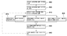
본 발명의 일 양태에서, 환자의 심장 관련 복합 리듬 장애를 나타내는 심장 정보를 재구성하여 심장 리듬 장애의 소스를 표시하는 방법으로서, 이 방법은,In one aspect of the invention, a method of reconstructing cardiac information indicative of a heart related complex rhythm disorder of a patient to indicate a source of a heart rhythm disorder, the method comprising:
복합 리듬 장애 동안, 환자의 심장으로부터 심장 정보 신호를 수신하는 단계;During the complex rhythm disorder, receiving a cardiac information signal from the patient's heart;
컴퓨팅 디바이스에 의해, 심장 정보 신호들을 신뢰성 역가에 위해 고신뢰성 및 저신뢰성 신호들로 분류하는 단계;Classifying, by the computing device, the cardiac information signals into high reliability and low reliability signals for reliable titer;
컴퓨팅 디바이스에 의해, 허용성 창(acceptance window) 내에서 저신뢰성 신호들과 관련된 활성화 온셋들을 판단하는 단계;Determining, by the computing device, activation onsets associated with low reliability signals within an acceptance window;
컴퓨팅 디바이스에 의해, 저신뢰성 신호들과 관련된 활성화 온셋들 및 고신뢰성 신호들과 관련된 활성화 온셋들을 명령하는 단계; 및Instructing, by the computing device, activation onsets associated with low reliability signals and activation onsets associated with high reliability signals; And
컴퓨팅 디바이스에 의해, 고신뢰성 신호 및 저신뢰성 신호와 관련된 활성화 온셋들을 출력하여 복합 심장 리듬 장애의 소스를 표시하는 단계를 포함한다.Outputting, by the computing device, activation onsets associated with the high and low reliability signals to indicate the source of the complex heart rhythm disorder.
판단하는 단계는 적어도 2개의 식별 가능한 활성화 온셋들을 연결하는 파 경로 벡터를 이용하여 저신뢰성 신호들과 관련된 활성화 온셋들을 판단하는 단계를 더 포함한다.The determining step further comprises determining activation onsets associated with low reliability signals using a wave path vector connecting at least two identifiable activation onsets.
일부 실시형태에서, 복합 리듬 장애는, 심장 정보 신호들이 휴지 상태인 동안인 식별가능한 주기를 포함하지 않는다.In some embodiments, the complex rhythm disorder does not include an identifiable period during which cardiac information signals are at rest.
다른 실시형태에서, 복합 리듬 장애는 심장 정보 신호들과 관련된 식별 가능한 가장 이른 활성화 온셋을 포함하지 않는다.In another embodiment, the complex rhythm disorder does not comprise the earliest identifiable activation onset associated with cardiac information signals.
분류하는 단계는 활성화 온셋, 사이클 길이(CL), 활동 전위 지속기간(APD), 및 진폭 중 적어도 하나를 이용하여 심장 정보 신호들을 고신뢰성 및 저신뢰성 신호들로 분류하는 단계를 더 포함하며, 활성화 온셋은 최대 dV/dt, 템플릿 매칭, 주파수 및 진폭 중 적어도 하나를 이용하여 판단된다.The classifying further includes classifying the cardiac information signals into high and low reliability signals using at least one of activation onset, cycle length (CL), action potential duration (APD), and amplitude. Onset is determined using at least one of maximum dV / dt, template matching, frequency and amplitude.
일부 실시형태에서, 허용성 창은 APD, 전도 속도(CV), 섬유 각도, 적어도 2개의 식별 가능한 활성화 온셋들을 연결하는 파 경로 벡터, 및 해부 인자들 중 적어도 하나를 이용하여 판단될 수 있다.In some embodiments, the tolerance window can be determined using at least one of APD, conduction velocity (CV), fiber angle, wave path vector connecting at least two identifiable activation onsets, and anatomical factors.
기선 원더(baseline wander) 및 잡음이 심장 정보 신호들로부터 제거될 수 있고, 심장 정보 신호들이 필터링될 수 있다.Baseline wander and noise can be removed from the cardiac information signals, and the cardiac information signals can be filtered.
심장 정보 신호들 중 적어도 하나는 신호-대-잡음 비(SNR), 템플릿 매칭, 주파수 및 진폭 중 적어도 하나를 이용하여 무시될 수 있다.At least one of the cardiac information signals may be ignored using at least one of signal-to-noise ratio (SNR), template matching, frequency and amplitude.
템플릿 매칭은 심장 정보 신호들과 관련된 고신뢰성 레벨 박동을 템플릿으로 식별하는 단계를 더 포함할 수 있다. 템플릿 매칭은 전문가 시스템을 이용하여 수행될 수 있고, 전문가 시스템은 박동 타입들을 이용하여 템플릿 매칭을 수행한다.Template matching may further comprise identifying a high reliability level beat associated with cardiac information signals as a template. Template matching may be performed using an expert system, which expert template performs template matching using beat types.
심장 정보 신호들과 관련된 박동들은 분류될 박동들과 관련된 형상에 기초하여 추가로 분류될 수 있다.Beats associated with cardiac information signals may be further classified based on a shape associated with the beats to be classified.
심장 정보 신호들의 분류는, 최소 APD보다 크고 최대 CL보다 작은, 분류될 박동과 관련된 CL에 응답하여 심장 정보 신호들과 관련된 박동들을 저신뢰성 박동들로 분류하는 단계를 더 포함할 수 있다.The classification of the cardiac information signals may further comprise classifying the beats associated with the cardiac information signals into low reliability beats in response to the CL associated with the beat to be classified, which is greater than the minimum APD and less than the maximum CL.
일부 실시형태에서, 벡터는 박동 형상, 박동 극성, 및 주변 회전/방사성 방출 중 적어도 하나를 이용하여 변경될 수 있다.In some embodiments, the vector can be modified using at least one of pulsating shape, pulsating polarity, and ambient rotational / radioactive release.
심장 정보 신호들의 분류는, 최소 APD보다 작고 최대 CL보다 큰, 분류될 박동과 관련된 CL에 응답하여 심장 정보 신호들과 관련된 박동들을 저신뢰성 박동들로 분류하는 단계를 더 포함할 수 있다.The classification of cardiac information signals may further include classifying the beats associated with the cardiac information signals into low reliability beats in response to the CL associated with the beat to be classified, which is less than the minimum APD and greater than the maximum CL.
허용성 창의 판단은 APD, CV, 적어도 2개의 식별 가능한 활성화 온셋들을 연결하는 파 경로 벡터, 주파수 및 섬유 각도 중 적어도 하나를 이용하여 허용성 창을 판단하는 단계를 더 포함할 수 있다.The determination of the tolerance window may further comprise determining the tolerance window using at least one of APD, CV, wave path vector connecting at least two identifiable activation onsets, frequency and fiber angle.
활성화 온셋들의 판단은 전문가 시스템을 이용하는 단계를 더 포함할 수 있으며, 전문가 시스템은 파 형상들을 포함한다.The determination of activation onsets may further comprise using an expert system, the expert system comprising wave shapes.
저신뢰성 신호들과 관련된 활성화 온셋들의 판단은 롤링 평균 및 위상 잠금 중 적어도 하나를 이용하여 활성화 온셋들을 판단하는 단계를 더 포함할 수 있다.The determination of activation onsets associated with low reliability signals may further include determining activation onsets using at least one of a rolling average and a phase lock.
저신뢰성 신호들과 관련된 활성화 온셋들의 판단은 파 경로 벡터, 허용성 창, 롤링 평균, 및 위상 잠금 중 적어도 2개를 이용하여 판단된 활성화 온셋들을 융화시키는 단계를 더 포함할 수 있다.The determination of activation onsets associated with low reliability signals may further comprise harmonizing the activation onsets determined using at least two of a wave path vector, a tolerance window, a rolling average, and a phase lock.
본 발명의 일 양태에서, 환자의 심장으로부터 복수의 채널들을 통해 수신된 복합 리듬 장애와 관련된 심장 신호들을 재구성하는 방법으로서,In one aspect of the invention, a method of reconstructing heart signals associated with a complex rhythm disorder received over a plurality of channels from a patient's heart, comprising:
총 박동들로부터 적어도 사전 결정된 비율의 식별 가능한 박동들을 포함하는 고신뢰성 채널들 및 제1 개수의 식별 가능한 박동들 및 제2 개수의 식별 불가능한 박동들을 포함하는 저신뢰성 채널들을 분류하되, 각각의 식별 가능한 박동은 식별 가능한 활성화 온셋을 갖고, 각각의 식별 불가능한 박동은 복수의 편향들 및 가능한 활성화 온셋과 관련된 휴지 상태 주기들을 가지며, 제1 개수의 식별 가능한 박동들은 사전 결정된 비율보다 적은 단계;Classify the high reliability channels comprising at least a predetermined ratio of identifiable beats from the total beats and the low reliability channels comprising a first number of identifiable beats and a second number of unidentifiable beats, each identifiable The pulsation has an identifiable activation onset, each unidentifiable rhythm has a dormant periods associated with a plurality of deflections and a possible activation onset, and wherein the first number of identifiable rhythms is less than a predetermined rate;
저신뢰성 채널에 인접한 고신뢰성 채널들 상에서의 복수의 식별 가능한 박동들을 식별하되, 고신뢰성 채널들 상에서의 식별 가능한 박동들은 저신뢰성 채널 상에서의 식별 불가능한 박동에 대응하는 단계;Identifying a plurality of identifiable beats on the high reliability channels adjacent to the low reliability channel, wherein the identifiable beats on the high reliability channels correspond to an unidentifiable beat on the low reliability channel;
저신뢰성 채널 상의 식별 불가능한 박동을 통해, 인접한 채널들 상에서 식별된 식별 불가능한 박동들의 적어도 2개의 활성화 온셋들 사이의 벡터를 계산하는 단계;Calculating a vector between at least two activation onsets of the unidentifiable beats identified on adjacent channels, via the unidentifiable beats on the low reliability channel;
벡터가 식별 불가능한 박동과 교차하는 영역을 중심으로 식별 불가능한 박동과 관련된 시간 간격을 정의하되, 상기 시간 간격은 선택되거나 판단된 활성화 온셋을 갖는 저신뢰성 채널 상의 이전 박동에 기초하여 식별 불가능한 박동이 얼마나 이르게 활성화할 수 있는지 그리고 적어도 하나의 사전 결정된 특성에 기초하여 식별 불가능한 박동이 얼마나 늦게 종결할 수 있는지를 나타내는 단계; 및Define a time interval associated with an unidentifiable beat centered around the region where the vector intersects the unidentifiable beat, wherein the time interval defines how early to activate the unidentifiable beat based on the previous beat on the low-reliability channel with the selected or determined activation onset. Indicating how late the unidentifiable beat can terminate based on the at least one predetermined characteristic; And
식별 불가능한 박동에 대해 계산된 벡터에 가장 가까운 정의된 시간 간격 동안, 가능한 활성화 온셋을 선택하는 단계를 포함하는 방법이 제공된다.A method is provided that includes selecting a possible activation onset for a defined time interval closest to the calculated vector for an unidentifiable beat.
가능한 활성화 온셋은 정의된 시간 간격 동안 휴지 상태 주기 또는 편향과 관련하여 선택될 수 있다.Possible activation onsets may be selected in terms of idle periods or deflections for defined time intervals.
일부 실시형태에서, 방법은:In some embodiments, the method is:
식별 불가능한 박동 이전에 발생하는 저신뢰성 채널 상의 식별 가능한 박동들 사이의 제2 시간 간격을 판단하되, 제2 시간 간격은 저신뢰성 채널 상에서 제1 활성화 온셋으로부터 각각의 식별 가능한 박동들의 제2 활성화 온셋으로 연장되는 단계;Determine a second time interval between identifiable beats on the low reliability channel that occurs prior to the unidentifiable beat, wherein the second time interval is from the first activation onset to the second activation onset of each identifiable beats on the low reliability channel. Extending;
제1 활성화 온셋이 이전 박동의 활성화 온셋에 근접하도록 제2 시간 간격을 앞당기는 단계;Advancing a second time interval such that the first activation onset is close to the activation onset of the previous beat;
선택된 활성화 온셋을 융화된 활성화 온셋에 대한 제2 활성화 온셋과 융화시키는 단계; 및Fusing the selected activation onset with a second activation onset for the harmonized activation onset; And
선택된 활성화 온셋을 식별 불가능한 박동에 대한 융화된 활성화 온셋으로 업데이트하는 단계를 더 포함할 수 있다.The method may further include updating the selected activation onset with a harmonized activation onset for unidentifiable beats.
본 발명의 일 양태에서, 복합 리듬 장애에서 활성화 시간을 판단하는 방법으로서,In one aspect of the invention, a method of determining the activation time in a complex rhythm disorder,
저신뢰성 채널에 인접한 고신뢰성 채널들의 신호들에서 적어도 2개의 식별 가능한 박동들을 식별하되, 식별 가능한 박동들은 저신뢰성 채널의 신호에서 식별 불가능한 박동에 대응하고, 식별 불가능한 박동은 복수의 편향들 및 가능한 활성화 온셋과 관련된 휴지 상태 주기들을 갖는 단계;Identify at least two identifiable beats in the signals of the high reliability channels adjacent to the low reliability channel, wherein the identifiable beats correspond to an unidentifiable beat in the signal of the low reliability channel, wherein the unidentifiable beat is a plurality of deflections and a possible activation onset. Having dormant periods associated with it;
식별 불가능한 박동을 통해, 식별 가능한 박동들의 활성화 온셋들 사이의 벡터를 계산하는 단계;Calculating, via an unidentifiable beat, a vector between activation onsets of the identifiable beats;
벡터가 식별 불가능한 박동과 교차하는 영역을 중심으로 식별 불가능한 박동과 관련된 시간 간격을 정의하되, 시간 간격은 선택되거나 판단된 활성화 온셋을 갖는 저신뢰성 채널의 신호에서 이전 박동에 기초하여 식별 불가능한 박동이 얼마나 이르게 활성화할 수 있는지 그리고 적어도 하나의 사전 결정된 특성에 기초하여 식별 불가능한 박동이 얼마나 늦게 종결할 수 있는지를 나타내는 단계; 및Define a time interval associated with an unidentifiable beat centered around the region where the vector intersects the unidentifiable beat, but the time interval defines how early the non-identifiable beat is activated based on the previous beat in the signal of the low-reliability channel with the selected or determined activation onset. Indicating how late the unidentifiable beat can be terminated based on at least one predetermined characteristic; And
식별 불가능한 박동에 대해 계산된 벡터에 가장 가까운 정의된 시간 간격 동안, 활성화 온셋을 선택하는 단계를 포함하는 방법이 제공된다.For a defined time interval closest to the calculated vector for unidentifiable beats, a method is provided that includes selecting an activation onset.
가능한 활성화 온셋은 정의된 시간 간격 동안 휴지 상태 주기 또는 편향과 관련하여 선택될 수 있다.Possible activation onsets may be selected in terms of idle periods or deflections for defined time intervals.
일부 실시형태에서, 방법은:In some embodiments, the method is:
식별 불가능한 박동 이전에 발생하는 저신뢰성 채널의 신호에서 식별 가능한 박동들 사이의 제2 시간 간격을 판단하되, 제2 시간 간격은 제1 활성화 온셋으로부터 각각의 식별 가능한 박동들의 제2 활성화 온셋으로 연장되는 단계;Determine a second time interval between identifiable beats in the signal of the low reliability channel that occurs prior to the unidentifiable beat, wherein the second time interval extends from the first activation onset to the second activation onset of the respective identifiable beats. step;
제1 활성화 온셋이 이전 박동의 활성화 온셋에 근접하도록 신호에서 제2 시간 간격을 앞당기는 단계;Advancing a second time interval in the signal such that the first activation onset is close to the activation onset of the previous beat;
선택된 활성화 온셋을 융화된 활성화 온셋에 대한 제2 활성화 온셋과 융화시키는 단계; 및Fusing the selected activation onset with a second activation onset for the harmonized activation onset; And
선택된 활성화 온셋을 식별 불가능한 박동에 대한 융화된 활성화 온셋으로 업데이트하는 단계를 더 포함할 수 있다.The method may further include updating the selected activation onset with a harmonized activation onset for unidentifiable beats.
환자의 심장 리듬 장애의 소스를 표시하기 위해 심장 관련 복합 리듬 장애를 나타내는 심장 정보를 재구성하게 하는 명령들을 포함하는 컴퓨터 판독가능 매체로서,A computer readable medium comprising instructions for reconstructing cardiac information indicative of a heart related complex rhythm disorder to indicate a source of a heart rhythm disorder in a patient, the method comprising:
복합 리듬 장애 동안, 복수의 센서들로부터 심장 정보 신호들을 수신하고;During a complex rhythm disorder, receive cardiac information signals from a plurality of sensors;
심장 정보 신호들을 고신뢰성 및 저신뢰성 신호들로 분류하되, 고신뢰성 및 저신뢰성 신호들이 신뢰성 역가에 의해 분리되고;Classify cardiac information signals into high and low reliability signals, wherein the high and low reliability signals are separated by a reliability titer;
허용성 창 내에서 저신뢰성 신호들과 관련된 활성화 온셋들을 판단하고;Determine activation onsets associated with low reliability signals within the tolerance window;
저신뢰성 신호들과 관련된 활성화 온셋들 및 고신뢰성 신호들과 관련된 활성화 온셋들을 명령하고;Command activation onsets associated with low reliability signals and activation onsets associated with high reliability signals;
고신뢰성 신호 및 저신뢰성 신호와 관련된 활성화 온셋들을 출력하여 복합 심장 리듬 장애의 소스를 나타냄으로써,By outputting activation onsets associated with high and low reliability signals to indicate the source of complex heart rhythm disorders,
컴퓨팅 디바이스로 하여금, 환자의 심장 관련 복합 리듬 장애를 나타내는 심장 정보를 재구성하게 하여 심장 리듬 장애의 소스를 표시하도록 하는 명령들을 포함하는 컴퓨터 판독가능 매체가 제공된다.A computer readable medium is provided that includes instructions to cause a computing device to reconstruct cardiac information indicative of a patient's heart related complex rhythm disorder to indicate a source of the heart rhythm disorder.
컴퓨팅 디바이스로 하여금, 적어도 2개의 식별 가능한 활성화 온셋들을 연결하는 파 경로 벡터를 이용하여 저신뢰성 신호들과 관련된 활성화 온셋들을 판단함으로써 환자의 심장 리듬 장애의 소스를 표시하기 위해 심장 관련 복합 리듬 장애를 나타내는 심장 정보를 재구성하게 하는 명령들이 제공될 수 있다.Indicative of a heart related complex rhythm disorder to indicate the source of the patient's heart rhythm disorder by determining activation onsets associated with low reliability signals using a wave path vector connecting at least two identifiable activation onsets. Instructions may be provided to reconstruct cardiac information.
일부 실시형태에서, 복합 리듬 장애는 심장 정보 신호들이 휴지 상태인 동안인 식별 가능한 주기를 포함하지 않는다. 다른 실시형태에서, 복합 리듬 장애는 심장 정보 신호들과 관련된 식별 가능한 가장 이른 활성화 온셋을 포함하지 않는다.In some embodiments, the complex rhythm disorder does not include an identifiable period during which cardiac information signals are at rest. In another embodiment, the complex rhythm disorder does not comprise the earliest identifiable activation onset associated with cardiac information signals.
컴퓨팅 디바이스로 하여금, 활성화 온셋, 사이클 길이(CL), 활동 전위 지속기간(APD), 및 진폭 중 적어도 하나를 이용하여 심정 정보 신호들을 고신뢰성 및 저신뢰성 신호들로 분류함으로써 환자의 심장 리듬 장애의 소스를 표시하기 위해 심장 관련 복합 리듬 장애를 나타내는 심장 정보를 재구성하게 하는 명령들이 제공될 수 있으며, 활성화 온셋은 최대 dV/dt, 템플릿 매칭, 주파수 및 진폭 중 적어도 하나를 이용함으로써 판단된다.The computing device is configured to classify the cardiac information signals into high and low reliability signals using at least one of activation onset, cycle length (CL), action potential duration (APD), and amplitude to determine the heart rhythm disorder of the patient. Instructions may be provided to reconstruct cardiac information indicative of a heart related complex rhythm disorder to indicate a source, and activation onset is determined by using at least one of maximum dV / dt, template matching, frequency, and amplitude.
컴퓨팅 디바이스로 하여금, APD, 전도 속도(CV), 섬유 각도, 및 해부 인자들 중 적어도 하나를 이용하여 허용성 창을 판단하게 하는 명령들이 제공될 수 있다.Instructions may be provided that cause the computing device to determine an acceptable window using at least one of APD, conduction velocity (CV), fiber angle, and anatomical factors.
컴퓨팅 디바이스로 하여금, 기선 원더 및 잡음을 심장 정보 신호들로부터 제거하고, 심장 정보 신호들을 필터링하게 하는 명령들이 제공될 수 있다.Instructions may be provided that cause the computing device to remove baseline wonder and noise from the cardiac information signals and filter the cardiac information signals.
컴퓨팅 디바이스로 하여금, 신호 대 잡음 비(SNR), 템플릿 매칭, 주파수 및 진폭 중 적어도 하나를 무시하게 하는 명령들이 제공될 수 있다.Instructions may be provided that cause the computing device to ignore at least one of signal to noise ratio (SNR), template matching, frequency, and amplitude.
컴퓨팅 디바이스로 하여금, 심장 정보 신호들과 관련된 고신뢰성 레벨 박동을 템플릿으로 식별함으로써, 템플릿 매칭을 하게 하는 명령들이 제공될 수 있다.Instructions may be provided that cause the computing device to make a template match by identifying a high reliability level beat associated with cardiac information signals with a template.
컴퓨팅 디바이스로 하여금, 전문가 시스템을 이용하여 템플릿 매칭을 하게 하는 명령들이 제공될 수 있으며, 전문가 시스템은 박동 타입들을 이용하여 템플릿 매칭을 수행한다.Instructions may be provided that cause the computing device to perform template matching using the expert system, which performs the template matching using the beat types.
컴퓨팅 디바이스로 하여금, 분류될 박동들과 관련된 형상에 기초하여 심장 정보 신호들과 관련된 박동들을 분류하게 하는 명령들이 제공될 수 있다.Instructions may be provided that cause the computing device to classify beats associated with cardiac information signals based on a shape associated with beats to be classified.
컴퓨팅 디바이스로 하여금, 최소 APD보다 크고 최대 CL보다 작은, 분류될 박동과 관련된 CL에 응답하여 심장 정보 신호들과 관련된 박동들을 저신뢰성 박동들로 분류하게 하는 명령들이 제공될 수 있다.Instructions may be provided that cause the computing device to classify the beats associated with cardiac information signals into low reliability beats in response to the CL associated with the beat to be classified that is greater than the minimum APD and less than the maximum CL.
컴퓨팅 디바이스로 하여금, 최소 APD보다 작고 최대 CL보다 큰, 분류될 박동과 관련된 CL에 응답하여 심장 정보 신호들과 관련된 박동들을 저신뢰성 박동들로 분류하게 하는 명령들이 제공될 수 있다.Instructions may be provided that cause the computing device to classify the beats associated with cardiac information signals as low reliability beats in response to the CL associated with the beat to be classified, which is less than the minimum APD and greater than the maximum CL.
컴퓨팅 디바이스로 하여금, 박동 형상, 박동 극성, 및 주변 회전/방사성 방출 중 적어도 하나를 이용하여 파 경로 벡터를 변경하게 하는 명령들이 제공될 수 있다.Instructions may be provided that cause the computing device to change the wave path vector using at least one of the rhythm shape, the rhythm polarity, and the ambient rotational / radioactive emission.
컴퓨팅 디바이스로 하여금, APD, CV, 적어도 2개의 식별 가능한 활성화 온셋들을 연결하는 파 경로 벡터, 주파수 및 섬유 각도 중 적어도 하나를 이용하여 허용성 창을 판단하게 하는 명령들이 제공될 수 있다.Instructions may be provided that cause the computing device to determine an allowable window using at least one of APD, CV, wave path vector connecting at least two identifiable activation onsets, frequency and fiber angle.
컴퓨팅 디바이스로 하여금, 전문가 시스템을 이용하여 활성화 온셋들을 판단하게 하는 명령들이 제공될 수 있으며, 전문가 시스템은 파 형상들을 포함한다.Instructions may be provided that cause the computing device to determine the activation onsets using the expert system, the expert system including wave shapes.
컴퓨팅 디바이스로 하여금, 롤링 평균 및 위상 잠금 중 적어도 하나를 이용하여 저신뢰성 신호들과 관련된 활성화 온셋들을 판단하게 하는 명령들이 제공될 수 있다.Instructions may be provided that cause the computing device to determine activation onsets associated with low reliability signals using at least one of a rolling average and a phase lock.
컴퓨팅 디바이스로 하여금, 파 경로 벡터, 허용성 창, 롤링 평균, 및 위상 잠금 중 적어도 2개를 이용하여 저신뢰성 신호들과 관련된 활성화 온셋들을 판단하게 하는 명령들이 제공될 수 있다.Instructions may be provided that cause the computing device to determine activation onsets associated with low reliability signals using at least two of a wave path vector, a tolerance window, a rolling average, and a phase lock.
본 발명의 일 양태에서, 컴퓨팅 디바이스에 의해 실행될 때, 컴퓨팅 디바이스로 하여금, In one aspect of the invention, when executed by a computing device, it causes the computing device to:
총 박동들로부터 적어도 사전 결정된 비율의 식별 가능한 박동들을 포함하는 고신뢰성 채널들 및 제1 개수의 식별 가능한 박동들 및 제2 개수의 식별 불가능한 박동들을 포함하는 저신뢰성 채널들을 분류하되, 각각의 식별 가능한 박동은 식별 가능한 활성화 온셋을 갖고, 각각의 식별 불가능한 박동은 복수의 편향들 및 가능한 활성화 온셋과 관련된 휴지 상태 주기들을 가지며, 제1 개수의 식별 가능한 박동들은 사전 결정된 비율보다 적고;Classify the high reliability channels comprising at least a predetermined ratio of identifiable beats from the total beats and the low reliability channels comprising a first number of identifiable beats and a second number of unidentifiable beats, each identifiable The beat has an identifiable activation onset, each unidentifiable beat has a dormant periods associated with a plurality of deflections and a possible activation onset, and the first number of identifiable beats is less than a predetermined rate;
저신뢰성 채널에 인접한 고신뢰성 채널들 상에서의 복수의 식별 가능한 박동들을 식별하되, 고신뢰성 채널들 상에서의 식별 가능한 박동들은 저신뢰성 채널 상에서의 식별 불가능한 박동에 대응하고;Identify a plurality of identifiable beats on the high reliability channels adjacent to the low reliability channel, wherein the identifiable beats on the high reliability channels correspond to an unidentifiable beat on the low reliability channel;
저신뢰성 채널 상의 식별 불가능한 박동을 통해, 인접한 채널들 상에서 식별된 식별 불가능한 박동들의 적어도 2개의 활성화 온셋들 사이의 벡터를 계산하고;Calculating, between the unreliable beats on the low reliability channel, a vector between at least two activation onsets of the unidentifiable beats identified on adjacent channels;
벡터가 식별 불가능한 박동과 교차하는 영역을 중심으로 식별 불가능한 박동과 관련된 시간 간격을 정의하되, 시간 간격은 선택되거나 판단된 활성화 온셋을 갖는 저신뢰성 채널 상의 이전 박동에 기초하여 식별 불가능한 박동이 얼마나 이르게 활성화할 수 있는지 그리고 적어도 하나의 사전 결정된 특성에 기초하여 식별 불가능한 박동이 얼마나 늦게 종결할 수 있는지를 나타내고; Define a time interval associated with an unidentifiable beat centered around the region where the vector intersects the unidentifiable beat, but the time interval can be activated as early as the unidentifiable beat based on the previous beat on the low-reliability channel with the selected or determined activation onset. Whether and how late the unidentifiable beat can terminate based on the at least one predetermined characteristic;
식별 불가능한 박동에 대해 계산된 벡터에 가장 가까운 정의된 시간 간격 동안, 가능한 활성화 온셋을 선택함으로써, 환자의 심장으로부터 복수의 채널들을 통해 수신된 복합 리듬 장애와 관련된 심장 신호들을 재구성하게 하는 명령들을 포함하는 컴퓨터 판독가능 매체가 제공된다.Instructions for reconstructing cardiac signals associated with a complex rhythm disorder received over a plurality of channels from the patient's heart by selecting a possible activation onset for a defined time interval closest to the calculated vector for the unidentifiable beat. A computer readable medium is provided.
가능한 활성화 온셋은 정의된 시간 간격 동안 휴지 상태 주기 또는 편향과 관련하여 선택될 수 있다.Possible activation onsets may be selected in terms of idle periods or deflections for defined time intervals.
일부 실시형태에서, 컴퓨팅 디바이스로 하여금, In some embodiments, the computing device causes
식별 불가능한 박동 이전에 발생하는 저신뢰성 채널 상의 식별 가능한 박동들 사이의 제2 시간 간격을 판단하게 하되, 제2 시간 간격은 저신뢰성 채널 상에서 제1 활성화 온셋으로부터 각각의 식별 가능한 박동들의 제2 활성화 온셋으로 연장되게 하고;Determine a second time interval between identifiable beats on the low reliability channel that occurs prior to the unidentifiable beat, wherein the second time interval is a second activation onset of each identifiable beats from the first activation onset on the low reliability channel Extended to;
제1 활성화 온셋이 이전 박동의 활성화 온셋에 근접하도록 제2 시간 간격을 앞당기고;Advance the second time interval such that the first activation onset is close to the activation onset of the previous beat;
선택된 활성화 온셋을 융화된 활성화 온셋에 대한 제2 활성화 온셋과 융화시키고; Fuse the selected activation onset with the second activation onset for the harmonized activation onset;
선택된 활성화 온셋을 식별 불가능한 박동에 대한 융화된 활성화 온셋으로 업데이트하게 하는 명령들이 제공될 수 있다.Instructions may be provided that cause the selected activation onset to be updated with a harmonized activation onset for unidentifiable beats.
본 발명의 일 양태에서, 컴퓨팅 디바이스로 하여금, In one aspect of the invention, a computing device comprises:
식별 불가능한 박동 이전에 발생하는 저신뢰성 채널의 신호에서 식별 가능한 박동들 사이의 제2 시간 간격을 판단하게 하되, 제2 시간 간격은 제1 활성화 온셋으로부터 각각의 식별 가능한 박동들의 제2 활성화 온셋으로 연장되게 하고;Determine a second time interval between identifiable beats in a signal of a low reliability channel that occurs prior to an unidentifiable beat, wherein the second time interval extends from the first activation onset to a second activation onset of each identifiable beats To become;
제1 활성화 온셋이 이전 박동의 활성화 온셋에 근접하도록 신호에서 제2 시간 간격을 앞당기고;Advance a second time interval in the signal such that the first activation onset is close to the activation onset of the previous beat;
선택된 활성화 온셋을 융화된 활성화 온셋으로 제2 활성화 온셋과 융화시키고;Fuse the selected activation onset with the second activation onset to a harmonized activation onset;
선택된 활성화 온셋을 식별 불가능한 박동에 대한 융화된 활성화 온셋으로 업데이트하게 하는 명령들이 제공될 수 있다.Instructions may be provided that cause the selected activation onset to be updated with a harmonized activation onset for unidentifiable beats.
가능한 활성화 온셋은 정의된 시간 간격 동안 휴지 상태 주기 또는 편향과 관련하여 선택될 수 있다.Possible activation onsets may be selected in terms of idle periods or deflections for defined time intervals.
일부 실시형태에서, 컴퓨팅 디바이스로 하여금,In some embodiments, the computing device causes
식별 불가능한 박동 이전에 발생하는 저신뢰성 채널의 신호에서 식별 가능한 박동들 사이의 제2 시간 간격을 판단하게 하되, 제2 시간 간격은 제1 활성화 온셋으로부터 각각의 식별 가능한 박동들의 제2 활성화 온셋으로 연장되게 하고;Determine a second time interval between identifiable beats in a signal of a low reliability channel that occurs prior to an unidentifiable beat, wherein the second time interval extends from the first activation onset to a second activation onset of each identifiable beats To become;
제1 활성화 온셋이 이전 박동의 활성화 온셋에 근접하도록 신호에서 제2 시간 간격을 앞당기고;Advance a second time interval in the signal such that the first activation onset is close to the activation onset of the previous beat;
선택된 활성화 온셋을 융화된 활성화 온셋에 대한 제2 활성화 온셋과 융화시키고;Fuse the selected activation onset with the second activation onset for the harmonized activation onset;
선택된 활성화 온셋을 식별 불가능한 박동에 대한 융화된 활성화 온셋으로 업데이트하게 하는 명령들이 제공될 수 있다.Instructions may be provided that cause the selected activation onset to be updated with a harmonized activation onset for unidentifiable beats.
본 발명의 일 양태에서, 환자의 심장 리듬 장애의 소스를 표시하기 위해 심장 관련 복합 리듬 장애를 나타내는 심장 정보를 재구성하는 시스템으로서,In one aspect of the invention, there is provided a system for reconstructing cardiac information indicative of a heart related complex rhythm disorder to indicate a source of a heart rhythm disorder in a patient,
복합 리듬 장애 동안, 환자의 심장으로부터 심장 정보 신호를 수신하고;During a complex rhythm disorder, receiving cardiac information signals from the patient's heart;
심장 정보 신호들을 고신뢰성 및 저신뢰성 신호들로 분류하되, 고신뢰성 및 저신뢰성 신호들은 신뢰성 역가에 의해 분리되고;Categorize cardiac information signals into high and low reliability signals, wherein the high and low reliability signals are separated by a reliability titer;
허용성 창(acceptance window) 내에서 저신뢰성 신호들과 관련된 활성화 온셋들을 판단하고;Determine activation onsets associated with low reliability signals within an acceptance window;
저신뢰성 신호들과 관련된 활성화 온셋들 및 고신뢰성 신호들과 관련된 활성화 온셋들을 명령하고;Command activation onsets associated with low reliability signals and activation onsets associated with high reliability signals;
고신뢰성 신호 및 저신뢰성 신호와 관련된 활성화 온셋들을 출력하여 복합 심장 리듬 장애의 소스를 나타내도록 구성된 적어도 하나의 컴퓨팅 디바이스를 포함하는 시스템이 제공된다.A system is provided that includes at least one computing device configured to output activation onsets associated with a high reliability signal and a low reliability signal to indicate a source of a complex heart rhythm disorder.
적어도 하나의 컴퓨팅 디바이스는 허용성 창을 이용하여 저신뢰성 신호들과 관련된 활성화 온셋들을 판단할 수 있다.At least one computing device may use the tolerance window to determine activation onsets associated with low reliability signals.
일부 실시형태에서, 복합 리듬 장애는, 심장 정보 신호들이 휴지 상태인 동안인 식별가능한 주기를 포함하지 않는다. 다른 실시형태에서, 복합 리듬 장애는 심장 정보 신호들과 관련된 식별 가능한 가장 이른 활성화 온셋을 포함하지 않는다.In some embodiments, the complex rhythm disorder does not include an identifiable period during which cardiac information signals are at rest. In another embodiment, the complex rhythm disorder does not comprise the earliest identifiable activation onset associated with cardiac information signals.
적어도 하나의 컴퓨팅 디바이스는, 활성화 온셋, 사이클 길이(CL), 활동 전위 지속기간(APD), 및 진폭 중 적어도 하나를 이용하여 심장 정보 신호들을 고신뢰성 및 저신뢰성 신호들로 분류할 수 있으며, 활성화 온셋은 최대 dV/dt, 템플릿 매칭, 주파수 및 진폭 중 적어도 하나를 이용하여 판단된다.At least one computing device may classify the cardiac information signals into high and low reliability signals using at least one of activation onset, cycle length (CL), action potential duration (APD), and amplitude, and activation Onset is determined using at least one of maximum dV / dt, template matching, frequency and amplitude.
적어도 하나의 컴퓨팅 디바이스는 APD, 전도 속도(CV), 섬유 각도, 적어도 2개의 식별 가능한 활성화 온셋들을 연결하는 파 경로 벡터, 및 해부 인자들 중 적어도 하나를 이용하여 허용성 창을 판단할 수 있다.The at least one computing device may determine the allowable window using at least one of APD, conduction velocity (CV), fiber angle, wave path vector connecting at least two identifiable activation onsets, and anatomical factors.
적어도 하나의 컴퓨팅 디바이스는 심장 정보 신호들로부터 기선 원더(baseline wander) 및 잡음을 제거할 수 있고, 컴퓨팅 디바이스는 심장 정보 신호들을 필터링할 수 있다.At least one computing device may remove baseline wander and noise from the cardiac information signals, and the computing device may filter the cardiac information signals.
적어도 하나의 컴퓨팅 디바이스는 신호-대-잡음 비(SNR), 템플릿 매칭, 주파수 및 진폭 중 적어도 하나를 이용하여 심장 정보 신호들 중 적어도 하나를 무시할 수 있다.At least one computing device may ignore at least one of the cardiac information signals using at least one of signal-to-noise ratio (SNR), template matching, frequency, and amplitude.
적어도 하나의 컴퓨팅 디바이스는, 심장 정보 신호들과 관련된 고신뢰성 레벨 박동을 템플릿으로 식별함으로써 템플릿 매칭을 수행할 수 있다.At least one computing device may perform template matching by identifying a high reliability level beat associated with cardiac information signals as a template.
시스템은 템플릿 매칭을 수행하도록 하는 전문가 시스템을 더 포함할 수 있다. The system may further comprise an expert system for performing template matching.
적어도 하나의 컴퓨팅 디바이스는, 분류될 박동들과 관련된 형상에 기초하여 심장 정보 신호들과 관련된 박동들을 분류할 수 있다.The at least one computing device may classify the beats associated with the cardiac information signals based on a shape associated with the beats to be classified.
하나의 컴퓨팅 디바이스는, 최소 APD 이상이고 최대 CL 이하인, 분류될 박동들과 관련된 CL에 응답하여 심장 정보 신호들과 관련된 박동들을 고신뢰성 박동들로 분류할 수 있다.One computing device may classify the beats associated with cardiac information signals as high reliability beats in response to the CL associated with the beats to be classified that are above the minimum APD and below the maximum CL.
적어도 하나의 컴퓨팅 디바이스는, 최소 APD보다 작고 최대 CL보다 큰, 분류될 박동과 관련된 CL에 응답하여 심장 정보 신호들과 관련된 박동들을 저신뢰성 박동들로 분류할 수 있다.At least one computing device may classify the beats associated with cardiac information signals into low reliability beats in response to the CL associated with the beat to be classified, which is less than the minimum APD and greater than the maximum CL.
적어도 하나의 컴퓨팅 디바이스는 박동 형상, 박동 극성, 및 주변 회전/방사성 방출 중 적어도 하나를 이용하여 파 경로 벡터를 변경할 수 있다.At least one computing device may modify the wave path vector using at least one of the pulsating shape, the pulsating polarity, and the ambient rotational / radioactive emission.
시스템은 APD, CV, 및 섬유 각도 중 적어도 하나를 이용하여 허용성 창을 판단하도록 하는 전문가 시스템을 더 포함할 수 있다.The system can further include an expert system for determining an acceptable window using at least one of APD, CV, and fiber angle.
시스템은 파 형상들을 이용하여 활성화 온셋들을 판단하도록 하는 전문가 시스템을 더 포함할 수 있다.The system may further include an expert system for determining activation onsets using wave shapes.
적어도 하나의 컴퓨팅 디바이스는 롤링 평균 및 위상 잠금 중 적어도 하나를 이용하여 저신뢰성 신호들과 관련된 활성화 온셋들을 판단할 수 있다.At least one computing device may determine activation onsets associated with low reliability signals using at least one of a rolling average and a phase lock.
적어도 하나의 컴퓨팅 디바이스는 파 경로 벡터, 허용성 창, 롤링 평균, 및 위상 잠금 중 적어도 2개를 이용하여 판단된 활성화 온셋들을 융화시킴으로써 저신뢰성 신호들과 관련된 활성화 온셋들을 판단할 수 있다.At least one computing device may determine activation onsets associated with low reliability signals by reconciling the activation onsets determined using at least two of a wave path vector, a tolerance window, a rolling average, and a phase lock.
본 발명의 일 양태에서, 환자의 심장으로부터 복수의 채널들을 통해 수신된 복합 리듬 장애와 관련된 심장 신호들을 재구성하는 시스템으로서,In one aspect of the invention, a system for reconstructing heart signals associated with a complex rhythm disorder received over a plurality of channels from a patient's heart, comprising:
총 박동들로부터 적어도 사전 결정된 비율의 식별 가능한 박동들을 포함하는 고신뢰성 채널들 및 제1 개수의 식별 가능한 박동들 및 제2 개수의 식별 불가능한 박동들을 포함하는 저신뢰성 채널들을 분류하되, 각각의 식별 가능한 박동은 식별 가능한 활성화 온셋을 갖고, 각각의 식별 불가능한 박동은 복수의 편향들 및 가능한 활성화 온셋과 관련된 휴지 상태 주기들을 가지며, 제1 개수의 식별 가능한 박동들은 사전 결정된 비율보다 적고;Classify the high reliability channels comprising at least a predetermined ratio of identifiable beats from the total beats and the low reliability channels comprising a first number of identifiable beats and a second number of unidentifiable beats, each identifiable The beat has an identifiable activation onset, each unidentifiable beat has a dormant periods associated with a plurality of deflections and a possible activation onset, and the first number of identifiable beats is less than a predetermined rate;
저신뢰성 채널에 인접한 고신뢰성 채널들 상에서의 복수의 식별 가능한 박동들을 식별하되, 고신뢰성 채널들 상에서의 식별 가능한 박동들은 저신뢰성 채널 상에서의 식별 불가능한 박동에 대응하고;Identify a plurality of identifiable beats on the high reliability channels adjacent to the low reliability channel, wherein the identifiable beats on the high reliability channels correspond to an unidentifiable beat on the low reliability channel;
저신뢰성 채널 상의 식별 불가능한 박동을 통해, 인접한 채널들 상에서 식별된 식별 불가능한 박동들의 적어도 2개의 활성화 온셋들 사이의 벡터를 계산하고;Calculating, between the unreliable beats on the low reliability channel, a vector between at least two activation onsets of the unidentifiable beats identified on adjacent channels;
벡터가 식별 불가능한 박동과 교차하는 영역을 중심으로 식별 불가능한 박동과 관련된 시간 간격을 정의하되, 시간 간격은 선택되거나 판단된 활성화 온셋을 갖는 저신뢰성 채널 상의 이전 박동에 기초하여 식별 불가능한 박동이 얼마나 이르게 활성화할 수 있는지 그리고 적어도 하나의 사전 결정된 특성에 기초하여 식별 불가능한 박동이 얼마나 늦게 종결할 수 있는지를 나타내고;Define a time interval associated with an unidentifiable beat centered around the region where the vector intersects the unidentifiable beat, but the time interval can be activated as early as the unidentifiable beat based on the previous beat on the low-reliability channel with the selected or determined activation onset. Whether and how late the unidentifiable beat can terminate based on the at least one predetermined characteristic;
식별 불가능한 박동에 대해 계산된 벡터에 가장 가까운 정의된 시간 간격 동안, 가능한 활성화 온셋을 선택하도록 구성된, Configured to select a possible activation onset for a defined time interval closest to the calculated vector for unidentifiable beats,
적어도 하나의 컴퓨팅 디바이스를 포함하는, 시스템이 제공된다.A system is provided that includes at least one computing device.
가능한 활성화 온셋은 정의된 시간 간격 동안 휴지 상태 주기 또는 편향과 관련하여 선택될 수 있다.Possible activation onsets may be selected in terms of idle periods or deflections for defined time intervals.
적어도 하나의 컴퓨팅 디바이스는, 추가로,At least one computing device further includes:
식별 불가능한 박동 이전에 발생하는 저신뢰성 채널 상의 식별 가능한 박동들 사이의 제2 시간 간격을 판단하되, 제2 시간 간격은 저신뢰성 채널 상에서 제1 활성화 온셋으로부터 각각의 식별 가능한 박동들의 제2 활성화 온셋으로 연장되고;Determine a second time interval between identifiable beats on the low reliability channel that occurs prior to the unidentifiable beat, wherein the second time interval is from the first activation onset to the second activation onset of each identifiable beats on the low reliability channel. Extended;
제1 활성화 온셋이 이전 박동의 활성화 온셋에 근접하도록 제2 시간 간격을 앞당기고;Advance the second time interval such that the first activation onset is close to the activation onset of the previous beat;
선택된 활성화 온셋을 융화된 활성화 온셋에 대한 제2 활성화 온셋과 융화시키고;Fuse the selected activation onset with the second activation onset for the harmonized activation onset;
선택된 활성화 온셋을 식별 불가능한 박동에 대한 융화된 활성화 온셋으로 업데이트하도록 구성될 수 있다.And may be configured to update the selected activation onset with a harmonized activation onset for unidentifiable beats.
본 발명의 일 양태에서, 복합 리듬 장애에서 활성화 시간을 판단하는 시스템으로서,In one aspect of the invention, a system for determining the activation time in a complex rhythm disorder,
저신뢰성 채널에 인접한 고신뢰성 채널들의 신호들에서 적어도 2개의 식별 가능한 박동들을 식별하되, 식별 가능한 박동들은 저신뢰성 채널의 신호에서 식별 불가능한 박동에 대응하고, 식별 불가능한 박동은 복수의 편향들 및 가능한 활성화 온셋과 관련된 휴지 상태 주기들을 갖고;Identify at least two identifiable beats in the signals of the high reliability channels adjacent to the low reliability channel, wherein the identifiable beats correspond to an unidentifiable beat in the signal of the low reliability channel, wherein the unidentifiable beat is a plurality of deflections and a possible activation onset. Have dormant periods associated with it;
식별 불가능한 박동을 통해, 식별 가능한 박동들의 활성화 온셋들 사이의 벡터를 계산하고;Calculate the vector between activation onsets of the identifiable beats, through the non-identifiable beats;
벡터가 식별 불가능한 박동과 교차하는 영역을 중심으로 식별 불가능한 박동과 관련된 시간 간격을 정의하되, 시간 간격은 선택되거나 판단된 활성화 온셋을 갖는 저신뢰성 채널의 신호에서 이전 박동에 기초하여 식별 불가능한 박동이 얼마나 이르게 활성화할 수 있는지 그리고 적어도 하나의 사전 결정된 특성에 기초하여 식별 불가능한 박동이 얼마나 늦게 종결할 수 있는지를 나타내고;Define a time interval associated with an unidentifiable beat centered around the region where the vector intersects the unidentifiable beat, but the time interval defines how early the non-identifiable beat is activated based on the previous beat in the signal of the low-reliability channel with the selected or determined activation onset. How late the non-identifiable beat can terminate based on at least one predetermined characteristic;
식별 불가능한 박동에 대해 계산된 벡터에 가장 가까운 정의된 시간 간격 동안, 활성화 온셋을 선택하도록 구성된,Configured to select an active onset for a defined time interval closest to the calculated vector for unidentifiable beats,
적어도 하나의 컴퓨팅 디바이스를 포함하는 시스템이 제공된다.A system is provided that includes at least one computing device.
가능한 활성화 온셋은 정의된 시간 간격 동안 휴지 상태 주기 또는 편향과 관련하여 선택될 수 있다.Possible activation onsets may be selected in terms of idle periods or deflections for defined time intervals.
적어도 하나의 컴퓨팅 디바이스는, 추가로, At least one computing device further includes:
식별 불가능한 박동 이전에 발생하는 저신뢰성 채널 상의 식별 가능한 박동들 사이의 제2 시간 간격을 판단하되, 제2 시간 간격은 저신뢰성 채널 상에서 제1 활성화 온셋으로부터 각각의 식별 가능한 박동들의 제2 활성화 온셋으로 연장되고;Determine a second time interval between identifiable beats on the low reliability channel that occurs prior to the unidentifiable beat, wherein the second time interval is from the first activation onset to the second activation onset of each identifiable beats on the low reliability channel. Extended;
제1 활성화 온셋이 이전 박동의 활성화 온셋에 근접하도록 제2 시간 간격을 앞당기고;Advance the second time interval such that the first activation onset is close to the activation onset of the previous beat;
선택된 활성화 온셋을 융화된 활성화 온셋에 대한 제2 활성화 온셋과 융화시키고;Fuse the selected activation onset with the second activation onset for the harmonized activation onset;
선택된 활성화 온셋을 식별 불가능한 박동에 대한 융화된 활성화 온셋으로 업데이트하도록 구성될 수 있다.And may be configured to update the selected activation onset with a harmonized activation onset for unidentifiable beats.
본 발명의 일 양태에서, 환자의 심장 리듬 장애의 소스를 표시하기 위해 심장 관련 복합 리듬 장애를 나타내는 심장 정보를 재구성하는 시스템으로서,In one aspect of the invention, there is provided a system for reconstructing cardiac information indicative of a heart related complex rhythm disorder to indicate a source of a heart rhythm disorder in a patient,
적어도 하나의 저장 디바이스,At least one storage device,
적어도 하나의 저장 디바이스에 동작 가능하게 커플링 가능한 적어도 하나의 컴퓨팅 디바이스,At least one computing device operatively coupled to the at least one storage device,
복합 리듬 장애 동안, 복수의 센서들로부터 심장 정보 신호들을 수신하는 적어도 하나의 저장 디바이스,At least one storage device that receives cardiac information signals from a plurality of sensors during a complex rhythm disorder,
심장 정보 신호들을 고신뢰성 및 저신뢰성 신호들로 분류하되, 고신뢰성 및 저신뢰성 신호들이 신뢰성 역가에 의해 분리되는 적어도 하나의 저장 디바이스,At least one storage device classifying cardiac information signals into high and low reliability signals, wherein the high and low reliability signals are separated by a reliability titer,
허용성 창 내에서 저신뢰성 신호들과 관련된 활성화 온셋들을 판단하는 적어도 하나의 저장 디바이스,At least one storage device for determining activation onsets associated with low reliability signals in the tolerance window,
저신뢰성 신호들과 관련된 활성화 온셋들 및 고신뢰성 신호들과 관련된 활성화 온셋들을 명령하는 적어도 하나의 저장 디바이스.At least one storage device instructing activation onsets associated with low reliability signals and activation onsets associated with high reliability signals.
고신뢰성 신호 및 저신뢰성 신호와 관련된 활성화 온셋들을 출력하는 적어도 하나의 저장 디바이스를 포함하는, 시스템.At least one storage device for outputting activation onsets associated with the high reliability signal and the low reliability signal.
적어도 하나의 저장 디바이스는 허용성 창을 이용하여 저신뢰성 신호들과 관련된 활성화 온셋들을 판단할 수 있다.At least one storage device may use the tolerant window to determine activation onsets associated with low reliability signals.
일부 실시형태에서, 복합 리듬 장애는 심장 정보 신호들이 휴지 상태인 동안인 식별가능한 주기를 포함하지 않는다. 다른 실시형태에서, 복합 리듬 장애는 심장 정보 신호들과 관련된 식별 가능한 가장 이른 활성화 온셋을 포함하지 않는다.In some embodiments, the complex rhythm disorder does not include an identifiable period during which cardiac information signals are at rest. In another embodiment, the complex rhythm disorder does not comprise the earliest identifiable activation onset associated with cardiac information signals.
적어도 하나의 컴퓨팅 디바이스는, 활성화 온셋, 사이클 길이(CL), 활동 전위 지속기간(APD), 및 진폭 중 적어도 하나를 이용하여 심장 정보 신호들을 고신뢰성 및 저신뢰성 신호들로 분류할 수 있으며, 활성화 온셋은 최대 dV/dt, 템플릿 매칭, 주파수 및 진폭 중 적어도 하나를 이용하여 판단된다.At least one computing device may classify the cardiac information signals into high and low reliability signals using at least one of activation onset, cycle length (CL), action potential duration (APD), and amplitude, and activation Onset is determined using at least one of maximum dV / dt, template matching, frequency and amplitude.
적어도 하나의 컴퓨팅 디바이스는 APD, 전도 속도(CV), 섬유 각도, 및 해부 인자들 중 적어도 하나를 이용하여 허용성 창을 판단할 수 있다.At least one computing device may determine the acceptable window using at least one of APD, conduction velocity (CV), fiber angle, and anatomical factors.
적어도 하나의 컴퓨팅 디바이스는 심장 정보 신호들로부터 기선 원더(baseline wander) 및 잡음을 제거할 수 있으며, 또한 심장 정보 신호들을 필터링할 수 있다.At least one computing device may remove baseline wander and noise from the cardiac information signals and may also filter the cardiac information signals.
적어도 하나의 컴퓨팅 디바이스는 신호-대-잡음 비(SNR), 템플릿 매칭, 및 진폭 중 적어도 하나를 이용하여 심장 정보 신호들 중 적어도 하나를 무시할 수 있다.At least one computing device may ignore at least one of the cardiac information signals using at least one of signal-to-noise ratio (SNR), template matching, and amplitude.
적어도 하나의 컴퓨팅 디바이스는 심장 정보 신호들과 관련된 고신뢰성 레벨 박동을 템플릿으로 식별함으로써 템플릿 매칭을 수행할 수 있다.At least one computing device may perform template matching by identifying, as a template, a high reliability level beat associated with cardiac information signals.
시스템은 템플릿 매칭을 수행하도록 하는 전문가 시스템을 더 포함할 수 있다.The system may further comprise an expert system for performing template matching.
적어도 하나의 컴퓨팅 디바이스는 분류될 박동들과 관련된 형상에 기초하여 심장 정보 신호들과 관련된 박동들을 분류할 수 있다.At least one computing device may classify beats associated with cardiac information signals based on a shape associated with beats to be classified.
일부 실시형태에서, 적어도 하나의 컴퓨팅 디바이스는, 최소 APD 이상이고 최대 CL 이하인, 분류될 박동들과 관련된 CL에 응답하여 심장 정보 신호들과 관련된 박동들을 고신뢰성 박동들로 분류할 수 있다. 다른 실시형태에서, 적어도 하나의 컴퓨팅 디바이스는, 최소 APD보다 작고 최대 CL보다 큰, 분류될 박동과 관련된 CL에 응답하여 심장 정보 신호들과 관련된 박동들을 저신뢰성 박동들로 분류할 수 있다.In some embodiments, the at least one computing device may classify the beats associated with the cardiac information signals into high reliability beats in response to the CL associated with the beats to be classified that are at least APD and below the max CL. In another embodiment, the at least one computing device may classify the beats associated with cardiac information signals into low reliability beats in response to the CL associated with the beat to be classified, which is less than the minimum APD and greater than the maximum CL.
적어도 하나의 컴퓨팅 디바이스는 박동 형상, 박동 극성, 및 주변 회전/방사성 방출 중 적어도 하나를 이용하여 파 경로 벡터를 변경할 수 있다.At least one computing device may modify the wave path vector using at least one of the pulsating shape, the pulsating polarity, and the ambient rotational / radioactive emission.
시스템은 또한 APD, CV, 및 섬유 각도 중 적어도 하나를 이용하여 허용성 창을 판단하도록 하는 전문가 시스템을 포함할 수 있다.The system may also include an expert system that allows to determine the acceptable window using at least one of APD, CV, and fiber angle.
시스템은 또한 파 형상들을 이용하여 활성화 온셋들을 판단하도록 하는 전문가 시스템을 포함할 수 있다.The system may also include an expert system for determining activation onsets using wave shapes.
적어도 하나의 컴퓨팅 디바이스는 롤링 평균 및 위상 잠금 중 적어도 하나를 이용하여 저신뢰성 신호들과 관련된 활성화 온셋들을 판단할 수 있다.At least one computing device may determine activation onsets associated with low reliability signals using at least one of a rolling average and a phase lock.
적어도 하나의 컴퓨팅 디바이스는 파 경로 벡터, 허용성 창, 롤링 평균, 및 위상 잠금 중 적어도 2개를 이용하여 판단된 활성화 온셋들을 융화시킴으로써 저신뢰성 신호들과 관련된 활성화 온셋들을 판단할 수 있다.At least one computing device may determine activation onsets associated with low reliability signals by reconciling the activation onsets determined using at least two of a wave path vector, a tolerance window, a rolling average, and a phase lock.
본 발명의 일 양태에서, 환자의 심장으로부터 복수의 채널들을 통해 수신된 복합 리듬 장애와 관련된 심장 신호들을 재구성하는 시스템으로서,In one aspect of the invention, a system for reconstructing heart signals associated with a complex rhythm disorder received over a plurality of channels from a patient's heart, comprising:
적어도 하나의 저장 디바이스,At least one storage device,
저장 디바이스에 커플링 가능한 적어도 하나의 컴퓨팅 디바이스를 포함하며,At least one computing device coupled to the storage device,
적어도 하나의 컴퓨터 디바이스는, At least one computer device,
총 박동들로부터 적어도 사전 결정된 비율의 식별 가능한 박동들을 포함하는 고신뢰성 채널들 및 제1 개수의 식별 가능한 박동들 및 제2 개수의 식별 불가능한 박동들을 포함하는 저신뢰성 채널들을 분류하되, 각각의 식별 가능한 박동은 식별 가능한 활성화 온셋을 갖고, 각각의 식별 불가능한 박동은 복수의 편향들 및 가능한 활성화 온셋과 관련된 휴지 상태 주기들을 가지며, 제1 개수의 식별 가능한 박동들은 사전 결정된 비율보다 적고;Classify the high reliability channels comprising at least a predetermined ratio of identifiable beats from the total beats and the low reliability channels comprising a first number of identifiable beats and a second number of unidentifiable beats, each identifiable The beat has an identifiable activation onset, each unidentifiable beat has a dormant periods associated with a plurality of deflections and a possible activation onset, and the first number of identifiable beats is less than a predetermined rate;
저신뢰성 채널에 인접한 고신뢰성 채널들 상에서의 복수의 식별 가능한 박동들을 식별하되, 고신뢰성 채널들 상에서의 식별 가능한 박동들은 저신뢰성 채널 상에서의 식별 불가능한 박동에 대응하고;Identify a plurality of identifiable beats on the high reliability channels adjacent to the low reliability channel, wherein the identifiable beats on the high reliability channels correspond to an unidentifiable beat on the low reliability channel;
저신뢰성 채널 상의 식별 불가능한 박동을 통해, 인접한 채널들 상에서 식별된 식별 불가능한 박동들의 적어도 2개의 활성화 온셋들 사이의 벡터를 계산하고;Calculating, between the unreliable beats on the low reliability channel, a vector between at least two activation onsets of the unidentifiable beats identified on adjacent channels;
벡터가 식별 불가능한 박동과 교차하는 영역을 중심으로 식별 불가능한 박동과 관련된 시간 간격을 정의하되, 시간 간격은 선택되거나 판단된 활성화 온셋을 갖는 저신뢰성 채널 상의 이전 박동에 기초하여 식별 불가능한 박동이 얼마나 이르게 활성화할 수 있는지 그리고 적어도 하나의 사전 결정된 특성에 기초하여 식별 불가능한 박동이 얼마나 늦게 종결할 수 있는지를 나타내고;Define a time interval associated with an unidentifiable beat centered around the region where the vector intersects the unidentifiable beat, but the time interval can be activated as early as the unidentifiable beat based on the previous beat on the low-reliability channel with the selected or determined activation onset. Whether and how late the unidentifiable beat can terminate based on the at least one predetermined characteristic;
식별 불가능한 박동에 대해 계산된 벡터에 가장 가까운 정의된 시간 간격 동안, 가능한 활성화 온셋을 선택하도록 구성된, 시스템이 제공된다.A system is provided that is configured to select a possible activation onset for a defined time interval closest to the calculated vector for unidentifiable beats.
가능한 활성화 온셋은 정의된 시간 간격 동안 휴지 상태 주기 또는 편향과 관련하여 선택될 수 있다.Possible activation onsets may be selected in terms of idle periods or deflections for defined time intervals.
적어도 하나의 컴퓨팅 디바이스는, 추가로,At least one computing device further includes:
식별 불가능한 박동 이전에 발생하는 저신뢰성 채널 상의 식별 가능한 박동들 사이의 제2 시간 간격을 판단하되, 제2 시간 간격은 저신뢰성 채널 상에서 제1 활성화 온셋으로부터 각각의 식별 가능한 박동들의 제2 활성화 온셋으로 연장되고;Determine a second time interval between identifiable beats on the low reliability channel that occurs prior to the unidentifiable beat, wherein the second time interval is from the first activation onset to the second activation onset of each identifiable beats on the low reliability channel. Extended;
제1 활성화 온셋이 이전 박동의 활성화 온셋에 근접하도록 제2 시간 간격을 앞당기고;Advance the second time interval such that the first activation onset is close to the activation onset of the previous beat;
선택된 활성화 온셋을 융화된 활성화 온셋에 대한 제2 활성화 온셋과 융화시키고; 및Fuse the selected activation onset with the second activation onset for the harmonized activation onset; And
선택된 활성화 온셋을 식별 불가능한 박동에 대한 융화된 활성화 온셋으로 업데이트하도록 구성될 수 있다.And may be configured to update the selected activation onset with a harmonized activation onset for unidentifiable beats.
본 발명의 일 양태에서, 복합 리듬 장애에서 활성화 시간을 판단하는 시스템으로서,In one aspect of the invention, a system for determining the activation time in a complex rhythm disorder,
적어도 하나의 저장 디바이스,At least one storage device,
저장 디바이스에 커플링 가능한 적어도 하나의 컴퓨팅 디바이스를 포함하며,At least one computing device coupled to the storage device,
적어도 하나의 컴퓨터 디바이스는, At least one computer device,
저신뢰성 채널에 인접한 고신뢰성 채널들의 신호들에서 적어도 2개의 식별 가능한 박동들을 식별하되, 식별 가능한 박동들은 저신뢰성 채널의 신호에서 식별 불가능한 박동에 대응하고, 식별 불가능한 박동은 복수의 편향들 및 가능한 활성화 온셋과 관련된 휴지 상태 주기들을 갖고;Identify at least two identifiable beats in the signals of the high reliability channels adjacent to the low reliability channel, wherein the identifiable beats correspond to an unidentifiable beat in the signal of the low reliability channel, wherein the unidentifiable beat is a plurality of deflections and a possible activation onset. Have dormant periods associated with it;
식별 불가능한 박동을 통해, 식별 가능한 박동들의 활성화 온셋들 사이의 벡터를 계산하고;Calculate the vector between activation onsets of the identifiable beats, through the non-identifiable beats;
벡터가 식별 불가능한 박동과 교차하는 영역을 중심으로 식별 불가능한 박동과 관련된 시간 간격을 정의하되, 시간 간격은 선택되거나 판단된 활성화 온셋을 갖는 저신뢰성 채널의 신호에서 이전 박동에 기초하여 식별 불가능한 박동이 얼마나 이르게 활성화할 수 있는지 그리고 적어도 하나의 사전 결정된 특성에 기초하여 식별 불가능한 박동이 얼마나 늦게 종결할 수 있는지를 나타내고;Define a time interval associated with an unidentifiable beat centered around the region where the vector intersects the unidentifiable beat, but the time interval defines how early the non-identifiable beat is activated based on the previous beat in the signal of the low-reliability channel with the selected or determined activation onset. How late the non-identifiable beat can terminate based on at least one predetermined characteristic;
식별 불가능한 박동에 대해 계산된 벡터에 가장 가까운 정의된 시간 간격 동안, 활성화 온셋을 선택하도록 구성된, 시스템이 제공된다.A system is provided that is configured to select an activation onset for a defined time interval closest to a calculated vector for unidentifiable beats.
가능한 활성화 온셋은 정의된 시간 간격 동안 휴지 상태 주기 또는 편향과 관련하여 선택될 수 있다.Possible activation onsets may be selected in terms of idle periods or deflections for defined time intervals.
적어도 하나의 컴퓨팅 디바이스는, 추가로,At least one computing device further includes:
식별 불가능한 박동 이전에 발생하는 저신뢰성 채널의 신호에서 식별 가능한 박동들 사이의 제2 시간 간격을 판단하되, 제2 시간 간격은 제1 활성화 온셋으로부터 각각의 식별 가능한 박동들의 제2 활성화 온셋으로 연장되고;Determine a second time interval between identifiable beats in the signal of the low reliability channel that occurs prior to the unidentifiable beat, wherein the second time interval extends from the first activation onset to the second activation onset of each identifiable beats; ;
제1 활성화 온셋이 이전 박동의 활성화 온셋에 근접하도록 신호에서 제2 시간 간격을 앞당기고;Advance a second time interval in the signal such that the first activation onset is close to the activation onset of the previous beat;
선택된 활성화 온셋을 융화된 활성화 온셋에 대한 제2 활성화 온셋과 융화시키고;Fuse the selected activation onset with the second activation onset for the harmonized activation onset;
선택된 활성화 온셋을 식별 불가능한 박동에 대한 융화된 활성화 온셋으로 업데이트하도록 구성될 수 있다.And may be configured to update the selected activation onset with a harmonized activation onset for unidentifiable beats.
본 발명의 일 양태에서, 환자의 심장으로부터 복수의 채널들을 통해 수신된 복합 리듬 장애와 관련된 심장 신호들을 재구성하는 시스템으로서,In one aspect of the invention, a system for reconstructing heart signals associated with a complex rhythm disorder received over a plurality of channels from a patient's heart, comprising:
복수의 센서들을 포함하는 카테터;A catheter comprising a plurality of sensors;
복합 리듬 장애 동안 센서들에 동작 가능하게 커플링 가능한 적어도 하나의 컴퓨팅 디바이스를 포함하며,At least one computing device operatively coupled to the sensors during the complex rhythm disorder;
적어도 하나의 컴퓨팅 디바이스는, 복합 리듬 장애 동안, 환자의 심장으로부터 심장 정보 신호를 수신하고;At least one computing device is configured to receive a cardiac information signal from the patient's heart during the complex rhythm disorder;
적어도 하나의 컴퓨팅 디바이스는, 심장 정보 신호들을 고신뢰성 및 저신뢰성 신호들로 분류하되, 고신뢰성 및 저신뢰성 신호들은 신뢰성 역가에 의해 분리되고;At least one computing device classifies cardiac information signals into high and low reliability signals, wherein the high and low reliability signals are separated by a reliability titer;
적어도 하나의 컴퓨팅 디바이스는, 허용성 창(acceptance window) 내에서 저신뢰성 신호들과 관련된 활성화 온셋들을 판단하고;At least one computing device is configured to determine activation onsets associated with low reliability signals within an acceptance window;
적어도 하나의 컴퓨팅 디바이스는, 저신뢰성 신호들과 관련된 활성화 온셋들 및 고신뢰성 신호들과 관련된 활성화 온셋들을 명령하고;The at least one computing device instructs activation onsets associated with low reliability signals and activation onsets associated with high reliability signals;
적어도 하나의 컴퓨팅 디바이스는, 고신뢰성 신호 및 저신뢰성 신호와 관련된 활성화 온셋들을 출력하여 복합 심장 리듬 장애의 소스를 표시하는, 시스템이 제공된다.At least one computing device is provided with a system that outputs activation onsets associated with a high reliability signal and a low reliability signal to indicate a source of a complex heart rhythm disorder.
적어도 하나의 컴퓨팅 디바이스는 허용성 창을 이용하여 저신뢰성 신호들과 관련된 활성화 온셋들을 판단할 수 있다.At least one computing device may use the tolerance window to determine activation onsets associated with low reliability signals.
일부 실시형태에서, 복합 리듬 장애는 심장 정보 신호들이 휴지 상태인 동안인 식별가능한 주기를 포함하지 않는다. 다른 실시형태에서, 복합 리듬 장애는 심장 정보 신호들과 관련된 식별 가능한 가장 이른 활성화 온셋을 포함하지 않는다.In some embodiments, the complex rhythm disorder does not include an identifiable period during which cardiac information signals are at rest. In another embodiment, the complex rhythm disorder does not comprise the earliest identifiable activation onset associated with cardiac information signals.
적어도 하나의 컴퓨팅 디바이스는, 활성화 온셋, 사이클 길이(CL), 활동 전위 지속기간(APD), 및 진폭 중 적어도 하나를 이용하여 심장 정보 신호들을 고신뢰성 및 저신뢰성 신호들로 분류할 수 있으며, 활성화 온셋은 최대 dV/dt, 템플릿 매칭, 주파수 및 진폭 중 적어도 하나를 이용하여 판단된다.At least one computing device may classify the cardiac information signals into high and low reliability signals using at least one of activation onset, cycle length (CL), action potential duration (APD), and amplitude, and activation Onset is determined using at least one of maximum dV / dt, template matching, frequency and amplitude.
적어도 하나의 컴퓨팅 디바이스는 APD, 전도 속도(CV), 섬유 각도, 및 해부 인자들 중 적어도 하나를 이용하여 허용성 창을 판단할 수 있다.At least one computing device may determine the acceptable window using at least one of APD, conduction velocity (CV), fiber angle, and anatomical factors.
적어도 하나의 컴퓨팅 디바이스는 심장 정보 신호들로부터 기선 원더(baseline wander) 및 잡음을 제거할 수 있고, 심장 정보 신호들을 필터링할 수 있다.At least one computing device may remove baseline wander and noise from the cardiac information signals and filter the cardiac information signals.
적어도 하나의 컴퓨팅 디바이스는 신호-대-잡음 비(SNR), 템플릿 매칭, 및 진폭 중 적어도 하나를 이용하여 심장 정보 신호들 중 적어도 하나를 무시할 수 있다.At least one computing device may ignore at least one of the cardiac information signals using at least one of signal-to-noise ratio (SNR), template matching, and amplitude.
적어도 하나의 컴퓨팅 디바이스는 심장 정보 신호들과 관련된 고신뢰성 레벨 박동을 템플릿으로 식별함으로써 템플릿 매칭을 수행할 수 있다.At least one computing device may perform template matching by identifying, as a template, a high reliability level beat associated with cardiac information signals.
시스템은 템플릿 매칭을 수행하도록 하는 전문가 시스템을 포함할 수 있다.The system may include an expert system to perform template matching.
적어도 하나의 컴퓨팅 디바이스는 분류될 박동들과 관련된 형상에 기초하여 심장 정보 신호들과 관련된 박동들을 분류할 수 있다.At least one computing device may classify beats associated with cardiac information signals based on a shape associated with beats to be classified.
적어도 하나의 컴퓨팅 디바이스는, 최소 APD 이상이고 최대 CL 이하인, 분류될 박동들과 관련된 CL에 응답하여 심장 정보 신호들과 관련된 박동들을 고신뢰성 박동들로 분류할 수 있다.The at least one computing device may classify the beats associated with the cardiac information signals as high reliability beats in response to the CL associated with the beats to be classified that are at least APD and below the max CL.
적어도 하나의 컴퓨팅 디바이스는, 최소 APD보다 작고 최대 CL보다 큰, 분류될 박동과 관련된 CL에 응답하여 심장 정보 신호들과 관련된 박동들을 저신뢰성 박동들로 분류할 수 있다.At least one computing device may classify the beats associated with cardiac information signals into low reliability beats in response to the CL associated with the beat to be classified, which is less than the minimum APD and greater than the maximum CL.
적어도 하나의 컴퓨팅 디바이스는 박동 형상, 박동 극성, 및 주변 회전/방사성 방출 중 적어도 하나를 이용하여 파 경로 벡터를 변경할 수 있다.At least one computing device may modify the wave path vector using at least one of the pulsating shape, the pulsating polarity, and the ambient rotational / radioactive emission.
시스템은 APD, CV, 및 섬유 각도 중 적어도 하나를 이용하여 허용성 창을 판단하도록 하는 전문가 시스템을 포함할 수 있다.The system can include an expert system that allows to determine the acceptable window using at least one of APD, CV, and fiber angle.
시스템은 또한 파 형상들을 이용하여 활성화 온셋들을 판단하도록 하는 전문가 시스템을 포함할 수 있다.The system may also include an expert system for determining activation onsets using wave shapes.
적어도 하나의 컴퓨팅 디바이스는 롤링 평균 및 위상 잠금 중 적어도 하나를 이용하여 저신뢰성 신호들과 관련된 활성화 온셋들을 판단할 수 있다.At least one computing device may determine activation onsets associated with low reliability signals using at least one of a rolling average and a phase lock.
적어도 하나의 컴퓨팅 디바이스는 파 경로 벡터, 허용성 창, 롤링 평균, 및 위상 잠금 중 적어도 2개를 이용하여 판단된 활성화 온셋들을 융화시킴으로써 저신뢰성 신호들과 관련된 활성화 온셋들을 판단할 수 있다.At least one computing device may determine activation onsets associated with low reliability signals by reconciling the activation onsets determined using at least two of a wave path vector, a tolerance window, a rolling average, and a phase lock.
본 발명의 일 양태에서, 환자의 심장으로부터 복수의 채널들을 통해 수신된 복합 리듬 장애와 관련된 심장 신호들을 재구성하는 시스템으로서,In one aspect of the invention, a system for reconstructing heart signals associated with a complex rhythm disorder received over a plurality of channels from a patient's heart, comprising:
복수의 센서들을 포함하는 카테터;A catheter comprising a plurality of sensors;
센서들에 동작 가능하게 커플링 가능한 적어도 하나의 컴퓨팅 디바이스를 포함하며,At least one computing device operatively coupled to the sensors,
적어도 하나의 컴퓨팅 디바이스는,At least one computing device,
총 박동들로부터 적어도 사전 결정된 비율의 식별 가능한 박동들을 포함하는 고신뢰성 채널들 및 제1 개수의 식별 가능한 박동들 및 제2 개수의 식별 불가능한 박동들을 포함하는 저신뢰성 채널들을 분류하되, 각각의 식별 가능한 박동은 식별 가능한 활성화 온셋을 갖고, 각각의 식별 불가능한 박동은 복수의 편향들 및 가능한 활성화 온셋과 관련된 휴지 상태 주기들을 가지며, 제1 개수의 식별 가능한 박동들은 사전 결정된 비율보다 적은, 채널들을 분류하고;Classify the high reliability channels comprising at least a predetermined ratio of identifiable beats from the total beats and the low reliability channels comprising a first number of identifiable beats and a second number of unidentifiable beats, each identifiable The pulsation has an identifiable activation onset, each unidentifiable rhythm has a dormant periods associated with a plurality of deflections and a possible activation onset, and the first number of identifiable pulsations classifies channels that are less than a predetermined rate;
저신뢰성 채널에 인접한 고신뢰성 채널들 상에서의 복수의 식별 가능한 박동들을 식별하되, 고신뢰성 채널들 상에서의 식별 가능한 박동들은 저신뢰성 채널 상에서의 식별 불가능한 박동에 대응하고;Identify a plurality of identifiable beats on the high reliability channels adjacent to the low reliability channel, wherein the identifiable beats on the high reliability channels correspond to an unidentifiable beat on the low reliability channel;
저신뢰성 채널 상의 식별 불가능한 박동을 통해, 인접한 채널들 상에서 식별된 식별 불가능한 박동들의 적어도 2개의 활성화 온셋들 사이의 벡터를 계산하고;Calculating, between the unreliable beats on the low reliability channel, a vector between at least two activation onsets of the unidentifiable beats identified on adjacent channels;
벡터가 식별 불가능한 박동과 교차하는 영역을 중심으로 식별 불가능한 박동과 관련된 시간 간격을 정의하되, 시간 간격은 선택되거나 판단된 활성화 온셋을 갖는 저신뢰성 채널 상의 이전 박동에 기초하여 식별 불가능한 박동이 얼마나 이르게 활성화할 수 있는지 그리고 적어도 하나의 사전 결정된 특성에 기초하여 식별 불가능한 박동이 얼마나 늦게 종결할 수 있는지를 나타내고; 및Define a time interval associated with an unidentifiable beat centered around the region where the vector intersects the unidentifiable beat, but the time interval can be activated as early as the unidentifiable beat based on the previous beat on the low-reliability channel with the selected or determined activation onset. Whether and how late the unidentifiable beat can terminate based on the at least one predetermined characteristic; And
식별 불가능한 박동에 대해 계산된 벡터에 가장 가까운 정의된 시간 간격 동안, 가능한 활성화 온셋을 선택하도록 구성된, 시스템이 제공된다.A system is provided that is configured to select a possible activation onset for a defined time interval closest to the calculated vector for unidentifiable beats.
가능한 활성화 온셋은 정의된 시간 간격 동안 휴지 상태 주기 또는 편향과 관련하여 선택될 수 있다.Possible activation onsets may be selected in terms of idle periods or deflections for defined time intervals.
적어도 하나의 컴퓨팅 디바이스는, 추가로,At least one computing device further includes:
식별 불가능한 박동 이전에 발생하는 저신뢰성 채널 상의 식별 가능한 박동들 사이의 제2 시간 간격을 판단하되, 제2 시간 간격은 저신뢰성 채널 상에서 제1 활성화 온셋으로부터 각각의 식별 가능한 박동들의 제2 활성화 온셋으로 연장되고;Determine a second time interval between identifiable beats on the low reliability channel that occurs prior to the unidentifiable beat, wherein the second time interval is from the first activation onset to the second activation onset of each identifiable beats on the low reliability channel. Extended;
제1 활성화 온셋이 이전 박동의 활성화 온셋에 근접하도록 제2 시간 간격을 앞당기고;Advance the second time interval such that the first activation onset is close to the activation onset of the previous beat;
선택된 활성화 온셋을 융화된 활성화 온셋에 대한 제2 활성화 온셋과 융화시키고; 및Fuse the selected activation onset with the second activation onset for the harmonized activation onset; And
선택된 활성화 온셋을 식별 불가능한 박동에 대한 융화된 활성화 온셋으로 업데이트하도록 구성될 수 있다.And may be configured to update the selected activation onset with a harmonized activation onset for unidentifiable beats.
본 발명의 일 양태에서, 환자의 심장으로부터 복수의 채널들을 통해 수신된 복합 리듬 장애와 관련된 심장 신호들을 재구성하는 시스템으로서,In one aspect of the invention, a system for reconstructing heart signals associated with a complex rhythm disorder received over a plurality of channels from a patient's heart, comprising:
복수의 센서들을 포함하는 카테터;A catheter comprising a plurality of sensors;
센서들에 동작 가능하게 커플링 가능한 적어도 하나의 컴퓨팅 디바이스를 포함하며,At least one computing device operatively coupled to the sensors,
적어도 하나의 컴퓨팅 디바이스는,At least one computing device,
저신뢰성 채널에 인접한 고신뢰성 채널들의 신호들에서 적어도 2개의 식별 가능한 박동들을 식별하되, 식별 가능한 박동들은 저신뢰성 채널의 신호에서 식별 불가능한 박동에 대응하고, 식별 불가능한 박동은 복수의 편향들 및 가능한 활성화 온셋과 관련된 휴지 상태 주기들을 갖고;Identify at least two identifiable beats in the signals of the high reliability channels adjacent to the low reliability channel, wherein the identifiable beats correspond to an unidentifiable beat in the signal of the low reliability channel, wherein the unidentifiable beat is a plurality of deflections and a possible activation onset. Have dormant periods associated with it;
식별 불가능한 박동을 통해, 식별 가능한 박동들의 활성화 온셋들 사이의 벡터를 계산하고;Calculate the vector between activation onsets of the identifiable beats, through the non-identifiable beats;
벡터가 식별 불가능한 박동과 교차하는 영역을 중심으로 식별 불가능한 박동과 관련된 시간 간격을 정의하되, 시간 간격은 선택되거나 판단된 활성화 온셋을 갖는 저신뢰성 채널의 신호에서 이전 박동에 기초하여 식별 불가능한 박동이 얼마나 이르게 활성화할 수 있는지 그리고 적어도 하나의 사전 결정된 특성에 기초하여 식별 불가능한 박동이 얼마나 늦게 종결할 수 있는지를 나타내고;Define a time interval associated with an unidentifiable beat centered around the region where the vector intersects the unidentifiable beat, but the time interval defines how early the non-identifiable beat is activated based on the previous beat in the signal of the low-reliability channel with the selected or determined activation onset. How late the non-identifiable beat can terminate based on at least one predetermined characteristic;
식별 불가능한 박동에 대해 계산된 벡터에 가장 가까운 정의된 시간 간격 동안, 활성화 온셋을 선택하도록 구성된, 시스템이 제공된다.A system is provided that is configured to select an activation onset for a defined time interval closest to a calculated vector for unidentifiable beats.
가능한 활성화 온셋은 정의된 시간 간격 동안 휴지 상태 주기 또는 편향과 관련하여 선택될 수 있다.Possible activation onsets may be selected in terms of idle periods or deflections for defined time intervals.
적어도 하나의 컴퓨팅 디바이스는, 추가로,At least one computing device further includes:
식별 불가능한 박동 이전에 발생하는 저신뢰성 채널의 신호에서 식별 가능한 박동들 사이의 제2 시간 간격을 판단하되, 제2 시간 간격은 제1 활성화 온셋으로부터 각각의 식별 가능한 박동들의 제2 활성화 온셋으로 연장되고;Determine a second time interval between identifiable beats in the signal of the low reliability channel that occurs prior to the unidentifiable beat, wherein the second time interval extends from the first activation onset to the second activation onset of each identifiable beats; ;
제1 활성화 온셋이 이전 박동의 활성화 온셋에 근접하도록 신호에서 제2 시간 간격을 앞당기고;Advance a second time interval in the signal such that the first activation onset is close to the activation onset of the previous beat;
선택된 활성화 온셋을 융화된 활성화 온셋에 대한 제2 활성화 온셋과 융화시키고; 및Fuse the selected activation onset with the second activation onset for the harmonized activation onset; And
선택된 활성화 온셋을 식별 불가능한 박동에 대한 융화된 활성화 온셋으로 업데이트하도록 구성될 수 있다.And may be configured to update the selected activation onset with a harmonized activation onset for unidentifiable beats.
본 발명의 일 양태에서, 환자의 심장 관련 복합 리듬 장애를 나타내는 심장 정보의 재구성을 용이하게 하여 심장 리듬 장애의 소스를 표시하는 어셈블리로서,In one aspect of the invention, there is provided an assembly for facilitating reconstruction of cardiac information indicative of a cardiac related complex rhythm disorder in a patient to indicate a source of cardiac rhythm disorder.
심장 정보 신호들을 제공하도록 구성된 복수의 센서들을 포함하는 카테터; 및A catheter comprising a plurality of sensors configured to provide cardiac information signals; And
센서들에 동작 가능하게 연결되어, 컴퓨팅 디바이스에 의해 실행될 때, Operatively connected to the sensors, when executed by the computing device,
복합 리듬 장애 동안, 복수의 센서들로부터 심장 정보 신호들을 수신하고;During a complex rhythm disorder, receive cardiac information signals from a plurality of sensors;
심장 정보 신호들을 고신뢰성 및 저신뢰성 신호들로 분류하되, 고신뢰성 및 저신뢰성 신호들이 신뢰성 역가에 의해 분리되고;Classify cardiac information signals into high and low reliability signals, wherein the high and low reliability signals are separated by a reliability titer;
허용성 창 내에서 저신뢰성 신호들과 관련된 활성화 온셋들을 판단하고;Determine activation onsets associated with low reliability signals within the tolerance window;
저신뢰성 신호들과 관련된 활성화 온셋들 및 고신뢰성 신호들과 관련된 활성화 온셋들을 명령하고;Command activation onsets associated with low reliability signals and activation onsets associated with high reliability signals;
고신뢰성 신호 및 저신뢰성 신호와 관련된 활성화 온셋들을 출력하여 복합 심장 리듬 장애의 소스를 표시함으로써,By outputting activation onsets associated with high and low reliability signals to indicate the source of complex heart rhythm disorders,
컴퓨팅 디바이스로 하여금, 환자의 심장 리듬 장애의 소스를 표시하기 위해 심장 관련 복합 리듬 장애를 나타내는 심장 정보를 재구성하게 하는 명령들을 포함하는 컴퓨터 판독가능 매체를 포함하는, 어셈블 리가 제공된다.An assembly is provided that includes a computer readable medium comprising instructions for causing a computing device to reconstruct cardiac information indicative of a heart related complex rhythm disorder to indicate a source of a heart rhythm disorder of a patient.
컴퓨팅 디바이스로 하여금, 허용성 창을 이용하여 저신뢰성 신호들과 관련된 활성화 온셋들을 판단하게 하는 명령들이 제공될 수 있다.Instructions may be provided that cause the computing device to determine activation onsets associated with low reliability signals using the tolerance window.
일부 실시형태에서, 복합 리듬 장애는 심장 정보 신호들이 휴지 상태인 동안인 식별가능한 주기를 포함하지 않는다. 다른 실시형태에서, 복합 리듬 장애는 심장 정보 신호들과 관련된 식별 가능한 가장 이른 활성화 온셋을 포함하지 않는다. In some embodiments, the complex rhythm disorder does not include an identifiable period during which cardiac information signals are at rest. In another embodiment, the complex rhythm disorder does not comprise the earliest identifiable activation onset associated with cardiac information signals.
컴퓨팅 디바이스로 하여금, 활성화 온셋, 사이클 길이(CL), 활동 전위 지속기간(APD), 및 진폭 중 적어도 하나를 이용하여 심장 정보 신호들을 고신뢰성 및 저신뢰성 신호들로 분류하게 하는 명령들이 제공될 수 있으며, 활성화 온셋은 최대 dV/dt, 템플릿 매칭, 주파수 및 진폭 중 적어도 하나를 이용하여 판단된다.Instructions may be provided that cause the computing device to classify cardiac information signals into high and low reliability signals using at least one of activation onset, cycle length (CL), action potential duration (APD), and amplitude. The activation onset is determined using at least one of maximum dV / dt, template matching, frequency and amplitude.
컴퓨팅 디바이스로 하여금, APD, 전도 속도(CV), 섬유 각도, 및 해부 인자들 중 적어도 하나를 이용하여 허용성 창을 판단하게 하는 명령들이 제공될 수 있다.Instructions may be provided that cause the computing device to determine an acceptable window using at least one of APD, conduction velocity (CV), fiber angle, and anatomical factors.
컴퓨팅 디바이스로 하여금, 심장 정보 신호들로부터 기선 원더(baseline wander) 및 잡음을 제거하게 하고, 심장 정보 신호들을 필터링하게 하는 명령들이 제공될 수 있다.Instructions may be provided that cause the computing device to remove baseline wander and noise from the cardiac information signals and to filter the cardiac information signals.
컴퓨팅 디바이스로 하여금, 신호-대-잡음 비(SNR), 템플릿 매칭, 주파수 및 진폭 중 적어도 하나를 이용하여 심장 정보 신호들 중 적어도 하나를 무시하게 하는 명령들이 제공될 수 있다.Instructions may be provided that cause the computing device to ignore at least one of the cardiac information signals using at least one of signal-to-noise ratio (SNR), template matching, frequency, and amplitude.
컴퓨팅 디바이스로 하여금, 심장 정보 신호들과 관련된 고신뢰성 레벨 박동을 템플릿으로 식별함으로써 템플릿 매칭을 수행하게 하는 명령들이 제공될 수 있다.Instructions may be provided that cause the computing device to perform template matching by identifying, with a template, a high reliability level beat associated with cardiac information signals.
컴퓨팅 디바이스로 하여금, 전문가 시스템을 이용하여 템플릿 매칭을 수행하게 하는 명령들이 제공될 수 있으며, 전문가 시스템은 박동 타입들을 이용하여 템플릿 매칭을 수행한다.Instructions may be provided that cause the computing device to perform template matching using the expert system, which performs the template matching using the beat types.
컴퓨팅 디바이스로 하여금, 분류될 박동들과 관련된 형상에 기초하여 심장 정보 신호들과 관련된 박동들을 분류하게 하는 명령들이 제공될 수 있다.Instructions may be provided that cause the computing device to classify beats associated with cardiac information signals based on a shape associated with beats to be classified.
컴퓨팅 디바이스로 하여금, 최소 APD 이상이고 최대 CL 이하인, 분류될 박동들과 관련된 CL에 응답하여 심장 정보 신호들과 관련된 박동들을 고신뢰성 박동들로 분류하게 하는 명령들이 제공될 수 있다.Instructions may be provided that cause the computing device to classify the beats associated with cardiac information signals into highly reliable beats in response to the CL associated with the beats to be classified that are above the minimum APD and below the maximum CL.
컴퓨팅 디바이스로 하여금, 최소 APD보다 작고 최대 CL보다 큰, 분류될 박동과 관련된 CL에 응답하여 심장 정보 신호들과 관련된 박동들을 저신뢰성 박동들로 분류하게 하는 명령들이 제공될 수 있다.Instructions may be provided that cause the computing device to classify the beats associated with cardiac information signals as low reliability beats in response to the CL associated with the beat to be classified, which is less than the minimum APD and greater than the maximum CL.
컴퓨팅 디바이스로 하여금, 박동 형상, 박동 극성, 및 주변 회전/방사성 방출 중 적어도 하나를 이용하여 파 경로 벡터를 변경하게 하는 명령들이 제공될 수 있다.Instructions may be provided that cause the computing device to change the wave path vector using at least one of the rhythm shape, the rhythm polarity, and the ambient rotational / radioactive emission.
컴퓨팅 디바이스로 하여금, APD, CV, 적어도 2개의 식별 가능한 활성화 온셋들을 연결하는 파 경로 벡터, 주파수 및 섬유 각도 중 적어도 하나를 이용하여 허용성 창을 판단하게 하는 명령들이 제공될 수 있다.Instructions may be provided that cause the computing device to determine an allowable window using at least one of APD, CV, wave path vector connecting at least two identifiable activation onsets, frequency and fiber angle.
컴퓨팅 디바이스로 하여금, 전문가 시스템을 이용하여 활성화 온셋들을 판단하게 하는 명령들이 제공될 수 있으며, 전문가 시스템은 파 형상들을 포함한다.Instructions may be provided that cause the computing device to determine the activation onsets using the expert system, the expert system including wave shapes.
컴퓨팅 디바이스로 하여금, 롤링 평균 및 위상 잠금 중 적어도 하나를 이용하여 저신뢰성 신호들과 관련된 활성화 온셋들을 판단하게 하는 명령들이 제공될 수 있다.Instructions may be provided that cause the computing device to determine activation onsets associated with low reliability signals using at least one of a rolling average and a phase lock.
컴퓨팅 디바이스로 하여금, 파 경로 벡터, 허용성 창, 롤링 평균, 및 위상 잠금 중 적어도 2개를 이용하여 판단된 활성화 온셋들을 융화시킴으로써 저신뢰성 신호들과 관련된 활성화 온셋들을 판단하게 하는 명령들이 제공될 수 있다.Instructions may be provided that cause the computing device to determine activation onsets associated with low reliability signals by reconciling the activation onsets determined using at least two of a wave path vector, a tolerance window, a rolling average, and a phase lock. have.
본 발명의 일 양태에서, 환자의 심장 관련 복합 리듬 장애를 나타내는 심장 정보의 재구성을 용이하게 하여 심장 리듬 장애의 소스를 표시하는 어셈블리로서,In one aspect of the invention, there is provided an assembly for facilitating reconstruction of cardiac information indicative of a cardiac related complex rhythm disorder in a patient to indicate a source of cardiac rhythm disorder.
심장 신호들을 수신하도록 하는 복수의 센서들을 포함하는 카테터; 및A catheter comprising a plurality of sensors to receive heart signals; And
센서들에 동작 가능하게 커플링 가능한 컴퓨터 판독가능 매체를 포함하며,A computer readable medium operatively coupled to the sensors,
컴퓨터 판독가능 매체는, 프로세서에 의해 실행될 때, 프로세서로 하여금,Computer-readable media, when executed by a processor, cause the processor to:
총 박동들로부터 적어도 사전 결정된 비율의 식별 가능한 박동들을 포함하는 고신뢰성 채널들 및 제1 개수의 식별 가능한 박동들 및 제2 개수의 식별 불가능한 박동들을 포함하는 저신뢰성 채널들을 분류하되, 각각의 식별 가능한 박동은 식별 가능한 활성화 온셋을 갖고, 각각의 식별 불가능한 박동은 복수의 편향들 및 가능한 활성화 온셋과 관련된 휴지 상태 주기들을 가지며, 제1 개수의 식별 가능한 박동들은 사전 결정된 비율보다 적고Classify the high reliability channels comprising at least a predetermined ratio of identifiable beats from the total beats and the low reliability channels comprising a first number of identifiable beats and a second number of unidentifiable beats, each identifiable The pulsation has an identifiable activation onset, each unidentifiable rhythm has a plurality of deflections and dormant periods associated with a possible activation onset, and the first number of identifiable rhythms is less than a predetermined rate and
저신뢰성 채널에 인접한 고신뢰성 채널들 상에서의 복수의 식별 가능한 박동들을 식별하되, 고신뢰성 채널들 상에서의 식별 가능한 박동들은 저신뢰성 채널 상에서의 식별 불가능한 박동에 대응하고;Identify a plurality of identifiable beats on the high reliability channels adjacent to the low reliability channel, wherein the identifiable beats on the high reliability channels correspond to an unidentifiable beat on the low reliability channel;
저신뢰성 채널 상의 식별 불가능한 박동을 통해, 인접한 채널들 상에서 식별된 식별 불가능한 박동들의 적어도 2개의 활성화 온셋들 사이의 벡터를 계산하고;Calculating, between the unreliable beats on the low reliability channel, a vector between at least two activation onsets of the unidentifiable beats identified on adjacent channels;
벡터가 식별 불가능한 박동과 교차하는 영역을 중심으로 식별 불가능한 박동과 관련된 시간 간격을 정의하되, 시간 간격은 선택되거나 판단된 활성화 온셋을 갖는 저신뢰성 채널 상의 이전 박동에 기초하여 식별 불가능한 박동이 얼마나 이르게 활성화할 수 있는지 그리고 적어도 하나의 사전 결정된 특성에 기초하여 식별 불가능한 박동이 얼마나 늦게 종결할 수 있는지를 나타내고;Define a time interval associated with an unidentifiable beat centered around the region where the vector intersects the unidentifiable beat, but the time interval can be activated as early as the unidentifiable beat based on the previous beat on the low-reliability channel with the selected or determined activation onset. Whether and how late the unidentifiable beat can terminate based on the at least one predetermined characteristic;
식별 불가능한 박동에 대해 계산된 벡터에 가장 가까운 정의된 시간 간격 동안, 가능한 활성화 온셋을 선택하게 하는 명령들을 포함하는 어셈블 리가 제공된다.An assembly is provided that includes instructions to select a possible activation onset for a defined time interval closest to the calculated vector for an unidentifiable beat.
가능한 활성화 온셋은 정의된 시간 간격 동안 휴지 상태 주기 또는 편향과 관련하여 선택될 수 있다.Possible activation onsets may be selected in terms of idle periods or deflections for defined time intervals.
프로세서로 하여금,Let the processor
식별 불가능한 박동 이전에 발생하는 저신뢰성 채널 상의 식별 가능한 박동들 사이의 제2 시간 간격을 판단하되, 제2 시간 간격은 저신뢰성 채널 상에서 제1 활성화 온셋으로부터 각각의 식별 가능한 박동들의 제2 활성화 온셋으로 연장되고;Determine a second time interval between identifiable beats on the low reliability channel that occurs prior to the unidentifiable beat, wherein the second time interval is from the first activation onset to the second activation onset of each identifiable beats on the low reliability channel. Extended;
제1 활성화 온셋이 이전 박동의 활성화 온셋에 근접하도록 제2 시간 간격을 앞당기고;Advance the second time interval such that the first activation onset is close to the activation onset of the previous beat;
선택된 활성화 온셋을 융화된 활성화 온셋에 대한 제2 활성화 온셋과 융화시키고;Fuse the selected activation onset with the second activation onset for the harmonized activation onset;
선택된 활성화 온셋을 식별 불가능한 박동에 대한 융화된 활성화 온셋으로 업데이트하게 하는 명령들이 제공될 수 있다.Instructions may be provided that cause the selected activation onset to be updated with a harmonized activation onset for unidentifiable beats.
본 발명의 일 양태에서, 복합 리듬 장애에서 활성화 온셋 시간을 판단하는 어셈블리로서,In one aspect of the invention, there is provided an assembly for determining activation onset time in a complex rhythm disorder,
심장 신호들을 수신하도록 하는 복수의 센서들을 포함하는 카테터;A catheter comprising a plurality of sensors to receive heart signals;
센서들에 동작 가능하게 커플링 가능한 컴퓨터 판독 매체를 포함하며,A computer readable medium operatively coupled to the sensors,
컴퓨터 판독가능 매체는, 프로세서에 의해 실행될 때, 프로세서로 하여금,Computer-readable media, when executed by a processor, cause the processor to:
저신뢰성 채널에 인접한 고신뢰성 채널들의 신호들에서 적어도 2개의 식별 가능한 박동들을 식별하되, 식별 가능한 박동들은 저신뢰성 채널의 신호에서 식별 불가능한 박동에 대응하고, 식별 불가능한 박동은 복수의 편향들 및 가능한 활성화 온셋과 관련된 휴지 상태 주기들을 갖고;Identify at least two identifiable beats in the signals of the high reliability channels adjacent to the low reliability channel, wherein the identifiable beats correspond to an unidentifiable beat in the signal of the low reliability channel, wherein the unidentifiable beat is a plurality of deflections and a possible activation onset. Have dormant periods associated with it;
식별 불가능한 박동을 통해, 식별 가능한 박동들의 활성화 온셋들 사이의 벡터를 계산하고;Calculate the vector between activation onsets of the identifiable beats, through the non-identifiable beats;
벡터가 식별 불가능한 박동과 교차하는 영역을 중심으로 식별 불가능한 박동과 관련된 시간 간격을 정의하되, 시간 간격은 선택되거나 판단된 활성화 온셋을 갖는 저신뢰성 채널의 신호에서 이전 박동에 기초하여 식별 불가능한 박동이 얼마나 이르게 활성화할 수 있는지 그리고 적어도 하나의 사전 결정된 특성에 기초하여 식별 불가능한 박동이 얼마나 늦게 종결할 수 있는지를 나타내고; 및Define a time interval associated with an unidentifiable beat centered around the region where the vector intersects the unidentifiable beat, but the time interval defines how early the non-identifiable beat is activated based on the previous beat in the signal of the low-reliability channel with the selected or determined activation onset. How late the non-identifiable beat can terminate based on at least one predetermined characteristic; And
식별 불가능한 박동에 대해 계산된 벡터에 가장 가까운 정의된 시간 간격 동안, 활성화 온셋을 선택하게 하는 명령들을 포함하는 어셈블리가 제공된다.An assembly is provided that includes instructions to select an activation onset for a defined time interval closest to the calculated vector for unidentifiable beats.
가능한 활성화 온셋은 정의된 시간 간격 동안 휴지 상태 주기 또는 편향과 관련하여 선택될 수 있다.Possible activation onsets may be selected in terms of idle periods or deflections for defined time intervals.
프로세서로 하여금,Let the processor
식별 불가능한 박동 이전에 발생하는 저신뢰성 채널의 신호에서 식별 가능한 박동들 사이의 제2 시간 간격을 판단하되, 제2 시간 간격은 제1 활성화 온셋으로부터 각각의 식별 가능한 박동들의 제2 활성화 온셋으로 연장되고;Determine a second time interval between identifiable beats in the signal of the low reliability channel that occurs prior to the unidentifiable beat, wherein the second time interval extends from the first activation onset to the second activation onset of each identifiable beats; ;
제1 활성화 온셋이 이전 박동의 활성화 온셋에 근접하도록 신호에서 제2 시간 간격을 앞당기고;Advance a second time interval in the signal such that the first activation onset is close to the activation onset of the previous beat;
선택된 활성화 온셋을 융화된 활성화 온셋에 대한 제2 활성화 온셋과 융화시키고; 및Fuse the selected activation onset with the second activation onset for the harmonized activation onset; And
선택된 활성화 온셋을 식별 불가능한 박동에 대한 융화된 활성화 온셋으로 업데이트하게 하는 명령들이 제공될 수 있다.Instructions may be provided that cause the selected activation onset to be updated with a harmonized activation onset for unidentifiable beats.
본 발명의 일 양태에서, 환자의 심장 관련 복합 리듬 장애를 나타내는 심장 정보의 재구성을 용이하게 하여 심장 리듬 장애의 소스를 표시하는 시스템으로서,In one aspect of the invention, there is provided a system for facilitating reconstruction of cardiac information indicative of a patient's heart related complex rhythm disorder to indicate a source of a heart rhythm disorder.
컴퓨팅 디바이스; 및A computing device; And
컴퓨팅 디바이스에 동작 가능하게 커플링 가능하도록 적응된 컴퓨터 판독가능 매체를 포함하며,A computer readable medium adapted to be operatively coupled to a computing device,
컴퓨터 판독가능 매체는, 컴퓨팅 디바이스에 의해 실행될 때, 컴퓨팅 디바이스로 하여금, Computer-readable media, when executed by a computing device, cause the computing device to:
복합 리듬 장애 동안, 환자의 심장으로부터 심장 정보 신호를 수신하고;During a complex rhythm disorder, receiving cardiac information signals from the patient's heart;
심장 정보 신호들을 고신뢰성 및 저신뢰성 신호들로 분류하되, 고신뢰성 및 저신뢰성 신호들은 신뢰성 역가에 의해 분리되고;Categorize cardiac information signals into high and low reliability signals, wherein the high and low reliability signals are separated by a reliability titer;
허용성 창(acceptance window) 내에서 저신뢰성 신호들과 관련된 활성화 온셋들을 판단하고;Determine activation onsets associated with low reliability signals within an acceptance window;
저신뢰성 신호들과 관련된 활성화 온셋들 및 고신뢰성 신호들과 관련된 활성화 온셋들을 명령하고;Command activation onsets associated with low reliability signals and activation onsets associated with high reliability signals;
고신뢰성 신호 및 저신뢰성 신호와 관련된 활성화 온셋들을 출력하여 복합 심장 리듬 장애의 소스를 표시함으로써,By outputting activation onsets associated with high and low reliability signals to indicate the source of complex heart rhythm disorders,
환자의 심장 관련 복합 리듬 장애를 나타내는 심장 정보의 재구성을 용이하게 하여 심장 리듬 장애의 소스를 표시하는 시스템이 제공된다.A system is provided for facilitating the reconstruction of cardiac information indicative of a patient's heart related complex rhythm disorder to indicate the source of the heart rhythm disorder.
컴퓨팅 디바이스로 하여금 허용성 창을 이용하여 저신뢰성 신호들과관련된 활성화 온셋들읖 판단하게 하는 명령들이 제공될 수 있다.Instructions may be provided that cause the computing device to determine activation onsets associated with low reliability signals using the tolerance window.
일부 실시형태에서, 복합 리듬 장애는 심장 정보 신호들이 휴지 상태인 동안인 식별 가능한 주기를 포함하지 않는다. 다른 실시형태에서, 복합 리듬 장애는 심장 정보 신호들과 관련된 식별 가능한 가장 이른 활성화 온셋을 포함하지 않는다.In some embodiments, the complex rhythm disorder does not include an identifiable period during which cardiac information signals are at rest. In another embodiment, the complex rhythm disorder does not comprise the earliest identifiable activation onset associated with cardiac information signals.
컴퓨팅 디바이스로 하여금, 활성화 온셋, 사이클 길이(CL), 활동 전위 지속기간(APD), 및 진폭 중 적어도 하나를 이용하여 심정 정보 신호들을 고신뢰성 및 저신뢰성 신호들로 분류함으로써 환자의 심장 관련 복합 리듬 장애를 나타내는 심장 정보의 재구성을 용이하게 하여 심장 리듬 장애의 소스를 표시하는 명령들이 제공될 수 있으며, 활성화 온셋은 최대 dV/dt, 템플릿 매칭, 주파수 및 진폭 중 적어도 하나를 이용함으로써 판단된다.Allowing the computing device to categorize the cardiac information signals into high and low reliability signals using at least one of activation onset, cycle length (CL), action potential duration (APD), and amplitude to the patient's heart related complex rhythm Instructions may be provided that facilitate reconstruction of cardiac information indicative of the disorder to indicate the source of the heart rhythm disorder, with activation onset determined by using at least one of maximum dV / dt, template matching, frequency, and amplitude.
컴퓨팅 디바이스로 하여금, APD, 전도 속도(CV), 섬유 각도, 및 해부 인자들 중 적어도 하나를 이용하여 허용성 창을 판단하게 하는 명령들이 제공될 수 있다.Instructions may be provided that cause the computing device to determine an acceptable window using at least one of APD, conduction velocity (CV), fiber angle, and anatomical factors.
컴퓨팅 디바이스로 하여금, 기선 원더 및 잡음을 심장 정보 신호들로부터 제거하고, 심장 정보 신호들을 필터링하게 하는 명령들이 제공될 수 있다.Instructions may be provided that cause the computing device to remove baseline wonder and noise from the cardiac information signals and filter the cardiac information signals.
컴퓨팅 디바이스로 하여금, 신호 대 잡음 비(SNR), 템플릿 매칭, 주파수 및 진폭 중 적어도 하나를 무시하게 하는 명령들이 제공될 수 있다.Instructions may be provided that cause the computing device to ignore at least one of signal to noise ratio (SNR), template matching, frequency, and amplitude.
컴퓨팅 디바이스로 하여금, 심장 정보 신호들과 관련된 고신뢰성 레벨 박동을 템플릿으로 식별함으로써, 템플릿 매칭을 하게 하는 명령들이 제공될 수 있다.Instructions may be provided that cause the computing device to make a template match by identifying a high reliability level beat associated with cardiac information signals with a template.
컴퓨팅 디바이스로 하여금, 전문가 시스템을 이용하여 템플릿 매칭을 수행하게 하는 명령들이 제공될 수 있다.Instructions may be provided that cause the computing device to perform template matching using an expert system.
컴퓨팅 디바이스로 하여금, 분류될 박동들과 관련된 형상에 기초하여 심장 정보 신호들과 관련된 박동들을 분류하게 하는 명령들이 제공될 수 있다.Instructions may be provided that cause the computing device to classify beats associated with cardiac information signals based on a shape associated with beats to be classified.
컴퓨팅 디바이스로 하여금, 최소 APD보다 크고 최대 CL보다 작은, 분류될 박동과 관련된 CL에 응답하여 심장 정보 신호들과 관련된 박동들을 저신뢰성 박동들로 분류하게 하는 명령들이 제공될 수 있다.Instructions may be provided that cause the computing device to classify the beats associated with cardiac information signals into low reliability beats in response to the CL associated with the beat to be classified that is greater than the minimum APD and less than the maximum CL.
컴퓨팅 디바이스로 하여금, 최소 APD보다 작고 최대 CL보다 큰, 분류될 박동과 관련된 CL에 응답하여 심장 정보 신호들과 관련된 박동들을 저신뢰성 박동들로 분류하게 하는 명령들이 제공될 수 있다.Instructions may be provided that cause the computing device to classify the beats associated with cardiac information signals as low reliability beats in response to the CL associated with the beat to be classified, which is less than the minimum APD and greater than the maximum CL.
컴퓨팅 디바이스로 하여금, 박동 형상, 박동 극성, 및 주변 회전/방사성 방출 중 적어도 하나를 이용하여 파 경로 벡터를 변경하게 하는 명령들이 제공될 수 있다.Instructions may be provided that cause the computing device to change the wave path vector using at least one of the rhythm shape, the rhythm polarity, and the ambient rotational / radioactive emission.
컴퓨팅 디바이스로 하여금, APD, CV, 적어도 2개의 식별 가능한 활성화 온셋들을 연결하는 파 경로 벡터, 주파수 및 섬유 각도 중 적어도 하나를 이용하여 허용성 창을 판단하게 하는 명령들이 제공될 수 있다.Instructions may be provided that cause the computing device to determine an allowable window using at least one of APD, CV, wave path vector connecting at least two identifiable activation onsets, frequency and fiber angle.
컴퓨팅 디바이스로 하여금, 전문가 시스템을 이용하여 활성화 온셋들을 판단하게 하는 명령들이 제공될 수 있으며, 전문가 시스템은 파 형상들을 포함한다.Instructions may be provided that cause the computing device to determine the activation onsets using the expert system, the expert system including wave shapes.
컴퓨팅 디바이스로 하여금, 롤링 평균 및 위상 잠금 중 적어도 하나를 이용하여 저신뢰성 신호들과 관련된 활성화 온셋들을 판단하게 하는 명령들이 제공될 수 있다.Instructions may be provided that cause the computing device to determine activation onsets associated with low reliability signals using at least one of a rolling average and a phase lock.
컴퓨팅 디바이스로 하여금, 파 경로 벡터, 허용성 창, 롤링 평균, 및 위상 잠금 중 적어도 2개를 이용하여 저신뢰성 신호들과 관련된 활성화 온셋들을 판단하게 하는 명령들이 제공될 수 있다.Instructions may be provided that cause the computing device to determine activation onsets associated with low reliability signals using at least two of a wave path vector, a tolerance window, a rolling average, and a phase lock.
본 발명의 일 양태에서, 환자의 심장 관련 복합 리듬 장애를 나타내는 심장 정보의 재구성을 용이하게 하여 심장 리듬 장애의 소스를 표시하는 시스템으로서,In one aspect of the invention, there is provided a system for facilitating reconstruction of cardiac information indicative of a patient's heart related complex rhythm disorder to indicate a source of a heart rhythm disorder.
컴퓨팅 디바이스; 및A computing device; And
컴퓨팅 디바이스에 동작 가능하게 커플링 가능하도록 적응된 컴퓨터 판독가능 매체를 포함하며,A computer readable medium adapted to be operatively coupled to a computing device,
컴퓨터 판독가능 매체는, 컴퓨팅 디바이스에 의해 실행될 때, 컴퓨팅 디바이스로 하여금, Computer-readable media, when executed by a computing device, cause the computing device to:
총 박동들로부터 적어도 사전 결정된 비율의 식별 가능한 박동들을 포함하는 고신뢰성 채널들 및 제1 개수의 식별 가능한 박동들 및 제2 개수의 식별 불가능한 박동들을 포함하는 저신뢰성 채널들을 분류하되, 각각의 식별 가능한 박동은 식별 가능한 활성화 온셋을 갖고, 각각의 식별 불가능한 박동은 복수의 편향들 및 가능한 활성화 온셋과 관련된 휴지 상태 주기들을 가지며, 제1 개수의 식별 가능한 박동들은 사전 결정된 비율보다 적고;Classify the high reliability channels comprising at least a predetermined ratio of identifiable beats from the total beats and the low reliability channels comprising a first number of identifiable beats and a second number of unidentifiable beats, each identifiable The beat has an identifiable activation onset, each unidentifiable beat has a dormant periods associated with a plurality of deflections and a possible activation onset, and the first number of identifiable beats is less than a predetermined rate;
저신뢰성 채널에 인접한 고신뢰성 채널들 상에서의 복수의 식별 가능한 박동들을 식별하되, 고신뢰성 채널들 상에서의 식별 가능한 박동들은 저신뢰성 채널 상에서의 식별 불가능한 박동에 대응하고;Identify a plurality of identifiable beats on the high reliability channels adjacent to the low reliability channel, wherein the identifiable beats on the high reliability channels correspond to an unidentifiable beat on the low reliability channel;
저신뢰성 채널 상의 식별 불가능한 박동을 통해, 인접한 채널들 상에서 식별된 식별 불가능한 박동들의 적어도 2개의 활성화 온셋들 사이의 벡터를 계산하고;Calculating, between the unreliable beats on the low reliability channel, a vector between at least two activation onsets of the unidentifiable beats identified on adjacent channels;
벡터가 식별 불가능한 박동과 교차하는 영역을 중심으로 식별 불가능한 박동과 관련된 시간 간격을 정의하되, 시간 간격은 선택되거나 판단된 활성화 온셋을 갖는 저신뢰성 채널 상의 이전 박동에 기초하여 식별 불가능한 박동이 얼마나 이르게 활성화할 수 있는지 그리고 적어도 하나의 사전 결정된 특성에 기초하여 식별 불가능한 박동이 얼마나 늦게 종결할 수 있는지를 나타내고;Define a time interval associated with an unidentifiable beat centered around the region where the vector intersects the unidentifiable beat, but the time interval can be activated as early as the unidentifiable beat based on the previous beat on the low-reliability channel with the selected or determined activation onset. Whether and how late the unidentifiable beat can terminate based on the at least one predetermined characteristic;
식별 불가능한 박동에 대해 계산된 벡터에 가장 가까운 정의된 시간 간격 동안, 가능한 활성화 온셋을 선택하게 하는 명령들을 포함하는 시스템이 제공된다.A system is provided that includes instructions to select a possible activation onset for a defined time interval closest to the calculated vector for unidentifiable beats.
가능한 활성화 온셋은 정의된 시간 간격 동안 휴지 상태 주기 또는 편향과 관련하여 선택될 수 있다.Possible activation onsets may be selected in terms of idle periods or deflections for defined time intervals.
컴퓨팅 디바이스로 하여금, Let the computing device
식별 불가능한 박동 이전에 발생하는 저신뢰성 채널 상의 식별 가능한 박동들 사이의 제2 시간 간격을 판단하되, 제2 시간 간격은 저신뢰성 채널 상에서 제1 활성화 온셋으로부터 각각의 식별 가능한 박동들의 제2 활성화 온셋으로 연장되고;Determine a second time interval between identifiable beats on the low reliability channel that occurs prior to the unidentifiable beat, wherein the second time interval is from the first activation onset to the second activation onset of each identifiable beats on the low reliability channel. Extended;
제1 활성화 온셋이 이전 박동의 활성화 온셋에 근접하도록 제2 시간 간격을 앞당기고;Advance the second time interval such that the first activation onset is close to the activation onset of the previous beat;
선택된 활성화 온셋을 융화된 활성화 온셋에 대한 제2 활성화 온셋과 융화시키고계;Fusing the selected activation onset with a second activation onset for the harmonized activation onset;
선택된 활성화 온셋을 식별 불가능한 박동에 대한 융화된 활성화 온셋으로 업데이트하게 하는 명령들이 제공될 수 있다.Instructions may be provided that cause the selected activation onset to be updated with a harmonized activation onset for unidentifiable beats.
본 발명의 일 양태에서, 환자의 심장 관련 복합 리듬 장애를 나타내는 심장 정보의 재구성을 용이하게 하여 심장 리듬 장애의 소스를 표시하는 시스템으로서,In one aspect of the invention, there is provided a system for facilitating reconstruction of cardiac information indicative of a patient's heart related complex rhythm disorder to indicate a source of a heart rhythm disorder.
컴퓨팅 디바이스; 및A computing device; And
컴퓨팅 디바이스에 동작 가능하게 커플링 가능하도록 적응된 컴퓨터 판독가능 매체를 포함하며,A computer readable medium adapted to be operatively coupled to a computing device,
컴퓨터 판독가능 매체는, 컴퓨팅 디바이스에 의해 실행될 때, 컴퓨팅 디바이스로 하여금, Computer-readable media, when executed by a computing device, cause the computing device to:
저신뢰성 채널에 인접한 고신뢰성 채널들의 신호들에서 적어도 2개의 식별 가능한 박동들을 식별하되, 식별 가능한 박동들은 저신뢰성 채널의 신호에서 식별 불가능한 박동에 대응하고, 식별 불가능한 박동은 복수의 편향들 및 가능한 활성화 온셋과 관련된 휴지 상태 주기들을 갖고;Identify at least two identifiable beats in the signals of the high reliability channels adjacent to the low reliability channel, wherein the identifiable beats correspond to an unidentifiable beat in the signal of the low reliability channel, wherein the unidentifiable beat is a plurality of deflections and a possible activation onset. Have dormant periods associated with it;
식별 불가능한 박동을 통해, 식별 가능한 박동들의 활성화 온셋들 사이의 벡터를 계산하고;Calculate the vector between activation onsets of the identifiable beats, through the non-identifiable beats;
벡터가 식별 불가능한 박동과 교차하는 영역을 중심으로 식별 불가능한 박동과 관련된 시간 간격을 정의하되, 시간 간격은 선택되거나 판단된 활성화 온셋을 갖는 저신뢰성 채널의 신호에서 이전 박동에 기초하여 식별 불가능한 박동이 얼마나 이르게 활성화할 수 있는지 그리고 적어도 하나의 사전 결정된 특성에 기초하여 식별 불가능한 박동이 얼마나 늦게 종결할 수 있는지를 나타내고; Define a time interval associated with an unidentifiable beat centered around the region where the vector intersects the unidentifiable beat, but the time interval defines how early the non-identifiable beat is activated based on the previous beat in the signal of the low-reliability channel with the selected or determined activation onset. How late the non-identifiable beat can terminate based on at least one predetermined characteristic;
식별 불가능한 박동에 대해 계산된 벡터에 가장 가까운 정의된 시간 간격 동안, 활성화 온셋을 선택하게 하는 명령들을 포함하는 시스템이 제공된다.A system is provided that includes instructions for selecting an activation onset for a defined time interval closest to a calculated vector for unidentifiable beats.
가능한 활성화 온셋은 정의된 시간 간격 동안 휴지 상태 주기 또는 편향과 관련하여 선택될 수 있다.Possible activation onsets may be selected in terms of idle periods or deflections for defined time intervals.
컴퓨팅 디바이스로 하여금, Let the computing device
식별 불가능한 박동 이전에 발생하는 저신뢰성 채널 상의 식별 가능한 박동들 사이의 제2 시간 간격을 판단하되, 제2 시간 간격은 저신뢰성 채널 상에서 제1 활성화 온셋으로부터 각각의 식별 가능한 박동들의 제2 활성화 온셋으로 연장되고;Determine a second time interval between identifiable beats on the low reliability channel that occurs prior to the unidentifiable beat, wherein the second time interval is from the first activation onset to the second activation onset of each identifiable beats on the low reliability channel. Extended;
제1 활성화 온셋이 이전 박동의 활성화 온셋에 근접하도록 제2 시간 간격을 앞당기고;Advance the second time interval such that the first activation onset is close to the activation onset of the previous beat;
선택된 활성화 온셋을 융화된 활성화 온셋에 대한 제2 활성화 온셋과 융화시키고;Fuse the selected activation onset with the second activation onset for the harmonized activation onset;
선택된 활성화 온셋을 식별 불가능한 박동에 대한 융화된 활성화 온셋으로 업데이트하게 하는 명령들이 제공될 수 있다.Instructions may be provided that cause the selected activation onset to be updated with a harmonized activation onset for unidentifiable beats.
본 발명의 일 양태에서, 복합 리듬 장애에서 식별 불가능한 박동들의 활성화 온셋들을 판단하는 방법으로서,In one aspect of the invention, there is provided a method of determining activation onsets of unidentifiable beats in a complex rhythm disorder,
복합 리듬 장애 동안 복수의 센서들로부터 심장 신호들을 수신하는 단계; 및Receiving heart signals from the plurality of sensors during the complex rhythm disorder; And
컴퓨팅 디바이스에 의해, 파 경로 벡터 및 허용성 창 중 적어도 하나를 이용하여 식별 불가능한 박동들과 관련된 활성화 온셋들을 판단하는 단계를 포함하는, 방법이 제공된다.Determining, by the computing device, activation onsets associated with unidentifiable beats using at least one of a wave path vector and an allowable window.
본 발명의 일 양태에서, 컴퓨터 판독가능 매체로서,In one aspect of the invention, a computer-readable medium,
컴퓨팅 디바이스에 의해 실행될 때, 컴퓨팅 디바이스로 하여금, When executed by a computing device, causes the computing device to:
복합 리듬 장애 동안 복수의 센서들로부터 심장 신호들을 수신하고;Receive cardiac signals from the plurality of sensors during the complex rhythm disorder;
컴퓨팅 디바이스에 의해, 파 경로 벡터 및 허용성 창 중 적어도 하나를 이용하여 식별 불가능한 박동들과 관련된 활성화 온셋들을 판단함으로써, 복합 리듬 장애에서 식별 불가능한 박동들의 활성화 온셋들을 판단하게 하는 명령들을 포함하는 컴퓨터 판독가능 매체가 제공된다.Computer-readable instructions comprising, by the computing device, determining activation onsets associated with unidentifiable beats using at least one of a wave path vector and an acceptable window, thereby determining activation onsets of unidentifiable beats in the complex rhythm disorder. Possible media are provided.
본 발명의 일 양태에서, 복합 리듬 장애에서 식별 불가능한 박동들의 활성화 온셋들을 판단하는 시스템으로서,In one aspect of the invention, there is provided a system for determining activation onsets of unidentifiable beats in a complex rhythm disorder.
적어도 하나의 컴퓨팅 디바이스를 포함하고,At least one computing device,
적어도 하나의 컴퓨팅 디바이스는 복합 리듬 장애 동안 복수의 센서들로부터 심장 신호들을 수신하고,At least one computing device receives heart signals from the plurality of sensors during the complex rhythm disorder,
적어도 하나의 컴퓨팅 디바이스는, 컴퓨팅 디바이스에 의해, 파 경로 벡터 및 허용성 창 중 적어도 하나를 이용하여 식별 불가능한 박동들과 관련된 활성화 온셋들을 판단하는, 시스템이 제공된다.At least one computing device is provided with a system that determines, by the computing device, activation onsets associated with unidentifiable beats using at least one of a wave path vector and an allowable window.
본 발명의 일 양태에서, 복합 리듬 장애에서 식별 불가능한 박동들의 활성화 온셋들을 판단하는 시스템으로서,In one aspect of the invention, there is provided a system for determining activation onsets of unidentifiable beats in a complex rhythm disorder.
적어도 하나의 저장 디바이스;At least one storage device;
적어도 하나의 저장 디바이스에 동작 가능하게 커플링 가능한 적어도 하나의 컴퓨팅 디바이스를 포함하며,At least one computing device operatively coupled to the at least one storage device,
적어도 하나의 컴퓨팅 디바이스는 복합 리듬 장애 동안 복수의 센서들로부터 심장 신호들을 수신하고,At least one computing device receives heart signals from the plurality of sensors during the complex rhythm disorder,
적어도 하나의 컴퓨팅 디바이스는, 컴퓨팅 디바이스에 의해, 파 경로 벡터 및 허용성 창 중 적어도 하나를 이용하여 식별 불가능한 박동들과 관련된 활성화 온셋들을 판단하는, 시스템이 제공된다.At least one computing device is provided with a system that determines, by the computing device, activation onsets associated with unidentifiable beats using at least one of a wave path vector and an allowable window.
본 발명의 일 양태에서, 복합 리듬 장애에서 식별 불가능한 박동들의 활성화 온셋들을 판단하는 시스템으로서,In one aspect of the invention, there is provided a system for determining activation onsets of unidentifiable beats in a complex rhythm disorder.
복수의 센서들을 포함하는 카테터;A catheter comprising a plurality of sensors;
센서들에 동작 가능하게 커플링 가능한 적어도 하나의 컴퓨팅 디바이스를 포함하며,At least one computing device operatively coupled to the sensors,
적어도 하나의 컴퓨팅 디바이스는 복합 리듬 장애 동안 복수의 센서들로부터 심장 신호들을 수신하고,At least one computing device receives heart signals from the plurality of sensors during the complex rhythm disorder,
적어도 하나의 컴퓨팅 디바이스는, 컴퓨팅 디바이스에 의해, 파 경로 벡터 및 허용성 창 중 적어도 하나를 이용하여 식별 불가능한 박동들과 관련된 활성화 온셋들을 판단하는, 시스템이 제공된다.At least one computing device is provided with a system that determines, by the computing device, activation onsets associated with unidentifiable beats using at least one of a wave path vector and an allowable window.
본 발명의 일 양태에서, 복합 리듬 장애에서 식별 불가능한 박동들의 활성화 온셋들을 판단하는 시스템으로서,In one aspect of the invention, there is provided a system for determining activation onsets of unidentifiable beats in a complex rhythm disorder.
복합 리듬 장애 동안의 심장 신호들을 제공하도록 적응된 복수의 센서들을 포함하는 카테터; 및A catheter comprising a plurality of sensors adapted to provide cardiac signals during a complex rhythm disorder; And
컴퓨팅 디바이스에 동작 가능하게 커플링 가능하도록 적응된 컴퓨터 판독가능 매체를 포함하며,A computer readable medium adapted to be operatively coupled to a computing device,
컴퓨터 판독가능 매체는, 컴퓨팅 디바이스에 의해 실행될 때, 컴퓨팅 디바이스로 하여금, 파 경로 벡터 및 허용성 창 중 적어도 하나를 이용하여 식별 불가능한 박동들과 관련된 활성화 온셋들을 판단함으로써 복합 리듬 장애에서의 식별 불가능한 박동들의 활성화 온셋들을 판단하게 하는 명령들을 포함하는, 시스템이 제공된다.The computer readable medium, when executed by the computing device, causes the computing device to determine the activation onsets associated with the unidentifiable beats using at least one of a wave path vector and an allowable window to cause the non-identifiable beat in the complex rhythm disorder. A system is provided that includes instructions to determine activation onsets of a computer.
본 발명의 일 양태에서, 복합 리듬 장애에서 식별 불가능한 박동들의 활성화 온셋들을 판단하는 시스템으로서,In one aspect of the invention, there is provided a system for determining activation onsets of unidentifiable beats in a complex rhythm disorder.
컴퓨팅 디바이스; 및A computing device; And
컴퓨팅 디바이스에 동작 가능하게 커플링 가능하도록 적응된 컴퓨터 판독가능 매체로서, 컴퓨팅 디바이스에 의해 실행될 때, 컴퓨팅 디바이스로 하여금, 파 경로 벡터 및 허용성 창 중 적어도 하나를 이용하여 식별 불가능한 박동들과 관련된 활성화 온셋들을 판단함으로써 복합 리듬 장애에서의 식별 불가능한 박동들의 활성화 온셋들을 판단하게 하는 명령들을 포함하는, 시스템이 제공된다.A computer readable medium adapted to be operatively coupled to a computing device, the computer readable medium, when executed by the computing device, causes the computing device to activate with an unidentifiable beats using at least one of a wave path vector and an allowable window. A system is provided that includes instructions for determining onsets of activation of unidentifiable beats in a complex rhythm disorder by determining onsets.
상술한 성분들, 동작들, 단계들 또는 실시형태들 중 임의의 것은 개시물의 특정 순서로 국한되지 않으며, 임의의 조합으로 이용될 수 있음이 이해되어야 한다.It is to be understood that any of the above components, acts, steps or embodiments are not limited to the specific order of the disclosure and can be used in any combination.
본 명세서의 일부를 구성하고, 다양한 형태들로 구현될 수도 있는 본 발명에 대한 예시적인 실시형태들을 포함한다. 일부 경우들에 있어서, 본 발명의 다양한 양태들은 본 발명의 이해를 용이하게 하기 위해 과장되거나 확대되게 도시되어 있을 수도 있다.
도 1은 본 발명에 따른 심장으로부터의 신호들을 프로세싱하고 그들을 명령하는, 본 발명의 센서들, 절제 카테터 및 전자 프로세싱 성분들의 사용을 도시한, 심장의 도면이다.
도 2는 저분해능으로 심방의 넓은 영역에 대해 그 후에 더 높은 분해능으로 더 좁은 영역에 대해 생체 신호들을 검출하는 본 발명의 센서 장치 디자인을 도시한다.
도 3은 저분해능으로 심방의 넓은 영역에 대해 그 후에 더 높은 분해능으로 더 좁은 영역에 대해 생체 신호들을 검출하는 본 발명의 다른 센서 장치 디자인을 도시한다.
도 4는 저분해능으로 심방의 넓은 영역에 대해 그 후에 더 높은 분해능으로 더 좁은 영역에 대해 생체 신호들을 검출하는 본 발명의 다른 센서 장치 디자인을 도시한다.
도 5는 본 발명에 의해 분석되는 심장으로부터의 일부 신호 타입들을 예시하고, 활성화 온셋, 활성화 오프셋 및 심장 확대 간격을 포함하는, 일부 선택된 항들을 정의한다.
도 6은 본 발명에 따른 생물학적 리듬 장애들에 대한 원인들을 식별하고 그 위치를 추적하도록 다수의 위치들에서의 신호들의 분석을 도시하는 흐름도이다.
도 7 은 일부 경우들에 있어서, 생리학적 패턴들의 삽입을 갖는, 인간의 신호들에 대한 레이트-거동(복원) 곡선들의 계산을 나타낸 본 발명의 실시형태를 도시한다.
도 8은 조율된 리듬들과 AF 사이에 측정될 때 인간 단상성 활동 전위 지속기간의 레이트-응답(복원)이 상이할 수도 있다는 것을 도시한다.
도 9는 위상의 직접적인 할당을 도시한다.
도 10은 데이터베이스 내의 감지된 신호들 및 저장된 데이터가 생물학적 리듬 장애에 대한 원인들을 식별하고 그 위치를 추적하기 위해 캐비티를 개선하도록 하는 확률 맵을 작성하고 사용하는 데 어떻게 이용될 수 있는지를 도시한, 실시형태의 흐름도이다.
도 11은 47세 남성에게서의 본 발명의 사용의 실례이다. 도시된 것은, 치료를 위해 제공하는 심방 세동을 갖는 환자의 좌심방 및 우심방과 관상동 내로부터의 신호들(전기 기록도)의 선택이다.
도 12는 본 발명의 방법 및 시스템을 이용한, 전기 로터를 식별하였고, 그것을 우심방에 위치시킨 결과들을 도시한다. 활성화 트레일은 코어 영역 주위를 선회하는 것으로 보여진다. 코어 영역은 또한 이 환자로부터의 심방 기하학적 구조에 우심방의 측벽에서 어두운색의 도트로서 도시된다.
도 13은 도 12에서 식별된 코어 영역에서 6분 미만의 직접적인 절제 동안, AF가 늦춰지고 정상 동성 리듬(sinus rhythm)으로 종결되어, AF의 원인이 사실상 성공적으로 위치 추적되었고 처치되었음을 입증한다.
도 14는, AF가 종결된 후, 심방을 매우 급속하게 (260 박동/분 이상과 동일한 사이클 길이 230 ms) 심박 조율하여도 AF를 재시작하는 것이 가능하지 않았음을 도시한다. 더 빠른 레이트 조율은 현재 차단되었다(심방을 자극하지 않았다).
도 15는 본 발명에 따라 검출된 인간 AF의 국부화된 원인들의 다른 환자 예들을 도시한다. 전기 로터들은 좌심방에서 2 명의 환자들에서 도시된다. 최상의 지식의 위해, 이들은 인간 AF에서 전기 로터들의 존재의 제1 실제적 증명이다.
도 16은 56세 환자에게서의 AF의 국부화된 초점 박동 원인의 다른 예를 도시한다. 도면은 좌심방에서 초점 박동 원인을 도시한 것으로, 활성화 트레일이 그로부터 방사상으로 방출하는 활성화를 도시한다. 이 위치에서의 절제는 또한 AF를 급성으로 종결시켰다.
도 17a 내지 도 17c는 환자의 심장으로부터 복수의 채널들을 통해 수신된 복합 리듬 장애와 관련된 심장 신호들을 재구성하는 방법을 도시한다.
도 18은 검출된 활성화 온셋에 부합하도록 하는 일련의 재구성된 활동 전위들 및 재구성된 활동 전위들의 실패를 도시한다.
도 19a는 복합 리듬 장애(심방 세동) 동안 환자의 심장으로부터 심장 (전기) 활동도를 수신하는 센서들로부터 획득된 복수의 시변 신호들을 도시한다. 많은 신호들에 존재하는 다수의 편향들 및 심지어 동일한 센서 위치에서도 변화하는 신호 특성들이 주목되며, 각각의 신호 온셋 도전의 판단이 이루어진다.
도 19b는 도 19a에 도시된 창 내에서 전기 활동도의 그 일부분만을 도시한다.
도 19c는 신호 검출이 레이트-조절된 활성화 전위 지속기간(APD) 내에 있고 그에 따라 아티팩트로서 취해지기 때문에, 신호 검출이 배제되는 신호의 확대도를 도시한다.
도 19d는 환자의 심방 상의 그리드를 제공하는, 심장 센서 위치들 또는 전극들의 2차원 표현이다.
도 20a는 박동들을 검출하고 활성화 온셋들을 판단하며, 도 19a 및 도 19c에 도시된 시변 심장 신호들에서의 잡음을 무시하는 다양한 방법들의 예들을 도시한다.
도 20b는 저신뢰성 채널로부터의 신호들을 도시한다.
도 20c는 개별 박동 신호들의 형상들이 박동에 따라 폭넓게 변화하여 활성화 온셋이 판단하기 매우 곤란한, 복합 및 저신뢰성 채널들로부터의 신호들을 도시한다.
도 21a 및 도 21b는 각각 도 19b 및 도 19d에 도시된 것들에 추가 세부사항들을 제공하여, 벡터들을 이용하여 클래스 B-박동들에 대한 활성화 온셋들을 판단하는 방법을 정의한다.
도 22a 내지 도 22c는 본원에서 설명되는 방법들 및 시스템들에 따라, 선택된 활성화 온셋들로부터 세동 시의 재구성된 파 경로들의 디스플레이들을 도시한다.
도 23a는 심방 표면 상에 중첩되는 포인트들 또는 전극 위치들로서 도시되는 센서들의 매트릭스의 2차원 표현이다.
도 23b는 도 23a에 도시된 심장 전극들 또는 센서들 중 9개로부터 획득된 시변 심장 신호들을 도시한다.
도 23c는 본원에서 설명되는 시스템들 및 방법들에 따라서 도 23b에 도시된 원신호들의 각각에서 박동들에 대한 활성화 온셋들을 택깅한 결과를 도시한다.
도 23d는 도 19a에서 판단된 활성화 온셋들에서 시작하고 특정 시간 동안 연장되고 그 이후에 감소하는 활성화 전위 지속기간(APD)의 재구성을 도시한다.
도 24a는 당업계에 공지된 종래의 방법들을 이용하여 도 23b에 도시된 원신호들로부터 획득된 예시적인 디스플레이를 도시한다.
도 24b는 로터가 도시된 도 23c에서 활성화 온셋들의 태깅으로부터 도출된 예시적인 디스플레이를 도시한다.
도 24c는 도 23c에서 판단된 태깅된 활성화 시간들 및 도 23d에서 판단된 재구성된 APD들은 탈분극화 라인 사이의 교차부를 정의하는 데 이용되는 디스플레이를 도시한다. 이 교차부는 치료가 리듬 장애를 처치하도록 전달될 수 있는 로터의 코어이다.
도 25는 개시된 실시형태들에 따른, 컴퓨터 시스템의 블록도이다.It includes exemplary embodiments of the invention, which may form part of this specification and may be implemented in various forms. In some instances, various aspects of the invention may be shown exaggerated or enlarged to facilitate understanding of the present invention.
1 is a drawing of a heart illustrating the use of sensors, ablation catheters and electronic processing components of the present invention to process and command signals from the heart according to the present invention.
Figure 2 shows a sensor device design of the present invention for detecting biomedical signals for a wide area of the atrium with a lower resolution and then for a narrower area with a higher resolution.
Figure 3 shows another sensor device design of the present invention for detecting biomedical signals for a wide area of the atrium with a lower resolution and then with a higher resolution for a narrower area.
Figure 4 shows another sensor device design of the present invention for detecting biomedical signals for a wide area of the atrium with a lower resolution and then with a lower resolution with a higher resolution.
Figure 5 illustrates some signal types from the heart analyzed by the present invention and defines some selected terms including activation onset, activation offset, and cardiac enlargement interval.
Figure 6 is a flow diagram illustrating the analysis of signals at multiple locations to identify causes of biological rhythm disorders according to the present invention and to track their location.
FIG. 7 shows an embodiment of the present invention showing the calculation of rate-behavior (restoration) curves for human signals, in some cases, with the insertion of physiological patterns.
Figure 8 shows that the rate-response (restoration) of the human single-phase action potential duration may be different when measured between tuned rhythms and AF.
Figure 9 shows a direct assignment of phases.
10 illustrates how sensed signals and stored data in a database can be used to create and use probability maps to improve cavities to identify causes for biological rhythm disorders and to track their location. It is a flowchart of embodiment.
Figure 11 is an illustration of the use of the invention in a 47 year old male. Shown is the selection of signals (electrograms) from the left atrium and right atrium and coronary arteries of patients with atrial fibrillation provided for treatment.
Figure 12 shows the results of identifying an electric rotor and positioning it in the right atrium using the method and system of the present invention. The activation trail appears to be circling around the core region. The core region is also shown as dark dots on the side wall of the right atrium in the atrial geometry from this patient.
Figure 13 demonstrates that AF is delayed and terminated with a sinus rhythm during a direct ablation for less than 6 minutes in the core region identified in Figure 12 to demonstrate that the cause of AF has been virtually successfully located and treated.
Figure 14 shows that it was not possible to restart the AF even after pacing the atrium very rapidly (pacing the same cycle time of 230 beats per minute or more, 230 ms) after the AF was terminated. The faster rate tuning was currently blocked (did not stimulate the atria).
Figure 15 shows other patient examples of localized causes of human AF detected according to the present invention. Electric rotors are shown in two patients in the left atrium. For best knowledge, these are the first practical demonstrations of the existence of electric rotors in human AF.
Figure 16 shows another example of the localized focal pulsation of AF in a 56 year old patient. The figure shows the cause of focus beats in the left atrium, showing the activation of the activation trail radially from it. Ablation at this location also terminated AF acutely.
17A-C illustrate a method of reconstructing heart signals associated with a complex rhythm disorder received over a plurality of channels from a patient's heart.
18 shows a series of reconstructed action potentials and failure of the reconstructed action potentials to conform to the detected activation onset.
19A shows a plurality of time-varying signals obtained from sensors receiving cardiac (electrical) activity from the patient's heart during complex rhythm disorder (atrial fibrillation). A number of deflections present in many signals and even signal characteristics varying at the same sensor location are noted and a determination of each signal onset challenge is made.
19B shows only a portion of the electrical activity within the window shown in FIG. 19A.
19C shows an enlarged view of the signal from which signal detection is excluded since signal detection is within the rate-controlled activation potential duration (APD) and is therefore taken as an artifact.
19D is a two dimensional representation of cardiac sensor locations or electrodes, providing a grid on a patient's atrium.
20A shows examples of various methods of detecting beats and determining activation onsets and ignoring noise in the time varying cardiac signals shown in FIGS. 19A and 19C.
20B shows signals from a low reliability channel.
20C shows signals from complex and low reliability channels, where the shapes of the individual pulsatile signals vary widely with the rhythm, making it very difficult for the activation onset to be determined.
21A and 21B define additional methods for determining activation onsets for class B-beats using vectors, providing additional details to those shown in FIGS. 19B and 19D, respectively.
22A-22C show displays of reconstructed wave paths upon fibrillation from selected activation onsets, in accordance with the methods and systems described herein.
FIG. 23A is a two-dimensional representation of a matrix of sensors shown as points or electrode locations overlapping on an atrial surface.
FIG. 23B shows time-varying cardiac signals obtained from nine of the cardiac electrodes or sensors shown in FIG. 23A.
FIG. 23C shows the result of tagging activation onsets for beats in each of the original signals shown in FIG. 23B in accordance with the systems and methods described herein.
FIG. 23D shows the reconstruction of the activation potential duration (APD) starting at the activation onsets determined in FIG. 19A and extending for a certain time and decreasing thereafter.
FIG. 24A shows an exemplary display obtained from the original signals shown in FIG. 23B using conventional methods known in the art.
FIG. 24B shows an exemplary display derived from tagging of activation onsets in FIG. 23C with the rotor shown.
FIG. 24C shows a display in which the tagged activation times determined in FIG. 23C and the reconstructed APDs determined in FIG. 23D are used to define the intersection between the depolarization lines. This intersection is the core of the rotor where treatment can be delivered to treat the rhythm disorder.
25 is a block diagram of a computer system, in accordance with disclosed embodiments.
정의Justice
본 발명의 목적을 위해, 다음의 정의가 제공된다:For the purposes of the present invention, the following definitions are provided:
“검출/진단”: 리듬 장애를 검출하고 진단한다는 용어는 본 출원에서 상호 교환적으로 사용된다."Detection / diagnosis": The terms detecting and diagnosing a rhythm disorder are used interchangeably in this application.
“활성화 시간”은 소정 심장 신호에 대한 활성화 온셋의 시간을 의미한다.“Activation time” means the time of activation onset for a given heart signal.
“활성화 시간 지속기간”은 소정 심장 박동의 신호에 대한 활성화 온셋 및 오프셋의 시간들 사이에서의 시간 주기 및 신호 파형을 의미한다. 심장 확대 간격은 이전 박동의 활성화 오프셋으로부터 현재 박동의 활성화 온셋까지의 시간 주기이다(도 3).“Activation time duration” means the time period and signal waveform between the times of activation onset and offset for a signal of a given heartbeat. Cardiac augmentation interval is the time period from the activation offset of the previous beat to the activation onset of the current beat (FIG. 3).
“활성화 트레일”은, 식별 가능한 신호 패턴(예를 들어, 로터를 나타내는 코어 영역 주위의 회전성 패턴, 초점 박동 원인을 나타내는 코어 영역으로부터의 방사상 방출 패턴, 또는 추가 신호 샘플링 및 전술한 분석 단계들을 요구하는 분산형 패턴을 포함하지만 이들로 국한되지 않음)을 생성하도록 하는 센서 위치들에서의 활성화 시간 온셋의 명령을 포함한다.An "activation trail" requires an identifiable signal pattern (e.g., a rotational pattern around the core region representing the rotor, a radial emission pattern from the core region representing the cause of focal beats, or additional signal sampling and analysis steps described above). Instructions on the activation time onset at the sensor locations, including but not limited to distributed patterns.
“식별하고 그 위치를 추적한다”는 것은, 심장 리듬 장애의 국부화된 또는 분산된 원인의 존재를 무시하고, 그 후에 센서 위치들에 대해 또는 심장 내의 공지된 해부학적 위치에 대해 상기 원인의 위치를 추적하는 프로세스를 의미한다."Identify and track its location" means ignoring the presence of a localized or distributed cause of a heart rhythm disorder, and then the location of the cause relative to sensor locations or to a known anatomical location within the heart. Means the process of tracing.
“심장 리듬 장애”는, 종종 처치를 요구하는 비정상적 리듬을 의미한다. 이들은, 정상 동상 결절의 급속하고 비이상적인 활성화(부적절한 동상 빈박 또는 동상 결절 회귀), 심방 빈박(AT), 심실성 빈맥(SVT), 심방 조동(AFL), 조기 심방 수축/박동(premature atrial complexes: PAC) 및 심방 세동(AF)의 복합 리듬들 및 이례적인 심방 세동의 특정 형태들과 같은 심장(심방)의 상측 심방들의 급속한 리듬들을 제한없이 포함한다. 또한, 심실성 빈맥(VT), 심실 세동(VF), 토르사드 드 포인트(torsades de pointes) 및 조기 심실 수축/박동(PVC) 등을 포함하는 급속한 리듬들은 심장의 바닥측 심방들(심실들)에서 발생할 수 있다. 동성 서맥(sinus bradycardia), 이소성 심방 서맥 접합부 서맥, 방실 블록 및 심실 고유 리듬(idioventricular rhythm)을 포함한 심장 리듬 장애들도 역시 느릴 수 있다.“Heart rhythm disorder” refers to abnormal rhythms that often require treatment. These include rapid and non-ideal activation of normal frostbite nodules (inappropriate frostbite or frostbite nodule regression), atrial tachycardia (AT), ventricular tachycardia (SVT), atrial fluttering (AFL), premature atrial complexes: PAC) and complex rhythms of atrial fibrillation (AF) and rapid rhythms of the upper atria of the heart (atrium), such as certain forms of unusual atrial fibrillation. Rapid rhythms, including ventricular tachycardia (VT), ventricular fibrillation (VF), torsades de pointes, and early ventricular contraction / pulsatility (PVC) Lt; / RTI > Cardiac rhythm disorders, including sinus bradycardia, ectopic atrial appendage bradycardia, atrioventricular block, and idioventricular rhythm, may also be slow.
본 출원에서 “생물학적 소스 또는 심장 리듬 장애”와 상호 교환 가능하게 사용되는 “생물학적 또는 심장 리듬 장애의 원인”은, 제한 없이, 로터를 나타내는 코어 영역 주위의 활성화 시퀀스의 회전 패턴, 초점 박동 원인을 나타내는 코어 영역으로부터의 방사상 방출 패턴, 또는 분산형 패턴을 지칭한다. 본 발명에서, 분산된 원인이 발견되면, 신호 샘플링은 추가적인 다수의 위치들로 연장되고, 본 발명의 검출 및 분석 단계들이 반복된다. 이들 원인들은 심장 리듬 장애의 영구화에 직접적으로 책임을 진다. As used herein interchangeably with "biological source or heart rhythm disorder", "cause of biological or heart rhythm disorder", without limitation, indicates the rotational pattern of the activation sequence around the core region representing the rotor, the cause of focal beats It refers to a radial emission pattern, or scatter pattern, from the core region. In the present invention, if a scattered cause is found, the signal sampling extends to a number of additional positions, and the detection and analysis steps of the present invention are repeated. These causes are directly responsible for the perpetuation of heart rhythm disorders.
“전극”과 상호 교환가능하게 사용되는 “센서”는 신호들을 검출하고 심장으로부터 또는 심장으로 송신하는 장치를 지칭한다.“Sensor” used interchangeably with “electrode” refers to a device that detects signals and transmits them to or from the heart.
본 발명의 발견 이전에, 인감 생물학적 리듬 장애들의 원인들 및 특히 심장 리듬 장애들은 식별되지 않았다. 본 발명은 인간 생물학적 장애들을 유지시키고, 영속시키고, 또는 ‘구동’하는 원인(들)을 정확하고 최소로 침습성인 방식으로 검출, 진단 및 후속으로 효과적으로 처치하는 방법이 설명된 제1의 공지된 경우를 나타낸다. 이 방법은 의료진이 장애를 폐지하도록 변경 또는 제거하기 위해 이들 소스들을 표적으로 할 수 있게 한다. 일 바람직한 실시형태가 심장 리듬 장애들에 대한 최소 침습성 수술들에 대한 것이지만, 본 발명은 또한 외과적 치료에도 적용될 수 있고, 두뇌, 중추 신경계(뇌전증 또는 발작의 원인들의 위치를 추적할 수도 있음), 말초 신경계(종양을 검출할 수도 있음), 골근육, 및 평활근(예컨대, 위장관, 방광 및 자궁)과 같은 장기에서의 전기적 임펄스 생성 또는 전달의 장애들에 대해 적용될 수 있다.Prior to the discovery of the present invention, the causes of seal biological rhythm disorders and in particular heart rhythm disorders have not been identified. The present invention provides a first known case in which a method of detecting, diagnosing, and subsequently effectively treating the cause (s) of maintaining, perpetuating, or 'driving' a human biological disorder in an accurate and minimally invasive manner is described. Indicates. This method allows the medical staff to target these sources in order to change or eliminate the disorder. While one preferred embodiment is for minimally invasive surgeries for heart rhythm disorders, the present invention may also be applied to surgical treatment and may track the location of the brain, central nervous system (the location of causes of epilepsy or seizures). It may be applied to disorders of electrical impulse production or delivery in organs such as the peripheral nervous system (which may detect a tumor), bone muscle, and smooth muscle (eg, the gastrointestinal tract, bladder and uterus).
본 발명의 실시형태에 따르면, 변화하는 공간 분해능 및 시야각에서, 그리고 그에 따라 감지 채널들의 수를 변경하는 장치로, 신호들을 샘플링하는 장치, 예를 들어 인간의 심장과 같은 인간의 장기 내에서 다수의 위치들로부터 전극 카테타와 같은 센서 디바이스가 개시된다.According to an embodiment of the present invention, a device for sampling signals, such as at a varying spatial resolution and viewing angle, and accordingly with a device for changing the number of sensing channels, for example in a large number of human organs such as the human heart. From the positions a sensor device such as an electrode catheter is disclosed.
본 발명의 실시형태에 따르면, 전기 로터들, 초점 박동, 및 심장 리듬(예컨대, AF, VF, 및 다형성 VR)에 대한 다른 국부화된 원인들을 식별하고, 국부화하는 방법이 개시된다.In accordance with an embodiment of the present invention, a method is disclosed for identifying and localizing electrical rotors, focal beats, and other localized causes for heart rhythm (eg, AF, VF, and polymorphic VR).
본 발명의 실시형태들은 활성화 트레일을 생성하도록 활성화 시퀀스를 명령하는 것과 같은 프로세스들 및 소프트웨어 방법들, 힐버트 변환과 같은 프로세스들, 다른 위상 지연 방법들, 공간 코히어런스 분석 및 다른 방법들을 이용할 수도 있다.Embodiments of the present invention may use processes and software methods such as instructing an activation sequence to generate an activation trail, processes such as Hilbert transform, other phase delay methods, spatial coherence analysis, and other methods. .
본 발명의 일 실시형태에서, 센서들로부터 수집되고 분석된 데이터는 자동으로 업데이트되는 데이터베이스에 데이터로서 저장된다. 이 데이터베이스는 국부화된 원인들의 진단/검출에 있어서 의료진을 보고하거나, 또는 리듬 장애들의 원인들의 패턴을 분류하는 데 이용된다. 이것은, 특정 특성들을 갖는 환자들에서의 원인들의 확률 분포 맵의 형태를 취할 수도 있다.In one embodiment of the invention, the data collected and analyzed from the sensors is stored as data in a database that is automatically updated. This database is used to report medical staff in diagnosing / detecting localized causes, or to classify patterns of causes of rhythm disorders. This may take the form of a probability distribution map of causes in patients with certain characteristics.
본 발명의 다른 실시형태에 따르면, 생체리듬에 대한 원인들을 처치 시 의료진을 보조할 수 있는 포맷으로 쉐이딩하는 장치가 제공된다. 예를 들어, 시각적 쉐이딩 스크린은 프로세서에 접속되어, 활성화 트레일의 뷰잉을 허용하고, 로터의 코어, 초점 소스 또는 다른 장애 원인의 시각적 위치를 허용한다. 청각적 포맷들은 또한 단독으로 또는 시각적 포맷과 결합하여 사용될 수도 있다. 예를 들어, 코어가 시각적으로 식별될 수 있도록 하는 소스의 시각적 도식에 추가하여 또는 그 대신에, 소스 및 그의 코어의 좌표들이 장애의 위치 및 원인에 대한 청각적 표시들에 의해 사용자에게 제공될 수 있다. 시각적 도식은 특히 바람직한데, 이는 그것이 시행자에게 원인의 명확한 표현을 제공하고, 처치의 선택을 매우 용이하게 하는 원인의 코어를 식별하는 기준을 제공하기 때문이다. 예를 들어, 실제 로터 또는 초점 박동의 시각적 표현은 실시자가 절제 카테터 또는 다른 처치를 어디로 지향시키는 지를 정확히 판단하게 한다.According to another embodiment of the present invention, there is provided an apparatus for shading causes for biorhythms in a format that can assist a medical staff in treating. For example, a visual shading screen is connected to the processor to allow viewing of the activation trail and to allow visual positioning of the rotor's core, focal source or other source of failure. The auditory formats may also be used alone or in combination with the visual format. For example, in addition to or instead of a visual scheme of the source allowing the core to be visually identified, the coordinates of the source and its core may be provided to the user by audible indications of the location and cause of the fault have. A visual scheme is particularly desirable because it provides a clear representation of the cause to the implementer and provides a basis for identifying the core of the cause which greatly facilitates the choice of treatment. For example, a visual representation of the actual rotor or focal beat allows the practitioner to accurately determine where to direct ablation catheter or other treatment.
본 발명의 다른 실시형태에 따르면, 일단 장애의 원인이 식별되면, 식별되고 국부화된 소스의 부위를 변경 또는 파괴하도록 하는 처치 디바이스 또는 방법의 이용이, 리듬 장애를 처치하거나 제거하도록 채용될 수도 있다. 처치 디바이스들 및 방법들이 비제한적인 실례들은, 예컨대 절제 카테터들, 외과적 절제 방법들, 외과적 제거에 의한 것과 같은 파괴적 에너지(절제)의 사용, 또는 이식형 리드들이나 다른 물리적 디바이스와 같은 심장 내부의 디바이스들을 사용하여, 에너지(심박 조율)를 자극하는 것, 약물제제의 직접적인 전달, 세포 치료 또는 다른 중재 기법들을 포함한다. 일 실시형태에서, 신체로부터, 및 특히 심장으로부터 신호들을 감지할 수 있는 카테터는, 또한 절제 에너지, 자극 에너지, 약물 치료, 세포 치료(예컨대, 줄기 세포들 또는 유전자 치료)를 전달하는 능력과 같은 처치의 수단 또는 다른 처치 수단들을 포함할 수도 있다. 따라서, 이러한 카테터는 장애의 검출 시 및 처치 시에 채용될 수도 있다.According to another embodiment of the present invention, once the cause of the disorder is identified, the use of a treatment device or method to alter or destroy the site of the identified and localized source may be employed to treat or eliminate the rhythm disorder. . Non-limiting examples of treatment devices and methods include, but are not limited to, ablation catheters, surgical resection methods, use of disruptive energy (ablation) such as by surgical removal, or use of implantable leads or other physical devices, Stimulating energy (pacing), direct delivery of drug products, cell therapy or other interventional techniques. In one embodiment, a catheter capable of sensing signals from the body, and in particular from the heart, may also be treated with treatments such as the ability to deliver ablation energy, stimulation energy, drug therapy, cell therapy (eg, stem cells or gene therapy). Or other treatment means. Thus, such a catheter may be employed at the time of detection and treatment of a disorder.
본 발명은, 예를 들어, VF, 다형성 VT, 토르사데 드 포인트 및 단 국부화된 원인이 급성으로 식별되고 정확히 차아내지면, 국부화된 원인의 정교하고 표적이 되는 절제가 구현될 수도 있는 AF와 같은 복합 심장 리듬 장애들의 검툴, 진단 및 처치에 특히 적합하다. 전술된 바와 같이, 원인의 식별 및 물리적 위치는 이전에는 가능하지 않았으며, 그에 따라 경험 있는 실시자가 성공적으로 처치하거나, 훨씬 덜 실질적으로 개선하거나 제거하기에 유달리 어려웠다.The present invention, for example, AF, where VF, polymorphic VT, Torsade point and short localized causes are identified acutely and accurately pinpointed, AF may be implemented where sophisticated and targeted ablation of localized causes may be implemented. It is particularly suitable for gumtool, diagnosis and treatment of complex heart rhythm disorders such as As mentioned above, the identification and physical location of the cause was not previously possible, and thus was difficult for experienced practitioners to successfully treat, or even less substantially improve or eliminate.
복합 심장 리듬 장애들의 원인을 찾고 후속으로 처치하는 것 외에도, 본 발명은 또한 실시자를 위해 분석을 가속화 및 단순화함으로써 단일 부위로부터 방출하는 ‘단순한’ 리듬들을 진단 및 처치하는 데 도움이 되도록 적용될 수도 있다. 심장 리듬 장애들의 경우, 간단한 장애들은 초점 심방 빈맥, 다초점 심방 빈맥(MAT), 동상 결절 회귀 또는 부적절한 동상 빈맥, 심실 빈맥(VT), 조기 심방 수축(PAC) 및 조기 심실 수축(PVC)을 포함한다.In addition to finding and subsequently treating the causes of complex heart rhythm disorders, the present invention may also be applied to aid and diagnose “simple” rhythms emanating from a single site by accelerating and simplifying the assay for the practitioner. In the case of cardiac rhythm disorders, simple disorders include focal atrial tachycardia, multifocal atrial tachycardia (MAT), sinus node nodal regression or inadequate bronchial tachycardia, ventricular tachycardia (VT), premature atrial contraction (PAC) and premature ventricular contraction do.
본 발명에 포함된 것은, 감지 디바이스들 및 기록 시스템들을 포함하는, 데이터를 수집하는 프로세스 및 시스템이다. 수집된 데이터는, 적어도, 하나 이상의 신호들을 송신한 각각의 센서의 위치 및 각각의 활성화 신호 또는 활성화 시간 지속기간이 발생한 온셋 시간을 포함한다. 프로세서는 이 정보를 수신하고, 순차적으로 활성화 온셋 시간들을 명령한다. 이 계산의 결과는, 장애에 대한 서명 패턴을 작성하는 활성화 트레일의 작성이며, 장애에 대한 원인의 위치 및 타입 양측 모두, 즉 그것이 로터인지, 초점 소스인지 또는 분산형 패턴인지(즉, 국부화된 소스가 아님)의 여부를 나타내어, 심장 또는 다른 신체 영역의 상이한 영역으로부터 수집될 추가 데이터를 요구한다. 일단 이 방식으로 명령된 데이터는 시각적 디스플레이 상에 시각적으로 도시될 수 있는 활성화 트레일을 작성하여, 로터 소스의 경우에 있어서, 로터의 실제 회전 패턴을 도시하여 로터의 코어가 시각적으로 명백하고 용이하게 식별되어 처치될 수 있게 한다. 초점 박동과 같은 방사상으로 방출하는 소스의 설명에 대해서도 동일한 사항이 유지된다. 각각의 센서에서 활성화 온셋 시간들의 순차적인 명령은 초점 코어가 표적이 된 정확한 처치를 위해 시각적 디스플레이 상에 용이하게 위치될 수 있도록, 초점 리듬 장애들의 위치를 허용한다. 바람직하게는, 리듬 소스들 또는 원인들은 시간 주기에 걸쳐서 리듬 소스들 또는 원인들이 쉐이딩되어, 실시자가 원인 포인트 또는 영역을 충분히 관찰하고 원인 위치에서의 적절한 처치에 대한 편안한 평가를 내리게 한다. 일 실시형태에서, 데이터 및/또는 프로세싱된 데이터의 시각적 디스플레이들(즉, 활성화 트레일의 “영화”)는 리듬 장애의 원인의 서명 패턴을 상술한다. 이러한 저장 정보는 실시자가 이전 패턴들을 찾아보고 유사한 원인들의 식별, 국부화 및 처치를 개선하는 데 도움이 되게 한다. 일부 경우들에 있어서, 이러한 저장 정보는 측정된 실시간 데이터의 보외를 허용하여 유사한 공지된 패턴들을 사용하여 예측 모델들을 제공하거나 특정 측정 패턴들을 명료하게 한다.Included in the present invention is a process and system for collecting data, including sensing devices and recording systems. The collected data includes at least the position of each sensor that transmitted one or more signals and the onset time at which each activation signal or activation time duration occurred. The processor receives this information and orders the activation onset times sequentially. The result of this computation is the creation of an activation trail that creates a signature pattern for the failure, and is used to determine whether both the location and type of the cause of the failure, ie, whether it is a rotor, a focal source or a distributed pattern Source), requiring additional data to be collected from different regions of the heart or other body regions. The data once commanded in this way creates an activation trail that can be visually shown on a visual display, in the case of the rotor source, showing the actual rotation pattern of the rotor so that the core of the rotor is visually clear and easily identified. To be killed. The same holds for a description of a radially emitting source, such as a focus pulse. The sequential command of the activation onset times at each sensor allows the position of the focus rhythm disturbances so that the focus core can be easily positioned on the visual display for the targeted precise treatment. Preferably, the rhythm sources or causes are shaded over time periods so that the practitioner fully observes the cause point or area and makes a comfortable assessment of appropriate treatment at the cause location. In one embodiment, the visual displays of data and / or processed data (ie, “movies” of activation trails) detail the signature pattern of the cause of the rhythm disorder. This stored information allows the practitioner to look for previous patterns and to improve identification, localization and treatment of similar causes. In some cases, such stored information allows the extrapolation of measured real-time data to provide predictive models using similar known patterns or to clarify certain measurement patterns.
본 발명의 추가 실시형태는, 종종 원인들이 상주하는 조직의 변경 또는 파괴에 의해, 이러한 원인들의 처치를 위한 프로세스 및 시스템을 제공한다. 여섯째, 바람직한 실시형태는 본 발명이, 환자를 처치하는 수술 동안에 직접적으로 하는 것이 아니라, “오프라인”의 비실시간 검토 모드에서 사용될 수 있게 한다.Further embodiments of the invention provide processes and systems for the treatment of such causes, often by alteration or destruction of the tissue in which the cause resides. Sixth, preferred embodiments allow the present invention to be used in an "offline" non-real time review mode, rather than directly during surgery to treat a patient.
본 발명의 프로세스 및 시스템은 침습성 치료(외과수술) 또는 외부 빔 방사를 유도하여 발작 또는 간질 초점 또는 초점 종양들(악성 또는 그 외의 것)을 식별하고 처치하도록 뇌전도 또는 다른 인덱스를 이용하여 두뇌 또는 중추 신경계에서의 비정상적 전기 임펄스 생성 또는 전달을 위한 소스들을 국부화하는 데(즉, 원인의 물리적 위치를 찾는 데) 채용될 수도 있다. 본 발명은 또한 횡문근, 소화 기관(예컨대, 식도 경련), 비뇨 생식 기관 및 호흡 기관들에서의 비정상적 임펄스 전달(예컨대, 골근육 부상)을 위한 소스들을 식별하는 데 이용될 수도 있다. 본 발명은 또한 임의의 신체 기관에서 종양들(악성 또는 그 외의 것들)을 검출하는 데 사용될 수도 있다. 본 발명은 또한 레이더나 초음파와 같은 방법들을 이용하는 지진 사건의 소스의 위치 추적 또는 탠덤(tandem)에서의 에너지 소스들의 위치 추적을 위한 것과 같은 의술 외의 애플리케이션들을 갖는다.The process and system of the present invention utilizes an electroencephalogram or other index to invade treatment (surgery surgery) or external beam radiation to identify and treat seizures or epileptic focal or focal tumors (malignant or otherwise), or the brain or central region. It may also be employed to localize sources (i.e. find the physical location of the cause) for abnormal electrical impulse generation or delivery in the nervous system. The invention may also be used to identify sources for abnormal impulse delivery (eg, bone muscle injury) in rhabdomies, digestive organs (eg, esophageal spasms), urogenital and respiratory organs. The present invention may also be used to detect tumors (malignant or otherwise) in any bodily organ. The present invention also has applications other than art, such as for tracking the location of a source of an earthquake event using methods such as radar or ultrasound, or for tracking the location of energy sources in a tandem.
여러 양태들을 갖는다. 제한 사항이 아닌 실례로서, 본 발명의 일 양태에서, 신호들은 리듬 장애 시에 장기에서 다수의 위치들로부터 검출되어, 상기 감지의 명확성을 최적화하기 위해 센서들 사이의 간격을 변경한다. 특히 바람직한 실시형태는 또한 리듬 장애 동안에 심장 또는 다른 신체 부분으로부터의 이들 신호들을 기록하고, 그들을 데이터베이스에 저장한다. 특정 신호와 관련된 각각의 센서의 위치 및 각각의 센서에서의 활성화 온셋 시간들은, 장애의 원인 및 그의 신체 내 특정 위치를 식별하는 활성화 트레일을 형성하도록 순차적인 명령을 포함하는, 분석을 위해 프로세서에 송신된다. 수동으로 또는 자동으로 업데이트될 수도 있는 원인들의 데이터베이스를 작성하는 것은 그 데이터베이스에 액세스하여 장애 원인들의 식별 및 국부화를 돕는 것을 허용한다. 이것은, 현재 환자에서의 데이터 수집이 제한된 품질의 것일 때, 환자에서의 패턴을 환자에서의 이전에 기록된 리듬들과 비교하여 리듬이 동일한지 또는 상이한지를 판단하게 하거나, 또는 한 환자에서의 패턴을 유사한 임상 특성들을 갖는 환자와 같은 다른 환자로부터의 것과 비교하는 데 이용된다. 이전의 경우로부터 이전에 저장된 데이터는 현재 경우에서의 리듬 장애에 대한 원인들을 식별하고, 국부화하며, 쉐이딩하는 데 도움을 주도록 사용될 수도 있다.It has several aspects. By way of example, and not by way of limitation, in one aspect of the present invention, signals are detected from multiple locations in the organ in the event of a rhythm failure, altering the spacing between the sensors to optimize the clarity of the sensing. Particularly preferred embodiments also record these signals from the heart or other body parts during the rhythm disorder and store them in a database. The location of each sensor associated with a particular signal and the activation onset times at each sensor are transmitted to the processor for analysis, including sequential instructions to form an activation trail identifying the cause of the failure and a particular location in the body thereof do. Creating a database of causes, which may be updated manually or automatically, allows access to the database to help identify and localize failures. This may be achieved by comparing the pattern in the patient with previously recorded rhythms in the patient to determine whether the rhythm is the same or different when the data collection in the current patient is of limited quality, And are compared with those from other patients, such as patients with similar clinical characteristics. Data previously stored from the previous case may be used to help identify, localize, and shade the causes for rhythm disturbances in the current case.
장애의 소스들을 시각적으로 쉐이딩하는 것은 실시자에게 매우 유용한데, 이는, 그것이 원인의 존재 및 위치에 대한 시각적 안내로서 기능하며, 리듬 장애를 개선하거나 제거하도록 후속의 표적화된 정확한 처치를 허용하기 때문이다.Visually shading the sources of the disorder is very useful to the practitioner because it serves as a visual guide to the presence and location of the cause and allows subsequent targeted precise treatment to ameliorate or eliminate the rhythm disorder. .
본 발명의 다른 양태들에서, 다른 경우로부터 이전에 저장된 데이터는 현재 경우에서의 리듬 장애에 대한 원인들을 식별하고, 국부화하며, 쉐이딩하는 데 이용될 수도 있다. 이것은, 그 후에, 미래의 수술에서 본 발명의 이용을 계획하는 데 사용될 수 있다.In other aspects of the invention, previously stored data from other cases may be used to identify, localize, and shade causes for rhythm disturbances in the present case. This can then be used to plan the use of the invention in future surgeries.
유용한 부품들, 모듈들, 및 디바이스들의 설명Description of Useful Parts, Modules, and Devices
도 1은 본 발명의 프로세스 및 시스템에서 사용될 수도 있는 여러 가지 유용한 부품들(모듈들)의 개략도이다. 모듈들은 서로 분리된 형태일 수도 있고, 그들의 기능을 제공하도록 상호 협력적으로 인터페이싱될 수도 있으며, 또는 이들 중 하나 이상은 프로세서에 포함되는 하나의 모듈에 다른 모듈이 통합되어 시스템이 더 적게 분리된 하드웨어 유닛들을 갖게 할 수도 있다. 도 1은, 최소 침습성 경피적 수술(minimally invasive percutaneous procedure) 동안에, 또는 예컨대 표면 ECG, 자기심전도(magnetocardiogram), 초음파 심장 검사기(echocardiographic), 및/또는 초음파, 전자기 방사, 음파, 마이크로파, 또는 전기 임피던스 변화로부터의 도플러 측정들을 이용하는 다른 수술들 동안에, 장애의 원인이 국소적으로 되게 하는 실시형태를 도시한다.Figure 1 is a schematic diagram of various useful components (modules) that may be used in the process and system of the present invention. The modules may be separate from one another and interfaced to provide their functionality, or one or more of them may be integrated into one module included in the processor, You may have units. 1 shows changes in surface ECG, magnetocardiogram, echocardiogram, and / or ultrasound, electromagnetic radiation, sound waves, microwaves, or electrical impedance during a minimally invasive percutaneous procedure or, for example. During other surgeries using Doppler measurements from, an embodiment causes the cause of the disorder to be localized.
도 1에서, 심장(10)에서의 전기적 사건들은 감지 전극들로 기록된다. 이들 전극들은, 도 2 내지 도 4에 예시된 주문-설계된 기록 카테터들을 포함한, 심방 또는 심혈관 내에 배치되는 카테터들(20)일 수도 있다. 전극들은 또한 이식형 심박 조율기(pacemaker) 또는 제세동기(cardioverter-defibrillator)로부터의 리드들의 확장부, 단상성 활동 전위 또는 일반적으로 대정맥(vena cavae, 20-21)이나 관상동(coronary sinus, 22)을 통해 도달하는 다른 신호들을 기록하는 데 사용되는 카테터들일 수도 있다. 따라서, 본 발명에서 특히 유용하다 하더라도, 활성화 시간 및 활성화 발생 위치를 정확히 송신할 수 있는, 체내 또는 체외에서 사용되는 임의의 카테터들 또는 감지 디바이스들이 채용될 수도 있기 때문에, 본 발명의 프로세스 및 시스템은 도 2 내지 도 4의 특수화된 카테터들을 채용할 필요가 없다.In Figure 1, electrical events in the
전극들(23)은, 심외막 또는 심장 주위 표면으로부터 기록할 수도 있고, 관상동 내에서 전극들(21)을 통해 액세스될 수도 있으며, 심장 주위 공간에서 전극들(23)을 통해 액세스될 수도 있고, 또는 다른 경로들을 통해 액세스될 수도 있다. 전극들은, 좌심방 및 심실에 위치될 수도 있는 심장(15)에 공급하는 신경 근처에 위치될 수도 있다. 전극들은 컴퓨터화된 맵핑 시스템으로부터의 가상(계산된) 전극들, 루틴 또는 고 분해능 ECG 맵핑 전극들(30), 피하 이식 또는 경피 이식되거나 또는 심장 또는 신체에 직접적으로 접촉하지 않고 신호들을 비침습성으로 검출하도록 하는 방법들로부터 도출되는 전극들일 수도 있다. 전극 정보는 또한 데이터베이스(160)에 저장된 전기곡선도로부터 도출될 수도 있다.The
심장 근처에 배치된 전극(25)은 리듬 장애에 대한 원인(들) 근처 또는 그러한 원인(들)에 있는 영역들을 변경하거나 파괴하는 데 사용될 수도 있다. 전극이 절제용 카테터인 경우, 그것은 에너지 발생기(60)에 인터페이스한다. 다른 전극들은 제어기(40) 및 심박 조율 모듈(50)과 인터페이싱하며, 모두들 바람직하게는 프로세스 제어기(70)와 통신한다. 절제술 또는 심박 조율은 심장(15)에 연결된 신경들(심장의 많은 위치들에 위치됨)에 대한 것일 수 있다. 체내 절제용 전극들은, 외과수술 동안의 외부 프로브들 또는 암치료 동안과 같은 외부 포커싱된 방사선 또는 광자 빔에서와 같은 체외 절제 시스템과 교체될 수도 있다. 또한, 소스들의 변경, 즉 장애 원인들의 처치는 적절한 약물 조성들, 유전자 치료, 세포 치료를 전달함으로써, 또는 (수술에서 또는 특수 디바이스들을 사용하여) 조직을 배제함으로써, 달성될 수도 있다.
프로세스 제어기(70)는 다양한 부품들 또는 모듈들을 포함할 수도 있다. 이러한 부품 또는 모듈 중 하나는 리듬 장애 동안의 신호들을 기록할 수 있고, 리듬 장애 중이 아닌 (심박 조율에 의한) 다양한 레이트들로 신호들을 기록할 수 있으며, 및/또는 심장 리듬 장애를 (심박 조율 또는 다른 방법들에 의해) 시뮬레이트하는 동안의 신호들을 기록할 수 있는 샘플링 모듈(80)을 포함한다. 신호 증폭기들(미도시)은 신호 명료성 및 세기를 증강시키는 데 사용될 수도 있고, 프로세스 제어기는 또한 충분한 수의 위치들로부터 감지하여 원인을 식별하고 그 위치를 정확히 추적하기 위해 최소 개수의 기록 증폭기들을 지능적으로 할당할 수도 있다. 예를 들어, 시스템은 50-60 개의 물리적 증폭기 채널들만을 사용하여 128 개의 센서들로부터 (예를 들어, 2 개의 상업적으로 입수 가능한 다극 카테터들로부터) 기록할 수도 있는데, 시간-슬라이싱에 의한 '시간-셰어' 단위로 그들 128 개의 센서들을 기록함으로써, 또는 리듬 원인에 가까운 개별적/다수의 센서들을 활성화시키고 그 외의 것들을 활성화 해제시킴으로써 기록할 수도 있다. 이 '스위칭' 기능은, 센서 디바이스를 전자 제어 시스템에 접속시키고 하나 이상의 다른 부품들에서 구현될 수도 있는 스위칭 부품에 의해 수행될 수도 있다. 스위칭은 수동적일 수도 있고, 또는 자동적일 수도 있으며, 예를 들어 심장 리듬 장애의 원인이 놓인 장소에 대해 결정될 수도 있다. 모듈(90)은 생체 신호를 감지하기 위한 추가 심장 레이트들을 제공하도록 심박 조율 모듈과 인터페이스한다. 이것은, 본원에서 설명되는 비실시간 모드(mod 6)에 특히 유용한데, 이는 그것이 진단되거나 처치되는 특정 리듬 장애에 있을 때조차도 상이한 심장 레이트들에서의 심장을 연구할 수 있기 때문이다.
본 발명의 방법 및 시스템은, 분석 모듈들에 의해 수행될 수도 있는 분석 방법들을 이용하여 수집된 데이터를 프로세싱한다. 예를 들어, 도 1에서, 모듈(100)은 "분석 엔진"의 부품 I이다. 분석 엔진의 이러한 부분은, 각각의 감지 위치에서 시간에 따른 생물학 신호에 대한 온셋 및 오프셋을 판별한다. 이것은, 시간에 따른 리듬(도 6에 예시됨) 동안에 일련의 활성화 시간들(온셋 타이밍) 및 복구 시간들(오프셋 타이밍)을 작성함으로써 구현된다. 신호는 일반적으로 시간에 따른 전압(즉, 전압-시계열)로서 표현된다. 활성화 시간은 많은 방식들로 프로세싱될 수 있다. 가장 간단한 것은, 가각의 위치에서의 수동 할당이다. 자동화된 또는 계산된 할당은, 최대 또는 최소를 정의하는 데 제1 도함수의 0을 이용하고, 최대 업스트로크 또는 다운스트로크를 나타내는 데 제2 도함수의 0을 이용하고, 또는 유사한 방법들을 이용함으로써 성취될 수 있다. 활성화 온셋 및 오프셋 시간들은 또한 전압 시계열이 역가와 교차할 때 할당될 수 있다. 활성화 시간들을 할당하도록 하는 다른 가능한 방법은 패턴-매칭을 이용하는 것이다. 예를 들어, 활성화 지속기간을 나타내도록 선택된 패턴은 시간에 따른 다수의 시점들에서의 신호에 상관될 수 있다. 상기 상관 값들이 높을 때는 상기 템플릿의 재발을 나타내며, 이에 따라 활성화 시간들로 간주된다. 이 분석에 이용되는 템플릿은 또한 데이터베이스에 저장된 데이터로부터 획득될 수 있고, 또는 그 위치에서의 리듬에 대한 레이트 추정으로부터 계산될 수 있다. 다수의 센서들로부터의 동시적 기록들은, 특히, 신호 품질에 잡음이 섞여 있을 수도 있을 때의 AF 또는 VF와 같은 복합 리듬들에 대한 불량함 품질의 활성화를 분석할 때 도움이 될 수 있고, 또는 상이한 시각들에서의 다수의 부품들을 보여 준다. 동시적 기록들로부터, 기준 신호는, 바람직하게는 분석되는 채널의 인근 위치에서 선택된다. 기준 채널 상의 신호들은 분석되는 채널 상의 신호 또는 신호 성분들을 선택하는 데 이용된다. 이것은, 패턴 매칭 또는 상관 함수들을 이용하여, 또는 벡터 분석 또는 다른 방법들을 이용하여, 시간 경과에 따라서 유사한 타이밍을 유지하는 부품들을 이용함으로써 이행될 수 있다. 많은 방법들이 요구되는 경우, 심방의 공지된 병렬 생리학에 의해 제약되는, 휴리스틱들, 패턴 인식 방법들, 및 소위 '퍼지 로직' 접근방안들이 적용될 수 있다.The method and system of the present invention process the collected data using analysis methods that may be performed by analysis modules. For example, in FIG. 1,
모듈(110)은, 심장 리듬 장애에 대한 소스들(원인들)의 존재 및 위치를 정확히 계산하고 국부화하는, 즉 판별하는 분석 엔진의 부품 II이다.
본 발명의 일부 실시형태들은, 시스템 및 프로세스에서 상이한 기능들을 협력적으로 수행하도록 설계된 하나 이상의 모듈들을 포함할 수도 있는 "처치 엔진"을 포함한다. 예를 들어, 도 1의 모듈(120)은 심장 내의 리듬 장애에 대한 소스들의 위치 및 이동 패턴을 판별하는 데 대한 책임을 맡고 있을 수도 있다. 이것은, 처치 엔진의 제1 모듈일 수도 있으며, 리듬 장애를 처치하거나 제거하기 위해 변경되어야 할 필요가 있는 위치 및 공간 영역을 계산하는 데 사용된다. 처치는, 본원에서 설명되는 바와 같이, 절제용 에너지 또는 다른 수단들에 의한 것일 수도 있으며, 소스가 절제술 동안에 이동하는 경우에 단순히 하나의 지점 또는 영역은 아니다. 모듈(130)은 처치 엔진의 다른 모듈을 나타내며, 바람직하게는 에너지 발생기와 직접적으로 인터페이스하여, 소스들을 나타낼 가능성이 있는 부위들의 조직을 절제(파괴)하거나 또는 변경(절제 또는 조율)하거나 또는 자극(조율)한다. 대안으로, 모듈(130)은, 예를 들어 약학 제제나 유전적 또는 세포 처치제를 전달함으로써, 파괴 에너지를 이용하지 않고 조직을 변경하는 데 사용될 수도 있다.Some embodiments of the present invention include a “treatment engine” that may include one or more modules designed to cooperatively perform different functions in systems and processes. For example,
도 1에 도시된 시스템의 모듈(170)은 시각적으로 또는 청각적인 방식으로 원인들의 식별 또는 위치를 표시하여 리듬 장애를 처치하거나 제거하는 데 있어 의료진을 보조하는 툴을 나타낸다. 예를 들어, 이 모듈은, 실시자가 명확히 보도록 로터, 초점 또는 다른 장애 원인을 스크린 상에서 텍스트, 그래픽 및/또는 청각적 시각화하는 디스플레이 스크린을 포함할 수도 있다. 일부 실시형태에서, 발견된 장애의 "영화(movie)" 클립이 스크린 상에 제시될 것이다. 이 클립은 장애의 실제 원인 및 위치의 실시간 표현이다. 예를 들어, 일단 데이터의 분석이 본 발명의 프로세스에 따라서 수행되었으면, 즉 신호들의 위치 및 그들의 활성화 온셋 시간들이 순차적으로 명령되었으면, 이 분석 및 계산의 결과는 활성화 트레일의 형태로 스크린 상에 도시될 것이다. 활성화 트레일의 패턴이, 중앙 코어 주위를 선회하는 일련의 활성화들을 나타내면, 로터가 발견되었고, 사실상 장애의 원인이다. 마찬가지로, 활성화 트레일의 패턴이, 중앙 코어 영역으로부터 방사상으로 발하는 일련의 활성화들을 나타낸다면, 초점 박동이 발견되었고, 사실상 장애의 원인이다. 따라서, 본 발명의 프로세스는 장애 원인의 직접적인 발견 및 실시자를 위해 그 존재, 타입 및 위치의 편의상 시각화를 허용한다. 어떠한 식별 가능한 패턴도 발견되지 않은 사건에서, 즉 활성화 트레일이 국부화되지 않은 사건에서는, 센서 위치들을 이동시키고 및/또는 이미 배치된 센서들을 턴온한 것에 의한 추가 신호 샘플링이 적절할 수도 있다. 추가 신호 샘플들은 그 후에 본 발명에 따라 프로세싱될 수도 있고, 스크린 상에 도시될 수도 있다. 원인이 데이터의 추가 샘플링 및 프로세싱을 통해 발견되면, 적절한 처치에 대한 결정이 이루어질 수도 있다. 분산된 활성화 트레일 및 패턴이 발견된 사건에서는, 실시자가 느끼는 시간이 충분할 때까지, 그 이상의 추가 샘플링이 바람직할 수도 있다. 일부 경우들에 있어서, 프로세스의 결과는 로터 또는 방사상으로 발하는 포커스의 존재 및 위치의 발견을 제공할 것이다. 다른 경우들에 있어서, 분산된 패턴이 반복되는 샘플링 및 프로세싱 이후에도 남아 있는 경우, 원인으로서 로터 또는 초점 박동들을 배제하는 진단이 이루어질 수도 있다. 따라서, 로터 또는 초점(박동) 의 찾기는 본질적으로 동시에 검출 및 진단이며, 그 반면에 그러한 찾기의 결여는 장애의 원인들 중 어느 하나의 존재를 배제할 수도 있는 진단이 될 것이다.
모드 1. 신호 샘플링 (도 1, 참조부호 80)
신호 샘플링은 실시간으로, 또는 리듬 장애를 절제하거나 처치하는 수술 동안에, 또는 수술에 대한 계획 이전에, 또는 장애를 검토한 후에, 이루어질 수 있다. 전술된 바와 같이, 신호들은 다양한 센서 타입들을 이용하여 장기(organ)로부터 하나 이상의 위치들에서 수집된다. 콘택트 센서들은 가급적 조직과 우수하게 접촉을 유지해야 한다. 바람직한 모드에서, 전극들은 다수의 부위들에서 동시에 또는 거의 동시에 기록해야 한다. AF와 같은 가장 빠른 심장 리듬 장애들은 사이클 길이 > 100 ms를 가지므로, 이 시간보다 실질적으로 짧은 시간 동안의 신호 획득은 '거의 동시'인 것으로 간주된다. 대안의 동작 모드는 센서가 순차적 부위들로 이동하는 것을 허용한다. 본 발명은 임의의 기존 센서 장치와 함께 이용될 수도 있다.Signal sampling may be performed in real time, or during surgery to ablate or treat rhythm disorders, or prior to planning for surgery, or after reviewing the disorder. As described above, signals are collected at one or more locations from an organ using various sensor types. The contact sensors should preferably maintain good contact with the tissue. In a preferred mode, the electrodes must be recorded simultaneously or nearly simultaneously at multiple sites. The earliest cardiac rhythm disorders, such as AF, have a cycle length of> 100 ms, so signal acquisition for a substantially shorter period of time than this time is considered to be 'nearly simultaneous'. An alternative mode of operation allows the sensor to move to sequential sites. The present invention may be used with any existing sensor device.
상업적으로 입수 가능한 다양한 전극 디바이스들이 신호 샘플링을 획득하도록 사용될 수도 있지만, 신호 샘플링에 특히 유용한 디바이스 실시형태들이 도 2 내지 도 4에 도시된다. 이들 장치들은, 개별적으로 활성화되거나 또는 활성화 해제될 수도 있고, 또는 서로에 대해 상대적으로 이동될 수도 있는 다수의 센서들을 사용한다. 이것은, 센서 간격이 희망에 따라 증가하거나 감소할 수 있다는 점에서 적응적 공간 분해능을 가능하게 한다. 넓게 이격된 센서들은 장기의 대부분(예컨대, 심장의 좌심방)에 대한 리듬을 '조사'하도록 넓은 시야각을 제공한다. 일단 소스 위치가 근사화되면, 구성은 좁은 시야각에 비해 더 높은 공간 분해능을 위한 센서 간격을 감소시키도록 변경되는 것이 바람직하다. 중점을 둔 영역에 에너지를 인가하여 소스를 처치하기 위한 조밀하게 이격된 센서 구성이 바람직하다.Although various commercially available electrode devices may be used to obtain signal sampling, device embodiments that are particularly useful for signal sampling are shown in FIGS. These devices use multiple sensors that may be activated or deactivated individually, or may be moved relative to each other. This enables adaptive spatial resolution in that the sensor spacing can be increased or decreased as desired. The widely spaced sensors provide a wide viewing angle to " probe " the rhythm over most of the organs (e.g., the left atrium of the heart). Once the source location is approximated, the configuration is preferably modified to reduce the sensor spacing for a higher spatial resolution relative to a narrow viewing angle. Densely spaced sensor configurations for treating the source by applying energy to the focused area are desirable.
적응적 공간 분해능은 본 발명의 다양한 실시형태들의 중요한 이점이다. 이것은 센서들을 물리적으로 이동시킴으로써 성취될 수 있다. 도 2는, 신호들을 감지하고 일부 경우들에서는 에너지 또는 다른 처치 치료(요소(205))를 전달하기 위한 다수의 감지 요소들(전극들 또는 프로브들)과 함께, 동심형 헬릭스들(요소(200))을 도시한다. 헬릭스들은 카테터의 일부분들이 샤프트(요소(215)) 내부에 배치되지 않은 상태(요소(210))를 유지할 때 넓게 이격된다. 어셈블리를 회전시키고 진전시키는 것은 심실에 더 많은 프로브들을 도입시키고, 그들의 간격을 감소시킨다. 도 3은, 에너지 또는 다른 처치 치료술을 감지하고 일부 경우들에서는 이들을 전달하기 위한, 조절 가능한 팬 카테터의 형태인 본 발명의 센서 카테터의 다른 실시형태를 도시한 것으로, 다수의 자오선들(요소(230))을 가지며, 각각의 자오선은 다수의 감지 요소들(전극들 또는 프로브들)(요소(245))을 포함한다. 샤프트 축(요소(245))을 따르는 트위스트형 또는 토션형(tortional) 모션의 결합에 의해, 도면에 도시된 바와 같이, 자오선들은 더 넓게 이격될 수도 있고(요소(230)) 또는 더 조밀하게 이격될 수도 있는데(요소(235)), 다시 말해 공간적으로 조절될 수도 있다. 도 4는, 블런트 비외상성 단부(blunt non-traumatic end)(요소(270))로 끝나는 적은 수의 나선형 자오선들(요소(260))을 가지며, 조절 가능한 코르크스크루 디자인의 형태인 본 발명의 센서 카테터의 다른 실시형태를 도시한다. 도 2 및 도 3의 디자인 구조들과 같이, 도 4의 자오선들은 다수의 요소들(전극들 또는 프로브들)(요소들(265))을 포함할 수도 있다. 코르크스크루는 샤프트(요소(280))를 조작함으로써 쉬스(sheath) 내로 전진하거나 축퇴되어, 코르크스크루 크기 및/또는 프로브 간격을 증가시키거나 감소시킬 수 있다. 이들 디자인들은 더 넓거나 작은 장기(예컨대, 변화하는 크기의 심방), 또는 AF와 같은 리듬들에 대한 소스들일 수도 있는 폐정맥(pulmonary veins) 또는 상대정맥(superior vena cava)을 포함한 하부구조들을 피트시키도록 더 크게 또는 더 작게 만들어질 수 있다. 물리적 이동은 의사에 의해 수동으로 성취될 수 있고, 또는 기계들을 사용하여 자동으로 성취될 수 있다. 발명자들에 의해 관찰된 심장 리듬 장애들에 대한 소스들의 관찰된 특성들을 고려하면, 센서들은 심장의 하나 이상의 심실 각각의 표면적의 적어도 약 25 %로부터 감지하는 것이 바람직하다. 이들 디자인들은 단지 예시에 불과하며, 실제 물리적 설계 또는 본 발명의 응용을 제한하도록 의도된 것은 아니다.Adaptive spatial resolution is an important advantage of the various embodiments of the present invention. This can be accomplished by physically moving the sensors. Figure 2 illustrates the use of concentric helixes (elements 200) along with a number of sensing elements (electrodes or probes) for sensing signals and, in some cases, delivering energy or other treatment treatments (elements 205) ). The helixes are widely spaced apart when the portions of the catheter remain unplaced inside the shaft (element 215) (element 210). Rotating and evolving the assembly introduces more probes into the chamber and reduces their spacing. FIG. 3 shows another embodiment of the sensor catheter of the present invention in the form of an adjustable fan catheter for sensing energy or other treatment regimen and in some cases delivering them, wherein the meridian (
각각의 센서에 대한 최적의 콘택트는 다양한 방식들로 적절성을 위해 프로세스 제어기(70)에 의해 모니터링될 수 있다. 예를 들어, 프로세스 제어기(70)는 감지된 신호들의 진폭에서의 안정성을 통해 콘택트를 검증할 수 있다. 대안으로, 제어기(70)는, 조율 모듈(50)을 컨디셔닝하여 프로세스 전극들(20-30)을 통해 신호들을 방출할 수 있고, 환기된 응답들의 진폭을 이용하여 콘택트를 검증할 수 있다. 제 3의 대안으로서, 프로세싱 모듈(70)은 (예를 들어, 조율이 가능하지 않은 AF에서) 안정한 조직 임피던스를 확인함으로써 콘택트를 판별할 수 있다. 다른 대안들로서, 경미 손상 패턴들을 검사하도록 또는 접촉력을 직접적으로 측정하도록 설계된 카테터들이 사용될 수 있다. 또한, 카테터 조작은 반자동 또는 자동화된 방식으로 로봇같이 제어될 수 있을 뿐 아니라 수동으로 제어될 수 있다.The optimal contact for each sensor can be monitored by the
적응적 공간 분해능은 또한 전자적으로 성취될 수 있다. 이 조절 가능한 센서 디바이스 내의 센서들은 개별적인 센서들을 활성화시킬 수도 있고 또는 활성화 해제시킬 수도 있는 전자 제어 시스템에 접속된다. 이것은, 예를 들어 의료진이 장기의 한 영역 상에서만 중점을 두는 경우, 수동으로 수행될 수도 있고, 또는 심장 리듬 소스가 놓이는 것으로 판단된 영역에 중점을 두도록 도 1에서의 프로세스 제어기에 의해 자동으로 수행될 수도 있다. 전자 스위칭 장치는 센서들과 전자 제어 시스템 사이의 접속들의 독립적인 스위칭을 제어하여, 실용적인 수의 증폭기 채널들의 사용을 최대화한다. 이들 전자 부품들은 일반적인 (유선) 전극들, 광섬유들, 에칭된 웨이퍼 회로 설계들, 생물학적 센서들, 화학적 센서들, 약학적 센서들, 압전 센서들, 적외선 센서들, 환자 수용상태 광 촬상, 광디바이스들, 원격 센서들 및 그 밖의 설계들의 다양한 조합들에 의해 구현될 수도 있다.Adaptive spatial resolution can also be achieved electronically. The sensors in this adjustable sensor device are connected to an electronic control system that may activate or deactivate individual sensors. This may be performed manually, for example, when the medical staff focuses only on one area of the organ, or automatically by the process controller in Figure 1 to focus on the area where the cardiac rhythm source is determined to be placed . The electronic switching device controls the independent switching of the connections between the sensors and the electronic control system to maximize the use of a practical number of amplifier channels. These electronic components can be used in a wide range of applications including general (wired) electrodes, optical fibers, etched wafer circuit designs, biological sensors, chemical sensors, pharmaceutical sensors, piezoelectric sensors, infrared sensors, ≪ / RTI > remote sensors, and other designs.
전극 스위칭은 또한 시간 슬라이싱에 의해 성취될 수도 있다. 다수의 위치들은 감지될 필요가 있을 수도 있지만, 감지 채널들의 수는 제한될 수도 있다. 신호 시간-슬라이싱은 더 적은 수의 채널들로부터 다수의 감지 채널들을 기록할 수 있다. 예를 들어, 신호들은 종종 (1 kHz에서) 매 1 ms마다 샘플링되지만, 매 10 밀리초(ms) 정도마다 획득된 데이터가 종종 AF 또는 VF 소스 분석에 충분하다. 따라서, 시스템은 위치 1에서 3 ms 동안 감지할 수 있고, 위치 2 및 위치 3에서 각각 3 ms 동안 감지할 수 있으며, 그 후, 센서 1로 되돌아가서 10 ms 시에서 그 사이클을 반복할 수 있다. 이 방식으로, 90 개의 위치들이 30 개의 채널들을 사용하여 감지될 수 있다. 하드웨어 또는 소프트웨어 스위칭 시간에 의존하는, 또는 채널들 간에 스위칭할 때 잡음 인자들을 허용하는 임의의 적절한 구성이 사용될 수 있다. 많은 다른 방법들은, 섬유 광학 또는 다른 디바이스를 따라서 다중화된 신호들을 전송하는 것, 또는 랜덤 액세스 메모리에 신호들을 저장하는 것, 그 후, 오프라인 분석을 이용하여 차례로 각각을 증폭 및 분석하는 것을 포함하는 많은 다른 방법들이 효과적인 수의 채널들을 증가시키는 데 이용될 수 있다.Electrode switching may also be accomplished by time slicing. Multiple locations may need to be sensed, but the number of sensing channels may be limited. Signal time-slicing can record multiple sense channels from fewer channels. For example, signals are often sampled every 1 ms (at 1 kHz), but data acquired every 10 milliseconds (ms) is often sufficient for AF or VF source analysis. Thus, the system can sense for 3 ms at
선택된 위치들의 수는 또한 상이한 심장 계획들과 관련하여 놓이는 센서들의 조합을 이용하여 증가할 수 있다. 예를 들어, 심장의 심내막 (내부) 표면상의 전극들은 심외막 (외부) 표면상의 전극들 및 가능하게는 심근 자체에서의 것들에 의해 보완되어 전체적인 공간 분해능을 증가시킬 수도 있다. 이것은, 얇은 심벽을 갖고 심외막 또는 심내막 전극들이 유사한 영역들을 목표로 할 수도 있는 심방 내의 특정 값이다. 심실에서 또는 심방의 후벽 영역들에서, 상이한 면들은 상이한 정보를 제공할 수도 있다.The number of selected locations may also be increased using a combination of sensors placed in connection with different heart plans. For example, electrodes on the endocardial (internal) surface of the heart may be supplemented by electrodes on the epicardial (external) surface and possibly on the myocardial itself to increase overall spatial resolution. This is a specific value within the atrium that may have a thin wall and aim at similar areas of the epicardial or endocardial electrodes. In the ventricles or in the posterior wall regions of the atria, different sides may provide different information.
특정의 바람직한 실시형태에서, 감지는 심장 리듬 장애 동안 장기 내에서 순차적으로 이동되는 하나 이상의 센서들(프로브들)을 사용하여 수행될 수 있다. 단일 프로브가 사용될 때, 각각의 위치로부터의 신호들은 타이밍 신호 기점에 대해 상대적으로 정렬된다. 이 방법은, 리듬이 중점 심방성 빈박 또는 심방 조동의 '단순한' 장애들과 같이 심장 내부에서 상대적으로 규칙적일 때 적용하기 쉽다. 그러나, 이 방법은 또한 리듬이 AF 또는 VF와 같은 복합 리듬들과 같이 심장 내에서 불규칙적이면 근사적인 가이드로서 이용될 수 있다. 이것은, 더 적은 센서들을 요구하는 이점을 가지며, 소스가 공간에서 일부 안정성을 보여주는 경우에 작용할 것이다. 예를 들어, AF가 불규칙한 동안, 활성화는 국부화된 소스들에서, 예를 들어 폐정맥 근처와 같은 특정 위치들에서 규칙적일 수도 있다.In certain preferred embodiments, the sensing can be performed using one or more sensors (probes) that are moved sequentially within the organ during heart rhythm disorder. When a single probe is used, the signals from each location are aligned relative to the timing signal origin. This method is easy to apply when the rhythm is relatively regular within the heart, such as 'simple' disorders of the central atrial flutter or atrial flutter. However, this method can also be used as an approximate guide if the rhythm is irregular in the heart, such as complex rhythms such as AF or VF. This has the advantage of requiring fewer sensors, and will work when the source shows some stability in space. For example, while AF is irregular, activation may be regular at certain locations, such as near the pulmonary veins, at localized sources.
다수의 위치들에서의 순차적인 감지를 이용하는 한 가지 특히 유용한 실시형태가 이제 (임상용 쿼드라폴라 카테터의 2 개의 바이폴들과 같은) 2 개의 센서들을 갖는 이동 프로브에 대해 예시되지만, 사용 가능한 경우에는 더 많은 센서들이 적용될 수도 있다. 각각의 위치에서, 하나의 센서는 기준인 것으로 간주되고, 연속 사이클들(박동들)에 대한 온셋 시간들은 기점이다. 제2 센서에서의 활성화 시간의 차이는 상대적인 타이밍을 나타내는 데 이용된다. 프로브는 이제 이동되어, 하나의 센서가 이전에 감지된 위치 위로 놓이게 한다. 제2 센서는 이제 새로운 위치를 감지하고, 여기에서 다수의 박동들에 대한 상대적인 타이밍 온셋들을 기록할 수 있다. 프로세스는 전체 관심 영역에 대해 반복된다. 이 프로세스가 위치들 간의 상대적 타이밍에 안정성을 도입하기 때문에, 각각의 위치에서 관찰된 박동별 타이밍 변화를 확률적으로 이용하여 가변성이 재도입될 수 있다.One particularly useful embodiment that uses sequential sensing at multiple locations is now illustrated for a mobile probe with two sensors (such as two bipoles of a clinical quadrapolar catheter), if available More sensors may be applied. At each position, one sensor is considered to be the reference, and the onset times for successive cycles (beats) are the starting points. The difference in activation time in the second sensor is used to indicate the relative timing. The probe is now moved to place one sensor above the previously sensed position. The second sensor is now able to sense the new position and record the timing intimations relative to the multiple beats here. The process is repeated for the entire region of interest. Since this process introduces stability to the relative timing between positions, the variability can be re-introduced using the observed beating timing change at each position probabilistically.
대안의 접근방안은, (AF 또는 VF를 포함한) 그 리듬에 대한 데이터베이스로부터의 저장된 데이터와 비교해, 심실 내의 레이트 및/또는 조직에서의 그레디언트들을 이용하는 것이다. 순차적인 위치들을 감지한 후, 양측 심실들에서 활성화 레이트는, 다양한 소스들(로터들 또는 중점 박동들) 및 주변 부위들에서의 이 관계를 설명하는 저장된 패턴들과 비교된다. (예컨대, 최소 자승 에러들과 같은) 에러 최소화 접근방안은 소스 위치를 추정하는 데 이용될 수도 있다. 추정은, 저장된 패턴들의 서브세트들과의 유사성에 기초하여, 그리고 알고리즘, 휴리스틱, 퍼지 로직 또는 다른 패턴 인식 방식을 이용하여, 적응적으로 제련될 수도 있다. 이 프로세스는 반복해서 반복된다. 공간적으로 지속되는 소스의 경우, 제2 후속 반복들은 원 추정치에 정밀도를 추가하고, 추정된 소스에 가장 가까운 위치들에 초점을 둘 수도 있다.An alternative approach is to use gradients in the ventricle rate and / or tissue, compared to stored data from the database for the rhythm (including AF or VF). After sensing sequential positions, the activation rate in the bilateral ventricles is compared to the stored patterns describing this relationship at various sources (rotors or focus beats) and surrounding regions. An error minimization approach (e.g., such as least squared errors) may be used to estimate the source position. The estimation may be adaptively refined based on similarities to subsets of stored patterns and using algorithms, heuristics, fuzzy logic or other pattern recognition schemes. This process is repeated over and over. For spatially persistent sources, the second subsequent iterations may add precision to the original estimates and focus on locations closest to the estimated source.
처치 치료의 전달은 본원에서 하기에 상세히 설명될 센서 디바이스의 다른 특징일 수도 있다. Delivery of treatment treatments may be another feature of the sensor device that will be described in detail herein below.
모드 2. 심장 리듬 장애들의 원인 계산
분석 시의 제1 단계는 도 5에 예시된 바와 같은 룩업 테이블을 이용하여 신호 타입(참조 부호 400-460)을 판별하는 것이다. 이 단계는, 신호가 심장(heart, cardiac), 두뇌, 호흡기관, 위장관(gastrointestinal tract), 비뇨생식 기관(urogenital system) 등으로부터 발생하는지를 판별한다. 심장의 경우, 신호는 표면 ECG, 심장 내, 심장 초음파 또는 다른 신호일 수도 있다. 심장 내의 경우, 신호는 추가로 활동 전위(일상형 활동 전위(monophasic action potential)), 바이폴라 일렉트로그램, 유니폴라 일렉트로그램 등으로 분류된다. 이들 신호들 중 일부는 고품질의 정보(예컨대, 심장 내 일상형 활동 전위 기록)를 제공하지만, 다른 것들은 그렇지 않다. 더 낮은 품질의 신호들은, 소스 국부화를 허용하기 위해, 상이한 시간들 및 다른 계산 단계들에서 그 환자 내에서, 사전 프로세싱, 필터링, 평균화, 데이터베이스 내에 저장된 신호들과의 비교를 요구할 가능성이 더 많다.The first step in the analysis is to determine the signal type (400-460) using a look-up table as illustrated in FIG. This step determines whether the signal is from the heart, cardiac, brain, respiratory, gastrointestinal tract, urogenital system, and the like. In the case of the heart, the signal may be a surface ECG, intracardiac, echocardiogram or other signal. In the heart, signals are further classified into action potentials (monophasic action potentials), bipolar electrograms, unipolar electrograms, and the like. Some of these signals provide high quality information (e. G., Daily activity potential recordings in the heart), while others are not. The lower quality signals are more likely to require pre-processing, filtering, averaging, and comparison with signals stored in the database, within the patient at different times and in different calculation steps, to allow source localization .
도 6에서, 신호는 단계들 800-840 사이에서 분석되어, 룩업 테이블 (도 5) 내에서 그의 타입을 식별한다. 이것은, 활성화 온셋 및 오프셋을 할당하는 것과, 도 5에 예시된 신호 타입에 의존하는 박동들 간의 간격(심장 확장 간격)을 할당하는 것을 포함한다. 룩업 테이블은 포괄적인 생체 신호 인벤토리일 수 있으며, 계산 목적을 위해 각각의 부품의 개별적인 생리적 역할을 하는 데이터를 구비한다. 부품들은 레이트에 따라 변할 수도 있으며, 박동에 따라 변동할 수도 있다. 각각의 신호 부품은 정상 또는 비정상 생리학의 개별적인 양태를 반영할 수도 있으며, 따라서 리듬 장애가 발생할 수도 있는 가능성을 나타낼 수도 있다. 실례들은 룩업 테이블의 범주를 제한하는 것으로 의도된 것이 아니며, 이는 다른 근육들(예컨대, 골근육, 방광 및 위장관), 두뇌 및 신경계로부터의 신호들을 포함할 수도 있다.In Fig. 6, the signal is analyzed between steps 800-840 to identify its type within the lookup table (Fig. 5). This includes assigning activation onset and offset and assigning an interval (heart extension interval) between beats depending on the signal type illustrated in FIG. 5. The lookup table may be a comprehensive biosignal inventory, with data that serves the individual physiological role of each part for computational purposes. The components may vary with rate and may vary with beating. Each signal component may reflect individual aspects of normal or abnormal physiology and thus may indicate the possibility that a rhythm disorder may occur. Instances are not intended to limit the scope of the look-up table, which may include signals from other muscles (e.g., bone muscle, bladder and gastrointestinal tract), brain and nervous system.
분석 시의 다음 단계는, 각각의 감지된 위치에 대해, 분석될 생리적 신호를 정의하는 것이다. 목적은, 결과로서 생성된 신호가 각각의 위치에서 심장 리듬 장애에서 발생하는 실제 생리적 활성화 및 회복을 가장 잘 표현하는 것이다. 기록된 신호가 '클린'하면(높은 신호-대-잡음 비를 가지면), 이것은 생리적 신호일 것이다. 신호들에 잡음이 섞이면, 필터링, 잡음 감소 및 다른 방식들이 생리적 신호를 드러내기 위해 필요로 될 수도 있다. 상기 잡음 방식들은 환자가 수 초 동안 숨 쉬기를 참고 있는 동인에 기록할 것을 요구할 수도 있다. 심방 리듬 장애들의 분석의 경우, 생리적 신호는 심실 활성화들 사이에서 (R-R 간격으로) 가장 잘 기록되며, 이는, 심실 레이트를 늦추는 에이전트들을 사용하여 또는 그러한 디바이스들로 환자들 내의 심박 조율기 레이트를 감소시킴으로써, 심장 박동이 감소하는 경우(R-R 간격이 연장되는 경우)d에 용이해질 수도 있다.The next step in the analysis is to define, for each sensed position, the physiological signal to be analyzed. The goal is to best represent the actual physiological activation and recovery that occurs in the cardiac rhythm disturbances at each location of the resulting signal. If the recorded signal is 'cleaned' (if it has a high signal-to-noise ratio), it will be a physiological signal. When noise is mixed in the signals, filtering, noise reduction, and other methods may be needed to expose physiological signals. The noise schemes may require the patient to record the driver for breathing for a few seconds. In the case of analysis of atrial rhythm disorders, physiological signals are best recorded between ventricular activations (at RR intervals), which can be achieved by using agents that slow the ventricular rate or by reducing the pacemaker rate in patients with such devices , And may be facilitated in d if the heart rate decreases (if the RR interval is extended).
도 7의 패널들(600-670)은 잡음 데이터 또는 저품질 데이터로 인한 제한들을 보상하도록 계산적인 방법들을 이용하여 생리적 신호들을 구성하기 위한 특히 유용한 실시형태를 예시한다. 먼저, 각각의 신호 타입의 레이트에 대한 응답(패널들(600, 620, 640)에 예시된 일상성 활동 전위들(MAP))이 결정된다. 이것은 리듬 장애 중일 때 또는 리듬 장애 중이 아닐 때(예컨대, 심박 조율에 의해, 모드 6 참조) 변화하는 레이트들로 신호들을 감지함으로써 수행된다. 레이트에 대한 신호 지속기간의 응답(MAP에 대해 예시됨)은 패널들(610, 630, 650)에 도시되며, MAP가, 증가하는 레이트로 (즉, 심장 확장 간격이 짧아질 때) 짧아진다는 것을 보여 준다. 동일한 세트의 레이트들에 대한 응답은 환자가 심장 리듬 장애 중일 때 및 심장 리듬 장애 중이 아닐 때 변할 수도 있다. 도 8의 패널들(700, 740)은 이것을 보여 준다. 패널(700)에서 단일 이소성 박동(extrabeat)의 전달에 따른 심박 조율은 AF가 시작하자마자 도 6의 참조부호 710에 도시된 복원도를 초래한다. 그러나, 수 분 후, 복원 곡선은 패널들(720-740)에 도시된 바와 같이 변화한다.Panels 600-670 of FIG. 7 illustrate a particularly useful embodiment for constructing physiological signals using computational methods to compensate for limitations due to noise data or low quality data. First, the response to the rate of each signal type (the ordinary activity potentials (MAP) illustrated in
본 발명에서 구현되는 한 가지 접근방안은 각각의 활성화 시간 온셋의 시간에 생리학적 패턴을 삽입함으로써 '하이브리드' 신호를 생성하는 것이다(패널들(660-670)). 생리학적 패턴은, 시간에 따른 기록 신호들을 (중간 박동 평균 또는 으로부터) 평균화함으로써, 또는 다양한 위치들(패널들(660-670))에서 일상성 활동 전위들로부터 이웃 위치들의 신호들을 평균화(공간 평균화)함으로써, 또는 주파수 또는 시간-주파수 도메인에서 기존의 유니폴라 또는 바이폴라 신호들을 필터링함으로써, 또는 데이터베이스(도 1, 참조부호 160)로부터의 저장된 패턴들을 이용함으로써 획득될 수도 있다. 저장된 신호들이 이용될 때, 이들 생리적 패턴들의 지속기간을 포함한 속성들은 레이트-응답(복원) 거동을 이용하여 레이트에 대해 조절될 수도 있다. 저장된 신호들은 이 환자로부터, 또는 유사한 특성들을 갖는 다른 환자로부터, 또는 다른 저장된 관계로부터 획득될 수도 있다. 이들 프로세스들은 개별적인 활성화들에 또는 전체 신호에 적용될 수도 있다.One approach implemented in the present invention is to generate a 'hybrid' signal by inserting a physiological pattern at the time of each activation time onset (panels 660-670). The physiological pattern is either averaging (spatial averaging) the signals of neighboring locations from the daily activity potentials at various locations (panels 660-670), or by averaging the recording signals over time (from the median beat average or at). Or by filtering existing unipolar or bipolar signals in the frequency or time-frequency domain, or by using stored patterns from a database (FIG. 1, reference numeral 160). When stored signals are used, attributes including the duration of these physiological patterns may be adjusted for rates using rate-response (restoration) behavior. The stored signals may be obtained from this patient, or from other patients having similar characteristics, or from other stored relationships. These processes may be applied to individual activations or to the overall signal.
이 방법은 각각의 위치에서 시간에 따른 활동의 생리적 표현을 초래하는데, 이는 이와 달리, 최소 침습성 수술들 동안에 환자들의 박동하는 심장에서 획득하기 곤란할 수도 있다. 그것은 심장 리듬 장애들 이외의 애플리케이션들을 갖는다. 예를 들어, 상기 생리적 패턴은 세포 이온 기능의 모델일 수도 있다. 이것은, 각각의 센서에서 이들 이온 전류들의 기능이, 이 환자의 박동하는 심장 내에서의 칼슘 플럭스들, 포타슘 전류들 또는 다른 프로세스들의 동적 특성 연구를 위해 각각 관찰된 활성화에 대해 타이밍된 세포들을 모델링하게 한다. 추가 실례에 의하면, 이 생리적 패턴은 약리적 배위자(pharmacological ligand)의 모델일 수도 있어, 특정 약리적 제제에 대해 박동하는 심장의 거동에 대한 연구를 허용한다. 위장관에서, 세포 호르몬 방출 모델들은 각각의 연동 '박동'에 대해 연구될 수도 있다. 두뇌에서, 이산 뇌파에 대한 신경 전달 물질 또는 엔돌핀 방출(두피 뇌파도를 통한 비침습성, 또는 수술과 같은 침습성)의 공지된 동역학은 다양한 조건들을 이해하고 다루는 데 도움이 될 수도 있다. 예를 들어, 본 발명을 이용한 간질 상태들의 처치는 본 발명의 일 실시형태이다. 본 발명은 또한 박동하는 심장의 거동 또는 다른 신체 부분의 리듬을 방출과 상관시킴으로써, 또는 커패시티 또는 레이트를 구속함으로써, 또는 신체에 대한 에이전트의 다른 활동에 의해 신체에 대한 약리적 또는 생물학효과제의 효과를 결정하는 방법을 포함한다. This method results in a physiological representation of activity over time at each location, which, on the contrary, may be difficult to obtain in patients' beating hearts during minimally invasive surgeries. It has applications other than heart rhythm disorders. For example, the physiological pattern may be a model of cell ion function. This is because the function of these ionic currents at each sensor models the cells timed for activation observed for the dynamic characterization of calcium fluxes, potassium currents or other processes within the patient's pulsatile heart do. According to a further example, this physiological pattern may be a model of a pharmacological ligand, allowing study of the behavior of the heart beating against a particular pharmacological agent. In the gastrointestinal tract, cell hormone release models may be studied for each synergistic 'beating'. In the brain, the known dynamics of neurotransmitters or endorphin release to discrete EEG (noninvasive via the scalp EEG, or invasive, such as surgery) may help to understand and deal with various conditions. For example, treatment of epilepsy conditions using the present invention is one embodiment of the present invention. The present invention also encompasses the effects of pharmacological or biologic effects on the body by correlating the behavior of the beating heart or the rhythm of other body parts with the release, or by restricting the capacity or rate, or by other activities of the agent to the body Quot; is < / RTI >
그 후, 활성화 트레일(activation trail)이 다수의 위치들에서 생리적 신호의 활성화 시퀀스들로부터 결정된다. 이 분석의 가장 단순한 형태는 각각의 위치에서 시간상 순차적으로 활성화를 명령하는 것이다. 다른 실시형태에서, 분석은 주파수 도메인 방법들, 시간-도메인 방법들 또는 공간-위상 방법들을 이용하여 리듬 장애에 대한 원인을 식별하고 그 위치를 정확히 추적할 수도 있다. 주파수 도메인 방법들은 힐버트 변환 또는 웨이블릿 변환 또는 위상 지연 방법들을 포함한다. 공간 위상 방법들은 특정 위치에서 활성화는 나타내는 부위들 간의 공간 상호 관계를 분석하여 활성화 트레일을 정의하도록 하는 것을 수반한다.An activation trail is then determined from the activation sequences of the physiological signal at a plurality of locations. The simplest form of this analysis is to order activation sequentially in time at each location. In another embodiment, the analysis may use frequency domain methods, time-domain methods, or space-phase methods to identify the cause for the rhythm disorder and to accurately track its location. Frequency domain methods include Hubert transform or wavelet transform or phase delay methods. Spatial phase methods involve exploiting the spatial interrelation between sites that represent activation at a particular location to define the activation trail.
위상-공간 방법들에 관하여, 주지된 기법들은 모든 전극 및 모든 시점에서 위상 φ를 신호에 할당한다. 로터의 팁의 정확한 위체어스이 위상은 미정되어 있고, 이웃 전극들의 위상의 합산은 2π의 "위상 점프"를 초래한다. 따라서, 로터 위치는 위상 특이성에 대응한다. 수학적으로, 이들 위상 특이성들은, _로서 폐곡선을 통한 선형 적분을 평가함으로써 찾을 수 있으며, 여기서 선형 적분은 위상 특이성을 둘러싼 경로 I에 대해 취해진다. 전극으로부터의 신호가 단일의 가관측량이므로, 위상의 판별은 특수한 태도를 필요로 한다. 우리는 전극 신호의 품질에 의존하는 여러 상이한 방법들을 채용할 것이다.With respect to phase-space methods, well-known techniques assign phase φ to the signal at all electrodes and at all times. The exact stator ground phase of the rotor tip is undefined, and the summation of the phases of the neighboring electrodes results in a "phase jump" of 2π. Thus, the rotor position corresponds to phase specificity. Mathematically, these phase-specificities can be found by evaluating the linear integration over the closed curve as _, where the linear integration is taken for path I surrounding the phase specificity. Since the signal from the electrode is a single gauge measurement, the determination of phase requires a special attitude. We will employ several different methods depending on the quality of the electrode signal.
제1 위상-공간 방법은 전극들로부터의 신호가 잡음이 있고 및/또는 작은 진폭을 갖는 경우에 이용될 것이다. 이 경우, 각각의 전극에 대한 활성화 시간들이 결정될 것이고, 그 뒤에 파면 동적 특성들(wave front dynamics)의 신규한 분석이 이어질 것이다. 제1 단계로서, 프로브들의 공간 분해능 및 그들의 활성화 시간들은, 표면에 걸쳐서 생성된 제1의 미세한 규칙 그리드를 사용하여 활성화를 보간하는 쌍1차 보간 방식을 이용하여 증가할 수도 있다. 활성화, 복구 및 심장 확장 간격 정보를 포함하는 고품질 생리적 신호들에서, 이것은 개선된 그리드의 각각의 포인트에 대한 시간 트레이스 V(t)를 유발한다.The first phase-space method will be used when the signal from the electrodes is noisy and / or has a small amplitude. In this case, the activation times for each electrode will be determined, followed by a new analysis of the wave front dynamics. As a first step, the spatial resolution of the probes and their activation times may be increased using a bilinear interpolation scheme interpolating the activation using a first fine rule grid created across the surface. In high quality physiological signals, including activation, restoration and cardiac expansion interval information, this results in a time trace V (t) for each point of the improved grid.
활동 전위의 형상이 박동들 간에서 안정적일 수도 있으므로, 방법은 다음으로 멤브레인 전위 V로부터 위상 φ로의 맵핑을 정의한다. 이러한 맵핑은 위상 변수의 최대 및 최소가 2π만큼 상이하도록 φ의 고유 값을 V의 각각의 값에 할당한다. 이 맵핑의 상세한 형태는 임의적이며, 위상은 _을 이용하여 계산된다. 위상 변수의 대응하는 시간 트레이스는 도 8(패널들(710-730))에서와 같이 신호의 구성 및 그의 위상을 즉각적으로 유발한다.Since the shape of the action potential may be stable between beats, the method then defines a mapping from the membrane potential V to the phase?. This mapping assigns an eigenvalue of phi to each value of V such that the maximum and minimum of the phase variable are different by 2 [pi]. The detailed shape of this mapping is arbitrary, and phase is calculated using _. The corresponding time trace of the phase variable immediately leads to the configuration of the signal and its phase, as in FIG. 8 (panels 710-730).
일단 위상 맵이 구성되면, 방법은, 각각의 시간에 대해, 그리드 간격에 의해 분리된 미세한 규칙 그리드의 모든 4개 포인트들(정사각형을 형성함)에 대한 위상의 합을 계산할 것이다(토폴로지 차지 방법). 0이 아닌 결과는 위상 특이성 및 로터의 존재를 나타낸다. 분석은, 파면들의 트래킹에 의해 추가로 보조될 것이다. 이들 앞면의 위치는 V가 포지티브 도함수 dV/dt에 의해 어디에서 언제 역가와 교차하는 지를 판별함으로써 미세한 규칙 그리드를 사용하여 계산될 것이다. 미세한 규칙 그리드의 x 및 y 방향을 따라서 이 계산을 수행하고 그리드 포인트들 사이의 선형 보간을 이용하는 것은 파면 상에 놓이는 포인트들의 세트를 초래할 것이다.Once the phase map is constructed, the method will calculate the sum of the phases for all four points (forming a square) of the fine rule grid separated by grid spacing, for each time (topology charge method) . Non-zero results indicate phase specificity and the presence of a rotor. The analysis will be further assisted by tracking of the wavefronts. These frontal positions will be calculated using a fine rule grid by determining where and when the V crosses the titer by the positive derivative dV / dt. Performing this calculation along the x and y directions of a fine rule grid and using linear interpolation between grid points will result in a set of points lying on the wavefront.
그 후, 파면은 이들 포인트들을 접속시킴으로써 구성된다. 유사한 분석이 등상 라인들이 트래킹되는 위상에 대해 수행될 것이다. 그 후, 2차원 시각적 표현은, 그레이 스케일 또는 컬러 스케일, 파면들을 나타내는 라인들, 유사한 위상을 나타내는 라인들(등상 라인들), 및 위상 특이성들을 위치시키는 심볼들을 이용하여 멤브레인 전위의 값을 각각의 시점에 대해 플롯하도록 구성된다. 이 시각적 보조는 의료진이 본 발명의 프로세스 및 시스템의 결과들을 해석하는 데 상당한 도움이 될 것이다. 파면들을 나타내는 라인들과 등상 라인들의 교차는 위상 특이성들을 나타낸다는 것에 주목한다. 위상 특이성들은 핵심 영역들을 나타내며, 따라서 로터들을 국부화시키는 데 이용될 수 있다.The wavefront is then constructed by connecting these points. A similar analysis will be performed for the phases on which the iso-phase lines are being tracked. The two-dimensional visual representation is then used to determine the value of the membrane potential using gray scale or color scale, lines representing wavefronts, lines representing similar phases (contour lines) And plotted against time. This visual assistance will be of considerable help to the medical staff to interpret the results of the process and system of the present invention. Note that the intersection of the lines representing the wavefronts and the iso-lines represents phase specificities. The phase specificities represent the core regions, and thus can be used to localize the rotors.
위상 변환은 AF - 일반적으로는 국부화된 영역으로부터 발하는 원심형 소스들로서 - 에서 중점 박동들을 입증할 수 있다. 중점 박동은, 세 가지 기준들: 1) 그것의 활성화 시간이 주변 위치에 있는 것보다 더 빠르다; 2) 이 영역이 이전에는 특정된 시간 주기 동안에 비활성(심장 확장 상태)이었다; 3) 후속 활성화 확산이 핵심 영역으로부터 방사상으로 발한다는 것을 충족하는 위치를 특징으로 한다. 이들 3 개의 기준들을 인식하여, 본 발명은 이들 소스들을 자동으로 찾아낸다. 이 알고리즘은 먼저 4 개의 가장 가까운 이웃들 및 4 개의 다음으로 가까운 이웃들에 앞서 활성화 시간들을 나타내는 위치들을 판별하고, 이들을 잠재적 중점 소스들로서 표시할 것이다. 다음, 그것은, 잠재적 중점 소스를 둘러싼 위치들에서의 활성화 시간들을 판별한다. 이들 위치들의 활성화 시간들이 그들의 주변 전극들보다 이르다면, 잠재적 중점 소스는 확인되고, 그에 따라 표시된다. 이들 부위들은, 전술된 바와 같은 본원의 구상(plotting) 기법을 이용하여 구상되어, 의료진이 이들 소스들의 위치를 정확히 추적하고 해석하는 데 크게 도움이 된다.The phase shift can prove the beating at AF - usually as centrifugal sources emanating from a localized region. The emphasis is on three criteria: 1) its activation time is faster than it is in the surrounding location; 2) This region was previously inactive (cardiac dilation) for a specified time period; And 3) a position that satisfies that the subsequent active diffusion radiates radially from the core region. Recognizing these three criteria, the present invention automatically finds these sources. The algorithm will first determine locations that represent activation times prior to the four nearest neighbors and four next closest neighbors, and mark them as potential midpoint sources. Next, it determines activation times at locations surrounding the potential source of interest. If the activation times of these positions are earlier than their peripheral electrodes, the potential center source is identified and displayed accordingly. These areas are conceived using our herein plotting technique as described above, which is very helpful for medical personnel to accurately track and interpret the location of these sources.
대안으로, 주파수 도메인 방법들이 이용될 수도 있다. 기록된 신호일 수도 있고 또는 필터링, 잡음 감소 및 전술된 다른 전략 후에 도출된 신호일 수도 있는, 심장 리듬 장애 동안의 생리적 신호 시, 여러 방법을 채용할 수도 있다.Alternatively, frequency domain methods may be used. May be a recorded signal, or may employ several methods for physiological signals during a cardiac rhythm disorder, which may be a signal derived after filtering, noise reduction and other strategies described above.
이러한 하나의 방법은 힐버트 변환이다. 힐버트 변환은 신호의 네거티브 주파수들의 위상을 π/2만큼 천이시키고, 포지티브 주파수들을 -π/2만큼 천이시킨다. 이 접근방안에서, 신호의 위상 φ의 결정은 전압의 힐버트 변환에 대한 전압을 구상함으로써 성취된다. 특히 유용한 실시형태는 활성화 시간들에서의 전압들(최대 dV/dt)을 0으로 설정하도록 하는 추세 제거(detrending) 알고리즘을 적용한다. 힐버트 변환은 추세 제거된 신호들의 위상 면을 구성하는 데 이용된다. 모든 위치들에서의 힐버트 변환은 생물학적 표면에 걸쳐서 생성된 미세 규칙 그리드 전반에 보간된다. 그 후, 위상은 전압 대비 그의 힐버트 변환의 상태-공간 플롯(state-space plot)으로부터 계산된다. 또한, 위상의 공간 분포들은 전술된 토폴로지 차지 기법으로 분석되어, 회귀성 파동의 팁에서와 같은 위상 특이성들과 관련된 위상 특이성들(파면들의 끝단들)의 위치를 추적하도록 할 것이다. 활성화 파면들은 전술된 바와 동일한 기법을 이용하여 구성되지만, 0 위상의 등치선들도 또한 트래킹될 것이다. 인간의 심방에서 본원의 방법들의 실례가, 주파수-도메인 방법들을 이요하여 계산된 좌심방 내 로터들을 나타내는, 도 12의 요소들(1030, 1040)에 도시된다.One such method is the Hilbert transform. The Hilbert transform shifts the phase of the negative frequencies of the signal by? / 2 and transitions the positive frequencies by? / 2. In this approach, the determination of the phase [phi] of the signal is accomplished by plotting the voltage for the Hilbert transform of the voltage. A particularly useful embodiment applies a detrending algorithm that sets the voltages at activation times (maximum dV / dt) to zero. The Hilbert transform is used to construct the phase plane of the trend removed signals. The Hilbert transformations at all positions are interpolated across the micro-rule grid generated across the biological surface. The phase is then computed from the state-space plot of the Hubert transform versus voltage. In addition, spatial distributions of phase will be analyzed with the topology charge technique described above to allow tracking of the location of phase specificities (ends of wavefronts) associated with phase specificities, such as at the tip of the regressive wave. Activation wavefronts are constructed using the same technique as described above, but isophases of zero phase will also be tracked. An example of the methods herein in the human atrium is shown in elements 1030 and 1040 of FIG. 12, representing rotors in the left atrium calculated using frequency-domain methods.
다른 유용한 방법은 신호의 위상을 결정하도록 하는 시간 지연 임베딩 기법을 채용한다. 이 기법은 고정된 시간 지연 τ 및 오프셋 V*에 대해 구상 _ 대 _로 구성되어, 각각의 시점 및 각각의 위치에 대해 위상 φ의 값을 초래한다. 실제로, 시간 지연 및 오프셋은 τ 및 V*에 대해 상이한 값들을 이용하여 여러 위치들에 대한 이들 구상들을 검사한 후 의료진에 의해 결정될 것이다. 최적의 값들은, 교차하지 않고(위상에 대하 비유일한 값을 유도함) 원점을 둘러싼(최소 및 최대 위상이 2π만큼 상이하다는 것을 보장함) 궤적들을 유도한다. 신호 및 위상 양측 모두는 생물학적 표면에 걸쳐서 생성된 미세 규칙 그리드에 걸쳐서 보간된다. 그 후, 결과로서 생성된 위상 맵은 위상 특이성들에 대해 검사될 것이고, 파면들은 전술된 바와 같이 트래킹될 것이다.Another useful method employs a time delay embedded technique that allows the phase of the signal to be determined. This technique consists of a sine-to-versus for a fixed time delay τ and an offset V *, resulting in a value of phase φ for each viewpoint and for each position. In practice, the time delay and offset will be determined by the practitioner after examining these schemes for various positions using different values for [tau] and V *. The optimal values induce trajectories around the origin (ensuring that the minimum and maximum phases differ by 2 pi) without crossing (inducing a non-unique value for the phase). Both the signal and the phase are interpolated across the micro-rule grid generated across the biological surface. The resulting phase map will then be examined for phase specificities, and the wavefronts will be tracked as described above.
신호의 위상을 결정하는 데 사용되는 또 다른 유용한 방법은 웨이블릿 변환이다. 이 웨이블릿의 정확한 형태는 가변적이고, 실례는 하아르 웨이블릿(Haar wavelet)을 포함한다. 웨이블릿 변환은 각각의 위치에 대해 계산될 것이다. 웨이블릿은 다수의 주파수 분해능들에서의 신호를 보여 준다. 이것은 특정 주파수들(또는 주파수 대역)의 원치 않는 잡음을 필터링하게 할 것이다. 이 접근방안에서, 위상 변환은 전압의 위상 천이된 웨이블릿 변환에 대비한 전압을 구상함으로써 성취된다. 일단 위상 φ가 계산되었으면, 쌍1차 보간을 통한 그리드의 제련, 위상 특이성 찾기, 파면들의 트래킹을 포함하는 이전과 같이 선행할 것이다.Another useful method used to determine the phase of a signal is wavelet transform. The exact shape of this wavelet is variable, and an example includes Haar wavelet. Wavelet transform will be calculated for each position. The wavelet shows the signal at multiple frequency resolutions. This will allow filtering of unwanted noise at specific frequencies (or frequency bands). In this approach, the phase shift is achieved by plotting the voltage versus the phase shifted wavelet transform of the voltage. Once the phase φ has been calculated, it will precede as before, including smelting the grid through bilinear interpolation, finding phase singularity, and tracking of wavefronts.
리듬 장애 동안 급속한 레이트의 위치들의 부위들의 장기 내 위치들, 더 적은 규칙 부위들에 의해 둘러싸인 매우 규칙적인 부위들의 존재, 변화하는 신호 구성들에 반대되는 연속적 신호들에 대한 안정적 박동 단위 구성(형상)의 존재, 특정 리듬 장애들(예컨대, AF에서 폐정맥, VF에서 히스-퍼키네 시스템(His-Purkinje system))과 관련된 공지된 해부학적 특징들에 대한 근접성, 또는 이들의 조합과 같은 다른 정보가 또한 소스들을 식별하고 그 위치를 정확히 추적하는 데 도움이 될 수도 있다.The presence of highly regular regions surrounded by fewer regulation regions, a stable beat unit configuration (contour) for continuous signals contrary to changing signal constellations, Other information such as the presence of a particular rhythm disturbance (e.g., proximity to known anatomical features associated with the pulmonary vein in AF, the His-Purkinje system in VF), or combinations thereof, It may help to identify sources and track their location precisely.
여러 타입들의 활성화 트레일들은 리듬 장애에 대한 다양한 타입들의 원인들의 대응하는 식별 가능한 신호 패턴들을 생성하는 결과를 가져올 수도 있다. 활성화 시퀀스들이 중심 '코어' 영역 주위를 선회하는 활성화 트레일은 로터라고 지칭된다. 코어 영역으로부터 급속히 발하는 활성화 트레일은 중점 박동(또는 반복적인 중점 활성화들 또는 박동들의 부위)으로 지칭된다. 다른 활성화 트레일 타입은, 국부화된 소스가 명확히 확인되지 않는 분산 패턴이다. 특히 유용한 실시형태에서, 이러한 경우들에 있어서, 신호 감지는 추가 위치들에서 또는 추가 시간 주기 동안에 반복된다. 심장 리듬 장애에 대한 원인의 국부화는 코어 영역의 위치 및 이 영역으로부터의 추가 활성화에 의존한다. 일부 실시형태들은 코어 영역을 직접적으로 식별한다. 예를 들어, 힐버트 변환 및 직접 위상 할당 방법들은 코어 영역을 분석의 실부 및 허부가 교차하는 부위로서 식별한다. 대조적으로, 본 발명의 직접 순차 지정 방법은 코어 영역을 시각적으로 또는 분석적으로 나타낸다.Several types of activation trails may result in generating corresponding identifiable signal patterns of various types of causes for rhythm disturbances. The activation trail where the activation sequences revolve around the central 'core' region is referred to as the rotor. Activation trails that emanate rapidly from the core region are referred to as midpoint pulses (or sites of repetitive central activation or pulsations). Other active trail types are distributed patterns in which localized sources are not explicitly identified. In a particularly useful embodiment, in these cases, signal sensing is repeated at additional locations or during additional time periods. The localization of the cause for a cardiac rhythm disorder depends on the location of the core region and further activation from this region. Some embodiments directly identify the core region. For example, Hilbert transform and direct phase allocation methods identify the core region as the intersection of the real and imaginary parts of the analysis. In contrast, the direct sequencing method of the present invention visually or analytically represents the core region.
패널들(1400-1495)에 의해 참조되는 도 10은 리듬 장애의 일차적 원인들을 나타낼 가능성이 가능 큰 원인(들)을 최적으로 식별하고 그 위치를 추적하며 선택하는 프로세스를 설명한다. 하나의 특히 바람직한 실시형태에서, 장애의 소스들에 대한 확률 맵(1480)이 구성된다. 이것은, 각각의 감지된 위치가 다른 감지된 위치들에 상대적으로 리듬 장애의 원인을 숨기고 있을 가능성을 나타낸다. 더 높은 상대적 가능성은, 코어 영역들이 더 긴 시간 주기 동안 (또는 더 많은 회전들 또는 박동들에 대해) 지속하고, 활성화 레이트가 더 빠르며, 활성화 레이트가 더 조직적이고, 둘러싼 조직을 1:1 방식으로 활성화하고(그에 따라, 전기 기록도 링킹이 존재하며), 위상 면에서 조직의 더 큰 영역들을 활성화하고(그에 따라 넓은 공간 상수를 가지며), 더 적은 동시적 소스들이 식별되는 부위에 대해, 인간 AF에서 폐정맥과 같은 리듬 장애들에 대한 높은 가능성의 공지 영역들 근처에 놓이는 소스들에 대해, 시간에 따라 더 적은 이동을 갖는 소스들에 대해, 및 로터 대비 소스의 중점 박동 타입들에 대해 할당된다. 하나의 특히 유용한 실시형태에서, 확률은 데이터베이스에 저장된 실례들과의 비교 이후에 할당되며; 비교는 스텝별 다변수 비교의 형태를 취할 수도 있다. 제한 경우에 있어서, 단독 전기 로터이고 전체 장기를 직접적으로 활성화시키는 공간적으로 고정된 소스는, 정의에 의하면, 그 심장 리듬 장애의 일차적 원인이다.10 referenced by panels 1400-1495 describes the process of optimally identifying, tracking, and selecting the most likely cause (s) likely to represent primary causes of rhythm disorder. In one particularly preferred embodiment, a
활성화 트레일에 대한 써로게이트들이 또한 존재한다. 이들은 몇 개의 위치들, 적은 길이 또는 상세한 기록들로부터의 데이터를 이용하여 또는 심장 내로부터가 아닌 ECG와 같은 다른 소스들로부터의 정보를 이용하여, 본 발명에 의해 제공되는 식별성 및 국부화를 근사화하는 데이터이다. 따라서, 써로게이트들은 활성화 트레일을 직접적으로 측정하는 분석에 비해 감소한 수의 센서 위치들을 이용하여 활성화 트레일의 근사화를 가능하게 한다. 독립적으로 또는 조합하여 사용되는 이들 써로게이트들은, 리듬 장애 동안의 급속한 레이트의 부위들, 덜 규칙적인 부위들에 의해 둘러싸인 매우 규칙적인 부위들의 존재, 변화하는 신호 구성들에 반대되는 연속적인 신호들에 대한 안정적 박동별 구성(형상)의 존재, 진폭이 매우 낮은 신호들, 각각의 활성화에 대해 매우 길어진 신호들, 특정 리듬 장애들(예컨대, AF에서의 폐정맥들, VF에서의 His-Purkinje 시스템)과 관련되는 것으로 공지된 해부학적 특징들에 대한 근접성, 또는 이들의 조합들은 또한 소스들을 식별하고 그 위치를 찾는 데 있어 도움이 될 수도 있다.Surrogates for the activation trail also exist. They use information from several locations, less length, or from other sources, such as ECG, using data from detailed records or not from within the heart, to approximate the identity and localization provided by the present invention Data. Thus, surrogates enable the approximation of the active trail using a reduced number of sensor locations compared to an analysis that directly measures the active trail. These surrogates, which are used independently or in combination, can be used for a variety of purposes, including rapid rate of sites during rhythm disturbances, presence of highly regular sites surrounded by less regular sites, continuous signals contrary to changing signal constellations The presence of a stable pulsatile configuration (shape), a very low amplitude signal, very long signals for each activation, specific rhythm disturbances (e.g., pulmonary pulses in AF, His-Purkinje system in VF) Proximity to known anatomical features to be related, or combinations thereof, may also be helpful in identifying and locating sources.
써로게이트들은 ECG로부터 검출될 수도 있고, 그에 따라 환자에게서 수술을 계획하거나 치료를 유도하는 데 이용될 수도 있다. 규칙성 및 높은 레이트의 영역들에 대한 ECG의 벡터 분석들은, 특히 더 낮은 규칙성 및 레이트의 영역들에 의해 둘러싸이는 경우에, 소스들이 놓이는 심장 내의 위치들을 나타낸다.Surrogates may be detected from the ECG and thus used to plan surgery or induce therapy in the patient. Vector analyzes of the ECG for regions of regularity and high rate represent positions in the heart where the sources are placed, especially when surrounded by regions of lower regularity and rate.
도 10의 패널들(1400-0495)은, 소스들을 식별하고 그 위치를 추적하는 접근방안을 요약한 것이다. 패널들(1400-1495)은 원인을 식별할 정도로 충분한 센서 분해능이 존재하는지를 판단한다. 충분한 양의 기준들은 파면 계산 시에 있어 불연속성의 부재, 코어 영역들의 위치에서의 점프의 부재, 및 대략 1cm를 초과해서는 안 되는 절대적 센서 간격을 포함한다. 이것은, 재돌입의 최소 원주가 인간 심방에서 >2cm이고, 인간 심실에서 더 크다는 계산들에 기초한다. 그 후, 패널들(1460-1490)은, 이후에 처치되는 소스들을 계산기 위해 최적화된 감지된 데이터와 저장된 데이터의 조합을 이용한다(패널(1495)). 본 발명은 필터링된 또는 필터링되지 않은 임상 데이터, 이 환자 및 다른 환자들을 포함하는 데이터베이스로부터의 데이터, 또는 분석될 신호 및 분석 결과를 나타내는 계산 추정치들의 폭넓은 이용을 포함한다. 또한, 기존의 환자 획득 데이터, 신호 프로세싱 방법들, 데이터베이스로부터의 저장된 신호들의 하이브리드 이용은, 본 발명의 프로세스 및 시스템의 주요 이점들인데, 특히 인간 심방 또는 심실로부터의 고분해능 생리학적 데이터가, 불가능하지 않다면, 심장 개흉 수술없이 임상적 전기 생리학적 연구에서 획득하기가 매우 어려울 수도 있기 때문이다.Panels 1400-0495 of FIG. 10 summarize the approach to identifying sources and tracking their location. Panels 1400-1495 determine whether there is enough sensor resolution to identify the cause. A sufficient amount of criteria includes the absence of discontinuities in the wavefront calculation, the absence of jumps at the locations of the core regions, and the absolute sensor spacing that should not exceed about 1 cm. This is based on calculations that the minimum circumference of reentry is> 2 cm in the human atrium and greater in the human ventricle. The panels 1460-1490 then use a combination of the sensed data and the stored data that are optimized for the calculators to be subsequently processed (panel 1495). The present invention includes filtered or unfiltered clinical data, data from a database containing the patient and other patients, or a widespread use of computational estimates that represent the signal and analysis results to be analyzed. Further, the hybrid acquisition of existing patient acquisition data, signal processing methods, and stored signals from a database is a major advantage of the process and system of the present invention, particularly high resolution physiological data from the human atrium or ventricle, Otherwise, it may be very difficult to obtain in clinical electrophysiology studies without cardiac thoracic surgery.
위 접근방안들 모두는 VF를 포함하는 임의의 복합 리듬에 적용될 수도 있다. 물론, 이들 접근방안들은 또한 (심방 조동과 같이) 스카 조직에 앵커된 로터들 또는 해부학적 장해물 주위의 재돌입과 같은 "단순한 리듬들"에 적용될 수도 있다.All of the above approaches may be applied to any complex rhythm including VF. Of course, these approaches may also be applied to “simple rhythms” such as rotors anchored to ska tissue (such as atrial flutter) or reentry around anatomical obstacles.
이들 본 발명의 프로세스들은 소프트웨어로 구현될 수도 있고, 매우 급속히 동작될 수도 있으며, 작은 스케일 부품들(예컨대, 이식형 디바이스들, 휴대용 보행 기계들, 손목시계 크기의 디바이스들), 및 전기 생리학 연구실들에서 발견되는 더 큰 크기의 컴퓨터들을 이용한 실시간 및 오프라인 분석에 적합하다.These processes of the present invention may be implemented in software, may be operated very rapidly, and may be implemented in small scale components (e.g., implantable devices, portable walking machines, wristwatch sized devices), and electrophysiology laboratories Real-time and offline analysis using larger-sized computers found in the Internet.
모드 3. 심장 리듬 소스들 상의 데이터를 데이터베이스에 저장
리듬 장애들에 대한 소스들 상의 데이터는 바람직하게는 데이터베이스(160)에 저장될 수도 있다. 이것은, 상이한 환자들에서의 소스들을 분류하거나, 단일 환자에서의 소스들을 식별하는 것을 돕거나, 또는 환자가 동일한 또는 상이한 소스를 갖고 복귀했는지를 판단하는 데 유용할 수도 있다. 따라서, 데이터베이스 내의 데이터는, 동시적 소스들, 레이트, 시간 변환에 따라 레이트 가변성, 지속기간, 활성화가 소스에 의해 직접적으로 야기되는 생리학적 조직의 크기(공간 상수), 위치, 이 위치가 시간 변화에 따라 이동하는 지의 여부, 소스가 검출된 시각에 심장의 다수의 영역들 내에서의 레이트(예컨대, AF 동안의 좌심방 및 좌심실 레이트), 및 절제에 대한 각각의 소스의 응답을 포함한, 전술된 특성들을 포함할 것이다.Data on sources for rhythm disturbances may preferably be stored in
데이터베이스에 저장될 추가 정보는 성별(남성/여성), 연령, 체중, 신장, 당뇨병(diabetes mellitus), 혈압, 심방 크기, 심실 크기, 심방 또는 심실의 스카(scar), 좌심실 박출 계수(ejection fraction)를 포함하는 그룹으로부터의 하나 이상의 임상 인자들을 포함한다.Additional information to be stored in the database includes gender (male / female), age, weight, height, diabetes mellitus, blood pressure, atrial size, ventricular size, atrial or ventricular scar, and left ventricular ejection fraction One or more clinical factors from the group comprising.
특히 유용한 실시형태에서, AF 소스들의 데이터베이스(160)는, 추가 경우들로부터의 새로운 소스 국부화에 기초하여, 계속적으로 업데이트될 것이다. 이것은, 새로운 환자를 이미 저장된 패턴들에 매칭하는 소프트웨어 전문 시스템에 의해, 새로운 환자들을 연구하는 시행자들을 위해 소스 국부화를 돕는 데 이용될 것이다.In a particularly useful embodiment, the database of
저장될 소스 데이터는 기존 데이터와의 일치성을 위해 분석되어, 전술한 변수들에 의해 매칭될 것이다. 데이터 무결성에 대한 엄격한 표준들을 충족하는 원시 데이터만이 포함될 것이며, 그 외의 것들은 거부될 것이다. 데이터 무결성을 보장한 후, 데이터는 미래의 환자들에 대한 국부화를 개선하도록 데이터베이스 추가될 것이다.The source data to be stored will be analyzed for consistency with existing data and will be matched by the aforementioned variables. Only raw data that meets strict standards for data integrity will be included, and others will be rejected. After ensuring data integrity, the data will be added to the database to improve localization for future patients.
본 발명 및 데이터베이스 인터페이스는 현재 데이터를 저장 데이터와 비교하는 전문 시스템을 포함할 수도 있다. 가장 가까운 매칭 또는 매칭들에 기초하여, 본 발명 내의 로직은, 추가 심장 리듬 소스들 또는 추가 특성들이 연구되어야 하는 지, 그리고 그들이 저장 정도에 기초한 상태로 있을 수도 있는 지를 판단한다. 이것은, 다양한 저장된 파라미터들에 대비하여 '적합도(goodness of fit)'를 이용한다. 이 기능은, 실제로는, 감지된 위치들의 개수가 시간 제약들에 의해 제한되고, 실제로는 많은 센서 위치들이 준최적 데이터를 제공할 수도 있어, 실제의 감지된 분해능을 제한하기 때문에, 그리고 발명자가, 많은 환자들이 유사한 소스 위치들 및 특성들을 보인다는 것을 관찰하였기 때문에 포함된다.The present invention and database interface may include specialized systems for comparing current data with stored data. Based on the closest matching or matching, the logic in the present invention determines whether additional cardiac rhythm sources or additional characteristics should be studied and whether they may be in a state based on degree of storage. This utilizes 'goodness of fit' against a variety of stored parameters. This function is, in practice, limited by time constraints and in practice many sensor locations may provide suboptimal data, limiting the actual sensed resolution, and the inventors, Many patients were included because they observed similar source locations and characteristics.
데이터베이스 업데이트들은, 전술한 정보를 포함하는, 중앙 집중식으로 위치된 보안 데이터베이스로부터 규칙적으로 시행자에게 이용 가능할 것이다. 환자 이름, 지리적 위치, 연구 일자 또는 의료계획법(Health Information Portability Act: HIPAA)에 의해 금지된 다른 항목들에 대한 어떠한 정보도 포함되지 않을 것이다. 이 데이터베이스는 원거리 위치에서 유지될 것이지만, 유선 및 무선 통신, CD, DVD 및 고체 상태 저장 디바이스들과 같은 전자 미디어를 포함하는 수단들에 의해 전자적으로 이용 가능할 것이다.Database updates will be available to the enforcer on a regular basis from a centrally located security database containing the above information. It will not include any information about the patient name, geographic location, date of study, or other items prohibited by the Health Information Portability Act (HIPAA). This database will be maintained in a remote location but will be available electronically by means including electronic media such as wired and wireless communications, CDs, DVDs and solid state storage devices.
모드 4. 생리학적 리듬 장애의 소스들의 표시
본 발명은 생리학적 리듬 장애들에 대한 소스들의 식별, 위치 및 전술한 특징들을 시행자에게 통신하도록 하는 방법들 및 장치들을 포함한다. 이것은, 일반적으로 컴퓨터 모니터 상의 그래픽 디스플레이의 형태인 시각적 표시 수단, 또는 심장 해부와 관련된 소스를 나타낸 인쇄물, 또는 소스가 있는 위치 및/또는 센서 부위의 기본적인 원문 라인 개요를 포함한다.The present invention includes identification and location of sources for physiological rhythm disorders and methods and apparatuses for communicating the foregoing features to an enforcer. This includes visual display means, typically in the form of a graphic display on a computer monitor, or a printout showing the source associated with cardiac anatomy, or a basic textual outline of the location of the source and / or the sensor site.
생리학적 리듬 장애들에 대한 소스들의 식별, 위치 및 전술한 특성들을 발성하는 청각적 디스플레이가 또한 사용될 수도 있다. 일 실시형태에서, 이것은, 분석 결과들 자체가 아니라 분석의 결론 또는 개요를 포함할 것이다.The identification, location of the sources for physiological rhythm disorders and auditory display that speaks the aforementioned characteristics may also be used. In one embodiment, this will include the conclusion or summary of the analysis, not the results of the analysis itself.
모드 5. 생리학적 리듬 장애의 원인들에서의 치료
리듬 장애의 원인을 검출하고 진단하는 데 사용되는 본 발명의 프로세스들 및 시스템들 외에도, 본 발명은 생리학적 리듬 장애에 대한 원인을 처치하여 상기 리듬 장애를 변형(modification), 개선 또는 제거하도록 하는 디바이스들 및 방법들을 또한 포함한다.In addition to the processes and systems of the present invention used for detecting and diagnosing the cause of a rhythm disorder, the present invention provides a device for treating a cause for a physiological rhythm disorder to modify, ameliorate, or eliminate the rhythm disorder. And methods also.
소스의 처치는, 고주파, 빙결 에너지(freezing energy), 마이크로파 또는 다른 소스들을 이용한 절제를 포함하는 임의의 유용한 기법을 채용할 수도 있다. 변형은 세포 치료(예컨대, 줄기 세포를 이용함), 유전자 치료, 약물 전달, 심장의 외부 또는 내부의 디바이스들에 의해 전달되는 이온화 또는 비이온화 방사선, 또는 다른 개재물을 또한 포함할 수도 있다.Treatment of the source may employ any useful technique, including ablation using high frequency, freezing energy, microwave or other sources. Modifications may also include ionizing or non-ionizing radiation, or other inclusions, delivered by cell therapy (e.g., using stem cells), gene therapy, drug delivery, devices external or internal to the heart.
처치는 원인을 변형하기 위해 전달된다. 심방 빈맥 또는 심방 조동과 같은 단순 심장 리듬 장애에서, 에너지는 원인을 제거하도록 직접적으로 인가된다. AF와 같은 복합 리듬 장애에서, 에너지는, 소스를 절제(파괴)하거나, 소스와 생균 심실(viable heart chamber) 사이의 조직을 파괴함으로써 소스를 분리하거나, 또는 상이한 소스들 사이의 상호작용을 변조하도록 인가될 수 있다. 이 후자의 처치 형태는 매우 신규하며, 발명자에 의한 실험들에서 극히 효과적인 것으로 보여졌다. 변조는 확률적 방식으로 수행될 수도 있다Treatment is delivered to modify the cause. In simple heart rhythm disorders such as atrial tachycardia or atrial flutter, energy is applied directly to eliminate the cause. In complex rhythm disorders such as AF, energy can be used to separate the source by ablating the source, destroying the tissue between the source and the viable heart chamber, or modulating the interaction between different sources . This latter form of treatment is very novel and has been shown to be extremely effective in experiments by the inventors. The modulation may be performed in a stochastic manner
특히 바람직한 실시형태에서, 치료는 리듬 장애에 대한 식별 또는 국부화된 원인의 코어 영역을 목표로 하며, 그 의도는 이 원인을 제거하여 심장 리듬 장애를 처치하는 것이다. 이것은, 상기 장애에 대한 2 이상의 원인을 식별하고, 위치시키며, 처치하도록 순차적으로 인가될 수도 있다.In a particularly preferred embodiment, the treatment aims at the core area of identification or localized cause for the rhythm disorder, the intention being to eliminate the cause to treat the heart rhythm disorder. This may be sequentially applied to identify, locate, and remedy two or more causes for the failure.
대안으로, 치료는 소스에 대한 코어 영역에 이웃하는 위치들을 목표로 할 수도 있고, 그 의도는 주변 조직으로부터 소스를 분리하는 것이다.Alternatively, the treatment may aim at locations neighboring the core region for the source, the intent being to separate the source from the surrounding tissue.
대안으로, 치료는 소스에 대한 코어 영역에 이웃한 위치들을 목표로 할 수도 있으며, 그 의도는 소스로 하여금 결정적 처치가 더욱 용이하게 완수되는 조직을 향해 이동하게 하는 것이다. 예를 들어, 해부, 조직 두께 또는 다른 인자들로 인해 절제가 어려운 위치에 소스가 있는 경우, 소스의 일측 상에서의 절제는 그것으로 하여금 더 얇은 조직 또는 해부 인자들로 인해 절제하기가 더 용이한 위치를 향해 이동하게 할 수도 있다.Alternatively, the treatment may target locations neighboring the core region for the source, the intent of which is to allow the source to move toward the tissue where the definitive treatment is more easily accomplished. For example, if there is a source at a location where resection is difficult due to dissection, tissue thickness, or other factors, resection on one side of the source may allow it to be located at a location that is easier to excise due to thinner tissue or dissection factors As shown in FIG.
대안으로, 치료는 소스에 대한 코어 영역에 이웃한 위치들을 목표로 할 수도 있으며, 그 의도는 소스의 이동을 방해하고 그에 따라 그것을 구분하는 것이다.Alternatively, the treatment may target locations neighboring the core region for the source, the intent of which is to disturb the movement of the source and to distinguish it accordingly.
대안으로, 치료는 소스에 대한 코어 영역에 이웃한 위치들을 목표로 할 수도 있으며, 그 의도는 유지할 소스에 대해 이용 가능한 조직 덩어리를 감소시켜 그것으로 하여금 종단시키는 것이다.Alternatively, the treatment may aim for positions adjacent to the core region for the source, which is intended to reduce the available tissue mass for the source to be maintained and thereby terminate it.
처치는 심장 내의 카테터(도 1의 요소 25)를 통해, 심외면 상에서, 또는 본원에 포함된 다전극 카테터 디자인들 중 하나에 존재하는 전극을 통해 전달되는, 절제술의 형태를 취할 수도 있다(예를 들어, 도 2 내지 도 4 참조).The treatment may take the form of a resection that is delivered through a catheter in the heart (
분산된 활성화 트레일이 관찰될 때, 식별하기 어려운, 소스들이 놓일 수도 있는 위치들이 먼저 목표로 된다. AF를 갖는 환자들에서, 이러한 부위들은 폐정맥 및 다른 흉부정맥과 심방이들(atrial appendages)을 포함한다. 따라서, 폐정맥 분리가 먼저 수행되고, 그 뒤에 임상적으로 의심되는 경우에 추가 부위들에서의 치료가 수행된다. 그 후, 신호 감지가 반복되어 원인을 식별하고 위치시킨다.When dispersed activation trails are observed, the locations that sources are likely to be located that are difficult to identify are targeted first. In patients with AF, these sites include pulmonary veins and other thoracic veins and attrial appendages. Thus, pulmonary vein separation is performed first, followed by treatment at additional sites if clinically suspected. The signal detection is then repeated to identify and locate the cause.
우선되는 특히 바람직한 실시형태에서, 멀티 센서 카테터(도 2 내지 도 4)는 절제술의 형태로 치료를 전달할 수 있는 어셈블리를 포함한다. 이 실시형태에서, 소스가 놓인 위치들에서의 센서들은 소스를 변형 또는 제거하는 절제 에너지를 전달하도록 활성화된다.In a first particularly preferred embodiment, the multi-sensor catheter (FIGS. 2-4) comprises an assembly capable of delivering treatment in the form of resection. In this embodiment, the sensors at the locations where the source is placed are activated to deliver ablation energy that deforms or removes the source.
시스템은 공간적 장소 및 고정 위치들에서 치료를 전달할 수도 있다. 이 시스템에서, 소스 코어 영역의 위치는 치료 전반에 걸쳐서 끊임없이 분석된다. 절제 에너지와 같은 치료는, 변화하는 위치들 및 잠재적으로 다수의 위치들에서 지향되어, 소스의 이동을 제한한다. 유사점은 이동하는 소스 주위에 절제된 조직의 '펜스(fence)'를 구성하여 그것을 하나의 위치에 유지시키는 것이다. 이것은, 상기 어셈블리의 상기 폴들의 다수의 센서들에서 동시적으로 치료 전달(예컨대, 절제)을 요구할 수도 있다. 이 프로세스는, 리듬이 종결되거나 원거리 소스가 우세하게 될 때까지 계속된다.The system may deliver treatment at both spatial and fixed locations. In this system, the location of the source core region is constantly analyzed throughout the treatment. Therapy, such as ablation energy, is directed at changing positions and potentially at multiple positions, thereby limiting movement of the source. The similarity is to construct a 'fence' of ablated tissue around the moving source and keep it in one position. This may require simultaneous therapeutic delivery (e.g. ablation) in multiple sensors of the poles of the assembly. This process continues until the rhythm is terminated or the far-source becomes dominant.
본 발명은 심장이 직접적으로 노출되는 수술실에서 외과적으로 수행되는 치료를 목표로 하는 데 매우 적합하다. 이것은, 최소 침습성 접근방안 또는 일반적인 개흉 심장 노출을 통한 것일 수도 있다. 기록 전극, 충격(sock), 플라크(plaque) 또는 다른 장비의 선택은 외과의의 재량에 달려 있으며, 치료의 원리를 변경하지 않는다.The present invention is well suited to aim at therapeutic treatment performed surgically in the operating room where the heart is directly exposed. This may be through a minimally invasive approach or a common open heart exposition. The choice of recording electrode, sock, plaque or other equipment depends on the discretion of the surgeon and does not alter the principle of treatment.
대안으로, 상기 변조는 조직을 자극(심박 조율)함으로써 적용될 수 있다. 심박 조율을 위해, 프로세스 제어기(70)는 심박 조율 모듈(50)을 컨디셔닝하여, 심장(20-25) 내의 전극들, 신체면(30) 상의 전극들, 또는 식도(150)로부터와 같은 다른 곳에서의 전극들을 사용하여 심장을 자극한다. 전극 제어기(40)는, 심박 조율 전에, 심박 조율 중에, 및 심박 조율 후에, 전극들로부터 신호들을 수신한다. 심박 조율은 심장 레이트를 증가시키고 이소성 박동들을 도입하는 데 이용된다.Alternatively, the modulation may be applied by stimulating the tissue (pacing). For pacemaking, the
대안의 실시형태에서, 본 발명은 심장 신경들을 절제하거나 자극하여, 소스를 변형하거나 제거할 수 있다. 따라서, 소스들이 심장 신경절 얼기들(ganglionic plexuses)의 위치들에 있다면, 그러한 위치들의 절제 또는 심박 조율은 소스를 변형하는 데 이용될 수 있다.In alternative embodiments, the present invention may ablate or stimulate cardiac nerves to modify or eliminate the source. Thus, if the sources are at the locations of the ganglionic plexuses, excision of such positions or pacing can be used to modify the source.
비정상 리듬이 소스들을 변경하거나 제거한 후에 종결되면, 리듬을 다시 시작하게 하려는 시도들이 이루어질 수 있다. 심장 리듬 장애들의 경우, 이것은, 매우 신속한 심박 조율, 이소프로테레놀(isoproterenol)의 투여, 또는 다른 개재를 포함할 수도 있다. 그 후, 본 발명의 전체 애플리케이션이 반복된다.If the abnormal rhythm ends after changing or removing the sources, attempts can be made to resume the rhythm. In the case of cardiac rhythm disorders, this may include very rapid pacemaking, administration of isoproterenol, or other interventions. Thereafter, the entire application of the present invention is repeated.
비정상 리듬이 더 이상 개시될 수 없는 사건에서, 내과 의사는 잠재적 소스들일 수도 있는 추가 영역들을 변형하는 데 신중을 기할 수도 있다. 이 정보는 데이터베이스에 저장된 데이터로부터 직접적으로 입수 가능하여, 환자들을 현재 환자와 유사한 분류에 매칭시킨다.In an event in which the abnormal rhythm can no longer be initiated, the physician may be tempted to modify additional areas, which may be potential sources. This information is available directly from the data stored in the database to match the patients to the current patient-like classification.
모드 6. 비실시간 검토 모드
중요한 동작 모드에서, 본 발명은 비실시간의 오프라인 분석 방식에서 사용될 수 있다. 이 검토 모드는, 이전의 전기 생리학적 연구와 같이, 언제 다시 한 번 이 개개인으로부터의 데이터, 상이한 디바이스(예컨대, 이식된 심박 조율기 또는 제세동기)로부터의 데이터, 또는 심지어 이전에 실패했던 절제술에 적용될 수 있다. 이것은, 이전의 수술로부터의 결과들을 검토하거나, 본 발명의 애플리케이션을 계획하기 전에 환자로부터의 데이터를 검토하거나, 또는 동일한 환자가 그들의 리듬 장애에 대한 현재 동일한 소스를 제공하는 지 아니면 상이한 소스를 제공하는 지를 평가하는 데 이용될 수 있다.In a critical mode of operation, the present invention may be used in a non-real-time, offline analysis approach. This review mode, like previous electrophysiological studies, can be applied once again to data from these individuals, data from different devices (eg implanted pacemakers or defibrillators), or even resections that previously failed. Can be. This can be done by reviewing results from previous surgery, reviewing data from patients before planning the application of the present invention, or providing the same source or a different source for the same patient currently for their rhythm disorders. Can be used to evaluate
신호들은 먼저 데이터베이스(160)에 저장된 전기 기록도로부터 프로세서 제어기(70)로 업로드된다. 이 데이터베이스는 다수의 환자들에 대한 데이터를 저장하는 마스터 데이터베이스 또는 환자 특정 데이터베이스일 수 있다. 데이터 저장 및 취출은 임의의 신호 타입에 대해 구현될 수 있다. 저장된 신호들은 다른 소스, 카탈로그화된 소스, 또는 예컨대 St Jude Medical에 의한 Ensite 3000 또는 NavX, 또는 Biosense-Webster에 의한 Carto로부터 계산되거나 가상되는 신호들로부터 도출될 수 있다. 신호들은 또한 상이한 개개인으로부터 도출되어, 유사한 인구 통계 자료 및 심장 리듬 장애를 갖는 환자에 대한 데이터베이스를 질의할 수도 있다.The signals are first uploaded to the
별도의 비실시간 모드에서, 환자가 심장 리듬 장애가 아닐 때 획득된 데이터는 본 발명에 의해 리듬 장애에 대한 소스들을 식별하고 위치시키는 데 사용될 수 있다. 이것은, 예를 들어 심장 리듬 장애가 수술 시간 중에 관찰되지 않는 경우에 유용할 수도 있으며, 통상의 방법들을 이용하여 시작될 수 없다. 이 모드는 소스들/원인들이 놓인 위치들을 심장 리듬 장애 시에 예측하도록 심방의 생리학적 속성들을 이용한다. 이러한 위치들은 활동 전위 지속기간 복원의 최대 경사가 >1인 부위들, 재분극 신호 형상에서의 박동-대-박동 발진들 또는 지속기간이 관찰되거나 전도 속도 복원이 넓어서 임계적 레이트들에서 느려진 전도를 나타내는 부위들을 포함한다.In a separate, non-real-time mode, the data obtained when the patient is not a cardiac rhythm disorder can be used by the present invention to identify and locate sources for rhythm disturbances. This may be useful, for example, when a cardiac rhythm disorder is not observed during an operation time, and can not be initiated using conventional methods. This mode utilizes the physiological properties of the atria to predict locations where sources / causes are located in a heart rhythm disorder. These locations include sites with a maximum slope of> 1 for restoration of action potential duration, beating-to-beat oscillations in the repolarization signal shape, or slow conduction velocities that are slowed at critical rates ≪ / RTI >
바람직한 실시형태에서, 복원을 측정하기 위해, 도 1의 요소 90에서 나타낸 바와 같이, 각각의 위치에서 넓은 범위의 레이트들에 대해 신호들을 감지하는 것이 필요하다. 이것은, 심박 조율을 이용하여 성취될 수도 있다. 이 경우, 프로세스 제어기(도 1, 요소 70)는 심박 조율 모듈(50)을 컨디셔닝하여, 심장(20-25) 내의, 신체 표면(30) 상의, 식도(150) 내의, 또는 다른 어디에 있는 전극들을 사용하여 심작을 자극한다. 레이트들, 특히 빠른 레이트들의 범위가 넓을수록, 복원의 분석을 위해 그 신호에 대한 데이터 범위는 더 포괄적이다. 심박 조율이 선택사항이 아닐 때, 본 발명은 사용자로 하여금 다른 선택사항을 이용하여 심장 레이트를 증가시키거나 데이터베이스로부터의 저장된 정보를 이용할 것을 촉진시킬 것이다.In a preferred embodiment, to measure reconstruction, it is necessary to sense the signals for a wide range of rates at each location, as shown at
이 실시형태에서, 레이트-응답("복원") 곡선은 도 5에 도시된 신호들의 각각의 성분들에 대한 각각의 레이트에서 작성된다. 예를 들어, 이 단계는 단상성 활동 전위 지속기간(상태 0으로부터 상태 3까지의 시간)이 레이트(APD 레이트 복원)에 따라 어떻게 변화하는 지를 계산할 수도 있다. 심방 APD 복원의 실례들이 도 5 및 도 6(항목들(600-720))에 도시된다. 샘플링된 심장 레이트들의 범위를 증가시키는 데 심박 조율을 이용하는 것은 각각의 생리 신호의 레이트 응답의 포괄적인 평가를 제공한다.In this embodiment, a rate-response (“restore”) curve is created at each rate for each component of the signals shown in FIG. 5. For example, this step may calculate how the monophasic action potential duration (time from
도 7의 참조부호들 600, 620, 640은 유용한 실시형태를 도시한 것으로, 이에 의해 발명자에 의해 좌심방(420)에서의 인간 활동 전위들이 기록되었으며, 이들 기록들 각각은 탈분극(위상 0), 재분극(위상 1-3), 위상 2 및 활동 전위 지속기간(위상 0 내지 위상 3의 시간 간격)을 포함한 고품질 정보를 제공한다. 위상 4는 하나의 비트와 다음 비트 사이의 간격을 나타낸다. 본 발명은 다수의 성분들의 레이트 응답(복원)을 결정하여, AP 지속기간(위상 0-3으로부터의 시간) 및 AP 위상 II 진폭에 중점을 둘 수도 있다.
참조부호 400(도 5)은 ECG이다. 이것은 심방간 성분들(P파 및 PR 간격)과, 탈분극(QRS 합성물) 및 재분극(T파)를 포함한 심실 성분들을 포함한다. 심방에 대해, 본 발명은, 이후에 도 7의 참조부호 600-650에서 도시된 분석을 이용하여, P파 지속기간이 레이트에 따라 어떻게 변화하는 지를 기록한다. 심실에 대해, 본 발명은 심실 APD 레이트-거동(복원)의 측정으로서 레이트에 따라 QT 간격이 어떻게 변화하는 지를 기록한다. 개별적인 QRS 합성물들은, 그들의 피크 값들인 최대 포지티브 또는 네거티브 경사 지점에 관한 전기 기록도를 정렬하거나, 그들의 평균 제곱차 또는 도출된 신호들에 기초한 메트릭들을 최소화하는 방법들을 포함한 여러 원주 기법들 중 하나를 이용하여 정렬된다. T파들은 유사하게 식별되고 정렬된다. 심방 활동성은 개재 간격들에 있는 것으로 간주된다.Reference numeral 400 (FIG. 5) is an ECG. This includes ventricular components, including atrial components (P wave and PR interval), depolarization (QRS complex) and repolarization (T wave). For atrial, the present invention records how the P wave duration varies with rate, using the analysis shown at 600-650 in FIG. 7 thereafter. For the ventricle, the present invention records how the QT interval varies with rate as a measure of ventricular APD rate-behavior (recovery). The individual QRS complexes may use one of several circumferential techniques, including methods of aligning the electrograms for the maximum positive or negative slope points that are their peak values, or minimizing metrics based on their mean squared differences or derived signals . The T waves are similarly identified and ordered. Atrial activity is considered to be in intervening intervals.
신호가 유니폴라 전기 기록도이면, 그것은 또한 유사한 방식으로 분석된다. 각각은 파형의 형상 및 지속기간에 대해 분석된다. 도 5의 아이템 430-440은 각각 인간 좌심방(430) 및 좌심실(440)로부터의 유니폴라 전기 기록도로서, 활성화-복구 간격, 단상성 활동 전위 지속기간에 대한 써로게이트로서 총괄적으로 측정된 탈분극 및 재분극을 나타낸다. 본 발명은 레이트에 대한 다양한 성분들의 조절을 결정한다.If the signal is a unipolar electrical record, it is also analyzed in a similar manner. Each is analyzed for shape and duration of the waveform. Item 430-440 of FIG. 5 is a unipolar electrical history diagram from human
신호들은 또한 바이폴라 전기 기록도들(아이템들 450, 460)일 수 있으며, 본 발명은 각각의 성분의 레이트 응답을 결정한다.The signals may also be bipolar electrograms (
대안의 실시형태에서, ECG 및 전기 기록도 데이터는 설명된 실시간 동작 모드와 유사한 방식으로 분석을 위해 데이터베이스(160)로부터 업로드된다. 데이터베이스로부터의 데이터는, 임의의 시각에 임의의 획득 시스템을 사용하여 기록된, 동일하거나 상이한 환자들로부터의 것일 수도 있다.In an alternative embodiment, the ECG and electrical history data is uploaded from the
AF에서, MAP 복원은, AF에서가 아닐 때, MAP와는 상이할 수도 있다. 도 8의 요소(700)는 심박 조율 이후의 AF의 초기화를 나타낸다. 요소(710)는 심박 조율 동안의 MAP 복원을 흑색으로 나타낸다. AF 온셋(적색 포인트들) 직후, APD들은 이전에 도출된 AP 복원을 추적한다. 그러나, 이것은 더 길게 지속되는 AF에 대해서는 사실이 아닐 수도 있다. 요소들(720, 730, 740)은 길게 지속되는 AF를 갖는 환자들을 보여주며, 이 환자들에게서 APD 복원은 AF 이전의 심박 조율 시에 획득한 것과는 상이하다.In AF, the MAP reconstruction may be different from the MAP when not in AF.
따라서, 신호 프로세싱 및 분석을 위해서는, 이 때 또는 이전의 시간에 AF에서 환자로부터, 혹은 이 환자 또는 다른 환자들에게 저장된 APD들로부터, 혹은 필터링되거나 계산된 데이터로부터 획득한 APD 복원을 이용하는 것이 유리할 수도 있다.Thus, for signal processing and analysis, it may be advantageous to use APD restoration obtained from the patient at AF at this time or at an earlier time, or from the APDs stored in the patient or other patients, or from the filtered or calculated data have.
후속하는 심장 리듬 장애 동안에 소스들이 발생할 수도 있는 위치들은 이제 이러한 분석들로부터 예측될 수도 있다. 단상성 활동 전위들에 대해, M APD 레이트 거동(복원)의 최대 경사가 > 1인 부위는 VF 또는 AF에 대한 원인들에 바로 인접할 수도 있다. 심장 리듬 장애들의 개시에 대한 높은 가능성의 다른 인덱스들은 전도의 넓은 레이트-응답(복원)을 포함하는데, 이는 동적 전도가 나타나는 그러한 부위들이, 심장 리듬 원인들이 놓이는 부위들을 나타낼 수도 있기 때문이다.The locations at which sources may occur during a subsequent cardiac rhythm disorder may now be predicted from these analyzes. For monophasic action potentials, the site where the maximum slope of M APD rate behavior (restore) is> 1 may be immediately adjacent to the causes for VF or AF. Other indices of high probability for the onset of cardiac rhythm disorders include a broad rate-response (recovery) of conduction, since such regions where dynamic conduction occurs may represent sites where cardiac rhythm causes are located.
에너지 생성기(70)는 절제 전극(25)을 통해 파괴적 에너지(초고주파, 냉동수술 또는 마이크로파 방사 중 어느 하나)를 인가하도록 기동될 수도 있다. 이 전극은 조작에 의해 수동적으로, 즉 일반적인 접근방식으로, 또는 로봇이나 컴퓨터 보조 유도를 이용하여 원격으로, 심장 내로 이동할 수 있다.The
본원에서 설명되는 시스템의 구현물은 대체로 디지털 신호 프로세싱 기법들에 기초할 수도 있다. 그러나, 이 기술의 당업자는 아날로그 신호 프로세싱을 위한 디지털 기법을 용이하게 적응시킬 수 있다는 것이 인지되어야 한다.Implementations of the systems described herein may be generally based on digital signal processing techniques. However, it should be appreciated that those skilled in the art can readily adapt the digital techniques for analog signal processing.
본 발명의 다양한 특징들은 하기의 청구범위에서 설명된다.Various features of the invention are described in the following claims.
본 발명은, 특히 바람직한 실시형태들과 관련하여 설명되어 있지만, 본 발명의 범주를 설명된 특정 형태로 제한하도록 의도된 것이 아니라, 대조적으로, 첨부된 청구범위에 의해 정의되는 바와 같이, 본 발명의 사상 및 범주 내에 포함될 수도 있는 그러한 대안물들, 변경물들, 및 등가물들을 포괄하는 것으로 의도된다.Although the invention has been described in connection with particularly preferred embodiments, it is not intended to limit the scope of the invention to the specific forms described, but on the contrary, as defined by the appended claims, It is intended to cover such alternatives, modifications, and equivalents as may be included within the spirit and scope.
실시예Example
47세 남성의 AF에 대한 소스의 식별 및 국부화Identification and localization of sources for AF in 47-year-old males
도 11의 패널들(900-910)은, 대표적인 환자로서, 5년 이상 동안 지속적인 심방 세동(AF)을 갖는 47세 남성을 나타낸다. 환자는, 아미오다론(amiodarone)을 사용한 다양한 치료 및 다른 적합한 치료에도 불구하고, 그리고 AF에 대한 이전의 절제 수술에도 불구하고, 처치를 위해 그에게 병원 응급실을 방문하게 한 심장의 증상 레이싱을 계속해서 가졌다. 따라서, 그의 증상들의 심각성을 고려하여, 그 환자는 향후 평가 및 절제를 위해 전기생리학 연구실로 돌아갈 것을 선택했다.Panels 900-910 of FIG. 11 show a 47-year-old male, with a representative atrial fibrillation (AF) for at least 5 years, as a representative patient. The patient continued to have symptomatic cardiac racing which caused him to visit the hospital emergency room for treatment, despite various treatments with amiodarone and other suitable treatments and despite previous resection surgery for AF . Thus, taking into account the severity of his symptoms, the patient chose to return to the electrophysiology laboratory for further evaluation and abstinence.
도 11의 패널들(900-910)은 전기생리학 연구의 시작 시에 AF 동안의 우심방 및 좌심방으로부터의 신호들을 나타낸다. AF 사이클 길이(연속적인 활성화 온셋 시간들 사이의 시간)는 매우 짧아서, 우심방에서 첫 번째 2회의 사이클 동안에 172 ms 및 165 ms로 나타나고(패널(910)), AF 에 대해 일반적인 바와 같이 변한다는 것을 알 수 있다. 특히, 신호들은, 통상적인 것처럼, 우심방('HRA' 상측(high) 우심방; 'Lat RA' 측방 우심방)에서보다 좌심방('포스트 LA') 및 관상동('CSP' 근위 관상동; 'CSD' 원위 관상동)에서 더 많이 분별되었고 조직 파괴되었다.Panels 900-910 of FIG. 11 show signals from the right atrium and the left atrium during AF at the start of the electrophysiology study. The AF cycle length (time between consecutive activation onset times) is very short, indicating 172 ms and 165 ms during the first two cycles in the right atrium (panel 910) and changes as usual for AF . In particular, signals are more common in the left atrium ('post LA') and coronary sinus ('CSP' proximal coronary sinus; 'CSD') than in the right atrium ('HRA' high right atrium; 'Lat RA' Distal coronary arteries) and tissue destruction.
이들 조사결과들은 정상적으로는 좌심방을 향해 절제를 유도할 것이다. 이 경우에 있어서의 일반적인 수술은 폐정맥 근처에서 절제하여 분리를 확인하고, 그 후에 다음을 포함한 부위들에서 추가 절제를 선택함으로써 시작될 것이다: (a) 분별된 전기기록도의 좌심방 부위들, 천장에서의 선형 절제, 승모판 고리(mitral annulus)에서의 선형 절제, 다른 선형 절체, 그 뒤의 (b) 분열 부위들 및 CTI(cavotricuspid isthmus)를 포함한 좌심방 절제. 이 제안된 수술은 전기적 심박 정상화가 수술의 마지막에서 정상 리듬을 회복하도록 요구될 것임을 의미하는 < 50%의 AF 종결 찬스로 대략 2-3 시간이 걸릴 것임을 의미한다 (Calkins, Brugada 외 2007).These findings will normally lead to ablation toward the left atrium. General surgery in this case will begin by making a resection near the pulmonary vein to confirm dissociation and then selecting additional resection at sites including: (a) Left atrial regions of the discriminant biograph, in the ceiling Linear ablation, linear ablation in the mitral annulus, other linear ablation, followed by (b) cleavage sites and left atrial ablation, including CTI (cavotricuspid isthmus). This proposed operation means that it will take approximately 2-3 hours with <50% AF termination chance, meaning that electrical heart normalization will be required to restore normal rhythm at the end of the operation (Calkins, Brugada et al. 2007).
이 공지된 접근방안을 이용하는 것이 아니라, 본 발명의 방법 및 처치의 실시형태가 적용되었다. 64개의 센서들(전극들)을 포함하는 카테터 어셈블리가 대퇴부 정맥을 통해 우심방 내로 그리고 환자의 좌심방 내로 중격 경유 천자(transseptal puncture)를 가로질러 삽입되었다. 이들은 AF 동안에 각각의 센서에서 신호들을 수집하는 기록 시스템에 유선 케이블들을 통해 접속되었다. 이들 신호들은 디지털 형태로 변환되었고, 컴퓨터 프로그램 내에 입력되었다. 활성화 온셋 시간들은 각각의 센서에서 AF의 2초 동안 기록되었다. 2초가 그의 환자에게 사용되었지만, 임의의 더 긴 또는 더 적은 시간 주기들이 유용할 수도 있다. 바람직하게는, 1초 이하가 이용될 수도 있다. 일부 실시형태에서, 밀리초가 이용될 수도 있다. 각각의 센서 위치에서 활성화 온셋 시간들은 적시에 순차적으로 명령되었다. 저장된 활동 전위 트레이싱들은, 각각의 센서에 대해 활성화 시간 온셋들에서 상기 트레이싱들을 삽입함으로써, 전기기록도(전압-시간 시리즈)를 작성하는 데 사용되었다. 마지막으로, 직접 위상 할당 기법이 코어 영역을 식별하는 데 이용되었다. 활성화 트레일은 이들 활성화 시퀀스들의 코어 영역에 대한 관계에 의해 직접적으로 표시된다 - 그들이 코어 주위에서 선회한다면, 전기 로터가 검출되고 원인 것으로 간주되지만, 그들이 코어 영역으로부터 방사상으로 발한다면, 중점 박동이 검출되어 원인으로 간주된다. 결과들은 내과적 소견을 위해 컴퓨터 모니터 상에 활동영상으로서 표시되었다.Rather than use this known approach, embodiments of the methods and treatments of the invention have been applied. A catheter assembly containing 64 sensors (electrodes) was inserted through the femoral vein into the right atrium and into the left atrium of the patient across the transseptal puncture. They were connected via wired cables to a recording system that collects signals from each sensor during AF. These signals were converted to digital form and entered into a computer program. Activation onset times were recorded for 2 seconds of AF on each sensor. Two seconds have been used for his patient, but any longer or fewer time periods may be useful. Preferably, one second or less may be used. In some embodiments, milliseconds may be used. At each sensor location, the active onset times were ordered in a timely and sequential manner. Stored active potential tracings were used to create an electrical record (voltage-time series) by inserting the tracings at activation time onsets for each sensor. Finally, a direct phase allocation technique was used to identify the core domain. The activation trail is indicated directly by the relationship to the core area of these activation sequences-if they are turning around the core, the electric rotor is detected and considered to be the cause, but if they emit radially from the core area, It is considered a cause. The results were displayed as active images on a computer monitor for medical examination.
활성화 트레일(도 12에서 패널(1035))은 전기 모터를 이 사람의 AF에 대한 원인으로서 드러냈다. 도 12의 패널(1000)에서, 활성화 온셋 시간들은 때때로 그레이스케일 및 10ms(레벨 "a") 내지 200ms(레벨"/")로 알파벳-코딩된, 우심방에서 코어 영역 주위를 선회하는 것으로 보일 수 있다(패널(1010). 어떠한 국부화된 원인도 좌심방에서 발견되지 않았다(패널(1020)). 패널(1040)은 이 동일한 로터를 탈분극화(활성화; "적색")되고 재분극화(비활성화, "청색")된 조직의 3개의 시간적 스냅샷들로서 상이한 형태로 표시한다. 연대순으로 (좌측으로부터 우측으로) 보자면, 이들 스냅샷들은 또한 코어 영역(로터) 주위를 선회하는 활성화 시퀀스들을 트레이싱한다. 이 코어 영역은 원인이 되는 높은 가능성을 가졌는데, 이는 그것이 거의 모든 주변 심방(넓은 공간 상수)에 대한 전기적 활성화를 제어한 독립적 소스였기 때문이다.The activation trail (panel 1035 in FIG. 12) revealed the electric motor as the cause for this person's AF. In panel 1000 of FIG. 12, activation onset times may sometimes appear to orbit around the core region in the right atrium, grayscale and alphabet-coded from 10 ms (level "a") to 200 ms (level "/"). (Panel 1010. No localized cause was found in the left atrium (Panel 1020).) Panel 1040 depolarized (activated; "red") and repolarized (deactivated, "blue" this same rotor. Three temporal snapshots of the tissue that have been "" marked in different forms. In chronological order (from left to right), these snapshots also trace activation sequences that orbit around the core region (rotor). Has a high probability of causing it because it was an independent source that controlled electrical activation for almost all of the surrounding atria (wide space constants).
임상적으로, 이 전기 로터가 우심방에 놓인다는 것을 놀라운 일이었다. 우심방 로터 부위는 높은 스펙트럼의 우세 주파수 또는 낮은 진폭의 분열된 신호들 중 어느 것도 보여 주지 않았고, 정상적으로는 절제용으로 식별되거나 목표로 되지 않을 것이다.Clinically, it was surprising that this electric rotor was placed in the right atrium. The right atrial rotor site did not show either a high spectral dominant frequency or a low amplitude split signal, and would normally not be identified or targeted for ablation.
절제는 우심방 내의 로터 코어, 즉 도12의 패널(1060)에서 흑색 도트로 표시된 부위에서 직접적으로 개시되었다(패널(1050)). 특히, AF는 227ms의 사이클 길이로의 에너지 전달의 30초 내에 나타났다. 도 10의 패널(1050)에서 백색 도트들로 나타낸, 바로 인접한 부위들에서의 후속 절제는, 그것이 도 13에 도시된 바와 같이 6분의 절제 내에 동상 리듬으로 종결될 때까지 AF를 더 나타냈다. 도 13의 패널(1100) 내지 패널(1120)에서, AF는 정지한 것으로 보일 수 있고(패널(1110)), 그 후에 정상 동상 리듬의 복원이 이어졌다(1120으로 라벨링됨). 이 점에서, AF는, 패널(1210)이 심방의 캡처를 갖는 신속한 심박조율 나타내고, 패널(1220)이 AF의 어떠한 도입도 나타내지 않으며, 패널(1230)이 심박조율의 종료 후에 동상 리듬을 나타내는 도 14에 도시된 바와 같은 신속한 심박조율의 일반적인 기법을 이용하여 재개될 수 없다.Ablation was initiated directly at the rotor core in the right atrium, ie, the area marked with black dots in
이 결과는, 일반적으로는 넓고 경험적으로 (심방의 30-40%에) 적용되는 지루한 절제 이후에 AF의 감속이 발생하는 현재로서 최신식인 것에 비해 인식체계의 대변환이지만, 지속적인 AF의 종결은 여전히 드물다. 반대로, 우리는 심방의 대략 2-3% 미만의 절제로 AF를 급성 감속시켰고 급성 종결시켰다. 지속적인 AF에서 선험적으로 식별된 단 하나의 부위만을 절제하고 AF의 즉각적인 감속 및 종결을 보는 것은, 이전에 수행되었던 것으로는 알려져 있지 않다.This result is largely empirical (30-40% of the atrial) applied to the decline of the AF after the tedious resection is currently the current state of the cognitive system is a major conversion, but the end of continuous AF is still rare. In contrast, we acutely slowed and acutely terminated AF with ablation less than approximately 2-3% of the atria. Resection of only one site identified a priori in continuous AF and to see the immediate deceleration and termination of AF is not known to have been performed previously.
AF에 대한 소스들의 식별 및 국부화의 다른 실시예Another embodiment of identifying and localizing sources for AF
심방 세동(AF)의 절제술을 제공받은 77세 노인. 그의 이력은 다수의 항부정맥제 약물요법(antiarrhythmic medications), 약간 확장된 좌심방(직경 45mm) 및 정상 좌심실 박출 계수(ejection fraction)(58%)에도 불구하고 발작성 AF에 대해 두드러졌다. 침습성 전기 생리학 연구에서, 카테터들은 설명된 심방 내로 삽입되었다. 본 발명은 다수의 센서들에 적용되었다. 도 15에서, 패널(900)은 좌하폐정맥 근처에서 전기 로터의 형태로 국부화된 소스를 도시한다. 좌측으로부터 우측으로(시간적으로 순방향)의 패널들의 검사는, 더 따뜻한 컬러(적색)로 탈분극화(활성화)된 조직이 좌하폐정맥의 내측순(medial lip) 상의 코어 영역 주위를 시계방향으로 선회하는 것을 도시한다(흑색 모래시계와 같은 윤곽 참조). 이 부위에서의 절제는 AF를 급성 종료시켰다.A 77-year-old man who had undergone resection of atrial fibrillation (AF). His history was noted for paroxysmal AF despite multiple antiarrhythmic medications, slightly enlarged left atrium (
절제술을 제공받은, 지속적인 AF를 가진 40세 환자. AF는 플레카이니드 및 다른 항부정맥제 약물들에 내성이 있었고, 그의 좌심방 직경은 52mm였으며, 좌심실 박출 계수는 69%였다. 침습성 전기생리학 연구에서, 카테터들이 전술된 심방 내에 삽입되었다. 본 발명은 다수의 센서들에 적용되었다. 도 15의 패널(910)은 좌심방의 후벽(posterior wall)에서 전기 로터의 형태인 국부화된 소스를 나타낸다. 또한, 좌측으로부터 우측으로 보이는 패널은, 활성화(탈분극화)된 조직이 폐정맥들 간에 좌심방의 후벽 상의 코어 영역 주위에서 반시계방향으로 선회한다는 것을 도시한다. 이 부위에서의 절제 이후, 환자는 AF가 없는 상태를 유지한다.40 years old patient with persistent AF who received resection. AF was resistant to flecainide and other antiarrhythmic drugs, its left atrial diameter was 52 mm, and the left ventricular ejection fraction was 69%. In an aggressive electrophysiology study, catheters were inserted into the atria described above. The present invention has been applied to a number of sensors. Panel 910 of FIG. 15 shows a localized source in the form of an electric rotor in the posterior wall of the left atrium. In addition, the panel viewed from the left to right shows that the activated (depolarized) tissue is pivoted counterclockwise around the core area on the left atrium's rear wall between the pulmonary veins. After resection at this site, the patient remains without AF.
절제술을 제공받은, 발작성 AF 및 중요한 증상들을 갖는 56세 환자. 여러 항부정맥제 약물들에도 불구하고 AF가 계속되었다. 그의 좌심방은 적당히 확장되었다. 침습성 전기생리학 연구에서, 카테터들이 전술된 바와 같이 심방 내로 삽입되었다. 본 발명은 다수의 센서들에 적용되었다. 도 16의 패널(1610)은 폐정맥들 간에 좌심방에서의 국부화된 소스의 출력을 도시하고 있지만, 그러한 소스는 이들 정맥에는 놓이지 않는다. 소스는 반복되었다(패널(1620)). 패널(1630)에서, 활성화 트레일(1630)은 이 부위로부터 방사상으로 발하는 활성화를 나타낸다. 패널(1640)에서, 좌심방 활성화는 세동(조직 파괴)되는 것으로 보인다. 절제는 이 중점 박동 원인에 적용되었고, AF는 급성 종료되었다. 파일링 시, 환자는 수 개월 동안 AF로부터 완전히 자유로왔다. 이것은 인식체계의 대변환인데, 이는 폐정맥을 순환하는 이 환자에서의 정상 절제 병변들이 이 소스를 놓칠 것이기 때문이다. 따라서, 이 환자는, AF를 처치하는 종래기술의 공지된 기술들이 이용되었다면, 절제 이후에 재발한 사람일 가능성이 있다.A 56-year-old patient with paroxysmal AF and major symptoms who received resection. AF continued despite several antiarrhythmic drugs. His left atrium was moderately expanded. In an aggressive electrophysiology study, catheters were inserted into the atria as described above. The present invention has been applied to a number of sensors. Panel 1610 of FIG. 16 shows the output of localized sources in the left atrium between pulmonary veins, but such sources do not lie in these veins. The source was repeated (panel 1620). In panel 1630, activation trail 1630 exhibits activation radially from this site. In panel 1640, activation of the left atrium appears to be fibrillated (tissue destruction). The ablation was applied to the cause of this center of gravity, and AF was terminated acutely. At filing, the patient was completely free from AF for several months. This is a major conversion of the cognitive system, because normal resection lesions in this patient who circulates the pulmonary veins will miss this source. Thus, the patient is likely to have recurred after resection if known prior art techniques for treating AF were used.
도 17a 내지 도 17c는 환자의 심장으로부터 복수의 채널들을 통해 수신된 복합 리듬 장애와 관련된 심장 신호들을 재구성하는 방법을 예시한다. 심장 신호들은 심전도(ECG) 신호들, 심장 내부로부터의 신호들(전기 기록도), 이들 신호들의 표현들(자기 심전도 신호들 또는 기계적 활동성의 표현들을 포함함), 또는 일반적으로는 환자의 생물학적 리듬들을 표현하는 임의의 신호들일 수 있다. 심장 신호들은 수신되어 저장 매체에 기록될 수 있다. 신호들은 환자의 심장으로부터의 복수의 센서들에 의해 캡처되고 채널들을 통해 적어도 하나의 컴퓨팅 디바이스에 송신될 수 있다. 적어도 하나의 컴퓨팅 디바이스는 도 17a 내지 도 17c에 따라서 심장 신호들을 재구성하도록 구성된다. 도 17a 내지 도 17c는 또한 복합 리듬 장애 시에 박동의 활성화 시간을 판단하는 구성적 방법을 나타낸다. 적어도 하나의 컴퓨팅 디바이스는 도 17a 내지 도 17c에 따라서 박동의 활성화 시간을 판단하도록 추가로 구성된다.17A-17C illustrate a method of reconstructing cardiac signals associated with a complex rhythm disorder received over a plurality of channels from a patient's heart. Cardiac signals include electrocardiogram (ECG) signals, signals from the interior of the heart (electrograms), representations of these signals (including electrocardiographic signals or expressions of mechanical activity), or generally biological rhythms of the patient Lt; / RTI > Cardiac signals may be received and recorded on a storage medium. The signals can be captured by a plurality of sensors from the patient's heart and transmitted via the channels to at least one computing device. At least one computing device is configured to reconstruct heart signals according to FIGS. 17A-17C. 17A-17C also illustrate a constitutive method of determining the activation time of a beat in a complex rhythm disorder. At least one computing device is further configured to determine the activation time of the beat according to FIGS. 17A-17C.
도 17a는 채널들을 통해 수신된 신호들에서 박동들의 품질에 따라 복수의 채널들을 분류하는 예시적인 방법의 흐름도를 예시한다. 방법은 채널이 복수의 채널들로부터 선택되는 동작(100A)에서 시작된다. 채널을 통해 수신된 신호(또는 그의 일부)가 취출된다. 동작(105A)에서, 하나 이상의 필터들이 신호로부터 기선 원더(baseline wander) 및 잡음을 제거하도록 적용된다. 주파수 도메인 필터링(예컨대, 대역통과, 고역통과, 저역통과, 및/또는 다른 주파수 도메인 필터링) 및 시간 도메인 필터링(예컨대, 중간 박동 필터링, 상관 필터링을 생성하는 템플릿-매칭, 및/또는 다른 시간 도메인 필터링)과 같은, 신호의 추가 필터링이 수행된다. 동작(110A)에서, 수신된 신호의 일부분은 박동(예컨대, 템플릿 박동)의 고신뢰성 레벨 표현으로서 식별 또는 선택된다. 예를 들어, 허용 가능한 진폭(신호 대 잡음 비 > 1), 허용 가능한 사이클 길이(예상 레이트 관련 활동 전위 지속기간보다 큼), 및 그의 신호 형상을 왜곡할 수도 있는 식별 가능한 잡음의 부재를 포함하되 이들로 국한되는 것은 아닌 하나 이상의 특질들을 갖는 템플릿 박동이 (알고리즘에 의해서 데이터베이스로부터, 또는 사용자 상호작용을 통해) 선택될 수 있다. 선택된 템플릿 박동은 신호에서 다른 고신뢰성 박동들을 식별하는 데 이용된다. 일 실시형태에서, 템플릿 박동은 전문 시스템(115A)을 사용하여 환자 또는 신호에 관련된 하나 이상의 기준들에 따라 박동 타입들의 라이브러리로부터 선택될 수 있다. 이들 기준들은, 연령, 성별, AF 타입(발작성 또는 지속성), AF 이력의 길이, AF 사이클 길이, 신호 진폭, 심방 애 기록 위치(예컨대, 좌심방, 우심방, 관상동), 좌심실 박출 계수를 포함하지만, 이들로 국한되는 것은 아니다.17A illustrates a flow diagram of an example method of classifying a plurality of channels according to the quality of beats in signals received over the channels. The method starts at
동작(120A)에서, 예컨대, 선택된 템플릿 박동을 이용하여 템플릿 매칭을 수행함으로써, 연속적인 박동들이 신호에서 식별된다. 예를 들어, 역가를 넘는 전압 또는 역가를 초과하는 전압의 최대 변화 레이트(제1 도함수, dV/dt)를 포함하는, 신호에서 박동들을 식별하는 대안의 방법들이 또한 이용될 수 있다. 동작(125A)에서, 선택된 신호가 허용 가능한 신호 대 잡음 비(SNR)를 갖는 지에 대한 판단이 이루어진다. SNR은 일반적으로 1보다 크지만(즉, 신호는 잡음 바닥보다 크지만), 센서 위치 및 잡음 성성에 의존하여 변할 수 있다. 예를 들어, 신호 및 잡음이 주기적이지만 상이한 주기들을 가지면, 각각은 그들의 상이한 스펙트럼 특성들에 의해 분리될 수도 있다. 신호의 SNR이 허용 가능하지 않다는 것이 동작(125A)에서 판단되면, 채널은 동작(130A)에서 해석불가능 또는 이용불가능 채널로서 마킹된다. 대안으로, 신호의 SNR이 허용 가능하다는 것이 동작(125A)에서 판단되면, 예시적인 방법은 동작들(135A-175A)로 계속되어, 그 채널을 이 채널과 관련된 신호에서의 박동들에 따라서 고신뢰성 채널 또는 저신뢰성 채널로 분류한다.In operation 120A, successive beats are identified in the signal, e.g., by performing template matching using the selected template beats. Alternative methods of identifying beats in the signal may also be used, including, for example, a voltage above the titer or a maximum rate of change of the voltage above the titer (first derivative, dV / dt). At
동작(135A)에서, 식별된 박동은 선택된 채널의 신호에서 복수의 식별된 박동들로부터 선택된다. 동작(140A)에서, 선택된 박동이 활성화 온셋을 표현할 수 있는 다수의 성분들을 포함하는 지에 대한 판단이 이루어지는데, 그러한 성분들 중 하나는 선택된 박동의 활성화 온셋으로서 선택될 수 있다. 선택된 박동이 다수의 성분들을 갖는다는 것이 동작(140A)에서 판단되면, 동작(145A)에서는, 선택된 박동이 "클래스-B" 박동으로서 태그되며, 활성화 온셋은 선택된 박동의 성분과 관련하여 선택된다. 클래스-B 박동은, 활성화 온셋이 고도의 신뢰성을 갖는 것으로 판단될 수 없는 박동이며, 그 반면에 "클래스-A" 박동은 일반적으로 저잡음 세팅에서 단상성이고(즉, 활성화 온셋이 논의 대상이 아닌 비복합 박동이고) 그에 따라 고도의 신뢰성을 갖는 박동으로 간주된다.In
활성화 온셋은 다음의 사항들 중 적어도 하나에 기초하여 선택된다: 선택된 박동의 최대 dV/dt; 템플릿에 대한 박동의 템플릿 매칭(자동으로, 또는 환자 타입 및 심장 내의 위치에 기초하여 데이터베이스로부터, 또는 사용자에 의해 상호작용적으로 선택됨); 선택된 박동의 진폭; 선택된 박동 내의 성분과, 인접한 채널들 상에서의 대응하는 박동들의 성분들의 비교; 및/또는 다른 하나 이상의 선택 기준들. 그 후, 방법은 하기에서 설명되는 동작(150A)에서 계속된다. 대안으로, 선택된 박동이 활성화 온셋을 표현할 수 있는 다수의 성분들을 갖지 않는 것(예컨대, 위에서 정의된 바와 같은 클래스-A 박동)(일반적으로, 저잡음 영역에서의 단상성 박동)이라는 것이 동작(150A)에서 판단되면, 활성화 온셋이 선택되고, 방법은 또한 하기에서 설명되는 바와 같이 동작(150A)에서 계속된다.The activation onset is selected based on at least one of the following: maximum dV / dt of the selected beats; Template matching of the beats to the template (automatically or interactively selected from the database, or based on the patient type and location within the heart); The amplitude of the selected beats; Comparing the components in the selected beats with the components of corresponding beats on adjacent channels; And / or one or more other selection criteria. The method then continues at
동작(150A)에서, 선택된 활성화 온셋에 기초한 선택된 박동의 사이클 길이가 허용 가능한 지에 대한 판단이 이루어진다. 선택된 활성화 온셋으로부터 연장되는 허용 가능한 사이클 길이는 최소(레이트 관련 활동 전위 지속기간, APD)로부터 최대(정의된 사이클 길이, CL)에 걸친 범위인 것으로 정의된다. 예를 들어, 도 19c에서, 편향들(608A)은 허용 가능하지 않은데, 이는 그들이 그 활성화 온셋(606A로 도시됨)으로부터 시작하는 최소 레이트 관련 APd 내에 있기 때문이다. 최대 CL은 선택된 활성화 온셋으로부터 다음 박동까지의 시간의 측정치이다. 발명자의 관찰로부터, 최소 레이트 관련 APD는 90 내지 400 ms의 범위를 가질 수 있다. 최대 CL은 또한 약 90 ms 내지 400 ms의 범위를 가질 수 있다. 동작(150A)에서, 사이클 길이가 허용 가능한 것으로 판단되면, 선택된 박동은 동작(153)에서 "클래스-A"로서 태그된다.At
그러나, 동작(150A)에서, 판단된 사이클 길이가 허용 가능하지 않으면, 동작(156A, 158A)에서, 선택된 박동의 성분들(편향들)은, 선택된 성분의 활성화 온셋으로부터 연장된 사이클 길이가 동작(150A)에서 허용 가능한 것으로 판단될 때까지 사전 결정된 수의 반복(예컨대, 2회 반복) 동안 반복된다. "클래스-A"인 것으로 (동작(140A)으로부터) 간주된 박동들은 일반적으로 변경되지 않는데, 다시 말해 그들의 활성화 온셋은 이들 동작들에 의해 변경되지 않는다. 그 후, 동작(160A)에서, 다음 박동은 선택된 신호로부터 선택되고, 동작들(135A-160A)은, 어떠한 박동들도 선택된 신호 상에 남아 있지 않을 때까지(또는 사전 결정된 수의 검사된 박동들 동안에), 선택된 박동에 대해 반복된다.However, at
동작(165A)에서, "클래스-A" 박동들이 선택된 채널의 신호에서 검사된 박동들의 수 또는 박동들의 총 수의 사전 결정된 비율을 구성하는 지에 대한 판단이 이루어진다. 사전 결정된 비율은 총 박동들 또는 검사된 박동들의 75%로 선택될 수 있다. 다른 사전 결정된 비율이 이용될 수 있다는 것에 주목한다. 충분한 수의 클래스-A 박동들이 존재하는 것으로 동작(165A)에서 판단되면, 동작(170A)에서, 선택된 채널은 고신뢰성 채널로서 분류된다. 대안으로, 충분한 수의 클래스-A 박동들이 존재하지 않는 것으로 동작(165A)에서 판단되면, 동작(175)에서, 선택된 채널은 저신뢰성 채널로서 분류된다. 방법은 동작(180A)에서 계속되어, 복수의 채널들로부터 다음 채널이 선택되고, 동작들(100A-175A)은, 복수의 채널들이 도 17a에 예시된 예시적인 방법에 따라서 분류될 때까지 그 선택된 채널에 대해 반복된다.At
도 17b는 채널들을 통해 수신된 신호들에서의 특정 품질 박동들의 선택된 활성화 온셋들을 변경 또는 업데이트하도록 하는 예시적인 방법의 흐름도를 예시한다. 구체적으로, 도 17b의 방법은 복수의 채널들의 클래스-B 박동들에 대해 반복되어, 선택된 활성화 온셋들을 잠재적으로 변경 또는 업데이트한다. 따라서, 방법은 동작(200A)에서 시작되어, 채널이 선택되고, 클래스-B 박동은 선택된 채널에서 선택된다. 일단 클래스-B 박동들이 선택된 채널 상에서 프로세싱되면, 클래스-B 박동들을 갖는 다음 채널은, 복수의 채널들(도 17a의 동작(130A)에서 해석불가능한 것으로 마킹된 채널들을 배제함)의 클래스-B 박동들이 프로세싱될 때까지 선택된다.17B illustrates a flowchart of an example method for changing or updating selected activation onsets of specific quality beats in signals received over channels. Specifically, the method of FIG. 17B is repeated for Class-B beats of a plurality of channels, potentially changing or updating the selected activation onsets. Thus, the method starts at
동작(210A)에서, 선택된 채널에 인접한 채널들에서 선택된 클래스-B 박동에 대응하는 (예컨대, 클래스-B 박동의 사전 결정된 시간 내에 있는) 클래스-A 박동들이 존재하는 지에 대한 판단이 이루어진다. 동작(210A)에서, 인접한 채널들의 신호들에서 대응하는 클래스-A 박동들이 존재하는 것으로 판단되면, 방법은 동작들(220A-240A)로 계속된다. 대안으로, 동작(210A)에서, 인접한 채널들의 신호들에서 대응하는 클래스-A 박동이 존재하지 않는 것으로 판단되면, 방법은 하기에서 설명되는 바와 같이 동작(250A)에서 계속된다.At operation 210A, a determination is made as to whether there are Class-A beats corresponding to the selected Class-B beats in channels adjacent to the selected channel (e.g., within a predetermined time of Class-B beats). At operation 210A, if it is determined that there are corresponding Class-A pulses in the signals of adjacent channels, the method continues with
동작(220A)에서, 선택된 클래스-B 박동에서 활성화 온셋의 선택을 유도하기 위해, 대응하는 (인근의) 클래스-A 박동들의 활성화 온셋들을 이용하여 벡터가 계산된다. 동작(230A)에서, 계산된 벡터는 적어도 하나의 특성에 기초하여 제련된다. 계산된 벡터는 관심 채널을 둘러싼 채널 위치들에 의해 정의된다. 도 19b에 도시된 바와 같이, 활성화 온셋들은 이들 채널들의 각각에서 고려되는 박동에 대해 정의된다. 이들 활성화 온셋들은 도 19d에 도시된 바와 같은 (각각의 채널의 공간적 위치를 알고 있는) 타당한 벡터들의 세트를 정의하는 데 사용된다. 이들 주변 채널 위치들에 기초한 벡터는 최상의 활성화 온셋 시간이 그 박동에 대한 관심 채널에 대해 판단되게 할 것이다(예컨대, 도 19d, 도 21a, 도 22a 내지 도 22c). 벡터는 또한 선택된 박동의 형상 또는 극성 변화에 기초하여, 또는 이 부위로부터의 활성화가 회전성(즉, 로터)인 지 방사성(즉, 중점 비트)인 지(회전성 및 방사성 모두는 선택된 클래스-B 박동에서 0 벡터들을 제공함)에 기초하여, 및/또는 하나 이상의 다른 특성들에 기초하여 제련될 수 있다. 명백히, 이 벡터는 박동별(사이클별)에 따라 변할 수도 있다.In
동작(240A)에서, 시간 간격(즉, 허용 창(acceptance window))는 선택된 클래스-B 박동에 대해 정의된다. 그 시간 간격은 (이전 박동에 상대적인) 선택된 클래스-B 박동의 가장 이른 허용 가능한 온셋 및 (적어도 하나의 특성에 기초한) 선택된 클래스-B 박동의 최대 허용 가능한 온셋을 나타낸다. 고려되거나 이용되는 특성들은 벡터, APD 복원, 전도 속도(CV) 복원, 심장 확장 간격(DI), 섬유 각도들, 하나 이상의 해부 인자들, 및 하나 이상의 추가 특성들을 포함한다. 구체적으로, 발명자는 다양한 심방 영역에서 다양한 레이트들로 상이한 환자 타입들에서 전도 속도 측정치들을 기록했다; 이들 전도 속도의 동적 특성들은 계산된 벡터를 따라 전도되기에는 제안된 신호 편향이 너무 이르게 발생하거나 너무 늦게 발생하는지를 판단하는 데 이용될 수 있다. 마찬가지로, 발명자는, 다수의 심방 위치들에서의 섬유 각도 방위들 및 해부 인자들(예컨대, 분계능(crista terminalis)과 같은 영역들이 전도 차단을 보이는 공지된 경향)에 기초하여, 활동 전위 지속기간 레이트 동적 특성들의 측정치들을 기록했다.In
일 실시형태에서, 그 특성들은 환자와 관련된 하나 이상의 기준들(예컨대, 그 환자가 노령인지 또는 매우 긴 심방을 가졌는지(양측 모두는 더 느린 전도를 예측함)의 여부) 또는 신호(예컨대, 신호들이 상대적으로 간단한지 더 복잡한지)에 따라서 특성들의 라이브러리로부터 전문 시스템(245A)을 통해 제공될 수 있다. 전문 시스템(245A)에서 고려되는 파라미터들은 연령, 성별, AF가 발작성인지 지속성인지의 여부, 혈압, 심방 볼륨, 좌심실 박출 계수, 당뇨병의 존재, 및 하나 이상의 다른 기준들을 포함한다. 허용 창을 정의하기 위한 DI의 이용은 하기에서 더 상세히 설명된다.In one embodiment, the characteristics are one or more criteria associated with the patient (eg, whether the patient is old or has a very long atrium (both predict slower conduction)) or a signal (eg, Whether relatively simple or more complex) may be provided through the
동작(250A)에서, 선택된 클래스-B 박동의 이전에 선택된 활성화 온셋이, 허용성 창 내에 있는 클래스-B 박동의 신호의 선택된 성분들(편향들)의 활성화 온셋들에 대한 비교에 의해 변경되거나 업데이트된다. 일 실시형태에서, 선택된 클래스-B 박동을 통해 계산된 벡터에 가장 가까운 성분이 선택될 수 있다. 다른 실시형태에서, 환자 또는 신호와 관련된 하나 이상의 기준들에 따라서 그 신호의 형상들의 라이브러리를 저장하는 전문 시스템(255A)은, 허용성 창 내에서 선택된 클래스-B 박동의 성분을 선택하는 데 사용될 수 있다. 예를 들어, 연령, 성별 및 하나 이상의 다른 기준들이 전문 시스템(255A)에서 신호 형상들을 분류하는 데 이용될 수 있다. 따라서, 허용성 창는, 레이트, 위치, 환자 인구 통계 및/또는 하나 이상의 다른 인자들에 대해 정의될 수 있다.At
동작(260A)에서, 적어도 2개의 클래스-A 박동들이 선택된 채널 상에서 존재하는 지에 대한 판단이 이루어진다. 동작(260A)에서, 적어도 2개의 클래스-A 박동이 선택된 채널 상에서 존재하는 것으로 판단되면, 방법은, 동작(265A)에서 계속되어, 클래스-A 박동들 사이의 사이클 길이 시간 간격을 (예컨대, 클래스-A 박동들의 활성화 온셋 시간을 감산함으로써) 판단한다. 동작(270A)에서, 판단된 시간 간격은, 신호의 편향이 허용성 창 내의 그 시간 간격에 있거나 그에 가까운 지를 판단하도록, 선택된 채널의 신호를 따라서 연속적으로 증진된다. 일 실시형태에서, 시간 간격은, 선택된 채널의 신호에서 이용 가능하다면, 연속적인 클래스-A 박동들에 기초하여 평균화될 수 있다(또는 사용된 중간 값일 수 있다). 그러나, 동작(260A)에서, 어떠한 클래스-A 박동도 선택된 채널 상에서 존재하지 않는다면, 방법은 동작(290A)에서 계속된다.At
동작(280A)에서, 선택된 클래스-B의 변경된 또는 업데이트된 활성화 온셋은 판단된 시간 간격의 제2 활성화 온셋과 융화되고, 융화된 활성화 온셋을 할당받는다. 일 실시형태에서, 이들 온셋들의 평균에 가장 가까운 (허용성 창 내의) 편향이 융화된 활성화 온셋으로서 선택될 수 있다. 다른 실시형태들은 이들 활성화 시간들 중 하나에 가장 가까운 편향을 중요한 순서대로 이용할 수 있고, 또는 동작들(145A, 250A 또는 270A)로부터의 다른 출력들을 이용할 수 있다.At
동작(290A)에서, 다음 클래스-B 박동은 선택된 채널의 신호로부터 선택되고, 방법은 다음 클래스-B 박동에 대해 동작들(200A-290A)을 통해 반복된다. 일단 클래스-B 박동들이 선택된 채널 상에서 프로세싱되면, 도 17a에서 마킹된 해석 불가능한 채널들을 배제한 복수의 채널들의 클래스-B 박동들이 도 17b에 따라서 프로세싱될 때까지 클래스-B 박동들을 갖는 다음 채널이 선택된다.At
도 17c는 복수의 채널들을 통해 수신된 신호들에서의 모든 박동들의 최종 활성화 온셋들을 선택하도록 하는 예시적인 방법의 흐름도를 예시한다. 구체적으로, 도 17c의 방법은 복수의 채널들(도 17a에서 표시된 해석불가능한 채널들을 배제한 고신뢰성 및 저신뢰성 채널들)을 통해 클래스-A 및 클래스-B에 대해 반복되어, 박동들과 관련된 활성화 온셋들을 마무리한다. 따라서, 방법은 채널이 선택되는 동작(300)에서 시작된다. 동작(310A)에서, 선택된 채널에서 박동이 선택된다.17C illustrates a flowchart of an example method for selecting the final activation onsets of all beats in signals received over a plurality of channels. Specifically, the method of FIG. 17C is repeated for Class-A and Class-B over a plurality of channels (high and low reliability channels excluding the uninterpretable channels indicated in FIG. 17A), resulting in activation onset associated with beats. Finish them. Thus, the method begins at
동작(320A)에서, 도 17b의 동작들(220A, 240A)에서 각각 설명된 바와 같이, 선택된 박동을 통해서 벡터가 계산되고, 선택된 박동에 대해 허용성 창이 정의된다. 도 17c의 동작들은, 벡터들이 (도 17b에서 변경된 바와 같은) 현재 클래스-A 박동들 및 클래스-B 박동들로부터 계산될 수 있다는 점에서 이전의 동작들과는 상이하다. 그 목적은 활성화 온셋들이 모든 클래스-A 박동들과 클래스-B 박동들 사이에서 일관된다는 것을 보장하는 것이다. 활성화 온셋들의 최종 조절은 이제 발생하는 비일관성들을 최소화하도록 이루어질 수 있다. 일 실시형태에서, 전문 시스템(325A)은 APD 및 CV 복원, DI, 및/또는 다른 특성들과 같은 하나 이상의 특성들을 제공하여 허용성 창을 정의하도록 하는 데 사용될 수 있다. 동작(330A)에서, 계산된 벡터는 적어도 하나의 특성에 기초하여 제련된다. 예를 들어, 계산된 벡터는, 심방 상으로 맵핑될 때의 파면 곡선, 박동 신호 형상, 분계능에서의 전도 블록과 같은 공지된 해부 인자들, 추정된 섬유 각도들 및/또는 하나 이상의 다른 특성들에 기초하여 제련될 수 있다. 일 실시형태에서, 이들 인자들은, 연령, 성별, AF가 발작성인지 지속성인지의 여부, 혈압, 심방 볼륨, 좌심실 박출 계수, 당뇨병의 존재, 및 하나 이상의 다른 기준들에 기초하여, 전문 시스템(335A)에서 수량화되고 코딩된다. 동작(338A)에서, 활성화 온셋은, 선택된 비트와 벡터가 교차하는 허용성 창 내에서, 선택된 박동에 대해 결정된다.In
동작(340A)에서, (도 17b로부터의) 선택된 박동의 이전 활성화 온셋이, 선택된 박동의 현재 결정된 활성화 온셋과 대략적으로 동등한지(예컨대, 사전 결정된 역가 내에 있는지)에 대한 판단이 이루어진다. 동작(340A)에서, 선택된 박동의 이전 활성화 온셋이 대략적으로 동등한 것으로 판단되면, 방법은 하기의 동작(370)에서 계속된다. 대안으로, 동작(340A)에서, 선택된 박동의 이전 활성화 온셋이 대략적으로 동등하지 않은 것으로 판단되면, 방법은 동작(350A)에서 계속된다.In
동작(350A)에서, 이전 활성화 온셋이, 융화된 활성화 온셋을 획득하도록 현재 활성화 온셋과 융화된다. 일 실시형태에서, 이들 활성화 온셋들의 평균에 가장 가까운 (허용성 창 내에서의) 편향은 융화된 활성화 온셋으로서 선택될 수 있다. 전문 시스템(355A)은, 특정 박동에 이어서 각각의 활성화 온셋의 위치를 추정하는 데 이용될 수 있는 사이클 길이 추정들을 제공하는 데 사용될 수 있으며, 이 경우에는 신호들이 이 채널에서 규칙성을 입증한다고 가정한다. 동작(360A)에서, 활성화 온셋들의 융화가 요구되었는지에 대한 판단이 이루어진다. 동작(360A)에서, 융화가 요구되었다면, 동작(363A)에서, 선택된 박동의 태깅은 클래스-B 박동으로 업데이트된다. 그러나, 동작(360A)에서, 융화가 요구되지 않았다면, 동작(368A)에서, 선택된 박동의 태깅은 클래스-A 박동으로 업데이트된다.At
동작들(363A, 368A) 이후, 방법은 융화된 활성화 온셋, (동작(338A로부터) 판단된 활성화 온셋, 또는 (동작(280A로부터 또는 클래스 A 박동들에 대한 동작들(140A, 153A)을 참조하여 설명된 바와 같은) 기존의 활성화 온셋이, 선택된 박동에 대한 최종 활성화 온셋으로서 선택되는 동작(370A)에서 계속된다. 동작(380)에서, 선택된 채널 상에서 다음 박동이 선택되며, 모든 박동들이, 선택된 채널 상에서 프로세싱될 때까지, 선택된 박동에 대해서 동작들(320A-370A)이 반복된다. 일단 모든 박동들이, 선택된 채널 상에서 프로세싱되면, 다음 채널이 동작(390A)에서 선택되고, 도 17a에서 마킹된 해석불가능한 채널들을 배제한 모든 채널이 도 17c에 따라서 선택될 때까지, 선택된 채널에 대해 동작들이 반복된다.After
심장 확장 간격(CI) 및 확동 전위 지속기간(APD)의 관계는 신호의 박동에서 활성화 온셋을 식별하는 데 이용될 수 있다. 복합 리듬 장애들(예컨대, 심장 세동)에서, 신호 품질이 채널을 통해 수신된 신호에서 클래스-B 박동의 활성화 온셋을 정밀하게 판단하기에는 불충분할 때, 클래스-B 박동에 대한 허용성 창을 추정하도록 이전 DI에 대한 APD 의존성과 함께 신호에서 클래스-A 박동의 활성화 온셋이 이용될 수 있다. 더 구체적으로, APD는 신호로부터 활동 전위(AP) 트레이스를 재구성하기 위해 이전 DI에 기초하여 각각의 활성화 사이클에 대해 정의될 수 있다.The relationship between cardiac dilation interval (CI) and amplification potential duration (APD) can be used to identify activation onset in the beat of the signal. In complex rhythm disorders (e.g., cardiac fibrillation), when the signal quality is insufficient to precisely determine the active onset of class-B beats in a signal received over the channel, an acceptable window for class-B beats The active onset of class-A beats in the signal can be used with the APD dependency on the previous DI. More specifically, the APD can be defined for each activation cycle based on the previous DI to reconstruct an action potential (AP) trace from the signal.
AP 재구성 시도는, 임의의 정의된 APD가 사전 정의된 최소치(예컨대, 90 ms)보다 작거나 또는 APD가 맞추어야 하는 가용 사이클 길이(CL)를 초과할 때 실패한 것으로 간주된다. 도 18에 도시된 AP 트레이스는 그러한 실패를 나타낸다.The AP reconfiguration attempt is deemed to have failed when any defined APD is less than a predefined minimum (e.g., 90 ms) or exceeds the available cycle length (CL) that the APD should meet. The AP trace shown in FIG. 18 indicates such a failure.
예를 들어, 파선들을 선택된 활성화 온셋들로 간주하고, 만곡된 수직 방향 라인들을 AP 재구성에서 APD인 것으로 간주하면, 제 5 APD는 다음 활성화 온셋이 도달되기 전에 재활성화를 위해 허용가능한 레벨로 떨어지지 않는다. 이것은 재구성 실패로 간주되며, 제1 APD(DI 시드)를 계산하는 데 이용된 초기 DI와 쌍을 이루는 이용된 APD-DI 관계가 실제 APD들을 나타내는 데 유효하지 않다는 것을 내포한다. 그것은, APD-DI 관계가 부정확했거나, DI 시드가 부정확했거나, 또는 양측 모두 그러했다는 것일 수 있다.For example, considering dashed lines as selected activation onsets and curved vertical lines as APD in AP reconfiguration, the fifth APD does not fall to an acceptable level for reactivation before the next activation onset is reached. . This is considered a reconfiguration failure and implies that the used APD-DI relationship paired with the initial DI used to compute the first APD (DI seed) is not valid to represent the actual APDs. It could be that the APD-DI relationship was inaccurate, the DI seed was inaccurate, or both.
DI들과 후속 APD들 사이의 관계가 공지되면, DI-APD 관계에서의 상수들에 대한 값들의 범위를 통해 많은 계산들을 수행하는 일 없이, 일련의 선택된 활성화 온셋들을 체크하는 데 환자 특정 복원 곡선이 이용될 수 있다. 환자 특정 복원 곡선에 따르면, 정확히 재구성된 AP 트레이스를 초래하는 어떠한 DI 시드들도 존재하지 않는 경우, 일련의 활성화 온셋들은 부정확한 것으로 간주된다. AP 트레이스를 재구성할 때, (각각의 DI 시드에 대한) 불균형적으로 많은 수의 재구성 시도들이 임의의 저신뢰성 활성화 온셋에 대해 (처음 4개의 활성화 온셋들 이후에) 실패한다면, 그 활성화 온셋은 부정확한 것으로 간주되고, 재평가되어야 한다.If the relationship between DIs and subsequent APDs is known, a patient-specific restoration curve can be used to check a set of selected active onsets, without performing many calculations through a range of values for constants in the DI-APD relationship Can be used. According to the patient specific recovery curve, if there are no DI seeds resulting in a correctly reconstituted AP trace, the series of activation onsets are considered to be inaccurate. When reconfiguring AP traces, if a disproportionately large number of reconfiguration attempts (for each DI seed) fails for any low reliability activation onset (after the first four activation onsets), the activation onset is incorrect And shall be reassessed.
선형 또는 로그 함수(알고리즘)는 DI와 APD를 관련시키는 데 이용될 수 있다. 예를 들어, 선형 함수는 APD=C1*DI+C2일 수 있다. 로그 함수는 APD=C1*ln(DI)+C2일 수 있다. DI와 APD 사이의 관계에서의 상수들이 공지되어 있지 않다면, 선형 함수 APD=C1*DI+C2가 상정될 수 있다. AP 재구성들은 타당한 DI 시드들에 대해 그리고 타당한 상수들 C1 및 C2에 대해 수행될 수 있다. AP 재구성 실패들의 총 수는 미킹된 각각의 활성화 온셋에 대해 추적될 수 있다. AP 재구성에서 최대 수의 실패들은 처음 몇 개의 활성화 온셋에서 발생하는 것으로 예상되는데, 이는 부정확한 DI 시드들 및 상수들이 처음 몇 개의 활성화 온셋들 내의 시퀀스를 맞추는 데 통상적으로 실패할 것이기 때문이다. 불균형적으로 많은 수의 시패들이 AP 재구성에서 이후에 발생한다면, 활성화 온셋은 "타당하지 않은" 것으로 간주되고, 검토 및/또는 추가 분석을 위해 마킹된다. A linear or logarithmic function (algorithm) can be used to relate the DI to the APD. For example, the linear function may be APD = C1 * DI + C2. The log function may be APD = Cl * ln (DI) + C2. If constants in the relationship between DI and APD are not known, a linear function APD = C1 * DI + C2 can be assumed. AP reconstitutions can be performed on valid DI seeds and on valid constants C1 and C2. The total number of AP reconfiguration failures can be tracked for each activated onset that is missed. The maximum number of failures in the AP reconstruction is expected to occur in the first few activation onsets because the incorrect DI seeds and constants will typically fail to fit the sequence in the first few activation onsets. If a disproportionately large number of floods occurs later in the AP reconstitution, the active onset is considered "untrue " and marked for review and / or further analysis.
DI와 APD 사이의 관계가 심장 내의 모든 위치들에 대해 변치 않는다는 가정이 이루어지면, 계산의 정밀도는, 고신뢰성 활성화 온셋들을 갖는 신호들에서 실패되는 트레이스 재구성들을 유도하는 상수들 C1 및 C2를 배제함으로써 개선될 수 있다. 이 방식으로, 전술한 알고리즘은, 분석되는 특정 환자에게 적용될 가능성이 없는 모든 수학적 DI-APD 관계들을 배제할 것이다.If the assumption is made that the relationship between DI and APD does not change for all positions in the heart, the accuracy of the calculation can be improved by excluding constants C1 and C2 that lead to failed trace reconstructions in signals with high reliability activation onsets Can be improved. In this way, the algorithm described above will exclude all mathematical DI-APD relationships that are not likely to be applied to the particular patient being analyzed.
도 19a는 복합 리듬 장애 동안에 환자의 심장으로부터 심장 (전기) 활동도를 수신하는 센서들로부터 획득된 복수의 시변 신호들(404)을 도시한다. 센서들은 환자 내부에 도입되는 심장 카테터에 포함될 수 있으며, 또는 센서들은 환자 외부에 배치될 수 있다. 신호들의 각각은, "A1A2", "B3B4", 및 "B5B6"와 같은 신호 식별자에 의해 표현된다. 도 19a에서 쉐이딩 부분에 의해 표시되는 스냅샷 또는 창(402A)의 실례는 특정된 시간 주기(예컨대, 2ms) 내의 심장 신호들(404A) 중 12개의 각각에 대해 예시적인 활동도를 나타낸다. 심장 신호들(402A)은, 심방 세동(AF)과 같은 복합 리듬 장애 동안의, 심방 내의 다양한 위치들(대응하는 센서가 위치됨)의 전기 심장 활동도를 나타낸다. 신호들(404A)로부터 가장 이른 활성화 온셋을 검출할 수 있도록 하는 심장 신호들(404A)에서의 어떠한 식별 가능한 휴지 주기도 존재하지 않기 때문에, 도 19a에 도시된 심장 신호들(404A)의 시각적 검사만을 통해서는 "가장 이른" 활성화 온셋의 검출이 불가능하다는 것에 주목한다.19A shows a plurality of time-varying signals 404 obtained from sensors receiving cardiac (electrical) activity from the patient's heart during a complex rhythm disorder. The sensors can be included in a cardiac catheter introduced into the patient, or the sensors can be placed outside the patient. Each of the signals is represented by a signal identifier such as "A1A2 "," B3B4 ", and "B5B6 ". An example of the snapshot or
도 19b는 도 19a에 도시된 창(402A) 내에서의 전기 활동도의 그 부분만을 도시한다. 수직 방향 라인들(504A)은 시변 심장 신호들의 가각에 대한 활성화 온셋들을 나타낸다. 도 19b에 도시된 심장 신호들로부터 용이하게 알 수 있는 바와 같이, 적어도 C5C6, C7C8, 및 D7D8에 의해 식별된 신호들에 대한 활성화 온셋들(504A)은 잘 정의되어 있지 않다. 화살표(512A)는 인접 시변 심장 신호들 내의 대응하는 지점들을 연결하는 벡터를 정의한다. 알 수 있는 바와 같이, 도 19b에 도시된 신호들에서는 어떠한 식별 가능한 가장 이른 활성화 온셋도 존재하지 않는다. 다시 말해, 단순히 활성화를 가장 이른 채널(즉, 이 실례에서는, 채널 C7C8)로 다시 트레이스하는 것이 가능하지 않다. 이것은 (심실상성 빈맥(supraventricular tachycardia)과 같은 리듬들과는 다르게) 다수의 공존하는 파들이 AF에 존재할 수도 있기 때문이다. 도 19d는 이들 잠재적 파 방향들 중 다수의 잠재적 파 경로들을 나타내는 일부를 도시한다. 최대 및 최소 잠재적 전도 속도의 고려 및 전술된 다른 생리적 특성들 은, 각각의 전극에서의, 관찰된 연속적이고 변화하는 복합 신호들을 설명하기 더 쉽거나 더 어려운 파 경로들을 결정할 것이다.19B shows only that portion of the electrical activity within
도 19c는, 다수의 편향들로 인해 활성화 온셋이 판단될 수 없는, C7C8에 의해 식별된 신호 및 대응하는 레이트 조절된 활성화 전위 지속기간(APD) 항목(606)의 표시의 확대도를 도시한다. 레이트 조절된 APD(606)는, 레이트 조절된 APD(606A)의 종료에 가까울 때까지 이 특정 채널 C7C8에서의 신호들이 발생할 수 없다는 것을 나타낸다. 이 사실은, 화살표들(608A)에 의해 도시된 바와 같이, APD(606A) 내에서 발생하는 신호(C7C8)의 편향들을 제거하고, 편향들을 활성화 온셋들로서 카운트하는 것을 회피하도록 하는 데 이용된다. 이것은, 심장 조직이 APD("재분극화")(606A)의 지속기간 동안에 물리적으로 다시 활성화할 수 없기 때문이다. 물론, APD(606A)의 실제 위치는 이전 활성화 온셋 시간(610A)의 타이밍에 의존한다.FIG. 19C shows an enlarged view of the indication of the signal identified by C7C8 and the corresponding Rate Adjusted Activation Potential Duration (APD) item 606 in which activation onset cannot be determined due to multiple biases. The rate controlled APD 606 indicates that signals on this particular channel C7C8 can not occur until it is close to the end of the rate controlled APD 606A. This fact is used to avoid deflections of the signal C7C8 occurring in the APD 606A, as shown by the
도 19d는 심장 센서들 또는 전극들의 위치들의 2차원 표현으로, 환자의 심방에 그리드를 제공한다. "B78", "C56", 및 "D12"와 같은 그리드 상의 지점들의 표현은, 도 19a 및 도 19b에 도시된 바와 같이, "B7B8", "C5C6", 및 "D1D2"와 같은 대응하는 시변 심장 신호들을 각각 제공하는 데 사용된는 전극들 또는 센서들에 대응한다. 따라서, 센서 "B78"는 시변 심장 신호 "B7B8"에 대응하고, 센서 "C56"는 심장 신호 "C5C6"에 대응한다. 도 19d에서 특정 센서들을 접속시킨 화살표들(714A)는 환자의 심방의 대응하는 위치들 사이에서 지향되는 벡터를 나타낸다. 따라서, 심장 신호 C5C6에서의 정보만을 이용하여, 신호 C5C6와 관련된 활성화 온셋은 벡터의 비선형 보간을 이용하여 벡터들 C78 또는 C34로부터 판단될 수 있으며, 이들의 활성화들은 양측 모두가 공지되어 있다. 센서 B34로부터 센서 C34로의 것과 같은 대안의 벡터들은 있을 것지 않은데, 이는 그들이 심장 조직에 의해 나타내지기에는 너무 빠른 전도 속도를 요구하기 때문이다. 심장 신호 D7D8는 일반적으로 해석불가능한 채널 또는 신호로서 폐기된다.19D is a two-dimensional representation of the locations of cardiac sensors or electrodes, providing a grid in the patient's atrium. The representations of the points on the grid, such as "B78", "C56", and "D12", correspond to corresponding time-varying hearts such as "B7B8", "C5C6", and "D1D2", as shown in FIGS. 19A and 19B. The electrodes used to provide the signals respectively correspond to electrodes or sensors. Therefore, the sensor "B78" corresponds to the time-varying heart signal "B7B8", and the sensor "C56" corresponds to the heart signal "C5C6".
도 20a는, 박동들을 검출하고, 활성화 온셋들을 판단하며, 도 19a에 도시된 시변 심장 신호들에 대한 잡음을 무시하는 다양한 방법들의 실례들을 도시한다. 고신뢰성 채널로부터의 시변 심장 신호는 신호(802A)로서 도시된다. 신호(802A)에서 활성화 온셋들을 마킹하거나 태그하기 위해, 템플릿(804A)은 신호(802A)의 소정 시간 주기에서 많은 식별가능한 편향들(또는 박동들) 중 하나로부터 도출될 수 있다. 그 후, 이 템플릿(804A)은 상관 함수들 또는 다른 방법들을 이용하여 신호(802)에서 후속 및 이전 박동들을 검출하는 데 이용될 수 있다. 신호(802A)에서의 활성화 온셋들을 태그하는 데 이용될 수 있는 다른 방법은, 도 19c를 참조하여 본질적으로 전술되었던 레이트 적응된 APD(806A)를 이용하여 도시된다. 즉, APD(806A)의 종료(808) 이전에 신호(802)에서 발생하는 임의의 편향들은 잡음으로서 제거되거나 분류되는데, 이는 심장 조직이 이 시간 동안에는 물리적으로 재활성화하기 때문이다. 따라서, 화살표(810)로 나타내진 편향들은 활성화 온셋들로 간주되는 것에서 제거된다. 활성화 온셋들을 정밀하게 판단하는 또 다른 방법은, 도 20a에서 화살표들(812A)에 의해 도시된 바와 같이, 특정 주파수 범위 또는 대역폭 내의 잡음을 필터링하고, 이후에 활성화 온셋들로서 간주되는 것으로부터 제거되는 것에 의한 것이다. 활성화 온셋 시간들은 템플릿 매칭의 조합을 이용하고, 사전 결정된 전압 역가를 시변 심장 신호의 시간 또는 경사와 관련하여 전압의 최대 변화 레이트로서 정의되는 최대 dV/dt와 교차시키는 것을 이용하여 판단된다.FIG. 20A illustrates examples of various methods for detecting beats, determining activation onsets, and ignoring noise for time-varying cardiac signals shown in FIG. 19A. The time-varying cardiac signal from the high-reliability channel is shown as
도 20b는 저신뢰성 채널로부터의 신호(902A)를 도시한다. 저신뢰성 채널들에 대해, 상이한 템플릿들은 다양한 형상들의 신호 성분들 또는 편향들을 검출하는 데 이용될 수도 있다. 따라서, 상이한 템플릿이 정의될 수 있으며, 도 20b에서 "A", "B" 및 "C"에 의해 식별된 복수의 상이한 형상들의 각각과 관련된 활성화 온셋들을 검출하는 데 이용될 수 있다.20B shows
도 20c는 개별적인 박동 표현들의 형상들이 박동에 따라서 폭넓게 변화하는 복합 채널로부터의 신호(1010A)를 도시한다. 벡터 및 APD 복원 방법들은, 이 타입의 신호에 대한 활성화 온셋들을 판단하는 데 이용될 수도 있는, 전술된 방법들 중의 것이다.20C shows
도 21a 및 도 21b는 도 19b 및 도 19d에 도시된 것들에 추가적인 세부사항들을 각각 제공하여, 벡터들을 이용하여 클래스-B 박동들에 대한 활성화 온셋들을 판단하는 방법을 정의한다. 도 19b에서와 같이, 도 21a에 도시된 짧은 수직 방향 라인들(1102A)은 시변 심장 신호들에 대해 판단된 예시적인 활성화 온셋들을 나타낸다. 수직 방향 라인들의 각각에 인접하여 표기된 참조부호들 1104A는 소정 시작 시간에 대해 상대적인 대응하는 시변 심장 신호에 대한 활성화 온셋들의 시간을 표현한다. 예를 들어, "37"로서 제공되는 심장 신호 B3B4에 대한 활성화 온셋 시간은, "42"로서 제공되는 심장 신호 B1B2에 대한 활성화 온셋 시간 이전에 발생한다. 도 21b는 "B34", "B12", "C12", 및 "D12"와 같은 식별부호 1106에 의해 표기되는 센서들의 매트릭스 또는 그리드를 도시한다. 유사하게, 벡터들이 도 21b에서 특정 센서들(1106A)을 연결하는 화살표들 또는 라인들(1108A)로서 도시된다. 예를 들어, B-채널로서 표기되는 심장 신호 C5C6에서의 활성화 온셋은 활성화 온셋들을 갖는 주변 채널들로부터 벡터들을 이용하여 판단될 것임을 가정한다. 도 21b로부터, (미지의 활성화 온셋을 갖는) 심장 신호 C5C6를 통과하는 가장 가능성이 벡터 경로들은 센서 C78로부터 C34로의 것인데, 이는 예컨대 센서 C56를 통과하는 것과 같은 대안의 통로들이 너무 빠르거나(예컨대, 센서 B56으로부터 C56) 또는 센서 C78로부터 C34로의 것보다 가능성이 적은(예컨대, 센서들 B78, B56, C78 및 C56을 통과하는 지그재그 진행) 전도 속도를 보일 것이다. 따라서, 분석의 결과는, 심장 신호 C5C6에 대한 활성화 온셋이, 반드시 선형일 필요는 없는 벡터를 이용하여, 센서들 C78 및 C34와 관련된 활성화 온셋들과 그에 따른 심장 신호들 C7C8 및 C3C4 사이에서 각각 도출된다는 것을 나타낸다.21A and 21B provide additional details to those shown in FIGS. 19B and 19D, respectively, to define a method for determining activation onsets for Class-B beats using vectors. As in FIG. 19B, the short vertical
도 22a 내지 도 22c는 본 출원에서 설명된 방법 및 시스템들에 따라, 선택된 활성화 온셋들로부터 세동 시에 재구성된 파 경로들의 디스플레이들을 도시한다. 활성화 온셋들은 2차원 어레이 또는 그리드로 배열된 숫자들(밀리초 단위)로서 제공된다. 도 22a 내지 도 22c의 각각에 도시된, 숫자들로 구성된 그리드는 도 19b, 도 19d 및 도 2b에 도시된 심장 센서들의 그리드에 대응하며, 그에 따라 동일한 위치에서 대응하는 심장 센서들에 의해 판단되는 활성화 온셋 시간들을 나타낸다. 각각의 채널에 대해, 고려 중인 박동은, 그의 활성화 온셋 시간을 밀리초로 나타낸 숫자 및 그에 따라 결과로서 생성된 활성화 벡터를, 이 2차원 공간을 통해 제공받는다. 이들 활성화 시간들은 도 17b로부터의 초기 할당 이후에 클래스-A 박동들 또는 역시 클래스-B 박동들을 나타낼 수도 있다. 저신뢰성 채널들은 물음표로 표시된다. 파 경로들은 동일하거나 유사한 활성화 온셋들의 공간 윤곽들로서 재구성된다. 예를 들어, 도 22a에는, 매우 유사한 활성화 온셋들(12ms 및 11ms)을 갖는 2개의 센서들을 접속시켜 파면의 위치를 대략 11ms 내지 12ms로 표현하는 윤곽선(1302A)이 도시된다. 마찬가지로, 윤곽선(1304A)은 매우 유사한 활성화 온셋들(90ms, 81ms 및 81ms)을 갖는 센서들을 접속시켜 파면의 위치를 대략 81ms 내지 90ms로 표현하는 윤곽선(1304A)이 도시된다. 각각의 윤곽선들은 나머지 윤곽선들에 대해 각각의 윤곽선의 상대적인 시간을 나타내도록 마킹된다. 따라서, 가장 이른 윤곽선은 "![]()
![]()
![]()
![]()
또한, 활성화 온셋들은 APD 복원 및 재분극화 시간들뿐 아니라 섬유 각도들(분해 경로들)을 이용하여 판단된다. 예를 들어, 섬유 각도들이, 표시된 충돌에서 전달 벡터에 수직이면, 이것은 결과들에 신뢰성을 더한다. 그렇지 않은 경우, 이러한 감속하는 외관을 제공한 클래스-B 채널들에서의 특정 편향들에 의해 활성화 온셋 시간들이 스큐되지 않았음을 보장하기 위해 다른 반복이 요구될 수도 있다. 일반적으로, 섬유 각도들에 수직인 파 전달은 섬유 각도들에 평행한 전달보다 더 느린 것으로 예상된다. 섬유 각도들은 경험에 의해, 좌심방 후벽 및 Papez의 셉토풀모나리 번들(septopulmonary bundle)과 같은 심방 내 특정 위치들에서의 공지된 각도들 및 이방성으로부터 제공된다.In addition, activation onsets are determined using fiber angles (degradation paths) as well as APD restoration and repolarization times. For example, if fiber angles are perpendicular to the transfer vector at the indicated collision, this adds credibility to the results. Otherwise, another iteration may be required to ensure that the activation onset times have not been skewed by certain deflections in the Class-B channels that provided this slowing appearance. Generally, wave propagation perpendicular to fiber angles is expected to be slower than propagation parallel to fiber angles. Fiber angles are, by experience, provided from known angles and anisotropy at specific locations within the atrium, such as the left atrial posterior wall and Papez's septopulmonary bundle.
최상의 형상 변경들 또는 경로 불연속성들은 청색 라인들로 도시된다. 일반적으로, 박동 신호 극성의 반전은 파가 바이폴라 기록 전극을 반대 방향으로 전달하고 있음을 나타내는 것이 고려된다. 이 정보는, 추가 검증 단계로서 파 윤곽들이 때때로 실질적인 박동 극성 변경을 사실상 변경하지 않았는지를 판단하는 데 이용될 수 있다.Best shape changes or path discontinuities are shown with blue lines. Generally, it is considered that the inversion of the pulse signal polarity indicates that the wave is transmitting the bipolar recording electrode in the opposite direction. This information can be used as a further verification step to determine whether the wave outlines have occasionally changed substantially the actual beat polarity change.
마찬가지로, 도 22b는, 이에 의해 정의된 파면이 로터 또는 회전 패턴이라는 점을 제외하고, "![]()
![]()
![]()
![]()
마찬가지로, 도 22c는 윤곽선(1506A)으로 향하는 화살표들(1504A)를 따라서 외측으로 전진하는 윤곽선(1502A)에 의해 정의된 중앙 위치로부터 발하는 초점 박동을 나타내는 예시적인 디스플레이를 도시한다.Likewise, FIG. 22C shows an exemplary display showing focal beats emanating from a central position defined by
도 23a는 심방 표면에 중첩된 점들 또는 전극 위치들로서 도시된 센서들(1602A)의 매트릭스의 2차원 표현으로, 핸드-드로잉 형상에 의해 나타내진다. 이 형상은 상하로 폴딩되는 2개의 절반부들을 갖는 승모판(mitral valve)의 평면을 수평 방향으로 절개한 좌심방을 나타낸다. 따라서, 상측 부분은 상측 승포판을 나타내고, 하측 부분은 하부 승모판을 나타낸다.FIG. 23A is a two-dimensional representation of a matrix of
도 23b는 도 23a에 도시된 심장 전극들 또는 센서들(1602A) 중 9개로부터 획득된 시변 심장 신호들을 도시한다. 심장 신호들은 원신호들(1702A)로서 표기되는데, 이는 그들이 심장 센서들로부터 직접적으로, 또는 최소량의 프로세싱이나 필터링으로 획득되기 때문이다.FIG. 23B shows time-varying cardiac signals obtained from nine of the cardiac electrodes or
도 23c는 당업계에 공지된 종래의 방법들을 이용하여 도 23b에 도시된 원신호들(1702A)로부터 획득된 예시적인 디스플레이를 도시한다. 디스플레이가 원신호들로부터 직접 획득되므로, 결과는 원점을 나타내는 임의의 패턴을 나타내지 않는 복수의 과도 패턴들 또는 복합 리듬 장애와 관련된 가장 이른 활성화 온셋을 갖는 혼동 맵이다(즉, 그것은 활성화 트레일을 나타내지 않는다). 도 24a의 디스플레이는, 그래드 내의 위치들이 심장 볼륨 내의 위치들의 위치들과 관련되므로 센서들의 위치에 대응한다는 점에서 도 23a에 도시된 그리드에 대응한다. 디스플레이에 도시된 쉐이딩 영역들은 디스플레이의 우측에서 스케일(1802A)에 따라 소정 시작 시간에 대해 상대적인 활성화 온셋들을 표현한다. 그레이스케일(1802A)은 활성화 온셋들과 관련된 쉐이딩을 (예컨대, 밀리초로) 나타낸다. 따라서, 예를 들어, 영역(1804A)에 도시된 디스플레이의 그 부분들은 영역(1808A)에 도시된 그러한 부분들보다 더 이른 영역(1806A)에 도시된 부분들보다 더 이른 활성화 온셋 시간을 갖는다.FIG. 23C shows an exemplary display obtained from the
도 23c는 본원에서 설명되는 시스템들 및 방법에 따라서 9개의 원신호들의 각각에서 박동들에 대한 활성화 온셋들을 태깅하는 결과를 도시한다. 활성화 온셋들은 점선들(1902A)로서 도시된다. 도 17a 내지 도 17c에 약술된 프로세스들은 도 23c에서의 수직 방향 라인들에 의해 나타내지는 각각의 채널에서 각각의 박동에 대한 활성화 시간들을 생성하는 데 이용된다.FIG. 23C shows the result of tagging activation onsets for beats in each of the nine original signals in accordance with the systems and methods described herein. Activation onsets are shown as dashed
도 24b는 도 23c에서 활성화 온셋 시간들의 태깅으로부터 도출된 예시적인 디스플레이를 도시하며, 로터는, 코어 주위위 화살표(2002)에 의해 도시된 바와 같이, 그레이 스케일의 상이한 쉐이딩들 사이에서 그러한 쉐이딩들을 통해 적색 영역("R"로 나타내짐)이 청색 영역("B"로 나타내짐)을 만나는 곳으로 도시된다. 이 코어는 활성화가 로터를 생성하도록 주변에서 회전하는 지주(fulcrum)이다. 도 24b에서의 디스플레이는 도 24a에 도시된 디스플레이로부터 검출 불가능했던 로터를 명확히 나타낸다는 것에 주목한다. 또한, 로터 코어의 정확한 위치가 시간에 따라 공간으로 움직일 수도 있지만(이동할 수도 있지만), 일반적으로는 공간 내의 작은 위치("로커스(locus)") 내에서 유지된다는 것에도 주목한다.FIG. 24B shows an exemplary display derived from the tagging of activation onset times in FIG. 23C, with the rotor passing through such shades between different shades of gray scale, as shown by the arrow 2002 around the core. It is shown where the red region (represented by "R") meets the blue region (represented by "B"). The core is a fulcrum that revolves around to generate the rotor. Note that the display in FIG. 24B clearly represents the rotor that was not detectable from the display shown in FIG. 24A. It is also noted that the precise position of the rotor core may move (even move) to space with time, but is generally maintained within a small location in space ("locus").
도 23d는 활성화 전위 지속기간(APD)의 재구성을 도시하는 것으로, 도 23c에서 판단된 활성화 온셋들에서 시작하고, 특정 시간 동안에 확장되거나 그 후에 감쇠한다. 따라서, APD(2012A)는 활성화 온셋들(2104A)으로 시작하여 APD의 종료(2106A) 시까지 확장된다.FIG. 23D shows the reconstruction of the activation potential duration (APD), starting at the activation onsets determined in FIG. 23C and expanding or decaying after a certain time. Thus, APD 2012A begins with
도 24c는 도 23c에서 판단된 태깅된 활성화 시간들 및 도 23d에서 판단된 재구성된 APD가 탈분극화 라인(윤곽선(2202A)에 의해 나타내짐)과 재분극화 라인(윤곽선(2204A)으로 나타내짐) 사이에서의 교차부를 정의하는 데 이용된 디스플레이를 도시한다. 구체적으로, 각각의 재구성된 APD 시간 시리즈는 힐버트 변환으로의 입력으로서 이용된다. 추세 제거(detrending) 알고리즘은 활성화 시간들에서의 전압들을 0으로 설정하도록 적용된다. 모든 전극들에서의 힐버트 변환은 미세한 규칙 그리드를 가로질러 보간된다. 위상의 공간 분포들은 토폴로지 전하 기법으로 분석되어, 재돌입성 파의 팁에서와 같은 파면들의 말단과 관련된 위상 특이성들을 위치시킨다. 그 후, 활성화 파면들은 능동-에지 기법을 이용하여 0 위상의 등위선들을 추적함으로써 구성된다. 요약하자면, 시간적인 스냅샷에 대해, 라인(2202A)은 조직에 대해 탈분극화의 리딩 에지를 나타내고, 라인(2204A)은 재분극화의 트레일링 에지를 나타낸다. 이들 라인들의 교차부는 로터 코어를 나타낸다. 이 로터 코어가, 목표로된 절제 에너지가 AF를 종결하고 제거할 수도 있는 위치라는 것을 실현하는 것은 임상적 축소술에 의해 도시되었다. 탈분극화 또는 재분극화 전류의 전달 및 유전자 치료 또는 다른 활성제들의 전달과 같은 다른 처치들은 또한 로터가 놓인 조직의 로커스(공간 영역)에도 적용될 수 있다.FIG. 24C shows that the tagged activation times determined in FIG. 23C and the reconstituted APD determined in FIG. 23D are between the depolarization line (indicated by
이들 정확한 기법들은 또한, 활성화 시간 윤곽들 및 힐버트 변환이 초점 박동 원점으로부터 발하고, 활성화들을 드러내는 동안에, 리듬이 심방 세동 또는 심실 세동(이에 대한 처치의 실례는 전술됨)을 초래하는 경우의 후속 조직 파괴를 갖는 초점 박동을 드러낼 수 있다는 점에 주목한다.These exact techniques also include subsequent tissue where the rhythm results in atrial fibrillation or ventricular fibrillation (an example of treatment for this is described above) while activation time contours and Hilbert transforms originate from the focal beat origin and reveal activations. Note that it can reveal focal beats with destruction.
도 25는 컴퓨터 시스템(2300A)의 블록도이다. 컴퓨터 시스템(2300A)은 컴퓨터 시스템(2300A)으로 하여금 도 17a 내지 도 24c에 대해 본원에 개시된 방법들 또는 컴퓨터 기반 기능들 중 임의의 하나 이상을 수행하게 하도록 실행될 수 있는 명령들의 세트를 포함할 수 있다. 컴퓨터 시스템(2300A) 또는 그의 임의의 일부분은 자립형 디바이스로서 동작할 수도 있고, 또는 도 17a 내지 도 24c에 대해 본원에 개시된 다른 컴퓨터 시스템들 또는 디바이스들에 (예컨대, 네트워크(2324A)를 사용하여) 접속될 수도 있다. 예를 들어, 컴퓨터 시스템(2300A)은 카테터, 컴퓨팅 디바이스, 서버, 생물학적 센서, 및/또는 도 1 내지 도 24c에 대해 본원에 개시된 임의의 다른 디바이스들 또는 시스템들 중 하나 이상의 것의 내에 포함될 수 있거나 포함되어야 한다.25 is a block diagram of a
네트워크형 배치에서, 컴퓨터 시스템(2300A)은 서버-클라이언트 네트워크 환경에서 서버 또는 클라이언트 머신의 용량, 또는 피어-투-피어(또는 분산형) 네트워크 환경에서 피어 머신에서 동작할 수도 있다. 컴퓨터 시스템(2300A)은 또한 개인용 컴퓨터(PC), 태블릿 PC, 개인 휴대 정보 단말기(PDA), 웹 어플라이언스, 통신 디바이스, 모바일 디바이스, 서버, 해당 기계에 의해 취해질 동작들을 특정하는 (순차적 또는 이와 다른 방식의) 명령들의 세트를 실행할 수 있는 클라이언트 또는 임의의 다른 기계와 같은 다양한 디바이스들로서 구현되거나 그들 내에 포함될 수 있다. 또한, 단일 컴퓨터 시스템(2300A)이 예시되지만, "시스템"이라는 용어는 또한 하나 이상의 컴퓨터 기능들을 수행하도록 하는 명령들의 세트 또는 다수의 세트들을 개별적으로 또는 공동으로 실행하는 시스템들 또는 서브시스템들의 임의의 집합을 포함하는 것을 취해져야 한다.In a networked deployment,
도 25에 예시된 바와 같이, 컴퓨터 시스템(2300A)은 프로세서(2302), 예컨대 중앙 처리 유닛(CPU), 그래픽 처리 유닛(GPU), 또는 양측 모두를 포함할 수 있다. 또한, 컴퓨터 시스템(2300A)은, 버스(2326A)를 통해 서로 통신할 수 있는, 메인 메모리(2304A) 및 정적 메모리(2306A)를 포함할 수 있다. 도시된 바와 같이, 컴퓨터 시스템(2300A)은 액정 디스플레이(LCD), 유기 발광 다이오드(OLED), 평면 디스플레이, 고체 상태 디스플레이, 또는 음극선관(CRT)와 같은 비디오 디스플레이 유닛(2310A)을 더 포함할 수도 있다. 또한, 컴퓨터 시스템(2300A)은, 키보드와 같은 입력 디바이스(2312A) 및 마우스와 같은 커서 제어 디바이스(2314A)를 포함할 수도 있다. 컴퓨터 시스템(2300A)은 또한 디스크 드라이브 유닛(2316A), 스피커나 원격제어와 같은 신호 생성 디바이스(2322A), 및 네트워크 인터페이스 디바이스(2308A)를 포함할 수 있다.As illustrated in FIG. 25,
특정 실시형태에서, 도 25에 도시된 바와 같이, 디스크 드라이브 유닛(2316A)은 명령들(2310A)의 하나 이상의 세트들(예컨대, 소프트웨어)이 임베딩될 수 있는 머신 또는 컴퓨터 판독가능 매체(2318A)를 포함할 수도 있다. 또한, 명령들(2320A)은 도 1 내지 도 24c를 참조하여 본원에 설명된 바와 같은 방법들, 기능들 또는 로직 중 하나 이상을 구현할 수도 있다. 특정 실시형태에서, 명령들(2320A)은 컴퓨터 시스템(2300A)에 의한 실행 동안 완전히 또는 적어도 부분적으로 메인 메모리(2320A), 정적 메모리(2306A), 및/또는 프로세서(2302A) 내부에 상주할 수도 있다. 메인 메모리(2304A) 및 프로세서(2302A)는 또한 컴퓨터 판독가능 매체를 포함할 수도 있다.In a particular embodiment, as shown in FIG. 25, the
대안의 실시형태에서, 애플리케이션 특정 집적회로들, 프로그래밍가능 로직 어레이들 및 다른 하드웨어 디바이스들과 같은 전용 하드웨어 구현물들은 본원에 설명된 방법들, 기능들 또는 로직 중 하나 이상을 구현하도록 구성될 수 있다. 다양한 실시형태들의 장치 및 시스템들을 포함할 수도 있는 애플리케이션들은 넓게는 다양한 전자 및 컴퓨터 시스템들을 포함할 수 있다. 본원에서 설명된 하나 이상의 실시형태들은 모듈들 또는 애플리케이션 특정 집적회로의 일부분들로서 그들 사이에서 그들을 통해 통신될 수 있는 관련 제어 및 데이터 신호들을 갖는 2개 이상의 특정 상호접속 하드웨어 모듈들 또는 디바이스들을 사용하여 기능들을 구현할 수도 있다. 따라서, 본 시스템은 소프트웨어, 펌웨어 및 하드웨어 구현물들을 포괄한다.In alternative embodiments, dedicated hardware implementations such as application specific integrated circuits, programmable logic arrays, and other hardware devices may be configured to implement one or more of the methods, functions, or logic described herein. Applications that may include the apparatus and systems of various embodiments can broadly include a variety of electronic and computer systems. One or more embodiments described herein function using two or more specific interconnect hardware modules or devices having associated control and data signals that can be communicated therebetween as parts of modules or application specific integrated circuits. You can also implement them. Thus, the system encompasses software, firmware, and hardware implementations.
대안의 실시형태에 따르면, 본원에서 설명된 방법들, 기능들 또는 로직은 프로세서 판독가능 매체에서 유형적으로 구현되고 프로세서에 의해 실행될 수도 있는 소프트웨어 프로그램들에 의해 구현될 수도 있다. 또한, 실례에서, 비제한적 실시형태의 구현물들은 분산형 프로세싱, 성분/객체 분산형 프로세싱, 및 병렬 프로세싱을 포함할 수 있다. 대안으로, 가상 컴퓨터 시스템 프로세싱은 본원에서 설명된 바와 같은 방법들, 기능 또는 로직 중 하나 이상을 구현하도록 구성될 수 있다.According to an alternate embodiment, the methods, functions or logic described herein may be implemented by software programs that are tangibly implemented in a processor readable medium and that may be executed by a processor. In addition, in an example, implementations of non-limiting embodiments can include distributed processing, component / object distributed processing, and parallel processing. Alternatively, the virtual computer system processing may be configured to implement one or more of the methods, functions, or logic as described herein.
컴퓨터 판독가능 매체가 단일 매체인 것으로 도시되어 있지만, "컴퓨터 판독가능 매체"라는 용어는 중앙집중형 또는 분산형 데이터베이스와 같은 단일 매체 또는 다수의 매체들 및/또는 명령들의 하나 이상의 세트들을 저장하는 관련 캐시들 및 서버들을 포함한다. "컴퓨터 판독가능 매체"라는 용어는 프로세서에 의한 실행을 위한 명령들의 세트를 저장, 인코딩 또는 전달할 수 있거나 또는 컴퓨터 시스템으로 하여금 본원에 개시된 방법들, 기능들, 로직 중 어느 하나를 수행하게 하는 임의의 매체를 포함해야 한다.Although the computer readable medium is shown as being a single medium, the term "computer readable medium" is intended to encompass a single medium such as a centralized or distributed database or an associated medium that stores one or more sets of multiple media and / Caches and servers. The term "computer-readable medium" is intended to be broadly interpreted as encompassing any type of computer-readable medium that can store, encode, or transport a set of instructions for execution by a processor or may cause a computer system to perform any of the methods, Media.
특정한 비제한적이고 예시적인 실시형태에서, 컴퓨터 판독가능 매체는 메모리 카드와 같은 고체 상태 메모리, 또는 하나 이상의 비휘발성 판독 전용 메모리들을 하우징하는 다른 패키지를 포함할 수 있다. 또한, 컴퓨터 판독가능 매체는 랜덤 액세스 메모리 또는 다른 휘발성 재기록 가능 메모리일 수 있다. 또한, 컴퓨터 판독가능 매체는 디스크 또는 테이프들과 같은 자기-광학 또는 광학 매체, 또는 송신 매체를 통해 통신되는 신호와 같은 반송파 신호들을 캡처하는 다른 저장 디바이스를 포함할 수 있다. 이메일에 첨부된 디지털 파일, 또는 다른 자가 함유 정보 아카이브 또는 아카이브들의 세트는 유형의 저장 매체와 등가인 분산 매체인 것으로 간주될 수도 있다. 따라서, 개시사항은 컴퓨터 판독가능 매체 또는 분산 매체 및 다른 등가물들과, 데이터 또는 명령들이 저장될 수도 있는 계승 매체 중 임의의 하나 이상의 것을 포함하는 것으로 간주된다.In certain non-limiting and exemplary embodiments, the computer readable medium may include a solid state memory, such as a memory card, or another package housing one or more nonvolatile read only memories. The computer-readable medium may also be a random access memory or other volatile rewritable memory. The computer-readable medium may also include other storage devices for capturing carrier signals, such as magneto-optical or optical media such as disks or tapes, or signals communicated over a transmission medium. A set of digital files, or other self-contained information archives or archives attached to emails may be considered to be a distributed medium equivalent to a type of storage medium. Accordingly, the disclosure is considered to include any one or more of computer-readable media or distributed media and other equivalents, and inheritance media in which data or instructions may be stored.
다양한 실시형태들에 따르면, 본원에서 설명된 방법들, 기능들 또는 로직은 컴퓨터 프로세서 상에서 구동하는 하나 이상의 소프트웨어 프로그램들로서 구현될 수도 있다. 애플리케이션 특정 집적회로들, 프로그래밍가능 로직 어레이들 및 다른 하드웨어 디바이스들을 포함하지만 이들로 국한되는 것은 아닌 전용 하드웨어 구현물들이 마찬가지로 본원에서 설명된 방법들을 구현하도록 구성될 수 있다. 또한, 분산형 프로세싱 또는 성분/객체 분산형 프로세싱, 병렬 프로세싱, 또는 가상 머신 프로세싱을 포함하지만 이들로 국한되는 것은 아닌 대안의 소프트웨어 구현물들이 본원에서 설명되는 방법들, 기능들 또는 로직을 구현하도록 구성될 수 있다.According to various embodiments, the methods, functions, or logic described herein may be implemented as one or more software programs running on a computer processor. Dedicated hardware implementations, including but not limited to application specific integrated circuits, programmable logic arrays, and other hardware devices, may likewise be configured to implement the methods described herein. Alternative software implementations, including but not limited to distributed processing or component / object distributed processing, parallel processing, or virtual machine processing, may be configured to implement the methods, functions, or logic described herein .
또한, 개시된 방법들, 기능들 또는 로직을 구현하는 소프트웨어는 자기 매체와 같은 유형의 저장 매체; 디스크 또는 테이프와 같은 자기 매체; 디스크와 같은 자기-광학 또는 광학 매체; 또는 하나 이상의 판독 전용(비휘발성) 메모리들, 랜덤 액세스 메모리들, 또는 다른 재기록가능(휘발성) 메모리들을 하우징하는 다른 패키지 또는 메모리 카드와 같은 고체 상태 매체에 선택적으로 저장될 수도 있다는 것에 유의해야 한다. 이메일에 첨부된 디지털 파일 또는 다른 자가 함유 정보 아카이브 또는 아카이브들의 세트는 유형의 저장 매체와 등가인 분산 매체인 것으로 간주된다. 따라서, 개시사항은 본원에서 나열된 유형의 저장 매체 또는 분산 매체, 및 본원의 소프트웨어 구현물들이 저장될 수도 있는 다른 등가물들 및 계승 매체들을 포함하는 것으로 간주된다.Also, the software implementing the disclosed methods, functions, or logic may be embodied in a storage medium of the type such as magnetic media; Magnetic media such as disks or tapes; A magneto-optical or optical medium such as a disc; Or solid state media such as memory cards or other packages housing one or more read only (nonvolatile) memories, random access memories, or other rewritable (volatile) memories. The digital file or other self-contained information archive or set of archives attached to the email is considered to be a distributed medium equivalent to a type of storage medium. Accordingly, the disclosure is considered to encompass storage media or distributed media of the types listed herein, and other equivalents in which the software implementations herein may be stored, and propagation media.
따라서, 생물학적(복합) 리듬 장애들의 검출, 진단 및 처치를 위한 방법들, 시스템들 및 장치들이 설명되었다. 특정의 예시적인 실시형태들이 설명되었지만, 본원에서 설명되는 본 발명의 주제(본 발명)의 더 넓은 범주로부터 벗어나는 일 없이 이들 실시형태들에 대한 다양한 변경 및 변형이 이루어질 수도 있다는 것은 명백할 것이다. 따라서, 명세서 및 도면은 제한의 관점이 아니라 예시인 것으로 간주되어야 한다. 그의 일부를 형성하는 첨부한 도면은 제한의 것이 아니라 예시로서, 주제가 실시될 수도 있는 특정 실시형태들을 도시한다. 예시된 실시형태들은 당업자가 본원에 개시된 교시사항들을 실시할 수 있도록 충분히 상세히 설명된다. 다른 실시형태들이 이용되고 그로부터 도출될 수도 있어, 본 개시물의 범주로부터 벗어나는 일 없이 구조적 및 논리적 치환 및 변경이 이루어질 수도 있다. 따라서, 이 상세한 설명은 제한적인 관점으로 취해져서는 안 되고, 다양한 실시형태들의 범주는 오로지 첨부한 청구범위에 의해서, 그러한 청구범위가 자격을 부여하는 전체 범위의 등가물들과 함께 정의된다.Thus, methods, systems, and apparatuses have been described for the detection, diagnosis, and treatment of biological (complex) rhythm disorders. While certain exemplary embodiments have been described, it will be apparent that various changes and modifications may be made to these embodiments without departing from the broader scope of the inventive subject matter described herein. Accordingly, the specification and drawings are to be regarded in an illustrative rather than a restrictive sense. The accompanying drawings, which form a part thereof, illustrate, by way of example, and not by way of limitation, specific embodiments in which the subject matter may be practiced. The illustrated embodiments are described in sufficient detail to enable those skilled in the art to practice the teachings disclosed herein. Other embodiments may be utilized and derived therefrom, such that structural and logical substitutions and changes may be made without departing from the scope of the present disclosure. Accordingly, this detailed description is not to be taken in a limiting sense, and the scope of various embodiments is defined only by the appended claims, along with the full range of equivalents to which such claims are entitled.
본 발명의 주제의 이러한 실시형태들은, 사실상 2 이상의 것이 개시되는 경우, 본원에서 단지 편의를 위해 본 출원의 범주를 임의의 단일 발명 또는 발명적 개념으로 자발적으로 제한하고자 하는 의도 없이, 개별적으로 및/또는 총괄적으로 참조될 수도 있다. 따라서, 특정 실시형태들이 본원에서 예시되고 설명되었지만, 동일한 목적을 달성하도록 계산되는 임의의 배열물은 도시된 특정 실시형태들에 대해 대체될 수도 있다는 것이 인지되어야 한다. 본 개시물은 다양한 실시형태들의 임의의 그리고 모든 적응물들 또는 변형물들을 포괄하고자 한다. 위의 실시형태들 및 본원에서 구체적으로 설명되지 않은 다른 실시형태들의 조합들은 위의 설명을 검토하면 당업자에게 자명할 것이다.These embodiments of the subject matter of the present invention are, individually and / or without the intention of spontaneously limiting the scope of the present application to any single invention or inventive concept for the sake of convenience herein only, when two or more are disclosed. Or may be referred to collectively. Thus, while specific embodiments have been illustrated and described herein, it should be appreciated that any arrangement calculated to achieve the same purpose may be substituted for the specific embodiments shown. This disclosure is intended to cover any and all adaptations or variations of various embodiments. Combinations of the above embodiments and other embodiments not specifically described herein will be apparent to those skilled in the art upon reviewing the above description.
실시형태들의 전술한 설명에서, 다양한 특징들은 개시물의 간소화의 목적을 위해 단일 실시형태로 함께 그룹화된다. 개시물의 이 방법은 청구되는 실시형태들이 각각의 청구항에서 명백히 인용되는 것보다 더 많은 특징들을 갖는다는 것을 반영하는 것으로 해석되지 않는다. 오히려, 첨부한 청구범위가 반영하므로, 발명의 주제는 단일의 개시된 실시형태의 모든 특징들보다는 적은 상태로 놓인다. 따라서, 후속하는 청구범위는 이로써 실시형태들의 상세한 설명 내에 포함되며, 각각의 청구항은 개별적인 예시적 실시형태로서 자체에 의거한다.In the foregoing description of the embodiments, various features are grouped together in a single embodiment for the purpose of streamlining the disclosure. This method of disclosure is not intended to reflect that the claimed embodiments have more features than are expressly recited in each claim. Rather, as the appended claims reflect, inventive subject matter lies in less than all features of a single disclosed embodiment. Accordingly, the following claims are hereby incorporated into the detailed description of the embodiments, with each claim standing on its own as a separate illustrative embodiment.
참조문헌References
Abreu Filho, C. A. C, L. A. F. Lisboa 외 (2005). "Effectiveness of the Maze Procedure Using Cooled-Tip Radiofrequency Ablation in Patients with Permanent Atrial Fibrillation and Rheumatic Mitral Valve Disease." Circulation 112(9_suppl): 1-20-25. Abreu Filho, C. A. C, L. A. F. Lisboa et al. (2005). "Effectiveness of the Maze Procedure Using Cooled-Tip Radiofrequency Ablation in Patients with Permanent Atrial Fibrillation and Rheumatic Mitral Valve Disease." Circulation 112 (9_suppl): 1-20-25.
Allessie, M. A., J. Ausma 외 (2002). "Electrical, Contractile and Structural Remodeling during Atrial Fibrillation." Cardiovasc Res 54(2): 230-246. Allessie, M. A., J. Ausma et al. (2002). "Electrical, Contractile and Structural Remodeling during Atrial Fibrillation." Cardiovasc Res 54 (2): 230-246.
Bardy, G. H., K. L. Lee 외 (2005). "Amiodarone or an Implantable Cardioverter-Defibrillator for Congestive Heart Failure." N Engl J Med 352(3): 225-237. Bardy, G. H., K. L. Lee et al. (2005). "Amiodarone or an Implantable Cardioverter-Defibrillator for Congestive Heart Failure." N Engl J Med 352 (3): 225-237.
Calkins, H., J. Brugada 외 (2007). "HRS/EHRA/ECAS expert Consensus Statement on catheter and surgical ablation of atrial fibrillation: recommendations for personnel, policy, procedures and follow-up. A report of the Heart Rhythm Society (HRS) Task Force on catheter and surgical ablation of atrial fibrillation.European Heart Rhythm Association (EHRA); European Cardiac Arrhythmia Society (ECAS); American College of Cardiology (ACC); American Heart Association (AHA); Society of Thoracic Surgeons (STSV Heart Rhythm 4(6): 816-61. Calkins, H., J. Brugada et al. (2007). "HRS / EHRA / ECAS expert Consensus Statement on catheter and surgical ablation of atrial fibrillation: recommendations for personnel, policy, procedures and follow-up.A report of the Heart Rhythm Society (HRS) Task Force on catheter and surgical ablation of atrial fibrillation European Heart Rhythm Association (EHRA); European Cardiac Arrhythmia Society (ECAS); American College of Cardiology (ACC); American Heart Association (AHA); Society of Thoracic Surgeons (STSV Heart Rhythm 4 (6): 816-61.
Cappato, R., H. Calkins 외 (2005). "Worldwide Survey on the Methods, Efficacy, and Safety of Catheter Ablation for Human Atrial Fibrillation." Circulation 111(9): 1100-1105. Cappato, R., H. Calkins et al. (2005). &Quot; Worldwide Survey on the Methods, Efficacy, and Safety of Catheter Ablation for Human Atrial Fibrillation. " Circulation 111 (9): 1100-1105.
Cappato, R., H. Calkins 외 (2009). "Prevalence and causes of fatal outcome in catheter ablation of atrial fibrillation." J Am Coll Cardiol 53(19): 1798-803. Cappato, R., H. Calkins et al. (2009). "Prevalence and causes of fatal outcome in catheter ablation of atrial fibrillation." J Am Coll Cardiol 53 (19): 1798-803.
Cheema, A., C. R. Vasamreddy 외 (2006). "Long-term single procedure efficacy of catheter ablation of atrial fibrillation " J Interv Card Electrophysiol 15(3): 145-155. Cheema, A., C. R. Vasamreddy et al. (2006). "Long-term single procedure efficacy of catheter ablation of atrial fibrillation" J Interv Card Electrophysiol 15 (3): 145-155.
Cox, J. L. (2004). "Cardiac Surgery For Arrhythmias." J. Cardiovasc Electrophysiol. 15: 250-262. Cox, J. L. (2004). "Cardiac Surgery For Arrhythmias." J. Cardiovasc Electrophysiol. 15: 250-262.
Cox, J. L. (2005). "The central controversy surrounding the interventional-surgical treatment of atrial fibrillation." J. Thorac. Cardiovasc. Surg. 129(1): 1-4.Cox, J. L. (2005). "The central controversy surrounding the interventional-surgical treatment of atrial fibrillation." J. Thorac. Cardiovasc. Surg. 129 (1): 1-4.
Ellis, E. R., S. D. Culler 외 (2009). "Trends in utilization and complications of catheter ablation for atrial fibrillation in Medicare beneficiaries." Heart Rhythm 6(9): 1267-73. Ellis, E. R., S. D. Culler et al (2009). "Trends in utilization and complications of catheter ablation for atrial fibrillation in Medicare beneficiaries." Heart Rhythm 6 (9): 1267-73.
Gaspo, R., R. F. Bosch 외 (1997). "Functional Mechanisms Underlying Tachycardia-Induced Sustained Atrial Fibrillation in a Chronic Dog Model." Circulation 96(11): 4027-4035. Gaspo, R., R. F. Bosch et al. (1997). "Functional Mechanisms Underlying Tachycardia-Induced Sustained Atrial Fibrillation in a Chronic Dog Model." Circulation 96 (11): 4027-4035.
Kalifa, J., K. Tanaka 외 (2006). "Mechanisms of Wave Fractionation at Boundaries of High-Frequency Excitation in the Posterior Left Atrium of the Isolated Sheep Heart During Atrial Fibrillation." Circulation 113(5): 626-633. Kalifa, J., K. Tanaka et al. (2006). "Mechanisms of Wave Fractionation at Boundaries of High-Frequency Excitation in the Posterior Left Atrium of the Isolated Sheep Heart During Atrial Fibrillation." Circulation 113 (5): 626-633.
Knecht, S., F. Sacher 외 (2009). "Long Term Follow-Up of Idiopathic Ventricular Fibrillation Ablation: A Multicenter Study." J Am Coll Cardiol 54(6): 552-528. Knecht, S., F. Sacher et al. (2009). "Long Term Follow-Up of Idiopathic Ventricular Fibrillation Ablation: A Multicenter Study." J Am Coll Cardiol 54 (6): 552-528.
Masse, S., E. Downar 외 (2007). "Ventricular fibrillation in myopathic human hearts: mechanistic insights from in vivo global endocardial and epicardial mapping." Am J Physiol Heart Circ Physiol 292(6): H2589-97. Masse, S., E. Downar et al. (2007). "Ventricular fibrillation in myopathic human hearts: mechanistic insights from in vivo global endocardial and epicardial mapping." Am J Physiol Heart Circ Physiol 292 (6): H2589-97.
Myerburg, R. J.와 A. Castellanos (2006). "Emerging paradigms of the epidemiology and demographics of sudden cardiac arrest." Heart Rhythm 3(2): 235-239. Myerburg, R. J. and A. Castellanos (2006). "Emerging paradigms of the epidemiology and demographics of sudden cardiac arrest." Heart Rhythm 3 (2): 235-239.
Nademanee, K., J. McKenzie 외 (2004a). "A new approach for catheter ablation of atrial fibrillation: mapping of the electrophysiologic substrate." J. Am. Coll. Cardiol. 43(11): 2044-2053. Nademanee, K., J. McKenzie et al. (2004a). "A new approach for catheter ablation of atrial fibrillation: mapping of the electrophysiologic substrate." J. Am. Coll. Cardiol. 43 (11): 2044-2053.
Narayan, S. M., D. E. Krummen 외 (2006d). "Evaluating Fluctuations in Human Atrial Fibrillatory Cycle Length Using Monophasic Action Potentials." Pacing Clin Electrophvsiol 29(11): 1209-1218. Narayan, S. M., D. E. Krummen et al. (2006d). "Evaluating Fluctuations in Human Atrial Fibrillatory Cycle Length Using Monophasic Action Potentials." Pacing Clin Electrophvsiol 29 (11): 1209-1218.
Nash, M. P., A. Mourad 외 (2006). "Evidence for Multiple Mechanisms in Human Ventricular Fibrillation " Circulation 114: 536-542. Nash, M. P., A. Mourad et al. (2006). "Evidence for Multiple Mechanisms in Human Ventricular Fibrillation" Circulation 114: 536-542.
Ng, J., A. H. Kadish 외 (2006). "Effect of electrogram characteristics on the relationship of dominant frequency to atrial activation rate in atrial fibrillation." Heart Rhythm 3(11): 1295-1305. Ng, J., A. H. Kadish et al. (2006). "Effect of electrogram characteristics on the relationship of dominant frequency to atrial activation rate in atrial fibrillation." Heart Rhythm 3 (11): 1295-1305.
Ng, J., A. H. Kadish 외 (2007). "Technical considerations for dominant frequency analysis." J Cardiovasc Electrophvsiol 18(7): 757-64. Ng, J., A. H. Kadish et al. (2007). "Technical considerations for dominant frequency analysis." J Cardiovasc Electrophvsiol 18 (7): 757-64.
Oral, H., A. Chugh 외 (2007). "Radiofrequency catheter ablation of chronic atrial fibrillation guided by complex electrograms." Circulation 115(20): 2606-12. Oral, H., A. Chugh et al. (2007). "Radiofrequency catheter ablation of chronic atrial fibrillation guided by complex electrograms." Circulation 115 (20): 2606-12.
Oral, H., A. Chugh 외 (2009). "A randomized assessment of the incremental role of ablation of complex fractionated atrial electrograms after antral pulmonary vein isolation for long-lasting persistent atrial fibrillation." J Am Coll Cardiol 53(9): 782-9. Oral, H., A. Chugh et al. (2009). "A randomized assessment of the incremental role of ablation of complex fractionated atrial electrograms after antral pulmonary vein isolation for long-lasting persistent atrial fibrillation." J Am Coll Cardiol 53 (9): 782-9.
Reddy, V. Y., M. R. Reynolds 외 (2007). "Prophylactic catheter ablation for the prevention of defibrillator therapy." N Engl J Med 357(26): 2657-65. Reddy, V. Y., M. R. Reynolds et al. (2007). "Prophylactic catheter ablation for the prevention of defibrillator therapy." N Engl J Med 357 (26): 2657-65.
Ryu, K., S. C. Shroff 외 (2005). "Mapping of Atrial Activation During Sustained Atrial Fibrillation in Dogs with Rapid Ventricular Pacing Induced Heart Failure: Evidence for a Role of Driver Regions." Journal of Cardiovascular Electrophysiology 16(12): 1348-1358. Ryu, K., S. C. Shroff et al. (2005). "Mapping of Atrial Activation During Sustained Atrial Fibrillation in Dogs with Rapid Ventricular Pacing Induced Heart Failure: Evidence for a Role of Driver Regions." Journal of Cardiovascular Electrophysiology 16 (12): 1348-1358.
Sahadevan, J., K. Ryu 외 (2004). "Epicardial Mapping of Chronic Atrial Fibrillation in Patients: Preliminary Observations." Circulation 110(21): 3293-3299. Sahadevan, J., K. Ryu et al. (2004). "Epicardial Mapping of Chronic Atrial Fibrillation in Patients: Preliminary Observations." Circulation 110 (21): 3293-3299.
Sanders, P., O. Berenfeld 외 (2005a). "Spectral Analysis Identifies Sites of High-Frequency Activity Maintaining Atrial Fibrillation in Humans." Circulation 112(6): 789-797. Sanders, P., O. Berenfeld et al. (2005a). "Spectral Analysis Identifies Sites of High-Frequency Activity Maintaining Atrial Fibrillation in Humans." Circulation 112 (6): 789-797.
Singh, B. N., S. N. Singh 외 (2005). "Amiodarone versus Sotalol for Atrial Fibrillation." N Engl J Med 352(18): 1861-1872. Singh, B. N., S. N. Singh et al. (2005). "Amiodarone versus Sotalol for Atrial Fibrillation." N Engl J Med 352 (18): 1861-1872.
Skanes, A. C, R. Mandapati 외 (1998). "Spatiotemporal Periodicity During Atrial Fibrillation in the Isolated Sheep Heart." Circulation 98(12): 1236-1248. Skanes, A. C, R. Mandapati et al. (1998). "Spatiotemporal Periodicity During Atrial Fibrillation in the Isolated Sheep Heart." Circulation 98 (12): 1236-1248.
Tabereaux, P. B., G. P. Walcott 외 (2007). "Activation patterns of Purkinje fibers during long-duration ventricular fibrillation in an isolated canine heart model." Circulation 116(10): 1113-9. Tabereaux, P. B., G. P. Walcott et al. (2007). "Activation patterns of Purkinje fibers during long-duration ventricular fibrillation in an isolated canine heart model." Circulation 116 (10): 1113-9.
Vaquero, M., D. Calvo 외 (2008). "Cardiac fibrillation: From ion channels to rotors in the human heart." Heart Rhythm. Vaquero, M., D. Calvo et al. (2008). "Cardiac fibrillation: From ion channels to rotors in the human heart." Heart Rhythm.
Waldo, A. L.와 G. K. Feld (2008). "Inter-relationships of atrial fibrillation and atrial fiutter mechanisms and clinical implications." J Am Coll Cardiol 51(8): 779-86. Waldo, A. L. and G. K. Feld (2008). "Inter-relationships of atrial fibrillation and atrial fiutter mechanisms and clinical implications." J Am Coll Cardiol 51 (8): 779-86.
Warren, M., P. K. Guha 외 (2003). "Blockade of the inward rectifying potassium current terminates ventricular fibrillation in the guinea pig heart." J Cardiovasc Electrophvsiol 14(6): 621-31. Warren, M., P. K. Guha et al. (2003). "Blockade of the inward rectifying potassium current terminates ventricular fibrillation in the guinea pig heart." J Cardiovasc Electrophvsiol 14 (6): 621-31.
Wijffels, M. C, C. J. Kirchhof 외 (1995). "Atrial fibrillation begets atrial fibrillation: a study in awake chronically instrumented goats." Circulation 92: 1954-1968. Wijffels, M. C, C. J. Kirchhof et al (1995). "Atrial fibrillation begets atrial fibrillation: a study in awake chronically instrumented goats." Circulation 92: 1954-1968.
Claims (21)
저신뢰성 채널에 인접한 고신뢰성 채널들 상의 다수의 식별가능한 박동들을 구별하되, 상기 고신뢰성 채널들 상의 상기 식별가능한 박동들은 상기 저신뢰성 채널 상의 식별 불가능한 박동과 대응하고;
상기 저신뢰성 채널 상의 상기 식별 불가능한 박동을 통해 인접한 채널들 상의 구별이 이루어진 식별가능한 비트들의 적어도 두 개의 활성화 온셋들 사이의 벡터를 계산하고;
파 경로가 상기 식별 불가능한 박동과 교차하는 영역을 중심으로 상기 식별 불가능한 박동과 관련된 시간 간격을 정의하되, 상기 시간 간격은 선택되거나 판단된 활성화 온셋을 갖는 상기 저신뢰성 채널 상의 이전 박동에 기초하여 상기 식별 불가능한 박동이 얼마나 빨리 활성화할 수 있는지 그리고 적어도 하나의 사전 결정된 특성에 기초하여 상기 식별 불가능한 박동이 얼마나 늦게 종결할 수 있는지를 나타내며; 그리고
상기 식별 불가능한 박동에 대해 상기 계산된 파 경로에 가장 가까운 가능한 활성화 온셋을 정의된 시간 간격 동안 선택하도록 구성된 적어도 하나의 컴퓨팅 디바이스를 포함하는, 시스템.A system for reconstructing cardiac signals associated with a complex rhythm disorder received over multiple channels from a patient's heart,
Distinguish a plurality of identifiable beats on high reliability channels adjacent to a low reliability channel, wherein the identifiable beats on the high reliability channels correspond to an unidentifiable beat on the low reliability channel;
Calculate a vector between at least two activation onsets of distinguishable identifiable bits on adjacent channels via the unidentifiable beat on the low reliability channel;
Define a time interval associated with the unidentifiable beat around an area where a wave path intersects the unidentifiable beat, wherein the time interval is based on the previous beat on the low reliability channel with a selected or determined activation onset How quickly it can activate and how late the non-identifiable beat can be terminated based on at least one predetermined characteristic; And
At least one computing device configured to select a possible activation onset closest to the calculated wave path for the unidentifiable beat for a defined time interval.
상기 가능한 활성화 온셋은 정의된 시간 간격 동안에 편향 또는 휴지 주기와 관련하여 선택되는, 시스템.The method of claim 1,
The possible activation onset is selected in relation to a deflection or rest period for a defined time interval.
상기 적어도 하나의 컴퓨팅 디바이스는,
상기 식별 불가능한 박동 이전에 발생하는 상기 저신뢰성 채널 상의 식별 가능한 박동들 사이의 제2 시간 간격을 판단하되, 상기 제2 시간 간격은 제1 활성화 온셋으로부터 상기 저신뢰성 채널 상의 각각의 식별 가능한 박동들의 제2 활성화 온셋으로 연장되고;
상기 제1 활성화 온셋이 상기 식별 불가능한 박동 이전의 박동의 활성화 온셋에 근접하도록 상기 판단된 제2 시간 간격을 앞당기고;
상기 선택된 활성화 온셋을 융화된 활성화 온셋에 대한 상기 제2 활성화 온셋과 융화시키며; 그리고
상기 선택된 활성화 온셋을 상기 식별 불가능한 박동에 대한 융화된 활성화 온셋으로 업데이트하도록 추가 구성되는, 시스템.The method of claim 1,
The at least one computing device,
Determine a second time interval between identifiable beats on the low reliability channel that occurs prior to the indistinguishable beat, wherein the second time interval is equal to the first of each distinguishable beats on the low reliability channel from a first activation onset. 2 activation onset;
Advance the determined second time interval such that the first activation onset is close to the activation onset of the pulsation prior to the unidentifiable rhythm;
Fuse the selected activation onset with the second activation onset for a harmonized activation onset; And
And update the selected activation onset to a harmonized activation onset for the unidentifiable beat.
상기 적어도 하나의 컴퓨팅 디바이스는,
총 박동들로부터 적어도 사전 결정된 비율의 식별 가능한 박동들을 포함하는 고신뢰성 채널들 및 제1 개수의 식별 가능한 박동들 및 제2 개수의 식별 불가능한 박동들을 포함하는 저신뢰성 채널들을 분류하되, 각각의 식별 가능한 박동은 구별 가능한 활성화 온셋을 갖고, 각각의 식별 불가능한 박동은 복수의 편향들 및 가능한 활성화 온셋과 관련된 휴지 상태 주기들을 가지며, 상기 제1 개수의 식별 가능한 박동들은 사전 결정된 비율 미만으로 추가 구성되는, 시스템.The method of claim 1,
The at least one computing device,
Classify the high reliability channels comprising at least a predetermined ratio of identifiable beats from the total beats and the low reliability channels comprising a first number of identifiable beats and a second number of unidentifiable beats, each identifiable The beat has a distinguishable activation onset, each unidentifiable beat has a dormant periods associated with a plurality of deflections and a possible activation onset, and wherein the first number of distinguishable beats is further configured to be below a predetermined rate.
상기 복합 리듬 장애는 상기 심장 신호들이 휴지 상태인 동안인 식별 가능한 주기를 포함하지 않는, 시스템.The method of claim 1,
The complex rhythm disorder does not include an identifiable period during which the heart signals are at rest.
상기 시스템은, 상기 환자의 심장으로부터 수신된 상기 심장 신호들을 저장하도록 구성된 적어도 하나의 저장 디바이스를 더 포함하며, 상기 적어도 하나의 저장 디바이스는 상기 적어도 하나의 컴퓨팅 디바이스에 동작 가능하게 연결될 수 있어서, 상기 적어도 하나의 컴퓨팅 디바이스에 상기 심장 신호들을 제공하는, 시스템.The method of claim 1,
The system further comprises at least one storage device configured to store the cardiac signals received from the heart of the patient, wherein the at least one storage device can be operably connected to the at least one computing device, Providing the cardiac signals to at least one computing device.
상기 시스템은 카테터를 더 포함하며,
상기 카테터는 상기 환자의 심장으로부터 상기 심장 신호들을 수신하는 복수의 센서들을 포함하며, 상기 적어도 하나의 컴퓨팅 디바이스에 동작 가능하게 연결될 수 있어서, 상기 적어도 하나의 컴퓨팅 디바이스에 상기 심장 신호들을 제공하는, 시스템.The method of claim 1,
The system further comprises a catheter,
The catheter includes a plurality of sensors that receive the cardiac signals from the patient's heart and can be operatively coupled to the at least one computing device to provide the cardiac signals to the at least one computing device. .
심장 신호들을 수신하는 복수의 센서들을 포함하는 카테터; 및
상기 센서들에 동작 가능하게 결합될 수 있고, 컴퓨팅 디바이스에 의해 실행될 때, 상기 컴퓨팅 디바이스로 하여금,
저신뢰성 채널에 인접한 고신뢰성 채널들 상의 다수의 식별가능한 박동들을 구별하되, 상기 고신뢰성 채널들 상의 상기 식별가능한 박동들은 상기 저신뢰성 채널 상의 식별 불가능한 박동과 대응하고;
상기 저신뢰성 채널 상의 식별 불가능한 박동을 통해 인접한 채널들 상의 구별이 이루어진 식별가능한 비트들의 적어도 두 개의 활성화 온셋들 사이의 벡터를 계산하고;
정의된 벡터가 식별 불가능한 박동과 교차하는 영역을 중심으로 식별 불가능한 박동과 관련된 시간 간격을 정의하되, 상기 정의된 시간 간격은 선택되거나 판단된 활성화 온셋을 갖는 저신뢰성 채널 상의 이전 박동에 기초하여 식별 불가능한 박동이 얼마나 빨리 활성화할 수 있는지 그리고 적어도 하나의 사전 결정된 특성에 기초하여 식별 불가능한 박동이 얼마나 늦게 종결할 수 있는지를 나타내며; 그리고
상기 식별 불가능한 박동에 대해 계산된 벡터에 가장 가까운 가능한 활성화 온셋을 정의된 시간 간격 동안 선택하도록 하는 명령들을 포함하는 컴퓨터 판독가능 매체를 포함하는, 어셈블리.An assembly that reconstructs cardiac signals associated with a complex rhythm disorder received over multiple channels from a patient's heart,
A catheter comprising a plurality of sensors for receiving cardiac signals; And
Operatively coupled to the sensors, and when executed by a computing device, cause the computing device to:
Distinguish a plurality of identifiable beats on high reliability channels adjacent to a low reliability channel, wherein the identifiable beats on the high reliability channels correspond to an unidentifiable beat on the low reliability channel;
Calculate a vector between at least two activation onsets of distinguishable bits that are distinguished on adjacent channels via an unidentifiable beat on the low reliability channel;
Define a time interval associated with an unidentifiable beat centered around the region where the defined vector intersects the unidentifiable beat, wherein the defined time interval is an unidentifiable beat based on a previous beat on a low-reliability channel with a selected or determined activation onset. How late it can be activated and how late an unidentifiable beat can end based on at least one predetermined characteristic; And
And a computer readable medium comprising instructions for selecting a possible activation onset closest to the calculated vector for the unidentifiable beat for a defined time interval.
상기 가능한 활성화 온셋은 상기 정의된 시간 간격 동안 편향 또는 휴지 주기와 관련하여 선택되는, 어셈블리.The method of claim 10,
And the possible activation onset is selected in relation to the deflection or rest period for the defined time interval.
상기 컴퓨터 판독가능 매체는,
상기 컴퓨팅 디바이스에 의해 실행될 때, 상기 컴퓨팅 장치로 하여금,
식별 불가능한 박동 이전에 발생하는 저신뢰성 채널 상의 식별 가능한 박동들 사이의 제2 시간 간격을 판단하되, 상기 제2 시간 간격은 저신뢰성 채널 상에서 제1 활성화 온셋으로부터 각각의 식별 가능한 박동들의 제2 활성화 온셋으로 연장되고;
상기 제1 활성화 온셋이 상기 식별 불가능한 박동 이전의 박동의 활성화 온셋에 근접하도록 상기 제2 시간 간격을 앞당기는 단계;
상기 선택된 활성화 온셋을 융화된 활성화 온셋에 대한 상기 제2 활성화 온셋과 융화시키는 단계; 및
상기 선택된 활성화 온셋을 식별 불가능한 박동에 대한 상기 융화된 활성화 온셋으로 업데이트하도록 하는 명령들을 더 포함하는, 어셈블리.The method of claim 10,
The computer readable medium,
When executed by the computing device, causes the computing device to:
Determine a second time interval between identifiable beats on the low reliability channel that occurs prior to the unidentifiable beat, wherein the second time interval is a second activation onset of each identifiable beats from the first activation onset on the low reliability channel. Extended to;
Advancing the second time interval such that the first activation onset is close to the activation onset of the beat prior to the indistinguishable beat;
Fusing said selected activation onset with said second activation onset for a harmonized activation onset; And
Instructions for updating the selected activation onset with the harmonized activation onset for unidentifiable beats.
상기 컴퓨터 판독가능 매체는, 컴퓨팅 디바이스에 의해 실행될 때, 상기 컴퓨팅 디바이스로 하여금, 총 박동들로부터 적어도 사전 결정된 비율의 식별 가능한 박동들을 포함하는 고신뢰성 채널들 및 제1 개수의 식별 가능한 박동들 및 제2 개수의 식별 불가능한 박동들을 포함하는 저신뢰성 채널들을 분류하도록 하되, 각각의 식별 가능한 박동은 구별 가능한 활성화 온셋을 갖고, 각각의 식별 불가능한 박동은 복수의 편향들 및 가능한 활성화 온셋과 관련된 휴지 상태 주기들을 가지며, 상기 제1 개수의 식별 가능한 박동들은 사전 결정된 비율보다 적도록 하는 명령들을 더 포함하는, 어셈블리.The method of claim 10,
The computer readable medium, when executed by a computing device, causes the computing device to comprise a first number of identifiable beats and high reliability channels comprising at least a predetermined ratio of identifiable beats from total beats; Classify low-reliability channels comprising two numbers of unidentifiable beats, each identifiable beat having a distinguishable activation onset, each unidentifiable beat having a dormant period associated with a plurality of deflections and a possible activation onset, And the first number of identifiable beats further comprising instructions to be less than a predetermined rate.
상기 복합 리듬 장애는 상기 심장 신호들이 휴지 상태 동안인 식별가능한주기를 포함하지 않는, 어셈블리. The method of claim 10,
Said complex rhythm disorder does not comprise an identifiable period during which said cardiac signals are at rest.
상기 어셈블리는 상기 카테터로부터 수신된 상기 심장 신호들을 저장하도록 구성된 적어도 하나의 저장 디바이스를 더 포함하며, 상기 적어도 하나의 저장 디바이스는 상기 컴퓨터 독취 가능한 매체에 접속가능하여 상기 심장 신호들을 상기 컴퓨팅 디바이스에 제공하는, 어셈블리.The method of claim 10,
The assembly further comprises at least one storage device configured to store the cardiac signals received from the catheter, the at least one storage device being connectable to the computer readable medium to provide the cardiac signals to the computing device. Doing, assembly.
저신뢰성 채널에 인접한 고신뢰성 채널들 상의 다수의 식별가능한 박동들을 구별하되, 상기 고신뢰성 채널들 상의 상기 식별가능한 박동들은 상기 저신뢰성 채널 상의 식별 불가능한 박동과 대응하는 단계;
상기 저신뢰성 채널 상의 상기 식별 불가능한 박동을 통해 상기 인접한 채널들 상의 구별이 이루어진 식별가능한 비트들의 적어도 두 개의 활성화 온셋들 사이의 벡터를 계산하는 단계;
벡터가 상기 식별 불가능한 박동과 교차하는 영역을 중심으로 상기 식별 불가능한 박동과 관련된 시간 간격을 정의하되, 상기 시간 간격은 선택되거나 판단된 활성화 온셋을 갖는 상기 저신뢰성 채널 상의 이전 박동에 기초하여 상기 식별 불가능한 박동이 얼마나 빨리 활성화할 수 있는지 그리고 적어도 하나의 사전 결정된 특성에 기초하여 상기 식별 불가능한 박동이 얼마나 늦게 종결할 수 있는지를 나타내는 단계; 그리고
상기 식별 불가능한 박동에 대해 계산된 벡터에 가장 가까운 가능한 활성화 온셋을 정의된 시간 간격 동안 선택하는 단계를 포함하는, 방법.A method of reconstructing cardiac signals associated with a complex rhythm disorder received over multiple channels from a patient's heart,
Distinguishing a plurality of identifiable beats on high reliability channels adjacent to a low reliability channel, wherein the identifiable beats on the high reliability channels correspond to an unidentifiable beat on the low reliability channel;
Calculating a vector between at least two activation onsets of distinguishable identifiable bits on the adjacent channels via the unidentifiable beat on the low reliability channel;
Define a time interval associated with the unidentifiable beat around a region where a vector intersects the unidentifiable beat, wherein the time interval is determined based on a previous beat on the low reliability channel with a selected or determined activation onset. Indicating how fast it can activate and how late the non-identifiable beat can terminate based on at least one predetermined characteristic; And
Selecting a possible activation onset closest to the calculated vector for the unidentifiable beat for a defined time interval.
상기 가능한 활성화 온셋은 정의된 시간 간격 동안에 편향 또는 휴지 주기와 관련하여 선택되는, 방법.18. The method of claim 17,
The possible activation onset is selected in terms of a deflection or rest period for a defined time interval.
식별 불가능한 박동 이전에 발생하는 저신뢰성 채널 상의 식별 가능한 박동들 사이의 제2 시간 간격을 판단하되, 상기 제2 시간 간격은 제1 활성화 온셋으로부터 저신뢰성 채널 상의 각각의 식별 가능한 박동들의 제2 활성화 온셋으로 연장되는 단계;
상기 제1 활성화 온셋이 식별 불가능한 박동 이전의 박동의 활성화 온셋에 근접하도록 상기 제2 시간 간격을 앞당기는 단계;
상기 선택된 활성화 온셋을 융화된 활성화 온셋에 대한 상기 제2 활성화 온셋과 융화시키는 단계; 및
상기 선택된 활성화 온셋을 식별 불가능한 박동에 대한 상기 융화된 활성화 온셋으로 업데이트하는 단계를 더 포함하는, 방법.18. The method of claim 17,
Determine a second time interval between identifiable beats on the low reliability channel that occurs prior to an unidentifiable beat, wherein the second time interval is from the first activation onset to the second activation onset of each identifiable beats on the low reliability channel. Extending to;
Advancing the second time interval such that the first activation onset is close to the activation onset of the pulsation prior to the indistinguishable rhythm;
Fusing said selected activation onset with said second activation onset for a harmonized activation onset; And
Updating the selected activation onset with the harmonized activation onset for unidentifiable beats.
총 박동들로부터 적어도 사전 결정된 비율의 식별 가능한 박동들을 포함하는 고신뢰성 채널들 및 제1 개수의 식별 가능한 박동들 및 제2 개수의 식별 불가능한 박동들을 포함하는 저신뢰성 채널들을 분류하되, 각각의 식별 가능한 박동은 구별 가능한 활성화 온셋을 갖고, 각각의 식별 불가능한 박동은 복수의 편향들 및 가능한 활성화 온셋과 관련된 휴지 상태 주기들을 가지며, 상기 제1 개수의 식별 가능한 박동들은 사전 결정된 비율 미만인 단계를 더 포함하는, 방법.18. The method of claim 17,
Classify the high reliability channels comprising at least a predetermined ratio of identifiable beats from the total beats and the low reliability channels comprising a first number of identifiable beats and a second number of unidentifiable beats, each identifiable Wherein the beat has a distinguishable activation onset, each unidentifiable beat has a plurality of deflections and idle periods associated with a possible activation onset, and wherein the first number of distinguishable beats is less than a predetermined rate.
Applications Claiming Priority (3)
| Application Number | Priority Date | Filing Date | Title |
|---|---|---|---|
| US34201610P | 2010-04-08 | 2010-04-08 | |
| US61/342,016 | 2010-04-08 | ||
| PCT/US2011/031470 WO2011127211A2 (en) | 2010-04-08 | 2011-04-06 | Methods, system and apparatus for the detection, diagnosis and treatment of biological rhythm disorders |
Publications (1)
| Publication Number | Publication Date |
|---|---|
| KR20130057998A true KR20130057998A (en) | 2013-06-03 |
Family
ID=44369410
Family Applications (2)
| Application Number | Title | Priority Date | Filing Date |
|---|---|---|---|
| KR1020127029390A KR20130057998A (en) | 2010-04-08 | 2011-04-06 | Methods, system and apparatus for the detection, diagnosis and treatment of biological rhythm disorders |
| KR1020127029389A KR20130057997A (en) | 2010-04-08 | 2011-04-06 | Methods, system and apparatus for the detection, diagnosis and treatment of biological rhythm disorders |
Family Applications After (1)
| Application Number | Title | Priority Date | Filing Date |
|---|---|---|---|
| KR1020127029389A KR20130057997A (en) | 2010-04-08 | 2011-04-06 | Methods, system and apparatus for the detection, diagnosis and treatment of biological rhythm disorders |
Country Status (12)
| Country | Link |
|---|---|
| US (7) | US8700140B2 (en) |
| EP (2) | EP2555673B1 (en) |
| JP (3) | JP5946187B2 (en) |
| KR (2) | KR20130057998A (en) |
| CN (6) | CN105361876A (en) |
| AU (4) | AU2011237661B2 (en) |
| BR (2) | BR112012025468A2 (en) |
| CA (2) | CA2795767C (en) |
| IL (3) | IL222251A0 (en) |
| MX (4) | MX2012011613A (en) |
| RU (4) | RU2015122311A (en) |
| WO (2) | WO2011127209A1 (en) |
Cited By (1)
| Publication number | Priority date | Publication date | Assignee | Title |
|---|---|---|---|---|
| KR20160086191A (en) * | 2015-01-09 | 2016-07-19 | 연세대학교 산학협력단 | Apparatus and method for generating map for virtual catherter ablation arrhythmas |
Families Citing this family (166)
| Publication number | Priority date | Publication date | Assignee | Title |
|---|---|---|---|---|
| US8676303B2 (en) | 2008-05-13 | 2014-03-18 | The Regents Of The University Of California | Methods and systems for treating heart instability |
| CA2942993A1 (en) | 2008-10-09 | 2010-04-15 | The Regents Of The University Of California | Methods, system and apparatus for the detection, diagnosis and treatment of biological rhythm disorders |
| EP2417913A4 (en) * | 2009-04-06 | 2014-07-23 | Hitachi Medical Corp | Medical image diagnosis device, region-of-interest setting method, medical image processing device, and region-of-interest setting program |
| US9332915B2 (en) | 2013-03-15 | 2016-05-10 | The Regents Of The University Of California | System and method to identify sources associated with biological rhythm disorders |
| US10434319B2 (en) | 2009-10-09 | 2019-10-08 | The Regents Of The University Of California | System and method of identifying sources associated with biological rhythm disorders |
| US9392948B2 (en) * | 2011-12-09 | 2016-07-19 | The Regents Of The University Of California | System and method of identifying sources for biological rhythms |
| US10398326B2 (en) | 2013-03-15 | 2019-09-03 | The Regents Of The University Of California | System and method of identifying sources associated with biological rhythm disorders |
| EP2555673B1 (en) | 2010-04-08 | 2019-06-12 | The Regents of The University of California | Methods, system and apparatus for the detection, diagnosis and treatment of biological rhythm disorders |
| EP2447866A1 (en) * | 2010-10-27 | 2012-05-02 | Koninklijke Philips Electronics N.V. | Method for determining a feature of the circadian rhythm of a subject |
| EP2995250B1 (en) | 2011-04-22 | 2019-10-30 | Topera, Inc. | Basket style cardiac mapping catheter having a flexible electrode assembly for detection of cardiac rhythm disorders |
| US9050006B2 (en) | 2011-05-02 | 2015-06-09 | The Regents Of The University Of California | System and method for reconstructing cardiac activation information |
| US8165666B1 (en) | 2011-05-02 | 2012-04-24 | Topera, Inc. | System and method for reconstructing cardiac activation information |
| EP3330894A1 (en) | 2011-05-02 | 2018-06-06 | Topera, Inc. | System and method for targeting heart rhythm disorders using shaped ablation |
| US9107600B2 (en) | 2011-05-02 | 2015-08-18 | The Regents Of The University Of California | System and method for reconstructing cardiac activation information |
| US9101333B2 (en) * | 2011-11-14 | 2015-08-11 | Biosense Webster (Israel) Ltd. | Integrative atrial fibrillation ablation |
| JP6071260B2 (en) * | 2012-06-13 | 2017-02-01 | キヤノン株式会社 | Subject information acquisition apparatus and information processing method |
| US20130345583A1 (en) * | 2012-06-20 | 2013-12-26 | Boston Scientific Scimed, Inc. | Suppression of global activity during multi-channel electrophysiology mapping using a whitening filter |
| US9295399B2 (en) | 2012-06-20 | 2016-03-29 | Intermountain Invention Management, Llc | Atrial fibrillation treatment systems and methods |
| EP2863793A1 (en) | 2012-06-20 | 2015-04-29 | Boston Scientific Scimed, Inc. | Far-field vs local activation discrimination on multi-electrode egms using vector analysis in multi-dimensional signal space |
| US9131866B2 (en) * | 2012-06-20 | 2015-09-15 | Boston Scientific Scimed Inc. | Augmented signal vector analysis to suppress global activation during electrophysiology mapping |
| US10512444B2 (en) * | 2012-06-27 | 2019-12-24 | Koninklijke Philips N.V. | Ultrasonic color flow map for analysis of mitral regurgitation |
| US9433365B1 (en) * | 2012-07-26 | 2016-09-06 | National Yang-Ming University | System and method for indentifying rotors in fractionated signals in persistent atrial fibrillation ablation |
| EP2882336B1 (en) | 2012-08-09 | 2019-06-26 | University of Iowa Research Foundation | Catheter systems for puncturing through a tissue structure |
| JP6252828B2 (en) * | 2012-09-18 | 2017-12-27 | カシオ計算機株式会社 | Pulse data detection device, pulse data detection method, and pulse data detection program |
| JP6556624B2 (en) * | 2012-09-21 | 2019-08-07 | カーディオインサイト テクノロジーズ インコーポレイテッド | Physiological mapping for arrhythmia |
| US9681817B2 (en) | 2012-12-20 | 2017-06-20 | Boston Scientific Scimed, Inc. | Suppression of global activation signals during anatomical mapping |
| JP2016501640A (en) * | 2012-12-20 | 2016-01-21 | ボストン サイエンティフィック サイムド,インコーポレイテッドBoston Scientific Scimed,Inc. | Rotor identification using time-series pattern matching |
| JP6144360B2 (en) * | 2012-12-27 | 2017-06-07 | ボストン サイエンティフィック サイムド,インコーポレイテッドBoston Scientific Scimed,Inc. | Catheter system for artifact removal to suppress far-field activity during electrophysiological mapping |
| US9693699B2 (en) | 2013-01-16 | 2017-07-04 | University Of Vermont | Methods and systems for mapping cardiac fibrillation |
| US10912476B2 (en) | 2013-01-16 | 2021-02-09 | University Of Vermont | Catheters, systems, and related methods for mapping, minimizing, and treating cardiac fibrillation |
| US11571160B2 (en) * | 2013-01-16 | 2023-02-07 | University Of Vermont | Methods and systems for wavelength mapping cardiac fibrillation and optimizing ablation lesion placement |
| WO2014113672A1 (en) * | 2013-01-17 | 2014-07-24 | Cardioinsight Technologies, Inc. | Wave front detection for electrophysiological signals |
| CN105324072B (en) * | 2013-01-17 | 2018-09-04 | 科迪影技术股份有限公司 | Lesion point is identified and is drawn |
| EP2945530B1 (en) * | 2013-01-17 | 2021-08-11 | CardioInsight Technologies, Inc. | Composite singularity mapping |
| AU2014233461A1 (en) * | 2013-03-15 | 2015-08-13 | The Regents Of The University Of California | System and method for reconstructing cardiac activation information |
| US8715199B1 (en) * | 2013-03-15 | 2014-05-06 | Topera, Inc. | System and method to define a rotational source associated with a biological rhythm disorder |
| US9636032B2 (en) | 2013-05-06 | 2017-05-02 | Boston Scientific Scimed Inc. | Persistent display of nearest beat characteristics during real-time or play-back electrophysiology data visualization |
| WO2014182965A1 (en) * | 2013-05-08 | 2014-11-13 | Cardioinsight Technologies, Inc. | Analysis and detection for arhythmia drivers |
| EP2996550B1 (en) * | 2013-05-14 | 2019-07-31 | Boston Scientific Scimed Inc. | Representation and identification of activity patterns during electro-physiology mapping using vector fields |
| CN105307558B (en) * | 2013-05-16 | 2017-11-28 | 波士顿科学医学有限公司 | Optimized by the exciting time started of the enhancing based on homophylic pattern match |
| JP6133708B2 (en) * | 2013-06-25 | 2017-05-24 | 日本光電工業株式会社 | Biological information display device and operating method of biological information display device |
| US9576107B2 (en) * | 2013-07-09 | 2017-02-21 | Biosense Webster (Israel) Ltd. | Model based reconstruction of the heart from sparse samples |
| WO2015031607A1 (en) | 2013-08-28 | 2015-03-05 | Boston Scientific Scimed Inc. | Estimating the prevalence of activation patterns in data segments during electrophysiology mapping |
| US9687166B2 (en) * | 2013-10-14 | 2017-06-27 | Boston Scientific Scimed, Inc. | High resolution cardiac mapping electrode array catheter |
| WO2015066164A1 (en) | 2013-10-31 | 2015-05-07 | Boston Scientific Scimed, Inc. | Medical device for high resolution mapping using localized matching |
| WO2015066322A1 (en) | 2013-11-01 | 2015-05-07 | Boston Scientific Scimed, Inc. | Cardiac mapping using latency interpolation |
| EP3091921B1 (en) | 2014-01-06 | 2019-06-19 | Farapulse, Inc. | Apparatus for renal denervation ablation |
| US9795315B2 (en) | 2014-01-28 | 2017-10-24 | John Bullinga | Catheter system for mapping of the left atrium, right atrium and coronary sinus |
| US9380953B2 (en) * | 2014-01-29 | 2016-07-05 | Biosense Webster (Israel) Ltd. | Hybrid bipolar/unipolar detection of activation wavefront |
| US9320447B2 (en) * | 2014-02-07 | 2016-04-26 | Biosense Webster (Israel) Ltd. | Dynamic cancellation of MRI sequencing noise appearing in an ECG signal |
| EP3116386A1 (en) * | 2014-03-11 | 2017-01-18 | Boston Scientific Scimed, Inc. | Medical devices for mapping cardiac tissue |
| US10111598B2 (en) | 2014-03-31 | 2018-10-30 | University Health Network | System and method for focal source identification |
| WO2015153797A1 (en) * | 2014-04-01 | 2015-10-08 | The Regents Of The University Of California | System and method of identifying sources associated with biological rhythm disorders |
| US9980656B2 (en) * | 2014-04-22 | 2018-05-29 | Latha Chakravarthy | Hybrid model and diagnostic process for atrial fibrillation |
| EP3495018B1 (en) | 2014-05-07 | 2023-09-06 | Farapulse, Inc. | Apparatus for selective tissue ablation |
| JP6375446B2 (en) | 2014-06-03 | 2018-08-15 | ボストン サイエンティフィック サイムド,インコーポレイテッドBoston Scientific Scimed,Inc. | Medical device for cardiac tissue mapping |
| WO2015192027A1 (en) | 2014-06-12 | 2015-12-17 | Iowa Approach Inc. | Method and apparatus for rapid and selective transurethral tissue ablation |
| EP3154464A4 (en) | 2014-06-12 | 2018-01-24 | Iowa Approach Inc. | Method and apparatus for rapid and selective tissue ablation with cooling |
| CN106456009A (en) | 2014-06-20 | 2017-02-22 | 波士顿科学医学有限公司 | Medical devices for mapping cardiac tissue |
| US9668665B2 (en) | 2014-08-13 | 2017-06-06 | Cameron Health, Inc. | Methods and implantable devices for detecting arrhythmia |
| WO2016033075A1 (en) * | 2014-08-29 | 2016-03-03 | The Regents Of The University Of California | System and method for reconstructing cardiac activation information |
| US10830689B2 (en) | 2014-09-30 | 2020-11-10 | Rosemount Inc. | Corrosion rate measurement using sacrificial probe |
| EP3206613B1 (en) | 2014-10-14 | 2019-07-03 | Farapulse, Inc. | Apparatus for rapid and safe pulmonary vein cardiac ablation |
| CN107072574B (en) | 2014-10-15 | 2020-06-12 | 圣犹达医疗用品心脏病学部门有限公司 | Method and system for mapping local conduction velocity |
| USD775634S1 (en) | 2014-10-30 | 2017-01-03 | Kardium Inc. | Display screen or portion thereof with animated graphical user interface for a monitoring and control device for an intra-cardiac procedure system |
| WO2016068923A1 (en) | 2014-10-30 | 2016-05-06 | Kardium Inc. | Systems and methods for ablating tissue |
| USD775141S1 (en) | 2014-10-30 | 2016-12-27 | Kardium Inc. | Display screen or portion thereof with animated graphical user interface for a monitoring and control device for an intra-cardiac procedure system |
| US10368936B2 (en) | 2014-11-17 | 2019-08-06 | Kardium Inc. | Systems and methods for selecting, activating, or selecting and activating transducers |
| US20160223514A1 (en) * | 2015-01-30 | 2016-08-04 | Samsung Electronics Co., Ltd | Method for denoising and data fusion of biophysiological rate features into a single rate estimate |
| EP3261524B1 (en) * | 2015-02-25 | 2021-04-21 | Spry Health, Inc. | Systems and methods for non-invasive blood pressure measurement |
| US11844615B2 (en) | 2015-03-12 | 2023-12-19 | The Regents Of The University Of Michigan | Catheter and method to localize ectopic and reentrant activity in the heart |
| US10278605B2 (en) | 2015-03-23 | 2019-05-07 | The Methodist Hospital System | Methods and devices for sample characterization |
| US10624555B2 (en) | 2015-03-30 | 2020-04-21 | Shiga University Of Medical Science | Myocardial analysis apparatus and myocardial excitation detection apparatus |
| US9737223B2 (en) * | 2015-05-13 | 2017-08-22 | Medtronic, Inc. | Determining onset of cardiac depolarization and repolarization waves for signal processing |
| US10542961B2 (en) | 2015-06-15 | 2020-01-28 | The Research Foundation For The State University Of New York | System and method for infrasonic cardiac monitoring |
| KR102436729B1 (en) * | 2015-07-27 | 2022-08-26 | 삼성전자주식회사 | Bio-signal processing appartus and bio-signal processing method |
| US9782094B2 (en) | 2015-07-31 | 2017-10-10 | Medtronic, Inc. | Identifying ambiguous cardiac signals for electrophysiologic mapping |
| US10506939B2 (en) * | 2015-08-11 | 2019-12-17 | Biosense Webster (Israel) Ltd. | Matching and tracking time sequences of heart activation |
| US10137305B2 (en) * | 2015-08-28 | 2018-11-27 | Cardiac Pacemakers, Inc. | Systems and methods for behaviorally responsive signal detection and therapy delivery |
| WO2017041892A1 (en) | 2015-09-07 | 2017-03-16 | Ablacon Inc. | System for analyzing electrophysiological data and method for analyzing electrophysiological data |
| US10405766B2 (en) | 2015-09-26 | 2019-09-10 | Boston Scientific Scimed, Inc. | Method of exploring or mapping internal cardiac structures |
| WO2017053914A1 (en) * | 2015-09-26 | 2017-03-30 | Boston Scientific Scimed Inc. | Multiple rhythm template monitoring |
| CN108140265B (en) | 2015-09-26 | 2022-06-28 | 波士顿科学医学有限公司 | System and method for anatomical shell editing |
| GB201519498D0 (en) * | 2015-11-04 | 2015-12-16 | Univ Exeter | Detection of conduction gaps in a pulmonary vein |
| US9949657B2 (en) * | 2015-12-07 | 2018-04-24 | Biosense Webster (Israel) Ltd. | Displaying multiple-activation areas on an electroanatomical map |
| US10319144B2 (en) * | 2015-12-22 | 2019-06-11 | The Regents Of The University Of California | Computational localization of fibrillation sources |
| US10172673B2 (en) | 2016-01-05 | 2019-01-08 | Farapulse, Inc. | Systems devices, and methods for delivery of pulsed electric field ablative energy to endocardial tissue |
| US10130423B1 (en) | 2017-07-06 | 2018-11-20 | Farapulse, Inc. | Systems, devices, and methods for focal ablation |
| US20170189097A1 (en) | 2016-01-05 | 2017-07-06 | Iowa Approach Inc. | Systems, apparatuses and methods for delivery of ablative energy to tissue |
| US10660702B2 (en) | 2016-01-05 | 2020-05-26 | Farapulse, Inc. | Systems, devices, and methods for focal ablation |
| US10624554B2 (en) | 2016-01-14 | 2020-04-21 | Biosense Webster (Israel) Ltd. | Non-overlapping loop-type or spline-type catheter to determine activation source direction and activation source type |
| US11006887B2 (en) | 2016-01-14 | 2021-05-18 | Biosense Webster (Israel) Ltd. | Region of interest focal source detection using comparisons of R-S wave magnitudes and LATs of RS complexes |
| US10314542B2 (en) | 2016-01-14 | 2019-06-11 | Biosense Webster (Israel) Ltd. | Identification of fractionated signals |
| US10517496B2 (en) | 2016-01-14 | 2019-12-31 | Biosense Webster (Israel) Ltd. | Region of interest focal source detection |
| US20170202521A1 (en) * | 2016-01-14 | 2017-07-20 | Biosense Webster (Israel) Ltd. | Overall system and method for detecting regions of interest |
| US10582894B2 (en) * | 2016-01-14 | 2020-03-10 | Biosense Webster (Israel) Ltd. | Region of interest rotational activity pattern detection |
| US10357168B2 (en) * | 2016-03-07 | 2019-07-23 | Apn Health, Llc | Time transformation of local activation times |
| JP6802445B2 (en) * | 2016-03-15 | 2020-12-16 | 国立大学法人滋賀医科大学 | Operation method of myocardial excitement discriminator and myocardial excitement discriminator |
| WO2017165830A1 (en) | 2016-03-24 | 2017-09-28 | The Regents Of The University Of California | System and method to determine driving sources of heart rhythm disorders |
| WO2017165846A1 (en) | 2016-03-24 | 2017-09-28 | The Regents Of The University Of California | Method to determine wavefront vector flow-field and vorticity from spatially-distributed recordings |
| US10136828B2 (en) | 2016-03-31 | 2018-11-27 | Biosense Webster (Israel) Ltd. | Mapping of atrial fibrillation |
| JP6734391B2 (en) | 2016-04-01 | 2020-08-05 | カーディアック ペースメイカーズ, インコーポレイテッド | System for detecting cardiac deterioration events |
| AU2017246369B2 (en) * | 2016-04-06 | 2019-07-11 | Cardiac Pacemakers, Inc. | Confidence of arrhythmia detection |
| US10765329B2 (en) * | 2016-05-02 | 2020-09-08 | Topera, Inc. | System and method to define an aggregated stability map of a rotational source over a plurality of time intervals associated with a biological rhythm disorder |
| GB201609207D0 (en) * | 2016-05-25 | 2016-07-06 | King S College London | Method and system for predicting heart tissue activation |
| US10349855B2 (en) | 2016-06-10 | 2019-07-16 | Biosense Webster (Israel) Ltd. | Identification and visualization of cardiac activation sequence in multi-channel recordings |
| WO2017218734A1 (en) | 2016-06-16 | 2017-12-21 | Iowa Approach, Inc. | Systems, apparatuses, and methods for guide wire delivery |
| US10383534B2 (en) | 2016-08-11 | 2019-08-20 | Biosense Webster (Israel) Ltd. | Annotation of a wavefront |
| US10362956B2 (en) * | 2016-08-15 | 2019-07-30 | University Health Network | System and method for rotor detection in cardiac fibrillation |
| RU2654764C2 (en) * | 2016-08-17 | 2018-05-22 | Федеральное государственное бюджетное образовательное учреждение высшего образования "Саратовский национальный исследовательский государственный университет имени Н.Г. Чернышевского" | Method of laser ablation of the pathological area of heart |
| BR112019003315B1 (en) * | 2016-09-01 | 2023-12-26 | RefloDx, LLC | SYSTEMS TO MONITOR AND CHARACTERIZE THE ESOPHAGEAL STATUS OF THE ESOPHAGUS, TO CHARACTERIZE ESOPHAGEAL EVENTS, TO ANALYZE CHANGES IN TIME AT VARIOUS ACOUSTIC FRAMEWORKS, TO MONITOR AND CHARACTERIZE PATIENT ESOPHAGEAL STATUS, AND TO DETECT AND CHARACTERIZE ESOPHAGEAL STATUS |
| WO2018075587A1 (en) * | 2016-10-18 | 2018-04-26 | Cardiac Pacemakers, Inc. | System for arrhythmia detection |
| US10176630B2 (en) * | 2016-12-06 | 2019-01-08 | Biosense Webster (Israel) Ltd. | Updating an electroanatomical map |
| US10575744B2 (en) | 2017-02-24 | 2020-03-03 | Medtronic Cryocath Lp | Pericardial balloon mapping |
| US10335600B2 (en) | 2017-03-30 | 2019-07-02 | Newstim, Inc. | Cardiac device for treating Brugada syndrome |
| US10493284B2 (en) | 2017-03-30 | 2019-12-03 | Newstim, Inc. | System for treating Brugada syndrome |
| US9987081B1 (en) | 2017-04-27 | 2018-06-05 | Iowa Approach, Inc. | Systems, devices, and methods for signal generation |
| US10806359B2 (en) | 2017-04-27 | 2020-10-20 | Cardioinsight Technologies, Inc. | Connectivity analysis for arrhythmia drivers |
| US10617867B2 (en) | 2017-04-28 | 2020-04-14 | Farapulse, Inc. | Systems, devices, and methods for delivery of pulsed electric field ablative energy to esophageal tissue |
| US10398346B2 (en) * | 2017-05-15 | 2019-09-03 | Florida Atlantic University Board Of Trustees | Systems and methods for localizing signal resources using multi-pole sensors |
| US10799700B2 (en) | 2017-08-02 | 2020-10-13 | Medtronic, Inc. | Closed-loop stimulation therapy in event of loss of sensor data |
| RU2665019C1 (en) * | 2017-08-22 | 2018-08-24 | федеральное государственное бюджетное учреждение "Национальный медицинский исследовательский центр профилактической медицины" Министерства здравоохранения Российской Федерации (ФГБУ "НМИЦ ПМ" Минздрава России) | Method for detecting rhythm disturbances and conduction in patients with epilepsy by using an implanted subcutaneous ecg loop register |
| US10441188B2 (en) * | 2017-09-12 | 2019-10-15 | Biosense Webster (Israel) Ltd. | Automatic display of earliest LAT point |
| EP3681391A1 (en) | 2017-09-12 | 2020-07-22 | Farapulse, Inc. | Systems, apparatuses, and methods for ventricular focal ablation |
| US10398338B2 (en) | 2017-10-06 | 2019-09-03 | Florida Atlantic University Board Of Trustees | Systems and methods for guiding a multi-pole sensor catheter to locate cardiac arrhythmia sources |
| WO2019105986A1 (en) | 2017-11-29 | 2019-06-06 | Universiteit Gent | Detection of rotational activity in cardiac electrophysiology |
| US11419539B2 (en) | 2017-12-22 | 2022-08-23 | Regents Of The University Of Minnesota | QRS onset and offset times and cycle selection using anterior and posterior electrode signals |
| US11389116B2 (en) | 2018-01-19 | 2022-07-19 | Biosense Webster (Israel) Ltd. | Apparatus and method for heartbeat classification based on time sequence and morphology of intracardiac and body surface electrocardiogram (ECG) signals |
| WO2019172596A1 (en) * | 2018-03-06 | 2019-09-12 | 연세대학교 산학협력단 | Device and method for detecting electrophysiological characteristics of heart |
| US11259871B2 (en) | 2018-04-26 | 2022-03-01 | Vektor Medical, Inc. | Identify ablation pattern for use in an ablation |
| US10860754B2 (en) | 2018-04-26 | 2020-12-08 | Vektor Medical, Inc. | Calibration of simulated cardiograms |
| US20190328335A1 (en) | 2018-04-26 | 2019-10-31 | Vektor Medical, Inc. | Bootstrapping a simulation-based electromagnetic output of a different anatomy |
| US11065060B2 (en) | 2018-04-26 | 2021-07-20 | Vektor Medical, Inc. | Identify ablation pattern for use in an ablation |
| WO2019212833A1 (en) * | 2018-04-30 | 2019-11-07 | The Board Of Trustees Of The Leland Stanford Junior University | System and method to maintain health using personal digital phenotypes |
| JP7399881B2 (en) | 2018-05-07 | 2023-12-18 | ファラパルス,インコーポレイテッド | epicardial ablation catheter |
| CN112087980B (en) | 2018-05-07 | 2023-01-10 | 波士顿科学医学有限公司 | Systems, devices, and methods for delivering ablation energy to tissue |
| WO2019217317A1 (en) | 2018-05-07 | 2019-11-14 | Farapulse, Inc. | Systems, apparatuses, and methods for filtering high voltage noise induced by pulsed electric field ablation |
| EP3818541A4 (en) | 2018-07-05 | 2022-04-06 | Regents of the University of California | Computational simulations of anatomical structures and body surface electrode positioning |
| US11357437B2 (en) * | 2018-08-27 | 2022-06-14 | Biosense Webster (Israel) Ltd. | Automatic identification of abnormal LAT differences |
| US10687892B2 (en) | 2018-09-20 | 2020-06-23 | Farapulse, Inc. | Systems, apparatuses, and methods for delivery of pulsed electric field ablative energy to endocardial tissue |
| JP2022506805A (en) * | 2018-10-31 | 2022-01-17 | レズメド インコーポレイテッド | Systems and methods for varying the amount of data sent to external sources |
| KR20210076165A (en) | 2018-11-13 | 2021-06-23 | 벡터 메디칼, 인크. | Magnification of image with root position |
| US11006886B2 (en) | 2018-12-20 | 2021-05-18 | Biosense Webster (Israel) Ltd. | Visualization of different cardiac rhythms using different timing-pattern displays |
| KR20210146883A (en) * | 2018-12-31 | 2021-12-06 | 더 리젠츠 오브 더 유니버시티 오브 캘리포니아 | Enhanced Computational Heart Simulation |
| JP7186395B2 (en) * | 2019-01-25 | 2022-12-09 | 国立大学法人滋賀医科大学 | Recording device, recording method and program |
| US11553867B2 (en) | 2019-02-28 | 2023-01-17 | St. Jude Medical, Cardiology Division, Inc. | Systems and methods for displaying EP maps using confidence metrics |
| KR102257498B1 (en) * | 2019-05-29 | 2021-05-27 | 연세대학교 산학협력단 | Method and apparatus for generating heart model reflected action potential duration restitution kinetics |
| US10709347B1 (en) | 2019-06-10 | 2020-07-14 | Vektor Medical, Inc. | Heart graphic display system |
| US10595736B1 (en) | 2019-06-10 | 2020-03-24 | Vektor Medical, Inc. | Heart graphic display system |
| US11116435B2 (en) * | 2019-08-26 | 2021-09-14 | Biosense Webster (Israel) Ltd. | Automatic identification of a location of focal source in atrial fibrillation (AF) |
| US10625080B1 (en) | 2019-09-17 | 2020-04-21 | Farapulse, Inc. | Systems, apparatuses, and methods for detecting ectopic electrocardiogram signals during pulsed electric field ablation |
| US11607272B2 (en) | 2019-11-12 | 2023-03-21 | Biosense Webster (Israel) Ltd. | Visual route indication for activation clusters |
| US11065047B2 (en) | 2019-11-20 | 2021-07-20 | Farapulse, Inc. | Systems, apparatuses, and methods for protecting electronic components from high power noise induced by high voltage pulses |
| US11497541B2 (en) | 2019-11-20 | 2022-11-15 | Boston Scientific Scimed, Inc. | Systems, apparatuses, and methods for protecting electronic components from high power noise induced by high voltage pulses |
| US10842572B1 (en) | 2019-11-25 | 2020-11-24 | Farapulse, Inc. | Methods, systems, and apparatuses for tracking ablation devices and generating lesion lines |
| US11147497B2 (en) | 2019-11-28 | 2021-10-19 | Biosense Webster (Israel) Ltd. | Mapping local activation times for sinus and non-sinus cardiac cycles |
| EP4082027A4 (en) * | 2019-12-23 | 2024-01-24 | Analytics For Life Inc | Method and system for signal quality assessment and rejection using heart cycle variability |
| WO2021249601A1 (en) | 2020-06-08 | 2021-12-16 | Ambu A/S | A sliding motion transfer member for an endoscope |
| CN111657927B (en) * | 2020-07-20 | 2023-12-08 | 杭州市第一人民医院 | Electro-shock device |
| CN111839460B (en) * | 2020-07-20 | 2022-09-02 | 苏州无双医疗设备有限公司 | Implanted medical equipment for comprehensively identifying ventricular fibrillation by blood flow and blood pressure |
| US20220160251A1 (en) * | 2020-11-25 | 2022-05-26 | Biosense Webster (Israel) Ltd. | Acquisition guidance for electroanatomical mapping |
| JP2022143421A (en) * | 2021-03-17 | 2022-10-03 | 日本光電工業株式会社 | Display control unit and operation method for display control unit |
| US11338131B1 (en) | 2021-05-05 | 2022-05-24 | Vektor Medical, Inc. | Guiding implantation of an energy delivery component in a body |
| CA3228337A1 (en) | 2021-08-09 | 2023-02-16 | Vektor Medical, Inc. | Tissue state graphic display system |
| WO2023087053A1 (en) * | 2021-11-16 | 2023-05-25 | Saluda Medical Pty Ltd | Circuits and methods for detecting biosignals in an implantable device |
| US11534224B1 (en) | 2021-12-02 | 2022-12-27 | Vektor Medical, Inc. | Interactive ablation workflow system |
Family Cites Families (188)
| Publication number | Priority date | Publication date | Assignee | Title |
|---|---|---|---|---|
| US4421114A (en) | 1978-10-30 | 1983-12-20 | Berkovits Barouh V | Tachycardia treatment |
| US4630204A (en) | 1984-02-21 | 1986-12-16 | Mortara Instrument Inc. | High resolution ECG waveform processor |
| US4905708A (en) | 1985-10-31 | 1990-03-06 | Davies David W | Apparatus for recognizing cardiac rhythms |
| EP0268708B1 (en) | 1986-11-20 | 1992-10-07 | Davies, David Wyn | Apparatus for recognising rejection of transplanted hearts |
| CN1009330B (en) | 1987-03-30 | 1990-08-29 | 创建基立有限公司 | A kind of computer electrical signal detection blood processor |
| US4754763A (en) | 1987-06-17 | 1988-07-05 | Noninvasive Technology, Inc. | Noninvasive system and method for testing the integrity of an in vivo bone |
| US5121750A (en) | 1990-03-02 | 1992-06-16 | Katims Jefferson J | Apparatus for locating a catheter adjacent to a pacemaker node of the heart |
| US5092341A (en) | 1990-06-18 | 1992-03-03 | Del Mar Avionics | Surface ecg frequency analysis system and method based upon spectral turbulence estimation |
| WO1991019452A1 (en) | 1990-06-20 | 1991-12-26 | Cedars-Sinai Medical Center | Methods for detecting and evaluating heart disorders |
| US5178154A (en) | 1990-09-18 | 1993-01-12 | Sorba Medical Systems, Inc. | Impedance cardiograph and method of operation utilizing peak aligned ensemble averaging |
| US5172699A (en) | 1990-10-19 | 1992-12-22 | Angelase, Inc. | Process of identification of a ventricular tachycardia (VT) active site and an ablation catheter system |
| DE69319641T2 (en) | 1992-03-09 | 1999-02-18 | Angeion Corp | Detection of tachycardia and cardiac fibrillation |
| US5662108A (en) | 1992-09-23 | 1997-09-02 | Endocardial Solutions, Inc. | Electrophysiology mapping system |
| WO1994006349A1 (en) | 1992-09-23 | 1994-03-31 | Endocardial Therapeutics, Inc. | Endocardial mapping system |
| US5687737A (en) | 1992-10-09 | 1997-11-18 | Washington University | Computerized three-dimensional cardiac mapping with interactive visual displays |
| SE9203822D0 (en) | 1992-12-18 | 1992-12-18 | Siemens Elema Ab | DEVICE TO ANALYZE THE FUNCTION OF A HEART |
| US5385146A (en) | 1993-01-08 | 1995-01-31 | Goldreyer; Bruce N. | Orthogonal sensing for use in clinical electrophysiology |
| US5433198A (en) | 1993-03-11 | 1995-07-18 | Desai; Jawahar M. | Apparatus and method for cardiac ablation |
| US5657755A (en) | 1993-03-11 | 1997-08-19 | Desai; Jawahar M. | Apparatus and method for cardiac ablation |
| US6522905B2 (en) | 1993-03-11 | 2003-02-18 | Jawahar M. Desai | Apparatus and method for cardiac ablation |
| EP0673225A4 (en) | 1993-03-16 | 1996-01-24 | Ep Technologies | Cardiac mapping and ablation systems. |
| US5738096A (en) | 1993-07-20 | 1998-04-14 | Biosense, Inc. | Cardiac electromechanics |
| US6983179B2 (en) * | 1993-07-20 | 2006-01-03 | Biosense, Inc. | Method for mapping a heart using catheters having ultrasonic position sensors |
| US5391199A (en) | 1993-07-20 | 1995-02-21 | Biosense, Inc. | Apparatus and method for treating cardiac arrhythmias |
| IL116699A (en) | 1996-01-08 | 2001-09-13 | Biosense Ltd | Method of constructing cardiac map |
| US5439483A (en) | 1993-10-21 | 1995-08-08 | Ventritex, Inc. | Method of quantifying cardiac fibrillation using wavelet transform |
| WO1995020344A1 (en) | 1994-01-28 | 1995-08-03 | Ep Technologies, Inc. | System for examining cardiac tissue electrical characteristics |
| US5487391A (en) | 1994-01-28 | 1996-01-30 | Ep Technologies, Inc. | Systems and methods for deriving and displaying the propagation velocities of electrical events in the heart |
| US5458621A (en) | 1994-03-15 | 1995-10-17 | Incontrol, Inc. | Automatic gain control and method for enabling detection of low and high amplitude depolarization activation waves of the heart and atrial defibrillator utilizing the same |
| US5711305A (en) | 1995-02-17 | 1998-01-27 | Ep Technologies, Inc. | Systems and methods for acquiring endocardially or epicardially paced electrocardiograms |
| WO1996025096A1 (en) | 1995-02-17 | 1996-08-22 | Ep Technologies, Inc. | Acquisition of endocardially or epicardially paced electrocardiograms |
| WO1996025095A1 (en) | 1995-02-17 | 1996-08-22 | Ep Technologies, Inc. | Systems and methods for making time-sequential measurements of biological events |
| US5554177A (en) * | 1995-03-27 | 1996-09-10 | Medtronic, Inc. | Method and apparatus to optimize pacing based on intensity of acoustic signal |
| ATE252870T1 (en) | 1995-04-20 | 2003-11-15 | Jawahar M Desai | DEVICE FOR MULTI-DIMENSIONAL DISPLAY AND TISSUE ABLATION OF THE HEART |
| EP0766527A1 (en) | 1995-04-20 | 1997-04-09 | Jawahar M. Desai | Apparatus for cardiac ablation |
| US5718241A (en) | 1995-06-07 | 1998-02-17 | Biosense, Inc. | Apparatus and method for treating cardiac arrhythmias with no discrete target |
| US5954665A (en) | 1995-06-07 | 1999-09-21 | Biosense, Inc. | Cardiac ablation catheter using correlation measure |
| US5848972A (en) | 1995-09-15 | 1998-12-15 | Children's Medical Center Corporation | Method for endocardial activation mapping using a multi-electrode catheter |
| US5582173A (en) | 1995-09-18 | 1996-12-10 | Siemens Medical Systems, Inc. | System and method for 3-D medical imaging using 2-D scan data |
| US5645070A (en) | 1995-09-25 | 1997-07-08 | Ventritex, Inc. | Method and apparatus for determining the origins of cardiac arrhythmias morphology dynamics |
| WO1997017893A1 (en) | 1995-11-13 | 1997-05-22 | Heart Rhythm Technologies, Inc. | System and method for analyzing electrogram waveforms |
| AU693388B2 (en) | 1995-11-22 | 1998-06-25 | Medtronic, Inc. | System and method for compressing digitized signals in implantable and battery powered devices |
| JP3496131B2 (en) | 1996-02-09 | 2004-02-09 | 日本光電工業株式会社 | ECG monitor |
| DE19609411C2 (en) | 1996-03-04 | 1999-11-25 | Biotronik Mess & Therapieg | Method and device for storing signals in an implantable medical device |
| US5817134A (en) | 1997-02-25 | 1998-10-06 | Greenhut; Saul E. | Apparatus and method for detecting atrial fibrillation by morphological analysis |
| US5954661A (en) | 1997-03-31 | 1999-09-21 | Thomas Jefferson University | Tissue characterization and treatment using pacing |
| US6112117A (en) | 1997-05-06 | 2000-08-29 | Cardiac Pacemakers, Inc. | Method and apparatus for treating cardiac arrhythmia using electrogram features |
| SE9702678D0 (en) | 1997-07-11 | 1997-07-11 | Siemens Elema Ab | Device for mapping electrical activity in the heart |
| US7016719B2 (en) | 1997-07-31 | 2006-03-21 | Case Western Reserve University | System and methods for noninvasive electrocardiographic imaging (ECGI) using generalized minimum residual (GMRes) |
| US6975900B2 (en) | 1997-07-31 | 2005-12-13 | Case Western Reserve University | Systems and methods for determining a surface geometry |
| US5868680A (en) | 1997-09-23 | 1999-02-09 | The Regents Of The University Of California | Quantitative characterization of fibrillatory spatiotemporal organization |
| SE9704311D0 (en) | 1997-11-24 | 1997-11-24 | Pacesetter Ab | A cardiac event detecting system for a heart stimulator |
| US6106460A (en) | 1998-03-26 | 2000-08-22 | Scimed Life Systems, Inc. | Interface for controlling the display of images of diagnostic or therapeutic instruments in interior body regions and related data |
| US7263397B2 (en) | 1998-06-30 | 2007-08-28 | St. Jude Medical, Atrial Fibrillation Division, Inc. | Method and apparatus for catheter navigation and location and mapping in the heart |
| US6301496B1 (en) | 1998-07-24 | 2001-10-09 | Biosense, Inc. | Vector mapping of three-dimensionally reconstructed intrabody organs and method of display |
| US6208888B1 (en) | 1999-02-03 | 2001-03-27 | Cardiac Pacemakers, Inc. | Voltage sensing system with input impedance balancing for electrocardiogram (ECG) sensing applications |
| US6236883B1 (en) | 1999-02-03 | 2001-05-22 | The Trustees Of Columbia University In The City Of New York | Methods and systems for localizing reentrant circuits from electrogram features |
| US6308095B1 (en) | 1999-02-12 | 2001-10-23 | Cardiac Pacemakers, Inc. | System and method for arrhythmia discrimination |
| US6438409B1 (en) | 1999-03-25 | 2002-08-20 | Medtronic, Inc. | Methods of characterizing ventricular operations and applications thereof |
| US6115628A (en) | 1999-03-29 | 2000-09-05 | Medtronic, Inc. | Method and apparatus for filtering electrocardiogram (ECG) signals to remove bad cycle information and for use of physiologic signals determined from said filtered ECG signals |
| US6324421B1 (en) | 1999-03-29 | 2001-11-27 | Medtronic, Inc. | Axis shift analysis of electrocardiogram signal parameters especially applicable for multivector analysis by implantable medical devices, and use of same |
| AU4203100A (en) | 1999-04-05 | 2000-10-23 | Regents Of The University Of California, The | Endomyocardial monophasic action potential for early detection of myocardium pathology |
| US6449503B1 (en) | 1999-07-14 | 2002-09-10 | Cardiac Pacemakers, Inc. | Classification of supraventricular and ventricular cardiac rhythms using cross channel timing algorithm |
| JP3213602B2 (en) * | 1999-07-27 | 2001-10-02 | 笠貫 宏 | Intracardiac potential analyzer and method |
| CN1124824C (en) * | 1999-09-27 | 2003-10-22 | 复旦大学 | Dynamic electrocardiographic assignment test method and its equipment |
| FI114282B (en) | 1999-11-05 | 2004-09-30 | Polar Electro Oy | Method, Arrangement and Heart Rate Monitor for Heartbeat Detection |
| US6892091B1 (en) | 2000-02-18 | 2005-05-10 | Biosense, Inc. | Catheter, method and apparatus for generating an electrical map of a chamber of the heart |
| US6725085B2 (en) | 2000-09-22 | 2004-04-20 | Armin Schwartzman | Method and apparatus for characterizing cardiac tissue from local electrograms |
| US6684100B1 (en) | 2000-10-31 | 2004-01-27 | Cardiac Pacemakers, Inc. | Curvature based method for selecting features from an electrophysiologic signals for purpose of complex identification and classification |
| US7369890B2 (en) | 2000-11-02 | 2008-05-06 | Cardiac Pacemakers, Inc. | Technique for discriminating between coordinated and uncoordinated cardiac rhythms |
| US6941166B2 (en) | 2000-11-10 | 2005-09-06 | C.R. Bard, Inc. | Software controlled electrophysiology data management |
| US6510339B2 (en) | 2000-12-06 | 2003-01-21 | Cardiac Pacemakers, Inc. | ECG auto-gain control |
| US6584345B2 (en) | 2001-03-13 | 2003-06-24 | Biosense, Inc. | Apparatus and method for measuring a plurality of electrical signals from the body of a patient |
| US6856830B2 (en) | 2001-07-19 | 2005-02-15 | Bin He | Method and apparatus of three dimension electrocardiographic imaging |
| US6847839B2 (en) | 2001-07-30 | 2005-01-25 | The Trustees Of Columbia University In The City Of New York | System and method for determining reentrant ventricular tachycardia isthmus location and shape for catheter ablation |
| US6920350B2 (en) | 2001-08-06 | 2005-07-19 | Ge Medical Systems-Information Technologies, Inc. | Method of and apparatus for displaying and analyzing a physiological signal |
| US7215993B2 (en) | 2002-08-06 | 2007-05-08 | Cardiac Pacemakers, Inc. | Cardiac rhythm management systems and methods for detecting or validating cardiac beats in the presence of noise |
| US6766190B2 (en) | 2001-10-31 | 2004-07-20 | Medtronic, Inc. | Method and apparatus for developing a vectorcardiograph in an implantable medical device |
| US6950696B2 (en) | 2001-11-27 | 2005-09-27 | St. Jude Medical Ab | Method and circuit for detecting cardiac rhythm abnormalities by analyzing time differences between unipolar signals from a lead with a multi-electrode tip |
| CA2471655A1 (en) | 2001-12-26 | 2003-07-17 | Mediwave Star Technology, Inc. | Method and system for evaluating arrhythmia risk with qt-rr interval data sets |
| SE0200624D0 (en) | 2002-02-28 | 2002-02-28 | St Jude Medical | Medical device |
| US6882882B2 (en) * | 2002-04-22 | 2005-04-19 | Medtronic, Inc. | Atrioventricular delay adjustment |
| US7043292B2 (en) | 2002-06-21 | 2006-05-09 | Tarjan Peter P | Single or multi-mode cardiac activity data collection, processing and display obtained in a non-invasive manner |
| US6950702B2 (en) | 2002-07-15 | 2005-09-27 | Cardiac Pacemakers, Inc. | Use of curvature based features for beat detection |
| RU2295985C2 (en) * | 2002-08-05 | 2007-03-27 | Япония, Представленная Президентом Национального Сосудистого Центра | Medical treatment system using alternative function of biological regulation; electrocardiostimulation system based on the medical treatment system, system for controlling arterial blood pressure and system for treating cardiac diseases |
| US7123954B2 (en) * | 2002-09-19 | 2006-10-17 | Sanjiv Mathur Narayan | Method for classifying and localizing heart arrhythmias |
| US7031764B2 (en) | 2002-11-08 | 2006-04-18 | Cardiac Pacemakers, Inc. | Cardiac rhythm management systems and methods using multiple morphology templates for discriminating between rhythms |
| US7076288B2 (en) | 2003-01-29 | 2006-07-11 | Vicor Technologies, Inc. | Method and system for detecting and/or predicting biological anomalies |
| US6985768B2 (en) | 2003-02-28 | 2006-01-10 | Medtronic, Inc. | Physiological event detection |
| US7477932B2 (en) | 2003-05-28 | 2009-01-13 | Cardiac Pacemakers, Inc. | Cardiac waveform template creation, maintenance and use |
| US7620446B2 (en) | 2003-07-31 | 2009-11-17 | Medtronic, Inc. | Monitoring P-waves to detect degradation of atrial myocardium |
| US7233824B2 (en) * | 2003-10-07 | 2007-06-19 | Medtronic, Inc. | Secure and efficacious therapy delivery for an extra-systolic stimulation pacing engine |
| US7142916B2 (en) | 2003-10-07 | 2006-11-28 | Medtronic, Inc. | Cardiac pacing modality having improved blanking, timing, and therapy delivery methods for extra-systolic stimulation pacing therapy |
| US7412287B2 (en) | 2003-12-22 | 2008-08-12 | Cardiac Pacemakers, Inc. | Automatic sensing vector selection for morphology-based capture verification |
| WO2005112749A1 (en) | 2004-05-12 | 2005-12-01 | Zoll Medical Corporation | Ecg rhythm advisory method |
| US20050288599A1 (en) | 2004-05-17 | 2005-12-29 | C.R. Bard, Inc. | High density atrial fibrillation cycle length (AFCL) detection and mapping system |
| US7206630B1 (en) | 2004-06-29 | 2007-04-17 | Cleveland Medical Devices, Inc | Electrode patch and wireless physiological measurement system and method |
| US7797036B2 (en) | 2004-11-30 | 2010-09-14 | Cardiac Pacemakers, Inc. | Cardiac activation sequence monitoring for ischemia detection |
| US7509170B2 (en) | 2005-05-09 | 2009-03-24 | Cardiac Pacemakers, Inc. | Automatic capture verification using electrocardiograms sensed from multiple implanted electrodes |
| US7457664B2 (en) | 2005-05-09 | 2008-11-25 | Cardiac Pacemakers, Inc. | Closed loop cardiac resynchronization therapy using cardiac activation sequence information |
| US8175702B2 (en) | 2004-11-04 | 2012-05-08 | The Washington University | Method for low-voltage termination of cardiac arrhythmias by effectively unpinning anatomical reentries |
| US7328063B2 (en) | 2004-11-30 | 2008-02-05 | Cardiac Pacemakers, Inc. | Method and apparatus for arrhythmia classification using atrial signal mapping |
| US7117030B2 (en) | 2004-12-02 | 2006-10-03 | The Research Foundation Of State University Of New York | Method and algorithm for spatially identifying sources of cardiac fibrillation |
| WO2006066324A1 (en) | 2004-12-21 | 2006-06-29 | Sydney West Area Health Service | Automated processing of electrophysiological data |
| US7430446B2 (en) | 2005-01-20 | 2008-09-30 | Cardiac Pacemakers, Inc. | Methods and apparatuses for cardiac arrhythmia classification using morphology stability |
| US7567835B2 (en) | 2005-04-18 | 2009-07-28 | Medtronic, Inc. | Method and apparatus for identifying oversensing using far-field intracardiac electrograms and marker channels |
| US20050203502A1 (en) | 2005-04-22 | 2005-09-15 | Boveja Birinder R. | Method and system for monitoring atrial fibrillation ablations with an ablation interface device |
| EP1894133A1 (en) | 2005-06-09 | 2008-03-05 | Koninklijke Philips Electronics N.V. | Morphograms in different time scales for robust trend analysis in intensive/critical care unit patients |
| JP2008541976A (en) | 2005-06-09 | 2008-11-27 | コーニンクレッカ フィリップス エレクトロニクス エヌ ヴィ | Method and apparatus for distinguishing between clinically significant changes and artifacts in patient physiological information |
| DE102005031642B4 (en) * | 2005-07-06 | 2008-05-29 | Siemens Ag | Mobile defibrillator |
| US7529578B2 (en) | 2005-07-12 | 2009-05-05 | Cardiac Pacemakers, Inc. | Multi channel approach to capture verification |
| US8838215B2 (en) | 2006-03-01 | 2014-09-16 | Angel Medical Systems, Inc. | Systems and methods of medical monitoring according to patient state |
| US20070055167A1 (en) | 2005-09-02 | 2007-03-08 | Bullinga John R | System and method for analysis of cardiac arrhythmia using timing and variability of relationships between elctrogram features |
| EP1937357B1 (en) | 2005-09-06 | 2023-11-01 | Impulse Dynamics N.V. | Apparatus for delivering electrical signals to a heart |
| US8311619B2 (en) | 2005-09-12 | 2012-11-13 | Gambro Lundia Ab | Detection of drastic blood pressure changes |
| US8229545B2 (en) | 2005-09-15 | 2012-07-24 | St. Jude Medical, Atrial Fibrillation Division, Inc. | System and method for mapping complex fractionated electrogram information |
| US7974691B2 (en) | 2005-09-21 | 2011-07-05 | Cardiac Pacemakers, Inc. | Method and apparatus for controlling cardiac resynchronization therapy using cardiac impedance |
| US7657307B2 (en) | 2005-10-31 | 2010-02-02 | Medtronic, Inc. | Method of and apparatus for classifying arrhythmias using scatter plot analysis |
| US7844331B2 (en) | 2005-12-20 | 2010-11-30 | Cardiac Pacemakers, Inc. | Method and apparatus for controlling anti-tachyarrhythmia pacing using hemodynamic sensor |
| US7751882B1 (en) | 2005-12-21 | 2010-07-06 | Pacesetter, Inc. | Method and system for determining lead position for optimized cardiac resynchronization therapy hemodynamics |
| US9629567B2 (en) | 2006-01-12 | 2017-04-25 | Biosense Webster, Inc. | Mapping of complex fractionated atrial electrogram |
| US7729753B2 (en) | 2006-03-14 | 2010-06-01 | Cardionet, Inc. | Automated analysis of a cardiac signal based on dynamical characteristics of the cardiac signal |
| CN1843293B (en) * | 2006-03-21 | 2010-04-14 | 李方洁 | Long route cardiogram data analysis method |
| US8160684B2 (en) | 2006-03-29 | 2012-04-17 | Medtronic, Inc. | Method and apparatus for detecting arrhythmias in a medical device |
| US8095205B2 (en) | 2006-03-31 | 2012-01-10 | Medtronic, Inc. | Method and apparatus for verifying a determined cardiac event in a medical device based on detected variation in hemodynamic status |
| US7774051B2 (en) * | 2006-05-17 | 2010-08-10 | St. Jude Medical, Atrial Fibrillation Division, Inc. | System and method for mapping electrophysiology information onto complex geometry |
| EP2745773A3 (en) | 2006-06-13 | 2014-07-02 | Rhythmia Medical, Inc. | Non-contact cardiac mapping, including moving catheter and multi-beat integration |
| US7505810B2 (en) | 2006-06-13 | 2009-03-17 | Rhythmia Medical, Inc. | Non-contact cardiac mapping, including preprocessing |
| US7515954B2 (en) | 2006-06-13 | 2009-04-07 | Rhythmia Medical, Inc. | Non-contact cardiac mapping, including moving catheter and multi-beat integration |
| US7729752B2 (en) | 2006-06-13 | 2010-06-01 | Rhythmia Medical, Inc. | Non-contact cardiac mapping, including resolution map |
| GB0618522D0 (en) | 2006-09-20 | 2006-11-01 | Imp Innovations Ltd | Atrial fibrillation analysis |
| US7890163B2 (en) | 2006-10-19 | 2011-02-15 | Cardiac Pacemakers, Inc. | Method and apparatus for detecting fibrillation using cardiac local impedance |
| US7738948B2 (en) | 2006-10-31 | 2010-06-15 | Medtronic, Inc. | Form parameter forecaster for analyzing signals distorted by noise |
| US7751873B2 (en) | 2006-11-08 | 2010-07-06 | Biotronik Crm Patent Ag | Wavelet based feature extraction and dimension reduction for the classification of human cardiac electrogram depolarization waveforms |
| US7996055B2 (en) | 2006-12-29 | 2011-08-09 | St. Jude Medical, Atrial Fibrillation Division, Inc. | Cardiac navigation system including electrode array for use therewith |
| US7907994B2 (en) | 2007-01-11 | 2011-03-15 | Biosense Webster, Inc. | Automated pace-mapping for identification of cardiac arrhythmic conductive pathways and foci |
| US7890172B2 (en) | 2007-01-18 | 2011-02-15 | Cardiac Pacemakers, Inc. | Pacing output configuration selection for cardiac resynchronization therapy patients |
| US7930020B2 (en) | 2007-04-27 | 2011-04-19 | Medtronic, Inc. | Morphology based arrhythmia detection |
| US8095206B2 (en) | 2007-05-01 | 2012-01-10 | Medtronic, Inc. | Method and apparatus for detecting arrhythmias in a medical device |
| WO2008138009A1 (en) | 2007-05-08 | 2008-11-13 | C.R. Bard, Inc. | Rapid 3d mapping using multielectrode position data |
| US8588885B2 (en) | 2007-05-09 | 2013-11-19 | St. Jude Medical, Atrial Fibrillation Division, Inc. | Bendable catheter arms having varied flexibility |
| US7801594B1 (en) | 2007-10-22 | 2010-09-21 | Pacesetter, Inc. | Morphology discrimination based on inflection point-related information |
| US8396541B2 (en) | 2007-10-24 | 2013-03-12 | Siemens Medical Solutions Usa, Inc. | Signal analysis of cardiac and other patient medical signals |
| US8666483B2 (en) | 2007-10-24 | 2014-03-04 | Siemens Medical Solutions Usa, Inc. | System for cardiac medical condition detection and characterization |
| US20090112199A1 (en) | 2007-10-25 | 2009-04-30 | Siemens Medical Solutions Usa,Inc. | Ep signal mapping-based optical ablation for patient monitoring and medical applications |
| US8165676B2 (en) | 2007-12-21 | 2012-04-24 | Medtronic, Inc. | Optical sensor and method for detecting a patient condition |
| US8929975B2 (en) | 2008-04-11 | 2015-01-06 | Siemens Medical Solutions Usa, Inc. | System for heart monitoring, characterization and abnormality detection |
| JP5596670B2 (en) | 2008-05-09 | 2014-09-24 | コーニンクレッカ フィリップス エヌ ヴェ | Non-contact respiratory monitoring of patients |
| US8676303B2 (en) | 2008-05-13 | 2014-03-18 | The Regents Of The University Of California | Methods and systems for treating heart instability |
| US7985185B2 (en) | 2008-06-03 | 2011-07-26 | Biotronik Crm Patent Ag | Heart monitoring apparatus |
| US8050751B2 (en) | 2008-07-31 | 2011-11-01 | Medtronic, Inc. | Periodic beat detection to detect artifacts in a cardiac electrogram |
| CN101640538A (en) | 2008-08-01 | 2010-02-03 | 扬智科技股份有限公司 | Analog-to-digital converter |
| CA2942993A1 (en) | 2008-10-09 | 2010-04-15 | The Regents Of The University Of California | Methods, system and apparatus for the detection, diagnosis and treatment of biological rhythm disorders |
| CN101461711A (en) | 2009-01-12 | 2009-06-24 | 复旦大学 | Shockable rhythm recognition algorithm based on standard deviation of standard slope absolute value |
| US8491487B2 (en) | 2009-02-11 | 2013-07-23 | Edwards Lifesciences Corporation | Detection of parameters in cardiac output related waveforms |
| US8265741B2 (en) | 2009-02-23 | 2012-09-11 | Biotronik Crm Patent Ag | Technique for determining signal quality in a physiologic sensing system using high frequency sampling |
| US20100239627A1 (en) | 2009-03-20 | 2010-09-23 | Wilson Kurt Whitekettle | Quarternary ammonium salts delivery systems |
| US8301233B2 (en) | 2009-03-31 | 2012-10-30 | Medtronic, Inc. | Detecting a condition of a patient using a probability-correlation based model |
| US8460196B2 (en) | 2009-05-29 | 2013-06-11 | Atlantis Limited Partnership | Method and apparatus for monitoring brain activity |
| US10434319B2 (en) | 2009-10-09 | 2019-10-08 | The Regents Of The University Of California | System and method of identifying sources associated with biological rhythm disorders |
| US10398326B2 (en) | 2013-03-15 | 2019-09-03 | The Regents Of The University Of California | System and method of identifying sources associated with biological rhythm disorders |
| US9332915B2 (en) | 2013-03-15 | 2016-05-10 | The Regents Of The University Of California | System and method to identify sources associated with biological rhythm disorders |
| US9392948B2 (en) | 2011-12-09 | 2016-07-19 | The Regents Of The University Of California | System and method of identifying sources for biological rhythms |
| US8321005B2 (en) | 2009-10-13 | 2012-11-27 | Siemens Medical Solutions Usa, Inc. | System for continuous cardiac pathology detection and characterization |
| US8412327B2 (en) | 2009-11-18 | 2013-04-02 | Pacesetter, Inc. | Cardiac resynchronization therapy optimization using vector measurements obtained from realtime electrode position tracking |
| EP2353644B1 (en) | 2010-02-09 | 2012-07-04 | BIOTRONIK SE & Co. KG | Cardioverter/defibrillator and signal processing device for classifying intracardiac signals |
| GB201004743D0 (en) | 2010-03-22 | 2010-05-05 | Univ Leicester | Method and apparatus for evaluating cardiac function |
| EP2555673B1 (en) * | 2010-04-08 | 2019-06-12 | The Regents of The University of California | Methods, system and apparatus for the detection, diagnosis and treatment of biological rhythm disorders |
| US8442624B2 (en) | 2010-05-12 | 2013-05-14 | Siemens Medical Solutions Usa, Inc. | System for cardiac medical condition detection |
| JP5632539B2 (en) | 2010-09-17 | 2014-11-26 | カーディオインサイト テクノロジーズ インコーポレイテッド | System and method for calculating excitement propagation diagrams |
| US8340766B2 (en) | 2010-10-07 | 2012-12-25 | St. Jude Medical, Atrial Fibrillation Division, Inc. | Method and system for identifying cardiac arrhythmia driver sites |
| US9002442B2 (en) | 2011-01-13 | 2015-04-07 | Rhythmia Medical, Inc. | Beat alignment and selection for cardiac mapping |
| US8948837B2 (en) | 2011-01-13 | 2015-02-03 | Rhythmia Medical, Inc. | Electroanatomical mapping |
| US8433398B2 (en) | 2011-03-10 | 2013-04-30 | Siemens Medical Solutions Usa, Inc. | Signal analysis system for heart condition determination |
| EP2995250B1 (en) | 2011-04-22 | 2019-10-30 | Topera, Inc. | Basket style cardiac mapping catheter having a flexible electrode assembly for detection of cardiac rhythm disorders |
| US9050006B2 (en) | 2011-05-02 | 2015-06-09 | The Regents Of The University Of California | System and method for reconstructing cardiac activation information |
| US9107600B2 (en) | 2011-05-02 | 2015-08-18 | The Regents Of The University Of California | System and method for reconstructing cardiac activation information |
| US8165666B1 (en) | 2011-05-02 | 2012-04-24 | Topera, Inc. | System and method for reconstructing cardiac activation information |
| EP3330894A1 (en) | 2011-05-02 | 2018-06-06 | Topera, Inc. | System and method for targeting heart rhythm disorders using shaped ablation |
| JP5865136B2 (en) | 2012-03-15 | 2016-02-17 | 日本光電工業株式会社 | RI measurement notification device and measurement notification program |
| JP2013218822A (en) | 2012-04-05 | 2013-10-24 | Yazaki Corp | Connector attachment structure and connector attaching method |
| US9131866B2 (en) | 2012-06-20 | 2015-09-15 | Boston Scientific Scimed Inc. | Augmented signal vector analysis to suppress global activation during electrophysiology mapping |
| US9295399B2 (en) | 2012-06-20 | 2016-03-29 | Intermountain Invention Management, Llc | Atrial fibrillation treatment systems and methods |
| US9031642B2 (en) | 2013-02-21 | 2015-05-12 | Medtronic, Inc. | Methods for simultaneous cardiac substrate mapping using spatial correlation maps between neighboring unipolar electrograms |
| US8715199B1 (en) | 2013-03-15 | 2014-05-06 | Topera, Inc. | System and method to define a rotational source associated with a biological rhythm disorder |
| WO2014182965A1 (en) | 2013-05-08 | 2014-11-13 | Cardioinsight Technologies, Inc. | Analysis and detection for arhythmia drivers |
| WO2015066678A2 (en) | 2013-11-04 | 2015-05-07 | Northwestern University | System and method for determining electrogram morphology recurrence patterns and rates during atrial fibrillation |
| CN106456009A (en) | 2014-06-20 | 2017-02-22 | 波士顿科学医学有限公司 | Medical devices for mapping cardiac tissue |
-
2011
- 2011-04-06 EP EP11715803.0A patent/EP2555673B1/en active Active
- 2011-04-06 KR KR1020127029390A patent/KR20130057998A/en not_active Application Discontinuation
- 2011-04-06 BR BR112012025468A patent/BR112012025468A2/en not_active IP Right Cessation
- 2011-04-06 AU AU2011237661A patent/AU2011237661B2/en not_active Ceased
- 2011-04-06 WO PCT/US2011/031468 patent/WO2011127209A1/en active Application Filing
- 2011-04-06 EP EP11715804.8A patent/EP2555674B1/en active Active
- 2011-04-06 MX MX2012011613A patent/MX2012011613A/en active IP Right Grant
- 2011-04-06 CN CN201510645409.3A patent/CN105361876A/en active Pending
- 2011-04-06 RU RU2015122311/14A patent/RU2015122311A/en not_active Application Discontinuation
- 2011-04-06 CN CN201180025878.5A patent/CN102917637B/en not_active Expired - Fee Related
- 2011-04-06 CA CA2795767A patent/CA2795767C/en not_active Expired - Fee Related
- 2011-04-06 CA CA2795770A patent/CA2795770A1/en not_active Abandoned
- 2011-04-06 CN CN201180025879.XA patent/CN102917638B/en not_active Expired - Fee Related
- 2011-04-06 RU RU2015123927/14A patent/RU2015123927A/en not_active Application Discontinuation
- 2011-04-06 AU AU2011237659A patent/AU2011237659B2/en not_active Ceased
- 2011-04-06 CN CN201510647078.7A patent/CN105361877A/en active Pending
- 2011-04-06 RU RU2012146904/14A patent/RU2556974C2/en not_active IP Right Cessation
- 2011-04-06 MX MX2012011614A patent/MX2012011614A/en active IP Right Grant
- 2011-04-06 KR KR1020127029389A patent/KR20130057997A/en not_active Application Discontinuation
- 2011-04-06 CN CN201510647132.8A patent/CN105361879A/en active Pending
- 2011-04-06 CN CN201510647098.4A patent/CN105361878A/en active Pending
- 2011-04-06 WO PCT/US2011/031470 patent/WO2011127211A2/en active Application Filing
- 2011-04-06 US US13/081,411 patent/US8700140B2/en active Active
- 2011-04-06 MX MX2015010264A patent/MX340276B/en unknown
- 2011-04-06 JP JP2013503931A patent/JP5946187B2/en active Active
- 2011-04-06 JP JP2013503930A patent/JP5936141B2/en active Active
- 2011-04-06 RU RU2012146906/14A patent/RU2559639C2/en not_active IP Right Cessation
- 2011-04-06 BR BR112012025470A patent/BR112012025470A2/en not_active IP Right Cessation
-
2012
- 2012-10-05 MX MX2015010265A patent/MX340275B/en unknown
- 2012-10-09 IL IL222251A patent/IL222251A0/en not_active IP Right Cessation
- 2012-10-09 IL IL222252A patent/IL222252A/en not_active IP Right Cessation
-
2013
- 2013-12-20 US US14/137,929 patent/US8868169B2/en active Active
-
2014
- 2014-03-31 US US14/231,600 patent/US9089269B2/en active Active
- 2014-08-29 US US14/473,112 patent/US9717436B2/en active Active
-
2015
- 2015-06-12 US US14/738,626 patent/US9241667B2/en active Active
- 2015-08-06 AU AU2015210402A patent/AU2015210402A1/en not_active Abandoned
- 2015-08-07 AU AU2015210445A patent/AU2015210445A1/en not_active Abandoned
- 2015-11-30 IL IL242842A patent/IL242842A/en not_active IP Right Cessation
- 2015-12-10 US US14/965,388 patent/US9549684B2/en active Active
-
2016
- 2016-04-28 JP JP2016090299A patent/JP6272388B2/en active Active
-
2017
- 2017-07-13 US US15/649,494 patent/US10856760B2/en active Active
Cited By (1)
| Publication number | Priority date | Publication date | Assignee | Title |
|---|---|---|---|---|
| KR20160086191A (en) * | 2015-01-09 | 2016-07-19 | 연세대학교 산학협력단 | Apparatus and method for generating map for virtual catherter ablation arrhythmas |
Also Published As
Similar Documents
| Publication | Publication Date | Title |
|---|---|---|
| US10856760B2 (en) | Method and system for detection of biological rhythm disorders | |
| JP6152516B2 (en) | System and method for targeting cardiac rhythm disorders using shaped ablation |
Legal Events
| Date | Code | Title | Description |
|---|---|---|---|
| E902 | Notification of reason for refusal | ||
| E601 | Decision to refuse application |