KR20130036367A - Treatment with anti-erbb2 antibodies - Google Patents
Treatment with anti-erbb2 antibodies Download PDFInfo
- Publication number
- KR20130036367A KR20130036367A KR1020137005286A KR20137005286A KR20130036367A KR 20130036367 A KR20130036367 A KR 20130036367A KR 1020137005286 A KR1020137005286 A KR 1020137005286A KR 20137005286 A KR20137005286 A KR 20137005286A KR 20130036367 A KR20130036367 A KR 20130036367A
- Authority
- KR
- South Korea
- Prior art keywords
- antibody
- antibodies
- erbb2
- cells
- cancer
- Prior art date
Links
Images

Classifications
-
- A—HUMAN NECESSITIES
- A61—MEDICAL OR VETERINARY SCIENCE; HYGIENE
- A61K—PREPARATIONS FOR MEDICAL, DENTAL OR TOILETRY PURPOSES
- A61K39/00—Medicinal preparations containing antigens or antibodies
- A61K39/395—Antibodies; Immunoglobulins; Immune serum, e.g. antilymphocytic serum
-
- A—HUMAN NECESSITIES
- A61—MEDICAL OR VETERINARY SCIENCE; HYGIENE
- A61K—PREPARATIONS FOR MEDICAL, DENTAL OR TOILETRY PURPOSES
- A61K31/00—Medicinal preparations containing organic active ingredients
- A61K31/33—Heterocyclic compounds
- A61K31/335—Heterocyclic compounds having oxygen as the only ring hetero atom, e.g. fungichromin
- A61K31/337—Heterocyclic compounds having oxygen as the only ring hetero atom, e.g. fungichromin having four-membered rings, e.g. taxol
-
- A—HUMAN NECESSITIES
- A61—MEDICAL OR VETERINARY SCIENCE; HYGIENE
- A61K—PREPARATIONS FOR MEDICAL, DENTAL OR TOILETRY PURPOSES
- A61K31/00—Medicinal preparations containing organic active ingredients
- A61K31/33—Heterocyclic compounds
- A61K31/335—Heterocyclic compounds having oxygen as the only ring hetero atom, e.g. fungichromin
-
- A—HUMAN NECESSITIES
- A61—MEDICAL OR VETERINARY SCIENCE; HYGIENE
- A61K—PREPARATIONS FOR MEDICAL, DENTAL OR TOILETRY PURPOSES
- A61K31/00—Medicinal preparations containing organic active ingredients
- A61K31/70—Carbohydrates; Sugars; Derivatives thereof
- A61K31/7042—Compounds having saccharide radicals and heterocyclic rings
- A61K31/7052—Compounds having saccharide radicals and heterocyclic rings having nitrogen as a ring hetero atom, e.g. nucleosides, nucleotides
- A61K31/706—Compounds having saccharide radicals and heterocyclic rings having nitrogen as a ring hetero atom, e.g. nucleosides, nucleotides containing six-membered rings with nitrogen as a ring hetero atom
- A61K31/7064—Compounds having saccharide radicals and heterocyclic rings having nitrogen as a ring hetero atom, e.g. nucleosides, nucleotides containing six-membered rings with nitrogen as a ring hetero atom containing condensed or non-condensed pyrimidines
- A61K31/7068—Compounds having saccharide radicals and heterocyclic rings having nitrogen as a ring hetero atom, e.g. nucleosides, nucleotides containing six-membered rings with nitrogen as a ring hetero atom containing condensed or non-condensed pyrimidines having oxo groups directly attached to the pyrimidine ring, e.g. cytidine, cytidylic acid
-
- A—HUMAN NECESSITIES
- A61—MEDICAL OR VETERINARY SCIENCE; HYGIENE
- A61K—PREPARATIONS FOR MEDICAL, DENTAL OR TOILETRY PURPOSES
- A61K39/00—Medicinal preparations containing antigens or antibodies
- A61K39/395—Antibodies; Immunoglobulins; Immune serum, e.g. antilymphocytic serum
- A61K39/39533—Antibodies; Immunoglobulins; Immune serum, e.g. antilymphocytic serum against materials from animals
- A61K39/39558—Antibodies; Immunoglobulins; Immune serum, e.g. antilymphocytic serum against materials from animals against tumor tissues, cells, antigens
-
- A—HUMAN NECESSITIES
- A61—MEDICAL OR VETERINARY SCIENCE; HYGIENE
- A61K—PREPARATIONS FOR MEDICAL, DENTAL OR TOILETRY PURPOSES
- A61K45/00—Medicinal preparations containing active ingredients not provided for in groups A61K31/00 - A61K41/00
- A61K45/06—Mixtures of active ingredients without chemical characterisation, e.g. antiphlogistics and cardiaca
-
- A—HUMAN NECESSITIES
- A61—MEDICAL OR VETERINARY SCIENCE; HYGIENE
- A61P—SPECIFIC THERAPEUTIC ACTIVITY OF CHEMICAL COMPOUNDS OR MEDICINAL PREPARATIONS
- A61P1/00—Drugs for disorders of the alimentary tract or the digestive system
- A61P1/04—Drugs for disorders of the alimentary tract or the digestive system for ulcers, gastritis or reflux esophagitis, e.g. antacids, inhibitors of acid secretion, mucosal protectants
-
- A—HUMAN NECESSITIES
- A61—MEDICAL OR VETERINARY SCIENCE; HYGIENE
- A61P—SPECIFIC THERAPEUTIC ACTIVITY OF CHEMICAL COMPOUNDS OR MEDICINAL PREPARATIONS
- A61P1/00—Drugs for disorders of the alimentary tract or the digestive system
- A61P1/16—Drugs for disorders of the alimentary tract or the digestive system for liver or gallbladder disorders, e.g. hepatoprotective agents, cholagogues, litholytics
-
- A—HUMAN NECESSITIES
- A61—MEDICAL OR VETERINARY SCIENCE; HYGIENE
- A61P—SPECIFIC THERAPEUTIC ACTIVITY OF CHEMICAL COMPOUNDS OR MEDICINAL PREPARATIONS
- A61P1/00—Drugs for disorders of the alimentary tract or the digestive system
- A61P1/18—Drugs for disorders of the alimentary tract or the digestive system for pancreatic disorders, e.g. pancreatic enzymes
-
- A—HUMAN NECESSITIES
- A61—MEDICAL OR VETERINARY SCIENCE; HYGIENE
- A61P—SPECIFIC THERAPEUTIC ACTIVITY OF CHEMICAL COMPOUNDS OR MEDICINAL PREPARATIONS
- A61P11/00—Drugs for disorders of the respiratory system
-
- A—HUMAN NECESSITIES
- A61—MEDICAL OR VETERINARY SCIENCE; HYGIENE
- A61P—SPECIFIC THERAPEUTIC ACTIVITY OF CHEMICAL COMPOUNDS OR MEDICINAL PREPARATIONS
- A61P13/00—Drugs for disorders of the urinary system
- A61P13/08—Drugs for disorders of the urinary system of the prostate
-
- A—HUMAN NECESSITIES
- A61—MEDICAL OR VETERINARY SCIENCE; HYGIENE
- A61P—SPECIFIC THERAPEUTIC ACTIVITY OF CHEMICAL COMPOUNDS OR MEDICINAL PREPARATIONS
- A61P13/00—Drugs for disorders of the urinary system
- A61P13/10—Drugs for disorders of the urinary system of the bladder
-
- A—HUMAN NECESSITIES
- A61—MEDICAL OR VETERINARY SCIENCE; HYGIENE
- A61P—SPECIFIC THERAPEUTIC ACTIVITY OF CHEMICAL COMPOUNDS OR MEDICINAL PREPARATIONS
- A61P13/00—Drugs for disorders of the urinary system
- A61P13/12—Drugs for disorders of the urinary system of the kidneys
-
- A—HUMAN NECESSITIES
- A61—MEDICAL OR VETERINARY SCIENCE; HYGIENE
- A61P—SPECIFIC THERAPEUTIC ACTIVITY OF CHEMICAL COMPOUNDS OR MEDICINAL PREPARATIONS
- A61P15/00—Drugs for genital or sexual disorders; Contraceptives
-
- A—HUMAN NECESSITIES
- A61—MEDICAL OR VETERINARY SCIENCE; HYGIENE
- A61P—SPECIFIC THERAPEUTIC ACTIVITY OF CHEMICAL COMPOUNDS OR MEDICINAL PREPARATIONS
- A61P25/00—Drugs for disorders of the nervous system
-
- A—HUMAN NECESSITIES
- A61—MEDICAL OR VETERINARY SCIENCE; HYGIENE
- A61P—SPECIFIC THERAPEUTIC ACTIVITY OF CHEMICAL COMPOUNDS OR MEDICINAL PREPARATIONS
- A61P35/00—Antineoplastic agents
-
- A—HUMAN NECESSITIES
- A61—MEDICAL OR VETERINARY SCIENCE; HYGIENE
- A61P—SPECIFIC THERAPEUTIC ACTIVITY OF CHEMICAL COMPOUNDS OR MEDICINAL PREPARATIONS
- A61P35/00—Antineoplastic agents
- A61P35/02—Antineoplastic agents specific for leukemia
-
- A—HUMAN NECESSITIES
- A61—MEDICAL OR VETERINARY SCIENCE; HYGIENE
- A61P—SPECIFIC THERAPEUTIC ACTIVITY OF CHEMICAL COMPOUNDS OR MEDICINAL PREPARATIONS
- A61P35/00—Antineoplastic agents
- A61P35/04—Antineoplastic agents specific for metastasis
-
- A—HUMAN NECESSITIES
- A61—MEDICAL OR VETERINARY SCIENCE; HYGIENE
- A61P—SPECIFIC THERAPEUTIC ACTIVITY OF CHEMICAL COMPOUNDS OR MEDICINAL PREPARATIONS
- A61P37/00—Drugs for immunological or allergic disorders
- A61P37/02—Immunomodulators
-
- A—HUMAN NECESSITIES
- A61—MEDICAL OR VETERINARY SCIENCE; HYGIENE
- A61P—SPECIFIC THERAPEUTIC ACTIVITY OF CHEMICAL COMPOUNDS OR MEDICINAL PREPARATIONS
- A61P43/00—Drugs for specific purposes, not provided for in groups A61P1/00-A61P41/00
-
- C—CHEMISTRY; METALLURGY
- C07—ORGANIC CHEMISTRY
- C07K—PEPTIDES
- C07K16/00—Immunoglobulins [IGs], e.g. monoclonal or polyclonal antibodies
- C07K16/18—Immunoglobulins [IGs], e.g. monoclonal or polyclonal antibodies against material from animals or humans
- C07K16/32—Immunoglobulins [IGs], e.g. monoclonal or polyclonal antibodies against material from animals or humans against translation products of oncogenes
-
- A—HUMAN NECESSITIES
- A61—MEDICAL OR VETERINARY SCIENCE; HYGIENE
- A61K—PREPARATIONS FOR MEDICAL, DENTAL OR TOILETRY PURPOSES
- A61K39/00—Medicinal preparations containing antigens or antibodies
- A61K2039/505—Medicinal preparations containing antigens or antibodies comprising antibodies
-
- A—HUMAN NECESSITIES
- A61—MEDICAL OR VETERINARY SCIENCE; HYGIENE
- A61K—PREPARATIONS FOR MEDICAL, DENTAL OR TOILETRY PURPOSES
- A61K38/00—Medicinal preparations containing peptides
-
- C—CHEMISTRY; METALLURGY
- C07—ORGANIC CHEMISTRY
- C07K—PEPTIDES
- C07K2317/00—Immunoglobulins specific features
- C07K2317/20—Immunoglobulins specific features characterized by taxonomic origin
- C07K2317/24—Immunoglobulins specific features characterized by taxonomic origin containing regions, domains or residues from different species, e.g. chimeric, humanized or veneered
Abstract
본 발명은 ErbB2의 과다발현을 특징으로 하는 질환의 치료에 관한 것이다. 더 구체적으로는 ErbB2를 과다발현하는 암으로 진단된 사람 환자 또는 그러한 암이 의심되는 환자를 독소루비신 또는 에피루비신 등의 안트라사이클린 이외의 화학요법제와 항-ErbB2 항체를 병용하여 치료하는 방법에 관한 것이다.The present invention relates to the treatment of diseases characterized by overexpression of ErbB2. More specifically, the present invention relates to a method for treating a human patient diagnosed with a cancer overexpressing ErbB2 or a patient suspected of such cancer in combination with an anti-ErbB2 antibody and a chemotherapeutic agent other than anthracycline such as doxorubicin or epirubicin. will be.
Description
본 발명은 ErbB2의 과다발현을 특징으로 하는 질환의 치료에 관한 것이다. 더 구체적으로는 ErbB2를 과다발현하는 암으로 의심되거나 이 질환으로 진단된 사람 환자를 독소루비신 또는 에피루비신 등의 안트라사이클린 이외의 화학요법제와 항-ErbB2 항체의 조합으로 치료하는 방법에 관한 것이다.The present invention relates to the treatment of diseases characterized by overexpression of ErbB2. More specifically, it relates to a method of treating a human patient suspected of being a cancer overexpressing ErbB2 or diagnosed with this disease with a combination of an anti-ErbB2 antibody and a chemotherapeutic agent other than anthracycline such as doxorubicin or epirubicin.
성장인자 및 성장인자 수용체를 코딩하는 원시발암유전자 (protooncogene)이 유방암을 비롯한 사람의 여러 악성 병태에서 중요한 역할을 하는 것으로 밝혀지고 있다. 상피 성장인자 수용체 (EGFR)와 관련된 185-kd 막횡단 (transmembrane) 당단백질 수용체 (p185HER2)를 코딩하는 사람 ErbB2 유전자 (erbB2; her2 또는 c-erbB-2로도 알려짐)는 사람 유방암의 약 25 내지 30 %에서 과다발현되는 것으로 밝혀졌다 [Slamon et al., Science, 235:177-182 (1987); 및 Slamon et al., Science, 244:707-712 (1989)].It has been found that the protooncogene, which encodes growth factors and growth factor receptors, plays an important role in various malignant conditions in humans, including breast cancer. Epidermal growth factor receptor (EGFR) 185-kd transmembrane (transmembrane) protein receptor human ErbB2 gene encoding the (p185 HER2) related to the party (erb B2; also known as her 2 or c-erbB- 2) about the human breast cancer It has been found to be overexpressed in 25-30% [Slamon et al., Science, 235:177-182 (1987); And Slamon et al., Science, 244:707-712 (1989)].
ErbB2 과다발현 종양의 병태 및 임상적 공격성에 있어서의 ErbB2의 직접적 역할을 뒷받침하는 여러 방면의 증거가 있다. ErbB2를 비신생물 세포로 도입하면 이들의 악성 형질전환이 유발되는 것으로 나타났다 (Hudziak et al., Proc. Natl. Acad. Sci. USA 84: 7159-7163 (1987), DiFiore et al., Science 237: 78-182 (1987)). HER2를 발현하는 형질전환 (transgenic) 생쥐는 유선 종양이 발병하는 것으로 밝혀졌다 (Guy et al., Proc. Natl. Acad. Sci. USA 89: 10578-10582 (1992)).There is a number of evidence supporting the direct role of ErbB2 in the pathology and clinical aggression of ErbB2 overexpressing tumors. Introduction of ErbB2 into non-neoplastic cells has been shown to induce malignant transformation (Hudziak et al., Proc. Natl. Acad. Sci. USA 84: 7159-7163 (1987), DiFiore et al., Science 237: 78-182 (1987)). Transgenic mice expressing HER2 have been found to develop mammary tumors (Guy et al., Proc. Natl. Acad. Sci. USA 89: 10578-10582 (1992)).
사람 erbB2 단백질 생성물 및 erbB2 유전자에 상응하는 쥐 유전자 (neu)에 의해 코딩되는 단백질에 대해 유도된 항체가 규명되어 있다. 문헌[Drebin et al., Cell 41:695-706 (1985)]는 쥐 neu 유전자 생성물에 대해 유도된 IgG2a 모노클로날 항체를 언급하였다. 7.16.4로 불리는 이 항체는 B104-1-1 세포(neu 원시발암유전자로 형질감염된 NIH-3T3 세포) 상의 세포 표면 p185 발현의 하향 조절(down-modulation)을 야기하고, 이들 세포의 콜로니 형성을 억제한다. 문헌[Drebin et al. PNAS (USA) 83:9129-9133 (1986)]에서, 7.16.4 항체는 누드(nude) 생쥐에 이식된 쥐 신경아세포종 세포 (이로부터 neu 발암유전자가 처음 단리됨) 뿐만 아니라 쥐 neu 형질전환된 NIH-3T3 세포의 종양원성 증식을 억제하는 것으로 밝혀졌다. 문헌[Drebin et al., Oncogene 2:387-394 (1988)]에는 쥐 neu 유전자 생성물에 대한 일군의 항체 생성에 관해 기재되어 있다. 상기 모든 항체는 연질 아가에 현탁된 neu 형질전환된 세포의 증식에 대해 세포 정지 효과를 보이는 것으로 밝혀졌다. IgM, IgG2a 및 IgG2b 이소형 (isotype)의 항체는 보체의 존재 하에 neu 형질전환된 세포의 상당한 시험관내 용해를 매개할 수 있지만, 상기 항체 어느 것도 neu 형질전환된 세포의 항체 의존성 세포의 세포독성 (ADCC)의 높은 수준을 매개할 수 없었다. 문헌[Drebin et al., Oncogene 2:273-277 (1988)]은 p185 분자 상의 2개의 상이한 영역과 반응성인 항체의 혼합물이 누드 생쥐에 이식된 neu 형질전환된 NIH-3T3 세포에 대해 시너지적 항종양 효과를 야기한다고 보고하였다. 항-neu 항체의 생물학적 효과는 문헌[Myers et al., Meth. Enzym. 198:277-290 (1991) 및 1994년 10월 13일 공개된 국제 특허 출원 공개 제WO94/22478호]에 정리되어 있다.Antibodies directed against a protein encoded by a human erb B2 protein product and a murine gene (neu) corresponding to the erb B2 gene have been identified. Drebin et al., Cell 41:695-706 (1985) referred to an IgG2a monoclonal antibody directed against the murine neu gene product. This antibody, called 7.16.4, causes down-modulation of cell surface p185 expression on B104-1-1 cells (NIH-3T3 cells transfected with the neu proto-oncogene) and inhibits colony formation of these cells. Suppress. Drebin et al. PNAS (USA) 83:9129-9133 (1986)], the 7.16.4 antibody was used for murine neuroblastoma cells (the neu oncogene was first isolated therefrom), as well as murine neu transformed. It has been shown to inhibit tumorigenic proliferation of NIH-3T3 cells. Drebin et al., Oncogene 2:387-394 (1988) describe the generation of a group of antibodies against the murine neu gene product. All of the above antibodies were found to exhibit a cell arrest effect on the proliferation of neu transformed cells suspended in soft agar. Antibodies of the IgM, IgG2a and IgG2b isotypes can mediate significant in vitro lysis of neu transformed cells in the presence of complement, but none of these antibodies are antibody dependent cell cytotoxicity of neu transformed cells ( ADCC) could not be mediated. Drebin et al., Oncogene 2:273-277 (1988) showed that a mixture of antibodies reactive with two different regions on the p185 molecule was synergistic against neu-transformed NIH-3T3 cells transplanted into nude mice. It has been reported to cause tumor effects. The biological effects of anti-neu antibodies are described in Myers et al., Meth. Enzym. 198:277-290 (1991) and International Patent Application Publication No. WO94/22478 published October 13, 1994.
문헌[Hudziak et al., Mol. Cell. Biol. 9(3):1165-1172 (1989)]에는 사람 유방암 세포주 SKBR3을 사용하여 특성화한 일군의 항-ErbB2 항체의 생성이 기재되어 있다. 상기 항체에 노출시킨 후 SKBR3 세포의 상대적 세포 증식은 72시간 후에 단일층의 크리스탈 바이올렛 염색에 의해 측정하였다. 상기 분석을 사용하여 세포 증식을 56% 억제하는 4D5로 불리는 항체를 사용하여 최대 억제율을 얻었다. 7C2 및 7F3를 비롯하여 상기 군의 다른 항체는 상기 분석에서 보다 작은 정도로 세포 증식을 저하시켰다. 상기 문헌에서는 SKBR3 세포가 배지로부터 항체의 제거 후에 거의 정상 속도로 증식을 재개하기 때문에 4D5 항체의 SKBR3 세포에 대한 효과가 세포독성이라기 보다는 세포정지성이라고 결론지었다. 항체 4D5는 또한 p185erbB2 과다발현 유방 종양 세포주를 TNF-α의 세포 독성 효과에 대해 감수성이 있는 것으로 만든다는 것이 밝혀졌다 (1989년 7월 27일 공개된 국제 특허 출원 공개 제W089/06692호 참조). 문헌[Hudziak et al.]에서 논의된 항-ErbB2 항체는 또한 다른 문헌[Fendly et al., Cancer Research 50:1550-1558 (1990); Kotts et al., In Vitro 26(3):59A (1990); Sarup et al., Growth Regulation 1:72-82 (1991); Shepard et al., J. Clin. Immunol. 11(3):117-127 (1991); Kumar et al., Mol. Cell. Biol. 11(2)979-986 (1991); Lewis et al., Cancer Immunol. Immunother. 37:255-263 (1993); Pietras et al. Oncogene 9:1829-1838 (1994); Vitetta et al., Cancer Research 54:5301-5309 (1994); Sliwkowski et al., J. Biol. Chem. 269(20):14661-14665(1994); Scott et al., Chem. 266:14300-5(1991); 및 D'Souza et al., Proc. Natl. Acad. Sci. 91:7202-7206 (1994)]에서 특징이 규명되어 있다.Hudziak et al., Mol. Cell. Biol. 9(3):1165-1172 (1989)] describes the generation of a group of anti-ErbB2 antibodies characterized using the human breast cancer cell line SKBR3. Relative cell proliferation of SKBR3 cells after exposure to the antibody was measured 72 hours later by monolayer crystal violet staining. Using this assay, the maximum inhibition was obtained using an antibody called 4D5 that inhibited cell proliferation by 56%. Other antibodies in this group, including 7C2 and 7F3, decreased cell proliferation to a lesser extent in this assay. This document concluded that the effect of 4D5 antibody on SKBR3 cells is cytostatic rather than cytotoxic because SKBR3 cells resume proliferation at an almost normal rate after removal of the antibody from the medium. It has also been found that antibody 4D5 makes the p185 erbB2 overexpressing breast tumor cell line susceptible to the cytotoxic effects of TNF-α (see International Patent Application Publication No. WO89/06692 published July 27, 1989). Anti-ErbB2 antibodies discussed in Hudziak et al. are also described in other publications, Fendly et al., Cancer Research 50:1550-1558 (1990); Kotts et al., In Vitro 26(3):59A (1990); Sarup et al., Growth Regulation 1:72-82 (1991); Shepard et al., J. Clin. Immunol. 11(3):117-127 (1991); Kumar et al., Mol. Cell. Biol. 11(2)979-986 (1991); Lewis et al., Cancer Immunol. Immunother. 37:255-263 (1993); Pietras et al. Oncogene 9:1829-1838 (1994); Vitetta et al., Cancer Research 54:5301-5309 (1994); Sliwkowski et al., J. Biol. Chem. 269(20):14661-14665 (1994); Scott et al., Chem. 266:14300-5 (1991); And D'Souza et al., Proc. Natl. Acad. Sci. 91:7202-7206 (1994)].
문헌 [Tagliabue et al., Int. J. Cancer 47:933-937 (1991)]에는 ErbB2를 과다발현하는 폐 선암 세포주 (Calu-3)에 대한 반응성을 기준으로 선택된 2개의 항체가 기재되었다. 이 항체들 중 MGR3으로 불리는 항체는 ErbB2를 내부화하여 ErbB2의 인산화를 유도하고 시험관내 종양 세포 증식을 억제한다.See Tagliaube et al., Int. J. Cancer 47:933-937 (1991)] describes two antibodies selected based on their reactivity against a lung adenocarcinoma cell line (Calu-3) overexpressing ErbB2. Among these antibodies, an antibody called MGR3 internalizes ErbB2 to induce phosphorylation of ErbB2 and inhibits tumor cell proliferation in vitro.
문헌 [McKenzie et al., Oncogene 4:543-548 (1989)]은 TA1로 명명된 항체를 포함하여 에피토프 특이성이 상이한 일군의 항-ErbB2 항체를 기재하고 있다. 이 TA1 항체는 ErbB2의 엔도사이토시스(endocytosis)의 촉진을 유도하는 것으로 밝혀졌다 (Maier et al., Cancer Res. 51:5361-5369 (1991) 참조). 문헌[Bacus et al. Molecular Carcinogenesis 3:350-362(1990)]은 TA1 항체가 유방암 세포주 AU-565 (erbB2 유전자를 과다발현함) 및 MCF-7 (erbB2 유전자를 과다발현하지 않음)의 성숙을 유도한다고 보고하였다. 상기 세포에서의 성숙 표현형의 성장 및 수득의 억제는 세포 표면에서의 ErbB2 수용체의 개수 감소 및 세포질에서의 일시적인 개수 증가를 수반하는 것으로 밝혀졌다.McKenzie et al., Oncogene 4:543-548 (1989) describe a group of anti-ErbB2 antibodies that differ in epitope specificity, including an antibody named TA1. This TA1 antibody has been shown to induce the promotion of endocytosis of ErbB2 (see Maier et al., Cancer Res. 51:5361-5369 (1991)). Bacus et al. Molecular Carcinogenesis 3:350-362 (1990)] reported that the TA1 antibody induces maturation of breast cancer cell lines AU-565 (overexpressing the erbB2 gene) and MCF-7 (not overexpressing the erbB2 gene). It has been found that inhibition of growth and yield of the mature phenotype in these cells is accompanied by a decrease in the number of ErbB2 receptors on the cell surface and a temporary increase in the number in the cytoplasm.
문헌 [Stancovski et al., PNAS (USA) 88:8691-8695 91991)]은 일군의 항-ErbB2 항체를 생성시켜 누드 생쥐에 복강내 주사하고 erbB2 유전자의 과다발현에 의해 형질전환된 쥐 섬유아세포의 종양 증식에 대한 상기 항체의 효과를 평가하였다. 4개의 항체에 대해서는 다양한 정도의 종양 억제가 검출되었지만, 항체 중의 하나 (N28)는 일관되게 종양 증식을 자극하였다. 모노클로날 항체 N28은 ErbB2 수용체의 상당한 인산화를 유도하였지만, 다른 4개의 항체는 일반적으로 낮은 인산화 유도 활성을 보이거나 전혀 보이지 않았다. 또한, SKBR3 세포의 증식에 대한 항-ErbB2 항체의 효과를 분석하였다. 상기 SKBR3 세포 증식 분석에서, 2개의 항체 (N12 및 N29)는 대조구에 비해 세포 증식의 저하를 야기하였다. 보체 의존성 세포독성 (CDC) 및 항체 매개 세포 의존성 세포독성 (ADCC)를 통한 시험관내 세포 용해를 유도하는 여러 항체의 능력을 평가하고, 상기 문헌의 저자들은 항체의 억제 기능이 CDC 또는 ADCC 때문이 아니라고 결론지었다.The literature [Stancovski et al., PNAS (USA) 88:8691-8695 91991) produced a group of anti-ErbB2 antibodies, injected intraperitoneally into nude mice, and transformed murine fibroblasts by overexpression of the erbB2 gene. The effect of this antibody on tumor proliferation was evaluated. Various degrees of tumor suppression were detected for the four antibodies, but one of the antibodies (N28) consistently stimulated tumor proliferation. Monoclonal antibody N28 induced significant phosphorylation of the ErbB2 receptor, but the other four antibodies generally showed low or no phosphorylation inducing activity. In addition, the effect of the anti-ErbB2 antibody on the proliferation of SKBR3 cells was analyzed. In the above SKBR3 cell proliferation assay, two antibodies (N12 and N29) caused a decrease in cell proliferation compared to the control. Evaluating the ability of several antibodies to induce cell lysis in vitro via complement dependent cytotoxicity (CDC) and antibody mediated cell dependent cytotoxicity (ADCC), the authors of the literature found that the inhibitory function of the antibody was not due to CDC or ADCC. Concluded.
문헌 [Bacus et al., Cancer Research 52:2580-2589 (1992)]은 상기 문헌 [Bacus et al(1990) 및 Stancovski et al]에 기재된 항체를 특성화하였다. Stancovski 등의 복강내 연구를 확장하여, 사람 ErbB2를 과다발현하는 생쥐 섬유아세포를 보유하는 누드 생쥐 내로 정맥내 주사한 후 항체의 효과를 평가하였다. 그들의 이전 연구에서 관찰된 바와 같이, N28은 종양 증식을 촉진하지만, N12 및 N29는 ErbB2 발현 세포의 증식을 상당히 억제하였다. 또한, 부분적인 종양 억제가 N24 항체에서 관찰되었다. Bacus 등은 또한 MCF-7 (낮은 수준의 ErbB2 수용체 보유) 뿐만 아니라 사람 유방암 세포주 AU-565 및 MDA-MB453 (ErbB2를 과다발현)에서의 성숙 표현형을 촉진하는 항체의 능력을 시험하였다. Bacus 등은 체내 종양 억제와 세포 분화 사이의 관계를 파악하고, 종양 자극 항체 N28이 분화에 영향을 주지 않고, N12, N29 및 N24 항체의 종양 억제 작용은 상기 항체가 유도하는 분화의 정도와 밀접한 관련이 있다고 보고하였다.Bacus et al., Cancer Research 52:2580-2589 (1992) characterized the antibodies described in Bacus et al (1990) and Stancovski et al, supra. An intraperitoneal study by Stancovski et al. was expanded to evaluate the effect of the antibody after intravenous injection into nude mice bearing mouse fibroblasts overexpressing human ErbB2. As observed in their previous study, N28 promoted tumor proliferation, while N12 and N29 significantly inhibited the proliferation of ErbB2 expressing cells. In addition, partial tumor suppression was observed with the N24 antibody. Bacus et al. also tested the ability of antibodies to promote the maturation phenotype in MCF-7 (having low levels of the ErbB2 receptor) as well as human breast cancer cell lines AU-565 and MDA-MB453 (overexpressing ErbB2). Bacus et al. investigated the relationship between tumor suppression and cell differentiation in the body, and the tumor-stimulating antibody N28 did not affect differentiation, and the tumor suppressing action of the N12, N29 and N24 antibodies is closely related to the degree of differentiation induced by the antibody. Reported that there is.
문헌 [Xu et al., Int. J. Cancer 53:401-408(1993)]은 일군의 항-ErbB2 항체의 에피토프 결합 특이성 뿐만 아니라 SKBR3 세포의 앵커리지 비의존성 및 앵커리지 의존성 증식을 억제하는 능력 (개개의 항체의 능력 및 항체의 조합에 의한 능력), 세포 표면 ErbB2를 조절하는 능력 및 리간드 자극 앵커리지 비의존성 성장을 억제하는 능력을 평가하였다 (항-ErbB2 항체 조합물에 대해서는 1994년 1월 6일 공개된 국제 특허 출원 공개 제WO94/00136호 및 Kasprzyk et al., Cancer Research 52:2771-2776 (1992) 참조). 다른 항-ErbB2 항체는 문헌 [Hancock et al., Cancer Res. 5:4575-4580 (1991); Shawver et al. Cancer Res. 54:1367-1373 (1994); Arteaga et al. Cancer Res. 54:3578-3765 (1994) 및 Harwerth et al., J. Biol. Chem. 267:15160-15167 (1992)]에서 논의되었다.Xu et al., Int. J. Cancer 53:401-408 (1993)] is not only the epitope binding specificity of a group of anti-ErbB2 antibodies, but also the ability to inhibit anchorage-independent and anchorage-dependent proliferation of SKBR3 cells (the ability of individual antibodies and combinations of antibodies). The ability to modulate cell surface ErbB2 and the ability to inhibit ligand-stimulated anchorage-independent growth were evaluated (International Patent Application Publication No. WO94/00136 published on January 6, 1994 for anti-ErbB2 antibody combinations). And Kasprzyk et al., Cancer Research 52:2771-2776 (1992)). Other anti-ErbB2 antibodies are described in Hancock et al., Cancer Res. 5:4575-4580 (1991); Shawver et al. Cancer Res. 54:1367-1373 (1994); Arteaga et al. Cancer Res. 54:3578-3765 (1994) and Harwerth et al., J. Biol. Chem. 267:15160-15167 (1992).
재조합 인간화 항-ErbB2 모노클로날 항체 (쥐의 항-ErbB2 항체 4D5의 인간화된 형태, rhuMAb HER2 또는 헤르셉틴 (HERCEPTIN (등록상표)로 불림)는 ErbB2-과다발현 전이성 유방암 환자에게 항암 치료 전에 다량 투여하면, 임상적으로 활성이 있었다 (Baselga et al., J. Clin. Oncol. 14:737-744 (1996)).Recombinant humanized anti-ErbB2 monoclonal antibody (humanized form of murine anti-ErbB2 antibody 4D5, rhuMAb HER2 or herceptin (referred to as HERCEPTIN (registered trademark)) is administered in large doses prior to chemotherapy in patients with metastatic breast cancer overexpressing ErbB2- Then, it was clinically active (Baselga et al., J. Clin. Oncol. 14:737-744 (1996)).
ErbB2 과다발현은 특히 엽액 림프절에 관련된 1차 질병 환자에게 있어서 흔히 나쁜 예후의 조짐으로서 여겨지며 (Slamon et al., (1987) 및 (1989), 앞의 책, Ravidin and Chamness, Gene 159:19-27 (1995), Hynes and Stern, Biochem Biophys Acta 1198: 165-184 (1994)), CMF (시클로포스파미드, 메토트렉세이트 및 플루오로우라실) 및 안트라사이클린류를 포함하는 화학요법 및 호르몬요법에 대한 감수성 및(또는) 내성과 연관된 것으로 보고되었다 (Baselga et al., Oncology 11 (3 Suppl 1):43-48 (1997). 그러나, ErbB2 과다발현과 나쁜 예후와의 관련성에도 불구하고, HER2-양성 환자가 탁산 치료에 대해 임상적으로 반응할 가능성은 HER2-음성 환자의 3배 이상이었다 (같은 책). rhuMab HER2는 BT-474 사람 유방 선암종 세포를 주사한, HER2를 높은 수준으로 발현하는 누드 생쥐에게서 파클리탁셀 (등록상표명 탁솔 (Taxol)) 및 독소루비신의 유방암 이식편 억제 활성을 향상시키는 것으로 나타났다 (Baselga et al., Breast Cancer, Proceedings of ASCO, Vol. 13, Abstract 53 (1994)).ErbB2 overexpression is often seen as a sign of a poor prognosis, particularly in patients with primary disease involving the lobe lymph nodes (Slamon et al., (1987) and (1989), previous book, Ravidin and Chamness, Gene 159:19-27. (1995), Hynes and Stern, Biochem Biophys Acta 1198: 165-184 (1994)), CMF (cyclophosphamide, methotrexate and fluorouracil) and susceptibility to chemotherapy and hormone therapy including anthracyclines and (Or) it has been reported to be associated with resistance (Baselga et al., Oncology 11 (3 Suppl 1):43-48 (1997). However, despite the association between ErbB2 overexpression and poor prognosis, HER2-positive patients The likelihood of clinical response to taxane treatment was more than three times that of HER2-negative patients (same book).rhuMab HER2 is paclitaxel in nude mice that express high levels of HER2 injected with BT-474 human mammary adenocarcinoma cells. (Registered trademark Taxol) and doxorubicin have been shown to improve breast cancer graft inhibitory activity (Baselga et al., Breast Cancer, Proceedings of ASCO, Vol. 13, Abstract 53 (1994)).
발명의 요약Summary of the invention
본 발명은 ErbB2의 과다발현을 특징으로 하는 질환의 치료 방법에 관한 것이며, 항-ErbB2 항체를 이용한 치료가 화학요법제 사용의 임상적 이익을 일반적으로 향상시키지만 안트라사이클린 유도체의 부작용으로 관찰되는 심근부전 증후군은 항-ErbB2 항체의 투여로 인해 오히려 증가한다는 인식에 기초해 있다.The present invention relates to a method of treating diseases characterized by overexpression of ErbB2, and treatment with anti-ErbB2 antibodies generally improves the clinical benefit of using chemotherapeutic agents, but myocardial failure observed as a side effect of anthracycline derivatives. The syndrome is based on the recognition that it rather increases with the administration of anti-ErbB2 antibodies.
따라서 본 발명은 안트라사이클린 유도체 이외의 화학요법제와 항-ErbB2 항체의 조합의 유효량을 안트라사이클린 유도체가 없는 상태에서 사람 환자에게 투여하는 것을 포함하는, ErbB2의 과다발현을 특징으로 하는 질환이 의심되거나 그러한 질환으로 진단된 사람 환자의 치료 방법에 관한 것이다.Therefore, in the present invention, a disease characterized by overexpression of ErbB2, comprising administering an effective amount of a combination of a chemotherapeutic agent other than an anthracycline derivative and an anti-ErbB2 antibody to a human patient in the absence of an anthracycline derivative, is suspected or It relates to a method of treating a human patient diagnosed with such a disease.
본 발명에 의하면, 안트라사이클린 유도체 이외의 화학요법제와 항-ErbB2 항체의 조합의 유효량을 투여함으로써 심근부전 증후군 부작용없이 질환을 치료할 수 있다.According to the present invention, by administering an effective amount of a combination of a chemotherapeutic agent other than an anthracycline derivative and an anti-ErbB2 antibody, a disease can be treated without side effects of myocardial failure syndrome.
도 1은 말단절단(truncation) 돌연변이체 분석 및 위치지정 돌연변이에 의해 결정된 ErbB2의 세포외 도메인의 에피토프 지도를 도시한 것이다 (Nakamura et al., J. of Virology 67(10):6179-6191 (1993년 10월); Renz et al., J. Cell Biol. 125(6):1395-1406 (1994년 6월)). 항증식성 MAb 4D5 및 3H4는 막횡단 도메인에 인접하여 결합한다. 다양한 ErbB2-ECD 말단절단 또는 점 돌연변이체를 중합 효소 연쇄 반응 기술을 사용하여 cDNA로부터 제조하였다. ErbB2 돌연변이체는 포유동물 발현 플라스미드에서 gD 융합 단백질로서 발현되었다. 상기 발현 플라스미드는 삽입된 cDNA의 하류에 위치한 SV40 종결 및 폴리아데닐화 신호를 갖는 사이토메갈로바이러스 프로모터/인핸서를 사용한다. 플라스미드 DNA를 사용하여 293S 세포를 형질감염시켰다. 형질감염 1일 후에, 세포를 1% 투석된 태송아지 혈청 및 35S 메티오닌 및 35S 시스테인 각각 25 μCi씩 함유하는 메티오닌 및 시스테인 부재의 저 글루코스 DMEM 중에서 밤새 대사적으로 표지시켰다. 상층물을 수거하고, 이 상층물에 ErbB2 MAb 또는 대조구 항체를 첨가하여 4℃에서 2 내지 4시간 인큐베이션하였다. 복합체를 침전시키고 10 내지 20% 트리신 SDS 구배 겔에 적용하고 100 V에서 전기영동시켰다. 겔을 멤브레인 상에 전기 블로팅시키고 방사성사진술에 의해 분석하였다. 서열 8 및 9는 각기 3H4 및 4D5 에피토프를 나타낸다.
도 2는 ErbB2의 도메인 1의 아미노산 서열 (서열 1)을 밑줄과 함께 도시한 것이다. 굵은 글씨로 나타낸 아미노산은 결실 맵핑(deletion mapping)에 의해 결정된 MAb 7C2 및 7F3에 의해 인식되는 에피토프, 즉 "7C2/7F3 에피토프" (서열 2)의 위치를 나타낸 것이다.1 shows an epitope map of the extracellular domain of ErbB2 determined by truncation mutant analysis and localization mutation (Nakamura et al., J. of Virology 67(10):6179-6191 (1993) October); Renz et al., J. Cell Biol. 125(6):1395-1406 (June 1994)). Antiproliferative MAb 4D5 and 3H4 contiguously bind to the transmembrane domain. Various ErbB2-ECD truncated or point mutants were prepared from cDNA using polymerase chain reaction techniques. The ErbB2 mutant was expressed as a gD fusion protein in a mammalian expression plasmid. The expression plasmid uses a cytomegalovirus promoter/enhancer having an SV40 termination and polyadenylation signal located downstream of the inserted cDNA. Plasmid DNA was used to transfect 293S cells. One day after transfection, cells were metabolically labeled overnight in low glucose DMEM without cysteine and methionine containing 1% dialyzed fetal calf serum and 25 μCi each of 35 S methionine and 35 S cysteine. The supernatant was collected, and ErbB2 MAb or control antibody was added to the supernatant, followed by incubation at 4°C for 2 to 4 hours. The complex was precipitated and applied to a 10-20% Tricine SDS gradient gel and electrophoresed at 100 V. The gel was electro-blotting onto the membrane and analyzed by radiographing. SEQ ID NOs: 8 and 9 represent the 3H4 and 4D5 epitopes, respectively.
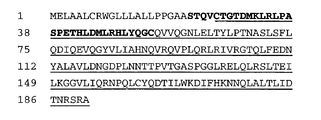
Figure 2 shows the amino acid sequence (SEQ ID NO: 1) of
이런 질환은 바람직하게는, ErbB2 수용체의 과다발현을 특징으로 하는 양성 또는 악성 종양, 예를 들면 유방암, 판상 세포암, 소세포 폐암, 비소세포 폐암, 위장암, 췌장암, 신경교아종, 자궁경부암, 난소암, 간암, 방광암, 간종양, 결장암, 결장직장암, 자궁내막암, 타액선암종, 신장암, 간암, 전립선암, 외음문암, 갑상선암, 간 암종 및 여러 형태의 두부 및 경부 암 등의 암이다. 화학요법제는 바람직하게는 탁솔 (등록상표) (파클리탁셀) 또는 탁솔 (등록상표) 유도체 등의 탁소이드이다.Such diseases are preferably benign or malignant tumors characterized by overexpression of the ErbB2 receptor, such as breast cancer, plaque cell cancer, small cell lung cancer, non-small cell lung cancer, gastrointestinal cancer, pancreatic cancer, glioma, cervical cancer, ovarian cancer. , Liver cancer, bladder cancer, liver tumor, colon cancer, colorectal cancer, endometrial cancer, salivary adenocarcinoma, kidney cancer, liver cancer, prostate cancer, vulvar cancer, thyroid cancer, liver carcinoma, and various types of head and neck cancer. The chemotherapeutic agent is preferably a taxoid such as Taxol (registered trademark) (paclitaxel) or Taxol (registered trademark) derivative.
항-ErbB2 항체는 항증식 효과면 충분하긴 하지만, 바람직한 실시양태에서는 세포사멸 (cell death) 또는 세포자멸 (apoptosis)을 유도할 수 있다. 바람직한 항-ErbB2 항체는 ErbB2 수용체의 세포외 도메인에 결합하며, 바람직하게는 ErbB2 세포외 도메인 서열 내의 에피토프 4D5 또는 3H4에 결합한다. 더욱 바람직하게는 항-ErbB2 항체는 항체 4D5이며, 가장 바람직하게는 인간화된 형태의 항체이다.Anti-ErbB2 antibodies are sufficient in antiproliferative effect, but in preferred embodiments can induce cell death or apoptosis. Preferred anti-ErbB2 antibodies bind to the extracellular domain of the ErbB2 receptor, preferably to the epitope 4D5 or 3H4 in the ErbB2 extracellular domain sequence. More preferably, the anti-ErbB2 antibody is antibody 4D5, most preferably in a humanized form.
본 발명의 방법은 특히 ErbB2 수용체의 과다발현을 특징으로하는 유방암 또는 난소암의 치료에 적합하다.The method of the invention is particularly suitable for the treatment of breast or ovarian cancer characterized by overexpression of the ErbB2 receptor.
본 발명의 다른 측면은 용기와, 이 용기 내의 항-ErbB2 항체를 포함하는 조성물과, 임의로는 이 조성물이 ErbB2 수용체의 과다발현을 특징으로 하는 상태의 치료를 위해 사용될 수 있음을 나타내는 용기 상의 또는 용기에 결합된 라벨과, 상기 조성물과 안트라사이클린형 화학요법제의 병용을 피하라는 지시사항을 담은 포장 삽입물을 포함하는 제조품에 관한 것이다.Another aspect of the invention is a container and a composition comprising an anti-ErbB2 antibody within the container, and optionally on or on a container indicating that the composition can be used for the treatment of a condition characterized by overexpression of the ErbB2 receptor. It relates to an article of manufacture comprising a label bound to and a package insert containing instructions to avoid the combination of the composition with an anthracycline-type chemotherapeutic agent.
1. 정의1. Definition
용어 "HER2", "ErbB2", "c-Erb-B2"는 서로 바꿔 사용할 수 있다. 다른 언급이 없는 한, 용어 "ErbB2", "c-Erb-B2" 및 "HER2"는 ErbB2 단백질을 의미하고, "erbB2", "c- erb -B2" 및 "her2"는 사람 유전자를 의미한다. 사람 erbB2 유전자 및 ErbB2 단백질은 문헌[예를 들어 Semba et al., PNAS (USA) 82:6497-6501) 및 Yamamoto et al., Nature 319:230-234(1986)]에 기재되어 있다 (Genebank 기탁 번호 X03363). ErbB2는 4개의 도메인 (도메인 1-4)으로 이루어진다.
The terms "HER2", "ErbB2", and "c-Erb-B2" can be used interchangeably. Unless otherwise stated, the terms “ErbB2”, “c-Erb-B2” and “HER2” refer to the ErbB2 protein, and “ erbB2 ”, “ c- erb -B2 ” and “ her2 ” refer to human genes. . The human erbB2 gene and ErbB2 protein are described in the literature (eg Semba et al., PNAS (USA) 82:6497-6501) and Yamamoto et al., Nature 319:230-234 (1986) (Genebank deposit Number X03363). ErbB2 consists of 4 domains (domains 1-4).
"에피토프 4D5"는 항체 4D5 (ATCC CRL 10463)가 결합하는 ErbB2의 세포외 도메인 내의 영역이다. 이 에피토프는 ErbB2의 막횡단 영역에 가깝다. 4D5 에피토프에 결합하는 항체를 스크리닝하기 위해서, 문헌[Antibodies, A Laboratory Manual, Cold Spring Harbor Laboratory, Ed Harlow and David Lane (1988)]에 기재된 바와 같은 통상의 교차 차단 분석을 수행할 수 있다. 별법으로, 에피토프 맵핑을 수행하여 (도 1 참조) 항체가 ErbB2 상의 4D5 에피토프(즉, ErbB2의 대략 잔기 529, 예를 들어 대략 잔기 561 내지 대략 잔기 625을 포함하는 영역 내의 하나 이상의 잔기)에 결합하는지를 평가할 수 있다."Epitope 4D5" is the region in the extracellular domain of ErbB2 to which antibody 4D5 (ATCC CRL 10463) binds. This epitope is close to the transmembrane region of ErbB2. To screen for antibodies that bind to the 4D5 epitope, conventional cross-blocking assays as described in Antibodies, A Laboratory Manual, Cold Spring Harbor Laboratory, Ed Harlow and David Lane (1988) can be performed. Alternatively, epitope mapping is performed (see Figure 1) to determine if the antibody binds to the 4D5 epitope on ErbB2 (i.e., one or more residues in the region comprising approximately residue 529 of ErbB2, e.g., approximately
"에피토프 3H4"는 ErbB2의 세포외 도메인에서 항체 3H4가 결합하는 영역이다. 이 에피토프를 도 1에 나타냈다. 이 에피토프는 ErbB2 세포외 도메인의 아미노산 서열에서 약 541 내지 약 599까지의 잔기를 포함한다."Epitope 3H4" is the region to which antibody 3H4 binds in the extracellular domain of ErbB2. This epitope is shown in FIG. 1. This epitope comprises residues from about 541 to about 599 in the amino acid sequence of the ErbB2 extracellular domain.
"에피토프 7C2/7F3"은 7C2 및(또는) 7F3 항체(각각 후술되는 바와 같이 ATCC에 기탁됨)가 결합하는 ErbB2의 세포외 도메인의 N 말단에 위치한 영역이다. 7C2/7F3 에피토프에 결합하는 항체를 스크리닝하기 위해서, 문헌[Antibodies, A Laboratory Manual, Cold Spring Harbor Laboratory, Ed Harlow and David Lane (1988)]에 기재된 바와 같은 통상의 교차 차단(cross-blocking) 분석을 수행할 수 있다. 별법으로, 에피토프 맵핑을 수행하여 항체가 ErbB2 상의 7C2/7F3 에피토프(즉, ErbB2의 대략 잔기 22 내지 대략 잔기 53의 영역 내의 하나 이상의 잔기 (서열 2))에 결합하는지를 조사할 수 있다.“Epitope 7C2/7F3” is a region located at the N-terminus of the extracellular domain of ErbB2 to which the 7C2 and/or 7F3 antibodies (respectively deposited with ATCC as described below) bind. To screen for antibodies that bind to the 7C2/7F3 epitope, a conventional cross-blocking assay as described in Antibodies, A Laboratory Manual, Cold Spring Harbor Laboratory, Ed Harlow and David Lane (1988) was performed. You can do it. Alternatively, epitope mapping can be performed to investigate whether the antibody binds to the 7C2/7F3 epitope on ErbB2 (i.e., one or more residues in the region of approximately
용어 "세포 사멸의 유도" 또는 "세포 사멸을 유도할 수 있는"이란 생존가능 세포를 비생존성으로 만드는 항체의 능력을 의미한다. 여기서 "세포"는 ErbB2 수용체를 발현하는 세포, 특히 ErbB2 수용체를 과다발현하는 세포이다. ErbB2를 "과다발현"하는 세포는 동일한 조직형의 비암세포에 비해 정상 수준보다 상당히 많은 양의 ErbB2를 갖는다. 바람직하게는, 세포는 암세포, 예를 들어 유방, 난소, 위, 자궁내막, 타액선, 폐, 신장, 결장, 갑상선, 췌장 및 방광 세포이다. 시험관 내에서, 이 세포는 SKBR3, BT474, Calu 3, MDA-MB-453, MDA-MB-361 또는 SKOV3 세포일 수 있다. 시험관내 세포 사멸은 항체 의존성 세포 독성 (ADCC) 또는 보체 의존성 세포독성 (CDC)에 의해 유도되는 세포 사멸과 구별하기 위해 보체 및 면역 이펙터 세포의 부재 하에 측정할 수 있다. 따라서, 세포 사멸 분석은 열 불활성화 혈청을 사용하여(즉, 보체의 부재 하에) 및 면역 이펙터 세포의 부재 하에 수행될 수 있다. 항체가 세포 사멸을 유도할 수 있는지를 결정하기 위해서, 요오드화프로피듐(PI), 트리판 블루 (Moore et al., Cytotechnology 17:1-11 (1995) 참조) 또는 7AAD (실시예 1 참조)의 흡수에 의해 평가되는 막 온전성의 상실을 비처리 세포에 비교하여 평가할 수 있다. 바람직한 세포 사멸 유도 항체는 "BT474 세포에서 PI 흡수 분석"에서 PI 흡수를 유도하는 항체이다.The term “induction of cell death” or “ability to induce cell death” refers to the ability of an antibody to render viable cells non-viable. Here, "cell" is a cell that expresses the ErbB2 receptor, in particular, a cell that overexpresses the ErbB2 receptor. Cells that "overexpress" ErbB2 have significantly greater than normal levels of ErbB2 compared to non-cancerous cells of the same tissue type. Preferably, the cells are cancer cells, such as breast, ovarian, stomach, endometrial, salivary gland, lung, kidney, colon, thyroid, pancreatic and bladder cells. In vitro, these cells can be SKBR3, BT474, Calu 3, MDA-MB-453, MDA-MB-361 or SKOV3 cells. In vitro cell death can be measured in the absence of complement and immune effector cells to differentiate from cell death induced by antibody dependent cytotoxicity (ADCC) or complement dependent cytotoxicity (CDC). Thus, cell death assays can be performed using heat inactivated serum (ie, in the absence of complement) and in the absence of immune effector cells. To determine whether the antibody can induce cell death, propidium iodide (PI), trypan blue (see Moore et al., Cytotechnology 17:1-11 (1995)) or 7AAD (see Example 1). Loss of membrane integrity, as assessed by absorption, can be assessed compared to untreated cells. A preferred apoptosis inducing antibody is an antibody that induces PI uptake in "PI uptake assay in BT474 cells".
문구 "세포자멸의 유도" 또는 "세포자멸을 유도할 수 있는"이란 아넥신 V의 결합, DNA의 단편화, 세포 수축, 소포체의 팽창, 세포 파편화 및(또는) 막 비지클(세포자멸체로 칭함)의 형성에 의해 측정되는 계획된 세포 사멸 (programmed cell death)을 유도하는 항체의 능력을 의미한다. 여기서 세포는 ErbB2 수용체를 과다발현하는 세포이다. 바람직하게는, "세포"는 종양 세포, 예컨데, 유방, 난소, 위, 자궁내막, 타액선, 폐, 신장, 결장, 갑상선, 췌장 또는 방광 세포이다. 시험관 내에서는 상기 세포가 SKBR3, BT474, Calu 3, MDA-MB-453, MDA-MB-361 또는 SKOV3 세포일 수 있다. 세포자멸에 관련된 세포 반응을 평가하기 위해 다양한 방법을 사용할 수 있다. 예를 들어, 포스파티딜 세린(PS) 위치전이는 아넥신 결합에 의해 측정할 수 있고, DNA 단편화는 본원 실시예에 기재된 바와 같이 DNA 래더링(laddering)을 통하여 평가할 수 있고, DNA 단편화와 함께 핵/크로마틴 농축은 저2배체(hypodiploid) 세포의 증가에 의해 평가할 수 있다. 바람직하게는, 세포자멸을 유도하는 항체는 "BT474 세포를 사용한 아넥신 결합 분석" (하기 참조)에서 미처리 세포에 비해 아넥신 결합을 약 2 내지 50배, 바람직하게는 약 5 내지 50배, 가장 바람직하게는 약 10 내지 50배 더 잘 유도하는 항체이다.The phrase "induction of apoptosis" or "capable of inducing apoptosis" means binding of annexin V, fragmentation of DNA, cell contraction, expansion of endoplasmic reticulum, cell fragmentation and/or membrane vehicle (referred to as apoptosis). It refers to the ability of an antibody to induce programmed cell death as measured by the formation of Here, the cell is a cell that overexpresses the ErbB2 receptor. Preferably, the “cell” is a tumor cell, such as a breast, ovary, stomach, endometrium, salivary gland, lung, kidney, colon, thyroid, pancreatic or bladder cell. In vitro, the cells may be SKBR3, BT474, Calu 3, MDA-MB-453, MDA-MB-361 or SKOV3 cells. A variety of methods can be used to evaluate cellular responses related to apoptosis. For example, phosphatidyl serine (PS) translocation can be measured by annexin binding, DNA fragmentation can be evaluated through DNA laddering as described in the Examples herein, and nuclear/ Chromatin enrichment can be assessed by an increase in hypodiploid cells. Preferably, the antibody inducing apoptosis is about 2 to 50 times, preferably about 5 to 50 times, most of the annexin binding compared to untreated cells in "Annexin binding assay using BT474 cells" (see below). It is preferably an antibody that induces about 10 to 50 times better.
때때로, 세포자멸 유도 항체는 ErbB2/ErbB3 복합체 (예를 들어 7F3 항체)의 HRG 결합/활성화를 차단하는 것이다. 다른 환경에서, 항체는 HRG에 의한 ErbB2/ErbB3 수용체 복합체의 활성화를 그다지 차단하지 않는 항체 (예를 들어 7C2)이다. 또한, 항체는 세포자멸을 유도하지만 S기 세포의 비율을 크게 저하시키지 않는 7C2와 같은 항체(예를 들어 대조구에 비해 상기 세포의 비율을 단지 약 0 내지 10% 감소시키는 항체)일 수 있다.Sometimes, the apoptosis inducing antibody is one that blocks HRG binding/activation of the ErbB2/ErbB3 complex (eg 7F3 antibody). In other circumstances, the antibody is an antibody that does not very much block activation of the ErbB2/ErbB3 receptor complex by HRG (eg 7C2). In addition, the antibody may be an antibody such as 7C2 that induces apoptosis but does not significantly reduce the proportion of S-phase cells (eg, an antibody that reduces the proportion of the cells by only about 0-10% compared to the control).
본 발명에서 항체는 사람 ErbB2에 특이적으로 결합하고, erbB1, erbB3 및(또는) erbB4 유전자에 의해 코딩되는 단백질과 같은 다른 단백질과 그다지 잘 교차반응하지 않는 7C2와 같은 항체이다. 때때로, 항체는 문헌[Schecter et al., Nature 312:513(1984) 및 Drebin et al., Nature 312:545-548(1984)]에 기재된 바와 같이 쥐 neu 단백질과 그다지 교차반응하지 않을 수 있다. 이러한 실시양태에서, 항체와 상기 단백질과의 결합 (예를 들어 내인성 수용체에 대한 세포 표면 결합)의 정도는 형광 활성화 세포 분류 (FACS) 분석 또는 방사성 면역 침전법 (RIA)에 의해 측정시에 약 10% 미만일 것이다.In the present invention, the antibody is an antibody such as 7C2 that specifically binds to human ErbB2 and does not cross-react very well with other proteins such as proteins encoded by the erbB1, erbB3 and/or erbB4 genes. Occasionally, the antibody may not cross-react very much with the murine neu protein as described by Schecter et al., Nature 312:513 (1984) and Drebin et al., Nature 312:545-548 (1984). In such embodiments, the degree of binding of the antibody to the protein (e.g., cell surface binding to an endogenous receptor) is about 10 as measured by fluorescence activated cell sorting (FACS) analysis or radioactive immunoprecipitation (RIA). Will be less than %.
"헤레귤린 (HRG)"은 ErbB2-ErbB3 및 ErbB2-ErbB4 단백질 복합체를 활성화시키는(즉, 복합체에 부착하면 복합체 내의 티로신 잔기의 인산화를 유도하는) 폴리펩티드를 의미한다. 상기 용어에 포함되는 다양한 헤레귤린 폴리펩티드는 문헌[Holmes et al., Science, 256:1205-1210(1992); WO92/20798; Wen et al., Mol. Cell. Biol., 14(3):1909-1919; 및 Marchionni et al., Nature, 362:312-318(1993)]에 기재되어 있다. 상기 용어는 천연 HRG 폴리펩티드의 생물학적 활성 단편 및(또는 변이체), 예를 들어 그의 EGF 유사 도메인 단편 (예를 들어 HRGβ1177-244)를 포함한다.“Heregulin (HRG)” refers to a polypeptide that activates the ErbB2-ErbB3 and ErbB2-ErbB4 protein complexes (ie, when attached to the complex induces phosphorylation of tyrosine residues in the complex). Various heregulin polypeptides included in the term are described in Holmes et al., Science, 256:1205-1210 (1992); WO92/20798; Wen et al., Mol. Cell. Biol., 14(3): 1909-1919; And Marchionni et al., Nature, 362:312-318 (1993). The term includes biologically active fragments and (or variants) of natural HRG polypeptides, eg, EGF-like domain fragments thereof (eg HRGβ1 177-244 ).
"ErbB2-ErbB3 단백질 복합체" 및 "ErbB2-ErbB4 단백질 복합체"는 각각 ErbB2 수용체 및 ErbB3 수용체 또는 ErbB4 수용체의 올리고머에 비공유적으로 결합된다. 복합체는 이들 수용체 둘 모두를 발현하는 세포가 HRG에 노출될 때 복합체가 형성되고 문헌[Sliwkowski et al., J. Biol. Chem., 269(20):14661-14665(1994)]에 기재된 바와 같이 면역침전에 의해 단리되고 SDS-PAGE에 의해 분석될 수 있다."ErbB2-ErbB3 protein complex" and "ErbB2-ErbB4 protein complex" are non-covalently bound to the ErbB2 receptor and to the ErbB3 receptor or oligomer of the ErbB4 receptor, respectively. The complex is formed when cells expressing both of these receptors are exposed to HRG and described by Sliwkowski et al., J. Biol. Chem., 269(20):14661-14665(1994)] and can be isolated by immunoprecipitation and analyzed by SDS-PAGE.
"항체"(Ab) 및 "면역글로불린"(Ig)은 동일한 구조적 특성을 갖는 당단백질이다. 항체는 특이적인 항원에 결합 특이성을 보이지만, 면역글로불린은 항체 및 항원 특이성이 결여된 다른 항체 유사 분자를 모두 포함한다. 항체 유사 분자 종류의 폴리펩티드는 예를 들어 림프계에 의해 낮은 수준으로, 골수종 세포에 의해 증가된 수준으로 생성된다."Antibody" (Ab) and "immunoglobulin" (Ig) are glycoproteins with the same structural properties. Antibodies exhibit binding specificity for a specific antigen, whereas immunoglobulins include both antibodies and other antibody-like molecules that lack antigen specificity. Polypeptides of the class of antibody-like molecules are produced, for example, at low levels by the lymphatic system and at increased levels by myeloma cells.
"천연 항체" 및 "천연 면역글로불린"은 일반적으로 2개의 동일한 경쇄(L) 및 2개의 동일한 중쇄(H)로 구성되는 약 150,000 달톤의 헤테로테트라머의 당단백질이다. 각각의 경쇄는 하나의 디술피드 공유결합에 의해 중쇄에 연결되고, 디술피드 결합의 개수는 상이한 면역글로불린 이소형의 중쇄 사이에서 변한다. 또한, 각각의 중쇄 및 경쇄는 규칙적 간격으로 사슬 내부의 디술피드 결합을 갖는다. 각각의 중쇄는 한 말단에 가변 도메인(VH) 및 이 도메인에 후속하는 많은 불변 도메인을 갖는다. 각각의 경쇄는 한 말단에 가변 도메인(VL) 및 다른 말단에 불변 도메인을 갖고, 경쇄의 불변 도메인은 중쇄의 제1 불변 도메인과 한줄로 위치하고, 경쇄 가변 도메인은 중쇄의 가변 도메인과 한줄로 위치한다. 특정 아미노산 잔기들이 경쇄 및 중쇄 가변 도메인 사이의 계면을 형성하는 것으로 생각된다.“Natural antibodies” and “natural immunoglobulins” are glycoproteins of about 150,000 Daltons heterotetrameric, generally composed of two identical light chains (L) and two identical heavy chains (H). Each light chain is linked to the heavy chain by one covalent disulfide bond, and the number of disulfide bonds varies between heavy chains of different immunoglobulin isotypes. In addition, each of the heavy and light chains has a disulfide bond inside the chain at regular intervals. Each heavy chain has a variable domain at one end (V H ) and a number of constant domains following this domain. Each light chain has a variable domain (V L ) at one end and a constant domain at the other end, the constant domain of the light chain is located in one line with the first constant domain of the heavy chain, and the light chain variable domain is located in one line with the variable domain of the heavy chain. do. It is believed that certain amino acid residues form the interface between the light and heavy chain variable domains.
용어 "가변"은 항체들 간에 가변 도메인의 특정 부분의 서열이 크게 상이하고 그 특정 항원에 대한 각각의 특정 항체의 결합 및 특이성에 사용된다는 사실을 의미한다. 그러나, 가변성은 항체의 가변 도메인 전체에 걸쳐 균일하게 분포되지 않는다. 가변성은 경쇄 및 중쇄 가변 도메인 모두의 상보성 결정 영역 (CDR) 또는 초가변 영역으로 불리는 3개의 세그먼트에 집중된다. 가변 도메인에서 보존도가 보다 높은 부분은 프레임워크 영역(FR)이라 불린다. 천연 중쇄 및 경쇄의 가변 도메인은, 루프 연결을 형성하며 어떤 경우에는 β-시트 구조의 일부를 이루는 3개의 CDR에 의해, 주로 β-시트의 입체형태인 프레임워크 영역 4개가 연결되어 이루어진다. 각 사슬 내의 CDR은 FR 영역에 의해 서로 매우 근접하게 유지되고, 다른 사슬의 CDR과 함께 항체의 항원 결합 부위의 형성에 기여한다 (Kabat et al., NIH Publ. No. 91-3242, Vol. I, pages 647-669 (1991) 참조). 불변 도메인은 항체의 항원 결합시에 직접 관여하지 않지만, 항체 의존적 세포의 세포독성에서 항체의 참가와 같은 상이한 이펙터 기능을 보인다.The term “variable” refers to the fact that between antibodies the sequence of a particular portion of a variable domain differs significantly and is used for the binding and specificity of each particular antibody for that particular antigen. However, the variability is not uniformly distributed throughout the variable domain of the antibody. Variability is concentrated in three segments called the complementarity determining regions (CDRs) or hypervariable regions of both the light and heavy chain variable domains. The portion of the variable domain with higher conservancy is called the framework region (FR). The variable domains of the natural heavy and light chains are made up of four framework regions, mainly the conformational form of the β-sheet, linked by three CDRs that form loop linkages and in some cases form part of the β-sheet structure. The CDRs within each chain are kept very close to each other by the FR regions, and together with the CDRs of the other chains contribute to the formation of the antigen binding site of the antibody (Kabat et al., NIH Publ. No. 91-3242, Vol. I. , pages 647-669 (1991)). The constant domain is not directly involved in antigen binding of the antibody, but exhibits different effector functions, such as the participation of the antibody in the cytotoxicity of antibody dependent cells.
항체를 파파인 분해하면 2개의 동일한 항원 결합 단편, 즉 항원 결합 부위를 하나씩 갖는 "Fab" 단편, 및 쉽게 결정화되는 능력을 반영하여 이름 붙여진 나머지 "Fc" 단편이 생성된다. 펩신을 처리하면, 2개의 항원 결합 부위를 가지며 여전히 항원에 교차결합할 수 있는 F(ab')2 단편이 생성된다.Papain digestion of an antibody yields two identical antigen-binding fragments, a "Fab" fragment with one antigen-binding site, and the remaining "Fc" fragment named to reflect its ability to crystallize easily. Treatment with pepsin produces an F(ab') 2 fragment that has two antigen binding sites and is still capable of cross-linking to the antigen.
"Fv"는 완전한 항원 인식 및 항원 결합 부위를 포함하는 최소 항체 단편이다. 이 영역은 단단하게 비공유결합된 하나의 중쇄 가변 도메인과 하나의 경쇄 가변 도메인의 이량체로 구성된다. 이것은 각 가변 도메인의 CDR 3개가 상호작용하여 VH-VL 이량체의 표면 상의 항원 결합 부위를 한정하는 배열로 존재한다. 결론적으로 보면, 6개의 CDR이 항체에 대한 항원 결합 특이성을 부여한다. 그러나, 하나의 가변 도메인 (또는 항원에 특이적인 단지 3개의 CDR만을 포함하는 Fv의 절반)의 경우에도 전체 결합 부위보다 친화성은 낮지만 항원을 인식하여 항원에 결합하는 능력을 갖는다.“Fv” is the smallest antibody fragment that contains a complete antigen recognition and antigen binding site. This region consists of a dimer of one heavy chain variable domain and one light chain variable domain tightly non-covalently linked. It exists in an arrangement in which the three CDRs of each variable domain interact to define the antigen binding site on the surface of the V H -V L dimer. In conclusion, the six CDRs confer antigen binding specificity for the antibody. However, even in the case of one variable domain (or half of the Fv containing only three CDRs specific to the antigen), the affinity is lower than that of the entire binding site, but has the ability to recognize the antigen and bind to the antigen.
또한, Fab 단편은 경쇄의 불변 도메인 및 중쇄의 제1 불변 도메인(CH1)을 함유한다. Fab' 단편은 항체 힌지 영역에서 유래한 하나 이상의 시스테인을 포함하여 중쇄 CH1 도메인의 카르복시 말단에 몇개의 잔기가 첨가되었다는 점에서 Fab 단편과 상이하다. Fab'-SH는 불변 도메인의 시스테인 잔기(들)이 유리 티올기를 보유하는 Fab'를 의미한다. F(ab')2 항체 단편은 본래 그들 사이에 힌지 시스테인을 갖는 몇쌍의 Fab' 단편으로서 생성되었다. 항체 단편의 다른 화학적 커플링도 공지되었다.In addition, the Fab fragment contains the constant domain of the light chain and the first constant domain (CH1) of the heavy chain. Fab' fragments differ from Fab fragments in that several residues are added to the carboxy terminus of the heavy chain CH1 domain, including one or more cysteines derived from the antibody hinge region. Fab'-SH means a Fab' in which the cysteine residue(s) of the constant domain has a free thiol group. F(ab') 2 antibody fragments were originally generated as pairs of Fab' fragments with hinge cysteines between them. Other chemical couplings of antibody fragments are also known.
임의의 척추동물종으로부터 유래한 항체(면역글로불린)의 "경쇄"는 그의 불변 도메인의 아미노산 서열을 기초로 하여 카파(κ) 및 람다(λ)로 불리는 2개의 명백하게 상이한 종류 중의 하나로 분류될 수 있다.The "light chain" of an antibody (immunoglobulin) derived from any vertebrate species can be classified into one of two distinctly different classes called kappa (κ) and lambda (λ) based on the amino acid sequence of its constant domain. .
중쇄의 불변 도메인의 아미노산 서열에 따라, 면역글로불린은 상이한 클래스로 분류될 수 있다. 면역글로불린에는 IgA, IgD, IgE, IgG 및 IgM의 5개의 주요 클래스가 존재하고, 이들 중 몇 개는 서브클래스(이소형), 예를 들어 IgG1, IgG2, IgG3, IgG4, IgA 및 IgA2로 추가 분류될 수 있다. 면역글로불린의 상이한 클래스에 대응하는 중쇄 불변 도메인은 각각 α, δ, ε,γ 및 μ로 지칭된다. 면역글로불린의 상이한 클래스의 서브유닛 구조 및 3차원 배열은 잘 알려져 있다.Depending on the amino acid sequence of the constant domain of the heavy chain, immunoglobulins can be classified into different classes. There are five major classes of immunoglobulins: IgA, IgD, IgE, IgG and IgM, some of which are further classified as subclasses (isotypes), e.g. IgG1, IgG2, IgG3, IgG4, IgA and IgA2. Can be. The heavy chain constant domains corresponding to different classes of immunoglobulins are referred to as α, δ, ε, γ and μ, respectively. The subunit structures and three-dimensional arrangements of different classes of immunoglobulins are well known.
용어 "항체"는 가장 광범위한 의미로 사용되고, 완전한 모노클로날 항체, 폴리클로날 항체, 2개 이상의 완전한 항체로부터 형성된 다특이성 항체 (예를 들어 이중특이적 항체), 및 요구되는 생물학적 활성을 보이는 항체 단편을 포함한다.The term “antibody” is used in the broadest sense and is a complete monoclonal antibody, a polyclonal antibody, a multispecific antibody (eg a bispecific antibody) formed from two or more complete antibodies, and an antibody that exhibits the required biological activity. Includes short stories.
"항체 단편"은 완전한 항체의 일부, 바람직하게는 완전한 항체의 항원 결합 또는 가변 영역을 포함한다. 항체 단편의 예로는 Fab, F(ab'), F(ab')2, 및 Fv 단편, 디아바디(diabody), 선형 항체 (Zapata et al., Protein Eng. 8(10):1057-1062 (1995)), 단일쇄 항체 분자, 및 항체 단편으로부터 형성된 다특이성 항체 등이 있다.An “antibody fragment” comprises a portion of an intact antibody, preferably the antigen binding or variable region of the intact antibody. Examples of antibody fragments include Fab, F(ab'), F(ab') 2 , and Fv fragments, diabodies, linear antibodies (Zapata et al., Protein Eng. 8(10):1057-1062 ( 1995)), single-chain antibody molecules, and multispecific antibodies formed from antibody fragments.
용어 "모노클로날 항체"는 실질적으로 상동성인 항체의 군집으로부터 수득된 항체를 말한다. 즉 미량 존재할 지 모르는 가능한 자연발생 돌연변이를 제외하고는 군집을 구성하는 개개의 항체는 동일하다. 모노클로날 항체는 단일 항원 부위에 대해 유도된 것으로, 높은 특이성을 갖는다. 또한, 일반적으로 상이한 항원 결정자(에피토프)에 대해 작용하는 상이한 항체를 포함하는 통상의 (폴리클로날) 항체 제제와는 달리, 각각의 모노클로날 항체는 항원 상의 단일 항원 결정자에 대해서만 유도된다. 특이성 외에, 모노클로날 항체는 다른 면역글로불린에 의해 오염되지 않은 하이브리도마 배양에 의해 합성되기 때문에 유리하다. "모노클로날"이란 수식어는 실질적으로 상동성인 항체 군집으로부터 수득되는 항체의 특성을 나타내고, 어떤 특정 방법에 의해 항체 생성을 요구하는 것으로서 해석되지 않는다. 예를 들어, 본 발명에 따라 사용되는 모노클로날 항체는 문헌[Kohler et al., Nature, 256:495 (1975)]에 처음 기재된 하이브리도마 방법에 의해 제조될 수 있거나, 재조합 DNA 방법 (예를 들어 미국 특허 제4,816,567호 참조)에 의해 제조될 수 있다. "모노클로날 항체"는 또한 문헌 [예를 들어 Clackson et al., Nature, 352:624-628(1991) 및 Marks et al., J. Mol. Biol., 222:581-597 (1991)]에 기재된 기술을 사용하여 파지 항체 라이브러리로부터 단리될 수 있다.The term “monoclonal antibody” refers to an antibody obtained from a population of substantially homologous antibodies. In other words, the individual antibodies constituting the cluster are identical, except for possible naturally occurring mutations that may be present in trace amounts. Monoclonal antibodies are directed against a single antigenic site and have high specificity. In addition, unlike conventional (polyclonal) antibody preparations, which generally contain different antibodies that act against different antigenic determinants (epitopes), each monoclonal antibody is directed against only a single antigenic determinant on the antigen. Besides specificity, monoclonal antibodies are advantageous because they are synthesized by hybridoma culture that is not contaminated with other immunoglobulins. The modifier "monoclonal" refers to the properties of an antibody obtained from a population of substantially homologous antibodies, and is not to be interpreted as requiring antibody production by any particular method. For example, the monoclonal antibody used according to the present invention can be prepared by the hybridoma method first described in Kohler et al., Nature, 256:495 (1975), or the recombinant DNA method (e.g. See, for example, U.S. Patent No. 4,816,567). “Monoclonal antibodies” are also described in, eg, Clackson et al., Nature, 352:624-628 (1991) and Marks et al., J. Mol. Biol., 222:581-597 (1991).
본원의 모노클로날 항체는 구체적으로 중쇄 및(또는) 경쇄의 일부가 특정 종으로부터 유래하거나 특정 항체 클래스 또는 서브클래스에 속하는 항체의 대응 서열과 동일하거나 상동성이고 사슬(들)의 나머지는 다른 종으로부터 유래하거나 다른 항체 클래스 또는 서브클래스에 속하는 항체의 대응 서열과 동일하거나 상동성인 "키메라(chimera)" 항체(면역글로불린) 및 요구되는 생물학적 활성을 보이는 이 항체의 단편을 포함한다(미국 특허 제4,816,567호; Morrison et al., Proc. Natl. Acad. Sci. USA, 81:6851-6855 (1984)).In the monoclonal antibody of the present application, a part of the heavy chain and/or light chain is specifically the same or homologous to the corresponding sequence of an antibody that is derived from a specific species or belongs to a specific antibody class or subclass, and the remainder of the chain(s) is a different species. A "chimera" antibody (immunoglobulin) that is identical or homologous to the corresponding sequence of an antibody derived from or belonging to another antibody class or subclass, and fragments of this antibody that exhibit the required biological activity (US Pat. No. 4,816,567. No.; Morrison et al., Proc. Natl. Acad. Sci. USA, 81:6851-6855 (1984)).
비인간(예를 들어 쥐과) 항체의 "인간화" 형태는 키메라 면역글로불린, 즉, 비사람 면역글로불린으로부터 유래한 최소한의 서열을 포함하는 면역글로불린 사슬 또는 그의 단편 (예를 들어 Fv, Fab, Fab', F(ab')2 또는 항체의 다른 항원 결합 서열)이다. 대부분의 경우, 인간화 항체는 사람 면역글로불린 (수용 항체)의 상보성 결정 영역 (CDR)의 잔기가 필요한 특이성, 친화도 및 능력을 갖는, 생쥐, 쥐 또는 토끼와 같은 비인간종(공여 항체)의 CDR의 잔기로 대체된 사람 면역글로불린이다. 어떤 경우에는 사람 면역글로불린의 Fv 프레임워크 영역 (FR)이 대응하는 비사람 잔기로 대체된다. 또한, 인간화 항체는 수용체 항체 또는 도입되는 CDR 또는 프레임워크 서열 중 어디에서도 발견되지 않는 잔기를 포함할 수 있다. 이런 변형은 항체 성능을 더욱 세련화하고 극대화하기 위해 이루어진다. 일반적으로, 인간화 항체는 모든 또는 실질적으로 모든 CDR 영역이 비사람 면역글로불린의 CDR 영역에 대응하고 모든 또는 실질적으로 모든 FR 영역이 사람 면역글로불린 서열의 영역인 하나 이상, 일반적으로 2개의 가변 도메인 모두를 실질적으로 포함할 것이다. 또한, 인간화 항체는 적어도 면역글로불린 불변 영역 (Fc), 일반적으로 사람 면역글로불린의 불변 영역의 일부를 포함할 것이다 (Jones et al., Nature, 321:522-525(1986); Reichmann et al., Nature, 332:323-329 (1988); 및 Presta, Curr. Op. Struct. Biol. 2:593-596 (1992) 참조). 인간화 항체는 항체의 항원 결합 영역이 목적 항원으로 짧은꼬리원숭이를 면역시켜 생성된 항체로부터 유래된 PRIMATIZED(상표) 항체가 있다.The "humanized" form of a non-human (eg murine) antibody is a chimeric immunoglobulin, ie an immunoglobulin chain or fragment thereof (eg Fv, Fab, Fab', containing minimal sequence derived from a non-human immunoglobulin). F(ab') 2 or other antigen binding sequence of the antibody). In most cases, a humanized antibody is a CDR of a non-human species (donor antibody) such as a mouse, rat or rabbit, with the required specificity, affinity and ability of the residues of the complementarity determining region (CDR) of a human immunoglobulin (receptive antibody). It is a human immunoglobulin replaced by a residue. In some cases, the Fv framework region (FR) of a human immunoglobulin is replaced with a corresponding non-human residue. In addition, humanized antibodies may contain residues not found either in the receptor antibody or in the introduced CDR or framework sequences. These modifications are made to further refine and maximize antibody performance. In general, humanized antibodies have one or more, generally all two variable domains, in which all or substantially all of the CDR regions correspond to the CDR regions of a non-human immunoglobulin and all or substantially all of the FR regions are regions of a human immunoglobulin sequence. Will contain substantially. In addition, the humanized antibody will comprise at least a portion of an immunoglobulin constant region (Fc), generally that of a human immunoglobulin (Jones et al., Nature, 321:522-525(1986); Reichmann et al., Nature, 332:323-329 (1988); and Presta, Curr. Op. Struct. Biol. 2:593-596 (1992)). Humanized antibodies include PRIMATIZED (trademark) antibodies derived from antibodies produced by immunizing macaques with the antigen-binding region of the antibody with a target antigen.
"단일쇄 Fv" 또는 "sFv" 항체 단편은 단일 폴리펩티드 사슬 중에 존재하는 항체의 VH 및 VL 도메인을 포함한다. 바람직하게는, Fv 폴리펩티드는 sFv가 항원 결합에 필요한 구조를 형성하도록 하는 VH 및 VL 도메인 사이의 폴리펩티드 링커를 포함한다(sFv에 관해서는 문헌 (Plueckthan in The Pharmacology of Monoclonal Antibodies, vol. 113, Rosenburg 및 Moore eds., Springer-Verlag, New York, pp. 269-315 (1994)) 참조). A “single chain Fv” or “sFv” antibody fragment comprises the V H and V L domains of an antibody present in a single polypeptide chain. Preferably, the Fv polypeptide comprises a polypeptide linker between the V H and V L domains that allows the sFv to form the structure necessary for antigen binding (see Pleckthan in The Pharmacology of Monoclonal Antibodies, vol. 113, for sFv). Rosenburg and Moore eds., Springer-Verlag, New York, pp. 269-315 (1994)).
용어 "디아바디 (diabody)"는 동일한 폴리펩티드 사슬 (VH-VL) 내의 경쇄 가변 도메인(VL)에 연결된 중쇄 가변 도메인 (VH)을 포함하는, 2개의 항원 결합 부위를 갖는 작은 항체 단편을 의미한다. 동일한 사슬 상의 2개의 도메인 사이의 페어링을 허용하기에는 너무 짧은 링커를 사용하여, 도메인을 다른 사슬의 상보성 도메인과 강제로 페어링시켜 2개의 항원 결합 부위를 생성시킨다. 디아바디는 예를 들어 EP 404,097, WO93/11611 및 Hollinger et al., Proc. Natl. Acad. Sci. USA, 90:6444-6448 (1993)에 보다 상세하게 기재되어 있다.The term “diabody” refers to a small antibody fragment having two antigen binding sites, comprising a heavy chain variable domain (V H ) linked to a light chain variable domain (V L ) within the same polypeptide chain (V H -V L ). Means. Using a linker that is too short to allow pairing between the two domains on the same chain, the domains are forcibly paired with the complementary domains of the other chain to create two antigen binding sites. Diabodies are, for example, EP 404,097, WO93/11611 and Hollinger et al., Proc. Natl. Acad. Sci. USA, 90:6444-6448 (1993).
"단리된" 항체는 그의 천연 환경의 성분으로부터 동정 및 분리 및(또는) 회수된 항체이다. 그의 천연 환경의 오염 성분은 항체의 진단 또는 치료 용도를 방해하는 물질이고, 효소, 호르몬 및 다른 단백질성 또는 비단백질성 용질을 포함할 수 있다. 바람직한 실시양태에서, 항체는 (1) 로우리(Lowry) 방법에 의해 측정시 95 중량% 초과, 가장 바람직하게는 99 중량% 초과의 항체로, 또는 (2) 스피닝 컵(spinning cup) 서열분석기를 사용하여 N 말단 또는 내부 아미노산의 잔기 15개 이상을 수득하기에 충분한 정도로, 또는 (3) 쿠마시 블루, 또는 바람직하게는 은 염색을 사용하여 환원 또는 비환원 조건 하에 SDS-PAGE에 의해 균질하게 정제될 것이다. 단리된 항체는 그 항체의 천연 환경의 하나 이상의 성분이 존재하지 않을 것이기 때문에, 재조합 세포 내의 원위치에서의 항체이다. 그러나, 단리된 항체는 일반적으로 하나 이상의 정제 단계에 의해 정제될 것이다.An “isolated” antibody is an antibody that has been identified and separated and/or recovered from a component of its natural environment. Contaminant components of its natural environment are substances that interfere with the diagnostic or therapeutic use of the antibody, and may include enzymes, hormones and other proteinaceous or non-proteinaceous solutes. In a preferred embodiment, the antibody is (1) greater than 95% by weight, most preferably greater than 99% by weight as measured by the Lowry method, or (2) using a spinning cup sequencer. To an extent sufficient to obtain at least 15 residues of the N-terminal or internal amino acid, or (3) to be homogeneously purified by SDS-PAGE under reducing or non-reducing conditions using Coomassie blue, or preferably silver staining. will be. An isolated antibody is an antibody in situ within a recombinant cell, since one or more components of the antibody's natural environment will not be present. However, the isolated antibody will generally be purified by one or more purification steps.
용어 "샐비지(salvage) 수용체 결합 에피토프"는 IgG 분자의 체내 혈청 반감기를 증가시키는 기능을 수행하는 IgG 분자 (예를 들어 IgG1, IgG2, IgG3 또는 IgG4)의 Fc 영역의 에피토프를 의미한다.The term “salvage receptor binding epitope” refers to an epitope of the Fc region of an IgG molecule (eg, IgG 1 , IgG 2 , IgG 3 or IgG 4 ) that functions to increase the serum half-life of the IgG molecule in the body. do.
"치료"는 치료적 처리 및 예방적 또는 억제적 조치를 모두 의미한다. 치료를 필요로 하는 대상은 질환이 억제되어야 하는 대상 뿐만 아니라 이미 질환을 앓고 있는 대상을 포함한다."Treatment" means both therapeutic treatment and prophylactic or inhibitory measures. Subjects in need of treatment include subjects already suffering from the disease as well as subjects for which the disease is to be suppressed.
치료를 위한 "포유동물"은 인간, 가축, 및 동물원, 경기용 또는 애완 동물, 예를 들어 개, 말, 고양이, 소 등을 포함하여 포유동물로서 분류된 모든 동물을 의미한다. 바람직하게는, 포유동물은 인간이다."Mammal" for treatment means all animals classified as mammals, including humans, domestic animals, and zoos, competitions or pets, such as dogs, horses, cats, cows, and the like. Preferably, the mammal is human.
"질환"은 항-ErbB2 항체를 사용한 치료에 의해 호전되는 모든 상태이다. 이것은 포유동물이 문제의 질환에 걸리게 하는 병태를 포함하여 만성 및 급성 질환 또는 질병을 포함한다. 본원에서 치료되는 질환의 예로는 양성 및 악성 종양, 백혈병 및 임파성 악성 질환, 신경, 신경교, 성상세포, 시상하부 및 다른 선, 마크로파지, 상피, 기질 및 포배강 질환, 및 염증성, 혈관형성 및 면역 질환이 있으나, 이에 제한되지 않는다."Disease" is any condition ameliorated by treatment with an anti-ErbB2 antibody. This includes chronic and acute diseases or diseases, including conditions that predispose the mammal to the disease in question. Examples of diseases to be treated herein include benign and malignant tumors, leukemia and lymphoid malignancies, neurons, glials, astrocytes, hypothalamus and other glands, macrophages, epithelium, stromal and blastocyst diseases, and inflammatory, angiogenesis and immunity. There is a disease, but is not limited thereto.
"치료적 유효량"이란 용어는 항증식 효과를 갖는 양을 지칭하는 데 사용된다. 바람직하게는 치료적 유효량이 세포자멸 활성을 갖거나 세포 사멸을 유도할 수 있으며, 바람직하게는 양성 또는 악성 종양 세포, 특히 암 세포의 사멸을 유도할 수 있다. 효능은 치료되는 상태에 따라 통상적인 방식으로 측정할 수 있다. 암 치료의 경우 효능 측정은 예를 들면 질병 진행 시간 (TTP) 평가 또는 반응률 (RR) 결정을 통해 이루어질 수 있다 (하기 실시예의 설명 참조).The term "therapeutically effective amount" is used to refer to an amount that has an antiproliferative effect. Preferably, the therapeutically effective amount has apoptotic activity or can induce apoptosis, and preferably can induce the death of benign or malignant tumor cells, particularly cancer cells. Efficacy can be measured in a conventional manner depending on the condition being treated. In the case of cancer treatment, efficacy can be measured, for example, by evaluating disease progression time (TTP) or determining response rate (RR) (see the description of the examples below).
용어 "암" 및 "암성"은 미조절된 세포 증식이라는 전형적 특징을 갖는 포유동물의 생리 상태를 의미하거나 규정한다. 암의 예로는 암종, 임파종, 아세포종, 육종 및 백혈병을 들 수 있으나, 이에 제한되지 않는다. 상기 암의 보다 구체적인 예는 판상 세포암, 소세포 폐암, 비소세포 폐암, 위장암, 췌장암, 신경교아종, 자궁경부암, 난소암, 간암, 방광암, 간종양, 유방암, 결장암, 결장직장암, 자궁내막암, 타액선암종, 신장암, 전립선암, 외음부암, 갑상선암, 간암종 및 여러 종류의 두부 및 경부 암을 포함한다.The terms “cancer” and “cancerous” mean or define the physiological condition of a mammal with the typical characteristics of uncontrolled cell proliferation. Examples of cancer include, but are not limited to, carcinoma, lymphoma, blastoma, sarcoma, and leukemia. More specific examples of the cancer include plaque cell cancer, small cell lung cancer, non-small cell lung cancer, gastrointestinal cancer, pancreatic cancer, glioma, cervical cancer, ovarian cancer, liver cancer, bladder cancer, liver tumor, breast cancer, colon cancer, colorectal cancer, endometrial cancer, Salivary adenocarcinoma, kidney cancer, prostate cancer, vulvar cancer, thyroid cancer, liver carcinoma, and several types of head and neck cancer.
용어 "세포독성제"는 세포의 기능을 저해하거나 억제하고(하거나) 세포를 파괴시키는 물질을 의미한다. 이 용어는 방사성 동위원소 (예를 들어 I131, I125, Y90 및 Re186), 화학요법제 및 독소, 예를 들면 세균, 진균, 식물 또는 동물 기원의 효소 활성을 갖는 독소 또는 그의 단편을 포함하는 것을 의미한다.The term “cytotoxic agent” refers to a substance that inhibits or inhibits the function of cells and/or destroys cells. The term refers to radioactive isotopes (eg I 131 , I 125 , Y 90 and Re 186 ), chemotherapeutic agents and toxins, such as toxins having enzymatic activity of bacterial, fungal, plant or animal origin, or fragments thereof. Means to include.
"화학요법제"는 암 치료에 유용한 화학적 화합물이다. 이의 예는 아드리아마신 (독소루비신), 에피루비신, 5-플루오로우라실 (5-FU), 시토신 아라비노시드 ("Ara-C"), 시클로포스파미드 (상표명 CYTOXAN), 티오테파, 부술판, 탁소이드, 예를 들면 파클리탁셀 (등록상표명 탁솔, 미국 뉴저지주 프린스톤에 위치한 브리스톨마이어스스큅 온콜로지사 제품) 및 도세탁셀 (탁소테레 Taxotere, 프랑스 안토니에 위치한 롱플랑 로레아사 제품), 메토트렉세이트, 시스플라틴, 빈블라스틴, 블레오마이신, 에토포시드, 이포스파미드, 미토마이신 C, 미톡산트론, 빈크리스틴, 비노렐빈, 카르보플라틴, 테니포시드, 다우노마이신, 카르미노마이신, 아미노프테린, 닥티노마이신, 미토마이신, 에스페라미신 (미국 특허 제4,675,187호 참조), 멜팔란 및 다른 관련 질소 머스타드를 포함한다. 또한, 타목시펜 및 오나프리스톤과 같은 종양에 대한 호르몬 작용을 조절하거나 억제하는 호르몬 제제도 상기 화학요법제의 정의에 포함된다."Chemotherapeutic agents" are chemical compounds useful in the treatment of cancer. Examples thereof include adriamycin (doxorubicin), epirubicin, 5-fluorouracil (5-FU), cytosine arabinoside ("Ara-C"), cyclophosphamide (trade name CYTOXAN), thiotepa, busulfan , Taxoids such as paclitaxel (registered trademark Taxol, a product of Bristol Myers Squibb Oncology, located in Princeton, NJ) and docetaxel (Taxotere, a product of Longplan Lorea, located in Antony, France), methotrexate, cisplatin, Vinblastine, bleomycin, etoposide, ifosfamide, mitomycin C, mitoxantrone, vincristine, vinorelbine, carboplatin, teniposide, daunomycin, carminomycin, aminopterin, doc Tinomycin, mitomycin, esperamicin (see US Pat. No. 4,675,187), melphalan and other related nitrogen mustards. Hormonal agents that modulate or inhibit the action of hormones on tumors such as tamoxifen and onapristone are also included in the definition of the chemotherapeutic agent.
화학요법제의 다른 예로는, 임프로술판 및 피포술판 등의 알킬 술폰산염류; 벤조도파, 카르보쿠온, 메투레도파 및 우레도파 등의 아지리딘류; 알트레타민, 트리에틸렌멜라민, 트리에틸렌포스포라미드, 트리에틸렌티오포스파오라미드 및 트리메틸올멜라민을 비롯한 에틸렌이민류 및 메틸아멜아민류; 클로람부실, 클로나파진, 콜로포스파미드, 에스트라머스틴, 메클로레타민, 메클로레타민 옥시드 히드로클로라이드, 노벰비친, 페네스테린, 프레드니무스틴, 트로포스파미드, 우라실 머스타드 등의 질소머스타드류; 카르무스틴, 클로로조토신, 포테무스틴, 로무스틴, 니무스틴, 라미무스틴 등의 니트로수레아류; 아클라시노마이신류, 악티노마이신, 아우트라마이신, 아자세린, 칵티노마이신, 칼리케아미신, 카라비신, 카르지노필린, 크로모마이신류, 6-디아조-5-옥소-L-노르루이신, 다우노루비신, 마이코페놀산, 노갈라마이신, 올리보마이신류, 페플로마이신, 포트피로마이신, 퓨로마이신, 스트렙토니그린, 스트렙토조신, 튜베르시딘, 우베니멕스, 지노스타틴, 조루비신, 젠타미신 등의 항생제; 데놉테린, 프테롭테린, 트리메트렉세이트 등의 폴릭산 유사체; 플루다라빈, 6-메르캅토퓨린, 티아미프린, 티오구아닌 등의 퓨린 유사체; 안시타빈, 아자시티딘, 6-아자우리딘, 카르모푸르, 시타라빈, 디데옥시우리딘, 독시플루리딘, 에노시타빈 및 플록스우리딘 등의 피리미딘 유사체; 칼루스테론, 드로모스타놀론 프로피오네이트, 에피티오스탄올, 메피티오스탄, 테스토락톤 등의 안드로겐류; 아미노글루테티미드, 미토탄, 트리로스탄 등의 항부신제 (anti-adrenals); 프롤린닉산 등의 폴릭산 보충물; 아세글락톤; 알도포파미드 글리코시드; 아미노레불리닉산; 암사크린; 베스트라부실; 비산트렌; 에다트락세이트; 데포파민; 데메콜신; 디아지쿠온; 엘포르니틴; 엘립티늄 아세테이트; 에토글루시드; 갈륨 니트레이트; 히드록시우레아; 렌티난; 로니다민; 미토구아존; 미톡산트론; 모피다몰; 니트라크린; 펜토스타틴; 페나메트; 피라루비신; 포도필린산; 2-에틸히드라지드; 프로카르바진; PSK (등록상표); 라족산; 시조피란; 스피로게르마늄; 테뉴아존산; 트리아지쿠온; 2,2',2"-트리클로로트리에틸아민; 우레탄; 빈데신; 다카르바진; 만노무스틴; 미토브로니톨; 미토락톨; 피포브로만; 가시토신; 클로람부실; 겜시타빈; 6-티오구아닌; 메르캅토퓨린; 백금; 나벨빈; 노반트론; 크셀로다; 이반드로네이트; CPT-11; 토포이소머라제 억제제 RFS 2000; 디클루오로메틸오르니틴 (DMFO); 레티노익산; 카페시타빈; 및 이들 중 어느 하나의 제약상 허용가능한 염, 산 또는 유도체 등이 있다.Other examples of chemotherapeutic agents include alkyl sulfonates such as improsulfan and piposulfan; Aziridines such as benzodopa, carboquone, meturedopa, and uredopa; Ethyleneimines and methylammelamines including altretamine, triethylene melamine, triethylene phosphoramide, triethylene thiophosphaolamide and trimethylol melamine; Chlorambucil, clonafazin, colophosphamide, estramustine, mechloretamine, mechloretamine oxide hydrochloride, nobembicine, phenesterine, prednimustine, trophosphamide, uracil mustard, etc. Nitrogen mustards; Nitrosureas such as carmustine, chlorozotocin, potatomustine, lomustine, nimustine, and ramiemustine; Aclacinomycins, Actinomycins, Autramycin, Azaserine, Cactinomycin, Calicheamicin, Carabicin, Carginophylline, Chromomycins, 6-diazo-5-oxo-L-Norruyi Sin, Daunorubicin, Mycophenolic Acid, Nogalamycin, Olivomycins, Peplomycin, Potpyromycin, Puromycin, Streptonigreen, Streptozosin, Tubercidine, Ubenimex, Zinostatin, Zorubicin And antibiotics such as gentamicin; Folic acid analogs such as denopterin, pteropterin, trimetrexate, etc.; Purine analogs such as fludarabine, 6-mercaptopurine, thiamiprine, and thioguanine; Pyrimidine analogs such as ancitabine, azacitidine, 6-azauridine, carmofur, cytarabine, dideoxyuridine, doxyfluridine, enocitabine and phloxuridine; Androgens such as calosterone, dromostanolone propionate, epithiostanol, mepithiostan, and testolactone; Anti-adrenals such as aminoglutetimide, mitotan, and trirostan; Folic acid supplements such as prolinnic acid; Aceglutone; Aldopopamide glycoside; Aminolevulinic acid; Amsaclean; Best Labusil; Bisantrene; Edatroxate; Depopamine; Demecolsin; Diazicuon; Elfornitine; Elliptinium acetate; Etogluside; Gallium nitrate; Hydroxyurea; Lentinan; Ronidamine; Mitoguazone; Mitoxantrone; Fur damole; Nitraclean; Pentostatin; Penamet; Pyrarubicin; Podophyllic acid; 2-ethylhydrazide; Procarbazine; PSK (registered trademark); Lajok acid; Sizopiran; Spirogermanium; Tenuazonic acid; Triazicion; 2,2',2"-trichlorotriethylamine; urethane; vindesine; dacarbazine; mannomustine; mitobronitol; mitrolactol; pipebroman; gashitosine; chlorambucil; gemcitabine; 6-thioguanine ; Mercaptopurine; Platinum; Navelbin; Novantron; Xceloda; Ibandronate; CPT-11; Topoisomerase inhibitor RFS 2000; Dicluoromethylornithine (DMFO); Retinoic acid; Capecitabine; And Pharmaceutically acceptable salts, acids or derivatives of any of these.
종양에 대해 호르몬 작용을 조절 또는 억제하는 호르몬제의 다른 예로는 랄록시펜 (Evista), 아로마타제 억제 4(5)-이미다졸, 4-히드록시타목시펜, 트리옥시펜, 케옥시펜 및 LY117018 등의 항에스트로겐제 및 플루타미드 및 닐루타미드 등의 항안드로겐제 및 이들 중 어느 하나의 제약상 허용가능한 염, 산 또는 유도체가 있다.Other examples of hormonal agents that modulate or inhibit hormonal action on tumors include raloxifene (Evista), aromatase inhibitory 4(5)-imidazole, 4-hydroxytamoxifen, trioxyphene, keoxyphene, and LY117018. Estrogens and anti-androgens such as flutamide and nilutamide, and pharmaceutically acceptable salts, acids or derivatives of any of them.
"증식 억제제"는 세포, 특히 ErbB2 과다발현 암세포의 시험관 내 또는 체내 증식을 억제하는 화합물 또는 조성물을 의미한다. 따라서, 성장 억제제는 ErbB2 과다발현 세포의 S기 비율을 크게 감소시키는 물질이다. 성장 억제제의 예는 세포 주기 진행을 (S기 이외의 시기에서)차단하는 물질, 예를 들어 G1 정지 및 M기 정지를 유도하는 물질을 포함한다. 전통적인 M기 차단제로는 빈카스 (빈크리스틴 및 빈블라스틴), 탁솔, 및 토포 II 억제제, 예를 들어 독소루비신, 에피루비신, 다우노루비신, 에토포시드 및 블레오마이신 등이 있다. 또한, G1을 정지시키는 물질, 예를 들어 DNA 알킬화제, 예를 들어 타목시펜, 프레드니손, 다카르바진, 메클로레타민, 시스플라틴, 메톡트렉세이트, 5-플루오로우라실 및 아라-C는 S기 정지에도 작용한다. 추가 정보는 문헌 [Murakami et al., The Molecular Basis of Cancer, Mendelsohn and Israel, eds., "Cell cycle regulation, oncogens, and antineoplastic drugs" (WB Saunders: Philadelphia, 1995), Chapter 1, 특히 13면]에 기재되어 있다. 4D5 항체 (및 그의 기능적 등가물)도 상기 목적을 위해 사용할 수 있다."Proliferation inhibitor" means a compound or composition that inhibits the proliferation of cells, in particular ErbB2 overexpressing cancer cells, in vitro or in the body. Therefore, the growth inhibitory agent is a substance that greatly reduces the S phase ratio of ErbB2 overexpressing cells. Examples of growth inhibitory agents include substances that block cell cycle progression (at a time other than phase S), such as substances that induce G1 arrest and M phase arrest. Traditional M-phase blockers include vincas (vincristine and vinblastine), taxol, and topo II inhibitors such as doxorubicin, epirubicin, daunorubicin, etoposide and bleomycin. In addition, substances that stop G1, such as DNA alkylating agents, such as tamoxifen, prednisone, dacarbazine, mechloretamine, cisplatin, methoxytrexate, 5-fluorouracil, and ara-C also act on S group arrest. do. Further information can be found in Murakami et al., The Molecular Basis of Cancer, Mendelsohn and Israel, eds., "Cell cycle regulation, oncogens, and antineoplastic drugs" (WB Saunders: Philadelphia, 1995),
"독소루비신"은 안트라사이클린 항생제이다. 독소루비신의 완전한 화학명은 (8S-시스)-10-[(3-아미노-2,3,6-트리데옥시-α-L-릭소-헥소피라노실)옥소]-7,8,9,10-테트라히드로-6,8,11-트리히드록시-8-(히드록시아세틸)-1-메톡시-5,12-나프타센디온이다.“Doxorubicin” is an anthracycline antibiotic. The complete chemical name of doxorubicin is (8S-cis)-10-[(3-amino-2,3,6-trideoxy-α-L-lixo-hexopyranosyl)oxo]-7,8,9,10 -Tetrahydro-6,8,11-trihydroxy-8-(hydroxyacetyl)-1-methoxy-5,12-naphthacendione.
"사이토킨"은 하나의 세포 군집에 의해 방출되는, 세포간 매개제로서 다른 세포에 대해 작용하는 단백질의 일반 명칭이다. 이러한 사이토킨의 예는 임포킨, 모노킨 및 전통적인 폴리펩티드 호르몬이다. 사이토킨에는 성장 호르몬, 예를 들어 사람 성장 호르몬, N-메티오닐 사람 성장 호르몬 및 소 성장 호르몬; 부갑상선 호르몬; 티록신; 인슐린; 프로인슐린; 렐락신; 프로렐락신; 당단백질 호르몬, 예를 들어 여포 자극 호르몬 (FSH), 갑상선 자극 호르몬(TSH) 및 황체 호르몬 (LH); 간 성장 인자; 섬유아세포 성장 인자; 프로락틴; 태반 락토겐; 종양 괴사 인자-α 및 -β; 물레리안 억제 물질; 생쥐 고나도트로핀 연합 펩티드; 인히빈; 악티빈; 혈관 내피 성장 인자; 인테그린; 트롬보포이에틴 (TPO); 신경 성장 인자, 예를 들어 NGF-β; 혈소판 형성 인자; 형질전환 성장 인자 (TGF), 예를 들어 TGF-α 및 TGF-β; 인슐린 유사 성장 인자-I 및 -II; 에리트로포이에틴 (EPO); 골유도 인자; 인터페론, 예를 들어 인터페론-α, -β 및 -γ; 콜로니 자극 인자 (CSF), 예를 들어 마크로파지-CSF (M-CSF); 과립구-마크로파지-CSF(GM-CSF); 및 과립구-CSF(G-CSF); 인터루킨(IL), 예를 들어 IL-1, IL-1α, IL-2, IL-3, IL-4, IL-5, IL-6, IL-7, IL-8, IL-9, IL-11, IL-12; 종양 괴사 인자, 예를 들어 TNF-α 및 TNF-β; 및 LIF 및 키트 리간드(KL)를 비롯한 다른 폴리펩티드 인자가 포함된다. 본원에서 사용되는 바와 같이, 용어 사이토킨은 천연 공급원 또는 재조합 세포 배양물로부터의 단백질 및 천연 서열 사이토킨의 생물학적 활성 등가물을 포함한다.“Cytokine” is a generic name for a protein that is released by one cell population and acts on another cell as an intercellular mediator. Examples of such cytokines are impokines, monokines and traditional polypeptide hormones. Cytokines include growth hormones such as human growth hormone, N-methionyl human growth hormone and bovine growth hormone; Parathyroid hormone; Thyroxine; insulin; Proinsulin; Relaxin; Prorelaxin; Glycoprotein hormones such as follicle stimulating hormone (FSH), thyroid stimulating hormone (TSH) and progesterone (LH); Liver growth factor; Fibroblast growth factor; Prolactin; Placental lactogen; Tumor necrosis factor-α and -β; Mulerian inhibitory substances; Mouse gonadotropin association peptide; Inhibin; Actibin; Vascular endothelial growth factor; Integrin; Thrombopoietin (TPO); Nerve growth factors such as NGF-β; Platelet-forming factor; Transforming growth factors (TGF), such as TGF-α and TGF-β; Insulin-like growth factor-I and -II; Erythropoietin (EPO); Bone inducing factor; Interferons such as interferon-α, -β and -γ; Colony stimulating factor (CSF), such as macrophage-CSF (M-CSF); Granulocyte-macrophage-CSF (GM-CSF); And granulocyte-CSF (G-CSF); Interleukin (IL), for example IL-1, IL-1α, IL-2, IL-3, IL-4, IL-5, IL-6, IL-7, IL-8, IL-9, IL- 11, IL-12; Tumor necrosis factors such as TNF-α and TNF-β; And other polypeptide factors including LIF and kit ligand (KL). As used herein, the term cytokine includes proteins from natural sources or from recombinant cell culture and biologically active equivalents of native sequence cytokines.
용어 "전구약물"은 모 약물에 비해 종양 세포에 대한 세포독성이 작고 효소에 의해 활성화되거나 보다 활성인 모 형태로 전환될 수 있는 제약상 활성 물질의 전구체 또는 유도체 형태를 의미한다 (예를 들어 Wilman, "Prodrugs in Cancer Chemotherapy" Biochemical Society Transactions, 14, pp. 375-382, 615th Meeting Belfast (1986) and Stella et al., "Prodrugs: A Chemical Approach to Targeted Drug Delivery," Directed Drug Delivery, Borchardt et al., (ed.), pp.247-267, Humana Press (1985) 참조). 본 발명의 전구약물은 인산염 함유 전구약물, 티오포스페이트 함유 전구약물, 황산염 함유 전구약물, 펩티드 함유 전구약물, D-아미노산 변형 전구약물, 글리코실화 전구약물, β-락탐 함유 전구약물, 임의로 치환된 페녹시아세트아미드 함유 전구약물 또는 임의로 치환된 페닐아세트아미드 함유 전구약물, 5-플루오로시토신 및 보다 활성인 세포독성 유리 약물로 전환될 수 있는 다른 5-플루오로우리딘 전구약물을 포함하나, 이에 제한되지 않는다. 본 발명에 사용하기 위한 전구약물 형태로 유도체화될 수 있는 세포독성 약물의 예는 상기한 바와 같은 화학요법제를 포함하나, 이에 제한되지 않는다.The term “prodrug” refers to a precursor or derivative form of a pharmaceutically active substance that has less cytotoxicity to tumor cells compared to the parent drug and can be activated by an enzyme or converted to a more active parent form (eg Wilman , "Prodrugs in Cancer Chemotherapy" Biochemical Society Transactions, 14, pp. 375-382, 615th Meeting Belfast (1986) and Stella et al., "Prodrugs: A Chemical Approach to Targeted Drug Delivery," Directed Drug Delivery, Borchardt et al ., (ed.), pp.247-267, Humana Press (1985)). Prodrugs of the present invention include phosphate-containing prodrugs, thiophosphate-containing prodrugs, sulfate-containing prodrugs, peptide-containing prodrugs, D-amino acid modified prodrugs, glycosylated prodrugs, β-lactam-containing prodrugs, and optionally substituted phenoxy. Cyacetamide containing prodrugs or optionally substituted phenylacetamide containing prodrugs, 5-fluorocytosine and other 5-fluorouridine prodrugs that can be converted to more active cytotoxic free drugs. It doesn't work. Examples of cytotoxic drugs that can be derivatized in the form of prodrugs for use in the present invention include, but are not limited to, chemotherapeutic agents as described above.
"고상 (solid phase)"은 본 발명의 항체가 부착될 수 있는 비수성 매트릭스를 의미한다. 고상의 예는 부분적으로 또는 완전하게 유리 (예를 들어 세공 조절된 유리), 폴리사카리이드 (예를 들어 아가로스), 폴리아크릴아미드, 폴리스티렌, 폴리비닐 알콜 및 실리콘을 포함한다. 특정 실시양태에서, 내용에 따라 고상은 분석 플레이트의 웰을 포함할 수 있다. 즉, 고상은 정제 컬럼 (예를 들어 친화도 크로마토그래피 컬럼)이다. 이 용어는 또한 미국 특허 제4,275,149호에 기재된 것과 같은 별개 입자의 비연속적 고상을 포함한다."Solid phase" means a non-aqueous matrix to which the antibodies of the present invention can be attached. Examples of solid phases include partially or completely free (eg pore controlled glass), polysaccharides (eg agarose), polyacrylamide, polystyrene, polyvinyl alcohol and silicone. In certain embodiments, depending on the context, the solid phase may comprise the wells of an assay plate. That is, the solid phase is a purification column (eg affinity chromatography column). The term also includes discontinuous solid phases of discrete particles, such as those described in US Pat. No. 4,275,149.
"리포솜"은 약물 (예를 들어 항-ErbB2 항체, 및 임의로 화학요법제)의 포유동물로의 전달에 유용한 다양한 형태의 지질, 인지질 및(또는) 계면활성제로 구성되는 작은 비지클이다. 리포솜의 성분은 통상 생물학적 막의 지질 배열과 유사한 2층 형성으로 배열된다.A “liposome” is a small vehicle composed of various types of lipids, phospholipids and/or surfactants useful for the delivery of drugs (eg anti-ErbB2 antibodies, and optionally chemotherapeutic agents) to mammals. The components of the liposome are usually arranged in two layers, similar to the lipid arrangement of biological membranes.
"포장 삽입물"이란, 치료용 제품의 상업적 포장에 통상적으로 포함되는 지시사항으로서, 이러한 치료용 제품의 사용과 관련한 징후, 사용법, 투여량, 투여방법, 금기 및(또는) 경고에 관한 정보를 담고 있다."Package insert" is instructions ordinarily included in commercial packaging of therapeutic products, which contain information on indications, directions for use, dosage, method of administration, contraindications and/or warnings related to the use of such therapeutic products. have.
II. 항-ErbB2 항체의 제조II. Preparation of anti-ErbB2 antibody
아래에는 본 발명에 따라 사용되는 항체의 생산을 위한 예시적인 기술에 관하여 설명한다. 항체 생산에 사용되는 ErbB2 항원은 예를 들면, 원하는 에피토프를 포함하는 ErbB2 세포외 도메인의 가용성 형태 또는 그의 일부일 수 있다. 별법으로, 세포 표면에 ErbB2를 발현하는 세포 (예를 들면, ErbB2를 과다발현하도록 형질전환된 NIH-3T3 세포 또는 SKBR3 세포와 같은 암종 세포주 {문헌[Stancovski et al., PNAS (USA) 88: 8691-8695 (1991)] 참조)를 사용하여 항체를 생산할 수 있다. 항체를 생산하기에 유용한 ErbB2의 다른 형태는 당업계의 기술자에게 명백할 것이다.Below, exemplary techniques for the production of antibodies used in accordance with the present invention are described. The ErbB2 antigen used for antibody production can be, for example, a soluble form of the ErbB2 extracellular domain comprising the desired epitope or a portion thereof. Alternatively, cells expressing ErbB2 on the cell surface (eg, NIH-3T3 cells transformed to overexpress ErbB2 or carcinoma cell lines such as SKBR3 cells {Stancovski et al., PNAS (USA) 88: 8691) -8695 (1991)] can be used to produce antibodies. Other forms of ErbB2 useful for producing antibodies will be apparent to those skilled in the art.
(i) 폴리클로날 항체(i) polyclonal antibody
폴리클로날 항체는 바람직하게는 관련 항원 및 보조제를 피하(sc) 또는 복강내(ip)로 여러번 주사하여 동물 내에서 유발시킨다. 그 관련 항원을, 면역시키고자 하는 종에서 면역원성인 단백질, 예를 들면, 키홀 삿갓조개(keyhole limpet) 헤모시아닌, 혈청 알부민, 소 타이로글로불린, 또는 대두 트립신 억제제에, 이관능성 작용제 또는 유도화제, 예를 들면, 말레이미도벤조일 술포숙신이미드 에스테르 (시스테인 잔기를 통해 결합), N-히드록시숙신이미드 (라이신 잔기를 통해), 글루타르알데히드, 숙신산 무수물, SOCl2, 또는 R1N=C=NR (여기에서, R 및 R1은 상이한 알킬기임)를 사용하여 결합시키는 것이 유용할 수 있다.Polyclonal antibodies are preferably induced in animals by multiple injections of the relevant antigens and adjuvants subcutaneously (sc) or intraperitoneally (ip). Proteins immunogenic in the species to be immunized against the related antigen, for example, keyhole limpet hemocyanin, serum albumin, bovine thyroglobulin, or soybean trypsin inhibitor, a bifunctional agonist or inducer , For example, maleimidobenzoyl sulfosuccinimide ester (linked through cysteine residue), N-hydroxysuccinimide (via lysine residue), glutaraldehyde, succinic anhydride, SOCl 2 , or R 1 N= It may be useful to bond using C=NR, wherein R and R 1 are different alkyl groups.
동물을 예를 들면, 100 ㎍ 또는 5 ㎍의 단백질 또는 결합물 (각각 토끼와 생쥐에 대해)을 3 부피의 프로인드(Freund's) 완전 보조제와 혼합하고, 이 용액을 여러 부위에 피내 주사함으로써 항원, 면역원성 결합물 또는 유도체에 대해 면역화시킨다. 1개월 후에, 동물을 원래의 양의 1/5 내지 1/10의 프로인드 완전 보조제 중의 펩티드 또는 결합물로 다수 부위에 피하 주사함으로써 재면역시켰다. 7 내지 14일 후, 동물을 출혈시키고, 혈청을 항체 역가에 대해 분석하였다. 역가가 정체 (plateau)될 때까지 동물을 재면역시킨다. 바람직하게는, 동일한 항원을 상이한 단백질에, 및(또는) 상이한 가교결합제를 통해 결합시킨 결합물로 동물을 재면역화한다. 결합물은 또한 단백질 융합체로서 재조합 세포 배양을 통해 제조할 수 있다. 또한, 명반(alum)과 같은 응집제를 사용하여 면역 반응을 증진시킨다.Animals are mixed with, for example, 100 μg or 5 μg of protein or conjugate (for rabbits and mice, respectively) with 3 volumes of Freund's complete adjuvant, and the solution is injected intradermally at several sites to obtain antigens, Immunization against immunogenic conjugates or derivatives. After 1 month, animals were reimmunized by subcutaneous injection at multiple sites with 1/5 to 1/10 of the original amount of peptide or conjugate in Freund's complete adjuvant. After 7-14 days, animals were bled and serum analyzed for antibody titer. Animals are reimmunized until titer plateaus. Preferably, the animal is reimmunized with a conjugate that binds the same antigen to a different protein and/or via a different crosslinking agent. Conjugates can also be prepared through recombinant cell culture as protein fusions. In addition, a coagulant such as alum is used to enhance the immune response.
(ii) 모노클로날 항체(ii) monoclonal antibody
모노클로날 항체는 실질적으로 균질 항체의 집단으로부터 얻으며, 즉, 집단을 이루는 개별적인 항체들은 소량 존재할 수 있는 가능한 자연 변이를 제외하고는 동일하다. 따라서, 변형 "모노클로날"은 별개의 항체들의 혼합물이 아닌 항체의 특성을 지시한다.Monoclonal antibodies are obtained from a population of substantially homogeneous antibodies, ie the individual antibodies that make up the population are identical except for possible natural variations that may be present in small amounts. Thus, a modified “monoclonal” refers to the properties of an antibody rather than a mixture of separate antibodies.
예를 들면, 모노클로날 항체는 문헌[코흘러 (Kohler) 등, Nature, 256: 495 (1975)]에서 처음 기술된 하이브리도마 방법을 사용하여 제조할 수 있거나, 재조합 DNA 방법 (미국 특허 제4,816,567호)을 이용하여 제조할 수 있다.For example, monoclonal antibodies can be prepared using the hybridoma method first described in Kohler et al., Nature, 256: 495 (1975), or the recombinant DNA method (US Patent No. 4,816,567) can be used.
하이브리도마 방법에서, 면역화를 위해 사용된 단백질에 충분하게 결합할 항체를 생산하거나 생산할 수 있는 백혈구를 유도하도록 생쥐, 또는 햄스터와 같은 다른 적절한 숙주 동물을 상기한 바와 같이 면역화시킨다. 별법으로, 백혈구를 시험관 내에서 면역화시킬 수 있다. 이어서, 백혈구를 폴리에틸렌 글리콜과 같은 적합한 융합제를 사용하여 골수종 세포와 융합시켜 하이브리도마 세포를 형성시킨다 [고딩 (Goding), Monoclonal Antibodies: Principle and Practice, pp. 59-103 (Academic Press, 1986)].In the hybridoma method, mice, or other suitable host animals, such as hamsters, are immunized as described above to induce leukocytes capable of producing or producing antibodies that will sufficiently bind to the protein used for immunization. Alternatively, leukocytes can be immunized in vitro. The leukocytes are then fused with myeloma cells using a suitable fusing agent such as polyethylene glycol to form hybridoma cells [Goding, Monoclonal Antibodies: Principle and Practice, pp. 59-103 (Academic Press, 1986)].
이렇게 제조한 하이브리도마 세포를 바람직하게는 비융합 골수종 모세포의 성장 또는 생존을 억제시키는 1종 이상의 물질을 함유하는 적합한 배지 중에 접종하고 배양시킨다. 예를 들면, 골수종 모세포가 하이포잔틴 구아닌 포스포리보실 트랜스퍼라제 (HGPRT 또는 HPRT) 효소가 결핍된 경우, 하이브리도마용 배지는 전형적으로 하이포잔틴, 아미노프테린 및 티미딘 (HAT 배지)을 포함할 것이며, 이들 물질은 HGPRT-결핍 세포의 성장을 억제시킨다.The hybridoma cells thus prepared are preferably inoculated and cultured in a suitable medium containing one or more substances that inhibit the growth or survival of non-fused myeloma blasts. For example, if the myeloma blasts are deficient in the enzyme hypoxanthine guanine phosphoribosyl transferase (HGPRT or HPRT), the medium for hybridomas will typically contain hypoxanthine, aminopterin and thymidine (HAT medium). , These substances inhibit the growth of HGPRT-deficient cells.
바람직한 골수종 세포는 효율적으로 융합하고, 선택된 항체-생산 세포에 의한 항체의 높은 수준의 안정한 생산을 지지하며, HAT 배지와 같은 배지에 감수성이 있는 것이다. 이들 중, 바람직한 골수종 세포주는 솔크 인스티튜트 셀 디스트리뷰션 센터 (Salk Institute Cell Distribution Center, 미국 캘리포니아주 샌디에고)로부터 입수가능한 MOPC-21 및 MPC-11, 및 아메리칸 타입 컬쳐 컬렉션 (American Type Culture Collection, 미국 매릴랜드주 록빌)으로부터 입수가능한 SP-2 또는 X63-Ag8-653 세포로부터 유래된 것과 같은 쥐 골수종 세포주이다. 사람 골수종 세포주 및 생쥐-사람 헤테로골수종 세포주가 또한 사람 모노클로날 항체의 생산에 대해 기술되었다 [코즈보르 (Kozbor), J. Immunol., 133: 3001 (1984); 및 브로듀어 (Brodeur) 등, Monoclonal Antibody Production Techniques and Applications, pp. 51-63 (Marcel Dekker, Inc., New York, 1987)].Preferred myeloma cells are those that fuse efficiently, support stable production of high levels of antibodies by selected antibody-producing cells, and are sensitive to media such as HAT media. Among these, preferred myeloma cell lines are MOPC-21 and MPC-11, available from Salk Institute Cell Distribution Center (San Diego, CA, USA), and American Type Culture Collection, Rockville, MD. ) Is a murine myeloma cell line such as that derived from SP-2 or X63-Ag8-653 cells available from. Human myeloma cell lines and mouse-human heteromyeloma cell lines have also been described for the production of human monoclonal antibodies [Kozbor, J. Immunol., 133: 3001 (1984); And Brodeur et al., Monoclonal Antibody Production Techniques and Applications, pp. 51-63 (Marcel Dekker, Inc., New York, 1987)].
하이브리도마 세포가 성장하는 배지를 항원에 대해 유도된 모노클로날 항체의 생산에 대해 분석하였다. 바람직하게는, 하이브리도마 세포에 의해 생산된 모노클로날 항체의 결합 특이성을 면역침강법에 의해, 또는 방사성면역분석법 (RIA) 또는 효소 결합 면역 흡수 분석법 (ELISA)과 같은 시험관내 결합 분석에 의해 측정하였다.The medium in which hybridoma cells were grown was analyzed for the production of monoclonal antibodies directed against the antigen. Preferably, the binding specificity of the monoclonal antibody produced by hybridoma cells is determined by immunoprecipitation or by an in vitro binding assay such as radioimmunoassay (RIA) or enzyme-linked immunosorbent assay (ELISA). It was measured.
모노클로날 항체의 결합 친화도는 예를 들면, 먼슨 (Munson) 등 [Anal. Biochem., 107: 220 (1980)]의 스캐차드 (Scatchard) 분석에 의해 측정할 수 있다.The binding affinity of monoclonal antibodies is determined, for example, in Munson et al. [Anal. Biochem., 107: 220 (1980)].
원하는 특이성, 친화도 및(또는) 활성의 항체를 생산하는 하이브리도마 세포를 확인한 후, 제한 희석 절차에 의해 클론을 서브클로닝시키고 표준 방법 [고딩, Monoclonal Antibodies: Principles and Practice, pp. 59-103 (Academic Press, 1986)]으로 배양시켰다. 이러한 목적에 적합한 배지는 예를 들면, D-MEM 또는 RPMI-1640 배지를 포함한다. 또한, 하이브리도마 세포는 동물에서 복수종양으로서 생체내 배양시킬 수 있다.After identifying hybridoma cells producing antibodies of the desired specificity, affinity and/or activity, the clones were subcloned by limiting dilution procedures and standardized methods [Goding, Monoclonal Antibodies: Principles and Practice, pp. 59-103 (Academic Press, 1986)]. Media suitable for this purpose include, for example, D-MEM or RPMI-1640 medium. In addition, hybridoma cells can be cultured in vivo as ascites tumors in animals.
서브클론에 의해 분비된 모노클로날 항체는 예를 들면, 단백질 A-세파로즈 (Sepharose), 히드록시아파티트 크로마토그래피, 겔 전기영동, 투석 또는 친화도 크로마토그래피와 같은 통상의 면역글로불린 정제 절차에 의해 배지, 복수액 또는 혈청으로부터 적합하게 분리시킨다.Monoclonal antibodies secreted by subclones are, for example, protein A-Sepharose, hydroxyapatite chromatography, gel electrophoresis, dialysis, or conventional immunoglobulin purification procedures such as affinity chromatography. Is suitably separated from the medium, ascites fluid or serum.
모노클로날 항체를 코딩하는 DNA는 통상의 절차 (예를 들면, 쥐 항체의 중쇄 및 경쇄를 코딩하는 유전자에 특이적으로 결합할 수 있는 올리고뉴클레오티드 프로브를 이용함으로써)를 이용하여 쉽게 분리되고 서열결정된다. 하이브리도마 세포는 그러한 DNA의 바람직한 기원으로서 작용한다. 일단 분리되면, DNA는 발현 백터 내로 넣을 수 있고, 이어서, 그렇지 않으면 면역글로불린 단백질을 생산하지 않는 이. 콜리(E. coli) 세포, 원숭이 COS 세포, 차이니즈 햄스터 난소(CHO) 세포, 또는 골수종 세포와 같은 숙주 세포 내로 형질감염시켜 재조합 숙주 세포에서 모노클로날 항체를 합성시킨다. 항체를 코딩하는 DNA의 세균에서의 재조합 발현에 대한 리뷰 문헌은 문헌[스케라 (Skerra) 등, Curr. Opinion in Immunol., 5: 256-262 (1993); 및 플뤼턴 (Plueckthun), Immunol. Revs., 130: 151-188 (1992)]을 포함한다.DNA encoding monoclonal antibodies is easily isolated and sequenced using conventional procedures (e.g., by using oligonucleotide probes capable of specifically binding to genes encoding heavy and light chains of murine antibodies). do. Hybridoma cells serve as the preferred source of such DNA. Once isolated, the DNA can be put into the expression vector, which in turn would otherwise not produce an immunoglobulin protein. Monoclonal antibodies are synthesized in recombinant host cells by transfection into host cells such as E. coli cells, monkey COS cells, Chinese hamster ovary (CHO) cells, or myeloma cells. Review literature on the recombinant expression of DNA encoding the antibody in bacteria (Skerra et al., Curr. Opinion in Immunol., 5: 256-262 (1993); And Flueckthun, Immunol. Revs., 130: 151-188 (1992)].
다른 실시양태에서, 항체 또는 항체 단편을 문헌[맥카퍼티 (McCafferty) 등, Nature, 348: 551-554 (1990)]에 기술된 기법을 이용하여 생산된 항체 파지 라이브러리로부터 단리할 수 있다. 문헌[클랙슨 (Clackson) 등, Nature, 352: 624-628 (1991) 및 막스 (Marks) 등, J. Mol. Biol. 222: 581-597 (1991)]에서는 파지 라이브러리를 사용하는 쥐 및 사람 항체의 단리를 각각 기술하였다. 이후의 문헌에서는 체인 서플링(chain shuffling)에 의한 고친화도(nM 범위)의 사람 항체의 생산 [막스 등, Bio/Technology 10: 779-783 (1992)] 뿐만 아니라, 매우 큰 파지 라이브러리의 제조 전략으로서의 조합 감염 및 생체내 재조합 [워터하우스 (Waterhouse) 등, Nuc. Acids. Res., 21: 2265-2266 (1993)]을 기술하였다. 따라서, 이들 기법은 모노클로날 항체의 단리를 위한 전통적인 모노클로날 항체 하이브리도마 기법과 병용가능한 방법이다.In other embodiments, antibodies or antibody fragments can be isolated from antibody phage libraries produced using techniques described in McCafferty et al., Nature, 348: 551-554 (1990). Clackson et al., Nature, 352: 624-628 (1991) and Marks et al., J. Mol. Biol. 222: 581-597 (1991)] describes the isolation of murine and human antibodies using phage libraries, respectively. In the subsequent literature, not only the production of high affinity (nM range) human antibodies by chain shuffling [Max et al., Bio/Technology 10: 779-783 (1992)], but also the production strategy of a very large phage library Combination infection and in vivo recombination as [Waterhouse et al., Nuc. Acids. Res., 21: 2265-2266 (1993)]. Therefore, these techniques are a method that can be used in combination with traditional monoclonal antibody hybridoma techniques for the isolation of monoclonal antibodies.
DNA는 또한 예를 들면, 동종 쥐 서열 대신 사람 중쇄 및 경쇄 불변 도메인에 대한 코딩 서열을 치환함으로써 (미국 특허 제4,816,567호; 문헌[모리슨 (Morrison) 등, Proc. Natl. Acad. Sci. USA, 81: 6851 (1984)]), 또는 비면역글로불린 폴리펩티드에 대한 코딩 서열의 전체 또는 일부를 면역글로불린 코딩 서열에 공유 결합시킴으로써 변형시킬 수 있다.DNA can also be used, for example, by substituting coding sequences for human heavy and light chain constant domains instead of homologous murine sequences (US Pat. No. 4,816,567; Morrison et al., Proc. Natl. Acad. Sci. USA, 81) : 6851 (1984)], or by covalently linking all or part of the coding sequence for a non-immunoglobulin polypeptide to an immunoglobulin coding sequence.
전형적으로 그러한 비면역글로불린 폴리펩티드는 항체의 불변 도메인에 대해 치환되거나, 항체의 한 항체-결합 부위의 가변 도메인에 대해 치환되어 항원에 특이성을 갖는 하나의 항체-결합 부위 및 상이한 항원에 특이성을 갖는 다른 항원-결합 부위를 포함하는 키메라 2가 항체를 생성시킨다.Typically such non-immunoglobulin polypeptides are substituted for the constant domain of the antibody, or for the variable domain of one antibody-binding site of the antibody, so that one antibody-binding site specific for an antigen and another specific for a different antigen. A chimeric bivalent antibody comprising an antigen-binding site is generated.
(iii) 인간화 항체 및 사람 항체(iii) humanized antibodies and human antibodies
비사람 항체를 인간화시키는 방법은 당업계에 잘 알려져 있다. 바람직하게는, 인간화 항체는 비사람 기원으로부터의 그에 도입된 하나 이상의 아미노산 잔기를 갖는다. 이들 비사람 아미노산 잔기는 종종 "임포트(import)" 잔기로 부르며, 전형적으로 "임포트" 가변 도메인으로부터 취한다. 인간화는 사람 항체의 상응하는 서열 대신 설치류 CDRs 또는 CDR 서열로 치환함으로써 본질적으로 윈터 (Winter) 등의 방법 [존스 (Jones) 등, Nature, 321: 522-525 (1986); 리치만 (Riechmann) 등, Nature, 332: 323-327 (1988); 베르호옌 (Verhoeyen) 등, Science, 239: 1534-1536 (1988)]에 따라 수행할 수 있다. 따라서, 그러한 "인간화" 항체는 실질적으로 보다 적은 무손상 사람 가변 도메인이 비사람 종으로부터 상응하는 서열에 의해 치환된 키메라 항체 (미국 특허 제4,816,567호)이다. 실제, 인간화 항체는 전형적으로 일부 CDR 잔기 및 가능하게는 일부 FR 잔기를 설치류 항체에서 유사한 부위로부터의 잔기로 치환시킨 사람 항체이다.Methods of humanizing non-human antibodies are well known in the art. Preferably, the humanized antibody has one or more amino acid residues introduced into it from non-human origin. These non-human amino acid residues are often referred to as "import" residues, and are typically taken from an "import" variable domain. Humanization is essentially a method of Winter et al. [Jones et al., Nature, 321: 522-525 (1986); Richmann et al., Nature, 332: 323-327 (1988); Verhoeyen et al., Science, 239: 1534-1536 (1988)]. Thus, such “humanized” antibodies are chimeric antibodies (US Pat. No. 4,816,567) in which substantially fewer intact human variable domains have been replaced by corresponding sequences from non-human species. In fact, humanized antibodies are typically human antibodies in which some CDR residues and possibly some FR residues have been replaced with residues from similar sites in rodent antibodies.
인간화 항체를 제조하기 위해 사용되는 사람 가변 도메인 (경쇄 및 중쇄 도메인 모두)의 선택은 항원성을 감소시키기 위해 매우 중요하다. 소위 "베스트핏(best-fit)" 방법에 따라, 설치류 항체의 가변 도메인의 서열을 공지의 사람 가변 도메인 서열의 전체 라이브러리에 대해 스크리닝한다. 이어서, 설치류의 서열에 가장 근접한 사람 서열을 인간화 항체를 위한 사람 프레임워크(FR)로서 수용한다 [심스 (Sims) 등, J. Immunol, 151: 2296 (1993); 코티아 (Chothia) 등, J. Mol. Biol., 196: 901 (1987)]. 다른 방법은 경쇄 또는 중쇄의 특정 하위군의 모든 사람 항체의 인식 서열로부터 유래된 특정 프레임워크를 사용한다. 몇종의 상이한 인간화 항체에 대해 동일한 프레임워크를 사용할 수 있다 [카터 (Carter) 등, Proc. Natl. Acad. Sci. USA, 89: 4285 (1992); 프레스타 (Presta) 등, J. Immnol., 151: 2623 (1993)].The choice of human variable domains (both light and heavy chain domains) used to make humanized antibodies is very important to reduce antigenicity. According to the so-called "best-fit" method, the sequence of the variable domains of rodent antibodies is screened against the entire library of known human variable domain sequences. The human sequence closest to that of the rodent is then accepted as a human framework (FR) for humanized antibodies [Sims et al., J. Immunol, 151: 2296 (1993); Chothia et al., J. Mol. Biol., 196: 901 (1987)]. Other methods use specific frameworks derived from the recognition sequences of all human antibodies of a specific subgroup of light or heavy chains. The same framework can be used for several different humanized antibodies [Carter et al., Proc. Natl. Acad. Sci. USA, 89: 4285 (1992); Presta et al., J. Immnol., 151: 2623 (1993)].
항원에 대한 높은 친화도와 다른 양호한 생물학적 특성을 보유하면서 항체를 인간화시키는 것이 또한 중요하다. 이러한 목적을 이루기 위해, 바람직한 방법에 따라, 인간화 항체는 모서열 및 인간화 서열의 3차원 모델을 사용하는 모서열과 다양한 구상되는 인간화 생성물의 분석 과정에 의해 제조한다. 3차원 면역글로불린 모델이 일반적으로 이용가능하고, 당업계의 기술자에게 친숙하다. 선택된 후보 면역글로불린 서열의 가능한 3차원 배위 구조를 예시하고 디스플레이하는 컴퓨터 프로그램이 이용가능하다. 이들 디스플레이를 점검하면 후보 면역글로불린 서열의 기능에서 잔기들의 유망한 역할을 분석할 수 있으며, 즉, 항원에 결합하는 후보 면역글로불린의 능력에 영향을 끼치는 잔기를 분석할 수 있다. 이러한 방식으로, 표적 항원(들)에 대한 증가된 친화성과 같은 원하는 항체 특성을 성취하도록, 수여자 및 임포트 서열로부터 FR 잔기를 선택하고 결합시킬 수 있다. 일반적으로, CDR 잔기는 항원 결합에 영향을 끼치는데 직접적으로 및 가장 실질적으로 관여된다.It is also important to humanize antibodies while retaining high affinity for the antigen and other good biological properties. To achieve this purpose, according to a preferred method, humanized antibodies are prepared by analysis of the parent sequence and various envisioned humanized products using a three-dimensional model of the parent sequence and the humanized sequence. Three-dimensional immunoglobulin models are generally available and are familiar to those skilled in the art. Computer programs are available that illustrate and display possible three-dimensional coordination structures of selected candidate immunoglobulin sequences. Examining these displays allows the analysis of the promising role of the residues in the function of the candidate immunoglobulin sequence, i.e., the analysis of residues that influence the ability of the candidate immunoglobulin to bind to the antigen. In this way, FR residues can be selected and associated from the recipient and import sequences to achieve the desired antibody properties, such as increased affinity for the target antigen(s). In general, CDR residues are directly and most substantially involved in affecting antigen binding.
별법으로, 지금은 내인성 면역글로불린 생산의 부재하에 면역시 사람 항체의 완전한 레퍼터리를 생산할 수 있는 트랜스제닉 동물 (예를 들면, 생쥐)을 제조할 수 있다. 예를 들면, 키메라 및 생식세포주 돌연변이 생쥐에서 항체의 중쇄 연결 영역 (JH) 유전자를 동질적으로 삭제시키면 내인성 항체 생산의 완전히 억제시키는 것이 기술되었다. 사람 생식세포주 면역글로불린 유전자 배열을 그러한 생식세포주 돌연변이 생쥐에 전달시키면 항원 챌린지시 사람 항체를 생산할 것이다. 예를 들면, 문헌[제이코보비츠 (Jakobovits) 등, Proc, Natl. Acad, Sci. USA, 90: 2551 (1993); 제이코보비츠 등, Nature, 362: 255-258 (1993); 브루거만 (Bruggermann) 등, Year in Immuno., 7: 33 (1993)] 참조. 사람 항체는 또한 파지-디스플레이 라이브러리로부터 유래될 수 있다 [후겐붐 (Hoogenboom) 등, J. Mol. Biol., 227: 381 (1991); 막스 등, J. Mol. Bio., 222: 581-597 (1991)].Alternatively, transgenic animals (eg mice) can now be prepared that are capable of producing a complete repertoire of human antibodies upon immunization in the absence of endogenous immunoglobulin production. For example, homogeneous deletion of the heavy chain linking region (J H ) gene of an antibody in chimeric and germ cell line mutant mice has been described to completely inhibit endogenous antibody production. Transferring the human germline immunoglobulin gene sequence to such germline mutant mice will produce human antibodies upon antigen challenge. See, for example, Jakobovits et al., Proc, Natl. Acad, Sci. USA, 90: 2551 (1993); Jikobovits et al., Nature, 362: 255-258 (1993); See Brugermann et al., Year in Immuno., 7: 33 (1993). Human antibodies can also be derived from phage-display libraries [Hoogenboom et al., J. Mol. Biol., 227: 381 (1991); Max et al., J. Mol. Bio., 222: 581-597 (1991)].
(iv) 항체 단편(iv) antibody fragment
항체 단편을 생산하기 위한 다양한 기법이 개발되었다. 전통적으로, 이들 단편은 무손상 단백질의 단백질 분해성 소화에 의해 유래되었다 (예를 들면, 문헌[모리모토 (Morimoto) 등, Journal of Biochemical and Biophysical Methods 24: 107-117 (1992) 및 브렌난 (Brennan) 등, Science, 229: 81 (1985)]을 참조). 그러나, 이들 단편은 상기 논의된 항체 파지 라이브러리로부터 단리시킬 수 있다. 별법으로, Fab'-SH 단편은 이. 콜리로부터 직접 회수하고 화학적으로 결합시켜 F(ab')2 단편을 형성할 수 있다 [카터 (Carter) 등, Bio/Technology 10: 163-167 (1992)]. 다른 방법에 따라, F(ab')2 단편을 재조합 숙주 세포 배양물로부터 직접 단리시킬 수 있다. 항체 단편을 생산하기 위한 다른 기법은 숙련된 당업자에게 명백할 것이다. 다른 실시양태에서, 선택된 항체는 단일쇄 Fv 단편 (scFv)이다. 국제 특허 공개 제WO 93/16185호 참조.Various techniques have been developed to produce antibody fragments. Traditionally, these fragments have been derived by proteolytic digestion of intact proteins (e.g., Morimoto et al., Journal of Biochemical and Biophysical Methods 24: 107-117 (1992) and Brennan. Et al., Science, 229: 81 (1985)). However, these fragments can be isolated from the antibody phage libraries discussed above. Alternatively, the Fab'-SH fragment is E. It can be recovered directly from coli and chemically bound to form F(ab') 2 fragments [Carter et al., Bio/Technology 10: 163-167 (1992)]. According to another method, F(ab') 2 fragments can be isolated directly from recombinant host cell culture. Other techniques for producing antibody fragments will be apparent to those of skill in the art. In other embodiments, the antibody of choice is a single chain Fv fragment (scFv). See International Patent Publication No. WO 93/16185.
(v) 이중특이적 항체(v) bispecific antibodies
이중특이적 항체는 2개 이상의 상이한 에피토프에 대해 결합 특이성을 갖는 항체이다. 예시적인 이중특이적 항체는 ErbB2 단백질의 2개의 상이한 에피토프에 결합할 수 있다. 예를 들면, 하나의 아암은 7C2/7F3 에피토프과 같은 ErbB2의 도메인 1에서의 에피토프에 결합할 수 있고, 다른 하나의 아암은 상이한 ErbB2 에피토프, 예를 들면, 4D5 에피토프에 결합할 수 있다. 그러한 다른 항체는 ErbB2 결합 부위를 EGFR, ErbB3 및(또는) ErbB4에 대한 결합 부위(들)에 결합시킬 수 있다. 별법으로, 항-ErbB2 아암은 T-세포 수용체 분자와 같은 백혈구 상의 트리거링 분자 (예를 들면, CD2 또는 CD3), 또는 FcγRI (CD64), FcγRII (CD32) 및 FcγRIII (CD16)과 같은 IgG (FcγR)에 대한 Fc 수용체에 결합하는 아암과 결합시킬 수 있다. 이중특이적 항체는 ErbB2를 발현하는 세포에 세포독성제를 배치시키기 위해 사용할 수 있다. 이들 항체는 ErbB2-결합 아암 및 세포독성제 (예를 들면, 사포린, 항-인터페론-α, 빙카 알칼로이드, 리신 A 사슬, 메토트렉세이트 또는 방사성 동위원소 합텐)과 결합하는 아암을 갖는다. 이중특이적 항체는 완전한 길이의 항체 또는 항체 단편 (예를 들면, F(ab')2 이중특이적 항체)으로서 제조할 수 있다.Bispecific antibodies are antibodies that have binding specificities for two or more different epitopes. Exemplary bispecific antibodies can bind to two different epitopes of the ErbB2 protein. For example, one arm can bind an epitope in
이중특이적 항체를 제조하는 방법은 당업계에 공지되어 있다. 완전한 길이의 이중특이적 항체의 전통적인 제법은 2개의 면역글로불린 중쇄-경쇄 쌍의 동시발현에 기초하며, 여기에서, 2개의 사슬은 상이한 특이성을 갖는다 [밀스타인 (Millstein) 등, Nature, 305: 537-539 (1983)]. 면역글로불린 중쇄 및 경쇄의 무작위 구분으로 인해, 이들 하이브리도마 (쿠아드로마)는 10개의 상이한 항체 분자의 가능한 혼합물을 생산하며, 이중 하나만이 정확한 이중특이적 구조를 갖는다. 보통 친화도 크로마토그래피 단계로 수행되는 정확한 분자의 정제는 다소 번거롭고, 생성물 수율이 낮다. 유사한 절차가 국제 특허 공개 제WO 93/08829호 및 문헌[트라우네커(Traunecker) 등, EMBO J., 10: 3655-3659 (1991)]에 기재되어 있다.Methods of making bispecific antibodies are known in the art. The traditional preparation of full-length bispecific antibodies is based on the co-expression of two immunoglobulin heavy-light chain pairs, wherein the two chains have different specificities (Millstein et al., Nature, 305: 537- 539 (1983)]. Due to the random division of immunoglobulin heavy and light chains, these hybridomas (quadromas) produce a possible mixture of 10 different antibody molecules, of which only one has the correct bispecific structure. Purification of the exact molecule, usually carried out by an affinity chromatography step, is somewhat cumbersome and the product yield is low. A similar procedure is described in WO 93/08829 and in Traunecker et al., EMBO J., 10: 3655-3659 (1991).
다른 방법에 따라, 원하는 결합 특이성(항체-항원 결합 부위)을 갖는 항체의 가변 도메인을 면역글로불린 불변 도메인 서열에 융합시킨다. 융합은 바람직하게는 힌지, CH2의 적어도 일부 및 CH3 영역을 포함하는 면역글로불린 중쇄 불변 도메인을 사용한다. 경쇄 결합을 위해 필요한 부위를 함유하는 제1 중쇄 불변 영역 (CH1)이 융합물의 적어도 하나에 존재하는 것이 바람직하다. 면역글로불린 중쇄 융합물, 및 원하는 경우 면역글로불린 경쇄를 코딩하는 DNA를 별개의 발현 벡터에 삽입하고, 적당한 숙주 유기체로 동시형질감염시킨다. 이렇게 하면 제작에 사용된 비동등한 비율의 3개의 폴리펩티드 사슬이 최적의 수율을 제공하는 경우 실시예에서 3개의 폴리펩티드 단편의 상호 비율을 조정하는데 우수한 융통성을 제공한다. 그러나, 동등한 비율의 적어도 2개의 폴리펩티드 사슬의 발현이 높은 수율을 제공하는 경우 또는 비율이 특히 중요하지 않은 경우, 2개 또는 3개의 모든 폴리펩티드 사슬에 대한 코딩 서열을 하나의 발현 벡터 내에 삽입하는 것도 가능하다.According to another method, the variable domain of the antibody with the desired binding specificity (antibody-antigen binding site) is fused to an immunoglobulin constant domain sequence. The fusion preferably uses an immunoglobulin heavy chain constant domain comprising a hinge, at least a portion of CH2 and a CH3 region. It is preferred that the first heavy chain constant region (CH1) containing the site necessary for light chain binding is present in at least one of the fusions. The DNA encoding the immunoglobulin heavy chain fusion and, if desired, the immunoglobulin light chain, is inserted into separate expression vectors and cotransfected into a suitable host organism. This provides excellent flexibility in adjusting the reciprocal ratio of the three polypeptide fragments in the examples when the unequal proportions of the three polypeptide chains used in the construction provide the optimal yield. However, it is also possible to insert the coding sequences for all two or three polypeptide chains into one expression vector if the expression of at least two polypeptide chains in equal proportions gives a high yield or if the ratio is not particularly important. Do.
이 방법의 바람직한 실시양태에서, 이중특이적 항체는 한 아암에서 제1 결합 특이성을 갖는 하이브리드 면역글로불린 중쇄와 다른 아암에서 하이브리드 면역글로불린 중쇄-경쇄 쌍 (제2 결합 특이성을 제공함)으로 이루어진다. 이중특이적 분자의 단지 1/2에서의 면역글로불리 경쇄가 손쉬운 분리 방식을 제공하므로, 이러한 비대칭 구조가 원하지 않는 면역글로불린 사슬 조합물로부터 원하는 이중특이적 화합물의 분리를 용이하게 하는 것으로 밝혀졌다. 이 방법은 국제 특허 공개 제WO 94/04690호에 기술되어 있다. 이중특이적 항체를 생산하기 위한 보다 상세한 내용은 예를 들면, 문헌[수레쉬(Suresh) 등, Methods in Enzymmology, 121: 210(1986)] 참조.In a preferred embodiment of this method, the bispecific antibody consists of a hybrid immunoglobulin heavy chain having a first binding specificity on one arm and a hybrid immunoglobulin heavy chain-light chain pair (providing a second binding specificity) on the other arm. Since the immunoglobulin light chain in only half of the bispecific molecule provides an easy way of separation, it has been found that this asymmetric structure facilitates the separation of the desired bispecific compound from the undesired immunoglobulin chain combination. . This method is described in International Patent Publication No. WO 94/04690. For more details on producing bispecific antibodies, see, for example, Suresh et al., Methods in Enzymmology, 121: 210 (1986).
국제 특허 공개 제WO 96/27011호에 기재된 다른 방법에 따라, 한 쌍의 항체 분자들 사이의 계면을 재조합 세포 배양물로부터 회수되는 헤테로다이머의 백분율을 최대화하도록 유전공학적으로 처리할 수 있다. 바람직한 계면은 항체 불변 도메인의 CH3 도메인의 적어도 일부를 포함한다. 이 방법에서, 제1 항체 분자의 계면으로부터의 하나 이상의 작은 아미노산 측쇄를 보다 큰 측쇄 (예를 들면, 티로신 또는 트립토판)으로 치환시킨다. 큰 아미노산 측쇄를 보다 작은 측쇄 (예를 들면, 알라민 또는 트레오닌)로 치환시킴으로써 제2 항체의 계면 상에 큰 측쇄(들)에 동일하거나 유사한 크기의 보충적인 "공간"이 생성된다. 이렇게 하여 호모다이머와 같은 다른 원하지 않는 최종 생성물 보다 헤테로다이머의 수율을 증가시키는 기전을 제공한다.According to another method described in WO 96/27011, the interface between a pair of antibody molecules can be genetically engineered to maximize the percentage of heterodimers recovered from the recombinant cell culture. A preferred interface comprises at least a portion of the C H 3 domain of an antibody constant domain. In this method, one or more small amino acid side chains from the interface of the first antibody molecule are substituted with larger side chains (eg, tyrosine or tryptophan). Substituting a larger amino acid side chain with a smaller side chain (eg, alamin or threonine) creates a complementary “space” of the same or similar size in the larger side chain(s) on the interface of the second antibody. This provides a mechanism to increase the yield of heterodimers over other undesired end products such as homodimers.
이중특이적 항체는 가교결합된 또는 "헤테로결합" 항체를 포함한다. 예를 들면, 헤테로결합물 내의 항체 중 하나는 아미딘과 결합되고, 다른 하나는 비오틴과 결합될 수 있다. 그러한 항체는 예를 들면, 면역계 세포를 원하지 않는 세포에 표적화시키기 위해 (미국 특허 제4,676,980호), 및 HIV 감염의 치료를 위해 (국제 특허 공개 제WO 91/00360호, 동 제WO 92/200373호, 및 유럽특허 제EP03089호) 제안되었다. 헤테로결합 항체는 임의의 편리한 가교결합 방법을 이용하여 제조할 수 있다. 적합한 가교결합제는 당업계에 공지되어 있고, 많은 가교결합 기법과 함께 미국 특허 제4,676,980호에 기술되어 있다.Bispecific antibodies include crosslinked or “heterolinked” antibodies. For example, one of the antibodies in the heteroconjugate may be bound to amidine and the other may be bound to biotin. Such antibodies can be used, for example, for targeting cells of the immune system to unwanted cells (U.S. Patent No. 4,676,980), and for the treatment of HIV infection (International Patent Publication No. WO 91/00360, WO 92/200373). , And European Patent No. EP03089). Heterolinked antibodies can be prepared using any convenient crosslinking method. Suitable crosslinking agents are known in the art and are described in US Pat. No. 4,676,980 along with many crosslinking techniques.
항체 단편으로부터 이중특이적 항체를 제조하는 기법은 또한 문헌에 기술되어 있다. 예를 들면, 이중특이적 항체는 화학적 연결을 이용하여 제조할 수 있다. 문헌[브렌난(Brennan) 등, Science, 229: 81 (1985)]에서는 F(ab')2 단편을 제조하기 위해 무손상 항체를 단백분해적으로 절단시키는 절차를 기술하였다. 이들 단편은 디티올 착화제인 아비산나트륨의 존재하에 환원되어 인근(vicinal) 디티올을 안정화시키고 분자간 디술피드 형성을 방지한다. 이어서, 생성된 Fab' 단편을 티오니트로벤조에이트 (TNB) 유도체로 전환시킨다. 이어서, Fab'-TNB 유도체 중 하나를 머캅토에틸아민으로 환원시킴으로써 Fab'-티올로 재전환시키고, 등몰량의 다른 Fab'-TNB 유도체와 혼합하여 이중특이적 항체를 형성한다. 생산된 이중특이적 항체는 효소의 선택적 고정을 위한 제제로서 사용할 수 있다.Techniques for making bispecific antibodies from antibody fragments are also described in the literature. For example, bispecific antibodies can be prepared using chemical linkage. In the literature [Brennan et al., Science, 229: 81 (1985)], a procedure for proteolytic cleavage of an intact antibody was described to prepare an F(ab') 2 fragment. These fragments are reduced in the presence of sodium arsenite, a dithiol complexing agent, to stabilize vicinal dithiol and prevent intermolecular disulfide formation. The resulting Fab' fragment is then converted to a thionitrobenzoate (TNB) derivative. Then, one of the Fab'-TNB derivatives is reconverted to Fab'-thiol by reduction with mercaptoethylamine, and mixed with an equimolar amount of another Fab'-TNB derivative to form a bispecific antibody. The produced bispecific antibody can be used as an agent for selective fixation of enzymes.
최근의 진보로 이. 콜리로부터 Fab'-SH 단편의 직접적인 회수가 용이해졌고, 이는 이중특이적 항체를 형성하도록 화학적으로 결합될 수 있다. 문헌[샐래비(Shalaby) 등, J. Exp. Med., 175: 217-225 (1992)]에서는 완전한 인간화 이중특이적 항체 F(ab')2 분자의 생산을 기술하였다. 각각의 Fab' 단편은 이. 콜리로부터 개별적으로 분비되고, 시험관 내에서 화학 결합시켜 이중특이적 항체를 형성한다. 이렇게 형성된 이중특이적 항체는 ErbB2 수용체를 과다발현하는 세포 및 정상 사람 T 세포에 결합할 수 있을 뿐만 아니라, 사람 유방암 표적에 대해 사람 세포독성 림프구의 용해 활성을 유발시킨다.With recent advances, this. Direct recovery of Fab'-SH fragments from coli has been facilitated, which can be chemically bound to form bispecific antibodies. See Shalaby et al., J. Exp. Med., 175: 217-225 (1992) describes the production of fully humanized bispecific antibody F(ab') 2 molecules. Each Fab' fragment is E. They are individually secreted from the collie and chemically bonded in vitro to form bispecific antibodies. The bispecific antibody thus formed can not only bind to cells overexpressing the ErbB2 receptor and normal human T cells, but also induce lytic activity of human cytotoxic lymphocytes against human breast cancer targets.
재조합 세포 배양물로부터 직접적으로 이중특이적 항체 단편을 제조하고 단리시키는 다양한 기법이 또한 기술되어 있다. 예를 들면, 이중특이적 항체는 류신 지퍼(zippers)를 사용하여 생산되었다 [코스텔나이(Kostelny) 등, J. Immunol., 148(5); 1547-1553 (1992)]. Fos 및 Jun 단백질로부터의 류신 지퍼 펩티드를 유전자 융합에 의해 2개의 상이한 항체의 Fab' 부분에 연결시켰다. 항체 호모다이머를 힌지 영역에서 환원시켜 모노머를 형성한 다음, 재산화시켜 항체 헤테로다이머를 형성하였다. 이 방법은 또한 항체 호모다이머의 생산에 이용될 수 있다. 문헌[홀린저(Hollinger) 등, Proc. Natl. Acad. Sci. USA, 90: 6444-6448 (1993)]에 기술되어 있는 "디아바디" 기법에서는 이중특이적 항체 단편을 제조하기 위한 대안적인 기전을 제공하였다. 단편은 동일 사슬 상의 2개의 도메인 사이를 짝지우기에는 너무 짧은 링커에 의해 경쇄 가변 도메인(VL)에 연결된 중쇄 가변 도메인(VH)을 포함한다. 따라서, 하나의 단편의 VH 및 VL 도메인은 다른 단편의 상보적인 VL 및 VH 도메인과 짝을 이루게 되어, 2개의 항원 결합 부위를 형성하게 된다. 단일 사슬 Fv (sFv)를 사용하여 이중특이적 항체 단편을 제조하기 위한 다른 방법이 또한 보고되었다 (문헌[그루버(Gruber) 등, J. Immunol., 152: 5368 (1994)] 참조).Various techniques for making and isolating bispecific antibody fragments directly from recombinant cell culture have also been described. For example, bispecific antibodies were produced using leucine zippers [Kostelny et al., J. Immunol., 148(5); 1547-1553 (1992)]. Leucine zipper peptides from Fos and Jun proteins were linked by gene fusion to the Fab' portions of two different antibodies. The antibody homodimer was reduced in the hinge region to form a monomer, and then re-oxidized to form an antibody heterodimer. This method can also be used for the production of antibody homodimers. See Hollinger et al., Proc. Natl. Acad. Sci. USA, 90: 6444-6448 (1993), the "diabody" technique provided an alternative mechanism for preparing bispecific antibody fragments. The fragment comprises a heavy chain variable domain (V H ) linked to a light chain variable domain (V L ) by a linker that is too short to pair between two domains on the same chain. Thus, the V H and V L domains of one fragment mate with the complementary V L and V H domains of the other fragment, thereby forming two antigen binding sites. Other methods for preparing bispecific antibody fragments using single chain Fv (sFv) have also been reported (see Gruber et al., J. Immunol., 152: 5368 (1994)).
2 이상의 역가를 갖는 항체가 고려된다. 예를 들면, 삼중특이적 항체를 제조할 수 있다 [투트(Tutt) 등, J. Immunol. 147: 60 (1991)].Antibodies with titers of 2 or more are contemplated. For example, trispecific antibodies can be prepared [Tutt et al., J. Immunol. 147: 60 (1991)].
(vi) 원하는 특성을 갖는 항체의 스크리닝(vi) screening for antibodies with desired properties
항체를 생산하는 기법은 상기에 기술하였다. 상기한 특징을 갖는 이들 항체를 선별한다.Techniques for producing antibodies have been described above. These antibodies having the above characteristics are selected.
세포 사멸을 유도하는 항체를 선별하기 위해, 예를 들면, PI, 트리판 블루 또는 7AAD 흡수에 의해 지시되는 막 자체의 손실을 대조구에 비해 평가하였다. 바람직한 평가는 "BT474 세포를 사용한 PI 흡수 분석"이다. 이 분석에 따라, BT474 세포 (이는 아메리칸 타입 컬쳐 컬렉션(American Type Culture Collection, 매릴랜드주 록빌))으로부터 입수할 수 있음)를 10% 열불활성화 FBS (Hyclone) 및 2mM L-글루타민을 보충한 둘베코 개질 이글 배지 (Dulbecco's Modified Eagle Medium; D-MEM):Ham's F-12 (50:50)에서 배양시킨다. (따라서, 분석은 보체 및 면역 작동기(effector) 세포의 부재하에 수행한다.) BT474 세포를 100×20 ㎜의 접시에 접시당 3×10⁶의 밀도로 접종하고 밤새 부착시킨다. 이어서, 배지를 제거하고, 신선한 배지만 또는 10㎍/㎖의 적절한 MAb를 함유하는 배지로 교체한다. 세포를 3일 동안 배양시킨다. 각각의 처리 후, 단일층을 PBS로 세척하고 트립신 분해 (trypsinization)에 의해 탈착시킨다. 이어서, 세포를 4℃에서 12000rpm으로 4분 동안 원심분리시키고, 펠렛을 3㎖의 빙냉 Ca2 + 결합 완충액 (10 mM Hepes, pH 7.4, 140 mM NaCl, 2.5 mM CaCl2)에 재현탁시키고, 세포 덩어리를 제거하기 위해 35㎜ 여과기 캐핑된 12×75 시험관 (시험관당 1㎖, 처리군당 3개의 시험관)에 분취하였다. 이어서, 시험관에 PI (10㎍/㎖)을 넣었다. FACSCAN™ 유동 세포 계산기 및 FACSCONVERT™ CellQuest 소프트웨어 (Becton Dickinson)를 사용하여 시료를 분석할 수 있다. PI 흡수에 의해 측정할 때 통계학적으로 유의한 수준으로 세포를 사멸시키는 항체를 선별하였다.In order to select for antibodies that induce cell death, the loss of the membrane itself, e.g., indicated by PI, trypan blue or 7AAD uptake, was evaluated compared to the control. A preferred evaluation is "PI uptake assay using BT474 cells". According to this assay, BT474 cells (which can be obtained from the American Type Culture Collection (Rockville, MD)) were modified with Dulbecco supplemented with 10% heat inactivated FBS (Hyclone) and 2 mM L-glutamine. Eagle medium (Dulbecco's Modified Eagle Medium; D-MEM): Incubated in Ham's F-12 (50:50). (Hence, the assay is performed in the absence of complement and immune effector cells.) BT474 cells are seeded in a 100×20 mm dish at a density of 3×10 6 per dish and attached overnight. The medium is then removed and replaced with fresh medium only or medium containing 10 μg/ml of the appropriate MAb. The cells are incubated for 3 days. After each treatment, the monolayer is washed with PBS and detached by trypsinization. Then, the cells were centrifuged at 4° C. at 12000 rpm for 4 minutes, and the pellet was resuspended in 3 ml of ice-cold Ca 2 + binding buffer (10 mM Hepes, pH 7.4, 140 mM NaCl, 2.5 mM CaCl 2 ), and the cells It was aliquoted into a 35 mm filter capped 12×75 test tube (1 ml per test tube, 3 test tubes per treatment group) to remove lumps. Then, PI (10 µg/ml) was added to the test tube. Samples can be analyzed using the FACSCAN™ flow cell calculator and FACSCONVERT™ CellQuest software (Becton Dickinson). Antibodies that kill cells at a statistically significant level as measured by PI uptake were selected.
세포자멸을 유발하는 항체를 선별하기 위해, 하기 실시예 2에 기술한 바와 같은 "BT474 세포를 사용하는 아넥신 결합 분석"이 이용가능하다. 앞서 단락에서 기술한 바와 같이 BT474 세포를 배양하고 접시에 접종시켰다. 이어서, 배지를 제거하고, 신선한 배지만 또는 10㎍/㎖의 MAb를 함유하는 배지로 교체한다. 3일간의 배양 기간 이후, 단일층을 PBS로 세척하고 트립신분해에 의해 탈착시켰다. 이어서, 상기 논의한 바와 같이 세포 사멸 분석을 위해 세포를 원심분리시키고, Ca2 + 결합 완충액에 재현탁시키고, 시험관에 분취하였다. 이어서, 시험관에 표지된 아넥신 (예를 들면, 아넥신 V-FTIC) (1㎍/㎖)을 넣었다. FACSCAN™ 유동 세포 계산기 및 FACSCONVERT™ CellQuest 소프트웨어 (Becton Dickinson)를 사용하여 시료를 분석할 수 있다. 대조구에 비해 통계학적으로 유의한 수준의 아넥신 결합을 유도하는 항체를 세포자멸 유도 항체로서 선별하였다.In order to select an antibody that induces apoptosis, a "annexin binding assay using BT474 cells" as described in Example 2 below is available. BT474 cells were cultured and plated as described in the previous paragraph. Subsequently, the medium is removed and replaced with fresh medium only or medium containing 10 μg/ml of MAb. After a 3 day incubation period, the monolayer was washed with PBS and detached by trypsin digestion. The cells were then centrifuged for apoptosis assay as discussed above, resuspended in Ca 2 + binding buffer, and aliquoted into test tubes. Then, a labeled annexin (eg, Annexin V-FTIC) (1 µg/ml) was added to the test tube. Samples can be analyzed using the FACSCAN™ flow cell calculator and FACSCONVERT™ CellQuest software (Becton Dickinson). An antibody that induces a statistically significant level of annexin binding compared to the control was selected as an apoptosis inducing antibody.
앞서 단락에서 논의한 아넥신 결합 분석 이외에, "BT474 세포를 사용하는 DNA 염색 분석"이 이용가능하다. 이 분석을 수행하기 위해, 앞서의 두 단락에 기술한 바와 같이 관심있는 항체로 처리한 BT474 세포를 9㎍/㎖의 HOECHST 33342™을 사용하여 37℃에서 2시간 동안 배양시킨 다음, MODFITLT™ 소프트웨어 (Verity Software House)를 사용하여 EPICS ELITE™ 유동 세포 계산기 (Coulter Corporation) 상에서 분석하였다. 비처리 세포 (100% 이하의 자멸 세포) 보다 2배 이상 (바람직하게는 3배 이상)으로 세포자멸 세포의 비율을 변화시키는 항체를 이 분석을 이용한 세포자멸 유도 항체로서 선별할 수 있다.In addition to the annexin binding assay discussed in the preceding paragraph, a "DNA staining assay using BT474 cells" is available. To perform this assay, BT474 cells treated with the antibody of interest as described in the previous two paragraphs were incubated at 37° C. for 2 hours using 9 μg/ml HOECHST 33342™, and then MODFITLT™ software ( Verity Software House) on an EPICS ELITE™ flow cytometer (Coulter Corporation). Antibodies that change the ratio of apoptotic cells by two or more (preferably three or more) than untreated cells (100% or less apoptotic cells) can be selected as an apoptosis-inducing antibody using this assay.
관심있는 항체에 의해 결합된 ErbB2 상의 에피토프에 결합하는 항체를 스크리닝하기 위해, 문헌[Antibodies, A Laboratory Manual, Cold Spring Harbor Laboratory, 에드 할로우(Ed Harlow) 및 데이빗 레인(David Lane) (1988)]에 기술된 것과 같은 보통의 교차 차단 분석을 수행할 수 있다. 별법으로, 실시예 2에 기술된 에피토프 맵핑을 수행할 수 있다. To screen for antibodies that bind to the epitope on ErbB2 bound by the antibody of interest, see Antibodies, A Laboratory Manual, Cold Spring Harbor Laboratory, Ed Harlow and David Lane (1988). Normal cross-blocking analysis as described can be performed. Alternatively, epitope mapping described in Example 2 can be performed.
세포 배양물에서 SKBR3 세포의 성장을 50-100% 억제하는 항-ErbB2 항체를 동정하기 위해, 국제 특허 공개 제WO 89/06692호에 기재된 SKBR3 분석을 수행할 수 있다. 이 분석에 따라, SKBR3 세포를 10% 태아 송아지 혈청, 글루타민 및 페니실린 스트렙토마이신을 보충한 F12 및 DMEM 배지의 1:1 혼합물 중에서 배양시켰다. SKBR3 세포를 35㎜ 세포 배양 접시 내에 20,000 세포로 플레이팅시켰다 (2㎖/35㎜ 접시). 1 접시당 2.5㎍/㎖의 항-ErbB2 항체를 첨가하였다. 6일 후, 비처리 세포에 대한 세포의 수를 전자적 COULTER™ 세포 계수기를 사용하여 계수하였다. SKBR3 세포의 성장을 50-100% 억제하는 항체가 원하는 바의 세포자멸 항체와 함께 선별된다. In order to identify an anti-ErbB2 antibody that inhibits the growth of SKBR3 cells by 50-100% in cell culture, the SKBR3 assay described in WO 89/06692 can be performed. Following this assay, SKBR3 cells were cultured in a 1:1 mixture of F12 and DMEM media supplemented with 10% fetal calf serum, glutamine and penicillin streptomycin. SKBR3 cells were plated at 20,000 cells in a 35 mm cell culture dish (2 ml/35 mm dish). 2.5 μg/ml of anti-ErbB2 antibody was added per dish. After 6 days, the number of cells relative to untreated cells was counted using an electronic COULTER™ cell counter. Antibodies that inhibit the growth of SKBR3 cells by 50-100% are selected together with the desired apoptosis antibodies.
(vii) 이펙터 기능 유전공학적 처리(vii) Effector function genetic engineering treatment
예를 들면, 암을 치료하는 데 있어서 항체의 유효성을 증가시키기 위해, 작동기 기능에 대해 본 발명의 항체를 개질시키는 것이 바람직할 수 있다. 예를 들면, 시스테인 잔기(들)을 Fc 영역에 도입하여, 이 영역에서 쇄간 디술피드 결합 형성을 허용할 수 있다. 이렇게 생산된 호모다이머 항체는 증진된 내부이행(internalization) 능력 및(또는) 증가된 보체-매개 세포 사멸 및 항체 의존성 세포독성(ADCC)를 가질 수 있다. 문헌[카론(Caron) 등, J. Exp Med. 176: 1191-1195 (1992); 및 쇼페스, 비. (Shopes, B.) J. Immunol. 148: 2918-2922 (1992)] 참조. 문헌[울프(Wolff) 등, Cancer Research 53: 2560-2565 (1993)]에 기술된 바와 같이 헤테로이관능성 가교결합기를 사용하여 증진된 항종양 활성을 갖는 호모다이머 항체를 또한 제조할 수 있다. 별법으로, 이중 Fc 영역을 갖는 항체를 유전공학적으로 처리할 수 있고, 이에 의해 보체 용해 및 ADCC 능력을 증진시킬 수 있다. 문헌[스테벤슨(Stevenson) 등, Anti-Cancer Drug Design 3: 219-230 (1989)] 참조.For example, in order to increase the effectiveness of the antibody in treating cancer, it may be desirable to modify the antibodies of the invention for effector function. For example, cysteine residue(s) can be introduced into the Fc region to allow interchain disulfide bond formation in this region. Homodimeric antibodies thus produced may have enhanced internalization capacity and/or increased complement-mediated cell death and antibody dependent cytotoxicity (ADCC). Caron et al., J. Exp Med. 176: 1191-1195 (1992); And Shoppes, B. (Shopes, B.) J. Immunol. 148: 2918-2922 (1992). Homodimeric antibodies with enhanced antitumor activity can also be prepared using heterobifunctional crosslinkers as described in Wolff et al., Cancer Research 53: 2560-2565 (1993). Alternatively, antibodies with dual Fc regions can be genetically engineered, thereby enhancing complement lysis and ADCC capacity. See Stevenson et al., Anti-Cancer Drug Design 3: 219-230 (1989).
(viii) 면역결합물(viii) immunoconjugates
본 발명은 또한 화학요법제, 독소(예를 들면, 세균, 진균, 식물 또는 동물 기원의 효소 활성 독소 또는 그의 단편), 또는 방사성 동위원소 (예를 들면, 방사성결합물)에 결합된 본원에 기술한 항체를 포함하는 면역결합물에 관한 것이다.The invention is also described herein bound to a chemotherapeutic agent, toxin (e.g., an enzymatically active toxin or fragment thereof of bacterial, fungal, plant or animal origin), or a radioactive isotope (e.g., radioactive conjugate). It relates to an immunoconjugate comprising an antibody.
그러한 면역결합물의 생산에 유용한 화학요법제는 상기 기술하였다. 사용될 수 있는 효소 활성 독소 및 그의 단편은 디프테리아 A 사슬, 디프테리아 독소의 비결합 활성 단편, 엑소톡신 A 사슬 (슈도모나스 애루기노사 (Pseudomonas aeruginosa)로부터), 리신 A 사슬, 아브린 A 사슬, 모덱신 A 사슬, 알파-사르신, 알루라이츠 포르디 (Aleurites fordii) 단백질, 디안틴 단백질, 파이놀라카 아메리카나 (Phytolaca americana) 단백질 (PAPI, PAPII 및 PAP-S), 모모르디카 카란티아 (momordica charantia) 억제제, 쿠르신, 크로틴, 사파오나리아 오피시날리스 (sapaonaria officinalis) 억제제, 겔로닌, 미토겔린, 레스트릭토신, 페노미신, 에노미신 및 트리코테센을 포함한다. 방사성결합된 항-ErbB2 항체를 생산하기 위해 다양한 방사성뉴클리드가 이용가능하다. 그 예로는 211Bi, 131I, 90Y 및 186Re를 포함한다.Chemotherapeutic agents useful for the production of such immunoconjugates have been described above. Enzymatically active toxins and fragments thereof that can be used include diphtheria A chain, non-binding active fragment of diphtheria toxin, exotoxin A chain (from Pseudomonas aeruginosa), lysine A chain, abrin A chain, modexin A Chain, alpha-sarcin, Aleurites fordii protein, diantin protein, Phytolaca americana protein (PAPI, PAPII and PAP-S), momordica charantia inhibitor , Cursin, crotin, sapaonaria officinalis inhibitors, gelonin, mitogelin, restrictosine, phenomycin, enomycin, and tricotecene. A variety of radionuclides are available to produce radiolinked anti-ErbB2 antibodies. Examples include 211 Bi, 131 I, 90 Y and 186 Re.
항체 및 세포독성제의 결합은 다양한 2기능성 단백질 결합제, 예를 들어 N-숙신이미딜-3(2-피리딜디티올) 프로피오네이트 (SPDP), 이미노티올란 (IT), 이미도에스테르의 2기능성 유도체 (예를 들어 디메틸 아디피미데이트 HCL), 활성 에스테르 (예를 들어 디숙신이미딜 수베레이트), 알데히드 (예를 들어 글루타르 알데히드), 비스-아지도 화합물(예를 들어 비스(p-아지도벤조일)헥산디아민), 비스-디아조늄 유도체 (예를 들어 비스-(p-디아조늄벤조일)-에틸렌디아민), 디이소시아네이트 (예를 들어 톨리엔 2,6-디이소시아네이트) 및 비스-활성 불소 화합물 (예를 들어 1,5-디플루오로-2,4-디니트로벤젠)을 사용하여 수행된다. 예를 들어, 리신 면역독소는 문헌 [Vitetta et al., Science 238:1098(1987)]에 기재된 바에 따라 제조될 수 있다. 탄소-14 표지된 1-이소티오시아나토벤질-3-메틸디에틸렌 트리아민펜타아세트산 (MX-DTPA)는 방사성 뉴클레오티드를 항체에 결합시키기 위한 킬레이팅제의 예이다 (예를 들어 WO94/11026 참조).The binding of the antibody and cytotoxic agent is a combination of various bifunctional protein binding agents, such as N-succinimidyl-3 (2-pyridyldithiol) propionate (SPDP), iminothiolane (IT), 2 of imidoesters. Functional derivatives (e.g. dimethyl adipimidate HCL), active esters (e.g. disuccinimidyl suberate), aldehydes (e.g. glutar aldehyde), bis-azido compounds (e.g. bis(p- Azidobenzoyl)hexanediamine), bis-diazonium derivatives (e.g. bis-(p-diazoniumbenzoyl)-ethylenediamine), diisocyanates (e.g. tolyene 2,6-diisocyanate) and bis-active It is carried out using a fluorine compound (
다른 실시양태에서, 항체는 종양 예비 표적화에 사용하기 위한 "수용체" (예를 들어 스트렙타비딘)에 결합될 수 있고, 항체-수용체 결합체는 환자에게 투여된 후 정화제를 사용하여 비결합된 결합체를 순환물로부터 제거하고 이어서 세포독성제(예를 들어 방사성 뉴클레오티드)에 결합된 "리간드"(예를 들어 아비딘)이 투여된다.In other embodiments, the antibody may be bound to a “receptor” (eg streptavidin) for use in tumor pre-targeting, and the antibody-receptor conjugate is administered to a patient and then a clarifying agent is used to detect the unbound conjugate. A “ligand” (eg avidin) bound to a cytotoxic agent (eg radioactive nucleotide) is then administered after removal from the circulation.
(ix) 면역리포솜(ix) immunoliposome
항-ErbB2 항체는 또한 면역리포솜으로서 제조될 수도 있다. 항체를 함유하는 리포솜은 공지의 방법에 의해 제조된다 [Epstein et al., Proc. Natl. Acad. Sci. USA, 82:3688 (1985); Hwang et al., Proc. Natl. Acad. Sci. USA, 77:4030(1980); 및 미국 특허 제4,485,045호 및 4,544,545호]. 순환 시간이 증강된 리포솜은 미국 특허 제5,013,556호에 기재되어 있다.Anti-ErbB2 antibodies can also be prepared as immunoliposomes. Liposomes containing antibodies are prepared by known methods [Epstein et al., Proc. Natl. Acad. Sci. USA, 82:3688 (1985); Hwang et al., Proc. Natl. Acad. Sci. USA, 77:4030 (1980); And US Patents 4,485,045 and 4,544,545]. Liposomes with enhanced circulation time are described in U.S. Patent No. 5,013,556.
특히 유용한 리포솜은 포스파티딜콜린, 콜레스테롤 및 PEG-유도체화 포스파티딜에탄올아민 (PEG-PE)를 포함하는 지질 조성물을 사용한 역상 증발법에 의해 생성될 수 있다. 리포솜은 한정된 세공 크기를 갖는 필터를 통하여 압출하여 목적 직경을 갖는 리포솜을 생성시킨다. 본 발명의 항체의 Fab' 단편은 디술피드 교환 반응을 통하여 문헌[Martin et al., Biol. Chem. 257:286-288 (1982)]에 기재된 바와 같이 리포솜에 결합될 수 있다. 화학요법제(예를 들어 독소루비신)은 리포솜 내에 임의로 함유된다 [Gabizon et al., J. National Cancer Inst. 81(19)1484(1989) 참조].Particularly useful liposomes can be produced by reverse phase evaporation using a lipid composition comprising phosphatidylcholine, cholesterol and PEG-derivated phosphatidylethanolamine (PEG-PE). Liposomes are extruded through a filter having a defined pore size to produce liposomes having a desired diameter. The Fab' fragment of the antibody of the present invention was described by Martin et al., Biol. Chem. 257:286-288 (1982)]. Chemotherapeutic agents (eg doxorubicin) are optionally contained within liposomes [Gabizon et al., J. National Cancer Inst. 81(19)1484(1989)].
(x) 항체 의존적 효소 매개 전구약물 요법 (ADEPT)(x) antibody dependent enzyme mediated prodrug therapy (ADEPT)
본 발명의 항체는 또한 전구약물(예를 들어 펩티딜 화학요법제, WO81/01145 참조)을 활성 함암 약물로 전환시키는 전구약물 활성화 효소에 항체를 결합시킴으로써 ADEPT에 사용될 수 있다 (예를 들어 WO88/07378 및 미국 특허 제4,975,278호 참조).Antibodies of the invention can also be used in ADEPT by binding the antibody to a prodrug activating enzyme that converts a prodrug (eg peptidyl chemotherapeutic agent, see WO81/01145) into an active cancer drug (eg WO88/ 07378 and U.S. Patent 4,975,278).
ADEPT에 유용한 면역결합체의 효소 성분은 전구약물을 그의 보다 활성인 세포독성 형태로 전화시키는 방식으로 전구약물에 대해 작용할 수 있는 효소를 포함한다.Enzymatic components of immunoconjugates useful for ADEPT include enzymes that can act on the prodrug in a way that converts the prodrug into its more active cytotoxic form.
본 발명의 방법에 유용한 효소는 인산염 함유 전구약물을 유리 약물로 전환시키는데 유용한 알칼리 포스파타제; 황산염 함유 전구약물을 유리 약물로 전환시키는데 유용한 알칼리 아릴술파타제; 비독성 5-플루오로시토신을 항암제인 5-플루오로우라실로 전환시키는데 유용한 시토신 데아미나제; 펩티드 함유 전구약물을 유리 약물로 전환시키는데 유용한 프로테아제, 예를 들어 세라티아 프로테아제, 써모리신, 서브틸리신, 카르복시펩티다제 및 카베신 (예를 들어 카텝신 B 및 L); D-아미노산 치환체를 함유하는 전구약물을 전환시키는데 유용한 D-알라닐카르복시펩티다제; 글리코실화 전구약물을 유리 약물로 전환시키는데 유용한 탄수화물 분해 효소, 예를 들어 β-갈라토시다제 및 뉴라미니다제; β-락탐으로 유도체화된 약물을 유리 약물로 전환시키는데 유용한 β-락타마제; 및 각각 페녹시아세틸 또는 페닐아세틸기로 아민 질소에서 유도체화된 약물을 유리 약물로 전환시키는데 유용한 페니실린 아미다제, 예를 들어 페니실린 V 아미다제 또는 페니실린 G 아미다제를 포함하고, 이에 제한되지 않는다. 별법으로, 당업계에 "아브짐(abzyme)"으로도 알려진 효소 활성을 갖는 항체를 사용하여 본 발명의 전구약물을 유리 활성 약물로 전환시킬 수 있다 (예를 들어 Massey, Nature 328:457-458(1987) 참조). 항체-아브짐 결합체는 아브짐의 종양 세포 군집으로의 전달에 대해서 본원에 기재된 바와 같이 제조할 수 있다.Enzymes useful in the methods of the present invention include alkaline phosphatase useful for converting phosphate-containing prodrugs into free drugs; Alkaline arylsulfatases useful for converting sulfate-containing prodrugs into free drugs; Cytosine deaminase useful for converting non-toxic 5-fluorocytosine to 5-fluorouracil, an anticancer agent; Proteases useful for converting peptide-containing prodrugs into free drugs, such as seratia protease, thermolysin, subtilisin, carboxypeptidases and carvesins (eg cathepsins B and L); D-alanylcarboxypeptidases useful for converting prodrugs containing D-amino acid substituents; Carbohydrate degrading enzymes useful for converting glycosylated prodrugs into free drugs, such as β-galatosidase and neuraminidase; β-lactamase useful for converting drugs derivatized with β-lactam into free drugs; And penicillin amidases, such as penicillin V amidase or penicillin G amidase, useful for converting drugs derivatized from amine nitrogen with phenoxyacetyl or phenylacetyl groups, respectively, to free drugs. Alternatively, antibodies with enzymatic activity, also known in the art as "abzyme", can be used to convert the prodrugs of the invention into free active drugs (eg Massey, Nature 328:457-458 (1987)). Antibody-abzyme conjugates can be prepared as described herein for delivery of abzim to a population of tumor cells.
본 발명의 효소는 상기 논의한 헤테로 2기능성 가교결합제의 사용과 같은 당업계에 공지된 기술에 의해 항-ErbB2 항체에 공유결합될 수 있다. 별법으로, 본 발명의 효소의 적어도 기능적 활성 부분에 연결된 본 발명의 항체의 적어도 항원 결합 영역을 포함하는 융합 단백질은 당업계에 공지된 재조합 DNA 기술을 사용하여 제조할 수 있다 (예를 들어 Neuberger et al., Nature, 312:604-608(1984) 참조).Enzymes of the present invention can be covalently linked to anti-ErbB2 antibodies by techniques known in the art, such as the use of heterobifunctional crosslinking agents discussed above. Alternatively, a fusion protein comprising at least an antigen-binding region of an antibody of the invention linked to at least a functionally active portion of an enzyme of the invention can be prepared using recombinant DNA techniques known in the art (e.g. Neuberger et al. al., Nature, 312:604-608 (1984)).
(xi) 항체-샐비지 수용체 결합 에피토프 융합(xi) antibody-salvage receptor binding epitope fusion
본 발명의 특정 실시양태에서, 예를 들어 종양 투과를 증가시키기 위해서 완전한 항체보다는 항체 단편을 사용하는 것이 바람직할 수 있다. 이 경우, 항체 단편은 그의 혈청 반감기를 증가시키기 위해 변형되는 것이 바람직할 수 있다. 이것은 예를 들어 샐비지 수용체 결합 에피토프를 항체 단편에 (예를 들어 항체 단편의 적절한 영역의 변이에 의해 또는 예를 들어 DNA 또는 펩티드 합성에 의해 항체 단편의 말단 또는 중앙에 융합되는 펩티드 태그에 에피토프를 도입함으로써) 도입함으로써 달성될 수 있다.In certain embodiments of the invention, it may be desirable to use antibody fragments rather than complete antibodies, for example to increase tumor penetration. In this case, it may be desirable that the antibody fragment is modified to increase its serum half-life. This can be done, for example, by attaching a salvage receptor binding epitope to an antibody fragment (e.g., by mutation of the appropriate region of the antibody fragment or to a peptide tag fused to the end or center of the antibody fragment, for example by DNA or peptide synthesis). By introducing).
체내 반감기가 증가된 상기 항체 변이체를 제조하는 체계적인 방법은 몇단계로 이루어진다. 제1 단계는 IgG 분자의 Fc 영역의 에피토프에 결합하는 샐비지 수용체의 서열 및 배열을 동정하는 단계를 수반한다. 에피토프가 동정된 후에 목적 항체의 서열은 동정된 결합 에피토프의 서열 및 배열을 포함하도록 변형된다. 서열이 변이된 후에, 항체 변이체는 본래의 항체보다 긴 체내 반감기를 갖는지를 확인하기 위해 시험된다. 항체 변이체가 시험시에 보다 긴 체내 반감기를 갖지 않으면, 그 서열은 동정된 결합 에피토프의 서열 및 배열을 포함하도록 추가로 변형된다. 변형된 항체는 보다 긴 체내 반감기에 대해 시험되고, 이 과정은 보다 긴 체내 반감기를 보이는 분자가 수득될 때까지 계속된다.A systematic method for preparing the antibody variant with an increased half-life in the body consists of several steps. The first step involves identifying the sequence and arrangement of salvage receptors that bind to the epitope of the Fc region of the IgG molecule. After the epitope has been identified, the sequence of the antibody of interest is modified to include the sequence and arrangement of the identified binding epitope. After the sequence has been mutated, the antibody variant is tested to see if it has a longer half-life in the body than the original antibody. If the antibody variant does not have a longer half-life in the body when tested, its sequence is further modified to include the sequence and arrangement of the identified binding epitope. The modified antibody is tested for a longer half-life in the body, and this process continues until a molecule with a longer half-life in the body is obtained.
이와 같이 목적 항체에 도입된 샐비지 수용체 결합 에피토프는 상기 정의된 바와 같은 적합한 에피토프이고, 그의 특성은 변형된 항체의 종류에 따라 결정될 것이다. 이전은 목적 항체가 게속 생물학적 활성을 갖도록 제조된다.The salvage receptor binding epitope thus introduced into the antibody of interest is a suitable epitope as defined above, and its properties will be determined according to the type of the modified antibody. Previously, the antibody of interest is prepared so that it continues to have biological activity.
에피토프는 Fc 도메인의 하나 또는 두개의 루프로부터의 1 이상의 아미노산 잔기가 항체 단편의 유사 위치로 이전된 영역을 구성하는 것이 바람직하다. 보다 바람직하게는, Fc 도메인의 하나 또는 두개의 루프로부터의 3개 이상의 잔기가 이전된다. 보다 더 바람직하게는, 에피토프는 Fc 영역(예를 들어 IgG의)의 CH2 도메인으로부터 선발되어 항체의 CH1, CH3 또는 VH 영역, 또는 하나 이상의 상기 영역으로 이전된다. 별법으로, 에피토프는 Fc 영역의 CH2 도메인으로부터 선발되어 항체 단편의 CL 영역 또는 VL 영역, 또는 두 영역 모두로 이전된다.The epitope preferably constitutes a region in which at least one amino acid residue from one or both loops of the Fc domain has been transferred to a similar position in the antibody fragment. More preferably, three or more residues from one or two loops of the Fc domain are transferred. Even more preferably, the epitope is selected from the CH2 domain of the Fc region (eg of IgG) and transferred to the CH1, CH3 or VH region of the antibody, or one or more of these regions. Alternatively, the epitope is selected from the CH2 domain of the Fc region and transferred to the C L region or V L region, or both regions of the antibody fragment.
가장 바람직한 한 실시양태에서, 샐비지 수용체 결합 에피토프는 서열 (5'에서 3'으로) PKNSSMISNTP (서열 3)를 포함하고, 임의로 HQSLGTQ (서열 4), HQNLSDGK (서열 5), HQNISDGK (서열 6) 및 VISSHLGQ (서열 7)로 구성되는 군으로부터 선택되는 서열을 추가로 포함하고, 특히 항체 단편은 Fab 또는 F(ab')2이다. 가장 바람직한 다른 실시양태에서, 샐비지 수용체 결합 에피토프는 서열(들) (5'에서 3'으로) HQNLSDGK (서열 5), HQNISDGK (서열 6) 또는 VISSHLGQ (서열 7) 및 서열 PKNSSMISNTP (서열 3)를 함유하는 폴리펩티드이다.In one most preferred embodiment, the salvage receptor binding epitope comprises the sequence (5′ to 3′) PKNSSMISNTP (SEQ ID NO: 3), optionally HQSLGTQ (SEQ ID NO: 4), HQNLSDGK (SEQ ID NO: 5), HQNISDGK (SEQ ID NO: 6) and It further comprises a sequence selected from the group consisting of VISSHLGQ (SEQ ID NO: 7), and in particular the antibody fragment is Fab or F(ab') 2 . In another most preferred embodiment, the salvage receptor binding epitope comprises the sequence(s) (5′ to 3′) HQNLSDGK (SEQ ID NO: 5), HQNISDGK (SEQ ID NO: 6) or VISSHLGQ (SEQ ID NO: 7) and the sequence PKNSSMISNTP (SEQ ID NO: 3). It is a containing polypeptide.
(ix) 항-ErbB2 항체의 정제(ix) Purification of anti-ErbB2 antibody
재조합 기술을 사용할 떼, 항체는 세포내에서, 세포질 주변(periplasm) 공간에서 생산될 수 있거나 또는 배지로 직접 분비될 수 있다. 항체가 세포내에서 생산되는 경우, 제1 단계로서 파쇄 입자, 숙주 세포 또는 용균 단편이 예를 들어 원심분리 또는 한외여과에 의해 제거된다. 문헌[Carter et al., Bio/Technology 10:163-167 (1992)]에는 이. 콜리의 세포질 주변 공간에 분비된 항체를 단리하는 방법이 기재되어 있다. 세포 페이스트는 아세트산나트륨(pH3.5), EDTA 및 페닐메틸술포닐플루오라이드(PMSF)의 존재 하에 약 30분에 걸쳐 해동된다. 세포 파쇄물은 원심분리에 의해 제거될 수 있다. 항체가 배지로 분비되는 경우에는, 상기 발현 시스템으로부터의 상층물은 시판되는 단백질 농축 필터, 예를 들어 Amicon 또는 Millipore Pellicon 한외여과 장치를 사용하여 먼저 농축하는 것이 바람직하다. PMSF와 같은 프로테아제 억제제는 단백질분해를 억제하기 위해 상기 단계에 포함될 수 있고, 항생도제는 외래 오염물의 성장을 억제하기 위해 포함될 수 있다.When using recombinant technology, antibodies can be produced intracellularly, in the periplasmic space, or secreted directly into the medium. When the antibody is produced intracellularly, as a first step the disrupted particles, host cells or lysed fragments are removed, for example by centrifugation or ultrafiltration. Carter et al., Bio/Technology 10:163-167 (1992) include E. Methods for isolating antibodies secreted to the periplasmic space of Coli are described. The cell paste is thawed over about 30 minutes in the presence of sodium acetate (pH3.5), EDTA and phenylmethylsulfonylfluoride (PMSF). Cell debris can be removed by centrifugation. When the antibody is secreted into the medium, the supernatant from the expression system is preferably concentrated first using a commercially available protein concentration filter, such as an Amicon or Millipore Pellicon ultrafiltration device. Protease inhibitors such as PMSF may be included in this step to inhibit proteolysis, and antibiotics may be included to inhibit the growth of foreign contaminants.
세포로부터 제조되는 항체 조성물은 예를 들어 히드록실아파티드 크로마토그래피, 겔 전기영동, 투석 및 친화도 크로마토그래피, 바람직하게는 친화도 크로마토그래피를 사용하여 정제할 수 있다. 친화도 리간드로서의 단백질 A의 적합성은 항체에 조재하는 면역글로불린 Fc 도메인의 종 및 이소형에 좌우된다. 단백질 A는 사람 γ1, γ2 또는 γ4 중쇄를 기재로 한 항체의 정제에 사용될 수 있다 (Lindmark et al., J. Immunol. Meth. 62:1-13 (1983)). 단백질 G는 모든 생쥐 이소형 및 사람 γ3을 위해 추천된다 (Guss et al., EMBO J. 5:15671575 (1986)). 친화도 리간드가 결합되는 매트릭스는 가장 흔하게는 아가로스이지만, 다른 매트릭스도 사용가능하다. 기계적으로 안정한 매트릭스는 세공 조절된 유리 또는 폴리(스티렌디비닐)벤젠은 아가로스를 사용하여 달성할 수 있는 것 보다 신속한 유속 및 짧은 처리 시간을 가능하게 한다. 항체가 CH3 도메인을 포함하는 경우에는, Bakerbond ABX 수지(등록상표) (J.T. Baker, 미국 뉴저지주 Phillipsburg 소재)가 정제에 유용하다. 단백질 정제를 위한 다른 기술, 예를 들어 이온 교환 컬럼에서의 분획화, 에탄올 침전, 역상 HPLC, 실리카 상에서의 크로마토그래피, 헤파린 SEPHAROSE(등록상표) 상에서의 크로마토그래피, 음이온 또는 양이온 교환 수지(예를 들어 폴리아스파르산 컬럼) 상에서의 크로마토그래피, 크로마토포커싱, SDS-PAGE 및 황산암모늄 침전이 회수되는 항체에 따라 사용가능하다.Antibody compositions prepared from cells can be purified using, for example, hydroxylapide chromatography, gel electrophoresis, dialysis and affinity chromatography, preferably affinity chromatography. The suitability of Protein A as an affinity ligand depends on the species and isotype of the immunoglobulin Fc domain present in the antibody. Protein A can be used for purification of antibodies based on human γ1, γ2 or γ4 heavy chains (Lindmark et al., J. Immunol. Meth. 62:1-13 (1983)). Protein G is recommended for all mouse isotypes and human γ3 (Guss et al., EMBO J. 5:15671575 (1986)). The matrix to which the affinity ligand is bound is most often agarose, but other matrices can also be used. The mechanically stable matrix allows for faster flow rates and shorter processing times than can be achieved with pore-controlled glass or poly(styrenedivinyl)benzene using agarose. When the antibody contains a C H 3 domain, Bakerbond ABX resin (registered trademark) (JT Baker, Phillipsburg, NJ) is useful for purification. Other techniques for protein purification, e.g. fractionation on an ion exchange column, ethanol precipitation, reverse phase HPLC, chromatography on silica, chromatography on heparin SEPHAROSE®, anion or cation exchange resin (e.g. Polyasparic acid column), chromatography on, chromatographic focusing, SDS-PAGE and ammonium sulfate precipitation are available depending on the antibody to be recovered.
예비 정제 단계(들) 후에, 목적 항체 및 오염물을 포함하는 혼합물은 약 2.5 내지 4.5의 pH의 용출 완충액을 사용하는, 바람직하게는 낮은 염 농도 (예를 들어 약 0 내지 0.25 M 염)에서 수행되는 저 pH 소수성 크로마토그래피에 적용할 수 있다.After the preliminary purification step(s), the mixture comprising the desired antibody and contaminant is carried out using an elution buffer with a pH of about 2.5 to 4.5, preferably at a low salt concentration (e.g. about 0 to 0.25 M salt). It can be applied to low pH hydrophobic chromatography.
C. 제약 제제C. Pharmaceutical formulation
항체의 치료 제제는 목적 순도를 갖는 항체를 임의의 제약상 허용되는 담체, 부형체 또는 안정화제와 함께 혼합함으로써 동결건조 제제 또는 수용액의 형태로 제조되어 보관된다 (Remington's Pharmaceutical Sciences 16th edition, Osol, A. Ed. (1980)). 적합한 담체, 부형제 또는 안정화제는 사용되는 투여량 및 농도에서 복용자에게 비독성이고, 완충액, 예를 들어 인산염, 시트레이트 및 기타 유기산 아스코르브산 및 메티오닌을 포함하는 항산화제; 보존제 (예를 들어 옥타데실디메틸벤질 암모늄 클로라이드; 헥사메토늄 클로라이드; 벤즈알코늄 클로라이드, 벤즈에토늄 클로라이드; 페놀, 부틸 또는 벤질 알콜; 알킬 파라벤, 예를 들어 메틸 또는 프로필 파라벤; 카테콜; 레소르시놀; 시클로헥산올; 3-펜탄올; 및 m-크레졸); 저분자량(약 10 잔기 미만) 폴리펩티드; 단백질, 예를 들어 혈청 알부민, 젤라틴 또는 면역글로불린; 친수성 중합체, 예를 들어 폴리비닐피롤리돈; 아미노산, 예를 들어 글리신, 글루타민, 아스파라긴, 히스티딘, 아르기닌 또는 라이신; 글루코스, 만노스 또는 덱스트린을 포함하여 일당류, 이당류 및 기타 탄수화물; 킬레이팅제, 예를 들어 EDTA; 당, 예를 들어 수크로스, 만니톨, 트레할로스 또는 소르비톨; 염 형성 반대 이온, 예를 들어 나트륨; 금속 착체 (예를 들어 Zn-단백질 착체); 및(또는) 비이온계 계면활성제, 예를 들어 TWEEN(등록상표), PLURONICS (등록상표) 또는 폴리에틸렌 글리콜 (PEG)를 포함한다.The therapeutic preparation of the antibody is prepared and stored in the form of a lyophilized preparation or an aqueous solution by mixing the antibody having the desired purity with any pharmaceutically acceptable carrier, excipient or stabilizer (Remington's Pharmaceutical Sciences 16th edition, Osol, A. Ed. (1980)). Suitable carriers, excipients or stabilizing agents are non-toxic to the user at the dosages and concentrations used, and buffers such as antioxidants including phosphate, citrate and other organic acids ascorbic acid and methionine; Preservatives (e.g. octadecyldimethylbenzyl ammonium chloride; hexamethonium chloride; benzalkonium chloride, benzethonium chloride; phenol, butyl or benzyl alcohol; alkyl parabens such as methyl or propyl parabens; catechol; resor) Sinol; cyclohexanol; 3-pentanol; and m-cresol); Low molecular weight (less than about 10 residues) polypeptides; Proteins such as serum albumin, gelatin or immunoglobulins; Hydrophilic polymers such as polyvinylpyrrolidone; Amino acids such as glycine, glutamine, asparagine, histidine, arginine or lysine; Monosaccharides, disaccharides and other carbohydrates including glucose, mannose or dextrin; Chelating agents such as EDTA; Sugars such as sucrose, mannitol, trehalose or sorbitol; Salt forming counter ions such as sodium; Metal complexes (eg Zn-protein complexes); And/or nonionic surfactants such as TWEEN (registered trademark), PLURONICS (registered trademark) or polyethylene glycol (PEG).
또한, 제제는 치료되는 특정 증상에 필요한 1 초과의 활성 화합물, 바람직하게는 서로 해로운 영향을 주지 않는 보완적 활성을 갖는 화합물을 함유할 수 있다. 예를 들어, 한 제제 내에 EGFR, ErbB2 (예를 들어 ErbB2 상의 상이한 에피토프에 결합하는 항체), ErbB3, ErbB4 또는 혈관 내피 인자 (VEGF)에 결합하는 항체를 추가로 제공하는 것이 바람직할 수 있다. 별법으로, 또는 추가로 조성물은 세포 독성제, 사이토킨 또는 성장 억제제를 포함할 수 있다. 상기 분자는 의도하는 목적에 효과적인 양으로 조합하여 존재하는 것이 적합하다.In addition, the formulations may contain more than one active compound as necessary for the particular condition being treated, preferably those with complementary activities that do not adversely affect each other. For example, it may be desirable to further provide an antibody that binds to EGFR, ErbB2 (e.g., antibodies that bind to different epitopes on ErbB2), ErbB3, ErbB4 or vascular endothelial factor (VEGF) in one formulation. Alternatively, or in addition, the composition may include a cytotoxic agent, cytokine or growth inhibitory agent. Suitably, the molecules are present in combination in an amount effective for the intended purpose.
또한, 활성 성분은 예를 들어 각각 콜로이드성 약물 전달 시스템 (예를 들어 리포솜, 알부민 마이크로스피어, 마이크로에멀젼, 나노입자 및 나노캡슐) 또는 마크로에멀젼 중에서 코아세르베이션(coacervation) 기술 또는 계면 중합, 예를 들어 히드록시메틸셀룰로스 또는 젤라틴 마이크로캡슐 및 폴리-(메틸메타크릴레이트) 마이크로캡슐에 의해 제조된 마이크로캡슐에 봉입될 수 있다. 상기 기술은 문헌 [Remington's Pharmaceutical Sciences 16th edition, Osol, A. Ed. (1980)]에 개시되어 있다.In addition, the active ingredients are, for example, in a colloidal drug delivery system (e.g. liposomes, albumin microspheres, microemulsions, nanoparticles and nanocapsules) or macroemulsions, respectively, in coacervation techniques or interfacial polymerization, e.g. For example, it may be enclosed in microcapsules prepared by hydroxymethylcellulose or gelatin microcapsules and poly-(methylmethacrylate) microcapsules. This technique is described in Remington's Pharmaceutical Sciences 16th edition, Osol, A. Ed. (1980)].
체내 투여에 사용되는 제제는 멸균처리되어야 한다. 이것은 멸균 여과막을 통한 여과에 의해 용이하게 수행된다.Formulations used for intra-body administration must be sterilized. This is easily done by filtration through a sterile filtration membrane.
서방성 제제도 제조될 수 있다. 서방성 제제의 적합한 예는 항체를 함유하는 고체 소수성 중합체의 성형품, 예를 들어 필름 또는 마이크로캡슐 형태의 반투과성 매트릭스를 포함한다. 서방성 매트릭스의 예는 폴리에스테르, 히드로겔(예를 들어, 폴리(2-히드록시에틸-메타크릴레이트), 또는 폴리(비닐알콜)), 폴리락티드 (미국 특허 제3,773,919호), L-글루탐산과 γ 에틸-L-글루타메이트의 공중합체, 비분해성 에틸렌-비닐 아세테이트, 분해성 락트산-글리콜산 공중합체, 예를 들어 LUPRON DEPOT (등록상표) (락트산-글리콜산 공중합체 및 류프롤리드 아세테이트로 구성된 주사가능 마이크로스피어), 및 폴리-D-(-)-3-히드록시부티르산을 포함한다. 에틸렌-비닐 아세테이트 및 락트산-글리콜산과 같은 중합체는 100일 이상 동안 분자의 방출을 가능하게 하지만, 특성 히드로겔은 보다 단기간 동안 단백질을 방출시킨다. 캡슐화된 항체가 장시간 동안 유지될 때, 이들 항체는 37℃에서 수분에 대한 노출의 결과로서 변성되거나 응집될 수 있고, 이것은 생물학적 활성의 상실 및 면역원성의 변화 가능성을 야기한다. 관련 메카니즘에 따라서 안정화를 위해 합리적인 전략을 고안할 수 있다. 예를 들어, 응집 메카니즘이 티오-디술피드 교환을 통한 분자내 S-S 결합의 형성으로 밝혀지면, 안정화는 술프히드릴 잔기를 개질시키고, 산성 용액으로부터 동결건조시키고 수함량을 조절하고, 적절한 첨가제를 사용하고, 특정 중합체 매트릭스 조성물을 개발함으로써 달성할 수 있다.Sustained release formulations can also be prepared. Suitable examples of sustained-release preparations include molded articles of solid hydrophobic polymers containing the antibody, for example semipermeable matrices in the form of films or microcapsules. Examples of sustained-release matrices are polyester, hydrogel (e.g., poly(2-hydroxyethyl-methacrylate), or poly(vinyl alcohol)), polylactide (US Pat. No. 3,773,919), L- Copolymer of glutamic acid and γ ethyl-L-glutamate, non-degradable ethylene-vinyl acetate, degradable lactic acid-glycolic acid copolymer, such as LUPRON DEPOT (registered trademark) (comprising lactic acid-glycolic acid copolymer and leuprolide acetate) Injectable microspheres), and poly-D-(-)-3-hydroxybutyric acid. Polymers such as ethylene-vinyl acetate and lactic acid-glycolic acid allow the release of molecules for more than 100 days, but characteristic hydrogels release proteins for a shorter period of time. When the encapsulated antibodies are maintained for a long time, these antibodies may denature or aggregate as a result of exposure to moisture at 37° C., which leads to loss of biological activity and possible changes in immunogenicity. Reasonable strategies for stabilization can be devised according to the mechanisms involved. For example, if the aggregation mechanism is found to be the formation of intramolecular SS bonds via thio-disulfide exchange, stabilization modifies the sulfhydryl moiety, lyophilizes it from an acidic solution, controls the water content, and uses appropriate additives And, by developing a specific polymer matrix composition.
IVIV . 항-. term- ErbB2ErbB2 항체를 이용한 치료 방법 Methods of treatment using antibodies
본 발명의 항-ErbB2 항체는 ErbB2 수용체의 과다발현 및(또는) 활성화라는 특징을 갖는 질환을 포함하여 상이한 질환의 치료에 사용될 수 있다. 항-ErbB2 항체로 치료되는 질환 또는 질병의 예는 양성 또는 악성 종양 (예를 들어 간, 신장, 방광, 유방, 위장, 난소, 직장결장, 전립선, 췌장, 폐, 음부, 갑상선, 간 암종; 육종; 신경교아세포종; 및 상이한 종류의 두부 및 목암); 백혈병 및 임파성 악성 종양; 다른 질환, 예를 들어 신경, 신경교, 성상세포, 시상하부 및 다른 선, 마크로파지, 상피, 기질 및 포배강 질환, 및 염증성, 혈관형성 및 면역학적 질환을 포함한다.The anti-ErbB2 antibodies of the present invention can be used for the treatment of different diseases, including diseases characterized by overexpression and/or activation of ErbB2 receptors. Examples of diseases or diseases treated with anti-ErbB2 antibodies include benign or malignant tumors (e.g. liver, kidney, bladder, breast, gastrointestinal, ovarian, colon, prostate, pancreas, lung, genital, thyroid, liver carcinoma; sarcoma; ; Glioblastoma; And different types of head and neck cancer); Leukemia and lymphoid malignancies; Other diseases, such as neurons, glials, astrocytes, hypothalamus and other glands, macrophages, epithelial, stromal and blastula diseases, and inflammatory, angiogenic and immunological diseases.
본 발명의 항체는 포유동물, 바람직하게는 인간에게 공지의 방법, 예를 들어 볼러스로서 정맥내 투여에 의해, 일정 시간에 걸친 연속 주입에 의해, 또는 근내, 복강내, 뇌척수내, 피하, 관절내, 활액낭내, 수강(髓腔)내, 경구, 국소 또는 흡입 경로에 의해 투여된다. 항체의 정맥내 투여가 바람직하다.The antibody of the present invention can be administered to a mammal, preferably a human, by a method known to a mammal, for example, by intravenous administration as a bolus, by continuous infusion over a period of time, or by intramuscular, intraperitoneal, intracerebral, subcutaneous, joint It is administered by intramuscular, intrasynovial, intranasal, oral, topical or inhalation routes. Intravenous administration of the antibody is preferred.
다른 치료법을 본 발명의 항-ErbB2 항체의 투여와 조합하여 사용할 수 있다. 예를 들어, 본원에 개시되는 항체로 치료되는 환자는 방사선 치료법을 받을 수도 있다. 별법으로, 또는 추가로 화학치료제를 환자에게 투여할 수 있다. 상기 화학치료제의 제조 및 투여 계획은 제조자의 지시에 따라 또는 처방의의 결정에 따라 사용할 수 있다. 또한, 상기 화학치료제의 제조 및 투여 계획은 문헌 [Chemotherapy Service Ed., M.C. Perry, William & Wilkins, Baltimore, MD (1992)]에 기재되어 있다. 화학치료제는 항체 투여 전 또는 후에, 또는 항체와 동시에 투여될 수 있다. 항체는 투여량이 공지된 항-에스테로겐 화합물, 예를 들어 타목시펜 또는 항-프로게스테론, 예를 들어 오나프리스톤 (EP 616812 참조)과 조합될 수 있다.Other therapies can be used in combination with administration of an anti-ErbB2 antibody of the present invention. For example, a patient treated with an antibody disclosed herein may receive radiation therapy. Alternatively, or in addition, chemotherapeutic agents may be administered to the patient. The preparation and administration schedule of the chemotherapeutic agent can be used according to the manufacturer's instructions or at the decision of the prescribing physician. In addition, the preparation and administration scheme of the chemotherapeutic agent is described in Chemotherapy Service Ed., M.C. Perry, William & Wilkins, Baltimore, MD (1992). The chemotherapeutic agent may be administered before or after administration of the antibody, or concurrently with the antibody. Antibodies can be combined with known anti-esterogen compounds at dosages, such as tamoxifen or anti-progesterone, such as onapristone (see EP 616812).
또한, 다른 종양 관련 항원에 대한 항체, 예를 들어 EGFR, ErbB3, ErbB4 또는 혈관 내피 인자 (VEGF)에 결합하는 항체를 투여하는 것이 바람직할 수 있다. 별법으로, 또는 추가로 2 이상의 항-ErbB2 항체가 환자에게 동시투여될 수 있다. 때때로, 1 이상의 사이토킨을 환자에게 투여하는 것이 유리할 수 있다. 바람직한 실시양태에서, 항-ErbB2 항체는 성장 억제제와 함께 동시투여된다. 예를 들어, 성장 억제제가 먼저 투여된 후, 항-ErbB2 항체가 투여될 수 있다. 그러나, 동시 투여 또는 항-ErbB2 항체의 선투여도 가능하다. 성장 억제제의 적합한 투여량은 현재 사용되는 양이고 성장 억제제와 항-ErbB2 항체의 조합 작용 (시너지)에 의해 저하될 수 있다.It may also be desirable to administer antibodies against other tumor-associated antigens, such as antibodies that bind to EGFR, ErbB3, ErbB4 or vascular endothelial factor (VEGF). Alternatively, or in addition, two or more anti-ErbB2 antibodies can be co-administered to the patient. Sometimes, it can be advantageous to administer more than one cytokine to the patient. In a preferred embodiment, the anti-ErbB2 antibody is co-administered with a growth inhibitory agent. For example, a growth inhibitory agent can be administered first, followed by an anti-ErbB2 antibody. However, simultaneous administration or pre-administration of an anti-ErbB2 antibody is also possible. Suitable dosages of growth inhibitory agents are those currently used and can be lowered by the combined action (synergy) of growth inhibitory agents and anti-ErbB2 antibodies.
질병의 억제 또는 치료를 위해, 항체의 적합한 투여량은 상기한 바와 같은 치료되는 질병의 종류, 질병의 심도 및 경과에 따라 결정되고, 항체는 에방 또는 치료 목적, 사전 치료, 환자의 임상 병력 및 항체에 대한 반응, 및 처방 의사의 판단에 따라 투여된다. 항체는 한번에 또는 일정 치료 기간에 걸쳐 환자에게 적합하게 투여된다.For the inhibition or treatment of a disease, a suitable dosage of the antibody is determined according to the type of disease to be treated, the severity and course of the disease as described above, and the antibody is for prevention or treatment, prior treatment, the clinical history of the patient and the antibody. Response to, and the prescribing physician's judgment. The antibody is suitably administered to the patient at one time or over a period of treatment.
질병의 종류 및 증세에 따라, 항체 약 1 ㎍/kg 내지 15 mg/kg (즉, 0.1 내지 20 mg/kg)이 예를 들어 1회 이상의 개별 투여에 의해 또는 연속 주입에 의해 환자에 투여되는 초기 가능 투여량이다. 일반적인 1일 투여량은 상기 인자에 따라 약 1 ㎍/kg 내지 100 mg/kg 이상일 수 있다. 수일 이상에 걸친 반복 투여의 경우, 질환에 따라 치료는 질병의 증상이 목적 수준으로 억제될 때까지 유지된다. 그러나, 다른 투여 요법도 유용할 수 있다. 상기 치료법의 진행은 통상의 기술 및 분석법에 의해 용이하게 모니터할 수 있다.Depending on the type and condition of the disease, about 1 μg/kg to 15 mg/kg (i.e. 0.1 to 20 mg/kg) of the antibody is initially administered to the patient, for example by one or more individual administrations or by continuous infusion. It is a possible dosage. A typical daily dosage may be about 1 μg/kg to 100 mg/kg or more, depending on the factors. In the case of repeated administration over several days or longer, depending on the disease, treatment is maintained until the symptoms of the disease are suppressed to the desired level. However, other dosing regimens may also be useful. The progress of the therapy can be easily monitored by conventional techniques and assays.
V. 제조품V. Products of manufacture
본 발명의 또다른 실시양태에서, 상기 질환의 치료에 유용한 물질을 함유하는 제품이 제공된다. 제품은 용기 및 라벨을 포함한다. 적합한 용기는 예를 들어 병, 바이알, 시린지 및 시험관을 포함한다. 용기는 유리 또는 플라스틱과 같은 다양한 재료로 제조될 수 있다. 용기는 질환 치료에 유효한 조성물을 보유하고 멸균 출입구를 가질 수 있다(예를 들어 용기는 정맥내 투여 용액 백 또는 피하주사 바늘이 침투가능한 스토퍼를 갖는 바이알일 수 있다). 조성물 내의 활성 물질은 항-ErbB2 항체이다. 용기 상의 또는 용기에 부착된 라벨은 조성물이 대상 질환의 치료에 사용된다는 것을 나타낸다. 제품은 추가로 제약상 허용되는 완충액, 예를 들어 인산염 완충 염수, Ringer 용액 및 덱스트로스 용액을 포함하는 제2 용기를 추가로 포함할 수 있다. 제품은 사용 지침서와 함께 다른 완충액, 희석제, 여과기, 바늘, 시린지 및 패키지 삽입물을 포함하여 상업적 및 사용자 측면에서 바람직한 다른 물질을 포함할 수 있다.In another embodiment of the present invention, a product containing substances useful for the treatment of the disease is provided. Products include containers and labels. Suitable containers include, for example, bottles, vials, syringes and test tubes. Containers can be made of a variety of materials such as glass or plastic. The container holds the composition effective for treating the disease and may have a sterile entrance (eg, the container may be an intravenous solution bag or a vial having a stopper through which a hypodermic needle is penetrated). The active substance in the composition is an anti-ErbB2 antibody. A label on or affixed to the container indicates that the composition is used for the treatment of the disease in question. The product may further comprise a second container containing a pharmaceutically acceptable buffer such as phosphate buffered saline, Ringer's solution and dextrose solution. The product may contain other materials desirable from a commercial and user standpoint, including other buffers, diluents, filters, needles, syringes and package inserts, along with instructions for use.
미생물 기탁Microbial deposit
다음과 같은 하이브리도마 세포주를 미국 메릴랜드주 록빌 파크론 드라이브 12301 소재의 아메리칸 타입 컬쳐 컬렉션 (ATCC)에 기탁하였다.The following hybridoma cell lines were deposited with the American Type Culture Collection (ATCC), 12301 Parkron Drive, Rockville, Maryland.
항체 이름 ATCC 번호 기탁일Antibody name ATCC number Deposit date
7C2 ATCC HB-12215 1996년 10월 17일7C2 ATCC HB-12215 October 17, 1996
7F3 ATCC HB-12216 1996년 10월 17일7F3 ATCC HB-12216 October 17, 1996
4D5 ATCC CRL 10463 1990년 5월 24일4D5 ATCC CRL 10463 24 May 1990
하기 실시예는 본 발명을 단지 예시하고자 한 것으로, 본 발명을 제한하지 않는다.The following examples are intended to illustrate the present invention only and do not limit the present invention.
[실시예][Example]
재료 및 방법Materials and methods
<항-ErbB2 모노클로날 항체><Anti-ErbB2 monoclonal antibody>
ErbB2의 세포외 도메인에 특이적인 항-ErbB2 IgG1κ 쥐 모노클로날 항체 4D5 및 7C2를 문헌[Fendly et al., Cancer Research 50:1550-1558 (1990) 및 WO89/06692)에 기재된 바와 같이 생산하였다. 문헌 [Hudziak et al., Proc. Natl. Acad. Sci. (USA) 84:7159 (1987)]에 기재된 바와 같이 생산한 NIH 3T3/HER2-3400 세포 (약 1 x 10⁵ ErbB2 분자/세포를 발현함)를 25 mM EDTA 함유 인산염 완충 염수(PBS)를 사용하여 수거한 후 이를 사용하여 BALB/c 생쥐를 면역처리하였다. 0, 2, 5 및 7주에서 생쥐에게 0.5 ml PBS 중의 107 세포를 복강내 주사하였다. 32P 표지된 ErbB2를 면역침전시키는 항혈청을 갖는 생쥐에게 9 및 13주에서 밀 배 아글루티닌-세파로스 (WGA) 정제된 ErbB2 멤브레인 추출물을 복강내 주사하였다. 이어서, ErbB2 제제 0.1 ml을 정맥내 주사하고, 비장세포를 생쥐 골수종 세포주 X63-Ag8.653과 융합시켰다. ELISA 및 방사성 면역 침전에 의해 하이브리도마 상층물을 ErbB2 결합에 대해 스크리닝하였다. MOPC-21 (IgG1) (미국 노쓰캐롤라이나주 더햄 소재의 Cappell)을 이소형 매치 대조구로서 사용하였다. Anti-ErbB2 IgG 1 κ murine monoclonal antibodies 4D5 and 7C2 specific for the extracellular domain of ErbB2 were produced as described in Fendly et al., Cancer Research 50:1550-1558 (1990) and WO89/06692. I did. See Hudziak et al., Proc. Natl. Acad. Sci. (USA) 84:7159 (1987)] produced as described in NIH 3T3/HER2-3 400 cells (expressing about 1 x 10 5 ErbB2 molecule/cell) with 25 mM EDTA-containing phosphate buffered saline (PBS). After harvesting, BALB/c mice were immunized using this. At 0, 2, 5 and 7 weeks, mice were injected intraperitoneally with 10 7 cells in 0.5 ml PBS. Mice with antisera immunoprecipitating 32 P labeled ErbB2 were injected intraperitoneally with wheat germ aglutinin-Sepharose (WGA) purified ErbB2 membrane extract at 9 and 13 weeks. Subsequently, 0.1 ml of the ErbB2 preparation was injected intravenously, and splenocytes were fused with the mouse myeloma cell line X63-Ag8.653. Hybridoma supernatants were screened for ErbB2 binding by ELISA and radioimmunoprecipitation. MOPC-21 (IgG1) (Cappell, Durham, NC) was used as an isotype match control.
쥐 4D5 항체의 인간화형 (등록상표명 헤르셉틴)를 써서 치료를 수행했다. 이 인간화 항체는 쥐 4D5 항체의 상보성 결정 영역을 컨센서스 사람 면역글로불린 IgG1 (IgG1)의 프레임워크로 삽입하여 제조한 것이다 (Carter et al., Proc. Natl. Acad. Sci. USA 89: 4285-4289 (1992)). 생성된 인간화 항-ErbB2 모노클로날 항체는 p185HER2에 대한 친화성이 높고 (딜로히에이션 상수 [Kd] = 0.1 nmol/L), 시험관 내에서 및 사람 이종이식편에서 고농도의 p185HER2를 함유하는 유방암 세포의 증식을 뚜렷하게 억제하며, 항체 의존적 세포 독성 (ADCC)를 유도하며, 이전에 광범위한 치료를 받은 바 있는 ErbB2를 과다발현하는 전이성 유방암 환자에게서 단일 작용제로서 임상적 활성을 띠는 것으로 밝혀졌다. 헤르셉틴 (등록상표)은 이 항체를 배양 배지로 분비하는 유전자조작된 중국 햄스터 난소 (CHO) 세포를 대규모 증식시켜 제조했다. 이 항체를 표준적 크로마토그래피법 및 여과 방법을 사용하여 CHO 배양 배지로부터 정제했다. 이 연구에 사용된 각 항체를 분석하여, 그 정체, 순도, 효능 및 미국 식품의약국 (FDA)의 멸균 및 안전성 요건에 충족하는가를 확인했다.Treatment was performed using a humanized form of murine 4D5 antibody (registered trademark Herceptin). This humanized antibody was prepared by inserting the complementarity determining region of murine 4D5 antibody into the framework of consensus human immunoglobulin IgG 1 (IgG 1 ) (Carter et al., Proc. Natl. Acad. Sci. USA 89: 4285- 4289 (1992)). The resulting humanized anti-ErbB2 monoclonal antibody has high affinity for p185 HER2 (dilution constant [K d ] = 0.1 nmol/L), and contains high concentrations of p185 HER2 in vitro and in human xenografts. It clearly inhibits the proliferation of breast cancer cells, induces antibody-dependent cytotoxicity (ADCC), and has been found to be clinically active as a single agent in metastatic breast cancer patients overexpressing ErbB2 that have previously undergone extensive treatment. Herceptin (registered trademark) was produced by large-scale proliferation of genetically engineered Chinese hamster ovary (CHO) cells secreting this antibody into the culture medium. This antibody was purified from CHO culture medium using standard chromatography and filtration methods. Each antibody used in this study was analyzed to determine its identity, purity, efficacy, and compliance with the U.S. Food and Drug Administration's (FDA) sterilization and safety requirements.
<적격성 기준><Eligibility Criteria>
연구 승인을 위해 환자들은 다음의 모든 기준에 만족해야 했다.For study approval, patients had to meet all of the following criteria.
- 전이성 유방암-Metastatic breast cancer
- ErbB2 (HER2) 발암유전자의 과다발현 (면역조직화학 또는 형광 원위치내 (in-situ) 혼성화 (FISH)에 의해 측정했을 때 2+ 내지 3+). (ErbB2의 종양 발현은 상기한 바와 같은 환자의 파라핀 집적 종양 조각으로부터 제조된 박편들을 면역조직화학적 분석 (Slamon et al., (1987) 및 (1989), 앞의 책)하여 측정할 수 있다. 사용된 1차 검출 항체는 쥐 4D5 MAb이며, 이는 치료에 사용되는 인간화 항체와 동일한 CDR을 갖는다. 만일 종양 세포의 25 % 이상이 p185HER2에 특징적인 막 염색을 드러내면 그 종양은 ErbB2를 과다발현하는 것으로 간주된다).-Overexpression of ErbB2 (HER2) oncogene (2+ to 3+ as measured by immunohistochemistry or fluorescence in-situ hybridization (FISH)). (Tumor expression of ErbB2 can be measured by immunohistochemical analysis (Slamon et al., (1987) and (1989), previous book) of slices prepared from paraffin-integrated tumor fragments of a patient as described above. The primary detection antibody produced is the murine 4D5 MAb, which has the same CDRs as the humanized antibody used for treatment.If more than 25% of tumor cells reveal characteristic membrane staining for p185 HER2 , the tumor is believed to overexpress ErbB2. Is considered).
- 방사선사진술, 신체 검사 또는 사진에 의해 2차원적으로 측정할 수 있는 질병 (예컨데, 용해성 골 병변)-Diseases that can be measured in two dimensions by radiographs, physical examination or photographs (e.g., soluble bone lesions)
측정가능한 질병이란 신체 검사, X-선 (평면 필름), 컴퓨터 X-선 단층촬영 (CT), 자기공명영상 (MRI), 초음파 또는 사진에 의해 2개의 수직 지름으로 재현가능하게 측정될 수 있는 임의의 덩어리로 정의된다.Measurable disease is any one that can be reproducibly measured in two vertical diameters by physical examination, X-ray (flat film), computed X-ray tomography (CT), magnetic resonance imaging (MRI), ultrasound or photograph. It is defined as a chunk of.
골아세포 전이, 흉망삼출액, 또는 복수증은 측정가능한 것으로 생각되지 않았다. 측정가능한 병변은 최대 직경이 적어도 1 cm 이상이어야 한다. 평가가능한 부위 (예, 폐)에서 전이성 질병의 평가가능한 부위 및 병변의 수를 세는 것은 적절한 케이스 리포트 형식 (CRF)에 기록되어야 한다. 많은 수의 폐 또는 간 병변이 존재했을 경우, 부위 마다 6 개의 최대 병변이 뒤따랐다.Osteoblast metastasis, thoracic effusion, or ascites were not thought to be measurable. Measurable lesions must have a maximum diameter of at least 1 cm. Counting the number of evaluable sites and lesions of metastatic disease at evaluable sites (eg lungs) should be recorded in the appropriate case report format (CRF). When a large number of lung or liver lesions were present, a maximum of 6 lesions per site followed.
- 고지에 입각한 동의서를 이해하는 능력과 서명하고자 하는 의사-Ability to understand informed consent and willingness to sign
- 18세 이상의 여성-Women over 18 years old
- 혈액, 신장, 간 및 대사 기능의 실험 평가 결과를 살펴봄으로써 검증되는, 동시 세포독성 화학요법을 받기에 적절한 후보자-Appropriate candidates for concurrent cytotoxic chemotherapy, verified by examining the results of experimental evaluations of blood, kidney, liver and metabolic function
<배제 기준><Exclusion criteria>
다음 중 어느 하나에라도 해당하는 환자는 연구 시작시 배제되었다.Patients with any of the following were excluded at the start of the study.
- 전이성 유방암 환자를 위한 사전 세포독성 화학요법 받음-Received prior cytotoxic chemotherapy for metastatic breast cancer patients
환자는 전이성 질병을 위한 사전 호르몬 요법 (예를 들면, 타목시펜) 또는 보조제 설정된 세포독성 요법을 받았을 수 있다.Patients may have received prior hormonal therapy (eg, tamoxifen) or adjuvant-set cytotoxic therapy for metastatic disease.
- 치유될 수 있도록 치료받지 못한 부수적 악성 질병 있음-There is an incidental malignant disease that has not been treated so that it can be cured
- 카르노프스키 스케일을 기준으로 60 % 미만의 동작 상태-Operating status of less than 60% based on the Karnowsky scale
- 임신 여성 또는 수유중인 여성, 연구자에 의해 결정된 효과적인 피임법을 사용하지 않는다면 출산 능력이 있는 여성-Pregnant or lactating women, women who are capable of giving birth unless effective contraception as determined by the researcher is used
- 좌우 양측 유방암 (양쪽 1차 종양이 모두 HER2를 2+ 내지 3+으로 과다발현하거나, 전이 부위가 HER2를 2+ 내지 3+으로 과다발현하는 경우 중 하나)-Both left and right breast cancer (both primary tumors both overexpress HER2 by 2+ to 3+, or the metastasis site overexpresses HER2 by 2+ to 3+)
- 연구 시작전 30일 이내에 연구중인 작용제 또는 허가받지 않은 작용제를 사용한 경우-In the case of using an agent under study or an unauthorized agent within 30 days prior to the start of the study
- 뇌로의 임상적으로 불안정하거나 치료되지 못한 전이 (예를 들면 방사선치료법이 필요)-Clinically unstable or untreated metastasis to the brain (e.g., radiotherapy is required)
상기의 기준에 근거하여 469명의 환자를 선정하여 연구에 등록시켰다. 이 환자들 중 (화학요법별로 계층화한) 절반을 무작위로 헤르셉틴(등록상표) 항체를 추가 투여했다 (아래 참조).Based on the criteria above, 469 patients were selected and enrolled in the study. Half of these patients (stratified by chemotherapy) were randomly administered an additional Herceptin® antibody (see below).
<투여 방법 및 투여량><Administration method and dosage>
항-term- ErbB2ErbB2 항체 Antibody
첫날, 인간화 항-ErbB2 항체 (헤르셉틴 (등록상표), H) 4 mg/kg 용량을 90분간에 걸쳐 정맥내 투여했다. 7일째부터는 매주마다 2 mg/kg 항체를 90 분간에 걸쳐 정맥내 투여했다.On the first day, a 4 mg/kg dose of humanized anti-ErbB2 antibody (Herceptin (registered trademark), H) was administered intravenously over 90 minutes. From the 7th day, 2 mg/kg antibody was administered intravenously over 90 minutes every week.
화학요법Chemotherapy
질병이 진행성이 아닌 환자들은 최소 6 주기 동안 다음 2개의 화학요법 중 하나를 받았다. (a) 환자가 보조제 설정된 안트라사이클린 요법을 받은 바 없었다면 시클로포스파미드와 함께 독소루비신 또는 에피루비신 (AC)을 투여하며, (b) 환자가 보조제 설정된 임의의 안트라사이클린 요법을 받은 적이 있으면 파클리탁셀 (T, 탁솔 (등록상표))을 투여했다. 헤르셉틴 (등록상표) 항체의 초기 용량은 어느 하나의 화학요법의 제1 주기를 수행하기 24 시간 전에 투여했다. 이 항체의 다음 용량은 초기 용량에 대한 내성이 양호한 경우에 화학요법 투여 직전에 투여했다. 만일 이 항체의 초기 용량에 대한 내성이 양호하지 못하면, 다음 주입은 화학요법 투여 24시간전까지 계속했다. 치료담당 의사의 견해상 계속 치료의 이점이 있다고 판단되는 환자는 6 주기 이상 계속해서 화학요법을 실시했다.Patients with non-progressive disease received one of the following two chemotherapy regimens for at least 6 cycles. (a) Doxorubicin or epirubicin (AC) with cyclophosphamide is administered if the patient has not received adjuvant anthracycline therapy; (b) Paclitaxel ( T, Taxol (registered trademark)) was administered. An initial dose of Herceptin(R) antibody was administered 24 hours prior to performing the first cycle of either chemotherapy. The next dose of this antibody was administered immediately before administration of chemotherapy when resistance to the initial dose was satisfactory. If resistance to the initial dose of this antibody was not satisfactory, the next infusion continued until 24 hours prior to chemotherapy administration. Patients who, in the opinion of the treating physician, were judged to have the benefit of continued treatment continued to receive chemotherapy for at least 6 cycles.
시클로포스파미드 (600 mg/㎡)는 최소 3 분 동안 정맥내 푸쉬를 통해서나 또는 최대 2 시간 동안 주입을 통해서 투여했다.Cyclophosphamide (600 mg/m2) was administered via an intravenous push for a minimum of 3 minutes or via infusion for a maximum of 2 hours.
독소루비신 (60 mg/㎡) 또는 에피루비신 (75 mg/㎡)은 제도적 규약에 따라 최소 3-5 분 동안 느린 정맥내 푸쉬를 통해서나 또는 최대 2 시간 동안 주입을 통해 투여했다.Doxorubicin (60 mg/m2) or epirubicin (75 mg/m2) was administered via slow intravenous push for a minimum of 3-5 minutes or by infusion for a maximum of 2 hours according to institutional protocols.
파클리탁셀 (등록상표명 탁솔)은 3 시간에 걸쳐 정맥내 투여를 통해 175 mg/㎡의 용량을 투여했다. 파클리탁셀을 투여받은 모든 환자는 파클리탁셀 보다 12 전 및 6 시간 전에 덱사메타손 (또는 그의 등가물) 20 mg x 2를 경구 투여하고, 파클리탁셀 보다 30 분 먼저 디펜히드라민 (또는 그의 등가물) 50 mg을 정맥내 투여하고, 파클리탁셀 보다 30 분 먼저 디메티딘 (또는 다른 H2 차단제) 300 mg을 정맥내 투여받았다.Paclitaxel (registered trademark Taxol) was administered a dose of 175 mg/m 2 via intravenous administration over 3 hours. All patients receiving paclitaxel were orally administered dexamethasone (or equivalent thereof) 20 mg x 2 12 and 6 hours prior to paclitaxel, and diphenhydramine (or equivalent thereof) 50 mg intravenously administered 30 minutes prior to paclitaxel. , 300 mg of dimethidine (or another H 2 blocker) was administered intravenously 30 minutes prior to paclitaxel.
<반응 기준><Response Criteria>
진행성 질병 이것의 객관적 증거는 임의의 측정가능한 병변의 25 % 이상 증가이다. 뿐만 아니라 진행성 질병은 새로운 병변이 나타나는 경우를 포함한다. 골 병변의 경우 진행성은 평면 필름, CT, MRI에 의한 객관적 측정시 25 % 증가, 골절에 기인하지 않은 새로운 병변 징후, 또는 통증 경감 방사선요법의 요구로 정의된다. Progressive Disease The objective evidence of this is an increase of at least 25% of any measurable lesion. In addition, progressive disease includes the appearance of new lesions. In the case of bone lesions, progression is defined as a 25% increase in objective measurements by flat film, CT, MRI, new lesion signs not attributable to fractures, or the need for pain relief radiotherapy.
완전한 반응 최소 4주 동안 모든 방사선사진 및(또는) 육안으로 명확하게 관찰되는 종양의 소멸. 피부 및 흉벽의 완전한 반응은 생검에 의해 확인되어야 했다. Complete response All radiographs for at least 4 weeks and/or disappearance of the tumor clearly visible to the naked eye. The complete reaction of the skin and chest wall had to be confirmed by biopsy.
부분 반응 최소 4주 동안 모든 측정가능한 병변의 수직 직경의 결과의 총합이 50 % 이상 감소. 이 경우라면, 어떠한 새 병변도 나타나지 않거나 어떠한 병변도 크기가 커지지 않았을 것이다. Partial reaction The sum of the results of the vertical diameters of all measurable lesions decreased by more than 50% for at least 4 weeks. In this case, no new lesions would have appeared or no lesions would have grown in size.
미미한 반응 모든 측정가능한 병변의 수직 직경의 결과의 총합에서 25 내지 49 %의 감소. 이 경우라면, 어떠한 새 병변도 나타나지 않거나 어떠한 병변도 크기가 커지지 않았을 것이다. Minor response A reduction of 25-49% in the sum of the results of the vertical diameters of all measurable lesions. In this case, no new lesions would have appeared or no lesions would have grown in size.
안정한 질병 측정가능한 병변의 크기가 25 % 이상 변화되지 않음. 아무런 병변도 나타나지 않을 것이다. Stable disease The size of the measurable lesion did not change by more than 25%. No lesions will appear.
종양 진행까지의 시간 (TTP)은 치료 시작부터 진행까지의 시간으로 계산되었다. 반응률에 대한 신뢰성 한계는 단일 비례를 위한 정확한 방법을 사용하여 계산했다 (Fleiss, JL, Statistical Methods for Rates and Proportions (ed. 2), New York, NY, Wiley, 1981, pp13-17).Time to tumor progression (TTP) was calculated as the time from initiation of treatment to progression. Confidence limits for response rates were calculated using the exact method for single proportions (Fleiss, JL, Statistical Methods for Rates and Proportions (ed. 2), New York, NY, Wiley, 1981, pp13-17).
결과result
10.5 개월의 추적 조사 중간에 질병 진행까지의 시간 (TTP (개월)) 및 반응률 (RR)에 대한 평가 결과, 헤르셉틴 (등록상표)에 의해 전체적으로 심각한 부작용 (AE) 없이 화학요법 효과가 상당히 증대한 것으로 나타났다.As a result of the evaluation of the time to disease progression (TTP (months)) and response rate (RR) in the middle of the 10.5 month follow-up, Herceptin (registered trademark) significantly increased the chemotherapy effect without serious adverse events (AEs) as a whole. Appeared.
안트라사이클린을 투여했을 때 관찰된 것과 유사한 심근 부전 증후군이 AC + H의 병용 치료 (18 % 등급 3/4)의 경우가 AC 단독 (3 %), T (0%), 또는 T + H (2 %)에 의한 치료 보다 더 자주 보고되었다.Myocardial insufficiency syndrome similar to that observed with administration of anthracycline was observed in combination with AC + H (18% grade 3/4) with AC alone (3%), T (0%), or T + H (2). %).
이 데이타가 의미하는 바는, 항-ErbB2 항체 치료를 화학요법과 병용하면, 반응률 및 질병 진행 평가를 통해 판정된 바로는 임상적 이점이 현저히 증가한다는 것이다. 그러나, 독소루비신 또는 에피루비신의 심장 부작용 증가로 인해 안트라사이클린류를 항-ErbB2 항체 치료와 병용하는 것은 금기시된다. 위험성 및 이점을 모두 고려할 때 상기 결과는 헤르셉틴 (등록상표)와 파클리탁셀 (탁솔 (등록상표))의 병용 치료가 긍정적임을 보여준다.What this data means is that the clinical benefit of anti-ErbB2 antibody therapy in combination with chemotherapy, as determined by evaluating response rates and disease progression, is significantly increased. However, the combination of anthracyclines with anti-ErbB2 antibody therapy is contraindicated due to the increased cardiac side effects of doxorubicin or epirubicin. Considering both risks and benefits, the above results show that the combination treatment of Herceptin (registered trademark) and paclitaxel (Taxol (registered trademark)) is positive.
명세서의 모든 인용의 공개 내용은 본원에 참고로 명백히 도입된다.The disclosure content of all citations in the specification is expressly incorporated herein by reference.
SEQUENCE LISTING <110> Genentech, Inc. <120> TREATMENT WITH ANTI-ErbB2 ANTIBODIES <130> P1256R2PCT <150> US 60/069,346 <151> 1997-12-12 <160> 9 <210> 1 <211> 166 <212> PRT <213> Homo sapiens <400> 1 Cys Thr Gly Thr Asp Met Lys Leu Arg Leu Pro Ala Ser Pro Glu 1 5 10 15 Thr His Leu Asp Met Leu Arg His Leu Tyr Gln Gly Cys Gln Val 20 25 30 Val Gln Gly Asn Leu Glu Leu Thr Tyr Leu Pro Thr Asn Ala Ser 35 40 45 Leu Ser Phe Leu Gln Asp Ile Gln Glu Val Gln Gly Tyr Val Leu 50 55 60 Ile Ala His Asn Gln Val Arg Gln Val Pro Leu Gln Arg Leu Arg 65 70 75 Ile Val Arg Gly Thr Gln Leu Phe Glu Asp Asn Tyr Ala Leu Ala 80 85 90 Val Leu Asp Asn Gly Asp Pro Leu Asn Asn Thr Thr Pro Val Thr 95 100 105 Gly Ala Ser Pro Gly Gly Leu Arg Glu Leu Gln Leu Arg Ser Leu 110 115 120 Thr Glu Ile Leu Lys Gly Gly Val Leu Ile Gln Arg Asn Pro Gln 125 130 135 Leu Cys Tyr Gln Asp Thr Ile Leu Trp Lys Asp Ile Phe His Lys 140 145 150 Asn Asn Gln Leu Ala Leu Thr Leu Ile Asp Thr Asn Arg Ser Arg 155 160 165 Ala <210> 2 <211> 32 <212> PRT <213> Homo sapiens <400> 2 Ser Thr Gln Val Cys Thr Gly Thr Asp Met Lys Leu Arg Leu Pro 1 5 10 15 Ala Ser Pro Glu Thr His Leu Asp Met Leu Arg His Leu Tyr Gln 20 25 30 Gly Cys <210> 3 <211> 11 <212> PRT <213> Homo sapiens <400> 3 Pro Lys Asn Ser Ser Met Ile Ser Asn Thr Pro 1 5 10 <210> 4 <211> 7 <212> PRT <213> Homo sapiens <400> 4 His Gln Ser Leu Gly Thr Gln 1 5 <210> 5 <211> 8 <212> PRT <213> Homo sapiens <400> 5 His Gln Asn Leu Ser Asp Gly Lys 1 5 <210> 6 <211> 8 <212> PRT <213> Homo sapiens <400> 6 His Gln Asn Ile Ser Asp Gly Lys 1 5 <210> 7 <211> 8 <212> PRT <213> Homo sapiens <400> 7 Val Ile Ser Ser His Leu Gly Gln 1 5 <210> 8 <211> 59 <212> PRT <213> Homo sapiens <400> 8 Val Glu Glu Cys Arg Val Leu Gln Gly Leu Pro Arg Glu Tyr Val 1 5 10 15 Asn Ala Arg His Cys Leu Pro Cys His Pro Glu Cys Gln Pro Gln 20 25 30 Asn Gly Ser Val Thr Cys Phe Gly Pro Glu Ala Asp Gln Cys Val 35 40 45 Ala Cys Ala His Tyr Lys Asp Pro Pro Phe Cys Val Ala Arg 50 55 <210> 9 <211> 65 <212> PRT <213> Homo sapiens <400> 9 Leu Pro Cys His Pro Glu Cys Gln Pro Gln Asn Gly Ser Val Thr 1 5 10 15 Cys Phe Gly Pro Glu Ala Asp Gln Cys Val Ala Cys Ala His Tyr 20 25 30 Lys Asp Pro Pro Phe Cys Val Ala Arg Cys Pro Ser Gly Val Lys 35 40 45 Pro Asp Leu Ser Tyr Met Pro Ile Trp Lys Phe Pro Asp Glu Glu 50 55 60 Gly Ala Cys Gln Pro 65 SEQUENCE LISTING <110> Genentech, Inc. <120> TREATMENT WITH ANTI-ErbB2 ANTIBODIES <130> P1256R2PCT <150> US 60/069,346 <151> 1997-12-12 <160> 9 <210> 1 <211> 166 <212> PRT <213> Homo sapiens <400> 1 Cys Thr Gly Thr Asp Met Lys Leu Arg Leu Pro Ala Ser Pro Glu 1 5 10 15 Thr His Leu Asp Met Leu Arg His Leu Tyr Gln Gly Cys Gln Val 20 25 30 Val Gln Gly Asn Leu Glu Leu Thr Tyr Leu Pro Thr Asn Ala Ser 35 40 45 Leu Ser Phe Leu Gln Asp Ile Gln Glu Val Gln Gly Tyr Val Leu 50 55 60 Ile Ala His Asn Gln Val Arg Gln Val Pro Leu Gln Arg Leu Arg 65 70 75 Ile Val Arg Gly Thr Gln Leu Phe Glu Asp Asn Tyr Ala Leu Ala 80 85 90 Val Leu Asp Asn Gly Asp Pro Leu Asn Asn Thr Thr Pro Val Thr 95 100 105 Gly Ala Ser Pro Gly Gly Leu Arg Glu Leu Gln Leu Arg Ser Leu 110 115 120 Thr Glu Ile Leu Lys Gly Gly Val Leu Ile Gln Arg Asn Pro Gln 125 130 135 Leu Cys Tyr Gln Asp Thr Ile Leu Trp Lys Asp Ile Phe His Lys 140 145 150 Asn Asn Gln Leu Ala Leu Thr Leu Ile Asp Thr Asn Arg Ser Arg 155 160 165 Ala <210> 2 <211> 32 <212> PRT <213> Homo sapiens <400> 2 Ser Thr Gln Val Cys Thr Gly Thr Asp Met Lys Leu Arg Leu Pro 1 5 10 15 Ala Ser Pro Glu Thr His Leu Asp Met Leu Arg His Leu Tyr Gln 20 25 30 Gly Cys <210> 3 <211> 11 <212> PRT <213> Homo sapiens <400> 3 Pro Lys Asn Ser Ser Met Ile Ser Asn Thr Pro 1 5 10 <210> 4 <211> 7 <212> PRT <213> Homo sapiens <400> 4 His Gln Ser Leu Gly Thr Gln 1 5 <210> 5 <211> 8 <212> PRT <213> Homo sapiens <400> 5 His Gln Asn Leu Ser Asp Gly Lys 1 5 <210> 6 <211> 8 <212> PRT <213> Homo sapiens <400> 6 His Gln Asn Ile Ser Asp Gly Lys 1 5 <210> 7 <211> 8 <212> PRT <213> Homo sapiens <400> 7 Val Ile Ser Ser His Leu Gly Gln 1 5 <210> 8 <211> 59 <212> PRT <213> Homo sapiens <400> 8 Val Glu Glu Cys Arg Val Leu Gln Gly Leu Pro Arg Glu Tyr Val 1 5 10 15 Asn Ala Arg His Cys Leu Pro Cys His Pro Glu Cys Gln Pro Gln 20 25 30 Asn Gly Ser Val Thr Cys Phe Gly Pro Glu Ala Asp Gln Cys Val 35 40 45 Ala Cys Ala His Tyr Lys Asp Pro Pro Phe Cys Val Ala Arg 50 55 <210> 9 <211> 65 <212> PRT <213> Homo sapiens <400> 9 Leu Pro Cys His Pro Glu Cys Gln Pro Gln Asn Gly Ser Val Thr 1 5 10 15 Cys Phe Gly Pro Glu Ala Asp Gln Cys Val Ala Cys Ala His Tyr 20 25 30 Lys Asp Pro Pro Phe Cys Val Ala Arg Cys Pro Ser Gly Val Lys 35 40 45 Pro Asp Leu Ser Tyr Met Pro Ile Trp Lys Phe Pro Asp Glu Glu 50 55 60 Gly Ala Cys Gln Pro 65
Claims (1)
Applications Claiming Priority (3)
| Application Number | Priority Date | Filing Date | Title |
|---|---|---|---|
| US6934697P | 1997-12-12 | 1997-12-12 | |
| US60/069,346 | 1997-12-12 | ||
| PCT/US1998/026266 WO1999031140A1 (en) | 1997-12-12 | 1998-12-10 | TREATMENT WITH ANTI-ErbB2 ANTIBODIES |
Related Parent Applications (1)
| Application Number | Title | Priority Date | Filing Date |
|---|---|---|---|
| KR1020127013045A Division KR20120062942A (en) | 1997-12-12 | 1998-12-10 | Treatment with anti-erbb2 antibodies |
Related Child Applications (1)
| Application Number | Title | Priority Date | Filing Date |
|---|---|---|---|
| KR1020147035850A Division KR20150013870A (en) | 1997-12-12 | 1998-12-10 | Treatment with Anti-ErbB2 Antibodies |
Publications (1)
| Publication Number | Publication Date |
|---|---|
| KR20130036367A true KR20130036367A (en) | 2013-04-11 |
Family
ID=22088358
Family Applications (7)
| Application Number | Title | Priority Date | Filing Date |
|---|---|---|---|
| KR1020137005286A KR20130036367A (en) | 1997-12-12 | 1998-12-10 | Treatment with anti-erbb2 antibodies |
| KR1020007006372A KR20010033019A (en) | 1997-12-12 | 1998-12-10 | Treatment with Anti-ErbB2 Antibodies |
| KR1020087018921A KR20080075042A (en) | 1997-12-12 | 1998-12-10 | Treatment with anti-erbb2 antibodies |
| KR1020147035850A KR20150013870A (en) | 1997-12-12 | 1998-12-10 | Treatment with Anti-ErbB2 Antibodies |
| KR1020187027274A KR20180107323A (en) | 1997-12-12 | 1998-12-10 | Treatment with Anti-ErbB2 Antibodies |
| KR1020107028433A KR20100135337A (en) | 1997-12-12 | 1998-12-10 | Treatment with anti-erbb2 antibodies |
| KR1020127013045A KR20120062942A (en) | 1997-12-12 | 1998-12-10 | Treatment with anti-erbb2 antibodies |
Family Applications After (6)
| Application Number | Title | Priority Date | Filing Date |
|---|---|---|---|
| KR1020007006372A KR20010033019A (en) | 1997-12-12 | 1998-12-10 | Treatment with Anti-ErbB2 Antibodies |
| KR1020087018921A KR20080075042A (en) | 1997-12-12 | 1998-12-10 | Treatment with anti-erbb2 antibodies |
| KR1020147035850A KR20150013870A (en) | 1997-12-12 | 1998-12-10 | Treatment with Anti-ErbB2 Antibodies |
| KR1020187027274A KR20180107323A (en) | 1997-12-12 | 1998-12-10 | Treatment with Anti-ErbB2 Antibodies |
| KR1020107028433A KR20100135337A (en) | 1997-12-12 | 1998-12-10 | Treatment with anti-erbb2 antibodies |
| KR1020127013045A KR20120062942A (en) | 1997-12-12 | 1998-12-10 | Treatment with anti-erbb2 antibodies |
Country Status (18)
| Country | Link |
|---|---|
| US (11) | US7846441B1 (en) |
| EP (3) | EP2277919A1 (en) |
| JP (7) | JP2002508397A (en) |
| KR (7) | KR20130036367A (en) |
| CN (2) | CN1269839C (en) |
| AR (1) | AR020051A1 (en) |
| AU (1) | AU1908199A (en) |
| BR (1) | BR9815363A (en) |
| CA (2) | CA2311409C (en) |
| DK (1) | DK1037926T3 (en) |
| ES (1) | ES2450922T3 (en) |
| HK (2) | HK1030784A1 (en) |
| IL (4) | IL136201A0 (en) |
| NO (2) | NO20002957L (en) |
| NZ (1) | NZ504597A (en) |
| TR (1) | TR200001689T2 (en) |
| WO (1) | WO1999031140A1 (en) |
| ZA (1) | ZA9811162B (en) |
Families Citing this family (101)
| Publication number | Priority date | Publication date | Assignee | Title |
|---|---|---|---|---|
| FR2697752B1 (en) | 1992-11-10 | 1995-04-14 | Rhone Poulenc Rorer Sa | Antitumor compositions containing taxane derivatives. |
| US6524832B1 (en) | 1994-02-04 | 2003-02-25 | Arch Development Corporation | DNA damaging agents in combination with tyrosine kinase inhibitors |
| US6972170B1 (en) | 1997-12-01 | 2005-12-06 | Sloan-Kettering Institute For Cancer Research | Markers for prostate cancer |
| ZA9811162B (en) * | 1997-12-12 | 2000-06-07 | Genentech Inc | Treatment with anti-ERBB2 antibodies. |
| US6417168B1 (en) | 1998-03-04 | 2002-07-09 | The Trustees Of The University Of Pennsylvania | Compositions and methods of treating tumors |
| US6316462B1 (en) | 1999-04-09 | 2001-11-13 | Schering Corporation | Methods of inducing cancer cell death and tumor regression |
| US6333348B1 (en) | 1999-04-09 | 2001-12-25 | Aventis Pharma S.A. | Use of docetaxel for treating cancers |
| SI1187632T1 (en) * | 1999-05-14 | 2009-04-30 | Genentech Inc | TREATMENT WITH ANTI-ErbB2 ANTIBODIES |
| EP1512755A3 (en) * | 1999-06-10 | 2007-11-21 | Sloan-Kettering Institute For Cancer Research | Markers for prostate cancer |
| HU226742B1 (en) * | 1999-06-25 | 2009-08-28 | Genentech Inc | Humanized anti-erbb2 antibodies and treatment with anti-erbb2 antibodies |
| US20040013667A1 (en) * | 1999-06-25 | 2004-01-22 | Genentech, Inc. | Treatment with anti-ErbB2 antibodies |
| AU784157B2 (en) * | 1999-06-25 | 2006-02-16 | Genentech Inc. | Methods of treatment using anti-ErbB antibody-maytansinoid conjugates |
| WO2001000238A1 (en) * | 1999-06-25 | 2001-01-04 | Genentech, Inc. | TREATING PROSTATE CANCER WITH ANTI-ErbB2 ANTIBODIES |
| US20030086924A1 (en) * | 1999-06-25 | 2003-05-08 | Genentech, Inc. | Treatment with anti-ErbB2 antibodies |
| US6949245B1 (en) * | 1999-06-25 | 2005-09-27 | Genentech, Inc. | Humanized anti-ErbB2 antibodies and treatment with anti-ErbB2 antibodies |
| EP1189931B1 (en) | 1999-07-02 | 2006-06-21 | Genentech, Inc. | Peptide compounds that bind her2 |
| GB9917012D0 (en) * | 1999-07-20 | 1999-09-22 | Pharmacia & Upjohn Spa | Combined preparations comprising antitumor agents |
| AU5286999A (en) | 1999-07-23 | 2001-02-13 | Glaxo Group Limited | Combination of an anti-ep-cam antibody with a chemotherapeutic agent |
| US7060284B1 (en) * | 1999-08-03 | 2006-06-13 | The Ohio State University | Polypeptides and polynucleotides for enhancing immune reactivity to HER-2 protein |
| NZ517150A (en) * | 1999-08-27 | 2005-01-28 | Genentech Inc | Dosages for treatment with anti-ErbB2 antibodies |
| WO2001024763A2 (en) | 1999-10-01 | 2001-04-12 | Immunogen, Inc. | Compositions and methods for treating cancer using immunoconjugates and chemotherapeutic agents |
| AU3458401A (en) | 2000-01-25 | 2001-08-07 | Genentech Inc. | Compositions and methods for treatment of cancer |
| US7097840B2 (en) | 2000-03-16 | 2006-08-29 | Genentech, Inc. | Methods of treatment using anti-ErbB antibody-maytansinoid conjugates |
| EP1265635A1 (en) * | 2000-03-22 | 2002-12-18 | Glaxo Group Limited | Pharmaceutical comprising an agent that blocks the cell cycle and an antibody |
| AU784617B2 (en) * | 2000-05-15 | 2006-05-18 | Pharmacia & Upjohn Company | Aromatase inhibitors and monoclonal anti-HER2 antibodies as antitumors agents |
| PL400669A1 (en) * | 2000-05-19 | 2012-11-05 | Genentech, Inc. | Genetic probe for detecting a response to ErbB antagonist for improving the likelihood and efficiency of cancer treatment |
| GB0017635D0 (en) * | 2000-07-18 | 2000-09-06 | Pharmacia & Upjohn Spa | Antitumor combined therapy |
| US20040138160A1 (en) * | 2001-04-27 | 2004-07-15 | Kenichiro Naito | Preventive/therapeutic method for cancer |
| US20040116330A1 (en) * | 2001-04-27 | 2004-06-17 | Kenichiro Naito | Preventive/therapeutic method for cancer |
| DK1495055T3 (en) * | 2002-04-18 | 2013-11-11 | Genencor Int | PRODUCTION OF FUNCTIONAL ANTIBODIES IN FILAMENTOUS FUNGI |
| KR100978169B1 (en) * | 2002-05-30 | 2010-08-25 | 도레이 카부시키가이샤 | Immunosuppressive substance adsorbent, extracorporeal circulation column and method of treating cancer |
| AU2002951853A0 (en) * | 2002-10-04 | 2002-10-24 | Commonwealth Scientific And Industrial Research Organisation | Crystal structure of erbb2 and uses thereof |
| US8088387B2 (en) | 2003-10-10 | 2012-01-03 | Immunogen Inc. | Method of targeting specific cell populations using cell-binding agent maytansinoid conjugates linked via a non-cleavable linker, said conjugates, and methods of making said conjugates |
| GT200500155A (en) * | 2004-06-16 | 2006-05-15 | PLATINUM-RESISTANT CANCER THERAPY | |
| JP2008507520A (en) | 2004-07-22 | 2008-03-13 | ジェネンテック・インコーポレーテッド | HER2 antibody composition |
| JO3000B1 (en) | 2004-10-20 | 2016-09-05 | Genentech Inc | Antibody Formulations. |
| AU2005314127A1 (en) * | 2004-12-07 | 2006-06-15 | Genentech, Inc. | Selecting patients for therapy with a HER inhibitor |
| KR20190110637A (en) | 2005-01-21 | 2019-09-30 | 제넨테크, 인크. | Fixed dosing of her antibodies |
| KR20070100968A (en) * | 2005-02-09 | 2007-10-15 | 제넨테크, 인크. | Inhibiting her2 shedding with matrix metalloprotease antagonists |
| RU2404806C2 (en) * | 2005-02-23 | 2010-11-27 | Дженентек, Инк. | Extension of time to progression of disease or lifetime of oncologic patients with application of her dimerisation inhibitors |
| US20060204505A1 (en) * | 2005-03-08 | 2006-09-14 | Sliwkowski Mark X | Methods for identifying tumors responsive to treatment with HER dimerization inhibitors (HDIs) |
| JP2006316040A (en) | 2005-05-13 | 2006-11-24 | Genentech Inc | Herceptin(r) adjuvant treatment |
| AU2006261342B2 (en) | 2005-06-15 | 2012-02-02 | The Ohio State University Research Foundation | Her-2 peptides |
| PE20070207A1 (en) * | 2005-07-22 | 2007-03-09 | Genentech Inc | COMBINED TREATMENT OF TUMORS THAT EXPRESS HER |
| EP1951754A2 (en) * | 2005-10-11 | 2008-08-06 | Domantis Limited | Antibody polypeptide library screening and selected antibody polypeptides |
| TW200812615A (en) | 2006-03-22 | 2008-03-16 | Hoffmann La Roche | Tumor therapy with an antibody for vascular endothelial growth factor and an antibody for human epithelial growth factor receptor type 2 |
| NZ574580A (en) | 2006-08-21 | 2011-12-22 | Hoffmann La Roche | Her2 tumor therapy with an anti-vegf antibody |
| PE20090681A1 (en) | 2007-03-02 | 2009-06-10 | Genentech Inc | PREDICTION OF RESPONSE TO A HER INHIBITOR |
| US8557588B2 (en) * | 2007-03-27 | 2013-10-15 | Schlumberger Technology Corporation | Methods and apparatus for sampling and diluting concentrated emulsions |
| WO2008150485A2 (en) * | 2007-05-29 | 2008-12-11 | Wyeth | Erbb2 binding proteins and use thereof |
| US20090304590A1 (en) * | 2007-05-29 | 2009-12-10 | Wyeth | Therapeutic compositions and methods |
| WO2008154249A2 (en) | 2007-06-08 | 2008-12-18 | Genentech, Inc. | Gene expression markers of tumor resistance to her2 inhibitor treatment |
| US9551033B2 (en) | 2007-06-08 | 2017-01-24 | Genentech, Inc. | Gene expression markers of tumor resistance to HER2 inhibitor treatment |
| US8796206B2 (en) * | 2007-11-15 | 2014-08-05 | Amgen Inc. | Aqueous formulation of erythropoiesis stimulating protein stabilised by antioxidants for parenteral administration |
| EP2235536A4 (en) | 2007-12-20 | 2011-05-04 | Lab Corp America Holdings | Her-2 diagnostic methods |
| TWI472339B (en) | 2008-01-30 | 2015-02-11 | Genentech Inc | Composition comprising antibody that binds to domain ii of her2 and acidic variants thereof |
| ES2342646B1 (en) * | 2008-06-02 | 2011-04-26 | Institut De Recerca Hospital Universitari Vall Hebron | METHOD OF DIAGNOSIS OF CANCERES THAT EXPRESS THE RECEIVER HER-2 OR ITS TRUNCATED VARIANTS. |
| BRPI0812682A2 (en) * | 2008-06-16 | 2010-06-22 | Genentech Inc | metastatic breast cancer treatment |
| WO2010051502A2 (en) * | 2008-10-31 | 2010-05-06 | Biogen Idec Ma Inc. | Light targeting molecules and uses thereof |
| JP6041489B2 (en) | 2008-11-22 | 2016-12-07 | ジェネンテック, インコーポレイテッド | Use of anti-VEGF antibodies in combination with chemotherapy for the treatment of breast cancer |
| CA2764386C (en) | 2008-12-01 | 2018-05-01 | Laboratory Corporation Of America Holdings | P95-her2 antibodies and uses thereof |
| WO2010083470A1 (en) | 2009-01-15 | 2010-07-22 | Laboratory Corporation Of America Holdings | Methods of determining patient response by measurement of her-3 |
| US20100234283A1 (en) | 2009-02-04 | 2010-09-16 | The Ohio State University Research Foundation | Immunogenic epitopes, peptidomimetics, and anti-peptide antibodies, and methods of their use |
| TW201544123A (en) | 2009-03-20 | 2015-12-01 | Genentech Inc | Anti-HER antibodies |
| CA2761280A1 (en) | 2009-05-29 | 2010-12-02 | F. Hoffmann-La Roche Ag | Modulators for her2 signaling in her2 expressing patients with gastric cancer |
| US9556249B2 (en) | 2010-02-18 | 2017-01-31 | Genentech, Inc. | Neuregulin antagonists and use thereof in treating cancer |
| WO2011146568A1 (en) | 2010-05-19 | 2011-11-24 | Genentech, Inc. | Predicting response to a her inhibitor |
| WO2011147982A2 (en) | 2010-05-27 | 2011-12-01 | Genmab A/S | Monoclonal antibodies against her2 epitope |
| EP3539988A3 (en) | 2010-05-27 | 2019-12-04 | Genmab A/S | Monoclonal antibodies against her2 |
| EP2582729A4 (en) | 2010-06-18 | 2014-05-28 | Hoffmann La Roche | Anti-axl antibodies and methods of use |
| TW201302793A (en) | 2010-09-03 | 2013-01-16 | Glaxo Group Ltd | Novel antigen binding proteins |
| CN101974535B (en) * | 2010-11-03 | 2012-12-19 | 北京天广实生物技术股份有限公司 | Preparation and application of novel anti-ErbB2 humanized antibody MIL12 |
| WO2012069466A1 (en) | 2010-11-24 | 2012-05-31 | Novartis Ag | Multispecific molecules |
| JP5766296B2 (en) | 2010-12-23 | 2015-08-19 | エフ.ホフマン−ラ ロシュ アーゲーF. Hoffmann−La Roche Aktiengesellschaft | Polypeptide-polynucleotide complexes and their use in targeted delivery of effector components |
| AU2012245116A1 (en) | 2011-04-20 | 2013-11-07 | Genmab A/S | Bispecific antibodies against HER2 and CD3 |
| CN103890007A (en) | 2011-08-17 | 2014-06-25 | 霍夫曼-拉罗奇有限公司 | Neuregulin antibodies and uses thereof |
| EP2751285B2 (en) | 2011-08-31 | 2020-04-01 | Genentech, Inc. | Method for sensitivity testing of a tumour for a egfr kinase inhibitor |
| CN116271013A (en) | 2011-10-14 | 2023-06-23 | 霍夫曼-拉罗奇有限公司 | Use of the HER2 dimerization inhibitor pertuzumab and articles of manufacture comprising the HER2 dimerization inhibitor pertuzumab |
| WO2013063229A1 (en) | 2011-10-25 | 2013-05-02 | The Regents Of The University Of Michigan | Her2 targeting agent treatment in non-her2-amplified cancers having her2 expressing cancer stem cells |
| WO2013081645A2 (en) | 2011-11-30 | 2013-06-06 | Genentech, Inc. | Erbb3 mutations in cancer |
| WO2013083810A1 (en) | 2011-12-09 | 2013-06-13 | F. Hoffmann-La Roche Ag | Identification of non-responders to her2 inhibitors |
| RU2014136886A (en) | 2012-03-27 | 2016-05-20 | Дженентек, Инк. | DIAGNOSTIC AND TREATMENT TYPES RELATED TO HER3 INHIBITORS |
| WO2014060365A1 (en) | 2012-10-15 | 2014-04-24 | Universität Zürich Prorektorat Mnw | Bispecific her2 ligands for cancer therapy |
| EP2719706A1 (en) | 2012-10-15 | 2014-04-16 | Universität Zürich | Bispecific HER2 ligands for cancer therapy |
| MX363188B (en) | 2012-11-30 | 2019-03-13 | Hoffmann La Roche | Identification of patients in need of pd-l1 inhibitor cotherapy. |
| EP3800204A1 (en) | 2013-04-16 | 2021-04-07 | F. Hoffmann-La Roche AG | Pertuzumab variants and evaluation thereof |
| SG11201508878WA (en) | 2013-05-01 | 2015-11-27 | Five Prime Therapeutics Inc | Methods of treating cancer |
| EP3581586A1 (en) | 2014-04-25 | 2019-12-18 | F. Hoffmann-La Roche AG | Methods of treating early breast cancer with trastuzumab-mcc-dm1 and pertuzumab |
| CN107614015A (en) | 2015-05-30 | 2018-01-19 | 豪夫迈·罗氏有限公司 | Treat HER2 positives Locally Advanced or the previously method of untreated metastatic breast cancer |
| WO2017087280A1 (en) | 2015-11-16 | 2017-05-26 | Genentech, Inc. | Methods of treating her2-positive cancer |
| EP3454863A1 (en) | 2016-05-10 | 2019-03-20 | INSERM (Institut National de la Santé et de la Recherche Médicale) | Combinations therapies for the treatment of cancer |
| CN109843327B (en) * | 2016-07-07 | 2022-05-13 | 小利兰·斯坦福大学托管委员会 | Antibody adjuvant conjugates |
| AU2017355432A1 (en) | 2016-11-04 | 2019-05-16 | F. Hoffmann-La Roche Ag | Treatment of HER2-positive breast cancer |
| TW201827077A (en) | 2016-12-28 | 2018-08-01 | 美商建南德克公司 | Treatment of advanced her2 expressing cancer |
| LT3570884T (en) | 2017-01-17 | 2020-12-10 | Genentech, Inc. | Subcutaneous her2 antibody formulations |
| AU2018227788B2 (en) | 2017-03-02 | 2021-04-22 | F. Hoffmann-La Roche Ag | Adjuvant treatment of HER2-positive breast cancer |
| WO2018200505A1 (en) | 2017-04-24 | 2018-11-01 | Genentech, Inc. | Erbb2/her2 mutations in the transmbrane or juxtamembrane domain |
| JP2019043214A (en) | 2017-08-30 | 2019-03-22 | いすゞ自動車株式会社 | Steering device |
| MX2021004012A (en) | 2018-10-08 | 2021-06-23 | Univ Zuerich | Her2-binding tetrameric polypeptides. |
| EP3937984A1 (en) | 2019-03-15 | 2022-01-19 | Bolt Biotherapeutics, Inc. | Immunoconjugates targeting her2 |
| IL303350A (en) | 2020-12-04 | 2023-08-01 | Macrogenics Inc | Pharmaceutical compositions of a her2/neu antibody and use of the same |
Family Cites Families (116)
| Publication number | Priority date | Publication date | Assignee | Title |
|---|---|---|---|---|
| US444960A (en) * | 1891-01-20 | Fence or gate | ||
| US4017471A (en) | 1975-09-15 | 1977-04-12 | G. D. Searle & Co. | Immunological compounds |
| US4935341A (en) | 1986-06-04 | 1990-06-19 | Whitehead Institute For Biomedical Research | Detection of point mutations in neu genes |
| US5169774A (en) | 1984-02-08 | 1992-12-08 | Cetus Oncology Corporation | Monoclonal anti-human breast cancer antibodies |
| US6054561A (en) | 1984-02-08 | 2000-04-25 | Chiron Corporation | Antigen-binding sites of antibody molecules specific for cancer antigens |
| US4753894A (en) | 1984-02-08 | 1988-06-28 | Cetus Corporation | Monoclonal anti-human breast cancer antibodies |
| US4943533A (en) | 1984-03-01 | 1990-07-24 | The Regents Of The University Of California | Hybrid cell lines that produce monoclonal antibodies to epidermal growth factor receptor |
| US7838216B1 (en) | 1986-03-05 | 2010-11-23 | The United States Of America, As Represented By The Department Of Health And Human Services | Human gene related to but distinct from EGF receptor gene |
| US5401638A (en) | 1986-06-04 | 1995-03-28 | Oncogene Science, Inc. | Detection and quantification of neu related proteins in the biological fluids of humans |
| US4994558A (en) | 1986-12-24 | 1991-02-19 | Eli Lilly And Company | Immunoglobulin conjugates |
| US4968603A (en) | 1986-12-31 | 1990-11-06 | The Regents Of The University Of California | Determination of status in neoplastic disease |
| US4975278A (en) | 1988-02-26 | 1990-12-04 | Bristol-Myers Company | Antibody-enzyme conjugates in combination with prodrugs for the delivery of cytotoxic agents to tumor cells |
| US5824311A (en) | 1987-11-30 | 1998-10-20 | Trustees Of The University Of Pennsylvania | Treatment of tumors with monoclonal antibodies against oncogene antigens |
| US5720937A (en) | 1988-01-12 | 1998-02-24 | Genentech, Inc. | In vivo tumor detection assay |
| WO1989006692A1 (en) * | 1988-01-12 | 1989-07-27 | Genentech, Inc. | Method of treating tumor cells by inhibiting growth factor receptor function |
| WO1989010412A1 (en) | 1988-04-18 | 1989-11-02 | Applied Biotechnology, Inc. | Detection of neu gene expression and products |
| JP2761543B2 (en) | 1988-08-17 | 1998-06-04 | 味の素株式会社 | Monoclonal antibody against human proto-oncogene product and hybridoma producing the same |
| DK0474727T3 (en) | 1989-05-19 | 1998-01-12 | Genentech Inc | HER2 extracellular domain |
| US5705157A (en) * | 1989-07-27 | 1998-01-06 | The Trustees Of The University Of Pennsylvania | Methods of treating cancerous cells with anti-receptor antibodies |
| JP3859695B2 (en) | 1989-08-04 | 2006-12-20 | バーレックス ラボラトリーズ インコーポレイテッド | C-erbB-2 foreign domain: GP75 |
| US6884418B1 (en) | 1989-08-04 | 2005-04-26 | Berlex Laboratories, Inc. | Use of ligand-mimicking agents and anti-neoplastic drugs in cancer therapy |
| JP2895105B2 (en) | 1989-09-14 | 1999-05-24 | 株式会社ニチレイ | Serodiagnosis method for breast cancer by immunoassay of c-erbB-2 oncogene product and kit therefor |
| WO1991005264A1 (en) | 1989-09-29 | 1991-04-18 | Oncogenetics Partners | Detection and quantification of neu related proteins in the biological fluids of humans |
| US5183884A (en) | 1989-12-01 | 1993-02-02 | United States Of America | Dna segment encoding a gene for a receptor related to the epidermal growth factor receptor |
| EP0527802B1 (en) | 1990-04-06 | 1997-06-04 | The Trustees Of The University Of Pennsylvania | Ligand for the neu gene product |
| US5578482A (en) | 1990-05-25 | 1996-11-26 | Georgetown University | Ligand growth factors that bind to the erbB-2 receptor protein and induce cellular responses |
| WO1992010591A1 (en) | 1990-12-14 | 1992-06-25 | Cell Genesys, Inc. | Chimeric chains for receptor-associated signal transduction pathways |
| US5571894A (en) | 1991-02-05 | 1996-11-05 | Ciba-Geigy Corporation | Recombinant antibodies specific for a growth factor receptor |
| US5367060A (en) | 1991-05-24 | 1994-11-22 | Genentech, Inc. | Structure, production and use of heregulin |
| US5834229A (en) | 1991-05-24 | 1998-11-10 | Genentech, Inc. | Nucleic acids vectors and host cells encoding and expressing heregulin 2-α |
| IL101943A0 (en) | 1991-05-24 | 1992-12-30 | Genentech Inc | Structure,production and use of heregulin |
| LU91067I2 (en) | 1991-06-14 | 2004-04-02 | Genentech Inc | Trastuzumab and its variants and immunochemical derivatives including immotoxins |
| WO1994004679A1 (en) | 1991-06-14 | 1994-03-03 | Genentech, Inc. | Method for making humanized antibodies |
| US6800738B1 (en) | 1991-06-14 | 2004-10-05 | Genentech, Inc. | Method for making humanized antibodies |
| US5939531A (en) | 1991-07-15 | 1999-08-17 | Novartis Corp. | Recombinant antibodies specific for a growth factor receptor |
| DE69228300T2 (en) | 1991-08-22 | 1999-09-23 | Becton Dickinson Co | METHODS AND COMPOSITIONS FOR CANCER THERAPY AND FOR THE PREDICTABILITY OF REACTIONS TO THIS TREATMENT |
| US5288477A (en) | 1991-09-27 | 1994-02-22 | Becton, Dickinson And Company | Method for prognosticating response to cancer therapy |
| US5587458A (en) | 1991-10-07 | 1996-12-24 | Aronex Pharmaceuticals, Inc. | Anti-erbB-2 antibodies, combinations thereof, and therapeutic and diagnostic uses thereof |
| AU3236793A (en) | 1991-12-12 | 1993-07-19 | Berlex Laboratories, Inc. | Recombinant and chimeric antibodies to c-erbB-2 |
| ATE503496T1 (en) | 1992-02-06 | 2011-04-15 | Novartis Vaccines & Diagnostic | BIOSYNTHETIC BINDING PROTEIN FOR TUMOR MARKERS |
| US5776427A (en) | 1992-03-05 | 1998-07-07 | Board Of Regents, The University Of Texas System | Methods for targeting the vasculature of solid tumors |
| AU4025193A (en) | 1992-04-08 | 1993-11-18 | Cetus Oncology Corporation | Humanized C-erbB-2 specific antibodies |
| ZA932522B (en) | 1992-04-10 | 1993-12-20 | Res Dev Foundation | Immunotoxins directed against c-erbB-2(HER/neu) related surface antigens |
| IT1257898B (en) * | 1992-06-16 | 1996-02-16 | PROCEDURE AND EQUIPMENT FOR THE PRODUCTION, IN PARTICULAR DOMESTIC, OF DRINKS. | |
| AU687346B2 (en) | 1992-06-30 | 1998-02-26 | Oncologix, Inc. | A combination of anti-erbB-2 monoclonal antibodies and method of using |
| AU5355594A (en) | 1992-10-09 | 1994-05-09 | Oncor, Inc. | Methods for the detection of chromosome structural abnormalities by (in situ) hybridization to fixed tissue |
| FR2697752B1 (en) | 1992-11-10 | 1995-04-14 | Rhone Poulenc Rorer Sa | Antitumor compositions containing taxane derivatives. |
| CA2103323A1 (en) | 1992-11-24 | 1994-05-25 | Gregory D. Plowman | Her4 human receptor tyrosine kinase |
| US5869445A (en) | 1993-03-17 | 1999-02-09 | University Of Washington | Methods for eliciting or enhancing reactivity to HER-2/neu protein |
| US5801005A (en) | 1993-03-17 | 1998-09-01 | University Of Washington | Immune reactivity to HER-2/neu protein for diagnosis of malignancies in which the HER-2/neu oncogene is associated |
| ATE186219T1 (en) | 1993-03-24 | 1999-11-15 | Berlex Biosciences | COMBINATION OF ANTIHORMONAL AND BINDING MOLECULES FOR CANCER TREATMENT |
| WO1994022478A1 (en) | 1993-03-30 | 1994-10-13 | The Trustees Of The University Of Pennsylvania | PREVENTION OF TUMORS WITH MONOCLONAL ANTIBODIES AGAINST $i(NEU) |
| US5430620A (en) | 1993-10-08 | 1995-07-04 | Cogent Light Technologies, Inc. | Compact surgical illumination system capable of dynamically adjusting the resulting field of illumination |
| US6811779B2 (en) | 1994-02-10 | 2004-11-02 | Imclone Systems Incorporated | Methods for reducing tumor growth with VEGF receptor antibody combined with radiation and chemotherapy |
| US20030108545A1 (en) | 1994-02-10 | 2003-06-12 | Patricia Rockwell | Combination methods of inhibiting tumor growth with a vascular endothelial growth factor receptor antagonist |
| EP0758196A4 (en) | 1994-05-05 | 1999-05-06 | Univ Pennsylvania | Compositions and methods of treating tumors |
| US5910486A (en) | 1994-09-06 | 1999-06-08 | Uab Research Foundation | Methods for modulating protein function in cells using, intracellular antibody homologues |
| US5846749A (en) | 1994-10-12 | 1998-12-08 | The Regents Of The University Of California | Quantitative measurement of tissue protein identified by immunohistochemistry and standardized protein determination |
| US5804396A (en) | 1994-10-12 | 1998-09-08 | Sugen, Inc. | Assay for agents active in proliferative disorders |
| US6214388B1 (en) | 1994-11-09 | 2001-04-10 | The Regents Of The University Of California | Immunoliposomes that optimize internalization into target cells |
| DE69504278T2 (en) | 1994-11-10 | 1999-05-06 | Univ Eberhard Karls | Method for inhibiting the growth of leukaemic cells by HER-2 protein lines |
| WO1996016673A1 (en) | 1994-12-02 | 1996-06-06 | Chiron Corporation | Method of promoting an immune response with a bispecific antibody |
| US5783404A (en) | 1995-04-13 | 1998-07-21 | Amgen Inc. | Methods and compositions for determining HER-2/neu expression using monoclonal antibodies |
| US5663144A (en) | 1995-05-03 | 1997-09-02 | The Trustees Of The University Of Pennsylvania | Compounds that bind to p185 and methods of using the same |
| AU5859796A (en) * | 1995-05-26 | 1996-12-11 | Igen, Inc. | Molecularly imprinted beaded polymers and stabilized suspens ion polymerization of the same in perfluorocarbon liquids |
| JP3354035B2 (en) * | 1995-05-31 | 2002-12-09 | 富士写真フイルム株式会社 | Processing method of silver halide color photographic light-sensitive material |
| US6410690B1 (en) | 1995-06-07 | 2002-06-25 | Medarex, Inc. | Therapeutic compounds comprised of anti-Fc receptor antibodies |
| AU6113396A (en) | 1995-06-14 | 1997-01-15 | Regents Of The University Of California, The | Novel high affinity human antibodies to tumor antigens |
| US6267958B1 (en) | 1995-07-27 | 2001-07-31 | Genentech, Inc. | Protein formulation |
| US6685940B2 (en) | 1995-07-27 | 2004-02-03 | Genentech, Inc. | Protein formulation |
| US5783186A (en) * | 1995-12-05 | 1998-07-21 | Amgen Inc. | Antibody-induced apoptosis |
| WO1997035012A1 (en) | 1996-03-20 | 1997-09-25 | Board Of Regents, The University Of Texas System | Sensitization of her-2/neu overexpressing cancer cells to chemotherapy |
| US5925519A (en) | 1996-06-03 | 1999-07-20 | The Regents Of The University Of California | Genetic alterations associated with prostate cancer |
| US5922845A (en) | 1996-07-11 | 1999-07-13 | Medarex, Inc. | Therapeutic multispecific compounds comprised of anti-Fcα receptor antibodies |
| NZ509480A (en) | 1996-10-18 | 2005-05-27 | Genentech Inc | Use of anti-ErbB2 antibodies which bind to domain1 of ErbB2 for treating a tumour or cancer |
| WO1998018489A1 (en) | 1996-10-30 | 1998-05-07 | The Uab Research Foundation | Enhancement of tumor cell chemosensitivity and radiosensitivity using single chain intracellular antibodies |
| WO1998033914A1 (en) | 1997-01-31 | 1998-08-06 | University Of Rochester | Chimeric antibody fusion proteins for the recruitment and stimulation of an antitumor immune response |
| US5994071A (en) | 1997-04-04 | 1999-11-30 | Albany Medical College | Assessment of prostate cancer |
| ZA9811162B (en) | 1997-12-12 | 2000-06-07 | Genentech Inc | Treatment with anti-ERBB2 antibodies. |
| CA2324494A1 (en) | 1998-03-27 | 1999-09-30 | Genentech, Inc. | Apo-2 ligand-anti-her-2 antibody synergism |
| PT1308456E (en) | 1998-05-06 | 2007-12-03 | Genentech Inc | Antibody purification by ion exchange chromatography |
| US6333348B1 (en) | 1999-04-09 | 2001-12-25 | Aventis Pharma S.A. | Use of docetaxel for treating cancers |
| US6316462B1 (en) | 1999-04-09 | 2001-11-13 | Schering Corporation | Methods of inducing cancer cell death and tumor regression |
| SI1187632T1 (en) | 1999-05-14 | 2009-04-30 | Genentech Inc | TREATMENT WITH ANTI-ErbB2 ANTIBODIES |
| US20040013667A1 (en) | 1999-06-25 | 2004-01-22 | Genentech, Inc. | Treatment with anti-ErbB2 antibodies |
| WO2001000238A1 (en) | 1999-06-25 | 2001-01-04 | Genentech, Inc. | TREATING PROSTATE CANCER WITH ANTI-ErbB2 ANTIBODIES |
| US6949245B1 (en) | 1999-06-25 | 2005-09-27 | Genentech, Inc. | Humanized anti-ErbB2 antibodies and treatment with anti-ErbB2 antibodies |
| US7041292B1 (en) | 1999-06-25 | 2006-05-09 | Genentech, Inc. | Treating prostate cancer with anti-ErbB2 antibodies |
| US20030086924A1 (en) | 1999-06-25 | 2003-05-08 | Genentech, Inc. | Treatment with anti-ErbB2 antibodies |
| NZ517150A (en) | 1999-08-27 | 2005-01-28 | Genentech Inc | Dosages for treatment with anti-ErbB2 antibodies |
| US7097840B2 (en) * | 2000-03-16 | 2006-08-29 | Genentech, Inc. | Methods of treatment using anti-ErbB antibody-maytansinoid conjugates |
| AU784617B2 (en) | 2000-05-15 | 2006-05-18 | Pharmacia & Upjohn Company | Aromatase inhibitors and monoclonal anti-HER2 antibodies as antitumors agents |
| PL400669A1 (en) | 2000-05-19 | 2012-11-05 | Genentech, Inc. | Genetic probe for detecting a response to ErbB antagonist for improving the likelihood and efficiency of cancer treatment |
| AU2002239486A1 (en) | 2000-12-08 | 2002-06-18 | Uab Research Foundation | Combination radiation therapy and chemotherapy in conjuction with administration of growth factor receptor antibody |
| KR100983997B1 (en) | 2001-01-09 | 2010-09-28 | 메르크 파텐트 게엠베하 | Combination therapy using receptor tyrosine kinase inhibitors and angiogenesis inhibitors |
| JP5069843B2 (en) | 2002-07-15 | 2012-11-07 | ジェネンテック, インコーポレイテッド | Method for identifying tumors responsive to treatment with anti-ErbB2 antibodies |
| JP2006516117A (en) | 2002-11-21 | 2006-06-22 | ジェネンテック・インコーポレーテッド | Treatment of non-malignant diseases or disorders using anti-ErbB2 antibodies |
| CA2563341C (en) | 2004-04-08 | 2014-10-21 | David B. Agus | Erbb antagonists for pain therapy |
| GT200500155A (en) | 2004-06-16 | 2006-05-15 | PLATINUM-RESISTANT CANCER THERAPY | |
| JP2008507520A (en) | 2004-07-22 | 2008-03-13 | ジェネンテック・インコーポレーテッド | HER2 antibody composition |
| JO3000B1 (en) | 2004-10-20 | 2016-09-05 | Genentech Inc | Antibody Formulations. |
| AU2005314127A1 (en) | 2004-12-07 | 2006-06-15 | Genentech, Inc. | Selecting patients for therapy with a HER inhibitor |
| KR20190110637A (en) | 2005-01-21 | 2019-09-30 | 제넨테크, 인크. | Fixed dosing of her antibodies |
| RU2404806C2 (en) | 2005-02-23 | 2010-11-27 | Дженентек, Инк. | Extension of time to progression of disease or lifetime of oncologic patients with application of her dimerisation inhibitors |
| US20060204505A1 (en) | 2005-03-08 | 2006-09-14 | Sliwkowski Mark X | Methods for identifying tumors responsive to treatment with HER dimerization inhibitors (HDIs) |
| JP2006316040A (en) | 2005-05-13 | 2006-11-24 | Genentech Inc | Herceptin(r) adjuvant treatment |
| PE20070207A1 (en) | 2005-07-22 | 2007-03-09 | Genentech Inc | COMBINED TREATMENT OF TUMORS THAT EXPRESS HER |
| US7700299B2 (en) | 2005-08-12 | 2010-04-20 | Hoffmann-La Roche Inc. | Method for predicting the response to a treatment |
| TW200812615A (en) | 2006-03-22 | 2008-03-16 | Hoffmann La Roche | Tumor therapy with an antibody for vascular endothelial growth factor and an antibody for human epithelial growth factor receptor type 2 |
| KR20090019890A (en) | 2006-06-05 | 2009-02-25 | 제넨테크, 인크. | Extending survival of cancer patients with elevated levels of egf or tgf-alpha |
| JP5317084B2 (en) | 2006-11-08 | 2013-10-16 | 国立大学法人愛媛大学 | π-conjugated cyclic compound and process for producing the same |
| PE20090681A1 (en) * | 2007-03-02 | 2009-06-10 | Genentech Inc | PREDICTION OF RESPONSE TO A HER INHIBITOR |
| JP5117165B2 (en) | 2007-11-09 | 2013-01-09 | 花王株式会社 | Method for producing oil-in-water emulsion composition |
| JP5170667B2 (en) | 2008-06-02 | 2013-03-27 | 小林クリエイト株式会社 | Information hiding form |
| BRPI0812682A2 (en) * | 2008-06-16 | 2010-06-22 | Genentech Inc | metastatic breast cancer treatment |
| JP5213775B2 (en) | 2009-03-25 | 2013-06-19 | 株式会社ジャパンディスプレイウェスト | Electro-optical device and electronic apparatus |
-
1998
- 1998-12-07 ZA ZA9811162A patent/ZA9811162B/en unknown
- 1998-12-10 CN CNB988120976A patent/CN1269839C/en not_active Expired - Lifetime
- 1998-12-10 JP JP2000539062A patent/JP2002508397A/en not_active Withdrawn
- 1998-12-10 TR TR2000/01689T patent/TR200001689T2/en unknown
- 1998-12-10 CN CN200610008639XA patent/CN1820734B/en not_active Ceased
- 1998-12-10 WO PCT/US1998/026266 patent/WO1999031140A1/en active Application Filing
- 1998-12-10 EP EP10178009A patent/EP2277919A1/en not_active Ceased
- 1998-12-10 KR KR1020137005286A patent/KR20130036367A/en not_active Application Discontinuation
- 1998-12-10 KR KR1020007006372A patent/KR20010033019A/en not_active Application Discontinuation
- 1998-12-10 CA CA2311409A patent/CA2311409C/en not_active Expired - Lifetime
- 1998-12-10 AU AU19081/99A patent/AU1908199A/en not_active Abandoned
- 1998-12-10 IL IL13620198A patent/IL136201A0/en unknown
- 1998-12-10 KR KR1020087018921A patent/KR20080075042A/en not_active Application Discontinuation
- 1998-12-10 ES ES98963840.8T patent/ES2450922T3/en not_active Expired - Lifetime
- 1998-12-10 BR BR9815363-3A patent/BR9815363A/en not_active Application Discontinuation
- 1998-12-10 CA CA2730373A patent/CA2730373A1/en not_active Abandoned
- 1998-12-10 DK DK98963840.8T patent/DK1037926T3/en active
- 1998-12-10 KR KR1020147035850A patent/KR20150013870A/en not_active Application Discontinuation
- 1998-12-10 EP EP10177992A patent/EP2275450A1/en not_active Withdrawn
- 1998-12-10 KR KR1020187027274A patent/KR20180107323A/en not_active Application Discontinuation
- 1998-12-10 EP EP98963840.8A patent/EP1037926B1/en not_active Revoked
- 1998-12-10 KR KR1020107028433A patent/KR20100135337A/en not_active Application Discontinuation
- 1998-12-10 KR KR1020127013045A patent/KR20120062942A/en not_active Application Discontinuation
- 1998-12-10 US US09/208,649 patent/US7846441B1/en not_active Expired - Fee Related
- 1998-12-11 AR ARP980106308A patent/AR020051A1/en unknown
-
2000
- 2000-05-17 NZ NZ504597A patent/NZ504597A/en not_active IP Right Cessation
- 2000-05-17 IL IL136201A patent/IL136201A/en not_active IP Right Cessation
- 2000-06-09 NO NO20002957A patent/NO20002957L/en unknown
-
2001
- 2001-03-06 HK HK01101597.4A patent/HK1030784A1/en not_active IP Right Cessation
-
2003
- 2003-02-03 US US10/356,824 patent/US7892549B2/en not_active Expired - Fee Related
- 2003-04-04 US US10/406,925 patent/US20030170234A1/en not_active Abandoned
-
2004
- 2004-08-02 US US10/909,998 patent/US8642036B2/en not_active Expired - Lifetime
-
2006
- 2006-10-27 HK HK06111909.1A patent/HK1091391A1/en not_active IP Right Cessation
-
2007
- 2007-07-20 US US11/780,640 patent/US8075892B2/en not_active Expired - Fee Related
-
2008
- 2008-01-04 US US11/969,465 patent/US20080187533A1/en not_active Abandoned
-
2009
- 2009-03-17 NO NO20091147A patent/NO20091147L/en not_active Application Discontinuation
-
2010
- 2010-04-07 JP JP2010088515A patent/JP2010209081A/en active Pending
-
2011
- 2011-05-09 US US13/103,799 patent/US8309087B2/en not_active Expired - Fee Related
- 2011-07-14 IL IL214084A patent/IL214084A/en not_active IP Right Cessation
- 2011-07-18 US US13/185,329 patent/US8425908B2/en not_active Expired - Fee Related
-
2012
- 2012-10-04 JP JP2012222108A patent/JP2013056887A/en not_active Withdrawn
-
2013
- 2013-03-15 US US13/841,146 patent/US20130209459A1/en not_active Abandoned
- 2013-12-26 US US14/141,232 patent/US20140341886A1/en not_active Abandoned
-
2014
- 2014-09-17 IL IL234696A patent/IL234696A/en not_active IP Right Cessation
-
2015
- 2015-06-09 JP JP2015116874A patent/JP2016006046A/en not_active Withdrawn
-
2016
- 2016-09-23 JP JP2016185281A patent/JP2017039737A/en active Pending
-
2017
- 2017-04-05 JP JP2017075241A patent/JP2017165737A/en not_active Withdrawn
-
2018
- 2018-12-05 JP JP2018227941A patent/JP2019055979A/en active Pending
-
2019
- 2019-04-23 US US16/392,300 patent/US20190240185A1/en not_active Abandoned
Also Published As
Similar Documents
| Publication | Publication Date | Title |
|---|---|---|
| JP2019055979A (en) | Therapeutic methods using anti-erbb2 antibodies | |
| JP5818545B2 (en) | Dosage for treatment with anti-ErbB2 antibody | |
| EP1947119A2 (en) | Treatment of cancer with anti-erb2 antibodies in combination with a chemotherapeutic agent | |
| AU2010235842A1 (en) | Treatment with anti-ErbB2 antibodies |
Legal Events
| Date | Code | Title | Description |
|---|---|---|---|
| A107 | Divisional application of patent | ||
| A201 | Request for examination | ||
| E902 | Notification of reason for refusal | ||
| E902 | Notification of reason for refusal | ||
| E601 | Decision to refuse application | ||
| J201 | Request for trial against refusal decision | ||
| A107 | Divisional application of patent | ||
| J301 | Trial decision |
Free format text: TRIAL NUMBER: 2014101007211; TRIAL DECISION FOR APPEAL AGAINST DECISION TO DECLINE REFUSAL REQUESTED 20141121 Effective date: 20160831 |
|
| J2X1 | Appeal (before the patent court) |
Free format text: TRIAL NUMBER: 2016201008230; APPEAL AGAINST DECISION TO DECLINE REFUSAL |
|
| J121 | Written withdrawal of request for trial | ||
| WITB | Written withdrawal of application | ||
| J302 | Written judgement (patent court) |
Free format text: TRIAL NUMBER: 2016201008230; JUDGMENT (PATENT COURT) FOR APPEAL AGAINST DECISION TO DECLINE REFUSAL REQUESTED 20161104 Effective date: 20170529 |