KR20100097139A - Dna microarray based identification and mapping of balanced translocation breakpoints - Google Patents
Dna microarray based identification and mapping of balanced translocation breakpoints Download PDFInfo
- Publication number
- KR20100097139A KR20100097139A KR1020107012586A KR20107012586A KR20100097139A KR 20100097139 A KR20100097139 A KR 20100097139A KR 1020107012586 A KR1020107012586 A KR 1020107012586A KR 20107012586 A KR20107012586 A KR 20107012586A KR 20100097139 A KR20100097139 A KR 20100097139A
- Authority
- KR
- South Korea
- Prior art keywords
- dna
- genomic
- amplified
- hybridization
- sample
- Prior art date
Links
Images


Classifications
-
- C—CHEMISTRY; METALLURGY
- C12—BIOCHEMISTRY; BEER; SPIRITS; WINE; VINEGAR; MICROBIOLOGY; ENZYMOLOGY; MUTATION OR GENETIC ENGINEERING
- C12Q—MEASURING OR TESTING PROCESSES INVOLVING ENZYMES, NUCLEIC ACIDS OR MICROORGANISMS; COMPOSITIONS OR TEST PAPERS THEREFOR; PROCESSES OF PREPARING SUCH COMPOSITIONS; CONDITION-RESPONSIVE CONTROL IN MICROBIOLOGICAL OR ENZYMOLOGICAL PROCESSES
- C12Q1/00—Measuring or testing processes involving enzymes, nucleic acids or microorganisms; Compositions therefor; Processes of preparing such compositions
- C12Q1/68—Measuring or testing processes involving enzymes, nucleic acids or microorganisms; Compositions therefor; Processes of preparing such compositions involving nucleic acids
- C12Q1/6813—Hybridisation assays
- C12Q1/6827—Hybridisation assays for detection of mutation or polymorphism
-
- C—CHEMISTRY; METALLURGY
- C12—BIOCHEMISTRY; BEER; SPIRITS; WINE; VINEGAR; MICROBIOLOGY; ENZYMOLOGY; MUTATION OR GENETIC ENGINEERING
- C12Q—MEASURING OR TESTING PROCESSES INVOLVING ENZYMES, NUCLEIC ACIDS OR MICROORGANISMS; COMPOSITIONS OR TEST PAPERS THEREFOR; PROCESSES OF PREPARING SUCH COMPOSITIONS; CONDITION-RESPONSIVE CONTROL IN MICROBIOLOGICAL OR ENZYMOLOGICAL PROCESSES
- C12Q1/00—Measuring or testing processes involving enzymes, nucleic acids or microorganisms; Compositions therefor; Processes of preparing such compositions
- C12Q1/68—Measuring or testing processes involving enzymes, nucleic acids or microorganisms; Compositions therefor; Processes of preparing such compositions involving nucleic acids
- C12Q1/6813—Hybridisation assays
- C12Q1/6834—Enzymatic or biochemical coupling of nucleic acids to a solid phase
- C12Q1/6837—Enzymatic or biochemical coupling of nucleic acids to a solid phase using probe arrays or probe chips
-
- C—CHEMISTRY; METALLURGY
- C12—BIOCHEMISTRY; BEER; SPIRITS; WINE; VINEGAR; MICROBIOLOGY; ENZYMOLOGY; MUTATION OR GENETIC ENGINEERING
- C12Q—MEASURING OR TESTING PROCESSES INVOLVING ENZYMES, NUCLEIC ACIDS OR MICROORGANISMS; COMPOSITIONS OR TEST PAPERS THEREFOR; PROCESSES OF PREPARING SUCH COMPOSITIONS; CONDITION-RESPONSIVE CONTROL IN MICROBIOLOGICAL OR ENZYMOLOGICAL PROCESSES
- C12Q2600/00—Oligonucleotides characterized by their use
- C12Q2600/118—Prognosis of disease development
Landscapes
- Chemical & Material Sciences (AREA)
- Organic Chemistry (AREA)
- Life Sciences & Earth Sciences (AREA)
- Zoology (AREA)
- Wood Science & Technology (AREA)
- Proteomics, Peptides & Aminoacids (AREA)
- Health & Medical Sciences (AREA)
- Engineering & Computer Science (AREA)
- Microbiology (AREA)
- Biochemistry (AREA)
- Physics & Mathematics (AREA)
- Molecular Biology (AREA)
- Biotechnology (AREA)
- Biophysics (AREA)
- Analytical Chemistry (AREA)
- Immunology (AREA)
- Bioinformatics & Cheminformatics (AREA)
- General Engineering & Computer Science (AREA)
- General Health & Medical Sciences (AREA)
- Genetics & Genomics (AREA)
- Measuring Or Testing Involving Enzymes Or Micro-Organisms (AREA)
- Investigating Or Analysing Materials By The Use Of Chemical Reactions (AREA)
Abstract
본 발명은 비교 유전체 하이브리드화를 이용하여 다양한 질병과 관련된 염색체 재배열을 검출하고 맵핑(mapping)하기 위한 방법을 기재한다. 본 발명에는 공지된 유전체 유전자좌의 전좌 파트너를 확인하고, 전좌 중지점(breakpoint)을 결정하기 위한 방법이 포함된다. 이러한 방법은 염색체 재배열을 포함하는 질병에 대한 소인의 예측, 진단 및 결정에 사용될 수 있다.The present invention describes methods for detecting and mapping chromosomal rearrangements associated with various diseases using comparative genome hybridization. The present invention includes methods for identifying translocation partners of known genomic loci and determining translocation breakpoints. Such methods can be used to predict, diagnose, and determine predisposition to diseases including chromosomal rearrangements.

Description
관련 출원에 대한 교차 참조Cross Reference to Related Applications
해당 무No
정부 지원 연구 및 개발 하에 이루어진 발명에 대한 권리에 대한 성명Statement of Rights to Invention Under Government-Supported Research and Development
본 출원은 NCI 승인 번호 5P30 CA015704 하에 정부 지원하에 이루어졌다. 정부는 본 발명에 대해 특정한 권리를 갖는다.This application was made with government support under NCI Approval No. 5P30 CA015704. The government has certain rights in the invention.
컴팩트Compact 디스크로 제출되는 "서열 목록", 표 또는 컴퓨터 프로그램 부록에 대한 참조 A reference to a "sequence list", table, or computer program appendix that is submitted to disk.
해당 무No
발명의 배경Background of the Invention
균형성 재배열(balanced rearrangement)(전좌 및 역위) 및 유전체 불균형(결실, 중복 및 증폭)을 포함하는 대규모 유전체 이상은 암에서 통상적이며, 종양발생에서 중추적 역할을 한다. 유전체 결실은 통상적으로 종양 억제제 유전자 기능의 상실, 원종양유전자의 과다발현을 갖는 증폭, 및 신규한 발암성 유전자 융합체의 생성 또는 통제가 해제된 종양유전자 발현을 갖는 전좌와 관련된다. 역사적으로, 균형성 전좌 및 유전자 융합체가 육종, 백혈병 및 림프종을 포함하는 혈액 및 중간엽 종양에서 현저하게 관찰되고(Rabbits, Nature 372(6502):143 (1994)), 암종과 같은 상피 종양에서는 통상적으로 덜 관찰되었다. 더욱 최근에, 종양발생 유전자 융합체가 전립선(Tomlins, Rhodes, et al., Science 310(5748):644-648 (2005)), 갑상선(Bongarzone, Butti et al., Cancer Res . 54(11):2979-2985 (1994); Kroll, Sarraf, et al., Science 289(5483):1357-1360 (2000)), 및 폐(Soda, Choi, et al., Nature 448(7153):561 (2007))의 암종에서 확인되었고, 이는 상기 종양발생 유전자 융합체가 기존에 평가되는 것 보다 암종에서 더욱 흔한 것을 암시하는데, 이는 상기 종양의 세포유전학적 복잡성 또는 다른 기술적 이유로 인한 것일 수 있다(Mitelman, Johansson, et al., Nat Genet 36(4):331 (2004)).Large genome abnormalities, including balanced rearrangement (translocation and inversion) and genomic imbalances (deletions, duplications and amplifications), are common in cancer and play a pivotal role in tumorigenesis. Genomic deletions are commonly associated with translocations with loss of tumor suppressor gene function, amplification with overexpression of proto-oncogenes, and oncogene expression with the deregulation or production of new oncogenic gene fusions. Historically, balanced translocations and gene fusions have been significantly observed in blood and mesenchymal tumors including sarcomas, leukemias and lymphomas (Rabbits, Nature 372 (6502): 143 (1994)) and are common in epithelial tumors such as carcinomas. Less was observed. More recently, oncogenic gene fusions have been developed in the prostate gland (Tomlins, Rhodes, et al., Science 310 (5748): 644-648 (2005)), thyroid gland (Bongarzone, Butti et al., Cancer Res . 54 (11): 2979-2985 (1994); Kroll, Sarraf, et al., Science 289 (5483): 1357-1360 (2000)), and lungs (Soda, Choi, et al., Nature 448 (7153): 561 (2007)). This suggests that the oncogenic gene fusions are more common in carcinomas than previously assessed, which may be due to the cytogenetic complexity of the tumor or for other technical reasons (Mitelman, Johansson, et al., Nat Genet 36 (4): 331 (2004).
대규모 이상의 분석은 신규한 원종양유전자, 종양 억제제 유전자 및 종양원성 유전자 융합체의 확인을 통해 우리의 종양발생의 이해를 변화시켰다. 마이크로어레이 기반의 비교 유전체 하이브리드화(어레이-CGH / aCGH) 기술의 발전은 암에서의 유전체 불균형에 대한 데이터의 기하급수적인 축적을 유도하였고, 상기 유전체 불균형은 증가하는 해상도로 맵핑(mapping)될 수 있다. 소수의 전좌는 어레이CGH(arrayCGH)에 의해 확인될 수 있는 하나 또는 둘 모두의 중지점에서 작은 결실을 갖는다. 그러나, 이러한 불균형은 보통 검출가능하지 않고, 존재시에도, 이들은 다른 파트너 유전자의 존재를 나타내지 않는다.Large-scale analyses have changed our understanding of oncogenesis through the identification of novel proto-oncogenes, tumor suppressor genes and oncogenic gene fusions. Advances in microarray-based comparative genome hybridization (array-CGH / aCGH) techniques have led to the exponential accumulation of data on genome imbalances in cancer, which can be mapped to increasing resolution. have. A few translocations have small deletions at one or both breakpoints that can be identified by arrayCGH. However, these imbalances are usually not detectable and, even when present, they do not indicate the presence of other partner genes.
구조적 변화는 이제 인간 집단에 걸친 유전적 변화의 중요한 원인으로 인지되고 있다(Bansal, Bashir, et al., Genome Research 17(2):219-230 (2007); Korbel, Urban, et al., Science 318(5849):420-426 (2007); Hurles, Dermitzakis, et al., Trends in Genetics 24(5):238-245 (2008); Kidd, Cooper, et al., Nature 453(7191:56-64 (2008)). 염색체 결실 또는 분절 중복(segmental duplication)과 같은 복제수 변화(CNV)는 지금까지 보고된 가장 흔한 구조적 변화이고, 이는 인간 질병 또는 질병에 대한 소인과 관련되어 있다(Gonzalez, Kulkarni, et al ., Science 30(5714):1434-1440 (2005); Hollox, Huffmeier, et al., Nat Genet 40(1):23 (2008)). 균형성 염색체 재배열을 확인하기 위한 방법은 CNV를 검출하기 위한 방법에 뒤떨어져 있으나, 염색체 역위 및 더욱 복잡한 재배열이 구조적 및 기능적 변화의 중요한 원인으로 인지되는 것이 증가하고 있으며, 이는 또한 인간 질병과 관련이 있다(Feuk, MacDonald, et al., PLoS Genetics 1(4):e56 (2005); Turner, Shendure, et al., Nat Meth 3(6):439 (2006); Bansal, Bashir, et al., Genome Research 17(2):219-230 (2007); Flores, Morales, et al ., PNAS 104(15):6099-6106 (2007); Kidd, Cooper, et al., Nature 453(7191:56-64 (2008)).Structural change is now recognized as an important cause of genetic change across the human population (Bansal, Bashir, et al., Genome Research 17 (2): 219-230 (2007); Korbel, Urban, et al., Science 318 (5849): 420-426 (2007); Hurles, Dermitzakis, et al., Trends in Genetics 24 (5): 238-245 (2008); Kidd, Cooper, et al., Nature 453 (7191: 56-64 (2008)). Copy number changes (CNV), such as chromosomal deletions or segmental duplications, are the most common structural changes reported to date, which are associated with predisposition to human diseases or diseases (Gonzalez, Kulkarni, et. al ., Science 30 (5714): 1434-1440 (2005); Hollox, Huffmeier, et al., Nat Genet 40 (1): 23 (2008)). While methods for identifying balanced chromosomal rearrangements lag behind methods for detecting CNV, chromosomal inversions and more complex rearrangements are increasingly recognized as important causes of structural and functional changes, which are also associated with human disease. (Feuk, MacDonald, et al., PLoS Genetics 1 (4): e 56 (2005); Turner, Shendure, et al., Nat Meth 3 (6): 439 (2006); Bansal, Bashir, et al., Genome Research 17 (2): 219-230 (2007); Flores, Morales, et al ., PNAS 104 (15): 6099-6106 (2007); Kidd, Cooper, et al., Nature 453 (7191: 56-64 (2008)).
현재, 균형성 재배열을 검출하기 위해 설계된 방법은 유전체 불균형을 검출하기 위해 이용가능한 방법과 비교하는 경우에 제한적이다. 전통적인 세포유전학적 분석 및 다색(또는 스펙트럼) 핵형분석은 대규모 유전체 이상을 확인하기 위한 강력한 유전체-범주 기술이나, 이러한 방법은 고된 일이며, 배양 방식의 세포 성장을 필요로 하며, 제한된 해상도(~5백만 bp)를 갖는다. 형광 인 시츄 하이브리드화(Fluorescence in situ hybridization, FISH)가 보다 높은 해상도(통상적으로, 100-1000kb)로 유전체 유전자좌의 적은 수를 분석하는데 사용될 수 있으나, FISH는 용이하게 확장될 수 없고, 융합 파트너의 기존의 지식을 필요로 한다. 어레이 페인팅(array painting)(Fiegler, Gribble, et al., J Med Genet 40(9):664-670 (2003))에서, DNA는 유동-분류된 비정상 염색체로부터 증폭되고 CGH 어레이에 하이브리드되어, 전좌 중지점이 높은 해상도로 맵핑될 수 있다(Gribble, Kalaitzopoulos, et al ., JMed Genet 44(1):51-58 (2007)). 그러나, 이러한 기술은 널리 이용가능하지 않고, 배양 상태로 성장될 수 있는 세포에 제한된다.Currently, methods designed to detect balanced rearrangements are limited when compared to methods available for detecting genome imbalances. Traditional cytogenetic analysis and multicolor (or spectral) karyotyping are powerful genome-specific techniques for identifying large genome abnormalities, but these methods are laborious, require cultured cell growth, and have limited resolution (~ 5). Million bp). Fluorescence in situ hybridization (FISH) can be used to analyze small numbers of genomic loci at higher resolutions (typically 100-1000 kb), but FISH cannot be easily extended and Requires existing knowledge In array painting (Fiegler, Gribble, et al., J Med Genet 40 (9): 664-670 (2003)), DNA is amplified from flow-classified abnormal chromosomes and hybridized to CGH arrays Breakpoints can be mapped to higher resolutions (Gribble, Kalaitzopoulos, et al ., J Med Genet 44 (1): 51-58 (2007)). However, this technique is not widely available and is limited to cells that can be grown in culture.
염색체 전좌의 확인 및 특성규명을 더욱 복잡하게 하는 것은 전좌와 관련된 유전자가, 점점 더 이들이 다양한 전좌 및 다양한 유형의 종양에서 다양한 파트너 유전자에 융합된 것으로 발견되어 "복잡한(promiscuous)" 것으로 인지된다는 사실이다(Cleary, N Engl J Med 329(13):958-959 (1993)). 두드러진 예는 혼합계열백혈병(mixed lineage leukemia, MLL) 유전자(Meyer, Schneider, et al., Leukemia 20(5):777(2006)), 면역글로불린 중쇄(IgH) 유전자좌(Willis and Dyer, Blood 96(3):808-822 (2000)), 및 ETV6(Bohlander, Seminars in Cancer Biology 15(3): 162-174 (2005))을 포함하고, 이들 각각은 다양한 전좌에서 20개 이상의 상이한 유전체 유전자좌와 파트너를 이룰 수 있다. 결과적으로, 파트너 중 하나가 공지되거나 추측될 수 있는 경우 균형성 전좌에서 공지되지 않은 융합 파트너 유전자를 확인하기 위한 다양한 분자적 방법이 개발되었다. 이러한 기술은 cDNA 말단의 신속한 증폭(RACE)(Frohman, Dush, et al., PNAS 85(23):8998-9002 (1988)), 장거리 역위(long-distance inverse, LDI) PCR(Ochman, Gerber, et al., Genetics 120(3):621-623 (1988); Willis, Jadayel, et al., Blood 90(6):2456-2464 (1997)) 및 융합 전사체의 어레이 기반의 검출(Nasedkina, Domer, et al., Haematologica 87(4):363-72 (2002); Maroc, Morel, et al., Leukemia 18(9): 1522-30 (2004))을 포함한다. 또한, 이러한 기술은 모두 고된 일이며, 제한된 처리량을 갖고, 임상 샘플의 통상적인 분석에 적합하지 않다.Further complicating the identification and characterization of chromosomal translocations is the fact that genes associated with translocations are increasingly recognized as "promiscuous" as they are found fused to various partner genes in various translocations and various types of tumors. Cleary, N Engl J Med 329 (13): 958-959 (1993). Prominent examples are the mixed lineage leukemia (MLL) gene (Meyer, Schneider, et al., Leukemia 20 (5): 777 (2006)), the immunoglobulin heavy chain (IgH) locus (Willis and Dyer, Blood 96 ( 3): 808-822 (2000)), and ETV6 (Bohlander, Seminars in Cancer Biology 15 (3): 162-174 (2005)), each of which partners with at least 20 different genomic loci at various translocations. Can be achieved. As a result, various molecular methods have been developed for identifying unknown fusion partner genes at balanced translocations when one of the partners is known or can be inferred. Such techniques include rapid amplification of cDNA ends (RACE) (Frohman, Dush, et al., PNAS 85 (23): 8998-9002 (1988)), long-distance inverse (LDI) PCR (Ochman, Gerber, et al., Genetics 120 (3): 621-623 (1988); Willis, Jadayel, et al., Blood 90 (6): 2456-2464 (1997)) and array-based detection of fusion transcripts (Nasedkina, Domer, et al., Haematologica 87 (4): 363-72 (2002); Maroc, Morel, et al., Leukemia 18 (9): 1522-30 (2004)). In addition, all of these techniques are laborious, have limited throughput, and are not suitable for routine analysis of clinical samples.
따라서, 염색체 이상, 특히 균형성 전좌의 통상적인 검출을 가능케 하는 개선된 방법이 필요하다. 본 발명은 상기 필요성 및 기타 필요성을 충족시킨다.Therefore, there is a need for improved methods that allow for the conventional detection of chromosomal abnormalities, particularly balanced translocations. The present invention fulfills these and other needs.
발명의 개요Summary of the Invention
비교 유전체 하이브리드화(CGH) 방법이 염색체 불균형의 검출에 강력한 것으로 판명되었으나, 기존의 CGH 방법은 일반적으로 기타 종양 중에서 특히 림프종 및 백혈병을 포함하는 다양한 암의 발병기전 및 진단에서 현저한 역할을 하는 상호 전좌와 같은 균형성 유전체 재배열을 검출할 수 없다. 균형성 전좌를 검출하기 위한 기존의 CGH 방법의 무능력은 적어도 부분적으로 상기 방법이 시험 샘플과 참조 샘플 사이의 상대적 차이의 검출에 의존하나, 균형성 전좌는 염색체 물질의 알짜 손실 또는 증가를 발생시키지 않고, 이에 따라 동일한 상대량이 유지된다는 사실에 기인한다.While comparative genomic hybridization (CGH) methods have been shown to be potent in the detection of chromosomal imbalances, existing CGH methods are generally translocations that play a prominent role in the pathogenesis and diagnosis of various cancers, including lymphoma and leukemia, among other tumors in general. A balanced dielectric rearrangement such as The inability of existing CGH methods to detect balanced translocations depends, at least in part, on the detection of relative differences between test and reference samples, but balanced translocations do not result in net loss or increase in chromosomal material. This is due to the fact that the same relative amount is thus maintained.
상기 제한을 극복하고, 검출될 수 있는 염색체 이상의 범위를 확대시키기 위해, 본 발명자는 초고해상도로 균형성 전좌 중지점을 확인할 수 있는 방법인 전좌 CGH(tCGH)를 개발하였다. 본 발명은 공지된 유전체 유전자좌 및 전좌 파트너의 서열에 걸친 프로브를 생성시키기 위해 선형 증폭(linear amplification) 반응에서 공지된 유전체 유전자좌 내의 서열에 특이적인 프라이머의 사용을 부분적으로 기초로 한다. 참조 샘플로부터 유래된 유사한 프로브의 하이브리드화와 비교된 시험 샘플로부터 생성된 프로브의 하이브리드화의 패턴 및 정도는 공지된 유전체 유전자좌의 전좌 파트너의 확인을 가능케 한다. 고밀도 마이크로어레이, 예를 들어, 타일링(tiling) 밀도 마이크로어레이의 사용은 전좌의 중지점의 고해상도 맵핑을 가능케 한다.In order to overcome the above limitations and to expand the range of chromosomal abnormalities that can be detected, the inventors have developed translocation CGH (tCGH), which is a method that can identify balanced translocation breakpoints in ultra high resolution. The present invention is based, in part, on the use of primers specific for sequences within known genomic loci in a linear amplification reaction to generate probes across sequences of known genomic loci and translocation partners. The pattern and extent of hybridization of probes generated from test samples compared to the hybridization of similar probes derived from reference samples enables the identification of translocation partners of known genomic loci. The use of high density microarrays, for example tiling density microarrays, enables high resolution mapping of translocation breakpoints.
하기에 보다 상세히 기재되는 바와 같이, 본 발명자는 연결(JH) 분절 및 반복성 스위치 재조합(SH) 영역 내에서 발생하는 것을 포함하는 IgH 전좌의 가장 흔한 유형을 검출하는 tCGH의 능력을 입증하였다. BCL2, BCL6, 사이클린(cyclin) D1(CCND1), 및 MYC 전좌 뿐만 아니라 복합 및 잠재 IgH 재배열 포함 상기 유전자좌를 포함하여, 분석되는 각각의 세포주에서 공지된 전좌 중지점이 확인되었다. tCGH의 유용성은 현재까지 보고된 가장 큰 시리즈인 5개의 외투 세포 및 전림프구성 림프종에서 신규한 CCND1 중지점을 맵핑하고 클로닝함으로써 추가로 입증된다. 추가로, 다중 tCGH 분석이 다양한 골수성 백혈병과 관련된 여러 흔한 전좌를 검출하기 위해 사용된다.As described in more detail below, we demonstrated the ability of tCGH to detect the most common types of IgH translocations, including those occurring within the linkage (J H ) segment and repeat switch recombination (S H ) regions. Known translocation breakpoints have been identified in each cell line analyzed, including the BCL2, BCL6, cyclin D1 (CCND1), and MYC translocations as well as these loci including complex and latent IgH rearrangements. The usefulness of tCGH is further demonstrated by mapping and cloning new CCND1 breakpoints in the 5 largest mantle cells and prolymphocytic lymphomas, the largest series reported to date. In addition, multiple tCGH assays are used to detect several common translocations associated with various myeloid leukemias.
따라서, 한 구체예에서, 본 발명은, (a) 시험 샘플의 세포로부터 제 1 유전체 DNA를 분리시키고, 참조 샘플의 세포로부터 제 2 유전체 DNA를 분리시키는 단계; (b) 공지된 유전체 유전자좌 내의 공지된 DNA 서열에 특이적인 프라이머를 이용하여 상기 제 1 유전체 DNA 샘플의 선형 증폭 및 라벨링을 수행하여 제 1 검출가능한 라벨을 포함하는 증폭된 시험 DNA 생성물을 생성시키고; 공지된 유전체 유전자좌 내의 공지된 DNA 서열에 특이적인 프라이머를 이용하여 상기 제 2 유전체 DNA 샘플의 선형 증폭 및 라벨링을 수행하여 제 2 검출가능한 라벨을 포함하는 증폭된 참조 DNA 생성물을 생성시키는 단계; (c) 상기 증폭된 시험 및 참조 DNA 생성물을 유전체 DNA 서열을 포함하는 DNA 마이크로어레이에 하이브리드화시키는 단계; 및 (d) 상기 DNA 마이크로어레이로의 하이브리드화 패턴 및 정도에 대해 상기 증폭된 시험 DNA 생성물과 상기 증폭된 참조 DNA 생성물을 비교하는 단계에 의해, 시험 샘플 내의 공지된 유전체 유전자의 염색체 재배열을 결정하는 방법을 제공하며, 여기서 상기 선형 증폭된 시험 샘플 DNA 생성물의 상기 공지된 유전체 유전자좌의 요소와 구별되는 DNA 마이크로어레이 요소로의 상기 선형 증폭된 참조 샘플 DNA 생성물의 하이브리드화를 초과하는 상기 선형 증폭된 시험 샘플 DNA 생성물의 과다 하이브리드화는 상기 세포 내에서 공지된 유전체 유전자좌와 제 2 유전체 유전자좌의 재배열을 나타낸다.Thus, in one embodiment, the present invention comprises the steps of: (a) isolating a first genomic DNA from a cell of a test sample and isolating a second genomic DNA from a cell of a reference sample; (b) performing linear amplification and labeling of said first genomic DNA sample using primers specific for known DNA sequences in known genomic loci to produce an amplified test DNA product comprising a first detectable label; Performing linear amplification and labeling of said second genomic DNA sample using primers specific for known DNA sequences in known genomic loci to produce an amplified reference DNA product comprising a second detectable label; (c) hybridizing the amplified test and reference DNA products to a DNA microarray comprising genomic DNA sequences; And (d) comparing the amplified test DNA product with the amplified reference DNA product for pattern and extent of hybridization to the DNA microarray to determine chromosomal rearrangements of known genomic genes in a test sample. Wherein said linearly amplified beyond the hybridization of said linearly amplified reference sample DNA product to a DNA microarray element distinct from said element of said known genomic loci of said linearly amplified test sample DNA product. Excess hybridization of the test sample DNA product indicates rearrangement of known and second genomic loci in the cell.
두번째 구체예에서, 본 발명은, (a) 시험 샘플의 세포로부터 제 1 유전체 DNA를 분리시키고, 참조 샘플의 세포로부터 제 2 유전체 DNA를 분리시키는 단계; (b) 공지된 유전체 유전자좌 내의 공지된 DNA 서열에 특이적인 프라이머를 이용하여 상기 제 1 유전체 DNA 샘플의 선형 증폭 및 라벨링을 수행하여 제 1 검출가능한 라벨을 포함하는 증폭된 시험 DNA 생성물을 생성시키고; 공지된 유전체 유전자좌 내의 공지된 DNA 서열에 특이적인 프라이머를 이용하여 상기 제 2 유전체 DNA 샘플의 선형 증폭 및 라벨링을 수행하여 제 2 검출가능한 라벨을 포함하는 증폭된 참조 DNA 생성물을 생성시키는 단계; (c) 라벨링되고 증폭된 시험 및 참조 DNA 생성물을 유전체 DNA 서열을 포함하는 DNA 마이크로어레이에 하이브리드화시키는 단계; 및 (d) 상기 DNA 마이크로어레이로의 하이브리드화 패턴 및 정도에 대해 상기 증폭된 시험 DNA 생성물과 상기 증폭된 참조 DNA 생성물을 비교하는 단계에 의해, 시험 샘플 내의 공지된 유전체 유전자좌의 염색체 재배열 파트너를 확인하는 방법을 제공하며, 여기서 상기 선형 증폭된 시험 샘플 DNA 생성물의 상기 공지된 유전체 유전자좌의 요소와 구별되는 DNA 마이크로어레이 요소로의 상기 선형 증폭된 참조 샘플 DNA 생성물의 하이브리드화를 초과하는 상기 선형 증폭된 시험 샘플 DNA 생성물의 과다 하이브리드화는 공지된 유전체 유전자좌의 재배열 파트너로서 DNA 마이크로어레이의 요소를 확인한다.In a second embodiment, the present invention comprises the steps of: (a) isolating a first genomic DNA from a cell of a test sample and isolating a second genomic DNA from a cell of a reference sample; (b) performing linear amplification and labeling of said first genomic DNA sample using primers specific for known DNA sequences in known genomic loci to produce an amplified test DNA product comprising a first detectable label; Performing linear amplification and labeling of said second genomic DNA sample using primers specific for known DNA sequences in known genomic loci to produce an amplified reference DNA product comprising a second detectable label; (c) hybridizing the labeled and amplified test and reference DNA products to DNA microarrays comprising genomic DNA sequences; And (d) comparing the amplified test DNA product with the amplified reference DNA product for the pattern and extent of hybridization to the DNA microarray to form a chromosomal rearrangement partner of a known genomic locus in a test sample. Wherein said linear amplification exceeds the hybridization of said linearly amplified reference sample DNA product to a DNA microarray element distinct from said element of said known genomic loci of said linearly amplified test sample DNA product. Overhybridization of the tested test sample DNA product identifies elements of the DNA microarray as a rearrangement partner of known genomic loci.
세번째 구체예에서, 본 발명은, (a) 시험 샘플의 세포로부터 제 1 유전체 DNA를 분리시키고, 참조 샘플의 세포로부터 제 2 유전체 DNA를 분리시키는 단계; (b) 공지된 유전체 유전자좌 내의 공지된 DNA 서열에 특이적인 프라이머를 이용하여 상기 제 1 유전체 DNA 샘플의 선형 증폭을 수행하여 시험 유전체 DNA 및 프라이머 특이적인 증폭된 시험 DNA 생성물의 혼합물을 생성시키고; 공지된 유전체 유전자좌 샘플 내의 공지된 DNA 서열에 대해 동일한 특이적인 프라이머를 이용하여 상기 제 2 유전체 DNA 샘플의 선형 증폭을 수행하여 참조 유전체 DNA 및 프라이머 특이적인 증폭된 참조 DNA 생성물의 혼합물을 생성시키는 단계; (c) 올리고누클레오티드 프라이밍되는 중합효소 매개 신장(polymerase mediated extension)을 통해 시험 및 참조 샘플 혼합물을 추가로 증폭시키고 라벨링시키는 단계; (d) 라벨링되고 증폭된 시험 및 참조 DNA 생성물을 유전체 DNA 서열을 포함하는 DNA 마이크로어레이에 하이브리드화시키는 단계; 및 (e) 상기 증폭된 시험 DNA 생성물과 상기 증폭된 참조 DNA 생성물의 상기 DNA 마이크로어레이로의 하이브리드화 패턴 및 정도를 비교하는 단계에 의해, 시험 샘플의 공지된 유전체 유전자좌의 염색체 재배열 뿐만 아니라 염색체 전좌를 동시에 결정하는 방법을 제공하며, 여기서 (ⅰ) 증폭된 시험 DNA 생성물 및 증폭된 참조 DNA 생성물 둘 모두가 DNA 마이크로어레이의 요소에 하이브리드되는 경우 상기 DNA 마이크로어레이의 요소로의 상기 증폭된 참조 DNA 생성물의 하이브리드화의 정도에 비해 더 큰 상기 DNA 마이크로어레이의 요소로의 상기 증폭된 시험 DNA 생성물의 하이브리드화 정도는 상기 시험 샘플 내의 마이크로어레이의 요소에 의해 제공되는 DNA 서열의 증폭을 나타내고; (ⅱ) 상기 DNA 마이크로어레이의 요소로의 상기 증폭된 시험 DNA 생성물의 하이브리드화를 초과하는 상기 DNA 마이크로어레이의 요소로의 상기 증폭된 참조 DNA 생성물의 하이브리드화는 상기 시험 샘플 내의 마이크로어레이의 요소에 의해 제공되는 DNA 서열의 결실을 나타내고; (ⅲ) 상기 DNA 어레이 요소로의 상기 증폭된 참조 DNA 생성물의 하이브리드화를 초과하는 상기 공지된 유전체 유전자좌의 요소와 상이한 DNA 어레이 요소로의 상기 증폭된 시험 DNA 생성물의 하이브리드화는 상기 세포 내에서 공지된 유전체 유전자좌와 제 2 유전체 유전자좌의 전좌를 나타낸다.In a third embodiment, the present invention comprises the steps of: (a) isolating a first genomic DNA from a cell of a test sample and isolating a second genomic DNA from a cell of a reference sample; (b) performing linear amplification of said first genomic DNA sample using a primer specific for a known DNA sequence within a known genomic locus to produce a mixture of test genomic DNA and primer specific amplified test DNA product; Performing linear amplification of said second genomic DNA sample using the same specific primers for known DNA sequences in known genomic loci samples to produce a mixture of reference genomic DNA and primer specific amplified reference DNA products; (c) further amplifying and labeling the test and reference sample mixtures via oligonucleotide primed polymerase mediated extensions; (d) hybridizing the labeled and amplified test and reference DNA products to DNA microarrays comprising genomic DNA sequences; And (e) comparing the pattern and extent of hybridization of the amplified test DNA product with the amplified reference DNA product to the DNA microarray, thereby providing chromosomal rearrangements as well as chromosomal rearrangements of known genomic loci of the test sample. A method of simultaneously determining translocations is provided wherein (i) the amplified reference DNA into an element of the DNA microarray when both the amplified test DNA product and the amplified reference DNA product are hybridized to an element of the DNA microarray. The degree of hybridization of the amplified test DNA product to the elements of the DNA microarray that is greater than the degree of hybridization of the product indicates the amplification of the DNA sequence provided by the elements of the microarray in the test sample; (Ii) hybridization of the amplified reference DNA product to the elements of the DNA microarray beyond the hybridization of the amplified test DNA product to the elements of the DNA microarray is applied to the elements of the microarray in the test sample. Deletion of the DNA sequence provided by; (Iii) hybridization of the amplified test DNA product to a DNA array element different from the elements of the known genomic loci that exceeds the hybridization of the amplified reference DNA product to the DNA array element is known in the cell. Translocation of the genome locus and the second genomic locus.
상기 구체예의 한 양태에서, 상기 방법은 증폭된 DNA 생성물에 하이브리드되는 공지된 유전체 유전자좌의 선형 서열에 해당하는 일련의 요소에서 마지막 요소를 결정함으로써, 공지된 유전체 유전자좌의 재배열 중지점(rearrangement breakpoint)의 추정(approximate) 위치를 확인하는 단계를 추가로 포함한다.In one aspect of this embodiment, the method determines the last element in a series of elements corresponding to the linear sequence of a known genomic locus that is hybridized to the amplified DNA product, thereby rearranging breakpoints of the known genomic locus. The method may further include identifying an approximate location of the.
상기 구체예의 또 다른 양태에서, 상기 방법은 증폭된 DNA 생성물에 하이브리드되는 공지된 유전체 유전자좌의 요소와 구별되는, 제 2 유전체 유전자좌의 선형 서열에 해당하는 일련의 요소 중의 제 1 요소를 결정함으로써, 재배열 파트너의 재배열 중지점의 추정 위치를 확인하는 단계를 추가로 포함한다.In another aspect of this embodiment, the method further comprises determining a first element of the series of elements corresponding to the linear sequence of the second genomic locus that is distinct from the elements of a known genomic locus that are hybridized to the amplified DNA product. Identifying the estimated position of the rearrangement breakpoint of the alignment partner.
상기 구체예의 추가 양태에서, 상기 시험 및 참조 샘플은 동일한 유전체 DNA를 포함하고, 시험 샘플은 단계 (b)의 선형 증폭 단계에 적용되나, 참조 샘플은 상기 선형 증폭 단계에 적용되지 않는다.In a further aspect of this embodiment, the test and reference sample comprise the same genomic DNA and the test sample is subjected to the linear amplification step of step (b), but the reference sample is not applied to the linear amplification step.
상기 구체예의 추가 양태에서, 상기 제 1 및 제 2 검출가능한 라벨은 동일하고, 증폭된 시험 및 참조 DNA 생성물의 하이브리드화는 별개이나 동일한 마이크로어레이로의 하이브리드화이거나, 동일한 마이크로어레이로의 연속적인 하이브리드화이다.In a further aspect of this embodiment, the first and second detectable labels are the same, and the hybridization of the amplified test and reference DNA products is a hybridization to separate or identical microarrays, or continuous hybridization to the same microarray. It is anger.
또 다른 특정 구체예에서, 본 발명의 방법은 이후에 소정의 참조 판독 또는 검출과 비교되는 제 1 샘플 DNA만의 증폭 및 검출을 포함한다.In another specific embodiment, the methods of the present invention comprise amplification and detection of only the first sample DNA, which is then compared to a predetermined reference read or detect.
네번째 구체예에서, 본 발명은, (a) 시험 샘플의 세포로부터 제 1 유전체 DNA를 분리시키고, 참조 샘플의 세포로부터 제 2 유전체 DNA를 분리시키는 단계; (b) 공지된 유전체 유전자좌 내의 공지된 DNA 서열에 특이적인 프라이머를 이용하여 상기 제 1 유전체 DNA 샘플의 선형 증폭을 수행하여 증폭된 시험 DNA 생성물 (T+)를 생성시키고; 공지된 유전체 유전자좌 내의 공지된 DNA 서열에 특이적인 프라이머를 이용하여 상기 제 2 유전체 DNA 샘플의 선형 증폭 및 라벨링을 수행하여 증폭된 참조 DNA 생성물 (N+)를 생성시키고; 공지된 유전체 유전자좌 내의 공지된 DNA 서열에 특이적인 프라이머를 생략시켜 상기 제 1 유전체 DNA 샘플의 모의(mock) 선형 증폭을 수행하여 모의-증폭된 시험 DNA 생성물 (T-)를 생성시키고; 공지된 유전체 유전자좌 내의 공지된 DNA 서열에 특이적인 프라이머를 생략시켜 상기 제 2 유전체 DNA 샘플의 모의 선형 증폭을 수행하여 모의-증폭된 참조 DNA 생성물 (N-)를 생성시키는 단계; (c) T+, N+, T- 및 N- 각각을 무작위 프라이머를 이용하는 프라이머 신장에 의해 상이한 검출가능한 라벨로 라벨링시키는 단계; (d) T+ 및 N+를 유전체 DNA 서열을 포함하는 제 1 DNA 마이크로어레이에 공동-하이브리드화시키는 단계; (e) T- 및 N-를 유전체 DNA 서열을 포함하는 제 2 DNA 마이크로어레이에 공동-하이브리드화시키는 단계; (f) 상기 제 1 DNA 마이크로어레이에 대한 하이브리드화 신호의 패턴 및 정도를 상기 제 2 DNA 마이크로어레이에 대한 하이브리드화 신호의 패턴 및 정도와 비교하는 단계에 의해 시험 샘플 내의 염색체 재배열을 결정하는 방법을 제공하며, 상기 제 2 마이크로어레이로부터의 유사한 패턴의 부재하에서 상기 제 1 마이크로어레이로부터의 염색체 위치에 대해 작도된 하이브리드화 신호의 산포도(scatter plot) 상의 하이브리드화 신호의 직각삼각형 패턴은 염색체 전좌를 나타내고, 직각변(vertical leg)은 염색체 전좌 중지점을 나타내고, 상기 제 1 마이크로어레이 및 상기 제 2 마이크로어레이로부터의 동일한 위치의 염색체 위치에 대해 작도된 하이브리드화 신호의 산포도에 대한 하이브리드화 신호의 직사각형 패턴은 염색체 중복(duplication) 또는 결실을 나타내고, 직각변은 중복되거나 결실된 유전체 영역의 2개의 말단 지점을 나타냄으로써, 시험 샘플 내의 염색체 재배열의 결정을 제공한다.In a fourth embodiment, the present invention comprises the steps of: (a) isolating a first genomic DNA from a cell of a test sample and isolating a second genomic DNA from a cell of a reference sample; (b) performing linear amplification of said first genomic DNA sample using primers specific for known DNA sequences in known genomic loci to generate amplified test DNA product (T +); Performing linear amplification and labeling of said second genomic DNA sample using primers specific for known DNA sequences in known genomic loci to generate amplified reference DNA product (N +); Mock linear amplification of said first genomic DNA sample is performed to omit primers specific for known DNA sequences within known genomic loci to generate mock-amplified test DNA products (T-); Omitting a primer specific for a known DNA sequence within a known genomic locus to perform mock linear amplification of the second genomic DNA sample to generate a mock-amplified reference DNA product (N-); (c) labeling each of T +, N +, T- and N- with different detectable labels by primer extension using random primers; (d) co-hybridizing T + and N + into a first DNA microarray comprising genomic DNA sequences; (e) co-hybridizing T- and N- into a second DNA microarray comprising genomic DNA sequences; (f) determining the chromosomal rearrangement in the test sample by comparing the pattern and extent of the hybridization signal for the first DNA microarray with the pattern and extent of the hybridization signal for the second DNA microarray. Wherein the right triangle pattern of the hybridization signal on a scatter plot of the hybridization signal plotted against the chromosomal location from the first microarray in the absence of a similar pattern from the second microarray represents a chromosomal translocation. A rectangular leg of the hybridization signal for a scatter diagram of the hybridization signal plotted against the chromosomal location at the same location from the first microarray and the second microarray. Pattern indicates chromosome duplication or deletion High, right-angled edges represent the two end points of overlapping or deleted genomic regions, thereby providing determination of chromosomal rearrangements in the test sample.
다섯번째 구체예에서, 본 발명은, (a) 시험 샘플의 세포로부터 제 1 유전체 DNA를 분리시키고, 참조 샘플의 세포로부터 제 2 유전체 DNA를 분리시키는 단계; (b) 공지된 유전체 유전자좌 내의 공지된 DNA 서열에 특이적인 프라이머를 이용하여 상기 제 1 유전체 DNA 샘플의 선형 증폭을 수행하여 증폭된 시험 DNA 생성물 (T+)를 생성시키고; 공지된 유전체 유전자좌 내의 공지된 DNA 서열에 특이적인 프라이머를 이용하여 상기 제 2 유전체 DNA 샘플의 선형 증폭 및 라벨링을 수행하여 증폭된 참조 DNA 생성물 (N+)를 생성시키고; 공지된 유전체 유전자좌 내의 공지된 DNA 서열에 특이적인 프라이머를 생략시켜 상기 제 1 유전체 DNA 샘플의 모의 선형 증폭을 수행하여 모의-증폭된 시험 DNA 생성물 (T-)를 생성시키고; 공지된 유전체 유전자좌 내의 공지된 DNA 서열에 특이적인 프라이머를 생략시켜 상기 제 2 유전체 DNA 샘플의 모의 선형 증폭을 수행하여 모의-증폭된 참조 DNA 생성물 (N-)를 생성시키는 단계; (c) T+, N+, T- 및 N- 각각을 무작위 프라이머를 이용하는 프라이머 신장에 의해 상이한 검출가능한 라벨로 라벨링시키는 단계; (d) T+ 및 T-를 유전체 DNA 서열을 포함하는 제 1 DNA 마이크로어레이에 공동-하이브리드화시키는 단계; (e) N+ 및 N-를 유전체 DNA 서열을 포함하는 제 2 DNA 마이크로어레이에 공동-하이브리드화시키는 단계; (f) 상기 제 1 DNA 마이크로어레이에 대한 하이브리드화 신호의 패턴 및 정도를 상기 제 2 DNA 마이크로어레이에 대한 하이브리드화 신호의 패턴 및 정도와 비교하는 단계에 의해 시험 샘플 내의 염색체 재배열을 결정하는 방법을 제공하며, 상기 제 2 마이크로어레이로부터의 유사한 패턴의 부재하에서 상기 제 1 마이크로어레이로부터의 염색체 위치에 대해 작도된 하이브리드화 신호의 산포도 상의 하이브리드화 신호의 직각삼각형 패턴은 염색체 전좌를 나타내고, 직각변(vertical leg)은 염색체 전좌 중지점을 나타내고, 상기 제 1 마이크로어레이 및 상기 제 2 마이크로어레이 둘 모두에 대해 공통적인 염색체 위치에 대해 작도된 하이브리드화 신호의 산포도 상의 하이브리드화 신호의 패턴은 위-중지점(pseudo-breakpoint)을 나타냄으로써, 시험 샘플 내의 염색체 재배열의 결정을 제공한다.In a fifth embodiment, the present invention comprises the steps of: (a) isolating a first genomic DNA from cells of a test sample and isolating a second genomic DNA from cells of a reference sample; (b) performing linear amplification of said first genomic DNA sample using primers specific for known DNA sequences in known genomic loci to generate amplified test DNA product (T +); Performing linear amplification and labeling of said second genomic DNA sample using primers specific for known DNA sequences in known genomic loci to generate amplified reference DNA product (N +); Mock linear amplification of the first genomic DNA sample is performed to omit primers specific for known DNA sequences within known genomic loci to generate mock-amplified test DNA products (T-); Omitting a primer specific for a known DNA sequence within a known genomic locus to perform mock linear amplification of the second genomic DNA sample to generate a mock-amplified reference DNA product (N-); (c) labeling each of T +, N +, T- and N- with different detectable labels by primer extension using random primers; (d) co-hybridizing T + and T- into a first DNA microarray comprising genomic DNA sequences; (e) co-hybridizing N + and N− into a second DNA microarray comprising genomic DNA sequences; (f) determining the chromosomal rearrangement in the test sample by comparing the pattern and extent of the hybridization signal for the first DNA microarray with the pattern and extent of the hybridization signal for the second DNA microarray. Wherein the right triangle pattern of the hybridization signal on the scatter diagram of the hybridization signal plotted against the chromosomal location from the first microarray in the absence of a similar pattern from the second microarray represents a chromosomal translocation (vertical leg) represents the chromosomal translocation breakpoint, and the pattern of the hybridization signal on the scatter diagram of the hybridization signal constructed for the chromosomal location common to both the first microarray and the second microarray is gastric stop Stain in test samples by showing pseudo-breakpoints It provides decision reordered.
여섯번째 구체예에서, 본 발명은, (a) 피검체로부터 생물학적 샘플을 수득하는 단계; (b) 생물학적 샘플의 세포로부터 제 1 유전체 DNA를 분리시키고, 참조 샘플의 세포로부터 제 2 유전체 DNA를 분리시키는 단계; (c) 염색체 재배열로부터 발생하는 질병과 관련된 공지된 유전체 유전자좌 내의 공지된 DNA 서열에 특이적인 프라이머를 이용하여 상기 제 1 유전체 DNA 샘플의 선형 증폭 및 라벨링을 수행하여 제 1 검출가능한 라벨을 포함하는 증폭된 시험 DNA 생성물을 생성시키고; 공지된 유전체 유전자좌 내의 공지된 DNA 서열에 특이적인 프라이머를 이용하여 상기 제 2 유전체 DNA 샘플의 선형 증폭 및 라벨링을 수행하여 제 2 검출가능한 라벨을 포함하는 증폭된 참조 DNA 생성물을 생성시키는 단계; (d) 상기 증폭된 시험 및 참조 DNA 생성물을 유전체 DNA 서열을 포함하는 DNA 마이크로어레이에 하이브리드화시키는 단계; 및 (e) DNA 마이크로어레이로의 하이브리드화의 패턴 및 정도에 대해 증폭된 시험 DNA 생성물과 증폭된 참조 DNA 생성물을 비교하는 단계에 의해, 피검체에서 염색체 재배열로부터 발생하는 질병을 진단하는 방법을 제공하며, 상기 선형 증폭된 시험 샘플 DNA 생성물의 상기 공지된 유전체 유전자좌의 요소와 구별되는 DNA 마이크로어레이 요소로의 상기 선형 증폭된 참조 샘플 DNA 생성물의 하이브리드화를 초과하는 상기 선형 증폭된 시험 샘플 DNA 생성물의 과다 하이브리드화는 공지된 유전체 유전자좌의 재배열 파트너로서 상기 DNA 마이크로어레이의 요소를 확인하고, 상기 재배열 파트너의 확인은 상기 피검체에서의 상기 질병의 진단을 제공한다.In a sixth embodiment, the present invention provides a method for producing a biological sample, comprising the steps of: (a) obtaining a biological sample from a subject; (b) isolating the first genomic DNA from the cells of the biological sample and isolating the second genomic DNA from the cells of the reference sample; (c) performing linear amplification and labeling of said first genomic DNA sample using primers specific for known DNA sequences within known genomic loci associated with diseases resulting from chromosomal rearrangements to include a first detectable label. Generating amplified test DNA product; Performing linear amplification and labeling of said second genomic DNA sample using primers specific for known DNA sequences in known genomic loci to produce an amplified reference DNA product comprising a second detectable label; (d) hybridizing the amplified test and reference DNA products to a DNA microarray comprising genomic DNA sequences; And (e) comparing the amplified test DNA product with the amplified reference DNA product for the pattern and extent of hybridization to the DNA microarray, thereby diagnosing a disease resulting from chromosomal rearrangement in the subject. The linearly amplified test sample DNA product that exceeds the hybridization of the linearly amplified reference sample DNA product to a DNA microarray element that is distinct from the elements of the known genomic loci of the linearly amplified test sample DNA product. Overhybridization of identifies the elements of the DNA microarray as a rearrangement partner of known genomic loci, and the identification of the rearrangement partner provides for the diagnosis of the disease in the subject.
상기 구체예의 몇몇 양태에서, 염색체 재배열은 전좌이다. 상기 구체예의 다른 양태에서, 염색체 재배열은 하나의 염색체 유전자좌로부터 유래된 DNA 단편의 제 2의 다른 염색체 유전자좌로의 삽입 또는 염색체 역위이다. 본 발명의 다른 구체예에서, 상기 방법은 결실, 중복, 증폭 및 역위로부터 선택된 염색체 이상의 검출을 추가로 포함한다. 특정 구체예에서, 염색체 이상의 하나 이상의 유형의 검출이 동시에 수행된다. 다른 구체예에서, 염색체 이상의 하나 이상의 유형의 검출은 연속적으로 수행된다.In some embodiments of the above embodiments, the chromosomal rearrangement is translocation. In another aspect of this embodiment, the chromosomal rearrangement is the insertion or chromosomal inversion of a DNA fragment derived from one chromosomal locus into a second, different chromosomal locus. In another embodiment of the invention, the method further comprises detection of a chromosomal abnormality selected from deletion, duplication, amplification and inversion. In certain embodiments, the detection of one or more types of chromosomal abnormalities is performed simultaneously. In other embodiments, the detection of one or more types of chromosomal abnormalities is performed continuously.
상기 구체예의 다른 양태에서, 제 1 및 제 2 검출가능한 라벨은 증폭 동안 통합되거나, 그렇지 않으면 증폭 후에 통합된다.In another aspect of this embodiment, the first and second detectable labels are integrated during amplification or otherwise integrated after amplification.
상기 구체예의 다른 양태에서, 제 1 및 제 2 검출가능한 라벨은 Cy3 및 Cy5를 포함할 수 있는 형광성 라벨이다.In another aspect of the above embodiments, the first and second detectable labels are fluorescent labels that may include Cy3 and Cy5.
상기 구체예의 추가 양태에서, DNA 마이크로어레이는 타일링 밀도 DNA 마이크로어레이이다.In a further aspect of this embodiment, the DNA microarray is a tiling density DNA microarray.
상기 구체예의 또 다른 양태에서, 공지된 유전체 유전자좌는 면역글로불린 유전자에 해당한다.In another embodiment of the above embodiments, the known genomic loci correspond to immunoglobulin genes.
본 발명의 방법의 또 다른 구체예에서, 공지된 유전체 유전자좌는 특정 질병 또는 질병 상태와 관련된 유전자좌에 해당한다. 특정 구체예에서, 상기 질병은 암이다. 한 특정 구체예에서, 암은 백혈병, 예를 들어, 골수성 백혈병이다.In another embodiment of the methods of the invention, known genomic loci correspond to loci associated with a particular disease or disease state. In certain embodiments, the disease is cancer. In one specific embodiment, the cancer is leukemia, eg, myeloid leukemia.
상기 구체예의 몇몇 양태에서, 시험 샘플의 세포는 종양 세포이고, 참조 샘플의 세포는 정상 세포이며, 상기 종양 세포는 림프종 또는 백혈병 세포이다.In some embodiments of the above embodiments, the cells of the test sample are tumor cells, the cells of the reference sample are normal cells, and the tumor cells are lymphoma or leukemia cells.
상기 구체예의 몇몇 양태에서, 시험 샘플의 세포는 하나의 개체로부터의 정상 또는 이상 세포이고, 참조 샘플은 제 2의 개체로부터의 정상 또는 이상 세포이고, 염색체 재배열은 전좌, 역위, 결실, 중복, 삽입, 또는 시험 샘플에는 존재하나 참조 샘플에는 존재하지 않거나, 참조 샘플에는 존재하나 시험 샘플에는 존재하지 않는 다른 복합 재배열이다.In some embodiments of the above embodiments, the cells of the test sample are normal or abnormal cells from one individual, the reference sample is normal or abnormal cells from a second individual, and the chromosomal rearrangement is translocation, inversion, deletion, overlap, An insertion, or other complex rearrangement present in the test sample but not in the reference sample, or present in the reference sample but not in the test sample.
도면의 간단한 설명Brief description of the drawings
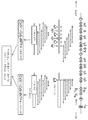
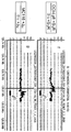
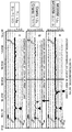
도 1은 (a) JH 및 스위치 반복 영역(switch repeat region)을 나타내는 IgH 유전자좌; (b) JH 프라이머를 이용한 선형 증폭; (c) S5(Svαε) 프라이머를 이용한 선형 증폭; (d) 통상적인 전좌 CGH(tCGH) 실험의 개요를 예시한다.1 shows (a) an IgH locus showing J H and a switch repeat region; (b) linear amplification with J H primers; (c) linear amplification with S 5 (S vαε ) primers; (d) provides an overview of a typical translocation CGH (tCGH) experiment.
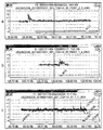
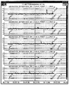
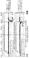
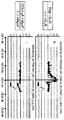
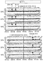
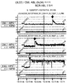
도 2는 공지된 IgH 전좌 중지점을 갖는 세포주에 대한 tCGH 데이터를 예시한다. (a) DHL16 세포주에서의 JH-BCL2 중지점(마이너 클러스터 영역); (b) MC116 세포주에서의 JH-MYC 중지점; (c) U266 세포주에서의 Sα-CCND1 중지점; (d) OCI-Ly8 세포주에서의 Sγ-BCL6 중지점.2 illustrates tCGH data for cell lines with known IgH translocation breakpoints. (a) J H -BCL2 breakpoint (minor cluster region) in DHL16 cell line; (b) J H -MYC breakpoint in MC116 cell line; (c) S α -CCND1 breakpoint in the U266 cell line; (d) S γ -BCL6 breakpoint in OCI-Ly8 cell line.
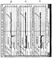
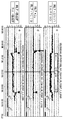
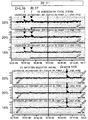
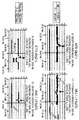
도 3은 (a) RL7 세포주에서의 JH-BCL2 중지점 및 BCL2 결실의 분석을 예시한다: (ⅰ) RL7 +JH (Cy3) / 정상 +JH (Cy5) - 중지점 및 결실; (ⅱ) RL7-JH (Cy3) / 정상 -JH (Cy5) - 결실 단독; (ⅲ) RL7 +JH (Cy3) / RL7 -JH (Cy5) - 중지점 단독. 부분 (b)는 상기 모든 3개의 실험에 대한 RL7/BCL2 어레이 데이터의 오버레이를 예시한다. 부분 (c)는 MO2058 세포주에서의 JH-CCND1 중지점 및 CCND1 중복/결실의 분석을 예시한다: (ⅰ) MO2058 +JH (Cy3) / 정상 +JH (Cy5) - 중지점 및 중복/결실; (ⅱ) MO2058 -JH (Cy3) / 정상 -JH (Cy5) - 중복/결실 단독; (ⅲ) MO2058 +JH (Cy3) / Granta -JH (Cy5) - 중지점 단독. 부분 (d)는 상기 모든 3개의 실험에 대한 MO2058/CCND1 어레이 데이터의 오버레이를 예시한다.Figure 3 illustrates (a) analysis of J H -BCL2 breakpoint and BCL2 deletion in RL7 cell line: (iii) RL7 + J H (Cy3) / normal + J H (Cy5)-breakpoint and deletion; (Ii) RL7-J H (Cy3) / normal -J H (Cy5)-deletion alone; (Iii) RL7 + J H (Cy3) / RL7 -J H (Cy5)-breakpoint alone. Part (b) illustrates the overlay of RL7 / BCL2 array data for all three experiments above. Part (c) illustrates the analysis of J H -CCND1 breakpoint and CCND1 overlap / deletion in the MO2058 cell line: (iii) MO2058 + J H (Cy3) / normal + J H (Cy5)-breakpoint and overlap / fruition; (Ii) MO2058 -J H (Cy3) / normal -J H (Cy5)-duplicate / deletion alone; (Iii) MO2058 + J H (Cy3) / Granta -JH (Cy5)-breakpoint alone. Part (d) illustrates the overlay of MO2058 / CCND1 array data for all three experiments above.
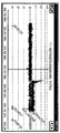
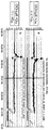
도 4는 OCI-Ly8 세포주에서 확인된 다중 IgH 중지점을 예시한다: (a) JH- BCL2 - "der(14)" 중지점; (b) Sγ3-BCL6 - SγR 프라이머를 이용하여 확인된 "der(3)" 중지점; (c) Sγ-MYC - SγR 프라이머를 이용하여 확인된 "der(8)" 중지점; (d) Sγ-BCL6 - SγF 프라이머를 이용하여 확인된 "der(14)" 중지점.4 illustrates multiple IgH breakpoints identified in OCI-Ly8 cell line: (a) J H -BCL2-"der (14)"breakpoint; (b) a "der (3)" breakpoint identified using S γ 3 -BCL6—S γ R primers; (c) “der (8)” breakpoints identified using S γ -MYC-S γ R primers; (d) “der (14)” breakpoints identified using S γ -BCL6 − S γ F primers.
도 5는 6분(밝은 색의 선) 대 10분(어두운 색의 선)의 선형 증폭 신장 시간의 중지점 프로파일에 대한 효과를 예시한다. 부분 (a)는 OCI-Ly8 세포주에서의 Sγ-BCL6 중지점을 예시하고; 부분 (b)는 OCI-Ly8 세포주에서의 Sγ-BCL6 중지점을 예시한다.5 illustrates the effect on the breakpoint profile of linear amplification stretch time of 6 minutes (light colored lines) versus 10 minutes (dark colored lines). Part (a) illustrates the S γ -BCL6 breakpoint in the OCI-Ly8 cell line; Part (b) illustrates the S γ -BCL6 breakpoint in the OCI-Ly8 cell line.
도 6은 다양한 JH-CCND1 중지점을 나타내는 5개의 원발성 외투세포 림프종의 tCGH 분석을 예시한다.6 illustrates tCGH analysis of five primary mantle cell lymphomas showing various J H -CCND1 breakpoints.
도 7은 통상적인 전좌 CGH(tCGH) 실험의 개관을 제공한다.7 provides an overview of a typical translocation CGH (tCGH) experiment.
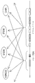
도 8은 다양한 B 세포 림프종 및 혈장 세포 골수종 내의 파트너 유전자좌를 포함하는 통상적인 IgH 전좌의 개관을 제공하며, 이는 tCGH 시스템의 확립 및 확인을 위한 모델 시스템으로서 사용되었다.FIG. 8 provides an overview of conventional IgH translocations including partner loci within various B cell lymphomas and plasma cell myeloma, which was used as a model system for the establishment and validation of the tCGH system.
도 9는 der(14) 염색체 상에 IgH 중지점 및 다수의 DH 분절 내에 상호 중지점을 갖는 VDJ-관련 전좌의 tCGH 검출을 위한 구성을 예시한다.9 illustrates a configuration for tCGH detection of VDJ-related translocations with IgH breakpoints on the der (14) chromosome and mutual breakpoints in multiple D H segments.
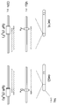
도 10은 BCL2 마이너 전좌 클러스터에 대해 맵핑된 상호 JH-BCL2 (a) 및 BCL2-DH (b) 융합체의 tCGH 분석; OCI-Ly8 림프종 세포주 내에서 발견된 둘 모두의 상호 Sγ3-BCL6 융합체(c 및 d); 역위된 배향을 갖는 비-IgH BCL6 엑손 1 재배열 (e); 버키트 림프종 세포주 MC116 (f) 내의 IgH-MYC 융합체; 및 역위된 배향을 갖는 비-IgH MYC 재배열(i)을 포함하는 여러 다른 MYC 재배열(g-i)을 예시한다.10 tCGH analysis of mutual J H -BCL2 (a) and BCL2-D H (b) fusions mapped against BCL2 minor translocation clusters; Both mutual S γ3- BCL6 fusions (c and d) found in OCI-Ly8 lymphoma cell line;
도 11은 다수의 상이한 선형 증폭 반응식을 이용한 BCL2의 큰(190 kb) 인트론 내의 신규한 167 kb 간극 결실의 복제수 변화의 tCGH 분석(a - e)을 예시한다.FIG. 11 illustrates a tCGH analysis (a-e) of copy number change of a novel 167 kb gap deletion in the large (190 kb) intron of BCL2 using a number of different linear amplification schemes.
도 12는 비-MTC 중지점을 갖는 5개의 원발성 MCL 경우로부터의, 원발성 림프종에서의 tCGH 분석에 의한 신규한 CCDN1 중지점의 확인(a - e)을 예시한다.12 illustrates the identification (a-e) of novel CCDN1 breakpoints by tCGH analysis in primary lymphomas from five primary MCL cases with non-MTC breakpoints.
도 13은 tCGH 분석에 의한 CCND1 유전자에 걸쳐 있는 중복, 및 MO2058(a - c) 및 Granta(d - f) 둘 모두의 세포주에서의 각각의 JH-CCND1 중지점 접합(junction)에 대한 정확한 범위의 확인을 예시한다.FIG. 13 shows the exact range of overlap across the CCND1 gene by tCGH analysis, and for each JH-CCND1 breakpoint junction in cell lines of both MO2058 (a-c) and Granta (d-f). Illustrate confirmation.
도 14는 tCGH 분석에 의한 OCI-Ly8 림프종 세포주 내의 IgH-BCL2 중지점에서의 약 6 kb 결실의 확인을 예시한다.FIG. 14 illustrates the identification of about 6 kb deletion at the IgH-BCL2 breakpoint in the OCI-Ly8 lymphoma cell line by tCGH analysis.
도 15는 tCGH 분석에 의한, SγR (a) 및 SPF (b) 프라이밍된 선형 증폭을 이용한, Sα1 내지 Sγ4에 걸쳐있고 3'α1 인핸서를 포함하는 ~ 100 kb의 IgH 불변 영역 분절의 CCND1 유전자좌로의 신규한 잠재(cryptic) 삽입의 확인을 예시한다. 모의 증폭된 종양 DNA가 하이브리드화 대조군로서 사용되고(c), 정상 유전체 DNA가 분석되는 경우(d)의 예상 전좌 중지점으로부터 떨어진 서열의 탈-표적(Off-target) 증폭이 예시된다.15 is the analysis by tCGH, S γ R (a) and SPF (b) using a linear amplification the priming, S α1 to IgH constant of ~ 100 kb to over S γ4, can also contain enhancer region segment 3'α1 The identification of new cryptic insertions into the CCND1 locus is illustrated. Off-target amplification of sequences away from expected translocation breakpoints when mock amplified tumor DNA is used as a hybridization control (c) and normal genomic DNA is analyzed (d).
도 16은 모의 증폭된 종양 DNA가 하이브리드화 대조군으로 사용되는 경우의 MO2058 (a) 및 Granta (b) 세포주에서의 예상 전좌 중지점에서 떨어진 서열의 탈-표적 증폭을 예시한다. 정상 유전체 DNA가 분석되는 경우에 유사한 결과가 관찰된다(c - f).FIG. 16 illustrates off-target amplification of sequences away from expected translocation breakpoints in MO2058 (a) and Granta (b) cell lines when mock amplified tumor DNA is used as a hybridization control. Similar results are observed when normal genomic DNA is analyzed (c-f).
도 17은 DHL16, RL7 및 Granta 519 유전체 DNA의 동등한 양의 혼합("33% 희석"으로 명명됨)에 의한 tCGH 분석의 분석 민감성의 결정을 예시하고; 20% 및 15% 희석 샘플은 정상 유전체 DNA와의 혼합에 의해 생성되었다. 이후, 샘플은 JH 프라이머를 이용하여 12 또는 20 주기 동안 증폭되었고, 유사하게 증폭된 정상 유전체 DNA에 공동 하이브리드화되었다.17 illustrates the determination of analytical sensitivity of tCGH assays by mixing equal amounts of DHL16, RL7 and
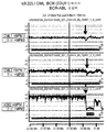
도 18은 골수성 프라이머 믹스(MPM) 및 AML 파일럿 어레이를 이용하여 BCR-ABL 균형성 전좌 t(22;9)를 특징으로 하는 3개의 만성 골수성 백혈병 세포주의 다중 선형 증폭 및 tCGH 분석의 결과를 예시한다.FIG. 18 illustrates the results of multiple linear amplification and tCGH analysis of three chronic myeloid leukemia cell lines featuring BCR-ABL balanced translocation t (22; 9) using myeloid primer mix (MPM) and AML pilot array. .
도 19는 골수성 프라이머 믹스(MPM) 및 AML 파일럿 어레이를 이용한, PML-RARA 균형성 전좌 t(15;21)를 특징으로 하는 2개의 급성 전골수구성 백혈병(APL) 세포주(상부 패널), 및 염색체 역위 inv(16)에 의해 야기된 MYH11-CBFB 융합체를 특징으로 하는 2개의 급성 골수단핵세포성 백혈병/호산구증가증 세포주의 다중 선형 증폭 및 tCGH 분석의 결과를 예시한다.FIG. 19 shows two acute promyelocytic leukemia (APL) cell lines (top panel) characterized by PML-RARA balanced translocation t (15; 21) using myeloid primer mix (MPM) and AML pilot arrays, and chromosomes. The results of multiple linear amplification and tCGH analysis of two acute osteocytic leukemia / eosinophilic cell lines characterized by the MYH11-CBFB fusion caused by inversion inv (16) are illustrated.
도 20은 P1/P7 프라이머 믹스(MPM) 및 AML 파일럿 어레이를 이용한 AF9-MLL 균형성 전좌 t(9;11)을 특징으로 하는 MLL 백혈병 세포주(상부 패널), 및 821 프라이머 믹스(MPM) 및 AML 파일럿 어레이를 이용한 ETO-AML1 균형성 전좌 t(8;21)을 특징으로 하는 카스미(Kasumi) 급성 골수성 백혈병 세포주(하부 패널)의 다중 선형 증폭 및 tCGH 분석의 결과를 예시한다.FIG. 20 shows MLL leukemia cell line (top panel) featuring AF9-MLL balanced translocation t (9; 11) using P1 / P7 primer mix (MPM) and AML pilot array, and 821 primer mix (MPM) and AML The results of multiple linear amplification and tCGH analysis of the Kasumi acute myeloid leukemia cell line (bottom panel) characterized by the ETO-AML1 balanced translocation t (8; 21) using the pilot array are illustrated.
발명의 상세한 설명Detailed description of the invention
어레이 기반의 비교 유전체 하이브리드화(CGH)는 염색체 불균형의 연구를 혁신화시켰으나, 이는 일반적으로 림프종, 백혈병 및 다른 종양의 발병기전 및 진단에서 중추적인 역할을 하는 상호 전좌와 같은 균형성 유전체 재배열을 검출할 수 없다. 예를 들어, 면역글로불린 중쇄(IgH) 전좌 파트너의 정확한 확인은 B 세포 림프종의 분류 및 다발골수종과 같은 혈장 세포 신생물에서의 예후 예측에 필수적이다.Array-based comparative genome hybridization (CGH) has revolutionized the study of chromosomal imbalances, but this generally leads to balanced genome rearrangements such as translocations that play a pivotal role in the pathogenesis and diagnosis of lymphoma, leukemia, and other tumors. It cannot be detected. For example, accurate identification of immunoglobulin heavy chain (IgH) translocation partners is essential for the classification of B cell lymphoma and for predicting prognosis in plasma cell neoplasia such as multiple myeloma.
균형성 유전체 재배열을 위한 모델로서 IgH 전좌를 이용하여, 본 발명자는 IgH 전좌 파트너의 신속한 확인 및 선례가 없는 해상도의 전좌 관련 중지점의 정확한 맵핑을 가능하게 하는 전좌-CGH(tCGH)로 명명한 어레이 CGH 방법을 개발하였다. 하기에 보다 상세하게 기재되는 바와 같이, CGH 어레이에서 IgH 전좌가 검출가능하도록 하기 위해, 시험 및 참조 샘플로부터의 유전체 DNA는 단일 IgH 연결(JH) 또는 스위치(Sμ/Sα/Sε) 영역 프라이머를 이용하는 효소적 선형 증폭 반응에서의 어레이 하이브리드화 전에 변형되어, IgH 프라이머의 다운스트림이 삽입(전좌 또는 다른 재배열을 통함)될 수 있는 임의의 융합 파트너 서열의 특정 증폭을 발생시킨다. MYC, BCL2 및 CCND1(사이클린 D1)와 같은 흔한 IgH 파트너 유전자좌를 제시하는 단일 타일링-밀도 올리고누클레오티드 어레이를 이용하여, tCGH는 MO2058 및 Granta 519 세포주(외투세포 림프종)에서의 JH-CCND1 중지점, U266(골수종)에서의 세포유전학적 잠재 Sα-CCND1 융합체, MC116 및 Raji(버키트 림프종)에서의 JH-MYC 및 Sμ-MYC 중지점, 및 DHL16(거대세포 림프종; 마이너 클러스터 영역) 및 소포 림프종(메이저 중지점 영역)의 기록 케이스에서의 JH-BCL2 중지점을 포함하는 다양한 세포주 및 원발성 림프종에서의 공지된 IgH 융합 중지점의 분류를 성공적으로 확인하고, ~100bp의 해상도로 맵핑하였다.Using IgH translocation as a model for balanced genome rearrangement, we named Translocation-CGH (tCGH), which enables rapid identification of IgH translocation partners and accurate mapping of translocation-related breakpoints at unprecedented resolution. An array CGH method was developed. As described in more detail below, in order to enable detectable IgH translocations in CGH arrays, genomic DNA from test and reference samples may contain a single IgH linkage (J H ) or switch (Sμ / Sα / Sε) region primer. Modifications prior to array hybridization in the enzymatic linear amplification reaction employed result in specific amplification of any fusion partner sequence that can be inserted (via translocation or other rearrangement) downstream of the IgH primers. Using a single tiling-density oligonucleotide array that presents common IgH partner loci such as MYC, BCL2, and CCND1 (cyclin D1), tCGH is expressed by the J H -CCND1 breakpoint in MO2058 and
이후, 본 발명자는 외투세포 림프종의 4개의 기록 케이스 및 B 세포 전림프구성 백혈병의 하나의 t(11;14)-양성 케이스를 분석하기 위해 tCGH를 이용하였고, 상기 케이스 모두는 CCND1 메이저 전좌 클러스터(MTC)에서 PCR-검출가능한 전좌 중지점이 결핍되었다. 5개의 신규한 CCND1 전좌 중지점이 확인되었고, ~100bp의 해상도로 맵핑되어, 예측 중지점의 증폭, 서열분석 및 확인을 위한 환자 특이적 PCR 프라이머의 신속한 설계가 가능하였다. 하나의 중지점이 MTC의 500bp 내로 맵핑되었고, 다른 4개는 MTC의 측면에 존재하는 ~150kb 영역에 걸쳐 산재되어 있었다. 본 발명자가 아는 바로는, 이는 현재까지 보고된 비-MTC 외투세포 림프종 중지점 서열의 가장 큰 시리즈이다. 이러한 결과는 또한 tCGH가 매우 큰 유전체 영역에 걸쳐 분산된 기존의 미확인의 IgH 전좌 중지점의 신속한 클로닐을 촉진할 수 있는 방법을 예시한다. tCGH는 단지 유전체 DNA만을 필요로 하고, 동일한 어레이 상에서 초고해상도로 균형성 IgH 전좌 및 유전체 불균형 둘 모두를 동시에 검출할 수 있으므로, 이는 B 세포 및 혈장 세포 신생물의 임상 시험을 위한 분자 세포유전학 방법(예를 들어, FISH)에 대한 유용한 대안이 될 수 있다. tCGH는 또한 최소 잔여 질병을 검출하기 위한 고도로 민감한 중지점 특이적 PCR 검정의 개발을 촉진할 것이다. 최종적으로, 선형 증폭 반응에 사용되는 프라이머는 충분히 맞춤화 가능하므로, 융합 파트너중 하나가 공지되어 있는 경우 tCGH는 비-IgH 유전자좌를 포함하는 다른 균형성 전좌(또는 보다 복잡한 유전체 융합)를 확인하고 맵핑하기 위해 용이하게 적합화될 수 있다.The inventors then used tCGH to analyze four recording cases of mantle cell lymphoma and one t (11; 14) -positive case of B cell prelymphocytic leukemia, all of which were CCND1 major translocation clusters ( MTC) lacked a PCR-detectable translocation breakpoint. Five new CCND1 translocation breakpoints were identified and mapped to a resolution of ˜100 bp, allowing for the rapid design of patient specific PCR primers for amplification, sequencing and confirmation of predicted breakpoints. One breakpoint was mapped within 500 bp of the MTC, and the other four were scattered over the ~ 150 kb region that flanked the MTC. To the best of our knowledge, this is the largest series of non-MTC mantle cell lymphoma breakpoint sequences reported to date. These results also illustrate how tCGH can promote rapid cloylation of existing, unidentified IgH translocation breakpoints dispersed over very large genome regions. tCGH only requires genomic DNA and can simultaneously detect both balanced IgH translocations and genomic imbalances at very high resolutions on the same array, which is a molecular cytogenetic method for clinical testing of B cell and plasma cell neoplasms. For example, it can be a useful alternative to FISH). tCGH will also facilitate the development of highly sensitive breakpoint specific PCR assays for detecting minimal residual disease. Finally, the primers used for the linear amplification reaction are fully customizable, so tCGH can identify and map other balanced translocations (or more complex genomic fusions), including non-IgH loci, if one of the fusion partners is known. To be easily adapted.
한 구체예에서, 본 발명은, (a) 표적 유전체 유전자좌를 증폭시키는 단계; (b) 상기 증폭된 생성물을 핵산 어레이에 하이브리드화시키는 단계; 및 (c) 상기 하이브리드화 패턴을 참조와 비교하는 단계를 포함하는, 염색체 재배열을 검출하는 방법을 제공하며, 여기서 상기 증폭은 선형 증폭이고, 상기 참조에 비교되는 상기 증폭된 유전체 유전자좌의 차별적 하이브리드화는 유전체 재배열의 존재를 나타낸다. 특정 구체예에서, 유전체 재배열은 균형성 재배열, 예를 들어, 균형성 전좌 또는 역위이다.In one embodiment, the present invention comprises the steps of: (a) amplifying a target genomic locus; (b) hybridizing the amplified product to a nucleic acid array; And (c) comparing the hybridization pattern with a reference, wherein the amplification is linear amplification and the differential hybridization of the amplified genomic loci compared to the reference. Ization indicates the presence of dielectric rearrangements. In certain embodiments, the genome rearrangement is balanced rearrangement, eg, balanced translocation or inversion.
한 구체예에서, 본 발명은 균형성 염색체 전좌를 검출하는 방법을 제공한다. 특정 구체예에서, 본 발명의 방법은, (a) 표적 유전체 유전자좌를 증폭하는 단계; (b) 상기 증폭된 생성물을 핵산 어레이에 하이브리드화시키는 단계; 및 (3) 상기 하이브리드화 패턴을 참조와 비교하는 단계를 포함하고, 여기서 상기 증폭은 선형 증폭이고, 직각삼각형 하이브리드화 패턴의 존재는 균형성 염색체 전좌의 존재를 나타낸다. 본 발명의 특정 구체예에서, 상기 직각삼각형 하이브리드화 패턴은 비대칭 하이브리드화 패턴을 포함한다. 특정 구체예에서, 본 발명의 방법은 염색체 전좌와 관련된 둘 모두의 파트너 유전자좌 내의 중지점의 검출 및/또는 맵핑을 포함할 수 있다. 또 다른 구체예에서, 본 발명의 방법은 균형성 전좌가 아닌 염색체 재배열의 검출을 포함한다.In one embodiment, the present invention provides a method for detecting balanced chromosomal translocations. In certain embodiments, the methods of the present invention comprise the steps of: (a) amplifying a target genomic locus; (b) hybridizing the amplified product to a nucleic acid array; And (3) comparing the hybridization pattern with a reference, wherein the amplification is linear amplification and the presence of a right triangle hybridization pattern indicates the presence of a balanced chromosomal translocation. In certain embodiments of the invention, the right triangle hybridization pattern comprises an asymmetric hybridization pattern. In certain embodiments, the methods of the invention can include the detection and / or mapping of breakpoints in both partner loci associated with chromosomal translocations. In another embodiment, the methods of the present invention comprise the detection of chromosomal rearrangements that are not balanced translocations.
상기 제공된 방법의 특정 구체예에서, 하나 이상의 앰플리콘을 증폭시키기 위해 다중 선형 증폭이 사용된다. 특정 구체예에서, 상기 방법은 하나 이상의 유전체 유전자좌의 동시 검사를 포함한다.In certain embodiments of the methods provided above, multiple linear amplifications are used to amplify one or more amplicons. In certain embodiments, the method comprises simultaneous testing of one or more genomic loci.
특정 구체예에서, 본 발명의 방법에 다수의 증폭 프라이머가 사용된다. 상기 다수의 증폭 프라이머는 질병과 관련된 균형성 전좌와 연관된 유전자좌의 증폭을 위한 프라이머를 포함할 수 있다. 균형성 염색체 전좌와 관련된 임의의 질병은 본 발명의 방법에 의해 검출될 수 있다. 본 발명의 한 특정 구체예에서, 질병은 암, 예를 들어, 림프종 또는 백혈병이다. 본 발명의 특정 구체예에서, MPM 믹스(mix), 821 믹스, P1/P7 믹스, 및 다수의 DH 프라이머로부터 선택된 다수의 프라이머가 본원에 제공된 방법에 사용될 수 있다.In certain embodiments, multiple amplification primers are used in the methods of the invention. The plurality of amplification primers may comprise primers for amplification of loci associated with balanced translocations associated with disease. Any disease associated with balanced chromosomal translocation can be detected by the methods of the invention. In one specific embodiment of the invention, the disease is cancer, eg, lymphoma or leukemia. In certain embodiments of the invention, multiple primers selected from MPM mix, 821 mix, P1 / P7 mix, and multiple D H primers can be used in the methods provided herein.
본 발명의 특정 구체예에서, 선형 증폭의 생성물을 검출하는데 사용되는 어레이는 마이크로어레이 또는 고밀도의 타일링(tiling)된 어레이를 포함할 수 있다. 몇몇 구체예에서, 상기 어레이는 다수의 유전체 유전자좌에 대한 프로브를 포함할 수 있다. 특정 구체예에서, 본 발명의 어레이 상의 프로브에 상응하는 하나 이상의 유전체 유전자좌는 질병과 관련될 수 있다. 특정 구체예에서, 질병은 암, 예를 들어, 림프종 또는 백혈병일 수 있다. 한 특정 구체예에서, 상기 어레이는 AML 파일럿 어레이를 포함할 수 있다.In certain embodiments of the invention, the array used to detect the product of linear amplification may comprise a microarray or a high density tiled array. In some embodiments, the array can include probes for multiple genomic loci. In certain embodiments, one or more genomic loci corresponding to probes on an array of the invention may be associated with a disease. In certain embodiments, the disease may be cancer, eg, lymphoma or leukemia. In one particular embodiment, the array may comprise an AML pilot array.
또 다른 구체예에서, 본 발명의 방법은 중복, 증폭, 결실, 역위, 균형성 전좌, 및 불균형성 전좌로부터 선택된 제 2의 염색체 재배열의 검출을 추가로 포함할 수 있다. 특정 구체예에서, 제 1 재배열 및 제 2 재배열의 검출은 연속적이거나 동시일 수 있다. 한 특정 구체예에서, 본 발명의 방법은 균형성 재배열 및 불균형성 재배열 둘 모두의 동시 검출을 포함한다. 상기 균형성 및 불균형성 재배열은 동일한 유전체 유전자좌 또는 상이한 유전체 유전자좌에 존재할 수 있다.In another embodiment, the methods of the present invention may further comprise detecting a second chromosomal rearrangement selected from overlapping, amplification, deletion, inversion, balanced translocation, and disproportionate translocation. In certain embodiments, the detection of the first rearrangement and the second rearrangement can be continuous or simultaneous. In one particular embodiment, the methods of the present invention comprise simultaneous detection of both balanced and unbalanced rearrangements. The balance and imbalance rearrangements can be in the same genomic locus or in different genomic loci.
또 다른 구체예에서, 본 발명은 균형성 염색체 전좌의 검출에 사용하기 위한 신규한 키트를 제공한다. 특정 구체예에서, 본 발명의 키트는 전좌와 연관된 유전자좌의 선형 증폭을 위한 프라이머를 포함한다. 다른 구체예에서, 본 발명의 키트는 전좌와 연관된 유전자좌로부터의 선형 증폭 생성물의 검출을 위한 어레이를 포함할 수 있다. 본 발명의 특정 구체예에서, 키트는 전좌와 연관된 유전자좌의 증폭을 위한 다수의 프라이머를 포함할 수 있다. 또 다른 구체예에서, 본 발명의 키트는 염색체 전좌와 관련된 질병의 진단 또는 예후에 사용될 수 있다. 한 특정 구체예에서, 질병은 암, 예를 들어, 림프종 또는 백혈병일 수 있다.In another embodiment, the present invention provides a novel kit for use in the detection of balanced chromosomal translocations. In certain embodiments, the kits of the invention comprise primers for linear amplification of loci associated with translocations. In other embodiments, the kits of the invention may comprise an array for the detection of linear amplification products from the locus associated with the translocation. In certain embodiments of the invention, the kit may comprise a plurality of primers for amplification of the locus associated with the translocation. In another embodiment, the kits of the invention can be used for the diagnosis or prognosis of a disease associated with chromosomal translocation. In one particular embodiment, the disease may be cancer, eg, lymphoma or leukemia.
1. 정의1. Definition
용어 "염색체 재배열" 또는 "염색체 이상"은 일반적으로 야생형 또는 정상 세포에서 발견되지 않는 방식의 염색체 물질의 분절의 이상 연결을 의미한다. 염색체 재배열의 예는 결실, 증폭, 역위 또는 전좌를 포함한다. 염색체 재배열은 염색체에서 발생하는 자발적인 절단 후에 발생할 수 있다. 절단 또는 절단들이 염색체 단편의 손실을 발생시키는 경우, 결실이 발생한다. 염색체 분절이 꺾여 끊어지는 경우의 역위 결과는 뒤집어지고(역위되고), 이는 이의 본래의 위치로 다시 삽입된다. 하나의 염색체 단편이 또 다른 염색체로부터의 단편과 교환되는 경우, 전좌가 발생한다. 증폭은 염색체의 특정 영역의 다중 복제를 발생시킨다. 염색체 재배열은 또한 상기의 조합을 포함할 수 있다.The term "chromosomal rearrangement" or "chromosomal abnormality" means aberrant linking of segments of chromosomal material in a manner not generally found in wild-type or normal cells. Examples of chromosomal rearrangements include deletions, amplifications, inversions or translocations. Chromosomal rearrangements can occur after spontaneous cleavage occurring on the chromosome. If cleavage or cleavage results in loss of chromosomal fragments, deletions occur. The inversion result when the chromosomal segment is broken is reversed (inverted), which is inserted back into its original position. When one chromosomal fragment is exchanged for a fragment from another chromosome, a translocation occurs. Amplification results in multiple copies of specific regions of the chromosome. Chromosomal rearrangements may also include combinations of the above.
용어 "전좌" 또는 "염색체 전좌"는 일반적으로 동등하거나 동등하지 않은 양의 동일하거나 상이한 염색체 사이의 염색체 물질의 교환을 의미한다. 종종, 상기 교환은 비상동 염색체 사이에서 발생한다.The term "translocation" or "chromosomal translocation" generally means the exchange of chromosomal material between equal or unequal amounts of the same or different chromosomes. Often, the exchange occurs between nonhomologous chromosomes.
"균형성" 전좌는 일반적으로 유전 물질의 알짜 손실 또는 증가가 존재하지 않는 염색체 물질의 교환을 의미한다."Balance" translocation generally refers to the exchange of chromosomal material in which no net loss or increase in genetic material is present.
"불균형성" 전좌는 일반적으로 염색체 물질을 남기거나 손실하는 염색체 물질의 동등하지 않은 교환을 의미한다."Unbalanced" translocation generally refers to unequal exchange of chromosomal material leaving or losing chromosomal material.
"핵산 어레이" 또는 "핵산 마이크로어레이"는 다수의 핵산 요소이며, 이들 각각은 프로브 핵산이 하이브리드되는 고체 표면 상에 고정된 하나 이상의 표적 핵산 분자를 포함한다. 상기 고체 지지체 상에 고정될 수 있는 핵산 분자는 올리고누클레오티드, cDNA 및 유전체 DNA를 포함하나, 이에 제한되지는 않는다. 본 발명의 상황에서, 유전체 핵산의 다양한 분절에 해당하는 서열을 함유하는 마이크로어레이가 사용된다. 마이크로어레이의 유전체 요소는 유기체의 전체 유전체를 제시할 수 있거나, 그렇지 않은 경우 유전체의 소정의 영역, 예를 들어, 특정 유전체 또는 이의 연속 분절을 제시할 수 있다.A "nucleic acid array" or "nucleic acid microarray" is a number of nucleic acid elements, each of which comprises one or more target nucleic acid molecules immobilized on a solid surface to which the probe nucleic acid is hybridized. Nucleic acid molecules that can be immobilized on the solid support include, but are not limited to, oligonucleotides, cDNAs, and genomic DNA. In the context of the present invention, microarrays containing sequences corresponding to various segments of genomic nucleic acid are used. The dielectric element of the microarray may present the entire genome of the organism, or else may present a predetermined region of the genome, eg, a particular genome or a continuous segment thereof.
유전체 타일링 마이크로어레이는 전체 관심 유전체 영역의 완전한 또는 거의 완전한 제시를 제공하도록 설계된 중첩 올리고누클레오티드를 포함한다.The dielectric tiling microarray includes overlapping oligonucleotides designed to provide complete or near complete presentation of the entire dielectric region of interest.
비교 유전체 하이브리드화(CGH)는 일반적으로 제공된 피검체 DNA의 DNA 내용물 및 종종 종양 세포에서의 복제수 변화(증가/손실)의 분석을 위한 분자-세포유전학적 방법을 의미한다. 암의 상황에서, 상기 방법은 정상 인간 유사분열 중기 제조물에 대한 라벨링된 종양 DNA(종종 형광 라벨로 라벨링됨) 및 정상 DNA(종종 제 2의 상이한 형광 라벨로 라벨링됨)의 하이브리드화를 기초로 한다. 낙사형광 현미경(epifluorescence microscopy) 및 정량 영상 분석(quantitative image analysis)을 이용하여, 대조군 DNA에 대비한 증가/손실의 형광 비의 영역적 차이가 검출될 수 있고, 이는 유전체 내의 이상 영역을 확인하는데 사용될 수 있다. CGH는 일반적으로 불균형 염색체 변화만 검출할 것이다. 구조적 염색체 이상, 예를 들어, 균형성 상호 전좌 또는 역위는 검출될 수 없는데, 이들은 복제수가 변하지 않기 때문이다. 문헌[Kallioniemi et al., Science 258: 818-821 (1992)] 참조.Comparative genomic hybridization (CGH) generally refers to a molecular-cytogenetic method for the analysis of the DNA content of a given subject DNA and often changes in copy number (increase / loss) in tumor cells. In the context of cancer, the method is based on hybridization of labeled tumor DNA (often labeled with fluorescent labels) and normal DNA (often labeled with a second different fluorescent label) for normal human mitotic midterm preparations. . Using epifluorescence microscopy and quantitative image analysis, regional differences in the fluorescence ratio of increase / loss relative to control DNA can be detected, which can be used to identify abnormal regions within the genome. Can be. CGH will generally only detect imbalanced chromosomal changes. Structural chromosomal aberrations, eg, balanced translocations or inversions, cannot be detected because they do not change the copy number. See Kallioniemi et al., Science 258: 818-821 (1992).
"염색체 마이크로어레이 분석(CMA)" 또는 "어레이CGH"로 언급되는 CGH의 변화에서, 피검체 조직 및 정상 대조 조직(참조)로부터의 DNA는 차별적으로 라벨링된다(예를 들어, 상이한 형광 라벨로 라벨링됨). 피검체 및 참조 DNA를 반복성 DNA 서열을 억제하기 위한 라벨링되지 않은 인간 cot 1 DNA와 혼합한 후, 상기 혼합물은 일반적으로 정상 참조 세포로부터의 다수의 소정의 DNA 프로브를 함유하는 슬라이드에 하이브리드된다. 미국 특허 제 5,830,645호; 제 6,562,565호 참조. 마이크로어레이 상의 요소로서 올리고누클레오티드가 사용되는 경우, 100kb의 해상도를 가능케하는 BAC 어레이의 사용과 비교하여 통상적으로 20-80개의 염기쌍의 해상도가 수득될 수 있다. 어레이의 요소에 따른 (형광) 색 비가 피검체 샘플 내의 DNA 증가 또는 손실 영역을 평가하는데 사용된다.In the change in CGH, referred to as "chromosome microarray analysis (CMA)" or "arrayCGH", DNA from subject tissue and normal control tissue (reference) are differentially labeled (e.g., labeled with different fluorescent labels). being). After mixing the subject and the reference DNA with unlabeled
용어 "하이브리드화의 직각삼각형 패턴" 또는 "직각삼각형 하이브리드화 패턴"은 일반적으로 (ⅰ) 재배열 중지점을 나타내는 단일한 별개의 경계, 및 (ⅱ) 중심절 또는 종말절의 방향에서 기준선에 대한 하이브리드화 신호(또는 이의 비 또는 log-비)의 점진적인 복귀(이는 별개가 아닌 제 2의 경계를 발생시킴)를 특징으로 하는 임의의 비대칭 하이브리드화 신호 패턴을 포함하는, 염색체 위치에 대한 하이브리드화 신호(또는 하이브리드화 신호 비 또는 이의 대수)의 플롯 상의 비대칭 패턴을 의미한다.The term “right triangle pattern of hybridization” or “right triangle hybridization pattern” generally refers to (i) a single distinct boundary that represents a rearrangement breakpoint, and (ii) a hybridization to a baseline in the direction of the central or apex. Hybridization signal (or any symmetric hybridization signal pattern), including any asymmetric hybridization signal pattern, characterized by a gradual return of the signal (or its ratio or log-ratio), which results in a second boundary that is not distinct Hybridization signal ratio or logarithm thereof).
용어 "증폭" 또는 "증폭 반응"은 주형 핵산 서열의 증가된 복제수를 발생시키는 효소 반응을 포함하는 임의의 화학적 반응을 의미한다. 증폭 반응은 중합효소 연쇄 반응(PCR) 및 리가아제 연쇄 반응(LCR)(참조: 미국 특허 제 4,683,195호 및 제 4,683,202호; PCR Protocols: A Guide to Methods and Applications (Innis et al., eds, 1990)), 가닥 치환 증폭(strand displacement amplification, SDA)(Walker, et al . Nucleic Acids Res. 20(7):1691 (1992); Walker PCR Methods Appl 3(1):1 (1993)), 전사-매개 증폭(transcription-mediated amplification)(Phyffer, et al., J. Clin . Microbiol. 34:834 (1996); Vuorinen, et al . , J. Clin . Microbiol. 33:1856 (1995)), 핵산 서열 기초 증폭(nucleic acid sequence-based amplification, NASBA)(Compton, Nature 350(6313):91 (1991), rolling circle amplification (RCA)(Lisby, Mol . Biotechnol. 12(1):75 (1999)); Hatch et al., Genet . Anal . 15(2):35 (1999)) 및 분지 DNA 신호 증폭(branched DNA signal amplification, bDNA)(참조: Iqbal et al., Mol Cell Probes 13(4):315 (1999))을 포함한다.The term "amplification" or "amplification reaction" means any chemical reaction including an enzymatic reaction that results in an increased copy number of the template nucleic acid sequence. Amplification reactions include polymerase chain reaction (PCR) and ligase chain reaction (LCR) (see US Pat. Nos. 4,683,195 and 4,683,202; PCR Protocols: A Guide to Methods and Applications (Innis et al., Eds, 1990)). ), Strand displacement amplification (SDA) (Walker, et. al . Nucleic Acids Res . 20 (7): 1691 (1992); Walker PCR Methods Appl 3 (1): 1 (1993)), transcription-mediated amplification (Phyffer, et al., J. Clin . Microbiol . 34: 834 (1996); Vuorinen, et al . , J. Clin . Microbiol . 33: 1856 (1995)), nucleic acid sequence-based amplification (NASBA) (Compton, Nature 350 (6313): 91 (1991), rolling circle amplification (RCA) ( .. Lisby, Mol Biotechnol 12 ( 1): 75 (1999)); Hatch et al, Genet Anal 15 (2):... 35 (1999)) and branched DNA signal amplification (branched DNA signal amplification, bDNA) ( See Iqbal et al., Mol Cell Probes 13 (4): 315 (1999).
선형 증폭은 DNA의 지수적 증폭을 발생시키지 않는 증폭 반응을 의미한다. DNA의 선형 증폭의 예는 본원에 기재된 바와 같이 단일 프라이머 만이 사용되는 경우의 PCR 방법에 의한 DNA의 증폭을 포함한다. 문헌[Liu, C. L., S. L. Schreiber, et al ., BMC Genomics, 4: Art. No. 19, May 9, 2003]을 참조하라. 다른 예는 등온 증폭 반응, 예를 들어, 특히 가닥 치환 증폭(SDA)(Walker, et al . Nucleic Acids Res. 20(7): 1691 (1992); Walker PCR Methods Appl 3(1):1 (1993)을 포함한다.Linear amplification refers to an amplification reaction that does not cause exponential amplification of DNA. Examples of linear amplification of DNA include amplification of DNA by PCR methods where only a single primer is used as described herein. See Liu, CL, SL Schreiber, et. al ., BMC Genomics , 4: Art. No. 19, May 9, 2003]. Other examples include isothermal amplification reactions, eg, strand displacement amplification (SDA) (Walker, et. al . Nucleic Acids Res. 20 (7): 1691 (1992); Walker PCR Methods Appl 3 (1): 1 (1993).
증폭 반응에 사용된 시약은, 예를 들어, 올리고누클레오티드 프라이머; 붕산염, 인산염, 탄산염, 바르비탈, 트리스 등을 기반으로 하는 완충액(참조: 미국 특허 제 5,508,178호); 염, 예를 들어, 염화칼륨 또는 염화나트륨; 마그네슘; 데옥시누클레오티드 트리포스페이트(dNTPs); 핵산 중합효소, 예를 들어, Taq DNA 중합효소; 및 DMSO; 및 안정화제, 예를 들어, 젤라틴, 우혈청 알부민, 및 비이온성 세제(예를 들어, Tween-20)를 포함할 수 있다.Reagents used in the amplification reaction include, for example, oligonucleotide primers; Buffers based on borate, phosphate, carbonate, barbital, tris and the like (see US Pat. No. 5,508,178); Salts such as potassium chloride or sodium chloride; magnesium; Deoxynucleotide triphosphate (dNTPs); Nucleic acid polymerases such as Taq DNA polymerase; And DMSO; And stabilizers such as gelatin, bovine serum albumin, and nonionic detergents (eg, Tween-20).
용어 "프로브"는 일반적으로 특정 관심 핵산 서열에 상보적인 핵산을 의미한다.The term “probe” generally refers to a nucleic acid that is complementary to a particular nucleic acid sequence of interest.
용어 "프라이머"는 증폭 반응에서 폴리누클레오티드의 합성을 프라이밍하는 핵산 서열을 의미한다. 통상적으로, 프라이머는 약 100개 미만의 누클레오티드를 포함하고, 바람직하게는 약 30개 미만의 누클레오티드를 포함한다. 예시적 프라이머는 약 5 내지 약 25개의 누클레오티드 범위이다.The term “primer” refers to a nucleic acid sequence that primes the synthesis of polynucleotides in an amplification reaction. Typically, the primer comprises less than about 100 nucleotides, preferably less than about 30 nucleotides. Exemplary primers range from about 5 to about 25 nucleotides.
용어 "표적" 또는 "표적 서열"은 증폭 반응에서 증폭되는 것으로 조사되는 단일 또는 이중 가닥의 폴리누클레오티드 서열을 의미한다.The term "target" or "target sequence" means a single or double stranded polynucleotide sequence that is examined to be amplified in an amplification reaction.
구 "핵산" 또는 "폴리누클레오티드"는 단일 또는 이중 가닥 형태의 데옥시리보노클레오티드 또는 리보누클레오티드 및 이의 중합체를 의미한다. 상기 용어는 참조 핵산과 유사한 결합 특성을 가지고, 참조 누클레오티드에 대해 유사한 방식으로 대사되는, 합성, 천연 발생 및 비천연 발생의 공지된 누클레오티드 유사체 또는 변형된 백본 잔기 또는 결합을 함유하는 핵산을 포함한다. 이러한 유사체의 예는 포스포로티오에이트, 포스포라미데이트, 메틸 포스포네이트, 키랄-메틸 포스포네이트, 2-O-메틸 리보누클레오티드, 펩티드-핵산(PNAs)을 포함하나, 이에 제한되지는 않는다.The phrase “nucleic acid” or “polynucleotide” means deoxyribonucleotides or ribonucleotides and polymers thereof in single or double stranded form. The term includes nucleic acids containing known nucleotide analogues of synthetic, naturally occurring and non-naturally occurring or modified backbone residues or bonds that have similar binding properties as the reference nucleic acid and are metabolized in a similar manner to the reference nucleotides. Examples of such analogs include, but are not limited to, phosphorothioates, phosphoramidates, methyl phosphonates, chiral-methyl phosphonates, 2-O-methyl ribonucleotides, peptide-nucleic acids (PNAs) .
2개의 핵산 서열 또는 폴리펩티드는, 상기 2개의 서열 내의 누클레오티드 또는 아미노산 잔기의 서열이 각각 하기 기재되는 바와 같이 최대 일치로 정렬되는 경우에 "동일"한 것으로 언급된다. 본원에서 사용되는 용어 "-에 상보적인"은 첫번째 서열 모두가 참조 폴리누클레오티드 서열의 적어도 일부에 상보적인 것을 의미하는 것으로 사용된다.Two nucleic acid sequences or polypeptides are referred to as being "identical" when the sequences of nucleotide or amino acid residues within those two sequences are each aligned in maximum agreement, as described below. As used herein, the term "complementary to-" is used to mean that all of the first sequence is complementary to at least a portion of the reference polynucleotide sequence.
구 "-에 선택적(또는 특이적)으로 하이브리드되는"은 특정 누클레오티드 서열이 복잡한 혼합물에 존재하는 경우에 엄격한 하이브리드화 조건하에서 상기 특정 누클레오티드 서열에만 분자가 결합하거나, 이중화되거나, 하이브리드되는 것을 의미한다.The phrase "hybrid selective (or specific) to-" means that a molecule binds, duplexes, or hybridizes only to that particular nucleotide sequence under stringent hybridization conditions when that particular nucleotide sequence is present in a complex mixture.
구 "엄격한 하이브리드화 조건"은 통상적으로 핵산의 복잡한 혼합물에서 프로브가 이의 표적 부분서열(subsequence)에 하이브리드되나, 다른 서열에는 하이브리드되지 않는 조건을 의미한다. 엄격한 조건은 서열 의존적이며, 이는 다양한 환경에서 다양할 것이다. 보다 긴 서열은 보다 높은 온도에서 특이적으로 하이브리드된다. 핵산의 하이브리드화에 대한 광범위한 지침은 문헌[Tijssen, Techniques in Biochemistry and Molecular Biology--Hybridization with Nucleic Probes, "Overview of principles of hybridization and the strategy of nucleic acid assays" (1993)]에서 발견된다. 일반적으로, 엄격한 조건은 소정의 이온 강도 pH에서 특정 서열에 대한 열 용융점(Tm)보다 약 5-10℃ 낮게 선택된다. Tm은 표적에 상보적인 프로브의 50%가 평형에서 표적 서열에 하이브리드(표적 서열이 과량으로 존재하고, Tm에서 프로브의 50%가 평형에서 점유됨에 따름)되는 온도(소정의 이온 강도, pH 및 핵산 농도 하)이다. 엄격한 조건은 pH 7.0 내지 8.3에서 염 농도가 약 1.0 M 나트륨 이온 미만이고, 통상적으로 약 0.01 내지 1.0 M 나트륨 이온 농도(또는 다른 염)이고, 상기 온도가 짧은 프로브(예를 들어, 10 내지 50개의 누클레오티드)에 대해 약 30℃ 이상이고, 긴 프로브(예를 들어, 50개를 초과하는 누클레오티드)에 대해 약 60℃ 이상인 조건일 것이다. 엄격한 조건은 또한 포름아미드와 같은 불안정화제(destabilizing agent)의 첨가와 함께 달성될 수 있다. 높은 엄격성의 하이브리드화를 위해, 양성 신호는 백그라운드의 적어도 2배, 바람직하게는 백그라운드 하이브리드화의 10배이다. 당업자는 유사한 엄격성의 조건을 발생시키기 위해 대안적 하이브리드화 및 세척 조건이 이용될 수 있음을 용이하게 인지할 것이다.The phrase “stringent hybridization conditions” typically refers to conditions under which a probe hybridizes to its target subsequence in a complex mixture of nucleic acids, but not to other sequences. Stringent conditions are sequence dependent, which will vary in various circumstances. Longer sequences hybridize specifically at higher temperatures. Extensive guidance on hybridization of nucleic acids is found in Tijssen, Techniques in Biochemistry and Molecular Biology--Hybridization with Nucleic Probes, "Overview of principles of hybridization and the strategy of nucleic acid assays" (1993). In general, stringent conditions are selected about 5-10 ° C. below the thermal melting point (Tm) for a particular sequence at a given ionic strength pH. Tm is the temperature at which 50% of probes complementary to the target hybridize to the target sequence at equilibrium (as excess target sequence is present and 50% of the probe at Tm is occupied at equilibrium) (predetermined ionic strength, pH and nucleic acid). Under concentration). Stringent conditions include salt concentrations of less than about 1.0 M sodium ions at pH 7.0 to 8.3, typically about 0.01 to 1.0 M sodium ion concentration (or other salts), and short temperature probes (e.g., 10 to 50 At least about 30 ° C. for nucleotides and at least about 60 ° C. for long probes (eg, more than 50 nucleotides). Stringent conditions can also be achieved with the addition of destabilizing agents such as formamide. For high stringency hybridization, the positive signal is at least 2 times the background, preferably 10 times the background hybridization. Those skilled in the art will readily appreciate that alternative hybridization and washing conditions may be used to generate conditions of similar stringency.
PCR을 위해, 약 36℃의 온도가 낮은 엄격성의 증폭에 통상적이나, 어닐링 온도는 프라이머 길이에 따라 약 32℃ 내지 48℃로 다양할 수 있다. 높은 엄격성의 PCR 증폭을 위해, 약 62℃의 온도가 통상적이나, 높은 엄격성의 어닐링 온도는 프라이머 길이 및 특이성에 따라 약 50℃ 내지 약 65℃의 범위일 수 있다. 높은 엄격성 및 낮은 엄격성의 증폭 둘 모두를 위한 통상적인 주기 조건은 30초 내지 2분 동안의 90℃ 내지 95℃의 변성 단계, 30초 내지 2분간 지속되는 어닐링 단계, 및 1 내지 2분 동안의 약 72℃의 신장 단계를 포함한다.For PCR, a temperature of about 36 ° C. is typical for low stringency amplification, but the annealing temperature can vary from about 32 ° C. to 48 ° C. depending on the primer length. For high stringency PCR amplification, temperatures of about 62 ° C. are typical, but high stringency annealing temperatures may range from about 50 ° C. to about 65 ° C., depending on primer length and specificity. Conventional cycle conditions for both high stringency and low stringency amplification include 90 ° C. to 95 ° C. denaturation for 30 seconds to 2 minutes, annealing step for 30 seconds to 2 minutes, and 1 to 2 minutes. Stretching step of about 72 ° C.
2. 전좌 2. Translocation CGHCGH (( tCGHtCGH )의 개관Overview of
본 발명의 방법은 염색체 이상, 특히 염색체 전좌를 검출하고 맵핑하기 위해 사용될 수 있다. 한 구체예에서, 본 발명의 방법은 시험 샘플, 예를 들어 환자 샘플로부터 수득된 유전체 핵산의 제 1 집단, 및 참조 샘플로부터 수득된 유전체 핵산의 제 2 집단을 이용한다. 참조 샘플은 개체로부터 수득된 본원에 제공된 임의의 세포, 조직 또는 유체, 또는 임의의 유전 이상을 함유하지 않는, 즉 모든 염색체의 정상 유전 보충물을 갖는 임의의 세포 배양물 또는 조직 배양물일 수 있다. 본 발명은 유전체 유전자좌에 의해 포함되고, 전좌쌍의 둘 모두의 일원을 포함하는 프로브 분자를 발생시키기 위해 전좌 파트너의 서열까지 걸친 서열의 선형 증폭을 수행하기 위한 특정 유전체 유전자좌에 특이적인 프라이머를 사용한다. 동시에, 참조 프로브는 또한 시험 샘플에 대해 기재된 방식으로 참조 세포로부터의 유전체 DNA의 선형 증폭을 이용하여 생성된다. 시험 및 참조 프로브는, 예를 들어, Cy3 및 Cy5를 이용하여 차별적으로 라벨링되나, 많은 적합한 형광 라벨쌍이 당 분야에 공지되어 있다. 차별적으로 라벨링된 프로브는 이후 유전체 DNA를 포함하는 마이크로어레이에 하이브리드된다. 일반적으로, 마이크로어레이의 유전체 DNA의 서열은 참조원, 예를 들어, 특정 유기체의 데이터베이스 서열, 예를 들어, 인간, 마우스 또는 래트 유전체의 완전한 데이터베이스로부터 유래된다. 참조 샘플로부터 유래된 유사한 프로브의 하이브리드화에 비한 시험 샘플 프로브의 하이브리드화의 패턴 및 정도는 공지된 유전체 유전자좌의 전좌 파트너의 확인을 가능케 한다. 고밀도 마이크로어레이, 예를 들어, 타일링 밀도 마이크로어레이의 사용은 전좌의 중지점의 고해상도 맵핑을 가능케 한다.The methods of the present invention can be used to detect and map chromosomal abnormalities, in particular chromosomal translocations. In one embodiment, the methods of the present invention utilize a first population of genomic nucleic acids obtained from a test sample, eg, a patient sample, and a second population of genomic nucleic acids obtained from a reference sample. The reference sample can be any cell culture or tissue culture that does not contain any cells, tissue or fluid provided herein, or any genetic abnormalities obtained from an individual, ie, with normal genetic supplementation of all chromosomes. The present invention uses primers specific for specific genomic loci for performing linear amplification of sequences spanning the sequences of translocation partners to generate probe molecules encompassed by genomic loci and comprising members of both translocation pairs. . At the same time, reference probes are also generated using linear amplification of genomic DNA from reference cells in the manner described for the test sample. Test and reference probes are differentially labeled using, for example, Cy3 and Cy5, but many suitable fluorescent label pairs are known in the art. The differentially labeled probe is then hybridized to a microarray containing genomic DNA. In general, the sequence of genomic DNA of a microarray is derived from a reference source, eg, a database sequence of a particular organism, eg, a complete database of the human, mouse, or rat genome. The pattern and extent of hybridization of test sample probes relative to the hybridization of similar probes derived from reference samples enables the identification of translocation partners of known genomic loci. The use of high density microarrays, for example tiling density microarrays, allows for high resolution mapping of translocation breakpoints.
따라서, 관심 유전체 유전자좌에 전좌가 존재하는 경우, 참조 세포로부터의 유전체 DNA 서열을 포함하는 마이크로어레이로의 시험 프로브의 하이브리드화는 공지된 유전체 유전자좌에 해당하는 요소와 관련된 신호 뿐만 아니라 또 다른 유전체 유전자좌와 관련된 마이크로어레이의 요소와 관련된 신호를 발생시킬 것이다. 다른 유전체 유전자좌와 관련된 신호는 상기 유전자좌가 공지된 유전체 유전자좌의 전좌 파트너인 것으로 확인한다. 대조적으로, 참조 프로브와의 마이크로어레이의 하이브리드화는 시험 프로브로 관찰된 바와 같은 또 다른 유전체 유전자좌와 관련된 하이브리드화 신호 없이, 공지된 유전자좌에 해당하는 마이크로어레이 요소와 관련된 하이브리드만을 발생시킬 것이다.Thus, where there is a translocation at the genomic locus of interest, hybridization of a test probe to a microarray comprising genomic DNA sequences from a reference cell may be associated with another genomic locus as well as signals associated with elements corresponding to known genomic loci. It will generate a signal related to the elements of the associated microarray. Signals associated with other genomic loci confirm that the locus is a translocation partner of a known genomic locus. In contrast, hybridization of microarrays with reference probes will only generate hybrids associated with microarray elements corresponding to known loci, without hybridization signals associated with another genomic locus as observed with the test probe.
고밀도 타일링 마이크로어레이가 사용되는 경우, 전좌의 중지점은 유전체 DNA의 연속 분절을 구현하는 일련의 마이크로어레이 요소에서 하이브리드화가 어디서 시작하고 종결되지는지 결정함으로써 확인될 수 있다. 따라서, 참조 프로브를 이용하는 시리즈에 따른 지속적인 하이브리드화와 함께, 시험 프로브를 이용하는 공지된 유전체 유전자좌에 해당하는 요소의 시리즈에 따른 특정 지점에서의 하이브리드화의 중지는 공지된 유전체 유전자좌에 대한 전좌 중지점인 하이브리드화가 중지하는 지점을 확인한다. 유사하게, 시험 프로브에 의한 하이브리드화가 공지된 유전체 유전자좌와 다른 유전자좌에 해당하는 일련의 요소에서 개시되고, 참조 프로브에 의한 하이브리드화에 대해 음성인 지점은 하이브리드화가 발생하는 첫번째 요소가 공지된 유전체 유전자좌의 전좌 파트너에 대한 중지점임을 나타낸다.When high density tiling microarrays are used, the breakpoint of the translocation can be identified by determining where hybridization begins and ends in a series of microarray elements that implement a continuous segment of genomic DNA. Thus, with continuous hybridization along the series using reference probes, the stop of hybridization at a particular point along the series of elements corresponding to a known genomic locus using the test probe is a translocation breakpoint for the known genomic locus. Identify the point at which hybridization stops. Similarly, the point where hybridization by a test probe is initiated in a series of elements corresponding to a locus different from the known genomic loci, and a point negative for hybridization by the reference probe is indicated by the first genomic locus of the known genomic loci. Indicates a breakpoint for a translocation partner.
특히, 본 발명의 방법의 2개의 일반적인 구체예가 하기 기재된다. 이러한 구체예 각각은 4개의 선형 증폭(LA) 반응의 수행을 포함한다:In particular, two general embodiments of the process of the invention are described below. Each of these embodiments involves performing four linear amplification (LA) reactions:
(1) "T+": LA 프라이머(관심 유전체 유전자좌 내의 공지된 서열에 대한 프라이머)를 이용하여 시험(예를 들어, 종양) DNA를 증폭한다;(1) “T +”: amplify test (eg, tumor) DNA using LA primers (primers for known sequences in genomic loci of interest);
(2) "N+": LA 프라이머를 이용하여 정상 DNA를 증폭한다;(2) "N +": amplify normal DNA using LA primers;
(3) "T-": 시험(예를 들어, 종양) DNA를 모의 증폭(즉, 프라이머는 존재하지 않음)한다;(3) “T-”: simulated amplification of test (eg tumor) DNA (ie no primer present);
(4) "N-": 정상 DNA를 모의 증폭(즉, 프라이머는 존재하지 않음)한다.(4) "N-": Simulates amplification of normal DNA (ie, no primer is present).
각각의 구체예(하기의 라벨링된 유형 A 및 유형 B 실험)에서, 동일한 4개의 반응물이 2개의 별개의 2-색 어레이에 대해 상이한 쌍을 이룬 조합으로 공동 하이브리드화(라벨링 후)된다. 하기 기재되는 바와 같이, 2개의 (2-색) 어레이의 비교는 염색체 재배열 정보를 발생시키나, 수득된 정보는 실험 유형에 따라 상이하며: 유형 A 및 B 둘 모두는 전좌 중지점을 나타낸 반면, 유형 A 시험은 유전체 불균형도 나타내었다.In each embodiment (labeled Type A and Type B experiments below), the same four reactants are co-hybridized (after labeling) in different paired combinations for two separate two-color arrays. As described below, the comparison of two (2-color) arrays results in chromosomal rearrangement information, but the information obtained varies depending on the type of experiment: both types A and B show translocation breakpoints, Type A tests also showed dielectric imbalance.
유형 A 실험: Type A Experiment :
단계 1: 하나의 어레이에 T+ 및 N+ 샘플을 공동 하이브리드화시킨다("T+/N+ 어레이"). 이러한 어레이는 전좌 중지점 및 유전체 불균형 둘 모두를 검출할 것이다. Step 1 : Co-hybridize T + and N + samples in one array (“T + / N + array”). This array will detect both translocation breakpoints and genome imbalances.
단계 2: 제 2 어레이에 T- 및 N- 샘플을 공동 하이브리드화시킨다("T-/N- 어레이"). 이러한 어레이는 유전체 불균형을 검출하나, 전좌 중지점은 검출하지 않을 것이다. Step 2 : Co-hybridize the T- and N-samples to the second array ("T- / N-array"). This array will detect dielectric imbalances but will not detect translocation breakpoints.
단계 3: 하기와 같이 T+/N+ 및 T-/N- 어레이의 결과를 분석하고 비교한다: Step 3 : Analyze and compare the results of the T + / N + and T- / N- arrays as follows:
a) 전좌 중지점은 T+/N+에서 관찰되나, T-/N- 어레이에서는 관찰되지 않는다. 통상적으로, 전좌 중지점은 직각삼각형으로 보이고, 직각변은 중지점의 위치를 나타내고, 수평명(horizontal leg)은 중지점에서 떨어져 나타난다.a) Translocation breakpoints are observed in T + / N + but not in T- / N- arrays. Typically, the translocation breakpoint appears as a right triangle, the right edge indicates the position of the breakpoint, and the horizontal leg appears away from the breakpoint.
b) 유전체 불균형은 T+/N+ 및 T-/N- 어레이 둘 모두에서 관찰된다. 통상적으로, 불균형은 직사각형으로 보이고, 여기서 수직측은 중복되거나 결실된 유전체 영역의 2개의 말단을 나타낸다.b) Dielectric imbalances are observed in both T + / N + and T- / N- arrays. Typically, the imbalance appears to be rectangular, where the vertical side represents the two ends of the overlapped or deleted dielectric region.
유형 B 실험: Type B Experiments :
단계 1: T+ 및 T- 샘플을 하나의 어레이에 공동 하이브리드화시킨다("T+/T- 어레이"). T+/T- 어레이는 실제 전좌 중지점 및 "위-중지점(pseudo-breakpoint)"을 검출하나, 유전체 불균형은 검출하지 않을 것이다. 유전체 전체에 걸친 다중 부위에서의 "비특이적" 프라이밍으로부터의 위-중지점 결과는 아마 프라이머 서열에 대한 이의 상동성을 기초로 할 것이다. 위-중지점 역시 직각삼각형 형태이다. Step 1 : Co-hybridize the T + and T- samples into one array (“T + / T− array”). The T + / T- array will detect the actual translocation breakpoint and “pseudo-breakpoint” but will not detect genome imbalance. Gastric-breakpoint results from "nonspecific" priming at multiple sites throughout the genome will probably be based on their homology to primer sequences. The stomach-stop point is also a right triangle.
단계 2: N+ 및 N- 샘플을 제 2 어레이에 공동 하이브리드화시킨다("N+/N- 어레이"). N+/N- 어레이는 "위-중지점"만을 검출하고, 실제 전좌 중지점 또는 유전체 불균형을 검출하지 않는데, 이는 N+ 및 N- 샘플 둘 모두가 정상 DNA를 이용하여 시작하기 때문이다. Step 2 : Co-hybridize the N + and N− samples to the second array (“N + / N− array”). N + / N- arrays only detect “up-stops” and do not detect actual translocation breakpoints or genomic imbalances since both N + and N− samples start with normal DNA.
단계 3: 하기와 같이 T+/T- 및 N+/N- 어레이를 분석하고 비교한다: Step 3 : Analyze and compare the T + / T- and N + / N- arrays as follows:
a) 전좌 중지점은 T+/T- 어레이에서 관찰되고, N+/N- 어레이에서는 관찰되지 않는다.a) translocation breakpoints are observed in the T + / T- array and not in the N + / N- array.
b) 위-중지점은 T+/T- 및 N+/N- 어레이 둘 모두에서 관찰되고, 이는 무시된다.b) Gastric stop is observed in both T + / T- and N + / N- arrays, which is ignored.
3. 생물학적 샘플3. Biological Sample
한 양태에서, 본 발명의 방법은 시험 샘플 내의 염색체 이상을 검출하기 위해 사용될 수 있다. 일반적으로, 시험 샘플은 환자로부터 수득된다. 시험 샘플은 염색체 또는 유전적 이상과 관련된 병리 또는 질환을 갖는 것으로 여겨지는 환자로부터 수득된 세포, 조직 또는 유체를 함유할 수 있다. 진단 또는 예후의 목적 상, 상기 병리 또는 질환은 일반적으로, 유전적 결함, 예를 들어, 유전체 핵산 염기 치환, 증폭, 결실 및/또는 전좌와 관련된다. 시험 샘플은 암성 세포 또는 이러한 세포로부터의 핵을 함유하는 것으로 여겨질 수 있다. 샘플은 양수, 생검, 혈액, 혈액 세포, 골수, 뇌척수액, 대변 샘플, 미세침 생검 샘플, 복수, 혈장, 흉수, 타액, 정액, 혈청, 담, 눈물, 조직 또는 조직 균질화물, 조직 배양 배지, 소변 등을 포함할 수 있으나, 이에 제한되지는 않는다. 샘플은 또한 조직의 절단, 분할, 정제 또는 세포 소기관 분리와 같이 가공될 수 있다.In one aspect, the methods of the present invention can be used to detect chromosomal abnormalities in test samples. Generally, test samples are obtained from patients. The test sample may contain cells, tissues or fluids obtained from a patient who is believed to have a pathology or disease associated with a chromosome or genetic abnormality. For diagnosis or prognosis purposes, the pathology or disease is generally associated with genetic defects such as genomic nucleic acid base substitutions, amplifications, deletions and / or translocations. Test samples may be considered to contain cancerous cells or nuclei from such cells. Samples may be amniotic fluid, biopsy, blood, blood cells, bone marrow, cerebrospinal fluid, stool sample, microneedle biopsy sample, ascites, plasma, pleural effusion, saliva, semen, serum, bile, tear, tissue or tissue homogenate, tissue culture medium, urine And the like, but are not limited thereto. Samples can also be processed, such as cutting, dividing, purifying, or organelle detachment of tissue.
세포, 조직 또는 유체 샘플을 분리시키는 방법은 당업자에게 널리 공지되어 있고, 흡출, 조직 절단, 혈액 또는 다른 유체의 채취, 외과적 생검 또는 바늘 생검 등을 포함하나, 이에 제한되지는 않는다. 환자로부터 유래된 샘플은 조직학 목적을 위해 수행되는 동결 절단 또는 파라핀 절단을 포함할 수 있다. 샘플은 또한 상층액(세포 배양물의 상층액), 세포의 용해질, 염색체 이상 및 복제수를 포함하는 섞임증(mosaicism)의 수준을 검출하는 것이 요망될 수 있는 조직 배양물로부터의 세포로부터 유래될 수 있다.Methods of isolating a cell, tissue or fluid sample are well known to those skilled in the art and include, but are not limited to, aspiration, tissue cutting, blood or other fluid collection, surgical biopsy or needle biopsy, and the like. Samples derived from the patient may comprise freeze cleavage or paraffin cleavage performed for histological purposes. Samples may also be derived from cells from tissue culture, which may be desired to detect levels of supernatant (supernatant of cell culture), lysates of cells, chromosomal aberrations, and copy number. have.
한 구체예에서, 암성 세포를 함유하는 것으로 여겨지는 샘플은 인간 환자로부터 수득된다. 샘플은 널리 공지된 기술, 예를 들어, 정맥천자, 요추천자, 타액 또는 소변과 같은 유체 샘플, 조직 생검 또는 바늘 생검 등을 이용하여 환자로부터 유래될 수 있다. 암성 세포를 함유하는 종양을 가질 것으로 여겨지는 환자에서, 샘플은, 예를 들어, 종양 생검, 미세침 흡출물, 또는 절제된 종양으로부터의 절단물을 포함하는 종양의 생검 또는 외과적 표본을 포함할 수 있다. 염수 세척을 이용하여 임의의 관심 영역, 예를 들어, 경부, 기관지, 방광 등으로부터 세척 표본이 제조될 수 있다. 환자 샘플은 또한 음주측정기(breathalyzer)로 채취되거나 기침 또는 재채기로부터 채취된 날숨 샘플을 포함할 수 있다. 생물학적 샘플은 또한 조직 및/또는 혈액이 저장되는 세포 또는 혈액 은행으로부터 수득될 수 있거나, 시험관내 공급원, 예를 들어, 세포 배양물로부터 수득될 수 있다. 샘플 공급원으로 사용하기 위한 세포 배양물을 확립시키는 기술은 당업자에게 널리 공지되어 있다.In one embodiment, a sample that is believed to contain cancerous cells is obtained from a human patient. The sample may be derived from the patient using well known techniques, for example fluid samples such as venipuncture, lumbar puncture, saliva or urine, tissue biopsies or needle biopsies, and the like. In patients believed to have a tumor containing cancerous cells, the sample may comprise a biopsy or surgical specimen of the tumor, including, for example, a tumor biopsy, microneedle aspirate, or a cut from the excised tumor. have. Saline washes can be used to prepare wash samples from any area of interest, such as the neck, bronchus, bladder, and the like. Patient samples may also include exhaled samples taken with a breathalyzer or from coughing or sneezing. Biological samples may also be obtained from cells or blood banks in which tissues and / or blood are stored, or may be obtained from in vitro sources such as cell culture. Techniques for establishing cell culture for use as a sample source are well known to those skilled in the art.
다양한 질병과 관련된 것으로 공지된 전좌의 예는 t(2;5)(p23;q35) - 퇴행성 대세포 림프종; t(8;14) - 버키트 림프종(c-myc); t(9;22)(q34;q11) - 필라델피아 염색체, CML, ALL; t(11;14) - 외투세포 림프종(Bcl-1); t(11;22)(q24;q11.2-12) - 유잉 육종; t(14;18)(q32;q21) - 소포림프종(Bcl-2); t(17;22) - 융기성 피부섬유육종; t(15;17) - 급성 전골수구성 백혈병; t(1;12)(q21;p13) - 급성 골수성 백혈병; t(9;12)(p24;p13) - CML, ALL(TEL-JAK2); t(X;18)(p11.2;q11.2) - 활막 육종; t(1;11)(q42.1;q14.3) - 정신분열병; t(12;15)(p13;q25) - (TEL-TrkC); 급성 골수성 백혈병, 선천성 섬유육종, 분비성 유방암종을 포함하나, 이에 제한되지는 않는다.Examples of translocations known to be associated with various diseases include t (2; 5) (p23; q35) —degenerative large cell lymphoma; t (8; 14)-Burkitt's lymphoma (c-myc); t (9; 22) (q34; q11) -Philadelphia chromosome, CML, ALL; t (11; 14)-mantle cell lymphoma (Bcl-1); t (11; 22) (q24; q11.2-12)-Ewing's sarcoma; t (14; 18) (q32; q21) -small lymphoma (Bcl-2); t (17; 22)-elevated skin fibrosarcoma; t (15; 17)-acute promyelocytic leukemia; t (1; 12) (q21; p13)-acute myeloid leukemia; t (9; 12) (p24; p13) -CML, ALL (TEL-JAK2); t (X; 18) (p11.2; q11.2)-synovial sarcoma; t (1; 11) (q42.1; q14.3)-schizophrenia; t (12; 15) (p13; q25)-(TEL-TrkC); Acute myeloid leukemia, congenital fibrosarcoma, secretory breast carcinoma, but not limited thereto.
따라서, 본 발명은 또한 염색체 전좌의 존재를 검출하고 전좌 파트너의 정체를 결정함으로써 염색체 재배열, 특히 염색체 전좌에 의해 야기되는 질병을 예측하거나, 진단하거나, 예후를 제공하는 방법을 제공한다. 예를 들어, 버키트 림프종의 진단이 요망되는 경우, 적절한 면역글로불린 조절 유전자좌의 선형 증폭을 위한 프라이머가 인간 마이크로어레이로의 하이브리드화를 위한 프로브를 발생시키기 위해 사용될 것이다. 본 발명의 방법을 이용하여, 면역글로불린 유전자좌에 대한 전좌 파트너가 MYC에 대한 유전자로 확인되는 경우에 버키트 림프종의 진단이 확인될 것이다. 한 구체예에서, 본 발명의 방법은 균형성 염색체 전좌와 관련된 암의 진단 또는 예후에 특히 적합하다.Accordingly, the present invention also provides a method for predicting, diagnosing, or providing a prognosis by detecting the presence of a chromosomal translocation and determining the identity of the translocation partner. For example, where diagnosis of Burkitt's lymphoma is desired, primers for linear amplification of appropriate immunoglobulin regulatory loci will be used to generate probes for hybridization to human microarrays. Using the method of the invention, a diagnosis of Burkitt's lymphoma will be confirmed if the translocation partner for the immunoglobulin locus is identified as the gene for MYC. In one embodiment, the methods of the invention are particularly suitable for the diagnosis or prognosis of a cancer associated with balanced chromosomal translocation.
용어 "암"은 인간 암 및 암종, 백혈병, 육종, 샘암종, 림프종, 고형 및 림프양 암 등을 의미한다. 다양한 유형의 암의 예는 단핵구 백혈병, 골수성 백혈병, 급성 림프구성 백혈병, 및 급성 골수구성 백혈병, 만성 골수구성 백혈병, 전골수구성 백혈병, 유방암, 위암, 방광암, 난소암, 갑상선암, 폐암, 전립선암, 자궁암, 고환암, 신경모세포종, 두부, 경부, 자궁경부 및 질의 편평 세포 암종, 다발골수종, 연조직 육종 및 골육종, 결장직장암, 간암(즉, 간암종), 신장암(즉, 신장 세포 암종), 흉막암, 췌장암, 자궁경부암, 항문암, 담관암, 위장관 유암종 종양, 식도암, 담낭암, 소장암, 중추신경계의 암, 피부암, 융모막암종; 골육종, 섬유육종, 신경교종, 흑색종, B-세포 림프종, 비-호지킨 림프종, 버키트 림프종, 소세포 림프종, 거대세포 림프종 등을 포함하나, 이에 제한되지는 않는다.The term "cancer" refers to human cancers and carcinomas, leukemias, sarcomas, adenocarcinomas, lymphomas, solid and lymphoid cancers, and the like. Examples of various types of cancer include monocyte leukemia, myeloid leukemia, acute lymphocytic leukemia, and acute myeloid leukemia, chronic myeloid leukemia, promyeloid leukemia, breast cancer, gastric cancer, bladder cancer, ovarian cancer, thyroid cancer, lung cancer, prostate cancer, Uterine cancer, testicular cancer, neuroblastoma, head, neck, cervical and vaginal squamous cell carcinoma, multiple myeloma, soft tissue sarcoma and osteosarcoma, colorectal cancer, liver cancer (ie hepatocarcinoma), kidney cancer (ie renal cell carcinoma), pleural cancer , Pancreatic cancer, cervical cancer, anal cancer, cholangiocarcinoma, gastrointestinal carcinoid tumor, esophageal cancer, gallbladder cancer, small intestine cancer, cancer of the central nervous system, skin cancer, chorionic carcinoma; Osteosarcoma, fibrosarcoma, glioma, melanoma, B-cell lymphoma, non-Hodgkin's lymphoma, Burkitt's lymphoma, small cell lymphoma, giant cell lymphoma, and the like.
또 다른 구체예에서, 본 발명의 방법은 태아에서 염색체 또는 유전적 이상을 검출하는데 사용될 수 있다. 예를 들어, 태아의 태아기 진단은 염색체 또는 유전적 이상을 갖는 태아를 가진 위험이 증가된 여성에 대해 필요할 수 있다. 위험 요인은 당 분야에 널리 공지되어 있으며, 이는, 예를 들어, 고령 임신(advanced maternal age), 태아 스크리닝에서의 이상 모체 혈청 마커, 이전 출산한 아동에서의 염색체 이상, 물리적 이상 및 공지되지 않은 염색체 상태를 갖는 이전 출산한 아동, 모(parental) 염색체 이상, 및 습관성 유산(recurrent spontaneous abortion)을 포함한다.In another embodiment, the methods of the present invention can be used to detect chromosomal or genetic abnormalities in the fetus. For example, prenatal diagnosis of the fetus may be necessary for women at increased risk of having a fetus with chromosomes or genetic abnormalities. Risk factors are well known in the art and include, for example, advanced maternal age, abnormal maternal serum markers in fetal screening, chromosomal abnormalities in previous childbirth, physical abnormalities and unknown chromosomes. Previous child with condition, parental chromosomal aberrations, and recurrent spontaneous abortion.
본 발명의 방법은 임의의 유형의 배아 또는 태아 세포를 이용하여 태아 진단을 수행하는데 사용될 수 있다. 태아 세포는 임신한 여성, 또는 태아의 샘플로부터 수득될 수 있다. 따라서, 태아 세포는 양수천자, 주사기에 의해 흡출된 융모막 융모, 경피 제대혈, 태아 피부 생검, 4-세포 내지 8-세포 단계의 태아(착상전)로부터의 할구, 또는 포배(착상전 또는 자궁 세척에 의함)로부터의 영양세포층 샘플에 의해 제공된다. 충분한 양의 유전체 핵산을 갖는 체액이 또한 사용될 수 있다.The methods of the present invention can be used to perform fetal diagnosis using any type of embryo or fetal cell. Fetal cells can be obtained from a pregnant female, or a sample of a fetus. Thus, fetal cells may be amniocentesis, chorionic villus aspirated by a syringe, transdermal umbilical cord blood, fetal skin biopsy, blastomeres from the 4-cell to 8-cell stage fetus (preimplantation), or blastocyst (pre-implantation or uterine washing). By feeder cell layer samples). Body fluids with sufficient amounts of genomic nucleic acid may also be used.
한 구체예에서, 본 발명의 tCGH 방법은 염색체 전좌와 관련된 둘 모두의 파트너 유전자에서의 중지점의 검출 및 맵핑을 포함한다(예를 들어, 도 7의 유전자 A 및 B 참조). 전좌의 둘 모두의 파트너 유전자를 검출하는 경우, 예를 들어, 프라이머에 의해 표적화된 유전자인 유전자 A의 선형 증폭에 의해 생성된 앰플리콘은 하이브리드화의 "역위된" 직각삼각형 패턴을 발생시킬 것이다(예를 들어, BCL6 및 MYC 앰플리콘 대한 역위된 하이브리드화 패턴을 각각 나타내는 도 10e 및 10i 참조).In one embodiment, the tCGH method of the present invention comprises the detection and mapping of breakpoints in both partner genes associated with chromosomal translocations (see, eg, genes A and B of FIG. 7). When detecting partner genes of both translocations, for example, amplicons generated by linear amplification of gene A, a gene targeted by primers, will generate a "inverted" right triangle pattern of hybridization ( See, eg, FIGS. 10E and 10I, which show inverted hybridization patterns for BCL6 and MYC amplicons, respectively).
두번째 구체예에서, 본 발명은 하나 이상의 유전자좌의 염색체 재배열을 동시에 검출하기 위한 다중 선형 증폭을 포함하는 tCGH 분석 방법을 제공한다. 한 구체예에서, 다중 증폭은 선형 증폭 프라이머의 혼합물을 이용하여 수행된다. 한 예에서, 7개의 DH 프라이머의 혼합물(표 3 참조)이 다중 DH 재배열을 포함시키기 위해 사용될 수 있다(van Dongen, Langerak et al ., Leukemia 17(12): 2257-317 (2003)). 또 다른 구체예에서, 골수성 백혈병과 관련된 유전자좌의 선형 증폭을 위한 5개 프라이머의 혼합물(MPM 믹스, 표 5 참조)이 3개의 상이한 골수성 백혈병 전좌를 증폭시키기 위해 사용될 수 있다: (1) BCR-ABL 융합체 = CML(만성 골수구성 백혈병)에서 t(9;22), (2) PML-RARA 융합체 = 급성 전골수구성 백혈병에서 t(15;17), 및 (3) inv(16)/1(16;16)을 갖는 급성 골수성 백혈병(AML). 또 다른 구체예에서, 균형성 전좌의 tCGH 분석과 함께 다중 선형 증폭에 821 프라이머 믹스 또는 P1/P7 프라이머 믹스(표 5)가 사용될 수 있다.In a second embodiment, the present invention provides a tCGH analysis method comprising multiple linear amplifications for simultaneously detecting chromosomal rearrangements of one or more loci. In one embodiment, multiple amplification is performed using a mixture of linear amplification primers. In one example, a mixture of seven D H primers (see Table 3) can be used to include multiple D H rearrangements (van Dongen, Langerak et. al ., Leukemia 17 (12): 2257-317 (2003)). In another embodiment, a mixture of five primers (MPM mix, see Table 5) for linear amplification of loci associated with myeloid leukemia can be used to amplify three different myeloid leukemia translocations: (1) BCR-ABL Fusion = t (9; 22) in CML (chronic myeloid leukemia), (2) PML-RARA fusion = t (15; 17) in acute promyelocytic leukemia, and (3) inv (16) / 1 (16) ; 16) acute myeloid leukemia (AML). In another embodiment, 821 primer mix or P1 / P7 primer mix (Table 5) can be used for multiple linear amplification with tCGH analysis of balanced translocations.
세번째 구체예에서, 본 발명에 제공된 방법은 균형성 전좌가 아닌 염색체 재배열의 검출을 포함할 수 있다. 특정 구체예에서, 이러한 염색체 재배열은 결실, 중복, 증폭, 역위, 또는 불균형성 전좌를 포함할 수 있다. 예를 들어, 도 11e는 tCGH 분석에 의한, 결실 중지점 전체에 걸친 증폭에 의한 인트론 간극 BCL2 결실의 검출을 도시한다. 유사하게, 도 19는 tCGH 분석에 의한, MYH11 및 CBFB 유전자 inv(16)를 융합시키는 염색체 역위의 검출(하부 패널)을 도시한다.In a third embodiment, the methods provided herein can include the detection of chromosomal rearrangements that are not balanced translocations. In certain embodiments, such chromosomal rearrangements may include deletions, duplications, amplifications, inversions, or disproportionate translocations. For example, FIG. 11E depicts detection of intron gap BCL2 deletion by amplification across deletion breakpoints by tCGH analysis. Similarly, FIG. 19 shows the detection (bottom panel) of chromosomal inversions that fuse MYH11 and
네번째 구체예에서, 본 발명은 균형성 재배열 및 불균형성 염색체 이상 둘 모두의 동시 검출을 포함할 수 있다. 특정 구체예에서, 본 발명의 방법은 불균형에 대한 중지점이 균형성 재배열의 중지점과 일치하는 경우에 동시 검출을 가능케 한다. 예를 들어, 도 13은 IgH-CCND1 전좌 중지점에서 MO2058 및 Granta 519 세포주 둘 모두에서의 균형성 전좌 및 염색체 중복의 동시 검출을 도시한다.In a fourth embodiment, the present invention may include simultaneous detection of both balanced rearrangements and unbalanced chromosomal abnormalities. In certain embodiments, the methods of the present invention allow for simultaneous detection when the breakpoint for imbalance coincides with the breakpoint of the balanced rearrangement. For example, FIG. 13 shows simultaneous detection of balanced translocation and chromosomal duplication in both MO2058 and
다섯번째 구체예에서, 본 발명은 염색체 재배열을 검출함으로써 개체 내의 질병을 진단하거나 상기 질병의 예후를 제공하는 방법을 제공한다. 한 구체예에서, 본 발명은 개체에서 림프종을 진단하는 방법을 제공하며, 상기 방법은 상기 개체로부터의 샘플에서의 표 2에서 발견된 것으로부터 선택된 신규한 중지점의 검출을 포함한다. 특정 구체예에서, 상기 방법은 B 세포 림프종, 외투세포 림프종(MCL), 골수종, 미만성 거대 B 세포 림프종(DLBCL), 버키트 림프종, B 세포 림프종 및 여포 중심 림프종(Follicle Center Lymphoma, FCL)으로부터 선택된 질병의 검출을 포함한다. 특정 구체예에서, 상기 검출은 PCR 분석, 서열분석, 질량분광법, 하이브리드화 또는 tCGH 분석에 의해 수행된다. 표 2에 나열된 신규한 전좌의 PCR 분석 또는 서열분석에 적합한 프라이머는 서열 목록 번호: 27, 28, 30, 31, 33, 34, 36, 37, 39, 40, 42, 43, 45, 46, 48, 49, 51, 52, 54, 55, 57, 58, 60, 61, 63, 64의 서열, 및 이의 기능성 동등물을 포함하나, 이에 제한되지는 않는다.In a fifth embodiment, the present invention provides a method of diagnosing a disease in a subject or providing a prognosis of the disease by detecting chromosomal rearrangements. In one embodiment, the invention provides a method of diagnosing lymphoma in a subject, said method comprising detecting a new breakpoint selected from that found in Table 2 in a sample from said subject. In certain embodiments, the method is selected from B cell lymphoma, mantle cell lymphoma (MCL), myeloma, diffuse large B cell lymphoma (DLBCL), Burkitt's lymphoma, B cell lymphoma, and follicle center lymphoma (FCL) Detection of the disease. In certain embodiments, the detection is performed by PCR analysis, sequencing, mass spectrometry, hybridization or tCGH analysis. Primers suitable for PCR analysis or sequencing of novel translocations listed in Table 2 include SEQ ID NOs: 27, 28, 30, 31, 33, 34, 36, 37, 39, 40, 42, 43, 45, 46, 48 , 49, 51, 52, 54, 55, 57, 58, 60, 61, 63, 64, and functional equivalents thereof.
한 특정 구체예에서, 본 발명은 개체로부터의 생물학적 샘플 내에서 IgH-CCND1 전좌를 검출함으로써 개체에서의 B 세포 림프종 또는 외투세포 림프종(MCL)을 진단하거나, 이의 예후를 제공하는 방법을 제공하며, 여기서 CCND1 중지점은 chr11:69,055,996, chr11:69,100,509, chr11:69,131,130, chr11:69,056,460, 68,989,831, chr11:69,082,854, chr11:69,059,199, 및 chr18:58,944,421로 구성된 군으로부터 선택된다. 두번째 특정 구체예에서, 본 발명은 개체로부터의 샘플 내에서 IgH-CCND1 전좌를 검출함으로써 개체에서의 골수종을 진단하거나, 이의 예후를 제공하는 방법을 제공하며, 여기서 CCND1 중지점은 chr11:69,153,045 또는 chr11:69,153,019이다. 세번째 특정 구체예에서, 본 발명은 개체로부터의 생물학적 샘플 내에서 IgH-BCL2 전좌를 검출함으로써 개체에서의 DLBCL을 진단하거나, 이의 예후를 제공하는 방법을 제공하며, 여기서 BCL2 중지점은 chr18:58,944,489, chr18:58,914,890, chr18:58,944,475 및 chr18:58,938,252로부터 선택된다. 네번째 특정 구체예에서, 본 발명은 개체로부터의 생물학적 샘플 내에서 IgH-BCL6 전좌를 검출함으로써 개체에서의 DLBCL을 진단하거나, 이의 예후를 제공하는 방법을 제공하며, 여기서 BCL2 중지점은 chr3:188,945,670 또는 chr3:188,945,699이다. 다섯번째 특정 구체예에서, 본 발명은 개체로부터의 생물학적 샘플 내에서 IgH-MYC 전좌를 검출함으로써 개체에서의 B 세포 림프종을 진단하거나, 이의 예후를 제공하는 방법을 제공하며, 여기서 MYC 중지점은 chr8:128,818,596, chr8:128,817,581 또는 chr8:128,816,104이다.In one specific embodiment, the invention provides a method of diagnosing or providing a prognosis for B cell lymphoma or mantle cell lymphoma (MCL) in a subject by detecting an IgH-CCND1 translocation in a biological sample from the subject, Wherein the CCND1 breakpoint is selected from the group consisting of chr11: 69,055,996, chr11: 69,100,509, chr11: 69,131,130, chr11: 69,056,460, 68,989,831, chr11: 69,082,854, chr11: 69,059,199, and chr18: 58,944,421. In a second specific embodiment, the invention provides a method of diagnosing or providing a prognosis of myeloma in a subject by detecting an IgH-CCND1 translocation in a sample from the subject, wherein the CCND1 breakpoint is chr11: 69,153,045 or chr11 : 69,153,019. In a third specific embodiment, the present invention provides a method for diagnosing or providing a prognosis for DLBCL in a subject by detecting an IgH-BCL2 translocation in a biological sample from the subject, wherein the BCL2 breakpoint is chr18: 58,944,489, chr18: 58,914,890, chr18: 58,944,475 and chr18: 58,938,252. In a fourth specific embodiment, the present invention provides a method of diagnosing or providing a prognosis of DLBCL in a subject by detecting IgH-BCL6 translocation in a biological sample from the subject, wherein the BCL2 breakpoint is chr3: 188,945,670 or chr3: 188,945,699. In a fifth specific embodiment, the present invention provides a method of diagnosing or providing a prognosis for B cell lymphoma in a subject by detecting an IgH-MYC translocation in a biological sample from the subject, wherein the MYC breakpoint is chr8 : 128,818,596, chr8: 128,817,581 or chr8: 128,816,104.
4. 균형성 전좌를 검출하기 위한 4. To detect balanced translocations 프로브의Of probe 생성 produce
전좌의 검출을 위해, 전좌의 잠재적 부위에 걸친 DNA의 선형 증폭을 발생시키는 임의의 방법이 사용될 수 있다. 본 발명의 실시에서 사용될 수 있는 선형 증폭 방법의 예는 단일 프라이머를 이용하는 PCR 증폭을 포함한다. 문헌[Liu, C. L., S. L. Schreiber, et al., BMC Genomics, 4: Art. No. 19, May 9, 2003]을 참조하라.For the detection of translocations, any method of generating linear amplification of DNA across potential sites of the translocation can be used. Examples of linear amplification methods that can be used in the practice of the present invention include PCR amplification using a single primer. See Liu, CL, SL Schreiber, et al., BMC Genomics , 4: Art. No. 19, May 9, 2003].
선형 증폭을 위한 조건의 예시적 세트는 1 μg 유전체 DNA, 200mM dNTPs, 및 150nM 선형 증폭 프라이머를 함유하는 50μl 부피의 반응물을 포함한다. 증폭은 어드밴티지(Advantage) 2 PCR 효소 시스템(Clontech)을 이용하여 다음과 같이 수행될 수 있다: 5분 동안 95℃에서의 변성 후, (95℃/15초, 60℃/15초 및 68℃/6 분)의 12 주기.An exemplary set of conditions for linear amplification includes a 50 μl volume of reactant containing 1 μg genomic DNA, 200 mM dNTPs, and 150 nM linear amplification primers. Amplification can be performed using the
프로브는 선형 증폭의 과정 동안 또는 증폭이 발생한 후에 라벨링될 수 있다. 하기 개설되는 특정 예에서, 라벨은 DNA 중합효소를 이용하는 올리고누클레오티드(무작위 헥사머) 매개 프라이머 신장에 의한 선형 증폭 후에 별개의 단계로 통합된다. 이러한 프로토콜을 이용하여, 본래의 유전체 DNA 샘플 및 선형 증폭 생성물 둘 모두는 신호를 발생시키는 라벨링된 프로브를 발생시킬 것이다. 하이브리드화 후, 생성된 데이터는 정상 aCGH를 이용하여 관찰되는 바와 같이 차별적 유전체 DNA 신호로부터의 둘 모두의 염색체 이상에 대한 정보를 발생시키고, 선형 증폭 생성물로부터 발생하는 차별적 신호로부터 염색체 재배열을 나타낼 것이다. 라벨링된 dNTP가 선형 증폭 단계에서 포함되는 경우에 발생하는 것과 같이, 라벨이 단순히 선형 증폭 생성물에 통합되는 경우, 전좌만이 나타날 것이고, 증폭 및 결실과 같은 염색체 이상은 나타나지 않을 것이다. 유용한 라벨은, 예를 들어, 형광 염료(예를 들어, Cy5, Cy3, FITC, 로다민, 란타마이드 포스포르(lanthamide phosphor), 텍사스 레드(Texas red)), 32P, 35S, 3H, 14C, 125I, 131I, 전자 밀집 시약(electron-dense reagents)(예를 들어, 금), 효소, 예를 들어, ELISA에서 통상적으로 사용되는 효소(예를 들어, 호스라디쉬 퍼옥시다아제(horseradish peroxidase), 베타-갈락토시다아제(beta-galactosidase), 루시퍼라아제(luciferase), 알칼라인 포스파타아제(alkaline phosphatase)), 비색 라벨(colorimetric label)(예를 들어, 콜로이드 금), 자기 라벨(예를 들어, 다이나비드(Dynabeads)), 비오틴, 디옥시게닌, 또는 합텐 및 항혈청 또는 모노클로날 항체가 이용가능한 단백질을 포함한다. 라벨은 검출되는 핵산으로 직접 통합될 수 있거나, 이는 검출되는 핵산에 하이브리드되거나 결합되는 프로브(예를 들어, 올리고누클레오티드) 또는 항체에 부착될 수 있다. 검출가능한 라벨은 핵산으로 통합되거나, 이와 결합되거나, 이에 컨쥬게이션될 수 있다. 핵산과 검출가능한 라벨 사이의 결합은 공유성 또는 비공유성일 수 있다. 라벨은 다른 유용한 특성 또는 바람직한 특성에 대한 잠재적 입체구조적 방해 또는 영향을 감소시키기 위해 다양한 길이의 스페이서 암(spacer arm)에 의해 부착될 수 있다.Probes may be labeled during the course of linear amplification or after amplification has occurred. In the specific examples outlined below, the labels are incorporated into separate steps after linear amplification by oligonucleotide (random hexamer) mediated primer extension using DNA polymerase. Using this protocol, both the original genomic DNA sample and the linear amplification product will generate a labeled probe that generates a signal. After hybridization, the generated data will generate information about both chromosomal abnormalities from differential genomic DNA signals as observed using normal aCGH and will represent chromosomal rearrangements from the differential signals resulting from linear amplification products. . As occurs when labeled dNTPs are included in the linear amplification step, when the label is simply incorporated into the linear amplification product, only translocations will appear and no chromosomal abnormalities such as amplification and deletion will appear. Useful labels include, for example, fluorescent dyes (eg Cy5, Cy3, FITC, rhodamine, lanthamide phosphor, Texas red), 32 P, 35 S, 3 H, 14 C, 125 I, 131 I, electron-dense reagents (eg gold), enzymes, eg enzymes commonly used in ELISA (eg horseradish peroxidase ( horseradish peroxidase, beta-galactosidase, luciferase, alkaline phosphatase, colorimetric label (e.g. colloidal gold), magnetic label (Eg, Dynabeads), biotin, dioxygenin, or hapten and antiserum or monoclonal antibodies include proteins available. The label may be integrated directly into the nucleic acid to be detected, or it may be attached to a probe (eg, oligonucleotide) or antibody that hybridizes or binds to the nucleic acid to be detected. The detectable label can be integrated into, conjugated to, or conjugated to a nucleic acid. The binding between the nucleic acid and the detectable label can be covalent or noncovalent. Labels may be attached by spacer arms of various lengths to reduce potential steric hindrance or influence on other useful or desirable properties.
5. 5. 마이크로어레이Microarray
예를 들어, 미국 특허 제 6,277,628호; 제 6,277,489호; 제 6,261,776호; 제 6,258,606호; 제 6,054,270호; 제 6,048,695호; 제 6,045,996호; 제 6,022,963호; 제 6,013,440호; 제 5,965,452호; 제 5,959,098호; 제 5,856,174호; 제 5,830,645호; 제 5,770,456호; 제 5,632,957호; 제 5,556,752호; 제 5,143,854호; 제 5,807,522호; 제 5,800,992호; 제 5,744,305호; 제 5,700,637호; 제 5,556,752호; 제 5,434,049호에 기재된 것과 같은 임의의 공지된 마이크로어레이 및/또는 마이크로어레이를 제조하거나 이용하는 방법이 본 발명의 실시에 이용될 수 있다; 예를 들어, 또한 WO 99/51773호; WO 99/09217호; WO 97/46313호; WO 96/17958호; 문헌[Johnston, Curr. Biol. 8:R171-R174, 1998; Schummer, Biotechniques 23:1087-1092, 1997; Kern, Biotechniques 23:120-124, 1997; Solinas-Toldo, Genes, Chromosomes & Cancer 20:399-407, 1997; Bowtell, Nature Genetics Supp. 21:25-32, 1999] 참조. 또한 공개된 미국 특허 출원 일련 번호 제 20010018642호; 제 20010019827호; 제 20010016322호; 제 20010014449호; 제 20010014448호; 제 20010012537호; 제 20010008765호를 참조하라.See, for example, US Pat. No. 6,277,628; No. 6,277,489; No. 6,261,776; No. 6,258,606; No. 6,054,270; 6,048,695; 6,048,695; 6,045,996; 6,045,996; No. 6,022,963; 6,013,440; 6,013,440; 5,965,452; 5,965,452; 5,959,098; 5,959,098; 5,856,174; 5,830,645; 5,830,645; 5,770,456; 5,770,456; 5,632,957; 5,632,957; No. 5,556,752; No. 5,143,854; No. 5,807,522; 5,800,992; 5,800,992; 5,744,305; 5,744,305; 5,700,637; 5,700,637; No. 5,556,752; Any known microarrays and / or methods of making or using microarrays, such as those described in US Pat. No. 5,434,049, may be used in the practice of the present invention; See also WO 99/51773; WO 99/09217; WO 97/46313; WO 96/17958; Johnston, Curr. Biol. 8: R171-R174, 1998; Schummer, Biotechniques 23: 1087-1092, 1997; Kern, Biotechniques 23: 120-124, 1997; Solinas-Toldo, Genes, Chromosomes & Cancer 20: 399-407, 1997; Bowtell, Nature Genetics Supp. 21: 25-32, 1999. See also published US patent application Ser. No. 20010018642; 20010019827; US20010016322; US20010014449; US20010014448; US20010012537; See 20010008765.
본 발명의 tCGH 방법은 다양한 시판되는 CGH 어레이, 뿐만 아니라 상업적으로 제작될 수 있는 맞춤 설계된 어레이를 이용하여 수행될 수 있다. 시판되는 고밀도 어레이 및 키트의 예는 인간, 마우스 및 래트 유전체 분석을 위한 아질런트 테크놀로지스(Agilent Technologies)사에서 시판되는 것(예를 들어, G4411B 및 G4412A), 님블겐(Nimblegen)(Roche)사에서 제조되는 맞춤 타일링 어레이, 및 어피메트릭스(Affymetrix)사에서 제조되는 전체 유전체 타일링 어레이를 포함한다.The tCGH method of the present invention can be performed using a variety of commercially available CGH arrays, as well as custom designed arrays that can be produced commercially. Examples of commercially available high density arrays and kits are those available from Agilent Technologies for human, mouse and rat genome analysis (eg, G4411B and G4412A), from Nimblegen (Roche). Custom tiling arrays manufactured, and full dielectric tiling arrays manufactured by Affymetrix.
한 구체예에서, 본 발명은 백혈병과 관련된 균형성 전좌의 검출을 위한 신규한 고밀도 어레이를 제공한다. 특정 구체예에서, 본 발명의 고밀도 어레이는 백혈병, 예를 들어, 골수성 백혈병 또는 림프종을 진단하거나, 이의 예후를 제공하거나, 이의 유전형을 결정하는데 유용하다. 한 특정 구체예에서, 본 발명은 표 1, 표 2 및/또는 표 4에서 발견되는 유전자좌를 검출하기 위한 어레이를 제공한다. 특정 구체예에서, 본 발명의 어레이는 표 2에서 발견되는 신규한 중지점의 검출을 가능케 한다. 특정 구체예에서, 본 발명의 어레이는 tCGH 분석을 위해 전좌에서의 둘 모두의 파트너 유전자의 검출을 가능케 한다. 한 특정 구체예에서, 본 발명은 표 4에 기술된 바와 같은 AML 고밀도 어레이를 제공한다.In one embodiment, the present invention provides a novel high density array for the detection of balanced translocations associated with leukemia. In certain embodiments, the high density arrays of the invention are useful for diagnosing, providing a prognosis, or determining the genotype of leukemia, eg, myeloid leukemia or lymphoma. In one specific embodiment, the present invention provides an array for detecting the locus found in Table 1, Table 2 and / or Table 4. In certain embodiments, the array of the present invention enables the detection of the new breakpoints found in Table 2. In certain embodiments, the arrays of the present invention allow detection of both partner genes at the translocation for tCGH analysis. In one specific embodiment, the present invention provides an AML high density array as described in Table 4.
또 다른 구체예에서, 본 발명은 질병, 예를 들어, 암과 관련된 균형성 전좌의 검출에 유용한 프라이머 혼합물을 제공한다. 한 특정 구체예에서, 암은 백혈병 또는 골수성 백혈병이다. 특정 구체예에서, 본 발명에 의해 제공된 프라이머 믹스는 질병에 걸린 개체에서 균형성 전좌와 통상적으로 관련되는 유전체 유전자좌의 선형 증폭에 유용하다. 몇몇 구체예에서, 본 발명의 프라이머 믹스는 다중 선형 증폭 및 다중 tCGH 분석에 유용하다. 본 발명의 특정 구체에에서, 프라이머 믹스는 골수성 프라이머 믹스(MPM), 821 믹스 및 P1/P7 믹스로부터 선택된다.In another embodiment, the present invention provides primer mixtures useful for the detection of balanced translocations associated with diseases such as cancer. In one specific embodiment, the cancer is leukemia or myeloid leukemia. In certain embodiments, the primer mixes provided by the present invention are useful for linear amplification of genomic loci typically associated with balanced translocations in diseased individuals. In some embodiments, the primer mixes of the present invention are useful for multiple linear amplification and multiple tCGH assays. In certain embodiments of the invention, the primer mix is selected from myeloid primer mix (MPM), 821 mix and P1 / P7 mix.
어레이 기반 CGH의 해상도는 전체 유전체에 걸쳐 있을 수 있는 어레이 내의 핵산 요소의 수, 크기 및 맵 위치에 주로 좌우된다. 본 발명의 한 특히 이로운 구체예에서, 타일링 밀도로 마이크로어레이를 형성시키기 위해 올리고누클레오티드 핵산 요소가 사용된다. 예를 들어, 문헌[Mockler, T. C. and J. R. Ecker, Genomics 85:1 (2005); Bertone, P., M. Gerstein, et al., Chromosome Research, 13: 259 (2005)]을 참조하라.The resolution of array-based CGH depends primarily on the number, size, and map position of nucleic acid elements in the array that may span the entire genome. In one particularly advantageous embodiment of the invention, oligonucleotide nucleic acid elements are used to form microarrays at tiling density. See, eg, Mockler, TC and JR Ecker, Genomics 85: 1 (2005); Bertone, P., M. Gerstein, et al., Chromosome Research , 13: 259 (2005).
6. 6. 마이크로어레이의Microarray 하이브리드화Hybridization
미국 특허 제 6,197,501호; 제 6,159,685호; 제 5,976,790호; 제 5,965,362호; 제 5,856,097호; 제 5,830,645호; 제 5,721,098호; 제 5,665,549호; 제 5,635,351호; 문헌[Diago, Am. J. Pathol. 158:1623-1631, 2001; Theillet, Bull. Cancer 88:261-268, 2001; Werner, Pharmacogenomics 2:25-36, 2001; Jain, Pharmacogenomics 1:289-307, 2000]에 기재된 것과 같은 비교 유전체 하이브리드화를 수행하기 위한 기존에 기재된 다수의 방법 중 임의의 방법이 본 발명의 실시에 사용될 수 있다.US Patent No. 6,197,501; No. 6,159,685; 5,976,790; 5,976,790; 5,965,362; 5,965,362; No. 5,856,097; 5,830,645; 5,830,645; 5,721,098; 5,721,098; 5,665,549; 5,665,549; No. 5,635,351; Diago, Am. J. Pathol. 158: 1623-1631, 2001; Theillet, Bull. Cancer 88: 261-268, 2001; Werner, Pharmacogenomics 2: 25-36, 2001; Any of a number of previously described methods for performing comparative genome hybridization, such as described in Jain, Pharmacogenomics 1: 289-307, 2000, may be used in the practice of the present invention.
몇몇 예에서, 특정 관심 프로브의 하이브리드화 전에, 반복 서열을 차단하는 것이 요망된다. 반복 서열로의 하이브리드화를 제거하고/하거나 차단하는 다수의 방법이 공지되어 있다(예를 들어, WO 93/18186호 참조). 예로서, Alu 서열과 같은 고도로 반복된 서열로의 하이브리드화를 차단하는 것이 요망될 수 있다. 상기를 달성하기 위한 한 방법은 상보적 서열의 하이브리드화 속도가 이의 농도가 증가함에 따라 증가한다는 사실을 이용한다. 따라서, 일반적으로 높은 농도로 존재하는 반복 서열은 하이브리드화 조건하에서의 변성 및 인큐베이션 후에 다른 것보다 신속하게 이중 가닥이 될 것이다. 이후, 이중 가닥의 핵산이 제거되고, 나머지가 하이브리드화에 사용된다. 이중 가닥 서열로부터 단일 가닥 서열을 분리시키는 방법은 히드록시아파타이트 또는 고체 지지체 등에 부착된 고정된 상보적 핵산을 이용하는 것을 포함한다. 대안적으로, 부분적으로 하이브리드화된 혼합물이 사용될 수 있고, 이중 가닥의 서열은 표적에 하이브리드화될 수 없을 것이다.In some instances, it is desirable to block repeat sequences prior to hybridization of a particular probe of interest. A number of methods are known for removing and / or blocking hybridization to repeat sequences (see, eg, WO 93/18186). As an example, it may be desirable to block hybridization to highly repeated sequences such as Alu sequences. One method to achieve this takes advantage of the fact that the rate of hybridization of the complementary sequence increases with increasing its concentration. Thus, repeat sequences, generally present at high concentrations, will double strand faster than others after denaturation and incubation under hybridization conditions. The double stranded nucleic acid is then removed and the remainder used for hybridization. Methods for separating single stranded sequences from double stranded sequences include using immobilized complementary nucleic acids attached to hydroxyapatite or a solid support or the like. Alternatively, a partially hybridized mixture can be used, and the double stranded sequence will not be able to hybridize to the target.
또한, 차단되는 것으로 조사된 서열에 상보적인 라벨링되지 않은 서열이 하이브리드화 혼합물에 첨가될 수 있다. 이러한 방법은 반복 서열 뿐만 아니라 다른 서열의 하이브리드화를 억제하는데 사용될 수 있다. 예를 들어, Cot-1 DNA가 샘플 내의 반복 서열의 하이브리드화를 선택적으로 억제하기 위해 사용될 수 있다. Cot-1 DNA를 제조하기 위해, DNA는 추출되고, 전단되고, 변성되고, 탈변성된다. 고도로 반복성인 서열은 더욱 신속히 재어닐링되므로, 생성된 하이브리드는 상기 서열에 대해 고도로 부화된다. 나머지 단일 가닥의 DNA(즉, 단일 복제 서열)는 S1 누클레아제로 분해되고, 이중 가닥의 Cot-1 DNA는 정제되고, 샘플 내의 반복 서열의 하이브리드화를 차단하는데 사용된다. Cot-1 DNA는 상기 기재된 바와 같이 제조될 수 있으나, 이는 또한 시판된다(BRL).In addition, unlabeled sequences complementary to the sequences examined to be blocked can be added to the hybridization mixture. Such methods can be used to inhibit hybridization of repeating sequences as well as other sequences. For example, Cot-1 DNA can be used to selectively inhibit hybridization of repeat sequences in a sample. To prepare Cot-1 DNA, the DNA is extracted, sheared, denatured and denatured. Highly repeatable sequences are reannealed more quickly, so that the resulting hybrids are highly enriched for those sequences. The remaining single stranded DNA (ie, a single copying sequence) is digested with S1 nuclease and the double stranded Cot-1 DNA is purified and used to block hybridization of the repeat sequence in the sample. Cot-1 DNA can be prepared as described above, but it is also commercially available (BRL).
본 발명의 방법에서의 핵산에 대한 하이브리드화 조건은 당 분야에 널리 공지되어 있다. 하이브리드화 조건은 높은 엄격성, 중간의 엄격성 또는 낮은 엄격성 조건일 수 있다. 이상적으로, 핵산은 상보적인 핵산에만 하이브리드될 것이며, 샘플 내의 다른 비상보적인 핵산에는 하이브리드되지 않을 것이다. 당 분야에 공지된 바와 같이 하이브리드화의 엄격함의 정도를 변경시키고 백그라운드 신호를 감소시키기 위해 하이브리드화 조건이 변경될 수 있다. 예를 들어, 하이브리드화 조건이 높은 엄격성 조건인 경우, 핵산은 매우 높은 정도의 상보성으로 핵산 표적 서열에만 결합할 것이다. 낮은 엄격성의 하이브리드화 조건은 약간의 정도의 서열 상이에도 서열의 하이브리드화를 가능케 할 것이다. 하이브리드화 조건은 생물학적 샘플, 핵산의 유형 및 서열에 따라 다양할 것이다. 당업자는 본 발명의 방법을 실시하기 위해 하이브리드화 조건을 최적화시키는 방법을 알 것이다.Hybridization conditions for nucleic acids in the methods of the invention are well known in the art. Hybridization conditions can be high stringency, medium stringency or low stringency conditions. Ideally, the nucleic acid will hybridize only to the complementary nucleic acid and not to other non-complementary nucleic acids in the sample. As is known in the art, hybridization conditions can be altered to alter the degree of stringency of hybridization and to reduce background signals. For example, if the hybridization conditions are high stringency conditions, the nucleic acid will bind only to the nucleic acid target sequence with a very high degree of complementarity. Low stringency hybridization conditions will allow hybridization of sequences even to some extent. Hybridization conditions will vary depending on the biological sample, type of nucleic acid and sequence. Those skilled in the art will know how to optimize hybridization conditions to practice the methods of the present invention.
예시적 하이브리드화 조건은 다음과 같다. 높은 엄격성은 일반적으로 65℃에서 0.018M NaCl에서 안정적인 하이브리드를 형성하는 핵산 서열만의 하이브리드화를 가능케 하는 조건을 의미한다. 높은 엄격성의 조건은, 예를 들어, 42℃에서의 50% 포름아미드, 5 x 덴하르트 용액(Denhardt's solution), 5 x SSC(염수 시트르산 나트륨), 0.2% SDS(소듐 도데실 술페이트) 중에서의 하이브리드화 후, 65℃에서의 0.1 x SSC 및 0.1% SDS에서의 세척에 의해 제공될 수 있다. 중간의 엄격성은 42℃에서의 50% 포름아미드, 5 x 덴하르트 용액, 5 x SSC, 0.2% SDS에서의 하이브리드화 후, 65℃에서의 0.2 x SSC 및 0.2% SDS에서의 세척에 상당하는 조건을 의미한다. 낮은 엄격성은 10% 포름아미드, 5 x 덴하르트 용액, 6 x SSC, 0.2% SDS에서의 하이브리드화 후, 50℃에서의 1 x SSC 및 0.2% SDS에서의 세척에 상당하는 조건을 의미한다.Exemplary hybridization conditions are as follows. High stringency generally refers to conditions that allow hybridization of only nucleic acid sequences to form stable hybrids at 0.018M NaCl at 65 ° C. Conditions of high stringency are, for example, in 50% formamide at 42 ° C., 5 × Denhardt's solution, 5 × SSC (saline citrate), 0.2% SDS (sodium dodecyl sulfate) After hybridization, it can be provided by washing in 0.1 x SSC and 0.1% SDS at 65 ° C. Moderate stringency is equivalent to 50% formamide at 42 ° C., 5 × Denhardt solution, 5 × SSC, hybridization at 0.2% SDS, followed by washing at 0.2 × SSC and 0.2% SDS at 65 ° C. Means. Low stringency refers to conditions corresponding to washing in 1 × SSC and 0.2% SDS at 50 ° C. after hybridization in 10% formamide, 5 × Denhardt solution, 6 × SSC, 0.2% SDS.
tCGHtCGH 검정의 판독 및 해석 Reading and Interpreting the Test
공지된 유전체 유전자좌의 전좌 파트너의 확인 및 전좌 중지점의 결정은 마이크로어레이의 하나 이상의 핵산 요소로의 라벨링된 프로브의 하이브리드화의 패턴 및 강도의 결정을 기초로 한다. 통상적으로, 샘플 또는 시험 프로브 및 참조 프로브와 결합된 검출가능한 라벨에 의해 생성된 어레이에서의 하이브리드화 신호의 위치, 하이브리드화 신호 강도, 및 강도의 비가 결정된다. 샘플 또는 시험 프로브에는 하이브리드되나 참조 프로브에는 하이브리드되지 않는 요소의 결정은 공지된 유전체 유전자좌의 전좌 파트너로서 요소 내에 함유된 서열을 확인한다. 시험 프로브와 참조 프로브 사이의 동일한 하이브리드화 패턴은 시험 샘플이 공지된 유전체 유전자좌에서 전좌를 함유하지 않는 것을 나타낸다. 타일링 밀도 마이크로어레이가 사용되는 경우, 전좌 중지점은 연속된 유전체 분절을 제시하는 일련의 마이크로어레이 요소에서 하이브리드화가 개시되거나 중단되는 위치를 확인함으로써 결정될 수 있다. 따라서, 균형성 전좌의 경우, 하이브리드화는 공지된 유전체 유전자좌와 다른 유전자 내의 특정 DNA 서열에서 시작할 것이다. 별개의 유전자의 연속된 서열 내의 제 1 요소에 의해 구현된 서열은 상기 서열이 제 2 유전자 내의 중지점을 제시하는 것으로 확인된다. 반대로, 공지된 유전체 유전자와 관련하여, 하이브리드화가 중지되는 연속된 서열 내의 요소는, 이러한 요소가 공지된 유전체 유전자좌 내의 전좌 중지점을 제시하는 것을 나타낸다.Identification of translocation partners of known genomic loci and determination of translocation breakpoints are based on determination of the pattern and intensity of hybridization of labeled probes to one or more nucleic acid elements of the microarray. Typically, the position of the hybridization signal, the hybridization signal strength, and the ratio of the intensity in the array generated by the detectable label associated with the sample or test probe and the reference probe are determined. Determination of an element that hybridizes to a sample or test probe but not to a reference probe identifies the sequences contained within the element as translocation partners of known genomic loci. The same hybridization pattern between the test probe and the reference probe indicates that the test sample does not contain translocations at known genomic loci. When a tiling density microarray is used, the translocation breakpoint can be determined by identifying where hybridization begins or stops in a series of microarray elements presenting continuous dielectric segments. Thus, for balanced translocations, hybridization will begin at specific DNA sequences within genes other than known genomic loci. Sequences implemented by a first element in a contiguous sequence of separate genes are identified that indicate the breakpoint in the second gene. Conversely, with respect to known genomic genes, the elements in the contiguous sequence where hybridization stops indicate that these elements present translocation breakpoints in known genomic loci.
또한, 통상적으로, 표적 핵산 분절에 대한 신호 강도의 비가 클수록, 요소에 결합하는 2개의 샘플 내의 서열의 복제수 비가 커진다. 따라서, 표적 핵산 분절에서의 신호 강도 비의 비교는 2개의 샘플의 유전체 핵산 내의 상이한 서열의 복제수 비의 비교를 가능케 한다.Also, typically, the greater the ratio of signal intensities to target nucleic acid segments, the greater the number of copies of the sequences in the two samples that bind the element. Thus, comparison of signal intensity ratios in target nucleic acid segments allows comparison of the copy number ratios of different sequences in genomic nucleic acids of two samples.
일반적으로, 어레이-고정된 핵산 분절로 결합하는 핵산과 결합된 측정가능한 라벨을 검출하는데 사용될 수 있는 임의의 장치 또는 방법이 본 발명의 실시에 사용될 수 있다. 다중 형광단의 검출을 위한 장치 및 방법은 당 분야에 널리 공지되어 있다. 예를 들어, 미국 특허 제 5,539,517호; 제 6,049,380호; 제 6,054,279호; 제 6,055,325호; 및 제 6,294,331호 참조. 어레이 판독 또는 "스캐닝" 장치, 예를 들어, 스캐닝 및 분석 다색 형광 이미지를 포함하는, 임의의 공지된 장치 또는 방법, 또는 이의 변형이 본 발명의 방법을 실시하는데 사용되거나 이에 적합화될 수 있다; 예를 들어, 미국 특허 제 6,294,331호; 제 6,261,776호; 제 6,252,664호; 제 6,191,425호; 제 6,143,495호; 제 6,140,044호; 제 6,066,459호; 제 5,943,129호; 제 5,922,617호; 제 5,880,473호; 제 5,846,708호; 제 5,790,727호; 및 본원에서 어레이의 논의에 언급된 특허문헌을 참조하라. 또한, 공개된 미국 특허 출원 일련 번호 제 20010018514호; 제 20010007747호; 및 공개된 국제 특허 출원 번호 WO0146467 A호; WO9960163 A호; WO0009650 A호; WO0026412 A호; WO0042222A호; WO0047600A호; 및 WO0101144A호를 참조하라.In general, any device or method that can be used to detect a measurable label associated with a nucleic acid that binds to an array-immobilized nucleic acid segment can be used in the practice of the present invention. Devices and methods for the detection of multiple fluorophores are well known in the art. See, for example, US Pat. No. 5,539,517; 6,049,380; 6,049,380; No. 6,054,279; No. 6,055,325; And 6,294,331. Array known or “scanning” devices, such as any known device or method, or variations thereof, including scanning and analysis multicolor fluorescence images, can be used or adapted to practice the methods of the present invention; See, for example, US Pat. No. 6,294,331; No. 6,261,776; No. 6,252,664; 6,191,425; 6,191,425; No. 6,143,495; No. 6,140,044; 6,066,459; 6,066,459; 5,943,129; 5,943,129; No. 5,922,617; 5,880,473; 5,880,473; 5,846,708; 5,846,708; 5,790,727; 5,790,727; And patents cited in the discussion of arrays herein. See also published US patent application Ser. No. 20010018514; US20010007747; And published International Patent Application No. WO0146467 A; WO9960163 A; WO0009650 A; WO0026412 A; WO0042222A; WO0047600A; And WO0101144A.
7. 본 발명의 7. of the present invention 키트Kit
본 발명은 또한 본원에 제공된 tCGH 방법을 촉진하고/하거나 표준화시키기 위한 키트를 제공한다. 본 발명의 다양한 방법을 실시하기 위한 재료 및 시약이 상기 방법을 촉진시키기 위한 키트에 제공될 수 있다. 본원에서 사용되는 용어 "키트"는 방법, 검정, 분석, 진단, 예후 또는 조작을 촉진하는 물품의 조합을 의미한다.The invention also provides kits for facilitating and / or standardizing the tCGH methods provided herein. Materials and reagents for practicing the various methods of the present invention may be provided in a kit for facilitating the method. As used herein, the term “kit” means a combination of articles that facilitates a method, assay, analysis, diagnosis, prognosis, or manipulation.
한 구체예에서, 본 발명에 의해 제공된 키트는 균형성 전좌와 관련된 유전체 유전자좌의 선형 증폭을 위한 핵산 프라이머를 포함할 수 있다. 특정 구체예에서, 키트는 다중 유전체 유전자좌의 다중 선형 증폭을 위한 프라이머 믹스를 포함할 수 있다. 다른 구체예에서, 본 발명의 키트는 균형성 염색체 전좌의 tCGH 분석에 사용하기 위한 고밀도 타일링 어레이를 포함할 수 있다. 특정 구체예에서, 본 발명은 균형성 전좌를 특징으로 하는 질병의 진단 또는 예후에 유용한 키트를 제공한다. 특정 구체예에서, 상기 질병은 암, 예를 들어, 림프종 또는 백혈병이다.In one embodiment, the kit provided by the present invention may comprise nucleic acid primers for linear amplification of genomic loci associated with balanced translocations. In certain embodiments, the kit may comprise a primer mix for multiple linear amplification of multiple genomic loci. In another embodiment, the kits of the present invention may comprise a high density tiling array for use in tCGH analysis of balanced chromosomal translocations. In certain embodiments, the invention provides kits useful for the diagnosis or prognosis of a disease characterized by balanced translocations. In certain embodiments, the disease is cancer, eg, lymphoma or leukemia.
한 특정 구체예에서, 본 발명은 골수성 백혈병과 관련된 균형성 전좌의 검출을 위한 고밀도 타일링 어레이를 포함하는 키트를 제공한다. 본 발명의 키트는 골수성 백혈병과 관련된 균형성 전좌와 관련된 유전체 유전자좌의 다중 선형 증폭을 위한 프라이머 세트를 추가로 포함할 수 있다. 한 특이적 구체예에서, 타일링 어레이는 AML 파일럿 어레이일 수 있고, 프라이머 믹스는 MPM 믹스, 821 믹스 또는 P1/P7 믹스로부터 선택될 수 있다.In one specific embodiment, the present invention provides a kit comprising a high density tiling array for the detection of balanced translocations associated with myeloid leukemia. The kit of the present invention may further comprise a primer set for multiple linear amplification of genomic loci associated with balanced translocations associated with myeloid leukemia. In one specific embodiment, the tiling array can be an AML pilot array and the primer mix can be selected from an MPM mix, an 821 mix or a P1 / P7 mix.
하기 실시예는 예시를 위해 제공되며, 청구된 본 발명을 제한하고자 함이 아니다.The following examples are provided for purposes of illustration and are not intended to limit the claimed invention.
실시예Example
실시예Example 1: One: JJ HH -관련 전좌 Related translocation 중지점의Of breakpoint 확인 Confirm
유전체 불균형을 검출하나 균형성 유전체 재배열을 검출하지 않는 어레이CGH를 설계하였다. 이에 따라, 본 발명자는 표준 CGH 어레이 상에 균형성 전좌 부위를 나타내기 위해 합성 유전체 불균형을 생성시키는 수단을 조사하였다. 모델 시스템으로서 림프종 세포주 내의 균형성 면역글로불린 전좌를 이용하여, 본 발명자는 형광 라벨링 및 마이크로어레이 하이브리드화 전에 표적화된 선형 증폭 단계에서 유전체 DNA를 간단히 변형시킴으로써 어레이 CGH에 의해 균형성 전좌가 검출가능하도록 하는 효소 선형 증폭 반응을 개발하였다. 도 1에 개설된 바와 같이, JH 콘센서스(consensus) 프라이머를 이용하여 JH-관련 전좌 중지점을 효소적으로 증폭하여(van Dongen, 2003), 전좌 파트너 유전자좌로의 중지점 접합 전체에 걸쳐 선형 증폭을 발생시켰다. 단일 프라이머의 사용은 IgH 파트너 유전자의 확인과 상관 없이 JH-관련 전좌의 증폭을 가능케 하였다.Array CGH was designed that detects genome imbalances but does not detect balanced genome rearrangements. Accordingly, the inventors examined the means of generating synthetic genome imbalances to show balanced translocation sites on standard CGH arrays. Using balanced immunoglobulin translocations in lymphoma cell lines as a model system, the inventors have made it possible to detect balanced translocations by array CGH by simply modifying genomic DNA in targeted linear amplification steps prior to fluorescence labeling and microarray hybridization. Enzyme linear amplification reactions were developed. Fig J H, using the J H consensus (consensus) primers as outlined in 1-amplifies the points associated translocation stops enzymatically (van Dongen, 2003), over the breakpoint junction of a translocation partner locus Linear amplification occurred. The use of a single primer enabled the amplification of J H -related translocations regardless of the identification of IgH partner genes.
통상적인 tCGH 실험에서, 림프종 및 정상 대조군으로부터의 유전체 DNA를 선형 증폭시키고, Cy3(림프종) 또는 Cy5(대조군)으로 형광 라벨링한 후, 조합시키고, 통상적인 IgH 융합 파트너 유전자좌를 제시하는 맞춤 올리고누클레오티드 어레이에 타일링 밀도로 하이브리드시켰다(도 1). BCL2 및 MYC 유전자좌를 포함하는 JH-관련 전좌는 도 2a 및 2b에 예시되어 있다. 비-IgH 유전자좌 내에 중지점의 유전체 위치를 나타내는 직각변을 갖는 특징적인 직각삼각형 형태에 의해 tCGH 어레이에서 중지점을 확인하였다. 직각변의 높이는 JH-관련 선형 증폭의 정도를 나타낸다. 이러한 삼각형 형태는 다양한 크기의 DNA 단편을 생성시키는 선형 증폭 단계, 및 JH 프라이머로부터 거리 증가에 따른 증폭 강도의 점진적인 감소와 일치하였다. 이러한 중지점 프로파일의 형태는 증폭 조건에 좌우되며, 신장 시간이 증가하면 tCGH 어레이에서 보다 넓은 프로파일이 생성되었다(도 6).In conventional tCGH experiments, custom oligonucleotide arrays that linearly amplify lymphoma and genomic DNA from normal controls, fluorescently label with Cy3 (lymphoma) or Cy5 (control), then combine and present a conventional IgH fusion partner locus Hybridized to tiling density (FIG. 1). J H -related translocations including BCL2 and MYC loci are illustrated in FIGS. 2A and 2B. Breakpoints were identified in the tCGH array by a characteristic right triangle form with right edges representing the genomic location of the breakpoint within the non-IgH locus. The height of the right angle indicates the degree of J H -related linear amplification. This triangular morphology was consistent with the linear amplification step to produce DNA fragments of various sizes, and a gradual decrease in amplification intensity with increasing distance from the J H primer. The shape of this breakpoint profile depends on the amplification conditions, with increasing stretch time resulting in a wider profile in the tCGH array (FIG. 6).
하이브리드화 대조군으로서의 정상적인 유전체 DNA의 사용은 동일한 어레이에서 균형성 전좌 및 염색체 불균형 둘 모두가 검출되는 것을 가능케 한다. 예를 들어, RL7(Lipford, 1987)(도 3a, 상부 패널) 및 OCI-Ly8(Tweeddale, 1987) 세포주(도 4a)가 BCL2 인트론 2에서 JH-BCL2 전좌 및 큰 결실을 갖는 것이 밝혀졌다. OCI-Ly8에서 JH-BCL2 중지점에 인접한 작은 결실은 상기 전좌와 관련하여 발생한 것으로 보인다. MO2058 외투세포 림프종(MCL) 세포주(Meeker, 1991)(도 3c)에서, JH 중지점에 대해 종말절인 CCND1 유전자좌의 중복 및 CCND1 3'UTR 내에 작은 결실이 있었다. 균형성 전좌와 관련된 어레이 프로파일의 비대칭 형태는 일반적으로 균형성 전좌가 tCGH 어레이에서 유전체 불균형으로부터 용이하게 구별되도록 한다. 이는 통상적인 어레이CGH에서와 같이, 유전체 불균형만이 확인되는 모의 증폭 실험에 대한 tCGH 결과의 비교에 의해 예시된다(도 3a 및 3c, 중간 패널). tCGH는 또한 대조 표본으로서 선형으로 증폭되는 정상 DNA 대신 모의 증폭되는 시험 DNA를 이용함으로써 독립적으로(유전체 불균형 검출 없음) 불균형 전좌를 확인하는데 사용될 수 있다(도 3a 및 3c, 하부 패널). 상기 방식으로 수행된 tCGH 실험은 통상적인 tCGH 실험에서 증폭되는 정상 DNA의 사용에 의해 최소화되는 인공물인, 다중 위치에서의 "위-중지점"을 나타내었다(보충 데이터, 나타내지 않음).The use of normal genomic DNA as a hybridization control allows both balanced translocation and chromosomal imbalance to be detected in the same array. For example, RL7 (Lipford, 1987) (FIG. 3A, top panel) and OCI-Ly8 (Tweeddale, 1987) cell lines (FIG. 4A) were found to have J H -BCL2 translocation and large deletions in
실시예Example 2: 2: IgHIgH 스위치( switch( SS HH )-) - 관련relation 전좌 Translocation 중지점의Of breakpoint 확인 Confirm
이후, IgH 스위치(SH) 영역을 포함하는 전좌를 확인하기 위해 선형 증폭 프라이머를 설계하였다. 인간 SH 영역은 특징적인 반복 서열 단위의 다수의 탠덤(tandem) 반복부를 함유하고: Sμ, Sα 및 SεE 영역에서, 반복 단위는 축퇴성 오합체 서열 G(A/G)GCT인 반면, Sγ 영역에서, 반복 단위는 80-90 nt 길이이고, 보다 복잡하다(Max, 1982; Mills, 1990; Mills, 1995). SH-관련 전좌 중지점은 상기 반복 영역 전체에 걸쳐 분포되어 있다. 상기 다양한 중지점의 검출을 촉진하기 위해, 상기 반복 단위를 인지하고, Sμ/Sα/Sε 영역(S5 프라이머) 또는 Sγ 반복 영역(Sγ 프라이머) 내의 다수의 위치에서의 합성을 프라이밍하기 위해 선형 증폭 프라이머를 설계하였다. 버키트 림프종 세포주 Raji(Dyson, 1985)(나타내지 않음)에서의 Sμ-MYC 전좌 및 다발골수종 세포주 U-266(Gabrea, 1999)(도 2c)에서의 잠재 Sα-CCND1 융합체를 확인하기 위해 JH를 사용하는 대신 S5 프라이머를 이용하여 tCGH를 수행하였다.Then, linear amplification primers were designed to identify translocations that include the IgH switch (S H ) region. The human S H region contains a number of tandem repeats of characteristic repeat sequence units: in the S μ , S α and S ε E regions, the repeat unit is the degenerate pentase sequence G (A / G) GCT. In contrast, in the S γ region, the repeat units are 80-90 nt long and more complex (Max, 1982; Mills, 1990; Mills, 1995). S H -related translocation breakpoints are distributed throughout the repeat region. To facilitate the detection of the various breakpoints, the repeating unit is recognized and synthesis at multiple positions within the S μ / S α / S ε region (S 5 primer) or S γ repeat region (S γ primer) Linear amplification primers were designed for priming. J to identify latent S α -CCND1 fusions in S μ- MYC translocation and multiple myeloma cell line U-266 (Gabrea, 1999) (FIG. 2C) in the Burkitt's lymphoma cell line Raji (Dyson, 1985) (not shown). TCGH was performed using S 5 primers instead of using H.
거대 세포 림프종 세포주 OCI-Ly8(Tweeddale, Lim, et al., Blood 69(5):1307-1314 (1987))는 MYC 재배열 외에 JH-BCL2 및 Sγ3-BCL6 융합체를 갖는 것으로 공지되어 있다(Farrugia, Duan, et al., Blood 83(1): 191-198 (1994); Chang, Blondal, et al ., Leuk Lymphoma 19(1-2):165-71 (1995); Ye, Chaganti, et al ., EMBO 14(24):6209-17 (1995)). 분자 세포유전학 연구는 염색체 3, 8, 14 및 18의 복잡한 재배열의 존재를 확인하였으나(Changanti, Rao, et al., Genes , Chromosomes and Cancer 23(4):328-336 (1998); Mehra, Messner, et al ., Genes , Chromosomes and Cancer 33(3):225-234 (2002); Sanchez-Izquierdo, Buchonnet, et al., Blood 101(11):4539-4546 (2003)), MYC 재배열의 상세사항은 완전히 확립되지 않았다. JH 프라이머와 함께 tCGH를 이용하여, 본 발명자는 JH-BCL2 융합체를 확인하였다. 흥미롭게도, RL7 세포주에서 확인된 것과 유사한 BCL2 인트론 내의 큰 결실 뿐만 아니라 작은 중지점 관련 결실이 우연히 확인되었다. 이후, 본 발명자는 SγR(여기서, R은 프라이머의 3' 말단이 14q 중심절로 향하는 것을 나타내는 "역방향" 배향을 의미함)로 명명한 모든 Sγ 반복부를 인지하기 위해 콘센서스 선형 증폭 프라이머를 설계하였다(도 9). SγR 프라이머를 이용하여 수행된 tCGH는 Sγ3-BCL6 융합체를 성공적으로 확인하였고(도 4b), MYC 유전자의 5' 말단을 포함하는 기존에 확인되지 않은 Sγ-MYC 융합체를 우연히 검출하였다(도 4c). SγF 선형 증폭 프라이머(F는 3' 말단이 14q 종말절을 향하는 것을 나타내는 "정방향" 배향을 나타냄)를 이용한 tCGH는 상호 Sγ-BCL6 융합체를 성공적으로 확인하였다(도 4d). 그러나, 상호 Sγ-MYC 융합체는 SγF, SPF, SPR, JH 또는 DH 선형 증폭 프라이머를 이용하여 수행된 tCGH에 의해 확인될 수 없었다.Giant cell lymphoma cell line OCI-Ly8 (Tweeddale, Lim, et al., Blood 69 (5): 1307-1314 (1987)) is known to have J H -BCL2 and S γ3- BCL6 fusions in addition to MYC rearrangements (Farrugia, Duan, et al., Blood 83 (1): 191-) . 198 (1994); Chang, Blondal, et. al ., Leuk Lymphoma 19 (1-2): 165-71 (1995); Ye, Chaganti, et al ., EMBO 14 (24): 6209-17 (1995)). Molecular cytogenetic studies have confirmed the presence of complex rearrangements of
실시예Example 3: 공지되지 않은 전좌 3: Unknown Transition 중지점의Of breakpoint 확인 Confirm
tCGH가 원발성 종양에서 신규한 중지점을 확인할 수 있는 것을 입증하기 위해, 본 발명자는 JH-CCND1 전좌를 갖는 것으로 추정되는 일련의 림프종을 연구하였다. 외투세포 림프종(MCL, 문헌(J ares, 2007)에 개관됨)은 JH-CCND1 유전자 융합체 및 CCND1 단백질의 과다발현을 발생시키는 전좌인 t(11;14)(q13;q32)를 특징으로 하는 성숙 B 세포 림프종이다. MO2058 세포주를 포함하는 MCL 케이스의 약 40-50%(도 3c)가 주요 전좌 클러스터(major translocation cluster, MTC)에 군집을 이룬 중지점을 갖고, 대부분의 MCL 중지점은 CCND1에 대해 중심절 ~400kb에 위치된 CCND1 및 MYEOV 유전자 사이의 큰 유전자간 영역 전체에 걸쳐 산재되어 있다. tCGH가 아직 확인되지 않은 전좌 중지점을 검출할 수 있는지 입증하기 위해, 본 발명자는 MTC-관련 중지점을 갖지 않는 MCL 케이스에서 JH-CCND1 전좌를 확인하고 맵핑하기 위해 tCGH를 이용하였다. 도 6은 4개의 상기 MCL 케이스 및 하나의 t(11;14)-양성 B 세포 전림프구성 백혈병(B-PLL)을 연구하기 위한 JH 프라이머를 이용한 tCGH의 결과를 도시한다. 각각의 케이스에서, 독특한 JH-CCND1 융합체의 증폭 및 서열분석을 위한 PCR 프라이머의 즉각적인 설계를 가능케 하기에 충분한 해상도로 독특한 중지점을 예측하였다. 중지점은 MTC의 바로 바깥쪽에 중지점을 갖는 하나의 케이스(B-PLL)를 포함하여 CCND1 중지점 영역의 ~140kb 분절에 걸쳐 산재되어 있다(Vaandrager, 1996).To demonstrate that tCGH can identify new breakpoints in primary tumors, we studied a series of lymphomas presumed to have a J H -CCND1 translocation. Mantle cell lymphoma (as outlined in MCL, J ares, 2007) is characterized by t (11; 14) (q13; q32), a translocation that results in overexpression of the J H -CCND1 gene fusion and CCND1 protein. Mature B cell lymphoma. Approximately 40-50% of the MCL cases containing the MO2058 cell line (FIG. 3C) have breakpoints clustered in the major translocation cluster (MTC), with most MCL breakpoints at central ˜400 kb for CCND1. It is scattered throughout the large intergenic region between the located CCND1 and MYEOV genes. To demonstrate that tCGH can detect translocation breakpoints that have not yet been identified, we used tCGH to identify and map J H -CCND1 translocations in MCL cases without MTC-related breakpoints. FIG. 6 shows the results of tCGH using J H primers to study four such MCL cases and one t (11; 14) -positive B cell prelymphocytic leukemia (B-PLL). In each case, unique breakpoints were predicted at a resolution sufficient to enable immediate design of PCR primers for amplification and sequencing of unique J H -CCND1 fusions. Breakpoints are scattered over the ~ 140kb segment of the CCND1 breakpoint region, including one case (B-PLL) with a breakpoint just outside the MTC (Vaandrager, 1996).
실시예Example 4: 방법 및 재료 4: method and materials
유전체 DNA 제조 및 하이브리드화: IgH 유전자좌를 표적으로 하는 하나 이상의 JH, DH 또는 SH 프라이머, 상기 유전자좌에서의 재배열을 표적으로 하는 MYC, BCL2 또는 BCL6 프라이머, 또는 BCR, MYH11, MLL, PML 또는 AML/RUNX1 유전자좌를 포함하는 재배열을 표적으로 하는 하나 이상의 프라이머를 이용하여 시험 및 참조 DNA를 각각 선형 증폭시켰다(표 3 및 5 참조). 증폭된 DNA를 초음파처리에 의해 단편화시키고, 본원에 기재된 바와 같이 시아닌-3-dUTP 및 시아닌-5-dUTP(Agilent Genomic DNA Labeling Kit)로 차별적으로 라벨링시켰다. 라벨링된 시험 및 참조 DNA 샘플을 본질적으로 제조업체에 의해 권고(Agilent Publications No. G4410-90010 및 G4427-90010)된 바와 같이 아질런트 커스텀(Agilent Custom) HD-CGH 8x15K 마이크로어레(Product Number G4427A)에 공동 하이브리드화시켰다. 어레이 데이터를 아질런트 피쳐 익스트랙터(Agilent Feature Extractor) 소프트웨어(버젼 9) 및 아질런트 CGHAnalytics 소프트웨어(버전 3.4 또는 3.5)를 이용하여 분석하였다. Genome DNA prepared and the hybridization: at least one of the IgH locus targeted J H, D H, or S H primers MYC to a rearrangement at the loci targeted, BCL2 or BCL6 primers, or BCR, MYH11, MLL, PML Or linearly amplify the test and reference DNA, respectively, with one or more primers targeting rearrangements comprising the AML / RUNX1 locus (see Tables 3 and 5). Amplified DNA was fragmented by sonication and differentially labeled with cyanine-3-dUTP and cyanine-5-dUTP (Agilent Genomic DNA Labeling Kit) as described herein. Labeled test and reference DNA samples are co-operated with the Agilent Custom HD-CGH 8x15K microarray (Product Number G4427A) as essentially recommended by the manufacturer (Agilent Publications No. G4410-90010 and G4427-90010). Hybridized. Array data were analyzed using Agilent Feature Extractor software (version 9) and Agilent CGHAnalytics software (version 3.4 or 3.5).
선형 증폭: 0.5μg 내지 2μg 유전체 DNA, 200mM dNTP 및 150nM 선형 증폭 프라이머를 함유하는 선형 증폭 반응물(50μl)을 클론텍 어드벤티지(Clontech Advantage) 2 PCR 효소 시스템을 이용하여 증폭시켰다. 다중 반응물은 75nM의 각각의 개별적 선형 증폭 프라이머를 함유하였다. 통상적인 반응 조건은 5분 동안 95℃에서의 변성 후, 12주기의 95℃/15초의 변성, 60℃/15초의 어닐링 및 68℃/6분의 신장이었으나, 2분 내지 18분의 신장 시간이 또한 성공적으로 사용되었으며, 실험을 선택하기 위해 20회 이하의 증폭 주기를 수행하였다(예를 들어, 도 17 참조). 선형 증폭 후, 생성된 DNA 혼합물을 얼음 상에서 3분 동안 400μl의 TE pH 8.0 중에서 미소닉스(Misonix) 431A 컵 혼(cup horn)이 장비된 피셔 모델 550 소닉 디스멤브레이터(Fisher Model 550 Sonic Dismembrator)를 이용하여 초음파 처리하여 단편화시킨 후, 32μl의 최종 부피로 농축(Microcon Y30)시켰다. 제한 절단 또는 전체 유전체 증폭을 수행하지 않은 것을 제외하고는 본질적으로 제조업체에 의해 권고된 바에 따라 아질런트 유전체 DNA 라벨링 키트 PLUS(Cat # 5188-5309)를 이용하여 형광 라벨링 및 하이브리드화를 수행하였다. 일반적으로, 라벨링된 dNTP의 존재하에서 DNA 중합효소를 이용하는 무작위 프라이머 매개 신장에 의해 라벨링을 수행하였다. 이는 샘플에서 선형 증폭된 생성물 뿐만 아니라 유전체 DNA 둘 모두의 라벨링을 발생시켰다. 그러나, 선형 증폭 반응의 생성물은 증폭 반응에서 라벨링된 dNTP의 포함에 의해 단독으로 라벨링될 수 있다. 프로메가(Promega)(Cat# G1471(남성) 및 G1521(여성))사로부터 대조군 인간 유전체 DNA를 구입하였다. Linear Amplification : Linear amplification reactions (50 μl) containing 0.5 μg to 2 μg genomic DNA, 200 mM dNTP and 150 nM linear amplification primers were amplified using the
어레이 계획: 프로브 길이, 예측 용융 온도 및 프로브 간격 및 밀도와 같은 파라미터를 최적화시키기 위해 설계된 알고리듬을 이용하여 선택된 맞춤 올리고누클레오티드 프로브에 의해 IgH 또는 골수종 백혈병(AML) 중지점-관련 유전체 영역을 아질런트 DNA 마이크로어레이(G4427A) 상에 타일링 밀도로 제시시켰다. 맞춤 프로브에 의한 고밀도 제시를 위해 선택된 유전체 영역을 고도로 보존된 반복 서열 요소를 마스킹(masking)시키기 위해 리피트마스커 (RepeatMasker) (http://repeatmasker.org/cgi-bin/AnnotationRequest)를 이용하여 먼저 여과시켰다. 유전체 범위를 최대화시키기 위해, 고도로 분기성인 반복부(>15% 분기성(divergent))를 마스킹시키지 않았는데, 이는 독특한 올리고누클레오티드 프로브가 상기 영역에서 확인될 수 있기 때문이다. 프로그램 타일(Tile)(하기 참조)을 이용하여 각각의 반복부 마스킹된 유전체 분절에 걸쳐 프로브의 균일한 공간적 분포를 발생시켰다. IgH 전좌 어레이는 IgH 전좌에 통상적으로 제시되는 BCL2, BCL6, CCND1, MLT1 및 MYC의 5개의 유전체 유전자좌를 제시하는 전체 11,852개의 프로브를 함유하였다(하기 표 1 참조). 또한, 아질런트 프로브 라이브러리(http://earray.chem.agilent.com/earray/)로부터 낮은 밀도로 추가의 23개의 유전체 유전자좌를 제시하는 2410개의 대조군 프로브를 선택하였다. AML 어레이(하기 표 4 참조)는 BCR, ABL, ETO (RUNX1T1), AML1, RARA, PML, CBFB, MYH11, MLL, AF9, IKZF1(Ikaros)과 같은 유전체 유전자좌를 제시하는 전체 14,262개의 프로브를 함유하였다. Array Scheme : Agilent DNA can be used to identify IgH or myeloma leukemia (AML) breakpoint-related genomic regions by custom oligonucleotide probes selected using algorithms designed to optimize parameters such as probe length, predicted melting temperature and probe spacing and density. Presented in tiling density on microarray (G4427A). First, using RepeatMasker (http://repeatmasker.org/cgi-bin/AnnotationRequest) to mask highly conserved repeat sequence elements of selected genomic regions for high density presentation by custom probes Filtered. In order to maximize the dielectric range, highly divergent repeats (> 15% divergent) were not masked because unique oligonucleotide probes could be identified in this region. A program tile (see below) was used to generate a uniform spatial distribution of the probe across each repeat masked dielectric segment. The IgH translocation array contained a total of 11,852 probes presenting five genomic loci of BCL2, BCL6, CCND1, MLT1 and MYC which are commonly presented in IgH translocations (see Table 1 below). In addition, 2410 control probes were selected from the Agilent Probe Library (http://earray.chem.agilent.com/earray/) presenting additional 23 genomic loci at low density. The AML array (see Table 4 below) contained a total of 14,262 probes presenting genomic loci such as BCR, ABL, ETO (RUNX1T1), AML1, RARA, PML, CBFB, MYH11, MLL, AF9, IKZF1 (Ikaros). .
타일(N. Hoffman and H. Greisman, 공개되지 않음)은 용융 온도(Tm), GC 함량, 및 파라미터를 특정하기 위해 가능한 한 밀접한 누클레오티드 길이를 갖는 올리고누클레오티드의 균일한 공간적 분포를 발생시키기 위해 간단한 알고리듬을 이용한다. 투입값은 올리고누클레오티드 서열의 목록이며, 이들 각각은 관심 유전체 분절 내의 시작 및 종결 위치 및 용융 온도와 관련된다. 상기 실험에 사용되는 어레이를 위해, 후보 올리고누클레오티드는 관심 영역에 걸친 모든 가능한 N-머(N-mer)를 포함시켰고, 여기서 N은 25 내지 60(IgH 어레이에 대해) 또는 35 내지 60(AML 어레이)의 범위였다. 올리고누클레오티드 선택 기준에 사용된 파라미터는 다음과 같이 정의된다:Tiles (N. Hoffman and H. Greisman, not published) are simple algorithms for generating a uniform spatial distribution of oligonucleotides with nucleotide lengths as close as possible to specify melt temperature (Tm), GC content, and parameters. Use The input is a list of oligonucleotide sequences, each of which relates to the starting and ending positions and melting temperatures within the genomic segment of interest. For the arrays used in the experiments, the candidate oligonucleotides included all possible N-mers across the region of interest, where N is 25 to 60 (for an IgH array) or 35 to 60 (AML array). ) Range. The parameters used for the oligonucleotide selection criteria are defined as follows:
Dmax, Dmin, Dopt - 시작 위치로부터의 최대, 최소 및 최적 거리D max , D min , D opt -maximum, minimum, and optimal distances from the starting position
Tmmax, Tmmin, Tmopt - 최대, 최소 및 최적 Tm Tm max , Tm min , Tm opt -maximum, minimum, and optimal Tm
Tmmax, Tmmin, Tmopt - 최대, 최소 및 최적 Tm Tm max , Tm min , Tm opt -maximum, minimum, and optimal Tm
올리고누클레오티드 선택 알고리듬을 서열 영역 내의 누클레오티드 위치 P0 에서 시작하여 다음과 같이 반복적으로 진행시켰다:The oligonucleotide selection algorithm was started repeatedly at nucleotide position P 0 in the sequence region as follows:
1. 누클레오티드 위치 P0+Dmin 내지 P0+Dmax 사이의 올리고누클레오티드 세트를 그룹으로 간주하였다.1. Oligonucleotide sets between nucleotide positions P 0 + D min to P 0 + D max were considered as groups.
2. [Tmmax, Tmmin] 범위 밖의 Tm을 갖는 올리고누클레오티드를 제거하였다.2. The oligonucleotides with Tm outside the range [Tm max , Tm min ] were removed.
3. 위치 Pi에서의 각각의 올리고누클레오티드 i에 대해, 다음과 같은 값을 계산하였다:3. For each oligonucleotide i at position P i , the following values were calculated:
a. Di = | P0 + Dopt - Pi |a. D i = | P 0 + D opt -P i |
따라서, 보다 작은 값은 서열 영역에 대한 최적 거리에 보다 가까운 위치에 해당한다. Di는 가장 가까운 Dbin 누클레오티드로 라운딩(rounding)될 수 있다(통상적으로, 5의 값이 사용됨).Thus, smaller values correspond to locations closer to the optimal distance to the sequence region. Di can be rounded to the nearest D bin nucleotide (usually a value of 5 is used).
b. dTmi = | Tmi - Tmopt | b. dTm i = | Tm i -Tm opt |
따라서, dTmi의 보다 작은 값은 최적 Tm에 보다 가까운 용융 온도에 해당한다. dTmi는 가장 가까운 dTmround 도(degree)로 라운딩될 수 있다(통상적으로 1도의 값이 사용됨).Thus, a smaller value of dTm i corresponds to a melting temperature closer to the optimum Tm. dTm i may be rounded to the nearest dTm round degree (usually a value of 1 degree is used).
c. -Li = -1 * (올리고 길이)c. -L i = -1 * (up to length)
따라서, 보다 작은 수는 보다 긴 올리고누클레오티드에 해당한다.Thus, smaller numbers correspond to longer oligonucleotides.
d. GCi = 100*(G+C)/길이d. GC i = 100 * (G + C) / length
따라서, 보다 작은 수는 보다 낮은 G+C 함량에 해당한다. GCi를 가장 가까운 백분율로 라운딩시켰다.Thus, smaller numbers correspond to lower G + C contents. GC i was rounded to the nearest percentage.
4. 각각의 올리고누클레오티드 i에 대해, Di, dTmi, -Li 및 GCi 중 일부 또는 전부로 구성된 튜플(tuple) 목록을 생성시켰다.4. For each oligonucleotide i , a tuple list consisting of some or all of D i , dTm i , -L i and GC i was generated.
5. 상기 값의 목록을 오름차순으로 분류시켰다. 튜플에서의 Di, dTmi, -Li 및 GCi의 각각의 상대 위치는 상기 파라미터의 상대 중량을 결정하였다(즉, Di가 GCi를 선행하는 경우, 올리고누클레오티드 길이는 GC 함량보다 많은 중량이 제공됨).5. The list of values was sorted in ascending order. The relative position of each of D i , dTm i , -L i and GC i in the tuple determined the relative weight of the parameter (ie, if D i precedes GC i , the oligonucleotide length is greater than the GC content). Weight is provided).
6. 분류된 목록 내의 첫번째 튜플에 해당하는 위치 x에서의 올리고누클레오티드를 선택하였다.6. An oligonucleotide at position x corresponding to the first tuple in the sorted list was selected.
7. P0를 Px로 리셋시키고, 상기 과정을 반복하였다.7. Reset P 0 to P x and repeat the above procedure.
알고리듬은 또한 괌심 서열에 걸친 후보 올리고누클레오티드의 범위 내의 불연속 영역을 다룬다. 상기 범위 [P0+Dmin, P0+Dmax]에 걸친 서열 영역, 및 상기 영역 [Pgapstart, Pgapstop]에 걸친 "갭(gap)"(즉, 후보 올리고누클레오티드를 함유하지 않는 서열 영역)을 고려하였다. Pgapstart < P0+Dmax인 경우, 이러한 반복에서, 올리고누클레오티드는 상기 범위 [P0+Dmin, Pgapstart]으로부터 선택될 것이다. 다음 반복에서, 올리고누클레오티드 선택은 갭 후에 시작할 것이고, PO는 후보 올리고누클레오티드의 범위가 점유되는 지점에 보다 가까운 올리고누클레오티드를 고려하도록 (Pgapstop - Dopt)로 설정된다.The algorithm also deals with discrete regions within the range of candidate oligonucleotides across the Guamsim sequence. A sequence region spanning the range [P 0 + D min , P 0 + D max ], and a “gap” over the region [P gapstart , P gapstop ] (ie, a sequence region containing no candidate oligonucleotide) ) Is considered. If P gapstart <P 0 + D max , then at this iteration, the oligonucleotide will be selected from the range [P 0 + D min , P gapstart ]. In the next iteration, oligonucleotide selection will begin after the gap, and P O is set to (P gapstop − D opt ) to consider oligonucleotides closer to the point where the range of candidate oligonucleotides is occupied.
최적 프로브 길이는 60개의 염기였고, 프로브 사이의 최적 거리는 50 내지 100개의 염기였고, 허용가능한 GC 함량은 20% 내지 80%였고, 최적 Tm은 74.5℃였고, 이는 EMBOSS 소프트웨어 슈트(suite)(http://emboss.sourceforge.net)의 프로그램 Dan을 이용하여 계산하였다.The optimal probe length was 60 bases, the optimal distance between probes was 50 to 100 bases, the allowable GC content was 20% to 80%, and the optimal Tm was 74.5 ° C, which is an EMBOSS software suit (http: Calculated using the program Dan of //emboss.sourceforge.net).
표 1TABLE 1

신규한 중지점의 확인: Identify new breakpoints :
PCR 증폭 및 생거(Sanger) 서열분석에 의해 모든 5 MCL 케이스에서의 JH-CCND1 융합체, 하나의 소포림프종(FCL) 케이스 및 DHL16에서의 JH-BCL2 융합체, 및 OCI-Ly8에서의 모든 4개의 신규한 IgH 융합체(JH-BCL2, DH-BCL2, Sγ2-MYC 및 Sγ3-BCL6)를 포함하는 신규한 전좌 및 결실 중지점을 모두 확인하였다. 파트너 중지점은 표 2에 제공되며, 서열은 하기에 제공된다. 또한, RL7(chr18:58,954,729-59,122,208) 및 OCI-Ly8(chr18:58,998,604-59,133,954)에서의 신규한 인트론 BCL2 결실을 각각 증폭시키고, 각각의 결실의 측면에 존재하는 특정 BCL2 프라이머를 이용하여 서열 분석하였다. 하기 참조.J H -CCND1 fusions in all 5 MCL cases, one vesicular lymphoma (FCL) case and J H -BCL2 fusions in DHL16, and all four in OCI-Ly8 by PCR amplification and Sanger sequencing Both novel translocation and deletion breakpoints were identified, including novel IgH fusions (J H -BCL2, D H -BCL2, S γ2 -MYC and S γ3 -BCL6). Partner breakpoints are provided in Table 2 and the sequences are provided below. In addition, new intron BCL2 deletions in RL7 (chr18: 58,954,729-59,122,208) and OCI-Ly8 (chr18: 58,998,604-59,133,954) were respectively amplified and sequenced using specific BCL2 primers present on the sides of each deletion. . See below.
표 2TABLE 2

MCL1 JH-CCND1 융합체에 대한 PCR/서열분석 프라이머 및 중지점 서열:PCR / sequencing primers and breakpoint sequences for MCL1 J H -CCND1 fusions:

MCL2 JH-CCND1 융합체에 대한 PCR/서열분석 프라이머 및 중지점 서열:PCR / sequencing primers and breakpoint sequences for MCL2 J H -CCND1 fusions:

MCL3 JH-CCND1 융합체에 대한 PCR/서열분석 프라이머 및 중지점 서열:PCR / sequencing primers and breakpoint sequences for MCL3 J H -CCND1 fusions:

MCL4 JH-CCND1 융합체에 대한 PCR/서열분석 프라이머 및 중지점 서열:PCR / sequencing primers and breakpoint sequences for MCL4 J H -CCND1 fusions:

MCL5 JH-CCND1 융합체에 대한 PCR/서열분석 프라이머 및 중지점 서열:PCR / sequencing primers and breakpoint sequences for MCL5 J H -CCND1 fusions:

FCL JH-BCL2 융합체에 대한 PCR/서열분석 프라이머 및 중지점 서열:PCR / sequencing primers and breakpoint sequences for FCL J H -BCL2 fusions:

DHL16 JH-BCL2 융합체에 대한 PCR/서열분석 프라이머 및 중지점 서열:PCR / sequencing primers and breakpoint sequences for DHL16 J H -BCL2 fusions:

OCI-Ly8 JH-BCL2 융합체에 대한 PCR/서열분석 프라이머 및 중지점 서열:PCR / sequencing primers and breakpoint sequences for the OCI-Ly8 J H -BCL2 fusions:

OCI-Ly8 DH-BCL2 융합체에 대한 PCR/서열분석 프라이머 및 중지점 서열:PCR / sequencing primers and breakpoint sequences for OCI-Ly8 D H -BCL2 fusions:

OCI-Ly8 Sγ2-MYC 융합체에 대한 PCR/서열분석 프라이머 및 중지점 서열:PCR / sequencing primers and breakpoint sequences for OCI-Ly8 Sγ2-MYC fusions:

OCI-Ly8 Sγ2-BCL6(SγF) 융합체에 대한 PCR/서열분석 프라이머 및 중지점 서열:PCR / sequencing primers and breakpoint sequences for OCI-Ly8 Sγ2-BCL6 (SγF) fusions:

OCI-Ly8 인트론 BCL2 결실에 대한 PCR/서열분석 프라이머 및 중지점 서열:PCR / sequencing primers and breakpoint sequences for OCI-Ly8 intron BCL2 deletions:

RL7 인트론 BCL2 결실에 대한 PCR/서열분석 프라이머 및 중지점 서열:PCR / sequencing primers and breakpoint sequences for RL7 intron BCL2 deletions:

실시예 5: 면역글로불린 중쇄(IgH) 전좌의 확인Example 5: Identification of Immunoglobulin Heavy Chain (IgH) Translocations
균형성 재배열이 올리고누클레오티드 어레이에서 검출가능하도록 하기 위해, 중지점-관련 유전체 DNA 서열을 DNA 라벨링 및 어레이 하이브리드화 전에 효소적으로 증폭시켰다. 이러한 선형 증폭 단계는 하나의 유전체 유전자좌에서 시작하고, 전좌 중지점에 걸쳐 파트너 유전자좌까지 미치는 하이브리드 DNA 단편의 합성을 비대칭적으로 프라이밍시키기 위해 단일한 올리고 프라이머를 이용한다(도 7). 파트너 유전자좌를 제시하도록 설계된 타일링 밀도 올리고 어레이로의 증폭 및 라벨링된 유전체 DNA의 하이브리드화는 전좌 파트너가 확인되고 유전체 중지점이 고해상도로 맵핑되는 것을 가능케 한다(도 7). 파트너 유전자좌를 표적으로 하는 제 2의 프라이머가 증폭에 필요하지 않으므로, tCGH는 단일 어레이를 이용하여 큰 유전체 영역에 걸쳐 산재되고, 다중 파트너 유전자좌 내에 존재하는 전좌 중지점을 검출할 수 있다. 증폭된 정상 유전체 DNA가 어레이 하이브리드화를 위한 참조 샘플로 사용되므로, tCGH는 동일한 어레이에서 유전체 불균형 및 균형성 전좌를 검출할 수 있다.To ensure that the balanced rearrangements are detectable in oligonucleotide arrays, breakpoint-associated genomic DNA sequences were enzymatically amplified prior to DNA labeling and array hybridization. This linear amplification step uses a single oligo primer to asymmetrically prime the synthesis of hybrid DNA fragments starting at one genomic locus and extending across the translocation breakpoint to the partner locus (FIG. 7). Amplification into tiling density oligo arrays designed to present partner loci and hybridization of labeled genomic DNA enables translocation partners to be identified and genome breakpoints mapped in high resolution (FIG. 7). Since a second primer targeting the partner locus is not needed for amplification, tCGH can be scattered over large genomic regions using a single array and detect translocation breakpoints present in multiple partner loci. Since amplified normal genomic DNA is used as a reference sample for array hybridization, tCGH can detect genome imbalance and balanced translocation in the same array.
본 실시예에서, 면역글로불린 중쇄(IgH) 전좌를 연구하였고, 이는 tCGH를 개발하고 확인하기 위한 모델 시스템으로서 다양한 B 세포 림프종 및 혈장 세포 골수종에서 다양한 파트너 유전자좌를 포함할 수 있다. 특정 림프종 또는 골수종에서의 특정 IgH 파트너 유전자좌의 확인은 정확한 진단 및 분류, 및 임상 결과 및 예후 예측에 필수적이다. IgH 전좌가 이상 VDJ의 부산물 또는 클래스 스위치 재조합(CSR)으로서 발생하고, 통상적으로 보존된 IgH 조절 영역으로 종양유전자의 전체 코딩 영역을 융합시키는 것으로 생각되므로, IgH 유전자좌 내의 중지점은 보존된 연결(JH), 다양성(DH) 또는 스위치(SH) 분절에 위치되는 경향이 있는 반면(도 8), 다양한 IgH 파트너 유전자좌 내의 중지점은 수백개의 수천개의 염기의 유전체 서열에 걸쳐 산재될 수 있다. IgH 전좌의 이러한 특징을 보존된 IgH 중지점 영역 모두를 검출하고, 타일링 밀도로 다중 IgH 파트너 유전자좌를 제시하는 고해상도 맞춤 올리고누클레오티드 어레이를 설계할 수 있는 선형 증폭 프라이머의 작은 세트를 설계함으로써 연구하였다. 본원에 기재된 파일럿 어레이는 CCND1 및 BCL2 중지점 영역 및 MYC 및 BCL6 중지점 영역의 일부를 포함하는 전체 약 1Mb의 유전체 서열을 제시한다. IgH 유전자좌는 최근의 유전학적 중복을 특징으로 하고(Ravetch, Siebenlist et al., Cell 27(3, Part 2): 583-591; Matsuda, Ishii et al ., J. Exp . Med. 188(11) 2151-2162 (1998)), 이에 따라 상기 파일럿 어레이에 제시되지 않았다.In this example, immunoglobulin heavy chain (IgH) translocations were studied, which can include various partner loci in various B cell lymphomas and plasma cell myeloma as a model system for developing and identifying tCGH. Identification of specific IgH partner loci in certain lymphomas or myeloma is essential for accurate diagnosis and classification, and for predicting clinical outcomes and prognosis. Since IgH translocation occurs as a byproduct of aberrant VDJ or class switch recombination (CSR), and is commonly thought to fuse the entire coding region of the oncogene into a conserved IgH regulatory region, the breakpoint in the IgH locus is a conserved linkage (J H ), tendency to be located in diversity (D H ) or switch (S H ) segments (FIG. 8), while breakpoints in various IgH partner loci can be scattered across genomic sequences of hundreds of thousands of bases. This feature of IgH translocation was studied by designing a small set of linear amplification primers that can detect all conserved IgH breakpoint regions and design high resolution custom oligonucleotide arrays that present multiple IgH partner loci at tiling density. The pilot arrays described herein present a total of about 1 Mb of genomic sequence comprising a portion of the CCND1 and BCL2 breakpoint regions and a portion of the MYC and BCL6 breakpoint regions. IgH loci are characterized by recent genetic duplications (Ravetch, Siebenlist et al., Cell 27 (3, Part 2): 583-591; Matsuda, Ishii et. al ., J. Exp . Med . 188 (11) 2151-2162 (1998)) and therefore are not presented in the pilot array.
BCL2, BCL6, MYC 및 CCND1 유전자좌를 포함하는 널리 특성규명된 IgH 전좌를 갖는 8개의 림프종 및 골수종 세포주의 패널을 tCGH를 시험하고 확인하기 위해 사용하였다. 모든 9개의 공지된 IgH 전좌를 고해상도로 확인하고 맵핑하였고, IgH 파트너 유전자좌에서 기존의 공지되지 않은 IgH-MYC 재배열 및 다수의 복제수 이상이 있었다. VDJ-관련 전좌에서, der(14) 염색체 상의 IgH 중지점은 통상적으로 6개의 JH 분절 중 하나로 맵핑된 반면, 상호 염색체 상의 중지점은 27개의 DH 분절 중 하나로 맵핑되었다(van Dongen, Langerak et al., Leukemia 17(12): 2257-317 (2003)). JH 중지점 및 이의 상호 DH 대응물을 단일 콘센서스 JH 프라이머 또는 7개의 콘센서스 DH 프라이머의 등몰 혼합물을 이용하는 선형 증폭에 의해 독립적으로 확인하였다(도 9). 예를 들어, 림프종 세포주 DHL16에서의 통상적인 균형성 IgH-BCL2 전좌(Hecht , Epstein et al ., Cancer Genetics and Cytogenetics 14(3-4):205 (1985))의 tCGH 분석은 BCL2 마이너 전좌 클러스터(van Dongen, Langerak et al ., Leukemia 17(12): 2257-317 (2003))에 대해 맵핑되는 상호 JH-BCL2 및 BCL2-DH 융합체(도 10a 및 10b 각각)를 나타낸다. JH 및 DH 융합체는 고해상도로 전좌 중지점을 정확하게 나타내는 단일한 별개의 경계를 갖는 비대칭 위-앰플리콘으로서 형광 log-비 플롯 상에 나타났다. 이러한 앰플리콘의 폭은 중지점으로부터의 유전체 거리가 증가할수록 점차 감소하고, 수천개의 염기의 스팬(span)에 걸쳐 기준선으로 복귀한다. 각각의 앰플리콘의 배향은 증폭의 방향에 좌우되고: JH 프라이머에 대해서는 종말절로 향하고(도 10b), DH 프라이머에 대해서는 중심절로 향한다(도 10b). 대조적으로, 개별적 앰플리콘의 넓이, 폭 및 형태는 선형 증폭 프라이머, 증폭 조건, 및 가능하게는 국소적 유전체 서열을 포함하는 외인성 파라이머에 따라 다양한 것으로 보인다.A panel of eight lymphoma and myeloma cell lines with well characterized IgH translocations including BCL2, BCL6, MYC and CCND1 loci were used to test and confirm tCGH. All nine known IgH translocations were identified and mapped in high resolution, with previously unknown unknown IgH-MYC rearrangements and multiple replication numbers at the IgH partner locus. In VDJ-related translocations, IgH breakpoints on the der (14) chromosome are typically mapped to one of six J H segments, while breakpoints on the mutual chromosomes are mapped to one of 27 D H segments (van Dongen, Langerak et. al., Leukemia 17 (12): 2257-317 (2003). J H breakpoints and their mutual D H counterparts were independently identified by linear amplification using an equimolar mixture of a single consensus J H primer or seven consensus D H primers (FIG. 9). For example, a conventional balanced IgH-BCL2 translocation in the lymphoma cell line DHL16 ( Hecht , Epstein et al ., Cancer Genetics and TCGH analysis of Cytogenetics 14 (3-4): 205 (1985) ) revealed a BCL2 minor translocation cluster (van Dongen, Langerak et. al ., Mutual J H -BCL2 and BCL2-D H fusions (FIGS. 10A and 10B, respectively) mapped to Leukemia 17 (12): 2257-317 (2003). J H and D H fusions appeared on fluorescence log-ratio plots as asymmetric gastric amplicons with a single distinct border that accurately represented translocation breakpoints at high resolution. The width of these amplicons decreases gradually with increasing dielectric distance from the breakpoint and returns to the baseline over a span of thousands of bases. The orientation of each amplicon depends on the direction of amplification: towards the end section for J H primers (FIG. 10B) and to the central section for D H primers (FIG. 10B). In contrast, the width, width, and shape of individual amplicons appear to vary depending on the exogenous parameters comprising linear amplification primers, amplification conditions, and possibly local genomic sequences.
버키트 림프종 세포주 MC116에서의 IgH-MYC 융합체(도 10f)를 맵핑하고, 외투세포 림프종 세포주 MO2058 및 Granta 519에서의 IgH-CCND1 융합체(도 13a, 13d)를 맵핑하기 위해 JH 및 DH 프라이머를 또한 사용하였다. tCGH의 분석 민감성을 동등한 양의 DHL16, RL7 및 Granta 519 유전체 DNA를 혼합("33% 희석"으로 명명됨)함으로써 결정하였고; 20% 및 15% 희석 샘플을 정상 유전체 DNA와 혼합함으로써 생성시켰다. 이러한 희석물을 JH 프라이머를 이용하여 12 또는 20 주기 동안 증폭시키고, 유사하게 증폭된 정상 유전체 DNA에 공동 하이브리드화시켰다. 도 17에 도시된 바와 같이, 모든 3개의 중지점이 각각의 샘플에서 검출가능하였으나, 15% 희석물에 대한 신호는 20주기에 비해 12주기 동안 증폭되는 경우에 약했다.J H and D H primers were used to map the IgH-MYC fusions in the Burkitt's lymphoma cell line MC116 (FIG. 10F) and to map the IgH-CCND1 fusions in the mantle cell lymphoma cell lines MO2058 and Granta 519 (FIGS. 13A, 13D). Also used. The assay sensitivity of tCGH was determined by mixing (named “33% dilution”) equal amounts of DHL16, RL7 and
SH-관련 전좌를 확인하기 위해, Sμ, Sα 및 Sε 반복부를 표적으로 하는 상호 SPF 및 SPR 증폭 프라이머를 설계하였고, 이는 모두 오합체 서열(GRGCT)n(Mills, Brooker et al ., Nucleic Acid Res . 18(24):7305-16 (1990)), 및 4개의 밀접하게 관련된 Sγ 반복부를 표적으로 하는 상호 SγF 및 SγR 프라이머를 함유한다(Mills, Mitchell et al ., J. Immunol. 155(6):3021-3036(1995))(표 3). OCI-Ly8 림프종 세포주의 SγF- 또는 SγR-프라이밍된 선형 증폭(Tweeddale, Lim et al ., Blood 69(5):1307-1314 (1987))은 둘 모두의 상호 Sγ3-BCL6 융합체를 확인하였고(도 10c 및 10d), 이중 하나는 기존에 확인되었고, 클로닝되었다(Ye, Chaganti et al ., EMBO J. 14(24) 6207-17 (1995)). 유사하게, 골수종 세포주 U266의 SPF- 및 SγR-프라이밍된 선형 증폭(도 15)은 Sα 1으로부터 Sγ4에 걸치고, 3'α1 인헨서를 포함하는 ~100 kb의 IgH 불변 영역 분절의 CCND1 유전자좌로의 잠재 삽입을 확인하였다(Gabrea, Bergsagel et al ., Molecular Cell 2(1):119 (1999)). JH-프라이밍된 앰플리콘과 유사하게, SPF 또는 SγF를 이용하여 증폭된 앰플리콘은 종말체로 배향되고, der(14)-엔코딩된 융합체에 해당하는 반면, SPR, SγR 또는 DH-프라이밍된 선형 증폭은 상호 유도 염색체 상의 융합체에 해당하는 중심절로 배향된 앰플리콘을 생성시켰다(도 9).To identify S H -related translocations, we designed mutual S P F and S P R amplification primers targeting the S μ , S α and S ε repeats, all of which are meric sequence (GRGCT) n (Mills, Brooker). et al ., Nucleic Acid Res . 18 (24): 7305-16 (1990)), and mutually S γ F and S γ R primers targeting four closely related S γ repeats (Mills, Mitchell et al. al ., J. Immunol . 155 (6): 3021-3036 (1995)) (Table 3). S γ F- or S γ R-primed linear amplification of OCI-Ly8 lymphoma cell lines (Tweeddale, Lim et al ., Blood 69 (5): 1307-1314 (1987)) identified both mutual S γ3- BCL6 fusions (FIGS. 10C and 10D), one of which was previously identified and cloned (Ye, Chaganti). et al ., EMBO J. 14 (24) 6207-17 (1995)). Similarly, S P F- and S γ R-primed linear amplification of myeloma cell line U266 (FIG. 15) spans from S α 1 to S γ 4 and is composed of an IgH constant region segment of ˜100 kb that includes a 3′α 1 enhancer. Potential insertion into the CCND1 locus was confirmed (Gabrea, Bergsagel et. al ., Molecular Cell 2 (1): 119 (1999)). Similar to the J H -primed amplicons, amplicons amplified using S P F or S γ F are oriented in the terminal and correspond to der (14) -encoded fusions, whereas S P R, S γ R or D H -primed linear amplification resulted in amplicons oriented in the center section corresponding to fusions on mutually induced chromosomes (FIG. 9).
표 3TABLE 3

놀랍게도, OCI-Ly8에서 공지된 Sγ3-BCL6 전좌를 확인하는 것 외에, SγR-프라이밍된 증폭은 또한 기존에 확인되지 않았던 Sγ2-MYC 융합체를 예기치 않게 나타내었다(도 11e). OCI-Ly8의 기존의 세포유전학 및 분자 연구는 염색체 3, 8, 14 및 18 상에서 BCL6, MYC, IgH 및 BCL2 유전자좌를 포함하는 복합한 재배열을 나타내었으나(Farrugia, Duan et al ., Blood 83(1):191-198 (1994); Chang, Blondal et al., Leuk Lymphoma 19(1-2):165-71 (1995); Ye, Chaganti et al ., EMBO J. 14(24) 6207-17 (1995); Changanti, Rao et al ., Genes Chromosomes and Cancer 23(4):328-336 (1998); and Sanchez-Izquierdo, Siebert et al ., Leukemia 15(9):1475-84 (2001)), 이러한 6개의 잠재적 IgH 융합 생성물 중 단지 1개가 클로닝되었고, 서열분석되었다(Ye, Chaganti et al ., EMBO J. 14(24) 6207-17 (1995)). tCGH는 균형성 Sγ3-BCL6 전좌(도 10) 및 JH/DH-BCL2 전좌(하기 및 도 10 참조) 뿐만 아니라 명백한 균형성 Sγ2-MYC 재배열(도 11e)의 둘 모두의 상호 융합 생성물을 포함하여 OCI-Ly8에서 전체 5개의 IgH 융합체를 확인하였다. 흥미롭게도, Sγ3 스위치 반복 영역의 외부에 해당하고, 접합 미세상동성(microhomology)을 나타내는 IgH-MYC 중지점 및 IgH-BCL2 중지점(도 14)에서의 이상하게 큰 약 6 kb의 결실을 포함하는, OCI-Ly8에서의 전좌 중지점 서열의 여러 이상 특징은 비-통상적인 메커니즘을 기초로 하는 상기 재배열을 암시한다(Jager,Bocskor et al., Blood 95(11):3520-3529 (2000); Shou, Martelli et al ., PNAS 97(1):228-233 (2000); Corneo, Wendland et al ., Nature 449(7161):483-486 (2007); and Yan, Boboila et al., Nature 449(7161)478-482 (2007)). 또한, JH, DH 또는 SH 선형 증폭 프라이머를 이용하여 상호 IgH-MYC 융합체를 확인하기 위한 이후의 시도는 성공적이지 못했고, 이는 비-정준 IgH 중지점 또는 비-IgH 파트너 유전자좌를 포함하는 보다 복잡한 재배열을 암시한다(Changanti, Rao et al., Genes Chromosomes and Cancer 23(4):328-336 (1998)).Surprisingly, in addition to confirming the known S γ 3 -BCL6 translocation in OCI-Ly8, the S γ R-primed amplification also unexpectedly showed an S γ 2 -MYC fusion that was not previously identified (FIG. 11E). Existing cytogenetic and molecular studies of OCI-Ly8 have shown complex rearrangements involving BCL6, MYC, IgH and BCL2 loci on
MYC 또는 BCL6 유전자 재배열의 서브셋은 IgH를 포함하지 않고, 대신 면역글로불린 카파 및 람다 경쇄 유전자좌 또는 다양한 다른 비-IgH 유전자좌를 포함하였다(Akasaka, Akasaka et al ., Cancer Res. 60(9):2335-2341 (2000); Shou, Martelli et al., PNAS 97(1):228-233 (2000)). tCGH가 상기 유전자좌에서 비-IgH 재배열을 검출할 수 있는지 입증하기 위해, MYC 및 BCL6 엑손 1 근처에 위치된 전좌 중지점 핫스팟(hotspot)을 표적화하도록 설계된 선형 증폭 프라이머를 사용하였다(Akasaka, Akasaka et al ., Cancer Res . 60(9):2335-2341 (2000); Busch, Keller et al ., Leukemia 21(8):1739 (2007). log-비 플롯에서, 상기 재배열(도 10e, 10i)은 선형 증폭 프라이머와 동일한 유전자좌 내에서의 tCGH에 의해 확인된 중지점의 예견된 특징인 '역위된' 배향을 제외하고는 상기 기재된 IgH 재배열과 유사한 것으로 보인다(도 7에서 "유전자 A"). 상기 실험은 재배열 파트너가 공지되지 않거나 어레이에 제시되지 않는 경우에도, tCGH가 MYC 및 BCL6 재배열을 검출하고, 상기 유전자좌 내의 중지점을 확인할 수 있는 것을 확립하였다.The subset of MYC or BCL6 gene rearrangements did not contain IgH, but instead included immunoglobulin kappa and lambda light chain loci or various other non-IgH loci (Akasaka, Akasaka et. al ., Cancer Res . 60 (9): 2335-2341 (2000); Shou, Martelli et al., PNAS 97 (1): 228-233 (2000)). To demonstrate that tCGH can detect non-IgH rearrangements at this locus, a linear amplification primers designed to target translocation breakpoint hotspots located near MYC and
실시예 6: 신규한 염색체 중복 및 결실의 확인Example 6: Identification of Novel Chromosome Duplications and Deletion
종양 및 정상 유전체 DNA가 선형 증폭 및 라벨링 후에 동일한 어레이에 공동 하이브리드화되므로(도 7), tCGH가 균형성 재배열 외에 복제수 변화를 검출하는 것이 예상되었다. 또한, 동일한 유전자좌에서의 IgH 전좌와 관련된 BCL2 및 CCND1 유전자좌에서의 여러 기존의 인지되지 않았던 결실 및 중복이 확인되었다. 예를 들어, 공지된 JH-BCL2 전좌를 갖는 RL7 림프종 세포주에서, BCL2의 거대한(190 kb) 인트론 내의 신규한 167 kb의 간극 결실이 확인되었다(도 11a). 유사한 135 kb 인트론 BCL2 결실을 OCI-Ly8에서 확인하였고, JH-BCL2 중지점 접합에서 명백한 6.2 kb의 결실이 존재하였다(도 13). 이러한 이상은 PCR 증폭 및 서열분석에 의해 각각 확인하였다. MO2058 및 Granta 519 둘 모두에서, CCND1 유전자에 걸쳐있고 각각의 JH-CCND1 중지점 접합에 대해 정확히 걸쳐있는 중복을 확인하였고, 이는 각각의 중복 사건이 der(14)t(11;14) 염색체에서 발생한 것을 암시한다(도 13). CCND1 3' 비번역 영역에서의 공지된 1852 nt의 결실(Withers, Harvey et al ., Mol . Cell . Biol. 11(10):4846-4853 (1991))이 또한 MO2058에서 확인되었다(도 13a). 최종적으로, 신규한 562 nt의 결실 다형태를 확인하였고, CCND1 유전자의 약 55 kb의 업스트림으로 특성규명되었다(도 12b). 복제수 변화의 tCGH 검출은 사용된 특정 선형 증폭에 독립적이었다(도 11a-c 및 도 14). 간극 결실의 중지점이 또한 결실 중지점에 걸친 선형 증폭에 의해 맵핑될 수 있었다(도 11 패널 a, d, e).Since tumor and normal genomic DNA were co-hybridized in the same array after linear amplification and labeling (FIG. 7), it was expected that tCGH detected copy number changes in addition to balanced rearrangements. In addition, several existing unrecognized deletions and duplications at the BCL2 and CCND1 loci associated with IgH translocations at the same locus have been identified. For example, in RL7 lymphoma cell lines with known J H -BCL2 translocations, a novel 167 kb gap deletion in the large (190 kb) intron of BCL2 was identified (FIG. 11A). Similar 135 kb intron BCL2 deletions were identified in OCI-Ly8 and there was an apparent 6.2 kb deletion in the J H -BCL2 breakpoint junction (FIG. 13). These abnormalities were confirmed by PCR amplification and sequencing, respectively. In both MO2058 and
상기 기재된 tCGH 실험에서, 선형으로 증폭된 종양 DNA를 유사하게 증폭된 정상 유전체 DNA와 함께 공동 하이브리드화시켰다(도 7). 증폭된 정상 DNA가 아닌 모의 증폭된 종양 DNA가 하이브리드화 대조군으로 사용되는 경우, tCGH는 균형성 재배열을 검출하나 복제수 변화는 검출하지 못하는 것으로 예상된다(도 11d 및 도 16a 및 16b). 그러나, 상기 조건하에서, 정상 유전체 DNA서 뿐만 아니라 예상된 전좌 중지점으로부터 떨어진 "탈-표적" 증폭이 또한 관찰되었다. 탈-표적 증폭 신호의 수 및 패턴은 선형 증폭 프라이머에 의존하고, 이는 반복성 스위치 서열을 표적으로 하는 SH 프라이머에 대해 더욱 복잡한 것으로 보이나, 탈-표적 신호의 패턴은 임의의 제공된 프라이머에 대해 현저하게 재현적이었다(도 15 및 16). 종양 및 정상 DNA가 동일한 프라이머를 이용하여 증폭되고, 통상적 tCGH로서 공동 하이브리드화되는 경우(도 7), 2개의 샘플에 대한 탈-표적 신호는 완전히 일치하였고, 이에 따라 서로 효과적으로 마스킹되었으며, 표적화된 전좌 중지점만 나타내었다.In the tCGH experiment described above, linearly amplified tumor DNA was co-hybridized with similarly amplified normal genomic DNA (FIG. 7). If simulated amplified tumor DNA rather than amplified normal DNA is used as the hybridization control, tCGH is expected to detect balanced rearrangements but not copy number changes (FIGS. 11D and 16A and 16B). However, under these conditions, "off-target" amplification away from normal genomic DNA as well as expected translocation breakpoints was also observed. The number and pattern of off-target amplification signals depends on the linear amplification primers, which appears to be more complex for the S H primers targeting the repeating switch sequence, but the pattern of off-target signals is significantly higher for any given primer. It was reproducible (FIGS. 15 and 16). When tumors and normal DNA were amplified using the same primers and co-hybridized as conventional tCGH (FIG. 7), the de-target signals for the two samples were completely consistent, thus effectively masking each other and targeting translocations. Only breakpoints are shown.
t(11;14) 및 관련된 JH-CCND1 융합체는 외투세포 림프종(MCL)에 대해 질병 특이적이다. MCL 케이스의 ~40%에서, CCND1 중지점은 ~100 nt 주요 전좌 클러스터(MTC) 내에 위치된 반면(van Dongen, Langerak et al ., Leukemia 17(12): 2257- 317 (2003)), MCL 케이스의 다른 ~60%는 MTC의 측면에 존재하는 ~400 kb의 영역에 걸쳐 산재된 비-MTC 중지점을 갖는다(Vaandrager, Schuuring et al ., Blood 88(4): 1177-1182 (1996)). 용이하게 클로닝되고 분석되는 MTC 중지점(Wetzel, Le et al., Cancer Res . 61(4):1629-1636 (2001))과 대조적으로, 비-MTC 중지점은 클로닝하기에 더욱 어려운 것으로 증명되었고, 단지 희귀한 예가 서열 분석되었다(Meeker, Grimaldi et al ., Blood 74(5): 1801-1806 (1989); Meeker, Sellers et al., Leukemia 5(9):733-7 (1991); Willis, Jadayel et al., Blood 90(6):2456-2464 (1997)). tCGH가 원발성 림프종 샘플에서 신규한 중지점을 확인할 수 있는지 결정하기 위해, 본 발명자는 비-MTC 중지점을 갖는 5개의 원발성 MCL 케이스를 분석하였다. 신규한 CCND1 중지점은 각각 100 nt의 해상도로 맵핑되었고(도 12), 이는 중지점 접합의 신속한 확인 및 서열분석을 가능하게 한다. 중지점은 MTC로부터 약 200 bp 떨어져 위치된 것을 포함하여 CCND1 유전자에 대해 약 150 kb의 중심절 영역에 걸쳐 산재되었다.t (11; 14) and related J H -CCND1 fusions are disease specific for mantle cell lymphoma (MCL). In ˜40% of MCL cases, CCND1 breakpoints were located within ˜100 nt major translocation clusters (MTCs) (van Dongen, Langerak et al ., Leukemia 17 (12): 2257-317 (2003)), another ~ 60% of MCL cases have non-MTC breakpoints scattered over an area of ~ 400 kb that flanks the MTC (Vaandrager, Schuuring et al ., Blood 88 (4): 1177-1182 (1996)). MTC breakpoints that are easily cloned and analyzed (Wetzel, Le et al., Cancer Res . In contrast to 61 (4): 1629-1636 (2001)), non-MTC breakpoints proved more difficult to clone, and only rare examples were sequenced (Meeker, Grimaldi et. al ., Blood 74 (5): 1801-1806 (1989); Meeker, Sellers et al., Leukemia 5 (9): 733-7 (1991); Willis, Jadayel et al., Blood 90 (6): 2456-2464 (1997)). To determine if tCGH could identify new breakpoints in primary lymphoma samples, we analyzed five primary MCL cases with non-MTC breakpoints. The new CCND1 breakpoints were each mapped at a resolution of 100 nt (FIG. 12), which allows for rapid identification and sequencing of breakpoint junctions. The breakpoint was interspersed over the central node region of about 150 kb for the CCND1 gene, including about 200 bp away from MTC.
실시예 6: 골수종 백혈병에서의 균형성 전좌의 검출을 위한 tCGH 검정의 확립 및 확인Example 6: Establishment and Confirmation of tCGH Assay for Detection of Balanced Translocation in Myeloma Leukemia
본 실시예는 골수종 백혈병에서 흔한 균형성 전좌, 염색체 결실, 염색체 중복 및 염색체 역위와 같은 염색체 이상의 검출에 유용한 tCGH 어레이의 확립을 예시한다. 전좌 및 큰 결실과 같은 염색체 이상에 의해 흔히 중단되는 7개의 유전자를 포함하고, 83 nt의 평균 간격으로 1.1 Mbp에 걸쳐 포함되는 14,262개의 프로브 서열을 함유하는 마이크로어레이를 작제하였다(표 4). 이후, 다양한 골수종 백혈병 세포주로부터 분리된 염색체 DNA의 다중 선형 증폭에 프라이머 믹스(표 5)를 사용하였다. 표 4에 개설된 AML 파일럿 어레이로의 하이브리드화에 의해 증폭된 염색체 서열에 대해 tCGH 분석을 수행하였다.This example illustrates the establishment of a tCGH array useful for the detection of chromosomal abnormalities such as balanced translocations, chromosomal deletions, chromosomal duplications and chromosomal inversions common in myeloma leukemia. Microarrays containing 14,262 probe sequences containing 7 genes, frequently interrupted by chromosomal abnormalities such as translocation and large deletions, and covered over 1.1 Mbp at an average interval of 83 nt were constructed (Table 4). The primer mix (Table 5) was then used for multiple linear amplification of chromosomal DNA isolated from various myeloma leukemia cell lines. TCGH analysis was performed on chromosomal sequences amplified by hybridization to AML pilot arrays outlined in Table 4.
표 4: AML 파일럿 어레이에서의 하이브리드화 프로브에 의해 포함되는 유전자좌 및 유전체 영역의 확인. Table 4 : Identification of loci and genomic regions covered by hybridization probes in AML pilot arrays.

표 5: 골수성 백혈병의 tCGH 분석에서의 염색체 DNA의 다중 선형 증폭을 위한 프라이머 믹스. Table 5 : Primer mixes for multiple linear amplification of chromosomal DNA in tCGH analysis of myeloid leukemia.

도 18은 MPM 프라이머 세트와 함께 AML 파일럿 어레이를 이용한 3개의 만성 골수종 백혈병 세포주, CML1, CML2 및 K562의 다중 tCGH 분석 결과를 도시한다. 전좌 중지점은 분석에서 명백하게 관찰된다. 또한, CML1 백혈병 세포주의 ikaros 유전자 내의 큰 염색체 결실이 상기 분석에 의해 예시된다. 이러한 실시예는 다중 tCGH 분석이 염색체 결실 및 증폭과 매우 관련된 만성 골수종 백혈병과 관련된 균형성 BCR-ABL 전좌의 유전형을 동시에 결정할 수 있음을 입증한다.FIG. 18 shows the results of multiple tCGH assays of three chronic myeloma leukemia cell lines, CML1, CML2 and K562, using an AML pilot array with MPM primer set. The translocation breakpoint is clearly observed in the analysis. In addition, large chromosomal deletions in the ikaros gene of the CML1 leukemia cell line are exemplified by this assay. This example demonstrates that multiple tCGH assays can simultaneously determine the genotype of a balanced BCR-ABL translocation associated with chronic myeloma leukemia that is highly related to chromosomal deletion and amplification.
도 19는 골수성 프라이머 믹스(MPM) 및 AML 파일럿 어레이를 이용한, RARA 유전자 내에 다양한 전좌 중지점을 갖는 PML-RARA 균형성 전좌 t(15;21)를 특징으로 하는 2개의 급성 전골수성 백혈병 세포주(APL1 및 APL2), 및 염색체 역위 inv(16)/t(16:16)에 의해 야기된 MYH11-CBFB 융합체를 특징으로 하는 2개의 급성 골수 단구성 백혈병/호산구증가증 세포주(M4Eo1 및 M4Eo2)의 AML 파일럿 어레이를 이용한 다중 tCGH 분석의 결과를 도시한다. 본 실시예에서 관찰될 수 있는 바와 같이, 다중 tCGH 분석은 균형성 전좌 및 염색체 역위와 같은 다양한 염색체 이상의 유전형을 동시에 결정할 수 있다.FIG. 19 shows two acute promyeloid leukemia cell lines (APL1) featuring PML-RARA balanced translocation t (15; 21) with various translocation breakpoints in the RARA gene using myeloid primer mix (MPM) and AML pilot arrays. And APL2), and AML pilot arrays of two acute myeloid monocytic leukemia / eosinophilic cell lines (M4Eo1 and M4Eo2) characterized by MYH11-CBFB fusion caused by chromosomal inversion inv (16) / t (16:16) The results of multiple tCGH assays are shown. As can be observed in this example, multiple tCGH assays can simultaneously determine genotypes of various chromosomal abnormalities such as balanced translocations and chromosomal inversions.
도 20은 P1/P7 프라이머 믹스(MPM) 및 AML 파일럿 어레이를 이용한, AF9-MLL 균형성 전좌 t(9;11)를 특징으로 하는 MLL 백혈병 세포주, 및 821 프라이머 믹스(MPM) 및 AML 파일럿 어레이를 이용한, ETO-AML1 균형성 전좌 t(8;21)를 특징으로 하는 카스미 급성 골수성 백혈병 세포주의 AML 파일럿 어레이를 이용한 다중 tCGH 분석의 결과를 도시한다. 본 실시예는 다중 tCGH가 균형성 전좌, 염색체 결실, 염색체 중복 및 염색체 역위를 포함하는 광범위한 염색체 이상의 유전형 분석에 사용할 수 있음을 입증한다.FIG. 20 shows MLL leukemia cell line featuring AF9-MLL balanced translocation t (9; 11), and 821 primer mix (MPM) and AML pilot array, using P1 / P7 primer mix (MPM) and AML pilot array. The results of multiple tCGH assays using AML pilot arrays of Kasumi acute myeloid leukemia cell lines characterized by ETO-AML1 balanced translocation t (8; 21) are shown. This example demonstrates that multiple tCGH can be used for genotyping of a wide range of chromosomal abnormalities, including balanced translocations, chromosomal deletions, chromosomal duplications and chromosomal inversions.
본원에 기재된 실시예 및 구체예는 단지 예시 목적이며, 다양한 변형 또는 변화가 당업자에게 암시되며, 이는 본 출원의 사상 및 범위 및 첨부된 청구항의 범위 내에 포함되는 것이 이해된다. 본원에 언급된 모든 간행물, 특허 및 특허 출원은 모든 목적상 이의 전체 내용이 참조로서 포함된다.The examples and embodiments described herein are for illustrative purposes only and various modifications or changes are implied to those skilled in the art, and it is understood that they are included within the spirit and scope of the present application and the scope of the appended claims. All publications, patents, and patent applications mentioned herein are incorporated by reference in their entirety for all purposes.
Claims (89)
(b) 공지된 유전체 유전자좌 내의 공지된 DNA 서열에 특이적인 프라이머를 이용하여 상기 제 1 유전체 DNA 샘플의 선형 증폭(linear amplification) 및 라벨링을 수행하여 제 1 검출가능한 라벨을 포함하는 증폭된 시험 DNA 생성물을 생성시키고; 공지된 유전체 유전자좌 내의 공지된 DNA 서열에 특이적인 프라이머를 이용하여 상기 제 2 유전체 DNA 샘플의 선형 증폭 및 라벨링을 수행하여 제 2 검출가능한 라벨을 포함하는 증폭된 참조 DNA 생성물을 생성시키는 단계;
(c) 상기 증폭된 시험 및 참조 DNA 생성물을 유전체 DNA 서열을 포함하는 DNA 마이크로어레이에 하이브리드화시키는 단계; 및
(d) 상기 DNA 마이크로어레이로의 하이브리드화 패턴 및 정도에 대해 상기 증폭된 시험 DNA 생성물과 상기 증폭된 참조 DNA 생성물을 비교하는 단계를 포함하는, 시험 샘플 내의 공지된 유전체 유전자좌의 염색체 재배열(chromosomal rearrangement)을 결정하는 방법으로서,
상기 선형 증폭된 시험 샘플 DNA 생성물의 상기 공지된 유전체 유전자좌의 요소와 구별되는 DNA 마이크로어레이 요소로의 상기 선형 증폭된 참조 샘플 DNA 생성물의 하이브리드화를 초과하는 상기 선형 증폭된 시험 샘플 DNA 생성물의 과다 하이브리드화가 상기 세포 내 공지된 유전체 유전자좌와 제 2 유전체 유전자좌의 재배열을 나타내는 방법.(a) isolating the first genomic DNA from the cells of the test sample and isolating the second genomic DNA from the cells of the reference sample;
(b) performing linear amplification and labeling of said first genomic DNA sample using primers specific for known DNA sequences within known genomic loci to produce an amplified test DNA product comprising a first detectable label To generate; Performing linear amplification and labeling of said second genomic DNA sample using primers specific for known DNA sequences in known genomic loci to produce an amplified reference DNA product comprising a second detectable label;
(c) hybridizing the amplified test and reference DNA products to a DNA microarray comprising genomic DNA sequences; And
(d) chromosomal rearrangement of known genomic loci in a test sample, comprising comparing the amplified test DNA product with the amplified reference DNA product for pattern and extent of hybridization to the DNA microarray. As a method of determining rearrangement,
Excess hybridization of the linearly amplified test sample DNA product that exceeds the hybridization of the linearly amplified reference sample DNA product to a DNA microarray element that is distinct from the elements of the known genomic loci of the linearly amplified test sample DNA product. A method of rearranging a known genomic locus and a second genomic locus in said cell.
(b) 공지된 유전체 유전자좌 내의 공지된 DNA 서열에 특이적인 프라이머를 이용하여 상기 제 1 유전체 DNA 샘플의 선형 증폭 및 라벨링을 수행하여 제 1 검출가능한 라벨을 포함하는 증폭된 시험 DNA 생성물을 생성시키고; 공지된 유전체 유전자좌 내의 공지된 DNA 서열에 특이적인 프라이머를 이용하여 상기 제 2 유전체 DNA 샘플의 선형 증폭 및 라벨링을 수행하여 제 2 검출가능한 라벨을 포함하는 증폭된 참조 DNA 생성물을 생성시키는 단계;
(c) 라벨링되고 증폭된 시험 및 참조 DNA 생성물을 유전체 DNA 서열을 포함하는 DNA 마이크로어레이에 하이브리드화시키는 단계; 및
(d) 상기 DNA 마이크로어레이로의 하이브리드화 패턴 및 정도에 대해 상기 증폭된 시험 DNA 생성물과 상기 증폭된 참조 DNA 생성물을 비교하는 단계를 포함하는, 시험 샘플 내의 공지된 유전체 유전자좌의 염색체 재배열 파트너를 확인하는 방법으로서,
상기 선형 증폭된 시험 샘플 DNA 생성물의 상기 공지된 유전체 유전자좌의 요소와 구별되는 DNA 마이크로어레이 요소로의 상기 선형 증폭된 참조 샘플 DNA 생성물의 하이브리드화를 초과하는 상기 선형 증폭된 시험 샘플 DNA 생성물의 과다 하이브리드화가 상기 공지된 유전체 유전자좌의 재배열 파트너로서 상기 DNA 마이크로어레이의 요소를 확인하는 방법.(a) isolating the first genomic DNA from the cells of the test sample and isolating the second genomic DNA from the cells of the reference sample;
(b) performing linear amplification and labeling of said first genomic DNA sample using primers specific for known DNA sequences in known genomic loci to produce an amplified test DNA product comprising a first detectable label; Performing linear amplification and labeling of said second genomic DNA sample using primers specific for known DNA sequences in known genomic loci to produce an amplified reference DNA product comprising a second detectable label;
(c) hybridizing the labeled and amplified test and reference DNA products to DNA microarrays comprising genomic DNA sequences; And
(d) comparing the amplified test DNA product with the amplified reference DNA product for a pattern and extent of hybridization to the DNA microarray to determine a chromosomal rearrangement partner of a known genomic locus in a test sample. As a way to confirm,
Excess hybridization of the linearly amplified test sample DNA product that exceeds the hybridization of the linearly amplified reference sample DNA product to a DNA microarray element that is distinct from the elements of the known genomic loci of the linearly amplified test sample DNA product. Method of identifying elements of said DNA microarray as a rearrangement partner of said known genomic loci.
(b) 공지된 유전체 유전자좌 내의 공지된 DNA 서열에 특이적인 프라이머를 이용하여 상기 제 1 유전체 DNA 샘플의 선형 증폭을 수행하여 시험 유전체 DNA 및 프라이머 특이적인 증폭된 시험 DNA 생성물의 혼합물을 생성시키고; 공지된 유전체 유전자좌 샘플 내의 공지된 DNA 서열에 대해 동일한 특이적인 프라이머를 이용하여 상기 제 2 유전체 DNA 샘플의 선형 증폭을 수행하여 참조 유전체 DNA 및 프라이머 특이적인 증폭된 참조 DNA 생성물의 혼합물을 생성시키는 단계;
(c) 올리고누클레오티드 프라이밍되는 중합효소 매개 신장(polymerase mediated extension)을 통해 시험 및 참조 샘플 혼합물을 추가로 증폭시키고 라벨링시키는 단계;
(d) 라벨링되고 증폭된 시험 및 참조 DNA 생성물을 유전체 DNA 서열을 포함하는 DNA 마이크로어레이에 하이브리드화시키는 단계; 및
(e) 상기 증폭된 시험 DNA 생성물과 상기 증폭된 참조 DNA 생성물의 상기 DNA 마이크로어레이로의 하이브리드화 패턴 및 정도를 비교하는 단계를 포함하는, 시험 샘플의 공지된 유전체 유전자좌의 염색체 전좌를 포함하는 염색체 재배열을 동시에 결정하는 방법으로서,
(ⅰ) 증폭된 시험 DNA 생성물 및 증폭된 참조 DNA 생성물 둘 모두가 DNA 마이크로어레이의 요소에 하이브리드되는 경우 상기 DNA 마이크로어레이의 요소로의 상기 증폭된 참조 DNA 생성물의 하이브리드화의 정도에 비해 더 큰 상기 DNA 마이크로어레이의 요소로의 상기 증폭된 시험 DNA 생성물의 하이브리드화 정도가 상기 시험 샘플 내의 마이크로어레이의 요소에 의해 제공되는 DNA 서열의 증폭을 나타내고;
(ⅱ) 상기 DNA 마이크로어레이의 요소로의 상기 증폭된 시험 DNA 생성물의 하이브리드화를 초과하는 상기 DNA 마이크로어레이의 요소로의 상기 증폭된 참조 DNA 생성물의 하이브리드화가 상기 시험 샘플 내의 마이크로어레이의 요소에 의해 제공되는 DNA 서열의 결실을 나타내고;
(ⅲ) 상기 DNA 어레이 요소로의 상기 증폭된 참조 DNA 생성물의 하이브리드화를 초과하는 상기 공지된 유전체 유전자좌의 요소와 상이한 DNA 어레이 요소로의 상기 증폭된 시험 DNA 생성물의 하이브리드화가 상기 세포 내에서 공지된 유전체 유전자좌와 제 2 유전체 유전자좌의 전좌를 나타내는 방법.(a) isolating the first genomic DNA from the cells of the test sample and isolating the second genomic DNA from the cells of the reference sample;
(b) performing linear amplification of said first genomic DNA sample using a primer specific for a known DNA sequence within a known genomic locus to produce a mixture of test genomic DNA and primer specific amplified test DNA product; Performing linear amplification of said second genomic DNA sample using the same specific primers for known DNA sequences in known genomic loci samples to produce a mixture of reference genomic DNA and primer specific amplified reference DNA products;
(c) further amplifying and labeling the test and reference sample mixtures via oligonucleotide primed polymerase mediated extensions;
(d) hybridizing the labeled and amplified test and reference DNA products to DNA microarrays comprising genomic DNA sequences; And
(e) a chromosome comprising a chromosomal translocation of a known genomic locus of a test sample, comprising comparing the pattern and extent of hybridization of said amplified test DNA product with said amplified reference DNA product to said DNA microarray. As a method of determining rearrangement simultaneously,
(Iii) the amplification is greater than the degree of hybridization of the amplified reference DNA product to the elements of the DNA microarray when both the amplified test DNA product and the amplified reference DNA product are hybridized to the elements of the DNA microarray. The degree of hybridization of the amplified test DNA product to the elements of the DNA microarray indicates the amplification of the DNA sequence provided by the elements of the microarray in the test sample;
(Ii) hybridization of the amplified reference DNA product to the elements of the DNA microarray beyond the hybridization of the amplified test DNA product to the elements of the DNA microarray by the elements of the microarray in the test sample. Deletion of the provided DNA sequence;
(Iii) hybridization of the amplified test DNA product to a DNA array element different from the elements of the known genomic loci that exceeds the hybridization of the amplified reference DNA product to the DNA array element is known in the cell. A method of representing a translocation of a genomic locus and a second genomic locus.
(b) 공지된 유전체 유전자좌 내의 공지된 DNA 서열에 특이적인 프라이머를 이용하여 상기 제 1 유전체 DNA 샘플의 선형 증폭을 수행하여 증폭된 시험 DNA 생성물 (T+)를 생성시키고; 공지된 유전체 유전자좌 내의 공지된 DNA 서열에 특이적인 프라이머를 이용하여 상기 제 2 유전체 DNA 샘플의 선형 증폭 및 라벨링을 수행하여 증폭된 참조 DNA 생성물 (N+)를 생성시키고; 공지된 유전체 유전자좌 내의 공지된 DNA 서열에 특이적인 프라이머를 생략시켜 상기 제 1 유전체 DNA 샘플의 모의(mock) 선형 증폭을 수행하여 모의-증폭된 시험 DNA 생성물 (T-)를 생성시키고; 공지된 유전체 유전자좌 내의 공지된 DNA 서열에 특이적인 프라이머를 생략시켜 상기 제 2 유전체 DNA 샘플의 모의 선형 증폭을 수행하여 모의-증폭된 참조 DNA 생성물 (N-)를 생성시키는 단계;
(c) T+, N+, T- 및 N- 각각을 무작위 프라이머를 이용하는 프라이머 신장에 의해 상이한 검출가능한 라벨로 라벨링시키는 단계;
(d) T+ 및 N+를 유전체 DNA 서열을 포함하는 제 1 DNA 마이크로어레이에 공동-하이브리드화시키는 단계;
(e) T- 및 N-를 유전체 DNA 서열을 포함하는 제 2 DNA 마이크로어레이에 공동-하이브리드화시키는 단계;
(f) 상기 제 1 DNA 마이크로어레이에 대한 하이브리드화 신호의 패턴 및 정도를 상기 제 2 DNA 마이크로어레이에 대한 하이브리드화 신호의 패턴 및 정도와 비교하는 단계를 포함하는, 시험 샘플 내의 염색체 재배열을 결정하는 방법으로서,
상기 제 2 마이크로어레이로부터의 유사한 패턴의 부재하에서 상기 제 1 마이크로어레이로부터의 염색체 위치에 대해 작도된 하이브리드화 신호의 산포도(scatter plot) 상의 하이브리드화 신호의 직각삼각형 패턴이 염색체 전좌를 나타내고, 직각변(vertical leg)의 염색체 위치가 염색체 전좌 중지점을 나타내고,
상기 제 1 마이크로어레이 및 상기 제 2 마이크로어레이로부터의 동일한 위치의 염색체 위치에 대해 작도된 하이브리드화 신호의 산포도에 대한 하이브리드화 신호의 직사각형 패턴이 염색체 중복(duplication) 또는 결실을 나타내고, 직각변의 염색체 위치가 중복되거나 결실된 유전체 영역의 2개의 말단 지점을 나타냄으로써,
시험 샘플 내의 염색체 재배열의 결정을 제공하는 방법.(a) isolating the first genomic DNA from the cells of the test sample and isolating the second genomic DNA from the cells of the reference sample;
(b) performing linear amplification of said first genomic DNA sample using primers specific for known DNA sequences in known genomic loci to generate amplified test DNA product (T +); Performing linear amplification and labeling of said second genomic DNA sample using primers specific for known DNA sequences in known genomic loci to generate amplified reference DNA product (N +); Mock linear amplification of said first genomic DNA sample is performed to omit primers specific for known DNA sequences within known genomic loci to generate mock-amplified test DNA products (T-); Omitting a primer specific for a known DNA sequence within a known genomic locus to perform mock linear amplification of the second genomic DNA sample to generate a mock-amplified reference DNA product (N-);
(c) labeling each of T +, N +, T- and N- with different detectable labels by primer extension using random primers;
(d) co-hybridizing T + and N + into a first DNA microarray comprising genomic DNA sequences;
(e) co-hybridizing T- and N- into a second DNA microarray comprising genomic DNA sequences;
(f) determining the chromosomal rearrangement in the test sample, comprising comparing the pattern and extent of the hybridization signal for the first DNA microarray with the pattern and extent of the hybridization signal for the second DNA microarray. As a way to,
The right triangle pattern of the hybridization signal on the scatter plot of the hybridization signal plotted against the chromosomal location from the first microarray in the absence of a similar pattern from the second microarray represents a chromosomal translocation the chromosomal location of the (vertical leg) represents the chromosomal translocation breakpoint,
The rectangular pattern of the hybridization signal to the scatter diagram of the hybridization signal constructed for the same position of the chromosomal position from the first microarray and the second microarray indicates chromosomal duplication or deletion, and the chromosomal position of the right side Denotes two terminal points of the overlapped or deleted dielectric region,
A method of providing determination of chromosomal rearrangements in a test sample.
(b) 공지된 유전체 유전자좌 내의 공지된 DNA 서열에 특이적인 프라이머를 이용하여 상기 제 1 유전체 DNA 샘플의 선형 증폭을 수행하여 증폭된 시험 DNA 생성물 (T+)를 생성시키고; 공지된 유전체 유전자좌 내의 공지된 DNA 서열에 특이적인 프라이머를 이용하여 상기 제 2 유전체 DNA 샘플의 선형 증폭 및 라벨링을 수행하여 증폭된 참조 DNA 생성물 (N+)를 생성시키고; 공지된 유전체 유전자좌 내의 공지된 DNA 서열에 특이적인 프라이머를 생략시켜 상기 제 1 유전체 DNA 샘플의 모의 선형 증폭을 수행하여 모의-증폭된 시험 DNA 생성물 (T-)를 생성시키고; 공지된 유전체 유전자좌 내의 공지된 DNA 서열에 특이적인 프라이머를 생략시켜 상기 제 2 유전체 DNA 샘플의 모의 선형 증폭을 수행하여 모의-증폭된 참조 DNA 생성물 (N-)를 생성시키는 단계;
(c) T+, N+, T- 및 N- 각각을 무작위 프라이머를 이용하는 프라이머 신장에 의해 상이한 검출가능한 라벨로 라벨링시키는 단계;
(d) T+ 및 T-를 유전체 DNA 서열을 포함하는 제 1 DNA 마이크로어레이에 공동-하이브리드화시키는 단계;
(e) N+ 및 N-를 유전체 DNA 서열을 포함하는 제 2 DNA 마이크로어레이에 공동-하이브리드화시키는 단계;
(f) 상기 제 1 DNA 마이크로어레이에 대한 하이브리드화 신호의 패턴 및 정도를 상기 제 2 DNA 마이크로어레이에 대한 하이브리드화 신호의 패턴 및 정도와 비교하는 단계를 포함하는, 시험 샘플 내의 염색체 재배열을 결정하는 방법으로서,
상기 제 2 마이크로어레이로부터의 유사한 패턴의 부재하에서 상기 제 1 마이크로어레이로부터의 염색체 위치에 대해 작도된 하이브리드화 신호의 산포도 상의 하이브리드화 신호의 직각삼각형 패턴이 염색체 전좌를 나타내고, 직각변(vertical leg)의 염색체 위치가 염색체 전좌 중지점을 나타내고,
상기 제 1 마이크로어레이 및 상기 제 2 마이크로어레이 둘 모두에 대해 공통적인 염색체 위치에 대해 작도된 하이브리드화 신호의 산포도 상의 하이브리드화 신호의 패턴이 위-중지점(pseudo-breakpoint)을 나타냄으로써,
시험 샘플 내의 염색체 재배열의 결정을 제공하는 방법.(a) isolating the first genomic DNA from the cells of the test sample and isolating the second genomic DNA from the cells of the reference sample;
(b) performing linear amplification of said first genomic DNA sample using primers specific for known DNA sequences in known genomic loci to generate amplified test DNA product (T +); Performing linear amplification and labeling of said second genomic DNA sample using primers specific for known DNA sequences in known genomic loci to generate amplified reference DNA product (N +); Mock linear amplification of the first genomic DNA sample is performed to omit primers specific for known DNA sequences within known genomic loci to generate mock-amplified test DNA products (T-); Omitting a primer specific for a known DNA sequence within a known genomic locus to perform mock linear amplification of the second genomic DNA sample to generate a mock-amplified reference DNA product (N-);
(c) labeling each of T +, N +, T- and N- with different detectable labels by primer extension using random primers;
(d) co-hybridizing T + and T- into a first DNA microarray comprising genomic DNA sequences;
(e) co-hybridizing N + and N− into a second DNA microarray comprising genomic DNA sequences;
(f) determining the chromosomal rearrangement in the test sample, comprising comparing the pattern and extent of the hybridization signal for the first DNA microarray with the pattern and extent of the hybridization signal for the second DNA microarray. As a way to,
The right triangle pattern of the hybridization signal on the scatter diagram of the hybridization signal plotted against the chromosomal location from the first microarray in the absence of a similar pattern from the second microarray represents a chromosomal translocation Chromosomal location of represents the chromosomal translocation breakpoint,
The pattern of the hybridization signal on the scatter plot of the hybridization signal plotted against the chromosomal location common to both the first microarray and the second microarray represents a pseudo-breakpoint,
A method of providing determination of chromosomal rearrangements in a test sample.
(b) 생물학적 샘플의 세포로부터 제 1 유전체 DNA를 분리시키고, 참조 샘플의 세포로부터 제 2 유전체 DNA를 분리시키는 단계;
(c) 염색체 재배열로부터 발생하는 질병과 관련된 공지된 유전체 유전자좌 내의 공지된 DNA 서열에 특이적인 프라이머를 이용하여 상기 제 1 유전체 DNA 샘플의 선형 증폭 및 라벨링을 수행하여 제 1 검출가능한 라벨을 포함하는 증폭된 시험 DNA 생성물을 생성시키고; 공지된 유전체 유전자좌 내의 공지된 DNA 서열에 특이적인 프라이머를 이용하여 상기 제 2 유전체 DNA 샘플의 선형 증폭 및 라벨링을 수행하여 제 2 검출가능한 라벨을 포함하는 증폭된 참조 DNA 생성물을 생성시키는 단계;
(d) 상기 증폭된 시험 및 참조 DNA 생성물을 유전체 DNA 서열을 포함하는 DNA 마이크로어레이에 하이브리드화시키는 단계; 및
(e) DNA 마이크로어레이로의 하이브리드화의 패턴 및 정도에 대해 증폭된 시험 DNA 생성물과 증폭된 참조 DNA 생성물을 비교하는 단계를 포함하는, 피검체에서 염색체 재배열로부터 발생하는 질병을 진단하는 방법으로서,
상기 선형 증폭된 시험 샘플 DNA 생성물의 상기 공지된 유전체 유전자좌의 요소와 구별되는 DNA 마이크로어레이 요소로의 상기 선형 증폭된 참조 샘플 DNA 생성물의 하이브리드화를 초과하는 상기 선형 증폭된 시험 샘플 DNA 생성물의 과다 하이브리드화가 상기 공지된 유전체 유전자좌의 재배열 파트너로서 상기 DNA 마이크로어레이의 요소를 확인하고, 상기 재배열 파트너의 확인이 상기 피검체에서의 상기 질병의 진단을 제공하는 방법.(a) obtaining a biological sample from the subject;
(b) isolating the first genomic DNA from the cells of the biological sample and isolating the second genomic DNA from the cells of the reference sample;
(c) performing linear amplification and labeling of said first genomic DNA sample using primers specific for known DNA sequences within known genomic loci associated with diseases resulting from chromosomal rearrangements to include a first detectable label. Generating amplified test DNA product; Performing linear amplification and labeling of said second genomic DNA sample using primers specific for known DNA sequences in known genomic loci to produce an amplified reference DNA product comprising a second detectable label;
(d) hybridizing the amplified test and reference DNA products to a DNA microarray comprising genomic DNA sequences; And
(e) comparing the amplified test DNA product with the amplified reference DNA product for the pattern and extent of hybridization to a DNA microarray, the method of diagnosing a disease resulting from chromosomal rearrangement in a subject. ,
Excess hybridization of the linearly amplified test sample DNA product that exceeds the hybridization of the linearly amplified reference sample DNA product to a DNA microarray element that is distinct from the elements of the known genomic loci of the linearly amplified test sample DNA product. And identification of the elements of the DNA microarray as a rearrangement partner of the known genomic loci, wherein the identification of the rearrangement partner provides a diagnosis of the disease in the subject.
(b) 상기 증폭된 생성물을 핵산 어레이에 하이브리드화시키는 단계; 및
(c) 상기 하이브리드화 패턴을 참조와 비교하는 단계를 포함하는, 염색체 재배열을 검출하는 방법으로서,
상기 증폭이 선형 증폭이고, 상기 참조에 비교되는 상기 증폭된 유전체 유전자좌의 차별적 하이브리드화가 유전체 재배열의 존재를 나타내는 방법.(a) amplifying the target genomic locus;
(b) hybridizing the amplified product to a nucleic acid array; And
(c) detecting the chromosomal rearrangement, comprising comparing the hybridization pattern with a reference,
Wherein said amplification is linear amplification and the differential hybridization of said amplified genomic loci compared to said reference indicates the presence of genome rearrangement.
(b) 상기 증폭된 생성물을 핵산 어레이에 하이브리드화시키는 단계; 및
(c) 상기 하이브리드화 패턴을 참조와 비교하는 단계를 포함하는, 균형성 염색체 전좌를 검출하는 방법으로서,
상기 증폭이 선형 증폭이고, 직각삼각형 하이브리드화 패턴의 존재가 균형성 염색체 전좌의 존재를 나타내는 방법.(a) amplifying the target genomic locus;
(b) hybridizing the amplified product to a nucleic acid array; And
(c) detecting a balanced chromosomal translocation comprising comparing said hybridization pattern with a reference,
Wherein said amplification is linear amplification and the presence of a right triangle hybridization pattern indicates the presence of a balanced chromosomal translocation.
(b) 상기 프라이머에 의해 증폭된 생성물의 검출을 위한 어레이를 포함하는,
균형성 염색체 전좌를 검출하는데 사용하기 위한 키트.(a) primers for linear amplification of genomic loci associated with translocations; And
(b) an array for detection of the product amplified by said primers,
Kit for use in detecting balanced chromosomal translocations.
Applications Claiming Priority (2)
| Application Number | Priority Date | Filing Date | Title |
|---|---|---|---|
| US98657607P | 2007-11-08 | 2007-11-08 | |
| US60/986,576 | 2007-11-08 |
Publications (1)
| Publication Number | Publication Date |
|---|---|
| KR20100097139A true KR20100097139A (en) | 2010-09-02 |
Family
ID=40626471
Family Applications (1)
| Application Number | Title | Priority Date | Filing Date |
|---|---|---|---|
| KR1020107012586A KR20100097139A (en) | 2007-11-08 | 2008-11-10 | Dna microarray based identification and mapping of balanced translocation breakpoints |
Country Status (10)
| Country | Link |
|---|---|
| US (1) | US20110021371A1 (en) |
| EP (1) | EP2217921A4 (en) |
| JP (1) | JP5421278B2 (en) |
| KR (1) | KR20100097139A (en) |
| CN (1) | CN101918831B (en) |
| AU (1) | AU2008323649A1 (en) |
| BR (1) | BRPI0820272A2 (en) |
| CA (1) | CA2704625A1 (en) |
| MX (1) | MX2010005060A (en) |
| WO (1) | WO2009062166A2 (en) |
Cited By (1)
| Publication number | Priority date | Publication date | Assignee | Title |
|---|---|---|---|---|
| US10443090B2 (en) | 2014-11-25 | 2019-10-15 | Electronics And Telecommunications Research Institute | Method and apparatus for detecting translocation |
Families Citing this family (10)
| Publication number | Priority date | Publication date | Assignee | Title |
|---|---|---|---|---|
| EP2147981A1 (en) * | 2008-07-25 | 2010-01-27 | Biotype AG | Kit and method for evaluating detection properties in amplification reactions |
| ITMI20091007A1 (en) | 2009-06-09 | 2010-12-10 | Giovanni Porta | MONITORING AND TREATMENT METHOD |
| CN102630250A (en) | 2009-09-25 | 2012-08-08 | 基因诊断测试公司 | Multiplex (+/-) stranded arrays and assays for detecting chromosomal abnormalities associated with cancer and other diseases |
| WO2012159069A1 (en) * | 2011-05-19 | 2012-11-22 | University Of Utah Research Foundation | Methods and compostions for the detection of balanced reciprocal translocations/rearrangements |
| WO2013033169A1 (en) * | 2011-08-31 | 2013-03-07 | Sanofi | Methods of identifying genomic translocations associated with cancer |
| FR3010530B1 (en) * | 2013-09-11 | 2015-10-09 | Univ Rouen | METHOD OF DIAGNOSING MALIGNANT HEMOPATHIES AND KIT THEREFOR |
| CN103704205B (en) * | 2014-01-15 | 2015-09-23 | 山东大学 | A kind of ox sperm freezing dilution liquid containing trace rare-earth and application thereof |
| WO2016193084A1 (en) * | 2015-05-29 | 2016-12-08 | Altergon Sa | Methods, supports and kits for enhanced cgh analysis |
| CN105044168A (en) * | 2015-06-03 | 2015-11-11 | 福建医科大学 | Method for detecting acute promyelocytic PML/RAR alpha gene sequence on basis of dual-channel sensor with fluoro nucleic acid probe |
| CN112669902B (en) * | 2021-03-16 | 2021-06-04 | 北京贝瑞和康生物技术有限公司 | Method, computing device and storage medium for detecting genomic structural variation |
Family Cites Families (14)
| Publication number | Priority date | Publication date | Assignee | Title |
|---|---|---|---|---|
| US5830645A (en) * | 1994-12-09 | 1998-11-03 | The Regents Of The University Of California | Comparative fluorescence hybridization to nucleic acid arrays |
| US6251601B1 (en) * | 1999-02-02 | 2001-06-26 | Vysis, Inc. | Simultaneous measurement of gene expression and genomic abnormalities using nucleic acid microarrays |
| WO2002093130A2 (en) * | 2001-05-14 | 2002-11-21 | Cancer Genetics, Inc. | Methods of analyzing chromosomal translocations using fluorescence in situ hybridization (fish) |
| US20050159378A1 (en) * | 2001-05-18 | 2005-07-21 | Sirna Therapeutics, Inc. | RNA interference mediated inhibition of Myc and/or Myb gene expression using short interfering nucleic acid (siNA) |
| US7198897B2 (en) * | 2001-12-19 | 2007-04-03 | Brandeis University | Late-PCR |
| US20030124539A1 (en) * | 2001-12-21 | 2003-07-03 | Affymetrix, Inc. A Corporation Organized Under The Laws Of The State Of Delaware | High throughput resequencing and variation detection using high density microarrays |
| US20040097711A1 (en) * | 2002-03-12 | 2004-05-20 | Henry Yue | Immunoglobulin superfamily proteins |
| US20050112689A1 (en) * | 2003-04-04 | 2005-05-26 | Robert Kincaid | Systems and methods for statistically analyzing apparent CGH data anomalies and plotting same |
| AU2003901671A0 (en) * | 2003-04-02 | 2003-05-01 | The University Of Adelaide | Comparative genomic hybridization |
| DE102005028381A1 (en) * | 2005-06-20 | 2006-12-28 | Wella Ag | Product release system for atomizing hair treatment compositions used for e.g. permanent hair restructuring, comprises pressure-resistant packaging and treatment composition containing hair-keratin-reducing compound or oxidizing agent |
| DE102005031734A1 (en) * | 2005-07-07 | 2007-01-18 | GM Global Technology Operations, Inc., Detroit | Method for calculating the negative pressure in the brake booster of a vehicle with Otto engine |
| GB0516797D0 (en) * | 2005-08-16 | 2005-09-21 | Oxford Gene Tech Ip Ltd | CGH method |
| US8076074B2 (en) * | 2005-11-29 | 2011-12-13 | Quest Diagnostics Investments Incorporated | Balanced translocation in comparative hybridization |
| US8058055B2 (en) * | 2006-04-07 | 2011-11-15 | Agilent Technologies, Inc. | High resolution chromosomal mapping |
-
2008
- 2008-11-10 JP JP2010533319A patent/JP5421278B2/en not_active Expired - Fee Related
- 2008-11-10 MX MX2010005060A patent/MX2010005060A/en not_active Application Discontinuation
- 2008-11-10 KR KR1020107012586A patent/KR20100097139A/en not_active Application Discontinuation
- 2008-11-10 BR BRPI0820272-9A patent/BRPI0820272A2/en not_active IP Right Cessation
- 2008-11-10 CN CN200880124084.2A patent/CN101918831B/en not_active Expired - Fee Related
- 2008-11-10 WO PCT/US2008/083014 patent/WO2009062166A2/en active Application Filing
- 2008-11-10 CA CA2704625A patent/CA2704625A1/en not_active Abandoned
- 2008-11-10 AU AU2008323649A patent/AU2008323649A1/en not_active Abandoned
- 2008-11-10 US US12/742,237 patent/US20110021371A1/en not_active Abandoned
- 2008-11-10 EP EP08847203A patent/EP2217921A4/en not_active Withdrawn
Cited By (1)
| Publication number | Priority date | Publication date | Assignee | Title |
|---|---|---|---|---|
| US10443090B2 (en) | 2014-11-25 | 2019-10-15 | Electronics And Telecommunications Research Institute | Method and apparatus for detecting translocation |
Also Published As
| Publication number | Publication date |
|---|---|
| CN101918831B (en) | 2014-10-15 |
| JP5421278B2 (en) | 2014-02-19 |
| WO2009062166A3 (en) | 2009-12-30 |
| BRPI0820272A2 (en) | 2015-05-26 |
| EP2217921A2 (en) | 2010-08-18 |
| MX2010005060A (en) | 2010-07-05 |
| CA2704625A1 (en) | 2009-05-14 |
| EP2217921A4 (en) | 2011-07-06 |
| US20110021371A1 (en) | 2011-01-27 |
| CN101918831A (en) | 2010-12-15 |
| WO2009062166A2 (en) | 2009-05-14 |
| JP2011505122A (en) | 2011-02-24 |
| AU2008323649A1 (en) | 2009-05-14 |
Similar Documents
| Publication | Publication Date | Title |
|---|---|---|
| KR20100097139A (en) | Dna microarray based identification and mapping of balanced translocation breakpoints | |
| CN110536967B (en) | Reagents and methods for analyzing associated nucleic acids | |
| ES2886923T3 (en) | Methods and composition for generating single-sequence DNA probes, labeling of DNA probes, and the use of these probes | |
| EP3784798A1 (en) | Enrichment of dna comprising target sequence of interest | |
| US11788121B2 (en) | Methods, systems, and compositions for counting nucleic acid molecules | |
| JP2015508655A5 (en) | ||
| CN105745336B (en) | For the method from mixture enrichment mutant nucleic acid | |
| WO2012040387A1 (en) | Direct capture, amplification and sequencing of target dna using immobilized primers | |
| BRPI0709545A2 (en) | sequence-specific amplification of fetal DNA from a mixture of fetal-maternal origin | |
| WO2011057354A1 (en) | Epigenetic analysis | |
| EP2480684A1 (en) | Multiplex (+/-) stranded arrays and assays for detecting chromosomal abnormalities associated with cancer and other diseases | |
| EP3066218A1 (en) | Methods for detecting nucleic acids | |
| US20070148636A1 (en) | Method, compositions and kits for preparation of nucleic acids | |
| MX2015003386A (en) | Method for detection of braf and pi 3k mutations. | |
| JP6974504B2 (en) | Compositions and Methods for Making Controls for Sequence-Based Genetic Testing | |
| US11248261B2 (en) | RhD gene allele associated with a weak D phenotype and its uses | |
| JPWO2020227100A5 (en) | ||
| US20100021971A1 (en) | Method to remove repetitive sequences from human dna | |
| US11873533B2 (en) | Method of detecting and quantifying geonomic and gene expression alterations using RNA | |
| JP2024529674A (en) | Methods for simultaneous mutation detection and methylation analysis | |
| JP2004500062A (en) | Methods for selectively isolating nucleic acids | |
| EP3927841A1 (en) | Improved nucleic acid target enrichment and related methods | |
| CN113930487A (en) | Novel multi-sample multi-fragment DNA methylation detection method | |
| CN116438454A (en) | Compositions and methods for isolating cell free DNA |
Legal Events
| Date | Code | Title | Description |
|---|---|---|---|
| A201 | Request for examination | ||
| E902 | Notification of reason for refusal | ||
| E601 | Decision to refuse application |