KR20100035648A - Treatment of diseases and disorders using self-renewing colony forming cells cultured and expanded in vitro - Google Patents
Treatment of diseases and disorders using self-renewing colony forming cells cultured and expanded in vitro Download PDFInfo
- Publication number
- KR20100035648A KR20100035648A KR1020107000829A KR20107000829A KR20100035648A KR 20100035648 A KR20100035648 A KR 20100035648A KR 1020107000829 A KR1020107000829 A KR 1020107000829A KR 20107000829 A KR20107000829 A KR 20107000829A KR 20100035648 A KR20100035648 A KR 20100035648A
- Authority
- KR
- South Korea
- Prior art keywords
- cells
- cell
- tissue
- bone marrow
- disease
- Prior art date
Links
Images


Classifications
-
- A—HUMAN NECESSITIES
- A61—MEDICAL OR VETERINARY SCIENCE; HYGIENE
- A61K—PREPARATIONS FOR MEDICAL, DENTAL OR TOILETRY PURPOSES
- A61K45/00—Medicinal preparations containing active ingredients not provided for in groups A61K31/00 - A61K41/00
- A61K45/06—Mixtures of active ingredients without chemical characterisation, e.g. antiphlogistics and cardiaca
-
- C—CHEMISTRY; METALLURGY
- C12—BIOCHEMISTRY; BEER; SPIRITS; WINE; VINEGAR; MICROBIOLOGY; ENZYMOLOGY; MUTATION OR GENETIC ENGINEERING
- C12N—MICROORGANISMS OR ENZYMES; COMPOSITIONS THEREOF; PROPAGATING, PRESERVING, OR MAINTAINING MICROORGANISMS; MUTATION OR GENETIC ENGINEERING; CULTURE MEDIA
- C12N5/00—Undifferentiated human, animal or plant cells, e.g. cell lines; Tissues; Cultivation or maintenance thereof; Culture media therefor
- C12N5/06—Animal cells or tissues; Human cells or tissues
- C12N5/0602—Vertebrate cells
- C12N5/0652—Cells of skeletal and connective tissues; Mesenchyme
- C12N5/0669—Bone marrow stromal cells; Whole bone marrow
-
- A—HUMAN NECESSITIES
- A61—MEDICAL OR VETERINARY SCIENCE; HYGIENE
- A61K—PREPARATIONS FOR MEDICAL, DENTAL OR TOILETRY PURPOSES
- A61K35/00—Medicinal preparations containing materials or reaction products thereof with undetermined constitution
- A61K35/12—Materials from mammals; Compositions comprising non-specified tissues or cells; Compositions comprising non-embryonic stem cells; Genetically modified cells
- A61K35/28—Bone marrow; Haematopoietic stem cells; Mesenchymal stem cells of any origin, e.g. adipose-derived stem cells
-
- A—HUMAN NECESSITIES
- A61—MEDICAL OR VETERINARY SCIENCE; HYGIENE
- A61K—PREPARATIONS FOR MEDICAL, DENTAL OR TOILETRY PURPOSES
- A61K38/00—Medicinal preparations containing peptides
- A61K38/16—Peptides having more than 20 amino acids; Gastrins; Somatostatins; Melanotropins; Derivatives thereof
- A61K38/17—Peptides having more than 20 amino acids; Gastrins; Somatostatins; Melanotropins; Derivatives thereof from animals; from humans
- A61K38/1703—Peptides having more than 20 amino acids; Gastrins; Somatostatins; Melanotropins; Derivatives thereof from animals; from humans from vertebrates
- A61K38/1709—Peptides having more than 20 amino acids; Gastrins; Somatostatins; Melanotropins; Derivatives thereof from animals; from humans from vertebrates from mammals
- A61K38/1722—Plasma globulins, lactoglobulins
-
- A—HUMAN NECESSITIES
- A61—MEDICAL OR VETERINARY SCIENCE; HYGIENE
- A61P—SPECIFIC THERAPEUTIC ACTIVITY OF CHEMICAL COMPOUNDS OR MEDICINAL PREPARATIONS
- A61P1/00—Drugs for disorders of the alimentary tract or the digestive system
- A61P1/02—Stomatological preparations, e.g. drugs for caries, aphtae, periodontitis
-
- A—HUMAN NECESSITIES
- A61—MEDICAL OR VETERINARY SCIENCE; HYGIENE
- A61P—SPECIFIC THERAPEUTIC ACTIVITY OF CHEMICAL COMPOUNDS OR MEDICINAL PREPARATIONS
- A61P1/00—Drugs for disorders of the alimentary tract or the digestive system
- A61P1/04—Drugs for disorders of the alimentary tract or the digestive system for ulcers, gastritis or reflux esophagitis, e.g. antacids, inhibitors of acid secretion, mucosal protectants
-
- A—HUMAN NECESSITIES
- A61—MEDICAL OR VETERINARY SCIENCE; HYGIENE
- A61P—SPECIFIC THERAPEUTIC ACTIVITY OF CHEMICAL COMPOUNDS OR MEDICINAL PREPARATIONS
- A61P1/00—Drugs for disorders of the alimentary tract or the digestive system
- A61P1/16—Drugs for disorders of the alimentary tract or the digestive system for liver or gallbladder disorders, e.g. hepatoprotective agents, cholagogues, litholytics
-
- A—HUMAN NECESSITIES
- A61—MEDICAL OR VETERINARY SCIENCE; HYGIENE
- A61P—SPECIFIC THERAPEUTIC ACTIVITY OF CHEMICAL COMPOUNDS OR MEDICINAL PREPARATIONS
- A61P1/00—Drugs for disorders of the alimentary tract or the digestive system
- A61P1/18—Drugs for disorders of the alimentary tract or the digestive system for pancreatic disorders, e.g. pancreatic enzymes
-
- A—HUMAN NECESSITIES
- A61—MEDICAL OR VETERINARY SCIENCE; HYGIENE
- A61P—SPECIFIC THERAPEUTIC ACTIVITY OF CHEMICAL COMPOUNDS OR MEDICINAL PREPARATIONS
- A61P11/00—Drugs for disorders of the respiratory system
-
- A—HUMAN NECESSITIES
- A61—MEDICAL OR VETERINARY SCIENCE; HYGIENE
- A61P—SPECIFIC THERAPEUTIC ACTIVITY OF CHEMICAL COMPOUNDS OR MEDICINAL PREPARATIONS
- A61P13/00—Drugs for disorders of the urinary system
- A61P13/02—Drugs for disorders of the urinary system of urine or of the urinary tract, e.g. urine acidifiers
-
- A—HUMAN NECESSITIES
- A61—MEDICAL OR VETERINARY SCIENCE; HYGIENE
- A61P—SPECIFIC THERAPEUTIC ACTIVITY OF CHEMICAL COMPOUNDS OR MEDICINAL PREPARATIONS
- A61P13/00—Drugs for disorders of the urinary system
- A61P13/10—Drugs for disorders of the urinary system of the bladder
-
- A—HUMAN NECESSITIES
- A61—MEDICAL OR VETERINARY SCIENCE; HYGIENE
- A61P—SPECIFIC THERAPEUTIC ACTIVITY OF CHEMICAL COMPOUNDS OR MEDICINAL PREPARATIONS
- A61P13/00—Drugs for disorders of the urinary system
- A61P13/12—Drugs for disorders of the urinary system of the kidneys
-
- A—HUMAN NECESSITIES
- A61—MEDICAL OR VETERINARY SCIENCE; HYGIENE
- A61P—SPECIFIC THERAPEUTIC ACTIVITY OF CHEMICAL COMPOUNDS OR MEDICINAL PREPARATIONS
- A61P15/00—Drugs for genital or sexual disorders; Contraceptives
-
- A—HUMAN NECESSITIES
- A61—MEDICAL OR VETERINARY SCIENCE; HYGIENE
- A61P—SPECIFIC THERAPEUTIC ACTIVITY OF CHEMICAL COMPOUNDS OR MEDICINAL PREPARATIONS
- A61P17/00—Drugs for dermatological disorders
-
- A—HUMAN NECESSITIES
- A61—MEDICAL OR VETERINARY SCIENCE; HYGIENE
- A61P—SPECIFIC THERAPEUTIC ACTIVITY OF CHEMICAL COMPOUNDS OR MEDICINAL PREPARATIONS
- A61P19/00—Drugs for skeletal disorders
-
- A—HUMAN NECESSITIES
- A61—MEDICAL OR VETERINARY SCIENCE; HYGIENE
- A61P—SPECIFIC THERAPEUTIC ACTIVITY OF CHEMICAL COMPOUNDS OR MEDICINAL PREPARATIONS
- A61P19/00—Drugs for skeletal disorders
- A61P19/04—Drugs for skeletal disorders for non-specific disorders of the connective tissue
-
- A—HUMAN NECESSITIES
- A61—MEDICAL OR VETERINARY SCIENCE; HYGIENE
- A61P—SPECIFIC THERAPEUTIC ACTIVITY OF CHEMICAL COMPOUNDS OR MEDICINAL PREPARATIONS
- A61P21/00—Drugs for disorders of the muscular or neuromuscular system
-
- A—HUMAN NECESSITIES
- A61—MEDICAL OR VETERINARY SCIENCE; HYGIENE
- A61P—SPECIFIC THERAPEUTIC ACTIVITY OF CHEMICAL COMPOUNDS OR MEDICINAL PREPARATIONS
- A61P25/00—Drugs for disorders of the nervous system
-
- A—HUMAN NECESSITIES
- A61—MEDICAL OR VETERINARY SCIENCE; HYGIENE
- A61P—SPECIFIC THERAPEUTIC ACTIVITY OF CHEMICAL COMPOUNDS OR MEDICINAL PREPARATIONS
- A61P27/00—Drugs for disorders of the senses
- A61P27/02—Ophthalmic agents
-
- A—HUMAN NECESSITIES
- A61—MEDICAL OR VETERINARY SCIENCE; HYGIENE
- A61P—SPECIFIC THERAPEUTIC ACTIVITY OF CHEMICAL COMPOUNDS OR MEDICINAL PREPARATIONS
- A61P29/00—Non-central analgesic, antipyretic or antiinflammatory agents, e.g. antirheumatic agents; Non-steroidal antiinflammatory drugs [NSAID]
-
- A—HUMAN NECESSITIES
- A61—MEDICAL OR VETERINARY SCIENCE; HYGIENE
- A61P—SPECIFIC THERAPEUTIC ACTIVITY OF CHEMICAL COMPOUNDS OR MEDICINAL PREPARATIONS
- A61P3/00—Drugs for disorders of the metabolism
- A61P3/08—Drugs for disorders of the metabolism for glucose homeostasis
- A61P3/10—Drugs for disorders of the metabolism for glucose homeostasis for hyperglycaemia, e.g. antidiabetics
-
- A—HUMAN NECESSITIES
- A61—MEDICAL OR VETERINARY SCIENCE; HYGIENE
- A61P—SPECIFIC THERAPEUTIC ACTIVITY OF CHEMICAL COMPOUNDS OR MEDICINAL PREPARATIONS
- A61P35/00—Antineoplastic agents
-
- A—HUMAN NECESSITIES
- A61—MEDICAL OR VETERINARY SCIENCE; HYGIENE
- A61P—SPECIFIC THERAPEUTIC ACTIVITY OF CHEMICAL COMPOUNDS OR MEDICINAL PREPARATIONS
- A61P37/00—Drugs for immunological or allergic disorders
-
- A—HUMAN NECESSITIES
- A61—MEDICAL OR VETERINARY SCIENCE; HYGIENE
- A61P—SPECIFIC THERAPEUTIC ACTIVITY OF CHEMICAL COMPOUNDS OR MEDICINAL PREPARATIONS
- A61P37/00—Drugs for immunological or allergic disorders
- A61P37/02—Immunomodulators
-
- A—HUMAN NECESSITIES
- A61—MEDICAL OR VETERINARY SCIENCE; HYGIENE
- A61P—SPECIFIC THERAPEUTIC ACTIVITY OF CHEMICAL COMPOUNDS OR MEDICINAL PREPARATIONS
- A61P37/00—Drugs for immunological or allergic disorders
- A61P37/02—Immunomodulators
- A61P37/06—Immunosuppressants, e.g. drugs for graft rejection
-
- A—HUMAN NECESSITIES
- A61—MEDICAL OR VETERINARY SCIENCE; HYGIENE
- A61P—SPECIFIC THERAPEUTIC ACTIVITY OF CHEMICAL COMPOUNDS OR MEDICINAL PREPARATIONS
- A61P5/00—Drugs for disorders of the endocrine system
-
- A—HUMAN NECESSITIES
- A61—MEDICAL OR VETERINARY SCIENCE; HYGIENE
- A61P—SPECIFIC THERAPEUTIC ACTIVITY OF CHEMICAL COMPOUNDS OR MEDICINAL PREPARATIONS
- A61P7/00—Drugs for disorders of the blood or the extracellular fluid
- A61P7/06—Antianaemics
-
- A—HUMAN NECESSITIES
- A61—MEDICAL OR VETERINARY SCIENCE; HYGIENE
- A61P—SPECIFIC THERAPEUTIC ACTIVITY OF CHEMICAL COMPOUNDS OR MEDICINAL PREPARATIONS
- A61P9/00—Drugs for disorders of the cardiovascular system
-
- A—HUMAN NECESSITIES
- A61—MEDICAL OR VETERINARY SCIENCE; HYGIENE
- A61K—PREPARATIONS FOR MEDICAL, DENTAL OR TOILETRY PURPOSES
- A61K35/00—Medicinal preparations containing materials or reaction products thereof with undetermined constitution
- A61K35/12—Materials from mammals; Compositions comprising non-specified tissues or cells; Compositions comprising non-embryonic stem cells; Genetically modified cells
- A61K2035/122—Materials from mammals; Compositions comprising non-specified tissues or cells; Compositions comprising non-embryonic stem cells; Genetically modified cells for inducing tolerance or supression of immune responses
-
- A—HUMAN NECESSITIES
- A61—MEDICAL OR VETERINARY SCIENCE; HYGIENE
- A61K—PREPARATIONS FOR MEDICAL, DENTAL OR TOILETRY PURPOSES
- A61K35/00—Medicinal preparations containing materials or reaction products thereof with undetermined constitution
- A61K35/12—Materials from mammals; Compositions comprising non-specified tissues or cells; Compositions comprising non-embryonic stem cells; Genetically modified cells
- A61K2035/124—Materials from mammals; Compositions comprising non-specified tissues or cells; Compositions comprising non-embryonic stem cells; Genetically modified cells the cells being hematopoietic, bone marrow derived or blood cells
Abstract
Description
본 발명은 개괄적으로, 각종 질환 및 장애(disorder)의 치료와 관련하여, 체외(또는 시험관내) 배양된 자기재생성 집락 형성 체세포(CF-SC) 및 이에 의해 조제된 조성물의 제조 방법과 용도에 관한 것이다. 이러한 CF-SC의 일예로서 성인의 골수에서 유래한 체세포(hABM-SC)가 있다.Summary of the Invention The present invention relates generally to methods of making and using in vitro (or in vitro) cultured self-renewing colony forming somatic cells (CF-SCs) and compositions prepared thereby, in connection with the treatment of various diseases and disorders. will be. One example of such CF-SC is somatic cells derived from adult bone marrow (hABM-SC).
본 발명은 또한 배양시 CF-SC 세포군을 변화시켜, 체외 배양 및 성장한 세포-재생성 집락 형성 세포에 의해 얻어지는 각종 가용성 또는 분비 조성물의 생성을 조절하는 (예, 상향 또는 하향 조절) 방법에 관한 것이다.The present invention also relates to a method of altering (eg, up- or down-regulating) the production of various soluble or secretory compositions obtained by in vitro culture and grown cell-regenerating colony forming cells by changing the CF-SC cell population in culture.
본 발명의 범위는 또한, 세포계 및 조직가공 요법에 관한 것이다. 더욱 구체적으로, 약제학적 수용가능한 담체 (약제학적 수용가능한 용액이나 전이성, 영구적 또는 생분해성 매트릭스 등)를 도입하거나 또는 이와 조합된 혼합물 형태로 투여하는 것을 포함하여, CF-SC 또는 이 세포에 의해 생성된 조성물을 사용 및/또는 투여하는 방법에 관한 것이다.The scope of the invention also relates to cell systems and tissue processing therapies. More specifically, produced by CF-SC or these cells, including the introduction or administration of a pharmaceutically acceptable carrier (such as a pharmaceutically acceptable solution or metastatic, permanent or biodegradable matrix) or in combination thereof To a method of using and / or administering a formulated composition.
세포계 치료법Cell System Therapy
손상 조직의 기능적 및/또는 미용적 수복을 목적으로 만성 및 급성 조직 손상을 관리 및 치료함에 있어서, 세포계 치료법의 이용시 다음과 같이 2가지 주요 사양이 있다: 즉, 1) 세포 대체 - 장기간 이식접목(engraftment)을 행하여 손상 조직을 대체하기 위한 세포의 용도; 및 2) 보급 영양 인자 - 장기간 이식접목 없이 세포에 의해 전달 또는 생성되는 인자의 방출을 통해 내인성 회복 기전을 자극하는 세포 (예, 성장 인자)에 의해 생성된 조성물 및 세포의 용도.In the management and treatment of chronic and acute tissue damage for the functional and / or cosmetic repair of damaged tissue, there are two main specifications when using cell therapy: 1) cell replacement-long-term grafts; use of cells to replace damaged tissue by engraftment); And 2) diffusion nutritional factors—uses of compositions and cells produced by cells (eg, growth factors) that stimulate endogenous repair mechanisms through the release of factors delivered or produced by cells without prolonged grafts.
본 발명은 장기간 이식접목에 의존하지 않는 세포계 치료법을 이용하는 것에 관한 것이다. 보다 구체적으로, 본 발명은 각종 질환 및 장애의 치료에 있어서 세포 및 이에 의해 조제된 조성물, 특히, 제한된 자기재생력을 가진 조직 및 기관 (예컨대, 신경계 및 심근 조직과 기관)을 포함하는 세포 및 이에 의해 조제된 조성물의 용도에 관한 것이다.The present invention relates to the use of cell-based therapies that do not depend on long-term grafts. More specifically, the present invention relates to cells and compositions comprising cells and compositions prepared by the present invention, in particular, tissues and organs (eg, nervous system and myocardial tissue and organs) with limited self-renewal ability, and thereby It relates to the use of the prepared composition.
조직 손상을 관리 치료함에 있어서 세포계 치료 사양은 또한 자가세포나 동종세포의 사용 가능성도 포함한다. 이러한 사양은 각각 장단점을 갖고 있다. 자가세포 이용시 다음과 같은 인자 또는 변수가 수반된다:Cellular therapeutic specifications in managing and treating tissue damage also include the possibility of using autologous or allogeneic cells. Each of these specifications has advantages and disadvantages. The use of autologous cells involves the following factors or variables:
* 환자가 공여자이다;The patient is a donor;
* 환자-환자간 세포 산물의 조제가 필요하다;* Preparation of patient-patient cell products;
* 세포 산물의 정량 확인, 순도 및 농도가 가변적이다; 또한Quantitative identification, purity and concentration of cell products are variable; Also
* 치료 결정과 이식 세포 사용 간의 정체 시간 (lag time)이 있다. 이에 비해서, 동종세포 사용시 다음과 같은 인자 또는 변수가 있다:There is a lag time between treatment decision and transplant cell use. In contrast, the use of allogeneic cells has the following factors or variables:
* 공여자는 타인이다 (예, 환자가 아님);The donor is someone else (eg, not the patient);
* 공여자의 변화에 따라 위험 요소가 있다;There are risks associated with changes in the donor;
* 세포 산물을 이용하여 제작한 배치(batch)당 다수의 환자를 치료할 수 있다; 또한Cell products can be used to treat multiple patients per batch made; Also
* 치료 결정과 세포 산물 사용 간의 정체 시간이 감소한다.Reduced retention time between treatment decision and cell product use.
본 발명은 기본적으로 동종세포를 이용한 치료방법에 관한 것이다. 그러나, 또한 자가세포를 이용하여 동일한 치료를 행할 수도 있다.The present invention basically relates to a method of treatment using allogeneic cells. However, autologous cells can also be used to perform the same treatment.
기관 및 조직 수복Institutional and Tissue Repair
포유류 체내의 특정 조직의 재생력은 수세기에 걸쳐 널리 공지되었으며, 예를 들면, 피부나 뼈 같은 조직은 상처를 입으면 스스로 회복되는 것으로 알려져 있다. 그러나, 중추신경계 (예, 뇌 및 척수), 말초신경계 및 심장과 관련된 다수의 병리 상태 및 질환은, 상처입은 조직 내에서의 재생 능력의 부족으로 인해, 인간에게 악영향을 준다. 이러한 병리 상태 및 질환은 예를 들면, 척수 손상, 근위축성 측삭경화증(ALS), 파킨슨병, 뇌졸증, 헌팅톤병, 외상형 뇌손상, 뇌종양, 파브리병, 심장 질환 (울혈성 심부전 및 심근경색 등) 등을 포함한다. 임상적 관리법은 예를 들면, 손상된 조직 (예, 뉴우런, 신경교세포, 심장근육)의 대체나 수복보다 추가 손상이나 상해를 예방하는 것에 집중되어 있으며; 외인성 스테로이드 및 합성 무세포성 약제물을 이용한 치료방법을 포함하고; 또한 스테로이드나 합성 약물의 지속 투여에 따라 치료 성공률이 달라질 수 있다.The regeneration of certain tissues in mammals has been well known for centuries, for example, tissues such as skin or bone are known to recover on their own when injured. However, many pathological conditions and diseases associated with the central nervous system (eg, brain and spinal cord), peripheral nervous system, and heart adversely affect humans due to the lack of regenerative capacity in the wounded tissue. Such pathologies and diseases include, for example, spinal cord injury, amyotrophic lateral sclerosis (ALS), Parkinson's disease, stroke, Huntington's disease, traumatic brain injury, brain tumors, Fabry's disease, heart disease (such as congestive heart failure and myocardial infarction) And the like. Clinical management focuses on preventing further damage or injury, for example, rather than replacing or repairing damaged tissue (eg neurons, glial cells, heart muscle); A method of treatment with exogenous steroids and synthetic acellular agents; In addition, the success rate of treatment may depend on the continuous administration of steroids or synthetic drugs.
예를 들면, 대부분의 척수 손상은 압축성 손상이고 그 나머지는 척수가 완전히 절단되는 경우이다. 이러한 척수 손상은, 수술 또는 수술외 처치로 척추를 물리적으로 안정화하고 스테로이드 요법을 통해 염증 반응을 억제함으로써, 추가적인 척수 손상을 예방하는 것이 현재의 치료 방법이다.For example, most spinal cord injuries are compressive injuries and the rest are cases where the spinal cord is completely cut. Such spinal cord injury is a current treatment method to prevent further spinal cord injury by physically stabilizing the spine with surgery or extraoperative treatment and inhibiting the inflammatory response through steroid therapy.
또한, 포유류에 영향을 미치는 거의 모든 통상의 질환에서 한가지 중요한 부정적 요인은 노화로서, 노화의 기본적 특징 중 하나는 피부, 뼈, 눈, 뇌, 간장, 신장, 심장, 혈관계, 근육 등 다수 조직의 퇴행이다. 또한, 신체 특정 조직에 있어서 재생력의 한계는 노화와 함께 더욱 하향하며 거의 모든 조직에 있어서 조직 유지 및 수복 기전은 시간이 흐르면서 감소한다.In addition, one important negative factor in almost all common diseases affecting mammals is aging, and one of the basic characteristics of aging is the degeneration of many tissues such as skin, bones, eyes, brain, liver, kidneys, heart, vascular system, muscles, etc. to be. In addition, the limit of regeneration in body specific tissues is further lowered with aging, and in almost all tissues the tissue maintenance and repair mechanisms decrease over time.
따라서, 효과가 더욱 개선된 새로운 질환 및 병리 상태 치료방법이 요구되며, 특히 인간에 있어서 신경계와 심장계 질환 및 노화에 관련된 퇴행성 병리 상태를 치료하는 방법이 필요하다.Therefore, there is a need for a new method for treating diseases and pathologies with improved effects, and in particular, a method for treating neurological and cardiac diseases and degenerative pathologies associated with aging in humans.
적혈구 생성Red blood cell production
건강한 인간 또는 기타 포유류의 조혈 세포는 보통 장기 자기재생력에 한계를 갖지 않는다. 그러나, 혈액의 대량 손실 (혹은 혈액 보충이 필요한 경우)이 공여자로부터의 혈액 공급 제한과 맞물릴 경우, 체외로부터의 적혈액 공급을 개선, 유지 또는 도입하는 방법이 크게 요구된다.Hematopoietic cells of healthy humans or other mammals usually do not have a limit on organ self-renewal. However, when mass loss of blood (or when blood replenishment is needed) is coupled with blood supply restrictions from the donor, there is a great need for a method of improving, maintaining or introducing red blood supply from outside the body.
혈액은 혈장이라는 유체 매질에 현탁된 다수종의 세포로 이루어진 고도의 특화된 순환 조직이다. 세포 구성분은 다음과 같다: 호흡 기체를 운반하고 헤모글로빈 (폐내에서 산소와 결합하고 이를 체내 조직에 전달하는 철 함유 단백질)이 함유된 탓에 상기 기체에 적색을 부여하는 적혈 세포 (적혈구); 장애과 싸우는 백혈 세포 (백혈구); 및 혈액 응고에 중요한 역할을 하는 세포 단편인 혈소판 (혈전구)으로 구성된다. 혈액과 관련된 의학 용어는 혈액을 뜻하는 그리스어의 "하이마"로부터 비롯된 헤모- 또는 헤마토- (BE: 하에모, 하에마토)로 시작한다. 혈액 세포는 골수에서 "조혈" 과정을 거쳐 생성되며, 비장 및 간에서 파괴된다. 건강한 적혈구는 상술한 조혈 공정에서 생성된 계통적으로 새로운 적혈구로 대체될 때까지 120일 정도의 혈장 반감기를 갖는다. 수혈은 가장 일반적인 혈액 치료 방법이다. 수혈 혈액은 통상 인간 공여자로부터 얻는다. 혈액형이 상이하여 부적합한 혈액을 수혈할 경우 심각한 합병증을 유발할 수 있으므로, 교차시험을 통해 적합한 혈액형을 수혈한다.Blood is a highly specialized circulating tissue composed of a large number of cells suspended in a fluid medium called plasma. The cell components are as follows: red blood cells (erythrocytes) that carry a respiratory gas and impart red color to the gas due to the inclusion of hemoglobin (an iron-containing protein that binds oxygen in the lungs and delivers it to body tissues); White blood cells (leukocytes) that fight disorders; And platelets (thrombocytes), which are cell fragments that play an important role in blood coagulation. Medical terminology related to blood begins with hemo- or hemato- (BE: Haemo, Haemato), which originates from the Greek word "hyma" which means blood. Blood cells are produced through a "hematopoietic" process in the bone marrow and are destroyed in the spleen and liver. Healthy red blood cells have a plasma half-life of about 120 days until they are replaced by systematically new red blood cells produced in the hematopoietic process described above. Blood transfusion is the most common method of blood treatment. Transfusion blood is usually obtained from human donors. Inadequate blood transfusions can lead to serious complications, so cross-test blood transfusions.
전세계적으로, 환자 치료에 있어서 수혈시 혈액 공여자의 결함과 적혈 세포의 부적절한 공급이 가장 일반적인 문제이다. 따라서, 혈액 세포의 활용도를 높여 적혈 세포 공급의 부족을 일부 완화할 수 있는 보다 개선된 효과적인 방법이 요구된다.Throughout the world, the most common problem is the defect of blood donors and the improper supply of red blood cells in transfusion. Therefore, there is a need for a better and more effective way to increase the utilization of blood cells to alleviate some of the lack of red blood cell supply.
피부skin
본 발명은 피부 손상 치료에도 부분적으로 관련한다. 현재 이용되는 다수의 피부 상처 치료는 외피 대체품, 피부 대체품, 인공 피부제품, 및 상처 드레싱류 등을 이용하는 것이다. 이러한 제품의 예를 다음과 같이 간략히 소개한다.The present invention is also partially related to the treatment of skin damage. Many skin wound treatments currently used utilize skin replacements, skin replacements, artificial skin products, wound dressings, and the like. An example of such a product is briefly introduced as follows.
외피 대체품Sheath Replacement
제조업체에 따르면, EPICELTM (겐자인사, 캠브리지, 마이애미)은 화상 치료를 위해 환자의 피부 생검으로부터 증식한 자가이식성 외피 세포로 구성된다. 세포는 또한 마우스 공급 세포주를 자가이식 외피 시트에 공동배양한다.According to the manufacturer, EPICEL ™ (Genzain, Cambridge, Miami) consists of autologous enveloped cells proliferated from a skin biopsy of a patient for burn treatment. The cells also coculture mouse feeder cell lines into autograft envelope sheets.
제조업체에 따르면, MYSKINTM (셀트란 리미티드, 셰필드, S1 4DP, 영국)는 화상, 궤양 및 기타 비치유성 상처의 치료를 위해 배양된 자가이식성 외피 대체물이다. MYSKINTM 은 개별 환자의 조직에서 증식된 생체 세포를 함유한다. MYSKINTM 은 치유가 개시될 상처에 대한 세포의 전달을 촉진하는 것으로, 고분자 코팅물 상에 형성된 케라틴 세포(외피 세포)층을 갖는다. MYSKINTM 은 의료형 실리콘 기질층을 이용하여 세포 전달 및 상처 보호를 지원하고 삼출물을 처리한다.According to the manufacturer, MYSKIN ™ (Celtran Limited, Sheffield, S1 4DP, UK) is an autograft shell substitute cultured for the treatment of burns, ulcers and other non-oily wounds. MYSKIN ™ contains living cells proliferated in the tissues of individual patients. MYSKIN ™ promotes the delivery of cells to the wound where healing will commence and has a layer of keratinocytes (envelope cells) formed on the polymeric coating. MYSKIN ™ uses a medical silicon substrate layer to support cell delivery and wound protection and to treat exudate.
또한, 제조업체에 따르면, EPIDEXTM (모덱스 테라페우틱스 리미티드, 로잔, 스위스)는 수술외 처치를 받은 환자에게서 취한 머리카락에서 유래한 줄기세포 및 전구세포로부터 직접적으로 성장하는 자가이식성 외피 균등물이다. In addition, according to the manufacturer, EPIDEX ™ (Modex Therapeutics Limited, Lausanne, Switzerland) is an autograft envelope equivalent that grows directly from stem and progenitor cells derived from hair taken from patients undergoing surgery.
제조업체에 따르면, CELLSPRAYTM (클리니칼 셀 컬쳐 유럽 리미티드, 캠브리지 CB2 1NL, 영국)은 외피를 신속히 커버하고 치유를 촉진하며, 흉터 상태를 최적화하기 위해 손상 피부에 분무되는 배양된 상피 자가이식체 현탁물이다.According to the manufacturer, CELLSPRAY TM (Cleveland nikal cell culture Europe Ltd., Cambridge CB2 1NL, UK) and quickly cover the outer skin and promote healing, the cultured epithelial jagayi corrosion body suspension is sprayed onto the damaged skin to optimize the scar condition .
피부 대체품Skin substitutes
제조업체에 따르면, INTEGRATM 피부 재생 형판 (인테그라 라이프사이언스 코포레이션, 플레인스보로, 뉴저지)은 피부 대체를 위한 2중층 막 시스템이다. 피부 대체층은 가교형 소힘줄의 섬유와 글리코사미노글리칸 (콘드로이틴-6-설페이트)의 다공성 매트릭스로 구성되며, 이 매트릭스는 소정의 공극률 및 분해율을 갖는다. 한시적 외피 대체층은 합성 폴리실옥산 고분자 (실리콘)으로 이루어지며, 상처로부터의 수분 손실을 억제한다. 콜라겐 피부 대체층은 섬유아세포, 마크로파지, 림프구, 및 상처부로부터 유래되는 모세관류의 침윤을 위한 매트릭스로 작용한다.According to the manufacturer, INTEGRA ™ skin regeneration template (Integra LifeScience Corp., Plainsboro, NJ) is a double layer membrane system for skin replacement. The skin replacement layer consists of a cross-linked tendon fibers and a porous matrix of glycosaminoglycans (chondroitin-6-sulfate), which have a predetermined porosity and degradation rate. The temporary sheath replacement layer is made of synthetic polysiloxane polymer (silicon) and suppresses the loss of moisture from the wound. The collagen skin replacement layer acts as a matrix for the infiltration of capillary flow from fibroblasts, macrophages, lymphocytes, and wounds.
제조업체에 따르면, DERMAGRAFTTM(어드밴스드 바이오힐링, 라 욜라, 캘리포니아)는 생분해성 메쉬 스카폴드 상에서 성장한 동종이형 신생 섬유아세포를 포함하며, 당뇨성 궤양 두께 전반에 적용된다.According to the manufacturer, DERMAGRAFT ™ (Advanced Biohealing, Layola, Calif.) Includes allogeneic neoplastic fibroblasts grown on biodegradable mesh scaffolds and is applied throughout diabetic ulcer thickness.
제조업체에 따르면, PERMACOLTM(티슈 사이언스 래버라토리즈사, 앤도버, 마이애미 01810) 수술용 이식체는 돼지의 진피에서 유래되며, 인체에 이식시 알레르기 반응을 일으키지 않고 장기간 지속되는 콜라겐 유도물이다.According to the manufacturer, PERMACOL ™ (Tissue Science Laboratories, Andover, Miami 01810) surgical implants are derived from pig dermis and are long-lasting collagen inducers that do not cause allergic reactions when implanted in the human body.
제조업체에 따르면, TRANSCYTETM(어드밴스드 바이오힐링, 라 욜라, 캘리포니아 92037)는 인간의 음경 포피로부터 유래한 섬유아세포에 기초한 한시적 피부 대체물 (동종이형 이식체)이다. 이 제품은 고분자 막과 더불어, 무균 조건하에 나일론 메쉬에서 체외 배양된 신생 인간 섬유아세포로 구성된다. 세포 성장에 앞서서, 나일론 메쉬를 돼지 진피 콜라겐으로 피복하고, 고분자막 (실리콘)에 결합시킨다. 이 막은 제품을 화상부에 도포할 때 투명한 합성 외피를 형성한다. 인간 섬유아세포 유래의 한시적 피부 대체물은 한시적인 보호막을 제공한다. TRANSCYTETM은 투명하며, 이를 통해 상처부를 육안으로 관찰할 수 있다. 제조업체에 따르면, 겔타입의 RENGRANEXTM(오르토-맥네일 파마수티컬사ⓒ, 에티콘사)은 겔에 재조합 PDGF를 함유한 국소 상처 치료제품이다.According to the manufacturer, TRANSCYTE ™ (Advanced Biohealing, La Jolla, CA 92037) is a temporary skin substitute (allogeneic implant) based on fibroblasts derived from human penile foreskin. In addition to the polymer membrane, the product consists of neonatal human fibroblasts cultured in vitro on nylon mesh under aseptic conditions. Prior to cell growth, the nylon mesh is coated with porcine dermal collagen and bound to the polymer membrane (silicon). This film forms a transparent synthetic shell when the product is applied to the burn. Temporary skin substitutes derived from human fibroblasts provide a temporary protective barrier. TRANSCYTE ™ is transparent and allows the eye to see the wound. According to the manufacturer, the gel-type RENGRANEX ™ (Orto-Macnail Pharmasutical, Inc., Eticon) is a topical wound treatment product containing recombinant PDGF in the gel.
인공 피부제품 (외피 및 진피 복합 제품)Artificial skin products (combination of skin and dermis)
제조업체에 따르면, PERMADERMTM (캄브렉스 바이오 사이언스 워커시빌사, 워커시빌, 메릴랜드)은 피부의 자가이식성 외피 및 진피 복합층으로 구성되며, 심한 화상 치료에 이용한다. 이 제품은 유연하며 환자와 함께 성장한다.According to the manufacturer, PERMADERM ™ (Cambrex Bioscience Walkerville, Walkerville, MD) consists of a complex of autograft skin and dermis of the skin and is used to treat severe burns. The product is flexible and grows with the patient.
제조업체에 따르면, ORCELTM (오르텍 인터내셔날, 뉴욕, 뉴욕주)은 소의 콜라겐에서 배양한 섬유아세포 및 동종이형 외피로 구성된 이중층 구조물로서 화상의 부분층 피부이식에 이용된다. 제조업체는 각각 2주 또는 3주간 상기 제품으로 치료한 2명의 환자로부터 제품에서 비롯된 DNA 검출 증거가 발견되지 않는다고 보고하고 있다.According to the manufacturer, ORCEL ™ (Ortec International, New York, NY) is a bilayer structure consisting of fibroblasts and allogeneic envelopes cultured in bovine collagen and used for partial layer skin grafts of burns. Manufacturers report no evidence of DNA detection from the product found in two patients treated with the product for two or three weeks, respectively.
제조업체에 따르면, APLIGRAFTM (스미스 앤 네퓨, 런던, WC2N 6LA, 영국)은 소 콜라겐에서 배양된 섬유아세포 및 동종이형 외피 세포로 이루어지며, 족부궤양 치료에 이용한다.According to the manufacturer, APLIGRAF ™ (Smith & Nephew, London, WC2N 6LA, UK) consists of fibroblasts and allogeneic envelope cells cultured in bovine collagen and used to treat foot ulcers.
상처 드레싱류Wound dressings
제조업체에 따르면, 3MTM TEGADERMTM 투명 필름 드레싱 (3M, 세인트 폴, 미네소타)은 외부 오염물에 대항하는 박테리아 및 바이러스 차단막을 제공한다.According to the manufacturer, 3M TM TEGADERM TM transparent film dressings (3M, St. Paul, Minnesota) provide bacterial and viral barriers against external contaminants.
제조업체에 따르면, TISSEELTM VH 피브린 실런트 (박스터, 디어필드, 일리노이즈)는 지혈 보조제로 사용한다.According to the manufacturer, TISSEEL ™ VH fibrin sealant (Bacter, Deerfield, Illinois) is used as a hemostatic aid.
본 발명은 안정한 세포집단 및 이에 따른 조성물의 제조방법 및 용도에 관한 것이다. "안정한 세포집단"이란 체외 배양 및 분리된 세포집단을 말하며, 이는 살아있는 포유류(예컨대 생쥐, 래트, 인간, 개, 소 등)의 기관에 도입했을 때, 특화된 세포 종 (연골세포, 지방세포, 골세포 등)으로 분화된 세포의 생성을 검출할 수 없고, 상기 세포집단내 세포는 발현 또는 발현력을 유지하거나 아니면, 적어도 하나의 치료적 유용한 조성물 (막 결합 또는 가용성 TNF-알파 수용체, IL-1R 길항제, IL-18 길항제, 표 1, 2, 3 등에 나타낸 조성물)을 검출가능한 수준으로 발현하도록 유도하는 능력을 유지한다. The present invention relates to a stable cell population and a method and use of the composition according to the composition. "Stable cell population" refers to in vitro culture and isolated cell populations, which, when introduced into organs of living mammals (such as mice, rats, humans, dogs, cattle, etc.), are specialized cell species (chondrocytes, adipocytes, bones). Production of cells differentiated into cells, etc., wherein the cells in the cell population maintain expression or potency or otherwise comprise at least one therapeutically useful composition (membrane binding or soluble TNF-alpha receptor, IL-1R). Antagonists, IL-18 antagonists, compositions shown in Tables 1, 2, 3, and the like) are maintained to express detectable levels.
본 발명의 안정한 세포집단의 또다른 특징은 세포가 비정상적 분화를 나타내지 않는 것이다. "비정상적(ectopic)" 이란 "적소가 아닌 곳" 또는 "벗어난 곳"이란 뜻이며, 장소이동을 뜻하는 그리스어로, "ek" (out of) 및 "topos" (place)의 조합어인 "에크토피스(ektopis)"에서 유래한다. 예를 들어, 신장외 위치는 통상의 위치가 아닌 곳이며, 자궁 밖의 임신을 "자궁외 임신"이라고 한다. 본 명세서에서 비정상적 분화의 예로는, 심장 조직에 도입시 소 조직 유사의 석회화 및/또는 골화증을 일으키는 세포를 들 수 있다. 이 현상은 예를 들어, 간엽 줄기세포를 심장 조직에 주입하면 발생하는 것으로 확인되었다 (Brietbach et al., "Potential Risks of Bone Marrow Cell Transplantation Into Infarcted Harts", Blood, Vol. 110, No.4 (Aug. 2007)).Another feature of the stable cell populations of the invention is that the cells do not exhibit abnormal differentiation. "Ectopic" means "out of place" or "out of place", a Greek word for moving a place, a combination of "ek" (out of) and "topos" (place) Ektopis ". For example, the extra kidney position is a non-normal position, and pregnancy outside the uterus is referred to as "ectopic pregnancy". Examples of abnormal differentiation herein include cells that cause calcification and / or osteolysis of small tissue-like upon introduction into heart tissue. This phenomenon has been shown to occur, for example, by injecting mesenchymal stem cells into heart tissue (Brietbach et al., "Potential Risks of Bone Marrow Cell Transplantation Into Infarcted Harts", Blood, Vol. 110, No. 4 ( Aug. 2007)).
본 발명은 체외 배양 및 증식된 자기재생성 집락 형성 체세포 (이하 "CF-SC"라 함)의 제조와 용도, 및 이에 의해 생성된 것으로서 각종 장애이나 질환의 치료에 이용되는 제품에 관한 것이다. 또한, 본 발명은 체외 배양 및 광역 증식된 자기재생성 집락 형성 체세포 (이하 "exCF-SC"라 함)의 제조와 용도, 및 이에 의해 생성된 것으로서 각종 장애이나 질환의 치료에 이용되는 제품에 관한 것이다. ExCF-SC는 체외 배양시 적어도 약 30, 40 또는 50회의 세포 집단 계대(doubling)를 거친 자기재생성 집락 형성 체세포(CF-SC)이다. 따라서, 체외 증식된 자기재생성 집락 형성 체세포는 이후 "CF-SC" 라고 한다 (별도의 언급이 없는 한, 약 30회 미만의 집단 계대 (예, 약 5회, 10회, 15회, 20회 또는 25회 이하의 집단 계대)를 거친 세포 집단과 또한, 체외에서 약 30회, 40회 또는 50회 이상의 집단 계대를 거친 세포 집단을 포함한다.) CF-SC의 하나의 특정예는 성인의 골수에서 유래한 체세포이다 (이하 "ABM-SC"라 한다). 또한, exCF-SC의 하나의 특정예는 체외 배양 과정에서 적어도 30회, 40회 또는 50회 이상의 집단 계대를 거친 것으로, 성인의 골수에서 유래한 체세포이다 (이하 "exABM-SC"라 한다). 따라서, "ABM-SC"는 별도의 언급이 없을 경우, 30회 미만의 세포 집단 계대 (예, 5회, 10회, 15회, 20회 또는 25회 집단 계대)를 거친 ABM-SC 세포 집단과 또한, 체외에서 30회, 40회 또는 50회 이상의 집단 계대를 거친 ABM-SC 세포 집단을 포함한다. 상술한 "광역 증식된"은 적어도 30회 이상의 세포 집단 계대를 거친 세포 집단을 말하며, 또한 이들 세포는 비-노화성 및 비-불멸성이고, 세포 기원종(cell species of origin)에서 발견된 정상 핵형을 계속 유지한다.The present invention relates to the production and use of in vitro cultured and proliferated self-renewing colony-forming somatic cells (hereinafter referred to as "CF-SC"), and the products produced thereby, for use in the treatment of various disorders or diseases. The present invention also relates to the production and use of in vitro culture and wide-growth self-renewing colony-forming somatic cells (hereinafter referred to as "exCF-SC"), and products produced therefrom and used for the treatment of various disorders or diseases. . ExCF-SCs are self-renewing colony forming somatic cells (CF-SCs) that have undergone at least about 30, 40 or 50 cell population doubling in vitro culture. Thus, in vitro propagated self-renewing colony somatic cells are hereinafter referred to as " CF-SCs " (unless otherwise noted, less than about 30 passages in population (e.g., about 5, 10, 15, 20 or Cell populations that pass through 25 or less population passages) and also cell populations that pass through about 30, 40, or 50 or more population passages in vitro.) One particular example of CF-SC is in adult bone marrow. Derived somatic cells (hereinafter referred to as "ABM-SC"). In addition, one specific example of exCF-SC is a somatic cell derived from adult bone marrow (hereinafter referred to as "exABM-SC"), which has undergone at least 30, 40, or 50 population passages during in vitro culture. Thus, "ABM-SC" refers to an ABM-SC cell population that has passed fewer than 30 cell population passages (eg, 5, 10, 15, 20, or 25 population passages) unless otherwise noted. Also included are ABM-SC cell populations that have undergone at least 30, 40 or 50 population passages in vitro. The above-mentioned "broad-grown" refers to a cell population that has passed at least 30 cell population passages, and these cells are also non-aging and non-immortal, and are found in the cell species of origin. Keep the karyotype.
여기서 "실질적인 자기재생 능력"은 다수의 세포분화 주기를 거쳐 다중 세대의 세포 혈통을 생성하는 능력을 말한다 (따라서, 각 세포 분할에서 하나의 세포는 2개의 "딸 세포(daughter cell)"를 생성하고 적어도 하나의 딸 세포는 후대의 세포 분할이 가능하다). "실질적 자기재생 능력"의 한가지 평가는 적어도 10, 15, 20, 25, 30, 35, 40, 45, 50회 또는 그 이상의 세포 계대를 거치는 세포 집단의 능력으로 표시된다. "실질적 자기재생 능력"의 다른 한가지 평가는 세포 배양 경로(동일하거나 유사한 배양 조건을 유지한다)를 거친 후, 세포 집단이 재집합 또는 조직 배양기 내에서 접근하여 융합하는 능력을 유지하는 것을 나타낸다. 따라서, "실질적 자기재생 능력"의 예는 세포 집단이 초기 세포 배양 계대 동안 (가령, 10회 이상의 집단 계대를 거치기 전에) 재집합에 필요한 시간의 적어도 25%, 50%, 60%, 70%, 80%, 90%, 95% 또는 100%에 해당하는 기간 동안 조직 배양기에서 재집합을 계속할 때 확인된다. "실질적 자기재생 능력"의 다른 평가는 집단 계대의 안정율 또는 안정적이고 비교적 신속한 집단 계대를 유지하는 것이다."Substantial self-renewal capacity" here refers to the ability to generate multiple generations of cell lineages through multiple cell differentiation cycles (so that in each cell division one cell produces two "daughter cells" and At least one daughter cell is capable of later cell division). One assessment of "substantial self-renewal ability" is expressed as the ability of a cell population to undergo at least 10, 15, 20, 25, 30, 35, 40, 45, 50 or more cell passages. Another assessment of "substantial self-renewal ability" indicates that after passing through the cell culture route (maintaining the same or similar culture conditions), the cell population maintains the ability to access and fuse within a reassembly or tissue incubator. Thus, examples of “substantial self-renewal capacity” include at least 25%, 50%, 60%, 70% of the time required for a cell population to reassemble during initial cell culture passages (eg, prior to 10 or more population passages). Confirmation is continued when reassembly is continued in the tissue incubator for periods of 80%, 90%, 95% or 100%. Another assessment of "substantial self-renewal ability" is to maintain a stable rate of group passage or to maintain a stable and relatively rapid group passage.
본 명세서에서 사용한 바와 같이, "실질적 다능 분화 능력의 결여"는 체외 또는 체내에서 상이한 복수종의 세포로 분화할 수 없는 세포 집단을 말한다. 실질적 다능 분화 능력을 가진 세포의 예는 체외 또는 체내에서 적혈 세포, T-세포, B-세포, 혈소판 등으로 분화될 수 있는 조혈(hematopoietic) 줄기세포이다. 실질적 다능 분화 능력을 가진 또다른 세포의 예는 골세포(골), 지방세포(지방), 연골세포(연골) 등으로 분화할 수 있는 간엽 줄기세포이다. 이에 비해서, "실질적 다능 분화 능력이 결여된" 세포 집단내의 세포는 체외에서 또는 체내에서 기관이나 타겟 조직에 도입될 때 복수종의 세포로 분화될 수 없다. 발명의 바람직한 구현예에서, "실질적 다능 분화 능력이 결여된" 세포 집단은, 체외 또는 체내에서, 세포 집단내 세포의 적어도 80%, 90%, 95%, 98%, 99% 또는 100%가 1종 이상의 세포로 검출 가능한 분화를 유도할 수 없는 세포 집단이다. "단능" 세포 또는 "단능 전구세포"는 실질적으로 다능 분화 능력이 결여된 세포의 예이다.As used herein, "lack of substantial pluripotent differentiation capacity" refers to a population of cells that are unable to differentiate into different cell types in vitro or in vivo. Examples of cells with substantial pluripotent differentiation capacity are hematopoietic stem cells that can differentiate into red blood cells, T-cells, B-cells, platelets, and the like in vitro or in vivo. Examples of other cells with substantial pluripotent differentiation capacity are mesenchymal stem cells capable of differentiating into osteocytes (bones), adipocytes (fats), chondrocytes (cartilage) and the like. In comparison, cells in a cell population that lack "substantial pluripotent differentiation capacity" cannot differentiate into a plurality of cells when introduced into an organ or target tissue in vitro or in the body. In a preferred embodiment of the invention, the cell population “lacking substantial pluripotent differentiation capacity”, in vitro or in vivo, comprises at least 80%, 90%, 95%, 98%, 99% or 100% of the cells in the cell population. It is a cell population that cannot induce detectable differentiation into cells of more than a species. "Munipotent" cells or "unipotent progenitor cells" are examples of cells that substantially lack pluripotent differentiation capacity.
본 명세서에서 사용한 바와 같이, "줄기세포"는 다음과 같은 2가지 특성을 갖는 세포이다: 1) 미분화 상태를 유지하면서 복수의 세포 분할 주기를 거치는 능력으로서 자기재생 능력; 및 2) 1종 이상의 성숙 세포종으로 변화하는 능력으로서, 이러한 변화시 세포 분할 주기를 더이상 거치지 않는 것을 특징으로 하는 분화 능력 (예컨대, 골세포, 지방세포, 연골세포 등으로 변화하는 능력). 본 명세서에서 사용한 바와 같이, 분화능은 전능성, 복수능성, 다능성 또는 단능성 전구세포 등의 능력을 말한다. "간엽 줄기세포"는 상기 정의에 따른 줄기세포로서 단, 간엽 조직 (예, 골수, 지방 또는 연골)으로부터 유래 또는 수득되는 것을 특징으로 한다. (Horwitz et al. "Clarification of the nomenclature for MSC: The International Society for Cellular Therapy position statement", Cytotherapy, vol. 7, no. 5, pp. 393-395 (2005)를 참고로서 수록한다).As used herein, "stem cells" are cells having the following two characteristics: 1) self-renewal capacity as an ability to go through a plurality of cell division cycles while maintaining undifferentiated state; And 2) the ability to change to one or more mature cell types, wherein the differentiation ability (eg, the ability to change into osteocytes, adipocytes, chondrocytes, etc.) is no longer undergoing a cell division cycle upon such a change. As used herein, differentiation capacity refers to the ability of omnipotent, pluripotent, pluripotent or pluripotent progenitor cells and the like. "Mesenchymal stem cells" are stem cells according to the above definition, characterized in that they are derived from or obtained from mesenchymal tissue (eg, bone marrow, fat or cartilage). (See Horwitz et al. "Clarification of the nomenclature for MSC: The International Society for Cellular Therapy position statement", Cytotherapy, vol. 7, no. 5, pp. 393-395 (2005)).
본 명세서에서 사용한 바와 같이, "전능성" 세포는 세포 기관 조직의 성장 단계에서 발견되는 임의의 세포일 수 있다. 전능성 세포는 통상 수정란의 처음 수회의 분할 (예, 난자와 정자세포의 융합 후)에 의해 생성된다. 그러므로, 전능성 세포는 배아 세포 형태 및 배외 세포 형태로 구분할 수 있다. As used herein, an "pluripotent" cell can be any cell found at the growth stage of organelle tissue. Pluripotent cells are usually produced by the first few divisions of the fertilized egg (eg, after fusion of the egg and sperm cells). Thus, pluripotent cells can be divided into embryonic cell forms and extracellular cell forms.
본 명세서에서 사용한 바와 같이, "복수능성" 세포는 세포 기원의 유기체에서 발견된 3개의 생식세포층 (내배엽, 중배엽, 외배엽)로부터 유래된 세포로 분화할 수 있는 세포를 말한다.As used herein, "pluripotent" cells refer to cells capable of differentiating into cells derived from three germ cell layers (endoderm, mesoderm, ectoderm) found in organisms of cellular origin.
본 명세서에서 사용한 바와 같이, "다능성" 세포란 복수종의 분화된 세포 (예, 1종 이상)를 생성할 수 있는 세포를 말한다. 간엽 줄기세포는 다능 세포의 예이다. "단일능" 세포는 1종 세포를 생성할 수 있는 세포이다. 단일능 세포는 자기재생성을 갖지만, 단일종의 성숙 세포로만 변화할 수 있다.As used herein, a "pluripotent" cell refers to a cell capable of producing multiple kinds of differentiated cells (eg, one or more types). Mesenchymal stem cells are an example of pluripotent cells. "Munipotent" cells are cells capable of producing one cell. Monopotent cells have self-renewal, but can only change into a single species of mature cell.
본 명세서에서 사용한 바와 같이, "정상 핵형"은 세포가 유래되는 특정의 종류에서 발견되며 정상형이라고 인정되는 갯수와 구조의 염색체를 포함하는 유전적 조성을 갖는 것을 뜻한다.As used herein, "normal karyotype" means having a genetic composition comprising a number and structure of chromosomes found in the specific species from which a cell is derived and recognized as normal.
본 명세서에서 사용한 바와 같이, "연결 조직"은 전통적인 분류방식에 따른 4가지 조직 중 하나 (나머지는 상피, 근육 및 신경 조직)이다. 연결 조직은 유기체 및 기관 조직과 연관되며, 이를 지지하고 또한 중배엽으로부터 유래된다. 본 명세서에서 사용한 바와 같이, "연결 조직"은 "연결 조직 고유체", "특화된 연결 조직" 및 "배낭 연결 조직" 등으로 종종 언급하는 조직들을 포함한다.As used herein, "connective tissue" is one of four tissues according to the traditional classification (the remaining epithelium, muscle and nerve tissue). Connective tissue is associated with, supports and also derives from mesoderm. As used herein, “connective tissue” includes tissues often referred to as “connective tissue native”, “specialized connective tissue” and “backpackage connective tissue” and the like.
"연결 조직 고유체"는 그물망형 (혹은 헐거운) 연결 조직을 포함하고, 이는 기관과 상피조직을 유지하며, 콜라겐과 엘라스틴을 포함한 다양한 단백질성 섬유를 포함한다. 연결 조직 고유체는 또한 조밀한 연결 조직 (혹은 섬유성 연결 조직)을 포함하며, 이는 인대와 힘줄을 구성한다."Connective tissue adipose" includes reticulated (or loose) connective tissue, which retains organs and epithelial tissue and includes various proteinaceous fibers, including collagen and elastin. The connective tissue adipose also includes dense connective tissue (or fibrous connective tissue), which constitutes ligaments and tendons.
"특화된 연결 조직"은 혈액, 골, 연골, 지방 및 망상 연결 조직을 포함한다. 망상 연결 조직은 연화 골격을 형성하여 림프성 기관 (림프절, 골수 및 비장)을 지지하는 망상 섬유망 (미세 콜라겐, III종)이다."Special connective tissue" includes blood, bone, cartilage, fat and reticular connective tissue. Reticular connective tissue is a reticular fibrous network (fine collagen, type III) that forms a softening skeleton and supports lymphoid organs (lymph nodes, bone marrow and spleen).
"배낭 연결 조직"은 간엽 연결 조직 및 점막 연결 조직을 포함한다. 간엽 (또는 배낭 연결 조직이라 함)은 주로 태아의 중배엽(3층 유전자 디스크의 중간층)에서 발생하는 조직 덩어리이다. 점성 농도의 간엽은 콜라겐 다발 및 섬유아세포를 함유한다. 간엽은 추후 혈관, 혈액관련 기관 및 연결 조직으로 분화한다. 점막 연결 조직 (혹은 점막 조직)은 태아의 성장 동안 발견되는 연결 조직이다; Wharton's 젤리 (탯줄내 세포를 보호 및 차단하는 젤라틴성 물질)의 성분으로서 쉽게 발견된다."Backpackage connective tissue" includes mesenchymal connective tissue and mucosal connective tissue. The mesenchymal (or backpack connective tissue) is a mass of tissue that occurs primarily in the mesoderm of the fetus (the middle layer of the three-layer gene disk). Viscous concentrations of mesenchyme contain collagen bundles and fibroblasts. The mesenchymal later differentiates into blood vessels, blood-related organs and connective tissue. Mucosal connective tissue (or mucosal tissue) is connective tissue found during fetal growth; It is easily found as a component of Wharton's jelly, a gelatinous substance that protects and blocks cells in the umbilical cord.
본 명세서에서 사용한 바와 같이, "불멸"이란 세포나 세포주가 체외에서 무한 횟수의 세포 계대를 거칠 수 있는 것을 말한다. 불멸 세포는 계속해서 분할하는 세포 능력에 대한 자연적 한계를 없애거나 극복하는 유전적 변화를 통해 이러한 능력을 갖게 된다. 대조적으로, "비-불멸" 세포는 유기체에서 직접 취하여 체외에서 배양하는 진핵 세포 ("초기 세포 배양")로, 노화 (분할 능력이 상실된) 및 사멸에 앞서서 한정된 세포 계대를 거칠 수 있다. 예를 들어, 대부분의 포유류의 초기 배양에서 비-불멸 세포는 통상, 분화, 노화 또는 사멸에 앞서서, 소정의 제한적인 재현범위(초기 세포종에 따라)의 세포 계대를 거칠 수 있다.As used herein, "immortal" refers to a cell or cell line that can pass through an infinite number of cell passages in vitro. Immortal cells have this ability through genetic changes that remove or overcome the natural limits of their ability to continue to divide. In contrast, “non-immortal” cells are eukaryotic cells (“initial cell cultures”) that are taken directly from the organism and cultured in vitro, which may undergo defined cell passage prior to aging (loss of dividing capacity) and death. For example, in early cultures of most mammals, non-immortal cells can typically undergo cell passage of some limited extent (depending on the initial cell species) prior to differentiation, aging or death.
본 명세서에서 사용한 바와 같이, "장기 이식접목"은 공여자 세포의 검출가능한 존재로서, 투여 4주 이상이 지난 뒤부터 상기 세포가 전달된 타겟 조직 내에서 (혹은 그 일부로서) 존재하는 것이다. "4주 이상"은 약 5, 6, 7, 8, 10, 12, 14, 16, 18, 20 및 24주 이상의 기간을 말한다. "4주 이상"은 또한 6개월, 8개월, 10개월, 12개월, 18개월, 24개월, 30개월, 36개월, 42개월 및 48개월 이상의 기간을 포함한다.As used herein, “long-term graft” is the detectable presence of a donor cell, which is present in (or as part of) a target tissue to which the cell has been delivered more than four weeks after administration. "4 weeks or more" refers to periods of about 5, 6, 7, 8, 10, 12, 14, 16, 18, 20, and 24 weeks or more. “4 weeks or more” also includes periods of 6 months, 8 months, 10 months, 12 months, 18 months, 24 months, 30 months, 36 months, 42 months, and 48 months or more.
본 발명은 또한, 체외 배양 및 증식한 자기재생성 집락 형성 세포에 의해 생성된 각종 가능성 또는 분비 조성물의 제조를 조절(예, 상향 또는 하향 조절)하기 위해 배양시 CF-SC 및 exCF-SC 세포 집단을 조정하는 것에 관한 것이다. The invention also relates to the CF-SC and exCF-SC cell populations in culture to modulate (eg, up or down regulate) the production of various potential or secretory compositions produced by in vitro culture and by proliferating self-renewing colony forming cells. It's about adjusting.
본 발명은 또한 골의 세포(골세포)를 분화하는 능력 손실을 특징으로 하는 광역 증식의 세포 집단에 관한 것이다. 예를 들어 본 발명은, 보충용 골 형성제 노긴(Noggine) (실시예 16 참조)의 존재하에 배양 유무와 관계없이 골유도 조건에서 배양시, 칼슘 침착물을 발생하는 능력의 손실을 특징으로 하는 광역 증식의 세포 집단에 관한 것이다. (Mouse and Human Noggin: U.S. National Center for Biotechnology PubMed Protein Database Accession Nos. NP_032737 and NP_005441; Valenzuela, et al., "Identification of mammalian noggin and its expression in the adult nervous system", J. Neurosci. 15(9), 6077-6084(1995) 참조).The invention also relates to a cell population of regional proliferation characterized by a loss of ability to differentiate cells of bone (osteocells). For example, the present invention is characterized by a loss of the ability to generate calcium deposits when cultured in osteoinductive conditions with or without culturing bone forming agent Noggine (see Example 16). It relates to a cell population of proliferation. (Mouse and Human Noggin: US National Center for Biotechnology PubMed Protein Database Accession Nos. NP_032737 and NP_005441; Valenzuela, et al., "Identification of mammalian noggin and its expression in the adult nervous system", J. Neurosci. 15 (9) , 6077-6084 (1995).
본 발명은 또한, 칼슘 퇴적물(상술한 바와 같은)을 생성하는 능력의 손실 및/또는 골 세포로 분화하는 능력의 손실을 특징으로 하는 광역 증식된 세포 집단에 관한 것이나, 이러한 세포 집단은 적어도 하나의 치료학적으로 유용한 조성물을 지속적으로 분비하거나, 분비 능력을 유지하거나 또는 분비하도록 유도된다.The invention also relates to a broadly grown cell population characterized by a loss of the ability to produce calcium deposits (as described above) and / or a loss of the ability to differentiate into bone cells. It is induced to continuously secrete, maintain or secrete a therapeutically useful composition.
본 발명은 또한 세포계 및 조직가공성 치료법 특히, CF-SC 및 exCF-SC 또는 이러한 세포로부터 조제되는 조성물을 사용 또는 투여하는 방법에 관한 것으로서 이러한 투여는 또한 약제학적 수용가능한 담체 (약제학적으로 수용가능한 용액이나 전이성, 영구성 또는 생분해성 매트릭스)와 조합 또는 혼합하여 투여하는 것을 포함한다.The present invention also relates to methods of using or administering cell-system and histoprocessing therapies, in particular CF-SC and exCF-SC or compositions prepared from such cells, wherein such administration also comprises a pharmaceutically acceptable carrier (pharmaceutically acceptable solution). Or in combination or mixed with a metastatic, permanent or biodegradable matrix).
본 발명은 또한 STRO-1 세포면 마커의 발현에 음성으로 작용하는 증식 (예, 체외 배양 및 통과) 및 광역 증식된 세포 집단에 관한 것이다 (Steward et al., "초기 인간 골수 기질 세포 및 이들의 분화종의 마커인 STRO-1, HOP-26 (CD63), CD49a 및 SB-10 (CD166): 체외 비교 조사" Cell Tissue Res. 2003 Sep; 313(3):281-90; and, Dennis et al., "STRO-1+ 골수 세포 집단은 다능성이다" Cells Tissues Organs. 2002; 170(2-3):73-82; 및 Oyajobi et al., "STRO-1 단클론 항체를 이용한, 태아 골수 기질 세포로부터 면역선택된 인간계통 골아세포 전구체의 분리 및 및 특성화", J Bone Miner Res. 1999 Mar; 14(3)351-61 참조).The invention also relates to proliferating (eg, in vitro culture and transit) and broadly expanded cell populations that negatively affect the expression of STRO-1 cell-side markers (Steward et al., “Early human bone marrow stromal cells and their Differentiation Markers STRO-1, HOP-26 (CD63), CD49a and SB-10 (CD166): In Vitro Comparison "Cell Tissue Res. 2003 Sep; 313 (3): 281-90; and, Dennis et al ., "STRO-1 + Bone Marrow Cell Populations Are Pluripotent" Cells Tissues Organs. 2002; 170 (2-3): 73-82; and Oyajobi et al., "Fetal Bone Marrow Substrates Using STRO-1 Monoclonal Antibodies. Isolation and Characterization of Immunoselected Human Lineage Osteoblasts Precursors from Cells ", J Bone Miner Res. 1999 Mar; 14 (3) 351-61).
본 발명은 또한 별도의 구성 성분 및/또는 치료 성분과 함께 CF-SC 및 exCF-SC (예를 들어, ABM-SC 및 exABM-SC)을 함유하는 약제학적 수용가능한 조성물의 제조 및 용도에 관한 것이다. 일예로서, CF-SC나 exCF-SC (예를 들어, ABM-SC나 exABM-SC) 및 콜라겐을 약제학적 수용가능한 용액 내에서 조합하여, 피부 질환 (예, 화상, 찰과상, 열상, 궤양, 감염 등의 피부 상처)의 치료, 수복 및 재생 등에 이용하기 위한 액상, 반고체 또는 고체형 조성물을 생성한다.The invention also relates to the manufacture and use of pharmaceutically acceptable compositions containing CF-SC and exCF-SC (eg, ABM-SC and exABM-SC) together with separate components and / or therapeutic ingredients. . In one example, a combination of CF-SC or exCF-SC (eg, ABM-SC or exABM-SC) and collagen in a pharmaceutically acceptable solution may be used to treat skin diseases (eg, burns, abrasions, lacerations, ulcers, infections). To produce liquid, semi-solid or solid compositions for use in the treatment, repair, regeneration, and the like.
본 발명은 광역 진행된 집락 형성 체세포(exCF-SC)를 포함하는 집락 형성 체세포(CF-SC)로서 자기재생성 세포의 용도에 관한 것이다. 이러한 세포의 예는 광역 진행된 성인 골수 유래의 체세포(exABM-SC)를 포함하는 성인 골수 유래의 체세포(ABM-SC)로서 각종 질환 특히 빈혈, 외상 및/또는 염증을 수반하는 질환이나 장애 (예, 급성 심근경색(AMI) 및 심장마비 등에 따른 심부전)을 치료하는데 이용한다.The present invention relates to the use of self-renewing cells as colony forming somatic cells (CF-SCs), including broadly advanced colony forming somatic cells (exCF-SC). Examples of such cells are somatic cells derived from adult bone marrow (ABM-SC), including somatic cells derived from widespread adult bone marrow (exABM-SC), diseases or disorders involving various diseases, particularly anemia, trauma and / or inflammation (e.g., It is used to treat acute myocardial infarction (AMI) and heart failure due to heart attack).
본 발명에서 이용한 성인 골수 유래의 체세포(ABM-SC) 같은 자기재생성 집락 형성 체세포(CF-SC)는 미국 특허 공보 제20030059414호 (미국 출원 제09/960,244호; 2001년 9월 20일 출원)에 개시된 바와 같이 조제한다. 이들 특허출원의 전문을 각각 본원에 참고로서 수록한다.Self-renewing colony forming somatic cells (CF-SCs) such as somatic cells derived from adult bone marrow (ABM-SC) used in the present invention are disclosed in US Patent Publication No. 20030059414 (US Application No. 09 / 960,244; filed Sep. 20, 2001). Prepare as disclosed. The entirety of these patent applications are each incorporated herein by reference.
특히, 세포 집단 공급원 (예컨대, 골수, 지방, 피부, 태반, 근육, 제대혈 또는 연결 조직 등에서 얻은 세포)로부터 분리된 CF-SC를 저(底) 산소 조건 (예, 대기보다 희박한 조건)에서 배양하고, 또한 CF-SC가 다수의 집단 계대를 통해 소정의 집단 계대를 유지하도록 저(底) 세포농도에서 배양을 진행한다. 적정수의 세포에 달할 때까지 CF-SC 증식후, CF-SC는 본 발명의 조성물 제조에 이용할 수 있다. 예를 들어, 적어도 30회, 40회 또는 50회의 세포 집단 계대를 위해 CF-SC를 체외 증식한 후, exCF-SC를 본 발명의 조성물 제조에 이용한다. 하나의 구현예에서, 본 발명에서 사용한 바와 같이, CF-SC 및 exCF-SC는 골수로부터 유래한다 (각각 ABM-SC 및 exABM-SC라고 언급한다).In particular, CF-SCs isolated from cell population sources (e.g., cells from bone marrow, fat, skin, placenta, muscle, umbilical cord blood or connective tissue, etc.) are cultured in low oxygen conditions (e.g., sparse than air). In addition, culturing is performed at low cell concentration so that CF-SC maintains a predetermined population passage through multiple population passages. After proliferation of CF-SC until reaching the appropriate number of cells, CF-SC can be used to prepare the composition of the present invention. For example, after ex vivo propagation of CF-SC for at least 30, 40 or 50 cell population passages, exCF-SC is used to prepare the compositions of the present invention. In one embodiment, as used herein, CF-SC and exCF-SC are derived from bone marrow (referred to as ABM-SC and exABM-SC, respectively).
본 발명에서 사용한 바와 같이, CF-SC 및 exCF-SC (예, ABM-SC 및 exAMB-SC)의 하나의 구현예는 세포 집단내 세포가 CD49c 및 C90을 공동 발현하고, 또한 세포 집단이 약 30회, 40회 또는 50회의 세포 집단 계대후 약 30시간 이하의 계대율을 유지하는 것을 특징으로 하는 분리된 세포 집단이다.As used herein, one embodiment of CF-SC and exCF-SC (eg, ABM-SC and exAMB-SC) is characterized in that cells in the cell population co-express CD49c and C90 and the cell population is about 30 An isolated cell population characterized by maintaining a passage rate of about 30 hours or less after passage of 40, 50 or 50 cell populations.
본 발명에서 사용한 바와 같이, CF-SC 및 exCF-SC (예, ABM-SC 및 exAMB-SC)의 또다른 구현예는 세포 집단내 세포가 CD49c 및 C90, CD44, HLA I종 항원 및 베타(β)2-마이크로글로불린 중에서 선택된 하나 이상의 세포 표면 단백질을 공동 발현하고, 또한 세포 집단이 약 30회, 40회 또는 50회의 세포 집단 계대후 약 30시간 이하의 계대율을 유지하는 것을 특징으로 하는 분리된 세포 집단이다.As used herein, another embodiment of CF-SC and exCF-SC (e.g., ABM-SC and exAMB-SC) is characterized in that the cells in the cell population are CD49c and C90, CD44, HLA class I antigen and beta (β). Co-expressing one or more cell surface proteins selected from 2-microglobulin and wherein the cell population maintains passage rates of about 30 hours or less after passage of about 30, 40 or 50 cell populations. Cell population.
본 발명에서 사용한 바와 같이, CF-SC 및 exCF-SC (예를 들면, 각각 ABM-SC 및 exAMB-SC 등)의 또다른 구현예는 세포 집단내 세포가 CD49c 및 CD90을 공동 발현하는 반면, 세포 표면 단백질 CD10의 발현에는 음성이며, 세포 집단이 약 30회, 40회 또는 50회의 세포 집단 계대후 약 30시간 이하의 계대율을 유지하는 것을 특징으로 하는 분리된 세포 집단이다.As used herein, another embodiment of CF-SC and exCF-SC (eg, ABM-SC and exAMB-SC, etc., respectively) shows that cells in a cell population co-express CD49c and CD90, whereas Expression of the surface protein CD10 is negative and the cell population is an isolated cell population characterized by maintaining passage rates of about 30 hours or less after passage of about 30, 40 or 50 cell populations.
본 발명에서 사용한 바와 같이, CF-SC 및 exCF-SC (예를 들면, 각각 ABM-SC 및 exAMB-SC 등)의 또다른 구현예는 세포 집단내 세포가 CD49c 및 CD90, CD44, HLA I종 항원 및 베타(β)2-마이크로글로불린 중에서 선택된 하나 이상의 세포 표면 단백질을 공동 발현하는 반면, 세포 표면 단백질 CD10의 발현에는 음성이며, 세포 집단이 약 30회, 40회 또는 50회의 세포 집단 계대후 약 30시간 이하의 계대율을 유지하는 것을 특징으로 하는 분리된 세포 집단이다.As used herein, another embodiment of CF-SC and exCF-SC (e.g., ABM-SC and exAMB-SC, etc., respectively) may be used for the cells in the cell population to express CD49c and CD90, CD44, HLA class I antigens. And co-expressing one or more cell surface proteins selected from beta (β) 2-microglobulin, while negative for expression of cell surface protein CD10, the cell population being about 30, 40 or 50 times after passage of the cell population. An isolated cell population characterized by maintaining sub passage rates below time.
본 발명에서 사용한 바와 같이, CF-SC 및 exCF-SC (예를 들면, 각각 ABM-SC 및 exAMB-SC 등)의 또다른 구현예는 세포 집단내 세포가 표 1, 2 및 3에 나타낸 가용성 단백질로 이루어진 군에서 선택된 하나 이상의 단백질을 발현하고, 세포 집단이 약 30회, 40회 또는 50회의 세포 집단 계대후 약 30시간 이하의 계대율을 유지하는 것을 특징으로 하는 분리된 세포 집단이다.As used herein, another embodiment of CF-SC and exCF-SC (eg, ABM-SC and exAMB-SC, respectively) is a soluble protein in which cells in a cell population are shown in Tables 1, 2 and 3 An isolated cell population expressing one or more proteins selected from the group consisting of and maintaining a passage rate of about 30 hours or less after passage of about 30, 40 or 50 cell populations.
손상된 조직과 기관은 예를 들면, 질환(예, 상속성(유전성) 또는 감염성 질환 (예를 들면, 박테리아성, 바이러스성 및 진균성 감염), 물리적 외상 (예를 들면, 화상, 열상, 찰과상, 압박이나 침입성 조직 및 기관 손상 등), 허혈, 노화, 독성 화합물에 대한 노출, 이온화 방사능 노출, 및 면역계의 조절이상 (예, 자가면역 질환) 등에 의해 야기될 수 있다.Damaged tissues and organs can be, for example, diseases (e.g. inherited (genetic) or infectious diseases (e.g. bacterial, viral and fungal infections), physical trauma (e.g. burns, lacerations, abrasions, compressions) Or invasive tissue and organ damage, etc.), ischemia, aging, exposure to toxic compounds, ionizing radiation exposure, and dysregulation of the immune system (eg, autoimmune diseases).
본 발명은 CF-SC 및 exCF-SC (예를 들면, 각각 ABM-SC 및 exABM-SC 등), CF-SC 및 exCF-SC 정제 단백질 분획물, CF-SC 및 exCF-SC 개입 배지의 상청액, CF-SC 및 exSC 개입 배지에서 유래한 세포-상청액의 분획물 등의 용도를 포함한다. 본 발명의 하나의 구현예에서, 상술한 성분은 콜라겐이나 피브린 (예를 들면, 정제된 자연형 또는 재조합성 인간, 소 또는 돼지 콜라겐이나 피브린), 및/또는 폴리글리콜산(PGA)이나 추가의 구조 화합물 또는 치료 화합물 등과 같은 추가 성분들을 함유하고, 생리학적으로 호환되는 생분해성 매트릭스와 조합하거나 이에 도입할 수 있다. 위와 같은 복합 매트릭스는 조직이나 기관 손상 부위에 투여하여 손상 조직이나 기관의 회복 및/또는 재생을 촉진, 개선할 수 있다.The present invention relates to CF-SC and exCF-SC (e.g., ABM-SC and exABM-SC, respectively), CF-SC and exCF-SC purified protein fractions, supernatants of CF-SC and exCF-SC interventional media, CF Fractions of cell-supernatants derived from -SC and exSC interventional media. In one embodiment of the invention, the aforementioned components may be collagen or fibrin (eg, purified natural or recombinant human, bovine or porcine collagen or fibrin), and / or polyglycolic acid (PGA) or additional It may contain additional ingredients such as structural compounds or therapeutic compounds, etc., and may be combined with or incorporated into a physiologically compatible biodegradable matrix. Such complex matrices can be administered to a tissue or organ damage site to promote or improve recovery and / or regeneration of the damaged tissue or organ.
본 발명의 구현예는 약제학적 수용가능한 조성물에 함입시켜 사용하는 CF-SC 및 exCF-SC (예를 들면, 각각 ABM-SC 및 exAMB-SC 등)의 용도를 포함하며, 상기 조성물은 액상, 반고체 또는 고체형으로 투여한다. 본 발명의 구현예는 당업자가 통상 이용하는 투여 방법, 예컨대, 분무나 분사 조성물, 주사 및 임플란트 등과 같은 방법으로 투여할 수 있다.Embodiments of the present invention include the use of CF-SC and exCF-SC (eg, ABM-SC and exAMB-SC, etc.) for use incorporating into a pharmaceutically acceptable composition, wherein the composition is a liquid, semisolid Or in solid form. Embodiments of the invention may be administered by methods of administration commonly used by those skilled in the art, such as by spraying or spraying compositions, injections and implants, and the like.
본 발명에 개시된 바와 같이, 조직 재생 치료법에 이용하는 CF-SC 및 exCF-SC (예를 들면, 각각 ABM-SC 및 exAMB-SC 등) 세포 및 이 세포에 의해 생성된 조성물은 종래의 조직 재생 치료법 및 제품과 비교시, 다수의 장점을 제공한다. 예를 들어, CF-SC 및 exCF-SC (예를 들면, 각각 ABM-SC 및 exAMB-SC 등) 세포와, 이 세포에 의해 생성된 조성물은 조직 재생 치료법의 한가지 수단이 되며, 면역 부작용 감소 (예컨대, 염증 및 T-세포 활성화 감소; 실시예 3A, 3B, 5, 18 및 19 참조)를 나타낸다. 또한 ABM-SC 및 exAMB-SC는 면기구적으로 무반응이므로, 치료전 HLA-결합 또는 전처리가 필요 없다 (실시예 10 제2부 및 도 17 참조).As disclosed herein, CF-SC and exCF-SC (e.g., ABM-SC and exAMB-SC, etc.) cells used in tissue regeneration therapy, and compositions produced by these cells, can be used in conventional tissue regeneration therapy and Compared with the product, it offers a number of advantages. For example, CF-SC and exCF-SC (e.g., ABM-SC and exAMB-SC, etc.) cells, and compositions produced by these cells, are one means of tissue regeneration therapy and reduce immune side effects ( Eg, inflammation and reduced T-cell activation; see Examples 3A, 3B, 5, 18 and 19). In addition, ABM-SC and exAMB-SC are non-responsive, so there is no need for HLA-binding or pretreatment prior to treatment (see Example 10
본 발명은 조혈을 유도, 개선 또는 유지하는 (특히, 적혈구 생성이라는 공정에서 조혈 전구세포로부터 적혈 세포(적혈구)를 체외 생성 및 제조 등) CF-SC 및 exCF-SC (증식 또는 광역 증식된 성인 골수 유래의 체세포 (인간 ABM-SC 및 exAMB-SC)) 세포와, 이 세포에 의해 제조된 세포 산물의 용도에 관한 것이다. 따라서, 본 발명의 또다른 구현예는 적혈 세포(적혈구)의 생성과 제조를 유도, 개선 또는 유지하기 위해 이용되는 상술한 세포 및/또는 이에 의해 조제된 조성물의 용도를 포함한다.The present invention relates to CF-SC and exCF-SC (proliferating or broad-growing adult bone marrow), which induces, improves or maintains hematopoiesis (especially in vitro production and production of red blood cells (erythrocytes) from hematopoietic progenitor cells in a process called erythrocyte production) Somatic cells (human ABM-SC and exAMB-SC) cells and the use of the cell products produced by these cells. Accordingly, another embodiment of the present invention encompasses the use of the above-described cells and / or compositions prepared thereby for use in inducing, improving or maintaining the production and manufacture of red blood cells (erythrocytes).
본 발명 분야의 또다른 예는 상술한 세포, 세포 집단 및 이에 의해 조제된 조성물을 이용하여 달성하는 면역, 자가면역, 염증성 질환의 예방과 치료에 관한 것이다.Another example in the field of the present invention relates to the prevention and treatment of immune, autoimmune, inflammatory diseases achieved using the cells, cell populations and compositions prepared thereby.
또다른 예에서 본 발명은 피부 (예, 외피, 진피, 하피 등)의 상처 회복과 재생을 위한 조성물과 방법으로서; CF-SC 및 exCF-SC (예를 들면, 인간 ABM-SC 및 exABM-SC)세포를 함입하는 액상, 반고체 및 고체형 매트릭스 또는 상기 세포에 의해 생성된 산물 및 추가의 구조 화합물이나, 치료 화합물의 제조 및 용도를 포함한다.In another embodiment, the present invention is directed to compositions and methods for wound recovery and regeneration of the skin (eg, epidermis, dermis, dermis, etc.); Liquid, semisolid and solid matrices incorporating CF-SC and exCF-SC (e.g., human ABM-SC and exABM-SC) cells or products produced by the cells and further structural compounds, Manufacturing and use.
전임상 연구의 실험 결과Experimental Results of Preclinical Studies
생체 전임상 약학적 연구에서, 심근 경색과 심장마비 치료시 ABM-SC의 유리한 효과를 입증하였다. 예를 들어, 심근 경색 래트 모델에서 hAMB-SC 심장 주사의 효과를 조사하는 연구에서 (특히, 심장 기능 후-AMI(급성 심근 경색)의 회복에서 hABM-SC의 효능을 결정하고 hAMB-SC의 분포와 소인을 평가하기 위한 연구에서), hAMB-SC는 심장 기능의 현저한 개선 및 섬유화의 감소를 가져왔다. 또한, hABM-SC는 심장 주사뒤 4주후 심장 또한 주사뒤 8주후 검사한 말단 기관에 잔류가 확인되지 않았다. 이에 더하여, 돼지의 AMI 모델을 대상으로 (특히, MYOSTARTM 카테터를 통해 세포의 경피적 NOGA TM-유도 심장내 투여의 특징, 안전성 및 효능을 평가하기 위해) 돼지 및 인간 ABM-SC의 안전성 및 효능을 조사하는 연구에서, 이러한 전달 방식에 적응성이 크고 또한 심장 변수 상에 현저한 개선을 유도하는 것으로 확인되었다. 마찬가지로, hABM-SC의 전달 및 심장마비 회복 방법 (특히, 허혈성 심장마비로부터 신경모터 회복 촉진시 hABM-SC의 효능을 측정하기 위한)과 비교시, I.V. 또는 뇌주사 치료가 신경모터 활성을 현저히 개선하는 것으로 관찰되었다. In vivo preclinical pharmaceutical studies have demonstrated the beneficial effects of ABM-SC in the treatment of myocardial infarction and heart attack. For example, in a study investigating the effects of hAMB-SC cardiac injection in a myocardial infarction rat model (especially, determining the efficacy of hABM-SC in the recovery of post-cardiac function-AMI (acute myocardial infarction) and the distribution of hAMB-SC) HAMB-SC has resulted in a significant improvement in cardiac function and a reduction in fibrosis. In addition, hABM-SC was not found in the terminal organs examined 4 weeks after the heart and 8 weeks after the injection. In addition, the safety and efficacy of pig and human ABM-SCs in pigs' AMI model (particularly to assess the characteristics, safety and efficacy of percutaneous NOGA ™ -induced intracardiac administration of cells via MYOSTAR ™ catheter) Investigations have been shown to be adaptable to this mode of delivery and to induce significant improvement on cardiac variables. Similarly, IV or brain injection treatment significantly improves neuromotor activity when compared to the delivery of hABM-SC and heart attack recovery methods (especially to measure the efficacy of hABM-SC in promoting neuromotor recovery from ischemic heart failure). Was observed.
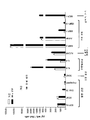
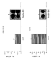
도 1은 (성인 골수 유래의 체세포 (약 27회 집단 계대시의 ABM-SC)에 의해 분비된 단백질의 2차원 SDS PAGE 분리 결과 (pH 3.5 내지 10; 12% 폴리아크릴아미드)를 도시한다. 겔의 각 스폿은 약 5 내지 200 킬로달톤(kDa) 크기 범위의 개별 구분 단백질을 나타낸다. X축은 등전점 (pH 3.5 내지 10)에 따라 분류된 단백질을 나타낸다. Y축은 분자량에 따라 분류된 단백질을 나타낸다 (12% 폴리아크릴아미드를 통한 전개시).
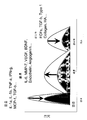
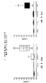
도 2는 신경 성장 인자 (nerve growth factor, NGF)와 인간 exABM-SC (약 43회 집단 계대시)로부터 유래된 조절 배지를 이용하여 뉴우런으로의 PC-12 분화의 광미소그래프를 도시한다. A) RPMI-ITS 단독 배지, B) NGF 보충 RPMI-ITS 배지, C) 1:50 희석 농도의 조절 배지와 NGF를 보충한 RPMI-ITS, D) 인간 ABM-SC에서 유래한 1:50 희석 농도의 조절 배지 및 NGF를 보충한 RPMI-ITS. 화살표는 뉴라이트 성장 결과물을 표시한다. 패널 D에서 뉴라이트 성장 정도는 패널 B 및 C보다 훨씬 뚜렷하다.
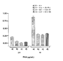
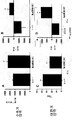
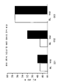
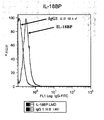
도 3은 인간 ABM-SC를 이용한 미토겐-유래 T 세포 증식의 저해를 나타내는 그래프를 도시한다. 로트 # RECB801은 약 19회 집단 계대로 2차 배양된 ABM-SC를 나타내고 로트 # RECB906은 약 43회 집단 계대로 2차 배양된 exABM-SC를 나타낸다. T-세포의 증식을 촉진하기 위해 배양물에 2.5 또는 10㎍/mL 피토헤마글루티닌을 접종했다. 72시간 후 세포를 수득하고 CD3-PC7 항체로 염색했다. 인간 간엽 줄기세포를 양의 대조군으로 사용했다 (인간 간엽 줄기세포를, 현재 론자 그룹 리미티드 소유의 캠브렉스 리서치 바이오프로덕츠로부터 취득했다, 바셀, 스위스).
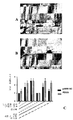
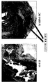
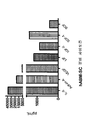
도 4는 수술로 절개 상처를 낸 뒤 7일 후 돼지 피부의 광미소그래프를 도시한다 (좌측 2개의 패널, 실험군 #4의 상처). A) 동종이형 돼지 ABM-SC (약 28회 집단 계대후)로 치료한 상처 (상처 번호 3)는 육안으로 아무런 상흔이 없이 완전히 아물었고, B) 비교로, 비히클만 투여한 상처 (상처 번호 4)는 상흔이 육안으로 확인되고, C) 그래프 (우측 패널)는 두 치료군으로부터 취득한 조직부의 조직형태 측정 그래프 (histomorphometric scoring)를 나타내는 것으로 돼지 ABM-SC 치료 상처의 조직구 수가 통계학적으로 현저히 감소한 것을 보여준다 (p=0.03) (2개의 테일이 있는 쌍이 아닌 T-시험을 이용하여 측정한 통계적 의미). "조직구" PBSG 대 측정 pABM-SC의 막대 그래프를 비교 목적으로 나타낸다.
도 5는 절개 상처를 내고 7일간 후치료 뒤 재-상피조직화 수준을 나타내는 그래프 (상부 패널)를 도시한다. 돼지 ABM-SC (약 28회 집단 계대후)로 치료한 상처는 비히클만으로 치료한 경우보다 더 두꺼운 외피를 갖는다. 하부 좌측 패널의 광미소그래프는 돼지 ABM-SC로 치료한 상처가 (조직학적으로) 완전히 및 해부학적으로 적절히 회복된 결과를 나타내고, 하부 우측 패널의 광미소그래프는 제7일에 하피속으로 적어도 전이적으로 이식접목된 상태를 보여주는 (조직학적) 돼지 ABM-SC (화살표 머리)를 나타낸다.
도 6은 세포 복원 24시간 후 시드된 수화 콜라겐 겔 격자의 ABM-SC 개입 수축 상태를 그래프를 도시한다. 인간 ABM-SC (약 27회 집단 계대)는 액상 생분해성 콜라겐-기초 배지 (1.8×10⁶ 세포/mL)에서 복원된 후 다시 약 4 내지 8℃에서 24시간 동안 저장했다. 다음날 액상 세포 현탁물을 배양 접시에 넣고 반고체 콜라겐 격자를 형성했다. 반고체 콜라겐 격자를 세포 배양기에 넣어 3일간 수축을 진행했다. 세포 없이 조제한 콜라겐 격자는 수축되지 않았으며, 수축은 세포의 존재 여부에 의존하는 것으로 나타났다.
도 7은 약 43회 집단 계대한 exABM-SC를 활용한 상이한 세포 농도에서 시드처리된 수화 콜라겐 겔 격자의 ABM-SC 개입 수축을 나타내는 그래프이다. 수축율과 절대 크기는 세포 수와 관련이 있다는 것을 데이터로부터 알 수 있으며, 열 비활성화된 세포는 겔을 수축하지 않으므로, 이 활성이 생체 물리학적 현상임을 입증한다.
도 8은 약 43회의 집단 계대후의 exABM-SC를 활용하는 수화 콜라겐 겔 격자에서 3일간 배양되었을 때 수개의 사이토킨과 매트릭스 프로테아제 (예, IL-6, VEGF, 액티빈-A, MMP-1, MMP-2)의 ABM-SC 개입 분비를 나타내는 그래프이다.
도 9는 액상(좌측 패널) 또는 반고체(우측 패널) 형태의 생분해성 콜라겐계 배지 (약 43회 집단 계대후의 exABM-SC를 활용하는)에서 복원된 인간 ABM-SC의 광미소그래프를 나타낸다. 이 제형을 이용하여 복원한 경우, 세포 현탁물은 4℃에서 24시간 이상 동안 액상으로 남아있을 수 있다. 배양 접시에 넣어 37℃에서 배양한 경우, 세포 현탁물은 1 내지 2시간 내에 고형화하므로, 물리적 조정 대신 반고체 구조화가 일어난다.
도 10은 생분해성 콜라겐계 배지 내에서 3일간 복원한 인간 ABM-SC (약 43회 집단 계대후)을 배양하여 형성된 고체형 신생조직을 나타내는 광미소그래프이다. 상부 우측 패널은 고체형 신생조직의 일반 구성을 나타내고, 하부 패널은 Masson's 트리크롬에 의해 염색된 조직의 조직학적 단면을 도시하며, ABM-SC에 의해 합성된 농축 세포외 매트릭스를 나타낸다. 세포를 제외하고 동일한 방식으로 구성한 대조군 겔은 상기 방법에 따를 경우 청색으로 염색되지 않으므로, 콜라겐과 글리코사미노글리칸 농축 매트릭스가 세포로부터 생성되는 것을 알 수 있다.
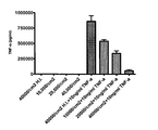
도 11은 TNF-알파 자극이 있거나 없는 인간 ABM-SC에 의해 분비된 복수종의 전-재생 사이토킨의 양에 관한 실시예를 도시한다. 2차 배양시 ABS-SC는 혈관 생성, 염증 및 상처 치유를 증대하는 것으로 알려진 사이토킨 및 수종의 성장 인자를 치료효과 농도로 분비한다. ABS-SC는 복수종의 사이토킨과 성장 인자를 적절히 체외 분비하는 것으로 확인되었다. 혈관 생성 인자 (예, SDF-1 알파, VEFG, ENA-78 및 안지오제닌), 면역조절자 (예, IL-6 및 IL-8) 및 상흔 억제자/상처 치유 조절자 (예, MMP-1, MMP-2, MMP-13 및 액티빈-A) 등이 여기에 포함된다. 또한, 상기 각종 인자의 방출은 급성 조직 손상의 과정에서 방출되는 공지의 염증성 사이토킨으로서 종양 괴사 인자 알파 (TNF-알파)에 의해 조절된다.
도 12는 상처-반응 단계의 모델 (상처로부터 상흔까지로서 염증, 재생 및 섬유화)과 염증, 재생 및 섬유화에 작용할 수 있는 분자들의 예를 도시한다.
도 13은 인간 ABM-SC로 치료한 래트에서 얻은 개선된 심장 기능 결과의 예를 도시한다. 치료후 4주간, ABM-SC를 수용한 래트는 매우 큰 +dp/dt (압력 변화의 피크 양성 속도)값 (A)을 나타내고, 제0주 +dp/dtd 값을 제4주의 값("델타 +dp/dt")에서 빼서 연구 기간 동안의 심장 기능의 변화를 표시하면, 비히클 치료 래트가 연구기간 동안 심장 기능 저하를 겪은 반면 (음성 델타), 세포 조제물로 치료한 동물은 심장 기능의 현저한 개선 (B)을 나타냈다. 비히클 치료 래트와 비교시, ABM-SC 수용 대상은 크게 저하된 타우 값(C)을 나타냈으며, 이는 좌심실 순응성 증가를 시사한다. 타우는 등용적 좌심실 압력 저하의 시간상수를 나타낸다. 압력 변화 (-dp/dt)의 피크 음성 속도에서, 제0주 +dp/dt 값을 제4주의 값 ("델타 -dp/dt")에서 제외하여 연구기간 동안 심장 기능의 변화를 표시함으로써, 비히클 치료 래트가 연구기간 동안 심장 기능 저하를 나타낸 반면 (음성 델타), 세포 조제로 치료한 동물은 심장 기능의 현저한 개선 (D)을 나타냈다 [ANOVA에 따르면, *p<0.05, **p<0.01].
도 14는 hAMB-SC로 치료한 심근 경색 래트 모델에서 섬유화 감소 및 혈관 생성의 개선을 도시한다. 2차 정량 측정을 통하여, 심근 경색후 7일에 ABM-SC나 비히클을 수용한 래트의 심장내 경색 크기의 변화를 평가하였다. ABM-SC 투여뒤 약 30일 후에 시행한 조직병리학적 분석에서, hABM-SC를 수용한 래트의 경색 크기는 비히클과 비교시 크게 감소한 것으로 나타났다. 설정 크기에 따르면, hABM-SC를 수용하는 래트는 비히클 대조군보다 약 2 포인트 낮은 것으로 나타났다. 이 도면은 통상의 경색 크기 감소에 관한 예시를 도시한다.
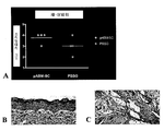
도 15는 ABM-SC 투여뒤 대체로 30일 후에 실행된 것으로서, 심근 경색 7일후에 비히클이나 ABM-SC를 수용한 래트의 심장 구조 변화를 조직학적으로 측정한 결과를 도시한다.
도 16은 인간 ABM-SC 및 exABM-SC 전구세포가 미토겐-유발 T 세포 증식을 억제하는 것을 단방향 MLR (혼합 림프구 반응) 분석으로 나타내고.
도 17은 양방향 MLR 챌린지 실험에서 부정 T-세포 개입 면역 반응에 무효한 돼지 ABM-SC 전구세포을 도시한다. 분할 지수는 기준선, 3 내지 30일간 후치료에서 수거한 시료에 대해 계산하며, 이에 배지, 비히클, ABM-SC나 ConA을 이용하여 체외 실시하였다. 전처리 및 괴사 양측 경우에서 ConA로 자극시킨 CD+ 세포에 대하여 제3일 또는 제30일에 측정한 모든 동물의 평균 분할 지수는, 비히클과 pABM-SC로 치료한 동물의 CD+ 세포에 대한 분할 지수보다 훨씬 높았다 (* p<0.05).
도 18은 기준선 (BL) 측정치를 hABM-SC의 치료후 90일에 측정치와 비교하여 3명의 환자의 심장 고정 관류 결손 크기의 변화를 도시한다.
도 19는 기준선 (BL) 측정치를 hABM-SC의 치료후 90일에 얻은 측정치와 비교하여 3명의 환자의 심장 박출의 변화를 도시한다.
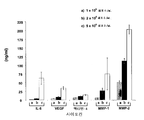
도 20은 hABM-SC (예, IL-6, 액티빈-A, VEGF, LIF, IGF-II, SDF-1 및 SCF)에 의한 체외 분비된 적혈구 생성 사이토킨의 정량 실시예를 도시한다. RAYBIOTM 인간 사이토킨 항체 어레이(RayBiotech, Inc.)을 이용하여 ABM-SC 로트를 사이토킨 분비에 대해 시험했다. 세포는 3일간 무혈청 어드밴스드 DMEM (GIBCOTM)에서 1차 배양하여 컨디셔닝된 배지(CM)를 생성하였다. 분석에 앞서서, 이 CM을 CENTRICONTM PLUS-20 원심분리 필터 장치 (밀리포어)를 이용하여 농축했다.
도 21은 exABM-SC가 용량 의존 방식으로 체외 TNF-α 레벨을 감소시키는 것을 표시한다. 인간 exABM-SC (약 43회 집단 계대시)는 각종 시드 밀도에서 (예, 10,000 세포/cm2, 20,000 세포/cm2, 및 40,000 세포/cm2) 배양시 TNF-α 레벨을 저하시키는 능력에 대해 시험했다. 세포를 무혈청 어드밴스드 DMEM (GIBCOTM) 단독 또는 이것에 10ng/mL의 TNF-α 를 보충한 액에서 3일간 배양하고, 고온-비활성화 세포를 또한 음성 대조군으로 포함시켰다. TNF의 농도를 Y축에 나타내었다 (Y축은 100x 로 농축한 배지 내의 물질 농도를 나타낸다).
도 22a 및 22b는 TNF-α의 감소가 exABM-SC (약 43회의 집단 계대)에 따른 sTNF-RI 및 sTNF-RII의 분비에 의해 조정되는 것을 나타낸다. sTNF-RI의 기초값은 프로-염증성 유발자의 결여시 발현되고 (A), sTNF-RII는 TNF-α로 초기 자극을 제공했을 때만 평가 가능한 수준으로 검출된다 (B). 이들 데이터는 시드된 세포수 및 검출된 sTNF-RI와 sTNF-RII의 레벨 간의 역비례 관계식으로 표시되며, 이는 분비된 수용체가 TNF-α 에 결합하여 이를 마스킹 할 수 있음을 시사한다 (Y축은 100x로 농축된 배지 내의 물질 농도를 나타낸다).
도 23은 IL-IRA의 분비 수준 (약 43회 집단 계대후의 exABM-SC에 의해 분비된)이 용량 의존성임을 나타낸다. IL-IRA의 기초값은 프로-염증성 유발자의 결여시 발현되고, 단, TNF-α로 초기 자극을 제공했을 경우 가용성 수준은 약 10배 증가한다 (Y축은 100x로 농축된 배지 내의 물질 농도를 나타낸다).
도 24는 exABM-SC에 의한 IL-1 수용체 길항제 (IL-IRA) 및 IL-18 결합 단백질 (IL-18BP)의 발현을 도시한다. 인간 exABM-SC는, TNF-알파 같은 염증성 신호의 결여시 IL-1 수용체 길항제(IL-IRA; 도 36a) 및 IL-18 결합 단백질 (IL-18BP; 도 36b)의 기초 레벨을 발현한다.
도 25a, b 및 c는 인간 ABM-SC가 TNF-알파 (도 25a) 및 IL-13 (도 25b)의 레벨을 감소시키고, 복합 PBMC 반응 분석에서 IL-2의 상승 발현(도 25c)을 동시에 유발하는 것을 도시한다 (R = 응답자 PBMC, 자기 = 응답자로서 동일한 공여자로부터 분리된 미토마이신-C로 치료한 PBMC, 흥분제 = 미토마이신-C로 치료한 PBMC).
도 26은 인간 ABM-SC를 이용하여 미토겐-유래의 인간 말초 혈액 단핵 세포(PBMC)의 억제현상을 나타내는 그래프이다. RECB 801은 약 19회 집단 계대로 2차 배양된 소정의 ABM-SC를 나타내고, RECB 906은 약 43회 집단 계대로 2차 배양된 소정의 ABM-SC를 나타낸다. PBMC 증식을 촉진하기 위해, 배양물에 2.5㎍/mL의 피토헤마글루티닌을 접종한다. 배양시 56시간 후, 티미딘-[메틸-3H]으로 세포를 펄스화 하고, 72시간에 동위원소 삽입으로 정량화 처리했다 (CPM). 인간 간엽 줄기세포는 양성 대조군으로 포함되었다.FIG. 1 shows the results of two-dimensional SDS PAGE separation (pH 3.5-10; 12% polyacrylamide) of proteins secreted by somatic cells derived from adult bone marrow (ABM-SC at about 27 population passages). Gel Each spot of represents an individual distinguished protein in the size range of about 5 to 200 kilodaltons (kDa) The X axis represents proteins classified according to isoelectric point (pH 3.5 to 10) The Y axis represents proteins classified according to molecular weight ( On development via 12% polyacrylamide).
FIG. 2 shows optical micrographs of PC-12 differentiation into neurons using regulatory media derived from nerve growth factor (NGF) and human exABM-SC (about 43 population passages). A) RPMI-ITS alone medium, B) NGF supplemented RPMI-ITS medium, C) 1:50 dilution control medium and RPMI-ITS supplemented with NGF, D) 1:50 dilution derived from human ABM-SC RPMI-ITS supplemented with NGF and NGF. Arrows indicate Newlite growth output. The extent of neurite growth in panel D is much more pronounced than panels B and C.
3 shows a graph showing inhibition of mitogen-derived T cell proliferation with human ABM-SC. Lot # RECB801 represents ABM-SC secondary cultured with approximately 19 population passages and Lot # RECB906 represents exABM-SC secondary cultured with approximately 43 population passages. Cultures were inoculated with 2.5 or 10 μg / mL phytohemagglutinin to promote T-cell proliferation. After 72 hours cells were obtained and stained with CD3-PC7 antibody. Human mesenchymal stem cells were used as positive controls (human mesenchymal stem cells were obtained from Cambrex Research Bioproducts currently owned by Lonza Group Limited, Basel, Switzerland).
FIG. 4 shows a photomicrograph of
5 shows a graph (top panel) showing re-epithelial tissue levels after incisional wounds and 7 days post-treatment. Wounds treated with porcine ABM-SC (after about 28 passages) have a thicker shell than those treated with vehicle only. The photomicrograph of the lower left panel shows the result of a (histologically) complete and anatomically adequate recovery of wounds treated with porcine ABM-SC, and the photomicrograph of the lower right panel at least subcutaneously on day 7 (Histological) porcine ABM-SC (arrow head) showing metastatically grafted status.
FIG. 6 shows a graph of the ABM-SC interventional contraction state of hydrated collagen gel lattice seeded 24 hours after cell recovery. Human ABM-SCs (approximately 27 passages) were restored in liquid biodegradable collagen-based medium (1.8 × 10 6 cells / mL) and then stored for another 24 hours at about 4-8 ° C. The next day the liquid cell suspension was placed in a culture dish to form a semisolid collagen lattice. The semi-solid collagen lattice was placed in a cell incubator and contracted for 3 days. The collagen lattice prepared without cells did not contract, and the contraction was shown to depend on the presence of cells.
FIG. 7 is a graph showing ABM-SC interventional shrinkage of hydrated collagen gel lattice seeded at different cell concentrations using exABM-SC passaged approximately 43 times. It can be seen from the data that the shrinkage rate and absolute size are related to the number of cells, demonstrating that this activity is a biophysical phenomenon since heat inactivated cells do not shrink the gel.
FIG. 8 shows several cytokines and matrix proteases (eg, IL-6, VEGF, Activin-A, MMP-1, MMP) when incubated for 3 days in a hydrated collagen gel grid utilizing exABM-SC after about 43 population passages. -2) is a graph showing the secretion of ABM-SC intervention.
FIG. 9 shows photomicrographs of human ABM-SCs restored in biodegradable collagen-based medium (using exABM-SC after about 43 population passages) in liquid (left panel) or semisolid (right panel) form. When restored using this formulation, the cell suspension may remain liquid for at least 24 hours at 4 ° C. When incubated at 37 ° C. in a culture dish, the cell suspension solidifies within 1 to 2 hours, so semisolid structuring occurs instead of physical adjustment.
FIG. 10 is an optical micrograph showing solid neoplastic tissue formed by culturing human ABM-SC (after about 43 times of passage passage) restored for 3 days in biodegradable collagen-based medium. The upper right panel shows the general configuration of solid neoplasms, the lower panel shows the histological cross section of the tissue stained with Masson's trichrome and shows the enriched extracellular matrix synthesized by ABM-SC. The control gel constructed in the same manner except for the cells is not stained blue according to the above method, so that the collagen and glycosaminoglycan enriched matrix are produced from the cells.
FIG. 11 shows an example of the amount of a plurality of pre-regenerative cytokines secreted by human ABM-SC with or without TNF-alpha stimulation. In secondary cultures, ABS-SC secretes therapeutic concentrations of cytokines and several growth factors known to enhance angiogenesis, inflammation and wound healing. ABS-SC has been found to secrete multiple cytokines and growth factors in vitro. Angiogenesis factors (eg SDF-1 alpha, VEFG, ENA-78 and angiogenin), immunomodulators (eg IL-6 and IL-8) and scar suppressor / wound healing modulators (eg MMP- 1, MMP-2, MMP-13 and activin-A) and the like. The release of these various factors is also regulated by tumor necrosis factor alpha (TNF-alpha) as known inflammatory cytokines released during the course of acute tissue damage.
FIG. 12 shows a model of wound-response stages (inflammation, regeneration and fibrosis, from wound to scar) and examples of molecules that may act on inflammation, regeneration and fibrosis.
Figure 13 shows an example of improved cardiac function results obtained in rats treated with human ABM-SC. At 4 weeks post-treatment, rats receiving ABM-SC exhibited a very large + dp / dt (peak positive rate of pressure change) value (A) and
FIG. 14 shows reduced fibrosis and improved blood vessel production in myocardial infarction rat models treated with hAMB-SC. Secondary quantitative measurements were performed to evaluate changes in intracardiac infarct size of rats receiving either ABM-SC or
FIG. 15 shows the results of histological measurements of cardiac structural changes in rats receiving vehicle or ABM-SC after 7 days of myocardial infarction, which were performed approximately 30 days after ABM-SC administration.
FIG. 16 shows unidirectional MLR (mixed lymphocyte response) analysis that human ABM-SC and exABM-SC progenitors inhibit mitogen-induced T cell proliferation.
17 depicts porcine ABM-SC progenitor cells that are invalid for negative T-cell intervening immune response in a bidirectional MLR challenge experiment. Split index was calculated for samples collected from baseline, post-treatment for 3 to 30 days, and performed in vitro using medium, vehicle, ABM-SC or ConA. In both cases of pretreatment and necrosis, the mean partitioning index of all animals measured on
FIG. 18 shows the change in heart fixation perfusion defect size in three patients comparing baseline (BL) measurements with measurements at day 90 post-treatment of hABM-SC.
FIG. 19 depicts changes in cardiac output in three patients compared to baseline (BL) measurements compared to measurements obtained 90 days after treatment of hABM-SC.
FIG. 20 shows quantitative examples of in vitro secreted erythropoietic cytokines by hABM-SC (eg, IL-6, activin-A, VEGF, LIF, IGF-II, SDF-1 and SCF). ABM-SC lots were tested for cytokine secretion using the RAYBIO ™ human cytokine antibody array (RayBiotech, Inc.). Cells were first cultured in serum free Advanced DMEM (GIBCO ™ ) for 3 days to produce conditioned medium (CM). Prior to analysis, this CM was concentrated using a CENTRICON ™ PLUS-20 centrifugal filter device (Millipore).
FIG. 21 shows that exABM-SC decreases in vitro TNF-α levels in a dose dependent manner. Human exABM-SC (approximately 43 population passages) has the ability to lower TNF-α levels in culture at various seed densities (eg, 10,000 cells / cm 2 , 20,000 cells / cm 2 , and 40,000 cells / cm 2 ). Tested against. Cells were incubated for 3 days in serum-free Advanced DMEM (GIBCO ™ ) alone or in a solution supplemented with 10ng / mL of TNF-α, and hot-inactivated cells were also included as negative controls. The concentration of TNF is shown on the Y axis (Y axis represents the concentration of substance in the medium concentrated to 100 ×).
22A and 22B show that the decrease in TNF-α is mediated by the secretion of sTNF-RI and sTNF-RII according to exABM-SC (about 43 population passages). Baseline values of sTNF-RI are expressed in the absence of pro-inflammatory triggers (A) and sTNF-RII is detected at an assessable level only when initial stimulation with TNF-α is provided (B). These data are expressed as an inverse relationship between the number of seeded cells and the levels of sTNF-RI and sTNF-RII detected, suggesting that secreted receptors can bind and mask TNF-α (Y-axis is 100x). Material concentration in concentrated medium).
23 shows that the secretion level of IL-IRA (secreted by exABM-SC after about 43 population passages) is dose dependent. The baseline of IL-IRA is expressed in the absence of pro-inflammatory inducers, except that the level of soluble increases approximately 10-fold when TNF-α provides initial stimulation (Y axis represents substance concentration in medium enriched to 100x). ).
24 shows expression of IL-1 receptor antagonist (IL-IRA) and IL-18 binding protein (IL-18BP) by exABM-SC. Human exABM-SC expresses basal levels of IL-1 receptor antagonist (IL-IRA; FIG. 36A) and IL-18 binding protein (IL-18BP; FIG. 36B) in the absence of inflammatory signals such as TNF-alpha.
25A, B and C show that human ABM-SCs reduce the levels of TNF-alpha (FIG. 25A) and IL-13 (FIG. 25B), and simultaneously result in synergistic expression of IL-2 (FIG. 25C) in a complex PBMC response assay. Induction is shown (R = responder PBMC, self = PBMC treated with mitomycin-C isolated from the same donor as responder, stimulant = PBMC treated with mitomycin-C).
FIG. 26 is a graph showing the inhibition of mitogen-derived human peripheral blood mononuclear cells (PBMC) using human ABM-SC.
통상적으로, 줄기세포나 기타 초기 전구세포는 세포가 특정의 분화 경로에 관계하므로 가소성을 잃었다. 생체분자 수준에서 상기 공정이 일어나면, 세포는 소정의 신호성 분자 (예, 미토겐 및 모르포겐)에 대해 응답하는 능력을 잃고, 그렇지 않으면 세포를 분할 구동시키거나 또다른 세포종으로 된다. 따라서, 세포가 분화되기 시작하면, 세포 주기를 떠나 (예, 더이상 유사분열을 행할 수 없다) 세포가 더이상 분할되지 않는 G0 이라는 비가역 상태에 들어간다. G0으로의 진입은 복제노화 (세포내 단백질 p21 및 p53의 발현 증가를 포함하는 검증)와 연계된다. 따라서, 가소성 (각종 세포로 분화되는 능력)의 손실은 보통 세포 분화 또는 세포 노화의 전조 현상으로 본다. 또한, 가소성 손실은 연속의 자기재생 능력 손실과도 관련이 있다. 대조적으로, 이러한 통상의 시나리오와 달리, 본 발명에서 달성한 예상외의 놀라운 결과에 따르면, exCF-SC (예, exABM-SC)는 가소성 손실에도 불구하고 자기재생을 계속한다 (비교적 일정한 속도로 진행하는 자기재생 포함). 따라서, 본 발명의 하나의 구현예는 연속 자기재생 능력을 통해 치료적으로 유용한 "최종 단계의 세포"이다 (예, 연속 자기재생 및 영양 지원 인자 (혹은 "영양 지원 세포")의 생산). 또다른 구현예에서, 본 발명의 exCF-SC 및 exABM-SC는 상당량의 p21 및/또는 p53을 발현하지 않으며, 상기 분자의 "상당량"은 세포 노화를 표시하는 양이다 (노화는 p21, p53, 및/또는 다른 세포주기 조절자의 충분한 발현 레벨이 필요하다).Typically, stem cells or other early progenitor cells lost plasticity because the cells are involved in specific differentiation pathways. When the process takes place at the biomolecule level, the cell loses the ability to respond to certain signaling molecules (eg, mitogens and morphogens), otherwise it divides the cell or becomes another cell species. Thus, when cells begin to differentiate, they leave the cell cycle (eg, can no longer undergo mitosis) and enter the irreversible state of G0 where the cells no longer divide. Entry into G0 is associated with replication aging (validation involving increased expression of intracellular proteins p21 and p53). Thus, loss of plasticity (the ability to differentiate into various cells) is usually seen as a precursor to cell differentiation or cell aging. In addition, plastic loss is also related to the continuous loss of self-renewal ability. In contrast, unlike this conventional scenario, according to the surprising surprising results achieved in the present invention, exCF-SC (e.g. exABM-SC) continues self-renewal in spite of plastic loss (comparatively constant magnetic Playback). Thus, one embodiment of the invention is a "final stage cell" that is therapeutically useful through continuous self-renewal ability (eg, the production of continuous self-renewing and nutritional support factors (or "nutrition support cells")). In another embodiment, exCF-SCs and exABM-SCs of the present invention do not express significant amounts of p21 and / or p53, and the "equivalent" of the molecule is an amount that indicates cellular aging (aging is p21, p53, And / or sufficient expression levels of other cell cycle regulators).
또한, 본 발명 분야의 전문가라면, 가소성이 손실된 무-조혈성 체세포가 제한된 유용성 또는 기관 및 조직을 재생 또는 그 재생을 촉진하는 능력을 갖는 것을 예상한다. 따라서, 본 발명의 예상외의 놀라운 결과는 가소성이 손실된 광역 증식의 CF-SC (예, ABM-SC)를 생성하는 능력이며, 따라서 신생 조직을 체외 생성 및 체내 조직 재생 촉진 능력을 유지한다.In addition, one of ordinary skill in the art would expect that hematopoietic somatic cells lacking plasticity have limited utility or ability to regenerate or promote regeneration of organs and tissues. Thus, an unexpected surprising result of the present invention is the ability to produce CF-SC (eg, ABM-SC) of widespread proliferation, which has lost plasticity, thus maintaining the ability to generate new tissues in vitro and promote tissue regeneration in vivo.
그러므로, 본 발명은 집락 형성 체세포 (CF-SC)인 자기재생 세포를 이용하는 조직의 치료, 재생 및/또는 회복 방법을 제공한다 (성인 골수 유래의 체세포 (ABM-SC)의 예). 본 발명에서 이용하는 성인 골수 유래의 체세포(ABM-SC) 같은 자기재생 집락 형성 체세포 (CF-SC)는 미국 특허 공보 제20030059414호 (미국 출원 제09/260,244호; 2001년 9월 21일 출원) 및 미국 특허 공보 제20040058412호 (미국 출원 제10/251,685; 2002년 9월 20일 출원)에 개시된 바와 같이 제조한다. 이들 특허출원의 전문을 본원에 참고로서 수록한다. 또한, 미국 가출원 제60/929,151호 및 제60/929,152호 (각기 2007년 6월 15일 출원), 미국 가출원 제60/955,204호 (2007년 8월 10일 출원), 및 미국 가출원 제60/996,093호 (2007년 11월 1일 출원)도 참고로서 수록한다.Therefore, the present invention provides a method of treating, regenerating and / or repairing a tissue using self-renewing cells that are colony forming somatic cells (CF-SCs) (example of somatic cells derived from adult bone marrow (ABM-SC)). Self-renewing colony forming somatic cells (CF-SC), such as adult bone marrow derived somatic cells (ABM-SC), are disclosed in US Patent Publication No. 20030059414 (US Application No. 09 / 260,244; filed September 21, 2001) and It is prepared as disclosed in US Patent Publication No. 20040058412 (US Application No. 10 / 251,685; filed September 20, 2002). The entirety of these patent applications is incorporated herein by reference. Also, US Provisional Application Nos. 60 / 929,151 and 60 / 929,152 (filed June 15, 2007, respectively), US Provisional Application No. 60 / 955,204, filed August 10, 2007, and US Provisional Application No. 60 / 996,093 (November 1, 2007 application) is also incorporated by reference.
특히, 기원 세포 집단 (비제한적인 예를 들어, 골수 (ABM-SC 및 exABM-SC), 지방, 피부, 태반, 근육, 제대혈, 또는 연결 조직 등)에서 분리된 CF-SC는 적절한 배지 (비제한적인 예를 들어, 4mM 글루타민 및 10% 태아소혈청을 보충한 최소 필수 배지-알파 (예, HYCLONETM 으로 판매)) 존재하에서 세포 배양 표면에 부착되고, 저 산소 조건 (비제한적인 예를 들어, 약 2-5%의 O2, 약 5%의 CO2 및 나머지로서 질소를 함유하는 분위기)에서 배양되고, CF-SC가 다수 집단 계대 (비제한적인 예를 들어, 10, 15, 20, 25, 30, 35, 40, 45 및/또는 50회의 집단 계대)를 통해 소정의 필수 집단 계대율 (비제한적인 예를 들어, 약 30시간 이하의 계대율)를 유지하도록 저 세포밀도 (예 30 내지 1000 세포/cm2)에서 연속으로 증식된다.In particular, CF-SC isolated from a population of origin cells (including but not limited to, bone marrow (ABM-SC and exABM-SC), fat, skin, placenta, muscle, umbilical cord blood, or connective tissue, etc.) may be used in appropriate media (non- For example, adhere to the cell culture surface in the presence of minimal essential medium-alpha (e.g., sold as HYCLONE ™ ) supplemented with 4 mM glutamine and 10% fetal bovine serum, and low oxygen conditions (e.g. In an atmosphere containing about 2-5% O 2 , about 5% CO 2 and nitrogen as the remainder, and CF-SC is subjected to multiple population passages (eg, without limitation, 10, 15, 20, 25, 30, 35, 40, 45, and / or 50 passages at a low cell density (e.g., non-limiting, eg, about 30 hours or less) to maintain a certain required population passage rate To 1000 cells / cm 2 ).
본 발명의 구현예는 저 산소 조건하에 배양된 CF-SC 및 exCF-SC (예를 들어, ABM-SC 및 exABM-SC)를 이용하여 실행되며, 상기 산조 조건에서 O2 농도는 1 내지 20% (예를 들어, O2 농도는 1%, 2%, 3%, 4%, 5%, 6%, 7%, 8%, 9%,10%, 11%, 12%, 13%, 14%, 15% 또는 20%)이며, 이에 더하여 CO2 및 나머지는 질소를 함유한다. 예를 들면, ABM-SC는 저 산소 조건으로서 O2 농도가 약 20%, 약 20% 이하, 약 15%, ㅇ약 % 이하, 약 10%, 약 10% 이하, 약 7%, 약 7% 이하, 약 6%, 약 6% 이하, 약 5%, 약 5% 이하, 약 4%, 약 4% 이하, 약 3%, 약 3% 이하, 약 2%, 약 2% 이하, 약 1%이거나; 또는 상기 저 산소 조건이 약 1% 내지 20%, 약 1% 내지 15%, 약 1% 내지 10%, 약 1% 내지 5%, 약 5% 내지 20%, 약 5% 내지 15%, 약 5% 내지 10%, 약 10% 내지 15%, 약 10% 내지 20%, 약 2% 내지 8%, 약 2% 내지 7%, 약 2% 내지 6%, 약 2% 내지 5%, 약 2% 내지 4%, 약 2% 내지 3%, 약 3% 내지 8%, 약 3% 내지 7%, 약 3% 내지 6%, 약 3% 내지 5%, 약 3% 내지 4%, 약 4% 내지 8%, 약 4% 내지 7%, 약 4% 내지 6%, 약 4% 내지 5%, 약 5% 내지 8%, 약 5% 내지 7%, 약 5% 내지 6%, 또는 약 5%이다.Embodiments of the invention are carried out using CF-SC and exCF-SC (eg, ABM-SC and exABM-SC) incubated under low oxygen conditions, in which the O 2 concentration is 1-20%. (For example, O 2 concentrations are 1%, 2%, 3%, 4%, 5%, 6%, 7%, 8%, 9%, 10%, 11%, 12%, 13%, 14% , 15% or 20%), in addition to CO 2 and the balance contains nitrogen. For example, ABM-SC is a low oxygen condition with an O 2 concentration of about 20%, about 20% or less, about 15%, about% or less, about 10%, about 10% or less, about 7%, or about 7% or less. About 6%, about 6% or less, about 5%, about 5% or less, about 4%, about 4% or less, about 3%, about 3% or less, about 2%, about 2% or less, about 1% ; Or the low oxygen condition is about 1% to 20%, about 1% to 15%, about 1% to 10%, about 1% to 5%, about 5% to 20%, about 5% to 15%, about 5 % To 10%, about 10% to 15%, about 10% to 20%, about 2% to 8%, about 2% to 7%, about 2% to 6%, about 2% to 5%, about 2% To 4%, about 2% to 3%, about 3% to 8%, about 3% to 7%, about 3% to 6%, about 3% to 5%, about 3% to 4%, about 4% to 8%, about 4% to 7%, about 4% to 6%, about 4% to 5%, about 5% to 8%, about 5% to 7%, about 5% to 6%, or about 5% .
본 발명의 구현예는 저 산소 조건하에 배양된 CF-SC 및 exCF-SC (예를 들어, ABM-SC 및 exABM-SC)를 이용하여 실행되며, 상기 산소 조건에서 CO2 농도는 1% 내지 15% (예를 들어, CO2 농도는 1%, 2%, 3%, 4%, 5%, 6%, 7%, 8%, 9%,10%, 11%, 12%, 13%, 14% 또는 15%)이며, 이에 더하여 저함량의 O2 및 나머지는 질소를 함유한다. 본 발명의 구현예는 저 세포 농도에서 세포를 시드하여 증식한 CF-SC 및 exCF-SC (예를 들면, ABM-SC 및 exABM-SC)를 이용하여 실행되며, 이때 상기의 세포 밀도는 1 내지 2500 세포/cm2 (예를 들어, 상기 세포 밀도는 약 1, 2, 5, 10, 20, 30, 40, 50, 60, 70, 80, 90, 100, 200, 300, 400, 500, 600, 700, 800, 900, 1000, 1500, 2000 또는 2500 세포/cm2)의 범위이다. 예를 들어, ABM-SC는 약 2500 세포/cm2 미만의 시드 농도에서 증식할 수 있으며, 약 1000 세포/cm2 미만, 500 세포/cm2 미만, 약 100 세포/cm2 미만, 약 50 세포/cm2 미만, 약 30 세포/cm2 미만 또는 약 10 세포/cm2 미만의 시드 농도에서 증식할 수 있다. 본 발명의 구현예는 CF-SC 및 exCF-SC (예를 들어, ABM-SC 및 exABM-SC)를 이용하여 실행할 수 있으며, 세포 집단 계대율은 약 24 내지 96 시간의 범위를 유지한다 (예를 들어, 세포 집단 계대율은 약 24, 30, 36, 42, 48, 54, 60, 66, 72, 78, 84, 90 또는 96 시간 이하로 유지한다). 본 발명의 구현예는 CF-SC 및 exCF-SC (예를 들어, ABM-SC 및 exABM-SC)를 이용하여 실행할 수 있으며, 세포 집단은 소정의 집단 계대율, 예컨대, 약 5 내지 50회의 집단 계대 중 실질적으로 일정한 계대율을 유지한다 (예를 들어, 약 5 내지 10회, 5 내지 15회, 5 내지 20회, 5 내지 25회, 5 내지 30회, 5 내지 35회, 5 내지 40회, 5 내지 45회 또는 5 내지 50회의 집단 계대 동안 소정의 집단 계대율을 유지한다).Embodiments of the invention are carried out using CF-SC and exCF-SC (eg, ABM-SC and exABM-SC) incubated under low oxygen conditions, wherein the CO 2 concentration is from 1% to 15 % (E.g. CO 2 concentration is 1%, 2%, 3%, 4%, 5%, 6%, 7%, 8%, 9%, 10%, 11%, 12%, 13%, 14 % Or 15%), in addition to low content of O 2 and the balance contains nitrogen. Embodiments of the present invention are carried out using CF-SC and exCF-SC (eg, ABM-SC and exABM-SC) grown by seeding cells at low cell concentration, wherein the cell density is from 1 to 1. 2500 cells / cm 2 (e.g., the cell density is about 1, 2, 5, 10, 20, 30, 40, 50, 60, 70, 80, 90, 100, 200, 300, 400, 500, 600 , 700, 800, 900, 1000, 1500, 2000 or 2500 cells / cm 2 ). For example, ABM-SCs can proliferate at seed concentrations of less than about 2500 cells / cm 2, less than about 1000 cells / cm 2, less than 500 cells / cm 2, less than about 100 cells / cm 2 , about 50 cells. It may proliferate at a seed concentration of less than / cm 2, less than about 30 cells / cm 2 or less than about 10 cells / cm 2 . Embodiments of the invention can be performed using CF-SC and exCF-SC (eg, ABM-SC and exABM-SC), with cell population passage rates maintained in the range of about 24 to 96 hours (eg For example, the cell population passage rate is maintained at about 24, 30, 36, 42, 48, 54, 60, 66, 72, 78, 84, 90 or 96 hours or less). Embodiments of the invention can be practiced using CF-SC and exCF-SC (eg, ABM-SC and exABM-SC), wherein the cell population has a predetermined population passage rate, eg, about 5-50 populations. Maintain a substantially constant passage rate (for example, about 5 to 10 times, 5 to 15 times, 5 to 20 times, 5 to 25 times, 5 to 30 times, 5 to 35 times, 5 to 40 times Maintain a certain group passage rate for 5 to 45 or 5 to 50 group passages.
본 발명의 구현예는 액상, 반고체 또는 고체 상태의 약제학적 수용가능한 조성물에 함입되는 CF-SC 및 exCF-SC (예를 들어, ABM-SC 및 exABM-SC)의 용도를 포함한다. "액상,반고체 또는 고체 상태"란 세포가 함유될 약제학적 수용가능한 조성물이: 1) 통상의 액상 (상용의 생리적 염수액); 2) 젤리형, 젤라틴형 또는 점탄성 상태를 포함하는 광범위한 저-고 점성 상태 (약제학적 조성물이 극고 내지 극저인 세포외 수분을 함유하는 경우로서, 예컨대, 조성물이 유분이나 꿀처럼 천천히 "스며나오는" 상태부터 젤리형, 유연형, 반탄성 및/또는 전성의 고젤라틴성 또는 점탄성 상태에 이르는 범위; 3) 매트릭스내 살아있는 세포가, 내구성 및 비-젤라틴성이면서도 아직 유연성, 반탄성 및 전성을 갖는 매트릭스 (예컨대, 포유류 피부의 유연성 또는 반탄성을 일부 갖는 매트릭스)에 초기 현탁되어 있는 환경을 리모델링한 고체 상태 (극저의 세포외 수분 함유); (도 10A 및 도 10B 참조).Embodiments of the invention include the use of CF-SCs and exCF-SCs (eg, ABM-SCs and exABM-SCs) incorporated into pharmaceutical acceptable compositions in liquid, semisolid or solid state. “Liquid, semisolid or solid state” means a pharmaceutically acceptable composition in which the cells will be contained: 1) a conventional liquid phase (commercial physiological saline solution); 2) a wide range of low-high viscosity states, including gelatinous, gelatinous, or viscoelastic states (where the pharmaceutical composition contains extremely high to very low extracellular moisture, for example, the composition is slowly "soaking" like oil or honey). From the state to the gelatinous, flexible, semi-elastic and / or malleable high gelatinous or viscoelastic state; 3) A matrix of living cells in the matrix that is durable and non-gelatinous yet yet flexible, semi-elastic and malleable Solid state (extreme extracellular water content) remodeling the environment initially suspended in (eg, a matrix having some of the flexibility or semi-elasticity of mammalian skin); (See FIGS. 10A and 10B).
(유의: 약탄성이라고도 하는 점탄성은 가소성 변형을 겪을 때 점성 및 탄성을 모두 나타내는 재료를 말한다. 꿀 같은 점성 재료는 응력이 인가될 때 시간에 따라 선형 변화하는 전단 유동 및 전단 변형에 대한 내성이 있다. 탄성 재료는 신장시 즉시 변형하며 응력이 사라질 때 초기 상태로 신속히 복귀한다. 점탄성 재료는 상기 성질을 갖는 성분을 함유하며 시간 의존성 변형을 나타낸다).(Note: Viscoelasticity, also called weak elasticity, is a material that exhibits both viscosity and elasticity when subjected to plastic deformation. Viscous materials such as honey are resistant to linearly varying shear flow and shear deformation when stress is applied. The elastic material deforms immediately upon stretching and quickly returns to its initial state when the stress disappears, the viscoelastic material contains components having these properties and exhibits time dependent deformation).
액상, 반고체 및 고체형 비히클에 함유된 세포의 임상 투여는, 상처로부터 불요의 삼출물을 막지않고 상처부의 윤곽에 맞춘 치료에 적용할 수 있다.Clinical administration of cells contained in liquid, semisolid and solid vehicles can be applied to the treatment of contouring wounds without blocking unnecessary exudates from the wound.
콜라겐이나 피브린 같은 가용성 매트릭스 성분을 CF-SC 및 exCF-SC (예를 들어, ABM-SC 및 exABM-SC)와 조합하면, 사이토킨 및 매트릭스 금속 단백질효소 같은 중요한 분비 단백질의 발현을 상향 조절하도록 세포 집단을 유도한다. 또한, 수술 상처에 ABM-SC를 적용하면, 상처가 아무는 것을 촉진할 수 있으며 상흔이 남는 것을 방지하므로 상흔을 최소화 할 수 있다 (실시예 7 참조).Combining soluble matrix components, such as collagen or fibrin, with CF-SC and exCF-SC (eg, ABM-SC and exABM-SC), cell populations to upregulate expression of important secretory proteins such as cytokines and matrix metalloproteinases. Induce. In addition, the application of ABM-SC to the surgical wound can promote the healing of the wound and prevent the scar from remaining, thereby minimizing the wound (see Example 7).
또한, CF-SC 및 exCF-SC (ABM-SC 및 exABM-SC 등)의 고면역조절성 (실시예 4 참조) 덕분에, 이들 세포를 함유한 조성물 및 이를 이용한 치료법은, 비제한적인 예로서 만성 염증성 피부질환, 건선, 태선, 홍반성 낭창(LE), 이식편대 숙주 질환 (GVHD), 및 약물발진 (예, 피부 약물 부작용) 등 피부 관련 면역 질환 및 장애의 치료에 관하여 주목을 받고 있다.In addition, thanks to the high immunomodulatory properties (cf. Example 4) of CF-SC and exCF-SC (such as ABM-SC and exABM-SC), compositions containing these cells and therapies using the same are non-limiting examples. Attention has been paid to the treatment of skin-related immune diseases and disorders, such as chronic inflammatory skin diseases, psoriasis, thyroiditis, lupus erythematosus (LE), graft-versus-host disease (GVHD), and drug rashes (eg, skin drug side effects).
CF-SC 및 exCF-SC (ABM-SC 및 exAMB-SC 등) 컨디셔닝된 배지로부터 얻은 세포-상청액 분획물 및 분비 단백질은 무혈청 조건에서 생산하여, 체내 이용에 적합한 방식으로 농축 및 조제할 수 있다. 이러한 방식으로 조제시, ABM-SC로부터 얻은 컨디셔닝된 무혈청 배지는 수많은 프로-재생성 사이토킨, 성장 인자 및 매트릭스 프로테아제를 치료학적 유효 농도로 함유하는 것으로 확인되었다 (표 1, 2 및 3 참조). ABM-SC에 의해 생산된 수백가지의 가용성 인자의 복합 혼합물은 2D SDS PAGE로 분류할 수 있다 (도 1 참조). 개별 단백질 및 기타 거대분자들은 이러한 겔로부터 분리 및 당해 기술에서 통상적으로 실시하는 기술, 예컨대, MALDI-TOF 질량 분광측정법 (Matrix Assisted Laser Desorption Ionisation-Time Of Filight 분광측정법)에 따라 확인할 수 있다.Cell-supernatant fractions and secreted proteins obtained from CF-SC and exCF-SC (such as ABM-SC and exAMB-SC) conditioned media can be produced in serum-free conditions and concentrated and formulated in a manner suitable for use in the body. When formulated in this manner, the conditioned serum free medium obtained from ABM-SC was found to contain therapeutically effective concentrations of numerous pro-regenerative cytokines, growth factors and matrix proteases (see Tables 1, 2 and 3). Complex mixtures of hundreds of soluble factors produced by ABM-SC can be classified as 2D SDS PAGE (see FIG. 1). Individual proteins and other macromolecules can be isolated from such gels and identified according to techniques commonly practiced in the art, such as MALDI-TOF mass spectrometry (Matrix Assisted Laser Desorption Ionisation-Time Of Filight spectroscopy).
공지의 방법 (및 크로마토그래피 또는 중공 섬유 세포 배양기 등의 다른 분리 기술)을 활용하면, 원하는 단백질 또는 세포 상청액 분획물을 분리, 투석, 동결건조 및 고체 상태로 저장하거나 또는 치료 투여를 목적으로 적절한 비히클에 용해시켜 복원할 수 있다. 하나의 구현예에서, 단백질 또는 세포-상청액 분획물은 반-고체형 콜라겐이나 피브린계 비히클에 용해시켜 복원 및 상처부에 국소 투여한다.Using known methods (and other separation techniques such as chromatography or hollow fiber cell incubators), the desired protein or cell supernatant fractions can be isolated, dialyzed, lyophilized and stored in solid state or placed in a suitable vehicle for therapeutic administration. Can be dissolved and restored. In one embodiment, the protein or cell-supernatant fraction is dissolved in semi-solid collagen or fibrin-based vehicle and topically administered to the restoration and wound.
CF-SC 및 exCF-SC (ABM-SC 및 exABM-SC 등)에 의해 생성된 산물 이외에도, 각종 및 다수의 약제학적 수용가능한 소분자물 내지 거대 분자형 화합물 (지질, 단백질 및 핵산 같은 생체 물질 포함)을 약제학적 수용가능 한 담체, 예컨대, CF-SC 및 exCF-SC (ABM-SC 및 exABM-SC)나, 이 세포에 의해 생성된 산물이 함유되어 있는 생분해성 매트릭스와 더불어 투여를 목적으로 포함될 수 있다. 소량 샘플링시, 상술한 추가적인 분자물은 소분자형 약제물, 즉, 몇가지 예를 들면 항-염증제, 항생제, 비타민 및 미네랄 (칼슘 등) 등을 포함할 수 있다. 마찬가지로, 극소량의 생체 물질 시료를 추출할 때, 세포외 매트릭스 단백질, 혈장 응고 단백질, 항체, 성장 인자, 케모카인, 사이토킨, 지질 (카르디오리핀 및 스핀고마이엘린), 및 핵산 (예컨대, 리보자임, 항감지성 올리고누클레오티드, 또는 cDNA 발현 구조물) 등을 포함하며; 또한 치료에 유리한 각종 이성체, 절편 및 서브유닛 같은 분자 유도체와 변형체, 또한 이들의 치환물, 삽입물 및 결실 변형체 등도 포함한다. 이들은 본원의 상세한 설명 및 당업자가 이해할 수 있는 범위 내에서 예시된 실시예이며, 기타 다수의 구조적 또는 치료학적으로 유용한 화합물을, CF-SC 및 exCF-SC (예컨대, ABM-SC 및 exABM-SC)나, 이들 세포에 의해 생성된 산물이 함유된 생분해성 매트릭스 같이 약제학적 수용가능한 담체와 함께, 포함시켜 투여할 수 있다. In addition to products produced by CF-SC and exCF-SC (such as ABM-SC and exABM-SC), various and numerous pharmaceutically acceptable small molecule to macromolecular compounds (including biomaterials such as lipids, proteins and nucleic acids) May be included for administration with a pharmaceutically acceptable carrier such as CF-SC and exCF-SC (ABM-SC and exABM-SC) or with a biodegradable matrix containing the products produced by these cells. have. In small sampling, the additional molecular substances described above may include small molecule drugs, ie some anti-inflammatory agents, antibiotics, vitamins and minerals (calcium, etc.) and the like. Likewise, when extracting very small amounts of biological material samples, extracellular matrix proteins, plasma coagulation proteins, antibodies, growth factors, chemokines, cytokines, lipids (cardiolipin and spingomyelin), and nucleic acids (eg ribozymes, Antisense oligonucleotides, or cDNA expression constructs); Also included are molecular derivatives and variants, such as various isomers, fragments and subunits that are beneficial for treatment, as well as their substitutions, inserts, and deletion variants. These are the examples exemplified within the scope of the description herein and to those skilled in the art, and many other structural or therapeutically useful compounds include CF-SC and exCF-SC (eg, ABM-SC and exABM-SC). Or, together with a pharmaceutically acceptable carrier, such as a biodegradable matrix containing the product produced by these cells.
본 발명의 하나의 구현예는 당뇨 환자의 다리 또는 하지 궤양이나 수술후의 상처 등의 상처 봉합을 자극하는 방법을 포함한다. 상처 봉합 자극은 CF-SC 및 exCF-SC (ABM-SC 및 exABM-SC 등)나, 이 세포에 의해 생성된 산물과 자연 발생적 세포외 매트릭스 및/또는 예컨대, 정제된 자연 또는 재조합 인간, 소, 돼지 또는 재조합 콜라겐, 라미닌, 피브리노겐 및/또는 트롬빈 등의 혈장 단백질 성분을 조합하여 얻은 약제학적 조성물로 치료함으로써 촉진될 수 있다. 약제학적 조성물은 인간을 포함한 포유류의 조직 손상부에 투여한다. 또다른 구현예에서, 국소 투여된 생분해성 매트릭스는 정제된 자연성 또는 재조합 콜라겐, 피브리노겐 및/또는 트롬빈 같은 성분과 동종이형 CF-SC 및 exCF-SC (예, ABM-SC 및 exABM-SC)을 조합하여 얻은 혼합물로부터 형성된다.One embodiment of the present invention includes a method for stimulating wound closure such as leg or lower extremity ulcer or postoperative wound in a diabetic patient. Wound closure stimulation may include CF-SC and exCF-SC (such as ABM-SC and exABM-SC), or products produced by these cells and naturally occurring extracellular matrices and / or, eg, purified natural or recombinant human, bovine, It can be facilitated by treatment with a pharmaceutical composition obtained by combining plasma protein components such as swine or recombinant collagen, laminin, fibrinogen and / or thrombin. Pharmaceutical compositions are administered to tissue damage in mammals, including humans. In another embodiment, the topically administered biodegradable matrix combines allogeneic CF-SC and exCF-SC (eg, ABM-SC and exABM-SC) with components such as purified natural or recombinant collagen, fibrinogen and / or thrombin. It is formed from the mixture obtained.
또다른 본 발명의 구현예에서, 동종이형 세포와 매트릭스의 약제학적 조성물은 장기간 동안 (비제한적인 예를 들어, 1일 내지 1개월 또는 그 이상) 체외 배양하여, 연결 조직을 체내 생성한다. 또다른 구현예에서, 생분해성 매트릭스는 소 콜라겐이나 폴리글리콜산이다. 또다른 구현예에서, 약제학적 조성물은 무혈청 세포 배지 내에서 환원 산소 장력의 조건 하에서, 즉, 비제한적 예를 들면 약 4-5% O2, 5% CO2 및 나머지는 질소를 함유하는 것에 상당하는 산소 장력하에서 배양된다.In another embodiment of the invention, the pharmaceutical composition of the allogeneic cells and the matrix is cultured in vitro for a prolonged period of time (eg, without limitation, from 1 day to 1 month or longer) to produce connective tissue in the body. In another embodiment, the biodegradable matrix is bovine collagen or polyglycolic acid. In another embodiment, the pharmaceutical composition is prepared under conditions of reducing oxygen tension in serum-free cell medium, ie, including, but not limited to, about 4-5% O 2 , 5% CO 2 and the remainder of nitrogen. Incubate under a corresponding oxygen tension.
하나의 구현예에서, 본 발명은 다음과 같은 단계를 포함하는 약제학적 조성물 조제 방법을 제공한다:In one embodiment, the present invention provides a method of preparing a pharmaceutical composition comprising the following steps:
가용성 콜라겐, 글루타민, 중탄산나트륨, HEPES를 보충한 (필요시 인슐린, 트란스페린 및/또는 셀레늄 보충 포함) 무혈청 세포 배양 배지를 함유하는 용액을 조제하고;Preparing a solution containing serum free cell culture medium supplemented with soluble collagen, glutamine, sodium bicarbonate, HEPES (including insulin, transferrin and / or selenium supplementation if necessary);
상기 용액에 CF-SC 또는 exCF-SC (예를 들어, ABM-SC 또는 exABM-SC)을 재현탁하고; 및Resuspend CF-SC or exCF-SC (eg, ABM-SC or exABM-SC) in the solution; And
예컨대 세포 배양기에 들어있을 경우, 세포 현탁물을 조직 주형 또는 이의 등가물에 전달하여 37℃에서 고형화하는 단계를 포함한다.For example, when contained in a cell incubator, the cell suspension is delivered to a tissue template or equivalent thereof and solidified at 37 ° C.
상기와 같은 약제학적 조성물 조제 방법은 또한 배양물을 소정 기간 동안 (비제한적인 예를 들어, 1 내지 3일 또는 그 이상) 약 4-5% O2, 5% CO2 및 나머지는 질소를 함유하는 것에 상당하는 저 산소 장력 조건에서 배양하는 단계를 더 포함할 수 있다.Such pharmaceutical composition preparation methods also allow the culture to contain about 4-5% O 2 , 5% CO 2 and the remainder for a period of time (non-limiting, eg, 1-3 days or longer). It may further comprise the step of culturing at low oxygen tension conditions equivalent to that.
또다른 구현예에서, 본 발명은 다음의 단계를 포함하는 약제학적 조성물 제조방법을 제공한다:In another embodiment, the present invention provides a method of preparing a pharmaceutical composition comprising the following steps:
피브리노겐 및 트롬빈 함유 용액을 조제하고;Preparing a fibrinogen and thrombin containing solution;
상기 용액에 CF-SC 또는 exCF-SC (예, ABM-SC 또는 exABM-SC)를 재현탁하고;Resuspend CF-SC or exCF-SC (eg, ABM-SC or exABM-SC) in the solution;
및And
재현탁된 용액을 개방된 상처에 투여하는 단계를 포함한다.Administering the resuspended solution to an open wound.
또다른 구현예에서, 본 발명은 다음의 단계를 포함하는 약제학적 조성물의 제조방법을 제공한다:In another embodiment, the present invention provides a method of preparing a pharmaceutical composition comprising the following steps:
가용성 콜라겐, 글루타민, 중탄산나트륨 및 HEPES가 보충된 (필요시 인슐린, 트랜스페린 및/또는 셀레늄 보충 포함) 무혈청 세포 배양 배지를 포함하는 용액을 조제하고;Preparing a solution comprising a serum-free cell culture medium supplemented with soluble collagen, glutamine, sodium bicarbonate and HEPES (including insulin, transferrin and / or selenium supplementation if necessary);
CF-SC 또는 exCF-SC (예를 들어, ABM-SC 또는 exABM-SC)로부터 유래된 세포-상청액 분획물을 용액에 혼합하고; 및Cell-supernatant fractions derived from CF-SC or exCF-SC (eg, ABM-SC or exABM-SC) are mixed into the solution; And
예컨대 세포 배양기에 들어있는 경우, 이 용액을 조직 주형이나 그의 등가물에 전달하여 37℃에서 고형화 처리하는 단계를 포함한다.For example, when contained in a cell incubator, the solution is delivered to a tissue template or an equivalent thereof and solidified at 37 ° C.
상술한 약제학적 조성물의 제조방법은 또한 배양물을 약 18-21% O2 및 5% CO2에 상당하는 대기성 산소 장력 조건에서 조직 주형 또는 그의 등가물을 배양하는 단계를 더 포함할 수 있다.The method of preparing the pharmaceutical composition described above may also further comprise culturing the culture template or equivalent thereof under atmospheric oxygen tension conditions equivalent to about 18-21% O 2 and 5% CO 2 .
또다른 구현예에서, 본 발명은 다음의 단계를 포함하는 약제학적 조성물의 제조방법을 제공한다:In another embodiment, the present invention provides a method of preparing a pharmaceutical composition comprising the following steps:
피브리노겐 및 트롬빈을 함유하는 용액을 조제하고;Preparing a solution containing fibrinogen and thrombin;
CF-SC 또는 exCF-SC (예를 들어, ABM-SC 또는 exABM-SC)에서 유래된 세포-상청액 분획물을 용액에 혼합하고; 및Cell-supernatant fractions derived from CF-SC or exCF-SC (eg, ABM-SC or exABM-SC) are mixed into the solution; And
이 용액을 개방 상처에 투여하는 단계를 포함한다.Administering this solution to an open wound.
또다른 구현예에서, 본 발명은 특히 조직 손상의 치료에서 조직 재생을 포함하며 상기 손상은: 면역 관련 질환 (자가면역 질환 등); 염증 (급성 및 만성 염증성 질환 포함); 국소 빈혈 (심근 경색 등); 외상성 상처 (화상, 열상 및 찰과상 등); 감염 (박테리아, 바이러스 및 진균성 감염 등); 및 만성 피부 상처 등을 원인으로 한다. 본 발명은 광범위한 손상과 질환, 비제한적인 예를 들어, 중추 신경계(뇌) 및 말초 신경계(예, 척수)의 신경학적 손상과 질환 (예를 들어, 신경손상과 신경퇴행성 질환에 의해 발병하는 것 등)의 치료 방법을 제공한다. 본 발명의 또다른 구현예는 골, 연결 조직 및 연골 재생, 만성 및 급성 염증성 간질환, 혈관 경색, 각막 및 안과적 퇴행 등의 질환 및 장애을 치료하는 방법을 제공한다. 본 발명의 또다른 구현예는 심장 및 폐 손상 및 질환의 치료 방법 (예를 들어, 심근 허혈과 혈관 치료 및 재생 등)도 포함한다. 본 발명의 또다른 구현예는, 췌장 및 간 조직의 손상과 질환 및 기타의 내분비선 및 외분비선의 손상과 질환을 치료하는 방법을 제공한다. 또다른 구현예는 흉선 및 기타 면역 세포 생성 수득 기관의 손상과 질환을 치료하는 방법을 제공한다. 또다른 구현예는 비뇨생식계 (예들 들어, 수뇨관 및 방광 등)의 손상과 질환을 치료하는 방법을 제공한다. 또다른 본 발명의 구현예는 탈장 및 탈장 조직의 치료방법도 제공한다. 본 발명의 또다른 구현예는 심장 판막의 치료, 회복, 재생 및 복원을 포함한다.In another embodiment, the present invention includes tissue regeneration, particularly in the treatment of tissue damage, wherein the damage is: immune related diseases (autoimmune diseases, etc.); Inflammation (including acute and chronic inflammatory diseases); Ischemia (myocardial infarction, etc.); Traumatic wounds (burns, lacerations and abrasions, etc.); Infections (bacteria, viral and fungal infections, etc.); And chronic skin wounds. The present invention is directed to a wide variety of injuries and diseases, including, but not limited to, neurological injuries and diseases of the central nervous system (brain) and peripheral nervous system (e.g. spinal cord) (e.g., neurological and neurodegenerative diseases). And the like). Another embodiment of the present invention provides a method of treating diseases and disorders such as bone, connective tissue and cartilage regeneration, chronic and acute inflammatory liver disease, vascular infarction, corneal and ophthalmic degeneration. Another embodiment of the present invention also includes methods of treating heart and lung injury and disease (eg, myocardial ischemia and blood vessel treatment and regeneration). Another embodiment of the invention provides a method of treating damage and disease of pancreas and liver tissue and other endocrine and exocrine glands. Another embodiment provides a method of treating damage and disease of the thymus and other immune cell producing organs. Another embodiment provides a method of treating damage and disease of the urogenital system (eg, ureter and bladder, etc.). Another embodiment of the present invention also provides a method for treating hernia and hernia tissue. Another embodiment of the present invention includes the treatment, recovery, regeneration and restoration of heart valves.
CF-SC 및 exCF-SC (ABM-SC 및 exABM-SC 등), 또는 CF-SC 및 exCF-SC (ABM-SC 및 exABM-SC 등)으로부터 유래된 세포-상청액 분획물 및 단백질을 또한 고체형 콜라겐 장치에서 복원할 수 있다. 세포를 이러한 방식으로 복원하면, 고체형 콜라겐 매트릭스가 수일간에 걸쳐 재모델링 되며, 이에 따라 자체의 고유 매트릭스를 갖춘 신생조직을 생성한다. 이러한 CF-SC 및 exCF-SC (ABM-SC 및 exABM-SC 등) 유래의 신생조직은 유연성, 포화성 및 생물활성이다 (도 6, 7 및 10a 내지 10c 참조). 이러한 구조는 또한 멸균, 화학적 가교 반응, 동결건조 또는 기타의 처리를 거쳐 비활성이고 추가 성장할 수 없는 세포를 제공한다.Cell-supernatant fractions and proteins derived from CF-SC and exCF-SC (such as ABM-SC and exABM-SC), or CF-SC and exCF-SC (such as ABM-SC and exABM-SC) may also be used as solid collagen. Can be restored on the device. When cells are restored in this way, the solid collagen matrix is remodeled over the course of several days, thus creating new tissue with its own matrix. Neoplasms derived from these CF-SCs and exCF-SCs (such as ABM-SC and exABM-SC) are flexible, saturated and bioactive (see FIGS. 6, 7 and 10a to 10c). This structure also provides cells that are inert and unable to grow further through sterile, chemical crosslinking reactions, lyophilization or other treatments.
이러한 장치는 화상 상처 전체 두께에 걸친 치료에 실제로 유리하다. 맥관화 상처부를 복원하기 위해, 심한 화상 환자는 보통 죽은 조직을 수술로 제거한 후 인공 피부 대체품으로 치료한다. 상처부위가 치유되면, 후속으로 인공 피부 제품으로 치료하거나 상피 세포를 도포하여 숙주 외피가 재성장하도록 한다. Such a device is actually advantageous for treatment over the entire thickness of the burn wound. To restore the vasculature wound, severe burn patients are usually treated with artificial skin substitutes after surgical removal of dead tissue. Once the wound has healed, subsequent treatment with an artificial skin product or application of epithelial cells causes the host envelope to regrow.
본 명세서에 개시된 조성물을 종래의 인공 피부 제품 (예, DERMAGRAFTTM) 대신 사용할 때, 이 조성물은 불필요한 T-세포 개입 면역 반응을 억제하거나 감소시켜, 후속으로 이식될 동종이형 피부의 수명을 증가시킨다 (실시예 5 참조). T-세포 개입 면역 응답을 조절함으로써, 본 발명의 조성물은 후속으로, 환자 피부의 재성장을 자극하기에 충분한 기간 동안 인공 피부를 다시 적용할 수 있게 한다.When using the compositions disclosed herein in place of conventional artificial skin products (e.g. DERMAGRAFT ™ ), these compositions inhibit or reduce unnecessary T-cell intervening immune responses, increasing the lifespan of allograft skin to be subsequently implanted ( See Example 5). By modulating the T-cell intervening immune response, the compositions of the present invention subsequently allow application of artificial skin again for a period sufficient to stimulate regrowth of the patient's skin.
상술한 ABM-SC는 다음과 같은 특성을 나타내는 것으로 확인되었다:The ABM-SC described above was found to exhibit the following properties:
체외In vitro
* 혈관 신생 및 조직 회복에 중요한 역할을 하는 사이토킨의 분비Secretion of cytokines that play an important role in angiogenesis and tissue repair
* 상흔 형성 및 매트릭스 턴오버를 예방 및 억제하기 위한 인자의 방출* Release of factors to prevent and inhibit scar formation and matrix turnover
* 프로-혈관 신생 활성을 표시하는 내피 세포의 이동 촉진* Promotes migration of endothelial cells that display pro-angiogenic activity
체내The body
* 급성 심근 경색 (AMI) 및 심장 마비의 복수종 동물 모델에서 현저한 결과 개선* Significant improvement in multiple animal models of acute myocardial infarction (AMI) and heart failure
* 효과적 및 우수한 내성의 심장내 또는 뇌내 세포 전달* Effective and excellent resistance intracellular or intracellular brain delivery
* 주사후 8주간 조직내 세포 불검출* No cell detection in tissue for 8 weeks after injection
* 세포에 대한 면역 응답 측정 불가* Unable to measure immune response to cells
본 발명의 하나의 구현예에서, 다수의 CF-SC 및 exCF-SC (ABM-SC 및 exABM-SC 등)에서 분비한 프로-재생성 세포 인자를 손상 조직 및 기관 (예를 들어, 급성 심근 경색(AMI) 또는 심장 마비로 인한 심장 질환에 의해 손상된 심장 및 신경 기관 및 조직 등)의 치료, 회복, 재생 및/또는 재건에 이용할 수 있다. 이것은 도 11에 도시된 ABM-SC 와 같은 CF-SC에 의해 분비될 수 있는 인자를 포함한다. 이들 인자의 비제한적인 예로서, SDF-1 알파, VEGF, ENA-78, 안지오제닌, BDNF, IL-6, IL-8, ALCAM, NMP-2, 액티빈, MMP-1, MMP-13, MCP-1를 포함한다 (도 11 참조). 표 1, 2 및 3 에 열거된 별도의 인자들은 또한 CF-SC 및 exCF-SC (ABM-SC 및 exABM-SC 등)에 의해 분비될 수 있다 (표 1, 2 및 3 참조).In one embodiment of the invention, pro-regenerative cellular factors secreted by a number of CF-SCs and exCF-SCs (such as ABM-SC and exABM-SC) may be applied to damaged tissues and organs (eg, acute myocardial infarction) AMI) or heart and nerve organs and tissues damaged by heart disease due to heart failure), and can be used for the treatment, recovery, regeneration and / or reconstruction. This includes factors that can be secreted by CF-SC, such as ABM-SC shown in FIG. Non-limiting examples of these factors include SDF-1 alpha, VEGF, ENA-78, angiogenin, BDNF, IL-6, IL-8, ALCAM, NMP-2, activin, MMP-1, MMP-13 , MCP-1 (see FIG. 11). Separate factors listed in Tables 1, 2 and 3 can also be secreted by CF-SC and exCF-SC (ABM-SC and exABM-SC, etc.) (see Tables 1, 2 and 3).
CF-SC 및 exCF-SC (ABM-SC 및 exABM-SC 등)에 의한 프로-재생 인자의 분비는 자극 인자 (예를 들어, 종양 괴사 인자-알파 (TNF-알파) 등)로 전처리하여 개선 또는 유도함으로써 컨디셔닝된 세포 배양 배지의 생산을 유도하고, 또는 환자에 세포 투여하기 앞서서 세포를 초기화할 수 있다. The secretion of pro-regenerative factors by CF-SC and exCF-SC (such as ABM-SC and exABM-SC) is improved or improved by pretreatment with stimulating factors (eg, tumor necrosis factor-alpha (TNF-alpha), etc.). Induction can induce the production of conditioned cell culture medium, or initialize cells prior to cell administration to a patient.
급성 빈혈, 외상 또는 염증은 악화 기관 및 조직의 세포 상태 및 화학 현상을 나열한 것이다 (도 12 참조). 염증 단계에서 상기 인자의 방출 및 상처 부위로 세포 유입이 일어난다. 재생 단계에서는 기능 조직이 적절히 치료되도록 순환 세포의 회복이 이루어진다. 또한, 섬유화 단계에서, 기관의 기능을 악화시킬 수 있는 섬유형 상흔이 퇴적된다. 더욱이, 각종 사이토킨 및 기타 생물학적 분자가 이러한 각각의 단계에서 폭넓은 역할을 한다 (도 12 참조).Acute anemia, trauma or inflammation is a listing of the cellular state and chemical phenomena of deteriorating organs and tissues (see Figure 12). In the inflammatory phase, the release of these factors and cell entry into the wound site occur. In the regeneration phase, the recovery of circulating cells takes place so that the functional tissue is properly treated. In addition, in the fibrosis step, fibrous scars are deposited that can worsen the function of the organ. Moreover, various cytokines and other biological molecules play a broad role in each of these steps (see FIG. 12).
본 발명에서 CF-SC 및 exCF-SC (ABM-SC 및 exABM-SC 등)의 용도는 염증 치료 및 예방 방법, 기관 및 조직 재생 자극과 함께 섬유화 (즉, 조직 상흔화) 감소를 위한 방법, 또한 자극이나 무자극 CF-SC 및 exCF-SC (ABM-SC 및 exABM-SC 등)에 의해 생성된 조성물 (예, 사이토킨, 프로테아제, 세포외 매트릭스 단백질 등)을 통해 혈관 신생을 자극하는 방법 등을 포함한다.The use of CF-SC and exCF-SC (such as ABM-SC and exABM-SC) in the present invention is a method for treating and preventing inflammation, a method for reducing fibrosis (ie tissue scarring) with stimulation of organ and tissue regeneration, and also Methods of stimulating angiogenesis through compositions (e.g., cytokines, proteases, extracellular matrix proteins, etc.) produced by stimulation or non-irritating CF-SC and exCF-SC (such as ABM-SC and exABM-SC). do.
또다른 구현예에서, CF-SC 및 exCF-SC는 섬유화의 생물학적 공정을 억제할 수 있다. 섬유화는 대부분의 인간 조직에서 상처 치유, 상흔화 및 염증시의 자연 부산물이다. 섬유 상흔화라고도 하는 섬유화는 특히 심장과 중추신경계(central nervous system, CNS)에서 최적의 기능으로 조직이 재생되도록 하는데 중요한 과정이며, 이는 상흔 조직이 최적의 기관 기능을 발휘하는데 필요한 세포를 대체하기 때문이다. 본원에서 개시한 세포의 치료는 섬유화를 예방 또는 감소시키는데 도움이 되고, 따라서 손상 조직의 치유를 촉진한다. 섬유화는 2가지 이상의 분비 단백질 또는 세포에 의해 생성된 조성물, 특히 막계 세포-표면 분자를 포함하는 조성물이 나타내는 부가 효과 또는 상승 효과에 의해 예방될 수 있다. 투여된 CF-SC 및 exCF-SC (ABM-SC 및 exABM-SC 등)에 의해 유도 또는 생성된 또다른 매트릭스 프로테아제는 섬유화 예방에 중요한 역할을 한다.In another embodiment, CF-SC and exCF-SC can inhibit the biological process of fibrosis. Fibrosis is a natural byproduct of wound healing, scarring and inflammation in most human tissues. Fibrosis, also known as fibrillation, is an important process for tissue regeneration to function optimally, especially in the heart and central nervous system (CNS), because it replaces the cells needed for optimal organ function. to be. Treatment of the cells disclosed herein helps to prevent or reduce fibrosis and thus promote healing of damaged tissues. Fibrosis can be prevented by the additive or synergistic effects exhibited by compositions produced by two or more secretory proteins or cells, in particular compositions comprising membrane-based cell-surface molecules. Another matrix protease induced or produced by administered CF-SC and exCF-SC (such as ABM-SC and exABM-SC) plays an important role in the prevention of fibrosis.
본 발명의 또다른 예시적 용도에서, 신혈관 형성이라고도 알려진 혈관 신생 작용은 적절한 조직에서 증가한다. 혈관 신행 또는 신혈관 형성은 신생 조직이 혈액 공급을 필수로 하기 때문에, 재생성 약품의 핵심 요소이며 혈관 신생 작용은, 본 발명에 따른 치료방법이 적용될 때, 퇴행 과정, 장애 진행 또는 급성 손상 등의 과정에서 내피 세포가 손실될 경우 결정적인 역할을 한다. 따라서, CF-SC 및 exCF-SC (ABM-SC 및 exABM-SC 등)이나 이 세포에 의해 생성된 조성물의 용도는 타겟 조직 및 기관 (특히, 예를 들어, 손상된 심장 조직)에서의 혈관 신행 작용을 자극하는데 효과적이다. 혈관 신생은 조직 치료에서 매우 중요한 요소이며 섬유화 억제와 함께 작용하여 손상 조직의 치유를 최적화 할 수 있다.In another exemplary use of the invention, angiogenesis, also known as neovascularization, is increased in appropriate tissues. Angiogenesis or neovascularization is a key component of regenerative drugs because neoplastic tissues require blood supply and angiogenesis is a process such as regression, progression of the disorder, or acute damage when the treatment method according to the invention is applied. Loss of endothelial cells plays a decisive role. Thus, the use of CF-SC and exCF-SC (such as ABM-SC and exABM-SC) or compositions produced by these cells may lead to angiogenic activity in target tissues and organs (e.g., damaged heart tissue, for example). Effective in stimulating Angiogenesis is a very important factor in tissue treatment and can work in conjunction with fibrotic inhibition to optimize the healing of damaged tissues.
본 발명의 또다른 예시적 용도는 투여 세포의 이식접목이 없는 재생 또는 회복 과정의 자극을 포함한다. 체내 연구 결과, 인간 ABM-SC 또는 exABM-SC의 장기간 세포 이식접목 또는 조직 특이적 분화는 일반적으로 관찰되지 않으며, 이 사실은 상기 세포가 조직 재생을 자극하는 메카니즘이 세포 대체가 아니라 세포 및/또는 세포가 생성하는 인자에 대한 숙주 반응에 따른 것임을 시사한다. 따라서, 본 발명은 손상 조직 및 기관의 치료를 포함하며 이때 투여된 CF-SC 및 exCF-SC (예를 들어, ABM-SC 및 exABM-SC)는 영구 또는 장기 조직 또는 기관의 이식접목을 나타내지 않는다. 그대신, 치료용 CF-SC 및 exCF-SC (예를 들어, ABM-SC 및 exABM-SC)는 영양 보충 인자를 제공하고 세포 죽음을 억제하며, 섬유화를 저해하고, 염증 (예, 면역 세포 염증 반응)을 억제하며, 세포외 매트릭스 리모델링을 촉진하고, 및/또는 고농도 또는 검출가능한 농도일 때 회복 조직의 일부를 구성하지 않으면서 혈관 신생을 자극한다.Another exemplary use of the invention includes stimulation of a regenerative or restorative process without grafts of administration cells. In vivo studies have shown that long-term cell transplantation or tissue specific differentiation of human ABM-SC or exABM-SC is generally not observed, which indicates that the mechanism by which the cell stimulates tissue regeneration is not cell replacement and not cell replacement. It suggests that it is due to the host response to the factors that cells produce. Thus, the present invention encompasses the treatment of injured tissues and organs, wherein the administered CF-SCs and exCF-SCs (eg, ABM-SCs and exABM-SCs) do not represent grafts of permanent or organ tissues or organs. . Instead, therapeutic CF-SCs and exCF-SCs (eg, ABM-SC and exABM-SC) provide nutritional supplements, inhibit cell death, inhibit fibrosis, and inflammation (eg, immune cell inflammation). Response), promote extracellular matrix remodeling, and / or stimulate angiogenesis without forming part of the recovery tissue at high or detectable concentrations.
본 발명의 또다른 실시예는 소정 기간 후 투여 세포가 실험 동물의 어느 부위에서도 검출되지 않으며, 이 결과 투여 세포가 환자의 몸으로부터 완전히 제거되었음을 시사한다.Another embodiment of the present invention suggests that after a period of time no dosed cells are detected in any part of the experimental animal and as a result the dosed cells have been completely removed from the patient's body.
유용성을 예시하는 본 발명의 또다른 실시예에서, hABM-SCs는 공여자로부터 얻는다. 이와 같이, 상기 세포는 환자의 동종이형 세포 이식체가 되는데 이러한 이식 세포는 면역 부작용을 불러일으킬 우려도 있다. 그러나, 놀랍게도 본원에 개시한 이식 동종이형 세포는 체외의 미토겐 유발 T-세포 증식을 원활하게 억제하고, 또한 체내의 T-세포 의존성 면역 응답의 유발을 피할 수 있음을 발견하였다. T-세포 개입 면역 응답은 치유, 재생 및 회복 과정에 결정적인 역할을 하는 면역 과정의 핵심 요인이다.In another embodiment of the invention illustrating the utility, hABM-SCs are obtained from donors. As such, the cells become allogeneic cell transplants of the patient, which may cause immune side effects. Surprisingly, however, the transplanted allogeneic cells disclosed herein have been found to smoothly inhibit mitogen-induced T-cell proliferation in vitro and to avoid inducing T-cell dependent immune responses in the body. T-cell intervening immune responses are key factors in the immune process that play a decisive role in the healing, regeneration and recovery processes.
본 명세서에서 사용한 바와 같이, "유효량"이란 검출가능한 개선효과, 즉, 조직, 기관 또는 생물계(예, 면역계) 기능, 작용, 통합성, 구조, 또는 손상 조직, 기관이나 생물계의 전체 또는 부분적 개량, 원상회복, 치료, 재생 또는 치유를 뜻하는 개선 효과를 가져오기에 충분한 양을 말한다.As used herein, an "effective amount" means a detectable improvement effect, i.e., a total or partial improvement of a tissue, organ or biological system (e.g., immune system) function, function, integrity, structure, or damaged tissue, organ or biological system, An amount sufficient to produce an improvement that means rehabilitation, healing, regeneration or healing.
표 1, 2, 및 3는 무혈청 세포 배양 배지에서 2차 배양되었을 때 인간 ABM-SC에서 분비된 사이토킨, 성장 인자, 가용성 수용체 및 매트릭스 프로테아제의 전체 목록을 나타낸다. 배지 상청액 농도 #1 = 4mM L-글루타민 보충의 어드밴스드 DMEM (GibcoTM). 배지 상청액 농도 #2 = 4mM L-글루타민 함유 RPMI-1640 및 인슐린-트랜스페린-셀레늄 A 보충의 HEPES (HyClone) (GibcoTM).Tables 1, 2, and 3 show a complete list of cytokines, growth factors, soluble receptors and matrix proteases secreted from human ABM-SC when secondary cultured in serum-free cell culture medium. Medium
이 결과로부터, 상술한 조건하에 배양시 조직 재생과 면역계 조정에 중요한 다수의 영양 인자 및 가용성 수용체는 치료적 농도의 ABM-SC에 의해 생성된다는 사실이 입증된다. 한편 초기 실험에 따르면, 표 1, 2 및 3에 나타낸 바와 같은 분비 단백질 농도를 달성하기 위해서는 인슐린, 트랜스페린 및 셀레늄 함유의 염기성 배양 배지를 보충해야 한다는 사실이 확인되었다.From these results, it is demonstrated that a number of trophic factors and soluble receptors important for tissue regeneration and immune system regulation in culture under the conditions described above are produced by therapeutic concentrations of ABM-SCs. Initial experiments, on the other hand, confirmed that in order to achieve secretory protein concentrations as shown in Tables 1, 2 and 3, it was necessary to supplement basic culture medium containing insulin, transferrin and selenium.
면역 질환Immune disease
본 발명에 따른 세포 및 조성물은 면역, 자가면역 및 염증성 장애과 질환의 예방, 치료 및/또는 개선을 위해 이용한다. 이러한 질환의 예로는 다음과 같은 것이 있다: 이들은 예시를 목적으로 한 것이므로 이외의 모든 면역, 자가면역 및 염증성 장애 및 질환에 대해서도 마찬가지로 적용된다; 다음의 예시는 본 발명의 세포 및 조성물로 치료할 수 있는 병리 상태를 한정하는 것으로 해석하지 않는다.The cells and compositions according to the invention are used for the prevention, treatment and / or amelioration of immune, autoimmune and inflammatory disorders and diseases. Examples of such diseases are as follows: They are for illustration purposes and thus apply to all other immune, autoimmune and inflammatory disorders and diseases as well; The following examples are not to be construed as limiting the pathological conditions that can be treated with the cells and compositions of the invention.
완전 또는 부분적 자가면역 병인론에 의거한 질환의 예: Examples of diseases based on complete or partial autoimmune etiology :
급성 전이성 뇌척수염 (ADEM), 애디슨 질환, 강직성 척추염, 항인지질 항체 증후군 (APS), 아플라스틱 빈혈, 자가면역 간염, 자가면역 난소염, 셀리악 병, 크론씨 병, I형 당뇨병, 반흔성 유천포창, 굿패스투어 증후군, 그레이브 병, 길리안-바레 증후군(GBS), 하시모토 병, 혈소판 감소성 자반증, 가와사키 병, 홍반성 낭창, 다발성 경화증, 중증 근무력증, 안구 진탕 증후군 (OMS), 시신경염, 오즈(Ord's) 갑상선염, 천포창, 악성 빈혈, 염증성, 원발성 담도 경화증, 류마티스 관절염, 라이터 증후군, 스요그렌 증후군, 타카야스 동맥염, 측두동맥염("거대 세포성 동맥염"으로도 알려짐), 온식 자가면역 용혈성 빈혈 및, 베게너 육아종 등이 있다.Acute metastatic encephalomyelitis (ADEM), Addison's disease, ankylosing spondylitis, antiphospholipid antibody syndrome (APS), aplastic anemia, autoimmune hepatitis, autoimmune ovarian infection, celiac disease, Crohn's disease, type I diabetes, scary stool, gut Passtour syndrome, Grave's disease, Gillian-Barre syndrome (GBS), Hashimoto's disease, thrombocytopenic purpura, Kawasaki disease, lupus erythematosus, multiple sclerosis, myasthenia gravis, ocular concussion syndrome (OMS), optic neuritis, Ord's thyroiditis , Stool, pernicious anemia, inflammatory, primary biliary sclerosis, rheumatoid arthritis, Reiter syndrome, Sjogren's syndrome, Takayasu's arteritis, temporal arteritis (also known as "giant cell arteritis"), warm autoimmune hemolytic anemia, and Wegener's granulomas, etc. There is this.
자가면역성에 관련된 것으로 의심되는 질환의 예: Examples of diseases suspected of being involved in autoimmunity :
일반 탈모증, 베체트 병, 카가스 병, 만성 피로 증후군, 자율신경 실조증, 자궁내막증, 화농성 한선염, 간질성 방광염, 라임병, 반상 경피증, 신경근 긴장증, 기면 발작, 건선, 유사 육종증, 피부 경화증, 염증성 장질환, 백반증 및 만성 외음부 통증 등이 있다.General alopecia, Behcet's disease, Kagas disease, chronic fatigue syndrome, autonomic ataxia, endometriosis, purulent cholangitis, interstitial cystitis, Lyme disease, sputum scleroderma, neuromuscular tension, narcolepsy, psoriasis, pseudosarcoma, scleroderma, Inflammatory bowel disease, vitiligo and chronic vulva pain.
면역 과민반응 장애 및 질환의 예: Examples of immune hypersensitivity disorders and diseases:
알레르기성 천식, 알레르기성 결막염, 알레르기성 비염 ("건초열"), 과민증, 중증 근무력증, 맥관부종, 아르튜스 반응, 아토피성 피부염 (습진), 자가면역 용혈성 빈혈, 자가면역 악성 빈혈, 복강 질환, 접촉성 피부염 (옻 두드러기, 호산구 증가증, 태아 적아구증, 농부 폐 (아르튜스 반응) 등), 굿패스투어 증후군, 그레이브 질환, 하시모토 갑상선염, 신생아의 용혈성 질환, 면역 복합 신우신염, 면역 혈소판 감소증, 중증 근무력증, 천포창, 류마티스열, 류마티스 관절염, 혈청병, 아급성 세균성 심내막염, 한센 증후군, 말라리아 증후군, 결핵 증후군, 전신 홍반성 낭창, 측두 동맥염, 수혈 반응, 이식 거부반응 및 두드러기 (구진) 등이 있다.Allergic asthma, allergic conjunctivitis, allergic rhinitis ("hay fever"), hypersensitivity, myasthenia gravis, angioedema, arthrosis, atopic dermatitis (eczema), autoimmune hemolytic anemia, autoimmune pernicious anemia, celiac disease, contact Sexual dermatitis (lactic urticaria, eosinophilia, fetal erythrophilia, farmer's lungs (Arthurs reaction, etc.), Goodpass Tour syndrome, Grave's disease, Hashimoto's thyroiditis, hemolytic disease of newborns, immune complex pyelonephritis, immune thrombocytopenia, myasthenia gravis , Swelling, rheumatic fever, rheumatoid arthritis, serum disease, subacute bacterial endocarditis, Hansen syndrome, malaria syndrome, tuberculosis syndrome, systemic lupus erythematosus, temporal arteritis, transfusion reactions, graft rejection and urticaria (papules).
염증성 질환의 예: Examples of inflammatory diseases:
알레르기, 강직성 척추염, 관절염, 천식, 자폐성 장염, 자기면역 질환, 베체트 병, 만성 염증, 신우신염, 염증성 장질환(IBD), 염증성 대장염, 골반염 질환, 건선, 건선관절염, 재관류 손상, 류마티스 관절염, 이식 거부반응 및 혈관염 등이 있다.Allergies, ankylosing spondylitis, arthritis, asthma, autism enteritis, autoimmune disease, Behcet's disease, chronic inflammation, pyelonephritis, inflammatory bowel disease (IBD), inflammatory colitis, pelvic inflammatory disease, psoriasis, psoriatic arthritis, reperfusion injury, rheumatoid arthritis, transplantation Rejection and vasculitis.
면역결핍증 질환의 예: Examples of immunodeficiency diseases:
B 세포 결핍 (X-연계 원발성 면역글로불린혈증 및 선택적 면역결핍증 등), T 세포 결핍증 (디조지 증후군 (흉선 무형성증), 만성 점막 피부 캔디다증, 하이퍼-IgM 증후군, 인터레우킨-12 수용체 결핍증 등), 복합 T-세포 및 B-세포 이상증 (중증 합병형 면역결핍증 (SCID), 위스콧-알드리히 증후군, 및 모세혈관 확장성 운동실조 등), 보체계 결핍증 (유전성 혈관신경 부종 또는 발작성 야간혈색뇨증 등), 식세포 결핍증 (백혈구 부착 결핍증, 만성 육아종성 질환(CGD), 체디악-히가시 증후군, 잡스 증후군 (하이퍼-IgE 증후군), 주기성 호산구 감소증, 치은열구액내 결핍증, 글루코스-6-포스페이트 탈수소효소 결핍증, 및 인터페론-γ 결핍증), 공통 가변성 면역결핍증(CVID), 비시 증후군 및 후천성 면역결핍증(AIDS) 등이 있다.B cell deficiency (such as X-linked primary immunoglobulinemia and selective immunodeficiency), T cell deficiency (DiGeorge syndrome (thymus aplasia), chronic mucosal cutaneous candidiasis, hyper-IgM syndrome, interleukin-12 receptor deficiency, etc.), Complex T-cell and B-cell aberrations (such as severe combined immunodeficiency syndrome (SCID), Wiscott-Aldrich syndrome, and capillary dilatation ataxia), complement system deficiency (genetic angioedema or paroxysmal nocturnal hematuria, etc.) ), Phagocytic deficiency (leukocyte adhesion deficiency, chronic granulomatous disease (CGD), chediac-Higashi syndrome, Jobs syndrome (hyper-IgE syndrome), cyclic eosinophilia, gingival fluid deficiency, glucose-6-phosphate dehydrogenase deficiency, And interferon- [gamma] deficiency), common variable immunodeficiency (CVID), Vichy syndrome and acquired immunodeficiency syndrome (AIDS).
발명의 구현예Embodiment of the Invention
본 발명의 구체적인 구현예는 다음을 포함한다:Specific embodiments of the invention include the following:
A1. 대상에게 생물학적 조성물 또는 조성물들을 치료학적 유효량으로 투여하는 방법으로서, 분리된 자기재생성 집락 형성 세포의 집단을 상기 대상에게 투여하는 것을 포함하고, 상기 세포 집단의 세포들은 실질적으로 다능성 분화능력을 갖지 않는 반면 정상 핵형을 갖고, 또한 비-불멸화되는 방법.A1. A method of administering a biologically effective composition or compositions to a subject in a therapeutically effective amount, the method comprising administering to the subject a population of isolated self-regulating colony forming cells, wherein the cells of the population of cells are substantially free of pluripotent differentiation capacity. Whereas it has a normal karyotype and is also non-immortalized.
A2. 대상에게 생물학적 조성물 또는 조성물들을 치료학적 유효량으로 투여하는 방법으로서,A2. A method of administering a biological composition or compositions in a therapeutically effective amount to a subject,
(i) 분리된 자기재생성 집락 형성 세포의 집단에 의해 조제된 생물학적 조성물 또는 조성물들을 분리하고; 및(i) separating the biological composition or compositions prepared by the population of isolated self-regulating colony forming cells; And
(ii) 상기 생물학적 조성물 또는 조성물들을 상기 대상에게 투여하는 단계를 포함하고, 상기 세포 집단의 세포들은 실질적으로 다능성 분화능력을 갖지 않는 한편 정상 핵형을 갖고, 또한 비-불멸화되는 방법.(ii) administering said biological composition or compositions to said subject, wherein the cells of said cell population are substantially free of pluripotent differentiation, while having a normal karyotype and also non-immortalized.
A3. 대상의 손상 조직의 회복, 치료 또는 재생 촉진 방법으로서,A3. As a method of promoting recovery, treatment or regeneration of damaged tissue of a subject,
분리된 자기재생성 집락 형성 세포의 집단을 상기 대상에게 투여하고, 상기 세포 집단의 세포들은 실질적으로 다능성 분화능력을 갖지 않는 반면 정상 핵형을 갖고, 또한 비-불멸화되는 방법.And administering to said subject a population of isolated self-regulating colony forming cells, said cells of said cell population being substantially free of pluripotent differentiation while having a normal karyotype and also being non-immortalized.
A4. 대상의 손상 조직의 회복, 치료 또는 재생 촉진 방법으로서,A4. As a method of promoting recovery, treatment or regeneration of damaged tissue of a subject,
(i) 분리된 자기재생성 집락 형성 세포의 집단에 의해 조제된 생물학적 조성물 또는 조성물들을 분리하고; 및(i) separating the biological composition or compositions prepared by the population of isolated self-regulating colony forming cells; And
(ii) 상기 생물학적 조성물 또는 조성물들을 상기 대상에게 투여하는 단계를 포함하고, 상기 세포 집단의 세포들은 실질적으로 다능성 분화능력을 갖지 않는 반면 정상 핵형을 갖고, 또한 비-불멸화되는 방법.(ii) administering said biological composition or compositions to said subject, wherein the cells of said cell population are substantially free of pluripotent differentiation while have a normal karyotype and are also non-immortalized.
A5. 대상의 염증, 면역 활성 또는 자가면역 활성의 치료방법으로서,A5. As a method of treating inflammation, immune activity or autoimmune activity of a subject,
분리된 자기재생성 집락 형성 세포의 집단을 상기 대상에게 투여하고, 상기 세포 집단의 세포들은 실질적으로 다능성 분화능력을 갖지 않는 반면 정상 핵형을 갖고, 또한 비-불멸화되는 방법.And administering to said subject a population of isolated self-regulating colony forming cells, said cells of said cell population being substantially free of pluripotent differentiation while having a normal karyotype and also being non-immortalized.
A6. 대상의 염증, 면역 활성 또는 자가면역 활성의 치료방법으로서,A6. As a method of treating inflammation, immune activity or autoimmune activity of a subject,
(i) 분리된 자기재생성 집락 형성 세포의 집단에 의해 조제된 생물학적 조성물 또는 조성물들을 분리하고; 및(i) separating the biological composition or compositions prepared by the population of isolated self-regulating colony forming cells; And
(ii) 상기 생물학적 조성물 또는 조성물들을 상기 대상에게 투여하는 단계를 포함하고, 상기 세포 집단의 세포들은 실질적으로 다능성 분화능력을 갖지 않는 반면 정상 핵형을 갖고, 또한 비-불멸화되는 방법.(ii) administering said biological composition or compositions to said subject, wherein the cells of said cell population are substantially free of pluripotent differentiation while have a normal karyotype and are also non-immortalized.
A7. 구현예 A1 내지 A6 중 어느 하나에 있어서, A7. The compound of any of embodiments A1 to A6, wherein
투여에 앞서서, 상기 세포 집단은 집단내의 세포들이 다능성 분화능력을 소실하기에 충분한 복수의 집단 계대 동안 체외 증식되는 방법.Prior to administration, the cell population is expanded in vitro for a plurality of population passages sufficient to cause cells in the population to lose pluripotent differentiation capacity.
A8. 구현예 A1 내지 A7 중 어느 하나에 있어서,A8. The method of any one of embodiments A1 to A7,
상기 세포 집단은 단일능 분화능력을 갖는 방법.Said cell population having unipotent differentiation capacity.
A9. 구현예 A1 내지 A8 중 어느 하나에 있어서,A9. The compound of any of embodiments A1 to A8, wherein
상기 세포는 실질적으로 자기재생 능력을 갖는 방법.Wherein said cell has substantially self-renewal ability.
A10. 구현예 A1 내지 A9 중 어느 하나에 있어서,A10. The compound of any of embodiments A1 to A9, wherein
투여에 앞서서, 상기 세포 집단은 자기재생 능력을 실질적으로 지속하면서 복수의 집단 계대 동안 체외 증식되는 방법.Prior to administration, the cell population proliferates in vitro during the plurality of population passages while substantially maintaining self-renewal ability.
A11. 구현예 A1 내지 A10 중 어느 하나에 있어서,A11. The method of any one of embodiments A1 to A10,
상기 분리된 세포 집단의 세포들은 태아 줄기세포가 아닌 방법.The cells of said isolated cell population are not fetal stem cells.
A12. 구현예 A1 내지 A11 중 어느 하나에 있어서,A12. The compound of any of embodiments A1 to A11,
상기 분리된 세포 집단의 세포들은 줄기세포, 간엽 줄기세포, 조혈 줄기세포, 다능성 성인 전구세포 (MAPCs), 다능성 성인 줄기세포 (MASCs), 또는 섬유아세포가 아닌 방법.The cells of the isolated cell population are not stem cells, mesenchymal stem cells, hematopoietic stem cells, pluripotent adult progenitor cells (MAPCs), pluripotent adult stem cells (MASCs), or fibroblasts.
A13. 구현예 A1 내지 A12 중 어느 하나에 있어서,A13. The compound of any of embodiments A1 to A12, wherein
상기 세포는:The cells are:
a) 골세포; b) 지방세포; 및 c) 연골세포로 이루어진 군에서 선택된 하나 이상의 세포종으로 분화되지 않는 방법.a) bone cells; b) adipocytes; And c) is not differentiated into at least one cell species selected from the group consisting of chondrocytes.
A14. 구현예 A1 내지 A13 중 어느 하나에 있어서,A14. The method of any one of embodiments A1 to A13,
상기 세포는 골유발성 조건 하에 치료에 따라 검출가능한 칼슘 농도로 퇴적되지 않는 방법.Wherein said cells are not deposited at detectable calcium concentrations upon treatment under osteogenic conditions.
A15. 구현예 A14에 있어서,A15. In embodiment A14,
상기 골유발성 조건은 외인성 공급 노긴에 대한 노출을 포함하는 방법.Said osteogenic conditions include exposure to exogenous supplying noggins.
A16. 구현예 A1 내지 A15 중 어느 하나에 있어서,A16. The method of any one of embodiments A1 to A15,
상기 분리된 세포 집단내의 세포는 연골조직으로부터 유래되는 방법.The cells in said isolated cell population are derived from cartilage tissue.
A17. 구현예 A1 내지 A16 중 어느 하나에 있어서,A17. The method of any one of embodiments A1 to A16,
상기 분리된 세포 집단내의 세포는 체세포인 방법.The cells in said isolated cell population are somatic cells.
A18. 구현예 A1 내지 A17 중 어느 하나에 있어서,A18. The method of any one of embodiments A1 to A17,
상기 분리된 세포 집단내의 세포는 CD49c 및 CD90을 공동 발현하는 방법.And cells in said isolated cell population co-express CD49c and CD90.
A19. 구현예 A1 내지 A18 중 어느 하나에 있어서,A19. The method of any one of embodiments A1 to A18,
상기 세포 집단은 복수의 체내 세포 계대 동안 대체로 일정한 계대율을 유지하는 방법.Wherein said cell population maintains a substantially constant passage rate for a plurality of in vivo cell passages.
A20. 구현예 A1 내지 A19 중 어느 하나에 있어서,A20. The compound of any of embodiments A1 to A19, wherein
상기 세포는:The cells are:
a) CD10; b) STRO-1; 및 c) CD106/VCAM-1으로 이루어진 군에서 선택된 하나 이상의 항원의 검출가능한 발현에 대해 음성인 방법.a) CD10; b) STRO-1; And c) negative for detectable expression of one or more antigens selected from the group consisting of CD106 / VCAM-1.
A21. 구현예 A1 내지 A20 중 어느 하나에 있어서,A21. The method of any one of embodiments A1 to A20,
상기 세포는:The cells are:
a) CD44; b) HLA 1종 항원; 및 c) 베타(β) 2- 마이크로글로불린으로 이루어진 군에서 선택된 하나 이상의 항원의 검출가능한 발현에 대해 양성인 방법.a) CD44; b)
A22. 구현예 A1 내지 A21 중 어느 하나에 있어서,A22. The method of any one of embodiments A1 to A21,
상기 세포는:The cells are:
a) TNF-RI; b) 가용성 TNF-RI; c) TNF-RII; d) 가용성 TNF-RII; e) IL-1R 길항제; 및 f) IL-18 결합 단백질로 이루어진 군에서 선택된 검출가능한 양의 조성물을 발현 또는 분비하는 방법.a) TNF-RI; b) soluble TNF-RI; c) TNF-RII; d) soluble TNF-RII; e) IL-1R antagonist; And f) expressing or secreting a detectable amount of the composition selected from the group consisting of IL-18 binding proteins.
A23. 구현예 A1 내지 A21 중 어느 하나에 있어서,A23. The method of any one of embodiments A1 to A21,
상기 세포는 표 1, 2 및 3에 나타낸 조성물로 이루어진 군에서 선택된 검출가능한 양의 조성물을 발현 또는 분비하는 방법.Said cells expressing or secreting a detectable amount of the composition selected from the group consisting of the compositions shown in Tables 1, 2 and 3.
A24. 구현예 A1 내지 A23 중 어느 하나에 있어서,A24. The method of any one of embodiments A1 to A23,
상기 분리된 세포 집단내의 세포는:The cells in the isolated cell population are:
a) 골수; b) 지방조직/지방; c) 피부; d) 태반; e) 탯줄; f) 힘줄; g) 인대; h) 근막; 및 i) 기타의 연결조직으로 이루어진 군에서 선택된 조직원으로부터 초기 분리되는 방법.a) bone marrow; b) adipose tissue / fat; c) skin; d) placenta; e) umbilical cord; f) tendons; g) ligaments; h) fascia; And i) initial separation from a member of the tissue selected from the group consisting of other connective tissue.
A25. 구현예 A24에 있어서,A25. In embodiment A24,
상기 조직원은 인간인 방법.Wherein said tissue member is a human.
A26. 구현예 A1 내지 A25 중 어느 하나에 있어서,A26. The method of any one of embodiments A1 to A25,
상기 세포 집단은:The cell population is:
a) 1 내지 5회의 세포 계대; b) 5 내지 10회의 세포 계대; c) 10 내지 20회의 세포 계대; d) 20 내지 30회의 세포 계대; e) 30 내지 40회의 세포 계대; f) 40 내지 50회의 세포 계대; g) 1 내지 50회의 세포 계대; h) 5 내지 50회의 세포 계대; i) 10 내지 50회의 세포 계대; j) 20 내지 50회의 세포 계대; k) 30 내지 50회의 세포 계대; l) 1 내지 10회의 세포 계대; m) 1 내지 20회의 세포 계대; n) 1 내지 30회의 세포 계대; o) 1 내지 40회의 세포 계대; p) 5 내지 20회의 세포 계대; q) 5 내지 30회의 세포 계대; r) 5 내지 40회의 세포 계대; s) 10 내지 30회의 세포 계대; t) 10 내지 40회의 세포 계대; 및 u) 20 내지 40회의 세포 계대로 이루어진 군에서 선택된 복수회의 체외 세포 계대 동안 대체로 일정한 계대율을 유지하는 방법.a) 1 to 5 cell passages; b) 5 to 10 cell passages; c) 10 to 20 cell passages; d) 20 to 30 cell passages; e) 30 to 40 cell passages; f) 40-50 cell passages; g) 1 to 50 cell passages; h) 5 to 50 cell passages; i) 10 to 50 cell passages; j) 20 to 50 cell passages; k) 30 to 50 cell passages; l) 1 to 10 cell passages; m) 1 to 20 cell passages; n) 1 to 30 cell passages; o) 1 to 40 cell passages; p) 5 to 20 cell passages; q) 5 to 30 cell passages; r) 5 to 40 cell passages; s) 10 to 30 cell passages; t) 10 to 40 cell passages; And u) maintaining a substantially constant passage rate for a plurality of in vitro cell passages selected from the group consisting of 20 to 40 cell passages.
A27. 구현예 A1 내지 A26 중 어느 하나에 있어서,A27. The method of any one of embodiments A1 to A26,
상기 세포 집단은:The cell population is:
a) 약 10회 이상의 집단 계대; b) 약 15회 이상의 집단 계대; c) 약 20회 이상의 집단 계대; d) 약 25회 이상의 집단 계대; e) 약 30회 이상의 집단 계대; f) 약 35회 이상의 집단 계대; g) 약 40회 이상의 집단 계대; h) 약 45회 이상의 집단 계대; i) 약 50회 이상의 집단 계대로 이루어진 군에서 선택된 다수의 집단 계대를 거친 방법.a) about 10 or more mass passages; b) about 15 or more mass passages; c) at least about 20 passages; d) at least about 25 passages; e) at least about 30 passages; f) about 35 or more mass passages; g) at least about 40 passages; h) about 45 or more mass passages; i) A method with multiple group passages selected from a group consisting of about 50 or more group passages.
A28. 구현예 A1 내지 A27 중 어느 하나에 있어서,A28. The method of any one of embodiments A1 to A27,
상기 생물학적 조성물 또는 조성물들은 상기 세포 집단의 세포 표면 내부 또는 표면 상에 결합되는 방법.Wherein said biological composition or compositions are bound within or on a cell surface of said cell population.
A29. 구현예 A1 내지 A28 중 어느 하나에 있어서,A29. The compound of any of embodiments A1 to A28,
상기 생물학적 조성물 또는 조성물들은 세포 집단의 세포외 환경 속으로 분비되는 방법.Wherein said biological composition or compositions are secreted into the extracellular environment of a cell population.
A30. 구현예 A1 내지 A29 중 어느 하나에 있어서,A30. The method of any one of embodiments A1 to A29,
상기 생물학적 조성물 또는 조성물들은:The biological composition or compositions are:
a) 단백질; b) 탄수화물; c) 지질; d) 지방산; e) 지방산 유도체; f) 기체; 및 g) 핵산으로 이루어진 군에서 선택된 하나 이상의 분자인 방법.a) protein; b) carbohydrates; c) lipids; d) fatty acids; e) fatty acid derivatives; f) gas; And g) at least one molecule selected from the group consisting of nucleic acids.
A31. 구현예 A30에 있어서,A31. In embodiment A30,
상기 단백질은:The protein is:
a) 글리코실화 단백질; b) 사이토킨; c) 케모카인; d) 림포카인; e) 성장 인자; f) 영양 인자; g) 형태 형성 단백질; 및 h) 호르몬으로 이루어진 군에서 선택되는 방법.a) glycosylated protein; b) cytokines; c) chemokines; d) lymphokine; e) growth factors; f) nutritional factors; g) form forming protein; And h) a hormone.
A32. 구현예 A31에 있어서,A32. In embodiment A31,
상기 생물학적 조성물 또는 조성물들은:The biological composition or compositions are:
a) 지방산; b) 지방산 유도체; c) 수용체 분자; d) 사이토킨; e) 케모카인; f) 림포카인; g) 성장 인자; h) 영양 인자; i) 형태 형성 단백질; 및 j) 호르몬으로 이루어진 군에서 선택된 분자들과 결합하여 이를 비활성화, 또는 이의 생물학적 활성을 감소시키는 방법.a) fatty acid; b) fatty acid derivatives; c) receptor molecules; d) cytokines; e) chemokines; f) lymphokine; g) growth factors; h) nutritional factors; i) form forming protein; And j) bind to and inactivate molecules selected from the group consisting of hormones, or reduce their biological activity.
A33. 구현예 A32에 있어서,A33. In embodiment A32,
상기 생물학적 조성물 또는 조성물들은:The biological composition or compositions are:
a) 지방산; b) 지방산 유도체; c) 수용체 분자; d) 사이토킨; e) 케모카인; f) 림포카인; g) 성장 인자; h) 영양 인자; i) 형태 형성 단백질; 및 j) 호르몬으로 이루어진 군에서 선택된 동종 리간드와 결합하는 가용성 수용체인 방법.a) fatty acid; b) fatty acid derivatives; c) receptor molecules; d) cytokines; e) chemokines; f) lymphokine; g) growth factors; h) nutritional factors; i) form forming protein; And j) a soluble receptor that binds to a homologous ligand selected from the group consisting of hormones.
A34. 구현예 A1 내지 A33 중 어느 하나에 있어서,A34. The method of any one of embodiments A1 to A33,
상기 세포는 하나 이상의 생물학적 조성물의 발현을 증가시키도록 유도되는 방법.The cell is induced to increase expression of one or more biological compositions.
A35. 구현예 A1 내지 A33 중 어느 하나에 있어서,A35. The method of any one of embodiments A1 to A33,
상기 세포는 하나 이상의 생물학적 조성물을 발현하도록 유도되는 방법.The cell is induced to express one or more biological compositions.
A36. 구현예 A1 내지 A29 중 어느 하나에 있어서,A36. The method of any one of embodiments A1 to A29,
상기 하나 이상의 생물학적 조성물은 표 1, 2 및 3 중에서 선택되는 방법.Wherein said at least one biological composition is selected from Tables 1, 2 and 3.
A37. 구현예 A1 내지 A29 중 어느 하나에 있어서,A37. The method of any one of embodiments A1 to A29,
상기 하나 이상의 생물학적 조성물은:The at least one biological composition is:
a) TNF-RI; b) 가용성 TNF-RI; c) TNF-RII; d) 가용성 TNF-RII; e) IL-1R 길항제; 및 f) IL-18 결합 단백질로 이루어진 군에서 선택되는 방법.a) TNF-RI; b) soluble TNF-RI; c) TNF-RII; d) soluble TNF-RII; e) IL-1R antagonist; And f) an IL-18 binding protein.
A38. 구현예 A1 내지 A37 중 어느 하나에 있어서,A38. The method of any one of embodiments A1 to A37,
상기 세포 집단내의 세포는 생체 포유류 유기체에 투여시 조직이나 기관 내에 또는 이에 대해 장기 이식접목을 나타내지 않는 방법.The cells in said population of cells do not exhibit organ transplantation into or against tissues or organs upon administration to a living mammalian organism.
A39. 구현예 A1 내지 A38 중 어느 하나에 있어서,A39. The compound of any of embodiments A1 to A38, wherein
상기 세포 집단내의 세포는 체내에서 치료학적으로 유용한 하나 이상의 조성물에 있어서 대체로 일정한 생산 수준을 유지하는 방법.Cells in said population of cells maintain a generally constant level of production in one or more therapeutically useful compositions in the body.
A40. 구현예 A39에 있어서,A40. In embodiment A39,
상기 생산 수준은:The production level is:
a) 약 24시간 이상; b) 약 48시간 이상; c) 약 72시간 이상; d) 약 4일 이상; e) 약 5일 이상; f) 약 6일 이상; g) 약 7일 이상; h) 약 2주 이상; i) 약 3주 이상; j) 약 4주 이상; k) 약 1개월 이상; l) 약 2개월 이상; m) 약 3개월 이상; n) 약 6개월 이상; 및 o) 약 1년 이상으로 이루어진 군에서 선택된 시간을 측정하기 위해 유지되는 방법.a) at least about 24 hours; b) at least about 48 hours; c) at least about 72 hours; d) at least about 4 days; e) about 5 days or more; f) at least about 6 days; g) at least about 7 days; h) about 2 weeks or more; i) at least about 3 weeks; j) at least about 4 weeks; k) at least about 1 month; l) about 2 months or longer; m) about 3 months or longer; n) about 6 months or longer; And o) maintained for measuring selected time in the group consisting of about 1 year or more.
A41. 구현예 A1 내지 A40 중 어느 하나에 있어서,A41. The method of any one of embodiments A1 to A40,
상기 환자는 인간인 방법.The patient is a human.
A42. 구현예 A1 내지 A41 중 어느 하나에 있어서,A42. The method of any one of embodiments A1 to A41,
상기 방법은:The method is:
a) 신경학적 질환이나 장애; b) 심장 질환이나 장애; c) 피부 질환이나 장애; d) 골격근 질환이나 장애; e) 호흡기 질환이나 장애; f) 간 질환이나 장애; g) 신장계 질환이나 장애; h) 비뇨기계 질환이나 장애; i) 방광 질환이나 질환; j) 내분비계 질환이나 장애; k) 조혈계 질환이나 장애; l) 췌장계 질환이나 장애; m) 당뇨병; n) 안과 질환이나 장애; o) 망막계 질환이나 장애; p) 위장계 질환이나 장애; q) 비장계 질환이나 장애; r) 면기구적 질환이나 장애; s) 자가면역 질환이나 장애; t) 염증성 질환이나 장애; u) 세포증식성 질환이나 장애; 및 v) 종양으로 이루어진 군에서 선택된 질환이나 장애을 치료하는데 이용되는 방법.a) neurological diseases or disorders; b) heart disease or disorder; c) skin diseases or disorders ; d) skeletal muscle disease or disorder; e) respiratory diseases or disorders; f) liver disease or disorder; g) diseases or disorders of the kidney system; h) urinary system diseases or disorders; i) bladder disease or condition; j) endocrine diseases or disorders; k) hematopoietic diseases or disorders; l) pancreatic diseases or disorders; m) diabetes; n) eye diseases or disorders; o) retinal diseases or disorders; p) diseases or disorders of the gastrointestinal system; q) spleen disorders or disorders; r) cotton wool disease or disorder; s) autoimmune diseases or disorders; t) inflammatory diseases or disorders; u) cell proliferative diseases or disorders; And v) a method used to treat a disease or disorder selected from the group consisting of tumors.
A43. 구현예 A1 내지 A42 중 어느 하나에 있어서,A43. The method of any one of embodiments A1 to A42,
상기 세포는 유전적으로 변형되는 방법.The cell is genetically modified.
A44. 구현예 A43에 있어서,A44. In embodiment A43,
상기 세포는 재조합 핵산 분자의 도입에 의해 유전적으로 변형되는 방법.Said cell is genetically modified by introduction of a recombinant nucleic acid molecule.
A45. 구현예 A1 내지 A44 중 어느 하나에 따른 분리된 세포 집단을 제조하는 방법으로서,A45. A method of making an isolated cell population according to any one of embodiments A1 to A44,
i) 하나의 유기체로부터 세포의 공급 집단을 얻고; 및i) obtaining a supply population of cells from one organism; And
ii) 상기 공급된 세포 집단을 체외 배양하는 단계를 포함하는 제조방법.ii) culturing the supplied cell population in vitro.
B1. 자기재생성 집락 형성 체세포(CF-SC)나, 이 세포로부터 유래된 컨디셔닝 처리된 세포 배양 배지, 및 정제된 자연발생적 또는 분리된 재조합형 세포외 매트릭스나 혈장 단백질으로 된 약제학적 수용가능한 혼합물을 포함하는 조성물.B1. Self-renewing colony forming somatic cells (CF-SC), or a conditioned cell culture medium derived from these cells, and a pharmaceutically acceptable mixture of purified naturally occurring or isolated recombinant extracellular matrix or plasma proteins. Composition.
B2. 구현예 B1에 있어서,B2. In embodiment B1,
상기 CF-SC는 골수로부터 유래되는 조성물.Said CF-SC is derived from bone marrow.
B3. 구현예 B1 또는 B2에 있어서,B3. In embodiment B1 or B2,
상기 CF-SC는 인간으로부터 유래되는 조성물.Said CF-SC is a composition derived from human.
B4. 구현예 B1 내지 B3 중 어느 하나에 있어서,B4. The method of any one of embodiments B1 to B3,
상기 CF-SC는 인간을 포함한 성인 포유류로부터 유래되는 조성물.The CF-SC composition is derived from adult mammals, including humans.
B5. 구현예 B1 내지 B4 중 어느 하나에 있어서,B5. The method of any one of embodiments B1 to B4,
상기 CF-SC는 표 1, 2 및 3에 나타낸 하나 이상의 분비 단백질을 발현하는 조성물.Wherein said CF-SC expresses one or more secreted proteins shown in Tables 1, 2 and 3.
B6. 구현예 B1 내지 B5 중 어느 하나에 있어서,B6. The method of any one of embodiments B1 to B5,
상기 세포외 매트릭스나 혈장은 하나 이상의 전길이 또는 별도로 가공처리된 아형(isoforms), 단백질 분해형 단편 혹은:The extracellular matrix or plasma may be one or more full length or separately processed isoforms, proteolytic fragments or:
a) 콜라겐; b) 엘라스틴; c) 피브로넥틴; d) 라미닌; e) 엔탁틴 (니도겐); f) 히알루론산; g) 폴리글리콜산 (PGA); h) 피브로노겐 (I형 인자); i) 피브린; j) 프로트롬빈 (II형 인자); k) 트롬빈; l) 항트롬빈; m) VIIa형 조직 인자 보조 인자 (III형 인자); n) C형 단백질; o) S형 단백질; p) Z형 단백질; q) Z형 단백질-관련 프로테아제 저해제; r) 헤파린 보조인자 II; s) V형 인자 (프로아셀레린, 불안정 인자); t) VII형 인자; u) VIII형 인자; v) IX형 인자; w) X형 인자; x) XI형 인자; y) XII형 인자; z) XIII형 인자; aa) 본 빌레브란드 인자; ab) 프레칼리크라인; ac) 고분자량 키니노겐; ad) 플라스미노겐; ae) 플라스민; af) 조직-플라스미노겐 활성체; ag) 우로키나제; ah) 플라스미노겐 활성체 저해제-1; 및 ai) 플라스미노겐 활성체 저해제-2로 이루어진 군에서 선택된 분자의 서브유닛을 포함하는 조성물.a) collagen; b) elastin; c) fibronectin; d) laminin; e) entaxin (nidogen); f) hyaluronic acid; g) polyglycolic acid (PGA); h) fibronogen (type I factor); i) fibrin; j) prothrombin (type II factor); k) thrombin; l) antithrombin; m) type VIIa tissue factor cofactor (type III factors); n) type C protein; o) S-type protein; p) Z-type protein; q) Z-type protein-associated protease inhibitors; r) heparin cofactor II; s) type V factor (proaceerine, labile factor); t) type VII factor; u) type VIII factor; v) type IX factor; w) type X factor; x) type XI factor; y) type XII factor; z) type XIII factor; aa) present bilebrand factor; ab) prekallikline; ac) high molecular weight kininogen; ad) plasminogen; ae) plasmin; af) tissue-plasminogen activator; ag) urokinase; ah) plasminogen activator inhibitor-1; And ai) a subunit of a molecule selected from the group consisting of plasminogen activator inhibitor-2.
B7. 구현예 B1 내지 B6 중 어느 하나에 있어서,B7. The method of any one of embodiments B1 to B6,
정제된 자연발생형 또는 분리된 재조합형 사이토킨이나 케모케인을 더 포함하는 조성물.Further comprising a purified naturally-occurring or isolated recombinant cytokine or chemokine.
B8. 구현예 B1 내지 B7 중 어느 하나에 있어서,B8. The method of any one of embodiments B1 to B7,
상기 세포외 매트릭스, 혈장 단백질, 사이토킨 및/또는 케모카인은 인간으로부터 유래되는 조성물.Said extracellular matrix, plasma protein, cytokine and / or chemokine derived from a human.
B9. 구현예 B1 내지 B8 중 어느 하나에 있어서,B9. The method of any one of embodiments B1 to B8,
상기 약제학적 수용가능한 혼합물은 반고형 또는 고형화된 매트릭스를 형성하는 조성물.Wherein said pharmaceutically acceptable mixture forms a semisolid or solidified matrix.
B10. 구현예 B1 내지 B8 중 어느 하나에 따른 조성물로 손상 조직을 치료하는 방법으로서, 상기 조성물은 액체인 방법.B10. A method of treating a damaged tissue with a composition according to any one of embodiments B1 to B8, wherein the composition is a liquid.
B11. 구현예 B10에 있어서,B11. In embodiment B10,
상기 액체는 주사로 공급 도포하는 방법.The liquid is supplied and applied by injection.
B12. 구현예 B1 내지 B9 중 어느 하나에 따른 조성물로 손상 조직을 치료하는 방법으로서, B12. A method of treating damaged tissue with a composition according to any one of embodiments B1 to B9,
상기 조성물은 액상으로 도포된 후 반고형 또는 고형화된 매트릭스를 형성하는 방법.Wherein said composition is applied in liquid form to form a semisolid or solidified matrix.
B13. 구현예 B10 내지 B12 중 어느 하나에 있어서,B13. The method of any one of embodiments B10 to B12,
상기 조직은:The tissue is:
a) 질환; b) 물리적 외상; c) 빈혈; d) 노화; e) 화상; f) 박테리아 감염; g) 바이러스 감염; h) 진균성 감염; 및 i) 면역계 조절이상으로 이루어진 군에서 선택된 상태의 결과로서 손상되는 방법.a) disease; b) physical trauma; c) anemia; d) aging; e) burns; f) bacterial infections; g) viral infections; h) fungal infections; And i) impaired as a result of a condition selected from the group consisting of immune system dysregulation.
B14. 구현예 B13에 있어서,B14. In embodiment B13,
손상 조직은 피부인 방법.How damaged tissue is the skin.
B15. 구현예 B1 내지 B9 중 어느 하나에 따른 조성물을 얼굴 피부 재건에 이용하는 용도.B15. Use of the composition according to any one of embodiments B1 to B9 for facial skin reconstruction.
B16. 구현예 B1 내지 B9 중 어느 하나에 따른 조성물을 심한 염증의 억제에 이용하는 용도.B16. Use of the composition according to any one of embodiments B1 to B9 for the inhibition of severe inflammation.
C1. 손상 기관이나 조직을 치료, 회복, 재생 또는 치유하는 방법으로서,C1. As a method of treating, restoring, regenerating or healing a damaged organ or tissue,
상기 손상 기관이나 조직을 유효량의 자기재생성 집락 형성 체세포나 이 세포로부터 생성된 조성물과 접촉시켜 상기 조직 기관이나 조직의 치료, 회복, 재생 또는 치유에 영향을 미치는 방법.Contacting said damaged organ or tissue with an effective amount of a self-renewing colony-forming somatic cell or a composition produced from said cell, thereby affecting the treatment, recovery, regeneration or healing of said tissue organ or tissue.
C2. 구현예 C1에 있어서,C2. In embodiment C1,
상기 손상된 기관이나 조직은 유효량의 자기재생성 집락 형성 체세포나 이 세포로부터 생성된 조성물과 접촉되며 그 접촉 방식은:The damaged organ or tissue is contacted with an effective amount of self-renewing colony-forming somatic cells or a composition produced from the cells, the contacting method being:
a) 손상 기관이나 조직에 주사하고; b) 손상 기관이나 조직에 도포하고; c) 손상 기관이나 조직에 근접한 위치에 주사하고; d) 손상 기관이나 조직에 근접한 위치에 도포하고; 및 e) 정맥 투여하는 것으로 이루어진 군에서 선택되는 하나인 방법.a) injection into an injured organ or tissue; b) applied to damaged organs or tissues; c) injection in proximity to the damaged organ or tissue; d) applied at a location proximate to the damaged organ or tissue; And e) intravenously.
C3. 구현예 C1 또는 C2에 있어서,C3. In embodiments C1 or C2,
세포는 골수로부터 유래되는 방법.The cell is derived from the bone marrow.
C4. 구현예 C1 내지 C3 중의 어느 하나에 있어서,C4. The compound of any of embodiments C1 to C3,
세포는 인간의 것인 방법.The cell is human.
C5. 구현예 C1 내지 C4 중 어느 하나에 있어서,C5. The compound of any of embodiments C1 to C4,
세포 또는 이 세포에 의해 생성된 조성물은 해로운 면역 응답 (세포 개입 자가면역성), 섬유화 (상흔) 및/또는 해로운 조직 리모델링 (가령, 혈관 리모델링)을 억제 또는 감소시키는 방법.The cell or composition produced by the cell inhibits or reduces harmful immune response (cell intervening autoimmunity), fibrosis (wounds) and / or deleterious tissue remodeling (eg, vascular remodeling).
C6. 구현예 C1 내지 C5 중 어느 하나에 있어서,C6. The compound of any of embodiments C1 to C5,
세포 또는 이 세포에 의해 생성된 조성물은 염증 제어 및/또는 심한 염증을 저해하는 방법.The cell or composition produced by the cell inhibits inflammation and / or inhibits severe inflammation.
C7. 구현예 C1 내지 C5 중 어느 하나에 있어서,C7. The compound of any of embodiments C1 to C5,
세포 또는 이 세포에 의해 생성된 조성물은 혈관 신생을 자극 또는 개선하는 방법.The cell or composition produced by the cell stimulates or improves angiogenesis.
C8. 구현예 C1 내지 C5 중 어느 하나에 있어서,C8. The compound of any of embodiments C1 to C5,
상기 세포는 상기 손상 기관이나 조직에 대한 고수준 또는 검출가능한 수준의 영구적 또는 장기 이식접목을 나타내지 않는 방법.Wherein said cell does not exhibit a high or detectable level of permanent or organ transplantation to said damaged organ or tissue.
C9. 구현예 C1 내지 C8 중 어느 하나에 있어서,C9. The compound of any of embodiments C1 to C8,
상기 손상 기관은 심장, 뇌 및 척수로 이루어진 군에서 선택되는 방법.Said injury organ is selected from the group consisting of heart, brain and spinal cord.
C10. 구현예 C1 내지 C8 중 어느 하나에 있어서,C10. The compound of any of embodiments C1 to C8,
상기 손상 조직은 심장 조직, 뉴우런 조직 (중추 및 말초신경계 조직을 포함), 및 혈관 조직 (주동맥 및 보조 동맥, 정맥 및 모세혈관을 포함)으로 이루어진 군에서 선택되는 방법.The damaged tissue is selected from the group consisting of heart tissue, neuronal tissue (including central and peripheral nervous system tissue), and vascular tissue (including main and secondary arteries, veins and capillaries).
D1. 자기재생성 집락 형성 체세포(CF-SC)나 이 세포로부터 유래된 컨디셔닝 처리된 세포 배양 배지, 및 정제된 자연발생적 또는 분리된 재조합형 세포외 매트릭스나 혈장 단백질로 된 약제학적 수용가능한 혼합물을 포함하는 조성물.D1. A composition comprising a self-renewing colony forming somatic cell (CF-SC) or a conditioned cell culture medium derived from the cell, and a pharmaceutically acceptable mixture of purified naturally occurring or isolated recombinant extracellular matrix or plasma protein. .
D2. 구현예 D1에 있어서,D2. In embodiment D1,
상기 CF-SC는 골수로부터 유래되는 조성물.Said CF-SC is derived from bone marrow.
D3. 구현예 D1 또는 D2에 있어서,D3. Embodiment D1 or D2,
상기 CF-SC는 인간으로부터 유래되는 조성물.Said CF-SC is a composition derived from human.
D4. 구현예 D1 내지 D3 중 어느 하나에 있어서,D4. The method of any one of embodiments D1 to D3,
상기 CF-SC는 인간을 포함한 성인 포유류로부터 유래되는 조성물.The CF-SC composition is derived from adult mammals, including humans.
D5. 구현예 D1 내지 D4 중 어느 하나에 있어서,D5. The method of any one of embodiments D1 to D4,
상기 CF-SC는 표 1, 2 및 3에 나타낸 하나 이상의 분비 단백질을 발현하는 조성물.Wherein said CF-SC expresses one or more secreted proteins shown in Tables 1, 2 and 3.
D6. 구현예 D1 내지 D5 중 어느 하나에 있어서,D6. The method of any one of embodiments D1 to D5,
상기 세포외 매트릭스나 혈장은 하나 이상의 전길이 또는 별도로 가공처리된 아형, 단백질 분해형 단편 혹은:The extracellular matrix or plasma may be one or more full-length or separately processed subtypes, proteolytic fragments or:
a) 콜라겐; b) 엘라스틴; c) 피브로넥틴; d) 라미닌; e) 엔탁틴 (니도겐); f) 히알루론산; g) 폴리글리콜산 (PGA); h) 피브로노겐 (I형 인자); i) 피브린; j) 프로트롬빈 (II형 인자); k) 트롬빈; l) 항트롬빈; m) VIIa형 조직 인자 보조 인자 (III형 인자); n) C형 단백질; o) S형 단백질; p) Z형 단백질-관련 프로테아제 저해제; r) 헤파린 보조인자 II; s) V형 인자 (프로아셀레린, 불안정 인자); t) VII형 인자; u) VIII형 인자; v) IX형 인자; w) X형 인자; x) XI형 인자; y) XII형 인자; z) XIII형 인자; aa) 본 빌레브란드 인자; ab) 프레칼리크라인; ac) 고분자량 키니노겐; ad) 플라스미노겐; ae) 플라스민; af) 조직-플라스미노겐 활성체; ag) 우로키나제; ah) 플라스미노겐 활성체 저해제-1; 및 ai) 플라스미노겐 활성체 저해제-2로 이루어진 군에서 선택된 분자의 서브유닛을 포함하는 조성물.a) collagen; b) elastin; c) fibronectin; d) laminin; e) entaxin (nidogen); f) hyaluronic acid; g) polyglycolic acid (PGA); h) fibronogen (type I factor); i) fibrin; j) prothrombin (type II factor); k) thrombin; l) antithrombin; m) type VIIa tissue factor cofactor (type III factors); n) type C protein; o) S-type protein; p) Z type protein-related protease inhibitors; r) heparin cofactor II; s) type V factor (proaceerine, labile factor); t) type VII factor; u) type VIII factor; v) type IX factor; w) type X factor; x) type XI factor; y) type XII factor; z) type XIII factor; aa) present bilebrand factor; ab) prekallikline; ac) high molecular weight kininogen; ad) plasminogen; ae) plasmin; af) tissue-plasminogen activator; ag) urokinase; ah) plasminogen activator inhibitor-1; And ai) a subunit of a molecule selected from the group consisting of plasminogen activator inhibitor-2.
D7. 구현예 D1 내지 D6 중 어느 하나에 있어서,D7. The method of any one of embodiments D1 to D6,
정제된 자연발생형 또는 분리된 재조합형 사이토킨이나 케모케인을 더 포함하는 조성물.Further comprising a purified naturally-occurring or isolated recombinant cytokine or chemokine.
D8. 구현예 D1 내지 D7 중 어느 하나에 있어서,D8. The method of any one of embodiments D1 to D7,
상기 세포외 매트릭스, 혈장 단백질, 사이토킨 및/또는 케모카인은 인간으로부터 유래되는 조성물.Said extracellular matrix, plasma protein, cytokine and / or chemokine derived from a human.
D9. 구현예 D1 내지 D8 중 어느 하나에 있어서,D9. The method of any one of embodiments D1 to D8,
상기 약제학적 수용가능한 혼합물은 반고형 또는 고형화된 매트릭스를 형성하는 조성물.Wherein said pharmaceutically acceptable mixture forms a semisolid or solidified matrix.
D10. 구현예 D1 내지 D8 중 어느 하나에 따른 조성물로 손상 조직을 치료하는 방법으로서, 상기 조성물은 액체인 방법.D10. A method of treating a damaged tissue with a composition according to any one of embodiments D1 to D8, wherein the composition is a liquid.
D11. 구현예 D10에 있어서,D11. In embodiment D10,
상기 액체는 주사로 공급 도포하는 방법.The liquid is supplied and applied by injection.
D12. 구현예 D1 내지 D9 중 어느 하나에 따른 조성물로 손상 조직을 치료하는 방법으로서, D12. A method of treating damaged tissue with a composition according to any one of embodiments D1 to D9,
상기 조성물은 액상으로 도포된 후 반고형 또는 고형화된 매트릭스를 형성하는 방법.Wherein said composition is applied in liquid form to form a semisolid or solidified matrix.
D13. 구현예 D10 내지 D12 중 어느 하나에 있어서,D13. The method of any one of embodiments D10 to D12,
상기 조직은:The tissue is:
a) 질환; b) 물리적 외상; c) 빈혈; d) 노화; e) 화상; f) 박테리아 감염; g) 바이러스 감염; h) 진균성 감염; 및 i) 면역계 조절이상으로 이루어진 군에서 선택된 상태의 결과로서 손상되는 방법.a) disease; b) physical trauma; c) anemia; d) aging; e) burns; f) bacterial infections; g) viral infections; h) fungal infections; And i) impaired as a result of a condition selected from the group consisting of immune system dysregulation.
D14. 구현예 D13에 있어서,D14. In embodiment D13,
손상 조직은 피부인 방법.How damaged tissue is the skin.
D15. 구현예 D1 내지 D9 중 어느 하나에 따른 조성물을 얼굴 피부 재건에 이용하는 용도.D15. Use of the composition according to any one of embodiments D1 to D9 for facial skin reconstruction.
D16. 구현예 D1 내지 D9 중 어느 하나에 따른 조성물을 심한 염증의 억제에 이용하는 용도.D16. Use of the composition according to any one of embodiments D1 to D9 for the inhibition of severe inflammation.
D17. 손상 기관이나 조직을 치료, 회복, 재생 또는 치유하는 방법으로서,D17. As a method of treating, restoring, regenerating or healing a damaged organ or tissue,
상기 손상 기관이나 조직을 유효량의 자기재생성 집락 형성 체세포나 이 세포로부터 생성된 조성물과 접촉시켜 상기 조직 기관이나 조직의 치료, 회복, 재생 또는 치유에 영향을 미치는 방법.Contacting said damaged organ or tissue with an effective amount of a self-renewing colony-forming somatic cell or a composition produced from said cell, thereby affecting the treatment, recovery, regeneration or healing of said tissue organ or tissue.
D18. 구현예 D17에 있어서,D18. In embodiment D17,
상기 손상된 기관이나 조직은 유효량의 자기재생성 집락 형성 체세포나 이 세포로부터 생성된 조성물과 접촉되며 그 접촉 방식은:The damaged organ or tissue is contacted with an effective amount of self-renewing colony-forming somatic cells or a composition produced from the cells, the contacting method being:
a) 손상 기관이나 조직에 주사하고; b) 손상 기관이나 조직에 도포하고; c) 손상 기관이나 조직에 근접한 위치에 주사하고; d) 손상 기관이나 조직에 근접한 위치에 도포하고; 및 e) 정맥 투여하는 것으로 이루어진 군에서 선택된 하나인 방법.a) injection into an injured organ or tissue; b) applied to damaged organs or tissues; c) injection in proximity to the damaged organ or tissue; d) applied at a location proximate to the damaged organ or tissue; And e) intravenously.
D19. 구현예 D17 또는 D18에 있어서,D19. Embodiment D17 or D18,
세포는 골수로부터 유래되는 방법.The cell is derived from the bone marrow.
D20. 구현예 D17 내지 D19 중의 어느 하나에 있어서,D20. The method of any one of embodiments D17 to D19,
세포는 인간의 것인 방법.The cell is human.
D21. 구현예 D17 내지 D20 중 어느 하나에 있어서,D21. The method of any one of embodiments D17 to D20,
세포 또는 이 세포에 의해 생성된 조성물은 해로운 면역 응답 (세포 개입 자가면역성), 섬유화 (상흔) 및/또는 해로운 조직 리모델링 (예를 들어, 혈관 리모델링)을 억제 또는 감소시키는 방법.The cell or composition produced by the cell inhibits or reduces harmful immune response (cell intervening autoimmunity), fibrosis (wounds) and / or deleterious tissue remodeling (eg, vascular remodeling).
D22. 구현예 D12 내지 D21 중 어느 하나에 있어서,D22. The method of any one of embodiments D12 to D21,
세포 또는 이 세포에 의해 생성된 조성물은 염증 제어 및/또는 심한 염증을 저해하는 방법.The cell or composition produced by the cell inhibits inflammation and / or inhibits severe inflammation.
D23. 구현예 D17 내지 D21 중 어느 하나에 있어서,D23. The method of any one of embodiments D17 to D21,
세포 또는 이 세포에 의해 생성된 조성물은 혈관 신생을 자극 또는 개선하는 방법.The cell or composition produced by the cell stimulates or improves angiogenesis.
D24. 구현예 D17 내지 D21 중 어느 하나에 있어서,D24. The method of any one of embodiments D17 to D21,
상기 세포는 상기 손상 기관이나 조직에 대한 고수준 또는 검출가능한 수준의 영구적 또는 장기 이식접목을 나타내지 않는 방법.Wherein said cell does not exhibit a high or detectable level of permanent or organ transplantation to said damaged organ or tissue.
D25. 구현예 D17 내지 D24 중 어느 하나에 있어서,D25. The method of any one of embodiments D17 to D24,
상기 손상 기관은 심장, 뇌 및 척수로 이루어진 군에서 선택되는 방법.Said injury organ is selected from the group consisting of heart, brain and spinal cord.
D26. 구현예 D17 내지 D24 중 어느 하나에 있어서,D26. The method of any one of embodiments D17 to D24,
상기 손상 조직은 심장 조직, 뉴우런 조직 (중추 및 말초신경계 조직을 포함), 및 혈관 조직 (주동맥 및 보조 동맥, 정맥 및 모세혈관을 포함)으로 이루어진 군에서 선택되는 방법.The damaged tissue is selected from the group consisting of heart tissue, neuronal tissue (including central and peripheral nervous system tissue), and vascular tissue (including main and secondary arteries, veins and capillaries).
D27. 신생 적혈 세포의 체외 생성을 유도, 개선 및/또는 유지하는 방법.D27. A method of inducing, improving and / or maintaining in vitro production of neoplastic red blood cells.
D28. 구현예 D27에 있어서,D28. In embodiment D27,
상기 유도, 개선 또는 유지는 조혈 전구세포를 자기재생성 집락 형성 세포와 공배양함으로써 달성하는 방법.Said induction, improvement or maintenance is achieved by coculture with hematopoietic progenitor cells with self-renewing colony forming cells.
D29. 구현예 D28에 있어서,D29. In embodiment D28,
상기 자기재생성 집락 형성 세포는 인간 골수 유래의 체세포 (hABM-SC)인 방법.Wherein said self-renewing colony forming cells are somatic cells derived from human bone marrow (hABM-SC).
D30. 구현예 D29에 있어서,D30. In embodiment D29,
hABM-SC는 성인으로부터 유래되는 방법.hABM-SC is derived from an adult.
D31. 구현예 D27 내지 D29 중 어느 하나에 있어서,D31. The method of any one of embodiments D27 to D29,
상기 공배양은 반투과성 차단막을 활용하여, 조혈성 전구세포를 자기재생성 집락 형성 세포에 의해 생성된 조성물을 상기 차단막을 통해 교환하는 동안 상기 자기재생성 집락 형성 세포로부터 조혈 전구세포의 분리를 유지하는 방법.And wherein the coculture utilizes a semipermeable barrier membrane to maintain separation of hematopoietic progenitor cells from the autologous colony forming cells while exchanging the composition produced by the autologous colony forming cells through the barrier membrane.
D32. 구현예 D27에 있어서,D32. In embodiment D27,
상기 유도, 개선 또는 유지는 조혈 전구세포를 이 세포에 의해 생성된 분리 조성물과 함께 공배양함으로써 달성되는 방법.Said induction, improvement or maintenance is achieved by co-culture of hematopoietic progenitor cells with the isolation composition produced by said cells.
D33. 구현예 D32에 있어서,D33. In embodiment D32,
상기 자기재생성 집락 형성 세포는 인간 골수 유래의 체세포 (hABM-SC)인 방법.Wherein said self-renewing colony forming cells are somatic cells derived from human bone marrow (hABM-SC).
D34. 구현예 D33에 있어서,D34. In embodiment D33,
상기 hABM-SC는 성인으로부터 유래되는 방법.Said hABM-SC is derived from an adult.
D35. 구현예 D32 내지 D34 중 어느 하나에 있어서,D35. The method of any one of embodiments D32 to D34,
상기 분리된 조성물은 동결건조된 방법.Wherein said separated composition is lyophilized.
D36. 구현예 D32 내지 D34 중 어느 하나에 있어서,D36. The method of any one of embodiments D32 to D34,
상기 분리된 조성물은 저온보존되는 방법.Wherein said separated composition is cryopreserved.
D37. 구현예 D32 내지 D34 중 어느 하나에 있어서,D37. The method of any one of embodiments D32 to D34,
상기 분리된 조성물은 하나 이상의 약제학적 수용가능한 담체와 혼합되는 방법.Wherein said separated composition is mixed with one or more pharmaceutically acceptable carriers.
D38. 세포 유래의 조성물 및/또는 영양 인자를 생성, 분리, 정제 및/또는 포장하는 방법.D38. A method of producing, isolating, purifying and / or packaging a cell derived composition and / or nutritional factor.
D39. 컨디셔닝 처리된 배지의 제조방법으로서, 상기 배지는 혈청을 함유하거나 또는 무혈청 배지인 방법.D39. A method of making a conditioned medium, wherein the medium is serum-containing or serum-free medium.
D40. 컨디셔닝 처리된 배지로부터 분획물 및/또는 세포 유래의 조성물을 분리 및 정제하는 방법으로서, 상기 배지는 혈청을 함유하거나 또는 무혈청 배지인 방법.D40. A method for separating and purifying fractions and / or cell-derived compositions from a conditioned medium, wherein the medium contains serum or is a serum-free medium.
D41. CD34+ 척수 혈액 세포(CBC)를 분리, 저온보존 및/또는 증식하는 방법.D41. A method for isolating, cryopreserving and / or proliferating CD34 + spinal cord blood cells (CBC).
D42. 구현예 D41에 있어서,D42. In embodiment D41,
상기 CBC는 현탁 배지에서 증식되는 방법.Wherein said CBCs are propagated in suspension medium.
D43. 구현예 D41에 있어서,D43. In embodiment D41,
상기 CBC는 자기재생성 집락 형성 세포를 공급자층과 함께 공배양하여 CBC를 증식하는 방법.Wherein said CBCs are co-cultured with self-renewal colony forming cells together with a feeder layer to propagate CBCs.
D44. 구현예 D43에 있어서,D44. In embodiment D43,
상기 자기재생성 집락 형성 세포는 인간 골수 유래의 체세포 (hABM-SC)인 방법.Wherein said self-renewing colony forming cells are somatic cells derived from human bone marrow (hABM-SC).
D45. 덱스트로스(BSSD)가 함유된 평형화된 염용액을 포함하는 세정액.D45. A cleaning solution comprising a balanced salt solution containing dextrose (BSSD).
D46. 구현예 D45에 있어서,D46. In embodiment D45,
상기 덱스트로스는 약 4.5% 덱스트로스 농도를 갖는 세정액.Said dextrose having a concentration of about 4.5% dextrose.
D47. 구현예 D45 또는 D46에 있어서,D47. The embodiment D45 or D46,
또한 인간 혈청 알부민을 더 포함하는 세정액.In addition, the washing liquid further comprising human serum albumin.
D48. 구현예 D47에 있어서,D48. In embodiment D47,
상기 인간 혈청 알부민은 약 5% 인간 혈청 알부민 농도를 갖는 세정액.Wherein said human serum albumin has a concentration of about 5% human serum albumin.
D49. 평형 염수에 디메틸 술폭시드(DMSO) 및 인간 혈청 알부민을 함유하는 저온보존 배지.D49. Cryopreservation medium containing dimethyl sulfoxide (DMSO) and human serum albumin in equilibrium saline.
D50. 구현예 D49에 있어서,D50. In embodiment D49,
상기 DMSO 농도는 약 5%이고 HSA 농도는 약 5%인 저온보존 배지.The DMSO concentration is about 5% and HSA concentration is about 5%.
E1. 세포 집단내 91% 이상의 세포가 CD49c 및 CD90을 공동 발현하고 세포 집단은 약 30 시간 이하의 계대율을 갖는, 골수로부터 유래되는 분리된 세포 집단.E1. An isolated cell population derived from bone marrow, wherein at least 91% of the cells in the cell population co-express CD49c and CD90 and the cell population has a passage rate of about 30 hours or less.
E2. 구현예 E1에 있어서,E2. In embodiment E1,
세포 집단은 인간 골수로부터 유래되는 분리된 세포 집단.The cell population is an isolated cell population derived from human bone marrow.
E3. 구현예 E1 또는 E2에 있어서,E3. The embodiment E1 or E2,
CD49c 및 CD90을 공동 발현하는 세포 집단의 세포는 CD34 및/또는 CD45를 발현하지 않는 분리된 세포 집단.Cells of the cell population co-expressing CD49c and CD90 are isolated cell populations that do not express CD34 and / or CD45.
E4. 구현예 E1, E2 및 E3 중 어느 하나에 있어서,E4. The compound of any of embodiments E1, E2 and E3,
CD49c 및 CD90을 공동 발현하는 세포 집단의 세포는 또한 GATA-4, Irx4 및 Nkx2.5로 이루어진 군에서 선택된 적어도 하나의 심장-관련 전사 인자를 발현하는 분리된 세포 집단.The cells of the cell population co-expressing CD49c and CD90 also express at least one heart-related transcription factor selected from the group consisting of GATA-4, Irx4 and Nkx2.5.
E5. 구현예 E1, E2 및 E3 중 어느 하나에 있어서,E5. The compound of any of embodiments E1, E2 and E3,
CD49c 및 CD90을 공동 발현하는 세포 집단의 세포는 또한:The cells of the cell population co-expressing CD49c and CD90 are also:
a) 뇌-유래 신경영양 인자(BDNF);a) brain-derived neurotrophic factor (BDNF);
b) 시스테인-C;b) cysteine-C;
c) 인터레우킨-6 (IL-6);c) interleukin-6 (IL-6);
d) 인터레우킨-7 (IL-7);d) interleukin-7 (IL-7);
e) 인터레우킨-11 (IL-11);e) interleukin-11 (IL-11);
f) 신경 성장 인자 (NGF);f) nerve growth factor (NGF);
g) 뉴우로트로핀-3 (NT-3);g) neurouropin-3 (NT-3);
h) 마크로파지 친화학 단백질-1 (MCP-1);h) macrophage chemistry protein-1 (MCP-1);
i) 매트릭스 메탈로프로테이나제-9 (MMP-9);i) matrix metalloproteinase-9 (MMP-9);
j) 줄기세포 인자(SCF); 및j) stem cell factor (SCF); And
k) 혈관 내피세포 성장 인자(VEGF)로 이루어진 군에서 선택된 적어도 하나의 영양 인자를 발현하는 분리된 세포 집단.k) An isolated cell population expressing at least one trophic factor selected from the group consisting of vascular endothelial growth factor (VEGF).
E6. 구현예 E1, E2 및 E3 중 어느 하나에 있어서,E6. The compound of any of embodiments E1, E2 and E3,
CD49c 및 CD90을 공동 발현하는 세포 집단의 세포는 또한 p21이나 p53을 발현하고, p53의 발현은 18s rRNA의 106 전사체당 최대 3000개의 p53 전사체 상대발현이며, 또한 p21의 발현은 18s rRNA의 106 전사체당 최대 20,000개의 p21 전사체 상대발현인 분리된 세포 집단.Cells of the cell population co-expressing CD49c and CD90 also express p21 or p53, the expression of p53 is up to 3000 p53 transcript relative expression per 10 6 transcripts of 18 s rRNA, and the expression of p21 is also 10 of 18 s rRNA. Separated cell population with up to 20,000 p21 transcript counterexpressions per 6 transcript.
E7. 구현예 E1, E2 및 E3 중 어느 하나에 있어서,E7. The compound of any of embodiments E1, E2 and E3,
분리된 세포 집단은:The isolated cell populations are:
a) 약 15회 이상의 집단 계대;a) about 15 or more mass passages;
b) 약 20회 이상의 집단 계대;b) at least about 20 passages;
c) 약 25회 이상의 집단 계대;c) at least about 25 passages;
d) 약 30회 이상의 집단 계대;d) at least about 30 passages;
e) 약 35회 이상의 집단 계대; 및e) about 35 or more mass passages; And
f) 약 40회 이상의 집단 계대로 이루어진 군에서 선택된 복수회의 집단 계대 동안 체외에서 배양되는 분리된 세포 집단.f) An isolated cell population cultured in vitro during multiple passage passages selected from the group consisting of at least about 40 passage passages.
E8. 골수로부터 유래된 분리된 세포 집단의 제조방법으로서,E8. As a method for producing an isolated cell population derived from bone marrow,
세포 집단내 91% 이상의 세포가 CD49c 및 CD90을 공동 발현하고 세포 집단은 약 30 시간 이하의 계대율을 가지며,At least 91% of the cells in the cell population co-express CD49c and CD90 and the cell population has a passage rate of about 30 hours or less,
a) 세포 집단의 공급원을 저 산소 조건 또는 저 산화성 응력 조건하에 배양하여 접착성 세포 집단을 생성하고; 및a) incubating the source of cell population under low oxygen conditions or low oxidative stress conditions to produce an adherent cell population; And
b) 상기 접착성 세포 집단을 약 2500 세포/cm2 이하의 시드 밀도에서 배양하는 단계를 포함하는 방법.b) culturing the adherent cell population at a seed density of about 2500 cells / cm 2 or less.
E9. 구현예 E8에 있어서,E9. In embodiment E8,
상기 세포 집단은 인간 골수로부터 유래되는 방법.Wherein said cell population is derived from human bone marrow.
E10. 구현예 E8 또는 E9에 있어서,E10. Embodiment E8 or E9,
상기 구현예 E8의 (a) 단계에 따른 세포 집단 공급원은:A cell population source according to step (a) of embodiment E8 is:
a) 약 75000 세포/cm2 미만; 및a) less than about 75000 cells / cm 2 ; And
b) 약 50000 세포/cm2 미만으로 이루어진 군에서 선택된 초기 시드 밀도에서 배양되는 방법.b) incubated at an initial seed density selected from the group consisting of less than about 50000 cells / cm 2 .
E11. 구현예 E8 내지 E10 중 어느 하나에 있어서,E11. The compound of any of embodiments E8 to E10,
상기 구현예 E8의 b) 단계에 따른 접착성 세포 집단은:The adherent cell population according to step b) of embodiment E8 is:
a) 약 2500 세포/cm2 미만; a) less than about 2500 cells / cm 2 ;
b) 약 1000 세포/cm2 미만;b) less than about 1000 cells / cm 2 ;
c) 약 100 세포/cm2 미만; c) less than about 100 cells / cm 2 ;
d) 약 50 세포/cm2 미만; 및d) less than about 50 cells / cm 2 ; And
e) 약 30 세포/cm2 미만으로 이루어진 군에서 선택된 초기 시드 밀도에서 배양되는 방법.e) culturing at an initial seed density selected from the group consisting of less than about 30 cells / cm 2 .
E12. 구현예 E8 내지 E11 중 어느 하나에 있어서,E12. The compound of any of embodiments E8 to E11,
상기 저 산소 조건은:The low oxygen condition is:
a) 약 1 내지 10% 산소; a) about 1-10% oxygen;
b) 약 2 내지 7% 산소;b) about 2-7% oxygen;
c) 약 20% 산소 미만; c) less than about 20% oxygen;
d) 약 15% 산소 미만; d) less than about 15% oxygen;
e) 약 10% 산소 미만; e) less than about 10% oxygen;
f) 약 5% 산소 미만; 및f) less than about 5% oxygen; And
g) 약 5% 산소로 이루어진 군에서 선택되는 방법.g) a method selected from the group consisting of about 5% oxygen.
E13. 구현예 E8 내지 E12 중 어느 하나에 있어서,E13. The compound of any of embodiments E8 to E12,
세포 집단 공급원의 배양에 앞서서 세포 집단의 공급원에서 적혈 세포를 용혈시키는 단계를 더 포함하는 방법.And lysing the red blood cells in the source of cell population prior to culturing the cell population source.
E14. 구현예 E8 내지 E12 중 어느 하나에 있어서,E14. The compound of any of embodiments E8 to E12,
세포 집단 공급원의 배양에 앞서서, 이를 밀도 구배 처리하여 분류된 세포 집단 공급원을 선별하는 단계를 더 포함하는 방법.Prior to culturing the cell population source, the method further comprises density gradient treatment to select the sorted cell population source.
E15. 구현예 E8 내지 E14 중 어느 하나에 있어서,E15. The compound of any of embodiments E8 to E14,
CD49c 및 CD90을 공동 발현하는 세포 집단의 세포는 CD34 및/또는 CD45를 발현하지 않는 방법.The cells of the cell population co-expressing CD49c and CD90 do not express CD34 and / or CD45.
E16. 구현예 E8 내지 E14 중 어느 하나에 있어서,E16. The compound of any of embodiments E8 to E14,
CD49c 및 CD90을 공동 발현하는 세포 집단의 세포는 또한 GATA-4, Irx4 및 Nkx2.5로 이루어진 군에서 선택된 적어도 하나의 심장-관련 전사 인자를 발현하는 방법.The cells of the cell population co-expressing CD49c and CD90 also express at least one heart-related transcription factor selected from the group consisting of GATA-4, Irx4 and Nkx2.5.
E17. 구현예 E8 내지 E15 중 어느 하나에 있어서,E17. The compound of any of embodiments E8 to E15,
CD49c 및 CD90을 공동 발현하는 세포 집단의 세포는 또한:The cells of the cell population co-expressing CD49c and CD90 are also:
a) 뇌-유래 신경영양 인자 (BDNF);a) brain-derived neurotrophic factor (BDNF);
b) 시스테인-C;b) cysteine-C;
c) 인터레우킨-6 (IL-6);c) interleukin-6 (IL-6);
d) 인터레우킨-7 (IL-7);d) interleukin-7 (IL-7);
e) 인터레우킨-11 (IL-11);e) interleukin-11 (IL-11);
f) 신경 성장 인자 (NGF);f) nerve growth factor (NGF);
g) 뉴우로트로핀-3 (NT-3);g) neurouropin-3 (NT-3);
h) 마크로파지 친화학 단백질-1 (MCP-1);h) macrophage chemistry protein-1 (MCP-1);
i) 매트릭스 메탈로프로테이나제-9 (MMP-9);i) matrix metalloproteinase-9 (MMP-9);
j) 줄기세포 인자 (SCF); 및j) stem cell factor (SCF); And
k) 혈관 내피세포 성장 인자 (VEGF)로 이루어진 군에서 선택된 적어도 하나의 영양 인자를 발현하는 방법.k) A method of expressing at least one nutritional factor selected from the group consisting of vascular endothelial growth factor (VEGF).
E18. 상기 구현예 E8 내지 E15 중 어느 하나에 있어서,E18. In any one of the above embodiments E8 to E15,
CD49c 및 CD90을 공동 발현하는 세포 집단의 세포는 또한 p21이나 p53을 발현하고, p53의 발현은 18s rRNA의 106 전사체당 최대 3000개의 p53 전사체 상대발현이며, 또한 p21의 발현은 18s rRNA의 106 전사체당 최대 20,000개의 p21 전사체 상대발현인 방법.Cells of the cell population co-expressing CD49c and CD90 also express p21 or p53, the expression of p53 is up to 3000 p53 transcript relative expression per 10 6 transcripts of 18 s rRNA, and the expression of p21 is also 10 of 18 s rRNA. 6 Up to 20,000 p21 transcript relative expressions per transcript.
E19. 구현예 E8 내지 E15 중 어느 하나에 있어서,E19. The compound of any of embodiments E8 to E15,
분리된 세포 집단은:The isolated cell populations are:
a) 약 15회 이상의 집단 계대;a) about 15 or more mass passages;
b) 약 20회 이상의 집단 계대;b) at least about 20 passages;
c) 약 25회 이상의 집단 계대;c) at least about 25 passages;
d) 약 30회 이상의 집단 계대;d) at least about 30 passages;
e) 약 35회 이상의 집단 계대; 및e) about 35 or more mass passages; And
f) 약 40회 이상의 집단 계대로 이루어진 군에서 선택된 복수회의 집단 계대 동안 체외에서 배양되는 방법.f) in vitro cultured for multiple passage passages selected from the group consisting of at least about 40 passages.
E20. 구현예 E1 내지 E7 중 어느 하나에 따른 분리된 세포 집단을:E20. Isolated cell populations according to any one of embodiments E1 to E7:
a) 퇴행성 병리 상태;a) degenerative pathology;
b) 심각한 상처;b) severe wounds;
c) 신경학적 병리 상태; 및c) neurological pathology; And
d) 심장 병리 상태로 이루어진 군에서 선택된 병리 상태의 인간을 치료하는 약제의 제조에 이용하는 상기 세포 집단의 용도.d) Use of said cell population for use in the manufacture of a medicament for treating a human in a pathological state selected from the group consisting of cardiac pathological conditions.
E21. 퇴행성 병리 상태 또는 심각한 상처의 인간을 치료하는 약제의 제조에 이용하는 상기 구현예 E1 내지 E7 중 어느 하나에 따른 분리된 세포 집단의 용도.E21. Use of an isolated cell population according to any one of the preceding embodiments E1 to E7 for use in the manufacture of a medicament for treating a human in a degenerative pathological condition or a serious wound.
E22. 세포 집단내 91% 이상의 세포가 CD49c 및 CD90을 공동 발현하고 세포 집단은 저 산소 조건에서 약 30 시간 이하의 계대율을 갖는 것으로 된, 골수로부터 유래한 분리된 세포 집단.E22. An isolated cell population derived from bone marrow, wherein at least 91% of the cells in the cell population co-express CD49c and CD90 and the cell population has a passage rate of about 30 hours or less under low oxygen conditions.
E23. 구현예 E22에 있어서,E23. In embodiment E22,
세포 집단은 인간 골수로부터 유래되는 분리된 세포 집단.The cell population is an isolated cell population derived from human bone marrow.
E24. 구현예 E22 또는 E23에 있어서,E24. The embodiment E22 or E23,
저 산소 조건은 약 1 내지 10% 산소인 분리된 세포 집단.Low oxygen conditions are about 1-10% oxygen.
E25. 구현예 E24에 있어서,E25. In embodiment E24,
상기 저 산소 조건은 약 5% 산소인 분리된 세포 집단.Wherein said low oxygen condition is about 5% oxygen.
E26. 구현예 E22 내지 E25 중 어느 하나에 있어서,E26. The method of any one of embodiments E22 to E25,
세포 집단은 약 2500 세포/cm2 미만의 시드 농도에서 접착성 세포 집단으로 배양되는 분리된 세포 집단.The cell population is an isolated cell population that is cultured into an adherent cell population at a seed concentration of less than about 2500 cells / cm 2 .
E27. 구현예 E22 내지 E25 중 어느 하나에 있어서,E27. The method of any one of embodiments E22 to E25,
시드 밀도는 약 1000 세포/cm2 미만인 분리된 세포 집단.An isolated cell population having a seed density of less than about 1000 cells / cm 2 .
E28. 구현예 E22 내지 E25 중 어느 하나에 있어서,E28. The method of any one of embodiments E22 to E25,
시드 밀도는 약 100 세포/cm2 미만인 분리된 세포 집단.An isolated cell population having a seed density of less than about 100 cells / cm 2 .
E29. 구현예 E22 내지 E25 중 어느 하나에 있어서,E29. The method of any one of embodiments E22 to E25,
시드 밀도는 약 50 세포/cm2 미만인 분리된 세포 집단.An isolated cell population having a seed density of less than about 50 cells / cm 2 .
E30. 구현예 E22 내지 E25 중 어느 하나에 있어서,E30. The method of any one of embodiments E22 to E25,
시드 밀도는 약 30 세포/cm2 미만인 분리된 세포 집단.An isolated cell population having a seed density of less than about 30 cells / cm 2 .
E31. 분리 세포 집단을 제조하는 방법으로서, E31. A method of making an isolated cell population,
세포 집단내 91% 이상의 세포가 CD49c 및 CD90을 공동 발현하고 세포 집단은 약 30 시간 이하의 계대율을 가지며,At least 91% of the cells in the cell population co-express CD49c and CD90 and the cell population has a passage rate of about 30 hours or less,
a) 인간의 골수를 흡출하고;a) aspirating human bone marrow;
b) 상기 골수 흡출물의 적혈 세포 성분을 용리하고;b) eluting the red blood cell components of said bone marrow aspirate;
c) 조직 배양기에서 무용리된 골수 세포를 시드화 하고;c) seeding the eluted bone marrow cells in a tissue incubator;
d) 무용리된 골수 세포를 표면에 접착시키고;d) attaching the eluted bone marrow cells to a surface;
e) 접착 세포를 5% 산소 조건하에 배양하고; 및e) incubating adherent cells under 5% oxygen conditions; And
f) 30 세포/cm2의 시드 밀도에서 접착 세포를 증식하는 단계를 포함하는 방법.f) propagating adherent cells at a seed density of 30 cells / cm 2 .
E32. 구현예 E31의 방법에 따라 수득할 수 있는 분리된 세포 집단.E32. An isolated cell population obtainable according to the method of embodiment E31.
E33. 구현예 E31의 방법에 따라 수득한 분리된 세포 집단.E33. An isolated cell population obtained according to the method of embodiment E31.
E34. 분리된 세포 집단의 제조방법으로서, E34. As a method for producing an isolated cell population,
세포 집단내 91% 이상의 세포가 CD49c 및 CD90을 공동 발현하고 세포 집단은 30회의 세포 계대후 약 30 시간 이하의 계대율을 가지며,At least 91% of the cells in the cell population co-express CD49c and CD90 and the cell population has a passage rate of about 30 hours or less after 30 cell passages,
a) 인간의 골수를 흡출하고;a) aspirating human bone marrow;
b) 밀도 구배 처리로 분류된 세포 집단 공급원을 선택하고;b) selecting a cell population source classified for density gradient treatment;
c) 분류된 세포를 조직 배양기에 시드하고;c) seeding the sorted cells in a tissue incubator;
d) 분류된 세포를 표면에 접착시키고;d) adhering the sorted cells to the surface;
e) 접착 세포를 5% 산소 조건하에 배양하고; 및e) incubating adherent cells under 5% oxygen conditions; And
f) 30 세포/cm2의 시드 밀도에서 접착 세포를 증식하는 단계를 포함하는 방법.f) propagating adherent cells at a seed density of 30 cells / cm 2 .
E35. 구현예 E34의 제조방법에 따라 수득할 수 있는 분리된 세포 집단.E35. An isolated population of cells obtainable according to the method of embodiment E34.
E36. 구현예 E45의 제조방법에 따라 수득한 분리된 세포 집단.E36. An isolated cell population obtained according to the method of embodiment E45.
실시예 Example
실시예 1Example 1
성인 골수 유래의 체세포의 생체활성도: 무혈청 컨디셔닝 처리된 배지의 제조Bioactivity of Somatic Cells Derived from Adult Bone Marrow: Preparation of Serum-Conditioned Medium
무혈청 컨디셔닝 처리된 배지는 이하와 같이 제조되며, 이를 분비 단백질의 고체상 항체 포집법 (이하에도 설명한 바와 같이) 등의 분석 용도로 이용한다.Serum-conditioned media are prepared as follows and used for analysis purposes such as solid phase antibody capture of secreted proteins (as described below).
인간 exABM-SC (로트 # RECB-819; 최대 43회의 집단 계대)를 해동하여 4mM L-글루타민 (카탈로그 #SH30034.01. 로트 #134-7944 (HYCLONETM 래버라토리즈사, 로간, UT, 미국))을 보충한 어드밴스드 DMEM (GIBCOTM; 카탈로그 # 12491-015, 로트 # 1216032 (인비트로겐사, 칼스배드, CA, 미국)) 또는 4mM L-글루타민 함유 및 인슐린-트랜스페린-셀레늄-A (ITS) (GIBCOTM ; 카탈로그 #51300-044, 로트 # 1349264) 보충의 HyQ® RPMI-1640 (HYCLONETM 카탈로그 # SH30255.01, 로트 # ARC 25868)에 재현탁했다. 세포 현탁물은 T-225cm2 CELLBINDTM (코닝사, NY, 미국) 배양 플라스크 (배양물 표면을 공지 특허의 마이크로웨이브 플라즈마 공정으로 처리함; 미국 특허 제6,617,152호 참조)를 이용하여 36mL 배지 (조건 당 n=3) 내에서 20,000 세포/cm3의 시드 농도로 시드 처리했다. 배양물을 37℃ 습윤처리된 3상 기체 배양기 (4% O2, 5% CO2 및 나머지는 질소)에 약 24시간 동안 담아두었다. 다음, 같은 날에 새 배지를 배양물에 공급하여 비접착 단편들을 제거하고 배양기에 복귀시켰다. 제3일에, 제조업체의 지침에 따라 20mL CENTRICONTM PLUS-20 원심분리 필터 장치 (밀리포어사, 빌레리카, MA, 미국)로 세포 배양 배지를 농축했다. 요약하면, 농축물을 1140xG에서 4분간 원심분리 처리했다. 농축 상청액을 투명한 2mL 저온 유리병으로 옮겨 -80℃에서 보관했다. 새 배양 배지를 또다시 상술한 바와 같이 농축하여 음성 대조군으로 이용했다. 0.25% 돼지 트립신 EDTA (CELLGROTM, 카탈로그 # 30-004-C1 (매디아텍사, 헌돈, VA, 미국)을 이용하여 플라스크로부터 세포를 제거했다. 10% 태아소 혈청을 함유하는 등량 부피의 세포 배양 배지를 가하여 트립신을 중화시켰다. 세포 계수 및 생존율 분석은, COULTERTM AcT 10 시리즈 분석기 (벡맨 콜터, 풀러톤, CA)및 트립판 청색 제거 분석법을 이용하여 각각 실시했다.4mM L-glutamine (catalog # SH30034.01.Lot # 134-7944 (HYCLONE ™ Laboratories, Logan, UT, USA) by thawing human exABM-SC (lot # RECB-819; up to 43 mass passages) Advanced DMEM (GIBCO ™ ; Catalog # 12491-015, Lot # 1216032 (Invitrogen, Carlsbad, CA, USA)) or 4 mM L-glutamine-containing and insulin-transferrin-selenium-A (ITS) (supplemented) Resuspended in HyQ® RPMI-1640 (HYCLONE ™ Catalog # SH30255.01, Lot # ARC 25868) supplemented with GIBCO ™ ; Catalog # 51300-044, Lot # 1349264). The cell suspension was subjected to 36 mL medium (per condition) using a T-225 cm 2 CELLBIND ™ (Corning, NY, USA) culture flask (treatment of the surface of the culture with a known microwave plasma process; see US Pat. No. 6,617,152). seeded at a seed concentration of 20,000 cells / cm 3 in n = 3). The cultures were placed in a 37 ° C. wetted three phase gas incubator (4% O 2 , 5% CO 2 and the rest nitrogen) for about 24 hours. Next, fresh medium was fed to the culture on the same day to remove non-bonded fragments and returned to the incubator. On
2D SDS-PAGE 실행을 위해, 인간 ABM-SC (로트 # PCH627; 최대 27회의 집단 계대)를 해동하여 4mM L-글루타민 (HYCLONETM ; 카탈로그 # SH 30034.01, 로트 # 134-7944) 보충의 HyQ® 최소 필수 배지(MEM), 알파 변형(HYCLONETM 카탈로그 # SH30265.01, 로트 # ASA 28110), 또는 4mM L-글루타민 (HYCLONETM ; 카탈로그 # SH 30034.01, 로트 # 134-7944) 보충의 RPMI 1640 (HYCLONETM 카탈로그 # SH30255.01)에 재현탁했다. 세포 현탁물은 T-225cm2 CELLBINDTM 배양 플라스크를 이용하여 36mL 배지 (조건 당 n=3) 내에서 24 내지 40,000 세포/cm3의 시드 농도로 시드 처리했다. 배양물을 37℃ 습윤처리된 3상 기체 배양기 (4% O2, 5% CO2 및 나머지는 질소)에 약 24시간 동안 담아두었다. 다음, 같은 날에 새 배지를 배양물에 공급하여 비접착 단편들을 제거하고 배양기에 복귀시켰다. 다음날, 컨디셔닝 처리된 배지를 수거, 공급 및 1140xG로 15분간 원심분리하여 세포 단편을 제거한 뒤 멸균 원심분리관으로 옮겨 -80℃에서 단기간 보관했다.For 2D SDS-PAGE runs, HyQ® minimum of 4 mM L-glutamine (HYCLONE ™ ; Catalog # SH 30034.01, Lot # 134-7944) supplementation by thawing human ABM-SC (lot # PCH627; up to 27 mass passages) RPMI 1640 (HYCLONE ™ ) supplement with Essential Medium (MEM), Alpha Modification (HYCLONE ™ Catalog # SH30265.01, Lot # ASA 28110), or 4 mM L-Glutamine (HYCLONE ™ ; Catalog # SH 30034.01, Lot # 134-7944) Resuspended in the catalog # SH30255.01). Cell suspensions were seeded using a T-225 cm 2 CELLBIND ™ culture flask at a seed concentration of 24 to 40,000 cells / cm 3 in 36 mL medium (n = 3 per condition). The cultures were placed in a 37 ° C. wetted three phase gas incubator (4% O 2 , 5% CO 2 and the rest nitrogen) for about 24 hours. Next, fresh medium was fed to the culture on the same day to remove non-bonded fragments and returned to the incubator. The following day, the conditioned media was harvested, fed and centrifuged at 1140 × G for 15 minutes to remove cell fragments and transferred to sterile centrifuge tubes and stored for a short time at -80 ° C.
실시예 2Example 2
분비 인자의 2차원 (2-D) SDS PAGE 분리 (도 1)Two-dimensional (2-D) SDS PAGE separation of secretory factors (FIG. 1)
컨디셔닝 처리된 배지 및 대조군 배지의 냉동 분취물 (시료)를 켄드릭 (랩사, 매디슨, WI)에 넣었다. 사용 전에 시료를 해동하여 실온으로 가온했다. 각 시료의 약 50 mL를 동결건조하고 200 μL의 SDS 비등 완충액 (5% 황산도데실나트륨, 5% 베타 메르캅토에탄올, 10% 글리콜 및 60mM 트리스, pH 6.8) 및 2mL 초순수에 재현탁했다. 6 내지 8,000 MWCO 막을 통해 시료를 5mM 트리스, pH 7.0에 대해 4℃에서 2일간 투석처리했다. 최종 투석은 물만 이용하여 실행했다. 시료를 다시 동결건조하고 200μL의 SDS 비등 완충액에 재용해한 후 비등수조 안에서 겔속으로 충전되기 전까지 5분간 가열했다.Frozen aliquots (samples) of conditioned medium and control medium were placed in Kendrick (Lab, Madison, WI). Samples were thawed and warmed to room temperature before use. About 50 mL of each sample was lyophilized and resuspended in 200 μL SDS boiling buffer (5% sodium dodecyl sulfate, 5% beta mercaptoethanol, 10% glycol and 60 mM Tris, pH 6.8) and 2 mL ultrapure water. Samples were dialyzed for 2 days at 4 ° C. against 5 mM Tris, pH 7.0 through 6-8,000 MWCO membranes. Final dialysis was performed using water only. The sample was lyophilized again, redissolved in 200 μL of SDS boiling buffer and heated for 5 minutes before filling into the gel in the boiling bath.
2차원 겔 전기영동을 O'Farrell (O'Farrell, P.H., J.Biol. Chem. 250:4007-4021, 1975)의 방법에 따라 하기와 같이 실행했다: 내경이 2.0mm인 유리관 안에서, 2.0% 암폴라인 (pH 3.5-10, 애머샴 바이오사이언스, 피스캣어웨이, NJ)을 이용하여 20,000 볼트-시간으로 등전 포커싱을 먼저 실시했다. 50ng의 IEF 내부 표준액(트로포마이오신)을 위와 같이 처리한 각 시료에 첨가했다. 트로포마이오신 표준액은 겔의 기준점으로 이용하며 MW 33,000 및 pI 5.2의 하부 폴리펩티드 스폿을 포함한 이중체 형태로 제공한다.Two-dimensional gel electrophoresis was performed according to the method of O'Farrell (O'Farrell, PH, J. Biol. Chem. 250: 4007-4021, 1975) as follows: In a glass tube with an internal diameter of 2.0 mm, 2.0% Isoelectric focusing was first performed at 20,000 volt-hours using Ampolline (pH 3.5-10, Amersham Bioscience, Peacecataway, NJ). 50 ng of IEF internal standard (tropomyosin) was added to each sample treated as above. The tropomyosin standard is used as a reference point for the gel and is provided in the form of a duplex containing the lower polypeptide spots of MW 33,000 and pi 5.2.
완충액 "0" (10% 글리세롤, 50mm 디티올트레이톨, 2.3% SDS, 0.0625M 트리스, pH 6.8)에서 1O분간 평형화 한 후, 각 튜브겔을 스택겔 상단에 밀봉시켰으며, 이 스택겔은 12% 아크릴아미드 슬랩겔 (1.0mm 두께)의 상단에 위치한다. SDS 슬랩겔 전기영동은 약 5시간 동안 25mA에서 실시했다. 다음의 단백질 (시그마 케미칼 컴퍼니)를 분자량 표준액으로 겔의 아가로스 부분에 있는 단일 웰에 첨가했으며 (아가로스는 튜브겔과 슬랩겔 사이에 성형 고정되어 있다), 상기의 겔은 다음과 같은 구성분을 함유한다: 미오신(220,000 달톤), 포스포릴라제 A (94,000 달톤), 카탈라제 (60,000 달톤), 액틴 (43,000 달톤), 탄소 탈수효소 (29,000 달톤) 및 리소자임 (14,000 달톤). 은염색 후 표준액은 아크릴아미드 슬랩겔의 저면 엣지부에 띠 형태로 나타난다 (Oakley et al. Anal. Biochem. 105:361-363, 1980). 겔은 좌측으로 산말단이 있는 2장의 셀로판지 시트 사이에서 건조되었다 (도 1). 겔을 질량 분광분석에 이용할 경우, 오코넬 앤 스털츠 은염색법 (O'Connell and Stults, Electrophoresis. 18:349-359, 1997)에 따라 염색한다.After equilibrating for 10 min in buffer "0" (10% glycerol, 50mm dithiolthritol, 2.3% SDS, 0.0625M Tris, pH 6.8), each tube gel was sealed on top of the stack gel, which was placed in a stack of 12 Located on top of% acrylamide slab gel (1.0 mm thick). SDS slab gel electrophoresis was performed at 25 mA for about 5 hours. The following protein (Sigma Chemical Company) was added to a single well in the agarose portion of the gel as a molecular weight standard solution (agarose is molded and fixed between the tube gel and the slab gel). Contains: myosin (220,000 daltons), phosphorylase A (94,000 daltons), catalase (60,000 daltons), actin (43,000 daltons), carbon dehydratase (29,000 daltons) and lysozyme (14,000 daltons). After silver staining, the standard solution appears as a band at the bottom edge of acrylamide slab gel (Oakley et al. Anal. Biochem. 105: 361-363, 1980). The gel was dried between two cellophane sheets with acid ends on the left side (FIG. 1). When gels are used for mass spectrometry, they are stained according to O'Connell and Stults, Electrophoresis. 18: 349-359, 1997.
결과로부터, 상술한 방법을 이용할 경우 인간 ABM-SC를 동물 혈청 없이 배양하여 분비 단백질이 농축된 컨디셔닝 처리된 배지를 조제할 수 있으며, 또한 이러한 단백질은 각각 정동 및 분리할 수 있다. 컨디셔닝 처리된 배지는 이와 별도로, 발현 단백질을 분자량에 기초하여 분별함으로써 가공처리할 수 있다. 단백질 농축 및 분별 기술은 공지되어 있으며, 당업자라면 상례적으로 이용할 수 있다. 이러한 기술은 또한 친화성 크로마토그래피, 중공 섬유 여과법, 2D PAGE 및 저흡수 한외여과법 등을 포함한다.From the results, using the method described above, human ABM-SCs can be cultured without animal serum to prepare a conditioned medium enriched with secreted proteins, and these proteins can also be identified and separated, respectively. Conditioned media can be separately processed by fractionating the expressed protein based on molecular weight. Protein concentration and fractionation techniques are known and routinely available to those skilled in the art. Such techniques also include affinity chromatography, hollow fiber filtration, 2D PAGE and low absorption ultrafiltration.
실시예 3AExample 3A
인간 ABM-SC에 의한 프로-재생성 사이토킨 분비Pro-Regenerating Cytokine Secretion by Human ABM-SC
인간 ABM-SC를 AFG 104 배지가 담긴 세포 배양 "T" 플라스크에 6,000 생육 세포/cm2의 농도로 3중 배양했다. 세포를 부착하여 24시간 평형화 처리한 후, 배양 배지를 전면 교체하고 플라스크를 72시간 배양했다. 배지를 수거하여 원심분리하고, 상용의 색측정용 ELISA 분석 기구를 이용하여 사이토킨 분석시까지 -80℃에서 보관했다. 분비된 사이토킨 방출의 분석을 위해서, 시스터 플라스크를 10mg/mL TNF-알파FH 처리하되, 72시간의 배양 중 마지막 24시간 동안에 첨가했다. 각 시료에 대해, 세포 및 상청액이 담긴 로트 플라스크 3개를 준비하여 가공 및 각각 독립적으로 기본 조건 및 자극 조건하에 경사처리하고, 각각 기본 플라스크 A, B 및 C 또는 자극 플라스크 A, B 및 C로 명명했다.Human ABM-SCs were cultured in triplicate in a cell culture "T" flask containing AFG 104 medium at a concentration of 6,000 viable cells / cm 2 . After 24 hours equilibration by attaching the cells, the culture medium was completely replaced and the flask was incubated for 72 hours. The medium was harvested and centrifuged and stored at -80 ° C until analysis of `` ‚Itokine using a commercially available color ELISA assay device. For analysis of secreted cytokine release, the sister flasks were treated with 10 mg / mL TNF-alphaFH but added during the last 24 hours of 72 hours of culture. For each sample, three lot flasks containing cells and supernatants were prepared, processed and decanted independently under the basic and stimulation conditions, respectively named as base flasks A, B and C or stimulation flasks A, B and C, respectively. did.
결과로부터, 2차 배양시 ABM-SC는 혈관 신생, 염증 및 상처치유를 증대시키는 것으로 알려진 성장 인자 및 사이토킨을 치료효능적 농도로 분비할 수 있음을 알 수 있다 (도 11 참조). 따라서, ABM-SC는 수개의 사이토킨 및 성장 인자를 체외 분비하는 것으로 확인되었으며, 이들 성장인자는: 프로-혈관 신생 인자 (예, SDF-1 알파, VEGF, ENA-78 및 안지오제닌), 면역조절자 (예, IL-6 및 IL-8) 및 상흔 억제자/상처 치유 조절자 (예, MMP-1, MMP-2, MMP-13 및 액티빈-A) 등이 있다. 또한, 수개의 인자를 방출하는 것은, 중증 조직 손상시 방출되는 공지의 염증성 사이토킨인 종양 괴사 인자 알파 (TNF-알파)에 의해 조정된다.From the results, it can be seen that in the second culture, ABM-SCs can secrete therapeutically effective concentrations of cytokines and growth factors known to enhance angiogenesis, inflammation and wound healing (see FIG. 11). Thus, ABM-SC has been shown to secrete several cytokines and growth factors in vitro, and these growth factors are: pro-angiogenic factors (eg, SDF-1 alpha, VEGF, ENA-78 and angiogenin), immune Modulators (eg, IL-6 and IL-8) and scar suppressor / wound healing modulators (eg, MMP-1, MMP-2, MMP-13 and activin-A). The release of several factors is also modulated by tumor necrosis factor alpha (TNF-alpha), a known inflammatory cytokine released upon severe tissue injury.
실시예 3BExample 3B
분비 인자의 고체상 포집 및 정도 (표 1, 2 및 3)Solid phase capture and extent of secretion factors (Tables 1, 2 and 3)
컨디셔닝 처리된 배지를, RAYBIOTM 인간 사이토킨 항체 어레이를 이용하는 고체상 항체 포집 단백질 어레이 방식을 통해 사이토킨, 단백질 분해효소, 가용성 수용체 같은 각종 단백질의 존재 여부를 확인하기 위해 스크리닝했다 (RayBiotech, Inc., Norcross, GA, USA). 요약하면, 컨디셔닝 처리된 배지의 냉동 분취물을 사용에 앞서 해동 및 실온으로 가온시켰다. 어레이 막을 8-웰형 트레이 (C 시리즈 1000)의 웰에 올려놓았다. 각 웰에 2mL 1X 블로킹 완충액 (RayBiotech, Inc.)을 첨가한 후 실온에서 30분간 배양하여 막을 차단했다. 블로킹 완충액을 각 용기에서 분리 제거하고 컨디셔닝 처리된 배지 (블로킹 완충액으로 1:10 희석한)로 실온에서 1시간 동안 상기 막을 배양했다. 새로운 세포 배양 배지를 PBS 대신 음성 대조군으로 이용하였다. 시료를 각 용기로부터 분리 제거하고 5분간 흔들면서 실온에서 2mL의 1X 세척 완충액 I(RayBiotech, Inc.)로 3회 세척했다. 어레이 막을 1개의 웰에 올려놓고, 1X 블로킹 완충액에 넣어 조제한 1mL의 비오틴-결합 2차 항체를 공급하여 1시간 동안 실온에서 배양했다. 어레이를 세척 완충액으로 수회 세척했다. 1X 블로킹 완충액을 이용하여 1:1000로 희석한 2mL의 HRP-결합 트렙트아비딘을 각 막에 첨가하고 이를 실온에서 2시간 동안 배양했다. 화학형광 검출 시약은 제조사의 지침(RayBiotech, Inc.)에 따라 조제하여 각 막에 가하고 이를 실온에서 2분간 배양했다. 다음, 측면을 따라 단백질을 도포한 막을 플라스틱 시트에 공급했다. 막의 반대측은 또다른 플라스틱 시트편으로 덮었다. 막으로부터 공기 방울을 플라스틱 외측을 따라 제거했다. 막을 다시 x-선 필름 (코닥 X-OMAT ARTM 필름)에 노출한 뒤 필름 형성기를 이용하여 가공처리했다.Conditioned media were screened for the presence of various proteins, such as cytokines, proteases, soluble receptors, through the solid phase antibody capture protein array method using the RAYBIO ™ human cytokine antibody array (RayBiotech, Inc., Norcross, GA, USA). In summary, frozen aliquots of conditioned media were thawed and warmed to room temperature prior to use. Array membranes were placed in the wells of an 8-well tray (C Series 1000). Membranes were blocked by adding 2
표 1, 2 및 3는 무혈청 세포 배양 배지에서 2차 배양된 경우 인간 ABM-SC에 의해 분비된 사이토킨, 성장 인자, 가용성 수용체 및 매트릭스 프로테아제의 전체 리스트를 보여준다. 4mM L-글루타민 보충된 배지 상청액 농축물 #1 = 어드밴스드 DMEM (GibcoTM). 4mM L-글루타민 함유 RPMI-1640 및 인슐린-트랜스페린-셀레늄-A (GibcoTM) 보충된 HEPES (HyClone). Tables 1, 2 and 3 show the complete list of cytokines, growth factors, soluble receptors and matrix proteases secreted by human ABM-SC when secondary cultured in serum free cell culture medium. 4 mM L-glutamine supplemented media
결과로부터, 상기 조건하에 배양시 조직 재생 및 면역계 조절에 중요한 가용성 수용체 및 다수의 영양 인자가 ABM-SC에 의해 생성되는 것이 확인되었다. 특히 초기 실험에서, 표 1, 2 및 3에서 보는 바와 같은 분비 단백질 농도를 달성하기 위해서는 기본 배양 배지에 인슐린, 트랜스페린 및 셀레늄의 보충이 필요하다는 점을 확인하였다. 표 1, 2 및 3에서 보는 바와 같은 단백질 농도는 RAYBIOTM 인간 사이토킨 항체 어레이 (RayBiotech, Inc.)를 이용하여 평가했다. 측정 값은 평균 광학 밀도 (O.D.) (시험 시료당 N=2, 대조군당 N=4)로 표시한다. (+)로 기록된 값은 각 음성 대조군의 평균 O.D.값보다 표준 편차 이상인 분석물을 나타낸다. 반면, (-)로 기록된 값은 각 음성 대조군의 평균 O.D.값보다 표준 편차 이하인 분석물을 나타낸다. From the results, it was confirmed that ABM-SC produces soluble receptors and a number of trophic factors that are important for tissue regeneration and immune system regulation when cultured under these conditions. Particularly in the initial experiments, it was confirmed that supplementation of insulin, transferrin and selenium to the basal culture medium was required to achieve secretory protein concentrations as shown in Tables 1, 2 and 3. Protein concentrations as shown in Tables 1, 2 and 3 were evaluated using the RAYBIO ™ human cytokine antibody array (RayBiotech, Inc.). The measured values are expressed as average optical density (OD) (N = 2 per test sample, N = 4 per control). Values recorded as positive indicate analytes that are greater than or equal to the standard deviation of the mean OD of each negative control. On the other hand, values recorded as negative indicate analytes that are less than or equal to the standard deviation of the mean OD of each negative control.


실시예 4Example 4
성인 골수 유래의 체세포의 생체활성도: 분비 인자에 의해 개선된 체외 신경 생성(도 2)Bioactivity of Somatic Cells Derived from Adult Bone Marrow: In Vitro Neuronal Production Improved by Secretion Factors (FIG. 2)
0.1N 아세트산에 래트 테일 콜라겐 (시그마 케미칼사)을 재현탁하여 조제함으로써 최종 농도 3.0mg/mL의 콜라겐 원액을 먼저 만들었다. 콜라겐계 배지를 Bell et al. (Proc. Natl. Acad. Sci. USA, vol. 76, no. 3, pp. 1274-1278 (March 1979))의 방법을 약간 변경시켜 조제했다. 즉, 래트 테일 콜라겐 용액을 5mM L-글루타민 (CELLGROTM) 보충된 DMEM 5X (JRH Biosciences), 항생제-항사상균 용액 (CELLGROTM) 및 완충액 (0.05N NaOH (시그마 케미칼사), 2.2% NaHCO3 (시그마 케미칼사) 및 60mM HEPES (JRS Biosciences)와 4.7:2.0:3.3의 비율로 혼합하여 콜라겐 배지를 조제했다. 약 500μL의 콜라겐 세포 현탁물을 24-웰형 배양 플레이트의 각 웰에 첨가했다. 24-웰 플레이트를 37℃ 습윤처리된 3상 기체 배양기 (4% O2, 5% CO2 및 나머지는 질소)에 1시간 동안 유치하여 콜라겐 용액을 동결시켰다. 냉동 래트 pc-12를 해동하여 4mM L-글루타민 보충된 RPMI-1640 및 인슐린-트랜스페린-셀레늄-A (GIBCOTM) 보충된 HEPES (HYCLONETM)에 세척하고, 또한 25℃에서 5분간 350xg로 원심분리하였다. 136ng/mL 래트 베타-NGF (β-NGF)(시그마 케미칼), 컨디셔닝 처리되지 않은 농축 RPMI-1640/ITS 배지의 1:50 희석액 (음성 대조군으로 이용), 및 컨디셔닝 처리된 농축 RPMI-1640/인슐린-트랜스페린-셀레늄-A (ITS) 배지를 이용하거나 이의 이용없이 세포 펠릿을 75,000 생육 세포/mL의 농도로 동일한 용액에 재현탁했다 (배지는 실시예 1에서와 같이 컨디셔닝 처리했고; 컨디셔닝 처리 및 처리되지 않은 음성 대조군 배지는 실시예 1에서와 같이 농축했다). 다음, 1mL의 세포 현탁물을 각 그룹에서 2가지 겔 각각의 표면에 균일하게 분무한 뒤, 상대비 현미경 측정으로 확인했다. 이 플레이트를 37℃ 습윤처리된 3상 기체 배양기 (4% O2, 5% CO2 및 나머지는 질소)에 넣었다. 소비된 배양 배지는 3일마다 새 배지로 교체했다. 제10일에 영상을 찍었다.Rat tail collagen (Sigma Chemical Co., Ltd.) was resuspended and prepared in 0.1 N acetic acid to prepare a collagen stock solution at a final concentration of 3.0 mg / mL. Collagen-based media were prepared by Bell et al. (Proc. Natl. Acad. Sci. USA, vol. 76, no. 3, pp. 1274-1278 (March 1979)) was prepared with a slight modification. That is, rat tail collagen solution was supplemented with 5 mM L-glutamine (CELLGRO ™ ), DMEM 5X (JRH Biosciences), antibiotic-antifungal solution (CELLGRO ™ ) and buffer (0.05N NaOH (Sigma Chemicals), 2.2% NaHCO 3 ( Collagen medium was prepared by mixing Sigma Chemicals) and 60 mM HEPES (JRS Biosciences) at a ratio of 4.7: 2.0: 3.3 Approximately 500 μL of collagen cell suspension was added to each well of a 24-well culture plate. The well plate was held in a 37 ° C. wetted three phase gas incubator (4% O 2 , 5% CO 2 and the rest for 1 hour) to freeze the collagen solution, thaw frozen rat pc-12 to 4mM L- Washed in glutamine supplemented RPMI-1640 and insulin-transferrin-selenium-A (GIBCO ™ ) supplemented HEPES (HYCLONE ™ ), and also centrifuged at 350 × g for 5 minutes at 25 ° C. 136 ng / mL rat beta-NGF (β -NGF) (Sigma Chemical), 1:50 dilution of unconditioned concentrated RPMI-1640 / ITS medium (Used as negative control), and cell pellets were resuspended in the same solution at a concentration of 75,000 viable cells / mL with or without conditioning concentrated RPMI-1640 / insulin-transferrin-selenium-A (ITS) medium. (Medium was conditioned as in Example 1; the negative control medium with and without conditioning was concentrated as in Example 1.) 1 mL of cell suspension was then surfaced of each of the two gels in each group. After homogeneous spraying on, it was confirmed by relative ratio microscopy The plates were placed in a 37 ° C. wetted three-phase gas incubator (4% O 2 , 5% CO 2 and the rest nitrogen). Every three days, they were replaced with fresh badges, and images were taken on
이들 결과로부터, NGF에 의한 뉴우런으로의 PC12 분화는 인간 ABM-SC에 의해 생성된 컨디셔닝 처리된 배지를 보충한 경우에 현저히 증가한다는 사실을 알 수 있다. 액손수와 배양시의 뉴트라이트 수를 기준으로 평가한 신경계 분화 정도는 컨디셔닝 처리된 배지만 첨가한 경우 현저하지 않았다. NGF만 존재할 경우 뉴라이트가 일부 성장한 것으로 관찰되었으나, 배양물에 컨디셔닝 처리된 배지를 보충할 경우 뉴라이트 수와 그 길이가 뚜렷하게 증가하였다. 본 발명자의 과거 연구에서, 인슐린, 트란스페린 및 셀레늄을 RPMI 배양 배지에 보충하는 것은 공지된 표준 실험 조건하에서 PC12의 신경 분화에 매우 중요하다는 사실을 입증했다. 이러한 데이터는 인간 ABM-SC로 컨디셔닝 처리된 배지가, RPMI/ITS 배지 단독 또는 NGF 함유의 RPMI/ITS 배지를 이용하여 달성한 수준보다 더 크게 뉴라이트 성장을 지원 또는 유도하는 성분들을 함유하고 있음을 보여준다.From these results, it can be seen that PC12 differentiation into neurons by NGF is significantly increased when supplemented with conditioned media produced by human ABM-SC. The degree of neural differentiation evaluated by axon count and neutrophil count in culture was not significant when only conditioned medium was added. Partial growth of neurite was observed in the presence of NGF, but the number of neurites and their length increased significantly when supplemented with cultured culture medium. In the past studies of the inventors, the supplementation of insulin, transferrin and selenium to the RPMI culture medium proved to be very important for the neural differentiation of PC12 under known standard experimental conditions. These data indicate that the medium conditioned with human ABM-SC contains components that support or induce neurite growth greater than levels achieved using RPMI / ITS medium alone or RPMI / ITS medium containing NGF. Shows.
실시예 5Example 5
성인 골수 유래의 체세포의 생체활성도: 체외 미토겐-유발 T 세포 증식의 억제Bioactivity of Somatic Cells Derived from Adult Bone Marrow: Inhibition of Mitogen-Induced T Cell Proliferation in Vitro
인간 ABM-SC (로트 # RECB 801, 최대 18회의 집단 계대) 및 exABM-SC (RECB 906, 최대 43회의 집단 계대)를 75cm2 플라스크 내의 완전 배지(4mM 글루타민 및 10% 혈청-로트 선별된 감마-조사 태아소 혈청(HYCLONETM)이 보충된 최소 필수 배지-알파(HYCLONETM))상에서 6000 생육 세포/cm2의 농도로 분주한 후 습윤화된 3상 기체 배양기(4% O2, 5% CO2 및 나머지는 질소)에서 37℃로 배양했다. 24시간 후, 소비된 배지를 흡출하고 15mL의 새로운 배지로 교체했다. 인간 간엽 줄기세포(hMSC, 카탈로그 # PT2501, 로트 # 6F3837; 현재 론자 그룹 리미티드 소유의 캠브렉스 리서치 바이오프로덕츠에서 구입; 바젤, 스위스)를 75cm2 플라스크 내의 15mL의 간엽 줄기세포 성장 배지(MSCGMTM; 론자 그룹 리미티드, 바젤, 스위스) 상에서 6000 생육 세포/cm2의 농도로 분주한 후 대기중의 02 및 5% CO2 조건으로 습윤화 배양기에서 37℃로 배양했다. 24시간 후 소비된 배지를 흡출하고 15mL의 새로운 MSCGMTM로 교체했다. 상기의 인간 ABM-SC (hABM-SC) 및 hMSC를 배양 96시간 후에 수득했다. 수득된 hABM-SC 및 hMSC를 96-웰 둥근바닥 플레이트에 RPMI-완전 배지(HYCLONETM) 상의 25,000 생육 세포/mL 농도로 분주했다. 인간 말초혈액 단핵 세포(PBMCs)를 1.25μM 카르복시플루오로신 숙신이미딜 에스테르(CFSE)에 라벨 표시하여, hMSC, 로트 % RECB801 또는 로트 # RECB906 hABM-SC를 함유하거나 또는 단독의 MI-완전 배지 상에서 250,000 세포/웰로 배양했다. T 세포 증식을 촉진하기 위해, 배양물은 2.5 또는 10μg/mL의 피토해마글루티닌 (시그마 케미칼)을 이용하여 접종했다. 다음, 세포를 72시간 후에 수득하고, 이를 제조사의 지침에 따라 CD3-PC7 항체 (벡맨 코울터)로 염색하고, 또한 FlowJo 8.0 소프트웨어 (Tree Star, Inc., Ashland, OR)를 이용한 벡맨 FC 500 세포측정계로 분석했다. 분할 지수 분석에는 CD3+ 세포만 이용했다. (도 3 참조).Human ABM-SC (
이러한 발견은, exABM-SC가 T 세포 활성화 및 증식을 억제하는 능력을 가지며, 따라서, T-세포 개입 이식 거부반응, T-세포 조절이상을 수반한 자가면역 질환 등을 억제하거나, 기타의 면역성 피부 제품에 대한 면역 내성 조건을 유도하기 위한 치료제로 유용할 수 있음을 입증한다. 따라서, 동종이형 피부 제품 이식수술을 기다리는 화상 환자를 치료하기 위하여 동종이형 인간 exABM-SC 또는 이 세포에 의해 생성된 조성물을 사용하는 것을 계획할 수 있다. 이러한 구현예에서, exABM-SC 또는 이 세포에 의해 생성된 조성물로 개방 상처를 치료하면, 상처 부위로 이동하도록 숙주 세포를 자극하여 상처부를 재건하는데 도움을 줄뿐만 아니라, 동종이형 피부 또는 피부 대체품의 장기 이식접목이 가능한 환경을 조성할 수 있다.These findings suggest that exABM-SC has the ability to inhibit T cell activation and proliferation, and thus inhibits T-cell interventional graft rejection, autoimmune diseases involving T-cell dysregulation, or other immune skins. Demonstrates that it may be useful as a therapeutic agent to induce immune resistant conditions for a product. Thus, one may plan to use allogeneic human exABM-SC or a composition produced by these cells to treat burn patients awaiting allogeneic skin product transplantation. In such embodiments, treating an open wound with exABM-SC or a composition produced by these cells not only stimulates the host cell to move to the wound site, but also helps to rebuild the wound, as well as the treatment of allogeneic skin or skin substitutes. Create an environment where organ transplantation is possible.
실시예 6Example 6
체내 투여를 위한 수성 비히클내 돼지 ABM-SC의 복원Restoration of Porcine ABM-SC in Aqueous Vehicle for In Vivo Administration
제4일에 돼지 ABM-SC를 60세포/cm2의 밀도로 시드하고 총 6일간 성장시켰다. 세포를 수거하여 후속 사용전까지 냉동처리했다. 돼지 ABM-SC의 냉동 분취물을 해동, DPBSG (4.5% 글루코스 보충된 둘베코 인산염 완충염수 (CELLGROTM))로 세척한 후, 25℃에서 350xg로 5분간 원심분리했다. 세포 계수 및 생존율 분석은, COULTERTM AcT 10 시리즈 분석기 (벡맨 콜터, 풀러톤, CA)및 트립판 청색 제거 분석법을 이용하여 각각 실시했다. 세포 현탁물을 1cc 투베르쿨린 주사기에 충전했다.On
실시예 7Example 7
성인 골수 유래의 체세포의 생체활성도: 동종이형 돼지 ABM-SC를 이용한 절개 상처의 치료Bioactivity of Somatic Cells Derived from Adult Bone Marrow: Treatment of Incisional Wounds with Allogeneic Porcine ABM-SC
각각 무게가 57kg 및 78kg인 두마리의 유카탄 돼지를 마취시켜 무균 수술 준비를 했다. 외과 수술칼로 동물의 양 옆구리에 각각 4곳씩 약 50mm 길이의 절개 상처를 내어 흉부의 피부와 측부골을 따라 총 8군데의 상처를 만들었다. 에피네프린에 적신 멸균 거즈를 병변부에 삽입하여 출혈을 멎게 했다. 약 10 내지 20분 후 거즈를 제거하고, 각 상처를 1회 용량의 돼지 ABM-SC로 치료하고, 추가로 상처부에 가할 총 10 내지 300μL을 절개부 주변에 균일한 간격을 두고 분할한 12곳에 주사했다. 마찬가지로, 대조군 상처에는 비히클만 (DPBSG)을 주사했다. 상처는 다시 스테리-스트립TM (3M)으로 봉합하고 처치가 끝난 동물에 보호용 알루미늄 재킷을 입혔다. 재킷은 적절히 착용하고 있는지 매일 수회 점검했다. 상처 드레싱은 매일 관찰했고 변화 상태를 제0일, 제1일, 제3일, 제5일 및 제7일에 사진 촬영했다. 제7일에 동물을 안락사시켜 조직병리 상태를 검사했다. 포르말린 고정 파라핀 매입 조직편을 준비하여 H&E로 염색했다. 치료군에 대해 알지 못하는 수의학 병리사가 조직형태를 측정했다Two yucatan pigs weighing 57 kg and 78 kg, respectively, were anesthetized and prepared for aseptic surgery. Using a surgical knife, four 50 mm long incisions were made on each side of the animal to make a total of eight wounds along the skin and lateral bone of the chest. Sterile gauze moistened with epinephrine was inserted in the lesion to stop bleeding. After about 10 to 20 minutes the gauze was removed, each wound was treated with a single dose of pig ABM-SC, and additionally a total of 10 to 300 μL to be applied to the wound was divided into 12 evenly spaced sections around the incision. Injected. Similarly, control wounds were injected with vehicle only (DPBSG). The wound was closed again with Steri-Strip ™ (3M) and a protective aluminum jacket was applied to the treated animals. The jacket was checked several times daily to ensure it was properly worn. Wound dressings were observed daily and the changes were photographed on
상처 처치 7일후, 병변을 혈관 생성 돼지 ABM-SC 로 치료한 경우 육안으로는 거의 아무런 상흔 징후도 보이지 않은 반면 (도 4), 비히클 치료를 받은 경우 가시적인 상흔 징후가 나타났다. 상처의 조직형태 분석 결과, ABM-SC로 치료한 대상에서 조직 마크로파지(조직구) 감소가 뚜렷했으나 다른 조직학적 측정치에서는 큰 변화가 나타나지 않았다.Seven days after wound treatment, the lesions were treated with angiogenic swine ABM-SC with almost no signs of scarring with the naked eye (FIG. 4), while visible signs of scarring were seen with vehicle treatment. Histomorphologic analysis of the wound showed a marked decrease in tissue macrophages (hispherocytes) in subjects treated with ABM-SC, but no significant change in other histological measurements.
유사한 조직편에서 재-상피화 정도 (초기 표시자 상처 치유율)를 측정했을 때, ABM-SC로 치료한 경우는 절개부를 재집합시키는 상피 세포의 양이 현저히 증가한 것으로 나타났다 (도 5).When measuring the degree of re-epithelialization (early marker wound healing rate) in similar tissues, treatment with ABM-SC showed a significant increase in the amount of epithelial cells that reassemble the incisions (FIG. 5).
실시예 8Example 8
액상, 반고체 또는 고체형 치료제 형태의 체내 투여용 콜라겐 비히클내 인간 ABM-SC의 생체활성도 (도 6 내지 9)Bioactivity of human ABM-SC in collagen vehicle for in vivo administration in liquid, semisolid or solid therapeutic form (FIGS. 6-9)
콜라겐계 생분해성 비히클 내에 복원하여 4℃에서 저장시, 인간 ABM-SC (로트 # PCH610; 최대 27회의 집단 계대)는 높은 세포 생존력을 적어도 24시간 동안 유지한다 (겔 수축 분석에서 세포 생체활성도로 확인함). 이 방식으로 보관시, 콜라겐 용액은 액상으로 유지되며 생존력의 현저한 손실 없이 세포를 현탁 상태로 보존된다(도 6). 세포의 생체활성도는 상처 회복의 체내 분석으로 평가할 수 있다. 이 분석을 위해, 먼저 래트 테일 콜라겐(시그마 케미칼)을 0.1N 아세트산에 재현탁하여 최종 농도 3.0mg/mL의 콜라겐 원액을 조제했다. 콜라겐 배지는 Bell et al. (Proc. Natl. Acad. Sci. USA, vol. 76, no. 3, pp. 1274-1278 (1979 3월))에 따른 방법을 약간 변경하여 조제했다. 요약하면, 래트 테일 콜라겐 용액을 5mM L-글루타민 (CELLGROTM) 보충된 DMEM 5X (JRH Biosciences), 항생제-항사상균 용액 (CELLGROTN; 카탈로그 #30-004-Cl) 및 완충액 (0.05N NaOH (시그마 케미칼), 2.2% NaHCO3 (시그마 케미칼) 및 60mM HEPES (JRH Biosciences))와 4.7:2.0:3.3의 비율로 혼합하여 콜라겐 배지를 조제했다. 냉동된 성인 골수 유래의 체세포 (hABM-SC)를 해동시켜 DMEM 1X 로 세척하고 25℃에서 350xg으로 5분간 원심분리했다. 세포 펠릿을 약 72,000 세포/μL의 농도로 DMEM 1X에 재현탁하였다. 50μL의 세포 현탁물을 2mL의 콜라겐 배지에 첨가하여 천천히 분쇄하여 (즉, 상하로 피펫 조작하여 콜라겐 배지 내의 균질한 세포 현탁물을 형성), 약 1,800 세포수/μL의 최종 세포 농도를 얻었다. 세포 현탁물을 다시 약 4 내지 8℃에서 하룻밤 보관하였다. 다음날, 액상 세포 현탁물을 15mL 원추관으로부터 24-웰 세포 배양 플레이트로 옮겨 500μL/웰의 양으로 분배했다. 플레이트를 37℃ 습윤화된 3상 기체 배양기(4% O2, 5% CO2 및 나머지는 질소)에 약 1시간 동안 넣어 콜라겐이 반고체형 겔로 고형화되도록 하였다. 겔은 1회용 멸균 주사기(VWR)를 이용하여 24-웰 플레이트로부터 분리하여 12-웰 배양 플레이트로 옮겼다. 겔을 웰 당 1.0mL DMEM 1X에 부유시켰다. 음성 대조군으로서, 겔은 상술한 바와 동일하게 단 세포없이 조제했다. 3개의 웰은 각 조건에서 (n=3) 시드처리했다.Upon recovery in the collagen biodegradable vehicle and stored at 4 ° C., human ABM-SC (lot # PCH610; up to 27 population passages) maintains high cell viability for at least 24 hours (identified by cell bioactivity in gel shrinkage assays) box). When stored in this manner, the collagen solution remains liquid and the cells are preserved in suspension without significant loss of viability (FIG. 6). The bioactivity of the cells can be assessed by in vivo analysis of wound repair. For this analysis, rat tail collagen (Sigma Chemical) was first resuspended in 0.1 N acetic acid to prepare a collagen stock solution with a final concentration of 3.0 mg / mL. Collagen medium was described by Bell et al. The method according to (Proc. Natl. Acad. Sci. USA, vol. 76, no. 3, pp. 1274-1278 (March 1979)) was prepared with a slight modification. In summary, rat tail collagen solution was supplemented with 5 mM L-glutamine (CELLGRO ™ ) DMEM 5X (JRH Biosciences), antibiotic-antifungal solution (CELLGRO TN ; Catalog # 30-004-Cl) and buffer (0.05N NaOH (Sigma) Collagen medium) was prepared by mixing at a ratio of 4.7: 2.0: 3.3 with 2.2) NaHCO 3 (Sigma Chemical) and 60 mM HEPES (JRH Biosciences). Frozen adult bone marrow-derived somatic cells (hABM-SC) were thawed, washed with DMEM 1X and centrifuged for 5 min at 350 × g at 25 ° C. Cell pellets were resuspended in
용량 의존적인 겔 수축의 범위를 평가하기 위해, 유사한 분석을 실시했으며 이때 인간 exABM-SC (로트 # RECB 819; 최대 43회의 집단 계대)를 저온보관에서 꺼낸 직후 상이한 세포 농도로 콜라겐 용액에 복원했다 (도 7). 먼저 래트 테일 콜라겐(시그마 케미칼)을 0.1N 아세트산에 재현탁하여 최종 농도 3.0mg/mL의 콜라겐 원액을 조제했다. To assess the extent of dose-dependent gel contraction, a similar analysis was performed, wherein human exABM-SC (lot # RECB 819; up to 43 population passages) was restored to collagen solution at different cell concentrations immediately after removal from cryopreservation ( 7). First, rat tail collagen (Sigma Chemical) was resuspended in 0.1 N acetic acid to prepare a collagen stock solution with a final concentration of 3.0 mg / mL.
다음, 콜라겐 배지는 Bell et al.(1979)에 따른 방법을 약간 변경하여 조제했다. 요약하면, 래트 테일 콜라겐 용액을 5mM L-글루타민 (CELLGROTM) 보충된 DMEM 5X (JRH Biosciences), 항생제-항사상균 용액 (CELLGROTN) 및 완충액 (0.05N NaOH (시그마 케미칼), 2.2% NaHCO3 (시그마 케미칼) 및 60mM HEPES (JRH Biosciences))와 4.7:2.0:3.3의 비율로 혼합하여 콜라겐 배지를 조제했다. 냉동된 성인 골수 유래의 체세포(hABM-SC)를 해동시켜 DMEM 1X 로 세척하고 25℃에서 350xg으로 5분간 원심분리했다. 세포 펠릿을 약 40,000, 80,000 및 200,000 생육 세포/μL의 농도로 DMEM 1X에 재현탁하였다. 50μL의 세포 현탁물을 2mL의 콜라겐 배지에 첨가하여 천천히 분쇄했다. 액상 세포 현탁물을 24-웰 세포 배양 플레이트로 옮겨 500μL/웰의 양으로 분배했다. 플레이트를 37℃ 3상 기체 배양기(4% O2, 5% CO2 및 나머지는 질소)에 약 1시간 동안 넣어 콜라겐이 반고체형 겔로 고형화되도록 하였다. 겔은 1회용 멸균 주사기(VWR)를 이용하여 24-웰 플레이트로부터 분리하여 12-웰 배양 플레이트로 옮겼다. 겔을 웰 당 1.0mL DMEM 1X에 부유시켰다. Next, collagen medium was prepared with a slight modification of the method according to Bell et al. (1979). In summary, rat tail collagen solution was supplemented with 5 mM L-glutamine (CELLGRO ™ ) DMEM 5X (JRH Biosciences), antibiotic-antifungal solution (CELLGRO TN ) and buffer (0.05N NaOH (Sigma Chemical), 2.2% NaHCO 3 ( Collagen medium was prepared by mixing Sigma Chemicals) and 60 mM HEPES (JRH Biosciences) at a ratio of 4.7: 2.0: 3.3. Frozen adult bone marrow-derived somatic cells (hABM-SC) were thawed, washed with DMEM 1X and centrifuged for 5 min at 350 × g at 25 ° C. Cell pellets were resuspended in
음성 대조군으로서, 겔은 최고 농도의 hABM-SC (5x10⁶/mL)을 이용하여 상술한 바와 동일하게 조제하였으며, 단 (생물학적 활성도를 없애기 위해) 세포를 열-비활성화한 것을 예외로 했다. 열-비활성화 세포는 먼저, DMEM 1X 배지에 담긴 초기 세포 현탁물을 물이 담긴 가열 블럭 안에서 40분간 70℃에서 가열하여 조제했다. 3개의 웰을 각 조건에 따라 (n=3) 시드 처리했다.As a negative control, the gel was prepared as described above using the highest concentration of hABM-SC (5x10 6 / mL), except that the cells were heat-inactivated (to eliminate biological activity). Heat-inactivated cells were first prepared by heating the initial cell suspension in DMEM 1X medium at 70 ° C. for 40 minutes in a heating block containing water. Three wells were seeded according to each condition (n = 3).
시간에 따른 겔 수축의 범위를 평가하기 위해, 초기 또는 시작 표면적 백분율을 플랫비드 스캐너를 이용하여 제0시간, 24시간, 48시간 및 72시간에 각각 찍은 디지탈 영상으로부터 계산했다. 각 영상에서, 겔의 직경을 수평 및 수직으로 측정한 뒤 평균을 냈다. 그 결과, 겔 수축율 및 겔 수축 범위은 용량 의존적 방식에 영향을 받는 것으로 확인되었다 (도 7).To assess the extent of gel shrinkage over time, the initial or starting surface area percentages were calculated from digital images taken at 0, 24, 48 and 72 hours, respectively, using a flat bead scanner. In each image, the diameter of the gel was measured horizontally and vertically and then averaged. As a result, the gel shrinkage rate and gel shrinkage range were found to be affected by the dose dependent manner (FIG. 7).
상기 반고형 겔에 함유된 인간 ABM-SC로부터 생성된 소정의 분비 단백질 농도를 측정하기 위하여, 겔을 에워싼 액상 배지로부터 수거한 컨디셔닝 처리된 세포 배양 상청액에 대해 효소 연계 면역 분석법(ELISA)을 실시하였다 (배양 제3일) (도 8). 상청액을 멸균 15mL 원추관에 옮겨 1140xg으로 15분간 원심분리하여 세포 단편을 제거했다. 다음 상청액을 2mL의 저온 유리병으로 옮긴 후 -80℃에서 단기간 보관했다. 분석일에 사용하기 앞서, 상청액을 해동하여 실온으로 평형화했다. ELISA 분석을 실시하여 IL-6, VEGF, 액티빈-A, 프로-MMP-1 및 MMP-2 ELISA를 검출했다 (제조사의 지침에 따라 실시함; 모든 분석기구는 R&D 시스템즈사 (미네아폴리스, MN, USA)에서 구입했다). 그 결과로부터, 치료학적 레벨의 영양 인자를 반고형 신생조직을 이용하여 생성할 수 있으며 상기 레벨은 세포 농도 조정으로 제어할 수 없다는 사실이 확인되었다. 영양 인자 측정시 검출가능한 레벨은 열 비활성화 세포만 함유한 배양물에서는 확인되지 않는다. 분석 조건 간의 통계학적 비교치는 원-웨이 ANOVA (*** p < 0.001)에 따라 결정했다.In order to determine the predetermined secreted protein concentration produced from human ABM-SC contained in the semi-solid gel, enzyme-linked immunoassay (ELISA) was performed on the conditioned cell culture supernatants collected from the liquid medium surrounding the gel. (
인간 ABM-SC는 또한, 국소 치료제로 이용가능한 대형-포맷 반고체 구조를 구성하는 콜라겐 용액에서 복원될 수 있다 (도 9). 이러한 구조를 형성하기 위해, 먼저 래트 테일 콜라겐(시그마 케미칼)을 0.1N 아세트산에 재현탁하여 최종 농도 3.0mg/mL의 콜라겐 원액을 조제했다. 다음, 래트 테일 골라겐 용액을 5mM L-글루타민 (CELLGROTM) 보충된 DMEM 5X (JRH Biosciences), 항생제-항사상균 용액 (CELLGROTN) 및 완충액 (0.286N NaOH (시그마 케미칼), 1.1% NaHCO3 (시그마 케미칼) 및 100mM HEPES (JRH Biosciences))와 6:2:2의 비율로 혼합하여 수성 콜라겐 배지를 조제했다. 냉동된 hABM-SC를 해동시켜 DMEM 1X 로 세척하고 25℃에서 350xg으로 5분간 원심분리했다. 세포 펠릿을 약 90,000 세포수/μL의 농도로 DMEM 1X에 재현탁하였다. 약 1mL의 세포 현탁물을 20mL의 콜라겐 배지에 첨가하여 천천히 분쇄함으로써 5x10⁶ 세포수/mL의 최종 농도에 도달했다. 콜라겐의 최종 농도는 1.8mg/mL이었다. 세포 현탁물을 10cm 페트리 접시(성형 접시)에 분배했다. 콜라겐 용액에 함유된 유효량의 세포는 생존 세포수가 약 100 x 106 개였다. 상기 세포 현탁물을 함유한 10cm 성형 접시를 다시 37℃ 습윤화된 배양기(5% CO2)에 약 1시간 동안 넣어 콜라겐 용액이 고형화되도록 하였다. 반고형 겔을 상기 10cm 성형 접시에서 천천히 분리하여 15cm 페트리 접시 (배양 접시)로 옮겨 담고 사진을 찍었다.Human ABM-SCs can also be restored in collagen solutions that make up large-format semisolid structures available as topical therapeutics (FIG. 9). To form this structure, rat tail collagen (Sigma Chemical) was first resuspended in 0.1 N acetic acid to prepare a collagen stock solution with a final concentration of 3.0 mg / mL. Rat tail collagen solution was then supplemented with 5 mM L-glutamine (CELLGRO ™ ) DMEM 5X (JRH Biosciences), antibiotic-antifungal solution (CELLGRO TN ) and buffer (0.286N NaOH (Sigma Chemical), 1.1% NaHCO 3 ( Aqueous collagen medium was prepared by mixing Sigma Chemicals) and 100 mM HEPES (JRH Biosciences) at a ratio of 6: 2: 2. Frozen hABM-SC was thawed, washed with DMEM 1X and centrifuged for 5 min at 350 × g at 25 ° C. Cell pellets were resuspended in
인간 ABM-SC 및 콜라겐으로부터 유래한 고체형 신생조직을 구성하기 위하여, 상술한 반고형 구조물을 상기의 37℃ 습윤화된 세포 배양기(5% CO2)에 넣어 추가로 2일간 배양했다 (도 10). 고체형 신생조직을 형성하기 위해, 최종 콜라겐 용액의 농도가 1.4mg/mL (1.8mg/mL 대신)인 것을 제외하고 상술한 바와 같이 조제한 반고형 겔을, 10cm 성형 접시의 엣지부에서 조심스럽게 추출하여 15cm 배양 접시에서 항생제-항사상균제 (CELLGROTM)가 함유된 약 82mL 1X DMEM 에 부유했다. 반고형 겔을 37℃ 습윤화된 세포 배양기(5% CO2)에 넣어 추가로 48시간 동안 배양함으로써, 매트릭스에서 초기의 콜라겐 기질이 함유되어 있지 않은 고체형 구조로 리모델링을 촉진했다. 고체형 신생조직을 15cm 배양 접시로부터 분리하여 사진을 찍었다 (도 10A 및 10B). 매슨 3색 염색법에 따른 신생조직의 조직학적 분석에서, 매트릭스는 새로 합성한 인간 콜라겐 및 프로테오글리칸에 풍부한 것으로 확인되었다 (도 10C). 대조군 콜라겐 겔은 이 방법에 의해 염색되지 않으며 콜라겐과 프로테오글리칸은 청색으로 염색된다.In order to construct solid neoplastic tissue derived from human ABM-SC and collagen, the above-mentioned semi-solid structure was put in the 37 ° C wetted cell incubator (5% CO 2 ) and incubated for another 2 days (FIG. 10). ). To form a solid neoplasm, the semi-solid gel prepared as described above was carefully extracted from the edge of a 10 cm molded dish except that the concentration of the final collagen solution was 1.4 mg / mL (instead of 1.8 mg / mL). Was suspended in about 82
상기 연구 결과, 냉동 ABM-SC 원액은 해동후 분주하여 액상 콜라겐계 배지에서 복원할 수 있으며, 이것은 액상 현탁물, 반고형 구조물 또는 고체형 신생조직으로서 치료학적으로 이용가능하다. 마찬가지 방식으로 조제 및 약 4 내지 10℃에서 보관시, 세포 현탁물은 액상으로 남아 적어도 24시간 이상 충분한 세포 생존력을 유지한다. 상기 방법을 채택하여 임상적 용도의 ABM-SC를 제형화 하면, 임상의가 광범위한 방식으로 세포 투여에 이용할 수 있다. 현탁물은 액상 주사제로 투여하거나 또는 상처부에 국소 도포할 수 있다. 후자의 경우, 액상 세포 현탁물은 상처의 윤곽에 맞추어 성형 및 반고체형 구조로 동결할 수 있다 (예, 최대 37℃까지 승온시). 이와 별도로, 현탁물은 반고체형 구조물이나 고체형 신생조직을 제조하기에 적합한 방식으로 이용할 수 있다.As a result of this study, frozen ABM-SC stock solution can be dispensed after thawing and restored in liquid collagen-based medium, which is therapeutically available as a liquid suspension, semisolid structure or solid neoplasm. In the same manner, upon preparation and storage at about 4-10 ° C., the cell suspension remains in liquid phase to maintain sufficient cell viability for at least 24 hours. By adopting the above method to formulate clinical use of ABM-SC, the clinician can use it for cell administration in a wide range of ways. Suspensions can be administered by liquid injection or topically applied to the wound. In the latter case, the liquid cell suspension can be frozen into shaped and semisolid structures to fit the contour of the wound (eg, at elevated temperatures up to 37 ° C.). Separately, the suspension may be used in a manner suitable for producing semisolid structures or solid neoplasms.
이러한 데이터는 또한, 상기 방법으로 조제할 경우 결과로 나온 조성물이 각종 상처 특히 피부 관련 상처를 치료할 때 관여하는 중요한 요소인 생체활성도를 갖는다는 것을 보여준다.These data also show that when prepared by the above method, the resulting composition has bioactivity, which is an important factor involved in treating various wounds, especially skin related wounds.
exCF-SC (예를 들어, exABM-SC) 또는 이 세포에 의해 액상 콜라겐계 배지 내에서 생성된 조성물은 개방 상처를 국소 치료하는데 이용하거나, 안면 재건을 위한 피부용 필러에 사용되는 주사 대체물로도 이용할 수 있다.exCF-SC (e.g. exABM-SC) or a composition produced in liquid collagen-based medium by these cells may be used for topical treatment of open wounds or as an injection replacement for dermal fillers for facial reconstruction. Can be.
반고형 exCF-SC (예를 들어, exABM-SC) 또는 이 세포에 의해 생성되는 조성물은 손상 피부의 전체 두께를 수술로 제거한 중증 화상 환자의 국소적 치료에도 이용할 수 있으며, 따라서 피부 대체품 역할을 한다.Semi-solid exCF-SC (eg exABM-SC) or compositions produced by these cells can also be used for topical treatment of severely burned patients who have surgically removed the entire thickness of the damaged skin, thus serving as a skin substitute .
exCF-SC (예, exABM-SC)에 의해 생성된 고체형 신생조직을 수술 용도로 인간의 창백한 피부의 대체품 (ALLODERMTM), 돼지 피부 대체품 (PERMACOLTM) 및 기타 동물계 구조의 대체품 (INTEGRATM) 등으로 이용할 수 있다. 또한 상기 데이터는, 상술한 각종 구조물의 능력을 세포나 이 세포에 의해 생성된 조성물의 투여용량을 변경함으로써 제어할 수 있음을 보여준다.Solid neoplasms produced by exCF-SC (e.g. exABM-SC) are used for surgical use in human pale skin substitutes (ALLODERM TM ), pig skin substitutes (PERMACOL TM ) and other animal-based alternatives (INTEGRA TM ) Etc. can be used. The data also show that the ability of the various constructs described above can be controlled by varying the dose of cells or compositions produced by these cells.
실시예 9Example 9
hABM-SC 치료한 래트의 심장 기능의 개선Improving Cardiac Function in hABM-SC-treated Rats
심근 경색 동물에게 인간 ABM-SC를 투여한 결과, 혈관신생을 자극하고 섬유화를 감소시키면 CF-SC (ABM-SC)가 심장 기능을 개선하고 심장 조직 손상의 치료 효능을 향상시키는 것으로 확인되었다 (도 15 참조). 급성 심근경색의 래트 모델을 활용하였으며, 관상동맥을 폐색하여 심장 병변 (예, 심장 손상부)를 만들었다. 병변을 가진 래트에게 hABM-SC나 비히클을 심장주사했다.Administration of human ABM-SC to myocardial infarction animals showed that CF-SC (ABM-SC) improves cardiac function and improves the therapeutic efficacy of heart tissue damage when stimulating angiogenesis and reducing fibrosis (FIG. 15). A rat model of acute myocardial infarction was used and a coronary artery was occluded to create a heart lesion (eg heart injury). Rats with lesions were injected with hABM-SC or vehicle.
심장 기능 방법:Cardiac Function Ways:
영구 실크 결찰사를 이용하여 좌측 전하방(LAD) 관상동맥 주변을 확보하고 중선을 절개하여 양쪽 성별 모두의 스프라그-다울리 래트 (연령 약 3개월)에게 실험적으로 심근 경색을 유발했다. 처치 5일 후, 시클로스포린 A 표준 치료법을 개시하고 연구 기간이 끝날 때까지 지속했다. 경색 후 제7일 또는 8일에, 래트를 마취, 삽관하고 늑간을 절개하여 심장의 정점부를 노출시켰다. 초음파 밀러 카테터를 심실벽을 통해 삽입하고 측정 시간 동안의 압력을 30 내지 60초 간격으로 기록했다. 이러한 경색 발생 및 심장 기능의 압력/시간측정 모델은 표준형으로서, 심장 기능에 대한 세포 치료법의 효능을 평가할 수 있는 특징화된 모델이다 (Muller-Ehmsen, et al., Circulation, 105(14):1720-6 (2002) 참조).Permanent silk ligation was used to secure the periphery of the left electric charge (LAD) coronary artery and incise the midline to experimentally cause myocardial infarction in Sprague-Dawley rats (approximately 3 months of age) of both genders. Five days after treatment, cyclosporin A standard therapy was initiated and continued until the end of the study period. On
30 게이지 저사강(dead-space)형 주사바늘이 장착된 100μL 해밀턴 주사기를 이용하여 시험 조성물을 전달했다. 20μL씩 2 내지 3분에 걸쳐 5회 주사했다. 4회의 주사는 육안으로 확인되는 경색부 주변에 등간격으로 실행했고 마지막 5회째는 변색부로 확인되는 경색부 중심에 직접 주사했다. 주사후, 절개부를 봉합하여 기흉을 줄이고 동물에게서 인공 호흡기 및 튜브를 제거했다. 주사 4주후 (경색후 5주), 동물을 재마취 및 심장을 중선 절개하여 노출한 후 밀러 카테터를 삽입했다. 상술한 바와 같이 dp/dt 측정을 실시한 후 방혈하여 동물을 안락사시켰다.Test compositions were delivered using a 100 μL Hamilton syringe equipped with a 30 gauge dead-space needle. Five injections of 20 μL were done over 2-3 minutes. Four injections were performed at equal intervals around the visually infarcted area, and the fifth was injected directly into the center of the infarct, which was identified as discoloration. After injection, the incision was closed to reduce pneumothorax and the ventilator and tube were removed from the animal. Four weeks after injection (5 weeks post-infarction), animals were exposed by re-anesthesia and a median incision in the heart and then a Miller catheter inserted. As described above, animals were euthanized by bleeding after dp / dt measurement.
심장 기능 결과 (도 13):Cardiac function results (FIG. 13):
처치 4주후, ABM-SC 공급 래트는 높은 +dp/dt값 (압력 변화의 피크 양성율)(A)을 나타냈다. 연구 과정에서 심장 기능의 변화는 제0주의 +dp/dt값을 제4주의 값에서 제외하여 표시하며 ("델타 +dp/dt"), 이는 비히클 치료 래트가 연구 동안 심장 기능의 저하를 나타낸 반면 (음성 델타값), 세포 조제물은 심장 기능을 현저히 개선하는 것(B)을 보여주었다.After 4 weeks of treatment, ABM-SC feed rats exhibited high + dp / dt values (peak positive rate of pressure change) (A). Changes in cardiac function during the study are indicated by excluding the + dp / dt value from
비히클 치료 래트와 비교하여, ABM-SC를 공급한 래트는 낮은 타우 값(C)을 나타냈으며, 이는 좌심실의 순응성 증가를 시사하는 것이다. 타우는 등용적 좌심실 압력 감소의 시간 상수이다. 압력 변화의 피크 음성율 (-dp/dt)에 관하여, 연구 기간 동안 제0주의 -dp/dt 값을 제4주의 값에서 제외하여 표시한 심장 기능의 변화("델타 "-dp/dt")는, 비히클 치료 래트가 상기 연구 기간 동안 심장 기능의 저하를 나타낸 것에 반하여 (음성 델타값) 세포 조제물로 치료한 동물의 경우 심장 기능이 현저히 개선된 것(D)을 보여주었다 [* p<0.05, **p<0.01, ANOVA 사용].Compared to vehicle treated rats, rats fed ABM-SC showed low tau values (C), suggesting an increase in left ventricular compliance. Tau is the time constant of isostatic left ventricular pressure decrease. Regarding the peak negative rate of pressure change (-dp / dt), the change in cardiac function expressed by subtracting the -dp / dt value from
심장 구조 방법:Heart Rescue Method:
영구 실크 결찰사를 이용하여 좌측 전하방(LAD) 관상동맥 주변을 확보하고 중간선을 절개하여 스프라그-다울리 래트에게 실험적으로 심근 경색을 유발했다. 처치 후 시클로스포린 A 표준 치료법 (일일 용량 10mg/kg s.c.)을 개시하고 연구 기간이 끝날 때까지 지속했다. Permanent silk ligation was used to secure the periphery of the left electric charge (LAD) coronary artery and incision of the midline to experimentally induce myocardial infarction in Sprague-Dawley rats. After treatment, cyclosporin A standard therapy (
경색 후 제7일 또는 8일에, 래트를 마취, 삽관하고 늑간을 절개하여 심장의 정점부를 노출시켰다. 30 게이지 저사강형 주사바늘이 장착된 100μL 해밀턴 주사기를 이용하여 시험물을 전달한 후 심장 기능을 측정했다. 20μL씩 2 내지 3분에 걸쳐 5회 주사했다. 4회의 주사는 육안으로 확인되는 경색부 주변에 등간격으로 실행했고 마지막 5회째는 변색부로 확인되는 경색부 중심에 직접 주사했다. 주사후, 절개부를 봉합하여 기흉을 줄이고 동물에게서 인공 호흡기 및 튜브를 제거했다. 주사 4주후 (경색후 5주), 동물을 재마취 및 심장을 중선 절개하여 노출한 후 심장 기능을 평가했다. 기능 측정이 끝나자 방혈하여 래트를 안락사시켰다. 먼저, 케타민(75mg/kg) 및 메데토미딘(0.5mg/kg)의 혼합물로 래트를 깊게 마취했다. 다음, 흉부강을 절제 노출하고 심장을 해부 분리하여 10% 중성 포르말린 완충액에 담가 고정시켰다. 심장을 3등분 절제하여 매입 주형에 넣고 파라핀 매입 가공했다. 심장 조직을 다시 6㎛ 크기로 절단하여 헤모톡실린 앤 에오신 (H&E) 또는 매슨 트리크롬으로 염색했다. 각 심장에서 적어도 6개의 절편을 헤모톡실린/에오신 및 트리크롬으로 각각 염색했다. 특히, 트리크롬 염색시 콜라겐(청색) 대 근육 조직(적색)을 가시화 할 수 있다. 콜라겐은 상흔 조직 (재생 결여) 여부를 나타내므로, 콜라겐 대 정상 심장 근육의 비율을 측정했다. 2차 정량적 측정 척도를 설계하여, 콜라겐 검출이 없을 경우 "0"으로 하고 최대/대량 검출을 "5"로 정하였다. 염색된 절편을 조직형태적 측정을 위해 공인자격의 병리학자에게 보냈다.On
각 슬라이드는 3가지 심장 단면부를 표시하며, 구체적으로 중간 심실 영역의 양측 심실(1), 심실에서 먼 1/3 지점(2) 및 심실의 정점부(3)의 단면도를 나타냈다. 조직형태 분석에서 다음과 같은 등급표를 이용했다:Each slide represents three cardiac cross-sections, specifically showing cross-sectional views of both
조직 손상 위치:Tissue damage location:
좌심실 (LV), 우심실(LV), 양측 심실 (BV). 심실의 손상율 (상처 크기): 백분율로 표시 (0 - 100%)Left ventricle (LV), right ventricle (LV), bilateral ventricle (BV). Ventricular injury rate (wound size): expressed as a percentage (0-100%)
실험적 심실 손상 면적의 두께 척도: Measure of thickness of experimental ventricular damage area :
밀리미터 단위로 산출한 두께를 근거로 1 내지 4등급을 정한다. 이를 조직편의 공지 기준치와 비교했다 (예, 평균 적혈구의 직경은 7㎛; 평균 심근 다발의 직경은 30㎛). 1등급 (0.5mm 미만); 2등급 (0.5mm 내지 1mm); 3등급 (1mm 내지 1.5mm); 4등급 (1.5mm +).The
조직 손상면적 상의 신혈관신생:Neovascularization on tissue damage area:
(0등급 내지 4등급; 정상 (0등급) 내지 초기 조직손상의 전면적에 걸친 신혈관신생 (4등급)).(Grade 0-4; normal (grade 0) to neovascularization (grade 4) over the entire area of initial tissue damage).
초기 혈관손상:Early Vascular Injury:
잔존하는 혈관 단편을 제거시 혈전증 및/또는 염증으로 인한 기존 혈관의 퇴행/괴사도 포함하며, 이를 0등급 내지 4등급으로 표시한다. 0등급은 혈관 손상이 없는 것이며 4등급은 해당 면적 전체에 혈관 손상이 있는 것이다.The removal of residual blood vessel fragments also includes the degeneration / necrosis of existing blood vessels due to thrombosis and / or inflammation, which is labeled as a 0 to 4 grade.
조직 손상면적 내의 섬유화 정도:Fibrosis degree within tissue damage area:
0등급 내지 4등급으로 표시하며, 이는 경색 처치로 유발한 초기 손상면적에서의 섬유화 및 상흔을 섬유화가 없는 0등급부터 20% 단위로 4등급까지 나타낸 것이다. 예를 들어, 심실의 섬유화가 20% 수준을 1등급, 40% 수준을 2등급, 60% 수준을 3등급 및 60% 이상을 4등급으로 한다.
심장 구조 결과;Cardiac rescue outcomes;
래트를 희생시켜 심장 조직을 절제 및 염색했다. 공인자격의 수의학 병리사가 2차 정량적 측정을 실시하여 (도 15), 심근 경색 7일후 비히클 또는 ABMSC를 투여한 래트의 심장내 경색 크기 변화를 평가했다. 조직병리학적 분석 결과, 비히클을 투여한 경우와 비교하여 hABM-SC를 투여한 래트에게서 경색 크기가 크게 감소한 것으로 나타났다. 설정된 크기 척도에 따르면, hABM-SC를 수용한 래트는 비히클 대조군보다 약 2 포인트 낮은 조직학적 점수를 갖는다. 도 14는 통상의 경색 크기 감소 예를 보여준다. 조직병리학적 분석에서는 hABM-SC가 경색 영역에서 섬유화를 줄이고 및 혈관신생을 증가시킨 것으로 나타났으며 (도 15), 동시에 프로-재생 활성을 안정적으로 유지했다. 따라서, hABM-SC 치료한 래트의 심장 조직 구조에서 큰 개선이 이루어진 것으로 관찰되었다 (도 14 및 15 참조).Rats were sacrificed to excise and stain heart tissue. Qualified veterinary pathologists performed secondary quantitative measurements (FIG. 15) to assess intracardiac infarct size changes in rats administered with vehicle or
실시예 10Example 10
성인 골수 유래의 체세포는 면역조절 반응을 억제한다Somatic cells derived from adult bone marrow suppress the immunomodulatory response
제1부: 단방향 MLR (혼합 림프구 반응) 분석시 미토겐 유래 T-세포 증식의 억제Part 1: Inhibition of mitogen-derived T-cell proliferation in unidirectional MLR (mixed lymphocyte response) analysis
방법:Way:
인간 ABM-SC 및 exABM-SC (각각 로트 #RECB 801 및 RECB 906)을, 4mM L-글루타민 (카탈로그 # SH 30034.01. 로트 # 134-7944 (래버라토리즈사, 로간, UT, 미국)) 보충의 어드밴스드 DMEM (GIBCOTM; 카탈로그 # 12491-015, 로트 # 1216032 (인비트로겐사, 칼스배드, CA, 미국)), 또는 4mM L-글루타민을 함유 및 인슐린-트랜스페린-셀레늄-A (ITS)(GIBCOTM; 카탈로그 # 51300-044, 로트 # 1349264) 보충의 HyQ® RPMI 1640 (HYCLONETM 카탈로그 # SH30255.01, 로트 # 1349264) 등과 같은 15mL 완전 배지에서 6000 생육 세포/cm2의 농도로 75cm2 플라스크에 공급하고 37℃ 습윤처리된 3상 기체 배양기 (4% O2, 5% CO2 및 나머지는 질소) 안에서 배양했다. 24시간 후, 소비된 배지를 흡출 제거하고 15mL의 새로운 배지로 교체했다. 인간 간엽 줄기세포 (hMSC) (론자 바이오사이언스, 전신 캠브릭스 바이오사이언스, 카탈로그 # PT2501, 로트 # 6F3837)를 15mL MSCGMTM (론자 바이오사이언스) 상의 6000 생육 세포/cm2 의 농도로 75cm2 플라스크에 공급하여 대기상의 O2 및 5% CO2 조건의 37℃ 습윤처리된 배양기 안에서 배양했다. 24시간 후, 소비된 배지를 흡출하고 15mL의 새로운 MSCGMTM으로 교체했다. hABM-SC 및 hMSC 모두를 96시간 배양후 수득했다. 수득한 hABM-SC 및 hMSC를 RPMI-완전 배지(하이클론) 상의 25,000 생육 세포/mL 농도로 96-웰 둥근 바닥 플레이트에 공급했다. 인간 말초혈액 단핵구(PBMCs)를 1.25uM 카르복시플루오로신 숙신이미딜 에스테르 (CFSE)에서 라벨화하고 hMSC, RECB801 또는 RECB906을 이용하거나 또는 단독으로 RPMI-완전 배지에서 250,000 세포/웰의 농도로 배양했다. T-세포 증식을 촉진하기 위해, 배양물에 2.5 또는 10㎍/mL 피토헤모글루티닌 (시그마 케미칼)을 접종했다. 세포를 72시간 후 수득하여 제조사의 지침에 따라 CD3-PC7 항체(벡맨 콜터)로 염색하고, 이를 Flow Jo 소프트웨어를 이용하여 벡맨 FC 500 세포측정계에서 분석했다. 분할 지수 분석에는 CD3+ 세포만 이용했다. Of human ABM-SC and exABM-SC (
결과:result:
동종이형 인간 ABM-SC 및 exABM-SC는 단방향 MLR 분석에 따르면 미토겐 유래 T-세포 증식을 억제한다 (도 16 참조).Allogeneic human ABM-SCs and exABM-SCs inhibit mitogen-derived T-cell proliferation according to unidirectional MLR analysis (see FIG. 16).
제2부: 동종이형 돼지 ABM-SC는 양방향 MLR 챌린지 분석시 부정 T-세포 개입의 면역 반응에 무효하다Part 2: Allogeneic swine ABM-SC is invalid for immune response of negative T-cell intervention in bidirectional MLR challenge assay
방법:Way:
면역분석용으로 제0일 (처치전)에 또한 세포 면역반응 분석용으로 부검시 (처치후 제3일 또는 제30일)에 돼지 전혈을 수거했다. 각 동물의 PBMC를 미토겐 ConA인 pABM-SC 또는 배지만 이용하여 배양했다. 시료를 유동 세포측정법으로 분석하고 증식량을 Flow Jo 소프트웨어를 이용하여 계산했다.Porcine whole blood was harvested on day 0 (pretreatment) for immunoassay and at necropsy (
전혈 세포를 DPBS (둘베코 PBS)-2% 돼지 혈청을 이용하여 희석했다. 희석 혈액을 피콜 (2:1의 희석혈액 대 피콜)에 도포하고 350xG으로 30분간 원심분리하며 원심분리 주기는 제로 브레이크로 종료한다. 결과로 얻은 상층을 흡출 제거한다. 원하는 단핵구를 함유하는 중간층을 각 시료에 넣어 DPBS-2% 돼지 혈청을 이용하여 3X 세척했다. 세척후, 펠릿을 20mL ACK 용리 완충액에 재현탁하고 3분간 배양하여 잔류 적혈세포를 제거하고, 다시 5분간 250xG로 원심분리했다. 펠릿을 20mL DPBS-2% 돼지 혈청으로 세척하고 5mL RPMI 완전 배지 (RPMI-1640, 10% 돼지 혈청, 2mM L-글루트, 20mM HEPES, 0.1mM NEAA, 1X Penn-Strep)에 재현탁했다. 세포를 원심분리와 함께 20x106 세포/mL의 농도로 냉동하고, 얼음 냉각 배지 (돼지 혈청에 용해된 10% DMSO)에 재현탁후 즉시 2mL 저온유리병에 담아 크리오레이트 냉동기에 넣었다 (냉동율= -25℃/분 내지 -40℃, +15℃/분 내지 -12.0℃, -1℃/분 내지 -40℃, -10/분 내지 -120℃). 사용하기 전까지, 세포를 기상 액체 질소를 채운 각 유리병에 2mL 분취물로 보관했다.Whole blood cells were diluted using DPBS (Dulbecco PBS) -2% porcine serum. Diluted blood is applied to picol (2: 1 diluted blood vs. picol) and centrifuged at 350 × G for 30 minutes and the centrifugation cycle ends with zero brake. The resulting upper layer is aspirated off. An intermediate layer containing the desired monocytes was added to each sample and washed 3 × with DPBS-2% porcine serum. After washing, the pellet was resuspended in 20 mL ACK elution buffer and incubated for 3 minutes to remove residual red blood cells and centrifuged again at 250 × G for 5 minutes. The pellet was washed with 20 mL DPBS-2% porcine serum and resuspended in 5 mL RPMI complete medium (RPMI-1640, 10% porcine serum, 2 mM L-glut, 20 mM HEPES, 0.1 mM NEAA, 1 × Penn-Strep). The cells were frozen at a concentration of 20 × 10 6 cells / mL with centrifugation, resuspended in ice cooling medium (10% DMSO dissolved in pig serum) and immediately placed in a 2 mL cold glass bottle and placed in a cryolate freezer (freezing rate = −). 25 ° C./min to -40 ° C., + 15 ° C./min to -12.0 ° C., -1 ° C./min to -40 ° C., -10 / min to -120 ° C.). Until use, cells were stored in 2 mL aliquots in each glass bottle filled with gaseous liquid nitrogen.
제0일에, pABM-SC를 각 시험 조건에 맞춘 연구용 탬플릿에 따라 AFG-104 배지상의 10,000 세포/웰의 농도로 96 웰 배양 접시에 공급했다. 플레이트를 하룻밤 동안 O2 (4-5%), 최대 5% CO2 및 나머지는 질소를 함유한 37℃ 습윤처리된 반응기에서 배양했다.On
다음날 PBMC를 CFSE (카르복시-플루오레신 디아세테이트, 숙신이미딜 에스테르)로 라벨 표시했다. 요약하면, PBMC 유리병을 37℃ 수조에서 해빙한 뒤 10mL RPMI-완전배지로 세척하여 300xG로 세포를 원심분리하고, DPBS를 재현탁했다. 세포농도는 10x106 세포/mL로 조정하고 5분간 0.625mM의 최종농도의 CFSE로 배양했다. 세포를 즉시 40mL의 얼음 냉각된 DPBS/5% 돼지 혈청으로 세척하고 10분간 300xG으로 원심분리했다. 세포를 다시 25mL의 DPBS/5% 돼지 혈청으로 세척하고 앞서와 같이 원심분리했다. 세포를 3번째 및 마지막으로 10mL RPMI-완전 배지로 세척했다. 세포 농도를 5x10⁶ 세포/mL의 최종 농도로 조정했다. 라벨화된 PBMC를 하기와 같은 연구 형판에 따라 분석 플레이트에 첨가했다: AFG-104 배지를 흡출 제거하고 100μL RPMI-완전 배지로 교체했다. 100μL의 RPMI-완전 배지를 비자극 웰에 첨가하고, RPMI-완전 배지에 용해한 20 ㎍/mL을 함유한 100μL 배지를 자극 웰에 첨가하며, 또한 4.5% 글루코스-RPMI-완전 배지를 비히클 셀에 가했다. 500,000개의 라벨화된 PBMC를 연구 형판에 따라 96 웰 플레이트의 웰에 분주했다. 플레이트를 37℃, 대기성 O2 (고함량 02) 및 5% CO2를 함유하고 질소는 함유하지 않는 조건에서 5일간 플레이트를 배양했다. 모든 조건은 수득된 각 혈액 시료에 대해 3회 중복했다. 비히클 자극은 혈액 시료 서브군에 대해 실행했으나 배지 단독의 결과와 현저히 상이했다. 5일간 공동 배양후, 결과물을 96 웰 플레이트에서 유동관으로 옮겨세포를 수득하고 이에 대해 유동 세포측정을 실시했다. 표준 프로토콜에 따라 간접 염색했다. 사용된 초기 항체는 비오틴 결합 마우스 항-돼지 CD3 단클론 항체이며 이를 스트렙타비딘-PE-Cy7 2차 시약에 노출시켰다. 세포를 200μL에 유동 세척 완충액에 재현탁하여 콜터 FC500 장치로 분석했다.The next day PBMCs were labeled with CFSE (carboxy-fluorescein diacetate, succinimidyl ester). In summary, PBMC vials were thawed in a 37 ° C. water bath and washed with 10 mL RPMI-complete media to centrifuge cells at 300 × G and resuspend DPBS. Cell concentration was adjusted to 10 × 10 6 cells / mL and incubated with CFSE at a final concentration of 0.625 mM for 5 minutes. The cells were immediately washed with 40 mL of ice cold DPBS / 5% porcine serum and centrifuged at 300 × G for 10 minutes. The cells were washed again with 25 mL of DPBS / 5% porcine serum and centrifuged as before. Cells were washed third and finally with 10 mL RPMI-complete media. Cell concentration was adjusted to a final concentration of 5 × 10 6 cells / mL. Labeled PBMCs were added to assay plates according to the study template as follows: AFG-104 medium was aspirated off and replaced with 100 μL RPMI-complete medium. 100 μL of RPMI-complete medium was added to the non-irritating wells, 100 μL medium containing 20 μg / mL dissolved in RPMI-complete medium was added to the stimulation wells, and 4.5% glucose-RPMI-complete medium was added to the vehicle cell. 500,000 labeled PBMCs were dispensed into wells of 96 well plates according to the study template. The plates were incubated for 5 days under conditions containing 37 ° C., atmospheric O 2 (high content 0 2 ) and 5% CO 2 and no nitrogen. All conditions were duplicated three times for each blood sample obtained. Vehicle stimulation was performed on blood sample subgroups but significantly different from the results of the medium alone. After co-culture for 5 days, the result was transferred to a flow tube in a 96 well plate to obtain cells and flow cytometry was performed. Indirect staining was followed according to standard protocols. The initial antibody used was a biotin binding mouse anti-swine CD3 monoclonal antibody which was exposed to streptavidin-PE-Cy7 secondary reagent. Cells were resuspended in 200 μL in flow wash buffer and analyzed on the Coulter FC500 apparatus.
결과:result:
분할 지수는 기준선 및 치료후 제3일 또는 제30일에 수거한 시료에 대해 계산하고 이를 배지, 비히클, pABM-SC 또는 ConA를 이용하여 체외 챌린지했다. ConA으로 자극한 CD3+ 세포의 경우에 대해 측정한 모든 동물의 평균 분할 지수는, 전처리 또는 부검한 비히클 및 pABM-SC 치료 동물의 CD3+ 세포의 분할 지수보다 훨씬 높았다 (* p<0.05) (도 17 참조).Split index was calculated for samples collected at baseline and on
실시예 11Example 11
임상 연구개발Clinical research and development
성인 시험군에 초기 급성 심근 경색이 발생한 뒤 30일 내지 60일 후에 심근 주사로 투여한 단일 확대 용량의 hABM-SC에 대한 안전성을 평가하기 위해 제1상 라벨 개방식 용량 확대 연구를 실시했다. 이 연구의 초기 목적은, NOGATM 또는 NOGA XPTM 전자기구적 심장 매핑 장치의 가이드와 함께, MYOSTARTM 카테터를 이용하여 복수회의 심근 주사를 통해 전달된 hABM-SC의 단일 확대 용량에 대한 안전성 및 유용성에 대해 조사하는 것이었다. 두번째 목적은 좌심실 부피, 크기, 심근 경색 크기 및 전위를 통해 측정된 것으로, hABM-SC의 단일 확대 용량에 대한 전효능을 조사하기 위한 것이다.
연구 프로토콜에서 12개월간 시험 대상물에 대해 안전성을 충분히 관찰한다. 90일 및 6개월간 계속 방문하여 효능을 평가한다. 연구 집단은 동의를 얻은 30 내지 75세의 성인 집단으로서, 각각 과거 30일 이내에 급성 심근 경색을 경험하고, TIMI II 또는 고유동을 회복하는 경피 재혈관 생성의 성공적인 치료를 받고, 심근 관류 영상(SPECT)으로 측정시 좌심실 박출이 30% 이상인 사람이 대상이다.Fully observe the safety of the test subjects for 12 months in the study protocol. Continue visits for 90 days and 6 months to evaluate efficacy. The study population is an adult population of 30 to 75 years of age with consent, each experiencing acute myocardial infarction within the past 30 days, receiving successful treatment of percutaneous revascularization to restore TIMI II or high fluidity, and In patients with left ventricular ejection greater than 30%,
포괄 및 예외 규정:Comprehensive and Exclusions:
연구를 위한 포괄 규정은: (1) 30 내지 75세 (포괄); (2) AMI후 30 내지 60일 이내 (심근 관류 영상(SPECT)에 의해 확인되는 ECG 변화 및 MI와 더불어, 정상 레벨과 비교시 심장특이적 트로포닌 I (cTnI) 효소 농도를 배증시키는 최근 MI에 의해 정의됨); (3) 경색 영역에서 TIMI II 또는 고유동을 회복하는 성공적인 경피 재혈관 생성; (4) 가임여성의 음성 임신 검사 (혈청-hCG) (투약전 24시간 이내); (5) 심근 관류 영상(SPECT)으로 측정된 좌심실 박출율 (LVEF) > 30%; (6) 기준선에서 정상 범위 내의 심장 효소 시험 (CPK, CPK, MB, cTnI); (7) 휴대성 등을 포함한다. Comprehensive provisions for the study are: (1)
연구를 위한 예외 규정은: (1) 6개월 내에 경피 또는 수술을 통하여 재혈관 생성이 요구되는 중증 관사 동맥 협착증; (2) 좌심실(LV) 트롬버스 (이동 또는 고정); (3) 고등급 방실 판막 (AVB); (4) 충분히 유지 (> 30초) 또는 유지되지 않는 심실 빈맥 > AMI후 48시간; (5) 심장 매핑 및 주사 처치시 안전에 장애되는 임상적으로 중요한 심전도 이상; (6) 심박수 제어가 불가한 심장 세동; (7) 급성 판막 질환 (예, 대동막 협착증, 승모판 협착증, 판막 교체가 필요한 급성 판막 부전증); (8) 심장 판막 교체 이력; (9) 특발성 심근증; (10) 급성 말초 혈관 질환; (11) 간 효소 (아스파테이트 아미노트랜스페라제 [AST]/알라닌 아미노트랜스페라제 [ALT]) > 정상치(ULN)의 상한치의 3배; (12) 혈청 크레아티닌 > 2.0mg/dL; (13) 지난 3년간의 활성 암의 이력 (초기 세포종을 제외); (14) 골수 이식 과거력; (15) 공지의 인간 면역결핍 바이러스(HIV) 감염; (16) 흉부 X-선(CXR)이나 혈액 배양시 동시 감염 또는 패혈증; (17) 등록전 30일 이내 임상실험 참여; (18) 등록전 6개월 이내 알코올 또는 개조 약물의 남용; (19) 등록전 14일 이내 대수술 또는 외상; (20) 공지의 자가면역 질환 (예, 전신 루프스 홍반 [SLE], 다발성 경화증); (21) 실험적 수준 대비 프로트롬빈 (PT) 또는 부분 트롬보플라스틴 시간(PTT)의 임상적 급증; (22) 혈소판 감소 (혈소판 수 < 50,000/mm3); (23) 사전 3개월간 항-당뇨 약제 처방의 변화 또는 HbAlc > 7.0%로 확인되는 I형 또는 II형 당뇨병의 부적절한 관리; (24) 수축기 혈압(SBP) > 1800mmHg 및/또는 이완기 혈압(DBP) > 100mmHg로 확인되는, 고혈압의 부적절한 관리; (25) AMI 후 25시간 이상 이온성 영양 약물의 이용; (26) 혈액의 동적 불안정성, 부정맥, 연구자의 판단시 연구 목적에 장애를 주거나 대상에게 위험을 야기할 수 있는 등의 기타 합병증; (27) 연구자의 판단시, 치료 프로토콜을 준수하는 대상자의 능력, 안전, 또는 연구 결과의 해석에 지장을 주는 기타 주요 질환; 및 (28) 적절한 CT 스캔 평가를 위한 비교용 배지와 함께 주사할 때 적용되는 기타 금지 사항 (알레르기 또는 신장 기능 부진). Exceptions for the study include : (1) severe coronary artery stenosis requiring revascularization through percutaneous or surgical procedures within 6 months; (2) left ventricular (LV) trombus (mobile or fixed); (3) high grade atrioventricular valve (AVB); (4) ventricular tachycardia maintained sufficiently (> 30 seconds) or not maintained> 48 hours after AMI; (5) clinically significant ECG abnormalities that impair safety in cardiac mapping and injection treatment; (6) cardiac fibrillation with no heart rate control; (7) acute valve disease (eg, aortic stenosis, mitral stenosis, acute valve insufficiency requiring valve replacement); (8) heart valve replacement history; (9) idiopathic cardiomyopathy; (10) acute peripheral vascular disease; (11) liver enzymes (aspartate aminotransferase [AST] / alanine aminotransferase [ALT])> three times the upper limit of normal (ULN); (12) serum creatinine> 2.0 mg / dL; (13) history of active cancer over the last three years (excluding early cell tumors); (14) history of bone marrow transplantation; (15) known human immunodeficiency virus (HIV) infections; (16) simultaneous infection or sepsis in chest X-ray (CXR) or blood culture; (17) participate in clinical trials within 30 days prior to enrollment; (18) abuse of alcohol or modified drugs within six months prior to registration; (19) major surgery or trauma within 14 days prior to enrollment; (20) known autoimmune diseases (eg, systemic lupus erythema [SLE], multiple sclerosis); (21) clinical spikes in prothrombin (PT) or partial thromboplastin time (PTT) relative to experimental levels; (22) platelet reduction (platelet count <50,000 / mm 3 ); (23) Inadequate management of type I or II diabetes identified as a change in anti-diabetic medication regimen or HbAlc> 7.0% over three months prior; (24) improper management of hypertension, identified as systolic blood pressure (SBP)> 1800 mmHg and / or diastolic blood pressure (DBP)> 100 mmHg; (25) use of ionic nutritional drugs for at least 25 hours after AMI; (26) other complications such as dynamic instability of the blood, arrhythmia, which may impede research objectives or pose a risk to the subject at the investigator's discretion; (27) other major diseases that, at the investigator's discretion, interfere with the subject's ability to comply with the treatment protocol, safety, or interpretation of the findings; And (28) other prohibitions (allergy or renal insufficiency) that apply when injected with comparative media for proper CT scan evaluation.
연구 약물 용량 및 투여:Study Drug Dosage and Administration:
동일 그룹의 대상자에게 3일 이하의 간격으로 투약하며, 앞서 투약한 그룹의 대상자 모두에 대해 적어도 3주간의 안전성 데이터를 검토할 수 있도록 후속 그룹의 투약은 약 4주의 간격을 둔다:The subjects in the same group should be given an interval of 3 days or less, with the follow-up group taking approximately 4 weeks apart to review at least 3 weeks of safety data for all subjects in the previously administered group:
그룹 용량Group capacity
그룹 1 30 x 106 세포
그룹 2 100 x 106 세포
그룹 3 300 x 106 세포
그룹 4 300 x 106 세포 또는 MTD
투약일에 기준선 평가 완료 및 NOGATM 또는 NOGA XPTM 심전도 매핑 장치 (Biosense Webster, Diamond Bar, CA)를 이용하여 경피적 심실 매핑한 직후, hABM-SC를 MYOSTARTM 카테터를 이용하여 경피적 LV 접근에 따라 심근에 직접 전달한다. Immediately after completion of baseline assessment and percutaneous ventricular mapping using a NOGA ™ or NOGA XP ™ electrocardiogram mapping device (Biosense Webster, Diamond Bar, CA), hABM-SC with a MYOSTAR ™ catheter followed by percutaneous LV approach Pass directly to
연구 절차:Research procedure:
대상자의 동의를 얻어 계획적 hABM-SC 투여전 21일간 대상자를 스크리닝하며, AMI후 30일 내지 60일 내에 실행해야 한다. 병원의 SOP에 별도의 검사(즉, 카테터 장착 직전)가 필요한 경우를 제외하고, 적격성 결정을 위한 스크리닝 절차를는 기준치로 이용할 수 있다. 치료 효능에 대한 기준선 검사는 6분 걷기 시험, NYHA 분류, B형 나트륨뇨 펩티드(BNP) 농도 혈액 검사, 초음파 심장진단, 좌우 심장 카테터 장착, 심근 관류 영상 (SPECT), 및 NOGATM 또는 NOGA XPTM 전자기구적 매핑 등으로 구성된다. 허가일에, 별도의 기준선 혈액 검사 (가임기 여성 대상자의 임신 검사 [혈청_hCG])를 행하고 적격성을 확인한다. 투약일 (연구 제0일)에, 대상자에 대해 NOGATM 또는 NOGA XPTM 전자기구적 심장 매핑을 실시하고, 또한 MYOSTARTM 카테터는 좌심실에 설치한다. hABM-SC를 다수회 연속으로 심근 주사를 통해 손상된 심근 조직에 투여한다 (NOGATM 또는 NOGA XPTM 측정시). hABM-SC의 투여후 초음파 심장 검진으로 트랜스뮤얼 천공 가능 여부를 검출하고, 대상자에게 연속 심장 원격 관측으로 최소 24시간 관찰하기 위해 집중 치료기(ICU)를 직접 적용한다. ICU에서 나온 합병증이 없는 안정 상태의 대상자를 스텝다운 장치로 이동하고 (심장 관측시) 투여가 끝나고 72시간 이후 집으로 귀가시킨다. 합병증을 가진 대상자는 최적의 의료 관리하에 ICU에 남아 후속의 스텝다운 장치에 적합한 안정기에 접어들 때까지 대기한다. 안전성 평가는 hABM-SC 투여후 제7일, 14일, 21일, 60일 및 90일, 그리고 6개월 및 12개월에 실행한다. 효능 평가는 상기 절차후 90일 및 6개월에 실시하며 평가 대상은 대조 개선 심전도, 심근 재관류 및 생존율 분석 (SPECT), 좌우 심장 카테터 장착 (90일 경우에 한정), NYHA 분류, 및 NOGATM 또는 NOGA XPTM 전자기구적 매핑 (90일 경우에 한정) 등의 방식으로 측정한 좌심실 용량, 치수, 심근 경색 크기와 전압 등을 포함한다.Subjects' consent should be screened for 21 days prior to planned hABM-SC administration and should be performed within 30 to 60 days after AMI. Screening procedures for determining eligibility may be used as a reference, unless a separate examination (ie, just prior to catheter placement) is required at the hospital's SOP. Baseline tests for treatment efficacy include 6-minute walking test, NYHA classification, type B urine peptide (BNP) concentration blood test, ultrasound cardiac diagnosis, left and right heart catheter placement, myocardial perfusion imaging (SPECT), and NOGA ™ or NOGA XP ™ It consists of electronic mechanical mapping. On the date of authorization, a separate baseline blood test (pregnancy test [serum_hCG] of women of childbearing age) is performed and eligibility is checked. On dosing day (Study Day 0), subjects are subjected to NOGA ™ or NOGA XP ™ electromechanical cardiac mapping and a MYOSTAR ™ catheter installed in the left ventricle. hABM-SC is administered to injured myocardial tissue via multiple myocardial injections (when measuring NOGA ™ or NOGA XP ™ ). Ultrasound cardiac examination after hABM-SC administration detects the possibility of transmucosal puncture, and the intensive care unit (ICU) is applied directly to the subject to observe at least 24 hours with continuous cardiac telemetry. Patients at rest with no complications from the ICU are transferred to a step-down device (at heart observation) and returned home 72 hours after the end of the administration. Subjects with complications remain in the ICU under optimal medical care and wait until they enter a ballast suitable for subsequent step-down devices. Safety assessments are performed on
연구에서 안전도 종말점은: (1) 연구 프로토콜에서 상세 개시한 부작용; (2) 혈액 또는 혈액 성분, 예, CBC, CMP, CPK/CPK MB, cTn1, PT/PTT 및 HLA 항체 분석의 기준선 대비의 중대한 임상 변화; (3) 심전도(ECG) 또는 심장 원격측정에서 평가한 심전도 활성의 기준선 대비의 중대한 임상적 변화; (4) 24시간 홀터 모니터링으로 평가한 심전도 활성의 기준선 대비의 중요한 임상적 변화; (5) 심전도 (후처리)로 평가한 주기적 심근 천공; 및 (6) 촛점 신경학적 검사를 포함한 물리적 검사로 평가한 물리적 및 정신적 상태에 대한 기준선 대비의 중대한 임상적 변화 등을 포함한다. 심혈관계 사건(심장마비) 관련 징후와 증세를 관측하면 추후 평가시 신경학적 자문을 얻을 수 있다.Safety endpoints in the study were: (1) side effects detailed in the study protocol; (2) significant clinical changes in baseline relative to blood or blood components such as CBC, CMP, CPK / CPK MB, cTn1, PT / PTT and HLA antibody assays; (3) significant clinical changes in baseline relative to electrocardiogram activity as assessed by electrocardiogram (ECG) or cardiac telemetry; (4) significant clinical changes in baseline versus ECG activity assessed by 24-hour Holter monitoring; (5) periodic myocardial perforation assessed by electrocardiogram (post-treatment); And (6) significant clinical changes relative to baseline for physical and mental conditions assessed by physical examinations, including focused neurological examinations. Observation of signs and symptoms related to cardiovascular events (heart attack) can provide neurological advice for further evaluation.
효능 종말점:Efficacy endpoints:
관측할 효능 종말점은: (1) 심근 관류 영상(SPECT)으로 측정한 기준선 대비 최종 수축기 및/또는 이완기 부피; (2) 심근 관류 영상(SPECT)으로 측정한 기준선 대비 심근 경색 크기; (3) 대조 개선 2-D 초음파 심장진단으로 측정한, 기준선 대비 기준선 대비 최종 수축기 및/또는 이완기 치수; (4) NOGATM 또는 NOGA XPTM 전자기구적 매핑으로 측정한 이력 대조군 (코어 래보러토리 제공) 및 기준선 대비 hABM-SC 주사 심근육 영역에서의 활성 전위크기; (5) 좌우 심장 카테터로 측정한 기준선 대비 심장 출력과 압력 구배; (6) 6분 걷기 시험으로 평가한 기준선 대비 생명 특성; 및 (7) 규정 실험에 따라 의사가 평가한 기준선 대비 심혈관 질환 등급 (NYHA 기능 분류표) 등을 포함한다.Efficacy endpoints to observe include: (1) final systolic and / or diastolic volume relative to baseline as measured by myocardial perfusion imaging (SPECT); (2) myocardial infarct size relative to baseline as measured by myocardial perfusion imaging (SPECT); (3) final systolic and / or diastolic dimensions relative to baseline, as measured by a contrast-enhanced 2-D ultrasound cardiac diagnostic; (4) active potential size in the hABM-SC injected myocardial region relative to baseline and history control measured with NOGA ™ or NOGA XP ™ electromechanical mapping; (5) cardiac output and pressure gradient relative to baseline as measured by left and right cardiac catheter; (6) life characteristics relative to baseline assessed by 6-minute walking test; And (7) cardiovascular disease grade (NYHA functional taxonomy) versus baseline assessed by a physician according to regulatory experiments.
hABM-SC의 심근내 전달:Intramyocardial delivery of hABM-SC:
심실로부터 직접형 카테터 유도 주사로 hABM-SC를 심근에 전달한다. hABM-SC의 심근내 전달은 NOGATM 심장 내비게이션 시스템 (최초 심근의 체내 물리적, 기구적 및 전기적 성질을 가시화하는 3차원 첨단 장치 중 하나; Biosense-Webster, Diamond Bar, CA)의 도움으로 달성한다. 실제 주사는 코르디스 MYOSTARTM/카테터로 실시한다. NOGATM 장치로 좌심실 기능의 실시간 관측, 심장 조직 손상 검출, 카테터 팁의 관찰과 설치가 가능하다. 후-AMI 환자의 약한 심장 조건하에, 비교적 비침입성 전달계 (개방 심장 또는 심장내 직접 전달 대비), 즉, NOGATM 매핑 장치와 연계한 MYOSTARTM 주사 카테터를 hABM-SC 투여 목적으로 선택했다.HABM-SC is delivered to the myocardium by direct catheter induced injection from the ventricles. Intramyocardial delivery of hABM-SC is achieved with the help of the NOGA ™ cardiac navigation system (one of the three-dimensional advanced devices that visualizes the physical, mechanical and electrical properties of the first myocardium; Biosense-Webster, Diamond Bar, CA). Actual injections are performed with Cordis MYOSTAR ™ / catheter. The NOGA TM device enables real-time observation of left ventricular function, detection of heart tissue damage, and catheter tip observation and installation. Under mild cardiac conditions in post-AMI patients, a relatively non-invasive delivery system (versus open heart or intracardiac direct delivery), ie a MYOSTAR ™ injection catheter in conjunction with a NOGA ™ mapping device, was selected for hABM-SC administration purposes.
예비 결과:Preliminary Results:
5명의 환자에 대한 예비 결과를 수득했다. 처음 3명의 환자는 초기 투약군 (3천만개 세포), 나머지 2명의 환자는 2차 확대 투약을 수용했다 (1억개 세포). 전체적으로, hABM-SC는 모든 환자에서 내성이 있었으며 수명의 환자에게서는 심장 기능이 개선되는 경향을 보였다. 상세한 내용은 하기와 같다.Preliminary results for five patients were obtained. The first three patients received the initial dosing group (30 million cells) and the other two patients received a second expanded dose (100 million cells). Overall, hABM-SC was resistant in all patients and improved in heart function in patients with lifespan. Details are as follows.
안전도 확인:Safety check:
동종이형 면역응답 (사전- 및 사후-처리 항체 프로파일링으로 측정)에 대한 아무런 증거도 환자에게서 발견하지 못했다.No evidence for allogeneic immune response (measured by pre- and post-treatment antibody profiling) was found in the patient.
심장 기능 평가:Cardiac function rating:
NOGA 전자기구적 매핑: 기능적 매핑은 치료시 및 세포 치료후 90일에 실시했다. 대표적 단극 전압 맵은 1차 투약 그룹의 두번째 환자에게서 얻었다. 뚜렷한 전압 감소는 경색 영역 (데이터 없음)에서 볼 수 있다. 단극 전압을 기준으로 하여 경색 경계부에 15회 세포 주사했다. 90일 후, 경색 영역에서 우세한 정상 전압에 근접한 단극 전압의 현저한 개선을 볼 수 있다. 유사한 전압 개선 정도를 환자 1, 3 및 4 (데이터 없음)에 대해 기록했다.NOGA Electromechanical Mapping: Functional mapping was performed at treatment and 90 days after cell treatment. Representative unipolar voltage maps were obtained from the second patient in the first dose group. A noticeable drop in voltage can be seen in the infarct region (no data). 15 cell injections were performed at the infarct boundary on the basis of unipolar voltage. After 90 days, a marked improvement in unipolar voltage close to the dominant normal voltage in the infarct region is seen. Similar degrees of voltage improvement were recorded for
심근 관류 영상 (SPECT): Myocardial Perfusion Imaging (SPECT) :
기준선, 공지의 방법에 따라 세포 치료한 뒤 90일 및 6개월 후에 관류 영상을 찍었다.Perfusion images were taken 90 days and 6 months after cell treatment according to baseline, known methods.
영상은 모두 디지탈 캡쳐 및 분석했다. 박출, 관류 결손 크기 및 혈관 부피는 24시간 세척 재스캔과 함께 기본 및 아데노신-응력 조건하에 상술한 분석에 따라 확인되었다. 각 환자에 대한 결과를 각 시점에서 하기와 같이 검토한다.All images were digitally captured and analyzed. Ejection, perfusion defect size, and vascular volume were confirmed according to the analysis described above under basic and adenosine-stress conditions with 24-hour wash rescan. The results for each patient are reviewed as follows at each time point.
관류 결손: Perfusion Deficit :
일반적으로, 전체 경색 크기를 나타내는 것으로 판단되는 관류 결손 크기는 치료 환자의 6개월간 감소 또는 무변화 상태이다. 2명의 환자는 "임상적으로 중요한" 수준에 속하는 결손 감소는 총 혈관벽의 4 내지 5% 미만으로 결손이 감소한 것을 말한다. 이들 모두의 경우, 개선 면적은 NOGA 매핑으로 측정한 전압 개선영역에 상응한다. NOGA 매핑은 조사를 목적으로 할지라도, 상기 데이터는 단극 전압이 경색 크기 측정 대체 방법이라는 가설을 뒷받침한다.In general, the perfusion defect size that is believed to represent the overall infarct size is reduced or unchanged for six months of treated patients. In two patients, the reduction in defects belonging to the "clinically important" level refers to a reduction in defects of less than 4-5% of the total vascular wall. In all of these cases, the improvement area corresponds to the voltage improvement area measured by the NOGA mapping. Although NOGA mapping is for investigation purposes, the data support the hypothesis that unipolar voltage is an alternative method to infarct size measurement.
박출 (Ejection Fraction, EF):Ejection Fraction (EF):
일반적으로, 환자 연구시 박출은 개선되거나 무변화를 유지했다. 전체 EF의 큰폭의 하락 (6개월간 65% 에서 50%)을 나타냈으나, 세포 치료 과정에서 심각한 부작용을 경험했다. 따라서, 투약 세포가 심근내에 실제로 투여되었는지 의심스럽다. 2명의 환자는 상기 환자군에서의 예상치보다 높은 EF 증가를 나타냈다. 위약 대조군 부족은 상기 개선 메카니즘에 대한 결론을 예상한다.In general, ejection improved or remained unchanged in patient studies. Although there was a significant drop in overall EF (65% to 50% in 6 months), they experienced severe side effects during cell therapy. Therefore, it is doubtful whether the administered cells were actually administered in the myocardium. Two patients showed higher EF increases than expected in this patient group. Lack of placebo control predicts conclusions on this improvement mechanism.
최종 이완기 부피 (EDV):Final Diastolic Volume (EDV):
EDV는 기준선과 치료후 90일 및 6개월에 측정했다. 일반적으로, 6개월간 모든 환자에서 EDV는 불변이었으며, 이는 이들에게서 큰 리모델링이 없었음을 입증한다.EDV was measured at baseline and 90 days and 6 months after treatment. In general, EDV was unchanged in all patients for 6 months, demonstrating no major remodeling in them.
도 18은 기준선(BL) 측정치 및 hABM-SC 치료후 90일에 얻은 측정치 간의 비교에 따라, 환자 3명의 심장 고정 관류 결손의 변화를 나타낸다. 도 19는 기준선(BL) 측정치 및 hABM-SC 치료후 90일에 얻은 측정치 간의 비교에 따라, 환자 3명에게서 측정한 심장 박출의 변화를 나타낸다.FIG. 18 shows changes in cardiac fixed perfusion defects in three patients, according to a comparison between baseline (BL) measurements and measurements obtained 90 days after hABM-SC treatment. FIG. 19 shows the change in heart ejection measured in three patients, according to a comparison between baseline (BL) measurements and measurements obtained at 90 days after hABM-SC treatment.
실시예 12Example 12
적혈 세포를 생성하는 인간 ABM-SC 및 조성물 기반 체내 치료법Human ABM-SC and Composition-Based In Vitro Therapies That Produce Red Blood Cells
골수 미세환경은 조혈 작용을 지원 및 조절하는 매트릭스 분자, 성장 인자 및 사이토킨의 필수 조합을 제공한다 (Dexter at al. 1981). 전부는 아니지만, 조혈 세포 자기재생 및 간엽 지지세포에서 유래한 혈통 한계 분화를 구동하는 것으로 공지되어 있다 (Quesenberry et al., 1985). Roecklein 및 Torok-Storb (1995)는 상기 세포의 순수 집단에서도 조혈 작용을 분화 지원할 2차 집단을 분리할 수 있다고 발표하였다. 공지 문헌에 따른 불멸 클론과 달리, 본원에서 활용되는 hABM-SC는 CD45 음성, CD90/CD49c 공동-양성 무-조형성 지지 세포로 된 순수 집단을 나타내며, 이들은 적혈구 생성을 유도 및 유지하는데 중요한 다수의 인자를 분비한다. 이러한 인자의 비제한적인 예로서, IL-6 (Ullrich et al. 1989), LIF (Cory et al. 1991), SDF-1 (Hodohara et al. 2000), SCF (Dai et al. 1991), 액티빈-A (Shao et al., 1992), VEGF 및 IGF-II (Miharada et al. 2006) 등이 있다 (도 20).The bone marrow microenvironment provides an essential combination of matrix molecules, growth factors and cytokines that support and regulate hematopoietic action (Dexter at al. 1981). Not all, but are known to drive hematopoietic cell self-renewal and lineage limit differentiation derived from mesenchymal support cells (Quesenberry et al., 1985). Roecklein and Torok-Storb (1995) reported that even a pure population of cells could isolate a secondary population that would support differentiation of hematopoietic action. Unlike the immortal clones according to the known literature, the hABM-SC utilized herein represents a pure population of CD45 negative, CD90 / CD49c co-positive non-forming support cells, which are a number of factors important for inducing and maintaining erythropoiesis Secrete. Non-limiting examples of such factors include IL-6 (Ullrich et al. 1989), LIF (Cory et al. 1991), SDF-1 (Hodohara et al. 2000), SCF (Dai et al. 1991), liquid Thibin-A (Shao et al., 1992), VEGF and IGF-II (Miharada et al. 2006) and the like (FIG. 20).
적혈 세포를 초기 조혈 전구세포 집단 (예, 태아 줄기세포(ES), 조혈 줄기세포(HSC), 척수혈 세포(CBC) 또는 연계 적아세포 전구체 (BFU-E) 등)으로 생성하기 위해, 인간 ABM-SC 및/또는 이 세포에 의해 생성된 조성물을 활용할 수 있으며 특히, 적혈구 생성 전구세포의 보충, 성장 및 분화에 필요한 적혈구 생성 인자들의 "칵테일"을 적아세포에 전달함으로써 적혈구 생성을 유도, 개선 및/또는 유지할 수 있다 (도 20 참조).Human ABM to generate red blood cells into an initial hematopoietic progenitor population (eg, fetal stem cells (ES), hematopoietic stem cells (HSC), spinal blood cells (CBC), or linked erythropoietic precursors (BFU-E), etc.) -SC and / or compositions produced by these cells can be utilized, in particular to induce, ameliorate erythrocyte production by delivering to the erythrocyte a "cocktail" of erythropoietin factors necessary for the recruitment, growth and differentiation of erythropoietic progenitor cells. And / or maintain (see FIG. 20).
실시예 13Example 13
세포 유래 조성물 및 영양 인자의 생산, 분리, 정제 및 포장Production, isolation, purification and packaging of cell derived compositions and nutrients
인간 ABM-SC 및 exABM-SC에 의해 생성된 성장 인자, 사이토킨, 가용성 수용체 및 기타 거대분자물 등의 조성물을 제조, 수거 및 정제하기 위한 2단계식 하향 생물가공법이 개발되었다. 상기 세포로부터 화학량론적 비율로 생성된 분비 세포 조성물은 프로-재생형 치료제, 세포 배양 시약 및/또는 세포 및 조직 재생에 관한 체외 연구 툴 등으로서 무한한 잠재적 능력이 있다. 이러한 조성물은 또한, 현탁물 배양물 안에서 초기 적혈구 전구세포 집단의 성장 및 혈통 고유 분화를 지원하기 위해 세포 자체의 대체물로도 이용할 수 있다.Two-stage downward bioprocessing has been developed for the preparation, collection and purification of compositions such as growth factors, cytokines, soluble receptors and other macromolecules produced by human ABM-SC and exABM-SC. Secretory cell compositions produced in stoichiometric ratios from these cells have unlimited potential as pro-regenerative therapeutics, cell culture reagents and / or in vitro research tools for cell and tissue regeneration. Such compositions can also be used as substitutes for the cells themselves to support the growth and pedigree differentiation of the initial population of erythroid progenitor cells in suspension culture.
무혈청 컨디셔닝 처리된 배지의 생산Production of Serum-Conditioned Medium
저온보존된 인간 ABM-SC (로트 번호. P25-T2S1F1-5)를 해동하여 4mM L-글루타민 (HYCLONE 래버라토리즈사, 로간, UT, 미국, 카탈로그 # SH30255.01)이 보충된 1리터의 어드밴스드 DMEM (GIBCO, 카탈로그 # 12491-015, 로트 284174 (인비트로겐사, 칼스배드, CA, 미국))에 재현탁했다.1 liter of Advanced supplemented with 4 mM L-glutamine (HYCLONE Laboratories, Logan, UT, USA, Catalog # SH30255.01) by thawing cryopreserved human ABM-SC (Lot No. P25-T2S1F1-5) Resuspend in DMEM (GIBCO, catalog # 12491-015, lot 284174 (Invitrogen, Carlsbad, CA, USA)).
세포를 20,000 내지 25,000 세포/cm2의 농도로 코닝® CellBind® 폴리스티렌 CellSTACK® 10-챔버 (카탈로그 번호 3312, (코닝사, NY, 미국))에 시드한다. 상기 CellSTACK® 10-챔버의 개구부에 CellSTACK® 배양 챔버 충전 부품 (코닝® 카탈로그 번호 3333, (코닝사, NY, 미국))을 장착하고, 또다른 챔버 개구부에는 CellSTACK® 배양 챔버 충전 부품인 37mm, 0.1μM 필터 (코닝® 카탈로그 번호 3284, (코닝사, NY, 미국))를 장착한다.The cells are seeded in Corning® CellBind® Polystyrene CellSTACK® 10-Chamber (Catalog No. 3312, Corning, NY, USA) at a concentration of 20,000 to 25,000 cells / cm 2 . CellSTACK® culture chamber filling parts (Corning® Catalog No. 3333, (Corning, NY, USA)) are mounted in the openings of the CellSTACK® 10-chamber, and another chamber opening is 37 mm, 0.1 μM CellSTACK® culture chamber filling parts. Fit the filter (Corning® Catalog No. 3284, (Corning, NY, USA)).
37℃±1℃ 배양기에 배양물을 담고 호기 조건하에 혈액 기체 혼합물 (5±0.25% CO2, 4±0.25 O2, 나머지는 질소 (GTS, 앨런타운, PA))을 5±0.5시간 동안 공급한다. 시드 처리뒤 24±2시간 후, 배지를 제거하고 1리터의 새 배지로 교체한 다음, 상술한 바와 같이 호기 처리한다. 대체로 72±2시간 후 무혈청 컨디셔닝 처리 배지를 생물학적으로 안전한 캐비닛 안에서 CellSTACK® 10-챔버로부터 무균 제거하여 1리터 PETG 병에 옮겨 담는다. 무혈청 컨디셔닝 처리된 배지는 접선 유동여과에 따라 처리한다.Place the culture in a 37 ° C. ± 1 ° C. incubator and supply a blood gas mixture (5 ± 0.25% CO 2 , 4 ± 0.25 O 2 , the rest nitrogen (GTS, Allentown, PA)) for 5 ± 0.5 hours under aerobic conditions. do. After 24 ± 2 hours after seed treatment, the medium is removed and replaced with 1 liter of fresh medium, followed by aerobic treatment as described above. As a rule, after 72 ± 2 hours, the serum-free conditioning medium is sterilely removed from the CellSTACK® 10-chamber in a biologically safe cabinet and transferred to a 1 liter PETG bottle. Serum-conditioned medium is treated according to tangential flow filtration.
무혈청 컨디셔닝 처리된 배지의 분리 및 정제Isolation and Purification of Serum-Conditioned Medium
접선 유동여과(TFF)는 무혈청 컨디셔닝 처리된 배지의 저장기 내에서 실시하며, 상술한 바와 같이 CellSTACK® 10-챔버로부터 회수한다. 분자량 한계치가 100 킬로달톤(kD)인 폴리술폰 중공섬유 (카탈로그 번호 M1A-360-01P (스펙트럼 래버라토리즈사, 란초 도밍구즈, CA, 미국))를 이용한다. 무혈청 컨디셔닝 처리된 물질 (보유액)이 담긴 저장기는 중공섬유의 내강을 통하여 이 내강의 표면에 대해 접선 방향으로 재순환된다. 100 kD 이하의 분자량을 갖는 분자들은 중공섬유의 내강을 통과하여 2리터 PETG 병 속으로 들어간다; 이 분획물을 침투물 또는 여액이라 부른다. 부피가 약 50mL로 감소할 때까지 보유액을 연속으로 재순환한다. 결과로 나온 여액 (약 1리터)은 투명한 무혈청 함유액으로서 세포 부스러기 및 큰 분자가 없는 소분자량의 분자를 함유하며, 이를 분획물 #1 이라 언급한다.Tangential flow filtration (TFF) is performed in a reservoir of serum-free treated media and withdrawn from the CellSTACK® 10-chamber as described above. Polysulfone hollow fibers (catalog number M1A-360-01P (Spectrum Laboratories, Rancho Dominguz, CA, USA)) having a molecular weight limit of 100 kilodaltons (kD) are used. The reservoir containing the serum-free conditioned material (retentate) is recycled tangentially to the surface of the lumen through the lumen of the hollow fiber. Molecules having a molecular weight of less than 100 kD pass through the lumen of the hollow fiber and enter the 2 liter PETG bottle; This fraction is called permeate or filtrate. The retentate is continuously recycled until the volume is reduced to about 50 mL. The resulting filtrate (approximately 1 liter) is a clear, serum-free solution containing small molecular weight molecules free of cell debris and large molecules, referred to as
분획물 #1은 후속으로 분자량 한계치 10kD의 폴리술폰 중공섬유 (카탈로그 번호 M11S-360-01P (스펙트럼 래버라토리즈사, 란초 도밍구즈, CA, 미국))을 이용하여 추가적인 TFF 처리를 한다. 분획물 #1은 추후 보유액으로 이용되며 중공섬유 내강을 통해 이 내강의 표면에 대해 접선 방향으로 재순환된다. 10kD 이하의 소분자물 (즉, 암모니아, 락트산 등)은 상기 내강을 통과한다. 보유액 부피가 100mL로 감소하면 투석여과를 개시한다. 페놀 레드가 함유되어 있지 않은 1리터의 알파-MEM (HYCLONE , 카탈로그 번호 RR11236.01 (HYCLONE 래버라토리즈사, 로간, UT, 미국))을, 여액 배출 속도와 동일한 속도로 보유액 저장기에 첨가하고; 이에 따라 저장기 내용량을 일정하게 유지한다. 결과로 얻은 보유액은 분자량 범위가 10kD 내지 100kD인 소분자량을 소량만 함유한다. 이 결과물을 분획물 #2 라고 언급한다.
분획물 #2에 대해, 분자량 한계치 50kD의 폴리술폰 중공섬유 (카탈로그 번호 M15S-360-01P (스펙트럼 래버라토리즈사, 란초 도밍구즈, CA, 미국))을 이용하는 추가적인 TFF 처리를 실시한다. 상기 분획물 #2를 중공섬유의 내강을 통해, 이 내강의 표면에 대해 접선 방향으로 재순환한다. 50kD 이하의 소분자물은 내강을 통과한다. 두 공정 흐름은 산물로 보존된다. 결과로 나온 침투물/여액은 기본적으로 10kD 내지 50kD의 분자물 (분획물 #3)이고 보유액은 50kD 내지 100kD 범위의 거대분자물 (분획물 #4)을 함유한다.
결과로 나온 분획물은 각각 60mL PETG 병 (카탈로그 번호 2019-0060, 날젠 넌크 인터내셔날 로체스터 NY)에서 냉동된다.The resulting fractions are each frozen in 60 mL PETG bottles (Cat. No. 2019-0060, Nalzen Nonk International Rochester NY).
분리된 단백질 분획물은 후속으로 추가의 무균 하향류 처리 및 패키지 처리할 수 있으며 이때의 조성물은 투석, 동결건조 및 무수 생체조화성 매트릭스, 예컨대, LYOSPHERESTM (BIOLYPHTM 제작, 홉킨스, 미네소타, 미국)으로 복원할 수 있다.The separated protein fractions can subsequently be subjected to further sterile downflow treatment and packaged wherein the composition is restored to dialysis, lyophilization and anhydrous biocompatibility matrices such as LYOSPHERES ™ (BIOLYPH ™ , Hopkins, Minnesota, USA) can do.
실시예 14Example 14
CD34+ 척수혈 세포(CBC)의 분리, 저온보존 및 증식Isolation, Cryopreservation and Proliferation of CD34 + Spinal Cord Cells (CBC)
혈통연계 적혈세포(CFU-E 또는 망상적혈구)의 대규모 생산은 다음과 같은 방법에 따라 줄기세포 또는 적혈아세포 전구체 (예, 척수혈 세포, 태아 줄기세포, 조혈 줄기세포 및 BFU-E)로 된 개시 집단으로부터 수행될 수 있다.Large-scale production of lineage-related erythrocytes (CFU-E or reticulocytes) is initiated with stem cells or erythroblast precursors (eg, spinal blood cells, fetal stem cells, hematopoietic stem cells, and BFU-E) according to the following methods: Can be performed from a population.
건강한 신생아 출산시 얻은 제대혈을 헤파린 처리된 혈액 수거백에 담는다. 청결한 핵화 세포 조제물은 염화암모늄 용리액을 제대혈에 첨가한 뒤, 이 혼합물을 300Xg로 15분간 실온에서 원심분리함으로써 얻어진다. 상청액은 세포 펠릿으로부터 흡출 제거하고 세포 펠릿은 5% 인간 혈청 알부민 함유 BSSD (세정액)으로 제정한다. 세포를 다시 300Xg로 15분간 실온에서 원심분리하고 세정액을 흡출 제거에 의해 세포 펠릿으로부터 제거한다. CD34+ 세포는 해당 프로토콜에 따라 MASC LS-컬럼(MACS®; Miltenyi Biotech, Gladbach, 독일)을 이용하는 자기 세포 분류법으로 분리한다. CD34+ CBCs를 다시 약 2백만 세포/mL의 농도에서 CSM-55에 재현탁하고 속도 제어 동결기를 이용하여 저온보존한다.Umbilical cord blood from a healthy newborn is put in a heparinized blood collection bag. Clean nucleated cell preparations are obtained by adding ammonium chloride eluate to umbilical cord blood and then centrifuging the mixture at 300 × g for 15 minutes at room temperature. The supernatant is aspirated off the cell pellet and the cell pellet is enacted with BSSD (cleaning solution) containing 5% human serum albumin. The cells are again centrifuged at 300Xg for 15 minutes at room temperature and the rinse is removed from the cell pellet by aspiration removal. CD34 + cells are isolated by magnetic cell sorting using MASC LS-column (MACS®; Miltenyi Biotech, Gladbach, Germany) according to the protocol in question. CD34 + CBCs are again resuspended in CSM-55 at a concentration of about 2 million cells / mL and cryopreserved using a rate controlled freezer.
BSSD (4.5% 덱스트로스 함유 평형 염용액)은 다음과 같이 조제한다:BSSD (Equilibrium Salt Solution with 4.5% Dextrose) is prepared as follows:
평형 염용액에 멸균 세척액 (BSS; 백스터, 디어필드, IL, 미국) 및 450±0.5g 덱스트로스 (EMD 라이프 사이언스, 깁스타운, NJ, 미국), 및 나머지로서 BSS를 첨가하여 10.0리터의 최종 부피로 만든다.Final volume of 10.0 liters by adding sterile wash (BSS; Baxter, Deerfield, IL, USA) and 450 ± 0.5 g dextrose (EMD Life Sciences, Gibbstown, NJ, USA), and BSS as balance to the equilibrium salt solution Make it.
CSM-55 (극저온 보관 배지 5% DMSO, 5% HSA)는 다음과 같이 조제한다:CSM-55 (
2리터 병에, 1.4리터의 BSSD와 함께 400mL의 25% HSA (25% 인간 혈청 알부민 용액, ZLB Behring, IL, 미국) 및 200mL의 50% DMSO (50% 디메틸 술폭시드, 에드워드 라이프사이언스 어빈, CA, 미국)를 첨가한다.In a 2 liter bottle, 400 mL of 25% HSA (25% human serum albumin solution, ZLB Behring, IL, USA) and 1.4 mL BSSD and 200 mL of 50% DMSO (50% dimethyl sulfoxide, Edward Life Sciences Irvine, CA) , USA).
세정액은 400mL의 BSSD 및 100mL의 25% HSA로 조제한다. The wash is formulated with 400 mL BSSD and 100
현탁물 배양시 CD34+ CBC의 증식Proliferation of CD34 + CBC in Suspension Cultures
그 후 세포는 1.0U/mL 재조합 인간 EPO (R&D 시스템즈, Cat # 287-TC가 보충된 StemSpan® H300 (StemCell Technology) 및 10 LYOSPHERESTM/L에 재현탁하고, 이를 1회용 HYCLONETM 관류 BIOPROCESS CONTAINERTM (생체반응기) 또는 균등물에 1.0x10⁶/mL의 세포농도로 접종한다. 배양물을 37℃ 및 5% CO2, 4% O2 및 나머지로 질소를 함유하는 조건에서 3주간 새 배양 배지의 연속 유동을 이용하여 배양한다. 제14일에, 글루코코르티코이드 길항제인 마이프프리스톤을 배양물에 보충하여 Miharada et al. (2006)에 개시된 바와 같이 핵화를 가속한다. 신 배양 배지의 연속 유동을 제21일 수득 전까지 상술한 조건하에 일정한 속도로 유지한다.The cells are then resuspended in StemSpan® H300 (StemCell Technology) and 10 LYOSPHERES ™ / L supplemented with 1.0U / mL recombinant human EPO (R & D Systems, Cat # 287-TC), and this is a disposable HYCLONE ™ perfusion BIOPROCESS CONTAINER ™ (Bioreactor) or equivalent is inoculated at a cell concentration of 1.0 × 10 6 / mL Cultures were inoculated with fresh culture medium for 3 weeks under conditions containing nitrogen at 37 ° C. and 5% CO 2 , 4% O 2 and the rest. On day 14, the glucocorticoid antagonist mipripristone is supplemented to the culture to accelerate nucleation as described in Miharada et al. (2006). It is maintained at a constant rate under the above-mentioned conditions until 21 days of yield.
인간 ABM-SC 공급층 상의 CBC 증식CBC proliferation on human ABM-SC feed layer
저온보존된 인간 ABM-SC는 해동후, 1.0U/mL 재조합 인간 EPO (R&D 시스템즈, Cat #287-TC), 4mM L-글루타민, 및 10% 로트 선별 감마-자극된 태아소혈청(Hyclone)를 보충한 어드밴스드 RPMI 배지 1640 (INVITROGENTM)에 재현탁하고, 10,000 세포/cm2의 농도로 40층 세포 배양 설비(코닝)에 시드하고, 37℃ 및 5% CO2, 4% O2에서 유지한다. 제5일에, 소비 배지의 부피 중 1/2을 배양물로부터 제거하고 이를 1.0x10⁶ CBC/mL 함유한 1/2 부피의 신선한 배지로 보충한다. 새로운 배양 배지의 불연속 유동 (온-오프-온)이 차례로 적용되어 배지가 새 것(온)과 컨디셔닝 처리된 것(오프) 사이에서 순환하고, 또다시 새 배지로 복귀하는(온) 과정을 거친다. 제14일에, 배양물에 글루코코르티코이드 길항제인 마이프프리트손을 보충하여 상술한 바와 같이 핵화를 가속화한다. 공동-배양물을 제21일에 수득할 때까지 상기 조건을 유지한다.Cryopreserved human ABM-SCs, after thawing, contained 1.0U / mL recombinant human EPO (R & D Systems, Cat # 287-TC), 4 mM L-glutamine, and 10% lot of selected gamma-stimulated fetal bovine serum (Hyclone). Resuspend in supplemented Advanced RPMI Medium 1640 (INVITROGEN ™ ) and seed in a 40-layer cell culture facility (Corning) at a concentration of 10,000 cells / cm 2 and maintain at 37 ° C. and 5% CO 2 , 4% O 2 . . On
실시예 15Example 15
ABS-SC 분비 스캐빈저 수용체와 길항제, 및 용량 의존 방식의 종양 괴사 인자-알파 수준의 감소Reduction of tumor necrosis factor-alpha levels in ABS-SC secretion scavenger receptors and antagonists, and in a dose dependent manner
배경:background:
본 발명의 구현예는, 치료학적 유효량의 exABM-SC 또는 이에 의해 생성된 조성물을 전달함으로써 대상의 면역 부작용 (염증이나 자가면역 활성 등)을 치료, 감소 또는 예방하는 방법과 조성물을 포함한다. 본 발명의 구현예는 또한 exABM-SC나 이 세포에 의해 생성된 조성물을 활용하여, 체외의 자연 발생적 또는 기본 수준의 분비 조성물을 생산하는 것을 포함한다. 이와 별도로, 본 발명의 구현예는 exABM-SC를 조절하여 이에 의해 생성될 조성물의 양과 종류를 조정 (상향 또는 하향 조절)하기 위해 (예를 들어, TNF-알파 같은 프로-염증성 인자의 투여로) 상기 exABM-SC 또는 이에 의해 생성된 조성물을 활용하는 것을 포함한다.Embodiments of the invention include methods and compositions for treating, reducing or preventing immune side effects (such as inflammation or autoimmune activity) in a subject by delivering a therapeutically effective amount of exABM-SC or a composition produced thereby. Embodiments of the invention also include the production of naturally occurring or baseline secretory compositions in vitro utilizing exABM-SC or compositions produced by these cells. Apart from this, embodiments of the present invention provide for adjusting (up or down regulating) the amount and type of composition to be produced thereby resulting in exABM-SC (eg, by administration of a pro-inflammatory factor such as TNF-alpha). And utilizing the exABM-SC or the composition produced thereby.
예컨대, exABM-SC는 사이토킨 종양 괴사 인자-알파(TNF-α)를 위한 적어도 하나의 스캐빈저 수용체, 인터레우킨-1 수용체(IL-1R)의 적어도 하나의 길항제, 사이토킨 인터레우킨-18 (IL-18)의 적어도 하나의 결합 단백질(길항제)을 생산하는 것으로 공지되었다. 따라서, 본 발명의 구현예는 치료학적 유용한 단백질을 자연 형태로 분비하는 안정한 세포 집단 (exABM-SC 등)의 이용 및 투여를 위한 조성물 및 제조방법을 포함한다.For example, exABM-SC is at least one scavenger receptor for cytokine tumor necrosis factor-alpha (TNF-α), at least one antagonist of interleukin-1 receptor (IL-1R), cytokine interleukin-18 It is known to produce at least one binding protein (antagonist) of (IL-18). Accordingly, embodiments of the present invention include compositions and methods for the use and administration of stable cell populations (such as exABM-SC, etc.) that secrete therapeutically useful proteins in their natural form.
본 명세서에서 사용한 바와 같이, "안정한 세포 집단" 이란, 생체 포유류 유기체에 도입될 때 (마우스, 래트, 인간, 개, 소 등), 새로운 세포종(들)으로 (예컨대 뉴우런(들), 심근세포(들), 골세포(들), 간세포(들) 등)분화된 세포를 검출가능한 수준으로 생산하지 않는 분리된 체외 배양된 세포 집단을 의미하며, 상기 세포 집단의 세포는 지속적으로 분비하거나, 분비 능력을 유지하거나, 또는 적어도 하나의 치료학적 유용한 조성물 (예컨대 가용성 TNF-알파 수용체, IL-1R 길항제, IL-18 길항제, 표 1, 2 및 3 등에서 보는 바와 같은 조성물 등)을 검출가능한 수준으로 분비하도록 유도하는 능력을 유지한다.As used herein, "stable cell population" refers to new cell species (s) (e.g., neuron (s), cardiomyocytes (when introduced into a living mammalian organism), such as mice, rats, humans, dogs, cattle, etc. ), Osteoblast (s), hepatocyte (s), etc.) refers to an isolated in vitro cultured cell population that does not produce detectable levels of differentiated cells, wherein the cells of the cell population are capable of continuously secreting or secreting capacity. Or secrete at detectable levels at least one therapeutically useful composition (such as a soluble TNF-alpha receptor, an IL-1R antagonist, an IL-18 antagonist, a composition as seen in Tables 1, 2 and 3, etc.), and the like. Maintain the ability to induce
본 발명을 위해서, "스캐빈전 수용체"는 가용성 혹은, 동족 리간드에 결합 및 중화할 수 있는 분비된 수용체(세포외 환경와 연계 또는 무관함)를 의미한다.For the purposes of the present invention, "pre-scavenging receptor" means a soluble or secreted receptor (associated or unrelated to the extracellular environment) capable of binding and neutralizing cognate ligands.
상기 열거된 프로-염증성 인자와 더불어, 본원의 관점에서, 본 발명의 세포 집단은 다수 및 각종 공지 또는 후속으로 발견되거나 확인될 인자를 다양한 조합과 농도로 이용하여 치료할 수 있고, 이에 따라 본 발명의 세포 집단에 의해 생성된 조성물의 종류와 농도를 조정할 수 있다. 예를 들어, 본 발명의 세포 집단은: IL-1 알파, IL-1 베타, IL-2, IL-12, IL-15, IL-18, IL-23, TNF-알파, TNF-베타 및 렙틴 등의 인자로 바람직하게 치료할 수 있다. 이러한 상기 인자의 간단한 목록은 본 발명의 세포 집단을 치료하는데 이용할 수 있는 다양한 조성물의 수, 또는 본 발명의 세포 집단을 조정하는데 이용할 수 있는 화합물 종류를 제한하는 것으로 해석하지 않아야 한다 (예를 들어, 핵산, 지질, 탄수화물 등의 기타 생물학적 거대분자, 및 디메틸술폭시드(DMSO), 일산화질소(NO) 등의 소분자와 화학물질 등을 포함한다).In addition to the pro-inflammatory factors listed above, in view of the present disclosure, the cell populations of the present invention may be treated using a variety of combinations and concentrations of multiple and various known or subsequently found or identified factors, and accordingly The type and concentration of composition produced by the cell population can be adjusted. For example, cell populations of the invention include: IL-1 alpha, IL-1 beta, IL-2, IL-12, IL-15, IL-18, IL-23, TNF-alpha, TNF-beta and leptin It can be preferably treated with such factors. Such a brief list of such factors should not be construed as limiting the number of various compositions that can be used to treat the cell populations of the invention, or the types of compounds that can be used to modulate the cell populations of the invention (eg, And other biological macromolecules such as nucleic acids, lipids, carbohydrates, and small molecules and chemicals such as dimethyl sulfoxide (DMSO) and nitrogen monoxide (NO).
방법:Way:
무혈청 컨디셔닝 처리된 배지는 하기와 같이 효소-연계 면역 분석(ELISA)에 사용하기 위해 조제하였다 (하기 참조). 저온보존된 인간 exABM-SC (로트 # MFG-05-15; 최대 43회의 집단 계대)를 해동하여 4mM L-글루타민 (카탈로그 # SH 30034.01. 로트 # 134-7944 (HYCLONEⓒ 래버라토리즈사, 로간, UT, 미국)) 보충 및 10ng/mL의 TNF-α가 함유되거나 함유되지 않은 어드밴스드 DMEM (GIBCOTM; 카탈로그 # 12491-015, 로트 # 1216032 (인비트로겐사, 칼스배드, CA, 미국))에서 재현탁했다. 세포 현탁물은 T-225cm2 CELLBINDTM (코닝사, NY, 미국) 배양 플라스크 (배양물 표면을 공지 특허의 마이크로웨이브 플라즈마 공정으로 처리함; 미국 특허 제6,617,152호 참조)를 이용하여 36mL 배지 (조건 당 n=3) 내에서 10,000, 20,000, 40,000 세포/cm3의 시드 농도로 시드 처리했다. 40,000 세포/cm2로 시드된 가열-비활성화된 세포를 음성 대조군으로 사용하였다. 분취물을 멸균관에 전달하여 이를 물이 담긴 70℃ 가열 블록 (열전달에 효과적인)에서 40분간 배양함으로써 고온-비활성화하였다. 동일한 날에 배양물에 새 배지를 재공급하여 비접착 세포 부스러기를 제거하고 배양기에 복귀시켰다. 제3일에, 세포 배양 배지를 제조사의 지시에 따라 20mL의 CENTRICONTM PLUS-20 원심분리 필터 장치 (밀리포어사, 빌레리카, MA, 미국)를 이용하여 농축했다. 요약하면, 농축물을 45분간 1140xG로 원심분리했다. 농축 상청액 (100x 최종 농도)를 청정한 2mL 저온유리병에 옮기고, 이후 사용전까지 -80℃에서 보관했다.Serum-conditioned media were prepared for use in enzyme-linked immunoassays (ELISA) as follows (see below). Thawed cryopreserved human exABM-SC (lot # MFG-05-15; up to 43 mass passages) to thaw 4 mM L-glutamine (catalog # SH 30034.01. Lot # 134-7944 (HYCLONE © Laboratories, Logan, UT, USA)) supplemented and reproduced in Advanced DMEM (GIBCO ™ ; Catalog # 12491-015, Lot # 1216032 (Invitrogen, Carlsbad, CA, USA)) with or without 10ng / mL TNF-α Muddy. The cell suspension was subjected to 36 mL medium (per condition) using a T-225 cm 2 CELLBIND ™ (Corning, NY, USA) culture flask (treatment of the surface of the culture with a known microwave plasma process; see US Pat. No. 6,617,152). seeded at a seed concentration of 10,000, 20,000, 40,000 cells / cm 3 in n = 3). Heat-inactivated cells seeded at 40,000 cells / cm 2 were used as negative controls. Aliquots were transferred to sterile tubes and hot-inactivated by incubating for 40 minutes in a 70 ° C. heating block containing water (effective for heat transfer). On the same day the culture was reloaded with fresh medium to remove non-bonded cell debris and returned to the incubator. On
상기 접착성 배지 내에서 인간 ABM-SC로부터 생성된 소정의 분비 단백질의 농도를 결정하기 위해, 제3일에 효소 연계 면역 분석(ELISA)을 실시하고 100x 농축 및 컨디셔닝 처리된 세포 상청액을 상술한 바와 같이 수거했다. 분석 당일, 사용전에 상청액을 해동하여 실온으로 평형화 했다. ELISA 분석을 실시하여 TNF-α, 가용성 TNF-RI (sTNF-RI), 가용성 TNF-RII (sTNF-RII), IL-1 수용체 길항제 (IL-IRA) 및 IL-2 수용체 알파 (제조사 지침 준수; 모든 기구는 R&D 시스템즈사에서 구입했다 (미네아폴리스, MN, 미국))를 검출하였다. To determine the concentration of a given secreted protein produced from human ABM-SC in the adhesive medium, an enzyme linked immunoassay (ELISA) was performed on
이러한 결과에서, 치료학적 레벨의 분비 스캐빈저 수용체 (예, sTNF-RI) 및 수용체 길항제 (예, IL-IRA)는 접착성 배양물을 이용하여 생산할 수 있으며, 또한 상기의 레벨은 세포 농도나 용량을 조정하면 제어할 수 있음을 알 수 있다 (도 21 내지 23). 중요하게, 이들 데이터는 세포가 이들이 속한 염증성 환경에 응답하는 것을 보여준다. 예를 들어, 염증성 사이토킨 TNF-알파의 치료에 따라, 세포가 sTNF-RII (도 22b) 및 IL-IRA (도 23)의 분비를 상향 조정한다. 흥미로운 사실로서, 이들 시료 배양물에 있어서 TNF-알파의 레벨은 세포 시드 농도 증가시 현저히 감소했으며 (도 21), 이 사실은 TNF-알파 자체가 ABM-SC 또는 이들이 분비하는 인자들에 의해 특정 방식으로 격리되었음을 입증한다.In these results, therapeutic levels of secretory scavenger receptors (e.g. sTNF-RI) and receptor antagonists (e.g. IL-IRA) can be produced using adhesive cultures, and these levels may It can be seen that adjustment can be made by adjusting the dose (FIGS. 21-23). Importantly, these data show that cells respond to their inflammatory environment. For example, following treatment of inflammatory cytokine TNF-alpha, cells upregulate secretion of sTNF-RII (FIG. 22B) and IL-IRA (FIG. 23). Interestingly, the levels of TNF-alpha in these sample cultures decreased significantly with increasing cell seed concentration (FIG. 21), indicating that TNF-alpha itself was in a specific manner by ABM-SC or factors secreted by them. To be isolated.
sTNF-RI 및 sTNF-RII이 TNF-알파와 결합하여 이의 생물학적 활성을 중화시킬 수 있음은 공지되어 있다. TNF-알파와 더불어 상기 2가지 형태의 TNF 수용체는 세포 시드 밀도 증가시 크게 감소하므로, 상기 분석계에서 ABM-SC 유래의 sTNF-RI 및 sTNF-RII는 TNF-알파에 결합하여 이를 마스킹하기 쉽다.It is known that sTNF-RI and sTNF-RII can bind to TNF-alpha and neutralize its biological activity. Both types of TNF receptors, in addition to TNF-alpha, decrease significantly with increasing cell seed density, so sTNF-RI and sTNF-RII derived from ABM-SC in the assay system are likely to bind to and mask TNF-alpha.
가용성 수용체와 측정된 수용체 길항제와 관련하여, 검출가능한 레벨은 가열-비활성화된 세포만 함유한 배양물에서는 확인되지 않았다. 분석 조건 간의 통계적 비교 결과는 단방향 ANVA로 측정 확인했다.With regard to soluble receptors and measured receptor antagonists, no detectable levels were identified in cultures containing only heat-inactivated cells. Statistical comparison between the analysis conditions was confirmed by measuring by one-way ANVA.
실시예 16Example 16
골생성 유발 분석: 인간 ABM-SC 세포는 약 25회 집단 계대로 증식된 세포 집단이 표준 골유발 조건에 노출되거나, 또는 30회 집단 계대로 증식된 세포 집단이 개선된 골유발 조건에 노출되는 경우 체외 골 분화 특성은 나타내지 않는다. Osteogenesis Induction Assay: Human ABM-SC cells are exposed to standard osteoinduced cell populations proliferated in approximately 25 population passages, or to advanced osteogenic conditions for cell populations proliferated in 30 population passages. In vitro bone differentiation is not shown .
방법:Way:
인간 ABM-SC 및 exABM-SC를 3,100 세포/cm2의 농도로, 각 웰당 MSCGMTM SingleQuot 키트 (론자, 카탈로그 #PT-4105)를 보충한 2.4mL의 간엽 줄기세포 기본 배지 (MSCBMTM; 론자, 카탈로그 # PT-3238)가 담긴 6-웰 배양 접시 (코닝, 카탈로그 # 3516)에 시드했다. 약 4시간 후 MSCGMTM은 적절한 시험 조건으로 변화했다. 음성 대조군 웰에는 MSCGMTM 단독 또는 5ng/mL 재조합 마우스 노긴/Fc 치머 (R&D 시스템즈, 카탈로그 # 719-NG)을 보충한 MSCGMTM을 재공급했다. 시험 웰은 골형성 유발 배지 (OIM; 론자 카탈로그 # PT-3924 및 # PT-4120) 단독 (표준 골유발 조건) 또는 5ng/mL 재조합 마우스 노긴/Fc 치머가 보충된 OIM (개선된 골유발 조건)으로 처리했다. 배양물을 습윤화 처리된 37℃의 CO2 배양기에 유지하고 2주 동안 3-4일 간격으로 새 배지를 재공급했다. 14일 후, 배양물은 제조사의 지침에 따라 칼슘 리퀴커러 키트 (Stanbio, 카탈로그 # 0150-250)를 이용하여 칼슘을 측정할 수 있도록 가공처리했다. 플레이트는 SpectraMax Plus384 마이크로플레이트 판독기를 이용하여 550nm에서 판독했다.2.4 mL of mesenchymal stem cell basal medium (MSCBM ™ ; Lonza, supplemented with MSCGM ™ SingleQuot kit (Lonza, Catalog # PT-4105) per well, human ABM-SC and exABM-SC at a concentration of 3100 cells / cm 2 Seeds were placed in 6-well petri dishes (Corning, catalog # 3516) containing catalog # PT-3238). After about 4 hours the MSCGM ™ changed to the appropriate test conditions. The negative control wells, was fed again to MSCGM TM alone or 5ng / mL recombinant mouse nogin / Fc chimeo (R & D Systems, catalog # 719-NG) MSCGM supplemented with TM. Test wells were osteogenic media (OIM; Lonza catalogs # PT-3924 and # PT-4120) alone (standard osteogenic conditions) or OIM (improved osteogenic conditions) supplemented with 5 ng / mL recombinant mouse nogin / Fc zimmer Dealt with. The culture was maintained in a humidified 37 ° C. CO 2 incubator and Fresh medium was reloaded every 3-4 days for 2 weeks. After 14 days, the cultures were processed to measure calcium using a calcium liquor kit (Stanbio, catalog # 0150-250) according to the manufacturer's instructions. Plates were read at 550 nm using a SpectraMax Plus 384 microplate reader.
결과:result:
연구 로트 # MCB109로부터 유래한 인간 ABM-SC 및 exABM-SC를 표준 골유발 조건 (OIM 단독) 또는 개선된 골유발 조건 (OIM 및 형태 형성자 노긴; OIM+노긴) 하에서 배양했다. 음성 대조군 배양물은 성장 배지 단독 (MSCGMTM) 또는 노긴 보충 MSCGMTM (MSCGMTM + 노긴)에서 유지했다.Human ABM-SCs and exABM-SCs derived from study lot # MCB109 were cultured under standard osteoinduced conditions (OIM alone) or improved osteoinduced conditions (OIM and form former nogin; OIM + nogin). Negative control cultures were maintained in growth medium alone (MSCGM ™ ) or Nogin supplemented MSCGM ™ (MSCGM ™ + Nogin).
OIM 배지가 노긴 보충된 경우 16회 집단 계대한 ABM-SC는 약 6배의 칼슘 퇴적 증가를 나타냈다 (즉, 약 16회 집단 계대의 ABM-SC는 OIM 조건에서 최대 5㎍ 칼슘/웰 및 OIM+노긴 조건에서 최대 30㎍ 칼슘/웰을 퇴적했다). 표준 조건하에서 약 16회 정도의 집단 계대 ABM-SC는 검출가능한 수준의 칼슘 퇴적 능력을 상실하였다. 그러나, 노긴을 보충한 경우 그 결과는 반전될 수 있다 (즉, 약 25회 집단 계대에 따른 exABM-SC는 OIM 조건시 검출 가능한 수준의 칼슘을 퇴적하지 않았으나, OIM + 노긴 조건하에서 상기 동일한 세포는 최대 4㎍ 칼슘/웰을 퇴적했다). 대조적으로, 약 30회 정도의 집단 계대시 (예, 약 35회 및 43회 집단 계대시) exABM-SC는 어떤 시험 조건에서도 (표준 또는 개선된 OIM 조건) 검출가능한 수준의 칼슘을 퇴적하지 않았다.When supplemented with OIM medium, ABM-SC at 16-time passages showed about a 6-fold increase in calcium deposition (ie, ABM-SC at about 16-time passages up to 5 μg calcium / well and OIM + nogin at OIM conditions). Conditions up to 30 μg calcium / well were deposited). About 16 passages of ABM-SC under standard conditions lost detectable levels of calcium deposition. However, when supplemented with nogin, the result can be reversed (ie exABM-SC following about 25 population passages did not deposit detectable levels of calcium under OIM conditions, but under OIM + nogin conditions the same cells Up to 4 μg calcium / well were deposited). In contrast, about 30 group passages (eg, about 35 and 43 group passages) exABM-SC did not deposit detectable levels of calcium under any test conditions (standard or improved OIM conditions).
실시예 17Example 17
ABM-SC에 의한 IL-1 수용체 길항제 (IL-1RA) 및 IL-19 결합 단백질 (IL-18BP)의 발현Expression of IL-1 Receptor Antagonist (IL-1RA) and IL-19 Binding Protein (IL-18BP) by ABM-SC
방법:Way:
약 43회의 세포 집단 계대를 거친 인간 ABM-SC (로트 # P17-T2S1F1-5)를 해동한 뒤, 3㎍/mL (1X)의 농도로 브레펠딘 A를 보충한 AFG 성장 배지에 시드하고, 이를 습윤처리된 5% CO2 배양기에 넣어 37℃에서 24시간 보관했다. 배양된 세포를 돼지 트립신이 함유된 배양 플라스크에서 분리 세척하여, CALTAG FIX & PERM(등록상표) 염색 프로토콜에 따라 (CALTAG LABORATORIES; 현재 인비트로겐사의 소유 (칼스배드, 캘리포니아, 미국)) 유동 세포측정에 적합하게 조제했다. 세포를 FITC 접합 마우스 항-인간 IL-1 수용체 길항제 (IL-1RA; eBioscience, 카탈로그 # 11-7015, 클론 CRM 17) 항체 또는 라벨화되지 않은 토끼 항-IL-18 결합 단백질 (IL-18BP; 에피토믹스, 카탈로그 # 1893-1, 클론 EP1088Y)를 1:10의 희석농도로 45분간 실온에서 이용하여 염색했다. 다음, FITC-토끼 FITC-라벨화된 염소 항-토끼 항체를 이용하여 IL-18BP를 검출했다. 동형체 관련 대조군은 음성 대조군에 포함시켰다 (벡맨 콜터).After thawing human ABM-SC (lot # P17-T2S1F1-5) passed through about 43 cell population passages, it was seeded in AFG growth medium supplemented with brefeldin A at a concentration of 3 μg / mL (1 ×). It was stored in a humidified 5% CO 2 incubator for 24 hours at 37 ℃. Cultured cells were isolated and washed in culture flasks containing porcine trypsin and subjected to flow cytometry (CALTAG LABORATORIES; currently owned by Invitrogen (Carlsbad, CA, USA)) according to the CALTAG FIX & PERM® staining protocol. It was prepared suitably. Cells were FITC conjugated mouse anti-human IL-1 receptor antagonist (IL-1RA; eBioscience, catalog # 11-7015, clone CRM 17) antibody or unlabeled rabbit anti-IL-18 binding protein (IL-18BP; epi TOMIX, catalog # 1893-1, clone EP1088Y) was stained using a dilution concentration of 1:10 for 45 minutes at room temperature. IL-18BP was then detected using a FITC-rabbit FITC-labeled goat anti-rabbit antibody. Isomeric related controls were included in the negative control (Beckman Coulter).
결과:result:
인간 exABM-SC는 TNF-알파같은 염증 신호가 없어도 기준 레벨의 IL-1 수용체 길항제 (IL-1RA; 도 24A) 및 IL-18 결합 단백질 (IL-18BP; 도 24B)을 발현한다.Human exABM-SCs express reference levels of IL-1 receptor antagonist (IL-1RA; FIG. 24A) and IL-18 binding protein (IL-18BP; FIG. 24B) even without an inflammatory signal such as TNF-alpha.
실시예 18Example 18
인간 ABM-SC는 TNF-알파 및 IL-13의 발현을 감소하는 동시에 IL-2의 발현을 증가시킨다Human ABM-SC decreases expression of TNF-alpha and IL-13 while simultaneously increasing expression of IL-2
방법:Way:
인간 말초혈 단핵구 세포(PBMC)를, 24 웰 플레이트에서 5% 인간 혈청 알부민, 10mM HEPES, 2mM 글루타민, 0.05mM 2-메르캅토에탄올, 100U/mL 페니실린 및 100㎍/mL 스트렙토마이신을 함유하는 RPMI-1640에서 공동 배양했고; 이때 상기 웰 플레이트는 1) 동일한 공여자 (응답자 + 자신)로부터 얻은 미토마이신-C 처리된 PBMC, 또는 2) 상이한 공여자 (응답자 + 자극자)로부터 유래된 미토마이신-C 처리된 PBMC를 함유한다. 각 공급원에서 얻은 PBMC를 각각 4x10⁵ 세포/웰의 시드 농도로 시드했다. 각 조건에서, 배양물에 40,000 세포/웰의 시드 농도로 인간 ABM-SC를 보충하거나 하지 않았다. 배양물은 습윤처리된 5% CO2 배양기에서 37℃로 7일간 유지하여 배지를 컨디셔닝 처리했다. 컨디셔닝 처리된 세포 배양 상청액을 수거하여 SEARCHLIGHTTM 9-Plex 분석기를 이용하여 (퍼스 프로테인 리서치 프로덕츠, 서모 피셔 사이너티픽사, 록포드, IL) 각종 사이토킨의 존재에 대해 분석했다. 통계 분석은 단방향 ANOVA에 따라 실시했다 (변동 분석)Human peripheral blood mononuclear cells (PBMCs) were RPMI- containing 5% human serum albumin, 10 mM HEPES, 2 mM glutamine, 0.05 mM 2-mercaptoethanol, 100 U / mL penicillin and 100 μg / mL streptomycin in 24 well plates. Co-cultured at 1640; The well plates then contain 1) mitomycin-C treated PBMCs obtained from the same donor (responder + self), or 2) mitomycin-C treated PBMCs derived from different donors (responder + stimulator). PBMCs from each source were seeded at a seed concentration of 4 × 10 5 cells / well, respectively. In each condition, the cultures were supplemented with or without human ABM-SC at a seed concentration of 40,000 cells / well. The cultures were kept at 37 ° C. for 7 days in a humidified 5% CO 2 incubator to condition the medium. Conditioned cell culture supernatants were collected and analyzed for the presence of various cytokines using a SEARCHLIGHT ™ 9-Plex analyzer (Perth Protein Research Products, Thermo Fisher Synthetic, Rockford, IL). Statistical analysis was conducted according to one-way ANOVA (variable analysis)
결과:result:
동종이형 PBMC (응답자 + 자극자)의 공동 배양시 TNF-알파 및 IL-13의 레벨증가가 뚜렷하였으며, 이에 따라 혼합 PBMC 반응을 예상할 수 있다. 인간 ABM-SC로 챌린지 할 경우, IL-13 및 TNF-알파는 크게 감소했으며 (P < 0.001), 이 사실은 염증성 조절자의 핵심 농도 또는 혈청 농도를 감소시켜 ABM-SC가 만성 염증성 질환이나 이식 거부반응을 치료하는데 활용될 수 있음을 시사한다 (도 25a, b 및 c 참조).In co-culture of allogeneic PBMCs (responders + stimulators) there was a pronounced increase in levels of TNF-alpha and IL-13, whereby a mixed PBMC response could be expected. When challenged with human ABM-SC, IL-13 and TNF-alpha were significantly reduced (P <0.001), which reduced the core concentration or serum concentration of inflammatory regulators, causing ABM-SC to reject chronic inflammatory disease or transplantation. Suggests that it can be used to treat the response (see FIGS. 25A, B and C).
한편, ABM-SC가 자가이식성 (응답자+자신) 및 동종이형 (응답자+자극자) 혼합 PBMC 배양물 (P < 0.001)에서 IL-2의 상승 발현을 유발하는 한편, PBMC 증식을 억제한다는 점에 주목한다. 이러한 사실은, T 세포 증식 촉진에서 IL-2가 중요한 역할을 하는 점과는 다소 모순되는 결과이며, 최근 생쥐 모델에서 IL-2 경로 붕괴시 림프구의 이상증식 및, 면역 결핍이 아닌 자가면역 현상이 일어나는 것으로 확인되었다. 이는, IL-2의 중요한 생리학적 역할이 T 세포 응답을 향상시키는 것이 아니라 이를 제한 또는 조절하는 것임을 시사한다 (Nelson, "IL-2, 조절성 T-세포 및 내성", Jour. Immunol. 172: 3983-3988 (2004)). 또한, IL-2는 체내 T 세포 응답을 억제하여 자가내성을 촉진하는데 중요하다는 점 및 CD4+/CD25+ 조절성 T 세포의 증식 및 성숙을 통해 형성되는 메카니즘이 현재 공지되어 있다. 따라서, ABM-SC는 IL-2의 상향 조절을 통해 T 조절 세포의 성숙을 간접 지원함으로써, T-세포의 내성을 유도하는 치료적 용도로 이용할 수 있다.ABM-SC, on the other hand, induces elevated expression of IL-2 in autograft (responder + self) and allogeneic (responder + stimulator) mixed PBMC cultures (P <0.001), while inhibiting PBMC proliferation. Pay attention. This is somewhat contradictory to the important role IL-2 plays in promoting T cell proliferation. In recent mouse models, apoptosis of lymphocytes during IL-2 pathway disruption, and autoimmune rather than immunodeficiency, It was confirmed to occur. This suggests that an important physiological role of IL-2 is not to enhance T cell response but to limit or regulate it (Nelson, "IL-2, Regulatory T-Cells and Tolerance", Jour. Immunol. 172: 3983-3988 (2004)). In addition, IL-2 is important for inhibiting T cell responses in the body to promote autoimmunity and the mechanisms that are formed through the proliferation and maturation of CD4 + / CD25 + regulatory T cells are now known. Thus, ABM-SC can be used for therapeutic use inducing T-cell resistance by indirectly supporting the maturation of T regulatory cells through upregulation of IL-2.
실시예 19Example 19
인간 ABM-SC는 미토겐-유발 말초혈 단핵구 세포 증식을 억제한다Human ABM-SC Inhibits Mitogen-Induced Peripheral Blood Monocyte Cell Proliferation
방법:Way:
성인 골수 유래의 체세포 (ABM-SC)는 5% CO2 하에 습윤처리된 배양기에서 96시간 동안 체외 배양하고 이를 RPMI-완전배지 (HYCLONETM) 상의 25,000 생육 세포/mL의 농도로 96-웰 둥근 바닥 플레이트로 분주했다. 인간 말초혈 단핵구 세포 (PBMC)를 RPMI-완전배지, 또는 ABM-SC 로트 RECB802 (약 19회 집단 계대까지 2차 배양) 또는 RECB906 (약 43회 집단 계대까지 2차 배양)가 함유된 배지 상의 250,000 세포/mL의 농도로 각각 배양했다. PBMC 증식을 촉진하기 위해, 배양물에 2.5㎍/mL의 피토헤마글루티닌 (시그마 케미칼사)를 접종했다. 배양 56시간 후, 세포를 티미딘-[메틸-3H]로 펄스 처리했다 (Perking Elmer, 1μCi/웰). 유리 필터 상에서 필터마스터 하비스터를 이용하여 세포를 72시간에 수득했다. NXT TopCount 신틸레이션 계수기를 이용하는 옴니필터 플래터에서 필터를 판독했다. 인간 간엽 줄기세포는 양성 대조군으로 포함되었다 (인간 간엽 줄기세포는 캠브렉스 리서치 바이오프로덕츠에서 수득했다; 현재 론자 그룹 리미티드 소유, 바셀, 스위스). 통계적 분석은 단방향 ANOVA에 따라 실시했다 (변동 분석).Somatic cells derived from adult bone marrow (ABM-SC) were cultured in vitro for 96 hours in a humidified incubator under 5% CO 2 and were 96-well rounded at a concentration of 25,000 viable cells / mL on RPMI-Complete Medium (HYCLONE ™ ). The plate was dispensed. Human peripheral blood mononuclear cells (PBMCs) were 250,000 on medium containing RPMI-complete medium, or ABM-SC lot RECB802 (secondary culture up to about 19 population passages) or RECB906 (secondary culture to about 43 population passages). Each was incubated at a concentration of cells / mL. To promote PBMC proliferation, the cultures were inoculated with 2.5 μg / mL of phytohemagglutinin (Sigma Chemicals). After 56 hours of culture, cells were pulsed with thymidine- [methyl-3H] (Perking Elmer, 1 μCi / well). Cells were obtained at 72 hours using a filtermaster harvester on a glass filter. The filter was read on an omnifilter platter using an NXT TopCount scintillation counter. Human mesenchymal stem cells were included as positive controls (human mesenchymal stem cells were obtained from Cambrex Research Bioproducts; currently owned by Lonza Group Limited, Basel, Switzerland). Statistical analysis was performed according to one-way ANOVA (variable analysis).
결과:result:
ABM-SC 로트로 챌린지 할 경우 PBMC-유래의 증식은 크게 감소했다 (P < 0.001) (도 26 참조). 간엽 줄기세포(MSC)는 양성 대조군으로 포함되었다. 데이터의 결과는, ABM-SC가 전체 PBMC 조제물에서 미토겐-유래 증식을 억제할 뿐만 아니라, 분석장치에 ABM-SC가 존재해도 상기 조제물 (예, 단핵구, 과립 적혈구, 림프구) 내에서 각종 세포 2차 집단의 증식을 유발하지 않는다는 사실을 시사한다.PBMC-derived proliferation was significantly reduced when challenged with ABM-SC lots (P <0.001) (see FIG. 26). Mesenchymal stem cells (MSCs) were included as positive controls. The results of the data indicate that ABM-SC not only inhibits mitogen-derived proliferation in the entire PBMC preparation, but also the presence of ABM-SC in the assay device, such as monocytes, granulocytes, lymphocytes, It does not induce proliferation of the secondary population of cells.
참고문헌references


Claims (94)
(a) 분리된 골수 유래 자기재생성 집락 형성 체세포의 집단(CF-SC)에 의해 조제된 생물학적 조성물 또는 조성물들을 분리하고; 및
(b) 상기 생물학적 조성물 또는 조성물들을 상기 기관, 조직, 또는 대상(subject)에 투여하는 단계를 포함하고,
상기 CF-SC는 다능성 분화능력을 갖지 않는 반면 정상 핵형을 갖고, 상기 CF-SC는 비-불멸화되며, 또한 상기 CF-SC는 CD13, CD44, CD49c, CD90, HLA I종 및 베타(β)2-마이크로글로불린을 발현하는 반면 CDlO, CD34, CD45, CD62L, 또는 CD106을 발현하지 않는 방법.A method of administering a biologically effective composition or compositions to a organ, tissue, or subject in a therapeutically effective amount,
(a) separating a biological composition or compositions prepared by an isolated population of bone marrow-derived autologous colony-forming somatic cells (CF-SC); And
(b) administering said biological composition or compositions to said organ, tissue, or subject,
The CF-SC does not have a pluripotent differentiation capacity, but has a normal karyotype, the CF-SC is non-immortalized, and the CF-SC is CD13, CD44, CD49c, CD90, HLA I and beta (β) A method of expressing 2-microglobulin while not expressing CD10, CD34, CD45, CD62L, or CD106.
ⅰ) 상기 골수 세포를 저(底) 산소 조건 하에 배양하여, 조직 배양-처리 표면에 대한 접착 허용시 상기 골수 세포가 접착성 집락 형성 유닛을 생성하고; 및
ⅱ) 상기 접착성 집락 형성 유닛 내의 세포를 낮은 세포 시드 밀도에서 증식하는 단계에 의해서 골수로부터 얻는 방법.A method of administering a biologically effective composition or compositions to an organ, tissue, or subject in a therapeutically effective amount, comprising: separating isolated bone marrow-derived, autologous, colony-forming somatic cells (CF-SCs) from the organ, Administration to a tissue, or subject, wherein the CF-SC does not have a pluripotent differentiation capacity but has a normal karyotype, the CF-SC is non-immortalized, and the CF-SC is also:
Iii) culturing the bone marrow cells under low oxygen conditions such that the bone marrow cells produce an adhesive colony forming unit upon allowing adhesion to a tissue culture-treated surface; And
Ii) proliferating cells in said adhesive colony forming unit at low cell seed density by means of bone marrow.
(a) 분리된 골수 유래 자기재생성 집락 형성 체세포의 집단(CF-SC)에 의해 조제된 생물학적 조성물 또는 조성물들을 분리하고; 및
(b) 상기 생물학적 조성물 또는 조성물들을 상기 기관, 조직, 또는 대상(subject)에 투여하는 단계를 포함하고,
상기 CF-SC는 다능성 분화능력을 갖지 않는 반면 정상 핵형을 갖고, 상기 CF-SC는 비-불멸화되며, 또한 상기 CF-SC는:
ⅰ) 상기 골수 세포를 저(底) 산소 조건 하에 배양하여, 조직 배양-처리 표면에 대한 접착 허용시 상기 골수 세포가 접착성 집락 형성 유닛을 생성하고; 및
ⅱ) 상기 접착성 집락 형성 유닛 내의 세포를 낮은 세포 시드 밀도에서 증식하는 단계에 의해서 골수로부터 얻는 방법.A method of administering a biologically effective composition or compositions to a organ, tissue, or subject in a therapeutically effective amount,
(a) separating a biological composition or compositions prepared by an isolated population of bone marrow-derived autologous colony-forming somatic cells (CF-SC); And
(b) administering said biological composition or compositions to said organ, tissue, or subject,
The CF-SC does not have a pluripotent differentiation capacity but has a normal karyotype, the CF-SC is non-immortalized, and the CF-SC is:
Iii) culturing the bone marrow cells under low oxygen conditions such that the bone marrow cells produce an adhesive colony forming unit upon allowing adhesion to a tissue culture-treated surface; And
Ii) proliferating cells in said adhesive colony forming unit at low cell seed density by means of bone marrow.
(a) 분리된 골수 유래 자기재생성 집락 형성 체세포의 집단(CF-SC)에 의해 조제된 생물학적 조성물 또는 조성물들을 분리하고; 및
(b) 상기 생물학적 조성물 또는 조성물들을 기관, 조직, 또는 대상(subject)에 투여하는 단계를 포함하고,
상기 CF-SC는 다능성 분화능력을 갖지 않는 반면 정상 핵형을 갖고, 상기 CF-SC는 비-불멸화되며, 또한 상기 CF-SC는 CD13, CD44, CD49c, CD90, HLA I종 및 베타(β)2-마이크로글로불린을 발현하는 반면 CDlO, CD34, CD45, CD62L, 또는 CD106을 발현하지 않는 방법.As a method of preventing tissue damage, repairing damaged tissue, or promoting regeneration,
(a) separating a biological composition or compositions prepared by an isolated population of bone marrow-derived autologous colony-forming somatic cells (CF-SC); And
(b) administering said biological composition or compositions to an organ, tissue, or subject,
The CF-SC does not have a pluripotent differentiation capacity, but has a normal karyotype, the CF-SC is non-immortalized, and the CF-SC is CD13, CD44, CD49c, CD90, HLA I and beta (β) A method of expressing 2-microglobulin while not expressing CD10, CD34, CD45, CD62L, or CD106.
ⅰ) 상기 골수 세포를 저(底) 산소 조건 하에 배양하여, 조직 배양-처리 표면에 대한 접착 허용시 상기 골수 세포가 접착성 집락 형성 유닛을 생성하고; 및
ⅱ) 상기 접착성 집락 형성 유닛 내의 세포를 낮은 세포 시드 밀도에서 증식하는 단계에 의해서 골수로부터 얻는 방법.As a method for preventing, repairing, treating, or regenerating damaged tissue, isolated bone marrow-derived colony-forming somatic cells (CF-SCs) may be applied to an organ, tissue, or subject. And administering, wherein the CF-SC does not have a pluripotent differentiation capacity, but has a normal karyotype, the CF-SC is non-immortalized, and the CF-SC also:
Iii) culturing the bone marrow cells under low oxygen conditions such that the bone marrow cells produce an adhesive colony forming unit upon allowing adhesion to a tissue culture-treated surface; And
Ii) proliferating cells in said adhesive colony forming unit at low cell seed density by means of bone marrow.
(a) 분리된 골수 유래 자기재생성 집락 형성 체세포의 집단(CF-SC)에 의해 조제된 생물학적 조성물 또는 조성물들을 분리하고; 및
(b) 상기 생물학적 조성물 또는 조성물들을 상기 기관, 조직, 또는 대상(subject)에 투여하는 단계를 포함하고,
상기 CF-SC는 다능성 분화능력을 갖지 않는 반면 정상 핵형을 갖고, 상기 CF-SC는 비-불멸화되며, 또한 상기 CF-SC는:
ⅰ) 상기 골수 세포를 저(底) 산소 조건 하에 배양하여, 조직 배양-처리 표면에 대한 접착 허용시 상기 골수 세포가 접착성 집락 형성 유닛을 생성하고; 및
ⅱ) 상기 접착성 집락 형성 유닛 내의 세포를 낮은 세포 시드 밀도에서 증식하는 단계에 의해서 골수로부터 얻는 방법.As a method of preventing tissue damage, repairing damaged tissue, or promoting regeneration,
(a) separating a biological composition or compositions prepared by an isolated population of bone marrow-derived autologous colony-forming somatic cells (CF-SC); And
(b) administering said biological composition or compositions to said organ, tissue, or subject,
The CF-SC does not have a pluripotent differentiation capacity but has a normal karyotype, the CF-SC is non-immortalized, and the CF-SC is:
Iii) culturing the bone marrow cells under low oxygen conditions such that the bone marrow cells produce an adhesive colony forming unit upon allowing adhesion to a tissue culture-treated surface; And
Ii) proliferating cells in said adhesive colony forming unit at low cell seed density by means of bone marrow.
(a) 분리된 골수 유래 자기재생성 집락 형성 체세포의 집단(CF-SC)에 의해 조제된 생물학적 조성물 또는 조성물들을 분리하고; 및
(b) 상기 생물학적 조성물 또는 조성물들을 상기 기관, 조직, 또는 대상(subject)에 투여하는 단계를 포함하고,
상기 CF-SC는 다능성 분화능력을 갖지 않는 반면 정상 핵형을 갖고, 상기 CF-SC는 비-불멸화되며, 또한 상기 CF-SC는 CD13, CD44, CD49c, CD90, HLA I종 및 베타(β)2-마이크로글로불린을 발현하는 반면 CDlO, CD34, CD45, CD62L, 또는 CD106을 발현하지 않는 방법.As a method of treating inflammation, immune activity or autoimmune activity of an organ, tissue or subject,
(a) separating a biological composition or compositions prepared by an isolated population of bone marrow-derived autologous colony-forming somatic cells (CF-SC); And
(b) administering said biological composition or compositions to said organ, tissue, or subject,
The CF-SC does not have a pluripotent differentiation capacity, but has a normal karyotype, the CF-SC is non-immortalized, and the CF-SC is CD13, CD44, CD49c, CD90, HLA I and beta (β) A method of expressing 2-microglobulin while not expressing CD10, CD34, CD45, CD62L, or CD106.
ⅰ) 상기 골수 세포를 저(底) 산소 조건 하에 배양하여, 조직 배양-처리 표면에 대한 접착 허용시 상기 골수 세포가 접착성 집락 형성 유닛을 생성하고; 및
ⅱ) 상기 접착성 집락 형성 유닛 내의 세포를 낮은 세포 시드 밀도에서 증식하는 단계에 의해서 골수로부터 얻는 방법.A method for treating inflammation, immune activity, or autoimmune activity of an organ, tissue, or subject, wherein isolated colony-forming somatic cells (CF-SCs) are derived from the organ, tissue. , Or administering to a subject, wherein the CF-SC does not have a pluripotent differentiation capacity but has a normal karyotype, the CF-SC is non-immortalized, and the CF-SC also comprises:
Iii) culturing the bone marrow cells under low oxygen conditions such that the bone marrow cells produce an adhesive colony forming unit upon allowing adhesion to a tissue culture-treated surface; And
Ii) proliferating cells in said adhesive colony forming unit at low cell seed density by means of bone marrow.
(a) 분리된 골수 유래 자기재생성 집락 형성 체세포의 집단(CF-SC)에 의해 조제된 생물학적 조성물 또는 조성물들을 분리하고; 및
(b) 상기 생물학적 조성물 또는 조성물들을 상기 기관, 조직, 또는 대상(subject)에 투여하는 단계를 포함하고,
상기 CF-SC는 다능성 분화능력을 갖지 않는 반면 정상 핵형을 갖고, 상기 CF-SC는 비-불멸화되며, 또한 상기 CF-SC는:
ⅰ) 상기 골수 세포를 저(底) 산소 조건 하에 배양하여, 조직 배양-처리 표면에 대한 접착 허용시 상기 골수 세포가 접착성 집락 형성 유닛을 생성하고; 및
ⅱ) 상기 접착성 집락 형성 유닛 내의 세포를 낮은 세포 시드 밀도에서 증식하는 단계에 의해서 골수로부터 얻는 방법.As a method of treating inflammation, immune activity or autoimmune activity of an organ, tissue or subject,
(a) separating a biological composition or compositions prepared by an isolated population of bone marrow-derived autologous colony-forming somatic cells (CF-SC); And
(b) administering said biological composition or compositions to said organ, tissue, or subject,
The CF-SC does not have a pluripotent differentiation capacity but has a normal karyotype, the CF-SC is non-immortalized, and the CF-SC is:
Iii) culturing the bone marrow cells under low oxygen conditions such that the bone marrow cells produce an adhesive colony forming unit upon allowing adhesion to a tissue culture-treated surface; And
Ii) proliferating cells in said adhesive colony forming unit at low cell seed density by means of bone marrow.
a) CD29;
b) CD59;
c) CD147;
d) CD166; 및
e) 텔로머라아제(telomerase)로 이루어지는 그룹으로부터 선택된 하나 이상의 분자를 더 발현하고,
상기 CF-SC는:
f) CDlIc;
g) CD14;
h) CD33;
i) CD62P;
j) CD80;
k) STRO-I;
1) HLA 2종;
m) CD178;
n) p53; 및
o) p21로 이루어지는 그룹으로부터 선택된 하나 이상의 분자를 더 발현하지 않는 방법. The method according to any one of claims 1, 2, 5, 6, 9 or 10, wherein the CF-SC is:
a) CD29;
b) CD59;
c) CD147;
d) CD166; And
e) further expressing at least one molecule selected from the group consisting of telomerase,
The CF-SC is:
f) CD1Ic;
g) CD14;
h) CD33;
i) CD62P;
j) CD80;
k) STRO-I;
1) two HLAs;
m) CD178;
n) p53; And
o) no further expression of one or more molecules selected from the group consisting of p21.
a) 약 20% 산소 미만;
b) 약 15% 산소 미만;
c) 약 10% 산소 미만;
d) 약 5% 산소 미만;
e) 약 1 내지 10% 산소;
f) 약 2 내지 7% 산소;
g) 약 3 내지 6% 산소;
h) 약 4 내지 6% 산소; 및
i) 약 4 내지 5% 산소로 이루어지는 그룹으로부터 선택되는 방법.The method of claim 3, 4, 7, 8, 11, or 12, wherein the low oxygen condition is:
a) less than about 20% oxygen;
b) less than about 15% oxygen;
c) less than about 10% oxygen;
d) less than about 5% oxygen;
e) about 1 to 10% oxygen;
f) about 2-7% oxygen;
g) about 3-6% oxygen;
h) about 4-6% oxygen; And
i) a method selected from the group consisting of about 4-5% oxygen.
a) 약 2500 세포/cm2 미만;
b) 약 1000 세포/cm2 미만; 및
c) 약 500 세포/cm2 미만으로 이루어지는 그룹으로부터 선택되는 방법.The method of claim 3, 4, 7, 8, 11, or 12, wherein the low cell seed density is:
a) less than about 2500 cells / cm 2 ;
b) less than about 1000 cells / cm 2 ; And
c) a method selected from the group consisting of less than about 500 cells / cm 2 .
a) 덱사메사손(dexamethasone);
b) 아스코르브산염; 및
c) 베타 글리세로포스페이트(glycerophosphate)로 이루어지는 그룹으로부터 선택된 하나 이상의 성분을 함유하는 배지에 CF-SC를 노출시키는 것을 포함하는 방법.The method of claim 22, wherein the treatment is:
a) dexamethasone;
b) ascorbate; And
c) exposing CF-SC to a medium containing at least one component selected from the group consisting of beta glycerophosphate.
a) 약 15회 이상의 집단 계대;
b) 약 20회 이상의 집단 계대;
c) 약 25회 이상의 집단 계대;
d) 약 30회 이상의 집단 계대;
e) 약 35회 이상의 집단 계대;
f) 약 40회 이상의 집단 계대;
g) 약 45회 이상의 집단 계대; 및
h) 약 50회 이상의 집단 계대로 이루어진 군에서 선택된 복수회의 집단 계대를 거치는 방법.32. The cell population of any one of the preceding claims wherein the cell population is:
a) about 15 or more mass passages;
b) at least about 20 passages;
c) at least about 25 passages;
d) at least about 30 passages;
e) about 35 or more mass passages;
f) about 40 or more mass passages;
g) at least about 45 passages; And
h) a plurality of group passages selected from a group of about 50 or more group passages.
a) 신경학적 손상, 질환 또는 장애(disorder);
b) 심장 손상, 질환 또는 장애;
c) 피부 손상, 질환 또는 장애;
d) 치주 손상, 질환 또는 장애;
e) 턱안면 손상, 질환 또는 장애;
f) 골격근 손상, 질환 또는 장애;
g) 인대 손상, 질환 또는 장애;
h) 폐 손상, 질환 또는 장애;
i) 간 손상, 질환 또는 장애;
j) 신장계 손상, 질환 또는 장애;
k) 비뇨기계 손상, 질환 또는 장애;
l) 방광 손상, 질환이나 장애;
m) 내분비계 손상, 질환 또는 장애;
n) 조혈계 손상, 질환 또는 장애;
o) 췌장계 손상, 질환 또는 장애;
p) 당뇨병;
q) 안과 손상, 질환 또는 장애;
r) 망막계 손상, 질환 또는 장애;
s) 위장계 손상, 질환 또는 장애;
t) 비장계 손상, 질환 또는 장애;
u) 면기구적 손상, 질환 또는 장애;
v) 자가면역 손상, 질환 또는 장애;
w) 염증성 손상, 질환 또는 장애;
x) 세포증식성 손상, 질환 또는 장애; 및
y) 종양으로 이루어진 군에서 선택된 질환이나 장애를 치료 또는 예방하는데 이용되는 방법.34. The method of any one of claims 1 to 33, wherein the method comprises damage to cells, organs, or tissues, or
a) neurological damage, disease or disorder;
b) heart damage, disease or disorder;
c) skin damage, disease or disorder;
d) periodontal injury, disease or disorder;
e) jaw face injury, disease or disorder;
f) skeletal muscle damage, disease or disorder;
g) ligament injury, disease or disorder;
h) lung injury, disease or disorder;
i) liver damage, disease or disorder;
j) renal system damage, disease or disorder;
k) urinary system damage, disease or disorder;
l) bladder damage, disease or disorder;
m) endocrine damage, disease or disorder;
n) hematopoietic system damage, disease or disorder;
o) pancreatic system damage, disease or disorder;
p) diabetes;
q) eye damage, disease or disorder;
r) retinal system damage, disease or disorder;
s) gastrointestinal damage, disease or disorder;
t) spleen damage, disease or disorder;
u) cotton wool damage, disease or disorder;
v) autoimmune injury, disease or disorder;
w) inflammatory damage, disease or disorder;
x) cytostatic damage, disease or disorder; And
y) A method used to treat or prevent a disease or disorder selected from the group consisting of tumors.
ⅰ) 상기 골수 세포를 저(底) 산소 조건 하에 배양하여, 조직 배양-처리 표면에 대한 접착 허용시 상기 골수 세포가 접착성 집락 형성 유닛을 생성하고; 및
ⅱ) 상기 접착성 집락 형성 유닛 내의 세포를 낮은 세포 시드 밀도에서 증식하는 단계에 의해서 골수로부터 얻는 조성물. A composition comprising a pharmaceutically acceptable mixture of purified naturally-occurring or isolated recombinant extracellular matrix or plasma protein and bone marrow-derived autologous colony-forming somatic cells (CF-SC), wherein the CF-SC has a normal karyotype , The CF-SC is non-immortalized and the CF-SC is also:
Iii) culturing the bone marrow cells under low oxygen conditions such that the bone marrow cells produce an adhesive colony forming unit upon allowing adhesion to a tissue culture-treated surface; And
Ii) a composition obtained from the bone marrow by propagating cells in the adhesive colony forming unit at low cell seed density.
ⅰ) 상기 골수 세포를 저(底) 산소 조건 하에 배양하여, 조직 배양-처리 표면에 대한 접착 허용시 상기 골수 세포가 접착성 집락 형성 유닛을 생성하고; 및
ⅱ) 상기 접착성 집락 형성 유닛 내의 세포를 낮은 세포 시드 밀도에서 증식하는 단계에 의해서 골수로부터 얻는 조성물. Pharmaceutically acceptable mixtures of purified naturally occurring or isolated recombinant extracellular matrix or plasma proteins and biological compositions or compositions prepared by a population of isolated bone marrow-derived autologous colony-forming somatic cells (CF-SCs) A composition comprising: the CF-SC has a normal karyotype, the CF-SC is non-immortalized, and the CF-SC is:
Iii) culturing the bone marrow cells under low oxygen conditions such that the bone marrow cells produce an adhesive colony forming unit upon allowing adhesion to a tissue culture-treated surface; And
Ii) a composition obtained from the bone marrow by propagating cells in the adhesive colony forming unit at low cell seed density.
상기 세포외 매트릭스나 혈장은 하나 이상의 전길이 또는 별도로 가공처리된 아형(isoforms), 단백질 분해형 단편 또는:
a) 콜라겐;
b) 엘라스틴;
c) 피브로넥틴;
d) 라미닌;
e) 엔탁틴 (니도겐);
f) 히알루론산;
g) 폴리글리콜산 (PGA);
h) 피브로노겐 (I형 인자);
i) 피브린;
j) 프로트롬빈 (II형 인자);
k) 트롬빈;
l) 항트롬빈;
m) VIIa형 조직 인자 보조 인자 (III형 인자);
n) C형 단백질;
o) S형 단백질;
p) Z형 단백질;
q) Z형 단백질-관련 프로테아제 저해제;
r) 헤파린 보조인자 II;
s) V형 인자 (프로아셀레린, 불안정 인자);
t) VII형 인자;
u) VIII형 인자;
v) IX형 인자;
w) X형 인자;
x) XI형 인자;
y) XII형 인자;
z) XIII형 인자;
aa) 본 빌레브란드 인자;
ab) 프레칼리크라인;
ac) 고분자량 키니노겐;
ad) 플라스미노겐;
ae) 플라스민;
af) 조직-플라스미노겐 활성체;
ag) 우로키나제;
ah) 플라스미노겐 활성체 저해제-1; 및
ai) 플라스미노겐 활성체 저해제-2로 이루어진 군에서 선택된 분자의 서브유닛을 포함하는 조성물.The method according to any one of claims 38 to 41,
The extracellular matrix or plasma may be one or more full length or separately processed isoforms, proteolytic fragments or:
a) collagen;
b) elastin;
c) fibronectin;
d) laminin;
e) entaxin (nidogen);
f) hyaluronic acid;
g) polyglycolic acid (PGA);
h) fibronogen (type I factor);
i) fibrin;
j) prothrombin (type II factor);
k) thrombin;
l) antithrombin;
m) type VIIa tissue factor cofactor (type III factors);
n) type C protein;
o) S-type protein;
p) Z-type protein;
q) Z-type protein-associated protease inhibitors;
r) heparin cofactor II;
s) type V factor (proaceerine, labile factor);
t) type VII factor;
u) type VIII factor;
v) type IX factor;
w) type X factor;
x) type XI factor;
y) type XII factor;
z) type XIII factor;
aa) present bilebrand factor;
ab) prekallikline;
ac) high molecular weight kininogen;
ad) plasminogen;
ae) plasmin;
af) tissue-plasminogen activator;
ag) urokinase;
ah) plasminogen activator inhibitor-1; And
ai) a composition comprising a subunit of a molecule selected from the group consisting of plasminogen activator inhibitor-2.
정제된 자연발생형 또는 분리된 재조합형 사이토킨이나 케모케인을 더 포함하는 조성물.43. The method of any of claims 38-42,
Further comprising a purified naturally-occurring or isolated recombinant cytokine or chemokine.
a) CD29;
b) CD59;
c) CD147;
d) CD166; 및
e) 텔로머라아제로 이루어지는 그룹으로부터 선택된 하나 이상의 분자를 더 발현하고,
상기 CF-SC는:
f) CDlIc;
g) CD14;
h) CD33;
i) CD62P;
j) CD80;
k) STRO-I;
1) HLA 2종;
m) CD178;
n) p53; 및
o) p21로 이루어지는 그룹으로부터 선택된 하나 이상의 분자를 더 발현하지 않는 조성물. 40. The method of claim 38 or 39, wherein the CF-SC is:
a) CD29;
b) CD59;
c) CD147;
d) CD166; And
e) further expressing at least one molecule selected from the group consisting of telomerase,
The CF-SC is:
f) CD1Ic;
g) CD14;
h) CD33;
i) CD62P;
j) CD80;
k) STRO-I;
1) two HLAs;
m) CD178;
n) p53; And
o) a composition that is further expressing at least one molecule selected from the group consisting of p21.
a) 약 20% 산소 미만;
b) 약 15% 산소 미만;
c) 약 10% 산소 미만;
d) 약 5% 산소 미만;
e) 약 1 내지 10% 산소;
f) 약 2 내지 7% 산소;
g) 약 3 내지 6% 산소;
h) 약 4 내지 6% 산소; 및
i) 약 4 내지 5% 산소로 이루어지는 그룹으로부터 선택되는 조성물.42. The method of claim 40 or 41, wherein the low oxygen condition is:
a) less than about 20% oxygen;
b) less than about 15% oxygen;
c) less than about 10% oxygen;
d) less than about 5% oxygen;
e) about 1 to 10% oxygen;
f) about 2-7% oxygen;
g) about 3-6% oxygen;
h) about 4-6% oxygen; And
i) a composition selected from the group consisting of about 4-5% oxygen.
a) 약 2500 세포/cm2 미만;
b) 약 1000 세포/cm2 미만; 및
c) 약 500 세포/cm2 미만으로 이루어지는 그룹으로부터 선택되는 조성물.42. The method of claim 40 or 41, wherein the low cell seed density is:
a) less than about 2500 cells / cm 2 ;
b) less than about 1000 cells / cm 2 ; And
c) a composition selected from the group consisting of less than about 500 cells / cm 2 .
a) 약 15회 이상의 집단 계대;
b) 약 20회 이상의 집단 계대;
c) 약 25회 이상의 집단 계대;
d) 약 30회 이상의 집단 계대;
e) 약 35회 이상의 집단 계대;
f) 약 40회 이상의 집단 계대;
g) 약 45회 이상의 집단 계대; 및
h) 약 50회 이상의 집단 계대로 이루어진 군에서 선택된 복수회의 집단 계대를 거치는 조성물.The cell population of claim 38, wherein the cell population is:
a) about 15 or more mass passages;
b) at least about 20 passages;
c) at least about 25 passages;
d) at least about 30 passages;
e) about 35 or more mass passages;
f) about 40 or more mass passages;
g) at least about 45 passages; And
h) a composition that undergoes multiple passages selected from the group consisting of at least about 50 passages.
a) 신경학적 손상, 질환 또는 장애(disorder);
b) 심장 손상, 질환 또는 장애;
c) 피부 손상, 질환 또는 장애;
d) 치주 손상, 질환 또는 장애;
e) 턱안면 손상, 질환 또는 장애;
f) 골격근 손상, 질환 또는 장애;
g) 인대 손상, 질환 또는 장애;
h) 폐 손상, 질환 또는 장애;
i) 간 손상, 질환 또는 장애;
j) 신장계 손상, 질환 또는 장애;
k) 비뇨기계 손상, 질환 또는 장애;
l) 방광 손상, 질환이나 장애;
m) 내분비계 손상, 질환 또는 장애;
n) 조혈계 손상, 질환 또는 장애;
o) 췌장계 손상, 질환 또는 장애;
p) 당뇨병;
q) 안과 손상, 질환 또는 장애;
r) 망막계 손상, 질환 또는 장애;
s) 위장계 손상, 질환 또는 장애;
t) 비장계 손상, 질환 또는 장애;
u) 면기구적 손상, 질환 또는 장애;
v) 자가면역 손상, 질환 또는 장애;
w) 염증성 손상, 질환 또는 장애;
x) 세포증식성 손상, 질환 또는 장애; 및
y) 종양으로 이루어진 군에서 선택된 질환이나 장애을 치료 또는 예방하는데 이용되는 조성물.59. The method of any of claims 38-58, wherein the method comprises damage to cells, organs, or tissues, or
a) neurological damage, disease or disorder;
b) heart damage, disease or disorder;
c) skin damage, disease or disorder;
d) periodontal injury, disease or disorder;
e) jaw face injury, disease or disorder;
f) skeletal muscle damage, disease or disorder;
g) ligament injury, disease or disorder;
h) lung injury, disease or disorder;
i) liver damage, disease or disorder;
j) renal system damage, disease or disorder;
k) urinary system damage, disease or disorder;
l) bladder damage, disease or disorder;
m) endocrine damage, disease or disorder;
n) hematopoietic system damage, disease or disorder;
o) pancreatic system damage, disease or disorder;
p) diabetes;
q) eye damage, disease or disorder;
r) retinal system damage, disease or disorder;
s) gastrointestinal damage, disease or disorder;
t) spleen damage, disease or disorder;
u) cotton wool damage, disease or disorder;
v) autoimmune injury, disease or disorder;
w) inflammatory damage, disease or disorder;
x) cytostatic damage, disease or disorder; And
y) A composition used to treat or prevent a disease or disorder selected from the group consisting of tumors.
a) 손상 기관이나 조직에 주사하고;
b) 손상 기관이나 조직에 도포하고;
c) 손상 기관이나 조직에 근접한 위치에 주사하고;
d) 손상 기관이나 조직에 근접한 위치에 도포하고;
e) 정맥 투여하고; 및
f) 기관 또는 조직 관류로 이루어진 군에서 선택된 수단에 의해서 투여되는 방법.66. The method according to any one of claims 1 to 65, wherein the CF-SC, or the biological composition or compositions prepared by the CF-SC:
a) injection into an injured organ or tissue;
b) applied to damaged organs or tissues;
c) injection in proximity to the damaged organ or tissue;
d) applied at a location proximate to the damaged organ or tissue;
e) administered intravenously; And
f) administered by means selected from the group consisting of organ or tissue perfusion.
a) 해로운 면역 응답;
b) 섬유화;
c) 해로운 조직 리모델링;
d) 기관 또는 조직 빈혈; 및
e) 세포 죽음으로 이루어지는 그룹으로부터 선택된 하나 이상의 생물학적 영향을 억제하거나 감소시키는 방법. 67. The method according to any one of claims 1 to 66, wherein the CF-SC, or biological composition or compositions prepared by the CF-SC:
a) harmful immune response;
b) fibrosis;
c) harmful tissue remodeling;
d) organ or tissue anemia; And
e) a method of inhibiting or reducing one or more biological effects selected from the group consisting of cell death.
자기면역 반응;
염증;
면역 세포 증식;
면역 세포 활성화;
면역 세포 탈과립; 및
T 세포 매개 면역 반응으로 이루어지는 그룹으로부터 선택되는 방법.The method of claim 67, wherein the deleterious immune response is:
Autoimmune reactions;
Inflammation;
Immune cell proliferation;
Immune cell activation;
Immune cell degranulation; And
T cell mediated immune response.
a) CD29;
b) CD59;
c) CD147;
d) CD166; 및
e) 텔로머라아제로 이루어지는 그룹으로부터 선택된 하나 이상의 분자를 더 발현하고,
상기 CF-SC는:
f) CDlIc;
g) CD14;
h) CD33;
i) CD62P;
j) CD80;
k) STRO-I;
1) HLA 2종;
m) CD178;
n) p53; 및
o) p21로 이루어지는 그룹으로부터 선택된 하나 이상의 분자를 더 발현하지 않는 조성물. The method of claim 70 or 71, wherein the CF-SC is:
a) CD29;
b) CD59;
c) CD147;
d) CD166; And
e) further expressing at least one molecule selected from the group consisting of telomerase,
The CF-SC is:
f) CD1Ic;
g) CD14;
h) CD33;
i) CD62P;
j) CD80;
k) STRO-I;
1) two HLAs;
m) CD178;
n) p53; And
o) a composition that is further expressing at least one molecule selected from the group consisting of p21.
ⅰ) 상기 골수 세포를 저(底) 산소 조건 하에 배양하여, 조직 배양-처리 표면에 대한 접착 허용시 상기 골수 세포가 접착성 집락 형성 유닛을 생성하고; 및
ⅱ) 상기 접착성 집락 형성 유닛 내의 세포를 낮은 세포 시드 밀도에서 증식하는 단계에 의해서 골수로부터 얻는 방법. A method of inducing, improving and / or maintaining the production of new red blood cells in vitro or in vivo, wherein said induction, improvement and / or maintenance is achieved by co-culturing hematopoietic progenitor cells with bone marrow-derived self-renewing colony forming cells (CF-SCs) Wherein the CF-SC has a normal karyotype, the CF-SC is non-immortalized, and the CF-SC is:
Iii) culturing the bone marrow cells under low oxygen conditions such that the bone marrow cells produce an adhesive colony forming unit upon allowing adhesion to a tissue culture-treated surface; And
Ii) proliferating cells in said adhesive colony forming unit at low cell seed density by means of bone marrow.
ⅰ) 상기 골수 세포를 저(底) 산소 조건 하에 배양하여, 조직 배양-처리 표면에 대한 접착 허용시 상기 골수 세포가 접착성 집락 형성 유닛을 생성하고; 및
ⅱ) 상기 접착성 집락 형성 유닛 내의 세포를 낮은 세포 시드 밀도에서 증식하는 단계에 의해서 골수로부터 얻는 방법. A method of inducing, improving and / or maintaining the production of new red blood cells in vitro or in vivo, wherein said induction, improvement and / or maintenance is used to prepare hematopoietic progenitor cells by bone marrow-derived self-renewing colony forming cells (CF-SCs). Achieved by co-culture with the composition or compositions, wherein the CF-SC has a normal karyotype, the CF-SC is non-immortalized, and the CF-SC is:
Iii) culturing the bone marrow cells under low oxygen conditions such that the bone marrow cells produce an adhesive colony forming unit upon allowing adhesion to a tissue culture-treated surface; And
Ii) proliferating cells in said adhesive colony forming unit at low cell seed density by means of bone marrow.
a) 약 20% 산소 미만;
b) 약 15% 산소 미만;
c) 약 10% 산소 미만;
d) 약 5% 산소 미만;
e) 약 1 내지 10% 산소;
f) 약 2 내지 7% 산소;
g) 약 3 내지 6% 산소;
h) 약 4 내지 6% 산소; 및
i) 약 4 내지 5% 산소로 이루어지는 그룹으로부터 선택되는 방법.75. The method of claim 73 or 74, wherein the low oxygen condition is:
a) less than about 20% oxygen;
b) less than about 15% oxygen;
c) less than about 10% oxygen;
d) less than about 5% oxygen;
e) about 1 to 10% oxygen;
f) about 2-7% oxygen;
g) about 3-6% oxygen;
h) about 4-6% oxygen; And
i) a method selected from the group consisting of about 4-5% oxygen.
a) 약 2500 세포/cm2 미만;
b) 약 1000 세포/cm2 미만; 및
c) 약 500 세포/cm2 미만으로 이루어지는 그룹으로부터 선택되는 방법.75. The method of claim 73 or 74, wherein the low cell seed density is:
a) less than about 2500 cells / cm 2 ;
b) less than about 1000 cells / cm 2 ; And
c) a method selected from the group consisting of less than about 500 cells / cm 2 .
a) 약 15회 이상의 집단 계대;
b) 약 20회 이상의 집단 계대;
c) 약 25회 이상의 집단 계대;
d) 약 30회 이상의 집단 계대;
e) 약 35회 이상의 집단 계대;
f) 약 40회 이상의 집단 계대;
g) 약 45회 이상의 집단 계대; 및
h) 약 50회 이상의 집단 계대로 이루어진 군에서 선택된 복수회의 집단 계대를 거치는 방법.87. The cell of any of claims 70-86, wherein the cell population is:
a) about 15 or more mass passages;
b) at least about 20 passages;
c) at least about 25 passages;
d) at least about 30 passages;
e) about 35 or more mass passages;
f) about 40 or more mass passages;
g) at least about 45 passages; And
h) a plurality of group passages selected from a group of about 50 or more group passages.
a) 감소된 IL-13의 국소 또는 전신성 레벨;
b) 감소된 TNF-알파의 국소 또는 전신성 레벨;
c) 증가된 IL-2의 국소 또는 전신성 레벨;
d) 감소된 국소 또는 전신성 면역 세포 증식;
e) 감소된 국소 또는 전신성 면역 세포 활성화; 및
f) 감소된 국소 또는 전신성 면역 세포 탈과립으로 이루어지는 그룹으로부터 선택된 하나 이상을 가져오는 방법. The method of claim 9, wherein the treatment or reduction of inflammation, immunity, or autoimmune activity is:
a) local or systemic level of reduced IL-13;
b) local or systemic levels of reduced TNF-alpha;
c) increased local or systemic levels of IL-2;
d) reduced local or systemic immune cell proliferation;
e) reduced local or systemic immune cell activation; And
f) a method of obtaining one or more selected from the group consisting of reduced local or systemic immune cell degranulation.
a) 단핵구;
b) 과립 적혈구;
c) 림프구; 및
d) 호중구로 이루어지는 그룹으로부터 선택된 하나 이상의 증식 억제를 포함하는 방법. 90. The method of claim 89, wherein said reduced immune cell proliferation is:
a) monocytes;
b) granular erythrocytes;
c) lymphocytes; And
d) at least one proliferation inhibition selected from the group consisting of neutrophils.
Applications Claiming Priority (8)
| Application Number | Priority Date | Filing Date | Title |
|---|---|---|---|
| US92915107P | 2007-06-15 | 2007-06-15 | |
| US92915207P | 2007-06-15 | 2007-06-15 | |
| US60/929,151 | 2007-06-15 | ||
| US60/929,152 | 2007-06-15 | ||
| US95520407P | 2007-08-10 | 2007-08-10 | |
| US60/955,204 | 2007-08-10 | ||
| US99609307P | 2007-11-01 | 2007-11-01 | |
| US60/996,093 | 2007-11-01 |
Publications (1)
| Publication Number | Publication Date |
|---|---|
| KR20100035648A true KR20100035648A (en) | 2010-04-05 |
Family
ID=40156544
Family Applications (1)
| Application Number | Title | Priority Date | Filing Date |
|---|---|---|---|
| KR1020107000829A KR20100035648A (en) | 2007-06-15 | 2008-06-16 | Treatment of diseases and disorders using self-renewing colony forming cells cultured and expanded in vitro |
Country Status (14)
| Country | Link |
|---|---|
| US (5) | US8354370B2 (en) |
| EP (1) | EP2171043B1 (en) |
| JP (1) | JP2010529987A (en) |
| KR (1) | KR20100035648A (en) |
| CN (1) | CN101835891A (en) |
| AP (1) | AP2010005103A0 (en) |
| AU (1) | AU2008266885A1 (en) |
| CA (1) | CA2690143A1 (en) |
| CO (1) | CO6251330A2 (en) |
| EA (1) | EA201070022A1 (en) |
| IL (1) | IL202445A0 (en) |
| MX (1) | MX2009013672A (en) |
| NZ (1) | NZ582616A (en) |
| WO (1) | WO2008156728A1 (en) |
Families Citing this family (19)
| Publication number | Priority date | Publication date | Assignee | Title |
|---|---|---|---|---|
| US9969980B2 (en) * | 2001-09-21 | 2018-05-15 | Garnet Biotherapeutics | Cell populations which co-express CD49c and CD90 |
| US9969977B2 (en) * | 2002-09-20 | 2018-05-15 | Garnet Biotherapeutics | Cell populations which co-express CD49c and CD90 |
| JP2010529987A (en) | 2007-06-15 | 2010-09-02 | ガーネット バイオセラピューティクス インコーポレイテッド | Treatment of diseases and disorders using self-replicating colony-forming cells cultured and grown in vitro |
| EP2418484A4 (en) * | 2009-04-06 | 2012-09-05 | Univ Yeungnam Iacf | Production process of gender-specific serum and biomarker using the serum |
| WO2011150398A1 (en) * | 2010-05-28 | 2011-12-01 | Garnet Biotherapeutics, Inc | Compositions and methods of using living and non-living bioactive devices with components derived from self- renewing colony forming cells cultured and expanded in vitro |
| EP2554662A1 (en) * | 2011-08-05 | 2013-02-06 | M Maria Pia Cosma | Methods of treatment of retinal degeneration diseases |
| KR101548124B1 (en) * | 2011-10-04 | 2015-08-31 | (주) 메디컬그룹베스티안 | Cell processing and delivery system for alleviating or treating of skin defects |
| US10016458B2 (en) | 2012-04-16 | 2018-07-10 | Baystate Health, Inc. | p53 silenced endothelial progenitor cells for diabetes |
| US20140134732A1 (en) * | 2012-11-14 | 2014-05-15 | Wisconsin Alumni Research Foundation | Simplified Compositions and Methods for Generating Neural Stem Cells From Human Pluripotent Stem Cells |
| GB201513996D0 (en) | 2015-08-07 | 2015-09-23 | Cell Therapy Ltd | Immuno-oncology mesodermal progenitor (IOMP) cell |
| US10463767B2 (en) | 2016-04-22 | 2019-11-05 | Vivex Biologics Group, Inc. | Moldable bone composition |
| US10596298B2 (en) | 2016-04-22 | 2020-03-24 | Vivex Biologics Group, Inc. | Malleable demineralized bone composition and method of manufacture |
| US11253629B2 (en) | 2016-04-22 | 2022-02-22 | Vivex Biologics Group, Inc. | Bone gel sheet composition and method of manufacture |
| US9788950B1 (en) | 2016-04-22 | 2017-10-17 | Vivex Biomedical, Inc. | Cohesive bone composition |
| US11253630B2 (en) | 2016-04-22 | 2022-02-22 | Vivex Biologics Group, Inc. | Malleable demineralized bone composition and method of manufacture |
| US20210088505A1 (en) * | 2017-04-07 | 2021-03-25 | La Jolla Institute For Allergy And Immunology | Unipotent Neutrophil Progenitor Cells, Methods of Preparation, and Uses Thereof |
| CN109251885A (en) * | 2018-09-26 | 2019-01-22 | 北京添易医学研究院 | Cure the preparation method of liquid in U.S. grade placenta basal decidua stem cell |
| CN111394306B (en) * | 2020-04-15 | 2023-08-01 | 深圳市旷逸生物科技有限公司 | Culture method for improving in-vitro amplification rate of umbilical cord mesenchymal stem cells |
| CN114591910B (en) * | 2022-03-28 | 2023-11-21 | 重庆市铂而斐细胞生物技术有限公司 | Method for enriching cord blood hematopoietic stem cells by utilizing mesenchymal stem cells cultured in low-oxygen three-dimensional environment |
Family Cites Families (59)
| Publication number | Priority date | Publication date | Assignee | Title |
|---|---|---|---|---|
| IT1168205B (en) | 1983-07-21 | 1987-05-20 | Wellcome Italia | GANGLIOSIDE MONSIAL DERIVATE EQUIPPED WITH ANTIBACTERIAL, ANTI-FUNGAL AND ANTI-TUMOR ACTIVITY, COMPOSITIONS CONTAINING IT AND PROCEDURE FOR THEIR PREPARATION |
| US4608199A (en) | 1984-03-20 | 1986-08-26 | Arnold Caplan | Bone protein purification process |
| US4609551A (en) | 1984-03-20 | 1986-09-02 | Arnold Caplan | Process of and material for stimulating growth of cartilage and bony tissue at anatomical sites |
| US4620327A (en) | 1984-07-05 | 1986-11-04 | Caplan Arnold I | Process of adapting soluble bone protein for use in stimulating osteoinduction |
| US5744347A (en) * | 1987-01-16 | 1998-04-28 | Ohio University Edison Biotechnology Institute | Yolk sac stem cells and their uses |
| US5061620A (en) | 1990-03-30 | 1991-10-29 | Systemix, Inc. | Human hematopoietic stem cell |
| US5837539A (en) | 1990-11-16 | 1998-11-17 | Osiris Therapeutics, Inc. | Monoclonal antibodies for human mesenchymal stem cells |
| US5226914A (en) | 1990-11-16 | 1993-07-13 | Caplan Arnold I | Method for treating connective tissue disorders |
| US5733542A (en) | 1990-11-16 | 1998-03-31 | Haynesworth; Stephen E. | Enhancing bone marrow engraftment using MSCS |
| US5811094A (en) | 1990-11-16 | 1998-09-22 | Osiris Therapeutics, Inc. | Connective tissue regeneration using human mesenchymal stem cell preparations |
| US6010696A (en) | 1990-11-16 | 2000-01-04 | Osiris Therapeutics, Inc. | Enhancing hematopoietic progenitor cell engraftment using mesenchymal stem cells |
| US5197985A (en) | 1990-11-16 | 1993-03-30 | Caplan Arnold I | Method for enhancing the implantation and differentiation of marrow-derived mesenchymal cells |
| US5486359A (en) | 1990-11-16 | 1996-01-23 | Osiris Therapeutics, Inc. | Human mesenchymal stem cells |
| US5591625A (en) | 1993-11-24 | 1997-01-07 | Case Western Reserve University | Transduced mesenchymal stem cells |
| US6719972B1 (en) | 1994-06-03 | 2004-04-13 | Repligen Corporation | Methods of inhibiting T cell proliferation or IL-2 accumulation with CTLA4- specific antibodies |
| US6174333B1 (en) | 1994-06-06 | 2001-01-16 | Osiris Therapeutics, Inc. | Biomatrix for soft tissue regeneration using mesenchymal stem cells |
| AU686823B2 (en) | 1994-06-06 | 1998-02-12 | Case Western Reserve University | Biomatrix for tissue regeneration |
| US5736396A (en) | 1995-01-24 | 1998-04-07 | Case Western Reserve University | Lineage-directed induction of human mesenchymal stem cell differentiation |
| US5643736A (en) | 1995-02-06 | 1997-07-01 | Osiris Therapeutics, Inc. | Monoclonal antibodies for human osteogenic cell surface antigens |
| US5716616A (en) | 1995-03-28 | 1998-02-10 | Thomas Jefferson University | Isolated stromal cells for treating diseases, disorders or conditions characterized by bone defects |
| US6974571B2 (en) | 1995-03-28 | 2005-12-13 | Thomas Jefferson University | Isolated stromal cells and methods of using the same |
| US6653134B2 (en) | 1995-03-28 | 2003-11-25 | Cp Hahnemann University | Isolated stromal cells for use in the treatment of diseases of the central nervous system |
| US5728581A (en) | 1995-06-07 | 1998-03-17 | Systemix, Inc. | Method of expanding hematopoietic stem cells, reagents and bioreactors for use therein |
| CN1209836A (en) | 1995-12-29 | 1999-03-03 | 阿尔格公司 | Expansion of bone marrow stromal cells |
| US5811297A (en) * | 1996-03-07 | 1998-09-22 | Amba Biosciences, Llc | Immortalized hematopoietic cell lines, cell system thereof with stromal cells, in vitro, ex vivo and in vivo uses, & in vitro generation of dendritic cells and macrophages |
| US6368636B1 (en) * | 1998-03-18 | 2002-04-09 | Osiris Therapeutics, Inc. | Mesenchymal stem cells for prevention and treatment of immune responses in transplantation |
| JP2002513545A (en) | 1998-05-07 | 2002-05-14 | ザ ユニヴァーシティー オブ サウス フロリダ | Bone marrow cells as a source of neurons for brain and spinal cord repair |
| AU766064B2 (en) | 1998-10-26 | 2003-10-09 | Philadelphia Health & Education Corporation | Stromal cell use |
| US6184035B1 (en) | 1998-11-18 | 2001-02-06 | California Institute Of Technology | Methods for isolation and activation of, and control of differentiation from, skeletal muscle stem or progenitor cells |
| US20030003572A1 (en) | 1999-03-05 | 2003-01-02 | David J. Anderson | Isolation and enrichment of neural stem cells from uncultured tissue based on cell-surface marker expression |
| PT1183035E (en) | 1999-05-14 | 2015-03-04 | Ford Henry Health System | Bone marrow transplantation for treatment of stroke |
| US6226914B1 (en) * | 1999-06-25 | 2001-05-08 | Waymon Burton Reed | Firearm safety apparatus |
| AUPQ147799A0 (en) * | 1999-07-07 | 1999-07-29 | Medvet Science Pty. Ltd. | Mesenchymal precursor cell |
| US7015037B1 (en) | 1999-08-05 | 2006-03-21 | Regents Of The University Of Minnesota | Multiponent adult stem cells and methods for isolation |
| NZ517002A (en) | 1999-08-05 | 2004-06-25 | Mcl Llc | Multipotent adult stem cells and methods for isolation |
| US8075881B2 (en) | 1999-08-05 | 2011-12-13 | Regents Of The University Of Minnesota | Use of multipotent adult stem cells in treatment of myocardial infarction and congestive heart failure |
| EP1218489B1 (en) | 1999-09-24 | 2009-03-18 | Cybios LLC | Pluripotent embryonic-like stem cells, compositions, methods and uses thereof |
| US7374937B1 (en) | 1999-10-29 | 2008-05-20 | Philadelphia Health And Education Corporation | Isolation and expansion of human marrow stromal cells |
| FR2801979B1 (en) | 1999-12-03 | 2002-02-08 | Oreal | USE OF A SPECIFIC ANTIBODY OF PAPILLARY FIBROBLASTS AS A QUALITY MARKER FOR SKIN |
| US7544509B2 (en) | 2000-01-24 | 2009-06-09 | Mcgill University | Method for preparing stem cell preparations |
| DE60129943T2 (en) | 2000-02-11 | 2008-05-08 | Philadelphia Health And Education Corp. | DIFFERENTIATION OF BONE MARROW CELLS IN NEURONAL CELLS AND THEIR USES |
| WO2001077313A1 (en) | 2000-04-11 | 2001-10-18 | Kyowa Hakko Kogyo Co., Ltd. | α1,2-FUCOSYLTRANSFERASE AND PROCESS FOR PRODUCING FUCOSE-CONTAINING COMPLEX CARBOHYDRATE |
| CA2406196A1 (en) | 2000-04-12 | 2001-10-25 | Children's Hospital Of Philadelphia | Therapeutic uses for mesenchymal stromal cells |
| AU2001285359A1 (en) | 2000-09-01 | 2002-03-13 | Neuronyx, Inc. | Novel synthetic gangliosides |
| AU2002224444A1 (en) | 2000-10-24 | 2002-05-06 | Children's Hospital Of Philadelphia | Therapeutic uses for mesenchymal stromal cells |
| JP2004529621A (en) | 2001-02-14 | 2004-09-30 | ティー ファークト,レオ | Pluripotent adult stem cells, their origin, methods of obtaining and maintaining them, methods of differentiating them, methods of their use, and cells derived therefrom |
| US7056738B2 (en) | 2001-03-23 | 2006-06-06 | Tulane University | Early stage multipotential stem cells in colonies of bone marrow stromal cells |
| US20030003090A1 (en) | 2001-05-31 | 2003-01-02 | Prockop Darwin J. | Directed in vitro differentiation of marrow stromal cells into neural cell progenitors |
| US20030059941A1 (en) | 2001-06-14 | 2003-03-27 | Prockop Darwin J. | Transduced marrow stromal cells |
| US20030017587A1 (en) | 2001-07-18 | 2003-01-23 | Rader William C. | Embryonic stem cells, clinical applications and methods for expanding in vitro |
| US9969980B2 (en) | 2001-09-21 | 2018-05-15 | Garnet Biotherapeutics | Cell populations which co-express CD49c and CD90 |
| US9969977B2 (en) * | 2002-09-20 | 2018-05-15 | Garnet Biotherapeutics | Cell populations which co-express CD49c and CD90 |
| AU2003294541A1 (en) * | 2002-12-24 | 2004-07-22 | An-Go-Gen Inc. | Encapsulated cell for therapy |
| JP2006521792A (en) | 2003-02-13 | 2006-09-28 | 国立大学法人京都大学 | Cell undifferentiated state markers and compositions and methods for stem cell isolation and preparation |
| CA2564679C (en) * | 2004-03-22 | 2015-06-23 | Osiris Therapeutics, Inc. | Mesenchymal stem cells and uses therefor |
| WO2005118780A1 (en) * | 2004-06-04 | 2005-12-15 | Universite Pierre Et Marie Curie - Paris Vi | A method for producing red blood cells |
| JP2008501724A (en) * | 2004-06-11 | 2008-01-24 | ヴィクトリアン ティッシュ エンジニアリング センター プロプライアトリー リミテッド | Tissue material and matrix |
| AU2005302469A1 (en) * | 2004-10-28 | 2006-05-11 | Medivas, Llc | Bioactive wound dressings and implantable devices and methods of use |
| JP2010529987A (en) | 2007-06-15 | 2010-09-02 | ガーネット バイオセラピューティクス インコーポレイテッド | Treatment of diseases and disorders using self-replicating colony-forming cells cultured and grown in vitro |
-
2008
- 2008-06-16 JP JP2010512217A patent/JP2010529987A/en active Pending
- 2008-06-16 AP AP2010005103A patent/AP2010005103A0/en unknown
- 2008-06-16 AU AU2008266885A patent/AU2008266885A1/en not_active Abandoned
- 2008-06-16 MX MX2009013672A patent/MX2009013672A/en active IP Right Grant
- 2008-06-16 CA CA2690143A patent/CA2690143A1/en not_active Abandoned
- 2008-06-16 US US12/140,065 patent/US8354370B2/en active Active
- 2008-06-16 CN CN200880025039A patent/CN101835891A/en active Pending
- 2008-06-16 NZ NZ582616A patent/NZ582616A/en not_active IP Right Cessation
- 2008-06-16 KR KR1020107000829A patent/KR20100035648A/en not_active Application Discontinuation
- 2008-06-16 EA EA201070022A patent/EA201070022A1/en unknown
- 2008-06-16 WO PCT/US2008/007488 patent/WO2008156728A1/en active Application Filing
- 2008-06-16 EP EP08768506.1A patent/EP2171043B1/en active Active
-
2009
- 2009-12-01 IL IL202445A patent/IL202445A0/en unknown
- 2009-12-14 CO CO09142587A patent/CO6251330A2/en not_active Application Discontinuation
-
2012
- 2012-01-12 US US13/349,385 patent/US20120148544A1/en not_active Abandoned
- 2012-01-12 US US13/349,394 patent/US20120156192A1/en not_active Abandoned
-
2013
- 2013-08-19 US US13/970,365 patent/US20140086884A1/en not_active Abandoned
- 2013-08-19 US US13/970,377 patent/US20140099329A1/en not_active Abandoned
Also Published As
| Publication number | Publication date |
|---|---|
| WO2008156728A1 (en) | 2008-12-24 |
| US20140086884A1 (en) | 2014-03-27 |
| US20140099329A1 (en) | 2014-04-10 |
| EP2171043A4 (en) | 2010-08-11 |
| CN101835891A (en) | 2010-09-15 |
| AP2010005103A0 (en) | 2010-02-28 |
| US20120156192A1 (en) | 2012-06-21 |
| EP2171043A1 (en) | 2010-04-07 |
| MX2009013672A (en) | 2010-03-30 |
| US20120148544A1 (en) | 2012-06-14 |
| CA2690143A1 (en) | 2008-12-24 |
| AU2008266885A1 (en) | 2008-12-24 |
| US8354370B2 (en) | 2013-01-15 |
| EP2171043B1 (en) | 2015-03-18 |
| CO6251330A2 (en) | 2011-02-21 |
| EA201070022A1 (en) | 2010-06-30 |
| US20090053183A1 (en) | 2009-02-26 |
| JP2010529987A (en) | 2010-09-02 |
| IL202445A0 (en) | 2011-08-01 |
| NZ582616A (en) | 2012-07-27 |
Similar Documents
| Publication | Publication Date | Title |
|---|---|---|
| KR20100035648A (en) | Treatment of diseases and disorders using self-renewing colony forming cells cultured and expanded in vitro | |
| JP2010529987A5 (en) | ||
| US8835170B2 (en) | Methods and compositions useful for diabetic wound healing | |
| Sharma et al. | Stem cells and growth factors-based delivery approaches for chronic wound repair and regeneration: A promise to heal from within | |
| RU2756561C2 (en) | Colony formation medium and its application | |
| JP6966332B2 (en) | Compositions Containing Mesenchymal Stem Cells and Their Use | |
| WO2015004609A2 (en) | Adherent cells from placenta and use thereof in treatment of injured tendons | |
| WO2014053418A9 (en) | Method for obtaining mesenchymal stem cells and use thereof | |
| SG193888A1 (en) | Expansion of haemopoietic precursors | |
| Cheng et al. | Influence of human platelet lysate on extracellular matrix deposition and cellular characteristics in adipose-derived stem cell sheets | |
| JP2022515916A (en) | How to treat back pain | |
| US20130259837A1 (en) | Compositions and Methods of Using Living and Non-Living Bioactive Devices with Components Derived From Self-Renewing Colony Forming Cells Cultured and Expanded In Vitro | |
| TW201432048A (en) | Composition containing growth factors and method of preparing thereof | |
| US20120201786A1 (en) | Methods for the use of stem cells and stem cell factors in the treatment of skin conditions | |
| EP4082613A1 (en) | Cell group and method for acquiring same | |
| AU2013202319A1 (en) | Treatment of diseases and disorders using self-renewing colony forming cells cultured and expanded in vitro | |
| Borena | Assessment of cell based therapies for cutaneous wound healing in rabbits and horses | |
| Zhang et al. | A novel population of human bone marrow multipotent mesenchymal stem cells regenerates infarcted myocardium in rats |
Legal Events
| Date | Code | Title | Description |
|---|---|---|---|
| WITN | Application deemed withdrawn, e.g. because no request for examination was filed or no examination fee was paid |