KR101658245B1 - Improved methods and devices for cellular analysis - Google Patents
Improved methods and devices for cellular analysis Download PDFInfo
- Publication number
- KR101658245B1 KR101658245B1 KR1020107011082A KR20107011082A KR101658245B1 KR 101658245 B1 KR101658245 B1 KR 101658245B1 KR 1020107011082 A KR1020107011082 A KR 1020107011082A KR 20107011082 A KR20107011082 A KR 20107011082A KR 101658245 B1 KR101658245 B1 KR 101658245B1
- Authority
- KR
- South Korea
- Prior art keywords
- cells
- cell
- cartridge
- sample
- analysis
- Prior art date
Links
- 238000000034 method Methods 0.000 title claims abstract description 125
- 238000004458 analytical method Methods 0.000 title claims abstract description 56
- 230000001413 cellular effect Effects 0.000 title claims abstract description 8
- 206010028980 Neoplasm Diseases 0.000 claims abstract description 88
- 238000012545 processing Methods 0.000 claims abstract description 23
- 238000012360 testing method Methods 0.000 claims description 85
- 239000003153 chemical reaction reagent Substances 0.000 claims description 51
- 238000001574 biopsy Methods 0.000 claims description 42
- 201000011510 cancer Diseases 0.000 claims description 39
- 108090000623 proteins and genes Proteins 0.000 claims description 38
- 230000014509 gene expression Effects 0.000 claims description 30
- 230000000694 effects Effects 0.000 claims description 28
- 239000012530 fluid Substances 0.000 claims description 27
- 239000003795 chemical substances by application Substances 0.000 claims description 25
- 238000000926 separation method Methods 0.000 claims description 23
- 108091032973 (ribonucleotides)n+m Proteins 0.000 claims description 21
- 102000004169 proteins and genes Human genes 0.000 claims description 21
- 230000037361 pathway Effects 0.000 claims description 14
- 239000000356 contaminant Substances 0.000 claims description 13
- 239000003814 drug Substances 0.000 claims description 11
- 239000011521 glass Substances 0.000 claims description 11
- 230000004083 survival effect Effects 0.000 claims description 11
- 102000001301 EGF receptor Human genes 0.000 claims description 10
- 108060006698 EGF receptor Proteins 0.000 claims description 10
- 229940079593 drug Drugs 0.000 claims description 10
- 230000004913 activation Effects 0.000 claims description 9
- 238000007479 molecular analysis Methods 0.000 claims description 9
- 230000000087 stabilizing effect Effects 0.000 claims description 8
- 239000000126 substance Substances 0.000 claims description 8
- 229920000642 polymer Polymers 0.000 claims description 7
- 230000009471 action Effects 0.000 claims description 5
- 230000006641 stabilisation Effects 0.000 claims description 5
- 238000011105 stabilization Methods 0.000 claims description 5
- 102000004190 Enzymes Human genes 0.000 claims description 4
- 108090000790 Enzymes Proteins 0.000 claims description 4
- 230000005754 cellular signaling Effects 0.000 claims description 4
- 230000004936 stimulating effect Effects 0.000 claims description 4
- OAICVXFJPJFONN-UHFFFAOYSA-N Phosphorus Chemical compound [P] OAICVXFJPJFONN-UHFFFAOYSA-N 0.000 claims description 3
- 102000004278 Receptor Protein-Tyrosine Kinases Human genes 0.000 claims description 3
- 108090000873 Receptor Protein-Tyrosine Kinases Proteins 0.000 claims description 3
- 239000007864 aqueous solution Substances 0.000 claims description 3
- 238000004113 cell culture Methods 0.000 claims description 3
- 230000003111 delayed effect Effects 0.000 claims description 3
- 229910052751 metal Inorganic materials 0.000 claims description 3
- 239000002184 metal Substances 0.000 claims description 3
- 239000011574 phosphorus Substances 0.000 claims description 3
- 229910052698 phosphorus Inorganic materials 0.000 claims description 3
- 230000010076 replication Effects 0.000 claims description 3
- 230000019491 signal transduction Effects 0.000 claims description 3
- 239000002904 solvent Substances 0.000 claims description 3
- 102000003688 G-Protein-Coupled Receptors Human genes 0.000 claims description 2
- 206010061598 Immunodeficiency Diseases 0.000 claims description 2
- 208000029462 Immunodeficiency disease Diseases 0.000 claims description 2
- 108020004459 Small interfering RNA Proteins 0.000 claims description 2
- 230000003042 antagnostic effect Effects 0.000 claims description 2
- 230000005775 apoptotic pathway Effects 0.000 claims description 2
- 230000007813 immunodeficiency Effects 0.000 claims description 2
- 230000037353 metabolic pathway Effects 0.000 claims description 2
- 102000040430 polynucleotide Human genes 0.000 claims description 2
- 108091033319 polynucleotide Proteins 0.000 claims description 2
- 239000002157 polynucleotide Substances 0.000 claims description 2
- 229920001184 polypeptide Polymers 0.000 claims description 2
- 238000002600 positron emission tomography Methods 0.000 claims description 2
- 102000004196 processed proteins & peptides Human genes 0.000 claims description 2
- 108090000765 processed proteins & peptides Proteins 0.000 claims description 2
- 108010008959 G-Protein-Coupled Receptor Kinases Proteins 0.000 claims 1
- 230000006482 proangiogenic pathway Effects 0.000 claims 1
- 239000000941 radioactive substance Substances 0.000 claims 1
- 230000007170 pathology Effects 0.000 abstract description 3
- 210000004027 cell Anatomy 0.000 description 352
- 239000000523 sample Substances 0.000 description 114
- 230000035882 stress Effects 0.000 description 37
- 210000001519 tissue Anatomy 0.000 description 35
- 239000000758 substrate Substances 0.000 description 31
- 102100027584 Protein c-Fos Human genes 0.000 description 30
- 238000011282 treatment Methods 0.000 description 28
- 239000006185 dispersion Substances 0.000 description 25
- 210000004881 tumor cell Anatomy 0.000 description 22
- 208000026310 Breast neoplasm Diseases 0.000 description 18
- 230000008569 process Effects 0.000 description 18
- 206010006187 Breast cancer Diseases 0.000 description 17
- 239000000243 solution Substances 0.000 description 17
- LFQSCWFLJHTTHZ-UHFFFAOYSA-N Ethanol Chemical compound CCO LFQSCWFLJHTTHZ-UHFFFAOYSA-N 0.000 description 16
- 239000000090 biomarker Substances 0.000 description 16
- 239000000975 dye Substances 0.000 description 16
- 238000004220 aggregation Methods 0.000 description 15
- 238000000338 in vitro Methods 0.000 description 15
- 102000039446 nucleic acids Human genes 0.000 description 13
- 108020004707 nucleic acids Proteins 0.000 description 13
- 150000007523 nucleic acids Chemical class 0.000 description 13
- 108020004414 DNA Proteins 0.000 description 12
- 239000000872 buffer Substances 0.000 description 11
- 239000000463 material Substances 0.000 description 11
- WSFSSNUMVMOOMR-UHFFFAOYSA-N Formaldehyde Chemical compound O=C WSFSSNUMVMOOMR-UHFFFAOYSA-N 0.000 description 10
- 238000007792 addition Methods 0.000 description 10
- 238000003556 assay Methods 0.000 description 10
- 230000004069 differentiation Effects 0.000 description 10
- 230000006698 induction Effects 0.000 description 10
- 239000003550 marker Substances 0.000 description 10
- 239000002609 medium Substances 0.000 description 10
- 150000001875 compounds Chemical class 0.000 description 9
- 238000009826 distribution Methods 0.000 description 9
- 230000033001 locomotion Effects 0.000 description 9
- 230000007246 mechanism Effects 0.000 description 9
- 108020004999 messenger RNA Proteins 0.000 description 9
- 239000000203 mixture Substances 0.000 description 9
- 239000011347 resin Substances 0.000 description 9
- 229920005989 resin Polymers 0.000 description 9
- FAPWRFPIFSIZLT-UHFFFAOYSA-M Sodium chloride Chemical compound [Na+].[Cl-] FAPWRFPIFSIZLT-UHFFFAOYSA-M 0.000 description 8
- 230000001419 dependent effect Effects 0.000 description 8
- 238000011534 incubation Methods 0.000 description 8
- 210000004940 nucleus Anatomy 0.000 description 8
- 230000004044 response Effects 0.000 description 8
- 235000002639 sodium chloride Nutrition 0.000 description 8
- 230000000638 stimulation Effects 0.000 description 8
- 238000003860 storage Methods 0.000 description 8
- 238000012546 transfer Methods 0.000 description 8
- 206010009944 Colon cancer Diseases 0.000 description 7
- 239000002202 Polyethylene glycol Substances 0.000 description 7
- 230000006907 apoptotic process Effects 0.000 description 7
- 230000008901 benefit Effects 0.000 description 7
- 230000020411 cell activation Effects 0.000 description 7
- 239000002299 complementary DNA Substances 0.000 description 7
- 238000000605 extraction Methods 0.000 description 7
- 230000006870 function Effects 0.000 description 7
- XGALLCVXEZPNRQ-UHFFFAOYSA-N gefitinib Chemical compound C=12C=C(OCCCN3CCOCC3)C(OC)=CC2=NC=NC=1NC1=CC=C(F)C(Cl)=C1 XGALLCVXEZPNRQ-UHFFFAOYSA-N 0.000 description 7
- 230000003287 optical effect Effects 0.000 description 7
- 239000002245 particle Substances 0.000 description 7
- 229920001223 polyethylene glycol Polymers 0.000 description 7
- 238000010186 staining Methods 0.000 description 7
- 239000000725 suspension Substances 0.000 description 7
- KCXVZYZYPLLWCC-UHFFFAOYSA-N EDTA Chemical compound OC(=O)CN(CC(O)=O)CCN(CC(O)=O)CC(O)=O KCXVZYZYPLLWCC-UHFFFAOYSA-N 0.000 description 6
- 102000009024 Epidermal Growth Factor Human genes 0.000 description 6
- PEDCQBHIVMGVHV-UHFFFAOYSA-N Glycerine Chemical compound OCC(O)CO PEDCQBHIVMGVHV-UHFFFAOYSA-N 0.000 description 6
- 239000005411 L01XE02 - Gefitinib Substances 0.000 description 6
- 201000008275 breast carcinoma Diseases 0.000 description 6
- 239000006285 cell suspension Substances 0.000 description 6
- 230000003833 cell viability Effects 0.000 description 6
- 238000002474 experimental method Methods 0.000 description 6
- 239000000834 fixative Substances 0.000 description 6
- 229960002584 gefitinib Drugs 0.000 description 6
- 239000011159 matrix material Substances 0.000 description 6
- 210000000056 organ Anatomy 0.000 description 6
- 230000035945 sensitivity Effects 0.000 description 6
- 239000011780 sodium chloride Substances 0.000 description 6
- XLYOFNOQVPJJNP-UHFFFAOYSA-N water Substances O XLYOFNOQVPJJNP-UHFFFAOYSA-N 0.000 description 6
- 239000011324 bead Substances 0.000 description 5
- 239000002738 chelating agent Substances 0.000 description 5
- 102000015694 estrogen receptors Human genes 0.000 description 5
- 108010038795 estrogen receptors Proteins 0.000 description 5
- 239000003446 ligand Substances 0.000 description 5
- 239000003068 molecular probe Substances 0.000 description 5
- 239000002953 phosphate buffered saline Substances 0.000 description 5
- 238000002360 preparation method Methods 0.000 description 5
- 238000011160 research Methods 0.000 description 5
- -1 saliva Substances 0.000 description 5
- 238000007790 scraping Methods 0.000 description 5
- 102000003952 Caspase 3 Human genes 0.000 description 4
- 108090000397 Caspase 3 Proteins 0.000 description 4
- IAZDPXIOMUYVGZ-UHFFFAOYSA-N Dimethylsulphoxide Chemical compound CS(C)=O IAZDPXIOMUYVGZ-UHFFFAOYSA-N 0.000 description 4
- 101001081172 Homo sapiens Humanin-like 11 Proteins 0.000 description 4
- 102100027736 Humanin-like 11 Human genes 0.000 description 4
- NKANXQFJJICGDU-QPLCGJKRSA-N Tamoxifen Chemical compound C=1C=CC=CC=1C(/CC)=C(C=1C=CC(OCCN(C)C)=CC=1)/C1=CC=CC=C1 NKANXQFJJICGDU-QPLCGJKRSA-N 0.000 description 4
- 238000005054 agglomeration Methods 0.000 description 4
- 230000002776 aggregation Effects 0.000 description 4
- 239000003963 antioxidant agent Substances 0.000 description 4
- 210000000170 cell membrane Anatomy 0.000 description 4
- GTSMOYLSFUBTMV-UHFFFAOYSA-N ethidium homodimer Chemical compound [H+].[H+].[Cl-].[Cl-].[Cl-].[Cl-].C12=CC(N)=CC=C2C2=CC=C(N)C=C2C(C)=[N+]1CCCNCCNCCC[N+](C1=CC(N)=CC=C1C1=CC=C(N)C=C11)=C1C1=CC=CC=C1 GTSMOYLSFUBTMV-UHFFFAOYSA-N 0.000 description 4
- 239000001963 growth medium Substances 0.000 description 4
- 230000012447 hatching Effects 0.000 description 4
- 238000003384 imaging method Methods 0.000 description 4
- 238000001802 infusion Methods 0.000 description 4
- 239000007924 injection Substances 0.000 description 4
- 238000002347 injection Methods 0.000 description 4
- 239000007788 liquid Substances 0.000 description 4
- 238000004519 manufacturing process Methods 0.000 description 4
- 239000012528 membrane Substances 0.000 description 4
- 229910052760 oxygen Inorganic materials 0.000 description 4
- 230000001575 pathological effect Effects 0.000 description 4
- 239000004033 plastic Substances 0.000 description 4
- 229920003023 plastic Polymers 0.000 description 4
- 229920001993 poloxamer 188 Polymers 0.000 description 4
- 238000003672 processing method Methods 0.000 description 4
- 238000007789 sealing Methods 0.000 description 4
- 108020001019 DNA Primers Proteins 0.000 description 3
- 239000003155 DNA primer Substances 0.000 description 3
- 102100034343 Integrase Human genes 0.000 description 3
- PWKSKIMOESPYIA-BYPYZUCNSA-N L-N-acetyl-Cysteine Chemical compound CC(=O)N[C@@H](CS)C(O)=O PWKSKIMOESPYIA-BYPYZUCNSA-N 0.000 description 3
- 241000124008 Mammalia Species 0.000 description 3
- OKKJLVBELUTLKV-UHFFFAOYSA-N Methanol Chemical compound OC OKKJLVBELUTLKV-UHFFFAOYSA-N 0.000 description 3
- 108010092799 RNA-directed DNA polymerase Proteins 0.000 description 3
- 102000004142 Trypsin Human genes 0.000 description 3
- 108090000631 Trypsin Proteins 0.000 description 3
- 239000000654 additive Substances 0.000 description 3
- 230000003078 antioxidant effect Effects 0.000 description 3
- QVGXLLKOCUKJST-UHFFFAOYSA-N atomic oxygen Chemical compound [O] QVGXLLKOCUKJST-UHFFFAOYSA-N 0.000 description 3
- 239000007975 buffered saline Substances 0.000 description 3
- 230000004663 cell proliferation Effects 0.000 description 3
- 238000003776 cleavage reaction Methods 0.000 description 3
- 230000002338 cryopreservative effect Effects 0.000 description 3
- 230000001186 cumulative effect Effects 0.000 description 3
- 238000001514 detection method Methods 0.000 description 3
- 238000003745 diagnosis Methods 0.000 description 3
- 229920001971 elastomer Polymers 0.000 description 3
- 238000001861 endoscopic biopsy Methods 0.000 description 3
- AAKJLRGGTJKAMG-UHFFFAOYSA-N erlotinib Chemical compound C=12C=C(OCCOC)C(OCCOC)=CC2=NC=NC=1NC1=CC=CC(C#C)=C1 AAKJLRGGTJKAMG-UHFFFAOYSA-N 0.000 description 3
- HQPMKSGTIOYHJT-UHFFFAOYSA-N ethane-1,2-diol;propane-1,2-diol Chemical compound OCCO.CC(O)CO HQPMKSGTIOYHJT-UHFFFAOYSA-N 0.000 description 3
- 230000007717 exclusion Effects 0.000 description 3
- 239000007850 fluorescent dye Substances 0.000 description 3
- 239000012456 homogeneous solution Substances 0.000 description 3
- 229940088597 hormone Drugs 0.000 description 3
- 239000005556 hormone Substances 0.000 description 3
- 230000002055 immunohistochemical effect Effects 0.000 description 3
- 230000005764 inhibitory process Effects 0.000 description 3
- 230000005291 magnetic effect Effects 0.000 description 3
- 206010061289 metastatic neoplasm Diseases 0.000 description 3
- 238000012986 modification Methods 0.000 description 3
- 230000004048 modification Effects 0.000 description 3
- 235000015097 nutrients Nutrition 0.000 description 3
- 238000011275 oncology therapy Methods 0.000 description 3
- 239000001301 oxygen Substances 0.000 description 3
- 108091005981 phosphorylated proteins Proteins 0.000 description 3
- 239000013615 primer Substances 0.000 description 3
- 230000002829 reductive effect Effects 0.000 description 3
- 230000003938 response to stress Effects 0.000 description 3
- 230000007017 scission Effects 0.000 description 3
- 239000007787 solid Substances 0.000 description 3
- 239000012588 trypsin Substances 0.000 description 3
- 230000035899 viability Effects 0.000 description 3
- 238000012800 visualization Methods 0.000 description 3
- 238000005406 washing Methods 0.000 description 3
- 229920000936 Agarose Polymers 0.000 description 2
- 108091003079 Bovine Serum Albumin Proteins 0.000 description 2
- 208000001333 Colorectal Neoplasms Diseases 0.000 description 2
- 229920002307 Dextran Polymers 0.000 description 2
- 239000006144 Dulbecco’s modified Eagle's medium Substances 0.000 description 2
- 101150029707 ERBB2 gene Proteins 0.000 description 2
- 102000009465 Growth Factor Receptors Human genes 0.000 description 2
- 108010009202 Growth Factor Receptors Proteins 0.000 description 2
- 239000012981 Hank's balanced salt solution Substances 0.000 description 2
- 239000005551 L01XE03 - Erlotinib Substances 0.000 description 2
- CSNNHWWHGAXBCP-UHFFFAOYSA-L Magnesium sulfate Chemical compound [Mg+2].[O-][S+2]([O-])([O-])[O-] CSNNHWWHGAXBCP-UHFFFAOYSA-L 0.000 description 2
- 102000035195 Peptidases Human genes 0.000 description 2
- 108091005804 Peptidases Proteins 0.000 description 2
- WCUXLLCKKVVCTQ-UHFFFAOYSA-M Potassium chloride Chemical compound [Cl-].[K+] WCUXLLCKKVVCTQ-UHFFFAOYSA-M 0.000 description 2
- 239000004365 Protease Substances 0.000 description 2
- 241000700159 Rattus Species 0.000 description 2
- AUNGANRZJHBGPY-SCRDCRAPSA-N Riboflavin Chemical compound OC[C@@H](O)[C@@H](O)[C@@H](O)CN1C=2C=C(C)C(C)=CC=2N=C2C1=NC(=O)NC2=O AUNGANRZJHBGPY-SCRDCRAPSA-N 0.000 description 2
- 239000013504 Triton X-100 Substances 0.000 description 2
- 229920004890 Triton X-100 Polymers 0.000 description 2
- GLNADSQYFUSGOU-GPTZEZBUSA-J Trypan blue Chemical compound [Na+].[Na+].[Na+].[Na+].C1=C(S([O-])(=O)=O)C=C2C=C(S([O-])(=O)=O)C(/N=N/C3=CC=C(C=C3C)C=3C=C(C(=CC=3)\N=N\C=3C(=CC4=CC(=CC(N)=C4C=3O)S([O-])(=O)=O)S([O-])(=O)=O)C)=C(O)C2=C1N GLNADSQYFUSGOU-GPTZEZBUSA-J 0.000 description 2
- 230000002159 abnormal effect Effects 0.000 description 2
- 239000005557 antagonist Substances 0.000 description 2
- 229940046836 anti-estrogen Drugs 0.000 description 2
- 230000001833 anti-estrogenic effect Effects 0.000 description 2
- 239000012472 biological sample Substances 0.000 description 2
- 230000015572 biosynthetic process Effects 0.000 description 2
- 230000015556 catabolic process Effects 0.000 description 2
- 210000003850 cellular structure Anatomy 0.000 description 2
- 238000006243 chemical reaction Methods 0.000 description 2
- 210000000349 chromosome Anatomy 0.000 description 2
- 238000003759 clinical diagnosis Methods 0.000 description 2
- 238000005345 coagulation Methods 0.000 description 2
- 208000029742 colonic neoplasm Diseases 0.000 description 2
- 201000010989 colorectal carcinoma Diseases 0.000 description 2
- 238000004891 communication Methods 0.000 description 2
- 230000000295 complement effect Effects 0.000 description 2
- 230000001120 cytoprotective effect Effects 0.000 description 2
- 230000006378 damage Effects 0.000 description 2
- 238000011161 development Methods 0.000 description 2
- 230000018109 developmental process Effects 0.000 description 2
- 238000004043 dyeing Methods 0.000 description 2
- 238000001839 endoscopy Methods 0.000 description 2
- 229940116977 epidermal growth factor Drugs 0.000 description 2
- 229940011871 estrogen Drugs 0.000 description 2
- 239000000262 estrogen Substances 0.000 description 2
- 239000000328 estrogen antagonist Substances 0.000 description 2
- 238000010195 expression analysis Methods 0.000 description 2
- 239000012894 fetal calf serum Substances 0.000 description 2
- OVBPIULPVIDEAO-LBPRGKRZSA-N folic acid Chemical compound C=1N=C2NC(N)=NC(=O)C2=NC=1CNC1=CC=C(C(=O)N[C@@H](CCC(O)=O)C(O)=O)C=C1 OVBPIULPVIDEAO-LBPRGKRZSA-N 0.000 description 2
- 230000004927 fusion Effects 0.000 description 2
- 108091008053 gene clusters Proteins 0.000 description 2
- 230000012010 growth Effects 0.000 description 2
- 229940022353 herceptin Drugs 0.000 description 2
- 238000010191 image analysis Methods 0.000 description 2
- 238000002991 immunohistochemical analysis Methods 0.000 description 2
- 238000003780 insertion Methods 0.000 description 2
- 230000037431 insertion Effects 0.000 description 2
- NOESYZHRGYRDHS-UHFFFAOYSA-N insulin Chemical compound N1C(=O)C(NC(=O)C(CCC(N)=O)NC(=O)C(CCC(O)=O)NC(=O)C(C(C)C)NC(=O)C(NC(=O)CN)C(C)CC)CSSCC(C(NC(CO)C(=O)NC(CC(C)C)C(=O)NC(CC=2C=CC(O)=CC=2)C(=O)NC(CCC(N)=O)C(=O)NC(CC(C)C)C(=O)NC(CCC(O)=O)C(=O)NC(CC(N)=O)C(=O)NC(CC=2C=CC(O)=CC=2)C(=O)NC(CSSCC(NC(=O)C(C(C)C)NC(=O)C(CC(C)C)NC(=O)C(CC=2C=CC(O)=CC=2)NC(=O)C(CC(C)C)NC(=O)C(C)NC(=O)C(CCC(O)=O)NC(=O)C(C(C)C)NC(=O)C(CC(C)C)NC(=O)C(CC=2NC=NC=2)NC(=O)C(CO)NC(=O)CNC2=O)C(=O)NCC(=O)NC(CCC(O)=O)C(=O)NC(CCCNC(N)=N)C(=O)NCC(=O)NC(CC=3C=CC=CC=3)C(=O)NC(CC=3C=CC=CC=3)C(=O)NC(CC=3C=CC(O)=CC=3)C(=O)NC(C(C)O)C(=O)N3C(CCC3)C(=O)NC(CCCCN)C(=O)NC(C)C(O)=O)C(=O)NC(CC(N)=O)C(O)=O)=O)NC(=O)C(C(C)CC)NC(=O)C(CO)NC(=O)C(C(C)O)NC(=O)C1CSSCC2NC(=O)C(CC(C)C)NC(=O)C(NC(=O)C(CCC(N)=O)NC(=O)C(CC(N)=O)NC(=O)C(NC(=O)C(N)CC=1C=CC=CC=1)C(C)C)CC1=CN=CN1 NOESYZHRGYRDHS-UHFFFAOYSA-N 0.000 description 2
- 230000003993 interaction Effects 0.000 description 2
- 230000003834 intracellular effect Effects 0.000 description 2
- FZWBNHMXJMCXLU-BLAUPYHCSA-N isomaltotriose Chemical compound O[C@@H]1[C@@H](O)[C@H](O)[C@@H](CO)O[C@@H]1OC[C@@H]1[C@@H](O)[C@H](O)[C@@H](O)[C@@H](OC[C@@H](O)[C@@H](O)[C@H](O)[C@@H](O)C=O)O1 FZWBNHMXJMCXLU-BLAUPYHCSA-N 0.000 description 2
- 230000000670 limiting effect Effects 0.000 description 2
- 210000004185 liver Anatomy 0.000 description 2
- 238000011068 loading method Methods 0.000 description 2
- 210000001165 lymph node Anatomy 0.000 description 2
- 238000005259 measurement Methods 0.000 description 2
- 230000001394 metastastic effect Effects 0.000 description 2
- VNWKTOKETHGBQD-UHFFFAOYSA-N methane Chemical compound C VNWKTOKETHGBQD-UHFFFAOYSA-N 0.000 description 2
- 239000002679 microRNA Substances 0.000 description 2
- 238000002493 microarray Methods 0.000 description 2
- 238000000386 microscopy Methods 0.000 description 2
- 210000003470 mitochondria Anatomy 0.000 description 2
- 239000000178 monomer Substances 0.000 description 2
- 229920005615 natural polymer Polymers 0.000 description 2
- 238000005192 partition Methods 0.000 description 2
- BASFCYQUMIYNBI-UHFFFAOYSA-N platinum Chemical compound [Pt] BASFCYQUMIYNBI-UHFFFAOYSA-N 0.000 description 2
- 229920000729 poly(L-lysine) polymer Polymers 0.000 description 2
- 239000013641 positive control Substances 0.000 description 2
- 239000002994 raw material Substances 0.000 description 2
- 230000001105 regulatory effect Effects 0.000 description 2
- 239000012508 resin bead Substances 0.000 description 2
- 229960004641 rituximab Drugs 0.000 description 2
- 150000003839 salts Chemical class 0.000 description 2
- 238000010008 shearing Methods 0.000 description 2
- 239000002356 single layer Substances 0.000 description 2
- 239000012192 staining solution Substances 0.000 description 2
- 230000002459 sustained effect Effects 0.000 description 2
- 229920001059 synthetic polymer Polymers 0.000 description 2
- 229960001603 tamoxifen Drugs 0.000 description 2
- 238000002560 therapeutic procedure Methods 0.000 description 2
- GPRLSGONYQIRFK-MNYXATJNSA-N triton Chemical compound [3H+] GPRLSGONYQIRFK-MNYXATJNSA-N 0.000 description 2
- 238000002604 ultrasonography Methods 0.000 description 2
- 239000004034 viscosity adjusting agent Substances 0.000 description 2
- 239000002699 waste material Substances 0.000 description 2
- OZFAFGSSMRRTDW-UHFFFAOYSA-N (2,4-dichlorophenyl) benzenesulfonate Chemical compound ClC1=CC(Cl)=CC=C1OS(=O)(=O)C1=CC=CC=C1 OZFAFGSSMRRTDW-UHFFFAOYSA-N 0.000 description 1
- AOUOVFRSCMDPFA-QSDJMHMYSA-N (2s)-2-[[(2s)-2-[[(2s)-2-[[(2s)-2-amino-3-carboxypropanoyl]amino]-4-carboxybutanoyl]amino]-3-methylbutanoyl]amino]butanedioic acid Chemical compound OC(=O)C[C@@H](C(O)=O)NC(=O)[C@H](C(C)C)NC(=O)[C@H](CCC(O)=O)NC(=O)[C@@H](N)CC(O)=O AOUOVFRSCMDPFA-QSDJMHMYSA-N 0.000 description 1
- YJTKZCDBKVTVBY-UHFFFAOYSA-N 1,3-Diphenylbenzene Chemical group C1=CC=CC=C1C1=CC=CC(C=2C=CC=CC=2)=C1 YJTKZCDBKVTVBY-UHFFFAOYSA-N 0.000 description 1
- JKMHFZQWWAIEOD-UHFFFAOYSA-N 2-[4-(2-hydroxyethyl)piperazin-1-yl]ethanesulfonic acid Chemical compound OCC[NH+]1CCN(CCS([O-])(=O)=O)CC1 JKMHFZQWWAIEOD-UHFFFAOYSA-N 0.000 description 1
- FWBHETKCLVMNFS-UHFFFAOYSA-N 4',6-Diamino-2-phenylindol Chemical compound C1=CC(C(=N)N)=CC=C1C1=CC2=CC=C(C(N)=N)C=C2N1 FWBHETKCLVMNFS-UHFFFAOYSA-N 0.000 description 1
- JYCQQPHGFMYQCF-UHFFFAOYSA-N 4-tert-Octylphenol monoethoxylate Chemical compound CC(C)(C)CC(C)(C)C1=CC=C(OCCO)C=C1 JYCQQPHGFMYQCF-UHFFFAOYSA-N 0.000 description 1
- FHVDTGUDJYJELY-UHFFFAOYSA-N 6-{[2-carboxy-4,5-dihydroxy-6-(phosphanyloxy)oxan-3-yl]oxy}-4,5-dihydroxy-3-phosphanyloxane-2-carboxylic acid Chemical compound O1C(C(O)=O)C(P)C(O)C(O)C1OC1C(C(O)=O)OC(OP)C(O)C1O FHVDTGUDJYJELY-UHFFFAOYSA-N 0.000 description 1
- 208000003174 Brain Neoplasms Diseases 0.000 description 1
- 206010055113 Breast cancer metastatic Diseases 0.000 description 1
- OYPRJOBELJOOCE-UHFFFAOYSA-N Calcium Chemical compound [Ca] OYPRJOBELJOOCE-UHFFFAOYSA-N 0.000 description 1
- 201000009030 Carcinoma Diseases 0.000 description 1
- 102000011727 Caspases Human genes 0.000 description 1
- 108010076667 Caspases Proteins 0.000 description 1
- 206010008342 Cervix carcinoma Diseases 0.000 description 1
- KRKNYBCHXYNGOX-UHFFFAOYSA-K Citrate Chemical compound [O-]C(=O)CC(O)(CC([O-])=O)C([O-])=O KRKNYBCHXYNGOX-UHFFFAOYSA-K 0.000 description 1
- 108020004705 Codon Proteins 0.000 description 1
- AUNGANRZJHBGPY-UHFFFAOYSA-N D-Lyxoflavin Natural products OCC(O)C(O)C(O)CN1C=2C=C(C)C(C)=CC=2N=C2C1=NC(=O)NC2=O AUNGANRZJHBGPY-UHFFFAOYSA-N 0.000 description 1
- 230000003682 DNA packaging effect Effects 0.000 description 1
- 239000012591 Dulbecco’s Phosphate Buffered Saline Substances 0.000 description 1
- 229940122558 EGFR antagonist Drugs 0.000 description 1
- 241000283074 Equus asinus Species 0.000 description 1
- 108090000371 Esterases Proteins 0.000 description 1
- IAYPIBMASNFSPL-UHFFFAOYSA-N Ethylene oxide Chemical compound C1CO1 IAYPIBMASNFSPL-UHFFFAOYSA-N 0.000 description 1
- GHASVSINZRGABV-UHFFFAOYSA-N Fluorouracil Chemical compound FC1=CNC(=O)NC1=O GHASVSINZRGABV-UHFFFAOYSA-N 0.000 description 1
- VWUXBMIQPBEWFH-WCCTWKNTSA-N Fulvestrant Chemical compound OC1=CC=C2[C@H]3CC[C@](C)([C@H](CC4)O)[C@@H]4[C@@H]3[C@H](CCCCCCCCCS(=O)CCCC(F)(F)C(F)(F)F)CC2=C1 VWUXBMIQPBEWFH-WCCTWKNTSA-N 0.000 description 1
- 108090000045 G-Protein-Coupled Receptors Proteins 0.000 description 1
- WQZGKKKJIJFFOK-GASJEMHNSA-N Glucose Natural products OC[C@H]1OC(O)[C@H](O)[C@@H](O)[C@@H]1O WQZGKKKJIJFFOK-GASJEMHNSA-N 0.000 description 1
- 239000007995 HEPES buffer Substances 0.000 description 1
- 108010033040 Histones Proteins 0.000 description 1
- 241000282412 Homo Species 0.000 description 1
- 108010054477 Immunoglobulin Fab Fragments Proteins 0.000 description 1
- 102000001706 Immunoglobulin Fab Fragments Human genes 0.000 description 1
- 108010091135 Immunoglobulin Fc Fragments Proteins 0.000 description 1
- 102000018071 Immunoglobulin Fc Fragments Human genes 0.000 description 1
- 102000004877 Insulin Human genes 0.000 description 1
- 108090001061 Insulin Proteins 0.000 description 1
- 102000004310 Ion Channels Human genes 0.000 description 1
- 208000008839 Kidney Neoplasms Diseases 0.000 description 1
- 206010058467 Lung neoplasm malignant Diseases 0.000 description 1
- 206010025323 Lymphomas Diseases 0.000 description 1
- 102000018697 Membrane Proteins Human genes 0.000 description 1
- 108010052285 Membrane Proteins Proteins 0.000 description 1
- 241001465754 Metazoa Species 0.000 description 1
- 108700011259 MicroRNAs Proteins 0.000 description 1
- 238000000342 Monte Carlo simulation Methods 0.000 description 1
- OVBPIULPVIDEAO-UHFFFAOYSA-N N-Pteroyl-L-glutaminsaeure Natural products C=1N=C2NC(N)=NC(=O)C2=NC=1CNC1=CC=C(C(=O)NC(CCC(O)=O)C(O)=O)C=C1 OVBPIULPVIDEAO-UHFFFAOYSA-N 0.000 description 1
- ZDZOTLJHXYCWBA-VCVYQWHSSA-N N-debenzoyl-N-(tert-butoxycarbonyl)-10-deacetyltaxol Chemical compound O([C@H]1[C@H]2[C@@](C([C@H](O)C3=C(C)[C@@H](OC(=O)[C@H](O)[C@@H](NC(=O)OC(C)(C)C)C=4C=CC=CC=4)C[C@]1(O)C3(C)C)=O)(C)[C@@H](O)C[C@H]1OC[C@]12OC(=O)C)C(=O)C1=CC=CC=C1 ZDZOTLJHXYCWBA-VCVYQWHSSA-N 0.000 description 1
- LYPFDBRUNKHDGX-SOGSVHMOSA-N N1C2=CC=C1\C(=C1\C=CC(=N1)\C(=C1\C=C/C(/N1)=C(/C1=N/C(/CC1)=C2/C1=CC(O)=CC=C1)C1=CC(O)=CC=C1)\C1=CC(O)=CC=C1)C1=CC(O)=CC=C1 Chemical compound N1C2=CC=C1\C(=C1\C=CC(=N1)\C(=C1\C=C/C(/N1)=C(/C1=N/C(/CC1)=C2/C1=CC(O)=CC=C1)C1=CC(O)=CC=C1)\C1=CC(O)=CC=C1)C1=CC(O)=CC=C1 LYPFDBRUNKHDGX-SOGSVHMOSA-N 0.000 description 1
- 206010029113 Neovascularisation Diseases 0.000 description 1
- 108091005461 Nucleic proteins Proteins 0.000 description 1
- 108700020796 Oncogene Proteins 0.000 description 1
- 206010033128 Ovarian cancer Diseases 0.000 description 1
- 206010061535 Ovarian neoplasm Diseases 0.000 description 1
- 206010061902 Pancreatic neoplasm Diseases 0.000 description 1
- 229930040373 Paraformaldehyde Natural products 0.000 description 1
- 208000037273 Pathologic Processes Diseases 0.000 description 1
- 229930182555 Penicillin Natural products 0.000 description 1
- JGSARLDLIJGVTE-MBNYWOFBSA-N Penicillin G Chemical compound N([C@H]1[C@H]2SC([C@@H](N2C1=O)C(O)=O)(C)C)C(=O)CC1=CC=CC=C1 JGSARLDLIJGVTE-MBNYWOFBSA-N 0.000 description 1
- 208000002151 Pleural effusion Diseases 0.000 description 1
- RVGRUAULSDPKGF-UHFFFAOYSA-N Poloxamer Chemical compound C1CO1.CC1CO1 RVGRUAULSDPKGF-UHFFFAOYSA-N 0.000 description 1
- 108010039918 Polylysine Proteins 0.000 description 1
- 101710098940 Pro-epidermal growth factor Proteins 0.000 description 1
- GOOHAUXETOMSMM-UHFFFAOYSA-N Propylene oxide Chemical compound CC1CO1 GOOHAUXETOMSMM-UHFFFAOYSA-N 0.000 description 1
- 206010060862 Prostate cancer Diseases 0.000 description 1
- 208000000236 Prostatic Neoplasms Diseases 0.000 description 1
- 108010071563 Proto-Oncogene Proteins c-fos Proteins 0.000 description 1
- 239000013614 RNA sample Substances 0.000 description 1
- 206010038389 Renal cancer Diseases 0.000 description 1
- 239000006146 Roswell Park Memorial Institute medium Substances 0.000 description 1
- 206010039491 Sarcoma Diseases 0.000 description 1
- 208000000453 Skin Neoplasms Diseases 0.000 description 1
- 206010041067 Small cell lung cancer Diseases 0.000 description 1
- QTENRWWVYAAPBI-YZTFXSNBSA-N Streptomycin sulfate Chemical compound OS(O)(=O)=O.OS(O)(=O)=O.OS(O)(=O)=O.CN[C@H]1[C@H](O)[C@@H](O)[C@H](CO)O[C@H]1O[C@@H]1[C@](C=O)(O)[C@H](C)O[C@H]1O[C@H]1[C@H](N=C(N)N)[C@@H](O)[C@H](N=C(N)N)[C@@H](O)[C@@H]1O.CN[C@H]1[C@H](O)[C@@H](O)[C@H](CO)O[C@H]1O[C@@H]1[C@](C=O)(O)[C@H](C)O[C@H]1O[C@H]1[C@H](N=C(N)N)[C@@H](O)[C@H](N=C(N)N)[C@@H](O)[C@@H]1O QTENRWWVYAAPBI-YZTFXSNBSA-N 0.000 description 1
- 208000002847 Surgical Wound Diseases 0.000 description 1
- 229940123237 Taxane Drugs 0.000 description 1
- 102000003978 Tissue Plasminogen Activator Human genes 0.000 description 1
- 108090000373 Tissue Plasminogen Activator Proteins 0.000 description 1
- 208000006105 Uterine Cervical Neoplasms Diseases 0.000 description 1
- 108010019530 Vascular Endothelial Growth Factors Proteins 0.000 description 1
- 102000005789 Vascular Endothelial Growth Factors Human genes 0.000 description 1
- DFPAKSUCGFBDDF-ZQBYOMGUSA-N [14c]-nicotinamide Chemical compound N[14C](=O)C1=CC=CN=C1 DFPAKSUCGFBDDF-ZQBYOMGUSA-N 0.000 description 1
- 210000000683 abdominal cavity Anatomy 0.000 description 1
- 230000003187 abdominal effect Effects 0.000 description 1
- 238000002679 ablation Methods 0.000 description 1
- 230000003213 activating effect Effects 0.000 description 1
- 238000000516 activation analysis Methods 0.000 description 1
- 230000000996 additive effect Effects 0.000 description 1
- 230000001464 adherent effect Effects 0.000 description 1
- 230000002411 adverse Effects 0.000 description 1
- 238000007605 air drying Methods 0.000 description 1
- 150000001298 alcohols Chemical class 0.000 description 1
- 229940072056 alginate Drugs 0.000 description 1
- 235000010443 alginic acid Nutrition 0.000 description 1
- 229920000615 alginic acid Polymers 0.000 description 1
- 239000002168 alkylating agent Substances 0.000 description 1
- 229940100198 alkylating agent Drugs 0.000 description 1
- 229940045799 anthracyclines and related substance Drugs 0.000 description 1
- 230000001093 anti-cancer Effects 0.000 description 1
- 230000000259 anti-tumor effect Effects 0.000 description 1
- 239000002246 antineoplastic agent Substances 0.000 description 1
- 238000013459 approach Methods 0.000 description 1
- 238000007836 assay cartridge Methods 0.000 description 1
- 230000002238 attenuated effect Effects 0.000 description 1
- 229940120638 avastin Drugs 0.000 description 1
- 230000004888 barrier function Effects 0.000 description 1
- 230000006399 behavior Effects 0.000 description 1
- 239000013060 biological fluid Substances 0.000 description 1
- 210000002459 blastocyst Anatomy 0.000 description 1
- 229920001400 block copolymer Polymers 0.000 description 1
- 210000004369 blood Anatomy 0.000 description 1
- 239000008280 blood Substances 0.000 description 1
- 239000012888 bovine serum Substances 0.000 description 1
- 210000000621 bronchi Anatomy 0.000 description 1
- 238000013276 bronchoscopy Methods 0.000 description 1
- 239000011575 calcium Substances 0.000 description 1
- 229910052791 calcium Inorganic materials 0.000 description 1
- BPKIGYQJPYCAOW-FFJTTWKXSA-I calcium;potassium;disodium;(2s)-2-hydroxypropanoate;dichloride;dihydroxide;hydrate Chemical compound O.[OH-].[OH-].[Na+].[Na+].[Cl-].[Cl-].[K+].[Ca+2].C[C@H](O)C([O-])=O BPKIGYQJPYCAOW-FFJTTWKXSA-I 0.000 description 1
- 239000003560 cancer drug Substances 0.000 description 1
- 239000012830 cancer therapeutic Substances 0.000 description 1
- 210000005056 cell body Anatomy 0.000 description 1
- 239000006143 cell culture medium Substances 0.000 description 1
- 230000030833 cell death Effects 0.000 description 1
- 230000006037 cell lysis Effects 0.000 description 1
- 239000013553 cell monolayer Substances 0.000 description 1
- 238000003693 cell processing method Methods 0.000 description 1
- 230000004637 cellular stress Effects 0.000 description 1
- 230000009222 cellular stress response pathway Effects 0.000 description 1
- 238000005119 centrifugation Methods 0.000 description 1
- 201000010881 cervical cancer Diseases 0.000 description 1
- 229960005395 cetuximab Drugs 0.000 description 1
- 230000008859 change Effects 0.000 description 1
- 230000009920 chelation Effects 0.000 description 1
- 201000010897 colon adenocarcinoma Diseases 0.000 description 1
- 238000011109 contamination Methods 0.000 description 1
- 238000007796 conventional method Methods 0.000 description 1
- 235000012343 cottonseed oil Nutrition 0.000 description 1
- 238000004132 cross linking Methods 0.000 description 1
- 239000013078 crystal Substances 0.000 description 1
- 238000002574 cystoscopy Methods 0.000 description 1
- 230000001461 cytolytic effect Effects 0.000 description 1
- 230000000120 cytopathologic effect Effects 0.000 description 1
- 230000007711 cytoplasmic localization Effects 0.000 description 1
- 231100000433 cytotoxic Toxicity 0.000 description 1
- 229940127089 cytotoxic agent Drugs 0.000 description 1
- 230000001472 cytotoxic effect Effects 0.000 description 1
- 230000003013 cytotoxicity Effects 0.000 description 1
- 231100000135 cytotoxicity Toxicity 0.000 description 1
- 238000007405 data analysis Methods 0.000 description 1
- 238000013500 data storage Methods 0.000 description 1
- 238000006731 degradation reaction Methods 0.000 description 1
- 230000002939 deleterious effect Effects 0.000 description 1
- 238000002405 diagnostic procedure Methods 0.000 description 1
- 238000012631 diagnostic technique Methods 0.000 description 1
- 229910003460 diamond Inorganic materials 0.000 description 1
- 239000010432 diamond Substances 0.000 description 1
- 235000014113 dietary fatty acids Nutrition 0.000 description 1
- 238000009792 diffusion process Methods 0.000 description 1
- 238000010790 dilution Methods 0.000 description 1
- 239000012895 dilution Substances 0.000 description 1
- LOKCTEFSRHRXRJ-UHFFFAOYSA-I dipotassium trisodium dihydrogen phosphate hydrogen phosphate dichloride Chemical compound P(=O)(O)(O)[O-].[K+].P(=O)(O)([O-])[O-].[Na+].[Na+].[Cl-].[K+].[Cl-].[Na+] LOKCTEFSRHRXRJ-UHFFFAOYSA-I 0.000 description 1
- 238000006073 displacement reaction Methods 0.000 description 1
- 238000004090 dissolution Methods 0.000 description 1
- VHJLVAABSRFDPM-QWWZWVQMSA-N dithiothreitol Chemical compound SC[C@@H](O)[C@H](O)CS VHJLVAABSRFDPM-QWWZWVQMSA-N 0.000 description 1
- 238000009509 drug development Methods 0.000 description 1
- 238000007878 drug screening assay Methods 0.000 description 1
- 238000002651 drug therapy Methods 0.000 description 1
- 230000009977 dual effect Effects 0.000 description 1
- 238000003708 edge detection Methods 0.000 description 1
- 229940121647 egfr inhibitor Drugs 0.000 description 1
- 239000000839 emulsion Substances 0.000 description 1
- 210000002889 endothelial cell Anatomy 0.000 description 1
- 210000003038 endothelium Anatomy 0.000 description 1
- 230000006353 environmental stress Effects 0.000 description 1
- 210000002919 epithelial cell Anatomy 0.000 description 1
- 229940082789 erbitux Drugs 0.000 description 1
- 229960001433 erlotinib Drugs 0.000 description 1
- 210000003238 esophagus Anatomy 0.000 description 1
- 239000003797 essential amino acid Substances 0.000 description 1
- 235000020776 essential amino acid Nutrition 0.000 description 1
- 230000005284 excitation Effects 0.000 description 1
- 230000001747 exhibiting effect Effects 0.000 description 1
- 239000003172 expectorant agent Substances 0.000 description 1
- 239000000194 fatty acid Substances 0.000 description 1
- 229930195729 fatty acid Natural products 0.000 description 1
- 150000004665 fatty acids Chemical class 0.000 description 1
- 239000000835 fiber Substances 0.000 description 1
- GNBHRKFJIUUOQI-UHFFFAOYSA-N fluorescein Chemical compound O1C(=O)C2=CC=CC=C2C21C1=CC=C(O)C=C1OC1=CC(O)=CC=C21 GNBHRKFJIUUOQI-UHFFFAOYSA-N 0.000 description 1
- 238000000799 fluorescence microscopy Methods 0.000 description 1
- 229960002949 fluorouracil Drugs 0.000 description 1
- 229960000304 folic acid Drugs 0.000 description 1
- 235000019152 folic acid Nutrition 0.000 description 1
- 239000011724 folic acid Substances 0.000 description 1
- 238000009472 formulation Methods 0.000 description 1
- 239000012737 fresh medium Substances 0.000 description 1
- 229960002258 fulvestrant Drugs 0.000 description 1
- 230000002496 gastric effect Effects 0.000 description 1
- 210000001035 gastrointestinal tract Anatomy 0.000 description 1
- 230000002068 genetic effect Effects 0.000 description 1
- 239000008103 glucose Substances 0.000 description 1
- 210000003714 granulocyte Anatomy 0.000 description 1
- 230000005484 gravity Effects 0.000 description 1
- 239000003102 growth factor Substances 0.000 description 1
- 230000036541 health Effects 0.000 description 1
- 238000010438 heat treatment Methods 0.000 description 1
- 210000003958 hematopoietic stem cell Anatomy 0.000 description 1
- 210000003677 hemocyte Anatomy 0.000 description 1
- 210000003494 hepatocyte Anatomy 0.000 description 1
- 238000001794 hormone therapy Methods 0.000 description 1
- BHEPBYXIRTUNPN-UHFFFAOYSA-N hydridophosphorus(.) (triplet) Chemical compound [PH] BHEPBYXIRTUNPN-UHFFFAOYSA-N 0.000 description 1
- 239000000017 hydrogel Substances 0.000 description 1
- 210000001822 immobilized cell Anatomy 0.000 description 1
- 238000003364 immunohistochemistry Methods 0.000 description 1
- 238000012744 immunostaining Methods 0.000 description 1
- 238000010874 in vitro model Methods 0.000 description 1
- 238000001727 in vivo Methods 0.000 description 1
- 238000011273 incision biopsy Methods 0.000 description 1
- 230000036512 infertility Effects 0.000 description 1
- 239000004615 ingredient Substances 0.000 description 1
- 239000003112 inhibitor Substances 0.000 description 1
- 229940125396 insulin Drugs 0.000 description 1
- 230000010354 integration Effects 0.000 description 1
- 150000002500 ions Chemical class 0.000 description 1
- 229940084651 iressa Drugs 0.000 description 1
- 210000003734 kidney Anatomy 0.000 description 1
- 201000010982 kidney cancer Diseases 0.000 description 1
- 230000002147 killing effect Effects 0.000 description 1
- 238000002357 laparoscopic surgery Methods 0.000 description 1
- 239000010410 layer Substances 0.000 description 1
- 201000007270 liver cancer Diseases 0.000 description 1
- 208000014018 liver neoplasm Diseases 0.000 description 1
- 239000003589 local anesthetic agent Substances 0.000 description 1
- 230000007774 longterm Effects 0.000 description 1
- 210000004072 lung Anatomy 0.000 description 1
- 201000005202 lung cancer Diseases 0.000 description 1
- 208000020816 lung neoplasm Diseases 0.000 description 1
- 210000004698 lymphocyte Anatomy 0.000 description 1
- 230000002934 lysing effect Effects 0.000 description 1
- 229910052943 magnesium sulfate Inorganic materials 0.000 description 1
- 235000019341 magnesium sulphate Nutrition 0.000 description 1
- 208000015486 malignant pancreatic neoplasm Diseases 0.000 description 1
- 238000009607 mammography Methods 0.000 description 1
- 238000007726 management method Methods 0.000 description 1
- 210000005015 mediastinal lymph node Anatomy 0.000 description 1
- 230000002503 metabolic effect Effects 0.000 description 1
- 230000006609 metabolic stress Effects 0.000 description 1
- 229910021645 metal ion Inorganic materials 0.000 description 1
- 150000002739 metals Chemical class 0.000 description 1
- 208000037819 metastatic cancer Diseases 0.000 description 1
- 208000011575 metastatic malignant neoplasm Diseases 0.000 description 1
- 108091070501 miRNA Proteins 0.000 description 1
- 238000007431 microscopic evaluation Methods 0.000 description 1
- 230000025608 mitochondrion localization Effects 0.000 description 1
- 108091005573 modified proteins Proteins 0.000 description 1
- 102000035118 modified proteins Human genes 0.000 description 1
- 238000004264 monolayer culture Methods 0.000 description 1
- 235000019799 monosodium phosphate Nutrition 0.000 description 1
- 229910000403 monosodium phosphate Inorganic materials 0.000 description 1
- 230000000877 morphologic effect Effects 0.000 description 1
- 229940066491 mucolytics Drugs 0.000 description 1
- 230000035772 mutation Effects 0.000 description 1
- 239000003345 natural gas Substances 0.000 description 1
- 230000017074 necrotic cell death Effects 0.000 description 1
- 238000013188 needle biopsy Methods 0.000 description 1
- 239000013642 negative control Substances 0.000 description 1
- 238000012758 nuclear staining Methods 0.000 description 1
- 230000006911 nucleation Effects 0.000 description 1
- 238000010899 nucleation Methods 0.000 description 1
- 235000019198 oils Nutrition 0.000 description 1
- 238000011369 optimal treatment Methods 0.000 description 1
- 238000005457 optimization Methods 0.000 description 1
- 210000000496 pancreas Anatomy 0.000 description 1
- 201000002528 pancreatic cancer Diseases 0.000 description 1
- 208000008443 pancreatic carcinoma Diseases 0.000 description 1
- 239000012188 paraffin wax Substances 0.000 description 1
- 229920002866 paraformaldehyde Polymers 0.000 description 1
- 230000005298 paramagnetic effect Effects 0.000 description 1
- 230000009054 pathological process Effects 0.000 description 1
- 239000013610 patient sample Substances 0.000 description 1
- 239000008188 pellet Substances 0.000 description 1
- 230000000149 penetrating effect Effects 0.000 description 1
- 229940049954 penicillin Drugs 0.000 description 1
- 230000000144 pharmacologic effect Effects 0.000 description 1
- 230000004962 physiological condition Effects 0.000 description 1
- 239000002504 physiological saline solution Substances 0.000 description 1
- 229910052697 platinum Inorganic materials 0.000 description 1
- 229920000656 polylysine Polymers 0.000 description 1
- 229920001296 polysiloxane Polymers 0.000 description 1
- 230000004481 post-translational protein modification Effects 0.000 description 1
- 239000001103 potassium chloride Substances 0.000 description 1
- 235000011164 potassium chloride Nutrition 0.000 description 1
- 230000002265 prevention Effects 0.000 description 1
- 239000000092 prognostic biomarker Substances 0.000 description 1
- 230000026447 protein localization Effects 0.000 description 1
- 238000007388 punch biopsy Methods 0.000 description 1
- 238000000746 purification Methods 0.000 description 1
- 230000002285 radioactive effect Effects 0.000 description 1
- 239000012857 radioactive material Substances 0.000 description 1
- 102000005962 receptors Human genes 0.000 description 1
- 108020003175 receptors Proteins 0.000 description 1
- 230000009467 reduction Effects 0.000 description 1
- 238000002407 reforming Methods 0.000 description 1
- 230000027425 release of sequestered calcium ion into cytosol Effects 0.000 description 1
- 238000001226 reprecipitation Methods 0.000 description 1
- 238000003757 reverse transcription PCR Methods 0.000 description 1
- 229960002477 riboflavin Drugs 0.000 description 1
- 235000019192 riboflavin Nutrition 0.000 description 1
- 239000002151 riboflavin Substances 0.000 description 1
- 239000003161 ribonuclease inhibitor Substances 0.000 description 1
- 210000003296 saliva Anatomy 0.000 description 1
- 239000012266 salt solution Substances 0.000 description 1
- 238000001507 sample dispersion Methods 0.000 description 1
- 238000005464 sample preparation method Methods 0.000 description 1
- 239000012488 sample solution Substances 0.000 description 1
- 230000028327 secretion Effects 0.000 description 1
- 210000000582 semen Anatomy 0.000 description 1
- 201000000849 skin cancer Diseases 0.000 description 1
- 208000000587 small cell lung carcinoma Diseases 0.000 description 1
- AJPJDKMHJJGVTQ-UHFFFAOYSA-M sodium dihydrogen phosphate Chemical compound [Na+].OP(O)([O-])=O AJPJDKMHJJGVTQ-UHFFFAOYSA-M 0.000 description 1
- 238000005063 solubilization Methods 0.000 description 1
- 230000007928 solubilization Effects 0.000 description 1
- 206010041823 squamous cell carcinoma Diseases 0.000 description 1
- 239000003381 stabilizer Substances 0.000 description 1
- 238000007447 staining method Methods 0.000 description 1
- 239000008223 sterile water Substances 0.000 description 1
- 210000002784 stomach Anatomy 0.000 description 1
- 230000009221 stress response pathway Effects 0.000 description 1
- 238000006467 substitution reaction Methods 0.000 description 1
- 239000004094 surface-active agent Substances 0.000 description 1
- 229940120982 tarceva Drugs 0.000 description 1
- 238000002626 targeted therapy Methods 0.000 description 1
- DKPFODGZWDEEBT-QFIAKTPHSA-N taxane Chemical class C([C@]1(C)CCC[C@@H](C)[C@H]1C1)C[C@H]2[C@H](C)CC[C@@H]1C2(C)C DKPFODGZWDEEBT-QFIAKTPHSA-N 0.000 description 1
- 229940063683 taxotere Drugs 0.000 description 1
- 229960002197 temoporfin Drugs 0.000 description 1
- 229940124597 therapeutic agent Drugs 0.000 description 1
- 229940126585 therapeutic drug Drugs 0.000 description 1
- 229960000187 tissue plasminogen activator Drugs 0.000 description 1
- 238000003325 tomography Methods 0.000 description 1
- 238000013518 transcription Methods 0.000 description 1
- 230000032258 transport Effects 0.000 description 1
- 229960000575 trastuzumab Drugs 0.000 description 1
- 239000003656 tris buffered saline Substances 0.000 description 1
- 238000009827 uniform distribution Methods 0.000 description 1
- 238000010200 validation analysis Methods 0.000 description 1
- 210000005166 vasculature Anatomy 0.000 description 1
- 230000000007 visual effect Effects 0.000 description 1
- 238000011179 visual inspection Methods 0.000 description 1
- 239000011782 vitamin Substances 0.000 description 1
- 235000013343 vitamin Nutrition 0.000 description 1
- 229940088594 vitamin Drugs 0.000 description 1
- 229930003231 vitamin Natural products 0.000 description 1
- 238000001262 western blot Methods 0.000 description 1
Images


Classifications
-
- G01N15/1433—
-
- C—CHEMISTRY; METALLURGY
- C12—BIOCHEMISTRY; BEER; SPIRITS; WINE; VINEGAR; MICROBIOLOGY; ENZYMOLOGY; MUTATION OR GENETIC ENGINEERING
- C12M—APPARATUS FOR ENZYMOLOGY OR MICROBIOLOGY; APPARATUS FOR CULTURING MICROORGANISMS FOR PRODUCING BIOMASS, FOR GROWING CELLS OR FOR OBTAINING FERMENTATION OR METABOLIC PRODUCTS, i.e. BIOREACTORS OR FERMENTERS
- C12M23/00—Constructional details, e.g. recesses, hinges
- C12M23/42—Integrated assemblies, e.g. cassettes or cartridges
-
- C—CHEMISTRY; METALLURGY
- C12—BIOCHEMISTRY; BEER; SPIRITS; WINE; VINEGAR; MICROBIOLOGY; ENZYMOLOGY; MUTATION OR GENETIC ENGINEERING
- C12M—APPARATUS FOR ENZYMOLOGY OR MICROBIOLOGY; APPARATUS FOR CULTURING MICROORGANISMS FOR PRODUCING BIOMASS, FOR GROWING CELLS OR FOR OBTAINING FERMENTATION OR METABOLIC PRODUCTS, i.e. BIOREACTORS OR FERMENTERS
- C12M23/00—Constructional details, e.g. recesses, hinges
- C12M23/02—Form or structure of the vessel
- C12M23/12—Well or multiwell plates
-
- C—CHEMISTRY; METALLURGY
- C12—BIOCHEMISTRY; BEER; SPIRITS; WINE; VINEGAR; MICROBIOLOGY; ENZYMOLOGY; MUTATION OR GENETIC ENGINEERING
- C12M—APPARATUS FOR ENZYMOLOGY OR MICROBIOLOGY; APPARATUS FOR CULTURING MICROORGANISMS FOR PRODUCING BIOMASS, FOR GROWING CELLS OR FOR OBTAINING FERMENTATION OR METABOLIC PRODUCTS, i.e. BIOREACTORS OR FERMENTERS
- C12M37/00—Means for sterilizing, maintaining sterile conditions or avoiding chemical or biological contamination
- C12M37/04—Seals
-
- G—PHYSICS
- G01—MEASURING; TESTING
- G01N—INVESTIGATING OR ANALYSING MATERIALS BY DETERMINING THEIR CHEMICAL OR PHYSICAL PROPERTIES
- G01N35/00—Automatic analysis not limited to methods or materials provided for in any single one of groups G01N1/00 - G01N33/00; Handling materials therefor
- G01N35/10—Devices for transferring samples or any liquids to, in, or from, the analysis apparatus, e.g. suction devices, injection devices
- G01N2035/1027—General features of the devices
- G01N2035/1032—Dilution or aliquotting
Abstract
본 발명의 실시예들은 세포, 응집된 세포, 또는 고형 종양을 분석하기 위한 개선된 방법 및 장치에 관한 것이다. 그러한 방법 및 장치는 예를 들어 병리학 분야에 유용하고, 개선된 세포 처리 및 분석 결과를 제공할 수 있다.Embodiments of the present invention are directed to improved methods and apparatus for analyzing cells, aggregated cells, or solid tumors. Such methods and devices are useful, for example, in the field of pathology, and can provide improved cellular processing and analysis results.

Description
본 발명의 실시예들은 세포, 응집된 세포, 또는 고형 종양을 분석하기 위한 개선된 방법 및 장치에 관한 것이다. 그러한 방법 및 장치는 예를 들어 병리학 분야에 유용하고, 개선된 세포 처리 및 분석 결과를 제공할 수 있다.Embodiments of the present invention are directed to improved methods and apparatus for analyzing cells, aggregated cells, or solid tumors. Such methods and devices are useful, for example, in the field of pathology, and can provide improved cellular processing and analysis results.
종래의 병리학적 샘플은 대부분 세포를 사멸시키거나 긴 샘플 처리 시간을 수반하는 방법을 사용하여 처리되었다. 그러한 방법은 일반적으로 환자 진료 지점으로부터 상당히 멀리 떨어진 실험실에서 수행되었다. 이들 종래의 방법은 동적인, 생-세포 관련 바이오마커를 포함한 생세포의 검사를 가능하게 하지 못하고, 환자 진료 지점에서의 신속한 샘플 처리 또는 분석 결과 생성을 가능하게 하지 못한다. 이러한 완전하면서도 신속하게 획득되는 정보의 결여는 의사가 적절한 치료 계획을 확인하지 못하도록 하거나 적어도 처리를 지연시킬 수 있으며, 이는 환자의 삶의 질에 불리한 영향을 미친다. 몇몇 개선된 실시예들에 대한 종래의 처리의 비교가 도 9에 도시된다.Conventional pathological samples have been largely treated using methods that kill cells or involve long sample processing times. Such methods have generally been performed in laboratories far from the patient's point of care. These conventional methods do not enable the examination of living cells including dynamic, biochemical-related biomarkers, and do not enable rapid sample processing or generation of analytical results at the patient's point of care. This lack of complete and rapidly acquired information can prevent a physician from identifying an appropriate treatment plan or at least delay processing, which adversely affects the quality of life of the patient. A comparison of conventional processing for some improved embodiments is shown in FIG.
예를 들어, 암 전문의는 진료의 기준으로 특징지어지는 약물과, 특정 암에 활용되는 라벨을 갖지 않지만 그 암에서의 효능의 증거가 있는 다수의 약물의 상이한 조합을 비롯한 그들에게 이용가능한 다수의 치료 옵션을 갖는다. 우수한 치료 결과의 최고의 가능성을 위해서는, 환자에게 최적의 이용가능한 암 치료가 배정되어야 하고, 이 배정이 진단에 이어 최대한 신속하게 이루어져야 하는 것이 요구된다.For example, a cancer specialist may use a number of therapies available to them, including drugs characterized by the criteria of care, and different combinations of multiple drugs that do not have a label for use in a particular cancer, but have evidence of efficacy in the cancer Option. For the best possible outcome of good treatment results, it is required that the best available cancer treatment should be assigned to the patient, and that this assignment be made as soon as possible following diagnosis.
몇몇 암은 게놈 마커를 사용하여 쉽게 식별될 수 있지만, 신뢰성 있는 게놈 마커가 모든 암에 대해 이용가능하지 않으며, 이는 하나 또는 (전형적으로) 많은 정상 유전자의 비정상적 발현을 보이는 것으로 더욱 잘 특징지어질 수 있다. 특정 유형의 암을 진단하고 유전자 발현에 기초하여 상이한 치료 계획의 가능한 유효성을 평가하기 위한 현재 이용가능한 진단 테스트는 하나 이상의 단점을 가질 수 있으며, 예를 들어: (1) 테스트는 혈액 테스트를 위해 계획될 수 있고 쉽게 고형 종양 테스트에 맞게 구성되지 못하며; (2) 세포의 탈응집을 비롯한 고형 종양 샘플을 위한 샘플 준비 방법이 생세포 취급 또는 마커 발현의 후속 측정 수행에 부적합할 수 있고; (3) 예컨대 세침 생검을 사용하여 얻어진 작은 샘플은 완전한 분석에 충분한 조직을 제공할 수 없으며; (4) 테스트는 세포의 체외 배양, 장기간의 배양 주기, 및/또는 테스트 세포가 환자로부터 획득되는 시점과 세포가 테스트되는 시점 사이의 상당한 지연을 필요로 할 수 있어, 마커 발현에 대한 광범위한 변동 및 외부 영향의 가능성을 초래하고; (5) 테스트는 다수의 유전자, 인단백질 또는 다른 마커의 발현을 동시에 측정하기에 부적합할 수 있으며, 이는 발현을 비정상적인 것으로 인지 및 특징짓는데 매우 중요할 수 있으며; (6) 테스트는 상대적 유전자 발현 레벨과 대조적으로 단백질의 존재 또는 부재를 결정하기 위해 원칙적으로 면역조직화학에 의존하는 비-정량적일 수 있고; (7) 시약 및 세포 취급 조건은 엄격하게 제어되지 않아, 테스트 간에 그리고 실험실 간에 고도의 변동성을 초래하며; (8) 테스트는 RNA의 불안정성 및 환자로부터 충분히 신선한 샘플을 얻는 것의 실제적 어려움으로 인해, RNA 레벨 분석에 부적합할 수 있고; (9) 테스트는 예컨대 선택된 시약의 존재 또는 부재 하에서, 임의의 유전자 발현 분석이 수행될 수 있기 전에 세포의 고정을 수반할 수 있다.Although some cancers can be easily identified using genomic markers, a reliable genomic marker is not available for all cancers, which can be more well characterized as exhibiting abnormal expression of one or (typically) many normal genes have. Currently available diagnostic tests for diagnosing a particular type of cancer and evaluating the possible effectiveness of different treatment plans based on gene expression may have one or more drawbacks, for example: (1) And is not readily configurable for solid tumor testing; (2) sample preparation methods for solid tumor samples, including de-aggregation of cells, may be unsuitable for live cell handling or subsequent measurement of marker expression; (3) Small samples obtained using, for example, fine needle biopsy can not provide sufficient tissue for complete analysis; (4) The test may require significant delay between the in vitro culture of the cells, the long term culture period, and / or the point at which the test cell is obtained from the patient and the time the cell is tested, Resulting in the possibility of external influences; (5) Testing may be unsuitable for simultaneously measuring the expression of multiple genes, phosphorylated proteins or other markers, which may be crucial in recognizing and characterizing expression as abnormal; (6) the test may be non-quantitative, in principle, dependent on immunohistochemistry to determine the presence or absence of the protein, in contrast to the relative gene expression level; (7) Reagents and cell handling conditions are not tightly controlled, resulting in high variability between tests and between laboratories; (8) the test may be unsuitable for RNA level analysis due to the instability of the RNA and the practical difficulty of obtaining a sufficiently fresh sample from the patient; (9) Testing can involve the fixation of cells, for example, in the presence or absence of a selected reagent, before any gene expression analysis can be performed.
최근에, 몇몇 그룹이 마이크로어레이 유전자 발현 분석에 의한 다양한 암 유형의 분류에 관한 연구를 발표하였다[예컨대, 골럽(Golub) 등의 Science 286:531-537(1999); 바타카르예(Bhattacharjae) 등의 Proc. Nat. Acad. Sci. USA 98:13790-13795(2001); 첸-시앙(Chen-Hsiang) 등의 Bioinformatics 17(Suppl. 1):S316-S322(2001); 라마스와미(Ramaswamy) 등의 Proc. Natl. Acad. Sci. USA 98:1514915154(2001) 참조]. 유전자 발현 패턴에 기초한 인간 유방암의 몇몇 분류가 또한 보고되었다[마틴(Martin) 등의 Cancer Res. 60:2232-2238(2000); 웨스트(West) 등의 Proc. Natl. Acad. Sci. USA 98:11462-11467(2001); 솔리(Sorlie) 등의 Proc. Natl. Acad. Sci. USA 98:1086910874(2001); 얀(Yan) 등의 Cancer Res. 61:8375-8380(2001)]. 그러나, 이들 연구는 주로 유방암을 비롯한 다양한 유형의 암의 이미 확립되어 있는 분류를 개선 및 개량하는 것에 집중하며, 일반적으로 상이하게 발현된 유전자의 관계에 대한 새로운 식견을 제공하지 못한다. 이들 연구는 암 요법의 임상 결과를 개선시키기 위해 연구 결과를 치료 계획과 연결시키지 못하며, 그것들은 기존의 세포 취급 및 분석 기술을 개선 및 표준화시키는 문제를 해소하지 못한다.Recently, several groups have published studies on classification of various cancer types by microarray gene expression analysis (see, for example, Golub et al., Science 286: 531-537 (1999); Bhattacharjae et al., Proc. Nat. Acad. Sci. USA 98: 13790-13795 (2001); Chen-Hsiang et al., Bioinformatics 17 (Suppl. 1): S316-S322 (2001); Proc. Of Ramaswamy et al. Natl. Acad. Sci. USA 98: 1514915154 (2001). Several classifications of human breast cancer based on gene expression patterns have also been reported (Martin et al., Cancer Res. 60: 2232-2238 (2000); West et al. Proc. Natl. Acad. Sci. USA 98: 11462-11467 (2001); Sorlie et al., Proc. Natl. Acad. Sci. USA 98: 1086910874 (2001); Yan et al. Cancer Res. 61: 8375-8380 (2001)). However, these studies focus primarily on improving and improving the already established classification of various types of cancer, including breast cancer, and generally fail to provide new insights into the relationship of differentially expressed genes. These studies fail to link research findings to treatment plans to improve the clinical outcome of cancer therapies and they do not address the problem of improving and standardizing existing cell handling and analytical techniques.
현대의 분자 생물학 및 생화학이 종양 세포의 거동, 그것들의 분화 상태, 및 다소의 예외를 갖고서 몇몇 치료 약물에 대한 그것들의 민감성 또는 내성에 영향을 미치는 활성도를 갖는 100개 초과의 유전자를 밝혀내었지만, 이들 유전자의 상태가 정기적으로 약물 치료에 대한 임상적 결정을 내리는데 활용되지 못하고 있다. 한가지 주목할만한 예외는 환자에게 타목시펜과 같은 항-에스트로겐 약물에 의한 치료를 제공하도록 유방 암종에서의 에스트로겐 수용체(ER) 단백질 발현의 사용이다. 다른 예외적인 예는 환자에게 Her2 길항제 약물 허셉틴(Herceptin)®[캘리포니아주 사우스 샌프란시스코 소재의 제넨텍(Genentech)]을 제공하도록 유방 암종에서의 ErbB2(Her2) 단백질 발현의 사용이다. 그러나, 대부분의 암에 대해서, 유전자 발현에서의 병리는 더욱 미묘할 수 있고, 특정 자극에 응하여 다수의 유전자 발현의 패턴 또는 유전자 발현을 포함할 수 있다.Modern molecular biology and biochemistry have revealed over 100 genes with activity that affect the behavior of tumor cells, their differentiation status, and their sensitivity or tolerance to some therapeutic drugs with few exceptions, The status of these genes is not routinely used to make clinical decisions about drug therapy. One notable exception is the use of estrogen receptor (ER) protein expression in breast carcinomas to provide therapy to patients with anti-estrogen drugs such as tamoxifen. Another exceptional example is the use of ErbB2 (Her2) protein expression in breast carcinoma to provide the patient with the Her2 antagonist drug Herceptin® (Genentech, South San Francisco, Calif.). However, for most cancers, the pathology in gene expression may be more subtle and may involve multiple gene expression patterns or gene expression in response to a particular stimulus.
병원적으로 상이한 종양 유형에 특이한 치료 계획을 표적화시키기 위한, 그리고 결과를 최적화시키기 위해 최대한 조기에 최적의 치료를 식별하기 위한 암 치료의 도전이 여전히 남는다. 따라서, 다양한 치료 옵션에 대한 환자 반응에 관한 예후적 및/또는 예측적 정보를 동시에 제공하는 테스트에 대한 필요성이 존재한다.There remains a challenge of cancer treatment to identify the optimal treatment as early as possible to target a treatment plan that is clinically different from the different tumor types and to optimize the outcome. Thus, there is a need for a test that simultaneously provides prognostic and / or predictive information about the patient response to various treatment options.
고형 종양 생검물 또는 달리 응집된 세포를 준비하기 위한 장치 및 방법으로서, 이들 단점을 해소하고, 샘플이 생존력을 상실하거나 체외 복제를 통해 배양되기 전에 동일한 조직 샘플로부터 상이한 바이오마커 반응의 자극 및/또는 보존을 가능하게 하기 위해, 조직 샘플의 생존율을 유지시키는 제어된, 일관된 및 효율적인 단계를 사용하여 조직 샘플을 취급 및 준비하는 기능을 단일의 작고 콤팩트한 기구 내에 통합시키는 장치 및 방법에 대한 필요성이 있다.An apparatus and method for preparing a solid tumor biopsy or otherwise agglomerated cells comprising the steps of eliminating these disadvantages and of stimulating different biomarker reactions from the same tissue sample before the sample is lost viability or cultured through in vitro replication and / There is a need for an apparatus and method for integrating the ability to handle and prepare tissue samples in a single compact and compact instrument using controlled, consistent and efficient steps to maintain the viability of the tissue sample .
본 발명의 실시예들은 대상물로부터 응집된 세포의 생 조직 샘플을 처리 또는 준비하기 위한 방법에 관한 것이다. 이들 방법은, 대상물로부터 얻은 응집된 생세포를 함유한 수용액을 탈응집시켜 제1 격리된 챔버 내에 적어도 하나의 테스트 분취물로 분산시키는 단계; 오염 세포를 제거함으로써 다른 오염 세포 유형에 대한 표적 세포의 백분율을 증가시키기 위해 분취물을 선택적으로 정제하는 단계; 선택적으로 정제된 생세포를 분석을 위해 하나 이상의 제2 격리된 챔버 내로 분배하는 단계; 및 분배된 세포의 세포 및/또는 분자 분석을 가능하게 하기 위해 분배된 세포를 안정화시키는 단계를 포함할 수 있다.Embodiments of the present invention are directed to methods for treating or preparing biopsy samples of agglomerated cells from an object. These methods comprise the steps of: deaggregating an aqueous solution containing aggregated live cells obtained from an object and dispersing it into at least one test fraction in a first isolated chamber; Selectively purifying the aliquots to increase the percentage of target cells for other contaminating cell types by removing the contaminating cells; Selectively distributing the purified live cells into one or more second isolated chambers for analysis; And stabilizing the dispensed cells to enable cell and / or molecular analysis of the dispensed cells.
본 발명은 또한 몇몇 실시예들에서 고형 종양으로부터 암 세포를 처리 또는 준비하기 위한 방법에 관한 것이며, 이 방법은, 고형 종양으로부터 얻은 생 암세포를 탈응집시켜 적어도 하나의 제1 격리된 챔버 내에 적어도 하나의 테스트 분취물로 분산시키는 단계; 오염물질을 제거함으로써 생 암세포를 선택적으로 정제하는 단계; 생 암세포를 분석을 위해 하나 이상의 제2 격리된 챔버 내로 분배하는 단계; 및 분배된 세포의 세포 및/또는 분자 분석을 가능하게 하기 위해 분배된 세포를 안정화시키는 단계를 포함한다.The present invention also relates to a method for treating or preparing cancer cells from solid tumors in some embodiments comprising decongesting live cancer cells from solid tumors to form at least one ≪ / RTI > Selectively removing the living cancer cells by removing contaminants; Dispensing a living cancer cell into one or more second isolated chambers for analysis; And stabilizing the dispensed cells to enable cell and / or molecular analysis of the dispensed cells.
또한, 본 발명의 몇몇 실시예들은 세포 처리를 위한 카트리지에 관한 것이다. 예를 들어, 생 암세포를 처리 또는 준비하는데 사용되는 카트리지는 복수의 멸균 분리 용기(sterile compartment)를 구비하고, 이 분리 용기는 서로로부터 분리될 수 있다. 다른 실시예들은 또한 세포를 분산시키기 위한 분리 용기, 세포를 정제하기 위한 분리 용기, 및 격리된 챔버인 분리 용기를 포함하는 복수의 분리 용기를 구비하는 카트리지에 관한 것이다.In addition, some embodiments of the present invention relate to cartridges for cell processing. For example, a cartridge used to treat or prepare live cancer cells has a plurality of sterile compartments, which can be separated from each other. Other embodiments are also directed to a cartridge having a plurality of separation vessels including a separation vessel for dispersing cells, a separation vessel for purifying cells, and a separation vessel that is an isolated chamber.
본 발명은 또한 본 발명의 카트리지와 분석 장치를 포함하는 시스템에 관한 것이다. 본 발명의 카트리지를 포함하는 키트도 또한 본 명세서에 포함된다.The present invention also relates to a system comprising a cartridge and an analyzing device of the present invention. A kit comprising the cartridge of the present invention is also included herein.
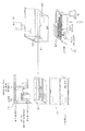
도 1은 본 발명의 카트리지의 예시적인 실시예를 도시한다.
도 2는 샘플을 수용 및 취급하기 위한 카트리지 내의 예시적인 격리된 챔버들을 도시한다.
도 3은 본 발명에 따른 예시적인 탈응집 공정을 도시한다.
도 4는 본 발명의 카트리지의 추가적 실시예를 도시한다.
도 5는 본 발명의 카트리지 상에 위치될 수 있는 유리 슬라이드 홀더의 예시적인 실시예의 단면도 및 평면도를 도시한다.
도 6은 예시적인 특징부를 도시한 본 발명의 카트리지의 추가적 실시예를 도시한다.
도 7은 카트리지의 분리 용기들 중 몇몇이 서로로부터 분리될 수 있는 본 발명의 카트리지의 실시예의 예시적인 도면을 도시한다.
도 8은 예시적인 특징부를 도시한 본 발명의 카트리지의 추가적 실시예를 도시한다.
도 9는 종래의 병리학 샘플 처리 방법(좌측)과 본 발명의 예시적인 처리 방법(우측)과의 비교를 도시한다.
도 10은 본 발명의 방법이 포르말린 고정 파라핀 포매된 공정보다 큰 RNA 완전성 수(RIN)를 갖는 RNA 샘플을 산출하는 것을 도시한다.
도 11은 종래의 종양 샘플 처리 방법이 바이오마커를 손상시킬 수 있어 이용가능한 세포 정보를 감소시킬 수 있는 것을 도시한 도해를 제공한다.
도 12는 세침 흡인(FNA)을 사용한 추출로부터 시작되는 본 발명의 예시적인 처리 방법을 도시한다.
도 13은 변화하는 세포 수에서 HCT-116 세포의 RIN 스코어를 (a)에, 생성된 RNA의 ㎍을 (b)에, 및 260/280 값을 (c)에 도시한다.
도 14는 변화하는 세포 수에서 MCF-7 세포의 RIN 스코어를 (a)에, 생성된 RNA의 ㎍을 (b)에, 및 260/280 값을 (c)에 도시한다.
도 15는 MCF-7 및 HCT-116 세포의 클러스터 크기에 대한 변화하는 다인/㎠에서의 분산의 영향을 도시한다.
도 16은 MCF-7 및 HCT-116 세포의 세포 생존율에 대한 변화하는 다인/㎠에서의 분산의 영향을 도시한다.
도 17은 MCF-7 세포에서 분산 및 EGF 자극으로부터의 FOS 유도의 변화하는 레벨을 도시한다.
도 18은 HCT-116 세포에서 분산 및 EGF 자극으로부터의 FOS 유도의 변화하는 레벨을 도시한다.
도 19는 본 발명의 방법을 사용한 MCF-7 및 HCT-116 세포에서의 종양 세포 부화를 도시한다.
도 20은 본 방법을 사용하여 준비된 MCF-7 세포 대 3시간 포르말린 고정 절차를 사용하여 준비된 그것에 생세포 프로우브를 사용한 결과를 도시한다.
도 21은 본 방법을 사용하여 준비된 HCT-116 세포 대 3시간 포르말린 고정 절차를 사용하여 준비된 그것에 생세포 프로우브를 사용한 결과를 도시한다.
도 22는 본 명세서에 기재된 탈응집 기술을 사용한 MCF-7 및 HCT-116 세포의 예시적인 분산을 도시한다.1 shows an exemplary embodiment of a cartridge of the present invention.
Figure 2 illustrates exemplary isolated chambers in a cartridge for receiving and handling samples.
Figure 3 illustrates an exemplary deaggregation process in accordance with the present invention.
Figure 4 shows a further embodiment of the cartridge of the present invention.
Figure 5 shows a cross-sectional and plan view of an exemplary embodiment of a glass slide holder that can be placed on a cartridge of the present invention.
Figure 6 illustrates a further embodiment of a cartridge of the present invention showing exemplary features.
Figure 7 shows an exemplary view of an embodiment of a cartridge of the present invention in which some of the separation containers of the cartridge can be separated from each other.
Figure 8 illustrates a further embodiment of a cartridge of the present invention showing exemplary features.
Fig. 9 shows a comparison between a conventional pathological sample processing method (left) and an exemplary processing method of the present invention (right).
Figure 10 shows that the method of the present invention produces an RNA sample having a higher RNA integrity number (RIN) than the formalin-fixed paraffin-embedded process.
Figure 11 provides an illustration showing that a conventional tumor sample processing method can damage the biomarker and thus reduce available cell information.
Figure 12 illustrates an exemplary method of treatment of the present invention, beginning with extraction using fine-needle aspiration (FNA).
Figure 13 shows the RIN score of HCT-116 cells in (a), (b) in μg of generated RNA, and (c) in 260/280 values in varying cell numbers.
Figure 14 shows (a) the RIN score of MCF-7 cells in varying cell numbers, (b) in μg of generated RNA, and (c) 260/280 values.
Figure 15 shows the effect of dispersion at varying dynes / cm < 2 > on cluster size of MCF-7 and HCT-116 cells.
Figure 16 shows the effect of dispersion at varying dynes / cm < 2 > on cell viability of MCF-7 and HCT-116 cells.
Figure 17 shows the varying levels of FOS induction from dispersion and EGF stimulation in MCF-7 cells.
Figure 18 shows the varying levels of FOS induction from dispersion and EGF stimulation in HCT-116 cells.
Figure 19 shows tumor cell incubation in MCF-7 and HCT-116 cells using the method of the present invention.
Figure 20 shows the results of using a viable cell proliferation with MCF-7 cells prepared using this method versus those prepared using a 3 hour formalin fixation procedure.
Figure 21 shows the results of using HCT-116 cells prepared using this method versus the live cell proliferation prepared using the 3 hour formalin fixation procedure.
Figure 22 illustrates an exemplary dispersion of MCF-7 and HCT-116 cells using the de-aggregation technique described herein.
본 명세서에 기재된 본 발명의 실시예는 표적 치료 및 분자 생물학 검사의 발전 및 사용을 촉진시키는 자동화된, 완비된 유체 종양 세포 처리 및 테스트 시스템을 포함하지만 이에 한정되는 것은 아니다. 본 발명의 실시예는 또한 체외에서 생세포에 수행될 수 있는 개선된 병리학적 처리 방법을 비롯한, 시스템을 사용하는 방법을 포함한다. 본 발명은 또한 본 명세서에 기재된 시스템 및 방법과 함께 사용되는 키트에 관한 것이다.Embodiments of the invention described herein include, but are not limited to, automated, complete fluid tumor cell processing and testing systems that facilitate the development and use of targeted therapies and molecular biology tests. Embodiments of the present invention also include methods of using the system, including improved pathological processing methods that can be performed on live cells in vitro. The present invention also relates to kits for use with the systems and methods described herein.
본 발명은 체외에서 고형 세포 검체의 수집, 취급 및 처리를 위한 안전한, 효과적인, 정확한, 정밀한, 재생가능한, 저렴한, 비용 효율적인, 효율적인, 신속한 및 편리한 방법과 "카트리지-기반" 시스템을 제공한다. 이들 방법 및 카트리지는 바이오마커 완전성을 유지시키기 위해 공정 중 샘플의 생존율을 유지시킬 수 있고, 그리고 선택적으로 원 샘플에 존재하지 않는 인단백질 및 RNA와 같은 바이오마커를 체외 자극을 통해 유발시킬 수 있다. 본 발명은 완전한 진단 세포학 실험실 시스템과 생검에 부합하는 제어된 조건에서 완전 통합된 검체 및 정보 관리를 제공하며, 이는 테스트 사이의 변동성을 최소화시키고, 생체오염의 위험성을 최소화시키며, 바이오마커 발현에 대한 샘플 준비 공정 자체의 효과를 최소화시킨다. 본 발명의 실시예들은 종양의 표적 치료를 용이하게 하도록 사용될 수 있고, 선택적으로 세포-계수 기능 및/또는 다른 관계된 분석과 같은 조직 샘플 적합성 평가를 또한 제공한다.The present invention provides a safe, effective, accurate, precise, reproducible, inexpensive, cost effective, efficient, rapid and convenient method and "cartridge-based" system for collection, handling and processing of solid cell samples in vitro. These methods and cartridges can maintain the survival rate of the sample during processing to maintain biomarker integrity and, optionally, can be induced via in vitro stimulation of biomarkers such as phosphorylated proteins and RNA that are not present in the original sample. The present invention provides fully integrated diagnostic and cytometric and information management under controlled conditions consistent with a complete diagnostic cytology laboratory system and biopsy, which minimizes variability between tests, minimizes the risk of bio-contamination, Thus minimizing the effect of the sample preparation process itself. Embodiments of the present invention can be used to facilitate targeted treatment of tumors and optionally also provide tissue sample conformity assessment, such as cell-counting function and / or other related analyzes.
도 9에 도시된 바와 같이, 종래의 세포 처리 기술은 조직 처리 전에 포르말린 고정을 사용할 수 있고, 궁극적으로 세포를 파라핀 내에 포매시킬 수 있다. 이는 많은 잠재적 세포 정보가 도 11에 도시된 바와 같이 "상실"되는 결과를 유발한다. 그러나, 본 명세서에 기재되고 예를 들어 도 9 및 도 12에 도시된 개선된 방법은 생세포의 자동화된 처리, 이들 세포의 자극, 및 그에 따른 후술되는 방법을 사용한 세포의 분석을 가능하게 한다.As shown in Fig. 9, conventional cell processing techniques can use formalin fixation prior to tissue processing, and ultimately, cells can be embedded in paraffin. This results in a number of potential cell information being "lost" as shown in FIG. However, the improved methods described herein and shown, for example, in FIGS. 9 and 12 enable automated treatment of live cells, stimulation of these cells, and thus analysis of cells using the methods described below.
당업자라면 인식할 바와 같이, 이들 새로운 장치, 시스템, 키트 및 방법은 임상 또는 연구 세팅에서 다수의 이점을 제공할 수 있다. 예를 들어, 그것들은 검체를 멀리 떨어진 실험실로 보낼 필요 없이 즉각적인, 환자 근접한 생검 처리를 제공하도록 사용될 수 있다. 그것들은 또한 생검 처리를 비용 효율적인 방식으로 표준화 및 자동화시키도록 사용될 수 있다. 본 발명은 세포에 관해서 현재의 병리학적 과정이 가능하게 하는 것보다 더욱 상세한 분자 정보를 제공할 수 있으며, 이는 선택적으로 새로운 체외 바이오마커 및 진단 테스트를 사용하여, 생검시(예컨대, 암 세포) 세포의 더욱 큰 세분류를 가능하게 한다. 종합하면, 본 발명의 이점은 환자 진료 지점에서의 신속한 진단과 후속하는 보다 효과적인 환자 맞춤형 치료 계획의 생성을 가능하게 한다.As will be appreciated by those skilled in the art, these new devices, systems, kits, and methods can provide a number of advantages in clinical or research settings. For example, they can be used to provide an immediate, patient-proximate biopsy treatment without having to send the sample to a remote lab. They can also be used to standardize and automate biopsy procedures in a cost-effective manner. The present invention is capable of providing more detailed molecular information than does enabling current pathological processes with respect to the cells, which can be used to selectively detect (e.g., Of the present invention. Taken together, the advantages of the present invention enable rapid diagnosis at the point of care and subsequent creation of a more effective patient-specific treatment plan.
아래에 더욱 상세히 기술되는 장치, 시스템, 및 방법을 사용하기 위한 예시적인, 비-제한적인 공정이 도 12에 순서도로 도시된다. 이 공정은 도 12에 도시된 세침 흡인(Fine Needle Aspiration: FNA) 단계(1201)와 같은 응집된 세포의 샘플을 획득함으로써 시작될 수 있다. 샘플은 이어서 본 명세서에 기재된 새로운 기술을 사용하여 탈응집된 다음에, 격리된 챔버 내로 분산된다(1203). 만약 샘플이 관심 대상 세포와 다른 세포의 혼합물을 함유하면, 샘플은 관심 대상이 아닌 세포와 오염물질을 제거함으로써 샘플 내의 관심 대상 세포의 수를 부화시키도록(1205) 선택적으로 정제될 수 있다. 샘플은 이어서 선택적으로 체외에서 자극되거나(1207) 또는 달리 테스트 시약과 혼합될 수 있으며, 이어서 분취물(aliquot)들이 새로운 격리된 챔버 내에 배치된다(1209). 분취물은 또한 선택적으로 수행 중인 분석에 따라 체외에서 자극되거나 또는 달리 테스트 시약과 혼합될 수 있다. 분취물은 이어서 관심 대상 특성에 대해 분석된다. 예를 들어, 현미경 분석을 위해 분취물로부터 슬라이드가 준비될 수 있거나(1211), 또는 분취물은 그것들의 세포가 용해되어 각각 핵산, RNA 및 DNA가 분석될 수 있다(1213, 1215). 분석의 결과는 이어서 예를 들어 그로부터 FNA가 취해졌던 환자에 대한 치료 계획의 세팅과 같은 적절한 조치를 취할 수 있는 연구자 또는 임상의에게 전달된다. 또한, 도 11에 도시된 바와 같이, 본 명세서에 기재된 개선된 방법 및 장치는 연구자 또는 임상의가 종래의 병리학적 세포 처리 기술 중에 "상실"되는 새로운 정보에 접근하도록 할 수 있다.An exemplary, non-limiting process for using the devices, systems, and methods described in more detail below is shown in flow chart in FIG. This process can be started by acquiring a sample of coagulated cells, such as Fine Needle Aspiration (FNA)
Ⅰ. 장치, 시스템, 및 Ⅰ. Device, system, and 키트Kit
A. 장치A. Devices
다른 실시예에서, 본 발명은 예컨대 본 명세서에 기재된 생세포 처리 및/또는 준비 방법에 유용한 장치 또는 플랫폼을 제공한다. 이 장치는 또한 카트리지로 지칭된다. 본 발명의 장치의 몇몇 실시예들이 아래에 더욱 상세히 기재되고 도 1 내지 도 8에 도시된다.In another embodiment, the invention provides a device or platform useful for example in the live cell processing and / or preparation method described herein. This device is also referred to as a cartridge. Several embodiments of the apparatus of the present invention are described in further detail below and shown in Figures 1-8.
그러한 카트리지는 하나 이상의 격리된 챔버를 포함할 수 있다. 격리된 챔버는 임의의 분리 용기, 섹션, 또는 생세포의 샘플 또는 고정된, 처리된, 및/또는 안정화된 세포의 샘플을 유지시킬 수 있는 다른 실용적 홀더이다. 예를 들어, 용어 격리된 챔버는 웰(well), 바이알, 관, 슬라이드(예컨대, 유리), 및 플레이트를 포함하지만 이에 한정되는 것은 아니다.Such cartridges may include one or more isolated chambers. The isolated chambers are any separate containers, sections, or other viable holders that can hold samples of live cells or samples of fixed, processed, and / or stabilized cells. For example, the term isolated chamber includes, but is not limited to, a well, a vial, a tube, a slide (e.g., glass), and a plate.
본 발명의 격리된 챔버 또는 분리 용기는 다음의 기능 중 하나, 일부 또는 전부에 적합하다: (1) 격벽 또는 다른 밀봉된 챔버를 통한 생물학적 검체의 수용; (2) 포함된 및 고정된 주사기 바늘 보관; (3) 격벽을 통한 제거에 이용가능한 액체 시약 보관; (4) 격벽을 통한 폐기물 수용 및 보관; (5) 액체 전단 및 기계적 전단을 통한 샘플 분열; (6) 세포 계수 및 세포 가시화; (7) 비드(bead) 기반 분리; 및 (8) 고형 수지 함유.The isolated chamber or separation vessel of the present invention is suitable for one, some or all of the following functions: (1) acceptance of a biological sample through a bulkhead or other sealed chamber; (2) Storing the contained and fixed syringe needles; (3) storage of liquid reagents available for removal through the bulkhead; (4) acceptance and storage of waste through bulkheads; (5) sample splitting through liquid shear and mechanical shear; (6) cell counting and cell visualization; (7) bead-based separation; And (8) a solid resin content.
각각의 카트리지는 그것의 용도에 따라 하나 이상의 격리된 챔버를 포함할 수 있다. 예를 들어, 카트리지는 1개 내지 약 200개의 격리된 챔버, 약 1개 내지 100개의 격리된 챔버, 또는 약 1개 내지 50개의 격리된 챔버를 구비할 수 있다. 몇몇 실시예들은 약 24개, 약 48개, 또는 약 96개의 격리된 챔버를 구비한다.Each cartridge may include one or more isolated chambers depending on its use. For example, the cartridge may comprise from 1 to about 200 isolated chambers, from about 1 to 100 isolated chambers, or from about 1 to 50 isolated chambers. Some embodiments include about 24, about 48, or about 96 isolated chambers.
몇몇 실시예들에서, 카트리지는 하나 이상의 제1 격리된 챔버와 하나 이상의 제2 격리된 챔버를 구비한다. 카트리지는 세포의 샘플을 유지시키기 위해 제1 격리된 챔버를 구비할 수 있다. 그러한 카트리지는 또한 세포 샘플의 분산된 분취물을 유지시키는 하나 이상의 제2 격리된 챔버를 특징으로 할 수 있다. 몇몇 실시예들에서, 제2 격리된 챔버는 세포의 분산된 분취물이 첨가되기 전에 챔버에 소정량의 테스트 시약을 함유할 수 있다. 도 2는 샘플의 수용 및 취급을 위해 존재할 수 있는 예시적인 격리된 챔버를 도시한다.In some embodiments, the cartridge has one or more first isolated chambers and one or more second isolated chambers. The cartridge may have a first isolated chamber to maintain a sample of cells. Such cartridges may also feature one or more second isolated chambers for maintaining a dispersed aliquot of the cell sample. In some embodiments, the second isolated chamber may contain a predetermined amount of the test reagent in the chamber before the dispersed aliquot of the cells is added. Figure 2 illustrates an exemplary isolated chamber that may be present for acceptance and handling of the sample.
일 실시예에서, 본 발명은 서로로부터 분리될 수 있는 분리 용기(예컨대, 격리된 챔버)를 구비하는 카트리지를 제공한다. 도 7에 도시된 바와 같이, 분리 용기는 분리될 수 있고, 내부의 샘플은 상이한 분석 테스트에 사용될 수 있다. 예를 들어, 하나의 분리 용기는 DNA 분석을 위해 보내어질 수 있고, 다른 것은 RNA 분석을 위해 보내어질 수 있으며, 다른 것은 현미경 분석을 위해 보내어질 수 있고, 다른 것은 면역조직화학적 분석을 위해 보내어질 수 있다. 예를 들어, 카트리지 상의 제2 격리된 챔버는 몇몇 실시예들에서 제1 격리된 챔버로부터 분리될 수 있다.In one embodiment, the present invention provides a cartridge having a separation vessel (e.g., an isolated chamber) that is separable from each other. As shown in FIG. 7, the separation vessel can be separated and the sample in the interior can be used for different analytical tests. For example, one separation vessel can be sent for DNA analysis, the other can be sent for RNA analysis, the other can be sent for microscopy analysis, the other can be sent for immunohistochemical analysis . For example, the second isolated chamber on the cartridge may be separate from the first isolated chamber in some embodiments.
본 발명의 장치는 카트리지의 온도를 원하는 온도로, 예를 들어 약 30℃ 내지 40℃로, 약 36℃ 내지 약 38℃로, 또는 임의의 다른 원하는 온도로 유지시키기 위해 가열 요소를 포함할 수 있다.The apparatus of the present invention may include a heating element to maintain the temperature of the cartridge at a desired temperature, for example, from about 30 캜 to 40 캜, from about 36 캜 to about 38 캜, or any other desired temperature .
본 발명의 장치는 장치 내에서 카트리지 및/또는 세포의 샘플의 공급원을 식별하는 바코드 또는 다른 수단을 포함할 수 있다. 카트리지를 식별하는 바코드 또는 다른 수단은 검체 식별 및/또는 환자 안전을 용이하게 하도록 사용될 수 있다. 그러한 식별 수단은 컴퓨터 기반 데이터 저장 시스템, 자동화된 세포 처리 시스템 및/또는 분석물, 및 개인 휴대 정보 단말기와 상호 작용할 수 있다. 바코드를 구비한 카트리지의 예시적인 실시예가 도 4에 도시된다.The apparatus of the present invention may include barcodes or other means for identifying a source of a cartridge and / or a sample of cells within the apparatus. Barcodes or other means for identifying cartridges may be used to facilitate sample identification and / or patient safety. Such identification means may interact with computer-based data storage systems, automated cell processing systems and / or analytes, and personal digital assistants. An exemplary embodiment of a cartridge with a bar code is shown in FIG.
본 장치의 몇몇 실시예들은 예를 들어 도 2의 F에 도시된 바와 같이 세포 계수 메커니즘을 포함할 수 있다. 이들 메커니즘은 자동화된 시스템 또는 수동 시스템일 수 있다. 예를 들어, 세포 계수 메커니즘은 혈구계 또는 Cellometer®[넥스셀롬 바이오사이언시즈 엘엘씨(Nexcelom Biosciences, LLC)로부터 구입 가능함]와 잠재적 디지털 또는 광학 기반 계수기일 수 있다. 당업자라면 인식할 바와 같이, 다른 세포 계수 방법이 당업계에 잘 알려져 있으며, 본 발명과 함께 또한 사용될 수 있다.Some embodiments of the present apparatus may include a cell counting mechanism, for example, as shown in FIG. 2F. These mechanisms may be automated or passive systems. For example, the cell counting mechanism may be a hemocytometer or a CellID® (available from Nexcelom Biosciences, LLC) and a potential digital or optical based counter. As will be appreciated by those skilled in the art, other cell counting methods are well known in the art and may also be used with the present invention.
본 발명의 임의의 실시예의 카트리지는 수지, 시약, 용매, 및 다른 물질을 함유한 하나 이상의 모듈 또는 격리된 챔버를 구비할 수 있다. 모듈은 특정 목적을 위해 사용되는 단일의 격리된 챔버 또는 일군의 복수의 격리된 챔버일 수 있다. 예를 들어, 다수의 조작(예컨대, 용해 및 염색)이 특정 테스트를 위해 수반되는 경우에, 하나 초과의 격리된 챔버가 그 시험 "모듈"의 일부분으로서 사용될 수 있다. 예를 들어, 하나 이상의 격리된 챔버는 핵산, 단백질, 천연 또는 합성 폴리머 또는 저분자가 부착된 수지를 함유할 수 있다. 하나 이상의 격리된 챔버는 액체, 예컨대 완충액, 분자 생물학 효소, 핵산 또는 단백질을 비롯한 생물학적 분자, 또는 저분자 화학물질을 함유할 수 있다. 하나 이상의 격리된 챔버는 온-카트리지(on-cartridge) 가용화를 위한 건성 시약을 함유할 수 있으며, 예컨대 여기에서 건성 시약은 핵산, 단백질, 천연 또는 합성 폴리머 또는 저분자를 포함할 수 있다.Cartridges in any embodiment of the present invention may include one or more modules or isolated chambers containing resins, reagents, solvents, and other materials. The module may be a single isolated chamber or a group of a plurality of isolated chambers used for a particular purpose. For example, more than one isolated chamber may be used as part of the test "module" if multiple operations (e.g., dissolution and dyeing) are involved for a particular test. For example, one or more isolated chambers may contain nucleic acids, proteins, natural or synthetic polymers, or resins with low molecular weight attached thereto. The one or more isolated chambers may contain liquids, such as buffers, biological molecules, including molecular biological enzymes, nucleic acids or proteins, or low molecular chemicals. The one or more isolated chambers may contain a dry reagent for on-cartridge solubilization, for example, where the dry reagent may comprise nucleic acid, protein, natural or synthetic polymer or low molecular weight.
몇몇 실시예들에서, 카메라 또는 다른 디지털 이미지형성 장치가 카트리지의 일부분이거나 또는 카트리지와 함께 사용될 수 있다. 따라서, 본 발명의 임의의 실시예의 카트리지는 10 내지 500 마이크로리터 체적의 샘플에 대한 세포 가시화 및 디지털 이미지 기반 세포 계수(도 2의 F 참조)를 가능하게 하는 이미지형성 모듈을 포함할 수 있으며, 상기 체적은 정변위에 의해 또는 모세관 작용에 의해 이미지형성 세포 내로 분배된다. 이미지형성 장치는 또한 샘플, 예컨대 단백질 국부화 데이터와 같은 샘플 내의 세포 성분의 사진을 촬영하도록 사용될 수 있다.In some embodiments, a camera or other digital image forming device may be part of a cartridge or used with a cartridge. Thus, the cartridge of any embodiment of the present invention may include an image forming module that enables cell visualization and digital image-based cell count (see F in FIG. 2) for a sample of 10 to 500 microliter volume, The volume is dispensed into the imaging cells by the positive side or by capillary action. The image forming apparatus can also be used to take a photograph of a cell component in a sample, such as a sample, e.g., protein localization data.
본 발명의 임의의 실시예의 카트리지는 10 내지 1000 마이크로리터 체적을 사용하여 100 내지 800 다인/㎠의 벽면 전단 응력을 생성시키기 위해 10 내지 500 ㎛의 직경을 갖는 마이크로통로를 통해 세포를 통과시키는 유체 모듈을 구비할 수 있다.Cartridges in certain embodiments of the present invention may include a fluid module for passing cells through a micro-passageway having a diameter of 10 to 500 mu m to produce a wall shear stress of 100 to 800 dynes / cm < 2 > using 10 to 1000 microliter volumes, .
본 발명의 임의의 실시예의 카트리지는 면역소실 모듈 또는 격리된 챔버를 구비할 수 있다. 그러한 면역소실 모듈 또는 격리된 챔버는 자기 비드 또는 다른 방법을 사용할 수 있다. 예컨대 노르웨이 오슬로 소재의 다이날 바이오테크(Dynal Biotech)로부터 입수가능한 자기 비드 제품을 참조하라. 이들 실시예는 방법 단락에서 더욱 상세히 기술된다.The cartridge of any embodiment of the present invention may comprise an immunodeficient module or an isolated chamber. Such immunodeficiency modules or isolated chambers may use magnetic beads or other methods. See magnetic bead products available from Dynal Biotech, Oslo, Norway, for example. These embodiments are described in further detail in the Methods section.
본 발명의 임의의 실시예의 카트리지는 예를 들어 세침 흡인 생검, 심부 생검, 타액, 혈액, 정액, 또는 질액과 같은 생물학적 체액 샘플, 유기체로부터 채취된 조직, 또는 세포 배양 샘플일 수 있는 생물학적 검체를 함유하거나 또는 그것을 함유하도록 구성되는 하나 이상의 모듈 또는 격리된 챔버를 구비할 수 있다. 그러한 모듈은 또한 제1 격리된 챔버로 지칭될 수 있고, 예를 들어 도 2의 A 및 도 4에 도시된다.Cartridges of any embodiment of the present invention may contain a biological sample that may be, for example, a fine needle aspiration biopsy, a deep biopsy, a biological fluid sample such as saliva, blood, semen, or vaginal fluid, tissue harvested from an organism, Or contain one or more modules or isolated chambers configured to contain it. Such a module may also be referred to as a first isolated chamber and is shown, for example, in FIG. 2A and FIG.
다른 실시예에서, 본 발명은 예컨대 본 명세서에 기재된 방법 및 장치에 사용하기에 적합한 카트리지, 예를 들어 개별적인 및 완비된 모듈들을 구비하는 카트리지로서, 이 모듈들은 세포 취급에 적합한 배지를 함유하고 각각 격벽 또는 다른 밀봉 메커니즘에 의해 밀봉되며, 상기 격벽 또는 밀봉 메커니즘은 관, 예컨대 바늘에 의해 우회 또는 천공될 수 있고 관의 제거시 재밀봉될 수 있는 카트리지를 제공한다. 몇몇 실시예들에서는, 분석 장치가 분석 중 생체유해 오염물질을 제공하는 경우에, 카트리지 또는 카트리지를 둘러싸는 분석 장치 내외로의 모든 액체 시약, 생물학적 테스트 검체, 날카로운 물체(즉, 바늘) 등의 통합된 전달 및 제거를 가능하게 하기에 충분한 모듈 또는 격리된 챔버가 있다. 모듈은 세포 계수 또는 생존율 분석과 같은 특정 테스트 또는 활동을 수행하기 위한 관련 시약 및 장치와 함께 다수의 격리된 챔버를 포함할 수 있다. 위에 기재된 실시예들은 예를 들어 도 1, 도 2, 도 4, 도 6, 도 7, 및 도 8에 도시된다.In another embodiment, the present invention is directed to a cartridge suitable for use in, for example, the methods and apparatus described herein, such as cartridges having individual and complete modules, which contain media suitable for cell handling, Or other sealing mechanism, wherein the septum or sealing mechanism can be bypassed or perforated by a tube, e.g., a needle, and can be resealed upon removal of the tube. In some embodiments, integration of all liquid reagents, biological test specimens, sharp objects (i. E., Needles) into and out of the analyzing device surrounding the cartridge or cartridge, when the analyzing device provides biohazardous contaminants during analysis There is a sufficient number of modules or isolated chambers to enable transfer and removal. The module may include a plurality of isolated chambers with associated reagents and devices for performing specific tests or activities, such as cell counting or survival analysis. The embodiments described above are shown, for example, in Figures 1, 2, 4, 6, 7, and 8.
도 5의 501B에 도시된 바와 같이, 본 발명의 카트리지는 예를 들어 계단형 구성(501A)으로 적층되는 슬라이드를 구비할 수 있고, 이 계단형 구성은 슬라이드가 세포 물질을 수용하도록 효율적으로 사용되도록 하고 이어서 수동 또는 로봇식 수단에 의해 하나씩 접근 및 분리되도록 한다. 도 5는 계단형 구성(501A/B)을 도시한 단면도를 도시한다. 따라서, 다른 실시예에서, 본 발명은 계단형 구성으로 배치된 다수의 평면형 기질, 예컨대 유리 슬라이드를 포함한 홀더를 포함하는 장치를 제공하며, 이 홀더는 기질의 측방향 및 수직 움직임을 제한한다. 적합한 기질은 유리 슬라이드와 같은 평탄한 직사각형 단품, 금속 플레이트, 또는 마이크로유체 장치를 포함하지만, 이에 한정되는 것은 아니다. 기질은 0.1 내지 3 mm의 두께 및/또는 2-3 cm 폭 × 7-9 cm 길이의 치수를 가질 수 있다. 당업자라면 인식할 바와 같이, 다른 기질 또는 격리된 챔버가 카트리지 상에 유사한 방식으로 배치될 수 있고, 그러한 실시예들이 본 명세서에 포함된다.As shown in 501B of FIG. 5, the cartridge of the present invention may include, for example, a slide that is stacked in a stepped
몇몇 실시예들에서, 카트리지 상의 홀더는 하단 부분 및 리드 부분을 구비하며, 이때 각각의 부분은 홀더 내에서의 또는 홀더에 대한 개별 기질의 계단형 스택의 위치설정 및 움직임 방지를 위한 단차부(stair step)를 포함한다. 홀더 내에 보관될 때, 기질은 최-하단 부분이 스택을 넘어 돌출되는 원위 단부를 구비할 수 있다(도 5의 501B 참조). 이 원위 단부는 하나 이상의 인쇄된 특징부, 웰 또는 함몰부를 구비할 수 있다. 이들 인쇄된 특징부는 기질이 사용된 후에 또는 사용 전에 기질을 식별하기 위한 라벨일 수 있다. 몇몇 실시예들에서, 홀더는 리드를 구비하고, 기질의 원위 단부는 리드의 격벽 또는 격벽들을 통해 접근하기 위해 노출된다. 홀더는 프레임, 예를 들어 SBS-적합 풋프린트(SBS-compatible footprint)를 갖는 프레임 내에 장착 및 탈착될 수 있다. 선택적으로, 홀더는 하단에 작은 홀 또는 복수의 홀들을 구비할 수 있다.In some embodiments, the holder on the cartridge has a lower portion and a lid portion, wherein each portion includes a stair for positioning and movement prevention of a stepped stack of individual substrates in or on the holder step. When stored in the holder, the substrate may have a distal end with the lowermost portion protruding beyond the stack (see 501B in FIG. 5). The distal end may have one or more printed features, wells, or depressions. These printed features may be labels after the substrate is used or to identify the substrate before use. In some embodiments, the holder has a lead, and the distal end of the substrate is exposed to access through the septum or septum of the lead. The holder may be mounted and detached in a frame, for example a frame with an SBS-compatible footprint. Alternatively, the holder may have a small hole or a plurality of holes at the bottom.
도 5는 예시적인 홀더의 평면도를 도시한다. 슬라이드의 스택은 시스템을 자동화된 또는 수동 사용에 이상적이도록 만드는 표준화된 열 및 컬럼 간격에 따라 위로부터 어드레스될 수 있다. 홀더는 각각의 기질 위의 위치에 독립적으로 어드레스하기 위해 각각의 기질 위에 격벽을 구비한 리드를 구비할 수 있다. 예를 들어, 홀더는 각각의 기질의 각각의 어드레스가능 위치 위에 O-링 또는 가스켓 밀봉가능 챔버를 구비할 수 있다. 이들 O-링 또는 가스켓 밀봉가능 챔버는 원하는 임의의 형상, 예를 들어 원형 또는 직사각형 또는 정사각형일 수 있다.Figure 5 shows a top view of an exemplary holder. The stack of slides can be addressed from above depending on the standardized column and column spacing that makes the system ideal for automated or manual use. The holder may have a lid having a barrier on each substrate for independent addressing at a location on each substrate. For example, the holder may have an O-ring or gasket sealable chamber on each addressable location of each substrate. These O-rings or gasket sealable chambers may be of any desired shape, for example circular or rectangular or square.
홀더 및 기질을 소정의 방식으로 위치시키는 것은 피펫, 주사기 바늘, 또는 핀툴(pintool)의 사용을 통한 1 나노리터 내지 0.5 밀리리터 범위의 체적의 유체 샘플의 기질로의 다중 전달을 가능하게 할 수 있다. 몇몇 실시예들에서, 홀더는 유체 전달 중 기질의 측방향 또는 수직 변위를 제한하고, 유체 전달 중 기질의 움직임을 제한한다. 다른 실시예들에서, 홀더는 기질의 수직 회전을 가능하게 하지만, 기질의 측방향 움직임을 제한한다. 이 유연성은 다양한 방법 및 시스템을 사용하여 각각의 격리된 챔버로의 자동화된 또는 수동 다중 유체 전달과 그것으로부터의 제거를 가능하게 할 수 있다.Positioning the holder and substrate in a predetermined manner may enable multiple delivery of the fluid sample to the substrate in a volume ranging from 1 nanometer to 0.5 milliliter through the use of a pipette, syringe needle, or pintool. In some embodiments, the holder limits the lateral or vertical displacement of the substrate during fluid transfer and limits the movement of the substrate during fluid transfer. In other embodiments, the holder allows vertical rotation of the substrate, but limits lateral movement of the substrate. This flexibility can enable automated or manual multiple fluid delivery to and removal from each isolated chamber using various methods and systems.
몇몇 실시예들에서, 홀더는 리브 부분이 제거된 상태로 분해될 수 있고, 하단 부분은 홀더의 하단 부분 내의 홀을 통해 돌출부를 위치시키는 스탠드 상에 장착될 수 있다. 기질은 사람 손 또는 로봇 파지 도구에 의한 순차적 파지를 위해 스탠드 돌출부 상에 있는 상태에서 특유하게 제공될 수 있다. 이는 각각의 샘플 상에서 침착된 샘플 위로의 기질의 스크레이핑(scraping)을 방지하여 슬라이드-기반 세포 물질의 안정된 수송을 가능하게 하기 위해, 예컨대 첫 번째로 하단 슬라이드를 분리시키고 마지막으로 상단 슬라이드를 분리시키는 것과 같은 기질의 순차적 분리를 가능하게 한다. 위에 기재된 홀더의 실시예들을 갖는 카트리지의 예시적인 분해도가 도 6 및 도 8에 개시된다.In some embodiments, the holder may be disassembled with the rib portion removed, and the lower end portion may be mounted on a stand that positions the protrusion through a hole in the lower end portion of the holder. The substrate may be provided peculiarly on the stand protrusion for sequential grasping by a human hand or a robotic gripping tool. This prevents scraping of the substrate onto the sample deposited on each sample, so as to enable stable transport of the slide-based cellular material, for example, to separate the first lower slide and finally to separate the upper slide Thereby allowing for sequential separation of the substrate. An exemplary exploded view of a cartridge having embodiments of the holder described above is disclosed in Figs. 6 and 8. Fig.
유체 샘플은 자동화된 시스템으로 또는 수동으로 기질로 전달될 수 있다. 몇몇 실시예들에서, 유체 샘플은 소정의 순서로 기질로 순차적으로 전달된다. 적합한 유체 샘플은 용액, 에멀젼, 현탁액, 또는 폴리머-함유 혼합물을 포함하지만, 이에 한정되는 것은 아니다. 예를 들어, 유체 샘플은 생물학적 또는 화학적 물질, 예컨대 유기 또는 무기 저분자, 단백질, 핵산, 세포, 입자, 휘발성 및 비-휘발성 용매, 폴리머, 또는 고착제일 수 있지만, 이에 한정되는 것은 아니다.Fluid samples can be delivered to an automated system or manually to the substrate. In some embodiments, the fluid samples are sequentially delivered to the substrate in a predetermined order. Suitable fluid samples include, but are not limited to, solutions, emulsions, suspensions, or polymer-containing mixtures. For example, the fluid sample may be a biological or chemical material such as organic or inorganic low molecular weight, protein, nucleic acid, cell, particle, volatile and non-volatile solvent, polymer, or fixative, but is not limited thereto.
다른 실시예들에서, 본 발명은 본 발명의 카트리지에 유용한 웰 플레이트를 또한 제공한다. 현재, 플레이트가 비스듬히 또는 거꾸로 유지되는 경우에 구성요소(개방된 관, 밀봉된 관, 주사기, 피펫 팁 등)가 제 위치를 벗어나지 않도록 구성요소가 제 위치에 로킹되게 하는 웰 플레이트 기술에 대한 필요성이 존재한다. 또한, 웰 플레이트 기술은 심지어 피펫 도구 상에 팁 이젝터도 필요로 하지 않고서, 속박된(locked-in) 구성요소가 표준[직교(Cartesian)] 피펫 도구에 의해 분리되어 사용된 다음에 속박된 위치로 복귀되도록 한다. 몇몇 실시예들에서, 본 발명은 구성요소의 측방향 통과를 가능하게 하는 개재 공간에 의해 웰 플레이트 상의 두 인접한 위치("이중 웰"로 불리움)가 연결되도록 제조되는 다수의 웰을 구비한 평면형 표면을 포함하는 웰 플레이트에 관한 것이다.In other embodiments, the present invention also provides a well plate useful in a cartridge of the present invention. Presently, there is a need for a well plate technique that allows components to be locked in position so that the components (open tubes, sealed tubes, syringes, pipette tips, etc.) do not deviate from their positions when the plates are held upside down or upside down exist. In addition, the well plate technique can be used to position the locked-in component in a constrained position after being used separately by a standard (Cartesian) pipette tool, without requiring a tip ejector on the pipette tool To be returned. In some embodiments, the present invention includes a planar surface having a plurality of wells that are fabricated to connect two adjacent locations (referred to as "double wells") on the well plate by intervening spaces that allow lateral passage of the components To the well plate.
구성요소는 개방된 관, 밀봉된 관, 격벽을 구비한 관, 투명한 관, 임의의 컨테이너, 주사기, 바늘, 블레이드, 또는 피펫 팁일 수 있다. 몇몇 실시예들에서, 구성요소는 로킹 메커니즘을 통과하기에 적합한 두께의 정사각형, 원형, 또는 다른 형상의 강성 재료 단품을 포함하고, 속박(lock-in) 웰의 로킹 메커니즘의 형상에 합치된다.The component can be an open tube, a sealed tube, a tube with a partition, a transparent tube, an optional container, a syringe, a needle, a blade, or a pipette tip. In some embodiments, the component includes a rigid material piece of square, circular, or other shape in thickness suitable for passing through the locking mechanism and conforms to the shape of the locking mechanism of the lock-in well.
이중-웰 플레이트의 몇몇 실시예들에서, 제1 인접 위치의 이중 웰("속박 웰"로 불리움)은, 구성요소가 제 위치에 로킹되도록 하고 움직임, 특히 플레이트가 상향을 향하고 있을 때 수직 방향으로의 구성요소의 후퇴를 저지하는 작은 탭을 포함한다. 이들 이중-웰 플레이트는 또한 구성요소가 속박 웰 내로 로킹될 때 부분적으로 압축되는 유연성 가스켓을 포함한 속박 웰을 구비할 수 있으며, 이 가스켓은 속박된 구성요소의 움직임을 방지한다.In some embodiments of the double-well plate, the double well (referred to as the "constraining well") at the first adjacent location is configured to allow the component to be locked in place and to move, And a small tab for preventing the retraction of the component of the device. These double-well plates may also have a constriction well that includes a compliant gasket that is partially compressed when the component is locked into the captive well, which prevents movement of the constrained component.
몇몇 실시예들에서, 구성요소는 도구(예컨대, 피펫터 또는 주사기)에 의해 어드레스가능한 "피팅"을 포함하며, 이 피팅[예컨대, 루어 록(luer lock)]은 도구와의 꼭 맞는 기밀 체결을 가능하게 한다. 도구는 속박 웰 내로 로킹되는 구성요소를 장착할 수 있고, 가스켓을 더욱 압축시키도록 구성요소를 내리누를 수 있어, 구성요소가 제1 위치의 이중 웰(속박 웰)로부터 "해제(release) 웰"로 불리우는 제2 위치로 이중 웰의 개재 공간 내에서의 측방향 통과를 가능하게 하는 위치에 존재하도록 한다. 몇몇 실시예들에서, 구성요소는 도구에 의해 수직으로 이동될 때 해제 웰 내외로 자유로이 이동할 수 있다. 도구는 또한 구성요소를 완전히 속박 웰 내로 전달할 수 있고, 웰 플레이트가 표면에 고정될 때, 도구는 웰 플레이트로부터 멀어지게 수직으로 이동할 수 있어, 구성요소가 속박 웰 내에 로킹된 상태에서 구성요소를 탈착시킬 수 있다.In some embodiments, the component includes a "fitting" that is addressable by a tool (e.g., a pipettor or a syringe) that fits tightly with the tool, such as a luer lock . The tool can mount a component that is locked into the captive well and can depress the component to further compress the gasket such that the component is released from the dual well in the first position (release well) To a second position, referred to as " second location ", in the interstitial space of the double well. In some embodiments, the component can move freely into and out of the release well when vertically moved by the tool. The tool can also completely transfer the component into the containment well and when the well plate is secured to the surface the tool can move vertically away from the well plate so that the component can be detached .
도구 및 플레이트는, 직교 좌표(x, y, z)에서의 도구 및/또는 플레이트의 움직임을 가능하게 하여, 도구가 구성요소를 속박 웰로 전달한 다음에 구성요소를 속박 웰 내에 남겨두도록 하고, 구성요소를 속박 웰 내에 장착하여 그것들을 해제 웰로 이동시키도록 하며, 구성요소를 해제 웰로부터 분리하여 그것들을 웰 플레이트로부터 자유롭게 하는 컴퓨터 제어식 갠트리(gantry) 시스템 상에 각각 장착될 수 있다. 그러한 셋업은 또한 도구가 구성요소를 해제 웰로 전달하여 구성요소를 팁 이젝터에 의해 해제 웰 내에 남겨두도록 할 수 있다.The tool and plate allow movement of the tool and / or plate in the Cartesian coordinates (x, y, z) such that the tool transfers the component to the constraint well and then leaves the component in the constriction well, May be mounted on a computer-controlled gantry system, respectively, that mounts them in the constriction wells and moves them to the release wells, separating the components from the release wells and freeing them from the well plate. Such setup may also allow the tool to transfer the component to the release well to leave the component in the release well by the tip ejector.
도구 및 플레이트를 사용한 몇몇 컴퓨터-제어식 시스템은 각도 운동(세타-축)의 사용을 필요로 하지 않도록 설정될 수 있어, 구성요소가 웰 플레이트 내에 있거나 웰 플레이트로부터 분리된 상태에서 웰 플레이트에 대한 구성요소의 위치 회전 재배향을 방지한다.Some computer-controlled systems using tools and plates can be set to not require the use of angular motion (theta-axes), so that components can be placed in or out of the well plate, To prevent rotation of the position.
몇몇 실시예들에서, 이중-웰은 유체가 샘플로부터 오염물질을 제거시키도록 구성되었던 소정의 경로를 통해 유동하게 하도록 사용될 수 있다. 그러한 이중 웰 시스템은 예를 들어 도 2에 웰(C, D)로 도시된다.In some embodiments, the double-well can be used to cause the fluid to flow through a predetermined path that has been configured to remove contaminants from the sample. Such a double well system is shown, for example, in FIG. 2 as wells (C, D).
B. 시스템B. System
본 발명은 또한 본 명세서에 기재된 카트리지 및 방법을 사용하는 시스템에 관한 것이다. 예를 들어, 시스템은 본 발명의 카트리지 중 하나 이상을 개별적으로 또는 키트의 일부분으로서 포함할 수 있고, 적어도 하나의 분석 결과를 얻기 위해 카트리지와 상호작용할 수 있는 분석 장치(본 명세서에서 또한 기구로도 지칭됨)를 포함할 수 있다. 예를 들어, 시스템은 환자에 대한 의료 진단을 얻기 위해 환자 진료 지점에 사용될 수 있다.The present invention also relates to a system using the cartridges and methods described herein. For example, a system may include one or more of the cartridges of the present invention, either individually or as part of a kit, and may include an analyzing device (also referred to herein as "Quot;). For example, the system may be used at a patient care point to obtain a medical diagnosis of the patient.
따라서, 본 발명의 몇몇 실시예들은 개별 모듈(예컨대, 격리된 분리 용기)을 포함하는 카트리지에 작업을 수행하기 위한 분석 장치에 관한 것이며, 이 장치는 하나 이상의 주사기를 위한 하나 이상의 홀더를 포함하고, 주사기는 중공 바늘을 구비하고, 흡인력을 인가하고 내용물을 카트리지의 모듈로 분배하기 위해 바늘이 컴퓨터 제어식 동작 제어에 의해 직교 좌표계(x, y, z)를 사용하여 카트리지의 개별 모듈로 지향될 수 있도록, 카트리지가 위치되는 플랫폼 위로 배향되며, 모듈은, 바늘에 의해 관통될 수 있지만 모듈의 내용물을 환경으로부터 달리 격리시키는 격벽을 각각 구비한다. 이들 카트리지의 몇몇 실시예들의 예가 도 1, 도 4 및 도 8에 도시된다. 몇몇 실시예들에서, 홀더는 카트리지에 의해 제공되는 도구 또는 주사기를 픽업할 수 있다.Accordingly, some embodiments of the present invention are directed to an analytical apparatus for performing an operation on a cartridge comprising an individual module (e.g., an isolated separation vessel), the apparatus comprising one or more holders for one or more syringes, The syringe includes a hollow needle and is configured to direct the needle to an individual module of the cartridge using the Cartesian coordinate system (x, y, z) by computer controlled actuation to dispense the suction force and dispense the contents to the module of the cartridge. , The module being oriented over the platform on which the cartridge is located, each module having a partition wall that can be penetrated by the needle but otherwise isolates the contents of the module from the environment. Examples of some embodiments of these cartridges are shown in Figs. 1, 4 and 8. Fig. In some embodiments, the holder may pick up a tool or syringe provided by the cartridge.
분석 장치는 본 발명의 카트리지를 수용하는 수단을 구비할 수 있다. 예를 들어, 슬롯, 개구, 홀, 또는 다른 영역이 카트리지를 수용할 수 있다. 일단 카트리지가 수용 수단 내에 배치되면, 몇몇 실시예들에서, 도어가 수용 수단을 밀봉하도록 밀폐되어, 카트리지를 분석 장치 내에 완전히 봉입시킨다. 이 도어 밀폐는 수동으로 수행될 수 있거나 자동일 수 있다. 이들 완전히 둘러싸인 변형예는 잠재적 생체유해물질이 실험실 또는 임상 환경 내로 누출되거나 달리 방출되지 못하도록 하는데 특히 유용할 수 있다.The analyzer may comprise means for receiving the cartridge of the present invention. For example, slots, openings, holes, or other areas can accommodate cartridges. Once the cartridge is positioned within the receiving means, in some embodiments, the door is sealed to seal the receiving means so that the cartridge is completely sealed in the analyzing apparatus. This door sealing can be done manually or automatically. These fully enclosed variations may be particularly useful in preventing potential biohazardous materials from leaking into, or otherwise releasing into, the laboratory or clinical environment.
몇몇 실시예들에서, 분석 장치 및 카트리지를 사용하기 위한 시스템은 진공 설비; 공기 보급 설비; 천연 가스 설비를 필요로 하지 않거나 사용하지 않으며; 모든 시약 라인(공정 유체, 분석 시약, 및 폐기물 스트림) 및 부착된 병을 구비하지 않을 수 있다. 즉, 장치는 완전히 완비된다.In some embodiments, the system for using the analyzer and the cartridge includes a vacuum system; Air supply equipment; Does not require or use natural gas facilities; All reagent lines (process fluid, analytical reagent, and waste stream) and attached bottles may not be present. That is, the device is completely complete.
그러한 시스템에 사용되거나 또는 독립적으로 사용되는 카트리지도 또한 완비될 수 있다. 이는 카트리지가 원하는 분석을 수동으로 또는 자동화된 시스템을 사용하여 수행하는데 필요로 하는 모든 필요한 도구(예컨대, 피펫 팁) 및 시약(예컨대, 테스트 시약, 염료, 또는 다른 화합물)으로 사전-적재됨을 의미한다. 예를 들어, 카트리지는 암 치료제 패널(예컨대, 플레이트 내의 웰과 같은 격리된 챔버 내에 각각 하나)과, 샘플을 탈응집시켜 이들 웰 내로 분산시키는데 필요한 모든 도구로 사전-적재될 수 있다.Cartridges used in such systems or used independently may also be complete. This means that the cartridge is pre-loaded with all the necessary tools (e.g., pipette tips) and reagents (e.g., test reagents, dyes, or other compounds) needed to perform the desired analysis either manually or using an automated system . For example, a cartridge can be pre-loaded with a cancer therapeutic agent panel (e.g., one each in an isolated chamber such as a well in a plate) and all the tools necessary to deaggregate and disperse the sample into these wells.
본 발명의 시스템은 카트리지의 모듈을 어드레스할 내부 이미지형성 능력을 포함할 수 있다. 이 시스템은 데이터를 생성하도록 사용될 수 있으며, 이 데이터는 이어서 사용자 또는 다른 장치로 출력된다.The system of the present invention may include an internal image forming capability to address a module of a cartridge. The system may be used to generate data, which is then output to a user or other device.
분석 장치는 또한 샘플에 테스트가 실시되는 상태에서 세포 생존율을 유지시키기 위해 열 배양에 필요한 구성요소를 포함할 수 있다. 그러한 분석 장치의 배양 부분은 하나 이상의 카트리지를 선택적으로 소정의 순서로(예컨대, 샘플 추출 시간을 기초로 시간순으로) 유지시킬 수 있다.The analyzer may also include components necessary for thermal culture to maintain cell viability under test in the sample. The incubation portion of such an analyzer can selectively maintain one or more cartridges in a predetermined order (e.g., chronologically based on sample extraction time).
전술한 장치 중 임의의 것은 또한 샘플 식별자를 카트리지의 모듈에 부착시켜 샘플 정보 및 공정 정보를 통신 라인을 통해 다른 장치로 전송할 수 있거나, 또는 사용자 검사 결과를 디스플레이할 수 있다.Any of the devices described above may also attach a sample identifier to a module of the cartridge to transmit sample information and process information to another device via a communication line or to display a user test result.
C. C. 키트Kit
본 발명은 또한 본 발명의 장치를 포함하는 키트에 관한 것이다. 예시적인 키트는 컨테이너 내에 패키지되는 본 명세서에 기재된 하나 이상의 카트리지를 포함한다. 키트는 또한 인쇄된 사용 설명서, 시약 및 완충제, 분자 프로우브(molecular probe), 아래에서 논의되는 바와 같은 하나 이상의 테스트 시약, 피펫 또는 바늘과 같은 탈응집 또는 분배 도구, 및 후술되는 방법을 수행하는데 유용한 다른 물품을 포함할 수 있다.The present invention also relates to a kit comprising the device of the invention. An exemplary kit includes one or more cartridges described herein packaged in a container. The kits may also be used to perform printed manuals, reagents and buffers, molecular probes, one or more test reagents as discussed below, de-aggregating or dispensing tools such as pipettes or needles, Other items may be included.
몇몇 실시예들에서, 이들 키트는 당업계에 알려진 방법을 사용하여 멸균될 수 있고, 멸균상태를 유지시키는 방식으로 패키지될 수 있다. 키트는 개별 키트로서 또는 멀티팩으로 판매될 수 있다. 키트는 또한 개봉 방지(tamper resistant)식으로 구성될 수 있거나, 또는 개봉 여부를 지시하도록 구성될 수 있다.In some embodiments, these kits can be sterilized using methods known in the art and can be packaged in a manner that maintains sterility. The kits may be sold as individual kits or in multi-packs. The kit may also be constructed in a tamper resistant manner, or it may be configured to indicate whether to open or not.
키트는 자동화된 기구의 사용에 의하기보다는 수동으로 사용될 수 있는 본 명세서에 기재된 바와 같은 분석용 카트리지를 포함할 수 있다. 그러한 경우에, 테스트 또는 카트리지의 목적을 수행하는데 필요로 하는 시약 및 장비는 키트의 일부분으로서 제공될 수 있다. 장비, 예컨대 주사기 바늘, 피펫 등은 카트리지 상에 사전적재될 수 있거나, 또는 카트리지의 외부에 존재하여 키트의 일부분으로서 제공될 수 있다. 다른 실시예들에서, 장비는 사용자에 의해 공급될 수 있고, 키트의 일부분이 아닐 수 있다. 유사하게, 키트와 함께 사용되는 테스트 시약은 카트리지의 특정한 격리된 챔버 내에 사전적재될 수 있거나, 또는 사용자에 의한 응용을 위해 카트리지 외부에 패키지될 수 있다. 이들 실시예에 따른 키트는 예를 들어 단일 테스트용 단일 카트리지, 다중 테스트용으로 적재 또는 준비된 단일 카트리지, 또는 다중 테스트용 다중 카트리지로서 패키지될 수 있다.The kit may include an assay cartridge as described herein which may be used manually rather than by use of an automated instrument. In such a case, the reagents and equipment needed to carry out the test or the purpose of the cartridge may be provided as part of the kit. Equipment, such as a syringe needle, pipette, etc., may be preloaded on the cartridge, or may be present on the exterior of the cartridge and provided as part of the kit. In other embodiments, the equipment may be supplied by the user and may not be part of the kit. Similarly, the test reagent used with the kit may be preloaded in a particular isolated chamber of the cartridge, or it may be packaged outside the cartridge for application by the user. The kit according to these embodiments can be packaged, for example, as a single cartridge for a single test, a single cartridge loaded or prepared for multiple tests, or multiple cartridges for multiple testing.
선택적으로, 키트는 또한 본 명세서에 기재된 것들과 같은 분석 또는 방법의 일부분으로서, 카트리지를 정확하게 사용하기 위한 사용법과 다른 필요한 물품, 예컨대 시약을 포함한다. 예를 들어, 키트는 안내문 또는 인쇄된 사용 설명서를 포함할 수 있다. 그러한 인쇄된 사용 설명서는 의약품 또는 생물학적 제품의 제조, 사용, 또는 판매를 규제하는 정부 기관에 의해 규정된 형태일 수 있으며, 안내문은 본 명세서에 기재된 분석, 방법, 및 장치로부터 도출된 정보를 사용하여 치료될 수 있는 건강상태를 진단 또는 치료하기 위한 인체 투여에 대해 제조, 사용, 또는 판매의 기관에 의한 승인을 반영한다. 몇몇 실시예들에서, 키트는 예컨대 세포를 처리하기 위한 키트의 사용에 대한 정보를 제공하는 인쇄물(printed matter), 또는 예컨대 세포를 처리하기 위한 키트의 사용에 대한 정보를 제공하는 사전-기록된(pre-recorded) 매체 장치를 또한 포함한다.Optionally, the kit also includes, as part of an assay or method such as those described herein, a necessary article, such as a reagent, for use precisely to use the cartridge. For example, the kit may include an instructional message or printed instruction manual. Such printed instructions may be in the form prescribed by a governmental agency regulating the manufacture, use, or sale of a medicinal or biological product, and the instructions may be provided using information derived from the analyzes, methods and apparatus described herein Reflects approval by the agency of manufacture, use, or sale for human administration to diagnose or treat a health condition that may be treated. In some embodiments, the kit includes a printed matter that provides information about the use of the kit, for example, to treat the cell, or a pre-recorded (e.g., pre-recorded media device.
"인쇄물"은 예를 들어 책, 작은 책, 브로셔 또는 낱장 인쇄물일 수 있다. 인쇄물은 실험 분석 및/또는 본 방법에 따라 세포를 처리하기 위한 프로토콜을 기술할 수 있다. 가능한 포맷은 단계별 사용 설명서, 불릿 포인트(bullet point) 리스트, 자주 묻는 질문(FAQ)의 리스트 또는 차트를 포함하지만, 이에 한정되는 것은 아니다. 또한, 제공될 정보는 그림, 그래픽, 또는 다른 기호를 사용하여 비-문자 표현으로 도해될 수 있다.The "print" may be, for example, a book, a small book, a brochure, or a sheet of paper. The printed matter may describe a protocol for experimental analysis and / or for treating cells according to the present method. Possible formats include, but are not limited to, step-by-step instructions, a bullet point list, a list of frequently asked questions (FAQ), or charts. In addition, the information to be provided may be illustrated as a non-textual representation using pictures, graphics, or other symbols.
"사전-기록된 매체 장치"는 예를 들어 비디오테이프 카세트, DVD(디지털 비디오 디스크), 필름스트립, 35 mm 영화와 같은 시각 매체 장치, 또는 임의의 다른 시각 매체 장치일 수 있다. 대안적으로, 사전-기록된 매체 장치는 CD-ROM(콤팩트 디스크-판독 전용 메모리) 또는 플로피 디스크와 같은 쌍방향 소프트웨어 응용프로그램일 수 있다. 대안적으로, 사전-기록된 매체 장치는 예를 들어 레코드, 오디오카세트, 또는 오디오 콤팩트 디스크와 같은 오디오 매체 장치일 수 있다. 사전-기록된 매체 장치에 수용된 정보는 실험 분석 및/또는 본 방법에 따라 세포를 처리하기 위한 프로토콜을 설명할 수 있다.A "pre-recorded media device" may be, for example, a video tape cassette, a DVD (digital video disc), a film strip, an optical media device such as a 35 mm film, or any other optical media device. Alternatively, the pre-recorded media device may be a bi-directional software application such as a CD-ROM (compact disk-read only memory) or a floppy disk. Alternatively, the pre-recorded media device may be an audio media device such as, for example, a record, audio cassette, or audio compact disc. The information received in the pre-recorded media device may describe a protocol for experimental analysis and / or for treating cells according to the present method.
Ⅱ. 방법Ⅱ. Way
본 발명은 임상 또는 연구 맥락에서 사용될 수 있는, 세포 샘플, 특히 응집된 세포 또는 고형 종양을 처리하기 위한 새로운 방법에 관한 것이다. 이들 방법은, 대상물로부터 얻은 생 암세포를 함유한 수용액을 탈응집시켜 제1 격리된 챔버 내에 적어도 하나의 테스트 분취물로 분산시키는 단계; 오염 세포를 제거시킴으로써 다른 오염 세포 유형에 대한 표적 세포의 백분율을 증가시키기 위해 선택적으로 샘플을 정제 또는 조작하는 단계; 정제된 생 암세포를 분석, 조작, 또는 자극을 위해 하나 이상의 제2 격리된 챔버 내로 분배하는 단계; 및 분배된 세포의 세포 및/또는 분자 분석을 가능하게 하기 위해 분배된 생세포를 안정화시키는 단계를 포함한다. 몇몇 실시예들에서, 안정화된 및 분배된 세포는 원하는 결과 및 관심 있는 세포 분석에 따라 생세포 또는 사멸 세포(dead cell)일 수 있다.The present invention relates to a novel method for treating cell samples, in particular aggregated cells or solid tumors, which can be used in clinical or research contexts. These methods comprise: deaggregating an aqueous solution containing live cancer cells obtained from an object and dispersing it into at least one test fraction in a first isolated chamber; Selectively purifying or manipulating the sample to increase the percentage of target cells for other contaminating cell types by removing the contaminating cells; Dispensing the purified live cancer cells into one or more second isolated chambers for analysis, manipulation, or stimulation; And stabilizing the dispensed viable cells to enable cell and / or molecular analysis of the dispensed cells. In some embodiments, the stabilized and dispensed cells can be live cells or dead cells, depending on the desired outcome and the cell analysis of interest.
본 발명의 다른 방법은 고형 종양으로부터 암세포를 처리 또는 준비하기 위한 방법을 포함하며, 이 방법은, 고형 종양으로부터 얻은 생 암세포를 탈응집시켜 적어도 하나의 제1 격리된 챔버 내에 적어도 하나의 테스트 분취물로 분산시키는 단계; 오염물질을 제거시키기 위해 선택적으로 생 암세포를 정제 또는 조작하는 단계; 정제된 생 암세포를 분석을 위해 하나 이상의 제2 격리된 챔버 내로 분배하는 단계; 및 세포의 세포 및/또는 분자 분석을 가능하게 하기 위해 분배된 세포를 안정화시키는 단계를 포함한다.Another method of the present invention includes a method for treating or preparing cancer cells from solid tumors comprising decongesting live cancer cells from a solid tumor to produce at least one test fraction in at least one first isolated chamber ; Selectively purifying or manipulating a living cancer cell to remove contaminants; Dispensing the purified live cancer cells into one or more second isolated chambers for analysis; And stabilizing the dispensed cells to enable cell and / or molecular analysis of the cells.
본 발명의 방법은 생세포가 신속히 처리되도록 한다. 세포는 최소의 세포 활성화 또는 스트레스(stress)(예컨대, 환경 스트레스, 온도 유발 스트레스, 대사성 스트레스, 또는 화학적 유발 스트레스)를 갖고서 살아 있는 상태에서 처리될 수 있다. 용어 "최소의 세포 활성화 또는 스트레스"는 자극과 비교시 세포 신호전달의 배경 잡음 및 세포 스트레스 경로의 최소의 변화에 의해 정의된다. 예를 들어, 도 17에서 볼 수 있는 바와 같이, 탈응집 및/또는 분산 전과 후의 세포에서의 FOS(또는 c-fos) 유도의 비교는 자극된 샘플(20-30배 증가)과 비교시 FOS 또는 다른 조기 반응 유전자 또는 생물학적 스트레스 지표의 최소의 유도(3-5배)를 보일 수 있다.The method of the present invention allows live cells to be processed quickly. Cells can be treated in a live state with minimal cell activation or stress (e.g., environmental stress, temperature-induced stress, metabolic stress, or chemical-induced stress). The term "minimal cell activation or stress" is defined by minimal changes in background noise and cellular stress pathways of cell signaling in comparison to stimulation. For example, as can be seen in FIG. 17, a comparison of FOS (or c-fos) induction in cells before and after de-aggregation and / (3-5 fold) of other early response genes or biological stress indicators.
본 발명의 방법들의 다른 이점은 그것들이 원 샘플에서 아주 적은 수의 세포를 사용할 수 있다는 것이다. 예를 들어, 몇몇 실시예들에서, 처리된 응집된 세포 또는 고형 종양 세포의 총 수는 약 1×10³ 내지 1×10⁷이다.Another advantage of the methods of the present invention is that they can use a very small number of cells in the original sample. For example, in some embodiments, the total number of treated aggregated cells or solid tumor cells is from about 1 x 10 3 to 1 x 10 7 .
본 발명의 방법은 또한 신속한 샘플 처리를 가능하게 한다. 몇몇 실시예들에서, 분배된 생세포의 안정화는 대상물로부터의 샘플의 획득 후 약 1시간, 약 2시간, 약 3시간, 또는 약 4시간 내에 완료된다. 이러한 놀라울 정도로 짧은 처리 시간은 높은 세포 생존율, 예를 들어 약 50%, 60%, 70%, 75%, 80%, 85%, 90%, 또는 95% 이상의 세포 생존율을 유지시키면서 세포 배양의 필요성을 없앤다. 몇몇 실시예들에서, 세포 생존율은 약 70% 또는 약 75%이다. 용어 "약"은 수, 예를 들어 백분율과 함께 사용될 때, 그 수의 플러스 또는 마이너스 10%를 의미한다. 예를 들어, 용어 "약 60%"는 54% 내지 66%를 포함한다.The method of the present invention also enables rapid sample processing. In some embodiments, the stabilization of the distributed live cells is completed within about 1 hour, about 2 hours, about 3 hours, or about 4 hours after acquisition of the sample from the subject. This surprisingly short treatment time allows for a high cell survival rate, for example, about 50%, 60%, 70%, 75%, 80%, 85%, 90%, or 95% . In some embodiments, the cell viability is about 70% or about 75%. The term "about" when used with a number, e.g., a percentage, means plus or minus 10% of that number. For example, the term "about 60%" includes 54% to 66%.
A. 샘플 획득A. Sample Acquisition
본 명세서에 기재된 방법 및 장치에 사용되는 샘플은 다양한 방식으로 얻어질 수 있다. 샘플은 포유류(예컨대, 인간) 또는 다른 살아 있는 유기체와 같은 대상물로부터 취해진 생세포일 수 있다. 예를 들어, 샘플은 궁극적으로 정확한 임상 진단 및 치료 과정을 결정하는데 도움을 주도록 사용되는 분석을 위해서 임상 세팅에서 인간 환자로부터 취해지는 생검물일 수 있다.Samples used in the methods and apparatus described herein can be obtained in a variety of ways. The sample may be a live cell taken from an object such as a mammal (e.g., human) or other living organism. For example, the sample may be a biopsy taken from a human patient at a clinical setting for analysis that is ultimately used to help determine an accurate clinical diagnosis and treatment process.
샘플은 몇몇 실시예들에서 또한 연구 또는 임상 세팅에서 관심 대상인, 응집된 또는 탈응집된, 세포들의 임의의 군 또는 단일 세포일 수 있다. 예를 들어, 고형 종양뿐만 아니라 림프종과 같은 개별 세포 또는 본 명세서에 기재된 것과는 다른 수단, 예컨대 트립신을 사용하여 탈응집되었던 세포이다. 이들 샘플은 임상 진단을 제공하기 위해서와는 다른 이유로 검사되는 기존 세포주, 이종 이식물, 또는 환자 검체로부터의 것일 수 있다. 그러한 샘플은 세포들 및 특정 테스트 시약에 대한 그것들의 반응을 더욱 특징짓기 위해 분석될 수 있다. 이들 응용은 새로운 화합물을 식별하기 위해 또는 기존 화합물의 투여를 개선시키기 위해 약물 개발 및 선별 분석의 일부분으로서 유용할 수 있다.The sample may in some embodiments be any group or single cell of aggregated or de-aggregated cells of interest, also in a study or clinical setting. For example, solid tumors as well as individual cells such as lymphomas or cells that have been de-aggregated using means other than those described herein, such as trypsin. These samples may be from an existing cell line, xenograft, or patient sample being examined for reasons other than to provide clinical diagnosis. Such samples can be analyzed to further characterize their response to cells and to specific test reagents. These applications may be useful as part of drug development and screening assays to identify new compounds or to improve the administration of existing compounds.
몇몇 실시예들에서, 본 명세서에 기재된 방법은 임의의 유형의 응집된 세포 또는 종양 세포와 함께 사용될 수 있다. 예를 들어, 그것들은 암종 또는 육종을 테스트 및 처리할 수 있다. 본 방법에 의해 테스트될 수 있는 예시적인 암은 결장암, 췌장암, 유방암, 난소암, 전립선암, 편평 세포 암종, 자궁 경부암, 폐암, 소세포 폐암종, 신장암, 간암, 뇌암, 피부암, 및 방광암을 포함하지만, 이에 한정되는 것은 아니다. 이들 암은 인간, 다른 포유류, 또는 비-인간 포유류(예컨대, 쥐)로부터 제거된 인간 암세포의 이종이식물로부터의 것일 수 있다.In some embodiments, the methods described herein can be used with any type of aggregated or tumor cells. For example, they can test and treat carcinoma or sarcoma. Exemplary cancers that may be tested by this method include colon cancer, pancreatic cancer, breast cancer, ovarian cancer, prostate cancer, squamous cell carcinoma, cervical cancer, lung cancer, small cell lung cancer, kidney cancer, liver cancer, brain cancer, skin cancer, However, the present invention is not limited thereto. These cancers may be from heterologous plants of human cancer cells removed from humans, other mammals, or non-human mammals (e.g., rats).
몇몇 실시예들에서, 조직 샘플은 고형 종양의 일부분 또는 전체 종양이다. 본 발명에 사용되는 종양 세포를 함유한 그러한 조직 샘플은 당업계에 알려진 바와 같은 임의의 방법에 의해, 예를 들어 환자로부터 생검물을 취함으로써 얻어질 수 있다. 본 발명에 채용될 수 있는 적합한 생검은 절개 생검, 심부 생검, 펀치 생검 및 세침 흡인 생검뿐만 아니라 절제 생검을 포함하지만, 이에 한정되는 것은 아니다. 몇몇 실시예들에서, 생검물은 종양의 세침 흡인(FNA)에 의해 얻어진다.In some embodiments, the tissue sample is part or all of the solid tumor. Such tissue samples containing the tumor cells used in the present invention can be obtained by any method known in the art, for example by taking a biopsy from a patient. Suitable biopsies that may be employed in the present invention include, but are not limited to, excision biopsies as well as incision biopsies, deep biopsies, punch biopsies and fine needle aspiration biopsies. In some embodiments, the biopsy is obtained by fine-needle aspiration (FNA) of the tumor.
세침 흡인(FNA) 생검은, 어떤 때는 주사기에 부착되고 어떤 때는 독립적으로 사용되는 세침에 의해 수행된다. 흡인 생검 또는 FNA는 암 샘플을 얻기 위해 본 발명에 채용될 수 있다. FNA 생검은 경피적(피부를 통한) 생검일 수 있거나, 또는 대안적으로 기관지, 식도, 위, 또는 장과 같은 기관의 루멘을 통한 생검일 수 있다. FNA 생검은 전형적으로 미세 게이지(fine gauge) 바늘(21 게이지 또는 그보다 미세함, 예컨대 22 게이지 또는 25 게이지)에 의해 달성된다. 해당 부위가 우선 세척되고, 이어서 일반적으로 국소 마취체로 마취된다. 바늘은 관심 대상 기관 또는 조직의 영역 내에 배치된다. 일단 바늘이 배치되면, 주사기에 의해 진공이 생성될 수 있거나, 또는 대안적으로 바늘 내에서의 모세관 작용이 단독으로 사용될 수 있으며, 바늘의 다중 진퇴 동작이 수행된다. 샘플추출될 세포는 바늘이 조직을 관통할 때 바늘의 사면의 마이크로-코어링(micro-coring) 작용을 통해 바늘의 루멘 내로 그리고 때로는 바늘의 허브 내로 도입된다. 보통 3개 내지 6개의 별개의 샘플이 만들어진다. 림프절과 같은 전이암 부위와 간은 FNA 생검에 우수한 후보이다. FNA 절차는 전형적으로 초음파 또는 컴퓨터 단층(CT) 촬영을 사용하여 수행된다.Fine-needle aspiration (FNA) biopsy is performed by a needle that is sometimes attached to a syringe and sometimes used independently. An aspiration biopsy or FNA can be employed in the present invention to obtain a cancer sample. The FNA biopsy may be a percutaneous biopsy (through the skin), or alternatively a biopsy through the lumen of an organ such as the bronchi, esophagus, stomach, or bowel. FNA biopsies are typically accomplished by fine gauge needles (21 gauge or finer, such as 22 gauge or 25 gauge). The site is first cleaned and then anesthetized with a local anesthetic. The needle is disposed within the region of the organ or tissue of interest. Once the needle is deployed, a vacuum can be created by the syringe, or alternatively, the capillary action in the needle can be used alone and multiple needle advancing and retreating operations are performed. The sample to be sampled is introduced into the lumen of the needle through the micro-coring action of the slope of the needle when the needle penetrates the tissue, and sometimes into the hub of the needle. Usually three to six separate samples are made. Metastatic cancer sites such as lymph nodes and liver are good candidates for FNA biopsy. FNA procedures are typically performed using ultrasound or computerized tomography (CT) imaging.
심부 침생검(또는 심부 생검)은 작은 중공 바늘을 피부를 통해 기관 내로 삽입함으로써 수행된다. 바늘은 이어서 샘플 또는 심부를 분리시키기 위해 세포층 내로 전진된다. 바늘은 조직 샘플의 분리를 돕도록 절제 팁을 구비하여 구성될 수 있다. 심부 생검은 흔히 조직 샘플의 분리를 돕도록 스프링 장착식 건(gun)의 사용에 의해 수행된다. 심부 생검은 전형적으로 CT 촬영, 초음파 또는 유방 촬영과 같은 이미지 유도 하에서 수행된다. 바늘은 수동으로 또는 샘플추출 장치의 도움으로 배치된다. 충분한 조직을 얻기 위해 흔히 다중 삽입이 이루어지며, 다수의 샘플이 취해진다. 심부 생검은 때때로 진공 장치에 의해 보조되는 흡인이다(진공 보조 생검). 이 방법은 단지 1회의 바늘 삽입만으로 다수의 샘플의 분리를 가능하게 한다. 심부 생검과는 달리, 진공 보조 생검 프로우브는 단 한 번만 작은 피부 닉(nick)을 통해 조직 내로 삽입된다. 이어서, 샘플추출 바늘 구멍(개구)의 회전을 사용하여 그리고 흡인력의 도움으로 다수의 샘플이 취해진다. 따라서, 조직 샘플을 얻기 위해 심부 침생검 또는 진공 보조 침생검이 본 발명에 채용될 수 있다.A deep infusion (or deep biopsy) is performed by inserting a small hollow needle through the skin into the organ. The needle is then advanced into the cell layer to separate the sample or core. The needles may be configured with ablation tips to aid in the separation of tissue samples. Deep biopsies are often performed by use of spring mounted guns to aid in the separation of tissue samples. Deep biopsies are typically performed under image guidance such as CT, ultrasound, or mammography. The needles are placed manually or with the aid of a sample extraction device. Multiple insertions are often made to obtain sufficient tissue, and multiple samples are taken. Deep biopsy is sometimes assisted by a vacuum device (vacuum assisted biopsy). This method allows separation of multiple samples with only one needle insertion. Unlike deep-seated biopsies, vacuum assisted biopsy probes are inserted into the tissue through a small skin nick only once. Subsequently, a number of samples are taken using the rotation of the sample extraction pinhole (aperture) and with the aid of suction force. Thus, deep abdominal or vacuum assisted suctions may be employed in the present invention to obtain tissue samples.
내시경 생검은 샘플을 얻기 위해 본 발명에 채용될 수 있는 일반적인 유형의 생검이다. 내시경 생검은 샘플추출 기구와 함께 신체 내로 삽입되는 내시경(신체 내부를 관찰하기 위한 광섬유 케이블)을 통해 수행된다. 내시경은 관심 대상 기관의 내면 상의 영역의 직접적인 가시화와; 샘플의 내시경 내부에서 연장되는 긴 케이블에 부착된 집게에 의한 조직의 작은 조각의 수집 또는 취출(pinching off)을 가능하게 한다. 내시경 생검은 자연적 신체 구멍 또는 작은 수술 절개부를 통해, 예를 들어 위장관(소화관 내시경 검사), 방광(방광경 검사), 복강(복강경 검사), 관절강(관절경 검사), 흉부의 중간-부분(종격동경 검사), 또는 기관 및 기관지계(후두경 검사 및 기관지경 검사)에 수행될 수 있다. 내시경 초음파-유도 세침 흡인 생검은 또한 경식도, 경위 또는 경십이지장 접근방식을 사용하여 폐 또는 종격동 림프절, 췌장, 또는 간에 수행될 수 있다.Endoscopic biopsy is a common type of biopsy that can be employed in the present invention to obtain a sample. An endoscopic biopsy is performed through an endoscope (a fiber optic cable for observing the inside of the body) inserted into the body together with a sample extraction device. The endoscopy includes direct visualization of the area on the internal surface of the organ of interest; Allowing pinching off of small pieces of tissue by a tongue attached to a long cable extending inside the endoscope of the sample. Endoscopic biopsy may be performed through a natural bodily or small surgical incision, for example, through the gastrointestinal tract (gastrointestinal endoscopy), the bladder (cystoscopy), the abdominal cavity (laparoscopy), the joints (arthroscopy) Test), or in the organ and bronchial system (laryngeal examination and bronchoscopy). Endoscopic ultrasound-guided fine needle aspiration biopsy may also be performed in the lung or mediastinal lymph node, pancreas, or liver using a transesophageal, laminectal, or transcapsular approach.
표면 생검이 암 샘플을 얻기 위해 본 발명에 채용될 수 있다. 이 기술은 세포를 분리하기 위해 조직 또는 기관의 표면의 샘플추출 또는 스크레이핑을 포함한다. 표면 생검은 흔히 피부의 작은 조각을 분리하도록 수행된다.Surface biopsies may be employed in the present invention to obtain a cancer sample. The technique involves sample extraction or scraping of the surface of a tissue or organ to separate the cells. Surface biopsies are often performed to separate small pieces of skin.
B. 세포 분산 및 B. cell dispersion and 탈응집De-agglomeration
처리를 위해 얻어진 샘플은, 세포들을 서로로부터 분리시킨 다음에(응집되어 있다면) 분리된 세포를 본 명세서에 기재된 카트리지 내에 또는 다른 적합한 컨테이너 내에 테스트 분취물로 분산시킴으로써 분석을 위해 준비될 수 있다. 이 공정은 분산 및 계수/생존율 분석을 비롯한 다수의 단계로 이루어질 수 있다.Samples obtained for treatment can be prepared for analysis by separating the cells from each other and then dispersing the separated cells (if agglutinated) into test assays in a cartridge as described herein or in another suitable container. This process can be done in a number of steps, including dispersion and count / survival analysis.
수술적으로-절제된 종양으로부터의 샘플과 비교시, 본 발명의 방법에 사용되는 샘플은 비교적 적은 수의 세포(예컨대, 종양 세포)를 함유할 수 있으며, 이는 본 명세서에 기재된 방법의 부재시, 몇몇 경우들에서 많은 현재의 분자 진단 기술에서의 이들 작은 샘플의 활용을 제한할 수 있다. 또한, 다른 방법을 사용하여 얻어진 고형 종양 또는 고형 종양 세포로부터의 FNA 샘플은 세포들(500개 초과의 세포)의 아주 큰 군집체(clump)를 함유할 수 있으며, 이는 다수의 테스트 분리 용기 내로의 검체의 균일한 분배를 방해한다.In comparison to samples from surgically-resected tumors, the samples used in the methods of the invention may contain a relatively small number of cells (e.g., tumor cells), which, in the absence of the methods described herein, Can limit the use of these small samples in many current molecular diagnostic techniques. In addition, FNA samples from solid tumors or solid tumor cells obtained using other methods may contain a very large clump of cells (more than 500 cells) It interferes with the uniform distribution of the specimen.
세포를 사멸시키지 않거나 또는 세포 내의 스트레스 반응 경로를 부당하게 활성화시키지 않고서 그러한 세포를 탈응집 및 분산시키는 것은 정밀한 방법 및 기술을 요하는 섬세한 공정이다. 본 명세서에 사용되는 바와 같이, 탈응집은 세포들을 분리시키는 것, 또는 세포를 함유한 샘플이 다수의 비교적 균일한 샘플로 분산될 수 있도록 세포의 대략 균질한 샘플을 제공하는 것을 의미한다. 예를 들어, 내피와 같은 부착-의존성 세포에서, 동맥 레벨에서의 지속적인 단방향 층류 전단력(10 내지 25 다인/㎠)은 1시간 미만에 강화된 FOS mRNA 및 FOS 단백질에 의해 신진대사[프로스타시클린 및 NO 생성(production)]의 급속한 변화뿐만 아니라 유전자 발현의 급속한 변화를 유발할 수 있다. CHO 및 HELA와 같은 몇몇 다른 세포주도 또한 단방향 전단 응력에의 지속적인 노출 후 FOS 유도를 나타낸다. 이들 연구는 전형적으로 세포가 표현형을 변화시키기 위해 수 분 내지 수 시간 내지 수 일간 전단 응력에 노출되는 유동 챔버를 전개한다. 예를 들어 다이아몬드 에스엘(Diamond SL), 에스킨 에스지(Eskin SG), 매킨타이어 엘브이(McIntire LV)를 참조하라. 유체 유동은 배양된 인체 내피 세포에 의해 조직 플라스미노겐 활성제 분비를 자극한다. 사이언스(Science), 1989년 3월 17일; 243(4897):1483-5. 유체 전단 연구와는 별개로, 기계적 섭동(기질 신장 또는 유도된 변형)은 신장-활성 이온 채널을 활성화시킬 수 있고, 칼슘 동원을 유발할 수 있다. 난류 전단 응력은 전형적으로 파열하는 산소 기포의 좌굴 필름과의 상호작용이 특히 세포용해성이라는 점에서 층류 전단 응력보다 더욱 유해하다. 플루로닉 F68 또는 소 혈청과 같은 첨가물은 세포보호성이지만, 세포가 기포와 결합되지 못하도록 하는 계면활성제 효과를 통해 부분적으로 작용한다.De-aggregating and dispersing such cells without killing the cells or unduly activating the stress response pathway in the cells is a delicate process requiring precise methods and techniques. As used herein, deaggregation means separating cells, or providing a substantially homogeneous sample of cells so that the sample containing the cells can be dispersed into a plurality of relatively uniform samples. For example, in adherent-dependent cells such as endothelium, a sustained unidirectional laminar shear force (10-25 dynes / cm2) at arterial level is inhibited by FOS mRNA and FOS protein enhanced in less than 1 hour, And NO production) as well as rapid changes in gene expression. Some other cell lines such as CHO and HELA also exhibit FOS induction after sustained exposure to unidirectional shear stress. These studies typically develop a flow chamber in which the cells are exposed to shear stress for several minutes to several hours to several days to change the phenotype. For example, see Diamond SL, Eskin SG, and McIntire LV. Fluid flow stimulates tissue plasminogen activator secretion by cultured human endothelial cells. Science, March 17, 1989; 243 (4897): 1483-5. Apart from fluid shear studies, mechanical perturbations (substrate elongation or induced deformation) can activate the kidney-activated ion channels and cause calcium mobilization. Turbulent shear stresses are typically more harmful than laminar shear stresses in that the interaction of the rupturing oxygen bubbles with the buckling film is particularly cytolytic. Additives such as pluronic F68 or bovine serum are cytoprotective but act partially through the surfactant effect that prevents cells from binding to bubbles.
본 발명자들은 놀랍게도 소정 크기의 층류 유체 전단 응력을 사용하는 것이, 세포를 사멸시키지 않고 세포에 단지 최소의 스트레스 반응만을 촉발시키거나 스트레스 반응을 전혀 촉발시키지 않고서 세포의 응집체를 효율적으로 분열시킬 수 있는 것을 확인하였다. 이러한 탈응집은 예를 들어 세포를 소정 크기의 바늘 또는 관 내로 흡입하여 세포를 예컨대 웰 내로 방출하고 필요한 만큼 반복함으로써 수행될 수 있다. 층류 전단 응력에의 노출 시간은 검체의 유체 역학적 분열 중 세포의 전단 활성화를 감소시키도록 최소화된다.The present inventors have surprisingly found that using laminar flow shear stresses of a predetermined size can efficiently divide aggregates of cells without causing cell death and triggering only minimal stress response to the cells or triggering no stress response at all Respectively. Such de-aggregation can be performed, for example, by aspirating the cells into a needle or tube of a predetermined size and releasing the cells into, for example, the wells and repeating as necessary. The exposure time to laminar shear stress is minimized to reduce shear activation of the cells during hydrodynamic fracture of the sample.
필요한 프로토콜 및 장비를 식별하기 위해 샘플의 탈응집을 위한 적절한 조건이 계산될 수 있다. 세포 시스템의 임의의 순간에서의 응집 상태는 그것의 개체군 크기 분포에 의해 규정된다. 시스템은 단분산성(예컨대, 모든 단일체 또는 응집체가 적은 수의 세포, 예를 들어 모두 20-mer 이하를 함유)일 수 있거나, 또는 응집체가 단일체와 일정 범위의 k-mer 사이의 범위에 있는 다분산성일 수 있으며, 여기에서 k는 예를 들어 20을 초과하는 큰 수이다. 세포 배양 라인에서, 응집체들은 동형이다. 그러나, FNA는 그것들이 다중 세포 유형을 포함한다는 점에서 이형이다. 시간에 따라 진화하는 응집 및 분화 공정의 수학적 처리가 잘 개발되어 있다. 복합도에 따라, 개체군 수지 방정식은 해석적으로(단순 동형 응집), 수치적으로(복합 동형 응집), 또는 몬테카를로 시뮬레이션에 의해(이형 응집/분화) 풀어질 수 있다. FNA 분열에 대해서, 기본 공정은 우세한 유동장, 응집체 크기, 및 완충제 조건에 의존하는 분화 커널 F에 의해 지시된다. 분화(각각의 입자가 2개의 보다 작은 입자로 파쇄되는 단일 구성요소)를 겪는 크기의 개체군에 대해서, 분화 수지는,Appropriate conditions for de-aggregation of the sample can be calculated to identify the required protocol and equipment. The aggregation state at any instant in the cell system is defined by its population size distribution. The system may be monodisperse (e.g., all monoliths or aggregates contain fewer cells, for example all containing up to 20-mer), or the aggregates may be in the range between monomers and a range of k-mers, Sex, where k is a large number, for example greater than 20. In the cell culture line, aggregates are homozygous. However, FNA is heterozygous in that they contain multiple cell types. Mathematical processing of the agglomerating and differentiating processes evolving over time is well developed. Depending on the complexity, population solu- tion equations can be solved either analytically (simple homogeneous aggregation), numerically (complex homogenous aggregation), or Monte Carlo simulation (heterogeneous aggregation / differentiation). For FNA cleavage, the basic process is indicated by the differentiation kernel F, which depends on the predominant flow field, aggregate size, and buffer conditions. For a population of sizes that undergoes differentiation (a single component where each particle is broken into two smaller particles)

와 같이 표현될 수 있으며, 여기에서 ck(t)는 시간 t에서 k-크기 입자(또는 k-mer)의 농도이고, ak는 k-mer의 순 파쇄율이며, bi |k는 k-mer의 파쇄시 생성된 i-mer들의 평균 수이다. 따라서, Fij = ai + jbi |i+j/2는 (i+j)-mer가 i-mer 및 j-mer로 파쇄되는 순 비율을 제공한다. 분화 커널 F는 관 유동에서 공간 의존적이고(벽면 부근에서 높고, 중심에서 0임), 또한 관 직경에 대한 응집체 크기의 비율에 의존적이다. 생검 후 조작에서, FNA는 최소 입자의 벽면으로의 방사상 이동을 수반하는 현탁물 동특성(suspension dynamics)이 중요하지 않도록 고도로 희석될 것이다(세포 체적/샘플 체적 << 1). 응집체의 분화는 2분열로부터 순 분쇄(응집체로부터 단일체들의 상실)에까지 이를 수 있다. 분화 커널은 FNA에서 종양 응집체에 대해 알려져 있지 않다. 당업자라면 인식할 바와 같이, 전단 유동에서 클러스터에 대한 경험적 분화율은 평균 전단율 Gavg와 응집체 수력 반경 또는 충돌 반경 Rhyd에 기초한 멱-법칙 관계이다. 예를 들어, 일반적인 형태는 ai = A * (Gavg)y(Rhyd)γ이며, 여기에서 A와 y는 실험적으로 결정되고, γ=2이다. 각각의 세포가 반경 Ro를 갖는 i-세포의 종양 응집체에 대해서, Rhyd = Ro(i)1/ Df이고, 여기에서 Df는 프랙탈 차원이다(Df ~ 1.7 내지 2.5).Where k k (t) is the concentration of k-sized particles (or k-mers) at time t, a k is the net fracture rate of k-mer, and b i | k is k -mer is the average number of i-mers produced upon fracture. Thus, F ij = a i + j b i | i + j / 2 provides a net rate at which (i + j) -mer is broken into i-mer and j-mer. The differentiating kernel F is space dependent in the tube flow (high near the wall, zero at the center) and also dependent on the ratio of the size of the aggregate to the tube diameter. In post-biopsy operation, the FNA will be highly diluted (cell volume / sample volume << 1) such that the suspension dynamics associated with radial movement of the minimal particles to the wall surface is not important. Differentiation of aggregates can range from bisecting to net crushing (loss of aggregates to monomers). The differentiation kernels are not known for tumor aggregates in FNA. As will be appreciated by those skilled in the art, the empirical differentiation rate for the clusters in the shear flow is a power-law relationship based on the average shear rate G avg and the aggregate hydraulic radius or collision radius R hir . For example, the general form is a i = A * (G avg ) y (R hyd ) γ where A and y are experimentally determined and γ = 2. For tumors of the i- cell aggregates, each cell having a radius of R o, R hyd = R o (i) 1 / Df and, where D f is the fractal dimension (D f ~ 1.7 to 2.5).
FNA 및 다른 세포 응집체는 다중 세포 유형 및 다양한 매트릭스 구성요소를 갖는 복합 물체일 수 있다. FNA의 분열을 고려하면, 환자로부터 유래된 큰 조직 샘플의 하위 개체군으로의 분화는 1차적으로 종양 세포들 사이의 접합의 붕괴의 결과이고, 2차적으로 종양 세포와 그 밑에 있는 매트릭스 사이의 인테그린-의존성 점착의 붕괴의 결과이다.FNA and other cell aggregates may be complex entities with multiple cell types and various matrix components. Considering the cleavage of FNA, differentiation of patient-derived large tissue samples into lower populations is primarily a consequence of the breakdown of junctions between tumor cells and, secondly, the presence of integrin- Dependence is the result of the collapse of adhesion.
관에서 생검 샘플, 예컨대 FNA의 전단 유도 탈응집: 세포를 탈응집시키는 한가지 방법은 전단 응력의 사용을 통한 것이다. 전술된 바와 같이, 층류 전단 응력이 바람직하다. 층류 전단 응력은 관 내에 생성될 수 있다.Shear-induced de-aggregation of biopsy samples, such as FNA, in tubes: One way to de-aggregate cells is through the use of shear stress. As described above, laminar shear stress is preferable. Laminar shear stress can be generated in the tube.
관 내의 층류 전단 유동(레이놀즈 수 < 2100)에 대해서, 유체가 단순히 하류로 유동 중인 경우에, 전단 응력은 관 벽면 부근에서 최대이고, 관의 중심에서 0이다. 벽면 전단 응력 tw와 통과 시간 ttransit은 단면적 A = πR2의 관을 통한 체적 유량 Q에 대해 tw(다인/㎠) = 4mQ/(πR3)과 ttransit = (L A)/Q로 정의될 수 있으며, 여기에서 Q[=]㎤/s, vavg[=]cm/s, 및 A[=]㎠에 대해 Q = vavgA이다. 관의 길이 L을 가로지르는 평균 통과 시간은 ttransit = L/vavg = L A/Q이도록 vavg = L/ttransit으로부터 정의된다. 물의 점도는 실온에서 0.01 포아즈이다. 글리세롤, 플루로닉 F68, 덱스트란, 폴리에틸렌 글리콜(PEG)과 같은 첨가제는 모두 유체상의 점도를 향상시킬 수 있다. 일정한 유량 및 기하학적 형상에서, 점도를 증가시키는 것은 전단력을 증가시킬 것이다. 작은 직경 관에서 입구 길이 효과는 명백히 최소이다. 일반적으로 사용되는 길이의 1" 주사기 및 주사기 게이지(G)와 물 관류 완충제(1 cP)에 대해서, 벽면 전단 응력(dpc, 다인/㎠) 및 통과 시간이 표 1에 주어진다.
For a laminar shear flow in the tube (Reynolds number <2100), when the fluid is simply flowing downstream, the shear stress is maximum near the tube wall surface and zero at the center of the tube. A wall passing through the shearing stress t w time t transit is the cross-sectional area A = πR t w (dyne / ㎠) = 4mQ / (πR 3) and t transit = (LA) / Q defined as for the volumetric flow rate Q through the tube 2 , Where Q = v avg A for Q [=] cm3 / s, v avg [=] cm / s, and A [=] cm2. The average transit time across the length L of the tube is defined by v avg = L / t transit such that t transit = L / v avg = LA / Q. The viscosity of water is 0.01 poise at room temperature. Additives such as glycerol, pluronic F68, dextran, polyethylene glycol (PEG) can all improve the viscosity of the fluid phase. In constant flow and geometry, increasing the viscosity will increase the shear force. The inlet length effect in a small diameter tube is clearly minimal. Table 1 gives the wall shear stress (dpc, dynes / cm2) and transit time for 1 "syringe and syringe gauge (G) and water flow buffer (1 cP) of commonly used length.

1 mL/초로 물 완충액에 의해(점도 = 1 cP) 관류된 1" 바늘에 대한 바늘 게이지, 벽면 전단 응력(dpc, 다인/㎠), 및 통과 시간(msec)의 관계.The relationship between needle gauge, wall shear stress (dpc, dynes / cm2), and transit time (msec) for a 1 "needle perfused with water buffer (viscosity = 1 cP) at 1 mL / sec.
층류 관 유동이 중간 정도의 신장 유동의 일례이다. 거의 평탄한 표면으로 지향되는 관과 같은 충돌 유동이 고도로 신장성이다. 세포 현탁물은 낮은 전단 환경으로 유입되기 전에 관의 출구에서 짧은 시간 주기 동안 신장력을 받는다. 신장력의 크기는 관 직경, 유량, 및 평탄한 표면으로부터의 거리에 의해 쉽게 제어된다. 정기적 동작 제어 및 미세조작이 10 ㎛ 내의 정확도로 거리를 제어할 수 있다. 또한, 바늘 내로의 유체의 유입과 바늘로부터의 유체의 유출은 강한 신장 유동을 생성시킬 수 있다. 가변 길이 및 내경(게이지)의 사용에 의해, 벽면 전단 응력 노출로 인한 탈응집과 바늘 내로의 유입 또는 유출에 의해 유발되는 그것을 구별할 수 있다.Laminar flow is an example of moderate extensional flow. The collision flow, such as a tube that is directed to a nearly planar surface, is highly elongated. The cell suspension is stretched for a short period of time at the exit of the tube before entering the low shear environment. The magnitude of the tensile force is easily controlled by the tube diameter, the flow rate, and the distance from the flat surface. Regular motion control and fine manipulation can control the distance with an accuracy within 10 [mu] m. In addition, the inflow of fluid into and out of the needle can create a strong elongation flow. The use of a variable length and an inner diameter (gauge) makes it possible to distinguish between de-agglomeration due to exposure to wall shear stress and inflow or outflow into the needle.
계산된 힘과 필요한 장비에 기초하여, 조직 샘플의 세포는 100-800 다인/㎠의 벽면 전단 응력을 생성하도록 10 내지 2000 마이크로리터의 체적을 사용하여 10 내지 500 ㎛의 직경을 갖는 관을 통과할 수 있다. 몇몇 실시예들에서, 세포는 22 게이지 또는 18 게이지 바늘을 통과한다. 몇몇 실시예들에서, 세포는 약 100 내지 약 800 다인/㎠의 층류 벽면 전단 응력, 약 300-약 500 다인/㎠의 층류 벽면 전단 응력, 또는 약 350-약 450 다인/㎠의 층류 벽면 전단 응력에 노출된다. 세포는 필요한 힘의 양 및 세포의 유형에 따라 약 10 밀리초 내지 약 500 밀리초 이상 동안 층류 벽면 전단 응력에 노출될 수 있다.Based on the calculated force and the required equipment, the cells of the tissue sample are passed through a tube having a diameter of 10 to 500 mu m using a volume of 10 to 2000 microliters to produce a wall shear stress of 100-800 dynes / cm < 2 > . In some embodiments, the cells pass through a 22 gauge or 18 gauge needle. In some embodiments, the cell has a laminar wall shear stress of about 100 to about 800 dynes /
몇몇 실시예들에서, 적절한 전단력을 제공하기 위해 배지의 점도가 조절된다.In some embodiments, the viscosity of the medium is adjusted to provide an appropriate shear force.
탈응집 단계는 분석에 적합한 샘플이 생성되었을 때까지 필요한 만큼 반복될 수 있다. 몇몇 실시예들에서, 세포의 적어도 약 70%, 약 80%, 약 90% 또는 약 90%를 초과하는 것이 1-100개 세포의 군집체로 분산된다. 이 군집체는 또한 5-100개 세포, 10-100개 세포, 10-25개 세포, 또는 5-25개 세포의 군일 수 있다. 바람직하게는, 군집체는 15개보다 적은 세포, 예를 들어 군집체당 1-10개 세포를 갖는다.The deaggregation step can be repeated as many times as necessary until a sample suitable for analysis is generated. In some embodiments, at least about 70%, about 80%, about 90%, or about 90% of the cells are dispersed in a cluster of 1-100 cells. This group of cells may also be a group of 5-100 cells, 10-100 cells, 10-25 cells, or 5-25 cells. Preferably, the population comprises less than 15 cells, for example 1-10 cells per cluster.
탈응집 단계는 또한 탈응집을 돕기 위한, 또는 세포에서의 스트레스 반응의 활성화를 방지하기 위한 화합물을 첨가하는 단계를 포함할 수 있다. 예를 들어, 다음의 것들 중 임의의 것이 탈응집 단계 중 세포에 첨가될 수 있다: 생리학적으로 허용가능한 항산화제; 점액용해제; 이황화 결합을 환원시킬 수 있는 제제, 예컨대 N-아세틸-L-시스테인 또는 디티오트레이톨; 생리학적으로 허용가능한 킬레이트제, 예컨대 EDTA; 및/또는 비이온성 계면활성제와 같은 하나 이상의 막-보호 표면 활성제, 예컨대 폴리에틸렌 글리콜, 폴리에톡시화 지방산, 또는 산화에틸렌 및 산화프로필렌 블록 공중합체, 예를 들어 플루로닉 F-68[바스프(BASF)].De-aggregating may also include adding a compound to aid de-aggregation, or to prevent activation of a stress response in the cell. For example, any of the following may be added to the cells during the deaggregation step: a physiologically acceptable antioxidant; Mucolytic agents; Agents capable of reducing disulfide bonds, such as N-acetyl-L-cysteine or dithiothreitol; Physiologically acceptable chelating agents such as EDTA; Such as polyethylene glycol, polyethoxylated fatty acid, or ethylene oxide and propylene oxide block copolymers, such as, for example, Pluronic F-68 [BASF < (R) > )].
분산 공정은 또한 무혈청 생리 식염수, 예컨대 약 0.9% w/v 염화나트륨을 함유한 멸균수 내에 세포 또는 세포 군집체를 현탁시키는 것을 포함할 수 있고, 선택적으로 생리학적으로 허용가능한 완충제 및 염; 예컨대, 유산화 링거액, 초산염화 링거액, 인산염-완충 식염수, TRIS-완충 식염수, 행크(Hank) 평형 염액, 얼(Earle) 평형 염액, 표준 시트르산 식염수, HEPES-완충 식염수로부터 선택되는 식염수를 또한 포함한다.The dispersing process may also include suspending the cell or cell aggregate in sterile water containing serum-free physiological saline, such as about 0.9% w / v sodium chloride, and may optionally contain physiologically acceptable buffers and salts; It also includes saline selected from, for example, lactated Ringer's solution, acylated Ringer's solution, phosphate-buffered saline, TRIS-buffered saline, Hank equilibrium saline, Earle equilibrium saline, standard citrate saline, HEPES-buffered saline .
당업자라면 인식할 바와 같이, 세포 탈응집 공정은 수동으로, 예를 들어 피펫 또는 바늘을 사용하는 사람에 의해, 또는 공기 또는 유체 구동식 자동화된 유체 처리 장치와 같은 자동화된 공정을 사용함으로써 수행될 수 있다. 수동 및 자동화된 탈응집 공정 둘 모두가 본 발명의 다양한 실시예들에 의해 포함된다.As will be appreciated by those skilled in the art, the cell de-coagulation process can be performed manually, for example, by a person using a pipette or needle, or by using an automated process such as an air or fluid driven automated fluid treatment device have. Both manual and automated deaggregation processes are encompassed by the various embodiments of the present invention.
몇몇 실시예들에서, 조직 샘플은 이에 한정되지는 않지만 세포 신호 전달 및 스트레스 반응 경로를 비롯한 반응 경로가 리간드 자극에 비해 활성화되지 않은 상태에서 탈응집 및 분산되며, 예를 들어 분산은 EGF 자극에 비해 FOS 발현을 활성화시키지 않는다.In some embodiments, the tissue sample is de-aggregated and dispersed while the reaction pathway, including, but not limited to, cell signaling and stress response pathways, is not activated relative to the ligand stimulation, e. G. It does not activate FOS expression.
C. 샘플 정제/부화C. Sample Purification / Hatching
응집된 세포는 다수의 세포 유형으로 이루어질 수 있으며, 흔히 그들 세포 유형 중 단지 아주 적은 수만이 또는 하나가 검사 및 분석용 표적이다. 이들 경우에, 세포는 보다 작은 군집체 및/또는 개별 세포로 탈응집될 수 있고, 이어서 샘플을 정제하고 원 혼합된 세포 샘플에서보다 더욱 높은 백분율의 관심 대상 세포를 제공함으로써 샘플을 부화시키기 위해, 관심 대상이 아닌 세포를 비롯한 오염물질을 제거하도록 혼합물이 정제된다. 본 명세서에 사용되는 바와 같이, "정제" 또는 "부화"는 비-표적 세포 또는 분석을 방해할지도 모를 세포 부분의 수에 대한 표적 세포(예컨대, 종양 세포 또는 분석되는 다른 세포)의 수의 비율을 증가시키는 것을 의미한다.Aggregated cells can be composed of multiple cell types, often only a few of them, or one of them, targets for testing and analysis. In these cases, cells can be de-aggregated into smaller clusters and / or individual cells, and then used to purify the sample and hatch the sample by providing a higher percentage of cells of interest than in the originally mixed cell sample, The mixture is purified to remove contaminants, including cells that are not of interest. As used herein, "purified" or "hatching" refers to the ratio of the number of target cells (eg, tumor cells or other cells to be analyzed) to the number of non-target cells or cell parts that may interfere with the assay .
예를 들어, 고형 종양으로부터의 생검 검체는 관심 대상 세포(예컨대, 종양 세포)와 오염 정상 세포 요소(조혈 세포, 간세포, 혈관계 등) 둘 모두를 포함하는 세포의 혼합물로 구성된다. 예시적인 실시예들에서, 오염 요소는 프로토콜에 따라, 오염 요소에 특이한 항체를 사용하여, 또는 표적 세포에 특이한 항체를 사용함으로써 제거된다. 항체는 관심 대상 세포로부터 오염 물질을 제거하기 위해, 기질, 예를 들어 플라스틱 표면, 예컨대 플레이트의 벽, 또는 플라스틱 또는 플라스틱-코팅된 비드, 예컨대 자기 비드에 직접 또는 제1 항체를 인지하는 제2 항체를 통해 결합될 수 있다.For example, a biopsy specimen from a solid tumor consists of a mixture of cells comprising both a cell of interest (e.g., a tumor cell) and a contaminated normal cell component (hematopoietic cells, hepatocytes, vasculature, etc.). In exemplary embodiments, the contaminating element is removed according to the protocol, using an antibody specific for the contaminant element, or by using an antibody specific for the target cell. The antibody may be attached directly to a substrate, for example a plastic surface, such as a wall of a plate, or a plastic or plastic-coated bead, e.g. magnetic bead, or a second antibody recognizing the first antibody Lt; / RTI >
D. 세포 분배D. Cell distribution
일단 세포가 탈응집되어 선택적으로 정제되면(단지 필요한 경우에만), 세포의 샘플은 본 발명의 카트리지 내의 하나 이상의 격리된 챔버(예컨대, 위에서 논의된 제2 격리된 챔버 중 하나 이상) 또는 다른 적합한 컨테이너 내로 분배된다.Once the cells are de-agglomerated and selectively purified (only if necessary), a sample of cells may be placed into one or more isolated chambers (e.g., one or more of the second isolated chambers discussed above) or other suitable containers Lt; / RTI >
관심 대상 세포는 원하는 경우에 위의 B에 기재된 바와 같은 기술을 사용하여 추가로 분산될 수 있으며, 이어서 그것들은 테스트 시약에의 노출 및/또는 다른 분석 및 테스트를 위해 분취물로 분배된다. 몇몇 실시예들에서, 분취물은 맞춤형 카트리지 내의 웰들의 일부 또는 전부 간에 분배된다. 몇몇 실시예들에서, 분취물은 세포 증식 및 신호 전달을 자극하기 위해 원하는 테스트 시약, 예를 들어 하나 이상의 리간드에 노출된다.The cells of interest can be further dispersed using techniques such as those described above in B if desired, and they are then dispensed into aliquots for exposure to test reagents and / or other assays and tests. In some embodiments, the aliquot is dispensed between some or all of the wells in the custom cartridge. In some embodiments, the aliquots are exposed to a desired test reagent, such as one or more ligands, to stimulate cell proliferation and signal transduction.
테스트를 위해 제2 격리된 챔버 또는 다른 기질로 분배되는 분취량은 필요한 세포의 수와 당업자에게 알려진 다른 실험 조건에 따라 변할 수 있다. 몇몇 실시예들에서, 테스트 분취물은 2 mL 미만, 200 μL 미만, 또는 1 μL 내지 200 μL의 체적을 갖는다. 당업자라면 인식할 바와 같이, 이 체적은 현탁물에서 테스트에 적합한 수의 세포가 입수가능하기만 하면, 필요한 경우에 이들 예시적인 범위를 넘어 변경될 수 있다.An aliquot dispensed to a second isolated chamber or other substrate for testing may vary depending on the number of cells required and other experimental conditions known to those skilled in the art. In some embodiments, the test fraction has a volume of less than 2 mL, less than 200 μL, or 1 μL to 200 μL. As will be appreciated by those skilled in the art, this volume may be varied over these exemplary ranges if necessary, provided that a suitable number of cells are available for testing in suspension.
몇몇 실시예들에서, 테스트 분취물을 수용한 적어도 하나의 격리된 챔버는 대조군(control)으로 지정된다. 이 대조군은 생존율 및 스트레스 레벨에 대해 재분석될 수 있거나, 세포 계수 공정을 받을 수 있거나, 또는 다른 챔버의 데이터 분석을 위한 양성 또는 음성 대조군을 제공하기 위해 추가적 시약 또는 대조 물질을 수용할 수 있다.In some embodiments, at least one isolated chamber that received the test fraction is designated as a control. This control can be reanalyzed for survival and stress levels, can undergo a cell counting process, or can accept additional reagents or control substances to provide a positive or negative control for data analysis in other chambers.
분배된 세포는 세포 유형에 적합한 임의의 배지 내에 현탁될 수 있다. 예를 들어, 분배된 세포의 테스트 분취물은 무혈청 최소 영양 배지 내에 있을 수 있다. 무혈청 최소 영양 배지는 필수 아미노산, 염(예컨대, 염화 칼륨, 황산 마그네슘, 염화 나트륨, 및 인산 이수소 나트륨), 포도당 및 비타민(예컨대, 엽산, 니코틴아미드, 리보플라빈, B-12)과; 세포의 적절한 처리 또는 분석에 필요한 임의의 다른 성분을 가질 수 있다. 적합한 무혈청 영양 배지는 둘베코(Dulbecco)/포크트(Vogt) 변형 이글(Eagle)의 최소 필수 배지(DMEM) 또는 RPMI를 포함한다.The dispensed cells may be suspended in any medium suitable for the cell type. For example, a test aliquot of the dispensed cells can be in serum-free minimal nutrient medium. The serum-free minimal nutrient medium comprises essential amino acids, salts (such as potassium chloride, magnesium sulfate, sodium chloride, and sodium dihydrogen phosphate), glucose and vitamins (such as folic acid, nicotinamide, riboflavin, B-12); And may have any other ingredients necessary for proper treatment or analysis of the cells. Suitable serum-free nutrient media include Dulbecco / Vogt modified Eagle's minimal essential medium (DMEM) or RPMI.
몇몇 실시예들에서, 테스트 분취물은 플라스틱 플레이트, 예컨대 96개 웰 플레이트의 웰 내로 분배되며, 웰의 벽면은 생리학적으로 허용가능한 하이드로겔 또는 오일, 예컨대 폴리에틸렌 글리콜, 덱스트란, 알지네이트, 또는 실리콘으로 코팅된다.In some embodiments, the test aliquot is dispensed into a well of a plastic plate, such as a 96 well plate, and the wall surface of the well is coated with a physiologically acceptable hydrogel or an oil such as polyethylene glycol, dextran, alginate, or silicone Coated.
E. 테스트 시약 및 E. Test reagents and 테스팅Testing
테스트 분취물은 카트리지 내에서 또는 카트리지로부터 격리된 챔버 중 하나 이상을 분리시킨 후 다양한 테스트 시약에 노출될 수 있다. 본 명세서의 방법 및 장치의 이점은 테스트 시약이 환자 진료 지점에서 첨가될 수 있고, 및/또는 카트리지의 특정된 웰 내에 사전적재될 수 있다는 것이다. 이는 선택적으로 환자 진료 지점에서 생세포를 사용하여 체외 바이오마커의 테스트를 가능하게 한다. 이들 방법 및 장치는, 새로운 예측 바이오마커의 개발을 촉진시키고 특정 약제와 당업자가 인식할 다른 용도에 대한 세포 민감성을 모니터 및 결정하기 위해 샘플을 체외에서 조작하기 위한 특정 테스트 시약과 함께 사용될 수 있다.The test aliquot may be exposed to various test reagents within the cartridge or after separating one or more of the chambers isolated from the cartridge. An advantage of the method and apparatus of the present disclosure is that the test reagent can be added at the patient care point and / or preloaded in a specified well of the cartridge. This optionally enables the testing of in vitro biomarkers using live cells at the point of care. These methods and devices may be used in conjunction with specific test reagents to facilitate the development of new predictive biomarkers and manipulate samples outside the body to monitor and determine the cytotoxicity to other agents that will be appreciated by those skilled in the art.
예를 들어, 환자로부터의 고형 종양의 샘플은 환자 진료 지점에서 탈응집되어 분산된 다음에 현재 이용가능한 일단의 암 치료법들을 상대로 테스트된다. 샘플은 이어서 안정화 및/또는 필요한 경우에 고정되어 분석될 수 있다. 각각의 테스트 시약에 대한 결과에 따라, 의사는 어느 치료법이 환자 진료 지점에서 개별 환자의 종양에 가장 효과적일지를 신속히 결정할 수 있다. 이 개인화된 치료는 다수의 이익, 특히 표적화된 암 치료법 및 치료 계획의 신속한, 비용 효율적인 방식으로의 사용을 제공한다.For example, a sample of solid tumors from a patient may be deagglomerated and dispersed at a patient care point and then tested against a group of currently available cancer therapies. The sample can then be stabilized and / or fixed and analyzed if necessary. Depending on the results for each test reagent, the physician can quickly determine which treatment will be most effective for the individual patient's tumor at the point of care. This personalized treatment provides a number of benefits, particularly the use of targeted cancer therapies and treatment plans in a rapid, cost-effective manner.
본 발명의 실시예들은 표적 체외 바이오마커 또는 생체분자에 대한 측정가능한 정량적 또는 정성적 효과를 생성하기 위해 적어도 하나의 제제를 투여함으로써 분배된 세포(예컨대, 암 세포)를 분석하는 것에 관한 것이다. 정량적 또는 정성적 효과는 세포 경로의 활성화 또는 억제일 수 있다. 예시적인 세포 경로는 대사 경로, 복제 경로, 세포 신호전달 경로, 종양유전자 신호전달 경로, 세포자멸사 경로, 및 신생혈관형성 경로를 포함하지만, 이에 한정되는 것은 아니다. 예를 들어, 정량적 또는 정성적 효과는 G-단백질 결합 수용체 또는 표피 성장 인자 수용체(EGFR) 및 하류 경로와 같은 티로신 키나제 수용체에 대한 작용 또는 길항 효과의 측정치일 수 있다.Embodiments of the present invention are directed to analyzing dispensed cells (e.g., cancer cells) by administering at least one agent to produce measurable quantitative or qualitative effects on the target in vitro biomarker or biomolecule. Quantitative or qualitative effects may be activation or inhibition of the cell pathway. Exemplary cell pathways include, but are not limited to, metabolic pathways, replication pathways, cell signaling pathways, tumor gene signaling pathways, apoptosis pathways, and neovascularization pathways. For example, a quantitative or qualitative effect may be a measure of the effect or antagonistic effect on tyrosine kinase receptors, such as G-protein coupled receptors or epidermal growth factor receptor (EGFR) and downstream pathways.
측정된 정량적 또는 정성적 효과는 즉각적 또는 지연된 조기 유전자군 구성원과 같은 유전자의 발현 레벨일 수 있다. 적합한 즉각적 또는 지연된 조기 유전자군 구성원은 FOS, JUN 및 DUSP 1-28을 포함하지만, 이에 한정되는 것은 아니다.The measured quantitative or qualitative effect may be an expression level of a gene such as an immediate or delayed early gene cluster member. Suitable immediate or delayed early gene cluster members include, but are not limited to, FOS, JUN, and DUSP 1-28.
테스트 시약의 존재 또는 부재의 효과는 또한 체외 바이오마커, 예를 들어 해독 후 변형된 단백질, 이온, 또는 효소를 검출함으로써 결정될 수 있다.The effect of the presence or absence of a test reagent can also be determined by detecting an in vitro biomarker, e. G., A post-transcription modified protein, ion, or enzyme.
적합한 테스트 시약은 이에 한정되지는 않지만 다음의 것들 중 하나 이상을 포함할 수 있다: 약제, 화학적 화합물, 세포를 자극하기 위한 제제, 폴리펩티드, 폴리뉴클레오티드, 항체, Fab 분절, Fc 분절, RNA, miRNA, siRNA 및 인단백질. 위에서 논의된 바와 같이, 시약의 투여에 이어서 분산된 또는 분배된 세포의 표적 체외 바이오마커 또는 생체분자에 대한 정량적 또는 정성적 효과를 측정할 수 있다.Suitable test reagents may include, but are not limited to, agents, chemical compounds, agents for stimulating cells, polypeptides, polynucleotides, antibodies, Fab fragments, Fc fragments, RNA, miRNA, siRNA and phosphorylated proteins. As discussed above, following the administration of the reagents, the quantitative or qualitative effects of the dispersed or dispensed cells on the target in vitro biomarker or biomolecule can be measured.
몇몇 분석 방법에 대해서, 테스트 시약은 검출가능한 제제일 수 있다. 이 검출가능한 제제는 개별적으로 사용될 수 있거나, 또는 다른 화합물(예컨대, 항체에 접합된 검출가능한 제제)에 접합되거나 달리 연결되어 사용될 수 있다. 적합한 검출가능한 제제는 효소, 형광 물질, 발광 물질, 생체발광 물질, 방사성 물질, 양전자 방출 단층 촬영을 이용한 양전자 방출 금속, 또는 비방사성 상자성 금속 이온을 포함하지만, 이에 한정되는 것은 아니다.For some analytical methods, the test reagent can be a detectable agent. The detectable agents may be used individually or they may be conjugated or otherwise linked to other compounds (e.g., a detectable agent conjugated to an antibody). Suitable detectable agents include, but are not limited to, enzymes, fluorescent materials, luminescent materials, bioluminescent materials, radioactive materials, positron emitting metals using positron emission tomography, or non-radioactive paramagnetic metal ions.
다른 적합한 테스트 시약은 성장 인자(예컨대, EGF, 인슐린, VEGF), 또는 호르몬, 예컨대 에스트로겐 또는 에스트로겐 화합물과 같은 종양-세포 자극 리간드를 포함한다.Other suitable test reagents include tumor-cell stimulating ligands such as growth factors (e. G., EGF, insulin, VEGF) or hormones such as estrogen or estrogen compounds.
본 방법 및 장치의 고형 종양 또는 다른 암 응용분야에 대해서, 테스트 시약은 예를 들어 항암 단일 클론 항체, 예컨대 트라스트주맙[허셉틴(Herceptin)®], 세툭시맙[얼비툭스(Erbitux)®], 베바시주맙[아바스틴(Avastin)®] 및 리툭시맙[리툭산(Rituxan)® 또는 맙테라(Mabthera)®]과 같은 표적화된 약제, 및 저분자 억제제, 예컨대 게피티닙[이레사(Iressa)®], 또는 엘로티닙[타세바(Tarceva)®] 또는 예를 들어 탁산[탁소테레(Taxotere)®], 대사 길항 물질(플루오로우라실), 알킬화제, 백금 제제 또는 안쓰라사이클린과 같은 세포독성 화학요법제를 포함할 수 있다. 이들 예시적인 약제는 개별적으로, 본 명세서에 개시된 다른 약제와 임의로 조합되어, 또는 다른 화합물과 조합되어 사용될 수 있다.For solid tumor or other cancer applications of the present methods and apparatus, the test reagent may be, for example, an anti-cancer monoclonal antibody, such as Trastuzumab (Herceptin), Cetuximab (Erbitux) Targeted drugs such as rituxim (Avastin®) and rituximab (Rituxan® or Mabthera®), and low molecular inhibitors such as gefitinib (Iressa®), or erotinib May include a cytotoxic chemotherapeutic agent such as [Tarceva®] or taxane [Taxotere®], metabolic antagonist (fluorouracil), alkylating agent, platinum agent or anthracycline . These exemplary agents may be used individually, in combination with other agents described herein, or in combination with other compounds.
테스트 시약을 투여한 후, 테스트 시약이 하나 이상의 마커의 발현에 영향을 미치는지가 결정될 수 있으며, 그러한 발현의 존재, 부재, 또는 상대적 정도는 선택된 약제에 대한 세포의 감수성을 지시한다. 이들 마커는 mRNA, 마이크로RNA, cDNA, 단백질, 인단백질, 단백질의 해독 후 변형물, 또는 히스톤 또는 DNA 패키징의 변형물과 같은 다양한 체외 바이오마커를 포함할 수 있다. 예를 들어, 마커는 약제에 대한 감수성과 관련된 조기 반응 유전자(예컨대, FOS 또는 JUN)에 대해 mRNA 또는 cDNA일 수 있다. 테스트 시약의 존재 하에서 마커의 조합의 발현의 존재, 부재, 또는 상대적 정도는 약제와 같은 선택된 테스트 시약에 대한 세포의 감수성을 지시할 수 있다.After administration of the test agent, it can be determined whether the test agent affects the expression of one or more markers, and the presence, absence, or relative degree of such expression indicates the susceptibility of the cells to the selected agent. These markers can include various in vitro biomarkers such as mRNA, microRNA, cDNA, protein, phosphorous protein, post-translational modifications of proteins, or modifications of histone or DNA packaging. For example, the marker may be mRNA or cDNA for an early response gene (e.g., FOS or JUN) associated with susceptibility to the agent. The presence, absence, or relative degree of expression of a combination of markers in the presence of the test agent can indicate the susceptibility of the cell to a selected test agent, such as a drug.
F. 샘플 준비 및 안정화F. Sample preparation and stabilization
본 명세서에 기재되고 도면에 도시된 바와 같이, 본 발명을 사용하여 처리되는 세포는 그것들에 다양한 세포 분석이 수행되도록 하기 위해 다수의 방식으로 준비 및 안정화될 수 있다. 예를 들어, 세포는 핵산 분석, 단백질 분석을 위해 준비될 수 있고, 및/또는 생세포 프로우브를 사용하여 분석될 수 있다.As described herein and shown in the figures, cells that are treated using the present invention can be prepared and stabilized in a number of ways to allow them to undergo various cell assays. For example, the cells can be prepared for nucleic acid analysis, protein analysis, and / or analyzed using live cell probes.
핵산 분석을 위해서, RNA레이터(RNAlater)®, RNA 프로텍트 셀 리젠트(RNA Protect Cell Reagent)®[둘 모두 퀴아젠(Qiagen)으로부터 입수가능함], 또는 에탄올과 같은 안정화 시약이 세포에 첨가될 수 있다. 안정화된 세포는 이어서 선택적으로 용해될 수 있거나, 또는 관심 대상 핵산이 달리 추출될 수 있다. 추출된 및 정제된 핵산은 이어서 예를 들어 PCR 기술을 사용하여 분석될 수 있다.For nucleic acid analysis, stabilizing reagents such as RNAlater®, RNA Protect Cell Reagent® (both available from Qiagen) or ethanol can be added to the cells. The stabilized cells can then be selectively lysed, or the nucleic acid of interest can be differentially extracted. The extracted and purified nucleic acids can then be analyzed using, for example, PCR techniques.
몇몇 실시예들에서, 그리고 전술된 바와 같이, 본 명세서에 기재된 방법은 추가 분석을 위해 핵산을 산출한다. 이들 샘플에 대해서, 분산 및 선택적 부화에 이어, 핵산은 높은 품질 및 수량의 핵산을 산출하기 위해 안정화 또는 추출될(선택적으로) 수 있다. 예를 들어 아래의 예 10과 도 13 및 도 14를 참조하라. 이는 예를 들어 테스트 시약에의 노출에 이어 원하는 세포를 용해한 다음에 역전사 효소 및 DNA 프라이머를 사용하여 cDNA를 얻음으로써 수행될 수 있다. DNA 프라이머는 폴리 A, 예컨대 올리고(dT)12 -18에 상보적인 비특이 프라이머 또는 관심 대상 mRNA 전사체에 상보적인 특이 프라이머를 포함할 수 있다. 당업자라면 인식할 바와 같이, 세포는 화학적 또는 기계적 수단과 같은 다양한 방법을 사용하여 용해될 수 있다.In some embodiments, and as described above, the methods described herein produce nucleic acids for further analysis. For these samples, following dispersion and selective hatching, the nucleic acid may be (optionally) stabilized or extracted to yield a high quality and quantity of nucleic acid. See, for example, Example 10 below and Figures 13 and 14 below. This can be done, for example, by exposure to a test reagent followed by lysing the desired cells and then obtaining the cDNA using reverse transcriptase and DNA primers. The DNA primer may comprise a non-specific primer complementary to polyA, such as oligo (dT) 12 -18 , or a specific primer complementary to the mRNA transcript of interest. As will be appreciated by those skilled in the art, cells can be lysed using a variety of methods, such as chemical or mechanical means.
선택적으로, 세포는 바이오마커 정보를 검출 및/또는 보존하기 위한 시약에 의해, 예컨대 cDNA 전사체를 얻기 위해 역전사 효소 및 DNA 프라이머를 사용하고 하류 분자 분석을 위해 RNA, DNA 및 단백질을 준비하여 안정화될 수 있다.Alternatively, the cells may be stabilized by reagents for detecting and / or preserving biomarker information, for example, using reverse transcriptase and DNA primers to obtain cDNA transcripts and RNA, DNA, and proteins for downstream molecular analysis .
단백질 분석을 위해서, 전체 세포 또는 용해된 세포가 사용될 수 있다. 손대지 않은 전체 세포는 아래의 표 2에 기재된 것과 같은 폴리머로 고정 및 안정화될 수 있어, 샘플은 격리된 챔버, 예를 들어 유리 슬라이드에 부착된다. 이들 샘플은 이어서 분석, 예를 들어 면역조직화학적(IHC) 분석을 받을 수 있다. 용해된 또는 달리 파열된 세포는 웨스턴 블라츠(Western Blots)와 같은 분석에 사용될 수 있고, 안정화 또는 고정을 필요로 하지 않을 수 있다.For protein analysis, whole cells or lysed cells can be used. Whole untouched cells can be fixed and stabilized with a polymer as described in Table 2 below, so that the sample is attached to an isolated chamber, for example a glass slide. These samples can then be subjected to analysis, for example, immunohistochemical (IHC) analysis. Dissolved or otherwise ruptured cells may be used for assays such as Western Blots and may not require stabilization or fixation.
병리학자에 의한 형태 검토 및 IHC에 의한 단백질 분석을 위한 슬라이드 준비는 본 명세서에 기재된 방법의 산출물일 수 있다. 따라서, 세포는 또한 형태 및 면역조직화학의 분석을 위해 선택적으로 폴리머를 사용하여 유리 슬라이드 상에 준비될 수 있다. 예를 들어, 표 2에 개시된 혼합물은 표 3의 예시적인 프로토콜에 따라 사용될 수 있다. 막셈 제이. 에이.(Maksem J. A.), 브이. 단와다(V. Dhanwada) 등(2006)의 "500 씬프렙 경우로부터 잔류 세포 현탁물을 사용한 수동 액상 세포검사법에 의한 자동화된 액상 세포검사 샘플 테스트(Testing automated liquid-based cytology samples with a manual liquid-based cytology method using residual cell suspensions from 500 ThinPrep cases)" Diagn. Cytopathol 34(6): 391-6을 또한 참조하라.Shape preparation by pathologists and preparation of slides for protein analysis by IHC may be an artifact of the methods described herein. Thus, cells can also be prepared on glass slides using polymers optionally for morphological and immunohistochemical analysis. For example, the mixtures disclosed in Table 2 may be used in accordance with the exemplary protocols of Table 3. Sang Jae Jae. Maksem J. A., v. An automated liquid-based cytological sample test using manual cell suspensions using a residual cell suspension from 500 case preparations of V. Dhanwada et al. (2006) based cytology method using residual cell suspensions from 500 ThinPrep cases) "Diagn. See also Cytopathol 34 (6): 391-6.

생세포 프로우브 분석은 세포가 살아 있는 경우에 세포를 처리하는 방법에서 분자 프로우브[예들에 기재된 바와 같은 미토트랙커(Mitotracker)®와 같은]를 임의의 지점에 첨가하는 것을 포함할 수 있다. 이러한 생세포 프로우브의 첨가는 세포의 고정 전에 또는 달리 세포가 사멸하게 하기 전에 이루어져야 한다. 예를 들어, 그러한 프로우브는 세포 안정화 전이나 후에, 그러나 세포 고정 전에 첨가될 수 있다.The viable cell prove analysis can include adding a molecular probe (such as Mitotracker® as described in the examples) to any point in the process of treating cells when the cell is alive. The addition of these live cell proves should be done before cell fixation or otherwise before the cells die. For example, such probes can be added before or after cell stabilization but before cell fixation.
몇몇 실시예들에서, 세포는 후속 분자 분석 및 마커의 검출을 가능하게 할 임의의 적합한 수단에 의해 안정화 및 고정될 수 있다. 일반적으로, 포르말린과 같은 가교 고착제는 바람직하지 않지만, 후속 분석을 방해하지 않을 소량으로 존재할 수 있다. 바이오마커가 특정 유전자 또는 유전자들의 발현인 경우에, 일 실시예에서, 세포는 세포에 mRNA 전사체의 cDNA 전사체를 생성시키기 위해 용해되어 역전사 효소 및 적합한 프라이머에 노출된다. 이는 cDNA가 mRNA보다 덜 열화되기 때문에 후속 분석을 용이하게 한다.In some embodiments, the cells can be stabilized and immobilized by any suitable means that will enable subsequent molecular analysis and detection of the marker. In general, crosslinking fixatives such as formalin are undesirable, but may be present in minor amounts which will not interfere with subsequent analysis. Where the biomarker is the expression of a particular gene or genes, in one embodiment, the cell is dissolved in the cell to produce a cDNA transcript of the mRNA transcript and exposed to reverse transcriptase and a suitable primer. This facilitates subsequent analysis because the cDNA is less degraded than mRNA.
몇몇 실시예들에서, 다음의 것들 중 임의의 것 또는 전부를 안정화시키기 위해 1×10⁴ 이상의 세포가 처리된다: RNA, DNA, 단백질, 및/또는 인단백질.In some embodiments, more than 1 x 10 < 4 > cells are treated to stabilize any or all of the following: RNA, DNA, protein, and / or phosphorus proteins.
몇몇 실시예들에서, 세포는 처리 후 고정될 수 있다. 임의의 적합한 고정 수단, 예를 들어 공기 건조 기술, 알코올과 같은 화합물, 예컨대 메탄올 또는 에탄올과 같은 저급 알칸올을 포함한 고착제의 첨가, 포르말린의 첨가, RN아제 억제제의 첨가, 아가로스의 첨가, 폴리에틸렌 글리콜의 첨가, 폴리 l-리신의 첨가, 또는 하나 이상의 킬레이트제 또는 항산화제의 첨가가 사용될 수 있다. 몇몇 실시예들에서, 고착제는 아가로스, 폴리에틸렌 글리콜, 옥틸페녹시-폴리에틸렌 글리콜, 폴리-l-리신, 시약 알코올 및 물을 포함한다.In some embodiments, the cells can be fixed after treatment. Addition of a fixing agent containing any suitable fixing means such as air drying techniques, compounds such as alcohols such as methanol or ethanol, addition of formalin, addition of RNase inhibitor, addition of agarose, addition of polyethylene glycol Addition of poly-lysine, or addition of one or more chelating agents or antioxidants may be used. In some embodiments, the fixative comprises agarose, polyethylene glycol, octylphenoxy-polyethylene glycol, poly-l-lysine, reagent alcohol and water.
본 발명의 방법의 다른 실시예는 예컨대 선택된 표적화된 약제에 대한 세포의 민감성의 결정을 위해, 대상물로부터 고형 조직 세포, 예컨대 고형 종양을 갖는 동물 또는 인간 대상물로부터 고형 종양 세포를 준비하기 위한 방법을 포함한다. 예시적인 방법은 (a) 대상물로부터 원하는 세포를 포함하는 고형 조직을 얻는 단계; (b) 조직을 단일 생세포 및/또는 100개 이하의 생세포, 예컨대 10 내지 100개의 세포의 응집체로 분산시키는 단계(예컨대, 전단력을 사용하여); (c) 샘플을 부화시키는 단계, 예컨대 생세포로부터 오염 물질을 제거시키는 단계; (d) 생세포를 격리된 챔버 내에 테스트 분취물로 분배하는 단계; (e) 생세포를 하나 이상의 테스트 시약에 노출시키는 단계; 및 (f) 추가 분석을 위해 종양 세포 및/또는 마커를 고정시키기 위해서 세포를 고착제 및/또는 안정화제(예컨대, RNA, DNA, 단백질 및/또는 인단백질을 안정화시키는 제제)로 처리하는 단계를 포함할 수 있으며, 종양 세포 및/또는 마커의 고정은 대상물로부터 조직을 자동화된 또는 수동 방식으로 분리한 후 4시간 내에 완료된다.Other embodiments of the methods of the invention include methods for preparing solid tumor cells from an object, such as an animal having a solid tumor or a human subject, for determining the sensitivity of the cells to a selected targeted agent do. Exemplary methods include (a) obtaining a solid tissue comprising a desired cell from an object; (b) dispersing the tissue into single live cells and / or aggregates of up to 100 live cells, such as 10 to 100 cells (e.g., using shear); (c) hatching the sample, e.g., removing contaminants from the live cell; (d) dispensing the live cells into a test aliquot in an isolated chamber; (e) exposing the live cell to one or more test reagents; And (f) treating the cell with a fixative and / or a stabilizing agent (e.g., an agent to stabilize RNA, DNA, protein and / or protein) to fix the tumor cells and / or marker for further analysis And fixation of the tumor cells and / or markers is completed within 4 hours after the tissue is automated or manually separated from the subject.
본 발명의 다른 실시예는, 고형 종양 세포가 포유류(예컨대, 인간 환자)로부터 분리되고 세포의 대부분, 예컨대 세포의 적어도 65%, 예컨대 세포의 적어도 75%가 생존가능하고 신체 외부에서 복제되지 않은 세포를 테스트하고, 체외에서 세포의 전부 또는 일부분을 하나 이상의 테스트 시약에 노출시키며, 선택적으로 DNA, RNA, 단백질, 및/또는 인단백질을 포함한 바이오마커 정보를 보존할 수 있는 고착제(예컨대, 폴리머)로 세포를 안정화시키는 방법을 제공한다. 이들 바이오마커는 당업계에 알려진 분자 분석을 사용하여, 또는 본 명세서에 개시된 새로운 체외 바이오마커 테스트를 사용하여 테스트될 수 있다.Another embodiment of the present invention is a method of treating a cell in which solid tumor cells are isolated from a mammal (e.g., a human patient) and wherein a majority of the cells, such as at least 65% of the cells, e.g., at least 75% (E. G., A polymer) capable of preserving biomarker information, including DNA, RNA, protein, and / or phosphorus proteins, by exposing all or a portion of the cells to one or more test reagents in vitro, Thereby stabilizing the cells. These biomarkers can be tested using molecular analysis known in the art, or using the new in vitro biomarker test disclosed herein.
다음의 예는 본 발명을 더욱 상세히 예시하지만, 본 발명의 범주를 제한하는 것으로 해석되어서는 안된다.The following examples illustrate the invention in more detail, but should not be construed as limiting the scope of the invention.
예 1: 생세포 처리Example 1: Live cell treatment
생세포 처리의 중요성은 생세포 분자 프로우브인 미토트랙커(MitoTracker)[캘리포니아주 칼스배드 소재의 인비트로젠(Invitrogen)으로부터 입수가능함]를 사용하여 실증되었다. 미토트랙커는 원형질 막을 가로지르는 수동 확산에 의해 생세포에 적용될 때 미토콘드리아에 국부화된다. 생세포는 미토트랙커 국부화를 안정시키도록 고정되었고, 형광 현미경 검사에 의해 분석되었다. 현재 이용가능한 생검 처리 방법과는 달리, 본 발명에 따른 방법, 장치 및 시스템은 분자 프로우브에 의한 생세포의 연구를 가능하게 한다. 이는 도 20 및 도 21에 도시되며, 여기에서 프로우브가 생세포에 인가될 때에는 미토콘드리아의 특정 세포질 국부화(입상 형광, 좌측 - 도 20a 및 도 21a)가 명확하게 보였지만, 종래 기술의 방법을 사용하여 고정된 세포에 인가될 때에는 정보 가치가 없었다(우측, 도 20b 및 도 21b).The importance of live cell treatment has been demonstrated using a viable cell molecular probe, MitoTracker (available from Invitrogen, Carlsbad, Calif.). The mito tractor is localized to mitochondria when applied to live cells by manual diffusion across the plasma membrane. Live cells were fixed to stabilize mitochondrial localization and analyzed by fluorescence microscopy. Unlike currently available biopsy methods, the methods, devices and systems according to the present invention enable the study of viable cells by molecular probes. This is shown in FIGS. 20 and 21, wherein specific cytoplasmic localization of mitochondria (granulocyte, left-hand-FIG. 20a and FIG. 21a) is apparent when the probe is applied to live cells, There was no information value when applied to fixed cells (right, Figs. 20b and 21b).
예 2 - MCF-7 및 HCT-116 탈응집 연구Example 2 - MCF-7 and HCT-116 De-aggregation Studies
MCF-7(인체 유방 암종 세포 - ATCC#HTB-22) 및 HCT-116(인체 결장 암종 세포 - ATCC#CCL-247)은 반-자동화된 피펫팅 장치로 클러스터 크기, 생존력 및 세포 활성화에 대한 전단력의 영향을 검사하도록 사용되었다. 간단히 말하면, MCF-7 및 HCT-116 세포는 80% 융합률까지 조직 배양으로 성장된 다음에, 고무 폴리스맨(rubber policeman)으로 부드럽게 찰과시킴으로써 플레이트로부터 제거되어, 전형적 FNA 샘플의 세포 수 및 분절 크기를 모사하기 위해 성장 배지 내에 현탁되었다. 세포 현탁물의 하나의 분취물은 22G 바늘을 구비한 자동화된 피펫팅 기구[매사추세츠주 홀리스톤 소재 하바드 애퍼러투스(Harvard Apparatus)로부터의 하바드 피펫터(Harvard Pipetter)]를 통해 4회 통과되어(각각의 통과시 4.14 mL/분으로 인출/주입), 100-800 다인/㎠ 범위의 벽면 전단 응력 노출과 각각의 세포 또는 응집체에 대해 4회 통과 × 14밀리초/통과 = 56밀리초의 총 노출 시간을 형성하였다. 각각으로부터의 대표적인 샘플이 유리 슬라이드 상으로 세포원심분리되고, 95% 에탄올로 고정되며, 파파니콜로 염료로 염색된다. 대표적인 영역의 현미경 사진이 얻어졌다(배율 ×200). 전단력을 증가시킴에 따라 세포 클러스터 크기가 감소되는 것에 주목하라. 이들 결과가 도 22에 도시된다.MCF-7 (Human Breast Carcinoma Cell-ATCC # HTB-22) and HCT-116 (Human Colon Carcinoma Cell-ATCC # CCL-247) are semi-automated pipetting devices for cluster size, viability, Was used to examine the effects of Briefly, MCF-7 and HCT-116 cells were grown in tissue culture to 80% confluence and then removed from the plate by gentle scraping with a rubber policeman to determine the cell number and fraction of a typical FNA sample Was suspended in growth medium to simulate size. One aliquot of the cell suspension was passed four times through an automated pipetting instrument with a 22G needle (Harvard Pipetter from Harvard Apparatus, Hollston, Mass.) Injection of 4.14 mL / min), a wall shear stress exposure in the range of 100-800 dynes / cm2 and a total passage time of 14 milliseconds / pass = 56 milliseconds for each cell or aggregate . Representative samples from each are cell-centrifuged onto glass slides, fixed with 95% ethanol, and stained with Papanicola dyes. Microscopic pictures of typical areas were obtained (magnification x 200). Note that cell cluster size is reduced by increasing shear force. These results are shown in Fig.
예 3 - 응집체 크기 분포Example 3 - Agglomerate size distribution
예 2의 방법을 사용한 후에, 이어서 세포 농도 및 세포 크기 분포를 측정하는 비-유동 이미지형성-기반 세포 계수기[매사추세츠주 로렌스 소재 넥셀롬(Nexcelom)으로부터의 셀로미터(Cellometer)®]의 사용을 통해 평균 클러스터 크기가 계측되었다. 100 다인/㎠에서, MCF-7 세포의 평균 클러스터 크기는 97±3 ㎛였고, HCT 세포는 51±6 ㎛였다(도 15). 이들 데이터는 생세포의 응집체를 분산시키는데 필요한 일정 범위의 재생가능한, 최적의 전단력을 제공한다.After using the method of Example 2, a non-flow imaging-based cell counter [Cellometer < (R) > from Nexcelom, Lawrence, Mass.) Was used to measure cell concentration and cell size distribution The average cluster size was measured. At 100 dynes / cm2, the mean cluster size of MCF-7 cells was 97 +/- 3 mu m and the HCT cells were 51 +/- 6 mu m (Fig. 15). These data provide a range of reproducible, optimal shear forces needed to disperse aggregates of living cells.
예 4 - 분산으로부터의 생존율 분석Example 4 - Survival analysis from dispersion
예 2의 방법을 사용한 후에, 반-자동화된 피펫터로부터 대등한 전단력에서 트리판 블루 배제 분석에 의해 생존율이 또한 검사되었다. 800 다인/㎠ 초과의 전단력은 생세포 조작 및 처리에 너무 가혹한 것으로 간주되는 생존율의 40% 초과의 감소를 초래하는 것으로 결론지어졌다. 얻어진 결과의 그래픽 도해에 대해 도 16을 참조하라.After using the method of Example 2, the survival rate was also checked by the trypan blue exclusion assay at a comparable shear force from a semi-automated pipettor. It was concluded that a shear force in excess of 800 dynes / cm2 resulted in a reduction of over 40% of the survival rate considered too severe for live cell manipulation and treatment. See FIG. 16 for a graphical illustration of the results obtained.
예 5 - 분산으로부터의 활성화 분석Example 5 - Activation analysis from dispersion
세포 활성화의 기능적 측정은 정량적 RT-PCR에 의해 결정된 FOS mRNA 유도를 포함한다. FOS는 EGFR 경로와 관련된 조기 반응 유전자이다. 실험에서, MCF-7 세포는 6개의 웰 플레이트 내에서 정상 성장 조건에서 80% 융합률까지 성장되었다. 세포는 부드럽게 찰과되었고, RNA 추출 전에 100 ng/ml의 EGF 리간드[시그마(Sigma)]의 존재 또는 부재 하에서 증가하는 배양 시간(0-45분) 외에 증가하는 전단력(하바드 피펫터를 통해 0-800 다인/㎠)에 노출되었다. FOS mRNA 유도는 30-45분에서 피크에 도달하고, 대략 60분 후에 기저 레벨로 복귀된다. FOS 유도는 또한 EGF 리간드에 의한 배양의 결과로서 자극된다. 중요하게도, 예비 결과는 반-자동화된 플랫폼 상에 생성된 100-800 다인/㎠ 분산의 전단력이 EGF 자극에 비해 현저한 세포 활성화를 유발하지 않음을 지시한다. 얻어진 결과의 그래픽 도해에 대해 도 17 및 도 18을 참조하라.Functional measurements of cell activation include FOS mRNA induction as determined by quantitative RT-PCR. FOS is an early response gene associated with the EGFR pathway. In the experiment, MCF-7 cells were grown in 6 well plates to 80% fusion rate under normal growth conditions. Cells were gently scraped and increased shear force (0-45% by Harvard pipetters) in addition to increased incubation time (0-45 min) in the presence or absence of 100 ng / ml EGF ligand [Sigma] 800 dynes / cm < 2 >). FOS mRNA induction reaches a peak at 30-45 min and returns to baseline levels after approximately 60 min. FOS induction is also stimulated as a result of incubation with EGF ligands. Importantly, the preliminary results indicate that the shear force of 100-800 dyne / cm2 dispersion produced on a semi-automated platform does not cause significant cell activation compared to EGF stimulation. See FIGS. 17 and 18 for a graphical illustration of the results obtained.
예 6: 기구Example 6:
도 1은 예를 들어 조직의 탈응집을 수행하는 기능, 세포-계수기의 기능, 유전자 발현 약물 감수성 테스트의 기능, 및 추가 분석을 위해 샘플을 고정시키는 기능을 통합하기 위한 플랫폼을 제공하는 기구에 사용되는 예시적인 카트리지의 개략도를 도시한다. 도 1 내지 도 22는 본 명세서에 기재된 특징부의 특정 실시예들을 도시한다. 당업자라면 도면들이 본 기재와 함께 고려될 때 다양한 실시예들이 작용하는 방식을 인지할 것이다.Figure 1 is used, for example, in a device that provides a platform for integrating the function of performing de-aggregation of tissue, the function of cell-counter, the function of gene expression drug susceptibility testing, and the ability to immobilize samples for further analysis Lt; RTI ID = 0.0 > cartridges < / RTI > Figures 1-22 illustrate specific embodiments of the features described herein. Those skilled in the art will recognize how the various embodiments operate when the drawings are considered in conjunction with the present disclosure.
기구는 그것의 주 구성요소로서 다음의 것들을 포함한다: 기구 내에 삽입될 수 있거나 수동 처리를 위해 사용될 수 있는 보관 카트리지(1)로서, 예를 들어 카트리지 상에 분리가능하게 장착되는 96개-웰 플레이트 포맷(2) 내에 복수의 작은 컨테이너를 구비하는 카트리지와; 샘플로부터 오염 물질의 초기 수용, 분산 및 제거를 위한 복수의 컨테이너(3). 컨테이너(3)는, 도 2에 개략적으로 도시되고 이전에 본 명세서에 기재된 바와 같이 상이한 기능을 수행할 수 있다. 카트리지는 라벨(4)을 구비하고, 이 라벨은 샘플의 식별을 용이하게 하기 위해 바코팅될 수 있다. 컨테이너의 각각은 웰과, 내용물의 생물학적 봉입을 보장하고 바늘에 의해 천공가능하지만 바늘의 제거시 재밀봉가능한 시일을 포함한다. 환자로부터 조직 샘플, 전형적으로 샘플 조직을 카트리지(1) 상의 수용기 컨테이너(3) 내에 침착시키는 의사에 의해 세침(5)을 사용하여 흡인 생검물이 수집된다.The instrument includes as its main components: a storage cartridge 1 which may be inserted into the instrument or used for manual processing, for example a 96-well plate (not shown) detachably mounted on the cartridge, A cartridge having a plurality of small containers in a format (2); A plurality of containers (3) for the initial acceptance, dispersion and removal of contaminants from the sample. The
카트리지(1)는 이어서 기구 내로 활주되고, 밀폐 도어가 기구를 밀봉시켜, 카트리지(1)는 외부 환경으로부터 생물학적으로 밀봉되고 임의의 생물학적으로 유해한 조직 샘플의 방출에 대항하여 밀봉된다. 대안적인 실시예에서, 카트리지는 특정 시약을 함유한 플랫폼이고, 수동으로 처리될 수 있다.The cartridge 1 is then slid into a device and the sealing door seals the device so that the cartridge 1 is biologically sealed from the external environment and sealed against the release of any biologically deleterious tissue sample. In an alternative embodiment, the cartridge is a platform containing a particular reagent and can be handled manually.
카트리지(1)는 조직 샘플의 조작을 수행하는데 사용되는 원 물질의 보관 및 처분을 위한 하나 이상의 리셉터클(6)을 구비할 수 있다. 이들 원 물질은 바늘 헤드 및 시약을 포함한다. 바늘 헤드는 구멍, 첨단부, 구멍과 유체 연통되는 바늘 내의 환형부, 및 주사기를 구비한 바늘로 구성된다. 기구는 필요한 경우에 코드를 통해 공급될 수 있는 전류를 제외하고 완비된다.The cartridge 1 may have one or
카트리지는 전형적으로 내부에서 조직 검체를 혼합하기 위해 처리 조립체를 내장하는 수용기 컨테이너(2)를 포함할 수 있다. 수용기 컨테이너(3)는 선택적으로 킬레이트제, 항산화제, 및 점도 개질제를 추가로 포함하는 완충 식염수의 탈응집 용액과 함께 사전패키지되며, 이때 탈응집 용액은 프로테아제의 사용을 피하여야 하는 제약을 갖는다.The cartridges may typically include a
수용기 컨테이너(3)는 웰을 위한 시일 커버로 구성된다. 이 완비된 웰 및 처리 조립체 장치는 생체유해물질에 대한 인간 작업자 노출을 최소화시킨다. 맞물림 연장부가 시일 커버를 통해 돌출된다.The
기구에는 바늘 헤드의 주사기를 작동시키기 위해, 그리고 수용기 컨테이너 상의 처리 조립체와 같은 기구 내의 몇몇 장치를 작동시키기 위해, 카트리지(1) 상의 보관 웰(6)로부터 바늘 헤드를 선택적으로 픽업할 수 있는 작동기 부재가 설치된다.The device includes an actuator member (not shown), which is capable of selectively picking up the needle head from the storage well 6 on the cartridge 1 to actuate the needle head syringe and to operate some of the devices in a mechanism such as a processing assembly on the receiver container Respectively.
기구 내에서의 제1 작동에서, 작동기 부재는 바늘 헤드를 보관 웰(6)로부터 취출하고, 샘플 조직이 의사에 의해 침착되었고 탈응집 용액, 그러한 완충제, 킬레이트제 및 항산화제와 함께 사전패키지되는 수용기 컨테이너(3)에 대해 어느 한 위치로 이동시키며, 수용기 컨테이너(3) 상의 시일을 바늘 헤드로 천공하여 바늘 헤드 첨단부를 균질한 용액 혼합물 내에 액침시키고, 샘플의 일부분을 수용기 컨테이너(3)로부터 바늘 헤드의 주사기 내로 인출하며, 바늘 헤드 첨단부를 수용기 컨테이너 내의 소정의 위치로 이동시키고, 조직 샘플을 손대지 않은 종양 세포 및 오염 물질의 균질한 용액으로 탈응집시키기 위해 인출된 샘플을 동일 또는 제2 수용기 컨테이너(3) 내로 분배한다. 이 단계는 조직 샘플의 소정의 탈응집 레벨을 달성하는데 필요한 횟수만큼 반복된다. 도 3의 예컨대 도면 부호 309를 참조하라.In a first operation in the apparatus, the actuator member is configured to take the needle head out of the
작동기 부재(또는 수동으로 조작되는 경우에 작업자)는 새로운 바늘 헤드를 카트리지 보관 웰(6)로부터 취출하고, 수용기 컨테이너(3)에 대해 어느 한 위치로 이동시키며, 선택된 수용기 컨테이너(3) 상의 시일을 바늘 헤드로 천공하여 바늘 헤드 첨단부를 균질한 용액 혼합물 내에 액침시키고, 샘플의 일부분을 수용기 컨테이너로부터 바늘 헤드의 주사기 내로 인출하며, 바늘을 수용기 컨테이너로부터 제거하고, 매트릭스 컨테이너에 대해 어느 한 위치로 이동시키며, 매트릭스 컨테이너 상의 시일을 바늘 헤드로 천공하고, 주사기로부터 샘플 부분을 매트릭스 컨테이너(3) 내에 침착시킨다. 도 2를 또한 참조하라. 매트릭스 컨테이너(3)는 수지 비드 베드 위에 위치되는 적재 챔버로 구성되고, 이 수지 비드 베드는 매트릭스 컨테이너의 하단에서 수집 챔버 위에 지지된다. 수지 베드에는 베드의 상단 표면으로부터 베드의 하단 표면까지 연장되는 플러그가 설치된다. 플러그는, 베드의 상단 표면에서 바늘 헤드에 의한 천공을 가능하게 하고 바늘이 하단 표면을 통해 수집 챔버 내로 연장되도록 하는 재료로 형성된다. 도 2를 또한 참조하라. 대안적으로, 수지 베드에는 적재 챔버의 상단 부근으로부터 수지 베드를 통해 수집 챔버의 하단 부근으로 연장되는 도관이 설치된다. 작동시, 침착된 샘플 용액은 수지 베드 위에 침착될 것이고, 중력 이송될 것이며, 또한 용액으로부터 오염물질을 제거하여 수집 챔버 내에 수집되는 수지 베드를 통해 흡입된다. 도관의 상단은 임의의 용액이 수집 챔버 내에 수집되기 전에 수지 베드를 우회하지 못하게 하도록 위치된다.The actuator member (or the operator in the case of manual operation) picks up the new needle head from the cartridge storage well 6 and moves it to a position relative to the
일 실시예에서, 샘플은 샘플 내에 분산되는 세포의 적절한 수를 보장하도록 검사된다. 작동기 부재는 바늘 헤드를 카트리지 보관 웰(6)로부터 취출하고, 수용기 컨테이너에 대해 어느 한 위치로 이동시키며, 수용기 컨테이너 상의 시일을 바늘 헤드로 천공하여 바늘 헤드 첨단부를 균질한 용액 혼합물 내에 액침시키고, 샘플의 소정의 부분을 수용기 컨테이너(3)로부터 바늘 헤드의 주사기 내로 인출하며, 바늘 헤드를 수용기 컨테이너(3)로부터 제거하고, 세포 계수기 모듈에 대해 어느 한 위치로 이동시키며, 샘플의 소정의 부분을 계수기 모듈 내에 침착시킨다. 바늘 헤드는 이어서 계수기 컨테이너로부터 후퇴되어 카트리지 상의 처분 컨테이너 내에 처분된다.In one embodiment, the sample is inspected to ensure an appropriate number of cells dispersed within the sample. The actuator member moves the needle head out of the cartridge storage well 6, moves it to a position relative to the receiver container, punctures the seal on the receiver container with the needle head to immerse the needle head tip in a homogeneous solution mixture, Withdraws the needle head from the
키트리지 상의 수용기 모듈(3) 중 하나일 수 있는 계수기 모듈은 계수기 컨테이너의 유지 챔버의 내부에 건성 염료와 함께 사전패키지된다. 계수기 컨테이너 내에서, 유지 챔버 내에 침착되었던 계수기 샘플 부분은 염료를 용해시키고, 이 염료는 다시 종양 세포를 염색한다. 소정의 샘플 부분은 계수기 관 내로 하향으로 이송되고, 이는 스캐너에 의한 세포 계수를 위해 광학적으로 스캔된다. 계수기 샘플 부분은 유지 웰 내에서 계수기 관 아래에 수집된다. 광학 스캐너는 계수치를 이 계수치가 성공적인 생검 샘플을 지시하는 소정의 계수치 크기를 충족하는지 또는 초과하는지를 결정하는 지시기로 중계한다. 이어서 분석 결과가 디스플레이 상에 표시되거나 및/또는 프린터에 의해 인쇄될 수 있다. 이 결과는 이어서 즉시 의사에게 추가 생검이 필요한지의 여부에 대해 안내한다. 샘플이 적합하면, 환자는 해방된다.The counter module, which may be one of the receiver modules (3) on the kitridge, is pre-packaged with the dry dye inside the holding chamber of the counter container. Within the counter container, the counter sample portion that has been deposited in the holding chamber dissolves the dye, which again dyes the tumor cells. The predetermined sample portion is transported downward into the counter tube, which is optically scanned for cell counting by the scanner. The counter sample portion is collected under the counter tube in the holding well. The optical scanner relays the count to an indicator that determines whether this count value meets or exceeds a predetermined count value indicative of a successful biopsy sample. The analysis results can then be displayed on the display and / or printed by the printer. This result will then be followed immediately by the doctor as to whether additional biopsies are necessary. If the sample fits, the patient is released.
일단 세포가 분산되어 오염물질이 제거되면, 세포는 테스트 매트릭스(2), 전형적으로 96개 웰 플레이트 포맷 내에 배치되는 분취물로 분할된다. 전형적으로, 웰은 원하는 배지(완충된 식염수 및 테스트 시약)로 사전충진되었다. 작동기 부재는 카트리지로부터 다른 바늘을 픽업하고, 수용기 컨테이너(3)로부터 원하는 양의 분산된 및 오염물질 제거된 세포를 분리시키며, 일련의 밀봉된 샘플 웰(2)에 대해 어느 한 위치로 이동시키고, 각각의 웰 상의 시일을 순차적으로 천공하며, 소정량의 샘플 부분을 각각의 웰 내에 침착시키고, 바늘 헤드를 제거한다. 바늘 헤드는 처분 컨테이너(6) 내에 처분된다.Once the cells are dispersed and the contaminants are removed, the cells are divided into test matrices (2), typically aliquots placed in a 96 well plate format. Typically, the wells were pre-filled with the desired medium (buffered saline and test reagent). The actuator member picks up another needle from the cartridge, separates the desired amount of dispersed and contaminant-removed cells from the
바람직하게는, 웰(2)은 투약제 및 제제의 각각에 대한 종양 세포의 감수성을 테스트하기 위해 다양한 투약량의 다양한 약제로 사전패키지된다. 웰을 유지시키기 위한 조건은 생리학적 조건에 근사하여야 한다. 웰 내에서의 배지의 pH는 생리학적 pH에 근사하여야 하며, 바람직하게는 pH 6-8이고, 더욱 바람직하게는 약 pH 7 내지7.8이며, 이때 pH 7.4가 가장 바람직하다. 생리학적 온도는 약 30 ℃ 내지 40 ℃ 범위이다. 세포는 바람직하게는 약 35 ℃ 내지 약 37 ℃의 온도로 유지된다. 유사하게, 세포는 공기 중 O2 농도에 비해 비교적 감소된 O2 레벨에서 배양될 수 있어, O2 농도는 공기 중 20% O2보다는 생리학적 레벨(1-6%)에 상당한다. 짧은 배양 시간을 가정하면, 일반적으로 세포에 산소를 공급할 필요가 없다.Preferably, the wells (2) are pre-packaged with various medicaments of varying dosage to test the sensitivity of the tumor cells to each of the agent and the agent. The conditions for maintaining the well should approximate physiological conditions. The pH of the medium in the well should approximate physiological pH, preferably pH 6-8, more preferably about pH 7-7, with pH 7.4 being most preferred. Physiological temperatures range from about 30 캜 to 40 캜. The cells are preferably maintained at a temperature of about 35 [deg.] C to about 37 [deg.] C. Similarly, cells can be cultured in a relatively reduced O 2 levels compared to the O 2 concentration in the air, the O 2 concentration is equivalent to physiological levels (1-6%) than the 20% O 2 in air. Assuming a short incubation time, there is generally no need to supply oxygen to the cells.
소정 길이의 시간 동안 제제로 배양한 후에, 각각의 웰은 추후 분석을 위해 세포를 고정시키는 고착제와 지시제로 처리된다. 이 처리는 작동기 부재, 새로운 바늘 헤드 및 한 병의 고착제에 의해 달성된다.After incubation in the formulation for a predetermined length of time, each well is treated with a fixative and indicator to fix the cells for further analysis. This treatment is accomplished by an actuator member, a new needle head and a bottle of fixative.
고형 종양 세포는 또한 당업계에 알려진 임의의 방법에 의해, 그것들이 필요해질 때까지 냉동보존될 수 있다. 세포는 특정 냉동보존제를 함유한 등장액, 바람직하게는 세포 배양 배지 내에 현탁된다. 그러한 냉동보존제는 디메틸 설폭사이드(DMSO), 글리세롤 등을 포함한다. 이들 냉동보존제는 5-15%, 바람직하게는 8-10%의 농도로 사용된다. 세포는 -10 ℃ 내지 -150 ℃, 바람직하게는 -20 ℃ 내지 -100 ℃. 더욱 바람직하게는 -150 ℃의 온도로 점차 동결된다.Solid tumor cells can also be cryopreserved by any method known in the art until they are needed. The cells are suspended in an isotope, preferably a cell culture medium, containing a particular cryopreservative. Such cryopreservatives include dimethyl sulfoxide (DMSO), glycerol, and the like. These cryopreservatives are used at a concentration of 5-15%, preferably 8-10%. The cells are maintained at -10 ° C to -150 ° C, preferably -20 ° C to -100 ° C. More preferably at a temperature of -150 < 0 > C.
그러나, 본 발명의 분야 및 범주로부터 벗어나지 않고서, 이상 기술된 기구(10) 및 방법에 대해 변형 및/또는 부가가 이루어질 수 있음은 명백하다. 예를 들어, 공정 조립체(34)의 작동 전에 수용기 컨테이너(14)로부터 직접 취해진 샘플에 계수기 컨테이너(46) 및 광학 스캐너(56)가 사용될 수 있다.It will, however, be evident that modifications and / or additions may be made to the
예 7: 탈응집 연구Example 7: De-agglutination study
MCF-7 인체 유방 암종 세포(ATCC#HTB-22)가 80% 융합률까지 조직 배양으로 성장된 다음에, 고무 폴리스맨으로 부드럽게 찰과시킴으로써 플레이트로부터 제거되어, 성장 배지 내에 현탁된다. 세포 현탁물의 하나의 분취물은 18G 바늘을 통해 2회 통과되어(각각의 통과시 1 mL/초로 인출/주입), 172 다인/㎠의 벽면 전단 응력 노출과 각각의 세포 또는 응집체에 대해 4회 통과 × 14밀리초/통과 = 56밀리초의 총 노출 시간을 형성한다. 제2 분취물이 18G 바늘을 통해 5회 통과된다(10회 통과 × 14밀리초/통과 = 140밀리초의 노출 시간). 각각으로부터의 대표적인 샘플이 유리 슬라이드 상으로 세포원심분리되고, 95% 에탄올로 고정되며, 파파니콜로 염료로 염색된다. 대표적인 영역의 현미경 사진이 얻어진다(배율 ×200). 비교를 위해, 전이성 유방암을 포함하는 것으로 밝혀진 비대 림프절의 초음파-유도 세침 흡인 생검(FNA)으로부터 다른 이미지가 얻어진다. 이 동일한 인체 샘플로부터의 다른 이미지는 여러 림프구의 배경에서 유방 암종 세포의 몇몇 군을 보였다. 1 mL/초에서 18G 바늘(1")을 통한 2회 인출/주입 사이클 또는 5회 사이클을 갖는 스크레이핑된 MCF-7 및 처리된 MCF-7. 원하지 않는 조건인, 샘플 C에서 5회 인출/주입 사이클은 세포용해 및 핵 유리를 유발하기에 충분한 것에 주목하라.MCF-7 human breast carcinoma cells (ATCC # HTB-22) are grown in tissue culture to 80% confluency and then removed from the plate by gentle scraping with a rubber polyster and suspended in the growth medium. One aliquot of the cell suspension was passed twice through an 18G needle (withdrawn / infused at 1 mL / sec for each passage), exposed to wall shear stress of 172 dynes / cm < 2 > and 4 times for each cell or aggregate × 14 milliseconds / pass = 56 milliseconds total exposure time. A second aliquot is passed through the 18G needle five times (10 passes x 14 milliseconds per pass = 140 milliseconds exposure time). Representative samples from each are cell-centrifuged onto glass slides, fixed with 95% ethanol, and stained with Papanicola dyes. Microscopic photographs of representative areas are obtained (magnification x 200). For comparison, another image is obtained from an ultrasonography-guided fine needle aspiration biopsy (FNA) of an asymptomatic lymph node that has been found to contain metastatic breast cancer. Other images from this same human sample showed several groups of breast carcinoma cells in the background of several lymphocytes. Scraped MCF-7 and treated MCF-7 with 2 withdrawal / infusion cycles or 5 cycles through 18G needle (1 ") at 1 mL / sec. / Note that the injection cycle is sufficient to cause cell lysis and nuclear glass.
위의 실험에서, MCF-7 세포의 2회 인출 및 주입 사이클의 노출은 EDTA의 부재 하에서, 스크레이핑된 MCF-7 단층을 분산시키기 위한 작동 조건의 예비 추정을 나타낸다. 이 실험 조건은 tw(4ttransit) = 172 다인/㎠ × 0.056초 = 9.6 다인-초/㎠의 통합된 전단 노출을 제공한다.In the above experiment, exposure of the MCF-7 cells for two withdrawal and infusion cycles represents a preliminary estimate of the operating conditions for dispersing the scraped MCF-7 monolayers in the absence of EDTA. This experimental condition provides an integrated shear exposure of t w (4t transit ) = 172 dynes / cm 2 x 0.056 sec = 9.6 dynes-sec /
예 8: EGFR 차단제에 대한 감수성을 위한 조기 마커로서의 상대적 FOS 발현Example 8 Relative FOS Expression as an Early Marker for Sensitivity to EGFR Blockers
표피 성장 인자 수용체(EGFR)를 지향하는 적어도 35개의 표적화된 암 약물이 승인되거나 임상 실험 중에 있다. 불행히도, EGFR 길항제에 대한 종양 민감성을 예측하는 바이오마커는 대부분의 암에 대해 알려져 있지 않다. EGFR 억제의 말단 효과로서 조기 반응 유전자 FOS의 발현의 변동이 평가될 수 있고, 항종양 효과, 즉 인간 암 세포주(A431, CAL27, HN11, HuCCT1, 및 Hep2)에서의 게피티닙 및 엘로티닙의 성장-억제 및 FOS-변조 효과에 대한 그것의 관계가 결정될 수 있다. 이어서, 이들 세포주는 쥐에 이종이식될 수 있고, 14일간 게피티닙(A431 및 HuCCT1) 또는 엘로티닙(CAL27, HN11, 및 Hep2)으로 처리된다. 종양의 세침 흡인 생검이 FOS 평가를 위해 치료 14일 후 기준에서 수행된다. 또한, 고형 종양을 갖는 환자에서의 게피티닙의 임상 실험으로부터 5쌍의 종양 샘플에서 이 마커를 분석할 가능성이 테스트될 수 있다. 배양시, 게피티닙과 엘로티닙은 감수성 세포주 A431, CAL27, 및 HN11에서 FOS mRNA 레벨을 감소시킨다. 게피티닙 또는 엘로티닙은 EGFR-민감성 A431, CAL27, 및 HN11 종양에서 체내 FOS 발현의 증가를 방해하지만 내성에 대해서는 그렇지 않다. 요약하면, FOS 발현의 변동은 체외 및 체내 모델에 대한 EGFR 억제제의 약리학적 작용을 반영한다. 예컨대 히메노 에이(Jimeno A), 쿨레스자 피(Kulesza P), 킨케이드 이(Kincaid E), 보어라우드 엔(Bouaroud N), 챈 에이(Chan A), 포라스티에 에이(Forastiere A), 브래머 제이(Brahmer J), 클라크 디피(Clark DP), 히달고 엠(Hidalgo M): 항-표피 성장 인자 수용체 효과의 마커로서 C-fos 평가(C-fos Assessment as a Marker of Anti-Epidermal Growth Factor Receptor Effect), Cancer Res 2006, 66:2385-2390을 참조하라.At least 35 targeted cancer drugs directed against the epidermal growth factor receptor (EGFR) have been approved or in clinical trials. Unfortunately, biomarkers that predict tumor sensitivity to EGFR antagonists are not known for most cancers. Variations in the expression of the early response gene FOS can be assessed as a terminal effect of EGFR inhibition and antitumor effects such as growth of gefitinib and erlotinib in human cancer cell lines (A431, CAL27, HN11, HuCCTl, and Hep2) - its relationship to inhibition and FOS-modulation effects can be determined. These cell lines can then be xenotransplanted into rats and treated with either gefitinib (A431 and HuCCT1) or elotinib (CAL27, HN11, and Hep2) for 14 days. Fine needle aspiration biopsy of the tumor is performed at baseline 14 days after treatment for FOS assessment. In addition, the possibility of analyzing this marker in five pairs of tumor samples from clinical trials of gefitinib in patients with solid tumors can be tested. During incubation, gefitinib and elotinib reduce FOS mRNA levels in susceptible cell lines A431, CAL27, and HN11. Gefitinib or elotinib inhibits the increase of FOS expression in the body in EGFR-sensitive A431, CAL27, and HN11 tumors but not in resistance. In summary, variability in FOS expression reflects the pharmacological action of EGFR inhibitors on in vitro and in vivo models. Such as, for example, Jimeno A, Kulesza P, Kincaid E, Bouaroud N, Chan A, Forastiere A, Brahmer J, Clark DP, Hidalgo M: C-fos Assessment as a Marker of Anti-Epidermal Growth Factor Receptor Effect (C-fos Assessment as a Marker of Anti-Epidermal Growth Factor Receptor Effect), Cancer Res 2006, 66: 2385-2390.
예 9: FNA의 신속하지만 적당한 분산을 위한 조건의 최적화Example 9: Optimization of conditions for fast but proper dispersion of FNA
FNA에서 유방 종양 세포의 유사체로서 MCF-7의 선택Selection of MCF-7 as an Analogue of Breast Tumor Cells in FNA
MCF-7 인체 유방암 세포가 호르몬 의존성 유방암을 위한 모델로서 광범위하게 연구되었다. 세포는 잘 특징지어진 에스트로겐 수용체(ER) 양성 세포주이고, 따라서 유방암 연구의 유용한 체외 모델이다. 안정된 상피 세포주는 전이성 질환을 갖는 여성 환자로부터의 흉막 삼출로부터 얻어진 인간 유방 암종 세포의 초대 배양으로부터 유래된다[소울(Soule), 1973]. 그때부터, MCF7 세포주는 이론의 여지가 있긴 하지만 대등한 임상적 속성의 결과로 수 천개의 인용문을 갖는 가장 널리 연구되는 유방암 모델이 되었다. 호르몬 의존성 ER-양성 유방암과 유사하게, MCF7 세포는 초기에 타목시펜 및 풀베스트란트와 같은 항-에스트로겐에 민감하다. MCF7 세포주는 또한 다수의 다른 유방암 모델의 유도에 대한 모세포주로서의 역할을 하였으며, 이는 임상 실험 결과를 반복적으로 예측하였다. 또한, MCF7 세포주의 유도체는 일차 호르몬 치료와 관련된 내성의 메커니즘에 대한 식견을 제공하였다.MCF-7 human breast cancer cells have been extensively studied as a model for hormone-dependent breast cancer. Cells are well-characterized estrogen receptor (ER) positive cell lines and are therefore useful in vitro models of breast cancer research. Stable epithelial cell lines are derived from primary cultures of human breast carcinoma cells obtained from pleural effusion from female patients with metastatic disease (Soule, 1973). Since then, the MCF7 cell line has become the most widely studied breast cancer model with thousands of citations as a result of comparable clinical properties, albeit in theory. Similar to hormone-dependent ER-positive breast cancer, MCF7 cells are initially sensitive to anti-estrogens such as tamoxifen and fulvestrant. The MCF7 cell line also served as a blastocyst for the induction of many other breast cancer models, which repeatedly predicted clinical trial results. In addition, derivatives of the MCF7 cell line provided insights into the mechanisms of resistance associated with primary hormone therapy.
MCF-7 인간 유방 암종 세포(ATCC#HTB-22)는 개질된 개선된 최소 필수 배지(캘리포니아주 칼스배드 소재의 인비트로젠), 10% 우태 혈청[유타주 로간 소재의 하이클론(Hyclone)], 및 1% 페니실린/스트렙토마이신 용액(10,000 IU ea., 인비트로젠)에서 성장된다. 80% 융합률에 도달시, 세포는 10 mL의 DPBS에 의해 1회 세척되고, 고무 세포 스크레이퍼로 부드럽게 스크레이핑함으로써 플라스크로부터 분리된다. 세포는 10 mL의 성장 배지 내에 현탁되고, 복제 분취물로 분할된다. 이 프로토콜은 인간 유방암 FNA에 존재하는 종양 세포 클러스터의 대용물인 아주 큰 응집체를 생성하도록 검증되었다. 제1 분취물(3 mL의 현탁된 세포)이 이미지형성되고, 이미지 분석 및 쿨터 계수기에 의해 크기지어진다. 제1 분취물의 초기 크기 상태가 계수된 후, 샘플은 트립신화되고, 모든 세포가 계수된다.MCF-7 human breast carcinoma cells (ATCC # HTB-22) were cultured in a modified medium supplemented medium (Invitrogen, Carlsbad, Calif.), 10% fetal calf serum (Hyclone, Logan, Utah) And 1% penicillin / streptomycin solution (10,000 IU ea., Invitrogen). Upon reaching an 80% fusion rate, the cells are washed once with 10 mL of DPBS and separated from the flask by gentle scraping with a rubber cell scraper. Cells are suspended in 10 mL of growth medium and divided into replicate aliquots. This protocol has been validated to produce very large aggregates, a surrogate for tumor cell clusters present in human breast cancer FNA. A first aliquot (3 mL of suspended cells) is imaged and sized by image analysis and Coulter counter. After the initial size condition of the first aliquot is counted, the sample is trypsinized and all cells are counted.
전이성 결장 암종 FNA의 유사체로서 HCT-116 결장 암종 세포주의 선택Selection of HCT-116 colon carcinoma cell line as an analog of metastatic colorectal carcinoma FNA
인간 결장 암종 세포주 HCT-116(ATCC# CCL-247)은 초기에 인간 남성 결장 선암으로부터 유래되고, 후속 연구에 널리 사용되었다. 그것의 형태는 전이성 결장 암종의 형태와 유사하고, 그것은 라스 원 종양 유전자의 코돈 13의 돌연변이를 비롯한 인간 결장 암종에 공통적인 유전자 특징을 갖는다. 그것은 약제 민감성에 대해 선별된 인간 암 세포주의 NCI-60 패널에 포함된다. 광범위한 cDNA 마이크로어레이 유전자 발현 데이터 및 상관적 약물 활성도 데이터가 이 세포주에 이용가능하다(http://discover.nci.nih.gov/).Human colon carcinoma cell line HCT-116 (ATCC # CCL-247) was originally derived from human male colon adenocarcinoma and was widely used in subsequent studies. Its morphology is similar to that of metastatic colorectal carcinoma, and it has a gene characteristic common to human colon carcinoma, including mutation of codon 13 of the lacunae oncogene. It is included in the NCI-60 panel of selected human cancer cell lines for drug sensitivity. Extensive cDNA microarray gene expression data and correlative drug activity data are available for this cell line (http://discover.nci.nih.gov/).
HCT-116 배양: HCT-116 세포는 10% 우태 혈청을 갖는 맥코이(McCoy)의 5a 개질된 배지 내에서 증식되고, 37 C 플러스 5% CO2에서 배양된다. 배지는 매 2-3일마다 새것으로 교체되고, 세포는 트립신-EDTA 용액을 사용하여 대략 1:8의 계대배양 비율로 융합될 때 분할된다.HCT-116 cultures: HCT-116 cells are grown in 5% modified medium of McCoy with 10% fetal calf serum and cultured at 37 C plus 5% CO2. The medium is replaced with fresh medium every 2-3 days, and the cells are divided when fused at a subculture ratio of approximately 1: 8 using trypsin-EDTA solution.
FOS 염색 방법: 세포 클러스터는 슬라이드를 2% 파라포름알데히드, 0.5% 트리톤 X-100을 함유한 용액 내에서 15분간 4 ℃로 배양함으로써 고정된다. 슬라이드는 이어서 3% BSA, PBS 내의 0.5% 트리톤 X-100으로 세척되고, FOS에 대해 50 ㎕의 면양 다클론 항체[델라웨어주 윌밍톤 소재의 캠브리지 리서치 인코포레이티드(Cambridge Research Inc.)]로 2시간 동안 1:20의 희석물(3% BSA, PBS 내의 0.5% 트리톤)에서 배양된다. 슬라이드는 이어서 5 ml의 3% BSA, PBS 용액 내의 0.5% 트리톤으로 3회 세척된다. 각각의 슬라이드는 50 ㎕의 플루오레세인 당나귀 항면양 IgG (H+L) 공액체[오리건주 유진 소재의 몰리큘러 프로우브즈 인코포레이티드(Molecular Probes Inc.)](1:20 희석)로 1시간 동안 배양되고, PBS로 4회 세척되며, 이미지형성된다.FOS staining method: Cell clusters are fixed by incubating the slides at 4 ° C for 15 minutes in a solution containing 2% paraformaldehyde, 0.5% Triton X-100. The slides were then washed with 3% BSA, 0.5% Triton X-100 in PBS, and 50 [mu] l of Follicularized Polyclonal Polyclonal Antibody (Cambridge Research Inc., Wilmington, DE) For 2 hours at a dilution of 1:20 (3% BSA, 0.5% Triton in PBS). The slides are then washed three times with 5 ml of 3% BSA, 0.5% Triton in PBS solution. Each slide was coated with 50 [mu] l of a Fluorescein donkey anti-rabbit IgG (H + L) conjugate solution (Molecular Probes Inc, Molecular Probes Inc., Eugene, Oreg.) Incubated for 1 hour, washed 4 times with PBS, and image formed.
트리판 블루 배제Blue Exclusion
이 간단한 시험은 세포의 막이 손대지 않은 그대로인 경우에 염료를 배제할 세포의 능력을 측정한다. 강도에 따라, 전단 노출은 막을 과도적으로 투과화시킬 수 있거나, 또는 세포의 원형질막을 영구적으로 손상시킬 수 있다. 분산 실험 후, 세포는 행크 평형 염액 내에 현탁될 것이다. 총 0.2 mL의 현탁물이 0.8 mL의 염색액[0.3 mL의 HBSS 내의 0.5 mL의 멸균 트리판 블루 용액 0.4%(시그마 T-8154)]에 첨가되고, 10분간 배양되며, 세포 수 및 사멸 세포 백분율을 얻기 위해 10 μL의 용액이 혈구계에 의해 계수된다.This simple test measures the ability of a cell to exclude a dye when the cell membrane is intact. Depending on the strength, shear exposure can transiently permeabilize the membrane or permanently damage the plasma membrane of the cell. After the dispersion experiment, the cells will be suspended in Hank's balanced salt solution. A total of 0.2 mL of the suspension is added to 0.8 mL of the staining solution (0.5 mL of sterile triphenyl blue solution 0.4% (Sigma T-8154) in 0.3 mL of HBSS), incubated for 10 minutes, and the cell number and percentage of
생/사멸 염색Raw / dead dyeing
트리판 블루 배제가 간단하고 정확하지만, 세포 생존율 및 세포 수의 형광 결정이 더욱 쉽게 자동화되고 소형화되기 때문에 형광 염료의 사용이 테스트된다. 생/사멸 형광 염색은 2가지 염료를 사용한다: 칼세인 AM과 에티디움 호모다이머(EthD-1). 칼세인 AM은 비-형광, 세포 투과가능 염료이다. 그것은 세포내 에스테라제에 의해 생세포 내에서 형광 형체로 분열된다. 에티디움 호모다이머(EthD-1)는 DNA를 결합시키고, 염색체 계수기 염료이지만, 생세포에 침투하지 않고, 사멸 세포를 검출하도록 사용될 수 있다. 표준 키트가 액티브모티프(ActiveMotif)로부터 입수가능하다.The use of fluorescent dyes is tested because the triplan blue exclusion is simple and accurate, but because cell viability and cell count fluorescence crystals are more easily automated and miniaturized. Biological / dead fluorescent staining uses two dyes: Calcine AM and Ethidium Homodimer (EthD-1). Calcine AM is a non-fluorescent, cell permeable dye. It is cleaved into fluorescent forms in live cells by intracellular esterases. Ethidium homodimer (EthD-1) binds DNA and is a chromosome counter dye, but can be used to detect dead cells without penetrating live cells. A standard kit is available from Active Motif.
세포자멸사의 검출Detection of apoptosis
노출의 강도에 따라, 유체 전단 응력은 괴사 또는 세포자멸사를 유발할 수 있다. 탈응집 프로토콜의 실행 후 0.5 내지 1시간의 시간에 비-고정된 세포의 세포자멸사를 측정하기 위해, 카스파제 3의 높은 DEVD아제 활성도의 이점을 취하는, 바이오튬(Biotium)으로부터 입수가능한 세포 투과성 누크뷰(NucView)-488 카스파제 3 기질이 사용된다. 카스파제 3은 세포자멸사의 일반적인 마커이다. 누크뷰™ 488 카스파제 3 기질은 형광생성 DNA 염료 및 DEVD 기질의 막 투과성 공액체이다. 세포내 카스파제 3에 의한 염료의 분열은 핵의 동시 염색을 위해 DNA 염료를 유리시킨다.Depending on the intensity of the exposure, fluid shear stress can cause necrosis or apoptosis. To measure the apoptosis of non-immobilized cells at 0.5 to 1 hour time after the de-coagulation protocol was performed, a cell permeable nuclc available from Biotium, taking advantage of the high activity of caspase- A NucView-488 caspase substrate is used.
실험 데이터 A 유전 알고리즘(GA)으로부터 분화율의 결정은 실험의 각각의 인출/주입 사이클 후 얻어진 t1, t2, t3, t4, 및 t5에서 일련의 시계열의 실험적으로 측정된 크기 분포로부터 크기-의존성 분화 커널을 회귀시키기 위해 사용된다. GA는 소진화(교차, 무작위, 적응도-조정 선택)의 원리에 따라 커널 모델의 초기 무작위 개체군을 진화시킨다. 주사기를 통한 각각의 통과 후, 분화로의 이산된 시간에 크기 분포가 측정된다. 주어진 평균 관 전단율(Gavg)에서 얻어진 데이터에 대해서, 식 1(k개 ODE의 세트)은 각각 i-세포의 각각의 클러스터에 대해 ai = A * (Gavg)y(Rhyd)g에 대한 진화가능한 파라미터 세트[A(i,Gavg), y, g]를 포함하는 테스트 "염색체"의 적응도의 평가에 의해 회귀될 것이다. A(i,Gavg)는 세포주이고 완충제-의존성인 것에 주목하라.Experimental Data A Determination of the differentiation rate from the genetic algorithm (GA) is performed experimentally in a series of time series at t 1 , t 2 , t 3 , t 4 , and t 5 obtained after each withdrawal / injection cycle of the experiment Lt; RTI ID = 0.0 > size-dependent < / RTI > GA evolves the initial random population of kernel models according to the principle of microevolution (crossover, random, adaptability - tuning choice). After each passage through the syringe, the size distribution is measured at the discrete time of the differentiation. For the data obtained at a given mean tube shear rate (G avg ), Equation 1 (set of k ODEs) is a i = A * (G avg ) y (R hyd ) g Of the test "chromosome" including the set of evolvable parameters [A (i, G avg ), y, g] Note that A (i, G avg ) is a cell line and is buffer-dependent.
MCF-7 및 HCT-116의 스크레이핑된 단층은 분산 특성을 평가하기 위해 10 밀리초 내지 100초의 다양한 시간 동안 멸균 주사기 바늘(5 내지 500 다인/㎠의 벽면 전단 응력) 내에서 포물선형 전단장을 받는다. 신장 유동은 100 ㎛ 내지 1000 ㎛ 범위의 갭 간격을 갖는 충돌 유동을 통해 테스트된다. 세포는 생존율에 대해 테스트된다. 크기 분포는 쿨터 계수기에 의해 얻어진다. 세포 활성화는 고레벨의 전단력에 의해 신속하게 상향조절될 수 있는 조기 반응 유전자인 FOS 면역염색에 의해 측정될 것이다. FOS 유도는 강한 핵 염색을 유발하고, 계수하는 핵에 기초하여 % 활성화로서 기록될 수 있다. 잠재적으로 중요한 세포 표면 단백질을 파괴하는 프로테아제의 사용을 피하는 제약을 갖고서, 킬레이트제 및 점도 개질제와 세포 보호제를 함유한 것들을 비롯한 다양한 완충제가 시험된다.The scraped monolayers of MCF-7 and HCT-116 were subjected to parabolic shearing (5 to 500 dynes / cm2 of wall shear stress) for various time periods of 10 milliseconds to 100 seconds to assess the dispersion characteristics . The extensional flow is tested through impingement flow with a gap spacing in the range of 100 [mu] m to 1000 [mu] m. Cells are tested for survival rate. The size distribution is obtained by a Coulter counter. Cellular activation will be measured by FOS immunostaining, an early response gene that can be rapidly up-regulated by high-level shear forces. The induction of FOS induces strong nuclear staining and can be recorded as% activation based on counting nuclei. Various buffers are tested, including those containing chelating agents and viscosity modifiers and cytoprotective agents, with the constraints of avoiding the use of proteases that destroy potentially important cell surface proteins.
전단 붕괴 전에 기준 활성화를 확립하기 위해 MCF-7 단층에서의 FOS 염색 및 생존율이 평가되어 스크레이핑된 단층(FNA 유사체)에 대해 얻어진 그것들과 비교된다. 어느 정도까지는, 이는 분산 전 FNA 획득 중의 활성화를 모사한다. 각각의 세포주에 대해, 결정할 가장 중요한 파라미터는 샘플을 탈응집시키기 위한 최소 전단 노출 강도(벽면 전단 응력) 및 최소 전단 노출 시간(바늘을 통한 누적 통과 시간)이다. 분취물(스크레이핑된 세포의 0.2 내지 3 mL의 현탁물)이 컴퓨터-제어식 하바드 주사기 펌프를 사용하여 0.1 내지 5 mL/초의 유량으로 15 내지 21 G 바늘(0.5 내지 2-인치 길이)을 통해 1 내지 10 사이클 통과될 것이다. 10 ml 주사기의 각각의 사이클(인출/주입)은 하나의 사이클로 규정된다. 바늘은 자동화된 액체 취급 시스템용 구성요소의 고객 맞춤 제조업체인 포퍼 앤드 손즈(Popper & Sons)(www.popperandsons.com)로부터 입수된다. 샘플은 각각의 사이클 후 이미지형성된다. 상이한 게이지 바늘 및 상이한 바늘 길이와 상이한 유량의 사용에 의해, 벽면 전단 응력 및 누적 노출 시간은 독립적으로 변경될 수 있다. 벽면 전단 응력은 유량 Q와 선형 비례하지만, 반경의 세제곱과 비례한다.FOS staining and survival rates in the MCF-7 monolayer were evaluated and compared to those obtained for scraped monolayers (FNA analogs) to establish baseline activation before shear collapse. To some extent, this simulates activation during pre-dispersion FNA acquisition. For each cell line, the most important parameter to determine is the minimum shear exposure intensity (wall shear stress) and minimum shear exposure time (cumulative transit time through the needle) to decouple the sample. Aliquots (0.2 to 3 mL of suspension of scraped cells) are transferred via a 15-21 G needle (0.5-2-inch length) at a flow rate of 0.1-5 mL / sec using a computer-controlled Harvard syringe pump 1 to 10 cycles will pass. Each cycle (withdrawal / injection) of a 10 ml syringe is defined as one cycle. Needles are available from Popper & Sons (www.popperandsons.com), a custom manufacturer of components for automated fluid handling systems. The sample is imaged after each cycle. By using different gauge needles and different needle lengths and different flow rates, the wall shear stress and cumulative exposure time can be varied independently. The wall shear stress is linearly proportional to the flow Q, but is proportional to the cube of the radius.
10밀리초 내지 500밀리초의 시간 동안 10 내지 150 다인/㎠의 층류 벽면 전단 응력에의 노출은 일반적으로 5 내지 10개 세포/클러스터의 클러스터를 얻기 위해 샘플의 탈응집 상태를 제어하기에 충분하다. 일반적으로 스크레이핑된 샘플의 신뢰성 있는 분산에 2 내지 4 사이클이 최적이다. FOS 활성화 및 세포 생존율 연구를 위해서, 핵의 15% 미만이 FOS 발현에 양성이도록 스크레이핑된 단층을 분열시키기 위해 조건 및 전단-유도 FOS 발현이 모니터된다.Exposure to laminar wall shear stresses of 10 to 150 dynes / cm < 2 > for a period of 10 to 500 milliseconds is generally sufficient to control the de-agglomerated state of the sample to obtain clusters of 5 to 10 cells / cluster. Generally, two to four cycles are optimal for a reliable dispersion of the scraped sample. For FOS activation and cell viability studies, conditions and shear-induced FOS expression are monitored to divide scratched monolayers so that less than 15% of the nuclei are positive for FOS expression.
유입/유출 효과Inflow / outflow effect
샘플의 주사기 내로의 유입 및 주사기 외로의 유출 중 분산의 역할을 평가하기 위해 실험이 수행된다(현저한 신장 유동이 존재할 수 있는 경우에). 결과는 동일한 벽면 전단 응력 및 동일한 누적 전단 노출 시간, 그러나 상이한 수의 유입/유출 사건에 노출되는 샘플이 생성될 수 있도록, 동일한 게이지 바늘 및 동일한 유량, 그러나 상이한 바늘 길이(0.5-인치 대 1.0-인치 대 2.0-인치) 및 상이한 사이클 수와 비교된다. 일반적으로, 샘플 유입 및 유출은 현저한 탈응집을 유발하지 않는다.Experiments are performed to evaluate the role of the dispersion of the sample into the syringe and the outflow of the syringe to the outside of the syringe (if there is a significant elongation flow). The results show the same gauge needle and the same flow rate, but with different needle lengths (0.5-inch to 1.0-inch), so that samples exposed to the same wall shear stress and the same cumulative shear exposure time, but different numbers of incoming / 0.0 > 2.0-inches) < / RTI > and a different number of cycles. In general, sample inlet and outlet do not cause significant de-agglomeration.
각각의 인출/주입 사이클 후 탈응집의 시각적 분석을 위해서, 샘플은 즉각적 세포원심분리를 위해 얼음 상에 배치된다. 세포는 우선 각각의 샘플을 탁상용 원심분리기[헤티치 로티나(Hettich Rotina) 46S]로 2000 rpm에서 원심분리시킨 다음에 농축된 세포 펠릿을 500 ㎕의 평형 염액[노르모졸(Normosol)] 내에 재현탁시킴으로써 현미경 분석을 위해 준비될 것이다. 세포는 이어서 3분간 750 rpm으로 세포원심분리기[써모섄돈 사이토스핀(ThermoShandon Cytospin) 4]를 사용하여 유리 현미경 슬라이드에 인가될 것이다. 슬라이드는 즉시 95% 에탄올로 고정되고, 파파니콜로 염료로 염색될 것이다. 선택된 영역의 디지털 이미지가 어도비 포토샵(Adobe Photoshop)(v. 5.5) 및 디지털 카메라[콘트론 엘렉트로닉(Kontron Elektronik) Prog/Res/3012]가 설치된 광학 현미경[올림푸스(Olympus) BX40]을 사용하여 얻어진다. 세포는 입자 계수 이미지 분석[NIH 이미지(Image)]을 받고, 결과는 원 분취물에 대해 얻어진 세포 계수치와 비교된다.For visual analysis of de-aggregation after each withdrawal / infusion cycle, the samples are placed on ice for immediate cell centrifugation. The cells were first centrifuged at 2000 rpm with a tabletop centrifuge (Hettich Rotina 46S), and the concentrated cell pellet was resuspended in 500 μl of equilibrium salt solution (Normosol) Will be prepared for microscopy analysis. The cells will then be applied to the glass microscope slide using a cell centrifuge [ThermoShandon Cytospin 4] at 750 rpm for 3 minutes. The slide is immediately fixed with 95% ethanol and will be stained with Papanicola dyes. A digital image of the selected area is obtained using an optical microscope (Olympus BX40) equipped with Adobe Photoshop (v. 5.5) and a digital camera (Kontron Elektronik Prog / Res / 3012) . The cells receive a particle count image analysis [NIH Image (Image)], and the results are compared to the cell counts obtained for the original aliquot.
충돌 유동 시스템Collision flow system
충돌 유동은 바늘의 종단부를 평탄한 플레이트 쪽으로 지향시킴으로써 신뢰성 있게 얻어질 수 있다. 액침된 관으로부터 유출되는 유체 제트가 소수의 관 직경 내에서 급속히 감쇠되기 때문에, S = k(내경)가 k = 0.5 내지 5이도록 갭 간격 S가 바늘 게이지(Ga)와 비례되는 것이 중요하다. 갭 간격은 수동 마이크로미터에 의해 제어된다. 큰 직경의 바늘이 사용되면, 벽면 전단 응력은 급속히 하락된다(표 1 참조). 작은 게이지 바늘(Ga = 10 내지 14)을 평탄한 하단 웰 쪽으로 지향시키고 보다 적은 유동(~ 0.1 mL/초)을 사용함으로써, 관 벽면 전단 응력이 1 다인/㎠ 미만으로 유지될 수 있다. 이 구성에서, 충돌 유동의 제어에 의해 탈응집이 유발된다. 충돌 유동은 세포가 매우 짧은 시간 주기 동안(마이크로초) 집중적인 신장 전단력을 경험하도록 한다. 응집체에 대한 충돌 유동은 분산을 위한 한계치가 한계치 부근일 수 있다는 점에서 더욱 "비선형"일 수 있다. FNA의 고착된 구조체(기질 조직)는 충돌 유동에 이어 표준 관 유동을 필요로 할 수 있다. 이는 컨테이너의 하단에 대한 바늘 위치를 제어하기 위해 스테퍼 모터의 사용에 의해서 자동화된 방식으로 쉽게 달성될 수 있다.The impingement flow can be reliably obtained by directing the end of the needle toward the flat plate. It is important that the gap S is proportional to the needle gauge Ga so that S = k (inner diameter) is k = 0.5 to 5, since fluid jets flowing out of the immersed tube are rapidly attenuated in a small number of tube diameters. The gap spacing is controlled by a passive micrometer. When large diameter needles are used, the wall shear stress is rapidly reduced (see Table 1). By directing the small gauge needle (Ga = 10-14) toward the flat bottom well and using less flow (~ 0.1 mL / sec), the tube wall shear stress can be kept below 1 dynes / cm2. In this configuration, de-agglomeration is caused by control of the impingement flow. The impinging flow allows the cells to experience intensive renal shear for a very short period of time (microseconds). The impinging flow to the agglomerate may be more "non-linear" in that the limit for dispersion may be near the limit. The stuck structure (substrate) of the FNA may require standard tube flow followed by impingement flow. This can be easily accomplished in an automated manner by the use of a stepper motor to control the position of the needle relative to the bottom of the container.
세포 활성화 연구Cell activation study
세포 샘플을 분열시키는 유체 조건을 결정하기 위한 1차 연구 후, 세포 클러스터는 막 완정성(트리판 블루 염색 및 생/사멸 염색), 세포 자멸사(세포 투과성 카스파제 3 형광생성 분석), 및 세포 활성화(FOS 염색)에 대해 분석된다. 양성 대조군으로서, 세포 생존율의 손실을 초래하는 조건이 MCF-7에 대해 이미 알려져 있다. 핵의 5% 미만이 스크레이핑된 단층에서 FOS 양성이다. 스크레이핑된 단층을 작은 클러스터로 분열시키기 위한 전단 조건은 FOS 양성 핵이 15% 미만인 것이다. 세포자멸사 연구를 위해서, 세포는 분산 후 세포자멸사의 발생을 평가하기 위해 분산 후 0.5 내지 1시간 동안 배양된다.After the primary study to determine the fluid conditions that divide the cell sample, the cell clusters were analyzed for membrane integrity (trypan blue staining and live / dead staining), apoptosis (
완충제 개질Buffer reforming
분산 완충제는, 샘플 분산을 향상시키고 세포 활성화를 최소화시키기 위해 개질될 수 있다. EDTA에 의한 세포외 칼슘의 킬레이트화는 세포들을 함께 유지시키는 접합부의 분해를 용이하게 한다. EDTA 노출(5 mM, pH 7.4)에 이은 재석회화는 자체적으로 세포 자극제로서 테스트된다. 스크레이핑된 단층의 분산 중 활성 산소 생성은 또한 세포 활성화를 유발할 수 있다. N-아세틸-L-시스테인(NAC, 5 mM)은 세포 분산 중 FOS 유도를 감소시킬 수 있다. 마지막으로, 0.2%(w/v) 플루로닉 F68은 다른 막 시스템에서 세포막 상호작용을 통해 세포-보호제 활성도를 보였던 폴리머 첨가제이다.Dispersing buffers can be modified to improve sample dispersion and minimize cell activation. Chelation of extracellular calcium by EDTA facilitates degradation of the junctions that hold the cells together. Re-precipitation following EDTA exposure (5 mM, pH 7.4) is itself tested as a cell stimulant. Active oxygen production during the dispersion of scraped monolayers can also lead to cell activation. N-acetyl-L-cysteine (NAC, 5 mM) may reduce FOS induction in cell dispersion. Finally, 0.2% (w / v) pluronic F68 is a polymer additive that exhibited cell-protective activity through cell membrane interactions in other membrane systems.
예 10: 세포의 수량Example 10: Quantity of cells
400 다인/㎠를 사용하여 이전 예들에서와 같이 분산된 HCT-116 및 MCF-7 세포에 대해서, 핵산이 셀 프로텍트(Cell Protect)를 사용하여 안정화되었다. DNA와 RNA가 퀴아젠 추출 기술을 사용하여 추출되었다. 분석을 위한 적절한 RNA 및 DNA 레벨을 얻는데 필요한 세포의 수가 결정되었다. 세포로부터의 RNA가 RNA 완전성 평가(RIN), 광학 밀도 260/280, 및 총 ㎍ RNA를 사용하여 평가되었다. 이 실험은 예를 들어 100,000개 초과의 세포의 샘플 크기가 DNA 및 RNA 분석에 적합함을 확증한다.For dispersed HCT-116 and MCF-7 cells as in the previous examples, the nucleic acid was stabilized using Cell Protect using 400 dynes / cm2. DNA and RNA were extracted using the quiagene extraction technique. The number of cells required to obtain appropriate RNA and DNA levels for analysis was determined. RNA from cells was assessed using an RNA integrity assay (RIN), optical density 260/280, and total [mu] g RNA. This experiment confirms that, for example, sample sizes of over 100,000 cells are suitable for DNA and RNA analysis.

이들 결과는 또한 도 13 및 도 14에 그래픽으로 도시된다.These results are also graphically shown in Figs. 13 and 14. Fig.
예 11: 세포 계수Example 11: Cell Count
이전 예에서 분산된 샘플에 대해서(MCF-7 및 HCT-116), 세포 계수가 테스트되고, 이미지형성 챔버로 분배된 10 내지 100 μL 세포 현탁물 분취물로 검증된다. 이미지 계수치는 혈구계 및 쿨터 계수기 기록 둘 모두와 비교된다. 군집체의 분석을 위해, 트립신 및 단일 세포 계수를 사용하여 완전 분산을 위해 하위집합으로 분할되는 샘플을 사용해서 다양한 이미지형성-처리 알고리즘이 검증된다. 쿨터 계수기는 채널형성기의 사용을 통해 크기 분포를 얻도록 사용될 수 있다.For the dispersed samples (MCF-7 and HCT-116) in the previous example, cell counts were tested and validated with 10-100 μL cell suspension aliquots dispensed into the imaging chamber. The image count is compared to both hemocytometer and Coulter counter recordings. For analysis of population aggregates, various image formation-processing algorithms are verified using samples that are subdivided for complete distribution using trypsin and single cell counts. The Coulter counter can be used to obtain the size distribution through the use of a channel former.
핵 계수 프로토콜 및 이미지 처리Nucleation Factor Protocol and Image Processing
핵의 형광 염색의 이점은 핵이 단층 배양 및 현탁 세포에서 쉽게 식별되는 아주 크고 이산된 세포 물체라는 것이다. 분리 또는 세척 없는 1-단계 세포 계수를 위한 이미지형성 방법의 검증시, 막 투과성 SYTO-11 및 SYTO-16(인비트로젠)을 비롯한 염료가 다음의 기준을 충족시키는지 테스트된다: (1) 복잡한 고정/세척/염색/세척 프로토콜의 필요 없이 세포에 적용될 수 있고; (2) 저비용 다이오드 또는 레이저에 의해 이용가능한 파장에서 쉽게 여기되며; (3) 높은 신호-대-배경 비율을 생성하고; (4) 세포의 신속한 염색을 생성함. uv-염료[DAPI, 회슈트(Hoescht) 33342]는 이들 기준 중 많은 것을 충족시키지만, uv 공급원을 필요로 하며, 이는 이미지형성 시스템이 소형화되고 경제적으로 사용되어야 할 때 양상 Ⅱ(Phase Ⅱ)에서의 단점이다. 세포는 챔버 슬라이드[랩-텍(Lab-Tek)™ 챔버 슬라이즈(Chamber Slides)™, 눈크(Nunc); 배양 면적: 0.4 ㎠/웰: 작업 체적 ㎕]에서의 이미지형성 전에 최대 5분간 10 mM SYTO11(S7573, 508 nm EX/527 nm EM) 또는 10μM SYTO-16(S7578, 488 nm EX/527 nm EM)에서 배양된다. 디지털 이미지가 어도비 포토샵(v. 5.5) 및 디지털 카메라(콘트론 엘렉트로닉 Prog/Res/3012)가 설치된 현미경(올림푸스)을 사용하여 얻어진다. 이미지는 시각 검사 및 이미지 분석 소프트웨어(NIH 이미지)에 의해 핵 계수치에 대해서 기록된다. 이들 계수치 기록은 혈구계 및 쿨터 계수치 기록 둘 모두와 비교된다. 목적은 100 μL의 세포 현탁물이 10 μL의 염색액에 첨가된 다음에 5분 배양 후 청색 여기/녹색 방출에 의해 이미지형성되는 신속하면서도 정확한 핵 계수 프로토콜을 개발하는 것이다. 선택된 2개가 부적당하면 SYTO 시리즈에서 몇몇 변형물이 입수가능하다. 유사하게, 투과성 핵 염색 CyTRAK 오렌지(Orange)[바이오스테이터스 리미티드(Biostatus Ltd.); 488 nm EX/615 nm EM]가 이들 용도로 테스트될 수 있다.The advantage of fluorescent staining of the nucleus is that the nucleus is a very large, discrete cell body that is easily discernible in monolayer cultures and suspension cells. During the validation of image-forming methods for one-step cell counting without separation or washing, it is tested whether dyes including membrane permeable SYTO-11 and SYTO-16 (Invitrogen) meet the following criteria: (1) Can be applied to cells without the need for fixing / washing / staining / washing protocols; (2) is easily excited at the wavelengths available by the low cost diode or laser; (3) generate a high signal-to-background ratio; (4) Produces rapid staining of cells. Although the uv-dye [DAPI, Hoescht 33342] fulfills many of these criteria, it requires a uv source, which is required when the image-forming system is to be miniaturized and economically used. This is a disadvantage. The cells were placed in a chamber slide (Lab-Tek ™ Chamber Slides ™, Nunc; (S7573, 508 nm EX / 527 nm EM) or 10 μM SYTO-16 (S7578, 488 nm EX / 527 nm EM) for up to 5 minutes before image formation in the culture medium, 0.4
작은 클러스터에서의 핵의 정확한 계수는 세포 단층 또는 단일 세포 현탁물에서의 핵 계수보다 상당히 더 어렵다. 에지 검출 및 더욱 복잡한 배경은 이미지 분석 소프트웨어에서의 오차를 초래할 수 있다. 정확성 및 일관성을 보장하기 위하여, 어떠한 부정확성이 평균 클러스터 크기, 클러스터 크기 분포, 및 클러스터 밀도/이미지 면적의 함수로서 존재하는지를 결정하기 위해, 완전 트라이신(full trysin)/EDTA 분산에 의해 얻어진 세포 계수치와의 비교가 이루어진다. 필요한 경우에, 신호 범위, 배경 분리, 임계화(thresholding), 물체 검출, 및 입자 계수의 확립을 위해 크기 및 강도 표준물이 현탁물에 첨가된다.The exact count of nuclei in a small cluster is considerably more difficult than the nuclear count in a cell monolayer or single cell suspension. Edge detection and more complex backgrounds can lead to errors in image analysis software. In order to ensure accuracy and consistency, the cell counts obtained by full trysin / EDTA dispersion and the cell counts obtained by full trypsin / EDTA dispersion, to determine if any inaccuracy exists as a function of average cluster size, cluster size distribution, and cluster density / . If necessary, size and strength standards are added to the suspension to establish signal range, background separation, thresholding, object detection, and particle counting.
이들 예는 본 발명의 가능한 실시예들을 설명한다. 본 발명이 특히 그것의 몇몇 실시예들과 관련하여 도시되고 설명되었지만, 당업자라면, 그것들이 제한적이 아닌 단지 예시적으로 제시되었고, 형태 및 세부사항에 있어 다양한 변경이 본 발명의 사상 및 범주로부터 벗어나지 않고서 이루어질 수 있음을 이해할 것이다. 따라서, 본 발명의 범위 및 범주는 전술된 예시적인 실시예들 중 임의의 것에 의해 제한되지 않아야 하고, 오로지 다음의 특허청구범위 및 그 동등물에 따라서만 한정되어야 한다. 본 명세서에 사용된 임의의 표제는 특별히 지시되지 않는 한, 단지 구성적 목적을 위해서만 제공되고, 본 문헌에 임의의 구분이나 의미를 부여하도록 의도되지 않는다.These examples illustrate possible embodiments of the invention. While the present invention has been particularly shown and described with reference to certain embodiments thereof, those skilled in the art will readily appreciate that various modifications, additions and substitutions are possible, without departing from the spirit and scope of the invention. It will be understood that the invention may be practiced without departing from the invention. Accordingly, the breadth and scope of the present invention should not be limited by any of the above-described exemplary embodiments, but should be limited only by the following claims and their equivalents. Any title used herein is for organizational purposes only and is not intended to confer any distinction or meaning to this document unless specifically indicated.
웹사이트, 학술지 논문 또는 요약문, 공개된 또는 대응하는 미국 또는 외국 특허 출원, 허여된 또는 외국 특허, 또는 임의의 다른 문헌을 비롯한 본 명세서에 인용된 모든 문헌은 그 인용된 문헌에 제시된 모든 데이터, 표, 도면, 및 본문을 포함하여 각각 본 명세서에 전체적으로 참고로 포함된다.All literature cited herein, including, but not limited to, websites, journals or abstracts, published or corresponding US or foreign patent applications, granted or foreign patents, or any other literature, , Drawings, and text, each of which is incorporated herein by reference in its entirety.
Claims (40)
오염 세포를 제거함으로써 다른 오염 세포 유형에 대한 표적 세포의 백분율을 증가시키기 위해 분취물을 선택적으로 정제하는 단계;
상기 생세포를 세포 배양 없이 분석을 위해 하나 이상의 제2 격리된 챔버 내로 분배하는 단계;
세포 경로 내의 하나 이상의 표적 체외 생체분자에 대한 측정가능한 정량적 또는 정성적 효과를 생성하기 위해 적어도 하나의 테스트 시약을 상기 분배된 세포에 투여하는 단계; 및
상기 분배된 세포의 세포 분석 및 분자 분석 중 어느 하나 또는 양자 모두를 가능하게 하기 위해 분배된 세포를 고정 또는 사멸시켜 안정화시키는 단계
를 포함하는, 대상물로부터 응집된 세포의 조직 샘플을 처리 또는 준비하는 방법.An aqueous solution containing the agglomerated live cells obtained from the object is deagglomerated and dispersed into at least one test aliquot in a first isolated chamber wherein the cells are deagglomerated and dehydrated using laminar wall shear stress of 100 to 800 dynes / ;
Selectively purifying the aliquots to increase the percentage of target cells for other contaminating cell types by removing the contaminating cells;
Dispensing said live cells into one or more second isolated chambers for analysis without cell culture;
Administering at least one test reagent to the dispensed cells to produce measurable quantitative or qualitative effects on one or more target ex vivo biomolecules in the cell pathway; And
Stabilizing or distributing the dispensed cells to enable either or both of cell analysis and molecular analysis of the dispensed cells
Wherein a tissue sample of the agglomerated cells from the subject is treated or prepared.
오염물질을 제거하도록 생 암세포를 선택적으로 정제하는 단계;
상기 생 암세포를 세포 배양 없이 분석을 위해 하나 이상의 제2 격리된 챔버 내로 분배하는 단계;
세포 경로 내의 하나 이상의 표적 체외 생체분자에 대한 측정가능한 정량적 또는 정성적 효과를 생성하기 위해 적어도 하나의 테스트 시약을 상기 분배된 세포에 투여하는 단계; 및
상기 분배된 세포의 세포 분석 및 분자 분석 중 어느 하나 또는 양자 모두를 가능하게 하기 위해 분배된 세포를 고정 또는 사멸시켜 안정화시키는 단계
를 포함하는, 고형 종양으로부터 암세포를 처리 또는 준비하는 방법.The autologous cancer cells obtained from the solid tumors are deagglomerated and dispersed in at least one first isolated chamber into at least one test aliquot wherein the cells are deagglomerated and dispersed using laminar wall shear stress of 100 to 800 dynes / Step;
Selectively purifying the living cancer cells to remove contaminants;
Dispensing said live cancer cells into one or more second isolated chambers for analysis without cell culture;
Administering at least one test reagent to the dispensed cells to produce measurable quantitative or qualitative effects on one or more target ex vivo biomolecules in the cell pathway; And
Stabilizing or distributing the dispensed cells to enable either or both of cell analysis and molecular analysis of the dispensed cells
Wherein the cancer cell is treated or prepared from a solid tumor.
서로로부터 분리될 수 있는 복수의 멸균 분리 용기
를 포함하고, 100 내지 800 다인/㎠의 벽면 전단 응력을 생성하도록 구성된 것인, 대상물의 종양으로부터 암세포를 처리 또는 준비하는데 사용되는 카트리지.At least one detachable vial at a predetermined location on the cartridge; And
A plurality of sterile separation vessels
And is configured to produce a wall shear stress of from 100 to 800 dynes / cm < 2 >. A cartridge for use in treating or preparing cancer cells from a tumor of an object.
세포를 분산시키기 위한 분리 용기 및 세포를 정제하기 위한 분리 용기를 포함하는 복수의 분리 용기
를 포함하고, 100 내지 800 다인/㎠의 벽면 전단 응력을 생성하도록 구성된 것인, 대상물로부터 응집된 세포의 조직 샘플 또는 고형 종양으로부터 암세포를 처리 또는 준비하는데 사용되는 카트리지.At least one detachable vial at a predetermined location on the cartridge; And
A plurality of separation vessels including a separation vessel for dispersing cells and a separation vessel for purifying cells;
Cm < 2 >, and is configured to produce a wall shear stress of from 100 to 800 dynes / cm < 2 >. A cartridge for use in treating or preparing cancer cells from solid tumors or tissue samples of cells agglomerated from an object.
Applications Claiming Priority (5)
| Application Number | Priority Date | Filing Date | Title |
|---|---|---|---|
| US98227907P | 2007-10-24 | 2007-10-24 | |
| US60/982,279 | 2007-10-24 | ||
| US9905908P | 2008-09-22 | 2008-09-22 | |
| US61/099,059 | 2008-09-22 | ||
| PCT/US2008/012148 WO2009055052A2 (en) | 2007-10-24 | 2008-10-24 | Improved methods and devices for cellular analysis |
Publications (2)
| Publication Number | Publication Date |
|---|---|
| KR20100115341A KR20100115341A (en) | 2010-10-27 |
| KR101658245B1 true KR101658245B1 (en) | 2016-09-22 |
Family
ID=40340698
Family Applications (1)
| Application Number | Title | Priority Date | Filing Date |
|---|---|---|---|
| KR1020107011082A KR101658245B1 (en) | 2007-10-24 | 2008-10-24 | Improved methods and devices for cellular analysis |
Country Status (10)
| Country | Link |
|---|---|
| US (2) | US9121801B2 (en) |
| EP (2) | EP2573541A1 (en) |
| JP (3) | JP2011501190A (en) |
| KR (1) | KR101658245B1 (en) |
| AU (1) | AU2008317329B2 (en) |
| CA (2) | CA2971968C (en) |
| ES (1) | ES2535631T3 (en) |
| HK (1) | HK1145878A1 (en) |
| PL (1) | PL2210080T3 (en) |
| WO (1) | WO2009055052A2 (en) |
Families Citing this family (48)
| Publication number | Priority date | Publication date | Assignee | Title |
|---|---|---|---|---|
| AU2007224386B2 (en) | 2006-03-06 | 2013-12-19 | Zetiq Technologies Ltd. | Methods and compositions for identifying a cell phenotype |
| CA2653745C (en) * | 2006-06-02 | 2013-11-19 | Pfizer Products Inc. | Circulating tumor cell assay |
| SG188082A1 (en) | 2007-10-02 | 2013-03-28 | Theranos Inc | Modular point-of-care devices and uses thereof |
| US20090226059A1 (en) * | 2008-02-12 | 2009-09-10 | Richard Levenson | Tissue Processing And Assessment |
| US9182403B2 (en) | 2009-05-19 | 2015-11-10 | Zetiq Technologies Ltd. | Kits for and methods of differential staining of cervical cancer cells and/or tissues |
| WO2011004854A1 (en) * | 2009-07-08 | 2011-01-13 | 株式会社ニコン | Cell picking-assisting device, display device and culture container |
| CA2795362C (en) * | 2010-04-19 | 2018-03-20 | Biomarker Strategies, Llc | Compositions and methods for prediction of drug sensitivity, resistance, and disease progression |
| US9228785B2 (en) | 2010-05-04 | 2016-01-05 | Alexander Poltorak | Fractal heat transfer device |
| JP5629939B2 (en) * | 2010-09-09 | 2014-11-26 | 株式会社パソロジー研究所 | Pile-up tissue array |
| TWI690594B (en) | 2011-01-21 | 2020-04-11 | 美商賽瑞諾斯Ip有限責任公司 | Systems and methods for sample use maximization |
| WO2013019491A1 (en) | 2011-08-01 | 2013-02-07 | Denovo Sciences | Cell capture system and method of use |
| US9404864B2 (en) | 2013-03-13 | 2016-08-02 | Denovo Sciences, Inc. | System for imaging captured cells |
| US10466160B2 (en) | 2011-08-01 | 2019-11-05 | Celsee Diagnostics, Inc. | System and method for retrieving and analyzing particles |
| US9632102B2 (en) | 2011-09-25 | 2017-04-25 | Theranos, Inc. | Systems and methods for multi-purpose analysis |
| US8475739B2 (en) | 2011-09-25 | 2013-07-02 | Theranos, Inc. | Systems and methods for fluid handling |
| US9664702B2 (en) | 2011-09-25 | 2017-05-30 | Theranos, Inc. | Fluid handling apparatus and configurations |
| US20140170735A1 (en) | 2011-09-25 | 2014-06-19 | Elizabeth A. Holmes | Systems and methods for multi-analysis |
| US9619627B2 (en) | 2011-09-25 | 2017-04-11 | Theranos, Inc. | Systems and methods for collecting and transmitting assay results |
| US10012664B2 (en) | 2011-09-25 | 2018-07-03 | Theranos Ip Company, Llc | Systems and methods for fluid and component handling |
| US9810704B2 (en) | 2013-02-18 | 2017-11-07 | Theranos, Inc. | Systems and methods for multi-analysis |
| JP2013185920A (en) * | 2012-03-07 | 2013-09-19 | Pathology Institute | Method for manufacturing tissue array sheet and manufacturing device thereof |
| WO2013142393A1 (en) * | 2012-03-19 | 2013-09-26 | Hudson River Biolabs | Method and apparatus for collecting, transporting and maintaining live tumor specimens ex vivo |
| US10391490B2 (en) | 2013-05-31 | 2019-08-27 | Celsee Diagnostics, Inc. | System and method for isolating and analyzing cells |
| FR3009622B1 (en) * | 2013-08-09 | 2015-08-07 | Novacyt | METHOD AND DEVICE FOR WASHING A PIPETAGE-DISTRIBUTION DEVICE |
| EP3189154B1 (en) | 2014-09-04 | 2023-03-22 | Labrador Diagnostics LLC | Pathogen and antimicrobial resistance testing |
| KR102328995B1 (en) * | 2014-11-26 | 2021-11-23 | 나노리소스 주식회사 | Separation method of Detonation Nano Diamond |
| US10852291B2 (en) | 2015-02-03 | 2020-12-01 | University Of Washington | Fluidic device and methods of use for processing tissue for pathology |
| US10542961B2 (en) | 2015-06-15 | 2020-01-28 | The Research Foundation For The State University Of New York | System and method for infrasonic cardiac monitoring |
| EP3371295A1 (en) * | 2015-11-04 | 2018-09-12 | Northeastern University | Systems for producing cellular immunotherapeutics and methods of use thereof |
| EP3485215B1 (en) | 2016-07-12 | 2023-06-07 | Alexander Poltorak | System and method for maintaining efficiency of a heat sink |
| WO2018085572A1 (en) * | 2016-11-02 | 2018-05-11 | University Of Iowa Research Foundation | Method and apparatus for selective removal of cells from a cell suspension by mechanical lysis |
| EP3651903A4 (en) | 2017-08-29 | 2021-06-16 | Bio-Rad Laboratories, Inc. | System and method for isolating and analyzing cells |
| DE102018206454A1 (en) * | 2018-04-26 | 2019-10-31 | Robert Bosch Gmbh | System and method for introducing sample material |
| US10647954B1 (en) | 2018-11-15 | 2020-05-12 | Flaskworks, Llc | Dendritic cell generating apparatus and method |
| WO2020154111A2 (en) * | 2019-01-23 | 2020-07-30 | Massachusetts Institute Of Technology | Rapid analysis of live cells |
| US10633693B1 (en) | 2019-04-16 | 2020-04-28 | Celsee Diagnostics, Inc. | System and method for leakage control in a particle capture system |
| KR20220016477A (en) | 2019-05-07 | 2022-02-09 | 바이오 래드 래버러토리스 인코오포레이티드 | Systems and Methods for Automated Single Cell Processing |
| US11273439B2 (en) | 2019-05-07 | 2022-03-15 | Bio-Rad Laboratories, Inc. | System and method for target material retrieval from microwells |
| SG11202112898WA (en) | 2019-06-14 | 2021-12-30 | Bio Rad Laboratories | System and method for automated single cell processing and analyses |
| US11566217B2 (en) | 2019-08-13 | 2023-01-31 | Flaskworks, Llc | Duty cycle for cell culture systems |
| AU2020369940A1 (en) | 2019-10-21 | 2022-05-26 | Flaskworks, Llc | Systems and methods for cell culturing |
| JP7363390B2 (en) | 2019-11-08 | 2023-10-18 | Jsr株式会社 | Cell aggregate dispersion device and cell aggregate dispersion method |
| GB2603679B (en) * | 2019-11-27 | 2023-09-20 | Thermo Fisher Scient Bremen Gmbh | Systems and methods for imaging and ablating a sample |
| US20210291181A1 (en) * | 2020-03-17 | 2021-09-23 | Detect, Inc. | Seal component for a rapid diagnostic test |
| KR102639140B1 (en) * | 2020-08-14 | 2024-02-20 | 연세대학교 산학협력단 | Method for biopsy and apparatus for biopsy |
| JP2022071963A (en) * | 2020-10-29 | 2022-05-17 | シスメックス株式会社 | Sample preparation method, cell analysis method, sample preparation device, and cell analysis device |
| WO2023135575A1 (en) * | 2022-01-14 | 2023-07-20 | Deepull Diagnostics S.L. | Systems, methods, and devices for pathogen identification |
| CN114518425B (en) * | 2022-02-11 | 2023-03-10 | 中国科学技术大学 | Analytical method for simultaneous detection of metabolites of the cytoplasm and cell membrane of a single immune cell |
Citations (3)
| Publication number | Priority date | Publication date | Assignee | Title |
|---|---|---|---|---|
| WO2003027236A2 (en) * | 2001-09-21 | 2003-04-03 | Access Genetics, Llc | Systems, methods and kits for remote genetic analysis and consultation |
| US20040014097A1 (en) * | 2002-05-06 | 2004-01-22 | Mcglennen Ronald C. | Genetic test apparatus and method |
| US20050233309A1 (en) * | 2004-04-19 | 2005-10-20 | Hankins William D | Kinetic process for the detection, diagnosis, processing, and monitoring of clonal cell populations |
Family Cites Families (34)
| Publication number | Priority date | Publication date | Assignee | Title |
|---|---|---|---|---|
| US3941317A (en) * | 1973-10-26 | 1976-03-02 | Lnih, Inc. | Method and apparatus for tissue disaggregation |
| US4350768A (en) * | 1980-09-19 | 1982-09-21 | Bristol Myers Company | Method for preparing single cell suspension |
| WO1987005533A1 (en) * | 1986-03-20 | 1987-09-24 | Beckman Instruments, Inc. | Modular reservoir system |
| SE9201636L (en) * | 1992-05-27 | 1993-11-26 | Gambro Ab | Methods to perform respective cytotoxicity test systems and kits included in this system |
| US5356814A (en) * | 1992-09-29 | 1994-10-18 | Hoffmann-La Roche Inc. | Disaggregation device for cytological specimens |
| JPH0751559A (en) * | 1993-08-20 | 1995-02-28 | Rigoushiya:Kk | Distributing and weighing apparatus for liquid specimen |
| ES2116066T3 (en) * | 1993-11-23 | 1998-07-01 | Genentech Inc | ACTIVATION TEST OF THE QUINASA RECEIVER. |
| EP0730646A1 (en) * | 1993-11-23 | 1996-09-11 | Genentech, Inc. | PROTEIN TYROSINE KINASES NAMED Rse |
| JP3329093B2 (en) * | 1994-09-22 | 2002-09-30 | スズキ株式会社 | Enzyme immunoassay analyzer |
| JP3923968B2 (en) * | 1995-07-31 | 2007-06-06 | プレシジョン・システム・サイエンス株式会社 | Container usage |
| US5854009A (en) * | 1995-09-19 | 1998-12-29 | Immuna Care Corporation | Method of detecting estrogen-sensitive pathologies |
| JPH09196917A (en) * | 1996-01-12 | 1997-07-31 | Suzuki Motor Corp | Automatic judging device for test plate |
| JPH1062436A (en) * | 1996-08-21 | 1998-03-06 | Fujirebio Inc | Method and apparatus for dispensing liquid |
| US6077684A (en) * | 1996-11-14 | 2000-06-20 | Vanderbilt University | Automated assay for measuring apoptosis in cell culture |
| JP2003144149A (en) * | 1997-03-31 | 2003-05-20 | Takara Holdings Inc | Isolation apparatus and method of using the same |
| DE19729028C1 (en) * | 1997-07-08 | 1999-05-06 | Fraunhofer Ges Forschung | Device and method for isolating cell material from a tissue association and / or a liquid |
| DE19804372A1 (en) * | 1998-02-04 | 1999-08-05 | Michael W Dr Dr Dahm | Method for the quantitative determination of tumor cells in a body fluid and suitable test kits |
| JP3142521B2 (en) * | 1998-11-04 | 2001-03-07 | 大成プラス株式会社 | Needlestick stopcock and its manufacturing method |
| JP2001000178A (en) * | 1999-06-23 | 2001-01-09 | Asahi Medical Co Ltd | Method and apparatus for cell separation |
| EP1261852B2 (en) * | 2000-03-08 | 2019-01-16 | Cytyc Corporation | Method and apparatus for preparing cytological specimens |
| ES2309163T3 (en) * | 2001-05-09 | 2008-12-16 | Axis-Shield Asa | TEST SYSTEM |
| US7133543B2 (en) * | 2001-06-12 | 2006-11-07 | Applied Imaging Corporation | Automated scanning method for pathology samples |
| JP2003294765A (en) * | 2002-03-29 | 2003-10-15 | Nippon Soda Co Ltd | Septum mounting jig |
| US20040157336A1 (en) * | 2002-11-14 | 2004-08-12 | Affymetrix, Inc. | Automated fluid control system and process |
| US7648678B2 (en) * | 2002-12-20 | 2010-01-19 | Dako Denmark A/S | Method and system for pretreatment of tissue slides |
| CN101061236A (en) * | 2003-01-13 | 2007-10-24 | 艾默瑞大学 | Methods of detecting gene expression in normal and cancerous cells |
| WO2005104755A2 (en) * | 2004-04-28 | 2005-11-10 | Vaxdesign Corporation | Artificial immune system: methods for making and use |
| ZA200701614B (en) * | 2004-08-06 | 2008-10-29 | Genentech Inc | Assays and methods using biomarkers |
| JP4686682B2 (en) * | 2004-12-02 | 2011-05-25 | 国立大学法人名古屋大学 | Ligand affinity complex protein acquisition method, antibody screening method, antibody screening kit, medical method, antibody drug, non-immunoreactive antibody drug, dosing method |
| US20060183223A1 (en) * | 2005-01-21 | 2006-08-17 | King Michael R | Continuous flow chamber device for separation, concentration, and/or purification of cells |
| GB0505379D0 (en) * | 2005-03-16 | 2005-04-20 | Robio Systems Ltd | Cellular entity maturation and transportation systems |
| WO2007002579A2 (en) * | 2005-06-23 | 2007-01-04 | Bioveris Corporation | Assay cartridges and methods for point of care instruments |
| AU2006329837B2 (en) * | 2005-12-16 | 2012-12-06 | Larry Mark Weisenthal | Microaggregates including endothelial cells |
| JP2008039551A (en) * | 2006-08-04 | 2008-02-21 | Hitachi High-Technologies Corp | Specimen treatment system |
-
2008
- 2008-10-24 ES ES08841971.8T patent/ES2535631T3/en active Active
- 2008-10-24 US US12/258,251 patent/US9121801B2/en active Active
- 2008-10-24 JP JP2010531056A patent/JP2011501190A/en active Pending
- 2008-10-24 KR KR1020107011082A patent/KR101658245B1/en active IP Right Grant
- 2008-10-24 EP EP12197989A patent/EP2573541A1/en not_active Ceased
- 2008-10-24 PL PL08841971T patent/PL2210080T3/en unknown
- 2008-10-24 CA CA2971968A patent/CA2971968C/en active Active
- 2008-10-24 WO PCT/US2008/012148 patent/WO2009055052A2/en active Application Filing
- 2008-10-24 AU AU2008317329A patent/AU2008317329B2/en not_active Ceased
- 2008-10-24 CA CA2703631A patent/CA2703631C/en active Active
- 2008-10-24 EP EP08841971.8A patent/EP2210080B1/en active Active
-
2011
- 2011-01-07 HK HK11100121.9A patent/HK1145878A1/en not_active IP Right Cessation
-
2015
- 2015-07-30 JP JP2015151362A patent/JP6177841B2/en active Active
- 2015-08-20 US US14/831,789 patent/US20150353880A1/en active Pending
-
2017
- 2017-07-12 JP JP2017136044A patent/JP6675355B2/en active Active
Patent Citations (4)
| Publication number | Priority date | Publication date | Assignee | Title |
|---|---|---|---|---|
| WO2003027236A2 (en) * | 2001-09-21 | 2003-04-03 | Access Genetics, Llc | Systems, methods and kits for remote genetic analysis and consultation |
| JP2005512023A (en) | 2001-09-21 | 2005-04-28 | アクセス ジェネティックス エルエルシー | System, method and kit for remote genetic analysis and diagnosis |
| US20040014097A1 (en) * | 2002-05-06 | 2004-01-22 | Mcglennen Ronald C. | Genetic test apparatus and method |
| US20050233309A1 (en) * | 2004-04-19 | 2005-10-20 | Hankins William D | Kinetic process for the detection, diagnosis, processing, and monitoring of clonal cell populations |
Also Published As
| Publication number | Publication date |
|---|---|
| JP2011501190A (en) | 2011-01-06 |
| WO2009055052A2 (en) | 2009-04-30 |
| AU2008317329A1 (en) | 2009-04-30 |
| US9121801B2 (en) | 2015-09-01 |
| WO2009055052A3 (en) | 2009-07-02 |
| EP2573541A1 (en) | 2013-03-27 |
| CA2703631C (en) | 2017-08-22 |
| ES2535631T3 (en) | 2015-05-13 |
| US20150353880A1 (en) | 2015-12-10 |
| CA2703631A1 (en) | 2009-04-30 |
| HK1145878A1 (en) | 2011-05-06 |
| EP2210080B1 (en) | 2015-01-28 |
| CA2971968C (en) | 2019-08-13 |
| JP2018004651A (en) | 2018-01-11 |
| CA2971968A1 (en) | 2009-04-30 |
| JP6675355B2 (en) | 2020-04-01 |
| AU2008317329B2 (en) | 2014-10-02 |
| PL2210080T3 (en) | 2015-06-30 |
| KR20100115341A (en) | 2010-10-27 |
| US20090162853A1 (en) | 2009-06-25 |
| JP2016001184A (en) | 2016-01-07 |
| JP6177841B2 (en) | 2017-08-09 |
| EP2210080A2 (en) | 2010-07-28 |
Similar Documents
| Publication | Publication Date | Title |
|---|---|---|
| KR101658245B1 (en) | Improved methods and devices for cellular analysis | |
| JP2011501190A5 (en) | ||
| Shin et al. | A one-stop microfluidic-based lung cancer organoid culture platform for testing drug sensitivity | |
| Thomas et al. | Cytokinesis-block micronucleus cytome assay in lymphocytes | |
| JP7212523B2 (en) | Screening kits and methods | |
| US20140308669A1 (en) | Methods for obtaining single cells and applications of single cell omics | |
| CN116157134A (en) | Method and apparatus for patient-derived micro-organ spheres | |
| JP4977325B2 (en) | Pseudo organization for quality control and quality control method using the same | |
| CN218755788U (en) | Apparatus for forming organ balls | |
| AU2014277688B2 (en) | Improved methods and devices for cellular analysis | |
| CN116615654A (en) | Liquid drop organoid-based immunooncology assays and methods of use thereof | |
| CN111194241B (en) | Collecting and preparing blood samples for immediate diagnosis | |
| CN107406814B (en) | Component analysis device, drug component analysis device, component analysis method, and drug component analysis method | |
| JP7113132B2 (en) | Intracellular dynamics analysis method for compounds | |
| Jun et al. | High-throughput organo-on-pillar (high-TOP) array system for three-dimensional ex vivo drug testing | |
| WO2023107839A1 (en) | Apparatuses and methods for cytopathological staining | |
| WO2024040145A1 (en) | Methods of delivering components to microorganospheres | |
| CN115595266A (en) | Biochip and method for manufacturing the same | |
| Campton et al. | Multi-level analysis of prostate cancer circulating tumor cells allowing IHC-based identification, 6-parameter fluorescence phenotyping, and individual cell molecular analysis |
Legal Events
| Date | Code | Title | Description |
|---|---|---|---|
| A201 | Request for examination | ||
| E902 | Notification of reason for refusal | ||
| E902 | Notification of reason for refusal | ||
| E701 | Decision to grant or registration of patent right | ||
| FPAY | Annual fee payment |
Payment date: 20190903 Year of fee payment: 4 |