KR101298442B1 - Ophthalmic devices and related methods and compositions - Google Patents
Ophthalmic devices and related methods and compositions Download PDFInfo
- Publication number
- KR101298442B1 KR101298442B1 KR1020077003303A KR20077003303A KR101298442B1 KR 101298442 B1 KR101298442 B1 KR 101298442B1 KR 1020077003303 A KR1020077003303 A KR 1020077003303A KR 20077003303 A KR20077003303 A KR 20077003303A KR 101298442 B1 KR101298442 B1 KR 101298442B1
- Authority
- KR
- South Korea
- Prior art keywords
- collagen
- corneal
- corneal device
- edc
- crosslinked
- Prior art date
Links
- 239000000203 mixture Substances 0.000 title claims abstract description 77
- 238000000034 method Methods 0.000 title claims abstract description 35
- 229920001436 collagen Polymers 0.000 claims abstract description 276
- 102000008186 Collagen Human genes 0.000 claims abstract description 275
- 108010035532 Collagen Proteins 0.000 claims abstract description 275
- 229920000642 polymer Polymers 0.000 claims abstract description 44
- 208000037265 diseases, disorders, signs and symptoms Diseases 0.000 claims abstract description 11
- 230000006378 damage Effects 0.000 claims abstract description 6
- 208000035475 disorder Diseases 0.000 claims abstract description 6
- 201000010099 disease Diseases 0.000 claims abstract description 5
- 208000014674 injury Diseases 0.000 claims abstract description 5
- 208000027418 Wounds and injury Diseases 0.000 claims abstract 3
- NQTADLQHYWFPDB-UHFFFAOYSA-N N-Hydroxysuccinimide Chemical compound ON1C(=O)CCC1=O NQTADLQHYWFPDB-UHFFFAOYSA-N 0.000 claims description 49
- 210000004087 cornea Anatomy 0.000 claims description 46
- 230000010261 cell growth Effects 0.000 claims description 31
- 238000004132 cross linking Methods 0.000 claims description 20
- 239000007952 growth promoter Substances 0.000 claims description 16
- 229920002674 hyaluronan Polymers 0.000 claims description 16
- 229960003160 hyaluronic acid Drugs 0.000 claims description 16
- 238000002834 transmittance Methods 0.000 claims description 16
- SQDAZGGFXASXDW-UHFFFAOYSA-N 5-bromo-2-(trifluoromethoxy)pyridine Chemical compound FC(F)(F)OC1=CC=C(Br)C=N1 SQDAZGGFXASXDW-UHFFFAOYSA-N 0.000 claims description 14
- 238000004519 manufacturing process Methods 0.000 claims description 14
- KIUKXJAPPMFGSW-DNGZLQJQSA-N (2S,3S,4S,5R,6R)-6-[(2S,3R,4R,5S,6R)-3-Acetamido-2-[(2S,3S,4R,5R,6R)-6-[(2R,3R,4R,5S,6R)-3-acetamido-2,5-dihydroxy-6-(hydroxymethyl)oxan-4-yl]oxy-2-carboxy-4,5-dihydroxyoxan-3-yl]oxy-5-hydroxy-6-(hydroxymethyl)oxan-4-yl]oxy-3,4,5-trihydroxyoxane-2-carboxylic acid Chemical compound CC(=O)N[C@H]1[C@H](O)O[C@H](CO)[C@@H](O)[C@@H]1O[C@H]1[C@H](O)[C@@H](O)[C@H](O[C@H]2[C@@H]([C@@H](O[C@H]3[C@@H]([C@@H](O)[C@H](O)[C@H](O3)C(O)=O)O)[C@H](O)[C@@H](CO)O2)NC(C)=O)[C@@H](C(O)=O)O1 KIUKXJAPPMFGSW-DNGZLQJQSA-N 0.000 claims description 13
- 229920001287 Chondroitin sulfate Polymers 0.000 claims description 13
- 229940059329 chondroitin sulfate Drugs 0.000 claims description 13
- 241001465754 Metazoa Species 0.000 claims description 10
- LMDZBCPBFSXMTL-UHFFFAOYSA-N 1-ethyl-3-(3-dimethylaminopropyl)carbodiimide Chemical compound CCN=C=NCCCN(C)C LMDZBCPBFSXMTL-UHFFFAOYSA-N 0.000 claims description 9
- 102000001187 Collagen Type III Human genes 0.000 claims description 9
- 108010069502 Collagen Type III Proteins 0.000 claims description 9
- 238000002156 mixing Methods 0.000 claims description 9
- 108090000765 processed proteins & peptides Proteins 0.000 claims description 9
- 239000003431 cross linking reagent Substances 0.000 claims description 8
- 102000012422 Collagen Type I Human genes 0.000 claims description 7
- 108010022452 Collagen Type I Proteins 0.000 claims description 7
- 108010025020 Nerve Growth Factor Proteins 0.000 claims description 7
- 239000004971 Cross linker Substances 0.000 claims description 6
- 108010045569 atelocollagen Proteins 0.000 claims description 6
- 208000010247 contact dermatitis Diseases 0.000 claims description 6
- 230000008569 process Effects 0.000 claims description 6
- MWOGMBZGFFZBMK-LJZWMIMPSA-N (2s)-2-[[(2s)-2-[[2-[[(2s,3s)-2-[[(2s)-2-amino-3-(4-hydroxyphenyl)propanoyl]amino]-3-methylpentanoyl]amino]acetyl]amino]-3-hydroxypropanoyl]amino]-5-(diaminomethylideneamino)pentanoic acid Chemical compound NC(N)=NCCC[C@@H](C(O)=O)NC(=O)[C@H](CO)NC(=O)CNC(=O)[C@H]([C@@H](C)CC)NC(=O)[C@@H](N)CC1=CC=C(O)C=C1 MWOGMBZGFFZBMK-LJZWMIMPSA-N 0.000 claims description 5
- -1 hyaluronic acid aldehyde Chemical class 0.000 claims description 5
- 108010052768 tyrosyl-isoleucyl-glycyl-seryl-arginine Proteins 0.000 claims description 5
- FHVDTGUDJYJELY-UHFFFAOYSA-N 6-{[2-carboxy-4,5-dihydroxy-6-(phosphanyloxy)oxan-3-yl]oxy}-4,5-dihydroxy-3-phosphanyloxane-2-carboxylic acid Chemical compound O1C(C(O)=O)C(P)C(O)C(O)C1OC1C(C(O)=O)OC(OP)C(O)C1O FHVDTGUDJYJELY-UHFFFAOYSA-N 0.000 claims description 4
- 102000009024 Epidermal Growth Factor Human genes 0.000 claims description 4
- 102000015336 Nerve Growth Factor Human genes 0.000 claims description 4
- 229940072056 alginate Drugs 0.000 claims description 4
- 229920000615 alginic acid Polymers 0.000 claims description 4
- 235000010443 alginic acid Nutrition 0.000 claims description 4
- 238000009472 formulation Methods 0.000 claims description 4
- 229940053128 nerve growth factor Drugs 0.000 claims description 4
- XQQUSYWGKLRJRA-RABCQHRBSA-N (2s)-2-[[(2s)-2-[[(2s)-2-[[(2s)-6-amino-2-[[(2s,3s)-2-amino-3-methylpentanoyl]amino]hexanoyl]amino]-3-methylbutanoyl]amino]propanoyl]amino]-3-methylbutanoic acid Chemical compound CC[C@H](C)[C@H](N)C(=O)N[C@@H](CCCCN)C(=O)N[C@@H](C(C)C)C(=O)N[C@@H](C)C(=O)N[C@@H](C(C)C)C(O)=O XQQUSYWGKLRJRA-RABCQHRBSA-N 0.000 claims description 3
- IYMAXBFPHPZYIK-BQBZGAKWSA-N Arg-Gly-Asp Chemical compound NC(N)=NCCC[C@H](N)C(=O)NCC(=O)N[C@@H](CC(O)=O)C(O)=O IYMAXBFPHPZYIK-BQBZGAKWSA-N 0.000 claims description 3
- 102000007072 Nerve Growth Factors Human genes 0.000 claims description 3
- 150000001413 amino acids Chemical group 0.000 claims description 3
- 108010088381 isoleucyl-lysyl-valyl-alanyl-valine Proteins 0.000 claims description 3
- 239000003900 neurotrophic factor Substances 0.000 claims description 3
- 238000011084 recovery Methods 0.000 claims description 3
- 101800003838 Epidermal growth factor Proteins 0.000 claims description 2
- 102000029797 Prion Human genes 0.000 claims description 2
- 108091000054 Prion Proteins 0.000 claims description 2
- 230000001851 biosynthetic effect Effects 0.000 claims description 2
- 229940116977 epidermal growth factor Drugs 0.000 claims description 2
- 229920001504 poly(N-isopropylacrylamide-co-acrylic acid) Polymers 0.000 claims description 2
- VBEQCZHXXJYVRD-GACYYNSASA-N uroanthelone Chemical compound C([C@@H](C(=O)N[C@H](C(=O)N[C@@H](CS)C(=O)N[C@@H](CC(N)=O)C(=O)N[C@@H](CS)C(=O)N[C@H](C(=O)N[C@@H]([C@@H](C)CC)C(=O)NCC(=O)N[C@@H](CC=1C=CC(O)=CC=1)C(=O)N[C@@H](CO)C(=O)NCC(=O)N[C@@H](CC(O)=O)C(=O)N[C@@H](CCCNC(N)=N)C(=O)N[C@@H](CS)C(=O)N[C@@H](CCC(N)=O)C(=O)N[C@@H]([C@@H](C)O)C(=O)N[C@@H](CCCNC(N)=N)C(=O)N[C@@H](CC(O)=O)C(=O)N[C@@H](CC(C)C)C(=O)N[C@@H](CCCNC(N)=N)C(=O)N[C@@H](CC=1C2=CC=CC=C2NC=1)C(=O)N[C@@H](CC=1C2=CC=CC=C2NC=1)C(=O)N[C@@H](CCC(O)=O)C(=O)N[C@@H](CC(C)C)C(=O)N[C@@H](CCCNC(N)=N)C(O)=O)C(C)C)[C@@H](C)O)NC(=O)[C@H](CO)NC(=O)[C@H](CC(O)=O)NC(=O)[C@H](CC(C)C)NC(=O)[C@H](CO)NC(=O)[C@H](CCC(O)=O)NC(=O)[C@@H](NC(=O)[C@H](CC=1NC=NC=1)NC(=O)[C@H](CCSC)NC(=O)[C@H](CS)NC(=O)[C@@H](NC(=O)CNC(=O)CNC(=O)[C@H](CC(N)=O)NC(=O)[C@H](CC(C)C)NC(=O)[C@H](CS)NC(=O)[C@H](CC=1C=CC(O)=CC=1)NC(=O)CNC(=O)[C@H](CC(O)=O)NC(=O)[C@H](CC=1C=CC(O)=CC=1)NC(=O)[C@H](CO)NC(=O)[C@H](CO)NC(=O)[C@H]1N(CCC1)C(=O)[C@H](CS)NC(=O)CNC(=O)[C@H]1N(CCC1)C(=O)[C@H](CC=1C=CC(O)=CC=1)NC(=O)[C@H](CO)NC(=O)[C@@H](N)CC(N)=O)C(C)C)[C@@H](C)CC)C1=CC=C(O)C=C1 VBEQCZHXXJYVRD-GACYYNSASA-N 0.000 claims description 2
- 208000022873 Ocular disease Diseases 0.000 claims 4
- 208000021957 Ocular injury Diseases 0.000 claims 2
- 231100000252 nontoxic Toxicity 0.000 claims 1
- 230000003000 nontoxic effect Effects 0.000 claims 1
- 238000002054 transplantation Methods 0.000 claims 1
- 239000000463 material Substances 0.000 abstract description 45
- 239000007943 implant Substances 0.000 abstract description 27
- 230000012010 growth Effects 0.000 abstract description 22
- 210000005036 nerve Anatomy 0.000 abstract description 17
- 230000004438 eyesight Effects 0.000 abstract description 14
- 239000000126 substance Substances 0.000 abstract description 11
- 230000000149 penetrating effect Effects 0.000 abstract description 8
- 229920001222 biopolymer Polymers 0.000 abstract description 7
- 208000030533 eye disease Diseases 0.000 abstract 1
- 230000004377 improving vision Effects 0.000 abstract 1
- 229920001059 synthetic polymer Polymers 0.000 abstract 1
- 229920003170 water-soluble synthetic polymer Polymers 0.000 abstract 1
- 239000000499 gel Substances 0.000 description 63
- 239000000017 hydrogel Substances 0.000 description 54
- 210000001508 eye Anatomy 0.000 description 43
- 239000000243 solution Substances 0.000 description 42
- 241000283690 Bos taurus Species 0.000 description 27
- 230000003287 optical effect Effects 0.000 description 25
- 210000002919 epithelial cell Anatomy 0.000 description 20
- 230000002378 acidificating effect Effects 0.000 description 18
- 210000004027 cell Anatomy 0.000 description 18
- 230000007935 neutral effect Effects 0.000 description 18
- LOKCTEFSRHRXRJ-UHFFFAOYSA-I dipotassium trisodium dihydrogen phosphate hydrogen phosphate dichloride Chemical compound P(=O)(O)(O)[O-].[K+].P(=O)(O)([O-])[O-].[Na+].[Na+].[Cl-].[K+].[Cl-].[Na+] LOKCTEFSRHRXRJ-UHFFFAOYSA-I 0.000 description 17
- 210000000981 epithelium Anatomy 0.000 description 16
- 239000002953 phosphate buffered saline Substances 0.000 description 15
- 210000001519 tissue Anatomy 0.000 description 15
- 238000012937 correction Methods 0.000 description 14
- 238000004925 denaturation Methods 0.000 description 14
- 230000036425 denaturation Effects 0.000 description 14
- 210000004045 bowman membrane Anatomy 0.000 description 13
- 229920001661 Chitosan Polymers 0.000 description 12
- 208000014733 refractive error Diseases 0.000 description 12
- 239000007987 MES buffer Substances 0.000 description 11
- 230000001965 increasing effect Effects 0.000 description 11
- XLYOFNOQVPJJNP-UHFFFAOYSA-N water Substances O XLYOFNOQVPJJNP-UHFFFAOYSA-N 0.000 description 11
- SXGZJKUKBWWHRA-UHFFFAOYSA-N 2-(N-morpholiniumyl)ethanesulfonate Chemical compound [O-]S(=O)(=O)CC[NH+]1CCOCC1 SXGZJKUKBWWHRA-UHFFFAOYSA-N 0.000 description 10
- SXRSQZLOMIGNAQ-UHFFFAOYSA-N Glutaraldehyde Chemical compound O=CCCCC=O SXRSQZLOMIGNAQ-UHFFFAOYSA-N 0.000 description 10
- 238000002679 ablation Methods 0.000 description 10
- 210000003560 epithelium corneal Anatomy 0.000 description 10
- HEMHJVSKTPXQMS-UHFFFAOYSA-M Sodium hydroxide Chemical compound [OH-].[Na+] HEMHJVSKTPXQMS-UHFFFAOYSA-M 0.000 description 9
- 230000008901 benefit Effects 0.000 description 9
- 239000000872 buffer Substances 0.000 description 9
- 238000001727 in vivo Methods 0.000 description 9
- 239000010410 layer Substances 0.000 description 9
- 238000005259 measurement Methods 0.000 description 9
- LFQSCWFLJHTTHZ-UHFFFAOYSA-N Ethanol Chemical compound CCO LFQSCWFLJHTTHZ-UHFFFAOYSA-N 0.000 description 8
- 238000000338 in vitro Methods 0.000 description 8
- 238000010186 staining Methods 0.000 description 8
- HEDRZPFGACZZDS-UHFFFAOYSA-N Chloroform Chemical compound ClC(Cl)Cl HEDRZPFGACZZDS-UHFFFAOYSA-N 0.000 description 7
- 230000004075 alteration Effects 0.000 description 7
- 210000002241 neurite Anatomy 0.000 description 7
- 241000282898 Sus scrofa Species 0.000 description 6
- 238000000113 differential scanning calorimetry Methods 0.000 description 6
- 238000012360 testing method Methods 0.000 description 6
- 239000007864 aqueous solution Substances 0.000 description 5
- 238000000149 argon plasma sintering Methods 0.000 description 5
- 210000005252 bulbus oculi Anatomy 0.000 description 5
- 238000006243 chemical reaction Methods 0.000 description 5
- 238000007796 conventional method Methods 0.000 description 5
- 229940079593 drug Drugs 0.000 description 5
- 239000003814 drug Substances 0.000 description 5
- 230000002708 enhancing effect Effects 0.000 description 5
- 238000002513 implantation Methods 0.000 description 5
- 239000011159 matrix material Substances 0.000 description 5
- 235000015097 nutrients Nutrition 0.000 description 5
- 238000012800 visualization Methods 0.000 description 5
- 241000283973 Oryctolagus cuniculus Species 0.000 description 4
- 201000009310 astigmatism Diseases 0.000 description 4
- 230000015572 biosynthetic process Effects 0.000 description 4
- 230000008859 change Effects 0.000 description 4
- 230000003013 cytotoxicity Effects 0.000 description 4
- 231100000135 cytotoxicity Toxicity 0.000 description 4
- 239000012456 homogeneous solution Substances 0.000 description 4
- 238000003364 immunohistochemistry Methods 0.000 description 4
- 239000007788 liquid Substances 0.000 description 4
- 108090000379 Fibroblast growth factor 2 Proteins 0.000 description 3
- 240000004808 Saccharomyces cerevisiae Species 0.000 description 3
- 241000282887 Suidae Species 0.000 description 3
- 239000002253 acid Substances 0.000 description 3
- 239000003929 acidic solution Substances 0.000 description 3
- 150000001412 amines Chemical group 0.000 description 3
- 230000024245 cell differentiation Effects 0.000 description 3
- KXKPYJOVDUMHGS-OSRGNVMNSA-N chondroitin sulfate Chemical compound CC(=O)N[C@H]1[C@H](O)O[C@H](OS(O)(=O)=O)[C@H](O)[C@@H]1O[C@H]1[C@H](O)[C@@H](O)[C@H](O)[C@@H](C(O)=O)O1 KXKPYJOVDUMHGS-OSRGNVMNSA-N 0.000 description 3
- 238000004624 confocal microscopy Methods 0.000 description 3
- 231100000433 cytotoxic Toxicity 0.000 description 3
- 230000001472 cytotoxic effect Effects 0.000 description 3
- 230000007423 decrease Effects 0.000 description 3
- 239000000975 dye Substances 0.000 description 3
- 230000000694 effects Effects 0.000 description 3
- 210000000871 endothelium corneal Anatomy 0.000 description 3
- 239000012530 fluid Substances 0.000 description 3
- 239000012634 fragment Substances 0.000 description 3
- 239000011521 glass Substances 0.000 description 3
- 238000000386 microscopy Methods 0.000 description 3
- 230000005012 migration Effects 0.000 description 3
- 238000013508 migration Methods 0.000 description 3
- 230000002093 peripheral effect Effects 0.000 description 3
- 239000004033 plastic Substances 0.000 description 3
- 229920003023 plastic Polymers 0.000 description 3
- 230000002980 postoperative effect Effects 0.000 description 3
- 238000002360 preparation method Methods 0.000 description 3
- 102000004196 processed proteins & peptides Human genes 0.000 description 3
- 230000002829 reductive effect Effects 0.000 description 3
- 210000001525 retina Anatomy 0.000 description 3
- 238000012876 topography Methods 0.000 description 3
- OZAIFHULBGXAKX-UHFFFAOYSA-N 2-(2-cyanopropan-2-yldiazenyl)-2-methylpropanenitrile Chemical compound N#CC(C)(C)N=NC(C)(C)C#N OZAIFHULBGXAKX-UHFFFAOYSA-N 0.000 description 2
- JKYKXTRKURYNGW-UHFFFAOYSA-N 3,4-dihydroxy-9,10-dioxo-9,10-dihydroanthracene-2-sulfonic acid Chemical group O=C1C2=CC=CC=C2C(=O)C2=C1C(O)=C(O)C(S(O)(=O)=O)=C2 JKYKXTRKURYNGW-UHFFFAOYSA-N 0.000 description 2
- IJGRMHOSHXDMSA-UHFFFAOYSA-N Atomic nitrogen Chemical compound N#N IJGRMHOSHXDMSA-UHFFFAOYSA-N 0.000 description 2
- 102000000905 Cadherin Human genes 0.000 description 2
- 108050007957 Cadherin Proteins 0.000 description 2
- 102000010834 Extracellular Matrix Proteins Human genes 0.000 description 2
- 108010037362 Extracellular Matrix Proteins Proteins 0.000 description 2
- 102000003974 Fibroblast growth factor 2 Human genes 0.000 description 2
- WZUVPPKBWHMQCE-UHFFFAOYSA-N Haematoxylin Chemical compound C12=CC(O)=C(O)C=C2CC2(O)C1C1=CC=C(O)C(O)=C1OC2 WZUVPPKBWHMQCE-UHFFFAOYSA-N 0.000 description 2
- 206010061218 Inflammation Diseases 0.000 description 2
- FAPWRFPIFSIZLT-UHFFFAOYSA-M Sodium chloride Chemical compound [Na+].[Cl-] FAPWRFPIFSIZLT-UHFFFAOYSA-M 0.000 description 2
- 244000269722 Thea sinensis Species 0.000 description 2
- 239000012062 aqueous buffer Substances 0.000 description 2
- 239000000607 artificial tear Substances 0.000 description 2
- 230000005540 biological transmission Effects 0.000 description 2
- 239000006227 byproduct Substances 0.000 description 2
- 239000011248 coating agent Substances 0.000 description 2
- 238000000576 coating method Methods 0.000 description 2
- 229920006037 cross link polymer Polymers 0.000 description 2
- 230000003247 decreasing effect Effects 0.000 description 2
- 238000010790 dilution Methods 0.000 description 2
- 239000012895 dilution Substances 0.000 description 2
- 210000002889 endothelial cell Anatomy 0.000 description 2
- 210000003038 endothelium Anatomy 0.000 description 2
- 210000002744 extracellular matrix Anatomy 0.000 description 2
- 238000000605 extraction Methods 0.000 description 2
- 239000000835 fiber Substances 0.000 description 2
- 210000002950 fibroblast Anatomy 0.000 description 2
- 239000007789 gas Substances 0.000 description 2
- 230000036541 health Effects 0.000 description 2
- 230000036571 hydration Effects 0.000 description 2
- 238000006703 hydration reaction Methods 0.000 description 2
- 238000005470 impregnation Methods 0.000 description 2
- 230000008595 infiltration Effects 0.000 description 2
- 238000001764 infiltration Methods 0.000 description 2
- 230000004054 inflammatory process Effects 0.000 description 2
- 230000004048 modification Effects 0.000 description 2
- 238000012986 modification Methods 0.000 description 2
- 239000000178 monomer Substances 0.000 description 2
- 210000002569 neuron Anatomy 0.000 description 2
- 231100000065 noncytotoxic Toxicity 0.000 description 2
- 230000002020 noncytotoxic effect Effects 0.000 description 2
- 239000007793 ph indicator Substances 0.000 description 2
- 230000000704 physical effect Effects 0.000 description 2
- 235000015277 pork Nutrition 0.000 description 2
- 239000000843 powder Substances 0.000 description 2
- 201000010041 presbyopia Diseases 0.000 description 2
- 230000035755 proliferation Effects 0.000 description 2
- 238000005086 pumping Methods 0.000 description 2
- 238000010526 radical polymerization reaction Methods 0.000 description 2
- 238000007493 shaping process Methods 0.000 description 2
- 239000011780 sodium chloride Substances 0.000 description 2
- JQWHASGSAFIOCM-UHFFFAOYSA-M sodium periodate Chemical compound [Na+].[O-]I(=O)(=O)=O JQWHASGSAFIOCM-UHFFFAOYSA-M 0.000 description 2
- 210000000130 stem cell Anatomy 0.000 description 2
- 238000003756 stirring Methods 0.000 description 2
- 238000001356 surgical procedure Methods 0.000 description 2
- 230000002195 synergetic effect Effects 0.000 description 2
- 230000001225 therapeutic effect Effects 0.000 description 2
- 230000009261 transgenic effect Effects 0.000 description 2
- 230000008733 trauma Effects 0.000 description 2
- JLEPZAUPTZFVIM-RHIZIOMBSA-N (3s,5s,9r,10s,13r,17r)-3-hydroxy-10,13-dimethyl-17-[(2r)-6-methylheptan-2-yl]-1,2,3,4,5,6,9,11,12,15,16,17-dodecahydrocyclopenta[a]phenanthrene-14-carbaldehyde Chemical compound C1[C@@H](O)CC[C@]2(C)[C@@H](CC[C@@]3([C@@H]([C@H](C)CCCC(C)C)CCC33C=O)C)C3=CC[C@H]21 JLEPZAUPTZFVIM-RHIZIOMBSA-N 0.000 description 1
- RYHBNJHYFVUHQT-UHFFFAOYSA-N 1,4-Dioxane Chemical compound C1COCCO1 RYHBNJHYFVUHQT-UHFFFAOYSA-N 0.000 description 1
- PZBPKYOVPCNPJY-UHFFFAOYSA-N 1-[2-(allyloxy)-2-(2,4-dichlorophenyl)ethyl]imidazole Chemical compound ClC1=CC(Cl)=CC=C1C(OCC=C)CN1C=NC=C1 PZBPKYOVPCNPJY-UHFFFAOYSA-N 0.000 description 1
- SMZOUWXMTYCWNB-UHFFFAOYSA-N 2-(2-methoxy-5-methylphenyl)ethanamine Chemical compound COC1=CC=C(C)C=C1CCN SMZOUWXMTYCWNB-UHFFFAOYSA-N 0.000 description 1
- NIXOWILDQLNWCW-UHFFFAOYSA-N 2-Propenoic acid Natural products OC(=O)C=C NIXOWILDQLNWCW-UHFFFAOYSA-N 0.000 description 1
- FPQQSJJWHUJYPU-UHFFFAOYSA-N 3-(dimethylamino)propyliminomethylidene-ethylazanium;chloride Chemical compound Cl.CCN=C=NCCCN(C)C FPQQSJJWHUJYPU-UHFFFAOYSA-N 0.000 description 1
- ISCMYZGMRHODRP-UHFFFAOYSA-N 3-(iminomethylideneamino)-n,n-dimethylpropan-1-amine Chemical compound CN(C)CCCN=C=N ISCMYZGMRHODRP-UHFFFAOYSA-N 0.000 description 1
- 241000894006 Bacteria Species 0.000 description 1
- 201000004569 Blindness Diseases 0.000 description 1
- 102000004510 Collagen Type VII Human genes 0.000 description 1
- 108010017377 Collagen Type VII Proteins 0.000 description 1
- 229920000045 Dermatan sulfate Polymers 0.000 description 1
- 102000016942 Elastin Human genes 0.000 description 1
- 108010014258 Elastin Proteins 0.000 description 1
- 241000196324 Embryophyta Species 0.000 description 1
- JOYRKODLDBILNP-UHFFFAOYSA-N Ethyl urethane Chemical compound CCOC(N)=O JOYRKODLDBILNP-UHFFFAOYSA-N 0.000 description 1
- 208000001860 Eye Infections Diseases 0.000 description 1
- 101150021185 FGF gene Proteins 0.000 description 1
- 102100024785 Fibroblast growth factor 2 Human genes 0.000 description 1
- 206010016654 Fibrosis Diseases 0.000 description 1
- 208000003098 Ganglion Cysts Diseases 0.000 description 1
- WQZGKKKJIJFFOK-GASJEMHNSA-N Glucose Natural products OC[C@H]1OC(O)[C@H](O)[C@@H](O)[C@@H]1O WQZGKKKJIJFFOK-GASJEMHNSA-N 0.000 description 1
- 102400001369 Heparin-binding EGF-like growth factor Human genes 0.000 description 1
- 101800001649 Heparin-binding EGF-like growth factor Proteins 0.000 description 1
- 206010020565 Hyperaemia Diseases 0.000 description 1
- 206010020675 Hypermetropia Diseases 0.000 description 1
- DGAQECJNVWCQMB-PUAWFVPOSA-M Ilexoside XXIX Chemical compound C[C@@H]1CC[C@@]2(CC[C@@]3(C(=CC[C@H]4[C@]3(CC[C@@H]5[C@@]4(CC[C@@H](C5(C)C)OS(=O)(=O)[O-])C)C)[C@@H]2[C@]1(C)O)C)C(=O)O[C@H]6[C@@H]([C@H]([C@@H]([C@H](O6)CO)O)O)O.[Na+] DGAQECJNVWCQMB-PUAWFVPOSA-M 0.000 description 1
- 229920000288 Keratan sulfate Polymers 0.000 description 1
- 102000005373 Keratin-3 Human genes 0.000 description 1
- 108010070918 Keratin-3 Proteins 0.000 description 1
- 108010085895 Laminin Proteins 0.000 description 1
- 229920001410 Microfiber Polymers 0.000 description 1
- 208000012868 Overgrowth Diseases 0.000 description 1
- 102000057297 Pepsin A Human genes 0.000 description 1
- 108090000284 Pepsin A Proteins 0.000 description 1
- 239000004743 Polypropylene Substances 0.000 description 1
- 101710098940 Pro-epidermal growth factor Proteins 0.000 description 1
- 241000700159 Rattus Species 0.000 description 1
- 208000005400 Synovial Cyst Diseases 0.000 description 1
- 229920006355 Tefzel Polymers 0.000 description 1
- 241000700605 Viruses Species 0.000 description 1
- 230000002776 aggregation Effects 0.000 description 1
- 238000004220 aggregation Methods 0.000 description 1
- AVJBPWGFOQAPRH-FWMKGIEWSA-N alpha-L-IdopA-(1->3)-beta-D-GalpNAc4S Chemical compound CC(=O)N[C@H]1[C@H](O)O[C@H](CO)[C@H](OS(O)(=O)=O)[C@@H]1O[C@H]1[C@H](O)[C@@H](O)[C@H](O)[C@H](C(O)=O)O1 AVJBPWGFOQAPRH-FWMKGIEWSA-N 0.000 description 1
- 150000001408 amides Chemical group 0.000 description 1
- 125000003277 amino group Chemical group 0.000 description 1
- 230000003444 anaesthetic effect Effects 0.000 description 1
- 238000004873 anchoring Methods 0.000 description 1
- 210000002159 anterior chamber Anatomy 0.000 description 1
- 210000001742 aqueous humor Anatomy 0.000 description 1
- 230000001580 bacterial effect Effects 0.000 description 1
- 210000002469 basement membrane Anatomy 0.000 description 1
- 230000000975 bioactive effect Effects 0.000 description 1
- 238000006065 biodegradation reaction Methods 0.000 description 1
- 239000003181 biological factor Substances 0.000 description 1
- 210000004204 blood vessel Anatomy 0.000 description 1
- 230000036760 body temperature Effects 0.000 description 1
- 150000001735 carboxylic acids Chemical class 0.000 description 1
- 238000004113 cell culture Methods 0.000 description 1
- 230000032823 cell division Effects 0.000 description 1
- 230000001413 cellular effect Effects 0.000 description 1
- 229940096422 collagen type i Drugs 0.000 description 1
- 239000003086 colorant Substances 0.000 description 1
- 239000002131 composite material Substances 0.000 description 1
- 238000010226 confocal imaging Methods 0.000 description 1
- 238000011109 contamination Methods 0.000 description 1
- 229920001577 copolymer Polymers 0.000 description 1
- 230000008878 coupling Effects 0.000 description 1
- 238000010168 coupling process Methods 0.000 description 1
- 238000005859 coupling reaction Methods 0.000 description 1
- 230000009260 cross reactivity Effects 0.000 description 1
- 238000005520 cutting process Methods 0.000 description 1
- 239000000412 dendrimer Substances 0.000 description 1
- 229920000736 dendritic polymer Polymers 0.000 description 1
- 229940051593 dermatan sulfate Drugs 0.000 description 1
- 238000013461 design Methods 0.000 description 1
- 239000003599 detergent Substances 0.000 description 1
- 230000006866 deterioration Effects 0.000 description 1
- 230000029087 digestion Effects 0.000 description 1
- 238000004090 dissolution Methods 0.000 description 1
- 229920002549 elastin Polymers 0.000 description 1
- 238000005516 engineering process Methods 0.000 description 1
- YQGOJNYOYNNSMM-UHFFFAOYSA-N eosin Chemical compound [Na+].OC(=O)C1=CC=CC=C1C1=C2C=C(Br)C(=O)C(Br)=C2OC2=C(Br)C(O)=C(Br)C=C21 YQGOJNYOYNNSMM-UHFFFAOYSA-N 0.000 description 1
- QHSJIZLJUFMIFP-UHFFFAOYSA-N ethene;1,1,2,2-tetrafluoroethene Chemical compound C=C.FC(F)=C(F)F QHSJIZLJUFMIFP-UHFFFAOYSA-N 0.000 description 1
- 238000002474 experimental method Methods 0.000 description 1
- 239000000284 extract Substances 0.000 description 1
- 208000011323 eye infectious disease Diseases 0.000 description 1
- 230000004424 eye movement Effects 0.000 description 1
- 230000035557 fibrillogenesis Effects 0.000 description 1
- 230000004761 fibrosis Effects 0.000 description 1
- 238000011049 filling Methods 0.000 description 1
- 238000001879 gelation Methods 0.000 description 1
- 239000008103 glucose Substances 0.000 description 1
- 230000013595 glycosylation Effects 0.000 description 1
- 238000006206 glycosylation reaction Methods 0.000 description 1
- 238000010438 heat treatment Methods 0.000 description 1
- 150000004678 hydrides Chemical class 0.000 description 1
- 201000006318 hyperopia Diseases 0.000 description 1
- 230000004305 hyperopia Effects 0.000 description 1
- 239000005457 ice water Substances 0.000 description 1
- 210000002865 immune cell Anatomy 0.000 description 1
- 230000028993 immune response Effects 0.000 description 1
- 238000010166 immunofluorescence Methods 0.000 description 1
- 230000005847 immunogenicity Effects 0.000 description 1
- 230000006872 improvement Effects 0.000 description 1
- 238000007373 indentation Methods 0.000 description 1
- 210000004969 inflammatory cell Anatomy 0.000 description 1
- 230000028709 inflammatory response Effects 0.000 description 1
- 239000003999 initiator Substances 0.000 description 1
- 230000010354 integration Effects 0.000 description 1
- 102000006495 integrins Human genes 0.000 description 1
- 108010044426 integrins Proteins 0.000 description 1
- 230000003993 interaction Effects 0.000 description 1
- 230000003834 intracellular effect Effects 0.000 description 1
- 230000004410 intraocular pressure Effects 0.000 description 1
- 230000002427 irreversible effect Effects 0.000 description 1
- 230000007794 irritation Effects 0.000 description 1
- 238000002955 isolation Methods 0.000 description 1
- KXCLCNHUUKTANI-RBIYJLQWSA-N keratan Chemical compound CC(=O)N[C@@H]1[C@@H](O)C[C@@H](COS(O)(=O)=O)O[C@H]1O[C@@H]1[C@@H](O)[C@H](O[C@@H]2[C@H](O[C@@H](O[C@H]3[C@H]([C@@H](COS(O)(=O)=O)O[C@@H](O)[C@@H]3O)O)[C@H](NC(C)=O)[C@H]2O)COS(O)(=O)=O)O[C@H](COS(O)(=O)=O)[C@@H]1O KXCLCNHUUKTANI-RBIYJLQWSA-N 0.000 description 1
- 210000002510 keratinocyte Anatomy 0.000 description 1
- 230000004807 localization Effects 0.000 description 1
- 230000033001 locomotion Effects 0.000 description 1
- 239000008176 lyophilized powder Substances 0.000 description 1
- 229920002521 macromolecule Polymers 0.000 description 1
- 208000002780 macular degeneration Diseases 0.000 description 1
- 210000004379 membrane Anatomy 0.000 description 1
- 239000012528 membrane Substances 0.000 description 1
- 239000003658 microfiber Substances 0.000 description 1
- 239000008267 milk Substances 0.000 description 1
- 210000004080 milk Anatomy 0.000 description 1
- 235000013336 milk Nutrition 0.000 description 1
- 238000012544 monitoring process Methods 0.000 description 1
- 230000000877 morphologic effect Effects 0.000 description 1
- 208000001491 myopia Diseases 0.000 description 1
- 230000004379 myopia Effects 0.000 description 1
- 210000001682 neurofibril Anatomy 0.000 description 1
- 230000005015 neuronal process Effects 0.000 description 1
- 230000000508 neurotrophic effect Effects 0.000 description 1
- 238000006386 neutralization reaction Methods 0.000 description 1
- 229910052757 nitrogen Inorganic materials 0.000 description 1
- 230000004493 normal intraocular pressure Effects 0.000 description 1
- 238000010979 pH adjustment Methods 0.000 description 1
- 230000036961 partial effect Effects 0.000 description 1
- 238000005192 partition Methods 0.000 description 1
- 230000035515 penetration Effects 0.000 description 1
- 229940111202 pepsin Drugs 0.000 description 1
- 230000035699 permeability Effects 0.000 description 1
- 230000008823 permeabilization Effects 0.000 description 1
- 230000036417 physical growth Effects 0.000 description 1
- 231100000614 poison Toxicity 0.000 description 1
- 229920003229 poly(methyl methacrylate) Polymers 0.000 description 1
- 239000004926 polymethyl methacrylate Substances 0.000 description 1
- 229920001184 polypeptide Polymers 0.000 description 1
- 229920001155 polypropylene Polymers 0.000 description 1
- 239000011148 porous material Substances 0.000 description 1
- 239000000047 product Substances 0.000 description 1
- 230000001737 promoting effect Effects 0.000 description 1
- 230000001681 protective effect Effects 0.000 description 1
- 238000000746 purification Methods 0.000 description 1
- 239000000376 reactant Substances 0.000 description 1
- 230000009257 reactivity Effects 0.000 description 1
- 230000001172 regenerating effect Effects 0.000 description 1
- 230000008929 regeneration Effects 0.000 description 1
- 238000011069 regeneration method Methods 0.000 description 1
- 230000004044 response Effects 0.000 description 1
- 230000000630 rising effect Effects 0.000 description 1
- 210000003786 sclera Anatomy 0.000 description 1
- 238000007789 sealing Methods 0.000 description 1
- 230000035945 sensitivity Effects 0.000 description 1
- 239000002356 single layer Substances 0.000 description 1
- 229910052708 sodium Inorganic materials 0.000 description 1
- 239000011734 sodium Substances 0.000 description 1
- 239000007787 solid Substances 0.000 description 1
- 238000009331 sowing Methods 0.000 description 1
- 210000003594 spinal ganglia Anatomy 0.000 description 1
- 239000011550 stock solution Substances 0.000 description 1
- 238000003860 storage Methods 0.000 description 1
- 239000000758 substrate Substances 0.000 description 1
- 230000003746 surface roughness Effects 0.000 description 1
- 230000004083 survival effect Effects 0.000 description 1
- 230000000699 topical effect Effects 0.000 description 1
- 239000003440 toxic substance Substances 0.000 description 1
- 239000012780 transparent material Substances 0.000 description 1
- 230000000472 traumatic effect Effects 0.000 description 1
- 125000001493 tyrosinyl group Chemical group [H]OC1=C([H])C([H])=C(C([H])=C1[H])C([H])([H])C([H])(N([H])[H])C(*)=O 0.000 description 1
- 238000007738 vacuum evaporation Methods 0.000 description 1
- 230000002792 vascular Effects 0.000 description 1
- 230000009790 vascular invasion Effects 0.000 description 1
- 230000000007 visual effect Effects 0.000 description 1
- 230000004393 visual impairment Effects 0.000 description 1
- 238000010792 warming Methods 0.000 description 1
Images


Classifications
-
- A—HUMAN NECESSITIES
- A61—MEDICAL OR VETERINARY SCIENCE; HYGIENE
- A61L—METHODS OR APPARATUS FOR STERILISING MATERIALS OR OBJECTS IN GENERAL; DISINFECTION, STERILISATION OR DEODORISATION OF AIR; CHEMICAL ASPECTS OF BANDAGES, DRESSINGS, ABSORBENT PADS OR SURGICAL ARTICLES; MATERIALS FOR BANDAGES, DRESSINGS, ABSORBENT PADS OR SURGICAL ARTICLES
- A61L27/00—Materials for grafts or prostheses or for coating grafts or prostheses
- A61L27/14—Macromolecular materials
- A61L27/22—Polypeptides or derivatives thereof, e.g. degradation products
- A61L27/24—Collagen
-
- A—HUMAN NECESSITIES
- A61—MEDICAL OR VETERINARY SCIENCE; HYGIENE
- A61F—FILTERS IMPLANTABLE INTO BLOOD VESSELS; PROSTHESES; DEVICES PROVIDING PATENCY TO, OR PREVENTING COLLAPSING OF, TUBULAR STRUCTURES OF THE BODY, e.g. STENTS; ORTHOPAEDIC, NURSING OR CONTRACEPTIVE DEVICES; FOMENTATION; TREATMENT OR PROTECTION OF EYES OR EARS; BANDAGES, DRESSINGS OR ABSORBENT PADS; FIRST-AID KITS
- A61F2/00—Filters implantable into blood vessels; Prostheses, i.e. artificial substitutes or replacements for parts of the body; Appliances for connecting them with the body; Devices providing patency to, or preventing collapsing of, tubular structures of the body, e.g. stents
- A61F2/02—Prostheses implantable into the body
- A61F2/14—Eye parts, e.g. lenses, corneal implants; Implanting instruments specially adapted therefor; Artificial eyes
-
- A—HUMAN NECESSITIES
- A61—MEDICAL OR VETERINARY SCIENCE; HYGIENE
- A61F—FILTERS IMPLANTABLE INTO BLOOD VESSELS; PROSTHESES; DEVICES PROVIDING PATENCY TO, OR PREVENTING COLLAPSING OF, TUBULAR STRUCTURES OF THE BODY, e.g. STENTS; ORTHOPAEDIC, NURSING OR CONTRACEPTIVE DEVICES; FOMENTATION; TREATMENT OR PROTECTION OF EYES OR EARS; BANDAGES, DRESSINGS OR ABSORBENT PADS; FIRST-AID KITS
- A61F9/00—Methods or devices for treatment of the eyes; Devices for putting-in contact lenses; Devices to correct squinting; Apparatus to guide the blind; Protective devices for the eyes, carried on the body or in the hand
- A61F9/0008—Introducing ophthalmic products into the ocular cavity or retaining products therein
- A61F9/0017—Introducing ophthalmic products into the ocular cavity or retaining products therein implantable in, or in contact with, the eye, e.g. ocular inserts
-
- A—HUMAN NECESSITIES
- A61—MEDICAL OR VETERINARY SCIENCE; HYGIENE
- A61K—PREPARATIONS FOR MEDICAL, DENTAL OR TOILETRY PURPOSES
- A61K38/00—Medicinal preparations containing peptides
- A61K38/16—Peptides having more than 20 amino acids; Gastrins; Somatostatins; Melanotropins; Derivatives thereof
- A61K38/17—Peptides having more than 20 amino acids; Gastrins; Somatostatins; Melanotropins; Derivatives thereof from animals; from humans
- A61K38/39—Connective tissue peptides, e.g. collagen, elastin, laminin, fibronectin, vitronectin, cold insoluble globulin [CIG]
-
- A—HUMAN NECESSITIES
- A61—MEDICAL OR VETERINARY SCIENCE; HYGIENE
- A61L—METHODS OR APPARATUS FOR STERILISING MATERIALS OR OBJECTS IN GENERAL; DISINFECTION, STERILISATION OR DEODORISATION OF AIR; CHEMICAL ASPECTS OF BANDAGES, DRESSINGS, ABSORBENT PADS OR SURGICAL ARTICLES; MATERIALS FOR BANDAGES, DRESSINGS, ABSORBENT PADS OR SURGICAL ARTICLES
- A61L27/00—Materials for grafts or prostheses or for coating grafts or prostheses
- A61L27/14—Macromolecular materials
- A61L27/20—Polysaccharides
-
- A—HUMAN NECESSITIES
- A61—MEDICAL OR VETERINARY SCIENCE; HYGIENE
- A61L—METHODS OR APPARATUS FOR STERILISING MATERIALS OR OBJECTS IN GENERAL; DISINFECTION, STERILISATION OR DEODORISATION OF AIR; CHEMICAL ASPECTS OF BANDAGES, DRESSINGS, ABSORBENT PADS OR SURGICAL ARTICLES; MATERIALS FOR BANDAGES, DRESSINGS, ABSORBENT PADS OR SURGICAL ARTICLES
- A61L27/00—Materials for grafts or prostheses or for coating grafts or prostheses
- A61L27/50—Materials characterised by their function or physical properties, e.g. injectable or lubricating compositions, shape-memory materials, surface modified materials
- A61L27/52—Hydrogels or hydrocolloids
-
- A—HUMAN NECESSITIES
- A61—MEDICAL OR VETERINARY SCIENCE; HYGIENE
- A61L—METHODS OR APPARATUS FOR STERILISING MATERIALS OR OBJECTS IN GENERAL; DISINFECTION, STERILISATION OR DEODORISATION OF AIR; CHEMICAL ASPECTS OF BANDAGES, DRESSINGS, ABSORBENT PADS OR SURGICAL ARTICLES; MATERIALS FOR BANDAGES, DRESSINGS, ABSORBENT PADS OR SURGICAL ARTICLES
- A61L27/00—Materials for grafts or prostheses or for coating grafts or prostheses
- A61L27/50—Materials characterised by their function or physical properties, e.g. injectable or lubricating compositions, shape-memory materials, surface modified materials
- A61L27/54—Biologically active materials, e.g. therapeutic substances
-
- A—HUMAN NECESSITIES
- A61—MEDICAL OR VETERINARY SCIENCE; HYGIENE
- A61P—SPECIFIC THERAPEUTIC ACTIVITY OF CHEMICAL COMPOUNDS OR MEDICINAL PREPARATIONS
- A61P27/00—Drugs for disorders of the senses
- A61P27/02—Ophthalmic agents
-
- G—PHYSICS
- G02—OPTICS
- G02B—OPTICAL ELEMENTS, SYSTEMS OR APPARATUS
- G02B1/00—Optical elements characterised by the material of which they are made; Optical coatings for optical elements
-
- A—HUMAN NECESSITIES
- A61—MEDICAL OR VETERINARY SCIENCE; HYGIENE
- A61L—METHODS OR APPARATUS FOR STERILISING MATERIALS OR OBJECTS IN GENERAL; DISINFECTION, STERILISATION OR DEODORISATION OF AIR; CHEMICAL ASPECTS OF BANDAGES, DRESSINGS, ABSORBENT PADS OR SURGICAL ARTICLES; MATERIALS FOR BANDAGES, DRESSINGS, ABSORBENT PADS OR SURGICAL ARTICLES
- A61L2430/00—Materials or treatment for tissue regeneration
- A61L2430/16—Materials or treatment for tissue regeneration for reconstruction of eye parts, e.g. intraocular lens, cornea
Abstract
시력을 향상시키거나 안구의 질환, 장애 또는 손상을 치료하기 위한 장치, 방법 및 조성물이 기술된다. 각막 온레이, 각막 인레이 및 전층 각막 이식물과 같은 안과용 장치는 장치를 통한 또는 장치 상에서의 신경 성장을 용이하게 하는데에 효과적인 물질로써 제조된다. 물질은 1 %(w/w) 초과, 예를 들면 약 10 내지 약 30 %(w/w)의 콜라겐을 포함할 수 있다. 물질은 EDC/NHS를 사용하여 가교결합된 콜라겐-중합체 및/또는 제 2 생체중합체 또는 수용성 합성 중합체를 포함할 수 있다. 물질은 추가로 합성 중합체를 포함할 수 있다. 장치는 개인의 시력을 교정 또는 개선하거나, 개인의 안구의 질환, 장애 또는 손상을 치료하도록 안구 내에 위치된다. Apparatus, methods and compositions are described for improving vision or treating eye diseases, disorders or injuries. Ophthalmic devices, such as corneal onlays, corneal inlays, and penetrating corneal implants, are prepared as materials that are effective in facilitating nerve growth through or on the device. The substance may comprise more than 1% (w / w), for example about 10 to about 30% (w / w) collagen. The material may comprise collagen-polymers and / or second biopolymers or water soluble synthetic polymers crosslinked using EDC / NHS. The material may further comprise a synthetic polymer. The device is positioned within the eye to correct or improve a person's vision or to treat a disease, disorder or injury of the eye of the individual.
각막 온레이, 각막 인레이, 전층 각막 이식물, 콜라겐, 신경 성장 Corneal Onlay, Corneal Inlay, Penetrating Corneal Implants, Collagen, Nerve Growth
Description
관련 출원에 대한 교차-참조Cross-reference to related application
본 출원은, 본원에서 전문이 참고로 인용된, 2004년 8월 13일자로 출원된 미국 가출원 제 60/601,270 호를 기초로 우선권을 주장한다. This application claims priority based on US Provisional Application No. 60 / 601,270, filed August 13, 2004, which is incorporated by reference in its entirety herein.
1. 발명의 분야1. Field of the Invention
본 발명은, 개인의 시력을 향상시키거나 개인의 안구의 외상 또는 안과적 질환 또는 장애를 치료하기 위한 장치, 방법 및 조성물에 관한 것이다. 특히, 본 발명은 개인에게 하나 이상의 이점을 제공하는 물질로써 제조된 각막 온레이(onlay), 각막 인레이(inlay) 및 각막 이식물에 관한 것이다. The present invention relates to devices, methods and compositions for improving a person's vision or for treating a traumatic or ophthalmic disease or disorder of an eye of an individual. In particular, the present invention relates to corneal onlays, corneal inlays and corneal implants made with materials that provide one or more advantages to an individual.
2. 해당 분야에 대한 설명2. Description of the field
미국특허 제 5,713,957 호에는, 10,000 달톤 초과의 분자 유체 중량을 갖는 조직 유체 성분의 온레이 관통을 허용하기에 충분한 기공을 갖는, 비-생분해성 비-히드로겔 안구 생분해성 물질을 포함하는 각막 온레이가 개시되어 있다. US Pat. No. 5,713,957 discloses a corneal onlay comprising a non-biodegradable non-hydrogel ocular biodegradable material having pores sufficient to allow onlay penetration of a tissue fluid component having a molecular fluid weight greater than 10,000 Daltons. Is disclosed.
미국특허 제 5,716,633 호에는 상피세포의 성장 및 실질층(stroma)의 재생을 촉진시키는 콜라겐/PHEMA-히드로겔이 개시되어 있다. 콜라겐-히드로겔은, 상피세 포의 성장 또는 렌즈의 전면에 대한 각막 상피의 부착을 촉진하고 돕는데에 효과적인, 보우만막에 부착되는 안과용 렌즈로서 제공될 수 있다. 콜라겐-히드로겔은, 콜라겐을 정착시키기 위한 3차원 중합체 그물을 형성하도록 콜라겐의 수성 원액의 존재 하에서 겔화되고 가교결합된 친수성 단량체 용액의 자유 라디칼 중합에 의해 형성된 히드로겔 중합체이다. 온레이 내의 콜라겐의 최종 농도는 약 0.3 내지 약 0.5 %(w/w)이다. U. S. Patent No. 5,716, 633 discloses collagen / PHEMA-hydrogels that promote the growth of epithelial cells and the regeneration of the stroma. Collagen-hydrogels can be provided as ophthalmic lenses attached to the Bowman's membrane, which are effective in promoting and helping the growth of epithelial cells or adhesion of the corneal epithelium to the front of the lens. Collagen-hydrogels are hydrogel polymers formed by free radical polymerization of a hydrophilic monomer solution gelled and crosslinked in the presence of an aqueous stock solution of collagen to form a three-dimensional polymer net for anchoring collagen. The final concentration of collagen in the onlay is about 0.3 to about 0.5% (w / w).
미국특허 제 5,836,313 호에는 이식가능한 복합재 케라토프로스데스(keratoprosthese)의 제조 방법이 개시되어 있다. 이 방법은 각막 상피세포의 성장에 적합한 기질을 제공하도록 고안된 케라토프로스데스를 제공한다. 각막 이식물 형상을 갖는 주형 내에 각막 조직을 넣고, 중합체 용액을 가교결합시킴으로써, 약 50 내지 100 마이크론의 두께를 갖는 생분해성 히드로겔을 각막 조직에 화학적으로 결합시켜, 케라토프로스데스를 제조한다. 아니면, 중합체 용액을 각막 조직과 예비-형성된 히드로겔 사이에 넣고, 중합체 용액이 히드로겔과 각막 조직 둘 다에 커플링되도록 중합시킨다. U. S. Patent No. 5,836, 313 discloses a method of making the implantable composite keratoprosthese. This method provides Keratoprosdes designed to provide a substrate suitable for the growth of corneal epithelial cells. Keratoprosdes are prepared by placing a corneal tissue in a mold having a corneal graft shape and crosslinking the polymer solution to chemically bind the biodegradable hydrogel having a thickness of about 50 to 100 microns to the corneal tissue. Alternatively, the polymer solution is placed between the corneal tissue and the pre-formed hydrogel and polymerized so that the polymer solution is coupled to both the hydrogel and the corneal tissue.
미국특허 제 6,454,800 호에는 조직 세포의 부착 및 성장을 돕는 다수의 표면 만입부를 갖는 표면을 포함하는 각막 온레이 또는 각막 이식물이 개시되어 있다. U. S. Patent No. 6,454, 800 discloses a corneal onlay or corneal implant comprising a surface having a plurality of surface indentations that aid in the attachment and growth of tissue cells.
미국특허 제 6,689,165 호에는 테더드(tethered) 각막 촉진제를 사용하여 각막 상피세포의 부착 및 이동을 증강시키는 각막 증강 및 대체를 위한 합성 장치가 개시되어 있다. U. S. Patent No. 6,689, 165 discloses a synthetic device for corneal enhancement and replacement using tethered corneal promoters to enhance adhesion and migration of corneal epithelial cells.
기존 콜라겐-기재의 물질과 연관된 몇몇 문제점은, 아마도 섬유-기재의 물질의 형성 또는 이러한 물질로의 전환으로 인해서, 콜라겐-기재의 물질이 광학적으로 등명하지 않기 때문에, 바람직하지 못한 광산란을 일으킬 수 있다는 점이다. Some problems associated with existing collagen-based materials may lead to undesirable light scattering, because collagen-based materials are not optically apparent, perhaps due to the formation or conversion to fiber-based materials. Is the point.
따라서, 개인의 시력 향상을 위해 안구에 위치하기에 적합한, 생분해성의, 안과적으로 허용가능한 물질이 필요하다. Thus, there is a need for a biodegradable, ophthalmically acceptable material suitable for being placed in the eye for improving the vision of the individual.
발명의 요약Summary of the Invention
안과용 장치는, 개인의 안구에 위치할 때, 본체를 통해 또는 본체 상에서의 신경 성장을 용이하게 하는데 효과적인 조성물을 포함하는 본체를 포함한다. 특정 실시양태에서, 장치는 시력 향상 안과용 장치이다. 또다른 실시양태에서, 장치는 치료용 안과용 장치이다. 본 발명의 시력 향상 장치는 하나 이상의 굴절이상을 교정하도록 구성된 장치라고 이해할 수 있다. 달리 말하자면, 본 발명의 장치는 굴절이상 교정 장치라고 이해할 수 있다. 본 발명의 장치의 본체는 광학적 기능(optical power)을 갖도록 형성될 수 있다. The ophthalmic device includes a body comprising a composition effective when facilitating nerve growth through or on the body when located in the eye of an individual. In certain embodiments, the device is a vision enhancing ophthalmic device. In another embodiment, the device is a therapeutic ophthalmic device. It can be understood that the vision enhancing device of the present invention is a device configured to correct one or more refractive errors. In other words, it can be understood that the apparatus of the present invention is a refractive error correcting apparatus. The body of the device of the present invention may be formed to have optical power.
본 발명의 조성물은 광학적으로 등명하고, 약 1 내지 약 50 %(w/v 또는 w/w)의 콜라겐을 포함할 수 있다. 특정 양태에서, 콜라겐의 양은 2.5 %(w/w 또는 w/v)초과이다. 본원에서 사용된 바와 같은, 콜라겐 및/또는 조성물 및 장치의 기타 성분의 양은, 본 발명의 개념에서 벗어나지 않게, w/w 또는 w/v %인 것으로 이해하도록 한다. 추가의 실시양태에서, 콜라겐의 양은 약 5.0% 초과이다. 예를 들면, 물질은 약 10 내지 약 30 %의 콜라겐을 포함할 수 있다. 특정 실시양태에서, 물질은 약 1 내지 약 50 %의 가교결합된 콜라겐을 포함하는데, 여기서 콜라겐은 1-에틸-3- (3-디메틸아미노프로필)카르보디이미드(EDC; CAS # 1892-57-5) 및 N-히드록시숙신이미드를 사용하여 가교결합된다. 추가의 실시양태에서, 가교결합된 콜라겐의 양은 2.5 내지 약 50 %이다. 물질은 제 2 콜라겐 중합체에 가교결합된 제 1 콜라겐 중합체를 포함할 수 있다. 특정 실시양태에서는, 본원에서 개시된 안과용 장치를 글루타르알데히드 없이 제조한다. 예를 들면, 안과용 장치의 제조 과정에서는 가교제로서 글루타르알데히드를 사용하지 않는다. 글루타르알데히드는 글루타르알데히드 및/또는 본 발명의 조성물 및 장치에 관한 취급 및 안전성 요건 때문에 가교제로서 사용되기에 적합하지 않거나 바람직하지 않을 수 있다. 특정 실시양태에서는, 안과용 장치를 세포독성 성분 없이 제조하는데, 달리 말하자면, 세포독성이 감소된 성분을 사용하여 제조한다. The composition of the present invention is optically clear and may comprise about 1 to about 50% (w / v or w / w) collagen. In certain embodiments, the amount of collagen is greater than 2.5% (w / w or w / v). As used herein, the amount of collagen and / or compositions and other components of the device is to be understood as being w / w or w / v% without departing from the concept of the present invention. In further embodiments, the amount of collagen is greater than about 5.0%. For example, the substance may comprise about 10 to about 30% collagen. In certain embodiments, the material comprises about 1 to about 50% crosslinked collagen, wherein the collagen is 1-ethyl-3- (3-dimethylaminopropyl) carbodiimide (EDC; CAS # 1892-57- 5) and N-hydroxysuccinimide. In further embodiments, the amount of crosslinked collagen is 2.5 to about 50%. The material may comprise a first collagen polymer crosslinked to a second collagen polymer. In certain embodiments, the ophthalmic device disclosed herein is prepared without glutaraldehyde. For example, glutaraldehyde is not used as a crosslinking agent in the manufacturing process of an ophthalmic device. Glutaraldehyde may not be suitable or desirable for use as a crosslinking agent because of handling and safety requirements with respect to glutaraldehyde and / or the compositions and devices of the present invention. In certain embodiments, ophthalmic devices are made without a cytotoxic component, in other words, with components that have reduced cytotoxicity.
상기 장치는 각막 온레이, 각막 인레이 또는 전층(full-thickness)의 각막 이식물, 예를 들면 개인의 원래 각막을 대체하도록 구성된 장치일 수 있다. 본 발명의 장치는 투명하고, 장치로 형성되기 전에도 투명한 조성물로부터 제조될 수 있다. The device may be a corneal onlay, corneal inlay or full-thickness corneal implant, such as a device configured to replace the individual's original cornea. The device of the present invention is transparent and can be made from a transparent composition even before it is formed into a device.
상기 장치의 물질은 하나 이상의 세포성장촉진제 또는 하나 이상의 추가의 생체중합체(biopolymer)를 포함할 수도 있다. The material of the device may comprise one or more cell growth promoters or one or more additional biopolymers.
본원 내용에 따르는 안과용 장치, 예를 들면 굴절이상 교정 장치의 제조 방법은, 1-에틸-3-(3-디메틸아미노프로필)카르보디이미드 및 N-히드록시숙신이미드(EDC 및 NHS)를 사용하여 콜라겐 중합체를 가교결합시킴을 포함한다. 가교결합은 산성 pH, 예를 들면 약 5.0 내지 약 5.5의 pH에서 일어난다. 이 방법은 세포성 장촉진제를 가교결합된 조성물에 첨가하는 하나 이상의 단계를 포함할 수도 있다. 이 방법은 조성물을 주형 내에 넣고, 조성물이 경화되도록 하여, 안과용 장치를 형성함을 포함한다.A method for producing an ophthalmic device, such as a refractive error correcting device, according to the present disclosure, includes 1-ethyl-3- (3-dimethylaminopropyl) carbodiimide and N-hydroxysuccinimide (EDC and NHS). And crosslinking the collagen polymer. Crosslinking occurs at an acidic pH, for example at a pH of about 5.0 to about 5.5. The method may comprise one or more steps of adding a cellular enterostimulator to the crosslinked composition. The method includes placing the composition into a mold and allowing the composition to cure, forming an ophthalmic device.
본원에서 기술된 임의의 양태 또는 양태들의 조합은, 이러한 임의의 조합에 포함된 양태들이, 본원의 내용 및 명세서, 및 해당 분야의 보통 숙련자의 지식으로부터 명백해지는 바와 같이 서로 불일치하지 않는다면, 본 발명의 범주 내에 포함된다. 또한, 임의의 양태 또는 양태들의 조합은 본 발명의 임의의 실시양태로부터 특별히 배제될 수 있다. Any aspect or combination of aspects described herein, unless the aspects contained in any such combination are inconsistent with each other, as will be apparent from the content and specification herein, and the ordinary skill in the art. It is included in a category. In addition, any aspect or combination of aspects may be specifically excluded from any embodiment of the present invention.
본 발명의 추가의 이점 및 양태는 하기 상세한 설명, 도면, 실시예 및 청구의 범위에서 명백해질 것이다. Further advantages and aspects of the present invention will become apparent from the following detailed description, drawings, examples and claims.
도 1은 본 발명의 조성물 및 장치의 제조를 위한 시스템의 T-어댑터의 단면도이다.1 is a cross-sectional view of a T-adapter of a system for the preparation of the compositions and devices of the present invention.
도 2는 본 발명의 조성물 및 장치의 제조를 위한 시스템의 암형(female) 루어(Luer) 어댑터의 단면도이다. 2 is a cross-sectional view of a female Luer adapter of a system for the manufacture of the compositions and devices of the present invention.
도 3은 본 발명의 조성물 및 장치의 제조를 위한, 1개의 격벽과 이것과 커플링된 2개의 주사기를 갖는, 도 1의 T-어댑터의 평면도이다. FIG. 3 is a top view of the T-adapter of FIG. 1 with one partition and two syringes coupled thereto for the manufacture of the compositions and devices of the present invention. FIG.
도 4는 F1으로 표시된 인간 재조합 히드로겔 물질에 대한 시간의 함수로서의 세포수의 그래프이다. 4 is a graph of cell number as a function of time for human recombinant hydrogel material labeled F1.
도 5는 F3으로 표시된 인간 재조합 히드로겔 물질에 대한 시간의 함수로서의 세포수의 그래프이다. 5 is a graph of cell number as a function of time for human recombinant hydrogel material labeled F3.
도 6은 F6로 표시된 인간 재조합 히드로겔 물질에 대한 시간의 함수로서의 세포수의 그래프이다. 6 is a graph of cell number as a function of time for human recombinant hydrogel material labeled F6.
도 7은 래트 내에 위치한, F3으로 표시된 인간 재조합 히드로겔 물질의 사진이다. 7 is a photograph of human recombinant hydrogel material labeled F3, located in rats.
도 8은 본 발명의 굴절이상 교정 안과용 장치의 한 실시양태를 도시한다. 8 illustrates one embodiment of the device for refractive error correction ophthalmology of the present invention.
도 8A는 본 발명의 온레이의 한 실시양태의 렌즈 가장자리 형상을 도시한다. 8A shows the lens edge shape of one embodiment of an onlay of the present invention.
도 9는 EDC 대 NH2 몰비의 함수로서의 팽창비의 그래프이다. 9 is a graph of expansion ratio as a function of EDC to NH 2 molar ratio.
도 10은 EDC 대 NH2 몰비의 함수로서의 인장강도의 그래프이다. 10 is a graph of tensile strength as a function of EDC to NH 2 molar ratio.
도 11은 콜라겐 농도의 함수로서의 인장강도의 그래프(왼쪽) 및 콜라겐 농도의 함수로서의 팽창비의 그래프(오른쪽)이다. 11 is a graph of tensile strength as a function of collagen concentration (left) and a graph of expansion ratio as a function of collagen concentration (right).
도 12는 상이한 EDC 대 NH2 몰비를 갖는 조성물에 대한 온도의 함수로서의 열유량의 그래프(왼쪽), 및 상이한 CSC 농도를 갖는 조성물에 대한 온도의 함수로서의 열유량의 그래프(오른쪽)이다. 12 is a graph of heat flow as a function of temperature for compositions with different EDC to NH 2 molar ratios (left), and a graph of heat flow as a function of temperature for compositions with different CSC concentrations (right).
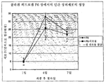
도 13은 콘드로이틴 술페이트 대 콜라겐 건조중량비의 함수로서의 신경돌기 길이의 그래프이다. FIG. 13 is a graph of neurite length as a function of chondroitin sulfate to collagen dry weight ratio.
전형적인 인간 안구는 수정체 및 홍채를 갖는다. 후안방(posterior chamber)은 홍채 뒤에 위치하고 전안방(anterior chamber)은 홍채 앞에 위치한다. 안구는 본원에서 논의되는 바와 같이 5개의 층으로 이루어진 각막을 갖는다. 층들 중 하나인 각막 상피는 각막의 외부 전면을 덮는다. 각막 상피는 주변부까지 측방향으로 연장하는 층화 편평 상피이다.Typical human eyes have the lens and the iris. The posterior chamber is behind the iris and the anterior chamber is located before the iris. The eye has a cornea consisting of five layers as discussed herein. One of the layers, the corneal epithelium, covers the outer front of the cornea. Corneal epithelium is a stratified squamous epithelium that extends laterally to the periphery.
각막의 5개의 층들은 각막 상피, 보우만막, 실질층, 데스메막 및 내피를 포함한다. 각막 상피는 통상적으로 약 5 내지 6 개의 세포 층 두께(약 50 마이크로미터 두께)를 갖고, 일반적으로 각막이 손상되면 재생된다. 각막 상피는 비교적 매끄러운 굴절 표면을 제공하고 안구 감염의 예방을 돕는다. 각막 실질층은 섬유아세포 및 각화세포와 같은 세포가 분산되어 있는 콜라겐의 라미네이트 구조이다. 실질층은 각막 두께의 약 90%를 차지한다. 상피 하에 위치한, 실질층의 앞부분은무세포성이며 보우만막으로서 공지되어 있다. 보우만막은 상피와 실질층 사이에 위치하며, 각막을 손상으로부터 보호하는 것으로 생각된다. 각막 내피는 전형적으로, 각막으로부터 수분을 제거함으로써 각막을 탈수시키는 저-입방 또는 편평 세포의 단층이다. 인간 성인 각막은 전형적으로 두께가 약 500 ㎛(0.5 ㎜)이고 전형적으로 혈관을 갖지 않는다. The five layers of the cornea include corneal epithelium, Bowman's membrane, parenchyma, Desme's membrane and endothelium. Corneal epithelium typically has a thickness of about 5 to 6 cell layers (about 50 micrometers thick) and is generally regenerated when the cornea is damaged. Corneal epithelium provides a relatively smooth refractive surface and helps prevent eye infections. The corneal parenchyma is a laminate structure of collagen in which cells such as fibroblasts and keratinocytes are dispersed. The parenchyma accounts for about 90% of the corneal thickness. The anterior part of the parenchyma, located under the epithelium, is acellular and known as the Bowman's membrane. The Bowman's membrane is located between the epithelium and the parenchyma and is thought to protect the cornea from damage. The corneal endothelium is typically a monolayer of low-cubic or squamous cells that dehydrate the cornea by removing water from the cornea. Human adult corneas are typically about 500 μm (0.5 mm) thick and typically have no blood vessels.
시력의 향상 또는 개선을 원하거나 안구의 질환, 장애 또는 외상의 치료를 필요로 하는 개인, 예를 들면 인간에게 하나 이상의 이점을 제공하는 안과용 장치가 발명되어 왔다. 본원에서 기술된 장치는 각막 온레이, 각막 인레이 또는 전층 각막 이식물로서 구성될 수 있다. 본 발명의 장치는 시력이 감퇴된 개인의 시력을 향상시키거나 시력을 잃은 개인에게 시력을 제공할 수 있다. 본원에서 기술된 장치는 특히 인공수정체를 포함하지 않는다.Ophthalmic devices have been invented that provide one or more benefits to an individual, such as a human, who desires to improve or improve vision or needs treatment of diseases, disorders or trauma to the eye. The device described herein can be configured as a corneal onlay, corneal inlay or full-layer corneal implant. The device of the present invention may improve vision of or provide vision to an individual who has lost vision. The device described herein does not specifically comprise an intraocular lens.
본원에서 사용된 "광학적으로 등명한"이란 85% 이상의 백색광 투과율을 말한다. 특정 실시양태에서, "광학적으로 등명한"이란 건강한 각막, 예를 들면 90% 초과의 백색광 투과율 및 3% 미만의 산란도를 갖는 각막의 광학적 등명성을 말한다.As used herein, "optically clear" refers to a white light transmittance of at least 85%. In certain embodiments, “optically clear” refers to the optical clarity of a healthy cornea, eg, a cornea having greater than 90% white light transmission and less than 3% scattering.
본원에서 사용된 "각막 온레이"란, 인간 또는 동물과 같은 개인의 안구의 상피 또는 상피세포층과 보우만막 사이에 위치하도록 구성된, 예를 들면 그러한 크기 또는 형상을 갖는 안과용 이식물 또는 장치이다. 이에 비해, 콘택트렌즈는 안구의 상피 상에 위치하도록 구성된다. 따라서 각막 온레이는 보우만막 전체에 걸쳐 위치하거나 보우만막 내로 연장되는 하나 이상의 부분을 포함할 수 있다. 이러한 부분은 장치의 면적 또는 부피의 적은 부분, 예를 들면 50% 미만을 차지한다. As used herein, “corneal onlay” is an ophthalmic implant or device, eg, having such size or shape, configured to be located between the Bowel's epithelial or epithelial cell layer of an individual, such as a human or animal. In comparison, the contact lens is configured to be located on the epithelium of the eyeball. Thus, the corneal onlay may comprise one or more portions located throughout or extending into the Bowman's membrane. This portion occupies a small part of the area or volume of the device, for example less than 50%.
본원에서 사용된 "각막 인레이"란, 안구의 실질층 내에 위치하도록 구성된 장치 또는 이식물이다. 실질층 내에 플랩(flap) 또는 포켓(pocket)을 형성함으로써 각막 인레이를 실질층 내에 위치시킬 수 있다. 각막 인레이는 안구의 보우만막 하에 위치된다. As used herein, “corneal inlay” is a device or implant configured to be located within the parenchyma of the eye. Corneal inlays can be placed in the parenchyma by forming flaps or pockets in the parenchyma. The corneal inlay is located under the Bowman's membrane of the eye.
본원에서 사용된 "전층 각막 이식물"이란 안구의 수양액(aqueous humor)의 앞에 위치한 안구의 건강하지 못한 각막의 전부 또는 일부를 대체하도록 구성된 장치이다. As used herein, a "periosteal corneal implant" is a device configured to replace all or part of an unhealthy cornea of an eye located in front of an aqueous humor of the eye.
본 발명의 안과용 장치는 감소된 세포독성을 갖거나 무-세포독성이고, 장치를 장착한 개인에게 하나 이상의 이점을 제공한다. 예를 들면, 장치는 (i) 바람직한 굴절률, (ii) 바람직한 광학적 등명성(가시광선의 경우, 동일한 두께를 갖는 건강한 인간 각막 물질의 투광률 및 광산란도와 동일하거나 더 우수한 투광률 및 광산란도), (iii) 바람직한 광학적 기능, 예를 들면 시력 향상 기능, (iv) 향상된 안락함, (v) 향상된 각막 및 상피 건강, 및 (vi) 치료적 이점, 예를 들면 안구의 질환, 장애 또는 외상의 치료 중 하나 이상을 제공한다. 본 발명의 안과용 장치는 투명하거나 투명한 물질로써 제조된다. 이러한 장치의 몇몇 예는 광학적으로 등명한 장치를 포함한다. Ophthalmic devices of the present invention have reduced cytotoxicity or are non-cytotoxic and provide one or more advantages to individuals equipped with the device. For example, the device may include (i) the desired refractive index, (ii) the desired optical transparency (in the case of visible light, the transmittance and light scattering degree equal to or better than that of healthy human corneal material having the same thickness), ( iii) desirable optical function, such as vision enhancement, (iv) improved comfort, (v) improved corneal and epithelial health, and (vi) therapeutic benefits, such as the treatment of diseases, disorders or trauma of the eye Provides more. The ophthalmic device of the present invention is made of a transparent or transparent material. Some examples of such devices include optically clear devices.
(i) 허용가능한 광학적 기능을 갖는 매트릭스를 형성하도록 성형가능, 예를 들면 몰딩가능하고 (ii) 광학적으로 등명하거나 시각적으로 투명하고 (iii) 장치를 통한 및/또는 장치 상에서의 신경 성장을 용이하게 하는데 효과적인 물질로써 장치를 제조함으로써, 상기 이점 뿐만 아니라 기타 이점을 얻을 수 있다. 장치가 각막 온레이인 경우, 장치는 장치의 전면 상에서의 재-상피화(re-epithelialization)를 용이하게 하는데 효과적이다. (i) moldable to form a matrix with acceptable optical function, for example moldable, (ii) optically clear or visually transparent, and (iii) facilitating nerve growth through and / or on the device. By manufacturing the device with a material which is effective to achieve the above advantages, as well as other advantages can be obtained. If the device is corneal onlay, the device is effective to facilitate re-epithelialization on the front of the device.
장치는, 취급, 봉합을 포함할 수 있는 이식, 및 장착후 마모 및 눈물을 견디도록 충분한 기계적 또는 구조적 성질을 갖는 물질로써 제조된다. 장치는 건강한 안구를 유지하기에 충분한 영양분 및 기체 교환을 제공하거나 허용한다. 각막 온레이와 같은, 주형 내에서 제조되는 장치는, 본원에서 논의되는 바와 같이, 가장자리 구배 및 시력 교정 곡률을 포함하는 적당한 크기 및 형상으로 몰딩될 수 있는 물질로써 제조된다. The device is made of a material that has sufficient mechanical or structural properties to withstand handling, implantation, which may include sutures, and wear and tear after mounting. The device provides or allows for sufficient nutrient and gas exchange to maintain a healthy eye. Devices manufactured in a mold, such as a corneal onlay, are made of a material that can be molded into a suitable size and shape, including edge gradients and vision correction curvature, as discussed herein.
본 발명의 한 실시양태에서, 시력 향상 안과용 장치는, 장치가 개인의 안구에 위치할 때, 본체를 통한 신경 성장을 용이하게 하기에 효과적인 물질을 포함하는 본체를 포함한다. 본체를 통한 신경 성장을 용이하게 함으로써, 장치를 수용하는 개인의 각막은 그것의 접촉감도를 유지한다. 본체는 광학적 기능을 갖도록 형성된다. 따라서, 본체는 렌즈 본체라고 이해될 수 있다. 본원에서 논의되는 바와 같이, 장치는 각막 온레이, 각막 인레이 또는 전층 각막 이식물이도록 구성되는데, 예를 들면 그러한 크기 및 형상을 갖는다. 특정 실시양태에서, 본 발명의 굴절이상 교정 장치는 광학적 기능을 갖지 않을 수 있다. 예를 들면, 본원 내용에 따르는 굴절이상 교정 장치는 환자의 각막 상피와 보우만막 사이에 위치되거나 환자의 각막 실질층 내에 위치할 수 있는 블랭크(blank)라고 이해될 수 있다.In one embodiment of the present invention, a vision enhancing ophthalmic device includes a body comprising a material effective to facilitate nerve growth through the body when the device is located in an eye of an individual. By facilitating nerve growth through the body, the cornea of the individual receiving the device maintains its contact sensitivity. The body is formed to have an optical function. Thus, the body can be understood as a lens body. As discussed herein, the device is configured to be a corneal onlay, corneal inlay or penetrating corneal implant, eg, having such size and shape. In certain embodiments, the refractive error correction apparatus of the present invention may not have an optical function. For example, it can be understood that a refractive error correcting device according to the present disclosure is a blank that can be located between the corneal epithelium of the patient and the Bowman's membrane or within the corneal parenchyma of the patient.
각막 온레이의 경우, 온레이를 형성하는 물질은 보우만막과 상피 사이의 기체 및 영양분(예를 들면 글루코스)의 교환을 제공 또는 허용함으로써, 회생가능하고 완전한 기능을 갖춘 상피를 유지한다. 기타 영양분은 상피세포와 같은 세포의 생존, 성장 및 분화를 촉진 또는 향상시키는 인자 또는 물질을 포함한다. 교환은 건강한 인간 각막의 것과 동일하거나 더 우수해야 한다. 영양분 및/또는 약물에 대한 물질의 투과도를 통상적인 기술을 사용하여 모니터링할 수 있다. 또한, 물질을 통한 영양분 및/또는 약물의 이동으로 인해 물질의 광학적 성질이 변해서는 안된다. 온레이 또는 렌티클은 완전 생분해성이고, 온레이에 대한 상피의 신속한 부착을 허용하고, 신경 지배 및 감도, 예를 들면 접촉감도의 회복을 허용한다. In the case of corneal onlays, the substances that form the onlays provide or permit the exchange of gas and nutrients (eg, glucose) between the Bowman's membrane and the epithelium, thereby maintaining a regenerating and fully functional epithelium. Other nutrients include factors or substances that promote or enhance the survival, growth and differentiation of cells such as epithelial cells. The exchange should be the same or better than that of healthy human corneas. Permeability of the substance to nutrients and / or drugs can be monitored using conventional techniques. In addition, the transport of nutrients and / or drugs through the material should not alter the optical properties of the material. The onlays or lentices are fully biodegradable and allow for rapid attachment of the epithelium to the onlays and allow for recovery of nerve domination and sensitivity, eg contact sensitivity.
본 발명의 안과용 장치는 세포외 매트릭스(ECM) 성분을 포함할 수 있다. 특정 장치에서, 본체 물질은 콜라겐을 포함하거나, 본질적으로 콜라겐으로 이루어지거나, 콜라겐으로 이루어진다. 콜라겐은 예를 들면 장치의 제조 동안에 EDC/NHS를 사용하여 가교결합될 수 있다. 본 발명의 히드로겔 장치 내에 제공된 콜라겐의 양은 현재 기타 안과용 장치에 사용되는 콜라겐의 양보다 많다. 예를 들면, 본 발명의 장치 내에 제공된 콜라겐의 양은, 본원에서 논의되는 바와 같이, 전형적으로 1 %(w/w) 또는 (w/v) 초과이다. 특정 실시양태에서, 콜라겐의 양은 2.5% 초과이다. 예를 들면, 콜라겐의 양은 약 5.0% 이상일 수 있다. 본 발명의 장치의 특정 실시양태에서, 콜라겐의 양은 약 1 내지 약 50 %(w/w), 예를 들면 2.5 내지 약 50 %이다. 예를 들면, 콜라겐의 양은 약 6 %(w/w) 초과이다. 또는, 물질은 약 10 내지 약 30 %(w/w)의 콜라겐을 포함할 수 있다. 해당 분야의 보통 숙련자들이 알고 있듯이, 수화된 인간 각막의 약 15 중량%는 콜라겐이다(문헌[Maurice D M: The Cornea and Sclera, pp 489-600. The Eye, Vol I, Second ed., Ed. H Davson. Academic Press, New York, 1969]을 참고). 따라서, 본 발명의 장치는 기존 안과용 장치에 존재하는 콜라겐의 양보다는 더 많고 인간 각막에 존재하는 콜라겐의 양과 훨씬 더 유사한 양의 콜라겐을 포함한다. 또한 본 발명의 장치 내에 제공된 콜라겐의 양 및 유형은 바람직한 굴절률, 바람직한 광학적 등명성, 성형성을 제공하고, 취급, 이식, 장치의 눈에의 봉합, 및 장착후 마모 및 눈물을 견디기에 효과적이다. The ophthalmic device of the present invention may comprise an extracellular matrix (ECM) component. In certain devices, the body material comprises collagen, consists essentially of collagen, or consists of collagen. Collagen can be crosslinked using, for example, EDC / NHS during the manufacture of the device. The amount of collagen provided in the hydrogel device of the present invention is greater than the amount of collagen currently used in other ophthalmic devices. For example, the amount of collagen provided in the device of the present invention is typically greater than 1% (w / w) or (w / v), as discussed herein. In certain embodiments, the amount of collagen is greater than 2.5%. For example, the amount of collagen may be about 5.0% or more. In certain embodiments of the device of the invention, the amount of collagen is about 1 to about 50% (w / w), for example 2.5 to about 50%. For example, the amount of collagen is greater than about 6% (w / w). Alternatively, the substance may comprise about 10 to about 30% (w / w) collagen. As is known to those skilled in the art, about 15% by weight of the hydrated human cornea is collagen (Maurice DM: The Cornea and Sclera, pp 489-600. The Eye, Vol I, Second ed., Ed. H. Davson.Academic Press, New York, 1969). Thus, the device of the present invention contains more collagen than the amount of collagen present in existing ophthalmic devices and much more similar to the amount of collagen present in the human cornea. The amount and type of collagen provided in the device of the present invention also provides the desired refractive index, the desired optical clarity, the formability, and is effective in handling, implanting, sealing the device to the eye, and withstanding wear and tear after mounting.
비-콜라겐-기재의 부분과 같은, 안과용 장치의 나머지 부분은 물 또는 식염수와 같은 액체일 수 있거나, 생체중합체 등과 같은 하나 이상의 추가의 중합체를 포함할 수도 있다. 예를 들면, 본원에서 개시된 바와 같이, 약 24 %(w/w)의 콜라겐을 포함하는 안과용 장치는 물 또는 식염수와 같은 액체를 약 76 %(w/w)로 포함할 수 있다. 달리 말하자면, 안과용 장치는, 수화된 상태에서는, 수화된 안과용 장치의 중량의 24%인 콜라겐 성분을 포함할 수 있다. 또다른 예로서, 안과용 장치는 수화된 장치의 중량의 24%인 콜라겐 성분, 및 수화된 장치의 중량의 6%인 제 2 중합체성 성분을 포함할 수 있고, 중량의 70%는 액체이다. The remaining portion of the ophthalmic device, such as a non-collagen-based portion, may be a liquid such as water or saline, or may include one or more additional polymers such as biopolymers and the like. For example, as disclosed herein, an ophthalmic device comprising about 24% (w / w) collagen may comprise about 76% (w / w) of a liquid, such as water or saline. In other words, the ophthalmic device may, in the hydrated state, comprise a collagen component that is 24% of the weight of the hydrated ophthalmic device. As another example, the ophthalmic device may comprise a collagen component that is 24% of the weight of the hydrated device, and a second polymeric component that is 6% of the weight of the hydrated device, with 70% of the weight being liquid.
해당 분야의 보통 숙련자들이 알고 있는 바와 같이, 장치가 수화되지 않은 상태에서는, 장치의 콜라겐의 양은 수화된 상태의 장치의 콜라겐의 양보다 큰 %일 수 있다. As will be appreciated by those of ordinary skill in the art, in a state where the device is not hydrated, the amount of collagen in the device may be a percentage greater than the amount of collagen in the device in the hydrated state.
콜라겐은 3개의 폴리펩티드쇄를 포함하고 나선형 구조이다. 본원에서 사용된 "콜라겐 중합체"라는 용어는 3중 나선 콜라겐 분자를 말한다. 콜라겐은 약 300 ㎚의 길이 및 약 1.5 ㎚의 직경을 갖는, 막대와 유사하게 생긴 분자이다. 콜라겐 분자는, 콜라겐의 항원성의 대부분을 포함하는, N-말단 및 C-말단 둘 다 상에 "텔로펩티드(telopeptide)"라고 불리는 아미노산 서열을 갖는다. 아텔로콜라겐(atelocollagen)은 펩신 소화에 의해 수득되고(문헌[DeLustro 등, J Biomed Mater Res. 1986 Jan; 20(1): 109-20]을 참고), 텔로펩티드를 갖지 않으므로, 면역원성이 낮다는 것을 알 수 있다(문헌[Stenzel 등, Annu Rev Biophys Bioeng. 1974; 3(0): 231-53]을 참고). Collagen contains three polypeptide chains and is helical. As used herein, the term "collagen polymer" refers to triple helix collagen molecules. Collagen is a rod-like molecule that has a length of about 300 nm and a diameter of about 1.5 nm. Collagen molecules have an amino acid sequence called "telopeptide" on both the N-terminus and C-terminus, including most of the antigenicity of collagen. Atelocollagen is obtained by pepsin digestion (see DeLustro et al., J Biomed Mater Res. 1986 Jan; 20 (1): 109-20) and has no telopeptides, and thus has low immunogenicity. (Stenzel et al., Annu Rev Biophys Bioeng. 1974; 3 (0): 231-53).
위에서 정의된 장치에서 사용되는 콜라겐은 동물, 이스트 및 박테리아 공급원을 포함하는 임의의 적합한 콜라겐 공급원으로부터 수득 또는 유래될 수 있다. 예를 들면, 콜라겐은 특히 인간 콜라겐, 소 콜라겐, 돼지 콜라겐, 새 콜라겐, 쥐 콜라겐, 말 콜라겐일 수 있거나, 재조합 콜라겐일 수도 있다. 본 발명의 장치에서 사용되는 재조합 콜라겐은, 보통의 동물성 공급원으로부터 수득되는 콜라겐 내에는 존재하지 않는 하나 이상의 구조적 또는 물리적 특성을 가질 수 있는데, 왜냐하면 재조합 콜라겐은 박테리아, 이스트, 식물 또는 유전자도입 동물로부터 수득되기 때문이다. 예를 들면, 재조합 인간 콜라겐은 동물로부터 유래되고 가공된 콜라겐 내에는 존재하지 않을 수 있는 상이한 글리코실화 성분을 포함할 수 있다. 또한, 재조합 콜라겐은 조성이 다양할 수 있는 동물-유래된 콜라겐과는 상이한 가교도를 가질 수 있다. 동물-유래된 콜라겐 내의 가교도의 변동은, 바람직하지 못할 수 있는콜라겐의 불일치성 및 화학적 및 물리적 성질의 변동을 초래할 수 있다. 재조합 인간 콜라겐은 엄격하게 제어된 순도를 가질 뿐만 아니라, 동물-유래된 콜라겐과 연관될 수 있는 바이러스 및/또는 프리온 오염과 관련이 없다. 본 발명의 장치에서 유용한 콜라겐을 공식적으로 입수하거나 통상적인 기술을 사용하여 합성할 수 있다. 예를 들면, 재조합 콜라겐을 피브로겐(Fibrogen)(뮤티젠(mutigene) 이스트 생체반응기 배지로부터 제조) 또는 파밍(Pharming)(네덜란드)(유전자도입 소 또는 토끼의 젖으로부터 제조)으로부터 입수할 수 있거나, 재조합 콜라겐을 PCT 공개 제 WO 93/07889 호 또는 제 WO 94/16570 호에 개시된 방법을 사용하여 제조하고 수득할 수 있다. 특정 장치에서, 콜라겐은 I형 콜라겐일 수 있다. 장치를 아텔로콜라겐(예를 들면 텔로펩티드를 갖지 않는 콜라겐)으로써 제조할 수도 있다. 특정 실시양태에서, 콜라겐은 변성되지 않은 유형의 콜라겐이다. 아텔로콜라겐을 코켄 재팬(Koken Japan)과 같은 회사(본원에서 기술된 바와 같은 공급처 A)로부터 입수할 수 있는데, 여기서 소 콜라겐은 중성 조성물 내 3.5 %(w/v), 산성 조성물 내 3.0 %(w/v) 및 산성 조성물 내 10 %(w/v)로서 입수가능하고, 돼지 콜라겐은 산성 조성물 내 3.0 %(w/v) 또는 산성 동결건조 돼지 콜라겐 분말로서 입수가능하다. 산성 동결건조 돼지 콜라겐 분말을 니폰 햄(Nippon Ham)(일본)(본원에서 기술된 바와 같은 공급처 B)으로부터 수득할 수도 있다. 벡톤 디킨슨(Becton Dickinson)(본원에서 기술된 바와 같은 공급처 C)은 0.3% 산성 및 10% 산성 콜라겐 조성물을 제공한다. Collagen for use in the devices defined above may be obtained or derived from any suitable collagen source, including animal, yeast and bacterial sources. For example, the collagen may in particular be human collagen, bovine collagen, porcine collagen, bird collagen, mouse collagen, horse collagen, or may be recombinant collagen. Recombinant collagen used in the device of the present invention may have one or more structural or physical properties that are not present in the collagen obtained from normal animal sources, since the recombinant collagen is obtained from bacteria, yeast, plants or transgenic animals. Because it becomes. For example, recombinant human collagen can include different glycosylation components that are derived from an animal and may not be present in processed collagen. In addition, recombinant collagen may have a different degree of crosslinking than animal-derived collagen, which may vary in composition. Variation in the degree of crosslinking in animal-derived collagen can lead to undesirable variations in collagen and chemical and physical properties. Recombinant human collagen not only has strictly controlled purity, but is also not associated with virus and / or prion contamination that may be associated with animal-derived collagen. Collagen useful in the device of the present invention can be obtained formally or synthesized using conventional techniques. For example, recombinant collagen can be obtained from Fibrogen (prepared from mutigene yeast bioreactor medium) or Pharming (Netherlands) (produced from transgenic cattle or rabbit milk) or Recombinant collagen can be prepared and obtained using the methods disclosed in PCT Publication WO 93/07889 or WO 94/16570. In certain devices, the collagen may be type I collagen. The device may also be prepared as atelocollagen (eg collagen without telopeptide). In certain embodiments, the collagen is a type of collagen that is not denatured. Atelocollagen can be obtained from a company such as Koken Japan (Supplier A as described herein), where bovine collagen is 3.5% (w / v) in the neutral composition and 3.0% (in acidic composition) w / v) and 10% (w / v) in the acidic composition, and pig collagen is available as 3.0% (w / v) or acidic lyophilized pork collagen powder in the acidic composition. Acidic lyophilized pork collagen powder may also be obtained from Nippon Ham (Japan) (Supplier B as described herein). Becton Dickinson (source C as described herein) provides 0.3% acidic and 10% acidic collagen compositions.
몇몇 콜라겐 유형들 중에서도, 아텔로콜라겐 I는 용해의 용이함, 취급 및 최종 장치의 등명성을 제공한다. 이러한 콜라겐(중성 또는 산성 용액 중의, 또는 산성 동결건조 분말로서의, 소, 돼지 또는 재조합 콜라겐)은 전술된 바와 같은 몇몇 회사로부터 입수가능하다. 동결건조 산성 돼지 콜라겐은 용이하게 용해되므로, 냉수에서 4℃에서 교반함으로써, 33 %(w/v) 이하의 농도의 균질(비-유백광) 수용액을 얻는다. 용액과 같은, 등명한 콜라겐 조성물의 pH는 약 3(공급처 B) 또는 약 5(공급처 A)이다. 0.3 %(w/v) 정도로 낮은 농도의 상업적인 산성 콜라겐 조성물을, 0 내지 4 ℃에서 교반하면서 진공 증발시킴으로써 농축시켜, 약 10 %(w/v) 이하의 최종 콜라겐 농도를 갖는 등명한 용액을 얻을 수 있는데, 이것을 본 발명의 장치의 제조에서 사용할 수 있다. Among some collagen types, atelocollagen I provides ease of dissolution, handling and clarity of the final device. Such collagen (bovine, swine or recombinant collagen, in neutral or acidic solutions, or as acidic lyophilized powder) is available from several companies as described above. Lyophilized acidic porcine collagen is readily soluble, so that stirring at 4 ° C. in cold water yields a homogeneous (non-milky white) aqueous solution of concentration up to 33% (w / v). The clear collagen composition, such as solution, has a pH of about 3 (source B) or about 5 (source A). Commercial acid collagen compositions at concentrations as low as 0.3% (w / v) are concentrated by vacuum evaporation with stirring at 0-4 ° C. to obtain a clear solution having a final collagen concentration of about 10% (w / v) or less. Can be used in the manufacture of the device of the invention.
단리 및 정제 과정에서 변성(즉 3중 나선 구조를 전부 또는 상당 부분 잃음으로써 젤라틴이 됨) 되지 않은 I형 콜라겐을 사용함으로써, 비교적 견고하거나 강한 안과용 장치를 수득할 수 있다.By using collagen type I that has not been denatured (i.e., becoming gelatinous by losing all or a substantial portion of the triple helix structure) during isolation and purification, a relatively robust or strong ophthalmic device can be obtained.
시차주사열계량법(DSC)은 공급처의 콜라겐 용액의 품질을 그것의 3중 나선 함량을 근거로 결정하기에 유용한 기술이다(표 1). 거의 완벽한 3중 나선 함량의 경우, 변성의 DSC 엔탈피(ΔH변성)는 (건조 콜라겐 중량을 기준으로) 65 내지 70 J/g이다. DSC 데이터로부터, ΔH변성 결과는, 상업적인 산성 동결건조 돼지 콜라겐 용액 및 몇몇 상업적 소 콜라겐 용액은 완전한 3중 나선 구조임을 알려준다. Differential scanning calorimetry (DSC) is a useful technique for determining the quality of a collagen solution at a supplier based on its triple helix content (Table 1). For a nearly perfect triple helix content, the denaturation DSC enthalpy (ΔH denaturation ) is 65 to 70 J / g (based on dry collagen weight). From the DSC data, the ΔH denaturation results indicate that the commercial acidic lyophilized porcine collagen solution and some commercial bovine collagen solutions are complete triple helix structures.
낮은 3중 나선 함량을 갖는 콜라겐 용액(ΔH변성 < 5 J/g, 공급처 C, 표 1)은, 비교적 낮은 점도를 가지며, 동일한 농도의, 100%에 가까운 3중 나선 함량을 갖는 콜라겐 조성물 또는 용액에 비해 약한 겔을 제공한다. 약 60 J/g 초과의 ΔH변성을 갖는 콜라겐 조성물(용액)이 허용가능한 안과용 장치를 제공하는 것으로 밝혀졌다. Collagen solutions with a low triple helix content (ΔH denaturation <5 J / g, source C, Table 1) have a relatively low viscosity and a collagen composition or solution with a triple helix content close to 100% of the same concentration. Provides a weak gel as compared to. Collagen compositions (solutions) with ΔH denaturation greater than about 60 J / g have been found to provide acceptable ophthalmic devices.
전술된 실시양태를 포함하여 특정 실시양태에서, 본체 물질은 가교결합된 콜라겐 중합체를 포함할 수 있다. 아니면, 달리 말하자면, 본체 물질은 둘 이상의 가교결합된 콜라겐 중합체를 포함할 수 있다. 예를 들면, 본체 물질은 제 1 콜라겐 중합체, 제 2 콜라겐 중합체 및 제 3 콜라겐 중합체를 포함할 수 있다. 기타 물질은 셋 초과의 콜라겐 중합체를 포함할 수 있다. 가교결합된 중합체는 안과용 장치의 콜라겐 성분으로서 이해될 수 있다. In certain embodiments, including the embodiments described above, the body material can comprise a crosslinked collagen polymer. Alternatively, in other words, the body material may comprise two or more crosslinked collagen polymers. For example, the body material may comprise a first collagen polymer, a second collagen polymer and a third collagen polymer. Other materials may include more than three collagen polymers. Crosslinked polymers can be understood as collagen components of ophthalmic devices.
따라서, 본 발명에 따르는 시력 향상 안과용 장치는 약 1 내지 50 %(w/w)의 콜라겐을 갖고 광학적 기능을 갖도록 형성된 콜라겐 성분을 포함할 수 있다. 본원에서 논의되는 바와 같이, 특정 실시양태에서, 콜라겐의 양은 2.5% 초과, 예를 들면 약 5.0% 이상이다. 예를 들면, 특정 실시양태에서, 콜라겐은 약 6 %(w/w) 초과이다. 예를 들면, 콜라겐의 양은 약 10 내지 약 30 %(w/w)이다. 예를 들면, 콜라겐의 양은 약 10 내지 약 24 %(w/w)일 수 있다. 특정 장치에서, 콜라겐은 장치의 유일한 수-팽창성(예를 들면 히드로겔) 중합체이다. 기타 장치에서, 콜라겐은 유일한 장치 또는 렌즈-형성 중합체일 수 있다. 예를 들면, 장치는 건조 상태에서 100%의 콜라겐을 포함할 수 있다. 앞에서 논의된 바와 같이. 특정 장치에서, 예를 들면 EDC/NHS를 사용하여, 콜라겐을 가교결합시키거나 적어도 부분적으로 가교결합시킬 수 있다. Accordingly, the vision enhancing ophthalmic device according to the present invention may have collagen components of about 1 to 50% (w / w) and comprise collagen components formed to have optical function. As discussed herein, in certain embodiments, the amount of collagen is greater than 2.5%, for example at least about 5.0%. For example, in certain embodiments, the collagen is greater than about 6% (w / w). For example, the amount of collagen is about 10 to about 30% (w / w). For example, the amount of collagen may be about 10 to about 24% (w / w). In certain devices, collagen is the only water-expandable (eg hydrogel) polymer of the device. In other devices, the collagen may be the only device or lens-forming polymer. For example, the device may contain 100% collagen in a dry state. As discussed earlier. In certain devices, for example, EDC / NHS can be used to crosslink or at least partially crosslink the collagen.
본 발명의 조성물 및 장치의 제조에서 사용되는 콜라겐 중합체는 동일한 콜라겐 공급원 또는 상이한 콜라겐 공급원으로부터 유래될 수 있다. 아니면, 달리 말하자면, 단일 유형의 콜라겐, 예를 들면 I형 아텔로콜라겐(다수의 콜라겐 중합체쇄를 함유)을, 콜라겐 중합체들이 서로 가교결합하는 것을 허용하기에 효과적인 방식으로 가공한다. 한 실시양태에서, 콜라겐 중합체는 재조합 콜라겐이다. 또다른 실시양태에서, 콜라겐 중합체는 둘 다 동일한 동물 공급원으로부터 유래된다. 단일 조성물 내의 개별 콜라겐 중합체들은 상이한 분자량을 가질 수 있다. Collagen polymers used in the preparation of the compositions and devices of the present invention may be derived from the same collagen source or from different collagen sources. Or, in other words, a single type of collagen, such as type I atelocollagen (containing a large number of collagen polymer chains), is processed in an effective manner to allow the collagen polymers to crosslink with each other. In one embodiment, the collagen polymer is recombinant collagen. In another embodiment, the collagen polymers are both from the same animal source. Individual collagen polymers in a single composition may have different molecular weights.
본 발명의 굴절이상 교정 장치는 가교결합된 재조합 콜라겐을 포함한다는 것을 알 수 있을 것이다. 이러한 장치 내에 존재하는 콜라겐의 양은 이전에 개시된 굴절이상 교정 장치 내의 기타 콜라겐의 양보다 더 많다. 이러한 장치는 광학적 기능을 갖도록 형성될 수 있다. It will be appreciated that the refractive error correction apparatus of the present invention includes cross-linked recombinant collagen. The amount of collagen present in such devices is greater than the amount of other collagen in the previously described refractive error correction device. Such a device may be formed to have an optical function.
본원에서 개시된 장치는 투명하다. 예를 들면, 장치는 광학적으로 등명해야 한다. 예를 들면, 장치는 개인의 안구 내에 위치할 때 (건강한 인간 각막 조직의 광산란도와 동일하거나 더 우수한) 최소의 광산란도를 제공해야 한다. 또한, 본원에서 개시된 장치는 굴절률을 갖는다. 특정 실시양태에서, 굴절률은 약 1.34 내지 약 1.37이다. 예를 들면, 굴절률은 1.341 내지 1.349일 수 있다. 장치가 각막 온레이 또는 각막 인레이로서 구성되는 경우, 이 장치는, 전층 각막 이식물이 요구될 수 있는, 각막이 손상되거나 질환을 갖는 안구에 위치하도록 구성되는것에 비해, 개인의 건강한 안구 내에 위치하도록 구성된다. 본 발명의 장치의 특정 실시양태에서, 장치는 황색기 또는 황색을 갖지 않는다. 예를 들면, 장치는 몇몇 콜라겐-함유 조성물과 연관될 수 있는 황색기 또는 황색을 감소시키거나 없애도록 디자인될 수 있다. The device disclosed herein is transparent. For example, the device should be optically clear. For example, the device should provide a minimum light scattering degree (equivalent to or better than the light scattering of healthy human corneal tissue) when placed in the eye of an individual. In addition, the device disclosed herein has a refractive index. In certain embodiments, the refractive index is about 1.34 to about 1.37. For example, the refractive index may be 1.341 to 1.349. When the device is configured as a corneal onlay or corneal inlay, the device is placed in a healthy eye of the individual as compared to the cornea being configured to be located in a damaged or diseased eye where a full corneal implant may be required. It is composed. In certain embodiments of the device of the invention, the device does not have a yellow group or yellow color. For example, the device may be designed to reduce or eliminate yellow or yellow that may be associated with some collagen-containing compositions.
본 발명의 장치는 전면 및 후면을 갖는다. 따라서, 장치의 본체 또는 콜라겐 성분은 전면 및 후면을 가질 수 있다. 전면 및 후면은 일반적으로 서로 반대되는 표면이다. 장치의 전면은, 장치가 안구 내에 위치할 때, 망막을 등지는 표면을 말하고, 후면은, 장치가 안구 내에 위치할 때, 망막 쪽을 향한다. 장치가 각막 온레이인 경우, 후면은 보우만막과 인접하거나 접촉할 수 있고, 전면은 각막 상피와 인접하거나 접촉할 수 있다. 장치가 각막 인레이인 경우, 전면은 보우만막과 인접하거나 보우만막 쪽을 향하고, 후면은 안구의 망막 쪽을 향하도록 실질층 내에 존재할 것이다. 장치가 전층 각막 이식물인 경우, 전면은 각막 상피 쪽으로 향하고, 후면은 각막 내피와 인접하거나 접촉할 수 있다. The device of the present invention has a front side and a rear side. Thus, the body or collagen component of the device may have a front side and a back side. The front and back are generally opposite surfaces. The front side of the device refers to the surface that rests the retina when the device is located in the eyeball, and the back side faces the retina when the device is located in the eyeball. If the device is corneal onlay, the backside may be adjacent to or in contact with the Bowman's membrane and the front side may be adjacent to or in contact with the corneal epithelium. If the device is a corneal inlay, the front face will be in the parenchyma so that the front face is adjacent to or facing the Bowman's membrane and the back face toward the retina side of the eye. If the device is a penetrating corneal implant, the front side may face toward the corneal epithelium and the back side may be adjacent to or in contact with the corneal endothelium.
본 발명의 장치는 추가로 표면 개질되지 않을 수 있거나, 본 발명의 장치는 전면과 후면 중 하나 또는 둘 다 상에서 세포 성장 및/또는 분화에 영향을 주도록 표면 개질될 수 있다. 예를 들면, 각막 온레이는 전면 또는 후면 상에서 세포 성장에 영향을 주는 표면 개질에 적용되지 않을 수 있다. 본원에서 사용된 "세포 성장"이란 세포 또는 세포수의 증대를 말한다. 따라서, 세포 성장이란 개별 세포의 물리적 성장, 예를 들면 표면적, 부피 등의 증가, 세포의 증식, 예를 들면 세포의 분열, 및 어떤 경우에는 건강한 인간 각막에서 발견되는 바와 같은 층화된 다층을 형성하는 세포의 이동을 말한다. 세포 성장이란 신경세포의 성장, 예를 들면 장치 상에서의 또는 장치 하에서의 또는 장치를 통한 하나 이상의 신경원 과정의 연장, 및 장치의 표면 상에서의 상피세포 또는 내피세포의 성장 또는 이동 또는 증식을 말한다. 본원에서 사용된 "세포 분화"란 단일 또는 다수의 전능성(totipotent), 중복성(multipotent) 또는 미성숙 전구세포(줄기세포를 포함)가 최종 표현형을 달성하기 위해 거치는 형태학적, 생화학적 및 생리학적 변화를 말한다. 본 발명의 장치의 특정 실시양태에서, 상피세포는 각막 온레이 상에서 성장하고, 거기에 견고하게 커플링되어 있는데, 예를 들면 온레이, 특히 온레이의 전면에 직접 부착된다.The device of the present invention may further be surface modified, or the device of the present invention may be surface modified to affect cell growth and / or differentiation on one or both of the front and rear surfaces. For example, corneal onlays may not be applied to surface modifications that affect cell growth on the front or back. As used herein, "cell growth" refers to the increase in cells or cell numbers. Thus, cell growth refers to the physical growth of individual cells, such as increases in surface area, volume, etc., proliferation of cells, for example cell division, and in some cases forming layered multilayers as found in healthy human corneas. Refers to the movement of cells. Cell growth refers to the growth of neurons, such as the extension of one or more neuronal processes on or under the device or through the device, and the growth or migration or proliferation of epithelial or endothelial cells on the surface of the device. As used herein, "cell differentiation" refers to the morphological, biochemical and physiological changes that single or multiple totipotent, multipotent or immature progenitor cells (including stem cells) undergo to achieve the final phenotype. Say. In certain embodiments of the device of the invention, epithelial cells grow on corneal onlays and are tightly coupled thereto, for example, directly on the front of the onlays, in particular onlays.
특정 각막 온레이에서, 본체 또는 콜라겐 성분은, 온레이가 개인의 안구 내에 위치할 때, 온레이 하에서의 상피세포 성장을 감소시키기에 효과적이도록 후면 개질된다. 추가로 또는 대안적으로, 각막 온레이는, 온레이가 개인의 안구 내에 위치할 때, 온레이의 전면 상에서의 상피세포 성장(이동을 포함)을 촉진시키기에 효과적이도록 전면 개질된 본체 또는 콜라겐 성분을 포함할 수 있다. 이와 관련하여, 전층 각막 이식물은, 이식물이 안구 내에 위치할 때, 전층 각막 이식물의 후면 상에서의 내피세포 성장을 감소시키기에 효과적이도록 후면 개질된 본체 또는 콜라겐 성분을 포함할 수 있다. 전층 각막 이식물은 전면 개질되지 않을 수 있다. In certain corneal onlays, the body or collagen component is rearranged to be effective to reduce epithelial cell growth under onlays when the onlay is located within the eye of the individual. Additionally or alternatively, the corneal onlay is a body or collagen component that has been modified to be effective to promote epithelial cell growth (including migration) on the front of the onlay when the onlay is located within the eye of the individual. It may include. In this regard, the penetrating corneal implant may comprise a body or collagen component that is rear modified to be effective to reduce endothelial cell growth on the back of the penetrating corneal implant when the implant is located within the eye. The penetrating corneal implant may not be fully modified.
세포 성장을 감소시킬 수 있는 표면 개질의 예는, 전면과 후면 중 하나 또는 둘 다 상에 CF4 또는 C3F8과 같은 플라스마-중합 플루오르화 단량체 필름을 제공하기, 표면들 중 하나 또는 둘 다 상에 낮은 자유 표면 에너지를 제공하기, 및/또는 표면들 중 하나 또는 둘 다를 친수성으로 만들기를 포함한다. 표면들 중 하나 또는 둘 다 상에 알긴산염 코팅을 제공함으로써, 표면을 친수성으로 만들 수 있다.Examples of surface modifications that can reduce cell growth include providing a plasma-polymerized fluorinated monomer film, such as CF 4 or C 3 F 8 , on one or both of the front and back sides, one or both of the surfaces Providing low free surface energy to the phase, and / or making one or both of the surfaces hydrophilic. By providing an alginate coating on one or both of the surfaces, the surface can be made hydrophilic.
장치는 장치 상에서의 또는 장치를 통한 세포 성장을 용이하게 하는 하나 이상의 세포성장촉진제를 포함할 수 있다. 특정 실시양태에서, 세포성장촉진제는 펩티드를 포함한다. 예를 들면, 세포성장촉진제는 RGD, YIGSR 또는 IKVAV를 포함하는 아미노산 서열을 갖는 펩티드일 수 있다. I형 콜라겐 그 자체는 RGD 서열의 풍부한 공급원이다. 특정 실시양태에서, 세포성장촉진제는 신경영양인자(neurotrophic factor) 또는 분자의 생체활성 또는 신경영양 부분이다. 예를 들면 신경영양인자는 신경성장인자(NGF), 표피성장인자(EGF 또는 HB-EGF) 또는 염기성섬유아세포성장인자(bFGF 또는 FGF-2)일 수 있다. 세포성장촉진제를, 장치의 콜라겐 성분 또는 본체와 일체형으로 형성할 수 있는데, 달리 말하자면, 세포성장촉진제를 실질적으로 장치 전체에 걸쳐 제공할 수 있다. 이에 비해, 몇몇 안과용 장치는 장치의 한 표면에만 제공된 펩티드를 포함한다. The device may include one or more cell growth promoters that facilitate cell growth on or through the device. In certain embodiments, the cell growth promoter comprises a peptide. For example, the cell growth promoter may be a peptide having an amino acid sequence comprising RGD, YIGSR or IKVAV. Type I collagen itself is a rich source of RGD sequences. In certain embodiments, the cell growth promoter is the bioactive or neurotrophic portion of a neurotrophic factor or molecule. For example, the neurotrophic factor may be nerve growth factor (NGF), epidermal growth factor (EGF or HB-EGF) or basic fibroblast growth factor (bFGF or FGF-2). The cell growth promoter can be formed integrally with the collagen component or the body of the device, in other words, the cell growth promoter can be provided substantially throughout the device. In comparison, some ophthalmic devices include peptides provided only on one surface of the device.
특정 실시양태에서, 콜라겐-기재의 안과용 장치는 장치 제조 과정에서 산성 pH에서 가공된 콜라겐 성분을 포함한다. 콜라겐 성분이 제 2 콜라겐 중합체에 가교결합된 제 1 콜라겐 중합체를 포함하는 경우, 산성 pH가 특히 유용하다. 이러한 장치의 제조에서 사용되는 산성 pH는 전형적으로 약 6.0 미만인데, 예를 들면 pH는 약 5.0 내지 약 5.5일 수 있다. 산성 pH를 유지하고 pH 조정 동안에 pH 서지(surge)를 방지 또는 감소시킴으로써, 콜라겐의 원섬유형성(fibrillogenesis)을 감소시킨다. 또한, pH를 약 5.0 보다 높게 유지하면, pH가 5.0 미만인 경우만큼 빠르게 콜라겐이 열화되지는 않는다.In certain embodiments, the collagen-based ophthalmic device comprises a collagen component processed at an acidic pH during device fabrication. Acidic pH is particularly useful when the collagen component comprises a first collagen polymer crosslinked to a second collagen polymer. The acidic pH used in the manufacture of such devices is typically less than about 6.0, for example the pH may be from about 5.0 to about 5.5. Maintaining acidic pH and preventing or reducing pH surge during pH adjustment reduces fibrillogenesis of collagen. In addition, keeping the pH above about 5.0 does not degrade collagen as quickly as when the pH is below 5.0.
임의의 소형 또는 중합체성 콜라겐-반응성 물질 또는 분자를 사용하여, 콜라겐 중합체를 가교결합시킬 수 있다. 가교결합화학에서는, 해당 분야의 보통 숙련자들이 통상적으로 알고 있는 통상적인 방법 또는 신규한 약품을 사용할 수 있다. 콜라겐 중합체를 가교결합시킴으로써, 장치는 광학적 등명성을 유지할 수 있고 생분해를 견뎌낼 수 있다.Any small or polymeric collagen-reactive substance or molecule can be used to crosslink the collagen polymer. In crosslinking chemistry, it is possible to use conventional methods or novel drugs commonly known to those skilled in the art. By crosslinking the collagen polymer, the device can maintain optical clarity and withstand biodegradation.
특정 실시양태에서, 1-에틸-3-(3-디메틸아미노프로필)카르보디이미드(EDC; CAS #1892-57-5) 및 N-히드록시숙신이미드(NHS)를 사용하여 콜라겐 중합체를 가교결합시킨다. 달리 말하자면, 장치의 제조에서 사용되는 가교제는 EDC/NHS이다. 콜라겐 중합체와 EDC/NHS 가교제를, pH 서지를 방지하면서 산성 pH에서 함께 혼합한다. 충분히 혼합한 후, 혼합된 조성물의 일부를 주형에 넣고, 이것이 주형 내에서 경화되도록 하여, 안과용 장치를 형성한다. 콜라겐과 CSC를 가교결합시키는데 수용성 EDC/NHS를 사용하는 것의 장점 중 하나가 길이가 0인 (아미드) 결합을 형성한다는 것이다. 이로써 그라프팅된 독성 물질이 조직 내로 침출할 가능성이 감소한다. 또한, 반응하지 않은 약품 및 EDC/NHS 반응의 부산물이 수용성이므로, 겔 형성 후 이것들을 쉽게 제거할 수 있다. In certain embodiments, 1-ethyl-3- (3-dimethylaminopropyl) carbodiimide (EDC; CAS # 1892-57-5) and N-hydroxysuccinimide (NHS) are used to crosslink the collagen polymer Combine. In other words, the crosslinking agent used in the manufacture of the device is EDC / NHS. The collagen polymer and the EDC / NHS crosslinker are mixed together at acidic pH while preventing pH surge. After sufficient mixing, a portion of the mixed composition is placed in a mold and allowed to cure in the mold, forming an ophthalmic device. One of the advantages of using water soluble EDC / NHS to crosslink collagen and CSC is the formation of zero length (amide) bonds. This reduces the chance of grafted toxic substances into the tissue. In addition, since unreacted drugs and by-products of the EDC / NHS reaction are water soluble, they can be easily removed after gel formation.
특정 실시양태에서, 세포독성이 감소된 가교물질 또는 가교제를 사용하여 콜라겐 중합체를 가교결합시킨다. 이러한 가교제는 바람직하게는, 안과용 장치가 개인의 안구 내에 위치할 때, 자극 또는 부작용을 일으키지 않는다. 몇몇 실시양태에서, 가교제는 글루타르알데히드 이외의 가교제이다. 글루타르알데히드가 특정 실시양태에서는 유용한 가교제일 수 있지만, 글루타르알데히드는 취급 및 안전성 요건 때문에 바람직하지 않을 수 있다. In certain embodiments, the crosslinking agent or crosslinking agent with reduced cytotoxicity is used to crosslink the collagen polymer. Such crosslinkers preferably do not cause irritation or side effects when the ophthalmic device is placed in the eye of an individual. In some embodiments, the crosslinker is a crosslinker other than glutaraldehyde. Although glutaraldehyde may be a useful crosslinking agent in certain embodiments, glutaraldehyde may be undesirable because of handling and safety requirements.
추가의 실시양태에서, 공정은 추가로, 콜라겐 조성물과 혼합될 수 있는, 폴리(N-이소프로필아크릴아미드-코-아크릴산), 콘드로이틴 술페이트, 케라탄 술페이트, 더마탄 술페이트, 엘라스틴, 키토산, N,O-카르복시메틸키토산, 히아루론산, 히아루론산 알데히드 및 알긴산염 중에서 하나 이상의 성분을 사용함을 포함할 수 있다. 따라서, 안과용 장치는 콜라겐 성분, 예를 들면 가교결합된 콜라겐 중합체의 매트릭스, 및 생체중합체를 포함하는 하나 이상의 비-콜라겐 중합체를 포함할 수 있다. 비-콜라겐 중합체를 함께 가교결합시키고/시키거나 콜라겐 중합체에 가교결합시켜 가교결합된 중합체의 네트워크 또는 매트릭스를 형성할 수 있다. In a further embodiment, the process further comprises poly (N-isopropylacrylamide-co-acrylic acid), chondroitin sulfate, keratan sulfate, dermatan sulfate, elastin, chitosan, which can be mixed with the collagen composition. And using one or more of N, O-carboxymethylchitosan, hyaluronic acid, hyaluronic acid aldehyde and alginate. Thus, the ophthalmic device may comprise one or more non-collagen polymers comprising a collagen component, for example a matrix of crosslinked collagen polymers, and a biopolymer. Non-collagen polymers may be crosslinked together and / or crosslinked to collagen polymers to form a network or matrix of crosslinked polymers.
특정 실시양태에서, 조성물을 비교적 좁은 채널 또는 통로를 통해 함께 혼합함으로써 상이한 조성물들 사이에 강한 전단을 유도한다. 한 실시양태에서는, 조성물을 주사기 시스템을 사용하여 혼합한다. 혼합은 점성 콜라겐 용액과 약품 사이에 강한 전단을 유도하는 좁은 채널을 통한 주사기 펌핑에 의존한다. 채널 직경은 주사기 또는 기타 유사한 장치의 점도 및 파열강도에 알맞는 것으로 선택된다. 높은 점도(예를 들면 20 내지 30 %(w/v) 콜라겐 용액)의 경우, 손을 사용하면 보다 높은 압력을 달성할 수 있기 때문에, 소직경 주사기 플런저를 갖는 저용량 주사기를 사용한다. 혼합을 산성 pH(예를 들면 약 5.0 내지 약 5.5) 및 저온(예를 들면 약 0 내지 약 5 ℃)에서 수행한다. In certain embodiments, the compositions are mixed together through relatively narrow channels or passageways to induce strong shear between different compositions. In one embodiment, the composition is mixed using a syringe system. Mixing relies on pumping syringes through narrow channels leading to strong shear between the viscous collagen solution and the drug. The channel diameter is chosen to suit the viscosity and burst strength of the syringe or other similar device. For high viscosities (for example 20-30% (w / v) collagen solution), low volume syringes with small diameter syringe plungers are used because higher pressures can be achieved using hands. Mixing is performed at acidic pH (eg about 5.0 to about 5.5) and low temperature (eg about 0 to about 5 ° C.).
기타 안과용 장치에 비해, 살아있는 세포를 사용하지 않고서 본 발명의 장치를 제조한다. 따라서, 본 발명의 발명자들은 살아있는 각막 세포를 사용하지 않고서 비교적 높은, 거의 생리학적 농도의 콜라겐을 함유한 조성물 및 안과용 장치의 신규한 제조 방법을 발명하였다. 또한, 본 발명의 장치는 기타 장치에서 콜라겐의 교차-반응성을 증가시키는데 사용되는 합성 덴드리머 성분을 실질적으로 또는 전혀 포함하지 않는다. Compared to other ophthalmic devices, the device of the present invention is made without using living cells. Accordingly, the inventors of the present invention have invented a novel method for preparing compositions and ophthalmic devices containing relatively high, near physiological concentrations of collagen without the use of live corneal cells. In addition, the device of the present invention contains substantially or no synthetic dendrimer component used to increase the cross-reactivity of collagen in other devices.
추가의 신경-친화성(nerve-friendly) 물질을 본 발명의 장치의 제조 과정에서 사용할 수 있다. 이러한 물질을 본원에서 개시된 방법으로 제조하고, 해당 분야의 보통 숙련자에게 통상적으로 알려진 통상적인 방법, 예를 들면 세포 배양 시스템 등을 사용하여, 신경 성장과 같은 신경-친화도에 대해 시험한다. 예를 들면, 물질을, 2003년 8월 11일자로 출원된 WO 2004/015090에 개시된 방법을 사용하여 시험하고 동정할 수 있다. Additional neuro-friendly materials can be used in the manufacture of the device of the invention. Such materials are prepared by the methods disclosed herein and tested for neuro-affinity, such as nerve growth, using conventional methods commonly known to those of ordinary skill in the art, such as cell culture systems and the like. For example, materials can be tested and identified using the method disclosed in WO 2004/015090, filed August 11, 2003.
본원에서 개시된 장치는 안구의 각막 영역 주위로 안구 내에 위치하도록 구성, 예를 들면 그러한 크기 및 형상을 갖는다. 장치가 각막 온레이인 경우, 온레이는 약 4 내지 약 12 ㎜, 예를 들면 약 6 ㎜의 직경을 가질 수 있다. 온레이는 약 30 ㎛ 미만, 예를 들면 약 10 내지 약 30 ㎛의 가장자리 두께를 가질 수도 있다. 온레이는 약 70 ㎛의 중심 두께를 가질 수도 있다. The device disclosed herein is configured to be positioned in the eye around the corneal region of the eye, for example, having such size and shape. If the device is a corneal onlay, the onlay may have a diameter of about 4 to about 12 mm, for example about 6 mm. The onlay may have an edge thickness of less than about 30 μm, for example between about 10 μm and about 30 μm. The onlay may have a center thickness of about 70 μm.
온레이 형상의 주형을 폴리프로필렌으로부터 제조할 수 있고, 이것은 4 ㎜, 6 ㎜, 8 ㎜ 또는 12 ㎜의 직경을 가질 수 있다. 주형은 비교적 뻣뻣하고(예를 들면 닫힌 동안에 휘어지지 않음), 충전물을 보여줄 수 있도록 투명해야 한다. 주형은 미세하게 점점 가늘어지는(예를 들면 약 10 ㎛) 온레이 가장자리, 또는 약간 가파른(예를 들면 약 30 ㎛) 온레이 가장자리를 제공하도록 구성된다. Onlay shaped molds can be made from polypropylene, which can have a diameter of 4 mm, 6 mm, 8 mm or 12 mm. The mold should be relatively stiff (for example, not bent while closed) and transparent to show the filling. The mold is configured to provide an onlay edge that is finely tapered (eg about 10 μm), or a slightly steep (eg about 30 μm) onlay edge.
각막 이식물 주형(전층 또는 부분층)은 약 12 ㎜의 직경을 가질 수 있다. 이식물 주형은 각막의 원하는 곡률 및 두께를 갖도록 성형된다. 필요하다면, 안과용 장치(예를 들면 히드로겔)를 이식 과정에서 필요로 하는 바와 같이 원형절제(trephine)할 수 있다.The corneal implant template (front layer or partial layer) may have a diameter of about 12 mm. The implant mold is shaped to have the desired curvature and thickness of the cornea. If necessary, an ophthalmic device (eg hydrogel) may be trephined as needed during the implantation process.
본 발명의 굴절이상 교정 장치의 한 예가 도 8 및 도 8A에 도시되어 있다. One example of the refractive error correcting apparatus of the present invention is shown in Figs. 8 and 8A.
본원에서 개시된 각막 온레이는, 개인의 안구의 하나 이상의 파면수차(wavefront aberration)를 교정하도록 구성될 수도 있다. 파면 기술 및 파면수차의 측정에 관한 내용은 미국특허 제 6,086,204 호(Magnate) 및 제 WO 2004/028356 호(Altmann)에 제공되어 있다. 온레이로 하여금 교정에 적합한 형상을 갖도록 하는데 필요한 형상으로 주형을 성형함으로써, 각막 온레이를 파면수차의 교정에 적합하도록 성형할 수 있다. 각막 온레이에서 파면수차 측정을 사용하는 방법은 2004년 5월 20일자로 출원된 미국특허출원 제 60/573,657 호에 개시되어 있다. 온레이를 파면수차의 교정에 적합하도록 삭마(ablate)할 수도 있다. 예를 들면, 레이저 또는 레이저-유사 장치, 선반(lathe) 및 기타 적합한 렌즈 성형 장치를 사용하여, 온레이를 삭마할 수 있다. The corneal onlays disclosed herein may be configured to correct one or more wavefront aberrations of an individual's eye. Information on wavefront technology and measurement of wavefront aberrations is provided in US Pat. Nos. 6,086,204 to Magnate and WO 2004/028356 to Altmann. By shaping the mold into a shape necessary to cause the onlay to have a shape suitable for correction, the corneal onlay can be shaped to be suitable for correction of wavefront aberration. A method of using wavefront aberration measurements in corneal onlays is disclosed in US patent application Ser. No. 60 / 573,657, filed May 20, 2004. The onlay may be ablated to suit the correction of wavefront aberration. For example, laser or laser-like devices, lathes, and other suitable lens shaping devices may be used to ablate the onlays.
본원에서 개시된 각막 온레이는 다수의 상이한 대역을 포함할 수도 있다. 예를 들면, 각막 온레이는 광학적 대역 및 주변 대역을 포함할 수 있다. 전형적으로 광학적 대역은 주변 대역으로 둘러싸여 있는데, 달리 말하자면, 광학적 대역은 대체로 온레이의 광축(예를 들면 중심 광축)을 중심으로 그 주위에 위치하고, 주변 대역은 각막 온레이의 광학적 대역의 가장자리와 주변 가장자리 사이에 위치한다. 환자가 겪는 특정 시력 감퇴에 따라서는, 추가의 대역 및 온레이 구조가 온레이에 제공될 수 있다. The corneal onlays disclosed herein may comprise a number of different bands. For example, the corneal onlay may include an optical band and a peripheral band. Typically, the optical band is surrounded by a peripheral band, that is to say, the optical band is generally located about the optical axis of the onlay (e.g., the central optical axis), the peripheral band being around the edge and periphery of the optical band of the corneal onlay. It is located between the edges. Depending on the particular deterioration of vision experienced by the patient, additional bands and onlay structures may be provided to the onlay.
또한, 본 발명의 각막 온레이는 비-경계 대역, 예를 들면 시각적으로 또는 광학적으로 감지가능한 경계를 갖지 않는 둘 이상의 대역을 가질 수 있다. 온레이의 대역들은 매끄럽고 연속적일 수 있고, 온레이는 굴절이상 뿐만 아니라, 굴절이상의 교정과는 별개로 또는 그것과 더불어 안구 및/또는 광학적 장치의 기타 광학적 수차를 교정하도록 광학적으로 최적화될 수 있다. 해당 분야의 숙련자가 알고 있는 바와 같이, 각막 온레이는 근시, 원시, 난시 및 노안을 포함하지만 여기에만 국한되지는 않는 시력 감퇴를 교정하도록 구성될 수 있다. 온레이는 안구의 실질층 상에 제공된 광학적 수단 또는 물리적 수단 중 하나 또는 둘 다를 사용하여 시력 감퇴를 향상시키거나 개선할 수 있다. 따라서, 각막 온레이는 단초점 렌즈이거나, 이중초점 렌즈를 포함하지만 여기에만 국한되지는 않는 다초점 렌즈일 수 있다. In addition, the corneal onlays of the present invention may have non-boundary bands, for example two or more bands that do not have a visually or optically detectable boundary. The bands of the onlay may be smooth and continuous, and the onlay may be optically optimized to correct not only for refractive errors, but also for or apart from correction of refractive errors, as well as other optical aberrations of the eye and / or optical device. As will be appreciated by those skilled in the art, corneal onlays can be configured to correct macular degeneration, including but not limited to myopia, hyperopia, astigmatism and presbyopia. Onlays may use one or both of optical or physical means provided on the parenchyma of the eye to enhance or improve vision loss. Thus, the corneal onlay may be a monofocal lens or a multifocal lens including but not limited to bifocal lenses.
추가로 또는 대안적으로, 각막 온레이는 원환체(toric) 렌즈일 수 있다. 예를 들면, 온레이는 난시를 갖는 안구 상에 위치할 때, 난시를 교정하거나 감소시키는데 효과적일 수 있는 원환체 영역을 포함할 수 있다. 온레이는 온레이의 후면 상에 위치한 원환체 영역을 포함할 수 있거나, 온레이는 전면 상에 위치한 원환체 영역을 포함할 수 있다. 유리하게도, 원환체 온레이는, 온레이가 안구 상에 적당하게 위치한 상태를 유지시키는 밸러스트(ballast) 없이 사용될 수 있는데, 왜냐하면 온레이는 장치 상피에 의해 비교적 정해진 위치에 고정될 수 있기 때문이다. 그러나, 밸러스트는 원한다면 제공될 수 있다. 특정 실시양태에서, 온레이는 프리즘과 같은 밸러스트를 포함하거나, 하나 이상의 하층(inferior) 및/또는 상층(superior)의 얇은 대역과 같은 하나 이상의 얇아진 영역을 포함할 수 있다. 노안을 교정하도록 구성된 온레이에서, 온레이는 동심원형, (양성 및/또는 음성 구면 수차를 갖는) 비구면형, 회절형 및/또는 다중-대역(multi-zone) 굴절형과 같은 하나 이상의 디자인을 포함할 수 있다. Additionally or alternatively, the corneal onlay may be a toric lens. For example, the onlay may include toric regions that may be effective to correct or reduce astigmatism when placed on an eye with astigmatism. The onlay may comprise a torus region located on the backside of the onlay or the onlay may comprise a torus region located on the frontside. Advantageously, toric onlays can be used without a ballast that keeps the onlays properly positioned on the eyeball because the onlays can be fixed in a relatively fixed position by the device epithelium. However, ballasts can be provided if desired. In certain embodiments, the onlay may include a ballast, such as a prism, or may include one or more thinned regions, such as one or more inferior and / or thin zones of a superior. In onlays configured to correct presbyopia, the onlays may employ one or more designs, such as concentric, aspherical (with positive and / or negative spherical aberration), diffractive and / or multi-zone refraction. It may include.
본 발명은 합성 또는 비-천연 조성물과 같은 조성물도 포함한다. 조성물은 완전 또는 부분 합성일 수 있다. 예를 들면, 본 발명은 광학적으로 등명한 조성물에 관한 것이다. 이러한 조성물은 본원에서 개시된 하나 이상의 안과용 장치의 제조에서 사용될 수 있다. 대안적으로는, 조성물은 비-안과용 조성물로서 비-안과용 장치에서 사용될 수 있거나 안과용 장치에서 사용될 수 있고 굴절이상 교정 효과를 제공하지 않을 수 있다. 또다른 실시양태에서, 본원 내용에 따르는 조성물은 수화된 상태의 약 1 %(w/w) 초과의 콜라겐을 포함하고 광학적으로 등명하다. 본원에서 논의되는 바와 같이, 콜라겐의 양은 2.5% 초과, 예를 들면 약 5.0 % 초과일 수 있다. 예를 들면, 조성물은 수화된 상태의 약 1 또는 2.5 또는 약 5.0 내지 약 30 %(w/w)의 콜라겐을 포함할 수 있다. 특정 실시양태에서, 조성물은 약 6 %(w/w)의 콜라겐을 포함할 수 있다. 또다른 실시양태에서, 조성물은 약 10 내지 약 24 %(w/w)의 콜라겐을 포함할 수 있다. 조성물은, EDC/NHS에 의해 가교결합된, 수화된 상태의, 약 1 %(w/w) 초과의 가교결합된 콜라겐을 포함할 수 있다. The invention also includes compositions such as synthetic or non-natural compositions. The composition can be fully or partially synthetic. For example, the present invention relates to an optically clear composition. Such compositions can be used in the manufacture of one or more ophthalmic devices disclosed herein. Alternatively, the composition may be used in a non-ophthalmic device as a non-ophthalmic composition or may be used in an ophthalmic device and may not provide a refractive error correction effect. In another embodiment, a composition according to the present disclosure comprises more than about 1% (w / w) collagen in a hydrated state and is optically clear. As discussed herein, the amount of collagen may be greater than 2.5%, for example greater than about 5.0%. For example, the composition may comprise about 1 or 2.5 or about 5.0 to about 30% (w / w) collagen in a hydrated state. In certain embodiments, the composition may comprise about 6% (w / w) collagen. In another embodiment, the composition may comprise about 10 to about 24% (w / w) collagen. The composition may comprise more than about 1% (w / w) crosslinked collagen in the hydrated state, crosslinked by EDC / NHS.
본 발명의 조성물은 둘 이상의 콜라겐 중합체를 포함할 수 있다. 특정 실시양태에서, 조성물은 전술된 바와 같이 제 2 콜라겐 중합체에 가교결합된 제 1 콜라겐 중합체를 포함한다. 조성물은 글루타르알데히드와 같은 세포독성 물질을 실질적으로 또는 전혀 포함하지 않을 수 있다. The composition of the present invention may comprise two or more collagen polymers. In certain embodiments, the composition comprises a first collagen polymer crosslinked to a second collagen polymer as described above. The composition may comprise substantially or no cytotoxic substances, such as glutaraldehyde.
본원에서 개시된 안과용 장치를 임의의 적합한 방법 또는 기술을 사용하여 안구 내에 위치시킬 수 있다. The ophthalmic device disclosed herein can be placed in the eye using any suitable method or technique.
예를 들면, 보우만막으로부터 상피의 일부를 제거 또는 분리함으로써, 각막 온레이를 안구의 보우만막 상에 위치시킬 수 있다. 특정 상황에서는, 에탄올과 같은 알콜을 특정량으로 각막 상피에 도포하여 상피를 안구로부터 벗겨낼 수 있다. 알콜은 약 10 내지 약 60 %, 예를 들면 약 20 또는 약 50 %의 농도일 수 있다. 에탄올을 약 37℃(예를 들면 체온)으로 가온하는 것은 상피 제거를 촉진시키는데 효과적일 수 있다. 이러한 상피 제거 기술은 현재 실행되고 있는 라섹(LASEK) 기술과 유사하다. For example, by removing or separating a portion of the epithelium from the Bowman's membrane, corneal onlays can be placed on the Bowman's membrane of the eye. In certain situations, an alcohol, such as ethanol, may be applied to the corneal epithelium in a specific amount to remove the epithelium from the eye. The alcohol may be at a concentration of about 10 to about 60%, for example about 20 or about 50%. Warming ethanol to about 37 ° C. (eg body temperature) may be effective to promote epithelial removal. This epithelial removal technique is similar to the LASEK technique currently being implemented.
또다른 상황에서는, 각막 온레이를 상피 플랩 하에 또는 상피 포켓 내에 위치시킴으로써, 온레이를 보우만막 상에 위치시킬 수 있다. 이러한 플랩 및 포켓을, 절단 장치, 둔개 도구 등을 사용하여 만들 수 있다. 각막 온레이를 안구 내에 위치시키는 방법의 예는 2003년 9월 12일자로 출원된 미국출원 제 10/661,400 호 및 2004년 5월 20일자로 출원된 미국출원 제 60/573,657 호에 개시되어 있다. In another situation, the onlay may be placed on the Bowman's membrane by placing the corneal onlay under the epithelial flap or within the epithelial pocket. Such flaps and pockets can be made using cutting devices, blunt tools, and the like. Examples of methods for positioning corneal onlays in the eye are disclosed in US Application No. 10 / 661,400, filed September 12, 2003 and US Application No. 60 / 573,657, filed May 20, 2004.
실질층내 포켓 또는 각막 플랩을 형성하고 각막 인레이를 이러한 포켓 내에 또는 플랩 하에 위치시킴으로써, 각막 인레이를 안구 내에 위치시킬 수 있다. Corneal inlays can be placed in the eye by forming a parenchymal pocket or corneal flap and placing the corneal inlay in or under the flap.
각막의 손상되거나 질환이 있는 부분을 제거하고 각막 이식물을 각막의 제거된 부분의 영역 내에 또는 그 근처에 위치시킴으로써, 전층 각막 이식물을 안구 내에 위치시킬 수 있다. The penetrating corneal implant can be placed in the eye by removing the damaged or diseased portion of the cornea and placing the corneal implant in or near the area of the removed portion of the cornea.
겸자, 또는 2003년 9월 12일자로 출원된 미국특허출원 제 10/661,400 호 및 2004년 5월 20일자로 출원된 미국특허출원 제 60/573,657 호에 기술된 바와 같은 임의의 기타 적합한 삽입기를 사용하여, 본원에서 개시된 안과용 장치를 안구 내에 위치시킬 수 있다. Forceps or any other suitable inserter as described in US Patent Application No. 10 / 661,400, filed September 12, 2003 and US Patent Application No. 60 / 573,657, filed May 20, 2004. Thus, the ophthalmic device disclosed herein can be placed in the eye.
안과용 장치를 안구 내에 위치시키는 것을 용이하게 하기 위해서, 장치는 시각화(visualization) 성분을 포함할 수 있다. 시각화 성분은 장치가 안구 내에 삽입 또는 위치되는 동안 장치를 쉽게 알아볼 수 있게 하는 임의의 적합한 요소일 수 있다. 예를 들면, 시각화 성분은 장치의 회전 배치를 도울 수 있는 하나 이상의 마크(marking)를 포함할 수 있고, 시각화 성분은 생분해성 또는 비-세포독성 염료와 같은 염료 또는 착색제를 포함할 수 있다. To facilitate positioning of the ophthalmic device within the eyeball, the device may include a visualization component. The visualization component can be any suitable element that makes the device easily visible while the device is inserted or positioned in the eye. For example, the visualization component may include one or more markings that may aid in rotational placement of the device, and the visualization component may include dyes or colorants, such as biodegradable or non-cytotoxic dyes.
본 발명의 안과용 장치 및 이러한 장치의 제조 방법 및 사용 방법과 관련한 추가의 상세한 내용이, 본 발명을 예시할 뿐 제한하지는 않는 하기 실시예에서 제공된다. Further details relating to the ophthalmic device of the present invention and methods of making and using such devices are provided in the following examples, which illustrate but do not limit the invention.
실시예 1Example 1
콜라겐-기재의 각막 온레이의 제조Preparation of Collagen-Based Corneal Onlays
전형적으로, 수성 완충액 중 콜라겐 용액 0.5 내지 2.0 ㎖를, 공기 기포 포집(air bubble entrapment) 없이 약 0℃에서, 수성 완충액 중 가교제 0.01 내지 0.50 ㎖와 혼합하였다. 몇몇 조성물에서는, 콜라겐 이외의 제 2 생체중합체를 조성물에 첨가하였다. Typically, 0.5-2.0 ml of collagen solution in aqueous buffer was mixed with 0.01-0.50 ml of crosslinker in aqueous buffer at about 0 ° C. without air bubble entrapment. In some compositions, a second biopolymer other than collagen was added to the composition.
조성물을 혼합하기 위해, 조성물을 함유하는 주사기를 테프젤 티-단편(Tefzel Tee-piece)(업타이트 피팅스(Uptight Fittings))에 연결하여, 점성 콜라겐 용액의 철저한 혼합 및/또는 pH 서지 없이 제어된 중화를 허용하는 마이크로-매니폴드를 형성한다. pH 서지는 종종 콜라겐의 비가역적인 섬유아세포형성을 초래하여 불투명한 매트릭스를 제공한다.To mix the composition, a syringe containing the composition is connected to a Tefzel Tee-piece (Uptight Fittings) to control without thorough mixing and / or pH surge of viscous collagen solution It forms a micro-manifold that allows for neutralization. pH surges often result in irreversible fibroblast formation of collagen, providing an opaque matrix.
더욱 구체적으로는, 제 1 루어 어댑터를 사용하여, 티-단편의 스레드(thread) 구멍의 저부에 꼭 들어맞는 크기로 절단된 격벽을 유지한다. 격벽은 레스텍 코포레이션(Restek Corporation)의 "아이스 블루(Ice Blue)" 17 ㎜ 범용 22397 격벽을 절단한 것이었다. MES(2-[N-모르폴리노]에탄술폰산) 완충액과 같은 완충액을 함유하는 제 1 주사기를 제 2 루어 어댑터에 넣고 임의의 공기 기포를 완충액과 함께 밀어냈다. 콜라겐 용액을 제 2 주사기에 넣고 이것을 3개의 루어 어댑터가 장착된 (도 1에 도시된 바와 같은) 테프젤 티-단편의 제 3 루어 어댑터에 연결하였다(도 2). 완전한 조립체가 도 3에 도시되어 있다. More specifically, a first luer adapter is used to hold the septum cut to size to fit the bottom of the threaded hole of the tee piece. The bulkhead was a cut of the "Ice Blue" 17 mm general purpose 22397 bulkhead from Restek Corporation. A first syringe containing a buffer such as MES (2- [N-morpholino] ethanesulfonic acid) buffer was placed in a second luer adapter and any air bubbles were pushed along with the buffer. The collagen solution was placed in a second syringe and connected to a third luer adapter of a Tefgel T-fragment (as shown in FIG. 1) equipped with three luer adapters (FIG. 2). The complete assembly is shown in FIG. 3.
티-단편 내의 좁은 구멍 채널(예를 들면 약 0.5 내지 약 0.25 ㎜)을 통해 흐르는 유체가 액체를 강하게 전단하도록, 티-단편을 통해 제 1 주사기와 제 2 주사기 사이에서 반복적으로 펌핑함으로써, 콜라겐 용액을 MES 완충액과 완전히 혼합하였다. pH를 5.0 내지 5.5로 조정하였다. 이어서 0 내지 4 ℃에서 콜라겐/완충액 혼합물과 EDC 및 NHS 용액(EDC:NHS의 몰당량비가 1:1)을 또다른 주사기를 사용하여 매니폴드에 통과시킴으로써, 이것들을 혼합하였다. Collagen solution by repeatedly pumping between the first and second syringes through the tee-fragments such that fluid flowing through the narrow hole channels (e.g. about 0.5 to about 0.25 mm) in the tee-fragments strongly shears the liquid. Was mixed thoroughly with MES buffer. The pH was adjusted to 5.0 to 5.5. These were then mixed by passing the collagen / buffer mixture and EDC and NHS solutions (molar equivalent ratio of EDC: NHS 1: 1) at 0-4 ° C. through another manifold using another syringe.
각각의 실질적으로 균질한 용액의 액적을 온레이 주형 내로 즉시 분배시키고, 100% 습도 환경에서, 우선 실온에서 5 내지 24 시간(예를 들면 15 시간) 동안 경화시킨 후, 37℃에서 15 내지 24 시간 동안 경화시켰다. Droplets of each substantially homogeneous solution are immediately dispensed into an onlay mold and cured for 5 to 24 hours (
각각의 최종 온레이 샘플을, 2시간 동안 인산염 완충 식염수(PBS)에 함침시킨 후, 조심스럽게 그것의 주형으로부터 분리하였다. Each final onlay sample was immersed in phosphate buffered saline (PBS) for 2 hours and then carefully separated from its template.
어떤 경우, 이러한 겔을 제 2 반응성 생체중합체의 수용액에 함침시켜, 추가 로 가교결합시키고 신규한 생물학적 인자를 부가할 수 있다. In some cases, such gels can be impregnated with an aqueous solution of a second reactive biopolymer to further crosslink and add new biological factors.
마지막으로, 가교결합된 온레이 히드로겔을 20℃에서 PBS 용액(PBS 중 0.5%, 1% 클로로포름을 함유)에 함침시켜, 임의의 반응성 잔사를 종결시키고, 반응 부산물을 추출시켰다. 이러한 멸균 평형 수화 온레이를 모든 시험 전에 PBS로써 철저하게 헹구었다. Finally, the crosslinked onlay hydrogel was impregnated in PBS solution (containing 0.5% in PBS, 1% chloroform) at 20 ° C. to terminate any reactive residue and extract the reaction byproducts. This sterile equilibrium hydration onlay was thoroughly rinsed with PBS before all tests.
보다 높은 콜라겐 농도(10% 이상)에서 몇몇 콜라겐/EDC-NHS 용액으로부터 제조된 겔의 경우, 겔을 우선 pH 9.1 완충액에 함침시켜 임의의 잔여 반응을 종결시키고 클로로포름-포화된 PBS에 저장하기 전에 반응 생성물을 적당하게 추출시켰다. 이러한 염기성 추출로 인해 이러한 샘플의 경우에는 상피 세포독성 문제가 없어졌다. 많은 화학양론의 경우, 클로로포름-포화된 PBS에 함침시킨 후, 클로로포름 잔사를 제거함으로써, 멸균 무-세포독성 겔을 얻었다. For gels prepared from several collagen / EDC-NHS solutions at higher collagen concentrations (10% or higher), the gel is first impregnated in pH 9.1 buffer to terminate any residual reaction and before storing in chloroform-saturated PBS The product was extracted properly. This basic extraction eliminated epithelial cytotoxicity problems with these samples. For many stoichiometry, sterile, cytotoxic gels were obtained by impregnating chloroform-saturated PBS and then removing the chloroform residue.
실시예 2Example 2
세포성장촉진제를 함유하는 안과용 장치Ophthalmic device containing cell growth promoter
펜타펩티드(YIGSR, 라미닌 거대분자 내의 활성 단위)와 같은 세포성장촉진제를 단독으로, 또는 시너지적(synergistic) 펩티드, 예를 들면 IKVAV, 시너지적 IGF를 함유하는 것, 및 상피 건강을 촉진하는 물질 P 펩티드, EGF, NGF, FGF 또는 이러한 분자들의 일부와 함께, 제 2 EDC-NHS 반응성 생체중합체를 갖는 것을 포함하여 임의의 콜라겐/EDC-NHS 가교결합된 장치에 도입시킬 수 있다. YIGSR의 경우, 이러한 세포성장촉진제의 커플링을, 촉진제 상의 티로신 잔사의 자유 아민 말단기의 반응성을 통해, 달성할 수 있다. 겔화 후 철저한 추출을 수행하여, 임의의 미 결합 세포성장촉진제를 제거할 수 있다. Cell growth promoters, such as pentapeptides (YIGSR, active units in laminin macromolecules) alone or in synergistic peptides, such as those containing IKVAV, synergistic IGF, and substances that promote epithelial health The peptide, EGF, NGF, FGF, or some of these molecules, can be introduced into any collagen / EDC-NHS crosslinked device, including those having a second EDC-NHS reactive biopolymer. In the case of YIGSR, this cell growth promoter coupling can be achieved through the reactivity of the free amine end groups of the tyrosine residues on the promoter. Thorough extraction can be performed after gelation to remove any unbound cell growth promoter.
안과용 장치의 구체적인 배합에 관한 상세한 내용은 하기 실시예 3 내지 13 및 표 2에 제공된다. Details regarding specific combinations of ophthalmic devices are provided in Examples 3 to 13 and Table 2 below.
실시예 3Example 3
MES 완충액에서 pH 5.5, 0 내지 4 ℃에서 시작하여 21℃에서 15 시간 동안 유지된 후 37℃에서 15시간 동안 유지된 온도, 및 1:1의 EDC:NHS 몰당량비의 조건에서, 1-에틸-3-(3-디메틸아미노프로필)카르보디이미드(EDC)/N-히드록시숙신이미드(NHS) + 콜라겐을 사용하여, 실시예 1에 기술된 바와 같이 안과용 장치를 제조하였다.1-ethyl- in MES buffer at pH 5.5, temperature maintained at 21 ° C. for 15 hours and then at 37 ° C. for 15 hours, and at an EDC: NHS molar equivalent ratio of 1: 1. An ophthalmic device was prepared as described in Example 1 using 3- (3-dimethylaminopropyl) carbodiimide (EDC) / N-hydroxysuccinimide (NHS) + collagen.
실시예 4Example 4
MES 완충액에서 pH 5.5, 0 내지 4 ℃에서 시작하여 21℃에서 15 시간 동안 유지된 후 37℃에서 15시간 동안 유지된 온도, 및 1:1의 EDC:NHS 몰당량비의 조건에서, COP + EDC-NHS + 콜라겐을 사용하여, 실시예 1에 기술된 바와 같이 안과용 장치를 제조하였다. [70℃에서 질소 하에서 1,4-디옥산 중 NiPAAm과 AAc를 2,2'-아조비스-이소부티로니트릴 개시제와 자유 라디칼 중합시킴으로써, COP라는 공중합체인 폴리(N-이소프로필아크릴아미드-코-아크릴산)을 제조하였다].COP + EDC- under conditions of pH 5.5, temperature at 4.8 buffer, starting at 0-4 ° C., maintained at 21 ° C. for 15 hours and then maintained at 37 ° C. for 15 hours, and an EDC: NHS molar equivalent ratio of 1: 1. Using NHS + collagen, an ophthalmic device was prepared as described in Example 1. [Poly (N-isopropylacrylamide-co, a copolymer of COP) by free radical polymerization of NiPAAm and AAc in 1,4-dioxane with a 2,2'-azobis-isobutyronitrile initiator under nitrogen at 70 ° C Acrylic acid).
실시예 5Example 5
MES 완충액에서 pH 5.5, 0 내지 4 ℃에서 시작하여 21℃에서 15 시간 동안 유지된 후 37℃에서 15시간 동안 유지된 온도, 및 1:1의 EDC:NHS 몰당량비의 조건에서, EDC-NHS + 콘드로이틴 술페이트 C(ChS) + 콜라겐을 사용하여, 실시예 1에 기 술된 바와 같이 안과용 장치를 제조하였다. EDC-NHS + at a temperature of pH 5.5, in a MES buffer starting at 0-4 ° C. and maintained at 21 ° C. for 15 hours and then at 37 ° C. for 15 hours, and an EDC: NHS molar equivalent ratio of 1: 1. An ophthalmic device was prepared as described in Example 1 using chondroitin sulfate C (ChS) + collagen.
실시예 6Example 6
MES 완충액에서 pH 5.5, 0 내지 4 ℃에서 시작하여 21℃에서 15 시간 동안 유지된 후 37℃에서 15시간 동안 유지된 온도, 및 1:1의 EDC:NHS 몰당량비의 조건에서, 콜라겐 + EDC-NHS + N,O-카르복시메틸키토산(CMC)을 사용하여, 실시예 1에 기술된 바와 같이 안과용 장치를 제조하였다. Collagen + EDC-, at a temperature of pH 5.5, in a MES buffer starting at 0-4 ° C. and maintained at 21 ° C. for 15 hours and then at 37 ° C. for 15 hours, and an EDC: NHS molar equivalent ratio of 1: 1. An ophthalmic device was prepared as described in Example 1 using NHS + N, O-carboxymethylchitosan (CMC).
실시예 7Example 7
MES 완충액에서 pH 5.5, 0 내지 4 ℃에서 시작하여 21℃에서 2 시간 동안 유지되는 온도, 겔을 PBS 중 키토산(1% 수용액, 5000 Da)에 4시간 동안 함침시켜 2차 가교결합시킴, 마지막으로 37℃에서 15시간 동안 유지되는 온도, 및 1:1의 EDC:NHS 몰당량비의 조건에서, 콜라겐 + EDC-NHS + N,O-카르복시메틸키토산(CMC)을 사용하여, 실시예 1에 기술된 바와 같이 안과용 장치를 제조하였다. In MES buffer, pH 5.5, starting at 0-4 ° C. and maintained at 21 ° C. for 2 hours, the gel was secondly crosslinked by impregnation of chitosan (1% aqueous solution, 5000 Da) in PBS for 4 hours, and finally Collagen + EDC-NHS + N, O-carboxymethylchitosan (CMC), described at Example 1, at a temperature maintained at 37 ° C. for 15 hours and at an EDC: NHS molar equivalent ratio of 1: 1. An ophthalmic device was prepared as described.
실시예 8Example 8
MES 완충액에서 pH 5.5, 0 내지 4 ℃에서 시작하여 21℃에서 15 시간 동안 유지된 후 37℃에서 15시간 동안 유지된 온도, 및 1:1의 EDC:NHS 몰당량비의 조건에서, 콜라겐 + EDC-NHS + 히아루론산(HA)을 사용하여, 실시예 1에 기술된 바와 같이 안과용 장치를 제조하였다. Collagen + EDC-, at a temperature of pH 5.5, in a MES buffer starting at 0-4 ° C. and maintained at 21 ° C. for 15 hours and then at 37 ° C. for 15 hours, and an EDC: NHS molar equivalent ratio of 1: 1. Using NHS + hyaluronic acid (HA), an ophthalmic device was prepared as described in Example 1.
실시예 9Example 9
MES 완충액에서 pH 5.5, 0 내지 4 ℃에서 시작하여 21℃에서 15 시간 동안 유지된 후 37℃에서 15시간 동안 유지된 온도, 및 1:1의 EDC:NHS 몰당량비의 조건 에서, 콜라겐 + EDC-NHS + 콘드로이틴 술페이트(ChS) + 히아루론산(HA)을 사용하여, 실시예 1에 기술된 바와 같이 안과용 장치를 제조하였다. Collagen + EDC-, at a temperature of pH 5.5, in a MES buffer starting at 0-4 ° C. and maintained at 21 ° C. for 15 hours and then at 37 ° C. for 15 hours, and at an EDC: NHS molar equivalent ratio of 1: 1. An ophthalmic device was prepared as described in Example 1 using NHS + Chondroitin Sulfate (ChS) + Hyaluronic Acid (HA).
실시예 10Example 10
PBS에서 pH 7 내지 8, 0 내지 4 ℃에서 시작하여 21℃에서 15 시간 동안 유지된 후 37℃에서 15시간 동안 유지된 온도의 조건에서, 콜라겐 + 히아루론산 알데히드(HA-CHO) + 소디움 시아노보로히드라이드를 사용하여, 실시예 1에 기술된 바와 같이 안과용 장치를 제조하였다. HA(0.1g)를 21℃에서 2시간 동안 과요오드산나트륨(0.05g)으로써 산화적 분해시킴으로써, HA-CHO를 제조하였다. 수용액을 2일 동안 물에 대해 투석시켰다. Collagen + Hyaluronic Acid Aldehyde (HA-CHO) + Sodium Cyanoboro, at a temperature of pH 7-8, 0-4 ° C., 15 hours at 21 ° C. and 15 hours at 37 ° C. in PBS Using hydride, an ophthalmic device was prepared as described in Example 1. HA-CHO was prepared by oxidatively digesting HA (0.1 g) with sodium periodate (0.05 g) at 21 ° C. for 2 hours. The aqueous solution was dialyzed against water for 2 days.
실시예 11Example 11
MES 완충액에서 pH 5.5, 0 내지 4 ℃에서 시작하여 21℃에서 15 시간 동안 유지된 후 37℃에서 15시간 동안 유지된 온도, 및 1:1의 EDC:NHS 몰당량비의 조건에서, 콜라겐 + EDC-NHS + 알긴산염을 사용하여, 실시예 1에 기술된 바와 같이 안과용 장치를 제조하였다. Collagen + EDC-, at a temperature of pH 5.5, in a MES buffer starting at 0-4 ° C. and maintained at 21 ° C. for 15 hours and then at 37 ° C. for 15 hours, and an EDC: NHS molar equivalent ratio of 1: 1. Using NHS + alginate, an ophthalmic device was prepared as described in Example 1.
실시예 12Example 12
MES 완충액에서 pH 5.5, 0 내지 4 ℃에서 시작하여 21℃에서 2 시간 동안 유지되는 온도, 겔을 PBS 중 키토산(1% 수용액, 5000 Da)에 4시간 동안 함침시켜 2차 가교결합시키는 조건에서, 글루타르알데히드("Glut", 수중 1%로 희석됨) + 콜라겐을 사용하여, 실시예 1에 기술된 바와 같이 안과용 장치를 제조하였다. 주형 내의 겔을, PBS에서 제거하기 전에, 15시간 동안 37℃로 가온하였다. In MES buffer, pH 5.5, starting at 0-4 ° C. and maintained at 21 ° C. for 2 hours, under conditions of secondary crosslinking by impregnating the gel with chitosan (1% aqueous solution, 5000 Da) in PBS for 4 hours, An ophthalmic device was prepared as described in Example 1 using glutaraldehyde (“Glut”, diluted 1% in water) + collagen. The gel in the template was warmed to 37 ° C. for 15 hours before being removed from PBS.
실시예 13Example 13
MES 완충액에서 pH 5.5, 0 내지 4 ℃에서 시작하여 21℃에서 15 시간 동안 유지된 후 37℃에서 15시간 동안 유지된 온도, 및 1:1의 EDC:NHS 몰당량비의 조건에서, 콜라겐 + EDC-NHS + 키토산을 사용하여, 실시예 1에 기술된 바와 같이 안과용 장치를 제조하였다. Collagen + EDC-, at a temperature of pH 5.5, in a MES buffer starting at 0-4 ° C. and maintained at 21 ° C. for 15 hours and then at 37 ° C. for 15 hours, and an EDC: NHS molar equivalent ratio of 1: 1. Using NHS + chitosan, an ophthalmic device was prepared as described in Example 1.
실시예 14Example 14
PBS 완충액에서 pH 7 내지 8, 0 내지 4 ℃에서 시작하여 21℃에서 15 시간 동안 유지된 후 37℃에서 15시간 동안 유지된 온도, 및 1:1의 EDC:NHS 몰당량비의 조건에서, EDC-NHS + 콘드로이틴 술페이트 C(ChS) + 콜라겐을 사용하여, 실시예 1에 기술된 바와 같이 안과용 장치를 제조하였다. EDC- at conditions of pH 7-8, 0-4 ° C. and 15 h at 21 ° C. followed by 15 h at 37 ° C., and an EDC: NHS molar equivalent ratio of 1: 1 in PBS buffer. An ophthalmic device was prepared as described in Example 1 using NHS + Chondroitin Sulfate C (ChS) + Collagen.
실시예 3 내지 14의 모든 장치는, 하기 표 2에 명시된 상업적인 콜라겐 및 반응물 비 모두를 갖는 견고하고 깨끗하고 가요성인 겔을 제공한다. All of the devices of Examples 3 to 14 provide a robust, clean, flexible gel having both the commercial collagen and reactant ratios specified in Table 2 below.
온레이 용도를 위한 몇몇 히드로겔은 DSC, 광학적 등명성 및 굴절률, 측정값, 인장성(강직도(stiffness), 최대 인장강도, 파단신장도, 표 2) 및 생체내 성능에 의해 특징지워졌다. 모든 예에 대해 반응 후 겔 상에서 DSC를 측정한 결과, 변성온도의 증가 및 ΔH변성의 감소가 관찰되었으며, 이는 콜라겐의 가교결합과 일치하는 결과였다. 표 2의 모든 배합물의 굴절률은 1.341 내지 1.349였다. Some hydrogels for onlay applications have been characterized by DSC, optical isotropy and refractive index, measurements, tensile properties (stiffness, maximum tensile strength, elongation at break, Table 2) and in vivo performance. DSC measurements on the gel after the reaction for all examples showed an increase in denaturation temperature and a decrease in ΔH denaturation , consistent with the crosslinking of collagen. The refractive indices of all formulations in Table 2 were 1.341 to 1.349.
실시예 15Example 15
시험관내 온레이 성능(표 2)In vitro onlay performance (Table 2)
리(Li) 등의 문헌[PNAS 100:15346-15351(2003)]에 개시된 방법을 사용하여, 어떻게 상피세포(인간 불멸화 각막 상피세포, HCEC)가 성장하여 히드로겔 상에서 합류상태(confluence)에 도달하는지를 평가하고(합류상태 도달 시간), 어떻게 HCEC 세포가 히드로겔 상에서 층화하는지를 평가하고, 히드로겔 상에서 및 히드로겔 내로의 병아리 후근신경절 신경 성장을 평가하였다(데이타가 유효한 경우, 후자는 마이크론/일 성장으로서 기록됨). Using the method disclosed in Li et al., PNAS 100: 15346-15351 (2003), how epithelial cells (human immortalized corneal epithelial cells, HCEC) grow and reach confluence on hydrogels Were evaluated (how to reach confluence), how HCEC cells stratified on the hydrogel, and chick dorsal ganglion nerve growth on the hydrogel and into the hydrogel (if the data is valid, the latter is micron / day growth). Recorded as).
인간 각막은 완전한 제거된 지 3 내지 5 일 후 상피를 회복한다. The human cornea restores the epithelium three to five days after complete removal.
시험관내 시험 기간은 통상적으로 약 6 내지 8 일이었지만, 보다 우수한 배합물은 3 내지 5 일 이하 이내에 합류상태에 도달하도록 재-상피화를 허용하였다. 보다 조밀한 겔(> 5% 콜라겐)의 경우, 많은 배합물의 대한 시험관내 시험에서 우수한 겔상(over) 신경 성장(300 마이크론)이 관찰되었다. 겔내(in) 신경 성장은 겔 강직도가 증가함에 따라 급격히 둔화되었지만 심층(in-depth) 현미경에 의해서는 관찰되었다. In vitro test periods were typically about 6 to 8 days, but better formulations allowed re-epithelialization to reach confluence within 3 to 5 days or less. For denser gels (> 5% collagen), excellent over nerve growth (300 microns) was observed in in vitro tests for many formulations. In-gel nerve growth slowed rapidly with increasing gel stiffness but was observed by in-depth microscopy.
(최초 농도, wt/vol %)Collagen Sources (Table 1)
(Initial concentration, wt / vol%)
(w/v%)Final collagen concentration in the gel
(w / v%)
g 힘* M stress,
g power *
(㎜)* Elongation at break
(Mm) *
g/㎜* Stiffness s,
g / mm *
(10%에서 용해됨)B, AFDP:
(Soluble at 10%)
Col:YIGSR
=5:0.0001Col-NH 2 : EDC = 5: 1
Col: YIGSR
= 5: 0.0001
겔내: 27㎛/dGel: Fast
In gel: 27 μm / d
(10%에서 용해됨)B, AFDP:
(Soluble at 10%)
겔내: 40㎛/dGel: Fast
In gel: 40 μm / d
(15%에서 용해됨)B, AFDP:
(Soluble at 15%)
(20%에서 용해됨)
350㎛
두께 겔B, AFDP:
(Soluble at 20%)
350㎛
Thickness of gel
(32%에서 용해됨)B, AFDP:
(Soluble at 32%)
겔내: 30㎛/dGel: Fast
In gel: 30 μm / d
Gel: Fast
Col:ChS=(9:1)Col-NH 2 : EDC = 2: 1
Col: ChS = (9: 1)
겔내: 41㎛/dGel: Fast
In gel: 41 μm / d
Col:ChS=(4:1)Col-NH 2 : EDC = 2: 1
Col: ChS = (4: 1)
겔내: 73㎛/dGel: Fast
In gel: 73 μm / d
Col:ChS=(3:1)Col-NH 2 : EDC = 2: 1
Col: ChS = (3: 1)
겔내: 70㎛/dGel: Fast
In gel: 70 μm / d
Col:CMC=(1:0.5)Col-NH 2 : EDC = 1: 1
Col: CMC = (1: 0.5)
(32%에서 용해됨)B, AFDP:
(Soluble at 32%)
1:1.3
Col:CMC=(15:1)Col-NH 2 : EDC =
1: 1.3
Col: CMC = (15: 1)
Col:CMC=(2:1) + 가용성 키토산Col-NH 2 : EDC = 1: 1
Col: CMC = (2: 1) + Soluble Chitosan
Col:HA=(9:1)Col-NH 2 : EDC = 2: 1
Col: HA = (9: 1)
Col:HA=(4:1)Col-NH 2 : EDC = 2: 1
Col: HA = (4: 1)
Col:HA=(3:1)Col-NH 2 : EDC = 2: 1
Col: HA = (3: 1)
0.5:1.0
Col:HA:Chs=
9:1:1Col-NH 2 : EDC =
0.5: 1.0
Col: HA: Chs =
9: 1: 1
350㎛
두께 겔A, (3.5% neutral cow)
350㎛
Thickness of gel
1:1Col-NH 2 : HA-CHO =
1: 1
Col:Alg=4:1Col-NH 2 : EDC = 2: 1
Col: Alg = 4: 1
겔내: 41㎛/dGel: Fast
In gel: 41 μm / d
Col:Alg=2:1Col-NH 2 : EDC = 2: 1
Col: Alg = 2: 1
겔내: 13㎛/dGel: Fast
In gel: 13 μm / d
(130:1) + 가용성 키토산Col: Glut =
(130: 1) + Soluble Chitosan
0.33:1.0
Col:키토산=
(15:1)Col-NH 2 : EDC =
0.33: 1.0
Col: Chitosan =
(15: 1)
0.66:1.0
Col:키토산=
(15:1)Col-NH 2 : EDC =
0.66: 1.0
Col: Chitosan =
(15: 1)
0.66:1.0
Col:키토산=
(15:1)Col-NH 2 : EDC =
0.66: 1.0
Col: Chitosan =
(15: 1)
†약어: Col 콜라겐; Glut 글루타르알데히드; HA 히아루론산; Chs 콘드로이틴 술페이트 C; Col-NH2 콜라겐의 자유 아민 부분; AFDP 산성 동결건조 돼지; epi. 상피; ND 결정하지 않음. † Abbreviation: Col collagen; Glut glutaraldehyde; HA hyaluronic acid; Chs chondroitin sulfate C; Free amine portion of Col-NH 2 collagen; AFDP acid lyophilized pigs; epi. epithelium; ND Not determined.
*달리 언급이 없는 한 500㎛ 두께, 12 ㎜ 직경 이식물. 응력, 신장도 및 강직도 데이터는 리 등의 문헌[PNAS 100:15346-15351(2003)]에 개시된 바와 같은 봉합 풀 아웃(suture pull out) 방법에서 수득된 것임. * 500 μm thick, 12 mm diameter implants unless otherwise noted. Stress, elongation and stiffness data were obtained from a suture pull out method as disclosed in Lee et al. (PNAS 100: 15346-15351 (2003)).
**겔상: DRG 과성장 히드로겔로부터 유래된 신경돌기; 겔내: 6일 이내에 지시된 길이만큼 히드로겔 내로 성장한 신경돌기 ** gel phase: neurites derived from DRG overgrowth hydrogels; In gel: neurites growing into hydrogel by the indicated length within 6 days
실시예 16Example 16
생체내 온레이 성능In vivo onlay performance
온레이를 실시예 1에 기술된 바와 같이 제조하였다. 10 %(w/v) 돼지 콜라겐과 EDC/NHS로부터 제 1 세트의 온레이를 제조하였다. 3.5 %(w/v) 소 콜라겐과 콘드로이틴 술페이트(CSC)와 EDC/NHS로부터 제 2 세트의 온레이를 제조하였다. 온레이는 약 6 ㎜의 직경, 약 70 ㎛의 중심 두께, 및 30 ㎛의 경사진 가장자리를 가졌다. Onlays were prepared as described in Example 1. A first set of onlays were prepared from 10% (w / v) porcine collagen and EDC / NHS. A second set of onlays was prepared from 3.5% (w / v) bovine collagen and chondroitin sulfate (CSC) and EDC / NHS. The onlay had a diameter of about 6 mm, a central thickness of about 70 μm, and an inclined edge of 30 μm.
온레이를 이식하기 위해, 돼지의 상피를 45% 에탄올로써 30 내지 45 초 동안 처리하였다. 나비형으로 절개하고 상피 내에 포켓을 형성하였다. 시각화를 위해, 온레이를 청색 무-세포독성 염료(겔-코드(Gel-Code, 등록상표))로써 염색하였다. 예비-염색된 온레이를 포켓에 삽입하였다. 보호용 콘택트렌즈를 안구 상에 봉합하였다. To implant the onlays, the pig epithelium was treated with 45% ethanol for 30-45 seconds. An incision was made in the form of a butterfly and a pocket formed in the epithelium. For visualization, the onlays were stained with a blue cell-free dye (Gel-Code®). The pre-stained onlay was inserted into the pocket. Protective contact lenses were closed on the eye.
염증, 충혈 및/또는 각막의 혈관침윤을 평가하기 위해 시각적 검사를 수행하였다. 각막 등명성을 평가하기 위해 세극등현미경검사를 사용하였다. 안압을 측정하기 위해 안압계(tonopen)를 사용하였다. 각막의 접촉감도를 결정하기 위해 코체트-보네트 촉각계(Cochet-Bonnet aesthesiometer)를 사용하였다. 접촉감도는 기능성 신경의 존재를 평가하는데 유용할 수 있는데, 이것은 수확된 이식물-함유 각막의 생체내 공초점 영상화 및 면역조직화학에 의해 입증되었다. 각막지형도(topography)를 이식 직전 및 수술한 지 3주일 후에 PAR 각막지형도시스템(CTS)을 사용하여 검사하였다. Visual tests were performed to assess inflammation, hyperemia and / or vascular infiltration of the cornea. Slit lamp microscopy was used to evaluate corneal clarity. A tonometer was used to measure intraocular pressure. Cochet-Bonnet aesthesiometer was used to determine corneal contact sensitivity. Contact sensitivity can be useful for assessing the presence of functional neurons, which has been demonstrated by in vivo confocal imaging and immunohistochemistry of harvested implant-containing corneas. Corneal topography was examined using the PAR Corneal Topographic System (CTS) immediately before implantation and three weeks after surgery.
마취된 돼지의 안구를 CTS에 정렬함으로써, 각막지형도를 검사하였다. 형광물질과 인공눈물의 묽은 용액을 안구에 도포하여, 각막 표면을 코팅하고 목표 격자(target grid)를 시각화하였다. 목표 격자가 각막 전면 상의 초점과 맞춰지도록, 장치 초점면을 조정하였다. 격자의 디지털 영상을 포착하였다. 디지털 영상을 분석하여 각막 전면의 형상의 측정값을 수득하였다. 온레이를 이식하기 전과 후의 디지털 영상을 비교함으로써, 온레이의 장착으로 인한 각막 형상의 변화를 평가할 수 있다. Corneal topography was examined by aligning the eyes of anesthetized pigs to CTS. Dilute solutions of fluorophores and artificial tears were applied to the eye to coat the corneal surface and visualize the target grid. The device focal plane was adjusted so that the target grating was in focus with the focus on the cornea front. A digital image of the grid was captured. Digital images were analyzed to obtain measurements of the shape of the cornea front. By comparing the digital images before and after implanting the onlay, it is possible to evaluate the changes in the corneal shape due to the mounting of the onlay.
생체내 공초점현미경검사를 수행하면, 살아있는 돼지의 각막의 상이한 깊이의 영상을 포착할 수 있어서, 안과용 장치에 대한 안구의 반응을 모니터링할 수 있게 된다. 예를 들면, 공초점현미경검사를 사용하여 장치 내 신경의 존재를 모니터링할 수 있다. 안과용 장치를 이식하기 전 및 수술한 지 3주일 후에, 니덱 콘포스캔 3(Nidek Confoscan 3) 생체내 공초점현미경으로써 마취된 돼지를 검사함으로써, 생체내 공초점현미경검사를 수행하였다. 인공눈물을 검사되는 안구 상에 점적하였다. 안구의 움직임을 감소시키기 위해, 국소용 마취제 2 방울을 안구에 도포하였다. 공초점 렌즈(겔 함침)를, 굴절률 일치를 위한 렌즈 전면 상의 1층 겔과 함께 각막과 접촉시켰다. 각막 내피가 초점을 맞추도록 장치 초점면을 조정하고, 이어서 렌즈의 초점면이 각막 두께와 동일한 깊이로 스캐닝되는 동안, 각막의 영상을 취득하였다.In vivo confocal microscopy can capture images of different depths in the cornea of a living pig, allowing monitoring of the eye's response to the ophthalmic device. For example, confocal microscopy can be used to monitor the presence of nerves in the device. In vivo confocal microscopy was performed by examining anesthetized pigs with a
헤마톡실린 및 에오신(H&E)으로 염색된 조직 절편의 조직병리학적 검사 외에도 면역조직화학을 사용하여, 온레이 상에서의 각막 상피의 회복 및 하부 온레이와의 부착 및 상호작용의 조기 징후를 결정하였다. 면역조직화학을 사용하여, 신경의 존재 여부 및 면역 및 염증 세포의 임의의 침윤을 입증할 수도 있었다. 통상적인 기술을 사용하여, 세제로써 투과화(permeabilization)한 후 이식된 온레이의 존재 또는 부재 하에서, 절반의 각막 상에서 항-신경미세섬유(anti-neurofilament) 염색을 수행하였다. 면역형광법을 사용하여 결합된 항체를 시각화하였다. In addition to histopathological examination of tissue sections stained with hematoxylin and eosin (H & E), immunohistochemistry was used to determine early signs of corneal epithelial recovery on onlays and attachment and interaction with underlying onlays. . Immunohistochemistry could also be used to demonstrate the presence of nerves and any infiltration of immune and inflammatory cells. Using conventional techniques, anti-neurofilament staining was performed on half the cornea, with or without permeabilization with a detergent, followed by the presence or absence of the implanted onlay. Immunofluorescence was used to visualize bound antibodies.
앞에서 논의된 바와 같이, 각막 온레이가 장착된 각막은 치유가 잘 되었으며, 충혈 또는 염증이 최소이거나 전혀 없으면서 광학적으로 등명함을 유지하였다. 혈관침윤 징후는 없었다. 정상적인 안압이 관찰되었다. 수술후 각막은 접촉감도를 나타내었다. 지형도 검사는 이식된 온레이가 각막 지형도의 변화에 영향을 줄 수 있음을 보여주었다. 온레이는 중심 각막 융기부에서 약 50 ㎛의 두께 변화를 초래하였다. 상피는 온레이에 잘 부착되었다. 생체내 각막 현미경검사 결과, 상피하 및 실질층 신경, 및 상피로부터 내피까지 분포된 세포를 갖는 우수한 일반적 각막 구조를 볼 수 있었다. H&E 염색된 동결절편은 온레이가 호스트 각막과 통합되었음을 보여주었다. 면역조직화학은 E-카드헤린(E-cadherin)을 사용한 염색을 이용해 미처리된 각막과 비교할 때 온레이-이식된 각막 내의 세포의 부착성은 거의 변하지 않았음을 입증하였다. 케라틴 3 및 E-카드헤린 염색은 대조물의 것에 필적할만하였다. 섬유와 기저막 복합물을 정착시키기 위한 VII형 콜라겐 염색은 대조물의 염색보다 덜 뚜렷하였다. α6 인테그린에 대한 염색은 조작된 대조물과 미처리된 대조물 둘 다에서 기저 상피세포의 편재화를 보여주었다. 항-신경미세섬유 200 항체 염색은 온레이가 장착된 각막 내 이식 부위 내의 신경의 존재를 보여주었다. 항 CD 45 항체 염색은 염증 또는 면역 반응이 없음을 보여주었다. As discussed earlier, the cornea with corneal onlay was well cured and remained optically clear with minimal or no redness or inflammation. There was no sign of vascular invasion. Normal intraocular pressure was observed. Postoperative cornea showed contact sensitivity. Topographic examination showed that implanted onlays could affect the change of corneal topography. Onlay resulted in a thickness change of about 50 μm at the central corneal ridge. Epithelium adhered well to onlay. In vivo corneal microscopy revealed excellent general corneal structures with subepithelial and parenchymal nerves and cells distributed from the epithelium to the endothelium. H & E stained frozen sections showed that the onlay was integrated with the host cornea. Immunohistochemistry demonstrated that the adhesion of cells in the onlay-transplanted cornea was little changed when compared to untreated cornea using staining with E-cadherin.
실시예 17Example 17
각막 온레이의 삭마Ablation of the corneal onlay
VISX 스타 S4 엑시머 레이저를 사용하여, 콜라겐/EDC 및 콜라겐/키토산 온레이를 삭마하였다(표 3). PAR 각막 지형도 시스템(CTS)을 사용하여 처리 전후의 온레이의 표면 지형도 측정값을 수득하였다. 처리를 위해, 온레이를 보관 용액으로부터 꺼내고 PMMA로 만들어진 구형 표면 상에 놓았다. Collagen / EDC and collagen / chitosan onlays were ablated using a VISX Star S4 excimer laser (Table 3). Surface topographical measurements of onlays before and after treatment were obtained using the PAR Corneal Topographic System (CTS). For treatment, the onlay was removed from the storage solution and placed on a spherical surface made of PMMA.
치료레이저 각막절제술(phototherapeutic keratectomy: PTK)은 균일한 수의 레이저 펄스(또는 에너지)를 전체 삭마 대역에 제공한다. 굴절교정 각막절제술(photorefractive keratectomy: PRK)은 삭마 대역 상의 펄스 밀도를 변화시킴으로써 곡률을 원하는 대로 변화시킨다. AZD란 삭마 대역 직경을 말한다. 깊이는 레이저 제조사에 의해 보고된 바와 같은 인간 각막 상의 예상된 치료 깊이이다. Phototherapeutic keratectomy (PTK) provides a uniform number of laser pulses (or energy) over the entire ablation zone. Photorefractive keratectomy (PRK) changes the curvature as desired by changing the pulse density on the ablation zone. AZD is the ablation band diameter. Depth is the expected treatment depth on the human cornea as reported by the laser manufacturer.

콜라겐/EDC 온레이의 수술전 및 수술후 지형도로부터 차 지도(difference map)를 생성하여 삭마의 효과를 나타내었다. Difference maps were generated from the preoperative and postoperative topographic maps of collagen / EDC onlay to demonstrate the effect of ablation.
PTK 삭마는 (차 지도에 대해) 직경 약 5 ㎜의 완전히 일정한 중심 청색 영역을 제공할 것으로 예상되었다. 제거된 조직의 양의 작은 구배가 예측되었는데, 왜냐하면 온레이는 구부러진 표면이기 때문이다. 근시 구 PRK 삭마는 중심에서 최대 조직 깊이를 제거할 것으로 예상되었다. 제거된 조직의 깊이는 삭마의 가장자리에서 점차 0으로 감소할 것으로 예상되었다. 원시 구 교정은 중심 1 ㎜ 직경을 건드리지 않고 그대로 놓아두고 처리 대역의 가장자리에서 최대로 조직을 제거할 것으로 예상되었고, 중심으로부터 9 ㎜로 밖으로 주변으로 형성될 것으로 예상되었다. 원시 교정 후 차 지도는 중심 녹색 대역 주위에 청색 환을 나타낼 것으로 기대되었다. 근시 난시 교정으로부터 얻어진 차 지도는, 그것의 청색 패턴이 타원형이라는 것만 제외하고는, 근시 구 교정으로부터 얻어진 지도와 유사하게 보일 것으로 예상되었다. PTK ablation was expected to provide a completely constant central blue area of about 5 mm in diameter (for tea maps). A small gradient of the amount of tissue removed was predicted because the onlay is a curved surface. Myopic PRK ablation was expected to eliminate the maximum tissue depth at the center. The depth of tissue removed was expected to gradually decrease to zero at the edge of the ablation. Primitive sphere correction was expected to remove tissue to the maximum at the edge of the treatment zone, leaving the
삭마된 온레이로부터 얻어진 차 지도는, 모든 차 지도에서, 전술된 예상 조직 제거 패턴을 나타내었다. 제거된 조직의 최대 깊이는 인간 각막에 대해 예상된 것보다 더 클 것으로 기대되었다. 예를 들면 각막 온레이 물질이 제거되는 속도는 각막 제거 속도의 약 1.7 내지 약 2 배였다. 온레이 물질의 삭마 속도와 각막 제거 속도의 차는 샘플 전체에 걸쳐 균일하지는 않았다. 속도차는, 다른 어떤 요인들 중에서도, 처리 측정의 깊이, 물질 밀도 및 표면 조도, 및 물질의 수분함량으로 인한 것일 수 있다. The car maps obtained from ablation onlay showed the expected tissue removal pattern described above in all car maps. The maximum depth of tissue removed was expected to be larger than expected for the human cornea. For example, the rate at which corneal onlay material was removed was about 1.7 to about 2 times the rate of corneal removal. The difference between the rate of ablation and corneal removal of the onlay material was not uniform throughout the sample. The rate difference may be due to, among other factors, the depth of the process measurement, the material density and surface roughness, and the moisture content of the material.
콜라겐/키토산 온레이는 콜라겐/EDC 온레이보다 더 빠른 속도로 삭마되는 것으로 관찰되었다. 이러한 차는 수화로 인한 것일 수 있다. 예를 들면, 수술후 콜라겐/키토산 온레이는 콜라겐/EDC 온레이보다 훨씬 더 낮은 수분함량을 가질 수 있다. Collagen / chitosan onlays were observed to ablate at a faster rate than collagen / EDC onlays. This difference may be due to hydration. For example, post-operative collagen / chitosan onlays may have a much lower moisture content than collagen / EDC onlays.
안과용 장치는 우레탄과 같은 강도 증강 성분을 포함할 수도 있다. The ophthalmic device may comprise a strength enhancing component such as urethane.
실시예 18Example 18
인간 재조합 콜라겐 안과용 장치Human Recombinant Collagen Ophthalmic Device
본원에서 기술된 바와 같은 주사기 시스템을 사용하여, 미국 캘리포니아주 샌프란시스코 소재의 피브로겐에서 입수된 13.7 중량%의 인간 재조합 I형 콜라겐 0.3 ㎖와 0.625 M 모르폴리노에탄술폰산(MES) 0.3㎖를 혼합함으로써, 가교결합된 콜라겐 히드로겔을 제조하였다. 혼합을 빙수욕에서 수행함으로써, 혼합을 저온에서 수행하였다. Using a syringe system as described herein, 0.3 ml of 13.7 wt.% Human recombinant type I collagen and 0.3 ml of 0.625 M morpholinoethanesulfonic acid (MES) obtained from fibrogen, San Francisco, CA, USA, were mixed. Thereby, crosslinked collagen hydrogels were prepared. Mixing was carried out at low temperature by performing mixing in an ice water bath.
균질한 용액을 얻은 후, EDC/NHS 57 ㎕를, 콜라겐 자유 아민(coll-NH2) 기에 대한 몰당량비가 3:3:1이 되도록 혼합물에 주입하였다. 용액의 pH를 약 5로 조정하기 위해서, NaOH(2N)를 혼합물에 첨가하였다. After obtaining a homogeneous solution, 57 μl of EDC / NHS were injected into the mixture such that the molar equivalent ratio to collagen free amine (coll-NH 2 ) group was 3: 3: 1. To adjust the pH of the solution to about 5, NaOH (2N) was added to the mixture.
혼합물을 유리 또는 플라스틱 주형에 주입하고 100% 습도 및 실온에서 16 시간 동안 두었다. 이어서 주형을 배양기로 옮겨 37℃에서 5시간 동안 후-경화시켰다. The mixture was poured into a glass or plastic mold and left for 16 hours at 100% humidity and room temperature. The template was then transferred to the incubator and post-cured at 37 ° C. for 5 hours.
이러한 방법을 사용하여, EDC/NHS 대 콜라겐 coll-NH2 기의 비가 1:1:1 및 6:6:1인 인간 재조합 I형 콜라겐을 함유하는 기타 히드로겔을 제조하였다. This method was used to prepare other hydrogels containing human recombinant type I collagen in which the ratio of EDC / NHS to collagen coll-NH 2 groups is 1: 1: 1 and 6: 6: 1.
굴절률(RI)을 비기(VEE GEE) 굴절계에서 결정하였다. 백색광, 450 ㎚, 500 ㎚, 550 ㎚, 600 ㎚ 및 650 ㎚의 파장에서 투광률을 측정하였다. 응력, 파단신장도 및 모듈러스와 같은 직접 인장성 측정값을 인스트론(Instron) 전자기 시험기(모델 3340) 상에서 결정하였다. 샘플의 크기는 5 ㎜ × 5 ㎜ × 0.5 ㎜였다. 히드로겔의 수분함량을 하기 식에 따라 계산하였다:Refractive index (RI) was determined on a VEE GEE refractometer. Transmittance was measured at wavelengths of white light, 450 nm, 500 nm, 550 nm, 600 nm and 650 nm. Direct tensile measurements such as stress, elongation at break and modulus were determined on an Instron electromagnetic tester (Model 3340). The size of the sample was 5 mm x 5 mm x 0.5 mm. The water content of the hydrogel was calculated according to the following formula:
(W - W0) / W%(W-W 0 ) / W%
상기 식에서, W0 및 W는 각각 건조한 샘플 및 팽창한 샘플의 중량을 나타낸다. Wherein W 0 and W represent the weight of the dry sample and the expanded sample, respectively.
EDC/NHS/coll-NH2의 비(몰당량)가 1/1/1인 인간 재조합 콜라겐 히드로겔(F1으로 표시)은 1.3457 ± 0.0013의 굴절률을 가졌다. EDC/NHS/coll-NH2의 비(몰당량)가 3/3/1인 인간 재조합 콜라겐 히드로겔(F3으로 표시)은 1.3451 ± 0.0002의 굴절률을 가졌다. EDC/NHS/coll-NH2의 비(몰당량)가 6/6/1인 인간 재조합 콜라겐 히드로겔(F6으로 표시)은 1.3465 ± 0.0001의 굴절률을 가졌다.Human recombinant collagen hydrogel (denoted as F1) with a ratio (molar equivalent) of EDC / NHS / coll-NH 2 having 1/1/1 had a refractive index of 1.3457 ± 0.0013. Human recombinant collagen hydrogel (denoted F3) having a ratio (molar equivalent) of EDC / NHS / coll-NH 2 of 3/3/1 had a refractive index of 1.3451 ± 0.0002. Human recombinant collagen hydrogel (denoted F6) having a ratio (molar equivalent) of EDC / NHS / coll-NH 2 of 6/6/1 had a refractive index of 1.3465 ± 0.0001.
표 4는 상이한 히드로겔의 투광률을 요약한 것이다. Table 4 summarizes the light transmittances of the different hydrogels.
(㎚)wavelength
(Nm)
F3로 표시된 히드로겔 물질은 가장 허용가능한 광학적 성질을 나타내는 것으로 나타났다. 시각적으로 또는 거시적으로는, F3은 기타 인간 재조합 히드로겔에 비해 가장 우수한 투명도를 갖는 것으로 나타났다. Hydrogel materials labeled F3 have been shown to exhibit the most acceptable optical properties. Visually or macroscopically, F3 has been shown to have the best transparency compared to other human recombinant hydrogels.
표 5는 본 발명의 인간 재조합 히드로겔의 기계적 성질을 제공한다. Table 5 provides the mechanical properties of the human recombinant hydrogels of the present invention.
히드로겔 F3은 비교적 낮은 모듈러스를 갖지만 허용가능한 기타 기계적 성질도 갖는 것으로 나타났다. Hydrogel F3 has been shown to have a relatively low modulus but also other acceptable mechanical properties.
표 6은 히드로겔 물질에 적합한 수분함량값을 제공한다. Table 6 provides the moisture content values suitable for hydrogel materials.
히드로겔이 고도로 수화되었다는 것은 명백하다. It is clear that the hydrogels are highly hydrated.
본 실시예에서는, pH 지시약을 MES 완충액에 첨가하여 pH 변화를 모니터링하는 것을 도왔다. 본 실시예에서 사용된 특정 지시약은 알리자린 레드 S(Alizarin Red S)(시그마 알드리치(Sigma Aldrich))이다. In this example, a pH indicator was added to the MES buffer to help monitor the pH change. The specific indicator used in this example is Alizarin Red S (Sigma Aldrich).
도 4는 7일에 걸친 인간 재조합 콜라겐 샘플 F1 상에서의 인간 각막 상피세포 성장의 그래프이다. 도 5는 7일에 걸친 인간 재조합 콜라겐 샘플 F3 상에서의 인간 각막 상피세포 성장의 그래프이다. 도 6은 7일에 걸친 인간 재조합 콜라겐 샘플 F6 상에서의 인간 각막 상피세포 성장의 그래프이다. 4 is a graph of human corneal epithelial cell growth on human recombinant collagen sample F1 over 7 days. 5 is a graph of human corneal epithelial cell growth on human recombinant collagen sample F3 over 7 days. 6 is a graph of human corneal epithelial cell growth on human recombinant collagen sample F6 over 7 days.
재조합 히드로겔 물질 상에서 관찰된 세포 성장은 대조 실험에서 관찰된 것보다 우수하였다. Cell growth observed on the recombinant hydrogel material was better than that observed in the control experiment.
도 7은 히드로겔 물질 F3이 30일 이상 동안 생체 내에서 유지됨을 보여주는 사진이다. 7 is a photograph showing that hydrogel material F3 is maintained in vivo for at least 30 days.
실시예 19Example 19
콜라겐-폴리(NIPAAm-co-AAC) 조성물Collagen-Poly (NIPAAm-co-AAC) Composition
본원에서 기술된 EDC/NHS 가교결합 방법을 사용하여 조성물을 제조하였다(실시예 4). 출발 콜라겐 농도는 15%였다. 최종 콜라겐 농도는 11%였다. 최종 폴리(NIPAAm-co-Aac) 농도는 3%였다. 겔 내의 총 고체 농도는 14%였다. The composition was prepared using the EDC / NHS crosslinking method described herein (Example 4). Starting collagen concentration was 15%. Final collagen concentration was 11%. The final poly (NIPAAm-co-Aac) concentration was 3%. The total solid concentration in the gel was 14%.
이러한 물질은, 리 등의 문헌[PNAS 100:15346-15351(2003)]에 개시된 바와 같은 봉합 풀 아웃 방법을 사용시, 1.3542의 굴절률, 11 g-힘의 인장강도, 3.3 ㎜의 신장도 및 3.8 g-힘/㎜의 모듈러스를 가졌다. Such materials, using the suture pull out method as disclosed in Lee et al., PNAS 100: 15346-15351 (2003), have a refractive index of 1.3542, a tensile strength of 11 g-force, an elongation of 3.3 mm and 3.8 g. Had a modulus of force / mm.
변성 온도는 가교결합 전 40℃로부터 가교결합 후 50℃로 증가하였다. 물질은 인간 각막 또는 토끼 각막보다 더 높은 투광률 및 더 낮은 후방산란도를 가졌다. 예를 들면, 백색광을 사용시, % 투광률은 히드로겔 물질에 대해서는 약 102%였고, 인간 각막에 대해서는 약 93%였고, 토끼 각막에 대해서는 78%였다. 히드로겔은 450 ㎚, 500 ㎚, 550 ㎚, 600 ㎚ 및 650 ㎚의 광의 파장에 대해 각각 약 90%, 96%, 100%, 101% 및 103%의 % 투광률을 가졌다. 인간 각막 및 토끼 각막은 모든 시험 파장에서 100% 미만의 % 투광률을 나타내었고, 각 파장에서 히드로겔 물질보다 일관되게 낮은 % 투광률을 나타내었다. The denaturation temperature increased from 40 ° C. before crosslinking to 50 ° C. after crosslinking. The material had higher light transmittance and lower backscatter than the human or rabbit cornea. For example, when white light was used, the% transmittance was about 102% for hydrogel material, about 93% for human cornea, and 78% for rabbit cornea. The hydrogel had a% light transmittance of about 90%, 96%, 100%, 101% and 103% for wavelengths of light of 450 nm, 500 nm, 550 nm, 600 nm and 650 nm, respectively. Human and rabbit corneas showed less than 100%% light transmittance at all test wavelengths and consistently lower% light transmittance than hydrogel material at each wavelength.
히드로겔 물질은 파종 7일 후에 각막 상피세포 합류상태에 도달한 것으로 나타났다. The hydrogel material appeared to reach corneal epithelial cell confluence after 7 days of sowing.
실시예 20Example 20
콜라겐/콘드로이틴 술페이트 조성물Collagen / Chondroitin Sulfate Composition
프로테오글리칸 등가물로서 콘드로이틴 술페이트(CSC), 및 I형 콜라겐을 사용하여, 높은 광학적 등명성 및 인장강도를 갖는 생합성 매트릭스를 개발하였다. 광학적 등명성의 손실을 초래할 수 있는 응집 또는 콜라겐 원섬유형성 없이, 제어된 조건 하에서, 콜라겐 건조 중량을 기준으로 30 %(w/w) 이하의 CSC를 함유하는 히드로겔을 제조하였다. 히드로겔은 물리적으로 및 생화학적으로 특징지워졌다. 시험관내 시험은, 인간 각막 상피세포(HCES)가 겔의 표면 상에서 잘 성장하였고 성공정으로 층화되었음을 보여주었다. 매트릭스는 우수한 겔내 신경 성장을 도왔다. 유사한 결과가 생체 내에서도 얻어졌다.Chondroitin sulfate (CSC), and type I collagen, were used as proteoglycan equivalents to develop biosynthetic matrices with high optical clarity and tensile strength. Hydrogels containing up to 30% (w / w) CSC, based on collagen dry weight, were prepared under controlled conditions, without aggregation or collagen fibrosis, which could lead to loss of optical clarity. Hydrogels have been characterized physically and biochemically. In vitro tests showed that human corneal epithelial cells (HCES) grew well on the surface of the gel and stratified with success. The matrix helped good intracellular gel growth. Similar results were obtained in vivo.
조성물을 본원에서 기술된 실시예 14와 유사하게 제조하였다. EDC 및 NHS를 사용하여, CSC를 콜라겐에 공유결합시켰다. The composition was prepared similar to Example 14 described herein. Using EDC and NHS, CSCs were covalently bound to collagen.
상이한 CSC 대 콜라겐 건조중량비 및 상이한 EDC 대 콜라겐-NH2 몰당량비를 갖는 콜라겐(3.5 w/v%) 및 CSC 겔을, 본원에서 논의된 바와 같은 EDC/NHS(1:1 몰당량) 가교결합 기술을 사용하여, 제조하였다. 모든 겔은 시각적으로 투명하였다. 조성물은, 표 7에 명시된 바와 같이, 인간 각막에 비해 더 높은 투광률 및 더 낮은 광산란도(약 87%의 투광률 및 3%의 후방산란도)를 가졌다. Collagen (3.5 w / v%) and CSC gels having different CSC to collagen dry weight ratios and different EDC to collagen-NH 2 molar equivalent ratios were subjected to the EDC / NHS (1: 1 molar equivalent) crosslinking technique as discussed herein. Was prepared. All gels were visually clear. The composition had higher light transmittance and lower light scattering (about 87% light transmission and 3% backscattering) as compared to the human cornea, as indicated in Table 7.
굴절률은 1.34 내지 1.35였고, 이것은 인간 각막의 굴절률(1.376)과 유사하다.The refractive index was 1.34 to 1.35, which is similar to the refractive index of the human cornea (1.376).
상이한 EDC 대 콜라겐-NH2 몰비를 사용하여 제조된 겔의 팽창비를 측정하고 이것을 하기 식을 사용하여 계산하였다:The expansion ratio of gels prepared using different EDC to collagen-NH 2 molar ratios was measured and this was calculated using the following formula:
팽창비 = (Ww - Wd)/Wd Expansion ratio = (W w -W d ) / W d
상기 식에서, Ww는 수화된 겔의 중량이고 Wd는 건조한 겔의 중량이다. Wherein W w is the weight of the hydrated gel and W d is the weight of the dry gel.
EDC/NHS를 사용하는 콜라겐-CSC 가교결합은 카르복실산과 아민기 사이에 가교결합을 형성하게 한다. 이러한 결과(도 9)는 EDC 대 콜라겐-NH2 비가 증가하면, 보다 압축된 네트워크가 도입됨으로 인해, 콜라겐-CSC 겔의 팽창비가 감소한다는 것을 시사한다.Collagen-CSC crosslinking using EDC / NHS causes crosslinking between carboxylic acid and amine groups. This result (FIG. 9) suggests that as the EDC to collagen-NH 2 ratio increases, the expansion ratio of the collagen-CSC gel decreases due to the introduction of a more compact network.
이식물의 기계적 성질의 측정을 리 등의 문헌[PNAS 100:15346-15351(2003)]에 개시된 바와 같은 봉합 풀 아웃 방법을 사용하여 수행하였다. 이식물을 PBS에서 완전히 수화시키고 10 ㎜/min의 속도로 인발시켰다. 이식물(두께 500 ㎛ 및 직경 12㎜)의 파열시의 인장강도를 모니터링하였다. EDC 대 NH2 몰비를 증가시킴으로써 겔의 인장강도를 향상시켰다(도 10). 그러나, EDC의 양이 실질적으로 증가하면 물질이 취약해지므로, EDC 대 콜라겐-NH2 몰비가 2 이상이 될 때 인장강도는 약간 감소하였다.Measurement of the mechanical properties of the implant was performed using a suture pull out method as disclosed in Lee et al., PNAS 100: 15346-15351 (2003). The implant was fully hydrated in PBS and drawn at a rate of 10 mm / min. Tensile strength at rupture of the implant (500 μm thick and 12 mm in diameter) was monitored. The tensile strength of the gel was improved by increasing the EDC to NH 2 molar ratio (FIG. 10). However, as the amount of EDC increased substantially, the material became vulnerable, so the tensile strength decreased slightly when the EDC to collagen-NH 2 molar ratio was 2 or more.
도 11에 도시된 바와 같이 콜라겐의 농도를 증가시킴으로써, 겔의 인장강도를 향상시킬 수 있다(3.5% 콜라겐 대신 10% 콜라겐을 사용). 인장강도는 2.65 g-힘으로부터 10.02 g-힘으로 증가하였고 팽창비는 21.5로부터 12.1로 감소하였다. By increasing the concentration of collagen as shown in Figure 11, it is possible to improve the tensile strength of the gel (using 10% collagen instead of 3.5% collagen). Tensile strength increased from 2.65 g-force to 10.02 g-force and the expansion ratio decreased from 21.5 to 12.1.
가교결합 효율을 시차주사열계량법(DSC)으로써 평가하였다. 콜라겐 또는 가교결합 콜라겐 히드로겔을 가열하면 가교결합의 본질 및 정도에 따라 특정 온도에서 원래의 3중나선구조의 구조적 변이가 유도될 것이다. 콜라겐 용액 및 가교결합된 완전 수화된 콜라겐 히드로겔을 밀봉된 팬에서 특징짓는데, 샘플의 온도는 2 ℃/min의 일정 속도로 상승하였다. 최대 피크에서의 온도를 변성온도로서 기록하였다. EDC 대 NH2 비가 증가함에 따라, 변성온도는 42.4℃로부터 56.6℃로 증가하였고(도 12A), 이는 공유결합이 3중나선의 안정성을 증가시켜 변성온도를 증가시켰다는 것을 시사한다. 콜라겐-CSC 겔의 변성온도는 콜라겐만으로 이루어진 겔의 변성온도보다 높았다(도 12B). 그러나, 콜라겐-CSC 겔 내에서의 CSC 대 콜라겐 몰비를 변화시키는 것으로는 변성온도를 변화시키지 못했다. Crosslinking efficiency was evaluated by differential scanning calorimetry (DSC). Heating the collagen or crosslinked collagen hydrogel will induce structural variation of the original triple helix structure at a certain temperature depending on the nature and extent of the crosslinking. Collagen solution and crosslinked fully hydrated collagen hydrogels were characterized in a sealed pan, with the temperature of the sample rising at a constant rate of 2 ° C./min. The temperature at the maximum peak was recorded as the denaturation temperature. As the EDC to NH 2 ratio increased, the denaturation temperature increased from 42.4 ° C. to 56.6 ° C. (FIG. 12A), suggesting that covalent bonds increased the stability temperature by increasing the stability of the triple helix. The denaturation temperature of the collagen-CSC gel was higher than the denaturation temperature of the gel composed only of collagen (FIG. 12B). However, changing the CSC to collagen molar ratio in the collagen-CSC gel did not change the denaturation temperature.
확립된 세포주로부터의 인간 각막 상피세포의 시험관내 성장을 관찰하였다. 겔 내로 이식된 후근신경절을 사용하여, 신경 성장을 시험관 내에서 수행하였다. 신경돌기를 7일 동안 성장시키고, 겔을 신경미세섬유에 대해 염색하고 신경돌기 연장 길이를 측정하였다. 신경돌기는 모든 콜라겐-CSC 겔 내에서 잘 성장하였다(도 13). In vitro growth of human corneal epithelial cells from established cell lines was observed. Nerve growth was performed in vitro using the dorsal root ganglion implanted into the gel. Neurites were grown for 7 days, gels were stained for neurofibrils and neurite extension lengths were measured. Neurites grew well in all collagen-CSC gels (FIG. 13).
CSC의 농도를 5%로부터 20%로 증가시켰더니 겔 내의 신경돌기 연장 길이가 상당히 향상되었다. 30% CSC를 함유하는 겔에서는 추가의 이점이 명백히 드러나지 않았다(도 13). 탁월한 상피 피복 및 이식물 일체화가 관찰되었다. Increasing the concentration of CSC from 5% to 20% resulted in a significant improvement in the length of neurites extension in the gel. Additional benefits were not evident in gels containing 30% CSC (FIG. 13). Excellent epithelial coating and implant integration were observed.
실시예 21Example 21
III형 콜라겐 조성물Type III collagen composition
물질: 인간 재조합 III형 콜라겐(5.1 %w/w 피브로겐 인코포레이티드), 0.625 M 모르폴리노에탄술폰산[MES, 알리자린 레드 S pH 지시약(6.5 ㎎/100㎖ 물)], 1-에틸-3-(3-디메틸 아미노프로필) 카르보디이미드 HCl(EDC), N-히드록시-숙신이미드(NHS).Substances: Human recombinant type III collagen (5.1% w / w fibrogen incorporated), 0.625 M morpholinoethanesulfonic acid [MES, Alizarin Red S pH indicator (6.5 mg / 100 mL water)], 1-ethyl 3- (3-dimethyl aminopropyl) carbodiimide HCl (EDC), N-hydroxy-succinimide (NHS).
18.3 %(w/w) III형 콜라겐 용액으로부터 히드로겔을 제조하였다. 18.2 중량% 인간 재조합 III형 콜라겐 0.3 ㎖(5.1 %w/w 인간 재조합 III형 콜라겐 피브로겐 인코포레이티드로부터 농축됨)과 MES(0.625 M) 0.3 ㎖를, 빙수욕 내의 플라스틱 티와 연결된 2개의 기포 발생 없는(bubble free) 주사기에서 혼합하였다. 균질한 용액이 형성된 후, EDC 33.5 ㎎ 및 NHS 20.1 ㎎을 MES 0.125 ㎖에 용해시키고 이 중 57 ㎕를 취하고 상기 주사기에 3:3:1의 EDC:NHS:콜라겐-NH2의 몰비로 주입하였다. 혼합물이 분홍색으로 보이면 pH가 약 5가 되었다는 것이므로 NaOH 용액을 첨가하지 않았다. 혼합물을 잘 혼합하고 유리 주형(두께 434 ㎛)에 넣고 100% 습도 및 실온에서 16 시간 동안 방치하였다. 이어서 주형을 배양기로 옮겨 37℃에서 5시간 동안 후-경화시켰다. 그 결과의 편평한 히드로겔을 꺼내서 10 mM PBS에 함침시키고 8시간 간격으로 신선한 완충액으로 교체하였다. 수득된 히드로겔을 1% 클로로포름을 함유하는 10 mM PBS에 함침시키고 4 ℃ 냉장고에서 보관하였다. Hydrogels were prepared from 18.3% (w / w) type III collagen solution. 0.3 ml of 18.2% by weight human recombinant type III collagen (concentrated from 5.1% w / w human recombinant type III collagen fibrogen incorporated) and 0.3 ml of MES (0.625 M) were added to the plastic tea in the ice bath. Mix in two bubble free syringes. After the homogeneous solution was formed, 33.5 mg of EDC and 20.1 mg of NHS were dissolved in 0.125 ml of MES, 57 μl of which was taken and injected into the syringe at a molar ratio of 3: 3: 1 of EDC: NHS: collagen-NH 2 . If the mixture appeared pink, the pH was about 5 and no NaOH solution was added. The mixture was mixed well and placed in a glass mold (thickness 434 μm) and left for 16 hours at 100% humidity and room temperature. The template was then transferred to the incubator and post-cured at 37 ° C. for 5 hours. The resulting flat hydrogel was taken out, impregnated in 10 mM PBS and replaced with fresh buffer every 8 hours. The obtained hydrogel was impregnated in 10 mM PBS containing 1% chloroform and stored in a 4 ° C. refrigerator.
전술된 방법을 사용하여, 2:2:1 및 1:1:1의 EDC:NHS:콜라겐-NH2의 비를 갖는 추가의 III형 콜라겐 히드로겔도 제조하였다. 수득된 모든 겔은 투명하였다. Using the method described above, additional type III collagen hydrogels were also prepared having an EDC: NHS: collagen-NH 2 ratio of 2: 2: 1 and 1: 1: 1. All gels obtained were clear.
5.1 %(w/w) III형 콜라겐 용액으로부터 히드로겔을 제조하였다. 5.1 중량% 인간 재조합 III형 콜라겐 0.3 ㎖ 및 MES(0.625 M) 50 ㎕를, 빙수욕 내의 플라스틱 티와 연결된 2개의 기포 발생 없는 주사기에서 혼합하였다. 균질한 용액이 형성된 후, EDC 9.3 ㎎ 및 NHS 5.6 ㎎을 MES 0.125 ㎖에 용해시키고 이 중 57 ㎕를 취하고 상기 주사기에 3:3:1의 EDC:NHS:콜라겐-NH2의 몰비로 주입하였다. 혼합물이 분홍색으로 보이면 pH가 약 5가 되었다는 것이므로 NaOH 용액을 첨가하지 않았다. 혼합물을 잘 혼합하고 유리 주형(두께 434 ㎛)에 넣고 100% 습도 및 실온에서 16 시간 동안 방치하였다. 이어서 주형을 배양기로 옮겨 37℃에서 5시간 동안 후-경화시켰다. 그 결과의 편평한 히드로겔을 꺼내서 10 mM PBS에 함침시키고 8시간 간격으로 신선한 완충액으로 교체하였다. 마지막으로, 수득된 히드로겔을 1% 클로로포름을 함유하는 10 mM PBS에 함침시키고 4 ℃ 냉장고에서 보관하였다. 그 결과의 겔은 광학적으로 등명하였다. Hydrogels were prepared from 5.1% (w / w) type III collagen solution. 0.3 ml of 5.1 wt% human recombinant Type III collagen and 50 μl of MES (0.625 M) were mixed in two bubble-free syringes connected to plastic tees in an ice bath. After a homogeneous solution was formed, 9.3 mg of EDC and 5.6 mg of NHS were dissolved in 0.125 ml of MES, 57 μl of which was taken and injected into the syringe in a molar ratio of 3: 3: 1 of EDC: NHS: collagen-NH 2 . If the mixture appeared pink, the pH was about 5 and no NaOH solution was added. The mixture was mixed well and placed in a glass mold (thickness 434 μm) and left for 16 hours at 100% humidity and room temperature. The template was then transferred to the incubator and post-cured at 37 ° C. for 5 hours. The resulting flat hydrogel was taken out, impregnated in 10 mM PBS and replaced with fresh buffer every 8 hours. Finally, the obtained hydrogel was impregnated in 10 mM PBS containing 1% chloroform and stored in a 4 ° C. refrigerator. The resulting gel was optically clear.
18.3 % w/w 콜라겐 출발 농도에 대한 최종 콜라겐 함량은 8.36 %(w/v)(각 성분의 첨가 후 희석비를 기준으로 계산된 값) 또는 약 10 %(w/v)(측정된 값)였다.The final collagen content for the 18.3% w / w collagen starting concentration is 8.36% (w / v) (calculated based on the dilution ratio after each component addition) or about 10% (w / v) (measured value) It was.
5.1 % w/w 콜라겐 출발 농도에 대한 최종 콜라겐 함량은 3.76 %(w/v)(각 성분의 첨가 후 희석비를 기준으로 계산된 값) 또는 약 4 %(w/v)(측정된 값)였다.The final collagen content for the 5.1% w / w collagen starting concentration is 3.76% (w / v) (calculated based on the dilution ratio after addition of each component) or about 4% (w / v) (measured value) It was.
본 발명을 다양한 구체적인 실시예 및 실시양태에 대해 기술하였지만, 본 발명이 여기에만 국한되는 것은 아니고 기타 실시양태도 본 발명의 범주 내에 속한다는 것을 알 것이다. While the invention has been described in terms of various specific examples and embodiments, it will be appreciated that the invention is not limited thereto but other embodiments are within the scope of the invention.
수많은 공개공보, 특허 및 특허출원이 앞에서 언급되었다. 언급된 각 공개공보, 특허 및 특허출원은 본원에서 전문이 참고로 인용되었다. Numerous publications, patents and patent applications have been mentioned above. Each publication, patent, and patent application mentioned are hereby incorporated by reference in their entirety.
Claims (29)
Applications Claiming Priority (3)
| Application Number | Priority Date | Filing Date | Title |
|---|---|---|---|
| US60127004P | 2004-08-13 | 2004-08-13 | |
| US60/601,270 | 2004-08-13 | ||
| PCT/CA2005/001240 WO2006015490A1 (en) | 2004-08-13 | 2005-08-12 | Ophthalmic devices and related methods and compositions |
Publications (2)
| Publication Number | Publication Date |
|---|---|
| KR20070060077A KR20070060077A (en) | 2007-06-12 |
| KR101298442B1 true KR101298442B1 (en) | 2013-08-22 |
Family
ID=35839107
Family Applications (2)
| Application Number | Title | Priority Date | Filing Date |
|---|---|---|---|
| KR1020077003304A KR20070053217A (en) | 2004-08-13 | 2005-08-12 | Vision enhancing ophthalmic devices and related methods and compositions |
| KR1020077003303A KR101298442B1 (en) | 2004-08-13 | 2005-08-12 | Ophthalmic devices and related methods and compositions |
Family Applications Before (1)
| Application Number | Title | Priority Date | Filing Date |
|---|---|---|---|
| KR1020077003304A KR20070053217A (en) | 2004-08-13 | 2005-08-12 | Vision enhancing ophthalmic devices and related methods and compositions |
Country Status (9)
| Country | Link |
|---|---|
| US (2) | US20060134170A1 (en) |
| EP (2) | EP1776148B1 (en) |
| JP (3) | JP2008508959A (en) |
| KR (2) | KR20070053217A (en) |
| CN (2) | CN101018513B (en) |
| BR (1) | BRPI0514349A (en) |
| CA (2) | CA2577025C (en) |
| TW (2) | TWI388349B (en) |
| WO (2) | WO2006015490A1 (en) |
Cited By (1)
| Publication number | Priority date | Publication date | Assignee | Title |
|---|---|---|---|---|
| KR20190055506A (en) | 2017-11-15 | 2019-05-23 | 고려대학교 산학협력단 | Descemet's membrane carrier sheet |
Families Citing this family (86)
| Publication number | Priority date | Publication date | Assignee | Title |
|---|---|---|---|---|
| US8668735B2 (en) | 2000-09-12 | 2014-03-11 | Revision Optics, Inc. | Corneal implant storage and delivery devices |
| MXPA05002669A (en) * | 2002-09-13 | 2005-08-19 | Ocular Sciences Inc | Devices and methods for improving vision. |
| US8185209B2 (en) * | 2003-01-03 | 2012-05-22 | Board Of Trustees Operating Michigan State University | Methods to extend vision to infrared wavelengths |
| US10835371B2 (en) | 2004-04-30 | 2020-11-17 | Rvo 2.0, Inc. | Small diameter corneal inlay methods |
| WO2006015490A1 (en) * | 2004-08-13 | 2006-02-16 | Ottawa Health Research Institute | Ophthalmic devices and related methods and compositions |
| US8029515B2 (en) * | 2005-01-31 | 2011-10-04 | Yichieh Shiuey | Corneal implants and methods and systems for placement |
| US7261412B2 (en) * | 2005-06-30 | 2007-08-28 | Visx, Incorporated | Presbyopia correction through negative high-order spherical aberration |
| SG165337A1 (en) * | 2005-09-09 | 2010-10-28 | Ottawa Hospital Res Inst | Interpenetrating networks, and related methods and compositions |
| US10555805B2 (en) | 2006-02-24 | 2020-02-11 | Rvo 2.0, Inc. | Anterior corneal shapes and methods of providing the shapes |
| US7883520B2 (en) | 2006-04-10 | 2011-02-08 | Forsight Labs, Llc | Corneal epithelial pocket formation systems, components and methods |
| EP2019646A4 (en) * | 2006-05-04 | 2013-12-04 | Herbert Kaufman | Method, device, and system for delivery of therapeutic agents to the eye |
| US20080124276A1 (en) * | 2006-07-24 | 2008-05-29 | Lifeline Cell Technology | Synthetic cornea from retinal stem cells |
| WO2008014330A2 (en) | 2006-07-25 | 2008-01-31 | Lai Shui T | Method of making high precision optics having a wavefront profile |
| ES2668641T3 (en) * | 2006-07-26 | 2018-05-21 | Shui T. Lai | Intrastromal surgery to correct low order and high order aberrations of the eye |
| US9955867B2 (en) | 2006-07-26 | 2018-05-01 | Shui T. Lai | Intrastromal surgery correcting low order and high order aberrations of the eye |
| US20110212524A1 (en) * | 2006-12-04 | 2011-09-01 | Body Organ Biomedical Corporation | Biomaterial and preparation method thereof |
| US7746649B2 (en) * | 2007-01-08 | 2010-06-29 | Rockwell Automation Technologies, Inc. | Modular soft starter |
| US9549848B2 (en) | 2007-03-28 | 2017-01-24 | Revision Optics, Inc. | Corneal implant inserters and methods of use |
| US9271828B2 (en) | 2007-03-28 | 2016-03-01 | Revision Optics, Inc. | Corneal implant retaining devices and methods of use |
| US20080293637A1 (en) * | 2007-05-23 | 2008-11-27 | Allergan, Inc. | Cross-linked collagen and uses thereof |
| US20080300679A1 (en) * | 2007-06-01 | 2008-12-04 | Altmann Griffith E | Diffractive Intraocular Lens |
| JP5497449B2 (en) * | 2007-11-28 | 2014-05-21 | 富士フイルム株式会社 | Chemical modification methods for biopolymers and polypeptides |
| JP5525823B2 (en) * | 2007-12-28 | 2014-06-18 | 満 明石 | Method for producing laminated collagen gel and laminated collagen gel |
| US9943401B2 (en) | 2008-04-04 | 2018-04-17 | Eugene de Juan, Jr. | Therapeutic device for pain management and vision |
| AU2009231636B2 (en) | 2008-04-04 | 2014-07-24 | Revision Optics, Inc. | Corneal inlay design and methods of correcting vision |
| US9539143B2 (en) | 2008-04-04 | 2017-01-10 | Revision Optics, Inc. | Methods of correcting vision |
| WO2009140246A2 (en) | 2008-05-12 | 2009-11-19 | University Of Utah Research Foundation | Intraocular drug delivery device and associated methods |
| US9095404B2 (en) | 2008-05-12 | 2015-08-04 | University Of Utah Research Foundation | Intraocular drug delivery device and associated methods |
| US10064819B2 (en) | 2008-05-12 | 2018-09-04 | University Of Utah Research Foundation | Intraocular drug delivery device and associated methods |
| US9877973B2 (en) | 2008-05-12 | 2018-01-30 | University Of Utah Research Foundation | Intraocular drug delivery device and associated methods |
| JP5388090B2 (en) * | 2008-05-29 | 2014-01-15 | 国立大学法人東北大学 | Preparation method of corneal transplant material by scleral transparency |
| US20100040593A1 (en) * | 2008-08-15 | 2010-02-18 | Orthopeutics, Lp | Formulations for nonsurgical exogenous crosslink therapy |
| US8283322B2 (en) * | 2008-08-15 | 2012-10-09 | Orthopeutics, Lp | Formulations for nonsurgical exogenous crosslink therapy |
| US8198248B2 (en) * | 2008-08-15 | 2012-06-12 | Orthopeutics, Lp | Formulations for nonsurgical exogenous crosslink therapy |
| EP2324064B1 (en) | 2008-09-02 | 2017-11-08 | Tautona Group LP | Threads of hyaluronic acid and/or derivatives thereof, methods of making thereof and uses thereof |
| US20100198348A1 (en) * | 2009-01-30 | 2010-08-05 | Hiles Michael C | Biomaterials with modified optical character and methods for preparing and using same |
| WO2010104983A2 (en) * | 2009-03-10 | 2010-09-16 | The Johns Hopkins University | Biological tissue connection and repair devices and methods of using same |
| CA2775670C (en) * | 2009-09-30 | 2018-02-20 | Ottawa Hospital Research Institute | Crosslinked hydrogels and related method of preparation |
| WO2011050365A1 (en) | 2009-10-23 | 2011-04-28 | Forsight Labs, Llc | Conformable therapeutic shield for vision and pain |
| ES2649890T3 (en) | 2009-10-23 | 2018-01-16 | Nexisvision, Inc. | Corneal enervation for the treatment of eye pain |
| US20110172180A1 (en) | 2010-01-13 | 2011-07-14 | Allergan Industrie. Sas | Heat stable hyaluronic acid compositions for dermatological use |
| ES2729994T3 (en) | 2010-03-22 | 2019-11-07 | Allergan Inc | Crosslinked polysaccharide and protein polysaccharide hydrogels for soft tissue augmentation |
| CN201689229U (en) * | 2010-06-04 | 2010-12-29 | 柏登生医股份有限公司 | Contact lens having the function of repairing cornea |
| US9622911B2 (en) | 2010-09-30 | 2017-04-18 | Cxl Ophthalmics, Llc | Ophthalmic treatment device, system, and method of use |
| US20130337227A1 (en) * | 2010-11-26 | 2013-12-19 | Tokyo Institute Of Technology | Non-fibrogenesis collagen material and a manufacturing method thereof |
| US8871016B2 (en) | 2011-08-03 | 2014-10-28 | The Johns Hopkins University | Cellulose-based hydrogels and methods of making thereof |
| US9175153B2 (en) | 2011-03-08 | 2015-11-03 | The Johns Hopkins University | Cellulose hydrogel compositions and contact lenses for corneal applications |
| US9211256B2 (en) | 2011-03-08 | 2015-12-15 | The Johns Hopkins University | Wound healing compositions comprising biocompatible cellulose hydrogel membranes and methods of use thereof |
| WO2014210186A2 (en) | 2013-06-26 | 2014-12-31 | Nexisvision, Inc. | Contact lenses for refractive correction |
| US9186243B2 (en) * | 2011-05-31 | 2015-11-17 | Novartis Ag | Accommodative intraocular lens and method of implantation |
| US9393263B2 (en) | 2011-06-03 | 2016-07-19 | Allergan, Inc. | Dermal filler compositions including antioxidants |
| US20130096081A1 (en) | 2011-06-03 | 2013-04-18 | Allergan, Inc. | Dermal filler compositions |
| US9408797B2 (en) | 2011-06-03 | 2016-08-09 | Allergan, Inc. | Dermal filler compositions for fine line treatment |
| CN103702659B (en) | 2011-06-03 | 2016-12-21 | 阿勒根公司 | Dermal filler composition including antioxidant |
| US20130244943A1 (en) | 2011-09-06 | 2013-09-19 | Allergan, Inc. | Hyaluronic acid-collagen matrices for dermal filling and volumizing applications |
| US9662422B2 (en) | 2011-09-06 | 2017-05-30 | Allergan, Inc. | Crosslinked hyaluronic acid-collagen gels for improving tissue graft viability and soft tissue augmentation |
| RU2619654C2 (en) | 2011-10-21 | 2017-05-17 | Ревижн Оптикс, Инк. | Device for cornea implants storage and delivery |
| CN102552975B (en) * | 2012-02-28 | 2013-09-11 | 青岛中皓生物工程有限公司 | Tissue engineering human corneal stroma carrier bracket and preparation method thereof |
| RU2475248C1 (en) * | 2012-03-12 | 2013-02-20 | Государственное бюджетное учреждение "Уфимский научно-исследовательский институт глазных болезней Академии наук Республики Башкортостан" | Ophthalmic cross-linking agent-2 |
| EP2830627A4 (en) | 2012-03-29 | 2015-10-14 | Cxl Ophthalmics Llc | Ocular treatment solutions, delivery devices and delivery augmentation methods |
| EP2830637A4 (en) | 2012-03-29 | 2016-03-16 | Cxl Ophthalmics Llc | Compositions and methods for treating or preventing diseases associated with oxidative stress |
| US9555111B2 (en) | 2012-03-29 | 2017-01-31 | Cxl Ophthalmics, Llc | Ocular cross-linking system and method for sealing corneal wounds |
| EP3750568A1 (en) * | 2012-09-06 | 2020-12-16 | Allergan, Inc. | Hyaluronic acid/collagen-based dermal filler compositions and methods for making same |
| US10092393B2 (en) | 2013-03-14 | 2018-10-09 | Allotex, Inc. | Corneal implant systems and methods |
| CN103333270B (en) * | 2013-07-02 | 2016-05-11 | 武汉理工大学 | Collagen peptide grafted sodium alginate sulfuric ester, preparation method and its usage |
| US10722444B2 (en) | 2014-09-30 | 2020-07-28 | Allergan Industrie, Sas | Stable hydrogel compositions including additives |
| KR101531479B1 (en) * | 2014-11-21 | 2015-06-26 | 세원셀론텍(주) | High concentration collagen production method for use as medical materials |
| WO2016144404A1 (en) | 2015-03-12 | 2016-09-15 | Revision Optics, Inc. | Methods of correcting vision |
| KR101690539B1 (en) | 2015-07-30 | 2016-12-29 | 주식회사 아이바이오코리아 | Pharmaceutical composition for preventing or treating dry eye |
| US10449090B2 (en) | 2015-07-31 | 2019-10-22 | Allotex, Inc. | Corneal implant systems and methods |
| TWI635877B (en) | 2015-12-18 | 2018-09-21 | 財團法人工業技術研究院 | A membrane for protecting intraocular tissues and the protection methods used thereof |
| SE539167C2 (en) * | 2015-12-22 | 2017-05-02 | Rafat Mehrdad | A composite collagen hydrogel material, an implantable ophthalmic device comprising such material and methods of producing the composite collagen hydrogel material and the implantable ophthalmic device |
| WO2017107638A1 (en) * | 2015-12-25 | 2017-06-29 | 深圳艾尼尔角膜工程有限公司 | Automatic sorting and collecting system and method for cornea packages |
| CN107670107B (en) * | 2016-07-27 | 2021-08-06 | 珐博进(中国)医药技术开发有限公司 | Biosynthetic cornea |
| CN111065359A (en) | 2017-06-16 | 2020-04-24 | 埃斯库莱泰克股份有限公司 | Thermally reactive polymers and their use |
| CN108277204A (en) * | 2018-01-10 | 2018-07-13 | 山东麦德克斯生物科技有限公司 | A kind of method that bioengineering cultivates eye Full-thickness corneal |
| CN111787879B (en) * | 2018-03-01 | 2023-09-15 | 巴伊兰大学 | System, method and material composition for correcting eye condition |
| KR101979286B1 (en) * | 2018-05-17 | 2019-05-15 | 경북대학교병원 | Partial-hardening contact lens-type dressing using amniotic membrane and preparation method thereof |
| CN108498862A (en) * | 2018-06-21 | 2018-09-07 | 常州大学 | A kind of graphene oxide is modified cornea repair material and preparation method thereof |
| DE202020005758U1 (en) * | 2019-10-06 | 2022-05-20 | Precise Bio 3D Ltd. | Biotechnological corneal transplants |
| WO2022272107A1 (en) * | 2021-06-24 | 2022-12-29 | Rvo 2.0, Inc, D/B/A Optics Medical | Corneal inlay implant |
| WO2022272082A1 (en) * | 2021-06-24 | 2022-12-29 | Rvo 2.0, Inc, D/B/A Optics Medical | Corneal onlay medical device |
| CN113713086A (en) * | 2021-08-20 | 2021-11-30 | 山西锦波生物医药股份有限公司 | Composition containing recombinant III-type humanized collagen and preparation method and application thereof |
| CN113773380A (en) * | 2021-09-28 | 2021-12-10 | 山东汉肽医美生物科技有限公司 | Recombinant human collagen, encoding gene and application thereof in preparation of biodegradable collagen-based cornea substitute |
| CN115518206B (en) * | 2022-10-18 | 2024-01-23 | 华中科技大学同济医学院附属协和医院 | Self-mineralizing GBR membrane and preparation method thereof |
| CN116173303B (en) * | 2023-04-27 | 2023-07-04 | 北赛泓升(北京)生物科技有限公司 | Biological tympanic membrane and preparation method and application thereof |
Citations (2)
| Publication number | Priority date | Publication date | Assignee | Title |
|---|---|---|---|---|
| US4223984A (en) * | 1979-04-04 | 1980-09-23 | Opticol Corporation | Collagen soft contact lens |
| US4581030A (en) * | 1982-09-30 | 1986-04-08 | Massachusetts General Hospital | Collagen replacement prothesis for the cornea |
Family Cites Families (97)
| Publication number | Priority date | Publication date | Assignee | Title |
|---|---|---|---|---|
| US1569707A (en) * | 1922-03-29 | 1926-01-12 | Gussie M Bunnell | Dumping truck |
| US1999286A (en) * | 1933-07-12 | 1935-04-30 | Bolt Minor T De | Brake |
| FR851651A (en) * | 1938-09-21 | 1940-01-12 | Westinghouse Freins & Signaux | Improvements in the fabrication of asymmetric conductivity devices |
| US3361200A (en) * | 1965-03-01 | 1968-01-02 | Lawrence S. Chambers | Equipment and method for servicing submarine oil wells |
| US4078564A (en) * | 1976-02-24 | 1978-03-14 | Novo Enzyme Corporation | Intralenticular cataract surgery |
| US4268131A (en) * | 1979-04-11 | 1981-05-19 | Opticol Corporation | Fiber collagen contact lens |
| US4563779A (en) * | 1984-01-27 | 1986-01-14 | Kelman Charles D | Corneal implant and method of making the same |
| CA1295796C (en) * | 1984-03-27 | 1992-02-18 | Conrad Whyne | Biodegradable matrix and methods for producing same |
| US4600533A (en) * | 1984-12-24 | 1986-07-15 | Collagen Corporation | Collagen membranes for medical use |
| JPH0796023B2 (en) * | 1985-08-23 | 1995-10-18 | ワシントン リサ−チ フアンデイシヨン | Polymeric intraocular lens with improved surface properties |
| US4636210A (en) * | 1985-12-09 | 1987-01-13 | Hoffer Kenneth J | Multi-part intraocular lens and method of implanting it in an eye |
| GB2185124B (en) * | 1986-01-03 | 1989-10-25 | Choyce David P | Intra-corneal implant |
| US4981841A (en) * | 1986-04-04 | 1991-01-01 | Allergan, Inc. | Methods and materials for use in corneal wound healing |
| US4799931A (en) * | 1986-05-14 | 1989-01-24 | Lindstrom Richard L | Intracorneal lens |
| US4819617A (en) * | 1986-09-04 | 1989-04-11 | University Of Florida | Viscoelastic material for ophthalmic surgery |
| US4983181A (en) * | 1986-10-16 | 1991-01-08 | Cbs Lens, | Collagen hydrogel for promoting epithelial cell growth and artificial lens using the same |
| US4994081A (en) * | 1986-10-16 | 1991-02-19 | Cbs Lens | Method for locating on a cornea an artificial lens fabricated from a collagen-hydrogel for promoting epithelial cell growth |
| US5114627A (en) * | 1986-10-16 | 1992-05-19 | Cbs Lens | Method for producing a collagen hydrogel |
| US5112350A (en) * | 1986-10-16 | 1992-05-12 | Cbs Lens, A California General Partnership | Method for locating on a cornea an artificial lens fabricated from a collagen-hydrogel for promoting epithelial cell growth and regeneration of the stroma |
| US4898461A (en) * | 1987-06-01 | 1990-02-06 | Valdemar Portney | Multifocal ophthalmic lens |
| US4810082A (en) * | 1987-07-01 | 1989-03-07 | Abel Robert Jr | Corneal onlay lens |
| US4834748A (en) * | 1987-09-29 | 1989-05-30 | Allergan, Inc. | Method and apparatus for removing corneal tissue |
| US5192316A (en) * | 1988-02-16 | 1993-03-09 | Allergan, Inc. | Ocular device |
| US5104408A (en) * | 1988-03-02 | 1992-04-14 | Thompson Keith P | Apparatus and process for application and adjustable reprofiling of synthetic lenticules for vision correction |
| US5108428A (en) * | 1988-03-02 | 1992-04-28 | Minnesota Mining And Manufacturing Company | Corneal implants and manufacture and use thereof |
| US4923467A (en) * | 1988-03-02 | 1990-05-08 | Thompson Keith P | Apparatus and process for application and adjustable reprofiling of synthetic lenticules for vision correction |
| US5475052A (en) * | 1988-11-21 | 1995-12-12 | Collagen Corporation | Collagen-synthetic polymer matrices prepared using a multiple step reaction |
| US5936035A (en) * | 1988-11-21 | 1999-08-10 | Cohesion Technologies, Inc. | Biocompatible adhesive compositions |
| US5614587A (en) * | 1988-11-21 | 1997-03-25 | Collagen Corporation | Collagen-based bioadhesive compositions |
| US5565519A (en) * | 1988-11-21 | 1996-10-15 | Collagen Corporation | Clear, chemically modified collagen-synthetic polymer conjugates for ophthalmic applications |
| US5019097A (en) * | 1989-11-22 | 1991-05-28 | Allergan, Inc. | Corneal onlay lenses and methods for attaching same |
| US5201764A (en) * | 1990-02-28 | 1993-04-13 | Autogenesis Technologies, Inc. | Biologically compatible collagenous reaction product and articles useful as medical implants produced therefrom |
| US5196027A (en) * | 1990-05-02 | 1993-03-23 | Thompson Keith P | Apparatus and process for application and adjustable reprofiling of synthetic lenticules for vision correction |
| US5288436A (en) * | 1990-11-06 | 1994-02-22 | Colloptics, Inc. | Methods of fabricating a collagen lenticule precursor for modifying the cornea |
| US5196026A (en) * | 1991-09-16 | 1993-03-23 | Chiron Ophthalmics, Inc. | Method of implanting corneal inlay lenses smaller than the optic zone |
| WO1993014133A1 (en) * | 1992-01-15 | 1993-07-22 | Allergan, Inc. | Hydrogel compositions and structures made from same |
| US5292514A (en) * | 1992-06-24 | 1994-03-08 | Minnesota Mining And Manufacturing Company | Azlactone-functional substrates, corneal prostheses, and manufacture and use thereof |
| EP0653926B1 (en) * | 1992-08-07 | 1999-05-06 | Keravision, Inc. | Intrastromal corneal ring |
| US5405384A (en) * | 1992-09-03 | 1995-04-11 | Keravision, Inc. | Astigmatic correcting intrastromal corneal ring |
| US5300118A (en) * | 1992-09-21 | 1994-04-05 | Keravision | Adjustable devices for corneal curvature adjustment |
| US5836313A (en) * | 1993-02-08 | 1998-11-17 | Massachusetts Institute Of Technology | Methods for making composite hydrogels for corneal prostheses |
| US5487895A (en) * | 1993-08-13 | 1996-01-30 | Vitaphore Corporation | Method for forming controlled release polymeric substrate |
| TW257671B (en) * | 1993-11-19 | 1995-09-21 | Ciba Geigy | |
| US6197019B1 (en) * | 1994-04-25 | 2001-03-06 | Gholam A. Peyman | Universal implant blank for modifying corneal curvature and methods of modifying corneal curvature therewith |
| US5496339A (en) * | 1994-05-17 | 1996-03-05 | Koepnick; Russell G. | Universal automated keratectomy apparatus and method |
| US6551307B2 (en) * | 2001-03-23 | 2003-04-22 | Gholam A. Peyman | Vision correction using intrastromal pocket and flap |
| US5919185A (en) * | 1997-04-25 | 1999-07-06 | Peyman; Gholam A. | Universal implant blank for modifying corneal curvature and methods of modifying corneal curvature therewith |
| US6221067B1 (en) * | 1995-10-20 | 2001-04-24 | Gholam A. Peyman | Corneal modification via implantation |
| US5964748A (en) * | 1995-10-20 | 1999-10-12 | Peyman; Gholam A. | Intrastromal corneal modification |
| US5722971A (en) * | 1995-10-20 | 1998-03-03 | Peyman; Gholam A. | Intrastromal corneal modification |
| US6203538B1 (en) * | 1995-11-03 | 2001-03-20 | Gholam A. Peyman | Intrastromal corneal modification |
| WO1997028827A1 (en) * | 1996-02-07 | 1997-08-14 | Rohto Pharmaceutical Co., Ltd. | Ophthalmic composition with regulated viscosity |
| KR20000004952A (en) * | 1996-03-27 | 2000-01-25 | 더블류. 하링, 지. 보이롤 | Producing method of porous polymer from mixture |
| US5876439A (en) * | 1996-12-09 | 1999-03-02 | Micooptix, Llc | Method and appartus for adjusting corneal curvature using a fluid-filled corneal ring |
| US5733334A (en) * | 1996-12-09 | 1998-03-31 | Microoptix | Method and apparatus for adjusting corneal curvature |
| US6030634A (en) * | 1996-12-20 | 2000-02-29 | The Chinese University Of Hong Kong | Polymer gel composition and uses therefor |
| US6186148B1 (en) * | 1998-02-04 | 2001-02-13 | Kiyoshi Okada | Prevention of posterior capsular opacification |
| US6036891A (en) * | 1998-05-11 | 2000-03-14 | Pharmacia & Upjohn | Polymerizable hydrophilic ultraviolet light absorbing monomers |
| US6599305B1 (en) * | 1998-08-12 | 2003-07-29 | Vladimir Feingold | Intracorneal lens placement method and apparatus |
| US6361560B1 (en) * | 1998-12-23 | 2002-03-26 | Anamed, Inc. | Corneal implant and method of manufacture |
| WO2000056354A2 (en) * | 1999-03-22 | 2000-09-28 | Boston Innovative Optics, Inc. | Methods of using agents that act on the epithelial sheet of a human eye |
| US20020039788A1 (en) * | 2000-02-29 | 2002-04-04 | Isseroff Roslyn R. | Corneal epithelial graft composites |
| US20070031473A1 (en) * | 2005-08-05 | 2007-02-08 | Peyman Gholam A | Drug delivery system and method |
| US6689165B2 (en) * | 2000-03-31 | 2004-02-10 | Board Of Supervisors Of Louisana State University And Agricultural And Mechanical College | Surface modifications for enhanced epithelialization |
| US6717651B2 (en) * | 2000-04-12 | 2004-04-06 | Nikon Corporation | Exposure apparatus, method for manufacturing thereof and method for manufacturing microdevice |
| US6338559B1 (en) * | 2000-04-28 | 2002-01-15 | University Of Rochester | Apparatus and method for improving vision and retinal imaging |
| US6544286B1 (en) * | 2000-07-18 | 2003-04-08 | Tissue Engineering Refraction, Inc. | Pre-fabricated corneal tissue lens method of corneal overlay to correct vision |
| AR030341A1 (en) * | 2000-08-14 | 2003-08-20 | Novartis Ag | MOLDED BIOMEDICAL ITEMS |
| US7152975B2 (en) * | 2000-11-10 | 2006-12-26 | Cooper Vision, Inc. | Junctionless ophthalmic lenses and methods for making same |
| US6547391B2 (en) * | 2000-12-08 | 2003-04-15 | Johnson & Johnson Vision Care, Inc. | Ocular aberration correction taking into account fluctuations due to biophysical rhythms |
| RU2190841C1 (en) * | 2001-07-04 | 2002-10-10 | Общество с ограниченной ответственностью "Научно-экспериментальное производство Микрохирургия глаза" | Biologically compatible optically transparent polymer material |
| US7156859B2 (en) * | 2001-07-23 | 2007-01-02 | Fos Holding S.A. | Device for separating the epithelium layer from the surface of the cornea of an eye |
| US6702807B2 (en) * | 2001-09-10 | 2004-03-09 | Minu, L.L.C. | Ablatable intracorneal inlay with predetermined refractive properties |
| US6860601B2 (en) * | 2002-02-06 | 2005-03-01 | John H. Shadduck | Adaptive optic lens system and method of use |
| US7476398B1 (en) * | 2002-06-28 | 2009-01-13 | Universite Laval | Corneal implant and uses thereof |
| US6855163B2 (en) * | 2002-07-19 | 2005-02-15 | Minu, Llc | Gradual correction of corneal refractive error using multiple inlays |
| BR0313726A (en) * | 2002-08-09 | 2005-06-14 | Ottawa Health Research Inst | Biosynthetic Matrix and Jobs of this |
| US20060034807A1 (en) * | 2002-08-09 | 2006-02-16 | Ottawa Health Research Institute | Innervated artificial tissues and uses thereof |
| US7235195B2 (en) * | 2002-09-06 | 2007-06-26 | Novartis Ag | Method for making opthalmic devices |
| MXPA05002669A (en) * | 2002-09-13 | 2005-08-19 | Ocular Sciences Inc | Devices and methods for improving vision. |
| US7166118B2 (en) * | 2002-11-27 | 2007-01-23 | Bausch & Lomb Incorporated | Microkeratome blade assembly |
| US7186266B2 (en) * | 2003-06-06 | 2007-03-06 | Teledioptic Lens System, Llc | Bifocal intraocular telescope for low vision correction |
| US7927519B2 (en) * | 2003-07-30 | 2011-04-19 | Eyesense Ag | Reflection hologram sensor in contact lens |
| US20070016292A1 (en) * | 2003-11-14 | 2007-01-18 | Edward Perez | Epithelium treatment methods and devices for treating the epithelium |
| CA2545390C (en) * | 2003-11-19 | 2012-09-18 | Vision Crc Limited | Methods and apparatuses for altering relative curvature of field and positions of peripheral, off-axis focal positions |
| NZ550964A (en) * | 2004-04-28 | 2011-05-27 | Angiodevice Internat Gmbh | Compositions and systems for forming crosslinked biomaterials and associated methods of preparation and use |
| MXPA06013343A (en) * | 2004-05-20 | 2007-05-08 | Coopervision Inc | Corneal onlays and wavefront aberration correction to enhance vision. |
| WO2006015490A1 (en) * | 2004-08-13 | 2006-02-16 | Ottawa Health Research Institute | Ophthalmic devices and related methods and compositions |
| US20060052796A1 (en) * | 2004-09-08 | 2006-03-09 | Edward Perez | Combined epithelial delaminator and inserter |
| US20060064112A1 (en) * | 2004-09-08 | 2006-03-23 | Edward Perez | Ocular device applicator |
| US20060071356A1 (en) * | 2004-10-04 | 2006-04-06 | Kevin Beebe | Method for separating excess material from a lens mold |
| US7857849B2 (en) * | 2004-10-05 | 2010-12-28 | The Board Of Trustees Of The Leland Stanford Junior Iniversity | Artificial corneal implant |
| CA2548881A1 (en) * | 2005-05-27 | 2006-11-27 | University Of Ottawa | Neoglycopolymer-cross-linked biopolymer matrix |
| US7520608B2 (en) * | 2006-03-20 | 2009-04-21 | High Performance Optics, Inc. | Color balanced ophthalmic system with selective light inhibition |
| AU2008218240B2 (en) * | 2007-02-23 | 2014-01-30 | E-Vision Smart Optics, Inc. | Ophthalmic dynamic aperture |
| US9943401B2 (en) * | 2008-04-04 | 2018-04-17 | Eugene de Juan, Jr. | Therapeutic device for pain management and vision |
| US20100087920A1 (en) * | 2008-10-07 | 2010-04-08 | Forsight Labs, Llc | Corneal Onlay Lenses and Related Methods for Improving Vision of Presbyopic Patients |
-
2005
- 2005-08-12 WO PCT/CA2005/001240 patent/WO2006015490A1/en active Application Filing
- 2005-08-12 JP JP2007525138A patent/JP2008508959A/en active Pending
- 2005-08-12 KR KR1020077003304A patent/KR20070053217A/en not_active Application Discontinuation
- 2005-08-12 CA CA2577025A patent/CA2577025C/en not_active Expired - Fee Related
- 2005-08-12 US US11/203,685 patent/US20060134170A1/en not_active Abandoned
- 2005-08-12 CN CN2005800269021A patent/CN101018513B/en not_active Expired - Fee Related
- 2005-08-12 CN CNA2005800350485A patent/CN101132818A/en active Pending
- 2005-08-12 CA CA2576308A patent/CA2576308C/en active Active
- 2005-08-12 BR BRPI0514349-7A patent/BRPI0514349A/en not_active Application Discontinuation
- 2005-08-12 KR KR1020077003303A patent/KR101298442B1/en active IP Right Grant
- 2005-08-12 US US11/660,135 patent/US20080269119A1/en not_active Abandoned
- 2005-08-12 EP EP05772220.9A patent/EP1776148B1/en not_active Not-in-force
- 2005-08-12 WO PCT/US2005/028723 patent/WO2006020859A2/en active Application Filing
- 2005-08-12 JP JP2007525825A patent/JP2008509748A/en active Pending
- 2005-08-12 EP EP05785447A patent/EP1791499A4/en not_active Withdrawn
- 2005-08-15 TW TW094127770A patent/TWI388349B/en not_active IP Right Cessation
- 2005-08-15 TW TW094127763A patent/TWI288630B/en not_active IP Right Cessation
-
2010
- 2010-11-05 JP JP2010248813A patent/JP2011020001A/en active Pending
Patent Citations (2)
| Publication number | Priority date | Publication date | Assignee | Title |
|---|---|---|---|---|
| US4223984A (en) * | 1979-04-04 | 1980-09-23 | Opticol Corporation | Collagen soft contact lens |
| US4581030A (en) * | 1982-09-30 | 1986-04-08 | Massachusetts General Hospital | Collagen replacement prothesis for the cornea |
Cited By (1)
| Publication number | Priority date | Publication date | Assignee | Title |
|---|---|---|---|---|
| KR20190055506A (en) | 2017-11-15 | 2019-05-23 | 고려대학교 산학협력단 | Descemet's membrane carrier sheet |
Also Published As
| Publication number | Publication date |
|---|---|
| BRPI0514349A (en) | 2008-06-10 |
| CA2577025C (en) | 2014-01-28 |
| TWI288630B (en) | 2007-10-21 |
| EP1791499A4 (en) | 2011-08-17 |
| JP2008509748A (en) | 2008-04-03 |
| KR20070053217A (en) | 2007-05-23 |
| CN101132818A (en) | 2008-02-27 |
| WO2006015490A1 (en) | 2006-02-16 |
| CN101018513B (en) | 2011-11-16 |
| US20080269119A1 (en) | 2008-10-30 |
| KR20070060077A (en) | 2007-06-12 |
| EP1776148A4 (en) | 2011-08-17 |
| US20060134170A1 (en) | 2006-06-22 |
| EP1791499A2 (en) | 2007-06-06 |
| EP1776148A1 (en) | 2007-04-25 |
| CA2577025A1 (en) | 2006-02-23 |
| TW200628133A (en) | 2006-08-16 |
| CA2576308C (en) | 2015-06-30 |
| EP1776148B1 (en) | 2019-03-06 |
| CA2576308A1 (en) | 2006-02-16 |
| TW200618824A (en) | 2006-06-16 |
| JP2011020001A (en) | 2011-02-03 |
| JP2008508959A (en) | 2008-03-27 |
| TWI388349B (en) | 2013-03-11 |
| CN101018513A (en) | 2007-08-15 |
| WO2006020859A3 (en) | 2007-01-25 |
| WO2006020859A2 (en) | 2006-02-23 |
Similar Documents
| Publication | Publication Date | Title |
|---|---|---|
| KR101298442B1 (en) | Ophthalmic devices and related methods and compositions | |
| Khosravimelal et al. | Hydrogels as emerging materials for cornea wound healing | |
| Liu et al. | Recombinant human collagen for tissue engineered corneal substitutes | |
| US5114627A (en) | Method for producing a collagen hydrogel | |
| KR101382083B1 (en) | Interpenetrating Networks, and Related Methods and Compositions | |
| US5112350A (en) | Method for locating on a cornea an artificial lens fabricated from a collagen-hydrogel for promoting epithelial cell growth and regeneration of the stroma | |
| US20070182920A1 (en) | Corneal Onlays and Related Methods | |
| WO2016178586A2 (en) | Collagen compositions and preparation and uses thereof | |
| MXPA05002669A (en) | Devices and methods for improving vision. | |
| JP2019512283A (en) | Bioadhesives for corneal repair | |
| US20230172704A1 (en) | Bioengineered corneal grafts | |
| WO1988002622A1 (en) | Collagen-hydrogel lens for promoting epithelial cell growth | |
| Li et al. | Applications of hydrogel materials in different types of corneal wounds | |
| MX2007001630A (en) | Vision enhancing ophthalmic devices and related methods and compositions | |
| US20230051595A1 (en) | Corneal implants for treating ectatic corneal disease | |
| CA2104113A1 (en) | Collagen hydrogel methods | |
| Evans et al. | Synthetic corneal implants | |
| WO2022272090A1 (en) | Hydrogel composition and methods of use | |
| WO2022272082A1 (en) | Corneal onlay medical device | |
| Giordano et al. | Biomaterials in ophthalmology |
Legal Events
| Date | Code | Title | Description |
|---|---|---|---|
| A201 | Request for examination | ||
| E701 | Decision to grant or registration of patent right | ||
| GRNT | Written decision to grant | ||
| FPAY | Annual fee payment |
Payment date: 20160720 Year of fee payment: 4 |
|
| FPAY | Annual fee payment |
Payment date: 20170719 Year of fee payment: 5 |
|
| FPAY | Annual fee payment |
Payment date: 20180718 Year of fee payment: 6 |