JP7100623B2 - Predictive device to assist doctors during eye surgery - Google Patents
Predictive device to assist doctors during eye surgery Download PDFInfo
- Publication number
- JP7100623B2 JP7100623B2 JP2019510898A JP2019510898A JP7100623B2 JP 7100623 B2 JP7100623 B2 JP 7100623B2 JP 2019510898 A JP2019510898 A JP 2019510898A JP 2019510898 A JP2019510898 A JP 2019510898A JP 7100623 B2 JP7100623 B2 JP 7100623B2
- Authority
- JP
- Japan
- Prior art keywords
- image
- real
- eye
- recommended
- quasi
- Prior art date
- Legal status (The legal status is an assumption and is not a legal conclusion. Google has not performed a legal analysis and makes no representation as to the accuracy of the status listed.)
- Active
Links
Images


Classifications
-
- A—HUMAN NECESSITIES
- A61—MEDICAL OR VETERINARY SCIENCE; HYGIENE
- A61B—DIAGNOSIS; SURGERY; IDENTIFICATION
- A61B34/00—Computer-aided surgery; Manipulators or robots specially adapted for use in surgery
- A61B34/25—User interfaces for surgical systems
-
- G—PHYSICS
- G16—INFORMATION AND COMMUNICATION TECHNOLOGY [ICT] SPECIALLY ADAPTED FOR SPECIFIC APPLICATION FIELDS
- G16H—HEALTHCARE INFORMATICS, i.e. INFORMATION AND COMMUNICATION TECHNOLOGY [ICT] SPECIALLY ADAPTED FOR THE HANDLING OR PROCESSING OF MEDICAL OR HEALTHCARE DATA
- G16H30/00—ICT specially adapted for the handling or processing of medical images
- G16H30/40—ICT specially adapted for the handling or processing of medical images for processing medical images, e.g. editing
-
- A—HUMAN NECESSITIES
- A61—MEDICAL OR VETERINARY SCIENCE; HYGIENE
- A61B—DIAGNOSIS; SURGERY; IDENTIFICATION
- A61B3/00—Apparatus for testing the eyes; Instruments for examining the eyes
- A61B3/10—Objective types, i.e. instruments for examining the eyes independent of the patients' perceptions or reactions
- A61B3/102—Objective types, i.e. instruments for examining the eyes independent of the patients' perceptions or reactions for optical coherence tomography [OCT]
-
- A—HUMAN NECESSITIES
- A61—MEDICAL OR VETERINARY SCIENCE; HYGIENE
- A61B—DIAGNOSIS; SURGERY; IDENTIFICATION
- A61B8/00—Diagnosis using ultrasonic, sonic or infrasonic waves
- A61B8/10—Eye inspection
-
- A—HUMAN NECESSITIES
- A61—MEDICAL OR VETERINARY SCIENCE; HYGIENE
- A61F—FILTERS IMPLANTABLE INTO BLOOD VESSELS; PROSTHESES; DEVICES PROVIDING PATENCY TO, OR PREVENTING COLLAPSING OF, TUBULAR STRUCTURES OF THE BODY, e.g. STENTS; ORTHOPAEDIC, NURSING OR CONTRACEPTIVE DEVICES; FOMENTATION; TREATMENT OR PROTECTION OF EYES OR EARS; BANDAGES, DRESSINGS OR ABSORBENT PADS; FIRST-AID KITS
- A61F9/00—Methods or devices for treatment of the eyes; Devices for putting-in contact lenses; Devices to correct squinting; Apparatus to guide the blind; Protective devices for the eyes, carried on the body or in the hand
- A61F9/007—Methods or devices for eye surgery
- A61F9/00736—Instruments for removal of intra-ocular material or intra-ocular injection, e.g. cataract instruments
-
- G—PHYSICS
- G16—INFORMATION AND COMMUNICATION TECHNOLOGY [ICT] SPECIALLY ADAPTED FOR SPECIFIC APPLICATION FIELDS
- G16H—HEALTHCARE INFORMATICS, i.e. INFORMATION AND COMMUNICATION TECHNOLOGY [ICT] SPECIALLY ADAPTED FOR THE HANDLING OR PROCESSING OF MEDICAL OR HEALTHCARE DATA
- G16H20/00—ICT specially adapted for therapies or health-improving plans, e.g. for handling prescriptions, for steering therapy or for monitoring patient compliance
- G16H20/40—ICT specially adapted for therapies or health-improving plans, e.g. for handling prescriptions, for steering therapy or for monitoring patient compliance relating to mechanical, radiation or invasive therapies, e.g. surgery, laser therapy, dialysis or acupuncture
-
- G—PHYSICS
- G16—INFORMATION AND COMMUNICATION TECHNOLOGY [ICT] SPECIALLY ADAPTED FOR SPECIFIC APPLICATION FIELDS
- G16H—HEALTHCARE INFORMATICS, i.e. INFORMATION AND COMMUNICATION TECHNOLOGY [ICT] SPECIALLY ADAPTED FOR THE HANDLING OR PROCESSING OF MEDICAL OR HEALTHCARE DATA
- G16H50/00—ICT specially adapted for medical diagnosis, medical simulation or medical data mining; ICT specially adapted for detecting, monitoring or modelling epidemics or pandemics
- G16H50/50—ICT specially adapted for medical diagnosis, medical simulation or medical data mining; ICT specially adapted for detecting, monitoring or modelling epidemics or pandemics for simulation or modelling of medical disorders
-
- A—HUMAN NECESSITIES
- A61—MEDICAL OR VETERINARY SCIENCE; HYGIENE
- A61B—DIAGNOSIS; SURGERY; IDENTIFICATION
- A61B34/00—Computer-aided surgery; Manipulators or robots specially adapted for use in surgery
- A61B34/20—Surgical navigation systems; Devices for tracking or guiding surgical instruments, e.g. for frameless stereotaxis
- A61B2034/2046—Tracking techniques
- A61B2034/2065—Tracking using image or pattern recognition
-
- A—HUMAN NECESSITIES
- A61—MEDICAL OR VETERINARY SCIENCE; HYGIENE
- A61B—DIAGNOSIS; SURGERY; IDENTIFICATION
- A61B34/00—Computer-aided surgery; Manipulators or robots specially adapted for use in surgery
- A61B34/25—User interfaces for surgical systems
- A61B2034/252—User interfaces for surgical systems indicating steps of a surgical procedure
-
- A—HUMAN NECESSITIES
- A61—MEDICAL OR VETERINARY SCIENCE; HYGIENE
- A61F—FILTERS IMPLANTABLE INTO BLOOD VESSELS; PROSTHESES; DEVICES PROVIDING PATENCY TO, OR PREVENTING COLLAPSING OF, TUBULAR STRUCTURES OF THE BODY, e.g. STENTS; ORTHOPAEDIC, NURSING OR CONTRACEPTIVE DEVICES; FOMENTATION; TREATMENT OR PROTECTION OF EYES OR EARS; BANDAGES, DRESSINGS OR ABSORBENT PADS; FIRST-AID KITS
- A61F9/00—Methods or devices for treatment of the eyes; Devices for putting-in contact lenses; Devices to correct squinting; Apparatus to guide the blind; Protective devices for the eyes, carried on the body or in the hand
- A61F9/007—Methods or devices for eye surgery
- A61F9/008—Methods or devices for eye surgery using laser
- A61F2009/00844—Feedback systems
- A61F2009/00851—Optical coherence topography [OCT]
Landscapes
- Health & Medical Sciences (AREA)
- Engineering & Computer Science (AREA)
- Public Health (AREA)
- Life Sciences & Earth Sciences (AREA)
- Medical Informatics (AREA)
- General Health & Medical Sciences (AREA)
- Nuclear Medicine, Radiotherapy & Molecular Imaging (AREA)
- Surgery (AREA)
- Biomedical Technology (AREA)
- Primary Health Care (AREA)
- Epidemiology (AREA)
- Veterinary Medicine (AREA)
- Ophthalmology & Optometry (AREA)
- Heart & Thoracic Surgery (AREA)
- Animal Behavior & Ethology (AREA)
- Radiology & Medical Imaging (AREA)
- Molecular Biology (AREA)
- Pathology (AREA)
- Biophysics (AREA)
- Physics & Mathematics (AREA)
- Databases & Information Systems (AREA)
- Data Mining & Analysis (AREA)
- Human Computer Interaction (AREA)
- Robotics (AREA)
- Vascular Medicine (AREA)
- Urology & Nephrology (AREA)
- Eye Examination Apparatus (AREA)
- Image Processing (AREA)
Description
人間の眼は、角膜と呼ばれる眼の透明な外側部分を通して光を透過および屈折され、水晶体を介してこの光を集束し、集束された光を、硝子体腔を介して網膜上に透過することによって見る。集束像の質は、限定されないが、眼の大きさ、形状および長さ、硝子体液の質、角膜および水晶体の形状および透明性を含む多くの因子に依存する。外傷、年齢、疾病および/または別の疾患が個人の視力を劣化させ得る。このような症状の治療は、眼科手術を含む。 The human eye transmits and refrains light through the transparent outer part of the eye, called the cornea, focuses this light through the crystalline lens, and transmits the focused light over the retina through the vitreous cavity. look. The quality of the focused image depends on many factors, including, but not limited to, eye size, shape and length, vitreous humor quality, corneal and lens shape and transparency. Trauma, age, illness and / or other illnesses can degrade an individual's vision. Treatment of such conditions includes eye surgery.
例えば、自発的であるかまたは病気に起因するかのいずれかである硝子体腔の変化は、硝子体腔内に網膜上膜(ERM)成長を生じさせ得る。ERMは、視力に悪影響を与えかつ網膜を引っ張り得る。網膜は、しわが寄り、最終的に裂け得る。これに対処するために、眼科手術がERMを除去するために行われ得る。 For example, changes in the vitreous cavities, either spontaneous or due to disease, can result in epiretinal membrane (ERM) growth within the vitreous cavities. ERM can adversely affect vision and pull the retina. The retina can wrinkle and eventually tear. To address this, eye surgery may be performed to remove the ERM.
ERM除去を行うために、医師は、眼を拡げて検査することによって眼底検査を行い得る。医師は、検査中に眼を撮影するかまたは眼の図を生成することもできる。手術が次にスケジュールされ得る。医師は、検査からの写真および臨床ノートに基づいて手術計画を作成し得る。手術計画は、検査中にERMが硝子体腔内のいずれの箇所に存在していたかを指示し、除去のためにERMに対して切断が行われ得る可能性の高い位置を示し得る。医師は、手術計画に部分的に基づいて手術を開始し、患者の現在の状態に基づいて進行し得る。 To perform ERM removal, the physician may perform a fundus examination by examining the eye with an open eye. The doctor can also take an eye or generate an eye diagram during the examination. Surgery may be scheduled next. The doctor may create a surgical plan based on photographs from the examination and clinical notes. The surgical plan may indicate where in the vitreous cavity the ERM was present during the examination and indicate the location where a cut is likely to be made to the ERM for removal. The doctor may initiate surgery based in part on the surgery plan and proceed based on the patient's current condition.
眼科手術が行われ得るが、眼の状態は、最後の臨床検査の時点と手術との間で著しく変化している場合がある。例えば、糖尿病性網膜症に関して、最後の検査と手術との間の期間内に相当な悪化があり得る。その結果、医師は、手術計画に対する変更をその場で行う必要があり得る。加えて、医師に提示される状況は、非常に複雑であり得る。したがって、ERM除去または他の手順の出発点および/または手順内の次の工程を判断することが困難であり得る。 Although eye surgery can be performed, eye condition may vary significantly between the time of the last laboratory examination and surgery. For example, with respect to diabetic retinopathy, there can be considerable deterioration within the period between the final examination and surgery. As a result, physicians may need to make changes to the surgical plan on the fly. In addition, the situation presented to the doctor can be very complex. Therefore, it can be difficult to determine the starting point for ERM removal or other procedures and / or the next step within the procedure.
したがって、必要とされるものは、手術を計画および実行する際に医師を支援する機構である。 Therefore, what is needed is a mechanism to assist the physician in planning and performing surgery.
方法およびシステムは、眼科手術を行う際に医師を支援する。本方法は、眼の少なくとも第1の部分の準リアルタイム画像を受信する工程を含む。眼の少なくとも第1の部分は、眼科手術の手術野を含む。推奨される次の領域および推奨される次の手順は、準リアルタイム画像および眼の計算モデルに基づいて判断される。推奨される次の手順の予想される次の結果は、準リアルタイム画像および計算モデルを使用して計算される。推奨される次の領域、推奨される次の手順および予想される結果は、医師に提供される。 Methods and systems assist physicians in performing eye surgery. The method comprises receiving a near real-time image of at least the first portion of the eye. At least the first part of the eye contains the surgical field of eye surgery. The following areas recommended and the following steps recommended are determined based on near-real-time images and computational models of the eye. The expected next outcome of the recommended next procedure is calculated using near-real-time images and computational models. The recommended next area, the recommended next procedure and the expected results will be provided to the doctor.
本明細書に開示される方法およびシステムによると、医師は、次の手順の推奨だけでなく、次の手順の予想される結果も提供され得る。したがって、医師は、手術をより良好に準備しかつ行うことができる。 According to the methods and systems disclosed herein, the physician may provide not only recommendations for the next procedure, but also the expected results of the next procedure. Therefore, the physician can better prepare and perform the surgery.
例示的実施形態は、眼科手術を含む手術中に医師を支援する機構に関する。以下の説明は、当業者が本発明を行いかつ使用できるようにするために提示され、特許出願およびその要件に関連して提供される。本明細書で説明される例示的実施形態ならびに一般的原理および特徴に対する様々な修正形態は、容易に明らかになるであろう。例示的実施形態は、特定の実施形態において提供される特定の方法およびシステムの観点で主として説明される。しかし、本方法およびシステムは、他の実施形態において有効に動作する。「例示的実施形態」、「一実施形態」および「別の実施形態」などの語句は、複数の実施形態だけでなく、同じまたは異なる実施形態も指し得る。本実施形態は、いくつかの部品を有するシステムおよび/または装置に関して説明される。しかし、本システムおよび/または装置は、示されたものより多いまたは少ない部品を含み得、部品の配置およびタイプの変形形態は、本発明の範囲から逸脱することなくなされ得る。さらに、特定のブロックが描写されるが、これらのブロックの様々な機能は、異なるブロックに分離されるかまたは組み合わされ得る。例示的実施形態はまた、いくつかの工程を有する特定の方法に関連して説明される。しかし、本方法およびシステムは、異なるおよび/または追加の工程および例示的実施形態と矛盾しない異なる順番の工程を有する他の方法に関して有効に動作する。したがって、本発明は、示された実施形態に限定されるように意図されず、本明細書に記載の原理および特徴に整合する最も広い範囲を与えられる。 Exemplary embodiments relate to mechanisms that assist physicians during surgery, including eye surgery. The following description is presented to enable those skilled in the art to make and use the invention and is provided in connection with the patent application and its requirements. Illustrative embodiments described herein and various modifications to general principles and features will be readily apparent. Exemplary embodiments are described primarily in terms of the particular methods and systems provided in the particular embodiments. However, the methods and systems work well in other embodiments. Terms such as "exemplary embodiment", "one embodiment" and "another embodiment" can refer not only to a plurality of embodiments but also to the same or different embodiments. This embodiment is described with respect to a system and / or an apparatus having several parts. However, the system and / or device may include more or less parts than those shown, and variations in component placement and type may be made without departing from the scope of the invention. In addition, specific blocks are depicted, but the various functions of these blocks can be separated or combined into different blocks. Exemplary embodiments are also described in the context of a particular method having several steps. However, the methods and systems work well with respect to other methods having different and / or additional steps and different ordering steps consistent with the exemplary embodiments. Accordingly, the invention is not intended to be limited to the embodiments shown, and is given the broadest scope consistent with the principles and features described herein.
本方法およびシステムはまた、複数物よりむしろ単数物の観点で説明される。例えば、単数の準リアルタイム画像、推奨される次の領域、推奨される次の手順および予想される結果について論述される。当業者は、これらの単数形の用語が複数を包含することを認識するであろう。例えば、準リアルタイム画像は、1つまたは複数の準リアルタイム画像を含み得、予想される結果は、1つまたは複数の予想される結果を含み得、推奨される次の手順は、1つまたは複数の手順を含み得、次の手順は、1つまたは複数の次の手順を含み得る等である。 The method and system are also described in terms of singular rather than plural. For example, a singular near-real-time image, the recommended next area, the recommended next procedure, and the expected results are discussed. Those of skill in the art will recognize that these singular terms are plural. For example, a quasi-real-time image may include one or more quasi-real-time images, the expected result may include one or more expected results, and the recommended next step may be one or more. The procedure may include one or more of the following steps, and so on.
いくつかの実施形態では、本システムは、1つまたは複数のプロセッサおよびメモリを含む。1つまたは複数のプロセッサは、添付図面に記載されかつ以下に説明される処理を生成および制御するために、メモリ内に格納された命令を実行するように構成され得る。本明細書で使用されるように、プロセッサは、1つまたは複数のマイクロプロセッサ、フィールドプログラマブルゲートアレイ(FPGA)、コントローラまたは任意の他の好適なコンピュータ装置もしくはリソースを含み得、メモリは、限定されないが、磁気媒体、光媒体、ランダムアクセスメモリ(RAM)、読み取り専用メモリ(ROM)、着脱可能媒体または任意の他の好適なメモリ部品を含む揮発性または不揮発性メモリの形態を取り得る。メモリは、プロセッサによって実行されると、処理機能を含む任意のこのようなプロセッサ、メモリまたは部品に関して本明細書で説明される機能を実施するプログラムおよびアルゴリズムのための命令を格納し得る。さらに、本方法およびシステムの態様は、完全にハードウェアの実施形態、完全にソフトウェアの実施形態(ファームウェア、常駐ソフトウェア、マイクロコードなどを含む)またはソフトウェア態様とハードウェア態様とを組み合わせた実施形態の形態を取り得る。さらに、本方法およびシステムの態様は、少なくとも1つのプロセッサ上で実行されるソフトウェア部品であって、その上で具現化されるコンピュータ可読プログラムコードを有する1つまたは複数のコンピュータ可読媒体内で具現化され得るソフトウェア部品の形態を取り得る。 In some embodiments, the system comprises one or more processors and memory. One or more processors may be configured to execute instructions stored in memory to generate and control the processes described in the accompanying drawings and described below. As used herein, the processor may include one or more microprocessors, a field programmable gate array (FPGA), a controller or any other suitable computer device or resource, and the memory is not limited. Can take the form of volatile or non-volatile memory, including magnetic media, optical media, random access memory (RAM), read-only memory (ROM), removable media or any other suitable memory component. When executed by a processor, the memory may store instructions for a program and algorithm that performs the functions described herein with respect to any such processor, memory or component, including processing functions. Further, the methods and aspects of the system are complete hardware embodiments, complete software embodiments (including firmware, resident software, microcode, etc.) or combinations of software and hardware embodiments. Can take form. Further, the method and aspects of the system are embodied within one or more computer-readable media having software components running on at least one processor and having computer-readable program code embodied on it. It can take the form of a possible software component.
本方法およびシステムは、眼科手術を行う際に医師を支援する。本方法は、眼の少なくとも第1の部分の準リアルタイム画像を受信する工程を含む。眼のこの部分は、眼科手術の手術野を含む。推奨される次の領域および推奨される次の手順は、準リアルタイム画像および眼の計算モデルに基づいて判断される。推奨される次の手順の予想される次の結果は、準リアルタイム画像および計算モデルを使用して計算される。推奨される次の領域、推奨される次の手順および予想される結果は、医師に提供される。 The methods and systems assist physicians in performing eye surgery. The method comprises receiving a near real-time image of at least the first portion of the eye. This part of the eye contains the surgical field of eye surgery. The following areas recommended and the following steps recommended are determined based on near-real-time images and computational models of the eye. The expected next outcome of the recommended next procedure is calculated using near-real-time images and computational models. The recommended next area, the recommended next procedure and the expected results will be provided to the doctor.
図1は、準リアルタイム画像を使用して、眼科手術中に医師を支援する方法100の例示的実施形態を描写するフローチャートである。簡潔さのために、いくつかの工程は、省略され得、交互配置され得、別の順番で行われ得、かつ/または組み合わされ得る。方法100は、1つまたは複数のプロセッサ上で命令を実行する工程を含み得る。さらに、方法100は、眼科手術に関連して説明される。しかし、方法100は、他のタイプの手術に拡張され得る。
FIG. 1 is a flow chart illustrating an exemplary embodiment of
眼の少なくとも一部分の少なくとも1つの準リアルタイム画像が工程102を介して受信される。工程102における画像の受信は、別個の撮像システムから画像のデータを受信する工程、または方法100を実行する本システムの一部分によって画像を捕捉する工程を含み得る。工程102は、医師のために画像をレンダリングする工程を含む必要はない。代わりに、工程102は、眼のデータを取得する工程を含む。準リアルタイム画像は、その場で捕捉される。換言すれば、準リアルタイム画像は、手術室内で捕捉される。さらに、準リアルタイム画像は、眼全体または眼の一部分を含み得る。しかし、医師が次の外科的処置を行うことを望む手術野は、準リアルタイム画像内に示される。準リアルタイム画像は、光学的干渉断層画像(OCT)、超音波画像、高周波超音波画像、超音波生体顕微鏡検査法(UBM)画像および/または他の画像を含み得る。したがって、本明細書で使用されるように、用語「画像」は、定量的走査を指し得る。したがって、準リアルタイム画像は、眼の容積または単に眼の断面を含み得る。いくつかの実施形態では、時間の進行を示すための映像または他の機構は、工程102において受信される準リアルタイム画像の一部であり得る。さらに、撮像技術の解像度は、医師が手術野内で眼の関連する特徴を見ることを可能にするのに十分である。準リアルタイム画像は、画像を捕捉するために使用される手順が手術中に行われるのに十分に速いため、「準リアルタイム」と称される。例えば、いくつかの実施形態では、画像は、30分以下で提供され得る。いくつかのこのような実施形態では、画像を捕捉する工程は、10分以下で完了され得る。いくつかの実施形態では、準リアルタイム画像を捕捉する工程は、1分以下の時間を必要とし得る。
At least one near real-time image of at least a portion of the eye is received via
本明細書で使用されるように、画像を捕捉する工程は、任意の集束および/または他の処理が行われる工程を含み得る。例えば、準リアルタイム画像がストレス集中を指示することが望まれる場合、工程102は、患者の眼に対して異なる眼内圧(IOP)で光干渉断層撮影(OCT)を使用して、複数の準リアルタイム画像を取得する工程を含み得る。いくつかの場合、眼のOCT画像は、各IOPにおいて取得される。様々なIOPは、低ストレス領域より高ストレス領域の様々な歪みを生じ得る。さらに、眼の特定の構成要素を薄層化するかまたは裂くことは、様々なIOPにおいてより良好に指示され得る。高および低ストレス領域を指示する眼の単一の連結画像またはモデルは、以下に述べるように形成され得る。 As used herein, the step of capturing an image may include a step of performing any focusing and / or other processing. For example, if a quasi-real-time image is desired to indicate stress concentration, step 102 may use optical coherence tomography (OCT) with different intraocular pressures (IOPs) for the patient's eye to provide multiple quasi-real-time images. It may include the step of acquiring an image. In some cases, OCT images of the eye are acquired at each IOP. Various IOPs can result in various strains in the high stress area rather than in the low stress area. In addition, thinning or tearing certain components of the eye can be better indicated in various IOPs. A single articulated image or model of the eye pointing to high and low stress areas can be formed as described below.
推奨される次の領域および推奨される次の手順は、工程104を介して準リアルタイム画像および眼の計算モデルに基づいて判断される。眼の計算モデルは、眼の一部分に特徴的なデータだけでなく、患者に特有のデータも含み得る。例えば、工程102において受信される準リアルタイム画像または患者の眼の手術前画像は、眼の様々な構成要素の大きさおよび/またはERMなどの特徴の予想される場所を判断するために使用され得る。このようなデータは、患者に特有であり得る。計算モデルは、眼内のいくつかの組織の抗張力などの眼の機械的性質も含み得る。このようなデータは、様々な患者全体にわたる組織に特徴的であり得る。いくつかの実施形態では、眼の有限要素分析法(FEM)モデルが生成され、眼の計算モデルとして使用され得る。
The recommended next area and the recommended next procedure are determined through the
したがって、工程104の一部として、工程102において受信された準リアルタイム画像のデータが処理される。例えば、特定の領域内のストレスは、様々なIOPにおいて準リアルタイム画像データ内に見られる歪みから判断され得る。同様に、様々な領域内の高ストレスに起因する条痕、折り目、薄膜化、断裂および/または他の問題は、取得されたデータと、どのように眼が挙動すると予想されるかを指示し得る計算モデルとに基づいて判断され得る。
Therefore, as part of
工程104における推奨される次の領域および次の手順の判断は、手術野内の高ストレスまたは他の問題の領域を識別する工程を含み得る。例えば、工程104は、高ストレス下の組織の近傍および/または薄層化組織の近傍の矢印のデータを生成する工程も含み得る。工程104は、眼の視覚的モデルを生成する工程も含み得る。例えば、1つの色(例えば、赤)は、高ストレス領域または網膜裂孔近傍領域に関して選択され得、別の色(例えば、青色)は、低ストレス領域に関して選択され得る。したがって、より問題でありかつ/または次の手順のための可能性の高い候補である領域が判断される。
Judgment of the next recommended area and the next procedure in
いくつかの実施形態では、工程104は、特定の推奨される手順を明示的に判断する工程を含み得る。しかし、一般的に、推奨される手順は、進行中に特定の手術に関して知らされる。例えば、ERM除去に関して、次の手順は、通常、ERMの断面を切断することである。したがって、高ストレスの領域をハイライトすることが本質的に次の手順(切断)を指示し得る。
In some embodiments,
推奨される次の手順の予想される次の結果も、工程108を介して準リアルタイム画像および計算モデルを使用して計算される。例えば、ERM除去に関して、特定の推奨される領域における次の推奨される手順(切断)は、その領域内のストレスを解放する。この手順は、その位置内のERMの解放も生じ得る。したがって、工程108は、その領域内のストレスの解放に対する周囲組織の反応を判断するために眼の計算モデルを使用する工程を含む。例えば、ERMは、特定の方向に移動することが予想され得る。工程108は、この反応をモデル化する。
The expected next outcome of the recommended next procedure is also calculated using the near real-time image and computational model via
推奨される次の領域、推奨される次の手順および予想される結果は、工程108を介して医師に提供される。工程108の一部は、様々な時間に行われ得る。例えば、推奨される次の領域および推奨される次の手順は、工程104において生成された準リアルタイム画像またはモデルをレンダリングすることによって行われ得る。例えば、矢印は、推奨される次の領域および/または手順を指示するために高ストレス下の組織の近傍および/または薄層化組織の近傍に置かれ得る。代わりに、準リアルタイム画像は、医師が画像を解析することを可能にするために単純にレンダリングされかつ医師に示され得る。工程108は、工程104において生成された眼の視覚的モデルをレンダリングする工程も含み得る。例えば、画像は、高ストレス領域または網膜裂孔近傍の領域を1つの色(例えば、赤色)で、および低ストレス領域を別の色(例えば、青色)でレンダリングし得る。したがって、より問題でありかつ/または次の手順のための可能性の高い候補である領域が指示される。外科医は、異なる色の特定の高ストレス領域を有する眼のモデルを示され得るか、またはそうでなければ指示され得る。予想される結果を医師に提供する工程は、受信された入力に応じて行われ得る。例えば、特定の推奨される領域が選択される場合、(工程106において計算された)その領域において推奨される手順を行うことの予想される結果は、工程108において提供される。したがって、工程108は、工程106において計算された眼のモデルをレンダリングする工程を含み得る。
The recommended next area, the recommended next procedure and the expected results are provided to the physician via
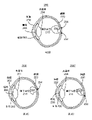
方法100の出力は、例えば、図2A~2Dを介して見られ得る。図2A、2B、2Cおよび2Dは、手順の推奨および予想される結果を含む眼200の準リアルタイム画像およびモデルの例示的実施形態を描写する。図2A~2Dは、原寸に比例しておらず、説明の目的のみのものである。したがって、特定の患者、症状または反応は、図2A~2Dに示されるように意図されない。図2Aは、眼の画像200を描写する。角膜202、水晶体204、虹彩206、瞳孔208、硝子体腔210および網膜220が説明の目的のために示される。硝子体腔210内の領域230は、ERM、高ストレスまたは他の問題の領域であり得る。説明の目的のために、領域230は、ERM230であると仮定する。画像200は、手術の直前にまたは手術中の何らかの時点で撮影された準リアルタイム画像またはその一部であり得る。代わりに、画像200は、期せずして眼の状態を表し続ける以前に撮影された手術前画像であり得る。
The output of
図2Bは、矢印によって指示される推奨される領域232および234を有する眼の画像200を描写する。推奨される領域232および234は、高ストレス領域および/またはERM230が網膜220を引っ張っている領域であり得る。画像200は、純粋にモデルであり得、またはハイライトされた推奨される領域232および234を有する図2Aに示す画像200であり得る。ERM230が除去されるため、推奨される手順(ERM230を切断する)が必然的に知らされる。画像200’は、医師が見るグラフィックディスプレイ上でレンダリングされ得る。他の実施形態では、推奨は、別の方法で提供され得る。
FIG. 2B depicts an
図2Cは、画像200’、すなわち推奨される領域232のための推奨される手順の予想される結果を描写する。したがって、画像200’は、切断が領域232において行われた場合の眼のモデルであると考えられ得る。分かるように、ERM230’は、領域232から後退し、形状を変化し、回転するようにモデル化される。様々なストレスに対する形状および/または位置の他の変化がモデル化され得る。画像200’は、医師が領域232を選択することに応じて医師が見るためにグラフィックディスプレイ上でレンダリングされ得る。他の実施形態では、予想される結果は、別の方法で提供され得る。
FIG. 2C depicts the expected results of the recommended procedure for image 200', i.e., recommended
図2Cは、画像200’’、すなわち推奨される領域234のための推奨される手順の予想される結果を描写する。したがって、画像200’’は、切断が領域234において行われた場合の眼のモデルであると考えられ得る。分かるように、ERM230’は、領域234から後退し、形状を変化し、回転するようにモデル化される。様々なストレスに対する形状および/または位置の他の変化がモデル化され得る。画像200’’は、医師が領域234を選択することに応じて医師が見るためにグラフィックディスプレイ上でレンダリングされ得る。他の実施形態では、予想される結果は、別の方法で提供され得る。
FIG. 2C depicts the expected results of
方法100を使用して、外科医は、手術を眼に対してより良好に行うことができ得る。例えば、手術の直前に、方法100は、眼に関する最新情報を提供し、それらの手術計画が依然として適切であるかどうかを医師に示すために使用され得る。そうでなければ、外科医は、異なる方法で進めることを選択し得る。1つまたは複数の手順(例えば、切断)が手術の一部として行われた後、方法100は、反復され得る。したがって、外科医は、眼が予想通りに反応しているかどうかを判断し得、手術計画に対する逸脱に合わせて調整することができ得る。外科医は、特定の手順前に眼がどのように反応すると予想されるかの一般的な考えも有し得、適切な選択肢をより良好に選択することができ得る。したがって、医師が手術を行う能力が改善される。方法100は、外科医が、非常に複雑でありおよび/または手術計画の形成後に著しく修正された状況を提示される場合に特に有用であり得る。したがって、方法100は、比較的速く進行し、かつ/または外科医に複雑な病状を提示する糖尿病性網膜症もしくは増殖性硝子体網膜症などの症状に対して特別な実用性を有し得る。したがって、医師が手術を行う能力が改善される。
Using
図3は、準リアルタイム画像を使用して、眼科手術中に医師を支援する方法150の例示的実施形態を描写するフローチャートである。簡潔さのために、いくつかの工程は、省略され得、交互配置され得、別の順番で行われ得、かつ/または組み合わされ得る。方法150は、1つまたは複数のプロセッサ上で命令を実行する工程を含み得る。さらに、方法150は、眼科手術に関連して説明される。しかし、方法150は、他のタイプの手術に拡張され得る。
FIG. 3 is a flowchart illustrating an exemplary embodiment of
眼の少なくとも一部分の少なくとも1つの初期画像が工程152を介して受信される。工程152における画像の受信は、別個の撮像システムから画像のデータを受信する工程、または方法150を行う本システムの一部分によって画像を捕捉する工程を含み得る。したがって、工程152において受信される画像は、準リアルタイム画像であり得るが、必ずしもそうである必要はない。工程152において受信される画像は、OCT、超音波画像、高周波超音波画像、UBM画像および/または別の三次元画像を含み得る。
At least one initial image of at least a portion of the eye is received via
推奨される初期領域および推奨される初期手順は、工程154を介して初期画像および眼の計算モデルに基づいて判断される。眼の計算モデルは、方法100に関して上に論述された計算モデルに類似し得る。したがって、工程154の一部として、工程152において受信された初期画像のデータが処理される。例えば、特定の領域内のストレスは、初期画像データ内に見られる歪みから判断され得る。同様に、高ストレスに起因する条痕、折り目、薄膜化、断裂および/または様々な領域内の他の問題は、画像データおよび計算モデルに基づいて判断され得る。工程154は、上述の工程104に類似した方法で行われ得る。しかし、準リアルタイム画像であってもなくてもよい初期画像が使用される。いくつかの実施形態では、工程154は、特定の推奨される手順を明示的に判断する工程を含み得る。しかし、一般的に、推奨される手順は、進行中に特定の手術に関して知らされる。
The recommended initial region and recommended initial procedure are determined through
初期手順の初期の予想される結果は、工程156を介して計算される。工程156は、上述の工程106に類似し得る。しかし、準リアルタイム画像であってもなくてもよい初期画像が使用される。初期の推奨される領域、初期の推奨される手順および初期の予想される結果は、工程158を介して医師に提供され得る。工程158は、工程108に類似している。したがって、眼の画像および/または眼のモデルが医師のために表示され得る。したがって、いくつかの実施形態では、この情報は、医師にグラフィック的に提供される。他の実施形態では、初期の推奨される領域、初期の推奨される手順および初期の予想される結果を提供する別の機構が使用される。
The initial expected results of the initial procedure are calculated via
次に、外科医は、切断するなどの1つまたは複数の手順を行い得る。外科医は、工程158において提供された推奨を採用するかまたは別の手順を行うことを選択し得る。例えば、外科医は、異なる位置において切断を行うことを望み得る。外科医は、複数の手順も行い得る。
The surgeon may then perform one or more procedures, such as amputation. The surgeon may choose to adopt the recommendations provided in
外科医が手順を行った後、眼の少なくとも一部分の少なくとも1つのその場の準リアルタイム画像が工程160を介して受信される。工程160における画像の受信は、別個の撮像システムから画像のデータを受信する工程、または方法150を行う本システムの一部分によって画像を捕捉する工程を含み得る。工程160は、医師のために画像をレンダリングする工程を含む必要はない。代わりに、工程160は、眼のデータを取得する工程を含む。したがって、工程160は、工程102に類似している。
After the surgeon has performed the procedure, at least one in-situ near-real-time image of at least one portion of the eye is received via
推奨される次の領域および推奨される次の手順は、工程162を介して準リアルタイム画像および眼の計算モデルに基づいて判断される。工程162は、工程104に類似している。
The recommended next area and the recommended next procedure are determined through
推奨される次の手順の予想される次の結果も、工程164を介して準リアルタイム画像および計算モデルを使用して計算される。したがって、工程164は、その領域内のストレスの解放に対する周囲組織の反応を判断するために眼の計算モデルを使用する工程を含む。
The expected next outcome of the recommended next procedure is also calculated using the near real-time image and computational model via
推奨される次の領域、推奨される次の手順および予想される結果は、工程166を介して医師に提供される。工程166の一部は、様々な時間に行われ得る。例えば、推奨される次の領域および推奨される次の手順は、工程162において生成された準リアルタイム画像またはモデルをレンダリングすることによって行われ得る。予想される結果を医師に提供する工程は、受信された入力に応じて行われ得る。例えば、特定の推奨される領域が選択される場合、その領域において推奨される手順を行うことの予想される結果は、工程166において提供される。したがって、工程166は、工程164において計算された眼のモデルをレンダリングする工程を含み得る。
The recommended next area, the recommended next procedure and the expected results are provided to the physician via
次に、外科医は、1つまたは複数の他の手順を実行することを許容され得る。例えば、1つまたは複数の他の切断が行われ得る。医師は、方法150において提供された推奨に従い得るが、必ずしもそのようにする必要はない。次に、工程160に戻り、眼が再走査され得る。次の工程および次の領域の推奨は、工程164において判断された新しい推奨の新しい走査および予想される結果によって判断され得る。これらの新しい推奨および新しい予想される結果は、工程166を介して医師に提供され得る。したがって、工程160、162、164および166は、外科医を支援するために反復的に繰り返され得る。これらの工程は、外科医が手順を行うたびに反復され得るが、必ずしもそうする必要はない。代わりに、工程160、162、164および166は、手術中の選択された時点で反復され得る。したがって、医師は、有用であるかまたは必要であると見なすときにのみ、これらの工程を反復することを選択し得る。
The surgeon may then be allowed to perform one or more other procedures. For example, one or more other cuts may be made. Physicians can follow the recommendations provided in
方法150を使用して、外科医は、手術を眼に対してより良好に行うことができ得る。方法150は、外科医の以前の情報(より古い初期画像)を使用し始め、かつ/または最近捕捉された準リアルタイム画像を使用することができる。したがって、医師は、自らの手術計画が依然として適切であるかどうかを判断し得る。1つまたは複数の手順が手術の一部として行われた後、工程160、162、164および166が行われ得るかまたは反復され得る。したがって、外科医は、眼が予想通りに反応しているかどうかを判断し得、手術全体にわたって自らの行為に合わせて調整することができ得る。外科医は、特定の手順前に眼がどのように反応すると予想されるかの一般的な考えも有し得、適切な選択肢をより良好に選択することができ得る。したがって、医師が眼科手術を行う能力が強化され得る。
Using
図4は、準リアルタイム画像を使用して、眼科手術中に医師を支援する装置300の例示的実施形態のブロック図である。簡潔さのために、いくつか部品のみが示される。加えて、図4において描写される部品は、OCTまたは他の撮像システムなどの単一装置内に一緒にパッケージ化され得る。代わりに、データ収集および処理の部分などのいくつかの部品は、別々に実装され得る。さらに、部品は、ハードウェアおよびいくつかの場合にはソフトウェアで実装され得る。また、図4に示すのは、調べられるサンプルの眼302である。
FIG. 4 is a block diagram of an exemplary embodiment of a
装置300は、撮像システム310、コントローラ/プロセッサ320、予測ユニット330およびユーザインターフェース(U/I)340を含む。撮像システム310は、システム300の残り部分と別個であり得る。したがって、撮像システム310は、破線によって接続されて示される。装置300の一部である撮像システム310は、プロセッサ320によって制御される。作業者は、命令を入力し、U/I 340からの出力を受信し得る。例えば、作業者は、撮像システム310によって走査される眼302の領域を設定し、その結果を見ることができ、またはそうでなければ命令を与えてシステム300からの出力を受信することができる。いくつかの実施形態では、コントローラ/プロセッサ320は、眼302のIOPまたは他の特徴を設定するシステムにリンクされるかまたはそれを制御する。したがって、コントローラプロセッサ320は、準リアルタイム画像捕捉を制御するために使用され得る。
The
予測ユニット330は、少なくとも部分的にソフトウェアで実現され得る。予測ユニット330は、撮像システム310からのデータを処理する。したがって、眼の画像データ332および計算モデル334が示される。計算モデル334の一部分は、メモリ内に格納され得、したがって図4に示される。例えば、計算モデル334のための患者のパラメータだけでなく、眼302の様々な部分の抗張力または密度の値も格納され得る。したがって、眼のFEAモデルまたは他のモデルが生成および使用され得る。推奨/予想結果生成器336は、画像データ332を処理し、推奨される領域、推奨される手順および予想される結果を判断するために計算モデル334を使用する。任意選択的なレンダリング装置338を使用して、これらは、U/I 340上で医師にグラフィック的に表示され得る。任意選択的なレンダリング装置338は、準リアルタイム画像データをU/I 340上に単に表示するためにも使用され得る。したがって、装置300は、眼302が手術中に走査およびマッピングされることと、眼のデータが処理されることと、眼302の推奨および予想される反応が判断されることとを可能にする。したがって、装置300を使用して、方法100および/または150が実施され得る。したがって、本方法100および/または150の恩恵の1つまたは複数が実現され得る。
The
特に眼科手術の外科医を支援する方法およびシステムが説明された。本方法およびシステムは、示された例示的実施形態に従って説明された。当業者は、実施形態に対する変形形態があり得ることと、いかなる変形形態も本方法およびシステムの趣旨および範囲内に入るであろうこととを容易に認識するであろう。したがって、添付された請求項の趣旨および範囲から逸脱することなく多くの修正形態が当業者によってなされ得る。 In particular, methods and systems to assist surgeons in eye surgery were described. The method and system have been described according to the exemplary embodiments shown. One of ordinary skill in the art will readily recognize that there may be variations to embodiments and that any modifications will fall within the spirit and scope of the method and system. Therefore, many modifications can be made by one of ordinary skill in the art without departing from the spirit and scope of the attached claims.
Claims (14)
眼の複数の領域において、複数の眼内圧における複数の準リアルタイム画像を取得する工程であって、前記複数の眼内圧は、前記複数の準リアルタイム画像における歪みの様々な度合い及び眼の組織におけるストレス集中の様々な度合いの指示をもたらす、工程と、
前記複数の準リアルタイム画像に基づき、複数の領域におけるストレスレベルを判断す
る工程であって、前記複数の領域の第1の部分は、前記複数の領域の第2の部分より高い
ストレスを有する、工程と、
前記準リアルタイム画像および前記眼の計算モデルに基づき、推奨される次の領域を前記第1の部分とし、かつ、推奨される次の手順を網膜上膜(ERM)除去とする、判断をする工程と、
前記準リアルタイム画像および前記計算モデルを使用して、網膜上膜(ERM)除去によって引き起こされる網膜組織のストレスが解放されるように前記推奨される次の手順の予想される次の結果を計算する工程と、
前記推奨される次の領域、前記推奨される次の手順および前記予想される結果をグラフィックディスプレイ上において前記医師に表示する工程と、
を含む方法。 A method performed by a system that assists doctors in performing eye surgery.
A step of acquiring a plurality of quasi-real-time images at a plurality of intraocular pressures in a plurality of regions of the eye, wherein the plurality of intraocular pressures are various degrees of distortion in the plurality of quasi-real-time images and stress in the eye tissue. Processes and processes that provide instructions for different degrees of concentration,
A step of determining a stress level in a plurality of regions based on the plurality of quasi-real-time images, wherein the first portion of the plurality of regions has a higher stress than the second portion of the plurality of regions. When,
Based on the quasi-real-time image and the computational model of the eye, the step of making a determination that the next recommended region is the first part and the next recommended procedure is epiretinal membrane (ERM) removal. When,
The near real-time image and the computational model are used to calculate the expected next result of the next recommended procedure to relieve stress on the retinal tissue caused by epiretinal membrane (ERM) removal. Process and
The step of displaying the recommended next area, the recommended next procedure and the expected result to the physician on a graphic display.
How to include.
前記複数の領域の前記第1の部分および前記複数の領域の前記第2の部分を指示する工程をさらに含む、請求項4に記載の方法。 The step of capturing the quasi-real-time image is
The method of claim 4 , further comprising the step of designating the first portion of the plurality of regions and the second portion of the plurality of regions.
眼の複数の領域において、複数の眼内圧における複数の準リアルタイム画像を取得し、複数の眼内圧は、前記複数の準リアルタイム画像における歪みの様々な度合い及び眼の組織におけるストレス集中の様々な度合いの指示をもたらす、準リアルタイム画像捕捉ユニットと、
前記複数の準リアルタイム画像に基づき、複数の領域におけるストレスレベルを判断し、前記複数の領域の第1の部分は、前記複数の領域の第2の部分より高いストレスを有しており、
前記準リアルタイム画像および前記眼の計算モデルに基づき、推奨される次の領域を前記第1の部分とし、かつ、推奨される次の手順を網膜上膜(ERM)除去とする、判断をする、予測ユニットであって、前記準リアルタイム画像および前記計算モデルを使用して、前記推奨される次の手順が、前記網膜上膜(ERM)除去によって引き起こされる網膜組織のストレスの解放である、と予想される次の結果を計算する予測ユニットと、
前記推奨される次の領域、前記推奨される次の手順および前記予想される結果を前記医師に表示するユーザインターフェースと、
を含むシステム。 A system that assists doctors in performing eye surgery.
Multiple quasi-real-time images at multiple intraocular pressures are acquired in multiple regions of the eye, where the multiple intraocular pressures are varying degrees of distortion in the plurality of quasi-real-time images and varying degrees of stress concentration in the eye tissue. With a quasi-real-time image capture unit, which provides instructions for
Based on the plurality of quasi-real-time images, the stress level in the plurality of regions is determined, and the first portion of the plurality of regions has higher stress than the second portion of the plurality of regions.
Based on the near-real-time image and the computational model of the eye, it is determined that the next recommended region is the first part and the next recommended procedure is epiretinal membrane (ERM) removal. Using the near-real-time image and the computational model in the predictive unit, it is predicted that the recommended next step is the release of stress on the retinal tissue caused by the epiretinal membrane (ERM) removal. With a prediction unit that calculates the next result to be done,
A user interface that displays the recommended next area, the recommended next procedure, and the expected result to the physician.
System including.
Applications Claiming Priority (3)
| Application Number | Priority Date | Filing Date | Title |
|---|---|---|---|
| US15/245,328 US10842573B2 (en) | 2016-08-24 | 2016-08-24 | Predictive apparatus for assisting a physician during ophthalmic surgery |
| US15/245,328 | 2016-08-24 | ||
| PCT/IB2017/055087 WO2018037357A1 (en) | 2016-08-24 | 2017-08-23 | Predictive apparatus for assisting a physician during ophthalmic surgery |
Publications (3)
| Publication Number | Publication Date |
|---|---|
| JP2019526334A JP2019526334A (en) | 2019-09-19 |
| JP2019526334A5 JP2019526334A5 (en) | 2020-04-23 |
| JP7100623B2 true JP7100623B2 (en) | 2022-07-13 |
Family
ID=59923489
Family Applications (1)
| Application Number | Title | Priority Date | Filing Date |
|---|---|---|---|
| JP2019510898A Active JP7100623B2 (en) | 2016-08-24 | 2017-08-23 | Predictive device to assist doctors during eye surgery |
Country Status (7)
| Country | Link |
|---|---|
| US (2) | US10842573B2 (en) |
| EP (1) | EP3504655B1 (en) |
| JP (1) | JP7100623B2 (en) |
| CN (1) | CN109643582A (en) |
| AU (1) | AU2017315286A1 (en) |
| CA (1) | CA3031180A1 (en) |
| WO (1) | WO2018037357A1 (en) |
Families Citing this family (7)
| Publication number | Priority date | Publication date | Assignee | Title |
|---|---|---|---|---|
| US10842573B2 (en) * | 2016-08-24 | 2020-11-24 | Alcon Inc. | Predictive apparatus for assisting a physician during ophthalmic surgery |
| US20200163727A1 (en) * | 2018-11-26 | 2020-05-28 | Douglas Patton | Cloud based system cataract treatment database and algorithm system |
| AU2020270386A1 (en) * | 2019-05-03 | 2021-12-02 | Lensar, Inc. | Cloud based system cataract treatment database and algorithm system |
| CN111616800B (en) * | 2020-06-09 | 2023-06-09 | 电子科技大学 | Ophthalmic surgery navigation system |
| JP7516172B2 (en) * | 2020-09-08 | 2024-07-16 | キヤノンメディカルシステムズ株式会社 | Ultrasound diagnostic device and program |
| DE102021100645A1 (en) * | 2021-01-14 | 2022-07-14 | Schwind Eye-Tech-Solutions Gmbh | Method for predicting a future position of a target point of an eye to compensate for a latency of an image evaluation; Control device and treatment device |
| DE102022133005A1 (en) | 2022-12-12 | 2024-06-13 | Carl Zeiss Meditec Ag | Eye surgery operating system, computer program and method for providing evaluation information regarding the guidance of a surgical tool |
Citations (3)
| Publication number | Priority date | Publication date | Assignee | Title |
|---|---|---|---|---|
| WO2015017375A2 (en) | 2013-07-29 | 2015-02-05 | Bioptigen, Inc. | Procedural optical coherence tomography (oct) for surgery and related systems and methods |
| WO2016082017A1 (en) | 2014-11-27 | 2016-06-02 | Synaptive Medical (Barbados) Inc. | Method, system and apparatus for quantitative surgical image registration |
| US20160192835A1 (en) | 2014-12-12 | 2016-07-07 | Carl Zeiss Ag | System for eye examination by means of stress-dependent parameters |
Family Cites Families (16)
| Publication number | Priority date | Publication date | Assignee | Title |
|---|---|---|---|---|
| US5891131A (en) | 1993-02-01 | 1999-04-06 | Arizona Board Of Regents | Method and apparatus for automated simulation and design of corneal refractive procedures |
| US6213998B1 (en) * | 1998-04-02 | 2001-04-10 | Vanderbilt University | Laser surgical cutting probe and system |
| US7192412B1 (en) * | 2002-09-14 | 2007-03-20 | Glaukos Corporation | Targeted stent placement and multi-stent therapy |
| CN1781466A (en) * | 2004-12-03 | 2006-06-07 | 上海市杨浦区民办华都医院 | Safety quality control system and method for excimer laser operation of ophthalmology |
| EP1733744A1 (en) * | 2005-06-17 | 2006-12-20 | Ludwig-Maximilians-Universität München | Method, dye and medicament for staining the internal limiting membrane and/or the capsule of an eye |
| EP2656780B1 (en) * | 2006-05-26 | 2016-04-27 | The Cleveland Clinic Foundation | System for measuring biomechanical properties in an eye |
| US20080082088A1 (en) * | 2006-09-05 | 2008-04-03 | Intralase Corp. | System and method for resecting corneal tissue |
| US20100049447A1 (en) | 2008-08-22 | 2010-02-25 | Gholam Peyman | Method of modeling the behavior of an eye subjected to an external force |
| US9411938B2 (en) | 2009-04-02 | 2016-08-09 | Sie Ag, Surgical Instrument Engineering | System for defining cuts in eye tissue |
| US8414124B2 (en) * | 2009-10-21 | 2013-04-09 | Sis Ag, Surgical Instrument Systems | Device and method for measuring a cornea |
| US10045882B2 (en) * | 2009-10-30 | 2018-08-14 | The Johns Hopkins University | Surgical instrument and systems with integrated optical sensor |
| US8591031B2 (en) * | 2010-11-19 | 2013-11-26 | Ziemer Ophthalmic Systems Ag | Device and method for determining the visual field |
| CN101999910A (en) * | 2010-12-09 | 2011-04-06 | 天津迈达医学科技有限公司 | Adaptive time-gain compensation method for use in ophthalmic ultrasonic measurement equipment |
| WO2016064867A2 (en) * | 2014-10-20 | 2016-04-28 | The Regents Of The University Of California | Optical intraocular sensor and sensing method |
| TWI568408B (en) * | 2015-12-23 | 2017-02-01 | 財團法人工業技術研究院 | Intraocular pressure detecting device and detecting method thereof |
| US10842573B2 (en) * | 2016-08-24 | 2020-11-24 | Alcon Inc. | Predictive apparatus for assisting a physician during ophthalmic surgery |
-
2016
- 2016-08-24 US US15/245,328 patent/US10842573B2/en active Active
-
2017
- 2017-08-23 CA CA3031180A patent/CA3031180A1/en not_active Abandoned
- 2017-08-23 JP JP2019510898A patent/JP7100623B2/en active Active
- 2017-08-23 EP EP17771581.0A patent/EP3504655B1/en active Active
- 2017-08-23 CN CN201780051784.2A patent/CN109643582A/en active Pending
- 2017-08-23 WO PCT/IB2017/055087 patent/WO2018037357A1/en unknown
- 2017-08-23 AU AU2017315286A patent/AU2017315286A1/en not_active Abandoned
-
2020
- 2020-10-05 US US17/063,383 patent/US20210113281A1/en not_active Abandoned
Patent Citations (3)
| Publication number | Priority date | Publication date | Assignee | Title |
|---|---|---|---|---|
| WO2015017375A2 (en) | 2013-07-29 | 2015-02-05 | Bioptigen, Inc. | Procedural optical coherence tomography (oct) for surgery and related systems and methods |
| WO2016082017A1 (en) | 2014-11-27 | 2016-06-02 | Synaptive Medical (Barbados) Inc. | Method, system and apparatus for quantitative surgical image registration |
| US20160192835A1 (en) | 2014-12-12 | 2016-07-07 | Carl Zeiss Ag | System for eye examination by means of stress-dependent parameters |
Also Published As
| Publication number | Publication date |
|---|---|
| EP3504655B1 (en) | 2024-03-13 |
| US20180055581A1 (en) | 2018-03-01 |
| US20210113281A1 (en) | 2021-04-22 |
| WO2018037357A1 (en) | 2018-03-01 |
| CN109643582A (en) | 2019-04-16 |
| EP3504655A1 (en) | 2019-07-03 |
| JP2019526334A (en) | 2019-09-19 |
| CA3031180A1 (en) | 2018-03-01 |
| AU2017315286A1 (en) | 2019-02-07 |
| US10842573B2 (en) | 2020-11-24 |
Similar Documents
| Publication | Publication Date | Title |
|---|---|---|
| JP7100623B2 (en) | Predictive device to assist doctors during eye surgery | |
| JP2018171453A (en) | Procedural optical coherence tomography for surgery, and related systems and methods | |
| JP6174908B2 (en) | Information processing apparatus, information processing method, and computer program | |
| JP7413147B2 (en) | Image processing device, image processing method, and program | |
| JP7279712B2 (en) | Image processing method, program, and image processing apparatus | |
| JP6463048B2 (en) | Image processing apparatus and method of operating image processing apparatus | |
| JP6289462B2 (en) | Image processing apparatus and image processing method | |
| JP2019526334A5 (en) | ||
| JP2018147387A (en) | System and method for processing ophthalmic examination information | |
| JP6901403B2 (en) | Correction of OCT image | |
| JP7332463B2 (en) | Control device, optical coherence tomography device, control method for optical coherence tomography device, and program | |
| CN114364305A (en) | Slit-lamp microscope, ophthalmologic information processing apparatus, ophthalmologic system, control method for slit-lamp microscope, program, and recording medium | |
| JPWO2018207466A1 (en) | Image processing apparatus, image processing method, and image processing program | |
| JP2018033693A (en) | Image processing apparatus, image processing method, and program | |
| JP2021164535A (en) | Image processing device, image processing method and program | |
| JP2022111159A (en) | Laser treatment device and ophthalmologic information processing device | |
| WO2021049104A1 (en) | Slit lamp microscope, ophthalmic information processing device, ophthalmic system, method for controlling slit lamp microscope, and recording medium | |
| JP2018147386A (en) | System and method for processing ophthalmic examination information | |
| WO2008035425A1 (en) | Eyeground image analysis and program | |
| JP7415570B2 (en) | Ophthalmology imaging device control program, ophthalmology imaging system, and ophthalmology imaging device | |
| JP2019154993A (en) | Ophthalmologic apparatus, control method therefor, program, and storage medium | |
| US20240013397A1 (en) | Ophthalmologic image processing system, ophthalmologic image processing device, and storage medium for storing ophthalmologic image processing program | |
| JP6636188B2 (en) | Information processing apparatus, information processing apparatus control method, and computer program | |
| JP6732093B2 (en) | Information processing apparatus, information processing apparatus control method, and computer program | |
| WO2020179588A1 (en) | Surgical microscope system, image processing method, program, and image processing device |
Legal Events
| Date | Code | Title | Description |
|---|---|---|---|
| A711 | Notification of change in applicant |
Free format text: JAPANESE INTERMEDIATE CODE: A711 Effective date: 20191227 |
|
| RD03 | Notification of appointment of power of attorney |
Free format text: JAPANESE INTERMEDIATE CODE: A7423 Effective date: 20200124 |
|
| RD04 | Notification of resignation of power of attorney |
Free format text: JAPANESE INTERMEDIATE CODE: A7424 Effective date: 20200212 |
|
| A521 | Request for written amendment filed |
Free format text: JAPANESE INTERMEDIATE CODE: A523 Effective date: 20200311 |
|
| A621 | Written request for application examination |
Free format text: JAPANESE INTERMEDIATE CODE: A621 Effective date: 20200311 |
|
| A977 | Report on retrieval |
Free format text: JAPANESE INTERMEDIATE CODE: A971007 Effective date: 20210302 |
|
| A131 | Notification of reasons for refusal |
Free format text: JAPANESE INTERMEDIATE CODE: A131 Effective date: 20210309 |
|
| A521 | Request for written amendment filed |
Free format text: JAPANESE INTERMEDIATE CODE: A523 Effective date: 20210607 |
|
| A131 | Notification of reasons for refusal |
Free format text: JAPANESE INTERMEDIATE CODE: A131 Effective date: 20211116 |
|
| A521 | Request for written amendment filed |
Free format text: JAPANESE INTERMEDIATE CODE: A523 Effective date: 20220214 |
|
| TRDD | Decision of grant or rejection written | ||
| A01 | Written decision to grant a patent or to grant a registration (utility model) |
Free format text: JAPANESE INTERMEDIATE CODE: A01 Effective date: 20220621 |
|
| A61 | First payment of annual fees (during grant procedure) |
Free format text: JAPANESE INTERMEDIATE CODE: A61 Effective date: 20220701 |
|
| R150 | Certificate of patent or registration of utility model |
Ref document number: 7100623 Country of ref document: JP Free format text: JAPANESE INTERMEDIATE CODE: R150 |