JP6865329B1 - Display device, display method and program - Google Patents
Display device, display method and program Download PDFInfo
- Publication number
- JP6865329B1 JP6865329B1 JP2020537262A JP2020537262A JP6865329B1 JP 6865329 B1 JP6865329 B1 JP 6865329B1 JP 2020537262 A JP2020537262 A JP 2020537262A JP 2020537262 A JP2020537262 A JP 2020537262A JP 6865329 B1 JP6865329 B1 JP 6865329B1
- Authority
- JP
- Japan
- Prior art keywords
- electrocardiogram data
- abnormal
- display device
- electrocardiogram
- divided
- Prior art date
- Legal status (The legal status is an assumption and is not a legal conclusion. Google has not performed a legal analysis and makes no representation as to the accuracy of the status listed.)
- Active
Links
- 238000000034 method Methods 0.000 title claims description 7
- 230000002159 abnormal effect Effects 0.000 claims abstract description 148
- 238000010801 machine learning Methods 0.000 claims abstract description 60
- 208000019622 heart disease Diseases 0.000 claims abstract description 43
- 238000012545 processing Methods 0.000 claims abstract description 26
- 201000010099 disease Diseases 0.000 description 16
- 208000037265 diseases, disorders, signs and symptoms Diseases 0.000 description 16
- 238000010586 diagram Methods 0.000 description 13
- 230000006870 function Effects 0.000 description 11
- 238000004891 communication Methods 0.000 description 10
- 230000005856 abnormality Effects 0.000 description 6
- 238000013527 convolutional neural network Methods 0.000 description 5
- 230000000694 effects Effects 0.000 description 4
- 238000003745 diagnosis Methods 0.000 description 3
- 238000012986 modification Methods 0.000 description 3
- 230000004048 modification Effects 0.000 description 3
- 230000001133 acceleration Effects 0.000 description 2
- 239000003550 marker Substances 0.000 description 2
- 230000009194 climbing Effects 0.000 description 1
- 238000007796 conventional method Methods 0.000 description 1
- 238000013135 deep learning Methods 0.000 description 1
- 238000003703 image analysis method Methods 0.000 description 1
- 238000007689 inspection Methods 0.000 description 1
- 238000000718 qrs complex Methods 0.000 description 1
- 230000004044 response Effects 0.000 description 1
- 230000001360 synchronised effect Effects 0.000 description 1
- 230000000007 visual effect Effects 0.000 description 1
- 210000000707 wrist Anatomy 0.000 description 1
Images


Classifications
-
- G—PHYSICS
- G16—INFORMATION AND COMMUNICATION TECHNOLOGY [ICT] SPECIALLY ADAPTED FOR SPECIFIC APPLICATION FIELDS
- G16H—HEALTHCARE INFORMATICS, i.e. INFORMATION AND COMMUNICATION TECHNOLOGY [ICT] SPECIALLY ADAPTED FOR THE HANDLING OR PROCESSING OF MEDICAL OR HEALTHCARE DATA
- G16H50/00—ICT specially adapted for medical diagnosis, medical simulation or medical data mining; ICT specially adapted for detecting, monitoring or modelling epidemics or pandemics
- G16H50/20—ICT specially adapted for medical diagnosis, medical simulation or medical data mining; ICT specially adapted for detecting, monitoring or modelling epidemics or pandemics for computer-aided diagnosis, e.g. based on medical expert systems
-
- A—HUMAN NECESSITIES
- A61—MEDICAL OR VETERINARY SCIENCE; HYGIENE
- A61B—DIAGNOSIS; SURGERY; IDENTIFICATION
- A61B5/00—Measuring for diagnostic purposes; Identification of persons
- A61B5/0002—Remote monitoring of patients using telemetry, e.g. transmission of vital signals via a communication network
- A61B5/0004—Remote monitoring of patients using telemetry, e.g. transmission of vital signals via a communication network characterised by the type of physiological signal transmitted
- A61B5/0006—ECG or EEG signals
-
- A—HUMAN NECESSITIES
- A61—MEDICAL OR VETERINARY SCIENCE; HYGIENE
- A61B—DIAGNOSIS; SURGERY; IDENTIFICATION
- A61B5/00—Measuring for diagnostic purposes; Identification of persons
- A61B5/24—Detecting, measuring or recording bioelectric or biomagnetic signals of the body or parts thereof
- A61B5/316—Modalities, i.e. specific diagnostic methods
- A61B5/318—Heart-related electrical modalities, e.g. electrocardiography [ECG]
- A61B5/339—Displays specially adapted therefor
-
- A—HUMAN NECESSITIES
- A61—MEDICAL OR VETERINARY SCIENCE; HYGIENE
- A61B—DIAGNOSIS; SURGERY; IDENTIFICATION
- A61B5/00—Measuring for diagnostic purposes; Identification of persons
- A61B5/72—Signal processing specially adapted for physiological signals or for diagnostic purposes
- A61B5/7235—Details of waveform analysis
- A61B5/7264—Classification of physiological signals or data, e.g. using neural networks, statistical classifiers, expert systems or fuzzy systems
- A61B5/7267—Classification of physiological signals or data, e.g. using neural networks, statistical classifiers, expert systems or fuzzy systems involving training the classification device
-
- A—HUMAN NECESSITIES
- A61—MEDICAL OR VETERINARY SCIENCE; HYGIENE
- A61B—DIAGNOSIS; SURGERY; IDENTIFICATION
- A61B5/00—Measuring for diagnostic purposes; Identification of persons
- A61B5/72—Signal processing specially adapted for physiological signals or for diagnostic purposes
- A61B5/7271—Specific aspects of physiological measurement analysis
- A61B5/7282—Event detection, e.g. detecting unique waveforms indicative of a medical condition
-
- A—HUMAN NECESSITIES
- A61—MEDICAL OR VETERINARY SCIENCE; HYGIENE
- A61B—DIAGNOSIS; SURGERY; IDENTIFICATION
- A61B5/00—Measuring for diagnostic purposes; Identification of persons
- A61B5/74—Details of notification to user or communication with user or patient ; user input means
- A61B5/7475—User input or interface means, e.g. keyboard, pointing device, joystick
- A61B5/748—Selection of a region of interest, e.g. using a graphics tablet
-
- G—PHYSICS
- G16—INFORMATION AND COMMUNICATION TECHNOLOGY [ICT] SPECIALLY ADAPTED FOR SPECIFIC APPLICATION FIELDS
- G16H—HEALTHCARE INFORMATICS, i.e. INFORMATION AND COMMUNICATION TECHNOLOGY [ICT] SPECIALLY ADAPTED FOR THE HANDLING OR PROCESSING OF MEDICAL OR HEALTHCARE DATA
- G16H40/00—ICT specially adapted for the management or administration of healthcare resources or facilities; ICT specially adapted for the management or operation of medical equipment or devices
- G16H40/60—ICT specially adapted for the management or administration of healthcare resources or facilities; ICT specially adapted for the management or operation of medical equipment or devices for the operation of medical equipment or devices
- G16H40/63—ICT specially adapted for the management or administration of healthcare resources or facilities; ICT specially adapted for the management or operation of medical equipment or devices for the operation of medical equipment or devices for local operation
-
- G—PHYSICS
- G16—INFORMATION AND COMMUNICATION TECHNOLOGY [ICT] SPECIALLY ADAPTED FOR SPECIFIC APPLICATION FIELDS
- G16H—HEALTHCARE INFORMATICS, i.e. INFORMATION AND COMMUNICATION TECHNOLOGY [ICT] SPECIALLY ADAPTED FOR THE HANDLING OR PROCESSING OF MEDICAL OR HEALTHCARE DATA
- G16H40/00—ICT specially adapted for the management or administration of healthcare resources or facilities; ICT specially adapted for the management or operation of medical equipment or devices
- G16H40/60—ICT specially adapted for the management or administration of healthcare resources or facilities; ICT specially adapted for the management or operation of medical equipment or devices for the operation of medical equipment or devices
- G16H40/67—ICT specially adapted for the management or administration of healthcare resources or facilities; ICT specially adapted for the management or operation of medical equipment or devices for the operation of medical equipment or devices for remote operation
-
- G—PHYSICS
- G16—INFORMATION AND COMMUNICATION TECHNOLOGY [ICT] SPECIALLY ADAPTED FOR SPECIFIC APPLICATION FIELDS
- G16H—HEALTHCARE INFORMATICS, i.e. INFORMATION AND COMMUNICATION TECHNOLOGY [ICT] SPECIALLY ADAPTED FOR THE HANDLING OR PROCESSING OF MEDICAL OR HEALTHCARE DATA
- G16H50/00—ICT specially adapted for medical diagnosis, medical simulation or medical data mining; ICT specially adapted for detecting, monitoring or modelling epidemics or pandemics
- G16H50/30—ICT specially adapted for medical diagnosis, medical simulation or medical data mining; ICT specially adapted for detecting, monitoring or modelling epidemics or pandemics for calculating health indices; for individual health risk assessment
-
- G—PHYSICS
- G16—INFORMATION AND COMMUNICATION TECHNOLOGY [ICT] SPECIALLY ADAPTED FOR SPECIFIC APPLICATION FIELDS
- G16H—HEALTHCARE INFORMATICS, i.e. INFORMATION AND COMMUNICATION TECHNOLOGY [ICT] SPECIALLY ADAPTED FOR THE HANDLING OR PROCESSING OF MEDICAL OR HEALTHCARE DATA
- G16H50/00—ICT specially adapted for medical diagnosis, medical simulation or medical data mining; ICT specially adapted for detecting, monitoring or modelling epidemics or pandemics
- G16H50/70—ICT specially adapted for medical diagnosis, medical simulation or medical data mining; ICT specially adapted for detecting, monitoring or modelling epidemics or pandemics for mining of medical data, e.g. analysing previous cases of other patients
-
- A—HUMAN NECESSITIES
- A61—MEDICAL OR VETERINARY SCIENCE; HYGIENE
- A61B—DIAGNOSIS; SURGERY; IDENTIFICATION
- A61B5/00—Measuring for diagnostic purposes; Identification of persons
- A61B5/24—Detecting, measuring or recording bioelectric or biomagnetic signals of the body or parts thereof
- A61B5/316—Modalities, i.e. specific diagnostic methods
- A61B5/318—Heart-related electrical modalities, e.g. electrocardiography [ECG]
Abstract
心電図表示装置(3)は、所定期間にわたって測定された患者の全体心電図データが所定期間よりも短い所定時間長の心電図データに分割された分割心電図データを、所定時間長の複数の教師用心電図データを用いて機械学習した機械学習モデルに入力する入力処理部(341)と、機械学習部(33)から出力される、分割心電図データに心臓疾患が疑われる波形部位が含まれているか否かを示す判定結果を取得する結果取得部(342)と、判定結果に基づいて、複数の分割心電図データから心臓疾患が疑われる波形部位が含まれている一以上の異常心電図データを特定し、特定した一以上の異常心電図データのうち少なくとも1つの異常心電図データの少なくとも一部のデータに対応する波形画像を医師端末(2)に表示させる表示制御部(343)と、を有する。The electrocardiogram display device (3) divides the patient's total electrocardiogram data measured over a predetermined period into electrocardiogram data having a predetermined time length shorter than the predetermined period, and divides the divided electrocardiogram data into a plurality of teacher's electrocardiogram data having a predetermined time length. Whether or not the divided electrocardiogram data output from the input processing unit (341) input to the machine learning model machine-learned using the above and the machine learning unit (33) includes a waveform part suspected of having a heart disease. Based on the result acquisition unit (342) that acquires the judgment result shown, and based on the judgment result, one or more abnormal electrocardiogram data including a waveform site suspected of having a heart disease was identified and specified from a plurality of divided electrocardiogram data. It has a display control unit (343) for displaying a waveform image corresponding to at least a part of the abnormal electrocardiogram data of at least one of the one or more abnormal electrocardiogram data on the doctor terminal (2).
Description
本発明は、表示装置、表示方法及びプログラムに関する。 The present invention relates to display devices, display methods and programs.
従来、患者が身体に装着して長時間にわたって心拍を測定することができるホルター心電計が知られている(例えば、特許文献1を参照)。 Conventionally, there is known a Holter electrocardiograph that can be worn by a patient to measure a heart rate for a long period of time (see, for example, Patent Document 1).
従来、測定された心電図を検査技師が目視確認することにより、心電図において疾患が疑われる部分を抜き出し、抜き出された部分の心電図を医師が診断していた。心電図が長時間(例えば24時間)にわたって測定されたデータを含んでいる場合、検査技師が目視確認することにより疾患が疑われる部位を抜き出すことは困難であった。 Conventionally, a laboratory technician visually confirms a measured electrocardiogram to extract a part of the electrocardiogram suspected of having a disease, and a doctor diagnoses the extracted part of the electrocardiogram. When the electrocardiogram contained data measured over a long period of time (for example, 24 hours), it was difficult for the laboratory technician to visually confirm the site where the disease was suspected.
検査技師が目視確認することにより疾患が疑われる部位を抜き出すことができるとしても、従来の方法により抜き出すことができる部位は、QRS波形間のタイミングのずれ、又はQRS波形の形状の違いのように、異常が目立つ波形の部位に限られていた。このような中、検査技師が気付くことができない疾患が疑われる部位の心電図を医師が診断しやすくすることが求められていた。 Even if the part suspected of having a disease can be extracted by a visual check by an inspection technician, the part that can be extracted by the conventional method is a timing difference between QRS waveforms or a difference in the shape of the QRS complex. , It was limited to the part of the waveform where the abnormality was conspicuous. Under these circumstances, it has been required to make it easier for doctors to diagnose an electrocardiogram of a site suspected of having a disease that cannot be noticed by a laboratory technician.
そこで、本発明はこれらの点に鑑みてなされたものであり、心電図に疾患が疑われる部位が含まれているかどうかを医師が診断しやすくすることを目的とする。
Therefore, the present invention has been made in view of these points, and an object of the present invention is to make it easier for a doctor to diagnose whether or not an electrocardiogram contains a site suspected of having a disease.
本発明の第1の態様の心電図表示装置は、所定期間にわたって測定された患者の全体心電図データが前記所定期間よりも短い所定時間長の心電図データに分割された分割心電図データを、前記所定時間長の複数の教師用心電図データを用いて機械学習した機械学習モデルに入力する入力処理部と、前記機械学習モデルから出力される、前記分割心電図データに心臓疾患が疑われる波形部位が含まれているか否かを示す判定結果を取得する結果取得部と、前記判定結果に基づいて、複数の前記分割心電図データから心臓疾患が疑われる波形部位が含まれている一以上の異常心電図データを特定し、特定した一以上の異常心電図データのうち少なくとも1つの異常心電図データの少なくとも一部のデータに対応する波形画像を表示装置に表示させる表示制御部と、を有する。 In the electrocardiogram display device of the first aspect of the present invention, the divided electrocardiogram data obtained by dividing the patient's total electrocardiogram data measured over a predetermined period into electrocardiogram data having a predetermined time length shorter than the predetermined time period is obtained by dividing the divided electrocardiogram data into the predetermined time length. Does the input processing unit that inputs to the machine learning model machine-learned using the plurality of teacher's electrocardiogram data and the divided electrocardiogram data output from the machine learning model include a waveform part suspected of having a heart disease? A result acquisition unit that acquires a determination result indicating whether or not it is present, and based on the determination result, one or more abnormal electrocardiogram data including a waveform portion suspected of having a heart disease is identified from the plurality of divided electrocardiogram data. It has a display control unit for displaying a waveform image corresponding to at least a part of the abnormal electrocardiogram data of at least one of the specified one or more abnormal electrocardiogram data on the display device.
前記表示制御部は、複数の前記異常心電図データを特定した場合に、前記表示装置に波形画像が表示されている前記異常心電図データの他の異常心電図データの波形画像を表示するための表示操作を行うための操作画面を前記表示装置に表示させてもよい。 When the display control unit identifies a plurality of the abnormal electrocardiogram data, the display control unit performs a display operation for displaying the waveform image of the other abnormal electrocardiogram data of the abnormal electrocardiogram data whose waveform image is displayed on the display device. The operation screen for performing the operation may be displayed on the display device.
前記表示制御部は、前記複数の異常心電図データそれぞれを特定するための複数の識別情報を含む前記操作画面を前記表示装置に表示させてもよい。 The display control unit may display the operation screen including a plurality of identification information for identifying each of the plurality of abnormal electrocardiogram data on the display device.
前記複数の識別情報から一以上の識別情報を選択する操作を示す操作情報を取得する操作情報取得部をさらに有し、前記表示制御部は、前記操作情報取得部が取得した前記操作情報に基づいて、複数の前記異常心電図データに対応する複数の波形画像を同時に前記表示装置に表示させてもよい。 The display control unit further includes an operation information acquisition unit that acquires operation information indicating an operation for selecting one or more identification information from the plurality of identification information, and the display control unit is based on the operation information acquired by the operation information acquisition unit. Therefore, a plurality of waveform images corresponding to the plurality of abnormal electrocardiogram data may be displayed on the display device at the same time.
前記表示制御部は、前記表示装置に表示させる前記異常心電図データにおいて、前記機械学習モデルにおいて心臓疾患が疑われると判定された波形部位を特定するための情報を前記異常心電図データの波形画像とともに表示させてもよい。 The display control unit displays information for identifying a waveform portion determined to be suspected of having a heart disease in the machine learning model in the abnormal electrocardiogram data displayed on the display device together with a waveform image of the abnormal electrocardiogram data. You may let me.
心電図表示装置は、前記心電図データが測定された前記所定期間内の前記患者の状態を示す状態情報を時刻に関連付けて取得する状態情報取得部をさらに有し、前記表示制御部は、前記異常心電図データが測定された時刻に関連付けられた前記状態情報が示す状態を、前記異常心電図データの波形画像とともに前記表示装置に表示させてもよい。 The electrocardiogram display device further includes a state information acquisition unit that acquires state information indicating the state of the patient within the predetermined period in which the electrocardiogram data is measured in association with time, and the display control unit further includes the abnormal electrocardiogram. The state indicated by the state information associated with the time when the data is measured may be displayed on the display device together with the waveform image of the abnormal electrocardiogram data.
この場合、前記表示制御部は、複数の前記異常心電図データそれぞれを特定するための複数の識別情報と前記状態とを関連付けて前記表示装置に表示させてもよい。 In this case, the display control unit may display the display device in association with a plurality of identification information for identifying each of the plurality of abnormal electrocardiogram data and the state.
前記表示制御部は、前記全体心電図データに含まれる複数の前記分割心電図データのうち、心臓疾患が疑われる波形部位が含まれている正常心電図データと前記異常心電図データとを含む、複数の連続する前記分割心電図データの波形画像を表示させる第1モードと、前記一以上の異常心電図データの波形画像を表示させ前記正常心電図データの波形画像を表示させない第2モードと、を切り替えてもよい。 The display control unit is a plurality of continuous electrocardiogram data including a normal electrocardiogram data including a waveform portion suspected of having a heart disease and an abnormal electrocardiogram data among the plurality of divided electrocardiogram data included in the overall electrocardiogram data. The first mode in which the waveform image of the divided electrocardiogram data is displayed and the second mode in which the waveform image of the one or more abnormal electrocardiogram data is displayed and the waveform image of the normal electrocardiogram data is not displayed may be switched.
本発明の第2の態様の心電図表示装置は、所定期間にわたって測定された患者の心電図データのうち、心臓疾患が疑われる波形部位を含む一定時間長の心電図データである異常心電図データの少なくとも一部のデータに対応する波形画像を表示部に表示させる表示制御部と、前記心電図データに前記異常心電図データが複数含まれている場合、波形画像が表示されている前記異常心電図データの他の異常心電図データの少なくとも一部のデータに対応する波形画像を前記表示部に表示するための操作情報を取得する操作情報取得部と、を有する。 The electrocardiogram display device of the second aspect of the present invention is at least a part of abnormal electrocardiogram data which is electrocardiogram data of a certain time length including a waveform site suspected of having a heart disease among the electrocardiogram data of a patient measured over a predetermined period. A display control unit that displays a waveform image corresponding to the data of the above, and another abnormal electrocardiogram of the abnormal electrocardiogram data on which the waveform image is displayed when the electrocardiogram data includes a plurality of the abnormal electrocardiogram data. It has an operation information acquisition unit for acquiring operation information for displaying a waveform image corresponding to at least a part of the data on the display unit.
前記表示制御部は、前記複数の異常心電図データそれぞれを特定するための複数の識別情報を前記表示部に表示させ、前記操作情報取得部は、前記複数の識別情報から一以上の識別情報を選択する操作を示す前記操作情報を取得してもよい。 The display control unit causes the display unit to display a plurality of identification information for identifying each of the plurality of abnormal electrocardiogram data, and the operation information acquisition unit selects one or more identification information from the plurality of identification information. The operation information indicating the operation to be performed may be acquired.
本発明の第3の態様の心電図表示方法は、コンピュータが実行する、所定期間にわたって測定された患者の全体心電図データを前記所定期間よりも短い所定時間長の複数の分割心電図データに分割するステップと、前記複数の分割心電図データを、前記所定時間長の複数の教師用心電図データを用いて機械学習した機械学習モデルに入力するステップと、前記機械学習モデルから出力される、前記分割心電図データに心臓疾患が疑われる波形部位が含まれているか否かを示す判定結果を取得するステップと、前記判定結果に基づいて、複数の前記分割心電図データから心臓疾患が疑われる波形部位が含まれている一以上の異常心電図データを特定するステップと、特定した前記一以上の異常心電図データのうち少なくとも1つの異常心電図データの少なくとも一部のデータに対応する波形画像を表示装置に表示させるステップと、を有する。 The electrocardiogram display method of the third aspect of the present invention includes a step of dividing the patient's total electrocardiogram data measured over a predetermined period into a plurality of divided electrocardiogram data having a predetermined time length shorter than the predetermined period, which is executed by a computer. A step of inputting the plurality of divided electrocardiogram data into a machine learning model machine-learned using the plurality of teacher electrocardiogram data having a predetermined time length, and a heart in the divided electrocardiogram data output from the machine learning model. A step of acquiring a determination result indicating whether or not a waveform portion suspected of having a disease is included, and a waveform portion suspected of having a heart disease from a plurality of the divided electrocardiogram data based on the determination result. It has a step of specifying the above-mentioned abnormal electrocardiogram data and a step of displaying a waveform image corresponding to at least a part of the specified abnormal electrocardiogram data of at least one of the specified abnormal electrocardiogram data on the display device. ..
本発明の第4の態様の心電図表示方法は、コンピュータが実行する、所定期間にわたって測定された患者の心電図データのうち、心臓疾患が疑われる波形部位を含む一定時間長の心電図データである異常心電図データの少なくとも一部のデータに対応する波形画像を表示装置に表示させるステップと、前記心電図データに前記異常心電図データが複数含まれている場合、前記表示装置に表示されている前記異常心電図データの他の異常心電図データの少なくとも一部のデータに対応する波形画像を表示するための表示操作を受け付けるステップと、を有する。 The electrocardiogram display method of the fourth aspect of the present invention is an abnormal electrocardiogram which is a fixed-time length electrocardiogram data including a waveform portion suspected of having a heart disease among the electrocardiogram data of a patient measured over a predetermined period, which is executed by a computer. A step of displaying a waveform image corresponding to at least a part of the data on the display device, and when the electrocardiogram data includes a plurality of the abnormal electrocardiogram data, the abnormal electrocardiogram data displayed on the display device. It has a step of accepting a display operation for displaying a waveform image corresponding to at least a part of other abnormal electrocardiogram data.
本発明の第5の態様のプログラムは、コンピュータを、所定期間にわたって測定された患者の全体心電図データが前記所定期間よりも短い所定時間長の心電図データに分割された分割心電図データを、前記所定時間長の複数の教師用心電図データを用いて機械学習した機械学習モデルに入力する入力処理部、前記機械学習モデルから出力される、前記分割心電図データに心臓疾患が疑われる波形部位が含まれているか否かを示す判定結果を取得する結果取得部、及び前記判定結果に基づいて、複数の前記分割心電図データから心臓疾患が疑われる波形部位が含まれている一以上の異常心電図データを特定し、特定した一以上の異常心電図データのうち少なくとも1つの異常心電図データの少なくとも一部のデータに対応する波形画像を表示装置に表示させる表示制御部、として機能させる。 The program of the fifth aspect of the present invention uses a computer to divide the patient's total electrocardiogram data measured over a predetermined period into electrocardiogram data having a predetermined time length shorter than the predetermined period, and divides the divided electrocardiogram data into the predetermined time. An input processing unit that inputs to a machine learning model machine-learned using a plurality of long teacher ECG data, and whether the divided electrocardiogram data output from the machine learning model contains a waveform part suspected of having a heart disease. Based on the result acquisition unit that acquires the determination result indicating whether or not, and the determination result, one or more abnormal electrocardiogram data including the waveform portion suspected of having a heart disease is identified from the plurality of divided electrocardiogram data. It functions as a display control unit that displays a waveform image corresponding to at least a part of the abnormal electrocardiogram data of at least one of the specified one or more abnormal electrocardiogram data on the display device.
本発明の第6の態様のプログラムは、コンピュータを、所定期間にわたって測定された患者の心電図データのうち、心臓疾患が疑われる波形部位を含む一定時間長の心電図データである異常心電図データの少なくとも一部のデータに対応する波形画像を表示装置に表示させる表示制御部、及び前記心電図データに前記異常心電図データが複数含まれている場合、前記表示装置に波形画像が表示されている前記異常心電図データの他の異常心電図データの少なくとも一部のデータに対応する波形画像を表示するための表示操作を受け付ける操作情報取得部、として機能させる。 In the program of the sixth aspect of the present invention, at least one of the abnormal electrocardiogram data which is the electrocardiogram data of a certain time length including the waveform site where the heart disease is suspected among the electrocardiogram data of the patient measured by the computer over a predetermined period. The display control unit that displays the waveform image corresponding to the data of the unit on the display device, and the abnormal electrocardiogram data in which the waveform image is displayed on the display device when the electrocardiogram data includes a plurality of the abnormal electrocardiogram data. It functions as an operation information acquisition unit that accepts a display operation for displaying a waveform image corresponding to at least a part of other abnormal electrocardiogram data.
本発明によれば、心電図において疾患が疑われる部位を医師が診断しやすくなるという効果を奏する。 According to the present invention, there is an effect that it becomes easier for a doctor to diagnose a site suspected of having a disease on an electrocardiogram.
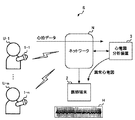
[心電図表示システムSの概要]
図1は、心電図表示システムSの概要を説明するための図である。心電図表示システムSは、心臓疾患を有する可能性がある患者Uの心電図を用いて医師が患者を診断しやすくするためのシステムである。心電図表示システムSは、心電計1(1−1〜1−n、nは自然数)と、医師端末2と、心電図表示装置3と、情報端末4とを備える。[Overview of ECG display system S]
FIG. 1 is a diagram for explaining an outline of the electrocardiogram display system S. The electrocardiogram display system S is a system for facilitating a doctor to diagnose a patient by using an electrocardiogram of a patient U who may have a heart disease. The electrocardiogram display system S includes an electrocardiograph 1 (1-1 to 1-n, n is a natural number), a
心電計1は、患者U(U−1〜U−n)が装着するホルター心電計であり、例えば、患者Uの手首に装着された状態で脈拍を測定することにより、心拍波形を示す心電図データを生成する。心電計1は、例えば、無線通信回線を含むネットワークNを介して、生成した心電図データを心電図表示装置3に送信する。心電図データには、心拍が測定された時刻を示す時刻情報が関連付けられている。心電計1が生成した心電図データは、ネットワークNを介することなく、例えば記憶媒体を介して心電図表示装置3に入力されてもよい。 The
医師端末2は、医師が使用する端末であり、例えばディスプレイ及びコンピュータを含む。医師端末2は、心電計1において生成された心電図データのうち、心電図表示装置3から受信した一部の心電図データに基づく波形画像を表示する。 The
心電図表示装置3は、心電計1又は医師端末2から受信した心電図データのうち心臓疾患が疑われる部位を含む異常心電図データを生成する装置であり、例えばサーバである。心電図表示装置3は、例えば、心電計1が生成した心電図データを取得し、機械学習モデルを用いて、取得した心電図データにおいて疾患が疑われる部位を特定する。機械学習モデルは、多数の正常な心電図データ(すなわち、疾患が表れていない心電図データ)と、異常な心電図データ(すなわち、疾患が表れている心電図データ)とを教師データとして機械学習(例えば深層学習)することにより作成されたモデルである。機械学習モデルの内部構成は任意であるが、例えばCNN(Convolutional Neural Network、畳み込みニューラルネットワーク)により構成されている。 The
心電図表示装置3は、心電図データのうち、疾患が疑われる部位が含まれている部位に対応する異常心電図データを生成し、生成した異常心電図データを医師端末2に送信することにより、異常心電図データの波形画像Hを医師端末2に表示させる。心電図表示装置3は、異常心電図データであるか正常心電図データであるかを示す情報を付した状態で分割心電図データを医師端末2に送信してもよく、異常心電図データのみを医師端末2に送信してもよい。疾患が疑われる部位が複数ある場合、心電図表示装置3は、疾患が疑われる部位に対応する心電図データが測定された時刻を示す情報に関連付けて、複数の異常心電図データを医師端末2に送信する。
以下、心電図表示装置3の構成及び動作を詳細に説明する。The
Hereinafter, the configuration and operation of the
図2は、心電図表示装置3の構成を示す図である。心電図表示装置3は、通信部31と、記憶部32と、機械学習部33と、制御部34とを有する。制御部34は、入力処理部341と、結果取得部342と、表示制御部343と、操作情報取得部344と、を有する。 FIG. 2 is a diagram showing a configuration of an
通信部31は、ネットワークNを介して心電計1及び医師端末2との間でデータを送受信するための通信コントローラを有する。通信部31は、ネットワークNを介して受信したデータを制御部34に通知する。また、通信部31は、ネットワークNを介して、制御部34から入力された心電図データを医師端末2に送信する。 The
記憶部32は、ROM(Read Only Memory)、RAM(Random Access Memory)、及びハードディスク等の記憶媒体を含む。記憶部32は、制御部34が実行するプログラムを記憶する。また、記憶部32は、制御部34が各種の演算を実行する際に必要な各種のデータを記憶する。 The
機械学習部33は、教師データとして用いられる教師用心電図データに基づいて学習することにより、入力された心電図データに心臓疾患の疑いがある部位があるか否かを判定した結果を出力することができる上述の機械学習モデルとして機能する。機械学習部33は、例えば、CNNを用いて各種の演算を実行するプロセッサと、CNNの係数を記憶するメモリと、を含んでいる。機械学習部33は、入力された心電図データが正常であるか異常であるかを示す情報を出力する。 The
機械学習部33は、入力された心電図データにおいて、入力された心電図データが異常心電図データであると判定した要因が表れている部位を示す異常部位情報をさらに出力してもよい。具体的には、まず機械学習部33は、入力された心電図データが異常心電図データであると判定した場合に、CNNを入力側に向かってたどる逆伝搬処理を実行し、入力と出力との間の差が相対的に大きかったノードを特定する。機械学習部33は、例えばGrad−CAM法を用いることにより、判定結果に与える影響が大きかった特徴に関連するノードを特定する。このようなノードは、判定結果に与える影響が大きい特徴に関連するノードである。 The
機械学習部33は、異常心電図データの波形において、特定したノードに対応する特徴が表れている部位を、入力された心電図データが異常心電図データであると判定した要因に関連する部位であるとする。機械学習部33は、例えば当該部位に対応する時刻情報、又は波形の部位を特定するための情報を含む異常部位情報を出力する。 In the waveform of the abnormal electrocardiogram data, the
制御部34は、記憶部32に記憶されたプログラムを実行することにより、入力処理部341、結果取得部342、表示制御部343、及び操作情報取得部344として機能する。 The
入力処理部341は、所定期間にわたって測定された患者の全体心電図データを心電計1から取得する。入力処理部341は、全体心電図データを医師端末2から取得してもよい。所定期間は、例えば24時間であるが、所定期間の長さは任意である。入力処理部341は、取得した心電図データを、それぞれ所定期間よりも短い所定時間長(例えば30秒)の複数の心電図データに分割する。本明細書においては、分割された心電図データを分割心電図データという。 The
なお、入力処理部341が取得する心電図データは任意であるが、例えばMFER(Medical waveform Format Encoding Rules、医用波形標準化技術規約)形式のデータである。入力処理部341は、MFER形式の心電図データを、心電図の波形を示す画像形式の心電図データに変換し、画像形式の分割心電図データを作成する。 The electrocardiogram data acquired by the
図3は、全体心電図と分割心電図との関係を示す模式図である。図3に示すように全体心電図が24時間にわたる心電図であり、分割心電図が30秒分の心電図である場合、分割心電図の数は、24時間×3600秒÷30秒=2880個となる。入力処理部341は、分割心電図を識別するために、分割心電図に識別番号(以下、「分割心電図ID」という。)を付与し、分割心電図IDに関連付けて分割心電図データを記憶部32に記憶させる。分割心電図IDは、例えば、全体心電図における分割心電図の順番を示す1から2880までの数値である。 FIG. 3 is a schematic diagram showing the relationship between the whole electrocardiogram and the divided electrocardiogram. As shown in FIG. 3, when the whole electrocardiogram is an electrocardiogram over 24 hours and the divided electrocardiogram is an electrocardiogram for 30 seconds, the number of divided electrocardiograms is 24 hours × 3600 seconds ÷ 30 seconds = 2880. The
入力処理部341は、機械学習モデルとして機能する機械学習部33に分割心電図データを入力する。当該機械学習モデルは、例えば、分割心電図データと同じ長さの教師用心電図データを、所定時間長の複数の教師用心電図データを用いて機械学習して作成されている。機械学習モデルが、分割心電図データと同じ長さの教師用心電図データを用いた機械学習により作成されていることで、全体心電図データが長時間にわたる心電図データを含む場合であっても、異常の有無を判定する精度が向上する。 The
結果取得部342は、機械学習部33が有する機械学習モデルから出力される、分割心電図データに心臓疾患が疑われる波形部位が含まれているか否かを示す判定結果を取得する。結果取得部342は、判定結果を表示制御部343に通知する。 The
結果取得部342は、分割心電図IDと、心臓疾患が疑われる波形部位が含まれているか否かを示す判定結果とを関連付けて記憶部32に記憶させてもよい。結果取得部342は、例えば、心臓疾患が疑われる波形部位が含まれている分割心電図データを分割心電図IDに関連付けて記憶部32に記憶させ、心臓疾患が疑われる波形部位が含まれていない分割心電図データを分割心電図IDに関連付けて記憶部32に記憶させない。結果取得部342がこのように動作することで、記憶部32の容量が不足しづらくなる。 The
表示制御部343は、表示装置として機能する医師端末2に心電図データの波形画像を表示させる。表示制御部343は、例えば、複数の分割心電図データのうち心臓疾患が疑われる波形部位が含まれている異常心電図データの少なくとも一部のデータに対応する波形画像を医師端末2に表示させるために、異常心電図データを医師端末2に送信する。異常心電図データの少なくとも一部のデータに対応する波形画像は、異常心電図データに対応する波形画像の全ての波形画像(例えば30秒の波形の画像)、又は異常心電図データに対応する波形画像のうちの一部の波形画像(例えば15秒の波形の画像)である。 The
表示制御部343は、異常心電図データを医師端末2に送信するために、結果取得部342が機械学習モデルから取得した判定結果に基づいて、複数の分割心電図データから心臓疾患が疑われる波形部位が含まれている一以上の異常心電図データを特定する。表示制御部343は、特定した一以上の異常心電図データのうち少なくとも1つの異常心電図データの波形画像を医師端末2に表示させる。すなわち、表示制御部343は、機械学習モデルにおいて心臓疾患が疑われると判定された波形部位を含む分割心電図データの少なくとも一部のデータに対応する波形画像を医師端末2に表示させる。 In order to transmit the abnormal electrocardiogram data to the
表示制御部343は、複数の異常心電図データを特定した場合に、医師端末2に波形画像が表示されている異常心電図データの他の異常心電図データの少なくとも一部のデータに対応する波形画像を表示するための表示操作を行うための操作画面を医師端末2に表示させる。表示制御部343は、例えば複数の異常心電図データそれぞれを特定するための複数の識別情報である分割心電図IDを含む操作画面を医師端末2に表示させる。表示制御部343が医師端末2に表示させる画面の詳細については後述する。 When a plurality of abnormal electrocardiogram data are specified, the
操作情報取得部344は、医師端末2において心電図を閲覧している医師が医師端末2で行った操作の内容を示す操作情報を取得する。操作情報取得部344は、例えば、医師端末2に波形画像を表示させる異常心電図データを選択するための操作画面を表示制御部343が医師端末2に表示させている状態で、複数の分割心電図IDから一以上の分割心電図IDを選択する操作を示す操作情報を医師端末2から取得する。 The operation
[心電図の表示画面]
図4は、表示制御部343が医師端末2に表示させる心電図の第1例である表示画面D1を示す図である。表示画面D1における領域R1、領域R2及び領域R4は、医師が他の異常心電図データの波形画像を表示するための表示操作を行うための操作画面の一例である。[ECG display screen]
FIG. 4 is a diagram showing a display screen D1 which is a first example of an electrocardiogram displayed on the
表示画面D1における領域R1には、異常心電図データの分割心電図IDが表示されている。このように、表示制御部343は、複数の異常心電図データそれぞれを特定するための複数の分割心電図IDを医師端末2に表示させる。図4に示す例においては、分割心電図IDが29、123、124、125、330、421の分割心電図データに心臓疾患が疑われる部位があることが領域R1に表示されている。医師は、領域R1に表示された複数の分割心電図IDから、波形画像を確認する対象の分割心電図IDを選択することができる。 In the area R1 on the display screen D1, the divided electrocardiogram ID of the abnormal electrocardiogram data is displayed. In this way, the
図4の領域R2は、波形を表示させる対象の分割心電図IDを入力するための領域である。医師端末2は、領域R2に入力された分割心電図IDに対応する分割心電図データの波形画像を表示する。医師端末2は、例えば、全ての分割心電図データを受信し、受信した分割心電図データのうち、領域R2に入力された分割心電図IDに対応する分割心電図データの波形画像を表示する。医師端末2は、領域R2に入力された分割心電図IDを含む操作情報を心電図表示装置3に送信し、送信した分割心電図IDに対応する分割心電図データの波形画像を心電図表示装置3から取得し、取得した分割心電図データの波形画像を表示してもよい。 The area R2 in FIG. 4 is an area for inputting the divided electrocardiogram ID of the target for which the waveform is to be displayed. The
領域R3には、分割心電図データの波形画像が表示される。表示制御部343は、操作受付部が受け付けた表示操作に基づいて、少なくとも1つの異常心電図データの波形画像を医師端末2に表示させる。図4に示す例において、表示制御部343は、領域R2に入力された分割心電図IDに対応する分割心電図データの波形画像とともに、当該分割心電図データに隣接する時間帯に取得された分割心電図データの波形画像も表示している。このように、選択された分割心電図IDに対応する分割心電図データの波形画像とともに、隣接する時間帯の分割心電図の波形画像も表示制御部343が医師端末2に表示させることで、医師が、心臓疾患が疑われる部位と他の部位とを比較しながら診断をすることが可能になる。 A waveform image of the divided electrocardiogram data is displayed in the region R3. The
なお、表示制御部343は、異常心電図データが、医師端末2の画面に同時に表示することができるデフォルト数(図4の場合は3)を超えて連続する場合、異常心電図データとともに正常心電図データを表示することができるように、医師端末2の画面に表示する分割心電図データの数をデフォルト数より増やしてもよい。 When the abnormal electrocardiogram data continues to exceed the default number (3 in the case of FIG. 4) that can be simultaneously displayed on the screen of the
領域R4は、領域R3に波形画像が表示される分割心電図データを切り替える操作を行うための領域である。医師は、領域R4において黒色で示されている操作バーを上下方向に移動することで、波形画像が表示される分割心電図データを切り替えることができる。表示制御部343は、例えば、操作バーが上向きに移動されたことを示す操作情報を操作情報取得部344が取得すると、波形画像が表示されている分割心電図データが取得された時刻よりも前の時刻に取得された分割心電図データの波形画像を医師端末2に表示させる。このように、医師は、操作バーを用いて、所望の分割心電図データを確認することができる。 The area R4 is an area for performing an operation of switching the divided electrocardiogram data in which the waveform image is displayed in the area R3. The doctor can switch the divided electrocardiogram data on which the waveform image is displayed by moving the operation bar shown in black in the region R4 in the vertical direction. For example, when the operation
なお、図4における「アップロード」アイコンは、医師端末2に記憶されている全体心電図データを心電図表示装置3にアップロードする際に用いられる。医師が、医師端末2に記憶されている全体心電図データのうち心臓疾患が疑われる部位を確認したい場合、全体心電図データのファイル名を選択してアップロードアイコンを選択することで、医師端末2から心電図表示装置3に全体心電図データが送信され、心電図表示装置3から異常心電図データを受信することができる。図4における「プリント」アイコンは、表示された心電図を印刷する操作をする際に用いられる。 The "upload" icon in FIG. 4 is used when uploading the entire electrocardiogram data stored in the
図5は、表示制御部343が医師端末2に表示させる心電図の第2例である表示画面D2を示す図である。図5においては、領域R2に入力された分割心電図IDに対応する分割心電図データの波形画像のみが表示されている。このように、表示制御部343は、1つの異常心電図データの波形画像のみを医師端末2に表示させてもよい。 FIG. 5 is a diagram showing a display screen D2 which is a second example of an electrocardiogram displayed on the
図6は、表示制御部343が医師端末2に表示させる心電図の第3例である表示画面D3を示す図である。図6においては、領域R1に表示された異常心電図データの分割心電図IDに対応する分割心電図データの波形画像のみが領域R3に表示されている。表示制御部343は、操作バーが上下方向に移動されると、複数の異常心電図データのうち医師端末2に表示させる異常心電図データを切り替える。 FIG. 6 is a diagram showing a display screen D3 which is a third example of an electrocardiogram displayed on the
表示制御部343は、例えば、操作バーが上向きに移動されたことを示す操作情報を操作情報取得部344が取得すると、波形画像が表示されている異常心電図データが取得された時刻よりも前の時刻に取得された異常心電図データの波形画像を医師端末2に表示させる。このように、表示制御部343が、操作情報取得部344が取得した操作情報に基づいて、複数の異常心電図データの波形画像を同時に医師端末2に表示させることで、医師が患者の心電図に表れている異常の傾向を把握しやすくなる。 For example, when the operation
図7は、表示制御部343が医師端末2に表示させる心電図の第4例である表示画面D4を示す図である。図7においては、複数の異常心電図データの分割心電図IDを一覧表示する領域R1が示されておらず、表示する波形画像を切り替えるためのアイコン画像C1、C2、及び表示中の異常心電図データの分割心電図IDを表示するための領域C3が示されている。アイコン画像C1は、波形画像を表示中の異常心電図データよりも前の時刻に測定された異常心電図データの波形画像を表示するための操作を医師が行うための画像である。アイコン画像C2は、波形画像を表示中の異常心電図データよりも後の時刻に測定された異常心電図データの波形画像を表示するための操作を医師が行うための画像である。 FIG. 7 is a diagram showing a display screen D4 which is a fourth example of an electrocardiogram displayed on the
図7(a)においては、分割心電図IDが125の異常心電図データの波形画像が表示されている。医師がアイコン画像C2を選択する操作をすると、図7(b)に示すように、分割心電図IDが125の異常心電図データの次に測定された、分割心電図IDが330の異常心電図データの波形画像が表示される。 In FIG. 7A, a waveform image of abnormal electrocardiogram data having a divided electrocardiogram ID of 125 is displayed. When the doctor selects the icon image C2, as shown in FIG. 7B, the waveform image of the abnormal electrocardiogram data having the divided electrocardiogram ID of 330, which is measured next to the abnormal electrocardiogram data having the divided electrocardiogram ID of 125. Is displayed.
図8は、表示制御部343が医師端末2に表示させる心電図の第5例である表示画面D5を示す図である。図8においては、表示制御部343が、医師端末2に表示させる異常心電図データにおいて、機械学習モデルにおいて心臓疾患が疑われると判定された波形部位を特定するための情報であるマーカーMを異常心電図データの波形画像とともに表示させている。マーカーMで囲まれた部位は、機械学習部33が逆伝搬処理を実行することにより特定した、入力された心電図データが異常心電図データであると判定した要因となった部位である。表示制御部343が、このように心臓疾患が疑われると判定された波形部位を示す情報を医師端末2に表示させることで、心臓疾患が専門でない医師であっても、精査すべき部位を把握しやすくなる。 FIG. 8 is a diagram showing a display screen D5, which is a fifth example of an electrocardiogram displayed on the
表示制御部343は、図4から図8に示したような各種の表示モードを切り替えてもよい。表示制御部343は、例えば、第1モード(例えば図4、図8に示すモード)と第2モード(例えば図5、図6、図7に示すモード)とを切り替える。 The
第1モードは、表示制御部343が、全体心電図データに含まれる複数の分割心電図データのうち、心臓疾患が疑われる波形部位が含まれている正常分割心電図データと異常分割心電図データとを含む、複数の連続する分割心電図データの波形画像を表示させるモードである。第2モードは、表示制御部343が、一以上の異常分割心電図データの波形画像を表示させ正常分割心電図データの波形画像を表示させないモードである。このように、表示制御部343が、医師端末2を使用する医師の操作に応じて表示モードを切り替えることで、医師が心電図を確認する目的に適した態様で異常心電図を医師端末2に表示させることができるので、医師が適切な診断を行いやすくなる。 In the first mode, the
[動作フローチャート]
図9は、制御部34が実行する動作のフローチャートである。図9に示すフローチャートは、入力処理部341が全体心電図データを取得した時点から開始している(S11)。[Operation flowchart]
FIG. 9 is a flowchart of the operation executed by the
入力処理部341は、全体心電図データを取得すると、所定の時間ごとに全体心電図データを分割して複数の分割心電図データを作成する(S12)。入力処理部341は、作成した複数の分割心電図データを機械学習部33に入力する(S13)。結果取得部342は、分割心電図データに疾患が疑われる部位があるか否かを判定した結果を機械学習部33から取得する(S14)。 When the
続いて、表示制御部343は、結果取得部342が取得した判定結果に基づいて、複数の分割心電図データから異常心電図データを選択する(S15)。表示制御部343は、医師端末2からの要求に応じて、選択した異常心電図データの波形画像を医師端末2に表示させる(S16)。 Subsequently, the
[患者Uの状態を表示]
図10は、心電図表示システムSの変形例を示す図である。図10に示す心電図表示装置3は、患者Uが使用する情報端末4から受信した情報に基づいて特定された患者Uの状態を異常心電図データの波形画像とともに医師端末2に表示させる。情報端末4は、例えばスマートフォンのように患者Uが携帯して使用する端末であり、患者Uの状態を検出するための各種のセンサーを有している。[Display the status of patient U]
FIG. 10 is a diagram showing a modified example of the electrocardiogram display system S. The
図11は、変形例に係る心電図表示装置3aの構成を示す図である。図11に示す心電図表示装置3aは、状態情報取得部345をさらに有するという点で、図2に示した心電図表示装置3と異なり、他の点で同じである。 FIG. 11 is a diagram showing a configuration of an electrocardiogram display device 3a according to a modified example. The electrocardiogram display device 3a shown in FIG. 11 is different from the
状態情報取得部345は、心電図データが測定された所定期間内の患者Uの状態を示す状態情報を時刻に関連付けて取得する。状態情報取得部345は、取得した状態情報及び時刻を示す情報を表示制御部343に通知する。状態情報取得部345は、患者Uの動き、患者Uがいる場所(緯度・経度・高度)、患者Uがいる場所の温度等のように、患者Uの状態に関連する情報を情報端末4から取得する。患者Uの動きを示す情報は、例えば、情報端末4が有する加速度センサーが検出した加速度である。患者Uがいる場所を示す情報は、例えば、情報端末4が有するGPS受信機が特定した緯度・経度を示す情報及び高度センサーが検出した高度である。 The state
表示制御部343は、状態情報取得部345から通知された複数の時刻に関連付けられた複数の状態情報のうち、異常心電図データが測定された時刻に対応する状態情報を特定する。表示制御部343は、異常心電図データが測定された時刻に関連付けられた状態情報が示す状態を、異常心電図データの波形画像とともに医師端末2に表示させる。表示制御部343は、複数の異常心電図データそれぞれを特定するための複数の異常心電図IDと患者Uの状態とを関連付けて医師端末2に表示させる。 The
図12は、異常心電図とともに患者Uの状態が表示された表示画面D6を示す図である。図12において、分割心電図IDが29の異常心電図データの波形画像に関連付けて「階段を上った」という状態が表示されており、分割心電図IDが123、124の異常心電図データの波形画像に関連付けて「走った」という状態が表示されている。このように、表示制御部343が異常心電図の波形画像と患者Uの状態とを関連付けて医師端末2に表示させることで、異常が発生したときの患者Uの状態を医師が把握することができるので、医師が適切な診断を行いやすくなる。 FIG. 12 is a diagram showing a display screen D6 in which the state of the patient U is displayed together with the abnormal electrocardiogram. In FIG. 12, the state of “climbing the stairs” is displayed in association with the waveform image of the abnormal electrocardiogram data having the divided electrocardiogram ID of 29, and is associated with the waveform image of the abnormal electrocardiogram data having the divided electrocardiogram IDs of 123 and 124. The status of "running" is displayed. In this way, the
なお、心電図データに関連付けられた時刻情報と状態情報に関連付けられた時刻情報とが同期している場合、機械学習部33は、心電図データと状態情報とを組み合わせたデータを教師データとして用いて学習してもよい。この場合、入力処理部341は、分割心電図データと、分割心電図データが測定された時刻における患者Uの状態を示す状態情報とを機械学習部33に入力する。機械学習部33は、状態情報とともに入力された分割心電図データに心臓疾患の疑いがある部位が含まれているか否かを判定し、判定結果を出力する。このように機械学習部33が分割心電図データとともに状態情報を使用することで、判定精度がさらに向上する。 When the time information associated with the electrocardiogram data and the time information associated with the state information are synchronized, the
入力処理部341は、心電図データのみを機械学習部33に入力し、状態情報を表示制御部343に通知してもよい。この場合、表示制御部343は、機械学習部33から異常心電図データであると判定された分割心電図データであっても、状態情報に基づいて異常ではないと判定される場合には、正常心電図データとして扱ってもよい。 The
[心電図表示装置として機能する医師端末2]
以上の説明においては、心電図表示装置3が心電図において心臓疾患の疑いがある部位を特定した結果に基づいて、異常心電図データの波形画像を医師端末2に表示させる場合を例示した。これに対して、医師が使用する医師端末2が、心電図において心臓疾患の疑いがある部位を特定し、当該部位を含む心電図を表示する心電図表示装置として機能してもよい。[
In the above description, a case where the
図13は、心電図表示装置として機能する医師端末2の構成を示す図である。図13に示す医師端末2は、通信部21と、記憶部22と、機械学習部23と、制御部24と、表示部25と、操作部26とを有する。制御部24は、入力処理部241と、結果取得部242と、表示制御部243と、操作情報取得部244と、を有する。通信部21、記憶部22、機械学習部23及び制御部24は、それぞれ図2に示した心電図表示装置3における通信部31、記憶部32、機械学習部33及び制御部34と同等の機能を有する。 FIG. 13 is a diagram showing a configuration of a
入力処理部241、結果取得部242、表示制御部243、及び操作情報取得部244は、それぞれ入力処理部341、結果取得部342、表示制御部343、及び操作情報取得部344と同等の機能を有する。医師端末2においても、制御部24が記憶部22に記憶されたプログラムを実行することにより、制御部24は、入力処理部241、結果取得部242、表示制御部243及び操作情報取得部244として機能する。 The
表示制御部243は、所定期間にわたって測定された患者の心電図データのうち、心臓疾患が疑われる波形部位を含む一定時間長の心電図データである異常心電図データの波形画像を表示部25に表示させる。表示制御部243は、複数の異常心電図データそれぞれを特定するための複数の識別情報である分割心電図IDを表示部25に表示させる。 The
操作情報取得部244は、心電図データに異常心電図データが複数含まれている場合、表示部25に波形画像が表示されている異常心電図データの他の異常心電図データの波形画像を表示するための操作情報を取得する。複数の識別情報から一以上の識別情報を選択する操作を示す操作情報を取得し、取得した操作情報を表示制御部243に通知する。表示制御部243は、通知された操作情報に基づいて、表示部25に波形画像を表示させる異常心電図データを選択する。医師端末2がこのように構成されていることで、医師端末2は、ネットワークを介してサーバに接続されていない状態であっても、機械学習モデルを用いて特定された異常心電図を表示することができる。 The operation
[心電図表示システムSによる効果]
以上説明したように、心電図表示システムSにおいては、表示制御部343が、分割心電図データが入力された機械学習部33から取得した、心臓疾患が疑われる波形部位が含まれているか否かを示す判定結果に基づいて特定した異常心電図データの波形画像を医師端末2に表示させる。心電図表示システムSがこのように構成されていることで、医師端末2を使用する医師が心臓疾患の専門医でない場合であっても、医師が疾患の有無を適切に判断することができる確率が高まる。[Effect of ECG display system S]
As described above, in the electrocardiogram display system S, the
また、表示制御部343は、医師端末2に波形画像が表示されている異常心電図データの他の異常心電図データの波形画像を表示するための表示操作を行うための操作画面を医師端末2に表示させる。そして、操作情報取得部344は、心電図データに異常心電図データが複数含まれている場合、波形画像が表示されている異常心電図データの他の異常心電図データの波形画像を医師端末2に表示するための操作情報を取得する。表示制御部343及び操作情報取得部344がこのように動作することで、医師は、長期間にわたって取得された心電図データのうち、異常が発生している疑いがある部位を容易に確認することが可能になる。 Further, the
以上、本発明を実施の形態を用いて説明したが、本発明の技術的範囲は上記実施の形態に記載の範囲には限定されず、その要旨の範囲内で種々の変形及び変更が可能である。例えば、装置の全部又は一部は、任意の単位で機能的又は物理的に分散・統合して構成することができる。また、複数の実施の形態の任意の組み合わせによって生じる新たな実施の形態も、本発明の実施の形態に含まれる。組み合わせによって生じる新たな実施の形態の効果は、もとの実施の形態の効果を併せ持つ。 Although the present invention has been described above using the embodiments, the technical scope of the present invention is not limited to the scope described in the above embodiments, and various modifications and changes can be made within the scope of the gist thereof. is there. For example, all or a part of the device can be functionally or physically distributed / integrated in any unit. Also included in the embodiments of the present invention are new embodiments resulting from any combination of the plurality of embodiments. The effect of the new embodiment produced by the combination also has the effect of the original embodiment.
例えば、以上の説明においては、心電図データに心臓疾患が疑われる部位が含まれているか否かを機械学習部33が判定した結果に基づいて、表示制御部343が異常心電図データに対応する波形画像を医師端末2に表示させる場合を例示した。しかしながら、表示制御部343は、機械学習部33を用いることなく、異常心電図データに対応する波形画像を医師端末2に表示させてもよい。表示制御部343は、例えば機械学習モデルを用いない画像解析手法を用いて分割心電図データに異常があるか否かを判定した結果に基づいて、異常心電図データに対応する波形画像を医師端末2に表示させてもよい。 For example, in the above description, the
1 心電計
2 医師端末
3 心電図表示装置
4 情報端末
31 通信部
32 記憶部
33 機械学習部
34 制御部
35 表示部
36 操作部
341 入力処理部
342 結果取得部
343 表示制御部
344 操作情報取得部
345 状態情報取得部1
Claims (10)
前記機械学習モデルから出力される、前記分割心電図データに心臓疾患が疑われる波形部位が含まれているか否かを示す判定結果を取得する結果取得部と、
前記判定結果に基づいて、複数の前記分割心電図データから心臓疾患が疑われる波形部位が含まれている複数の異常心電図データを特定し、特定した前記複数の異常心電図データの少なくとも一部の前記異常心電図データに対応する前記所定時間長の波形画像を表示装置に表示させる表示制御部と、
前記複数の異常心電図データそれぞれを特定するための複数の識別情報から一以上の識別情報を選択する操作を示す操作情報を取得する操作情報取得部と、
を有し、
前記表示制御部は、前記操作情報取得部が取得した前記操作情報に基づいて特定した一以上の前記異常心電図データに対応する前記所定時間長の一以上の前記波形画像を同時に前記表示装置に表示させる、
表示装置。 The divided electrocardiogram data obtained by dividing the patient's total electrocardiogram data measured over a predetermined period into electrocardiogram data having a predetermined time length shorter than the predetermined time period was machine-learned using a plurality of teacher electrocardiogram data having the predetermined time length. Input processing unit to input to machine learning model,
A result acquisition unit that acquires a determination result indicating whether or not the divided electrocardiogram data output from the machine learning model includes a waveform portion suspected of having a heart disease.
Based on the determination result, a plurality of abnormal electrocardiogram data including a waveform site suspected of having a heart disease is identified from the plurality of divided electrocardiogram data, and at least a part of the specified abnormal electrocardiogram data is said to be abnormal. A display control unit that displays a waveform image of the predetermined time length corresponding to the electrocardiogram data on the display device, and
An operation information acquisition unit that acquires operation information indicating an operation of selecting one or more identification information from a plurality of identification information for identifying each of the plurality of abnormal electrocardiogram data, and an operation information acquisition unit.
Have,
The display control unit simultaneously displays one or more of the waveform images of the predetermined time length corresponding to the one or more abnormal electrocardiogram data specified based on the operation information acquired by the operation information acquisition unit on the display device. Let,
Display device.
請求項1記載の表示装置。 The display control unit causes the display device to display the waveform image corresponding to the predetermined time length, which is the time length of the divided electrocardiogram data input by the input processing unit to the machine learning model.
The display device according to claim 1.
請求項1又は2に記載の表示装置。 When the display control unit identifies a plurality of the abnormal electrocardiogram data, the display device displays the waveform image corresponding to the abnormal electrocardiogram data having the predetermined time length in addition to the abnormal electrocardiogram data. The display device is displayed with an operation screen for performing a display operation for displaying the waveform image of abnormal electrocardiogram data having a predetermined time length.
The display device according to claim 1 or 2.
請求項3に記載の表示装置。 The display control unit causes the display device to display the operation screen including a plurality of identification information for identifying each of the plurality of abnormal electrocardiogram data.
The display device according to claim 3.
請求項1から4のいずれか一項に記載の表示装置。 The display control unit displays information for identifying a waveform portion determined to be suspected of having a heart disease in the machine learning model in the abnormal electrocardiogram data displayed on the display device together with a waveform image of the abnormal electrocardiogram data. Let,
The display device according to any one of claims 1 to 4.
前記表示制御部は、前記異常心電図データが測定された時刻に関連付けられた前記状態情報が示す状態を、前記異常心電図データの波形画像とともに前記表示装置に表示させる、
請求項1から5のいずれか一項に記載の表示装置。 Further, it has a state information acquisition unit that acquires state information indicating the state of the patient within the predetermined period in which the electrocardiogram data is measured in association with time.
The display control unit causes the display device to display the state indicated by the state information associated with the time when the abnormal electrocardiogram data is measured together with the waveform image of the abnormal electrocardiogram data.
The display device according to any one of claims 1 to 5.
請求項6に記載の表示装置。 The display control unit causes the display device to display a plurality of identification information for identifying each of the plurality of abnormal electrocardiogram data in association with the state.
The display device according to claim 6.
請求項1から7のいずれか一項に記載の表示装置。 The display control unit includes a plurality of continuous normal electrocardiogram data and the abnormal electrocardiogram data that do not include a waveform portion suspected of having a heart disease among the plurality of divided electrocardiogram data included in the overall electrocardiogram data. Switching between a first mode in which the waveform image of the divided electrocardiogram data is displayed and a second mode in which the waveform image of the one or more abnormal electrocardiogram data is displayed and the waveform image of the normal electrocardiogram data is not displayed.
The display device according to any one of claims 1 to 7.
所定期間にわたって測定された患者の全体心電図データを前記所定期間よりも短い所定時間長の複数の分割心電図データに分割するステップと、
前記複数の分割心電図データを、前記所定時間長の複数の教師用心電図データを用いて機械学習した機械学習モデルに入力するステップと、
前記機械学習モデルから出力される、前記分割心電図データに心臓疾患が疑われる波形部位が含まれているか否かを示す判定結果を取得するステップと、
前記判定結果に基づいて、複数の前記分割心電図データから心臓疾患が疑われる波形部位が含まれている複数の異常心電図データを特定するステップと、
特定した前記複数の異常心電図データの少なくとも一部の前記異常心電図データに対応する前記所定時間長の波形画像を表示装置に表示させるステップと、
前記複数の異常心電図データそれぞれを特定するための複数の識別情報から一以上の識別情報を選択する操作を示す操作情報を取得するステップと、
取得した前記操作情報に基づいて特定した一以上の前記異常心電図データに対応する前記所定時間長の一以上の前記波形画像を同時に前記表示装置に表示させるステップと、
を有する表示方法。 Computer runs,
A step of dividing the patient's total electrocardiogram data measured over a predetermined period into a plurality of divided electrocardiogram data having a predetermined time length shorter than the predetermined period.
A step of inputting the plurality of divided electrocardiogram data into a machine learning model machine-learned using the plurality of teacher electrocardiogram data having a predetermined time length, and
A step of acquiring a determination result indicating whether or not the divided electrocardiogram data output from the machine learning model includes a waveform portion suspected of having a heart disease, and
Based on the determination result, a step of identifying a plurality of abnormal electrocardiogram data including a waveform site suspected of having a heart disease from the plurality of divided electrocardiogram data, and a step of identifying the plurality of abnormal electrocardiogram data.
A step of displaying a waveform image having a predetermined time length corresponding to at least a part of the specified abnormal electrocardiogram data on the display device.
A step of acquiring operation information indicating an operation of selecting one or more identification information from a plurality of identification information for identifying each of the plurality of abnormal electrocardiogram data, and a step of acquiring operation information.
A step of simultaneously displaying one or more of the waveform images of the predetermined time length corresponding to the one or more abnormal electrocardiogram data specified based on the acquired operation information on the display device.
Display method having.
所定期間にわたって測定された患者の全体心電図データを前記所定期間よりも短い所定時間長の複数の分割心電図データに分割するステップと、
前記複数の分割心電図データを、前記所定時間長の複数の教師用心電図データを用いて機械学習した機械学習モデルに入力するステップと、
前記機械学習モデルから出力される、前記分割心電図データに心臓疾患が疑われる波形部位が含まれているか否かを示す判定結果を取得するステップと、
前記判定結果に基づいて、複数の前記分割心電図データから心臓疾患が疑われる波形部位が含まれている複数の異常心電図データを特定するステップと、
特定した前記複数の異常心電図データの少なくとも一部の前記異常心電図データに対応する前記所定時間長の波形画像を表示装置に表示させるステップと、
前記複数の異常心電図データそれぞれを特定するための複数の識別情報から一以上の識別情報を選択する操作を示す操作情報を取得するステップと、
取得した前記操作情報に基づいて特定した一以上の前記異常心電図データに対応する前記所定時間長の一以上の前記波形画像を同時に前記表示装置に表示させるステップと、
を実行させるためのプログラム。 On the computer,
A step of dividing the patient's total electrocardiogram data measured over a predetermined period into a plurality of divided electrocardiogram data having a predetermined time length shorter than the predetermined period.
A step of inputting the plurality of divided electrocardiogram data into a machine learning model machine-learned using the plurality of teacher electrocardiogram data having a predetermined time length, and
A step of acquiring a determination result indicating whether or not the divided electrocardiogram data output from the machine learning model includes a waveform portion suspected of having a heart disease, and
Based on the determination result, a step of identifying a plurality of abnormal electrocardiogram data including a waveform site suspected of having a heart disease from the plurality of divided electrocardiogram data, and a step of identifying the plurality of abnormal electrocardiogram data.
A step of displaying a waveform image having a predetermined time length corresponding to at least a part of the specified abnormal electrocardiogram data on the display device.
A step of acquiring operation information indicating an operation of selecting one or more identification information from a plurality of identification information for identifying each of the plurality of abnormal electrocardiogram data, and a step of acquiring operation information.
A step of simultaneously displaying one or more of the waveform images of the predetermined time length corresponding to the one or more abnormal electrocardiogram data specified based on the acquired operation information on the display device.
A program to execute.
Priority Applications (1)
| Application Number | Priority Date | Filing Date | Title |
|---|---|---|---|
| JP2021064038A JP2021106902A (en) | 2019-07-29 | 2021-04-05 | Electrocardiogram display device, electrocardiogram display method, and program |
Applications Claiming Priority (3)
| Application Number | Priority Date | Filing Date | Title |
|---|---|---|---|
| JP2019139026 | 2019-07-29 | ||
| JP2019139026 | 2019-07-29 | ||
| PCT/JP2020/025067 WO2021019984A1 (en) | 2019-07-29 | 2020-06-25 | Electrocardiogram display device, electrocardiogram display method, and program |
Related Child Applications (1)
| Application Number | Title | Priority Date | Filing Date |
|---|---|---|---|
| JP2021064038A Division JP2021106902A (en) | 2019-07-29 | 2021-04-05 | Electrocardiogram display device, electrocardiogram display method, and program |
Publications (2)
| Publication Number | Publication Date |
|---|---|
| JP6865329B1 true JP6865329B1 (en) | 2021-04-28 |
| JPWO2021019984A1 JPWO2021019984A1 (en) | 2021-09-13 |
Family
ID=74229888
Family Applications (2)
| Application Number | Title | Priority Date | Filing Date |
|---|---|---|---|
| JP2020537262A Active JP6865329B1 (en) | 2019-07-29 | 2020-06-25 | Display device, display method and program |
| JP2021064038A Pending JP2021106902A (en) | 2019-07-29 | 2021-04-05 | Electrocardiogram display device, electrocardiogram display method, and program |
Family Applications After (1)
| Application Number | Title | Priority Date | Filing Date |
|---|---|---|---|
| JP2021064038A Pending JP2021106902A (en) | 2019-07-29 | 2021-04-05 | Electrocardiogram display device, electrocardiogram display method, and program |
Country Status (4)
| Country | Link |
|---|---|
| US (1) | US20220130548A1 (en) |
| EP (1) | EP3981321A4 (en) |
| JP (2) | JP6865329B1 (en) |
| WO (1) | WO2021019984A1 (en) |
Families Citing this family (6)
| Publication number | Priority date | Publication date | Assignee | Title |
|---|---|---|---|---|
| JP7032747B1 (en) * | 2021-03-24 | 2022-03-09 | アステラス製薬株式会社 | ECG analysis support device, program, ECG analysis support method, and ECG analysis support system |
| CN113598787B (en) * | 2021-09-07 | 2022-06-17 | 四川大学 | Method for detecting key area of electrocardiosignal based on multiple views |
| WO2023187987A1 (en) * | 2022-03-29 | 2023-10-05 | 日本電気株式会社 | Electrocardiogram evaluation method |
| JP7336171B1 (en) | 2022-04-11 | 2023-08-31 | 株式会社カルディオインテリジェンス | Electrocardiogram analyzer, electrocardiogram analysis method and program |
| WO2023199682A1 (en) * | 2022-04-11 | 2023-10-19 | 株式会社カルディオインテリジェンス | Electrocardiographic analysis device, electrocardiographic analysis method, and program |
| WO2022179645A2 (en) * | 2022-06-13 | 2022-09-01 | 合肥心之声健康科技有限公司 | Electrocardiogram analysis method and apparatus, electronic device and storage medium |
Citations (11)
| Publication number | Priority date | Publication date | Assignee | Title |
|---|---|---|---|---|
| JPH05207985A (en) * | 1991-11-29 | 1993-08-20 | Nec Corp | Electrocardiogram waveform recognizing system |
| JP2005000409A (en) * | 2003-06-12 | 2005-01-06 | Omron Healthcare Co Ltd | Electrocardiograph and display method for electrocardiographic complex |
| JP2014054391A (en) * | 2012-09-12 | 2014-03-27 | Toshiba Corp | Biological information processing apparatus, holter electrocardiograph, and biological information processing system |
| US20140194762A1 (en) * | 2013-01-04 | 2014-07-10 | Infobionic, Inc. | Systems and methods for processing and displaying patient electrocardiograph data |
| CN105496396A (en) * | 2016-01-06 | 2016-04-20 | 罗致远 | Portable electrocardiogram (ECG) machine with motion monitoring function |
| JP2018153244A (en) * | 2017-03-15 | 2018-10-04 | 日本光電工業株式会社 | Biological information recording system, biological information analyzer, biological information display method, and biological information display program |
| WO2019024861A1 (en) * | 2017-08-03 | 2019-02-07 | 安徽华米信息科技有限公司 | Detection of electrocardiogram signal |
| US20190167143A1 (en) * | 2015-10-27 | 2019-06-06 | Cardiologs Technologies Sas | Electrocardiogram processing system for delineation and classification |
| CN109846476A (en) * | 2019-03-19 | 2019-06-07 | 苏州哈特智能医疗科技有限公司 | A kind of ventricular fibrillation recognition methods based on machine learning techniques |
| CN109864739A (en) * | 2019-03-19 | 2019-06-11 | 苏州哈特智能医疗科技有限公司 | A method of identifying second degree A-V block from 10 seconds electrocardiograms |
| CN109875545A (en) * | 2018-12-17 | 2019-06-14 | 浙江好络维医疗技术有限公司 | A kind of 12 lead electrocardiogram method for reconstructing of individuation based on CNN |
Family Cites Families (23)
| Publication number | Priority date | Publication date | Assignee | Title |
|---|---|---|---|---|
| JP2008539988A (en) * | 2005-05-13 | 2008-11-20 | カーディオコア ラブ、インコーポレイテッド | Method and apparatus for high-speed ECG waveform analysis |
| JP2007195693A (en) | 2006-01-25 | 2007-08-09 | Matsushita Electric Works Ltd | Portable electrocardiographic device |
| EP2534597B1 (en) * | 2010-03-15 | 2018-10-17 | Singapore Health Services Pte Ltd | Method of predicting the survivability of a patient |
| KR20140063100A (en) * | 2012-11-16 | 2014-05-27 | 삼성전자주식회사 | Apparatus and methods for remote cardiac disease management |
| JP6133708B2 (en) * | 2013-06-25 | 2017-05-24 | 日本光電工業株式会社 | Biological information display device and operating method of biological information display device |
| US10646130B2 (en) * | 2014-06-05 | 2020-05-12 | Guangren CHEN | Method for recognizing point quantification standard elevation or depression near the equipotential line of each heartbeat |
| US10631750B2 (en) * | 2014-06-05 | 2020-04-28 | Guangren CHEN | Using aiECG to automatically track, navigate and measure ECG waveform data and parameters data |
| CN111107785B (en) * | 2017-09-21 | 2023-12-01 | 皇家飞利浦有限公司 | Detection of atrial fibrillation using short single lead ECG recordings |
| US20190090774A1 (en) * | 2017-09-27 | 2019-03-28 | Regents Of The University Of Minnesota | System and method for localization of origins of cardiac arrhythmia using electrocardiography and neural networks |
| CN107981858B (en) * | 2017-11-27 | 2020-12-01 | 上海优加利健康管理有限公司 | Automatic electrocardiogram heart beat identification and classification method based on artificial intelligence |
| CN107951485B (en) * | 2017-11-27 | 2019-06-11 | 深圳市凯沃尔电子有限公司 | Ambulatory ECG analysis method and apparatus based on artificial intelligence self study |
| CN107837082B (en) * | 2017-11-27 | 2020-04-24 | 乐普(北京)医疗器械股份有限公司 | Automatic electrocardiogram analysis method and device based on artificial intelligence self-learning |
| CN107714023B (en) * | 2017-11-27 | 2020-09-01 | 上海优加利健康管理有限公司 | Static electrocardiogram analysis method and device based on artificial intelligence self-learning |
| CN111712186A (en) * | 2017-12-20 | 2020-09-25 | 医鲸股份有限公司 | Method and device for assisting in the diagnosis of cardiovascular diseases |
| CN108478209B (en) * | 2018-02-24 | 2021-06-11 | 上海乐普云智科技股份有限公司 | Electrocardio information dynamic monitoring method and dynamic monitoring system |
| CN108309263A (en) * | 2018-02-24 | 2018-07-24 | 乐普(北京)医疗器械股份有限公司 | Multi-parameter monitoring data analysing method and multi-parameter monitoring system |
| US20210121117A1 (en) * | 2018-05-08 | 2021-04-29 | Alivecor, Inc. | Systems and methods of qt interval analysis |
| US11538585B2 (en) * | 2018-06-08 | 2022-12-27 | Cambridge Heartwear Limited | Detecting abnormalities in ECG signals |
| EP3608918A1 (en) * | 2018-08-08 | 2020-02-12 | Tata Consultancy Services Limited | Parallel implementation of deep neural networks for classifying heart sound signals |
| WO2020136571A1 (en) * | 2018-12-26 | 2020-07-02 | Analytics For Life Inc. | Methods and systems to configure and use neural networks in characterizing physiological systems |
| US11694804B2 (en) * | 2019-05-06 | 2023-07-04 | Medtronic, Inc. | Reduced power machine learning system for arrhythmia detection |
| US20200352466A1 (en) * | 2019-05-06 | 2020-11-12 | Medtronic, Inc. | Arrythmia detection with feature delineation and machine learning |
| US11475998B2 (en) * | 2019-05-06 | 2022-10-18 | Medtronic, Inc. | Data preparation for artificial intelligence-based cardiac arrhythmia detection |
-
2020
- 2020-06-25 JP JP2020537262A patent/JP6865329B1/en active Active
- 2020-06-25 EP EP20848466.7A patent/EP3981321A4/en active Pending
- 2020-06-25 WO PCT/JP2020/025067 patent/WO2021019984A1/en unknown
-
2021
- 2021-04-05 JP JP2021064038A patent/JP2021106902A/en active Pending
-
2022
- 2022-01-11 US US17/572,637 patent/US20220130548A1/en active Pending
Patent Citations (11)
| Publication number | Priority date | Publication date | Assignee | Title |
|---|---|---|---|---|
| JPH05207985A (en) * | 1991-11-29 | 1993-08-20 | Nec Corp | Electrocardiogram waveform recognizing system |
| JP2005000409A (en) * | 2003-06-12 | 2005-01-06 | Omron Healthcare Co Ltd | Electrocardiograph and display method for electrocardiographic complex |
| JP2014054391A (en) * | 2012-09-12 | 2014-03-27 | Toshiba Corp | Biological information processing apparatus, holter electrocardiograph, and biological information processing system |
| US20140194762A1 (en) * | 2013-01-04 | 2014-07-10 | Infobionic, Inc. | Systems and methods for processing and displaying patient electrocardiograph data |
| US20190167143A1 (en) * | 2015-10-27 | 2019-06-06 | Cardiologs Technologies Sas | Electrocardiogram processing system for delineation and classification |
| CN105496396A (en) * | 2016-01-06 | 2016-04-20 | 罗致远 | Portable electrocardiogram (ECG) machine with motion monitoring function |
| JP2018153244A (en) * | 2017-03-15 | 2018-10-04 | 日本光電工業株式会社 | Biological information recording system, biological information analyzer, biological information display method, and biological information display program |
| WO2019024861A1 (en) * | 2017-08-03 | 2019-02-07 | 安徽华米信息科技有限公司 | Detection of electrocardiogram signal |
| CN109875545A (en) * | 2018-12-17 | 2019-06-14 | 浙江好络维医疗技术有限公司 | A kind of 12 lead electrocardiogram method for reconstructing of individuation based on CNN |
| CN109846476A (en) * | 2019-03-19 | 2019-06-07 | 苏州哈特智能医疗科技有限公司 | A kind of ventricular fibrillation recognition methods based on machine learning techniques |
| CN109864739A (en) * | 2019-03-19 | 2019-06-11 | 苏州哈特智能医疗科技有限公司 | A method of identifying second degree A-V block from 10 seconds electrocardiograms |
Non-Patent Citations (1)
| Title |
|---|
| 谷口浩久、田村雄一: "人工知能(AI)を用いたホルター心電図自動診断システムの開発", 国際医療福祉大学学会誌, vol. 23, JPN6020050897, 26 August 2018 (2018-08-26), pages 168, ISSN: 0004418644 * |
Also Published As
| Publication number | Publication date |
|---|---|
| JP2021106902A (en) | 2021-07-29 |
| EP3981321A1 (en) | 2022-04-13 |
| US20220130548A1 (en) | 2022-04-28 |
| EP3981321A4 (en) | 2022-08-03 |
| JPWO2021019984A1 (en) | 2021-09-13 |
| WO2021019984A1 (en) | 2021-02-04 |
Similar Documents
| Publication | Publication Date | Title |
|---|---|---|
| JP6865329B1 (en) | Display device, display method and program | |
| US11207015B2 (en) | Systems and methods for processing and displaying patient electrocardiograph data | |
| CN103052956B (en) | For the demonstration of clinical events and the method for navigation | |
| JP5875285B2 (en) | Medical diagnosis support apparatus, information processing method, and program | |
| US20050020886A1 (en) | Monitoring system and method using rules | |
| JP5959051B2 (en) | Dementia inquiry support device | |
| US20210082558A1 (en) | Medication management device, medication management method, and non-transitory computer-readable storage medium storing medication management program | |
| CN105659239A (en) | System and method of evaluating an association between a wireless sensor and a monitored patient | |
| US20210118539A1 (en) | Medication management device, medication management method, and non-transitory computer-readable storage medium storing medication management program | |
| US20210142913A1 (en) | Diagnosis support device, diagnosis support method, and non-transitory recording medium storing diagnosis support program | |
| US20230079951A1 (en) | System for configuring patient monitoring | |
| US7542795B2 (en) | Vector superimposition and graphical display of physiological data without or before analysis | |
| JP7076967B2 (en) | Data processing equipment, data processing method and data processing program | |
| JP2008178558A (en) | Biomagnetic field measuring device | |
| US20160342763A1 (en) | Health monitoring assist system | |
| KR101338352B1 (en) | Display Method for Bio-signal | |
| JP6316325B2 (en) | Information processing apparatus, information processing apparatus operating method, and information processing system | |
| WO2021010734A1 (en) | Medical examination result sheet generation system | |
| CN108133738A (en) | A kind of medical services managing device and interactive medical service management system | |
| CN112911992A (en) | Monitoring method, monitor and computer storage medium | |
| US20230033963A1 (en) | Multiple Physiological Data Collection and Analysis Device and System | |
| US20230148370A1 (en) | Era patient monitor | |
| US11779215B2 (en) | Enhanced ECG workflows | |
| Kyriacou et al. | Post cardiac surgery home-monitoring system | |
| Zalevskii | Collection of data from different platforms for monitoring biometric parameters |
Legal Events
| Date | Code | Title | Description |
|---|---|---|---|
| A521 | Request for written amendment filed |
Free format text: JAPANESE INTERMEDIATE CODE: A523 Effective date: 20201112 |
|
| A621 | Written request for application examination |
Free format text: JAPANESE INTERMEDIATE CODE: A621 Effective date: 20201112 |
|
| A871 | Explanation of circumstances concerning accelerated examination |
Free format text: JAPANESE INTERMEDIATE CODE: A871 Effective date: 20201112 |
|
| A975 | Report on accelerated examination |
Free format text: JAPANESE INTERMEDIATE CODE: A971005 Effective date: 20201208 |
|
| A131 | Notification of reasons for refusal |
Free format text: JAPANESE INTERMEDIATE CODE: A131 Effective date: 20210105 |
|
| A521 | Request for written amendment filed |
Free format text: JAPANESE INTERMEDIATE CODE: A523 Effective date: 20210205 |
|
| TRDD | Decision of grant or rejection written | ||
| A01 | Written decision to grant a patent or to grant a registration (utility model) |
Free format text: JAPANESE INTERMEDIATE CODE: A01 Effective date: 20210330 |
|
| A61 | First payment of annual fees (during grant procedure) |
Free format text: JAPANESE INTERMEDIATE CODE: A61 Effective date: 20210405 |
|
| R150 | Certificate of patent or registration of utility model |
Ref document number: 6865329 Country of ref document: JP Free format text: JAPANESE INTERMEDIATE CODE: R150 |
|
| S531 | Written request for registration of change of domicile |
Free format text: JAPANESE INTERMEDIATE CODE: R313531 |
|
| R350 | Written notification of registration of transfer |
Free format text: JAPANESE INTERMEDIATE CODE: R350 |
|
| R250 | Receipt of annual fees |
Free format text: JAPANESE INTERMEDIATE CODE: R250 |