JP6800862B2 - Nanopore sensor including fluid passage - Google Patents
Nanopore sensor including fluid passage Download PDFInfo
- Publication number
- JP6800862B2 JP6800862B2 JP2017540674A JP2017540674A JP6800862B2 JP 6800862 B2 JP6800862 B2 JP 6800862B2 JP 2017540674 A JP2017540674 A JP 2017540674A JP 2017540674 A JP2017540674 A JP 2017540674A JP 6800862 B2 JP6800862 B2 JP 6800862B2
- Authority
- JP
- Japan
- Prior art keywords
- nanopore
- fluid passage
- fluid
- nanopores
- sensor according
- Prior art date
- Legal status (The legal status is an assumption and is not a legal conclusion. Google has not performed a legal analysis and makes no representation as to the accuracy of the status listed.)
- Active
Links
- 239000012530 fluid Substances 0.000 title claims description 280
- 239000002070 nanowire Substances 0.000 claims description 115
- 238000005259 measurement Methods 0.000 claims description 93
- OKTJSMMVPCPJKN-UHFFFAOYSA-N Carbon Chemical compound [C] OKTJSMMVPCPJKN-UHFFFAOYSA-N 0.000 claims description 70
- 229910021389 graphene Inorganic materials 0.000 claims description 68
- 239000012528 membrane Substances 0.000 claims description 62
- 238000006243 chemical reaction Methods 0.000 claims description 59
- 238000000034 method Methods 0.000 claims description 54
- 230000005945 translocation Effects 0.000 claims description 54
- 229920000642 polymer Polymers 0.000 claims description 33
- 230000008859 change Effects 0.000 claims description 25
- 229910052710 silicon Inorganic materials 0.000 claims description 25
- 239000010703 silicon Substances 0.000 claims description 25
- XUIMIQQOPSSXEZ-UHFFFAOYSA-N Silicon Chemical compound [Si] XUIMIQQOPSSXEZ-UHFFFAOYSA-N 0.000 claims description 24
- 239000011148 porous material Substances 0.000 claims description 22
- 239000007787 solid Substances 0.000 claims description 21
- 230000008569 process Effects 0.000 claims description 20
- 102000004169 proteins and genes Human genes 0.000 claims description 14
- 108090000623 proteins and genes Proteins 0.000 claims description 14
- 239000002773 nucleotide Substances 0.000 claims description 13
- 125000003729 nucleotide group Chemical group 0.000 claims description 13
- 239000012634 fragment Substances 0.000 claims description 10
- 108091034117 Oligonucleotide Proteins 0.000 claims description 6
- JLCPHMBAVCMARE-UHFFFAOYSA-N [3-[[3-[[3-[[3-[[3-[[3-[[3-[[3-[[3-[[3-[[3-[[5-(2-amino-6-oxo-1H-purin-9-yl)-3-[[3-[[3-[[3-[[3-[[3-[[5-(2-amino-6-oxo-1H-purin-9-yl)-3-[[5-(2-amino-6-oxo-1H-purin-9-yl)-3-hydroxyoxolan-2-yl]methoxy-hydroxyphosphoryl]oxyoxolan-2-yl]methoxy-hydroxyphosphoryl]oxy-5-(5-methyl-2,4-dioxopyrimidin-1-yl)oxolan-2-yl]methoxy-hydroxyphosphoryl]oxy-5-(6-aminopurin-9-yl)oxolan-2-yl]methoxy-hydroxyphosphoryl]oxy-5-(6-aminopurin-9-yl)oxolan-2-yl]methoxy-hydroxyphosphoryl]oxy-5-(6-aminopurin-9-yl)oxolan-2-yl]methoxy-hydroxyphosphoryl]oxy-5-(6-aminopurin-9-yl)oxolan-2-yl]methoxy-hydroxyphosphoryl]oxyoxolan-2-yl]methoxy-hydroxyphosphoryl]oxy-5-(5-methyl-2,4-dioxopyrimidin-1-yl)oxolan-2-yl]methoxy-hydroxyphosphoryl]oxy-5-(4-amino-2-oxopyrimidin-1-yl)oxolan-2-yl]methoxy-hydroxyphosphoryl]oxy-5-(5-methyl-2,4-dioxopyrimidin-1-yl)oxolan-2-yl]methoxy-hydroxyphosphoryl]oxy-5-(5-methyl-2,4-dioxopyrimidin-1-yl)oxolan-2-yl]methoxy-hydroxyphosphoryl]oxy-5-(6-aminopurin-9-yl)oxolan-2-yl]methoxy-hydroxyphosphoryl]oxy-5-(6-aminopurin-9-yl)oxolan-2-yl]methoxy-hydroxyphosphoryl]oxy-5-(4-amino-2-oxopyrimidin-1-yl)oxolan-2-yl]methoxy-hydroxyphosphoryl]oxy-5-(4-amino-2-oxopyrimidin-1-yl)oxolan-2-yl]methoxy-hydroxyphosphoryl]oxy-5-(4-amino-2-oxopyrimidin-1-yl)oxolan-2-yl]methoxy-hydroxyphosphoryl]oxy-5-(6-aminopurin-9-yl)oxolan-2-yl]methoxy-hydroxyphosphoryl]oxy-5-(4-amino-2-oxopyrimidin-1-yl)oxolan-2-yl]methyl [5-(6-aminopurin-9-yl)-2-(hydroxymethyl)oxolan-3-yl] hydrogen phosphate Polymers Cc1cn(C2CC(OP(O)(=O)OCC3OC(CC3OP(O)(=O)OCC3OC(CC3O)n3cnc4c3nc(N)[nH]c4=O)n3cnc4c3nc(N)[nH]c4=O)C(COP(O)(=O)OC3CC(OC3COP(O)(=O)OC3CC(OC3COP(O)(=O)OC3CC(OC3COP(O)(=O)OC3CC(OC3COP(O)(=O)OC3CC(OC3COP(O)(=O)OC3CC(OC3COP(O)(=O)OC3CC(OC3COP(O)(=O)OC3CC(OC3COP(O)(=O)OC3CC(OC3COP(O)(=O)OC3CC(OC3COP(O)(=O)OC3CC(OC3COP(O)(=O)OC3CC(OC3COP(O)(=O)OC3CC(OC3COP(O)(=O)OC3CC(OC3COP(O)(=O)OC3CC(OC3COP(O)(=O)OC3CC(OC3COP(O)(=O)OC3CC(OC3CO)n3cnc4c(N)ncnc34)n3ccc(N)nc3=O)n3cnc4c(N)ncnc34)n3ccc(N)nc3=O)n3ccc(N)nc3=O)n3ccc(N)nc3=O)n3cnc4c(N)ncnc34)n3cnc4c(N)ncnc34)n3cc(C)c(=O)[nH]c3=O)n3cc(C)c(=O)[nH]c3=O)n3ccc(N)nc3=O)n3cc(C)c(=O)[nH]c3=O)n3cnc4c3nc(N)[nH]c4=O)n3cnc4c(N)ncnc34)n3cnc4c(N)ncnc34)n3cnc4c(N)ncnc34)n3cnc4c(N)ncnc34)O2)c(=O)[nH]c1=O JLCPHMBAVCMARE-UHFFFAOYSA-N 0.000 claims description 6
- 150000001413 amino acids Chemical class 0.000 claims description 6
- 238000004891 communication Methods 0.000 claims description 6
- 239000002777 nucleoside Substances 0.000 claims description 6
- 125000003835 nucleoside group Chemical group 0.000 claims description 6
- 229920001184 polypeptide Polymers 0.000 claims description 6
- 102000004196 processed proteins & peptides Human genes 0.000 claims description 6
- 108090000765 processed proteins & peptides Proteins 0.000 claims description 6
- 230000001131 transforming effect Effects 0.000 claims description 6
- 238000012545 processing Methods 0.000 claims description 5
- 239000012620 biological material Substances 0.000 claims description 4
- 230000005669 field effect Effects 0.000 claims description 4
- 230000004044 response Effects 0.000 claims description 4
- 239000007850 fluorescent dye Substances 0.000 claims description 3
- 230000003287 optical effect Effects 0.000 claims description 3
- 239000011343 solid material Substances 0.000 claims description 2
- 239000000243 solution Substances 0.000 description 103
- 150000002500 ions Chemical class 0.000 description 83
- 239000010410 layer Substances 0.000 description 72
- 238000001514 detection method Methods 0.000 description 53
- 241000894007 species Species 0.000 description 38
- 108020004414 DNA Proteins 0.000 description 36
- 239000000463 material Substances 0.000 description 33
- 150000004767 nitrides Chemical class 0.000 description 25
- 230000006870 function Effects 0.000 description 20
- 230000035945 sensitivity Effects 0.000 description 19
- 239000000872 buffer Substances 0.000 description 17
- 238000000609 electron-beam lithography Methods 0.000 description 15
- 239000007853 buffer solution Substances 0.000 description 14
- 230000000875 corresponding effect Effects 0.000 description 14
- 238000004458 analytical method Methods 0.000 description 13
- 102000053602 DNA Human genes 0.000 description 12
- LFQSCWFLJHTTHZ-UHFFFAOYSA-N Ethanol Chemical compound CCO LFQSCWFLJHTTHZ-UHFFFAOYSA-N 0.000 description 12
- 238000004519 manufacturing process Methods 0.000 description 12
- 238000000206 photolithography Methods 0.000 description 12
- 238000010586 diagram Methods 0.000 description 11
- 239000004205 dimethyl polysiloxane Substances 0.000 description 11
- 235000013870 dimethyl polysiloxane Nutrition 0.000 description 11
- CXQXSVUQTKDNFP-UHFFFAOYSA-N octamethyltrisiloxane Chemical compound C[Si](C)(C)O[Si](C)(C)O[Si](C)(C)C CXQXSVUQTKDNFP-UHFFFAOYSA-N 0.000 description 11
- 238000004987 plasma desorption mass spectroscopy Methods 0.000 description 11
- 229920000435 poly(dimethylsiloxane) Polymers 0.000 description 11
- 230000005684 electric field Effects 0.000 description 10
- 238000001465 metallisation Methods 0.000 description 8
- 238000001020 plasma etching Methods 0.000 description 8
- 238000004658 scanning gate microscopy Methods 0.000 description 8
- 108090000790 Enzymes Proteins 0.000 description 7
- 102000004190 Enzymes Human genes 0.000 description 7
- 230000008878 coupling Effects 0.000 description 7
- 238000010168 coupling process Methods 0.000 description 7
- 238000005859 coupling reaction Methods 0.000 description 7
- 238000009792 diffusion process Methods 0.000 description 7
- 238000000691 measurement method Methods 0.000 description 7
- 239000000758 substrate Substances 0.000 description 7
- 235000012431 wafers Nutrition 0.000 description 7
- 239000000232 Lipid Bilayer Substances 0.000 description 6
- 238000005229 chemical vapour deposition Methods 0.000 description 6
- 238000009826 distribution Methods 0.000 description 6
- 238000010894 electron beam technology Methods 0.000 description 6
- 238000005530 etching Methods 0.000 description 6
- 102000040430 polynucleotide Human genes 0.000 description 6
- 108091033319 polynucleotide Proteins 0.000 description 6
- 239000002157 polynucleotide Substances 0.000 description 6
- 239000004065 semiconductor Substances 0.000 description 6
- CBENFWSGALASAD-UHFFFAOYSA-N Ozone Chemical compound [O-][O+]=O CBENFWSGALASAD-UHFFFAOYSA-N 0.000 description 5
- 239000010931 gold Substances 0.000 description 5
- 230000002829 reductive effect Effects 0.000 description 5
- PXHVJJICTQNCMI-UHFFFAOYSA-N Nickel Chemical compound [Ni] PXHVJJICTQNCMI-UHFFFAOYSA-N 0.000 description 4
- 230000008901 benefit Effects 0.000 description 4
- 238000004364 calculation method Methods 0.000 description 4
- 238000003801 milling Methods 0.000 description 4
- 150000007523 nucleic acids Chemical class 0.000 description 4
- 238000001712 DNA sequencing Methods 0.000 description 3
- PEDCQBHIVMGVHV-UHFFFAOYSA-N Glycerine Chemical compound OCC(O)CO PEDCQBHIVMGVHV-UHFFFAOYSA-N 0.000 description 3
- KFZMGEQAYNKOFK-UHFFFAOYSA-N Isopropanol Chemical compound CC(C)O KFZMGEQAYNKOFK-UHFFFAOYSA-N 0.000 description 3
- 108060004795 Methyltransferase Proteins 0.000 description 3
- 229910052581 Si3N4 Inorganic materials 0.000 description 3
- 230000003321 amplification Effects 0.000 description 3
- QVGXLLKOCUKJST-UHFFFAOYSA-N atomic oxygen Chemical compound [O] QVGXLLKOCUKJST-UHFFFAOYSA-N 0.000 description 3
- 230000015572 biosynthetic process Effects 0.000 description 3
- 229910052799 carbon Inorganic materials 0.000 description 3
- 238000004140 cleaning Methods 0.000 description 3
- 239000004020 conductor Substances 0.000 description 3
- 230000003247 decreasing effect Effects 0.000 description 3
- 230000001419 dependent effect Effects 0.000 description 3
- 239000000975 dye Substances 0.000 description 3
- 230000004907 flux Effects 0.000 description 3
- 239000011521 glass Substances 0.000 description 3
- PCHJSUWPFVWCPO-UHFFFAOYSA-N gold Chemical compound [Au] PCHJSUWPFVWCPO-UHFFFAOYSA-N 0.000 description 3
- 229910052737 gold Inorganic materials 0.000 description 3
- 239000012212 insulator Substances 0.000 description 3
- 230000010354 integration Effects 0.000 description 3
- 230000000670 limiting effect Effects 0.000 description 3
- 229910052751 metal Inorganic materials 0.000 description 3
- 239000002184 metal Substances 0.000 description 3
- 239000000203 mixture Substances 0.000 description 3
- 238000003199 nucleic acid amplification method Methods 0.000 description 3
- 102000039446 nucleic acids Human genes 0.000 description 3
- 108020004707 nucleic acids Proteins 0.000 description 3
- 230000003204 osmotic effect Effects 0.000 description 3
- 239000001301 oxygen Substances 0.000 description 3
- 229910052760 oxygen Inorganic materials 0.000 description 3
- 230000036961 partial effect Effects 0.000 description 3
- HQVNEWCFYHHQES-UHFFFAOYSA-N silicon nitride Chemical compound N12[Si]34N5[Si]62N3[Si]51N64 HQVNEWCFYHHQES-UHFFFAOYSA-N 0.000 description 3
- 230000005641 tunneling Effects 0.000 description 3
- XKRFYHLGVUSROY-UHFFFAOYSA-N Argon Chemical compound [Ar] XKRFYHLGVUSROY-UHFFFAOYSA-N 0.000 description 2
- XMWRBQBLMFGWIX-UHFFFAOYSA-N C60 fullerene Chemical group C12=C3C(C4=C56)=C7C8=C5C5=C9C%10=C6C6=C4C1=C1C4=C6C6=C%10C%10=C9C9=C%11C5=C8C5=C8C7=C3C3=C7C2=C1C1=C2C4=C6C4=C%10C6=C9C9=C%11C5=C5C8=C3C3=C7C1=C1C2=C4C6=C2C9=C5C3=C12 XMWRBQBLMFGWIX-UHFFFAOYSA-N 0.000 description 2
- RYGMFSIKBFXOCR-UHFFFAOYSA-N Copper Chemical compound [Cu] RYGMFSIKBFXOCR-UHFFFAOYSA-N 0.000 description 2
- KCXVZYZYPLLWCC-UHFFFAOYSA-N EDTA Chemical compound OC(=O)CN(CC(O)=O)CCN(CC(O)=O)CC(O)=O KCXVZYZYPLLWCC-UHFFFAOYSA-N 0.000 description 2
- 239000002202 Polyethylene glycol Substances 0.000 description 2
- 239000008049 TAE buffer Substances 0.000 description 2
- HGEVZDLYZYVYHD-UHFFFAOYSA-N acetic acid;2-amino-2-(hydroxymethyl)propane-1,3-diol;2-[2-[bis(carboxymethyl)amino]ethyl-(carboxymethyl)amino]acetic acid Chemical compound CC(O)=O.OCC(N)(CO)CO.OC(=O)CN(CC(O)=O)CCN(CC(O)=O)CC(O)=O HGEVZDLYZYVYHD-UHFFFAOYSA-N 0.000 description 2
- 239000002253 acid Substances 0.000 description 2
- 238000007743 anodising Methods 0.000 description 2
- 210000003050 axon Anatomy 0.000 description 2
- 230000006399 behavior Effects 0.000 description 2
- 229920001222 biopolymer Polymers 0.000 description 2
- 229910052796 boron Inorganic materials 0.000 description 2
- 230000001276 controlling effect Effects 0.000 description 2
- 229910052802 copper Inorganic materials 0.000 description 2
- 239000010949 copper Substances 0.000 description 2
- 238000013461 design Methods 0.000 description 2
- 239000003989 dielectric material Substances 0.000 description 2
- 238000002474 experimental method Methods 0.000 description 2
- 239000007789 gas Substances 0.000 description 2
- 238000010884 ion-beam technique Methods 0.000 description 2
- 239000002608 ionic liquid Substances 0.000 description 2
- 239000000178 monomer Substances 0.000 description 2
- 239000002105 nanoparticle Substances 0.000 description 2
- 229910052759 nickel Inorganic materials 0.000 description 2
- TWNQGVIAIRXVLR-UHFFFAOYSA-N oxo(oxoalumanyloxy)alumane Chemical compound O=[Al]O[Al]=O TWNQGVIAIRXVLR-UHFFFAOYSA-N 0.000 description 2
- 230000037361 pathway Effects 0.000 description 2
- 239000004033 plastic Substances 0.000 description 2
- 229920003023 plastic Polymers 0.000 description 2
- 229920001223 polyethylene glycol Polymers 0.000 description 2
- 239000002096 quantum dot Substances 0.000 description 2
- 230000000630 rising effect Effects 0.000 description 2
- 230000011664 signaling Effects 0.000 description 2
- LIVNPJMFVYWSIS-UHFFFAOYSA-N silicon monoxide Chemical compound [Si-]#[O+] LIVNPJMFVYWSIS-UHFFFAOYSA-N 0.000 description 2
- 239000000126 substance Substances 0.000 description 2
- 229910018072 Al 2 O 3 Inorganic materials 0.000 description 1
- 101100003259 Arabidopsis thaliana ATX1 gene Proteins 0.000 description 1
- 238000012935 Averaging Methods 0.000 description 1
- ZOXJGFHDIHLPTG-UHFFFAOYSA-N Boron Chemical compound [B] ZOXJGFHDIHLPTG-UHFFFAOYSA-N 0.000 description 1
- 102000014914 Carrier Proteins Human genes 0.000 description 1
- 241000252506 Characiformes Species 0.000 description 1
- 108091006149 Electron carriers Proteins 0.000 description 1
- 108060002716 Exonuclease Proteins 0.000 description 1
- 239000004952 Polyamide Substances 0.000 description 1
- -1 Si 3 N 4 Chemical class 0.000 description 1
- BLRPTPMANUNPDV-UHFFFAOYSA-N Silane Chemical compound [SiH4] BLRPTPMANUNPDV-UHFFFAOYSA-N 0.000 description 1
- VYPSYNLAJGMNEJ-UHFFFAOYSA-N Silicium dioxide Chemical compound O=[Si]=O VYPSYNLAJGMNEJ-UHFFFAOYSA-N 0.000 description 1
- 108020004682 Single-Stranded DNA Proteins 0.000 description 1
- 229920006362 Teflon® Polymers 0.000 description 1
- 241000723873 Tobacco mosaic virus Species 0.000 description 1
- 101710183280 Topoisomerase Proteins 0.000 description 1
- 241000700605 Viruses Species 0.000 description 1
- 239000013543 active substance Substances 0.000 description 1
- 238000007792 addition Methods 0.000 description 1
- 239000000654 additive Substances 0.000 description 1
- 230000000996 additive effect Effects 0.000 description 1
- 239000000853 adhesive Substances 0.000 description 1
- 230000001070 adhesive effect Effects 0.000 description 1
- 229910052782 aluminium Inorganic materials 0.000 description 1
- XAGFODPZIPBFFR-UHFFFAOYSA-N aluminium Chemical compound [Al] XAGFODPZIPBFFR-UHFFFAOYSA-N 0.000 description 1
- 229910052786 argon Inorganic materials 0.000 description 1
- 230000002238 attenuated effect Effects 0.000 description 1
- 230000004888 barrier function Effects 0.000 description 1
- 108091008324 binding proteins Proteins 0.000 description 1
- 230000005540 biological transmission Effects 0.000 description 1
- 239000008364 bulk solution Substances 0.000 description 1
- 229910021393 carbon nanotube Inorganic materials 0.000 description 1
- 239000002041 carbon nanotube Substances 0.000 description 1
- 239000000969 carrier Substances 0.000 description 1
- 239000012159 carrier gas Substances 0.000 description 1
- 238000003486 chemical etching Methods 0.000 description 1
- 230000000295 complement effect Effects 0.000 description 1
- 238000001124 conductive atomic force microscopy Methods 0.000 description 1
- 239000000356 contaminant Substances 0.000 description 1
- 230000002596 correlated effect Effects 0.000 description 1
- 239000008367 deionised water Substances 0.000 description 1
- 229910021641 deionized water Inorganic materials 0.000 description 1
- 238000004720 dielectrophoresis Methods 0.000 description 1
- 239000000982 direct dye Substances 0.000 description 1
- 238000004090 dissolution Methods 0.000 description 1
- 239000002019 doping agent Substances 0.000 description 1
- 238000001312 dry etching Methods 0.000 description 1
- 230000000694 effects Effects 0.000 description 1
- 229920001971 elastomer Polymers 0.000 description 1
- 239000000806 elastomer Substances 0.000 description 1
- 239000012776 electronic material Substances 0.000 description 1
- 230000002255 enzymatic effect Effects 0.000 description 1
- 102000013165 exonuclease Human genes 0.000 description 1
- 238000001125 extrusion Methods 0.000 description 1
- 238000001914 filtration Methods 0.000 description 1
- 230000007274 generation of a signal involved in cell-cell signaling Effects 0.000 description 1
- 238000010438 heat treatment Methods 0.000 description 1
- 239000001307 helium Substances 0.000 description 1
- 229910052734 helium Inorganic materials 0.000 description 1
- SWQJXJOGLNCZEY-UHFFFAOYSA-N helium atom Chemical compound [He] SWQJXJOGLNCZEY-UHFFFAOYSA-N 0.000 description 1
- 238000004050 hot filament vapor deposition Methods 0.000 description 1
- 230000000977 initiatory effect Effects 0.000 description 1
- 229910010272 inorganic material Inorganic materials 0.000 description 1
- 239000011147 inorganic material Substances 0.000 description 1
- 229920000592 inorganic polymer Polymers 0.000 description 1
- 239000011810 insulating material Substances 0.000 description 1
- 230000003993 interaction Effects 0.000 description 1
- 150000002632 lipids Chemical class 0.000 description 1
- 239000007788 liquid Substances 0.000 description 1
- 238000001459 lithography Methods 0.000 description 1
- 230000004807 localization Effects 0.000 description 1
- 150000002739 metals Chemical class 0.000 description 1
- 238000004377 microelectronic Methods 0.000 description 1
- 238000012986 modification Methods 0.000 description 1
- 230000004048 modification Effects 0.000 description 1
- 238000000399 optical microscopy Methods 0.000 description 1
- 238000005457 optimization Methods 0.000 description 1
- 239000011368 organic material Substances 0.000 description 1
- 229920000620 organic polymer Polymers 0.000 description 1
- 239000002245 particle Substances 0.000 description 1
- 238000000059 patterning Methods 0.000 description 1
- 150000003904 phospholipids Chemical class 0.000 description 1
- 229920002647 polyamide Polymers 0.000 description 1
- 238000006116 polymerization reaction Methods 0.000 description 1
- 229920001296 polysiloxane Polymers 0.000 description 1
- 238000002331 protein detection Methods 0.000 description 1
- 238000004445 quantitative analysis Methods 0.000 description 1
- 238000013139 quantization Methods 0.000 description 1
- 230000009467 reduction Effects 0.000 description 1
- 238000011160 research Methods 0.000 description 1
- 150000003839 salts Chemical class 0.000 description 1
- 239000012047 saturated solution Substances 0.000 description 1
- 238000007789 sealing Methods 0.000 description 1
- 238000001338 self-assembly Methods 0.000 description 1
- 229910000077 silane Inorganic materials 0.000 description 1
- 229910052814 silicon oxide Inorganic materials 0.000 description 1
- 239000013464 silicone adhesive Substances 0.000 description 1
- 229920002379 silicone rubber Polymers 0.000 description 1
- 239000004945 silicone rubber Substances 0.000 description 1
- 239000002356 single layer Substances 0.000 description 1
- 238000000527 sonication Methods 0.000 description 1
- 239000002344 surface layer Substances 0.000 description 1
- 238000012360 testing method Methods 0.000 description 1
- 108091005703 transmembrane proteins Proteins 0.000 description 1
- 102000035160 transmembrane proteins Human genes 0.000 description 1
- 229920000428 triblock copolymer Polymers 0.000 description 1
- 238000007740 vapor deposition Methods 0.000 description 1
- XLYOFNOQVPJJNP-UHFFFAOYSA-N water Chemical compound O XLYOFNOQVPJJNP-UHFFFAOYSA-N 0.000 description 1
- 238000004804 winding Methods 0.000 description 1
Images
Classifications
-
- G—PHYSICS
- G01—MEASURING; TESTING
- G01N—INVESTIGATING OR ANALYSING MATERIALS BY DETERMINING THEIR CHEMICAL OR PHYSICAL PROPERTIES
- G01N33/00—Investigating or analysing materials by specific methods not covered by groups G01N1/00 - G01N31/00
- G01N33/48—Biological material, e.g. blood, urine; Haemocytometers
- G01N33/483—Physical analysis of biological material
- G01N33/487—Physical analysis of biological material of liquid biological material
- G01N33/48707—Physical analysis of biological material of liquid biological material by electrical means
- G01N33/48721—Investigating individual macromolecules, e.g. by translocation through nanopores
-
- B—PERFORMING OPERATIONS; TRANSPORTING
- B01—PHYSICAL OR CHEMICAL PROCESSES OR APPARATUS IN GENERAL
- B01L—CHEMICAL OR PHYSICAL LABORATORY APPARATUS FOR GENERAL USE
- B01L3/00—Containers or dishes for laboratory use, e.g. laboratory glassware; Droppers
- B01L3/50—Containers for the purpose of retaining a material to be analysed, e.g. test tubes
- B01L3/502—Containers for the purpose of retaining a material to be analysed, e.g. test tubes with fluid transport, e.g. in multi-compartment structures
- B01L3/5027—Containers for the purpose of retaining a material to be analysed, e.g. test tubes with fluid transport, e.g. in multi-compartment structures by integrated microfluidic structures, i.e. dimensions of channels and chambers are such that surface tension forces are important, e.g. lab-on-a-chip
- B01L3/502715—Containers for the purpose of retaining a material to be analysed, e.g. test tubes with fluid transport, e.g. in multi-compartment structures by integrated microfluidic structures, i.e. dimensions of channels and chambers are such that surface tension forces are important, e.g. lab-on-a-chip characterised by interfacing components, e.g. fluidic, electrical, optical or mechanical interfaces
-
- B—PERFORMING OPERATIONS; TRANSPORTING
- B01—PHYSICAL OR CHEMICAL PROCESSES OR APPARATUS IN GENERAL
- B01L—CHEMICAL OR PHYSICAL LABORATORY APPARATUS FOR GENERAL USE
- B01L3/00—Containers or dishes for laboratory use, e.g. laboratory glassware; Droppers
- B01L3/50—Containers for the purpose of retaining a material to be analysed, e.g. test tubes
- B01L3/502—Containers for the purpose of retaining a material to be analysed, e.g. test tubes with fluid transport, e.g. in multi-compartment structures
- B01L3/5027—Containers for the purpose of retaining a material to be analysed, e.g. test tubes with fluid transport, e.g. in multi-compartment structures by integrated microfluidic structures, i.e. dimensions of channels and chambers are such that surface tension forces are important, e.g. lab-on-a-chip
- B01L3/50273—Containers for the purpose of retaining a material to be analysed, e.g. test tubes with fluid transport, e.g. in multi-compartment structures by integrated microfluidic structures, i.e. dimensions of channels and chambers are such that surface tension forces are important, e.g. lab-on-a-chip characterised by the means or forces applied to move the fluids
-
- C—CHEMISTRY; METALLURGY
- C12—BIOCHEMISTRY; BEER; SPIRITS; WINE; VINEGAR; MICROBIOLOGY; ENZYMOLOGY; MUTATION OR GENETIC ENGINEERING
- C12Q—MEASURING OR TESTING PROCESSES INVOLVING ENZYMES, NUCLEIC ACIDS OR MICROORGANISMS; COMPOSITIONS OR TEST PAPERS THEREFOR; PROCESSES OF PREPARING SUCH COMPOSITIONS; CONDITION-RESPONSIVE CONTROL IN MICROBIOLOGICAL OR ENZYMOLOGICAL PROCESSES
- C12Q1/00—Measuring or testing processes involving enzymes, nucleic acids or microorganisms; Compositions therefor; Processes of preparing such compositions
- C12Q1/68—Measuring or testing processes involving enzymes, nucleic acids or microorganisms; Compositions therefor; Processes of preparing such compositions involving nucleic acids
- C12Q1/6869—Methods for sequencing
-
- G—PHYSICS
- G01—MEASURING; TESTING
- G01N—INVESTIGATING OR ANALYSING MATERIALS BY DETERMINING THEIR CHEMICAL OR PHYSICAL PROPERTIES
- G01N27/00—Investigating or analysing materials by the use of electric, electrochemical, or magnetic means
- G01N27/26—Investigating or analysing materials by the use of electric, electrochemical, or magnetic means by investigating electrochemical variables; by using electrolysis or electrophoresis
- G01N27/403—Cells and electrode assemblies
- G01N27/414—Ion-sensitive or chemical field-effect transistors, i.e. ISFETS or CHEMFETS
- G01N27/4145—Ion-sensitive or chemical field-effect transistors, i.e. ISFETS or CHEMFETS specially adapted for biomolecules, e.g. gate electrode with immobilised receptors
-
- G—PHYSICS
- G01—MEASURING; TESTING
- G01N—INVESTIGATING OR ANALYSING MATERIALS BY DETERMINING THEIR CHEMICAL OR PHYSICAL PROPERTIES
- G01N27/00—Investigating or analysing materials by the use of electric, electrochemical, or magnetic means
- G01N27/26—Investigating or analysing materials by the use of electric, electrochemical, or magnetic means by investigating electrochemical variables; by using electrolysis or electrophoresis
- G01N27/403—Cells and electrode assemblies
- G01N27/414—Ion-sensitive or chemical field-effect transistors, i.e. ISFETS or CHEMFETS
- G01N27/4146—Ion-sensitive or chemical field-effect transistors, i.e. ISFETS or CHEMFETS involving nanosized elements, e.g. nanotubes, nanowires
-
- G—PHYSICS
- G01—MEASURING; TESTING
- G01N—INVESTIGATING OR ANALYSING MATERIALS BY DETERMINING THEIR CHEMICAL OR PHYSICAL PROPERTIES
- G01N27/00—Investigating or analysing materials by the use of electric, electrochemical, or magnetic means
- G01N27/26—Investigating or analysing materials by the use of electric, electrochemical, or magnetic means by investigating electrochemical variables; by using electrolysis or electrophoresis
- G01N27/416—Systems
- G01N27/4163—Systems checking the operation of, or calibrating, the measuring apparatus
-
- G—PHYSICS
- G01—MEASURING; TESTING
- G01N—INVESTIGATING OR ANALYSING MATERIALS BY DETERMINING THEIR CHEMICAL OR PHYSICAL PROPERTIES
- G01N27/00—Investigating or analysing materials by the use of electric, electrochemical, or magnetic means
- G01N27/26—Investigating or analysing materials by the use of electric, electrochemical, or magnetic means by investigating electrochemical variables; by using electrolysis or electrophoresis
- G01N27/416—Systems
- G01N27/447—Systems using electrophoresis
- G01N27/44704—Details; Accessories
- G01N27/44717—Arrangements for investigating the separated zones, e.g. localising zones
- G01N27/44721—Arrangements for investigating the separated zones, e.g. localising zones by optical means
- G01N27/44726—Arrangements for investigating the separated zones, e.g. localising zones by optical means using specific dyes, markers or binding molecules
-
- G—PHYSICS
- G01—MEASURING; TESTING
- G01N—INVESTIGATING OR ANALYSING MATERIALS BY DETERMINING THEIR CHEMICAL OR PHYSICAL PROPERTIES
- G01N27/00—Investigating or analysing materials by the use of electric, electrochemical, or magnetic means
- G01N27/26—Investigating or analysing materials by the use of electric, electrochemical, or magnetic means by investigating electrochemical variables; by using electrolysis or electrophoresis
- G01N27/416—Systems
- G01N27/447—Systems using electrophoresis
- G01N27/44704—Details; Accessories
- G01N27/44717—Arrangements for investigating the separated zones, e.g. localising zones
- G01N27/4473—Arrangements for investigating the separated zones, e.g. localising zones by electric means
-
- G—PHYSICS
- G01—MEASURING; TESTING
- G01N—INVESTIGATING OR ANALYSING MATERIALS BY DETERMINING THEIR CHEMICAL OR PHYSICAL PROPERTIES
- G01N27/00—Investigating or analysing materials by the use of electric, electrochemical, or magnetic means
- G01N27/26—Investigating or analysing materials by the use of electric, electrochemical, or magnetic means by investigating electrochemical variables; by using electrolysis or electrophoresis
- G01N27/416—Systems
- G01N27/447—Systems using electrophoresis
- G01N27/44756—Apparatus specially adapted therefor
- G01N27/44791—Microapparatus
-
- B—PERFORMING OPERATIONS; TRANSPORTING
- B01—PHYSICAL OR CHEMICAL PROCESSES OR APPARATUS IN GENERAL
- B01L—CHEMICAL OR PHYSICAL LABORATORY APPARATUS FOR GENERAL USE
- B01L2300/00—Additional constructional details
- B01L2300/06—Auxiliary integrated devices, integrated components
- B01L2300/0627—Sensor or part of a sensor is integrated
- B01L2300/0645—Electrodes
-
- B—PERFORMING OPERATIONS; TRANSPORTING
- B01—PHYSICAL OR CHEMICAL PROCESSES OR APPARATUS IN GENERAL
- B01L—CHEMICAL OR PHYSICAL LABORATORY APPARATUS FOR GENERAL USE
- B01L2300/00—Additional constructional details
- B01L2300/08—Geometry, shape and general structure
- B01L2300/0861—Configuration of multiple channels and/or chambers in a single devices
- B01L2300/0867—Multiple inlets and one sample wells, e.g. mixing, dilution
-
- B—PERFORMING OPERATIONS; TRANSPORTING
- B01—PHYSICAL OR CHEMICAL PROCESSES OR APPARATUS IN GENERAL
- B01L—CHEMICAL OR PHYSICAL LABORATORY APPARATUS FOR GENERAL USE
- B01L2300/00—Additional constructional details
- B01L2300/08—Geometry, shape and general structure
- B01L2300/0896—Nanoscaled
-
- B—PERFORMING OPERATIONS; TRANSPORTING
- B01—PHYSICAL OR CHEMICAL PROCESSES OR APPARATUS IN GENERAL
- B01L—CHEMICAL OR PHYSICAL LABORATORY APPARATUS FOR GENERAL USE
- B01L2300/00—Additional constructional details
- B01L2300/12—Specific details about materials
-
- B—PERFORMING OPERATIONS; TRANSPORTING
- B01—PHYSICAL OR CHEMICAL PROCESSES OR APPARATUS IN GENERAL
- B01L—CHEMICAL OR PHYSICAL LABORATORY APPARATUS FOR GENERAL USE
- B01L2400/00—Moving or stopping fluids
- B01L2400/04—Moving fluids with specific forces or mechanical means
- B01L2400/0403—Moving fluids with specific forces or mechanical means specific forces
- B01L2400/0415—Moving fluids with specific forces or mechanical means specific forces electrical forces, e.g. electrokinetic
- B01L2400/0421—Moving fluids with specific forces or mechanical means specific forces electrical forces, e.g. electrokinetic electrophoretic flow
Landscapes
- Health & Medical Sciences (AREA)
- Life Sciences & Earth Sciences (AREA)
- Chemical & Material Sciences (AREA)
- Physics & Mathematics (AREA)
- Molecular Biology (AREA)
- Engineering & Computer Science (AREA)
- General Health & Medical Sciences (AREA)
- Analytical Chemistry (AREA)
- Immunology (AREA)
- Biochemistry (AREA)
- Pathology (AREA)
- General Physics & Mathematics (AREA)
- Chemical Kinetics & Catalysis (AREA)
- Electrochemistry (AREA)
- Biomedical Technology (AREA)
- Spectroscopy & Molecular Physics (AREA)
- Organic Chemistry (AREA)
- Proteomics, Peptides & Aminoacids (AREA)
- Hematology (AREA)
- Biophysics (AREA)
- Nanotechnology (AREA)
- Dispersion Chemistry (AREA)
- Zoology (AREA)
- Microelectronics & Electronic Packaging (AREA)
- Wood Science & Technology (AREA)
- Food Science & Technology (AREA)
- Urology & Nephrology (AREA)
- Medicinal Chemistry (AREA)
- Clinical Laboratory Science (AREA)
- Microbiology (AREA)
- Biotechnology (AREA)
- Bioinformatics & Cheminformatics (AREA)
- General Engineering & Computer Science (AREA)
- Genetics & Genomics (AREA)
- Investigating Or Analyzing Materials By The Use Of Electric Means (AREA)
- Apparatus Associated With Microorganisms And Enzymes (AREA)
- Investigating, Analyzing Materials By Fluorescence Or Luminescence (AREA)
Description
関連出願の相互参照
本出願は、参照により全体として本明細書に組み入れられる、2015年2月5日に出願された米国特許仮出願第62/112,630号の恩典を主張する。
Cross-references to related applications This application claims the benefits of US Patent Provisional Application No. 62 / 112,630 filed February 5, 2015, which is incorporated herein by reference in its entirety.
連邦政府資金援助を受けた研究に関する声明
本発明は、NIHによって付与された契約第5DP1OD003900号の下、政府支援を受けて成されたものである。政府は本発明に特定の権利を有する。
Statement on Federally Funded Research The invention was made with government support under Contract No. 5DP1OD003900 granted by NIH. Government has specific rights to the invention.
本発明は概して、ナノポアセンサを用いる検出システムに関し、より詳細には、種がナノポアセンサをトランスロケートするときその種を検出するための技術に関する。 The present invention generally relates to detection systems using nanopore sensors, and more particularly to techniques for detecting species when they translocate the nanopore sensor.
固体ナノポアおよび生物学的ナノポアはいずれも、ポリマー分子のような単一分子を含む広範囲の種を検出するために用いることができる低コストで高スループット検出システムの開発において、ますます多大な努力の中心となっている。ナノポアベースの検出における一般的な手法は、高抵抗性両親媒性膜に設けられたナノポアを通過しながら膜の各側に設けられた電極の間を流れるイオン電流の計測を用いる。DNAなどのポリマー分析対象物のような分子がナノポアをトランスロケートするとき、ナノポアを通過するイオン電流はDNA鎖の様々なヌクレオチド塩基によって変調される。ポリマー鎖の配列特性を決定するために、イオン電流の変化の計測を実施することができる。また、ポリヌクレオチド以外の分析対象物の検知のためのナノポア装置が、たとえば、国際特許出願PCT/US2013/026414(WO2013/123379として公開)(特許文献1)において、タンパク質の検知に関して報告されている。また、DNAをシークエンシングするために固体ナノポアを使用する方法およびシステムの開発に多大な努力が払われてきたが、商業的実現のためには数多くの難題が残る。加えて、ナノポアの様々な構成が特定の難題を提起する。たとえば、アレイ中の各ナノポアを通過するイオン電流を計測することができるナノポアのアレイの使用においては、共通の電極と各ナノポアのそれぞれ反対側に提供された複数の電極との間で計測を実施することができる。この場合、複数の電極は互いから電気的に分離される必要があり、ナノポア装置の集積密度のレベルを制限する。 Both solid nanopores and biological nanopores are increasingly striving to develop low-cost, high-throughput detection systems that can be used to detect a wide range of species, including single molecules such as polymer molecules. It is the center. A common technique for nanopore-based detection uses the measurement of ion currents that pass between nanopores provided in a highly resistant amphipathic membrane and between electrodes provided on each side of the membrane. When a molecule such as a polymer analysis object such as DNA translocates a nanopore, the ionic current through the nanopore is modulated by various nucleotide bases in the DNA strand. Measurements of changes in ionic current can be performed to determine the alignment characteristics of the polymer chains. In addition, a nanopore device for detecting an analysis object other than a polynucleotide is reported for protein detection in, for example, the international patent application PCT / US2013 / 026414 (published as WO2013 / 123379) (Patent Document 1). .. In addition, great efforts have been made to develop methods and systems that use solid nanopores to sequence DNA, but many challenges remain for commercial realization. In addition, the various configurations of nanopores pose certain challenges. For example, in the use of an array of nanopores that can measure the ionic current through each nanopore in the array, measurements are taken between a common electrode and multiple electrodes provided on opposite sides of each nanopore. can do. In this case, the plurality of electrodes need to be electrically separated from each other, limiting the level of integration density of the nanopore device.
生物学的ナノポアは、いくつかの点で、一定かつ再現精度の高い物理的開口を提供するという点で、固体ナノポアよりも好都合である。しかし、それらが設けられる両親媒性膜は概して脆弱であり、劣化すると、膜を通してイオン漏出経路を提供するおそれがある。生物学的ナノポアを通過する分析対象物のトランスロケーションの速度は、酵素の使用によって制御することができる。ポリヌクレオチドの酵素支援トランスロケーションは一般的に30塩基/秒の程度である。分析対象物のスループットを高めるためには、はるかに高いトランスロケーション速度が望ましいが、概して、検出信号の計測が問題となりかねないことがわかっている。 Biological nanopores are advantageous over solid nanopores in some respects in that they provide a constant and highly reproducible physical aperture. However, the amphipathic membranes on which they are provided are generally fragile and, when deteriorated, can provide an ion leakage pathway through the membrane. The rate of translocation of the object to be analyzed through the biological nanopore can be controlled by the use of enzymes. Enzyme-assisted translocation of polynucleotides is generally on the order of 30 bases / sec. Much higher translocation rates are desirable to increase the throughput of the objects to be analyzed, but in general it has been found that measurement of detection signals can be problematic.
ナノポア検出のためのイオン電流計測法によって提起される技術的難題を回避するために、いくつかの代替ナノポア検出法が提案されている。そのような代替法は、概して、ナノポアと統合されている電子センサを用いて相対的に局所的なナノポア信号が記録されるアレンジに関する。これらのナノポア検出法は、たとえば、ナノポアを横切る容量結合の計測およびナノポアをトランスロケートする種を通過するトンネリング電流計測を含む。このような容量結合およびトンネル電流計測技術は、興味深い代替検出技術を提供するが、ナノポア検出のための従来のイオン電流検出技術を上回る改良には至っておらず、イオン電流検出技術は、信号振幅および信号帯域幅の課題を残したままである。 Several alternative nanopore detection methods have been proposed to avoid the technical challenges posed by the ion current measurement method for nanopore detection. Such alternatives generally relate to arrangements in which relatively local nanopore signals are recorded using electronic sensors integrated with nanopores. These nanopore detection methods include, for example, measurement of capacitive coupling across nanopores and measurement of tunneling currents through species that translocate nanopores. While such capacitive coupling and tunnel current measurement techniques provide interesting alternative detection techniques, they have not been improved over traditional ion current detection techniques for nanopore detection, and ion current detection techniques include signal amplitude and signal amplitude. The issue of signal bandwidth remains.
本明細書には、センサ中に設けられた流体通路における局所電位を計測することにより、従来のナノポアセンサおよびナノポア検出技術の上述の限界を克服するナノポアセンサが提供される。ナノポアセンサは、支持構造中に配置されたナノポアを含む。第一の流体リザーバとナノポアとの間に流体通路が配置されて、流体通路を介して第一の流体リザーバをナノポアに流体的に接続する。流体通路は、通路幅よりも大きい通路長を有する。第二の流体リザーバがナノポアに流体的に接続され、ナノポアが流体通路と第二のリザーバとの間の流体連絡を提供する。ナノポアをはさんで電位差を加えるために電極が接続されている。流体通路に局所的である電位を計測するための接続を有する少なくとも1つの電気変換素子がナノポアセンサ中に配置されている。 The present specification provides a conventional nanopore sensor and a nanopore sensor that overcomes the above-mentioned limitations of the nanopore detection technique by measuring a local potential in a fluid passage provided in the sensor. The nanopore sensor includes nanopores placed in the support structure. A fluid passage is arranged between the first fluid reservoir and the nanopore to fluidly connect the first fluid reservoir to the nanopore via the fluid passage. The fluid passage has a passage length larger than the passage width. A second fluid reservoir is fluidly connected to the nanopore, which provides fluid communication between the fluid passage and the second reservoir. Electrodes are connected to add a potential difference across the nanopore. At least one electrical conversion element having a connection for measuring the potential local to the fluid passage is located in the nanopore sensor.
このナノポアセンサ構成は、変換素子による局所電位検出を可能にして、イオン電流に比例する高感度かつ高帯域幅および限局化された大きな信号を提供する。その結果、DNAシークエンシングのようなナノポア検出アプリケーションをナノポアセンサによって非常に高い集積密度および分析対象物のスループットで達成することができる。本発明の他の特徴および利点は、以下の詳細な説明および添付図面ならびに特許請求の範囲から明らかになる。
[本発明1001]
支持構造中に配置されたナノポア;
第一の流体リザーバ;
流体通路幅を有し、該流体通路幅よりも大きい流体通路長を有する流体通路であって、該流体通路を介して該第一の流体リザーバを該ナノポアに流体的に接続するための、該第一の流体リザーバと該ナノポアとの間に配置された流体通路;
該ナノポアに流体的に接続された第二の流体リザーバであって、該ナノポアが該流体通路と該第二の流体リザーバとの間の流体接続を提供する、第二の流体リザーバ;
該第一および第二の流体リザーバの間で該ナノポアをはさんで電位差を加えるための、該第一および第二のリザーバ中に接続された電極;ならびに
該流体通路に局所的な電位を計測するための接続を有する、ナノポアセンサ中に配置された少なくとも1つの電気変換素子
を含む、ナノポアセンサ。
[本発明1002]
電気変換素子が流体通路内に配置されている、本発明1001のナノポアセンサ。
[本発明1003]
電気変換素子がナノポア支持構造上に配置されている、本発明1001のナノポアセンサ。
[本発明1004]
電気変換素子が、電位の変化に応答して蛍光を変化させる蛍光色素を含む、本発明1001、1002または1003のいずれかのナノポアセンサ。
[本発明1005]
電気変換素子が電気装置または装置領域を含む、本発明1001、1002または1003のいずれかのナノポアセンサ。
[本発明1006]
電気変換素子が電気回路を含む、本発明1001のナノポアセンサ。
[本発明1007]
電気変換素子がトランジスタを含む、本発明1001のナノポアセンサ。
[本発明1008]
電気変換素子が電界効果トランジスタを含む、本発明1007のナノポアセンサ。
[本発明1009]
電気変換素子がナノワイヤ電界効果トランジスタを含む、本発明1008のナノポアセンサ。
[本発明1010]
ナノワイヤがシリコンナノワイヤを含む、本発明1009のナノポアセンサ。
[本発明1011]
電気変換素子が単一電子トランジスタを含む、本発明1007のナノポアセンサ。
[本発明1012]
前記トランジスタがナノポア支持構造上に配置されている、本発明1007、1008、1009、1010または1011のいずれかのナノポアセンサ。
[本発明1013]
前記トランジスタが、ナノポアに配置されている電子伝導チャネルを含む、本発明1007、1008、1009、1010または1011のいずれかのナノポアセンサ。
[本発明1014]
電気変換素子が電気伝導チャネルを含む、前記本発明のいずれかのナノポアセンサ。
[本発明1015]
ナノポアが配置されている支持構造が膜を含む、前記本発明のいずれかのナノポアセンサ。
[本発明1016]
膜がグラフェンを含む、本発明1015のナノポアセンサ。
[本発明1017]
電気変換素子が、ナノポアが配置されているグラフェン層を含む、前記本発明のいずれかのナノポアセンサ。
[本発明1018]
支持構造が膜を含み、電気変換素子が、該膜上の流体通路中に配置されたナノワイヤを含む、本発明1001のナノポアセンサ。
[本発明1019]
ナノポアが配置されている支持構造が固体材料を含む、本発明1001のナノポアセンサ。
[本発明1020]
ナノポアが生物学的材料を含む、前記本発明のいずれかのナノポアセンサ。
[本発明1021]
ナノポアが、固体支持構造中の両親媒性層中に配置された生物学的材料を含む、本発明1001のナノポアセンサ。
[本発明1022]
第一の流体リザーバが、第一のイオン濃度を有する第一の流体溶液を保持し、第二の流体リザーバが、該第一のイオン濃度とは異なる第二のイオン濃度を有する第二の流体溶液を保持する、前記本発明のいずれかのナノポアセンサ。
[本発明1023]
第二のイオン濃度が第一のイオン濃度よりも少なくとも約10倍高い、本発明1022のナノポアセンサ。
[本発明1024]
第二のイオン濃度が第一のイオン濃度よりも少なくとも約100倍高い、本発明1022のナノポアセンサ。
[本発明1025]
流体通路長が流体通路幅よりも少なくとも約10倍大きい、前記本発明のいずれかのナノポアセンサ。
[本発明1026]
流体通路長が流体通路幅よりも少なくとも約100倍大きい、本発明1025のナノポアセンサ。
[本発明1027]
流体通路長が流体通路幅よりも少なくとも約1000倍大きい、本発明1026のナノポアセンサ。
[本発明1028]
ナノポアをはさんで電位差を加えるために接続された電極が、対象物を流体リザーバ間で該ナノポアに通して電気泳動的に駆動するための、該ナノポアへの入口と該ナノポアからの出口との間の電圧バイアスを有する電気接続を、第一の流体リザーバと第二の流体リザーバとの間に含む、前記本発明のいずれかのナノポアセンサ。
[本発明1029]
電気変換素子が、DNA、DNAフラグメント、RNA、RNAフラグメント、PNA、ヌクレオチド、ヌクレオシド、オリゴヌクレオチド、タンパク質、ポリペプチド、アミノ酸、およびポリマーからなる群より選択される少なくとも1つの対象物の、ナノポアを通過するトランスロケーションに応答して流体通路に局所的な電位を計測するために接続されている、本発明1001のナノポアセンサ。
[本発明1030]
ナノポアを通過する対象物のトランスロケーションの速度を制御するためにナノポアに配置された分子モータをさらに含む、本発明1028のナノポアセンサ。
[本発明1031]
電気変換素子が、ポリマーの単位ごとに異なる電位信号値を生成するように接続されている、本発明1028、1029または1030のいずれかのナノポアセンサ。
[本発明1032]
電気変換素子が、k-merごとに異なる電位信号値を生成するように接続され、k-merはポリマーのkポリマー単位であり、kは正の整数である、本発明1028、1029または1030のいずれかのナノポアセンサ。
[本発明1033]
計測された電位を時間の関数として処理して、ナノポアを通過する対象物のトランスロケーションの時間および持続時間を決定するための、前記変換素子に接続された電気回路をさらに含む、本発明1001のナノポアセンサ。
[本発明1034]
電気変換素子計測値を光学的に決定するための光学読み出し素子をさらに含む、本発明1001のナノポアセンサ。
[本発明1035]
支持構造中、第一の支持構造面と第二の支持構造面との間にナノポアを提供する工程;
第一のイオン濃度を有する第一の流体溶液を保持する第一の流体リザーバを提供する工程であって、該第一の流体リザーバは、流体通路幅および該流体通路幅よりも大きい流体通路長を有する流体通路を介して該ナノポアと流体接続する状態で配置される、工程;
第二のイオン濃度を有する第二の流体溶液を保持する第二の流体リザーバを提供する工程であって、該第二の流体リザーバは、該ナノポアと直接流体接続する状態で配置される、工程;
該流体通路中の電位を計測する工程;
該流体通路中の計測された電位に基づいて、該流体通路長にわたって電位差を決定し、該ナノポアをはさんで電位差を決定する工程;
該流体通路長にわたって決定された電位差を該ナノポアをはさんで決定された電位差と比較する工程;ならびに
該流体通路長にわたって決定された電位差が、該ナノポアをはさんで決定された電位差の少なくとも0.1倍〜100倍になるまで、該第一のイオン濃度および該第二のイオン濃度の少なくとも1つを調節する工程
を含む、ナノポアセンサを較正する方法。
[本発明1036]
対象物をナノポアに通してトランスロケートし、トランスロケーション中の時間の関数として流体通路中の電位を計測する工程;
計測された電位を、該ナノポアを通過する公知の対象物の以前のトランスロケーションおよび該公知対象物トランスロケーションによる該流体通路中の電位の計測に基づく該流体通路中の公知の電位値と比較する工程;ならびに
該比較に基づいて該対象物を同定する工程
をさらに含む、本発明1035の方法。
[本発明1037]
前記公知の対象物が、DNA、DNAフラグメント、RNA、RNAフラグメント、PNA、ヌクレオチド、ヌクレオシド、オリゴヌクレオチド、タンパク質、ポリペプチド、アミノ酸、およびポリマーからなる群より選択される、本発明1036の方法。
This nanopore sensor configuration allows local potential detection by a transforming device to provide a large, sensitive, high bandwidth and localized signal proportional to the ionic current. As a result, nanopore detection applications such as DNA sequencing can be achieved with nanopore sensors at very high integration densities and throughput of analytical objects. Other features and advantages of the present invention will become apparent from the following detailed description and accompanying drawings and claims.
[Invention 1001]
Nanopores placed in the support structure;
First fluid reservoir;
A fluid passage having a fluid passage width and a fluid passage length greater than the fluid passage width for fluidly connecting the first fluid reservoir to the nanopore via the fluid passage. A fluid passage located between the first fluid reservoir and the nanopore;
A second fluid reservoir that is fluidly connected to the nanopore, wherein the nanopore provides a fluid connection between the fluid passage and the second fluid reservoir;
Electrodes connected in the first and second reservoirs for applying a potential difference across the nanopores between the first and second fluid reservoirs; and
At least one electrical conversion element located in the nanopore sensor that has a connection to measure the local potential in the fluid passage.
Including nanopore sensor.
[Invention 1002]
The nanopore sensor of the present invention 1001 in which an electrical conversion element is arranged in a fluid passage.
[Invention 1003]
The nanopore sensor of the present invention 1001 in which an electrical conversion element is arranged on a nanopore support structure.
[Invention 1004]
The nanopore sensor of any of 1001, 1002 or 1003 of the present invention, wherein the electrical conversion element comprises a fluorescent dye that changes fluorescence in response to changes in potential.
[Invention 1005]
The nanopore sensor of any of 1001, 1002 or 1003 of the present invention, wherein the electrical conversion element comprises an electrical device or device area.
[Invention 1006]
The nanopore sensor of the present invention 1001 in which the electric conversion element includes an electric circuit.
[Invention 1007]
The nanopore sensor of the present invention 1001 in which the electrical conversion element includes a transistor.
[Invention 1008]
The nanopore sensor of the present invention 1007, wherein the electrical conversion element includes a field effect transistor.
[Invention 1009]
The nanopore sensor of the present invention 1008, wherein the electrical conversion element includes a nanowire field effect transistor.
[Invention 1010]
The nanopore sensor of the present invention 1009, wherein the nanowires include silicon nanowires.
[Invention 1011]
The nanopore sensor of the present invention 1007, wherein the electrical conversion element includes a single electron transistor.
[Invention 1012]
The nanopore sensor of any of 1007, 1008, 1009, 1010 or 1011 of the present invention, wherein the transistor is located on a nanopore support structure.
[Invention 1013]
The nanopore sensor of any of 1007, 1008, 1009, 1010 or 1011 of the present invention, wherein the transistor comprises an electron conduction channel located in the nanopore.
[Invention 1014]
The nanopore sensor of any of the present invention, wherein the electrical conversion element comprises an electrical conductive channel.
[Invention 1015]
The nanopore sensor according to any one of the present inventions, wherein the support structure on which the nanopores are arranged includes a membrane.
[Invention 1016]
The nanopore sensor of the present invention 1015, wherein the membrane contains graphene.
[Invention 1017]
The nanopore sensor according to any one of the present invention, wherein the electrical conversion element includes a graphene layer on which nanopores are arranged.
[Invention 1018]
The nanopore sensor of the present invention 1001 in which the support structure comprises a membrane and the electrical conversion element comprises nanowires arranged in a fluid passage on the membrane.
[Invention 1019]
The nanopore sensor of the present invention 1001 in which the support structure in which the nanopores are arranged includes a solid material.
[Invention 1020]
The nanopore sensor of any of the present invention, wherein the nanopore comprises a biological material.
[Invention 1021]
The nanopore sensor of the present invention 1001 comprising a biological material in which the nanopores are placed in an amphipathic layer in a solid support structure.
[Invention 1022]
A first fluid reservoir holds a first fluid solution having a first ion concentration, and a second fluid reservoir has a second fluid having a second ion concentration different from the first ion concentration. The nanopore sensor of any of the present invention that holds a solution.
[Invention 1023]
The nanopore sensor of 1022 of the present invention, wherein the second ion concentration is at least about 10 times higher than the first ion concentration.
[1024 of the present invention]
The nanopore sensor of 1022 of the present invention, wherein the second ion concentration is at least about 100 times higher than the first ion concentration.
[Invention 1025]
The nanopore sensor of any of the present invention, wherein the fluid passage length is at least about 10 times greater than the fluid passage width.
[Invention 1026]
The nanopore sensor of the present invention 1025, wherein the fluid passage length is at least about 100 times larger than the fluid passage width.
[Invention 1027]
The nanopore sensor of the present invention 1026, wherein the fluid passage length is at least about 1000 times larger than the fluid passage width.
[Invention 1028]
Electrodes connected to apply a potential difference across the nanopores at the inlet to the nanopores and the outlets from the nanopores to electrophoretically drive an object through the nanopores between fluid reservoirs. The nanopore sensor of any of the present invention comprising an electrical connection with a voltage bias between the first fluid reservoir and the second fluid reservoir.
[Invention 1029]
An electroconversion element passes through a nanopore of at least one object selected from the group consisting of DNA, DNA fragments, RNA, RNA fragments, PNAs, nucleotides, nucleosides, oligonucleotides, proteins, polypeptides, amino acids, and polymers. The nanopore sensor of the present invention 1001 is connected to measure a local potential in a fluid passage in response to translocation.
[Invention 1030]
The nanopore sensor of the present invention 1028 further comprises a molecular motor placed in the nanopore to control the speed of translocation of an object passing through the nanopore.
[Invention 1031]
The nanopore sensor of any of the present inventions 1028, 1029 or 1030, wherein the electrical conversion elements are connected to generate different potential signal values for each unit of polymer.
[Invention 1032]
Electrical conversion elements are connected to generate different potential signal values for each k-mer, where k-mer is the k-polymer unit of the polymer and k is a positive integer, according to the invention 1028, 1029 or 1030. Either nanopore sensor.
[Invention 1033]
1001 of the present invention further comprises an electrical circuit connected to the transforming element for processing the measured potential as a function of time to determine the time and duration of translocation of an object passing through the nanopore. Nanopore sensor.
[Invention 1034]
The nanopore sensor of the present invention 1001 further comprising an optical readout element for optically determining an electrical conversion element measurement value.
[Invention 1035]
A step of providing nanopores between the first support structure surface and the second support structure surface in the support structure;
A step of providing a first fluid reservoir for holding a first fluid solution having a first ion concentration, wherein the first fluid reservoir has a fluid passage width and a fluid passage length greater than the fluid passage width. The process is arranged in a fluid connection with the nanopore via a fluid passage having.
A step of providing a second fluid reservoir for holding a second fluid solution having a second ion concentration, wherein the second fluid reservoir is arranged in a state of direct fluid connection to the nanopore. ;
The step of measuring the electric potential in the fluid passage;
A step of determining the potential difference over the length of the fluid passage based on the measured potential in the fluid passage and determining the potential difference across the nanopore;
The step of comparing the potential difference determined over the fluid passage length with the potential difference determined across the nanopore;
At least one of the first ion concentration and the second ion concentration until the potential difference determined over the fluid passage length is at least 0.1 to 100 times the potential difference determined across the nanopore. The process of adjusting
How to calibrate a nanopore sensor, including.
[Invention 1036]
The process of translocating an object through a nanopore and measuring the potential in the fluid passage as a function of time during translocation;
The measured potential is compared to the previous translocation of the known object passing through the nanopore and the known potential value in the fluid passage based on the measurement of the potential in the fluid passage by the known object translocation. Process; as well
Step of identifying the object based on the comparison
The method of the present invention 1035, further comprising.
[Invention 1037]
The method of the present invention 1036, wherein the known object is selected from the group consisting of DNA, DNA fragments, RNA, RNA fragments, PNAs, nucleotides, nucleosides, oligonucleotides, proteins, polypeptides, amino acids, and polymers.
詳細な説明
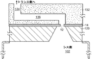
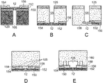
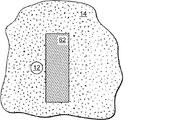
図1A〜1Eは、ナノポア検出のための局所電位検出法を可能にする、本明細書に提供されるナノポアセンサ構成の略図である。説明を明確にするために、図示される装置特徴は一定の拡大縮尺で示されていない。図1Aを参照すると、膜のような支持構造14を含み、その支持構造中にナノポア12が配置されているナノポアセンサ3が示されている。ナノポア12は、支持構造中、ここではトランス側リザーバおよびシス側リザーバとして概略的に示される2つの流体リザーバの間で、シス側リザーバとトランス側リザーバとの間の唯一の流体連絡経路であるように構成されている。一方のリザーバはナノポアの入口に接続され、他方のリザーバはナノポアからの出口に接続されている。ナノポアを通過する種トランスロケーションの局所電位計測検知のためのナノポアセンサの動作においては、種の1つまたは複数の対象物、たとえば分子が、2つのリザーバの一方の流体溶液中に、ナノポアを通って2つのリザーバの他方へトランスロケートするように提供されている。多くの用途、特に分子検出用途の場合、分子または他の種対象物をリザーバの一方の中のイオン流体溶液中に提供することが好ましいといえ、リザーバのいずれか1つの中に提供することができる。
Detailed Description Figures 1A-1E are schematic representations of the nanopore sensor configurations provided herein that enable local potential detection methods for nanopore detection. For clarity of explanation, the device features shown are not shown at a constant scale. With reference to FIG. 1A, a
ナノポアを通ってトランスロケートする種対象物は、たとえばDNA、DNAフラグメント、RNA、RNAフラグメント、PNA、ヌクレオチド、ヌクレオシド、オリゴヌクレオチド、タンパク質、ポリペプチド、アミノ酸およびポリマーから選択される対象物を含むことができる。種対象物は、標識されたヌクレオチドから放出される標識を含むことができる。参照により本明細書に組み入れられるWO2013/191793号に記載されているように、ポリメラーゼを援用して、核酸分子に沿うヌクレオチドを重合させて、核酸分子の少なくとも一部分に相補的である核酸鎖を生成することができ、それにより、重合中、標識が、ヌクレオチドの個々のヌクレオチドから放出され、それにより、放出された標識がナノポアをトランスロケートする。 Species objects translocating through nanopores may include objects selected from, for example, DNA, DNA fragments, RNA, RNA fragments, PNAs, nucleotides, nucleosides, oligonucleotides, proteins, polypeptides, amino acids and polymers. it can. The species object can include a label released from the labeled nucleotide. As described in WO 2013/191793, which is incorporated herein by reference, polymerases are used to polymerize nucleotides along the nucleic acid molecule to produce nucleic acid chains that are complementary to at least a portion of the nucleic acid molecule. This allows the label to be released from the individual nucleotides of the nucleotide during polymerization, whereby the released label translocates the nanopores.
ナノポアは、支持構造中に開口、間隙、チャネル、溝、ポアまたは他の穴として提供され得、関心対象の種対象物を検出するのに適当である、対応する形状のための、直径のような大きさで提供される。ナノポアを通過する分子トランスロケ−ションを検出するためには、約100nm未満のナノポアが好ましいといえ、10nm、5nmまたは2nm未満のナノポアがより好ましいといえる。以下に説明するように、いくつかの分子検出用途の場合、1nmのナノポアが適当であり、好ましいとさえいえる。 Nanopores can be provided as openings, gaps, channels, grooves, pores or other holes in the support structure and are suitable for detecting the species object of interest, such as diameter for the corresponding shape. It is provided in a large size. In order to detect molecular translocation that passes through nanopores, nanopores of less than about 100 nm are preferable, and nanopores of less than 10 nm, 5 nm or 2 nm are more preferable. As described below, 1 nm nanopores are suitable and even preferred for some molecular detection applications.
ナノポアセンサのリザーバまたは他の構成要素は、分子のような種の対象物をナノポアに向けて、またはリザーバの一方からリザーバの他方へとナノポアに通して移動させるための駆動力を提供するように構成され得る。たとえば、電極13、15が、電圧および電流素子16、18を有する回路中に提供されて、溶液中の種をナノポアに向けて、または一方のリザーバから他方のリザーバへとナノポアに通して電気泳動的に駆動するための電気泳動力をリザーバ間で発生させることができる。種の電気泳動的駆動を可能にするために、リザーバの流体溶液は、溶液中の種に順応するpHおよび他の特性を有する導電性イオン溶液として提供されることができる。これにより、ナノポアを介して電気回路をリザーバ溶液と直列に接続することができ、図示するような電極13、15が、ナノポアをはさんで溶液間に電圧バイアスを提供する。ナノポアを通過する種のトランスロケーションおよびトランスロケーション速度の制御は、酵素分子モータのような代替技術によって実施することもできる。
The reservoir or other component of the nanopore sensor is such that it provides the driving force to move a species-like object, such as a molecule, towards the nanopore or through the nanopore from one of the reservoirs to the other of the reservoir. Can be configured. For example,
印加電圧である駆動力に加えて、または駆動力に代えて、ポアをはさんでの圧力勾配を使用して、分子をナノポアに向けて移動させる、および/またはナノポアに通すことができる。この圧力勾配は、物理的圧力または浸透圧のような化学的圧力を使用することによって発生させることができる。浸透圧は、シス側およびトランス側チャンバ間の濃度差から発生させることができる。浸透圧は、浸透的に活性な物質、たとえば塩、ポリエチレングリコール(PEG)またはグリセロールの濃度勾配を有することによって発生させることができる。 In addition to, or in place of, the driving force, which is the applied voltage, the pressure gradient across the pores can be used to move the molecules towards and / or through the nanopores. This pressure gradient can be generated by using a chemical pressure such as physical pressure or osmotic pressure. Osmotic pressure can be generated from the difference in concentration between the cis and trans chambers. Osmotic pressure can be generated by having a concentration gradient of osmotically active substances such as salts, polyethylene glycol (PEG) or glycerol.
図1Aに示すように、ナノポアセンサ中に、素子の位置に局所的な電位を検出し、その局所電位を示す特徴を発現させる変換素子7を提供することができる。局所電位を示す信号を発現させるために、装置および/または回路の位置に局所的な電位を検出する電気接続、たとえば装置および/または回路の装置または領域、ワイヤまたは回路素子の組み合わせを変換素子7として提供することができる。電位検出の位置は、以下に詳細に説明するように、リザーバの中、支持構造の表面またはナノポアセンサ内の他の位置であることができる。
As shown in FIG. 1A, it is possible to provide a
図1Bに示すように、たとえば、ソースS、ドレインDおよびチャネル領域24を有するトランジスタ装置22を含む回路20を提供することができる。チャネル領域24は、この例においては、ナノポアセンサ環境中、局所電位計測を実施するための位置に物理的に配置されている。トランジスタのチャネル領域24のこの物理的位置は、局所電位にアクセスするための任意の好都合かつ適当な位置であることができる。
As shown in FIG. 1B, for example, a
図1A〜1Bのアレンジにおいて、電位検出回路は、ナノポア12のトランス側リザーバ側でトランス側リザーバに局所的な電位を計測するトランジスタまたは他の装置を提供するために、トランス側リザーバに局所的に構成されている。または、図1Cに示すように、電位検出装置または回路のような電気変換素子7がナノポアのシス側リザーバ側で構成されることもできる。ここでは、たとえば、図1Dに示すように、ナノポア12のシス側リザーバ側でシス側リザーバに局所的な電位を計測するためのトランジスタ24または他の装置を含む回路20を提供することができる。
In the arrangement of FIGS. 1A-1B, the potential detection circuit locally to the transformer-side reservoir to provide a transistor or other device to measure the potential locally to the transformer-side reservoir on the transformer-side reservoir side of the
さらなる代替構成においては、図1Eに示すように、ナノポアセンサシステム中の2つ以上の位置で電位を検出する、トランジスタ22a、22bのような変換素子に接続された回路20a、20bなどを有する2つ以上の変換素子がたとえばナノポア支持構造の各側に含まれることもできる。これにより、電位検出回路の物理的な実施形態に依存して、このアレンジにより、ナノポア膜14の2つの側で電位を計測することができる。これは、ナノポアセンサ中の2つの位置の間の局所電位差の計測が可能になる例示的な構成である。したがって、用語「計測された局所電位」とは、ナノポアセンサ中の単一位置の電位を指し、2つ以上の位置の間の局所電位の差または和を指し、かつ、ナノポアセンサ構成中の2つ以上の位置における局所電位を指す。
In a further alternative configuration, as shown in Figure 1E, it has
局所電位計測は、任意の適当な装置および/または回路もしくは他の変換素子、たとえば生物学的または他の非固体変換素子によって実施されることができ、上記トランジスタ実施形態に限定されない。図1Fに示すように、単一電子トランジスタ(SET)回路27として構成されている変換素子を支持構造14上に提供することができる。SETのソースS領域およびドレインD領域が支持構造上に配置されて、SET27にトンネリングバリヤを提供する。得られる量子ドットシステムにおいて、SET27を通過する電気コンダクタンスは、ソースSおよびドレインDのフェルミ準位に対するSETのエネルギー準位に依存する。ナノポア12がSETの近くに位置していると、種対象物がナノポアを通ってトランスロケートするときSETの電位および対応するエネルギー準位が変化して、SET回路のコンダクタンスを変化させる。
Local potential measurements can be performed by any suitable device and / or circuit or other conversion element, such as a biological or other non-solid conversion element, and are not limited to the transistor embodiments described above. As shown in FIG. 1F, a conversion element configured as a single electron transistor (SET) circuit 27 can be provided on the
さらに図1Gに示すように、局所電位計測を実施するための量子点接触(QPC)システム29を支持構造14上に提供することができる。このシステムにおいては、ナノポア12の位置で非常に細い伝導チャネル領域を介して接続されるソースS領域およびドレインD領域を形成する導電性領域31が提供される。チャネル領域は、チャネル領域に対して垂直である電子キャリア粒子エネルギー状態が量子化されるのに十分な細さである。種対象物がナノポアを通ってトランスロケートするとき、QPCの周囲の局所電位、ひいては細い伝導チャネル領域内のフェルミ準位が変化し、その結果、フェルミ準位よりも低い量子化状態の数の変化および対応するQPCコンダクタンスの変化を生じさせる。
Further, as shown in FIG. 1G, a quantum dot contact (QPC) system 29 for performing local potential measurements can be provided on the
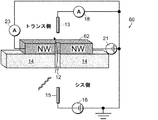
また、ナノワイヤFETをナノポアの位置で構成することもできる。ナノワイヤは、フラーレン構造および半導体ワイヤを含む任意の適当な導電性または半導電性材料で形成されることができる。本明細書の中で使用される用語「ナノワイヤ」とは、ナノポア位置から計測された信号減衰長と適合する幅を特徴とする電気伝導チャネルをいう。チャネル幅は、好ましくは、減衰長と同程度の大きさであり、かつより大きいこともできる。ナノワイヤは、選択されたリザーバ溶液中で安定である任意の半導体材料から作ることができる。 In addition, the nanowire FET can be configured at the position of the nanopore. Nanowires can be made of any suitable conductive or semi-conductive material, including fullerene structures and semiconductor wires. As used herein, the term "nanowire" refers to an electrically conductive channel characterized by a width that matches the signal attenuation length measured from the nanopore position. The channel width is preferably as large as, and can be larger than, the decay length. Nanowires can be made from any semiconductor material that is stable in selected reservoir solutions.
ナノポアセンサは、固体電圧検出装置を有する固体ナノポア構成に限定されない。たとえばタンパク質ナノポアまたは他の適当な構成を有する生物学的ナノポアおよび電位検出アレンジを用いることもできる。図1Hに示すように、タンパク質ナノポア33が配置されている両親媒性層31を提供することができる。電圧感受性色素、たとえば蛍光直接染料37を電気変換素子として脂質二重層中に提供することができる。このアレンジを用いると、分子のような種対象物がタンパク質ナノポアを通ってトランスロケートするとき、両親媒性層をはさんで電圧降下が変化し、色素の蛍光が電圧変化によって変調される。色素蛍光の光学的検知または検出およびその蛍光への変化がナノポアにおける電位の検出を提供する。ナノポアセンサからの光出力信号としてこの電位計測を実施するためには、光学顕微鏡法または他の従来のアレンジを用いることができる。この両親媒性層ナノポアセンサは、ナノポアシステム中の位置における局所電位の検出に基づく生物学的ナノポアセンサの一例である。ナノポアトランスロケーション検出のための局所電位計測の方法は、特定の固体または生物学的構成に限定されず、かつ任意の適当なナノポア構成に適用されることができる。
The nanopore sensor is not limited to a solid nanopore configuration having a solid voltage detector. For example, protein nanopores or other biological nanopores with suitable configurations and potential detection arrangements can also be used. As shown in FIG. 1H, an amphipathic layer 31 in which the
支持構造は、マイクロエレクトロニクス材料(導電性、半導電性または絶縁性のいずれでもよい)、たとえばII-IVおよびIII-V材料、酸化物および窒化物、たとえばSi3N4、Al2O3およびSiOのような材料、有機および無機ポリマー、たとえばポリアミド、プラスチック、たとえばTeflon(登録商標)またはエラストマー、たとえば二成分付加型硬化シリコーンゴムおよびガラスをはじめとする、有機材料および無機材料のいずれかまたは両方から形成されることができる。固体支持構造は、単原子層、たとえばグラフェンまたは原子数個分の厚さしかない層、たとえば、いずれも参照により本明細書に組み入れられる米国特許第8,698,481号および米国特許出願公開公報2014/174927に開示されているものから形成され得る。参照により本明細書に組み入れられる米国特許出願公開公報2013/309776に開示されているように、2つ以上の支持層材料、たとえば2つ以上のグラフェン層が含まれることもできる。適当な窒化ケイ素膜が、参照により本明細書に組み入れられる米国特許第6,627,067号に開示されており、支持構造は、参照により本明細書に組み入れられる米国特許出願公開公報2011/053284に開示されているように、化学的に官能化されてもよい。 The support structure can be a microelectronic material (which can be conductive, semi-conductive or insulating), such as II-IV and III-V materials, oxides and nitrides, such as Si 3 N 4 , Al 2 O 3 and Materials such as SiO, organic and inorganic polymers, such as polyamides, plastics, such as Teflon® or elastomers, such as two-component additive curable silicone rubber and glass, or both organic and inorganic materials. Can be formed from. Solid support structures are described in monatomic layers, such as graphene or layers that are only a few atoms thick, such as US Pat. No. 8,698,481 and US Patent Application Publication No. 2014/174927, both of which are incorporated herein by reference. It can be formed from what is disclosed. Two or more support layer materials, such as two or more graphene layers, can also be included, as disclosed in US Patent Application Publication 2013/309776, which is incorporated herein by reference. Suitable silicon nitride films are disclosed in US Pat. No. 6,627,067, which is incorporated herein by reference, and the supporting structure is disclosed in US Patent Application Publication No. 2011/053284, which is incorporated herein by reference. As such, it may be chemically functionalized.
選択された支持構造材料の組成、厚さおよびアレンジに関して、支持構造中にナノポアを製造するために任意の適当な方法を用いることができる。たとえば、ナノポアを製造するためには、たとえばいずれも参照により本明細書に組み入れられる米国特許出願公開公報2014/0262820、米国特許出願公開公報2012/0234679、米国特許第8,470,408号、米国特許第8,092,697号、米国特許第6,783,643号および米国特許第8,206,568号に記載されているように、電子ビームミリング、イオンビームミリング、高エネルギービームによる材料彫塑、ドライエッチング、ウェットケミカルもしくはエレクトロケミカルエッチングまたは他の方法を用いることができる。加えて、押出し成形、自己集合、相対的に大きな開口の側壁への材料付着または他のナノポア形成方法を用いることができる。 With respect to the composition, thickness and arrangement of the selected support structure material, any suitable method can be used to produce nanopores in the support structure. For example, to produce nanopores, for example, US Patent Application Publication 2014/0262820, US Patent Application Publication 2012/0234679, US Patent 8,470,408, US Patent 8,092,697, all of which are incorporated herein by reference. , US Pat. No. 6,783,643 and US Pat. No. 8,206,568, using electron beam milling, ion beam milling, material engraving with a high energy beam, dry etching, wet chemical or electrochemical etching or other methods. be able to. In addition, extrusion molding, self-assembly, material attachment to the sidewalls of relatively large openings or other nanopore forming methods can be used.
完全に固体のナノポアを提供する代わりに、固体開口内に生物学的ナノポアを提供することもできる。そのような構造が、たとえば参照により本明細書に組み入れられる米国特許第8,828,211号に開示されている。さらに、生物学的ナノポアは膜貫通タンパク質ポアであってもよい。生物学的ポアは、天然のポアであってもよいし、または突然変異ポアであってもよい。一般的なポアが、米国特許出願第2012/1007802号に記載され、Stoddart D et al., Proc Natl Acad Sci, 12;106(19):7702-7、Stoddart D et al., Angew Chem Int Ed Engl. 2010;49(3):556-9、Stoddart D et al., Nano Lett. 2010 Sep 8;10(9):3633-7、Butler TZ et al., Proc Natl Acad Sci 2008;105(52);20647-52、米国特許出願公開公報2014/186823およびWO2013/153359に記載されている。これらすべては参照により本明細書に組み入れられる。ポアは、ホモオリゴマー、すなわち、同一のモノマーから誘導されたものであってもよい。ポアは、ヘテロオリゴマー、すなわち、少なくとも1つのモノマーが他とは異なるものであってもよい。ポアは、参照により本明細書に組み入れられるLangecker et al., Science, 2012; 338: 932-936によって記載されているような、DNAオリガミポアであってもよい。
Instead of providing completely solid nanopores, biological nanopores can also be provided within the solid opening. Such a structure is disclosed, for example, in US Pat. No. 8,828,211 incorporated herein by reference. In addition, the biological nanopore may be a transmembrane protein pore. The biological pore may be a natural pore or a mutant pore. Common pores are described in US Patent Application No. 2012/1007802, Stoddart D et al., Proc Natl Acad Sci, 12; 106 (19): 7702-7, Stoddart D et al., Angew Chem Int Ed. Engl. 2010; 49 (3): 556-9, Stoddart D et al., Nano Lett. 2010
1つの態様において、ポアは、両親媒性層内に提供されることができる。両親媒性層とは、親水性および親油性の両性質を有する両親媒性分子、たとえばリン脂質から形成された層である。両親媒性層は単層または二重層であることができ、その層は、脂質二重層または非天然脂質二重層から選択される。二重層は、たとえばKunitake T., Angew. Chem. Int. Ed. Engl. 31 (1992) 709-726によって開示されているような合成二重層であることができる。両親媒性層は、いずれも参照により本明細書に組み入れられるGonzalez-Perez et al., Langmuir, 2009, 25, 10447-10450および米国特許第6,723,814号によって開示されているようなコブロックポリマーであることができる。ポリマーは、たとえば、PMOXA-PDMS-PMOXAトリブロックコポリマーであることができる。 In one embodiment, the pores can be provided within the amphipathic layer. The amphipathic layer is a layer formed from an amphipathic molecule having both hydrophilic and lipophilic properties, for example, a phospholipid. The amphipathic layer can be a monolayer or a bilayer, which layer is selected from a lipid bilayer or an unnatural lipid bilayer. The bilayer can be a synthetic bilayer, for example as disclosed by Kunitake T., Angew. Chem. Int. Ed. Engl. 31 (1992) 709-726. The amphipathic layer is a coblock polymer as disclosed by Gonzalez-Perez et al., Langmuir, 2009, 25, 10447-10450 and US Pat. No. 6,723,814, both incorporated herein by reference. be able to. The polymer can be, for example, a PMOXA-PDMS-PMOXA triblock copolymer.
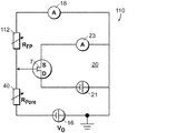
図2Aを参照すると、ナノポアセンサ中の1つまたは複数の位置における局所電位を計測するためのこれらの支持構造、ナノポアおよび電気的構成のいずれかを、ナノポアを通過する種のトランスロケ−ションを検出する方法に用いることができる。この検出の原理を説明するためには、ナノポアセンサを、図2Aに示すような、センサの物理的素子に対応する電気部品を含む回路35としてモデル化することが理解しやすい。シス側およびトランス側リザーバは、それぞれ、特徴的な流体アクセス抵抗RTrans36、RCis38を有するようにモデル化されることができる。このアクセス抵抗は、この分析の場合、ナノポアの位置に局所的なリザーバ溶液中の流体抵抗と定義され、ナノポアから離れたバルク溶液中の流体抵抗と定義されるのではない。ナノポアは、ナノポアが配置されている支持構造の2つの側の間のナノポア長を通過する溶液の流体抵抗である、特徴的なナノポア溶液抵抗RPore40を有するようにモデル化されることができる。ナノポアはまた、ナノポアが配置されている膜または他の支持構造の関数である、特徴的なキャパシタンスCPoreを有するようにモデル化されることもできる。両チャンバのアクセス抵抗およびナノポア溶液抵抗は可変性である。
With reference to FIG. 2A, one of these support structures, nanopores and electrical configurations for measuring local potentials at one or more positions in the nanopore sensor, the translocation of the species through the nanopores. It can be used as a detection method. To explain the principle of this detection, it is easy to understand that the nanopore sensor is modeled as a
ナノポアをトランスロケートする種が存在しないナノポアセンサ開始条件において、ナノポアは上記溶液抵抗RPoreを特徴とすることができ、両方の流体リザーバは、トランス側リザーバおよびシス側リザーバのアクセス抵抗それぞれRTransおよびRCisを特徴とすることができる。次いで、図2Aに示すように生物学的分子45のような種対象物がナノポア12をトランスロケートするとき、ナノポア中の分子がナノポア長に延びる通路を少なくとも部分的に閉塞してナノポアの有効直径を変化させるせいで、ナノポアの溶液抵抗RPoreならびに各リザーバのアクセス抵抗RTransおよびRCisが変化する。そのような閉塞により、ナノポアの流体溶液抵抗および両リザーバのアクセス抵抗は、ナノポア中に分子が存在しないときのナノポアの抵抗および両リザーバのアクセス抵抗よりも増大する。
In nanopore sensor initiation conditions where there is no species translocating the nanopore, the nanopore can be characterized by the solution resistance R Pore described above, and both fluid reservoirs have access resistances of the trans and cis side reservoirs R Trans and R, respectively. It can be characterized by R Cis . Then, when a seed object such as
種対象物によるナノポアの部分的閉塞は、以下に詳細に説明するように、ナノポア溶液抵抗とリザーバアクセス抵抗とに異なるふうに影響する。その結果、トランスロケートする種によるナノポアの部分的閉塞は、ナノポアとシス側およびトランス側リザーバ溶液との間で電圧の対応する再分布を生じさせ、ナノポアセンサ中の至るところで電位が相応に調節する。それにより、ナノポア溶液抵抗のこの変化およびリザーバ溶液とナノポアとの間の電位の再分布とともに、図2A中でAおよびBと表記された両方の位置の局所電位が相応に変化する。それにより、これらの位置のいずれかもしくはナノポアセンサ構成の別の位置における電位の計測、または2つ以上の位置の間の局所電位の差の計測が、ナノポアを通過する分子のトランスロケーションの指標を提供する。 Partial occlusion of nanopores by a species object affects nanopore solution resistance and reservoir access resistance differently, as described in detail below. As a result, partial occlusion of the nanopores by the translocating species results in a corresponding redistribution of voltage between the nanopores and the cis-side and trans-side reservoir solutions, with potentials adjusted accordingly throughout the nanopore sensor. .. Thereby, with this change in nanopore solution resistance and the redistribution of the potential between the reservoir solution and the nanopore, the local potentials at both positions labeled A and B in FIG. 2A change accordingly. Thereby, the measurement of the potential at one of these positions or another position of the nanopore sensor configuration, or the measurement of the difference in local potential between two or more positions, provides an indicator of the translocation of the molecule through the nanopore. provide.
選択されたナノポアセンサ位置における局所電位およびこの電位の変化を、ナノポアセンサ中に配置された電気変換素子によって検出することができる。たとえば、トランジスタ装置中の伝導チャネルのコンダクタンスの変化が電位計測を提供することができる。したがって、トランジスタチャネルコンダクタンスは、トランジスタチャネルの物理的位置に局所的な電位の直接的な指標として用いることができる。図1A〜1Bのナノポアセンサアレンジは、図2Aの回路35の位置Aにおける局所電位計測に対応する。図1C〜1Dのナノポアセンサアレンジは、図2Aの回路35の位置Bにおける局所電位計測に対応する。図1Eのナノポアセンサアレンジは、図2Aの回路35の位置AおよびBの両方における局所電位計測に対応し、これら2つの位置における電位間の差の決定を可能にする。
The local potential at the selected nanopore sensor position and the change in this potential can be detected by an electrical conversion element arranged in the nanopore sensor. For example, changes in conductance of conduction channels in a transistor device can provide potential measurements. Therefore, transistor channel conductance can be used as a direct indicator of potential local to the physical position of the transistor channel. The nanopore sensor arrangement of FIGS. 1A-1B corresponds to the local potential measurement at position A of
図1Bの例示的構成の電気回路等価物が図2Bに示されている。この図には、シス側およびトランス側リザーバのアクセス抵抗それぞれRCisおよびRTransならびにナノポアの流体溶液抵抗RPoreが示されている。局所電位を計測するための電気変換素子、たとえばトランジスタ22のチャネルの位置がここでは図2Aの位置Aに配置されて、ナノポアのトランス側リザーバ側のトランス側リザーバ中の局所電位の指標を提供する。このアレンジを用いると、分子のような種対象物がナノポアを通ってトランスロケートするとき、ナノポアの状態の変化およびナノポア中の1つまたは複数の対象物の有無に対応する電位の変化に関して電位計測回路の出力信号をモニタすることができる。
An electrical circuit equivalent of the exemplary configuration of Figure 1B is shown in Figure 2B. This figure shows the access resistances of the cis and trans reservoirs, R Cis and R Trans, and the fluid solution resistance of nanopores, R Pore , respectively. The position of the channel of an electrical conversion element for measuring the local potential, eg,
この分析は、局所電気変換素子が提供されている任意のナノポアセンサに適用することができる。分析は、上記FETおよび他の実施形態に限定されず、任意の変換素子のための任意の適当なアレンジに適用可能である。求められるすべては、電気変換素子、たとえば装置、装置の領域、回路または種対象物がナノポアをトランスロケートするとき局所電位計測を実施する他の変換素子の提供である。 This analysis can be applied to any nanopore sensor for which a local electrical conversion device is provided. The analysis is not limited to the FETs and other embodiments described above, and is applicable to any suitable arrangement for any transforming device. All that is sought is the provision of electrical conversion devices, such as other conversion devices that perform local potential measurements when a device, device area, circuit or species object translocates a nanopore.
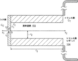
ナノポアセンサシステムパラメータをさらに分析するために、ナノポアセンサを、図3Aの略図に示すようにモデル化することができる。分析計算を可能にするためにいくつかの仮説を用いることができる。まず、局所電位検出変換素子の包含によって生じる、膜のようなナノポア支持構造およびナノポアそのものならびにナノポアセンサの他の領域の幾何学的変化を無視し、電位検出変換素子を点電位検知器としてモデル化することができる。流体リザーバは、導電性イオン溶液を含むものと仮定する。2つのリザーバ溶液は、異なるイオン濃度であってもよい別々のイオン濃度を含むように特定される。1つの分析においては、リザーバに特定された異なるイオン濃度で、シス側/トランス側リザーバ濃度差によって駆動される定常状態拡散により、ナノポアシステム中のイオン濃度分布を決定する。拡散は、定常状態に達するものと仮定する。バッファ濃度分布および電位がナノポアの両側の小さな半球部中で一定であると概算することにより、さらなる仮説を立てることができる。ナノポアセンサは、定常状態にあるものと仮定する。これらの条件下、ナノポアセンサの拡散方程式は以下のように記される:
式中、Cは流体イオン濃度であり、tは時間であり、rは、リザーバ中の、ナノポアから計測された点における位置であり、zは、ナノポア長を通過する距離である。ナノポアから遠いシス側リザーバ中ではC=CCisであり、ナノポアから遠いトランス側リザーバ中ではC=CTransであり、流束がナノポア中および両リザーバに関して同じであり、各リザーバ中のナノポア開口における濃度が連続的である境界条件下でこれらの拡散方程式を解くならば、2つのリザーバおよびナノポアのイオン濃度を以下のように求めることができる:
式中、lおよびdは、それぞれ、ナノポア支持構造の厚さおよびナノポア直径である。したがってイオン濃度分布は公知であり、溶液伝導率は濃度に比例するため、溶液の伝導率σは以下のように求められる:
式中、Σは溶液のモル伝導率である。全電流がIであると仮定すると、シス側リザーバ電圧VC、トランス側リザーバ電圧VTおよびナノポア電圧VPで、ナノポアセンサを通しての電位降下を以下のように求めることができる:
ナノポアから遠い、シス側リザーバ中の電位は、構造または膜に印加される電圧、すなわち膜電圧(TMV)に等しく、対象物をナノポアに通して電気泳動的に駆動し、ナノポアから遠い、トランス側チャンバ中の電位は0Vであると特定する境界条件でこれら3つの式を解くならば、ナノポアセンサ中の電圧、すなわちシス側リザーバ中の電圧VC(r)、トランス側リザーバ中の電圧VT(r)およびナノポア中の電圧VP(r)は以下のように求められる:
リザーバに通じる両方のナノポア開口で電位は連続的であるため、また、印加される全電圧はVであるため、式(5)を以下のようにさらに簡約することができる:
この式を用いると、ナノポア内の電場EP(r)を以下のように求めることができる:
この式を用いると、ナノポア中の分子のような種対象物の存在によるナノポア面積Aの減少による電位変化によってナノポアのトランス側リザーバ側の電位変化を以下のように推定することができる:
式中、δAは分子の断面積である。ナノポアセンサの抵抗、すなわちRCis、RTransおよびRPoreは、リザーバおよびナノポアをはさんでの電圧降下のための上記式に基づいて以下のように計算することができる:
したがって、ナノポアセンサの全抵抗およびイオン電流は
として求められる。
To further analyze the nanopore sensor system parameters, the nanopore sensor can be modeled as shown in the schematic of FIG. 3A. Several hypotheses can be used to enable analytical calculations. First, the potential detection conversion element is modeled as a point potential detector, ignoring the film-like nanopore support structure and the nanopore itself and the geometric changes in other regions of the nanopore sensor caused by the inclusion of the local potential detection conversion element. can do. The fluid reservoir is assumed to contain a conductive ion solution. The two reservoir solutions are specified to contain different ion concentrations, which may be different ion concentrations. In one analysis, the ion concentration distribution in the nanopore system is determined by steady-state diffusion driven by the cis / trans reservoir concentration difference at different ion concentrations identified in the reservoir. It is assumed that the diffusion reaches a steady state. Further hypotheses can be made by estimating that the buffer concentration distribution and potential are constant in the small hemispheres on either side of the nanopore. The nanopore sensor is assumed to be in a steady state. Under these conditions, the diffusion equation for the nanopore sensor is written as follows:
In the equation, C is the fluid ion concentration, t is the time, r is the position in the reservoir at the point measured from the nanopore, and z is the distance through the nanopore length. C = C Cis in the cis-side reservoir far from the nanopore, C = C Trans in the trans-side reservoir far from the nanopore, the flux is the same in the nanopore and for both reservoirs, at the nanopore opening in each reservoir. If these diffusion equations are solved under boundary conditions where the concentrations are continuous, the ion concentrations of the two reservoirs and the nanopores can be determined as follows:
In the formula, l and d are the thickness and nanopore diameter of the nanopore support structure, respectively. Therefore, since the ion concentration distribution is known and the solution conductivity is proportional to the concentration, the conductivity σ of the solution can be obtained as follows:
In the formula, Σ is the molar conductivity of the solution. Assuming the total current is I, with the cis-side reservoir voltage V C , the transformer-side reservoir voltage V T, and the nanopore voltage V P , the potential drop through the nanopore sensor can be determined as follows:
The potential in the cis-side reservoir, far from the nanopore, is equal to the voltage applied to the structure or membrane, the membrane voltage (TMV), and the object is electrophoretically driven through the nanopore, far from the nanopore, on the transformer side. If these three equations are solved under the boundary condition that specifies that the potential in the chamber is 0 V, then the voltage in the nanopore sensor, that is, the voltage V C (r) in the cis side reservoir and the voltage V T in the transformer side reservoir. The voltage V P (r) in (r) and the nanopore is calculated as follows:
Since the potentials are continuous at both nanopore openings leading to the reservoir and the total voltage applied is V, equation (5) can be further reduced as follows:
Using this equation, the electric field E P (r) in the nanopore can be calculated as follows:
Using this equation, the potential change on the transformer-side reservoir side of the nanopore can be estimated as follows by the potential change due to the decrease in the nanopore area A due to the presence of a seed object such as a molecule in the nanopore:
In the formula, δA is the cross-sectional area of the molecule. The resistance of the nanopore sensor, namely R Cis , R Trans and R Pore , can be calculated as follows based on the above equation for voltage drop across the reservoir and nanopore:
Therefore, the total resistance and ionic current of the nanopore sensor
Is required as.
これらの式により、ナノポアセンサの電気的特性、特にセンサ中の電位の分布がシス側およびトランス側リザーバ中の流体溶液のイオン濃度に直接依存するということが実証される。具体的には、リザーバ溶液濃度の比が、ナノポアの種トランスロケーションによる局所電位の変化の大きさに直接影響する。 These equations demonstrate that the electrical properties of the nanopore sensor, especially the distribution of potentials in the sensor, are directly dependent on the ion concentration of the fluid solution in the cis and transformer side reservoirs. Specifically, the ratio of reservoir solution concentrations directly affects the magnitude of changes in local potential due to nanopore seed translocation.
図3B〜3Eは、これらの条件を実証する、ナノポア中の電位および電場のプロットである。1:1のシス側/トランス側バッファ溶液濃度比、厚さ50nmの窒化物膜、膜中の直径10nmのナノポアおよび1Vの膜電圧、すなわち2つのリザーバ中の溶液間に印加される1Vの電圧を仮定して、上記式(6)に基づき、ナノポア中の電位がシス側リザーバにおけるナノポア開口からの距離の関数として図3Bにプロットされている。シス側/トランス側バッファ溶液濃度比が代わりに100:1である条件での同じ電位が図3Cにプロットされている。不均衡なバッファ溶液比の場合、より低い濃度のリザーバにより近い点で、所与のナノポア位置における電位が増すことに注目されたい。 Figures 3B-3E are plots of potential and electric fields in nanopores demonstrating these conditions. 1: 1 cis / trans buffer solution concentration ratio, 50 nm thick nitride film, 10 nm diameter nanopores in the film and 1 V membrane voltage, i.e. 1 V voltage applied between the solutions in the two reservoirs Is assumed, and based on the above equation (6), the potential in the nanopore is plotted in FIG. 3B as a function of the distance from the nanopore opening in the cis-side reservoir. The same potential under the condition that the cis / trans buffer solution concentration ratio is instead 100: 1 is plotted in Figure 3C. Note that for an unbalanced buffer solution ratio, the potential increases at a given nanopore position at a point closer to the lower concentration reservoir.
図3Dは、ここでは均衡したバッファ溶液比の場合の、上記式(7)に基づく、上記条件下でのナノポア中の電場のプロットである。シス側/トランス側バッファ溶液濃度比が代わりに100:1である条件での同じ電位プロファイルが図3Eにプロットされている。不均衡なバッファ溶液比の場合の所与のナノポア位置における電位の増大および低濃度かつより高い抵抗のリザーバにより近い点で電場が劇的に強くなることに注目されたい。 FIG. 3D is a plot of the electric field in the nanopore under the above conditions, based on equation (7) above, here for a balanced buffer solution ratio. The same potential profile under the condition that the cis / trans buffer solution concentration ratio is instead 100: 1 is plotted in Figure 3E. Note the increase in potential at a given nanopore position in the case of an unbalanced buffer solution ratio and the dramatic increase in electric field near the reservoir of low concentration and higher resistance.
この発見により、リザーバ溶液がいずれも同じイオン濃度の導電性イオン溶液として提供される条件では、シス側リザーバのアクセス抵抗RCis、トランス側リザーバのアクセス抵抗RTransおよびナノポアの溶液抵抗RPoreの比はすべて固定され、ナノポア抵抗がリザーバアクセス抵抗よりもずっと大きいことが見いだされる。しかし、不均衡なイオン濃度条件下では、より低いイオン濃度を有するリザーバが、ナノポア抵抗の程度であることができる、より大きいアクセス抵抗を有し、一方で、より高いイオン濃度を有するリザーバの抵抗は比較可能に無視しうる程度になる。 Based on this finding, the ratio of the access resistance R Cis of the cis-side reservoir, the access resistance R Trans of the trans-side reservoir, and the solution resistance R Pore of the nanopore under the condition that the reservoir solutions are all provided as conductive ion solutions with the same ion concentration. Are all fixed and it is found that the nanopore resistance is much higher than the reservoir access resistance. However, under imbalanced ion concentration conditions, reservoirs with lower ion concentrations have greater access resistance, which can be a degree of nanopore resistance, while resistance of reservoirs with higher ion concentrations. Is comparable and negligible.
この対応の理解に基づき、ナノポアセンサ中の局所電位計測を最大化するために、1つの態様においては、イオンリザーバ溶液に異なるイオン濃度を提供することができることが本明細書の中で見いだされる。不均衡なイオン濃度のこの構成を用いると、局所電位計測は、好ましくは、より低いイオン濃度を含むリザーバ中の位置で実施される。さらに、より低いイオン濃度溶液のバッファ濃度は、そのリザーバのアクセス抵抗をナノポア抵抗と同程度の大きさ、かつ、高イオン濃度溶液の抵抗よりもずっと大きく、たとえば高イオン濃度溶液の抵抗よりも少なくとも一桁大きくして、たとえば、局所電位計測がトランス側リザーバ中で実施されるならば、
となるように選択されることが好ましい。
Based on this understanding of correspondence, it is found herein that different ion concentrations can be provided to the ion reservoir solution in one embodiment in order to maximize local potential measurements in the nanopore sensor. With this configuration of disproportionate ion concentrations, local potential measurements are preferably performed at locations in the reservoir containing lower ion concentrations. Moreover, the buffer concentration of the lower ion concentration solution makes the access resistance of the reservoir as large as the nanopore resistance and much higher than the resistance of the high ion concentration solution, for example, at least more than the resistance of the high ion concentration solution. Increased by an order of magnitude, for example, if local potential measurements are performed in the transformer side reservoir
It is preferable that it is selected so as to be.
そして、この発見に基づき、ナノポア抵抗RPを設定する所与のナノポア直径の場合、1つの態様においては、局所電位計測のために指定されたリザーバのイオン溶液バッファ濃度を、そのリザーバのアクセス抵抗がナノポア抵抗と同程度の大きさであるレベルまで下げることが好ましい。このリザーバアクセス抵抗は、ナノポアセンサ抵抗を圧倒するべきではなく、ナノポア抵抗の程度であるべきである。 Then, based on this finding, for a given nanopore diameter that sets the nanopore resistance R P , in one embodiment, the ion solution buffer concentration of the reservoir specified for local potential measurement, the access resistance of that reservoir. Is preferably reduced to a level comparable to the nanopore resistance. This reservoir access resistance should not overwhelm the nanopore sensor resistance, but should be the degree of nanopore resistance.
この条件は、上記のようにナノポアセンサ構成要素を電気的にモデル化することによって直接定量することができる。上記式(8)に基づき、所与のナノポアセンサパラメータの場合に選択された対象物のナノポアトランスロケーション中の電位変化を最大化する溶液濃度の比を決定することができる。図4Aは、dsDNA分子がナノポアを通ってトランスロケートするときの電気泳動種トランスロケーションの場合の、厚さ50nmのナノポア膜および1VのTMVの構成の場合の式(8)のプロットである。電位変化が、10nm未満の様々なナノポア直径の場合のCCis/CTransイオン濃度比の関数として示されている。このプロットから、ここでモデル化される任意のナノポア直径の場合、トランス側リザーバ中の局所電位変化が約100:1のCCis/CTransチャンババッファ濃度比の場合に最大化されることがわかる。図4Bは、直径10nmのナノポアの場合、1VのTMVで、選択された100:1のCCis/CTrans溶液濃度比の場合の、トランス側リザーバ中の対応する計算された電位変化分布のプロットである。 This condition can be directly quantified by electrically modeling the nanopore sensor components as described above. Based on equation (8) above, the ratio of solution concentrations that maximizes the potential change in the nanopore translocation of the selected object for a given nanopore sensor parameter can be determined. FIG. 4A is a plot of equation (8) for a 50 nm thick nanopore membrane and a 1 V TMV configuration for electrophoretic species translocation as dsDNA molecules translocate through nanopores. The potential change is shown as a function of the C Cis / C Trans ion concentration ratio for various nanopore diameters below 10 nm. From this plot, it can be seen that for any nanopore diameter modeled here, the local potential change in the transformer-side reservoir is maximized for a C Cis / C Trans chamber buffer concentration ratio of approximately 100: 1. .. Figure 4B is a plot of the corresponding calculated potential change distribution in the trance-side reservoir for a selected 100: 1 C Cis / C Trans solution concentration ratio at 1 V TMV for nanopores with a diameter of 10 nm. Is.
これは、本明細書における発見に基づき、1つの態様において、電位計測を実施するために選択される所与のリザーバ位置に関し、その選択された計測位置における電位変化の振幅を最大化するためには、より低いバッファ濃度の溶液が計測リザーバ中にあるように2つのリザーバ中のイオン流体バッファ濃度の比を選択することができることを実証する。この結果的な電位変化の分布は、図4Bに示すように、ナノポアから数十ナノメートル以内で高度に限局化している。100:1のCCis/CTrans溶液濃度比および上記ナノポアパラメータの場合、たとえば上記式(9)に基づくと、トランス側リザーバのアクセス溶液抵抗とナノポアの溶液抵抗とが実際には同程度の大きさであると判断することができる。 This is based on the findings herein, in one embodiment, with respect to a given reservoir position selected to perform a potential measurement, in order to maximize the amplitude of the potential change at that selected measurement position. Demonstrates that the ratio of ionic liquid buffer concentrations in the two reservoirs can be selected so that the solution with the lower buffer concentration is in the measurement reservoir. As shown in FIG. 4B, the distribution of the resulting potential change is highly localized within tens of nanometers from the nanopore. In the case of the 100: 1 C Cis / C Trans solution concentration ratio and the nanopore parameter, for example, based on the above equation (9), the access solution resistance of the transformer side reservoir and the solution resistance of the nanopore are actually about the same magnitude. It can be judged that it is.
リザーバ流体溶液バッファ濃度および電位計測構成のこのアレンジを用いるとき、局所電位検出技術は、膜電圧(TMV)およびイオン電流信号に依存する局所電位計測信号を生成するということが注目される。他のセンサベースのナノポア技術は概して、トランスロケートする種とナノポアセンサとの間の直接的相互作用、たとえば電気結合または量子力学トンネリングに依存する。これらの技術の場合、ナノポア出力信号は一般に、TMVまたはイオン電流に正比例せず、TMVが変化するときでも有意には変化しないはずである。対照的に、本明細書における局所電位計測システムにおいて、ナノポアセンサ信号はTMVに比例し、イオン電流信号の線形増幅とみなすことができる。その結果、局所電位計測信号およびイオン電流信号の両方の振幅はTMVに線形に依存するが、それらの間の比は、上記式によって証明されるように、所与のナノポア形状およびリザーバ溶液濃度に関して一定である。 When using this arrangement of reservoir fluid solution buffer concentration and potential measurement configurations, it is noted that the local potential detection technique produces a local potential measurement signal that depends on the membrane voltage (TMV) and ion current signals. Other sensor-based nanopore techniques generally rely on direct interactions between the translocating species and the nanopore sensor, such as electrical coupling or quantum mechanical tunneling. For these techniques, the nanopore output signal is generally not directly proportional to the TMV or ionic current and should not change significantly as the TMV changes. In contrast, in the local potential measurement system herein, the nanopore sensor signal is proportional to TMV and can be considered as a linear amplification of the ion current signal. As a result, the amplitudes of both the local potential measurement signal and the ionic current signal are linearly dependent on TMV, but the ratio between them is with respect to a given nanopore shape and reservoir solution concentration, as evidenced by the above equation. It is constant.
局所電位計測法の利点は、低ノイズで特徴的に高帯域幅の計測能力である。低い信号帯域幅は、非常に小さい計測電流信号の高帯域幅増幅の困難さのせいで、従来のイオン電流閉塞計測技術による直接ナノポア検出を制限する問題の1つである。これは、DNA検出に用いられる場合の小さなナノポアに特に当てはまることができる。局所電位検出法においては、小さな電流信号の代わりに大きな局所電位信号が計測され、したがって、信号帯域幅は電流増幅器の能力によって制限されない。その結果、高帯域幅信号処理電子部品を固体ナノポア検出構造上に統合することができる。 The advantage of the local potential measurement method is the ability to measure low noise and characteristically high bandwidth. Low signal bandwidth is one of the problems limiting direct nanopore detection by conventional ion current blockage measurement techniques due to the difficulty of high bandwidth amplification of very small measured current signals. This is especially true for small nanopores when used for DNA detection. In the local potential detection method, a large local potential signal is measured instead of a small current signal, and therefore the signal bandwidth is not limited by the capabilities of the current amplifier. As a result, high bandwidth signal processing electronic components can be integrated on the solid nanopore detection structure.
さらに、固有ショットノイズおよびジョンソンノイズを除いて、イオン電流閉塞計測技術へのノイズ寄与の大部分は、ナノポア膜を横切る容量結合を介して導入され、ノイズのこの容量結合成分が、ナノポア動作の特定の段階で圧倒的になることができる。通常、ノイズを最小化するための努力においては、リザーバ溶液への非常に小さな膜面積暴露が求められる。本明細書における局所電位計測法において、局所電位信号は、妥当なリザーバ濃度比の場合、ナノポアの周囲の数十ナノメートル以内で減衰するため、局所電位計測信号は、この局所ボリューム内のリザーバ溶液間の容量結合による影響しか受けない。したがって、局所電位計測検出法において、容量結合ノイズの大部分は自動的に拒絶される。 In addition, with the exception of inherent shot noise and Johnson noise, most of the noise contribution to ion current occlusion measurement techniques is introduced through capacitive coupling across the nanopore membrane, and this capacitive coupling component of the noise identifies nanopore behavior. Can be overwhelming at the stage of. Efforts to minimize noise usually require very small membrane area exposure to the reservoir solution. In the local potential measurement method herein, the local potential signal is attenuated within tens of nanometers around the nanopore for a reasonable reservoir concentration ratio, so the local potential measurement signal is the reservoir solution in this local volume. It is only affected by the capacitive coupling between them. Therefore, in the local potential measurement detection method, most of the capacitive coupling noise is automatically rejected.
図4C〜4Dを参照すると、1つの態様においては、ナノポアセンサの信号帯域幅を最適化するためにリザーババッファ溶液濃度比を選択することができる。局所電位計測をナノポアの1つの側、たとえばナノポアのトランス側で実施すると仮定すると、シス側リザーバ溶液濃度は、ナノポア溶液抵抗を最小化するために、飽和溶液に関して妥当な高さ、たとえば約4Mに設定される。そして、たとえば図4Cのプロットに基づき、帯域幅の関数として信号ノイズが分析される。ここでは、ノイズに対する様々な寄与および流体ナノポア動作の場合に予想される信号がプロットされている。「フリースペース」と標識されたプロットは、フリースペース分子サイズに基づく計算を指す。「Bayley」と標識されたプロットは、Bayleyらによる、J. Clarke et al., "Continuous Base Identification for Single-Molecule Nanopore DNA Sequencing," Nature Nanotechnology, N. 4, pp. 265-270, 2009における以前の研究からの分子サイズに基づく計算を指す。2つの信号線は、4つのDNA塩基の間で達成される最小信号差であり、最小信号はA塩基とT塩基との間に存在する。ここで、ナノポアは、4Mのシス側リザーバ溶液濃度、50:1のリザーバ間のバッファ濃度比および約10-9V/√Hzの電圧ノイズ密度で、グラフェン膜中の直径1nmのナノポアとして与えられる。グラフェンの誘電損率は未知であるため、便宜上、1を使用した。プロット中の信号および全ノイズの交差点の発見が1:1のS/N比をセットする。たとえば、流体ナノポア動作の場合、1:1のS/N比は約100MHzの帯域幅にある。これは可能な限り高い信号帯域幅である。100MHzの帯域幅だけでなく、約50MHzよりも大きい帯域幅が好ましいといえる。 With reference to FIGS. 4C-4D, in one embodiment, the reservoir buffer solution concentration ratio can be selected to optimize the signal bandwidth of the nanopore sensor. Assuming local potential measurements are performed on one side of the nanopore, eg, the trans side of the nanopore, the cis-side reservoir solution concentration should be at a reasonable height for the saturated solution, eg, about 4M, to minimize nanopore solution resistance. Set. The signal noise is then analyzed as a function of bandwidth, for example, based on the plot in FIG. 4C. Here, the various contributions to noise and the expected signals for fluid nanopore operation are plotted. Plots labeled "free space" refer to calculations based on free space molecule size. The plot labeled "Bayley" was previously by Bayley et al. In J. Clarke et al., "Continuous Base Identification for Single-Molecule Nanopore DNA Sequencing," Nature Nanotechnology, N. 4, pp. 265-270, 2009. Refers to a calculation based on molecular size from the study of. The two signal lines are the minimum signal differences achieved between the four DNA bases, and the minimum signal is between the A and T bases. Here, the nanopores are given as nanopores with a diameter of 1 nm in the graphene film, with a cis-side reservoir solution concentration of 4 M, a buffer concentration ratio of 50: 1 and a voltage noise density of about 10 -9 V / √ Hz. .. Since the dielectric loss ratio of graphene is unknown, 1 was used for convenience. The discovery of signal and total noise intersections in the plot sets the signal-to-noise ratio of 1: 1. For example, for fluid nanopore operation, the 1: 1 signal-to-noise ratio is in the bandwidth of about 100 MHz. This is the highest possible signal bandwidth. It can be said that not only the bandwidth of 100 MHz but also the bandwidth larger than about 50 MHz is preferable.
図4Dのプロットを参照すると、100MHzの帯域幅は、ナノポアの低濃度リザーバ側で局所電位が実施される約50:1のリザーバ溶液濃度比に対応している。この例において使用されたナノポアセンサパラメータの場合、約50:1よりも高い任意のリザーバ濃度比は帯域幅を減少させる。約50:1よりも低い任意の濃度比はS/N比を低下させる。したがって、本明細書において、帯域幅を最適化することができ、リザーバ濃度比の最適化点、たとえば50:1が存在することが見いだされる。したがって、リザーバ溶液濃度比は、1つの態様において、ナノポアセンサの特徴的なノイズとナノポアセンサの所望の動作帯域幅との間のトレードオフに基づいて選択することができる。したがって、ノイズを最小化するためにはリザーバ溶液濃度比を高めることができるが、帯域幅が相応に減少し得ることが理解されるべきである。または、低域フィルタリングのような電子信号処理または他の信号処理を用いることもできる。 Referring to the plot in Figure 4D, the bandwidth of 100 MHz corresponds to a reservoir solution concentration ratio of about 50: 1 where the local potential is performed on the low concentration reservoir side of the nanopore. For the nanopore sensor parameters used in this example, any reservoir concentration ratio higher than about 50: 1 reduces bandwidth. Any concentration ratio below about 50: 1 reduces the signal-to-noise ratio. Therefore, it is found herein that bandwidth can be optimized and that there is an optimization point for the reservoir concentration ratio, eg 50: 1. Therefore, the reservoir solution concentration ratio can be selected in one embodiment based on the trade-off between the characteristic noise of the nanopore sensor and the desired operating bandwidth of the nanopore sensor. Therefore, it should be understood that the reservoir solution concentration ratio can be increased to minimize noise, but the bandwidth can be reduced accordingly. Alternatively, electronic signal processing such as low frequency filtering or other signal processing can be used.
さらに、概して、小さめの直径のナノポアは、所与の種対象物がナノポアを通ってトランスロケートする場合、より大きな信号を生成することが理解されている。しかし、DNAなどの特定の分子を検出するような用途の場合、ナノポアの大きさは好ましくは分子の大きさに基づき、リザーバ濃度比の調整が相応に実施される。 Moreover, it is generally understood that smaller diameter nanopores produce larger signals when a given species object translocates through the nanopores. However, for applications such as detecting specific molecules such as DNA, the size of the nanopores is preferably based on the size of the molecule, and the reservoir concentration ratio is adjusted accordingly.
また、さらなる態様においては、選択された局所電位計測装置を収容する、ナノポアの位置から計測される信号減衰長を生成するようにリザーババッファ溶液濃度比を選択することもできる。信号の減衰長は、減衰長内への電位計測装置のアレンジを受け入れるのに十分な大きさであるべきであることが理解されている。図4Eは、局所電位計測がナノポアのトランス側リザーバ側で実施されると仮定した、一定範囲のバッファ濃度比に対する信号減衰長のプロットである。プロットは、プロット中にはめ込んで示す回路モデルに基づく。 In a further aspect, the reservoir buffer solution concentration ratio can also be selected to generate the signal attenuation length measured from the location of the nanopores that accommodates the selected local potential measuring device. It is understood that the attenuation length of the signal should be large enough to accommodate the arrangement of the potential measuring device within the attenuation length. FIG. 4E is a plot of signal attenuation length over a range of buffer concentration ratios, assuming local potential measurements are performed on the transformer-side reservoir side of the nanopore. The plot is based on a circuit model that fits into the plot.
この分析に基づくと、約20または30よりも大きい濃度比で、減衰長内の局所電位を計測することができる装置を収容するのに十分な信号減衰長が生成されることが示される。約50:1よりも大きい濃度比で、減衰長内で電位計測を実施するのに十分な減衰長が提供される。たとえば約10nmおよび約20nmの信号減衰長だけでなく、約5nmよりも大きい信号減衰長が好ましいといえる。 Based on this analysis, it is shown that concentration ratios greater than about 20 or 30 produce a signal attenuation length sufficient to accommodate a device capable of measuring local potentials within the decay length. Concentration ratios greater than about 50: 1 provide sufficient decay length to perform potential measurements within decay length. For example, it can be said that not only signal attenuation lengths of about 10 nm and about 20 nm but also signal attenuation lengths larger than about 5 nm are preferable.
相対シス側およびトランス側溶液イオン濃度を特定するためにナノポアセンサ分析を用いるこの特定の態様は、ナノポアを通過する種トランスロケーションによって生じるナノポア抵抗およびリザーバアクセス抵抗の変化の考慮に基づく。本明細書における方法は、種トランスロケーション中にナノポアのすぐ近くから離れるシス側およびトランス側リザーバ流体の抵抗の変化をさらに考慮する、さらなる分析、方法および構造を提供する。 This particular aspect of using nanopore sensor analysis to identify relative cis-side and trans-side solution ion concentrations is based on the consideration of changes in nanopore resistance and reservoir access resistance caused by species translocation through the nanopores. The methods herein provide further analysis, methods and structures that further consider changes in the resistance of cis-side and trans-side reservoir fluids away from the immediate vicinity of the nanopore during seed translocation.
図5を参照すると、このさらなる態様において、支持構造14中に配置されているナノポア12の互いに反対側にシス側リザーバ102およびトランス側リザーバ104を含む、本明細書に提供されるナノポアセンサ100が概略的に示されている。ナノポアは、2つのリザーバ間の流体連絡の経路を横断しなければならない。流体通路105が、ここではトランス側リザーバとして示される一方のリザーバとナノポア12との間に配置されて、そのリザーバを流体通路を介してナノポアに流体接続している。流体通路は、通路断面の大きさ、幅または直径よりも大きい通路長を有し、かつ通路が通じるナノポアとリザーバとの間の流体連絡を可能にするように接続されている。第二のリザーバ、ここではシス側リザーバは、ナノポアを介して流体通路と流体連絡するようにアレンジされている。第二のリザーバが第二の流体通路を含む必要はない。
Referring to FIG. 5, in this further embodiment, the
上記ナノポアセンサ例におけるように、この態様のナノポアセンサ100は、固体支持構造をはさんでリザーバ間に電位を印加するための電極13、15および電圧源16を含むことができる。ナノポアは、適当な支持構造中に配置され、固体ナノポア、生物学的ナノポアまたは上記やり方における2つの組み合わせである。
As in the nanopore sensor example described above, the
このナノポアセンサ態様は、図6の回路に示すように電気的にモデル化することができる。このモデルにおいて、ナノポアのアスペクト比、すなわちナノポアの直径と長さとの比、および流体通路のアスペクト比は、トランス側およびシス側リザーバチャンバの流体アクセス抵抗を無視できるほど十分に大きいことが先験的に特定される。そして、流体通路の抵抗RFPおよびナノポア抵抗RPOREが規定される。さらに、流体通路の流体抵抗RFPが、シス側リザーバのバルク流体抵抗よりもずっと大きく、トランス側リザーバのバルク流体抵抗よりもずっと大きいということが先験的に特定される。その結果、図6に示すように、ナノポアセンサ抵抗を流体通路抵抗およびナノポア抵抗としてモデル化することができる。 This nanopore sensor aspect can be electrically modeled as shown in the circuit of FIG. In this model, it is a priori that the aspect ratio of the nanopores, that is, the ratio of the diameter to the length of the nanopores, and the aspect ratio of the fluid passages are large enough to ignore the fluid access resistance of the transformer and cis reservoir chambers. Is specified in. Then, the resistance R FP of the fluid passage and the nanopore resistance R PORE are defined. Furthermore, it is a priori identified that the fluid resistance RFP of the fluid passage is much higher than the bulk fluid resistance of the cis-side reservoir and much higher than the bulk fluid resistance of the transformer-side reservoir. As a result, as shown in FIG. 6, the nanopore sensor resistance can be modeled as a fluid passage resistance and a nanopore resistance.
図6および図5の両方を参照すると、上記のような電気変換素子7が、流体通路105に局所的な電位を検出して、流体通路中の局所電位を示す特徴を発現させるために、センサ中に提供されている。局所電位を示す信号を発生させるために、装置および/または回路の位置に局所的な電位を検出する電気接続、たとえば装置もしくは装置および/または回路の領域、ワイヤまたは回路素子の組み合わせを変換素子として提供することができる。電気変換素子7の位置は、リザーバ中であることもできるし、図5に示すように支持構造の表面であることもできるし、流体通路中の位置であることもできるし、またはナノポアセンサ内の他の位置であることもできる。
With reference to both FIGS. 6 and 5, the
図6に示すように、1つの態様においては、たとえば、ソースS、ドレインDおよびチャネル領域を有するトランジスタ装置である電気変換素子を支持するための回路20を提供することができる。チャネル領域は、ナノポアセンサ環境中、局所電位計測を実施するための位置に物理的に配置されることができる。トランジスタのチャネル領域のこの物理的位置は、局所電位にアクセスするための任意の好都合かつ適当な位置であることができる。
As shown in FIG. 6, in one embodiment, for example, a
ここで、このナノポアセンサアレンジを考えると、流体通路105の抵抗がナノポア12の抵抗に匹敵するならば、流体通路の局所電位計測は、式(11)におけるようにナノポアに対して1つのリザーバのアクセス抵抗を参照すると、上述のやり方でナノポアを通過する種トランスロケ−ションを示す信号計測を最大化する。そして、図5〜6の回路に示す位置における局所電位の計測により、検出電圧VSensは以下のように求められる:
式中、RPoreはナノポアの抵抗であり、RFPは流体通路の抵抗であり、V0は、回路からナノポアに印加される電圧16である。
Now, considering this nanopore sensor arrangement, if the resistance of the
Wherein, R Pore is the resistance of the nanopore, R FP is the resistance of the fluid passage, V 0 is the
ナノポアを通過する種トランスロケーション中に検出される電圧信号VSigは、トランスロケートする種によるナノポアの部分的閉塞によるナノポアの相応に小さな抵抗変化によって生じる小さな電圧変化であり、VSigは
として求められる。
Voltage signal V Sig detected in the seed translocation through the nanopore is a small voltage change caused by the correspondingly small resistance changes in the nanopore by partial occlusion of the nanopore by species that translocates, V Sig is
Is required as.
この信号は、
であるとき最大化され、RFP=RPoreであることを要求する。この関係は、流体通路抵抗がナノポア抵抗と同じでない場合、検出することができる電圧信号は可能な最大化電圧信号よりも小さくなるということを実証する。達成される実際の電圧信号と最大化計測電圧信号との比RSignalは、システムが最適状態からどのくらい離れているかを示し、比率メトリクスは
として求められる。
This signal is
When is maximized, it is required that R FP = R Pore . This relationship demonstrates that if the fluid passage resistance is not the same as the nanopore resistance, the voltage signal that can be detected will be smaller than the maximum possible voltage signal. The ratio of the actual voltage signal achieved to the maximized measured voltage signal R Signal indicates how far the system is from the optimum state, and the ratio metric is
Is required as.
式(14)が図7にプロットされている。このプロットは、流体通路抵抗がナノポア抵抗の10%であるとき、電圧信号VSigが最大達成可能信号の30%を超えることを示す。したがって、この分析のために、流体通路抵抗RFPが、ナノポア抵抗RPoreの少なくとも約10%であり、かつナノポア抵抗の約10倍以下である条件が、流体通路抵抗とナノポア抵抗との整合であると考えられる。 Equation (14) is plotted in FIG. This plot shows that the voltage signal V Sig exceeds 30% of the maximum achievable signal when the fluid passage resistance is 10% of the nanopore resistance. Therefore, for this analysis, the condition that the fluid passage resistance R FP is at least about 10% of the nanopore resistance R Pore and not more than about 10 times the nanopore resistance is the matching of the fluid passage resistance and the nanopore resistance. It is believed that there is.
この抵抗整合要件を満たす流体通路の構造的および幾何学的要件を決定するために、1つの例示的方法においては、ナノポアおよび流体通路がいずれも概して円柱形であると仮定することができる。さらに、上述したように、ナノポアおよび流体通路のアスペクト比がシス側およびトランス側リザーバに対して相対的に高いことを前提とすると、この分析に関しては、シス側およびトランス側リザーバのアクセス抵抗を無視することができる。最後に、2つのリザーバ中のイオン濃度が異なるならば、シス側およびトランス側リザーバ間のイオン濃度の拡散が定常状態にあると仮定する。 To determine the structural and geometric requirements of a fluid passage that meets this resistance matching requirement, in one exemplary method, it can be assumed that both the nanopores and the fluid passage are generally cylindrical. Furthermore, as mentioned above, assuming that the aspect ratios of the nanopores and fluid passages are relatively high relative to the cis and trans reservoirs, the access resistance of the cis and trans reservoirs is ignored for this analysis. can do. Finally, if the ion concentrations in the two reservoirs are different, it is assumed that the diffusion of ion concentrations between the cis and trans reservoirs is steady.
図8は、流体通路105の形状を規定する略図である。ここで、l1、r1およびl2、r2は、ナノポアおよび流体通路それぞれの長さおよび半径である。CC、CIおよびCTは、それぞれ、シス側チャンバ中のイオン溶液濃度、ナノポアと流体通路との間の界面におけるイオン溶液濃度およびトランス側チャンバ中のイオン濃度である。
FIG. 8 is a schematic diagram defining the shape of the
定常状態条件下、ナノポアおよび流体通路中のイオン濃度の拡散流束Fluxは、
と記される条件において等しく、それにより、界面濃度CIを
と決定することができ、よって、ナノポア中のイオン濃度CPoreおよび流体通路中のイオン濃度CFPは
として求められる。
Under steady-state conditions, the diffusion flux Flux of ionic concentrations in the nanopores and fluid passages
Equal under the conditions marked, thereby reducing the interfacial concentration C I
Therefore, the ion concentration C Pore in the nanopore and the ion concentration C FP in the fluid passage can be determined.
Is required as.
通常の計測条件下、溶液の伝導率σは概して以下のように溶液のイオン濃度に概ね比例する:
σ=Σ・C
式中、Σは溶液のモル伝導率であり、Cはイオン濃度である。この関係により、ナノポアの抵抗RPoreおよび流体通路の抵抗RFPは以下のように求められる:
そして、式(18)から、抵抗比を
として決定することができる。
Under normal measurement conditions, the conductivity σ of a solution is generally proportional to the ionic concentration of the solution as follows:
σ = Σ ・ C
In the formula, Σ is the molar conductivity of the solution and C is the ion concentration. Based on this relationship, the resistance R Pore of the nanopore and the resistance R FP of the fluid passage are obtained as follows:
Then, from equation (18), the resistivity is
Can be determined as.
最大電圧信号を得るためには、この抵抗比は、最適には1、すなわち
である。式(19)の中でこの比を値1に設定することにより、
が得られる。
To obtain the maximum voltage signal, this resistivity is optimally 1, ie
Is. By setting this ratio to a value of 1 in equation (19)
Is obtained.
この式を簡約するために、シス側およびトランス側チャンバ中のイオン濃度の比CC/CTをRCと定め、ナノポア半径に対する流体通路半径の比r2/r1をRrと定めることができる。ナノポアのアスペクト比ARPoreをARPore=l1/r1と定めることができ、流体通路のアスペクト比ARFPをARFP=l2/r2と定めることができる。すると、簡約された式が
として得られる。
In order to simplify this equation, the ratio C C / C T of the ion concentration in the cis side and transformer side chambers is defined as RC, and the ratio r 2 / r 1 of the fluid passage radius to the nanopore radius is defined as R r. Can be done. The aspect ratio AR Pore of the nanopore can be defined as AR Pore = l 1 / r 1, and the aspect ratio AR FP of the fluid passage can be defined as AR FP = l 2 / r 2 . Then the simplified formula
Obtained as.
この式は、流体通路の抵抗とナノポア抵抗とを整合させるためには、流体通路のアスペクト比が流体通路半径とナノポア半径との比に比例して増加しなければならないことを直接的に示す。シス側およびトランス側チャンバ中の流体溶液イオン濃度が等しいならば、流体通路のアスペクト比は、流体通路およびナノポアの半径の比のみによって設定される。流体溶液イオン濃度が等しくないならば、低濃度溶液がたとえば流体通路およびトランス側チャンバ中に提供され、一方で、高濃度溶液がシス側チャンバ中に提供されると仮定すると、この条件を満たすために流体通路に求められるアスペクト比は、対応する因数だけ減少する。1つの態様においては、上記やり方で大きなイオン濃度比を流体通路設計と合わせて用いて、流体通路とナノポアとの間の抵抗整合を維持しながらも、流体通路のアスペクト比要件を下げることができる。 This equation directly indicates that the aspect ratio of the fluid passage must increase in proportion to the ratio of the fluid passage radius to the nanopore radius in order for the resistance of the fluid passage to match the nanopore resistance. If the fluid solution ion concentrations in the cis and transformer side chambers are equal, the aspect ratio of the fluid passage is set only by the ratio of the radius of the fluid passage and the nanopore. If the fluid solution ion concentrations are not equal, then this condition is met, assuming that the low concentration solution is provided, for example, in the fluid passage and the transformer side chamber, while the high concentration solution is provided in the cis side chamber. The aspect ratio required for the fluid passage is reduced by the corresponding factor. In one embodiment, a large ion concentration ratio can be used in conjunction with the fluid passage design in the manner described above to reduce the aspect ratio requirement of the fluid passage while maintaining resistance matching between the fluid passage and the nanopores. ..
製造または他の考慮事項が、ナノポアとの実質的な抵抗整合を生じさせる寸法の流体通路の作製を許さないとき、上記全比式(14)を考慮して、比におけるすべての変数の関係を
として求めることができる。
When manufacturing or other considerations do not allow the creation of fluid passages of dimensions that result in substantial resistance matching with the nanopores, the relationship of all variables in the ratio is taken into account, taking into account equation (14) above.
Can be obtained as.
この関係の例を考慮すると、流体通路の直径がナノポアの直径の1000倍、たとえば1.5μm対1.5nmであるならば、ナノポア長とナノポア直径とのアスペクト比3:1の場合、流体通路のアスペクト比は、シス側およびトランス側チャンバの間で100:1のイオン濃度の差が用いられるときのナノポア抵抗に整合するために、流体通路長と流体通路直径、ここでは深さ450μmとの場合で300:1に設定される。この結果は、3:1アスペクト比ナノポア、100:1イオン濃度差およびナノポア直径の1000倍の流体通路直径の仮定の場合の抵抗比を示す図9のプロットに示されている。
Considering an example of this relationship, if the diameter of the fluid passage is 1000 times the diameter of the nanopore, for example 1.5 μm vs. 1.5 nm, then the aspect ratio of the fluid passage is 3: 1 between the nanopore length and the nanopore diameter. The ratio is for fluid passage length and fluid passage diameter, here at a depth of 450 μm, to match the nanopore resistance when a 100: 1 difference in ion concentration between the cis and transformer side chambers is used. Set to 300: 1. This result is shown in the plot of FIG. 9 showing the resistance ratio under the assumption of a 3: 1 aspect ratio nanopore, a 100: 1 ion concentration difference and a
このような完璧な抵抗整合は不要である。上述したように、流体通路抵抗がナノポア抵抗の10%であるとき、電圧信号VSigは最大達成可能信号の30%を超える。たとえば、75μmの長さを有するアスペクト比50:1の流体通路を仮定すると、そのような流体通路は、図9のプロットに示すように、ナノポア抵抗の>20%を提供することができ、図10のプロットに示すように、最大電圧信号の約60%を生じさせることができる。図9のプロットに示すような抵抗−電圧信号の関係に基づくと、ナノポアセンサの性能規格を満たすために必要とされる流体通路アスペクト比を決定することができる。したがって、通路の長さが通路の直径よりも大きい限り、特定の流体通路アスペクト比は求められない。必要とされるものは、流体通路に局所的な電位を計測することができる、ナノポアセンサ中のある位置における電気変換素子のアレンジである。 Such perfect resistance matching is not necessary. As mentioned above, when the fluid passage resistance is 10% of the nanopore resistance, the voltage signal V Sig exceeds 30% of the maximum achievable signal. For example, assuming a fluid passage with an aspect ratio of 50: 1 with a length of 75 μm, such a fluid passage can provide> 20% of the nanopore resistance, as shown in the plot of FIG. As shown in the plot of 10, it is possible to generate about 60% of the maximum voltage signal. Based on the resistance-voltage signal relationship as shown in the plot of FIG. 9, the fluid passage aspect ratio required to meet the performance standard of the nanopore sensor can be determined. Therefore, no particular fluid passage aspect ratio can be determined as long as the passage length is greater than the passage diameter. What is needed is an arrangement of electrical conversion elements at a location in the nanopore sensor that can measure the potential local to the fluid passage.
ナノポアとリザーバの1つとの間に接続された流体通路を含むこのナノポアアレンジは、シス側およびトランス側チャンバイオン溶液濃度を異ならせることだけによって課せられる制限のいくつかを補うことができる。異なるイオン濃度を含むが流体通路を含まないナノポアセンサにおいて、イオン濃度はナノポア抵抗ならびにシス側およびトランス側リザーバアクセス抵抗に影響を及ぼし、このシナリオにおいて、ナノポア抵抗を1つのリザーバアクセス抵抗と整合させる能力は限られ、したがって、生成することができる電圧信号は制限される。さらに、流体アクセス抵抗はナノポアの近くに限局化され、ナノポアからの距離の関数であるため、可動性である両親媒性膜層のような電気変換素子の場合、有意な信号変動が生じることができる。ナノポアと1つのリザーバとの間に流体通路を含めることは、流体抵抗の構造的規定を可能にし、さらなる制御パラメータをナノポアセンサに加える。流体通路アスペクト比ならびにシス側およびトランス側リザーバイオン溶液濃度の両方の制御により、電圧信号計測を最適化するようにナノポアセンサを調整することができる。 This nanopore arrangement, which includes a fluid passage connected between the nanopore and one of the reservoirs, can compensate for some of the limitations imposed solely by varying the cis-side and trans-side chamber ion solution concentrations. In nanopore sensors with different ion concentrations but no fluid passage, the ion concentration affects the nanopore resistance as well as the cis and transformer side reservoir access resistances, and in this scenario the ability to match the nanopore resistance with a single reservoir access resistance. Is limited and therefore the voltage signals that can be generated are limited. In addition, fluid access resistance is localized near the nanopores and is a function of distance from the nanopores, which can lead to significant signal variation in the case of mobile electrical conversion devices such as amphipathic membrane layers. it can. The inclusion of a fluid passage between the nanopore and one reservoir allows for structural definition of fluid resistance and adds additional control parameters to the nanopore sensor. By controlling both the fluid passage aspect ratio and the cis-side and transformer-side reservoir ion solution concentrations, the nanopore sensor can be tuned to optimize voltage signal measurements.
リザーバをナノポアに接続する流体通路は、選択されたアスペクト比およびナノポアセンサとの統合を可能にする任意の簡便なアレンジで構成されることができる。図11を参照すると、流体通路105は、ナノポアを画定するために提供された構造中のウェル、トレンチ、チャネルまたは他の流体保持チャンバとして形成されることができる。たとえば、流体通路は、ナノポア12が配置される支持構造14をアレンジされる構造120中のダクト、チャネル、開放型トレンチ、経路または他の形状であり得る。この例においては、支持構造体層14が、ナノポア12を提供するために基材120上に配置されている。流体通路は基材120中に形成されている。流体通路の壁115は、適当な流体通路断面形状、たとえば概して円形、楕円形、円形、正方形または他の形状を提供するように構成されることができる。流体通路は、任意の次元を有するリザーバ、たとえばトランス側リザーバに接続されている。図は、トランス側リザーバが任意の次元を有し、かつ、概して流体通路のような高アスペクト比の通路ではないことを示すために、トランス側リザーバを概略的に表す。
The fluid passage connecting the reservoir to the nanopores can be configured with any convenient arrangement that allows integration with selected aspect ratios and nanopore sensors. With reference to FIG. 11, the
図12を参照すると、流体通路105は、ナノポア支持構造の反対側に配置されることができる。たとえば、材料の層125がナノポア支持構造14上に提供されることができ、その材料層125中に流体通路105が画定される。ナノポア支持構造14を支持する基材または構造120が、流体通路の反対側で、支持構造14の反対側に提供されることができる。図11〜12の流体通路構成は、流体通路をナノポアセンサの任意の好都合な位置に配置することができることを実証する。
With reference to FIG. 12, the
図13を参照すると、流体通路105は、選択されたアレンジにおいて、ポア、ウェル、チャネルまたは他の形状の集団として提供されることができる。たとえば、流体通路105は、陽極酸化アルミニウム(AAO)の層を含むことができる。AAOは、約100nmの直径を有する、非常に高いアスペクト比、たとえば>1000:1の穴を形成されることができる従来の材料である。これらのAAO穴は、数百ナノメートルの範囲の格子定数を有する二次元の準六方格子状にアレンジされる。制御可能な陽極酸化条件下で酸化アルミニウム膜を陽極酸化することができ、そのような陽極酸化は、AAO格子定数および孔径を調整するために、表面プレパターニングを有する膜上で実施することができる。AAO穴の集団は協調して作用して有効アスペクト比を提供する。このAAOアレンジは、流体通路として用いられることができる穴、ウェル、経路または他のチャネルの集団を有する有孔膜、膜または他の構造の一例である。
With reference to FIG. 13, the
加えて、任意の流体通路構成は、流体通路の流体抵抗率を増すために流体通路中に配置されるゲルもしくは他の多孔質または選択された材料で満たされることができる。 In addition, any fluid passage configuration can be filled with gel or other porous or selected material placed in the fluid passage to increase the fluid resistivity of the fluid passage.
流体通路は、垂直アレンジ、水平アレンジまたは水平形状と垂直形状と組み合わせで構成されることができる。図4を参照すると、1つの態様において、垂直通路区分134および水平通路区分136を含む流体通路が概略的に示されている。このようなアレンジは、たとえば、支持構造14上の表面層132に提供されることもできるし、または基材もしくは他の構造的特徴中に統合されることもできる。
The fluid passage can be composed of a vertical arrangement, a horizontal arrangement or a combination of horizontal and vertical shapes. With reference to FIG. 4, in one embodiment, the fluid passage including the
図15Aおよび図15Bを参照すると、流体通路105は、選択された流体通路アスペクト比を達成するための任意の適当な経路の横向きまたは垂直形状に構成されることができる。図15Aの略図に示すように、選択された流体通路のアスペクト比を達成するために、自らを中心に巻く(横向きまたは垂直のいずれかに)経路を構成することができる。または、図15Bの略図に示すように、横向きまたは垂直のいずれかに曲がりくねる経路を流体通路として用いることができる。特定の形状は必要とされず、流体通路は、リザーバとナノポアとを接続する際に、複数の材料および異なる支持構造部材を横切ることができる。形状は、支持構造、基材またはセンサ中の素子の組み合わせ上に層として形成されることができる。流体通路のポート140は水平または垂直方向に接続されて、液体のリザーバとナノポアとの間の流体連絡を提供することができる。
With reference to FIGS. 15A and 15B, the
これらの様々な流体通路構成は、様々な態様において、たとえば、直径約150nmの穴を有し、約300nmの穴間距離および約100μm〜1000μmの膜厚を有するAAO膜;約0.5μm〜1μmの幅、約100nm〜200nmの深さおよび約200μm〜500μmの長さを有する平坦なPDMS/酸化物/窒化物チャネル;約0.5μm〜2μmのウェル直径および約20μm〜50μmの直径を有する誘電体中の深いウェル;ならびに約2μm〜6μmの直径および約200μm〜300μmの深さを有するシリコンウェーハウェルを含むことができる。これらの例それぞれにおいて、シス側およびトランス側イオン濃度は同じであることができるし、または異なることができ、選択されたイオン濃度比は、シス側:トランス側イオン濃度の比として、たとえば約50:1〜1000:1である。 These various fluid passage configurations, in various embodiments, are, for example, AAO films having holes with a diameter of about 150 nm, inter-hole distances of about 300 nm and film thicknesses of about 100 μm to 1000 μm; about 0.5 μm to 1 μm. Flat PDMS / oxide / nitride channels with width, depth of about 100 nm to 200 nm and length of about 200 μm to 500 μm; in dielectrics with well diameters of about 0.5 μm to 2 μm and diameters of about 20 μm to 50 μm Deep wells; as well as silicon wafer wells with a diameter of about 2 μm to 6 μm and a depth of about 200 μm to 300 μm can be included. In each of these examples, the cis-side and trans-side ion concentrations can be the same or different, and the selected ion concentration ratio is, for example, about 50 as the cis-side: trans-side ion concentration ratio. : 1 to 1000: 1.
上述したような、流体通路中で局所電位計測を実施するための電気変換素子を用いる流体通路の実施形態に目を向けると、局所電位計測は、ナノポアセンサ中、ナノポア実施形態を受け入れる任意の適当な装置または回路によって実施することができる。図16A〜図16Eを参照すると、上記流体通路構成は、流体通路へのナノポア接続の位置に電気変換素子を含むように適合されることができる。図16Aの構成は図11の流体通路設計に対応する。ここでは、SOI(silicon-on-insulator)ウェーハを用いて、埋め込み酸化物層(BOX)およびシリコン層152を上に提供された基材120を形成することができる。ナノポア12は、これらの層150、152中に形成することができる。シリコン層152は、ナノポアの位置で局所電位計測を実施するためのコンダクタンスチャネルとして構成されることができ、シリコンコンダクタンスチャネルによって変換されるコンダクタンスを計測するために導電性ソース154およびドレイン156領域が提供されている。同様に、図16Bに示すように、シリコン層152は、ソースおよびドレイン領域154、156を有するコンダクタンスチャネル領域として、ここでは流体通路を画定する層125中に構成されることができる。図16Cに示すように、金属、カーボンナノチューブまたは他の材料の検出電極158がナノポア12の位置で構成され、たとえばドレイン電極156による検出のために接続されることができる。
Looking at the embodiment of the fluid passage using an electrical conversion element for performing the local potential measurement in the fluid passage as described above, the local potential measurement is any suitable in the nanopore sensor that accepts the nanopore embodiment. It can be carried out by various devices or circuits. With reference to FIGS. 16A-16E, the fluid passage configuration can be adapted to include an electrical conversion element at the location of the nanopore connection to the fluid passage. The configuration of FIG. 16A corresponds to the fluid passage design of FIG. Here, SOI (silicon-on-insulator) wafers can be used to form the
図16D〜16Eを参照すると、横向き流体通路チャネルが変換素子とともに断面で示されている。図15Dの構成において、流体通路105は、窒化物層のような層158中に提供されている。窒化物層158は、SOIウェーハからのシリコン層152(BOX層150がその下にある)のような支持構造14上に提供されている。ソースおよびドレイン領域154、156が、シリコン層152に接続した状態で、ナノポアにおける局所電位を変換するためのチャネルとしてパターニングされた状態で提供されている。図16Eに示す構成において、図15A〜15Bにおけるチャネルのようなチャネルを示すために断面で示された流体通路は、PDMSのような最上層160に設けられている。チャネルは、最上層160中に成形されたのち、ナノポアセンサ構造に接続されることができる。SOIウェーハからのシリコン層152がコンダクタンスチャネルとして構成されることができ、ナノポアにおける局所電位を変換するためにソースおよびドレイン領域154、156が接続されている。図16A〜16Eにおけるこれらの構成それぞれが、ナノポアにおける流体通路中の局所電位の検出を可能にする。
With reference to FIGS. 16D-16E, the lateral fluid passage channel is shown in cross section with the transforming element. In the configuration of FIG. 15D, the
ナノポアセンサ態様のいずれに関しても、流体通路の有無にかかわらず、一定の範囲のさらなる電気変換素子を用いることができる。多くの用途の場合、ナノワイヤベースのFET装置が十分に適した装置であることができるが、ここではそのようなものは不要である。SET、QPC、脂質二重層または他の装置およびナノポア実施形態(生物学的であろうと固体であろうと)を用いることができる。局所電位計測を可能にする任意の回路または装置を用いることができる。 For any of the nanopore sensor embodiments, a range of additional electrical conversion devices can be used with or without fluid passages. For many applications, nanowire-based FET devices can be well-suited devices, but such devices are not needed here. SET, QPC, lipid bilayer or other devices and nanopore embodiments (biological or solid) can be used. Any circuit or device that allows local potential measurement can be used.
一例において、ナノワイヤFETは、図17に示すように(ここでは、明確に示すために流体通路なしで示されている)、ナノポアの位置で構成されることができる。このナノワイヤ実施形態60においては、ナノポア12が配置されている支持構造14上にナノワイヤ62が提供されている。ナノワイヤは、フラーレン構造および半導体ワイヤを含む任意の適当な導電性または半導電性材料で形成されることができる。本明細書の中で使用される「ナノワイヤ」とは、上記のようにナノポア位置から計測される信号減衰長と適合性である幅を特徴とする電気伝導チャネルをいう。チャネル幅は、好ましくは、減衰長と同じ程度の大きさであり、より大きいこともできる。ナノワイヤは、選択されたリザーバ溶液中で安定である任意の半導体材料から作られることができる。
In one example, the nanowire FET can be configured at the nanopore position, as shown in FIG. 17 (here, shown without fluid passages for clarity). In this
図18は、図6のナノポアセンサの実施形態65の斜視図である。ここでは、基材のような支持構造64上に提供された、支持フレームの縁間でトランポリンのようにその大きさにかけて自己支持されている膜支持構造14上に提供されたナノワイヤ62が示されている。ナノワイヤは膜の上に提供され、ナノポアが、ナノワイヤおよび膜の厚さを貫通して延びている。図17および図18に示すように、ナノポア12は、ナノワイヤの幅全体には延びていない。電気伝導がナノワイヤの長さに沿って連続的であるように、ナノポアの大きさに沿って途切れていないナノワイヤの領域が存在する。ナノワイヤの各端部にメタライゼーション領域または他の導電性領域が提供されて、ソース(S)およびドレイン(D)領域を形成している。この構成を用いると、ナノポアセンサは、一方のリザーバからナノポアを通って他方のリザーバへの種のトランスロケ−ションを検出するために、シス側およびトランス側リザーバならびにリザーバの一方に接続された流体通路を有するように構成されることができる。
FIG. 18 is a perspective view of embodiment 65 of the nanopore sensor of FIG. Here, the
また、図19A〜19Bを参照すると、支持構造およびナノワイヤ構成は、様々な代替アレンジで具現化されることができ、膜支持層のような支持構造は、ナノワイヤ材料が自己支持性であり、かつそれ自体が、ナノポアが配置される支持構造として機能することができる用途には不要である。たとえば、図19A〜Bに示すように、グラフェンベースのナノポアセンサ68においては、支持層70を提供することができ、この支持層が他方でグラフェン膜72を支持する。グラフェン膜72は、支持層70中の開口にわたして自己支持されている。グラフェン膜は他方でナノワイヤ62を支持し、ナノポア12は、ナノワイヤおよびグラフェンの厚さを貫通して延び、ナノワイヤは、ナノワイヤの点に沿って連続したままである。図20A〜20Bに示すように、代わりにナノワイヤ72がグラフェン層72の下で支持層70上に配置されるようにこのアレンジを変更することもできる。
Also, referring to FIGS. 19A-19B, the support structure and nanowire configuration can be embodied in various alternative arrangements, and the support structure, such as the membrane support layer, is such that the nanowire material is self-supporting and It is not necessary in itself for applications where it can function as a support structure on which the nanopores are placed. For example, as shown in FIGS. 19A-B, in a graphene-based
図21A〜21Bに示すような代替グラフェンベースのナノポアセンサ75においては、支持層70のような支持構造を構成することができ、この支持構造上にグラフェン層68が配置され、このグラフェン層が、ナノポア12が構成される構造を提供し、それ自体がナノワイヤを提供するように機能する。グラフェンは、ナノポア12の位置で必要なナノワイヤを提供する任意の適当な形状で提供されることができる。この構成において、グラフェン層68は、その厚さおよび導電性のせいで、ナノポアの両側で局所電位を検出する。すなわち、グラフェン層のコンダクタンスは、トランス側およびシス側リザーバ両方における局所電位の関数として変化する。したがって、局所電位計測のナノポアセンサ信号は、このグラフェンベースのナノポアセンサの場合、シス側およびトランス側リザーバ電位の差を示すものである。
In the alternative graphene-based
したがって、非常に広い範囲の電気変換素子のいずれかを、ナノポアセンサの流体通路に局所的な電位を計測するために用いることができる。半導体ベースのFETまたは他の検出装置、FET装置のような装置に接続された検出金属電極、グラフェンベースの装置または他の適当な変換素子を用いることができる。 Therefore, any of a very wide range of electrical conversion elements can be used to measure the potential local to the fluid passage of the nanopore sensor. Semiconductor-based FETs or other detectors, detection metal electrodes connected to devices such as FET devices, graphene-based devices or other suitable conversion elements can be used.
これらのアレンジによって示されるように、支持層、支持層膜、ナノワイヤおよび支持構造は、広範囲の材料組み合わせおよび厚さのいずれかで構成されることができる。上記流体通路構成は、これらのアレンジのいずれかに統合されることができる。多くの用途の場合、ナノポアが配置される構造は、可能な限り薄く、好ましくは、検出される種対象物または対象物領域の大きさよりも厚くないことが好ましいといえる。上述したように、支持構造材料は、窒化物、酸化物、導体、半導体、グラフェン、プラスチックまたは電気的に絶縁性もしくは電気的に伝導性であることができる他の適当な材料を含むことができる。 As indicated by these arrangements, the support layer, support layer film, nanowires and support structure can be constructed in any of a wide range of material combinations and thicknesses. The fluid passage configuration can be integrated into any of these arrangements. For many applications, it is preferable that the structure in which the nanopores are located is as thin as possible, preferably not thicker than the size of the species object or object region to be detected. As mentioned above, the supporting structural material can include nitrides, oxides, conductors, semiconductors, graphene, plastics or other suitable materials that can be electrically insulating or electrically conductive. ..
図22A〜22Dに示すように、ナノワイヤ実施形態の場合、ナノポアは、電気伝導のための途切れのない連続経路がナノワイヤを通して提供されるよう、ナノワイヤ62の位置に提供される。ナノポアは、図22Aに示すように、ナノワイヤの中央領域に提供されることもできるし、図22B〜22Cに示すように、ナノワイヤの縁に提供されることもできるし、または図22Dに示すように、ナノワイヤに近い、もしくは隣接する位置に提供されることもできる。全ての場合において、電気伝導のための連続経路がナノワイヤを通して提供される。
As shown in FIGS. 22A-22D, for nanowire embodiments, nanopores are provided at the location of
図22A〜22Cのナノポアアレンジにおいて、ナノポア領域の感度が、ナノポア穿孔前の同じ領域の感度と比較して有意に増強することがわかる。この感度限局化は、ドーピングレベルおよび移動度のような他すべての材料特性が変化ないままであると仮定して、伝導チャネルとしてのナノワイヤの断面積の減少を考慮するモデルによって理解されることができる。ナノワイヤの断面積の減少はナノポア領域の抵抗を増大させ、したがって、ナノワイヤの他の部分からの直列抵抗および信号減衰を緩和する。定量的には、ナノポア領域におけるこの感度増強は、一例として長方形ナノポアの場合、以下の式
から求めることができる。
It can be seen that in the nanopore arrangement of FIGS. 22A to 22C, the sensitivity of the nanopore region is significantly enhanced as compared with the sensitivity of the same region before nanopore perforation. This sensitivity localization can be understood by a model that considers the reduction in the cross-sectional area of nanowires as conduction channels, assuming that all other material properties such as doping levels and mobility remain unchanged. it can. Decreasing the cross-sectional area of the nanowire increases resistance in the nanopore region and thus mitigates series resistance and signal attenuation from other parts of the nanowire. Quantitatively, this sensitivity enhancement in the nanopore region is, for example, in the case of rectangular nanopores, the following equation
Can be obtained from.
式中、Δは、ナノポアを有する装置の感度をナノポアなしの感度で割ったものと定義される感度増強であり、ρ0およびρは、それぞれ、ナノポアを有する場合および有しない場合のナノワイヤ伝導チャネルの線抵抗率である。Lは全チャネル長であり、L0はナノポア領域のチャネル長であり、この正方形の例では、ナノワイヤ軸方向に沿うナノポアの辺の長さに等しい。ナノワイヤの他の部分に関しては、全てのパラメータは同じままであるが、ナノポアのせいで全チャネル抵抗がわずかに増すため、ナノポア穿孔後、感度はわずかに低下するはずである。ナノポア領域における感度の増大と、他すべてのナノワイヤ部分の感度低下との組み合わせが、増強され、自己整列し、かつナノポアに限局化されたナノポアセンサの感度を作り出す。 In the equation, Δ is the sensitivity enhancement defined as the sensitivity of the device with nanopores divided by the sensitivity without nanopores, and ρ 0 and ρ are the nanowire conduction channels with and without nanopores, respectively. The line resistivity of. L is the total channel length and L 0 is the channel length of the nanopore region, which in this square example is equal to the length of the side of the nanopore along the nanowire axis. For the rest of the nanowires, all parameters remain the same, but the sensitivity should decrease slightly after nanopore perforation due to the slight increase in total channel resistance due to the nanopores. The combination of increased sensitivity in the nanopore region and decreased sensitivity of all other nanowire moieties creates enhanced, self-aligned, and nanopore-localized nanopore sensor sensitivity.
ナノポアセンサの作製においては、流体通路を含む態様および流体通路を含まない態様のいずれでも、まず、ナノワイヤベースの固体ナノポアセンサを考慮すると、短チャネルナノワイヤが好ましいといえ、多くの用途の場合、シリコンナノワイヤ(SiNW)が好ましいといえる。理由は、SiNWは、溶液中で顕著な安定性を示す、細胞レベル下および単一ウイルスレベルシグナリングのための優れた電位および電荷センサとして実証されているからである。チャネル直列抵抗からの信号減衰を最小化するために、SiNWチャネルは、所望により、ニッケル固体拡散によって約200nm未満まで縮小することができる。SiNWは、たとえば、化学蒸着または他の適当なプロセスによって作製され、溶解により、選択された膜、たとえば窒化物膜に付着される。多くの用途の場合、市販の窒化物膜チップを適当に用いることができる。ナノワイヤの端部にソースおよびドレイン電極を製造するためには電子ビームリソグラフィーまたは光リソグラフィーを用いることができる。すべての電極および電気接点は、たとえば窒化物または酸化物材料で不動態化されるべきであり、それは、金属蒸着の後かつリフトオフプロセスの前に達成することができる。ナノポアは、選択された位置で、たとえば電子ビームによって、または選択されたナノポア寸法を生成する他のビーム種またはエッチングプロセスによって製造することができる。 In the fabrication of nanopore sensors, short-channel nanowires are preferred, given that nanowire-based solid nanopore sensors, both in and without fluid passages, and in many applications silicon. It can be said that nanowire (SiNW) is preferable. The reason is that SiNW has been demonstrated as an excellent potential and charge sensor for subcellular and single virus level signaling, showing outstanding stability in solution. To minimize signal attenuation from the channel series resistor, the SiNW channel can optionally be reduced to less than about 200 nm by nickel solid diffusion. SiNW is made, for example, by chemical vapor deposition or other suitable process and is attached to selected membranes, such as nitride membranes, by dissolution. For many applications, commercially available nitride film chips can be appropriately used. Electron beam lithography or optical lithography can be used to produce source and drain electrodes at the ends of the nanowires. All electrodes and electrical contacts should be passivated, for example with a nitride or oxide material, which can be achieved after metal deposition and before the lift-off process. Nanopores can be produced at selected locations, for example by electron beams, or by other beam types or etching processes that produce selected nanopore dimensions.
図19A〜19Bのグラフェンベースのナノポアセンサのような、グラフェン膜の上にナノワイヤ構造を含むグラフェンベースのナノポアセンサの作製においては、まず、窒化物膜のような膜を、たとえば電子ビームリソグラフィーまたはフォトリソグラフィーおよび反応性イオンエッチング(RIE)によって加工して、ミクロンサイズの開口を膜中に形成する。次いで、グラフェンシートまたはピースを、開口を覆うように窒化物膜上に配置して、グラフェン膜を形成する。グラフェンシートは、CVDもしくは他のプロセスによって合成することもできるし、または機械的剥離によって製造し、窒化物膜の、窒化物膜開口の上に転写することもできる。 In the fabrication of graphene-based nanopore sensors containing a nanowire structure on a graphene film, such as the graphene-based nanopore sensors of FIGS. 19A-19B, first a film such as a nitride film is used, for example electron beam lithography or photolithography. Processed by lithography and reactive ion etching (RIE) to form micron-sized openings in the membrane. The graphene sheet or piece is then placed on the nitride film to cover the openings to form the graphene film. Graphene sheets can be synthesized by CVD or other processes, or they can be produced by mechanical peeling and transferred onto the nitride membrane openings of the nitride membrane.
次いで、電子ビームリソグラフィーまたはフォトリソグラフィーを金属蒸着とともに実施して、窒化物膜上に従来のやり方で電極を画定することができる。次いで、誘電泳動法または他の適当なプロセスを用いて、シリコンナノワイヤのようなナノワイヤを、グラフェン膜の上に、窒化物膜中の開口の位置で整合させることができる。次いで、電子ビームリソグラフィーまたはフォトリソグラフィーを金属蒸着とともに実施して、SiNWの端部にソースおよびドレイン接点を画定することができる。その後、過剰のグラフェンを電子ビームリソグラフィーまたはフォトリソグラフィーおよびたとえばUVオゾンストリッパ、酸素プラズマまたは他の適当な方法によって除去して、所期のグラフェン膜位置の外側の領域からグラフェンを除去することができる。最後に、ナノポアを、たとえば電子ビームミリング、イオンビームミリング、エッチングまたは上記のような他の適当なプロセスにより、ナノワイヤおよびその下にあるグラフェン膜の位置で製造する。 Electron beam lithography or photolithography can then be performed with metal deposition to define the electrodes on the nitride film in a conventional manner. Nanowires, such as silicon nanowires, can then be aligned on the graphene membrane at the location of openings in the nitride membrane using dielectrophoresis or other suitable processes. Electron beam lithography or photolithography can then be performed with metal deposition to define source and drain contacts at the ends of the SiNW. The excess graphene can then be removed by electron beam lithography or photolithography and, for example, a UV ozone stripper, oxygen plasma or other suitable method to remove graphene from the region outside the desired graphene film location. Finally, the nanopores are prepared at the location of the nanowires and the graphene membrane beneath them, for example by electron beam milling, ion beam milling, etching or other suitable process as described above.
図20A〜20Bのグラフェンベースのナノポアセンサのような、ナノワイヤFET構造の上にあるグラフェン膜を含むグラフェンベースのナノポアセンサの作製においては、たとえばSOI(silicon-on-insulator)チップによってアレンジを構成するための適当な構造を用いることができる。この例においては、まず、開口を、たとえばXF2エッチング(酸化物層上で止める)によってSOIチップの裏面の厚いシリコン部分に形成して、酸化物−シリコン膜を形成する。次いで、電子ビームリソグラフィーまたはフォトリソグラフィーを用いて、より小さな開口領域においてSOIチップから酸化物層を除去して、SOIチップの薄いシリコン領域からシリコンの膜を製造する。次いで、このシリコン膜をたとえば電子ビームリソグラフィーまたはフォトリソグラフィーおよび化学エッチングまたはRIEによってエッチングしてシリコンのナノワイヤを形成する。一例においては、図20Bに示すような、SOIチップの酸化物膜の開口と整合したダブテール形Siピースが形成される。 In the fabrication of graphene-based nanopore sensors containing a graphene film over a nanowire FET structure, such as the graphene-based nanopore sensors in Figures 20A-20B, the arrangement is constructed using, for example, an SOI (silicon-on-insulator) chip. A suitable structure for this can be used. In this example, the openings are first formed in the thick silicon portion of the back surface of the SOI chip by, for example, XF 2 etching (stopping on the oxide layer) to form an oxide-silicon film. The oxide layer is then removed from the SOI chip in a smaller aperture region using electron beam lithography or photolithography to produce a silicon film from the thin silicon region of the SOI chip. The silicon film is then etched, for example, by electron beam lithography or photolithography and chemical etching or RIE to form silicon nanowires. In one example, a dovetail Si piece is formed that matches the opening of the oxide film of the SOI chip, as shown in FIG. 20B.
次いで、電子ビームリソグラフィーまたはフォトリソグラフィーを金属蒸着とともに実施して、酸化物層上に従来のやり方で電極を画定することができる。次いで、グラフェンシートまたはピースを、開口部を覆うように酸化物膜上に配置して、シリコンナノワイヤの上にグラフェン膜を形成する。グラフェンシートは、CVDもしくは他のプロセスによって合成することができるし、または機械的剥離によって製造し、酸化物膜の、SiNWおよび酸化物膜開口の上に転写することができる。グラフェンシートは、パターニングされたシリコン層の上に重ねられるため、グラフェンピースは平坦ではなくてもよいことが理解されよう。この構成の場合に漏れが懸念されるならば、たとえばSiOxの薄い層をグラフェン縁の周囲にコートして、封止された縁状態を形成することができる。 Electron beam lithography or photolithography can then be performed with metal deposition to define the electrodes on the oxide layer in a conventional manner. The graphene sheet or piece is then placed on the oxide film so as to cover the opening to form the graphene film on the silicon nanowires. Graphene sheets can be synthesized by CVD or other processes, or manufactured by mechanical stripping and transferred onto the SiNW and oxide film openings of the oxide film. It will be appreciated that the graphene piece does not have to be flat as the graphene sheet is layered on top of the patterned silicone layer. If leakage is a concern for this configuration, a thin layer of SiO x , for example, can be coated around the graphene rim to form a sealed rim condition.
その後、過剰のグラフェンを電子ビームリソグラフィーまたは光リソグラフィーおよびたとえばUVオゾンストリッパ、酸素プラズマまたは他の適当な方法によって除去して、所期のグラフェン膜位置の外側の領域からグラフェンを除去することができる。最後に、ナノポアを、たとえば電子ビームにより、もっとも狭いSi形状の位置に対し、上にあるグラフェンおよびシリコンナノワイヤの位置で製造する。 The excess graphene can then be removed by electron beam lithography or photolithography and, for example, UV ozone strippers, oxygen plasma or other suitable method to remove graphene from the region outside the desired graphene film location. Finally, nanopores are produced, for example, by electron beam, at the positions of graphene and silicon nanowires above, relative to the narrowest Si-shaped position.
図21Aに示すようなグラフェンベースのナノポアセンサの作製においては、まず、窒化物膜のような膜を、たとえば電子ビームリソグラフィーまたはフォトリソグラフィーおよび反応性イオンエッチング(RIE)によって加工して、ミクロンサイズの開口を膜中に形成する。次いで、グラフェンシートまたはピースを、開口を覆うように窒化物膜上に配置して、グラフェン膜を形成する。グラフェンシートは、CVDもしくは他のプロセスによって合成することができるし、または機械的剥離によって製造し、窒化物膜の、窒化物膜開口の上に転写することができる。 In the fabrication of graphene-based nanopore sensors as shown in FIG. 21A, a film such as a nitride film is first processed by, for example, electron beam lithography or photolithography and reactive ion etching (RIE) to make it micron-sized. An opening is formed in the membrane. The graphene sheet or piece is then placed on the nitride film to cover the openings to form the graphene film. Graphene sheets can be synthesized by CVD or other processes, or manufactured by mechanical peeling and transferred onto the nitride membrane openings of the nitride membrane.
次いで、電子ビームリソグラフィーまたはフォトリソグラフィーを金属蒸着とともに実施して、グラフェン膜上に従来のやり方でソースおよびドレイン電極を画定することができる。その後、グラフェンを電子ビームリソグラフィーまたは光リソグラフィーおよびたとえばUVオゾンストリッパ、酸素プラズマまたは他の適当な方法によってダブテールまたは他の選択される形状にパターニングして、ナノポアのために選択された位置の近くで狭いグラフェン領域を製造する。最後に、ナノポアを、たとえば電子ビームにより、グラフェン膜中に製造する。 Electron beam lithography or photolithography can then be performed with metal deposition to define source and drain electrodes on the graphene film in a conventional manner. The graphene is then patterned into a dovetail or other selected shape by electron beam or optical lithography and, for example, UV ozone strippers, oxygen plasma or other suitable method, narrowing near the location selected for nanopores. Manufacture the graphene region. Finally, nanopores are produced in the graphene membrane, for example by electron beam.
図1FのようなSETベースのナノポアセンサの作製においては、任意の適当な膜材料(導電性および電気絶縁性の両方)を用いることができる。窒化物膜構造または他の構造、たとえば上記のようなグラフェン膜または組み合わせグラフェン−窒化物膜構造を用いることができる。導電性膜材料が用いられるならば、SETが形成される膜の側で材料を酸化物または窒化物層のような絶縁層でコートすることが好ましいといえる。次いで、電子ビームリソグラフィーおよび金属蒸着技術を用いて、適当な金属からソースおよびドレイン領域ならびにSET領域を形成することができる。次いで、上記やり方でSETの位置にナノポアを形成することができる。導電性膜材料上に絶縁層が提供され、膜を通してナノポアの長さにかけて絶縁層がコートされるならば、ナノポアの背面からたとえばHFまたは他の適当なエッチングによってその絶縁材料をナノポア側壁から除去して、ナノポアおよびナノポアの隣接領域から絶縁体層を除去することが好ましいといえる。 Any suitable membrane material (both conductive and electrically insulating) can be used in the fabrication of SET-based nanopore sensors as shown in Figure 1F. Nitride membrane structures or other structures such as graphene membranes or combined graphene-nitride membrane structures as described above can be used. If a conductive film material is used, it is preferable to coat the material with an insulating layer such as an oxide or nitride layer on the side of the film on which the SET is formed. Electron beam lithography and metal deposition techniques can then be used to form source and drain regions as well as SET regions from suitable metals. Then, nanopores can be formed at the SET position by the above method. If an insulating layer is provided on the conductive membrane material and the insulating layer is coated over the length of the nanopore through the membrane, then the insulating material is removed from the nanopore sidewalls from the back of the nanopore, for example by HF or other suitable etching. Therefore, it can be said that it is preferable to remove the insulator layer from the nanopores and the adjacent region of the nanopores.
ナノポアを有する図1GのようなQPCアレンジの作製においては、SOI構造を用いて、上述やり方で厚いシリコン層を除去し、次いで電子ビームリソグラフィーを使用してQPCアレンジ中に上部シリコン層構造を画定することができる。次いで、ナノポアを上記やり方で膜中に形成することができる。 In the fabrication of QPC arrangements with nanopores, such as Figure 1G, the SOI structure is used to remove the thick silicon layer as described above, and then electron beam lithography is used to define the upper silicon layer structure during the QPC arrangement. be able to. Nanopores can then be formed in the membrane in the manner described above.
これらのプロセスのすべてにおいて、ナノポアと流体リザーバの1つとの間に接続された流体通路を形成するために、1つまたは複数のプロセス工程およびさらなる材料が含まれることができる。平坦なチャネルは、窒化物または酸化物層をナノポア位置でパターン化エッチングし、酸化物、ガラス、PDMSまたは他の材料を窒化物または酸化物層に結合して流体通路チャネルを封止することによって画定することができる。または、厚い材料層、たとえば酸化物層、窒化物層、PDMS層、ポリマーまたは他の層をたとえば深堀り反応性イオンエッチング(RIE)によってエッチングまたは穿孔して流体通路を画定することができる。さらに、上記のように、シリコンウェーハのような下にある支持基材をたとえばRIEによってエッチングして、ウェーハの厚さを通して流体通路を形成することができる。これらの例示的プロセスは、限定的であることを意図したものではなく、ナノポアセンサを製造するための技術の一般的な例として提供される。任意の適当な支持構造材料および装置材料を用いることができる。 In all of these processes, one or more process steps and additional materials can be included to form a connected fluid passage between the nanopore and one of the fluid reservoirs. Flat channels are formed by pattern-etching a nitride or oxide layer at the nanopore location and binding oxide, glass, PDMS or other material to the nitride or oxide layer to seal the fluid passage channel. Can be defined. Alternatively, thick material layers such as oxide layers, nitride layers, PDMS layers, polymers or other layers can be etched or perforated, for example, by deep-drilled reactive ion etching (RIE) to define fluid passages. Further, as described above, the underlying supporting substrate, such as a silicon wafer, can be etched with, for example, RIE to form a fluid passage through the thickness of the wafer. These exemplary processes are not intended to be limited and are provided as general examples of techniques for manufacturing nanopore sensors. Any suitable support structural material and equipment material can be used.
ナノポアセンサ作製プロセスは、任意の適当なナノポア構造(固体であるろうと、生物学的であろうと、または2つの何らかの組み合わせであろうと)を受け入れるようにカスタマイズされることができる。上述したように、上記のようなFETチャネル材料のような固体支持構造の開口に配置されたタンパク質ナノポアを用いることができる。さらに、上記のように、タンパク質ナノポアは、両親媒性層に配置された状態で用いられることができるし、またはFETチャネルの位置における固体支持構造中の開口に配置された状態で用いられることができる。ナノポアを含む支持構造には任意の材料の組み合わせを用いることができる。 The nanopore sensor fabrication process can be customized to accept any suitable nanopore structure (whether solid, biological, or any combination of the two). As described above, protein nanopores arranged in the openings of solid support structures such as the FET channel materials described above can be used. Further, as described above, the protein nanopores can be used in a state of being placed in the amphipathic layer or in a state of being placed in an opening in the solid support structure at the position of the FET channel. it can. Any combination of materials can be used for the support structure containing the nanopores.
これらのプロセスそれぞれにおいて、ナノポアの寸法は、上記やり方で所望の電位計測を達成するために、ナノポアセンサによって調査される種対象物に関する考慮とともに、リザーババッファ溶液濃度の選択された比に基づいて選択されることが好ましい。種のナノポア検出のための電位計測を可能にするためには、上記分析式をその他のナノポアセンサパラメータおよび動作とともに使用して、ナノポアを通過するトランスロケーションによって検知される所与の種のための最適なナノポアサイズを決定することができる。 In each of these processes, the nanopore dimensions are selected based on the selected ratio of reservoir buffer solution concentration, along with considerations for the species object investigated by the nanopore sensor to achieve the desired potential measurements in the manner described above. It is preferable to be done. To enable potential measurements for species nanopore detection, the above equations are used in conjunction with other nanopore sensor parameters and behaviors for a given species detected by translocation through the nanopores. The optimum nanopore size can be determined.
これは、対象物のナノポアトランスロケーションが実施されるとき、異なる種対象物を区別する能力を最大化するために特に重要である。上記グラフェンベースのナノポアセンサは、グラフェン厚さがDNA塩基の大きさの程度であるため、DNAおよび他のバイオポリマー種のような分子種を検出する場合に特に魅力的である。しかし、グラフェンはシス側およびトランス側リザーバ溶液によってグラフェンの両側で電気的にゲートされ、2つのリザーバ中の電位は反対であるため、グラフェン電位計測によって示される電位の合計は、膜の一方の側でのナノワイヤの実施形態によって示されるものよりも小さい。しかし、たとえば直径約1nmの小さなナノポアの場合、シス側およびトランス側リザーバの間のバッファ濃度が十分に大きい比ならば、グラフェン電位計測によって示される電位の合計はナノワイヤナノポアセンサのそれに匹敵する。 This is especially important to maximize the ability to distinguish between different species objects when nanopore translocation of the object is performed. The graphene-based nanopore sensor is particularly attractive for detecting molecular species such as DNA and other biopolymer species because the graphene thickness is about the size of a DNA base. However, because graphene is electrically gated on both sides of the graphene by the cis-side and trans-side reservoir solutions and the potentials in the two reservoirs are opposite, the sum of the potentials shown by graphene potential measurements is on one side of the membrane. Smaller than that indicated by the embodiment of nanowires in. However, for small nanopores, for example about 1 nm in diameter, the sum of the potentials shown by graphene potential measurements is comparable to that of nanowire nanopore sensors if the buffer concentration between the cis and trans reservoirs is large enough.
概して、グラフェンベースのナノポアセンサまたは他のナノポア支持構造材料の場合、ヌクレオチドまたは他のポリマーがナノポアを通ってトランスロケートするとき、トランスロケーションの速度はポリマー結合部分によって制御されることができる。一般に、この部分は、印加電場にしたがって、またはそれに反対して、ポリマーをナノポアに通して移動させることができる。この部分は、たとえばその部分が酵素である場合、酵素活性を使用する分子モータであることもできるし、または分子ブレーキであることもできる。ポリマーがポリヌクレオチドである場合、ポリヌクレオチド結合酵素の使用を含め、トランスロケーションの速度を制御する方法が数多くある。ポリヌクレオチドのトランスロケーションの速度を制御するのに適した酵素は、ポリメラーゼ、ヘリカーゼ、エキソヌクレアーゼ、一本鎖および二本鎖結合タンパク質ならびにトポイソメラーゼ、たとえばジャイレースを含むが、それらに限定されるわけではない。特に、酵素は、WO2013-057495およびWO2014-013260によって開示されているようなヘリカーゼまたは修飾ヘリカーゼであり得る。他のポリマータイプの場合、そのポリマータイプと相互作用する部分を使用することができる。ポリマーと相互作用する部分は、WO-2010/086603、WO-2012/107778およびLieberman KR et al, J Am Chem Soc. 2010;132(50):17961-72)および、電圧ゲートスキームの場合、Luan B et al., Phys Rev Lett. 2010;104(23):238103に開示されたいずれかであり得る。 In general, for graphene-based nanopore sensors or other nanopore-supported structural materials, the rate of translocation can be controlled by the polymer binding moiety when nucleotides or other polymers translocate through the nanopores. In general, this portion can move the polymer through the nanopores according to or against the applied electric field. This portion can be a molecular motor or a molecular brake that uses enzymatic activity, for example if the portion is an enzyme. When the polymer is a polynucleotide, there are many ways to control the rate of translocation, including the use of polynucleotide-binding enzymes. Suitable enzymes for controlling the translocation rate of polynucleotides include, but are not limited to, polymerases, helicases, exonucleases, single- and double-stranded binding proteins and topoisomerases such as gyrases. Absent. In particular, the enzyme can be a helicase or modified helicase as disclosed by WO2013-057495 and WO2014-013260. For other polymer types, moieties that interact with that polymer type can be used. The parts that interact with the polymer are WO-2010 / 086603, WO-2012 / 107778 and Lieberman KR et al, J Am Chem Soc. 2010; 132 (50): 17961-72) and, in the case of voltage gate schemes, Luan. It can be any of those disclosed in B et al., Phys Rev Lett. 2010; 104 (23): 238103.
1つより多いポリマー単位がポリマーのトランスロケーション中の計測信号に寄与し得ることが理解され、この場合、信号はk-mer依存性であると呼ばれ得、k-merはポリマーのkポリマー単位であり、kは正の整数である。信号がk-merに依存する程度は、開口の形状および長さならびにポリマータイプに依存する。たとえば、MspAポアを通過するポリヌクレオチドのトランスロケーションの場合、信号は5つのヌクレオチド塩基に依存すると考えられ得る。または、たとえばナノポア支持構造が単原子的に薄い場合、信号は、少数のポリマー単位のみに依存し得、単一のポリマー単位によって支配されることさえあり得る。計測された信号は、ポリマー単位の配列確率を決定するために、または分析対象物の有無を判定するために使用され得る。信号分析の適当な例示的方法はWO2013-041878およびWO2013-121224に開示されている。 It has been understood that more than one polymer unit can contribute to the measurement signal during polymer translocation, in which case the signal can be referred to as k-mer dependent, where k-mer is the k polymer unit of the polymer. And k is a positive integer. The degree to which the signal depends on k-mer depends on the shape and length of the aperture and the polymer type. For example, in the case of translocation of a polynucleotide passing through the MspA pore, the signal can be considered to depend on 5 nucleotide bases. Alternatively, for example, if the nanopore support structure is monatomic thin, the signal may depend on only a few polymer units and may even be dominated by a single polymer unit. The measured signal can be used to determine the sequence probabilities of polymer units or to determine the presence or absence of an analysis object. Suitable exemplary methods of signal analysis are disclosed in WO2013-041878 and WO2013-121224.
実施例I
ナノポアセンサ中のSiNW FETの作製
Auナノ粒子触媒化学蒸着(CVD)法を使用してSiNWを合成した。直径30nmの金ナノ粒子(Ted Pella Inc., Redding, CA)を、厚さ600nmの酸化ケイ素層(NOVA Electronic Materials Inc., Flower Mound, TX)でコートされたシリコンウェーハ上に分散させた。シリコン供給源として2.4標準立方センチメートル/分(sccm)のシラン、ホウ素ドーパント供給源として3sccmジボラン(ヘリウム中100ppm)およびキャリアガスとして10sccmアルゴンを用いて、435℃および30torrでホウ素ドープp型SiNWを合成した。公称ドーピング比は4000:1(Si:B)であり、成長時間は20分間であった。得られたSiNWを約10秒間の穏やかな超音波処理によってエタノール中に溶解した。次いで、NW溶液を厚さ50nmの100μm×100μm窒化ケイ素TEM膜グリッド(SPI supplies, West Chester, PA)に付着させた。電子ビームリソグラフィーおよび厚さ60nmのニッケル層の蒸着を実施して、ナノワイヤ上に約1μm離間したソースおよびドレイン電極を作製した。次いで、金属蒸着の直後に、厚さ約75〜100nmの窒化ケイ素の層をプラズマ強化CVD(NEXX Systems, Billerica, MA)によってチップに付着させて、すべての電極を不動態化した。
Example I
Fabrication of SiNW FETs in nanopore sensors
SiNW was synthesized using the Au nanoparticle catalytic chemical vapor deposition (CVD) method. Gold nanoparticles with a diameter of 30 nm (Ted Pella Inc., Redding, CA) were dispersed on a silicon wafer coated with a silicon oxide layer (NOVA Electronic Materials Inc., Flower Mound, TX) with a thickness of 600 nm. Boron-doped p-type SiNW was synthesized at 435 ° C. and 30 torr using 2.4 standard cubic centimeters / minute (sccm) of silane as the silicon source, 3 sccm diborane (100 ppm in helium) as the boron dopant source, and 10 sccm argon as the carrier gas. .. The nominal doping ratio was 4000: 1 (Si: B) and the growth time was 20 minutes. The resulting SiNW was dissolved in ethanol by gentle sonication for about 10 seconds. The NW solution was then adhered to a 50 nm thick 100 μm × 100 μm silicon nitride TEM membrane grid (SPI supplies, West Chester, PA). Electron beam lithography and vapor deposition of a 60 nm thick nickel layer were performed to create source and drain electrodes about 1 μm apart on the nanowires. Immediately after metal deposition, a layer of silicon nitride about 75-100 nm thick was then attached to the chip by plasma reinforced CVD (NEXX Systems, Billerica, MA) to passivate all electrodes.
次いで、マスクのリフトオフを実施して、不動態化されたソースおよびドレイン電極を有する窒化物膜上にナノワイヤを製造した。次いで、この構造を、急速熱処理装置(HeatPulse 610, Total Fab Solutions, Tempe, AZ)により、フォーミングガス中380℃で135秒間アニールして、ナノワイヤチャネルを約200nm未満の大きさまで収縮させた。得られたSiNW FETの伝導率試験ののち、構造の各側をUVオゾンストリッパ(Samco International Inc., Amityville, NY)によって150℃で25分間清浄化した。次いで、この構造を電界放出透過型電子顕微鏡(TEM)(JEOL 2010、200kV)に載せ、約2〜5分間、1つのスポットへの収束高エネルギー電子ビームにより、ナノワイヤ中の選択された位置に大きさ約9nmまたは10nmのナノポアを穿孔した。図22Bのアレンジに示すように、ナノポアはナノワイヤの縁に位置し、それにより、ナノワイヤ幅の実質部分は連続的であった。
The mask was then lifted off to produce nanowires on a nitride membrane with passivated source and drain electrodes. The structure was then annealed in a forming gas at 380 ° C. for 135 seconds with a rapid heat treatment apparatus (HeatPulse 610, Total Fab Solutions, Tempe, AZ) to shrink the nanowire channels to a size less than about 200 nm. After the conductivity test of the obtained SiNW FET, each side of the structure was cleaned with a UV ozone stripper (Samco International Inc., Amityville, NY) at 150 ° C. for 25 minutes. This structure is then placed on a field emission transmission electron microscope (TEM) (
実施例II
ナノポアセンサ中のSiNW FETの感度プロファイリング
ナノポアセンサのSiNW FETセンサの感度を走査型ゲート顕微鏡検査(SGM)によって特性決定した。実施例Iの方法にしたがって、ここでは約2μmのチャネル長で、SGMの限られた空間分解能を受け入れるためにSiNW FET装置を作製した。SGMは、Nanoscope IIIa Multi-Mode AFM(Digital Instruments Inc., Tonawanda, NY)中、- 10Vバイアスを加えた伝導性AFMチップ(PPP-NCHPt, Nanosensors Inc., Neuchatel, SW)の位置の関数としてナノワイヤのコンダクタンスを記録することによって実施した。AFMチップは、SGM記録中、表面から20nm上にあった。
Example II
Sensitivity Profiling of SiNW FETs in Nanopore Sensors The sensitivity of SiNW FET sensors in nanopore sensors was characterized by scanning gate microscopy (SGM). According to the method of Example I, a SiNW FET device was made here to accept the limited spatial resolution of SGM with a channel length of about 2 μm. SGM is a nanowire as a function of the position of a conductive AFM chip (PPP-NCHPt, Nanosensors Inc., Neuchatel, SW) with a -10V bias in Nanoscope IIIa Multi-Mode AFM (Digital Instruments Inc., Tonawanda, NY). It was carried out by recording the conductance of. The AFM chip was 20 nm above the surface during SGM recording.
ナノワイヤ位置にナノポアを形成する前に、ナノワイヤ全体にかけてSGMプロファイルを生成した。次いで、ナノポアを、図22Bに示すアレンジにおけるナノワイヤの縁に形成した。ナノポアが存在する状態で、ナノワイヤのSGMプロファイルを再び生成した。SGMプロファイルは、WSxMソフトウェアを使用してSiNWの垂直方向の見かけ幅(約100nm)にわたるコンダクタンスを平均化することによって決定した。図23は、ナノポア形成前およびナノポア形成後の、ナノワイヤ沿いのコンダクタンス変化をAFMチップゲート電圧で割ったものと定義される感度のプロットである。装置の感度が鋭く限局化し、ナノポアと整合していることが明らかである。より重要なことに、ナノポア領域の感度もまた、ナノポア形成前の同じ領域の感度と比較して有意に高められている。 An SGM profile was generated across the nanowires before forming nanopores at the nanowire positions. Nanopores were then formed at the edges of the nanowires in the arrangement shown in FIG. 22B. The SGM profile of the nanowires was regenerated in the presence of the nanopores. The SGM profile was determined by averaging the conductance over the vertical apparent width of the SiNW (approximately 100 nm) using WSxM software. FIG. 23 is a plot of sensitivity defined as the change in conductance along the nanowires divided by the AFM chipgate voltage before and after nanopore formation. It is clear that the sensitivity of the device is sharply localized and consistent with the nanopores. More importantly, the sensitivity of the nanopore region is also significantly increased compared to the sensitivity of the same region before nanopore formation.
実施例III
ナノポアセンサ中のナノワイヤ−ナノポアの清浄およびアセンブリ
上記実施例Iの方法によって製造したナノワイヤ−ナノポアアセンブリの各側を、ナノポアの形成後、UVオゾンストリッパ(Samco International Inc.)によって150℃で25分間清浄化した。この清浄プロセスは、構造上の任意の潜在的炭素付着物を除去するために好ましい。次いで、この構造をフォーミングガス中250℃〜350℃で30秒間アニールして、ナノワイヤのコンダクタンスを回復させた。さらに25分間の室温UV-オゾン清浄を構造物の各側で実施して、アセンブリ直前にナノポアの親水性を確保した。
Example III
Cleaning and Assembly of Nanowire-Nanopores in Nanopore Sensors Each side of the nanowire-nanopore assembly produced by the method of Example I above is cleaned with a UV ozone stripper (Samco International Inc.) for 25 minutes at 150 ° C. after formation of the nanopores. It became. This cleaning process is preferred for removing any potential carbon deposits on the structure. The structure was then annealed in forming gas at 250 ° C. to 350 ° C. for 30 seconds to restore nanowire conductance. An additional 25 minutes of room temperature UV-ozone cleaning was performed on each side of the structure to ensure the hydrophilicity of the nanopores immediately prior to assembly.
ナノポアを通過する種トランスロケーションのためにナノワイヤ−ナノポア構造を流体リザーバとアセンブルするために、PDMSチャンバを、まず脱イオン水、次いで70%エタノール、そして最後に純エタノールの中でそれぞれ約30分間超音波処理し、次いで純エタノール中に貯蔵した。アセンブリの直前に、PDMSチャンバを清浄なガラスペトリ皿中、約80℃で約2時間ベーキングして、吸収されたエタノールの大部分を除去した。 To assemble the nanowire-nanopore structure with the fluid reservoir for seed translocation through the nanopores, the PDMS chamber is first placed in deionized water, then 70% ethanol, and finally in pure ethanol for more than about 30 minutes each. It was sonicated and then stored in pure ethanol. Immediately prior to assembly, the PDMS chamber was baked in a clean glass Petri dish at about 80 ° C. for about 2 hours to remove most of the absorbed ethanol.
ナノポアセンサへの電気的接続を形成するためのプリント回路基板(PCB)チップキャリアを製造し、Scotch-Brite(3M, St. Paul, MN)によって清浄化して、銅表面酸化物および接着剤のような任意の混入物質を除去した。次いで、PCBを、イソプロピルアルコール中、次いで70%エタノール中、それぞれ約30分間超音波処理した。アセンブリの直前に、金溶液電極をピラニア溶液中で約1時間清浄化した。 Printed circuit board (PCB) chip carriers for forming electrical connections to nanopore sensors are manufactured and cleaned with Scotch-Brite (3M, St. Paul, MN) to resemble copper surface oxides and adhesives. Any contaminants were removed. The PCB was then sonicated in isopropyl alcohol and then in 70% ethanol for about 30 minutes each. Immediately prior to assembly, the gold solution electrodes were cleaned in piranha solution for approximately 1 hour.
Kwik-Cast(World Precision Instruments, Inc., Sarasota, FL)シリコーン接着剤を使用して、清浄化されたナノワイヤ−ナノポア構造を、PCBチップキャリアの深さ約250μmの中央くぼみの中に、装置側表面がPCBチップキャリアの残りの表面とほぼ面一になる状態で接着した。装置のソースおよびドレイン電気接点をワイヤボンディング(West-Bond Inc., Anaheim, CA)によってチップキャリア上の銅フィンガに配線接続した。前部PDMSチャンバは、中央に約1.8mmの穴を有し、ナノポア膜表面を押して密封を確保するために穴開口部の片側の周囲に約0.5mmの突出部を有するPDMSのピースで形成されたものであった。PDMSチャンバをチップキャリアの両側に機械的に締め付け、Au電極をPDMSリザーバに挿入した。金電極は、ナノポアを電気泳動的に通過する種トランスロケ−ションを駆動するための膜電圧(TMV)を発生させるためにPDMSチャンバ溶液にバイアスをかけるための電気接続として機能する。 Using Kwik-Cast (World Precision Instruments, Inc., Sarasota, FL) silicone adhesive, a cleaned nanowire-nanopore structure is placed on the device side in a central recess approximately 250 μm deep in the PCB chip carrier. The surface was adhered so that it was almost flush with the remaining surface of the PCB chip carrier. The source and drain electrical contacts of the device were wired and connected to copper fingers on the chip carrier by wire bonding (West-Bond Inc., Anaheim, CA). The anterior PDMS chamber is formed of a piece of PDMS with a hole of approximately 1.8 mm in the center and a protrusion of approximately 0.5 mm around one side of the hole opening to push the nanopore membrane surface to ensure sealing. It was a thing. The PDMS chamber was mechanically tightened on both sides of the chip carrier and the Au electrode was inserted into the PDMS reservoir. The gold electrode acts as an electrical connection to bias the PDMS chamber solution to generate a membrane voltage (TMV) to drive the seed translocation that electrophoretically passes through the nanopores.
トランス側チャンバを、ナノポアセンサの電位計測を実施するリザーバとして選択した。したがって、ナノワイヤがトランス側リザーバに面するように膜を向けた状態でアセンブリをアレンジした。トランス側チャンバを、約10mMバッファの濃度を有する溶液、10mM KCl+0.1×TAEバッファ:4mMトリス酢酸および0.1mM EDTA溶液で満たした。相応に、シス側チャンバは、トランス側チャンバ中の局所電位計測の位置でより高いアクセス抵抗を提供するために必要なリザーバ濃度比を提供するために、より高いイオン濃度の溶液で満たした。シス側チャンバを、1M KCl+1×TAEバッファ:40mMトリス酢酸および1mM EDTAとしての約1Mバッファの溶液で満たした。使用前、両溶液をオートクリーブし(auto-cleaved)、ハウスバキュームによって脱気し、20nmのAnotopシリンジフィルタ(Whatman Ltd., Kent, UK)によってろ過した。 The transformer side chamber was selected as the reservoir for performing the potential measurement of the nanopore sensor. Therefore, the assembly was arranged with the membrane oriented so that the nanowires faced the transformer-side reservoir. The trans chamber was filled with a solution having a concentration of about 10 mM buffer, 10 mM KCl + 0.1 × TAE buffer: 4 mM Trisacetic acid and 0.1 mM EDTA solution. Correspondingly, the cis-side chamber was filled with a solution of higher ion concentration to provide the reservoir concentration ratio required to provide higher access resistance at the location of the local potential measurement in the transformer-side chamber. The cis-side chamber was filled with a solution of 1M KCl + 1 × TAE buffer: 40 mM Trisacetic acid and approximately 1M buffer as 1 mM EDTA. Prior to use, both solutions were auto-cleaved, degassed by house vacuum and filtered through a 20 nm Anotop syringe filter (Whatman Ltd., Kent, UK).
実施例IV
ナノポアを通過するDNAトランスロケーションのナノポア検出
上記例の方法によって製造され、かつ実施例IIIによって規定されたバッファ濃度を有する溶液とアセンブルされたナノワイヤ−ナノポア構造を作動させて、種対象物、すなわち1.4nMのpUC19(dsDNA)の二本鎖DNA分子のトランスロケ−ションを検出した。ナノポアを通過するイオン電流およびナノワイヤFET装置からの電流の両方を計測した。
Example IV
Nanopore detection of DNA translocation through nanopores The nanowire-nanopore structure assembled with a solution produced by the method of the above example and having the buffer concentration specified in Example III is actuated to form a species object, ie 1.4. Translocation of a double-stranded DNA molecule of nM pUC19 (dsDNA) was detected. Both the ionic current through the nanopores and the current from the nanowire FET device were measured.
イオン電流を、Axon Axopatch 200Bパッチクランプ増幅器(Molecular Devices, Inc., Sunnyvale, CA)により、β=0.1(1nAを100mVへ変換)および2kHz帯域幅で増幅した。ナノワイヤFET電流を、DL1211電流増幅器(DL Instruments)により、106倍率(1nAを1mVへ変換)および0.3ms立ち上がり時間で増幅した。膜電圧(TMV)およびナノワイヤFETのソース電極とドレイン電極との間の電圧Vsdの両方を、Axon Digidata 1440Aデジタイザ(Molecular Devices, Inc.)によって取得した。ナノポアイオン電流およびナノワイヤFET信号の両方を1440Aデジタイザに供給し、コンピュータによって5kHzで記録した。ナノポアセンサの操作は暗いファラデーケージの中で実施した。異なる機器から電気接地によって導入されうる60Hzノイズを回避するために、すべての電流増幅器およびすべての機器(増幅器およびデジタイザ)ならびにファラデーケージから接地ラインを除去し、いっしょに建物のアースに手作業で接地した。 Ion currents were amplified by the Axon Axopatch 200B patch clamp amplifier (Molecular Devices, Inc., Sunnyvale, CA) at β = 0.1 (1nA converted to 100 mV) and 2 kHz bandwidth. The nanowire FET current, the DL1211 current amplifier (DL Instruments), was amplified with (a 1nA converted to 1 mV) 10 6 magnification and 0.3ms rise time. Both the membrane voltage (TMV) and the voltage V sd between the source and drain electrodes of the nanowire FET were obtained with the Axon Digidata 1440A Digitizer (Molecular Devices, Inc.). Both nanopore ion currents and nanowire FET signals were supplied to the 1440A digitizer and recorded at 5kHz by a computer. The operation of the nanopore sensor was performed in a dark Faraday cage. To avoid 60Hz noise that can be introduced by electrical grounding from different equipment, remove the grounding line from all current amplifiers and all equipment (amplifiers and digitizers) and Faraday cages and manually ground them together to the building ground. did.
シス側リザーバにdsDNAが導入されると、TMVが約2Vに達したとき、ナノポアイオン電流信号チャネルから間欠的なトランスロケーション事象が記録された。ナノワイヤFET信号チャネルの場合、コンダクタンストレース中で、イオン電流計測とほぼ完全に時間的に相関して類似の事象が記録された。図24Aは、ナノポアを通して計測されたイオン電流のプロットiおよび2.0VのTMVの場合に計測されたナノワイヤFETコンダクタンスのプロットiiを含む。図24Bは、ナノポアを通して計測されたイオン電流のプロットiおよび2.4VのTMVの場合に計測されたナノワイヤFETコンダクタンスのプロットiiを示す。TMVが増すにつれ、ナノポアを通過するイオン電流によって計測され、ナノワイヤFET局所電位検出によって計測されたトランスロケーション事象の持続時間および頻度は、それぞれ減少および増加した。プロットから、局所電位計測検出法は従来のイオン電流計測による検出を完全に追跡することが示されている。それにより、局所電位計測法は、ナノポアを通過する対象物のトランスロケーションの時間および持続時間の決定を可能にする。 When dsDNA was introduced into the cis-side reservoir, intermittent translocation events were recorded from the nanopore current signaling channel when TMV reached approximately 2V. For nanowire FET signal channels, similar events were recorded in conductance traces that were almost completely temporally correlated with ion current measurements. FIG. 24A includes a plot i of the ion current measured through the nanopore and a plot ii of the nanowire FET conductance measured in the case of a 2.0 V TMV. FIG. 24B shows a plot i of the ion current measured through the nanopore and a plot ii of the nanowire FET conductance measured for a TMV of 2.4V. As TMV increased, the duration and frequency of translocation events measured by ionic currents passing through the nanopores and measured by nanowire FET local potential detection decreased and increased, respectively. The plot shows that the local potential measurement detection method completely tracks detection by conventional ion current measurement. Thereby, the local potential measurement method makes it possible to determine the time and duration of translocation of an object passing through the nanopore.
FET局所電位計測信号およびナノポアイオン電流計測信号の信号振幅を直接比較するために、約200nSおよび約24μSのベースラインのFETコンダクタンス信号を、150mVのソース−ドレイン電圧で乗じることによって電流に変換した。この計算は、約12nAのベースラインで、ナノポアを通過するイオン電流の変化が約2nAである場合、約3.6μAのベースラインで、ナノワイヤ局所電位計測において約30nAまでのFET電流の増幅が生まれることを示す。現在の作製プロセスが低ノイズ装置に最適化されておらず、概してSiNWの場合にずっと高いSN比が実証されていることを考慮すると、ナノワイヤFETそのもののノイズおよびSN比はこの計測の基本的な制限要素ではない。 To directly compare the signal amplitudes of the FET local potential measurement signal and the nanopore current measurement signal, the baseline FET conductance signals of about 200 nS and about 24 μS were converted to current by multiplying by a source-drain voltage of 150 mV. This calculation shows that at a baseline of about 12 nA, if the change in ionic current through the nanopores is about 2 nA, then at a baseline of about 3.6 μA, an amplification of the FET current up to about 30 nA will occur in the nanowire local potential measurement. Is shown. The noise and signal-to-noise ratio of the nanowire FET itself is fundamental to this measurement, given that the current fabrication process is not optimized for low noise devices and generally has demonstrated much higher signal-to-noise ratios for SiNW. Not a limiting factor.
実施例V
シス側およびトランス側リザーババッファ濃度差への局所電位計測依存
ナノポアセンサのシス側およびトランス側リザーバ中の異なるイオン濃度流体の影響を決定するために、上記実施例IIIの手順にしたがって、ただし、ここではシス側およびトランス側チャンバの両方が、異なるバッファ濃度を有する溶液ではなく、1MのKClバッファ溶液で満たされる状態で、ナノワイヤ−ナノポア構造を構成した。上記実施例IVの手順にしたがって、0.6VのTMVで、dsDNAをトランス側リザーバ中に提供してナノポアセンサの操作を実施した。ナノワイヤを通過するイオン電流を、局所電位と同じく、実施例IVのやり方でナノワイヤFETコンダクタンスを介して計測した。
Example V
Reliance on Local Potential Measurement Dependence on Differences in Reservoir Buffer Concentrations on the Sis and Trans Sides To determine the effect of different ion concentration fluids in the cis and trans reservoirs of the nanopore sensor, according to the procedure of Example III above, where The nanowire-nanopore structure was constructed with both the cis and trans chambers filled with 1 M KCl buffer solution rather than solutions with different buffer concentrations. According to the procedure of Example IV above, the nanopore sensor was operated by providing dsDNA into the transformer-side reservoir at a TMV of 0.6 V. The ionic current passing through the nanowires was measured via the nanowire FET conductance in the manner of Example IV, as was the local potential.
図24Cは、計測されたイオンコンダクタンスのプロットiおよび計測されたFETコンダクタンスのプロットiiを含むプロットを提供する。プロットに示すように、トランスロケーション事象は、TMVが0.5〜0.6Vに達したときイオン電流の変化によって検出されたが、同時に記録されたFETコンダクタンス変化はその電圧では無視しうる程度であった。したがって、リザーバ溶液濃度比が信号生成において重要な役割を演じるものと理解される。 FIG. 24C provides a plot containing the measured ion conductance plot i and the measured FET conductance plot ii. As shown in the plot, translocation events were detected by changes in ionic current when TMV reached 0.5-0.6 V, while simultaneously recorded FET conductance changes were negligible at that voltage. Therefore, it is understood that the reservoir solution concentration ratio plays an important role in signal generation.
この実験の均衡バッファ溶液濃度条件(1M/1M)下、ナノポア溶液抵抗はナノポアセンサの抵抗の大部分に寄与する。したがって、TMVのほぼすべてがナノポアをはさんで低下する。したがって、ナノワイヤセンサ近傍の電位は、この条件の場合、種トランスロケーション中の閉塞によるナノポアの溶液抵抗およびリザーバのアクセス抵抗の任意の変化にかかわらず、大地電位に非常に近い。上記実施例IVの不均衡バッファ条件(10mM/1M)下、シス側チャンバのアクセス抵抗は依然として無視しうる程度であるが、ナノポア溶液抵抗およびトランス側チャンバアクセス抵抗は匹敵しうる程度である。ナノポア中の溶液抵抗およびトランス側リザーバ中のアクセス抵抗の任意の変化は、対応するTMVの再分布、ひいては、トランス側チャンバにおけるナノポア近傍の電位の変化を生じさせ、この電位変化が、ナノポアセンサの局所電位計測によって容易に検出されるものである。 Under balanced buffer solution concentration conditions (1M / 1M) in this experiment, the nanopore solution resistance contributes to most of the resistance of the nanopore sensor. Therefore, almost all TMVs fall across the nanopores. Therefore, the potential near the nanowire sensor is very close to the ground potential under this condition, regardless of any changes in the nanopore solution resistance and reservoir access resistance due to blockage during seed translocation. Under the imbalanced buffer condition (10 mM / 1M) of Example IV above, the access resistance of the cis side chamber is still negligible, but the nanopore solution resistance and the transformer side chamber access resistance are comparable. Any change in solution resistance in the nanopore and access resistance in the transformer-side reservoir causes a corresponding TMV redistribution, and thus a change in the potential near the nanopore in the transformer-side chamber, which changes in the nanopore sensor. It is easily detected by local potential measurement.
この例は、局所電位計測ナノポア検出が、好ましくは、ナノポアセンサのリザーバ間のバッファ溶液濃度の差によって実施され、かつ局所電位計測が、より高い電位、すなわち相応に低い濃度を有するナノポアのリザーバ側で実施されるという本明細書における発見を検証する。この条件は、グラフェンナノワイヤがナノポアの2つの側の電位の差の指標を提供するグラフェンナノワイヤセンサのような、膜が、ナノワイヤとして作用し、かつ膜の両側の電位を検出するのに十分なほど薄いナノポアセンサには適用できない。この場合、バッファ溶液濃度の差はなおも好ましいが、ナノワイヤ計測が本質的に膜の両側の電位を検出すると仮定すると、局所電位計測はナノポアの一方または他方の側に厳密には限定されない。 In this example, local potential measurement nanopore detection is preferably performed by the difference in buffer solution concentration between reservoirs of the nanopore sensor, and local potential measurement is on the reservoir side of the nanopore with a higher potential, i.e. a correspondingly lower concentration. Validate the findings herein to be carried out in. This condition is sufficient for the membrane to act as nanowires and detect potentials on both sides of the membrane, such as the graphene nanowire sensor, where graphene nanowires provide an indicator of the potential difference between the two sides of the nanopore. Not applicable to thin nanopore sensors. In this case, the difference in buffer solution concentration is still preferable, but assuming that the nanowire measurement essentially detects the potential on both sides of the membrane, the local potential measurement is not strictly limited to one or the other side of the nanopore.
リザーバ溶液濃度が等しい上記実験および等しくない上記実験においては、ナノポアを通過する初期トランスロケーションまで異なる膜電圧を要したことに注目されたい。等しい溶液濃度のトランス側リザーバ中の電位計測の場合、図3Dのプロットに示すように、ナノポアを通過する電場は一定である。リザーバ濃度が異なる、たとえば上記例の100:1濃度であるとき、ナノポアを通過する電場はナノポアのシス側リザーバ側でより小さくなる。等しいリザーバ溶液濃度で得られるものと同じ電場を生成するためには、膜電圧を約4倍増させる必要がある。これが、図23および図24のプロットのデータを説明する。 It should be noted that in the above experiments where the reservoir solution concentrations were equal and those where they were not, different membrane voltages were required up to the initial translocation through the nanopores. For potential measurements in transformer-side reservoirs of equal solution concentration, the electric field through the nanopores is constant, as shown in the plot in Figure 3D. When the reservoir concentrations are different, for example 100: 1 in the above example, the electric field passing through the nanopores is smaller on the cis-side reservoir side of the nanopores. The membrane voltage needs to be increased about four times to produce the same electric field that can be obtained with the same reservoir solution concentration. This describes the plot data in FIGS. 23 and 24.
実施例VI
マルチチャネルナノポア検出
上記例の方法にしたがって3つのナノワイヤナノポアセンサを構築した。3つのナノポアセンサを、シス側チャンバ中の1MのKClバッファ溶液およびトランス側チャンバ中の10mMのKClバッファで、共通のリザーバシステムと統合した。3Vの膜電圧を用い、ナノポアを通過するトランスロケーションのためにpUC19 DNA 1.4nMを提供した。
Example VI
Multi-channel nanopore detection Three nanowire nanopore sensors were constructed according to the method in the above example. Three nanopore sensors were integrated with a common reservoir system with 1 M KCl buffer solution in the cis side chamber and 10 mM KCl buffer in the trans side chamber. Using a membrane voltage of 3 V, we provided pUC19 DNA 1.4 nM for translocation through the nanopores.
図25は、DNAナノポアトランスロケーション動作中の、全イオン電流および3つのナノポアセンサのそれぞれのナノワイヤFETコンダクタンスのプロットi〜ivを提供する。プロットに示すように、3つのナノポアセンサすべておよび全イオン電流チャネルにおいて連続的なトランスロケーション事象が認められる。すべてのナノポアセンサは独立して作動した、そしてイオン電流チャネル中で明らかなすべての立ち下がりエッジまたは立ち上がりエッジが、3つのナノポアセンサの1つにおける対応するエッジと唯一に相関することができる。3つのナノポアセンサすべてからの信号の立ち下がりエッジおよび立ち上がりエッジを使用して全イオン電流トレースを再構成すると、再構成はすべての事象に関してほぼ完璧になる。このナノポア動作は、ナノポアセンサの重要な利点が大規模統合能力であることを実証する。複雑なマイクロ流体システムを要することなく、複数の独立したナノポアセンサを具現化することができる。 FIG. 25 provides plots i-iv of total ion current and nanowire FET conductance of each of the three nanopore sensors during DNA nanopore translocation operation. As shown in the plot, continuous translocation events are observed in all three nanopore sensors and all ion current channels. All nanopore sensors operate independently, and all apparent falling or rising edges in the ion current channel can only correlate with the corresponding edge in one of the three nanopore sensors. Reconstructing the total ion current trace using the falling and rising edges of the signals from all three nanopore sensors makes the reconstruction nearly perfect for all events. This nanopore operation demonstrates that a key advantage of nanopore sensors is their ability to integrate on a large scale. Multiple independent nanopore sensors can be embodied without the need for complex microfluidic systems.
実施例VII
流体通路を有するナノポアセンサ
上記例の方法にしたがってナノワイヤナノポアセンサを構築した。流体リザーバとナノポアとの間に接続された誘電材料中に直径1μm、長さ50μmの流体通路を含めた。ナノワイヤ中、ナノワイヤチャネル中の約100nmの開口の脂質二重層中にタンパク質ナノポアを配置した。タンパク質ナノポアは、直径1.5nmおよび長さ4.5nmの有効形状を有するものであった。シス側リザーバに1.6Mのイオン溶液を提供し、トランス側リザーバに1mMのイオン溶液を提供した。ssDNAをナノポアに通して電気泳動的に駆動するために、300mVバイアスをナノポアに印加した。
Example VII
Nanopore sensor with fluid passage A nanowire nanopore sensor was constructed according to the method of the above example. A fluid passage with a diameter of 1 μm and a length of 50 μm was included in the dielectric material connected between the fluid reservoir and the nanopore. In the nanowires, protein nanopores were placed in a lipid bilayer with an opening of about 100 nm in the nanowire channel. The protein nanopores had an effective shape with a diameter of 1.5 nm and a length of 4.5 nm. A 1.6 M ionic solution was provided to the cis-side reservoir and a 1 mM ionic solution was provided to the trans-side reservoir. A 300 mV bias was applied to the nanopores to electrophoretically drive the ssDNA through the nanopores.
ssDNAのトランスロケーションの前に、通路の底で流体通路の電位は150mVと計測され、ナノポアにかかる電圧は150mVであった。ssDNAがナノポアを通ってトランスロケートしたとき、有効ナノポア直径は1.12nmであり、流体通路の底の電位は128mVと計測され、ナノポアにかかる電圧は172mVであった。ssDNAトランスロケーションに対応する計測された電圧信号は22mVであった。この信号は、流体通路の底のFET検出面で均一に分布し、生物学的ナノポアが脂質膜内で移動したときも変化しなかった。 Prior to ssDNA translocation, the fluid passage potential was measured at 150 mV at the bottom of the passage and the voltage across the nanopore was 150 mV. When the ssDNA was translocated through the nanopores, the effective nanopore diameter was 1.12 nm, the potential at the bottom of the fluid passage was measured to be 128 mV, and the voltage across the nanopores was 172 mV. The measured voltage signal corresponding to the ssDNA translocation was 22 mV. This signal was evenly distributed on the FET detection surface at the bottom of the fluid passage and did not change when the biological nanopores migrated within the lipid membrane.
これらの実施例および前記詳細な説明により、ナノポアセンサが、ナノポアを通ってトランスロケートする種の検出を提供することができ、かつDNA塩基のような異なる対象物がナノポアを通ってトランスロケートするとき、そのような対象物を区別することができることが実証される。ナノポアセンサは、特定の種または種のクラスの検出に限定されず、広い範囲の用途に用いることができる。ナノポアセンサは、ナノポアを通過するトランスロケーションのために提供されるバイオポリマー分子の検出に特に好適であることが理解される。そのような分子は、たとえば、核酸鎖、たとえばDNA鎖、オリゴヌクレオチドまたは一本鎖DNAの部分、ヌクレオチド、ヌクレオシド、ポリペプチドまたはタンパク質、アミノ酸全般または他の生物学的ポリマー鎖を含む。ナノポアセンサによって検出される種対象物は特に限定されない。異なるリザーバ溶液濃度、流体通路構成またはこれらの2つの特徴の組み合わせにより、ナノポアセンサは、DNA塩基を区別するための妥当な帯域幅および感度で作動することができ、したがってDNAシークエンシングを可能にするということが実証される。 With these examples and the detailed description above, when the nanopore sensor can provide detection of species that translocate through the nanopore, and when different objects such as DNA bases translocate through the nanopore. , It is demonstrated that such objects can be distinguished. Nanopore sensors are not limited to detecting a particular species or class of species and can be used in a wide range of applications. It is understood that nanopore sensors are particularly suitable for the detection of biopolymer molecules provided for translocation through the nanopores. Such molecules include, for example, nucleic acid strands, such as DNA strands, oligonucleotides or parts of single-stranded DNA, nucleotides, nucleosides, polypeptides or proteins, amino acids in general or other biological polymer strands. The species object detected by the nanopore sensor is not particularly limited. With different reservoir solution concentrations, fluid passage configurations or a combination of these two features, the nanopore sensor can operate with reasonable bandwidth and sensitivity to distinguish DNA bases, thus allowing DNA sequencing. It is proved that.
当業者が、当技術分野への本寄与の精神および範囲を逸脱することなく、上記態様に対して様々な修飾および追加を加え得るということが理解される。したがって、本明細書によって与えられることが望まれる保護は、主題の請求項および本発明の範囲に正当に入るそのすべての均等物に及ぶものとみなされるべきであることが理解される。 It will be appreciated that those skilled in the art may make various modifications and additions to the above embodiments without departing from the spirit and scope of this contribution to the art. It is therefore understood that the protection desired to be provided by the present specification should be considered to extend to the claims of the subject matter and all its equivalents that are justified within the scope of the invention.
Claims (36)
第一のイオン濃度を有する流体溶液を含む第一の流体リザーバ;
流体通路を介して該第一の流体リザーバを該ナノポアに流体的に接続するための、該第一の流体リザーバと該ナノポアとの間に配置された流体通路であって、該流体通路が流体通路長l FP を有し、該流体通路長の一方の端部が該ナノポアとの界面に配置された、流体通路;
第二のイオン濃度を有する流体溶液を含む第二の流体リザーバであって、該ナノポアに流体的に接続されており、該ナノポアが該流体通路と該第二の流体リザーバとの間の流体連絡を提供し、ここで、流体通路は、該流体通路長lFPの少なくとも一部に沿って流体通路半径rFPを有するとともに、流体通路のアスペクト比AR FP =l FP /r FP を有し、該流体通路長は、該流体通路半径よりも大きく、
なる条件を満たし、式中RCは、第一のイオン濃度に対する第二のイオン濃度の比であり、R r は、ナノポア半径r pore に対する流体通路半径r FP の比である、第二の流体リザーバ;
該第一および第二の流体リザーバの間で該ナノポアをはさんで電位を加えるための、該第一および第二のリザーバ中に接続された電極;ならびに
該流体通路に局所的な電位を検出する電気信号を発生させるための、ナノポアセンサ中の変換素子のための位置に配置された少なくとも1つの電気変換素子
を含む、ナノポアセンサ。 A arranged in the support structure nanopore, which has nanopores radius r pore and nanopore length l pore, having an aspect ratio AR Pore = l pore / r pore nanopores nanopore;
A first fluid reservoir containing a fluid solution with a first ion concentration;
A fluid passage arranged between the first fluid reservoir and the nanopore for fluidly connecting the first fluid reservoir to the nanopore via a fluid passage, wherein the fluid passage is a fluid. A fluid passage having a passage length l FP and one end of the fluid passage length located at the interface with the nanopore ;
A second fluid reservoir containing a fluid solution having a second ion concentration, which is fluidly connected to the nanopore, and the nanopore is a fluid communication between the fluid passage and the second fluid reservoir. providing, where the fluid passage is configured to have a fluid passage radius r FP along at least a portion of the fluid path length l FP, have a aspect ratio AR FP = l FP / r FP fluid passage , The fluid passage length is larger than the fluid passage radius,
Satisfies the following condition, wherein R C is Ri Oh in a second ion concentration the ratio of the first ion concentration, R r is Ru Oh the ratio of the fluid passage radius r FP for nanopore radius r pore, second Fluid reservoir;
Electrodes connected in the first and second reservoirs to apply potential across the nanopores between the first and second fluid reservoirs; and detect local potentials in the fluid passages. A nanopore sensor that includes at least one electrical conversion element located for a conversion element in the nanopore sensor to generate an electrical signal .
第一のイオン濃度を有する第一の流体溶液を保持する第一の流体リザーバを提供する工程であって、該第一の流体リザーバは、流体通路幅および該流体通路幅よりも大きい流体通路長を有する流体通路を介して該ナノポアと流体接続する状態で配置される、工程;
第二のイオン濃度を有する第二の流体溶液を保持する第二の流体リザーバを提供する工程であって、該第二の流体リザーバは、該ナノポアと直接流体接続する状態で配置される、工程;
該流体通路中の電位を計測する工程;
該流体通路長の抵抗R FP を決定し、該ナノポアの抵抗R Pore を決定する工程;
該決定された流体通路長の抵抗R FP を該決定されたナノポアの抵抗R Pore と比較する工程;ならびに
該決定された流体通路長の抵抗R FP が、該決定されたナノポアの抵抗R Pore の少なくとも0.1倍になるまで、該第一のイオン濃度および該第二のイオン濃度の少なくとも1つを調節する工程
を含む、ナノポアセンサを較正する方法。 A step of providing nanopores between the first support structure surface and the second support structure surface in the support structure;
A step of providing a first fluid reservoir for holding a first fluid solution having a first ion concentration, wherein the first fluid reservoir has a fluid passage width and a fluid passage length greater than the fluid passage width. The process is arranged in a fluid connection with the nanopore via a fluid passage having.
A step of providing a second fluid reservoir for holding a second fluid solution having a second ion concentration, wherein the second fluid reservoir is arranged in a state of direct fluid connection to the nanopore. ;
The step of measuring the electric potential in the fluid passage;
The step of determining the resistance R FP of the fluid passage length and determining the resistance R Pore of the nanopore;
Step a resistance R FP of the decision fluid path length compared to the resistance R Pore nanopores that are said decision; resistance R FP of and the decision fluid passage length, the decision has been nanopores resistor R until at least 0.1 times the Pore, comprising the step of adjusting at least one of the ion concentration and said second ion concentration of the first, a method of calibrating a Nanopoasensa.
計測された電位を、該ナノポアを通過する公知の対象物の以前のトランスロケーションおよび該公知対象物トランスロケーションによる該流体通路中の電位の計測に基づく該流体通路中の公知の電位値と比較する工程;ならびに
該比較に基づいて該対象物を同定する工程
をさらに含む、請求項34に記載の方法。 The process of translocating an object through a nanopore and measuring the potential in the fluid passage as a function of time during translocation;
The measured potential is compared to the previous translocation of the known object passing through the nanopore and the known potential value in the fluid passage based on the measurement of the potential in the fluid passage by the known object translocation. The method of claim 34, further comprising a step; as well as a step of identifying the object based on the comparison.
Applications Claiming Priority (3)
| Application Number | Priority Date | Filing Date | Title |
|---|---|---|---|
| US201562112630P | 2015-02-05 | 2015-02-05 | |
| US62/112,630 | 2015-02-05 | ||
| PCT/US2016/016664 WO2016127007A2 (en) | 2015-02-05 | 2016-02-04 | Nanopore sensor including fluidic passage |
Related Child Applications (2)
| Application Number | Title | Priority Date | Filing Date |
|---|---|---|---|
| JP2020195190A Division JP7229219B2 (en) | 2015-02-05 | 2020-11-25 | Nanopore sensor with fluid passageway |
| JP2020195192A Division JP7026757B2 (en) | 2015-02-05 | 2020-11-25 | Nanopore sensor including fluid passage |
Publications (3)
| Publication Number | Publication Date |
|---|---|
| JP2018510329A JP2018510329A (en) | 2018-04-12 |
| JP2018510329A5 JP2018510329A5 (en) | 2019-03-14 |
| JP6800862B2 true JP6800862B2 (en) | 2020-12-16 |
Family
ID=55410256
Family Applications (4)
| Application Number | Title | Priority Date | Filing Date |
|---|---|---|---|
| JP2017540674A Active JP6800862B2 (en) | 2015-02-05 | 2016-02-04 | Nanopore sensor including fluid passage |
| JP2020195192A Active JP7026757B2 (en) | 2015-02-05 | 2020-11-25 | Nanopore sensor including fluid passage |
| JP2020195190A Active JP7229219B2 (en) | 2015-02-05 | 2020-11-25 | Nanopore sensor with fluid passageway |
| JP2022020919A Active JP7279219B2 (en) | 2015-02-05 | 2022-02-15 | Nanopore sensor with fluid passageway |
Family Applications After (3)
| Application Number | Title | Priority Date | Filing Date |
|---|---|---|---|
| JP2020195192A Active JP7026757B2 (en) | 2015-02-05 | 2020-11-25 | Nanopore sensor including fluid passage |
| JP2020195190A Active JP7229219B2 (en) | 2015-02-05 | 2020-11-25 | Nanopore sensor with fluid passageway |
| JP2022020919A Active JP7279219B2 (en) | 2015-02-05 | 2022-02-15 | Nanopore sensor with fluid passageway |
Country Status (5)
| Country | Link |
|---|---|
| US (5) | US10794895B2 (en) |
| EP (2) | EP4293349A3 (en) |
| JP (4) | JP6800862B2 (en) |
| CN (2) | CN107533045B (en) |
| WO (1) | WO2016127007A2 (en) |
Cited By (1)
| Publication number | Priority date | Publication date | Assignee | Title |
|---|---|---|---|---|
| KR20220135619A (en) * | 2021-03-31 | 2022-10-07 | 고려대학교 산학협력단 | Device for single cell analysis using micropore |
Families Citing this family (39)
| Publication number | Priority date | Publication date | Assignee | Title |
|---|---|---|---|---|
| CN108051578B (en) | 2011-04-04 | 2020-07-24 | 哈佛大学校长及研究员协会 | Nanopore sensing by local potential measurement |
| GB201202519D0 (en) | 2012-02-13 | 2012-03-28 | Oxford Nanopore Tech Ltd | Apparatus for supporting an array of layers of amphiphilic molecules and method of forming an array of layers of amphiphilic molecules |
| AU2013231930B2 (en) | 2012-03-15 | 2017-05-25 | King Fahd University Of Petroleum & Minerals | Graphene based filter |
| GB201313121D0 (en) | 2013-07-23 | 2013-09-04 | Oxford Nanopore Tech Ltd | Array of volumes of polar medium |
| US9901879B2 (en) * | 2013-11-01 | 2018-02-27 | Massachusetts Institute Of Technology | Mitigating leaks in membranes |
| US9658190B2 (en) * | 2014-12-18 | 2017-05-23 | Genia Technologies, Inc. | Printed electrode |
| CN107533045B (en) | 2015-02-05 | 2020-10-09 | 哈佛大学校长及研究员协会 | Nanopore sensor including fluidic channel |
| GB201510322D0 (en) * | 2015-06-12 | 2015-07-29 | Imp Innovations Ltd | Apparatus and method |
| GB201611770D0 (en) | 2016-07-06 | 2016-08-17 | Oxford Nanopore Tech | Microfluidic device |
| GB201619930D0 (en) * | 2016-11-24 | 2017-01-11 | Oxford Nanopore Tech | Apparatus and methods for controlling insertion of a membrane channel into a membrane |
| US20200191767A1 (en) * | 2017-04-28 | 2020-06-18 | The University Of Ottawa | Controlling Translocating Molecules Through A Nanopore |
| JP7362112B2 (en) | 2017-06-29 | 2023-10-17 | プレジデント アンド フェローズ オブ ハーバード カレッジ | Deterministic step of polymer through nanopore |
| US10752496B2 (en) | 2017-09-22 | 2020-08-25 | Applied Materials, Inc. | Pore formation in a substrate |
| GB2568895B (en) | 2017-11-29 | 2021-10-27 | Oxford Nanopore Tech Ltd | Microfluidic device |
| EP3502688B1 (en) * | 2017-12-22 | 2019-11-13 | IMEC vzw | Nanopore fet sensor with non-linear potential profile |
| AU2019222673B2 (en) * | 2018-02-16 | 2022-01-20 | Illumina, Inc. | Device for sequencing |
| WO2020006484A1 (en) | 2018-06-28 | 2020-01-02 | Massachusetts Institute Of Technology | Coatings to improve the selectivity of atomically thin membranes |
| JP7318951B2 (en) * | 2018-07-11 | 2023-08-01 | アイポア株式会社 | flow path |
| WO2020010589A1 (en) * | 2018-07-12 | 2020-01-16 | Shenzhen Genorivision Technology Co., Ltd. | An apparatus for biopolymer sequencing |
| WO2020068400A2 (en) * | 2018-09-07 | 2020-04-02 | Ontera Inc. | Sensing compositions, methods, and devices for the detection of molecules using a nanopore device |
| US11249067B2 (en) * | 2018-10-29 | 2022-02-15 | Applied Materials, Inc. | Nanopore flow cells and methods of fabrication |
| CN113574381A (en) * | 2019-03-12 | 2021-10-29 | 牛津纳米孔科技公司 | Nanopore sensing device, assembly, and method of operation |
| EP3969887A4 (en) * | 2019-05-28 | 2023-06-07 | The University of Tokyo | Analyzing apparatus and method using a pore device |
| CN112147185B (en) * | 2019-06-29 | 2022-07-01 | 清华大学 | Method for controlling speed of polypeptide passing through nanopore and application of method |
| US11536708B2 (en) * | 2020-01-09 | 2022-12-27 | Applied Materials, Inc. | Methods to fabricate dual pore devices |
| CN115867797A (en) * | 2020-07-02 | 2023-03-28 | 伊鲁米纳公司 | Device with field effect transistor |
| CN115989410A (en) | 2020-07-17 | 2023-04-18 | 牛津纳米孔科技公开有限公司 | Nanopore sensing device |
| US11738226B1 (en) | 2021-03-11 | 2023-08-29 | Darsh Shah | Trampoline monitoring and alert system |
| US11994484B2 (en) * | 2021-03-25 | 2024-05-28 | The Regents Of The University Of California | Apparatus and method for single cell discrimination |
| GB2610380A (en) | 2021-08-23 | 2023-03-08 | Cambridge Entpr Ltd | Nucleic acid detection |
| CN113690475B (en) * | 2021-08-30 | 2024-01-05 | 北京工业大学 | Flexible concentration battery and preparation method thereof |
| CN113552332B (en) * | 2021-09-22 | 2022-04-22 | 成都齐碳科技有限公司 | Device and apparatus for sensing an analyte contained in a liquid |
| GB202113935D0 (en) | 2021-09-29 | 2021-11-10 | Cambridge Entpr Ltd | Nucleic acid characterisation |
| CN115876866A (en) * | 2021-09-30 | 2023-03-31 | 成都今是科技有限公司 | Nanopore sequencing circuit unit and gene sequencing device |
| CN115901907A (en) * | 2021-09-30 | 2023-04-04 | 成都今是科技有限公司 | Nanopore sequencing circuit unit and gene sequencing device |
| CN114113280A (en) * | 2021-11-24 | 2022-03-01 | 中国科学院重庆绿色智能技术研究院 | Detection system and detection method for detecting self-assembled structure of high molecular polymer |
| US11892445B2 (en) * | 2021-12-08 | 2024-02-06 | Western Digital Technologies, Inc. | Devices, systems, and methods of using smart fluids to control translocation speed through a nanopore |
| TWI825704B (en) * | 2022-05-06 | 2023-12-11 | 國立清華大學 | Micropore microfluidic chip system and use thereof for measuring electrical properties of bacteria |
| WO2024141401A1 (en) * | 2022-12-28 | 2024-07-04 | Imec Vzw | Fet-based nanopore sensing device |
Family Cites Families (67)
| Publication number | Priority date | Publication date | Assignee | Title |
|---|---|---|---|---|
| US6362002B1 (en) * | 1995-03-17 | 2002-03-26 | President And Fellows Of Harvard College | Characterization of individual polymer molecules based on monomer-interface interactions |
| US5795782A (en) * | 1995-03-17 | 1998-08-18 | President & Fellows Of Harvard College | Characterization of individual polymer molecules based on monomer-interface interactions |
| US6465193B2 (en) * | 1998-12-11 | 2002-10-15 | The Regents Of The University Of California | Targeted molecular bar codes and methods for using the same |
| US6627067B1 (en) | 1999-06-22 | 2003-09-30 | President And Fellows Of Harvard College | Molecular and atomic scale evaluation of biopolymers |
| US8206568B2 (en) | 1999-06-22 | 2012-06-26 | President And Fellows Of Harvard College | Material deposition techniques for control of solid state aperture surface properties |
| US6783643B2 (en) | 1999-06-22 | 2004-08-31 | President And Fellows Of Harvard College | Control of solid state dimensional features |
| US20060292041A1 (en) * | 2000-03-23 | 2006-12-28 | Dugas Matthew P | Solid state membrane channel device for the measurement and characterization of atomic and molecular sized samples |
| US6723814B2 (en) | 2000-05-16 | 2004-04-20 | Biocure, Inc. | Amphiphilic copolymer planar membranes |
| US6919002B2 (en) | 2002-05-17 | 2005-07-19 | Agilent Technologies, Inc. | Nanopore system using nanotubes and C60 molecules |
| US6870361B2 (en) | 2002-12-21 | 2005-03-22 | Agilent Technologies, Inc. | System with nano-scale conductor and nano-opening |
| US7910064B2 (en) | 2003-06-03 | 2011-03-22 | Nanosys, Inc. | Nanowire-based sensor configurations |
| WO2005017025A2 (en) | 2003-08-15 | 2005-02-24 | The President And Fellows Of Harvard College | Study of polymer molecules and conformations with a nanopore |
| US7238485B2 (en) * | 2004-03-23 | 2007-07-03 | President And Fellows Of Harvard College | Methods and apparatus for characterizing polynucleotides |
| US20060068401A1 (en) * | 2004-09-30 | 2006-03-30 | Flory Curt A | Biopolymer resonant tunneling with a gate voltage source |
| US20060210995A1 (en) | 2005-03-15 | 2006-09-21 | Joyce Timothy H | Nanopore analysis systems and methods of using nanopore devices |
| JP4891313B2 (en) | 2005-04-06 | 2012-03-07 | ザ プレジデント アンド フェロウズ オブ ハーバード カレッジ | Molecular characterization using carbon nanotube control |
| US20060275779A1 (en) * | 2005-06-03 | 2006-12-07 | Zhiyong Li | Method and apparatus for molecular analysis using nanowires |
| US20070178507A1 (en) * | 2006-01-31 | 2007-08-02 | Wei Wu | Method and apparatus for detection of molecules using nanopores |
| US9034637B2 (en) | 2007-04-25 | 2015-05-19 | Nxp, B.V. | Apparatus and method for molecule detection using nanopores |
| EP2158476B8 (en) | 2007-05-08 | 2019-10-09 | Trustees of Boston University | Chemical functionalization of solid-state nanopores and nanopore arrays and applications thereof |
| EP2195648B1 (en) | 2007-09-12 | 2019-05-08 | President and Fellows of Harvard College | High-resolution molecular graphene sensor comprising an aperture in the graphene layer |
| AU2008307485A1 (en) * | 2007-10-02 | 2009-04-09 | President And Fellows Of Harvard College | Capture, recapture, and trapping of molecules with a nanopore |
| WO2009045473A2 (en) | 2007-10-02 | 2009-04-09 | President And Fellows Of Harvard College | Carbon nanotube synthesis for nanopore devices |
| US20120100530A1 (en) | 2009-01-30 | 2012-04-26 | Oxford Nanopore Technologies Limited | Enzyme mutant |
| GB0905140D0 (en) | 2009-03-25 | 2009-05-06 | Isis Innovation | Method |
| US20100243449A1 (en) * | 2009-03-27 | 2010-09-30 | Oliver John S | Devices and methods for analyzing biomolecules and probes bound thereto |
| US9017937B1 (en) | 2009-04-10 | 2015-04-28 | Pacific Biosciences Of California, Inc. | Nanopore sequencing using ratiometric impedance |
| US8986928B2 (en) * | 2009-04-10 | 2015-03-24 | Pacific Biosciences Of California, Inc. | Nanopore sequencing devices and methods |
| BR112012005888B1 (en) | 2009-09-18 | 2019-10-22 | Harvard College | graphene nanopore sensors and method for evaluating a polymer molecule |
| US8969090B2 (en) | 2010-01-04 | 2015-03-03 | Life Technologies Corporation | DNA sequencing methods and detectors and systems for carrying out the same |
| US8652779B2 (en) | 2010-04-09 | 2014-02-18 | Pacific Biosciences Of California, Inc. | Nanopore sequencing using charge blockade labels |
| US8828211B2 (en) | 2010-06-08 | 2014-09-09 | President And Fellows Of Harvard College | Nanopore device with graphene supported artificial lipid membrane |
| US8940148B2 (en) * | 2010-06-22 | 2015-01-27 | International Business Machines Corporation | Nano-fluidic field effective device to control DNA transport through the same |
| US8986524B2 (en) * | 2011-01-28 | 2015-03-24 | International Business Machines Corporation | DNA sequence using multiple metal layer structure with different organic coatings forming different transient bondings to DNA |
| US8901914B2 (en) | 2011-01-31 | 2014-12-02 | The Regents Of The University Of California | High throughput label free nanoparticle detection and size assay |
| KR101939420B1 (en) | 2011-02-11 | 2019-01-16 | 옥스포드 나노포어 테크놀로지즈 리미티드 | Mutant pores |
| CN108051578B (en) | 2011-04-04 | 2020-07-24 | 哈佛大学校长及研究员协会 | Nanopore sensing by local potential measurement |
| US8518829B2 (en) * | 2011-04-22 | 2013-08-27 | International Business Machines Corporation | Self-sealed fluidic channels for nanopore array |
| GB201111237D0 (en) * | 2011-06-30 | 2011-08-17 | Isis Innovation | Nanochip |
| US10761043B2 (en) | 2011-07-22 | 2020-09-01 | The Trustees Of The University Of Pennsylvania | Graphene-based nanopore and nanostructure devices and methods for macromolecular analysis |
| JP2014521956A (en) * | 2011-07-27 | 2014-08-28 | ザ ボード オブ トラスティーズ オブ ザ ユニヴァーシティー オブ イリノイ | Nanopore sensors for characterizing biomolecules |
| CN104066850B (en) * | 2011-09-23 | 2017-11-10 | 牛津楠路珀尔科技有限公司 | The analysis of polymer comprising polymer unit |
| US9758823B2 (en) | 2011-10-21 | 2017-09-12 | Oxford Nanopore Technologies Limited | Enzyme method |
| KR101933619B1 (en) | 2011-12-26 | 2018-12-31 | 삼성전자주식회사 | Nanopore device, method of fabricating the same, and DNA detection apparatus including the same |
| EP3736339B1 (en) | 2012-02-16 | 2022-07-27 | Oxford Nanopore Technologies plc | Analysis of measurements of a polymer |
| EP2814939B1 (en) * | 2012-02-16 | 2018-04-18 | The Regents of the University of California | Nanopore sensor for enzyme-mediated protein translocation |
| CN103492865B (en) | 2012-03-29 | 2018-12-28 | 量子生物有限公司 | It determines the method for the base sequence of polynucleotides and determines the device of the base sequence of polynucleotides |
| KR101906967B1 (en) * | 2012-04-05 | 2018-10-11 | 삼성전자주식회사 | Nanogap sensor and method of manufacturing the same |
| CN112646019B (en) | 2012-04-10 | 2022-08-16 | 牛津纳米孔科技公开有限公司 | Mutant lysenin pore |
| US20150119259A1 (en) | 2012-06-20 | 2015-04-30 | Jingyue Ju | Nucleic acid sequencing by nanopore detection of tag molecules |
| US10808231B2 (en) | 2012-07-19 | 2020-10-20 | Oxford Nanopore Technologies Limited | Modified helicases |
| JP6276182B2 (en) * | 2012-08-17 | 2018-02-07 | クオンタムバイオシステムズ株式会社 | Sample analysis method |
| US9021864B2 (en) | 2012-08-21 | 2015-05-05 | International Business Machines Corporation | Sensing biomolecules using scanning probe with twin-nanopore to detect a change in the magnitude of the current through the second nanopore |
| CN102901763B (en) | 2012-09-25 | 2014-06-11 | 清华大学 | Deoxyribonucleic acid (DNA) sequencing device based on graphene nanopore-microcavity-solid-state nanopore and manufacturing method |
| JP6282036B2 (en) | 2012-12-27 | 2018-02-21 | クオンタムバイオシステムズ株式会社 | Method and control apparatus for controlling movement speed of substance |
| EP2971180A4 (en) | 2013-03-13 | 2016-11-23 | Univ Arizona State | Systems, devices and methods for translocation control |
| EP3470554B1 (en) | 2013-03-15 | 2020-05-27 | President and Fellows of Harvard College | Fabrication of nanopores in atomically-thin membranes by ultra-short electrical pulsing |
| JP2014190891A (en) * | 2013-03-28 | 2014-10-06 | Hitachi High-Technologies Corp | Voltage applying system of nanopore type analyzer |
| US8890121B1 (en) | 2013-05-06 | 2014-11-18 | International Business Machines Corporation | Integrated nanowire/nanosheet nanogap and nanopore for DNA and RNA sequencing |
| US9377431B2 (en) | 2013-07-24 | 2016-06-28 | Globalfoundries Inc. | Heterojunction nanopore for sequencing |
| US10344324B2 (en) | 2014-03-26 | 2019-07-09 | International Business Machines Corporation | Electrical trapping and stretching of charged biomolecules by single electrode gating structure |
| CN107533045B (en) | 2015-02-05 | 2020-10-09 | 哈佛大学校长及研究员协会 | Nanopore sensor including fluidic channel |
| GB201508669D0 (en) | 2015-05-20 | 2015-07-01 | Oxford Nanopore Tech Ltd | Methods and apparatus for forming apertures in a solid state membrane using dielectric breakdown |
| AU2019222673B2 (en) | 2018-02-16 | 2022-01-20 | Illumina, Inc. | Device for sequencing |
| CN113574381A (en) | 2019-03-12 | 2021-10-29 | 牛津纳米孔科技公司 | Nanopore sensing device, assembly, and method of operation |
| GB202016874D0 (en) | 2020-10-23 | 2020-12-09 | Oxford Nanopore Tech Ltd | Nanopore support structure and manufacture thereof |
| CN115989410A (en) | 2020-07-17 | 2023-04-18 | 牛津纳米孔科技公开有限公司 | Nanopore sensing device |
-
2016
- 2016-02-04 CN CN201680019980.7A patent/CN107533045B/en active Active
- 2016-02-04 CN CN202010965386.5A patent/CN112816679B/en active Active
- 2016-02-04 JP JP2017540674A patent/JP6800862B2/en active Active
- 2016-02-04 EP EP23191347.6A patent/EP4293349A3/en active Pending
- 2016-02-04 EP EP16706099.5A patent/EP3254103B1/en active Active
- 2016-02-04 WO PCT/US2016/016664 patent/WO2016127007A2/en active Application Filing
- 2016-02-04 US US15/015,277 patent/US10794895B2/en active Active
-
2020
- 2020-08-31 US US17/007,290 patent/US11994507B2/en active Active
- 2020-08-31 US US17/007,114 patent/US11959904B2/en active Active
- 2020-11-25 JP JP2020195192A patent/JP7026757B2/en active Active
- 2020-11-25 JP JP2020195190A patent/JP7229219B2/en active Active
-
2022
- 2022-02-15 JP JP2022020919A patent/JP7279219B2/en active Active
- 2022-04-12 US US17/718,619 patent/US11946925B2/en active Active
-
2024
- 2024-05-23 US US18/672,910 patent/US20240310356A1/en active Pending
Cited By (2)
| Publication number | Priority date | Publication date | Assignee | Title |
|---|---|---|---|---|
| KR20220135619A (en) * | 2021-03-31 | 2022-10-07 | 고려대학교 산학협력단 | Device for single cell analysis using micropore |
| KR102514030B1 (en) | 2021-03-31 | 2023-03-24 | 고려대학교 산학협력단 | Device for single cell analysis using micropore |
Also Published As
| Publication number | Publication date |
|---|---|
| US11946925B2 (en) | 2024-04-02 |
| JP7229219B2 (en) | 2023-02-27 |
| CN112816679B (en) | 2024-05-28 |
| WO2016127007A2 (en) | 2016-08-11 |
| US20220236251A1 (en) | 2022-07-28 |
| US20160231307A1 (en) | 2016-08-11 |
| US20200400648A1 (en) | 2020-12-24 |
| US20200400649A1 (en) | 2020-12-24 |
| US20240310356A1 (en) | 2024-09-19 |
| EP4293349A3 (en) | 2024-02-21 |
| US10794895B2 (en) | 2020-10-06 |
| EP4293349A2 (en) | 2023-12-20 |
| US11959904B2 (en) | 2024-04-16 |
| JP2021060406A (en) | 2021-04-15 |
| EP3254103B1 (en) | 2023-08-16 |
| EP3254103C0 (en) | 2023-08-16 |
| EP3254103A2 (en) | 2017-12-13 |
| CN107533045A (en) | 2018-01-02 |
| JP2018510329A (en) | 2018-04-12 |
| CN107533045B (en) | 2020-10-09 |
| JP7279219B2 (en) | 2023-05-22 |
| JP2021056227A (en) | 2021-04-08 |
| WO2016127007A3 (en) | 2016-10-06 |
| JP2022069453A (en) | 2022-05-11 |
| US11994507B2 (en) | 2024-05-28 |
| CN112816679A (en) | 2021-05-18 |
| JP7026757B2 (en) | 2022-02-28 |
Similar Documents
| Publication | Publication Date | Title |
|---|---|---|
| JP7026757B2 (en) | Nanopore sensor including fluid passage | |
| US11067534B2 (en) | Multi-channel nanopore sensing by local electrical potential measurement | |
| US9989516B2 (en) | Electro-diffusion enhanced bio-molecule charge detection using electrostatic interaction | |
| WO2019086305A1 (en) | Electrochemical sequencing of dna using an edge electrode |
Legal Events
| Date | Code | Title | Description |
|---|---|---|---|
| A521 | Request for written amendment filed |
Free format text: JAPANESE INTERMEDIATE CODE: A523 Effective date: 20190201 |
|
| A621 | Written request for application examination |
Free format text: JAPANESE INTERMEDIATE CODE: A621 Effective date: 20190201 |
|
| A131 | Notification of reasons for refusal |
Free format text: JAPANESE INTERMEDIATE CODE: A131 Effective date: 20191218 |
|
| A977 | Report on retrieval |
Free format text: JAPANESE INTERMEDIATE CODE: A971007 Effective date: 20191218 |
|
| A601 | Written request for extension of time |
Free format text: JAPANESE INTERMEDIATE CODE: A601 Effective date: 20200317 |
|
| A521 | Request for written amendment filed |
Free format text: JAPANESE INTERMEDIATE CODE: A523 Effective date: 20200515 |
|
| TRDD | Decision of grant or rejection written | ||
| A01 | Written decision to grant a patent or to grant a registration (utility model) |
Free format text: JAPANESE INTERMEDIATE CODE: A01 Effective date: 20201028 |
|
| A61 | First payment of annual fees (during grant procedure) |
Free format text: JAPANESE INTERMEDIATE CODE: A61 Effective date: 20201125 |
|
| R150 | Certificate of patent or registration of utility model |
Ref document number: 6800862 Country of ref document: JP Free format text: JAPANESE INTERMEDIATE CODE: R150 |
|
| R250 | Receipt of annual fees |
Free format text: JAPANESE INTERMEDIATE CODE: R250 |


