JP6571332B2 - Volumetric ultrasound imaging with stable frame rate - Google Patents
Volumetric ultrasound imaging with stable frame rate Download PDFInfo
- Publication number
- JP6571332B2 JP6571332B2 JP2014540590A JP2014540590A JP6571332B2 JP 6571332 B2 JP6571332 B2 JP 6571332B2 JP 2014540590 A JP2014540590 A JP 2014540590A JP 2014540590 A JP2014540590 A JP 2014540590A JP 6571332 B2 JP6571332 B2 JP 6571332B2
- Authority
- JP
- Japan
- Prior art keywords
- volume
- scan
- image
- ultrasound
- expressed
- Prior art date
- Legal status (The legal status is an assumption and is not a legal conclusion. Google has not performed a legal analysis and makes no representation as to the accuracy of the status listed.)
- Active
Links
- 238000012285 ultrasound imaging Methods 0.000 title claims description 9
- 238000002604 ultrasonography Methods 0.000 claims description 67
- 238000000034 method Methods 0.000 claims description 21
- 238000004590 computer program Methods 0.000 claims description 7
- 230000008569 process Effects 0.000 claims description 4
- 230000004044 response Effects 0.000 claims description 3
- 239000000523 sample Substances 0.000 description 17
- 238000010859 live-cell imaging Methods 0.000 description 11
- 238000003384 imaging method Methods 0.000 description 8
- 238000010586 diagram Methods 0.000 description 7
- 230000006870 function Effects 0.000 description 6
- 230000001419 dependent effect Effects 0.000 description 3
- 230000009471 action Effects 0.000 description 2
- 210000003484 anatomy Anatomy 0.000 description 2
- 230000008901 benefit Effects 0.000 description 2
- 230000005540 biological transmission Effects 0.000 description 2
- 230000008859 change Effects 0.000 description 2
- 238000005516 engineering process Methods 0.000 description 2
- 238000001914 filtration Methods 0.000 description 2
- 230000009467 reduction Effects 0.000 description 2
- 102100025283 Gap junction alpha-8 protein Human genes 0.000 description 1
- 238000010009 beating Methods 0.000 description 1
- 238000006243 chemical reaction Methods 0.000 description 1
- 238000004891 communication Methods 0.000 description 1
- 230000006835 compression Effects 0.000 description 1
- 238000007906 compression Methods 0.000 description 1
- 238000001514 detection method Methods 0.000 description 1
- 238000002592 echocardiography Methods 0.000 description 1
- 230000000694 effects Effects 0.000 description 1
- 208000025339 heart septal defect Diseases 0.000 description 1
- 239000011159 matrix material Substances 0.000 description 1
- 230000003287 optical effect Effects 0.000 description 1
- 239000007787 solid Substances 0.000 description 1
Images
Classifications
-
- A—HUMAN NECESSITIES
- A61—MEDICAL OR VETERINARY SCIENCE; HYGIENE
- A61B—DIAGNOSIS; SURGERY; IDENTIFICATION
- A61B8/00—Diagnosis using ultrasonic, sonic or infrasonic waves
- A61B8/08—Detecting organic movements or changes, e.g. tumours, cysts, swellings
- A61B8/0883—Detecting organic movements or changes, e.g. tumours, cysts, swellings for diagnosis of the heart
-
- A—HUMAN NECESSITIES
- A61—MEDICAL OR VETERINARY SCIENCE; HYGIENE
- A61B—DIAGNOSIS; SURGERY; IDENTIFICATION
- A61B8/00—Diagnosis using ultrasonic, sonic or infrasonic waves
- A61B8/48—Diagnostic techniques
- A61B8/483—Diagnostic techniques involving the acquisition of a 3D volume of data
-
- A—HUMAN NECESSITIES
- A61—MEDICAL OR VETERINARY SCIENCE; HYGIENE
- A61B—DIAGNOSIS; SURGERY; IDENTIFICATION
- A61B8/00—Diagnosis using ultrasonic, sonic or infrasonic waves
- A61B8/54—Control of the diagnostic device
-
- G—PHYSICS
- G01—MEASURING; TESTING
- G01S—RADIO DIRECTION-FINDING; RADIO NAVIGATION; DETERMINING DISTANCE OR VELOCITY BY USE OF RADIO WAVES; LOCATING OR PRESENCE-DETECTING BY USE OF THE REFLECTION OR RERADIATION OF RADIO WAVES; ANALOGOUS ARRANGEMENTS USING OTHER WAVES
- G01S15/00—Systems using the reflection or reradiation of acoustic waves, e.g. sonar systems
- G01S15/88—Sonar systems specially adapted for specific applications
- G01S15/89—Sonar systems specially adapted for specific applications for mapping or imaging
- G01S15/8906—Short-range imaging systems; Acoustic microscope systems using pulse-echo techniques
- G01S15/8993—Three dimensional imaging systems
-
- G—PHYSICS
- G01—MEASURING; TESTING
- G01S—RADIO DIRECTION-FINDING; RADIO NAVIGATION; DETERMINING DISTANCE OR VELOCITY BY USE OF RADIO WAVES; LOCATING OR PRESENCE-DETECTING BY USE OF THE REFLECTION OR RERADIATION OF RADIO WAVES; ANALOGOUS ARRANGEMENTS USING OTHER WAVES
- G01S7/00—Details of systems according to groups G01S13/00, G01S15/00, G01S17/00
- G01S7/52—Details of systems according to groups G01S13/00, G01S15/00, G01S17/00 of systems according to group G01S15/00
- G01S7/52017—Details of systems according to groups G01S13/00, G01S15/00, G01S17/00 of systems according to group G01S15/00 particularly adapted to short-range imaging
- G01S7/52085—Details related to the ultrasound signal acquisition, e.g. scan sequences
-
- G—PHYSICS
- G10—MUSICAL INSTRUMENTS; ACOUSTICS
- G10K—SOUND-PRODUCING DEVICES; METHODS OR DEVICES FOR PROTECTING AGAINST, OR FOR DAMPING, NOISE OR OTHER ACOUSTIC WAVES IN GENERAL; ACOUSTICS NOT OTHERWISE PROVIDED FOR
- G10K11/00—Methods or devices for transmitting, conducting or directing sound in general; Methods or devices for protecting against, or for damping, noise or other acoustic waves in general
- G10K11/18—Methods or devices for transmitting, conducting or directing sound
- G10K11/26—Sound-focusing or directing, e.g. scanning
- G10K11/34—Sound-focusing or directing, e.g. scanning using electrical steering of transducer arrays, e.g. beam steering
Landscapes
- Engineering & Computer Science (AREA)
- Physics & Mathematics (AREA)
- Health & Medical Sciences (AREA)
- Life Sciences & Earth Sciences (AREA)
- Radar, Positioning & Navigation (AREA)
- Remote Sensing (AREA)
- Acoustics & Sound (AREA)
- Computer Networks & Wireless Communication (AREA)
- General Physics & Mathematics (AREA)
- Radiology & Medical Imaging (AREA)
- Molecular Biology (AREA)
- Pathology (AREA)
- Biophysics (AREA)
- Biomedical Technology (AREA)
- Heart & Thoracic Surgery (AREA)
- Medical Informatics (AREA)
- Nuclear Medicine, Radiotherapy & Molecular Imaging (AREA)
- Surgery (AREA)
- Animal Behavior & Ethology (AREA)
- General Health & Medical Sciences (AREA)
- Public Health (AREA)
- Veterinary Medicine (AREA)
- Multimedia (AREA)
- Cardiology (AREA)
- Ultra Sonic Daignosis Equipment (AREA)
Description
本発明は、例えば患者の解剖学的部位等の体積の三次元ライブ画像を提供するための超音波システム及び方法に関する。本発明は、さらに、そのような方法を実行するためのコンピュータプログラムに関する。 The present invention relates to an ultrasound system and method for providing a three-dimensional live image of a volume, such as a patient's anatomy. The invention further relates to a computer program for carrying out such a method.
三次元超音波イメージング又はボリュームイメージングにおいて、三次元画像の取得は、関心のある体積をスライスする多くの二次元走査を行うことによって達成される。従って、互いに隣り合う多数の二次元画像が取得される。適した画像処理によって、関心のある体積の三次元画像を、多数の二次元画像から構築することができる。多数の二次元画像から取得された三次元情報が、超音波システムの使用者のために、ディスプレイ上に適切なフォームで表示される。 In 3D ultrasound imaging or volume imaging, acquisition of 3D images is accomplished by performing many 2D scans that slice the volume of interest. Accordingly, a large number of two-dimensional images adjacent to each other are acquired. With suitable image processing, a three-dimensional image of the volume of interest can be constructed from a number of two-dimensional images. Three-dimensional information obtained from multiple two-dimensional images is displayed in an appropriate form on the display for the user of the ultrasound system.
さらに、いわゆる三次元ライブイメージング又は4Dイメージングは、臨床適用において使用されることが多くある。三次元ライブイメージングにおいて、体積に対するリアルタイムの観察を得ることができ、使用者が、例えば鼓動している心臓等の解剖学的部位の可動部分を観察するのを可能にしている。三次元ライブイメージングの臨床適用において、1つの弁等、心臓の比較的小さい領域、又は、中隔欠損を画像化することが必要とされる場合があり、さらに、心室全体等、心臓の大きな領域を画像化することが必要とされる場合がある。 Furthermore, so-called three-dimensional live imaging or 4D imaging is often used in clinical applications. In three-dimensional live imaging, real-time observations of the volume can be obtained, allowing the user to observe moving parts of an anatomical site such as the beating heart. In clinical applications of 3D live imaging, it may be necessary to image a relatively small area of the heart, such as a single valve, or a septal defect, and a large area of the heart, such as the entire ventricle May need to be imaged.
従って、いわゆる関心領域(ROI)及びそのサイズは、三次元ライブ超音波イメージングの臨床適用を通して変わり得る。 Thus, the so-called region of interest (ROI) and its size can vary throughout the clinical application of three-dimensional live ultrasound imaging.
従来の実行において、いわゆる走査線密度、すなわち、総走査線、特にトランスデューサアレイの受信走査線によって分割される体積の大きさは固定される。走査線密度は、2つの隣接する走査線間の空間の度合いでもある。典型的に、走査線密度は、1つの線あたりの度等、1つの線あたりの寸法値として表される。固定された走査線密度の場合、体積の取得レートは、使用者によって関心領域が変えられるに従い変化する。大きな体積は、より多くの走査線又は音響走査線を要し、従って、ボリュームレート(volume rate)は落ちる。しかし、三次元ライブイメージングにおいて、ライブ及び可動画像を提供するために、取得レートは十分に高く、すなわち20Hzよりも大きく、特に、24Hzよりも大きくあるべきである。従って、使用者が走査線密度を変えて取得レートの低下を代償するための制御が提供されることが多くある。しかしこれは手動のステップであり、使用者にとって厄介且つ時間がかかる恐れがある。 In conventional practice, the so-called scan line density, ie the volume divided by the total scan line, in particular the receive scan line of the transducer array, is fixed. Scan line density is also the degree of space between two adjacent scan lines. Typically, scan line density is expressed as a dimensional value per line, such as degrees per line. For a fixed scan line density, the volume acquisition rate changes as the region of interest is changed by the user. Larger volumes require more scan lines or acoustic scan lines, and therefore the volume rate is reduced. However, in 3D live imaging, the acquisition rate should be high enough to provide live and moving images, ie greater than 20 Hz, in particular greater than 24 Hz. Thus, control is often provided for the user to change the scan line density to compensate for the reduction in acquisition rate. However, this is a manual step and can be cumbersome and time consuming for the user.
従って、走査線密度を自動的に変える方法が熟慮されてきた。参照文献US2008/0089571 A1は、三次元領域の中でも関心領域以外の領域に対する超音波ビームの伝送の走査線密度と比較して関心領域に対する超音波ビームの伝送の走査線密度を上げることによる、超音波ビームを使用した三次元領域を走査する超音波プローブを開示している。 Therefore, methods for automatically changing the scanning line density have been considered. Reference US 2008/0089571 A1 describes a super An ultrasonic probe for scanning a three-dimensional region using an acoustic beam is disclosed.
そのような三次元超音波システムをさらに改善する必要がある。 There is a need to further improve such three-dimensional ultrasound systems.
改善された超音波システム及び方法を提供することが本発明の目的である。そのような方法を実行するためのコンピュータプログラムを提供することが本発明のさらなる目的である。 It is an object of the present invention to provide an improved ultrasound system and method. It is a further object of the present invention to provide a computer program for performing such a method.
本発明の第1の態様において、体積の三次元画像を提供するための超音波イメージングシステムが示されている。当該超音波イメージングシステムは、超音波受信信号を提供するように構成されたトランスデューサアレイ、トランスデューサアレイを制御して、多数の走査線に沿って体積を走査するように構成され、さらに、超音波受信信号を受ける及び画像信号を提供するように構成されたビームフォーマ、ビームフォーマを制御するためのコントローラであって、体積にわたる総走査線数を維持しながら体積のサイズの関数として体積内の走査線の密度を調整するように構成されたコントローラ、画像信号を受ける及び画像データを提供するように構成されたシグナルプロセッサ、シグナルプロセッサから画像データを受ける及びディスプレイデータを提供するように構成された画像プロセッサ、並びに、ディスプレイデータを受ける及び三次元画像を提供するように構成されたディスプレイを含む。 In a first aspect of the invention, an ultrasound imaging system for providing a three-dimensional image of a volume is shown. The ultrasound imaging system is configured to scan a volume along a number of scan lines by controlling a transducer array configured to provide an ultrasound received signal, the transducer array, and an ultrasound receiving A beamformer configured to receive a signal and provide an image signal, a controller for controlling the beamformer, wherein the scanlines within the volume as a function of the size of the volume while maintaining the total number of scanlines over the volume A controller configured to adjust the density of the image processor, a signal processor configured to receive the image signal and provide image data, an image processor configured to receive the image data from the signal processor and provide display data And receiving display data and 3D images Comprising a display that is configured to provide.
本発明のさらなる態様において、体積の三次元超音波画像を提供するための方法が示されており、その体積は、多数の走査線に沿って走査されることになる。当該方法は、体積のサイズを決定するパラメータを受けるステップ、体積にわたる総走査線数を維持しながら体積のサイズの関数として走査線の密度を調整するステップ、超音波信号を提供するトランスデューサアレイを用いて走査線に沿って体積を走査するステップ、超音波信号を処理して画像データを提供するステップ、及び、画像データを使用して三次元超音波画像を表示するステップ、を含む。 In a further aspect of the invention, a method for providing a three-dimensional ultrasound image of a volume is shown, the volume being scanned along multiple scan lines. The method uses a parameter that determines the size of the volume, adjusts the density of the scan lines as a function of the size of the volume while maintaining the total number of scan lines across the volume, and uses a transducer array that provides an ultrasound signal Scanning the volume along the scan line, processing the ultrasound signal to provide image data, and using the image data to display a three-dimensional ultrasound image.
本発明のさらなる態様において、コンピュータプログラムであって、該コンピュータプログラムがコンピュータ上で実行される場合にコンピュータに前記方法のステップを実行させるプログラムコード手段を含むコンピュータプログラムが示されている。 In a further aspect of the invention, there is shown a computer program comprising program code means for causing a computer to perform the steps of the method when the computer program is executed on a computer.
本発明の基本的なアイデアは、体積又は関心領域のサイズの関数として自動的に走査線密度を調整することである。これによって、いかなる他の調整も要することなく、使用者が関心領域のサイズを変えて使用者のニーズに適するに従い、一定且つ十分に高いボリューム取得レートを使用者に提供することができる。三次元ライブイメージングにおいて、十分に高いボリュームレートを維持して、検査されている解剖学的形態の動的性質を適切に可視化する必要がある。大きな関心領域と小さな関心領域とを変える場合、臨床医の側には、大きな関心領域を画像化する場合に画像解像度を下げ、さらに、より小さな関心領域が画像化されることになる場合に解像度を上げる意志が存在する。 The basic idea of the present invention is to automatically adjust the scan line density as a function of volume or size of the region of interest. This can provide a constant and sufficiently high volume acquisition rate to the user as the user changes the size of the region of interest to suit the user's needs without requiring any other adjustments. In 3D live imaging, it is necessary to maintain a sufficiently high volume rate to adequately visualize the dynamic nature of the anatomy being examined. When changing between large and small regions of interest, the clinician will reduce the image resolution when imaging large regions of interest, and resolution when smaller regions of interest will be imaged. There is a will to raise.
従って、本発明の重大な技術的利益は、より小さな構造体を画像化するために関心領域又は体積のサイズが使用者によって減らされる場合に、関心領域内の音響走査線のより高い密度のため、イメージング空間解像度が上がることである。これは、総走査線数が一定に保たれるためである。関心領域がより小さくなる場合、音響走査線の密度はより高くならなければならない。 Thus, a significant technical advantage of the present invention is due to the higher density of acoustic scan lines in the region of interest when the size of the region of interest or volume is reduced by the user to image smaller structures. The imaging spatial resolution is increased. This is because the total number of scanning lines is kept constant. If the region of interest is smaller, the density of acoustic scan lines must be higher.
さらに、これは、体積のサイズが増えるに従い体積をスライスする走査線を広げることによって、固定された数の音響走査線又は走査線、従って固定されたボリューム取得レートを効果的に維持することによって、小さい関心領域にわたっても大きな関心領域にわたっても高いボリューム取得レートを超音波システムが維持するのを可能にする。これは、関心領域のサイズとは無関係である。 In addition, this effectively maintains a fixed number of acoustic scan lines or scan lines, and thus a fixed volume acquisition rate, by widening the scan lines that slice the volume as the volume size increases. Allows the ultrasound system to maintain a high volume acquisition rate over small and large regions of interest. This is independent of the size of the region of interest.
本発明の好ましい実施形態は、従属請求項において定められている。請求される方法は、請求される装置と類似及び/又は同一の、さらに、従属請求項において定められる好ましい実施形態を有するということを理解されたい。 Preferred embodiments of the invention are defined in the dependent claims. It is to be understood that the claimed method is similar and / or identical to the claimed apparatus and has preferred embodiments as defined in the dependent claims.
一実施形態において、前記コントローラは、目標ボリューム取得レートの関数として走査線の密度を調整するようさらに構成される。従って、目標ボリューム取得レート又は所望のボリューム取得レートがコントローラに入力される。特に、使用者は、目標ボリューム取得レートを24Hzに設定して、三次元ライブ画像が提供されることを確認してもよい。しかし、三次元ライブイメージングに対する目標取得レートは、自動的に設定されてもよく、特に、24Hz以上の値に設定されてもよい。 In one embodiment, the controller is further configured to adjust the density of the scan lines as a function of the target volume acquisition rate. Accordingly, the target volume acquisition rate or the desired volume acquisition rate is input to the controller. In particular, the user may set the target volume acquisition rate to 24 Hz and confirm that a 3D live image is provided. However, the target acquisition rate for 3D live imaging may be set automatically, and in particular may be set to a value of 24 Hz or higher.
従って、さらなる実施形態において、前記コントローラは、使用者の入力を介して目標ボリューム取得レートを受けるように構成される。これによって、所望のように取得レートとイメージング空間解像度とを選択的にトレードオフするために使用者が目標ボリューム取得レートを選ぶのを可能にするコントローラが使用者に提供される。これによって、使用者が三次元ライブイメージングを諦め且つ目標ボリューム取得レートを例えば10Hz等のより低いレートに設定しても、有意に高いイメージング解像度を有し得るという技術的効果が達成される。これは、よい解像度で患者の体内の動いていない又は非動的部位を使用者が検査したい場合にも有利であり得る。 Accordingly, in a further embodiment, the controller is configured to receive a target volume acquisition rate via user input. This provides the user with a controller that allows the user to select a target volume acquisition rate to selectively trade off acquisition rate and imaging spatial resolution as desired. This achieves the technical effect that even if the user abandons 3D live imaging and sets the target volume acquisition rate to a lower rate, such as 10 Hz, it can have a significantly higher imaging resolution. This can also be advantageous if the user wants to examine a non-moving or non-dynamic site in the patient's body with good resolution.
さらなる実施形態において、前記コントローラは、度で表される横の広がり、度で表される高さの広がり、及び、各走査線の走査時間によって表される深さの形で体積のサイズを受けるように構成される。走査時間とは、超音波システムが各走査線に沿って超音波エコー応答画像を取得する又は受けるために費やす時間を意味する。走査時間は、従って、通常、1つの線あたりの時間の形で入力される。走査時間は、送られた超音波インパルスと、検査されている組織からの反射によって受けた応答との間で待たされた時間に影響を及ぼすため、体積又は関心領域の深さに正比例する。特に、前記コントローラは、使用者の入力を介して横の広がり及び高さの広がりを受けるように構成される。従って、使用者は、横の広がり及び高さの広がりを変えて、より小さな関心領域に画像取得を制限してもよい。さらに、前記コントローラは、超音波システムの予め設定された又は固定されたパラメータとして各走査線の走査時間を受けるように構成してもよい。各走査線に対する走査時間を予め設定する、及び、総走査線数を一定に保つことによって、当該超音波システムは、走査の間、一定のボリューム取得レートを維持することが可能である。 In a further embodiment, the controller receives a volume size in the form of a lateral spread in degrees, a height spread in degrees, and a depth represented by the scan time of each scan line. Configured as follows. Scan time refers to the time spent by the ultrasound system to acquire or receive an ultrasound echo response image along each scan line. The scan time is therefore usually entered in the form of time per line. The scan time is directly proportional to the volume or depth of the region of interest since it affects the time waited between the transmitted ultrasound impulse and the response received by the reflection from the tissue being examined. In particular, the controller is configured to receive lateral spread and height spread via user input. Thus, the user may limit the image acquisition to a smaller region of interest by changing the lateral spread and height spread. Further, the controller may be configured to receive the scan time of each scan line as a preset or fixed parameter of the ultrasound system. By presetting the scan time for each scan line and keeping the total number of scan lines constant, the ultrasound system can maintain a constant volume acquisition rate during the scan.
さらなる実施形態において、前記コントローラは、以下の実験式 In a further embodiment, the controller has the following empirical formula:
さらなる実施形態において、前記コントローラは、調整された走査線の密度に対して境界条件を適用するようさらに構成される。これによって、調整された走査線密度が効果的な密度範囲においてのみ保たれることを確実にすることができる。例えば、境界条件は、最大の境界条件又は最大の走査線密度であってもよい。例えば、総音響走査線数は固定されていると仮定すると、最大の走査線密度は、最大の走査線密度が掛けられた総走査線数が、トランスデューサアレイが機能し得る最大の角度を超えないように設定することができる。さらなる例として、体積において深い位置にある小さな対象物でさえも依然として検出することができるように、隣接する走査線に沿った受信ビームがばらばらに漂流しすぎないような角度に最大の走査線密度は設定することができる。加えて、又或いは、境界条件は、最小の境界条件又は最小の走査線密度であってもよい。これによって、例えば、走査線密度が、特定のトランスデューサアレイで取得可能な最小の走査線密度又は線間隔以上であるということを確実にすることができる。 In a further embodiment, the controller is further configured to apply boundary conditions to the adjusted scan line density. This can ensure that the adjusted scan line density is maintained only in the effective density range. For example, the boundary condition may be a maximum boundary condition or a maximum scan line density. For example, assuming that the total number of acoustic scan lines is fixed, the maximum scan line density is such that the total number of scan lines multiplied by the maximum scan line density does not exceed the maximum angle at which the transducer array can function. Can be set as follows. As a further example, the maximum scan line density at such an angle that the received beams along adjacent scan lines do not drift too much apart so that even small objects deep in the volume can still be detected. Can be set. In addition, or alternatively, the boundary condition may be a minimum boundary condition or a minimum scan line density. This can ensure, for example, that the scan line density is greater than or equal to the minimum scan line density or line spacing obtainable with a particular transducer array.
さらなる実施形態において、前記コントローラは、境界条件を満たすために体積のサイズを調整するようさらに構成される。例えば、大きすぎる関心領域の横の広がり及び高さの広がりを使用者が設定する場合、コントローラは、その選択を無効化し且つ代わりに一部を最大値に設定して、境界条件を満たすことができる。 In a further embodiment, the controller is further configured to adjust the size of the volume to satisfy a boundary condition. For example, if the user sets the lateral spread and height spread of a region of interest that is too large, the controller may invalidate the selection and instead set some to the maximum value to satisfy the boundary condition. it can.
さらなる実施形態において、前記コントローラは、境界条件を満たすために実際のボリューム取得レートを調整するよう構成される。しかし、コントローラは通常、目標ボリューム取得レートを維持するべきであるため、この働きは、境界条件を満たすための最後の処置であり得る。さらに、実際のボリューム取得レートを調整することは、境界条件を満たすために体積のサイズを調整する処置と組み合わせることができる。特に、前記コントローラは、24Hzを下回らないよう実際のボリューム取得レートを低くして、いかなる状況下においても三次元ライブイメージングを維持するように構成することができる。コントローラは、次に、境界条件を満たすために体積のサイズを小さくし始めてもよい。 In a further embodiment, the controller is configured to adjust the actual volume acquisition rate to meet boundary conditions. However, since the controller should normally maintain the target volume acquisition rate, this action can be the last action to meet the boundary condition. Furthermore, adjusting the actual volume acquisition rate can be combined with a procedure that adjusts the size of the volume to meet the boundary conditions. In particular, the controller can be configured to maintain 3D live imaging under any circumstances by lowering the actual volume acquisition rate so that it does not fall below 24 Hz. The controller may then begin to reduce the size of the volume to satisfy the boundary condition.
本発明の上記及び他の態様が、以下に記載の1つ又は複数の実施形態から明らかになり、さらに、以下に記載の1つ又は複数の実施形態を参考にして説明される。 These and other aspects of the invention will be apparent from one or more embodiments set forth below, and further described with reference to one or more embodiments set forth below.
図1は、一実施形態に従った超音波システム10、特に、医療用超音波三次元イメージングシステムを例示した概略図を示している。超音波システム10を適用して、解剖学的部位、特に、患者12の解剖学的部位の体積が検査される。超音波システム10は、超音波を送る及び/又は受けるための多数のトランスデューサ要素を有するトランスデューサアレイを少なくとも1つ有する超音波プローブ14を含む。1つの例において、トランスデューサ要素はそれぞれ、特定のパルス幅の少なくとも1つの伝達インパルス、特に、複数の続く伝達パルスの形で超音波を送ることができる。トランスデューサ要素は、例えば、機械的に軸を中心にして動かす又は回転させることができる二次元画像を提供するために、例えば、一次元の列に配置することができる。さらに、トランスデューサ要素は、特に多断面又は三次元画像を提供するために、二次元のアレイに配置することができる。
FIG. 1 shows a schematic diagram illustrating an
一般に、特定の音響走査線又は走査線、特に走査受信線にそれぞれ沿った多数の二次元画像を、3つの異なる方法で得ることができる。第一に、使用者は、手動の走査を介して多数の画像を得ることができる。この場合、超音波プローブは、走査線又は走査面の位置及び向きの経過を追うことができるポジションセンシング装置を含んでもよい。しかしこれは、現在熟慮されていない。第二に、トランスデューサを超音波プローブ内で自動的且つ機械的に走査することができる。これは、一次元のトランスデューサアレイが使用される場合の事例であり得る。第三に、好ましくは、位相二次元アレイのトランスデューサが超音波プローブ内に置かれ、さらに、超音波ビームが電子的に走査される。超音波プローブは、例えば医療スタッフ又は医師等、システムの使用者によって手で持たれてもよい。患者12における解剖学的部位の画像が提供されるように、超音波プローブ14が患者12の身体に適用される。
In general, a number of two-dimensional images can be obtained in three different ways, each along a particular acoustic scan line or scan line, in particular a scan receive line. First, the user can obtain multiple images via manual scanning. In this case, the ultrasonic probe may include a position sensing device that can follow the progress of the position and orientation of the scanning line or scanning surface. But this is not currently considered. Second, the transducer can be automatically and mechanically scanned within the ultrasound probe. This may be the case when a one-dimensional transducer array is used. Third, preferably a phase two-dimensional array of transducers is placed in the ultrasound probe and the ultrasound beam is scanned electronically. The ultrasound probe may be hand held by a user of the system, such as a medical staff or a doctor. An
さらに、超音波システム10は、超音波システム10を介する三次元画像の供給を制御する制御ユニット16を有する。以下においてさらに詳細に説明されるように、制御ユニット16は、超音波プローブ14のトランスデューサアレイを介するデータの取得だけでなく、超音波プローブ14のトランスデューサアレイによって受信した超音波ビームのエコーから三次元画像を形成する信号及び画像の処理も制御する。
Furthermore, the
超音波システム10は、使用者に対して三次元画像を表示するためのディスプレイ18をさらに含む。さらに、キー又はキーボード22、及び、例えばトラックボール24等のさらなる入力装置を含んでもよい入力装置20が提供される。入力装置20は、ディスプレイ18に接続させるか、又は、制御ユニット16に直接接続させてもよい。
The
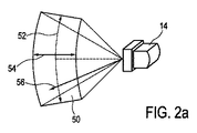
図2aは、超音波プローブ14に関係した体積50の例を示している。この例において描写される例証的な体積50は、位相二次元の電子的に走査されるアレイとして構成されている超音波プローブ14のトランスデューサアレイのため、セクタータイプのものである。従って、体積50のサイズは、仰角52及び水平角54によって表すことができる。体積50の深さ56は、1つの線あたり秒で表されるいわゆる走査線時間(line time)によって表すことができる。これは、特定の走査線を走査するために費やした走査時間である。
FIG. 2 a shows an example of a
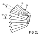
図2bは、どのようにして体積50を、多数のスライス58又は多数のいわゆる走査線59に沿ってそれぞれ取得される二次元画像に分割することができるかという例示的な例を示している。画像取得の間、超音波プローブ14の二次元トランスデューサアレイは、体積50が多数のこれらの走査線58に沿って連続して走査される方法で、ビームフォーマによって作動される。しかし、マルチライン受信処理において、単一の伝達ビームが、例えば4つ等、多数の受信走査線を照らすことができ、その受信走査線に沿って、信号が同時に取得される。その場合、そのような受信線のセットは、次に、体積50にわたって電子的に連続して走査される。
FIG. 2b shows an illustrative example of how the
従って、取得した二次元画像から処理される三次元画像の解像度は、いわゆる走査線密度次第であり、従って、2つの隣接する走査線59間のスペーシング60次第でもある。事実、スライス58内の2つの隣接する走査線59間の距離、さらに、スライス58間の距離である。結果として、横の広がりの方向における、及び、高さの広がりの方向における走査線密度は同じである。従って、走査線密度は、1つの線あたり度の形で測定される。
Therefore, the resolution of the three-dimensional image processed from the acquired two-dimensional image depends on the so-called scanning line density, and therefore also depends on the
図3は、超音波システム10の概略的なブロック図を示している。先においてすでに提示されているように、超音波システム10は、超音波プローブ(PR)14、制御ユニット(CU)16、ディスプレイ(DI)18及び入力装置(ID)20を含む。先においてさらに提示されているように、プローブ14は、位相二次元トランスデューサアレイ26を含む。一般に、制御ユニット(CU)16は、アナログ及び/又はデジタルの電子回路、プロセッサ、マイクロプロセッサ等を含み、画像取得及び供給全体を調和させてもよい中央処理装置を含んでもよい。さらに、制御ユニット16は、本明細書において画像取得コントローラ28と呼ばれるものを含む。しかし、画像取得コントローラ28は超音波システム10内の分けられた実体又はユニットである必要はないということを理解しなければならない。画像取得コントローラ28は、制御ユニット16の一部であってもよく、さらに、一般的には、実行されるハードウェア又はソフトウェアであってもよい。現在の区別は、例示的目的のためだめにつけられている。制御ユニット16の一部としての画像取得コントローラ28は、ビームフォーマを制御することができ、これによって、体積50のどの画像が得られるか、及び、どのようにしてこれらの画像が得られるかを制御することができる。トランスデューサアレイ26を駆動させるビームフォーマ30は電圧を生じ、部分的繰返し周波数(parts repetition frequency)を決定し、伝達したビーム及び1つ又は複数の受信若しくは受けたビームを走査する、焦点を合わせる、及び、アポダイズする(apodize)ことができ、さらに、フィルターを増幅する、及び、トランスデューサアレイ26によって戻らされたエコー電圧流をデジタル化することができる。さらに、本明細書において制御ユニット16の画像取得コントローラ28の画像取得部分32と呼ばれるものは、一般的な走査戦略を決定することができる。そのような一般戦略は、先においてすでに説明したように、所望のボリューム取得レート、体積の横の広がり、体積の高さの広がり、最大及び最小の走査線密度、走査線時間並びに走査線密度を含んでもよい。ここでも、画像取得部分32は、超音波システム10内の分けられた実体又はユニットである必要はない。画像取得部分32は、制御ユニット16の一部であってもよく、さらに、一般的には、実行されるハードウェア又はソフトウェアであってもよい。現在の区別は、例示的目的のためだめにつけられている。画像取得部分32は、例えば、ビームフォーマ30又は一般的な制御ユニット16において実行することもでき、又は、コントローラ16のデータ処理ユニットにおいて作動されるソフトウェアとして実行してもよい。
FIG. 3 shows a schematic block diagram of the
ビームフォーマ30は、さらに、トランスデューサアレイ26から超音波信号を受け、さらに、その信号を画像信号として転送する。
The beam former 30 further receives an ultrasonic signal from the
さらに、超音波システム10は、画像信号を受けるシグナルプロセッサ34を含む。シグナルプロセッサ34は、一般的に、アナログデジタル変換、例えば帯域フィルタリング等のデジタルフィルタリング、並びに、受けた超音波エコー又は画像信号の検出及び、例えばダイナミックな範囲縮小等の圧縮のために提供される。シグナルプロセッサは、画像データを転送する。
In addition, the
さらに、超音波システム10は、シグナルプロセッサ34から受けた画像データを、ディスプレイ18上で最終的に示される表示データに変換する画像プロセッサ36を含む。特に、画像プロセッサ36は、画像データを受け、画像データを前処理し、さらに、それを画像メモリに記憶することができる。次に、これらの画像データをさらに後処理し、ディスプレイ18を介して最も便利な画像を使用者に対して提供することができる。この場合、特に、画像プロセッサ36は、各スライス58における多数の走査線59に沿って取得した多数の二次元画像から三次元画像を形成することができる。
In addition, the
ユーザーインターフェースは、全般的に、参照番号38で描写され、さらに、ディスプレイ18及び入力装置20を含む。ユーザーインターフェースは、超音波プローブ14自体に提供することさえできる、例えばマウス又はさらなるボタン等、さらなる入力装置を含んでもよい。
The user interface is generally depicted with
本発明を適用することができる三次元超音波システムに対する特定の例は、本出願人によって売られているCX50CompactXtreme超音波システム、特に、本出願人のX7−2t TEEトランスデューサ、又は、本出願人のxMATRIX技術を使用した別のトランスデューサと共に使用する超音波システムである。一般に、Philips社のiE33システム上に見られるマトリクス型トランスデューサシステム、又は、例えばPhilips社のiU22及びHD15システム上に見られる機械的な3D/4Dトランスデューサ技術は、本発明を適用することができる。 A specific example for a 3D ultrasound system to which the present invention can be applied is the CX50 CompactXstream ultrasound system sold by the Applicant, in particular Applicant's X7-2t TEE transducer, or Applicant's An ultrasound system for use with another transducer using xMATRIX technology. In general, the matrix transducer system found on the Philips iE33 system or the mechanical 3D / 4D transducer technology found, for example, on the Philips iU22 and HD15 systems can apply the present invention.
使用中、一般的なシステム入力は、予め設定された又は固定された超音波システム10のパラメータとして行われる。これらのいわゆるシステム入力は、特に、最大の走査線密度、最小の走査線密度及び走査線時間である。使用者は、次に、目標ボリューム取得レートを入力してもよく、特に、超音波システム10に対して、体積50の横の広がり及び高さの広がり形で、走査されることになる体積又は関心領域のサイズをさらに特定してもよい。使用者は、横の広がり及び高さの広がりを、例えば40度等、数値として直接入力してもよい。しかし、使用者は、例えば、ディスプレイ18上のユーザーインターフェース38を介して特定の領域を選択してもよく、その選択は、次に、横の広がり及び高さの広がりに対する数値に変えられ、さらに、画像取得部分32に転送される。
In use, general system inputs are made as parameters of the
これらの入力に基づき、走査線密度は、以下の実験式 Based on these inputs, the scanning line density is
例えば、システム入力は、3度の最大の走査線密度、0.75度の最小の走査線密度、及び、1つの線あたり0.00005秒の走査線時間であってもよい。 For example, the system input may be a maximum scan line density of 3 degrees, a minimum scan line density of 0.75 degrees, and a scan line time of 0.00005 seconds per line.
図4aは、超音波システム10のディスプレイ18上の使用者に対して示され得る第1の例ディスプレイ40を示している。ディスプレイ上では、第1の大きな走査体積42が、関心領域の三次元画像として示されている。
FIG. 4 a shows a
この例において、使用者は、目標ボリューム取得レートを25Hz(1/秒)に設定しており、さらに、横の広がりは40度に設定され、且つ、高さの広がりも40度に設定されている。これらの値をユーザー入力及び上記のシステム入力として用いて、コントローラ28は、上記の実験式を介して、1つの線あたり1.41度として走査線密度を計算することができる。この値は、最小の走査線密度及び最大の走査線密度を介して設定された境界条件内にあるため、25Hzの実際のボリュームレートを所望のように維持することができる。
In this example, the user has set the target volume acquisition rate to 25 Hz (1 / second), the horizontal spread is set to 40 degrees, and the height spread is also set to 40 degrees. Yes. Using these values as user input and system input as described above, the
図4bにおいて、第2の例ディスプレイ44が示されている。このディスプレイ44において、より小さい関心領域46の画像が示されている。例えば、使用者は、第1の例ディスプレイ40において示されている体積42の特定部分は特に関心のある部分であり、それを対応するフレームでマークしたかもしれないと決定している。或いは、使用者は、体積の横及び高さの広がりに対して異なる値を直接入力したかもしれない。
In FIG. 4b, a
第2の例ディスプレイ44において、所望のボリュームレートは、ライブ又はリアルタイムでの三次元イメージングを維持するために依然として25Hz(1/秒)であってもよい。横の広がりは22度に設定され、さらに、高さの広がりは28度に設定される。コントローラ28はここで、上記及び第1の例と同じシステム入力が与えられると、1つの線あたり0.88度に走査線密度を計算することができる。この走査線密度は境界条件内にあるため、実際のボリューム取得レートは、25Hzの所望のボリューム取得レート又は目標ボリューム取得レートと等しい。従って、体積50にわたって走査線59の総数が一定のまま保たれるため、三次元ライブイメージングは維持される。さらに、使用者は、例ディスプレイ44におけるより小さい関心領域64が有意に小さい走査線密度を用いて取得されるという技術的利益を自動的に有する。従って、より小さい関心領域64の空間解像度は、第1のより大きな関心領域42の空間解像度よりも高い。しかし、超音波システム10は一定の走査線59の総数を維持したため、ボリューム取得レートは25Hzで維持される。従って、三次元ライブイメージングは、使用中より小さい関心領域を選んだ後に使用者に提供される。使用者がより大きな体積42に戻る場合、逆もまた同様に生じる。特に、使用者は関心領域を大きくしたけれども、取得レートは一定のまま残る。
In the
図5は、方法の一実施形態の概略的な流れ図を示している。当該方法が開始された後、第1のステップS1が実行される。このステップにおいて、体積50のサイズを決定するパラメータをコントローラは受ける。これらのパラメータは、入力装置20を介してユーザー入力として受けられる。さらに、システム入力が、超音波システム10内ですでにされている。システム入力は、解剖学的部位の走査にわたって一定のまま残り、ユーザー入力は、特定の走査の間、時間の経過に伴い変わり得る。システム入力は、最大の走査線密度、最小の走査線密度及び走査線時間である。ユーザー入力は、所望のボリューム取得レート又は目標ボリューム取得レート、体積50の横の広がり、及び、体積50の高さの広がりである。次に、ステップS2において、コントローラ28が、先において提示された以下の式
FIG. 5 shows a schematic flow diagram of one embodiment of the method. After the method is started, the first step S1 is performed. In this step, the controller receives parameters that determine the size of the
これによって、1つの線あたり度で表される走査線密度が計算される。次に、ステップS3において、超音波システム10は、トランスデューサアレイを用いて走査線59に沿って体積50を走査し、さらに、超音波信号を提供する。ステップS4において、超音波信号は、ビームフォーマ30及びシグナルプロセッサ34内で処理され、画像データを提供する。最後に、ステップS5において、三次元超音波画像が、画像データを使用して表示される。
This calculates the scanning line density expressed in degrees per line. Next, in step S3, the
さらに、任意で、使用中及びステップS6において、走査されることになる体積のサイズが変化したかどうかが決定される。特に、これは、関心領域が使用者によって変えられたため、体積の横の広がり及び/又は高さの広がりが変わる場合の事例である。この場合、新たなユーザー入力パラメータが、コントローラ28内に入力され、さらに、走査線密度がステップS2において再計算される。従って、走査はステップS3と共に続く。
Further, optionally, it is determined whether in use and in step S6 the size of the volume to be scanned has changed. In particular, this is the case when the lateral extent and / or height extent of the volume changes because the region of interest has been changed by the user. In this case, new user input parameters are input into the
ステップS6において体積のサイズが変わらなかった場合、ステップS7において走査処理全体が停止されるべきであるかどうかをさらに決定することができる。この場合、方法は終了し、そうでない場合、走査がステップS3において続けられる。 If the volume size has not changed in step S6, it can further be determined in step S7 whether the entire scanning process should be stopped. In this case, the method ends, otherwise scanning continues in step S3.
本発明は、図面及び上記の説明において詳細に例示及び記述されてきたけれども、そのような例示及び記述は、例示的又は例証的であり、拘束性はないと考慮されることになる。本発明は、開示された実施形態に限定されない。開示された実施形態に対する他の変化は、請求された発明を実行する際に、図面、明細書、及び付随の特許請求の範囲の調査から当業者により理解及びもたらすことができる。 Although the invention has been illustrated and described in detail in the drawings and foregoing description, such illustration and description are to be considered illustrative or exemplary and not restrictive. The invention is not limited to the disclosed embodiments. Other changes to the disclosed embodiments can be understood and brought by those skilled in the art from examining the drawings, the specification, and the appended claims, when carrying out the claimed invention.
特許請求の範囲において、「含む」という用語は、他の要素又はステップを除外せず、不定冠詞はその複数形を除外しない。1つの要素又は他のユニットは、特許請求の範囲内に列挙されたいくつかの項目の機能を満たすことができる。特定の手段が互いに異なる従属項において記載されているという単なる事実は、これらの手段の組合せを役立つよう使用することができないと示しているわけではない。 In the claims, the term “comprising” does not exclude other elements or steps, and the indefinite article does not exclude a plurality. An element or other unit may fulfill the functions of several items recited in the claims. The mere fact that certain measures are recited in mutually different dependent claims does not indicate that a combination of these measures cannot be used to help.
コンピュータプログラムは、他のハードウェアと共に若しくはその一部として供給される、光記憶媒体又は固体記憶媒体等、適したメディア上に記憶/分布することができるが、インターネット又は他の有線若しくは無線の通信システムを介して等、他の形状で分布することもできる。 The computer program can be stored / distributed on a suitable medium, such as an optical storage medium or a solid storage medium, supplied with or as part of other hardware, but the Internet or other wired or wireless communication It can also be distributed in other shapes, such as through a system.
特許請求の範囲におけるいかなる参照番号も、その範囲を限定するとして解釈されるべきではない。 Any reference signs in the claims should not be construed as limiting the scope.
Claims (8)
超音波受信信号を提供するように構成されるトランスデューサアレイ、
該トランスデューサアレイを制御して、当該超音波イメージングシステム内にパラメータとして予め設定されているそれぞれの走査線を走査するための走査時間に基づき、多数の走査線に沿って前記体積を走査するように構成され、さらに、前記超音波受信信号を受ける及び画像信号を提供するように構成されるビームフォーマ、
該ビームフォーマを制御して、ユーザー入力を介して受ける目標ボリューム取得レートを維持するために、前記体積のサイズに応じて前記体積内の前記走査線の密度を調整するように構成されるコントローラであり、ユーザー入力を介して受ける、度で表される横の広がり、及び、度で表される高さの広がり、並びに、前記それぞれの走査線を走査するための走査時間によって表される深さの形で前記体積のサイズを受けるように構成され、以下の実験式
式中、LDは、1つの線あたり度で表される前記走査線の密度であり、LEは、前記体積の横の広がりが度で表され、EEは、前記体積の高さの広がりが度で表され、TVRは、ヘルツで表される目標ボリューム取得レートであり、さらに、LTは、1つの線あたり秒で表される前記各走査線の走査時間である、コントローラ、
前記画像信号を受ける及び画像データを提供するように構成されるシグナルプロセッサ、
該シグナルプロセッサから前記画像データを受ける及びディスプレイデータを提供するように構成される画像プロセッサ、並びに、
前記ディスプレイデータを受ける及び前記三次元画像を提供するように構成されるディスプレイ、
を含むシステム。 An ultrasound imaging system for providing a three-dimensional image of a volume, comprising:
A transducer array configured to provide an ultrasound received signal,
Controlling the transducer array to scan the volume along multiple scan lines based on a scan time for scanning each scan line preset as a parameter in the ultrasound imaging system. A beamformer configured to receive the ultrasound received signal and provide an image signal;
A controller configured to adjust the density of the scan lines within the volume in response to the size of the volume to control the beamformer and maintain a target volume acquisition rate received via user input; Yes, received via user input, the horizontal spread in degrees, the spread in height in degrees, and the depth represented by the scan time for scanning each of the scan lines. Is configured to receive the size of the volume in the form of
Where LD is the density of the scan line expressed in degrees per line, LE is expressed in degrees of the lateral extent of the volume, and EE is the extent of height of the volume. A controller, wherein TVR is a target volume acquisition rate expressed in Hertz, and LT is the scan time of each scan line expressed in seconds per line,
A signal processor configured to receive the image signal and provide image data;
An image processor configured to receive the image data from the signal processor and provide display data; and
A display configured to receive the display data and to provide the three-dimensional image;
Including system.
前記コントローラが、前記体積のサイズを決定するパラメータ、ユーザー入力を介した目標ボリューム取得レート、及び、前記超音波イメージングシステム内にパラメータとして予め設定されているそれぞれの走査線を走査するための走査時間を受けるステップ、
前記コントローラが、前記目標ボリューム取得レートを維持するために、前記体積のサイズに応じて前記体積内の前記走査線の密度を調整するステップであり、前記体積のサイズは、ユーザー入力を介して受ける、度で表される横の広がり、及び、度で表される高さの広がり、並びに、前記それぞれの走査線を走査するための走査時間によって表される深さの形で表わされ、前記走査線の密度は、以下の実験式
式中、LDは、1つの線あたり度で表される前記走査線の密度であり、LEは、前記体積の横の広がりが度で表され、EEは、前記体積の高さの広がりが度で表され、TVRは、ヘルツで表される目標ボリューム取得レートであり、さらに、LTは、1つの線あたり秒で表される前記各走査線の走査時間である、ステップ、
超音波受信信号を提供する前記トランスデューサアレイが、前記走査時間及び調整された前記走査線の密度に基づき、前記走査線に沿って前記体積を走査するステップ、
前記ビームフォーマ及びシグナルプロセッサが、前記超音波受信信号を処理して画像データを提供するステップ、
前記画像プロセッサが、前記画像データを使用して前記ディスプレイに前記三次元超音波画像を表示させるステップ、
を含む方法。 An ultrasound imaging system including a controller, transducer array, beamformer, signal processor, image processor and display is used to provide a three-dimensional ultrasound image of a volume to be scanned along multiple scan lines A method,
The controller determines a parameter for determining the size of the volume, a target volume acquisition rate via user input, and a scan time for scanning each scan line preset as a parameter in the ultrasound imaging system. Step to receive,
The controller adjusting the density of the scan lines within the volume according to the size of the volume in order to maintain the target volume acquisition rate, the size of the volume being received via user input; , Expressed in the form of a horizontal spread expressed in degrees, a height spread expressed in degrees, and a depth represented by a scan time for scanning each of the scan lines, The scanning line density is calculated using the following empirical formula:
Where LD is the density of the scan line expressed in degrees per line, LE is expressed in degrees of the lateral extent of the volume, and EE is the extent of height of the volume. Where TVR is the target volume acquisition rate expressed in hertz, and LT is the scan time of each scan line expressed in seconds per line,
Scanning the volume along the scan line based on the scan time and the adjusted density of the scan line, the transducer array providing an ultrasound received signal;
The beamformer and a signal processor process the ultrasound received signal to provide image data;
Wherein the image processor is the step of causing display of the three-dimensional ultrasound image on the display using the image data,
Including methods.
Applications Claiming Priority (3)
| Application Number | Priority Date | Filing Date | Title |
|---|---|---|---|
| US201161557955P | 2011-11-10 | 2011-11-10 | |
| US61/557,955 | 2011-11-10 | ||
| PCT/IB2012/056088 WO2013068894A1 (en) | 2011-11-10 | 2012-11-01 | Steady frame rate volumetric ultrasound imaging |
Publications (2)
| Publication Number | Publication Date |
|---|---|
| JP2014534885A JP2014534885A (en) | 2014-12-25 |
| JP6571332B2 true JP6571332B2 (en) | 2019-09-04 |
Family
ID=47172849
Family Applications (1)
| Application Number | Title | Priority Date | Filing Date |
|---|---|---|---|
| JP2014540590A Active JP6571332B2 (en) | 2011-11-10 | 2012-11-01 | Volumetric ultrasound imaging with stable frame rate |
Country Status (8)
| Country | Link |
|---|---|
| US (1) | US10939895B2 (en) |
| EP (1) | EP2748627B1 (en) |
| JP (1) | JP6571332B2 (en) |
| CN (1) | CN103946717B (en) |
| BR (1) | BR112014011024A2 (en) |
| IN (1) | IN2014CN03623A (en) |
| MX (1) | MX2014005545A (en) |
| WO (1) | WO2013068894A1 (en) |
Families Citing this family (7)
| Publication number | Priority date | Publication date | Assignee | Title |
|---|---|---|---|---|
| JP2016016022A (en) * | 2014-07-04 | 2016-02-01 | 株式会社東芝 | Ultrasonic diagnostic equipment |
| JP6907193B2 (en) * | 2015-09-10 | 2021-07-21 | コーニンクレッカ フィリップス エヌ ヴェKoninklijke Philips N.V. | Ultrasonic system with wide depth and detailed view |
| JP6651316B2 (en) * | 2015-09-16 | 2020-02-19 | キヤノンメディカルシステムズ株式会社 | Ultrasound diagnostic equipment |
| JP7159046B2 (en) * | 2015-12-15 | 2022-10-24 | コリンダス、インコーポレイテッド | System and method for controlling x-ray frame rate of imaging system |
| US11696745B2 (en) * | 2017-03-16 | 2023-07-11 | Koninklijke Philips N.V. | Optimal scan plane selection for organ viewing |
| EP3469993A1 (en) | 2017-10-16 | 2019-04-17 | Koninklijke Philips N.V. | An ultrasound imaging system and method |
| US20190167231A1 (en) * | 2017-12-01 | 2019-06-06 | Sonocine, Inc. | System and method for ultrasonic tissue screening |
Family Cites Families (19)
| Publication number | Priority date | Publication date | Assignee | Title |
|---|---|---|---|---|
| JPH04152939A (en) * | 1990-10-17 | 1992-05-26 | Hitachi Medical Corp | Ultrasonic diagnostic device |
| US5793701A (en) | 1995-04-07 | 1998-08-11 | Acuson Corporation | Method and apparatus for coherent image formation |
| JPH09192130A (en) * | 1996-01-12 | 1997-07-29 | Aloka Co Ltd | Ultrasonic diagnostic equipment |
| JP3723663B2 (en) * | 1997-07-15 | 2005-12-07 | フクダ電子株式会社 | Ultrasonic diagnostic equipment |
| EP1162476A1 (en) | 2000-06-06 | 2001-12-12 | Kretztechnik Aktiengesellschaft | Method for examining objects with ultrasound |
| US6468216B1 (en) | 2000-08-24 | 2002-10-22 | Kininklijke Philips Electronics N.V. | Ultrasonic diagnostic imaging of the coronary arteries |
| JP2003010182A (en) * | 2001-06-19 | 2003-01-14 | Ge Medical Systems Global Technology Co Llc | Ultrasonographic method and ultrasonographic device |
| US7758508B1 (en) | 2002-11-15 | 2010-07-20 | Koninklijke Philips Electronics, N.V. | Ultrasound-imaging systems and methods for a user-guided three-dimensional volume-scan sequence |
| JP2004290249A (en) | 2003-03-25 | 2004-10-21 | Fuji Photo Film Co Ltd | Ultrasonic imaging apparatus and ultrasonic imaging method |
| EP1611457B1 (en) * | 2003-03-27 | 2014-09-10 | Koninklijke Philips N.V. | Guidance of invasive medical devices by wide view three dimensional ultrasonic imaging |
| EP1673014A1 (en) * | 2003-10-08 | 2006-06-28 | Koninklijke Philips Electronics N.V. | Improved ultrasonic volumetric imaging by coordination of acoustic sampling resolution, volumetric line density and volume imaging rate |
| US20070088213A1 (en) | 2003-11-20 | 2007-04-19 | Koninklijke Philips Electronics N.V. | Ultrasonic diagnostic imaging with automatic adjustment of beamforming parameters |
| JP2007512869A (en) * | 2003-11-21 | 2007-05-24 | コーニンクレッカ フィリップス エレクトロニクス エヌ ヴィ | Ultrasound imaging system and method capable of adaptive selection of image frame rate and / or number of averaged echo samples |
| DE602004029930D1 (en) * | 2003-12-16 | 2010-12-16 | Koninkl Philips Electronics Nv | Diagnostic ultrasound imaging method and system with automatic resolution and refresh rate control |
| US20050228280A1 (en) | 2004-03-31 | 2005-10-13 | Siemens Medical Solutions Usa, Inc. | Acquisition and display methods and systems for three-dimensional ultrasound imaging |
| US20070007680A1 (en) | 2005-07-05 | 2007-01-11 | Fina Technology, Inc. | Methods for controlling polyethylene rheology |
| JP4969985B2 (en) | 2006-10-17 | 2012-07-04 | 株式会社東芝 | Ultrasonic diagnostic apparatus and control program for ultrasonic diagnostic apparatus |
| KR100949059B1 (en) | 2006-10-17 | 2010-03-25 | 주식회사 메디슨 | Ultrasound system and method for forming ultrasound image |
| JP5366385B2 (en) * | 2007-09-26 | 2013-12-11 | 株式会社東芝 | Ultrasonic diagnostic apparatus and ultrasonic scanning program |
-
2012
- 2012-11-01 US US14/356,182 patent/US10939895B2/en active Active
- 2012-11-01 MX MX2014005545A patent/MX2014005545A/en unknown
- 2012-11-01 WO PCT/IB2012/056088 patent/WO2013068894A1/en active Application Filing
- 2012-11-01 EP EP12784349.8A patent/EP2748627B1/en active Active
- 2012-11-01 BR BR112014011024A patent/BR112014011024A2/en not_active IP Right Cessation
- 2012-11-01 JP JP2014540590A patent/JP6571332B2/en active Active
- 2012-11-01 CN CN201280055160.5A patent/CN103946717B/en active Active
- 2012-11-01 IN IN3623CHN2014 patent/IN2014CN03623A/en unknown
Also Published As
| Publication number | Publication date |
|---|---|
| JP2014534885A (en) | 2014-12-25 |
| CN103946717A (en) | 2014-07-23 |
| WO2013068894A1 (en) | 2013-05-16 |
| CN103946717B (en) | 2016-12-21 |
| IN2014CN03623A (en) | 2015-09-04 |
| EP2748627B1 (en) | 2021-04-07 |
| MX2014005545A (en) | 2014-05-30 |
| EP2748627A1 (en) | 2014-07-02 |
| BR112014011024A2 (en) | 2017-04-25 |
| US20140358006A1 (en) | 2014-12-04 |
| US10939895B2 (en) | 2021-03-09 |
Similar Documents
| Publication | Publication Date | Title |
|---|---|---|
| JP6571332B2 (en) | Volumetric ultrasound imaging with stable frame rate | |
| US10335118B2 (en) | Ultrasonic diagnostic apparatus, medical image processing apparatus, and medical image parallel display method | |
| KR101140525B1 (en) | Method and apparatus for extending an ultrasound image field of view | |
| JP4868843B2 (en) | Ultrasonic diagnostic apparatus and control program for ultrasonic diagnostic apparatus | |
| JP4942237B2 (en) | Display method and imaging system for arranging region of interest in image | |
| JP2020072937A (en) | Elastography measurement system and method of the same | |
| US9743910B2 (en) | Ultrasonic diagnostic apparatus, ultrasonic image processing apparatus, medical image diagnostic apparatus, and medical image processing apparatus | |
| JP2007513726A (en) | Ultrasound imaging system with automatic control of penetration, resolution and frame rate | |
| US9877698B2 (en) | Ultrasonic diagnosis apparatus and ultrasonic image processing apparatus | |
| WO2004000124A1 (en) | Ultrasound quantification in real-time using acoustic data in more than two dimensions | |
| JP6207518B2 (en) | Improved large volume 3D ultrasound imaging | |
| US20130211254A1 (en) | Ultrasound acquisition | |
| JP6687336B2 (en) | Ultrasonic diagnostic device and control program | |
| JP2007044354A (en) | Ultrasonic diagnostic equipment and ultrasonic diagnostic equipment control program | |
| JP5570927B2 (en) | Ultrasonic diagnostic apparatus, ultrasonic image treatment apparatus, medical image processing apparatus, and ultrasonic image processing program | |
| JP2022016592A (en) | Ultrasonic diagnostic device, image processing device, and image processing program | |
| JP2013244161A (en) | Ultrasonograph | |
| JP2010022648A (en) | Ultrasonograph and ultrasonic image-processing program |
Legal Events
| Date | Code | Title | Description |
|---|---|---|---|
| A621 | Written request for application examination |
Free format text: JAPANESE INTERMEDIATE CODE: A621 Effective date: 20151030 |
|
| A977 | Report on retrieval |
Free format text: JAPANESE INTERMEDIATE CODE: A971007 Effective date: 20160916 |
|
| A131 | Notification of reasons for refusal |
Free format text: JAPANESE INTERMEDIATE CODE: A131 Effective date: 20161108 |
|
| A521 | Request for written amendment filed |
Free format text: JAPANESE INTERMEDIATE CODE: A523 Effective date: 20170206 |
|
| A02 | Decision of refusal |
Free format text: JAPANESE INTERMEDIATE CODE: A02 Effective date: 20170801 |
|
| A521 | Request for written amendment filed |
Free format text: JAPANESE INTERMEDIATE CODE: A523 Effective date: 20171129 |
|
| A911 | Transfer to examiner for re-examination before appeal (zenchi) |
Free format text: JAPANESE INTERMEDIATE CODE: A911 Effective date: 20171206 |
|
| A912 | Re-examination (zenchi) completed and case transferred to appeal board |
Free format text: JAPANESE INTERMEDIATE CODE: A912 Effective date: 20180223 |
|
| A601 | Written request for extension of time |
Free format text: JAPANESE INTERMEDIATE CODE: A601 Effective date: 20190201 |
|
| A521 | Request for written amendment filed |
Free format text: JAPANESE INTERMEDIATE CODE: A523 Effective date: 20190411 |
|
| A521 | Request for written amendment filed |
Free format text: JAPANESE INTERMEDIATE CODE: A523 Effective date: 20190528 |
|
| A61 | First payment of annual fees (during grant procedure) |
Free format text: JAPANESE INTERMEDIATE CODE: A61 Effective date: 20190808 |
|
| R150 | Certificate of patent or registration of utility model |
Ref document number: 6571332 Country of ref document: JP Free format text: JAPANESE INTERMEDIATE CODE: R150 |
|
| R250 | Receipt of annual fees |
Free format text: JAPANESE INTERMEDIATE CODE: R250 |
|
| R250 | Receipt of annual fees |
Free format text: JAPANESE INTERMEDIATE CODE: R250 |
|
| R250 | Receipt of annual fees |
Free format text: JAPANESE INTERMEDIATE CODE: R250 |


