JP6204350B2 - Therapeutic antigen binding molecules with FcRn binding domains that promote antigen clearance - Google Patents
Therapeutic antigen binding molecules with FcRn binding domains that promote antigen clearance Download PDFInfo
- Publication number
- JP6204350B2 JP6204350B2 JP2014514947A JP2014514947A JP6204350B2 JP 6204350 B2 JP6204350 B2 JP 6204350B2 JP 2014514947 A JP2014514947 A JP 2014514947A JP 2014514947 A JP2014514947 A JP 2014514947A JP 6204350 B2 JP6204350 B2 JP 6204350B2
- Authority
- JP
- Japan
- Prior art keywords
- antigen
- binding
- region
- amino acid
- antibody
- Prior art date
- Legal status (The legal status is an assumption and is not a legal conclusion. Google has not performed a legal analysis and makes no representation as to the accuracy of the status listed.)
- Active
Links
Images


Classifications
-
- C—CHEMISTRY; METALLURGY
- C07—ORGANIC CHEMISTRY
- C07K—PEPTIDES
- C07K16/00—Immunoglobulins [IGs], e.g. monoclonal or polyclonal antibodies
- C07K16/18—Immunoglobulins [IGs], e.g. monoclonal or polyclonal antibodies against material from animals or humans
- C07K16/28—Immunoglobulins [IGs], e.g. monoclonal or polyclonal antibodies against material from animals or humans against receptors, cell surface antigens or cell surface determinants
- C07K16/2866—Immunoglobulins [IGs], e.g. monoclonal or polyclonal antibodies against material from animals or humans against receptors, cell surface antigens or cell surface determinants against receptors for cytokines, lymphokines, interferons
-
- A—HUMAN NECESSITIES
- A61—MEDICAL OR VETERINARY SCIENCE; HYGIENE
- A61P—SPECIFIC THERAPEUTIC ACTIVITY OF CHEMICAL COMPOUNDS OR MEDICINAL PREPARATIONS
- A61P19/00—Drugs for skeletal disorders
- A61P19/02—Drugs for skeletal disorders for joint disorders, e.g. arthritis, arthrosis
-
- A—HUMAN NECESSITIES
- A61—MEDICAL OR VETERINARY SCIENCE; HYGIENE
- A61P—SPECIFIC THERAPEUTIC ACTIVITY OF CHEMICAL COMPOUNDS OR MEDICINAL PREPARATIONS
- A61P29/00—Non-central analgesic, antipyretic or antiinflammatory agents, e.g. antirheumatic agents; Non-steroidal antiinflammatory drugs [NSAID]
-
- A—HUMAN NECESSITIES
- A61—MEDICAL OR VETERINARY SCIENCE; HYGIENE
- A61P—SPECIFIC THERAPEUTIC ACTIVITY OF CHEMICAL COMPOUNDS OR MEDICINAL PREPARATIONS
- A61P37/00—Drugs for immunological or allergic disorders
-
- A—HUMAN NECESSITIES
- A61—MEDICAL OR VETERINARY SCIENCE; HYGIENE
- A61P—SPECIFIC THERAPEUTIC ACTIVITY OF CHEMICAL COMPOUNDS OR MEDICINAL PREPARATIONS
- A61P37/00—Drugs for immunological or allergic disorders
- A61P37/02—Immunomodulators
- A61P37/06—Immunosuppressants, e.g. drugs for graft rejection
-
- C—CHEMISTRY; METALLURGY
- C07—ORGANIC CHEMISTRY
- C07K—PEPTIDES
- C07K16/00—Immunoglobulins [IGs], e.g. monoclonal or polyclonal antibodies
- C07K16/18—Immunoglobulins [IGs], e.g. monoclonal or polyclonal antibodies against material from animals or humans
- C07K16/28—Immunoglobulins [IGs], e.g. monoclonal or polyclonal antibodies against material from animals or humans against receptors, cell surface antigens or cell surface determinants
- C07K16/2803—Immunoglobulins [IGs], e.g. monoclonal or polyclonal antibodies against material from animals or humans against receptors, cell surface antigens or cell surface determinants against the immunoglobulin superfamily
- C07K16/2812—Immunoglobulins [IGs], e.g. monoclonal or polyclonal antibodies against material from animals or humans against receptors, cell surface antigens or cell surface determinants against the immunoglobulin superfamily against CD4
-
- A—HUMAN NECESSITIES
- A61—MEDICAL OR VETERINARY SCIENCE; HYGIENE
- A61K—PREPARATIONS FOR MEDICAL, DENTAL OR TOILETRY PURPOSES
- A61K39/00—Medicinal preparations containing antigens or antibodies
- A61K2039/505—Medicinal preparations containing antigens or antibodies comprising antibodies
-
- C—CHEMISTRY; METALLURGY
- C07—ORGANIC CHEMISTRY
- C07K—PEPTIDES
- C07K2317/00—Immunoglobulins specific features
- C07K2317/30—Immunoglobulins specific features characterized by aspects of specificity or valency
- C07K2317/34—Identification of a linear epitope shorter than 20 amino acid residues or of a conformational epitope defined by amino acid residues
-
- C—CHEMISTRY; METALLURGY
- C07—ORGANIC CHEMISTRY
- C07K—PEPTIDES
- C07K2317/00—Immunoglobulins specific features
- C07K2317/50—Immunoglobulins specific features characterized by immunoglobulin fragments
- C07K2317/52—Constant or Fc region; Isotype
-
- C—CHEMISTRY; METALLURGY
- C07—ORGANIC CHEMISTRY
- C07K—PEPTIDES
- C07K2317/00—Immunoglobulins specific features
- C07K2317/50—Immunoglobulins specific features characterized by immunoglobulin fragments
- C07K2317/52—Constant or Fc region; Isotype
- C07K2317/524—CH2 domain
-
- C—CHEMISTRY; METALLURGY
- C07—ORGANIC CHEMISTRY
- C07K—PEPTIDES
- C07K2317/00—Immunoglobulins specific features
- C07K2317/50—Immunoglobulins specific features characterized by immunoglobulin fragments
- C07K2317/52—Constant or Fc region; Isotype
- C07K2317/526—CH3 domain
-
- C—CHEMISTRY; METALLURGY
- C07—ORGANIC CHEMISTRY
- C07K—PEPTIDES
- C07K2317/00—Immunoglobulins specific features
- C07K2317/90—Immunoglobulins specific features characterized by (pharmaco)kinetic aspects or by stability of the immunoglobulin
- C07K2317/94—Stability, e.g. half-life, pH, temperature or enzyme-resistance
Description
本発明は、中性pHにおける胎児性Fc受容体(FcRn)に対するアフィニティーが増強された改変FcRn結合ドメイン;免疫原性が低く、安定性が高く、かつわずかな凝集物しか形成しない、該FcRn結合ドメインを含む抗原結合分子;中性pHにおける既存の抗医薬品抗体に対する結合活性が増強されることなく、中性pHまたは酸性pHにおけるFcRn結合活性が増強された改変抗原結合分子;抗原結合分子による抗原の細胞内への取り込みを改善するための抗原結合分子の使用;特定の抗原の血漿中濃度を低下させるための抗原結合分子の使用;単一の抗原結合分子がその分解前に結合できる抗原の総数を増加させるための改変FcRn結合ドメインの使用;抗原結合分子の薬物動態を改善するための改変FcRn結合ドメインの使用;既存の抗医薬品抗体に対する結合活性を低下させる方法;および該抗原結合分子の製造方法に関する。 The present invention provides a modified FcRn binding domain with enhanced affinity for fetal Fc receptor (FcRn) at neutral pH; said FcRn binding that is less immunogenic, highly stable and forms few aggregates An antigen-binding molecule comprising a domain; a modified antigen-binding molecule having enhanced FcRn-binding activity at neutral pH or acidic pH without enhancing binding activity to an existing anti-pharmaceutical antibody at neutral pH; antigen by antigen-binding molecule Use of antigen-binding molecules to improve cellular uptake of cells; use of antigen-binding molecules to reduce plasma concentrations of specific antigens; use of antigens to which a single antigen-binding molecule can bind prior to its degradation Use of modified FcRn binding domains to increase the total number; Use of modified FcRn binding domains to improve pharmacokinetics of antigen binding molecules; Binding activity against existing anti-pharmaceutical antibodies How to Do; and to a process for the production of said antigen binding molecule.
血漿中での安定性が高く、副作用も少ないことから、ますます多くの抗体が医薬品として用いられつつある。可溶型抗原を標的とする従来の抗体は、注射後に患者の血漿中の抗原と結合し、その後、分解されるまで抗体抗原複合体の形態で安定に持続する。典型的な抗体は一般的に半減期が長いのに対して(1〜3週間)、抗原は1日未満という比較的短い半減期を有する。そのため、抗体と複合体を形成している抗原は抗原単独よりも顕著に長い半減期を有する。その結果、従来の抗体の注射後に抗原濃度が上昇する傾向がある。このような事例は、IL-6(J Immunotoxicol. 2005, 3, 131-9.(非特許文献1))、βアミロイド(MAbs. 2010 Sep-Oct;2(5):576-88(非特許文献2))、MCP-1(ARTHRITIS & RHEUMATISM 2006, 54,2387-92 (非特許文献3))、ヘプシジン(AAPS J. 2010, 12(4):646-57.(非特許文献4))、およびsIL-6受容体(Blood. 2008 Nov 15;112(10):3959-64.(非特許文献5))などの様々な可溶型抗原を標的とする抗体について報告されている。報告では、抗体投与時に血漿中総抗原濃度がベースラインからおよそ10〜1000倍上昇する(抗原による)と記載している。 Because of its high stability in plasma and few side effects, more and more antibodies are being used as pharmaceuticals. Conventional antibodies that target soluble antigens bind to antigens in the patient's plasma after injection and then persist stably in the form of antibody-antigen complexes until degraded. Typical antibodies generally have a long half-life (1-3 weeks), whereas antigens have a relatively short half-life of less than 1 day. Therefore, the antigen forming a complex with the antibody has a significantly longer half-life than the antigen alone. As a result, the antigen concentration tends to increase after conventional antibody injection. Such cases include IL-6 (J Immunotoxicol. 2005, 3, 131-9. (Non-Patent Document 1)), β-amyloid (MAbs. 2010 Sep-Oct; 2 (5): 576-88 (Non-Patent Document 1). 2)), MCP-1 (ARTHRITIS & RHEUMATISM 2006, 54, 2387-92 (Non-Patent Document 3)), hepcidin (AAPS J. 2010, 12 (4): 646-57. (Non-Patent Document 4)) And antibodies targeting various soluble antigens such as sIL-6 receptor (Blood. 2008 Nov 15; 112 (10): 3959-64. (Non-patent Document 5)) have been reported. The report states that the plasma total antigen concentration increases approximately 10-1000 times from baseline (depending on antigen) upon antibody administration.
血漿中総抗原濃度のこのような上昇は望ましくないため、治療用抗体によって抗原を除去するための戦略が開発された。このような戦略の1つは、IgGの胎児性Fc受容体(FcRn)に対する結合アフィニティーが増強されたpH依存的抗原結合抗体を用いて抗原を迅速に処分することである(例えば、PCT出願第PCT/JP2011/001888号(特許文献1)を参照されたい)。FcRnは、多くの細胞の膜内に見られるタンパク質である。中性pHにおけるFcRnに対する結合活性が増強された抗体は、細胞表面上でFcRnと結合し、それによって、抗体を伴った受容体は小胞により細胞内部に取り込まれる。小胞内部のpHが徐々に低下するにつれて、抗原は、酸性pHではアフィニティーが低いためにpH依存的抗原結合抗体から解離する。解離した抗原はその後分解されるのに対して、FcRnおよび結合している抗体は、分解される前にリサイクルされて細胞の表面に戻される。したがって、中性pHにおけるFcRnに対する結合活性が増強されたpH依存的抗原結合抗体を用いて、血漿中から抗原を除去し、その血漿中濃度を低下させることができる。 Because such an increase in plasma total antigen concentration is undesirable, strategies have been developed to remove antigens with therapeutic antibodies. One such strategy is to rapidly dispose of the antigen using a pH-dependent antigen-binding antibody with enhanced binding affinity of IgG to the fetal Fc receptor (FcRn) (e.g. PCT application no. (See PCT / JP2011 / 001888 (Patent Document 1)). FcRn is a protein found in the membrane of many cells. Antibodies with enhanced FcRn binding activity at neutral pH bind to FcRn on the cell surface, whereby the receptor with the antibody is taken up into the cell by the vesicle. As the pH inside the vesicle gradually decreases, the antigen dissociates from the pH-dependent antigen-binding antibody due to its low affinity at acidic pH. The dissociated antigen is then degraded, whereas FcRn and bound antibody are recycled back to the cell surface before being degraded. Therefore, using a pH-dependent antigen-binding antibody with enhanced FcRn binding activity at neutral pH, it is possible to remove antigen from plasma and reduce its plasma concentration.
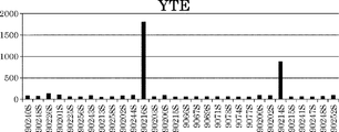
また、以前の研究によって、酸性pHにおけるFcRnに対する結合アフィニティーを増強するためのFc操作も、エンドソームリサイクル効率および抗体の薬物動態を改善できることが示された。例えば、M252Y/S254T/T256E(YTE)改変体(J Biol Chem, 2006, 281:23514-23524.(非特許文献6))、M428L/N434S(LS)改変体(Nat Biotechnol, 2010 28:157-159.(非特許文献7))、T250Q/M428L(J Immunol. 2006, 176(1):346-56.(非特許文献8))、およびN434H改変体(Clinical Pharmacology & Therapeutics (2011) 89(2):283-290.(非特許文献9))は、天然IgG1に対する半減期の改善を示した。 Previous studies have also shown that Fc manipulation to enhance binding affinity for FcRn at acidic pH can also improve endosome recycling efficiency and antibody pharmacokinetics. For example, M252Y / S254T / T256E (YTE) variant (J Biol Chem, 2006, 281: 23514-23524. (Non-Patent Document 6)), M428L / N434S (LS) variant (Nat Biotechnol, 2010 28: 157- 159. (Non-patent document 7)), T250Q / M428L (J Immunol. 2006, 176 (1): 346-56. (Non-patent document 8)), and N434H variant (Clinical Pharmacology & Therapeutics (2011) 89 ( 2): 283-290. (Non-Patent Document 9)) showed an improvement in half-life relative to native IgG1.
しかしながら、このような置換には、抗体の安定性、免疫原性、凝集挙動、および既存の抗体(例えば、リウマトイド因子)に対する結合アフィニティーといった、治療用抗体の開発に重要な抗体の特性を変えるリスクもある。したがって、抗体のクリアランスを増大させるだけでなく、治療用抗原結合分子を開発するための基準をも満たす改変FcRn結合ドメインを提供することが、本発明の主な目的である。このような開発可能性の基準とは、具体的には、安定性が高いこと、免疫原性が低いこと、凝集物の割合が低いこと、および既存の抗医薬品抗体(ADA)に対する結合アフィニティーが低いことである。 However, such substitutions involve risks that alter antibody properties important for therapeutic antibody development, such as antibody stability, immunogenicity, aggregation behavior, and binding affinity for existing antibodies (e.g., rheumatoid factor). There is also. Accordingly, it is a main object of the present invention to provide modified FcRn binding domains that not only increase antibody clearance but also meet the criteria for developing therapeutic antigen binding molecules. These criteria for development potential are specifically high stability, low immunogenicity, low percentage of aggregates, and binding affinity to existing anti-pharmaceutical antibodies (ADA). It is low.
本発明に関する先行技術文献を以下に示す。本明細書において引用された文献はすべて、参照により本明細書に組み入れられる。 Prior art documents relating to the present invention are shown below. All documents cited herein are hereby incorporated by reference.
本発明はこのような状況に鑑みて為されたものであり、その目的は、中性pHにおけるFcRnに対するアフィニティーが増強された改変FcRn結合ドメイン;該FcRn結合ドメインを含む抗原結合分子であって、免疫原性が低く、安定性が高く、かつわずかな凝集物しか形成しない抗原結合分子;中性pHにおける既存の抗医薬品抗体に対する結合活性が増強されることなく、中性pHまたは酸性pHにおけるFcRn結合活性が増強された改変抗原結合分子;抗原結合分子による抗原の細胞内への取り込みを改善するための抗原結合分子の使用;特定の抗原の血漿中濃度を低下させるための抗原結合分子の使用;単一の抗原結合分子がその分解前に結合できる抗原の総数を増加させるための改変FcRn結合ドメインの使用;抗原結合分子の薬物動態を改善するための改変FcRn結合ドメインの使用;および該抗原結合分子の製造方法を提供することにある。 The present invention has been made in view of such circumstances, and the object thereof is a modified FcRn-binding domain with enhanced affinity for FcRn at neutral pH; an antigen-binding molecule comprising the FcRn-binding domain, Antigen-binding molecule with low immunogenicity, high stability, and formation of few aggregates; FcRn at neutral or acidic pH without enhanced binding activity to existing anti-pharmaceutical antibodies at neutral pH Modified antigen-binding molecules with enhanced binding activity; use of antigen-binding molecules to improve the uptake of antigens into cells by antigen-binding molecules; use of antigen-binding molecules to reduce plasma concentrations of specific antigens Use of a modified FcRn binding domain to increase the total number of antigens that a single antigen-binding molecule can bind before its degradation; modified FcR to improve the pharmacokinetics of an antigen-binding molecule Use of an n-binding domain; and to provide a method for producing the antigen-binding molecule.
本発明者らは、中性pHにおけるFcRnに対するアフィニティーが増強された改変FcRn結合ドメインについて、および免疫原性が低く、安定性が高く、かつわずかな凝集物しか形成しない、該FcRn結合ドメインを含む抗原結合分子について鋭意研究を行った。その結果、本発明者らは、FcRn結合ドメインの特定の位置における置換によって、免疫原性が実質的に上昇することなく、安定性が実質的に低下することなく、かつ/または高分子量種の割合が実質的に上昇することなく、中性pHにおけるFcRnに対するアフィニティーを増強することを見出した。
さらに、本発明者らは、中性pHまたは酸性pHにおけるFcRnに対するアフィニティーが増強されたが、既存の抗医薬品抗体に対する結合活性が有意に増強されていない改変FcRn結合ドメインについて、およびこのようなFcRn結合ドメインを含む抗原結合分子について鋭意研究を行った。その結果、本発明者らは、FcRn結合ドメインの特定の位置における置換によって、FcRn結合活性が実質的に低下することなく、中性pHにおける既存の抗医薬品抗体に対するアフィニティーが低下することを見出した。
We include a modified FcRn binding domain with enhanced affinity for FcRn at neutral pH, and the FcRn binding domain which is less immunogenic, highly stable and forms few aggregates We have conducted extensive research on antigen-binding molecules. As a result, the inventors have found that substitutions at specific positions of the FcRn binding domain do not substantially increase immunogenicity, substantially reduce stability, and / or of high molecular weight species. It has been found that the affinity for FcRn at neutral pH is enhanced without a substantial increase in the proportion.
In addition, the present inventors have identified modified FcRn binding domains that have enhanced affinity for FcRn at neutral or acidic pH, but not significantly enhanced binding activity to existing anti-pharmaceutical antibodies, and such FcRn We have conducted extensive research on antigen-binding molecules containing binding domains. As a result, the present inventors have found that substitution at a specific position of the FcRn binding domain reduces affinity for existing anti-pharmaceutical antibodies at neutral pH without substantially reducing FcRn binding activity. .
具体的には、本発明は以下に関する:
[1]改変FcRn結合ドメインを含む抗原結合分子であって、該改変FcRn結合ドメインが、EU238、EU250、EU252、EU254、EU255、EU258、EU286、EU307、EU308、EU309、EU311、EU315、EU428、EU433、EU434、およびEU436からなる群より選択される1つまたは複数の位置にアミノ酸置換を含み、数字がEUナンバリングで表される置換の位置を示す、抗原結合分子。
[2]前記FcRn結合ドメインが、
a) EU252位およびEU434位におけるアミノ酸置換;ならびに
b) EU238、EU250、EU252、EU254、EU255、EU256、EU258、EU286、EU387、EU307、EU308、EU309、EU311、EU315、EU428、EU433、EU434、およびEU436からなる群より選択される1つまたは複数の位置におけるアミノ酸置換
を有する、[1]記載の抗原結合分子。
[3]前記改変FcRn結合ドメインが、
EU238位にアスパラギン酸、
EU250位にバリン、
EU252位にチロシン、
EU254位にスレオニン、
EU255位にロイシン、
EU256位にグルタミン酸、
EU258位にアスパラギン酸もしくはイソロイシン、
EU286位にグルタミン酸、
EU307位にグルタミン、
EU308位にプロリン、
EU309位にグルタミン酸、
EU311位にアラニンもしくはヒスチジン、
EU315位にアスパラギン酸、
EU428位にイソロイシン、
EU433位にアラニン、リジン、プロリン、アルギニン、もしくはセリン、
EU434位にチロシンもしくはトリプトファン、および/または
EU436位にイソロイシン、ロイシン、バリン、スレオニン、もしくはフェニルアラニン
を含む、[1]または[2]記載の抗原結合分子。
[4]前記FcRn結合ドメインが、
a) EU252、EU434、およびEU436;
b) EU252、EU307、EU311、およびEU434;
c) EU252、EU315、およびEU434;
d) EU252、EU308、およびEU434:
e) EU238、EU252、およびEU434;
f) EU252、EU434、EU307、EU311、およびEU436;ならびに
g) EU252、EU387、およびEU434
からなる群より選択される位置の組み合わせのうちの1つまたは複数にアミノ酸置換を含む、[2]記載の抗原結合分子。
[5]前記FcRn結合ドメインが、
a) EU252位にチロシン、EU315位にアスパラギン酸、およびEU434位にチロシン;または
b) EU252位にチロシン、EU434位にチロシン、およびEU436位にイソロイシン;または
c) EU252位にチロシン、EU434位にチロシン、およびEU436位にロイシン;または
d) EU252位にチロシン、EU434位にチロシン、およびEU436位にバリン;または
e) EU252位にチロシン、EU254位にスレオニン、EU434位にチロシン、およびEU436位にイソロイシン
を含む、[4]記載の抗原結合分子。
[6]前記FcRn結合ドメインが3つ以上の位置にアミノ酸置換を含み、該3つ以上の位置が、
a) EU252/EU434/EU307/EU311/EU286;
b) EU252/EU434/EU307/EU311/EU286/EU254;
c) EU252/EU434/EU307/EU311/EU436;
d) EU252/EU434/EU307/EU311/EU436/EU254;
e) EU252/EU434/EU307/EU311/EU436/EU250;
f) EU252/EU434/EU308/EU250;
g) EU252/EU434/EU308/EU250/EU436;および
h) EU252/EU434/EU308/EU250/EU307/EU311
からなる組み合わせ群のうちの1つである、[2]記載の抗原結合分子。
[7]前記FcRn結合ドメインが、
a) EU252位にチロシン、EU286位にグルタミン酸、EU307位にグルタミン、EU311位にアラニン、およびEU434位にチロシン;または
b) EU252位にチロシン、EU254位にスレオニン、EU286位にグルタミン酸、EU307位にグルタミン、EU311位にアラニン、およびEU434位にチロシン;または
c) EU252位にチロシン、EU307位にグルタミン、EU311位にアラニン、EU434位にチロシン、および436位にイソロイシン;または
d) EU252位にチロシン、EU254位にスレオニン、EU286位にグルタミン酸、EU307位にグルタミン、EU311位にアラニン、EU434位にチロシン、およびEU436位にイソロイシン;または
e) EU250位にバリン、EU252位にチロシン、EU254位にスレオニン、EU308位にプロリン、EU434位にチロシン、およびEU436位にバリン;または
f) EU250位にバリン、EU252位にチロシン、EU307位にグルタミン、EU311位にアラニン、EU434位にチロシン、およびEU436位にバリン;または
g) EU252位にチロシン、EU307位にグルタミン、EU311位にアラニン、EU434位にチロシン、およびEU436位にバリン;または
h) EU250位にバリン、EU252位にチロシン、EU308位にプロリン、およびEU434位にチロシン;または
i) EU250位にバリン、EU252位にチロシン、EU307位にグルタミン、EU308位にプロリン、EU311位にアラニン、およびEU434位にチロシン
を含む、[6]記載の抗原結合分子。
[8]前記FcRn結合ドメインが3つ以上の位置にアミノ酸置換を含み、該3つ以上の位置が、
a) EU252およびEU434およびEU307およびEU311およびEU436およびEU286;
b) EU252およびEU434およびEU307およびEU311およびEU436およびEU250およびEU308;
c) EU252およびEU434およびEU307およびEU311およびEU436およびEU250およびEU286およびEU308;
d) EU252およびEU434およびEU307およびEU311およびEU436およびEU250およびEU286およびEU308およびEU428
からなる組み合わせ群のうちの1つである、[2]記載の抗原結合分子。
[9]前記FcRn結合ドメインが、
a) EU252位にチロシン、EU286位にグルタミン酸、EU307位にグルタミン、EU311位にアラニン、EU434位にチロシン、およびEU436位にバリン;または
b) EU250位にバリン、EU252位にチロシン、EU307位にグルタミン、EU308位にプロリン、EU311位にアラニン、EU434位にチロシン、およびEU436位にバリン;または
c) EU250位にバリン、EU252位にチロシン、EU286位にグルタミン酸、EU307位にグルタミン、EU308位にプロリン、EU311位にアラニン、EU434位にチロシン、およびEU436位にバリン;または
d) EU250位にバリン、EU252位にチロシン、EU286位にグルタミン酸、EU307位にグルタミン、EU308位にプロリン、EU311位にアラニン、EU434位にチロシン、およびEU436位にバリン
を含む、[8]記載の抗原結合分子。
[10]前記FcRn結合ドメインが、
a) EU434およびEU307およびEU311;
b) EU434およびEU307およびEU309およびEU311;または
c) EU434およびEU250およびEU252およびEU436
からなる組み合わせ群のうちの1つである3つ以上の位置においてアミノ酸置換を含む、[2]記載の抗原結合分子。
[11]前記FcRn結合ドメインが、
a) EU307位にグルタミン、EU311位にヒスチジン、およびEU434位にチロシン;または
b) EU307位にグルタミン、EU309位にグルタミン酸、EU311位にアラニン、およびEU434位にチロシン;または
c) EU307位にグルタミン、EU309位にグルタミン酸、EU311位にヒスチジン、およびEU434位にチロシン;または
d) EU250位にバリン、EU252位にチロシン、EU434位にチロシン、およびEU436位にバリン
を含む、[10]記載の抗原結合分子。
[12]高分子量種の割合が2%未満である、[1]〜[11]のいずれか一つに記載の抗原結合分子。
[13]a) 抗原に対する結合活性が、pH 7〜8よりもpH 5.5〜6.5において低いか、または
b) 抗原に対する「カルシウム濃度依存的結合」活性を有する
抗原結合ドメインを含む、[1]〜[12]のいずれか一つに記載の抗原結合分子。
[14]pH 7におけるFcRnに対する前記結合分子の結合活性が50〜150 nMであり、Tmが63.0℃よりも高く、かつEpibaseスコアが250未満である、[1]〜[5]のいずれか一つに記載の抗原結合分子。
[15]pH 7におけるFcRnに対する前記結合分子の結合活性が15〜50 nMであり、Tmが60℃よりも高く、かつEpibaseスコアが500未満である、[1]〜[3]および[6]〜[7]のいずれか一つに記載の抗原結合分子。
[16]pH 7におけるFcRnに対する前記結合分子の結合活性が15nMよりも強く、Tmが57.5℃よりも高く、かつEpibaseスコアが500未満である、[1]〜[3]および[8]〜[9]のいずれか一つに記載の抗原結合分子。
[17]前記FcRn結合ドメインが、
a) EU238位、EU255位、および/またはEU258位、ならびに
b) 表4〜7に記載された組み合わせのうちの1つである3つ以上の位置
においてアミノ酸置換を含む、[1]〜[3]のいずれか一つに記載の抗原結合分子。
[18]a) 前記FcRn結合ドメインのEU257位のアミノ酸が、アラニン、バリン、イソロイシン、ロイシン、およびスレオニンからなる群より選択されるアミノ酸ではなく、かつ/または
b) 前記FcRn結合ドメインのEU252位のアミノ酸がトリプトファンではない、
[1]〜[17]のいずれか一つに記載の抗原結合分子。
[19]既存の抗医薬品抗体に対する結合活性が、インタクトなFcRn結合ドメインを含む対照抗体の結合アフィニティーと比較して有意に増強されていない、[1]〜[18]のいずれか一つに記載の抗原結合分子。
[20]前記FcRn結合ドメインが、EU387、EU422、EU424、EU426、EU433、EU436、EU438、およびEU440からなる群より選択される1つまたは複数の位置にアミノ酸置換をさらに含む、[19]記載の抗原結合分子。
[21]前記FcRn結合ドメインが、
EU387位におけるアルギニン、
EU422位におけるグルタミン酸、アルギニン、またはセリン、アスパラギン酸、リジン、スレオニン、またはグルタミン;
EU424位におけるグルタミン酸、またはアルギニン、リジン、またはアスパラギン;
EU426位におけるアスパラギン酸、グルタミン、アラニン、またはチロシン;
EU433位におけるアスパラギン酸;
EU436位におけるスレオニン;
EU438位におけるグルタミン酸、アルギニン、セリン、またはリジン;および
EU440位におけるグルタミン酸、アスパラギン酸、またはグルタミン
からなる群より選択される1つまたは複数のアミノ酸置換を含む、[20]記載の抗原結合分子。
[22]前記改変FcRn結合ドメインが、表12〜13に記載された組み合わせのうちの1つである3つ以上の置換を含む、[1]〜[21]のいずれか一つに記載の抗原結合分子。
[23]前記改変FcRn結合ドメインが、表14〜15に記載された組み合わせのうちの1つである3つ以上の置換を含む、[1]〜[22]のいずれか一つに記載の抗原結合分子。
[24]前記FcRn結合ドメインが、
a) EU252位にチロシン、EU387位にアルギニン、EU434位にチロシン、およびEU436位にバリン;または
b) EU252位にチロシン、EU422位にグルタミン酸、EU434位にチロシン、およびEU436位にバリン;または
c) EU252位にチロシン、EU422位にアルギニン、EU434位にチロシン、およびEU436位にバリン;または
d) EU252位にチロシン、EU422位にセリン、EU434位にチロシン、およびEU436位にバリン;または
e) EU252位にチロシン、EU422位にグルタミン酸、EU434位にチロシン、およびEU436位にバリン;または
f) EU252位にチロシン、EU424位にアルギニン、EU434位にチロシン、およびEU436位にバリン;または
g) EU252位にチロシン、EU434位にチロシン、EU436位にバリン、およびEU438位にグルタミン酸;または
h) EU252位にチロシン、EU434位にチロシン、EU436位にバリン、およびEU438位にアルギニン;または
i) EU252位にチロシン、EU434位にチロシン、EU436位にバリン、およびEU438位にセリン;または
j) EU252位にチロシン、EU434位にチロシン、EU436位にバリン、およびEU440位にグルタミン酸
を含む、[20]〜[23]のいずれか一つに記載の抗原結合分子。
[25]抗体である、[1]〜[24]のいずれか一つに記載の抗原結合分子。
[26]抗原結合分子による抗原の細胞内への取り込みを改善するための、[1]〜[25]のいずれか一つに記載の抗原結合分子の使用。
[27]特定の抗原の血漿中濃度を低下させるための[1]〜[25]のいずれか一つに記載の抗原結合分子の使用であって、該抗原結合分子が該抗原に結合し得る抗原結合ドメインを含む、使用。
[28]EU238、EU250、EU252、EU254、EU255、EU258、EU286、EU307、EU308、EU309、EU311、EU315、EU428、EU433、EU434、およびEU436からなる群より選択される1つまたは複数の位置において、抗原結合分子のFcRn結合ドメインにアミノ酸置換を導入する工程を含む、抗原結合分子の薬物動態を改善する方法。
[29]EU238、EU250、EU252、EU254、EU255、EU258、EU286、EU307、EU308、EU309、EU311、EU315、EU428、EU433、EU434、およびEU436からなる群より選択される1つまたは複数の位置において、抗原結合分子のFcRn結合ドメインにアミノ酸置換を導入する工程を含む、対象における抗原結合分子の消失を遅らせる方法。
[30]EU238、EU250、EU252、EU254、EU255、EU258、EU286、EU307、EU308、EU309、EU311、EU315、EU428、EU433、EU434、およびEU436からなる群より選択される1つまたは複数の位置において、抗原結合分子のFcRn結合ドメインにアミノ酸置換を導入する工程を含む、抗原結合分子の血漿中滞留時間を長くする方法。
[31]EU238、EU250、EU252、EU254、EU255、EU258、EU286、EU307、EU308、EU309、EU311、EU315、EU428、EU433、EU434、およびEU436からなる群より選択される1つまたは複数の位置において、抗原結合分子のFcRn結合ドメインにアミノ酸置換を導入する工程を含む、抗原結合分子の血漿中抗原消失速度を増大させる方法。
[32]EU238、EU250、EU252、EU254、EU255、EU258、EU286、EU307、EU308、EU309、EU311、EU315、EU428、EU433、EU434、およびEU436からなる群より選択される1つまたは複数の位置において、抗原結合分子のFcRn結合ドメインにアミノ酸置換を導入する工程を含む、抗原結合分子の血漿中抗原消失能を増強する方法。
[33]前記FcRn結合ドメイン中のEU256位においてさらなるアミノ酸置換が導入される、[28]〜[32]のいずれか一つに記載の方法。
[34]EU387、EU422、EU424、EU426、EU433、EU436、EU438、およびEU440からなる群より選択される1つまたは複数の位置において前記FcRn結合ドメインにアミノ酸置換を導入する工程をさらに含む、[28]〜[33]のいずれか一つに記載の方法。
[35]以下の工程を含む、[1]〜[25]のいずれか一つに記載の抗原結合分子を製造する方法:
(a) 親FcRn結合ドメインを選択し、EU238、EU250、EU252、EU254、EU255、EU258、EU286、EU307、EU308、EU309、EU311、EU315、EU428、EU433、EU434、およびEU436からなる群より選択される1つまたは複数の位置にアミノ酸置換を導入することにより、親FcRnを改変する工程;
(b) pH依存的抗原結合ドメインまたはカルシウムイオン依存的抗原結合ドメインを得るために、抗原結合分子の抗原結合ドメインを選択し、該抗原結合ドメイン中の少なくとも1つのアミノ酸を改変する工程;
(c) (a)および(b)において調製されたヒトFcRn結合ドメインおよび抗原結合ドメインが連結された抗原結合分子をコードする遺伝子を得る工程、ならびに
(d) (c)において調製された遺伝子を用いて、抗原結合分子を製造する工程。
[36]工程a)において、前記FcRn結合ドメイン中のEU256位においてさらなるアミノ酸置換が導入される、[35]記載の方法。
[37]EU387、EU422、EU424、EU426、EU433、EU436、EU438、およびEU440からなる群より選択される1つまたは複数の位置において前記FcRn結合ドメインにアミノ酸置換を導入する工程をさらに含む、[35]または[36]記載の方法。
[38]改変FcRn結合ドメインを含む抗原結合分子であって、該改変FcRn結合ドメインが、EU387、EU422、EU424、EU426、EU433、EU436、EU438、およびEU440からなる群より選択される1つまたは複数の位置にアミノ酸置換を含み、中性pHにおける既存の抗医薬品抗体(ADA)に対する該抗原結合分子の結合アフィニティーが、インタクトなFcRn結合ドメインを含む抗原結合分子の結合アフィニティーと比較して有意に増強されていない、抗原結合分子。
[39]さらにpH中性域またはpH酸性域におけるFcRnに対する結合アフィニティーが増強されている、[38]記載の抗原結合分子。
[40]EU387、EU422、EU424、EU426、EU433、EU436、EU438、およびEU440からなる群より選択される1つまたは複数の位置を置換するアミノ酸が、
a) EU387位におけるアルギニン;
b) EU422位におけるグルタミン酸、アルギニン、セリン、アスパラギン酸、リジン、スレオニン、またはグルタミン;
c) EU424位におけるグルタミン酸、アルギニン、リジン、またはアスパラギン;
d) EU426位におけるアスパラギン酸、グルタミン、アラニン、またはチロシン;
e) EU433位におけるアスパラギン酸
f) EU436位におけるスレオニン
g) EU438位におけるグルタミン酸、アルギニン、セリン、またはリジン;および
h) EU440位におけるグルタミン酸、アスパラギン酸、またはグルタミン
からなる群より選択される、[38]または[39]記載の抗原結合分子。
[41]前記改変FcRn結合ドメインが、表10に記載された1つもしくは複数の位置または組み合わせのうちの1つにおいてアミノ酸置換を含む、[38]〜[40]のいずれか一つに記載の抗原結合分子。
[42]前記改変FcRn結合ドメインが、表11に記載されたアミノ酸の置換または置換の組み合わせのいずれか1つを含む、[38]〜[40]のいずれか一つに記載の抗原結合分子。
[43]前記改変FcRn結合ドメインが、FcRn結合ドメインにおけるEU238、EU250、EU252、EU254、EU255、EU256、EU258、EU286、EU307、EU308、EU309、EU311、EU315、EU428、EU434、およびEU436からなる群より選択される1つまたは複数の位置にアミノ酸置換をさらに含み、該置換がpH中性域またはpH酸性域におけるFcRn結合活性を増強する、[39]〜[42]のいずれか一つに記載の抗原結合分子。
[44]前記改変FcRn結合ドメインが、FcRn結合ドメインにおける位置
i) a) EU438/EU440 または b) EU424;および
ii) a) EU434、b) EU252/EU254/EU256、c) EU428/EU434、または d) EU250/EU428
にアミノ酸置換を含む、[39]〜[43]のいずれか一つに記載の抗原結合分子。
[45]前記改変FcRn結合ドメインが、アミノ酸置換
i) a) EU438R/EU440E または b) EU424N;および
ii) a) M434H、b) M252Y/S254T/T256E、c) M428L/N434S、または d) T250QおよびM428L(EUナンバリング)
を含む、[44]記載の抗原結合分子。
[46]前記改変FcRn結合ドメインが、表13および15に記載された組み合わせのうちの1つである3つ以上のアミノ酸置換を含む、[45]記載の抗原結合分子。
[47]前記改変FcRn結合ドメインが、
a) EU387、EU422、EU424、EU438、EU440、EU433からなる群より選択される1つもしくは複数の位置、またはEU422/EU424およびEU438/EU440からなる組み合わせ群のうちの1つである2つ以上の位置;ならびに
b) 表9に記載された組み合わせのうちの1つである2つ以上の位置
に置換を含む、[39]〜[42]のいずれか一つに記載の抗原結合分子。
[48]前記改変FcRn結合ドメインが、表12または14に記載された組み合わせのうちの1つである3つ以上のアミノ酸置換を含む、[47]記載の抗原結合分子。
[49]pH依存的抗原結合ドメインまたはカルシウムイオン依存的抗原結合ドメインを含む、[39]〜[48]のいずれか一つに記載の抗原結合分子。
[50]以下の工程を含む、中性pHまたは酸性pHにおけるFcRnに対する結合活性が増強されており、かつ中性pHにおける既存のADAに対する結合活性が増強されているFcRn結合ドメインを含む抗原結合分子の、既存のADAに対する結合活性を低下させる方法:
a) 中性pHまたは酸性pHにおけるFcRnに対する結合活性が増強されており、かつ中性pHにおける既存のADAに対する結合活性が増強されているFcRn結合ドメインを有する抗原結合分子を提供する工程;および
b) EU387、EU422、EU424、EU426、EU433、EU436、EU438、およびEU440からなる群より選択される1つまたは複数の位置において、FcRn結合ドメイン中のアミノ酸を置換して、改変FcRn結合ドメインを有する抗原結合分子をもたらす工程。
[51]工程b)が、表10に記載された組み合わせのうちの1つである3つ以上の位置においてアミノ酸を置換することを含む、[50]記載の方法。
[52]工程b)が、表11に記載された組み合わせのうちの1つである3つ以上のアミノ酸置換をFcRn結合ドメインに導入することを含む、[50]記載の方法。
[53]以下の工程を含む、親抗体と比較して中性pHにおける既存のADAに対する結合活性を有意に増強することなく、単一の抗原結合分子が結合できる抗原の総数を増加させる方法:
a) 親FcRn結合ドメインを含む抗原結合分子を提供する工程、
b) EU238、EU250、EU252、EU254、EU255、EU256、EU258、EU286、EU307、EU308、EU309、EU311、EU315、EU428、EU433、EU434、およびEU436からなる群より選択される1つまたは複数の位置において、親FcRn結合ドメインのアミノ酸配列中のアミノ酸を置換することにより、工程a)の親FcRn結合ドメインを改変する工程;ならびに
c) EU387、EU422、EU424、EU426、EU433、EU436、EU438、およびEU440からなる群より選択される1つまたは複数の位置において、親FcRn結合ドメインのアミノ酸配列中のアミノ酸を置換することにより、工程b)の改変FcRn結合ドメインを改変する工程。
[54]以下の工程を含む、親抗体と比較して中性pHにおける既存のADAに対する抗原結合分子の結合活性を有意に増強することなく、抗原と結合した状態で細胞内に取り込まれた抗原結合分子の、抗原と結合していない状態での細胞外への放出を促進する方法:
a) 親FcRn結合ドメインを含む抗原結合分子を提供する工程、
b) EU238、EU250、EU252、EU254、EU255、EU256、EU258、EU286、EU307、EU308、EU309、EU311、EU315、EU428、EU433、EU434、およびEU436、およびEU428からなる群より選択される1つまたは複数の位置において、親FcRn結合ドメインのアミノ酸配列中のアミノ酸を置換することにより、親FcRn結合ドメインを改変する工程;ならびに
c) EU387、EU422、EU424、EU426、EU433、EU436、EU438、およびEU440からなる群より選択される1つまたは複数の位置において、親FcRn結合ドメインのアミノ酸配列中のアミノ酸を置換することにより、工程b)の改変FcRn結合ドメインを改変する工程。
[55]以下の工程を含む、親抗体と比較して中性pHにおける既存のADAに対する結合活性を有意に増強することなく、抗原結合分子の血漿中抗原消失能を増強する方法:
a) 親FcRn結合ドメインを含む抗原結合分子を提供する工程、
b) EU238、EU250、EU252、EU254、EU255、EU256、EU258、EU286、EU307、EU308、EU309、EU311、EU315、EU428、EU433、EU434、およびEU436、およびEU428からなる群より選択される1つまたは複数の位置において、親FcRn結合ドメインのアミノ酸配列中のアミノ酸を置換することにより、親FcRn結合ドメインを改変する工程;ならびに
c) EU387、EU422、EU424、EU426、EU433、EU436、EU438、およびEU440からなる群より選択される1つまたは複数の位置において、親FcRn結合ドメインのアミノ酸配列中のアミノ酸を置換することにより、工程b)の改変FcRn結合ドメインを改変する工程。
[56]以下の工程を含む、親抗体と比較して中性pHにおける既存のADAに対する結合活性を有意に増強することなく、抗原結合分子の薬物動態を改善する方法:
a) 親FcRn結合ドメインを含む抗原結合分子を提供する工程、
b) EU238、EU250、EU252、EU254、EU255、EU256、EU258、EU286、EU307、EU308、EU309、EU311、EU315、EU428、EU433、EU434、およびEU436からなる群より選択される1つまたは複数の位置において、親FcRn結合ドメインのアミノ酸配列中のアミノ酸を置換することにより、親FcRn結合ドメインを改変する工程;ならびに
c) EU387、EU422、EU424、EU426、EU433、EU436、EU438、およびEU440からなる群より選択される1つまたは複数の位置において、親FcRn結合ドメインのアミノ酸配列中のアミノ酸を置換することにより、工程b)の改変FcRn結合ドメインを改変する工程。
[57]以下の工程を含む、親抗体と比較して中性pHにおける既存のADAに対する結合活性を有意に増強することなく、血漿中の総抗原濃度または遊離抗原濃度を低下させる方法:
a) 親FcRn結合ドメインを含む抗原結合分子であって、該抗原と結合し得る抗原結合ドメインを含む抗原結合分子を提供する工程、
b) EU238、EU250、EU252、EU254、EU255、EU256、EU258、EU286、EU307、EU308、EU309、EU311、EU315、EU428、EU433、EU434、およびEU436からなる群より選択される1つまたは複数の位置において、親FcRn結合ドメインのアミノ酸配列中のアミノ酸を置換することにより、親FcRn結合ドメインを改変する工程;ならびに
c) EU387、EU422、EU424、EU426、EU433、EU436、EU438、およびEU440からなる群より選択される1つまたは複数の位置において、親FcRn結合ドメインのアミノ酸配列中のアミノ酸を置換することにより、工程b)の改変FcRn結合ドメインを改変する工程。
[58]以下の工程を含む、中性pHまたは酸性pHにおけるFcRnに対する結合活性が増強されており、かつ中性pHにおける既存のADAに対する結合活性が低下しているFcRn結合ドメインを含む抗原結合分子を製造する方法:
(a) pH中性域またはpH酸性域におけるFcRnに対する結合活性、およびpH中性域における既存のADAに対する結合活性が増強されているFcRn結合ドメインを提供する工程、
(b) EU387、EU422、EU424、EU426、EU433、EU436、EU438、およびEU440からなる群より選択される1つまたは複数の位置において、アミノ酸を置換する工程、
(c) pH依存的抗原結合ドメインを得るために、抗原結合分子の抗原結合ドメインを選択して該抗原結合ドメイン中の少なくとも1つのアミノ酸を改変する工程、またはカルシウムイオン依存的抗原結合ドメインを選択する工程;
(d) (a)および(b)において調製されたヒトFcRn結合ドメインおよび抗原結合ドメインが連結された抗原結合分子をコードする遺伝子を得る工程、ならびに
(e) (c)において調製された遺伝子を用いて抗原結合分子を製造する工程であって、製造された該抗原結合分子が、インタクトなFcRn結合ドメインを有する親抗原結合ドメインと比較して、中性pHまたは酸性pHにおけるFcRnに対する結合活性が増強されており、かつ中性pHにおける内因性ADAに対する結合活性が低下している、工程。
[59]pH中性域またはpH酸性域におけるFcRnおよび既存のADAに対する結合活性ならびにpH中性域における既存のADAに対する結合活性が増強されている前記FcRn結合ドメインが、EU238、EU250、EU252、EU254、EU255、EU256、EU258、EU286、EU307、EU308、EU309、EU311、EU315、EU428、EU433、EU434、およびEU436からなる群より選択される1つまたは複数の位置にアミノ酸の置換を含む、[58]記載の方法。
[60]工程a)で導入されるアミノ酸置換が3つ以上の位置におけるものであり、該3つ以上の位置が、表4〜7に記載された組み合わせのうちの1つである、[53]〜[57]のいずれか一つに記載の方法。
[61]工程b)で導入されるアミノ酸置換が3つ以上の位置におけるものであり、該3つ以上の位置が、表10に記載された組み合わせのうちの1つである、[53]〜[60]のいずれか一つに記載の方法。
Specifically, the present invention relates to:
[1] An antigen-binding molecule comprising a modified FcRn-binding domain, wherein the modified FcRn-binding domain comprises EU238, EU250, EU252, EU254, EU255, EU258, EU286, EU307, EU308, EU309, EU311, EU315, EU428, EU433 An antigen-binding molecule comprising an amino acid substitution at one or more positions selected from the group consisting of EU434, EU434, and EU436, wherein the number indicates the position of the substitution represented by EU numbering.
[2] The FcRn binding domain is
a) amino acid substitutions at positions EU252 and EU434; and
b) one or more selected from the group consisting of EU238, EU250, EU252, EU254, EU255, EU256, EU258, EU286, EU387, EU307, EU308, EU309, EU311, EU315, EU428, EU433, EU434, and EU436 The antigen-binding molecule according to [1], which has an amino acid substitution at a position.
[3] The modified FcRn binding domain is
Aspartic acid in EU 238th place,
Valin to EU 250th place
EU252 is tyrosine,
Threonine in EU 254th,
Leucine in EU255,
EU 256th place glutamic acid,
Aspartic acid or isoleucine in EU258 position,
EU286 is glutamic acid,
Glutamine in EU 307
Proline in EU308,
EU309 position glutamic acid,
EU311 is alanine or histidine,
EU 315th aspartic acid,
Isoleucine at EU428
EU433 position alanine, lysine, proline, arginine or serine,
EU434 position tyrosine or tryptophan, and / or
The antigen-binding molecule according to [1] or [2], which contains isoleucine, leucine, valine, threonine, or phenylalanine at position EU436.
[4] The FcRn binding domain is
a) EU252, EU434 and EU436;
b) EU252, EU307, EU311 and EU434;
c) EU252, EU315, and EU434;
d) EU252, EU308 and EU434:
e) EU238, EU252 and EU434;
f) EU252, EU434, EU307, EU311 and EU436; and
g) EU252, EU387, and EU434
The antigen-binding molecule according to [2], comprising an amino acid substitution in one or more of a combination of positions selected from the group consisting of:
[5] The FcRn binding domain is
a) tyrosine at EU252 position, aspartic acid at EU315 position and tyrosine at EU434 position; or
b) tyrosine at EU252 position, tyrosine at EU434 position and isoleucine at EU436 position; or
c) EU252 at tyrosine, EU434 at tyrosine, and EU436 at leucine; or
d) tyrosine at EU252 position, tyrosine at EU434 position, and valine at EU436 position; or
e) The antigen-binding molecule according to [4], comprising tyrosine at position EU252, threonine at position EU254, tyrosine at position EU434, and isoleucine at position EU436.
[6] The FcRn-binding domain contains an amino acid substitution at three or more positions, and the three or more positions are
a) EU252 / EU434 / EU307 / EU311 / EU286;
b) EU252 / EU434 / EU307 / EU311 / EU286 / EU254;
c) EU252 / EU434 / EU307 / EU311 / EU436;
d) EU252 / EU434 / EU307 / EU311 / EU436 / EU254;
e) EU252 / EU434 / EU307 / EU311 / EU436 / EU250;
f) EU252 / EU434 / EU308 / EU250;
g) EU252 / EU434 / EU308 / EU250 / EU436; and
h) EU252 / EU434 / EU308 / EU250 / EU307 / EU311
The antigen-binding molecule according to [2], which is one of a combination group consisting of:
[7] The FcRn binding domain is
a) tyrosine at position EU252, glutamic acid at position EU286, glutamine at position EU307, alanine at position EU311 and tyrosine at position EU434; or
b) EU252, tyrosine, EU254, threonine, EU286, glutamic acid, EU307, glutamine, EU311, alanine, and EU434, tyrosine; or
c) tyrosine at EU252 position, glutamine at EU307 position, alanine at EU311 position, tyrosine at EU434 position, and isoleucine at position 436; or
d) tyrosine at EU252 position, threonine at EU254 position, glutamic acid at EU286 position, glutamine at EU307 position, alanine at EU311 position, tyrosine at EU434 position, and isoleucine at EU436 position; or
e) EU250, valine, EU252, tyrosine, EU254, threonine, EU308, proline, EU434, tyrosine, and EU436, valine; or
f) Valine at EU250, tyrosine at EU252, glutamine at EU307, alanine at EU311, tyrosine at EU434, and valine at EU436; or
g) tyrosine at EU252 position, glutamine at EU307 position, alanine at EU311 position, tyrosine at EU434 position, and valine at EU436 position; or
h) EU at position valine, EU 252 at tyrosine, EU 308 at proline, and EU 434 at tyrosine; or
i) The antigen-binding molecule according to [6], comprising valine at position EU250, tyrosine at position EU252, glutamine at position EU307, proline at position EU308, alanine at position EU311 and tyrosine at position EU434.
[8] The FcRn binding domain contains an amino acid substitution at three or more positions, and the three or more positions are
a) EU252 and EU434 and EU307 and EU311 and EU436 and EU286;
b) EU252 and EU434 and EU307 and EU311 and EU436 and EU250 and EU308;
c) EU252 and EU434 and EU307 and EU311 and EU436 and EU250 and EU286 and EU308;
d) EU252 and EU434 and EU307 and EU311 and EU436 and EU250 and EU286 and EU308 and EU428
The antigen-binding molecule according to [2], which is one of a combination group consisting of:
[9] The FcRn binding domain is
a) tyrosine at EU252 position, glutamic acid at EU286 position, glutamine at EU307 position, alanine at EU311 position, tyrosine at EU434 position, and valine at EU436 position; or
b) Valine at EU250, tyrosine at EU252, glutamine at EU307, proline at EU308, alanine at EU311, tyrosine at EU434, and valine at EU436; or
c) EU250, valine, EU252, tyrosine, EU286, glutamic acid, EU307, glutamine, EU308, proline, EU311, alanine, EU434, tyrosine, and EU436, valine; or
d) EU250, containing valine, EU252, tyrosine, EU286, glutamic acid, EU307, glutamine, EU308, proline, EU311, alanine, EU434, tyrosine, and EU436, valine. [8] Antigen binding molecule.
[10] The FcRn binding domain is
a) EU434 and EU307 and EU311;
b) EU434 and EU307 and EU309 and EU311; or
c) EU434 and EU250 and EU252 and EU436
The antigen-binding molecule according to [2], comprising an amino acid substitution at 3 or more positions which is one of a combination group consisting of:
[11] The FcRn binding domain is
a) Glutamine at EU 307, histidine at EU 311 and tyrosine at EU 434; or
b) Glutamine at EU307, glutamic acid at EU309, alanine at EU311 and tyrosine at EU434; or
c) Glutamine at EU307, glutamic acid at EU309, histidine at EU311 and tyrosine at EU434; or
d) The antigen-binding molecule according to [10], comprising valine at position EU250, tyrosine at position EU252, tyrosine at position EU434, and valine at position EU436.
[12] The antigen-binding molecule according to any one of [1] to [11], wherein the proportion of high molecular weight species is less than 2%.
[13] a) the binding activity to the antigen is lower at pH 5.5-6.5 than at pH 7-8, or
b) The antigen-binding molecule according to any one of [1] to [12], comprising an antigen-binding domain having “calcium concentration-dependent binding” activity for an antigen.
[14] Any one of [1] to [5], wherein the binding activity of the binding molecule to FcRn at
[15] The binding activity of the binding molecule to FcRn at
[16] The binding activity of the binding molecule to FcRn at
[17] The FcRn binding domain is
a) EU238th, EU255th, and / or EU258th, and
b) The antigen-binding molecule according to any one of [1] to [3], comprising an amino acid substitution at 3 or more positions that is one of the combinations described in Tables 4-7.
[18] a) The amino acid at position EU257 of the FcRn binding domain is not an amino acid selected from the group consisting of alanine, valine, isoleucine, leucine, and threonine, and / or
b) the amino acid at position EU252 of the FcRn binding domain is not tryptophan;
The antigen-binding molecule according to any one of [1] to [17].
[19] The binding activity to an existing anti-pharmaceutical antibody is not significantly enhanced compared to the binding affinity of a control antibody containing an intact FcRn binding domain, according to any one of [1] to [18] Antigen-binding molecules.
[20] The FcRn binding domain further comprises an amino acid substitution at one or more positions selected from the group consisting of EU387, EU422, EU424, EU426, EU433, EU436, EU438, and EU440. Antigen binding molecule.
[21] The FcRn binding domain is
Arginine in EU 387th place,
Glutamic acid, arginine, or serine, aspartic acid, lysine, threonine, or glutamine at position EU422;
Glutamic acid or arginine, lysine or asparagine at EU position 424;
Aspartic acid, glutamine, alanine, or tyrosine at EU position 426;
Aspartic acid at position EU433;
Threonine in EU436th place;
Glutamic acid, arginine, serine, or lysine at position EU438; and
[20] The antigen-binding molecule according to [20], comprising one or more amino acid substitutions selected from the group consisting of glutamic acid, aspartic acid, or glutamine at position EU440.
[22] The antigen according to any one of [1] to [21], wherein the modified FcRn binding domain includes three or more substitutions that are one of the combinations described in Tables 12 to 13 Binding molecule.
[23] The antigen according to any one of [1] to [22], wherein the modified FcRn binding domain comprises three or more substitutions that are one of the combinations described in Tables 14 to 15 Binding molecule.
[24] The FcRn binding domain is
a) tyrosine at EU252 position, arginine at EU387 position, tyrosine at EU434 position, and valine at EU436 position; or
b) tyrosine at EU252 position, glutamic acid at EU422 position, tyrosine at EU434 position, and valine at EU436 position; or
c) tyrosine at EU252 position, arginine at EU422 position, tyrosine at EU434 position, and valine at EU436 position; or
d) tyrosine at position EU252, serine at position EU422, tyrosine at position EU434, and valine at position EU436; or
e) Tyrosine at EU252 position, glutamic acid at EU422 position, tyrosine at EU434 position, and valine at EU436 position; or
f) tyrosine at EU252 position, arginine at EU424 position, tyrosine at EU434 position, and valine at EU436 position; or
g) tyrosine at EU252 position, tyrosine at EU434 position, valine at EU436 position, and glutamic acid at EU438 position; or
h) tyrosine at EU252 position, tyrosine at EU434 position, valine at EU436 position, and arginine at EU438 position; or
i) tyrosine at position EU252, tyrosine at position EU434, valine at position EU436, and serine at position EU438; or
j) The antigen-binding molecule according to any one of [20] to [23], comprising tyrosine at EU252 position, tyrosine at EU434 position, valine at EU436 position, and glutamic acid at EU440 position.
[25] The antigen-binding molecule according to any one of [1] to [24], which is an antibody.
[26] Use of the antigen-binding molecule according to any one of [1] to [25] for improving uptake of an antigen into a cell by an antigen-binding molecule.
[27] Use of the antigen-binding molecule according to any one of [1] to [25] for reducing the plasma concentration of a specific antigen, wherein the antigen-binding molecule can bind to the antigen Use comprising an antigen binding domain.
[28] In one or more positions selected from the group consisting of EU238, EU250, EU252, EU254, EU255, EU258, EU286, EU307, EU308, EU309, EU311, EU315, EU428, EU433, EU434, and EU436, A method for improving the pharmacokinetics of an antigen-binding molecule, comprising introducing an amino acid substitution into the FcRn-binding domain of the antigen-binding molecule.
[29] In one or more positions selected from the group consisting of EU238, EU250, EU252, EU254, EU255, EU258, EU286, EU307, EU308, EU309, EU311, EU315, EU428, EU433, EU434, and EU436, A method of delaying the disappearance of an antigen binding molecule in a subject, comprising introducing an amino acid substitution into the FcRn binding domain of the antigen binding molecule.
[30] In one or more positions selected from the group consisting of EU238, EU250, EU252, EU254, EU255, EU258, EU286, EU307, EU308, EU309, EU311, EU315, EU428, EU433, EU434, and EU436, A method for prolonging the residence time of an antigen-binding molecule in plasma, comprising the step of introducing an amino acid substitution into the FcRn-binding domain of the antigen-binding molecule.
[31] In one or more positions selected from the group consisting of EU238, EU250, EU252, EU254, EU255, EU258, EU286, EU307, EU308, EU309, EU311, EU315, EU428, EU433, EU434, and EU436, A method for increasing the rate of disappearance of an antigen-binding molecule in a plasma, comprising introducing an amino acid substitution into the FcRn-binding domain of the antigen-binding molecule.
[32] In one or more positions selected from the group consisting of EU238, EU250, EU252, EU254, EU255, EU258, EU286, EU307, EU308, EU309, EU311, EU315, EU428, EU433, EU434, and EU436, A method for enhancing the ability of an antigen-binding molecule to eliminate an antigen in plasma, comprising the step of introducing an amino acid substitution into the FcRn-binding domain of the antigen-binding molecule.
[33] The method according to any one of [28] to [32], wherein an additional amino acid substitution is introduced at position EU256 in the FcRn binding domain.
[34] The method further comprises introducing an amino acid substitution into the FcRn binding domain at one or more positions selected from the group consisting of EU387, EU422, EU424, EU426, EU433, EU436, EU438, and EU440. ] The method as described in any one of [33].
[35] A method for producing an antigen-binding molecule according to any one of [1] to [25], comprising the following steps:
(a) Select a parent FcRn binding domain and selected from the group consisting of EU238, EU250, EU252, EU254, EU255, EU258, EU286, EU307, EU308, EU309, EU311, EU315, EU428, EU433, EU434, and EU436 Modifying the parent FcRn by introducing amino acid substitutions at one or more positions;
(b) selecting an antigen-binding domain of an antigen-binding molecule and modifying at least one amino acid in the antigen-binding domain to obtain a pH-dependent antigen-binding domain or a calcium ion-dependent antigen-binding domain;
(c) obtaining a gene encoding an antigen-binding molecule to which the human FcRn-binding domain and the antigen-binding domain prepared in (a) and (b) are linked; and
(d) A step of producing an antigen-binding molecule using the gene prepared in (c).
[36] The method of [35], wherein in step a), an additional amino acid substitution is introduced at position EU256 in the FcRn binding domain.
[37] The method further comprises introducing an amino acid substitution into the FcRn binding domain at one or more positions selected from the group consisting of EU387, EU422, EU424, EU426, EU433, EU436, EU438, and EU440. ] Or the method according to [36].
[38] An antigen-binding molecule comprising a modified FcRn-binding domain, wherein the modified FcRn-binding domain is selected from the group consisting of EU387, EU422, EU424, EU426, EU433, EU436, EU438, and EU440 Containing an amino acid substitution at
[39] The antigen-binding molecule according to [38], wherein the binding affinity for FcRn in the neutral pH range or acidic pH range is further enhanced.
[40] An amino acid that substitutes one or more positions selected from the group consisting of EU387, EU422, EU424, EU426, EU433, EU436, EU438, and EU440,
a) Arginine in EU position 387;
b) glutamic acid, arginine, serine, aspartic acid, lysine, threonine, or glutamine at position EU422;
c) glutamic acid, arginine, lysine or asparagine at EU position 424;
d) aspartic acid, glutamine, alanine or tyrosine at EU position 426;
e) Aspartic acid at EU position 433
f) Threonine at EU 436th place
g) glutamic acid, arginine, serine, or lysine at position EU438; and
h) The antigen-binding molecule according to [38] or [39], which is selected from the group consisting of glutamic acid, aspartic acid, or glutamine at position EU440.
[41] The modified FcRn binding domain according to any one of [38] to [40], wherein the modified FcRn-binding domain comprises an amino acid substitution at one of one or more positions or combinations described in Table 10. Antigen binding molecule.
[42] The antigen-binding molecule according to any one of [38] to [40], wherein the modified FcRn-binding domain includes any one of the amino acid substitutions or substitution combinations described in Table 11.
[43] The modified FcRn binding domain is selected from the group consisting of EU238, EU250, EU252, EU254, EU255, EU256, EU258, EU286, EU307, EU308, EU309, EU311, EU315, EU428, EU434, and EU436 in the FcRn binding domain. The amino acid substitution at one or more selected positions further, and the substitution enhances FcRn binding activity in the pH neutral range or the pH acidic range, according to any one of [39] to [42] Antigen binding molecule.
[44] Position of the modified FcRn binding domain in the FcRn binding domain
i) a) EU438 / EU440 or b) EU424; and
ii) a) EU434, b) EU252 / EU254 / EU256, c) EU428 / EU434, or d) EU250 / EU428
The antigen-binding molecule according to any one of [39] to [43], which comprises an amino acid substitution.
[45] The modified FcRn-binding domain has an amino acid substitution
i) a) EU438R / EU440E or b) EU424N; and
ii) a) M434H, b) M252Y / S254T / T256E, c) M428L / N434S, or d) T250Q and M428L (EU numbering)
The antigen-binding molecule according to [44], comprising:
[46] The antigen-binding molecule according to [45], wherein the modified FcRn-binding domain comprises three or more amino acid substitutions that are one of the combinations described in Tables 13 and 15.
[47] The modified FcRn binding domain is
a) two or more positions that are one or more positions selected from the group consisting of EU387, EU422, EU424, EU438, EU440, EU433, or one of a combination group consisting of EU422 / EU424 and EU438 / EU440. Position; and
b) The antigen-binding molecule according to any one of [39] to [42], comprising a substitution at two or more positions that are one of the combinations described in Table 9.
[48] The antigen-binding molecule according to [47], wherein the modified FcRn-binding domain comprises three or more amino acid substitutions that are one of the combinations described in Table 12 or 14.
[49] The antigen-binding molecule according to any one of [39] to [48], comprising a pH-dependent antigen-binding domain or a calcium ion-dependent antigen-binding domain.
[50] An antigen-binding molecule comprising an FcRn-binding domain having enhanced FcRn binding activity at neutral pH or acidic pH and enhanced binding activity to existing ADA at neutral pH, comprising the following steps: To reduce the binding activity of an existing ADA:
a) providing an antigen-binding molecule having an FcRn-binding domain with enhanced binding activity to FcRn at neutral or acidic pH and enhanced binding activity to an existing ADA at neutral pH;
b) having an altered FcRn binding domain by substituting an amino acid in the FcRn binding domain at one or more positions selected from the group consisting of EU387, EU422, EU424, EU426, EU433, EU436, EU438, and EU440 Providing an antigen binding molecule.
[51] The method of [50], wherein step b) comprises substituting amino acids at three or more positions that are one of the combinations described in Table 10.
[52] The method of [50], wherein step b) comprises introducing into the FcRn binding domain three or more amino acid substitutions that are one of the combinations described in Table 11.
[53] A method for increasing the total number of antigens that can be bound by a single antigen-binding molecule without significantly enhancing the binding activity to existing ADA at neutral pH compared to the parent antibody, comprising the following steps:
a) providing an antigen binding molecule comprising a parent FcRn binding domain;
b) at one or more positions selected from the group consisting of EU238, EU250, EU252, EU254, EU255, EU256, EU258, EU286, EU307, EU308, EU309, EU311, EU315, EU428, EU433, EU434, and EU436. Modifying the parent FcRn binding domain of step a) by substituting amino acids in the amino acid sequence of the parent FcRn binding domain; and
c) by replacing an amino acid in the amino acid sequence of the parent FcRn binding domain at one or more positions selected from the group consisting of EU387, EU422, EU424, EU426, EU433, EU436, EU438, and EU440. The step of modifying the modified FcRn binding domain of b).
[54] An antigen that is incorporated into a cell in a state bound to the antigen without significantly enhancing the binding activity of the antigen-binding molecule to the existing ADA at neutral pH as compared with the parent antibody, including the following steps: Methods for facilitating the release of binding molecules outside the cell in the absence of antigen binding:
a) providing an antigen binding molecule comprising a parent FcRn binding domain;
b) one or more selected from the group consisting of EU238, EU250, EU252, EU254, EU255, EU256, EU258, EU286, EU307, EU308, EU309, EU311, EU315, EU428, EU433, EU434, and EU436, and EU428 Modifying the parent FcRn binding domain by substituting amino acids in the amino acid sequence of the parent FcRn binding domain at the position of:
c) by replacing an amino acid in the amino acid sequence of the parent FcRn binding domain at one or more positions selected from the group consisting of EU387, EU422, EU424, EU426, EU433, EU436, EU438, and EU440. The step of modifying the modified FcRn binding domain of b).
[55] A method for enhancing the antigen-eliminating ability of an antigen-binding molecule in plasma without significantly enhancing the binding activity to an existing ADA at neutral pH as compared with a parent antibody, comprising the following steps:
a) providing an antigen binding molecule comprising a parent FcRn binding domain;
b) one or more selected from the group consisting of EU238, EU250, EU252, EU254, EU255, EU256, EU258, EU286, EU307, EU308, EU309, EU311, EU315, EU428, EU433, EU434, and EU436, and EU428 Modifying the parent FcRn binding domain by substituting amino acids in the amino acid sequence of the parent FcRn binding domain at the position of:
c) by replacing an amino acid in the amino acid sequence of the parent FcRn binding domain at one or more positions selected from the group consisting of EU387, EU422, EU424, EU426, EU433, EU436, EU438, and EU440. The step of modifying the modified FcRn binding domain of b).
[56] A method for improving the pharmacokinetics of an antigen-binding molecule without significantly enhancing the binding activity to an existing ADA at neutral pH compared to a parent antibody, comprising the following steps:
a) providing an antigen binding molecule comprising a parent FcRn binding domain;
b) at one or more positions selected from the group consisting of EU238, EU250, EU252, EU254, EU255, EU256, EU258, EU286, EU307, EU308, EU309, EU311, EU315, EU428, EU433, EU434, and EU436. Modifying the parent FcRn binding domain by substituting amino acids in the amino acid sequence of the parent FcRn binding domain; and
c) by replacing an amino acid in the amino acid sequence of the parent FcRn binding domain at one or more positions selected from the group consisting of EU387, EU422, EU424, EU426, EU433, EU436, EU438, and EU440. The step of modifying the modified FcRn binding domain of b).
[57] A method for reducing the total or free antigen concentration in plasma without significantly enhancing the binding activity to existing ADA at neutral pH compared to the parent antibody, comprising the following steps:
a) providing an antigen binding molecule comprising a parent FcRn binding domain, the antigen binding molecule comprising an antigen binding domain capable of binding to the antigen;
b) at one or more positions selected from the group consisting of EU238, EU250, EU252, EU254, EU255, EU256, EU258, EU286, EU307, EU308, EU309, EU311, EU315, EU428, EU433, EU434, and EU436. Modifying the parent FcRn binding domain by substituting amino acids in the amino acid sequence of the parent FcRn binding domain; and
c) by replacing an amino acid in the amino acid sequence of the parent FcRn binding domain at one or more positions selected from the group consisting of EU387, EU422, EU424, EU426, EU433, EU436, EU438, and EU440. The step of modifying the modified FcRn binding domain of b).
[58] An antigen-binding molecule comprising an FcRn-binding domain having enhanced FcRn binding activity at neutral pH or acidic pH and reduced binding activity to existing ADA at neutral pH, comprising the following steps: How to manufacture:
(a) providing an FcRn binding domain having enhanced binding activity to FcRn in the neutral pH range or acidic pH range, and binding activity to an existing ADA in the neutral pH range;
(b) substituting an amino acid at one or more positions selected from the group consisting of EU387, EU422, EU424, EU426, EU433, EU436, EU438, and EU440,
(c) In order to obtain a pH-dependent antigen-binding domain, selecting an antigen-binding domain of the antigen-binding molecule and modifying at least one amino acid in the antigen-binding domain, or selecting a calcium ion-dependent antigen-binding domain The step of:
(d) obtaining a gene encoding an antigen-binding molecule to which the human FcRn-binding domain and the antigen-binding domain prepared in (a) and (b) are linked; and
(e) a step of producing an antigen-binding molecule using the gene prepared in (c), wherein the produced antigen-binding molecule is compared with a parent antigen-binding domain having an intact FcRn-binding domain; A process wherein the binding activity to FcRn at neutral or acidic pH is enhanced and the binding activity to endogenous ADA at neutral pH is reduced.
[59] The FcRn binding domain having enhanced binding activity to FcRn and existing ADA in the neutral pH range or acidic pH range and binding activity to existing ADA in the neutral pH range is EU238, EU250, EU252, EU254. An amino acid substitution at one or more positions selected from the group consisting of: EU255, EU256, EU258, EU286, EU307, EU308, EU309, EU311, EU315, EU428, EU433, EU434, and EU436, [58] The method described.
[60] The amino acid substitution introduced in step a) is at 3 or more positions, and the 3 or more positions are one of the combinations described in Tables 4-7. [53] ] The method as described in any one of [57].
[61] The amino acid substitution introduced in step b) is at three or more positions, and the three or more positions are one of the combinations described in Table 10, [53] to [53] [60] The method according to any one of [60].
発明の詳細な説明
本発明の材料および方法を説明する前に、これらの説明が単に例示にすぎず、限定を意図するものではないことが理解されるべきである。本明細書に記載される特定のサイズ、形状、寸法、材料、方法論、プロトコール等は慣行的な実験法および/または最適化に従って変更可能であるため、本発明がこれらに限定されないこともまた理解されるべきである。説明で用いられる用語は、特定の型または態様を説明するためのものにすぎず、添付の特許請求の範囲によってのみ限定される本発明の範囲を限定することは意図されない。特記されない限り、本明細書で用いられる技術的および科学的用語はすべて、本発明が属する技術分野の当業者によって一般的に理解されている意味と同じ意味を有する。矛盾する場合には、定義を含めて本明細書が優先する。
DETAILED DESCRIPTION OF THE INVENTION Before describing the materials and methods of the present invention, it is to be understood that these descriptions are merely illustrative and are not intended to be limiting. It is also understood that the invention is not limited to the specific sizes, shapes, dimensions, materials, methodologies, protocols, etc. described herein can be altered according to routine experimentation and / or optimization. It should be. The terminology used in the description is only for the purpose of describing particular types or embodiments and is not intended to limit the scope of the invention which is limited only by the appended claims. Unless defined otherwise, all technical and scientific terms used herein have the same meaning as commonly understood by one of ordinary skill in the art to which this invention belongs. In case of conflict, the present specification, including definitions, will control.
本明細書において言及される各出版物、特許、または特許出願の開示は、その全体が参照により本明細書に明確に組み入れられる。しかしながら、本明細書中のいかなるものも、本発明が先行発明によりそのような開示に先行する権利を与えられないことを承認するものとしては解釈されるべきではない。 The disclosure of each publication, patent, or patent application mentioned in this specification is expressly incorporated herein by reference in its entirety. However, nothing in this specification should be construed as an admission that the invention is not entitled to antedate such disclosure by virtue of prior invention.
本明細書で用いられる「1つの(a)」、「1つの(an)」、および「その」という単語は、他に具体的に指示がない限り「少なくとも1つの」を意味する。 As used herein, the words “a”, “an”, and “that” mean “at least one” unless specifically indicated otherwise.
WO/2011/122011に記載されている研究により、pH 7.4におけるFcRnへの結合が増強された抗原結合分子(例えば、抗IL6受容体抗体)が血漿中から抗原を消失させて血漿中総抗原濃度を低下させることができること、およびひいては、抗原消失効率が、pH依存的抗原結合(pH 7.4の血漿中で抗原に結合し、pH 6.0の酸性エンドソーム内で抗原から解離する)によって、またはイオン化カルシウム濃度依存的抗原結合(イオン化カルシウム濃度が高い血漿中で抗原に結合し、イオン化カルシウム濃度が低いエンドソーム内で抗原から解離する)によって改善され得ることが示された(図1Bを参照されたい)。中性pHにおけるFcRnに対する結合アフィニティーが従来の抗体と比較して改善されたpH依存的抗原結合抗体による血漿中からの抗原消失の機構を図1Aに示す。 According to the research described in WO / 2011/122011, an antigen-binding molecule (for example, an anti-IL6 receptor antibody) with enhanced binding to FcRn at pH 7.4 eliminates the antigen from the plasma, and the total antigen concentration in the plasma And thus the antigen elimination efficiency is dependent on pH-dependent antigen binding (binding to antigen in plasma at pH 7.4 and dissociating from antigen in acidic endosome at pH 6.0) or ionized calcium concentration It has been shown that it can be improved by dependent antigen binding (binding to antigen in plasma with high ionized calcium concentration and dissociating from antigen in endosomes with low ionized calcium concentration) (see FIG. 1B). FIG. 1A shows the mechanism of antigen disappearance from plasma by a pH-dependent antigen-binding antibody with improved binding affinity for FcRn at neutral pH compared to conventional antibodies.
本発明は、pH酸性域およびpH中性域における抗原結合分子のFcRn結合活性を増強する、FcRn結合ドメインにおける新規アミノ酸置換を提供し、この場合、pH中性域におけるFcRn結合活性は、インタクトなIgGまたはインタクトなFcRn結合ドメインを含む抗原結合分子の活性よりも強い(例えば、3200 nMよりも強い)。改変された抗原結合分子は、その投与後に、同じ抗原結合ドメインを含むがインタクトなヒトIgG FcRn結合ドメインを含む対照抗原結合分子よりも血漿中総抗原濃度を低下させることができる。 The present invention provides a novel amino acid substitution in the FcRn binding domain that enhances the FcRn binding activity of antigen binding molecules in the acidic and neutral pH range, where the FcRn binding activity in the neutral pH range is intact. Stronger than the activity of an antigen-binding molecule containing IgG or an intact FcRn binding domain (eg, stronger than 3200 nM). The modified antigen binding molecule can lower the total antigen concentration in plasma after administration than a control antigen binding molecule that contains the same antigen binding domain but contains an intact human IgG FcRn binding domain.
Fc受容体は、ナチュラルキラー細胞、マクロファージ、好中球、および肥満細胞などの免疫細胞の表面上のタンパク質である。Fc受容体は、感染細胞または侵入病原体に付着した抗体のFc(結晶性フラグメント)領域に結合し、食細胞または細胞傷害性細胞を刺激して、抗体媒介性食作用または抗体依存性細胞介在性細胞傷害によって微生物または感染細胞を破壊する。 Fc receptors are proteins on the surface of immune cells such as natural killer cells, macrophages, neutrophils, and mast cells. Fc receptors bind to the Fc (crystalline fragment) region of antibodies attached to infected cells or invading pathogens and stimulate phagocytic cells or cytotoxic cells to become antibody-mediated phagocytosis or antibody-dependent cell-mediated Destroy microorganisms or infected cells by cell injury.
いくつかの異なるタイプのFc受容体が存在し、それらが認識する抗体のタイプに基づいて分類される。本発明において、「FcRn」という用語は、IgGと結合し、MHCクラスIタンパク質と構造が類似しており、かつヒトではFCGRT遺伝子によってコードされる胎児性Fc受容体を意味する。 Several different types of Fc receptors exist and are classified based on the type of antibody they recognize. In the present invention, the term “FcRn” means a fetal Fc receptor that binds IgG, is structurally similar to an MHC class I protein, and is encoded by the FCGRT gene in humans.
本明細書で用いられる「FcRn結合ドメイン」という用語は、FcRnに直接または間接的に結合するタンパク質ドメインを意味する。好ましくはFcRnは哺乳動物FcRnであり、より好ましくはヒトFcRnである。FcRnに直接結合するFcRn結合ドメインは、抗体Fc領域である。一方、ヒトFcRn結合活性を有する、アルブミンまたはIgGなどのポリペプチドに結合し得る領域は、アルブミン、IgG等を介してヒトFcRnに間接的に結合することができる。したがって、このようなヒトFcRn結合領域は、ヒトFcRn結合活性を有するポリペプチドに結合する領域であってよい。 As used herein, the term “FcRn binding domain” means a protein domain that binds directly or indirectly to FcRn. Preferably FcRn is mammalian FcRn, more preferably human FcRn. The FcRn binding domain that binds directly to FcRn is the antibody Fc region. On the other hand, a region capable of binding to a polypeptide such as albumin or IgG having human FcRn binding activity can indirectly bind to human FcRn via albumin, IgG or the like. Accordingly, such a human FcRn binding region may be a region that binds to a polypeptide having human FcRn binding activity.
本明細書で用いられる「Fc領域」または「抗原結合分子のFc領域」という用語は、FcRn、好ましくは哺乳動物FcRn、より好ましくはヒトFcRnに直接結合するFcRn結合ドメインを意味する。具体的には、Fc領域は抗体のFc領域である。好ましくは、Fc領域は哺乳動物Fc領域であり、より好ましくはヒトFc領域である。具体的には、本発明のFc領域は、ヒト免疫グロブリンの第2および第3定常ドメイン(CH2およびCH3)、より好ましくはヒンジ、CH2、およびCH3を含むFc領域である。好ましくは、免疫グロブリンはIgGである。好ましくは、Fc領域はヒトIgG1のFc領域である。 As used herein, the term “Fc region” or “Fc region of an antigen binding molecule” means an FcRn binding domain that directly binds to FcRn, preferably mammalian FcRn, more preferably human FcRn. Specifically, the Fc region is an antibody Fc region. Preferably, the Fc region is a mammalian Fc region, more preferably a human Fc region. Specifically, the Fc region of the present invention is an Fc region comprising human immunoglobulin second and third constant domains (CH2 and CH3), more preferably a hinge, CH2 and CH3. Preferably, the immunoglobulin is IgG. Preferably, the Fc region is a human IgG1 Fc region.
本発明は、改変FcRn結合ドメインを有する抗原結合分子であって、インタクトなFcRn結合ドメインを有する抗原結合分子と比較して、pH中性域におけるFcRn結合活性が増強された抗原結合分子を提供する。
具体的には、本発明は、EU238、EU250、EU252、EU254、EU255、EU258、EU286、EU307、EU308、EU309、EU311、EU315、EU428、EU433、EU434、およびEU436からなる群より選択される1つまたは複数の位置においてFcRn結合ドメイン中にアミノ酸置換を有する改変FcRn結合ドメインを有する抗原結合分子を提供する。本発明の抗原結合分子はまた、さらなる位置に置換を含み得る。例えば、抗原結合分子は、上記の1つまたは複数の位置における置換に加えて、EU256位に置換を含み得る。好ましくは、EU256位のアミノ酸はグルタミン酸と置換される。
The present invention provides an antigen-binding molecule having a modified FcRn-binding domain and having enhanced FcRn-binding activity in the neutral pH range as compared to an antigen-binding molecule having an intact FcRn-binding domain. .
Specifically, the present invention is one selected from the group consisting of EU238, EU250, EU252, EU254, EU255, EU258, EU286, EU307, EU308, EU309, EU311, EU315, EU428, EU433, EU434, and EU436. Alternatively, an antigen binding molecule having a modified FcRn binding domain having an amino acid substitution in the FcRn binding domain at a plurality of positions is provided. The antigen binding molecules of the invention may also contain substitutions at additional positions. For example, the antigen-binding molecule may comprise a substitution at position EU256 in addition to the substitution at one or more positions described above. Preferably, the amino acid at position EU256 is replaced with glutamic acid.
「結合アフィニティー」または「結合活性」という用語は、他に明確に規定されない限り、2つの物質によって形成される複合体の解離定数(KD)によって測定される、2つの物質間の非共有的相互作用の強さを意味する。結合タンパク質(または「リガンド」)は、例えば、例えばFcRnといった特定の標的分子に対するKD が10-5、10-6、10-7、または10-8 M未満であり得る。結合リガンドが、第2のpH域における標的との結合と比較して、第1のpH域においてより高いアフィニティーで標的と結合することは、第2のpH域において標的と結合する場合のKD数値よりも、第1のpH域において標的と結合する場合のより小さなKD数値によって示され得る。結合アフィニティーの差は、少なくとも1.5倍、2倍、3倍、4倍、5倍、10倍、15倍、20倍、50倍、70倍、80倍、100倍、500倍、または1000倍であってよい。結合アフィニティーは、表面プラズモン共鳴、平衡透析、平衡結合、ゲル濾過、ELISA、または(例えば、蛍光アッセイを用いる)分光法を含む種々の方法によって決定することができる。 The terms `` binding affinity '' or `` binding activity '' are non-covalent interactions between two substances, as measured by the dissociation constant (KD) of the complex formed by the two substances, unless otherwise specified. It means the strength of action. A binding protein (or “ligand”) can have a KD for a particular target molecule, eg, FcRn, of less than 10 −5 , 10 −6 , 10 −7 , or 10 −8 M. The binding ligand binds to the target with higher affinity in the first pH range compared to binding to the target in the second pH range. Rather than by a lower KD value when binding to the target in the first pH range. Binding affinity difference is at least 1.5 times, 2 times, 3 times, 4 times, 5 times, 10 times, 15 times, 20 times, 50 times, 70 times, 80 times, 100 times, 500 times, or 1000 times It may be. Binding affinity can be determined by various methods including surface plasmon resonance, equilibrium dialysis, equilibrium binding, gel filtration, ELISA, or spectroscopy (eg, using a fluorescence assay).
あるpH域におけるFcRnに対するFcRn結合ドメインの結合アフィニティーの増強は、インタクトなFcRn結合ドメインについて測定されたFcRn結合アフィニティーと比較した、測定されたFcRn結合アフィニティーの増強に相当する。結合アフィニティーの差であるKD(インタクト)/KD(改変体)は、少なくとも1.5倍、2倍、3倍、4倍、5倍、10倍、15倍、20倍、50倍、70倍、80倍、100倍、500倍、または1000倍であってよい。FcRnに対するFcRn結合ドメインの結合アフィニティーの増強は、pH酸性域またはpH中性域におけるものであってよい。 An increase in the binding affinity of the FcRn binding domain for FcRn at a pH range corresponds to an increase in the measured FcRn binding affinity compared to the FcRn binding affinity measured for the intact FcRn binding domain. KD (intact) / KD (modified), which is the difference in binding affinity, is at least 1.5 times, 2 times, 3 times, 4 times, 5 times, 10 times, 15 times, 20 times, 50 times, 70 times, 80 times It may be double, 100 times, 500 times, or 1000 times. The enhanced binding affinity of the FcRn binding domain for FcRn may be in the acidic pH range or neutral pH range.
「インタクトなFcRn結合ドメインを含む抗原結合分子」という用語は、改変されていないFcRn結合ドメインを含む抗原結合分子を意味する。本明細書で用いられる「インタクトなIgG FcRn結合ドメイン」という用語は、ヒトIgGの改変されていないFcRn結合ドメインを意味する。具体的には、FcRn結合ドメインは、インタクトなヒトIgGのFcRn結合ドメインである。好ましくは、インタクトなFcRn結合ドメインはインタクトなFc領域である。「インタクトなFc領域を含む抗体」という用語は、改変されていないFc領域を含む抗体を意味する。改変されていないFc領域の由来元である抗体は、好ましくはIgGである。より好ましくは、それはヒトIgG1、IgG2、IgG3、またはIgG4であり、さらにより好ましくはヒトIgG1である。本発明の特に好ましい態様において、インタクトなFc領域を含む抗体は、改変されていないFc領域を含む抗体である。インタクトなFc領域を含む抗体は、インタクトなヒトIgGであってよい。 The term “antigen binding molecule comprising an intact FcRn binding domain” means an antigen binding molecule comprising an unmodified FcRn binding domain. The term “intact IgG FcRn binding domain” as used herein refers to the unmodified FcRn binding domain of human IgG. Specifically, the FcRn binding domain is an intact human IgG FcRn binding domain. Preferably, the intact FcRn binding domain is an intact Fc region. The term “antibody comprising an intact Fc region” means an antibody comprising an unmodified Fc region. The antibody from which the Fc region that has not been modified is derived is preferably IgG. More preferably it is human IgG1, IgG2, IgG3 or IgG4, even more preferably human IgG1. In a particularly preferred embodiment of the invention, the antibody comprising an intact Fc region is an antibody comprising an unmodified Fc region. An antibody comprising an intact Fc region may be an intact human IgG.
本明細書で用いられる「インタクトなIgG」という用語は、改変されていないIgGを意味し、特定のクラスのIgGに限定されない。このことは、ヒトIgG1、IgG2、IgG3、IgG4、またはそれらのアロタイプ改変体が、pH酸性域においてヒトFcRnに結合し得る限り、「インタクトなヒトIgG」として用いられ得ることを意味する。好ましくは、「インタクトなIgG」はヒトIgG1である。好ましくは、インタクトなIgGは、野生型Fc領域を含むIgGである。 As used herein, the term “intact IgG” means an unmodified IgG and is not limited to a particular class of IgG. This means that human IgG1, IgG2, IgG3, IgG4, or allotype variants thereof can be used as “intact human IgG” as long as they can bind to human FcRn in the acidic pH range. Preferably, the “intact IgG” is human IgG1. Preferably, the intact IgG is an IgG containing a wild type Fc region.
本発明において、pH中性域における抗原結合分子の増強されたFcRn結合活性は、好ましくはKD 3.2μMよりも強い。好ましくは、pH中性域における増強されたFcRn結合活性は700ナノモル濃度よりも強く、より好ましくは500ナノモル濃度よりも強く、最も好ましくは150ナノモル濃度よりも強い。 In the present invention, the enhanced FcRn binding activity of the antigen-binding molecule in the neutral pH range is preferably stronger than KD 3.2 μM. Preferably, the enhanced FcRn binding activity in the neutral pH range is greater than 700 nanomolar, more preferably greater than 500 nanomolar, and most preferably greater than 150 nanomolar.
pH酸性域における本発明の抗原結合分子の増強されたFcRn結合活性は、通常、インタクトなIgGのFcRn結合活性よりも約2倍〜約100倍強い範囲のFcRn結合活性である。好ましくは、pH酸性域における抗原結合分子の増強されたFcRn結合活性は、インタクトなIgGのFcRn結合活性よりも少なくとも10倍強い。より好ましくは、pH酸性域における本発明の抗原結合分子の増強されたFcRn結合活性は、インタクトなIgGのFcRn結合活性よりも少なくとも20倍強い。 The enhanced FcRn binding activity of the antigen-binding molecules of the present invention in the acidic pH range is usually in the range of about 2 to about 100 times stronger than the intact IgG FcRn binding activity. Preferably, the enhanced FcRn binding activity of the antigen binding molecule in the acidic pH range is at least 10 times stronger than the FcRn binding activity of intact IgG. More preferably, the enhanced FcRn binding activity of the antigen binding molecules of the invention in the acidic pH range is at least 20 times stronger than the FcRn binding activity of intact IgG.
本明細書において用いられる「pH中性域」および「中性pH」という用語は、典型的にはpH 6.7〜pH 10.0を意味し、好ましくはpH 7.0〜pH 8.0内の任意のpH値を意味し、この例にはpH 7.0、7.1、7.2、7.3、7.4、7.5、7.6、7.7、7.8、7.9、および8.0が含まれる。特に好ましい酸性pH値は、生体内の血漿中(血中)のpHに近いpH7.4である。 As used herein, the terms “pH neutral” and “neutral pH” typically mean pH 6.7 to pH 10.0, preferably any pH value within pH 7.0 to pH 8.0. Examples of this include pH 7.0, 7.1, 7.2, 7.3, 7.4, 7.5, 7.6, 7.7, 7.8, 7.9, and 8.0. A particularly preferred acidic pH value is pH 7.4, which is close to the pH of plasma (blood) in vivo.
本明細書において用いられる「pH酸性域」および「酸性pH」という用語は、典型的にはpH 4.0〜pH 6.5を意味し、好ましくはpH 5.5〜pH 6.5内の任意のpH値を意味し、この例にはpH 5.5、5.6、5.7、5.8、5.9、6.0、6.1、6.2、6.3、6.4、および6.5が含まれる。特に好ましいpH酸性域は、生体内の早期エンドソーム内のpHに近いpH 5.8〜pH 6.0の範囲である。 As used herein, the terms “pH acidic range” and “acid pH” typically mean pH 4.0 to pH 6.5, preferably any pH value within pH 5.5 to pH 6.5, Examples include pH 5.5, 5.6, 5.7, 5.8, 5.9, 6.0, 6.1, 6.2, 6.3, 6.4, and 6.5. A particularly preferred acidic pH range is in the range of pH 5.8 to pH 6.0 which is close to the pH in the early endosome in vivo.
例えば「EU387」または「387位」のように本出願において言及されるアミノ酸位置は、特記されない限り、EUナンバリングシステムと称されるスキーム(Kabat, E. A., T. T. Wu, H. M. Perry, K. S. Gottesman, C. Foeler. 1991. Sequences of Proteins of Immunological Interest. No. 91-3242 U. S. Public Health Services, National Institutes of Health, Bethesda)による数字で表され、FcRn結合ドメインにおける位置、具体的にはFc領域における位置を意味する。同様に、置換は例えば「EU387R」または「EU440E」と示され、「EU」の後に示される数字はEUナンバリングで表される置換の位置を示し、この数字の後にくる文字は一文字記号で示された置換されたアミノ酸である。置換はまた、(アミノ酸1)-位置-(アミノ酸2)と記載されてもよく、第1のアミノ酸は置換されたアミノ酸であり、第2のアミノ酸はその特定の位置における置換後のアミノ酸である。 The amino acid positions referred to in this application, for example “EU387” or “position 387”, unless otherwise specified, are referred to as the EU numbering system (Kabat, EA, TT Wu, HM Perry, KS Gottesman, C. Foeler. 1991. Sequences of Proteins of Immunological Interest. No. 91-3242 US Public Health Services, National Institutes of Health, Bethesda), which represents the position in the FcRn binding domain, specifically the position in the Fc region. To do. Similarly, the substitution is indicated as, for example, “EU387R” or “EU440E”, the number after “EU” indicates the position of the substitution represented by EU numbering, and the letter following this number is indicated by a single letter symbol. Substituted amino acids. The substitution may also be described as (amino acid 1) -position- (amino acid 2), where the first amino acid is the substituted amino acid and the second amino acid is the amino acid after substitution at that particular position .
本明細書で用いられる「置換」および「アミノ酸の置換」という用語は、アミノ酸配列中のアミノ酸の別のアミノ酸との置き換えを意味し、後者は置き換えられたアミノ酸とは異なる。アミノ酸を置き換える方法は当業者に周知であり、これにはアミノ酸配列をコードするヌクレオチド配列の変異が含まれるが、これに限定されない。
より具体的には、FcRn結合ドメインにおけるアミノ酸の置換は、親FcRn結合ドメインのアミノ酸配列に関するアミノ酸の置き換えを意味する。望ましい置換を既に有する改変FcRn結合ドメインもまた、本発明のFcRn結合ドメインに含まれる。親FcRn結合ドメインは、EU238位にプロリン、EU250位にスレオニン、EU252位にメチオニン、EU254位にセリン、EU255位にアルギニン、EU256位にスレオニン、U258位にグルタミン酸、EU286位にアスパラギン、EU307位にスレオニン、EU308位にバリン、EU309位にロイシン、EU311位にグルタミン、EU315位にアスパラギン、EU387位にプロリン、EU422位にバリン、EU424位にセリン、EU426位にセリン、EU428位にメチオニン、EU433位にヒスチジン、EU434位にアスパラギン、EU436位にチロシン、EU438位にグルタミン、およびEU440位にセリンを有し、かつ中性pHにおけるFcRnに対するアフィニティーがないかまたは低い(3200 nMよりも弱い)FcRn結合ドメインである。親FcRn結合ドメインは、他の位置に置換を含んでもよいが、好ましくは親FcRn結合ドメインは改変されていない。好ましくは、親FcRn結合ドメインはFc領域(親Fc領域)である。好ましくは、親Fc領域は哺乳動物抗体に由来する;より好ましくは、親Fc領域はヒト抗体のFc領域である。ヒト抗体のFc領域は、本発明においてヒトFc領域と称される。
親Fc領域は好ましくはインタクトなFc領域であり、より好ましくはヒトのインタクトなFc領域である。好ましくは、親Fc領域はIgGのFc領域であり、より好ましくはヒトIgGのFc領域である。さらにより好ましくは、親Fc領域は、野生型ヒンジ、野生型CH2、および野生型CH3ドメインを含むヒトFc領域である。本発明において、親抗体という用語は、親Fc領域を含む抗体を意味する。
As used herein, the terms “substitution” and “amino acid substitution” refer to the replacement of an amino acid in an amino acid sequence with another amino acid, the latter being different from the replaced amino acid. Methods for replacing amino acids are well known to those of skill in the art and include, but are not limited to, mutations in the nucleotide sequence encoding the amino acid sequence.
More specifically, an amino acid substitution in the FcRn binding domain means an amino acid substitution with respect to the amino acid sequence of the parent FcRn binding domain. Modified FcRn binding domains that already have the desired substitution are also included in the FcRn binding domains of the invention. The parent FcRn binding domain consists of proline at position EU238, threonine at position EU250, serine at position EU252, serine at position EU254, arginine at position EU256, threonine at position EU256, glutamic acid at position U258, asparagine at position EU286, threonine at position EU307 Valine in EU308, Leucine in EU309, Glutamine in EU311, Asparagine in EU315, Proline in EU387, Valine in EU422, Serine in EU424, Serine in EU426, methionine in EU428, histidine in EU433 FcRn binding domain with asparagine at EU434, tyrosine at EU436, glutamine at EU438, serine at EU440 and no or low affinity for FcRn at neutral pH (weaker than 3200 nM) . The parent FcRn binding domain may contain substitutions at other positions, but preferably the parent FcRn binding domain is not modified. Preferably, the parent FcRn binding domain is an Fc region (parent Fc region). Preferably, the parent Fc region is derived from a mammalian antibody; more preferably, the parent Fc region is the Fc region of a human antibody. The Fc region of a human antibody is referred to as human Fc region in the present invention.
The parent Fc region is preferably an intact Fc region, more preferably a human intact Fc region. Preferably, the parent Fc region is an IgG Fc region, more preferably a human IgG Fc region. Even more preferably, the parent Fc region is a human Fc region comprising a wild type hinge, a wild type CH2, and a wild type CH3 domain. In the present invention, the term parent antibody means an antibody comprising a parent Fc region.
親抗原結合分子には、受容体タンパク質(膜結合型受容体および可溶性受容体)、細胞表面マーカーなどの膜抗原を認識する抗体、およびサイトカインなどの可溶型抗原を認識する抗体が含まれるが、これらに限定されない。 Parent antigen-binding molecules include receptor proteins (membrane-bound and soluble receptors), antibodies that recognize membrane antigens such as cell surface markers, and antibodies that recognize soluble antigens such as cytokines. However, it is not limited to these.
本発明で用いられる「親抗原結合分子」という用語は、親FcRn結合ドメインを有する抗原結合分子を意味する。「親抗原結合分子」の起源は限定されず、非ヒト動物の任意の生物またはヒトから得られ得る。好ましくは、当該生物は、マウス、ラット、モルモット、ハムスター、アレチネズミ、ネコ、ウサギ、イヌ、ヤギ、ヒツジ、ウシ、ウマ、ラクダ、および非ヒト霊長類からなる群より選択される。別の態様において、「親抗原結合分子」はまた、カニクイザル、マーモセット、アカゲザル、チンパンジー、またはヒトから得ることもできる。親IgGは、天然に存在するIgGであってもよく、または天然に存在するIgGの改変体もしくは操作された型であってもよい。親IgGとは、ポリペプチドそのもの、親IgGを含む組成物、または親IgGをコードするアミノ酸配列を意味し得る。「親IgG」には、以下に概説する組換えによって産生された公知の市販のIgGが含まれることに留意されたい。好ましくは、「親IgG」はヒトIgG1から得られるが、特定のサブクラスのIgGに限定されない。このことは、ヒトIgG1、IgG2、IgG3、またはIgG4を「親IgG」として適宜使用できることを意味する。同様に、本明細書において先に記載した任意の生物からの任意のサブクラスのIgGを、好ましくは「親IgG」として用いることもできる。天然に存在するIgGの改変体または操作された型の例は、Curr Opin Biotechnol. 2009 Dec; 20(6): 685-91、Curr Opin Immunol. 2008 Aug; 20(4): 460-70、Protein Eng Des Sel. 2010 Apr; 23(4): 195-202、WO 2009/086320、WO 2008/092117、WO 2007/041635、およびWO 2006/105338に記載されているが、これらに限定されない。 The term “parent antigen binding molecule” as used herein refers to an antigen binding molecule having a parent FcRn binding domain. The origin of the “parent antigen binding molecule” is not limited and can be obtained from any organism or human being of a non-human animal. Preferably, the organism is selected from the group consisting of mouse, rat, guinea pig, hamster, gerbil, cat, rabbit, dog, goat, sheep, cow, horse, camel, and non-human primate. In another embodiment, a “parent antigen binding molecule” can also be obtained from a cynomolgus monkey, marmoset, rhesus monkey, chimpanzee, or human. The parent IgG may be a naturally occurring IgG, or a naturally occurring IgG variant or engineered form. The parent IgG can mean the polypeptide itself, a composition comprising the parent IgG, or an amino acid sequence encoding the parent IgG. Note that “parental IgG” includes known commercially available IgGs produced recombinantly as outlined below. Preferably, the “parent IgG” is derived from human IgG1, but is not limited to a particular subclass of IgG. This means that human IgG1, IgG2, IgG3, or IgG4 can be used as the “parent IgG” as appropriate. Similarly, any subclass of IgG from any organism previously described herein can also be used as a “parent IgG”. Examples of naturally occurring variants or engineered forms of IgG are Curr Opin Biotechnol. 2009 Dec; 20 (6): 685-91, Curr Opin Immunol. 2008 Aug; 20 (4): 460-70, Protein Eng Des Sel. 2010 Apr; 23 (4): 195-202, WO 2009/086320, WO 2008/092117, WO 2007/041635, and WO 2006/105338, but are not limited thereto.
本発明のFcRn結合ドメインまたはFc領域は、2つ以上の位置に置換を含んでもよく、これは本発明で置換の「組み合わせ」と称される。例えば、組み合わせ「EU424/EU434/EU436」によって規定されるFc領域は、EU424位、EU434位、およびEU436位に置換を含むFc領域である。 An FcRn binding domain or Fc region of the present invention may contain substitutions at more than one position, which is referred to herein as a “combination” of substitutions. For example, the Fc region defined by the combination “EU424 / EU434 / EU436” is an Fc region containing substitutions at positions EU424, EU434, and EU436.
置換後のアミノ酸(親FcRn結合ドメイン中のアミノ酸を置換するアミノ酸)は、本明細書において具体的に言及されない限り、アラニン(Ala、A)、アルギニン(arg、R)、アスパラギン(asn、N)、アスパラギン酸(asp、D)、システイン(cys、C)、グルタミン酸(glu、E)、グルタミン(gln、Q)、グリシン(gly、G)、ヒスチジン(his、H)、イソロイシン(ile、I)、ロイシン(leu、L)、リジン(lys、K)、メチオニン(met、M)、フェニルアラニン(phe、F)、プロリン(pro、P)、セリン(ser、S)、スレオニン(thr、T)、トリプトファン(trp、W)、チロシン(tyr、Y)、およびバリン(val、V)からなる群を含むがこれらに限定されない任意のアミノ酸であってよい。好ましくは、EU387位、EU422位、EU424位、EU426位、EU433位、EU436位、EU438位、およびEU440位のいずれか1つにおける置換後のアミノ酸は、アラニン(Ala、A)、アルギニン(arg、R)、グルタミン酸(glu、E)、グルタミン(gln、Q)、アスパラギン酸(asp、D)、セリン(ser、S)、スレオニン(thr、T)、チロシン(tyr、Y)、およびリジン(lys、K)からなる群より選択される。 Substituted amino acids (amino acids that replace amino acids in the parent FcRn binding domain) are alanine (Ala, A), arginine (arg, R), asparagine (asn, N) unless specifically stated herein. , Aspartic acid (asp, D), cysteine (cys, C), glutamic acid (glu, E), glutamine (gln, Q), glycine (gly, G), histidine (his, H), isoleucine (ile, I) , Leucine (leu, L), lysine (lys, K), methionine (met, M), phenylalanine (phe, F), proline (pro, P), serine (ser, S), threonine (thr, T), It may be any amino acid including but not limited to the group consisting of tryptophan (trp, W), tyrosine (tyr, Y), and valine (val, V). Preferably, the amino acid after substitution at any one of EU387, EU422, EU424, EU426, EU433, EU436, EU438, and EU440 is alanine (Ala, A), arginine (arg, R), glutamic acid (glu, E), glutamine (gln, Q), aspartic acid (asp, D), serine (ser, S), threonine (thr, T), tyrosine (tyr, Y), and lysine (lys) , K).
本発明の好ましい態様において、本発明の抗原結合分子は、
a) EU252位およびEU434位において、ならびに
b) EU238、EU250、EU252、EU254、EU255、EU256、EU258、EU286、EU307、EU308、EU309、EU311、EU315、EU428、EU433、EU434、およびEU436からなる群より選択される1つまたは複数の位置において、
置換されたアミノ酸とは異なるアミノ酸によるアミノ酸置換を含む改変FcRn結合ドメインを有する。
In a preferred embodiment of the present invention, the antigen-binding molecule of the present invention comprises
a) In EU252nd and EU434th, and
b) At one or more positions selected from the group consisting of EU238, EU250, EU252, EU254, EU255, EU256, EU258, EU286, EU307, EU308, EU309, EU311, EU315, EU428, EU433, EU434, and EU436 ,
It has a modified FcRn binding domain that includes an amino acid substitution with an amino acid different from the substituted amino acid.
置換後のアミノ酸は、本明細書において具体的に言及されない限り、任意のアミノ酸であってよい。EU238位、EU250位、EU252位、EU254位、EU255位、EU258位、EU286位、EU307位、EU308位、EU309位、EU311位、EU315位、EU428位、EU433位、EU434位、およびEU436位に対する好ましい置換後のアミノ酸を表1に示す。 The amino acid after substitution may be any amino acid unless specifically mentioned in the present specification. Preferred for EU238, EU250, EU252, EU254, EU255, EU258, EU286, EU307, EU308, EU309, EU311, EU315, EU428, EU433, EU434 and EU436 Table 1 shows the amino acids after substitution.
(表1)

好ましくは、本発明の改変FcRn結合ドメインは、表1に記載されたアミノ酸置換のうちの少なくとも1つを含む。FcRn結合ドメインが特定の位置において上記の所与のアミノ酸のうちの少なくとも1つを既に有し、かつ該FcRn結合ドメインがpH酸性域およびpH中性域においてヒトFcRn結合活性を有し、pH中性域におけるFcRn結合活性が増強されている限り、いかなる改変も含まないFcRn結合ドメインを用いることが可能である。 Preferably, the modified FcRn binding domain of the invention comprises at least one of the amino acid substitutions listed in Table 1. The FcRn binding domain already has at least one of the above given amino acids at a specific position, and the FcRn binding domain has human FcRn binding activity in the acidic pH range and neutral pH range, As long as the FcRn binding activity in the sex zone is enhanced, it is possible to use FcRn binding domains without any modification.
好ましい態様において、本発明の改変抗原結合分子は、FcRn結合ドメイン中の3つ以上の位置に改変を含み、該3つ以上の位置は、表2、4〜7に記載された組み合わせのうちの1つである。 In a preferred embodiment, the modified antigen binding molecule of the present invention comprises modifications at three or more positions in the FcRn binding domain, wherein the three or more positions are of the combinations described in Tables 2, 4-7. One.
(表2)
より好ましい態様において、本発明の抗原結合分子は、FcRn結合ドメイン中に3つ以上のアミノ酸置換を含み、該3つ以上の置換は、表3、12、14、および17〜20に記載された組み合わせのうちの1つである。 In a more preferred embodiment, the antigen binding molecule of the invention comprises three or more amino acid substitutions in the FcRn binding domain, the three or more substitutions listed in Tables 3, 12, 14, and 17-20. One of the combinations.
(表3)
安定性、免疫原性、および凝集物形成
置換の導入によりFcRn結合ドメインを操作することで、抗原結合分子の安定性が低下する場合がある(WO/2007/092772)。安定性に乏しいタンパク質は貯蔵中に容易に凝集する傾向があるため、薬物タンパク質の安定性は医薬品の製造にとって極めて重要である。そのため、Fc領域における置換によって起こる安定性の低下のために、安定な製剤の開発が困難になる(WO2007/092772)。
Manipulating the FcRn binding domain by introducing stability, immunogenicity, and aggregate-forming substitutions may reduce the stability of the antigen-binding molecule (WO / 2007/092772). Because protein with poor stability tends to aggregate easily during storage, the stability of the drug protein is extremely important for the manufacture of pharmaceuticals. Therefore, it becomes difficult to develop a stable preparation due to the decrease in stability caused by substitution in the Fc region (WO2007 / 092772).
加えて、単量体種および高分子量種に関する薬物タンパク質の純度もまた、医薬品開発にとって重要である。プロテインA精製後の野生型IgG1は相当量の高分子量種を含まないが、置換の導入によってFcRn結合ドメインを操作すると、大量の高分子量種が生じ得る。このような場合、高分子量種は精製工程によって原薬物質から除去される必要があり、これは精製工程の開発において難しい可能性がある。 In addition, drug protein purity with respect to monomeric and high molecular weight species is also important for pharmaceutical development. Wild-type IgG1 after protein A purification does not contain significant amounts of high molecular weight species, but manipulation of the FcRn binding domain by introduction of substitutions can result in large amounts of high molecular weight species. In such cases, high molecular weight species need to be removed from the drug substance by a purification process, which can be difficult in the development of the purification process.
さらに、抗医薬品抗体の存在により、体内からの薬物のクリアランスおよびひいては治療効果の低下が起こるため、ヒトにおけるタンパク質医薬品の免疫原性は重要である(IDrugs 2009; 12:233-7.)。野生型Fcドメイン(IgG1 Fcドメインなど)に置換が導入される場合、改変された配列は非ヒト配列になる。このような改変配列はMHCクラスIIによって提示され、ひいてはヒト患者において免疫原性となり得る。 Furthermore, the immunogenicity of protein drugs in humans is important (IDrugs 2009; 12: 233-7.) Because the presence of anti-drug antibodies causes clearance of the drug from the body and thus a decrease in therapeutic efficacy. When a substitution is introduced into a wild type Fc domain (such as an IgG1 Fc domain), the modified sequence becomes a non-human sequence. Such modified sequences are presented by MHC class II and thus can be immunogenic in human patients.
タンパク質は、安定性および純度が乏しいFc改変体を含む場合には薬物として開発されず、また免疫原性不良は臨床開発を邪魔する。したがって、本発明の目的は、
安定性を有意に失うことなく;
高分子量種の割合を上昇させることなく、かつ
免疫原性のリスク(抗医薬品抗体形成のリスク)を高めることなく、
pH 7.4におけるFcRn結合アフィニティーを改善することである。
Proteins are not developed as drugs when they contain Fc variants with poor stability and purity, and poor immunogenicity interferes with clinical development. Therefore, the object of the present invention is to
Without significantly losing stability;
Without increasing the proportion of high molecular weight species and without increasing the risk of immunogenicity (risk of anti-pharmaceutical antibody formation)
To improve FcRn binding affinity at pH 7.4.
(グループ1)
したがって、本発明はまた、EU252位、EU434位、EU307位、およびEU311位においてFcRn結合ドメイン中にアミノ酸置換を含み、pH 7におけるFcRnに対する結合活性が15 nM超であり、融解温度Tmが57.5℃以上であり、HMWが2%未満であり、かつ免疫原性が低い(Epibase(Lonza)により決定したスコア500未満に相当する)抗原結合分子を提供する。
好ましくは、抗原結合分子は4つ以上の位置においてFcRn結合ドメイン中にアミノ酸置換を含み、該4つ以上の位置は、
a) EU252/EU434/EU307/EU311/EU436、ならびに
b) EU286、EU308、およびEU428からなる群より選択される1つまたは複数の位置と組み合わせたEU252/ EU434/EU307/EU311/EU436
からなる組み合わせ群のうちの1つである。
(Group 1)
Thus, the present invention also includes amino acid substitutions in the FcRn binding domain at EU252, EU434, EU307, and EU311, the binding activity to FcRn at
Preferably, the antigen binding molecule comprises an amino acid substitution in the FcRn binding domain at four or more positions, wherein the four or more positions are
a) EU252 / EU434 / EU307 / EU311 / EU436, and
b) EU252 / EU434 / EU307 / EU311 / EU436 in combination with one or more positions selected from the group consisting of EU286, EU308 and EU428.
One of the combination groups consisting of
好ましい組み合わせを表4に記載する。 Preferred combinations are listed in Table 4.
(表4)
さらにより好ましい態様において、改変FcRn結合ドメインは、
a) EU252位にチロシン、EU286位にグルタミン酸、EU307位にグルタミン、EU311位にアラニン、EU434位にチロシン、およびEU436位にバリン;または
b) EU250位にバリン、EU252位にチロシン、EU307位にグルタミン、EU308位にプロリン、EU311位にアラニン、EU434位にチロシン、およびEU436位にバリン;または
c) EU250位にバリン、EU252位にチロシン、EU286位にグルタミン酸、EU307位にグルタミン、EU308位にプロリン、EU311位にアラニン、EU434位にチロシン、およびEU436位にバリン;または
d) EU250位にバリン、EU252位にチロシン、EU286位にグルタミン酸、EU307位にグルタミン、EU308位にプロリン、EU311位にアラニン、EU434位にチロシン、およびEU436位にバリン
を含む。
In an even more preferred embodiment, the modified FcRn binding domain is
a) tyrosine at EU252 position, glutamic acid at EU286 position, glutamine at EU307 position, alanine at EU311 position, tyrosine at EU434 position, and valine at EU436 position; or
b) Valine at EU250, tyrosine at EU252, glutamine at EU307, proline at EU308, alanine at EU311, tyrosine at EU434, and valine at EU436; or
c) EU250, valine, EU252, tyrosine, EU286, glutamic acid, EU307, glutamine, EU308, proline, EU311, alanine, EU434, tyrosine, and EU436, valine; or
d) EU250 contains valine, EU252 contains tyrosine, EU286 contains glutamic acid, EU307 contains glutamine, EU308 contains proline, EU311 contains alanine, EU434 contains tyrosine, and EU436 contains valine.
(グループ2)
本発明はまた、3つ以上の位置においてFcRn結合ドメイン中にアミノ酸置換を含む抗原結合分子を提供し、該3つ以上の位置は、a) EU252/EU434/EU307/EU311;およびb) EU252/EU434/EU308からなる組み合わせ群のうちの1つであり;中性pHにおける該抗原結合分子のFcRn結合活性は15〜50 nMであり、Tmは60℃よりも高く、HMWは2%未満であり、かつ抗原結合分子の免疫原性は低く、Epibase(Lonza)により決定したスコアが500未満に相当する。
(Group 2)
The invention also provides an antigen binding molecule comprising an amino acid substitution in the FcRn binding domain at more than two positions, wherein the more than one position comprises a) EU252 / EU434 / EU307 / EU311; and b) EU252 / One of a group consisting of EU434 / EU308; the FcRn binding activity of the antigen binding molecule at neutral pH is 15-50 nM, Tm is higher than 60 ° C. and HMW is less than 2% And the immunogenicity of the antigen-binding molecule is low, corresponding to a score of less than 500 as determined by Epibase (Lonza).
好ましい態様において、アミノ酸置換は4つ以上の位置におけるものであり、該4つ以上の位置は、表5に記載された組み合わせのうちの1つである。 In a preferred embodiment, the amino acid substitution is at 4 or more positions, and the 4 or more positions are one of the combinations listed in Table 5.
(表5)
より好ましいのは、4つ以上のアミノ酸置換を含む抗原結合分子であり、該4つ以上の置換は、
a) EU252位のチロシン、EU286位のグルタミン酸、EU307位のグルタミン、EU311位のアラニン、およびEU434位のチロシン;
b) EU252位のチロシン、EU254位のスレオニン、EU286位のグルタミン酸、EU307位のグルタミン、EU311位のアラニン、およびEU434位のチロシン;
c) EU252位のチロシン、EU307位のグルタミン、EU311位のアラニン、EU434位のチロシン、および436位のイソロイシン;
d) EU252位のチロシン、EU254位のスレオニン、EU286位のグルタミン酸、EU307位のグルタミン、EU311位のアラニン、EU434位のチロシン、およびEU436位のイソロイシン;
e) EU250位のバリン、EU252位のチロシン、EU254位のスレオニン、EU308位のプロリン、EU434位のチロシン、およびEU436位のバリン;
f) EU250位のバリン、EU252位のチロシン、EU307位のグルタミン、EU311位のアラニン、EU434位のチロシン、およびEU436位のバリン;
g) EU252位のチロシン、EU307位のグルタミン、EU311位のアラニン、EU434位のチロシン、およびEU436位のバリン;
h) EU250位のバリン、EU252位のチロシン、EU308位のプロリン、およびEU434位のチロシン;ならびに
i) EU250位のバリン、EU252位のチロシン、EU307位のグルタミン、EU308位のプロリン、EU311位のアラニン、およびEU434位のチロシン
からなる組み合わせ群のうちの1つである。
More preferred are antigen binding molecules comprising 4 or more amino acid substitutions, wherein the 4 or more substitutions are
a) EU252 tyrosine, EU286 glutamic acid, EU307 glutamine, EU311 alanine, and EU434 tyrosine;
b) EU252 position tyrosine, EU254 position threonine, EU286 position glutamic acid, EU307 position glutamine, EU311 position alanine, and EU434 position tyrosine;
c) EU252 tyrosine, EU307 glutamine, EU311 alanine, EU434 tyrosine, and 436 isoleucine;
d) EU252 position tyrosine, EU254 position threonine, EU286 position glutamic acid, EU307 position glutamine, EU311 position alanine, EU434 position tyrosine, and EU436 position isoleucine;
e) EU250 position valine, EU252 position tyrosine, EU254 position threonine, EU308 position proline, EU434 position tyrosine, and EU436 position valine;
f) EU250 position valine, EU252 position tyrosine, EU307 position glutamine, EU311 position alanine, EU434 position tyrosine, and EU436 position valine;
g) EU252 position tyrosine, EU307 position glutamine, EU311 position alanine, EU434 position tyrosine, and EU436 position valine;
h) EU250 position valine, EU252 position tyrosine, EU308 position proline, and EU434 position tyrosine; and
i) One of a combination group consisting of EU250 position valine, EU252 position tyrosine, EU307 position glutamine, EU308 position proline, EU311 position alanine, and EU434 position tyrosine.
(グループ3)
本発明はまた、
a) EU252位/EU434位において;ならびに
b) EU436位および/またはEU254位および/またはEU315位において
FcRn結合ドメイン中にアミノ酸置換を含み、
かつpH 7におけるFcRn結合活性が50〜150 nMであり、Tmが63℃よりも高く、HMWが2%未満であり、かつEpibase(Lonza)により決定したスコアが250未満と定義される非常に低い免疫原性を有する抗原結合分子を提供する。
(Group 3)
The present invention also provides
a) EU252nd / EU434th; and
b) In EU436th and / or EU254th and / or EU315th
Containing an amino acid substitution in the FcRn binding domain;
And the FcRn binding activity at
好ましくは、アミノ酸置換は3つ以上の位置におけるものであり、該3つ以上の位置は、表6に記載された組み合わせのうちの1つである。 Preferably, the amino acid substitution is at three or more positions, and the three or more positions are one of the combinations described in Table 6.
(表6)
より好ましい態様において、改変抗原結合分子は3つ以上のアミノ酸置換を含み、該3つ以上の置換は、
a) EU252位のチロシン、EU315位のアスパラギン酸、およびEU434位のチロシン;
b) EU252位のチロシン、EU434位のチロシン、およびEU436位のイソロイシン;
c) EU252位のチロシン、EU434位のチロシン、およびEU436位のロイシン;
d) EU252位のチロシン、EU434位のチロシン、およびEU436位のバリン;ならびに
e) EU252位のチロシン、EU254位のスレオニン、EU434位のチロシン、およびEU436位のイソロイシン
からなる組み合わせ群のうちの1つである。
In a more preferred embodiment, the modified antigen binding molecule comprises three or more amino acid substitutions, wherein the three or more substitutions are
a) tyrosine at EU252 position, aspartic acid at EU315 position, and tyrosine at EU434 position;
b) EU252 tyrosine, EU434 tyrosine, and EU436 isoleucine;
c) EU252 position tyrosine, EU434 position tyrosine, and EU436 position leucine;
d) EU252 tyrosine, EU434 tyrosine, and EU436 valine; and
e) One of a combination group consisting of tyrosine at position EU252, threonine at position EU254, tyrosine at position EU434, and isoleucine at position EU436.
(グループ4)
本発明はさらに、3つ以上の位置においてFcRn結合ドメイン中にアミノ酸置換を含む抗原結合分子を提供し、該3つ以上の位置は、表7に記載された組み合わせのうちの1つである。該改変抗原結合分子は、pH 7におけるFcRnに対する結合活性が150〜700 nMであり、Tmが66.5℃よりも高く、HMWが2%未満であり、かつEpibase(Lonza)により決定したスコアが250未満と定義される非常に低い免疫原性を有する。
(Group 4)
The invention further provides an antigen binding molecule comprising an amino acid substitution in the FcRn binding domain at three or more positions, wherein the three or more positions are one of the combinations described in Table 7. The modified antigen binding molecule has a binding activity for FcRn at
(表7)
好ましくは、改変抗原結合分子は3つ以上の置換を含み、該3つ以上の置換は、
a) EU307位のグルタミン、EU311位のヒスチジン、およびEU434位のチロシン;
b) EU307位のグルタミン、EU309位のグルタミン酸、EU311位のアラニン、EU434位のチロシン;
c) EU307位のグルタミン、EU309位のグルタミン酸、EU311位のヒスチジン、EU434位のチロシン;または
d) EU250位のバリン、EU252位のチロシン、EU434位のチロシン、EU436位のバリン
からなる組み合わせ群のうちの1つである。
Preferably, the modified antigen binding molecule comprises 3 or more substitutions, wherein the 3 or more substitutions are
a) EU307 position glutamine, EU311 position histidine, and EU434 position tyrosine;
b) EU307 position glutamine, EU309 position glutamic acid, EU311 position alanine, EU434 position tyrosine;
c) EU307 position glutamine, EU309 position glutamic acid, EU311 position histidine, EU434 position tyrosine; or
d) One of the combination groups consisting of EU250th valine, EU252th tyrosine, EU434th tyrosine, EU436th valine.
既存の抗医薬品抗体
抗体におけるアミノ酸の置換は、例えば治療用抗体の免疫原性の上昇といった負の結果をもたらし得、次いでサイトカインストームおよび/または抗医薬品抗体(ADA)の産生を引き起こし得る。ADAは治療用抗体の効果および薬物動態に影響を及ぼし、時に重篤な副作用をもたらすことがあるため、臨床における治療用抗体の有用性と薬効は制限され得る。多くの要因が治療用抗体の免疫原性に影響を及ぼし、エフェクターT細胞エピトープの存在がその要因の1つである。同様に、治療用抗体に対する既存の抗体の存在もまた問題があると考えられる。そのような既存の抗体の例は、リウマトイド因子(RF)、抗体(すなわち、IgG)のFc部分に対する自己抗体(自己のタンパク質に対する抗体)である。リウマトイド因子は特に、全身性エリテマトーデス(SLE)患者または関節リウマチ患者において見られる。関節炎患者では、RFとIgGが連結して免疫複合体を形成し、これが疾患過程に寄与する。最近、Asn434His変異を有するヒト化抗CD4 IgG1抗体が顕著なリウマトイド因子結合を誘発することが報告された(Clin Pharmacol Ther. 2011 Feb;89(2):283-90(非特許文献9))。詳細な研究により、ヒトIgG1におけるAsn434His変異が、親ヒトIgG1と比較して、抗体のFc領域に対するリウマトイド因子の結合を増強することが確認された。
Amino acid substitutions in existing anti-pharmaceutical antibody antibodies can have negative consequences, such as increased immunogenicity of therapeutic antibodies, which can then cause the production of cytokine storms and / or anti-pharmaceutical antibodies (ADA). Because ADA affects the efficacy and pharmacokinetics of therapeutic antibodies and can sometimes cause serious side effects, the usefulness and efficacy of therapeutic antibodies in the clinic can be limited. Many factors affect the immunogenicity of therapeutic antibodies, one of which is the presence of effector T cell epitopes. Similarly, the presence of existing antibodies to therapeutic antibodies may also be problematic. Examples of such existing antibodies are rheumatoid factor (RF), autoantibodies against the Fc portion of antibodies (ie IgG) (antibodies against self proteins). Rheumatoid factor is particularly found in patients with systemic lupus erythematosus (SLE) or rheumatoid arthritis. In arthritic patients, RF and IgG are linked to form an immune complex that contributes to the disease process. Recently, it was reported that a humanized anti-CD4 IgG1 antibody having an Asn434His mutation induces significant rheumatoid factor binding (Clin Pharmacol Ther. 2011 Feb; 89 (2): 283-90 (Non-patent Document 9)). Detailed studies have confirmed that the Asn434His mutation in human IgG1 enhances the binding of rheumatoid factor to the Fc region of the antibody compared to the parent human IgG1.
RFはヒトIgGに対するポリクローナル自己抗体であり、ヒトIgGの配列中のRFのエピトープはクローンにより異なるが、RFエピトープは、CH2/CH3界面領域およびFcRn結合エピトープと重複し得るCH3ドメイン中に位置するようである。そのため、中性pHにおけるFcRnに対する結合アフィニティーを増強するための変異はまた、RFの特定クローンに対する結合アフィニティーを増強する可能性がある。 RF is a polyclonal autoantibody against human IgG, and the epitope of RF in the sequence of human IgG varies from clone to clone, but the RF epitope appears to be located in a CH3 domain that can overlap with the CH2 / CH3 interface region and FcRn binding epitope It is. Thus, mutations to enhance binding affinity for FcRn at neutral pH may also enhance binding affinity for specific clones of RF.
したがって、中性pHの血漿中での既存の抗体に対する治療用抗体の結合アフィニティーを増強せずに、中性pHおよび/または酸性pHにおけるFcRn結合アフィニティーを増強することが好ましい。 Therefore, it is preferred to enhance FcRn binding affinity at neutral and / or acidic pH without increasing the binding affinity of the therapeutic antibody to the existing antibody in neutral pH plasma.
したがって、本発明はまた、改変FcRn結合ドメイン(好ましくは改変Fc領域)を含み、中性pHにおける既存のADAに対する結合活性が、野生型Fc領域を含む抗原結合分子の結合アフィニティーと比較して有意に増強されていない抗原結合分子を提供する。改変FcRn結合ドメイン(改変Fc領域)は好ましくは、EU387、EU422、EU424、EU426、EU433、EU436、EU438、およびEU440からなる群より選択される1つまたは複数の位置においてアミノ酸置換を含む。 Thus, the present invention also includes a modified FcRn binding domain (preferably a modified Fc region), wherein the binding activity to an existing ADA at neutral pH is significant compared to the binding affinity of an antigen binding molecule comprising a wild type Fc region. An antigen binding molecule that is not enhanced is provided. The modified FcRn binding domain (modified Fc region) preferably comprises an amino acid substitution at one or more positions selected from the group consisting of EU387, EU422, EU424, EU426, EU433, EU436, EU438, and EU440.
上記の置換は好ましくは、中性pHまたは酸性pHにおけるFcRnに対するアフィニティーが増強された抗原結合分子のFcRn結合ドメインまたはFc領域であって、中性pHにおける既存のADAに対する結合活性が増強された該改変FcRn結合ドメインまたはFc領域に導入される。置換の効果は、既存のADAに対する結合活性の低下である。そのため、好ましい態様において、本発明の改変FcRn結合ドメインまたは改変Fc領域は、中性pHまたは酸性pHにおけるFcRnに対する結合活性が増強され、かつpH中性域における既存の抗医薬品抗体に対する結合活性が増強されたFcRn結合ドメインまたはFc領域と比較して、既存のADAに対する結合活性が低下している。好ましくは、中性pHにおけるFcRnに対する結合活性および既存のADAに対する結合活性が増強された抗原結合分子は、上記のEU238、EU250、EU252、EU254、EU255、EU258、EU286、EU307、EU308、EU309、EU311、EU315、EU428、EU433、EU434、およびEU436からなる群より選択される1つまたは複数の位置にアミノ酸置換を含む抗原結合分子である。これはまた、上記の1つまたは複数の位置における置換に加えて、EU256位に置換を含み得る。好ましくは、EU256位のアミノ酸はグルタミン酸と置換されている。 The substitution is preferably an FcRn binding domain or Fc region of an antigen binding molecule with enhanced affinity for FcRn at neutral or acidic pH, wherein the binding activity to existing ADA at neutral pH is enhanced. Introduced into a modified FcRn binding domain or Fc region. The effect of the substitution is a reduction in binding activity to existing ADA. Therefore, in a preferred embodiment, the modified FcRn binding domain or the modified Fc region of the present invention has enhanced binding activity to FcRn at neutral pH or acidic pH, and enhanced binding activity to existing anti-pharmaceutical antibodies in the neutral pH range. Compared with the FcRn-binding domain or Fc region, the binding activity to existing ADA is reduced. Preferably, the antigen-binding molecule having enhanced binding activity to FcRn at neutral pH and to existing ADA is the EU238, EU250, EU252, EU254, EU255, EU258, EU286, EU307, EU308, EU309, EU311 An antigen binding molecule comprising an amino acid substitution at one or more positions selected from the group consisting of: EU315, EU428, EU433, EU434, and EU436. This may also include a substitution at position EU256 in addition to the substitution at one or more of the positions described above. Preferably, the amino acid at position EU256 is substituted with glutamic acid.
したがって、本発明はまた、中性pHまたは酸性pHにおけるFcRnに対するアフィニティーが増強された改変Fc領域を含み、中性pHにおける既存の抗医薬品抗体(ADA)に対するアフィニティーが、野生型Fc領域を含む抗原結合分子の結合アフィニティーと比較して有意に増強されていない抗原結合分子を提供する。好ましい態様において、本発明は、EU387、EU422、EU424、EU426、EU433、EU436、EU438、およびEU440からなる群より選択される1つまたは複数の位置においてアミノ酸置換を含む、中性pHまたは酸性pHにおけるFcRnに対するアフィニティーが増強された改変Fc領域を含む抗原結合分子を提供する。 Thus, the present invention also includes a modified Fc region with enhanced affinity for FcRn at neutral or acidic pH, wherein the affinity for an existing anti-pharmaceutical antibody (ADA) at neutral pH comprises an antigen comprising a wild-type Fc region. Provide antigen binding molecules that are not significantly enhanced compared to the binding affinity of the binding molecule. In preferred embodiments, the present invention comprises an amino acid substitution at one or more positions selected from the group consisting of EU387, EU422, EU424, EU426, EU433, EU436, EU438, and EU440, at neutral or acidic pH Provided are antigen binding molecules comprising a modified Fc region with enhanced affinity for FcRn.
好ましくは、抗原結合分子は、中性pHまたは酸性pHにおけるFcRnに対するアフィニティーが増強された改変Fc領域を含み、中性pHにおける既存のADAに対する結合活性は対照抗原結合分子と比較して有意に増強されておらず、改変Fc領域は、表8に示された置換より選択される1つまたは複数の位置においてアミノ酸置換を含む。 Preferably, the antigen binding molecule comprises a modified Fc region with enhanced affinity for FcRn at neutral or acidic pH, and the binding activity to existing ADA at neutral pH is significantly enhanced compared to a control antigen binding molecule. Not shown, the modified Fc region contains amino acid substitutions at one or more positions selected from the substitutions shown in Table 8.
(表8)
本発明で用いられる「抗医薬品抗体」および「ADA」という用語は、治療用抗体上に位置するエピトープに対する結合アフィニティーを有し、よって該治療用抗体と結合し得る内因性抗体を意味する。本発明で用いられる「既存の抗医薬品抗体」および「既存のADA」という用語は、治療用抗体を患者に投与する前に患者の血中に存在し、検出され得る抗医薬品抗体を意味する。好ましくは、既存のADAはヒト抗体である。特に好ましい態様において、既存のADAは、リウマトイド因子、ヒトIgG抗体のFc領域に対するポリクローナルまたはモノクローナル自己抗体である。リウマトイド因子のエピトープは、CH2/CH3界面領域およびCH3ドメイン中に位置するが、クローンにより異なり得る。 As used herein, the terms “anti-pharmaceutical antibody” and “ADA” refer to an endogenous antibody that has binding affinity for an epitope located on a therapeutic antibody and is therefore capable of binding to the therapeutic antibody. As used herein, the terms “existing anti-pharmaceutical antibody” and “existing ADA” mean an anti-pharmaceutical antibody that may be present and detected in a patient's blood prior to administering the therapeutic antibody to the patient. Preferably, the existing ADA is a human antibody. In a particularly preferred embodiment, the existing ADA is a rheumatoid factor, a polyclonal or monoclonal autoantibody directed against the Fc region of a human IgG antibody. The epitope for rheumatoid factor is located in the CH2 / CH3 interface region and CH3 domain, but may vary from clone to clone.
中性pHまたは酸性pHにおけるFcRnに対するアフィニティーおよび中性pHにおける既存の抗医薬品抗体に対するアフィニティーが増強されたFcRn結合ドメイン領域(またはFc領域)を含む抗原結合分子は、インタクトなFcRn結合ドメイン(またはインタクトなFc領域)を含む抗体と比較して、FcRnに対する抗原結合分子のFcRn結合ドメイン(またはFc領域)の結合アフィニティーが増強されるように改変されたFcRn結合ドメイン(またはFc領域)を含む抗原結合分子である。企図される改変には、抗原結合ドメインのFc部分のアミノ酸配列におけるアミノ酸の置換が含まれるが、これに限定されない。a) pH中性域における既存のADAに対する結合活性、および中性pH(中性pHにおけるFcRn結合活性が増強された目的の抗原結合分子の場合)または酸性pH(酸性pHにおけるFcRn結合活性が増強された目的の抗原結合分子の場合)におけるFcRnに対する結合活性が増強されたFcRn結合ドメインまたはFc領域を含む抗原結合分子は、本発明において「参照抗体」と称される。「参照抗体」は好ましくは、EU387、EU422、EU424、EU426、EU433、EU436、EU438、およびEU440からなる群より選択される1つまたは複数の位置においてアミノ酸を置換する前、より好ましくは表8に記載された置換のいずれか1つを導入する前の改変抗原結合分子である。「参照抗体」は、EU238、EU250、EU252、EU254、EU255、EU256、EU258、EU286、EU307、EU308、EU309、EU311、EU315、EU428、EU433、EU434、およびEU436からなる群より選択される1つまたは複数の位置においてFcRn結合ドメイン中にアミノ酸置換を含む抗原結合分子であってよい。 An antigen-binding molecule comprising an FcRn binding domain region (or Fc region) with enhanced affinity for FcRn at neutral or acidic pH and an affinity for an existing anti-pharmaceutical antibody at neutral pH is an intact FcRn binding domain (or intact). Antigen binding comprising an FcRn binding domain (or Fc region) modified to enhance the binding affinity of the FcRn binding domain (or Fc region) of the antigen binding molecule to FcRn compared to an antibody comprising a Is a molecule. Contemplated modifications include, but are not limited to, amino acid substitutions in the amino acid sequence of the Fc portion of the antigen binding domain. a) Binding activity to existing ADA in neutral pH range, and neutral pH (for target antigen-binding molecule with enhanced FcRn binding activity at neutral pH) or acidic pH (increased FcRn binding activity at acidic pH) In the present invention, an antigen-binding molecule comprising an FcRn-binding domain or Fc region with enhanced FcRn-binding activity in the case of an antigen-binding molecule of interest is referred to as a “reference antibody” in the present invention. The “reference antibody” is preferably before substitution of amino acids at one or more positions selected from the group consisting of EU387, EU422, EU424, EU426, EU433, EU436, EU438, and EU440, more preferably in Table 8. A modified antigen binding molecule prior to introduction of any one of the described substitutions. The `` reference antibody '' is one selected from the group consisting of EU238, EU250, EU252, EU254, EU255, EU256, EU258, EU286, EU307, EU308, EU309, EU311, EU315, EU428, EU433, EU434, and EU436. It may be an antigen binding molecule comprising amino acid substitutions in the FcRn binding domain at multiple positions.
pH中性域におけるFcRn結合活性が増強された「参照抗体」の例は、
a) EU252位およびEU434位;ならびに
b) EU238、EU250、EU254、EU255、EU256、EU258、EU286、EU307、EU308、EU309、EU311、EU315、EU428、EU433、およびEU436からなる群より選択される1つまたは複数の位置
においてFc領域中にアミノ酸置換を含む、pH中性域におけるFcRnに対するアフィニティーが増強され、かつ中性pHにおける既存のADAに対するアフィニティーが増強されたFc領域を含む抗原結合分子である。
より好ましくは、pH中性域におけるFcRnに対するアフィニティーが増強され、かつpH中性域における既存のADAに対するアフィニティーが増強されたFc領域を含む抗原結合分子は、表9に記載された組み合わせのうちの1つを含む。
Examples of `` reference antibodies '' with enhanced FcRn binding activity in the neutral pH range are:
a) EU252nd and EU434th; and
b) in the Fc region at one or more positions selected from the group consisting of EU238, EU250, EU254, EU255, EU256, EU258, EU286, EU307, EU308, EU309, EU311, EU315, EU428, EU433, and EU436. An antigen binding molecule comprising an Fc region with enhanced affinity for FcRn in the neutral pH range, including amino acid substitutions, and enhanced affinity for existing ADA at neutral pH.
More preferably, the antigen binding molecule comprising an Fc region with enhanced affinity for FcRn in the neutral pH range and enhanced affinity for an existing ADA in the neutral pH range is of the combinations described in Table 9. Contains one.
(表9)
pH酸性域におけるFcRn結合活性が増強された「参照抗体」の例は、好ましくは
i) EU434位に、または
ii) a) EU252/EU254/EU256;b) EU428/EU434;およびc) EU250/EU428からなる組み合わせ群のうちの1つである、2つ以上の位置に
置換を含む、pH酸性域におけるFcRnに対するアフィニティーが増強され、かつpH中性域における既存のADAに対するアフィニティーが増強されたFc領域を含む抗原結合分子である。
Examples of `` reference antibodies '' with enhanced FcRn binding activity in the acidic pH range are preferably
i) EU434th place, or
ii) a) EU252 / EU254 / EU256; b) EU428 / EU434; and c) one of the combination groups consisting of EU250 / EU428, for FcRn in pH acidic range, including substitution at two or more positions An antigen-binding molecule comprising an Fc region with enhanced affinity and enhanced affinity for an existing ADA in the neutral pH range.
好ましくは、pH酸性域におけるアフィニティーが増強され、かつ中性pHにおける既存のADAに対するアフィニティーが増強されたFc領域を含む抗原結合分子は、
i) 置換M434H;または
ii) a) M252Y/S254T/T256E;b) M428L/N434S;およびc) T250Q/M428L(EUナンバリング)からなる組み合わせ群のうちの1つ
を含む。
好ましくは、以下の置換または組み合わせ a) M252Y/S254T/T256E、b) M428L/N434S、またはc) T250QおよびM428L、またはd) M434H(EUナンバリング)のうちの1つを含むFc領域を含む抗原結合分子は、酸性pHにおけるFcRnに対する結合活性が増強されているが、pH中性域における結合活性は増強されていない。
Preferably, an antigen-binding molecule comprising an Fc region with enhanced affinity in the acidic pH range and enhanced affinity for an existing ADA at neutral pH,
i) substitution M434H; or
ii) includes one of a combination group consisting of a) M252Y / S254T / T256E; b) M428L / N434S; and c) T250Q / M428L (EU numbering).
Preferably, antigen binding comprising an Fc region comprising one of the following substitutions or combinations: a) M252Y / S254T / T256E, b) M428L / N434S, or c) T250Q and M428L, or d) M434H (EU numbering) The molecule has enhanced binding activity to FcRn at acidic pH, but not enhanced pH neutral range.
既存の抗医薬品抗体に対する抗原結合分子のFc領域の結合活性は、本出願において、中性pHにおける電気化学発光(ECL)反応として示される;しかしながら、当業者に公知である、既存のADAに対する結合活性を決定するための他の適切な方法も存在する。ECLアッセイは例えば、Moxness et al(Clin Chem, 2005, 51:1983-85)および本発明の実施例に記載されている。既存のADAに対する結合活性を決定するためのアッセイにおいて用いられる条件は、当業者によって適宜選択され得、したがって特に限定されない。 The binding activity of the Fc region of an antigen binding molecule to an existing anti-pharmaceutical antibody is shown in this application as an electrochemiluminescence (ECL) reaction at neutral pH; however, binding to an existing ADA known to those skilled in the art There are other suitable methods for determining activity. ECL assays are described, for example, in Moxness et al (Clin Chem, 2005, 51: 1983-85) and in the examples of the present invention. Conditions used in the assay for determining the binding activity to an existing ADA can be appropriately selected by those skilled in the art, and thus are not particularly limited.
既存のADAに対する増強されたまたはより高い結合アフィニティーとは、対照抗原結合分子の既存のADAに対する結合アフィニティーと比較して増強されたものである。 An increased or higher binding affinity for an existing ADA is an increase compared to the binding affinity of the control antigen binding molecule for the existing ADA.
本発明で用いられる「対照抗原結合分子」という用語は、インタクトなヒトFc領域を含む抗原結合分子、好ましくはインタクトなヒトFc領域を含む抗体または抗体誘導体を意味する。 As used herein, the term “control antigen binding molecule” refers to an antigen binding molecule comprising an intact human Fc region, preferably an antibody or antibody derivative comprising an intact human Fc region.
既存のADAに対する結合アフィニティーは、10℃〜50℃の任意の温度で評価することができる。好ましくは、ヒトFc領域とヒトの既存のADAとの間の結合アフィニティーを決定するために、15℃〜40℃の温度が使用される。より好ましくは、ヒトFc領域とヒトの既存のADAとの間の結合アフィニティーを決定するために、20℃、21℃、22℃、23℃、24℃、25℃、26℃、27℃、28℃、29℃、30℃、31℃、32℃、33℃、34℃、および35℃のうちのいずれか1つのような、20℃〜35℃の任意の温度が用いられる。当該温度は好ましくは20℃〜25℃であり、より好ましくは25℃である。好ましい態様において、ヒトの既存のADAとヒトFc領域との間の相互作用は、pH 7.4(またはpH 7.0)および25℃で測定される。 Binding affinity for existing ADA can be assessed at any temperature between 10 ° C and 50 ° C. Preferably, a temperature between 15 ° C. and 40 ° C. is used to determine the binding affinity between the human Fc region and the existing human ADA. More preferably, 20 ° C, 21 ° C, 22 ° C, 23 ° C, 24 ° C, 25 ° C, 26 ° C, 27 ° C, 28 ° C to determine the binding affinity between the human Fc region and the existing human ADA. Any temperature between 20 ° C and 35 ° C is used, such as any one of ° C, 29 ° C, 30 ° C, 31 ° C, 32 ° C, 33 ° C, 34 ° C, and 35 ° C. The said temperature becomes like this. Preferably it is 20 to 25 degreeC, More preferably, it is 25 degreeC. In a preferred embodiment, the interaction between the existing human ADA and the human Fc region is measured at pH 7.4 (or pH 7.0) and 25 ° C.
本発明において、「既存のADAに対する結合アフィニティーが増強される」という用語は、既存のADAに対する本発明の抗原結合分子の測定された結合アフィニティー(すなわち、KD)が、既存のADAに対する対照抗原結合分子の測定された結合アフィニティーと比較して増強されることを意味する。既存のADAに対する結合アフィニティーのこのような増強は、個々の患者または患者群で観察され得る。 In the present invention, the term “enhanced binding affinity for an existing ADA” means that the measured binding affinity (ie, KD) of an antigen binding molecule of the present invention for an existing ADA is equal to the control antigen binding to the existing ADA. Means enhanced compared to the measured binding affinity of the molecule. Such an increase in binding affinity for existing ADA can be observed in individual patients or groups of patients.
本発明で用いられる「患者(patients)」および「患者(patient)」という用語は特に限定されず、これには、疾患に罹患しており、治療用抗原結合分子を投与する治療の過程にあるすべてのヒトが含まれる。好ましくは、患者は自己免疫疾患に罹患している人である。より好ましくは、患者は、関節炎疾患または全身性エリテマトーデス(SLE)に罹患している人である。関節炎疾患には、特に関節リウマチが含まれる。 The terms “patients” and “patients” as used in the present invention are not particularly limited, as they are afflicted with a disease and are in the process of being administered a therapeutic antigen binding molecule. All humans are included. Preferably, the patient is a person suffering from an autoimmune disease. More preferably, the patient is a person suffering from an arthritic disease or systemic lupus erythematosus (SLE). Arthritic diseases particularly include rheumatoid arthritis.
本発明において、個々の患者において既存のADAに対する結合活性が有意に増強されるとは、患者において測定された、改変Fc領域を含む治療用抗原結合分子(すなわち、治療用抗体)の既存のADAに対する結合活性が、対照抗原結合分子の既存のADAに対する結合アフィニティーと比較して、少なくとも10%、少なくとも20%、少なくとも30%、少なくとも40%、少なくとも50%、少なくとも60%増強されることに相当する。当該増強は、既存のADAに対する対照抗原結合分子の結合アフィニティーと比較して、改変Fc領域を含む抗原結合分子の結合活性が、好ましくは少なくとも20%、より好ましくは少なくとも30%、さらにより好ましくは少なくとも40%、最も好ましくは少なくとも50%増強されることである。あるいは、患者において既存のADAに対する抗原結合分子の結合活性が有意に増強されるとは、抗原結合分子に対するECL反応が、好ましくは250超、好ましくは少なくとも500、より好ましくは少なくとも1000、最も好ましくは少なくとも2000のことである。より好ましくは、当該増強は、500未満(好ましくは250未満)である対照抗原結合分子のECL反応と比較して増強されていることである。対照抗原結合分子の既存のADAに対する結合活性と、改変Fc領域を有する抗原結合分子のそのような結合活性との間の好ましい範囲は、具体的には、ECL反応が250未満〜少なくとも250、250未満〜少なくとも500、500未満〜500以上、500未満〜1000以上、および500未満〜少なくとも2000である。 In the present invention, the binding activity to an existing ADA is significantly enhanced in an individual patient when the existing ADA of a therapeutic antigen-binding molecule (that is, a therapeutic antibody) containing a modified Fc region is measured in the patient. Corresponding to increased binding activity to at least 10%, at least 20%, at least 30%, at least 40%, at least 50%, at least 60% compared to the binding affinity of the control antigen binding molecule for the existing ADA To do. The enhancement is preferably such that the binding activity of an antigen binding molecule comprising a modified Fc region is at least 20%, more preferably at least 30%, even more preferably compared to the binding affinity of a control antigen binding molecule for an existing ADA. An enhancement of at least 40%, most preferably at least 50%. Alternatively, the binding activity of an antigen binding molecule to an existing ADA in a patient is significantly enhanced if the ECL response to the antigen binding molecule is preferably greater than 250, preferably at least 500, more preferably at least 1000, most preferably That is at least 2000. More preferably, the enhancement is enhanced compared to an ECL response of a control antigen binding molecule that is less than 500 (preferably less than 250). A preferred range between the binding activity of a control antigen binding molecule to an existing ADA and such binding activity of an antigen binding molecule having a modified Fc region is specifically that the ECL reaction is less than 250 to at least 250, 250 Less than at least 500, less than 500 to 500 or more, less than 500 to 1000 or more, and less than 500 to at least 2000.
既存のADAに対する結合活性が増強されるとは、患者集団における、a) 中性pHまたは酸性pHにおけるFcRnに対する結合活性およびb) 中性pHにおける既存のADAに対する結合活性が増強された抗原結合分子についてのECL反応が少なくとも500(好ましくは少なくとも250)である患者の測定された割合が、中性pHにおける対照抗原結合分子についてのECL反応が少なくとも500(好ましくは少なくとも250)である患者の割合と比較して上昇することにも相当し得る。患者集団における当該患者の割合が「有意に」上昇するとは、好ましくは、中性pHにおける改変Fc領域を含む治療用抗原結合分子のリウマトイド因子に対するECL反応が500以下(好ましくは250以上)である患者が、対照抗原結合分子についてECL反応を有する患者の割合と比較して、少なくとも10%、少なくとも20%、少なくとも30%、少なくとも40%、少なくとも50%上昇することである。当該上昇は、好ましくは少なくとも20%、より好ましくは少なくとも30%、さらにより好ましくは少なくとも40%、最も好ましくは50%以上の上昇である。 Enhanced binding activity to existing ADA means that in a patient population, a) binding activity to FcRn at neutral or acidic pH and b) antigen binding molecule with enhanced binding activity to existing ADA at neutral pH The measured proportion of patients with an ECL response for at least 500 (preferably at least 250) is the proportion of patients with an ECL response for a control antigen binding molecule at neutral pH of at least 500 (preferably at least 250) It can also correspond to rising in comparison. A “significant” increase in the proportion of the patient in the patient population is preferably an ECL response to rheumatoid factor of a therapeutic antigen-binding molecule containing a modified Fc region at neutral pH of 500 or less (preferably 250 or more). The patient is at least 10%, at least 20%, at least 30%, at least 40%, at least 50% elevated compared to the proportion of patients with an ECL response for the control antigen binding molecule. The increase is preferably an increase of at least 20%, more preferably at least 30%, even more preferably at least 40%, most preferably 50% or more.
本発明において、既存のADAに対する結合アフィニティーが低下するとは、測定された結合活性(すなわち、KDまたはECL反応)が、参照抗体について測定された結合活性と比較して低下することを意味する。既存のADAに対する結合アフィニティーのこのような低下は、個々の患者または患者群で観察され得る。個々の患者において中性pHにおける既存のADAに対する治療用抗原結合分子のアフィニティーが低下するとは、該患者において、測定された中性pHにおける結合活性が、参照抗体について測定された中性pHにおける既存のADAに対する結合活性と比較して低下することを意味する。好ましくは、個々の患者における有意な低下は、測定された中性pHにおける既存のADAに対する改変抗原結合分子の結合活性が、中性pHにおける既存のADAに対する参照抗体の結合活性と比較して、少なくとも10%、少なくとも20%、少なくとも30%、少なくとも40%、少なくとも50%低下することである。当該低下は、参照抗体と比較して、より好ましくは少なくとも30%、さらにより好ましくは40%、最も好ましくは50%以上低下することである。 In the present invention, a decrease in binding affinity for an existing ADA means that the measured binding activity (ie, KD or ECL reaction) is decreased compared to the binding activity measured for the reference antibody. Such a decrease in binding affinity for an existing ADA can be observed in individual patients or groups of patients. A decrease in the affinity of a therapeutic antigen-binding molecule for an existing ADA at neutral pH in an individual patient means that the binding activity at the measured neutral pH in the individual is pre-existing at the neutral pH measured for the reference antibody. This means that the binding activity to ADA decreases. Preferably, a significant decrease in an individual patient is such that the binding activity of the modified antigen binding molecule to the existing ADA at the measured neutral pH is compared to the binding activity of the reference antibody to the existing ADA at neutral pH, A reduction of at least 10%, at least 20%, at least 30%, at least 40%, at least 50%. The reduction is more preferably at least 30%, even more preferably 40%, most preferably 50% or more compared to the reference antibody.
あるいは、個々の患者において既存のADAに対する改変抗原結合分子の結合活性が有意に低下するとは、500以上(好ましくは1000以上、最も好ましくは2000以上)であった該抗原結合分子のECL反応が、参照抗体のECL反応と比較して、500未満、好ましくは250未満へと低下することとして測定され得る。好ましい低下は、ECL反応が、500以上から500未満へ、より好ましくは少なくとも250から250未満へ、さらにより好ましくは少なくとも500から250未満と低下することである。好ましい範囲は、具体的には、少なくとも250〜250未満、少なくとも500〜250未満、少なくとも1000〜250未満、少なくとも2000〜250未満、少なくとも500〜500未満、少なくとも1000〜500未満、および少なくとも2000〜500未満である。 Alternatively, the binding activity of a modified antigen-binding molecule to an existing ADA in an individual patient is significantly reduced when the ECL reaction of the antigen-binding molecule was 500 or more (preferably 1000 or more, most preferably 2000 or more), It can be measured as a decrease to less than 500, preferably less than 250, compared to the ECL response of the reference antibody. A preferred decrease is that the ECL reaction decreases from 500 to less than 500, more preferably from at least 250 to less than 250, and even more preferably from at least 500 to less than 250. Preferred ranges are specifically at least 250-250, at least 500-250, at least 1000-250, at least 2000-250, at least 500-500, at least 1000-500, and at least 2000-500. Is less than.
当該低下はまた、患者集団における、pH中性域における改変抗原結合分子に対する既存のADAの結合が増強された患者の割合が低下することであってもよい。言い換えると、当該低下は、参照抗体に対するECL反応と比較して、改変抗原結合分子に対する既存のADAのECL反応を有する人の割合が低下することとして測定され得る。好ましくは、低下は、患者集団における、治療用抗原結合分子の既存のADAに対する結合活性が増強されている患者の割合が、参照抗体の既存のADAに対する結合活性が増強されている患者の割合と比較して、少なくとも10%、少なくとも20%、少なくとも30%、少なくとも40%、少なくとも50%低下することであってもよく、当該結合の増強は、500以上、好ましくは250以上のECL反応として示される。当該低下は、好ましくは少なくとも20%、より好ましくは少なくとも30%、さらにより好ましくは40%、最も好ましくは50%以上の低下である。 The reduction may also be a reduction in the proportion of patients in the patient population that have enhanced binding of existing ADA to modified antigen binding molecules in the neutral pH range. In other words, the reduction can be measured as a reduction in the proportion of people with an existing ADA ECL response to the modified antigen binding molecule compared to the ECL response to the reference antibody. Preferably, the reduction is such that the percentage of patients in the patient population with enhanced binding activity of the therapeutic antigen binding molecule to existing ADA is greater than the percentage of patients with enhanced binding activity of the reference antibody to existing ADA. It may be a decrease of at least 10%, at least 20%, at least 30%, at least 40%, at least 50%, and the enhanced binding is shown as an ECL response of 500 or more, preferably 250 or more. It is. The reduction is preferably a reduction of at least 20%, more preferably at least 30%, even more preferably 40%, most preferably 50% or more.
好ましい態様において、本発明の治療用抗原結合分子は、中性pHにおける既存のADAに対する結合活性が低い。具体的には、中性pHにおける既存のADAに対する本発明の改変抗原結合分子の結合活性は、好ましくは中性pHにおける既存のADAに対する参照抗体の結合活性と比較して有意に低下している。より好ましくは、中性pHにおける既存のADAに対する本発明の改変抗原結合分子の結合活性は、対照抗原結合分子の結合アフィニティーと比較して有意に増強されていない(既存のADAに対して、対照抗原結合分子とほぼ同じ結合活性を有する)。既存のADAに対する結合活性が低いまたはアフィニティーがベースラインであるとは、好ましくは個々の患者におけるECL反応が500未満のことである。好ましくは、ECL反応は250未満である。ある患者集団において、既存のADAに対する結合活性が低いとは、当該患者集団における90%の患者、より好ましくは95%の患者、最も好ましくは98%の患者において、ECL反応が500未満のことである。 In a preferred embodiment, the therapeutic antigen-binding molecule of the present invention has low binding activity against existing ADA at neutral pH. Specifically, the binding activity of the modified antigen binding molecule of the present invention to existing ADA at neutral pH is preferably significantly reduced compared to the binding activity of the reference antibody to existing ADA at neutral pH. . More preferably, the binding activity of the modified antigen binding molecule of the invention to the existing ADA at neutral pH is not significantly enhanced compared to the binding affinity of the control antigen binding molecule (as compared to the existing ADA, the control It has almost the same binding activity as an antigen-binding molecule). A low binding activity to an existing ADA or an affinity baseline is preferably an ECL response in an individual patient of less than 500. Preferably, the ECL reaction is less than 250. In a patient population, low binding activity to an existing ADA means that the ECL response is less than 500 in 90%, more preferably 95%, most preferably 98% of the patient population. is there.
より好ましい態様において、抗原結合分子は、中性pHまたは酸性pHにおけるFcRnに対するアフィニティーが増強された改変FcRn結合ドメインを含み、中性pHにおける既存のADAに対する結合活性は対照抗原結合分子と比較して有意に増強されておらず、本発明の改変FcRn結合ドメインは、表10に記載された位置または組み合わせのうちの1つまたは複数において置換を含む。 In a more preferred embodiment, the antigen binding molecule comprises a modified FcRn binding domain with enhanced affinity for FcRn at neutral or acidic pH, wherein the binding activity for an existing ADA at neutral pH is compared to a control antigen binding molecule. Not significantly enhanced, the modified FcRn binding domains of the invention contain substitutions at one or more of the positions or combinations listed in Table 10.
(表10)
より好ましい態様において、本発明の抗原結合分子は、表11に記載された置換または組み合わせのうちの1つまたは複数を有する改変FcRn結合ドメインを含む。 In a more preferred embodiment, the antigen binding molecule of the invention comprises a modified FcRn binding domain having one or more of the substitutions or combinations described in Table 11.
(表11)
好ましい態様において、抗原結合分子は、a) 中性pHまたは酸性pHにおけるFcRnに対するアフィニティーが増強されており、b) 中性pHにおける既存のADAに対する結合アフィニティーが対照抗原結合分子と比較して有意に増強されていない、改変FcRn結合ドメインを含み、該抗原結合分子は、表12に記載された置換の組み合わせのいずれか1つを含む。 In a preferred embodiment, the antigen binding molecule has a) enhanced affinity for FcRn at neutral or acidic pH, and b) significantly increased binding affinity for existing ADA at neutral pH compared to a control antigen binding molecule. An unenhanced, modified FcRn binding domain, wherein the antigen binding molecule comprises any one of the combinations of substitutions listed in Table 12.
また、好ましくは、pH中性域におけるFcRn結合活性が増強されており、かつ、中性pHにおける既存のADAに対する結合アフィニティーが野生型Fc領域を含む抗原結合分子と比較して有意に増強されていない、抗原結合分子は、a) EU387、EU422、EU424、EU438、EU440、EU433からなる群より選択される1つもしくは複数の位置において、または、b) 組み合わせEU422/EU424もしくはEU438/EU440である2つ以上の位置において、FcRn結合ドメイン中にアミノ酸置換を含む。より好ましくは、当該置換は、表11に記載された置換の中から選択される。
Preferably, the FcRn binding activity in the neutral pH range is enhanced, and the binding affinity for the existing ADA at neutral pH is significantly enhanced compared to the antigen-binding molecule containing the wild-type Fc region. No, the antigen binding molecule is a) one or more positions selected from the group consisting of EU387, EU422, EU424, EU438, EU440, EU433, or b) the combination EU422 / EU424 or EU438 /
さらにより好ましくは、pH中性域におけるFcRnに対する結合活性が増強されており、かつ、中性pHにおける既存のADAに対する結合アフィニティーが野生型Fc領域を含む抗原結合分子と比較して有意に増強されていない、抗原結合分子のFcRn結合ドメインは、表12に記載された置換の組み合わせのいずれか1つを含む。具体的には、pH中性域におけるFcRn結合活性が増強されており、中性pHにおける既存のADAに対する結合アフィニティーが有意に増強されていない、好ましい改変抗原結合分子は、FcRn結合ドメイン中に3つ以上の置換を含み、該3つ以上の置換は、表12に記載された組み合わせ番号(2)〜(26)および(28)〜(59)のいずれか1つである。 Even more preferably, the binding activity to FcRn in the neutral pH range is enhanced, and the binding affinity to an existing ADA at neutral pH is significantly enhanced compared to an antigen binding molecule containing a wild-type Fc region. The FcRn-binding domain of the antigen-binding molecule that does not contain any one of the substitution combinations listed in Table 12. Specifically, preferred modified antigen-binding molecules that have enhanced FcRn binding activity in the neutral pH range and do not have significantly enhanced binding affinity to existing ADA at neutral pH are 3 in the FcRn binding domain. Including one or more substitutions, the three or more substitutions are any one of the combination numbers (2) to (26) and (28) to (59) described in Table 12.
(表12)
本発明はまた、pH酸性域におけるFcRnに対する結合活性が増強されており、かつ、中性pHにおける既存のADAに対する結合アフィニティーが対照抗原結合分子と比較して有意に増強されておらず、a) EU424位または b) EU438位/EU440位にアミノ酸置換を含む、抗原結合分子を提供する。
より好ましくは、当該置換は、a) EU424NおよびEU438R/EU440Eの中から選択される。
The present invention also has enhanced FcRn binding activity in the acidic pH range , and binding affinity for existing ADA at neutral pH is not significantly enhanced compared to the control antigen binding molecule, a) EU424 position or b) Provide an antigen binding molecule comprising an amino acid substitution at EU438 / EU440.
More preferably, the substitution is selected from a) EU424N and EU438R / EU440E.
好ましくは、pH酸性域におけるFcRnに対する結合活性が増強されており、かつ、中性pHにおける既存のADAに対する結合アフィニティーが対照抗原結合分子と比較して有意に増強されていない、抗原結合分子のFcRn結合ドメインは、表13に記載された置換の組み合わせのうちの1つを含む。より好ましくは、pH酸性域におけるFcRn結合活性が増強されており、中性pHにおける既存のADAに対する結合アフィニティーが対照抗原結合分子と比較して有意に増強されていない抗原結合分子は、表13に記載された置換組み合わせ番号(13)〜(28)のいずれか1つを含む。 Preferably, the FcRn of the antigen binding molecule has enhanced binding activity to FcRn in the acidic pH range and the binding affinity for existing ADA at neutral pH is not significantly enhanced compared to the control antigen binding molecule. The binding domain includes one of the combinations of substitutions listed in Table 13. More preferably, antigen binding molecules that have enhanced FcRn binding activity in the acidic pH range and whose binding affinity for existing ADA at neutral pH is not significantly enhanced compared to control antigen binding molecules are listed in Table 13. Any one of the described substitution combination numbers (13) to (28) is included.
(表13)
EU387位、EU422位、EU424位、EU426位、EU433位、EU436位、EU438位、およびEU440位のいずれか1つにおける置換に加えて、本発明のFc領域はまた、以下の位置:
EU248、EU249、EU250、EU251、EU252、EU253、EU254、EU255、EU256、EU257、EU305、EU306、EU307、EU308、EU309、EU310、EU311、EU312、EU313、EU314、EU342、EU343、EU344、EU345、EU346、EU347、EU348、EU349、EU350、EU351、EU352、EU380、EU381、EU382、EU383、EU384、EU385、EU386、EU388、EU414、EU415、EU416、EU417、EU418、EU419、EU420、EU421、EU423、EU425、EU427、EU428、EU429、EU430、EU431、EU432、EU433、EU434、EU435、EU436、EU437、EU441、EU442、EU443、およびEU444
のうちの1つまたは複数においてアミノ酸の置換をさらに含み得る。
In addition to substitution at any one of EU387, EU422, EU424, EU426, EU433, EU436, EU438, and EU440, the Fc region of the present invention also has the following positions:
EU248, EU249, EU250, EU251, EU252, EU253, EU254, EU255, EU256, EU257, EU305, EU306, EU307, EU308, EU309, EU310, EU311, EU312, EU313, EU314, EU342, EU343, EU344, EU345, EU346, EU347, EU348, EU349, EU350, EU351, EU352, EU380, EU381, EU382, EU383, EU384, EU385, EU386, EU388, EU414, EU415, EU416, EU417, EU418, EU419, EU420, EU421, EU423, EU425, EU427, EU428, EU429, EU430, EU431, EU432, EU433, EU434, EU435, EU436, EU437, EU441, EU442, EU443, and EU444
It may further comprise amino acid substitutions in one or more of the above.
これらの位置のいずれか1つにおいてFc領域を置換することで、FcRnに対する結合アフィニティーに負の影響を及ぼすことなく、既存のADA、特にリウマトイド因子に対する結合アフィニティーを低下させることができる。 Replacing the Fc region at any one of these positions can reduce the binding affinity for existing ADA, particularly rheumatoid factors, without negatively affecting the binding affinity for FcRn.
さらに、本発明の方法は、以下の位置:
EU248、EU249、EU250、EU251、EU252、EU253、EU254、EU255、EU256、EU257、EU305、EU306、EU307、EU308、EU309、EU310、EU311、EU312、EU313、EU314、EU342、EU343、EU344、EU345、EU346、EU347、EU348、EU349、EU350、EU351、EU352、EU380、EU381、EU382、EU383、EU384、EU385、EU386、EU388、EU414、EU415、EU416、EU417、EU418、EU419、EU420、EU421、EU423、EU425、EU427、EU428、EU429、EU430、EU431、EU432、EU433、EU434、EU435、EU436、EU437、EU441、EU442、EU443、およびEU444
のうちの1つまたは複数において、上記の抗原結合分子のFc領域を置換する工程をさらに含み得る。
Furthermore, the method of the present invention comprises the following positions:
EU248, EU249, EU250, EU251, EU252, EU253, EU254, EU255, EU256, EU257, EU305, EU306, EU307, EU308, EU309, EU310, EU311, EU312, EU313, EU314, EU342, EU343, EU344, EU345, EU346, EU347, EU348, EU349, EU350, EU351, EU352, EU380, EU381, EU382, EU383, EU384, EU385, EU386, EU388, EU414, EU415, EU416, EU417, EU418, EU419, EU420, EU421, EU423, EU425, EU427, EU428, EU429, EU430, EU431, EU432, EU433, EU434, EU435, EU436, EU437, EU441, EU442, EU443, and EU444
In one or more of the above, the method may further comprise the step of replacing the Fc region of the antigen-binding molecule.
エフェクター受容体または補体タンパク質に対する結合活性が弱いかまたは結合活性がない
Fcγ受容体または補体タンパク質への結合もまた、望ましくない影響(例えば、不適切な血小板活性化)を引き起こし得る。FcγRIIa受容体などのエフェクター受容体に結合しない改変抗原結合分子は、より安全であり、かつ/またはより有効である。したがって、好ましい態様において、本発明の改変抗原結合分子はさらに、エフェクター受容体に対する結合活性が弱いかまたはエフェクター受容体に結合しない。エフェクター受容体の例には、活性化Fcγ受容体、特にFcγ受容体I、Fcγ受容体II、およびFcγ受容体IIIが含まれるが、これらに限定されない。Fcγ受容体Iには、Fcγ受容体Ia、Fcγ受容体Ib、およびFcγ受容体Ic、ならびにこれらのサブタイプが含まれる。Fcγ受容体IIには、Fcγ受容体IIa(2つのアロタイプR131およびH131を有する)およびFcγ受容体IIbが含まれる。Fcγ受容体IIIには、Fcγ受容体IIIa(2つのアロタイプ:V158およびF158を有する)およびFcγ受容体IIIb(2つのアロタイプ:FcγIIIb-NA1およびFcγIIIb-NA2を有する)が含まれる。エフェクター受容体に対する結合活性が弱いかまたはそれらに結合しない抗体は、例えば、サイレントFc領域を含む抗体、またはFc領域を含まない抗体(例えば、Fab、F(ab)'2、scFv、sc(Fv)2、ダイアボディ)である。
Low or no binding activity to effector receptors or complement proteins
Binding to Fcγ receptors or complement proteins can also cause undesirable effects (eg, inappropriate platelet activation). Modified antigen binding molecules that do not bind to effector receptors such as FcγRIIa receptors are safer and / or more effective. Accordingly, in a preferred embodiment, the modified antigen-binding molecule of the present invention further has weak binding activity to the effector receptor or does not bind to the effector receptor. Examples of effector receptors include, but are not limited to, activated Fcγ receptors, particularly Fcγ receptor I, Fcγ receptor II, and Fcγ receptor III. Fcγ receptor I includes Fcγ receptor Ia, Fcγ receptor Ib, and Fcγ receptor Ic, and subtypes thereof. Fcγ receptor II includes Fcγ receptor IIa (having two allotypes R131 and H131) and Fcγ receptor IIb. Fcγ receptor III includes Fcγ receptor IIIa (having two allotypes: V158 and F158) and Fcγ receptor IIIb (having two allotypes: FcγIIIb-NA1 and FcγIIIb-NA2). Antibodies that have weak or no binding activity to effector receptors include, for example, antibodies that contain a silent Fc region, or antibodies that do not contain an Fc region (e.g., Fab, F (ab) '2, scFv, sc (Fv ) 2, Diabody).
エフェクター受容体に対する結合活性が弱いかまたは該結合活性をもたないFc領域の例は、例えば、Strohl et al.(Current Opinion in Biotechnology (2009) 20(6), 685-691)に記載されている。具体的には、例えば、脱グリコシル化されたFc領域(N297A、N297Q)、およびエフェクター機能がサイレント化(または免疫抑制)されるよう操作されたFc領域であるサイレントFc領域の例(IgG1-L234A/L235A、IgG1-H268Q/A330S/P331S、IgG1-C226S/C229S、IgG1-C226S/C229S/E233P/L234V/L235A、IgG1-L234F/L235E/P331S、IgG2-V234A/G237A、IgG2-H268Q/V309L/A330S/A331S、IgG4-L235A/G237A/E318A、IgG4-L236E)が記載されている。WO2008/092117には、置換G236R/L328R、L235G/G236R、N325A/L328R、またはN325L/L328R(位置はEUナンバリングシステムによる)を含むサイレントFc領域を含む抗体が開示されている。さらに、WO 2000/042072には、EU233位、EU234位、EU235位、およびEU237位のうちの1つまたは複数において置換を含むサイレントFc領域を含む抗体が開示されている。WO 2009/011941には、EU231〜EU238の残基の欠失を含むサイレントFc領域を含む抗体が開示されている。Davis et al(Journal of Rheumatology (2007) 34(11): 2204-2210)には、置換C220S/C226S/C229S/P238Sを含むサイレントFc領域を含む抗体が開示されている。Shields et al(Journal of Biological Chemistry (2001) 276 (9), 6591-6604)には、置換D265Aを含むサイレントFc領域を含む抗体が開示されている。 Examples of Fc regions with weak or no binding activity to effector receptors are described, for example, in Strohl et al. (Current Opinion in Biotechnology (2009) 20 (6), 685-691). Yes. Specifically, for example, an example of a silent Fc region (IgG1-L234A) that is a deglycosylated Fc region (N297A, N297Q) and an Fc region that has been engineered to silence (or immunosuppress) effector functions. / L235A, IgG1-H268Q / A330S / P331S, IgG1-C226S / C229S, IgG1-C226S / C229S / E233P / L234V / L235A, IgG1-L234F / L235E / P331S, IgG2-V234A / G237A, IgG2-H268Q / V309L / A330S / A331S, IgG4-L235A / G237A / E318A, IgG4-L236E). WO2008 / 092117 discloses an antibody comprising a silent Fc region containing a substitution G236R / L328R, L235G / G236R, N325A / L328R, or N325L / L328R (positions according to EU numbering system). Furthermore, WO 2000/042072 discloses an antibody comprising a silent Fc region containing a substitution at one or more of positions EU233, EU234, EU235 and EU237. WO 2009/011941 discloses an antibody comprising a silent Fc region comprising deletion of residues from EU231 to EU238. Davis et al (Journal of Rheumatology (2007) 34 (11): 2204-2210) discloses antibodies comprising a silent Fc region comprising the substitution C220S / C226S / C229S / P238S. Shields et al (Journal of Biological Chemistry (2001) 276 (9), 6591-6604) discloses antibodies comprising a silent Fc region containing the substitution D265A.
「エフェクター受容体に対する弱い結合」という用語は、エフェクター受容体に対するインタクトなIgG(またはインタクトなFc領域を含む抗体)の結合活性の95%以下、好ましくは90%以下、85%以下、80%以下、75%以下、より好ましくは70%以下、65%以下、60%以下、55%以下、50%以下、45%以下、40%以下、35%以下、30%以下、25%以下、20%以下、15%以下、10%以下、9%以下、8%以下、7%以下、6%以下、5%以下、4%以下、3%以下、2%以下、1%以下である結合活性を意味する。FcγRに対する結合活性は、好ましくは、エフェクター受容体に対するインタクトなIgG(またはインタクトなFc領域を含む抗体)の結合活性と比較して、少なくとも約10倍以上、約50倍以上、約100倍以上低下する。 The term “weak binding to effector receptors” means 95% or less, preferably 90% or less, 85% or less, 80% or less of the binding activity of an intact IgG (or an antibody containing an intact Fc region) to an effector receptor. 75% or less, more preferably 70% or less, 65% or less, 60% or less, 55% or less, 50% or less, 45% or less, 40% or less, 35% or less, 30% or less, 25% or less, 20% Less than 15%, 10% or less, 9% or less, 8% or less, 7% or less, 6% or less, 5% or less, 4% or less, 3% or less, 2% or less, 1% or less means. The binding activity to FcγR is preferably reduced by at least about 10 times, about 50 times or more, about 100 times or more compared to the binding activity of intact IgG (or an antibody containing an intact Fc region) to an effector receptor. To do.
サイレントFc領域は、インタクトなFc領域と比較してエフェクター受容体への結合を低下させる1つまたは複数のアミノ酸置換、挿入、付加、および/または欠失を含む改変Fc領域である。エフェクター受容体に対する結合活性は大きく低下し得るため、Fc領域はもはやエフェクター受容体と結合しない。サイレントFc領域の例には、EU234、EU235、EU236、EU237、EU238、EU239、EU265、EU266、EU267、EU269、EU270、EU271、EU295、EU296、EU297、EU298、EU300、EU324、EU325、EU327、EU328、EU329、EU331、およびEU332からなる群より選択される1つまたは複数の位置においてアミノ酸置換を含むFc領域が含まれるが、これらに限定されない。 A silent Fc region is a modified Fc region that includes one or more amino acid substitutions, insertions, additions, and / or deletions that reduce binding to an effector receptor relative to an intact Fc region. The Fc region no longer binds to the effector receptor because the binding activity to the effector receptor can be greatly reduced. Examples of silent Fc regions include EU234, EU235, EU236, EU237, EU238, EU239, EU265, EU266, EU267, EU269, EU270, EU271, EU295, EU296, EU297, EU298, EU300, EU324, EU325, EU327, EU328, Examples include, but are not limited to, an Fc region containing an amino acid substitution at one or more positions selected from the group consisting of EU329, EU331, and EU332.
具体的には、サイレントFc領域は、EU234、EU235、EU236、EU237、EU238、EU239、EU265、EU266、EU267、EU269、EU270、EU271、EU295、EU296、EU297、EU298、EU300、EU324、EU325、EU327、EU328、EU329、EU331、およびEU332からなる群より選択される1つまたは複数の位置に、以下のリストより選択されるアミノ酸との置換を有する。好ましくは、サイレントFc領域は、EU235、EU237、EU238、EU239、EU270、EU298、EU325、およびEU329からなる群より選択される1つまたは複数の位置に、以下のリストより選択されるアミノ酸との置換を有する。
EU234位のアミノ酸は、好ましくは、Ala、Arg、Asn、Asp、Gln、Glu、Gly、His、Lys、Met、Phe、Pro、Ser、およびThrからなる群より選択されるアミノ酸のうちの1つと置き換えられる。
EU235位のアミノ酸は、好ましくは、Ala、Asn、Asp、Gln、Glu、Gly、His、Ile、Lys、Met、Pro、Ser、Thr、Val、およびArgからなる群より選択されるアミノ酸のうちの1つと置き換えられる。
EU236位のアミノ酸は、好ましくは、Arg、Asn、Gln、His、Leu、Lys、Met、Phe、Pro、およびTyrからなる群より選択されるアミノ酸のうちの1つと置き換えられる。
EU237位のアミノ酸は、好ましくは、Ala、Asn、Asp、Gln、Glu、His、Ile、Leu、Lys、Met、Pro、Ser、Thr、Val、Tyr、およびArgからなる群より選択されるアミノ酸のうちの1つと置き換えられる。
EU238位のアミノ酸は、好ましくは、Ala、Asn、Gln、Glu、Gly、His、Ile、Lys、Thr、Trp、およびArgからなる群より選択されるアミノ酸のうちの1つと置き換えられる。
EU239位のアミノ酸は、好ましくは、Gln、His、Lys、Phe、Pro、Trp、Tyr、およびArgからなる群より選択されるアミノ酸のうちの1つと置き換えられる。
EU265位のアミノ酸は、好ましくは、Ala、Arg、Asn、Gln、Gly、His、Ile、Leu、Lys、Met、Phe、Ser、Thr、Trp、Tyr、およびValからなる群より選択されるアミノ酸のうちの1つと置き換えられる。
EU266位のアミノ酸は、好ましくは、Ala、Arg、Asn、Asp、Gln、Glu、Gly、His、Lys、Phe、Pro、Ser、Thr、Trp、およびTyrからなる群より選択されるアミノ酸のうちの1つと置き換えられる。
EU267位のアミノ酸は、好ましくは、Arg、His、Lys、Phe、Pro、Trp、およびTyrからなる群より選択されるアミノ酸のうちの1つと置き換えられる。
EU269位のアミノ酸は、好ましくは、Ala、Arg、Asn、Gln、Gly、His、Ile、Leu、Lys、Met、Phe、Pro、Ser、Thr、Trp、Tyr、およびValからなる群より選択されるアミノ酸のうちの1つと置き換えられる。
EU270位のアミノ酸は、好ましくは、Ala、Arg、Asn、Gln、Gly、His、Ile、Leu、Lys、Met、Phe、Pro、Ser、Thr、Trp、Tyr、およびValからなる群より選択されるアミノ酸のうちの1つと置き換えられる。
EU271位のアミノ酸は、好ましくは、Arg、His、Phe、Ser、Thr、Trp、およびTyrからなる群より選択されるアミノ酸のうちの1つと置き換えられる。
EU295位のアミノ酸は、好ましくは、Arg、Asn、Asp、Gly、His、Phe、Ser、Trp、およびTyrからなる群より選択されるアミノ酸のうちの1つと置き換えられる。
EU296位のアミノ酸は、好ましくは、Arg、Gly、Lys、およびProからなる群より選択されるアミノ酸のうちの1つと置き換えられる。
EU297位のアミノ酸は、好ましくはAlaと置き換えられる。
EU298位のアミノ酸は、好ましくは、Arg、Gly、Lys、Pro、Trp、およびTyrからなる群より選択されるアミノ酸のうちの1つと置き換えられる。
EU300位のアミノ酸は、好ましくは、Arg、Lys、およびProからなる群より選択されるアミノ酸のうちの1つと置き換えられる。
EU324位のアミノ酸は、好ましくはLysまたはProと置き換えられる。
EU325位のアミノ酸は、好ましくは、Ala、Arg、Gly、His、Ile、Lys、Phe、Pro、Thr、Trp、Tyr、およびValからなる群より選択されるアミノ酸のうちの1つと置き換えられる。
EU327位のアミノ酸は、好ましくは、Arg、Gln、His、Ile、Leu、Lys、Met、Phe、Pro、Ser、Thr、Trp、Tyr、およびValからなる群より選択されるアミノ酸のうちの1つと置き換えられる。
EU328位のアミノ酸は、好ましくは、Arg、Asn、Gly、His、Lys、およびProからなる群より選択されるアミノ酸のうちの1つと置き換えられる。
EU329位のアミノ酸は、好ましくは、Asn、Asp、Gln、Glu、Gly、His、Ile、Leu、Lys、Met、Phe、Ser、Thr、Trp、Tyr、Val、およびArgからなる群より選択されるアミノ酸のうちの1つと置き換えられる。
EU330位のアミノ酸は、好ましくはProまたはSerと置き換えられる。
EU331位のアミノ酸は、好ましくは、Arg、Gly、およびLysからなる群より選択されるアミノ酸のうちの1つと置き換えられる。
EU332位のアミノ酸は、好ましくは、Arg、Lys、およびProからなる群より選択されるアミノ酸のうちの1つと置き換えられる。
好ましくは、サイレントFc領域は、EU235位におけるLysもしくはArgとの置換、EU237位におけるLysもしくはArgとの置換、EU238位におけるLysもしくはArgとの置換、EU239位におけるLysもしくはArgとの置換、EU270位におけるPheとの置換、EU298位におけるGlyとの置換、EU325位におけるGlyとの置換、またはEU329位におけるLysもしくはArgとの置換を含む。より好ましくは、サイレントFc領域は、EU235位におけるアルギニンとの置換およびEU239位におけるリジンとの置換を含む。より好ましくは、サイレントFc領域は置換L235R/S239Kを含む。
Specifically, the silent Fc region is EU234, EU235, EU236, EU237, EU238, EU239, EU265, EU266, EU267, EU269, EU270, EU271, EU295, EU296, EU297, EU298, EU300, EU324, EU325, EU327, One or more positions selected from the group consisting of EU328, EU329, EU331, and EU332 have a substitution with an amino acid selected from the following list. Preferably, the silent Fc region is substituted with an amino acid selected from the following list at one or more positions selected from the group consisting of EU235, EU237, EU238, EU239, EU270, EU298, EU325, and EU329 Have
The amino acid at position EU234 is preferably one of the amino acids selected from the group consisting of Ala, Arg, Asn, Asp, Gln, Glu, Gly, His, Lys, Met, Phe, Pro, Ser, and Thr. Replaced.
The amino acid at position EU235 is preferably an amino acid selected from the group consisting of Ala, Asn, Asp, Gln, Glu, Gly, His, Ile, Lys, Met, Pro, Ser, Thr, Val, and Arg. Replaced with one.
The amino acid at position EU236 is preferably replaced with one of the amino acids selected from the group consisting of Arg, Asn, Gln, His, Leu, Lys, Met, Phe, Pro, and Tyr.
The EU amino acid at position 237 is preferably an amino acid selected from the group consisting of Ala, Asn, Asp, Gln, Glu, His, Ile, Leu, Lys, Met, Pro, Ser, Thr, Val, Tyr, and Arg. Replaced with one of them.
The amino acid at position EU238 is preferably replaced with one of the amino acids selected from the group consisting of Ala, Asn, Gln, Glu, Gly, His, Ile, Lys, Thr, Trp, and Arg.
The amino acid at position EU239 is preferably replaced with one of the amino acids selected from the group consisting of Gln, His, Lys, Phe, Pro, Trp, Tyr, and Arg.
The amino acid at position EU265 is preferably an amino acid selected from the group consisting of Ala, Arg, Asn, Gln, Gly, His, Ile, Leu, Lys, Met, Phe, Ser, Thr, Trp, Tyr, and Val. Replaced with one of them.
The amino acid at position EU266 is preferably an amino acid selected from the group consisting of Ala, Arg, Asn, Asp, Gln, Glu, Gly, His, Lys, Phe, Pro, Ser, Thr, Trp, and Tyr. Replaced with one.
The amino acid at position EU267 is preferably replaced with one of the amino acids selected from the group consisting of Arg, His, Lys, Phe, Pro, Trp, and Tyr.
The amino acid at position EU269 is preferably selected from the group consisting of Ala, Arg, Asn, Gln, Gly, His, Ile, Leu, Lys, Met, Phe, Pro, Ser, Thr, Trp, Tyr, and Val. Replaced with one of the amino acids.
The amino acid at position EU270 is preferably selected from the group consisting of Ala, Arg, Asn, Gln, Gly, His, Ile, Leu, Lys, Met, Phe, Pro, Ser, Thr, Trp, Tyr, and Val. Replaced with one of the amino acids.
The amino acid at position EU271 is preferably replaced with one of the amino acids selected from the group consisting of Arg, His, Phe, Ser, Thr, Trp, and Tyr.
The amino acid at position EU295 is preferably replaced with one of the amino acids selected from the group consisting of Arg, Asn, Asp, Gly, His, Phe, Ser, Trp, and Tyr.
The amino acid at position EU296 is preferably replaced with one of the amino acids selected from the group consisting of Arg, Gly, Lys and Pro.
The amino acid at position EU297 is preferably replaced with Ala.
The amino acid at position EU298 is preferably replaced with one of the amino acids selected from the group consisting of Arg, Gly, Lys, Pro, Trp and Tyr.
The amino acid at position EU300 is preferably replaced with one of the amino acids selected from the group consisting of Arg, Lys and Pro.
The amino acid at position EU324 is preferably replaced with Lys or Pro.
The amino acid at position EU325 is preferably replaced with one of the amino acids selected from the group consisting of Ala, Arg, Gly, His, Ile, Lys, Phe, Pro, Thr, Trp, Tyr, and Val.
The amino acid at position EU327 is preferably one of the amino acids selected from the group consisting of Arg, Gln, His, Ile, Leu, Lys, Met, Phe, Pro, Ser, Thr, Trp, Tyr, and Val. Replaced.
The amino acid at position EU328 is preferably replaced with one of the amino acids selected from the group consisting of Arg, Asn, Gly, His, Lys, and Pro.
The amino acid at position EU329 is preferably selected from the group consisting of Asn, Asp, Gln, Glu, Gly, His, Ile, Leu, Lys, Met, Phe, Ser, Thr, Trp, Tyr, Val, and Arg. Replaced with one of the amino acids.
The amino acid at position EU330 is preferably replaced with Pro or Ser.
The amino acid at position EU331 is preferably replaced with one of the amino acids selected from the group consisting of Arg, Gly and Lys.
The amino acid at position EU332 is preferably replaced with one of the amino acids selected from the group consisting of Arg, Lys and Pro.
Preferably, the silent Fc region is substituted with Lys or Arg at EU235, substitution with Lys or Arg at EU237, substitution with Lys or Arg at EU238, substitution with Lys or Arg at EU239, EU270 Substitution with Phe at position EU298, Gly at position EU298, Gly at position EU325, or Lys or Arg at position EU329. More preferably, the silent Fc region comprises a substitution with arginine at position EU235 and a substitution with lysine at position EU239. More preferably, the silent Fc region comprises the substitution L235R / S239K.
さらに、本発明の改変抗原結合分子は好ましくは脱グリコシル化されている。より好ましくは、本発明の改変抗原結合分子は、例えばWO2005/03175に記載されているような部位におけるグリコシル化を妨げるために、重鎖グリコシル化部位に変異を含む。したがって、本発明の好ましい態様において、グリコシル化されない改変抗原結合分子は、重鎖グリコシル化部位を改変し、すなわち置換N297QまたはN297A(位置はEUナンバリングシステムによる)を導入し、このタンパク質を適切な宿主細胞で発現させることによって調製される。置換を導入するには、実施例に記載された方法を用いることができる。 Furthermore, the modified antigen binding molecules of the invention are preferably deglycosylated. More preferably, the modified antigen binding molecule of the invention comprises a mutation at the heavy chain glycosylation site to prevent glycosylation at the site, eg as described in WO2005 / 03175. Thus, in a preferred embodiment of the invention, a modified antigen-binding molecule that is not glycosylated modifies the heavy chain glycosylation site, i.e. introduces the substitution N297Q or N297A (position by EU numbering system) and the protein is transferred to a suitable host. Prepared by expressing in cells. The methods described in the examples can be used to introduce substitution.
本発明の特定の態様において、本発明の改変抗原結合分子は、補体タンパク質に対して結合活性が弱いかまたは補体タンパク質に結合しない。好ましくは、補体タンパク質はC1qである。補体タンパク質に対する弱い結合活性とは、好ましくは、インタクトなIgGまたはインタクトなFc領域を含む抗体の補体タンパク質に対する結合活性と比較して約10倍以上、約50倍以上、約100倍以上低下した、補体タンパク質に対する結合活性である。補体タンパク質に対するFc領域の結合活性は、アミノ酸置換、挿入、付加、および/または欠失などのアミノ酸配列の改変によって低下させることができる。 In certain embodiments of the invention, the modified antigen binding molecules of the invention have weak binding activity for complement proteins or do not bind to complement proteins. Preferably, the complement protein is C1q. The weak binding activity to complement protein is preferably about 10 times or more, about 50 times or more, about 100 times or more lower than the binding activity of antibodies containing intact IgG or intact Fc region to complement protein Binding activity to complement proteins. The binding activity of the Fc region to complement proteins can be reduced by amino acid sequence modifications such as amino acid substitutions, insertions, additions and / or deletions.
本発明の好ましい態様において、抗原結合分子は、酸性pHまたは中性pHにおけるFcRn結合アフィニティーが増強されており、かつ、エフェクター受容体および/また補体タンパク質に対する結合活性が弱いかまたは当該結合活性をもたない。好ましくは、このような抗原結合分子は、
a) EU238、EU250、EU252、EU254、EU255、EU256、EU258、EU286、EU307、EU308、EU309、EU311、EU315、EU428、EU433、EU434、およびEU436からなる群より選択される1つまたは複数の位置、ならびに
b) EU234、EU235、EU236、EU237、EU238、EU239、EU265、EU266、EU267、EU269、EU270、EU271、EU295、EU296、EU297、EU298、EU300、EU324、EU325、EU327、EU328、EU329、EU331、およびEU332(EUナンバリングシステムによる)からなる群より選択される1つまたは複数の位置
において、FcRn結合ドメイン中に置換を含む。より好ましくは、エフェクター受容体および/または補体タンパク質に対する結合活性が低下した、または結合活性をもたない本発明の改変抗原結合分子は、EU235位におけるLysまたはArgとの置換、EU237位におけるLysまたはArgとの置換、EU238位におけるLysまたはArgとの置換、EU239位におけるLysまたはArgとの置換、EU270位におけるPheとの置換、EU298位におけるGlyとの置換、EU325位におけるGlyとの置換、およびEU329位におけるLysまたはArgとの置換からなる群より選択される、Fc領域における1つまたは複数の置換を含む。さらにより好ましくは、当該抗原結合分子は、Fc領域において、EU235位におけるArgとの置換、およびEU239位におけるLysとの置換を含む。さらにより好ましくは、当該抗原結合分子はFc領域において置換の組み合わせL235R/S239Kを含む。
In a preferred embodiment of the present invention, the antigen-binding molecule has an enhanced FcRn binding affinity at acidic or neutral pH, and has a weak binding activity against the effector receptor and / or complement protein or exhibits such binding activity. There is no waste. Preferably, such an antigen binding molecule is
a) one or more positions selected from the group consisting of EU238, EU250, EU252, EU254, EU255, EU256, EU258, EU286, EU307, EU308, EU309, EU311, EU315, EU428, EU433, EU434, and EU436, And
b) EU234, EU235, EU236, EU237, EU238, EU239, EU265, EU266, EU267, EU269, EU270, EU271, EU295, EU296, EU297, EU298, EU300, EU324, EU325, EU327, EU328, EU329, EU331, and EU332 A substitution is included in the FcRn binding domain at one or more positions selected from the group consisting of (by the EU numbering system). More preferably, the modified antigen-binding molecule of the present invention with reduced or no binding activity to effector receptors and / or complement proteins is substituted with Lys or Arg at EU position 235, Lys at EU position 237 Or substitution with Arg, substitution with Lys or Arg at EU238 position, substitution with Lys or Arg at EU239 position, substitution with Phe at EU270 position, substitution with Gly at EU298 position, substitution with Gly at EU325 position, And one or more substitutions in the Fc region selected from the group consisting of substitution with Lys or Arg at position EU329. Even more preferably, the antigen binding molecule comprises a substitution with Arg at position EU235 and a substitution with Lys at position EU239 in the Fc region. Even more preferably, the antigen binding molecule comprises the substitution combination L235R / S239K in the Fc region.
好ましくは、このような抗原結合分子はまた、既存のADAに対する結合活性が有意に増強されていない。そのため、エフェクター受容体および/または補体タンパク質に対する結合活性が低下した、または結合活性をもたない本発明の抗原結合分子は、c) EU387、EU422、EU424、EU426、EU433、EU436、EU438、およびEU440からなる群より選択される1つまたは複数の位置にアミノ酸置換をさらに含む。本発明のより好ましい態様において、改変抗原結合分子は、FcRn結合ドメイン中に3つ以上のアミノ酸置換を含み、該3つ以上の置換は、表14および15に記載された組み合わせのうちの1つである。 Preferably, such antigen binding molecules are also not significantly enhanced in binding activity to existing ADA. Therefore, antigen-binding molecules of the present invention that have reduced or no binding activity to effector receptors and / or complement proteins are c) EU387, EU422, EU424, EU426, EU433, EU436, EU438, and Further comprising an amino acid substitution at one or more positions selected from the group consisting of EU440. In a more preferred embodiment of the invention, the modified antigen binding molecule comprises three or more amino acid substitutions in the FcRn binding domain, wherein the three or more substitutions are one of the combinations described in Tables 14 and 15. It is.
(表14)
(表15)
さらなる改変
さらに、本発明の抗原結合分子は、上記の改変に加えて、FcRn結合ドメインのEU257位に、アラニン、バリン、イソロイシン、ロイシン、およびスレオニンからなる群より選択されるアミノ酸を含まず、
かつ/またはFcRn結合ドメインのEU252位にトリプトファンを含まない。言い換えると、本発明の好ましい抗原結合分子は、上記の改変のいずれかに加えて、EU257位にアラニン、バリン、イソロイシン、ロイシン、スレオニン、アルギニン、アスパラギン、アスパラギン酸、システイン、グルタミン酸、グルタミン、グリシン、ヒスチジン、リジン、メチオニン、フェニルアラニン、プロリン、セリン、トリプトファン、またはチロシンを含み、かつEU252位にアルギニン、アスパラギン、アスパラギン酸、システイン、グルタミン酸、グルタミン、グリシン、ヒスチジン、リジン、メチオニン、フェニルアラニン、プロリン、セリン、またはチロシンを含む。
Further modification Further, in addition to the above modification, the antigen-binding molecule of the present invention does not contain an amino acid selected from the group consisting of alanine, valine, isoleucine, leucine, and threonine at EU257 position of the FcRn binding domain,
And / or no tryptophan at position EU252 in the FcRn binding domain. In other words, preferred antigen binding molecules of the invention include, in addition to any of the above modifications, alanine, valine, isoleucine, leucine, threonine, arginine, asparagine, aspartic acid, cysteine, glutamic acid, glutamine, glycine, at position EU257. Contain histidine, lysine, methionine, phenylalanine, proline, serine, tryptophan, or tyrosine, and in the EU252 position arginine, asparagine, aspartic acid, cysteine, glutamic acid, glutamine, glycine, histidine, lysine, methionine, phenylalanine, proline, serine, Or contains tyrosine.
本明細書で言及する位置または位置の組み合わせのいずれか1つにおける置換に加えて、EU239位にリジンおよび/またはEU270位にフェニルアラニンを含む改変FcRn結合ドメインもまた好ましい。 In addition to substitutions at any one of the positions or combinations of positions referred to herein, modified FcRn binding domains comprising lysine at EU 239 and / or phenylalanine at EU 270 are also preferred.
抗原結合分子
本発明の抗原結合分子は、対象とする抗原への特異的な結合活性を有する抗原結合ドメインおよび本発明のFcRn結合ドメインを含んでいれば、特に限定されない。抗原結合ドメインの好ましい例として、抗体の抗原結合領域を有しているドメインを挙げることができる。抗体の抗原結合領域の例として、CDRを挙げることができる。抗体の抗原結合領域は、全長抗体に含まれる6つのCDR全てを含んでいてもよいし、1つ若しくは2つ以上のCDRを含んでいてもよい。抗体の抗原結合領域は、アミノ酸の欠失、置換、付加及び/又は挿入を含み、又、CDRの一部分を含んでもよい。
Antigen-binding molecule The antigen-binding molecule of the present invention is not particularly limited as long as it contains the antigen-binding domain having specific binding activity to the target antigen and the FcRn-binding domain of the present invention. Preferred examples of the antigen binding domain include a domain having an antigen binding region of an antibody. An example of an antigen-binding region of an antibody is CDR. The antigen-binding region of the antibody may contain all six CDRs contained in the full-length antibody, or may contain one or more CDRs. The antigen-binding region of an antibody includes amino acid deletions, substitutions, additions and / or insertions, and may include a portion of a CDR.
また、本発明の抗原結合分子は、アンタゴニスト活性を有する抗原結合分子(アンタゴニスト抗原結合分子)、アゴニスト活性を有する抗原結合分子(アゴニスト抗原結合分子)、および細胞傷害活性を有する分子を含む。好ましい態様として、抗原結合分子は、アンタゴニスト抗原結合分子であり、特に受容体やサイトカインなどの抗原を認識するアンタゴニスト抗原結合分子である。 The antigen-binding molecule of the present invention includes an antigen-binding molecule having antagonist activity (antagonist antigen-binding molecule), an antigen-binding molecule having agonist activity (agonist antigen-binding molecule), and a molecule having cytotoxic activity. In a preferred embodiment, the antigen-binding molecule is an antagonist antigen-binding molecule, particularly an antagonist antigen-binding molecule that recognizes an antigen such as a receptor or cytokine.
本発明の抗原結合分子は好ましくは抗体である。本発明の好ましい抗体には、例えばIgG抗体が含まれる。用いられる抗体がIgG抗体である場合、IgGのタイプは特に限定されず、したがってIgGはIgG1、IgG2、IgG3、またはIgG4などの任意のアイソタイプ(サブクラス)に属してよい。ヒトIgG1、IgG2、IgG3、またはIgG4定常領域については、遺伝子多型(アロタイプ)が「Sequences of proteins of immunological interest, NIH Publication No.91-3242」に記載されている。これらのアロタイプもまた、本出願における定常領域のために用いることができる。特にヒトIgG1については、アミノ酸Asp-Glu-Leu(DEL)およびGlu-Glu-Met(EEM)の両方が、EUナンバリングで示される356〜358の残基に用いられ得る。同様に、ヒト免疫グロブリンκ定常領域についても、遺伝子多型(アロタイプ)が「Sequences of proteins of immunological interest, NIH Publication No.91-3242」に記載されている。これらのアロタイプもまた、本出願における定常領域のために用いることができる。さらに、本発明の抗原結合分子は抗体定常領域を含んでもよく、定常領域にアミノ酸変異が導入されてもよい。導入されるアミノ酸変異には、例えば、Fcγ受容体への結合を増強するかまたは損なうもの(Proc Natl Acad Sci U S A. 2006 Mar 14;103(11):4005-10)が含まれるが、これらの例に限定されない。あるいは、IgG2などの適切な定常領域を選択することによって、pH依存的結合を変化させることも可能である(WO09125825)。
The antigen-binding molecule of the present invention is preferably an antibody. Preferred antibodies of the present invention include, for example, IgG antibodies. When the antibody used is an IgG antibody, the type of IgG is not particularly limited, and thus IgG may belong to any isotype (subclass) such as IgG1, IgG2, IgG3, or IgG4. For human IgG1, IgG2, IgG3, or IgG4 constant regions, gene polymorphisms (allotypes) are described in “Sequences of proteins of immunological interest, NIH Publication No. 91-3242”. These allotypes can also be used for the constant region in this application. In particular, for human IgG1, both amino acids Asp-Glu-Leu (DEL) and Glu-Glu-Met (EEM) can be used for residues 356-358 as indicated by EU numbering. Similarly, gene polymorphism (allotype) of human immunoglobulin κ constant region is described in “Sequences of proteins of immunological interest, NIH Publication No. 91-3242”. These allotypes can also be used for the constant region in this application. Furthermore, the antigen-binding molecule of the present invention may contain an antibody constant region, and amino acid mutations may be introduced into the constant region. Introduced amino acid mutations include, for example, those that enhance or impair binding to the Fcγ receptor (Proc Natl Acad Sci US A. 2006
本発明の抗原結合分子が抗体である場合、抗体はマウス、ヒト、ラット、ウサギ、ヤギ、またはラクダなどの任意の動物に由来し得る。好ましくは、抗体はヒト抗体である。さらに、抗体は、改変抗体、例えばキメラ抗体、特にヒト化抗体の配列中にアミノ酸置換を含む改変抗体等であってもよい。本発明によって企図される抗体のカテゴリーにはまた、二重特異性抗体、様々な分子と連結させた抗体改変産物、および抗体断片(特に免疫原性および/または免疫反応性抗体断片)を含むポリペプチドが含まれる。好ましい態様において、抗原結合分子はモノクローナル抗体である。 When the antigen-binding molecule of the present invention is an antibody, the antibody can be derived from any animal, such as a mouse, human, rat, rabbit, goat, or camel. Preferably the antibody is a human antibody. Furthermore, the antibody may be a modified antibody, such as a chimeric antibody, particularly a modified antibody containing an amino acid substitution in the sequence of a humanized antibody. The categories of antibodies contemplated by the present invention also include polyspecific antibodies, antibody modification products linked to various molecules, and antibody fragments (particularly immunogenic and / or immunoreactive antibody fragments). Peptides are included. In a preferred embodiment, the antigen binding molecule is a monoclonal antibody.
「キメラ抗体」とは、異なる動物由来の配列を組み合わせて調製される抗体である。具体的には、キメラ抗体には、例えば、マウス抗体由来の重鎖および軽鎖可変(V)領域ならびにヒト抗体由来の重鎖および軽鎖定常(C)領域を有する抗体が含まれる。キメラ抗体を作製する方法は公知である。ヒト-マウスキメラ抗体の場合、例えば、抗体V領域をコードするDNAをヒト抗体C領域をコードするDNAに連結してよく;これを発現ベクターに挿入し、宿主に導入して、キメラ抗体を産生させることができる。 A “chimeric antibody” is an antibody prepared by combining sequences derived from different animals. Specifically, chimeric antibodies include, for example, antibodies having heavy and light chain variable (V) regions from mouse antibodies and heavy and light chain constant (C) regions from human antibodies. Methods for producing chimeric antibodies are known. In the case of a human-mouse chimeric antibody, for example, DNA encoding an antibody V region may be linked to DNA encoding a human antibody C region; this is inserted into an expression vector and introduced into a host to produce a chimeric antibody. Can be made.
「ヒト化抗体」とは、再構成(reshaped)ヒト抗体とも称される、ヒト以外の哺乳動物由来の抗体、例えばマウス抗体の相補性決定領域(CDR;complementarity determining region)をヒト抗体のCDRへ移植したものである。CDRを同定するための方法は公知である(Kabat et al., Sequence of Proteins of Immunological Interest (1987), National Institute of Health, Bethesda, Md.; Chothia et al., Nature (1989) 342: 877)。また、その一般的な遺伝子組換え手法も公知である(欧州特許出願公開番号EP 125023号公報、WO 96/02576 号公報参照)。ヒト化抗体は公知の方法により、例えば、マウス抗体のCDRを決定し、該CDRとヒト抗体のフレームワーク領域(framework region;FR)とが連結された抗体をコードするDNAを取得し、ヒト化抗体を通常の発現ベクターを用いた系により産生することができる。このようなDNAは、CDR及びFR両方の末端領域にオーバーラップする部分を有するように作製した数個のオリゴヌクレオチドをプライマーとして用いてPCR法により合成することができる(WO98/13388号公報に記載の方法を参照)。CDRを介して連結されるヒト抗体のFRは、CDRが良好な抗原結合部位を形成するように選択される。必要に応じ、再構成ヒト抗体のCDRが適切な抗原結合部位を形成するように、抗体の可変領域におけるFRのアミノ酸を改変してもよい(Sato et al., Cancer Res. (1993) 53: 10.01-6)。改変できるFR中のアミノ酸残基には、抗原に直接、非共有結合により結合する部分(Amit et al., Science (1986) 233: 747-53)、CDR構造に影響または作用する部分(Chothia et al., J. Mol. Biol. (1987) 196: 901-17)及びVH-VL相互作用に関連する部分(EP239400号特許公報)が含まれる。 “Humanized antibody” refers to an antibody derived from a mammal other than a human, also referred to as a reshaped human antibody, for example, the complementarity determining region (CDR) of a mouse antibody to the CDR of a human antibody. It is transplanted. Methods for identifying CDRs are known (Kabat et al., Sequence of Proteins of Immunological Interest (1987), National Institute of Health, Bethesda, Md .; Chothia et al., Nature (1989) 342: 877) . In addition, general gene recombination techniques are also known (see European Patent Application Publication No. EP 125023, WO 96/02576). The humanized antibody is obtained by a known method, for example, by determining the CDR of a mouse antibody, obtaining DNA encoding the antibody in which the CDR and the framework region (FR) of the human antibody are linked, and humanizing the antibody. The antibody can be produced by a system using a normal expression vector. Such DNA can be synthesized by PCR using several oligonucleotides prepared as primers with overlapping portions in both CDR and FR terminal regions (described in WO98 / 13388). See how). The FRs of human antibodies linked via CDRs are selected so that the CDRs form a good antigen binding site. If necessary, FR amino acids in the variable region of the antibody may be modified so that the CDRs of the reshaped human antibody form an appropriate antigen binding site (Sato et al., Cancer Res. (1993) 53: 10.01-6). The amino acid residues in FR that can be modified include a moiety that binds directly to the antigen by non-covalent bonds (Amit et al., Science (1986) 233: 747-53), a moiety that affects or acts on the CDR structure (Chothia et al. al., J. Mol. Biol. (1987) 196: 901-17) and parts related to the VH-VL interaction (EP239400 patent publication).
本発明における抗原結合分子がキメラ抗体またはヒト化抗体である場合には、これらの抗体の定常領域は、好ましくはヒト抗体由来のものが使用される。例えばH鎖では、Cγ1、Cγ2、Cγ3、Cγ4などを、L鎖ではCκ、Cλなどを使用することができる。また、Fcγ受容体への結合を増強あるいは低減するために、抗体の安定性または抗体の産生を改善するために、ヒト抗体C領域を必要に応じアミノ酸変異を導入してもよい。本発明におけるキメラ抗体は、好ましくはヒト以外の哺乳動物由来抗体の可変領域とヒト抗体由来の定常領域とからなる。一方、ヒト化抗体は、好ましくはヒト以外の哺乳動物由来抗体のCDRと、ヒト抗体由来のFRおよびC領域とからなる。ヒト抗体由来の定常領域は、ヒトFcRn結合領域を含んでいることが好ましく、そのような抗体の例として、IgG(IgG1、IgG2、IgG3、IgG4)を挙げることができる。本発明におけるヒト化抗体に用いられる定常領域は、どのアイソタイプに属する抗体の定常領域であってもよい。好ましくは、ヒトIgG1由来の定常領域が用いられるが、これに限定されるものではない。また、ヒト化抗体に利用されるヒト抗体由来のFRも特に限定されず、どのアイソタイプに属する抗体のものであってもよい。 When the antigen-binding molecule in the present invention is a chimeric antibody or a humanized antibody, the constant region of these antibodies is preferably derived from a human antibody. For example, Cγ1, Cγ2, Cγ3, Cγ4, etc. can be used for the H chain, and Cκ, Cλ, etc. can be used for the L chain. In order to enhance or reduce the binding to the Fcγ receptor, amino acid mutations may be introduced into the human antibody C region as necessary in order to improve antibody stability or antibody production. The chimeric antibody in the present invention preferably comprises a variable region of a non-human mammal-derived antibody and a constant region derived from a human antibody. On the other hand, humanized antibodies are preferably composed of CDRs of antibodies derived from mammals other than humans, and FR and C regions derived from human antibodies. The constant region derived from a human antibody preferably contains a human FcRn binding region, and examples of such an antibody include IgG (IgG1, IgG2, IgG3, IgG4). The constant region used for the humanized antibody in the present invention may be a constant region of an antibody belonging to any isotype. Preferably, a constant region derived from human IgG1 is used, but is not limited thereto. Further, the FR derived from a human antibody used for a humanized antibody is not particularly limited, and may be an antibody belonging to any isotype.
本発明において用いる「二重特異性抗体」という用語は、異なるエピトープを認識する可変領域を同一の抗体分子内に有する抗体をいう。二重特異性抗体は2種以上の異なる抗原を認識する抗体であってもよいし、同一抗原上の異なる2種以上のエピトープを認識する抗体であってもよい。 The term “bispecific antibody” as used in the present invention refers to an antibody having variable regions that recognize different epitopes within the same antibody molecule. The bispecific antibody may be an antibody that recognizes two or more different antigens, or an antibody that recognizes two or more different epitopes on the same antigen.
さらに、抗体断片を含むポリペプチドは、例えば、scFv-Fc(WO 2005/037989)、dAb-Fc、およびFc融合タンパク質であってもよい。そのようなポリペプチドの抗原断片は、例えば、Fab断片、F(ab')2断片、scFv(Nat Biotechnol. 2005 Sep;23(9):1126-36.)、ドメイン抗体 (dAb)(WO2004/058821、WO2003/002609)であってもよい。Fc領域を含んでいる分子は、Fc領域をヒトFcRn結合ドメインとして用いることができる。また、これらの分子にヒトFcRn結合ドメインを融合させてもよい。 Furthermore, the polypeptide containing the antibody fragment may be, for example, scFv-Fc (WO 2005/037989), dAb-Fc, and Fc fusion protein. Antigen fragments of such polypeptides include, for example, Fab fragments, F (ab ′) 2 fragments, scFv (Nat Biotechnol. 2005 Sep; 23 (9): 1126-36.), Domain antibody (dAb) (WO2004 / 058821, WO2003 / 002609). Molecules containing the Fc region can use the Fc region as a human FcRn binding domain. These molecules may be fused with a human FcRn binding domain.
さらに、本発明に適用可能な抗原結合分子は、抗体様分子であってもよいし、抗体様分子を含んでもよい(例えば、本発明のFc領域と抗体様分子との融合タンパク質)。抗体様分子(足場分子、ペプチド分子)とは、標的分子に結合することで機能を発揮するような分子であり(Current Opinion in Biotechnology 2006, 17:653-658、Current Opinion in Biotechnology 2007, 18:1-10、Current Opinion in Structural Biology 1997, 7:463-469、Protein Science 2006, 15:14-27)、例えば、DARPins(WO2002/020565)、アフィボディ(Affibody)(WO1995/001937)、アビマー(Avimer)(WO2004/044011, WO2005/040229)、アドネクチン(Adnectin)(WO2002/032925)等が挙げられる。これら抗体様分子であっても、標的分子に対してpH依存的もしくはカルシウム依存的に結合し、且つ/または、pH中性域においてヒトFcRn結合活性を有することが出来れば、抗原結合分子による抗原の細胞内への取り込みを促進させ、抗原結合分子の投与により血漿中の抗原濃度の低下を促進させ、抗原結合分子の薬物動態を改善し、1分子の抗原結合分子が結合できる抗原の数を増加させることが可能である。 Furthermore, the antigen-binding molecule applicable to the present invention may be an antibody-like molecule or may include an antibody-like molecule (for example, a fusion protein of the Fc region of the present invention and an antibody-like molecule). Antibody-like molecules (scaffold molecules, peptide molecules) are molecules that exert their functions by binding to target molecules (Current Opinion in Biotechnology 2006, 17: 653-658, Current Opinion in Biotechnology 2007, 18: 1-10, Current Opinion in Structural Biology 1997, 7: 463-469, Protein Science 2006, 15: 14-27), for example, DARPins (WO2002 / 020565), Affibody (WO1995 / 001937), Avimar ( Avimer) (WO2004 / 044011, WO2005 / 040229), Adnectin (WO2002 / 032925) and the like. Even if these antibody-like molecules bind to the target molecule in a pH-dependent or calcium-dependent manner and / or have human FcRn binding activity in the neutral pH range, antigens by the antigen-binding molecule can be used. Facilitates the uptake of cells into cells, promotes the reduction of plasma antigen concentration by administering antigen-binding molecules, improves the pharmacokinetics of antigen-binding molecules, and increases the number of antigens that can be bound by one antigen-binding molecule. It is possible to increase.
また抗原結合分子は、リガンドを含む標的に結合する受容体タンパク質に本発明のFcRn結合ドメインが融合されたタンパク質であってもよく、例えば、TNFR-Fc融合タンパク、IL1R-Fc融合タンパク、VEGFR-Fc融合タンパク、CTLA4-Fc融合タンパク等(Nat Med. 2003 Jan;9(1):47-52、BioDrugs. 2006;20(3):151-60.)が挙げられる。これら受容体-FcRn結合ドメイン融合タンパク質であっても、リガンドを含む標的分子に対してpH依存的もしくはカルシウム依存的に結合し、更に、pH中性域においてFcRn結合活性を有することが出来れば、抗原結合分子による抗原の細胞内への取り込みを促進させ、抗原結合分子の投与により血漿中の抗原濃度の低下を促進させ、抗原結合分子の薬物動態を改善し、1分子の抗原結合分子が結合できる抗原の数を増加させることが可能である。受容体タンパク質は、リガンドを含む標的に対する受容体タンパク質の結合ドメインを含めるように適宜設計および修飾される。本明細書において先に記載した例(すなわち、TNFR-Fc融合タンパク質、IL1R-Fc融合タンパク質、VEGFR-Fc融合タンパク質、およびCTLA4-Fc融合タンパク質)にあるように、リガンドを含む標的との結合に必要な受容体タンパク質の細胞外ドメインを含む可溶型受容体分子が、特に好ましい。そのような設計および修飾された受容体分子は、本発明において人工受容体と呼ばれる。人工受容体分子を構築するよう受容体分子を設計および修飾するための方法は、当技術分野において公知であり、実際に通常用いられている。 The antigen-binding molecule may be a protein in which the FcRn-binding domain of the present invention is fused to a receptor protein that binds to a target including a ligand. For example, a TNFR-Fc fusion protein, IL1R-Fc fusion protein, VEGFR- Fc fusion protein, CTLA4-Fc fusion protein, etc. (Nat Med. 2003 Jan; 9 (1): 47-52, BioDrugs. 2006; 20 (3): 151-60.). Even these receptor-FcRn binding domain fusion proteins can bind to a target molecule including a ligand in a pH-dependent or calcium-dependent manner, and further have FcRn-binding activity in a neutral pH range. Promotes the antigen-binding molecule to take up the antigen into the cell, and administration of the antigen-binding molecule promotes the reduction of the antigen concentration in plasma, improves the pharmacokinetics of the antigen-binding molecule, and binds one antigen-binding molecule It is possible to increase the number of possible antigens. The receptor protein is suitably designed and modified to include the binding domain of the receptor protein for the target that contains the ligand. As in the examples previously described herein (ie, TNFR-Fc fusion protein, IL1R-Fc fusion protein, VEGFR-Fc fusion protein, and CTLA4-Fc fusion protein) for binding to a target containing a ligand. Soluble receptor molecules that contain the extracellular domain of the required receptor protein are particularly preferred. Such designed and modified receptor molecules are referred to in the present invention as artificial receptors. Methods for designing and modifying receptor molecules to construct artificial receptor molecules are known in the art and are commonly used in practice.
さらに、本発明の抗体は、修飾された糖鎖を有してもよい。糖鎖が修飾された抗体の例としては、例えば、グリコシル化が修飾された抗体(WO99/54342など)、糖鎖に付加するフコースが欠損した抗体(WO00/61739、WO02/31140、WO2006/067847、WO2006/067913など)、バイセクティングGlcNAcを有する糖鎖を有する抗体(WO02/79255など)などを挙げることができる。 Furthermore, the antibody of the present invention may have a modified sugar chain. Examples of antibodies with modified sugar chains include, for example, antibodies with modified glycosylation (WO99 / 54342, etc.), antibodies lacking fucose added to the sugar chain (WO00 / 61739, WO02 / 31140, WO2006 / 067847). , WO2006 / 067913), and antibodies having a sugar chain having bisecting GlcNAc (WO02 / 79255).
the Journal of Immunology (2009) 182: 7663-7671によれば、pH酸性域(pH 6.0)におけるインタクトなヒトIgG1のヒトFcRn結合活性はKD 1.7μM(μM)であり、pH中性域ではその活性はほとんど検出不可能である。よって、好ましい態様において、本発明の抗原結合分子には、pH酸性域におけるヒトFcRn結合活性がKD 1.7μMよりも強く、かつpH中性域におけるヒトFcRn結合活性がインタクトなヒトIgGのものと等しいかまたはそれよりも強い、抗原結合分子が含まれる。より好ましい態様において、pH中性域における既存のADAに対するその結合活性は、インタクトなIgGと比較して有意に増強されていない。上記のKD値は、the Journal of Immunology (2009) 182: 7663-7671に記載された方法(抗原結合分子をチップ上に固定化し、アナライトとしてヒトFcRnを流すこと)によって決定される。 According to the Journal of Immunology (2009) 182: 7663-7671, the human FcRn binding activity of intact human IgG1 in the acidic pH range (pH 6.0) is KD 1.7 μM (μM), and its activity in the neutral pH range. Is almost undetectable. Therefore, in a preferred embodiment, the antigen-binding molecule of the present invention has a human FcRn binding activity in the acidic pH range that is stronger than KD 1.7 μM, and the human FcRn binding activity in the neutral pH range is equal to that of intact human IgG. Antigen binding molecules that are or are stronger are included. In a more preferred embodiment, its binding activity to pre-existing ADA in the neutral pH range is not significantly enhanced compared to intact IgG. The KD value is determined by the method described in the Journal of Immunology (2009) 182: 7663-7671 (an antigen-binding molecule is immobilized on a chip and human FcRn is allowed to flow as an analyte).
ヒトFcRn結合活性の値としてKD(解離定数)を用いることが可能であるが、インタクトなヒトIgGのヒトFcRn結合活性はpH中性域(pH7.4)ではほとんど認められない、したがって、当該活性をKDとして算出することは困難な場合が多い。pH7.4でのヒトFcRn結合活性が、インタクトなヒトIgGよりも高いかどうかを判断する方法として、Biacoreにおける同じ濃度でアナライトを流した時の結合レスポンスの大小で判断する方法があげられる。すなわち、インタクトなヒトIgGを固定化したチップにヒトFcRnをpH7.4で流した時のレスポンスよりも、抗原結合分子を固定化したチップにヒトFcRnをpH7.4で流した時のレスポンスのほうが大きければ、その抗原結合分子のpH7.4でのヒトFcRn結合活性はインタクトなヒトIgGよりも高いと判断できる。 Although it is possible to use KD (dissociation constant) as the value of human FcRn binding activity, human FcRn binding activity of intact human IgG is hardly observed in the pH neutral range (pH 7.4). It is often difficult to calculate as KD. As a method for judging whether the human FcRn binding activity at pH 7.4 is higher than that of intact human IgG, there is a method for judging by the magnitude of the binding response when the analyte is run at the same concentration in Biacore. In other words, the response when human FcRn was run at pH 7.4 on a chip with immobilized antigen binding molecules was better than the response when human FcRn was run at pH 7.4 on a chip with intact human IgG immobilized. If it is larger, it can be judged that the human FcRn binding activity of the antigen-binding molecule at pH 7.4 is higher than that of intact human IgG.
本発明において、pH中性域としてpH 7.0を用いることができる。中性pHとしてpH 7.0を用いることで、ヒトFcRnとFcRn結合ドメインとの間の弱い相互作用を促進することができる。10℃〜50℃の任意の温度をアッセイ条件において使用する温度として結合アフィニティーを評価することができる。好ましくは、ヒトFcRn結合ドメインとヒトFcRnとの間の結合アフィニティーを決定するために、15℃〜40℃の温度を使用する。より好ましくは、ヒトFcRn結合ドメインとヒトFcRnとの間の結合アフィニティーを決定するために、20℃、21℃、22℃、23℃、24℃、25℃、26℃、27℃、28℃、29℃、30℃、31℃、32℃、33℃、34℃、および35℃のうちのいずれか1つのような、20℃〜35℃の任意の温度も同様に使用される。WO2011/122011の実施例5に記載された25℃という温度は、本発明の態様に関する一例である。好ましい態様において、ヒトFcRnとFcRn結合ドメインとの間の相互作用は、WO2011/122011の実施例5に記載されている通りpH 7.0および25℃で測定され得る。ヒトFcRnに対する抗原結合分子の結合アフィニティーは、WO2011/122011の実施例5に記載されている通りBiacoreによって測定され得る。 In the present invention, pH 7.0 can be used as a neutral pH range. By using pH 7.0 as the neutral pH, weak interaction between human FcRn and FcRn binding domain can be promoted. Binding affinity can be assessed using any temperature between 10 ° C. and 50 ° C. as the temperature used in the assay conditions. Preferably, a temperature between 15 ° C. and 40 ° C. is used to determine the binding affinity between the human FcRn binding domain and human FcRn. More preferably, to determine the binding affinity between human FcRn binding domain and human FcRn, 20 ° C, 21 ° C, 22 ° C, 23 ° C, 24 ° C, 25 ° C, 26 ° C, 27 ° C, 28 ° C, Any temperature between 20 ° C. and 35 ° C. is used as well, such as any one of 29 ° C., 30 ° C., 31 ° C., 32 ° C., 33 ° C., 34 ° C., and 35 ° C. The temperature of 25 ° C. described in Example 5 of WO2011 / 122011 is an example relating to an embodiment of the present invention. In a preferred embodiment, the interaction between human FcRn and FcRn binding domain can be measured at pH 7.0 and 25 ° C. as described in Example 5 of WO2011 / 122011. The binding affinity of an antigen binding molecule for human FcRn can be measured by Biacore as described in Example 5 of WO2011 / 122011.
好ましくは、pH中性域における結合アフィニティーは、生体内の血漿中(血中)のpHに近いpH 7.4で測定される。ヒトFcRn結合ドメインとヒトFcRnとの間の結合アフィニティーがpH 7.4では低いためにそのアフィニティーを評価することが難しい場合には、pH 7.4の代わりにpH7.0を用いることができる。好ましくは、pH酸性域における結合アフィニティーは、生体内の早期エンドソーム内のpHに近いpH 6.0で測定される。アッセイ条件において使用される温度として、ヒトFcRn結合ドメインとヒトFcRnとの間の結合アフィニティーを10℃〜50℃の任意の温度で評価することができる。好ましくは、ヒトFcRn結合ドメインとヒトFcRnとの間の結合アフィニティーを決定するために、15℃〜40℃の温度が使用される。より好ましくは、ヒトFcRn結合ドメインとヒトFcRnとの間の結合アフィニティーを決定するために、20℃、21℃、22℃、23℃、24℃、25℃、26℃、27℃、28℃、29℃、30℃、31℃、32℃、33℃、34℃、および35℃のうちのいずれか1つのような、20℃〜35℃の任意の温度も同様に使用される。25℃という温度は、例えばWO2011/122011の実施例5および本発明の実施例において記載されている。 Preferably, the binding affinity in the neutral pH range is measured at pH 7.4, which is close to the pH in plasma (blood) in vivo. If it is difficult to assess the affinity because the binding affinity between the human FcRn binding domain and human FcRn is low at pH 7.4, pH 7.0 can be used instead of pH 7.4. Preferably, the binding affinity in the acidic pH range is measured at pH 6.0 close to the pH in the early endosome in vivo. As the temperature used in the assay conditions, the binding affinity between the human FcRn binding domain and human FcRn can be evaluated at any temperature between 10 ° C and 50 ° C. Preferably, a temperature between 15 ° C. and 40 ° C. is used to determine the binding affinity between the human FcRn binding domain and human FcRn. More preferably, to determine the binding affinity between human FcRn binding domain and human FcRn, 20 ° C, 21 ° C, 22 ° C, 23 ° C, 24 ° C, 25 ° C, 26 ° C, 27 ° C, 28 ° C, Any temperature between 20 ° C. and 35 ° C. is used as well, such as any one of 29 ° C., 30 ° C., 31 ° C., 32 ° C., 33 ° C., 34 ° C., and 35 ° C. A temperature of 25 ° C. is described, for example, in Example 5 of WO2011 / 122011 and the examples of the present invention.
ヒトFcRn結合活性または生体内活性に関して抗原結合分子と比較するための参照インタクトヒトIgGとして、好ましくはインタクトなヒトIgG1、IgG2、IgG3、またはIgG4が用いられる。好ましくは、本発明が対象とする抗原結合分子と同じ抗原結合ドメインと、ヒトFcRn結合ドメインとしてのインタクトなヒトIgG Fc領域とを含む抗原結合分子が参照として用いられる。より好ましくは、そのヒトFcRn結合活性または生体内活性を本発明の抗原結合分子のヒトFcRn結合活性または生体内活性と比較するための参照インタクトヒトIgGとして、インタクトなヒトIgG1が用いられる。 Intact human IgG1, IgG2, IgG3, or IgG4 is preferably used as a reference intact human IgG for comparison with antigen binding molecules for human FcRn binding activity or in vivo activity. Preferably, an antigen-binding molecule comprising the same antigen-binding domain as the antigen-binding molecule targeted by the present invention and an intact human IgG Fc region as a human FcRn-binding domain is used as a reference. More preferably, intact human IgG1 is used as a reference intact human IgG for comparing its human FcRn binding activity or in vivo activity with the human FcRn binding activity or in vivo activity of the antigen-binding molecule of the present invention.
抗原やヒトFcRnへの結合活性を測定する際のpH以外の条件は当業者が適宜選択することが可能であり、特に限定されない。例えば、WO2009/125825に記載のようにMESバッファー、37℃の条件において測定することが可能である。別の態様では、WO2011/122011の実施例4または5に記載ように、25℃でリン酸ナトリウム緩衝液を用いて活性を測定することが可能である。又、抗原結合分子の抗原結合活性およびヒトFcRn結合活性の測定は当業者に公知の方法により行うことが可能であり、例えば、Biacore(GE Healthcare)などを用いて測定することが可能である。抗原結合分子と抗原の結合活性の測定は、抗原が可溶型抗原である場合は、抗原結合分子を固定化したチップへ、抗原をアナライトとして流すことで可溶型抗原への結合活性を評価することが可能であり、抗原が膜型抗原である場合は、抗原を固定化したチップへ、抗原結合分子をアナライトとして流すことで膜型抗原への結合活性を評価することが可能である。抗原結合分子とヒトFcRnの結合活性の測定は、抗原結合分子あるいはヒトFcRnを固定化したチップへ、それぞれヒトFcRnあるいは抗原結合分子をアナライトとして流すことで評価することが可能である。 Conditions other than pH at the time of measuring the binding activity to the antigen or human FcRn can be appropriately selected by those skilled in the art and are not particularly limited. For example, as described in WO2009 / 125825, measurement can be performed under conditions of MES buffer and 37 ° C. In another embodiment, activity can be measured using sodium phosphate buffer at 25 ° C. as described in Example 4 or 5 of WO2011 / 122011. The antigen binding activity and human FcRn binding activity of the antigen-binding molecule can be measured by methods known to those skilled in the art, for example, using Biacore (GE Healthcare). Measurement of the binding activity between the antigen-binding molecule and the antigen is carried out when the antigen is a soluble antigen. When the antigen is a membrane-type antigen, it is possible to evaluate the binding activity to the membrane-type antigen by flowing the antigen-binding molecule as an analyte to the chip on which the antigen is immobilized. is there. The measurement of the binding activity between the antigen-binding molecule and human FcRn can be evaluated by flowing the human FcRn or antigen-binding molecule as an analyte through a chip on which the antigen-binding molecule or human FcRn is immobilized, respectively.
本発明は、抗原結合ドメインと、pH中性域におけるFcRn結合活性が増強されたヒトFc領域とを含む本発明の抗原結合分子を提供する。好ましくは、pH中性域における既存のADAに対する前記抗原結合分子の結合活性は有意に増強されていない。pH中性域におけるこのような抗原結合分子のFcRn結合活性は、好ましくはKD 3.2μMよりも強い。より好ましくは、pH中性域におけるFcRn結合活性は700ナノモル濃度よりも強く、さらにより好ましくは500ナノモル濃度よりも強く、最も好ましくは150ナノモル濃度よりも強い。好ましくは、抗原結合分子は、pH中性域におけるヒトFcRn結合活性が増強されており、かつ、抗原結合活性が、pH中性域よりもpH酸性域において低いか、または高カルシウム濃度条件よりも低カルシウム濃度において低い。好ましくは、pH中性域における既存のADAに対するこのような抗原結合分子の結合活性は有意に増強されていない。本発明はまた、抗原結合ドメインおよびヒトFcRn結合ドメインを含む本発明の抗原結合分子を提供し、そのヒトFcRn結合活性はpH中性域において増強されており、さらにpH中性域におけるヒトFcRn結合活性はインタクトなヒトIgGのものよりも28倍強く、より好ましくはpH中性域におけるヒトFcRn結合活性はインタクトなヒトIgGのものよりも38倍強い。好ましくは、pH中性域における既存のADAに対するこのような抗原結合分子の結合活性は有意に増強されていない。pH中性域におけるFcRn結合活性が増強されており、好ましくはpH中性域における既存のADAに対する結合活性が有意に増強されていない本発明の抗原結合分子は、好ましくは、pH 7.0および25℃におけるヒトFcRn結合活性が、インタクトなヒトIgGよりも28倍強く、好ましくは38倍強い。あるいは、pH 7.0および25℃におけるFcRn結合活性が増強された抗原結合分子のヒトFcRn結合活性は、好ましくはKD3.2μMよりも強い。より好ましくは、pH 7.0および25℃におけるFcRn結合活性は、700ナノモル濃度よりも強く、より好ましくは500ナノモル濃度よりも強く、最も好ましくは150ナノモル濃度よりも強い。 The present invention provides an antigen-binding molecule of the present invention comprising an antigen-binding domain and a human Fc region with enhanced FcRn binding activity in a neutral pH range. Preferably, the binding activity of the antigen binding molecule to existing ADA in the neutral pH range is not significantly enhanced. The FcRn binding activity of such an antigen binding molecule in the neutral pH range is preferably stronger than KD 3.2 μM. More preferably, the FcRn binding activity in the neutral pH range is greater than 700 nanomolar, even more preferably greater than 500 nanomolar, and most preferably greater than 150 nanomolar. Preferably, the antigen-binding molecule has enhanced human FcRn binding activity in the neutral pH range, and the antigen binding activity is lower in the acidic pH range than in the neutral pH range, or higher than the high calcium concentration condition. Low at low calcium concentrations. Preferably, the binding activity of such antigen binding molecules to existing ADA in the neutral pH range is not significantly enhanced. The present invention also provides an antigen-binding molecule of the present invention comprising an antigen-binding domain and a human FcRn-binding domain, wherein the human FcRn-binding activity is enhanced in the pH neutral range, and further human FcRn binding in the pH neutral range The activity is 28 times stronger than that of intact human IgG, more preferably the human FcRn binding activity in the neutral pH range is 38 times stronger than that of intact human IgG. Preferably, the binding activity of such antigen binding molecules to existing ADA in the neutral pH range is not significantly enhanced. Antigen binding molecules of the invention that have enhanced FcRn binding activity in the neutral pH range, preferably not significantly enhanced binding activity to existing ADA in the neutral pH range, are preferably pH 7.0 and 25 ° C. Human FcRn-binding activity is 28 times stronger than intact human IgG, preferably 38 times stronger. Alternatively, the human FcRn binding activity of an antigen binding molecule with enhanced FcRn binding activity at pH 7.0 and 25 ° C. is preferably stronger than KD 3.2 μM. More preferably, the FcRn binding activity at pH 7.0 and 25 ° C. is greater than 700 nanomolar, more preferably greater than 500 nanomolar, and most preferably greater than 150 nanomolar.
本発明は、抗原結合ドメインと、pH酸性域におけるFcRn結合活性が増強されており、かつ、pH中性域における既存のADAに対する結合活性が有意に増強されていない本発明のヒトFc領域とを含む、本発明の抗原結合分子を提供する。本発明はまた、抗原結合ドメインと、pH酸性域におけるヒトFcRn結合活性が増強されており、かつ、pH中性域における既存のADAに対する結合活性がインタクトなIgGの既存のADAに対する結合活性と比較して有意に増強されていないヒトFcRn結合ドメインとを含む、本発明の抗原結合分子を提供し、ここで、pH酸性域におけるヒトFcRn結合活性は、インタクトなヒトIgGのヒトFcRn結合活性よりも約2倍〜約100倍強い範囲である。好ましくは、pH酸性域における本発明の抗原結合分子のヒトFcRn結合活性は、インタクトなヒトIgGのFcRn結合活性よりも少なくとも10倍強く、より好ましくは、pH酸性域におけるヒトFcRn結合活性は、インタクトなヒトIgGのものよりも少なくとも20倍強い。pH酸性域におけるFcRn結合活性が増強されており、pH中性域における既存のADAに対する結合活性が有意に増強されていない本発明の抗原結合分子は、pH 6.0および25℃におけるヒトFcRn結合活性が、インタクトなヒトIgGよりも10倍強く、好ましくは20倍強い。 The present invention comprises an antigen-binding domain and a human Fc region of the present invention that has enhanced FcRn binding activity in the acidic pH range and that has not significantly enhanced binding activity to existing ADA in the neutral pH range. An antigen-binding molecule of the present invention is provided. The present invention also compares the antigen-binding domain and human FcRn-binding activity in the acidic pH range, and the binding activity to the existing ADA of intact IgG in the neutral pH range is compared with the binding activity to the existing ADA. And an antigen-binding molecule of the present invention comprising a human FcRn-binding domain that is not significantly enhanced, wherein the human FcRn-binding activity in the acidic pH range is greater than the human FcRn-binding activity of intact human IgG. The range is about 2 to 100 times stronger. Preferably, the human FcRn binding activity of the antigen-binding molecule of the present invention in the acidic pH range is at least 10 times stronger than the FcRn binding activity of intact human IgG, more preferably the human FcRn binding activity in the acidic pH range is intact. Strong human IgG at least 20 times stronger. The FcRn binding activity in the acidic pH range has been enhanced, and the antigen-binding molecule of the present invention that has not significantly enhanced the binding activity to existing ADA in the neutral pH range has human FcRn binding activity at pH 6.0 and 25 ° C. , 10 times stronger than intact human IgG, preferably 20 times stronger.
本発明の抗原結合分子は、pH中性域におけるFcRn結合活性が増強されていてもよく、かつ、pH酸性域における抗原結合活性がpH中性域における抗原結合活性よりも低くてもよく、または低カルシウム濃度における抗原結合活性が高カルシウム濃度条件における抗原結合活性よりも低くてもよい。このような抗原結合分子の具体例には、pH 7.4におけるヒトFcRnに対する結合活性がインタクトなIgよりも高く、抗原結合活性が、生体内の血漿中のpHであると推定されるpH 7.4よりも生体内の早期エンドソーム内のpHであると推定されるpH 5.8において低いものが含まれる。抗原結合活性がpH 7.4よりもpH 5.8において低い抗原結合分子はまた、抗原結合活性がpH 5.8よりもpH 7.4において強い抗原結合分子と称され得る。pH 5.8とpH 7.4における抗原に対する解離定数(KD)の比であるKD(pH 5.8)/KD(pH 7.4)の値は、1.5、2、3、4、5、10、15、20、50、70、80、100、500、1000、または10,000であり、好ましくは2以上であり、より好ましくは10以上であり、さらにより好ましくは40以上である。KD(pH 5.8)/KD(pH 7.4)値の上限は特に限定されず、当業者の技術を用いて製造が可能である限り、例えば400、1,000、または10,000といった任意の値であってよい。
pH酸性域におけるFcRn結合活性が増強されており、かつ、抗原結合活性がpH中性域よりもpH酸性域において低いか、または抗原結合活性が高カルシウム濃度よりも低カルシウム濃度において低い、本発明の抗原結合分子もまた好ましい。好ましくは、pH中性域における既存のADAに対するこのような抗原結合分子の結合活性は有意に増強されていない。このような抗原結合分子の具体例には、生体内の早期エンドソーム内のpHであると推定されるpH 5.8〜6.0においてヒトFcRnに対する結合活性がIgGよりも高く、抗原結合活性がpH 7.4よりもpH 5.8において低いものが含まれる。抗原結合活性がpH 7.4よりもpH 5.8において低い抗原結合分子はまた、抗原結合活性がpH 7.4よりもpH 5.8において弱い抗原結合分子と称され得る。好ましくは、pH酸性域におけるFcRnに対する結合活性が増強された抗原結合分子は、pH中性域におけるFcRn結合活性がインタクトなヒトIgGよりも強い。
The antigen-binding molecule of the present invention may have enhanced FcRn binding activity in the neutral pH range, and may have lower antigen binding activity in the acidic pH range than in the neutral pH range, or The antigen binding activity at a low calcium concentration may be lower than the antigen binding activity at a high calcium concentration condition. Specific examples of such antigen-binding molecules include a binding activity to human FcRn at pH 7.4 that is higher than that of intact Ig, and the antigen-binding activity is higher than that of pH 7.4, which is estimated to be the pH in plasma in vivo. A low value is included at pH 5.8, which is estimated to be the pH in early endosomes in vivo. An antigen-binding molecule that has a lower antigen-binding activity at pH 5.8 than pH 7.4 can also be referred to as an antigen-binding molecule that has a stronger antigen-binding activity at pH 7.4 than pH 5.8. The value of KD (pH 5.8) / KD (pH 7.4), which is the ratio of dissociation constant (KD) to antigen at pH 5.8 and pH 7.4, is 1.5, 2, 3, 4, 5, 10, 15, 20, 50, 70, 80, 100, 500, 1000, or 10,000, preferably 2 or more, more preferably 10 or more, and even more preferably 40 or more. The upper limit of the KD (pH 5.8) / KD (pH 7.4) value is not particularly limited, and may be any value such as 400, 1,000, or 10,000 as long as it can be produced using the techniques of those skilled in the art.
The FcRn binding activity in the acidic pH range is enhanced, and the antigen binding activity is lower in the acidic pH range than in the neutral pH range, or the antigen binding activity is lower in the low calcium concentration than in the high calcium concentration. Also preferred are antigen binding molecules. Preferably, the binding activity of such antigen binding molecules to existing ADA in the neutral pH range is not significantly enhanced. Specific examples of such antigen-binding molecules include human FcRn binding activity higher than IgG at pH 5.8 to 6.0, which is estimated to be the pH in the early endosome in vivo, and antigen binding activity higher than pH 7.4. Contains low at pH 5.8. An antigen-binding molecule that has a lower antigen binding activity at pH 5.8 than pH 7.4 can also be referred to as an antigen-binding molecule whose antigen binding activity is weaker at pH 5.8 than pH 7.4. Preferably, an antigen-binding molecule with enhanced FcRn binding activity in the acidic pH range is stronger than intact human IgG in FcRn binding activity in the neutral pH range.
本発明の改変FcRn結合ドメインは、標的抗原のタイプにかかわらず、任意の抗原結合分子に適用可能である。 The modified FcRn binding domain of the present invention is applicable to any antigen binding molecule regardless of the type of target antigen.
本発明の抗原結合分子は他の特性を有していてもよい。例えば、本発明の抗原結合分子は、a)pH中性域におけるヒトFcRn結合活性が必要なだけ増強されているか、またはb) 酸性域におけるヒトFcRn結合活性が増強されており、かつ、既存のADAに対するその結合活性が有意に増強されていない限り、アゴニスト抗原結合分子であってもアンタゴニスト抗原結合分子であってもよい。好ましくは、このような抗原結合分子の抗原結合活性は、pH中性域よりもpH酸性域において低い。本発明の好ましい抗原結合分子には、例えばアンタゴニスト抗原結合分子が含まれる。このようなアンタゴニスト抗原結合分子は典型的には、リガンド(アゴニスト)と受容体との結合を阻止することによって受容体媒介性細胞内シグナル伝達を阻害する抗原結合分子である。 The antigen-binding molecule of the present invention may have other properties. For example, the antigen-binding molecule of the present invention has a) enhanced human FcRn-binding activity in the neutral pH range, or b) enhanced human FcRn-binding activity in the acidic region, It may be an agonist antigen binding molecule or an antagonist antigen binding molecule as long as its binding activity to ADA is not significantly enhanced. Preferably, the antigen binding activity of such an antigen binding molecule is lower in the acidic pH range than in the neutral pH range. Preferred antigen binding molecules of the present invention include, for example, antagonist antigen binding molecules. Such antagonist antigen binding molecules are typically antigen binding molecules that inhibit receptor-mediated intracellular signaling by blocking the binding of a ligand (agonist) and receptor.
一方、本発明の抗原結合分子は任意の抗原を認識してよい。本発明の抗原結合分子によって認識される具体的な抗原には、例えば、上記の受容体タンパク質(膜結合型受容体および可溶性受容体)、細胞表面マーカーなどの膜抗原、およびサイトカインなどの可溶型抗原が含まれる。このような抗原には、例えば以下に記載された抗原が含まれる。 On the other hand, the antigen-binding molecule of the present invention may recognize any antigen. Specific antigens recognized by the antigen-binding molecule of the present invention include, for example, the above receptor proteins (membrane-bound receptors and soluble receptors), membrane antigens such as cell surface markers, and solubles such as cytokines. Type antigens are included. Such antigens include, for example, the antigens described below.
抗原結合ドメインを含む本発明の抗原結合分子は、血漿とエンドソームとで異なる、抗原に対する抗原結合分子の結合アフィニティー(血漿では強く結合し、エンドソームでは弱く結合する)を得るために、血漿とエンドソームとの環境の差としてpHの差を利用し得る。血漿とエンドソームの環境の差は、pHの差に限定されないことから、抗原に対する抗原結合分子の結合に及ぼすpH依存的結合特性は、例えばイオン化カルシウム濃度などの、血漿中とエンドソーム内で濃度が異なる他の要因を利用することによって置き換えることができる。このような要因を用いて、血漿中では抗原に結合するが、エンドソーム内では抗原を解離する抗体を作製することもできる。ゆえに、本発明には、ヒトFcRn結合活性がpH中性域において増強され、エンドソームにおける抗原結合活性が血漿と比較して低い、ヒトFcRn結合ドメインを含む抗原結合分子も含まれる。好ましくは、pH中性域における既存のADAに対するこれらの抗原結合分子の結合活性は有意に増強されていない。このような抗原結合分子のヒトFcRn結合活性は、血漿中でインタクトなヒトIgGのものよりも強く、さらにはこのような抗原結合分子の抗原結合ドメインは、抗原に対するアフィニティーが血漿中よりもエンドソーム内部で弱い。好ましくは、抗原結合ドメインは、pH酸性域における抗原結合活性がpH中性域における抗原結合活性よりも低い抗原結合ドメイン(pH依存的抗原結合ドメイン)、または抗原結合活性が高カルシウム濃度条件下よりも低カルシウム濃度で低い抗原結合ドメイン(カルシウム濃度依存的抗原結合ドメイン)である。本発明はまた、pH酸性域におけるヒトFcRn結合活性が増強されているヒトFcRn結合ドメインを有する抗原結合分子であって、抗原に対するアフィニティーが血漿中よりもエンドソーム内部で弱い抗原結合ドメインをさらに含み、その結果、エンドソームにおける抗原結合分子のヒトFcRn結合活性がインタクトなヒトIgGのものよりも強く、かつエンドソームにおける抗原結合分子の抗原結合活性が血漿中よりも強い、抗原結合分子を含む。好ましくは、pH中性域における既存のADAに対するこれらの抗原結合分子の結合活性は有意に増強されていない。好ましくは、抗原結合ドメインは、pH酸性域における抗原結合活性がpH中性域における抗原結合活性よりも低い抗原結合ドメイン(pH依存的抗原結合ドメイン)、または抗原結合活性が高カルシウム濃度条件下よりも低カルシウム濃度で低い抗原結合ドメイン(カルシウム濃度依存的抗原結合ドメイン)である。 The antigen-binding molecule of the present invention comprising an antigen-binding domain is different from plasma and endosome in order to obtain the binding affinity of antigen-binding molecule to antigen (strongly bound in plasma and weakly bound in endosome). The difference in pH can be used as the environmental difference. Since the difference between plasma and endosomal environments is not limited to pH differences, the pH-dependent binding properties on the binding of antigen-binding molecules to antigens differ in plasma and endosome concentration, for example, ionized calcium concentration It can be replaced by using other factors. Such factors can be used to produce antibodies that bind to antigens in plasma but dissociate antigens in endosomes. Therefore, the present invention also includes an antigen-binding molecule comprising a human FcRn-binding domain, wherein human FcRn-binding activity is enhanced in the neutral pH range and the antigen-binding activity in endosomes is low compared to plasma. Preferably, the binding activity of these antigen binding molecules to existing ADA in the neutral pH range is not significantly enhanced. The human FcRn binding activity of such antigen-binding molecules is stronger than that of intact human IgG in plasma, and furthermore, the antigen-binding domain of such antigen-binding molecules has an affinity for the antigen that is more endosomal than in plasma. It is weak. Preferably, the antigen-binding domain is an antigen-binding domain (pH-dependent antigen-binding domain) whose antigen-binding activity in the acidic pH range is lower than that in the pH-neutral range, or whose antigen-binding activity is higher under high calcium concentration conditions. Is also a low antigen concentration domain with a low calcium concentration (calcium concentration dependent antigen binding domain). The present invention also includes an antigen-binding molecule having a human FcRn-binding domain with enhanced human FcRn-binding activity in an acidic pH range, and further comprising an antigen-binding domain that has a weaker affinity for the antigen inside the endosome than in plasma, As a result, the antigen-binding molecule contains an antigen-binding molecule in which the human FcRn-binding activity of the antigen-binding molecule in the endosome is stronger than that of intact human IgG, and the antigen-binding activity of the antigen-binding molecule in the endosome is stronger than in plasma. Preferably, the binding activity of these antigen binding molecules to existing ADA in the neutral pH range is not significantly enhanced. Preferably, the antigen-binding domain is an antigen-binding domain (pH-dependent antigen-binding domain) whose antigen-binding activity in the acidic pH range is lower than that in the pH-neutral range, or whose antigen-binding activity is higher under high calcium concentration conditions. Is also a low antigen concentration domain with a low calcium concentration (calcium concentration dependent antigen binding domain).
本発明の抗原結合分子は、特に本発明の抗原結合分子がpH依存的抗原結合ドメインまたはカルシウム濃度依存的抗原結合ドメインである抗原結合ドメインを含む場合に、細胞内への抗原の取り込みを促進する。抗原結合分子はエンドソーム内で抗原から容易に解離し、次にヒトFcRnに結合することによって細胞の外部に放出される。本発明の抗原結合分子は、血漿中で再度抗原に容易に結合すると推定される。よって、例えば、本発明の抗原結合分子が中和抗原結合分子である場合、この分子を投与することで血漿中抗原濃度の低下が促進され得る。 The antigen-binding molecule of the present invention promotes antigen uptake into cells, particularly when the antigen-binding molecule of the present invention contains an antigen-binding domain that is a pH-dependent antigen-binding domain or a calcium concentration-dependent antigen-binding domain. . Antigen binding molecules readily dissociate from the antigen within the endosome and are then released to the outside of the cell by binding to human FcRn. The antigen-binding molecule of the present invention is presumed to easily bind to the antigen again in plasma. Thus, for example, when the antigen-binding molecule of the present invention is a neutralizing antigen-binding molecule, administration of this molecule can promote a decrease in plasma antigen concentration.
抗原結合ドメイン
好ましくは、抗原結合分子の抗原結合ドメインは、酸性pHまたは低カルシウムイオン濃度において抗原に対するアフィニティーが低下する。より好ましくは、抗原結合ドメインは、本明細書に記載されたpH依存的抗原結合ドメインまたはイオン化カルシウム濃度依存的抗原結合ドメインである。
Antigen-binding domain Preferably, the antigen-binding domain of the antigen-binding molecule has a reduced affinity for the antigen at acidic pH or low calcium ion concentration. More preferably, the antigen binding domain is a pH dependent antigen binding domain or an ionized calcium concentration dependent antigen binding domain as described herein.
A) pH依存的抗原結合ドメイン
さらに、本発明の抗原結合分子は、好ましくは、pH酸性域における抗原結合活性がpH中性域における抗原結合活性よりも低いpH依存的抗原結合ドメインを含む。該抗原結合分子は、好ましくは、抗原結合活性がpH中性域よりもpH酸性域において低い。抗原結合活性がpH中性域よりもpH酸性域において低い限り、結合活性の比は限定されない。好ましい態様において、本発明の抗原結合分子には、pH 7.4における抗原結合活性がpH 5.8における抗原結合活性よりも2倍以上、好ましくは10倍以上高い抗原結合分子が含まれる。さらにより好ましい態様において、本発明の抗原結合分子には、pH 7.4における抗原結合活性がpH 5.8における抗原結合活性よりも40倍以上高い抗原結合分子が含まれる。
A) pH-dependent antigen-binding domain Furthermore, the antigen-binding molecule of the present invention preferably comprises a pH-dependent antigen-binding domain whose antigen-binding activity in the acidic pH range is lower than that in the neutral pH range. The antigen-binding molecule preferably has a lower antigen-binding activity in the acidic pH range than in the neutral pH range. The ratio of the binding activities is not limited as long as the antigen binding activity is lower in the acidic pH range than in the neutral pH range. In a preferred embodiment, the antigen-binding molecule of the present invention includes an antigen-binding molecule whose antigen-binding activity at pH 7.4 is 2 times or more, preferably 10 times or more higher than that at pH 5.8. In an even more preferred embodiment, the antigen-binding molecule of the present invention includes an antigen-binding molecule whose antigen-binding activity at pH 7.4 is 40 times or more higher than that at pH 5.8.
本発明の抗原結合分子の具体例には、WO 2009/125825に記載された態様が含まれる。好ましい態様において、pH依存的抗原結合ドメインを含む本発明の抗原結合分子は、pH 5.8における抗原結合活性がpH 7.4における抗原結合活性よりも低く、pH 5.8における抗原に対するKDとpH 7.4における抗原に対するKDの比であるKD(pH 5.8)/KD(pH 7.4)の値は、好ましくは2以上であり、より好ましくは10以上であり、さらにより好ましくは40以上である。KD(pH 5.8)/KD(pH 7.4)値の上限は特に限定されず、当業者の技術を用いて製造が可能である限り、例えば400、1,000、または10,000といった任意の値であってよい。 Specific examples of the antigen-binding molecule of the present invention include the embodiments described in WO 2009/125825. In a preferred embodiment, the antigen-binding molecule of the invention comprising a pH-dependent antigen-binding domain has an antigen-binding activity at pH 5.8 that is lower than that at pH 7.4, and KD for the antigen at pH 5.8 and KD for the antigen at pH 7.4. The value of the ratio of KD (pH 5.8) / KD (pH 7.4) is preferably 2 or more, more preferably 10 or more, and even more preferably 40 or more. The upper limit of the KD (pH 5.8) / KD (pH 7.4) value is not particularly limited, and may be any value such as 400, 1,000, or 10,000 as long as it can be produced using the techniques of those skilled in the art.
別の好ましい態様において、pH 5.8における抗原結合活性がpH 7.4における抗原結合活性よりも低い本発明の抗原結合分子は、pH 5.8における抗原に対するKDとpH 7.4における抗原に対するKDの比であるKD(pH 5.8)/KD(pH 7.4)の値が2以上であり、より好ましくは5以上であり、さらにより好ましくは10以上であり、さらにより好ましくは30以上である。KD(pH 5.8)/KD(pH 7.4)値の上限は特に限定されず、当業者の技術を用いて製造が可能である限り、例えば50、100、または200といった任意の値であってよい。 In another preferred embodiment, an antigen binding molecule of the invention having an antigen binding activity at pH 5.8 that is lower than the antigen binding activity at pH 7.4 is a ratio of KD to antigen at pH 5.8 and KD to antigen at pH 7.4, KD (pH The value of 5.8) / KD (pH 7.4) is 2 or more, more preferably 5 or more, even more preferably 10 or more, and even more preferably 30 or more. The upper limit of the KD (pH 5.8) / KD (pH 7.4) value is not particularly limited, and may be any value such as 50, 100, or 200, for example, as long as it can be produced using techniques of those skilled in the art.
抗原結合活性、既存のADAに対する結合活性、およびヒトFcRn結合活性を測定する際のpH以外の条件は、当業者が適宜選択することができ、このような条件は特に限定されない;しかしながら測定は、例えば、実施例に記載されたようにMESバッファーおよび37℃の条件下で行うことができる。さらに、抗原結合分子の抗原結合活性は、例えば、実施例に記載されたようにBiacore T100(GE Healthcare)等を用いるなど、当業者に公知の方法によって決定することができる。 Conditions other than pH for measuring antigen binding activity, binding activity to existing ADA, and human FcRn binding activity can be appropriately selected by those skilled in the art, and such conditions are not particularly limited; For example, it can be performed under conditions of MES buffer and 37 ° C. as described in the Examples. Furthermore, the antigen-binding activity of the antigen-binding molecule can be determined by methods known to those skilled in the art, for example, using Biacore T100 (GE Healthcare) as described in the Examples.
pH酸性域における抗原結合分子の抗原結合活性をpH中性域における抗原結合活性よりも低下させる(弱くする)方法(pH依存的結合能を付与する方法)は、特に限定されず、適切な方法は当業者に公知である。例えばWO 2009/125825において、抗原結合ドメイン中のアミノ酸をヒスチジンに置換するか、または抗原結合ドメインにヒスチジンを挿入することにより、pH酸性域における抗原結合活性をpH中性域における抗原結合活性よりも低下させる(弱くする)方法が記載されている。抗体中のアミノ酸をヒスチジンに置換することにより、抗体がpH依存的抗原結合活性を付与され得ることがさらに知られている(FEBS Letter (1992) 309(1): 85-88)。他の適切な方法には、抗原結合ドメイン中のアミノ酸を非天然アミノ酸に置換するか、または抗原結合ドメインに非天然アミノ酸を挿入する方法が含まれる。非天然アミノ酸を用いることにより、pKaを人為的に調整できることが知られている(Angew. Chem. Int. Ed. 2005, 44, 34; Chem Soc Rev. 2004 Sep 10, 33 (7): 422-30; Amino Acids. (1999) 16(3-4): 345-79)。本発明において、任意の非天然アミノ酸を用いることができる。実際に、当業者に公知である非天然アミノ酸を用いることが可能である。
The method of reducing (weakening) the antigen-binding activity of the antigen-binding molecule in the acidic pH range than the antigen-binding activity in the neutral pH range (method of imparting pH-dependent binding ability) is not particularly limited and is an appropriate method. Are known to those skilled in the art. For example, in WO 2009/125825, by replacing the amino acid in the antigen-binding domain with histidine, or by inserting histidine into the antigen-binding domain, the antigen-binding activity in the acidic pH range is more than the antigen-binding activity in the neutral pH range. A method of reducing (weakening) is described. It is further known that antibodies can be given pH-dependent antigen-binding activity by substituting amino acids in the antibody with histidine (FEBS Letter (1992) 309 (1): 85-88). Other suitable methods include replacing amino acids in the antigen binding domain with unnatural amino acids or inserting unnatural amino acids into the antigen binding domain. It is known that pKa can be artificially adjusted by using an unnatural amino acid (Angew. Chem. Int. Ed. 2005, 44, 34; Chem Soc Rev. 2004
好ましい態様において、pH酸性域での抗原結合活性がpH中性域での抗原結合活性よりも低い抗原結合ドメインを含む本発明の抗原結合分子として、抗原結合分子中のアミノ酸の少なくとも1つがヒスチジン又は非天然アミノ酸に置換されており且つ/又は少なくとも1つのヒスチジン又は非天然アミノ酸が挿入されている抗原結合分子を挙げることができる。ヒスチジン又は非天然アミノ酸変異が導入される位置は特に限定されず、置換前と比較して、結果として生じたpH酸性域における抗原結合活性がpH中性域における抗原結合活性より弱くなる(KD(pH酸性域)/KD(pH中性域)の値が大きくなる、又はkd(pH酸性域)/kd(pH中性域)の値が大きくなる)限り、当業者によって適切と考えられるいかなる部位でもよい。例えば、抗原結合分子が抗体の場合には、抗体の可変領域やCDRなどを挙げることができる。ヒスチジン又は非天然アミノ酸に置換されるアミノ酸の数、又は挿入されるアミノ酸の数は当業者が適宜決定することができ、1つのアミノ酸をヒスチジン又は非天然アミノ酸で置換してもよいし、1つのアミノ酸を挿入してもよいし、2つ以上の複数のアミノ酸をヒスチジン又は非天然アミノ酸で置換してもよいし、2つ以上のアミノ酸を挿入してもよい。又、ヒスチジン又は非天然アミノ酸への置換又はヒスチジン又は非天然アミノ酸の挿入以外に、他のアミノ酸の欠失、付加、挿入および/または置換などを同時に行ってもよい。ヒスチジン又は非天然アミノ酸への置換又はヒスチジン又は非天然アミノ酸の挿入は、当業者の公知のアラニンスキャニングのアラニンをヒスチジンに置き換えたヒスチジンスキャニングなどの方法によりランダムに行ってもよい。そしてヒスチジン又は非天然アミノ酸変異がランダムに導入された抗原結合分子の中から、変異前と比較してKD(pH5.8)/KD(pH7.4)又はkd(pH5.8)/kd(pH7.4)の値が大きくなった抗原結合分子を選択することができる。 In a preferred embodiment, as the antigen-binding molecule of the present invention comprising an antigen-binding domain whose antigen-binding activity in the acidic pH range is lower than that in the neutral pH range, at least one of the amino acids in the antigen-binding molecule is histidine or Mention may be made of antigen-binding molecules which are substituted with unnatural amino acids and / or into which at least one histidine or unnatural amino acid is inserted. The position at which the histidine or unnatural amino acid mutation is introduced is not particularly limited, and the resulting antigen-binding activity in the acidic pH range is weaker than that in the neutral pH range (KD ( As long as the value of (pH acidic range) / KD (pH neutral range) increases, or k d (pH acidic range) / k d (pH neutral range) increases), it is considered appropriate by those skilled in the art. Any site is acceptable. For example, when the antigen-binding molecule is an antibody, examples include an antibody variable region and CDR. The number of amino acids to be substituted with histidine or unnatural amino acid, or the number of amino acids to be inserted can be appropriately determined by those skilled in the art, and one amino acid may be substituted with histidine or unnatural amino acid, An amino acid may be inserted, two or more plural amino acids may be substituted with histidine or an unnatural amino acid, or two or more amino acids may be inserted. In addition to substitution with histidine or an unnatural amino acid or insertion of histidine or an unnatural amino acid, deletion, addition, insertion and / or substitution of other amino acids may be performed simultaneously. Substitution to histidine or an unnatural amino acid or insertion of histidine or an unnatural amino acid may be performed randomly by a method known to those skilled in the art, such as histidine scanning in which alanine in alanine scanning is replaced with histidine. Then, KD (pH 5.8) / KD (pH 7.4) or k d (pH 5.8) / k d among antigen-binding molecules into which histidine or unnatural amino acid mutations are randomly introduced, as compared to before the mutation. Antigen-binding molecules with an increased (pH 7.4) value can be selected.
中性pH(すなわちpH7.4)での抗原結合ドメインの結合活性が維持されていることが好ましい。ヒスチジン又は非天然アミノ酸変異前の抗原結合分子の抗原結合活性を100%とした場合、ヒスチジン又は非天然アミノ酸変異後のpH7.4での抗原結合分子の抗原結合活性は、少なくとも10%以上、好ましくは50%以上、さらに好ましくは80%以上、より好ましくは90%以上である。ヒスチジン又は非天然アミノ酸変異後のpH7.4での抗原結合活性がヒスチジン又は非天然アミノ酸変異前のpH7.4での抗原結合活性より高くなってもよい。ヒスチジン又は非天然アミノ酸への置換又は挿入により抗原結合分子の抗原結合活性が低くなった場合には、抗原結合分子中の1又は複数のアミノ酸の置換、欠失、付加及び/又は挿入などにより抗原結合活性をヒスチジン置換又は挿入前の抗原結合活性と同等にしてもよい。 It is preferable that the binding activity of the antigen binding domain is maintained at a neutral pH (ie, pH 7.4). When the antigen-binding activity of the antigen-binding molecule before mutation of histidine or non-natural amino acid is 100%, the antigen-binding activity of the antigen-binding molecule at pH 7.4 after mutation of histidine or non-natural amino acid is at least 10%, preferably Is 50% or more, more preferably 80% or more, more preferably 90% or more. The antigen binding activity at pH 7.4 after histidine or non-natural amino acid mutation may be higher than the antigen binding activity at pH 7.4 before histidine or non-natural amino acid mutation. When the antigen-binding activity of an antigen-binding molecule is reduced by substitution or insertion into histidine or an unnatural amino acid, the antigen is replaced by substitution, deletion, addition and / or insertion of one or more amino acids in the antigen-binding molecule. The binding activity may be equivalent to the antigen binding activity before histidine substitution or insertion.
本発明においてヒスチジン又は非天然アミノ酸に置換される箇所の例として、抗原結合分子が抗体の場合には、抗体のCDR配列やCDRの構造を決定する配列が改変箇所として考えられ、例えばWO2009/125825に記載の箇所を挙げることができる。 In the present invention, as an example of a place where histidine or an unnatural amino acid is substituted, when the antigen-binding molecule is an antibody, the CDR sequence of the antibody or the sequence that determines the structure of the CDR is considered as a modified place. For example, WO2009 / 125825 Can be mentioned.
さらに、本発明は、以下のうちの1箇所における少なくとも1つのアミノ酸がヒスチジン又は非天然アミノ酸に置換された抗原結合分子を提供する。
重鎖:H27、H31、H32、H33、H35、H50、H58、H59、H61、H62、H63、H64、H65、H99、H100b、およびH102
軽鎖:L24、L27、L28、L32、L53、L54、L56、L90、L92、およびL94
これらの改変箇所のうち、H32、H61、L53、L90、およびL94は普遍性の高い改変箇所と考えられる。アミノ酸位置はKabatナンバリング(Kabat et al., Sequences of Immunological Interest. 5th Ed. Public Health Service, National Institutes of Health, Bethesda, Md. (1991))で示している。Kabatナンバリングシステムは一般的に、可変ドメインの残基(およそ、軽鎖の1番目〜107番目の残基および重鎖の1番目〜113番目の残基)について言及する場合に用いられる(例えば、Kabat et al., Sequences of Immunological Interest. 5th Ed. Public Health Service, National Institutes of Health, Bethesda, Md. (1991))。複数の箇所を組み合わせてヒスチジン又は非天然アミノ酸に置換する場合の好ましい組み合わせの具体例としては、例えば、H27、H31、およびH35の組み合わせ、H27、H31、H32、H35、H58、H62、およびH102の組み合わせ、L32およびL53の組み合わせ、L28、L32、およびL53の組み合わせ等を挙げることができる。さらに、重鎖と軽鎖の置換箇所の好ましい組み合わせの例としては、H27、H31、L32、およびL53の組み合わせを挙げることができる。
Furthermore, the present invention provides an antigen-binding molecule in which at least one amino acid at one of the following is substituted with histidine or an unnatural amino acid.
Heavy chain: H27, H31, H32, H33, H35, H50, H58, H59, H61, H62, H63, H64, H65, H99, H100b, and H102
Light chain: L24, L27, L28, L32, L53, L54, L56, L90, L92, and L94
Of these alterations, H32, H61, L53, L90, and L94 are considered to be highly universal alterations. Amino acid positions are indicated by Kabat numbering (Kabat et al., Sequences of Immunological Interest. 5th Ed. Public Health Service, National Institutes of Health, Bethesda, Md. (1991)). The Kabat numbering system is generally used when referring to variable domain residues (approximately,
特に限定されないが、抗原がIL-6受容体(例えば、ヒトIL-6受容体)の場合の好ましい改変箇所として以下の箇所を挙げることができる。
重鎖:H27、H31、H32、H35、H50、H58、H61、H62、H63、H64、H65、H100b、およびH102
軽鎖:L24、L27、L28、L32、L53、L56、L90、L92、およびL94
複数の箇所を組み合わせてヒスチジン又は非天然アミノ酸に置換する場合の好ましい組み合わせの具体例としては、例えば、H27、H31、およびH35の組み合わせ、H27、H31、H32、H35、H58、H62、およびH102の組み合わせ、L32およびL53の組み合わせ、L28、L32、およびL53の組み合わせ等を挙げることができる。さらに、重鎖と軽鎖の置換箇所の好ましい組み合わせの例としては、H27、H31、L32、およびL53の組み合わせを挙げることができる。
Although not particularly limited, examples of preferable modified sites when the antigen is an IL-6 receptor (for example, human IL-6 receptor) include the following sites.
Heavy chain: H27, H31, H32, H35, H50, H58, H61, H62, H63, H64, H65, H100b, and H102
Light chain: L24, L27, L28, L32, L53, L56, L90, L92, and L94
Specific examples of preferable combinations when a plurality of positions are combined and substituted with histidine or an unnatural amino acid include, for example, combinations of H27, H31, and H35, H27, H31, H32, H35, H58, H62, and H102. A combination, a combination of L32 and L53, a combination of L28, L32, and L53 can be exemplified. Furthermore, examples of preferable combinations of heavy and light chain substitution sites include combinations of H27, H31, L32, and L53.
上記の位置のうちの1つまたは複数において、ヒスチジンまたは非天然アミノ酸に置換することができる。 One or more of the above positions can be substituted with histidine or an unnatural amino acid.
あるいは、本発明の抗原結合分子は、pH 5.8における抗原結合活性がpH 7.4における抗原結合活性よりも低くなるように改変された抗体定常領域を含み得る。抗原結合分子に含まれる抗体定常領域を改変する方法は公知であり、実際に当業者にとって慣習的である。改変後の抗体定常領域の具体例には、WO 2009/125825の実施例に記載された定常領域(配列番号:11、12、13、および14)が含まれる。 Alternatively, an antigen binding molecule of the invention may comprise an antibody constant region that has been modified such that the antigen binding activity at pH 5.8 is lower than the antigen binding activity at pH 7.4. Methods for modifying antibody constant regions contained in antigen-binding molecules are known and in fact are routine to those skilled in the art. Specific examples of the antibody constant region after modification include the constant regions (SEQ ID NOs: 11, 12, 13, and 14) described in the examples of WO 2009/125825.
また、抗体定常領域の改変方法としては、例えば、定常領域のアイソタイプ(IgG1、IgG2、IgG3、およびIgG4)を複数検討し、pH酸性域における抗原結合活性が低下する(pH酸性域における解離速度が速くなる)アイソタイプを選択する方法が知られており、挙げられる。さらに野生型アイソタイプのアミノ酸配列(野生型IgG1、IgG2、IgG3、またはIgG4アミノ酸配列)にアミノ酸置換を導入することで、pH酸性域における抗原結合活性を低下させる(pH酸性域における解離速度が速くする)方法が挙げられる。アイソタイプ(IgG1、IgG2、IgG3、およびIgG4)によって抗体定常領域のヒンジ領域の配列が大きく異なり、ヒンジ領域のアミノ酸配列の違いは抗原結合活性に大きく影響を与えるため、抗原やエピトープの種類によって適切なアイソタイプを選択することでpH酸性域における抗原結合活性が低下する(pH酸性域における解離速度が速くする)アイソタイプを選択することが可能である。また、ヒンジ領域のアミノ酸配列の違いは抗原結合活性に大きく影響を与えることから、野生型アイソタイプのアミノ酸配列のアミノ酸置換箇所としては、ヒンジ領域が望ましいと考えられる。 In addition, as a method for modifying the antibody constant region, for example, a plurality of constant region isotypes (IgG1, IgG2, IgG3, and IgG4) are studied, and the antigen binding activity decreases in the acidic pH range (dissociation rate in the acidic pH range). Methods to select isotypes that are faster) are known and mentioned. Furthermore, by introducing an amino acid substitution into the amino acid sequence of the wild type isotype (wild type IgG1, IgG2, IgG3, or IgG4 amino acid sequence), the antigen binding activity in the acidic pH range is reduced (the dissociation rate in the acidic pH range is increased). ) Method. The sequence of the hinge region of the antibody constant region varies greatly depending on the isotype (IgG1, IgG2, IgG3, and IgG4), and the difference in the amino acid sequence of the hinge region greatly affects the antigen-binding activity. By selecting an isotype, it is possible to select an isotype that reduces the antigen binding activity in the acidic pH range (in which the dissociation rate in the acidic pH range is increased). In addition, since the difference in the amino acid sequence of the hinge region greatly affects the antigen binding activity, it is considered that the hinge region is desirable as the amino acid substitution site in the amino acid sequence of the wild type isotype.
pH酸性域における抗原結合活性がpH中性域における抗原結合活性より低下した(弱い)抗原結合分子(pH依存的結合を示す抗原結合分子)は、上述の方法を用いることでこのような性質を有さない抗原結合分子からアミノ酸置換や挿入によって作製することができるが、それ以外の方法として、このような性質を有する抗原結合分子を直接取得する方法が挙げられる。例えば、動物(マウス、ラット、ハムスター、ウサギ、ヒト免疫グロブリントランスジェニックマウス、ヒト免疫グロブリントランスジェニックラット、ヒト免疫グロブリントランスジェニックウサギ、ラマ、ラクダ等)へ抗原を免疫して得られた抗体を抗原へのpH依存的結合を指標にスクリーニングを行い、目的の性質を有する抗体を直接取得してもよい。抗体は、当業者公知の方法であるハイブリドーマ技術またはB細胞クローニング技術(Bernasconi et al, Science (2002) 298, 2199-2202; WO2008/081008)によって生成されうるが、これらに限定されない。または、目的の特性を有する抗体を、インビトロで抗原結合ドメインを提示するライブラリーからpH依存的抗原結合を指標として用いてスクリーニングすることによって直接選択してもよい。そのようなライブラリーには、当業者公知のライブラリー(Methods Mol Biol. 2002; 178: 87-100;J Immunol Methods. 2004 Jun; 289(1-2): 65-80;およびExpert Opin Biol Ther. 2007 May; 7(5): 763-79)である、ヒトナイーブライブラリー、非ヒト動物およびヒト由来の免疫ライブラリー、半合成ライブラリーならびに合成ライブラリーが含まれるが、これらに限定されない。しかし、方法はこれらの例に特に限定されない。 Antigen-binding molecules whose antigen-binding activity in the acidic pH range is lower (weaker) than that in the neutral pH range (antigen-binding molecules that show pH-dependent binding) can be obtained by using these methods. An antigen-binding molecule can be prepared from an antigen-binding molecule that is not present by amino acid substitution or insertion, but other methods include a method of directly obtaining an antigen-binding molecule having such properties. For example, an antibody obtained by immunizing an animal (mouse, rat, hamster, rabbit, human immunoglobulin transgenic mouse, human immunoglobulin transgenic rat, human immunoglobulin transgenic rabbit, llama, camel, etc.) with the antigen Screening may be carried out using pH-dependent binding to as an index to directly obtain an antibody having the desired property. Antibodies can be generated by, but not limited to, hybridoma technology or B cell cloning technology (Bernasconi et al, Science (2002) 298, 2199-2202; WO2008 / 081008), methods known to those skilled in the art. Alternatively, antibodies with the desired properties may be selected directly by screening from a library displaying antigen binding domains in vitro using pH dependent antigen binding as an indicator. Such libraries include those known to those skilled in the art (Methods Mol Biol. 2002; 178: 87-100; J Immunol Methods. 2004 Jun; 289 (1-2): 65-80; and Expert Opin Biol Ther 2007 May; 7 (5): 763-79), including but not limited to human naive libraries, non-human animal and human derived immune libraries, semi-synthetic libraries and synthetic libraries. However, the method is not particularly limited to these examples.
B) イオン化カルシウム依存的抗原結合ドメイン
別の好ましい態様において、本発明の抗原結合分子はカルシウムイオン依存的抗原結合ドメインを含む。このような抗原結合分子の抗原結合活性はカルシウム濃度に依存し、低カルシウム濃度における抗原結合活性は、高カルシウム濃度における抗原結合活性よりも低い。
B) In another preferred embodiment according to the ionized calcium-dependent antigen binding domain , the antigen-binding molecule of the present invention comprises a calcium ion-dependent antigen binding domain. The antigen-binding activity of such an antigen-binding molecule depends on the calcium concentration, and the antigen-binding activity at a low calcium concentration is lower than the antigen-binding activity at a high calcium concentration.
好ましくは、抗原結合活性には、0.5〜10μMのイオン化カルシウム濃度における抗原結合活性が含まれる。より好ましいイオン化カルシウム濃度には、生体内の早期エンドソーム内のイオン化カルシウム濃度が含まれる。具体的には、抗原結合活性には、1〜5μMにおける活性が含まれる。一方、高カルシウム濃度における抗原結合分子の抗原結合活性は、100μM〜10 mMのイオン化カルシウム濃度における抗原結合活性である限り、特に限定されない。好ましくは、抗原結合活性には、200μM〜5 mMのイオン化カルシウム濃度における抗原結合活性が含まれる。好ましくは、低カルシウム濃度は0.1〜30μMのイオン化カルシウム濃度であり、高カルシウム濃度は100μM〜10 mMのイオン化カルシウム濃度である。 Preferably, the antigen binding activity includes antigen binding activity at an ionized calcium concentration of 0.5-10 μM. More preferable ionized calcium concentration includes ionized calcium concentration in early endosome in vivo. Specifically, antigen binding activity includes activity at 1-5 μM. On the other hand, the antigen-binding activity of the antigen-binding molecule at a high calcium concentration is not particularly limited as long as it is an antigen-binding activity at an ionized calcium concentration of 100 μM to 10 mM. Preferably, the antigen binding activity includes antigen binding activity at an ionized calcium concentration of 200 μM to 5 mM. Preferably, the low calcium concentration is an ionized calcium concentration of 0.1-30 μM and the high calcium concentration is an ionized calcium concentration of 100 μM-10 mM.
好ましくは、低カルシウム濃度はエンドソーム内のイオン化カルシウム濃度であり、高カルシウム濃度は血漿中のイオン化カルシウム濃度である。より具体的には、該カルシウム依存的抗原結合ドメインを含む抗原結合分子には、生体内の早期エンドソーム内のイオン化カルシウム濃度(1〜5μMなどの低カルシウム濃度)における抗原結合活性が、生体内の血漿中のイオン化カルシウム濃度(0.5〜2.5 mMなどの高カルシウム濃度)における抗原結合活性よりも低い抗原結合分子が含まれる。 Preferably, the low calcium concentration is the concentration of ionized calcium in the endosome and the high calcium concentration is the concentration of ionized calcium in plasma. More specifically, the antigen-binding molecule containing the calcium-dependent antigen-binding domain has an antigen-binding activity at an ionized calcium concentration (low calcium concentration such as 1 to 5 μM) in the early endosome in vivo. Antigen-binding molecules are included that are less than the antigen-binding activity at ionized calcium concentrations in plasma (high calcium concentrations such as 0.5-2.5 mM).
低カルシウム濃度における抗原結合活性が高カルシウム濃度における抗原結合活性よりも低い抗原結合分子の抗原結合活性に関して、低カルシウム濃度における抗原結合活性が高カルシウム濃度における抗原結合活性よりも低い限り、この抗原結合活性の差は限定されない。抗原結合分子の抗原結合活性が、低カルシウム濃度条件下でわずかにより低いだけであることでさえ許容され得る。 With respect to the antigen-binding activity of an antigen-binding molecule whose antigen-binding activity at low calcium concentration is lower than that at high calcium concentration, this antigen-binding is as long as the antigen-binding activity at low calcium concentration is lower than that at high calcium concentration. The difference in activity is not limited. It can even be tolerated that the antigen-binding activity of the antigen-binding molecule is only slightly lower under low calcium concentration conditions.
好ましい態様において、低カルシウム濃度(低Ca)における抗原結合活性が高カルシウム濃度(高Ca)における抗原結合活性よりも低い本発明の抗原結合分子について、低カルシウム濃度と高カルシウム濃度とのKD比であるKD(低Ca)/KD(高Ca)の値は2以上であり、好ましくはKD(低Ca)/KD(高Ca)の値は10以上であり、より好ましくはKD(低Ca)/KD(高Ca)の値は40以上である。KD(低Ca)/KD(高Ca)値の上限は特に限定されず、当業者に公知の技法により製造され得る限り、400、1,000、および10,000などの任意の値であってよい。 In a preferred embodiment, for the antigen-binding molecule of the present invention in which the antigen-binding activity at a low calcium concentration (low Ca) is lower than the antigen-binding activity at a high calcium concentration (high Ca), the KD ratio between the low calcium concentration and the high calcium concentration is The value of a certain KD (low Ca) / KD (high Ca) is 2 or more, preferably the value of KD (low Ca) / KD (high Ca) is 10 or more, more preferably KD (low Ca) / The value of KD (high Ca) is 40 or more. The upper limit of the KD (low Ca) / KD (high Ca) value is not particularly limited, and may be any value such as 400, 1,000, and 10,000 as long as it can be produced by techniques known to those skilled in the art.
別の好ましい態様において、低カルシウム濃度における抗原結合活性が高カルシウム濃度における抗原結合活性よりも低いカルシウム依存的抗原結合ドメインを含む抗原結合分子について、低カルシウム濃度条件およびpH 7.4における抗原に対するkdの比であるkd(低Ca)/kd(高Ca)の値は2以上であり、好ましくはkd(低Ca)/kd(高Ca)の値は5以上であり、より好ましくはkd(低Ca)/kd(高Ca)の値は10以上であり、さらにより好ましくはkd(低Ca)/kd(高Ca)の値は30以上である。kd(低Ca)/kd(高Ca)値の上限は特に限定されず、当業者に公知の技法により製造され得る限り、50、100、および200などの任意の値であってよい。 In another preferred embodiment, for an antigen binding molecule comprising a calcium-dependent antigen binding domain whose antigen binding activity at low calcium concentration is lower than that at high calcium concentration, the ratio of kd to antigen at low calcium concentration conditions and pH 7.4. The value of kd (low Ca) / kd (high Ca) is 2 or more, preferably the value of kd (low Ca) / kd (high Ca) is 5 or more, more preferably kd (low Ca) The value of / kd (high Ca) is 10 or more, and even more preferably, the value of kd (low Ca) / kd (high Ca) is 30 or more. The upper limit of the kd (low Ca) / kd (high Ca) value is not particularly limited, and may be any value such as 50, 100, and 200 as long as it can be produced by techniques known to those skilled in the art.
抗原結合分子の抗原結合活性は当業者に公知の方法により測定することが可能であり、イオン化カルシウム濃度以外の条件については当業者が適宜決定することが可能である。抗原結合分子の抗原結合活性は、KD(解離定数)、見かけのKD(見かけの解離定数)、解離速度であるkd(解離速度)、又は見かけのkd(見かけの解離速度)等として評価することが可能である。これらは当業者公知の方法で測定することが可能であり、例えばBiacore (GE Healthcare)、スキャッチャードプロット、FACS等を用いることが可能である。 The antigen-binding activity of the antigen-binding molecule can be measured by methods known to those skilled in the art, and those skilled in the art can appropriately determine conditions other than the ionized calcium concentration. The antigen-binding activity of an antigen-binding molecule should be evaluated as KD (dissociation constant), apparent KD (apparent dissociation constant), dissociation rate kd (dissociation rate), or apparent kd (apparent dissociation rate). Is possible. These can be measured by methods known to those skilled in the art. For example, Biacore (GE Healthcare), Scatchard plot, FACS and the like can be used.
本発明のスクリーニング法によってスクリーニングされる抗原結合分子は、任意の抗原結合分子であってよい。例えば、天然配列を有する抗原結合分子、またはアミノ酸配列が置換された抗原結合分子をスクリーニングすることが可能である。本発明のスクリーニング法によってスクリーニングされるカルシウムイオン依存的抗原結合ドメインを含む抗原結合分子は、任意の方法によって調製され得る。例えば、既存の抗体、既存のライブラリー(ファージライブラリー等)、ならびに免疫された動物のB細胞または動物を免疫することにより調製されたハイブリドーマから調製された抗体およびライブラリー、カルシウムをキレート化し得るアミノ酸(例えば、アスパラギン酸またはグルタミン酸)または非天然アミノ酸による変異をこのような抗体またはライブラリーに導入することによって得られた抗体またはライブラリー(非天然アミノ酸またはカルシウムをキレート化し得るアミノ酸(例えば、アスパラギン酸またはグルタミン酸)の含有率が高いライブラリー、特定の部位において非天然アミノ酸による変異またはカルシウムをキレート化し得るアミノ酸(例えば、アスパラギン酸またはグルタミン酸)による変異が導入されたライブラリー等)などを用いることが可能である。 The antigen-binding molecule screened by the screening method of the present invention may be any antigen-binding molecule. For example, it is possible to screen an antigen-binding molecule having a natural sequence or an antigen-binding molecule having an amino acid sequence substituted. An antigen-binding molecule comprising a calcium ion-dependent antigen-binding domain that is screened by the screening method of the present invention can be prepared by any method. For example, existing antibodies, existing libraries (such as phage libraries), and antibodies and libraries prepared from immunized animal B cells or hybridomas prepared by immunizing animals, can chelate calcium Antibodies or libraries obtained by introducing mutations with amino acids (e.g., aspartic acid or glutamic acid) or unnatural amino acids into such antibodies or libraries (amino acids capable of chelating unnatural amino acids or calcium (e.g., asparagine Acid or glutamic acid) library, a library introduced with mutations due to non-natural amino acids or amino acids capable of chelating calcium (for example, aspartic acid or glutamic acid) at a specific site) Etc. it is possible to use.
低カルシウム濃度条件下における抗原結合活性が高カルシウム濃度条件下における抗原結合活性よりも低い抗原結合分子は、当技術分野で慣習的な方法を用いて容易にスクリーニングし、同定し、かつ単離することができる(例えば、PCT出願第PCT/JP2011/077619号を参照されたい)。このようなスクリーニング法の例は、以下より選択される少なくとも1つの機能を有する抗原結合分子についてアッセイする工程を含む:
(i) 細胞内への抗原の取り込みを促進する機能;
(ii) 抗原に2回以上結合する機能;
(iii) 血漿中抗原濃度の低下を促進する機能;および
(iv) 優れた血漿中滞留性の機能。
Antigen binding molecules whose antigen binding activity under low calcium concentration conditions is lower than antigen binding activity under high calcium concentration conditions are easily screened, identified and isolated using methods routine in the art (See, for example, PCT Application No. PCT / JP2011 / 077619). An example of such a screening method includes assaying for an antigen binding molecule having at least one function selected from:
(i) a function of promoting antigen uptake into cells;
(ii) the ability to bind to an antigen more than once;
(iii) a function that promotes a decrease in plasma antigen concentration; and
(iv) Excellent plasma retention function.
具体的には、本発明は、以下の工程を含む、カルシウムイオン依存的抗原結合ドメインを含む抗原結合分子をスクリーニングする方法を提供する:
(a) 低カルシウム濃度条件下における抗原結合分子の抗原結合活性を決定する工程;
(b) 高カルシウム濃度条件下における抗原結合分子の抗原結合活性を決定する工程;および
(c) 低カルシウム濃度条件下における抗原結合活性が高カルシウム濃度条件下における抗原結合活性よりも低い抗原結合分子を選択する工程。
Specifically, the present invention provides a method for screening an antigen binding molecule comprising a calcium ion dependent antigen binding domain, comprising the following steps:
(a) determining the antigen-binding activity of the antigen-binding molecule under low calcium concentration conditions;
(b) determining the antigen binding activity of the antigen binding molecule under high calcium concentration conditions; and
(c) A step of selecting an antigen-binding molecule whose antigen-binding activity under low calcium concentration conditions is lower than that under high calcium concentration conditions.
カルシウムイオン依存的抗原結合ドメインを有する抗原結合分子を製造する方法は、例えば、以下の工程を含む方法である:
(a) 低カルシウム濃度条件下における抗原結合分子の抗原結合活性を決定する工程;
(b) 高カルシウム濃度条件下における抗原結合分子の抗原結合活性を決定する工程;および
(c) 低カルシウム濃度条件下における抗原結合活性が高カルシウム濃度条件下における抗原結合活性よりも低い抗原結合分子を選択する工程。
A method for producing an antigen-binding molecule having a calcium ion-dependent antigen-binding domain is, for example, a method comprising the following steps:
(a) determining the antigen-binding activity of the antigen-binding molecule under low calcium concentration conditions;
(b) determining the antigen binding activity of the antigen binding molecule under high calcium concentration conditions; and
(c) A step of selecting an antigen-binding molecule whose antigen-binding activity under low calcium concentration conditions is lower than that under high calcium concentration conditions.
カルシウムイオン依存的抗原結合ドメインを有する抗原結合分子を製造する別の方法は、以下の工程を含む方法である:
(a) 高カルシウム濃度条件下で抗原を抗原結合分子または抗原結合分子のライブラリーと接触させる工程;
(b) 工程(a)で抗原に結合した抗原結合分子を得る工程;
(c) 工程(b)で得られた抗原結合分子を低カルシウム濃度条件下に置く工程;
(d) 工程(c)における抗原結合活性が工程(b)の選択に関する活性よりも低い抗原結合分子を得る工程;
(e) 工程(d)で得られた抗原結合分子をコードする遺伝子を得る工程;および
(f) 工程(e)で得られた遺伝子を用いて抗原結合分子を製造する工程。
工程(a)〜(e)は2回以上繰り返してもよい。したがって、本発明は、上記の方法において、工程(a)〜(e)を2回以上繰り返す工程をさらに含む方法を提供する。工程(a)〜(e)の反復回数は特に限定されないが、典型的には10回以下である。
Another method for producing an antigen binding molecule having a calcium ion dependent antigen binding domain is a method comprising the following steps:
(a) contacting an antigen with an antigen-binding molecule or a library of antigen-binding molecules under high calcium concentration conditions;
(b) obtaining an antigen-binding molecule bound to the antigen in step (a);
(c) placing the antigen-binding molecule obtained in step (b) under low calcium concentration conditions;
(d) obtaining an antigen-binding molecule whose antigen-binding activity in step (c) is lower than the activity related to the selection in step (b);
(e) obtaining a gene encoding the antigen-binding molecule obtained in step (d); and
(f) A step of producing an antigen-binding molecule using the gene obtained in the step (e).
Steps (a) to (e) may be repeated twice or more. Accordingly, the present invention provides a method further comprising the step of repeating steps (a) to (e) two or more times in the above method. The number of repetitions of steps (a) to (e) is not particularly limited, but is typically 10 or less.
本発明の製造方法において用いられる抗原結合分子は、任意の従来法によって調製することができる。例えば、既存の抗体、既存のライブラリー(ファージライブラリー等)、動物を免疫することにより得られたハイブリドーマまたは免疫された動物のB細胞から調製された抗体およびライブラリー、ヒスチジンまたは非天然アミノ酸による変異を上記の抗体およびライブラリーに導入することによって調製された抗体およびライブラリー(ヒスチジンまたは非天然アミノ酸の含有率が高いライブラリー、特定の部位にヒスチジンまたは非天然アミノ酸が導入されたライブラリー等)などを用いることが可能である。
このようなカルシウムイオン依存的抗原結合分子またはカルシウムイオン依存的抗原結合ドメインをスクリーニングするさらなる方法は、PCT出願第PCT/JP2011/077619号に記載されている。
The antigen-binding molecule used in the production method of the present invention can be prepared by any conventional method. For example, with existing antibodies, existing libraries (such as phage libraries), hybridomas obtained by immunizing animals, or antibodies and libraries prepared from B cells of immunized animals, histidine or unnatural amino acids Antibodies and libraries prepared by introducing mutations into the above-described antibodies and libraries (libraries with high histidine or unnatural amino acid content, libraries with histidine or unnatural amino acid introduced at specific sites, etc.) ) Or the like can be used.
Additional methods for screening for such calcium ion dependent antigen binding molecules or calcium ion dependent antigen binding domains are described in PCT Application No. PCT / JP2011 / 077619.
抗原
本発明の抗体等の本発明の抗原結合分子が認識する抗原は特に限定されず、そのような本発明の抗原結合分子は、いかなる抗原を認識するものであってもよい。本発明の抗原結合分子が認識する抗原の具体的な例としては、17-IA、4-1 BB、4Dc、6-ケト-PGF1a、8-イソ-PGF2a、8-オキソ-dG、A1アデノシン受容体、A33、ACE、ACE-2、アクチビン、アクチビンA、アクチビンAB、アクチビンB、アクチビンC、アクチビンRIA、アクチビンRIA ALK-2、アクチビンRIB ALK-4、アクチビンRIIA、アクチビンRIIB、ADAM、ADAM10、ADAM12、ADAM15、ADAM17/TACE、ADAM8、ADAM9、ADAMTS、ADAMTS4、ADAMTS5、アドレシン(Addressins)、アディポネクチン、ADPリボシルシクラーゼ-1、aFGF、AGE、ALCAM、ALK、ALK-1、ALK-7、アレルゲン、α1-アンチキモトリプシン、α1-アンチトリプシン、α-シヌクレイン、α-V/β-1アンタゴニスト、アミニン(aminin)、アミリン、アミロイドβ、アミロイド免疫グロブリン重鎖可変領域、アミロイド免疫グロブリン軽鎖可変領域、アンドロゲン、ANG、アンジオテンシノーゲン、アンジオポエチンリガンド-2、抗Id、アンチトロンビンIII、炭疽、APAF-1、APE、APJ、アポA1、アポ血清アミロイドA、アポ-SAA、APP、APRIL、AR、ARC、ART、アルテミン(Artemin)、ASPARTIC、心房性ナトリウム利尿因子、心房性ナトリウム利尿ペプチド、心房性ナトリウム利尿ペプチドA、心房性ナトリウム利尿ペプチドB、心房性ナトリウム利尿ペプチドC、av/b3インテグリン、Axl、B7-1、B7-2、B7-H、BACE、BACE-1、バチルス・アントラシス(Bacillus anthracis)防御抗原、Bad、BAFF、BAFF-R、Bag-1、BAK、Bax、BCA-1、BCAM、Bcl、BCMA、BDNF、b-ECGF、β-2-ミクログロブリン、βラクタマーゼ、bFGF、BID、Bik、BIM、BLC、BL-CAM、BLK、Bリンパ球刺激因子(BIyS)、BMP、BMP-2 (BMP-2a)、BMP-3 (オステオゲニン(Osteogenin))、BMP-4 (BMP-2b)、BMP-5、BMP-6 (Vgr-1)、BMP-7 (OP-1)、BMP-8 (BMP-8a)、BMPR、BMPR-IA (ALK-3)、BMPR-IB (ALK-6)、BMPR-II (BRK-3)、BMP、BOK、ボンベシン、骨由来神経栄養因子(Bone-derived neurotrophic factor)、ウシ成長ホルモン、BPDE、BPDE-DNA、BRK-2、BTC、Bリンパ球細胞接着分子、C10、C1阻害因子、C1q、C3、C3a、C4、C5、C5a(補体5a)、CA125、CAD-8、カドヘリン-3、カルシトニン、cAMP、炭酸脱水酵素-IX、癌胎児抗原(CEA)、癌関連抗原(carcinoma-associated antigen)、カルジオトロフィン-1、カテプシンA、カテプシンB、カテプシンC/DPPI、カテプシンD、カテプシンE、カテプシンH、カテプシンL、カテプシンO、カテプシンS、カテプシンV、カテプシンX/Z/P、CBL、CCI、CCK2、CCL、CCL1/I-309、CCL11/エオタキシン、CCL12/MCP-5、CCL13/MCP-4、CCL14/HCC-1、CCL15/HCC-2、CCL16/HCC-4、CCL17/TARC、CCL18/PARC、CCL19/ELC、CCL2/MCP-1、CCL20/MIP-3-α、CCL21/SLC、CCL22/MDC、CCL23/MPIF-1、CCL24/エオタキシン-2、CCL25/TECK、CCL26/エオタキシン-3、CCL27/CTACK、CCL28/MEC、CCL3/M1P-1-α、CCL3Ll/LD-78-β、CCL4/MIP-l-β、CCL5/RANTES、CCL6/C10、CCL7/MCP-3、CCL8/MCP-2、CCL9/10/MTP-1-γ、CCR、CCR1、CCR10、CCR2、CCR3、CCR4、CCR5、CCR6、CCR7、CCR8、CCR9、CD1、CD10、CD105、CD11a、CD11b、CD11c、CD123、CD13、CD137、CD138、CD14、CD140a、CD146、CD147、CD148、CD15、CD152、CD16、CD164、CD18、CD19、CD2、CD20、CD21、CD22、CD23、CD25、CD26、CD27L、CD28、CD29、CD3、CD30、CD30L、CD32、CD33 (p67タンパク質)、CD34、CD37、CD38、CD3E、CD4、CD40、CD40L、CD44、CD45、CD46、CD49a、CD49b、CD5、CD51、CD52、CD54、CD55、CD56、CD6、CD61、CD64、CD66e、CD7、CD70、CD74、CD8、CD80 (B7-1)、CD89、CD95、CD105、CD158a、CEA、CEACAM5、CFTR、cGMP、CGRP受容体、CINC、CKb8-1、クローディン18、CLC、クロストリジウム・ボツリヌム(Clostridium botulinum)毒素、クロストリジウム・ディフィシレ(Clostridium difficile)毒素、クロストリジウム・パーフリンジェンス(Clostridium perfringens)毒素、c-Met、CMV、CMV UL、CNTF、CNTN-1、補体因子3 (C3)、補体因子D、コルチコステロイド結合グロブリン、コロニー刺激因子-1受容体、COX、C-Ret、CRG-2、CRTH2、CT-1、CTACK、CTGF、CTLA-4、CX3CL1/フラクタルカイン、CX3CR1、CXCL、CXCL1/Gro-α、CXCL10、CXCL11/I-TAC、CXCL12/SDF-l-α/β、CXCL13/BCA-1、CXCL14/BRAK、CXCL15/ラングカイン(Lungkine)、CXCL16、CXCL16、CXCL2/Gro-β CXCL3/Gro-γ、CXCL3、CXCL4/PF4、CXCL5/ENA-78、CXCL6/GCP-2、CXCL7/NAP-2、CXCL8/IL-8、CXCL9/Mig、CXCLlO/IP-10、CXCR、CXCR1、CXCR2、CXCR3、CXCR4、CXCR5、CXCR6、シスタチンC、サイトケラチン腫瘍関連抗原、DAN、DCC、DcR3、DC-SIGN、崩壊促進因子、デルタ様タンパク質(Delta-like protein)リガンド4、des(1-3)-IGF-1 (脳IGF-1)、Dhh、DHICAオキシダーゼ、Dickkopf-1、ジゴキシン、ジペプチジルペプチダーゼIV、DK1、DNAM-1、Dnase、Dpp、DPPIV/CD26、Dtk、ECAD、EDA、EDA-A1、EDA-A2、EDAR、EGF、EGFR (ErbB-1)、EGF様ドメイン含有タンパク質7(EGF like domain containing protein 7)、エラスターゼ、エラスチン、EMA、EMMPRIN、ENA、ENA-78、エンドシアリン(Endosialin)、エンドセリン受容体、エンドトキシン、エンケファリナーゼ、eNOS、Eot、エオタキシン、エオタキシン-2、エオタキシニ(eotaxini)、EpCAM、エフリンB2/EphB4、Epha2チロシンキナーゼ受容体、上皮増殖因子受容体 (EGFR)、ErbB2受容体、ErbB3チロシンキナーゼ受容体、ERCC、エリスロポエチン(EPO)、エリスロポエチン受容体、E-セレクチン、ET-1、エクソダス(Exodus)-2、RSVのFタンパク質、F10、F11、F12、F13、F5、F9、第Ia因子、第IX因子、第Xa因子、第VII因子、第VIII因子、第VIIIc因子、Fas、FcαR、FcイプシロンRI、FcγIIb、FcγRI、FcγRIIa、FcγRIIIa、FcγRIIIb、FcRn、FEN-1、フェリチン、FGF、FGF-19、FGF-2、FGF-2受容体、FGF-3、FGF-8、酸性FGF(FGF-acidic)、塩基性FGF(FGF-basic)、FGFR、FGFR-3、フィブリン、線維芽細胞活性化タンパク質(FAP)、線維芽細胞増殖因子、線維芽細胞増殖因子-10、フィブロネクチン、FL、FLIP、Flt-3、FLT3リガンド、葉酸受容体、卵胞刺激ホルモン(FSH)、フラクタルカイン(CX3C)、遊離型重鎖、遊離型軽鎖、FZD1、FZD10、FZD2、FZD3、FZD4、FZD5、FZD6、FZD7、FZD8、FZD9、G250、Gas 6、GCP-2、GCSF、G-CSF、G-CSF受容体、GD2、GD3、GDF、GDF-1、GDF-15 (MIC-1)、GDF-3 (Vgr-2)、GDF-5 (BMP-14/CDMP-1)、GDF-6 (BMP-13/CDMP-2)、GDF-7 (BMP-12/CDMP-3)、GDF-8 (ミオスタチン)、GDF-9、GDNF、ゲルゾリン、GFAP、GF-CSF、GFR-α1、GFR-α2、GFR-α3、GF-β1、gH外被糖タンパク質、GITR、グルカゴン、グルカゴン受容体、グルカゴン様ペプチド1受容体、Glut 4、グルタミン酸カルボキシペプチダーゼII、糖タンパク質ホルモン受容体、糖タンパク質IIb/IIIa (GP IIb/IIIa)、グリピカン-3、GM-CSF、GM-CSF受容体、gp130、gp140、gp72、顆粒球-CSF (G-CSF)、GRO/MGSA、成長ホルモン放出因子、GRO-β、GRO-γ、ピロリ菌(H. pylori)、ハプテン(NP-capまたはNIP-cap)、HB-EGF、HCC、HCC 1、HCMV gB外被糖タンパク質、HCMV UL、造血増殖因子(Hemopoietic growth factor) (HGF)、Hep B gp120、ヘパラナーゼ、ヘパリン補因子II、肝細胞増殖因子(hepatic growth factor)、バチルス・アントラシス防御抗原、C型肝炎ウイルスE2糖タンパク質、E型肝炎、ヘプシジン、Her1、Her2/neu (ErbB-2)、Her3 (ErbB-3)、Her4 (ErbB-4)、単純ヘルペスウイルス(HSV) gB糖タンパク質、HGF、HGFA、高分子量メラノーマ関連抗原(High molecular weight melanoma-associated antigen) (HMW-MAA)、GP120等のHIV外被タンパク質、HIV MIB gp 120 V3ループ、HLA、HLA-DR、HM1.24、HMFG PEM、HMGB-1、HRG、Hrk、HSP47、Hsp90、HSV gD糖タンパク質、ヒト心筋ミオシン、ヒトサイトメガロウイルス(HCMV)、ヒト成長ホルモン(hGH)、ヒト血清アルブミン、ヒト組織型プラスミノーゲン活性化因子(t-PA)、ハンチンチン、HVEM、IAP、ICAM、ICAM-1、ICAM-3、ICE、ICOS、IFN-α、IFN-β、IFN-γ、IgA、IgA受容体、IgE、IGF、IGF結合タンパク質、IGF-1、IGF-1 R、IGF-2、IGFBP、IGFR、IL、IL-1、IL-10、IL-10受容体、IL-11、IL-11受容体、IL-12、IL-12受容体、IL-13、IL-13受容体、IL-15、IL-15受容体、IL-16、IL-16受容体、IL-17、IL-17受容体、IL-18 (IGIF)、IL-18受容体、IL-1α、IL-1β、IL-1受容体、IL-2、IL-2受容体、IL-20、IL-20受容体、IL-21、IL-21受容体、IL-23、IL-23受容体、IL-2受容体、IL-3、IL-3受容体、IL-31、IL-31受容体、IL-3受容体、IL-4、IL-4受容体、IL-5、IL-5受容体、IL-6、IL-6受容体、IL-7、IL-7受容体、IL-8、IL-8受容体、IL-9、IL-9受容体、免疫グロブリン免疫複合体、免疫グロブリン、INF-α、INF-α受容体、INF-β、INF-β受容体、INF-γ、INF-γ受容体、I型IFN、I型IFN受容体、インフルエンザ、インヒビン、インヒビンα、インヒビンβ、iNOS、インスリン、インスリンA鎖、インスリンB鎖、インスリン様増殖因子1、インスリン様増殖因子2、インスリン様増殖因子結合タンパク質、インテグリン、インテグリンα2、インテグリンα3、インテグリンα4、インテグリンα4/β1、インテグリンα-V/β-3、インテグリンα-V/β-6、インテグリンα4/β7、インテグリンα5/β1、インテグリンα5/β3、インテグリンα5/β6、インテグリンα-δ (αV)、インテグリンα-θ、インテグリンβ1、インテグリンβ2、インテグリンβ3(GPIIb-IIIa)、IP-10、I-TAC、JE、カリクレイン、カリクレイン11、カリクレイン12、カリクレイン14、カリクレイン15、カリクレイン2、カリクレイン5、カリクレイン6、カリクレインL1、カリクレインL2、カリクレインL3、カリクレインL4、カリスタチン、KC、KDR、ケラチノサイト増殖因子(KGF)、ケラチノサイト増殖因子-2 (KGF-2)、KGF、キラー免疫グロブリン様受容体、kitリガンド (KL)、Kitチロシンキナーゼ、ラミニン5、LAMP、LAPP (アミリン、膵島アミロイドポリペプチド)、LAP (TGF-1)、潜伏期関連ペプチド、潜在型TGF-1、潜在型TGF-1 bp1、LBP、LDGF、LDL、LDL受容体、LECT2、レフティー、レプチン、黄体形成ホルモン(leutinizing hormone)(LH)、Lewis-Y抗原、Lewis-Y関連抗原、LFA-1、LFA-3、LFA-3受容体、Lfo、LIF、LIGHT、リポタンパク質、LIX、LKN、Lptn、L-セレクチン、LT-a、LT-b、LTB4、LTBP-1、肺サーファクタント、黄体形成ホルモン、リンホタクチン、リンホトキシンβ受容体、リゾスフィンゴ脂質受容体、Mac-1、マクロファージ-CSF (M-CSF)、MAdCAM、MAG、MAP2、MARC、マスピン、MCAM、MCK-2、MCP、MCP-1、MCP-2、MCP-3、MCP-4、MCP-I (MCAF)、M-CSF、MDC、MDC (67 a.a.)、MDC (69 a.a.)、メグシン(megsin)、Mer、METチロシンキナーゼ受容体ファミリー、メタロプロテアーゼ、膜糖タンパク質OX2、メソテリン、MGDF受容体、MGMT、MHC (HLA-DR)、微生物タンパク質(microbial protein)、MIF、MIG、MIP、MIP-1 α、MIP-1 β、MIP-3 α、MIP-3 β、MIP-4、MK、MMAC1、MMP、MMP-1、MMP-10、MMP-11、MMP-12、MMP-13、MMP-14、MMP-15、MMP-2、MMP-24、MMP-3、MMP-7、MMP-8、MMP-9、単球誘引タンパク質(monocyte attractant protein)、単球コロニー阻害因子(monocyte colony inhibitory factor)、マウスゴナドトロピン関連(gonadotropin-associated)ペプチド、MPIF、Mpo、MSK、MSP、MUC-16、MUC18、ムチン(Mud)、ミュラー管抑制因子、Mug、MuSK、ミエリン関連糖タンパク質、骨髄前駆細胞阻害因子-1 (MPIF-I)、NAIP、ナノボディ(Nanobody)、NAP、NAP-2、NCA 90、NCAD、N-カドヘリン、NCAM、ネプリライシン、神経細胞接着分子、ニューロセルピン(neroserpin)、神経成長因子(NGF)、ニューロトロフィン-3、ニューロトロフィン-4、ニューロトロフィン-6、ニューロピリン1、ニュールツリン、NGF-β、NGFR、NKG20、N-メチオニルヒト成長ホルモン、nNOS、NO、Nogo-A、Nogo受容体、C型肝炎ウイルス由来の非構造タンパク質3型 (NS3)、NOS、Npn、NRG-3、NT、NT-3、NT-4、NTN、OB、OGG1、オンコスタチンM、OP-2、OPG、OPN、OSM、OSM受容体、骨誘導因子(osteoinductive factor)、オステオポンチン、OX40L、OX40R、酸化型LDL、p150、p95、PADPr、副甲状腺ホルモン
、PARC、PARP、PBR、PBSF、PCAD、P-カドヘリン、PCNA、PCSK9、PDGF、PDGF受容体、PDGF-AA、PDGF-AB、PDGF-BB、PDGF-D、PDK-1、PECAM、PEDF、PEM、PF-4、PGE、PGF、PGI2、PGJ2、PIGF、PIN、PLA2、胎盤増殖因子、胎盤アルカリホスファターゼ(PLAP)、胎盤ラクトゲン、プラスミノーゲン活性化因子阻害因子-1、血小板増殖因子(platelet-growth factor)、plgR、PLP、異なるサイズのポリグリコール鎖(poly glycol chain)(例えば、PEG-20、PEG-30、PEG40)、PP14、プレカリクレイン、プリオンタンパク質、プロカルシトニン、プログラム細胞死タンパク質1、プロインスリン、プロラクチン、プロタンパク質転換酵素PC9、プロリラキシン(prorelaxin)、前立腺特異的膜抗原(PSMA)、プロテインA、プロテインC、プロテインD、プロテインS、プロテインZ、PS、PSA、PSCA、PsmAr、PTEN、PTHrp、Ptk、PTN、P-セレクチン糖タンパク質リガンド-1、R51、RAGE、RANK、RANKL、RANTES、リラキシン、リラキシンA鎖、リラキシンB鎖、レニン、呼吸器多核体ウイルス(RSV) F、Ret、レチキュロン(reticulon)4、リウマチ因子、RLI P76、RPA2、RPK-1、RSK、RSV Fgp、S100、RON-8、SCF/KL、SCGF、スクレロスチン(Sclerostin)、SDF-1、SDF1 α、SDF1 β、セリン(SERINE)、血清アミロイドP、血清アルブミン、sFRP-3、Shh、志賀様毒素II、SIGIRR、SK-1、SLAM、SLPI、SMAC、SMDF、SMOH、SOD、SPARC、スフィンゴシン一リン酸受容体1、ブドウ球菌のリポテイコ酸、Stat、STEAP、STEAP-II、幹細胞因子(SCF)、ストレプトキナーゼ、スーパーオキシドジスムターゼ、シンデカン-1、TACE、TACI、TAG-72 (腫瘍関連糖タンパク質-72)、TARC、TB、TCA-3、T細胞受容体α/β、TdT、TECK、TEM1、TEM5、TEM7、TEM8、テネイシン、TERT、精巣PLAP様アルカリホスファターゼ、TfR、TGF、TGF-α、TGF-β、TGF-β汎特異的(TGF-β Pan Specific)、TGF-β RII、TGF-β RIIb、TGF-β RIII、TGF-β Rl (ALK-5)、TGF-β1、TGF-β2、TGF-β3、TGF-β4、TGF-β5、TGF-I、トロンビン、トロンボポエチン(TPO)、胸腺間質性リンホプロテイン(Thymic stromal lymphoprotein)受容体、胸腺Ck-1、甲状腺刺激ホルモン(TSH)、チロキシン、チロキシン結合グロブリン、Tie、TIMP、TIQ、組織因子、組織因子プロテアーゼインヒビター、組織因子タンパク質、TMEFF2、Tmpo、TMPRSS2、TNF受容体I、TNF受容体II、TNF-α、TNF-β、TNF-β2、TNFc、TNF-RI、TNF-RII、TNFRSF10A (TRAIL R1 Apo-2/DR4)、TNFRSF10B (TRAIL R2 DR5/KILLER/TRICK-2A/TRICK-B)、TNFRSF10C (TRAIL R3 DcR1/LIT/TRID)、TNFRSF10D (TRAIL R4 DcR2/TRUNDD)、TNFRSF11A (RANK ODF R/TRANCE R)、TNFRSF11B (OPG OCIF/TR1)、TNFRSF12 (TWEAK R FN14)、TNFRSF12A、TNFRSF13B (TACI)、TNFRSF13C (BAFF R)、TNFRSF14 (HVEM ATAR/HveA/LIGHT R/TR2)、TNFRSF16 (NGFR p75NTR)、TNFRSF17 (BCMA)、TNFRSF18 (GITR AITR)、TNFRSF19 (TROY TAJ/TRADE)、TNFRSF19L (RELT)、TNFRSF1A (TNF Rl CD120a/p55-60)、TNFRSF1B (TNF RII CD120b/p75-80)、TNFRSF21 (DR6)、TNFRSF22 (DcTRAIL R2 TNFRH2)、TNFRSF25 (DR3 Apo-3/LARD/TR-3/TRAMP/WSL-1)、TNFRSF26 (TNFRH3)、TNFRSF3 (LTbR TNF RIII/TNFC R)、TNFRSF4 (OX40 ACT35/TXGP1 R)、TNFRSF5 (CD40 p50)、TNFRSF6 (Fas Apo-1/APT1/CD95)、TNFRSF6B (DcR3 M68/TR6)、TNFRSF7 (CD27)、TNFRSF8 (CD30)、TNFRSF9 (4-1 BB CD137/ILA)、TNFRST23 (DcTRAIL R1 TNFRH1)、TNFSF10 (TRAIL Apo-2リガンド/TL2)、TNFSF11 (TRANCE/RANKリガンドODF/OPGリガンド)、TNFSF12 (TWEAK Apo-3リガンド/DR3リガンド)、TNFSF13 (APRIL TALL2)、TNFSF13B (BAFF BLYS/TALL1/THANK/TNFSF20)、TNFSF14 (LIGHT HVEMリガンド/LTg)、TNFSF15 (TL1A/VEGI)、TNFSF18 (GITRリガンド AITRリガンド/TL6)、TNFSF1A (TNF-a コネクチン(Conectin)/DIF/TNFSF2)、TNFSF1B (TNF-b LTa/TNFSF1)、TNFSF3 (LTb TNFC/p33)、TNFSF4 (OX40リガンドgp34/TXGP1)、TNFSF5 (CD40リガンドCD154/gp39/HIGM1/IMD3/TRAP)、TNFSF6 (Fasリガンド Apo-1リガンド/APT1リガンド)、TNFSF7 (CD27リガンドCD70)、TNFSF8 (CD30リガンドCD153)、TNFSF9 (4-1 BBリガンド CD137リガンド)、TNF-α、TNF-β、TNIL-I、毒性代謝産物(toxic metabolite)、TP-1、t-PA、Tpo、TRAIL、TRAIL R、TRAIL-R1、TRAIL-R2、TRANCE、トランスフェリン受容体、TGF-αおよびTGF-β等のトランスフォーミング増殖因子(TGF)、膜貫通型糖タンパク質NMB、トランスサイレチン、TRF、Trk、TROP-2、トロホブラスト糖タンパク質、TSG、TSLP、腫瘍壊死因子(TNF)、腫瘍関連抗原CA 125、Lewis Y関連糖を示す腫瘍関連抗原、TWEAK、TXB2、Ung、uPAR、uPAR-1、ウロキナーゼ、VAP-1、血管内皮増殖因子(VEGF)、バスピン(vaspin)、VCAM、VCAM-1、VECAD、VE-カドヘリン、VE-カドヘリン-2、VEFGR-1 (flt-1)、VEFGR-2、VEGF受容体 (VEGFR)、VEGFR-3 (flt-4)、VEGI、VIM、ウイルス抗原、ビタミンB12受容体、ビトロネクチン受容体、VLA、VLA-1、VLA-4、VNRインテグリン、フォン・ヴィルブランド因子(vWF)、WIF-1、WNT1、WNT10A、WNT10B、WNT11、WNT16、WNT2、WNT2B/13、WNT3、WNT3A、WNT4、WNT5A、WNT5B、WNT6、WNT7A、WNT7B、WNT8A、WNT8B、WNT9A、WNT9B、XCL1、XCL2/SCM-l-β、XCLl/リンホタクチン、XCR1、XEDAR、XIAP、XPD、HMGB1、IgA、Aa、CD81、CD97、CD98、DDR1、DKK1、EREG、Hsp90、IL-17/IL-17R、IL-20/IL-20R、酸化LDL、PCSK9、プレカリクレイン、RON、TMEM16F、SOD1、クロモグラニンA、クロモグラニンB、タウ、VAP1、高分子キニノーゲン、IL-31、IL-31R、Nav1.1、Nav1.2、Nav1.3、Nav1.4、Nav1.5、Nav1.6、Nav1.7、Nav1.8、Nav1.9、EPCR、C1、C1q、C1r、C1s、C2、C2a、C2b、C3、C3a、C3b、C4、C4a、C4b、C5、C5a、C5b、C6、C7、C8、C9、factor B、第D因子、第H因子、プロパージン、スクレロスチン、フェブリノゲン、フィブリン、プロトロンビン、トロンビン、組織因子、第V因子、第Va因子、第VII因子、第VIIa因子、第VIII因子、第VIIIa因子、第IX因子、第IXa因子、第X因子、第Xa因子、第XI因子、第XIa因子、第XII因子、第XIIa因子、第XIII因子、第XIIIa因子、TFPI、アンチトロンビンIII、EPCR、トロンボモデュリン、TAPI、tPA、プラスミノーゲン、プラスミン、PAI-1、PAI-2、GPC3、シンデカン-1、シンデカン-2、シンデカン-3、シンデカン-4、LPA、S1Pなどを挙げることができるが、これらに限定されない。
Antigen Recognized by the antigen-binding molecule of the present invention such as the antibody of the present invention is not particularly limited, and such an antigen-binding molecule of the present invention may recognize any antigen. Specific examples of the antigen recognized by the antigen-binding molecule of the present invention include 17-IA, 4-1BB, 4Dc, 6-keto-PGF1a, 8-iso-PGF2a, 8-oxo-dG, and A1 adenosine receptor. Body, A33, ACE, ACE-2, Activin, Activin A, Activin AB, Activin B, Activin C, Activin RIA, Activin RIA ALK-2, Activin RIB ALK-4, Activin RIIA, Activin RIIB, ADAM, ADAM10, ADAM12 , ADAM15, ADAM17 / TACE, ADAM8, ADAM9, ADAMTS, ADAMTS4, ADAMTS5, Addressins, adiponectin, ADP ribosyl cyclase-1, aFGF, AGE, ALCAM, ALK, ALK-1, ALK-7, allergen, α1- Antichymotrypsin, α1-antitrypsin, α-synuclein, α-V / β-1 antagonist, aminin, amylin, amyloid β, amyloid immunoglobulin heavy chain variable region, amyloid immunoglobulin light chain variable region, androgen, ANG , Ngiotensinogen, Angiopoietin ligand-2, Anti-Id, Antithrombin III, Anthrax, APAF-1, APE, APJ, Apo A1, Apo serum amyloid A, Apo-SAA, APP, APRIL, AR, ARC, ART, Artemin (Artemin), ASPARTIC, atrial natriuretic factor, atrial natriuretic peptide, atrial natriuretic peptide A, atrial natriuretic peptide B, atrial natriuretic peptide C, av / b3 integrin, Axl, B7-1, B7-2, B7-H, BACE, BACE-1, Bacillus anthracis protective antigen, Bad, BAFF, BAFF-R, Bag-1, BAK, Bax, BCA-1, BCAM, Bcl, BCMA, BDNF, b-ECGF, β-2-microglobulin, β-lactamase, bFGF, BID, Bik, BIM, BLC, BL-CAM, BLK, B lymphocyte stimulating factor (BIyS), BMP, BMP-2 (BMP-2a ), BMP-3 (Osteogenin), BMP-4 (BMP-2b), BMP-5, BMP-6 (Vgr-1), BMP-7 (OP-1), BMP-8 (BMP- 8a), BMPR , BMPR-IA (ALK-3), BMPR-IB (ALK-6), BMPR-II (BRK-3), BMP, BOK, bombesin, bone-derived neurotrophic factor, bovine growth hormone , BPDE, BPDE-DNA, BRK-2, BTC, B lymphocyte cell adhesion molecule, C10, C1 inhibitor, C1q, C3, C3a, C4, C5, C5a (complement 5a), CA125, CAD-8, cadherin -3, calcitonin, cAMP, carbonic anhydrase-IX, carcinoembryonic antigen (CEA), carcinoma-associated antigen, cardiotrophin-1, cathepsin A, cathepsin B, cathepsin C / DPPI, cathepsin D Cathepsin E, Cathepsin H, Cathepsin L, Cathepsin O, Cathepsin S, Cathepsin V, Cathepsin X / Z / P, CBL, CCI, CCK2, CCL, CCL1 / I-309, CCL11 / eotaxin, CCL12 / MCP-5, CCL13 / MCP-4, CCL14 / HCC-1, CCL15 / HCC-2, CCL16 / HCC-4, CCL17 / TARC, CCL18 / PARC, CCL19 / ELC, CCL2 / MCP-1, CCL20 / MIP-3-α, CCL21 / SLC, CCL22 / MDC, CCL23 / MPIF-1, CCL24 / eotaxin-2, CC L25 / TECK, CCL26 / eotaxin-3, CCL27 / CTACK, CCL28 / MEC, CCL3 / M1P-1-α, CCL3Ll / LD-78-β, CCL4 / MIP-l-β, CCL5 / RANTES, CCL6 / C10, CCL7 / MCP-3, CCL8 / MCP-2, CCL9 / 10 / MTP-1-γ, CCR, CCR1, CCR10, CCR2, CCR3, CCR4, CCR5, CCR6, CCR7, CCR8, CCR9, CD1, CD10, CD105, CD11a, CD11b, CD11c, CD123, CD13, CD137, CD138, CD14, CD140a, CD146, CD147, CD148, CD15, CD152, CD16, CD164, CD18, CD19, CD2, CD20, CD21, CD22, CD23, CD25, CD26, CD27L, CD28, CD29, CD3, CD30, CD30L, CD32, CD33 (p67 protein), CD34, CD37, CD38, CD3E, CD4, CD40, CD40L, CD44, CD45, CD46, CD49a, CD49b, CD5, CD51, CD52, CD54, CD55, CD56, CD6, CD61, CD64, CD66e, CD7, CD70, CD74, CD8, CD80 (B7-1), CD89, CD95, CD105, CD158a, CEA, CEACAM5, CFTR, cGMP, CGRP receptor, CINC , CKb8-1, Claudin 18, CLC, Clostridium botulinum toxin, Clostridium difficile ium difficile) toxin, Clostridium perfringens toxin, c-Met, CMV, CMV UL, CNTF, CNTN-1, complement factor 3 (C3), complement factor D, corticosteroid binding globulin, Colony stimulating factor-1 receptor, COX, C-Ret, CRG-2, CRTH2, CT-1, CTACK, CTGF, CTLA-4, CX3CL1 / fractalkine, CX3CR1, CXCL, CXCL1 / Gro-α, CXCL10, CXCL11 / I-TAC, CXCL12 / SDF-l-α / β, CXCL13 / BCA-1, CXCL14 / BRAK, CXCL15 / Lungkine, CXCL16, CXCL16, CXCL2 / Gro-β CXCL3 / Gro-γ, CXCL3, CXCL4 / PF4, CXCL5 / ENA-78, CXCL6 / GCP-2, CXCL7 / NAP-2, CXCL8 / IL-8, CXCL9 / Mig, CXCLlO / IP-10, CXCR, CXCR1, CXCR2, CXCR3, CXCR4, CXCR5, CXCR6 , Cystatin C, cytokeratin tumor-associated antigen, DAN, DCC, DcR3, DC-SIGN, decay-accelerating factor, Delta-like protein ligand 4, des (1-3) -IGF-1 (brain IGF- 1), Dhh, DHICA oxidase, Dickkopf-1, digoxin, di Peptidyl peptidase IV, DK1, DNAM-1, Dnase, Dpp, DPPIV / CD26, Dtk, ECAD, EDA, EDA-A1, EDA-A2, EDAR, EGF, EGFR (ErbB-1), EGF-like domain-containing protein 7 ( EGF like domain containing protein 7), elastase, elastin, EMA, EMMPRIN, ENA, ENA-78, endothelin receptor, endotoxin, enkephalinase, eNOS, Eot, eotaxin, eotaxin-2, eotaxini ), EpCAM, Ephrin B2 / EphB4, Epha2 tyrosine kinase receptor, epidermal growth factor receptor (EGFR), ErbB2 receptor, ErbB3 tyrosine kinase receptor, ERCC, erythropoietin (EPO), erythropoietin receptor, E-selectin, ET -1, Exodus-2, RSV F protein, F10, F11, F12, F13, F5, F9, Factor Ia, Factor IX, Factor Xa, Factor VII, Factor VIII, Factor VIIIc , Fas, FcαR, Fc Epsilon RI , FcγIIb, FcγRI, FcγRIIa, FcγRIIIa, FcγRIIIb, FcRn, FEN-1, Ferritin, FGF, FGF-19, FGF-2, FGF-2 receptor, FGF-3, FGF-8, acidic FGF (FGF-acidic) , Basic FGF (FGF-basic), FGFR, FGFR-3, fibrin, fibroblast activation protein (FAP), fibroblast growth factor, fibroblast growth factor-10, fibronectin, FL, FLIP, Flt- 3, FLT3 ligand, folate receptor, follicle stimulating hormone (FSH), fractalkine (CX3C), free heavy chain, free light chain, FZD1, FZD10, FZD2, FZD3, FZD4, FZD5, FZD6, FZD7, FZD8, FZD9, G250, Gas 6, GCP-2, GCSF, G-CSF, G-CSF receptor, GD2, GD3, GDF, GDF-1, GDF-15 (MIC-1), GDF-3 (Vgr-2) , GDF-5 (BMP-14 / CDMP-1), GDF-6 (BMP-13 / CDMP-2), GDF-7 (BMP-12 / CDMP-3), GDF-8 (myostatin), GDF-9 , GDNF, gelsolin, GFAP, GF-CSF, GFR-α1, GFR-α2, GFR-α3, GF-β1, gH coat glycoprotein, GITR, glucagon, glucagon receptor, glucago -Like peptide 1 receptor, Glut 4, glutamate carboxypeptidase II, glycoprotein hormone receptor, glycoprotein IIb / IIIa (GP IIb / IIIa), glypican-3, GM-CSF, GM-CSF receptor, gp130, gp140 , Gp72, granulocyte-CSF (G-CSF), GRO / MGSA, growth hormone releasing factor, GRO-β, GRO-γ, H. pylori, hapten (NP-cap or NIP-cap), HB -EGF, HCC, HCC 1, HCMV gB coat glycoprotein, HCMV UL, Hemopoietic growth factor (HGF), Hep B gp120, heparanase, heparin cofactor II, hepatic growth factor , Bacillus anthracis protective antigen, hepatitis C virus E2 glycoprotein, hepatitis E, hepcidin, Her1, Her2 / neu (ErbB-2), Her3 (ErbB-3), Her4 (ErbB-4), herpes simplex virus ( HSV) gB glycoprotein, HGF, HGFA, high molecular weight melanoma-associated antigen (HMW-MA A), HIV coat protein such as GP120, HIV MIB gp 120 V3 loop, HLA, HLA-DR, HM1.24, HMFG PEM, HMGB-1, HRG, Hrk, HSP47, Hsp90, HSV gD glycoprotein, human myocardium Myosin, human cytomegalovirus (HCMV), human growth hormone (hGH), human serum albumin, human tissue-type plasminogen activator (t-PA), huntingtin, HVEM, IAP, ICAM, ICAM-1, ICAM -3, ICE, ICOS, IFN-α, IFN-β, IFN-γ, IgA, IgA receptor, IgE, IGF, IGF binding protein, IGF-1, IGF-1 R, IGF-2, IGFBP, IGFR, IL, IL-1, IL-10, IL-10 receptor, IL-11, IL-11 receptor, IL-12, IL-12 receptor, IL-13, IL-13 receptor, IL-15, IL-15 receptor, IL-16, IL-16 receptor, IL-17, IL-17 receptor, IL-18 (IGIF), IL-18 receptor, IL-1α, IL-1β, IL-1 Receptor, IL-2, IL-2 receptor, IL-20, IL-20 receptor, IL-21, IL-21 receptor, IL-23, IL-23 receptor, IL-2 receptor, IL -3, IL-3 receptor, IL-31, IL-31 receptor, IL-3 receptor, I L-4, IL-4 receptor, IL-5, IL-5 receptor, IL-6, IL-6 receptor, IL-7, IL-7 receptor, IL-8, IL-8 receptor, IL-9, IL-9 receptor, immunoglobulin immune complex, immunoglobulin, INF-α, INF-α receptor, INF-β, INF-β receptor, INF-γ, INF-γ receptor, I Type IFN, type I IFN receptor, influenza, inhibin, inhibin α, inhibin β, iNOS, insulin, insulin A chain, insulin B chain, insulin-like growth factor 1, insulin-like growth factor 2, insulin-like growth factor binding protein, Integrin, integrin α2, integrin α3, integrin α4, integrin α4 / β1, integrin α-V / β-3, integrin α-V / β-6, integrin α4 / β7, integrin α5 / β1, integrin α5 / β3, integrin α5 / β6, integrin α-δ (αV), integrin α-θ, integrin β1, integrin β2, integrin β3 (GPIIb-IIIa), IP-10, I-TAC, JE, Kallikrein 11, Kallikrein 11, Kallikrein 12, Kallikrein 14, Kallikrein 15, Kallikrein 2, Kallikrein 5, Kallikrein 6, Kallikrein L1, Kallikrein L3, Kallikrein L3 L4, calistatin, KC, KDR, keratinocyte growth factor (KGF), keratinocyte growth factor-2 (KGF-2), KGF, killer immunoglobulin-like receptor, kit ligand (KL), Kit tyrosine kinase, laminin 5, LAMP, LAPP (amylin, islet amyloid polypeptide), LAP (TGF-1), latency-related peptide, latent TGF-1, latent TGF-1 bp1, LBP, LDGF, LDL, LDL receptor, LECT2, lefty, leptin, Luteinizing hormone (LH), Lewis-Y antigen, Lewis-Y related antigen, LFA-1, LFA-3, LFA-3 receptor, Lfo, LIF, LIGHT, lipoprotein, LIX, LKN, Lptn , L-selectin, LT-a, LT-b LTB4, LTBP-1, Lung surfactant, luteinizing hormone, lymphotactin, lymphotoxin β receptor, lysosphingolipid receptor, Mac-1, macrophage-CSF (M-CSF), MAdCAM, MAG, MAP2, MARC, maspin, MCAM , MCK-2, MCP, MCP-1, MCP-2, MCP-3, MCP-4, MCP-I (MCAF), M-CSF, MDC, MDC (67 aa), MDC (69 aa), megsin ( megsin), Mer, MET tyrosine kinase receptor family, metalloprotease, membrane glycoprotein OX2, mesothelin, MGDF receptor, MGMT, MHC (HLA-DR), microbial protein, MIF, MIG, MIP, MIP- 1 α, MIP-1 β, MIP-3 α, MIP-3 β, MIP-4, MK, MMAC1, MMP, MMP-1, MMP-10, MMP-11, MMP-12, MMP-13, MMP- 14, MMP-15, MMP-2, MMP-24, MMP-3, MMP-7, MMP-8, MMP-9, monocyte attractant protein, monocyte colony inhibitory factor ), Mouse gonadotropin related (gonado) tropin-associated peptide, MPIF, Mpo, MSK, MSP, MUC-16, MUC18, Mucin, Mueller tube suppressor, Mug, MuSK, myelin-associated glycoprotein, bone marrow progenitor cell inhibitor-1 (MPIF-I) ), NAIP, Nanobody, NAP, NAP-2, NCA 90, NCAD, N-cadherin, NCAM, neprilysin, nerve cell adhesion molecule, neuroserpin, nerve growth factor (NGF), neurotrophin- 3, neurotrophin-4, neurotrophin-6, neuropilin 1, neurturin, NGF-β, NGFR, NKG20, N-methionyl human growth hormone, nNOS, NO, Nogo-A, Nogo receptor, hepatitis C virus Nonstructural protein type 3 (NS3), NOS, Npn, NRG-3, NT, NT-3, NT-4, NTN, OB, OGG1, Oncostatin M, OP-2, OPG, OPN, OSM, OSM Receptor, osteoinductive factor, osteopontin, OX40L, OX40R, oxidized LDL, p150, p95, PADPr, Parathyroid hormone, PARC, PARP, PBR, PBSF, PCAD, P-cadherin, PCNA, PCSK9, PDGF, PDGF receptor, PDGF-AA, PDGF-AB, PDGF-BB, PDGF-D, PDK-1, PECAM, PEDF, PEM, PF-4, PGE, PGF, PGI2, PGJ2, PIGF, PIN, PLA2, placental growth factor, placental alkaline phosphatase (PLAP), placental lactogen, plasminogen activator inhibitor-1, platelet growth factor (Platelet-growth factor), plgR, PLP, polyglycol chains of different sizes (eg PEG-20, PEG-30, PEG40), PP14, prekallikrein, prion protein, procalcitonin, programmed cell death Protein 1, proinsulin, prolactin, proprotein convertase PC9, prorelaxin, prostate specific membrane antigen (PSMA), protein A, protein C, protein D, protein S, protein Z, PS, PSA, PSCA, PsmAr, PTEN, PTHrp, Ptk, PTN, P-selectin glycoprotein ligand-1, R51, RAGE, RANK, RANKL, RANTES, relaxin, relaxin A chain, relaxin B chain, renin, respiratory multinuclear virus (RSV) F, Ret, reticulon 4 , Rheumatoid factor, RLI P76, RPA2, RPK-1, RSK, RSV Fgp, S100, RON-8, SCF / KL, SCGF, Sclerostin, SDF-1, SDF1 α, SDF1 β, serine (SERINE), Serum amyloid P, serum albumin, sFRP-3, Shh, Shiga-like toxin II, SIGIRR, SK-1, SLAM, SLPI, SMAC, SMDF, SMOH, SOD, SPARC, sphingosine monophosphate receptor 1, staphylococcal lipoteicho Acid, Stat, STEAP, STEAP-II, stem cell factor (SCF), streptokinase, superoxide dismutase, syndecan-1, TACE, TACI, TAG-72 (tumor-associated glycoprotein-72), TARC, TB, TCA-3 , T cell receptor α / β, TdT, TECK, TEM1, TEM5, TEM7, TEM8, tenascin, TERT, testis PLAP-like protein Lucariphosphatase, TfR, TGF, TGF-α, TGF-β, TGF-β Pan Specific, TGF-β RII, TGF-β RIIb, TGF-β RIII, TGF-β Rl (ALK -5), TGF-β1, TGF-β2, TGF-β3, TGF-β4, TGF-β5, TGF-I, thrombin, thrombopoietin (TPO), thymic stromal lymphoprotein receptor, Thymus Ck-1, Thyrotropin (TSH), Thyroxine, Thyroxine-binding globulin, Tie, TIMP, TIQ, Tissue factor, Tissue factor protease inhibitor, Tissue factor protein, TMEFF2, Tmpo, TMPRSS2, TNF receptor I, TNF receptor II, TNF-α, TNF-β, TNF-β2, TNFc, TNF-RI, TNF-RII, TNFRSF10A (TRAIL R1 Apo-2 / DR4), TNFRSF10B (TRAIL R2 DR5 / KILLER / TRICK-2A / TRICK-B ), TNFRSF10C (TRAIL R3 DcR1 / LIT / TRID), TNFRSF10D (TRAIL R4 DcR2 / TRUNDD), TNFRSF11A (RANK ODF R / TRANCE R), TNFRSF11B (OPG OCIF / TR1), TNFRSF12 (TWEAK R FN14), TNFRSF12B, TN (TACI), TNFRSF13C (BAFF R), TNFRSF14 (HVEM ATAR / HveA / LIGHT R / TR2), TNFRSF16 (NGFR p75NTR), TNFRSF17 (BCMA), TNFRSF18 (GITR AITR), TNFRSF19 (TROY TAJ / TRADE), TNFRSF19L (RELT), TNFRSF1A (TNF Rl CD120a / p55- 60), TNFRSF1B (TNF RII CD120b / p75-80), TNFRSF21 (DR6), TNFRSF22 (DcTRAIL R2 TNFRH2), TNFRSF25 (DR3 Apo-3 / LARD / TR-3 / TRAMP / WSL-1), TNFRSF26 (TNFRH3) , TNFRSF3 (LTbR TNF RIII / TNFC R), TNFRSF4 (OX40 ACT35 / TXGP1 R), TNFRSF5 (CD40 p50), TNFRSF6 (Fas Apo-1 / APT1 / CD95), TNFRSF6B (DcR3 M68 / TR6), TNFRSF7 (CD27) , TNFRSF8 (CD30), TNFRSF9 (4-1 BB CD137 / ILA), TNFRST23 (DcTRAIL R1 TNFRH1), TNFSF10 (TRAIL Apo-2 ligand / TL2), TNFSF11 (TRANCE / RANK ligand ODF / OPG ligand), TNFSF12 (TWEAK Apo-3 ligand / DR3 ligand), TNFSF13 (APRIL TALL2), TNFSF13B (BAFF BLYS / TALL1 / THANK / TNFSF20), TNFSF14 (LIGHT HVEM ligand / LTg), TNFSF15 (TL1A / VEGI), TNFSF18 (GITR ligand AITR ligand / TL6), TNFSF1A (TNF-a Connectin / DIF / TNFSF2), TNFSF1B (TNF-b LTa / TNFSF1), TNFSF3 (LTb TNFC / p33), TNFSF4 (OX40 ligand gp34 / TXGP1), TNFSF5 (CD40 ligand CD154 / gp39 / HIGM1 / IMD3 / TRAP), TNFSF6 (Fas ligand Apo-1 ligand / APT1 ligand), TNFSF7 (CD27 ligand CD70), TNFSF8 (CD30 ligand CD153) , TNFSF9 (4-1 BB ligand CD137 ligand), TNF-α, TNF-β, TNIL-I, toxic metabolite, TP-1, t-PA, Tpo, TRAIL, TRAIL R, TRAIL-R1 , TRAIL-R2, TRANCE, transferrin receptor, transforming growth factor (TGF) such as TGF-α and TGF-β, transmembrane glycoprotein NMB, transthyretin, TRF, Trk, TROP-2, trophoblast glycoprotein , TSG, TSLP, tumor necrosis factor (TNF), tumor associated antigen CA 125, tumor associated antigen showing Lewis Y related sugar, TWEAK, TXB2, Ung, uPAR, uPAR-1, urokinase, VAP-1, vascular endothelial growth factor (VEGF), vaspin, VCAM, VCAM-1, VECAD, VE-cadherin, VE-cadherin-2, VEFGR-1 (flt-1), VEFGR-2, VEGF receptor (VEG FR), VEGFR-3 (flt-4), VEGI, VIM, viral antigen, vitamin B12 receptor, vitronectin receptor, VLA, VLA-1, VLA-4, VNR integrin, von Willebrand factor (vWF), WIF-1, WNT1, WNT10A, WNT10B, WNT11, WNT16, WNT2, WNT2B / 13, WNT3, WNT3A, WNT4, WNT5A, WNT5B, WNT6, WNT7A, WNT7B, WNT8A, WNT8B, WNT9A, WNT9B, XCL1, XCL1 l-β, XCLl / lymphotactin, XCR1, XEDAR, XIAP, XPD, HMGB1, IgA, Aa, CD81, CD97, CD98, DDR1, DKK1, EREG, Hsp90, IL-17 / IL-17R, IL-20 / IL- 20R, oxidized LDL, PCSK9, prekallikrein, RON, TMEM16F, SOD1, chromogranin A, chromogranin B, tau, VAP1, high molecular weight kininogen, IL-31, IL-31R, Nav1.1, Nav1.2, Nav1.3, Nav1.4, Nav1.5, Nav1.6, Nav1.7, Nav1.8, Nav1.9, EPCR, C1, C1q, C1r, C1s, C2, C2a, C2b, C3, C3a, C3b, C4, C4a, C4b, C5, C5a, C5b, C6, C7, C8, C9, factor B, Factor D, Factor H, properdin, scre Stin, fibrinogen, fibrin, prothrombin, thrombin, tissue factor, factor V, factor Va, factor VII, factor VIIa, factor VIII, factor VIIIa, factor IX, factor IXa, factor X, factor Factor Xa, factor XI, factor XIa, factor XII, factor XIIa, factor XIII, factor XIIIa, factor TFPI, antithrombin III, EPCR, thrombomodulin, TAPI, tPA, plasminogen, plasmin, Examples include, but are not limited to, PAI-1, PAI-2, GPC3, syndecan-1, syndecan-2, syndecan-3, syndecan-4, LPA, S1P and the like.
本明細書に記載の抗原結合分子は、上記の抗原の血漿中総抗原濃度を低下させることができる。本発明に記載の抗原結合分子はまた、ウイルス、細菌および真菌の構造成分に結合することによって、血漿からウイルス、細菌、および真菌を消失させることができる。特に、RSVのFタンパク質、ブドウ球菌のリポテイコ酸、クロストリジウム・ディフィシレ毒素、志賀菌様毒素II、バチルス・アントラシス防御抗原、およびC型肝炎ウイルスE2糖タンパク質を、ウイルス、細菌、および真菌の構造成分として用いることができる。 The antigen-binding molecules described herein can reduce the plasma total antigen concentration of the antigens described above. The antigen-binding molecules described in the present invention can also eliminate viruses, bacteria, and fungi from plasma by binding to viral, bacterial and fungal structural components. In particular, RSV F protein, Staphylococcal lipoteichoic acid, Clostridium difficile toxin, Shiga-like toxin II, Bacillus anthracis protective antigen, and hepatitis C virus E2 glycoprotein as structural components of viruses, bacteria, and fungi Can be used.
使用
本発明はまた、上記の本発明の抗原結合分子の多くの使用を提供する。
Uses The present invention also provides many uses of the antigen binding molecules of the present invention described above.
したがって、本発明は、抗原結合分子による抗原の細胞内への取り込みを改善するための本発明の改変抗原結合分子の使用を提供する。さらに、本発明はまた、親FcRn結合ドメイン中の、EU238、EU250、EU252、EU254、EU255、EU258、EU286、EU307、EU308、EU309、EU311、EU315、EU428、EU433、EU434、およびEU436からなる群より選択される1つまたは複数の位置のアミノ酸を置換し、それによって、インタクトなFcRn結合ドメインを有する抗原結合分子と比較して中性pHにおけるFcRn結合活性を増強することにより、親FcRn結合ドメインを含む抗原結合分子を改変する工程を含む、抗原結合分子による抗原の細胞内への取り込みを改善する方法を提供する。 Accordingly, the present invention provides the use of the modified antigen binding molecules of the present invention to improve the uptake of antigens into cells by antigen binding molecules. Furthermore, the present invention also relates to the parent FcRn binding domain from the group consisting of EU238, EU250, EU252, EU254, EU255, EU258, EU286, EU307, EU308, EU309, EU311, EU315, EU428, EU433, EU434, and EU436. Replace the parent FcRn binding domain by substituting an amino acid at one or more selected positions, thereby enhancing FcRn binding activity at neutral pH compared to an antigen binding molecule with an intact FcRn binding domain. There is provided a method for improving uptake of an antigen into a cell by an antigen-binding molecule, comprising the step of modifying the antigen-binding molecule.
本発明において、抗原結合分子による「抗原の細胞内への取り込み」という用語は、抗原がエンドサイトーシスによって細胞内に取り込まれることを意味する。また本発明において「細胞内への取り込みを促進する」という用語は、血漿中において抗原と結合した抗原結合分子が細胞内に取り込まれる速度が促進され、および/または、取り込まれた抗原が血漿中にリサイクルされる量が低下することを意味し、FcRn結合ドメインの改変前、したがって抗原結合分子のpH中性域におけるヒトFcRnへの結合活性を増強させる前、あるいは、当該ヒトFcRnへの結合活性の増強に加えて抗原結合分子のpH酸性域における抗原結合活性(結合能)をpH中性域における抗原結合活性より低下させる前の抗原結合分子と比較して、細胞内への取り込み速度が促進されていればよく、インタクトなIgGより促進されていることが好ましく、特にインタクトなヒトIgGよりも促進されていることが好ましい。したがって、本発明において、抗原結合分子による抗原の細胞内への取り込みが促進されたかどうかは、抗原の細胞内への取り込み速度が増大したかどうかで判断することが可能である。抗原の細胞内への取り込み速度は、例えば、ヒトFcRn発現細胞を含む培養液に抗原結合分子と抗原を添加し、抗原の培養液中濃度の低下を経時的に測定すること、あるいは、ヒトFcRn発現細胞内に取り込まれた抗原の量を経時的に測定すること、により算出することができる。本発明の抗原結合分子の抗原の細胞内への取り込み速度を促進させる方法を利用することによって、例えば本発明の抗原結合分子を投与することによって、血漿中の抗原の消失速度を促進させることができる。したがって、抗原結合分子による抗原の細胞内への取り込みが促進されたかどうかは、例えば、血漿中に存在する抗原の消失速度が加速されているかどうか、あるいは、本発明の抗原結合分子の投与によって血漿中の総抗原濃度が低減されているかどうか、を測定することによっても確認することができる。 In the present invention, the term “uptake of an antigen into a cell” by an antigen-binding molecule means that the antigen is taken up into the cell by endocytosis. In the present invention, the term “promoting uptake into cells” means that the rate at which an antigen-binding molecule bound to an antigen in the plasma is taken into the cell is promoted and / or the incorporated antigen is in the plasma. This means that the amount recycled to the human FcRn is reduced before the FcRn-binding domain is modified, and thus the binding activity to the human FcRn in the neutral pH range of the antigen-binding molecule is increased. In addition to the enhancement of the antigen, the antigen binding activity (binding ability) in the acidic pH range of the antigen-binding molecule is faster than the antigen-binding molecule before the antigen binding activity in the neutral pH range is reduced. As long as it is promoted by intact IgG, and more preferably promoted by intact human IgG. Therefore, in the present invention, whether or not the antigen-binding molecule has promoted uptake of the antigen into the cell can be determined by whether or not the uptake rate of the antigen into the cell has increased. The rate of antigen uptake into cells can be determined by, for example, adding an antigen-binding molecule and an antigen to a culture medium containing human FcRn-expressing cells and measuring the decrease in the concentration of the antigen in the culture medium over time, or human FcRn It can be calculated by measuring the amount of antigen taken up into the expressed cells over time. By utilizing the method of promoting the uptake rate of the antigen-binding molecule of the present invention into the cell, for example, by administering the antigen-binding molecule of the present invention, the rate of disappearance of the antigen in plasma can be promoted. it can. Therefore, whether or not the antigen-binding molecule has promoted uptake of the antigen into the cell can be determined by, for example, whether the rate of disappearance of the antigen present in the plasma is accelerated or by administration of the antigen-binding molecule of the present invention. It can also be confirmed by measuring whether the total antigen concentration in the medium is reduced.
本発明において、「血漿中総抗原濃度」という用語は、抗原結合分子結合抗原濃度と非結合抗原濃度または抗原結合分子非結合抗原濃度である「血漿中遊離抗原濃度」とを合計した濃度を意味する。「血漿中総抗原濃度」および「血漿中遊離抗原濃度」を測定するための様々な方法が、本明細書において以下に記載するように当技術分野において周知である。 In the present invention, the term “total plasma antigen concentration” means the sum of the antigen-bound molecule-bound antigen concentration and the unbound antigen concentration or the “antigen-bound molecule unbound antigen concentration” “plasma free antigen concentration”. To do. Various methods for measuring "total plasma antigen concentration" and "plasma free antigen concentration" are well known in the art as described herein below.
本発明はまた、単一の抗原結合分子がその分解前に結合できる抗原の総数を増加させるための本発明の抗原結合分子の使用を提供する。本発明はまた、本発明の抗原結合分子を用いることにより、単一の抗原結合分子が結合できる抗原の数を増加させる方法を提供する。具体的には、本発明は、抗原結合分子の親FcRn結合ドメイン中の、EU238、EU250、EU252、EU254、EU255、EU258、EU286、EU307、EU308、EU309、EU311、EU315、EU428、EU433、EU434、およびEU436からなる群より選択される1つまたは複数の位置のアミノ酸を置換し、それによって、インタクトなFcRn結合ドメインを有する抗原結合分子と比較して中性pHにおけるFcRn結合活性を増強することにより、単一の抗原結合分子が結合できる抗原の総数を増加させる方法を提供する。 The present invention also provides the use of an antigen binding molecule of the present invention to increase the total number of antigens that a single antigen binding molecule can bind before its degradation. The present invention also provides a method for increasing the number of antigens that can be bound by a single antigen-binding molecule by using the antigen-binding molecule of the present invention. Specifically, the present invention relates to EU238, EU250, EU252, EU254, EU255, EU258, EU286, EU307, EU308, EU309, EU311, EU315, EU428, EU433, EU434, in the parent FcRn binding domain of an antigen binding molecule. And by substituting amino acids at one or more positions selected from the group consisting of EU436, thereby enhancing FcRn binding activity at neutral pH compared to antigen binding molecules having an intact FcRn binding domain Provide a method of increasing the total number of antigens that a single antigen-binding molecule can bind.
「従来の抗体」は通常、エンドソーム内で分解される前に1つまたは2つの抗原としか結合することができない。本発明の抗原結合分子は、抗原結合分子が分解されるまでに達成されるサイクル数を増加させることができ、各サイクルは、血漿中で抗原が抗原結合分子に結合し、抗原に結合している抗原結合分子が細胞内に取り込まれ、エンドソーム内で抗原から解離され、その後、抗原結合分子が血漿中に戻ることからなる。このことは、FcRn結合ドメインを改変する前の抗原結合分子であって、pH中性域もしくは酸性域における抗原結合分子のヒトFcRn結合活性を増強する前、またはヒトFcRn結合活性を増強し、かつpH酸性域における抗原結合分子の抗原結合活性(結合能)をpH中性域におけるその抗原結合活性よりも低下させる前の抗原結合分子と比較して、サイクル数が増加することを意味する。したがって、サイクル数が増加するかどうかは、上記の「細胞内取り込みが促進される」かどうか、または下記の「薬物動態が改善される」かどうかを試験することにより、評価することができる。 A “conventional antibody” usually can only bind to one or two antigens before being degraded in the endosome. The antigen-binding molecule of the present invention can increase the number of cycles that are achieved before the antigen-binding molecule is degraded, each cycle in which the antigen binds to and binds to the antigen-binding molecule in plasma. The antigen-binding molecule is taken up into the cell and dissociated from the antigen in the endosome, after which the antigen-binding molecule returns to the plasma. This is an antigen-binding molecule prior to modifying the FcRn-binding domain, which enhances the human FcRn-binding activity of the antigen-binding molecule in the neutral or acidic range of pH, or enhances the human FcRn-binding activity, and This means that the number of cycles is increased as compared to the antigen-binding molecule before the antigen-binding activity (binding ability) of the antigen-binding molecule in the acidic pH range is lower than the antigen-binding activity in the neutral pH range. Therefore, whether the number of cycles increases can be evaluated by testing whether “intracellular uptake is promoted” as described above or “pharmacokinetics is improved” described below.
本発明はまた、哺乳動物、すなわちヒトにおいて血液からの抗原除去を改善するための本発明の抗原結合分子の使用を提供する。具体的には、本発明は、特定の抗原の血漿中濃度を低下させるための本発明の抗原結合分子の使用を提供し、ここで、抗原結合分子は該抗原と結合し得る抗原結合ドメインを含む。本発明はまた、親FcRn結合ドメイン中の、EU238、EU250、EU252、EU254、EU255、EU258、EU286、EU307、EU308、EU309、EU311、EU315、EU428、EU433、EU434、およびEU436からなる群より選択される1つまたは複数の位置のアミノ酸を置換し、それによって、インタクトなFcRn結合ドメインを有する抗原結合分子と比較して中性pHにおけるFcRn結合活性を増強することにより、特定の抗原の血漿中濃度を低下させる方法を提供し、ここで、抗原結合分子は該抗原と結合し得る抗原結合ドメインを含む。 The invention also provides the use of an antigen binding molecule of the invention to improve antigen removal from blood in a mammal, ie a human. Specifically, the present invention provides the use of an antigen binding molecule of the present invention to reduce the plasma concentration of a particular antigen, wherein the antigen binding molecule comprises an antigen binding domain that can bind to said antigen. Including. The present invention is also selected from the group consisting of EU238, EU250, EU252, EU254, EU255, EU258, EU286, EU307, EU308, EU309, EU311, EU315, EU428, EU433, EU434, and EU436 in the parent FcRn binding domain. Plasma concentrations of specific antigens by substituting amino acids at one or more positions thereby enhancing FcRn binding activity at neutral pH compared to antigen binding molecules with intact FcRn binding domains Wherein the antigen binding molecule comprises an antigen binding domain capable of binding to the antigen.
本発明はまた、抗原と結合した状態で細胞内に取り込まれた抗原結合分子の、抗原と結合していない状態での細胞外への放出を促進するための本発明の抗原結合分子の使用を提供する。より具体的には、本発明は、親FcRn結合ドメイン中の、EU238、EU250、EU252、EU254、EU255、EU258、EU286、EU307、EU308、EU309、EU311、EU315、EU428、EU433、EU434、およびEU436からなる群より選択される1つまたは複数の位置のアミノ酸を置換し、それによって、インタクトなFcRn結合ドメインを有する抗原結合分子と比較して中性pHにおけるFcRn結合活性を増強することにより、親抗体と比較して中性pHにおける既存のADAに対する結合活性を有意に増強することなく、抗原と結合した状態で細胞内に取り込まれた抗原結合分子の、抗原と結合していない状態での細胞外への放出を促進する方法を提供する。 The present invention also relates to the use of the antigen-binding molecule of the present invention for promoting the release of an antigen-binding molecule taken up into a cell in a state bound to an antigen to the outside of the cell in a state not bound to the antigen. provide. More specifically, the present invention includes from EU238, EU250, EU252, EU254, EU255, EU258, EU286, EU307, EU308, EU309, EU311, EU315, EU428, EU433, EU434, and EU436 in the parent FcRn binding domain. A parent antibody by substituting an amino acid at one or more positions selected from the group and thereby enhancing FcRn binding activity at neutral pH compared to an antigen binding molecule having an intact FcRn binding domain Of antigen-binding molecules incorporated into cells in an antigen-bound state without significantly increasing the binding activity to existing ADA at neutral pH compared to Provide a way to promote release into
本発明において、「抗原と結合した状態で細胞内に取り込まれた抗原結合分子の、抗原と結合していない状態での細胞外への放出」とは、抗原と結合した状態で細胞内に取り込まれた抗原結合分子全てが抗原と結合していない状態で細胞外に放出される必要はなく、FcRn結合ドメインを改変する前であって、抗原結合分子のpH酸性域における抗原結合活性をpH中性域における抗原結合活性より低くし、pH中性域におけるヒトFcRn結合活性を高くする前と比較して、抗原と結合していない状態で細胞外に放出される抗原結合分子の割合が高くなっていればよい。細胞外に放出された抗原結合分子は、抗原結合活性を維持していることが好ましい。 In the present invention, “the release of an antigen-binding molecule taken into a cell in a state bound to an antigen to the outside of the cell in a state not bound to the antigen” refers to the uptake into the cell in a state bound to the antigen. It is not necessary for all the antigen-binding molecules released to the outside of the cell in a state where they are not bound to the antigen, and before the FcRn binding domain is modified, the antigen-binding activity of the antigen-binding molecule in the acidic pH range is maintained in the pH range. The ratio of antigen-binding molecules released outside the cell in a state that is not bound to the antigen is higher than before the antigen-binding activity in the sex range is lowered and the human FcRn binding activity in the pH neutral range is increased. It only has to be. The antigen-binding molecule released to the outside of the cell preferably maintains the antigen-binding activity.
また、本発明は、抗原結合分子の血漿中抗原消失能を増強するための本発明のFcRn結合ドメインの使用を提供する。本発明において、「血漿中抗原消失能を増強する方法」とは、「抗原を血漿中から消失させる抗原結合分子の能力を増強する方法」と同義である。より具体的には、本発明は、親FcRn結合ドメイン中の、EU238、EU250、EU252、EU254、EU255、EU258、EU286、EU307、EU308、EU309、EU311、EU315、EU428、EU433、EU434、およびEU436からなる群より選択される1つまたは複数の位置のアミノ酸を置換し、それによって、インタクトなFcRn結合ドメインを有する抗原結合分子と比較して中性pHおよび/または酸性pHにおけるFcRn結合活性を増強することにより、抗原結合分子の血漿中抗原消失能を増強する方法を提供する。 The present invention also provides the use of the FcRn binding domain of the present invention to enhance the ability of antigen binding molecules to eliminate antigens in plasma. In the present invention, “a method for enhancing the ability to eliminate an antigen in plasma” is synonymous with “a method for enhancing the ability of an antigen-binding molecule to eliminate an antigen from plasma”. More specifically, the present invention includes from EU238, EU250, EU252, EU254, EU255, EU258, EU286, EU307, EU308, EU309, EU311, EU315, EU428, EU433, EU434, and EU436 in the parent FcRn binding domain. Substitutes an amino acid at one or more positions selected from the group, thereby enhancing FcRn binding activity at neutral and / or acidic pH compared to an antigen binding molecule having an intact FcRn binding domain Thus, a method for enhancing the ability of an antigen-binding molecule to eliminate an antigen in plasma is provided.
本発明において、「血漿中抗原消失能」という用語は、抗原結合分子が生体内に投与された、あるいは、抗原結合分子が生体内で分泌された際に、抗原を血漿中から消失させる能力のことをいう。従って、本発明において、「抗原結合分子の血漿中抗原消失能が増強される」とは、抗原結合分子を投与した際に、FcRn結合ドメインを改変する前であって、pH中性域における抗原結合分子のヒトFcRn結合活性を増強する前、またはヒトFcRn結合活性を増強すると同時にpH酸性域におけるその抗原結合活性をpH中性域における抗原結合活性より低下させる前と比較して、血漿中から抗原が消失する速さが速くなることを意味する。抗原結合分子が血漿中から抗原を消失させる活性の増強は、例えば、可溶型抗原および抗原結合分子を生体内に投与し、投与後の可溶型抗原の血漿中濃度を測定することにより評価することが可能である。可溶型抗原および改変抗原結合分子の投与後の血漿中の可溶型抗原の濃度が低下している場合には、抗原結合分子の血漿中抗原消失能が増強されたと判断することができる。可溶型抗原は、抗原結合分子が結合している抗原であっても、または抗原結合分子が結合していない抗原であってもよく、その濃度はそれぞれ「血漿中抗原結合分子結合抗原濃度」および「血漿中抗原結合分子非結合抗原濃度」として決定することができる。後者は「血漿中遊離抗原濃度」と同義である。「血漿中総抗原濃度」とは、抗原結合分子結合抗原濃度と、抗原結合分子非結合抗原濃度または抗原結合分子非結合抗原濃度である「血漿中遊離抗原濃度」とを合計した濃度を意味することから、可溶型抗原濃度は「血漿中総抗原濃度」として決定することができる。「血漿中総抗原濃度」または「血漿中遊離抗原濃度」を測定するための様々な方法が、本明細書において以下に記載するように当技術分野において周知である。 In the present invention, the term “antigen elimination ability in plasma” refers to the ability to eliminate an antigen from plasma when the antigen binding molecule is administered in vivo or secreted in vivo. That means. Therefore, in the present invention, “the antigen-dissolving ability of the antigen-binding molecule in plasma is enhanced” means that the antigen in the neutral pH range is before the FcRn-binding domain is modified when the antigen-binding molecule is administered. Compared to before increasing the human FcRn binding activity of the binding molecule or before increasing the human FcRn binding activity and at the same time lowering its antigen binding activity in the acidic pH range below the antigen binding activity in the neutral pH range. This means that the rate at which the antigen disappears increases. Enhancement of the activity of antigen-binding molecules to eliminate antigens from plasma is evaluated, for example, by administering soluble antigen and antigen-binding molecules in vivo and measuring the plasma concentration of soluble antigen after administration. Is possible. When the concentration of the soluble antigen in plasma after administration of the soluble antigen and the modified antigen-binding molecule is decreased, it can be determined that the antigen-dissolving ability of the antigen-binding molecule in the plasma is enhanced. The soluble antigen may be an antigen to which an antigen-binding molecule is bound, or an antigen to which no antigen-binding molecule is bound, and the concentration thereof is “the concentration of antigen-binding molecule-bound antigen in plasma”. And “concentration of non-binding antigen-binding molecule in plasma”. The latter is synonymous with “plasma free antigen concentration”. “Total plasma antigen concentration” means the total concentration of antigen-binding molecule-bound antigen concentration and “antigen-binding molecule non-binding antigen concentration” or “antigen-binding molecule non-binding antigen concentration”. Therefore, the soluble antigen concentration can be determined as “the total antigen concentration in plasma”. Various methods for determining “total plasma antigen concentration” or “free plasma antigen concentration” are well known in the art, as described herein below.
本発明はまた、抗原結合分子の薬物動態を改善するための本発明のFcRn結合ドメインの使用を提供する。より具体的には、本発明は、親FcRn結合ドメイン中の、EU238、EU250、EU252、EU254、EU255、EU258、EU286、EU307、EU308、EU309、EU311、EU315、EU428、EU433、EU434、およびEU436からなる群より選択される1つまたは複数の位置のアミノ酸を置換し、それによって、インタクトなFcRn結合ドメインを有する抗原結合分子と比較して中性pHおよび/または酸性pHにおけるFcRn結合活性を増強することにより、抗原結合分子の薬物動態を改善する方法を提供する。 The present invention also provides the use of the FcRn binding domain of the present invention to improve the pharmacokinetics of an antigen binding molecule. More specifically, the present invention includes from EU238, EU250, EU252, EU254, EU255, EU258, EU286, EU307, EU308, EU309, EU311, EU315, EU428, EU433, EU434, and EU436 in the parent FcRn binding domain. Substitutes an amino acid at one or more positions selected from the group, thereby enhancing FcRn binding activity at neutral and / or acidic pH compared to an antigen binding molecule having an intact FcRn binding domain This provides a method for improving the pharmacokinetics of an antigen binding molecule.
本発明において、「薬物動態の向上」、「薬物動態の改善」、および「優れた薬物動態」という用語は、「血漿中(血中)滞留性の向上」、「血漿中(血中)滞留性の改善」、「優れた血漿中(血中)滞留性」、および「血漿中(血中)滞留性を長くする」と言い換えることが可能であり、これらの用語は、本発明において同じ意味で使用される。
薬物動態を改善するとは、具体的には以下を包含する:
(1) 消失の遅延:抗原結合分子が投与されてから血漿中から消失するまでの時間を、対照抗原結合分子(例えば、インタクトなFcRn結合ドメインを有する抗原結合分子)と比較して長くすること;および/または
(2) 好ましくは抗体もしくは抗体誘導体がその抗原に結合できる形態をした抗原結合分子の、抗原結合分子投与後の血漿中滞留時間を、対照抗原結合分子(例えば、インタクトなFcRn結合ドメインを有する抗原結合分子)の血漿中滞留時間と比較して長くすること;および/または
(3) 抗原結合分子の投与から消失までの、抗原と結合していない(抗原が体内で抗原結合分子に結合していない)期間を、対照抗原結合分子と比較して短くすること(投与から消失までの、抗原結合分子が対象の体内でその抗原に結合している期間を、対照抗原結合分子(例えば、インタクトなFcRn結合ドメインを有する抗原結合分子)と比較して長くすること);および/または
(4)抗体が分解される前の、体内の全抗原に対する、抗原結合分子に結合している抗原の割合を、対照抗原結合分子(例えば、インタクトなFcRn結合ドメインを有する抗原結合分子)に結合している抗原の割合と比較して増加させること(抗体もしくは抗体誘導体の投与から分解までの抗原結合分子とその抗原との結合事象の数を、投与から分解までの対照抗原結合分子の結合事象の数と比較して増加させること)。
(5) 抗原結合分子の投与後の血漿中総抗原濃度もしくは遊離抗原濃度を、対照抗原結合分子(例えば、インタクトなFcRn結合ドメインを有する抗原結合分子)の投与後の血漿中総抗原濃度もしくは遊離抗原濃度と比較して低下させること。
In the present invention, the terms "improved pharmacokinetics", "improved pharmacokinetics", and "excellent pharmacokinetics" are "improved plasma (blood) retention", "plasma (blood) retention" Can be rephrased as `` improvement of performance '', `` excellent plasma (blood) retention '', and `` prolong plasma (blood) retention '', and these terms have the same meaning in the present invention. Used in.
Improving pharmacokinetics specifically includes the following:
(1) Delayed disappearance: Prolonged time from administration of antigen-binding molecule to disappearance from plasma compared to control antigen-binding molecule (eg, antigen-binding molecule with intact FcRn binding domain) And / or
(2) The residence time in the plasma after administration of the antigen-binding molecule of an antigen-binding molecule, preferably in the form in which an antibody or antibody derivative can bind to its antigen, is expressed as a control antigen-binding molecule (for example, an antigen having an intact FcRn-binding domain). Longer than the residence time of the binding molecule) in the plasma; and / or
(3) The period from the administration to the disappearance of the antigen-binding molecule that is not bound to the antigen (the antigen is not bound to the antigen-binding molecule in the body) should be shorter than the control antigen-binding molecule (from administration to Lengthening the period of time that the antigen-binding molecule binds to its antigen in the subject's body until loss compared to a control antigen-binding molecule (eg, an antigen-binding molecule having an intact FcRn binding domain)); and / Or
(4) Bind the ratio of antigen binding to the antigen binding molecule to the total antigen in the body before the antibody is degraded to the control antigen binding molecule (for example, the antigen binding molecule having an intact FcRn binding domain) The number of antigen-binding molecules binding to the antigen from administration to degradation of the antibody or antibody derivative, and the number of binding events of the control antigen-binding molecule from administration to degradation. Increase compared to the number of).
(5) Plasma total antigen concentration or free antigen concentration after administration of antigen-binding molecule is expressed as total antigen concentration or release in plasma after administration of control antigen-binding molecule (for example, antigen-binding molecule having an intact FcRn binding domain). Reduce compared to antigen concentration.
本発明はまた、EU238、EU250、EU252、EU254、EU255、EU258、EU286、EU307、EU308、EU309、EU311、EU315、EU428、EU433、EU434、およびEU436からなる群より選択される1つまたは複数の位置において、抗原結合分子のFcRn結合ドメインに改変を導入する工程を含む、対象における抗原結合分子の消失を遅らせる方法を提供する。 The present invention also includes one or more positions selected from the group consisting of EU238, EU250, EU252, EU254, EU255, EU258, EU286, EU307, EU308, EU309, EU311, EU315, EU428, EU433, EU434, and EU436. Provides a method of delaying the disappearance of an antigen binding molecule in a subject comprising the step of introducing a modification into the FcRn binding domain of the antigen binding molecule.
本発明で用いられる「薬物動態の改善」という用語は、抗原結合分子が対象(ヒト、またはマウス、ラット、サル、ウサギ、およびイヌなどの非ヒト動物)に投与されてから、血漿中から消失するまで(例えば、細胞内で分解される等して抗原結合分子が血漿中に戻ることが不可能な状態になるまで)の時間が長くなることのみならず、抗原結合分子が投与されてから分解されるまでの間に抗原に結合可能な状態(例えば、抗原結合分子が抗原と結合していない状態))で血漿中に滞留する時間を長くなることも指す。
したがって、本発明はまた、EU238、EU250、EU252、EU254、EU255、EU258、EU286、EU307、EU308、EU309、EU311、EU315、EU428、EU433、EU434、およびEU436からなる群より選択される1つまたは複数の位置において、抗原結合分子のFcRn結合ドメインに改変を導入する工程を含む、抗原結合分子の血漿中滞留時間を長くする方法を提供する。インタクトなヒトIgGは、非ヒト動物由来のFcRnに結合することができる。例えば、インタクトなヒトIgGはヒトFcRnよりもマウスFcRnに強く結合することができることから(Int Immunol. 2001 Dec; 13(12): 1551-9)、本発明の抗原結合分子の特性を確認する目的で、好ましくはマウスを用いて投与を行うことができる。別の例として、本来のFcRn遺伝子が破壊されており、ヒトFcRn遺伝子に関するトランスジーンを有して発現するマウス(Methods Mol Biol. 2010; 602: 93-104)もまた、以下に記載する本発明の抗原結合分子の特性を確認する目的で、投与を行うために好ましく用いることができる。
具体的には、「薬物動態の改善」とはまた、具体的には、「薬物動態が改善する」とはまた、抗原に結合していない抗原結合分子(抗原非結合型抗原結合分子)が投与されてから分解されるまでの、時間が長くなることを含む。抗原結合分子が血漿中に存在していても、その抗原結合分子にすでに抗原が結合している場合は、その抗原結合分子は新たな抗原に結合できない。そのため抗原結合分子が抗原に結合していない時間が長くなれば、新たな抗原に結合できる時間が長くなる(別の抗原に結合する機会が多くなる)。言い換えると、より短い時間により多くの抗原が結合する。したがって、改変抗原結合分子の投与により血漿中からの抗原の消失を加速することができれば、抗原非結合型抗原結合分子の血漿中濃度は上昇し、また、抗原が抗原結合分子に結合している時間が長くなる。
As used herein, the term “improving pharmacokinetics” refers to the disappearance of plasma from the administration of an antigen-binding molecule to a subject (human or non-human animals such as mice, rats, monkeys, rabbits, and dogs). (For example, until it becomes impossible for the antigen-binding molecule to return to the plasma due to degradation in the cell, etc.) It also means prolonging the residence time in plasma in a state where it can bind to an antigen (for example, a state where an antigen-binding molecule is not bound to an antigen) before being degraded.
Accordingly, the present invention also provides one or more selected from the group consisting of EU238, EU250, EU252, EU254, EU255, EU258, EU286, EU307, EU308, EU309, EU311, EU315, EU428, EU433, EU434, and EU436. The method of prolonging the residence time in the plasma of an antigen binding molecule | numerator including the process of introduce | transducing a modification | change into the FcRn binding domain of an antigen binding molecule | numerator is provided. Intact human IgG can bind to FcRn from non-human animals. For example, since intact human IgG can bind to mouse FcRn more strongly than human FcRn (Int Immunol. 2001 Dec; 13 (12): 1551-9), the purpose of confirming the characteristics of the antigen-binding molecule of the present invention is Preferably, administration can be performed using mice. As another example, a mouse (Methods Mol Biol. 2010; 602: 93-104) in which the original FcRn gene is disrupted and expressed with a transgene for the human FcRn gene is also described in the present invention. For the purpose of confirming the characteristics of the antigen-binding molecule, it can be preferably used for administration.
Specifically, “improvement of pharmacokinetics” means specifically that “improves pharmacokinetics” also means that an antigen-binding molecule that is not bound to an antigen (antigen-unbound antigen-binding molecule) This includes a longer time from administration to decomposition. Even if an antigen-binding molecule is present in plasma, if the antigen is already bound to the antigen-binding molecule, the antigen-binding molecule cannot bind to a new antigen. Therefore, if the time during which the antigen-binding molecule is not bound to the antigen becomes longer, the time during which the antigen-binding molecule can bind to a new antigen becomes longer (the opportunity to bind to another antigen increases). In other words, more antigen binds in a shorter time. Therefore, if the disappearance of the antigen from plasma can be accelerated by administration of the modified antigen-binding molecule, the plasma concentration of the non-antigen-binding antigen-binding molecule increases, and the antigen is bound to the antigen-binding molecule. The time will be longer.
具体的には、本発明における「抗原結合分子の薬物動態の改善」とは、抗原非結合型抗原結合分子のいずれかの薬物動態パラメーターの改善(血漿中半減期の増加、平均血漿中滞留時間の増加、血漿中クリアランスの低下のいずれか)、あるいは、改変された抗原結合分子の投与後に抗原が抗原結合分子に結合している時間の延長、あるいは、抗原結合分子による血漿中からの抗原の消失が加速されること、を含む。 Specifically, “improvement of pharmacokinetics of antigen-binding molecule” in the present invention means improvement of pharmacokinetic parameters of any non-antigen-binding antigen-binding molecule (increase in plasma half-life, average plasma residence time). Increase in the plasma, decrease in plasma clearance), extension of the time that the antigen is bound to the antigen-binding molecule after administration of the modified antigen-binding molecule, or Accelerating disappearance.
抗原結合分子の薬物動態の改善は、抗原結合分子あるいは抗原非結合型抗原結合分子の血漿中半減期、平均血漿中滞留時間、血漿中クリアランス等のいずれかのパラメーター(ファーマコキネティクス 演習による理解(南山堂))を測定することにより判断することが可能である。例えば、抗原結合分子をマウス、ラット、サル、ウサギ、イヌ、ヒトなどに投与した場合、抗原結合分子あるいは抗原非結合型抗原結合分子の血漿中濃度を測定し、各パラメーターを算出し、血漿中半減期が長くなった又は平均血漿中滞留時間が長くなった場合等には、抗原結合分子の薬物動態が改善したと言える。これらのパラメーターは当業者に公知の方法により測定することが可能であり、例えば、薬物動態解析ソフトWinNonlin(Pharsight)を用いて、付属の手順書に従いノンコンパートメント(Noncompartmental)解析することによって適宜評価することができる。抗原に結合していない抗原結合分子の血漿中濃度の測定は当業者公知の方法で実施することが可能であり、例えば、Clin Pharmacol. 2008 Apr;48(4):406-17において測定されている方法を用いることができる。 The improvement of the pharmacokinetics of antigen-binding molecules can be achieved by any of the parameters such as plasma half-life, mean plasma residence time, and plasma clearance of antigen-binding molecules or non-antigen-binding antigen-binding molecules (understanding by pharmacokinetic exercises ( It can be judged by measuring Nanzan-do)). For example, when an antigen-binding molecule is administered to mice, rats, monkeys, rabbits, dogs, humans, etc., the plasma concentration of the antigen-binding molecule or non-antigen-binding antigen-binding molecule is measured, each parameter is calculated, It can be said that the pharmacokinetics of the antigen-binding molecule is improved when the half-life is increased or when the average plasma residence time is increased. These parameters can be measured by methods known to those skilled in the art. For example, using the pharmacokinetic analysis software WinNonlin (Pharsight), the parameters are appropriately evaluated by noncompartmental analysis according to the attached procedure manual. be able to. Measurement of the plasma concentration of an antigen-binding molecule not bound to the antigen can be performed by a method known to those skilled in the art, for example, as measured in Clin Pharmacol. 2008 Apr; 48 (4): 406-17. Can be used.
本発明において「薬物動態の改善」という用語は、抗原結合分子投与後に抗原が抗原結合分子に結合している時間が長くなったことも含む。抗原結合分子投与後に抗原が抗原結合分子に結合している時間が長くなったか否かは、遊離抗原の血漿中濃度を測定し、遊離抗原の血漿中濃度、あるいは、総抗原濃度に対する遊離抗原濃度の割合が上昇してくるまでの時間により判断することが可能である。 In the present invention, the term “improvement of pharmacokinetics” includes that the time during which an antigen is bound to an antigen-binding molecule is increased after administration of the antigen-binding molecule. Whether or not the time during which the antigen is bound to the antigen-binding molecule has become longer after administration of the antigen-binding molecule is determined by measuring the plasma concentration of the free antigen, or the free antigen concentration relative to the total antigen concentration. It is possible to judge based on the time until the ratio increases.
本発明はまた、特定の抗原の血漿中総抗原濃度または遊離抗原濃度を低下させるための本発明の抗原結合分子の使用を提供し、ここで抗原結合分子は該抗原と結合し得る抗原結合ドメインを含む。より具体的には、本発明は、以下の工程を含む、血漿中総抗原濃度または遊離抗原濃度を低下させる方法を提供する:
a) 親FcRn結合ドメインを含む抗原結合分子であって、前記抗原と結合し得る抗原結合ドメインを含む抗原結合分子を提供する工程、
b) 親FcRn結合ドメイン中の、EU238、EU250、EU252、EU254、EU255、EU258、EU286、EU307、EU308、EU309、EU311、EU315、EU428、EU433、EU434、およびEU436からなる群より選択される1つまたは複数の位置のアミノ酸を置換し、それによってインタクトなFcRn結合ドメインを有する抗原結合分子と比較して中性pHにおけるFcRn結合活性を増強する工程。
The present invention also provides the use of an antigen binding molecule of the present invention to reduce the plasma total antigen concentration or free antigen concentration of a particular antigen, wherein the antigen binding molecule is capable of binding to said antigen. including. More specifically, the present invention provides a method of reducing plasma total antigen concentration or free antigen concentration comprising the following steps:
a) providing an antigen binding molecule comprising a parent FcRn binding domain comprising an antigen binding domain capable of binding to said antigen;
b) One selected from the group consisting of EU238, EU250, EU252, EU254, EU255, EU258, EU286, EU307, EU308, EU309, EU311, EU315, EU428, EU433, EU434, and EU436 in the parent FcRn binding domain Or substituting amino acids at multiple positions, thereby enhancing FcRn binding activity at neutral pH compared to an antigen binding molecule having an intact FcRn binding domain.
さらに、本発明は、EU238、EU250、EU252、EU254、EU255、EU258、EU286、EU307、EU308、EU309、EU311、EU315、EU428、EU433、EU434、およびEU436からなる群より選択される1つまたは複数の位置において、抗原結合分子のFcRn結合ドメインに改変を導入する工程を含むことを提供する。本発明で用いられる「抗原消失速度」という用語は、抗体または抗体誘導体の投与から消失(すなわち、分解)までの間に抗原結合分子が血漿中から除去し得る抗原の数を意味する。 Further, the present invention provides one or more selected from the group consisting of EU238, EU250, EU252, EU254, EU255, EU258, EU286, EU307, EU308, EU309, EU311, EU315, EU428, EU433, EU434, and EU436. It is provided that the method comprises the step of introducing a modification into the FcRn binding domain of the antigen binding molecule at the location. The term “antigen elimination rate” as used in the present invention means the number of antigens that an antigen-binding molecule can remove from plasma from administration to elimination (ie, degradation) of an antibody or antibody derivative.
抗原結合分子に結合していない遊離抗原の血漿中濃度、あるいは、総抗原濃度に対する遊離抗原濃度の割合は当業者公知の方法で実施することが可能であり、例えば、Pharm Res. 2006 Jan;23(1):95-103において測定されている方法を用いることができる。また、抗原が何らかの機能を生体内で示す場合、抗原が抗原の機能を中和する抗原結合分子(アンタゴニスト分子)と結合しているかどうかは、その抗原の機能が中和されているかどうかで評価することも可能である。抗原の機能が中和されているかどうかは、抗原の機能を反映する何らかの生体内におけるマーカーを測定することで評価することが可能である。抗原が抗原の機能を活性化する抗原結合分子(アゴニスト分子)と結合しているかどうかは、抗原の機能を反映する何らかの生体内におけるマーカーを測定することで評価することが可能である。 The plasma concentration of the free antigen not bound to the antigen-binding molecule, or the ratio of the free antigen concentration to the total antigen concentration can be performed by methods known to those skilled in the art. For example, Pharm Res. 2006 Jan; 23 (1): The method measured in 95-103 can be used. In addition, when an antigen exhibits some function in vivo, whether the antigen is bound to an antigen-binding molecule (antagonist molecule) that neutralizes the function of the antigen is evaluated based on whether the function of the antigen is neutralized. It is also possible to do. Whether or not the antigen function is neutralized can be evaluated by measuring some in vivo marker that reflects the antigen function. Whether an antigen is bound to an antigen-binding molecule (agonist molecule) that activates the function of the antigen can be evaluated by measuring some in vivo marker that reflects the function of the antigen.
遊離抗原の血漿中濃度の測定、血漿中の総抗原量に対する血漿中の遊離抗原量の割合の測定、生体内におけるマーカーの測定などの測定は特に限定されないが、抗原結合分子が投与されてから一定時間が経過した後に行われることが好ましい。本発明において抗原結合分子が投与されてから一定時間が経過した後とは、特に限定されず、投与された抗原結合分子の性質等により当業者が適時決定することが可能であり、例えば抗原結合分子を投与してから1日経過後、抗原結合分子を投与してから3日経過後、抗原結合分子を投与してから7日経過後、抗原結合分子を投与してから14日経過後、抗原結合分子を投与してから28日経過後などを挙げることができる。本発明において、「血漿中抗原濃度」という用語は、抗原結合分子結合抗原と抗原結合分子非結合抗原とを合計した濃度である「血漿中総抗原濃度」、または抗原結合分子非結合抗原濃度である「血漿中遊離抗原濃度」のいずれかを意味する。 Measurements such as measurement of plasma concentration of free antigen, measurement of ratio of plasma free antigen amount to total amount of antigen in plasma, measurement of marker in vivo, etc. are not particularly limited, but after antigen binding molecule is administered It is preferable to be performed after a certain time has elapsed. In the present invention, the time after a fixed time has elapsed since the administration of the antigen-binding molecule is not particularly limited, and can be determined by a person skilled in the art according to the nature of the administered antigen-binding molecule. 1 day after administration of the molecule, 3 days after administration of the antigen binding molecule, 7 days after administration of the antigen binding molecule, 14 days after administration of the antigen binding molecule, For example, 28 days after administration. In the present invention, the term “plasma antigen concentration” means “total plasma antigen concentration”, which is the sum of antigen-binding molecule-bound antigen and antigen-bound molecule-unbound antigen, or antigen-bound molecule unbound antigen concentration. It means any “plasma free antigen concentration”.
血漿中総抗原濃度は、ヒトFcRn結合ドメインとしてインタクトヒトIgG Fc領域を含む対照抗原結合分子を投与した場合と比較して、または本発明の抗原結合ドメイン分子を投与しない場合と比較して、本発明の抗原結合分子の投与により、2倍、5倍、10倍、20倍、50倍、100倍、200倍、500倍、1,000倍またはそれ以上低減しうる。 The total antigen concentration in plasma is compared to the case where a control antigen-binding molecule containing an intact human IgG Fc region as a human FcRn-binding domain is administered, or compared to the case where the antigen-binding domain molecule of the present invention is not administered. By administration of the antigen-binding molecule of the invention, it can be reduced by 2-fold, 5-fold, 10-fold, 20-fold, 50-fold, 100-fold, 200-fold, 500-fold, 1,000-fold or more.
抗原/抗原結合分子モル比は、以下に示す通りに算出することができる:
A値:各時点での抗原のモル濃度
B値:各時点での抗原結合分子のモル濃度
C値:各時点での抗原結合分子のモル濃度あたりの抗原のモル濃度(抗原/抗原結合分子モル比)
C=A/B。
The antigen / antigen binding molecule molar ratio can be calculated as shown below:
A value: Molar concentration of antigen at each time point
B value: Molar concentration of antigen-binding molecule at each time point
C value: molar concentration of antigen per molar concentration of antigen-binding molecule at each time point (antigen / antigen-binding molecule molar ratio)
C = A / B.
C値がより小さいことは、抗原結合分子あたりの抗原消失効率がより高いことを示し、C値がより大きいことは、抗原結合分子あたりの抗原消失効率がより低いことを示す。 A smaller C value indicates a higher antigen loss efficiency per antigen-binding molecule, and a higher C value indicates a lower antigen loss efficiency per antigen-binding molecule.
抗原/抗原結合分子モル比は、ヒトFcRn結合ドメインとしてインタクトヒトIgG Fc領域を含む対照抗原結合分子を投与した場合と比較して、本発明の抗原結合分子の投与により2倍、5倍、10倍、20倍、50倍、100倍、200倍、500倍、1,000倍またはそれ以上低減しうる。 The antigen / antigen-binding molecule molar ratio is two-fold, five-fold, 10-fold by administration of the antigen-binding molecule of the present invention, compared to the case of administering a control antigen-binding molecule containing an intact human IgG Fc region as a human FcRn-binding domain. Can be reduced by a factor of 20, 20, 50, 100, 200, 500, 1,000, or more.
本発明において、インタクトなヒトIgG1、IgG2、IgG3、またはIgG4は、好ましくはヒトFcRn結合活性または生体内活性に関して抗原結合分子と比較する対照インタクトヒトIgG用途のための、インタクトなヒトIgGとして用いられる。好ましくは、目的の抗原結合分子と同一の抗原結合ドメインおよびヒトFcRn結合ドメインとしてインタクトヒトIgG Fc領域を含む対照抗原結合分子を適宜用いることができる。より好ましくは、インタクトなヒトIgG1は、ヒトFcRn結合活性または生体内活性に関して抗原結合分子と比較する対照インタクトヒトIgG用途に用いられる。 In the present invention, intact human IgG1, IgG2, IgG3, or IgG4 is preferably used as an intact human IgG for control intact human IgG applications compared to antigen-binding molecules for human FcRn binding activity or in vivo activity. . Preferably, a control antigen-binding molecule containing the same antigen-binding domain as the target antigen-binding molecule and an intact human IgG Fc region as a human FcRn-binding domain can be used as appropriate. More preferably, intact human IgG1 is used for control intact human IgG applications compared to antigen binding molecules for human FcRn binding activity or in vivo activity.
血漿中総抗原濃度または抗原/抗体モル比の低下は、WO2011/122011の実施例6、8および13に記載の通りに評価することができる。より具体的には、本発明において対象となる抗原結合分子がマウスカウンターパート抗原と交差反応しない場合は、ヒトFcRnトランスジェニックマウス系統32または系統276(Jackson Laboratories, Methods Mol Biol.(2010) 602: 93-104)を用い、抗原抗体同時注射モデルまたは定常状態抗原注入モデルのいずれかによって評価することができる。抗原結合分子がマウスカウンターパートと交差反応する場合は、ヒトFcRnトランスジェニックマウス系統32または系統276(Jackson Laboratories)に抗原結合分子を単に注射することによって評価することができる。同時注射モデルでは、抗原結合分子と抗原の混合物をマウスに投与する。定常状態抗原注入モデルでは、一定の血漿中抗原濃度を達成するためにマウスに抗原溶液を充填した注入ポンプを埋め込んで、次に抗原結合分子をマウスに注射する。全ての試験抗原結合分子を同じ用量で投与する。血漿中総抗原濃度、血漿中遊離抗原濃度、および血漿中抗原結合分子濃度を、当業者公知の方法を用いて適切な時点で測定する。 Reduction in plasma total antigen concentration or antigen / antibody molar ratio can be assessed as described in Examples 6, 8, and 13 of WO2011 / 122011. More specifically, when the antigen-binding molecule of interest in the present invention does not cross-react with mouse counterpart antigen, human FcRn transgenic mouse strain 32 or strain 276 (Jackson Laboratories, Methods Mol Biol. (2010) 602: 93-104) and can be evaluated by either antigen-antibody co-injection model or steady-state antigen injection model. If an antigen binding molecule cross-reacts with a mouse counterpart, it can be assessed by simply injecting the antigen binding molecule into human FcRn transgenic mouse strain 32 or strain 276 (Jackson Laboratories). In the co-injection model, mice are administered a mixture of antigen-binding molecules and antigens. In a steady state antigen infusion model, mice are implanted with an infusion pump filled with an antigen solution to achieve a constant plasma antigen concentration, and then antigen binding molecules are injected into the mouse. All test antigen binding molecules are administered at the same dose. Plasma total antigen concentration, plasma free antigen concentration, and plasma antigen-binding molecule concentration are measured at appropriate time points using methods known to those skilled in the art.
投与2日後、4日後、7日後、14日後、28日後、56日後、または84日後に血漿中の総抗原濃度または遊離抗原濃度および抗原/抗原結合分子モル比を測定して、本発明の長期効果を評価することができる。言い換えれば、本発明の抗原結合分子の特性を評価する目的で、長期間の血漿中抗原濃度が、抗原結合分子の投与2日後、4日後、7日後、14日後、28日後、56日後、または84日後に血漿中の総抗原濃度または遊離抗原濃度および抗原/抗原結合分子モル比を測定することによって決定される。本発明に記載の抗原結合分子によって血漿中抗原濃度または抗原/抗原結合分子モル比の低下が達成されるか否かは、先に記載した任意の1つまたは複数の時点でその低下を評価することにより決定されうる。 After 2 days, 4 days, 7 days, 14 days, 28 days, 56 days, or 84 days after administration, plasma total antigen concentration or free antigen concentration and antigen / antigen binding molecule molar ratio were measured to determine the long-term The effect can be evaluated. In other words, for the purpose of evaluating the properties of the antigen-binding molecule of the present invention, the long-term plasma antigen concentration is determined to be 2 days, 4 days, 7 days, 14 days, 28 days, 56 days after administration of the antigen binding molecule, or It is determined after 84 days by measuring the total or free antigen concentration and the antigen / antigen binding molecule molar ratio in the plasma. Whether a reduction in plasma antigen concentration or antigen / antigen binding molecule molar ratio is achieved by an antigen-binding molecule according to the present invention is assessed at any one or more of the time points described above. Can be determined.
投与15分後、1時間後、2時間後、4時間後、8時間後、12時間後、または24時間後に、血漿中の総抗原濃度または遊離抗原濃度および抗原/抗原結合分子モル比を測定して、本発明の短期効果を評価することができる。言い換えれば、本発明の抗原結合分子の特性を評価する目的で、短期間の血漿中抗原濃度が、抗原結合分子の投与15分後、1時間後、2時間後、4時間後、8時間後、12時間後、または24時間後に血漿中の総抗原濃度または遊離抗原濃度および抗原/抗原結合分子モル比を測定することによって決定される。
Measure total or free antigen concentration and antigen / antigen binding molecule molar ratio in
より具体的には、血漿中抗原消失活性に長期的な効果を及ぼす本発明の抗原結合分子は、pH 7.0、25℃におけるヒトFcRn結合活性がインタクトなヒトIgG1よりも28倍〜440倍強い範囲内であるか、または、KDが3.0μM〜0.2μMの範囲内である。KDは、好ましくは700ナノモル濃度〜0.2ナノモル濃度の範囲内であり、より好ましくは500ナノモル濃度〜3.5ナノモル濃度の範囲内であり、より好ましくは150ナノモル濃度〜3.5ナノモル濃度の範囲内である。本発明の抗原結合分子が血漿中抗原消失活性に及ぼす長期的効果を評価する目的で、抗原結合分子の投与から2日後、4日後、7日後、14日後、28日後、56日後、または84日後に血漿中の総抗原濃度または遊離抗原濃度および抗原/抗原結合分子モル比を測定することによって、長期間の血漿中抗原濃度を決定する。血漿中抗原濃度または抗原/抗原結合分子モル比の低下が本発明の抗原結合分子によって達成されるかどうかは、当該低下を上記のいずれか1つまたは複数の時点で評価することによって決定することができる。 More specifically, the antigen-binding molecule of the present invention, which has a long-term effect on plasma antigen elimination activity, has a human FcRn binding activity at pH 7.0 and 25 ° C. that is 28 to 440 times stronger than intact human IgG1. Or KD is in the range of 3.0 μM to 0.2 μM. KD is preferably in the range of 700 nanomolar to 0.2 nanomolar, more preferably in the range of 500 nanomolar to 3.5 nanomolar, and more preferably in the range of 150 nanomolar to 3.5 nanomolar. For the purpose of evaluating the long-term effect of the antigen-binding molecule of the present invention on the antigen elimination activity in plasma, 2 days, 4 days, 7 days, 14 days, 28 days, 28 days, 56 days, or 84 days after administration of the antigen binding molecule. The long term plasma antigen concentration is determined later by measuring the total or free antigen concentration and the antigen / antigen binding molecule molar ratio in the plasma. Whether a decrease in plasma antigen concentration or antigen / antigen binding molecule molar ratio is achieved by an antigen binding molecule of the present invention is determined by assessing the decrease at any one or more of the time points above. Can do.
さらにより具体的には、血漿中抗原消失に短期的な効果を及ぼす本発明の抗原結合分子は、pH 7.0、25℃におけるヒトFcRn結合活性がインタクトなヒトIgGよりも440倍強いか、またはKDが0.2μMよりも強く、好ましくは700ナノモル濃度よりも強く、より好ましくは500ナノモル濃度よりも強く、最も好ましくは150ナノモル濃度よりも強い。本発明の抗原結合分子が血漿中抗原消失活性に及ぼす短期的効果を評価する目的で、抗原結合分子を投与してから15分後、1時間後、2時間後、4時間後、8時間後、12時間後、または24時間後に血漿中の総抗原濃度または遊離抗原濃度および抗原/抗原結合分子モル比を測定することによって、短期間の血漿中抗原濃度を決定することができる。 Even more specifically, the antigen-binding molecules of the present invention that have a short-term effect on plasma antigen disappearance have human FcRn binding activity at pH 7.0, 25 ° C. 440 times stronger than intact human IgG, or KD Is greater than 0.2 μM, preferably greater than 700 nanomolar, more preferably greater than 500 nanomolar, and most preferably greater than 150 nanomolar. For the purpose of evaluating the short-term effect of the antigen-binding molecule of the present invention on the antigen elimination activity in plasma, 15 minutes, 1 hour, 2 hours, 4 hours, 8 hours after administration of the antigen binding molecule By measuring the total or free antigen concentration and the antigen / antigen binding molecule molar ratio in plasma after 12 hours, or 24 hours, the plasma antigen concentration in the short term can be determined.
本発明の抗原結合分子の投与経路は、皮内注射、静脈内注射、硝子体内注射、皮下注射、腹腔内注射、非経口注射、および筋肉内注射から選択することができる。 The administration route of the antigen-binding molecule of the present invention can be selected from intradermal injection, intravenous injection, intravitreal injection, subcutaneous injection, intraperitoneal injection, parenteral injection, and intramuscular injection.
本発明においては、ヒトにおける薬物動態が改善することが好ましい。ヒトでの血漿中滞留性を測定することが困難である場合には、マウス(例えば、正常マウス、ヒト抗原発現トランスジェニックマウス、ヒトFcRn発現トランスジェニックマウス、等)またはサル(例えば、カニクイザルなど)での血漿中滞留性をもとに、ヒトでの血漿中滞留性を予測することができる。 In the present invention, it is preferable to improve the pharmacokinetics in humans. When it is difficult to measure plasma retention in humans, mice (eg, normal mice, human antigen-expressing transgenic mice, human FcRn-expressing transgenic mice, etc.) or monkeys (eg, cynomolgus monkeys) Based on plasma retention in humans, plasma retention in humans can be predicted.
本発明において、「抗原結合分子のpH酸性域における抗原結合活性をpH中性域における抗原結合活性より低下させる」という用語は、抗原結合分子のpH4.0〜pH6.5での抗原結合活性をpH6.7〜pH10.0での抗原結合活性より弱くすることを意味する。好ましくは、抗原結合分子のpH5.5〜pH6.5での抗原結合活性をpH7.0〜pH8.0での抗原結合活性より弱くすることを意味し、特に好ましくは、生体内において早期エンドソーム内のpHにおける抗原結合活性を血漿中のpHにおける抗原結合活性より弱くする、具体的には、抗原結合分子のpH5.8〜pH6.0での抗原結合活性をpH7.4での抗原結合活性より弱くすることを意味する。
本発明において、「抗原結合分子のpH中性域における抗原結合活性をpH酸性域における抗原結合活性より低下させる」という用語は、抗原結合分子のpH6.7〜pH10.0での抗原結合活性をpH4.0〜pH6.5での抗原結合活性より弱くすることを意味する。好ましくは、抗原結合分子のpH7.0〜pH8.0での抗原結合活性をpH5.5〜pH6.5での抗原結合活性より弱くすることを意味し、特に好ましくは、生体内において生体内の血漿中のpHにおける抗原結合活性を早期エンドソーム内のpHにおける抗原結合活性より弱くする、具体的には、抗原結合分子のpH7.4での抗原結合活性をpH5.8〜pH6.0での抗原結合活性より弱くすることを意味する。
In the present invention, the term “reducing the antigen binding activity in the acidic pH range of the antigen binding molecule than the antigen binding activity in the pH neutral range” means that the antigen binding activity of the antigen binding molecule at pH 4.0 to pH 6.5 is reduced. It means weaker than the antigen binding activity at pH 6.7 to pH 10.0. Preferably, it means that the antigen-binding activity of the antigen-binding molecule at pH 5.5 to pH 6.5 is weaker than that at pH 7.0 to pH 8.0, and particularly preferably in the early endosome in vivo. The antigen-binding activity at the pH of the antigen is weaker than the antigen-binding activity at the pH in plasma. Specifically, the antigen-binding activity of the antigen-binding molecule at pH 5.8 to pH 6.0 is less than the antigen-binding activity at pH 7.4. It means weakening.
In the present invention, the term “reducing the antigen-binding activity in the neutral pH range of the antigen-binding molecule than the antigen-binding activity in the acidic pH range” means that the antigen-binding activity of the antigen-binding molecule at pH 6.7 to pH 10.0. It means weaker than the antigen binding activity at pH 4.0 to pH 6.5. Preferably, it means that the antigen-binding activity at pH 7.0 to pH 8.0 of the antigen-binding molecule is weaker than that at pH 5.5 to pH 6.5, and particularly preferably in vivo in vivo. Antigen-binding activity at pH in plasma is weaker than antigen-binding activity at pH in early endosomes. Specifically, antigen-binding activity of antigen-binding molecule at pH 7.4 is reduced to antigen at pH 5.8 to 6.0. It means weaker than the binding activity.
さらに本発明において、「抗原結合分子のpH酸性域における抗原結合活性をpH中性域における抗原結合活性より低下させる」という表現は、「抗原結合分子のpH中性域における抗原結合活性をpH酸性域における抗原結合活性よりも高くする」と表現することもできる。つまり、本発明においては、抗原結合分子のpH酸性域における抗原結合活性とpH中性域における抗原結合活性の比を大きくすればよい。例えば、後述のように、KD(pH5.8)/KD(pH7.4)の値を大きくするという態様が挙げられる。抗原結合分子のpH酸性域における抗原結合活性とpH中性域における抗原結合活性の比を大きくするためには、例えば、pH酸性域における抗原結合活性を低くしてもよいし、pH中性域における抗原結合活性を高くしてもよいし、又は、その両方でもよい。
「抗原結合分子のpH中性域における抗原結合活性をpH酸性域における抗原結合活性より低下させる」という表現は、「抗原結合分子のpH酸性域における抗原結合活性をpH中性域における抗原結合活性よりも高くする」と表現することもできる。つまり、本発明においては、抗原結合分子のpH酸性域における抗原結合活性とpH中性域における抗原結合活性の比を大きくすればよい。例えば、後述のように、KD(pH7.4)/KD(pH5.8)の値を大きくするという態様が挙げられる。抗原結合分子のpH中性域における抗原結合活性とpH酸性域における抗原結合活性の比を大きくするためには、例えば、pH酸性域における抗原結合活性を低くしてもよいし、pH中性域における抗原結合活性を高くしてもよいし、又は、その両方でもよい。
Furthermore, in the present invention, the expression “reducing the antigen binding activity in the acidic pH range of the antigen binding molecule than the antigen binding activity in the neutral pH range” means that the antigen binding activity in the neutral pH range of the antigen binding molecule is It can also be expressed as “higher than the antigen binding activity in the region”. That is, in the present invention, the ratio of the antigen binding activity of the antigen-binding molecule in the acidic pH range to the antigen binding activity in the neutral pH range may be increased. For example, as described later, there is an embodiment in which the value of KD (pH 5.8) / KD (pH 7.4) is increased. In order to increase the ratio of the antigen binding activity in the acidic pH range of the antigen-binding molecule to the antigen binding activity in the neutral pH range, for example, the antigen binding activity in the acidic pH range may be lowered, or the neutral pH range The antigen-binding activity in can be increased, or both.
The expression “antigen-binding activity of the antigen-binding molecule in the neutral pH range is lower than the antigen-binding activity in the acidic pH range” means that the antigen-binding activity of the antigen-binding molecule in the acidic pH range is the antigen binding activity in the neutral pH range. It can also be expressed as “higher than”. That is, in the present invention, the ratio of the antigen binding activity of the antigen-binding molecule in the acidic pH range to the antigen binding activity in the neutral pH range may be increased. For example, as described later, there is an embodiment in which the value of KD (pH 7.4) / KD (pH 5.8) is increased. In order to increase the ratio of the antigen-binding activity in the neutral pH range of the antigen-binding molecule to the antigen-binding activity in the acidic pH range, for example, the antigen binding activity in the acidic pH range may be lowered, or the neutral pH range The antigen-binding activity in can be increased, or both.
本発明で用いる「低カルシウムイオン濃度における抗原結合活性(結合能)を高カルシウムイオン濃度における抗原結合活性よりも低下させる」という用語は、低カルシウムイオン濃度における抗原結合ドメインの抗原に対する結合アフィニティーを、高カルシウムイオン濃度における該抗原結合ドメインの抗原に対する結合アフィニティーと比較して低下させることを意味する。低カルシウム濃度は、好ましくはイオン化カルシウム0.5〜10μM、より好ましくは0.1〜30μMであり、高カルシウム濃度は、イオン化カルシウム100μM〜10 mM、より好ましくは200μM〜5 mMである。 As used in the present invention, the term `` reducing antigen binding activity (binding ability) at a low calcium ion concentration than antigen binding activity at a high calcium ion concentration '' refers to the binding affinity of an antigen binding domain to an antigen at a low calcium ion concentration, It means a reduction compared to the binding affinity of the antigen-binding domain for an antigen at a high calcium ion concentration. The low calcium concentration is preferably 0.5-10 μM ionized calcium, more preferably 0.1-30 μM, and the high calcium concentration is 100 μM-10 mM ionized calcium, more preferably 200 μM-5 mM.
本発明において、「pH酸性域における抗原結合活性をpH中性域における抗原結合活性より低下させる」を「pH酸性域における抗原結合能をpH中性域における抗原結合能よりも弱くする」と記載する場合もある。 In the present invention, “reducing antigen binding activity in the acidic pH range than antigen binding activity in the neutral pH range” is described as “making the antigen binding capacity in the acidic pH range weaker than the antigen binding capacity in the neutral pH range”. There is also a case.
本発明において、pH酸性域におけるヒトFcRnに対する結合活性とは、pH4.0〜pH6.5でのヒトFcRn結合活性を意味する。好ましくはpH5.5〜pH6.5でのヒトFcRn結合活性を意味し、特に好ましくは、生体内の早期エンドソーム内のpHに近いpH5.8〜pH6.0でのヒトFcRn結合活性を意味する。また、本発明において、pH中性域におけるヒトFcRnに対する結合活性とは、pH6.7〜pH10.0でのヒトFcRn結合活性を意味する。好ましくは、pH7.0〜pH8.0でのヒトFcRn結合活性を意味し、特に好ましくは、生体内の血漿中のpHに近いpH7.4でのヒトFcRn結合活性を意味する。 In the present invention, the binding activity to human FcRn in the acidic pH range means the human FcRn binding activity at pH 4.0 to pH 6.5. Preferably, it means human FcRn binding activity at pH 5.5 to pH 6.5, and particularly preferably means human FcRn binding activity at pH 5.8 to pH 6.0 close to the pH in the early endosome in vivo. In the present invention, the binding activity to human FcRn in the neutral pH range means human FcRn binding activity at pH 6.7 to pH 10.0. Preferably, it means human FcRn binding activity at pH 7.0 to pH 8.0, and particularly preferably means human FcRn binding activity at pH 7.4, which is close to pH in plasma in vivo.
本発明の抗原結合分子および使用は特定の理論により限定されるものではないが、例えば、抗原結合分子のpH酸性域における抗原結合能をpH中性域における抗原結合能よりも低く(弱く)し、かつ/あるいは、pH中性域におけるヒトFcRn結合能を増強する(高くする)ことと、抗原結合分子の細胞への取り込みが促進され1分子の抗原結合分子が結合できる抗原の数が増加すること、および、血漿中抗原濃度の消失促進との関連は以下のように説明することが可能である。 The antigen-binding molecule and use of the present invention are not limited by any particular theory, but, for example, the antigen-binding ability of the antigen-binding molecule in the acidic pH range is lower (weaker) than the antigen-binding ability in the neutral pH range. And / or enhance (enhance) the human FcRn binding ability in the neutral pH range, promote the uptake of antigen-binding molecules into cells and increase the number of antigens that can be bound by one antigen-binding molecule The relationship between this and the promotion of disappearance of the plasma antigen concentration can be explained as follows.
例えば、抗原結合分子が膜抗原に結合する抗体の場合、生体内に投与した抗体は抗原に結合して、その後、抗体は抗原に結合したまま抗原と一緒にインターナライゼーションによって細胞内のエンドソームに取り込まれる。その後、抗体は抗原に結合したままライソソームへ移行し抗体は抗原と一緒にライソソームにより分解される。インターナライゼーションを介した血漿中からの消失は抗原依存的な消失と呼ばれており、多くの抗体分子で報告されている(Drug Discov Today. 2006 Jan;11(1-2):81-8)。1分子のIgG抗体が2価で抗原に結合した場合、1分子の抗体が2分子の抗原に結合した状態でインターナライズされ、そのままライソソームで分解される。従って、通常の抗体の場合、1分子のIgG抗体が3分子以上の抗原に結合することは出来ない。例えば、中和活性を有する1分子のIgG抗体の場合、3分子以上の抗原を中和することはできない。 For example, if the antigen-binding molecule is an antibody that binds to a membrane antigen, the antibody administered in vivo binds to the antigen, and then the antibody remains bound to the antigen and is taken together with the antigen by internalization into intracellular endosomes. It is. Thereafter, the antibody is transferred to the lysosome while bound to the antigen, and the antibody is degraded by the lysosome together with the antigen. Disappearance in plasma through internalization is called antigen-dependent disappearance and has been reported for many antibody molecules (Drug Discov Today. 2006 Jan; 11 (1-2): 81-8) . When one molecule of an IgG antibody binds to an antigen bivalently, it is internalized with one molecule of antibody bound to two molecules of antigen, and is degraded as it is by lysosomes. Therefore, in the case of a normal antibody, one molecule of IgG antibody cannot bind to three or more molecules of antigen. For example, in the case of a single molecule IgG antibody having neutralizing activity, it is not possible to neutralize 3 or more antigens.
IgG分子の血漿中滞留性が比較的長い(消失が遅い)のは、IgG分子のサルベージ受容体として知られているヒトFcRnが機能しているためである。ピノサイトーシスによってエンドソームに取り込まれたIgG分子は、エンドソーム内の酸性条件下においてエンドソーム内に発現しているヒトFcRnに結合する。ヒトFcRnに結合できなかったIgG分子はライソソームへと進み、そこで分解されるが、ヒトFcRnへ結合したIgG分子は細胞表面へ移行し血漿中の中性条件下においてヒトFcRnから解離することで再び血漿中に戻る。 The reason why the retention of IgG molecules in plasma is relatively long (slow disappearance) is because human FcRn known as a salvage receptor for IgG molecules is functioning. IgG molecules taken into endosomes by pinocytosis bind to human FcRn expressed in endosomes under acidic conditions in endosomes. IgG molecules that failed to bind to human FcRn proceed to lysosomes where they are degraded, but IgG molecules bound to human FcRn migrate to the cell surface and dissociate again from human FcRn under neutral conditions in plasma. Return to plasma.
また、抗原結合分子が可溶型抗原に結合する抗体の場合、生体内に投与した抗体は抗原に結合し、その後、抗体は抗原に結合したまま細胞内に取り込まれる。細胞内に取り込まれた抗体の多くはFcRnにより細胞外に放出されるが、抗原に結合したまま細胞外に放出される為、再度、抗原に結合することはできない。従って、膜抗原に結合する抗体と同様、通常の抗体の場合、1分子のIgG抗体が3分子以上の抗原に結合することはできない。 In the case of an antibody in which the antigen-binding molecule binds to a soluble antigen, the antibody administered in vivo binds to the antigen, and then the antibody is taken into the cell while bound to the antigen. Most of the antibodies taken into the cells are released to the outside by FcRn, but are released outside the cells while bound to the antigen, and thus cannot be bound to the antigen again. Therefore, like an antibody that binds to a membrane antigen, in the case of a normal antibody, one molecule of IgG antibody cannot bind to three or more antigens.
抗原に対して血漿中の中性条件下においては強く結合し、エンドソーム内の酸性条件下において抗原から解離するpH依存的抗原結合抗体(すなわち、中性条件下で結合し、酸性条件下で解離する抗体)はエンドソーム内で抗原から解離することが可能である。pH依存的抗原結合抗体は、抗原を解離した後に抗体がFcRnによって血漿中にリサイクルされると再び抗原に結合することが可能であるため、1つの抗体で複数の抗原に繰り返し結合することが可能となる。また、抗原結合分子に結合した抗原がエンドソーム内で解離することで血漿中にリサイクルされないため、抗原結合分子による抗原の細胞内への取り込みを促進し、抗原結合分子の投与により抗原の消失を促進し、血漿中の抗原濃度を低下させることが可能となる。 A pH-dependent antigen-binding antibody that binds strongly to antigen under neutral conditions in plasma and dissociates from antigen under acidic conditions in endosomes (ie, binds under neutral conditions and dissociates under acidic conditions) Antibody) can dissociate from the antigen within the endosome. A pH-dependent antigen-binding antibody can bind to multiple antigens once it has been dissociated and then rebound to the plasma by FcRn, so it can repeatedly bind to multiple antigens with a single antibody. It becomes. In addition, the antigen bound to the antigen-binding molecule is not recycled into the plasma by dissociating in the endosome, so the antigen-binding molecule promotes the uptake of the antigen into the cell and the administration of the antigen-binding molecule promotes the disappearance of the antigen. In addition, the antigen concentration in plasma can be reduced.
血漿中の高カルシウム濃度条件下で抗原に強く結合し、エンドソーム内の低カルシウム濃度条件下で抗原から解離するカルシウム濃度依存的抗原結合抗体は、エンドソーム内で抗原から解離し得る。カルシウム濃度依存的抗原結合抗体は、抗原解離後にFcRnによって血漿中にリサイクルされた場合、再度抗原に結合することができる。そのため、このような単一の抗体が複数の抗原に繰り返し結合することができる。一方、抗原結合分子に結合した抗原は、エンドソーム内で解離するために血漿中にリサイクルされず、このようにして抗原結合分子は細胞内への抗原の取り込みを促進する。抗原結合分子を投与することで、抗原の消失が促進され、これによって血漿中の抗原濃度の低下が可能になる。 Calcium concentration-dependent antigen-binding antibodies that bind strongly to antigen under conditions of high calcium concentration in plasma and dissociate from antigen under conditions of low calcium concentration within endosomes can dissociate from antigen within endosomes. A calcium concentration-dependent antigen-binding antibody can bind to an antigen again when recycled to plasma by FcRn after antigen dissociation. Thus, such a single antibody can repeatedly bind to multiple antigens. On the other hand, the antigen bound to the antigen-binding molecule is not recycled into the plasma because it dissociates within the endosome, and thus the antigen-binding molecule promotes the uptake of the antigen into the cell. Administration of the antigen binding molecule promotes the disappearance of the antigen, thereby enabling a reduction in the plasma antigen concentration.
抗原に対する結合にpH依存性(中性条件下で結合し、酸性条件下で解離する)を有する抗体において、中性条件下(pH7.4)におけるヒトFcRnに対する結合を付与することで、さらに抗原結合分子による抗原の細胞内への取り込みを促進し、抗原結合分子の投与により抗原の消失を促進し、血漿中の抗原濃度を低下させることができる。通常、抗体および抗体−抗原複合体は、非特異的なエンドサイトーシスによって細胞に取り込まれ、エンドソーム内の酸性条件下でFcRnに結合することで細胞表面に輸送され、細胞表面の中性条件下でFcRnから解離することで血漿中にリサイクルされる。そのため、抗原に対する結合に十分なpH依存性(中性条件下で結合し、酸性条件下で解離する)を有する抗体が血漿中で抗原に結合し、エンドソーム内において結合している抗原を解離した場合、抗原の消失速度は非特異的なエンドサイトーシスによる細胞への取り込み速度と等しくなると推定される。pH依存性が不十分な場合は、エンドソーム内において解離しない抗原が血漿中にリサイクルされてしまうが、pH依存性が十分な場合は、抗原の消失速度は非特異的なエンドサイトーシスによる細胞への取り込みが律速となる。FcRnは抗体をエンドソーム内から細胞表面に輸送するため、FcRnの一部は細胞表面にも存在していると推定される。 In antibodies that have pH dependence (binding under neutral conditions and dissociating under acidic conditions) in binding to antigens, further binding to human FcRn under neutral conditions (pH 7.4) It is possible to promote the uptake of an antigen into a cell by a binding molecule, promote the disappearance of the antigen by administration of the antigen binding molecule, and reduce the antigen concentration in plasma. Normally, antibodies and antibody-antigen complexes are taken up by cells by non-specific endocytosis, transported to the cell surface by binding to FcRn under acidic conditions in the endosome, and under neutral conditions on the cell surface It is recycled into plasma by dissociating from FcRn. Therefore, antibodies that have sufficient pH dependence for binding to antigens (binding under neutral conditions and dissociating under acidic conditions) bind to antigens in plasma and dissociate bound antigens in endosomes. In some cases, the rate of antigen disappearance is estimated to be equal to the rate of uptake into cells by nonspecific endocytosis. If the pH dependence is insufficient, antigens that do not dissociate in the endosome are recycled into the plasma, but if the pH dependence is sufficient, the rate of disappearance of the antigen is reduced to cells by nonspecific endocytosis. Incorporation becomes the rate-limiting. Since FcRn transports antibodies from the endosome to the cell surface, it is presumed that a part of FcRn is also present on the cell surface.
本発明者らは、通常、抗原結合分子の一つであるIgG型免疫グロブリンはpH中性域におけるFcRnへの結合能をほとんど有さないが、pH中性域においてFcRnへの結合能を有するIgG型免疫グロブリンは、細胞表面に存在するFcRnに結合することが可能であり、細胞表面に存在するFcRnに結合することでFcRn依存的に細胞に取り込まれると推定した。FcRnを介した細胞への取り込み速度は、非特異的なエンドサイトーシスによる細胞への取り込み速度よりも早い。そのため、pH中性域においてFcRnへの結合能を付与することで、抗原の消失速度をさらに速めることが可能である。すなわち、pH中性域においてFcRnに対する結合能を有する抗原結合分子は、通常の(インタクトなヒト)IgG型免疫グロブリンよりも速やかに抗原を細胞内に送り込み、エンドソーム内で抗原を解離し、再び細胞表面ないしは血漿中にリサイクルされ、そこで再び抗原に結合し、またFcRnを介して細胞内に取り込まれる。pH中性域においてFcRnに対する結合能を高くすることで、このサイクルの回転速度を速くすることが可能となり、それにより、血漿中から抗原を消失させる速度が速くなる。更に抗原結合分子のpH酸性域における抗原結合活性をpH中性域における抗原結合活性より低下させることによって、更にその効率を高めることが可能である。また1分子の抗原結合分子によるこの回転数が多くなることによって、1分子の抗原結合分子によって結合できる抗原の数も多くなると推定される。本発明の抗原結合分子は、抗原結合ドメインとFcRn結合ドメインからなり、FcRn結合ドメインは抗原に対する結合に影響を与えることはないため、また、上述のメカニズムから考えても、抗原の種類に依存することがなく、抗原結合分子のpH酸性域における抗原結合活性(結合能)をpH中性域における抗原結合活性より低下させ、且つ、あるいは、または、血漿中でのpHにおけるFcRnへの結合活性を増強させることにより、抗原結合分子による抗原の細胞内への取り込みを促進させ、抗原の消失速度を速くすることが可能であると考えられる。 The present inventors generally have IgG-type immunoglobulin, which is one of antigen-binding molecules, having almost no ability to bind to FcRn in the neutral pH range, but has the ability to bind to FcRn in the neutral pH range. It was estimated that IgG-type immunoglobulin can bind to FcRn present on the cell surface and is taken up by the cell in an FcRn-dependent manner by binding to FcRn present on the cell surface. The rate of uptake into cells via FcRn is faster than the rate of uptake into cells by nonspecific endocytosis. Therefore, it is possible to further increase the rate of antigen disappearance by imparting the ability to bind to FcRn in the neutral pH range. That is, an antigen-binding molecule that has the ability to bind to FcRn in the neutral pH range sends antigen into cells more rapidly than normal (intact human) IgG-type immunoglobulins, dissociates antigens in endosomes, and re-expresses cells. It is recycled to the surface or plasma, where it binds again to the antigen and is taken up into the cell via FcRn. By increasing the binding ability to FcRn in the neutral pH range, it is possible to increase the rotational speed of this cycle, thereby increasing the rate of elimination of the antigen from plasma. Further, the efficiency of the antigen-binding molecule can be further increased by lowering the antigen-binding activity in the acidic pH range than the antigen-binding activity in the neutral pH range. Also, it is estimated that the number of antigens that can be bound by one antigen-binding molecule increases as the number of rotations by one antigen-binding molecule increases. The antigen-binding molecule of the present invention consists of an antigen-binding domain and an FcRn-binding domain, and the FcRn-binding domain does not affect the binding to the antigen. Without reducing the antigen-binding activity (binding ability) of the antigen-binding molecule in the acidic pH range than the antigen-binding activity in the neutral pH range and / or reducing the binding activity to FcRn at pH in plasma. By enhancing, it is considered that it is possible to promote the uptake of the antigen into the cell by the antigen-binding molecule and to increase the rate of disappearance of the antigen.
前述のすべての使用において、本発明の抗原結合分子は、言及した1つまたは複数の位置における置換に加えて、EU256位にも置換を含み得る。好ましくは、EU256位のアミノ酸はグルタミン酸と置換される。さらに、前述のすべての使用方法は、EU238、EU250、EU252、EU254、EU255、EU258、EU286、EU307、EU308、EU309、EU311、EU315、EU428、EU433、EU434、およびEU436からなる群より選択される1つまたは複数の位置における置換に加えて、EU256位にも置換を含んでもよく、EU256位のアミノ酸は好ましくはグルタミン酸と置換される。 In all of the foregoing uses, the antigen-binding molecules of the invention may include a substitution at position EU256 in addition to the substitution at one or more positions mentioned. Preferably, the amino acid at position EU256 is replaced with glutamic acid. Further, all the above-mentioned usage methods are selected from the group consisting of EU238, EU250, EU252, EU254, EU255, EU258, EU286, EU307, EU308, EU309, EU311, EU315, EU428, EU433, EU434, and EU4361 In addition to substitutions at one or more positions, EU256 may also contain substitutions, and the amino acid at position EU256 is preferably replaced with glutamic acid.
前述のすべての使用および方法の好ましい態様において、FcRn結合領域はFc領域であり、より好ましくはヒトFc領域である。 In preferred embodiments of all the uses and methods described above, the FcRn binding region is an Fc region, more preferably a human Fc region.
さらに、中性pHまたは酸性pHにおけるFcRn結合活性を増強するための親FcRn結合ドメインのアミノ酸配列における置換は、好ましくはEU252位およびEU434位、ならびにEU238、EU250、EU254、EU255、EU256、EU258、EU286、EU307、EU308、EU309、EU311、EU315、EU428、EU433、およびEU436からなる群より選択される1つまたは複数の位置における置換である。より好ましくは、当該置換は3つ以上の位置における置換であり、該3つ以上の位置は、表2および4〜7に記載された組み合わせのうちの1つである。さらにより好ましくは、当該置換は表3に記載された組み合わせのうちの1つである。 Furthermore, substitutions in the amino acid sequence of the parent FcRn binding domain to enhance FcRn binding activity at neutral or acidic pH are preferably EU252 and EU434, and EU238, EU250, EU254, EU255, EU256, EU258, EU286. , EU307, EU308, EU309, EU311, EU315, EU428, EU433, and substitution at one or more positions selected from the group consisting of EU436. More preferably, the substitution is a substitution at three or more positions, and the three or more positions are one of the combinations described in Tables 2 and 4-7. Even more preferably, the substitution is one of the combinations listed in Table 3.
さらに、前述の使用方法は、EU387、EU422、EU424、EU426、EU433、EU436、EU438、およびEU440からなる群より選択される1つまたは複数の位置にアミノ酸置換を導入し、それによって既存のADAに対する増強された結合活性を低下させる工程をさらに含み得る。好ましくは、当該置換は表11より選択される組み合わせである。 Further, the aforementioned usage method introduces amino acid substitutions at one or more positions selected from the group consisting of EU387, EU422, EU424, EU426, EU433, EU436, EU438, and EU440, thereby making the existing ADA It may further comprise the step of reducing the enhanced binding activity. Preferably, the substitution is a combination selected from Table 11.
加えて、前述の使用方法はまた、EU234、EU235、EU236、EU237、EU238、EU239、EU265、EU266、EU267、EU269、EU270、EU271、EU295、EU296、EU297、EU298、EU300、EU324、EU325、EU327、EU328、EU329、EU331、およびEU332(EUナンバリングシステムによる)からなる群より選択される1つまたは複数の位置において、FcRn結合ドメインにアミノ酸置換を導入する付加的な工程を含み得る。好ましくは、置換L235R/S239Kが導入される。好ましくは、当該置換は表14より選択される組み合わせである。 In addition, the above-mentioned methods of use are also EU234, EU235, EU236, EU237, EU238, EU239, EU265, EU266, EU267, EU269, EU270, EU271, EU295, EU296, EU297, EU298, EU300, EU324, EU325, EU327, Additional steps may be included to introduce amino acid substitutions into the FcRn binding domain at one or more positions selected from the group consisting of EU328, EU329, EU331, and EU332 (according to the EU numbering system). Preferably, the substitution L235R / S239K is introduced. Preferably, the substitution is a combination selected from Table 14.
前述のすべての使用および使用方法の抗原結合分子は、pH依存的抗原結合ドメインまたはカルシウムイオン依存的抗原結合ドメインを含み得る。本発明の抗原結合分子によって媒介される抗原の細胞内への取り込みは、上記の抗原結合分子のpH酸性域における抗原結合活性(結合能)をpH中性域におけるその抗原結合活性よりも低下させることによってさらに改善される。例えば抗原結合分子の抗原結合ドメインをイオン化カルシウム濃度依存的抗原結合ドメインと置き換えることによって、低カルシウムイオン濃度(すなわち、0.5〜10μM)における本発明の抗原結合分子の抗原結合活性(結合能)を高カルシウムイオン濃度(すなわち、100μM〜10 mM)におけるその抗原結合活性よりも低下させることにより、抗原の細胞内への取り込みをさらに改善することもまた好ましい。あるいは、親抗原結合分子はイオン化カルシウム濃度依存的抗原結合ドメインを既に含んでいる。上記抗原結合分子の抗原結合ドメイン中の少なくとも1つのアミノ酸を改変することによりpH酸性域における抗原結合活性(結合能)を低下させる方法は、上記されている。好ましくは、抗原結合ドメインは、抗原の細胞内への取り込みを促進する上記抗原結合分子の抗原結合ドメイン中の少なくとも1つのアミノ酸をヒスチジンに置換するか、または該抗原結合ドメインに少なくとも1つのヒスチジンを挿入することによって改変される。
The antigen binding molecules of all the uses and methods of use described above can comprise a pH dependent antigen binding domain or a calcium ion dependent antigen binding domain. Incorporation of an antigen into a cell mediated by the antigen-binding molecule of the present invention decreases the antigen-binding activity (binding ability) of the antigen-binding molecule in the acidic pH range than the antigen-binding activity in the neutral pH range. Can be further improved. For example, by replacing the antigen-binding domain of the antigen-binding molecule with an ionized calcium concentration-dependent antigen-binding domain, the antigen-binding activity (binding ability) of the antigen-binding molecule of the present invention at a low calcium ion concentration (that is, 0.5 to 10 μM) is increased. It is also preferred to further improve the uptake of the antigen into the cell by reducing it below its antigen binding activity at a calcium ion concentration (
中性pHまたは酸性pHにおけるFcRn結合活性が増強された改変抗原結合分子のクリアランスは、これらの改変によって既存のADA(例えばリウマトイド因子)に対する抗原結合分子のアフィニティーが増強される場合に低下し得る。このことは、このような抗体をさらに改変して既存のADAに対するアフィニティーを低下させることにより、既存のADAに対するアフィニティーを低下させる第2の改変より前の抗原結合分子と比較して、サイクル数が増加し得ることを意味する。
既存の抗医薬品抗体に対するアフィニティーが、中性pHにおいて野生型Fc領域と比較して有意に増強されていない本発明の抗原結合分子、特に、EU387、EU422、EU424、EU426、EU433、EU436、EU438、およびEU440からなる群より選択される1つまたは複数の位置においてFcRn結合ドメイン中にアミノ酸置換を含む本発明の抗原結合分子は、自己免疫疾患、移植拒絶反応(移植片対宿主病)、他の炎症性疾患およびアレルギー疾患に罹患しているヒト患者を治療するための治療用抗体として特に有用である。
Clearance of modified antigen binding molecules with enhanced FcRn binding activity at neutral or acidic pH can be reduced if these modifications enhance the affinity of the antigen binding molecule for an existing ADA (eg, rheumatoid factor). This means that by further modifying such antibodies to reduce their affinity for existing ADA, the cycle number is lower compared to antigen binding molecules prior to the second modification that reduces affinity for existing ADA. It means that it can increase.
Antigen-binding molecules of the invention whose affinity for existing anti-pharmaceutical antibodies is not significantly enhanced compared to the wild-type Fc region at neutral pH, in particular EU387, EU422, EU424, EU426, EU433, EU436, EU438, And antigen binding molecules of the invention comprising amino acid substitutions in the FcRn binding domain at one or more positions selected from the group consisting of EU440, autoimmune disease, transplant rejection (graft versus host disease), other It is particularly useful as a therapeutic antibody for treating human patients suffering from inflammatory and allergic diseases.
自己免疫疾患とは、体組織が自己の免疫系によって攻撃される場合に起こる疾病である。本発明において企図される自己免疫疾患の例には、全身性エリテマトーデス、ループス腎炎、類天疱瘡、天疱瘡、皮膚筋炎、自己免疫性肝炎、シェーグレン症候群、橋本甲状腺炎、関節リウマチ、若年性(1型)糖尿病、多発性筋炎、強皮症、アジソン病、セリアック病、ギラン・バレー症候群、拡張型心筋症、混合性結合組織病、ウェゲナー肉芽腫症、抗リン脂質抗体症候群、白斑症、悪性貧血、糸球体腎炎、および肺線維症が含まれる。重症筋無力症、グレーブス病、特発性血小板減少性紫斑病、溶血性貧血、糖尿病、炎症性腸疾患、クローン病、潰瘍性大腸炎、多発性硬化症、乾癬、および薬物誘導性自己免疫疾患、例えば薬物誘発性ループス。好ましくは、自己免疫疾患は全身性エリテマトーデスまたはループス腎炎である。移植片対宿主病を含む移植拒絶反応とは、移植レシピエントの免疫系が移植された臓器または組織を攻撃するプロセスである。他の炎症性疾患およびアレルギー疾患には、アテローム性動脈硬化症および花粉症が含まれる。 An autoimmune disease is a disease that occurs when a body tissue is attacked by its own immune system. Examples of autoimmune diseases contemplated in the present invention include systemic lupus erythematosus, lupus nephritis, pemphigoid, pemphigus, dermatomyositis, autoimmune hepatitis, Sjogren's syndrome, Hashimoto's thyroiditis, rheumatoid arthritis, juvenile (1 Type) Diabetes, polymyositis, scleroderma, Addison's disease, Celiac disease, Guillain-Barre syndrome, dilated cardiomyopathy, mixed connective tissue disease, Wegener's granulomatosis, antiphospholipid syndrome, leukosis, pernicious anemia , Glomerulonephritis, and pulmonary fibrosis. Myasthenia gravis, Graves' disease, idiopathic thrombocytopenic purpura, hemolytic anemia, diabetes, inflammatory bowel disease, Crohn's disease, ulcerative colitis, multiple sclerosis, psoriasis, and drug-induced autoimmune disease, For example, drug-induced lupus. Preferably, the autoimmune disease is systemic lupus erythematosus or lupus nephritis. Transplant rejection, including graft-versus-host disease, is the process by which the transplant recipient's immune system attacks the transplanted organ or tissue. Other inflammatory and allergic diseases include atherosclerosis and hay fever.
既存のADAに対する結合アフィニティーが増強されると、臨床における治療用抗体の有用性と薬効は低下し得る。このように、既存のADAは治療用抗体の効果および薬物動態(例えば、分解速度)に影響し得るため、治療用抗体の有用性はこれらのADAによって制限され得る。場合によっては、この結合は重篤な副作用を招き得る。さらに、本発明は、中性pHまたは酸性pHにおけるFcRnに対する結合活性が増強されかつ中性pHにおける既存のADAに対する結合活性が増強されたFc領域を含む抗原結合ドメインの、中性pHにおける既存のADAに対する結合活性を低下させる方法を提供する。
本発明はまた、中性pHまたは酸性pHにおけるFcRnに対するアフィニティーが増強されておりかつ既存のADAに対する結合活性が増強された抗原結合分子の、中性pHにおける既存のADAに対する結合活性を低下させるための、改変FcRn結合ドメインを含む本発明の抗原結合分子の使用を提供する。
As the binding affinity for existing ADA is enhanced, the usefulness and efficacy of therapeutic antibodies in the clinic can be reduced. Thus, because existing ADAs can affect the efficacy and pharmacokinetics (eg, degradation rate) of therapeutic antibodies, the usefulness of therapeutic antibodies can be limited by these ADAs. In some cases, this binding can cause serious side effects. Furthermore, the present invention provides an antigen binding domain comprising an Fc region with enhanced FcRn binding activity at neutral or acidic pH and enhanced binding activity against existing ADA at neutral pH. A method of reducing the binding activity to ADA is provided.
The present invention also reduces the binding activity of an antigen-binding molecule with enhanced affinity for FcRn at neutral or acidic pH and enhanced binding activity for existing ADA to existing ADA at neutral pH. The use of an antigen binding molecule of the invention comprising a modified FcRn binding domain is provided.
具体的には、本発明は、以下の工程を含む、中性pHまたは酸性pHにおけるFcRnに対する結合活性が増強された抗原結合分子のFc領域の、中性pHにおける既存のADAに対する結合活性を低下させる方法を提供する:
a) 中性pHまたは酸性pHにおけるFcRnに対する結合活性および中性pHにおける既存のADAに対する結合活性が増強されたFc領域を提供する工程、ならびに
b) EU387、EU422、EU424、EU426、EU433、EU436、EU438、およびEU440からなる群より選択される1つまたは複数の位置においてアミノ酸を置換する工程。
工程a)における好ましいFc領域はヒトFc領域である。好ましくは、pH中性域またはpH酸性域におけるFcRnに対する結合活性およびpH中性域における既存のADAに対する結合活性が増強されたFc領域は、EU238、EU250、EU252、EU254、EU255、EU256、EU258、EU286、EU307、EU308、EU309、EU311、EU315、EU428、EU433、EU434、およびEU436からなる群より選択される1つまたは複数の位置にアミノ酸の置換を含み、より好ましくは表2および4〜7より選択される位置の組み合わせのいずれか1つに置換を含み、さらにより好ましくは表3および17〜20のいずれか1つに記載された置換または置換の組み合わせのいずれか1つを含む。
Specifically, the present invention reduces the binding activity of an Fc region of an antigen-binding molecule with enhanced FcRn binding activity at neutral or acidic pH to an existing ADA at neutral pH, comprising the following steps: Provide a way to:
a) providing an Fc region with enhanced binding activity to FcRn at neutral or acidic pH and to existing ADA at neutral pH; and
b) substituting amino acids at one or more positions selected from the group consisting of EU387, EU422, EU424, EU426, EU433, EU436, EU438, and EU440.
A preferred Fc region in step a) is a human Fc region. Preferably, the Fc region with enhanced binding activity to FcRn in the neutral pH range or acidic pH range and to existing ADA in the neutral pH range is EU238, EU250, EU252, EU254, EU255, EU256, EU258, Contains amino acid substitutions at one or more positions selected from the group consisting of EU286, EU307, EU308, EU309, EU311, EU315, EU428, EU433, EU434, and EU436, more preferably from Tables 2 and 4-7 It includes substitution at any one of the selected combinations of positions, and even more preferably includes any one of the substitutions or substitution combinations described in any one of Tables 3 and 17-20.
好ましくは、工程b)は、表8中の位置のいずれか1つにおいてアミノ酸を置換する工程を含む。より好ましくは、工程b)は、表11より選択される置換または組み合わせのうちの1つを導入する工程を含む。 Preferably, step b) comprises the step of substituting an amino acid at any one of the positions in Table 8. More preferably, step b) comprises the step of introducing one of the substitutions or combinations selected from Table 11.
同様に好ましくは、抗原結合分子は、pH依存的抗原結合ドメインまたはカルシウムイオン依存的抗原結合ドメインをさらに含む。 Similarly preferably, the antigen binding molecule further comprises a pH dependent antigen binding domain or a calcium ion dependent antigen binding domain.
さらに、本発明は、以下の工程を含む、中性pHにおけるFcRnに対する結合活性が増強されたFc領域を含む抗原結合分子の、既存のADAに対する結合活性を低下させる方法を提供する:
a) 酸性pHにおけるFcRnに対する結合活性および中性pHにおける既存のADAに対する結合活性が増強されたFc領域を含む抗原結合分子を提供する工程、ならびに
b) EU387、EU422、EU424、EU426、EU433、EU436、EU438、およびEU440からなる群より選択される1つまたは複数の位置において、Fc領域中のアミノ酸を置換する工程。
Furthermore, the present invention provides a method for reducing the binding activity to an existing ADA of an antigen-binding molecule comprising an Fc region with enhanced binding activity to FcRn at neutral pH, comprising the following steps:
a) providing an antigen-binding molecule comprising an Fc region with enhanced FcRn binding activity at acidic pH and an existing ADA binding activity at neutral pH;
b) substituting amino acids in the Fc region at one or more positions selected from the group consisting of EU387, EU422, EU424, EU426, EU433, EU436, EU438, and EU440.
工程a)における好ましいFc領域はヒトFc領域である。好ましくは、pH中性域におけるFcRnに対する結合活性およびpH中性域における既存のADAに対する結合活性が増強されたFc領域は、EU238、EU250、EU252、EU254、EU255、EU258、EU286、EU307、EU308、EU309、EU311、EU315、EU428、EU433、EU434、およびEU436からなる群より選択される1つまたは複数の位置にアミノ酸の置換を含む。前記Fc領域は、上記の1つまたは複数の位置における置換に加えて、EU256位にも置換を含んでもよく、EU256位のアミノ酸は好ましくはグルタミン酸と置換される。より好ましくは、前記Fc領域は表2および4〜7より選択される位置の組み合わせのいずれか1つに置換を含む。さらにより好ましくは、前記Fc領域は表3および17〜20のいずれか1つより選択される置換または置換の組み合わせのいずれか1つを含む。 A preferred Fc region in step a) is a human Fc region. Preferably, the Fc region with enhanced FcRn binding activity in the neutral pH range and existing ADA binding activity in the neutral pH range is EU238, EU250, EU252, EU254, EU255, EU258, EU286, EU307, EU308, An amino acid substitution is included at one or more positions selected from the group consisting of EU309, EU311, EU315, EU428, EU433, EU434, and EU436. The Fc region may include a substitution at position EU256 in addition to the substitution at one or more of the positions described above, and the amino acid at position EU256 is preferably replaced with glutamic acid. More preferably, said Fc region comprises a substitution at any one of the combinations of positions selected from Tables 2 and 4-7. Even more preferably, said Fc region comprises any one of a substitution or combination of substitutions selected from any one of Tables 3 and 17-20.
好ましくは、工程b)は、表10中の位置のいずれか1つにおいてアミノ酸を置換する工程を含む。より好ましくは、当該位置は、a) EU387、b) EU422、c) EU424、d) EU438、e) EU440、f) EU422/EU424、およびg) EU438/EU440からなる群より選択される。さらにより好ましくは、工程b)は、表11より選択される置換または組み合わせのうちの1つを導入する工程を含む。 Preferably step b) comprises the step of substituting an amino acid at any one of the positions in Table 10. More preferably, the position is selected from the group consisting of a) EU387, b) EU422, c) EU424, d) EU438, e) EU440, f) EU422 / EU424, and g) EU438 / EU440. Even more preferably, step b) includes the step of introducing one of the substitutions or combinations selected from Table 11.
さらに、本発明は、以下の工程を含む、酸性pHにおけるFcRnに対する結合活性が増強されたFc領域を含む抗原結合分子の、既存のADAに対する結合活性を低下させる方法を提供する:
a) 酸性pHにおけるFcRnに対する結合活性および中性pHにおける既存のADAに対する結合活性が増強されたFc領域を含む抗原結合分子を提供する工程、ならびに
b) EU387、EU422、EU424、EU426、EU433、EU436、EU438、およびEU440からなる群より選択される1つまたは複数の位置において、Fc領域中のアミノ酸を置換する工程。
工程a)における好ましいFc領域はヒトFc領域である。好ましくは、酸性pHにおけるFcRnに対する結合活性およびpH中性域における既存のADAに対する結合活性が増強されたFc領域は、
i) EU434位、または
ii) a) EU252/EU254/EU256;b) EU428/EU434;およびc) EU250/EU428からなる組み合わせ群のうちの1つである、2つ以上の位置
にアミノ酸置換を含む。好ましくは、Fc領域は以下を含む:i) 置換M434H;またはii) a) M252Y/S254T/T256E;b) M428L/N434S;およびc) T250Q/M428L(EUナンバリング)からなる組み合わせ群のうちの1つ。
好ましい態様において、工程b)では、アミノ酸は、a) EU424位またはb) EU438位/EU440位において置換される。より好ましくは、当該置換はa) EU424Nまたはb) 組み合わせEU438R/EU440Eである。
Furthermore, the present invention provides a method of reducing the binding activity to an existing ADA of an antigen-binding molecule comprising an Fc region with enhanced binding activity to FcRn at acidic pH, comprising the following steps:
a) providing an antigen-binding molecule comprising an Fc region with enhanced FcRn binding activity at acidic pH and an existing ADA binding activity at neutral pH;
b) substituting amino acids in the Fc region at one or more positions selected from the group consisting of EU387, EU422, EU424, EU426, EU433, EU436, EU438, and EU440.
A preferred Fc region in step a) is a human Fc region. Preferably, the Fc region having enhanced binding activity to FcRn at acidic pH and binding activity to existing ADA in the neutral pH range is
i) EU 434th, or
ii) a) EU252 / EU254 / EU256; b) EU428 / EU434; and c) an amino acid substitution at two or more positions, which is one of a combination group consisting of EU250 / EU428. Preferably, the Fc region comprises: i) substituted M434H; or ii) a) M252Y / S254T / T256E; b) M428L / N434S; and c) T250Q / M428L (EU numbering). One.
In a preferred embodiment, in step b), the amino acid is substituted at position a) EU424 or b) EU438 / EU440. More preferably, the substitution is a) EU424N or b) the combination EU438R / EU440E.
さらに好ましい態様において、既存のADAに対する結合活性を低下させる方法は、c) 改変Fcドメインを有する前記抗原結合分子の内因性ADAに対する結合活性が、インタクトなFcドメインを含む工程a)に記載された元の抗原結合分子の結合活性と比較して低下していることを確認する工程をさらに含む。 In a further preferred embodiment, the method for reducing the binding activity to an existing ADA is described in step a) wherein c) the binding activity to the endogenous ADA of the antigen-binding molecule having a modified Fc domain comprises an intact Fc domain. The method further includes the step of confirming that the binding activity of the original antigen-binding molecule is reduced.
同様に好ましくは、抗原結合分子は、pH依存的抗原結合ドメインまたはカルシウムイオン依存的抗原結合ドメインをさらに含む。 Similarly preferably, the antigen binding molecule further comprises a pH dependent antigen binding domain or a calcium ion dependent antigen binding domain.
本発明は、哺乳動物、好ましくは自己免疫疾患に罹患しているヒト患者の血中からの抗原除去を増強するための本発明の抗原結合分子の使用を提供する。 The present invention provides the use of an antigen binding molecule of the present invention to enhance antigen removal from the blood of a mammal, preferably a human patient suffering from an autoimmune disease.
本発明はさらに、以下の工程を含む、中性pHにおける既存のADAに対する結合活性を親抗体と比較して有意に増強することなく、単一の抗原結合分子が結合できる抗原の総数を増加させる方法を提供する:
a) 親FcRn結合ドメインを含む抗原結合分子を提供する工程、
b) EU238、EU250、EU252、EU254、EU255、EU256、EU258、EU286、EU307、EU308、EU309、EU311、EU315、EU428、EU433、EU434、およびEU436からなる群より選択される1つまたは複数の位置において、親FcRn結合ドメインのアミノ酸配列中のアミノ酸を置換することにより、工程a)の親FcRn結合ドメインを改変する工程;ならびに
c) EU387、EU422、EU424、EU426、EU433、EU436、EU438、およびEU440からなる群より選択される1つまたは複数の位置において、親FcRn結合ドメインのアミノ酸配列中のアミノ酸を置換することにより、工程b)の改変FcRn結合ドメインを改変する工程。
The present invention further includes the following steps to increase the total number of antigens that a single antigen-binding molecule can bind without significantly enhancing the binding activity to existing ADA at neutral pH compared to the parent antibody: Provide a way:
a) providing an antigen binding molecule comprising a parent FcRn binding domain;
b) at one or more positions selected from the group consisting of EU238, EU250, EU252, EU254, EU255, EU256, EU258, EU286, EU307, EU308, EU309, EU311, EU315, EU428, EU433, EU434, and EU436. Modifying the parent FcRn binding domain of step a) by substituting amino acids in the amino acid sequence of the parent FcRn binding domain; and
c) by replacing an amino acid in the amino acid sequence of the parent FcRn binding domain at one or more positions selected from the group consisting of EU387, EU422, EU424, EU426, EU433, EU436, EU438, and EU440. The step of modifying the modified FcRn binding domain of b).
本発明はさらに、以下の工程を含む、中性pHにおける既存のADAに対する抗原結合分子の結合活性を親抗体と比較して有意に増強することなく、抗原と結合した状態で細胞内に取り込まれた抗原結合分子の、抗原と結合していない状態での細胞外への放出を促進する方法を提供する:
a) 親FcRn結合ドメインを含む抗原結合分子を提供する工程、
b) EU238、EU250、EU252、EU254、EU255、EU256、EU258、EU286、EU307、EU308、EU309、EU311、EU315、EU428、EU433、EU434、およびEU436、およびEU428からなる群より選択される1つまたは複数の位置において、親FcRn結合ドメインのアミノ酸配列中のアミノ酸を置換することにより、親FcRn結合ドメインを改変する工程;ならびに
c) EU387、EU422、EU424、EU426、EU433、EU436、EU438、およびEU440からなる群より選択される1つまたは複数の位置において、親FcRn結合ドメインのアミノ酸配列中のアミノ酸を置換することにより、工程b)の改変FcRn結合ドメインを改変する工程。
The present invention further includes the following steps, wherein the antigen-binding molecule binding activity to the existing ADA at neutral pH is not significantly enhanced as compared with the parent antibody, and is incorporated into the cell in the bound state with the antigen. Provide a method for promoting the release of an antigen-binding molecule outside the cell in the absence of antigen binding:
a) providing an antigen binding molecule comprising a parent FcRn binding domain;
b) one or more selected from the group consisting of EU238, EU250, EU252, EU254, EU255, EU256, EU258, EU286, EU307, EU308, EU309, EU311, EU315, EU428, EU433, EU434, and EU436, and EU428 Modifying the parent FcRn binding domain by substituting amino acids in the amino acid sequence of the parent FcRn binding domain at the position of:
c) by replacing an amino acid in the amino acid sequence of the parent FcRn binding domain at one or more positions selected from the group consisting of EU387, EU422, EU424, EU426, EU433, EU436, EU438, and EU440. The step of modifying the modified FcRn binding domain of b).
本発明はさらに、以下の工程を含む、中性pHにおける既存のADAに対する結合活性を親抗体と比較して有意に増強することなく、抗原結合分子の血漿中抗原消失能を増強する方法を提供する:
a) 親FcRn結合ドメインを含む抗原結合分子を提供する工程、
b) EU238、EU250、EU252、EU254、EU255、EU256、EU258、EU286、EU307、EU308、EU309、EU311、EU315、EU428、EU433、EU434、およびEU436、およびEU428からなる群より選択される1つまたは複数の位置において、親FcRn結合ドメインのアミノ酸配列中のアミノ酸を置換することにより、親FcRn結合ドメインを改変する工程;ならびに
c) EU387、EU422、EU424、EU426、EU433、EU436、EU438、およびEU440からなる群より選択される1つまたは複数の位置において、親FcRn結合ドメインのアミノ酸配列中のアミノ酸を置換することにより、工程b)の改変FcRn結合ドメインを改変する工程。
The present invention further provides a method of enhancing the antigen-dissolving ability of an antigen-binding molecule in plasma without significantly enhancing the binding activity to an existing ADA at neutral pH as compared to the parent antibody, comprising the following steps: To:
a) providing an antigen binding molecule comprising a parent FcRn binding domain;
b) one or more selected from the group consisting of EU238, EU250, EU252, EU254, EU255, EU256, EU258, EU286, EU307, EU308, EU309, EU311, EU315, EU428, EU433, EU434, and EU436, and EU428 Modifying the parent FcRn binding domain by substituting amino acids in the amino acid sequence of the parent FcRn binding domain at the position of:
c) by replacing an amino acid in the amino acid sequence of the parent FcRn binding domain at one or more positions selected from the group consisting of EU387, EU422, EU424, EU426, EU433, EU436, EU438, and EU440. The step of modifying the modified FcRn binding domain of b).
本発明はさらに、以下の工程を含む、中性pHにおける既存のADAに対する結合活性を親抗体と比較して有意に増強することなく、抗原結合分子の薬物動態を改善する方法を提供する:
a) 親FcRn結合ドメインを含む抗原結合分子を提供する工程、
b) EU238、EU250、EU252、EU254、EU255、EU256、EU258、EU286、EU307、EU308、EU309、EU311、EU315、EU428、EU433、EU434、およびEU436からなる群より選択される1つまたは複数の位置において、親FcRn結合ドメインのアミノ酸配列中のアミノ酸を置換することにより、親FcRn結合ドメインを改変する工程;ならびに
c) EU387、EU422、EU424、EU426、EU433、EU436、EU438、およびEU440からなる群より選択される1つまたは複数の位置において、親FcRn結合ドメインのアミノ酸配列中のアミノ酸を置換することにより、工程b)の改変FcRn結合ドメインを改変する工程。
The present invention further provides a method for improving the pharmacokinetics of an antigen binding molecule without significantly enhancing the binding activity to an existing ADA at neutral pH compared to the parent antibody, comprising the following steps:
a) providing an antigen binding molecule comprising a parent FcRn binding domain;
b) at one or more positions selected from the group consisting of EU238, EU250, EU252, EU254, EU255, EU256, EU258, EU286, EU307, EU308, EU309, EU311, EU315, EU428, EU433, EU434, and EU436. Modifying the parent FcRn binding domain by substituting amino acids in the amino acid sequence of the parent FcRn binding domain; and
c) by replacing an amino acid in the amino acid sequence of the parent FcRn binding domain at one or more positions selected from the group consisting of EU387, EU422, EU424, EU426, EU433, EU436, EU438, and EU440. The step of modifying the modified FcRn binding domain of b).
本発明はさらに、以下の工程を含む、中性pHにおける既存のADAに対する結合活性を親抗体と比較して有意に増強することなく、血漿中の総抗原濃度または遊離抗原濃度を低下させる方法を提供する:
a) 親FcRn結合ドメインを含む抗原結合分子であって、該抗原と結合し得る抗原結合ドメインを含む抗原結合分子を提供する工程、
b) EU238、EU250、EU252、EU254、EU255、EU256、EU258、EU286、EU307、EU308、EU309、EU311、EU315、EU428、EU433、EU434、およびEU436からなる群より選択される1つまたは複数の位置において、親FcRn結合ドメインのアミノ酸配列中のアミノ酸を置換することにより、親FcRn結合ドメインを改変する工程;ならびに
c) EU387、EU422、EU424、EU426、EU433、EU436、EU438、およびEU440からなる群より選択される1つまたは複数の位置において、親FcRn結合ドメインのアミノ酸配列中のアミノ酸を置換することにより、工程b)の改変FcRn結合ドメインを改変する工程。
The present invention further includes a method for reducing the total antigen concentration or free antigen concentration in plasma without significantly enhancing the binding activity against existing ADA at neutral pH as compared to the parent antibody, comprising the following steps: provide:
a) providing an antigen binding molecule comprising a parent FcRn binding domain, the antigen binding molecule comprising an antigen binding domain capable of binding to the antigen;
b) at one or more positions selected from the group consisting of EU238, EU250, EU252, EU254, EU255, EU256, EU258, EU286, EU307, EU308, EU309, EU311, EU315, EU428, EU433, EU434, and EU436. Modifying the parent FcRn binding domain by substituting amino acids in the amino acid sequence of the parent FcRn binding domain; and
c) by replacing an amino acid in the amino acid sequence of the parent FcRn binding domain at one or more positions selected from the group consisting of EU387, EU422, EU424, EU426, EU433, EU436, EU438, and EU440. The step of modifying the modified FcRn binding domain of b).
前述の使用方法の工程a)における好ましいFc領域は、ヒトFc領域である。好ましい態様において、工程b)における1つまたは複数の位置におけるアミノ酸置換は、EU238、EU250、EU252、EU254、EU255、EU258、EU286、EU307、EU308、EU309、EU311、EU315、EU428、EU433、EU434、およびEU436からなる群より選択される1つまたは複数の位置における置換であり、工程b)のFc領域は、pH中性域におけるFcRnに対する結合活性および既存のADAに対する結合活性が増強されている。当該Fc領域はまた、上記の1つまたは複数の位置における置換に加えて、EU256位に置換を含んでもよく、EU256位のアミノ酸は好ましくはグルタミン酸と置換される。より好ましくは、当該Fc領域は、表2および4〜7より選択される位置の組み合わせのいずれか1つに置換を含む。さらにより好ましくは、当該Fc領域は、表3および17〜20のいずれか1つより選択される置換または置換の組み合わせのいずれか1つを含む。 A preferred Fc region in step a) of the aforementioned method of use is a human Fc region. In preferred embodiments, the amino acid substitution at one or more positions in step b) is EU238, EU250, EU252, EU254, EU255, EU258, EU286, EU307, EU308, EU309, EU311, EU315, EU428, EU433, EU434, and A substitution at one or more positions selected from the group consisting of EU436, and the Fc region in step b) has enhanced binding activity to FcRn and neutral ADA in the neutral pH range. The Fc region may also include a substitution at position EU256 in addition to the substitution at one or more of the positions described above, and the amino acid at position EU256 is preferably replaced with glutamic acid. More preferably, the Fc region comprises a substitution at any one of the combinations of positions selected from Tables 2 and 4-7. Even more preferably, the Fc region comprises any one of a substitution or combination of substitutions selected from any one of Tables 3 and 17-20.
好ましくは、工程c)は、表10中の位置のいずれか1つにおいてアミノ酸を置換する工程を含む。より好ましくは、当該位置は、a) EU387、b) EU422、c) EU424、d) EU438、e) EU440、f) EU422/EU424、およびg) EU438/EU440からなる群より選択される。さらにより好ましくは、工程c)は、表11より選択される置換または組み合わせのうちの1つを導入する工程を含む。 Preferably step c) comprises substituting an amino acid at any one of the positions in Table 10. More preferably, the position is selected from the group consisting of a) EU387, b) EU422, c) EU424, d) EU438, e) EU440, f) EU422 / EU424, and g) EU438 / EU440. Even more preferably, step c) comprises introducing one of the substitutions or combinations selected from Table 11.
別の好ましい態様において、工程b)における1つまたは複数の位置におけるアミノ酸置換は、
i) EU434位における、または
ii) a) EU252/EU254/EU256;b) EU428/EU434;およびc) EU250/EU428からなる組み合わせ群のうちの1つである、2つ以上の位置における
置換であり、工程b)のFc領域は、酸性域におけるFcRn結合活性が増強されており、かつpH中性域における既存のADAに対する結合活性が増強されている。好ましくは、当該Fc領域は以下を含む:i) 置換M434H;またはii) a) M252Y/S254T/T256E;b) M428L/N434S;およびc) T250Q/M428L(EUナンバリング)からなる組み合わせ群のうちの1つ。好ましい態様において、工程c)では、アミノ酸は、a) EU424位またはb) EU438位/EU440位において置換される。より好ましくは、当該置換は、a) EU424Nまたはb) 組み合わせEU438R/EU440Eである。
In another preferred embodiment, the amino acid substitution at one or more positions in step b)
i) in EU # 434 or
ii) a) EU252 / EU254 / EU256; b) EU428 / EU434; and c) one of the combinations consisting of EU250 / EU428, a substitution at two or more positions, and the Fc region of step b) Has enhanced FcRn binding activity in the acidic range and enhanced binding activity to existing ADA in the neutral pH range. Preferably, the Fc region comprises: i) substituted M434H; or ii) a) M252Y / S254T / T256E; b) M428L / N434S; and c) T250Q / M428L (EU numbering) One. In a preferred embodiment, in step c), the amino acid is substituted at position a) EU424 or b) EU438 / EU440. More preferably, the substitution is a) EU424N or b) the combination EU438R / EU440E.
医薬組成物
また本発明は、本発明の抗原結合分子、または本発明の製造方法により製造された抗原結合分子を含む医薬組成物に関する。本発明の抗原結合分子または本発明の製造方法により製造された抗原結合分子はその投与により通常の抗原結合分子と比較して血漿中の抗原濃度を低下させる作用が高いことから医薬組成物として有用である。本発明の医薬組成物は薬学的に許容される担体を含むことができる。本発明において医薬組成物とは、通常、疾患の治療もしくは予防、あるいは検査・診断のための薬剤を言う。
The pharmaceutical composition or the present invention also relates to a pharmaceutical composition comprising the antigen-binding molecule of the present invention or the antigen-binding molecule produced by the production method of the present invention. The antigen-binding molecule of the present invention or the antigen-binding molecule produced by the production method of the present invention is useful as a pharmaceutical composition because its action reduces the antigen concentration in plasma compared to normal antigen-binding molecules. It is. The pharmaceutical composition of the present invention may contain a pharmaceutically acceptable carrier. In the present invention, the pharmaceutical composition usually refers to a drug for treatment or prevention of disease, or examination / diagnosis.
本発明の医薬組成物は、当業者に公知の方法で製剤化することが可能である。例えば、水もしくはそれ以外の薬学的に許容される液との無菌性溶液、又は懸濁液剤の注射剤の形で非経口的に使用できる。例えば、薬学的に許容される担体もしくは媒体、具体的には、滅菌水や生理食塩水、植物油、乳化剤、懸濁剤、界面活性剤、安定剤、香味剤、賦形剤、ベヒクル、防腐剤、結合剤などと適宜組み合わせて、一般に認められた製薬実施に要求される単位用量形態で混和することによって製剤化することが考えられる。これら製剤における有効成分量は、指示された範囲の適当な容量が得られるように、容易かつ慣例的に設定することができる。 The pharmaceutical composition of the present invention can be formulated by methods known to those skilled in the art. For example, it can be used parenterally in the form of a sterile solution with water or other pharmaceutically acceptable liquid, or an injection of suspension. For example, a pharmaceutically acceptable carrier or vehicle, specifically, sterile water or physiological saline, vegetable oil, emulsifier, suspension, surfactant, stabilizer, flavoring agent, excipient, vehicle, preservative It is conceivable to prepare a pharmaceutical preparation by combining with a binder or the like as appropriate and mixing in a unit dosage form generally required for pharmaceutical practice. The amount of the active ingredient in these preparations can be easily and routinely set so as to obtain an appropriate volume within the indicated range.
注射のための無菌組成物は注射用蒸留水のようなベヒクルを用いて通常の製剤実施に従って処方することができる。注射用の水溶液としては、例えば生理食塩水、ブドウ糖やその他の補助薬(例えばD-ソルビトール、D-マンノース、D-マンニトール、塩化ナトリウム)を含む等張液が挙げられる。適当な溶解補助剤、例えばアルコール(エタノール等)、ポリアルコール(プロピレングリコール、ポリエチレングリコール等)、非イオン性界面活性剤(ポリソルベート80(商標)、HCO-50等)を併用してもよい。 Sterile compositions for injection can be formulated according to normal pharmaceutical practice using a vehicle such as distilled water for injection. Examples of the aqueous solution for injection include isotonic solutions containing physiological saline, glucose and other adjuvants (for example, D-sorbitol, D-mannose, D-mannitol, sodium chloride). A suitable solubilizing agent such as alcohol (ethanol etc.), polyalcohol (propylene glycol, polyethylene glycol etc.), nonionic surfactant (polysorbate 80 (trademark), HCO-50 etc.) may be used in combination.
油性液としてはゴマ油、大豆油があげられ、溶解補助剤として安息香酸ベンジル及び/またはベンジルアルコールを併用してもよい。また、緩衝剤(例えば、リン酸塩緩衝液及び酢酸ナトリウム緩衝液)、無痛化剤(例えば、塩酸プロカイン)、安定剤(例えば、ベンジルアルコール及びフェノール)、酸化防止剤と配合してもよい。調製された注射液は通常、適当なアンプルに充填する。 Examples of the oily liquid include sesame oil and soybean oil, and benzyl benzoate and / or benzyl alcohol may be used in combination as a solubilizing agent. Moreover, you may mix | blend with a buffering agent (for example, phosphate buffer and sodium acetate buffer), a soothing agent (for example, procaine hydrochloride), a stabilizer (for example, benzyl alcohol and phenol), and antioxidant. The prepared injection solution is usually filled in a suitable ampoule.
本発明の医薬組成物は、好ましくは非経口投与により投与される。例えば、注射剤型、経鼻投与剤型、経肺投与剤型、経皮投与型の組成物とすることができる。そのような組成物は、静脈内注射、筋肉内注射、腹腔内注射、皮下注射などにより全身または局部的に投与することができる。 The pharmaceutical composition of the present invention is preferably administered by parenteral administration. For example, the composition can be an injection, nasal, pulmonary, or transdermal composition. Such compositions can be administered systemically or locally by intravenous injection, intramuscular injection, intraperitoneal injection, subcutaneous injection, and the like.
投与方法は、患者の年齢、症状により適宜選択することができる。抗原結合分子含有する医薬組成物の投与量は、例えば、一回につき体重1 kgあたり0.0001 mgから1000 mgの範囲に設定することが可能である。または、例えば、患者あたり0.001〜100000 mgの投与量とすることもできるが、本発明はこれらの数値に必ずしも制限されるものではない。投与量及び投与方法は、患者の体重、年齢、症状などにより変動するが、当業者であればそれらの条件を考慮し適当な投与量及び投与方法を設定することが可能である。 The administration method can be appropriately selected depending on the age and symptoms of the patient. The dose of the pharmaceutical composition containing the antigen-binding molecule can be set, for example, in the range of 0.0001 mg to 1000 mg per kg of body weight at a time. Alternatively, for example, the dose may be 0.001 to 100,000 mg per patient, but the present invention is not necessarily limited to these values. The dose and administration method vary depending on the patient's weight, age, symptoms, etc., but those skilled in the art can set an appropriate dose and administration method in consideration of these conditions.
なお、本発明で記載されているアミノ酸配列に含まれるアミノ酸は翻訳後に修飾(例えば、N末端のグルタミンのピログルタミル化によるピログルタミン酸への修飾は当業者によく知られた修飾である)を受ける場合もあるが、そのようにアミノ酸が翻訳後修飾された場合であっても当然のことながら本発明で記載されているアミノ酸配列に含まれる。 The amino acids contained in the amino acid sequences described in the present invention are modified after translation (for example, modification to pyroglutamic acid by pyroglutamylation of N-terminal glutamine is a modification well known to those skilled in the art). In some cases, even if the amino acid is post-translationally modified as such, it is naturally included in the amino acid sequence described in the present invention.
製造方法
本発明は、本発明の抗原結合分子を製造する方法を提供する。具体的には、本発明は、FcRn結合ドメインの中性pHにおけるFcRnに対する結合活性が、野生型Fc領域を含む抗原結合分子と比較して増強された、抗原結合分子を製造する方法を提供する。
本発明は、以下の工程を含む、抗原結合分子を製造する方法を提供する:
(a) 親FcRn結合ドメインを選択し、該親FcRnドメインを、EU252、EU434、EU436、EU315、EU311、EU308、EU307、EU286、EU254、EU250、EU238、EU387、EU422、EU424、EU428、EU438、およびEU440からなる群より選択される1つまたは複数の位置において、アミノ酸配列中のアミノ酸を別のアミノ酸と置換することにより改変する工程;
(b) pH依存的抗原結合ドメインまたはカルシウムイオン依存的抗原結合ドメインを得るために、抗原結合分子の抗原結合ドメインを選択し、該抗原結合ドメイン中の少なくとも1つのアミノ酸を改変する工程;
(c) (a)および(b)において調製されたヒトFcRn結合ドメインおよび抗原結合ドメインが連結された抗原結合分子をコードする遺伝子を得る工程;ならびに
(d) (c)において調製された遺伝子を用いて、抗原結合分子を製造する工程。
Production Method The present invention provides a method for producing the antigen-binding molecule of the present invention. Specifically, the present invention provides a method for producing an antigen-binding molecule in which the binding activity to FcRn at neutral pH of the FcRn-binding domain is enhanced compared to an antigen-binding molecule containing a wild-type Fc region. .
The present invention provides a method for producing an antigen binding molecule comprising the following steps:
(a) selecting a parent FcRn binding domain and assigning the parent FcRn domain to EU252, EU434, EU436, EU315, EU311, EU308, EU307, EU286, EU254, EU250, EU238, EU387, EU422, EU424, EU428, EU438, and Modifying one or more positions selected from the group consisting of EU440 by substituting an amino acid in the amino acid sequence with another amino acid;
(b) selecting an antigen-binding domain of an antigen-binding molecule and modifying at least one amino acid in the antigen-binding domain to obtain a pH-dependent antigen-binding domain or a calcium ion-dependent antigen-binding domain;
(c) obtaining a gene encoding the human FcRn binding domain prepared in (a) and (b) and an antigen binding molecule to which the antigen binding domain is linked; and
(d) A step of producing an antigen-binding molecule using the gene prepared in (c).
好ましくは、選択された抗原結合分子は、pH 7〜8よりもpH 5.5〜6.5において低い抗原結合活性を有するか、またはカルシウム依存的抗原結合活性を有する抗原結合ドメインを含む。好ましくは、工程a)のFcRn結合ドメインは本発明のFcRn結合ドメインである。より好ましくは、FcRn結合ドメインは3つ以上の位置にアミノ酸置換を含み、該3つ以上の位置は、表2および4〜7に記載された組み合わせのうちの1つである。さらにより好ましくは、FcRn結合ドメインは3つ以上の置換を含み、該3つ以上の置換は、表3、17〜20に記載された組み合わせのうちの1つである。
工程(a)は、EU238、EU250、EU252、EU254、EU255、EU258、EU286、EU307、EU308、EU309、EU311、EU315、EU428、EU433、EU434、およびEU436からなる群より選択される1つまたは複数の位置においてアミノ酸を置換し、pH中性域におけるヒトFcRn結合活性がKD 3.2μMよりも強いFcRn結合ドメインを選択する工程を含み得る。
工程(b)は、pH依存的抗原結合ドメインを得るために、抗原結合ドメインを選択し、該抗原結合ドメイン中の少なくとも1つのアミノ酸を改変する工程、またはカルシウムイオン依存的抗原結合ドメインを選択する工程を含み得る。アミノ酸を改変する工程は、好ましくは、少なくとも1つのアミノ酸をヒスチジンに置換するか、または少なくとも1つのヒスチジンを挿入する工程である。一方、少なくとも1つのヒスチジン変異を導入する部位は特に限定されず、よって、ヒスチジン変異により、pH酸性域における抗原結合活性がpH中性域における抗原結合活性より低くなる限り、任意の位置にヒスチジン変異を導入することができる。このようなヒスチジン変異は、単一の部位または2以上の部位に導入することができる。工程a)およびb)は、2回以上繰り返してもよい。工程(a)および(b)を繰り返す回数は特に限定されないが、典型的には10回以下である。
Preferably, the selected antigen binding molecule comprises an antigen binding domain that has lower antigen binding activity at pH 5.5-6.5 than pH 7-8, or has calcium-dependent antigen binding activity. Preferably, the FcRn binding domain of step a) is an FcRn binding domain of the present invention. More preferably, the FcRn binding domain comprises amino acid substitutions at three or more positions, wherein the three or more positions are one of the combinations described in Tables 2 and 4-7. Even more preferably, the FcRn binding domain comprises three or more substitutions, wherein the three or more substitutions are one of the combinations described in Table 3, 17-20.
Step (a) is one or more selected from the group consisting of EU238, EU250, EU252, EU254, EU255, EU258, EU286, EU307, EU308, EU309, EU311, EU315, EU428, EU433, EU434, and EU436. Substituting an amino acid at a position and selecting a FcRn binding domain with a human FcRn binding activity in the neutral pH range that is stronger than KD 3.2 μM can be included.
Step (b) selects an antigen binding domain and modifies at least one amino acid in the antigen binding domain to obtain a pH dependent antigen binding domain, or selects a calcium ion dependent antigen binding domain Steps may be included. The step of modifying an amino acid is preferably a step of substituting at least one amino acid with histidine or inserting at least one histidine. On the other hand, the site for introducing at least one histidine mutation is not particularly limited. Therefore, as long as the antigen binding activity in the acidic pH range is lower than the antigen binding activity in the pH neutral range due to the histidine mutation, the histidine mutation is located at any position. Can be introduced. Such histidine mutations can be introduced at a single site or at two or more sites. Steps a) and b) may be repeated twice or more. The number of times of repeating steps (a) and (b) is not particularly limited, but is typically 10 times or less.
リンカーは、(a)および(b)において調製されたFcRn結合ドメインと抗原結合ドメインとを機能的に連結するが、いかなる形態にも限定されない。ヒトFcRn結合ドメインおよび抗原結合ドメインは、共有結合力または非共有結合力によって連結されうる。特に、リンカーは、ペプチドリンカーもしくは化学リンカー、またはビオチンとストレプトアビジンの組み合わせのような結合対でありうる。ヒトFcRn結合ドメインと抗原結合ドメインとを含むポリペプチドの修飾は当技術分野において公知である。別の態様において、本発明のヒトFcRn結合ドメインと抗原結合ドメインとを、ヒトFcRn結合ドメインと抗原結合ドメインの融合タンパク質を形成することによって連結することができる。ヒトFcRn結合ドメインと抗原結合ドメインの融合タンパク質を構築するために、ヒトFcRn結合ドメインおよび抗原結合ドメインをコードする遺伝子を、インフレームで融合ポリペプチドを形成するように機能的に連結させることができる。いくつかのアミノ酸からなるペプチドを含むリンカーを、ヒトFcRn結合ドメインと抗原結合ドメインのあいだに適宜挿入することができる。配列が(GGGGS)n(配列番号:11)からなるリンカーのような様々なフレキシブルリンカーが当技術分野において公知である。 The linker functionally links the FcRn binding domain and antigen binding domain prepared in (a) and (b), but is not limited to any form. The human FcRn binding domain and the antigen binding domain can be linked by covalent or non-covalent forces. In particular, the linker can be a peptide or chemical linker or a binding pair such as a combination of biotin and streptavidin. Modification of polypeptides comprising a human FcRn binding domain and an antigen binding domain are known in the art. In another embodiment, the human FcRn binding domain and antigen binding domain of the invention can be linked by forming a fusion protein of the human FcRn binding domain and the antigen binding domain. To construct a fusion protein of a human FcRn binding domain and an antigen binding domain, genes encoding the human FcRn binding domain and the antigen binding domain can be operably linked to form a fusion polypeptide in frame. . A linker containing a peptide consisting of several amino acids can be appropriately inserted between the human FcRn-binding domain and the antigen-binding domain. Various flexible linkers are known in the art, such as a linker consisting of (GGGGS) n (SEQ ID NO: 11).
本発明はさらに、野生型Fc領域を含む抗原結合分子と比較して、中性pHにおける既存のADAに対する結合活性が有意に増強されることなく、中性pHまたは酸性pHにおけるFcRnに対する結合活性が増強されたFcRn結合ドメインを含む抗原結合分子を製造する方法を提供する。
好ましくは、中性pHまたは酸性pHにおけるFcRnに対する結合活性が増強されかつ中性pHにおける既存のADAに対する結合活性が低下したFc領域を含む抗原結合分子を製造する方法は、以下の工程を含む:
(a) pH中性域またはpH酸性域におけるFcRnに対する結合活性が増強されかつpH中性域における既存のADAに対する結合活性が増強されたFc領域を提供する工程、
b) EU387、EU422、EU424、EU426、EU433、EU436、EU438、およびEU440からなる群より選択される1つまたは複数の位置において、Fc領域のアミノ酸配列中のアミノ酸を置換する工程、
(c) 抗原結合分子の抗原結合ドメイン中の少なくとも1つのアミノ酸を改変し、抗原結合活性がpH酸性域よりもpH中性域において強い抗原結合分子を選択する工程;
(d) (b)において調製されたヒトFcRn結合ドメインと(c)において調製された抗原結合ドメインが連結された抗原結合分子をコードする遺伝子を得る工程、ならびに
(e) (d)において調製された遺伝子を用いて、抗原結合分子を製造する工程。
The present invention further provides binding activity for FcRn at neutral or acidic pH without significantly enhancing the binding activity for existing ADA at neutral pH compared to an antigen binding molecule comprising a wild type Fc region. A method of producing an antigen binding molecule comprising an enhanced FcRn binding domain is provided.
Preferably, the method for producing an antigen-binding molecule comprising an Fc region with enhanced binding activity to FcRn at neutral or acidic pH and reduced binding activity to existing ADA at neutral pH comprises the following steps:
(a) providing an Fc region having enhanced binding activity to FcRn in the neutral pH range or acidic pH range and enhanced binding activity to an existing ADA in the neutral pH range;
b) substituting an amino acid in the amino acid sequence of the Fc region at one or more positions selected from the group consisting of EU387, EU422, EU424, EU426, EU433, EU436, EU438, and EU440,
(c) modifying at least one amino acid in the antigen-binding domain of the antigen-binding molecule, and selecting an antigen-binding molecule whose antigen-binding activity is stronger in the pH neutral range than in the acidic pH range;
(d) obtaining a gene encoding an antigen-binding molecule in which the human FcRn-binding domain prepared in (b) and the antigen-binding domain prepared in (c) are linked, and
(e) A step of producing an antigen-binding molecule using the gene prepared in (d).
工程a)における好ましいFc領域はヒトFc領域である。好ましくは、pH中性域またはpH酸性域におけるFcRnに対する結合活性および既存のADAに対する結合活性ならびにpH中性域における既存のADAに対する結合活性が増強されたFc領域は、EU238、EU250、EU252、EU254、EU255、EU258、EU286、EU307、EU308、EU309、EU311、EU315、EU428、EU433、EU434、およびEU436からなる群より選択される1つまたは複数の位置にアミノ酸の置換を含む。より好ましくは、当該Fc領域は、表2および4〜7より選択される位置の組み合わせのいずれか1つに置換を含む。さらにより好ましくは、当該Fc領域は、表3および17〜20のいずれか1つに記載された置換または置換の組み合わせのいずれか1つを含む。好ましくは、工程a)は、pH中性域またはpH酸性域におけるFcRnに対する結合活性および既存のADAに対する結合活性が増強されたFc領域をコードするヌクレオチド配列を提供する工程を含む。
好ましくは、工程b)における置換は、表10に記載された1つもしくは複数の位置または位置の組み合わせにおけるアミノ酸置換である。より好ましくは、工程b)の置換は、表11に記載された置換の組み合わせのうちの1つである。工程b)において、EU387、EU422、EU424、EU426、EU433、EU436、EU438、およびEU440からなる群より選択される1つまたは複数の位置におけるアミノ酸は、好ましくは、ヌクレオチド配列中の1つまたは複数のヌクレオチドを置き換えることによって置換される。
工程(b)と(c)はいずれの順序で行ってもよい。さらに、工程c)は、pH依存的抗原結合ドメインを得るために、上記の抗原結合ドメイン中の少なくとも1つのアミノ酸を改変する工程、またはカルシウムイオン依存的抗原結合ドメインを選択する工程を含み得る。工程(c)において、アミノ酸を改変する工程は、好ましくは、少なくとも1つのアミノ酸をヒスチジンに置換するか、または少なくとも1つのヒスチジンを挿入する工程である。一方、少なくとも1つのヒスチジン変異を導入する部位は特に限定されず、よって、ヒスチジン変異により、pH酸性域における抗原結合活性がpH中性域における抗原結合活性より低くなる限り、任意の位置にヒスチジン変異を導入することができる。このようなヒスチジン変異は、単一の部位または2以上の部位に導入することができる。工程b)およびc)は、2回以上繰り返してもよい。工程(b)および(c)を繰り返す回数は特に限定されないが、典型的には10回以下である。
A preferred Fc region in step a) is a human Fc region. Preferably, the FcRn binding activity in the neutral pH range or acidic pH range and the binding activity to the existing ADA and the Fc region enhanced binding activity to the existing ADA in the neutral pH range are EU238, EU250, EU252, EU254. An amino acid substitution at one or more positions selected from the group consisting of: EU255, EU258, EU286, EU307, EU308, EU309, EU311, EU315, EU428, EU433, EU434, and EU436. More preferably, the Fc region comprises a substitution at any one of the combinations of positions selected from Tables 2 and 4-7. Even more preferably, the Fc region comprises any one of the substitutions or combinations of substitutions listed in any one of Tables 3 and 17-20. Preferably, step a) includes the step of providing a nucleotide sequence encoding an Fc region with enhanced FcRn binding activity and existing ADA binding activity in a neutral pH range or acidic pH range.
Preferably, the substitution in step b) is an amino acid substitution at one or more positions or combinations of positions listed in Table 10. More preferably, the substitution in step b) is one of the substitution combinations listed in Table 11. In step b), the amino acid at one or more positions selected from the group consisting of EU387, EU422, EU424, EU426, EU433, EU436, EU438 and EU440 is preferably one or more in the nucleotide sequence. It is replaced by replacing the nucleotide.
Steps (b) and (c) may be performed in any order. Furthermore, step c) may comprise the step of modifying at least one amino acid in the above antigen binding domain to obtain a pH dependent antigen binding domain, or selecting a calcium ion dependent antigen binding domain. In step (c), the step of modifying amino acids is preferably a step of replacing at least one amino acid with histidine or inserting at least one histidine. On the other hand, the site for introducing at least one histidine mutation is not particularly limited. Therefore, as long as the antigen binding activity in the acidic pH range is lower than the antigen binding activity in the pH neutral range due to the histidine mutation, the histidine mutation is located at any position. Can be introduced. Such histidine mutations can be introduced at a single site or at two or more sites. Steps b) and c) may be repeated two or more times. The number of times of repeating steps (b) and (c) is not particularly limited, but is typically 10 times or less.
リンカーは、(b)および(c)において調製されたFcRn結合ドメインと抗原結合ドメインとを機能的に連結するが、いかなる形態にも限定されない。ヒトFcRn結合ドメインおよび抗原結合ドメインは、共有結合力または非共有結合力によって連結されうる。特に、リンカーは、ペプチドリンカーもしくは化学リンカー、またはビオチンとストレプトアビジンの組み合わせのような結合対でありうる。ヒトFcRn結合ドメインと抗原結合ドメインとを含むポリペプチドの修飾は当技術分野において公知である。別の態様において、本発明のヒトFcRn結合ドメインと抗原結合ドメインとを、ヒトFcRn結合ドメインと抗原結合ドメインの融合タンパク質を形成することによって連結することができる。ヒトFcRn結合ドメインと抗原結合ドメインの融合タンパク質を構築するために、ヒトFcRn結合ドメインおよび抗原結合ドメインをコードする遺伝子を、インフレームで融合ポリペプチドを形成するように機能的に連結させることができる。いくつかのアミノ酸からなるペプチドを含むリンカーを、ヒトFcRn結合ドメインと抗原結合ドメインのあいだに適宜挿入することができる。配列が(GGGGS)n(配列番号:11)からなるリンカーのような様々なフレキシブルリンカーが当技術分野において公知である。 The linker functionally links the FcRn binding domain and antigen binding domain prepared in (b) and (c), but is not limited to any form. The human FcRn binding domain and the antigen binding domain can be linked by covalent or non-covalent forces. In particular, the linker can be a peptide or chemical linker or a binding pair such as a combination of biotin and streptavidin. Modification of polypeptides comprising a human FcRn binding domain and an antigen binding domain are known in the art. In another embodiment, the human FcRn binding domain and antigen binding domain of the invention can be linked by forming a fusion protein of the human FcRn binding domain and the antigen binding domain. To construct a fusion protein of a human FcRn binding domain and an antigen binding domain, genes encoding the human FcRn binding domain and the antigen binding domain can be operably linked to form a fusion polypeptide in frame. . A linker containing a peptide consisting of several amino acids can be appropriately inserted between the human FcRn-binding domain and the antigen-binding domain. Various flexible linkers are known in the art, such as a linker consisting of (GGGGS) n (SEQ ID NO: 11).
従って、本発明の製造方法においては、上記のアミノ酸改変工程およびヒスチジンの置換又は挿入工程をさらに含んでもよい。なお、本発明の製造方法においてはヒスチジンの代わりに非天然アミノ酸を用いてもよい。従って、上述のヒスチジンを非天然アミノ酸と置き換えて本発明を理解することも可能である。 Therefore, the production method of the present invention may further include the above-described amino acid modification step and histidine substitution or insertion step. In the production method of the present invention, an unnatural amino acid may be used instead of histidine. Therefore, it is possible to understand the present invention by replacing the histidine described above with an unnatural amino acid.
本発明の製造方法の工程a)はまた、上記の1つまたは複数の位置における置換に加えて、EU256位に置換を含んでもよく、EU256位のアミノ酸は好ましくはグルタミン酸と置換される。
さらに、本発明の製造方法は、EU234、EU235、EU236、EU237、EU238、EU239、EU265、EU266、EU267、EU269、EU270、EU271、EU295、EU296、EU297、EU298、EU300、EU324、EU325、EU327、EU328、EU329、EU331、およびEU332(EUナンバリングシステムによる)からなる群より選択される1つまたは複数の位置において、Fc領域のアミノ酸配列中のアミノ酸を置換する工程をさらに含み得る。好ましくは、置換L235R/S239Kが導入される。
Step a) of the production process of the present invention may also include a substitution at EU256 position in addition to the substitution at one or more of the positions described above, and the amino acid at EU256 position is preferably substituted with glutamic acid.
Further, the production method of the present invention includes EU234, EU235, EU236, EU237, EU238, EU239, EU265, EU266, EU267, EU269, EU270, EU271, EU295, EU296, EU297, EU298, EU300, EU324, EU325, EU327, EU328. , EU329, EU331, and EU332 (according to the EU numbering system) may further comprise substituting an amino acid in the amino acid sequence of the Fc region at one or more positions selected from the group consisting of. Preferably, the substitution L235R / S239K is introduced.
本発明の製造方法で用いられる親FcRn結合ドメインおよびそれを含む抗原結合分子はどのように調製されてもよく、例えば、あらかじめ存在している抗体、あらかじめ存在しているライブラリー(ファージライブラリー等)、動物への免疫から得られたハイブリドーマや免疫動物からのB細胞から作製された抗体又はライブラリー、これらの抗体やライブラリーにランダムにアミノ酸を改変した抗体又はライブラリー、ヒスチジンや非天然アミノ酸変異を導入した抗体又はライブラリー(ヒスチジン又は非天然アミノ酸の含有率を高くしたライブラリーや特定箇所にヒスチジン又は非天然アミノ酸変異を導入したライブラリー等)などを用いることが可能である。 The parent FcRn-binding domain and the antigen-binding molecule containing the parent FcRn-binding domain used in the production method of the present invention may be prepared in any manner, for example, pre-existing antibodies, pre-existing libraries (phage libraries, etc. ), Antibodies or libraries prepared from hybridomas obtained from immunization of animals or B cells from immunized animals, antibodies or libraries in which these antibodies or libraries are randomly modified with amino acids, histidine or unnatural amino acids It is possible to use a mutation-introduced antibody or library (such as a library in which the content of histidine or unnatural amino acid is increased, or a library in which histidine or unnatural amino acid mutation is introduced at a specific position).
抗原結合分子の抗原結合活性とヒトFcRn結合活性は当業者に公知の方法により測定することが可能であり、pH以外の条件については当業者が適宜決定することが可能である。 The antigen-binding activity and human FcRn-binding activity of the antigen-binding molecule can be measured by methods known to those skilled in the art, and conditions other than pH can be appropriately determined by those skilled in the art.
上述の製造方法において抗原と抗原結合分子の結合、ヒトFcRnと抗原結合分子の結合はいかなる状態で行われてもよく、特に限定されない。例えば、固定化された抗原結合分子に抗原やヒトFcRnを接触させることにより抗原結合分子と結合させてもよいし、固定化された抗原やヒトFcRnに抗原結合分子を接触させることにより抗原結合分子を結合させてもよい。又、溶液中で抗原結合分子と抗原やヒトFcRnを接触させることにより抗原結合分子と結合させてもよい。 In the above-described production method, the binding between the antigen and the antigen-binding molecule and the binding between human FcRn and the antigen-binding molecule may be performed in any state, and are not particularly limited. For example, the antigen-binding molecule may be bound to the immobilized antigen-binding molecule by bringing the antigen or human FcRn into contact with the antigen-binding molecule, or the antigen-binding molecule is brought into contact with the immobilized antigen or human FcRn. May be combined. Alternatively, the antigen-binding molecule may be bound to the antigen-binding molecule by contacting the antigen-binding molecule with the antigen or human FcRn in a solution.
上記の方法によって製造される抗原結合分子は、本発明の任意の抗原結合分子であってよく、好ましい抗原結合分子には、例えば、イオン化カルシウム濃度依存的抗原結合ドメインである抗原結合ドメイン、またはアミノ酸のヒスチジン置換もしくは少なくとも1つのヒスチジンの挿入を有する抗原結合ドメインを有する抗原結合分子であって、EU238、EU250、EU252、EU254、EU255、EU258、EU286、EU307、EU308、EU309、EU311、EU315、EU428、EU433、EU434、およびEU436(EUナンバリング)からなる群より選択される1つまたは複数の位置にアミノ酸置換を含むヒトFcRn結合ドメインをさらに含む抗原結合分子が含まれる。本発明の抗原結合分子はまた、上記の1つまたは複数の位置における置換に加えて、EU256位に置換を含み得る。好ましくは、EU256位のアミノ酸はグルタミン酸と置換される。より好ましくは、FcRn結合ドメインは3つ以上の位置にアミノ酸置換を含み、該3つ以上の位置は、表2および4〜7に記載された組み合わせのうちの1つである。さらにより好ましくは、FcRn結合ドメインは3つ以上の置換を含み、該3つ以上の置換は、表3、17〜20に記載された組み合わせのうちの1つである。 The antigen-binding molecule produced by the above method may be any antigen-binding molecule of the present invention. Preferred antigen-binding molecules include, for example, an antigen-binding domain that is an ionized calcium concentration-dependent antigen-binding domain, or an amino acid. An antigen-binding molecule having an antigen-binding domain having a histidine substitution or at least one histidine insertion, comprising EU238, EU250, EU252, EU254, EU255, EU258, EU286, EU307, EU308, EU309, EU311, EU315, EU428, Antigen binding molecules further comprising a human FcRn binding domain comprising an amino acid substitution at one or more positions selected from the group consisting of EU433, EU434, and EU436 (EU numbering). The antigen binding molecules of the present invention may also include a substitution at position EU256 in addition to the substitution at one or more of the positions described above. Preferably, the amino acid at position EU256 is replaced with glutamic acid. More preferably, the FcRn binding domain comprises amino acid substitutions at three or more positions, wherein the three or more positions are one of the combinations described in Tables 2 and 4-7. Even more preferably, the FcRn binding domain comprises three or more substitutions, wherein the three or more substitutions are one of the combinations described in Table 3, 17-20.
さらに好ましい抗原結合分子には、例えば、イオン化カルシウム濃度依存的抗原結合ドメインである抗原結合ドメイン、またはアミノ酸のヒスチジン置換もしくは少なくとも1つのヒスチジンの挿入を有する抗原結合ドメインを有する抗原結合分子であって、EU387、EU422、EU424、EU426、EU433、EU436、EU438、およびEU440からなる群より選択される1つまたは複数の位置においてアミノ酸の置換を有するヒトFc領域をさらに含む抗原結合分子が含まれる。より好ましくは、FcRn結合ドメインは、3つ以上の位置においてヒトFcRn結合ドメイン中にアミノ酸の置換を含み、該3つ以上の位置は、表9および10に記載された組み合わせのうちの1つである。 Further preferred antigen binding molecules are, for example, antigen binding molecules having an antigen binding domain that is an ionized calcium concentration-dependent antigen binding domain, or an antigen binding domain having a histidine substitution of amino acids or an insertion of at least one histidine, Antigen binding molecules further comprising a human Fc region having an amino acid substitution at one or more positions selected from the group consisting of EU387, EU422, EU424, EU426, EU433, EU436, EU438, and EU440. More preferably, the FcRn binding domain comprises amino acid substitutions in the human FcRn binding domain at three or more positions, wherein the three or more positions are in one of the combinations described in Tables 9 and 10. is there.
より好ましい抗原結合分子には、イオン化カルシウム濃度依存的抗原結合ドメインである抗原結合ドメイン、またはアミノ酸のヒスチジン置換もしくは少なくとも1つのヒスチジンの挿入を有する抗原結合ドメインを有する抗原結合分子であって、
a) EU238、EU250、EU252、EU254、EU255、EU256、EU258、EU286、EU307、EU308、EU309、EU311、EU315、EU428、EU433、EU434、およびEU436からなる群より選択される1つまたは複数の位置、ならびに
b) EU387、EU422、EU424、EU426、EU433、EU436、EU438、およびEU440(EUナンバリング)からなる群より選択される1つまたは複数の位置
においてアミノ酸の置換を有するヒトFc領域をさらに含む抗原結合分子が含まれる。
好ましくは、EU256位のアミノ酸はグルタミン酸と置換される。
より好ましくは、抗原結合分子は、表11〜13に記載された置換の組み合わせを含む。
More preferred antigen binding molecules are antigen binding molecules having an antigen binding domain that is an ionized calcium concentration-dependent antigen binding domain, or an antigen binding domain having a histidine substitution of amino acids or an insertion of at least one histidine,
a) one or more positions selected from the group consisting of EU238, EU250, EU252, EU254, EU255, EU256, EU258, EU286, EU307, EU308, EU309, EU311, EU315, EU428, EU433, EU434, and EU436, And
b) an antigen-binding molecule further comprising a human Fc region having an amino acid substitution at one or more positions selected from the group consisting of EU387, EU422, EU424, EU426, EU433, EU436, EU438, and EU440 (EU numbering) Is included.
Preferably, the amino acid at position EU256 is replaced with glutamic acid.
More preferably, the antigen binding molecule comprises a combination of substitutions described in Tables 11-13.
以下に示す抗体ライブラリーやハイブリドーマから得られる複数の抗体の中から、所望の活性を有する抗体をスクリーニングによって選択してもよい。 An antibody having a desired activity may be selected by screening from a plurality of antibodies obtained from the following antibody library or hybridoma.
抗原結合分子中のアミノ酸を改変する場合、改変前の抗原結合分子のアミノ酸配列は既知の配列を用いることも可能であり、又、当業者に公知の方法で新しく取得した抗原結合分子のアミノ酸配列を用いることも可能である。例えば、抗体であれば、抗体ライブラリーから取得することも可能であるし、モノクローナル抗体を産生するハイブリドーマから抗体をコードする遺伝子をクローニングして取得することも可能である。 When the amino acid in the antigen-binding molecule is modified, a known sequence can be used as the amino acid sequence of the antigen-binding molecule before modification, or the amino acid sequence of the antigen-binding molecule newly obtained by a method known to those skilled in the art. It is also possible to use. For example, in the case of an antibody, it can be obtained from an antibody library, or it can be obtained by cloning a gene encoding the antibody from a hybridoma producing a monoclonal antibody.
抗体ライブラリーについては既に多くの抗体ライブラリーが公知になっており、又、抗体ライブラリーの作製方法も公知であるので、当業者は適宜抗体ライブラリーを入手することが可能である。例えば、ファージライブラリーについては、Clackson et al., Nature 1991, 352: 624-8、Marks et al., J. Mol. Biol. 1991, 222: 581-97、Waterhouses et al., Nucleic Acids Res. 1993, 21: 2265-6、Griffiths et al., EMBO J. 1994, 13: 324.0-60、Vaughan et al., Nature Biotechnology 1996, 14: 309-14、及び特表平20−504970号公報(外国語による国際公開に対応する日本国内段階における公報(未審査))等の文献を参照することができる。その他、真核細胞をライブラリーとする方法(WO95/15393号パンフレット)やリボソーム提示法等の公知の方法を用いることが可能である。さらに、ヒト抗体ライブラリーを用いて、パンニングによりヒト抗体を取得する技術も知られている。例えば、ヒト抗体の可変領域を一本鎖抗体(scFv)としてファージディスプレイ法によりファージの表面に発現させ、抗原に結合するファージを選択することができる。選択されたファージの遺伝子を解析すれば、抗原に結合するヒト抗体の可変領域をコードするDNA配列を決定することができる。抗原に結合するscFvのDNA配列が明らかになれば、当該配列を元に適当な発現ベクターを作製し、ヒト抗体を取得することができる。これらの方法は既に周知であり、WO92/01047、WO92/20791、WO93/06213、WO93/11236、WO93/19172、WO95/01438、WO95/15388を参考にすることができる。 Many antibody libraries have already been known for antibody libraries, and methods for preparing antibody libraries are also known, and those skilled in the art can appropriately obtain antibody libraries. For example, for phage libraries, Clackson et al., Nature 1991, 352: 624-8, Marks et al., J. Mol. Biol. 1991, 222: 581-97, Waterhouses et al., Nucleic Acids Res. 1993, 21: 2265-6, Griffiths et al., EMBO J. 1994, 13: 324.0-60, Vaughan et al., Nature Biotechnology 1996, 14: 309-14, and JP-T-20-504970 (foreign) References such as gazettes in the Japanese domestic phase (unexamined) corresponding to international publication in Japanese can be referred to. In addition, a known method such as a method using eukaryotic cells as a library (WO95 / 15393 pamphlet) or a ribosome display method can be used. Furthermore, a technique for obtaining a human antibody by panning using a human antibody library is also known. For example, the variable region of a human antibody is expressed as a single chain antibody (scFv) on the surface of the phage by the phage display method, and a phage that binds to the antigen can be selected. By analyzing the gene of the selected phage, the DNA sequence encoding the variable region of the human antibody that binds to the antigen can be determined. If the DNA sequence of scFv that binds to the antigen is clarified, a suitable expression vector can be prepared based on the sequence to obtain a human antibody. These methods are already well known, and WO92 / 01047, WO92 / 20791, WO93 / 06213, WO93 / 11236, WO93 / 19172, WO95 / 01438, and WO95 / 15388 can be referred to.
ハイブリドーマから抗体をコードする遺伝子を取得する方法は、基本的には公知技術を使用し、所望の抗原または所望の抗原を発現する細胞を感作抗原として使用して、これを通常の免疫方法にしたがって免疫し、得られる免疫細胞を通常の細胞融合法によって公知の親細胞と融合させ、通常のスクリーニング法により、モノクローナルな抗体産生細胞(ハイブリドーマ)をスクリーニングし、得られたハイブリドーマのmRNAから逆転写酵素を用いて抗体の可変領域(V領域)のcDNAを合成し、これを所望の抗体定常領域(C領域)をコードするDNAと連結することにより得ることができる。 A method for obtaining a gene encoding an antibody from a hybridoma basically uses a known technique, and uses a desired antigen or a cell that expresses a desired antigen as a sensitizing antigen. Therefore, the immune cells obtained are immunized, and the obtained immune cells are fused with known parental cells by a normal cell fusion method, and monoclonal antibody-producing cells (hybridomas) are screened by a normal screening method, and reverse transcription is performed from the obtained hybridoma mRNA. It can be obtained by synthesizing cDNA of an antibody variable region (V region) using an enzyme and ligating it with DNA encoding a desired antibody constant region (C region).
より具体的には、特に以下の例示に限定されないが、例えば、上記のH鎖及びL鎖をコードする抗原結合分子遺伝子を得るための感作抗原は、免疫原性を有する完全抗原と、免疫原性を示さないハプテン等を含む不完全抗原の両方を含むことができる。例えば、目的タンパク質の全長タンパク質、又は部分ペプチドなどを用いることができる。その他、多糖類、核酸、脂質等から構成される物質が抗原となり得ることが知られており、本発明の抗原結合分子の抗原は特に限定されるものではない。抗原の調製は、当業者に公知の方法により行うことができ、例えば、バキュロウィルスを用いた方法(例えば、WO98/46777など)などに準じて行うことができる。ハイブリドーマの作製は、例えば、ミルステインらの方法(G. Kohler and C. Milstein, Methods Enzymol. 1981, 73: 3-46)等に準じて行うことができる。抗原の免疫原性が低い場合には、アルブミン等の免疫原性を有する巨大分子と結合させ、免疫を行えばよい。また、必要に応じ抗原を他の分子と結合させることにより可溶型抗原とすることもできる。膜抗原(例えば、受容体など)のような膜貫通分子を抗原として用いる場合、膜抗原の細胞外領域部分を断片として用いたり、膜貫通分子を細胞表面上に発現する細胞を免疫原として使用することも可能である。 More specifically, although not particularly limited to the following examples, for example, the sensitizing antigen for obtaining the antigen-binding molecule gene encoding the H chain and the L chain described above includes a complete antigen having immunogenicity, Both incomplete antigens including haptens and the like that do not exhibit virulence can be included. For example, a full-length protein of the target protein or a partial peptide can be used. In addition, it is known that substances composed of polysaccharides, nucleic acids, lipids and the like can serve as antigens, and the antigen of the antigen-binding molecule of the present invention is not particularly limited. The antigen can be prepared by a method known to those skilled in the art, for example, according to a method using baculovirus (for example, WO98 / 46777). The hybridoma can be prepared, for example, according to the method of Milstein et al. (G. Kohler and C. Milstein, Methods Enzymol. 1981, 73: 3-46). When the immunogenicity of the antigen is low, immunization may be performed by binding to an immunogenic macromolecule such as albumin. In addition, a soluble antigen can be obtained by binding an antigen to other molecules as necessary. When a transmembrane molecule such as a membrane antigen (for example, a receptor) is used as an antigen, the extracellular region of the membrane antigen is used as a fragment, or a cell expressing the transmembrane molecule on the cell surface is used as an immunogen. It is also possible to do.
抗原結合分子産生細胞は、上述の適当な感作抗原を用いて動物を免疫化することにより得ることができる。または、抗原結合分子を産生し得るリンパ球をインビトロで免疫化して抗原結合分子産生細胞とすることもできる。免疫化する動物としては、各種哺乳動物を使用できるが、ゲッ歯目、ウサギ目、霊長目の動物が一般的に用いられる。マウス、ラット、ハムスター等のゲッ歯目、ウサギ等のウサギ目、カニクイザル、アカゲザル、マントヒヒ、チンパンジー等のサル等の霊長目の動物を例示することができる。その他、ヒト抗体遺伝子のレパートリーを有するトランスジェニック動物も知られており、このような動物を使用することによりヒト抗体を得ることもできる(WO96/34096; Mendez et al., Nat. Genet. 1997, 15: 146-56参照)。このようなトランスジェニック動物の使用に代えて、例えば、ヒトリンパ球をインビトロで所望の抗原または所望の抗原を発現する細胞で感作し、感作リンパ球をヒトミエローマ細胞、例えばU266と融合させることにより、抗原への結合活性を有する所望のヒト抗体を得ることもできる(特公平1-59878号公報参照)。また、ヒト抗体遺伝子の全てのレパートリーを有するトランスジェニック動物を所望の抗原で免疫することで所望のヒト抗体を取得することができる(WO93/12227、WO92/03918、WO94/02602、WO96/34096、WO96/33735参照)。 Antigen-binding molecule-producing cells can be obtained by immunizing animals with the appropriate sensitizing antigens described above. Alternatively, lymphocytes capable of producing antigen-binding molecules can be immunized in vitro to produce antigen-binding molecule-producing cells. Various mammals can be used as the animal to be immunized, and rodents, rabbits, and primates are generally used. Examples include rodents such as mice, rats and hamsters, rabbits such as rabbits, and primates such as monkeys such as cynomolgus monkeys, rhesus monkeys, baboons and chimpanzees. In addition, transgenic animals having a repertoire of human antibody genes are also known, and human antibodies can be obtained by using such animals (WO96 / 34096; Mendez et al., Nat. Genet. 1997, 15: 146-56). Instead of using such transgenic animals, for example, sensitizing human lymphocytes with a desired antigen or cells expressing the desired antigen in vitro and fusing the sensitized lymphocytes with human myeloma cells, such as U266 Thus, a desired human antibody having an antigen binding activity can be obtained (see Japanese Patent Publication No. 1-59878). Further, a desired human antibody can be obtained by immunizing a transgenic animal having all repertoires of human antibody genes with a desired antigen (WO93 / 12227, WO92 / 03918, WO94 / 02602, WO96 / 34096, (See WO96 / 33735).
動物の免疫化は、例えば、感作抗原をリン酸緩衝食塩水(Phosphate-Buffered Saline)(PBS)または生理食塩水等で適宜希釈、懸濁し、必要に応じてアジュバントを混合して乳化した後、動物の腹腔内または皮下に注射することにより行われる。その後、好ましくは、フロイント不完全アジュバントに混合した感作抗原を4〜21日毎に数回投与する。抗体の産生の確認は、動物の血清中の目的とする抗体力価を常法により測定することにより行われ得る。 For animal immunization, for example, a sensitized antigen is appropriately diluted and suspended in phosphate-buffered saline (PBS) or physiological saline, etc., and mixed with an adjuvant as necessary and emulsified. This is done by intraperitoneal or subcutaneous injection of the animal. Thereafter, preferably, the sensitizing antigen mixed with Freund's incomplete adjuvant is administered several times every 4 to 21 days. Confirmation of antibody production can be performed by measuring the desired antibody titer in the serum of an animal by a conventional method.
ハイブリドーマは、所望の抗原で免疫化した動物またはリンパ球より得られた抗原結合分子産生細胞を、慣用の融合剤(例えば、ポリエチレングリコール)を使用してミエローマ細胞と融合して作成することができる(Goding, Monoclonal Antibodies: Principles and Practice, Academic Press, 1986, 59-103)。必要に応じハイブリドーマ細胞を培養・増殖し、免疫沈降、放射免疫分析(RIA)、酵素結合免疫吸着分析(ELISA)等の公知の分析法により該ハイブリドーマより産生される抗原結合分子の結合特異性を測定する。その後、必要に応じ、目的とする特異性、アフィニティーまたは活性が測定された抗原結合分子を産生するハイブリドーマを限界希釈法等の手法によりサブクローニングすることもできる。 A hybridoma can be prepared by fusing an antigen-binding molecule-producing cell obtained from an animal or lymphocyte immunized with a desired antigen with a myeloma cell using a conventional fusion agent (for example, polyethylene glycol). (Goding, Monoclonal Antibodies: Principles and Practice, Academic Press, 1986, 59-103). Hybridoma cells are cultured and expanded as necessary, and the binding specificity of the antigen-binding molecule produced from the hybridoma is determined by known analytical methods such as immunoprecipitation, radioimmunoassay (RIA), and enzyme-linked immunosorbent assay (ELISA). taking measurement. Thereafter, if necessary, the hybridoma producing the antigen-binding molecule whose target specificity, affinity or activity is measured can be subcloned by a technique such as limiting dilution.
続いて、選択された抗原結合分子をコードする遺伝子をハイブリドーマまたは抗原結合分子産生細胞(感作リンパ球等)から、抗原結合分子に特異的に結合し得るプローブ(例えば、抗体定常領域をコードする配列に相補的なオリゴヌクレオチド等)を用いてクローニングすることができる。また、mRNAからRT-PCRによりクローニングすることも可能である。免疫グロブリンは、IgA、IgD、IgE、IgG及びIgMの5つの異なるクラスに分類される。さらに、これらのクラスは幾つかのサブクラス(アイソタイプ)(例えば、IgG-1、IgG-2、IgG-3、及びIgG-4;IgA-1及びIgA-2等)に分けられる。本発明において抗原結合分子の製造に使用するH鎖及びL鎖は、これらいずれのクラス及びサブクラスに属する抗体に由来するものであってもよく、特に限定されないが、IgGは特に好ましい。 Subsequently, a probe that can specifically bind to the antigen-binding molecule from a hybridoma or an antigen-binding molecule-producing cell (such as a sensitized lymphocyte) (for example, an antibody constant region) is encoded. And can be cloned using an oligonucleotide complementary to the sequence. It is also possible to clone from mRNA by RT-PCR. Immunoglobulins are classified into five different classes: IgA, IgD, IgE, IgG and IgM. Furthermore, these classes are divided into several subclasses (isotypes) (eg, IgG-1, IgG-2, IgG-3, and IgG-4; IgA-1 and IgA-2, etc.). In the present invention, the H chain and L chain used for the production of the antigen-binding molecule may be derived from antibodies belonging to any of these classes and subclasses, and are not particularly limited, but IgG is particularly preferable.
ここで、H鎖及びL鎖をコードする遺伝子を遺伝子工学的手法により改変することも可能である。例えば、マウス抗体、ラット抗体、ウサギ抗体、ハムスター抗体、ヒツジ抗体、ラクダ抗体等の抗体について、ヒトに対する異種免疫原性を低下させること等を目的として、人為的に改変した遺伝子組換え型抗体、例えば、キメラ抗体、ヒト化抗体等を適宜作製することができる。キメラ抗体は、ヒト以外の哺乳動物、例えば、マウス抗体のH鎖、L鎖の可変領域とヒト抗体のH鎖、L鎖の定常領域からなる抗体であり、マウス抗体の可変領域をコードするDNAをヒト抗体の定常領域をコードするDNAと連結し、これを発現ベクターに組み込んで宿主に導入し産生させることにより得ることができる。ヒト化抗体は、再構成(reshaped)ヒト抗体とも称され、ヒト以外の哺乳動物、例えばマウス抗体の相補性決定領域(CDR; complementary determining region)を連結するように設計したDNA配列を、末端部にオーバーラップする部分を有するように作製した数個のオリゴヌクレオチドからPCR法により合成する。得られたDNAをヒト抗体定常領域をコードするDNAと連結し、次いで発現ベクターに組み込んで、これを宿主に導入し産生させることにより得られる(EP239400; WO96/02576参照)。CDRを介して連結されるヒト抗体のFRは、相補性決定領域が良好な抗原結合部位を形成するものが選択される。必要に応じ、再構成ヒト抗体の相補性決定領域が適切な抗原結合部位を形成するように抗体の可変領域のフレームワーク領域のアミノ酸を置換してもよい(K. Sato et al., Cancer Res. 1993, 53: 10.01-10.06)。 Here, the genes encoding the H chain and the L chain can be modified by a genetic engineering technique. For example, for antibodies such as a mouse antibody, a rat antibody, a rabbit antibody, a hamster antibody, a sheep antibody, and a camel antibody, a genetically modified antibody that has been artificially modified for the purpose of reducing heterogeneity to humans, For example, chimeric antibodies, humanized antibodies and the like can be appropriately prepared. A chimeric antibody is an antibody comprising a human antibody H chain, L chain variable region and a human antibody H chain, L chain constant region, and a DNA encoding the variable region of a mouse antibody. Can be obtained by linking to a DNA encoding the constant region of a human antibody, incorporating it into an expression vector, introducing it into a host, and producing it. A humanized antibody, also called a reshaped human antibody, is a DNA sequence designed to link complementarity determining regions (CDRs) of non-human mammals, such as mouse antibodies, at the end. Are synthesized by PCR from several oligonucleotides prepared so as to have overlapping portions. The obtained DNA is obtained by ligating with DNA encoding a human antibody constant region, then incorporating it into an expression vector, introducing it into a host and producing it (see EP239400; WO96 / 02576). As the FR of the human antibody to be ligated via CDR, one in which the complementarity determining region forms a favorable antigen binding site is selected. If necessary, amino acid in the framework region of the variable region of the antibody may be substituted so that the complementarity determining region of the reshaped human antibody forms an appropriate antigen binding site (K. Sato et al., Cancer Res 1993, 53: 10.01-10.06).
上述のヒト化以外に、例えば、抗原との結合性等の抗体の生物学的特性を改善するために改変を行うことも考えられる。本発明における改変は、部位特異的突然変異(例えば、Kunkel (1910.0) Proc. Natl. Acad. Sci. USA 82: 488参照)、PCR変異、カセット変異等の方法により行うことができる。一般に、生物学的特性の改善された抗体変異体は70%以上、より好ましくは80%以上、さらに好ましくは90%以上(例えば、95%以上、97%、98%、99%等)のアミノ酸配列相同性及び/または類似性を、元となった抗体の可変領域のアミノ酸配列に対して有する。本発明において、配列の相同性及び/または類似性は、配列相同性が最大の値を取るように必要に応じ配列を整列化、及びギャップ導入した後、元となった抗体残基と相同(同じ残基)または類似(一般的なアミノ酸の側鎖の特性に基づき同じグループに分類されるアミノ酸残基)するアミノ酸残基の割合として定義される。通常、天然のアミノ酸残基は、その側鎖の性質に基づいて
(1)疎水性:アラニン、イソロイシン、バリン、メチオニン及びロイシン;
(2)中性親水性:アスパラギン、グルタミン、システイン、スレオニン及びセリン;
(3)酸性:アスパラギン酸及びグルタミン酸;
(4)塩基性:アルギニン、ヒスチジン及びリジン;
(5)鎖の配向に影響する残基:グリシンおよびプロリン;ならびに
(6)芳香族性:チロシン、トリプトファン及びフェニルアラニン
のグループに分類される。
In addition to the humanization described above, modifications may be made to improve the biological properties of the antibody, such as binding to the antigen. The modification in the present invention can be carried out by site-directed mutation (see, for example, Kunkel (1910.0) Proc. Natl. Acad. Sci. USA 82: 488), PCR mutation, cassette mutation and the like. In general, antibody variants with improved biological properties are 70% or more, more preferably 80% or more, more preferably 90% or more (eg, 95% or more, 97%, 98%, 99%, etc.) amino acids Has sequence homology and / or similarity to the amino acid sequence of the variable region of the original antibody. In the present invention, the sequence homology and / or similarity is the homology with the original antibody residue after aligning the sequences and introducing gaps as necessary so that the sequence homology takes the maximum value (see FIG. Defined as the percentage of amino acid residues that are (same residues) or similar (amino acid residues that are grouped in the same group based on the characteristics of the side chains of common amino acids). Normally, natural amino acid residues are based on the nature of their side chains.
(1) Hydrophobicity: alanine, isoleucine, valine, methionine and leucine;
(2) Neutral hydrophilicity: asparagine, glutamine, cysteine, threonine and serine;
(3) Acidity: aspartic acid and glutamic acid;
(4) Basic: arginine, histidine and lysine;
(5) residues that affect chain orientation: glycine and proline; and
(6) Aromaticity: Classified into tyrosine, tryptophan and phenylalanine groups.
さらに、本発明は本発明のFcRn結合ドメインおよび本発明の抗原結合分子をコードする遺伝子を提供する。本発明の抗原結合分子をコードする遺伝子はいかなる遺伝子でもよく、DNA、RNA、その他核酸類似体などでもよい。 Furthermore, the present invention provides a gene encoding the FcRn binding domain of the present invention and the antigen binding molecule of the present invention. The gene encoding the antigen-binding molecule of the present invention may be any gene, such as DNA, RNA, and other nucleic acid analogs.
さらに本発明は、上記遺伝子を有する宿主細胞を提供する。該宿主細胞は、特に制限されず、例えば、大腸菌や種々の動物細胞などを挙げることができる。宿主細胞は、例えば、本発明の抗体の製造や発現のための産生系として使用することができる。ポリペプチド製造のための産生系には、インビトロおよび生体内の産生系がある。インビトロの産生系としては、真核細胞を使用する産生系及び原核細胞を使用する産生系が挙げられる。 The present invention further provides a host cell having the above gene. The host cell is not particularly limited, and examples thereof include E. coli and various animal cells. The host cell can be used, for example, as a production system for production and expression of the antibody of the present invention. Production systems for polypeptide production include in vitro and in vivo production systems. Examples of in vitro production systems include production systems using eukaryotic cells and production systems using prokaryotic cells.
宿主細胞として使用できる真核細胞として、例えば、動物細胞、植物細胞、真菌細胞が挙げられる。動物細胞としては、哺乳類細胞、例えば、CHO(チャイニーズハムスター卵巣細胞株)、COS(サル腎臓細胞株)、ミエローマ(Sp2/O、NS0等)、BHK(ベビーハムスター腎臓細胞株)、HeLa、Vero、HEK293(剪断されたアデノウイルス(Ad)5 DNAを有するヒト胎児由来腎臓細胞株)、PER.C6細胞(アデノウイルス5型(Ad5)E1AおよびE1B遺伝子で形質転換されたヒト胚網膜芽細胞株)、293等(Current Protocols in Protein Science (May, 2001, Unit 5.9, Table 5.9.1)を参照)、両生類細胞、例えばアフリカツメガエル卵母細胞(Valle et al., Nature (1981) 291: 338-340)、及び昆虫細胞、例えば、Sf9、Sf21、Tn5が例示される。本発明の抗体の発現においては、CHO-DG44、CHO-DX11B、COS7細胞、HEK293細胞、BHK細胞が好適に用いられる。動物細胞において、大量発現を目的とする場合には特にCHO細胞が好ましい。宿主細胞へのベクターの導入は、例えば、リン酸カルシウム法、DEAEデキストラン法、カチオニックリボソームDOTAP(Boehringer Mannheim製)を用いた方法、エレクトロポレーション法、リポフェクションなどの方法で行うことが可能である。 Examples of eukaryotic cells that can be used as host cells include animal cells, plant cells, and fungal cells. Animal cells include mammalian cells such as CHO (Chinese hamster ovary cell line), COS (monkey kidney cell line), myeloma (Sp2 / O, NS0, etc.), BHK (baby hamster kidney cell line), HeLa, Vero, HEK293 (human embryonic kidney cell line with sheared adenovirus (Ad) 5 DNA), PER.C6 cell (human embryo retinoblast cell line transformed with adenovirus type 5 (Ad5) E1A and E1B genes) (See Current Protocols in Protein Science (May, 2001, Unit 5.9, Table 5.9.1)), amphibian cells such as Xenopus oocytes (Valle et al., Nature (1981) 291: 338-340). ), And insect cells such as Sf9, Sf21, and Tn5. For the expression of the antibody of the present invention, CHO-DG44, CHO-DX11B, COS7 cells, HEK293 cells, and BHK cells are preferably used. In animal cells, CHO cells are particularly preferred for mass expression purposes. Introduction of a vector into a host cell can be performed by, for example, a calcium phosphate method, a DEAE dextran method, a method using a cationic ribosome DOTAP (manufactured by Boehringer Mannheim), an electroporation method, a lipofection method, or the like.
植物細胞としては、例えば、ニコチアナ・タバカム(Nicotiana tabacum)由来の細胞およびウキクサ(Lemna minor)がタンパク質生産系として知られており、この細胞をカルス培養する方法により本発明の抗原結合分子を産生させることができる。真菌細胞としては、酵母、例えば、サッカロミセス(Saccharomyces)属の細胞(サッカロミセス・セレビシエ(Saccharomyces cerevisiae)、サッカロミセス・ポンベ(Saccharomyces pombe)等)、及び糸状菌、例えば、アスペルギルス(Aspergillus)属の細胞(アスペルギルス・ニガー(Aspergillus niger)等)を用いたタンパク質発現系が公知であり、本発明の抗原結合分子産生の宿主として利用できる。 As plant cells, for example, cells derived from Nicotiana tabacum and Lemna minor are known as protein production systems, and the antigen-binding molecule of the present invention is produced by a method of culturing these cells in callus. be able to. Fungal cells include yeast, for example, cells of the genus Saccharomyces (Saccharomyces cerevisiae, Saccharomyces pombe, etc.), and filamentous fungi, for example, cells of the genus Aspergillus (Aspergillus). A protein expression system using niger (Aspergillus niger, etc.) is known and can be used as a host for producing the antigen-binding molecule of the present invention.
原核細胞を使用する場合、細菌細胞を用いる産生系がある。細菌細胞としては、上述の大腸菌(E. coli)に加えて、枯草菌を用いた産生系が知られており、本発明の抗原結合分子産生に利用できる。 When prokaryotic cells are used, there are production systems using bacterial cells. As a bacterial cell, a production system using Bacillus subtilis in addition to the above-mentioned E. coli is known, and can be used for producing the antigen-binding molecule of the present invention.
本発明の製造方法において得られた遺伝子は、通常、適当なベクターへ担持(挿入)され、宿主細胞へ導入される。該ベクターとしては、挿入した核酸を安定に保持するものであれば特に制限されず、例えば宿主に大腸菌を用いるのであれば、クローニング用ベクターとしてはpBluescriptベクター(Stratagene社製)などが好ましいが、市販の種々のベクターを利用することができる。本発明の抗原結合分子を生産する目的においてベクターを用いる場合には、特に発現ベクターが有用である。発現ベクターとしては、試験管内、大腸菌内、培養細胞内、生物個体内で抗原結合分子を発現するベクターであれば特に制限されないが、例えば、試験管内発現であればpBESTベクター(プロメガ社製)、大腸菌であればpETベクター(Invitrogen社製)、培養細胞であればpME18S-FL3ベクター(GenBankアクセッション番号AB009864)、生物個体であればpME18Sベクター(Mol Cell Biol. 8:466-472(1988))などが好ましい。さらに、本発明の対象となる遺伝子のコピー数を増やすために、EBNA1タンパク質を共発現させてもよい。この場合、複製開始部位としてOriPを含むベクターを使用する(Biotechnol Bioeng. 2001 Oct 20;75(2):197-203, Biotechnol Bioeng. 2005 Sep 20;91(6):670-7)。ベクターへの本発明のDNAの挿入は、常法により、例えば、制限酵素サイトを用いたリガーゼ反応により行うことができる(Current protocols in Molecular Biology edit. Ausubel et al. (1987) Publish. John Wiley & Sons.Section 11.4-11.11)。
The gene obtained in the production method of the present invention is usually carried (inserted) into an appropriate vector and introduced into a host cell. The vector is not particularly limited as long as it stably retains the inserted nucleic acid. For example, if E. coli is used as the host, the cloning vector is preferably a pBluescript vector (Stratagene), but is commercially available. Various vectors can be used. When using a vector for the purpose of producing the antigen-binding molecule of the present invention, an expression vector is particularly useful. The expression vector is not particularly limited as long as it is a vector that expresses an antigen-binding molecule in vitro, in E. coli, in cultured cells, or in an individual organism, for example, pBEST vector (manufactured by Promega) for in vitro expression, PET vector (manufactured by Invitrogen) for E. coli, pME18S-FL3 vector (GenBank accession number AB009864) for cultured cells, pME18S vector for organisms (Mol Cell Biol. 8: 466-472 (1988)) Etc. are preferable. Furthermore, EBNA1 protein may be co-expressed in order to increase the copy number of the gene of interest of the present invention. In this case, a vector containing OriP as the replication start site is used (Biotechnol Bioeng. 2001
上記宿主細胞としては特に制限はなく、目的に応じて種々の宿主細胞が用いられる。抗原結合分子を発現させるための細胞としては、例えば、細菌細胞(例:ストレプトコッカス、スタフィロコッカス、大腸菌、ストレプトミセス、枯草菌)、真菌細胞(例:酵母、アスペルギルス)、昆虫細胞(例:ドロソフィラS2、スポドプテラSF9)、動物細胞(例:CHO、COS、HeLa、C127、3T3、BHK、HEK293、Bowes メラノーマ細胞)および植物細胞を例示することができる。宿主細胞へのベクター導入は、例えば、リン酸カルシウム沈殿法、電気パルス穿孔法(Current protocols in Molecular Biology edit. Ausubel et al. (1987) Publish. John Wiley & Sons.Section 9.1-9.9)、リポフェクション法、マイクロインジェクション法などの公知の方法で行うことが可能である。 There is no restriction | limiting in particular as said host cell, According to the objective, various host cells are used. Examples of cells for expressing an antigen-binding molecule include bacterial cells (eg, Streptococcus, Staphylococcus, E. coli, Streptomyces, Bacillus subtilis), fungal cells (eg, yeast, Aspergillus), and insect cells (eg, Drosophila). S2, Spodoptera SF9), animal cells (eg, CHO, COS, HeLa, C127, 3T3, BHK, HEK293, Bowes melanoma cells) and plant cells can be exemplified. Vector introduction into host cells includes, for example, calcium phosphate precipitation, electric pulse perforation (Current protocols in Molecular Biology edit. Ausubel et al. (1987) Publish. John Wiley & Sons. Section 9.1-9.9), lipofection, It can be performed by a known method such as an injection method.
宿主細胞の培養は、公知の方法に従って行うことができる。例えば、動物細胞を宿主とした場合、培養液として、例えば、DMEM、MEM、RPMI1640、IMDMを使用することができる。その際、FBS、牛胎児血清(FCS)等の血清補液を併用しても、無血清培養により細胞を培養してもよい。培養時のpHは、約6〜8とするのが好ましい。培養は、通常30〜40℃で約15〜200時間行い、必要に応じて培地の交換、通気、攪拌を加える。 The host cell can be cultured according to a known method. For example, when animal cells are used as hosts, for example, DMEM, MEM, RPMI1640, and IMDM can be used as a culture solution. At that time, the cells may be cultured by serum-free culture, even if serum supplements such as FBS and fetal calf serum (FCS) are used in combination. The pH during culture is preferably about 6-8. Culture is usually performed at 30 to 40 ° C. for about 15 to 200 hours, and medium exchange, aeration, and agitation are added as necessary.
宿主細胞において発現した抗原結合分子を小胞体の内腔に、細胞周辺腔に、または細胞外の環境に分泌させるために、適当な分泌シグナルを目的のポリペプチドに組み込むことができる。これらのシグナルは目的の抗原結合分子に対して内因性であっても、異種シグナルであってもよい。 Appropriate secretion signals can be incorporated into the polypeptide of interest in order to secrete antigen binding molecules expressed in the host cell into the lumen of the endoplasmic reticulum, into the periplasmic space, or into the extracellular environment. These signals may be endogenous to the antigen-binding molecule of interest or may be heterologous signals.
一方、生体内でポリペプチドを産生させる系としては、例えば、動物を使用する産生系や植物を使用する産生系が挙げられる。これらの動物又は植物に目的とするポリヌクレオチドを導入し、動物又は植物の体内でポリペプチドを産生させ、回収する。本発明における「宿主」とは、これらの動物、植物を包含する。 On the other hand, examples of a system for producing a polypeptide in vivo include a production system using animals and a production system using plants. A target polynucleotide is introduced into these animals or plants, and the polypeptide is produced in the body of the animals or plants and recovered. The “host” in the present invention includes these animals and plants.
動物を使用する場合、哺乳類動物、昆虫を用いる産生系がある。哺乳類動物としては、ヤギ、ブタ、ヒツジ、マウス、ウシ等を用いることができる(Vicki Glaser, SPECTRUM Biotechnology Applications (1993))。また、哺乳類動物を用いる場合、トランスジェニック動物を用いることができる。 When animals are used, there are production systems using mammals and insects. As mammals, goats, pigs, sheep, mice, cows and the like can be used (Vicki Glaser, SPECTRUM Biotechnology Applications (1993)). Moreover, when using a mammal, a transgenic animal can be used.
例えば、本発明の抗原結合分子をコードするポリヌクレオチドを、ヤギβカゼインのような乳汁中に固有に産生されるポリペプチドをコードする遺伝子との融合遺伝子として調製する。次いで、この融合遺伝子を含むポリヌクレオチド断片をヤギの胚へ注入し、この胚を雌のヤギへ移植する。胚を受容したヤギから生まれるトランスジェニックヤギ又はその子孫が産生する乳汁から、目的の抗原結合分子を得ることができる。トランスジェニックヤギから産生される抗原結合分子を含む乳汁量を増加させるために、適宜ホルモンをトランスジェニックヤギに投与してもよい(Ebert et al., Bio/Technology (1994) 12: 699-702)。 For example, a polynucleotide encoding the antigen-binding molecule of the present invention is prepared as a fusion gene with a gene encoding a polypeptide inherently produced in milk such as goat β casein. The polynucleotide fragment containing the fusion gene is then injected into a goat embryo and the embryo is implanted into a female goat. The target antigen-binding molecule can be obtained from the milk produced by the transgenic goat born from the goat that received the embryo or its progeny. In order to increase the amount of milk containing antigen-binding molecules produced from the transgenic goat, hormones may be administered to the transgenic goat as appropriate (Ebert et al., Bio / Technology (1994) 12: 699-702). .
また、本発明の抗原結合分子を産生させる昆虫としては、例えばカイコを用いることができる。カイコを用いる場合、目的の抗原結合分子をコードするポリヌクレオチドを挿入したバキュロウィルスをカイコに感染させることにより、このカイコの体液から目的の抗原結合分子を得ることができる。 Moreover, as an insect which produces the antigen binding molecule of this invention, a silkworm can be used, for example. When a silkworm is used, the target antigen-binding molecule can be obtained from the body fluid of the silkworm by infecting the silkworm with a baculovirus into which a polynucleotide encoding the target antigen-binding molecule is inserted.
さらに、植物を本発明の抗原結合分子産生に使用する場合、例えばタバコを用いることができる。タバコを用いる場合、目的とする抗原結合分子をコードするポリヌクレオチドを植物発現用ベクター、例えばpMON 530に挿入し、このベクターをアグロバクテリウム・ツメファシエンス(Agrobacterium tumefaciens)のようなバクテリアに導入する。このバクテリアをタバコ、例えば、ニコチアナ・タバカム(Nicotiana tabacum)に感染させ、本タバコの葉より所望の抗原結合分子を得ることができる(Ma et al., Eur. J. Immunol. (1994) 24: 131-8)。また、同様のバクテリアをウキクサ(Lemna minor)に感染させ、クローン化した後にウキクサの細胞より所望の抗原結合分子を得ることができる(Cox KM et al. Nat. Biotechnol. 2006 Dec;24(12):1591-1597)。 Furthermore, when a plant is used for producing the antigen-binding molecule of the present invention, for example, tobacco can be used. When tobacco is used, a polynucleotide encoding a target antigen-binding molecule is inserted into a plant expression vector, such as pMON 530, and this vector is introduced into a bacterium such as Agrobacterium tumefaciens. This bacterium can be infected with tobacco, for example Nicotiana tabacum, to obtain the desired antigen-binding molecule from the leaves of this tobacco (Ma et al., Eur. J. Immunol. (1994) 24: 131-8). Moreover, a desired antigen-binding molecule can be obtained from a duckweed cell after infecting a similar bacterium with a duckweed (Lemna minor) and cloning (Cox KM et al. Nat. Biotechnol. 2006 Dec; 24 (12) : 1591-1597).
このようにして得られた抗原結合分子は、宿主細胞内または細胞外(培地、乳汁など)から単離し、実質的に純粋で均一な抗原結合分子として精製することができる。抗原結合分子の分離、精製は、通常のポリペプチドの精製で使用されている分離、精製方法を使用すればよく、何ら限定されるものではない。例えば、クロマトグラフィーカラム、フィルター、限外濾過、塩析、溶媒沈殿、溶媒抽出、蒸留、免疫沈降、SDS-ポリアクリルアミドゲル電気泳動、等電点電気泳動法、透析、再結晶等を適宜選択、組み合わせて抗原結合分子を分離、精製することができる。 The antigen-binding molecule thus obtained can be isolated from inside or outside the host cell (medium, milk, etc.) and purified as a substantially pure and homogeneous antigen-binding molecule. Separation and purification of the antigen-binding molecule may be carried out using separation and purification methods used in usual polypeptide purification, and is not limited at all. For example, chromatography column, filter, ultrafiltration, salting out, solvent precipitation, solvent extraction, distillation, immunoprecipitation, SDS-polyacrylamide gel electrophoresis, isoelectric focusing, dialysis, recrystallization, etc. are appropriately selected, In combination, antigen-binding molecules can be separated and purified.
クロマトグラフィー技術の例としては、アフィニティクロマトグラフィー、イオン交換クロマトグラフィー、疎水性クロマトグラフィー、ゲル濾過、逆相クロマトグラフィー、吸着クロマトグラフィー等が非限定的に挙げられる(Strategies for Protein Purification and Characterization: A Laboratory Course Manual. Ed Daniel R. Marshak et al.(1996) Cold Spring Harbor Laboratory Press)。これらのクロマトグラフィーは、液相クロマトグラフィー、例えばHPLC、FPLC等の液相クロマトグラフィーを用いて行うことができる。アフィニティクロマトグラフィーに用いるカラムとしては、プロテインAカラム、プロテインGカラムが挙げられる。例えば、プロテインAを用いたカラムとして、Hyper D, POROS, Sepharose F. F. (Pharmacia製)等が挙げられる。 Examples of chromatographic techniques include, but are not limited to, affinity chromatography, ion exchange chromatography, hydrophobic chromatography, gel filtration, reverse phase chromatography, adsorption chromatography, etc. (Strategies for Protein Purification and Characterization: A Laboratory Course Manual. Ed Daniel R. Marshak et al. (1996) Cold Spring Harbor Laboratory Press). These chromatography can be performed using liquid phase chromatography, for example, liquid phase chromatography such as HPLC and FPLC. Columns used for affinity chromatography include protein A columns and protein G columns. Examples of the column using protein A include Hyper D, POROS, Sepharose FF (manufactured by Pharmacia) and the like.
必要に応じ、抗原結合分子の精製前又は精製後に適当なタンパク質修飾酵素を作用させることにより、任意に修飾を加えたり、部分的にペプチドを除去することもできる。タンパク質修飾酵素としては、例えば、トリプシン、キモトリプシン、リシルエンドペプチダーゼ、プロテインキナーゼ、グルコシダーゼなどが用いられる。 If necessary, an appropriate protein-modifying enzyme can be allowed to act before or after purification of the antigen-binding molecule, and the peptide can be partially removed. Examples of protein modifying enzymes include trypsin, chymotrypsin, lysyl endopeptidase, protein kinase, glucosidase, and the like.
本明細書において、本発明をその特定の態様に関して詳細に説明するが、前記説明は本質的に例示的かつ説明的なものであって、本発明およびその好ましい態様を説明することを意図していることが理解されるべきである。慣例的な実験を通して、当業者は、その境界が添付の特許請求の範囲によって規定される本発明の精神および範囲から逸脱することなく、様々な変更および修正がその中でなされ得ることを容易に認識するであろう。 In the present specification, the invention will be described in detail with respect to particular embodiments thereof, but the foregoing description is exemplary and explanatory in nature and is intended to describe the invention and preferred embodiments thereof. It should be understood that Through routine experimentation, one skilled in the art will readily recognize that various changes and modifications can be made therein without departing from the spirit and scope of the invention as defined by the appended claims. You will recognize.
[実施例1] 中性pHにおけるFcRn結合アフィニティーが改善された新規Fc改変体の構築
血漿中からの抗原消失を増強する目的で、FcRnと相互作用する抗原結合分子(抗体)のFc領域(Nat Rev Immunol. 2007 Sep; 7(9):715-25)を、中性pHにおけるFcRnに対する結合アフィニティーが改善されるように操作した。従来の抗体と比較して中性pHにおけるFcRnに対する結合アフィニティーが改善されたpH依存的抗原結合抗体による血漿中からの抗原消失の機構を図1Aに示す。
[Example 1] Construction of a novel Fc variant with improved FcRn binding affinity at neutral pH For the purpose of enhancing antigen disappearance from plasma, the Fc region (Nat) of an antigen-binding molecule (antibody) that interacts with FcRn Rev Immunol. 2007 Sep; 7 (9): 715-25) was engineered to improve binding affinity for FcRn at neutral pH. The mechanism of antigen disappearance from plasma by a pH-dependent antigen-binding antibody with improved binding affinity for FcRn at neutral pH compared to conventional antibodies is shown in FIG. 1A.
WO2011/122011の実施例1〜17は、中性pHにおけるFcRnに対する結合アフィニティーを改善する変異(アミノ酸置換)を開示しており、抗体の中性pHにおけるFcRnに対する結合アフィニティーを改善することに焦点を合わせて作製されたFc改変体F1〜F599(表16)を記載している。しかしながら、このようなFc改変体を含む抗体医薬の開発に関しては、それらの薬理学的特性(すなわち、FcRn結合の改善)のみならず、安定性、純度、および免疫原性も考慮すべきである。不安定かつ低純度の抗体は薬物として適しておらず、また免疫原性不良はそれらの臨床開発の妨げとなる。 Examples 1-17 of WO2011 / 122011 disclose mutations (amino acid substitutions) that improve the binding affinity for FcRn at neutral pH and focus on improving the binding affinity for FcRn at neutral pH of the antibody The Fc variants F1 to F599 (Table 16) prepared together are described. However, for the development of antibody drugs containing such Fc variants, not only their pharmacological properties (i.e., improved FcRn binding), but also stability, purity, and immunogenicity should be considered. . Unstable and low purity antibodies are not suitable as drugs, and poor immunogenicity hinders their clinical development.
1-1. 中性pHにおけるhFcRnに対する結合アフィニティーが改善されたFc改変体の設計および作製
高い安定性、高い純度、および低い免疫原性リスクを維持しつつ、中性pHにおけるhFcRnに対する結合アフィニティーが改善された様々なFc改変体を設計した。野生型IgG1のFc領域に導入した変異(アミノ酸置換)を、各Fc改変体について表16に示す(IgG1-F1〜F1434)。WO2011/122011の参考実施例1に記載の当業者公知の方法によって、アミノ酸置換をVH3-IgG1(配列番号:1)に導入してFc改変体を作製した。
1-1. Design and construction of Fc variants with improved binding affinity for hFcRn at neutral pH Binding affinity for hFcRn at neutral pH while maintaining high stability, high purity, and low immunogenicity risk Various improved Fc variants were designed. Mutations (amino acid substitutions) introduced into the Fc region of wild-type IgG1 are shown in Table 16 for each Fc variant (IgG1-F1 to F1434). An amino acid substitution was introduced into VH3-IgG1 (SEQ ID NO: 1) by a method known to those skilled in the art described in Reference Example 1 of WO2011 / 122011 to prepare an Fc variant.
(表16)
上記のように調製された重鎖とL(WT)-CK(配列番号:2)とを各々含む改変体(IgG1-F600〜F1434)を、WO2011/122011の参考実施例2に記載の当業者公知の方法によって発現させて精製した。 Variants (IgG1-F600 to F1434) each containing the heavy chain prepared as described above and L (WT) -CK (SEQ ID NO: 2) are obtained by those skilled in the art described in Reference Example 2 of WO2011 / 122011. It was expressed and purified by a known method.
1-2. Biacoreを用いたFc改変体のFcRn結合アフィニティーの評価
実施例1で調製された新規Fc改変体(F600〜F1434)、およびWO2011/122011の実施例1で調製された既存のFc改変体(F1〜F599)のhFcRn結合アフィニティーを、Biacore T100(GE Healthcare)を用いて評価した。このために、参考実施例A2に記載された通りにヒトFcRnを調製した。適量のプロテインL(ACTIGEN)をアミノカップリング法によりセンサーチップCM4(GE Healthcare)上に固定化し、このチップに目的の抗体を捕捉させた。次に、FcRn希釈液およびランニングバッファー(対照溶液として)を注入し、ヒトFcRnが、センサーチップ上に捕捉された抗体と相互作用できるようにした。50 mmol/lリン酸ナトリウム、150 mmol/l NaCl、および0.05%(w/v) Tween20(pH7.0)を含むランニングバッファー使用した。各バッファーを用いてFcRnを希釈した。チップは、10 mmol/lグリシン-HCl(pH 1.5)を用いて再生した。アッセイはもっぱら25℃で行った。アッセイで得られたセンサーグラムに基づき、いずれもカイネティクスパラメーターである結合速度定数ka(1/Ms)および解離速度定数kd(1/s)を算出し、これらの値からヒトFcRnに対する各抗体のKD(M)を決定した。各パラメーターは、Biacore T100 Evaluation Software(GE Healthcare)を用いて算出した。全Fc改変体の結合アフィニティーを表16に示す。
1-2. Evaluation of FcRn binding affinity of Fc variant using Biacore Novel Fc variant (F600-F1434) prepared in Example 1 and existing Fc modification prepared in Example 1 of WO2011 / 122011 The body (F1-F599) hFcRn binding affinity was evaluated using Biacore T100 (GE Healthcare). For this, human FcRn was prepared as described in Reference Example A2. An appropriate amount of protein L (ACTIGEN) was immobilized on the sensor chip CM4 (GE Healthcare) by the amino coupling method, and the target antibody was captured on this chip. Next, FcRn dilution and running buffer (as a control solution) were injected to allow human FcRn to interact with the antibody captured on the sensor chip. A running buffer containing 50 mmol / l sodium phosphate, 150 mmol / l NaCl, and 0.05% (w / v) Tween20 (pH 7.0) was used. FcRn was diluted with each buffer. The chip was regenerated using 10 mmol / l glycine-HCl (pH 1.5). The assay was performed exclusively at 25 ° C. Based on the sensorgrams obtained in the assay, the binding rate constant ka (1 / Ms) and the dissociation rate constant k d (1 / s), both of which are kinetic parameters, are calculated, and each antibody against human FcRn is calculated from these values. KD (M) was determined. Each parameter was calculated using Biacore T100 Evaluation Software (GE Healthcare). The binding affinities of all Fc variants are shown in Table 16.
1-3. 示差走査蛍光定量法(DSF)を用いたFc改変体の安定性の評価
実施例1で調製された新規Fc改変体(F600〜F1434)、およびWO2011/122011の実施例1で調製された既存のFc改変体(F1〜F599)の安定性を、示差走査蛍光定量法(DSF)を用いて評価した。この方法は、温度を徐々に上昇させながら極性感受性プローブの蛍光強度を測定し、タンパク質の疎水性領域が露出する遷移温度を得ることからなる。DSFを用いて得られた遷移温度は、示差走査熱量測定法を用いて得られた融解温度と良好な相関を示すことが既に報告されている(Journal of Pharmaceutical Science 2010; 4: 1707-1720)。SYPRO orange色素(Molecular Probes)をPBS(Sigma)により希釈し、タンパク質溶液に添加した。各試料は染色した溶液20μLとして用いた。470 nmの固定励起波長を用いて、蛍光放射を555 nmで収集した。DSF実験中は、温度を30℃から99℃まで0.4℃ずつ上昇させ、各温度で6秒間の平衡時間を設けてから測定した。データは、Rotor-Gene Q Series Software(QIAGEN)を用いて解析した。蛍光遷移の温度を融解温度(Tm)と定義する。Fc改変体F1〜F1434のTm値を表16に示す。
1-3. Evaluation of stability of Fc variant using differential scanning fluorimetry (DSF) Novel Fc variant (F600-F1434) prepared in Example 1 and prepared in Example 1 of WO2011 / 122011 The stability of the existing Fc variants (F1 to F599) was evaluated using differential scanning fluorimetry (DSF). This method consists of measuring the fluorescence intensity of a polar sensitive probe while gradually raising the temperature to obtain a transition temperature at which the hydrophobic region of the protein is exposed. It has already been reported that the transition temperature obtained using DSF shows a good correlation with the melting temperature obtained using differential scanning calorimetry (Journal of Pharmaceutical Science 2010; 4: 1707-1720). . SYPRO orange dye (Molecular Probes) was diluted with PBS (Sigma) and added to the protein solution. Each sample was used as 20 μL of the stained solution. Fluorescence emission was collected at 555 nm using a fixed excitation wavelength of 470 nm. During the DSF experiment, the temperature was increased from 30 ° C. to 99 ° C. by 0.4 ° C., and an equilibration time of 6 seconds was provided at each temperature. Data was analyzed using Rotor-Gene Q Series Software (QIAGEN). The temperature of the fluorescence transition is defined as the melting temperature (Tm). Table 16 shows the Tm values of the Fc variants F1 to F1434.
1-4. サイズ排除クロマトグラフィー(SEC)を用いたFc改変体の純度の評価
実施例1で調製された新規Fc改変体(F600〜F1434)、およびWO2011/122011の実施例1で調製された既存のFc改変体(F1〜F599)の高分子量種の割合(HMW(%))を、サイズ排除クロマトグラフィー(SEC)を用いて評価した。SECはACQUITY UPLC H-Classシステム(waters)で実施した。抗体をBEH200 SECカラム(1.7μm、4.6×150 mm、waters)上に注入した。0.05 Mリン酸ナトリウム、0.3 M塩化ナトリウム(pH 7.0、Isekyu)を移動相とし、流速0.3 mL/分にて均一濃度で流した。溶出されたタンパク質を、215 nmのUV吸光度で検出した。データはEmpower2(waters)を用いて解析した。抗体単量体のピークよりも早く溶出するピークを、HMW成分パーセンタイルとして記録した。全Fc改変体(F1〜F1434)のHMW(%)を表16に示す。
1-4. Evaluation of Fc variant purity using size exclusion chromatography (SEC)
Ratio of high molecular weight species (HMW (%)) of the novel Fc variant prepared in Example 1 (F600 to F1434) and the existing Fc variant (F1 to F599) prepared in Example 1 of WO2011 / 122011 ) Was evaluated using size exclusion chromatography (SEC). The SEC was performed on an ACQUITY UPLC H-Class system (waters). The antibody was injected onto a BEH200 SEC column (1.7 μm, 4.6 × 150 mm, waters). 0.05 M sodium phosphate and 0.3 M sodium chloride (pH 7.0, Isekyu) were used as the mobile phase and flowed at a uniform concentration at a flow rate of 0.3 mL / min. The eluted protein was detected by UV absorbance at 215 nm. Data was analyzed using Empower2 (waters). The peak eluting earlier than the antibody monomer peak was recorded as the HMW component percentile. Table 16 shows the HMW (%) of all Fc variants (F1 to F1434).
1-5. in silico免疫原性予測ツールEpibaseを用いたFc改変体の免疫原性リスクの評価
抗医薬品抗体(ADA)は治療用抗体の効果および薬物動態に影響を及ぼし、時に重篤な副作用をもたらすことがあるため、臨床における治療用抗体の有用性と薬効はADAの産生によって制限され得る。多くの要因が治療用抗体の免疫原性に影響を及ぼすが、治療用タンパク質中に存在するエフェクターT細胞エピトープの重要性が多数報告されている。
1-5. Evaluation of immunogenicity risk of Fc variant using in silico immunogenicity prediction tool Epibase Anti-drug antibody (ADA) affects the effect and pharmacokinetics of therapeutic antibody and sometimes serious side effects Therefore, the usefulness and efficacy of therapeutic antibodies in the clinic can be limited by the production of ADA. Although many factors affect the immunogenicity of therapeutic antibodies, many of the importance of effector T cell epitopes present in therapeutic proteins have been reported.
T細胞エピトープを予測するためのin silicoツールとしては、Epibase(Lonza)、iTope/TCED(Antitope)、およびEpiMatrix(EpiVax)等が開発されている。これらのin silicoツールを用いることで、各アミノ酸配列中のT細胞エピトープの存在を予測することができ(Expert Opin Biol Ther. 2007 Mar;7(3):405-18.)、Fc改変体の潜在的免疫原性の評価が可能になる。Epibase Light(Lonza)を用いて、Fc改変体の潜在的免疫原性を評価した。 Epibase (Lonza), iTope / TCED (Antitope), EpiMatrix (EpiVax) and the like have been developed as in silico tools for predicting T cell epitopes. By using these in silico tools, it is possible to predict the presence of T cell epitopes in each amino acid sequence (Expert Opin Biol Ther. 2007 Mar; 7 (3): 405-18.). Allows assessment of potential immunogenicity. Epibase Light (Lonza) was used to evaluate the potential immunogenicity of Fc variants.
Epibase Light(Lonza)は、FASTERアルゴリズム(Expert Opin Biol Ther. 2007 Mar;7(3):405-18.)を用いて、9-merペプチドとmajor DRB1アレルとの結合アフィニティーを計算するin silicoツールである。Epibase Light(Lonza)は、MHCクラスIIに対する強い結合および中程度の結合となるT細胞エピトープを同定する。各Fc改変体のin silico免疫原性スコアは、Epibase Light(Lonza)システムに組み入れられる以下の式を用いて算出した。免疫原性スコア = Sum ((each DRB1 allotype population frequency) × (number of critical epitopes))。 Epibase Light (Lonza) is an in silico tool that calculates the binding affinity between the 9-mer peptide and the major DRB1 allele using the FASTER algorithm (Expert Opin Biol Ther. 2007 Mar; 7 (3): 405-18.) It is. Epibase Light (Lonza) identifies T cell epitopes that result in strong and moderate binding to MHC class II. The in silico immunogenicity score of each Fc variant was calculated using the following formula incorporated into the Epibase Light (Lonza) system. Immunogenicity score = Sum ((each DRB1 allotype population frequency) x (number of critical epitopes)).
式にはDRB1アロタイプの存在比(DRB1 allotype population frequency)が用いられ、これには以下に示すCaucasian集団に基づくDRB1アロタイプの存在比を使用した。
DRB1*0701(25.3%)、DRB1*1501(23.1%)、DRB1*0301(21.7%)、DRB1*0101(15.3%)、DRB1*0401(13.8%)、DRB1*1101(11.8%)、DRB1*1302(8.0%)、DRB1*1401(4.9%)、DRB1*0403(2.3%)、DRB1*0901(1.8%)
The abundance ratio of DRB1 allotype (DRB1 allotype population frequency) was used in the formula, and the abundance ratio of DRB1 allotype based on the Caucasian population shown below was used.
DRB1 * 0701 (25.3%), DRB1 * 1501 (23.1%), DRB1 * 0301 (21.7%), DRB1 * 0101 (15.3%), DRB1 * 0401 (13.8%), DRB1 * 1101 (11.8%), DRB1 * 1302 (8.0%), DRB1 * 1401 (4.9%), DRB1 * 0403 (2.3%), DRB1 * 0901 (1.8%)
改変体の定常領域(CH1-ヒンジ-CH2-CH3)中に含まれる強い結合と中程度の結合の全てのエピトープの数をFASTERアルゴリズムにより求め、式中のcritical epitopeの数(number of critical epitopes)として用いた。ヒト抗体ジャームライン配列や可変領域と定常領域との境界領域をエピトープから除外し、除外されなかったエピトープのみが免疫原性スコア計算に用いられる(critical epitopeとしてカウントされる)。 The number of critical epitopes in the formula is determined by the FASTER algorithm using the FASTER algorithm to determine the number of strong and medium binding epitopes in the variant constant region (CH1-hinge-CH2-CH3). Used as. The human antibody germline sequence or the border region between the variable region and the constant region is excluded from the epitope, and only the epitope that has not been excluded is used in the immunogenicity score calculation (counted as a critical epitope).
実施例1に記載された新規Fc改変体(F600〜F1434)、およびWO2011/122011の実施例1に記載された既存のFc改変体(F1〜F599)のアミノ酸配列の免疫原性スコアを、上記のEpibase Light(Lonza)システムを用いて算出した。全Fc改変体(F1〜F1434)の免疫原性スコアを表16に示す。 The immunogenicity scores of the amino acid sequences of the novel Fc variant (F600 to F1434) described in Example 1 and the existing Fc variant (F1 to F599) described in Example 1 of WO2011 / 122011 are described above. This was calculated using the Epibase Light (Lonza) system. The immunogenicity scores of all Fc variants (F1-F1434) are shown in Table 16.
[実施例2] 安定性が高く、高分子量種が少なく、かつ免疫原性リスクが低い、FcRn結合が改善されたFc改変体の同定
2-1. hFcRn結合アフィニティーに対してTm、HMW(%)、および免疫原性スコアをプロットすることによる、既存のFc改変体および新規Fc改変体の解析
WO2011/122011の実施例1に記載された既存のFc改変体(F1〜F599)、および実施例1で作製され評価された新規Fc改変体(F600〜F1052)のhFcRn結合アフィニティーおよびTmをプロットし、図2に示す。既存のFc改変体および新規Fc改変体のhFcRn結合アフィニティーおよびHMW(%)をプロットし、図3に示す。Fc改変体F1〜F599および新規Fc改変体(F600〜F1052)のhFcRn結合アフィニティーおよび免疫原性スコアをプロットし、図4に示す。
[Example 2] Identification of Fc variants with improved FcRn binding that have high stability, low molecular weight species, and low immunogenicity risk
2-1. Analysis of existing and novel Fc variants by plotting Tm, HMW (%), and immunogenicity score against hFcRn binding affinity
Plot the hFcRn binding affinity and Tm of the existing Fc variant (F1-F599) described in Example 1 of WO2011 / 122011 and the new Fc variant (F600-F1052) produced and evaluated in Example 1. As shown in FIG. The hFcRn binding affinity and HMW (%) of the existing Fc variant and the new Fc variant are plotted and shown in FIG. The hFcRn binding affinity and immunogenicity scores of Fc variants F1-F599 and the new Fc variants (F600-F1052) are plotted and shown in FIG.
Ser239Lys変異またはAsp270Phe変異を有する新規Fc改変体(F600〜F1052)および既存のFc改変体(F1〜F599)改変体は、プロットから除いた。Ser239Lys変異およびAsp270Phe変異は、安定性(Tm)を改善したが、FcRn結合アフィニティーを改善せず、すべてのヒトFcγ受容体に対する結合アフィニティーを低下させたため、グループ1〜4の以下の詳細な解析において、Fc改変体の安定性は、Ser239Lys変異もAsp270Phe変異も持たない改変体について比較すべきである。 New Fc variants (F600-F1052) and existing Fc variants (F1-F599) with Ser239Lys mutation or Asp270Phe mutation were excluded from the plot. The Ser239Lys and Asp270Phe mutations improved stability (Tm) but did not improve FcRn binding affinity and decreased binding affinity for all human Fcγ receptors, so in the following detailed analysis of Groups 1-4 The stability of the Fc variants should be compared for variants that do not have the Ser239Lys or Asp270Phe mutations.
加えて、Pro257Xxx変異(XxxはAla、Val、Ile、LeuもしくはThrである)またはMet252Trp変異を有する新規Fc改変体(F600〜F1052)および既存のFc改変体(F1〜F599)は、FcRn結合アフィニティーを改善するが、プロットから除いた。Pro257Xxx変異およびMet252Trp変異は、Tmの有意な低下を示さず、Pro257Xxx変異およびMet252Trp変異を有する改変体は高い安定性を有することが示唆された。それにもかかわらず、Pro257Xxx変異またはMet252Trp変異を有するこれらの改変体は、加速安定性試験中または冷却貯蔵した場合に有意な凝集および沈殿を示した。このように安定性に問題があるため、Pro257Xxx変異およびMet252Trp変異を有するFc改変体は医薬品開発において許容されるものではなく、よってグループ1〜4の以下の詳細な解析において、このようなFc改変体はプロットから除かれるべきである。 In addition, a novel Fc variant (F600-F1052) and an existing Fc variant (F1-F599) with the Pro257Xxx mutation (where Xxx is Ala, Val, Ile, Leu or Thr) or the Met252Trp mutation have FcRn binding affinity Improved but removed from plot. Pro257Xxx and Met252Trp mutations did not show a significant decrease in Tm, suggesting that variants with Pro257Xxx and Met252Trp mutations have high stability. Nevertheless, these variants with the Pro257Xxx mutation or the Met252Trp mutation showed significant aggregation and precipitation during accelerated stability testing or when stored cold. Because of this stability problem, Fc variants with Pro257Xxx and Met252Trp mutations are not acceptable in drug development, and therefore such Fc alterations will be included in the following detailed analyzes of Groups 1-4. The body should be removed from the plot.
2-2. グループ1(hFcRnに対する結合アフィニティーが15 nMよりも強い)の詳細な解析
hFcRnに対する結合アフィニティーが15 nMよりも強い(以後、グループ1と記載する)、実施例1で作製され評価された新規Fc改変体(F600〜F1052)、およびWO2011/122011の実施例1に記載された既存のFc改変体(F1〜F599)を、X軸にhFcRn結合アフィニティーをプロットし、Y軸にTm、HMW(%)、および免疫原性スコアをプロットすることにより詳細に解析した。
X軸にhFcRn結合アフィニティー(15 nMよりも強いKD)をプロットし、Y軸にTm、HMW(%)、および免疫原性スコアをプロットすることによるグループ1の詳細な解析を、それぞれ図5、6、および7に示す。
2-2. Detailed analysis of group 1 (binding affinity for hFcRn is stronger than 15 nM)
The binding affinity for hFcRn is stronger than 15 nM (hereinafter referred to as group 1), a novel Fc variant (F600-F1052) prepared and evaluated in Example 1, and described in Example 1 of WO2011 / 122011 The existing Fc variants (F1-F599) were analyzed in detail by plotting hFcRn binding affinity on the X axis and Tm, HMW (%), and immunogenicity score on the Y axis.
Detailed analysis of
グループ1のFc改変体の開発可能性基準に関して、Tmの基準は57.5℃よりも高いと設定し、HMW(%)の基準は2%未満と設定し、免疫原性スコアは500未満と設定した。
With regard to the feasibility criteria for
開発可能性基準(Tmが57.5℃より高く、HMW(%)が2%未満であり、かつ免疫原性スコア500未満である)をすべて満たすグループ1のFc改変体を表17に示す。
(表17)
既存のFc改変体(F1〜F599)はいずれもアフィニティーが15 nMよりも強くなかったのに対して、実施例1で作製された新規Fc改変体のうちのいくつかは15 nMよりも強く、開発可能性基準をすべて満たした。表17に記載されたこのようなグループ1の新規Fc改変体は、特にpH依存的抗原結合ドメインと組み合わせて用いる場合に、非常に迅速かつ大規模な血漿中からの抗原消失を可能にするFcドメインとして極めて有用である。
None of the existing Fc variants (F1-F599) had an affinity greater than 15 nM, whereas some of the new Fc variants generated in Example 1 were stronger than 15 nM, Meet all the development possibility criteria. Such novel Fc variants of
2-3. グループ2(hFcRnに対する結合アフィニティーが15 nM〜50 nMである)の詳細な解析
hFcRnに対する結合アフィニティーが15 nM〜50 nMである(以後「グループ2」と称する)、実施例1で作製され評価された新規Fc改変体(F600〜F1052)、およびWO2011/122011の実施例1に記載された既存のFc改変体(F1〜F599)を、X軸にhFcRn結合アフィニティーをプロットし、Y軸にTm、HMW(%)、および免疫原性スコアをプロットすることにより詳細に解析した。
2-3. Detailed analysis of group 2 (binding affinity for hFcRn is 15 nM to 50 nM)
The binding affinity for hFcRn is 15 nM to 50 nM (hereinafter referred to as “
x軸にhFcRn結合アフィニティー(15 nM〜50 nMのKD)をプロットし、Y軸にTm、HMW(%)、または免疫原性スコアをプロットすることによるグループ2の詳細な解析を、それぞれ図8、9、および10に示す。
Detailed analysis of
グループ2のFc改変体の開発可能性基準に関して、Tmの基準は60℃よりも高いと設定し、HMW(%)の基準は2%未満と設定し、免疫原性スコアは500未満と設定した。
With regard to the feasibility criteria for
開発可能性基準(Tmが60℃より高く、HMW(%)が2%未満であり、かつ免疫原性スコア500未満である)をすべて満たすグループ2のFc改変体を表18に示す。
(表18)
既存のFc改変体(F1〜F599)には、すべての開発可能性基準を満たしたものはなかったが、実施例1で作製された新規Fc改変体のうちのいくつかはすべてを満たした。開発可能性基準を満たすグループ2のこのようなFc改変体は、特にpH依存的抗原結合ドメインと組み合わせて用いる場合に、迅速かつ大規模な血漿中からの抗原消失を可能にするのに極めて有用である。
None of the existing Fc variants (F1-F599) met all the developability criteria, but some of the new Fc variants created in Example 1 met all.
2-4. グループ3(hFcRnに対する結合アフィニティーが50 nM〜150 nMである)の詳細な解析
hFcRnに対する結合アフィニティーが50 nM〜150 nMである(以後「グループ3」と称する)、実施例1で作製され評価された新規Fc改変体(F600〜F1052)、およびWO2011/122011の実施例1に記載された既存のFc改変体(F1〜F599)を、X軸にhFcRn結合アフィニティーをプロットし、Y軸にTm、HMW(%)、および免疫原性スコアをプロットすることにより詳細に解析した。
2-4. Detailed analysis of group 3 (binding affinity for hFcRn is 50 nM to 150 nM)
The binding affinity for hFcRn is 50 nM to 150 nM (hereinafter referred to as “
X軸にhFcRn結合アフィニティー(50 nM〜150 nMのKD)をプロットし、Y軸にTm、HMW(%)、または免疫原性スコアをプロットすることによるグループ3の詳細な解析を、それぞれ図11、12、および13に示す。
Detailed analysis of
グループ3のFc改変体の開発可能性基準に関して、Tmの基準は63.0℃よりも高いと設定し、HMW(%)の基準は2%未満と設定し、免疫原性スコアは250未満と設定した。
With regard to the feasibility criteria for
開発可能性基準(Tmが63.0℃より高く、HMW(%)が2%未満であり、かつ免疫原性スコアが250未満である)をすべて満たすグループ3のFc改変体を表19に示す。
(表19)
既存のFc改変体(F1〜F599)には、すべての開発可能性基準を満たしたものはなかったが、実施例1で作製された新規Fc改変体のうちのいくつかはすべてを満たした。開発可能性基準をすべて満たすグループ3のこのような新規Fc改変体は、特にpH依存的抗原結合ドメインと組み合わせて用いる場合に、血漿中からの中程度かつ持続的な抗原消失を可能にするのに極めて有用である。
None of the existing Fc variants (F1-F599) met all the developability criteria, but some of the new Fc variants created in Example 1 met all. These new Fc variants from
2-5. グループ4(hFcRnに対する結合アフィニティーが150 nM〜700 nMである)の詳細な解析
hFcRnに対する結合アフィニティーが150 nM〜700 nMである(以後「グループ4」と称する)、実施例1で作製され評価された新規Fc改変体(F600〜F1052)、およびWO2011/122011の実施例1に記載された既存のFc改変体(F1〜F599)を、X軸にhFcRn結合アフィニティーをプロットし、Y軸にTm、HMW(%)、および免疫原性スコアをプロットすることにより詳細に解析した。
2-5. Detailed analysis of group 4 (binding affinity for hFcRn is 150 nM to 700 nM)
The binding affinity for hFcRn is 150 nM to 700 nM (hereinafter referred to as “
X軸にhFcRn結合アフィニティー(150 nM〜700 nMのKD)をプロットし、Y軸にTm、HMW(%)、または免疫原性スコアをプロットすることによるグループ4の詳細な解析を、それぞれ図14、15、および16に示す。
A detailed analysis of
グループ4のFc改変体の開発可能性基準に関して、Tmの基準は66.5℃より高いと設定し、HMW(%)の基準は2%未満と設定し、免疫原性スコアは250未満と設定した。
With regard to the feasibility criteria for
開発可能性基準(Tmが66.5℃よりも高く、HMW(%)が2%未満であり、かつ免疫原性スコアが250未満である)をすべて満たすグループ4のFc改変体を表20に示す。
(表20)
既存のFc改変体(F1〜F599)には、すべての開発可能性基準を満たしたものはなかったが、実施例1で作製された新規Fc改変体のうちのいくつかはそれらすべてを満たした。開発可能性基準をすべて満たすグループ4のこのような新規Fc改変体は、特にpH依存的抗原結合ドメインと組み合わせて用いる場合に、血漿中からの中程度かつ持続的な抗原消失を可能にするのに極めて有用である。
None of the existing Fc variants (F1-F599) met all the developability criteria, but some of the new Fc variants made in Example 1 met all of them . These novel Fc variants from
要約すると、表17〜20に記載された新規Fc改変体は、血漿中から抗原を除去し得る抗原結合分子の医薬品開発に適した高いTm、低いHMW(%)、および低い免疫原性スコアを有する。 In summary, the novel Fc variants listed in Tables 17-20 have high Tm, low HMW (%), and low immunogenicity scores suitable for drug development of antigen binding molecules that can remove antigens from plasma. Have.
[実施例3] ヒトFcRnトランスジェニックを用いたヒトIL-6受容体定常状態注入モデルにおける新規Fc改変体のインビボ抗原消失試験
3-1. インビボ試験用の抗体の調製
pH依存的抗ヒトIL6受容体IgG1抗体である、VH3-IgG1(配列番号:1)およびVL3-CK(配列番号:3)を含むFv4-IgG1、既存のFc改変体である、VH3-F11(配列番号:4)およびVL3-CK(配列番号:3)を含むFv4-F11、ならびに、新規Fc改変体である、VH3-F652(配列番号:5)およびVL3-CK(配列番号:3)を含むFv4-F652、VH3-F890(配列番号:6)およびVL3-CK(配列番号:3)を含むFv4-F890、VH3-F946(配列番号:7)およびVL3-CK(配列番号:3)を含むFv4-F946を、WO2011/122011の参考実施例2に記載の当業者公知の方法によって発現させて精製した。
[Example 3] In vivo antigen disappearance test of a novel Fc variant in a human IL-6 receptor steady-state injection model using human FcRn transgenic
3-1. Preparation of antibodies for in vivo testing
A pH-dependent anti-human IL6 receptor IgG1 antibody, Fv4-IgG1, including VH3-IgG1 (SEQ ID NO: 1) and VL3-CK (SEQ ID NO: 3), an existing Fc variant, VH3-F11 ( Fv4-F11 containing SEQ ID NO: 4) and VL3-CK (SEQ ID NO: 3), and novel Fc variants VH3-F652 (SEQ ID NO: 5) and VL3-CK (SEQ ID NO: 3) Fv4-F890, VH3-F890 (SEQ ID NO: 6) and Fv4-F890, VH3-F946 (SEQ ID NO: 7) and VL3-CK (SEQ ID NO: 3), including Fv4-F652, VH3-F890 (SEQ ID NO: 3) Fv4-F946 contained therein was expressed and purified by a method known to those skilled in the art described in Reference Example 2 of WO2011 / 122011.
Fv4-IgG1、Fv4-F11、Fv4-F652、Fv4-F890、およびFv4-F946のインビボ抗原消失試験を、ヒトFcRnトランスジェニックを用いたヒトIL-6受容体定常状態注入モデルで行った。 In vivo antigen loss studies of Fv4-IgG1, Fv4-F11, Fv4-F652, Fv4-F890, and Fv4-F946 were performed in a human IL-6 receptor steady state injection model using human FcRn transgenics.
3-2. ヒトFcRnトランスジェニックマウス系統32を用いた定常状態注入モデルによる抗体のインビボ試験
ヒトFcRnトランスジェニックマウス系統32を用いた定常状態注入モデルによりインビボ試験を行った。ヒトFcRnトランスジェニックマウス系統32(B6.mFcRn-/-.hFcRn Tg 系統32 +/+マウス(B6.mFcRn-/- hFCRN Tg32 B6.Cg-Fcgrt<tm1Dcr> Tg(FCGRT)32Dcr)、Jackson Laboratories; Methods Mol Biol. (2010) 602: 93-104)の背部皮下に、可溶型ヒトIL-6受容体を充填した注入ポンプ(MINI-OSMOTIC PUMP MODEL 2004;alzet)を埋め込むことで、血漿中可溶型ヒトIL-6受容体濃度が一定に維持されるモデル動物を作製した。モデル動物に抗ヒトIL-6受容体抗体を投与し、投与後の可溶型ヒトIL-6受容体の体内動態を評価した。可溶型ヒトIL-6受容体に対する中和抗体の産生を抑制するために、注入ポンプを埋め込む前と、抗体を尾静脈に投与した7日後および17日後に、モノクローナル抗マウスCD4抗体(自社製)を20 mg/kgで投与した。次に、92.8μg/mlの可溶型ヒトIL-6受容体を充填した注入ポンプをマウスの背部皮下に埋め込んだ。注入ポンプ埋め込み3日後に、抗ヒトIL-6受容体抗体を尾静脈に単回投与した。試験1では、約1g/kgのサングロポール(CSLベーリング)と共に、Fv4-IgG1、Fv4-F652、Fv4-F890およびFv4-F946を1mg/kgの用量で投与し、試験2では、Fv4-IgG1、Fv4-F11およびFv4-F652を1mg/kgの用量で投与した。両試験において、対照群(抗体注入なし)には抗体を投与しなかった。抗ヒトIL-6受容体抗体の投与後、適切な時点で血液を採取した。採取した血液は直ちに4℃、15,000 rpmで15分間遠心分離し、血漿を得た。分離した血漿は、測定を実施するまで-20℃以下に設定された冷凍庫に保存した。
3-2. In Vivo Testing of Antibodies with Steady State Injection Model Using Human FcRn Transgenic Mouse Strain 32 In vivo studies were performed with a steady state injection model using human FcRn transgenic mouse strain 32. Human FcRn transgenic mouse strain 32 (B6.mFcRn-/-. HFcRn Tg strain 32 + / + mice (B6.mFcRn-/-hFCRN Tg32 B6.Cg-Fcgrt <tm1Dcr> Tg (FCGRT) 32Dcr), Jackson Laboratories; Method Mol Biol. (2010) 602: 93-104) is implanted in the back subcutaneously with an infusion pump (MINI-OSMOTIC PUMP MODEL 2004; alzet) filled with soluble human IL-6 receptor. A model animal was prepared in which the concentration of soluble human IL-6 receptor was maintained constant. Anti-human IL-6 receptor antibody was administered to a model animal, and the pharmacokinetics of soluble human IL-6 receptor after administration was evaluated. In order to suppress the production of neutralizing antibodies against soluble human IL-6 receptor, monoclonal anti-mouse CD4 antibody (manufactured by the company) was prepared before implantation of the infusion pump and 7 and 17 days after the administration of the antibody into the tail vein. ) Was administered at 20 mg / kg. Next, an infusion pump filled with 92.8 μg / ml soluble human IL-6 receptor was implanted subcutaneously in the back of the mouse. Three days after implantation of the infusion pump, an anti-human IL-6 receptor antibody was administered once into the tail vein. In
3-3. 血漿中抗ヒトIL-6受容体抗体濃度のELISA法による測定
マウス血漿中の抗ヒトIL-6受容体抗体濃度はELISA法にて測定した。まず抗ヒトIgG(γ鎖特異的)F(ab')2抗体断片(Sigma) をNunc-ImmunoPlate, MaxiSorp (Nalge Nunc International)に分注し、4℃で一晩静置し抗ヒトIgG固相化プレートを調製した。血漿中濃度として0.8、0.4、0.2、0.1、0.05、0.025、0.0125μg/mLの検量線試料と100倍以上希釈したマウス血漿測定試料を調製し、これら検量線試料および血漿測定試料100μLに20 ng/mLのhsIL-6Rを200μL加え、室温で1時間静置した。その後抗ヒトIgG固相化プレートに分注しさらに室温で1時間静置した。その後ビオチン化抗ヒトIL-6 R抗体(R&D)を室温で1時間反応させ、さらにStreptavidin-PolyHRP80 (Stereospecific Detection Technologies)を室温で1時間反応させ、TMB One Component HRP Microwell Substrate (BioFX Laboratories)を基質として用い発色反応を行い、1N硫酸(昭和化学)で反応停止後、マイクロプレートリーダーにて450 nmの吸光度を測定した。マウス血漿中濃度は検量線の吸光度から解析ソフトウェアSOFTmax PRO(Molecular Devices)を用いて算出した。
3-3. Measurement of plasma anti-human IL-6 receptor antibody concentration by ELISA The anti-human IL-6 receptor antibody concentration in mouse plasma was measured by ELISA. First, anti-human IgG (gamma chain specific) F (ab ') 2 antibody fragment (Sigma) was dispensed into Nunc-ImmunoPlate, MaxiSorp (Nalge Nunc International), and allowed to stand at 4 ° C overnight. Prepared plates. Prepare a calibration curve sample of 0.8, 0.4, 0.2, 0.1, 0.05, 0.025, 0.0125 μg / mL as a plasma concentration and a mouse plasma measurement sample diluted 100 times or more, and add 20 ng to 100 μL of these calibration curve sample and plasma measurement sample. 200 μL of / mL hsIL-6R was added and allowed to stand at room temperature for 1 hour. Thereafter, the solution was dispensed onto an anti-human IgG solid-phase plate and allowed to stand at room temperature for 1 hour. Then, biotinylated anti-human IL-6 R antibody (R & D) is reacted at room temperature for 1 hour, then Streptavidin-PolyHRP80 (Stereospecific Detection Technologies) is reacted at room temperature for 1 hour, and TMB One Component HRP Microwell Substrate (BioFX Laboratories) is used as a substrate. The color reaction was carried out as follows, and the reaction was stopped with 1N sulfuric acid (Showa Chemical), and the absorbance at 450 nm was measured with a microplate reader. The mouse plasma concentration was calculated from the absorbance of the calibration curve using the analysis software SOFTmax PRO (Molecular Devices).
3-4. 電気化学発光法による血漿中hsIL-6R濃度測定
マウスの血漿中hsIL-6R濃度は電気化学発光法にて測定した。2000、1000、500、250、125、62.5、および31.25 pg/mLの濃度に調整したhsIL-6R検量線試料ならびに50倍以上希釈したマウス血漿測定試料を調製した。試料を、SULFO-TAG NHS Ester(Meso Scale Discovery)でルテニウム化したモノクローナル抗ヒトIL-6R抗体(R&D)、ビオチン化抗ヒトIL-6 R抗体(R&D, Systems Inc., USA)、およびトシリズマブ(中外製薬株式会社)の溶液と混合し、37℃で一晩反応させた。その際の抗ヒトIL-6受容体抗体として、トシリズマブの終濃度は試料に含まれる抗ヒトIL-6受容体抗体濃度より過剰の333μg/mLであり、試料中のほぼ全てのhsIL-6Rをトシリズマブと結合した状態にすることを目的とした。その後、MA400 PR Streptavidin Plate(Meso Scale Discovery)に分注した。さらに室温で1時間反応させ洗浄後、Read Buffer T(×4)(Meso Scale Discovery)を分注し、ただちにSECTOR PR 400 reader(Meso Scale Discovery)で測定を行った。hsIL-6R濃度は検量線のレスポンスから解析ソフトウェアSOFTmax PRO(Molecular Devices)を用いて算出した。
3-4. Measurement of plasma hsIL-6R concentration by electrochemiluminescence method Plasma hsIL-6R concentration in mice was measured by electrochemiluminescence method. HsIL-6R calibration curve samples adjusted to concentrations of 2000, 1000, 500, 250, 125, 62.5, and 31.25 pg / mL and mouse plasma measurement samples diluted 50 times or more were prepared. Samples were monoclonal anti-human IL-6R antibody (R & D) ruthenated with SULFO-TAG NHS Ester (Meso Scale Discovery), biotinylated anti-human IL-6 R antibody (R & D, Systems Inc., USA), and tocilizumab ( And a reaction at 37 ° C. overnight. As the anti-human IL-6 receptor antibody at that time, the final concentration of tocilizumab is 333 μg / mL, which is an excess of the anti-human IL-6 receptor antibody concentration contained in the sample, and almost all hsIL-6R in the sample is The purpose was to combine with tocilizumab. Then, it dispensed to MA400 PR Streptavidin Plate (Meso Scale Discovery). Furthermore, after reacting at room temperature for 1 hour and washing, Read Buffer T (× 4) (Meso Scale Discovery) was dispensed and immediately measured with a
3-5. 試験1の結果:新規Fc改変体のインビボ抗原消失効果
図17は、Fv4-IgG1、Fv4-F652、Fv4-F890、およびFv4-F946の注射後の血漿中hsIL-6R濃度の時間プロファイルを示し、図18は、Fv4-IgG1、Fv4-F652、Fv4-F890、およびFv4-F946の注射後の血漿中抗体濃度の時間プロファイルを示す。Fv4-IgG1および対照(抗体の注射なし)と比較して、中性pHにおけるFcRnに対する結合が改善された新規Fc改変体を有するFv4-F652、Fv4-F890、およびFv4-F946は、血漿中hsIL-6R濃度の有意な低下を示し、中性pHにおけるFcRnに対する結合が改善されたpH依存的抗原結合抗体のインビボ抗原消失効果が示された。Fv4-F652およびFv4-F890は、それぞれFv4-IgG1と比較して30倍および10倍の抗原消失効果を7日目に示したが、それらの血漿中抗体濃度時間プロファイルはFv4-IgG1と同程度であった。
3-5. Results of Study 1: In vivo antigen elimination effect of novel Fc variant Figure 17 shows the time of hsIL-6R plasma concentration after injection of Fv4-IgG1, Fv4-F652, Fv4-F890, and Fv4-F946 The profile is shown and FIG. 18 shows the time profile of plasma antibody concentration after injection of Fv4-IgG1, Fv4-F652, Fv4-F890, and Fv4-F946. Fv4-F652, Fv4-F890, and Fv4-F946 have novel Fc variants with improved binding to FcRn at neutral pH compared to Fv4-IgG1 and control (no antibody injection) A significant decrease in -6R concentration was shown, indicating the in vivo antigen elimination effect of pH-dependent antigen-binding antibodies with improved binding to FcRn at neutral pH. Fv4-F652 and Fv4-F890 showed 30-fold and 10-fold antigen elimination effects on
よって、本試験から、Fv4-F652およびFv4-F890は、Fv4-IgG1と同程度の抗体薬物動態を維持しつつ、血漿中から可溶型抗原を選択的に消失させることができることが示された。Fv4-F890はグループ3に属し、本試験から、グループ3のFc改変体が、IgG1と同程度の抗体薬物動態を維持しつつ、血漿中抗原濃度をおよそ10倍低下させ得ることが示された。このことは、pH依存的抗原結合IgG1抗体にグループ3のFc改変体を適用することで、抗体投与量を10倍低減できることを意味する。グループ3のFc改変体による抗体投与量のこのような低下は、抗体投与量を低下させる必要があり、同時に低頻度の投薬を必要とする場合に特に意味がある。
Therefore, this study showed that Fv4-F652 and Fv4-F890 can selectively eliminate soluble antigen from plasma while maintaining antibody pharmacokinetics comparable to Fv4-IgG1. . Fv4-F890 belongs to
一方、Fv4-F946では、血漿中hsIL-6R濃度がFv4-IgG1と比較して100倍低下し、Fv4-F946の抗体クリアランスはFv4-IgG1よりも大きかった。Fv4-F946はグループ2に属し、本試験から、グループ2のFc改変体は、抗体クリアランスはIgG1よりも大きいものの、血漿中抗原濃度をおよそ100倍低下させ得ることが示された。このことは、pH依存的抗原結合IgG1抗体にグループ2のFc改変体を適用することで、血漿中総抗原濃度をおよそ100倍低下させることができることを意味する。血漿中の標的抗原濃度が高すぎて、現実的な抗体投与量(すなわち、100 mg/kg)では中和が不可能な場合、グループ2のFc改変体によって、抗原クリアランスが増大するにも関わらず総抗原濃度が100倍低下することは、現実的な抗体投与量である10 mg/kg未満で標的抗原が中和され得ることを意味する。
On the other hand, in Fv4-F946, the plasma hsIL-6R concentration decreased 100-fold compared with Fv4-IgG1, and the antibody clearance of Fv4-F946 was larger than that of Fv4-IgG1. Fv4-F946 belongs to
Fv4-F652およびFv4-F890のhFcRn結合アフィニティーを複数回測定し、hFcRnに対するアフィニティーは、F652については2.4E-07 M(n=7)であり、F890については1.1E-07 M(n=12)であった。WO2011/122011の実施例1に記載された以前の試験により、抗原消失および抗体クリアランスの程度が、中性pHにおけるFcRnに対する結合アフィニティーと相関することが明らかにされた。図17に示されるように、Fv4-F652は、抗体薬物動態はFv4-F890と同程度でありながら、Fv4-F890と比較して程度の大きな抗原消失を示した。よって、F652における特定の変異が抗原除去効果の増強に寄与することが示唆された。 The hFcRn binding affinity of Fv4-F652 and Fv4-F890 was measured multiple times and the affinity for hFcRn is 2.4E-07 M (n = 7) for F652 and 1.1E-07 M (n = 12) for F890 )Met. Previous studies described in Example 1 of WO2011 / 122011 revealed that the extent of antigen loss and antibody clearance correlated with binding affinity for FcRn at neutral pH. As shown in FIG. 17, Fv4-F652 showed a large degree of antigen disappearance compared with Fv4-F890, while the antibody pharmacokinetics was similar to Fv4-F890. Therefore, it was suggested that a specific mutation in F652 contributes to the enhancement of the antigen removal effect.
どの残基がF652の抗原除去効果の増強に寄与したのかを同定するために、F11(Met252Tyr、Asn434Tyr二重変異)およびF652(Pro238Asp、Met252Tyr、Asn434Tyr三重変異)を用いて試験2を行った。F11のhFcRnに対するアフィニティーは3.1E-07 M(n=12)であり、F652について測定されたアフィニティーと同程度であった。
3-6. 試験2の結果:Pro238Asp変異のインビボ抗原消失効果
図19は、Fv4-IgG1、Fv4-F11、およびFv4-F652の注射後の血漿中hsIL-6R濃度の時間プロファイルを示し、図20は、Fv4-IgG1、Fv4-F11、およびFv4-F652の注射後の血漿中抗体濃度の時間プロファイルを示す。Fv4-F11は血漿中hsIL-6R濃度の低下を示したが、Fv4-F652は血漿中hsIL-6R濃度のより大きな低下を示した。Fv4-F11とFv4-F652は同程度の血漿中抗体濃度時間プロファイルを示した。
3-6. Results of Study 2: In vivo antigen elimination effect of Pro238Asp mutation FIG. 19 shows the time profile of plasma hsIL-6R concentration after injection of Fv4-IgG1, Fv4-F11, and Fv4-F652, FIG. Shows time profiles of plasma antibody concentrations after injection of Fv4-IgG1, Fv4-F11, and Fv4-F652. Fv4-F11 showed a decrease in plasma hsIL-6R concentration, while Fv4-F652 showed a greater decrease in plasma hsIL-6R concentration. Fv4-F11 and Fv4-F652 showed similar plasma antibody concentration time profiles.
よって、本試験から、Pro238Asp変異が、Fv4-IgG1と同程度の抗体薬物動態を維持しつつ、血漿中からの抗原消失を増強し得ることが示された。よって、Pro238Asp変異は、pH依存的抗原結合抗体による抗原消失を増大させるのに極めて有用である。 Thus, this study showed that the Pro238Asp mutation could enhance antigen disappearance from plasma while maintaining antibody pharmacokinetics comparable to Fv4-IgG1. Thus, the Pro238Asp mutation is extremely useful for increasing antigen loss by pH dependent antigen binding antibodies.
[実施例4] 部位特異的突然変異誘発による、FcRn結合改善Fc改変体に対するリウマトイド因子結合の消失
抗医薬品抗体(ADA)は治療用抗体の効果および薬物動態に影響を及ぼし、時に重篤な副作用をもたらすことがあるため、臨床における治療用抗体の有用性と薬効はADAの産生によって制限され得る。多くの要因が治療用抗体の免疫原性に影響を及ぼし、エフェクターT細胞エピトープの存在がその要因の1つである。加えて、治療用抗体に対する既存の抗体の存在もまた、ADAの観点から問題があると考えられる。特に、関節リウマチなどの自己免疫疾患の患者に対する治療用抗体の場合、ヒトIgGに対する自己抗体であるリウマトイド因子が既存の抗体の問題となり得る。最近、Asn434His変異を有するヒト化抗CD4 IgG1抗体が顕著なリウマトイド因子結合を誘発することが報告された(Clin Pharmacol Ther. 2011 Feb;89(2):283-90)。詳細な研究により、ヒトIgG1におけるAsn434His変異が、親ヒトIgG1と比較して抗体のFc領域に対するリウマトイド因子の結合を増強することが確認された。
[Example 4] Loss of rheumatoid factor binding to FcRn binding-improved Fc variants by site-directed mutagenesis Anti-pharmaceutical antibodies (ADA) affect the efficacy and pharmacokinetics of therapeutic antibodies and sometimes severe side effects Therefore, the usefulness and efficacy of therapeutic antibodies in the clinic can be limited by the production of ADA. Many factors affect the immunogenicity of therapeutic antibodies, one of which is the presence of effector T cell epitopes. In addition, the presence of existing antibodies to therapeutic antibodies is also considered problematic from an ADA perspective. In particular, in the case of therapeutic antibodies for patients with autoimmune diseases such as rheumatoid arthritis, rheumatoid factor, which is an autoantibody against human IgG, can be a problem with existing antibodies. Recently, it has been reported that humanized anti-CD4 IgG1 antibody with Asn434His mutation induces significant rheumatoid factor binding (Clin Pharmacol Ther. 2011 Feb; 89 (2): 283-90). Detailed studies have confirmed that the Asn434His mutation in human IgG1 enhances the binding of rheumatoid factor to the Fc region of the antibody compared to the parent human IgG1.
リウマトイド因子はヒトIgGに対するポリクローナル自己抗体であり、ヒトIgG中のそれらのエピトープはクローンにより異なるが、それらのエピトープは、CH2/CH3界面領域中、およびFcRn結合エピトープと重複し得るCH3ドメイン中に位置するようである。そのため、FcRnに対する結合アフィニティーを増強する変異は、リウマトイド因子の特定クローンに対する結合アフィニティーも増強する可能性がある。 Rheumatoid factors are polyclonal autoantibodies to human IgG, and their epitopes in human IgG vary from clone to clone, but they are located in the CH2 / CH3 interface region and in the CH3 domain that can overlap with the FcRn binding epitope. It seems to do. Therefore, mutations that increase binding affinity for FcRn may also increase the binding affinity of rheumatoid factor for specific clones.
以前の研究から、酸性pHにおけるFcRnに対する結合アフィニティーを増強するためのFc操作が、エンドソームリサイクル効率を改善し、抗体の薬物動態を長くすることが示された。例えば、M252Y/S254T/T256E(YTE)改変体(J Biol Chem 2006 281:23514-23524.)、M428L/N434S(LS)改変体(Nat Biotechnol, 2010 28:157-159.)、およびN434H改変体(Clinical Pharmacology & Therapeutics (2011) 89(2):283-290.)は、天然IgG1に対する半減期の改善を示した。 Previous studies have shown that Fc manipulation to enhance binding affinity for FcRn at acidic pH improves endosome recycling efficiency and lengthens antibody pharmacokinetics. For example, M252Y / S254T / T256E (YTE) variant (J Biol Chem 2006 281: 23514-23524.), M428L / N434S (LS) variant (Nat Biotechnol, 2010 28: 157-159.), And N434H variant (Clinical Pharmacology & Therapeutics (2011) 89 (2): 283-290.) Showed an improvement in half-life relative to native IgG1.
血漿中からの抗原消失を達成するために、FcRnと相互作用する抗原結合分子(抗体)のFc領域(Nat Rev Immunol. 2007 Sep;7(9):715-25)を、中性pHにおけるFcRnに対する結合アフィニティーが改善されるように操作した。このように操作されたFc改変体には、F11改変体、F68改変体、F890改変体、およびF947改変体が含まれる。従来の抗体と比較して中性pHにおけるFcRnに対する結合アフィニティーが改善されたpH依存的抗原結合抗体による血漿中からの抗原消失の機構を図1に示す。 In order to achieve antigen disappearance from plasma, the Fc region (Nat Rev Immunol. 2007 Sep; 7 (9): 715-25) of the antigen-binding molecule (antibody) that interacts with FcRn was converted to FcRn at neutral pH. Engineered to improve the binding affinity for. Fc variants so manipulated include F11 variant, F68 variant, F890 variant, and F947 variant. FIG. 1 shows the mechanism of antigen disappearance from plasma by a pH-dependent antigen-binding antibody with improved binding affinity for FcRn at neutral pH compared to conventional antibodies.
FcRn結合(pH 6.0および/または中性pHにおける)が改善されたこのようなFc改変体は、以前に報告されたAsn434His変異の場合のように、リウマトイド因子に対する結合増強を示す可能性がある。そこで、本発明者らは、これらのFcRn結合改善Fc改変体が、リウマトイド因子に対する結合増強を示すかどうかを試験した。以下の試験で用いた改変体抗体は、Fv4-hIgG1、Fv4-YTE、Fv4-LS、Fv4-N434H、Fv4-F11、Fv4-F68、Fv4-890、およびFv4-F947であった。 Such Fc variants with improved FcRn binding (at pH 6.0 and / or neutral pH) may show enhanced binding to rheumatoid factors, as in the previously reported Asn434His mutation. Therefore, the present inventors examined whether these FcRn-binding improved Fc variants show enhanced binding to rheumatoid factor. The variant antibodies used in the following tests were Fv4-hIgG1, Fv4-YTE, Fv4-LS, Fv4-N434H, Fv4-F11, Fv4-F68, Fv4-890, and Fv4-F947.
4-1. FcRn結合改善Fc改変体のリウマトイド因子結合試験
リウマトイド因子に対する結合アッセイは、15名または30名の個々のRA患者の血清(Proteogenex)を用いて、pH 7.4における電気化学発光(ECL)により行った。50倍希釈した血清試料、ビオチン標識した試験抗体(1μg/mL)、およびSULFO-TAG NHS Ester(Meso Scale Discovery)標識した試験抗体(1μg/mL)を混合し、室温で3時間インキュベートした。その後、混合物をStreptavidinでコーティングされたMULTI-ARRAY 96ウェルプレート(Meso Scale Discovery)に加え、プレートを室温で2時間インキュベートし、洗浄した。Read Buffer T(×4)(Meso Scale Discovery)を各ウェルに加えた後、直ちにプレートをSECTOR imager 2400 Reader(Meso Scale Discovery)にセットし、化学発光を測定した。
4-1. FcRn binding improved Fc variant rheumatoid factor binding test The binding assay for rheumatoid factor is based on electrochemiluminescence (ECL) at pH 7.4 using serum from 15 or 30 individual RA patients (Proteogenex) It went by. A serum sample diluted 50 times, a test antibody labeled with biotin (1 μg / mL), and a test antibody labeled with SULFO-TAG NHS Ester (Meso Scale Discovery) (1 μg / mL) were mixed and incubated at room temperature for 3 hours. The mixture was then added to a Streptavidin coated MULTI-ARRAY 96 well plate (Meso Scale Discovery) and the plate was incubated for 2 hours at room temperature and washed. After adding Read Buffer T (× 4) (Meso Scale Discovery) to each well, the plate was immediately set in a SECTOR imager 2400 Reader (Meso Scale Discovery), and chemiluminescence was measured.
この試験の結果を図21および22に示す。図21および22は、15名または30名の個々のRA患者に由来する血清のECL反応である。天然ヒトIgG1を有するFv4-hIgG1(図21-1および22-1)は、弱いリウマトイド因子結合しか示さなかったのに対して、FcRn結合改善Fc改変体(Fv4-YTE(図21-2)、Fv4-LS(図21-3)、Fv4-N434H(図22-2)、Fv4-F11(図22-3)、Fv4-F68(図22-4)、Fv4-890(図22-5)、およびFv4-F947(図22-6))はすべて、3名以上のドナーにおいてリウマトイド因子結合を有意に増強した。本試験により、関節リウマチなどの自己免疫疾患について、FcRnに対する結合アフィニティーが改善された治療用抗体の臨床開発を考慮する場合に、既存のリウマトイド因子に関連する免疫原性が問題となり得ることが明らかに示される。図23は、15名のRA患者の血液を用いた場合の、上記抗体改変体の血清のECL反応の平均値、幾何平均値、および中央値を示す。 The results of this test are shown in FIGS. Figures 21 and 22 are ECL responses of sera from 15 or 30 individual RA patients. Fv4-hIgG1 with native human IgG1 (FIGS. 21-1 and 22-1) showed only weak rheumatoid factor binding, whereas FcRn binding improved Fc variants (Fv4-YTE (FIG. 21-2), Fv4-LS (Figure 21-3), Fv4-N434H (Figure 22-2), Fv4-F11 (Figure 22-3), Fv4-F68 (Figure 22-4), Fv4-890 (Figure 22-5), And Fv4-F947 (Figures 22-6)) all significantly enhanced rheumatoid factor binding in more than two donors. The study reveals that immunogenicity associated with existing rheumatoid factors can be a problem when considering the clinical development of therapeutic antibodies with improved FcRn binding affinity for autoimmune diseases such as rheumatoid arthritis Shown in FIG. 23 shows the mean value, geometric mean value, and median value of the ECL response of the serum of the above-described antibody variant when using blood from 15 RA patients.
そこで、次の試験において、本発明者らは、FcRn結合能を維持しつつ、ポリクローナルリウマトイド因子結合を潜在的に低下させ得る一連の改変体を作製した。 Thus, in the next test, the inventors created a series of variants that could potentially reduce polyclonal rheumatoid factor binding while maintaining FcRn binding ability.
4-2. Fc領域に変異を導入することによる、FcRn結合改善Fc改変体のリウマトイド因子結合の低下
FcRn結合能を維持しつつ、ポリクローナルリウマトイド因子結合が低下した改変体を作製するために、ヒトFcRn/ヒトIgG相互作用を妨げないと推定されるCH2/CH3界面近傍の表面残基に変異を合理的に導入した。
4-2. Reduced rheumatoid factor binding of FcRn-binding-improved Fc variants by introducing mutations into the Fc region
Rationalize mutations in surface residues near the CH2 / CH3 interface presumed not to interfere with human FcRn / human IgG interactions to create variants with reduced polyclonal rheumatoid factor binding while maintaining FcRn binding capacity Introduced.
親Fc改変体としてFv4-F890を選択し、単一の変異およびFc改変体の単一の変異の組み合わせをFv4-F890に導入した。表21に記載された新規Fc改変体F1058〜F1073、F1107〜F1114、F1104〜F1106、およびF1230〜F1232を作製した。加えて、親Fc改変体としてFv4-F947を選択し、同様の単一の変異および変異の組み合わせを導入した。表21に記載された新規Fc改変体F1119〜F1124を作製した。これらの改変体について、まず、pH 7.0におけるヒトFcRnに対する結合アフィニティーを評価した。結果もまた表21に示す。親Fv4-F890または親Fv4-F947のいずれかと比較して、これらの改変体は、ヒトFcRnに対する結合アフィニティーの有意な低下を示さず、これらの変異がヒトFcRn結合に影響しないことが示された。 Fv4-F890 was selected as the parent Fc variant, and a combination of a single mutation and a single mutation of the Fc variant was introduced into Fv4-F890. Novel Fc variants F1058 to F1073, F1107 to F1114, F1104 to F1106, and F1230 to F1232 described in Table 21 were generated. In addition, Fv4-F947 was selected as the parent Fc variant, and similar single mutations and mutation combinations were introduced. Novel Fc variants F1119 to F1124 described in Table 21 were prepared. These variants were first evaluated for binding affinity for human FcRn at pH 7.0. The results are also shown in Table 21. Compared to either parent Fv4-F890 or parent Fv4-F947, these variants did not show a significant decrease in binding affinity for human FcRn, indicating that these mutations do not affect human FcRn binding .
(表21)
次に本発明者らは、表21中の改変体について、pH 7においてリウマトイド因子結合試験を行った。この試験の結果を図24〜29に示す。これらの図は、以下の抗体改変体に関する、15名の個々のRA患者由来の血清のECL反応を示す:Fv4-IgG1、Fv4-F890、Fv4-F1058〜Fv4-1073(図24)、Fv4-F1104〜Fv4-F1106(図26)、Fv4-F1107〜Fv4-F1114(図27)、Fv4-F1230〜Fv4-F1232(図28)、Fv4-947およびFv4-F1119〜Fv4-F1124(図29)。図25-1、25-2、および25-3は、改変体Fv4-IgG1、Fv4-F890、およびFv4-F1058〜Fv4-1073に関する、15名のRA患者に由来する血清のECL反応の平均値、幾何平均値、および中央値である。
驚いたことに、強いリウマトイド因子結合を示したF890と比較して、F1062、F1064〜F1072、およびF1107〜F1114などの、F890に対して単一の変異を有する新規Fc改変体は、リウマトイド因子結合の有意な低下を示した。特に、F1062、F1064、F1068、F1070、F1072、F1107〜F1109、およびF1111〜F1113は、天然IgG1と同程度のリウマトイド因子結合を示した。このことによって、ヒトFcRn結合に影響することなくリウマトイド因子結合を低下させるさらなる単一の変異を導入することにより、F890改変体の高まった免疫原性リスクが完全に消失することが示された。患者におけるリウマトイド因子は、Fc領域中の複数のエピトープに結合するポリクローナル抗体であるため、Fc領域に対するリウマトイド因子の結合が単一の変異によって有意に消失したことは驚くべきことであった。
Next, the present inventors performed a rheumatoid factor binding test at
Surprisingly, new Fc variants with a single mutation for F890, such as F1062, F1064-F1072, and F1107-F1114, compared to F890, which showed strong rheumatoid factor binding, were associated with rheumatoid factor binding. Showed a significant decrease. In particular, F1062, F1064, F1068, F1070, F1072, F1107 to F1109, and F1111 to F1113 showed rheumatoid factor binding comparable to that of natural IgG1. This showed that the increased immunogenicity risk of F890 variants was completely abolished by introducing additional single mutations that reduce rheumatoid factor binding without affecting human FcRn binding. Since rheumatoid factor in patients is a polyclonal antibody that binds to multiple epitopes in the Fc region, it was surprising that the binding of rheumatoid factor to the Fc region was significantly abolished by a single mutation.
さらに、単一変異FcであるF1070(Q438R)またはF1072(S440E)と比較して、二重変異FcであるF1106(Q438R/S440E)もリウマトイド因子結合の有意な低下を示した。同様に、二重変異FcであるF1230(Q438R/S440D)、F1231(Q438K/S440E)、およびF1232(Q438K/S440D)もまた、変異の組み合わせによりリウマトイド因子結合のさらなる低下を示した。その一方で、F1104(V422E/S424R)またはF1105(V422S/S424R)は、いかなる組み合わせ効果も示さなかった。 Furthermore, compared to the single mutation Fc F1070 (Q438R) or F1072 (S440E), the double mutation Fc F1106 (Q438R / S440E) also showed a significant decrease in rheumatoid factor binding. Similarly, the double mutant Fc F1230 (Q438R / S440D), F1231 (Q438K / S440E), and F1232 (Q438K / S440D) also showed further reduction of rheumatoid factor binding with the combination of mutations. On the other hand, F1104 (V422E / S424R) or F1105 (V422S / S424R) did not show any combination effect.
加えて、親Fc改変体としてFv4-F939を選択して、FcRn結合を増強するための他の変異(S254TまたはT256E)およびリウマトイド因子結合を低下させるための他の変異(H433D)を評価した。表22に記載された新規Fc改変体(F1291、F1268、F1269、F1243、F1245、F1321、F1340、およびF1323)を作製した。これらの改変体について、まず、pH 7.0におけるヒトFcRnに対する結合アフィニティーを評価した。結果もまた表22に示す。 In addition, Fv4-F939 was selected as the parent Fc variant to evaluate other mutations to enhance FcRn binding (S254T or T256E) and other mutations to reduce rheumatoid factor binding (H433D). Novel Fc variants (F1291, F1268, F1269, F1243, F1245, F1321, F1340, and F1323) described in Table 22 were prepared. These variants were first evaluated for binding affinity for human FcRn at pH 7.0. The results are also shown in Table 22.
(表22)
次に本発明者らは、これらの改変体についてのリウマトイド因子結合試験を上記の通りに行った。この試験の結果を図30に示す。驚いたことに、F939と比較して、F1291(F939に対する単一の変異H433D)は、何名かのドナーにおいてリウマトイド因子結合の有意な低下を示した。同様に、F1321と比較して、F1323(F1321に対する単一の変異H433D)も、何名かのドナーにおいてリウマトイド因子結合の有意な低下を示した。さらに、Q438R/S440E変異、Q438K/S440D変異、およびQ438K/S440E変異は、FcRn結合を増強するための他の変異(S254TまたはT256E)を有する改変体で、リウマトイド因子結合の有意な低下を示した。 Next, the present inventors conducted a rheumatoid factor binding test for these variants as described above. The results of this test are shown in FIG. Surprisingly, compared to F939, F1291 (single mutation H433D to F939) showed a significant reduction in rheumatoid factor binding in some donors. Similarly, compared to F1321, F1323 (single mutation H433D for F1321) also showed a significant reduction in rheumatoid factor binding in some donors. In addition, the Q438R / S440E, Q438K / S440D, and Q438K / S440E mutations were variants with other mutations to enhance FcRn binding (S254T or T256E) and showed a significant reduction in rheumatoid factor binding. .
4-3. Fc領域に付加的なN-グリコシル化を導入することによる、FcRn結合改善Fc改変体のリウマトイド因子結合の低下
リウマトイド因子結合エピトープの近傍に付加的なN-グリコシル化を導入することによっても、かさ高いN-グリコシル化による立体障害によってリウマトイド因子結合が抑制され得る。FcRn結合を維持しつつNグリコシル化配列(Asn-Xxx-Ser/Thr)を導入する変異という点から変異を選択することができる。Fc領域に付加的なN-グリコシル化配列を導入するために、単一変異または二重変異をFv4-F11に導入した。表23に記載された新規Fc改変体(F1077〜F1083、F1094〜F1097)を作製した。これらの改変体について、pH 7.0におけるヒトFcRnに対する結合アフィニティーを評価し、また付加的なグリコシル化の存在をSDS-Pageにより評価した。結果を表23に記載する。F1077(K248N)、F1080(S424N)、F1081(Y436N/Q438T)、および F1082(Q438N)は付加的なグリコシル化を有することが見出され、特にF1080(S424N)はヒトFcRnに対する結合アフィニティーを維持していた。
4-3. Reduction of rheumatoid factor binding in FcRn binding-improved Fc variants by introducing additional N-glycosylation in the Fc region Introducing additional N-glycosylation in the vicinity of the rheumatoid factor binding epitope Can also inhibit rheumatoid factor binding by steric hindrance due to bulky N-glycosylation. Mutations can be selected in terms of mutations that introduce N-glycosylation sequences (Asn-Xxx-Ser / Thr) while maintaining FcRn binding. Single or double mutations were introduced into Fv4-F11 to introduce additional N-glycosylation sequences in the Fc region. Novel Fc variants (F1077 to F1083, F1094 to F1097) described in Table 23 were prepared. These variants were assessed for binding affinity for human FcRn at pH 7.0, and the presence of additional glycosylation was assessed by SDS-Page. The results are listed in Table 23. F1077 (K248N), F1080 (S424N), F1081 (Y436N / Q438T), and F1082 (Q438N) were found to have additional glycosylation, in particular F1080 (S424N) maintains binding affinity for human FcRn. It was.
(表23)
そこで、次の試験では、S424N変異をFv4-F890に導入して、表24に記載されたFv4-F1115を作製し、pH 7.0におけるヒトFcRnに対する結合アフィニティーを評価した。結果もまた表24に示す。 Therefore, in the next test, the S424N mutation was introduced into Fv4-F890 to produce Fv4-F1115 described in Table 24, and the binding affinity for human FcRn at pH 7.0 was evaluated. The results are also shown in Table 24.
(表24)
次に本発明者らは、これらの改変体についてのリウマトイド因子結合試験を上記の通りに行った。この試験の結果を図31に示す。驚いたことに、単一のS424N変異体であるFv4-F1115は、リウマトイド因子結合の有意な低下を示した。この結果から、付加的なN-グリコシル化の導入は、リウマトイド因子結合を抑制するのに効果的なアプローチであることが示唆される。 Next, the present inventors conducted a rheumatoid factor binding test for these variants as described above. The results of this test are shown in FIG. Surprisingly, a single S424N mutant, Fv4-F1115, showed a significant reduction in rheumatoid factor binding. This result suggests that the introduction of additional N-glycosylation is an effective approach to suppress rheumatoid factor binding.
4-4. YTE改変体、N434H改変体、およびLS改変体のリウマトイド因子結合の低下
酸性pHにおけるFcRn結合を改善し、抗体薬物動態を長くするFv4-YTE改変体、Fv4-N434H改変体、およびFv4-LS改変体のリウマトイド因子結合を低下させるため、これらの改変体にQ438R/S440E変異またはS424N変異を導入した。表25に記載された新規Fc改変体(F1166、F1167、F1172、F1173、F1170、およびF1171)を作製した。これらの改変体について、まず、pH 6.0におけるヒトFcRnに対する結合アフィニティーを評価した。結果もまた表25に示す。
4-4. Reduced rheumatoid factor binding of YTE variant, N434H variant, and LS variant In order to reduce the rheumatoid factor binding of Fv4-LS variants, Q438R / S440E mutation or S424N mutation was introduced into these variants. Novel Fc variants (F1166, F1167, F1172, F1173, F1170, and F1171) described in Table 25 were generated. These variants were first evaluated for binding affinity for human FcRn at pH 6.0. The results are also shown in Table 25.
(表25)
次に本発明者らは、これらの改変体(Fv4-F1166、F1167、F1172、F1173、F1170、およびF1171)についてのリウマトイド因子結合試験を上記の通りに行った。この試験の結果を図32に示す。YTEが2名のドナー(90216Sおよび90214S)において強いリウマトイド因子結合を示したのと比較して、F1166(Q438R/S440E)およびF1167(S424N)は、リウマトイド因子結合の有意な低下を示した。さらに、F1173およびF1171は、S424N変異がN434H改変体およびLS改変体のリウマトイド因子結合も抑制し得ることを示している。しかしながら、Q438R/S440E変異は、N434H改変体およびLS改変体のリウマトイド因子結合を完全に抑制することはできず、ドナー1名または2名においてリウマトイド因子結合が認められた。 Next, the present inventors conducted rheumatoid factor binding tests on these variants (Fv4-F1166, F1167, F1172, F1173, F1170, and F1171) as described above. The results of this test are shown in FIG. Compared to YTE showing strong rheumatoid factor binding in two donors (90216S and 90214S), F1166 (Q438R / S440E) and F1167 (S424N) showed a significant reduction in rheumatoid factor binding. In addition, F1173 and F1171 indicate that the S424N mutation can also inhibit rheumatoid factor binding of N434H and LS variants. However, the Q438R / S440E mutation was not able to completely suppress the rheumatoid factor binding of the N434H variant and the LS variant, and rheumatoid factor binding was observed in one or two donors.
4-5. LS改変体のリウマトイド因子結合を低下させるための別の変異
新たな単一変異をFv4-LSに導入し、表26に記載されたFc改変体(Fv4-F1380〜Fv4-F1392)を作製した。
4-5. Another mutation for reducing rheumatoid factor binding of the LS variant A new single mutation was introduced into Fv4-LS, and the Fc variants described in Table 26 (Fv4-F1380 to Fv4-F1392) Was made.
(表26)
次に本発明者らは、pH 6.0におけるFcRn結合を維持する改変体(Fv4-F1380、F1384〜F1386、F1388、およびF1389)について、リウマトイド因子結合試験を行った。この試験の結果を図33に示す。これらの改変体は、何名かのドナーにおいてリウマトイド因子結合の有意な低下を示した。特に、Fv4-F1389は、天然IgG1と同程度のリウマトイド因子結合を示した。 Next, the present inventors conducted a rheumatoid factor binding test on variants (Fv4-F1380, F1384 to F1386, F1388, and F1389) that maintain FcRn binding at pH 6.0. The results of this test are shown in FIG. These variants showed a significant reduction in rheumatoid factor binding in some donors. In particular, Fv4-F1389 showed the same level of rheumatoid factor binding as native IgG1.
したがって、Pro387Arg、Val422Glu、Val422Arg、Val422Ser、Val422Asp、Val422Lys、Val422Thr、Val422Gln、Ser424Glu、Ser424Arg、Ser424Lys、Ser424Asn、Ser426Asp、Ser426Ala、Ser426Gln、Ser426Tyr、His433Asp、Tyr436Thr、Gln438Glu、Gln438Arg、Gln438Ser、Gln438Lys、Ser440Glu、Ser440Asp、Ser440Gln(位置はEUナンバリングで示す)などの変異は、血漿中から抗原を消失させ得る、中性pHにおけるFcRnに対する結合アフィニティーが改善されたpH依存的抗原結合抗体、および抗体薬物動態を改善し得る、酸性pHにおけるFcRnに対する結合アフィニティーが改善された抗体などの、FcRn結合が増強されたFc領域を含む抗原結合分子(例えば、F1〜F1434)の免疫原性を低下させるのに極めて有用である。 Therefore, Pro387Arg, Val422Glu, Val422Arg, Val422Ser, Val422Asp, Val422Lys, Val422Thr, Val422Gln, Ser424Glu, Ser424Arg, Ser424Lys, Ser424Asn, Ser426Asp, Ser426Ala, Ser426Gln, Ser426Tyr, 433 Mutations such as Ser440Gln (positions are indicated by EU numbering) can eliminate antigens from plasma, pH-dependent antigen-binding antibodies with improved binding affinity for FcRn at neutral pH, and can improve antibody pharmacokinetics It is extremely useful in reducing the immunogenicity of antigen binding molecules (eg, F1-F1434) comprising Fc regions with enhanced FcRn binding, such as antibodies with improved binding affinity for FcRn at acidic pH.
ヒトFcRn結合に影響を及ぼさずにリウマトイド因子の結合を低下させるための、EU387、EU422、EU424、EU426、EU433、EU436、EU438、およびEU440以外の変異部位は、EUナンバリングで示される248〜257、305〜314、342〜352、380〜386、388、414-421、423、425〜437、439、および441〜444より選択され得る。 Mutation sites other than EU387, EU422, EU424, EU426, EU433, EU436, EU438, and EU440 to reduce rheumatoid factor binding without affecting human FcRn binding are 248-257, indicated by EU numbering, 305-314, 342-352, 380-386, 388, 414-421, 423, 425-437, 439, and 441-444 may be selected.
[実施例5] 中性pHにおけるヒトFcRnに対する結合が改善された新規Fc改変体のリウマトイド因子結合の低下
表27に記載された新規Fc改変体(F939、F1378、F1379、F1262、F1138、F1344、F1349、F1350、F1351、F1261、F1263、F1305、F1306、F1268、F1269、F1413、F1416、F1419、F1420、F1370、F1371、F1599、F1600、F1566、F1448、F1601〜F1603、F1531、F1604、F1605、F1586、F1592、F1610〜F1615、F1567、F1572、F1576、F1578、F1579、F1641〜F1655、F1329、F1331)を作製した。これらの改変体について、まず、pH 7.0におけるヒトFcRnに対する結合アフィニティーを評価した。結果もまた表27に示す。
[Example 5] Reduction of rheumatoid factor binding of novel Fc variants with improved binding to human FcRn at neutral pH F1349, F1350, F1351, F1261, F1263, F1305, F1306, F1268, F1269, F1413, F1416, F1419, F1420, F1370, F1371, F1599, F1600, F1566, F1448, F1601 to F1603, F1531, F1604, F1605, F1586 F1592, F1610 to F1615, F1567, F1572, F1576, F1578, F1579, F1641 to F1655, F1329, and F1331) were prepared. These variants were first evaluated for binding affinity for human FcRn at pH 7.0. The results are also shown in Table 27.
(表27)
次に本発明者らは、表27中の改変体について、pH 7.4においてリウマトイド因子結合試験を行った。この試験の結果を図34〜94に示す。
リウマトイド因子結合を低下させる二重変異(Q438R/S440E、Q438R/S440D、Q438K/S440E、およびQ438K/S440D)は、中性pHにおけるFcRn結合を増強するための他の変異に対するリウマトイド因子結合の有意な低下を示した。
Next, the present inventors conducted a rheumatoid factor binding test at pH 7.4 for the variants in Table 27. The results of this test are shown in FIGS.
Double mutations that reduce rheumatoid factor binding (Q438R / S440E, Q438R / S440D, Q438K / S440E, and Q438K / S440D) are significant in rheumatoid factor binding relative to other mutations to enhance FcRn binding at neutral pH. Showed a decline.
5-1. 酸性pHにおけるヒトFcRnに対する結合が改善された新規Fc改変体のリウマトイド因子結合の低下
表28に記載された新規Fc改変体(F1718〜F1721、F1671、F1670、F1711〜F1713、F1722〜F1725、F1675、F1714〜F1717、F1683、F1756〜F1759、F1681、F1749〜F1751、F1760〜F1763、F1752〜F1755、F1685)を作製した。これらの改変体について、まず、pH 6.0におけるヒトFcRnに対する結合アフィニティーを評価した。結果もまた表28に示す。
5-1. Reduction of rheumatoid factor binding of novel Fc variants with improved binding to human FcRn at acidic pH Novel Fc variants described in Table 28 F1725, F1675, F1714 to F1717, F1683, F1756 to F1759, F1681, F1749 to F1751, F1760 to F1763, F1752 to F1755, F1685) were prepared. These variants were first evaluated for binding affinity for human FcRn at pH 6.0. The results are also shown in Table 28.
(表28)
次に本発明者らは、表28中の改変体について、pH 7.4においてリウマトイド因子結合試験を行った。この試験の結果を図95〜130に示す。
リウマトイド因子結合を低下させる二重変異(Q438R/S440E、Q438R/S440D、Q438K/S440E、およびQ438K/S440D)は、酸性pHにおけるFcRn結合を増強するための他の変異に対して、リウマトイド因子結合の有意な低下を示した。
Next, the present inventors conducted a rheumatoid factor binding test at pH 7.4 for the variants in Table 28. The results of this test are shown in FIGS.
Double mutations that reduce rheumatoid factor binding (Q438R / S440E, Q438R / S440D, Q438K / S440E, and Q438K / S440D) are associated with other mutations that enhance FcRn binding at acidic pH. There was a significant decrease.
[実施例6] ヒトFcRnトランスジェニックマウスを用いたヒトIL-6受容体定常状態注入モデルにおける新規Fc改変体のインビボ抗原消失試験
6-1. インビボ試験用の抗体の調製
pH依存的抗ヒトIL6受容体IgG1抗体である、VH3-IgG1(配列番号:1)およびVL3-CK(配列番号:3)を含むFv4-IgG1、ならびに、新規Fc改変体である、VH3-F1243(配列番号:8)およびVL3-CK(配列番号:3)を含むFv4-F1243、VH3-F1245(配列番号:9)およびVL3-CK(配列番号:3)を含むFv4-F1245を、WO2011/122011の実施例2に記載の当業者公知の方法によって発現させて精製した。
[Example 6] In vivo antigen loss test of a novel Fc variant in a human IL-6 receptor steady-state injection model using human FcRn transgenic mice
6-1. Preparation of antibodies for in vivo testing
pH-dependent anti-human IL6 receptor IgG1 antibodies, Fv4-IgG1, including VH3-IgG1 (SEQ ID NO: 1) and VL3-CK (SEQ ID NO: 3), and a novel Fc variant, VH3-F1243 Fv4-F1243 including (SEQ ID NO: 8) and VL3-CK (SEQ ID NO: 3), Fv4-F1245 including VH3-F1245 (SEQ ID NO: 9) and VL3-CK (SEQ ID NO: 3) are disclosed in WO2011 / It was expressed and purified by methods known to those skilled in the art described in Example 2 of 122011.
実施例4に記載されたように、Fv4-F1243およびFv4-F1245は、中性pHにおけるヒトFcRnに対する結合アフィニティーが改善されているが、リウマトイド因子に対する結合が有意に低下している新規Fc領域を有する。これらの改変体の抗原消失効果を評価するために、Fv4-IgG1、Fv4-F1243、およびFv4-F1245のインビボ試験を、ヒトFcRnトランスジェニックマウスを用いたヒトIL-6受容体定常状態注入モデルで行った。 As described in Example 4 , Fv4-F1243 and Fv4-F1245 have a novel Fc region with improved binding affinity for human FcRn at neutral pH but significantly reduced binding to rheumatoid factor. Have. To evaluate the antigen-elimination effect of these variants, in vivo testing of Fv4-IgG1, Fv4-F1243, and Fv4-F1245 was performed in a human IL-6 receptor steady state injection model using human FcRn transgenic mice. went.
6-2. ヒトFcRnトランスジェニックマウス系統32を用いた定常状態注入モデルによる抗体のインビボ試験
WO2011/122011の実施例13に記載されたものと同じ方法によって、ヒトFcRnトランスジェニックマウス系統32を用いた定常状態注入モデルによりインビボ試験を行った。
6-2. In vivo antibody testing by steady state injection model using human FcRn transgenic mouse strain 32
In vivo studies were performed in a steady state injection model using human FcRn transgenic mouse strain 32 by the same method described in Example 13 of WO2011 / 122011.
6-3. 試験の結果:新規Fc改変体のインビボ抗原消失効果
図131は、Fv4-IgG1、Fv4-F1243、およびFv4-F1245の注射後の血漿中hsIL-6R濃度の時間プロファイルを示し、図132は、Fv4-IgG1、Fv4-F1243、およびFv4-F1245の注射後の血漿中抗体濃度の時間プロファイルを示す。Fv4-IgG1および対照(抗体の注射なし)と比較して、中性pHにおけるFcRnに対する結合が改善された新規Fc改変体を有するFv4-F1243およびFv4-F1245は、血漿中hsIL-6R濃度の有意な低下を示し、中性pHにおけるFcRnに対する結合が改善されたpH依存的抗原結合抗体のインビボ抗原消失が示された。Fv4-F1243およびFv4-F1245はそれぞれ21日目または7日目にFv4-IgG1と比較して10倍の抗原消失効果を示したが、Fv4-F1243およびFv4-F1245の血漿中抗体濃度時間プロファイルはFv4-IgG1と同程度であった。
6-3. Test results: In vivo antigen elimination effect of novel Fc variant Figure 131 shows the time profile of plasma hsIL-6R concentration after injection of Fv4-IgG1, Fv4-F1243, and Fv4-F1245 132 shows the time profile of plasma antibody concentration after injection of Fv4-IgG1, Fv4-F1243, and Fv4-F1245. Fv4-F1243 and Fv4-F1245, which have a novel Fc variant with improved binding to FcRn at neutral pH compared to Fv4-IgG1 and control (no antibody injection), have a significant increase in plasma hsIL-6R concentration In vivo antigen loss of pH-dependent antigen-binding antibodies with improved binding to FcRn at neutral pH. Fv4-F1243 and Fv4-F1245 showed 10-fold antigen elimination effect compared to Fv4-IgG1 on
[実施例7] ヒトFcRnトランスジェニックマウスを用いた新規Fc改変体のインビボPK試験
7-1. インビボ試験用の抗体の調製
pH依存的抗ヒトIL6受容体IgG1抗体である、VH3-IgG1(配列番号:1)およびVL3-CK(配列番号:3)を含むFv4-IgG1、新規Fc改変体である、VH3-F1389(配列番号:10)およびVL3-CK(配列番号:3)を含むFv4-F1389を、WO2011/122011の参考実施例2に記載の当業者公知の方法によって発現させて精製した。
[Example 7] In vivo PK test of a novel Fc variant using human FcRn transgenic mice
7-1. Preparation of antibodies for in vivo testing
pH-dependent anti-human IL6 receptor IgG1 antibodies, Fv4-IgG1, including VH3-IgG1 (SEQ ID NO: 1) and VL3-CK (SEQ ID NO: 3), a novel Fc variant, VH3-F1389 (sequence) Fv4-F1389 containing No. 10) and VL3-CK (SEQ ID NO: 3) was expressed and purified by a method known to those skilled in the art described in Reference Example 2 of WO2011 / 122011.
実施例4および5に記載されたように、Fv4-F1389は、酸性pHにおけるヒトFcRnに対する結合アフィニティーが改善されているが、リウマトイド因子に対する結合が有意に低下している新規Fc領域を有する。この改変体の薬物動態を評価するために、Fv4-IgG1およびFv4-F1389のインビボ試験を、ヒトFcRnトランスジェニックマウスを用いて行った。 As described in Examples 4 and 5, Fv4-F1389 has a novel Fc region with improved binding affinity for human FcRn at acidic pH but significantly reduced binding to rheumatoid factor. To assess the pharmacokinetics of this variant, in vivo studies of Fv4-IgG1 and Fv4-F1389 were performed using human FcRn transgenic mice.
7-2. ヒトFcRnトランスジェニックマウス系統32を用いることによる抗体のインビボ試験
WO2011/122011の実施例13に記載されたものと同じ方法により、ヒトFcRnトランスジェニックマウス系統32を用いてインビボ試験を行った。
7-2. In vivo testing of antibodies by using human FcRn transgenic mouse strain 32
In vivo studies were performed using human FcRn transgenic mouse strain 32 by the same method as described in Example 13 of WO2011 / 122011.
7-3. 新規Fc改変体のインビボPK試験の結果
図133は、Fv4-IgG1およびFv4-F1389の注射後の血漿中抗体濃度の時間プロファイルを示す。Fv4-IgG1と比較して、酸性pHにおけるFcRnに対する結合が改善され、かつリウマトイド因子に対する結合が低下した新規Fc改変体を有するFv4-F1389は、薬物動態の改善を示した。表28に記載された新規Fc改変体は、pH 6.0におけるFcRnに対する結合アフィニティーが、F1389と同レベルまで増強されている。よって、これらの改変体もまた、リウマトイド因子に対する結合を低下させながら、ヒトFcRnトランスジェニックマウス系統32を用いた薬物動態の改善を示すと予測される。
7-3. Results of In Vivo PK Test of Novel Fc Variant FIG. 133 shows the time profile of plasma antibody concentration after injection of Fv4-IgG1 and Fv4-F1389. Compared to Fv4-IgG1, Fv4-F1389, which has a novel Fc variant with improved binding to FcRn at acidic pH and reduced binding to rheumatoid factor, showed improved pharmacokinetics. The novel Fc variants listed in Table 28 have enhanced binding affinity for FcRn at pH 6.0 to the same level as F1389. Thus, these variants are also expected to show improved pharmacokinetics using human FcRn transgenic mouse strain 32 while reducing binding to rheumatoid factor.
[実施例8] カルシウム依存的にヒトIgAに結合する抗体の調製
8-1. ヒトIgA(hIgA)の調製
抗原であるヒトIgA(以下hIgAとも呼ばれる)は以下のような組換え技術を用いて調製された。H (WT)-IgA1(配列番号:12)とL (WT)(配列番号:13)を含む組み組換えベクターを含む宿主細胞を培養することによって発現されたhIgA(可変領域は抗ヒトIL-6受容体抗体に由来する)が、当業者公知の方法によってイオン交換クロマトグラフィーおよびゲルろ過クロマトグラフィーを用いて精製された。
[Example 8] Preparation of antibody binding to human IgA in a calcium-dependent manner
8-1. Preparation of human IgA (hIgA) Human IgA (hereinafter also referred to as hIgA), an antigen, was prepared using the following recombinant technique. HIgA expressed by culturing a host cell containing a recombinant vector containing H (WT) -IgA1 (SEQ ID NO: 12) and L (WT) (SEQ ID NO: 13) (the variable region is anti-human IL- From 6 receptor antibodies) was purified using ion exchange chromatography and gel filtration chromatography by methods known to those skilled in the art.
8-2. hIgAに結合する抗体の発現と精製
GA2-IgG1(重鎖配列番号:14、軽鎖配列番号:15)はhIgAに結合する抗体である。GA2-IgG1の重鎖(配列番号:14)およびGA2-IgG1の軽鎖(配列番号:15)をコードするDNA配列を、動物細胞発現用プラスミドに当業者公知の方法で組み込んだ。抗体の発現は以下の方法を用いて行った。ヒト胎児腎細胞由来FreeStyle 293-F株(Invitrogen)をFreeStyle 293 Expression Medium培地(Invitrogen)に懸濁させた細胞懸濁液を、1.33 x 10⁶個/mLの細胞密度で6 well plateの各ウェルへ3 mLずつ播種した。次に、リポフェクション法により調製したプラスミドを細胞に導入した。当該細胞をCO2インキュベーター(37℃、8% CO2, 90 rpm)で4日間培養し、単離したその培養上清から、rProtein A SepharoseTM Fast Flow(Amersham Biosciences)を用いて当業者公知の方法で抗体を精製した。精製した抗体溶液の吸光度(波長:280nm)は、分光光度計を用いて測定した。得られた測定値からPACE法によって算出した吸光係数を用いて抗体濃度を算出した(Protein Science (1995) 4, 2411-2423)。
8-2. Expression and purification of antibodies that bind to hIgA
GA2-IgG1 (heavy chain SEQ ID NO: 14, light chain SEQ ID NO: 15) is an antibody that binds to hIgA. DNA sequences encoding the heavy chain of GA2-IgG1 (SEQ ID NO: 14) and the light chain of GA2-IgG1 (SEQ ID NO: 15) were incorporated into animal cell expression plasmids by methods known to those skilled in the art. Antibody expression was carried out using the following method. Each well of a 6-well plate was prepared by suspending human embryonic kidney cell-derived FreeStyle 293-F (Invitrogen) in FreeStyle 293 Expression Medium (Invitrogen) at a cell density of 1.33 x 10 6 cells / mL. 3 mL each was seeded. Next, the plasmid prepared by the lipofection method was introduced into the cells. The cells were cultured in a CO 2 incubator (37 ° C., 8% CO 2 , 90 rpm) for 4 days, and the culture supernatant was isolated from the culture supernatant using a rProtein A Sepharose ™ Fast Flow (Amersham Biosciences). The antibody was purified by the method. The absorbance (wavelength: 280 nm) of the purified antibody solution was measured using a spectrophotometer. The antibody concentration was calculated from the obtained measured value using the extinction coefficient calculated by the PACE method (Protein Science (1995) 4, 2411-2423).
8-3. 取得された抗体のhIgAに対するカルシウム依存的結合能の評価
Biacore T200(GE Healthcare)を用いて、8-2で単離された抗体のhIgA結合活性(解離定数KD (M))を評価した。ランニングバッファーとして3μMまたは1.2 mM CaCl2を含有する0.05% tween20、20 mmol/L ACES、150 mmol/L NaCl(pH7.4またはpH5.8)を用いて測定を行った。
アミノカップリング法で適切な量の組換え型プロテインA/G(Thermo Scientific)が適当量固定化されたSensor chip CM5(GE Healthcare)に、抗体を結合させた。次に、アナライトとして適切な濃度のhIgA(8−1に記載)をインジェクトすることによって、hIgAとセンサーチップ上の抗体を相互作用させた。測定は37℃で行われた。測定後、10 mmol/L Glycine-HCl、 pH1.5をインジェクトすることによって、センサーチップを再生した。Biacore T200 Evaluation Software(GE Healthcare)を用いて、カーブフィッティングによる解析および平衡値解析により、測定結果から解離定数KD(M)を算出した。その結果を表29に示した。また、得られたセンサーグラムを図134に示した。GA2-IgG1はCa2+濃度が1.2 mMにおいてはhIgAに強く結合するが、Ca2+濃度が3μMにおいてはhIgAに弱く結合することが示された。また、GA2-IgG1はCa2+濃度が1.2 mMの条件下で、pH7.4においてはヒトIgAに強く結合するが、pH5.8においてはヒトIgAに弱く結合することが示された。すなわち、GA2-IgG1は、ヒトIgAに対して、pH依存的、および、カルシウム依存的に結合することが明らかとなった。
8-3. Evaluation of calcium-dependent binding ability of obtained antibodies to hIgA
Biacore T200 (GE Healthcare) was used to evaluate the hIgA binding activity (dissociation constant K D (M)) of the antibody isolated in 8-2. The measurement was performed using 0.05
The antibody was bound to Sensor chip CM5 (GE Healthcare) in which an appropriate amount of recombinant protein A / G (Thermo Scientific) was immobilized by an amino coupling method. Next, hIgA and the antibody on the sensor chip were allowed to interact by injecting an appropriate concentration of hIgA (described in 8-1) as an analyte. Measurements were made at 37 ° C. After the measurement, the sensor chip was regenerated by injecting 10 mmol / L Glycine-HCl, pH 1.5. Using Biacore T200 Evaluation Software (GE Healthcare), the dissociation constant K D (M) was calculated from the measurement results by analysis by curve fitting and equilibrium value analysis. The results are shown in Table 29. The obtained sensorgram is shown in FIG. GA2-IgG1 was shown to bind strongly to hIgA at a Ca 2+ concentration of 1.2 mM, but weakly bind to hIgA at a Ca 2+ concentration of 3 μM. In addition, GA2-IgG1 was shown to bind strongly to human IgA at pH 7.4 and weakly bind to human IgA at pH 5.8 under the condition of Ca 2+ concentration of 1.2 mM. That is, it was revealed that GA2-IgG1 binds to human IgA in a pH-dependent and calcium-dependent manner.
(表29)
[実施例9] カルシウム依存的にhIgAに結合する改変Fc領域を有する抗体の調製
次に、FcRn結合が血漿中からの抗原(hIgA)消失に及ぼす効果を評価する目的で、FcγRへの結合を消失させるためにアミノ酸置換L235RおよびS239KをGA2-IgG1に導入することにより、GA2-F760(重鎖配列番号:16;軽鎖配列番号:15)を構築した。さらに、アミノ酸置換G236R、M252Y、S254T、T256E、N434Y、Y436V、Q438R、およびS440EをGA2-F760に導入することにより、pH 7.4においてGA2-F760よりも強くFcRnに結合するGA2-F1331(重鎖配列番号:17;軽鎖配列番号:15)を構築した。GA2-F1331(重鎖配列番号:17;軽鎖配列番号:15)およびGA2-F760(重鎖配列番号:16;軽鎖配列番号:15)をコードするDNA配列を当業者に公知の方法によって挿入した動物発現プラスミドを用いて、上記の方法により改変抗体を発現させた。精製後に抗体濃度を決定した。GA2-F760について、様々なマウスFcγR(mFcγRI、mFcγRII、mFcγRIII、およびmFcγRIV)に対する結合を評価した。その結果、GA2-F760は上記受容体のいずれにも結合しないことが示された。
[Example 9] Preparation of antibody having a modified Fc region that binds to hIgA in a calcium-dependent manner Next, for the purpose of evaluating the effect of FcRn binding on antigen (hIgA) disappearance from plasma, binding to FcγR was performed. GA2-F760 (heavy chain SEQ ID NO: 16; light chain SEQ ID NO: 15) was constructed by introducing amino acid substitutions L235R and S239K into GA2-IgG1 for elimination. Furthermore, by introducing amino acid substitutions G236R, M252Y, S254T, T256E, N434Y, Y436V, Q438R, and S440E into GA2-F760, GA2-F1331 (heavy chain sequence that binds to FcRn more strongly than GA2-F760 at pH 7.4) No: 17; light chain SEQ ID NO: 15) was constructed. DNA sequences encoding GA2-F1331 (heavy chain SEQ ID NO: 17; light chain SEQ ID NO: 15) and GA2-F760 (heavy chain SEQ ID NO: 16; light chain SEQ ID NO: 15) are obtained by methods known to those skilled in the art. The modified antibody was expressed by the above-described method using the inserted animal expression plasmid. Antibody concentration was determined after purification. GA2-F760 was evaluated for binding to various mouse FcγRs (mFcγRI, mFcγRII, mFcγRIII, and mFcγRIV). As a result, it was shown that GA2-F760 does not bind to any of the above receptors.
[実施例10] ヒトFcRnトランスジェニックマウスを用いたCa依存性hIgA結合抗体の抗原の血漿中滞留性への影響評価
10-1. ヒトFcRnトランスジェニックマウスを用いたインビボ試験
ヒトFcRnトランスジェニックマウス(B6.mFcRn-/-.hFcRn Tg line 32 +/+ mouse, Jackson Laboratories; Methods Mol Biol. (2010) 602: 93-104)に対して、hIgA(ヒトIgA:実施例8にて作製)を、単独で、または抗hIgA抗体と組み合わせて投与した後の、hIgAおよび抗hIgA抗体の薬物動態を評価した。hIgAと抗hIgA抗体の混合溶液を尾静脈に10 mL/kgの用量で単回投与した。抗hIgA抗体として、上述のGA2-F760およびGA2-F1331を使用した。
[Example 10] Evaluation of effect of Ca-dependent hIgA-binding antibody on plasma retention of antigen using human FcRn transgenic mice
10-1. In vivo test using human FcRn transgenic mice (B6.mFcRn-/-. HFcRn Tg line 32 + / + mouse, Jackson Laboratories; Methods Mol Biol. (2010) 602: 93- 104), the pharmacokinetics of hIgA and anti-hIgA antibody after administration of hIgA (human IgA: produced in Example 8) alone or in combination with anti-hIgA antibody was evaluated. A mixed solution of hIgA and anti-hIgA antibody was administered once to the tail vein at a dose of 10 mL / kg. The above-described GA2-F760 and GA2-F1331 were used as anti-hIgA antibodies.
混合溶液中のhIgA濃度は全て80μg/mLであるが、抗hIgA抗体濃度は2.69 mg/mLであった。このとき、hIgAに対して抗hIgA抗体は十分量過剰に存在することから、hIgAは大部分が抗体に結合していると考えられる。投与後15分間、1時間、2時間、7時間、1日間、3日間、7日間および14日間でマウスから採血を行った。採取した血液を直ちに4℃、12,000 rpmで15分間遠心分離することによって、血漿を得た。分離した血漿は、測定を実施するまで-20℃以下に設定した冷凍庫にて保存した。
The hIgA concentration in the mixed solution was all 80 μg / mL, but the anti-hIgA antibody concentration was 2.69 mg / mL. At this time, since a sufficient amount of anti-hIgA antibody is present in excess of hIgA, hIgA is considered to be mostly bound to the antibody. Blood was collected from the
10-2. ELISA法によるヒトFcRnトランスジェニックマウス血漿中の抗hIgA抗体濃度測定
マウス血漿中の抗hIgA抗体濃度をELISA法にて測定した。まずAnti-Human IgG(γ-chain specific) F(ab')2 Fragment Antibody (SIGMA) を各ウェルに分注したNunc-Immuno Plate, MaxiSorp(Nalge nunc International)を4℃で1晩静置することによってAnti-Human IgG固相化プレートを作成した。血漿中濃度の標準液として0.5、0.25、0.125、0.0625、0.03125、0.01563、0.07813μg/mLに調製した抗hIgA抗体の検量線試料と、100倍以上希釈したマウス血漿測定試料とを、前記のAnti-Human IgG固相化プレートに分注した後、当該プレートを25℃で1時間インキュベーションした。その後、Goat Anti-Human IgG (γ chain specific) Biotin (BIOT) Conjugate(Southern Biotechnology Associates Inc.)を前記プレートの各ウェルに分注した後、当該プレートを25℃で1時間反応させた。さらに、Streptavidin-PolyHRP80(Stereospecific Detection Technologies)を前記プレートの各ウェルに分注した後、当該プレートを25℃で1時間反応させた。TMB One Component HRP Microwell Substrate(BioFX Laboratories)を基質として用いた発色反応を、1N硫酸(昭和化学)を用いて停止した後、マイクロプレートリーダーを用いて各ウェルの反応液の450 nmの吸光度を測定した。マウス血漿中の抗hIgA抗体濃度は、検量線の吸光度から解析ソフトウェアSOFTmax PRO(Molecular Devices)を用いて算出した。この方法で測定した静脈内投与後のヒトFcRnトランスジェニックマウスにおけるGA2-F1331およびGA2-F760の血漿中抗体濃度推移を図135に示した。
10-2. Measurement of anti-hIgA antibody concentration in human FcRn transgenic mouse plasma by ELISA The anti-hIgA antibody concentration in mouse plasma was measured by ELISA. First, leave the Nunc-Immuno Plate and MaxiSorp (Nalge nunc International) with anti-human IgG (γ-chain specific) F (ab ') 2 Fragment Antibody (SIGMA) dispensed to each well at 4 ° C overnight. The anti-human IgG solid phase immobilization plate was prepared. An anti-hIgA antibody calibration curve sample prepared at 0.5, 0.25, 0.125, 0.0625, 0.03125, 0.01563, and 0.07813 μg / mL as a standard solution for plasma concentration, and a mouse plasma measurement sample diluted 100 times or more as described above. -After dispensing into a human IgG-immobilized plate, the plate was incubated at 25 ° C for 1 hour. Thereafter, Goat Anti-Human IgG (γ chain specific) Biotin (BIOT) Conjugate (Southern Biotechnology Associates Inc.) was dispensed into each well of the plate, and then the plate was reacted at 25 ° C. for 1 hour. Further, Streptavidin-PolyHRP80 (Stereospecific Detection Technologies) was dispensed into each well of the plate, and then the plate was reacted at 25 ° C. for 1 hour. Color reaction using TMB One Component HRP Microwell Substrate (BioFX Laboratories) as a substrate was stopped using 1N sulfuric acid (Showa Kagaku), and then the absorbance at 450 nm of the reaction solution in each well was measured using a microplate reader did. The anti-hIgA antibody concentration in mouse plasma was calculated from the absorbance of the calibration curve using analysis software SOFTmax PRO (Molecular Devices). FIG. 135 shows changes in plasma antibody concentrations of GA2-F1331 and GA2-F760 in human FcRn transgenic mice after intravenous administration measured by this method.
10-3. ELISA法による血漿中hIgA濃度測定
マウスの血漿中hIgA濃度をELISA法にて測定した。まずGoat anti-Human IgA Antibody(BETHYL)を各ウェルに分注したNunc-Immuno Plate, MaxiSorp(Nalge nunc International)を4℃で1晩静置することによってAnti-Human IgA固相化プレートを作成した。血漿中濃度0.4、0.2、0.1、0.05、0.025、0.0125、0.00625μg/mLの標準液としてhIgAの検量線試料を調製し、マウス血漿測定試料を100倍以上希釈することによりアッセイ用試料を調製した。各試料(100μL)を200μLのhsIL-6R(500 ng/mL)と室温で1時間混合し、続いて、Anti-Human IgA固相化プレートに分注した。次に、Biotinylated Anti-human IL-6 R Antibody(R&D)を前記プレートの各ウェルに分注した後、当該プレートを室温で1時間反応させた。更にStreptavidin-PolyHRP80(Stereospecific Detection Technologies)を前記プレートの各ウェルに分注した後、当該プレートを室温で1時間反応させた。TMB One Component HRP Microwell Substrate(BioFX Laboratories)を基質として用いた発色反応を、1N硫酸(昭和化学)を用いて停止させた後、マイクロプレートリーダーを用いて各ウェルの反応液の450 nmの吸光度を測定した。マウス血漿中濃度を検量線の吸光度から解析ソフトウェアSOFTmax PRO(Molecular Devices)を用いて算出した。この方法で測定した静脈内投与後のヒトFcRnトランスジェニックマウスにおける血漿中hIgA濃度推移を図136に示した。
10-3. Measurement of plasma hIgA concentration by ELISA The plasma hIgA concentration of mice was measured by ELISA. First, an Anti-Human IgA solid-phase plate was prepared by allowing Nunc-Immuno Plate and MaxiSorp (Nalge nunc International), which were dispensed with Goat anti-Human IgA Antibody (BETHYL) to each well, to stand overnight at 4 ° C. . HIgA calibration curve samples were prepared as standard solutions with plasma concentrations of 0.4, 0.2, 0.1, 0.05, 0.025, 0.0125, and 0.00625 μg / mL, and samples for assay were prepared by diluting mouse
その結果、ヒトFcRnとの親和性が非常に弱い抗体であるGA2-F760との組み合わせでhIgAを投与した場合と比べて、強いヒトFcRn結合を示す抗体であるGA2-F1331との組み合わせでhIgAを投与した場合に、hIgA単独の消失が顕著に加速された。 As a result, compared with the case where hIgA was administered in combination with GA2-F760, which has an extremely low affinity with human FcRn, hIgA was combined with GA2-F1331, which is an antibody that exhibits strong human FcRn binding. When administered, the disappearance of hIgA alone was significantly accelerated.
[実施例11] pH依存的抗IgE抗体の取得
11-1. 抗ヒトIgE抗体の取得
pH依存的抗ヒトIgE抗体を取得するために、抗原であるヒトIgE(重鎖配列番号:18、軽鎖配列番号:19)(可変領域は抗ヒトGlypican3抗体からなる)をFreeStyle293(Life Technologies)を用いて発現させた。発現したヒトIgEは、当業者公知の一般的なカラムクロマトグラフィー法により精製、調製した。
[Example 11] Acquisition of pH-dependent anti-IgE antibody
11-1. Acquisition of anti-human IgE antibody
In order to obtain a pH-dependent anti-human IgE antibody, human IgE (heavy chain SEQ ID NO: 18, light chain SEQ ID NO: 19) (variable region is composed of an anti-human Glypican3 antibody) was used as Free Antigen 293 (Life Technologies). It was made to express using. The expressed human IgE was purified and prepared by a general column chromatography method known to those skilled in the art.
取得した多数の抗体の中から、ヒトIgEにpH依存的に結合する抗体を選抜した。選抜した抗ヒトIgE抗体をヒトIgG1重鎖定常領域、および、ヒト軽鎖定常領域を用いて発現、精製した。作製した抗体をクローン278(重鎖配列番号:20、軽鎖配列番号:21)と命名した。 From the obtained many antibodies, antibodies that bind to human IgE in a pH-dependent manner were selected. The selected anti-human IgE antibody was expressed and purified using human IgG1 heavy chain constant region and human light chain constant region. The prepared antibody was named clone 278 (heavy chain SEQ ID NO: 20, light chain SEQ ID NO: 21).
11-2. 抗ヒトIgE抗体の結合活性およびpH依存的結合活性の評価
エンドソーム内で抗原を解離することができる抗体は、抗原に対してpH依存的に結合するだけでなく、Ca依存的に結合する抗原に結合することによっても創製することが可能である。そこで、クローン278およびコントロールとなるpH/Ca依存的IgE結合能を有さないXolair (omalizumab, Novartis)の、ヒトIgE(hIgE)に対するpH依存的結合能およびpH/Ca依存的結合能を評価した。
11-2. Evaluation of binding activity and pH-dependent binding activity of anti-human IgE antibodies Antibodies that can dissociate antigens within endosomes not only bind to antigens in a pH-dependent manner but also depend on Ca-dependent properties. It can also be created by binding to a binding antigen. Therefore, we evaluated the pH-dependent binding ability and pH / Ca-dependent binding ability of
より具体的には、Biacore T200(GE Healthcare)を用いて、クローン278およびXolairのhIgEに対する結合活性(解離定数KD (M))を評価した。ランニングバッファーとして以下3種を用いて測定を行った。
1.2 mmol/l CaCl2 /0.05% tween20, 20 mmol/l ACES, 150 mmol/l NaCl, pH7.4
1.2 mmol/l CaCl2 /0.05% tween20, 20 mmol/l ACES, 150 mmol/l NaCl, pH5.8
3 μmol/l CaCl2 /0.05% tween20, 20 mmol/l ACES, 150 mmol/l NaCl, pH5.8
More specifically, the binding activity (dissociation constant K D (M)) of
1.2 mmol / l CaCl 2 /0.05% tween20, 20 mmol / l ACES, 150 mmol / l NaCl, pH7.4
1.2 mmol / l CaCl 2 /0.05% tween20, 20 mmol / l ACES, 150 mmol / l NaCl, pH5.8
3 μmol / l CaCl 2 /0.05% tween20, 20 mmol / l ACES, 150 mmol / l NaCl, pH5.8
適当量添加された化学合成されたヒトグリピカン3タンパク質由来配列(配列番号:22)のC末端に存在するLysにビオチンが付加されたペプチド(以下「ビオチン化GPC3ペプチド」と記載する)を、ストレプトアビジンとビオチンの親和性を利用してSensor chip SA(GE Healthcare)上に固定した。適切な濃度のヒトIgEをインジェクトして、ビオチン化GPC3ペプチドに捕捉させることで、ヒトIgEがチップ上に固定した。アナライトとして適切な濃度のクローン278をインジェクトして、センサーチップ上のヒトIgEと相互作用させた。その後、10 mmol/L Glycine-HCl, pH1.5をインジェクトして、センサーチップを再生した。相互作用は全て37℃で測定した。Biacore T200 Evaluation Software(GE Healthcare)を用いた、カーブフィッティングによる測定結果の解析により、結合速度定数ka (1/Ms)及び解離速度定数kd (1/s)を算出した。これらの定数を元に解離定数KD (M)を算出した。さらに、pH5.8, 1.2 mM Ca条件とpH7.4, 1.2 mM Ca条件の下での各抗体のKD比を算出してpH依存性結合を評価し、pH5.8, 3μM Ca条件とpH7.4, 1.2 mM Ca条件の下での各抗体のKD比を算出してpH/Ca依存性結合を評価した。その結果を表30に示した。
A peptide in which biotin is added to Lys present at the C-terminus of a chemically synthesized
(表30)
[実施例12] インビボ試験用の、ヒトIgEに結合する改変Fc領域を有する抗体の調製
次に、FcRn結合が血漿中からの抗原(ヒトIgE)消失に及ぼす効果を評価する目的で、FcγRへの結合を消失させるために278-F760(重鎖配列番号:23;軽鎖配列番号:21)を構築した。さらに、アミノ酸置換G236R、M252Y、S254T、T256E、N434Y、Y436V、Q438R、およびS440Eを278-F760に導入することにより、pH 7.4において278-F760よりも強くFcRnに結合する278-F1331(重鎖配列番号:24;軽鎖配列番号:21)を構築した。278-F1331(重鎖配列番号:24;軽鎖配列番号:21)および278-F760(重鎖配列番号:23;軽鎖配列番号:21)をコードするDNA配列を当業者に公知の方法によって挿入した動物発現プラスミドを用いて、上記の方法により改変抗体を発現させた。精製後に抗体濃度を決定した。
[Example 12] Preparation of an antibody having a modified Fc region that binds to human IgE for in vivo testing Next, in order to evaluate the effect of FcRn binding on antigen (human IgE) disappearance from plasma, FcγR 278-F760 (heavy chain SEQ ID NO: 23; light chain SEQ ID NO: 21) was constructed. Furthermore, by introducing the amino acid substitutions G236R, M252Y, S254T, T256E, N434Y, Y436V, Q438R, and S440E into 278-F760, the 278-F1331 (heavy chain sequence that binds to FcRn more strongly than 278-F760 at pH 7.4 No: 24; light chain SEQ ID NO: 21) was constructed. DNA sequences encoding 278-F1331 (heavy chain SEQ ID NO: 24; light chain SEQ ID NO: 21) and 278-F760 (heavy chain SEQ ID NO: 23; light chain SEQ ID NO: 21) by methods known to those skilled in the art The modified antibody was expressed by the above-described method using the inserted animal expression plasmid. Antibody concentration was determined after purification.
[実施例13] クローン278のインビボ評価
13-1. インビボ評価用のヒトIgE(hIgE(Asp6))の調製
重鎖(配列番号:25)および軽鎖(配列番号:19)からなるインビボ評価用のヒトIgEであるhIgE (Asp6)(可変領域は抗ヒトGlypican3抗体)を、実施例11と同様の方法で調製した。hIgE(Asp6)は、ヒトIgEのN型糖鎖のヘテロジェニティーが抗原であるヒトIgEの血漿中濃度推移の影響を受けないようにするために、ヒトIgEの6か所のN型糖鎖結合サイトのアスパラギンをアスパラギン酸に改変された分子である。
[Example 13] In vivo evaluation of
13-1. Preparation of human IgE (hIgE (Asp6)) for in vivo evaluation hIgE (Asp6), a human IgE for in vivo evaluation consisting of a heavy chain (SEQ ID NO: 25) and a light chain (SEQ ID NO: 19) ( The variable region was an anti-human Glypican3 antibody) prepared in the same manner as in Example 11. hIgE (Asp6) is the N-type sugar chain of human IgE in order to prevent the heterogeneity of the N-type sugar chain of human IgE from being affected by changes in the plasma concentration of human IgE antigen. It is a molecule in which asparagine at the binding site is changed to aspartic acid.
13-2. ヒトFcRnトランスジェニックマウスを用いた、ヒトIgE消失を加速する効果に関するクローン278の評価
hIgE(Asp6)を抗hIgE抗体(278-F760および278-F1331)ならびに サングロポール(ヒト正常免疫グロブリン、CSLベーリング)と組み合わせてヒトFcRnトランスジェニックマウス(B6.mFcRn-/-.hFcRn Tg系統32 +/+マウス、Jackson Laboratories;Methods Mol Biol. (2010) 602: 93-104)に投与した後に、hIgE(Asp6)および抗ヒトIgE抗体の薬物動態を評価した。hIgE(Asp6)、抗ヒトIgE抗体、およびサングロポールの混合物(濃度を表31に示す)を、尾静脈より10 mL/kgの用量で単回投与した。上記の条件下では、各抗体はhIgE(Asp6)に対して十分に過剰に存在するため、hIgE(Asp6)はほぼ完全に抗体に結合していると予測される。投与から5分後、2時間後、7時間後、1日後、2日後、4日後または5日後、7日後、14日後、21日後、および28日後に、当該マウスから採血した。採取した血液を直ちに15,000 rpm、4℃で5分間遠心分離して、血漿を得た。分離した血漿は、測定を実施するまで-20℃以下に設定された冷凍庫にて保存した。
13-2. Evaluation of
Human FcRn transgenic mice (B6.mFcRn-/-. hFcRn Tg strain 32 combined with hIgE (Asp6) with anti-hIgE antibodies (278-F760 and 278-F1331) and sunglopol (human normal immunoglobulin, CSL Behring) After administration to + / + mice, Jackson Laboratories; Methods Mol Biol. (2010) 602: 93-104), the pharmacokinetics of hIgE (Asp6) and anti-human IgE antibodies were evaluated. A mixture of hIgE (Asp6), anti-human IgE antibody, and sunglopol (concentrations are shown in Table 31) was administered once at a dose of 10 mL / kg from the tail vein. Under the above conditions, each antibody is present in sufficient excess relative to hIgE (Asp6), so hIgE (Asp6) is expected to be almost completely bound to the antibody. Blood was collected from the
(表31)
13-3. ヒトFcRnトランスジェニックマウスにおける血漿中抗ヒトIgE抗体濃度の決定
マウス血漿中の抗hIgE抗体濃度は、電気化学発光(ECL)アッセイによって決定した。32、16、8、4、2、1、0.5、および0.25μg/mLの血漿中濃度で、検量線試料を調製した。hIgE(Asp6)を固定化したECLプレートに、検量線試料およびマウス血漿アッセイ試料を分注した。プレートを4℃で一晩置いた。その後、SULFO-TAG標識抗ウサギ抗体(ヤギ)(Meso Scale Discovery)を室温で1時間反応させた。Read Buffer T(×4)(Meso Scale Discovery)を分注してから直ちに、Sector Imager 2400 Reader(Meso Scale Discovery)により測定を行った。解析ソフトウェアSOFTmax PRO(Molecular Devices)を用いて、検量線の応答からマウス血漿中の濃度を算出した。上記の方法によって決定された静脈内投与後の血漿中抗体濃度の経時変化を図137に示す。
13-3. Determination of plasma anti-human IgE antibody concentration in human FcRn transgenic mice Anti-hIgE antibody concentration in mouse plasma was determined by electrochemiluminescence (ECL) assay. Calibration curve samples were prepared at plasma concentrations of 32, 16, 8, 4, 2, 1, 0.5, and 0.25 μg / mL. A standard curve sample and a mouse plasma assay sample were dispensed onto an ECL plate on which hIgE (Asp6) had been immobilized. Plates were placed overnight at 4 ° C. Then, SULFO-TAG labeled anti-rabbit antibody (goat) (Meso Scale Discovery) was reacted at room temperature for 1 hour. Immediately after dispensing Read Buffer T (× 4) (Meso Scale Discovery), measurement was performed with a Sector Imager 2400 Reader (Meso Scale Discovery). Using analysis software SOFTmax PRO (Molecular Devices), the concentration in mouse plasma was calculated from the response of the calibration curve. FIG. 137 shows the time course of antibody concentration in plasma after intravenous administration determined by the above method.
13-4. ヒトFcRnトランスジェニックマウスの血漿中hIgE(Asp6)濃度の測定
マウス血漿中hIgE(Asp6)濃度をELISA法にて測定した。血漿中濃度として192、96、48、24、12、6、3 ng/mLの検量線試料を調製した。hIgE(Asp6)と抗hIgE抗体の免疫複合体を均一にするため、検量線およびマウス血漿測定試料には、10μg/mLとなるようにXolair(Novartis)を添加し、室温で30分静置させた。静置後の検量線およびマウス血漿測定試料を抗ヒトIgEが固相化されたイムノプレート(MABTECH)もしくは、抗ヒトIgE(clone 107、MABTECH)が固相化されたイムノプレート(Nunc F96 MicroWell Plate(Nalge nunc International))に分注し、室温で2時間静置もしくは4℃で一晩静置させた。その後、ヒトGPC3コアタンパク質(配列番号:26)、NHS-PEG4-ビオチン(Thermo Fisher Scientific)でビオチン化された抗GPC3抗体(中外製薬株式会社にて調製)、ストレプトアビジン-ポリHRP80(Stereospecific Detection Technologies)をそれぞれ1時間順次反応させた。TMB One Component HRP Microwell Substrate(BioFX Laboratories)を基質として用いた発色反応を1N硫酸(昭和化学)で反応停止後、当該発色をマイクロプレートリーダーにて450nmの吸光度を測定する方法、もしくはSuperSignal(r) ELISA Pico Chemiluminescent Substrate(Thermo Fisher Scientific)を基質として発光反応を行い、マイクロプレートリーダーにて発光強度を測定する方法によってマウス血漿中濃度を測定した。マウス血漿中濃度は検量線の吸光度もしくは発光強度から解析ソフトウェアSOFTmax PRO(Molecular Devices)を用いて算出した。この方法で測定された静脈内投与後の血漿中hIgE(Asp6)濃度推移を図138に示した。
13-4. Measurement of plasma hIgE (Asp6) concentration in human FcRn transgenic mice The hIgE (Asp6) concentration in mouse plasma was measured by ELISA. Calibration curve samples with plasma concentrations of 192, 96, 48, 24, 12, 6, 3 ng / mL were prepared. To homogenize the immune complex of hIgE (Asp6) and anti-hIgE antibody, add Xolair (Novartis) to the calibration curve and mouse plasma measurement sample to 10 μg / mL and let stand at room temperature for 30 minutes It was. The calibration curve after standing and the mouse plasma measurement sample were immunoplate (MABTECH) with anti-human IgE immobilized, or immunoplate (Nunc F96 MicroWell Plate) with anti-human IgE (clone 107, MABTECH) immobilized (Nalge nunc International)) and allowed to stand at room temperature for 2 hours or at 4 ° C. overnight. Subsequently, human GPC3 core protein (SEQ ID NO: 26), anti-GPC3 antibody biotinylated with NHS-PEG4-biotin (Thermo Fisher Scientific) (prepared by Chugai Pharmaceutical Co., Ltd.), streptavidin-polyHRP80 (Stereospecific Detection Technologies) ) Were sequentially reacted for 1 hour. Color reaction using TMB One Component HRP Microwell Substrate (BioFX Laboratories) as a substrate is stopped with 1N sulfuric acid (Showa Kagaku), and then the color development is measured at 450 nm with a microplate reader, or SuperSignal (r) Luminescent reaction was performed using ELISA Pico Chemiluminescent Substrate (Thermo Fisher Scientific) as a substrate, and the concentration in mouse plasma was measured by a method of measuring luminescence intensity with a microplate reader. The mouse plasma concentration was calculated from the absorbance or luminescence intensity of the calibration curve using the analysis software SOFTmax PRO (Molecular Devices). FIG. 138 shows changes in plasma hIgE (Asp6) concentration after intravenous administration measured by this method.
その結果、ヒトIgEを、278-F760よりもはるかに強くヒトFcRnに結合する278-F1331と組み合わせて投与した場合に、ヒトIgEの消失が有意に加速されることが示された。特に、IL6RおよびIgAの場合のみならず、IgEの場合にも、FcRnに対する結合活性が増強されたpH依存的抗原結合抗体によって、血漿中からの抗原クリアランスが加速され、血漿中の抗原濃度が低下し得ることが示された。 The results showed that the disappearance of human IgE was significantly accelerated when human IgE was administered in combination with 278-F1331, which binds to human FcRn much more strongly than 278-F760. In particular, not only IL6R and IgA, but also IgE, the pH-dependent antigen-binding antibody with enhanced FcRn binding activity accelerates antigen clearance from plasma and lowers antigen concentration in plasma It has been shown that
[参考例A1] 可溶型ヒトIL-6受容体(hsIL-6R)の調製
抗原として組み換えヒトIL-6受容体を以下のように調製した。J. Immunol. 152, 4958-4968 (1994)で報告されているN末端側1番目から357番目のアミノ酸配列からなる可溶型ヒトIL-6受容体(以下、hsIL-6R)を恒常的に発現する細胞株を当業者公知の方法で樹立し、培養し、hsIL-6Rを発現させた。得られた培養上清から、Blue Sepharose 6 FFカラムクロマトグラフィーおよびゲルろ過クロマトグラフィーの2工程によりhsIL-6Rを精製した。最終工程においてメインピークとして溶出した画分を最終精製品として用いた。
[Reference Example A1] Preparation of soluble human IL-6 receptor (hsIL-6R) A recombinant human IL-6 receptor was prepared as an antigen as follows. The soluble human IL-6 receptor (hsIL-6R) consisting of the N-terminal 1st to 357th amino acid sequence reported in J. Immunol. 152, 4958-4968 (1994) is permanently A cell line to be expressed was established by a method known to those skilled in the art and cultured to express hsIL-6R. From the obtained culture supernatant, hsIL-6R was purified by two steps of
[参考例A2] ヒトFcRnの調製
FcRnは、FcRnのα鎖とβ2-ミクログロブリンとのヘテロ二量体である。公表されているヒトFcRn遺伝子配列に基づいてオリゴDNAプライマーを調製した(J Exp Med. 1994 Dec 1; 180(6): 2377-81)。遺伝子全体をコードするDNA断片は、調製したプライマーおよび鋳型としてヒトcDNA(Human Placenta Marathon-Ready cDNA, Clontech)を用いてPCRによって調製した。得られたDNA断片を鋳型として用い、シグナル領域(Met1〜Leu290)を含有する細胞外ドメインをコードするDNA断片をPCRによって増幅し、哺乳動物細胞発現ベクターに挿入した。同様に、公表されているヒトβ2-ミクログロブリン遺伝子配列(Proc. Natl. Acad. Sci. U.S.A. 99 (26): 16899-16903 (2002))に基づいてオリゴDNAプライマーを調製した。遺伝子全体をコードするDNA断片は、調製したプライマーおよび鋳型としてヒトcDNA(Human Placenta Marathon-Ready cDNA, Clontech)を用いてPCRによって調製した。得られたDNA断片を鋳型として用い、シグナル領域(Met1〜Met119)を含有するタンパク質全体をコードするDNA断片をPCRによって増幅し、哺乳動物細胞発現ベクターに挿入した。
[Reference Example A2] Preparation of human FcRn
FcRn is a heterodimer of the α chain of FcRn and β2-microglobulin. Oligo DNA primers were prepared based on the published human FcRn gene sequence (J Exp Med. 1994
可溶型ヒトFcRnを以下の手順により発現させた。ヒトFcRnα鎖(配列番号:27)およびβ2-ミクログロブリン(配列番号:28)を発現させるために構築したプラスミドを、PEI(Polyscience)を用いたリポフェクション法によって、ヒト胎児腎癌由来細胞株HEK293H(Invitrogen)の細胞に導入した。得られた培養上清を回収して、IgG Sepharose 6 Fast Flow(Amersham Biosciences)を用いて精製した後にHiTrap Q HP(GE Healthcare)(J Immunol. 2002 Nov 1; 169(9): 5171-80)を用いることによってFcRnをさらに精製した。
Soluble human FcRn was expressed by the following procedure. A plasmid constructed to express human FcRn α chain (SEQ ID NO: 27) and β 2 -microglobulin (SEQ ID NO: 28) was prepared by a lipofection method using PEI (Polyscience) by a human fetal kidney cancer-derived cell line HEK293H ( Invitrogen) cells. The obtained culture supernatant was collected and purified using
[参考例A3] ヒトIgA(hIgA)の調製
H(WT)-IgA1(配列番号:12)とL(WT)(配列番号:13)とを含むhIgAを発現させて、当業者公知の方法によってrProtein L-agarose(ACITgen)の後にゲルろ過クロマトグラフィーを用いて精製した。
[Reference Example A3] Preparation of human IgA (hIgA)
HIgA containing H (WT) -IgA1 (SEQ ID NO: 12) and L (WT) (SEQ ID NO: 13) was expressed, followed by gel filtration chromatography after rProtein L-agarose (ACITgen) by a method known to those skilled in the art. Purified using chromatography.
[参考例A4] 可溶型ヒトプレキシンA1(hsプレキシンA1)の調製
抗原として組換え型可溶型ヒトプレキシンA1(以下、hsプレキシンA1)を以下のように調製した。NCBI参照配列(NP_115618)を参照してhsプレキシンA1を構築した。特に、hsプレキシンA1は、上記のNCBI参照由来の27番目から1243番目のアミノ酸配列からなり、そのC末端にFLAGタグ(DYKDDDDK、配列番号:29)が接続された。hsプレキシンA1を、FreeStyle293(Invitrogen)を用いて一過性に発現させ、培養上清から、抗FLAGカラムクロマトグラフィーおよびゲルろ過クロマトグラフィーの2工程により精製した。最終工程においてメインピークとして溶出した分画を最終精製品として用いた。
[Reference Example A4] Preparation of Soluble Human Plexin A1 (hs Plexin A1) Recombinant soluble human plexin A1 (hereinafter, hs plexin A1) was prepared as an antigen as follows. Hs plexin A1 was constructed with reference to the NCBI reference sequence (NP_115618). In particular, hs plexin A1 consists of the 27th to 1243rd amino acid sequences derived from the NCBI reference described above, and a FLAG tag (DYKDDDDK, SEQ ID NO: 29) was connected to the C-terminus thereof. hs plexin A1 was transiently expressed using FreeStyle293 (Invitrogen) and purified from the culture supernatant by two steps of anti-FLAG column chromatography and gel filtration chromatography. The fraction eluted as the main peak in the final step was used as the final purified product.
可溶型抗原を標的とする従来の抗体を対象に投与すると、抗原は抗体に結合して、血漿中で安定に持続する。抗体に結合した抗原は抗原単独の場合よりも半減期が顕著に長いため、抗原濃度は、従来の抗体の注射後にベースラインからおよそ10〜1000倍の血漿中総抗原濃度にまで上昇する。抗体濃度(抗体投与量)は、血漿中総抗原濃度の実質的上昇が起こらない場合に比べて10〜1000倍が必要になるため、血漿中総抗原濃度のこのような上昇は治療用抗体にとって好ましくない。したがって、血漿中から抗原を消失させて、従来の抗体と比較して血漿中総抗原濃度を低下させる抗体は、必要な投与量が、従来の抗体に必要な投与量よりも10〜1000倍低いために極めて有用である。 When a conventional antibody that targets a soluble antigen is administered to a subject, the antigen binds to the antibody and persists stably in plasma. Since antigen bound to the antibody has a significantly longer half-life than antigen alone, the antigen concentration rises to approximately 10-1000 times the total plasma antigen concentration from baseline after conventional antibody injection. Since the antibody concentration (antibody dose) needs to be 10 to 1000 times greater than when no substantial increase in plasma total antigen concentration occurs, such an increase in plasma total antigen concentration is important for therapeutic antibodies. It is not preferable. Therefore, antibodies that eliminate antigen from plasma and reduce plasma total antigen concentration compared to conventional antibodies require 10-1000 times lower dosage than conventional antibodies require Is extremely useful for.
本発明者らは、中性pHにおけるFcRnに対するアフィニティーが増強された改変FcRn結合ドメイン、および免疫原性が低く、安定性が高く、かつわずかな凝集物しか形成しない、該FcRn結合ドメインを含む抗原結合分子について鋭意研究を行った。その結果、FcRn結合ドメインの特定の位置における置換によって、免疫原性および高分子量種の割合が実質的に上昇することなく、かつFcRn結合ドメインを含む抗原結合分子の安定性が実質的に低下することなく、中性pHにおけるFcRnに対するアフィニティーが増強されることが見出された。本発明のFcRn結合ドメインを含む抗原結合分子は、血漿中抗原濃度の低下を促進する際の薬物動態が優れており、免疫原性が低く、安定性が高く、かつ凝集物が非常に少ないという開発可能性基準を満たす。 We have a modified FcRn binding domain with enhanced affinity for FcRn at neutral pH and an antigen comprising said FcRn binding domain that is less immunogenic, highly stable, and forms few aggregates We have conducted extensive research on binding molecules. As a result, substitution at specific positions in the FcRn binding domain does not substantially increase the proportion of immunogenicity and high molecular weight species, and substantially reduces the stability of antigen binding molecules containing the FcRn binding domain. Without being found, the affinity for FcRn at neutral pH was enhanced. The antigen-binding molecule containing the FcRn-binding domain of the present invention has excellent pharmacokinetics when promoting a decrease in plasma antigen concentration, low immunogenicity, high stability, and very few aggregates. Meet development potential criteria.
さらに、中性pHまたは酸性pHにおけるFcRnに対する結合アフィニティーを増強するためのFc操作は、エンドソームリサイクル効率および抗体の薬物動態を改善することができる。しかしながら、抗体のアミノ酸配列の改変(例えば、アミノ酸の置換および挿入)によって、治療用抗体の免疫原性が上昇し、次いでサイトカインストームおよび/または抗医薬品抗体(ADA)の産生が引き起こされる場合もある。 Furthermore, Fc manipulation to enhance binding affinity for FcRn at neutral or acidic pH can improve endosome recycling efficiency and antibody pharmacokinetics. However, alterations in the amino acid sequence of the antibody (e.g., amino acid substitutions and insertions) may increase the immunogenicity of the therapeutic antibody and then cause the production of cytokine storms and / or anti-pharmaceutical antibodies (ADA). .
本発明者らは、中性pHまたは酸性pHにおけるFcRnに対するアフィニティーを増強するFcRn結合ドメイン中の置換によって中性pHにおける既存の抗医薬品抗体(ADA)に対する結合活性が増強された改変FcRn結合ドメインを含む抗原結合分子について鋭意研究を行った。その結果、FcRn結合ドメインの特定の位置における他の置換によって、各pH域における増強されたFcRn結合活性を高度に維持したまま、中性pHにおける既存の抗医薬品抗体(ADA)に対する結合活性が低下することが見出された。本発明の抗原結合分子は、抗体クリアランスを増大させることなく血漿中抗原濃度の低下を促進する際の薬物動態が優れている。 We have identified a modified FcRn binding domain with enhanced binding activity to an existing anti-pharmaceutical antibody (ADA) at neutral pH by substitution in the FcRn binding domain that enhances affinity for FcRn at neutral or acidic pH. We have conducted intensive research on the antigen-binding molecules. As a result, other substitutions at specific positions in the FcRn binding domain reduce the binding activity to existing anti-pharmaceutical antibodies (ADA) at neutral pH, while maintaining a high level of enhanced FcRn binding activity in each pH range. It was found to be. The antigen-binding molecule of the present invention is excellent in pharmacokinetics when promoting a decrease in plasma antigen concentration without increasing antibody clearance.
Claims (32)
a) EU387位におけるアルギニンへのアミノ酸置換;
b) EU422位におけるグルタミン酸、アルギニン、セリン、アスパラギン酸、リジン、スレオニン、またはグルタミンへのアミノ酸置換;
c) EU424位におけるグルタミン酸、アルギニン、リジン、またはアスパラギンへのアミノ酸置換;
d) EU426位におけるアスパラギン酸、グルタミン、アラニン、またはチロシンへのアミノ酸置換;
e) EU433位におけるアスパラギン酸へのアミノ酸置換;
f) EU436位におけるスレオニンへのアミノ酸置換;および
g) EU440位におけるグルタミン酸、アスパラギン酸、またはグルタミンへのアミノ酸置換
からなる群より選択される1つまたは複数のアミノ酸置換を含み、中性pHにおけるリウマトイド因子に対する該改変抗体Fc領域の結合アフィニティーが、インタクトな抗体Fc領域の結合アフィニティーと比較して有意に増強されていない、抗原結合分子。 An antigen-binding molecule comprising a modified antibody Fc region, wherein the modified antibody Fc region comprises an amino acid substitution to arginine at position EU438 ;
a) amino acid substitution to arginine at position EU387;
b) amino acid substitution to glutamic acid, arginine, serine, aspartic acid, lysine, threonine, or glutamine at position EU422;
c) amino acid substitution to glutamic acid, arginine, lysine or asparagine at position EU424;
d) an amino acid substitution to aspartic acid, glutamine, alanine or tyrosine at position EU426;
e) amino acid substitution to aspartic acid at position EU433;
f) an amino acid substitution to threonine at position EU436; and
g) Amino acid substitution to glutamic acid, aspartic acid or glutamine at position EU440
The binding affinity of the modified antibody Fc region for rheumatoid factor at neutral pH is significantly enhanced compared to the binding affinity of the intact antibody Fc region, comprising one or more amino acid substitutions selected from the group consisting of Not an antigen binding molecule.
1) EU438R/EU440E
2) EU438R/EU440D The modified antibody Fc region comprises one of the following combinations of amino acid substitutions, the antigen binding molecule of claim 1, wherein.
1) EU438R / EU440E
2) EU438R / EU440D
i) a) EU438R/EU440E;および
ii) a) M434H、b) M252Y/S254T/T256E、c) M428L/N434S、または d) T250QおよびM428L(EUナンバリング)
を含む、請求項1または2記載の抗原結合分子。 The modified antibody Fc region is an amino acid substitution
i) a) EU438R / EU440E; and
ii) a) M434H, b) M252Y / S254T / T256E, c) M428L / N434S, or d) T250Q and M428L (EU numbering)
The antigen-binding molecule according to claim 1 or 2 , comprising
1) EU252Y/EU254T/EU256E/EU438R/EU440E
2) EU428L/EU434S/EU438R/EU440E
3) EU434H/EU438R/EU440E
4) N434Y/Y436V/Q438R/S440E
5) N434Y/Y436V/Q438R/S440D
6) H433D/N434Y/Y436V/Q438R/S440E
7) H433D/N434Y/Y436V/Q438R/S440D
8) N434Y/Y436T/Q438R/S440E
9) N434Y/Y436T/Q438R/S440D
10) H433D/N434Y/Y436T/Q438R/S440E
11) H433D/N434Y/Y436T/Q438R/S440D The antigen-binding molecule according to claim 1 or 2 , wherein the modified antibody Fc region comprises any one of the following combinations of amino acid substitutions.
1) EU252Y / EU254T / EU256E / EU438R / EU440E
2) EU428L / EU434S / EU438R / EU440E
3) EU434H / EU438R / EU440E
4) N434Y / Y436V / Q438R / S440E
5) N434Y / Y436V / Q438R / S440D
6 ) H433D / N434Y / Y436V / Q438R / S440E
7 ) H433D / N434Y / Y436V / Q438R / S440D
8 ) N434Y / Y436T / Q438R / S440E
9 ) N434Y / Y436T / Q438R / S440D
10 ) H433D / N434Y / Y436T / Q438R / S440E
11 ) H433D / N434Y / Y436T / Q438R / S440D
1) L235R/S239K/N434Y/Y436V/Q438R/S440E
2) L235R/S239K/N434Y/Y436V/Q438R/S440D
3) L235R/S239K/H433D/N434Y/Y436V/Q438R/S440E
4) L235R/S239K/H433D/N434Y/Y436V/Q438R/S440D
5) L235R/S239K/N434Y/Y436T/Q438R/S440E
6) L235R/S239K/N434Y/Y436T/Q438R/S440D
7) L235R/S239K/H433D/N434Y/Y436T/Q438R/S440E
8) L235R/S239K/H433D/N434Y/Y436T/Q438R/S440D The antigen-binding molecule according to claim 1 or 2 , wherein the modified antibody Fc region comprises any one of the following combinations of amino acid substitutions.
1) L235R / S239K / N434Y / Y436V / Q438R / S440E
2) L235R / S239K / N434Y / Y436V / Q438R / S440D
3 ) L235R / S239K / H433D / N434Y / Y436V / Q438R / S440E
4 ) L235R / S239K / H433D / N434Y / Y436V / Q438R / S440D
5 ) L235R / S239K / N434Y / Y436T / Q438R / S440E
6 ) L235R / S239K / N434Y / Y436T / Q438R / S440D
7 ) L235R / S239K / H433D / N434Y / Y436T / Q438R / S440E
8 ) L235R / S239K / H433D / N434Y / Y436T / Q438R / S440D
1) M252Y/N434Y/Y436V/Q438R/S440E
2) M252Y/N434Y/Y436T/Q438R/S440E
3) M252Y/N434Y/Y436F/Q438R/S440E
4) M252Y/N434Y/Y436V/Q438R/S440D
5) M252Y/H433D/N434Y/Y436V/Q438R/S440D
6) M252Y/H433D/N434Y/Y436V/Q438R/S440E
7) M252Y/H433D/N434W/Y436V/Q438R/S440E
8) M252Y/S254T/T256B/H433D/N434Y/Y436V/Q438R/S440E
9) M252Y/S254T/T256E/H433D/N434Y/Y436V/Q438R/S440D
10) M252Y/S254T/T256E/N286E/H433D/N434Y/Y436V/Q438R/S440E
11) M252Y/S254T/T256E/N286E/H433D/N434Y/Y436V/Q438R/S440D
12) M252Y/S254T/R255L/T256E/H433D/N434Y/Y436V/Q438R/S440E
13) M252Y/S254T/R255L/T256E/H433D/N434Y/Y436V/Q438R/S440D
14) M252Y/S254T/R255L/T256E/E258D/H433D/N434Y/Y436V/Q438R/S440E
15) M252Y/S254T/R255L/T256E/E258I/H433D/N434Y/Y436V/Q438R/S440E
16) M252Y/S254T/R255L/T256E/E258D/H433D/N434Y/Y436V/Q438R/S440D
17) M252Y/S254T/R255L/T256E/E258I/H433D/N434Y/Y436V/Q438R/S440D
18) M252Y/S254T/T256E/H433A/N434Y/Y436V/Q438R/S440E
19) M252Y/S254T/T256E/H433K/N434Y/Y436V/Q438R/S440E
20) M252Y/S254T/T256E/H433P/N434Y/Y436V/Q438R/S440E
21) M252Y/S254T/T256E/H433R/N434Y/Y436V/Q438R/S440E
22) M252Y/S254T/T256E/H433S/N434Y/Y436V/Q438R/S440E
23) M252Y/S254T/T256E/H433A/N434Y/Y436V/Q438R/S440D
24) M252Y/S254T/T256E/H433K/N434Y/Y436V/Q438R/S440D
25) M252Y/S254T/T256E/H433P/N434Y/Y436V/Q438R/S440D
26) M252Y/S254T/T256E/H433R/N434Y/Y436V/Q438R/S440D
27) M252Y/S254T/T256E/H433S/N434Y/Y436V/Q438R/S440D
28) L235R/G236R/S239K/M252Y/S254T/N434Y/Y436V/Q438R/S440E
29) L235R/G236R/S239K/M252Y/S254T/T256E/N434Y/Y436V/Q438R/S440E
30) EU252Y/EU434Y/EU436V/EU438R/EU440E
31) EU252Y/EU434Y/EU436V/EU438R/EU440D The antigen-binding molecule according to claim 1 or 2 , wherein the modified antibody Fc region comprises any one of the following combinations of amino acid substitutions.
1 ) M252Y / N434Y / Y436V / Q438R / S440E
2 ) M252Y / N434Y / Y436T / Q438R / S440E
3 ) M252Y / N434Y / Y436F / Q438R / S440E
4 ) M252Y / N434Y / Y436V / Q438R / S440D
5 ) M252Y / H433D / N434Y / Y436V / Q438R / S440D
6 ) M252Y / H433D / N434Y / Y436V / Q438R / S440 E
7 ) M252Y / H433D / N434W / Y436V / Q438R / S440 E
8 ) M252Y / S254T / T256B / H433D / N434Y / Y436V / Q438R / S440E
9 ) M252Y / S254T / T256E / H433D / N434Y / Y436V / Q438R / S440D
10 ) M252Y / S254T / T256E / N286E / H433D / N434Y / Y436V / Q438R / S440E
11 ) M252Y / S254T / T256E / N286E / H433D / N434Y / Y436V / Q438R / S440D
12 ) M252Y / S254T / R255L / T256E / H433D / N434Y / Y436V / Q438R / S440E
13 ) M252Y / S254T / R255L / T256E / H433D / N434Y / Y436V / Q438R / S440D
14 ) M252Y / S254T / R255L / T256E / E258D / H433D / N434Y / Y436V / Q438R / S440E
15 ) M252Y / S254T / R255L / T256E / E258I / H433D / N434Y / Y436V / Q438R / S440E
16 ) M252Y / S254T / R255L / T256E / E258D / H433D / N434Y / Y436V / Q438R / S440D
17 ) M252Y / S254T / R255L / T256E / E258I / H433D / N434Y / Y436V / Q438R / S440D
18 ) M252Y / S254T / T256E / H433A / N434Y / Y436V / Q438R / S440E
19 ) M252Y / S254T / T256E / H433K / N434Y / Y436V / Q438R / S440E
20 ) M252Y / S254T / T256E / H433P / N434Y / Y436V / Q438R / S440E
21 ) M252Y / S254T / T256E / H433R / N434Y / Y436V / Q438R / S440E
22 ) M252Y / S254T / T256E / H433S / N434Y / Y436V / Q438R / S440E
23 ) M252Y / S254T / T256E / H433A / N434Y / Y436V / Q438R / S440D
24 ) M252Y / S254T / T256E / H433K / N434Y / Y436V / Q438R / S440D
25 ) M252Y / S254T / T256E / H433P / N434Y / Y436V / Q438R / S440D
26 ) M252Y / S254T / T256E / H433R / N434Y / Y436V / Q438R / S440D
27 ) M252Y / S254T / T256E / H433S / N434Y / Y436V / Q438R / S440D
28 ) L235R / G236R / S239K / M252Y / S254T / N434Y / Y436V / Q438R / S440E
29 ) L235R / G236R / S239K / M252Y / S254T / T256E / N434Y / Y436V / Q438R / S440E
30 ) EU252Y / EU434Y / EU436V / EU438R / EU440E
31 ) EU252Y / EU434Y / EU436V / EU438R / EU440D
a) 中性pHまたは酸性pHにおけるFcRnに対する結合活性が増強されており、かつ中性pHにおけるリウマトイド因子に対する結合活性が増強されている抗体Fc領域を有する抗原結合分子を提供する工程;および
b) EU438の位置のアミノ酸をアルギニンへ置換して、改変抗体Fc領域を有する抗原結合分子をもたらす工程。 Rheumatoid factor of an antigen binding molecule comprising an antibody Fc region that has enhanced binding activity to FcRn at neutral or acidic pH and enhanced binding activity to rheumatoid factor at neutral pH, comprising the following steps: Methods for reducing the binding activity to:
a) providing an antigen-binding molecule having an antibody Fc region that has enhanced binding activity to FcRn at neutral or acidic pH and enhanced rheumatoid factor binding activity at neutral pH;
b) substituting the amino acid at position EU438 with arginine to yield an antigen binding molecule having a modified antibody Fc region.
1) EU438R/EU440E
2) EU438R/EU440D 12. The method of claim 11 , wherein step b) comprises introducing any one of the following amino acid substitution combinations into the antibody Fc region.
1) EU438R / EU440E
2) EU438R / EU440D
a) 親抗体Fc領域を含む抗原結合分子を提供する工程、
b) EU238、EU250、EU252、EU254、EU255、EU256、EU258、EU286、EU307、EU308、EU309、EU311、EU315、EU428、EU433、EU434、およびEU436からなる群より選択される1つまたは複数の位置において、親抗体Fc領域のアミノ酸配列中のアミノ酸を置換することにより、工程a)の親抗体Fc領域を改変する工程;ならびに
c) EU438の位置において、親抗体Fc領域のアミノ酸配列中のアミノ酸をアルギニンへ置換することにより、工程b)の改変抗体Fc領域を改変する工程。 A method for increasing the total number of antigens that can be bound by a single antigen-binding molecule without significantly enhancing the binding activity of the modified antibody Fc region to rheumatoid factor at neutral pH compared to the parent antibody, comprising the following steps: :
a) providing an antigen-binding molecule comprising a parent antibody Fc region;
b) At one or more positions selected from the group consisting of EU238, EU250, EU252, EU254, EU255, EU256, EU258, EU286, EU307, EU308, EU309, EU311, EU315, EU428, EU433, EU434, and EU436 Modifying the parent antibody Fc region of step a) by substituting amino acids in the amino acid sequence of the parent antibody Fc region; and
c) A step of modifying the modified antibody Fc region of step b) by substituting arginine for an amino acid in the amino acid sequence of the parent antibody Fc region at the position of EU438.
a) 親抗体Fc領域を含む抗原結合分子を提供する工程、
b) EU238、EU250、EU252、EU254、EU255、EU256、EU258、EU286、EU307、EU308、EU309、EU311、EU315、EU428、EU433、EU434、およびEU436、およびEU428からなる群より選択される1つまたは複数の位置において、親抗体Fc領域のアミノ酸配列中のアミノ酸を置換することにより、親抗体Fc領域を改変する工程;ならびに
c) EU438の位置において、親抗体Fc領域のアミノ酸配列中のアミノ酸をアルギニンへ置換することにより、工程b)の改変抗体Fc領域を改変する工程。 The antigen-binding molecule incorporated into the cell while bound to the antigen without significantly enhancing the binding activity of the modified antibody Fc region to rheumatoid factor at neutral pH compared to the parent antibody, including the following steps: A method for promoting extracellular release in the absence of antigen binding:
a) providing an antigen-binding molecule comprising a parent antibody Fc region;
b) one or more selected from the group consisting of EU238, EU250, EU252, EU254, EU255, EU256, EU258, EU286, EU307, EU308, EU309, EU311, EU315, EU428, EU433, EU434, and EU436, and EU428 Modifying the parent antibody Fc region by substituting amino acids in the amino acid sequence of the parent antibody Fc region at the positions of:
c) A step of modifying the modified antibody Fc region of step b) by substituting arginine for an amino acid in the amino acid sequence of the parent antibody Fc region at the position of EU438.
a) 親抗体Fc領域を含む抗原結合分子を提供する工程、
b) EU238、EU250、EU252、EU254、EU255、EU256、EU258、EU286、EU307、EU308、EU309、EU311、EU315、EU428、EU433、EU434、およびEU436、およびEU428からなる群より選択される1つまたは複数の位置において、親抗体Fc領域のアミノ酸配列中のアミノ酸を置換することにより、親抗体Fc領域を改変する工程;ならびに
c) EU438の位置において、親抗体Fc領域のアミノ酸配列中のアミノ酸をアルギニンへ置換することにより、工程b)の改変抗体Fc領域を改変する工程。 A method for enhancing the antigen-dissolving ability of an antigen-binding molecule in plasma without significantly enhancing the binding activity of the modified antibody Fc region to rheumatoid factor at neutral pH compared to the parent antibody, comprising the following steps:
a) providing an antigen-binding molecule comprising a parent antibody Fc region;
b) one or more selected from the group consisting of EU238, EU250, EU252, EU254, EU255, EU256, EU258, EU286, EU307, EU308, EU309, EU311, EU315, EU428, EU433, EU434, and EU436, and EU428 Modifying the parent antibody Fc region by substituting amino acids in the amino acid sequence of the parent antibody Fc region at the positions of:
c) A step of modifying the modified antibody Fc region of step b) by substituting arginine for an amino acid in the amino acid sequence of the parent antibody Fc region at the position of EU438.
a) 親抗体Fc領域を含む抗原結合分子を提供する工程、
b) EU238、EU250、EU252、EU254、EU255、EU256、EU258、EU286、EU307、EU308、EU309、EU311、EU315、EU428、EU433、EU434、およびEU436からなる群より選択される1つまたは複数の位置において、親抗体Fc領域のアミノ酸配列中のアミノ酸を置換することにより、親抗体Fc領域を改変する工程;ならびに
c) EU438の位置において、親抗体Fc領域のアミノ酸配列中のアミノ酸をアルギニンへ置換することにより、工程b)の改変抗体Fc領域を改変する工程。 A method for improving the pharmacokinetics of an antigen-binding molecule without significantly enhancing the binding activity of a modified antibody Fc region to rheumatoid factor at neutral pH compared to the parent antibody, comprising the following steps:
a) providing an antigen-binding molecule comprising a parent antibody Fc region;
b) at one or more positions selected from the group consisting of EU238, EU250, EU252, EU254, EU255, EU256, EU258, EU286, EU307, EU308, EU309, EU311, EU315, EU428, EU433, EU434, and EU436. Modifying the parent antibody Fc region by substituting amino acids in the amino acid sequence of the parent antibody Fc region; and
c) A step of modifying the modified antibody Fc region of step b) by substituting arginine for an amino acid in the amino acid sequence of the parent antibody Fc region at the position of EU438.
a) 親抗体Fc領域を含む抗原結合分子であって、該抗原と結合し得る抗原結合ドメインを含む抗原結合分子を提供する工程、
b) EU238、EU250、EU252、EU254、EU255、EU256、EU258、EU286、EU307、EU308、EU309、EU311、EU315、EU428、EU433、EU434、およびEU436からなる群より選択される1つまたは複数の位置において、親抗体Fc領域のアミノ酸配列中のアミノ酸を置換することにより、親抗体Fc領域を改変する工程;ならびに
c) EU438の位置において、親抗体Fc領域のアミノ酸配列中のアミノ酸をアルギニンへ置換することにより、工程b)の改変抗体Fc領域を改変する工程。 A method of reducing the total or free antigen concentration in plasma without significantly enhancing the binding activity of the modified antibody Fc region to rheumatoid factor at neutral pH compared to the parent antibody, comprising the following steps:
a) providing an antigen binding molecule comprising a parent antibody Fc region, the antigen binding molecule comprising an antigen binding domain capable of binding to the antigen;
b) at one or more positions selected from the group consisting of EU238, EU250, EU252, EU254, EU255, EU256, EU258, EU286, EU307, EU308, EU309, EU311, EU315, EU428, EU433, EU434, and EU436. Modifying the parent antibody Fc region by substituting amino acids in the amino acid sequence of the parent antibody Fc region; and
c) A step of modifying the modified antibody Fc region of step b) by substituting arginine for an amino acid in the amino acid sequence of the parent antibody Fc region at the position of EU438.
a) EU252/EU434/EU307/EU311/EU436/EU286
b) EU252/EU434/EU307/EU311/EU436/EU308
c) EU252/EU434/EU307/EU311/EU436/EU286/EU308
d) EU252/EU434/EU307/EU311/EU436/EU428
e) EU252/EU434/EU307/EU311/EU436/EU308/EU428
f) EU252/EU434/EU307/EU311/EU436/EU250/EU428
g) EU252/EU434/EU307/EU311/EU436/EU250/EU308
h) EU252/EU434/EU307/EU311/EU436/EU250/EU286/EU308
i) EU252/EU434/EU307/EU311/EU436/EU250/EU286/EU308/EU428 23. The method according to any one of claims 13 to 22 , wherein the amino acid substitution introduced in step b) is one of the following combinations:
a) EU252 / EU434 / EU307 / EU311 / EU436 / EU286
b) EU252 / EU434 / EU307 / EU311 / EU436 / EU308
c) EU252 / EU434 / EU307 / EU311 / EU436 / EU286 / EU308
d) EU252 / EU434 / EU307 / EU311 / EU436 / EU428
e) EU252 / EU434 / EU307 / EU311 / EU436 / EU308 / EU428
f) EU252 / EU434 / EU307 / EU311 / EU436 / EU250 / EU428
g) EU252 / EU434 / EU307 / EU311 / EU436 / EU250 / EU308
h) EU252 / EU434 / EU307 / EU311 / EU436 / EU250 / EU286 / EU308
i) EU252 / EU434 / EU307 / EU311 / EU436 / EU250 / EU286 / EU308 / EU428
a) EU252/EU434/EU307/EU311/EU286
b) EU252/EU434/EU307/EU311/EU286/EU254
c) EU252/EU434/EU307/EU311/EU436
d) EU252/EU434/EU307/EU311/EU436/EU254
e) EU252/EU434/EU307/EU311/EU436/EU250
f) EU252/EU434/EU308/EU250
g) EU252/EU434/EU308/EU250/EU436
h) EU252/EU434/EU308/EU250/EU307/EU311 23. The method according to any one of claims 13 to 22 , wherein the amino acid substitution introduced in step b) is one of the following combinations:
a) EU252 / EU434 / EU307 / EU311 / EU286
b) EU252 / EU434 / EU307 / EU311 / EU286 / EU254
c) EU252 / EU434 / EU307 / EU311 / EU436
d) EU252 / EU434 / EU307 / EU311 / EU436 / EU254
e) EU252 / EU434 / EU307 / EU311 / EU436 / EU250
f) EU252 / EU434 / EU308 / EU250
g) EU252 / EU434 / EU308 / EU250 / EU436
h) EU252 / EU434 / EU308 / EU250 / EU307 / EU311
a) EU252/EU315/EU434
b) EU252/EU434/EU436
c) EU252/EU254/EU434/EU436 23. The method according to any one of claims 13 to 22 , wherein the amino acid substitution introduced in step b) is one of the following combinations:
a) EU252 / EU315 / EU434
b) EU252 / EU434 / EU436
c) EU252 / EU254 / EU434 / EU436
a) EU307/EU311/EU434
b) EU307/EU309/EU311/EU434
c) EU307/EU309/EU311/EU434
d) EU250/EU252/EU434/EU436 23. The method according to any one of claims 13 to 22 , wherein the amino acid substitution introduced in step b) is one of the following combinations:
a) EU307 / EU311 / EU434
b) EU307 / EU309 / EU311 / EU434
c) EU307 / EU309 / EU311 / EU434
d) EU250 / EU252 / EU434 / EU436
1) EU438R/EU440E
2) EU438R/EU440D 23. A method according to any one of claims 13 to 22 , wherein step c) comprises introducing one of the following combinations:
1) EU438R / EU440E
2) EU438R / EU440D
(a) pH中性域またはpH酸性域におけるFcRnに対する結合活性、およびpH中性域におけるリウマトイド因子に対する結合活性が増強されているFcドメインを提供する工程、
(b) EU438の位置において、Fc領域のアミノ酸配列中のアミノ酸をアルギニンへ置換する工程、
(c) 次のいずれかの工程:
(i) pH7.4における抗体可変領域の抗原結合活性を決定する工程、pH5.8における抗体可変領域の抗原結合活性を決定する工程、および、pH7.4における抗原結合活性がpH5.8における抗原結合活性よりも2倍以上高い抗体可変領域を選択する工程を含む、pH依存的抗体可変領域を選択する工程、または
(ii) イオン化カルシウム濃度0.1μM〜30μMにおける抗体可変領域の抗原結合活性を決定する工程、イオン化カルシウム濃度100μM〜10 mMにおける抗体可変領域の抗原結合活性を決定する工程、および、イオン化カルシウム濃度0.1μM〜30μMにおける抗原結合活性がイオン化カルシウム濃度100μM〜10 mMにおける抗原結合活性よりも低い抗体可変領域を選択する工程を含む、カルシウムイオン依存的抗体可変領域を選択する工程、
(d) (b)において調製されたヒトFcドメインおよび(c)において調製された抗体可変領域が連結された抗原結合分子をコードする遺伝子を得る工程、ならびに
(e) (d)において調製された遺伝子を用いて抗原結合分子を製造する工程。 A method for producing an antigen-binding molecule comprising an Fc domain having enhanced FcRn binding activity at neutral pH or acidic pH and reduced rheumatoid factor binding activity at neutral pH, comprising the following steps:
(a) providing an Fc domain having enhanced binding activity to FcRn in the neutral pH range or acidic pH range, and binding activity to rheumatoid factor in the neutral pH range;
(b) substituting an amino acid in the amino acid sequence of the Fc region with arginine at the position of EU438;
(c) One of the following steps:
(i) determining the antigen-binding activity of the antibody variable region at pH 7.4, determining the antigen-binding activity of the antibody variable region at pH 5.8, and antigen having an antigen-binding activity at pH 7.4 of pH 5.8 Selecting a pH-dependent antibody variable region, comprising selecting an antibody variable region that is at least two times higher than the binding activity, or
(ii) determining the antigen-binding activity of the antibody variable region at an ionized calcium concentration of 0.1 μM to 30 μM, determining the antigen-binding activity of the antibody variable region at an ionized calcium concentration of 100 μM to 10 mM, and an ionized calcium concentration of 0.1 μM Selecting a calcium ion-dependent antibody variable region, comprising selecting an antibody variable region whose antigen binding activity at -30 μM is lower than the antigen binding activity at an ionized calcium concentration of 100 μM to 10 mM;
(d) obtaining a gene encoding an antigen binding molecule to which the human Fc domain prepared in (b) and the antibody variable region prepared in (c) are linked, and
(e) A step of producing an antigen-binding molecule using the gene prepared in (d).
(a) 親抗体Fc領域を選択し、該親抗体Fc領域を、EU438の位置において、アミノ酸配列中のアミノ酸をアルギニンへ置換することにより改変する工程;
(b) 次のいずれかの工程:
(i) pH7.4における抗体可変領域の抗原結合活性を決定する工程、pH5.8における抗体可変領域の抗原結合活性を決定する工程、および、pH7.4における抗原結合活性がpH5.8における抗原結合活性よりも2倍以上高い抗体可変領域を選択する工程を含む、pH依存的抗体可変領域を選択する工程、または
(ii) イオン化カルシウム濃度0.1μM〜30μMにおける抗体可変領域の抗原結合活性を決定する工程、イオン化カルシウム濃度100μM〜10 mMにおける抗体可変領域の抗原結合活性を決定する工程、および、イオン化カルシウム濃度0.1μM〜30μMにおける抗原結合活性がイオン化カルシウム濃度100μM〜10 mMにおける抗原結合活性よりも低い抗体可変領域を選択する工程を含む、カルシウムイオン依存的抗体可変領域を選択する工程;
(c) (a)および(b)において調製されたヒト抗体Fc領域および抗体可変領域が連結された抗原結合分子をコードする遺伝子を得る工程;ならびに
(d) (c)において調製された遺伝子を用いて、抗原結合分子を製造する工程。 A method of producing an antigen binding molecule comprising the following steps:
(a) selecting a parent antibody Fc region and modifying the parent antibody Fc region by substituting arginine for an amino acid in the amino acid sequence at the position of EU438;
(b) One of the following steps:
(i) determining the antigen-binding activity of the antibody variable region at pH 7.4, determining the antigen-binding activity of the antibody variable region at pH 5.8, and antigen having an antigen-binding activity at pH 7.4 of pH 5.8 Selecting a pH-dependent antibody variable region, comprising selecting an antibody variable region that is at least two times higher than the binding activity, or
(ii) determining the antigen-binding activity of the antibody variable region at an ionized calcium concentration of 0.1 μM to 30 μM, determining the antigen-binding activity of the antibody variable region at an ionized calcium concentration of 100 μM to 10 mM, and an ionized calcium concentration of 0.1 μM Selecting a calcium ion dependent antibody variable region comprising selecting an antibody variable region having an antigen binding activity at ˜30 μM lower than the antigen binding activity at an ionized calcium concentration of 100 μM to 10 mM;
(c) obtaining a gene encoding an antigen binding molecule to which the human antibody Fc region and antibody variable region prepared in (a) and (b) are linked; and
(d) A step of producing an antigen-binding molecule using the gene prepared in (c).
Priority Applications (1)
| Application Number | Priority Date | Filing Date | Title |
|---|---|---|---|
| JP2014514947A JP6204350B2 (en) | 2011-09-30 | 2012-09-28 | Therapeutic antigen binding molecules with FcRn binding domains that promote antigen clearance |
Applications Claiming Priority (16)
| Application Number | Priority Date | Filing Date | Title |
|---|---|---|---|
| JP2011218736 | 2011-09-30 | ||
| JP2011218736 | 2011-09-30 | ||
| PCT/JP2012/058603 WO2012133782A1 (en) | 2011-03-30 | 2012-03-30 | Retention of antigen-binding molecules in blood plasma and method for modifying immunogenicity |
| JPPCT/JP2012/058603 | 2012-03-30 | ||
| JP2012123782 | 2012-05-30 | ||
| JP2012123781 | 2012-05-30 | ||
| JP2012123781 | 2012-05-30 | ||
| JP2012123773 | 2012-05-30 | ||
| JP2012123782 | 2012-05-30 | ||
| JP2012123773 | 2012-05-30 | ||
| JP2012139211 | 2012-06-20 | ||
| JP2012139211 | 2012-06-20 | ||
| JP2012177311 | 2012-08-09 | ||
| JP2012177311 | 2012-08-09 | ||
| JP2014514947A JP6204350B2 (en) | 2011-09-30 | 2012-09-28 | Therapeutic antigen binding molecules with FcRn binding domains that promote antigen clearance |
| PCT/JP2012/006218 WO2013046704A2 (en) | 2011-09-30 | 2012-09-28 | THERAPEUTIC ANTIGEN-BINDING MOLECULE WITH A FcRn-BINDING DOMAIN THAT PROMOTES ANTIGEN CLEARANCE |
Related Child Applications (1)
| Application Number | Title | Priority Date | Filing Date |
|---|---|---|---|
| JP2017166638A Division JP6541730B2 (en) | 2011-09-30 | 2017-08-31 | Therapeutic antigen binding molecules having FcRn binding domains that promote antigen clearance |
Publications (3)
| Publication Number | Publication Date |
|---|---|
| JP2014528906A JP2014528906A (en) | 2014-10-30 |
| JP2014528906A5 JP2014528906A5 (en) | 2015-11-12 |
| JP6204350B2 true JP6204350B2 (en) | 2017-09-27 |
Family
ID=47996560
Family Applications (4)
| Application Number | Title | Priority Date | Filing Date |
|---|---|---|---|
| JP2014514947A Active JP6204350B2 (en) | 2011-09-30 | 2012-09-28 | Therapeutic antigen binding molecules with FcRn binding domains that promote antigen clearance |
| JP2017166638A Active JP6541730B2 (en) | 2011-09-30 | 2017-08-31 | Therapeutic antigen binding molecules having FcRn binding domains that promote antigen clearance |
| JP2019108321A Active JP7001637B2 (en) | 2011-09-30 | 2019-06-11 | Therapeutic antigen-binding molecule with FcRn-binding domain that promotes antigen clearance |
| JP2021211155A Pending JP2022034037A (en) | 2011-09-30 | 2021-12-24 | ANTIGEN BINDING MOLECULE FOR TREATMENT WITH FcRn BINDING DOMAIN WHICH PROMOTES ANTIGEN CLEARANCE |
Family Applications After (3)
| Application Number | Title | Priority Date | Filing Date |
|---|---|---|---|
| JP2017166638A Active JP6541730B2 (en) | 2011-09-30 | 2017-08-31 | Therapeutic antigen binding molecules having FcRn binding domains that promote antigen clearance |
| JP2019108321A Active JP7001637B2 (en) | 2011-09-30 | 2019-06-11 | Therapeutic antigen-binding molecule with FcRn-binding domain that promotes antigen clearance |
| JP2021211155A Pending JP2022034037A (en) | 2011-09-30 | 2021-12-24 | ANTIGEN BINDING MOLECULE FOR TREATMENT WITH FcRn BINDING DOMAIN WHICH PROMOTES ANTIGEN CLEARANCE |
Country Status (14)
| Country | Link |
|---|---|
| US (3) | US10253100B2 (en) |
| EP (3) | EP4324850A2 (en) |
| JP (4) | JP6204350B2 (en) |
| KR (1) | KR20140069332A (en) |
| CN (2) | CN103958547B (en) |
| AU (2) | AU2012313670B2 (en) |
| BR (1) | BR112014007290A2 (en) |
| CA (1) | CA2850035A1 (en) |
| HK (1) | HK1257946A1 (en) |
| MX (1) | MX2014003891A (en) |
| RU (2) | RU2014117500A (en) |
| SG (2) | SG10201808187TA (en) |
| TW (3) | TW201817744A (en) |
| WO (1) | WO2013046704A2 (en) |
Cited By (2)
| Publication number | Priority date | Publication date | Assignee | Title |
|---|---|---|---|---|
| JP2018048120A (en) * | 2011-09-30 | 2018-03-29 | 中外製薬株式会社 | ANTIGEN BINDING MOLECULE FOR TREATMENT WITH FcRn BINDING DOMAIN WHICH PROMOTES ANTIGEN CLEARANCE |
| US11780912B2 (en) | 2016-08-05 | 2023-10-10 | Chugai Seiyaku Kabushiki Kaisha | Composition for prophylaxis or treatment of IL-8 related diseases |
Families Citing this family (80)
| Publication number | Priority date | Publication date | Assignee | Title |
|---|---|---|---|---|
| IN2014DN10515A (en) | 2006-03-31 | 2015-08-21 | Chugai Pharmaceutical Co Ltd | |
| PL3059246T3 (en) | 2007-09-26 | 2018-11-30 | Chugai Seiyaku Kabushiki Kaisha | Modified antibody constant region |
| CA2700701C (en) | 2007-09-26 | 2020-12-29 | Chugai Seiyaku Kabushiki Kaisha | Method of modifying isoelectric point of antibody via amino acid substitution in cdr |
| SG189775A1 (en) | 2008-04-11 | 2013-05-31 | Chugai Pharmaceutical Co Ltd | Antigen-binding molecule capable of binding to two or more antigen molecules repeatedly |
| TWI440469B (en) | 2008-09-26 | 2014-06-11 | Chugai Pharmaceutical Co Ltd | Improved antibody molecules |
| CN103328632A (en) | 2010-11-30 | 2013-09-25 | 中外制药株式会社 | Antigen-binding molecule capable of binding to plurality of antigen molecules repeatedly |
| SG192945A1 (en) | 2011-02-25 | 2013-09-30 | Chugai Pharmaceutical Co Ltd | Fcgriib-specific fc antibody |
| KR102492584B1 (en) | 2011-09-30 | 2023-01-27 | 추가이 세이야쿠 가부시키가이샤 | Antigen-binding molecule inducing immune response to target antigen |
| EP3939996A1 (en) | 2011-09-30 | 2022-01-19 | Chugai Seiyaku Kabushiki Kaisha | Antigen-binding molecule promoting disappearance of antigens having plurality of biological activities |
| RU2739792C1 (en) | 2011-11-30 | 2020-12-28 | Чугаи Сейяку Кабусики Кайся | Carrier containing a drug into a cell for forming an immune complex |
| EP3892638A1 (en) * | 2012-05-30 | 2021-10-13 | Chugai Seiyaku Kabushiki Kaisha | Antigen-binding molecule for eliminating aggregated antigens |
| WO2013180200A1 (en) | 2012-05-30 | 2013-12-05 | 中外製薬株式会社 | Target-tissue-specific antigen-binding molecule |
| WO2014030750A1 (en) | 2012-08-24 | 2014-02-27 | 中外製薬株式会社 | MOUSE FcγRII-SPECIFIC Fc ANTIBODY |
| DK2889377T3 (en) | 2012-08-24 | 2020-03-30 | Chugai Pharmaceutical Co Ltd | Fc? RIIb-Specific Fc region variant |
| EP2970453B1 (en) | 2013-03-13 | 2019-12-04 | Prothena Biosciences Limited | Tau immunotherapy |
| WO2014163101A1 (en) | 2013-04-02 | 2014-10-09 | 中外製薬株式会社 | Fc region variant |
| EP3712166A1 (en) | 2013-09-05 | 2020-09-23 | Amgen Inc. | Fc-containing molecules exhibiting predictable, consistent, and reproducible glycoform profiles |
| SG10201803449VA (en) | 2013-09-27 | 2018-05-30 | Chugai Pharmaceutical Co Ltd | Method for producing polypeptide heteromultimer |
| TWI702316B (en) | 2013-12-04 | 2020-08-21 | 日商中外製藥股份有限公司 | Antigen-binding molecules and their database that change antigen-binding ability in response to compound concentration |
| NZ711451A (en) | 2014-03-07 | 2016-05-27 | Alexion Pharma Inc | Anti-c5 antibodies having improved pharmacokinetics |
| WO2015164723A1 (en) | 2014-04-25 | 2015-10-29 | The Trustees Of The University Of Pennsylvania | Methods and compositions for treating metastatic breast cancer and other cancers in the brain |
| MA44179B1 (en) | 2014-05-13 | 2020-10-28 | Univ Pennsylvania | Compositions comprising an adeno-associated virus expressing double antibody constructs and their uses |
| MA40764A (en) | 2014-09-26 | 2017-08-01 | Chugai Pharmaceutical Co Ltd | THERAPEUTIC AGENT INDUCING CYTOTOXICITY |
| US9765135B2 (en) | 2014-12-19 | 2017-09-19 | Chugai Seiyaku Kabushiki Kaisha | Anti-C5 antibodies |
| CR20170326A (en) | 2014-12-19 | 2017-08-22 | Chugai Pharmaceutical Co Ltd | ANTIMIOSTATINE ANTIBODIES, POLYPEPTIDES CONTAINING VARIANTS FC REGIONS, AND METHODS OF USE |
| US10633433B2 (en) | 2015-01-28 | 2020-04-28 | Prothena Biosciences Limited | Anti-transthyretin antibodies |
| US9879080B2 (en) | 2015-01-28 | 2018-01-30 | Prothena Biosciences Limited | Anti-transthyretin antibodies |
| US10464999B2 (en) | 2015-01-28 | 2019-11-05 | Prothena Biosciences Limited | Anti-transthyretin antibodies |
| MA41459A (en) | 2015-02-03 | 2017-12-12 | Als Therapy Development Inst | ANTI-CD40L ANTIBODIES AND METHODS FOR TREATING CD40L ILLNESSES OR DISORDERS |
| WO2016125495A1 (en) * | 2015-02-05 | 2016-08-11 | Chugai Seiyaku Kabushiki Kaisha | Antibodies comprising an ion concentration dependent antigen-binding domain, fc region variants, il-8-binding antibodies, and uses therof |
| WO2016136933A1 (en) | 2015-02-27 | 2016-09-01 | 中外製薬株式会社 | Composition for treating il-6-related diseases |
| FR3038517B1 (en) * | 2015-07-06 | 2020-02-28 | Laboratoire Francais Du Fractionnement Et Des Biotechnologies | USE OF MODIFIED FC FRAGMENTS IN IMMUNOTHERAPY |
| FR3039832A1 (en) * | 2015-08-04 | 2017-02-10 | Univ Francois Rabelais De Tours | IGG1 AND THEIR THERAPEUTIC USE |
| JP6953020B2 (en) * | 2015-09-08 | 2021-10-27 | セリピオン, インコーポレイテッド | ApoA-1 fusion polypeptide and related compositions and methods |
| MX2018003005A (en) * | 2015-09-18 | 2018-04-11 | Chugai Pharmaceutical Co Ltd | Il-8-binding antibodies and uses thereof. |
| CN108779167A (en) | 2015-10-28 | 2018-11-09 | 宾夕法尼亚州大学信托人 | Intrathecal application adeno-associated virus vector is used for gene therapy |
| WO2017110981A1 (en) * | 2015-12-25 | 2017-06-29 | Chugai Seiyaku Kabushiki Kaisha | Anti-myostatin antibodies and methods of use |
| GEP20217232B (en) * | 2016-03-14 | 2021-03-25 | I Oslo Universitetet | Engineered immunoglobulins with altered fcrn binding |
| CU24537B1 (en) | 2016-05-02 | 2021-07-02 | Prothena Biosciences Ltd | MONOCLONAL ANTIBODIES COMPETING TO JOIN HUMAN TAU WITH THE 3D6 ANTIBODY |
| DK3452507T3 (en) | 2016-05-02 | 2022-11-14 | Prothena Biosciences Ltd | TAU IMMUNOTHERAPY |
| US20190185578A1 (en) | 2016-07-29 | 2019-06-20 | Chugai Seiyaku Kabushiki Kaisha | Bispecific antibody exhibiting increased alternative fviii-cofactor-function activity |
| WO2018052556A1 (en) | 2016-08-02 | 2018-03-22 | Visterra, Inc. | Engineered polypeptides and uses thereof |
| SG10201607778XA (en) | 2016-09-16 | 2018-04-27 | Chugai Pharmaceutical Co Ltd | Anti-Dengue Virus Antibodies, Polypeptides Containing Variant Fc Regions, And Methods Of Use |
| US11168139B2 (en) * | 2016-11-28 | 2021-11-09 | Chugai Seiyaku Kabushiki Kaisha | Antigen-binding domain, and polypeptide including conveying section |
| JP7191833B2 (en) | 2017-01-30 | 2022-12-19 | 中外製薬株式会社 | Anti-sclerostin antibodies and uses thereof |
| EP3620531A4 (en) | 2017-05-02 | 2021-03-17 | National Center of Neurology and Psychiatry | Method for predicting and evaluating therapeutic effect in diseases related to il-6 and neutrophils |
| JP7442790B2 (en) | 2017-05-02 | 2024-03-05 | プロセナ バイオサイエンシーズ リミテッド | Tau recognition antibody |
| BR112019023860A2 (en) * | 2017-05-12 | 2020-09-08 | Shire Human Genetic Therapies, Inc. | recombinant folistatin-fc proteins and use in the treatment of duchenne dystrophy |
| RU2770209C2 (en) * | 2017-05-24 | 2022-04-14 | ЭйЭлЭс ТЕРАПИ ДЕВЕЛОПМЕНТ ИНСТИТЬЮТ | Cd40 ligand therapeutic antibodies |
| US11220550B2 (en) | 2017-05-25 | 2022-01-11 | Bristol-Myers Squibb Company | Antagonistic anti-CD40 antibodies and methods of antagonizing CD40 activity |
| EP3658184B1 (en) | 2017-07-27 | 2023-09-06 | Alexion Pharmaceuticals, Inc. | High concentration anti-c5 antibody formulations |
| WO2019033087A1 (en) * | 2017-08-11 | 2019-02-14 | Research Development Foundation | Engineered antibody fc variants for enhanced serum half life |
| JP7357609B2 (en) | 2017-10-06 | 2023-10-06 | ノボ ノルディスク エー/エス | anti-transthyretin antibody |
| CA3083356A1 (en) | 2017-11-29 | 2019-06-06 | Prothena Biosciences Limited | Lyophilized formulation of a monoclonal antibody against transthyretin |
| WO2019113224A1 (en) | 2017-12-05 | 2019-06-13 | The Trustees Of The University Of Pennsylvania | Fusion proteins and antibodies targeting human red blood cell antigens |
| KR20200132938A (en) | 2018-03-15 | 2020-11-25 | 추가이 세이야쿠 가부시키가이샤 | Anti-dengue virus antibodies with cross-reactivity against Zika virus and methods of use |
| WO2019235426A1 (en) | 2018-06-04 | 2019-12-12 | 中外製薬株式会社 | Antigen-binding molecule showing changed half-life in cytoplasm |
| WO2020032230A1 (en) | 2018-08-10 | 2020-02-13 | 中外製薬株式会社 | Anti-cd137 antigen-binding molecule and utilization thereof |
| KR20210079278A (en) | 2018-09-06 | 2021-06-29 | 시다라 세라퓨틱스, 인코포레이티드 | Compositions and methods for the treatment of viral infections |
| AU2019370339A1 (en) * | 2018-11-01 | 2021-06-10 | Shandong New Time Pharmaceutical Co., Ltd. | Bispecific antibody and use thereof |
| AR117091A1 (en) | 2018-11-19 | 2021-07-07 | Bristol Myers Squibb Co | MONOCLONAL ANTIBODIES ANTAGONISTS AGAINST CD40 AND THEIR USES |
| AR117453A1 (en) * | 2018-12-20 | 2021-08-04 | Genentech Inc | CF OF MODIFIED ANTIBODIES AND METHODS TO USE THEM |
| CA3131531A1 (en) | 2019-03-03 | 2020-09-10 | Prothena Biosciences Limited | Antibodies recognizing tau |
| EP3943108A4 (en) | 2019-03-19 | 2023-01-04 | Chugai Seiyaku Kabushiki Kaisha | Antigen-binding molecule containing antigen-binding domain of which binding activity to antigen is changed depending on mta, and library for obtaining said antigen-binding domain |
| EP3954696A4 (en) | 2019-04-10 | 2023-07-26 | Chugai Seiyaku Kabushiki Kaisha | Method for purifying fc region-modified antibody |
| CA3137649A1 (en) * | 2019-05-15 | 2020-11-19 | Chugai Seiyaku Kabushiki Kaisha | An antigen-binding molecule, a pharmaceutical composition, and a method |
| TW202120539A (en) * | 2019-07-25 | 2021-06-01 | 美商健臻公司 | Methods of treating antibody-mediated disorders with fcrn antagonists |
| KR20220066276A (en) * | 2019-08-22 | 2022-05-24 | 시다라 세라퓨틱스, 인코포레이티드 | Variant FC domains and uses thereof |
| TW202122117A (en) | 2019-09-06 | 2021-06-16 | 美商席達拉醫療有限公司 | Compositions and methods for the treatment of viral infections |
| PE20221661A1 (en) | 2019-12-18 | 2022-10-26 | Hoffmann La Roche | BISPECIFIC ANTI-CCL2 ANTIBODIES |
| MX2022007840A (en) | 2019-12-27 | 2022-07-19 | Chugai Pharmaceutical Co Ltd | Anti-ctla-4 antibody and use thereof. |
| TW202144395A (en) | 2020-02-12 | 2021-12-01 | 日商中外製藥股份有限公司 | Anti-CD137 antigen-binding molecule for use in cancer treatment |
| EP4173637A1 (en) * | 2020-06-29 | 2023-05-03 | Hanall Biopharma Co., Ltd. | Formulation for anti-fcrn antibody |
| US20230416371A1 (en) | 2020-08-28 | 2023-12-28 | Chugai Seiyaku Kabushiki Kaisha | Heterodimer fc polypeptide |
| CN113063949A (en) * | 2021-03-23 | 2021-07-02 | 中国医学科学院输血研究所 | Quantitative determination method for specific IgM antibody in plasma |
| AR125344A1 (en) | 2021-04-15 | 2023-07-05 | Chugai Pharmaceutical Co Ltd | ANTI-C1S ANTIBODY |
| JPWO2022244838A1 (en) | 2021-05-19 | 2022-11-24 | ||
| KR20240021859A (en) | 2021-06-18 | 2024-02-19 | 에프. 호프만-라 로슈 아게 | Bispecific anti-CCL2 antibody |
| CA3221833A1 (en) | 2021-06-25 | 2022-12-29 | Chugai Seiyaku Kabushiki Kaisha | Anti-ctla-4 antibody |
| IL308633A (en) | 2021-06-25 | 2024-01-01 | Chugai Pharmaceutical Co Ltd | Use of anti-ctla-4 antibody |
Family Cites Families (236)
| Publication number | Priority date | Publication date | Assignee | Title |
|---|---|---|---|---|
| US4769320A (en) | 1982-07-27 | 1988-09-06 | New England Medical Center Hospitals, Inc. | Immunoassay means and methods useful in human native prothrombin and human abnormal prothorombin determinations |
| JPS58201994A (en) | 1982-05-21 | 1983-11-25 | Hideaki Hagiwara | Method for producing antigen-specific human immunoglobulin |
| US4816567A (en) | 1983-04-08 | 1989-03-28 | Genentech, Inc. | Recombinant immunoglobin preparations |
| GB8607679D0 (en) | 1986-03-27 | 1986-04-30 | Winter G P | Recombinant dna product |
| JPH06104071B2 (en) | 1986-08-24 | 1994-12-21 | 財団法人化学及血清療法研究所 | Factor IX Monoclonal antibody specific for conformation |
| US4801687A (en) | 1986-10-27 | 1989-01-31 | Bioprobe International, Inc. | Monoclonal antibody purification process using protein A |
| US4851341A (en) | 1986-12-19 | 1989-07-25 | Immunex Corporation | Immunoaffinity purification system |
| US5004697A (en) | 1987-08-17 | 1991-04-02 | Univ. Of Ca | Cationized antibodies for delivery through the blood-brain barrier |
| US4803247A (en) | 1987-10-27 | 1989-02-07 | Allied-Signal Inc. | Polyamide compositions having nitrile rubber and copolymer of ethylene and alpha-olefin therein |
| JPH01144991A (en) | 1987-12-02 | 1989-06-07 | Kagaku Oyobi Ketsusei Riyouhou Kenkyusho | Purification of blood coagulation factor viii |
| US5322678A (en) | 1988-02-17 | 1994-06-21 | Neorx Corporation | Alteration of pharmacokinetics of proteins by charge modification |
| JPH0636741B2 (en) | 1989-11-08 | 1994-05-18 | 帝人株式会社 | Method for separating human protein C |
| GB9015198D0 (en) | 1990-07-10 | 1990-08-29 | Brien Caroline J O | Binding substance |
| DK0585287T3 (en) | 1990-07-10 | 2000-04-17 | Cambridge Antibody Tech | Process for producing specific binding pair elements |
| EP0546073B1 (en) | 1990-08-29 | 1997-09-10 | GenPharm International, Inc. | production and use of transgenic non-human animals capable of producing heterologous antibodies |
| TW205553B (en) | 1991-04-25 | 1993-05-11 | Chugai Pharmaceutical Co Ltd | |
| WO1993006213A1 (en) | 1991-09-23 | 1993-04-01 | Medical Research Council | Production of chimeric antibodies - a combinatorial approach |
| PT1024191E (en) | 1991-12-02 | 2008-12-22 | Medical Res Council | Production of anti-self antibodies from antibody segment repertoires and displayed on phage |
| AU3328493A (en) | 1991-12-17 | 1993-07-19 | Genpharm International, Inc. | Transgenic non-human animals capable of producing heterologous antibodies |
| WO1993019172A1 (en) | 1992-03-24 | 1993-09-30 | Cambridge Antibody Technology Limited | Methods for producing members of specific binding pairs |
| EP0652950B1 (en) | 1992-07-24 | 2007-12-19 | Amgen Fremont Inc. | Generation of xenogeneic antibodies |
| US5639641A (en) | 1992-09-09 | 1997-06-17 | Immunogen Inc. | Resurfacing of rodent antibodies |
| JPH08511423A (en) | 1993-06-10 | 1996-12-03 | ジェネティック セラピー,インコーポレイテッド | Adenovirus vector for treatment of hemophilia |
| GB9313509D0 (en) | 1993-06-30 | 1993-08-11 | Medical Res Council | Chemisynthetic libraries |
| FR2707189B1 (en) | 1993-07-09 | 1995-10-13 | Gradient Ass | Method for treating combustion residues and installation for implementing said method. |
| GB9314271D0 (en) | 1993-07-09 | 1993-08-18 | Inst Of Cancer The Research | Cell growth factor receptors |
| IL107742A0 (en) | 1993-11-24 | 1994-02-27 | Yeda Res & Dev | Chemically-modified binding proteins |
| US6214613B1 (en) | 1993-12-03 | 2001-04-10 | Ashai Kasei Kogyo Kabushiki Kaisha | Expression screening vector |
| AU690171B2 (en) | 1993-12-03 | 1998-04-23 | Medical Research Council | Recombinant binding proteins and peptides |
| US6074642A (en) | 1994-05-02 | 2000-06-13 | Alexion Pharmaceuticals, Inc. | Use of antibodies specific to human complement component C5 for the treatment of glomerulonephritis |
| CA2194907A1 (en) | 1994-07-13 | 1996-02-01 | Kouji Matsushima | Reshaped human antibody against human interleukin-8 |
| US6309636B1 (en) | 1995-09-14 | 2001-10-30 | Cancer Research Institute Of Contra Costa | Recombinant peptides derived from the Mc3 anti-BA46 antibody, methods of use thereof, and methods of humanizing antibody peptides |
| AU700819B2 (en) | 1994-10-07 | 1999-01-14 | Chugai Seiyaku Kabushiki Kaisha | Rheumatoid arthritis remedy containing IL-6 antagonist as effective component |
| CZ296979B6 (en) | 1994-10-21 | 2006-08-16 | Chugai Seiyaku Kabushiki Kaisha | Pharmaceutical composition for prevention or treatment of Castleman's disease |
| US6485943B2 (en) | 1995-01-17 | 2002-11-26 | The University Of Chicago | Method for altering antibody light chain interactions |
| JP4312259B2 (en) | 1995-04-27 | 2009-08-12 | アムジェン フレモント インク. | Human antibodies derived from immunized XenoMouse |
| AU2466895A (en) | 1995-04-28 | 1996-11-18 | Abgenix, Inc. | Human antibodies derived from immunized xenomice |
| CA2249195A1 (en) | 1996-03-18 | 1997-09-25 | Board Of Regents, The University Of Texas System | Immunoglobin-like domains with increased half lives |
| WO1998003546A1 (en) | 1996-07-19 | 1998-01-29 | Amgen Inc. | Analogs of cationic proteins |
| RU2198220C2 (en) | 1996-09-26 | 2003-02-10 | Чугаи Сейяку Кабусики Кайся | Antibody raised against protein relative to human parathyroid hormone, dna-vector, method of preparing and antibody using |
| US5990286A (en) | 1996-12-18 | 1999-11-23 | Techniclone, Inc. | Antibodies with reduced net positive charge |
| US6277375B1 (en) | 1997-03-03 | 2001-08-21 | Board Of Regents, The University Of Texas System | Immunoglobulin-like domains with increased half-lives |
| US6884879B1 (en) | 1997-04-07 | 2005-04-26 | Genentech, Inc. | Anti-VEGF antibodies |
| US7365166B2 (en) | 1997-04-07 | 2008-04-29 | Genentech, Inc. | Anti-VEGF antibodies |
| FR2761994B1 (en) | 1997-04-11 | 1999-06-18 | Centre Nat Rech Scient | PREPARATION OF MEMBRANE RECEPTORS FROM EXTRACELLULAR BACULOVIRUSES |
| US5897781A (en) | 1997-06-06 | 1999-04-27 | Waters Investments Limited | Active pump phasing to enhance chromatographic reproducibility |
| CN1277632A (en) | 1997-10-03 | 2000-12-20 | 中外制药株式会社 | Natural human antibody |
| HUP0101160A2 (en) | 1998-04-03 | 2001-08-28 | Chugai Seiyaku Kabushiki Kaisha | Humanized antibody against human tissue factor (tf) and process for constructing humanized antibody |
| DK1071700T3 (en) | 1998-04-20 | 2010-06-07 | Glycart Biotechnology Ag | Glycosylation modification of antibodies to enhance antibody-dependent cellular cytotoxicity |
| GB9809951D0 (en) | 1998-05-08 | 1998-07-08 | Univ Cambridge Tech | Binding molecules |
| EP1105427A2 (en) | 1998-08-17 | 2001-06-13 | Abgenix, Inc. | Generation of modified molecules with increased serum half-lives |
| US6475718B2 (en) | 1998-09-08 | 2002-11-05 | Schering Aktiengesellschaft | Methods and compositions for modulating the interaction between the APJ receptor and the HIV virus |
| EP1135415B1 (en) | 1998-12-01 | 2010-08-11 | Facet Biotech Corporation | Humanized antibodies to gamma-interferon |
| EP1141024B1 (en) | 1999-01-15 | 2018-08-08 | Genentech, Inc. | POLYPEPTIDE COMPRISING A VARIANT HUMAN IgG1 Fc REGION |
| US7183387B1 (en) * | 1999-01-15 | 2007-02-27 | Genentech, Inc. | Polypeptide variants with altered effector function |
| US6737056B1 (en) | 1999-01-15 | 2004-05-18 | Genentech, Inc. | Polypeptide variants with altered effector function |
| US6972125B2 (en) | 1999-02-12 | 2005-12-06 | Genetics Institute, Llc | Humanized immunoglobulin reactive with B7-2 and methods of treatment therewith |
| DK2270150T4 (en) | 1999-04-09 | 2019-08-26 | Kyowa Hakko Kirin Co Ltd | PROCEDURE TO CONTROL THE ACTIVITY OF IMMUNOLOGICAL FUNCTIONAL MOLECULE. |
| SK782002A3 (en) | 1999-07-21 | 2003-08-05 | Lexigen Pharm Corp | FC fusion proteins for enhancing the immunogenicity of protein and peptide antigens |
| SE9903895D0 (en) | 1999-10-28 | 1999-10-28 | Active Biotech Ab | Novel compounds |
| FR2807767B1 (en) | 2000-04-12 | 2005-01-14 | Lab Francais Du Fractionnement | MONOCLONAL ANTIBODIES ANTI-D |
| CA2406650C (en) | 2000-05-03 | 2009-07-21 | Mbt Munich Biotechnology Ag | Cationic diagnostic, imaging and therapeutic agents associated with activated vascular sites |
| AU6842701A (en) | 2000-06-16 | 2002-01-14 | Human Genome Sciences Inc | Antibodies that immunospecifically bind to blys |
| AU2002218166A1 (en) | 2000-09-08 | 2002-03-22 | Universitat Zurich | Collections of repeat proteins comprising repeat modules |
| PL218428B1 (en) | 2000-10-06 | 2014-12-31 | Kyowa Hakko Kogyo Kk | Cells producing antibody compositions |
| JP2004526419A (en) | 2000-10-16 | 2004-09-02 | フィロス インク. | Protein scaffolds for antibody mimics and other binding proteins |
| PT1355919E (en) | 2000-12-12 | 2011-03-02 | Medimmune Llc | Molecules with extended half-lives, compositions and uses thereof |
| US7754208B2 (en) | 2001-01-17 | 2010-07-13 | Trubion Pharmaceuticals, Inc. | Binding domain-immunoglobulin fusion proteins |
| CA2442801A1 (en) | 2001-04-02 | 2002-10-10 | Idec Pharmaceutical Corporation | Recombinant antibodies coexpressed with gntiii |
| ATE471344T1 (en) | 2001-04-13 | 2010-07-15 | Biogen Idec Inc | ANTIBODIES AGAINST VLA-1 |
| US7667004B2 (en) | 2001-04-17 | 2010-02-23 | Abmaxis, Inc. | Humanized antibodies against vascular endothelial growth factor |
| US20030157561A1 (en) | 2001-11-19 | 2003-08-21 | Kolkman Joost A. | Combinatorial libraries of monomer domains |
| KR20080077295A (en) | 2001-06-22 | 2008-08-21 | 츄가이 세이야꾸 가부시키가이샤 | Cell proliferation inhibitors containing anti-glypican 3 antibody |
| WO2004058821A2 (en) | 2002-12-27 | 2004-07-15 | Domantis Limited | Dual specific single domain antibodies specific for a ligand and for the receptor of the ligand |
| EP1399484B1 (en) | 2001-06-28 | 2010-08-11 | Domantis Limited | Dual-specific ligand and its use |
| US20040161741A1 (en) | 2001-06-30 | 2004-08-19 | Elazar Rabani | Novel compositions and processes for analyte detection, quantification and amplification |
| US20030049203A1 (en) | 2001-08-31 | 2003-03-13 | Elmaleh David R. | Targeted nucleic acid constructs and uses related thereto |
| WO2004039826A1 (en) | 2001-11-14 | 2004-05-13 | Centocor, Inc. | Anti-il-6 antibodies, compositions, methods and uses |
| EP1464702A4 (en) | 2001-12-28 | 2005-09-21 | Chugai Pharmaceutical Co Ltd | Method of stabilizing protein |
| MXPA04007583A (en) | 2002-02-11 | 2005-04-25 | Genentech Inc | Antibody variants with faster antigen association rates. |
| AR038568A1 (en) | 2002-02-20 | 2005-01-19 | Hoffmann La Roche | ANTI-A BETA ANTIBODIES AND ITS USE |
| US7662925B2 (en) | 2002-03-01 | 2010-02-16 | Xencor, Inc. | Optimized Fc variants and methods for their generation |
| US7317091B2 (en) | 2002-03-01 | 2008-01-08 | Xencor, Inc. | Optimized Fc variants |
| AU2003217912A1 (en) | 2002-03-01 | 2003-09-16 | Xencor | Antibody optimization |
| US20080199471A1 (en) | 2002-03-01 | 2008-08-21 | Bernett Matthew J | Optimized cd40 antibodies and methods of using the same |
| US8188231B2 (en) | 2002-09-27 | 2012-05-29 | Xencor, Inc. | Optimized FC variants |
| US20040132101A1 (en) | 2002-09-27 | 2004-07-08 | Xencor | Optimized Fc variants and methods for their generation |
| WO2003105757A2 (en) | 2002-06-12 | 2003-12-24 | Genencor International, Inc. | Methods and compositions for milieu-dependent binding of a targeted agent to a target |
| DK1512015T3 (en) | 2002-06-12 | 2009-07-06 | Genencor Int | Methods for improving the binding properties of a molecule |
| US8193318B2 (en) | 2002-08-14 | 2012-06-05 | Macrogenics, Inc. | FcγRIIB specific antibodies and methods of use thereof |
| WO2004016740A2 (en) | 2002-08-15 | 2004-02-26 | Epitomics, Inc. | Humanized rabbit antibodies |
| EP2364996B1 (en) | 2002-09-27 | 2016-11-09 | Xencor Inc. | Optimized FC variants and methods for their generation |
| RU2325186C2 (en) | 2002-09-27 | 2008-05-27 | Ксенкор, Инк. | ANTIBODY CONTAINING Fc-VARIANT PART (VERSIONS), PHARMACEUTICAL FORMULATION, CONTAINING ANTIBODY AND METHOD OF MAMMAL MEDICAL TREATMENT |
| DE60334141D1 (en) | 2002-10-15 | 2010-10-21 | Facet Biotech Corp | CHANGE OF FcRn BINDING SAFFINITIES OR SERUM HALF TIMES OF ANTIBODIES BY MUTAGENESIS |
| US7217797B2 (en) | 2002-10-15 | 2007-05-15 | Pdl Biopharma, Inc. | Alteration of FcRn binding affinities or serum half-lives of antibodies by mutagenesis |
| US7960512B2 (en) | 2003-01-09 | 2011-06-14 | Macrogenics, Inc. | Identification and engineering of antibodies with variant Fc regions and methods of using same |
| EP1608684A2 (en) | 2003-02-07 | 2005-12-28 | Protein Design Labs, Inc. | Amphiregulin antibodies and their use to treat cancer and psoriasis |
| CA2516518A1 (en) | 2003-02-28 | 2004-09-10 | Lonza Biologics Plc. | Antibody purification by protein a and ion exchange chromatography |
| US8388955B2 (en) | 2003-03-03 | 2013-03-05 | Xencor, Inc. | Fc variants |
| US20090010920A1 (en) | 2003-03-03 | 2009-01-08 | Xencor, Inc. | Fc Variants Having Decreased Affinity for FcyRIIb |
| GB2401040A (en) | 2003-04-28 | 2004-11-03 | Chugai Pharmaceutical Co Ltd | Method for treating interleukin-6 related diseases |
| CA2766627C (en) | 2003-05-02 | 2019-12-03 | Xencor, Inc. | Optimized fc variants and methods for their generation |
| JP2007536896A (en) | 2003-06-05 | 2007-12-20 | ジェネンテック・インコーポレーテッド | BLYS antagonists and their uses |
| WO2005003175A2 (en) | 2003-06-13 | 2005-01-13 | Biogen Idec Ma Inc. | Aglycosyl anti-cd154 (cd40 ligand) antibodies and uses thereof |
| AU2004273791A1 (en) | 2003-09-05 | 2005-03-31 | Genentech, Inc. | Antibodies with altered effector functions |
| JP2005101105A (en) | 2003-09-22 | 2005-04-14 | Canon Inc | Positioning device, exposure apparatus, and device manufacturing method |
| US8101720B2 (en) | 2004-10-21 | 2012-01-24 | Xencor, Inc. | Immunoglobulin insertions, deletions and substitutions |
| AU2003271174A1 (en) | 2003-10-10 | 2005-04-27 | Chugai Seiyaku Kabushiki Kaisha | Double specific antibodies substituting for functional protein |
| CA2545539A1 (en) | 2003-10-15 | 2005-04-28 | Pdl Biopharma, Inc. | Alteration of fc-fusion protein serum half-lives by mutagenesis of positions 250, 314 and/or 428 of the heavy chain constant region of ig |
| EP1675878A2 (en) | 2003-10-24 | 2006-07-05 | Avidia, Inc. | Ldl receptor class a and egf domain monomers and multimers |
| AU2004291341B2 (en) | 2003-11-05 | 2010-08-05 | Ares Trading S.A. | Process for the purification of IL-18 binding protein |
| EP1697415A1 (en) * | 2003-11-12 | 2006-09-06 | Biogen Idec MA Inc. | NEONATAL Fc RECEPTOR (FcRn)-BINDING POLYPEPTIDE VARIANTS, DIMERIC Fc BINDING PROTEINS AND METHODS RELATED THERETO |
| EP2418220B1 (en) | 2003-12-10 | 2017-08-02 | E. R. Squibb & Sons, L.L.C. | Interferon alpha antibodies and their uses |
| WO2005077981A2 (en) | 2003-12-22 | 2005-08-25 | Xencor, Inc. | Fc POLYPEPTIDES WITH NOVEL Fc LIGAND BINDING SITES |
| DK2177537T3 (en) | 2004-01-09 | 2011-12-12 | Pfizer | Antibodies to MAdCAM |
| UA86605C2 (en) * | 2004-01-12 | 2009-05-12 | Аплайд Молекьюлер Иволюшн, Инк. | Antibody comprising a variant fc region |
| US7276585B2 (en) | 2004-03-24 | 2007-10-02 | Xencor, Inc. | Immunoglobulin variants outside the Fc region |
| US20050260711A1 (en) | 2004-03-30 | 2005-11-24 | Deepshikha Datta | Modulating pH-sensitive binding using non-natural amino acids |
| WO2005123780A2 (en) | 2004-04-09 | 2005-12-29 | Protein Design Labs, Inc. | Alteration of fcrn binding affinities or serum half-lives of antibodies by mutagenesis |
| WO2005112564A2 (en) | 2004-04-15 | 2005-12-01 | The Government Of The United States Of America As Represented By The Secretary Of The Department Of Health And Human Services | Germline and sequence variants of humanized antibodies and methods of making and using them |
| EP1747237A4 (en) | 2004-04-16 | 2008-05-21 | Macrogenics Inc | Fc gamma riib-specific antibodies and methods of use thereof |
| AR049390A1 (en) | 2004-06-09 | 2006-07-26 | Wyeth Corp | ANTIBODIES AGAINST HUMAN INTERLEUQUINE-13 AND USES OF THE SAME |
| EP1771205B1 (en) | 2004-06-18 | 2016-10-26 | Ambrx, Inc. | Novel antigen-binding polypeptides and their uses |
| JP2008504289A (en) | 2004-06-25 | 2008-02-14 | メディミューン,インコーポレーテッド | Increased production of recombinant antibodies in mammalian cells by site-directed mutagenesis |
| DE102004032634A1 (en) | 2004-07-06 | 2006-02-16 | Sms Demag Ag | Method and device for measuring and controlling the flatness and / or the strip tensions of a stainless steel strip or a stainless steel foil during cold rolling in a multi-roll stand, in particular in a 20-roll Sendizimir rolling mill |
| CN103351434B (en) | 2004-07-15 | 2015-09-30 | 赞科股份有限公司 | The Fc variant optimized |
| BR122018016031B8 (en) | 2004-08-04 | 2021-07-27 | Applied Molecular Evolution Inc | process for producing a variant monoclonal antibody with enhanced adcc response |
| CN101080421A (en) | 2004-08-11 | 2007-11-28 | 三菱化学株式会社 | Antibody and utilization of the same |
| AU2005277641A1 (en) | 2004-08-16 | 2006-03-02 | Medimmune, Llc | Eph receptor Fc variants with enhanced antibody dependent cell-mediated cytotoxicity activity |
| WO2006031370A2 (en) | 2004-08-19 | 2006-03-23 | Genentech, Inc. | Polypeptide variants with altered effector function |
| AU2005284006A1 (en) | 2004-09-14 | 2006-03-23 | Health Protection Agency | Vaccine |
| US7563443B2 (en) | 2004-09-17 | 2009-07-21 | Domantis Limited | Monovalent anti-CD40L antibody polypeptides and compositions thereof |
| AU2005302453A1 (en) | 2004-10-29 | 2006-05-11 | Medimmune, Llc | Methods of preventing and treating RSV infections and related conditions |
| US7462697B2 (en) | 2004-11-08 | 2008-12-09 | Epitomics, Inc. | Methods for antibody engineering |
| AU2005335714B2 (en) | 2004-11-10 | 2012-07-26 | Macrogenics, Inc. | Engineering Fc antibody regions to confer effector function |
| US8367805B2 (en) | 2004-11-12 | 2013-02-05 | Xencor, Inc. | Fc variants with altered binding to FcRn |
| US9200079B2 (en) | 2004-11-12 | 2015-12-01 | Xencor, Inc. | Fc variants with altered binding to FcRn |
| RU2412200C2 (en) | 2004-11-12 | 2011-02-20 | Ксенкор, Инк. | Fc-VERSIONS WITH CHANGED BINDING WITH FcRn |
| US8802820B2 (en) | 2004-11-12 | 2014-08-12 | Xencor, Inc. | Fc variants with altered binding to FcRn |
| JPWO2006067847A1 (en) | 2004-12-22 | 2008-06-12 | 中外製薬株式会社 | Antibody production method using cells in which fucose transporter function is inhibited |
| AU2005318700B2 (en) | 2004-12-23 | 2011-04-14 | Novo Nordisk A/S | Antibody binding affinity ligands |
| EP3050963B1 (en) | 2005-03-31 | 2019-09-18 | Chugai Seiyaku Kabushiki Kaisha | Process for production of polypeptide by regulation of assembly |
| EP1871808A2 (en) | 2005-03-31 | 2008-01-02 | Xencor, Inc. | Fc VARIANTS WITH OPTIMIZED PROPERTIES |
| DK2824183T3 (en) | 2005-04-08 | 2020-09-28 | Chugai Pharmaceutical Co Ltd | Process for the production of bispecific antibodies |
| US8008443B2 (en) | 2005-04-26 | 2011-08-30 | Medimmune, Llc | Modulation of antibody effector function by hinge domain engineering |
| WO2006121852A2 (en) | 2005-05-05 | 2006-11-16 | Duke University | Anti-cd19 antibody therapy for autoimmune disease |
| WO2006130834A2 (en) | 2005-05-31 | 2006-12-07 | Board Of Regents, The University Of Texas System | IGGl ANTIBODIES WITH MUTATED FC PORTION FOR INCREASED BINDING TO FCRN RECEPTOR AND USES THEREOF |
| PT2573114T (en) | 2005-08-10 | 2016-07-13 | Macrogenics Inc | Identification and engineering of antibodies with variant fc regions and methods of using same |
| CN103450359B (en) | 2005-08-19 | 2018-07-24 | 惠氏有限责任公司 | The antagonist antibodies of anti-GDF-8 and the purposes in ALS and other GDF-8- associated disorder treatments |
| CA2624189A1 (en) | 2005-10-03 | 2007-04-12 | Xencor, Inc. | Fc variants with optimized fc receptor binding properties |
| EP2465870A1 (en) | 2005-11-07 | 2012-06-20 | Genentech, Inc. | Binding polypeptides with diversified and consensus VH/VL hypervariable sequences |
| WO2007060411A1 (en) | 2005-11-24 | 2007-05-31 | Ucb Pharma S.A. | Anti-tnf alpha antibodies which selectively inhibit tnf alpha signalling through the p55r |
| EP1959995A1 (en) | 2005-11-30 | 2008-08-27 | Can-Fite Biopharma Ltd. | Therapeutic uses of a3 adenosine receptor antibodies |
| MX2008008621A (en) | 2005-12-29 | 2008-11-27 | Centocor Inc | Human anti-il-23 antibodies, compositions, methods and uses. |
| JP2009525986A (en) | 2006-02-03 | 2009-07-16 | メディミューン,エルエルシー | Protein preparation |
| IN2014DN10515A (en) | 2006-03-31 | 2015-08-21 | Chugai Pharmaceutical Co Ltd | |
| EP3345616A1 (en) | 2006-03-31 | 2018-07-11 | Chugai Seiyaku Kabushiki Kaisha | Antibody modification method for purifying bispecific antibody |
| TWI395754B (en) | 2006-04-24 | 2013-05-11 | Amgen Inc | Humanized c-kit antibody |
| WO2008105886A2 (en) | 2006-05-26 | 2008-09-04 | Macrogenics, Inc. | HUMANIZED FCγRIIB-SPECIFIC ANTIBODIES AND METHODS OF USE THEREOF |
| ES2429407T3 (en) | 2006-06-08 | 2013-11-14 | Chugai Seiyaku Kabushiki Kaisha | Preventive agent or remedy for inflammatory diseases |
| WO2008002933A2 (en) | 2006-06-26 | 2008-01-03 | Macrogenics, Inc. | Combination of fcgammariib antibodies and cd20-specific antibodies and methods of use thereof |
| WO2008022152A2 (en) | 2006-08-14 | 2008-02-21 | Xencor, Inc. | Optimized antibodies that target cd19 |
| US8236313B2 (en) | 2006-10-06 | 2012-08-07 | The United States Of America, As Represented By The Secretary Of The Department Of Health And Human Services | Prevention of tissue ischemia, related methods and compositions |
| US20100034194A1 (en) | 2006-10-11 | 2010-02-11 | Siemens Communications Inc. | Eliminating unreachable subscribers in voice-over-ip networks |
| WO2008140603A2 (en) | 2006-12-08 | 2008-11-20 | Macrogenics, Inc. | METHODS FOR THE TREATMENT OF DISEASE USING IMMUNOGLOBULINS HAVING FC REGIONS WITH ALTERED AFFINITIES FOR FCγR ACTIVATING AND FCγR INHIBITING |
| LT2436696T (en) | 2007-01-05 | 2017-08-25 | University Of Zurich | Anti-beta-amyloid antibody and uses thereof |
| CA2669412A1 (en) | 2007-01-23 | 2008-07-31 | Xencor, Inc. | Optimized cd40 antibodies and methods of using the same |
| WO2008092117A2 (en) | 2007-01-25 | 2008-07-31 | Xencor, Inc. | Immunoglobulins with modifications in the fcr binding region |
| WO2008114011A2 (en) | 2007-03-19 | 2008-09-25 | Medimmune Limited | Fc polypeptide variants obtained by ribosome display methodology |
| EP2144931A2 (en) | 2007-04-04 | 2010-01-20 | The Government Of The U.S.A, As Represented By The Secretary, Dept. Of Health And Human Services | Monoclonal antibodies against dengue and other viruses with deletion in fc region |
| MX2009012343A (en) | 2007-05-14 | 2010-02-10 | Biogen Idec Inc | Single-chain fc (scfc) regions, binding polypeptides comprising same, and methods related thereto. |
| EP2176298B1 (en) | 2007-05-30 | 2017-11-15 | Xencor, Inc. | Methods and compositions for inhibiting cd32b expressing cells |
| BRPI0813954A2 (en) | 2007-06-29 | 2020-10-06 | Quest Diagnostics Investments Incorporated | analysis of amino acids in the body fluid through liquid chromatography-mass spectrometry |
| WO2009008529A1 (en) | 2007-07-06 | 2009-01-15 | Tokyo Metropolitan Organization For Medical Research | Antibody binding specifically to tdp-43 aggregate |
| PL3059246T3 (en) | 2007-09-26 | 2018-11-30 | Chugai Seiyaku Kabushiki Kaisha | Modified antibody constant region |
| AR068564A1 (en) | 2007-09-26 | 2009-11-18 | Chugai Pharmaceutical Co Ltd | ANTI-RECEIVER ANTIBODY OF IL-6 (INTERLEUQUINA 6) |
| CA2700701C (en) | 2007-09-26 | 2020-12-29 | Chugai Seiyaku Kabushiki Kaisha | Method of modifying isoelectric point of antibody via amino acid substitution in cdr |
| EP2584043A3 (en) | 2007-09-28 | 2013-10-02 | Chugai Seiyaku Kabushiki Kaisha | Anti-glypican 3 antibody having improved kinetics in plasma |
| EP2203474A1 (en) | 2007-10-22 | 2010-07-07 | Merck Serono S.A. | Method for purifying fc-fusion proteins |
| WO2009062051A2 (en) | 2007-11-08 | 2009-05-14 | Pikamab, Inc. | Methods and compositions for antibody therapy |
| EP2235058A2 (en) | 2007-12-21 | 2010-10-06 | Amgen, Inc | Anti-amyloid antibodies and uses thereof |
| EP4098661A1 (en) | 2007-12-26 | 2022-12-07 | Xencor, Inc. | Fc variants with altered binding to fcrn |
| US20110183861A1 (en) | 2008-01-29 | 2011-07-28 | Ablynn N.V. | Methods to stabilize proteins and polypeptides |
| SG189775A1 (en) | 2008-04-11 | 2013-05-31 | Chugai Pharmaceutical Co Ltd | Antigen-binding molecule capable of binding to two or more antigen molecules repeatedly |
| CA2722600C (en) | 2008-05-01 | 2014-01-21 | Amgen Inc. | Anti-hepcidin antibodies and methods of use |
| AU2009259901B2 (en) | 2008-06-20 | 2016-02-04 | Massachusetts Institute Of Technology | Immunoglobulins with reduced aggregation |
| TWI440469B (en) | 2008-09-26 | 2014-06-11 | Chugai Pharmaceutical Co Ltd | Improved antibody molecules |
| JP5028372B2 (en) | 2008-09-26 | 2012-09-19 | 京セラドキュメントソリューションズ株式会社 | Image processing apparatus, image processing method, and image processing program |
| KR102100066B1 (en) * | 2008-10-14 | 2020-04-10 | 제넨테크, 인크. | Immunoglobulin variants and uses thereof |
| WO2010058860A1 (en) | 2008-11-18 | 2010-05-27 | 株式会社シノテスト | Measurement method and measurement reagent for c-reactive protein in sample |
| BRPI1006141B8 (en) | 2009-01-12 | 2021-05-25 | Cytomx Therapeutics Llc | modified antibody compositions, methods of making and using the same |
| US20120100140A1 (en) | 2009-01-23 | 2012-04-26 | Biogen Idec Ma Inc. | Stabilized fc polypeptides with reduced effector function and methods of use |
| EP2400981A4 (en) | 2009-02-26 | 2013-02-27 | Lpath Inc | Humanized platelet activating factor antibody design using anti-lipid antibody templates |
| TWI646193B (en) | 2009-03-19 | 2019-01-01 | 中外製藥股份有限公司 | Antibody constant region alteration |
| EP2233500A1 (en) | 2009-03-20 | 2010-09-29 | LFB Biotechnologies | Optimized Fc variants |
| JP2010250827A (en) | 2009-04-16 | 2010-11-04 | Accenture Global Services Gmbh | Touchpoint customization system |
| US8568726B2 (en) * | 2009-10-06 | 2013-10-29 | Medimmune Limited | RSV specific binding molecule |
| JP5734988B2 (en) | 2009-10-06 | 2015-06-17 | メディミューン リミテド | RSV specific binding molecule |
| DK2486141T3 (en) * | 2009-10-07 | 2018-04-23 | Macrogenics Inc | FC-REGION-CONTAINING POLYPEPTIDES THAT PROVIDE IMPROVED EFFECTOR FUNCTION BASED ON CHANGES OF THE SCOPE OF FUCOSYLATION AND PROCEDURES FOR THEIR USE |
| EP2528948B1 (en) | 2010-01-28 | 2018-09-19 | AB Biosciences, Inc. | Novel lowered affinity antibodies and methods of making the same |
| HUE030100T2 (en) | 2010-02-19 | 2017-04-28 | Xencor Inc | Novel ctla4-ig immunoadhesins |
| JP2011184418A (en) | 2010-03-11 | 2011-09-22 | Tokyo Institute Of Technology | Affinity variable antibody |
| CN102844332B (en) | 2010-03-11 | 2015-08-19 | 瑞纳神经科学公司 | The antibody combined in pH dependence antigen |
| TWI667346B (en) | 2010-03-30 | 2019-08-01 | 中外製藥股份有限公司 | Antibodies with modified affinity to fcrn that promote antigen clearance |
| CA2794860C (en) | 2010-03-30 | 2022-05-17 | Chugai Seiyaku Kabushiki Kaisha | Antibodies with modified affinity to fcrn that promote antigen clearance |
| KR20130096731A (en) | 2010-09-08 | 2013-08-30 | 할로자임, 아이엔씨 | Methods for assessing and identifying or evolving conditionally active therapeutic proteins |
| CN103328632A (en) | 2010-11-30 | 2013-09-25 | 中外制药株式会社 | Antigen-binding molecule capable of binding to plurality of antigen molecules repeatedly |
| SG192945A1 (en) | 2011-02-25 | 2013-09-30 | Chugai Pharmaceutical Co Ltd | Fcgriib-specific fc antibody |
| WO2012132067A1 (en) | 2011-03-30 | 2012-10-04 | 中外製薬株式会社 | Retention of antigen-binding molecules in blood plasma and method for modifying immunogenicity |
| WO2012133782A1 (en) * | 2011-03-30 | 2012-10-04 | 中外製薬株式会社 | Retention of antigen-binding molecules in blood plasma and method for modifying immunogenicity |
| JP5972915B2 (en) | 2011-03-16 | 2016-08-17 | アムジエン・インコーポレーテツド | Fc variant |
| CA2834589A1 (en) | 2011-05-25 | 2012-11-29 | Merck Sharp & Dohme Corp. | Method for preparing fc-containing polypeptides having improved properties |
| UA117901C2 (en) | 2011-07-06 | 2018-10-25 | Ґенмаб Б.В. | Antibody variants and uses thereof |
| TW201817744A (en) * | 2011-09-30 | 2018-05-16 | 日商中外製藥股份有限公司 | Therapeutic antigen-binding molecule with a FcRn-binding domain that promotes antigen clearance |
| TW201726745A (en) | 2011-09-30 | 2017-08-01 | 中外製藥股份有限公司 | Antigen-binding molecule for promoting loss of antigens |
| KR102492584B1 (en) | 2011-09-30 | 2023-01-27 | 추가이 세이야쿠 가부시키가이샤 | Antigen-binding molecule inducing immune response to target antigen |
| EP3939996A1 (en) | 2011-09-30 | 2022-01-19 | Chugai Seiyaku Kabushiki Kaisha | Antigen-binding molecule promoting disappearance of antigens having plurality of biological activities |
| KR102528622B1 (en) * | 2011-09-30 | 2023-05-04 | 추가이 세이야쿠 가부시키가이샤 | Ion concentration-dependent binding molecule library |
| JP6271251B2 (en) | 2011-10-05 | 2018-01-31 | 中外製薬株式会社 | An antigen-binding molecule that promotes elimination of an antigen containing a sugar chain receptor-binding domain from plasma |
| RU2739792C1 (en) | 2011-11-30 | 2020-12-28 | Чугаи Сейяку Кабусики Кайся | Carrier containing a drug into a cell for forming an immune complex |
| US20150210763A1 (en) | 2012-02-09 | 2015-07-30 | Chugai Seiyaku Kabushiki Kaisha | MODIFIED Fc REGION OF ANTIBODY |
| JP6138108B2 (en) | 2012-02-24 | 2017-05-31 | 中外製薬株式会社 | Antigen-binding molecule that promotes disappearance of antigen via FcγRIIB |
| WO2013180200A1 (en) | 2012-05-30 | 2013-12-05 | 中外製薬株式会社 | Target-tissue-specific antigen-binding molecule |
| EP3892638A1 (en) | 2012-05-30 | 2021-10-13 | Chugai Seiyaku Kabushiki Kaisha | Antigen-binding molecule for eliminating aggregated antigens |
| EA028244B1 (en) | 2012-08-13 | 2017-10-31 | Ридженерон Фармасьютикалз, Инк. | ANTI-PCSK9 ANTIBODIES WITH pH-DEPENDENT BINDING CHARACTERISTICS |
| DK2889377T3 (en) | 2012-08-24 | 2020-03-30 | Chugai Pharmaceutical Co Ltd | Fc? RIIb-Specific Fc region variant |
| WO2014030750A1 (en) | 2012-08-24 | 2014-02-27 | 中外製薬株式会社 | MOUSE FcγRII-SPECIFIC Fc ANTIBODY |
| TW201418707A (en) | 2012-09-21 | 2014-05-16 | Alexion Pharma Inc | Screening assays for complement component C5 antagonists |
| AU2014213147B2 (en) | 2013-01-31 | 2019-01-17 | lmmunAbs Inc. | C5 antibody and method for preventing and treating complement-related diseases |
| US20160032014A1 (en) | 2013-03-15 | 2016-02-04 | Amgen Inc. | Human antigen binding proteins that bind to proprotein convertase subtilisin kexin type 9 |
| US9321686B2 (en) | 2013-03-15 | 2016-04-26 | Forta Corporation | Reinforcement fiber coating compositions, methods of making and treating, and uses for improved adhesion to asphalt and portland cement concrete |
| WO2014163101A1 (en) | 2013-04-02 | 2014-10-09 | 中外製薬株式会社 | Fc region variant |
| NZ711451A (en) | 2014-03-07 | 2016-05-27 | Alexion Pharma Inc | Anti-c5 antibodies having improved pharmacokinetics |
| US10183994B2 (en) | 2014-06-30 | 2019-01-22 | Merck Patent Gmbh | Anti-TNFα antibodies with pH-dependent antigen binding for improved target clearence |
| US9765135B2 (en) * | 2014-12-19 | 2017-09-19 | Chugai Seiyaku Kabushiki Kaisha | Anti-C5 antibodies |
| WO2016125495A1 (en) * | 2015-02-05 | 2016-08-11 | Chugai Seiyaku Kabushiki Kaisha | Antibodies comprising an ion concentration dependent antigen-binding domain, fc region variants, il-8-binding antibodies, and uses therof |
-
2012
- 2012-09-27 TW TW107101834A patent/TW201817744A/en unknown
- 2012-09-27 TW TW107101835A patent/TW201817745A/en unknown
- 2012-09-27 TW TW101135493A patent/TW201326209A/en unknown
- 2012-09-28 CA CA2850035A patent/CA2850035A1/en not_active Abandoned
- 2012-09-28 EP EP23210057.8A patent/EP4324850A2/en active Pending
- 2012-09-28 SG SG10201808187TA patent/SG10201808187TA/en unknown
- 2012-09-28 RU RU2014117500/10A patent/RU2014117500A/en unknown
- 2012-09-28 KR KR1020147011698A patent/KR20140069332A/en not_active Application Discontinuation
- 2012-09-28 EP EP19150272.3A patent/EP3549956A3/en not_active Withdrawn
- 2012-09-28 BR BR112014007290A patent/BR112014007290A2/en active Search and Examination
- 2012-09-28 MX MX2014003891A patent/MX2014003891A/en unknown
- 2012-09-28 JP JP2014514947A patent/JP6204350B2/en active Active
- 2012-09-28 CN CN201280058760.7A patent/CN103958547B/en not_active Expired - Fee Related
- 2012-09-28 SG SG11201401098TA patent/SG11201401098TA/en unknown
- 2012-09-28 WO PCT/JP2012/006218 patent/WO2013046704A2/en active Application Filing
- 2012-09-28 EP EP12775324.2A patent/EP2760890B1/en active Active
- 2012-09-28 AU AU2012313670A patent/AU2012313670B2/en not_active Ceased
- 2012-09-28 RU RU2018118993A patent/RU2018118993A/en not_active Application Discontinuation
- 2012-09-28 US US14/347,187 patent/US10253100B2/en active Active
- 2012-09-28 CN CN201810780566.9A patent/CN108948197A/en not_active Withdrawn
-
2017
- 2017-08-18 AU AU2017216587A patent/AU2017216587B2/en not_active Ceased
- 2017-08-31 JP JP2017166638A patent/JP6541730B2/en active Active
-
2019
- 2019-01-09 HK HK19100308.6A patent/HK1257946A1/en unknown
- 2019-02-01 US US16/264,735 patent/US20190233525A1/en not_active Abandoned
- 2019-06-11 JP JP2019108321A patent/JP7001637B2/en active Active
-
2021
- 2021-12-24 JP JP2021211155A patent/JP2022034037A/en active Pending
-
2023
- 2023-12-08 US US18/533,360 patent/US20240117059A1/en active Pending
Cited By (2)
| Publication number | Priority date | Publication date | Assignee | Title |
|---|---|---|---|---|
| JP2018048120A (en) * | 2011-09-30 | 2018-03-29 | 中外製薬株式会社 | ANTIGEN BINDING MOLECULE FOR TREATMENT WITH FcRn BINDING DOMAIN WHICH PROMOTES ANTIGEN CLEARANCE |
| US11780912B2 (en) | 2016-08-05 | 2023-10-10 | Chugai Seiyaku Kabushiki Kaisha | Composition for prophylaxis or treatment of IL-8 related diseases |
Also Published As
Similar Documents
| Publication | Publication Date | Title |
|---|---|---|
| JP7001637B2 (en) | Therapeutic antigen-binding molecule with FcRn-binding domain that promotes antigen clearance | |
| JP7186813B2 (en) | Antibodies with altered affinity to FcRn that promote antigen clearance | |
| US20230220083A1 (en) | Antibodies with modified affinity to fcrn that promote antigen clearance | |
| RU2810471C1 (en) | ANTIBODIES WITH MODIFIED AFFINITY TO FcRn, WHICH INCREASE ANTIGEN CLEARANCE |
Legal Events
| Date | Code | Title | Description |
|---|---|---|---|
| RD04 | Notification of resignation of power of attorney |
Free format text: JAPANESE INTERMEDIATE CODE: A7424 Effective date: 20150318 |
|
| A521 | Request for written amendment filed |
Free format text: JAPANESE INTERMEDIATE CODE: A523 Effective date: 20150916 |
|
| A621 | Written request for application examination |
Free format text: JAPANESE INTERMEDIATE CODE: A621 Effective date: 20150916 |
|
| A131 | Notification of reasons for refusal |
Free format text: JAPANESE INTERMEDIATE CODE: A131 Effective date: 20160818 |
|
| A601 | Written request for extension of time |
Free format text: JAPANESE INTERMEDIATE CODE: A601 Effective date: 20161013 |
|
| A521 | Request for written amendment filed |
Free format text: JAPANESE INTERMEDIATE CODE: A523 Effective date: 20161125 |
|
| A131 | Notification of reasons for refusal |
Free format text: JAPANESE INTERMEDIATE CODE: A131 Effective date: 20170306 |
|
| A521 | Request for written amendment filed |
Free format text: JAPANESE INTERMEDIATE CODE: A523 Effective date: 20170406 |
|
| TRDD | Decision of grant or rejection written | ||
| A01 | Written decision to grant a patent or to grant a registration (utility model) |
Free format text: JAPANESE INTERMEDIATE CODE: A01 Effective date: 20170802 |
|
| A61 | First payment of annual fees (during grant procedure) |
Free format text: JAPANESE INTERMEDIATE CODE: A61 Effective date: 20170831 |
|
| R150 | Certificate of patent or registration of utility model |
Ref document number: 6204350 Country of ref document: JP Free format text: JAPANESE INTERMEDIATE CODE: R150 |
|
| R250 | Receipt of annual fees |
Free format text: JAPANESE INTERMEDIATE CODE: R250 |
|
| R250 | Receipt of annual fees |
Free format text: JAPANESE INTERMEDIATE CODE: R250 |
|
| R250 | Receipt of annual fees |
Free format text: JAPANESE INTERMEDIATE CODE: R250 |
|
| R250 | Receipt of annual fees |
Free format text: JAPANESE INTERMEDIATE CODE: R250 |