JP5954876B2 - Protein detection by nanoreporter - Google Patents
Protein detection by nanoreporter Download PDFInfo
- Publication number
- JP5954876B2 JP5954876B2 JP2012534336A JP2012534336A JP5954876B2 JP 5954876 B2 JP5954876 B2 JP 5954876B2 JP 2012534336 A JP2012534336 A JP 2012534336A JP 2012534336 A JP2012534336 A JP 2012534336A JP 5954876 B2 JP5954876 B2 JP 5954876B2
- Authority
- JP
- Japan
- Prior art keywords
- protein
- probe
- nanoreporter
- oligo
- region
- Prior art date
- Legal status (The legal status is an assumption and is not a legal conclusion. Google has not performed a legal analysis and makes no representation as to the accuracy of the status listed.)
- Active
Links
Images


Classifications
-
- G—PHYSICS
- G01—MEASURING; TESTING
- G01N—INVESTIGATING OR ANALYSING MATERIALS BY DETERMINING THEIR CHEMICAL OR PHYSICAL PROPERTIES
- G01N33/00—Investigating or analysing materials by specific methods not covered by groups G01N1/00 - G01N31/00
- G01N33/48—Biological material, e.g. blood, urine; Haemocytometers
- G01N33/50—Chemical analysis of biological material, e.g. blood, urine; Testing involving biospecific ligand binding methods; Immunological testing
- G01N33/53—Immunoassay; Biospecific binding assay; Materials therefor
- G01N33/5306—Improving reaction conditions, e.g. reduction of non-specific binding, promotion of specific binding
-
- G—PHYSICS
- G01—MEASURING; TESTING
- G01N—INVESTIGATING OR ANALYSING MATERIALS BY DETERMINING THEIR CHEMICAL OR PHYSICAL PROPERTIES
- G01N33/00—Investigating or analysing materials by specific methods not covered by groups G01N1/00 - G01N31/00
- G01N33/48—Biological material, e.g. blood, urine; Haemocytometers
- G01N33/50—Chemical analysis of biological material, e.g. blood, urine; Testing involving biospecific ligand binding methods; Immunological testing
- G01N33/68—Chemical analysis of biological material, e.g. blood, urine; Testing involving biospecific ligand binding methods; Immunological testing involving proteins, peptides or amino acids
- G01N33/6803—General methods of protein analysis not limited to specific proteins or families of proteins
-
- C—CHEMISTRY; METALLURGY
- C12—BIOCHEMISTRY; BEER; SPIRITS; WINE; VINEGAR; MICROBIOLOGY; ENZYMOLOGY; MUTATION OR GENETIC ENGINEERING
- C12Q—MEASURING OR TESTING PROCESSES INVOLVING ENZYMES, NUCLEIC ACIDS OR MICROORGANISMS; COMPOSITIONS OR TEST PAPERS THEREFOR; PROCESSES OF PREPARING SUCH COMPOSITIONS; CONDITION-RESPONSIVE CONTROL IN MICROBIOLOGICAL OR ENZYMOLOGICAL PROCESSES
- C12Q2537/00—Reactions characterised by the reaction format or use of a specific feature
- C12Q2537/10—Reactions characterised by the reaction format or use of a specific feature the purpose or use of
- C12Q2537/125—Sandwich assay format
-
- G—PHYSICS
- G01—MEASURING; TESTING
- G01N—INVESTIGATING OR ANALYSING MATERIALS BY DETERMINING THEIR CHEMICAL OR PHYSICAL PROPERTIES
- G01N2333/00—Assays involving biological materials from specific organisms or of a specific nature
- G01N2333/435—Assays involving biological materials from specific organisms or of a specific nature from animals; from humans
- G01N2333/52—Assays involving cytokines
- G01N2333/54—Interleukins [IL]
- G01N2333/55—IL-2
-
- G—PHYSICS
- G01—MEASURING; TESTING
- G01N—INVESTIGATING OR ANALYSING MATERIALS BY DETERMINING THEIR CHEMICAL OR PHYSICAL PROPERTIES
- G01N2458/00—Labels used in chemical analysis of biological material
- G01N2458/10—Oligonucleotides as tagging agents for labelling antibodies
Description
関連出願
本出願は、2009年10月13日に出願された米国仮特許出願61/251,192、2010年4月16日に出願された米国仮特許出願61/325,224、および2010年4月22日に出願された米国仮特許出願61/326,787の利益を主張し、上記仮出願の内容は、その全容が参考として各々本明細書に援用される。
RELATED APPLICATIONS This application includes US Provisional Patent Application 61 / 251,192, filed October 13, 2009, US Provisional Patent Application 61 / 325,224, filed April 16, 2010, and April 2010. Alleging the benefit of US Provisional Patent Application 61 / 326,787, filed on May 22, the contents of the provisional application are hereby incorporated by reference in their entirety.
発明の分野
本発明は、一般的に、固有のナノレポーター構築物を作製するための分子生物学のツール、およびそれらを使用するための方法を用いる、タンパク質の検出、定量化、同定、および多重分析の分野に関する。
FIELD OF THE INVENTION The present invention generally relates to the detection, quantification, identification, and multiplex analysis of proteins using molecular biology tools to create unique nanoreporter constructs and methods for using them. Related to the field.
発明の背景
最近のヒトゲノムの分析の完了に伴い、今、多くの注目がプロテオミクスの分野にシフトしつつあり、その分野では、遺伝子産物(タンパク質)、それらの変種、相互作用するパートナー、ならびにそれらの制御およびプロセシングの動態学が研究の重点項目である。そのような研究は、例えば、遺伝性障害および環境誘発性障害に潜む機構、または薬物による治療の影響を理解するのに必須であり、加えて、さらなる臨床分析および診断分析のための根本の基盤となる可能性がある。これらの研究にとって重要なことは、タンパク質全体の特定の変種(例えば、スプライスバリアント、点突然変異、翻訳後修飾バージョン、および環境/治療誘発性改変)を定性的に決定する能力、およびそれらの定量的調節を見る能力である。さらに、1つだけではなく、複数の生物学的液体/抽出物からこれらの分析を実施することが益々重要になりつつある。タンパク質試料に内在する追加的な難題のせいで多重化タンパク質測定テクノロジーの方法が制限されている。
BACKGROUND OF THE INVENTION With the recent completion of human genome analysis, much attention is now shifting to the field of proteomics, where gene products (proteins), their variants, interacting partners, and their Control and processing dynamics are the focus of research. Such studies are essential, for example, to understand the mechanisms behind genetic and environmentally induced disorders, or the effects of drug treatment, as well as the fundamental basis for further clinical and diagnostic analysis There is a possibility. Important to these studies is the ability to qualitatively determine specific variants of the entire protein (eg, splice variants, point mutations, post-translationally modified versions, and environmental / therapy-induced alterations), and their quantification The ability to see adjustments. Furthermore, it is becoming increasingly important to perform these analyzes from multiple biological fluids / extracts rather than just one. The additional challenges inherent in protein samples limit the methods of multiplexed protein measurement technology.
しかしながら、生物学的液体におけるタンパク質の測定は、それらの特有の性質のせいで困難である。したがって、タンパク質およびそれらの変種の分析のための迅速で、感度が高く、再現可能で、かつ正確な分析アプローチの差し迫った要求がある。 However, the measurement of proteins in biological fluids is difficult due to their unique properties. Thus, there is an urgent need for a rapid, sensitive, reproducible and accurate analytical approach for the analysis of proteins and their variants.
関心対象となるタンパク質をそれらの天然の環境から、およびその環境内で分析するために、様々な生物学的液体および/または抽出物に存在するタンパク質を定性的および定量的の両方で評価し得るアッセイが必要とされている。 To analyze proteins of interest from and within their natural environment, proteins present in various biological fluids and / or extracts can be evaluated both qualitatively and quantitatively An assay is needed.
発明の要旨
本発明は、タンパク質の分析のための方法および組成物を提供する。いくつかの実施形態において、本発明は、試料におけるタンパク質の検出および/または定量化のための方法および組成物を提供する。いくつかの実施形態において、本発明は、(a)(i)少なくとも1つのタンパク質、(ii)前記の少なくとも1つのタンパク質の第1の領域に特異的な第1のタンパク質プローブであって、捕獲領域を含む第1のタンパク質プローブ、(iii)少なくとも1つのタンパク質の第2の領域に特異的な第2のタンパク質プローブであって、複数の異なる検出可能な標識を含むナノレポーターを含む第2のタンパク質プローブ、および(iv)第1のタンパク質プローブにおける捕獲領域に結合し得る部分(portion)を付着させているマトリックスを供給する工程;(b)少なくとも1つのタンパク質、第1のタンパク質プローブ、第2のタンパク質プローブ、および上記部分を含む少なくとも1つの複合体を形成する工程であって、少なくとも1つのタンパク質が第1のタンパク質プローブおよび第2のタンパク質プローブに結合しており、上記部分が第1のタンパク質プローブにおける捕獲プローブに結合している、工程;ならびに(c)ナノレポーターの1つまたは複数の分子の存在を個々にカウントすることを含む方法によって複合体または複合体の少なくとも一部分を個々に検出する工程であって、1つまたは複数の分子の存在が試料におけるタンパク質の濃度を示している、工程を含む、試料における少なくとも1つのタンパク質の濃度を決定する方法を提供する。いくつかの実施形態において、個々に検出する工程は、デジタルシグナルを検出することをさらに含む。
SUMMARY OF THE INVENTION The present invention provides methods and compositions for protein analysis. In some embodiments, the present invention provides methods and compositions for the detection and / or quantification of proteins in a sample. In some embodiments, the present invention provides (a) (i) at least one protein, (ii) a first protein probe specific for a first region of said at least one protein, comprising: A first protein probe comprising a region, (iii) a second protein probe specific for a second region of at least one protein comprising a nanoreporter comprising a plurality of different detectable labels Providing a protein probe, and (iv) a matrix to which a portion capable of binding to a capture region in the first protein probe is attached; (b) at least one protein, a first protein probe, a second A protein probe, and at least one complex comprising said portion, At least one protein is bound to the first protein probe and the second protein probe, and the moiety is bound to a capture probe in the first protein probe; and (c) one of the nanoreporters Or individually detecting the complex or at least a portion of the complex by a method comprising counting the presence of a plurality of molecules individually, wherein the presence of the one or more molecules indicates the concentration of the protein in the sample. A method for determining the concentration of at least one protein in a sample comprising the steps of: In some embodiments, the step of individually detecting further comprises detecting a digital signal.
成分(moiety)とはエンティティーを指し、エンティティーとしても知られている。本発明の成分は、マトリックスに作動可能に連結され、第1のタンパク質プローブの捕獲領域と結合する。成分は、限定されないが、共有結合、非共有結合、電子結合、屈曲結合、芳香族結合、金属結合、水素結合、イオン結合、またはファンデルワールス力を含む物理的または化学的結合によってマトリックスに作動可能に連結している。成分は、本明細書に記載された物理的または化学的結合、受容体−リガンド相互作用、2つのオリゴヌクレオチド間のハイブリダイゼーション事象、またはオリゴヌクレオチドとポリペプチドとの間の相互作用のいずれかを通して第1のタンパク質プローブの捕獲領域と結合する。例えば、ビオチンを含む捕獲領域は、ストレプトアビジンを含む成分に結合して、強い非共有結合を形成し、ストレプトアビジンを付着させているマトリックスが、そのマトリックスが第1のタンパク質プローブの捕獲領域に結合することを可能にする(図1参照)。全ての公知の受容体−リガンド相互作用が企図されるが、0.1fM〜1000nMの間の解離定数(Kd)をもつ相互作用が好ましい。ハイブリダイゼーション事象は、相補的配列を有するオリゴヌクレオチド間で起こる。しかしながら、完璧または完全な相補性は必要とされない。本発明は、50%、60%、70%、80%、90%、95%、100%、およびそれらの間にある任意のパーセンテージの相補性を有するオリゴヌクレオチド間のハイブリダイゼーション事象を含む。さらに、アプタマーの第1のタンパク質プローブとの会合は、オリゴヌクレオチドとポリペプチドとの間の好ましい相互作用の非限定的例を提供する。 A component refers to an entity, also known as an entity. The component of the invention is operably linked to the matrix and binds to the capture region of the first protein probe. Ingredients act on the matrix by physical or chemical bonds including but not limited to covalent, non-covalent, electronic, flex, aromatic, metal, hydrogen, ionic, or van der Waals forces It is connected as possible. The component can be through either physical or chemical binding, receptor-ligand interactions, hybridization events between two oligonucleotides, or interactions between oligonucleotides and polypeptides described herein. It binds to the capture region of the first protein probe. For example, a capture region containing biotin binds to a component containing streptavidin to form a strong non-covalent bond, and the matrix to which streptavidin is attached binds to the capture region of the first protein probe (See FIG. 1). Although all known receptor-ligand interactions are contemplated, interactions with a dissociation constant (Kd) between 0.1 fM and 1000 nM are preferred. Hybridization events occur between oligonucleotides having complementary sequences. However, perfect or complete complementarity is not required. The invention includes hybridization events between oligonucleotides having 50%, 60%, 70%, 80%, 90%, 95%, 100%, and any percentage of complementarity therebetween. Furthermore, the association of the aptamer with the first protein probe provides a non-limiting example of a preferred interaction between the oligonucleotide and the polypeptide.
いくつかの実施形態において、本発明は、複数の標的タンパク質の濃度を、複数の複合体を形成することによって決定するための方法であって、各複合体が(i)少なくとも1つの標的タンパク質、(ii)少なくとも1つのタンパク質の第1の領域に特異的な第1のタンパク質プローブであって、捕獲領域を含む第1のタンパク質プローブ、(iii)少なくとも1つのタンパク質の第2の領域に特異的な第2のタンパク質プローブであって、複数の異なる検出可能な標識を含むナノレポーターを含む第2のタンパク質プローブ、(iv)マトリックスに付着した成分であって、第1のタンパク質プローブにおける捕獲領域に結合し得る成分を含み、各第2のタンパク質プローブが異なるナノレポーター領域を含む、方法を提供する。いくつかの実施形態において、複数の複合体における各ナノレポーターは、それを集団における他のナノレポーターから区別する検出可能なシグナルを有する。いくつかの実施形態において、第1のタンパク質プローブおよび第2のタンパク質プローブの解離定数は、約1.00×10⁻¹⁰〜約1.00×10−08である。いくつかの実施形態において、2個以上の標的タンパク質の濃度が決定される。いくつかの実施形態において、3個、4個、5個、10個、20個、30個、50個、100個、200個、300個、500個、600個、700個、800個、900個、1000個、または1000個より多い異なる標的タンパク質の濃度が決定される。いくつかの実施形態において、少なくとも972個の異なる標的タンパク質の濃度が決定される。 In some embodiments, the invention provides a method for determining the concentration of a plurality of target proteins by forming a plurality of complexes, wherein each complex comprises (i) at least one target protein, (Ii) a first protein probe specific for a first region of at least one protein and comprising a capture region; (iii) specific for a second region of at least one protein A second protein probe comprising a nanoreporter comprising a plurality of different detectable labels, (iv) a component attached to a matrix, wherein the component is attached to a capture region in the first protein probe A method is provided comprising a component capable of binding, wherein each second protein probe comprises a different nanoreporter region. In some embodiments, each nanoreporter in the plurality of complexes has a detectable signal that distinguishes it from other nanoreporters in the population. In some embodiments, the dissociation constant of the first protein probe and the second protein probe is from about 1.00 × 10 −10 to about 1.00 × 10 −08 . In some embodiments, the concentration of two or more target proteins is determined. In some embodiments, 3, 4, 5, 10, 20, 30, 50, 100, 200, 300, 500, 600, 700, 800, 900 Individual, 1000, or more than 1000 different target protein concentrations are determined. In some embodiments, the concentration of at least 972 different target proteins is determined.
いくつかの実施形態において、マトリックスは、ビーズおよびアレイからなる群から選択される。いくつかの実施形態において、マトリックスはビーズである。複数の標的タンパク質が分析されるいくつかの実施形態において、マトリックスはビーズであり、複数の複合体の各複合体における各成分は異なるビーズに付着している。いくつかの実施形態において、マトリックスは表面である。複数の標的タンパク質が分析されるいくつかの実施形態において、マトリックスは表面であり、複数の複合体の各複合体における各成分は、表面の異なる位置に付着している。 In some embodiments, the matrix is selected from the group consisting of beads and arrays. In some embodiments, the matrix is a bead. In some embodiments where multiple target proteins are analyzed, the matrix is a bead and each component in each complex of the multiple complexes is attached to a different bead. In some embodiments, the matrix is a surface. In some embodiments where multiple target proteins are analyzed, the matrix is a surface, and each component in each complex of the multiple complexes is attached to a different location on the surface.
いくつかの実施形態において、第1のタンパク質プローブおよび第2のタンパク質プローブは、抗体、ペプチド、アプタマー、およびペプトイドからなる群から独立して選択される。 In some embodiments, the first protein probe and the second protein probe are independently selected from the group consisting of antibodies, peptides, aptamers, and peptoids.
いくつかの実施形態において、ナノレポーターは、一本鎖核酸バックボーンを含み、バックボーンが、直線的に組み合わされて一緒に共有結合した複数の標識付着領域を含み、各標識付着領域が、検出可能な標識を付着させている相補的ポリヌクレオチド配列にハイブリダイズする。いくつかの実施形態において、ナノレポーターは、リンカーオリゴへのハイブリダイゼーションを通して第2のプローブに付着している。いくつかの実施形態において、ナノレポーターは、摂氏約32度(℃)〜約40℃の温度でリンカーオリゴにハイブリダイズする。いくつかの実施形態において、ナノレポーターは、約37℃の温度でリンカーオリゴにハイブリダイズする。いくつかの実施形態において、ナノレポーターは、リンカーオリゴに相補的である部分を含む。いくつかの実施形態において、相補的領域は約15個〜約20個の塩基である。 In some embodiments, the nanoreporter comprises a single stranded nucleic acid backbone, the backbone comprises a plurality of label attachment regions that are linearly combined and covalently linked together, each label attachment region being detectable. Hybridizes to the complementary polynucleotide sequence to which the label is attached. In some embodiments, the nanoreporter is attached to the second probe through hybridization to a linker oligo. In some embodiments, the nanoreporter hybridizes to the linker oligo at a temperature of about 32 degrees Celsius (° C.) to about 40 ° C. In some embodiments, the nanoreporter hybridizes to the linker oligo at a temperature of about 37 ° C. In some embodiments, the nanoreporter comprises a moiety that is complementary to a linker oligo. In some embodiments, the complementary region is from about 15 to about 20 bases.
いくつかの実施形態において、本発明は、(a)(i)少なくとも1つのタンパク質、(ii)少なくとも1つのタンパク質の第1の領域に特異的な第1のタンパク質プローブであって、第1の捕獲領域または第1のマトリックスに付着している第1のタンパク質プローブ、(iii)少なくとも1つのタンパク質の第2の領域に特異的な第2のタンパク質プローブであって、シグナルオリゴを含む第2のタンパク質プローブ、および(iv)第1のプローブが第1捕獲領域に付着している場合:第1のタンパク質プローブにおける捕獲領域に結合し得る成分を付着させている第2のマトリックスを供給する工程;(b)少なくとも1つのタンパク質、第1のタンパク質プローブ、および第2のタンパク質プローブを含む少なくとも第1の複合体を形成する工程であって、少なくとも1つのタンパク質が第1のタンパク質プローブおよび第2のタンパク質プローブに結合しており、第1のプローブが第1の捕獲領域に付着している場合、捕獲プローブが第2のマトリックスにおける成分に結合している、工程;(c)シグナルオリゴを第1複合体から遊離させる工程;(d)(1)少なくともシグナルオリゴ、(2)シグナルオリゴ特異的領域、およびナノレポーターを含む領域を含む少なくとも1つのオリゴプローブを含む第2の複合体を形成する工程であって、ナノレポーターが複数の異なる検出可能な標識を含む、工程;ならびに(e)ナノレポーターの1つまたは複数の分子の存在を個々にカウントすることを含む方法によって第2の複合体または第2の複合体の少なくとも一部分を個々に検出する工程であって、第2の1つまたは複数の分子の存在が試料におけるタンパク質の濃度を示している、工程を含む、試料における少なくとも1つのタンパク質の濃度を決定するための方法を提供する。いくつかの実施形態において、個々に検出する工程は、デジタルシグナルを検出することをさらに含む。 In some embodiments, the invention provides (a) (i) at least one protein, (ii) a first protein probe specific for a first region of at least one protein comprising: A first protein probe attached to a capture region or a first matrix; (iii) a second protein probe specific to a second region of at least one protein comprising a signal oligo When the protein probe and (iv) the first probe is attached to the first capture region: providing a second matrix to which a component capable of binding to the capture region in the first protein probe is attached; (B) at least a first complex comprising at least one protein, a first protein probe, and a second protein probe; A capture probe when at least one protein is bound to the first protein probe and the second protein probe and the first probe is attached to the first capture region Is bound to a component in the second matrix; (c) releasing the signal oligo from the first complex; (d) (1) at least a signal oligo, (2) a signal oligo specific region, and Forming a second complex comprising at least one oligo probe comprising a region comprising a nanoreporter, wherein the nanoreporter comprises a plurality of different detectable labels; and (e) one of the nanoreporters At least a second complex or a second complex by a method comprising counting the presence of one or more molecules individually Individually detecting a portion, wherein the presence of the second one or more molecules indicates the concentration of the protein in the sample, for determining the concentration of at least one protein in the sample Provide a method. In some embodiments, the step of individually detecting further comprises detecting a digital signal.
いくつかの実施形態において、第1マトリックスはビーズまたはアレイである。好ましくは、第1マトリックスはビーズである。他の実施形態において、第2のマトリックスはビーズまたはアレイである。 In some embodiments, the first matrix is a bead or array. Preferably, the first matrix is a bead. In other embodiments, the second matrix is a bead or array.
いくつかの実施形態において、シグナルオリゴは、第2の捕獲領域に付着している。いくつかの実施形態において、シグナルオリゴの遊離工程は、シグナル分子を第3のマトリックスへ直接的または間接的に捕獲することをさらに含む。 In some embodiments, the signal oligo is attached to the second capture region. In some embodiments, the step of releasing the signal oligo further comprises capturing the signal molecule directly or indirectly into the third matrix.
いくつかの実施形態において、ナノレポーターは、定常領域をさらに含み、定常領域が複数の反復ヌクレオチド配列を含む。いくつかの実施形態において、定常領域は第3のマトリックスにおける第2の成分に結合しており、第2の成分は定常領域を結合し得る。 In some embodiments, the nanoreporter further comprises a constant region, the constant region comprising a plurality of repetitive nucleotide sequences. In some embodiments, the constant region is bound to a second component in the third matrix, and the second component can bind the constant region.
いくつかの実施形態において、本発明は、複数の標的タンパク質の濃度を、複数の複合体を形成することによって決定するための方法であって、各複合体が(i)少なくとも1つの標的タンパク質、(ii)少なくとも1つのタンパク質の第1の領域に特異的な第1のタンパク質プローブであって、捕獲領域または第1のマトリックスに付着している第1のタンパク質プローブ、(iii)少なくとも1つのタンパク質の第2の領域に特異的な第2のタンパク質プローブであって、シグナル分子を含む第2のタンパク質プローブを含み、第1のプローブが第1捕獲領域に付着している場合、捕獲プローブが第2のマトリックスにおける成分に結合しており、それぞれの複数の複合体における各第2のタンパク質プローブが異なるシグナルオリゴを含む、方法を提供する。いくつかの実施形態において、2個以上の標的タンパク質の濃度が決定される。いくつかの実施形態において、2個、3個、4個、5個、10個、20個、30個、50個、100個、200個、300個、500個、600個、700個、800個、900個、1000個、または1000個より多い異なる標的タンパク質の濃度が決定される。いくつかの実施形態において、少なくとも972個の異なる標的タンパク質の濃度が決定される。 In some embodiments, the invention provides a method for determining the concentration of a plurality of target proteins by forming a plurality of complexes, wherein each complex comprises (i) at least one target protein, (Ii) a first protein probe specific to a first region of at least one protein, the first protein probe being attached to a capture region or a first matrix, (iii) at least one protein A second protein probe specific to the second region of the second protein probe, comprising a second protein probe comprising a signal molecule, wherein the first probe is attached to the first capture region; Each of the second protein probes in each of the multiple complexes has a different signal orientation. Including, to provide a method. In some embodiments, the concentration of two or more target proteins is determined. In some embodiments, 2, 3, 4, 5, 10, 20, 30, 50, 100, 200, 300, 500, 600, 700, 800 Individual, 900, 1000, or more than 1000 different target protein concentrations are determined. In some embodiments, the concentration of at least 972 different target proteins is determined.
いくつかの実施形態において、複数の複合体のうちの複合体の第1マトリックスはビーズであり、ビーズは複数の同一の第1のタンパク質プローブを含む。同一のという用語は、同じ配列を有し、かつ同じ捕獲領域を含むかまたはそれに付着しているかのいずれかのタンパク質プローブを記載するものとする。 In some embodiments, the first matrix of the plurality of complexes is a bead, and the bead includes a plurality of identical first protein probes. The term identical shall describe protein probes that have the same sequence and either contain or are attached to the same capture region.
いくつかの実施形態において、第1のタンパク質プローブおよび第2のタンパク質プローブは、抗体、ペプチド、アプタマー、およびペプトイドからなる群から独立して選択される。 In some embodiments, the first protein probe and the second protein probe are independently selected from the group consisting of antibodies, peptides, aptamers, and peptoids.
いくつかの実施形態において、ナノレポーターは、一本鎖核酸バックボーンを含み、バックボーンが、直線的に組み合わされて一緒に共有結合した複数の標識付着領域を含み、各標識付着領域が、検出可能な標識を付着させている相補的ポリヌクレオチド配列にハイブリダイズする。 In some embodiments, the nanoreporter comprises a single stranded nucleic acid backbone, the backbone comprises a plurality of label attachment regions that are linearly combined and covalently linked together, each label attachment region being detectable. Hybridizes to the complementary polynucleotide sequence to which the label is attached.
いくつかの実施形態において、本発明は、(a)(i)少なくとも1つのタンパク質、(ii)少なくとも1つのタンパク質の第1の領域に特異的な第1のタンパク質プローブであって、第1のオリゴに付着している第1のタンパク質プローブ、および(iii)少なくとも1つのタンパク質の第2の領域に特異的な第2のタンパク質プローブであって、第2のオリゴに付着している第2のタンパク質プローブを供給する工程;(b)少なくとも1つのタンパク質、第1のタンパク質プローブ、および第2のタンパク質プローブを含む第1の複合体を形成する工程であって、少なくとも1つのタンパク質が第1のタンパク質プローブおよび第2のタンパク質プローブに結合している、工程;(c)第1のオリゴおよび第2のオリゴをライゲーションしてシグナルオリゴを形成する工程;(d)(1)第1シグナルオリゴ、(2)シグナルオリゴ特異的領域、およびナノレポーターを含む領域を含む少なくとも1つのオリゴプローブを含む第2の複合体を形成する工程であって、ナノレポーターが複数の異なる検出可能な標識を含む、工程;ならびに(e)ナノレポーターの1つまたは複数の分子の存在を個々にカウントすることを含む方法によって第2の複合体または第2の複合体の少なくとも一部分を個々に検出する工程であって、1つまたは複数の分子の存在が試料におけるタンパク質の濃度を示している、工程を含む、試料における少なくとも1つのタンパク質の濃度を決定するための方法を提供する。いくつかの実施形態において、個々に検出する工程は、デジタルシグナルを検出することをさらに含む。 In some embodiments, the invention provides (a) (i) at least one protein, (ii) a first protein probe specific for a first region of at least one protein comprising: A first protein probe attached to an oligo, and (iii) a second protein probe specific for a second region of at least one protein, wherein the second protein probe is attached to a second oligo Providing a protein probe; (b) forming a first complex comprising at least one protein, a first protein probe, and a second protein probe, wherein the at least one protein is the first Binding to a protein probe and a second protein probe; (c) ligating the first oligo and the second oligo Forming a signal oligo; (d) a second complex comprising at least one oligo probe comprising (1) a first signal oligo, (2) a signal oligo specific region, and a region comprising a nanoreporter Forming a body, wherein the nanoreporter comprises a plurality of different detectable labels; and (e) individually counting the presence of one or more molecules of the nanoreporter. Individually detecting at least a portion of the two complexes or the second complex, wherein the presence of one or more molecules indicates the concentration of the protein in the sample. A method is provided for determining the concentration of one protein. In some embodiments, the step of individually detecting further comprises detecting a digital signal.
いくつかの実施形態において、シグナルオリゴは第1複合体から遊離される。いくつかの実施形態において、シグナルオリゴは捕獲領域を含む。いくつかの実施形態においてシグナルオリゴの遊離工程は、シグナルオリゴをマトリックスへ直接的または間接的に捕獲することをさらに含む。 In some embodiments, the signal oligo is released from the first complex. In some embodiments, the signal oligo includes a capture region. In some embodiments, the step of releasing the signal oligo further comprises capturing the signal oligo directly or indirectly into the matrix.
いくつかの実施形態において、本発明は、複数の標的タンパク質の濃度を、複数の複合体を形成することによって決定するための方法であって、各複合体が(i)少なくとも1つの標的タンパク質、(ii)少なくとも1つのタンパク質の第1の領域に特異的な第1のタンパク質プローブであって、第1のオリゴに付着している第1のタンパク質プローブ、(iii)少なくとも1つのタンパク質の第2の領域に特異的な第2のタンパク質プローブであって、第2のオリゴに付着している第2のタンパク質プローブを含み、第1のオリゴと第2のオリゴのライゲーションがシグナルオリゴを形成し、複数の複合体における各複合体が異なるシグナルオリゴを含む、方法を提供する。いくつかの実施形態において、2つ以上の標的タンパク質の濃度が決定される。いくつかの実施形態において、2個、3個、4個、5個、10個、20個、30個、50個、100個、200個、300個、500個、600個、700個、800個、900個、1000個、または1000個より多い異なる標的タンパク質の濃度が決定される。いくつかの実施形態において、少なくとも972個の異なる標的タンパク質の濃度が決定される。 In some embodiments, the invention provides a method for determining the concentration of a plurality of target proteins by forming a plurality of complexes, wherein each complex comprises (i) at least one target protein, (Ii) a first protein probe specific to a first region of at least one protein, the first protein probe being attached to a first oligo; (iii) a second of at least one protein A second protein probe specific to a region of the second protein probe attached to the second oligo, wherein the ligation of the first oligo and the second oligo forms a signal oligo, A method is provided wherein each complex in the plurality of complexes comprises a different signal oligo. In some embodiments, the concentration of two or more target proteins is determined. In some embodiments, 2, 3, 4, 5, 10, 20, 30, 50, 100, 200, 300, 500, 600, 700, 800 Individual, 900, 1000, or more than 1000 different target protein concentrations are determined. In some embodiments, the concentration of at least 972 different target proteins is determined.
いくつかの実施形態において、第1のタンパク質プローブおよび第2のタンパク質プローブは、抗体、ペプチド、アプタマー、およびペプトイドからなる群から独立して選択される。 In some embodiments, the first protein probe and the second protein probe are independently selected from the group consisting of antibodies, peptides, aptamers, and peptoids.
いくつかの実施形態において、ナノレポーターは、一本鎖核酸バックボーンを含み、バックボーンが、直線的に組み合わされて一緒に共有結合した複数の標識付着領域を含み、各標識付着領域が、検出可能な標識を付着させている相補的ポリヌクレオチド配列にハイブリダイズする。 In some embodiments, the nanoreporter comprises a single stranded nucleic acid backbone, the backbone comprises a plurality of label attachment regions that are linearly combined and covalently linked together, each label attachment region being detectable. Hybridizes to the complementary polynucleotide sequence to which the label is attached.
いくつかの実施形態において、本発明は、固有に標識されたタンパク質プローブの集団であって、各プローブが、i)標的特異的領域;およびii)複数の異なる検出可能な分子を含むナノレポーターを含む領域を含み、各タンパク質プローブにおけるナノレポーターが、それを集団における他のナノレポーターから区別する検出可能なシグナルを有する、集団を提供する。いくつかの実施形態において、標的特異的領域は、抗体、ペプチド、アプタマー、およびペプトイドからなる群から選択される。 In some embodiments, the present invention provides a population of uniquely labeled protein probes, each probe comprising a nanoreporter comprising i) a target specific region; and ii) a plurality of different detectable molecules. Providing a population comprising a comprising region, wherein the nanoreporter in each protein probe has a detectable signal that distinguishes it from other nanoreporters in the population. In some embodiments, the target specific region is selected from the group consisting of antibodies, peptides, aptamers, and peptoids.
いくつかの実施形態において、ナノレポーターは、一本鎖核酸バックボーンを含み、バックボーンが、直線的に組み合わされて一緒に共有結合した複数の標識付着領域を含み、各標識付着領域が、検出可能な標識を付着させている相補的ポリヌクレオチド配列にハイブリダイズする。いくつかの実施形態において、標的特異的領域の解離定数は約1.00×10⁻¹⁰〜約1.00×10⁻⁸である。 In some embodiments, the nanoreporter comprises a single stranded nucleic acid backbone, the backbone comprising a plurality of label attachment regions that are linearly combined and covalently linked together, each label attachment region being detectable. Hybridizes to the complementary polynucleotide sequence to which the label is attached. In some embodiments, the dissociation constant of the target specific region is from about 1.00 × 10 −10 to about 1.00 × 10 −8 .
いくつかの実施形態において、ナノレポーターは、リンカーオリゴへのハイブリダイゼーションを通してタンパク質プローブに付着している。いくつかの実施形態において、ナノレポーターは、約32℃〜約40℃の温度でリンカーオリゴにハイブリダイズする。いくつかの実施形態において、ナノレポーターは、約37℃の温度でリンカーオリゴにハイブリダイズする。いくつかの実施形態において、ナノレポーターは、リンカーオリゴに相補的である部分を含む。いくつかの実施形態において、相補的領域は約15個〜約20個の塩基である。 In some embodiments, the nanoreporter is attached to the protein probe through hybridization to a linker oligo. In some embodiments, the nanoreporter hybridizes to the linker oligo at a temperature of about 32 ° C to about 40 ° C. In some embodiments, the nanoreporter hybridizes to the linker oligo at a temperature of about 37 ° C. In some embodiments, the nanoreporter comprises a moiety that is complementary to a linker oligo. In some embodiments, the complementary region is from about 15 to about 20 bases.
参照による組み入れ
本明細書に言及された全ての刊行物および特許出願は、あたかも各個々の刊行物または特許出願が具体的かつ個々に示されて参照により組み入れられるのと同じ程度で、参照により本明細書に組み入れられる。
例えば、本発明は以下の項目を提供する。
(項目1)
試料における少なくとも1つのタンパク質の濃度を決定するための方法であって、
(a)(i)少なくとも1つのタンパク質、
(ii)前記少なくとも1つのタンパク質の第1の領域に特異的な第1のタンパク質プローブであって、前記第1のタンパク質プローブは、捕獲領域を含む、第1のタンパク質プローブ、
(iii)前記少なくとも1つのタンパク質の第2の領域に特異的な第2のタンパク質プローブであって、前記第2のタンパク質プローブは、複数の異なる検出可能な標識を含むナノレポーターを含む、第2のタンパク質プローブ、および
(iv)マトリックスであって、前記第1のタンパク質プローブにおける前記捕獲領域に結合し得る成分が前記マトリックスに付着している、マトリックス
を供給する工程;
(b)前記少なくとも1つのタンパク質、前記第1のタンパク質プローブ、前記第2のタンパク質プローブ、および前記成分を含む少なくとも1つの複合体を形成する工程であって、前記少なくとも1つのタンパク質が前記第1のタンパク質プローブおよび前記第2のタンパク質プローブに結合しており、前記成分が前記第1のタンパク質プローブにおける前記捕獲プローブに結合している、工程;ならびに
(c)前記ナノレポーターの1つまたは複数の分子の存在を個々にカウントする工程を含む方法によって前記複合体または前記複合体の少なくとも一部分を個々に検出する工程であって、前記1つまたは複数の分子の存在が前記試料における前記タンパク質の濃度を示している、工程
を含む、方法。
(項目2)
前記個々に検出する工程が、デジタルシグナルを検出する工程をさらに含む、項目1に記載の方法。
(項目3)
複数の複合体を形成することを含む方法によって複数の標的タンパク質の濃度を決定する工程をさらに含み、各複合体が
(i)少なくとも1つの標的タンパク質、
(ii)前記少なくとも1つのタンパク質の第1の領域に特異的な第1のタンパク質プローブであって、前記第1のタンパク質プローブは、捕獲領域を含む、第1のタンパク質プローブ、
(iii)前記少なくとも1つのタンパク質の第2の領域に特異的な第2のタンパク質プローブであって、前記第2のタンパク質プローブは、複数の異なる検出可能な標識を含むナノレポーターを含む、第2のタンパク質プローブ、ならびに
(iv)マトリックスに付着した成分であって、前記成分は、前記第1のタンパク質プローブにおける前記捕獲領域に結合し得る、成分
を含み、各第2のタンパク質プローブが異なるナノレポーター領域を含む、項目1に記載の方法。
(項目4)
前記複数の複合体における各ナノレポーターが、それを前記集団における他のナノレポーターから区別する検出可能なシグナルを有する、項目3に記載の方法。
(項目5)
前記第1のタンパク質プローブおよび前記第2のタンパク質プローブの解離定数が約1.00×10 −15 〜約1.00×10 −08 である、項目3に記載の方法。
(項目6)
2つ以上の標的タンパク質の濃度が決定される、項目3に記載の方法。
(項目7)
3個、4個、5個、10個、20個、30個、50個、100個、200個、300個、500個、600個、700個、800個、900個、1000個、または1000個より多い異なる標的タンパク質の濃度が決定される、項目6に記載の方法。
(項目8)
最高2000個までの異なる標的タンパク質の濃度が決定される、項目3に記載の方法。
(項目9)
最高980個までの異なる標的タンパク質の濃度が決定される、項目3に記載の方法。
(項目10)
前記マトリックスがビーズおよびアレイからなる群から選択される、項目1に記載の方法。
(項目11)
前記マトリックスがビーズであり、前記複数の複合体の各複合体における各成分が異なるビーズに付着している、項目3に記載の方法。
(項目12)
前記マトリックスが表面であり、前記複数の複合体の各複合体における各成分が前記表面の異なる位置に付着している、項目3に記載の方法。
(項目13)
前記第1のタンパク質プローブおよび前記第2のタンパク質プローブが、抗体、ペプチド、アプタマー、およびペプトイドからなる群から独立して選択される、項目1に記載の方法。
(項目14)
前記ナノレポーターが一本鎖核酸バックボーンを含み、前記バックボーンが、直線的に組み合わされて一緒に共有結合した複数の標識付着領域を含み、各標識付着領域が、相補的ポリヌクレオチド配列にハイブリダイズし、前記相補的ポリヌクレオチド配列に、前記検出可能な標識が付着している、項目1に記載の方法。
(項目15)
前記ナノレポーターが、リンカーオリゴへのハイブリダイゼーションを通して前記第2のプローブに付着している、項目1に記載の方法。
(項目16)
前記ナノレポーターが、摂氏約32度〜約40度の温度で前記リンカーオリゴにハイブリダイズしている、項目15に記載の方法。
(項目17)
前記ナノレポーターが、摂氏約37度〜約45度の温度で前記リンカーオリゴにハイブリダイズしている、項目15に記載の方法。
(項目18)
前記ナノレポーターが、摂氏約37度の温度で前記リンカーオリゴにハイブリダイズしている、項目15に記載の方法。
(項目19)
前記ナノレポーターが、前記リンカーオリゴに相補的である部分を含む、項目15に記載の方法。
(項目20)
前記相補的領域が約15個〜約20個の塩基である、項目19に記載の方法。
(項目21)
試料における少なくとも1つのタンパク質の濃度を決定するための方法であって、
(a)(i)少なくとも1つのタンパク質、
(ii)前記少なくとも1つのタンパク質の第1の領域に特異的な第1のタンパク質プローブであって、前記第1のタンパク質プローブは、第1の捕獲領域または第1のマトリックスに付着している、第1のタンパク質プローブ、
(iii)前記少なくとも1つのタンパク質の第2の領域に特異的な第2のタンパク質プローブであって、前記第2のタンパク質プローブは、シグナルオリゴを含む、第2のタンパク質プローブ、および
(iv)前記第1のプローブが第1捕獲領域に付着している場合:第2のマトリックスであって、前記第1のタンパク質プローブにおける前記捕獲領域に結合し得る成分が前記第2のマトリックスに付着している、第2のマトリックス
を供給する工程;
(b)前記少なくとも1つのタンパク質、前記第1のタンパク質プローブ、および前記第2のタンパク質プローブを含む少なくとも第1の複合体を形成する工程であって、前記少なくとも1つのタンパク質が前記第1のタンパク質プローブおよび前記第2のタンパク質プローブに結合しており、前記第1のプローブが第1の捕獲領域に付着している場合、前記捕獲プローブが前記第2のマトリックスにおける前記成分に結合している、工程;
(c)前記シグナルオリゴを前記第1複合体から遊離させる工程;
(d)(1)少なくとも前記シグナルオリゴ、ならびに
(2)シグナルオリゴ特異的領域、およびナノレポーターを含む領域を含む少なくとも1つのオリゴプローブ
を含む第2の複合体を形成する工程であって、前記ナノレポーターが複数の異なる検出可能な標識を含む、工程;
(e)前記ナノレポーターの1つまたは複数の分子の存在を個々にカウントする工程を含む方法によって前記第2の複合体または前記第2の複合体の少なくとも一部分を個々に検出する工程であって、前記第2の1つまたは複数の分子の存在が前記試料における前記タンパク質の濃度を示している、工程
を含む、方法。
(項目22)
前記シグナルオリゴが第2の捕獲領域に付着している、項目21に記載の方法。
(項目23)
前記シグナルオリゴの前記遊離工程が、前記シグナル分子を第3のマトリックスへと直接的または間接的に捕獲する工程をさらに含む、項目22に記載の方法。
(項目24)
前記ナノレポーターが定常領域をさらに含み、該定常領域が複数の反復ヌクレオチド配列を含む、項目23に記載の方法。
(項目25)
前記定常領域を前記第3マトリックスにおける第2の成分に結合させる工程であって、前記第2成分が前記定常領域を結合し得る、工程をさらに含む、項目24に記載の方法。
(項目26)
前記個々に検出する工程が、デジタルシグナルを検出する工程をさらに含む、項目21に記載の方法。
(項目27)
前記第1マトリックスがビーズおよびアレイからなる群から選択される、項目21に記載の方法。
(項目28)
前記第2のマトリックスがビーズおよびアレイからなる群から選択される、項目21に記載の方法。
(項目29)
複数の複合体を形成する工程を含む方法によって複数の標的タンパク質の濃度を決定する工程をさらに含み、各複合体が
(i)少なくとも1つの標的タンパク質、
(ii)前記少なくとも1つのタンパク質の第1の領域に特異的な第1のタンパク質プローブであって、前記第1のタンパク質プローブが、捕獲領域または第1のマトリックスに付着している、第1のタンパク質プローブ、
(iii)前記少なくとも1つのタンパク質の第2の領域に特異的な第2のタンパク質プローブであって、前記第2のタンパク質プローブは、シグナル分子を含む、第2のタンパク質プローブ
を含み、前記第1のプローブが第1捕獲領域に付着している場合、前記捕獲プローブが前記第2のマトリックスにおける前記成分に結合しており、それぞれの前記複数の複合体におけるそれぞれの第2のタンパク質プローブが異なるシグナルオリゴを含む、項目21に記載の方法。
(項目30)
第1マトリックスがビーズであり、該ビーズが複数の同一の第1のタンパク質プローブを含む、項目29に記載の方法。
(項目31)
2個以上の標的タンパク質の濃度が決定される、項目29に記載の方法。
(項目32)
2個、3個、4個、5個、10個、20個、30個、50個、100個、200個、300個、500個、600個、700個、800個、900個、1000個、または1000個より多い異なる標的タンパク質の濃度が決定される、項目29に記載の方法。
(項目33)
最高2000個までの異なる標的タンパク質の濃度が決定される、項目29に記載の方法。
(項目34)
最高980個までの異なる標的タンパク質の濃度が決定される、項目29に記載の方法。
(項目35)
前記第1のタンパク質プローブおよび前記第2のタンパク質プローブが、抗体、ペプチド、アプタマー、およびペプトイドからなる群から独立して選択される、項目21に記載の方法。
(項目36)
前記ナノレポーターが一本鎖核酸バックボーンを含み、前記バックボーンが、直線的に組み合わされて一緒に共有結合した複数の標識付着領域を含み、各標識付着領域が、相補的ポリヌクレオチド配列にハイブリダイズし、前記相補的ポリヌクレオチド配列に、前記検出可能な標識が付着している、項目21に記載の方法。
(項目37)
試料における少なくとも1つのタンパク質の濃度を決定するための方法であって、
(a)(i)少なくとも1つのタンパク質、
(ii)前記少なくとも1つのタンパク質の第1の領域に特異的な第1のタンパク質プローブであって、第1のオリゴに付着している前記第1のタンパク質プローブ、
(iii)前記少なくとも1つのタンパク質の第2の領域に特異的な第2のタンパク質プローブであって、第2のオリゴに付着している前記第2のタンパク質プローブ
を供給する工程;
(b)前記少なくとも1つのタンパク質、前記第1のタンパク質プローブ、および前記第2のタンパク質プローブを含む第1の複合体を形成する工程であって、前記少なくとも1つのタンパク質が前記第1のタンパク質プローブおよび前記第2のタンパク質プローブに結合している、工程;
(c)前記第1のオリゴおよび前記第2のオリゴをライゲーションして、シグナルオリゴを形成する工程;
(d)(1)前記第1シグナルオリゴ、ならびに
(2)シグナルオリゴ特異的領域、およびナノレポーターを含む領域を含む少なくとも1つのオリゴプローブ
を含む第2の複合体を形成する工程であって、前記ナノレポーターが複数の異なる検出可能な標識を含む、工程;
(e)前記ナノレポーターの1つまたは複数の分子の存在を個々にカウントすることを含む方法によって前記第2の複合体または前記第2の複合体の少なくとも一部分を個々に検出する工程であって、前記1つまたは複数の分子の存在が前記試料における前記タンパク質の濃度を示している、工程
を含む、方法。
(項目38)
前記シグナルオリゴを前記第1複合体から遊離させる工程をさらに含む、項目37に記載の方法。
(項目39)
前記シグナルオリゴが捕獲領域を含む、項目38に記載の方法。
(項目40)
前記シグナルオリゴの前記遊離工程が、前記シグナル分子をマトリックスへと直接的または間接的に捕獲する工程をさらに含む、項目39に記載の方法。
(項目41)
前記個々に検出する工程が、デジタルシグナルを検出する工程をさらに含む、項目37に記載の方法。
(項目42)
複数の複合体を形成する工程を含む方法によって複数の標的タンパク質の濃度を決定する工程をさらに含み、各複合体が
(i)少なくとも1つの標的タンパク質、
(ii)前記少なくとも1つのタンパク質の第1の領域に特異的な第1のタンパク質プローブであって、前記第1のタンパク質プローブが、第1のオリゴに付着している、第1のタンパク質プローブ、
(iii)前記少なくとも1つのタンパク質の第2の領域に特異的な第2のタンパク質プローブであって、前記第2のタンパク質プローブが、第2のオリゴに付着している、第2のタンパク質プローブ
を含み、前記第1のオリゴと前記第2のオリゴとのライゲーションがシグナルオリゴを形成し、前記複数の複合体における各複合体が異なるシグナルオリゴを含む、項目37に記載の方法。
(項目43)
2個以上の標的タンパク質の濃度が決定される、項目42記載の方法。
(項目44)
2個、3個、4個、5個、10個、20個、30個、50個、100個、200個、300個、500個、600個、700個、800個、900個、1000個、または1000個より多い異なる標的タンパク質の濃度が決定される、項目43に記載の方法。
(項目45)
少なくとも972個の異なる標的タンパク質の濃度が決定される、項目41に記載の方法。
(項目46)
前記第1のタンパク質プローブおよび前記第2のタンパク質プローブが、抗体、ペプチド、アプタマー、およびペプトイドからなる群から独立して選択される、項目37に記載の方法。
(項目47)
前記ナノレポーターが一本鎖核酸バックボーンを含み、前記バックボーンが、直線的に組み合わされて一緒に共有結合した複数の標識付着領域を含み、各標識付着領域が、相補的ポリヌクレオチド配列にハイブリダイズし、前記相補的ポリヌクレオチド配列に、前記検出可能な標識が付着している、項目37に記載の方法。
(項目48)
固有的に標識されたタンパク質プローブの集団であって、各プローブが、
i)標的特異的領域;および
ii)ナノレポーターを含む領域であって、前記ナノレポーターは、複数の異なる検出可能な分子を含む、領域;
を含み、各タンパク質プローブにおける前記ナノレポーターが、それを前記集団における他のナノレポーターから区別する検出可能なシグナルを有する、集団。
(項目49)
標的特異的領域が、抗体、ペプチド、アプタマー、およびペプトイドからなる群から選択される、項目48に記載の集団。
(項目50)
前記ナノレポーターが一本鎖核酸バックボーンを含み、前記バックボーンが、直線的に組み合わされて一緒に共有結合した複数の標識付着領域を含み、各標識付着領域が、相補的ポリヌクレオチド配列にハイブリダイズし、前記相補的ポリヌクレオチド配列に、前記検出可能な標識が付着している、項目48に記載の集団。
(項目51)
前記標的特異的領域の解離定数が約1.00×10 −15 〜約1.00×10 −08 である、項目48に記載の集団。
(項目52)
前記ナノレポーターが、リンカーオリゴへのハイブリダイゼーションを通して前記タンパク質プローブに付着している、項目48に記載の集団。
(項目53)
前記ナノレポーターが、摂氏約32度〜約40度の温度で前記リンカーオリゴにハイブリダイズしている、項目52に記載の集団。
(項目54)
前記ナノレポーターが、摂氏約37度の温度で前記リンカーオリゴにハイブリダイズしている、項目53に記載の集団。
(項目55)
前記ナノレポーターが、リンカーオリゴに相補的である部分を含む、項目52に記載の集団。
(項目56)
前記相補的領域が約15個〜約20個の塩基である、項目55に記載の方法。
(項目57)
前記ナノレポーターが、リンカーオリゴへのハイブリダイゼーションを通して前記抗体に付着しており、前記ナノレポーターが、摂氏約37〜45度の温度で前記リンカーオリゴにハイブリダイズしている、項目49に記載の集団。
INCORPORATION BY REFERENCE All publications and patent applications mentioned herein are hereby incorporated by reference to the same extent as if each individual publication or patent application was specifically and individually indicated and incorporated by reference. Incorporated into the specification.
For example, the present invention provides the following items.
(Item 1)
A method for determining the concentration of at least one protein in a sample comprising:
(A) (i) at least one protein,
(Ii) a first protein probe specific to a first region of the at least one protein, wherein the first protein probe comprises a capture region;
(Iii) a second protein probe specific for a second region of the at least one protein, wherein the second protein probe comprises a nanoreporter comprising a plurality of different detectable labels, Protein probes, and
(Iv) a matrix, wherein a component capable of binding to the capture region in the first protein probe is attached to the matrix
Supplying
(B) forming at least one complex comprising the at least one protein, the first protein probe, the second protein probe, and the component, wherein the at least one protein is the first The protein probe and the second protein probe, and the component is bound to the capture probe in the first protein probe; and
(C) individually detecting the complex or at least a portion of the complex by a method comprising individually counting the presence of one or more molecules of the nanoreporter, wherein the one or more The presence of the molecule indicates the concentration of the protein in the sample
Including a method.
(Item 2)
(Item 3)
Determining the concentration of the plurality of target proteins by a method comprising forming a plurality of complexes, each complex comprising:
(I) at least one target protein,
(Ii) a first protein probe specific to a first region of the at least one protein, wherein the first protein probe comprises a capture region;
(Iii) a second protein probe specific for a second region of the at least one protein, wherein the second protein probe comprises a nanoreporter comprising a plurality of different detectable labels, Protein probes, and
(Iv) a component attached to a matrix, the component being capable of binding to the capture region in the first protein probe
The method of
(Item 4)
4. The method of
(Item 5)
4. The method of
(Item 6)
4. The method of
(Item 7)
3, 4, 5, 10, 20, 30, 50, 100, 200, 300, 500, 600, 700, 800, 900, 1000, or 1000 7. The method of item 6, wherein the concentration of more than one different target protein is determined.
(Item 8)
4. The method of
(Item 9)
4. The method of
(Item 10)
The method of
(Item 11)
4. The method according to
(Item 12)
4. The method according to
(Item 13)
(Item 14)
The nanoreporter includes a single-stranded nucleic acid backbone, the backbone includes a plurality of label attachment regions that are linearly combined and covalently bonded together, and each label attachment region hybridizes to a complementary polynucleotide sequence. 2. The method of
(Item 15)
The method of
(Item 16)
16. The method of
(Item 17)
16. The method of
(Item 18)
16. The method of
(Item 19)
16. The method of
(Item 20)
20. A method according to item 19, wherein the complementary region is about 15 to about 20 bases.
(Item 21)
A method for determining the concentration of at least one protein in a sample comprising:
(A) (i) at least one protein,
(Ii) a first protein probe specific to a first region of the at least one protein, wherein the first protein probe is attached to a first capture region or a first matrix; A first protein probe,
(Iii) a second protein probe specific for a second region of the at least one protein, wherein the second protein probe comprises a signal oligo; and
(Iv) When the first probe is attached to the first capture region: a second matrix, wherein a component capable of binding to the capture region in the first protein probe is present in the second matrix Adhering second matrix
Supplying
(B) forming at least a first complex including the at least one protein, the first protein probe, and the second protein probe, wherein the at least one protein is the first protein. When bound to a probe and the second protein probe, and when the first probe is attached to a first capture region, the capture probe is bound to the component in the second matrix; Process;
(C) releasing the signal oligo from the first complex;
(D) (1) at least the signal oligo, and
(2) at least one oligo probe comprising a signal oligo specific region and a region containing a nanoreporter
Forming a second complex comprising: wherein the nanoreporter comprises a plurality of different detectable labels;
(E) individually detecting the second complex or at least a portion of the second complex by a method comprising individually counting the presence of one or more molecules of the nanoreporter, The presence of the second one or more molecules indicates the concentration of the protein in the sample
Including a method.
(Item 22)
(Item 23)
24. The method of
(Item 24)
24. The method of item 23, wherein the nanoreporter further comprises a constant region, the constant region comprising a plurality of repetitive nucleotide sequences.
(Item 25)
25. The method of item 24, further comprising the step of binding the constant region to a second component in the third matrix, wherein the second component can bind the constant region.
(Item 26)
(Item 27)
24. The method of item 21, wherein the first matrix is selected from the group consisting of beads and arrays.
(Item 28)
22. A method according to item 21, wherein the second matrix is selected from the group consisting of beads and arrays.
(Item 29)
Determining the concentration of a plurality of target proteins by a method comprising the step of forming a plurality of complexes, each complex comprising:
(I) at least one target protein,
(Ii) a first protein probe specific to a first region of the at least one protein, wherein the first protein probe is attached to a capture region or a first matrix; Protein probes,
(Iii) a second protein probe specific to a second region of the at least one protein, wherein the second protein probe comprises a signal molecule
And wherein the first probe is attached to the first capture region, the capture probe is bound to the component in the second matrix, and each second in each of the plurality of complexes.
(Item 30)
30. The method of item 29, wherein the first matrix is a bead and the bead comprises a plurality of identical first protein probes.
(Item 31)
30. The method of item 29, wherein the concentration of two or more target proteins is determined.
(Item 32)
2, 3, 4, 5, 10, 20, 30, 50, 100, 200, 300, 500, 600, 700, 800, 900, 1000 30. The method of item 29, wherein the concentration of more than 1000 different target proteins is determined.
(Item 33)
30. The method of item 29, wherein the concentration of up to 2000 different target proteins is determined.
(Item 34)
30. The method of item 29, wherein the concentration of up to 980 different target proteins is determined.
(Item 35)
22. The method of item 21, wherein the first protein probe and the second protein probe are independently selected from the group consisting of antibodies, peptides, aptamers, and peptoids.
(Item 36)
The nanoreporter includes a single-stranded nucleic acid backbone, the backbone includes a plurality of label attachment regions that are linearly combined and covalently bonded together, and each label attachment region hybridizes to a complementary polynucleotide sequence. 28. The method of item 21, wherein the detectable label is attached to the complementary polynucleotide sequence.
(Item 37)
A method for determining the concentration of at least one protein in a sample comprising:
(A) (i) at least one protein,
(Ii) a first protein probe specific to a first region of the at least one protein, wherein the first protein probe is attached to a first oligo;
(Iii) a second protein probe specific to a second region of the at least one protein, the second protein probe being attached to a second oligo
Supplying
(B) forming a first complex including the at least one protein, the first protein probe, and the second protein probe, wherein the at least one protein is the first protein probe; And binding to said second protein probe;
(C) ligating the first oligo and the second oligo to form a signal oligo;
(D) (1) the first signal oligo, and
(2) at least one oligo probe comprising a signal oligo specific region and a region containing a nanoreporter
Forming a second complex comprising: wherein the nanoreporter comprises a plurality of different detectable labels;
(E) individually detecting the second complex or at least a portion of the second complex by a method comprising individually counting the presence of one or more molecules of the nanoreporter, The presence of the one or more molecules indicates the concentration of the protein in the sample
Including a method.
(Item 38)
38. The method of item 37, further comprising the step of releasing the signal oligo from the first complex.
(Item 39)
40. The method of item 38, wherein the signal oligo comprises a capture region.
(Item 40)
40. The method of item 39, wherein the step of releasing the signal oligo further comprises the step of capturing the signal molecule directly or indirectly into a matrix.
(Item 41)
38. The method of item 37, wherein the individually detecting step further comprises a step of detecting a digital signal.
(Item 42)
Determining the concentration of a plurality of target proteins by a method comprising the step of forming a plurality of complexes, each complex comprising:
(I) at least one target protein,
(Ii) a first protein probe specific to a first region of the at least one protein, wherein the first protein probe is attached to a first oligo;
(Iii) a second protein probe specific to a second region of the at least one protein, wherein the second protein probe is attached to a second oligo
38. The method of item 37, wherein ligation of the first oligo and the second oligo forms a signal oligo, and each complex in the plurality of complexes comprises a different signal oligo.
(Item 43)
43. The method of item 42, wherein the concentration of two or more target proteins is determined.
(Item 44)
2, 3, 4, 5, 10, 20, 30, 50, 100, 200, 300, 500, 600, 700, 800, 900, 1000 44. The method of item 43, wherein the concentration of more than 1000 different target proteins is determined.
(Item 45)
42. The method of item 41, wherein the concentration of at least 972 different target proteins is determined.
(Item 46)
38. The method of item 37, wherein the first protein probe and the second protein probe are independently selected from the group consisting of antibodies, peptides, aptamers, and peptoids.
(Item 47)
The nanoreporter includes a single-stranded nucleic acid backbone, the backbone includes a plurality of label attachment regions that are linearly combined and covalently bonded together, and each label attachment region hybridizes to a complementary polynucleotide sequence. 38. The method of item 37, wherein the detectable label is attached to the complementary polynucleotide sequence.
(Item 48)
A group of uniquely labeled protein probes, each probe being
i) a target specific region; and
ii) a region comprising a nanoreporter, wherein the nanoreporter comprises a plurality of different detectable molecules;
Wherein the nanoreporter in each protein probe has a detectable signal that distinguishes it from other nanoreporters in the population.
(Item 49)
49. The population of item 48, wherein the target specific region is selected from the group consisting of antibodies, peptides, aptamers, and peptoids.
(Item 50)
The nanoreporter includes a single-stranded nucleic acid backbone, the backbone includes a plurality of label attachment regions that are linearly combined and covalently bonded together, and each label attachment region hybridizes to a complementary polynucleotide sequence. 49. The population of item 48, wherein the detectable label is attached to the complementary polynucleotide sequence.
(Item 51)
49. The population of item 48, wherein the target specific region has a dissociation constant of about 1.00 × 10 −15 to about 1.00 × 10 −08 .
(Item 52)
49. The population of item 48, wherein the nanoreporter is attached to the protein probe through hybridization to a linker oligo.
(Item 53)
53. The population of item 52, wherein the nanoreporter is hybridized to the linker oligo at a temperature of about 32 degrees Celsius to about 40 degrees Celsius.
(Item 54)
54. The population of item 53, wherein the nanoreporter is hybridized to the linker oligo at a temperature of about 37 degrees Celsius.
(Item 55)
53. The population of item 52, wherein the nanoreporter comprises a moiety that is complementary to a linker oligo.
(Item 56)
56. The method of
(Item 57)
50. The population of item 49, wherein the nanoreporter is attached to the antibody through hybridization to a linker oligo, and the nanoreporter is hybridized to the linker oligo at a temperature of about 37-45 degrees Celsius. .
本発明の新規な特徴は、添付された特許請求の範囲に詳細に示されている。本発明の特徴および利点をより良く理解することは、本発明の原理が利用される例証的な実施形態を示す以下の詳細な説明、および添付の図面を参照することによって得られるであろう:
発明の詳細な説明
本発明の特に好ましい実施形態について詳細に言及する。好ましい実施形態の例は、次の実施例セクションにおいて示されている。
DETAILED DESCRIPTION OF THE INVENTION Reference will be made in detail to particularly preferred embodiments of the invention. Examples of preferred embodiments are shown in the following example section.
他に規定がない限り、本明細書に用いられる全ての技術用語および科学用語は、本発明が属する当業者によって一般的に理解されているのと同じ意味をもつ。本明細書で言及された全ての特許および刊行物は全体として参照により本明細書に組み入れられる。 Unless defined otherwise, all technical and scientific terms used herein have the same meaning as commonly understood by one of ordinary skill in the art to which this invention belongs. All patents and publications mentioned herein are hereby incorporated by reference in their entirety.
本発明は生体分子試料における個々の標的分子の検出および定量化のための組成物および方法を提供する。特に、本発明は、個々の標的分子を結合し得るタンパク質プローブを提供する。本発明はまた、ナノレポーターの使用を提供する。ナノレポーターの標識コードを通して、タンパク質プローブの標的分子への結合は、標的分子の同定をもたらす。そのようなタンパク質プローブおよび/またはナノレポーターを作製して用いる方法もまた提供される。本明細書に記載された方法および組成物は、診断、予後、品質管理、およびスクリーニングの適用などの幅広い種類の適用において用いることができる。 The present invention provides compositions and methods for the detection and quantification of individual target molecules in a biomolecule sample. In particular, the present invention provides protein probes that can bind individual target molecules. The present invention also provides for the use of nanoreporters. Through the nanoreporter label code, binding of the protein probe to the target molecule results in the identification of the target molecule. Also provided are methods of making and using such protein probes and / or nanoreporters. The methods and compositions described herein can be used in a wide variety of applications, such as diagnostic, prognostic, quality control, and screening applications.
本発明のある特定の態様は、多数の標的分子の検出に関する。多重化は、試料を分ける必要のない、試料内の1つより多い標的分子の測定である。本明細書に記載された方法は、多重化、定量化、および感度の領域で潜在的利益をもたらす。例えば、いくつかの実施形態において、標的分子はタンパク質である。タンパク質濃度の測定は難題である。タンパク質は、粘着性であり、凝集する傾向にある。加えて、タンパク質は不安定であり、RNAまたはDNAより容易にアンフォールディングする傾向にある。極度のpH、温度、溶質濃度、および変性剤の存在は、タンパク質安定を妨害し、測定を複雑にし得る条件である。いくつかの実施形態において、本発明は、感度が高く、かつ信頼性のある多重化タンパク質測定のための方法および組成物を提供する。 Certain embodiments of the invention relate to the detection of multiple target molecules. Multiplexing is a measurement of more than one target molecule in a sample without having to separate the sample. The methods described herein provide potential benefits in the areas of multiplexing, quantification, and sensitivity. For example, in some embodiments, the target molecule is a protein. Measuring protein concentration is a challenge. Proteins are sticky and tend to aggregate. In addition, proteins are unstable and tend to unfold more easily than RNA or DNA. Extreme pH, temperature, solute concentration, and the presence of denaturing agents are conditions that can interfere with protein stability and complicate measurements. In some embodiments, the present invention provides methods and compositions for multiplexed protein measurements that are sensitive and reliable.
液体試料内での多重化は、このアプローチの重要な利点である。1つの試料内での多重化は、かなりの労働力を節約し、測定の数に比例した試料の必要量を低減し、別々の試料操作および測定工程により度合いを増すエラーの削減により正確さを向上させる。いくつかの実施形態において、本明細書に記載された方法は、処理中に異なる試料を一緒にプールして、一度に分析することを可能にする。これは、処理量の優位性を与え、異なる試料の分析を、例えば、8倍まで、加速させることができる。 Multiplexing within a liquid sample is an important advantage of this approach. Multiplexing within a single sample saves considerable labor, reduces the amount of sample required in proportion to the number of measurements, and improves accuracy by reducing errors that increase with separate sample handling and measurement steps. Improve. In some embodiments, the methods described herein allow different samples to be pooled together during processing and analyzed at once. This gives a throughput advantage and can accelerate the analysis of different samples, for example, up to 8 times.
いくつかの実施形態において、本発明は、標的分子の分析のためのタンパク質プローブを提供する。いくつかの実施形態において、本発明は、多重化アッセイに用いるタンパク質プローブ集団を提供する。集団における各タンパク質プローブは、標的分子に特異的である。その後、標的分子のタンパク質プローブへの結合がナノレポーターを用いて検出される。各ナノレポーターは、特定の標的分子に会合することができる固有の標識コードを含む。 In some embodiments, the present invention provides protein probes for analysis of target molecules. In some embodiments, the present invention provides protein probe populations for use in multiplexed assays. Each protein probe in the population is specific for the target molecule. Subsequently, binding of the target molecule to the protein probe is detected using a nanoreporter. Each nanoreporter contains a unique label code that can be associated with a particular target molecule.
いくつかの実施形態において、ナノレポーターは、タンパク質プローブへ直接的または間接的に付着している。その後、固有のナノレポーターの標識コードは、各ナノレポーターの標識コードが、タンパク質プローブに結合した標的分子に会合することができるように特定のタンパク質プローブに割り当てられる。 In some embodiments, the nanoreporter is attached directly or indirectly to the protein probe. A unique nanoreporter label code is then assigned to a particular protein probe so that each nanoreporter label code can associate with a target molecule bound to the protein probe.
他の実施形態において、タンパク質プローブは、シグナルオリゴへ直接的または間接的に付着している。各タンパク質プローブは、固有のシグナルオリゴに付着している。シグナルオリゴの分析に用いられるナノレポーターは、シグナルオリゴに相補的である部分を含む。固有のナノレポーターの標識コードは、各ナノレポーターの標識コードがシグナルオリゴ配列を介して標的分子に会合することができるように、特定のシグナルオリゴ配列に割り当てられる。 In other embodiments, the protein probe is attached directly or indirectly to the signal oligo. Each protein probe is attached to a unique signal oligo. Nanoreporters used for analysis of signal oligos contain a portion that is complementary to the signal oligo. A unique nanoreporter label code is assigned to a particular signal oligo sequence so that each nanoreporter label code can be associated with the target molecule via the signal oligo sequence.
本発明の他の態様において、本発明は、シグナルをデジタル的に測定することにより標的分子を検出するための方法を提供する。現在のテクノロジーは、アナログ蛍光シグナルを用いて標的分子の存在を定量化する。蛍光を用いる定量化は、様々な理由でエラーを起こしやすい。例えば、フルオロフォアは光退色し得る。タンパク質の存在下で、または局所環境、例えば、pH、塩のせいで、スペクトルの変化があり得る。加えて、光源は、時間と共に強度に変化があり得る。例えば、アークランプは、一般的に用いられる光源であるが、時間と共に有意に異なる照度レベルを引き起こし得るアークワンダーと呼ばれる現象を示す。本発明の実施形態において、標的分子はデジタル的に検出される。蛍光を、ナノレポーターの標識コードを読み取るために用いてもよいが、シグナルは高く、スポットは存在するかしないかのいずれかであり、したがってデジタル検出である。標的分子のデジタル検出は、より正確な定量化をもたらす。 In another aspect of the invention, the invention provides a method for detecting a target molecule by digitally measuring a signal. Current technology uses analog fluorescent signals to quantify the presence of target molecules. Quantification using fluorescence is prone to errors for a variety of reasons. For example, fluorophores can be photobleached. There may be spectral changes in the presence of the protein or due to the local environment, eg pH, salt. In addition, the light source can vary in intensity over time. For example, arc lamps are a commonly used light source but exhibit a phenomenon called arc wonder that can cause significantly different illumination levels over time. In an embodiment of the invention, the target molecule is detected digitally. Fluorescence may be used to read the nanoreporter label code, but the signal is high, the spot is either present or not, and is thus digital detection. Digital detection of target molecules results in more accurate quantification.
タンパク質プローブ
タンパク質プローブは、少なくとも1つの標的タンパク質、少なくとも1つの標的タンパク質代理物、または両方と結合するように設計される分子または集合体であり;適切な条件下で、タンパク質プローブおよび標的タンパク質を含む分子複合体を形成することができる。用語「タンパク質」、「ポリペプチド」、「ペプチド」、および「アミノ酸配列」は、本明細書では交換可能に用いられ、任意の長さのアミノ酸のポリマーを指す。ポリマーは線状でも分岐型でもよく、修飾アミノ酸を含んでもよく、非アミノ酸または合成アミノ酸が割り込んでいてもよい。その用語はまた、例えば、ジスルフィド結合形成、グリコシル化、脂質化、アセチル化、リン酸化、または標識コンポーネントとのコンジュゲーションなどの任意の他の操作によって改変されているアミノ酸ポリマーも含む。本明細書で用いられる場合、用語「アミノ酸」は、天然および/または非天然または合成アミノ酸のいずれをも指し、それらには、グリシンおよびD型またはL型の光学異性体の両方、ならびにアミノ酸類似体およびペプチド模倣体が挙げられるが、それらに限定されない。
Protein probe A protein probe is a molecule or assembly designed to bind to at least one target protein, at least one target protein surrogate, or both; under appropriate conditions, comprises a protein probe and a target protein A molecular complex can be formed. The terms “protein”, “polypeptide”, “peptide”, and “amino acid sequence” are used interchangeably herein and refer to a polymer of amino acids of any length. The polymer may be linear or branched, may contain modified amino acids, and may be interrupted by non-amino acids or synthetic amino acids. The term also includes amino acid polymers that have been modified by any other manipulation, such as, for example, disulfide bond formation, glycosylation, lipidation, acetylation, phosphorylation, or conjugation with a labeling component. As used herein, the term “amino acid” refers to both natural and / or non-natural or synthetic amino acids, including both glycine and D or L optical isomers, as well as amino acid analogs. Body and peptidomimetics include, but are not limited to.
本発明の方法はまた、タンパク質以外の標的を結合するように設計されたタンパク質プローブを含む。タンパク質以外の標的の例には、核酸、脂質、糖質、イオン、小分子、有機モノマー、および薬物が挙げられるが、それらに限定されない。便宜のためのみ、本明細書に記載された実施形態のほとんどは、標的タンパク質に結合するタンパク質プローブとの関連において説明されている。しかしながら、これらの実施形態はまた、他の標的分子に適用することができる。 The methods of the invention also include protein probes designed to bind targets other than proteins. Examples of targets other than proteins include, but are not limited to, nucleic acids, lipids, carbohydrates, ions, small molecules, organic monomers, and drugs. For convenience only, most of the embodiments described herein are described in the context of a protein probe that binds to a target protein. However, these embodiments can also be applied to other target molecules.
タンパク質プローブは、典型的には、少なくとも1つの第1のプローブおよび少なくとも1つの第2のプローブを含む少なくとも1つのプローブセットの一部である。しかしながら、ある特定の実施形態において、少なくとも1つのプローブセットは、第1のプローブのみまたは第2のプローブのみを含むことができるが、第1のプローブおよび第2のプローブの両方を含むことはできない。プローブは、それらが少なくとも1つの標的タンパク質、少なくとも1つの標的タンパク質の少なくとも1つの部分、少なくとも1つの標的タンパク質代理物、標的タンパク質代理物の少なくとも一部分、またはそれらの組み合わせに、典型的には、配列特異的様式、高次構造(confirmation)特異的様式、または両方で、例えば、限定されないが、抗原−抗体結合、アプタマー−標的結合などの様式で、結合し、またはそれらと相互作用することを可能にする少なくとも1つの反応部分を含む。 The protein probe is typically part of at least one probe set comprising at least one first probe and at least one second probe. However, in certain embodiments, at least one probe set can include only the first probe or only the second probe, but cannot include both the first probe and the second probe. . Probes are typically sequenced to at least one target protein, at least one portion of at least one target protein, at least one target protein surrogate, at least a portion of a target protein surrogate, or a combination thereof. Can bind to or interact with in a specific manner, conformation-specific manner, or both, for example, but not limited to, antigen-antibody binding, aptamer-target binding, etc. At least one reactive moiety.
ある特定の実施形態において、タンパク質プローブは、アイデンティティ部またはアイデンティティ部の少なくとも一部分、例えば、シグナルオリゴ、ナノレポーター、および/またはリンカーオリゴを含む。ある特定の実施形態において、タンパク質プローブは捕獲領域を含む。いくつかの実施形態において、捕獲領域は、タンパク質プローブの単離および/またはタンパク質プローブの表面への固定化のために用いられる。捕獲領域は、下記に記載されているようなアフィニティータグ、ビーズ、スライド、またはアレイであり得る。 In certain embodiments, the protein probe comprises an identity portion or at least a portion of an identity portion, eg, a signal oligo, a nanoreporter, and / or a linker oligo. In certain embodiments, the protein probe includes a capture region. In some embodiments, the capture region is used for isolation of the protein probe and / or immobilization to the surface of the protein probe. The capture region can be an affinity tag, bead, slide, or array as described below.
いくつかの実施形態において、タンパク質プローブは抗体である。本明細書で用いられる場合、抗体(複数可)という用語は、広い意味で用いられ、無傷の抗体分子、例えば、限定されないが、免疫グロブリンA、免疫グロブリンG、および免疫グロブリンMだけでなく、少なくとも1つのエピトープに免疫特異的に結合する抗体分子の任意の免疫反応性コンポーネント(複数可)も含む。そのような免疫反応性コンポーネントには、Fab断片、Fab’断片、F(ab’)2断片、一本鎖抗体断片(scFv)、ミニ抗体、二重特異性抗体、架橋抗体断片、AffibodyTM、サイクロチド、分子などが挙げられるが、それらに限定されない。抗体工学技術またはタンパク質工学技術を用いて導き出された免疫反応性産物もまた、明白に、抗体という用語の意味の範囲内である。関連したプロトコールを含む抗体工学および/またはタンパク質工学の詳細な説明は、とりわけ、J.MaynardおよびG.Georgiou、Ann.Rev.Biomed.Eng.2:339 76(2000);Antibody Engineering、R.KontermannおよびS.Dubel編、Springer Lab Manual、Springer Verlag(2001);米国特許第5,831,012号;ならびにS.Paul、Antibody Engineering Protocols、Humana Press(1995)に見出すことができる。 In some embodiments, the protein probe is an antibody. As used herein, the term antibody (s) is used in a broad sense and includes intact antibody molecules such as, but not limited to, immunoglobulin A, immunoglobulin G, and immunoglobulin M, Also included is any immunoreactive component (s) of an antibody molecule that immunospecifically binds to at least one epitope. Such immunoreactive components include Fab fragments, Fab ′ fragments, F (ab ′) 2 fragments, single chain antibody fragments (scFv), miniantibodies, bispecific antibodies, cross-linked antibody fragments, Affibody ™ , Examples include, but are not limited to, cyclotide and molecules. Immunoreactive products derived using antibody engineering techniques or protein engineering techniques are also clearly within the meaning of the term antibody. A detailed description of antibody engineering and / or protein engineering, including relevant protocols, can be found in J. Maynard and G.M. Georgiou, Ann. Rev. Biomed. Eng. 2: 339 76 (2000); Antibody Engineering, R .; Kontermann and S.H. Ed. Dubel, Springer Lab Manual, Springer Verlag (2001); US Pat. No. 5,831,012; It can be found in Paul, Antibody Engineering Protocols, Humana Press (1995).
抗体は、限定されないが、ポリクローナル抗体、モノクローナル抗体、単一特異性抗体、組換え発現抗体、ヒト化抗体、植物抗体などを含む様々な源から得ることができ、ウサギ、マウス、ヤギ、ラット、ヒト、ウマ、ウシ、モルモット、ニワトリ、ヒツジ、ロバ、ヒトなどを含む様々な動物種から得ることができることを当業者は認識しているであろう。幅広い種類の抗体が市販されており、カスタムメイドの抗体は、いくつかの契約研究所から入手することができる。関連したプロトコールを含む抗体の詳細な説明は、とりわけ、Current Protocols in Immunology、Coliganら編、John Wiley & Sons(1999、2003年8月までの更新を含む);The Electronic Notebook;Basic Methods in Antibody Production and Characterization、G.HowardおよびD.Bethel編、CRC Press(2000);J.Goding、Monoclonal Antibodies:Principles and Practice、第3版、Academic Press(1996);E.HarlowおよびD.Lane、Using Antibodies、Cold Spring Harbor Lab Press(1999);P.ShepherdおよびC.Dean、Monoclonal Antibodies:A Practical Approach、Oxford University Press(2000);A.JohnstoneおよびM.Turner、Immunochemistry 1 and 2、Oxford University Press(1997);C.Borrebaeck、Antibody Engineering、第2版、Oxford university Press(1995);A.JohnstoneおよびR.Thorpe、Immunochemistry in Practice、Blackwell Science,Ltd.(1996);H.Zola、Monoclonal Antibodies:Preparation and Use of Monoclonal Antibodies and Engineered Antibody Derivatives(Basics:From Background to Bench)、Springer Verlag(2000);ならびにS.Hockfieldら、Selected Methods for Antibody and Nucleic Acid Probes、Cold Spring Harbor Lab Press(1993)に見出すことができる。さらに、標識型または非標識型の、ポリクローナル抗体、モノクローナル抗体、および単一特異性抗体、加えて、それらの免疫反応性コンポーネントを含む膨大な数の市販の抗体;カスタム抗体供給業者などは、ワールドワイドウェブ上の、とりわけ、biocompare.comでのAntibody Searchページ、antibodyresource.comでのAntibody Resource Page、およびsigmaaldrich.comでのAntibody Explorerページに見出すことができる。
Antibodies can be obtained from a variety of sources including, but not limited to, polyclonal antibodies, monoclonal antibodies, monospecific antibodies, recombinantly expressed antibodies, humanized antibodies, plant antibodies, and the like, rabbits, mice, goats, rats, One skilled in the art will recognize that it can be obtained from a variety of animal species including humans, horses, cows, guinea pigs, chickens, sheep, donkeys, humans and the like. A wide variety of antibodies are commercially available, and custom-made antibodies are available from several contract laboratories. Detailed descriptions of antibodies, including related protocols, include, among others, Current Protocols in Immunology, edited by Coligan et al., John Wiley & Sons (1999, including updates until August 2003); and Characterization, G.A. Howard and D.W. Edited by Bethel, CRC Press (2000); Goding, Monoclonal Antibodies: Principles and Practice, 3rd edition, Academic Press (1996); Harlow and D.H. Lane, Using Antibodies, Cold Spring Harbor Lab Press (1999); Shepherd and C.I. Dean, Monoclonal Antibodies: A Practical Approach, Oxford University Press (2000); Johnstone and M.C. Turner,
いくつかの実施形態において、本明細書に記載された抗体は、核酸、例えば、シグナルオリゴ、リンカーオリゴ、および/またはナノレポーターに付着している。核酸を抗体に付着させるための方法は、当技術分野において知られている。核酸を抗体に付着させるための任意の適切な方法は、本発明の方法に含まれる。本明細書に記載された抗体は、Gullbergら、PNAS 101(22):228420−8424頁(2004);およびBoozerら、Analytical Chemistry、76(23):6967−6972頁(2004)に記載された方法によって核酸に付着することができ、両方の文献は、参照により本明細書に組み入れられる。本明細書に記載された抗体は、ランダムアミン付着によって核酸に付着することができる。いくつかの実施形態において、本明細書に記載された抗体は、10:1の核酸対抗体の比を用いてランダムアミン付着によって核酸に付着することができる。本明細書に記載された抗体は、参照により本明細書に組み入れられたKozlovら、Biopolymers 5:73(5):621−630頁(2004)に記載された方法によって核酸に付着することができる。本明細書に記載された抗体は、ヒドラジン化学作用によって核酸に付着することができる。本明細書に記載された抗体は、参照により本明細書に組み入れられたNolan、Nature Methods 2、11−12(2005)に記載されているように「オタマジャクシ(tadpole)」を用いて核酸に付着することができる。本明細書に記載された抗体は、本明細書に記載されたものを含む、操作された抗体を作製するための当技術分野において公知の任意の適切な方法によって核酸に付着することができる。
In some embodiments, an antibody described herein is attached to a nucleic acid, eg, a signal oligo, linker oligo, and / or nanoreporter. Methods for attaching nucleic acids to antibodies are known in the art. Any suitable method for attaching a nucleic acid to an antibody is included in the methods of the invention. The antibodies described herein were described in Gullberg et al., PNAS 101 (22): 228420-8424 (2004); and Boozer et al., Analytical Chemistry, 76 (23): 6967-6972 (2004). Both literatures can be attached to the nucleic acid by methods and are hereby incorporated by reference. The antibodies described herein can be attached to nucleic acids by random amine attachment. In some embodiments, the antibodies described herein can be attached to nucleic acids by random amine attachment using a 10: 1 nucleic acid to antibody ratio. The antibodies described herein can be attached to nucleic acids by the methods described in Kozlov et al., Biopolymers 5:73 (5): 621-630 (2004), incorporated herein by reference. . The antibodies described herein can be attached to nucleic acids by hydrazine chemistry. The antibodies described herein are attached to nucleic acids using “tadpoles” as described in Nolan,
いくつかの実施形態において、タンパク質プローブはアプタマーである。アプタマーには、核酸アプタマー(すなわち、一本鎖DNA分子または一本鎖RNA分子)およびペプチドアプタマーが挙げられる。アプタマーは、高特異性の高次構造依存性様式で、典型的には非常に高い親和性で、標的分子を結合するが、必要に応じて、より低い結合親和性のアプタマーを選択することができる。アプタマーは、メチル基またはヒドロキシル基の存在または非存在などの非常に小さな構造的違いに基づいて標的間を区別することが示されており、ある特定のアプタマーは、D−エナンチオマーとL−エナンチオマーを区別することができる。薬物、金属イオン、および有機色素を含む小分子標的、ペプチド、ビオチン、ならびに、限定されないが、ストレプトアビジン、VEGF、およびウイルスタンパク質を含むタンパク質に結合するアプタマーが得られている。アプタマーは、ビオチン化後、フルオレセイン標識後、ならびにガラス表面およびミクロスフェアに付着した場合に、機能活性を保持することが示されている。 In some embodiments, the protein probe is an aptamer. Aptamers include nucleic acid aptamers (ie, single-stranded DNA molecules or single-stranded RNA molecules) and peptide aptamers. Aptamers bind target molecules in a highly specific conformation dependent manner, typically with very high affinity, but if necessary, aptamers with lower binding affinity can be selected. it can. Aptamers have been shown to distinguish between targets based on very small structural differences, such as the presence or absence of methyl or hydroxyl groups, and certain aptamers can be identified as D- and L-enantiomers. Can be distinguished. Aptamers have been obtained that bind to proteins, including but not limited to small molecule targets, including drugs, metal ions, and organic dyes, peptides, biotin, and streptavidin, VEGF, and viral proteins. Aptamers have been shown to retain functional activity after biotinylation, after fluorescein labeling, and when attached to glass surfaces and microspheres.
スピーゲルマー(speigelmers)を含む核酸アプタマーは、試験管内進化法(SELEX)として知られたインビトロ選択プロセスにより同定される。SELEXプロセスにおいて、オリゴヌクレオチドの非常に大きいコンビナトリアルライブラリー、例えば、多くの場合、60〜100ヌクレオチド長ほどの大きさの、1014〜1015個の個々の配列が、日常的に、インビトロ選択および増幅の反復性プロセスによってスクリーニングされる。たいていの標的は、8〜15サイクル内でアフィニティー濃縮され、プロセスは自動化されて、より速いアプタマー単離を可能にしている。ペプチドアプタマーは典型的には、当技術分野において公知知ないくつかの異なるタンパク質工学技術によって同定され、それらの技術には、ファージディスプレイ、リボソームディスプレイ、mRNAディスプレイ、選択的感染ファージテクノロジー(SIP)などが挙げられるが、それらに限定されない。核酸アプタマーおよびペプチドアプタマーが従来の手順に従って、過度の実験なしに、得ることができることを当業者は理解しているであろう。関連したプロトコールを含むアプタマーの詳細な説明は、とりわけ、L.Gold、J.Biol.Chem.、270(23):13581 84(1995);S.Jayasena、Clin.Chem.、45:1628−50(1999);V.Sieberら、Nat Biotechnol.16(10):955−60(1998);D.WilsonおよびJ.Szostak、Ann.Rev.Biochem.68:611−47(1999);L.Jermutusら、Eur.Biophys.J.、31:179−84(2002);S S.Spadaら、Biol.Chem.、378:445−56(1997);B.Wlotzkaら、Proc.Natl.Acad.Sci.、99:8898−8902(2002)に見出すことができる。 Nucleic acid aptamers, including spiegelmers, are identified by an in vitro selection process known as in vitro evolution (SELEX). In the SELEX process, very large combinatorial libraries of oligonucleotides, such as 10 14 to 10 15 individual sequences, often as large as 60 to 100 nucleotides in length, are routinely selected for in vitro selection and Screened by an iterative process of amplification. Most targets are affinity enriched within 8-15 cycles and the process is automated to allow for faster aptamer isolation. Peptide aptamers are typically identified by several different protein engineering techniques known in the art, including phage display, ribosome display, mRNA display, selective infection phage technology (SIP), etc. But are not limited thereto. One skilled in the art will appreciate that nucleic acid aptamers and peptide aptamers can be obtained according to conventional procedures without undue experimentation. A detailed description of aptamers, including relevant protocols, can be found in L.L. Gold, J.M. Biol. Chem. 270 (23): 1358184 (1995); Jayasena, Clin. Chem. 45: 1628-50 (1999); Sieber et al., Nat Biotechnol. 16 (10): 955-60 (1998); Wilson and J.H. Szostak, Ann. Rev. Biochem. 68: 611-47 (1999); Jermutus et al., Eur. Biophys. J. et al. 31: 179-84 (2002); Spada et al., Biol. Chem. 378: 445-56 (1997); Wlotzka et al., Proc. Natl. Acad. Sci. 99: 8898-8902 (2002).
いくつかの実施形態において、アプタマーは、シグナルオリゴ、リンカーオリゴ、および/またはナノレポーターにライゲーションされるか、またはハイブリダイズする。いくつかの実施形態において、アプタマーのナノレポーターへのライゲーションは、標識を有するセグメントをナノレポーターにアニーリングする前に行われる。アプタマーのハイブリダイゼーションまたはライゲーションは、当技術分野において公知の任意の適切な方法によって行うことができる。例えば、ライゲーションは、少なくとも1つのDNAリガーゼまたは少なくとも1つのRNAリガーゼ、例えば、限定されないが、T4 DNAリガーゼ、T4 RNAリガーゼ、Thermus thermophilus(Tth)リガーゼ、Thermus aquaticus(Taq)DNAリガーゼ、またはPyrococcus furiosus(Pfu)リガーゼによって酵素的に実施することができる。ライゲーションはまた、カルボジイミド、臭化シアン(BrCN)、イミダゾール、1−メチルイミダゾール/カルボジイミド/シスタミン、N−シアノイミダゾール、ジチオスレイトール(DTT)、および紫外線などの活性化剤および還元剤を用いる化学的ライゲーションによって実施することができる。 In some embodiments, aptamers are ligated or hybridized to signal oligos, linker oligos, and / or nanoreporters. In some embodiments, ligation of the aptamer to the nanoreporter is performed prior to annealing the labeled segment to the nanoreporter. Aptamer hybridization or ligation can be performed by any suitable method known in the art. For example, the ligation may include at least one DNA ligase or at least one RNA ligase, such as, but not limited to, T4 DNA ligase, T4 RNA ligase, Thermus thermophilus (Tth) ligase, Thermus aquaticus (Taq) DNA ligase, or Pyrococcus (urine) Pfu) can be carried out enzymatically by ligase. Ligation is also chemical using activators and reducing agents such as carbodiimide, cyanogen bromide (BrCN), imidazole, 1-methylimidazole / carbodiimide / cystamine, N-cyanoimidazole, dithiothreitol (DTT), and UV light. Can be performed by ligation.
いくつかの実施形態において、タンパク質プローブはペプトイドである。ペプトイドは、タンパク質を結合するN置換型グリシンの合成ペプチドの短い配列である。いくつかの実施形態において、小さいサイズのペプトイドは、本明細書に記載された方法の拡散および動態学を向上させる。ペプトイドを作製するための当技術分野において公知の任意の適切な方法は、本明細書に記載された方法に含まれる。参照により本明細書に組み入れられた、Simonら、PNAS 15;89(20):9367−9371(1992)を参照。
In some embodiments, the protein probe is a peptoid. Peptoids are short sequences of synthetic peptides of N-substituted glycine that bind proteins. In some embodiments, small size peptoids improve the diffusion and kinetics of the methods described herein. Any suitable method known in the art for making peptoids is included in the methods described herein. See Simon et al.,
標的タンパク質
標的タンパク質は、タンパク質プローブのそれへの結合によって検出または測定されるタンパク質であり、そのタンパク質プローブの標的特異的領域(複数可)が認識する。しかしながら、本発明は、核酸、脂質、糖質、小分子、有機モノマー、または薬物などのタンパク質以外の他の標的の検出を含む。本明細書における方法によって分析することができる核酸には、二本鎖DNA、一本鎖DNA、一本鎖DNAヘアピン、DNA/RNAハイブリッド、RNA(例えば、mRNAまたはmiRNA)、およびRNAヘアピンが挙げられる。便宜上のみ、本明細書に記載された方法は、ほとんどタンパク質を分析することに関連して説明されている。しかしながら、本明細書に記載された実施形態はまた、非タンパク質標的を検出するために用いることもできる。
Target protein A target protein is a protein that is detected or measured by the binding of a protein probe to it and is recognized by the target specific region (s) of the protein probe. However, the present invention includes the detection of other targets other than proteins such as nucleic acids, lipids, carbohydrates, small molecules, organic monomers, or drugs. Nucleic acids that can be analyzed by the methods herein include double stranded DNA, single stranded DNA, single stranded DNA hairpins, DNA / RNA hybrids, RNA (eg, mRNA or miRNA), and RNA hairpins. It is done. For convenience only, the methods described herein are mostly described in connection with analyzing proteins. However, the embodiments described herein can also be used to detect non-protein targets.
標的タンパク質は、他のコンポーネントを含む生体分子試料の一部であり得るか、または試料の唯一もしくは主要なコンポーネントであり得る。標的タンパク質は、細胞全体または組織全体、細胞抽出物または組織抽出物、その分画された可溶化液、または実質的に精製された分子のコンポーネントであり得る。標的タンパク質は、溶液中、または、例えば、チップ、マイクロアレイ、またはビーズなどの固体表面を含む固相に付着することができる。また、標的分子は、公知かまたは未知のいずれかの構造または配列を有し得る。 The target protein can be part of a biomolecule sample that includes other components, or can be the only or major component of the sample. The target protein can be a whole cell or tissue, a cell extract or tissue extract, a fractionated lysate thereof, or a substantially purified molecular component. The target protein can be attached in solution or to a solid phase including, for example, a solid surface such as a chip, microarray, or bead. A target molecule can also have either a known or unknown structure or sequence.
本明細書に開示された組成物、方法、およびキットはまた、試料において標的タンパク質の存在を決定する幅広い種類の適用に用いることができる。例えば、限定されないが、組成物、方法、およびキットは、限定されないが、薬物代謝、ADMEプロファイリング、および毒性研究を含む薬物動態学的研究;薬物発見についての標的検証;タンパク質発現プロファイリング;プロテオーム分析;メタボロミクス研究;限定されないが、グリコシル化、リン酸化、アセチル化、ならびにガンマ−カルボキシグルタミン酸を形成するグルタミン酸の修飾、およびヒドロキシル化を形成するプロリンのヒドロキシル化などのアミノ酸修飾を含む翻訳後修飾研究;特定の血清または粘膜抗体レベルの分析;非核酸診断指示薬の評価;外来抗原検出などに有用である。 The compositions, methods, and kits disclosed herein can also be used in a wide variety of applications for determining the presence of a target protein in a sample. For example, without limitation, compositions, methods, and kits include, but are not limited to, pharmacokinetic studies including drug metabolism, ADME profiling, and toxicity studies; target validation for drug discovery; protein expression profiling; proteome analysis; Metabolomics studies; post-translational modification studies including but not limited to glycosylation, phosphorylation, acetylation, and modification of glutamic acid to form gamma-carboxyglutamic acid, and hydroxylation of proline to form hydroxylation; specific Serum or mucosal antibody level analysis; evaluation of non-nucleic acid diagnostic indicators; detection of foreign antigens, etc.
ある特定の実施形態において、少なくとも1つのプローブセットの少なくとも1つの第1のタンパク質プローブ、少なくとも1つの第2のタンパク質プローブ、または第1のタンパク質プローブおよび第2のタンパク質プローブの両方は、少なくとも1つの標的タンパク質または少なくとも1つの標的タンパク質代理物と特異的に反応する少なくとも1つの抗体、アプタマー、またはペプトイドを含む。ある特定の実施形態において、少なくとも1つのプローブセットの少なくとも1つの第1のタンパク質プローブ、少なくとも1つの第2のタンパク質プローブ、または第1のタンパク質プローブおよび第2のタンパク質プローブの両方は、少なくとも1つの標的タンパク質または少なくとも1つの標的タンパク質代理物と特異的に相互作用する結合タンパク質を含む。 In certain embodiments, at least one first protein probe, at least one second protein probe, or both the first protein probe and the second protein probe of at least one probe set is at least one It comprises at least one antibody, aptamer, or peptoid that specifically reacts with the target protein or at least one target protein surrogate. In certain embodiments, at least one first protein probe, at least one second protein probe, or both the first protein probe and the second protein probe of at least one probe set is at least one A binding protein that specifically interacts with the target protein or at least one target protein surrogate.
抗体プローブに関して、反応部分が典型的には、抗原結合部位および抗体分子の関連残基をふくむこと;ならびに、標的配列が、エピトープを含む、分析物の部分を含み、そのような配列が線状か、高次構造か、またはそれらの組み合わせかを問わないことを当業者は理解している。本明細書に記載された分子複合体および分子複合体の少なくとも一部分が、とりわけ、特定の分子複合体もしくは切断可能コンポーネントの性質、および用いられるSMD技術および検出装置に依存して、基質に繋ぎ止められ、もしくは付着しながらも、または、溶液中でありながらも、個々に検出することができることを当業者は認識しているであろう。 For antibody probes, the reactive moiety typically includes the antigen binding site and related residues of the antibody molecule; and the target sequence includes the portion of the analyte that contains the epitope, and such a sequence is linear Those skilled in the art understand that it may be a high-order structure, or a combination thereof. At least a portion of the molecular complexes and molecular complexes described herein are anchored to a substrate, depending on, inter alia, the nature of the particular molecular complex or cleavable component and the SMD technology and detection device used. Those skilled in the art will recognize that they can be detected individually while being or attached or in solution.
タンパク質単離技術もまた当技術分野においてよく知られており、これらの技術の少なくとも一部を用いるキットは市販されている。タンパク質単離技術は、典型的には、以下のうちの1つまたは複数を用いる:物理的方法、化学的方法、および酵素的方法を含む浸解および細胞溶解;遠心分離;サイズ排除クロマトグラフィーおよび分取電気泳動などの分子量による分離;選択的沈殿、例えば、塩溶手順および塩析手順;様々なクロマトグラフ的方法など。タンパク質精製技術の詳細な説明およびそれについての関連したプロトコールは、とりわけ、Marchakら、Strategies for Protein Purification and Characterization:A Laboratory Course Manual、Cold Spring Harbor Press(1996);Essentials from Cells:A Laboratory Manual、D.SpectorおよびR.Goldman編、Cold Spring Harbor Press(2003);R.Simpson、Proteins and Proteomics:A Laboratory Manual、Cold Spring Harbor Press(2003);ならびに、D.Liebler、Introduction to Proteomics、Humana Press(2002)に見出すことができる。市販のキットも用いることができ、それらには、CALBIOCHEM.RTM.、La Jolla、Calif.から入手できるProteoExtractTMPartial Proteome Extraction Kits(P−PEK)、およびProteoExtractTMComplete Proteome Extraction Kits(C−PEK)が挙げられるが、それらに限定されない。本発明の組成物、方法、およびキットと共に用いる非核酸分析物が、そのような精製技術および市販のキットを用いて過度の実験なしに容易に得ることができることを当業者は認識しているであろう。 Protein isolation techniques are also well known in the art, and kits using at least some of these techniques are commercially available. Protein isolation techniques typically employ one or more of the following: lysis and cell lysis, including physical, chemical, and enzymatic methods; centrifugation; size exclusion chromatography and Separation by molecular weight such as preparative electrophoresis; selective precipitation, such as salt solution and salting out procedures; various chromatographic methods. A detailed description of protein purification techniques and related protocols therefor are, among others, Marchak et al., Strategies for Protein Purification and Characterization: A Laboratory Course Manual, Cold Spring Harbor Press (1996) Ms. . Spector and R.M. Edited by Goldman, Cold Spring Harbor Press (2003); Simpson, Proteins and Proteomics: A Laboratory Manual, Cold Spring Harbor Press (2003); It can be found in Liebler, Induction to Proteomics, Humana Press (2002). Commercially available kits can also be used and include CALBIOCHEM. RTM. La Jolla, Calif. Proteo Extract ™ Partial Proteome Extraction Kits (P-PEK) and Proteo Extract ™ Complete Proteome Extraction Kits (C-PEK) available from: Those skilled in the art will recognize that non-nucleic acid analytes for use with the compositions, methods, and kits of the present invention can be readily obtained without undue experimentation using such purification techniques and commercially available kits. I will.
方法
本発明は、生体分子試料における個々の標的タンパク質の検出および定量化のための方法を提供する。特に、本発明は、個々の標的タンパク質を結合し得るタンパク質プローブを提供する。本発明はまた、ナノレポーターの使用を提供する。ナノレポーターの標識コードを通して、タンパク質プローブの標的分子への結合は、標的分子の同定をもたらす。そのようなタンパク質プローブおよび/またはナノレポーターを作製して用いる方法もまた提供される。
Methods The present invention provides methods for the detection and quantification of individual target proteins in biomolecular samples. In particular, the present invention provides protein probes that can bind individual target proteins. The present invention also provides for the use of nanoreporters. Through the nanoreporter label code, binding of the protein probe to the target molecule results in the identification of the target molecule. Also provided are methods of making and using such protein probes and / or nanoreporters.
いくつかの実施形態において、本発明は、タンパク質プローブを標的タンパク質に結合させることによる、標的タンパク質の検出および/または定量化のための方法を提供する。タンパク質プローブは、そのプローブが標的タンパク質、または標的タンパク質代理物、またはそれらの組み合わせに、典型的には、配列特異的様式、高次構造特異的様式、または両方で、例えば、限定されないが、抗原−抗体結合、アプタマー−標的結合などの様式で、結合し、またはそれらと相互作用することを可能にする少なくとも1つの反応部分を含む。 In some embodiments, the present invention provides a method for detection and / or quantification of a target protein by binding a protein probe to the target protein. A protein probe is an antigen that is targeted to a target protein, or a target protein surrogate, or a combination thereof, typically in a sequence-specific manner, conformation-specific manner, or both, for example, but not limited to, an antigen -Comprises at least one reactive moiety that allows to bind or interact with them in a manner such as antibody binding, aptamer-target binding, and the like.
タンパク質プローブは、典型的には、少なくとも1つの第1のプローブおよび少なくとも1つの第2のプローブを含む少なくとも1つのプローブセットの一部である。したがって、いくつかの実施形態において、本発明は、タンパク質プローブセットを標的タンパク質に結合させることによる、標的タンパク質の検出および/または定量化のための方法であって、タンパク質プローブセットが、第1のタンパク質プローブおよび第2のタンパク質プローブを含む、方法を提供する。第1のタンパク質プローブおよび第2のタンパク質プローブは、標的タンパク質、または標的タンパク質代理物、またはそれらの組み合わせの異なる領域に、例えば、配列特異的様式、高次構造特異的様式、または両方で、結合し、またはそれらと相互作用することを可能にする少なくとも1つの反応部分を含む。 The protein probe is typically part of at least one probe set comprising at least one first probe and at least one second probe. Accordingly, in some embodiments, the present invention provides a method for detection and / or quantification of a target protein by binding the protein probe set to a target protein, wherein the protein probe set comprises the first A method is provided comprising a protein probe and a second protein probe. The first protein probe and the second protein probe bind to different regions of the target protein, or target protein surrogate, or combinations thereof, eg, in a sequence-specific manner, a conformation-specific manner, or both Or at least one reactive moiety that allows them to interact.
いくつかの実施形態において、本明細書に記載された方法は、アイデンティティ部またはアイデンティティ部の少なくとも一部分、例えば、シグナルオリゴ、ナノレポーター、および/またはリンカーオリゴを含むタンパク質プローブをさらに含む。アイデンティティ部は、本明細書に記載された方法の検出工程において標的タンパク質に結合したタンパク質プローブ(複数可)の存在または非存在の同定を可能にする。したがって、いくつかの実施形態において、本発明は、タンパク質プローブまたはタンパク質プローブセットを標的タンパク質に結合させることによる、標的タンパク質の検出および/または定量化のための方法であって、タンパク質プローブまたはタンパク質プローブセットにおけるタンパク質プローブの少なくとも1つがアイデンティティ部(例えば、シグナルオリゴ、ナノレポーター、および/またはリンカーオリゴ)を含む、方法を提供する。 In some embodiments, the methods described herein further comprise a protein probe comprising an identity portion or at least a portion of the identity portion, eg, a signal oligo, a nanoreporter, and / or a linker oligo. The identity portion allows the identification of the presence or absence of protein probe (s) bound to the target protein in the detection step of the methods described herein. Accordingly, in some embodiments, the present invention is a method for detection and / or quantification of a target protein by binding a protein probe or protein probe set to a target protein comprising the protein probe or protein probe Methods are provided wherein at least one of the protein probes in the set comprises an identity moiety (eg, signal oligo, nanoreporter, and / or linker oligo).
いくつかの実施形態において、アイデンティティ部はシグナルオリゴである。シグナルオリゴは、ポリヌクレオチド配列を含む。各タンパク質プローブまたはタンパク質プローブセットは、アッセイにおいて、シグナルオリゴが標的タンパク質と会合することができるように、特異的および/または固有的シグナルオリゴを有する。ある特定の実施形態において、シグナルオリゴは、約4個、5個、6個、7個、8個、9個、10個、11個、12個、13個、14個、15個、16個、17個、18個、19個、20個、21個、22個、23個、24個、25個、26個、27個、28個、29個、30個、40個、50個、60個、70個、またはそれ以上のヌクレオチド塩基を含む。一実施形態において、シグナルオリゴは、40〜120個の間の塩基、または80〜100個の間の塩基を含む。いくつかの実施形態において、シグナルオリゴはビオチン化されており、下記のように、捕獲プローブおよびナノレポーターと共に用いられる。シグナルオリゴは、タンパク質プローブに直接的または間接的に付着することができる。核酸をタンパク質プローブに付着させるための方法は、本明細書に記載されたものを含め、当技術分野において公知である。シグナルオリゴは、設計された合成核酸配列、またはウイルスゲノム、バクテリオファージ、もしくは動物ゲノム由来の配列などの天然源由来の天然配列であり得る。 In some embodiments, the identity portion is a signal oligo. The signal oligo includes a polynucleotide sequence. Each protein probe or protein probe set has a specific and / or unique signal oligo so that the signal oligo can associate with the target protein in the assay. In certain embodiments, the signal oligo is about 4, 5, 6, 7, 8, 9, 10, 11, 12, 13, 14, 15, 16, 17、18、19、20、21、22、23、24、25、26、27、28、29、30、40、50、60 Contains 70, 70, or more nucleotide bases. In one embodiment, the signal oligo comprises between 40 and 120 bases, or between 80 and 100 bases. In some embodiments, the signal oligo is biotinylated and is used with capture probes and nanoreporters as described below. The signal oligo can be attached directly or indirectly to the protein probe. Methods for attaching nucleic acids to protein probes are known in the art, including those described herein. The signal oligo can be a designed synthetic nucleic acid sequence or a natural sequence derived from a natural source, such as a sequence derived from a viral genome, bacteriophage, or animal genome.
いくつかの実施形態において、シグナルオリゴは、タンパク質プローブに付着したリンカーオリゴとのハイブリダイゼーションを通してタンパク質プローブへ間接的に付着している。リンカーオリゴは、ポリヌクレオチド配列を含む。リンカーオリゴが用いられる実施形態において、各リンカーオリゴは、相補的シグナルオリゴが標的タンパク質に会合することができるように、アッセイにおいてタンパク質プローブまたはタンパク質プローブセットに特異的および/または固有的である。シグナルオリゴは、タンパク質プローブに付着したリンカーオリゴに相補的である部分を含む。いくつかの実施形態において、シグナルオリゴの相補的部分は5個、6個、7個、8個、9個、10個、11個、12個、13個、14個、15個、16個、17個、18個、19個、20個、21個、22個、23個、24個、25個、26個、27個、28個、29個、30個、40個、50個、60個、70個、またはそれ以上のヌクレオチド塩基である。いくつかの実施形態において、シグナルオリゴの相補的部分は10〜25個の塩基である。いくつかの実施形態において、シグナルオリゴの相補的部分は15〜20個の塩基の範囲にある。いくつかの実施形態において、シグナルオリゴの相補的部分は40個の塩基である。いくつかの実施形態において、シグナルオリゴの相補的部分は30個の塩基である。いくつかの実施形態において、シグナルオリゴの相補的部分は20個の塩基である。リンカーオリゴは、設計された合成核酸配列、またはウイルスゲノム、バクテリオファージ、もしくは動物ゲノム由来の配列などの天然源由来の天然配列であり得る。 In some embodiments, the signal oligo is indirectly attached to the protein probe through hybridization with a linker oligo attached to the protein probe. The linker oligo includes a polynucleotide sequence. In embodiments where linker oligos are used, each linker oligo is specific and / or specific to the protein probe or protein probe set in the assay, such that a complementary signal oligo can be associated with the target protein. The signal oligo includes a portion that is complementary to the linker oligo attached to the protein probe. In some embodiments, the complementary portion of the signal oligo is 5, 6, 7, 8, 9, 10, 11, 12, 13, 14, 15, 16, 17, 18, 19, 20, 21, 22, 23, 24, 25, 26, 27, 28, 29, 30, 30, 40, 50, 60 , 70, or more nucleotide bases. In some embodiments, the complementary portion of the signal oligo is 10-25 bases. In some embodiments, the complementary portion of the signal oligo is in the range of 15-20 bases. In some embodiments, the complementary portion of the signal oligo is 40 bases. In some embodiments, the complementary portion of the signal oligo is 30 bases. In some embodiments, the complementary portion of the signal oligo is 20 bases. The linker oligo can be a designed synthetic nucleic acid sequence or a natural sequence derived from a natural source, such as a sequence derived from a viral genome, bacteriophage, or animal genome.
図1は、シグナルオリゴが標的タンパク質の検出に用いられる、本発明の実施形態の1つの概略図を示す。図1に示された実施形態は、標的タンパク質の結合をナノレポーターのハイブリダイゼーションから分離するように設定されている。工程1)における図1は、リンカーオリゴとのハイブリダイゼーションを介してプローブに付着したシグナルオリゴを含む第1のタンパク質プローブ;およびアフィニティータグに付着した第2タンパク質を示す。図1に示された実施形態において、タンパク質プローブは抗体であり、アフィニティータグはビオチンである。しかしながら、この図に示された実施形態は、本明細書に記載されたタンパク質プローブおよびアフィニティータグのいずれでも利用することができる。第1のタンパク質プローブおよび第2のタンパク質プローブのどちらも、標的の1つまたは複数の部分を結合し得る標的特異的領域を含む。工程2)および3)において、標的タンパク質を、第1のタンパク質プローブおよび第2のタンパク質プローブと混合する。工程4)において、標的タンパク質とタンパク質プローブの複合体を精製する。図1に示された例において、標的タンパク質とタンパク質プローブの複合体を、Dynabeads(登録商標)(Invitrogen)などのストレプトアビジン結合型磁気ビーズを用いて精製する。しかしながら、これを始めとする、本明細書に記載されたいずれの他の実施形態においても、標的タンパク質とタンパク質プローブ(複数可)の複合体を、限定されないが、HPLC、FPLC、サイズ排除(ゲル濾過)クロマトグラフィー、アフィニティークロマトグラフィー、イオン交換クロマトグラフィー、疎水性相互作用クロマトグラフィー、イムノアフィニティークロマトグラフィー、および逆相クロマトグラフィーを含むクロマトグラフィー;ビオチン−アビジン、マルトース−マルトース結合タンパク質(MBP)、カルシウム−カルシウム結合ペプチドなどのリガンド−受容体結合;アプタマー−標的結合;ジップコードハイブリダイゼーションなどの当技術分野において公知の任意の適切な方法によって精製することができる。 FIG. 1 shows one schematic diagram of an embodiment of the present invention in which signal oligos are used for detection of target proteins. The embodiment shown in FIG. 1 is set up to separate target protein binding from nanoreporter hybridization. FIG. 1 in step 1) shows a first protein probe comprising a signal oligo attached to the probe via hybridization with a linker oligo; and a second protein attached to the affinity tag. In the embodiment shown in FIG. 1, the protein probe is an antibody and the affinity tag is biotin. However, the embodiment shown in this figure can be utilized with any of the protein probes and affinity tags described herein. Both the first protein probe and the second protein probe include a target specific region that can bind one or more portions of the target. In steps 2) and 3), the target protein is mixed with the first protein probe and the second protein probe. In step 4), the complex of the target protein and the protein probe is purified. In the example shown in FIG. 1, the target protein and protein probe complex is purified using streptavidin-conjugated magnetic beads such as Dynabeads® (Invitrogen). However, in this and other other embodiments described herein, the complex of target protein and protein probe (s) can be, but is not limited to, HPLC, FPLC, size exclusion (gel Filtration) chromatography, affinity chromatography, ion exchange chromatography, hydrophobic interaction chromatography, immunoaffinity chromatography, and chromatography including reverse phase chromatography; biotin-avidin, maltose-maltose binding protein (MBP), calcium Purification by any suitable method known in the art, such as ligand-receptor binding such as calcium binding peptides; aptamer-target binding; zipcode hybridization. That.
図1の工程5)において、シグナルオリゴを、標的タンパク質とタンパク質プローブの複合体から溶離させ、下記のようなナノレポーターを用いて分析する。シグナルオリゴを溶離させるための方法は、当技術分野において公知であり、それらには、図1に示されたものおよび本明細書に記載されたものが挙げられる。いくつかの実施形態において、図1に示された方法は、複数の標的タンパク質を検出および/または定量化するために用いられる。各標的タンパク質は、図1に記載されているような第1のプローブおよび第2のプローブを含むプローブセットによって検出される。各プローブセットは、特異的および/または固有的シグナルオリゴを有し、そのシグナルオリゴが、その後、各プローブセットの標的タンパク質に会合することができる。 In step 5) of FIG. 1, the signal oligo is eluted from the complex of target protein and protein probe and analyzed using a nanoreporter as described below. Methods for eluting signal oligos are known in the art and include those shown in FIG. 1 and those described herein. In some embodiments, the method shown in FIG. 1 is used to detect and / or quantify multiple target proteins. Each target protein is detected by a probe set that includes a first probe and a second probe as described in FIG. Each probe set has a specific and / or unique signal oligo that can then be associated with the target protein of each probe set.
いくつかの実施形態において、タンパク質プローブは捕獲領域を含む。いくつかの実施形態において、捕獲領域は、タンパク質プローブの単離および/またはタンパク質プローブの表面への固定化に用いられる。捕獲領域は、下記のようなアフィニティータグ、またはビーズ、スライド、もしくはアレイなどの固体表面であり得る。 In some embodiments, the protein probe includes a capture region. In some embodiments, the capture region is used for protein probe isolation and / or immobilization to the surface of the protein probe. The capture region can be an affinity tag as described below, or a solid surface such as a bead, slide, or array.
図6は、本発明の実施形態の1つの概略図を示す。この実施形態において、タンパク質プローブは、捕獲領域、例えば、磁気ビーズに付着している。図6は抗体の使用を示している。しかしながら、この図に示された実施形態は、本明細書に記載されたタンパク質プローブおよび捕獲領域のいずれでも利用することができる。タンパク質プローブ(例えば、抗体)を、本明細書に記載された方法を含む当技術分野において公知の任意の適切な方法によって捕獲領域に付着させることができる。標的タンパク質を、捕獲領域を含むタンパク質プローブと混合する。その後、標的タンパク質とタンパク質プローブの複合体を、リンカーオリゴを介してシグナルオリゴに付着した第2のタンパク質プローブと接触させる。標的タンパク質とタンパク質プローブの複合体を精製する。この例において、標的タンパク質と抗体の複合体を、捕獲抗体における磁気ビーズを用いて精製する。しかしながら、これを始めとする本明細書に記載されたいずれの他の実施形態においても、標的タンパク質とタンパク質プローブの複合体は、上記の方法などの当技術分野において公知の任意の適切な方法によって精製することができる。捕獲領域がスライドまたはアレイである場合には、標的タンパク質とタンパク質プローブの複合体は、過剰の結合していない試料およびタンパク質プローブを洗い流すことによって精製することができる。その後、単離された標的タンパク質/タンパク質プローブ複合体を洗浄し、シグナルオリゴを溶離させる。シグナルオリゴを、下記のようなナノレポーターを用いて分析する。シグナルオリゴを溶離させるための方法は、本明細書に記載された方法を含む当技術分野において公知である。この実施形態において、タンパク質とナノレポーターは十分離れており、それが、タンパク質の粘着性に関する懸念を排除している。いくつかの実施形態において、図6に示された方法は、複数の標的タンパク質を検出および/または定量化するために用いられる。各標的タンパク質は、図6に記載されているように第1のプローブおよび第2のプローブを含むプローブセットによって検出される。各プローブセットは、特異的および/または固有的シグナルオリゴを有し、そのシグナルオリゴが、その後、各プローブセットの標的タンパク質に会合することができる。 FIG. 6 shows one schematic diagram of an embodiment of the present invention. In this embodiment, the protein probe is attached to a capture region, eg, a magnetic bead. FIG. 6 shows the use of the antibody. However, the embodiment shown in this figure can be utilized with any of the protein probes and capture regions described herein. Protein probes (eg, antibodies) can be attached to the capture region by any suitable method known in the art, including the methods described herein. The target protein is mixed with a protein probe that includes a capture region. Thereafter, the complex of the target protein and the protein probe is brought into contact with the second protein probe attached to the signal oligo via the linker oligo. The target protein / protein probe complex is purified. In this example, the target protein and antibody complex is purified using magnetic beads in the capture antibody. However, in any other embodiment described herein, including this, the complex of target protein and protein probe may be obtained by any suitable method known in the art, such as those described above. Can be purified. Where the capture region is a slide or array, the complex of target protein and protein probe can be purified by washing away excess unbound sample and protein probe. The isolated target protein / protein probe complex is then washed and the signal oligo is eluted. The signal oligo is analyzed using a nanoreporter as described below. Methods for eluting signal oligos are known in the art, including the methods described herein. In this embodiment, the protein and nanoreporter are well separated, which eliminates concerns about protein stickiness. In some embodiments, the method shown in FIG. 6 is used to detect and / or quantify multiple target proteins. Each target protein is detected by a probe set that includes a first probe and a second probe as described in FIG. Each probe set has a specific and / or unique signal oligo that can then be associated with the target protein of each probe set.
いくつかの実施形態において、シグナルオリゴはアフィニティータグに付着している。シグナルオリゴにおけるアフィニティータグは、シグナルオリゴを単離および/または固定化するために用いることができる。シグナルオリゴを利用する本明細書に記載された方法のいずれにおいても、シグナルオリゴはアフィニティータグに付着することができる。 In some embodiments, the signal oligo is attached to an affinity tag. The affinity tag in the signal oligo can be used to isolate and / or immobilize the signal oligo. In any of the methods described herein that utilize a signal oligo, the signal oligo can be attached to an affinity tag.
図7は、本発明の実施形態の1つの概略図を示す。この実施形態は、本明細書に記載された方法のいずれとも用いることができる。図7の図は、タンパク質プローブとして抗体を示すが、この例は、本明細書に記載されたタンパク質プローブのいずれとも用いることができる。図7は、捕獲領域(例えば、磁気ビーズ)に直接的または間接的に(例えば、オリゴを通してのハイブリダイゼーションによる)付着した抗体、およびビオチン化シグナルオリゴに付着した第2抗体を示す。しかしながら、この図に示された実施形態は、本明細書に記載された捕獲領域およびアフィニティータグのいずれでも利用することができる。標的タンパク質をタンパク質プローブと混合する。標的タンパク質と抗体の複合体を、捕獲抗体における磁気ビーズを用いて精製する。しかしながら、これを始めとする本明細書に記載されたいずれの他の実施形態においても、標的タンパク質とタンパク質プローブの複合体は、上記の方法などの当技術分野において公知の任意の適切な方法によって精製することができる。捕獲領域がスライドまたはアレイである場合には、標的タンパク質とタンパク質プローブの複合体は、過剰の結合していない試料およびタンパク質プローブを洗い流すことによって精製することができる。その後、単離された標的タンパク質/抗体複合体を洗浄し、シグナルオリゴを、本明細書に記載されたものを含む当技術分野において公知の任意の適切な方法によって溶離させる。図7の実施形態において、シグナルオリゴを、Dynabeads(登録商標)などのオリゴヌクレオチド結合型ビーズを用いて精製する。しかしながら、シグナルオリゴを、それに付着したアフィニティータグに従っての任意の適切な方法によって精製することができる。シグナルオリゴを、下記のようなナノレポーターを用いて分析する。いくつかの実施形態において、図7に示された方法は、複数の標的タンパク質を検出および/または定量化するために用いられる。各標的タンパク質は、図7に記載されているように第1のプローブおよび第2のプローブを含むプローブセットによって検出される。各プローブセットは、特異的および/または固有的シグナルオリゴを有し、そのシグナルオリゴが、その後、各プローブセットの標的タンパク質に会合することができる。図7に記載された実施形態は、それがたった2回のビーズ精製を必要とするだけという利点を提供する。加えて、この実施形態において、タンパク質とナノレポーターは十分離れており、それが、タンパク質の粘着性に関する懸念を排除している。 FIG. 7 shows a schematic diagram of one embodiment of the present invention. This embodiment can be used with any of the methods described herein. Although the diagram in FIG. 7 shows an antibody as a protein probe, this example can be used with any of the protein probes described herein. FIG. 7 shows an antibody attached directly or indirectly (eg, by hybridization through an oligo) to a capture region (eg, a magnetic bead) and a second antibody attached to a biotinylated signal oligo. However, the embodiment shown in this figure can be utilized with any of the capture regions and affinity tags described herein. The target protein is mixed with the protein probe. The target protein and antibody complex is purified using magnetic beads in the capture antibody. However, in any other embodiment described herein, including this, the complex of target protein and protein probe may be obtained by any suitable method known in the art, such as those described above. Can be purified. Where the capture region is a slide or array, the complex of target protein and protein probe can be purified by washing away excess unbound sample and protein probe. The isolated target protein / antibody complex is then washed and the signal oligo is eluted by any suitable method known in the art, including those described herein. In the embodiment of FIG. 7, the signal oligo is purified using oligonucleotide-bound beads such as Dynabeads®. However, the signal oligo can be purified by any suitable method according to the affinity tag attached thereto. The signal oligo is analyzed using a nanoreporter as described below. In some embodiments, the method shown in FIG. 7 is used to detect and / or quantify multiple target proteins. Each target protein is detected by a probe set that includes a first probe and a second probe as described in FIG. Each probe set has a specific and / or unique signal oligo that can then be associated with the target protein of each probe set. The embodiment described in FIG. 7 provides the advantage that it requires only two bead purifications. In addition, in this embodiment, the protein and nanoreporter are well separated, which eliminates concerns about protein stickiness.
いくつかの実施形態において、シグナルオリゴは、近接している2つのオリゴをライゲーションすること、例えば、近接ライゲーションによって作製される。近接ライゲーションの図は、図8に示されている。図8の工程1)において、オリゴを含むプローブは、標的タンパク質に対として結合するように、かつプローブが近接した状態になったとき、ライゲーションによりシグナルオリゴを形成するように設計される。図8は、タンパク質プローブとして抗体を用いる実施形態を示す。しかしながら、図8に記載された方法は、本明細書に記載されたタンパク質プローブのいずれとも用いることができる。オリゴを含むプローブは、当技術分野において公知の任意の方法、例えば、Gullbergら、PNAS 101(22)、p8420−24(2004)に記載された方法よって調製および精製することができる。図8の工程2)において、その後、標的タンパク質を、オリゴおよび架橋オリゴを含むプローブと混合する。 In some embodiments, signal oligos are made by ligating two neighboring oligos, eg, proximity ligation. A diagram of proximity ligation is shown in FIG. In step 1) of FIG. 8, the oligo-containing probe is designed to bind to the target protein as a pair and to form a signal oligo by ligation when the probe is in close proximity. FIG. 8 shows an embodiment using an antibody as a protein probe. However, the method described in FIG. 8 can be used with any of the protein probes described herein. Probes containing oligos can be prepared and purified by any method known in the art, for example, the method described in Gullberg et al., PNAS 101 (22), p8420-24 (2004). In step 2) of FIG. 8, the target protein is then mixed with a probe comprising an oligo and a cross-linked oligo.
架橋オリゴはポリヌクレオチド配列を含む。タンパク質プローブに付着したオリゴは、架橋オリゴに相補的である部分を含む。いくつかの実施形態において、オリゴの相補的部分は、5個、6個、7個、8個、9個、10個、11個、12個、13個、14個、15個、16個、17個、18個、19個、20個、21個、22個、23個、24個、25個、26個、27個、28個、29個、30個、40個、50個、60個、70個、またはそれ以上のヌクレオチド塩基である。いくつかの実施形態において、タンパク質プローブに付着したオリゴのそれぞれとの架橋オリゴの相補的部分は、6〜15個の塩基であり、架橋オリゴの全長は12〜30個の塩基である。いくつかの実施形態において、オリゴの相補的部分は40個の塩基である。いくつかの実施形態において、オリゴの相補的部分は30個の塩基である。いくつかの実施形態において、オリゴの相補的部分は20個の塩基である。 The bridging oligo contains a polynucleotide sequence. The oligo attached to the protein probe includes a portion that is complementary to the bridging oligo. In some embodiments, the complementary portion of the oligo is 5, 6, 7, 8, 9, 10, 11, 12, 13, 14, 15, 16, 17, 18, 19, 20, 21, 22, 23, 24, 25, 26, 27, 28, 29, 30, 30, 40, 50, 60 , 70, or more nucleotide bases. In some embodiments, the complementary portion of the bridging oligo with each of the oligos attached to the protein probe is 6-15 bases, and the total length of the bridging oligos is 12-30 bases. In some embodiments, the complementary portion of the oligo is 40 bases. In some embodiments, the complementary portion of the oligo is 30 bases. In some embodiments, the complementary portion of the oligo is 20 bases.
図8の工程4)において、プローブライゲーションに必要とされるコンポーネントを加える。タンパク質プローブにおけるオリゴは、当技術分野において公知の任意の適切な方法によってライゲーションすることができる。本発明によるライゲーションは、任意の酵素的または化学的プロセスを含み、ヌクレオチド間の連結は、架橋オリゴに隣接してハイブリダイズする核酸配列の逆末端の間で形成される。ライゲーションに用いることができる酵素の例には、T4 DNAリガーゼ、T4 RNAリガーゼ、Thermus thermophilus(Tth)リガーゼ、Thermus aquaticus(Taq)DNAリガーゼ、またはPyrococcus furiosus(Pfu)リガーゼなどのDNAリガーゼおよびRNAリガーゼが挙げられるが、それらに限定されない。化学的ライゲーションは、カルボジイミド、臭化シアン(BrCN)、イミダゾール、1−メチルイミダゾール/カルボジイミド/シスタミン、N−シアノイミダゾール、ジチオスレイトール(DTT)、および紫外線などの活性化剤および還元剤を用いて実施することができる。限定されないが、ギャップ充填OLAおよびLCRを含むギャップ充填ライゲーション、架橋オリゴヌクレオチドライゲーション、および補正ライゲーションなどのライゲーション技術もまた本発明の範囲内である。これらの技術の説明は、とりわけ、米国特許第5,185,243号、欧州特許出願公開第EP 320308号および第EP 439182号、ならびに国際公開特許第WO 90/01069号および第WO 01/57268号に見出すことができる。 In step 4) of FIG. 8, the components required for probe ligation are added. Oligos in protein probes can be ligated by any suitable method known in the art. Ligation according to the present invention involves any enzymatic or chemical process, where the linkage between nucleotides is formed between the opposite ends of the nucleic acid sequences that hybridize adjacent to the bridging oligo. Examples of enzymes that can be used for ligation include DNA ligases such as T4 DNA ligase, T4 RNA ligase, Thermus thermophilus (Tth) ligase, Thermus aquaticus (Taq) DNA ligase, or Pyrococcus furiosus (Pfu) ligase. But not limited to. Chemical ligation uses activators and reducing agents such as carbodiimide, cyanogen bromide (BrCN), imidazole, 1-methylimidazole / carbodiimide / cystamine, N-cyanoimidazole, dithiothreitol (DTT), and ultraviolet light. Can be implemented. Ligation techniques such as, but not limited to, gap fill ligation, including gap filled OLA and LCR, cross-linked oligonucleotide ligation, and correction ligation are also within the scope of the present invention. A description of these techniques includes, among others, US Pat. No. 5,185,243, European Patent Application Publication Nos. EP 320308 and EP 439182, and International Patent Publication Nos. Can be found in
図8の工程5)において、ライゲーション後、シグナルオリゴを、ジスルフィド還元、ウラシル除去、制限消化、プロテイナーゼK、または当技術分野において公知の任意の他の適切な方法によって遊離させる。加えて、シグナルオリゴを、図8B〜8Dに示された方法によって遊離させることができる。図8Bは、シグナルオリゴがビオチンまたは配列などのアフィニティータグを有する実施形態を示す。アフィニティータグは、本明細書に記載されているようなシグナルオリゴを単離および/または固定化するために用いることができる。図8Cは、架橋オリゴがビオチンまたは配列などのアフィニティータグを有する実施形態を示す。アフィニティータグは、本明細書に記載されているようなシグナルオリゴを単離および/または固定化するために用いることができる。ライゲーションされたオリゴのみが、単離および/または固定化プロセス中にシグナルオリゴにハイブリダイズしたままであるのに十分な重複を有する。図8Dは、図8Bおよび8Cの実施形態が組み合わされている実施形態を示す。シグナルオリゴを、下記のようなナノレポーターを用いて分析する。いくつかの実施形態において、図8に示された方法は、複数の標的タンパク質を検出および/または定量化するために用いられる。各標的タンパク質は、図8に記載されているように第1のプローブおよび第2のプローブを含むプローブセットによって検出される。各プローブセットは、特異的および/または固有的シグナルオリゴを有し、そのシグナルオリゴが、その後、各プローブセットの標的タンパク質に会合することができる。図8に記載された実施形態は、感度、交差反応性の最小化、および多重化を中心としたいくつかの有益性をもつ。近接ライゲーションは、高感度を示しており、オフ速度を本質的に減少させることによって見かけのKdを低下させる効果を有する。 In step 5) of FIG. 8, after ligation, the signal oligo is released by disulfide reduction, uracil removal, restriction digestion, proteinase K, or any other suitable method known in the art. In addition, the signal oligo can be released by the method shown in FIGS. FIG. 8B shows an embodiment where the signal oligo has an affinity tag such as biotin or a sequence. Affinity tags can be used to isolate and / or immobilize signal oligos as described herein. FIG. 8C shows an embodiment where the bridging oligo has an affinity tag such as biotin or a sequence. Affinity tags can be used to isolate and / or immobilize signal oligos as described herein. Only the ligated oligo has sufficient overlap to remain hybridized to the signal oligo during the isolation and / or immobilization process. FIG. 8D shows an embodiment in which the embodiments of FIGS. 8B and 8C are combined. The signal oligo is analyzed using a nanoreporter as described below. In some embodiments, the method shown in FIG. 8 is used to detect and / or quantify multiple target proteins. Each target protein is detected by a probe set that includes a first probe and a second probe as described in FIG. Each probe set has a specific and / or unique signal oligo that can then be associated with the target protein of each probe set. The embodiment described in FIG. 8 has several benefits centered on sensitivity, minimization of cross-reactivity, and multiplexing. Proximity ligation shows high sensitivity and has the effect of reducing the apparent Kd by essentially reducing the off speed.
近接ライゲーションを利用するいくつかの実施形態において、オリゴのうちの1つは、ナノレポーターに付着している。図9はそのような実施形態の1つの図を示す。 In some embodiments utilizing proximity ligation, one of the oligos is attached to the nanoreporter. FIG. 9 shows one diagram of such an embodiment.
図9の工程1)において、オリゴを含むプローブは、標的タンパク質に対として結合するように設計される。タンパク質プローブの1つにおけるオリゴの1つはナノレポーターに付着している。図9は、タンパク質プローブとして抗体を用いる実施形態を示す。しかしながら、図9に記載された方法は、本明細書に記載されたタンパク質プローブのいずれとも用いることができる。オリゴを含むプローブは、上記のように調製および精製することができる。図9の工程2)および3)において、その後、標的タンパク質を、オリゴおよび架橋オリゴを含むプローブと混合する。架橋オリゴは、第1のタンパク質プローブにおけるオリゴ、および第2のタンパク質プローブに付着したナノレポーターの部分に結合する。 In step 1) of FIG. 9, the probe containing the oligo is designed to bind as a pair to the target protein. One of the oligos in one of the protein probes is attached to the nanoreporter. FIG. 9 shows an embodiment using antibodies as protein probes. However, the method described in FIG. 9 can be used with any of the protein probes described herein. Probes containing oligos can be prepared and purified as described above. In steps 2) and 3) of FIG. 9, the target protein is then mixed with a probe comprising an oligo and a cross-linked oligo. The bridging oligo binds to the oligo in the first protein probe and the portion of the nanoreporter attached to the second protein probe.
第1のタンパク質プローブに付着したオリゴおよびナノレポーターは、架橋オリゴと相補的である部分を含む。いくつかの実施形態において、相補的部分は、5個、6個、7個、8個、9個、10個、11個、12個、13個、14個、15個、16個、17個、18個、19個、20個、21個、22個、23個、24個、25個、26個、27個、28個、29個、30個、40個、50個、60個、70個、またはそれ以上のヌクレオチド塩基である。いくつかの実施形態において、相補的部分は40個の塩基である。いくつかの実施形態において、相補的部分は30個の塩基である。いくつかの実施形態において、相補的部分は20個の塩基である。いくつかの実施形態において、タンパク質プローブに付着したオリゴそれぞれおよびナノレポーターとの架橋オリゴの相補的部分は、6〜15個の塩基であり、架橋オリゴの全長は12〜30個の塩基である。図9の工程4)において、その後、プローブライゲーションに必要とされるコンポーネントを加える。第1のタンパク質プローブにおけるオリゴおよびナノレポーターは、上記のような当技術分野において公知の任意の適切な方法によってライゲーションすることができる。図9の工程5)において、ライゲーション後、任意で、シグナルオリゴを、ジスルフィド還元、ウラシル除去、制限消化、プロテイナーゼK、または当技術分野において公知の任意の他の適切な方法によって遊離させることができる。 The oligo and nanoreporter attached to the first protein probe includes a portion that is complementary to the bridging oligo. In some embodiments, the complementary portions are 5, 6, 7, 8, 9, 10, 11, 12, 13, 14, 15, 16, 17 18、19、20、21、22、23、24、25、26、27、28、29、30、40、50、60、70 One or more nucleotide bases. In some embodiments, the complementary portion is 40 bases. In some embodiments, the complementary portion is 30 bases. In some embodiments, the complementary portion is 20 bases. In some embodiments, each oligo attached to the protein probe and the complementary portion of the bridging oligo with the nanoreporter is 6-15 bases and the total length of the bridging oligo is 12-30 bases. In step 4) of FIG. 9, then the components required for probe ligation are added. The oligo and nanoreporter in the first protein probe can be ligated by any suitable method known in the art as described above. In step 5) of FIG. 9, optionally after ligation, the signal oligo can be released by disulfide reduction, uracil removal, restriction digestion, proteinase K, or any other suitable method known in the art. .
加えて、シグナルオリゴを、図8B〜8Dに示された方法によって遊離させることができる。例えば、図8Cに記載されたアプローチを用いて、精製工程は、シグナルオリゴの、例えば、抗体からの遊離後、ライゲーションされたオリゴをライゲーションされていないオリゴから分離するように実施される。この精製工程は、磁気ビーズまたはタンパク質の物理的分離についての当技術分野において公知の任意の他の方法を用いて実施することができる。重要なことには、用いられる抗体の量が、用いられるレポーターの量より高い場合には、その結果として生じる過剰のライゲーションされていないオリゴが、レポーターのオリゴへのハイブリダイゼーションをブロックする可能性がある。実施例7に記載されているように、精製工程は、緩衝溶液でのすすぎ工程をさらに含む。図13は、様々な緩衝液条件において、抗体レポーター複合体のどれくらい様々なコンポーネントが、精製され、すすがれるかを示している。好ましいすすぎ用緩衝液はSSPEである;しかしながら、受容体複合体またはそのコンポーネントの数を保持するための類似した能力を有する他の緩衝液および全ての濃度が、これらの方法に包含される。 In addition, the signal oligo can be released by the method shown in FIGS. For example, using the approach described in FIG. 8C, the purification step is performed to separate the ligated oligo from the unligated oligo after release of the signal oligo, eg, from the antibody. This purification step can be performed using magnetic beads or any other method known in the art for physical separation of proteins. Importantly, if the amount of antibody used is higher than the amount of reporter used, the resulting excess of unligated oligo can block the hybridization of the reporter to the oligo. is there. As described in Example 7, the purification step further includes a rinsing step with a buffer solution. FIG. 13 shows how many different components of the antibody reporter complex are purified and rinsed under different buffer conditions. A preferred rinsing buffer is SSPE; however, other buffers and all concentrations having similar ability to retain the receptor complex or number of its components are encompassed by these methods.
シグナルオリゴを、下記のようなナノレポーターを用いて分析する。いくつかの実施形態において、図9に示された方法は、複数の標的タンパク質を検出および/または定量化するために用いられる。各標的タンパク質は、図9に記載されているように第1のプローブおよび第2のプローブを含むプローブセットによって検出される。各プローブセットは、特異的および/または固有的シグナルオリゴを有し、そのシグナルオリゴが、その後、各プローブセットの標的タンパク質に会合することができる。図9に記載された実施形態は、近接ライゲーションによるKoffの減少という利点を利用している。より低いKoffは、より低いKd、およびより低い濃度のタンパク質プローブで作用する能力を意味する。このKdの減少によって、レポーターについて必要とされる濃度で作用すること、およびそれにしたがって、多重分析についての直接的検出アプローチを企図し、試薬費用を減少させることがより容易になる。これらの実施形態は、アッセイ内で、レポーターへのハイブリダイゼーション工程を必要としない。したがって、これらのアッセイは、より速くなって、より短い時間で応答することになるであろう。 The signal oligo is analyzed using a nanoreporter as described below. In some embodiments, the method shown in FIG. 9 is used to detect and / or quantify multiple target proteins. Each target protein is detected by a probe set that includes a first probe and a second probe as described in FIG. Each probe set has a specific and / or unique signal oligo that can then be associated with the target protein of each probe set. The embodiment described in FIG. 9 takes advantage of the Koff reduction due to proximity ligation. Lower Koff means lower Kd and the ability to work with lower concentrations of protein probe. This reduction in Kd makes it easier to work at the required concentration for the reporter and thus to contemplate a direct detection approach for multiplex analysis and reduce reagent costs. These embodiments do not require a hybridization step to the reporter within the assay. Thus, these assays will be faster and will respond in a shorter time.
いくつかの実施形態において、シグナルオリゴを、下のセクションで記載されているようなナノレポーター(複数可)を用いて分析/検出する。これらの実施形態において、ナノレポーター(複数可)は、シグナルオリゴに相補的である部分を含む。いくつかの実施形態において、相補的部分は、5個、6個、7個、8個、9個、10個、11個、12個、13個、14個、15個、16個、17個、18個、19個、20個、21個、22個、23個、24個、25個、26個、27個、28個、29個、30個、40個、50個、60個、70個、またはそれ以上のヌクレオチド塩基である。いくつかの実施形態において、相補的部分は40個の塩基である。いくつかの実施形態において、相補的部分は30個の塩基である。いくつかの実施形態において、相補的部分は20個の塩基である。いくつかの実施形態において、相補的部分は15〜20個の塩基である。 In some embodiments, signal oligos are analyzed / detected using nanoreporter (s) as described in the section below. In these embodiments, the nanoreporter (s) comprises a moiety that is complementary to the signal oligo. In some embodiments, the complementary portions are 5, 6, 7, 8, 9, 10, 11, 12, 13, 14, 15, 16, 17 18、19、20、21、22、23、24、25、26、27、28、29、30、40、50、60、70 One or more nucleotide bases. In some embodiments, the complementary portion is 40 bases. In some embodiments, the complementary portion is 30 bases. In some embodiments, the complementary portion is 20 bases. In some embodiments, the complementary portion is 15-20 bases.
いくつかの実施形態において、本明細書に記載された方法は、ナノレポーターを含むタンパク質プローブをさらに含む。したがって、いくつかの実施形態において、本発明は、タンパク質プローブまたはタンパク質プローブセットを標的タンパク質に結合させることによる、標的タンパク質の検出および/または定量化のための方法であって、タンパク質プローブまたはプローブセットにおけるタンパク質プローブの少なくとも1つがナノレポーターを含む、方法を提供する。 In some embodiments, the methods described herein further comprise a protein probe comprising a nanoreporter. Accordingly, in some embodiments, the present invention is a method for detection and / or quantification of a target protein by binding a protein probe or protein probe set to a target protein, the protein probe or probe set Wherein at least one of the protein probes comprises a nanoreporter.
図4は、本発明の実施形態のうちの1つの概略図を示す。この実施形態において、ナノレポーターは抗体のうちの1つに付着している。図4に記載された方法は、本明細書に記載されたタンパク質プローブのいずれを用いても、利用することができる。いくつかの実施形態において、ナノレポーターは、タンパク質プローブに直接的に付着することができる。他の実施形態において、ナノレポーターは、リンカーオリゴを通してのハイブリダイゼーションによってタンパク質プローブに付着することができる。したがって、ナノレポーターは、タンパク質プローブにおけるリンカーオリゴと相補的である部分を含む。いくつかの実施形態において、相補的部分は、5個、6個、7個、8個、9個、10個、11個、12個、13個、14個、15個、16個、17個、18個、19個、20個、21個、22個、23個、24個、25個、26個、27個、28個、29個、30個、40個、50個、60個、70個、またはそれ以上のヌクレオチド塩基である。いくつかの実施形態において、リンカーオリゴは15〜20個の塩基である。いくつかの実施形態において、相補的部分は40個の塩基である。いくつかの実施形態において、相補的部分は30個の塩基である。いくつかの実施形態において、相補的部分は20個の塩基である。いくつかの実施形態において、相補的部分は15個の塩基である。 FIG. 4 shows a schematic diagram of one of the embodiments of the present invention. In this embodiment, the nanoreporter is attached to one of the antibodies. The method described in FIG. 4 can be utilized using any of the protein probes described herein. In some embodiments, the nanoreporter can be attached directly to the protein probe. In other embodiments, the nanoreporter can be attached to the protein probe by hybridization through a linker oligo. Thus, the nanoreporter includes a portion that is complementary to the linker oligo in the protein probe. In some embodiments, the complementary portions are 5, 6, 7, 8, 9, 10, 11, 12, 13, 14, 15, 16, 17 18、19、20、21、22、23、24、25、26、27、28、29、30、40、50、60、70 One or more nucleotide bases. In some embodiments, the linker oligo is 15-20 bases. In some embodiments, the complementary portion is 40 bases. In some embodiments, the complementary portion is 30 bases. In some embodiments, the complementary portion is 20 bases. In some embodiments, the complementary portion is 15 bases.
ナノレポーターのリンカーオリゴへのハイブリダイゼーションは、相補的部分の長さに依存して異なる温度で起こり得る。いくつかの実施形態において、ナノレポーターは、32℃〜40℃の範囲の温度で、タンパク質プローブに付着したリンカーオリゴにハイブリダイズすることができる。いくつかの実施形態において、ナノレポーターは、35℃の温度で、タンパク質プローブに付着したリンカーオリゴにハイブリダイズすることができる。いくつかの実施形態において、ナノレポーターは、37℃の温度で、タンパク質プローブに付着したリンカーオリゴにハイブリダイズすることができる。いくつかの実施形態において、ナノレポーターは、45℃の温度で、タンパク質プローブに付着したリンカーオリゴにハイブリダイズすることができる。いくつかの実施形態において、ナノレポーターは、52℃〜57℃の温度で、タンパク質プローブに付着したリンカーオリゴにハイブリダイズすることができる。いくつかの実施形態において、ナノレポーターは、リンカーオリゴとのナノレポーターの相補的部分の融解温度(Tm)より15℃〜20℃下の温度で、タンパク質プローブに付着したリンカーオリゴにハイブリダイズすることができる。リンカーオリゴとのナノレポーターの相補的部分の長さおよびそれらのハイブリダイゼーション温度が、用いられるタンパク質プローブの型に依存することを当業者は理解しているであろう。いくつかの実施形態において、タンパク質プローブは抗体であり、リンカーオリゴとのナノレポーターの相補的部分の長さは15〜20個の塩基であり、それは、約57℃、すなわち、37℃の理想的抗体温度より15℃〜20℃上のTmを与える。したがって、いくつかの実施形態において、タンパク質プローブは抗体であり、リンカーオリゴとのナノレポーターの相補的部分の長さは15〜20個の塩基であり、ハイブリダイズする温度は37℃である。
Hybridization of the nanoreporter to the linker oligo can occur at different temperatures depending on the length of the complementary portion. In some embodiments, the nanoreporter can hybridize to the linker oligo attached to the protein probe at a temperature ranging from 32 ° C to 40 ° C. In some embodiments, the nanoreporter can hybridize to a linker oligo attached to a protein probe at a temperature of 35 ° C. In some embodiments, the nanoreporter can hybridize to a linker oligo attached to a protein probe at a temperature of 37 ° C. In some embodiments, the nanoreporter can hybridize to a linker oligo attached to a protein probe at a temperature of 45 ° C. In some embodiments, the nanoreporter can hybridize to a linker oligo attached to a protein probe at a temperature between 52 ° C and 57 ° C. In some embodiments, the nanoreporter hybridizes to the linker oligo attached to the protein probe at a
図4は、抗体の1つがビオチンに結合しており、その他の抗体がナノレポーターを付着させている、標的タンパク質と抗体の複合体が形成されることを示す。図4に記載された方法は、ビオチン以外の本明細書に記載されたいずれのアフィニティータグも用いることができる。標的タンパク質−抗体複合体の精製は、本明細書に記載されたものを含む、当技術分野において公知の任意の適切な方法によって実施することができる。ナノレポーターの溶離は、GビーズおよびFビーズを溶融することにより、消化、または当技術分野において公知の任意の他の適切な方法により、達成することができる。タンパク質−抗体複合体がアフィニティータグを含む実施形態において、複合体を、例えばストレプトアビジンでコーティングされた、カバーガラス(Optichem(登録商標)、Accelr8 Technology Corporation)に結合させることができる。ナノレポーターを下記のように分析する。いくつかの実施形態において、図4に示された方法は、複数の標的タンパク質を検出および/または定量化するために用いられる。各標的タンパク質は、図4に記載されているように第1のプローブおよび第2のプローブを含むプローブセットによって検出される。各プローブセットは、特異的および/または固有的ナノレポーターを有し、そのナノレポーターが、その後、各プローブセットの標的タンパク質に会合することができる。 FIG. 4 shows that a target protein-antibody complex is formed where one of the antibodies is bound to biotin and the other antibody has a nanoreporter attached. The method described in FIG. 4 can use any affinity tag described herein other than biotin. Purification of the target protein-antibody complex can be performed by any suitable method known in the art, including those described herein. The elution of the nanoreporter can be accomplished by melting the G and F beads, by digestion, or any other suitable method known in the art. In embodiments where the protein-antibody complex includes an affinity tag, the complex can be bound to a cover glass (Optichem®, Accelr8 Technology Corporation), eg, coated with streptavidin. The nanoreporter is analyzed as follows. In some embodiments, the method shown in FIG. 4 is used to detect and / or quantify multiple target proteins. Each target protein is detected by a probe set that includes a first probe and a second probe as described in FIG. Each probe set has a specific and / or unique nanoreporter, which can then be associated with the target protein of each probe set.
いかなる理論またはいかなる特定の実施形態にも限定されるつもりはないが、シグナルオリゴを利用する本発明の実施形態は、以下のいくつかの利点を提供する:(1)これらの実施形態は、標的タンパク質およびタンパク質プローブをナノレポーターから分離する。レポーターからのタンパク質の分離によって、タンパク質を測定するためにナノレポーターを用いることに付随した溶解性および粘着性の潜在的問題が排除される。ナノレポーターからの標的タンパク質の分離は、DNAとタンパク質の間でのKdミスマッチの課題を回避し、最大シグナルおよび最低ノイズを得るための両方についての理想的濃度の使用を可能にし、必要に応じて低Kdの抗体の使用を可能にする;(2)間接的シグナルオリゴアプローチは、下記のナノレポーターアッセイの上流のプロセスとして実行し、それにより、最適化されたナノレポーターアッセイを利用することができる;(3)タンパク質プローブセット(例えば、抗体ペア)が、必要に応じて、それらの通常の配置で、例えば、表面上(例えば、磁気ビーズ上)の捕獲抗体および溶液中の検出抗体として、用いることができる。いくつかの抗体はこの配置において最良に作用する;(4)これらの実施形態に関して、タンパク質プローブが標的から外れること(Koff速度)、例えば、抗体だけが、ビーズ上での結合および精製中、標的に結合した状態で留まらなければならないことに関連した問題が最小限に抑えられる。これは、より低い結合親和性をもつ抗体を含む広範囲の抗体の使用を可能にする;ならびに(5)タンパク質は、核酸、例えば、RNAまたはDNAと同じレーンで読み取ることができる。試料をまず分割する:一部を、本明細書に記載されたタンパク質検出実施形態に供し(細胞を界面活性剤で溶解し、その後、本明細書に記載されているように、結合させ、精製する)、一部を切り離して、核酸試料として処理する(細胞をGITCで溶解する)。その後、試料を再び組み合わせ、下記のようなナノレポーターを用いて分析するが、同じレーンで行う可能性もある。同じレーンにおける核酸(例えば、RNA)およびタンパク質の両方の測定は、測定による違いを最小限にし、タンパク質および核酸の発現データをより比較可能にし、必要とされるデータを得るための複数の測定方法の必要性を排除する。 While not intending to be limited to any theory or any particular embodiment, embodiments of the present invention utilizing signal oligos provide several advantages: (1) These embodiments are targeted Separate proteins and protein probes from the nanoreporter. Separation of the protein from the reporter eliminates the potential solubility and stickiness problems associated with using nanoreporters to measure proteins. Separation of the target protein from the nanoreporter avoids the problem of Kd mismatch between DNA and protein, allowing the use of ideal concentrations for both maximum signal and minimum noise, as needed Allows the use of low Kd antibodies; (2) The indirect signal oligo approach can be performed as a process upstream of the nanoreporter assay described below, thereby utilizing an optimized nanoreporter assay (3) protein probe sets (eg, antibody pairs) are used in their usual arrangement, as needed, eg as capture antibodies on the surface (eg on magnetic beads) and detection antibodies in solution; be able to. Some antibodies work best in this configuration; (4) for these embodiments, the protein probe is off target (Koff rate), eg, only antibodies are targeted during binding and purification on the beads. The problems associated with having to stay coupled to are minimized. This allows the use of a wide range of antibodies, including antibodies with lower binding affinity; as well as (5) proteins can be read in the same lane as nucleic acids, eg RNA or DNA. Samples are first split: a portion is subjected to the protein detection embodiments described herein (cells are lysed with detergent and then bound and purified as described herein. ), Partly cut off and processed as a nucleic acid sample (cells are lysed with GITC). The samples are then recombined and analyzed using a nanoreporter as described below, but may be done in the same lane. Multiple nucleic acid (eg, RNA) and protein measurements in the same lane minimize differences due to measurement, make protein and nucleic acid expression data more comparable, and multiple measurement methods to obtain the required data Eliminate the need for
いくつかの実施形態において、本明細書に記載された方法は、タンパク質の測定と組み合わせた、核酸、例えば、RNAまたはDNAの測定を提供する。 In some embodiments, the methods described herein provide for the measurement of nucleic acids, eg, RNA or DNA, in combination with protein measurements.
本明細書に記載された実施形態のいずれも、複数の標的タンパク質の検出に用いることができる。いくつかの実施形態において、本発明は、標的タンパク質の分析のためのタンパク質プローブを含む方法を提供する。いくつかの実施形態において、本発明は、多重化アッセイに用いるタンパク質プローブ集団を提供する。集団における各タンパク質プローブは、標的分子に特異的である。その後、標的タンパク質のタンパク質プローブへの結合を、ナノレポーターを用いて検出する。各ナノレポーターは、下記のように、特定の標的分子に会合することができる固有の標識コードを含む。 Any of the embodiments described herein can be used to detect multiple target proteins. In some embodiments, the present invention provides a method comprising a protein probe for analysis of a target protein. In some embodiments, the present invention provides protein probe populations for use in multiplexed assays. Each protein probe in the population is specific for the target molecule. Thereafter, binding of the target protein to the protein probe is detected using a nanoreporter. Each nanoreporter includes a unique label code that can be associated with a particular target molecule, as described below.
いくつかの実施形態において、下記のようなナノレポーターの検出は、1回に1分子がカウントされる点で本来、デジタルである。コードを読むために蛍光が用いられるが、シグナルは高く、スポットは存在するかしないかのいずれかであり、したがってデジタル検出である。シグナルを定量化するために用いられるアナログ蛍光シグナルよりむしろデジタル検出を用いることが、より正確な定量化をもたらす。したがって、本明細書に記載された方法は、現在可能なレベルを超えるレベルでの多重化、より正確な定量化、場合により、より高い感度を可能にする。 In some embodiments, detection of a nanoreporter as described below is inherently digital in that one molecule is counted at a time. Fluorescence is used to read the code, but the signal is high, spots are either present or not, and thus digital detection. Using digital detection rather than the analog fluorescence signal used to quantify the signal results in more accurate quantification. Thus, the methods described herein allow for multiplexing at levels beyond what is currently possible, more accurate quantification, and possibly higher sensitivity.
ナノレポーター
特定の標的に会合するシグナルのコード(ナノレポーター標識コード)を提供するナノレポーター。いくつかの実施形態において、タンパク質プローブに会合したシグナルオリゴまたはリンカーオリゴへのナノレポーターの結合の際、ナノレポーターコードは、ナノレポーターが結合しているシグナルオリゴまたはタンパク質プローブを同定する。したがって、いくつかの実施形態において、本発明のナノレポーターは、以下の2つの主要な部分を含む:(i)タンパク質プローブに会合したシグナルオリゴまたはリンカーオリゴに特異的な配列;および(ii)標識されたナノレポーター。いくつかの実施形態において、ナノレポーターはタンパク質プローブに直接的に付着している。
Nanoreporter A nanoreporter that provides a code for a signal associated with a specific target (nanoreporter label code). In some embodiments, upon binding of the nanoreporter to a signal oligo or linker oligo associated with a protein probe, the nanoreporter code identifies the signal oligo or protein probe to which the nanoreporter is bound. Thus, in some embodiments, a nanoreporter of the invention comprises two major parts: (i) a sequence specific to a signal or linker oligo associated with a protein probe; and (ii) a label Nano reporter. In some embodiments, the nanoreporter is attached directly to the protein probe.
ナノレポーターはモジュラー構造である。いくつかの実施形態において、ナノレポーターは、複数の異なる検出可能な分子を含む。いくつかの実施形態において、標識ナノレポーターは、以下のある特定の基本的要素を含む分子成分である:(i)直線的に組み合わされて付着した複数の標識付着領域、および(ii)バックボーンの標識付着領域に付着した相補的ポリヌクレオチド配列。いくつかの実施形態において、標識ナノレポーターは、直線的に組み合わされて付着した2個、3個、4個、5個、6個、7個、8個、9個、10個、またはそれ以上の固有の標識付着領域、およびバックボーンの標識付着領域に付着した相補的ポリヌクレオチド配列を含む。いくつかの実施形態において、標識ナノレポーターは、直線的に組み合わされて付着した3個以上の標識付着領域、およびバックボーンの標識付着領域に付着した相補的ポリヌクレオチド配列を含む。標識付着領域という用語は、検出可能な分子についての個々の付着点としての役割を果たし得る、所定のバックボーン内の定義されたポリヌクレオチド配列の領域を含む。 Nanoreporters are modular structures. In some embodiments, the nanoreporter comprises a plurality of different detectable molecules. In some embodiments, a labeled nanoreporter is a molecular component that includes certain basic elements: (i) a plurality of labeled attachment regions attached in a linear combination, and (ii) of the backbone Complementary polynucleotide sequence attached to the label attachment region. In some embodiments, labeled nanoreporters are 2, 3, 4, 5, 6, 7, 8, 9, 10, or more attached in a linear combination. A unique label attachment region and a complementary polynucleotide sequence attached to the backbone label attachment region. In some embodiments, the labeled nanoreporter comprises three or more label attachment regions attached in linear combination and complementary polynucleotide sequences attached to the backbone label attachment regions. The term label attachment region includes a defined region of polynucleotide sequence within a given backbone that can serve as an individual attachment point for a detectable molecule.
直線的に組み合わされて付着した複数の標識付着領域は、固有的に設計された配列を含み得る。加えて、ナノレポーター内の直線的に組み合わされて付着した複数の標識付着領域は、少なくとも1つの鋳型、例えば、限定されないが、クローニングカセット、ポリリンカーなどを含む誘導体を含めての、アデノウイルス、肝炎ウイルス、ヘルペスウイルス、ロタウイルスのゲノムなどの線状もしくは線状可能なウイルスゲノム、またはラムダ、M13、φX−174、Tシリーズのバクテリオファージなどのバクテリオファージの少なくとも一部分などの少なくとも1つの核酸配列;クローニングカセット、ポリリンカーなどを含む誘導体を含めての、pBR322およびpUCシリーズプラスミドなどのプラスミド;合成鋳型;人工配列を含む鋳型などを含む。事実上、いかなる小片の核酸も、それが少なくとも2個の標識付着領域を含むのに十分大きいならば、またはそれが少なくとも1つの他の核酸配列と組み合わせることができて、その組み合わされた配列が少なくとも2個の標識付着領域を含むのに十分大きいならば、ナノレポーターを作製するための鋳型としての役割を果たすことができることを当業者は理解しているであろう。 A plurality of label attachment regions attached in a linear combination may include a uniquely designed sequence. In addition, a plurality of label attachment regions attached in a linear combination within the nanoreporter can be adenovirus, including at least one template, such as, but not limited to, derivatives including cloning cassettes, polylinkers, etc. At least one nucleic acid sequence such as a linear or linearizable viral genome, such as hepatitis virus, herpes virus, rotavirus genome, or at least a portion of a bacteriophage such as lambda, M13, φX-174, T series bacteriophages Plasmids such as pBR322 and pUC series plasmids, including derivatives including cloning cassettes, polylinkers, etc .; synthetic templates; templates containing artificial sequences, and the like. Virtually any piece of nucleic acid can be combined with at least one other nucleic acid sequence if it is large enough to contain at least two label attachment regions, and the combined sequence is One skilled in the art will appreciate that if it is large enough to contain at least two label attachment regions, it can serve as a template for creating a nanoreporter.
いくつかの実施形態において、標識ナノレポーターはまた、定常領域を含むバックボーンを含む。定常領域は、ナノレポーターに直接的または間接的に付着することができる。したがって、定常領域は、ナノレポーターに共有結合することができるか、または定常領域は、アッセイにおいて後で、ナノレポーターに結合することができる。定常領域という用語は、約10個〜約25個のヌクレオチドの縦列反復配列を含む。定常領域は、ナノレポーターの5’領域または3’領域のいずれかで付着することができ、定常領域に相補的である配列を固体基質に付着させることによるなど、画像化または検出のためのナノレポーターの捕獲および固定化のために利用されてもよい。 In some embodiments, the labeled nanoreporter also includes a backbone that includes a constant region. The constant region can be directly or indirectly attached to the nanoreporter. Thus, the constant region can be covalently bound to the nanoreporter, or the constant region can be bound to the nanoreporter later in the assay. The term constant region includes tandem repeats of about 10 to about 25 nucleotides. The constant region can be attached at either the 5 ′ region or the 3 ′ region of the nanoreporter, such as by attaching a sequence that is complementary to the constant region to a solid substrate, such as nano for imaging or detection. It may be utilized for reporter capture and immobilization.
ナノレポーターの要素は、単一分子成分(単一ナノレポーター)または2つの別個の分子成分(二重ナノレポーター)として見出すことができる。各分子成分は、1つの分子で構成されてもよいし、共有結合性または非共有結合性手段によってお互いに付着した1つより多い分子で構成されてもよい。いくつかの実施形態において、二重ナノレポーターの各コンポーネントは、同じシグナルオリゴ分子上の異なる部位に結合するシグナルオリゴ特異的配列を有する。二重ナノレポーター系を用いる場合、ナノレポータープローブの1つは、標識されてなくてもよい。いくつかの実施形態において、非標識ナノレポータープローブは捕獲領域を含んでもよい。いくつかの実施形態において、非標識ナノレポータープローブは、シグナルオリゴ特異的領域、および一本鎖であり得るバックボーンを含んでもよい。いくつかの実施形態において、非標識ナノレポータープローブは、シグナルオリゴ特異的領域、および二本鎖であり得るバックボーンを含んでもよい。 Nanoreporter elements can be found as a single molecular component (single nanoreporter) or as two separate molecular components (dual nanoreporter). Each molecular component may be composed of one molecule or may be composed of more than one molecule attached to each other by covalent or non-covalent means. In some embodiments, each component of the dual nanoreporter has a signal oligo specific sequence that binds to a different site on the same signal oligo molecule. If a dual nanoreporter system is used, one of the nanoreporter probes may not be labeled. In some embodiments, the unlabeled nanoreporter probe may include a capture region. In some embodiments, the unlabeled nanoreporter probe may comprise a signal oligo specific region and a backbone that may be single stranded. In some embodiments, the unlabeled nanoreporter probe may comprise a signal oligo specific region and a backbone that may be double stranded.
ナノレポーターバックボーンに付着した相補的ポリヌクレオチド配列は、検出可能な分子または標識モノマーをナノレポーターバックボーンに付着させる作用をする。相補的ポリヌクレオチド配列は、例えば、1つまたは複数の検出可能な分子の相補的ポリヌクレオチド配列への共有結合性組み入れによって、直接的に標識されてもよい。あるいは、相補的ポリヌクレオチド配列は、特異的なリガンド相互作用の能力があるビオチンまたは他の分子の相補的ポリヌクレオチド配列への組み入れによるなど、間接的に標識されてもよい。そのような例において、リガンド(例えば、相補的ポリヌクレオチド配列へのビオチン組み入れの場合におけるストレプトアビジン)が、検出可能な分子に共有結合していてもよい。標識付着領域に付着した検出可能な分子が、相補的ポリヌクレオチド配列へ直接的には組み入れられていない場合、この配列は、検出可能な分子と標識付着領域との間の橋としての役割を果たし、架橋分子、例えば、架橋核酸と呼ばれる場合がある。 The complementary polynucleotide sequence attached to the nanoreporter backbone serves to attach a detectable molecule or label monomer to the nanoreporter backbone. Complementary polynucleotide sequences may be directly labeled, eg, by covalent incorporation of one or more detectable molecules into the complementary polynucleotide sequence. Alternatively, the complementary polynucleotide sequence may be indirectly labeled, such as by incorporation of biotin or other molecules capable of specific ligand interaction into the complementary polynucleotide sequence. In such examples, a ligand (eg, streptavidin in the case of biotin incorporation into a complementary polynucleotide sequence) may be covalently bound to a detectable molecule. If the detectable molecule attached to the label attachment region is not directly incorporated into the complementary polynucleotide sequence, this sequence serves as a bridge between the detectable molecule and the label attachment region. , Sometimes referred to as cross-linked molecules, for example cross-linked nucleic acids.
いくつかの実施形態において、本発明は、参照により全体として本明細書に組み入れられた、米国特許第7,473,767号;米国特許出願第10/542,458号;第12/324,357号;第11/645,270号、および第12/541,131号に記載されたナノレポーターを用いる。 In some embodiments, the invention is disclosed in US Pat. No. 7,473,767; US Patent Application No. 10 / 542,458; 12 / 324,357, which is incorporated herein by reference in its entirety. No. 11 / 645,270 and 12 / 541,131 are used.
本発明の、核酸に基づいたナノレポーター、ナノレポーター−シグナルオリゴ複合体、またはナノレポーター−タンパク質プローブ複合体は、ナノレポーターの定常領域に相補的であるオリゴヌクレオチドなどの核酸を用いてアフィニティー精製または固定化され得る核酸を含む。上記で述べられているように、いくつかの実施形態において、ナノレポーターは、精製および/または(例えば、固体表面への)固定化のためのアフィニティータグとしての役割を果たし得る少なくとも1つの定常領域を含む。定常領域は、典型的には、1連の15塩基の反復などの反復ヌクレオチドの2つ以上の縦列反復領域を含む。そのような例示的な実施形態において、ナノレポーターは、シグナルオリゴ、標的分子と複合体形成している、していないに関わらず、反復単位の逆相補体である15塩基のオリゴヌクレオチドでコーティングされたアフィニティー試薬により精製または固定化することができる。 The nucleic acid-based nanoreporter, nanoreporter-signal oligo complex, or nanoreporter-protein probe complex of the present invention can be affinity purified or purified using a nucleic acid such as an oligonucleotide that is complementary to the constant region of the nanoreporter. Includes nucleic acids that can be immobilized. As noted above, in some embodiments, the nanoreporter is at least one constant region that can serve as an affinity tag for purification and / or immobilization (eg, to a solid surface). including. The constant region typically includes two or more tandem repeat regions of repetitive nucleotides, such as a series of 15 base repeats. In such exemplary embodiments, the nanoreporter is coated with a 15 base oligonucleotide that is the reverse complement of the repeat unit, whether or not complexed with a signal oligo, target molecule. It can be purified or immobilized by using an affinity reagent.
ナノレポーター、ナノレポーター−シグナルオリゴ複合体、またはナノレポーター−タンパク質プローブ複合体は、2つ以上のアフィニティー選択工程で精製することができる。例えば、ナノレポーターがタンパク質プローブに付着している実施形態において、ナノレポーターはアフィニティータグを含み得る。シグナルオリゴおよび二重ナノレポーターが用いられる場合の他の実施形態において、1つのナノレポータープローブは、第1のアフィニティータグを含み得、その他のナノレポータープローブは、第2の(異なる)アフィニティータグを含み得る。ナノレポータープローブを、シグナルオリゴと混合し、二重ナノレポーターの2つのプローブを含む複合体は、一方または両方の個々のアフィニティータグに対するアフィニティー精製により結合していない材料(例えば、シグナルオリゴ、またはナノレポーターの個々のプローブ)から分離される。第1工程において、混合物は、第1アフィニティータグについてのアフィニティー試薬に結合することができ、それによって、第1アフィニティータグを含むプローブのみ、および所望の複合体が精製される。結合した材料を、第1アフィニティー試薬から遊離させ、任意で、第2アフィニティータグについてのアフィニティー試薬に結合させて、第1アフィニティータグを含むナノレポータープローブからの複合体の分離を可能にする。この時点で、完全な複合体のみが結合しているであろう。複合体を、最終的に、第2アフィニティータグについてのアフィニティー試薬から遊離させ、その後、好ましくは、ストレッチさせ、画像化する。アフィニティー試薬は、アフィニティータグの結合パートナーでコーティングされた任意の固体表面であり得、例えば、結合パートナーでコーティングされたカラム、ビーズ(例えば、ラテックスまたは磁気ビーズ)、またはスライドなどである。アフィニティー試薬を用いるナノレポーターの固定化およびストレッチは、参照により全体として本明細書に組み入れられた、Sean M.FerreeおよびDwayne L.Dunawayによる、「Compositions Comprising Oriented,Immobilized Macromolecules and Methods for Their Preparation」という発明の名称の2005年12月23日に出願された米国仮出願第60/753,816号、ならびに米国特許第7,473,767号;米国特許出願第10/542,458号;第12/324,357号;第11/645,270号、および第12/541,131号に完全に記載されている。 The nanoreporter, nanoreporter-signal oligo complex, or nanoreporter-protein probe complex can be purified in two or more affinity selection steps. For example, in embodiments where the nanoreporter is attached to a protein probe, the nanoreporter can include an affinity tag. In other embodiments where signal oligos and dual nanoreporters are used, one nanoreporter probe can include a first affinity tag and the other nanoreporter probe can include a second (different) affinity tag. May be included. A nanoreporter probe is mixed with a signal oligo, and a complex comprising two probes of a dual nanoreporter is not bound by affinity purification to one or both individual affinity tags (eg, signal oligo, or nano Individual probes of the reporter). In the first step, the mixture can be bound to an affinity reagent for the first affinity tag, thereby purifying only the probe containing the first affinity tag and the desired complex. The bound material is released from the first affinity reagent and optionally bound to the affinity reagent for the second affinity tag to allow separation of the complex from the nanoreporter probe comprising the first affinity tag. At this point, only the complete complex will be bound. The complex is finally released from the affinity reagent for the second affinity tag and is then preferably stretched and imaged. An affinity reagent can be any solid surface coated with a binding partner of an affinity tag, such as a column, beads (eg, latex or magnetic beads), or a slide coated with a binding partner. Immobilization and stretching of nanoreporters using affinity reagents has been described by Sean M., incorporated herein by reference in its entirety. Ferree and Dwayne L. U.S. Provisional Application No. 60 / 753,816 filed on Dec. 23, 2005, entitled "Compositions Comprising Oriented, Immobilized Macromolecules and Methods for Ther Preparation" by Dunaway, and U.S. Provisional Application No. 60 / 375,816 767; U.S. Patent Application Nos. 10 / 542,458; 12 / 324,357; 11 / 645,270, and 12 / 541,131.
所定のナノレポーターのバックボーンの様々な標識付着領域に会合した標識モノマーによって供給されるシグナルの配列により、ナノレポーターの固有の同定が可能になる。例えば、蛍光標識を用いる場合、固有のアイデンティティまたは固有のスペクトルシグネチャーを有するナノレポーターは、特定の標的分子またはその部分を認識するシグナルオリゴ特異的配列またはタンパク質プローブに会合している。ナノレポーターと会合した、蛍光標識ナノレポーターのスペクトルコードなどのナノレポーターシグナルの検出により、混合物における標的分子の存在の検出が可能になる(定性分析)。所定のスペクトルコードまたはシグネチャーと会合した、全ての標識モノマーをカウントすることにより、シグナルオリゴ特異的配列、またはナノレポーターに結合したタンパク質プローブと会合した混合物における全ての分子のカウントが可能になる(定量分析)。シグナルオリゴが用いられる実施形態において、その後、シグナルオリゴは、シグナルオリゴに会合したタンパク質プローブへの標的分子の結合を介して標的分子と相関することができる。したがって、ナノレポーターは、公知の生物学的マーカーの定量分析によって異なる生物学的状態(例えば、疾患対健康)の診断または予後に有用である。 The sequence of signals provided by label monomers associated with various label attachment regions of a given nanoreporter backbone allows for unique identification of the nanoreporter. For example, when using fluorescent labels, nanoreporters with unique identities or unique spectral signatures are associated with signal oligo-specific sequences or protein probes that recognize specific target molecules or portions thereof. Detection of a nanoreporter signal, such as a spectral code of a fluorescently labeled nanoreporter, associated with the nanoreporter allows detection of the presence of the target molecule in the mixture (qualitative analysis). Counting all labeled monomers associated with a given spectral code or signature allows counting of all molecules in the mixture associated with signal oligo-specific sequences or protein probes bound to nanoreporters (quantitation) analysis). In embodiments where a signal oligo is used, the signal oligo can then be correlated with the target molecule through binding of the target molecule to a protein probe associated with the signal oligo. Thus, nanoreporters are useful for the diagnosis or prognosis of different biological conditions (eg, disease versus health) by quantitative analysis of known biological markers.
さらに、本発明のナノレポーターによって提供されるシグナル分子検出および定量化の精巧な感度により、異なる生物学的状態の間でのゆらぎが、伝統的な分子方法を用いて特定の生物学的状態との相関を検出するには軽微すぎるものを含む、新しい診断マーカーおよび予後マーカーの同定が可能になる。ナノレポーターに基づいた分子検出の感度により、小さい生体試料における治療剤および診断剤の詳細な薬物動態学的分析が可能になる。 In addition, due to the sophisticated sensitivity of signal molecule detection and quantification provided by the nanoreporters of the present invention, fluctuations between different biological states can be achieved using traditional molecular methods and specific biological states. New diagnostic and prognostic markers can be identified, including those that are too minor to detect any correlation. The sensitivity of molecular detection based on nanoreporters allows for detailed pharmacokinetic analysis of therapeutic and diagnostic agents in small biological samples.
ナノレポーターの合成は、当技術分野において公知の任意の適切な方法によって実施することができる。ナノレポーターの合成の例は、参照により全体として本明細書に組み入れられた、米国特許第7,473,767号;米国特許出願第10/542,458号;第12/324,357号;第11/645,270号、および第12/541,131号に記載されている。 The synthesis of the nanoreporter can be performed by any suitable method known in the art. Examples of synthesis of nanoreporters are described in US Pat. No. 7,473,767; US patent application Ser. Nos. 10 / 542,458; 12 / 324,357; 11 / 645,270 and 12 / 541,131.
一実施形態において、本発明は、ナノレポーターバックボーンに付着したアフィニティータグをさらに含むナノレポーターを提供し、それによって、アフィニティータグの支持体への付着が、バックボーンストレッチ、およびバックボーン上の異なる標識付着領域に対応する標識モノマーにより提供されるシグナルの分解を可能にする。ナノレポーターストレッチは、限定されないが、物理的、水力学的、または電気的手段を含む手段を含む、当技術分野において公知の任意のストレッチ手段を含み得る。アフィニティータグは定常領域を含んでもよい。 In one embodiment, the present invention provides a nanoreporter further comprising an affinity tag attached to the nanoreporter backbone, whereby attachment of the affinity tag to the support is a backbone stretch, and different label attachment regions on the backbone. Allows the degradation of the signal provided by the label monomer corresponding to. Nanoreporter stretches can include any stretch means known in the art, including but not limited to means including physical, hydraulic, or electrical means. The affinity tag may comprise a constant region.
プローブの集団における各ナノレポータープローブの固有性により、複数の標的分子の多重化分析が可能になる。例えば、いくつかの実施形態において、各ナノレポータープローブは、6個の標識付着領域を含み得、各バックボーンの各標識付着領域は、その同じバックボーンにおけるその他の標識付着領域とは異なっている。標識認識領域が4つの色のうちの1つで標識されることになっており、かつ標識付着領域について24個の可能な固有配列があり、かつ各標識付着領域が特定の色を割り当てられている場合には、各バックボーンにおける各標識付着領域は、4つの配列のうちの1つからなるであろう。この例において4096個の可能なナノレポーターがあることになる。可能なナノレポーターの数は、例えば、色の数を増加させること、標識付着領域についての固有配列の数を増加させること、および/またはバックボーンあたりの標識付着領域の数を増加させることにより、増加させることができる。同様に、可能なナノレポーターの数は、色の数を減少させること、標識付着領域についての固有配列の数を減少させること、および/またはバックボーンあたりの標識付着領域の数を減少させることにより、減少させることができる。 The uniqueness of each nanoreporter probe in a population of probes allows for multiplexed analysis of multiple target molecules. For example, in some embodiments, each nanoreporter probe can include six label attachment regions, each label attachment region of each backbone being different from other label attachment regions in that same backbone. The label recognition area is to be labeled with one of four colors, and there are 24 possible unique sequences for the label attachment area, and each label attachment area is assigned a specific color If present, each label attachment region in each backbone will consist of one of four sequences. In this example there will be 4096 possible nanoreporters. The number of possible nanoreporters increases, for example, by increasing the number of colors, increasing the number of unique sequences for the label attachment region, and / or increasing the number of label attachment regions per backbone Can be made. Similarly, the number of possible nanoreporters can be reduced by reducing the number of colors, reducing the number of unique sequences for the label attachment region, and / or reducing the number of label attachment regions per backbone. Can be reduced.
ある特定の実施形態において、検出方法は、多重アッセイで実施され、複数の標的分子が同じアッセイ(単一の反応混合物)において検出される。好ましい実施形態において、アッセイは、複数の標的分子が同時に検出されるハイブリダイゼーションアッセイである。ある特定の実施形態において、同じアッセイで検出される複数の標的分子は、少なくとも2個、少なくとも5個の異なる標的分子、少なくとも10個の異なる標的分子、少なくとも20個の異なる標的分子、少なくとも50個の異なる標的分子、少なくとも75個の異なる標的分子、少なくとも100個の異なる標的分子、少なくとも200個の異なる標的分子、少なくとも500個の異なる標的分子、または少なくとも750個の異なる標的分子、または少なくとも1000個の異なる標的分子である。他の実施形態において、同じアッセイで検出される複数の標的分子は、最高50個の異なる標的分子、最高100個の異なる標的分子、最高150個の異なる標的分子、最高200個の異なる標的分子、最高300個の異なる標的分子、最高500個の異なる標的分子、最高750個の異なる標的分子、最高1000個の異なる標的分子、最高2000個の標的分子、または最高5000個の標的分子である。さらに他の実施形態において、検出される複数の標的分子は、異なる標的分子の前述の数の間の任意の範囲、例えば、限定されないが、20個〜50個までの異なる標的分子、50個〜200個までの異なる標的分子、100個〜1000個までの異なる標的分子、500個〜5000個までの異なる標的分子など、その他もろもろである。 In certain embodiments, the detection method is performed in a multiplex assay and multiple target molecules are detected in the same assay (single reaction mixture). In a preferred embodiment, the assay is a hybridization assay in which multiple target molecules are detected simultaneously. In certain embodiments, the plurality of target molecules detected in the same assay is at least 2, at least 5 different target molecules, at least 10 different target molecules, at least 20 different target molecules, at least 50 Different target molecules, at least 75 different target molecules, at least 100 different target molecules, at least 200 different target molecules, at least 500 different target molecules, or at least 750 different target molecules, or at least 1000 Are different target molecules. In other embodiments, the plurality of target molecules detected in the same assay is up to 50 different target molecules, up to 100 different target molecules, up to 150 different target molecules, up to 200 different target molecules, Up to 300 different target molecules, up to 500 different target molecules, up to 750 different target molecules, up to 1000 different target molecules, up to 2000 target molecules, or up to 5000 target molecules. In yet other embodiments, the plurality of target molecules to be detected can be in any range between the aforementioned numbers of different target molecules, such as, but not limited to, 20 to 50 different target molecules, 50 to Up to 200 different target molecules, 100 to 1000 different target molecules, 500 to 5000 different target molecules, and so on.
本発明のナノレポーターにより提供される定性分析能力、およびそれに基づいた分析技術に加えて、本発明のナノレポーターは、独特なことに、定量分析を行うのに適している。生体分子試料における、本発明のナノレポーター(単一ナノレポーターか二重ナノレポーターかに関わらず)とそれらの標的分子の間の1対1の結合を提供することによって、試料に存在する標的分子の全部または代表的部分を、同定し、カウントすることができる。この様々な分子種の個々のカウントは、生体分子試料における標的分子の絶対的または相対的濃度を決定するための正確かつ直接的な方法を提供する。さらに、混合物中の各分子を個々にアドレス指定する能力により、高感度、最小限の試料必要量、小容量における液相動態学により与えられる高反応速度、および最終的に非常に低い試薬費用を含む小型化による利益を個々に活用している。 In addition to the qualitative analysis capabilities provided by the nanoreporters of the present invention, and analytical techniques based thereon, the nanoreporters of the present invention are uniquely suitable for performing quantitative analyses. By providing one-to-one binding between the nanoreporters of the present invention (whether single nanoreporters or dual nanoreporters) and their target molecules in a biomolecule sample, the target molecules present in the sample All or a representative portion of can be identified and counted. This individual count of various molecular species provides an accurate and direct way to determine the absolute or relative concentration of a target molecule in a biomolecule sample. In addition, the ability to individually address each molecule in the mixture provides high sensitivity, minimal sample requirements, high reaction rates afforded by liquid phase kinetics in small volumes, and ultimately very low reagent costs. Incorporating the benefits of downsizing individually.
検出可能な分子または標識モノマー
本発明のナノレポーターは、放射性同位元素、蛍光色素、色素、酵素、ナノ粒子、化学発光マーカー、ビオチン、または直接的(例えば、発光により)もしくは間接的(例えば、蛍光標識抗体の結合により)に検出することができる当技術分野において公知の他のモノマーなど、様々な標識モノマーのいずれかで標識することができる。一般的に、ナノレポーターにおける標識付着領域のうちの1つまたは複数が、1つまたは複数の標識モノマーで標識され、ナノレポーターの標識付着領域に付着した標識モノマーによって提供されるシグナルは、ナノレポーターの標的特異的領域が結合する標的を同定する検出可能なコードを構成する。ある特定の実施形態において、標識付着領域からの所定のシグナルの欠如(例えば、ダークスポット)もまた、ナノレポーターコードの部分を構成することができる。
Detectable molecule or label monomer The nanoreporter of the invention can be a radioisotope, a fluorescent dye, a dye, an enzyme, a nanoparticle, a chemiluminescent marker, biotin, or directly (eg, by luminescence) or indirectly (eg, fluorescence). It can be labeled with any of a variety of label monomers, including other monomers known in the art that can be detected (by binding of labeled antibodies). Generally, one or more of the label attachment regions in a nanoreporter are labeled with one or more label monomers, and the signal provided by the label monomer attached to the nanoreporter's label attachment region is a nanoreporter Constitutes a detectable code that identifies the target to which the target specific region binds. In certain embodiments, the lack of a predetermined signal from the label attachment region (eg, a dark spot) can also constitute part of the nanoreporter code.
本明細書に記載されたナノレポーターと共に用いることができる標識モノマーの例、および標識モノマーをナノレポーターへ組み入れるための方法の例は、参照により全体として本明細書に組み入れられた、米国特許第7,473,767号;米国特許出願第10/542,458号;第12/324,357号;第11/645,270号、および第12/541,131号に記載されている。 Examples of label monomers that can be used with the nanoreporters described herein, and examples of methods for incorporating label monomers into nanoreporters are described in US Pat. No. 7, incorporated herein by reference in its entirety. U.S. Patent Application Nos. 10 / 542,458; 12 / 324,357; 11 / 645,270, and 12 / 541,131.
アフィニティータグ
当技術分野において公知の様々なアフィニティータグは、例えば、ナノレポーターを精製および/または固定化するために、用いられ得る。いくつかの実施形態において、ビオチンアンカーが、ナノレポーターに付着しており、ナノレポーターのストレプトアビジンコーティング化スライド上への固定化を可能にする。
Affinity tags Various affinity tags known in the art can be used, for example, to purify and / or immobilize nanoreporters. In some embodiments, a biotin anchor is attached to the nanoreporter, allowing the nanoreporter to be immobilized on a streptavidin-coated slide.
いくつかの実施形態において、標識ナノレポーターは、各末端にアフィニティータグ、A1およびA2を含む。標識ナノレポーターは、A1の固定化アフィニティーパートナーへの結合を通して表面に固定化することができる。A2についてのアフィニティー結合パートナーの非存在下において、ナノレポーターのA2末端は溶液中に留まるが、アフィニティー結合パートナー(A2’)の存在下において、ナノレポーターのA2末端もまた固定化される。いくつかの実施形態において、標識ナノレポーターは、単一のアフィニティータグ、A1を含む。もう一つのアフィニティータグ、A2は、A2を含む分子へのナノレポーターの直接的結合によってナノレポーターに付着することができる(例えば、ナノレポーターが核酸であるか、または核酸を含む場合には、それは、A2が付着している別の核酸と直接的にハイブリダイズすることができる)。あるいは、いずれかのアフィニティータグが、架橋核酸などの架橋分子を介して標識ナノレポーターに付着することができる。いくつかの実施形態において、A1の固定化の際、ナノレポーターは、ナノレポーターコードの検出を可能にする様式における標識付着領域の分離のために、例えば、エレクトロストレッチングにより、ストレッチまたは「伸長」することができる。任意で、ナノレポーターが伸長された状態にあると同時に、A2が導入され、表面まで降りて、A2と相補的であるナノレポーターの末端に結合する。 In some embodiments, the labeled nanoreporter comprises an affinity tag, A1 and A2, at each end. The labeled nanoreporter can be immobilized on the surface through binding of A1 to an immobilized affinity partner. In the absence of an affinity binding partner for A2, the A2 end of the nanoreporter remains in solution, but in the presence of the affinity binding partner (A2 '), the A2 end of the nanoreporter is also immobilized. In some embodiments, the labeled nanoreporter comprises a single affinity tag, A1. Another affinity tag, A2, can be attached to the nanoreporter by direct binding of the nanoreporter to a molecule containing A2 (eg, if the nanoreporter is or comprises a nucleic acid, it , And can hybridize directly with another nucleic acid to which A2 is attached). Alternatively, any affinity tag can be attached to the labeled nanoreporter via a cross-linking molecule such as a cross-linked nucleic acid. In some embodiments, upon immobilization of A1, the nanoreporter is stretched or “elongated” for separation of the label attachment region in a manner that allows detection of the nanoreporter code, eg, by electrostretching. can do. Optionally, at the same time that the nanoreporter is in an extended state, A2 is introduced, descends to the surface, and binds to the end of the nanoreporter that is complementary to A2.
いくつかの実施形態において、アフィニティータグは、例えば、タンパク質プローブを精製および/または固定化するために、タンパク質プローブに付着している。 In some embodiments, the affinity tag is attached to the protein probe, eg, to purify and / or immobilize the protein probe.
アフィニティータグは、限定されないが精製を含む様々な有用な適用のために、ビーズまたは他のマトリックスへの付着に用いることができる。 Affinity tags can be used for attachment to beads or other matrices for a variety of useful applications including but not limited to purification.
アフィニティータグの例、ならびにそれらを作製し、および/またはそれらを本明細書に記載されたナノレポーターに付着させる方法の例は、参照により全体として本明細書に組み入れられた、米国特許第7,473,767号;米国特許出願第10/542,458号;第12/324,357号;第11/645,270号、および第12/541,131号に記載されている。 Examples of affinity tags and methods of making them and / or attaching them to the nanoreporters described herein are described in US Pat. No. 7, 473,767; U.S. Patent Application Nos. 10 / 542,458; 12 / 324,357; 11 / 645,270, and 12 / 541,131.
生体分子試料
本発明のタンパク質プローブおよびナノレポーター系は、任意の生体分子試料において標的タンパク質を検出するために用いることができる。当業者によって認識されているように、試料は、物質をいくらでも含んでいてもよく、その例には以下が挙げられるが、それらに限定されない:(初代細胞および培養細胞系の両方を含む)細胞、細胞可溶化物または細胞抽出物、ならびに組織および組織抽出物などの生体試料;体液(血液、尿、血清、リンパ液、胆汁、脳脊髄液、間質液、眼房水または硝子体液、初乳、痰、羊水、唾液、肛門および膣の分泌液、汗および精液、漏出液、滲出液(例えば、膿瘍、または感染もしくは炎症の任意の他の部位から得られる液体)または事実上、任意の器官の関節(例えば、正常な関節、または関節リウマチ、変形性関節症、痛風、もしくは化膿性関節炎などの疾患に冒された関節)から得られる液体が挙げられるが、哺乳類試料が好ましく、ヒト試料が特に好ましい;環境試料(空気、農業、水、および土の試料が挙げられるが、それらに限定されない);生物戦剤試料;細胞培養物由来の細胞外液、細胞外上清、細菌における封入体、細胞コンパートメント、細胞周辺質、ミトコンドリアコンパートメントなどを含む研究試料。
Biomolecular Sample The protein probe and nanoreporter system of the present invention can be used to detect a target protein in any biomolecular sample. As will be appreciated by those skilled in the art, a sample may contain any amount of material, examples of which include but are not limited to: cells (including both primary cells and cultured cell lines) Biological samples such as cell lysates or cell extracts, and tissue and tissue extracts; body fluids (blood, urine, serum, lymph, bile, cerebrospinal fluid, interstitial fluid, aqueous humor or vitreous humor, colostrum , Sputum, amniotic fluid, saliva, anal and vaginal secretions, sweat and semen, leakage, exudate (eg, abscess, or fluid obtained from any other site of infection or inflammation) or virtually any organ Fluids obtained from other joints (eg, normal joints or joints affected by diseases such as rheumatoid arthritis, osteoarthritis, gout, or septic arthritis), but mammalian samples are preferred, Samples are particularly preferred; environmental samples (including but not limited to air, agriculture, water, and soil samples); biological warfare agent samples; extracellular fluids from cell cultures, extracellular supernatants, bacteria Research samples including inclusion bodies, cell compartments, periplasm, and mitochondrial compartments.
生体分子試料は、生物学的検体から間接的に引き出すことができる。例えば、関心対象となる標的タンパク質がキナーゼである場合、本発明の生体分子試料は、細胞可溶化物由来の単離されたタンパク質を含む試料であり得る。別の例において、本発明の生体分子試料は、生物学的検体を分画、例えば、サイズ分画または膜分画に供することによって作製される。 The biomolecule sample can be indirectly drawn from the biological specimen. For example, when the target protein of interest is a kinase, the biomolecule sample of the present invention can be a sample containing an isolated protein derived from a cell lysate. In another example, a biomolecule sample of the present invention is made by subjecting a biological specimen to a fraction, such as a size fraction or a membrane fraction.
本発明の生体分子試料は、天然(例えば、操作または処理を受けていない)であってもよいし、処理されていてもよい(薬物を含む候補作用物質への暴露、遺伝子操作(例えば、遺伝子の付加または欠失)などを含む処理をいくつでも含み得る)。 The biomolecule sample of the present invention may be natural (eg, not subjected to manipulation or treatment) or may be treated (exposure to a candidate agent including a drug, genetic manipulation (eg, gene Any number of treatments including the addition or deletion of
生体分子試料はまた、細菌、または、とりわけある特定の海もしくは地球に基づいた試料においてなどの、珪藻、渦鞭毛藻類、藻類などの他の生物体を含むものなどの環境試料も含み得る。 Biomolecular samples can also include environmental samples such as bacteria or other organisms such as diatoms, dinoflagellates, algae, such as in certain sea or earth based samples, among others.
ナノレポーターの検出
ナノレポーターは、所定のナノレポーター上の特定のシグナルを検出し得る、当技術分野において利用可能な任意の手段によって検出される。ナノレポーターが蛍光標識されている場合、適切な励起源についての相応の考慮を吟味してもよい。可能な源として、アークランプ、キセノンランプ、レーザー、発光ダイオード、またはそれらのいくかの組み合わせを挙げることができるが、それらに限定されない。適切な励起源が、適切な光学的検出系、例えば、倒立蛍光顕微鏡、落射蛍光顕微鏡、または共焦点顕微鏡と共に用いられる。好ましくは、ナノレポーター上のスポットの配列を決定するのに十分な空間分解能での検出を可能にすることができる顕微鏡が用いられる。例えば、一実施形態において、標的分子にハイブリダイズした二重ナノレポーターの画像を得ることができる。例えば、ナノレポーターが3つの異なる色、(それぞれ、1、2、および3と名付けられた)Alexa 488、Cy3、およびAlexa 647で標識されている場合には、色1、2、および3がそれぞれ、異なるチャネルで獲得され、スポットの列として見ることができる第1および第2レジスターは、各レジスターを個々に示すことができるように数個のピクセルによってシフトアップされる。
Nanoreporter detection Nanoreporters are detected by any means available in the art that can detect a specific signal on a given nanoreporter. Where the nanoreporter is fluorescently labeled, appropriate considerations for an appropriate excitation source may be examined. Possible sources can include, but are not limited to, arc lamps, xenon lamps, lasers, light emitting diodes, or some combination thereof. A suitable excitation source is used with a suitable optical detection system such as an inverted fluorescence microscope, an epifluorescence microscope, or a confocal microscope. Preferably, a microscope is used that can allow detection with sufficient spatial resolution to determine the sequence of spots on the nanoreporter. For example, in one embodiment, an image of a dual nanoreporter hybridized to a target molecule can be obtained. For example, if the nanoreporter is labeled with three different colors, Alexa 488, Cy3, and Alexa 647 (named 1, 2, and 3, respectively),
本発明の方法において用いることができるナノレポーターの検出のための方法の例は、「Methods for detection and quantification of analytes in complex mixtures」という発明の名称の米国特許第7,473,767号、「Methods for detection and quantification of analytes in complex mixtures」という発明の名称の米国特許公開第2007/0166708号、「Compositions comprising oriented, immobilized macromolecules and methods for their preparation」という発明の名称の米国特許出願第11/645,270号、「Nanoreporters and methods of manufacturing and use thereof」という発明の名称のPCT出願第US06/049274号、および「Stable nanoreporter」という発明の名称の米国仮出願第60/088,988号に記載されており、それらの全部は、参照により全体として本明細書に組み入れられる。 Examples of methods for the detection of nanoreporters that can be used in the methods of the present invention are described in US Pat. No. 7,473,767, “Methods for Methods for detection and quantification of analyzes in complex mixes”. United States Patent Publication No. 2007/0166708 entitled "For detection and quantification of analyses in complex mitretures" US Patent Application No. 11 / 645,270, PCT Application No. US06 / 049274, entitled “Nanoreporters and methods of manufacturing and use theof”, and US Provisional Application named “Stable nanoporter”. No. 60 / 088,988, all of which are hereby incorporated by reference in their entirety.
ナノレポーターテクノロジーによるタンパク質検出の適用
本発明の組成物および方法は、診断、予後、治療、患者層別化、薬物開発、処置選択、およびスクリーニングを目的として用いることができる。本発明は、多くの異なる標的タンパク質が、本発明の方法を用いて単一の生体分子試料から一度に分析することができるという利点を提供する。これは、例えば、いくつかの診断検査を1つの試料で実施することを可能にする。
Application of protein detection by nanoreporter technology The compositions and methods of the present invention can be used for purposes of diagnosis, prognosis, therapy, patient stratification, drug development, treatment selection, and screening. The present invention provides the advantage that many different target proteins can be analyzed at once from a single biomolecule sample using the methods of the present invention. This allows, for example, several diagnostic tests to be performed on one sample.
本発明の組成物および方法は、プロテオミクスにおいて用いることができる。本明細書に記載された方法は、典型的には、答えを迅速に提供し、それはこの適用にとって非常に望ましいことである。本明細書に記載された方法および組成物は、診断または予後に、ならびに健康および疾患の指標として用い得るバイオマーカーを見出すプロセスにおいて用いることができる。本明細書に記載された方法および組成物は、薬物についてスクリーニングすること、例えば、薬物開発、処置の選択、処置効力の決定、および/または医薬開発のために標的を同定することに用いることができる。薬物に関与するスクリーニングアッセイでタンパク質発現を試験する能力は、タンパク質が身体において最終遺伝子産物であるため、非常に重要である。いくつかの実施形態において、本明細書に記載された方法および組成物は、タンパク質および遺伝子発現の両方を同時に測定し、それは実施されることになっている特定のスクリーニングに関して最も多い情報を提供することになるであろう。 The compositions and methods of the present invention can be used in proteomics. The methods described herein typically provide an answer quickly, which is highly desirable for this application. The methods and compositions described herein can be used in the process of finding biomarkers that can be used as an indicator of diagnosis or prognosis, as well as health and disease. The methods and compositions described herein can be used to screen for drugs, eg, to identify targets for drug development, treatment selection, treatment efficacy determination, and / or drug development. it can. The ability to test protein expression in screening assays involving drugs is very important because the protein is the final gene product in the body. In some embodiments, the methods and compositions described herein measure both protein and gene expression simultaneously, which provides the most information about the particular screen that is to be performed. It will be.
本方法は、罹患細胞型が試料中に存在するかどうか、疾患の段階、患者の予後、患者の特定の処置に応答する能力、または患者にとっての最良の処置を決定するために、患者から得られた、または患者に由来する生体分子試料の分析に適用することができる。本方法はまた、特定の疾患について同定されたバイオマーカーに適用することができる。 The method is obtained from a patient to determine whether the affected cell type is present in the sample, the stage of the disease, the prognosis of the patient, the ability to respond to the patient's specific treatment, or the best treatment for the patient. It can be applied to the analysis of biomolecule samples obtained or derived from patients. The method can also be applied to biomarkers identified for a particular disease.
いくつかの実施形態において、本明細書に記載された方法は、状態の診断に用いられる。本明細書に用いられる場合、用語「診断する」または状態の「診断」は、状態を予想すること、または診断すること、状態の素因を決定すること、状態の処置をモニターすること、疾患の治療的応答を診断すること、ならびに状態の予後、状態進行、および状態の特定の処置に対する応答を含む。例えば、血液試料は、試料における疾患のマーカーまたは悪性細胞型の存在および/または量を決定して、それにより、疾患または癌を診断し、または病期分類するために、本明細書に記載された方法のいずれかに従ってアッセイすることができる。 In some embodiments, the methods described herein are used for diagnosis of a condition. As used herein, the term “diagnose” or “diagnosis” of a condition refers to predicting or diagnosing a condition, determining a predisposition to a condition, monitoring treatment of a condition, Diagnosing a therapeutic response, as well as prognosis of the condition, condition progression, and response to a particular treatment of the condition. For example, a blood sample is described herein to determine the presence and / or amount of a disease marker or malignant cell type in the sample, thereby diagnosing or staging the disease or cancer. Can be assayed according to any of the methods.
いくつかの実施形態において、本明細書に記載された方法および組成物は、状態の診断および予後に用いられる。 In some embodiments, the methods and compositions described herein are used for the diagnosis and prognosis of a condition.
多数の免疫学的、増殖性、および悪性の疾患および障害は特に、本明細書に記載された方法に適している。免疫学的疾患および障害には、アレルギー性疾患および障害、免疫機能の障害、ならびに自己免疫疾患および状態が挙げられる。アレルギー性疾患および障害には、アレルギー性鼻炎、アレルギー性結膜炎、アレルギー性喘息、アトピー性湿疹、アトピー性皮膚炎、および食物アレルギーが挙げられるが、それらに限定されない。免疫不全症には、重症複合免疫不全症(SCID)、好酸球増加症候群、慢性肉芽腫症、白血球粘着不全症IおよびII、高IgE症候群、チェディアック・東症候群、好中球増多症、好中球減少症、形成不全、無ガンマグロブリン血症、高IgM症候群、ディジョージ/口蓋心臓顔面症候群、ならびにインターフェロンガンマ−TH1経路欠陥が挙げられるが、それらに限定されない。自己免疫障害および免疫調節不全障害には、関節リウマチ、糖尿病、全身性エリテマトーデス、グレーブス病、グレーブス眼症、クローン病、多発性硬化症、乾癬、全身性硬化症、甲状腺腫およびリンパ腫性甲状腺腫(橋本甲状腺炎、リンパ節様甲状腺腫)、円形脱毛症、自己免疫性心筋炎、硬化性苔癬、自己免疫性ブドウ膜炎、アジソン病、萎縮性胃炎、重症筋無力症、特発性血小板減少性紫斑病、溶血性貧血、原発性胆汁性肝硬変、ウェゲナー肉芽腫症、結節性多発性動脈炎、ならびに炎症性腸疾患、同種移植片拒絶および感染性微生物または環境抗原に対するアレルギー性反応からの組織破壊が挙げられるが、それらに限定されない。 A number of immunological, proliferative, and malignant diseases and disorders are particularly suitable for the methods described herein. Immunological diseases and disorders include allergic diseases and disorders, impaired immune function, and autoimmune diseases and conditions. Allergic diseases and disorders include, but are not limited to, allergic rhinitis, allergic conjunctivitis, allergic asthma, atopic eczema, atopic dermatitis, and food allergies. Immunodeficiencies include severe combined immunodeficiency (SCID), hypereosinophilic syndrome, chronic granulomatosis, leukocyte adhesion deficiency I and II, high IgE syndrome, Chediak-East syndrome, neutropenia , Neutropenia, dysplasia, agammaglobulinemia, hyper-IgM syndrome, DiGeorge / palatal cardiofacial syndrome, and interferon gamma-TH1 pathway defects. Autoimmune and immune dysregulation disorders include rheumatoid arthritis, diabetes, systemic lupus erythematosus, Graves 'disease, Graves' ophthalmopathy, Crohn's disease, multiple sclerosis, psoriasis, systemic sclerosis, goiter and lymphoma goiter ( Hashimoto thyroiditis, lymph node-like goiter), alopecia areata, autoimmune myocarditis, sclerosis lichen, autoimmune uveitis, Addison's disease, atrophic gastritis, myasthenia gravis, idiopathic thrombocytopenia Purpura, hemolytic anemia, primary biliary cirrhosis, Wegener's granulomatosis, nodular polyarteritis, and inflammatory bowel disease, allograft rejection and tissue destruction from allergic reactions to infectious microorganisms or environmental antigens But are not limited thereto.
本発明の方法によって評価され得る増殖性疾患および障害には、新生児における血管腫症;二次性進行型多発性硬化症;慢性進行型骨髄変性疾患;神経線維腫症;神経節神経腫症;ケロイド形成;骨ページェット病;(例えば、乳房または子宮の)線維嚢胞性疾患;サルコイドーシス;ペイロニーおよびデュピュイトラン線維症、硬変、アテローム性動脈硬化症、ならびに血管再狭窄が挙げられるが、それらに限定されない。 Proliferative diseases and disorders that can be assessed by the methods of the present invention include hemangiomas in neonates; secondary progressive multiple sclerosis; chronic progressive myelodegenerative diseases; neurofibromatosis; ganglion neuromatosis; Keloid formation; Paget's disease of bone; Fibrocystic disease (eg of breast or uterus); Sarcoidosis; Peyronie and Dupuytren fibrosis, cirrhosis, atherosclerosis, and vascular restenosis It is not limited.
本発明の方法によって評価され得る悪性疾患および障害には、血液悪性腫瘍および固形腫瘍の両方が挙げられる。 Malignant diseases and disorders that can be evaluated by the methods of the present invention include both hematological malignancies and solid tumors.
血液悪性腫瘍は、試料が血液試料である場合、そのような悪性腫瘍が血液由来細胞の変化に関与するため、本発明の方法に特に適している。そのような悪性腫瘍には、非ホジキンリンパ腫、ホジキンリンパ腫、非B細胞リンパ腫、および他のリンパ腫、急性または慢性白血病、赤血球増加症、血小板血症、多発性骨髄腫、骨髄異形成障害、骨髄増殖性障害、骨髄線維症、非定型免疫リンパ球増殖、ならびに形質細胞障害が挙げられる。 Hematological malignancies are particularly suitable for the methods of the present invention when the sample is a blood sample, since such malignant tumors are involved in changes in blood-derived cells. Such malignancies include non-Hodgkin lymphoma, Hodgkin lymphoma, non-B cell lymphoma, and other lymphomas, acute or chronic leukemia, erythrocytosis, thrombocythemia, multiple myeloma, myelodysplastic disorder, myeloproliferation Sexual disorders, myelofibrosis, atypical immune lymphocyte proliferation, and plasma cell disorders.
本発明の方法によって評価され得る形質細胞障害には、多発性骨髄腫、アミロイドーシス、およびワルデンシュトレームマクログロブリン血症が挙げられる。 Plasma cell disorders that can be assessed by the methods of the present invention include multiple myeloma, amyloidosis, and Waldenstrom's macroglobulinemia.
固形腫瘍の例として、結腸癌、乳癌、肺癌、前立腺癌、脳腫瘍、中枢神経腫瘍、膀胱腫瘍、メラノーマ、肝癌、骨肉腫および他の骨癌、精巣癌腫および卵巣癌腫、頭頸部腫瘍、ならびに頚部新生物が挙げられるが、それらに限定されない。 Examples of solid tumors include colon cancer, breast cancer, lung cancer, prostate cancer, brain tumor, central nerve tumor, bladder tumor, melanoma, liver cancer, osteosarcoma and other bone cancers, testicular carcinoma and ovarian carcinoma, head and neck tumor, and cervical neoplasia Examples include but are not limited to organisms.
本明細書に記載された方法は、病原感染、例えば、細胞内細菌およびウイルスによる感染を、試料中、細菌またはウイルス、それぞれのマーカーの存在および/または量を決定することによって診断するために用いることができる。 The methods described herein are used to diagnose pathogenic infections, such as infection by intracellular bacteria and viruses, by determining the presence and / or amount of bacteria or viruses, respective markers, in a sample. be able to.
幅広い種類の感染性疾患が、本発明のプロセスによって検出することができる。典型的には、これらは、細菌、ウイルス、寄生虫、および真菌の感染病原体によって引き起こされる。様々な感染病原体の薬物に対する抵抗性もまた、本発明を用いて決定することができる。 A wide variety of infectious diseases can be detected by the process of the present invention. Typically, these are caused by bacterial, viral, parasite, and fungal infectious agents. Resistance to drugs of various infectious agents can also be determined using the present invention.
本発明によって検出することができる細菌の感染病原体には、大腸菌、サルモネラ菌、赤痢菌、Klebsiella、Pseudomonas、Listeria monocytogenes、Mycobacterium tuberculosis、Mycobacterium aviumintracellulare、Yersinia、Francisella、Pasteurella、Brucella、Clostridia、Bordetella pertussis、Bacteroides、Staphylococcus aureus、Streptococcus pneumonia、B−Hemolytic strep.、Corynebacteria、Legionella、Mycoplasma、Ureaplasma、Chlamydia、Neisseria gonorrhea、Neisseria meningitides、Hemophilus influenza、Enterococcus faecalis、Proteus vulgaris、Proteus mirabilis、Helicobacter pylori、Treponema palladium、Borrelia burgdorferi、Borrelia recurrentis、Rickettsial pathogens、Nocardia、およびAcitnomycetesが挙げられる。 The infectious agent of bacteria that can be detected by the present invention include E. coli, Salmonella, Shigella, Klebsiella, Pseudomonas, Listeria monocytogenes, Mycobacterium tuberculosis, Mycobacterium aviumintracellulare, Yersinia, Francisella, Pasteurella, Brucella, Clostridia, Bordetella pertussis, Bacteroides, Staphylococcus aureus, Streptococcus pneumonia, B-Hemolytic strep. , Include Corynebacteria, Legionella, Mycoplasma, Ureaplasma, Chlamydia, Neisseria gonorrhea, Neisseria meningitides, Hemophilus influenza, Enterococcus faecalis, Proteus vulgaris, Proteus mirabilis, Helicobacter pylori, Treponema palladium, Borrelia burgdorferi, Borrelia recurrentis, Rickettsial pathogens, Nocardia, and Acitnomycetes is It is done.
本発明によって検出することができる真菌の感染病原体には、Cryptococcus neoformans、Blastomyces dermatitidis、Histoplasma capsulatum、Coccidioides immitis、Paracoccidioides brasiliensis、Candida albicans、Aspergillus fumigautus、Phycomycetes(Rhizopus)、Sporothrix schenckii、Chromomycosis、およびMaduromycosisが挙げられる。 The infectious agent of a fungus can be detected by the present invention, Cryptococcus neoformans, Blastomyces dermatitidis, Histoplasma capsulatum, Coccidioides immitis, Paracoccidioides brasiliensis, Candida albicans, Aspergillus fumigautus, Phycomycetes (Rhizopus), Sporothrix schenckii, include Chromomycosis, and Maduromycosis is It is done.
本発明によって検出することができるウイルスの感染病原体には、ヒト免疫不全ウイルス、ヒトTリンパ球向性ウイルス、肝炎ウイルス(例えば、B型肝炎ウイルスおよびC型肝炎ウイルス)、エプスタイン・バーウイルス、サイトメガロウイルス、ヒトパピローマウイルス、オルソミクソウイルス、パラミクソウイルス、アデノウイルス、コロナウイルス、ラブドウイルス、ポリオウイルス、トガウイルス、ブニアウイルス、アレナウイルス、風疹ウイルス、およびレオウイルスが挙げられる。 Viral infectious agents that can be detected by the present invention include human immunodeficiency virus, human T lymphotropic virus, hepatitis virus (eg, hepatitis B virus and hepatitis C virus), Epstein-Barr virus, site Examples include megalovirus, human papillomavirus, orthomyxovirus, paramyxovirus, adenovirus, coronavirus, rhabdovirus, poliovirus, togavirus, bunia virus, arenavirus, rubella virus, and reovirus.
本発明によって検出することができる寄生虫の病原体には、Plasmodium falciparum、Plasmodium malaria、Plasmodium vivax、Plasmodium ovale、Onchoverva volvulus、Leishmania、Trypanosoma spp.、Schistosoma spp.、Entamoeba histolytica、Cryptosporidum、Giardia spp.、Trichimonas spp.、Balatidium coli、Wuchereria bancrofti、Toxoplasma spp.、Enterobius vermicularis、Ascaris lumbricoides、Trichuris trichiura、Dracunculus medinesis、trematodes、Diphyllobothrium latum、Taenia spp.、Pneumocystis carinii、およびNecator americanisが挙げられる。 Parasite pathogens that can be detected according to the present invention include Plasmodium falciparum, Plasmodium malaria, Plasmodium vivax, Plasmodium ovale, Onchoverva volvulus, Leishmania p. Schistosoma spp. Entamoeba histolytica, Cryptosporidum, Giardia spp. , Trichimonas spp. Balatidium coli, Wuchereria bancrofti, Toxoplasma spp. , Enterobius vermicularis, Ascaris lumbricoides, Trichuris trichiura, Dracunculus medicines, trematodes, Diphyllobothium latum, Taenia spp. , Pneumocystis carinii, and Necata americanis.
本発明はまた、感染病原体による薬物抵抗性の検出にも有用である。例えば、バンコマイシン抵抗性Enterococcus faecium、メチシリン抵抗性Staphylococcus aureus、ペニシリン抵抗性Streptococcus pneumoniae、多剤耐性Mycobacterium tuberculosis、およびAZT抵抗性ヒト免疫不全ウイルスは全て、本発明で同定することができる。 The present invention is also useful for detecting drug resistance by infectious agents. For example, vancomycin-resistant Enterococcus faecium, methicillin-resistant Staphylococcus aureus, penicillin-resistant Streptococcus pneumoniae, multidrug-resistant Mycobacterium tuberculosis, and AZT-resistant human immunodeficiency virus can all be identified.
したがって、本発明の組成物および方法を用いて検出される標的分子は、患者マーカー(癌マーカーなど)、または細菌マーカーもしくはウイルスマーカーなどの外来病原体での感染のマーカーのいずれでもあり得る。 Thus, the target molecule detected using the compositions and methods of the invention can be either a patient marker (such as a cancer marker) or a marker of infection with a foreign pathogen such as a bacterial or viral marker.
ナノレポーターの定量的性質により、本発明の組成物および方法は、その量が生物学的状態または疾患状態を示している標的タンパク質、例えば、病状の結果として上方制御または下方制御される血液マーカーを定量化するために用いることができる。 Due to the quantitative nature of nanoreporters, the compositions and methods of the present invention allow target proteins whose amount is indicative of a biological or disease state, such as blood markers that are up- or down-regulated as a result of a medical condition. Can be used to quantify.
いくつかの実施形態において、本発明の組成物および方法は、サイトカイン検出に用いることができる。本明細書に記載された方法の低感度は、例えば、状態のバイオマーカーとして、癌などの疾患の診断または予後として、および無症候性状態の同定としての、サイトカインの早期検出に役に立つであろう。 In some embodiments, the compositions and methods of the invention can be used for cytokine detection. The low sensitivity of the methods described herein may be useful for early detection of cytokines, for example, as a biomarker of status, as a diagnosis or prognosis of diseases such as cancer, and asymptomatic status identification .
キット
本発明はさらに、本発明の1つまたは複数のコンポーネントを含むキットを提供する。キットは、例えば、1つもしくは複数のタンパク質プローブセット、および/または1つもしくは複数のナノレポーターを含み得る。キットは、上記のものを含め、当業者にとって明らかないずれの目的のためにも用いることができる。
Kits The present invention further provides kits comprising one or more components of the present invention. The kit can include, for example, one or more protein probe sets and / or one or more nanoreporters. The kit can be used for any purpose apparent to those of skill in the art, including those described above.
ある特定の実施形態において、本発明はまた、ナノレポーターの伸展および選択的固定化に有用なキットを提供する。キットは、固定化のための基質、およびナノレポーターの伸展または固定化を促進するための1つまたは複数の結合パートナーを含み得る。結合パートナーは、ある特定の実施形態において、適当な力でのナノレポーターの伸展に有用な成分を含み得る。特定の実施形態において、結合パートナーは、ナノレポーターの表面への固定化または選択的固定化を促進することができる。さらなる実施形態において、キットは、伸展および固定化のためのナノレポーターを含み得る。さらなる実施形態において、キットは、ナノレポーターを伸展させ得る装置を含み得る。 In certain embodiments, the present invention also provides kits useful for the extension and selective immobilization of nanoreporters. The kit can include a substrate for immobilization and one or more binding partners to facilitate extension or immobilization of the nanoreporter. The binding partner may, in certain embodiments, include components useful for extending the nanoreporter with the appropriate force. In certain embodiments, the binding partner can facilitate immobilization of the nanoreporter to the surface or selective immobilization. In further embodiments, the kit may include a nanoreporter for extension and immobilization. In further embodiments, the kit can include a device capable of extending a nanoreporter.
キットは、本明細書に記載されているような、タンパク質プローブおよび/またはナノレポーターの集団を含み得る。 The kit can include a population of protein probes and / or nanoreporters, as described herein.
キットは、あらかじめ標識されたナノレポーター、またはナノレポーターを標識するための1つもしくは複数のコンポーネントと共に標識されていないナノレポーターを含み得る。さらに、キットにおいて提供されるナノレポーターは、あらかじめ付着した標的特異的配列を有してもよいし、有しなくてもよい。一実施形態において、標的配列は、ナノレポーターバックボーンに付着せずに、キット内で提供される。 The kit can include a pre-labeled nanoreporter, or an unlabeled nanoreporter with one or more components for labeling the nanoreporter. Furthermore, the nanoreporter provided in the kit may or may not have a pre-attached target specific sequence. In one embodiment, the target sequence is provided in the kit without being attached to the nanoreporter backbone.
キットは、シグナルオリゴ、リンカーオリゴ、および架橋オリゴなどの他の試薬を含み得る。いくつかの実施形態において、キットは、タンパク質プローブペアを異なるプレミックスへ分離することができる。 The kit may contain other reagents such as signal oligos, linker oligos, and cross-linking oligos. In some embodiments, the kit can separate protein probe pairs into different premixes.
キットは、その上、他の試薬、例えば、ハイブリダイゼーション反応を実施するための緩衝液、リンカー、制限エンドヌクレアーゼ、およびDNA Iリガーゼを含み得る。 The kit may additionally contain other reagents, such as a buffer for performing the hybridization reaction, a linker, a restriction endonuclease, and a DNA I ligase.
キットはまた、キットのコンポーネントを用いるための、ならびに/または標識ナノレポーターを作製し、および/もしくは用いるための使用説明書を含む。 The kit also includes instructions for using the components of the kit and / or for creating and / or using a labeled nanoreporter.
本発明の好ましい実施形態を本明細書に示し、かつ記載するが、そのような実施形態が例としてのみ提供されることは当業者にとって明白であろう。多数のバリエーション、変化、および置換が、本発明から逸脱することなしに、当業者に思い浮かぶであろう。本明細書に記載された発明の実施形態の様々な代替を、本発明を実践するのに用い得ることは理解されたい。以下の特許請求の範囲は本発明の範囲を定義すること、およびこれらの特許請求の範囲およびそれらの等価物の範囲内の方法および構造がそれらに網羅されることが意図される。 While preferred embodiments of the invention are shown and described herein, it will be apparent to those skilled in the art that such embodiments are provided by way of example only. Numerous variations, changes and substitutions will occur to those skilled in the art without departing from the invention. It should be understood that various alternatives to the embodiments of the invention described herein may be used to practice the invention. The following claims are intended to define the scope of the invention and to cover the methods and structures within the scope of these claims and their equivalents.
実施例1.間接的測定を用いるタンパク質の検出−溶液中のサンドイッチアッセイ
図1に、この実施例についてのプロトコールの図を示す。この実施例において、アッセイは、タンパク質プローブの低い結合親和性とレポーターの作用濃度とのミスマッチに関する問題を排除するために、タンパク質標的の結合をレポーターのハイブリダイゼーションから分離するように設定される。
Example 1. Detection of Proteins Using Indirect Measurements-Sandwich Assay in Solution FIG. 1 shows a diagram of the protocol for this example. In this example, the assay is set up to separate protein target binding from reporter hybridization in order to eliminate the problem of mismatch between the low binding affinity of the protein probe and the working concentration of the reporter.
検出抗体のオリゴでの標識および精製
10対1のリンカーオリゴ対抗体の比を用いるランダムアミン付着により、リンカーオリゴを抗体に付着させた。簡単に述べれば、二機能性クロスリンカーのスルホサクシニミジル4−[p−マレイミドフェニル]ブチレート(SMPB)(ThermoFisher, Inc.、Waltham、MA)を、抗IL2抗体Aに結合し、その後、室温で、1:3の抗体:オリゴの比でチオール化オリゴに反応させて、クロスリンクさせた。その混合物をZebaカラム2X、1000G(ThermoFisher, Inc.、Waltham、MA)に通して流すことによって、SMPBに連結した抗体Aを精製し、収量を決定する。
Labeling and purification of detection antibodies with oligos Linker oligos were attached to antibodies by random amine attachment using a 10: 1 linker oligo to antibody ratio. Briefly, the bifunctional crosslinker sulfosuccinimidyl 4- [p-maleimidophenyl] butyrate (SMPB) (ThermoFisher, Inc., Waltham, Mass.) Was conjugated to anti-IL2 antibody A followed by room temperature. And reacted with the thiolated oligo at a 1: 3 antibody: oligo ratio to crosslink. The antibody A linked to SMPB is purified and the yield is determined by running the mixture through a Zeba column 2X, 1000G (ThermoFisher, Inc., Waltham, Mass.).
オリゴをSMPB連結IL−2抗体Aに結合させるために、オリゴを、精製されたSMPB連結抗体に4℃で加えた。 In order to bind the oligo to SMPB-linked IL-2 antibody A, the oligo was added to the purified SMPB-linked antibody at 4 ° C.
IL−2抗体Aに連結したオリゴである、抗体−SMPB−オリゴおよびPBEを、Omega Membraneを有するPall Nanosep(登録商標)遠心分離装置(MWCO 100kDa、Sigma−Aldrich, Inc.、St.Louis、MO)に加え、洗浄し、遠心分離した。
Oligo linked to IL-2 antibody A, antibody-SMPB-oligo and PBE, were analyzed using a Palla Nanosep® centrifuge (
シグナルオリゴの検出抗体へのハイブリダイゼーション
オリゴ連結Il2抗体Aおよびシグナルオリゴを3:2のシグナルオリゴ:抗体の比で加えることによって、シグナルオリゴを、IL−2抗体Aに連結したオリゴにあらかじめアニーリングさせた。他の比が企図される。
Hybridization of signal oligo to detection antibody The signal oligo was pre-annealed to the oligo linked to IL-2 antibody A by adding oligo-linked Il2 antibody A and signal oligo in a 3: 2 signal oligo: antibody ratio. It was. Other ratios are contemplated.
標的と抗体の複合体の形成
シグナルオリゴにアニーリングしたIL−2抗体Aを、約1×10⁻¹⁵〜1×10⁻⁸Mでのビオチン化抗体B(BAF202、R&D systems, Inc.、Minneapolis、MN)、およびブロッカー(サケ精子)と、標的溶液を加えるための余裕を残して、混合した。標的タンパク質IL−2を望ましい希釈度(<1×10⁻⁸M)まで加えた。抗体を、100〜15〜100〜8Mの推定Kdに関して10×濃度とした。混合物をインキュベートした。
Formation of target-antibody complex IL-2 antibody A annealed to signal oligo was biotinylated antibody B (BAF202, R & D systems, Inc., Minneapolis, approximately 1 × 10 −15 to 1 × 10 −8 M). MN), and blocker (salmon sperm), leaving room to add the target solution. Target protein IL-2 was added to the desired dilution (<1 × 10 −8 M). The antibody was at 10 × concentration with an estimated Kd of 100-15 to 100-8M . The mixture was incubated.
標的タンパク質と抗体の複合体を、ストレプトアビジン結合型Dynabeads(登録商標)(Invitrogen)を用いてプロトコールに従って精製した。 The target protein / antibody complex was purified using streptavidin-conjugated Dynabeads® (Invitrogen) according to the protocol.
シグナルオリゴの溶離
単離された標的タンパク質/抗体の複合体を洗浄し、シグナルオリゴを、45℃より高い温度で、0.1×SSPEを用いて10〜15分間、溶離させた。より短い時間およびより長い時間が企図される。
Elution of signal oligos The isolated target protein / antibody complex was washed and the signal oligos were eluted with 0.1 x SSPE for 10-15 minutes at temperatures above 45 ° C. Shorter and longer times are contemplated.
シグナルオリゴの検出
各試料におけるシグナルオリゴの検出を、標識ナノレポータープローブおよび非標識ナノレポータープローブの両方を有する二重ナノレポーター系を用いて行った。各試料由来のシグナルオリゴを、以下のような最終濃度のハイブリダイゼーション試薬とハイブリダイズさせた:非標識ビオチン化プローブ、標識レポータープローブ、5×SSPE(pH7.5)、5×デンハルト試薬(Sigma)、剪断化サケ精子DNA(Sigma)、および界面活性剤。試薬を混合し、加熱フタ付きのサーモサイクラーブロック内で16時間、インキュベートした。
Detection of signal oligos Detection of signal oligos in each sample was performed using a dual nanoreporter system with both labeled and non-labeled nanoreporter probes. Signal oligos from each sample were hybridized with final concentrations of hybridization reagents as follows: unlabeled biotinylated probe, labeled reporter probe, 5 × SSPE (pH 7.5), 5 × Denhardt's reagent (Sigma) , Sheared salmon sperm DNA (Sigma), and surfactants. The reagents were mixed and incubated for 16 hours in a thermocycler block with a heated lid.
ハイブリダイゼーション後の精製
ハイブリダイズしていないレポーターを除去するために、ビオチン化プローブごとに含まれる3’−反復配列に相補的なオリゴヌクレオチドに結合した磁気ビーズ(InvitrogenTM)に関して反応物を精製した。反応物をまず、0.1%界面活性剤中のSSPE混合物/TEで希釈し、20℃より高い温度で、連続的に回転させながら、ビーズに結合させた。ビーズを、SSPEおよび界面活性剤の中で3回洗浄し、ハイブリダイズした複合体を0.1×SSPE/0.1%界面活性剤混合物中、45℃で15分間、溶離させた。溶離後、レポータープローブごとに含まれる5’−反復配列に相補的なオリゴヌクレオチドに結合した磁気ビーズに結合させることによって過剰のビオチン化プローブを除去するように、試料の2度目の精製を行った。抗3’−反復配列ビーズからの溶離は、最終濃度の1×SSPEでもたらされ、回転させながら、22.5℃で15分間、結合させた。ビーズを上記のように洗浄し、0.1×SSPE/0.1%界面活性剤混合物中、40℃より高い温度で、溶離させた。その後、その二重に精製された試料を、下記のような捕獲のために調製した。
Post-hybridization purification To remove non-hybridized reporters, the reaction was purified on magnetic beads (Invitrogen ™) conjugated to oligonucleotides complementary to the 3′-repeat sequence contained in each biotinylated probe. The reaction was first diluted with SSPE mixture / TE in 0.1% surfactant and bound to the beads with continuous rotation at temperatures above 20 ° C. The beads were washed three times in SSPE and surfactant, and the hybridized complex was eluted in a 0.1 × SSPE / 0.1% surfactant mixture at 45 ° C. for 15 minutes. After elution, a second purification of the sample was performed to remove excess biotinylated probe by binding to magnetic beads conjugated to oligonucleotides complementary to the 5'-repeat sequence included with each reporter probe. . Elution from anti-3′-repeat beads was effected at a final concentration of 1 × SSPE and allowed to bind for 15 minutes at 22.5 ° C. with rotation. The beads were washed as above and eluted in a 0.1 × SSPE / 0.1% surfactant mixture at a temperature above 40 ° C. The doubly purified sample was then prepared for capture as described below.
NanoStringレポーター捕獲、ストレッチング、および画像化
Tetraspeck蛍光マイクロスフェア(InvitrogenTM)のカスタム製剤の溶液を各試料に加えた。試料をNanoString流体装置へ投入し、処理し、画像化した。
NanoString Reporter Capture, Stretching, and Imaging A solution of a custom formulation of Tetraspec fluorescent microspheres (Invitrogen ™) was added to each sample. The sample was loaded into a NanoString fluid device, processed and imaged.
結果:
結果を図2に示す。この実験の結果より、IL−2が本明細書に記載されたアッセイによって検出されたことが示された(図2)。この実験より、約110−11〜110−10Mの感度が示された。検出の効率は、図3においてプロットで示された傾きである。観察された効率は、おそらく、シグナルオリゴが付着している抗体の結合親和性によるものであった。この抗体は、約1.3×10⁻⁷のKdを有すると思われる。効率は、この抗体を約10−9のKdを有する抗体と交換することによって100×増加し得ることが予想される。
result:
The results are shown in FIG. The results of this experiment indicated that IL-2 was detected by the assay described herein (Figure 2). From this experiment, a sensitivity of about 110 −11 to 110 −10 M was shown. The efficiency of detection is the slope shown in the plot in FIG. The observed efficiency was probably due to the binding affinity of the antibody to which the signal oligo was attached. This antibody appears to have a Kd of about 1.3 × 10 −7 . It is expected that efficiency can be increased 100 × by replacing this antibody with an antibody having a Kd of about 10 −9 .
効率の向上およびバックグラウンドの適度な改善で、感度は、1×10⁻¹³のレベルに達するはずであるが、さらなる向上は、バックグラウンドの継続的低下で可能であると予想される。 With an increase in efficiency and a moderate improvement in background, the sensitivity should reach a level of 1 × 10 −13 , but further improvements are expected to be possible with a continuous decrease in background.
このテクノロジーは、現在可能なレベルを超えるレベルで多重化を可能にし、より正確な定量化を可能にする可能性をもち、さらなる開発しかないだろうが、おそらく、より高い感度が最後のポイントとわかるであろう。 This technology will allow for multiplexing at levels beyond what is currently possible, with the potential to allow more accurate quantification and will only require further development, but perhaps higher sensitivity is the last point. You will understand.
実施例2.直接的測定を用いるタンパク質の検出−溶液三者間−レポーターに付着したタンパク質プローブ
図4に、この実施例についてのプロトコールの図を示す。この実施例において、ナノレポーターは抗体のうちの1つに付着している。抗体の調製および試料への結合は、実施例1に記載されたプロトコールと類似して起こる。図4に記載されたアッセイにおいて、抗体のうちの1つがビオチンに結合しており、その他の抗体がナノレポーターを付着させている、標的タンパク質と抗体の複合体が形成される。
Example 2 Detection of proteins using direct measurements-solution tripartite-protein probe attached to reporter Figure 4 shows a diagram of the protocol for this example. In this example, the nanoreporter is attached to one of the antibodies. Antibody preparation and sample binding occurs analogously to the protocol described in Example 1. In the assay described in FIG. 4, a target protein-antibody complex is formed in which one of the antibodies is bound to biotin and the other antibody is attached to the nanoreporter.
このアプローチは、中位から低いKd、すなわち、強い結合親和性の抗体を用いることによって、最良に作用するであろう。いかなる理論にも限定されるつもりはないが、抗体のKd(解離定数)は、通常、ナノレポーターの作用濃度よりずっと高い。ナノレポーターに付着した抗体を用いて作用させるためには、Kdは、標的のプローブへの>90%の結合を保証するために、レポーターの作用濃度の10分の1である必要がある。図5は、このアッセイに必要とされるプローブについての理想的なKdの計算を示す。図5は、結合した標的の割合対ナノレポータープローブおよびタンパク質プローブのKdを示す。図5より、タンパク質プローブについての理想的なKdが約1.0×10⁻¹⁵〜1.0×10⁻¹⁰であろうことが示されている。このKdは、1×10⁻⁷Mのレポーターからみ合い閾値より低い多重化レポーターの濃度を可能にする。 This approach will work best by using antibodies with moderate to low Kd, ie strong binding affinity. While not intending to be limited to any theory, the Kd (dissociation constant) of an antibody is usually much higher than the working concentration of a nanoreporter. In order to work with an antibody attached to a nanoreporter, the Kd needs to be one tenth of the working concentration of the reporter to ensure> 90% binding to the target probe. FIG. 5 shows the ideal Kd calculation for the probe required for this assay. FIG. 5 shows the percentage of target bound versus the Kd of the nanoreporter probe and protein probe. FIG. 5 shows that the ideal Kd for a protein probe would be about 1.0 × 10 −15 to 1.0 × 10 −10 . This Kd allows for a concentration of multiplexed reporter below the entanglement threshold from the 1 × 10 −7 M reporter.
ナノレポーターの抗体への付着
1つのアプローチは、抗体の標的タンパク質への結合前に、抗体をレポーターに付着させることである。このアプローチは、1×10⁻⁷Mのレポーターからみ合い閾値より有意に低い多重化レポーターの濃度を可能にするために、非常に強い結合親和性(Kd)抗体を必要とする。
Attachment of the nanoreporter to the antibody One approach is to attach the antibody to the reporter prior to binding of the antibody to the target protein. This approach requires a very strong binding affinity (Kd) antibody to allow a concentration of multiplexed reporter that is significantly below the mating threshold from the 1 × 10 −7 M reporter.
リンカーオリゴは、実施例1に記載されているように、抗体に加えられる。ナノレポーターは、37℃〜45℃の間の温度でのリンカーオリゴへのハイブリダイゼーションによって抗体に付着している。抗体:レポーター比は1:1であった。標識抗体およびレポーターを、0.05nM、37℃、1×SSPEで一晩、ハイブリダイズさせた。 Linker oligos are added to the antibody as described in Example 1. The nanoreporter is attached to the antibody by hybridization to a linker oligo at a temperature between 37 ° C and 45 ° C. The antibody: reporter ratio was 1: 1. Labeled antibody and reporter were hybridized overnight at 0.05 nM, 37 ° C., 1 × SSPE.
第2の直接的アプローチは、まず、溶液中で抗体を標的に結合させて、実施例1に記載されているように複合体を精製することである。精製後、ナノレポーターを、上記のプロトコールを用いて(標的結合後の)抗体にハイブリダイズさせる。このアプローチにおいて、ハイブリダイゼーションとタンパク質結合の間のミスマッチの問題は回避される。 The second direct approach is to first bind the antibody to the target in solution and purify the complex as described in Example 1. After purification, the nanoreporter is hybridized to the antibody (after target binding) using the protocol described above. In this approach, the mismatch problem between hybridization and protein binding is avoided.
両方のこれらのアプローチについて、精製中および画像化中に結合したままであるためには、強い結合親和性もまた必要とされる。基本的に、Koff速度(標的タンパク質からの抗体解離)は、精製およびアッセイの読み取りについての時間より長くなければならない。 For both these approaches, strong binding affinity is also required to remain bound during purification and imaging. Basically, the Koff rate (antibody dissociation from the target protein) must be longer than the time for purification and assay readings.
標的タンパク質−抗体の複合体の精製は、実施例1に記載されているように実施することができる。溶離は、GビーズおよびFビーズを溶融することにより、または消化により達成することができる。しかしながら、複合体を溶融することは、抗体が結合したままであるが、アフィニティータグが遊離するのを可能にするために条件の最適化を必要とし得ることを、当業者は理解しているであろう。 Purification of the target protein-antibody complex can be performed as described in Example 1. Elution can be achieved by melting the G and F beads or by digestion. However, one skilled in the art understands that melting the complex may leave the antibody bound, but may require optimization of conditions to allow the affinity tag to be released. I will.
実施例1に記載されているように、タンパク質−抗体の複合体を、ストレプトアビジンでコーティングされたカバーグラス(Optichem(登録商標)、Accelr8 Technology Corporation)に結合させ、ストレッチして、画像化することができる。 Binding protein-antibody complexes to streptavidin-coated coverslips (Optichem®, Accelr8 Technology Corporation), stretching and imaging as described in Example 1 Can do.
抗体の存在が、正常な結合、ストレッチング、および画像化プロセスを阻害する、表面への非特異的結合または粘着性を引き起こしていないことを確かめるために、抗体にハイブリダイズしたレポーターのストレッチングを試験した(データ未呈示)。 To ensure that the presence of the antibody does not cause non-specific binding or sticking to the surface that interferes with normal binding, stretching, and imaging processes, stretch the reporter hybridized to the antibody. Tested (data not shown).
実施例3.間接的測定を用いるタンパク質の検出−表面でのサンドイッチアッセイ
図6に、この実施例についてのプロトコールの図を示す。この実施例において、捕獲抗体は、表面、例えば、磁気ビーズに付着しており、第2抗体はシグナルオリゴに付着している。抗体は、実施例1に記載された方法を含む当技術分野において公知の任意の方法によって調製することができる。この実施例において、タンパク質とナノレポーターは、十分離れており、それが、タンパク質の粘着性に関する懸念を排除している。このアッセイにおいて、表面における局所的抗体濃度は高くあり得る。
Example 3 Detection of Proteins Using Indirect Measurements—Surface Sandwich Assay FIG. 6 shows a diagram of the protocol for this example. In this example, the capture antibody is attached to a surface, eg, a magnetic bead, and the second antibody is attached to a signal oligo. Antibodies can be prepared by any method known in the art, including the method described in Example 1. In this example, the protein and nanoreporter are well separated, which eliminates concerns about protein stickiness. In this assay, the local antibody concentration at the surface can be high.
標的タンパク質を、磁気ビーズ上の捕獲抗体と混合する(2時間〜一晩、1×PBS、および室温)。結合していないタンパク質試料を洗い流す。標識抗体シグナルオリゴの複合体を、ブロッカーと共にビーズに加える(1×PBSおよび室温)。結合の期間後、過剰の標識抗体シグナルオリゴの複合体を洗い流す。その後、実施例1に記載されているように、単離された標的タンパク質/抗体の複合体を洗浄し、シグナルオリゴを溶離させ、分析する。 Target protein is mixed with capture antibody on magnetic beads (2 hours to overnight, 1 × PBS, and room temperature). Wash away unbound protein sample. The conjugate of labeled antibody signal oligo is added to the beads with blocker (1 × PBS and room temperature). After the period of binding, excess labeled antibody signal oligo complex is washed away. The isolated target protein / antibody complex is then washed and the signal oligo eluted and analyzed as described in Example 1.
実施例6.間接的測定を用いるタンパク質の検出−ビオチン化シグナルオリゴ
図7に、この実施例についてのプロトコールの図を示す。この実施例において、捕獲抗体は、表面、例えば、磁気ビーズに付着しており、第2抗体は、溶液中にあるか、またはビオチン化シグナルオリゴに付着している。このアッセイは、それがたった2回のビーズ精製を必要とするのみであるという利点を提供する。加えて、このアッセイにおいて、実施例2のように、タンパク質とナノレポーターは、十分離れており、それが、タンパク質の粘着性に関する懸念を排除している。実施例3に記載されているように、標的タンパク質を、捕獲抗体、標識抗体シグナルオリゴの複合体、およびブロッカーと混合する。実施例3に記載されているように、標的タンパク質と抗体の複合体を、捕獲抗体における磁気ビーズを用いて精製する。
Example 6 Detection of Proteins Using Indirect Measurement-Biotinylated Signal Oligo FIG. 7 shows a diagram of the protocol for this example. In this example, the capture antibody is attached to a surface, eg, a magnetic bead, and the second antibody is in solution or attached to a biotinylated signal oligo. This assay offers the advantage that it only requires two rounds of bead purification. In addition, in this assay, as in Example 2, the protein and nanoreporter are well separated, which eliminates concerns about protein stickiness. As described in Example 3, the target protein is mixed with a capture antibody, a complex of labeled antibody signal oligos, and a blocker. As described in Example 3, target protein and antibody complexes are purified using magnetic beads in capture antibodies.
その後、実施例1に記載されているように、単離された標的タンパク質/抗体の複合体を洗浄し、シグナルオリゴを溶離させる。その後、ストレプトアビジン結合型Dynabeads(登録商標)(Invitrogen)を用いて製造会社のプロトコールに従って、シグナルオリゴを精製することができる。その後、実施例1に記載されているように、シグナルオリゴを分析する。 The isolated target protein / antibody complex is then washed and the signal oligo is eluted as described in Example 1. Thereafter, the signal oligo can be purified using streptavidin-linked Dynabeads (registered trademark) (Invitrogen) according to the manufacturer's protocol. The signal oligo is then analyzed as described in Example 1.
実施例7.近接ライゲーションを用いるタンパク質の検出−間接的測定
図8に、この実施例についてのプロトコールの図を示す。このアッセイにおいて、2つの物理的に近接したオリゴをライゲーションさせる。オリゴを含むプローブは、標的タンパク質に対として結合するように、かつプローブが近接した状態になった場合、ライゲーションによってシグナルオリゴを形成するように設計される。
Example 7 Protein Detection Using Proximity Ligation—Indirect Measurement FIG. 8 shows a protocol diagram for this example. In this assay, two physically adjacent oligos are ligated. Probes containing oligos are designed to bind as a pair to the target protein and to form signal oligos by ligation when the probes are in close proximity.
このアプローチは、感度、交差反応性の最小化、および多重化を中心としたいくつかの有益性を有する。近接ライゲーションは、高感度を示しており、オフ速度を本質的に減少させることによって見かけのKdを低下させる効果を有する。 This approach has several benefits centered on sensitivity, minimization of cross-reactivity, and multiplexing. Proximity ligation shows high sensitivity and has the effect of reducing the apparent Kd by essentially reducing the off speed.
オリゴを含むプローブを、Gullbergら、PNAS 101(22)、p8420−24(2004)に記載されているように、調製および精製する。その後、標的タンパク質を、オリゴを含むプローブおよび架橋オリゴと、試料を1時間インキュベートすることによって、混合する。その後、Gullbergらに記載されているように、プローブライゲーションに必要とされるコンポーネントを加える。室温での5分間のライゲーション後、シグナルオリゴを、ジスルフィド還元、ウラシル除去、制限消化、プロテイナーゼK、または当技術分野において公知の任意の他の適切な方法によって遊離させる。加えて、シグナルオリゴを、図8B〜8Dに示された方法によって遊離させることができる。 Probes containing oligos are prepared and purified as described in Gullberg et al., PNAS 101 (22), p8420-24 (2004). The target protein is then mixed with the probe containing the oligo and the cross-linked oligo by incubating the sample for 1 hour. Then add the components needed for probe ligation as described in Gullberg et al. After ligation for 5 minutes at room temperature, the signal oligo is released by disulfide reduction, uracil removal, restriction digestion, proteinase K, or any other suitable method known in the art. In addition, the signal oligo can be released by the method shown in FIGS.
実施例1に記載されているように、シグナルオリゴを分析する。 Signal oligos are analyzed as described in Example 1.
あるいは、アッセイは、図9に示されているように実施することができる。このアプローチにおいて、オリゴのうちの1つはナノレポーターに付着している。 Alternatively, the assay can be performed as shown in FIG. In this approach, one of the oligos is attached to the nanoreporter.
このアプローチは、近接ライゲーションによるKoffの減少という利点を利用している。より低いKoffは、より低いKd、およびより低い濃度のタンパク質プローブで作用する能力を意味する。このKdの減少によって、レポーターについて必要とされる濃度で作用すること、およびそれにしたがって、多重分析についての直接的検出アプローチを企図し、試薬費用を減少させることがより容易になる。このアプローチは、本明細書に提案された他の方法のいくつかのように、アッセイ内で、レポーターへのハイブリダイゼーション工程を必要としない。したがって、これらのアッセイは、より速くなって、より短い時間で応答することになるであろう。 This approach takes advantage of the Koff reduction due to proximity ligation. Lower Koff means lower Kd and the ability to work with lower concentrations of protein probe. This reduction in Kd makes it easier to work at the required concentration for the reporter and thus to contemplate a direct detection approach for multiplex analysis and reduce reagent costs. This approach does not require a hybridization step to the reporter within the assay, like some of the other methods proposed herein. Thus, these assays will be faster and will respond in a shorter time.
図8Cに記載されたアプローチを用いて、抗体からの遊離後、ライゲーションしたオリゴをライゲーションしていないオリゴから除去するように、精製条件を最適化する。例えば、この精製工程は、磁気ビーズを用いて実施することができる。重要なことには、用いられる抗体の量が、用いられるレポーターの量より高い場合には、その結果として生じる過剰のライゲーションされていないオリゴが、レポーターのオリゴへのハイブリダイゼーションをブロックする可能性がある。 Using the approach described in FIG. 8C, purification conditions are optimized to remove ligated oligos from unligated oligos after release from the antibody. For example, this purification step can be performed using magnetic beads. Importantly, if the amount of antibody used is higher than the amount of reporter used, the resulting excess of unligated oligo can block the hybridization of the reporter to the oligo. is there.
実施例8に記載された方法を用いて、抗体ペアをオリゴで標識した。これらのオリゴは、9個の塩基の重複、および1×PBS中での37℃の融解温度を有する架橋オリゴを含むように設計された。架橋オリゴは、18個の塩基、および精製を目的としたビオチンタグを有した。ライゲーションされたオリゴは、ビオチン化架橋と18個の塩基の重複を有し、それにしたがって、ライゲーションされたオリゴは、抗体に結合しているライゲーションしていないオリゴより安定である。ビオチン化オリゴを、ストレプトアビジンでコーティングされた磁気ビーズ上で溶液から単離する。ライゲーションしたオリゴだけが、ストリンジェントな緩衝液条件を用いるすすぎ工程後、ビオチン化オリゴに付着したままであるのに十分高い融解温度を有することが決定された。 Using the method described in Example 8, antibody pairs were labeled with oligos. These oligos were designed to contain cross-linked oligos with a 9 base overlap and a melting temperature of 37 ° C. in 1 × PBS. The cross-linked oligo had 18 bases and a biotin tag for purification purposes. The ligated oligo has a biotinylated bridge and an 18 base overlap, and accordingly, the ligated oligo is more stable than the unligated oligo attached to the antibody. Biotinylated oligos are isolated from solution on magnetic beads coated with streptavidin. Only the ligated oligo was determined to have a melting temperature high enough to remain attached to the biotinylated oligo after a rinsing step using stringent buffer conditions.
図13は、どれくらい様々なコンポーネントが様々な緩衝液条件で精製されるのかを示している。コンポーネントは、アッセイに用いられる濃度で溶液中に存在した。アッセイにおいて実施されているように、オリゴを抗体から遊離させるために、精製前に、溶液をプロテアーゼによって消化させた。融解温度推定値によって予想されたように、0.03×SSPEが最も効率のよい緩衝液をもたらした。PROX05が、このすすぎ工程後に保持されるライゲーションされた産物を表す。 FIG. 13 shows how many different components are purified with different buffer conditions. The component was present in the solution at the concentration used in the assay. As performed in the assay, the solution was digested with protease prior to purification in order to release the oligo from the antibody. As expected by the melting temperature estimate, 0.03 × SSPE yielded the most efficient buffer. PROX05 represents the ligated product retained after this rinsing step.
実施例8.間接的測定を用いるタンパク質の検出−溶液中の多重化アッセイ
この実施例は実施例1に類似しているが、検出が多重化されており、抗体とオリゴの間に異なる結合化学作用を利用している。
Example 8 FIG. Detection of proteins using indirect measurement-multiplexed assay in solution This example is similar to Example 1, but the detection is multiplexed and utilizes different binding chemistries between the antibody and the oligo. ing.
バイオインフォマティクス
シグナルオリゴを、一連の温度、典型的には、約4℃〜約37℃までで、かつ1×PBSにおいて最小の交差反応性を有するように設計した。シグナルオリゴと標識オリゴの間の固有の重複は、1×PBSにおいて51〜56の融解温度を有し、37℃でシグナルオリゴを標識オリゴにハイブリダイズさせることを可能にした。これらの15〜17個の塩基の重複は、0.1×SSPEにおいて41℃〜45℃の融解温度を有し、磁気ビーズ精製後の溶離を可能にした。
Bioinformatics signal oligos were designed to have minimal cross-reactivity at a range of temperatures, typically from about 4 ° C. to about 37 ° C. and in 1 × PBS. The inherent overlap between the signal oligo and the labeled oligo had a melting temperature of 51-56 in 1 × PBS, allowing the signal oligo to hybridize to the labeled oligo at 37 ° C. These 15-17 base overlaps had a melting temperature of 41 ° C. to 45 ° C. in 0.1 × SSPE, allowing elution after magnetic bead purification.
オリゴの抗体への結合
オリゴを、アルデヒド−ヒドラジン化学作用を用いて、抗体に結合させた。全ての抗体および標的を、R&D systems,Inc.(Minneapolis、MN)から購入した。各抗体A(表1参照)を、サイズ排除スピンカラム(0.5ml Zeba Spin Column、Fisher Scientific、Pittsburgh、PA)を用いて脱塩した。サクシニミジル6−ヒドラジノニコチネートアセトンヒドラゾン(Solulink、San Diego、CA)を各抗体Aに反応させた。各抗体を再び、サイズ排除スピンカラムを用いて精製した。
Binding of oligos to antibodies Oligos were conjugated to antibodies using aldehyde-hydrazine chemistry. All antibodies and targets were obtained from R & D systems, Inc. (Minneapolis, MN). Each antibody A (see Table 1) was desalted using a size exclusion spin column (0.5 ml Zeba Spin Column, Fisher Scientific, Pittsburgh, PA). Succinimidyl 6-hydrazinonicotinate acetone hydrazone (Solulink, San Diego, Calif.) Was reacted with each antibody A. Each antibody was again purified using a size exclusion spin column.
表1.抗体 Table 1. antibody
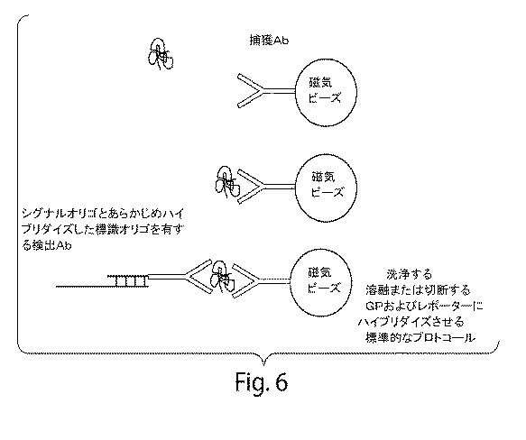
それぞれの対応する修飾オリゴを、対応する修飾抗体と3:1のモル比で反応させた。これを、スピンカラム(2ml Zeba Spin Column、Fisher Scientific、Pittsburgh、PA)で精製した。表2は、カウントプロセスの終わりに、1〜2個の間のオリゴが各抗体に付着していたことを示す(オリゴ:Ab)。 Each corresponding modified oligo was reacted with the corresponding modified antibody in a 3: 1 molar ratio. This was purified on a spin column (2 ml Zeba Spin Column, Fisher Scientific, Pittsburgh, PA). Table 2 shows that between 1 and 2 oligos were attached to each antibody at the end of the counting process (oligo: Ab).
表2.抗体−オリゴ結合プロセスの品質管理および定量化能力を示すモル置換率 Table 2. Molar substitution rate showing quality control and quantification ability of antibody-oligo conjugation process
各固有のシグナルオリゴを、各抗体Aに連結した固有オリゴに、オリゴ連結抗体Aおよび対応するシグナルオリゴを4:1の比、シグナルオリゴ:抗体比で加えることによって、別々にあらかじめアニーリングさせた。他の比が企図される。
標的と抗体の複合体の形成
ブロッカー(サケ精子)と共に約1×10⁻¹⁵〜1×10⁻⁸Mで単一チューブ中にシグナルオリゴにアニーリングした各抗体Aおよびビオチン化抗体B(4ペア)を含む2×マスターミックスを作製した。このマスターミックスのアリコートに望ましい希釈度(<1×10⁻⁸M)まで標的タンパク質を加えた。抗体は、10−15〜10−8Mの推定Kdに関して10×濃度であった。混合物をインキュベートした。
Formation of target-antibody complex Each antibody A and biotinylated antibody B (4 pairs) annealed to signal oligo in a single tube at about 1 × 10 −15 to 1 × 10 −8 M with blocker (salmon sperm) A 2 × master mix containing was prepared. Target protein was added to aliquots of this master mix to the desired dilution (<1 × 10 −8 M). The antibody was 10 × concentration with an estimated Kd of 10 −15 to 10 −8 M. The mixture was incubated.
標的タンパク質と抗体の複合体を、ストレプトアビジン結合型Dynabeads(登録商標)(Invitrogen)を用いてプロトコールに従って精製した。 The target protein / antibody complex was purified using streptavidin-conjugated Dynabeads® (Invitrogen) according to the protocol.
シグナルオリゴの溶離
単離された標的タンパク質/抗体の複合体を洗浄し、シグナルオリゴを、45℃より高い温度で、0.1×SSPEを用いて10〜15分間、溶離させた。より短い時間およびより長い時間が企図される。
Elution of signal oligos The isolated target protein / antibody complex was washed and the signal oligos were eluted with 0.1 x SSPE for 10-15 minutes at temperatures above 45 ° C. Shorter and longer times are contemplated.
シグナルオリゴの検出
各試料におけるシグナルオリゴの検出を、標識ナノレポータープローブおよび非標識ナノレポータープローブの両方を有する二重ナノレポーター系を用いて行った。各試料由来のシグナルオリゴを、以下のような最終濃度のハイブリダイゼーション試薬とハイブリダイズさせた:非標識ビオチン化プローブ、標識レポータープローブ、5×SSPE(pH7.5)、5×デンハルト試薬(Sigma)、剪断化サケ精子DNA(Sigma)、および界面活性剤。試薬を混合し、加熱フタ付きのサーモサイクラーブロック内で16時間、インキュベートした。
Detection of signal oligos Detection of signal oligos in each sample was performed using a dual nanoreporter system with both labeled and non-labeled nanoreporter probes. Signal oligos from each sample were hybridized with final concentrations of hybridization reagents as follows: unlabeled biotinylated probe, labeled reporter probe, 5 × SSPE (pH 7.5), 5 × Denhardt's reagent (Sigma) , Sheared salmon sperm DNA (Sigma), and surfactants. The reagents were mixed and incubated for 16 hours in a thermocycler block with a heated lid.
ハイブリダイゼーション後の精製
ハイブリダイズしていないレポーターを除去するために、ビオチン化プローブごとに含まれる3’−反復配列に相補的なオリゴヌクレオチドに結合した磁気ビーズ(InvitrogenTM)に関して反応物を精製した。反応物をまず、0.1%界面活性剤中のSSPE混合物/TEで希釈し、20℃より高い温度で、連続的に回転させながら、ビーズに結合させた。ビーズを、SSPEおよび界面活性剤の中で3回洗浄し、ハイブリダイズした複合体を0.1×SSPE/0.1%界面活性剤混合物中、45℃で15分間、溶離させた。溶離後、レポータープローブごとに含まれる5’−反復配列に相補的なオリゴヌクレオチドに結合した磁気ビーズに結合させることによって過剰のビオチン化プローブを除去するように、試料の2度目の精製を行った。抗3’−反復配列ビーズからの溶離は、最終濃度の1×SSPEでもたらされ、回転させながら、22.5℃で15分間、結合させた。ビーズを上記のように洗浄し、0.1×SSPE/0.1%界面活性剤混合物中、40℃より高い温度で、溶離させた。その後、その二重に精製された試料を、下記のような捕獲のために調製した。
Post-hybridization purification To remove non-hybridized reporters, the reaction was purified on magnetic beads (Invitrogen ™) conjugated to oligonucleotides complementary to the 3′-repeat sequence contained in each biotinylated probe. The reaction was first diluted with SSPE mixture / TE in 0.1% surfactant and bound to the beads with continuous rotation at temperatures above 20 ° C. The beads were washed three times in SSPE and surfactant, and the hybridized complex was eluted in a 0.1 × SSPE / 0.1% surfactant mixture at 45 ° C. for 15 minutes. After elution, a second purification of the sample was performed to remove excess biotinylated probe by binding to magnetic beads conjugated to oligonucleotides complementary to the 5'-repeat sequence included with each reporter probe. . Elution from anti-3′-repeat beads was effected at a final concentration of 1 × SSPE and allowed to bind for 15 minutes at 22.5 ° C. with rotation. The beads were washed as above and eluted in a 0.1 × SSPE / 0.1% surfactant mixture at a temperature above 40 ° C. The doubly purified sample was then prepared for capture as described below.
NanoStringレポーター捕獲、ストレッチング、および画像化
Tetraspeck蛍光マイクロスフェア(InvitrogenTM)のカスタム製剤の溶液を各試料に加えた。試料をNanoString流体装置へ投入し、処理し、画像化した。
NanoString Reporter Capture, Stretching, and Imaging A solution of a custom formulation of Tetraspec fluorescent microspheres (Invitrogen ™) was added to each sample. The sample was loaded into a NanoString fluid device, processed and imaged.
結果
結果を図10に示す。この実験の結果より、4つのタンパク質が、本明細書に記載されたアッセイによって同時に検出されたことが示された(図10)。検出されたタンパク質は、TNFα、IL1α、IL6、およびVEGFであった。この実験より、約1×10⁻¹²Mの感度が示された。図11は、液体試料に対してプロットされた同じデータを示す。次の実験(図12)により、これらのタンパク質のうちの2つの検出の限界が、26pg/mlおよび38pg/ml(IL1αおよびIL6のそれぞれについて、1.4×10−12Mおよび1.9×10−12M)であることが示された。
Results The results are shown in FIG. The results of this experiment showed that 4 proteins were detected simultaneously by the assay described herein (Figure 10). The detected proteins were TNFα, IL1α, IL6, and VEGF. This experiment showed a sensitivity of about 1 × 10 −12 M. FIG. 11 shows the same data plotted for a liquid sample. The following experiment (FIG. 12) shows that the limit of detection of two of these proteins is 26 pg / ml and 38 pg / ml (1.4 × 10 −12 M and 1.9 × for IL1α and IL6, respectively). 10 −12 M).
バックグラウンドにおける改善、すなわち、バックグラウンド検出の低減またはバックグラウンド対標的検出の比の向上によって、約2桁の感度の増加が可能になり、それに従って、感度は、1×10⁻¹⁴M、または有意に<1pg/mlに達するであろう。 An improvement in background, ie, a reduction in background detection or an increase in the ratio of background to target detection allows an increase in sensitivity of about 2 orders of magnitude, and accordingly the sensitivity is 1 × 10 −14 M, or Significantly <1 pg / ml will be reached.
実施例9.抗ストレプトアビジンプローブレポーター
抗ストレプトアビジン抗体(Affinity Bioreagents、Rockford、IL)を、実施例1に記載されているように、オリゴヌクレオチド(オリゴ)で標識した。
Example 9 Anti-Streptavidin Probe Reporter Anti-streptavidin antibodies (Affinity Bioreagents, Rockford, IL) were labeled with oligonucleotides (oligos) as described in Example 1.
この実施形態の特定の例において、抗体−標識オリゴをレポーターに、0.05nMの濃度、45℃の温度で、1×SSPE緩衝液中、一晩、ハイブリダイズさせた。レポーターと、抗体に結合したオリゴとの間に25塩基の重複があった。この重複は、必要に応じて、短縮されてもよい。ある特定の実施形態において、重複は、任意で、1個、5個、10個、15個、20個、25個の塩基、またはそれらの間の任意の長さである。レポーターと抗体上のオリゴとの間の塩基のより短い重複により、抗体安定を生じる温度での抗体−オリゴのレポーターへのハイブリダイゼーションの効率の増加が可能になる。抗ストレプトアビジン抗体プローブと共にレポーターを、カートリッジのフローチャンバーへ(0.25×SSPE緩衝液中0.025nMの濃度で)導入し、レポーターの第1末端を、ストレプトアビジン表面に10分間、結合させた。その後、チャンバーをTAE緩衝液で洗浄した。まず、200ボルト(V)/センチメートル(cm)の電場を用い、次に、ビオチン化オリゴを導入して、レポーターの第2末端を表面に付着させることによって、レポーターをストレッチし、その後、表面に固定化した。試料を再び、TAEで洗浄し、色素を安定化させるためにSlowFadeTMを導入した。その後、この試料を画像化した。 In a particular example of this embodiment, antibody-labeled oligos were hybridized to the reporter overnight in 1 × SSPE buffer at a concentration of 0.05 nM and a temperature of 45 ° C. There was a 25 base overlap between the reporter and the oligo bound to the antibody. This duplication may be shortened if necessary. In certain embodiments, the overlap is optionally one, five, ten, fifteen, twenty, twenty-five bases, or any length in between. The shorter base overlap between the reporter and the oligo on the antibody allows for increased efficiency of antibody-oligo to reporter hybridization at temperatures that produce antibody stability. The reporter along with the anti-streptavidin antibody probe was introduced into the flow chamber of the cartridge (at a concentration of 0.025 nM in 0.25 × SSPE buffer) and the first end of the reporter was allowed to bind to the streptavidin surface for 10 minutes. . Thereafter, the chamber was washed with TAE buffer. First, the reporter is stretched by using an electric field of 200 volts (V) / centimeter (cm), then introducing a biotinylated oligo to attach the second end of the reporter to the surface, and then the surface Immobilized to. The sample was again washed with TAE and SlowFade ™ was introduced to stabilize the dye. The sample was then imaged.
図14は、抗ストレプトアビジンプローブとのレポーター(S16)のみが検出されたことを示している。 FIG. 14 shows that only the reporter (S16) with the anti-streptavidin probe was detected.
他の実施形態
本発明の好ましい実施形態を本明細書に示し、かつ記載するが、そのような実施形態が例としてのみ提供されることは当業者にとって明白であろう。多数のバリエーション、変化、および置換が、本発明から逸脱することなしに、当業者に思い浮かぶであろう。本明細書に記載された発明の実施形態の様々な代替を、本発明を実践するのに用い得ることは理解されたい。以下の特許請求の範囲は本発明の範囲を定義すること、およびこれらの特許請求の範囲およびそれらの等価物の範囲内の方法および構造がそれらに網羅されることが意図される。
Other Embodiments While preferred embodiments of the invention are shown and described herein, it will be apparent to those skilled in the art that such embodiments are provided by way of example only. Numerous variations, changes and substitutions will occur to those skilled in the art without departing from the invention. It should be understood that various alternatives to the embodiments of the invention described herein may be used to practice the invention. The following claims are intended to define the scope of the invention and to cover the methods and structures within the scope of these claims and their equivalents.
Claims (21)
(a)(i)少なくとも1つのタンパク質、および
(ii)前記少なくとも1つのタンパク質の第2の領域に特異的な第2のタンパク質プローブであって、前記第2のタンパク質プローブは、部分的に二本鎖の核酸に直接またはリンカーオリゴを介して間接的に付着している、第2のタンパク質プローブ
を供給する工程;
(b)前記少なくとも1つのタンパク質、および前記第2のタンパク質プローブを含む少なくとも第1の複合体を形成する工程であって、前記少なくとも1つのタンパク質が前記第2のタンパク質プローブに結合している、工程;
(c)前記部分的に二本鎖の核酸の一方の鎖を前記第1の複合体から遊離させることにより、シグナルオリゴを生成する工程;
(d)(1)少なくとも前記シグナルオリゴ、ならびに
(2)前記シグナルオリゴにハイブリダイズ可能な領域、およびナノレポーターを含む領域を含む少なくとも1つのオリゴプローブ
を含む第2の複合体を形成する工程であって、前記ナノレポーターが複数の異なる検出可能な標識を含む、工程;ならびに
(e)前記ナノレポーターの複数の異なる検出可能な標識の存在を個々にカウントする工程を含む方法によって前記第2の複合体または前記第2の複合体の少なくとも一部分を個々に検出する工程であって、前記ナノレポーターの前記複数の異なる検出可能な標識のカウントが前記試料における前記タンパク質の濃度を示している、工程
を含む、方法。 A method for determining the concentration of at least one protein in a sample comprising:
(A) (i) at least one protein, and (ii) a second protein probe specific for a second region of the at least one protein, wherein the second protein probe is partially Providing a second protein probe that is directly or indirectly attached to a nucleic acid of a single strand via a linker oligo;
(B) forming at least a first complex comprising the at least one protein and the second protein probe, wherein the at least one protein is bound to the second protein probe; Process;
(C) generating a signal oligo by releasing one strand of the partially double-stranded nucleic acid from the first complex;
(D) forming a second complex comprising (1) at least the signal oligo, and (2) at least one oligo probe comprising a region hybridizable to the signal oligo and a region containing a nanoreporter. Wherein said nanoreporter comprises a plurality of different detectable labels; and (e) individually counting the presence of a plurality of different detectable labels of said nanoreporter by said method. Individually detecting at least a portion of a complex or the second complex, wherein the count of the plurality of different detectable labels of the nanoreporter indicates the concentration of the protein in the sample Including a method.
(a)(i)少なくとも1つのタンパク質、
(ii)前記少なくとも1つのタンパク質の第1の領域に特異的な第1のタンパク質プローブであって、前記第1のタンパク質プローブは、第1の捕獲領域または第1のマトリックスに付着している、第1のタンパク質プローブ、
(iii)前記少なくとも1つのタンパク質の第2の領域に特異的な第2のタンパク質プローブであって、前記第2のタンパク質プローブは、部分的に二本鎖の核酸に直接またはリンカーオリゴを介して間接的に付着している、第2のタンパク質プローブ、および
(iv)前記第1のタンパク質プローブが第1の捕獲領域に付着している場合:第2のマトリックスであって、前記第1のタンパク質プローブにおける前記捕獲領域に結合し得る成分が前記第2のマトリックスに付着している、第2のマトリックス
を供給する工程;
(b)前記少なくとも1つのタンパク質、前記第1のタンパク質プローブ、および前記第2のタンパク質プローブを含む少なくとも第1の複合体を形成する工程であって、前記少なくとも1つのタンパク質が前記第1のタンパク質プローブおよび前記第2のタンパク質プローブに結合しており、前記第1のタンパク質プローブが第1の捕獲領域に付着している場合、前記捕獲領域が前記第2のマトリックスにおける前記成分に結合している、工程;
(c)前記部分的に二本鎖の核酸の一方の鎖を前記第1の複合体から遊離させることにより、シグナルオリゴを生成する工程;
(d)(1)少なくとも前記シグナルオリゴ、ならびに
(2)前記シグナルオリゴにハイブリダイズ可能な領域、およびナノレポーターを含む領域を含む少なくとも1つのオリゴプローブ
を含む第2の複合体を形成する工程であって、前記ナノレポーターが複数の異なる検出可能な標識を含む、工程;ならびに
(e)前記ナノレポーターの複数の異なる検出可能な標識の存在を個々にカウントする工程を含む方法によって前記第2の複合体または前記第2の複合体の少なくとも一部分を個々に検出する工程であって、前記ナノレポーターの前記複数の異なる検出可能な標識のカウントが前記試料における前記タンパク質の濃度を示している、工程
を含む、方法。 A method for determining the concentration of at least one protein in a sample comprising:
(A) (i) at least one protein,
(Ii) a first protein probe specific to a first region of the at least one protein, wherein the first protein probe is attached to a first capture region or a first matrix; A first protein probe,
(Iii) a second protein probe specific for a second region of the at least one protein, wherein the second protein probe is partly directly to a double-stranded nucleic acid or via a linker oligo A second protein probe that is indirectly attached; and (iv) if the first protein probe is attached to a first capture region: a second matrix comprising the first protein Providing a second matrix, wherein a component capable of binding to the capture region in a probe is attached to the second matrix;
(B) forming at least a first complex including the at least one protein, the first protein probe, and the second protein probe, wherein the at least one protein is the first protein. When bound to a probe and the second protein probe, and the first protein probe is attached to a first capture region, the capture region is bound to the component in the second matrix The process;
(C) generating a signal oligo by releasing one strand of the partially double-stranded nucleic acid from the first complex;
(D) forming a second complex comprising (1) at least the signal oligo, and (2) at least one oligo probe comprising a region hybridizable to the signal oligo and a region containing a nanoreporter. Wherein said nanoreporter comprises a plurality of different detectable labels; and (e) individually counting the presence of a plurality of different detectable labels of said nanoreporter by said method. Individually detecting at least a portion of a complex or the second complex, wherein the count of the plurality of different detectable labels of the nanoreporter indicates the concentration of the protein in the sample Including a method.
請求項1または2に記載の方法。 The step of releasing the signal oligo further comprises capturing the signal oligo directly or indirectly into a third matrix;
The method according to claim 1 or 2.
(i)少なくとも1つの標的タンパク質、および
(ii)少なくとも1つの前記標的タンパク質の第2の領域に特異的な第2のタンパク質プローブであって、前記複数の第1の複合体における前記第2のタンパク質プローブは、異なる部分的に二本鎖の核酸に直接付着しているか、または、リンカーオリゴを介して異なる部分的に二本鎖の核酸に間接的に付着している、第2のタンパク質プローブ
を含み、それぞれの前記複数の第1の複合体におけるそれぞれの第2のタンパク質プローブが、シグナルオリゴとして遊離可能な異なる部分的に二本鎖の核酸に付着している、
請求項1に記載の方法。 Determining the concentration of the plurality of target proteins in the sample by a method comprising forming a plurality of first complexes, wherein each first complex comprises (i) at least one target protein, and (ii) ) A second protein probe specific for a second region of at least one of the target proteins, wherein the second protein probes in the plurality of first complexes are different partially double stranded Each of said plurality of first conjugates comprising a second protein probe attached directly to a nucleic acid or indirectly attached to a different partially double stranded nucleic acid via a linker oligo Each second protein probe in the body is attached to a different partially double stranded nucleic acid which can be released as a signal oligo,
The method of claim 1.
(i)少なくとも1つの標的タンパク質、
(ii)少なくとも1つの前記標的タンパク質の第1の領域に特異的な第1のタンパク質プローブであって、前記第1のタンパク質プローブが、捕獲領域または第1のマトリックスに付着している、第1のタンパク質プローブ、
(iii)少なくとも1つの前記標的タンパク質の第2の領域に特異的な第2のタンパク質プローブであって、前記複数の第1の複合体における前記第2のタンパク質プローブは、異なる部分的に二本鎖の核酸に直接付着しているか、または、リンカーオリゴを介して異なる部分的に二本鎖の核酸に間接的に付着している、第2のタンパク質プローブ
を含み、前記第1のタンパク質プローブが第1の捕獲領域に付着している場合、前記捕獲領域が前記第2のマトリックスにおける前記成分に結合しており、それぞれの前記複数の第1の複合体におけるそれぞれの第2のタンパク質プローブが、シグナルオリゴとして遊離可能な異なる部分的に二本鎖の核酸に付着している、
請求項2に記載の方法。 Determining the concentration of a plurality of target proteins in the sample by a method comprising forming a plurality of first complexes, wherein each first complex comprises (i) at least one target protein,
(Ii) a first protein probe specific for a first region of at least one of the target proteins, wherein the first protein probe is attached to a capture region or a first matrix; Protein probe,
(Iii) a second protein probe specific to a second region of at least one of the target proteins, wherein the second protein probes in the plurality of first complexes are two different partially A second protein probe that is directly attached to a strand of nucleic acid or indirectly attached to a different partially double-stranded nucleic acid via a linker oligo, wherein the first protein probe comprises: When attached to a first capture region, the capture region is bound to the component in the second matrix, and each second protein probe in each of the plurality of first complexes is Attached to different partially double stranded nucleic acids which can be released as signal oligos,
The method of claim 2.
それぞれが(1)少なくとも1つのシグナルオリゴ、および、(2)前記シグナルオリゴにハイブリダイズ可能な領域と、ナノレポーターを含む領域とを含む少なくとも1つのオリゴプローブ、を含む複数の第2の複合体を形成する工程であって、前記ナノレポーターが複数の異なる検出可能な標識を含む、工程;ならびに
前記ナノレポーターの複数の異なる検出可能な標識の存在を個々にカウントする工程を含む方法によって前記複数の第2の複合体のそれぞれまたは前記複数の第2の複合体のそれぞれの少なくとも一部分を個々に検出する工程であって、前記ナノレポーターの前記複数の異なる検出可能な標識のカウントが前記試料における前記複数の標的タンパク質のそれぞれの濃度を示している、工程
を含む、請求項10に記載の方法。 In addition:
A plurality of second complexes each comprising (1) at least one signal oligo, and (2) at least one oligo probe comprising a region hybridizable to the signal oligo and a region containing a nanoreporter Wherein the nanoreporter comprises a plurality of different detectable labels; and the method further comprises individually counting the presence of a plurality of different detectable labels on the nanoreporter. Individually detecting at least a portion of each of said second complexes or each of said plurality of second complexes, wherein the counts of said different detectable labels of said nanoreporter are in said sample 11. The method of claim 10, comprising the step of indicating the concentration of each of the plurality of target proteins. .
請求項11に記載の方法。 2, 3, 4, 5, 10, 20, 30, 50, 100, 200, 300, 500, 600, 700, 800, 900, 1000 Or concentrations of more than 1000 and not more than 2000 different target proteins are determined,
The method of claim 11.
Applications Claiming Priority (7)
| Application Number | Priority Date | Filing Date | Title |
|---|---|---|---|
| US25119209P | 2009-10-13 | 2009-10-13 | |
| US61/251,192 | 2009-10-13 | ||
| US32522410P | 2010-04-16 | 2010-04-16 | |
| US61/325,224 | 2010-04-16 | ||
| US32678710P | 2010-04-22 | 2010-04-22 | |
| US61/326,787 | 2010-04-22 | ||
| PCT/US2010/052556 WO2011047087A2 (en) | 2009-10-13 | 2010-10-13 | Protein detection via nanoreporters |
Related Child Applications (2)
| Application Number | Title | Priority Date | Filing Date |
|---|---|---|---|
| JP2016081763A Division JP6225212B2 (en) | 2009-10-13 | 2016-04-15 | Protein detection by nanoreporter |
| JP2016081762A Division JP2016136161A (en) | 2009-10-13 | 2016-04-15 | Protein detection via nanoreporters |
Publications (3)
| Publication Number | Publication Date |
|---|---|
| JP2013507642A JP2013507642A (en) | 2013-03-04 |
| JP2013507642A5 JP2013507642A5 (en) | 2013-11-28 |
| JP5954876B2 true JP5954876B2 (en) | 2016-07-20 |
Family
ID=43855316
Family Applications (3)
| Application Number | Title | Priority Date | Filing Date |
|---|---|---|---|
| JP2012534336A Active JP5954876B2 (en) | 2009-10-13 | 2010-10-13 | Protein detection by nanoreporter |
| JP2016081763A Active JP6225212B2 (en) | 2009-10-13 | 2016-04-15 | Protein detection by nanoreporter |
| JP2016081762A Pending JP2016136161A (en) | 2009-10-13 | 2016-04-15 | Protein detection via nanoreporters |
Family Applications After (2)
| Application Number | Title | Priority Date | Filing Date |
|---|---|---|---|
| JP2016081763A Active JP6225212B2 (en) | 2009-10-13 | 2016-04-15 | Protein detection by nanoreporter |
| JP2016081762A Pending JP2016136161A (en) | 2009-10-13 | 2016-04-15 | Protein detection via nanoreporters |
Country Status (8)
| Country | Link |
|---|---|
| US (2) | US9714937B2 (en) |
| EP (2) | EP2488876B1 (en) |
| JP (3) | JP5954876B2 (en) |
| AU (1) | AU2010306862B2 (en) |
| CA (1) | CA2775613C (en) |
| ES (1) | ES2627063T3 (en) |
| HK (1) | HK1249580A1 (en) |
| WO (1) | WO2011047087A2 (en) |
Families Citing this family (84)
| Publication number | Priority date | Publication date | Assignee | Title |
|---|---|---|---|---|
| WO2011047087A2 (en) | 2009-10-13 | 2011-04-21 | Nanostring Technologies, Inc. | Protein detection via nanoreporters |
| EP2534174A4 (en) * | 2010-02-12 | 2015-02-25 | Solulink Inc | Preparation and/or purification of oligonucleotide conjugates |
| US20190300945A1 (en) | 2010-04-05 | 2019-10-03 | Prognosys Biosciences, Inc. | Spatially Encoded Biological Assays |
| JP5893607B2 (en) | 2010-04-05 | 2016-03-23 | プログノシス バイオサイエンシズ インコーポレイテッドPrognosys Biosciences,Inc. | Spatial-encoded biological assay |
| US10787701B2 (en) | 2010-04-05 | 2020-09-29 | Prognosys Biosciences, Inc. | Spatially encoded biological assays |
| US9523680B2 (en) * | 2010-06-30 | 2016-12-20 | Ambergen, Inc. | Global Proteomic screening of random bead arrays using mass spectrometry imaging |
| GB201106254D0 (en) | 2011-04-13 | 2011-05-25 | Frisen Jonas | Method and product |
| US10501791B2 (en) | 2011-10-14 | 2019-12-10 | President And Fellows Of Harvard College | Sequencing by structure assembly |
| EP2769202A4 (en) * | 2011-10-20 | 2015-11-18 | Exxonmobil Upstream Res Co | Nanoparticle probes, methods, and systems for use thereof |
| WO2013067489A1 (en) * | 2011-11-05 | 2013-05-10 | President And Fellows Of Harvard College | Nucleic acid-based linkers for detecting and measuring interactions |
| EP3467500B1 (en) * | 2011-11-11 | 2021-05-12 | Eli N. Glezer | Co-binder assisted assay methods |
| US10227639B2 (en) | 2011-12-22 | 2019-03-12 | President And Fellows Of Harvard College | Compositions and methods for analyte detection |
| US11021737B2 (en) | 2011-12-22 | 2021-06-01 | President And Fellows Of Harvard College | Compositions and methods for analyte detection |
| US8838394B2 (en) | 2012-02-03 | 2014-09-16 | California Institute Of Technology | Signal encoding and decoding in multiplexed biochemical assays |
| WO2013184754A2 (en) | 2012-06-05 | 2013-12-12 | President And Fellows Of Harvard College | Spatial sequencing of nucleic acids using dna origami probes |
| RU2670133C2 (en) | 2012-11-19 | 2018-10-18 | Аптон Биосистемс, Инк. | Digital analysis of molecular analytes using single molecule detection |
| US10829816B2 (en) | 2012-11-19 | 2020-11-10 | Apton Biosystems, Inc. | Methods of analyte detection |
| US10138509B2 (en) | 2013-03-12 | 2018-11-27 | President And Fellows Of Harvard College | Method for generating a three-dimensional nucleic acid containing matrix |
| US10114015B2 (en) | 2013-03-13 | 2018-10-30 | Meso Scale Technologies, Llc. | Assay methods |
| ES2754177T3 (en) | 2013-06-12 | 2020-04-16 | Massachusetts Gen Hospital | Methods for multiplexed detection of target molecules and their uses |
| WO2014210225A1 (en) | 2013-06-25 | 2014-12-31 | Prognosys Biosciences, Inc. | Methods and systems for determining spatial patterns of biological targets in a sample |
| JP6695280B2 (en) | 2014-05-15 | 2020-05-20 | メソ スケール テクノロジーズ エルエルシー | Improved assay method |
| EP3143165A4 (en) * | 2014-05-15 | 2018-05-16 | Two Pore Guys, Inc. | Scaffold data storage and target detection in a sample using a nanopore |
| US10179932B2 (en) | 2014-07-11 | 2019-01-15 | President And Fellows Of Harvard College | Methods for high-throughput labelling and detection of biological features in situ using microscopy |
| KR102298387B1 (en) | 2014-11-21 | 2021-09-07 | 나노스트링 테크놀로지스, 인크. | Enzyme- and amplification-free sequencing |
| CN107249741B (en) | 2014-11-24 | 2021-04-02 | 纳米线科技公司 | Methods and apparatus for gene purification and imaging |
| US11198900B2 (en) | 2014-12-06 | 2021-12-14 | Children's Medical Center Corporation | Nucleic acid-based linkers for detecting and measuring interactions |
| US10774374B2 (en) | 2015-04-10 | 2020-09-15 | Spatial Transcriptomics AB and Illumina, Inc. | Spatially distinguished, multiplex nucleic acid analysis of biological specimens |
| US11396650B2 (en) | 2015-06-02 | 2022-07-26 | Children's Medical Center Corporation | Nucleic acid complexes for screening barcoded compounds |
| US20170002405A1 (en) * | 2015-06-30 | 2017-01-05 | Nanostring Technologies, Inc. | Methods and kits for simultaneously detecting gene or protein expression in a plurality of sample types using self-assembling fluorescent barcode nanoreporters |
| EP3325650B1 (en) | 2015-07-17 | 2024-01-03 | Nanostring Technologies, Inc. | Simultaneous quantification of gene expression in a user-defined region of a cross-sectioned tissue |
| US10501777B2 (en) | 2015-07-17 | 2019-12-10 | Nanostring Technologies, Inc. | Simultaneous quantification of a plurality of proteins in a user-defined region of a cross-sectioned tissue |
| CN105132533B (en) * | 2015-07-24 | 2018-11-23 | 清华大学深圳研究生院 | A kind of detection method of concentration of target molecules |
| WO2017041084A2 (en) * | 2015-09-03 | 2017-03-09 | Nanostring Technologies, Inc. | Multivalent probes having single nucleotide resolution |
| MX2018005611A (en) | 2015-11-03 | 2018-11-09 | Harvard College | Method and apparatus for volumetric imaging of a three-dimensional nucleic acid containing matrix. |
| US10774370B2 (en) | 2015-12-04 | 2020-09-15 | 10X Genomics, Inc. | Methods and compositions for nucleic acid analysis |
| WO2017139409A1 (en) | 2016-02-09 | 2017-08-17 | Children's Medical Center Corporation | Method for detection of analytes via polymer complexes |
| US10919037B2 (en) | 2016-03-23 | 2021-02-16 | Children's Medical Center Corporation | Systems and apparatus for detecting compounds in human biological samples |
| CN109415761B (en) | 2016-04-25 | 2022-09-20 | 哈佛学院董事及会员团体 | Hybrid chain reaction method for in situ molecular detection |
| CN116200462A (en) | 2016-05-16 | 2023-06-02 | 纳米线科技公司 | Method for detecting target nucleic acid in sample |
| US10066263B2 (en) | 2016-06-17 | 2018-09-04 | California Institute Of Technology | Nucleic acid reactions and related methods and compositions |
| US11352667B2 (en) | 2016-06-21 | 2022-06-07 | 10X Genomics, Inc. | Nucleic acid sequencing |
| DK3264087T3 (en) * | 2016-06-27 | 2020-07-20 | Chimera Biotec Gmbh | Method and device for quantification of target molecules |
| US9927441B1 (en) | 2016-07-29 | 2018-03-27 | X Development Llc | Combinatorial methods for aptamer based proteomics |
| CN109923216A (en) | 2016-08-31 | 2019-06-21 | 哈佛学院董事及会员团体 | By the detection combination of biomolecule to the method for the single test using fluorescent in situ sequencing |
| CN109983125A (en) | 2016-08-31 | 2019-07-05 | 哈佛学院董事及会员团体 | The method for generating the nucleic acid sequence library for detecting by fluorescent in situ sequencing |
| CN116334202A (en) | 2016-11-21 | 2023-06-27 | 纳米线科技公司 | Chemical compositions and methods of use thereof |
| KR101968565B1 (en) | 2017-03-16 | 2019-08-13 | 장준영 | Clothes hanger hanging jacket temporarily |
| JP7305611B2 (en) | 2017-03-17 | 2023-07-10 | アプトン バイオシステムズ インコーポレイテッド | Methods of sequencing and high-resolution imaging |
| CN109789167A (en) | 2017-04-14 | 2019-05-21 | 哈佛学院董事及会员团体 | Method for generating cell-derived microfilament network |
| WO2018218150A1 (en) | 2017-05-26 | 2018-11-29 | President And Fellows Of Harvard College | Systems and methods for high-throughput image-based screening |
| US11674951B2 (en) | 2017-07-17 | 2023-06-13 | The Brigham And Women's Hospital, Inc. | Methods for identifying a treatment for rheumatoid arthritis |
| US11597965B2 (en) | 2017-10-06 | 2023-03-07 | 10X Genomics, Inc. | RNA templated ligation |
| US11591636B2 (en) | 2017-11-20 | 2023-02-28 | Children's Medical Center Corporation | Force-controlled nanoswitch assays for single-molecule detection in complex biological fluids |
| WO2019148001A1 (en) * | 2018-01-25 | 2019-08-01 | Apton Biosystems, Inc. | Methods and composition for high throughput single molecule protein detection systems |
| SG11202007501SA (en) | 2018-02-12 | 2020-09-29 | Nanostring Technologies Inc | Biomolecular probes and methods of detecting gene and protein expression |
| WO2019200326A1 (en) | 2018-04-13 | 2019-10-17 | Rarecyte, Inc. | Kits for labeling of biomarkers and methods of using the same |
| WO2019213478A1 (en) | 2018-05-04 | 2019-11-07 | Nanostring Technologies, Inc. | Gene expression assay for measurement of dna mismatch repair deficiency |
| US11549139B2 (en) | 2018-05-14 | 2023-01-10 | Nanostring Technologies, Inc. | Chemical compositions and methods of using same |
| CA3100200A1 (en) | 2018-05-21 | 2019-11-28 | Nanostring Technologies, Inc. | Molecular gene signatures and methods of using same |
| EP3836967A4 (en) | 2018-07-30 | 2022-06-15 | ReadCoor, LLC | Methods and systems for sample processing or analysis |
| CN109337909B (en) * | 2018-11-12 | 2020-10-13 | 湖南大学 | Aptamer for detecting liver cancer drug-resistant cell strain and application thereof |
| WO2020123316A2 (en) | 2018-12-10 | 2020-06-18 | 10X Genomics, Inc. | Methods for determining a location of a biological analyte in a biological sample |
| US11926867B2 (en) | 2019-01-06 | 2024-03-12 | 10X Genomics, Inc. | Generating capture probes for spatial analysis |
| US11649485B2 (en) | 2019-01-06 | 2023-05-16 | 10X Genomics, Inc. | Generating capture probes for spatial analysis |
| SG11202111878RA (en) | 2019-05-31 | 2021-11-29 | 10X Genomics Inc | Method of detecting target nucleic acid molecules |
| WO2021092433A2 (en) | 2019-11-08 | 2021-05-14 | 10X Genomics, Inc. | Enhancing specificity of analyte binding |
| IT201900024159A1 (en) | 2019-12-16 | 2021-06-16 | Menarini Silicon Biosystems Spa | METHOD AND KIT FOR TOTAL AMPLIFICATION OF THE GENOME AND ANALYSIS OF TARGET MOLECULES IN A BIOLOGICAL SAMPLE |
| FI3891300T3 (en) | 2019-12-23 | 2023-05-10 | 10X Genomics Inc | Methods for spatial analysis using rna-templated ligation |
| US11702693B2 (en) | 2020-01-21 | 2023-07-18 | 10X Genomics, Inc. | Methods for printing cells and generating arrays of barcoded cells |
| US11732299B2 (en) | 2020-01-21 | 2023-08-22 | 10X Genomics, Inc. | Spatial assays with perturbed cells |
| US11898205B2 (en) | 2020-02-03 | 2024-02-13 | 10X Genomics, Inc. | Increasing capture efficiency of spatial assays |
| US11732300B2 (en) | 2020-02-05 | 2023-08-22 | 10X Genomics, Inc. | Increasing efficiency of spatial analysis in a biological sample |
| US11891654B2 (en) | 2020-02-24 | 2024-02-06 | 10X Genomics, Inc. | Methods of making gene expression libraries |
| EP4242325A3 (en) | 2020-04-22 | 2023-10-04 | 10X Genomics, Inc. | Methods for spatial analysis using targeted rna depletion |
| EP4153775A1 (en) | 2020-05-22 | 2023-03-29 | 10X Genomics, Inc. | Simultaneous spatio-temporal measurement of gene expression and cellular activity |
| EP4153776A1 (en) | 2020-05-22 | 2023-03-29 | 10X Genomics, Inc. | Spatial analysis to detect sequence variants |
| WO2021252499A1 (en) | 2020-06-08 | 2021-12-16 | 10X Genomics, Inc. | Methods of determining a surgical margin and methods of use thereof |
| AU2021294334A1 (en) | 2020-06-25 | 2023-02-02 | 10X Genomics, Inc. | Spatial analysis of DNA methylation |
| US11761038B1 (en) | 2020-07-06 | 2023-09-19 | 10X Genomics, Inc. | Methods for identifying a location of an RNA in a biological sample |
| US11926822B1 (en) | 2020-09-23 | 2024-03-12 | 10X Genomics, Inc. | Three-dimensional spatial analysis |
| US11827935B1 (en) | 2020-11-19 | 2023-11-28 | 10X Genomics, Inc. | Methods for spatial analysis using rolling circle amplification and detection probes |
| WO2022140028A1 (en) | 2020-12-21 | 2022-06-30 | 10X Genomics, Inc. | Methods, compositions, and systems for capturing probes and/or barcodes |
| WO2023034489A1 (en) | 2021-09-01 | 2023-03-09 | 10X Genomics, Inc. | Methods, compositions, and kits for blocking a capture probe on a spatial array |
Family Cites Families (77)
| Publication number | Priority date | Publication date | Assignee | Title |
|---|---|---|---|---|
| AU622426B2 (en) | 1987-12-11 | 1992-04-09 | Abbott Laboratories | Assay using template-dependent nucleic acid probe reorganization |
| ATE138106T1 (en) | 1988-07-20 | 1996-06-15 | David Segev | METHOD FOR AMPLIFICATION AND DETECTION OF NUCLEIC ACID SEQUENCES |
| US5185243A (en) * | 1988-08-25 | 1993-02-09 | Syntex (U.S.A.) Inc. | Method for detection of specific nucleic acid sequences |
| EP0439182B1 (en) | 1990-01-26 | 1996-04-24 | Abbott Laboratories | Improved method of amplifying target nucleic acids applicable to both polymerase and ligase chain reactions |
| US5320814A (en) | 1991-01-25 | 1994-06-14 | Trustees Of Tufts College | Fiber optic array sensors, apparatus, and methods for concurrently visualizing and chemically detecting multiple analytes of interest in a fluid sample |
| US5543838A (en) | 1993-08-31 | 1996-08-06 | Xerox Corporation | Signal multiplexing system for an image sensor array |
| SE9400088D0 (en) * | 1994-01-14 | 1994-01-14 | Kabi Pharmacia Ab | Bacterial receptor structures |
| WO1997007245A1 (en) | 1995-08-14 | 1997-02-27 | Ely Michael Rabani | Methods and devices for parallel multiplex polynucleotide sequencing |
| DE69638321D1 (en) | 1995-10-11 | 2011-03-03 | Luminex Corp | SIMULTANEOUS MULTI-ANALYSIS OF CLINICAL SAMPLES |
| US7144119B2 (en) | 1996-04-25 | 2006-12-05 | Bioarray Solutions Ltd. | System and method for programmable illumination pattern generation |
| US6387707B1 (en) | 1996-04-25 | 2002-05-14 | Bioarray Solutions | Array Cytometry |
| US6416960B1 (en) | 1996-08-08 | 2002-07-09 | Prolume, Ltd. | Detection and visualization of neoplastic tissues and other tissues |
| DE69735445T2 (en) | 1996-12-10 | 2006-08-10 | Sequenom, Inc., San Diego | NON-VOLATILE, NON-VOLATILE MOLECULES FOR MASS MARKING |
| US20020034737A1 (en) | 1997-03-04 | 2002-03-21 | Hyseq, Inc. | Methods and compositions for detection or quantification of nucleic acid species |
| US6180415B1 (en) | 1997-02-20 | 2001-01-30 | The Regents Of The University Of California | Plasmon resonant particles, methods and apparatus |
| US20050037397A1 (en) * | 2001-03-28 | 2005-02-17 | Nanosphere, Inc. | Bio-barcode based detection of target analytes |
| US7115884B1 (en) | 1997-10-06 | 2006-10-03 | Trustees Of Tufts College | Self-encoding fiber optic sensor |
| US7348181B2 (en) | 1997-10-06 | 2008-03-25 | Trustees Of Tufts College | Self-encoding sensor with microspheres |
| WO1999019515A1 (en) | 1997-10-14 | 1999-04-22 | Luminex Corporation | Precision fluorescently dyed particles and methods of making and using same |
| US6207392B1 (en) | 1997-11-25 | 2001-03-27 | The Regents Of The University Of California | Semiconductor nanocrystal probes for biological applications and process for making and using such probes |
| US6562567B2 (en) | 1998-01-27 | 2003-05-13 | California Institute Of Technology | Method of detecting a nucleic acid |
| EP1090293B2 (en) | 1998-06-24 | 2019-01-23 | Illumina, Inc. | Decoding of array sensors with microspheres |
| DE69905832T2 (en) | 1998-09-18 | 2004-02-05 | Massachusetts Institute Of Technology, Cambridge | Biological uses of semiconducting nanocrystals |
| US6268147B1 (en) * | 1998-11-02 | 2001-07-31 | Kenneth Loren Beattie | Nucleic acid analysis using sequence-targeted tandem hybridization |
| US6465193B2 (en) | 1998-12-11 | 2002-10-15 | The Regents Of The University Of California | Targeted molecular bar codes and methods for using the same |
| US6429027B1 (en) | 1998-12-28 | 2002-08-06 | Illumina, Inc. | Composite arrays utilizing microspheres |
| US6908737B2 (en) | 1999-04-15 | 2005-06-21 | Vitra Bioscience, Inc. | Systems and methods of conducting multiplexed experiments |
| US6355431B1 (en) | 1999-04-20 | 2002-03-12 | Illumina, Inc. | Detection of nucleic acid amplification reactions using bead arrays |
| AU4701200A (en) | 1999-05-07 | 2000-11-21 | Quantum Dot Corporation | A method of detecting an analyte using semiconductor nanocrystals |
| US20020051971A1 (en) | 1999-05-21 | 2002-05-02 | John R. Stuelpnagel | Use of microfluidic systems in the detection of target analytes using microsphere arrays |
| WO2001000875A2 (en) | 1999-06-25 | 2001-01-04 | Motorola, Inc. | Novel methods and products for arrayed microsphere analysis |
| CA2375034C (en) | 1999-07-26 | 2012-01-03 | The Government Of The United States Of America, As Represented By The Secretary, Department Of Health & Human Services, The National Institutes Of Health | Method and device for analysis of biological specimens |
| US7214477B1 (en) | 1999-07-26 | 2007-05-08 | The United States Of America As Represented By The Secretary Of The Department Of Health And Human Services | Layered device with capture regions for cellular analysis |
| AU2246601A (en) | 1999-08-30 | 2001-04-10 | Illumina, Inc. | Methods for improving signal detection from an array |
| WO2001033190A2 (en) | 1999-11-04 | 2001-05-10 | Arcturus Engineering, Inc. | Automated laser capture microdissection |
| EP1255865B1 (en) | 2000-02-07 | 2007-04-18 | Illumina, Inc. | Nucleic acid detection method using universal priming |
| AU2001239760B2 (en) | 2000-02-10 | 2005-11-24 | Illumina, Inc. | Array of individual arrays as substrate for bead-based simultaneous processing of samples and manufacturing method therefor |
| US20020028457A1 (en) | 2000-02-16 | 2002-03-07 | Quantum Dot Corporation | Single target counting assays using semiconductor nanocrystals |
| CA2399189A1 (en) | 2000-02-22 | 2001-08-30 | Genospectra, Inc. | Microarray fabrication techniques and apparatus |
| US20030186426A1 (en) | 2000-03-15 | 2003-10-02 | The Regents Of The University Of California | Multichannel flow cell for interacting single optically trapped, DNA molecules with different chemical species |
| US6759235B2 (en) | 2000-04-06 | 2004-07-06 | Quantum Dot Corporation | Two-dimensional spectral imaging system |
| US20020034827A1 (en) | 2000-08-01 | 2002-03-21 | Rajendra Singh | Methods for solid phase nanoextraction and desorption |
| US6777244B2 (en) | 2000-12-06 | 2004-08-17 | Hrl Laboratories, Llc | Compact sensor using microcavity structures |
| US7473767B2 (en) * | 2001-07-03 | 2009-01-06 | The Institute For Systems Biology | Methods for detection and quantification of analytes in complex mixtures |
| US7553619B2 (en) | 2002-02-08 | 2009-06-30 | Qiagen Gmbh | Detection method using dissociated rolling circle amplification |
| US20040229294A1 (en) | 2002-05-21 | 2004-11-18 | Po-Ying Chan-Hui | ErbB surface receptor complexes as biomarkers |
| DK2348124T3 (en) | 2002-10-30 | 2014-03-10 | Nuevolution As | Synthesis of a bifunctional complex |
| ATE431854T1 (en) * | 2003-06-27 | 2009-06-15 | Nanosphere Inc | DETECTION OF TARGET ANALYTES BASED ON BIO-BARCODES |
| US7402398B2 (en) | 2003-07-17 | 2008-07-22 | Monogram Biosciences, Inc. | Measuring receptor homodimerization |
| US20050048498A1 (en) | 2003-08-29 | 2005-03-03 | Applera Corporation | Compositions, methods, and kits for assembling probes |
| EP1681983A4 (en) | 2003-10-14 | 2008-12-10 | Monogram Biosciences Inc | Receptor tyrosine kinase signaling pathway analysis for diagnosis and therapy |
| WO2005044836A2 (en) | 2003-11-05 | 2005-05-19 | Genovoxx Gmbh | Macromolecular nucleotide compounds and methods for using the same |
| TW200526778A (en) | 2003-11-14 | 2005-08-16 | Sembiosys Genetics Inc | Methods for the production of apolipoproteins in transgenic plants |
| US7569392B2 (en) | 2004-01-08 | 2009-08-04 | Vanderbilt University | Multiplex spatial profiling of gene expression |
| WO2005071401A2 (en) * | 2004-01-15 | 2005-08-04 | Chiron Corporation | Homogeneous multiplex assay for nucleic acid targets |
| US7588906B2 (en) | 2004-02-24 | 2009-09-15 | Wisconsin Alumni Research Foundation | Hydrogels for biomolecule analysis and corresponding method to analyze biomolecules |
| JP2005257641A (en) * | 2004-03-15 | 2005-09-22 | Geneticlab Co Ltd | Method for simultaneously detecting a plurality of proteins by using rna polymerase |
| EP2325631B1 (en) | 2005-06-07 | 2015-01-28 | Centre National de la Recherche Scientifique (CNRS) | Antibody conjugated to peptide via a UV cleavable linker |
| EP3211086B1 (en) * | 2005-09-20 | 2021-06-09 | Menarini Silicon Biosystems S.p.A. | Methods and composition to generate unique sequence dna probes, labeling of dna probes and the use of these probes |
| EP1963500B1 (en) * | 2005-12-23 | 2013-02-13 | Nanostring Technologies, Inc. | Compositions comprising oriented, immobilized macromolecules and methods for their preparation |
| CA2640385C (en) * | 2005-12-23 | 2014-07-15 | Nanostring Technologies, Inc. | Nanoreporters and methods of manufacturing and use thereof |
| US7855057B2 (en) | 2006-03-23 | 2010-12-21 | Millipore Corporation | Protein splice variant/isoform discrimination and quantitative measurements thereof |
| US8003312B2 (en) | 2007-02-16 | 2011-08-23 | The Board Of Trustees Of The Leland Stanford Junior University | Multiplex cellular assays using detectable cell barcodes |
| US7728287B2 (en) | 2007-03-01 | 2010-06-01 | Lawrence Livermore National Security, Llc | Imaging mass spectrometer with mass tags |
| US20100291562A1 (en) * | 2007-04-04 | 2010-11-18 | Michael Adler | Method for the detection of an analyte in biological matrix |
| CA2687292C (en) | 2007-04-10 | 2017-07-04 | Nanostring Technologies, Inc. | Methods and computer systems for identifying target-specific sequences for use in nanoreporters |
| US8906700B2 (en) | 2007-11-06 | 2014-12-09 | Ambergen, Inc. | Methods and compositions for phototransfer |
| WO2010019826A1 (en) * | 2008-08-14 | 2010-02-18 | Nanostring Technologies, Inc | Stable nanoreporters |
| EP2163900A1 (en) | 2008-09-04 | 2010-03-17 | Commissariat A L'energie Atomique | New method of imaging by mass spectrometry and new mass tag associated trityl derivatives |
| US8309306B2 (en) | 2008-11-12 | 2012-11-13 | Nodality, Inc. | Detection composition |
| DE102008062372B3 (en) | 2008-12-17 | 2010-06-17 | Medizinische Hochschule Hannover | Detecting conjugate and method of analysis |
| SG10201408392PA (en) | 2009-01-15 | 2015-01-29 | Lab Corp America Holdings | Methods of determining patient response by measurement of her-3 |
| JP5457222B2 (en) | 2009-02-25 | 2014-04-02 | エフ.ホフマン−ラ ロシュ アーゲー | Miniaturized high-throughput nucleic acid analysis |
| WO2010115089A2 (en) | 2009-04-03 | 2010-10-07 | University Of Virginia Patent Foundation | Serial multiple antigen colocalization in paraffin-embedded tissue |
| DE102009002653B3 (en) | 2009-04-27 | 2010-05-27 | Federal-Mogul Nürnberg GmbH | Method and device for producing a piston for an internal combustion engine and piston for an internal combustion engine |
| WO2011047087A2 (en) | 2009-10-13 | 2011-04-21 | Nanostring Technologies, Inc. | Protein detection via nanoreporters |
| US8653242B2 (en) | 2010-03-01 | 2014-02-18 | Lostam Pharmaceuticals Ltd. | Therapeutic antibodies against flagellated Pseudomonas aeruginosa |
-
2010
- 2010-10-13 WO PCT/US2010/052556 patent/WO2011047087A2/en active Application Filing
- 2010-10-13 EP EP10824048.2A patent/EP2488876B1/en active Active
- 2010-10-13 US US12/904,078 patent/US9714937B2/en active Active
- 2010-10-13 AU AU2010306862A patent/AU2010306862B2/en active Active
- 2010-10-13 EP EP16201369.2A patent/EP3236264A3/en not_active Withdrawn
- 2010-10-13 JP JP2012534336A patent/JP5954876B2/en active Active
- 2010-10-13 ES ES10824048.2T patent/ES2627063T3/en active Active
- 2010-10-13 CA CA2775613A patent/CA2775613C/en active Active
-
2015
- 2015-07-30 US US14/814,216 patent/US9995739B2/en active Active
-
2016
- 2016-04-15 JP JP2016081763A patent/JP6225212B2/en active Active
- 2016-04-15 JP JP2016081762A patent/JP2016136161A/en active Pending
-
2018
- 2018-04-24 HK HK18105349.7A patent/HK1249580A1/en unknown
Also Published As
| Publication number | Publication date |
|---|---|
| JP2013507642A (en) | 2013-03-04 |
| EP2488876A2 (en) | 2012-08-22 |
| CA2775613C (en) | 2018-06-12 |
| US9714937B2 (en) | 2017-07-25 |
| EP3236264A2 (en) | 2017-10-25 |
| CA2775613A1 (en) | 2011-04-21 |
| JP6225212B2 (en) | 2017-11-01 |
| JP2016136161A (en) | 2016-07-28 |
| US20110086774A1 (en) | 2011-04-14 |
| EP3236264A3 (en) | 2017-11-08 |
| AU2010306862B2 (en) | 2015-08-27 |
| US9995739B2 (en) | 2018-06-12 |
| EP2488876B1 (en) | 2017-03-01 |
| US20160003809A1 (en) | 2016-01-07 |
| WO2011047087A2 (en) | 2011-04-21 |
| ES2627063T3 (en) | 2017-07-26 |
| HK1249580A1 (en) | 2018-11-02 |
| EP2488876A4 (en) | 2013-03-20 |
| AU2010306862A1 (en) | 2012-04-12 |
| WO2011047087A3 (en) | 2011-08-18 |
| JP2016136162A (en) | 2016-07-28 |
Similar Documents
| Publication | Publication Date | Title |
|---|---|---|
| JP6225212B2 (en) | Protein detection by nanoreporter | |
| US11932902B2 (en) | Barcoded beads and method for making the same by split-pool synthesis | |
| JP6525872B2 (en) | Increasing dynamic range to identify multiple epitopes in cells | |
| CA3163623A1 (en) | In situ rna analysis using probe pair ligation | |
| JP2023505195A (en) | Nucleic acid binding immunosandwich assay (NULISA) | |
| US8927210B2 (en) | Conjugate complexes for analyte detection | |
| CA3029178C (en) | Double-stranded nucleic acid signal probe and method for detecting target molecule using same | |
| JP2023171748A (en) | Controls for proximity detection assays | |
| US11560585B2 (en) | Methods of identifying multiple epitopes in cells | |
| AU2015261558B2 (en) | Protein Detection Via Nanoreporters | |
| WO2023239805A1 (en) | In situ nucleic acid analysis using probe pair ligation | |
| Wei | Conjugation by ligation-a new and promising way to create PLA probes and relevant applications | |
| Yan | Optimization of solid phase PLA with emphasis on antibody modification |
Legal Events
| Date | Code | Title | Description |
|---|---|---|---|
| A521 | Request for written amendment filed |
Free format text: JAPANESE INTERMEDIATE CODE: A523 Effective date: 20131011 |
|
| A621 | Written request for application examination |
Free format text: JAPANESE INTERMEDIATE CODE: A621 Effective date: 20131011 |
|
| A977 | Report on retrieval |
Free format text: JAPANESE INTERMEDIATE CODE: A971007 Effective date: 20140521 |
|
| A131 | Notification of reasons for refusal |
Free format text: JAPANESE INTERMEDIATE CODE: A131 Effective date: 20140523 |
|
| A601 | Written request for extension of time |
Free format text: JAPANESE INTERMEDIATE CODE: A601 Effective date: 20140814 |
|
| A602 | Written permission of extension of time |
Free format text: JAPANESE INTERMEDIATE CODE: A602 Effective date: 20140821 |
|
| A521 | Request for written amendment filed |
Free format text: JAPANESE INTERMEDIATE CODE: A523 Effective date: 20141003 |
|
| A131 | Notification of reasons for refusal |
Free format text: JAPANESE INTERMEDIATE CODE: A131 Effective date: 20150312 |
|
| A601 | Written request for extension of time |
Free format text: JAPANESE INTERMEDIATE CODE: A601 Effective date: 20150611 |
|
| A521 | Request for written amendment filed |
Free format text: JAPANESE INTERMEDIATE CODE: A523 Effective date: 20150625 |
|
| A131 | Notification of reasons for refusal |
Free format text: JAPANESE INTERMEDIATE CODE: A131 Effective date: 20160127 |
|
| A521 | Request for written amendment filed |
Free format text: JAPANESE INTERMEDIATE CODE: A523 Effective date: 20160415 |
|
| TRDD | Decision of grant or rejection written | ||
| A01 | Written decision to grant a patent or to grant a registration (utility model) |
Free format text: JAPANESE INTERMEDIATE CODE: A01 Effective date: 20160609 |
|
| A61 | First payment of annual fees (during grant procedure) |
Free format text: JAPANESE INTERMEDIATE CODE: A61 Effective date: 20160610 |
|
| R150 | Certificate of patent or registration of utility model |
Ref document number: 5954876 Country of ref document: JP Free format text: JAPANESE INTERMEDIATE CODE: R150 |
|
| R250 | Receipt of annual fees |
Free format text: JAPANESE INTERMEDIATE CODE: R250 |
|
| R250 | Receipt of annual fees |
Free format text: JAPANESE INTERMEDIATE CODE: R250 |
|
| R250 | Receipt of annual fees |
Free format text: JAPANESE INTERMEDIATE CODE: R250 |
|
| R250 | Receipt of annual fees |
Free format text: JAPANESE INTERMEDIATE CODE: R250 |
|
| R250 | Receipt of annual fees |
Free format text: JAPANESE INTERMEDIATE CODE: R250 |