CN109415761B - Hybrid chain reaction method for in situ molecular detection - Google Patents
Hybrid chain reaction method for in situ molecular detection Download PDFInfo
- Publication number
- CN109415761B CN109415761B CN201780039335.6A CN201780039335A CN109415761B CN 109415761 B CN109415761 B CN 109415761B CN 201780039335 A CN201780039335 A CN 201780039335A CN 109415761 B CN109415761 B CN 109415761B
- Authority
- CN
- China
- Prior art keywords
- hcr
- probe
- initiator
- fluorescent
- linker
- Prior art date
- Legal status (The legal status is an assumption and is not a legal conclusion. Google has not performed a legal analysis and makes no representation as to the accuracy of the status listed.)
- Active
Links
Images


Classifications
-
- C—CHEMISTRY; METALLURGY
- C12—BIOCHEMISTRY; BEER; SPIRITS; WINE; VINEGAR; MICROBIOLOGY; ENZYMOLOGY; MUTATION OR GENETIC ENGINEERING
- C12Q—MEASURING OR TESTING PROCESSES INVOLVING ENZYMES, NUCLEIC ACIDS OR MICROORGANISMS; COMPOSITIONS OR TEST PAPERS THEREFOR; PROCESSES OF PREPARING SUCH COMPOSITIONS; CONDITION-RESPONSIVE CONTROL IN MICROBIOLOGICAL OR ENZYMOLOGICAL PROCESSES
- C12Q1/00—Measuring or testing processes involving enzymes, nucleic acids or microorganisms; Compositions therefor; Processes of preparing such compositions
- C12Q1/68—Measuring or testing processes involving enzymes, nucleic acids or microorganisms; Compositions therefor; Processes of preparing such compositions involving nucleic acids
- C12Q1/6813—Hybridisation assays
- C12Q1/6841—In situ hybridisation
-
- C—CHEMISTRY; METALLURGY
- C12—BIOCHEMISTRY; BEER; SPIRITS; WINE; VINEGAR; MICROBIOLOGY; ENZYMOLOGY; MUTATION OR GENETIC ENGINEERING
- C12Q—MEASURING OR TESTING PROCESSES INVOLVING ENZYMES, NUCLEIC ACIDS OR MICROORGANISMS; COMPOSITIONS OR TEST PAPERS THEREFOR; PROCESSES OF PREPARING SUCH COMPOSITIONS; CONDITION-RESPONSIVE CONTROL IN MICROBIOLOGICAL OR ENZYMOLOGICAL PROCESSES
- C12Q1/00—Measuring or testing processes involving enzymes, nucleic acids or microorganisms; Compositions therefor; Processes of preparing such compositions
- C12Q1/68—Measuring or testing processes involving enzymes, nucleic acids or microorganisms; Compositions therefor; Processes of preparing such compositions involving nucleic acids
- C12Q1/6876—Nucleic acid products used in the analysis of nucleic acids, e.g. primers or probes
-
- C—CHEMISTRY; METALLURGY
- C12—BIOCHEMISTRY; BEER; SPIRITS; WINE; VINEGAR; MICROBIOLOGY; ENZYMOLOGY; MUTATION OR GENETIC ENGINEERING
- C12N—MICROORGANISMS OR ENZYMES; COMPOSITIONS THEREOF; PROPAGATING, PRESERVING, OR MAINTAINING MICROORGANISMS; MUTATION OR GENETIC ENGINEERING; CULTURE MEDIA
- C12N15/00—Mutation or genetic engineering; DNA or RNA concerning genetic engineering, vectors, e.g. plasmids, or their isolation, preparation or purification; Use of hosts therefor
- C12N15/09—Recombinant DNA-technology
- C12N15/11—DNA or RNA fragments; Modified forms thereof; Non-coding nucleic acids having a biological activity
- C12N15/115—Aptamers, i.e. nucleic acids binding a target molecule specifically and with high affinity without hybridising therewith ; Nucleic acids binding to non-nucleic acids, e.g. aptamers
-
- C—CHEMISTRY; METALLURGY
- C12—BIOCHEMISTRY; BEER; SPIRITS; WINE; VINEGAR; MICROBIOLOGY; ENZYMOLOGY; MUTATION OR GENETIC ENGINEERING
- C12Q—MEASURING OR TESTING PROCESSES INVOLVING ENZYMES, NUCLEIC ACIDS OR MICROORGANISMS; COMPOSITIONS OR TEST PAPERS THEREFOR; PROCESSES OF PREPARING SUCH COMPOSITIONS; CONDITION-RESPONSIVE CONTROL IN MICROBIOLOGICAL OR ENZYMOLOGICAL PROCESSES
- C12Q1/00—Measuring or testing processes involving enzymes, nucleic acids or microorganisms; Compositions therefor; Processes of preparing such compositions
- C12Q1/68—Measuring or testing processes involving enzymes, nucleic acids or microorganisms; Compositions therefor; Processes of preparing such compositions involving nucleic acids
- C12Q1/6806—Preparing nucleic acids for analysis, e.g. for polymerase chain reaction [PCR] assay
-
- C—CHEMISTRY; METALLURGY
- C12—BIOCHEMISTRY; BEER; SPIRITS; WINE; VINEGAR; MICROBIOLOGY; ENZYMOLOGY; MUTATION OR GENETIC ENGINEERING
- C12Q—MEASURING OR TESTING PROCESSES INVOLVING ENZYMES, NUCLEIC ACIDS OR MICROORGANISMS; COMPOSITIONS OR TEST PAPERS THEREFOR; PROCESSES OF PREPARING SUCH COMPOSITIONS; CONDITION-RESPONSIVE CONTROL IN MICROBIOLOGICAL OR ENZYMOLOGICAL PROCESSES
- C12Q1/00—Measuring or testing processes involving enzymes, nucleic acids or microorganisms; Compositions therefor; Processes of preparing such compositions
- C12Q1/68—Measuring or testing processes involving enzymes, nucleic acids or microorganisms; Compositions therefor; Processes of preparing such compositions involving nucleic acids
- C12Q1/6813—Hybridisation assays
- C12Q1/6816—Hybridisation assays characterised by the detection means
- C12Q1/682—Signal amplification
-
- C—CHEMISTRY; METALLURGY
- C12—BIOCHEMISTRY; BEER; SPIRITS; WINE; VINEGAR; MICROBIOLOGY; ENZYMOLOGY; MUTATION OR GENETIC ENGINEERING
- C12N—MICROORGANISMS OR ENZYMES; COMPOSITIONS THEREOF; PROPAGATING, PRESERVING, OR MAINTAINING MICROORGANISMS; MUTATION OR GENETIC ENGINEERING; CULTURE MEDIA
- C12N2310/00—Structure or type of the nucleic acid
- C12N2310/10—Type of nucleic acid
- C12N2310/16—Aptamers
-
- C—CHEMISTRY; METALLURGY
- C12—BIOCHEMISTRY; BEER; SPIRITS; WINE; VINEGAR; MICROBIOLOGY; ENZYMOLOGY; MUTATION OR GENETIC ENGINEERING
- C12N—MICROORGANISMS OR ENZYMES; COMPOSITIONS THEREOF; PROPAGATING, PRESERVING, OR MAINTAINING MICROORGANISMS; MUTATION OR GENETIC ENGINEERING; CULTURE MEDIA
- C12N2320/00—Applications; Uses
- C12N2320/10—Applications; Uses in screening processes
Abstract
The present disclosure provides a method of detecting a target analyte in a biological sample, comprising contacting the sample with one or more probe sets each comprising a primary probe and a linker, contacting the sample with a priming sequence, contacting the sample with a plurality of DNA hairpins, wherein the probes bind to a target molecule, the linkers link the probes to the priming sequence, and wherein the priming sequence nucleates with a cognate hairpin and initiates self-assembly of the tethered fluorescent amplification polymer, and detecting the target molecule by measuring a fluorescent signal of the sample.
Description
Data of related applications
This application claims priority to U.S. provisional application No. 62/326,959 filed on 25/4/2016, which is incorporated herein by reference for all purposes.
Statement of government interest
The invention was made with government support under approval number DGE1144152 awarded by the national science foundation and approval number HG005550 awarded by the national institutes of health. The government has certain rights in this invention.
Background
Choi, Harry MT, Victor a. beck and Niles a. pierce, next generation in situ hybridization chain reaction: higher gain, lower cost, more durable (Next-generation in situ hybridization reaction: highher gain, lower cost, great reduce), ACS nano 8.5 (2014): 4284-4294 describes a hybrid chain reaction method. Other methods include those disclosed in US2005/0260635, US 2006/0228733 and US 7,727,721.
Disclosure of Invention
Embodiments of the present disclosure relate to methods of using one or more probe sets based on hybridization strand reactions ("HCRs") for identifying and/or sequencing one or more molecules in a sample, such as a biological sample. Typically, the hybridization strand reaction uses a nucleic acid priming sequence, such as a DNA priming sequence, and two or more metastable HCR monomers, which may be in the form of: the double-stranded portion and the single-stranded sequence, such as a sticky end (toehold) sequence, which are joined by a linker at one end of the double-stranded portion, are attached to one strand at the other end of the double-stranded sequence. An exemplary metastable HCR monomer is a DNA hairpin with a cohesive end sequence. For ease of understanding, reference may be made to hairpin sequences as examples of metastable HCR monomers, it being understood that other metastable HCR monomers having different structures may be used. The priming sequence hybridizes to one strand of the first hairpin sequence, resulting in the first hairpin sequence being opened, leaving a single-stranded tagged extension, which can then hybridize to a second hairpin sequence, resulting in the second hairpin sequence being opened, leaving a single-stranded extension, which can then hybridize to a third hairpin sequence, and so on, to form a polymer with multiple tags. Materials and methods for using the hybrid strand reaction are provided in US 2006/0228733, which is incorporated herein by reference in its entirety.
The methods described herein include hybrid strand reactions ("HCRs") as dynamic DNA-based sensing platforms that can be used to read information encoded by the presence, abundance, and location of the starting strand of DNA or RNA that elicits hybrid strand reactions of nucleic acid molecules from a pool of stable or metastable HCR monomers (e.g., hairpins), which are generally understood herein to include double-stranded portions connected at one end by a linker or linker sequence. HCR amplifies the signal by increasing the number of detectable moieties (e.g., fluorophores) localized to the starting strand. The starting strand is said to be informative in the sense that the starting strand can be designed to be related to a specific target molecule in a sample comprising a plurality of target molecules.
The present disclosure provides hybrid strand reaction cycling strategies. The probe set is used to generate a plurality of HCR reactions performed in series, such as 2 to 10 series reactions, 5 to 100 series reactions, 10 to 100 series reactions, or 20 to 100 series reactions; or as parallel reaction sets conducted in series, e.g., 2 to 10 series reaction sets, 10 to 100 series reaction sets, or 20 to 100 series reaction sets, wherein each set of reactions comprises 2 to 4 HCR reactions, 2 to 10 HCR reactions, 2 to 20 HCR reactions, 5 to 20 HCR reactions, or 5 to 50 HCR reactions. These series reactions or sets of parallel reactions in series can be used to achieve sequential or combinatorial labeling of multiple analytes, e.g., 10 to 1000, 10 to 10000, 100 to 1000000, 500 to 100000, or 1000 to 10000 analytes. The present disclosure provides methods of using a probe set for a target analyte (whether modified or unmodified) using a schedule of tandem detection events. The present disclosure provides methods of programming an association between a probe for a target analyte and one or more HCR priming sequences. The present disclosure provides methods for programming the function of the HCR priming sequence. The present disclosure provides methods of using HCR hairpin sets (whether modified or unmodified) for programmable assembly/disassembly of HCR polymers. The present disclosure provides methods of programming the correlation between HCR polymers and fluorescent signals.
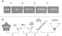
The methods described herein combine the methods shown in fig. 1A-1C and as ACS Nano 8.5 (2014): 4284-4294, the entire contents of which are incorporated herein by reference. FIGS. 1A-1C depict in situ amplification by hybrid strand reaction (HCR). Figure 1A depicts the HCR mechanism. The metastable fluorescent hairpin self-assembles into a fluorescent amplification polymer after detection of a homologous initiator. Initiator I1, comprising a single-stranded segment "b-a", nucleated with hairpin H1 through base pairing with the single-stranded sticky end "a" of H1, mediated branch migration, which opened the hairpin to form complex I1 · H1 containing a single-stranded segment "c-b". This complex nucleates with hairpin H2 by base pairing with the single-stranded sticky end "c", mediating branch migration, which opens the hairpin to form complex I1 · H1 · H2 containing the single-stranded segment "b-a". Thus, the initiation sequence is regenerated, providing the basis for the chain reaction of alternating H1 and H2 polymerization steps. Red stars indicate fluorophores. FIG. 1B depicts an in situ hybridization protocol. In the detection stage, a probe set comprising one or more priming strands is hybridized to the mRNA target, and unused probes are washed from the sample. In the amplification stage of the hybrid strand reaction using an initiator and multiple hairpins as described in FIG. 1A, the initiator triggers the self-assembly of the tethered fluorescently amplified polymer from the hairpins and the unused hairpins are washed out of the sample. Fig. 1C depicts an experimental timeline. The same two-stage protocol was used, regardless of the amount of target mRNA. For multiplex experiments (three color example depicted), probe sets for different target mrnas (five probes depicted per set) carry orthogonal initiators, which trigger orthogonal HCR amplification cascades labeled by spectrally distinct fluorophores.
"HCR system," "HCR probe set," or "HCR initiator/hairpin set" includes one or more nucleic acid initiation strands and one or more metastable HCR monomers, e.g., nucleic acid hairpins, that together are capable of forming a hybrid strand-reacting polymer. HCR systems are designed using criteria to achieve desired properties, such as orthogonality or non-reactivity with other nucleic acid species, and desirable kinetic and thermodynamic properties, according to the methods described herein. HCR systems can be synthesized using standard methods, such as chemical nucleic acid synthesis, including commercial sources such as integrated DNA technology (IDT, coralville, iowa), w.m. keck gold oligomer synthesis research project (new han, connecticut), or molecular devices (pasadena, california). Alternatively, HCR system components can be synthesized and/or amplified using standard enzymatic methods, such as PCR, followed by lambda exonuclease digestion of one strand to produce ssDNA (see Current Protocols in Molecular Biology (2014): 14-23, which is incorporated herein by reference in its entirety) or in vitro transcription followed by reverse transcription to produce ssDNA (see Science 348: 6233 (2015): aaa6090, which is incorporated herein by reference in its entirety).
The methods described herein that utilize hybrid strand reaction characteristics can be used to detect one or more analytes or target molecules, for example, within a biological sample (in situ), by designing one or more HCR reactions (performed in tandem, or as a parallel reaction set performed in tandem) for sequential or combinatorial labeling of multiple target molecules, molecular species, molecular masses, or molecular compositions such that each target is associated with a unique HCR signal or set of HCR signals throughout the HCR reaction. Target molecules include nucleic acid polymers, such as RNA, DNA and analogs thereof, amino acid polymers, including proteins, any of the chemical modifications described above, lipids, metabolites, biomolecules and other small molecules, and molecular combinations comprising one or more of any of the foregoing.
The target molecule or analyte is targeted by a probe, which can be attached to a priming strand. The present disclosure provides probes that can be attached to a priming strand via a linker. The present disclosure provides that the priming strand can be removed from the probe. The present disclosure provides that the linker may be a cleavable linker. The present disclosure provides that linkers can be formed from any molecular binding pair that can be bound together and separated. The binding pair will link the probe and the initiating strand such that the probe and the initiator are not directly linked, but are indirectly linked through a molecular binding pair.
The methods described herein allow for rapid and isothermal amplification of signals and detection of multiple analytes or target molecules in the same sample. The methods described herein include multiplexing by detecting different analytes simultaneously using independent and orthogonal HCR systems, multiplexing by detecting different analytes simultaneously using independent and orthogonal HCR systems labeled with spectrally different dyes, increasing the space for spectrally different labels by combinatorial or colorimetric barcoding, such as by using one or more fluorophores per HCR system simultaneously (see Science 297: 836-840(2002), incorporated herein by reference in its entirety), by using trigger probes that protect initiators until the specificity of the probes specifically bind to the target, by using background that is reduced from self-quenching HCR system components versus fluorophore/quencher pairs that are separated upon assembly into an amplification polymer, where unreacted HCR system components exhibit suppressed fluorescence, by using rapid diffusion and permeation into a small pore matrix, small HCR system components such as formaldehyde-fixed biological samples or polyacrylamide hydrogels penetrate into the sample with high efficiency, perform sensitive quantitative amplification by using a nonlinear HCR mechanism that provides polymers that grow exponentially to specific final sizes, and programmable amplification by using HCR systems that exhibit linear, quadratic, or exponential polymer growth.
Thus, the methods described herein utilize target molecules or analytes that can be tracked for analysis, as the methods described herein utilize a cyclic approach to analyzing such target molecules or analytes. That is, repeated or cyclical analysis of a particular target molecule or analyte using HCR as described herein is performed, and thus tracked in a manner that is spectrally resolvable from other target molecules or analytes that may be in the same sample. One exemplary method of tracking a particular target molecule or analyte is by immobilizing the sample in a three-dimensional matrix such that each target molecule or analyte has a fixed, known location within the matrix, and can be subjected to the repeated or cyclical HCR procedures described herein, wherein the signal produced by the HCR can be monitored and analyzed to produce a time-ordered signal of the same or a particular target molecule or analyte.
The methods described herein, such as repetition or cycling of certain method steps, advantageously overcome the upper limit of the number of orthogonal HCR systems associated with known systems. See ACS Nano 8.5 (2014): 4284-4294. HCR is known to be limited to five orthogonal DNA HCR probe sets. For simultaneous use, the HCR probe sets must be unreactive with each other, which is typically achieved by simultaneous computational design of the HCR probe sets. This process can be computationally intensive, and scaling up the number of simultaneously designed probe sets can significantly increase computational costs. In practice, increasing the number of HCR probesets comes at the cost of increasing background and false positive amplification, because the distance between the probesets in the nucleic acid sequence space is reduced in view of the nucleic acid sequence space defined by the size of the HCR system functional domains (e.g., priming domain and propagation region). There may be additional costs associated with more specific engineering of HCR probes by increasing the size of the nucleic acid sequence "design space", e.g., a set of HCR probes with a longer propagation region may take significantly longer to polymerize.
The methods described herein advantageously overcome the inherent barcoding limitations associated with known systems. If each HCR probe set is labeled with one of N spectrally distinct dyes, then the N analytes can be labeled simultaneously. If all combinations and monochrome barcodes are used, the number of simultaneously labeled analytes equals 2 N -1。
Biological systems represent a great deal of complexity in terms of molecular species, molecular mass and molecular configuration. The methods described herein can be used to simultaneously multiplex labeling of multiple molecular species, molecular configurations and molecular masses in order to determine the species, abundance and location of molecules within a biological system, e.g., measuring the molecular configuration of a biological system. Certain properties of the target analyte comprise some "raw information" about the presence, location, abundance, quantity, species, mass, configuration or other property of the target that needs to be measured; where "information" is broadly considered to mean carried or represented within a biological system by a particular spatial and/or temporal arrangement of atoms, molecules, compounds or molecular complexes that are desired to be measured. During detection, this information, or a portion thereof, is transferred from the target analyte to a human or computer system by labeling and detection.
Given N orthogonal, independent and spectrally distinct HCR systems, the methods described herein provide greater multiplexing for linear or exponential barcoding through the use of tandem labeling of analytes in the method steps. Linear barcoding was repeated k consecutive times using (i.e. using the same) N HCR systems to label k × N total analytes. This can be achieved by varying the correlation between analyte and HCR initiator between each round of HCR amplification and detection, such that each HCR initiator is correlated with a different analyte during each round of HCR. Exponential barcoding repeated use (i.e. using the same) of N HCR systems k consecutive times to label N k Total analytes. This can be achieved by varying the association between analyte and HCR initiator between each cycle of HCR amplification and detection, such that each analyte is associated with multiple HCR initiators throughout successive HCR cycles (each analyte is associated with between 0 and 1 HCR system during each successive cycle of HCR). Throughout the HCR cycle, the combined label associated with the target analyte is thus constructed from the individual HCR signals within each cycle. In both cases, the relationship between the target analyte and the HCR reaction (which can be understood to produce a detected fluorescent signal) is programmable, as the HCR reaction is engineered over time to produce a set of encoded fluorescent signals for the purpose of labeling the analyte, e.g., in situ labeling the analyte. In general, this technique is referred to herein as cyclic hcr (chcr), since steps in the overall marking process can occur cyclically, i.e., in a continuous and repeated manner.
The present disclosure provides methods and materials for "programming" a labeled cascade of HCR reactions, comprising the steps of contacting a sample with a probe, contacting the sample with an HCR priming sequence, contacting the sample with a metastable HCR monomer (e.g., hairpin), and contacting the sample with a fluorescent moiety, wherein the probe binds to a target analyte, and wherein the HCR priming sequence is associated with the probe, and wherein the priming sequence nucleates with the homologous hairpin and triggers self-assembly of the tethered amplification polymer, and wherein the tethered amplification polymer is associated with the fluorescent moiety, and wherein the target analyte is detected by measuring fluorescence of the sample.
The present disclosure also provides methods and materials for "programming" a labeling cascade, comprising the steps of contacting a sample with a probe, contacting the sample with an HCR priming sequence, contacting the sample with a metastable HCR monomer (e.g., a hairpin), and contacting the sample with a fluorescent moiety, wherein the probe binds to a target analyte, and wherein the HCR priming sequence is associated with the probe, and wherein the priming sequence nucleates with the homologous hairpin and triggers self-assembly of the tethered amplification polymer, and wherein the tethered amplification polymer is associated with the fluorescent moiety, and wherein the target analyte is detected by measuring fluorescence of the sample; and further comprising the steps of: dissociating the fluorescent moieties from the HCR polymer and removing them from the sample, e.g. by washing, degrading or disassembling the HCR polymer, and removing component debris from the sample, e.g. by washing, dissociating or removing the HCR trigger sequence from the probe contacted with the target analyte, and removing it from the sample, e.g. by washing, and/or dissociating the probe from the target analyte and removing it from the sample, e.g. by washing.
The cyclic HCR is enabled by method and material specificity to achieve programmability of each information transfer step. "programmability" refers to materials and methods that enable each step of an information transfer or tagging cascade that depends on one or more, or a variety of, inputs to be gated, i.e., performed according to a predetermined, non-sequential schedule; alternatively, each step can be specifically reversed, i.e., after being detected, the information passed to subsequent steps in the labeling cascade is selectively inactivated, removed, destroyed, or rendered undetectable; or each step may be gated and reversible. As used herein, "gated" may mean "inactive," "restricted," "unable to continue," and "ungated," as used herein, may mean "active," "activated," "unrestricted," "able to continue," and the like.
The present disclosure provides a method for detecting a target analyte in a biological sample, comprising the steps of: contacting the sample with a probe comprising a priming sequence, contacting the sample with one or more, metastable fluorescent HCR monomers (e.g., hairpins), wherein the probe binds to the target analyte, and wherein the priming sequence nucleates with a cognate hairpin and triggers self-assembly of the tethered fluorescent amplification polymer, and detecting the target analyte in the sample by measuring fluorescence of the sample. In one embodiment, multiple probes can be added for detection of multiple target analytes. In another embodiment, multiple metastable fluorescent hairpins with spectrally different fluorophores may be added for multiplex detection. In one embodiment, the analyte comprises a nucleic acid polymer, including RNA, DNA, and analogs thereof. In another embodiment, the analyte comprises an amino acid polymer, including proteins and chemical modifications thereof. In yet another embodiment, the analyte comprises lipids, metabolites, biomolecules and other small molecules. In one embodiment, the priming sequence is a DNA priming sequence. In another embodiment, the methods of the invention further comprise linear or exponential barcoded analyte serial tags for multiplex detection. In one embodiment, the method of the present disclosure further comprises attaching a linker probe or secondary probe to the target analyte. In another embodiment, the linker probe or secondary probe is bound to a probe comprising a priming sequence. In certain embodiments, the priming sequence is common or unique to the target analyte. In one embodiment, the probe is a trigger or activatable probe such that the trigger sequence is protected or inhibited until the probe specifically binds to the target analyte whereupon the trigger sequence is activated. In certain embodiments, the unique markers associated with the target analyte are constructed from one or more individual HCR signals using a cycling HCR.
The present disclosure also provides an in-situ imaging method, comprising the steps of: contacting the biological sample with a probe, contacting the sample with an HCR priming sequence that becomes associated with the probe, contacting the biological sample with a metastable HCR monomer, such as a hairpin, wherein the probe binds to a target analyte in the biological sample, and wherein the HCR priming sequence is associated with the probe, and wherein the priming sequence nucleates with a homologous hairpin and triggers self-assembly of a tethered amplification polymer, and wherein the tethered amplification polymer is associated with a fluorescent moiety, and wherein the target analyte in the biological sample is detected by measuring fluorescence of the polymer.
In one embodiment, multiple probes can be added for imaging multiple target analytes. In another embodiment, multiple metastable fluorescent hairpins with spectrally different fluorophores may be added for multiplexed imaging. In another embodiment, the methods of the invention further comprise the sequential labeling of linearly or exponentially barcoded analytes for multiplex detection. In one embodiment, the method of the present disclosure further comprises attaching an adapter probe or a secondary probe to the target analyte, wherein the adapter probe or the secondary probe is unique to the target analyte. In another embodiment, the linker probe or secondary probe is bound to a probe that includes a priming sequence. In certain embodiments, the priming sequence is common or unique to the target analyte. In one embodiment, the probe is a triggered probe in which the trigger sequence is protected or inhibited until the probe specifically binds to the target analyte whereupon the trigger sequence is activated. The method according to the invention also comprises multiple rounds of hybridization chain reaction "HCR" and detection cycles.
The present disclosure provides hybrid strand reaction "HCR" systems that include a probe comprising one or more nucleic acid priming strands, and a metastable nucleic acid fluorescent HCR monomer, such as a hairpin, wherein the priming strand is capable of nucleating with a homologous hairpin and triggering self-assembly of the HCR fluorescent polymer. In one embodiment, there are a plurality of probes for imaging a plurality of target analytes. In another embodiment, there are multiple metastable fluorescent hairpins with spectrally distinct fluorophores for multiplexed imaging. In one embodiment, the system is designed using criteria to achieve desired properties, such as non-reactivity or orthogonality with other nucleic acid species, and to have desired kinetic and thermal properties. In one embodiment, the hairpin may be produced by chemical and/or enzymatic synthesis. In some embodiments, multiple rounds of hybridization strand reaction "HCR" and detection cycles can be performed. In one embodiment, the initiator and the hairpin may be reused. In another embodiment, the fluorescent signal can be generated and reset programmatically.
According to one aspect, the present disclosure provides a method for detecting one or more target analytes in a sample, comprising contacting the sample with one or more probesets, wherein each probeset comprises one or more primary probes, each primary probe is homologous to a linker, and wherein each probeset is specific for a target analyte, contacting the sample with one or more Hybridization Chain Reaction (HCR) initiators that bind to a linker, contacting the sample with one or more HCR amplifier systems, wherein each HCR amplifier system comprises two or more metastable HCR monomers, wherein at least one HCR monomer comprises a detectable label, wherein the primary probes bind to an analyte, wherein the linkers connect the primary probes to the initiators, and wherein the initiators contact with homologous HCR amplifier monomers and trigger hybridization chain reactions of self-assembling and tethered nucleic acid amplification polymer products, and wherein the detectable label is detected. In one embodiment, a plurality of probesets, each specific for a target analyte, are designed for programmable and time-ordered hybridization strand reactions. In another embodiment, the detectable label is a fluorescent label and the total number of fluorescent signals generated over time provides a unique set of information for each target analyte, including molecular species, molecular mass or molecular configuration. In one embodiment, the sample may be contacted with both the probe set and the initiator simultaneously. In another embodiment, the HCR amplicon system comprises two metastable DNA hairpins. In one embodiment, the detectable label of the HCR amplicon system comprises spectrally distinct fluorescent signals for multiplex detection. In another embodiment, the detectable label of the HCR amplicon system comprises a sequencing template for fluorescent sequencing by hybridization, fluorescent sequencing by ligation, or fluorescent sequencing by synthesis. In some embodiments, the target analyte containing nucleic acid polymer, including RNA, DNA and its analogues. In other embodiments, the target analyte contains amino acid polymers, including proteins and chemical modifications thereof. In some embodiments, the target analyte comprises lipids, metabolites, biomolecules and other small molecules. In certain embodiments, the initiator comprises a nucleic acid hybridization strand reaction (HCR) initiation region. In one embodiment, the priming agent comprises DNA. In some embodiments, the HCR amplicon monomer comprises metastable DNA duplexes joined by a linker. In some embodiments, the target analyte is continuously labeled. In one embodiment, the time-ordered set of detection markers from the combination of all cycles of HCR comprises a unique composite marker for each target analyte, wherein each cycle comprises detection of the detectable markers of one or more HCR systems. In another embodiment, the composite marker comprises a linear or exponential barcode for multiplex detection. In one embodiment, the unique composite marker comprises a barcoded message. In another embodiment, the barcoded message further contains additional information, including for error detection or correction. In one embodiment, the design of programmable and time-ordered hybridization strand reactions and sets of cognate fluorescence signals includes a unique barcoded message for each target analyte. In one embodiment, the cyclic HCR is enabled by the programmability of each information transfer step. Programmability refers to allowing each step of information transfer to be gated and/or reversed. Gated information transfer refers to the transfer of information according to a predetermined discontinuous schedule, wherein the information transfer is dependent on one or more inputs. In one embodiment, the binding of one or more primary probe sets to the target is repeated two or more times. In one embodiment, the primary probe and the linker are linked by covalent or non-covalent interactions. In another embodiment, the linker and initiator are linked by covalent or non-covalent interactions. In one embodiment, the linker may be a bond or comprise a sequence portion that is complementary to a sequence portion of an oligonucleotide comprising a priming sequence and hybridizes to an oligonucleotide comprising a priming sequence. In another embodiment, the connection between the primary probe and the linker is programmably broken or reversed. In one embodiment, the connection between the linker and the initiator is programmably broken or reversed. In another embodiment, the linker comprises an initiation sequence homologous to a protecting group that prevents the initiator from initiating the HCR. In one embodiment, the priming sequence is protected by a protective oligonucleotide. In another embodiment, the protecting group is programmably interrupted from the linker, which allows the initiator to initiate the HCR. In one embodiment, a deprotecting oligonucleotide may be introduced to remove the protecting oligonucleotide by sticky end strand displacement. In another embodiment, the HCR polymer is degraded or disassembled upon detection of the detectable label. In one embodiment, the linkage between the HCR polymer and the detection label is programmably broken or reversed after detection. In another embodiment, the binding of the primary probe to the target, and the linkage between the primary probe, linker, initiator, polymer, and detection moiety, can be programmably disrupted and reversed. In certain embodiments, the method further comprises multiple rounds of hybridization chain reaction "HCR" and detection cycles. In other embodiments, the method can be used for in situ imaging of biological samples.
According to another aspect, the present disclosure provides a cycling hybridization strand reaction "HCR" system comprising one or more probesets, wherein each probeset comprises one or more primary probes, each primary probe is homologous to a linker, and wherein each probeset is specific for a target analyte, an initiator, and one or more HCR amplifier systems, wherein each HCR amplifier system comprises two or more metastable HCR monomers, wherein at least one HCR monomer comprises a detectable label, wherein the initiator contacts the homologous HCR amplifier monomers and triggers a hybridization strand reaction of the self-assembled and tethered nucleic acid amplification polymer product, and wherein the detectable label is detected. In one embodiment, a plurality of probesets, each specific for a target analyte, are designed for programmable and time-ordered hybridization strand reactions. In another embodiment, the total number of time-derived fluorescent signals provides a unique set of information for each target analyte, including molecular species, molecular mass or molecular configuration. In one embodiment, the HCR amplicon monomer is a DNA hairpin. In another embodiment, the detectable label of the HCR amplicon monomer further comprises a spectrally distinct fluorescent signal for multiplex detection. In one embodiment, the system is designed using criteria to achieve desired properties, such as non-reactivity or orthogonality with other nucleic acid species, and to have desired kinetic and thermodynamic properties. In another embodiment, HCR monomers can be produced by chemical and/or enzymatic synthesis. In one embodiment, non-fluorescent HCR monomers may be used. In another embodiment, the non-fluorescent HCR monomer is fluorescently labeled during or after the HCR polymerization stage. In one embodiment, the polymer formed from the non-fluorescent monomer is fluorescently labeled after the HCR polymerization stage. In another embodiment, the polymer formed from the non-fluorescent monomer is fluorescently labeled after the HCR polymerization stage of fluorescent sequencing by hybridization, fluorescent sequencing by ligation, or fluorescent sequencing by synthesis, enzymatic reaction, or chemical reaction. In certain embodiments, more than one round of hybridization strand reaction "HCR" and fluorescence detection may be performed. In other embodiments, the probe, linker, initiator, and HCR monomer may be reused. In one embodiment, the linker is a nucleic acid sequence complementary to the oligonucleotide comprising the initiator. In another embodiment, the linker comprises a functional group for programmable dissociation from the initiator. In one embodiment, the linker comprises an initiator homologous to the protecting group, which prevents the initiator from initiating the HCR. In another embodiment, the binding of the primary probe to the target, and the linkage between the primary probe, linker, initiator, polymer, and detectable label, can be disrupted and reversed during each round of the hybridization strand reaction "HCR" and detection cycle to achieve programmability of the system. In one embodiment, detection of the detectable label can be generated and reset programmatically. In another embodiment, the HCR amplicon monomer contains functional groups for programmable disassembly or degradation of the polymer. In one embodiment, the functional group consists of a sticky end strand displacement sequence. In another embodiment, the functional group comprises a chemical group that is chemically labile, enzymatically labile, or photolabile. In certain embodiments, the binding of the probe to the target analyte is reversed by a method comprising: chemical treatment, enzymatic treatment, dnase treatment of RNA ISH probes, exonuclease treatment of 5' phos ISH probes, nuclease treatment of nucleic acid probes, protease treatment of peptide probes, use of heat or denaturants to disrupt nucleic acid hybridization, use of heat or denaturants to disrupt aptamer binding, or use of heat or denaturants to disrupt the bond between an antibody and a protein. In one embodiment, the system includes a method for programming functional ligation of HCR initiators to binding probes. In another embodiment, a method for programming functional ligation of an HCR initiator to a binding probe comprises a) using nucleic acid hybridization with an initiator added to a linker probe with a complementary nucleic acid molecule by hybridization sequencing, b) using an enzyme to add the initiator to the linker probe, c) disrupting the nucleic acid hybridization with heat or a denaturing agent to remove the initiator hybridized to the linker probe, d) using sticky end strand displacement to remove a protecting strand from the initiator located at the target molecule by the linker probe, and e) incorporating a chemical, enzymatic or photolabile group between the initiator and the linker probe such that the initiator can be removed by chemical, enzymatic or light treatment that disrupts the chemical ligation between the initiator and the linker probe. In one embodiment, the enzyme that adds the initiator to the linker probe is a DNA ligase that catalyzes the splint ligation reaction. In another embodiment, the system includes a method for reversing a hybrid chain reaction. In one embodiment, a method for reversing a hybridization strand reaction includes a) using a modified HCR monomer comprising one or more additional sequences for cohesive end strand displacement such that addition of one or more complementary DNA strands will result in disassembly of the HCR polymer, and b) using a modified HCR monomer comprising one or more enzymatic or chemically sensitive or photolabile groups in the DNA backbone of the HCR monomer such that the HCR polymer can be fragmented or destroyed by chemical, enzymatic or light treatment. In one embodiment, the system includes a method for programming the functional generation of a HCR polymer fluorescence signal. In certain embodiments, methods of programming the functional generation of a fluorescence signal of an HCR polymer include a) introducing a fluorescent moiety into the HCR polymer using a modified HCR monomer comprising an additional sequence that can be detected by Sequencing By Synthesis (SBS), by ligation Sequencing (SBL) or by Sequencing By Hybridization (SBH), b) using a modified HCR monomer comprising an enzyme, chemical or photolabile group between the HCR DNA monomer backbone and the fluorescent moiety such that the fluorescent moiety can be removed by chemical, enzymatic, or light treatment, c) using a modified fluorescent probe capable of labeling the HCR polymer, e.g., by SBS, SBL, or SBH, wherein the fluorescent probe comprises an additional sequence for sticky end-strand displacement such that the fluorescent probe can be removed from the HCR polymer by disrupting hybridization between the fluorescent probe and the HCR polymer, and d) using a fluorescent probe capable of labeling the HCR polymer, e.g., by SBS, SBL or SBH labels a modified fluorescent probe of the HCR polymer, wherein the fluorescent probe comprises an enzymatic, chemical or photolabile group between the HCR polymer backbone and the fluorescent moiety, such that the fluorescent moiety can be removed by chemical, enzymatic or light treatment.
According to one aspect, the present disclosure provides a method for in situ detection of one or more target analytes in a biological sample by hybridization strand reaction (HCR), comprising contacting the sample with one or more probesets, wherein each probeset comprises one or more primary probes, each primary probe associated with a linker, and wherein each probeset is specific for a target analyte, contacting the sample with one or more hybridization strand reaction (HCR) initiators, contacting the sample with one or more HCR amplifier systems, wherein each HCR amplifier system comprises two or more metastable HCR monomers, wherein at least one HCR monomer comprises a detectable label, wherein the primary probes bind to the target analyte, wherein the linkers connect the primary probes with the initiators, and wherein the initiators contact with homologous HCR amplifier monomers and trigger hybridization strand reactions of self-assembling and tethered nucleic acid amplification polymer products, and wherein the detectable label is detected. In one embodiment, a plurality of probesets, each specific for a target analyte, are designed for programmable and time-ordered hybridization strand reactions. In another embodiment, the total number of time-derived fluorescent signals provides a unique set of information for each target analyte, including molecular species, molecular mass, or molecular configuration. In one embodiment, the sample may be contacted with both the probe set and the initiator simultaneously. In another embodiment, the binding of the probe to the target analyte can be reversed such that the target analyte can be re-detected using a hybridization chain reaction to amplify the signal. In certain embodiments, the binding of the probe to the target molecule is reversed by a method comprising: chemical treatment, enzymatic treatment, dnase treatment of RNA ISH probes, exonuclease treatment of 5' phos ISH probes, nuclease treatment of nucleic acid probes, protease treatment of peptide probes, use of heat or denaturing agents to disrupt nucleic acid hybridization, use of heat or denaturing agents to disrupt aptamer binding, or use of heat or denaturing agents to disrupt the bond between antibody and protein. In one embodiment, the method further comprises a method for programming HCR initiator functional ligation to a binding probe. In one embodiment, a method for programming functional ligation of an HCR initiator to a binding probe comprises a) using nucleic acid hybridization with an initiator added to the linker probe with a complementary nucleic acid molecule by hybridization sequencing, b) using an enzyme to add the initiator to the linker probe, c) disrupting the nucleic acid hybridization with heat or a denaturing agent to remove the initiator hybridized to the linker probe, d) using sticky end strand displacement to remove a protecting strand from the initiator located at the target molecule by the linker probe, and e) incorporating a chemical, enzymatic or photolabile group between the initiator and the linker probe such that the initiator can be removed by chemical, enzymatic or light treatment that disrupts the chemical ligation between the initiator and the linker probe. In one embodiment, the enzyme that adds the initiator to the linker probe is a DNA ligase that catalyzes the splint ligation reaction. In one embodiment, the method further comprises a method for reversing the hybridization chain reaction. In one embodiment, a method for reversing a hybridization strand reaction comprises a) using a modified HCR monomer comprising one or more additional sequences for cohesive end strand displacement such that addition of one or more complementary DNA strands will result in disassembly of the HCR polymer, and b) using a modified HCR monomer comprising one or more enzymatic or chemosensitive or photolabile groups in the DNA backbone of the HCR monomer such that the HCR polymer can be fragmented or destroyed by chemical, enzymatic or light treatment. In one embodiment, the method further comprises a method for programming the functional generation of a fluorescence signal for the HCR polymer. In one embodiment, a method of programming the functional generation of a fluorescence signal of an HCR polymer comprises a) introducing a fluorescent moiety into the HCR polymer using a modified HCR monomer comprising an additional sequence that can be detected by Sequencing By Synthesis (SBS), by ligation Sequencing (SBL) or by Sequencing By Hybridization (SBH), b) using a modified HCR monomer comprising an enzyme, a chemical or a photolabile group between the HCR DNA monomer backbone and the fluorescent moiety such that the fluorescent moiety can be removed by chemical, enzymatic, or light treatment, c) using a modified fluorescent probe capable of labeling the HCR polymer, e.g., by SBS, SBL, or SBH, wherein the fluorescent probe comprises an additional sequence for sticky end-strand displacement such that the fluorescent probe can be removed from the HCR polymer by disrupting hybridization between the fluorescent probe and the HCR polymer, and d) using a fluorescent probe capable of labeling, e.g., by SBS, SBL or SBH labels a modified fluorescent probe of the HCR polymer, wherein the fluorescent probe comprises an enzymatic, chemical or photolabile group between the HCR polymer backbone and the fluorescent moiety, such that the fluorescent moiety can be removed by chemical, enzymatic or light treatment.
According to another aspect, the present disclosure provides a method for detecting one or more target analytes, comprising contacting a sample more than once with a cycling hybridization strand reaction "HCR" system, wherein each target analyte in the sample is associated with one amplified fluorescent signal over the total number of HCR cycles. In one embodiment, the combination of HCR cycling and spectrally resolvable fluorescence signals produced by the cycling HCR comprises a unique label for the target analyte.
According to another aspect, the present disclosure provides a method for detecting one or more target analytes, comprising contacting a sample more than once with a cycling hybridization strand reaction "HCR" system, wherein each target analyte in the sample is associated with more than one amplified fluorescent signal over the total number of HCR cycles. In one embodiment, the amplified fluorescent signals produced by each target analyte are combined in information to a composite label. In one embodiment, each target analyte is associated with a unique composite marker. In another embodiment, the sample is fixed. In one embodiment, the complex label is generated by spatial invariance of the target analyte between HCR cycles. In one embodiment, the target analyte is attached to a 3D matrix. In another embodiment, the composite label is generated by spatial invariance of the target analyte between HCR cycles. In one embodiment, the composite label is generated by unknown order invariance of the target analyte between HCR cycles. In another embodiment, one or more components of the cyclic HCR system are attached to a 3D matrix. In one embodiment, the composite label is generated by spatial invariance of the target analyte between HCR cycles. In another embodiment, the composite label is generated by unknown order invariance of the target analyte between HCR cycles. In one embodiment, the association between the target analyte and the HCR fluorescence signal is programmable. In another embodiment, the association between the target analyte and the HCR fluorescence signal is programmable.
According to one aspect, the present disclosure provides a method for detecting one or more target analytes in a sample, comprising (a) contacting the sample with one or more probesets, wherein each probeset comprises one or more primary probes each having a linker, and wherein each probeset is specific for a target analyte, wherein the one or more primary probes having linkers bind to the target analyte; (B) contacting the sample with one or more adaptor-bound hybridization strand reaction (HCR) initiators, (C) contacting the sample with two or more metastable HCR monomers, wherein the one or more initiators contact the two or more metastable HCR monomers and initiate a hybridization strand reaction to produce a self-assembled and tethered nucleic acid amplification polymer product, and (D) attaching one or more detectable labels to the tethered nucleic acid amplification product, and optionally detecting the one or more detectable labels. In one embodiment, the probe is removable from the target analyte, the initiator is removable from the linker, the nucleic acid amplification polymer product is removable from the initiator, or the one or more detectable labels are removable from the nucleic acid amplification polymer product. In another embodiment, the probe is removable from the target analyte, the initiator is removable from the linker, and the nucleic acid amplification polymer product is removable from the initiator. In one embodiment, the probe is removable from the target analyte. In another embodiment, the initiator may be removed from the linker. In one embodiment, the nucleic acid amplification polymer product may be removed from the initiator. In another embodiment, one or more detectable labels may be removed from the nucleic acid amplification polymer product. In another embodiment, the probe is removable from the target analyte, the initiator is removable from the linker, the nucleic acid amplification polymer product is removable from the initiator, and the one or more detectable labels are removable from the nucleic acid amplification polymer product.
According to another aspect, the present disclosure provides a method for detecting one or more target analytes in a sample, comprising (a) contacting the sample with one or more probesets, wherein each probeset comprises one or more primary probes each having a linker, and wherein each probeset is specific for a target analyte, wherein one or more primary probes having linkers bind to the target analyte; (B) contacting the sample with one or more linker-bound hybrid strand reaction (HCR) initiators, (C) contacting the sample with two or more metastable HCR monomers comprising a detectable label, wherein the one or more initiators contact the two or more metastable HCR monomers and initiate a hybrid strand reaction to produce a self-assembled and tethered nucleic acid amplification polymer product, and (D) optionally detecting the one or more detectable labels. In one embodiment, the probe can be removed from the target analyte. In another embodiment, the initiator may be removed from the linker. In another embodiment, the nucleic acid amplification polymer product may be removed from the initiator.
According to one aspect, the present disclosure provides a method for identifying a target analyte in a sample, comprising (a) contacting the sample with one or more probes, wherein a given probe of the one or more probes is coupled to a linker, and wherein the given probe has a sequence complementary to the sequence of the target analyte, wherein the given probe binds to the target analyte when the sample is contacted with the one or more probes; (b) contacting a sample with one or more Hybridization Chain Reaction (HCR) initiators under conditions sufficient to allow binding of a given HCR initiator of the one or more HCR initiators to a linker, wherein the given HCR initiator is separate from the given probe, and wherein the linker couples the probe to the given HCR initiator upon contacting the sample with the one or more HCR initiators; (c) contacting the sample with one or more HCR amplicons to initiate a hybridization strand reaction, wherein a given HCR amplicon of the one or more HCR amplicons comprises at least one HCR monomer comprising a detectable label, thereby producing an amplification product comprising the HCR monomer, which amplification product is coupled to the given probe; and (d) detecting the amplification product, thereby identifying the target analyte. In one embodiment, the method further comprises contacting the sample with a plurality of probesets, each probeset specific to the target analyte, the plurality of probesets configured to allow programmable and time-ordered hybridization strand reactions. In another embodiment, the detectable label is a fluorescent label and the detecting comprises detecting a fluorescent signal, wherein the fluorescent signal generated at all times provides a unique set of information, including the molecular species, molecular mass or molecular configuration of each target analyte. In one embodiment, the one or more HCR amplicons comprise two metastable DNA hairpins. In another embodiment, the detectable label of one or more HCR amplicons comprises a spectrally distinct fluorescent signal for multiplex detection. In one embodiment, the detectable label of the HCR monomer comprises a sequencing template for fluorescent sequencing by hybridization, fluorescent sequencing by ligation, or fluorescent sequencing by synthesis. In another embodiment, the target analyte includes nucleic acid polymers, including RNA, DNA, RNA analogs, DNA analogs, proteins and chemical modifications thereof. In yet another embodiment, the target analyte comprises lipids, metabolites, biomolecules and other small molecules. In one embodiment, the method further comprises sequentially labeling the target analyte. In one embodiment, the serial labeling comprises associating each analyte with a plurality of HCR initiators. In another embodiment, a given HCR amplifier comprises two or more metastable HCR monomers. In one embodiment, said binding of said given to a target analyte is repeated two or more times. In one embodiment, the linker may be a bond or comprise a sequence portion that is complementary to a sequence portion of an oligonucleotide comprising a priming sequence and hybridizes to an oligonucleotide comprising a priming sequence. In certain embodiments, the method further comprises disrupting or reversing the coupling between the given probe and the linker, the coupling between the linker and the HCR initiator, or the coupling between the given probe and the HCR initiator. In one embodiment, the linker comprises a priming sequence homologous to a protecting group that prevents the HCR initiator from triggering the HCR. In another embodiment, the protecting group is a protective oligonucleotide. In another embodiment, the method further comprises disrupting a protecting group from the linker, thereby allowing the HCR initiator to trigger the HCR. In one embodiment, the disrupting comprises introducing a deprotecting oligonucleotide to the sample to remove the protecting group by sticky end strand displacement. In another embodiment, the method further comprises degrading or disassembling the amplification product after said detecting. In one embodiment, the method further comprises disrupting or reversing the coupling between the amplification product and the detection label after said detecting. In another embodiment, the method further comprises disrupting or reversing the binding of said given probe to said target analyte. In another embodiment, the method further comprises performing multiple rounds of hybridization strand reactions comprising multiple detection cycles. In one embodiment, the multiple rounds of hybridization chain reactions comprise repeated use of one or more HCR initiators or one or more HCR amplicons. In another embodiment, the method further comprises programming for functional ligation between a given HCR initiator and a given probe, wherein the programming comprises a) using nucleic acid hybridization using adapter probes with HCR initiators added to complementary nucleic acid molecules by hybridization sequencing, b) using enzymes to add HCR initiators to adapter probes, c) disrupting nucleic acid hybridization using heat or denaturants to remove HCR initiators hybridized to adapter probes, d) using sticky end strand displacement to remove protective strands from HCR initiators localized to target molecules by adapter probes, or e) incorporating chemical, enzymatic, or photo labile groups between HCR initiators and adapter probes such that HCR initiators can be removed by chemical, enzymatic, or light treatment that disrupts chemical ligation between initiators and adapter probes. In one embodiment, the enzyme that adds the HCR initiator to the linker probe is a DNA ligase that catalyzes the splint ligation reaction. In another embodiment, the method further comprises reversing or blocking the hybrid strand reaction. In one embodiment, the reverse or block hybridization strand reaction comprises a) using a modified HCR monomer comprising one or more additional sequences for cohesive end strand displacement such that addition of one or more complementary DNA strands will result in disassembly of the amplification product, or b) using a modified HCR monomer comprising one or more enzymatic or chemosensitive or photolabile groups in the DNA backbone of the HCR monomer such that the amplification product is fragmented or destroyed by chemical, enzymatic or light treatment. In another embodiment, the method further comprises programming the generation of the fluorescent signal from the amplification product by: a) introducing a fluorescent moiety into the amplification product using a modified HCR monomer comprising an additional sequence that can be detected by Sequencing By Synthesis (SBS), Sequencing By Ligation (SBL) or Sequencing By Hybridization (SBH), b) using a modified HCR monomer comprising an enzyme, a chemical or photolabile group, between the DNA backbone of the HCR monomer and the detectable label of the fluorescent moiety, such that the fluorescent moiety can be removed by chemical, enzymatic, or light treatment, c) using a modified fluorescent probe capable of labeling the amplification product, e.g., by Sequencing By Synthesis (SBS), by Sequencing By Ligation (SBL) or Sequencing By Hybridization (SBH), wherein the fluorescent probe comprises an additional sequence for sticky end strand displacement such that the fluorescent probe can be removed from the amplification product by disrupting hybridization between the fluorescent probe and the amplification product, and d) using a fluorescent probe capable of being detected, e.g., by SBS, an SBL or SBH labeled amplification product modified fluorescent probe, wherein the fluorescent probe comprises an enzymatic, chemical or photolabile group between the amplification product backbone and a detectable label comprising a fluorescent moiety, such that the fluorescent moiety can be removed by chemical, enzymatic or light treatment. In one embodiment, a given probe may be removed from the target analyte, the HCR initiator may be removed from the linker, the amplification product may be removed from the HCR initiator, or the detectable label may be removed from the amplification product.
According to another aspect, the present disclosure provides a cyclic Hybrid Chain Reaction (HCR) system comprising: one or more probes, wherein a given probe of the one or more probes is coupled to a linker, wherein the given probe has a sequence complementary to a target analyte, one or more HCR initiators, wherein a given HCR initiator of the one or more HCR initiators is separate from the given probe, and wherein the given HCR initiator is configured to bind to the linker and couple the probe to the given HCR initiator, and one or more HCR amplicons, wherein a given HCR amplicon of the one or more HCR amplicons comprises at least one HCR monomer comprising a detectable label, wherein the given HCR initiator is configured to couple to a HCR monomer and initiate a hybridization strand reaction to produce an amplification product comprising the HCR monomer, which amplification product is coupled to the given probe. In one embodiment, the system further comprises a plurality of probesets, each probeset specific for a target analyte, wherein the plurality of probesets are designed for programmable and time-ordered hybridization strand reactions. In one embodiment, the plurality of probe sets are configured to provide temporally generated fluorescent signals, and wherein the total number of temporally generated fluorescent signals provides a unique set of information, including molecular species, molecular mass, or molecular configuration, for each target analyte. In another embodiment, each HCR amplifier comprises two or more metastable HCR monomers, each monomer being a DNA hairpin. In another embodiment, the one or more HCR amplicons comprise two or more metastable HCR monomers comprising a detectable label comprising spectrally distinct fluorescent signals for multiplex detection. In one embodiment, the HCR monomer is a non-fluorescent HCR monomer. In another embodiment, the non-fluorescent HCR monomer is configured to be fluorescently labeled during or after production of the amplification product. In one embodiment, the amplification product formed from the non-fluorescent monomer is fluorescently labeled after fluorescent sequencing by hybridization, fluorescent sequencing by ligation, or fluorescent sequencing by synthesis, enzymatic reaction, or chemical reaction to produce an amplification product. In another embodiment, one or more probes, linkers, one or more HCR initiators or one or more HCR amplicons are configured for reuse. In one embodiment, the linker is a nucleic acid sequence complementary to an oligonucleotide comprising an HCR initiator. In another embodiment, the linker comprises a functional group for programmable dissociation from the initiator. In one embodiment, the detection of the detectable label can be generated and reset programmatically. In another embodiment, the HCR monomer contains a functional group for programmable disassembly or degradation of the amplification product. In one embodiment, the functional group comprises a sticky end strand displacement sequence. In another embodiment, the functional group comprises a chemical group that is chemically labile, enzymatically labile, or photolabile. In one embodiment, the binding of a given probe to a target analyte is configured to be disrupted or reversed during a hybridization strand reaction. In another embodiment, the binding of a given probe to a target analyte is disrupted or reversed by: chemical treatment, enzymatic treatment, dnase treatment of RNA In Situ Hybridization (ISH) probes, exonuclease treatment of 5' phos ISH probes, nuclease treatment of nucleic acid probes, protease treatment of peptide probes, use of heat or denaturants to disrupt nucleic acid hybridization, use of heat or denaturants to disrupt aptamer binding, or use of heat or denaturants to disrupt the bond between an antibody and a protein.
According to one aspect, the present disclosure provides a method for identifying a target analyte in a sample comprising (a) contacting the sample with a primary probe comprising a sequence complementary to a sequence of the target analyte; (b) contacting the sample with a secondary probe configured to couple to the primary probe, wherein the coupling of the primary probe to the secondary probe facilitates a hybridization strand reaction (HCR) in the presence of at least one HCR amplicon comprising a detectable label to produce an amplification product comprising the detectable label, wherein the secondary probe is separate from the HCR amplicon and the primary probe; and (c) detecting the detectable label, thereby identifying the target analyte. In one embodiment, the HCR is not a polymerase chain reaction. In another embodiment, the amplification product is coupled to the primary probe. In one embodiment, the HCR amplifier has a sequence complementary to the sequence of the secondary probe. In another embodiment, the primary probe is coupled to a linker that allows the primary probe to be coupled to the secondary probe. In one embodiment, the primary probe comprises an HCR initiator that initiates the HCR. In another embodiment, the primary probe comprises a protecting group that prevents the HCR initiator from initiating the HCR prior to contacting the sample with the secondary probe. In one embodiment, the protecting group is a protective oligonucleotide. In another embodiment, the primary probe comprises an HCR initiator that initiates the HCR. In one embodiment, the secondary probe does not comprise a detectable label. In another embodiment, the HCR amplifier comprises two or more metastable HCR monomers. In one embodiment, each of the two or more metastable HCR monomers comprises a metastable DNA hairpin.
According to another aspect, the present disclosure provides a system for identifying a target analyte in a sample, comprising a detector for detecting a detectable label; a controller operatively coupled to the detector, wherein the controller comprises one or more computer processors individually or collectively programmed to direct: (i) contacting the sample with a primary probe comprising a sequence complementary to the target analyte sequence; (ii) contacting the sample with a secondary probe configured to couple to the primary probe, wherein the coupling of the primary probe to the secondary probe facilitates a hybridization strand reaction (HCR) in the presence of at least one HCR amplicon comprising a detectable label to produce an amplification product comprising the detectable label, wherein the secondary probe is separate from the HCR amplicon and the primary probe; and (iii) detecting the detectable label using the detector, thereby identifying the target analyte.
According to another aspect, the present invention provides a kit for identifying a target analyte in a sample, comprising a hybridization strand reaction (HCR) amplifier containing a detectable label, the HCR amplifier configured to facilitate HCR; a primary probe comprising a sequence complementary to a sequence of the target analyte; and a secondary probe configured to couple to the primary probe, wherein the secondary probe does not comprise a detectable label, wherein the secondary probe is separate from the HCR amplicon and the primary probe. In one embodiment, the kit further comprises instructions for performing said HCR using said HCR amplifier, primary probe and secondary probe. In another embodiment, the kit further comprises a cleavage agent configured to cleave the linker between the primary probe and the secondary probe, thereby disrupting the one or more HCR initiators to prevent triggering of a strand reaction with the one or more HCR amplicons. In another embodiment, the HCR amplifier has a sequence complementary to the sequence of the secondary probe. In one embodiment, the primary probe is coupled to a linker that allows the primary probe to be coupled to the secondary probe. In another embodiment, the primary probe comprises an HCR initiator that initiates the HCR. In one embodiment, the primary probe comprises a protecting group that prevents the HCR initiator from initiating the HCR prior to coupling the primary probe to the secondary probe. In another embodiment, the protecting group is a protective oligonucleotide. In one embodiment, the secondary probe comprises an HCR initiator that initiates the HCR. In another embodiment, the HCR amplifier comprises two or more metastable HCR monomers.
According to another aspect, the present disclosure provides a method for disrupting the production of a hybrid strand reaction (HCR) amplification product, comprising (a) providing a sample comprising a primary probe coupled to a secondary probe, wherein the primary probe comprises a sequence complementary to a sequence of a target analyte, and wherein the primary probe hybridizes to the target analyte under conditions sufficient to facilitate a hybrid strand reaction (HCR) to produce an amplification product; and (b) contacting the sample with a cleavage agent to separate the primary probe from the secondary without separating the primary probe from the target analyte, thereby preventing the HCR and disrupting the production of the amplification product.
Other features and advantages of certain embodiments of the invention will be apparent from the claims and from the following description of the drawings and embodiments.
Drawings
This patent or application document contains at least one drawing executed in color. Copies of this patent or patent application publication and color drawing(s) will be provided by the government agency upon request, after payment of the necessary fee. The above and other features and advantages of the present invention will be more clearly understood from the following detailed description of exemplary embodiments taken in conjunction with the accompanying drawings, in which:
FIGS. 1A-1C depict in situ amplification by hybrid strand reaction (HCR). Figure 1A depicts the HCR mechanism. The metastable fluorescent hairpin self-assembles into a fluorescent amplification polymer after detection of a homologous initiator. Initiator I1, comprising a single-stranded segment "b-a", nucleated with hairpin H1 through base pairing with the single-stranded sticky end "a" of H1, mediated branch migration, which opened hairpin H1 to form complex I1 · H1 containing a single-stranded segment "c-b". This complex nucleates with hairpin H2 by base pairing with the single-stranded sticky end "c", mediating branch migration, which opens the hairpin to form complex I1 · H1 · H2 containing the single-stranded segment "b-a". Thus, the initiation sequence was regenerated, providing the basis for chain reaction of alternating H1 and H2 polymerization steps. Red stars indicate fluorophores. FIG. 1B depicts an in situ hybridization protocol. And (3) a detection stage: a set of DNA probes, each probe containing initiator 11 and I2, and a region complementary to an mRNA target, hybridized to the mRNA target; unbound probe is washed from the sample. An amplification stage: triggering self-assembly of the linked fluorescent amplification polymer by an initiator; unpolymerized hairpins were washed from the sample. Fig. 1C depicts an experimental timeline. The same two-stage protocol was used, regardless of the amount of target mRNA. For multiplex experiments (three color example depicted), probe sets for different target mrnas (five probes depicted per set) carry orthogonal initiators, which trigger orthogonal HCR amplification cascades labeled by spectrally distinct fluorophores.
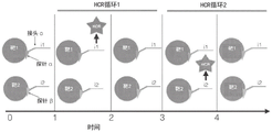
Fig. 2A-2B depict schematic diagrams of the information transfer steps a-D of the cyclic HCR technique. The primary information is a property of the analyte being detected, such as the molecular species, molecular mass or molecular configuration of the interrogation. In step a, the analyte is targeted by the probe, which specifically binds to the target analyte, such that the original information of the analyte is represented by the presence of the bound probe. In step B, the analyte information or some portion thereof carried by the probe is transferred to the HCR initiator via the linker. The HCR initiator is associated with a probe that is associated with an analyte. The linker connects the probe to the initiator. In step C, the analyte information or a portion thereof conveyed to the presence and location of the HCR initiator is converted to a DNA polymer, referred to as an HCR polymer, by initiating a hybridization chain reaction of one or more metastable HCR monomers (e.g., hairpins). Metastable HCR monomer is added to the sample and an initiator is initially combined with the metastable HCR monomer, and then chain reaction of one or more of the remaining HCR monomers results in formation of HCR polymer. In step D, the analyte information, or a portion thereof, that is conveyed to the presence and location of the HCR polymer is converted to an amplified fluorescent signal, which can be measured using a photon detector (e.g., a microscope equipped with a digital camera). The HCR polymer is associated with one or more detectable moieties. These steps A-D describe the general method and information-carrying strands in analyte detection assays using HCRs, such as the HCR-amplified mRNA fluorescence in situ hybridization assay depicted in FIG. 1. The cyclic HCR is enabled by method and material specificity to achieve programmability of each information transfer step. "programmability" refers to materials and methods that enable each step of information transfer to be gated, i.e., performed according to a predetermined, non-continuous schedule, wherein information transfer is dependent on multiple inputs; alternatively, each step can be specifically reversed, i.e., after being detected, the information passed to subsequent steps in the process is selectively destroyed or removed or rendered undetectable; or each step may be gated and reversible. The detectable moiety may be or have been removed from the HCR polymer, the HCR polymer may be or have been removed from the initiator, the initiator may be or have been removed from the probe, and the probe may be or have been removed from the analyte. This is in contrast to the HCR reaction in fig. 1, where information transfer is continuous and irreversible, e.g., a probe (a region of sequence complementary to an mRNA sequence that binds an mRNA) is irreversibly linked to an HCR initiator and the production of HCR polymers begins upon introduction of a complementary HCR hairpin. For example, the programmability of step C is intended to indicate that the reaction between the initiator and the HCR hairpin is gated in some way, for example by requiring another input signal in addition to the necessary HCR hairpin to allow the reaction to proceed, and that the formation of the HCR polymer can be reversed, for example by targeted degradation of the polymer. Programmability between the information transfer steps is represented by the connecting lines in fig. 2B, each end with an arrow.
FIG. 3 depicts a schematic of information and physical representation of the original information of an analyte in an analyte detection experiment using circulating HCR. The primary information is a property of the analyte being detected, such as the interrogating molecular species, molecular mass or molecular configuration. The original information is uniquely associated with an informational marker (referred to as a "marker"). The marker is represented herein as a binary string, but is used to represent any symbolic representation that carries original information, such as an alphanumeric value corresponding to the analyte or its original information, e.g., a gene name or reference thereto. The information marker is uniquely represented by an information message carried by a fluorescent signal comprising spatiotemporal organization of the detected message. The information tag and the message may be identical or the message may contain additional information than strictly necessary for the reference tag, as in the case of additional information for error detection or correction. In this example, a "message" is constructed as a bit string of labels followed by an inverted bit string of labels. Upon detection of the message, each bit of the marker will be detected twice, allowing certain errors to be detected (e.g., if the first bit of the marker is detected as a "0" at the first bit of the message, then detected as a "1" at the last bit of the message, it is apparent that errors occurred during transmission or detection of the message, such as during detection, HCR, imaging or image processing). The message is converted into a unique set of time-ordered fluorescent HCR signals, which are the detected messages. The temporal sequencing of HCR signals is enabled by the programmability of the cyclic HCR methods and materials, designed as a set of protocols and materials, e.g., primary (step a) probes, step B probes, step C probes, and/or step D probes, microscopes, other reagents, etc., and performed experimentally.
FIG. 4 depicts a schematic of the binding of a programming probe to a target molecule over a two-cycle HCR for sequential detection of two target analytes. At time 0, two target molecules are present in the sample. At time 1, a primary (step a) probe, such as an antibody, aptamer or DNA/RNA ISH probe, called "probe α", denoted as an orange triangle, is added to the sample and binds to target 1, which is homologous to the HCR initiator "i 1" via a certain linker (which is denoted as a short red line, such as a chemical bond or molecular interaction, but is understood to be any kind of programmable or non-programmable linker as described in step B of the circulating HCR). At time 2, the HCR hairpin is introduced to the sample, contacted with an initiator, and subsequently an amplified fluorescent HCR signal is generated. At time 3, the probe α has been stripped from the sample and the HCR polymer is also removed using any of the methods described in step C of recycling HCR. At time 4, a new probe "probe β" homologous to the same HCR initiator "i 1" via some linker (e.g. a chemical bond or molecular interaction, but understood as any type of programmable or non-programmable linker as described in step B of the cycling HCR) has been added to the sample and bound to target 2; at time 5, the HCR hairpin is added to the sample, contacted with an initiator, and subsequently an amplified fluorescent HCR signal is generated. At time 6, the probe β has been stripped from the sample and the HCR polymer is also removed using any of the methods described in step C of recycling HCR. In this example, the same HCR fluorescence signal amplification hairpin and initiator are used repeatedly in two HCR cycles to detect two tandem target molecules. The first cycle of the HCR is represented at times 1 to 3, while the second cycle of the HCR is represented at times 4 to 6. Where the detection of HCR fluorescence is represented as a binary 1 and the lack of HCR fluorescence is represented as a binary 0, the detected message corresponding to target 1 is "10" and the detected message corresponding to target 2 is "01". Thus, although the detection of these two molecules uses the same HCR fluorescence signal to amplify the hairpin and initiator, the ordered set of fluorescence signals that make up the detected information is unique for each target molecule.
FIGS. 5A-C depict schematic diagrams of specific mechanisms for programming primary probe binding by step A of the cyclic HCR method. FIG. 5A depicts the target analyte bound to a target nucleic acid or nucleic acid analog aptamer in step 1, referred to as "probe α", homologous to an HCR initiator, followed by detection of amplified fluorescent HCR signal, via a linker, such as a chemical bond or molecular interaction, but understood to be any kind of programmable or non-programmable linker described by step B of cycling HCR, referred to as "linker α". In this schematic, the linker represents all downstream information transfer through steps B-D of the circulating HCR. In step 2, the binding between the aptamer and the target molecule is disrupted, such as by treatment with a denaturing agent, such as formamide, which destabilizes interactions such as hydrogen bonding and hydrophobic interactions between the aptamer and the target molecule. The probe α is then washed from the sample. In step 3, the same primary probe, aptamer probe α, is reintroduced into the sample, binding again to the target molecule, but homologous to a different linker "linker β", which represents the downstream steps B-D of the circulating HCR reaction. For example, linker β may differ from linker α in that linker β, rather than linker α, is associated with a spectrally distinct HCR fluorescence signal. FIG. 5B depicts the target analyte bound as a primary probe "Probe α" antibody in step 1, which is homologous to the HCR trigger and subsequently detected by amplified fluorescent HCR signal through a linker (e.g., a chemical bond or molecular interaction) referred to as "linker α". In step 2, the binding between the antibody and the target molecule is disrupted as in step 2 of 5A. In step 3, different antibody "probe β" are introduced to bind the same target analyte, are homologous to the HCR trigger and are subsequently detected by amplifying the fluorescent HCR signal, via a linker, e.g. a chemical bond or molecular interaction, referred to as "linker β", which is different from "linker α" in step 1 as shown in fig. 5A. FIG. 5C depicts the same reversibility as step A in FIG. 5A, except that RNA or DNA target molecules, which are nucleic acid or nucleic acid analog ISH probes referred to as "Probe α" bind. In this example, the reversal of cycle HCR step a is mediated in step 2 of fig. 5C, as by activation of photolabile groups incorporated into probe α, which disrupt nucleic acid annealing induced by UV light; or by treatment with dnase to digest DNAISH probe α bound to the target RNA molecule.
FIG. 6 depicts a schematic diagram of the functional linkage between the programming of the primary probe and the HCR initiation sequence "i 1" for the sequential detection of two target analytes by two sequential rounds of cycling of HCR, using the two methods of step B, i.e., programming the physical association of HCR initiator with primary probe by step B probe and programming the state of gated HCR initiator. At time 0, both target analytes are present and have been bound by a primary probe that also contains a linker motif but no functional HCR priming sequence. Target 1 has been bound by "probe α", which also contains a binding moiety called "linker α". Target 2 has been bound by "probe β", which is attached to a gated inactive HCR initiator "i 1", referred to as "linker β". At time 1, a step B probe comprising a complementary binding moiety, e.g., a complementary nucleic acid sequence, to the linker alpha binding moiety, and further comprising an HCR priming sequence "I1"; at time 2, an HCR hairpin is added, which produces a detected amplified fluorescent HCR signal. At time 3, the HCR fluorescence signal from time 1 has been removed using the methods and materials described in step C herein, and the step B probe has been removed or isolated from the primary probe a by the methods described herein and washed away from the sample. At time 4, the gated HCR priming sequence contained in the linker β of the primary probe β is activated, e.g., by using an input signal. At time 5, an HCR hairpin was added, which produced a detected amplified fluorescent HCR signal. At time 6, the HCR fluorescence signal from time 1 has been removed using the methods and materials described in step C herein, and the HCR initiator present in the linker β has been deactivated. In this way, the HCR system corresponding to the priming sequence "i 1" has been used in tandem to detect two target analytes. The first cycle of the HCR is represented at times 1 to 3, while the second cycle of the HCR is represented at times 4 to 6.
Fig. 7A-C depict schematic diagrams of a method of step B of programming the loop HCR. In fig. 7A, the analyte has been bound by "probe α", which contains a linkage to HCR priming sequence "i 1". The linkage between these may be covalent, as with HCR priming sequence oligonucleotides coupled directly to antibody probes alpha to detect protein target analytes. However, the linkage is not functional, as the initiating sequence cannot initiate the HCR polymerization reaction, as it is protected by a protective oligomer, shown in purple, which also contains the additional sequence "a". In step 1, a deprotected oligomer, referred to as a step B probe, is introduced into the sample and bound by viscosityThe protective strand is removed by terminal strand displacement. The displaced protective strand is washed from the sample. Subsequently, HCR hairpins are added, which generate a detected amplified fluorescent HCR signal. In step 2, initiator "i 1" is again rendered incapable of initiating HCR polymerization by capping with the complementary protecting strand "i 1". In FIG. 7B, the target analyte is bound by first stage A probes "β", which do not contain HCR priming sequences, but rather contain an additional binding moiety "B", such as a DNA oligonucleotide sequence, referred to as "linker β". In step 1, an oligonucleotide probe, referred to as step B probe "B' -i 1", is added to the sample and hybridized to the linker β, introducing the HCR priming sequence "i 1". Subsequently, HCR hairpins are added, which generate a detected amplified fluorescent HCR signal. At step 2, ligation between the region of the step B probe "B ' -i 1" complementary to the cleavage motif "B" and the region containing the priming sequence "i 1", for example by cleavage of a bridged phosphorothioate bond in the backbone of the oligonucleotide "B ' -i 1" with the cleavable group represented by the small yellow circle of "B ' -i 1", returns the primary step A probe to a state incapable of initiating HCR polymerization. FIG. 7C depicts the same step B reaction as in 7B, except that in 7C, the mechanism for creating a functional linkage between the primary probe and the HCR initiator is sequencing by a ligation reaction to couple an oligonucleotide "a" containing a sequence complementary to the linker motif a 2 "and a separate sequence comprising the HCR priming sequence" i1 "on the primary probe. Sequencing by ligation reaction from oligonucleotide "a 1 ' "initiation. In step 2, initiator "i 1" is again rendered incapable of initiating HCR polymerization by capping with the complementary protecting strand "i 1".
Figure 8 depicts a cyclic HCR implementation using steps a and B. At time 0, two target analytes are present in the sample. In the first cycle of HCR, a first analyte is detected as depicted during times 1 to 3. Target 1 binds to primary probe "α" at time 1, which is functionally linked to the HCR priming sequence "i 1" by a "linker α" which is understood to be covalently linked or programmably linked using the method as described in step B programmability herein. At time 2, an HCR hairpin homologous to HCR initiator "i 1" was added to the sample and contacted initiator "i 1"; HCR polymers are formed and the amplified fluorescent signal is detected. At time 3, step B is reversed for target 1, for example by cleaving initiator "i 1" from the linker. At this time, steps C and D are also understood to have been reversed such that the fluorescent signal detected at time 2 is no longer present. At time 4, step a is repeated to the next cycle by introducing a primary probe "β" which binds to target analyte 2 and is functionally linked to the HCR priming sequence "i 1" by a covalently or programmably linked "linker α" understood as using the method as described herein in step B programmability. At time 5, an HCR hairpin homologous to HCR initiator "i 1" was added to the sample and contacted initiator "i 1"; HCR polymers are formed and the amplified fluorescent signal is detected. At time 6, step B is reversed for target 2, such as by gating the HCR initiator to the inactive state "i 1". In this manner, steps a and B are cycled in a coordinated manner to detect both target analytes.
Fig. 9 depicts a schematic diagram of the programming step C, i.e. the HCR polymerization reaction. At time 0, two target molecules are present. Target 1 has bound to a primary probe "α" which is functionally linked to the HCR priming sequence "i 1" by a "linker α". Target 2 has bound to a primary probe "β", which is understood to be functionally linked to the HCR priming sequence "i 2" by a linker. The linker may be a covalent linkage or a programmable linkage using the methods described herein for step B programmability. At time 1, an HCR hairpin homologous to initiator "i 1" was added to the sample and "i 1" was contacted with the homologous HCR hairpin to form an HCR polymer, represented by a red star, representing a red fluorescent signal, which was detected. The HCR polymer was then degraded or disassembled at time 2, returning the sample to the previous state without HCR polymer. At time 3, an HCR hairpin homologous to initiator "i 2" was added to the sample and "i 2" was contacted with the homologous HCR hairpin to form an HCR polymer, represented by a red star, representing the same red fluorescent signal, which was detected. The HCR polymer was then degraded or disassembled at time 4, returning the sample to the previous state without HCR polymer. The first cycle of the HCR is represented at times 1 to 2, while the second cycle of the HCR is represented at times 3 to 4.
FIGS. 10A-C depict schematic diagrams of methods and materials for step C of programming the cycle HCR. FIG. 9A depicts a target analyte bound by a primary probe "α" that is functionally linked to a priming sequence "i 1" by a linker designated "L" but understood to be any kind of programmable or non-programmable linker as described in step B of the circulating HCR. In step 1, a cleavable fluorescent HCR hairpin is added to a sample. Blue stars represent fluorescent moieties; and blue squares represent cleavable moieties such as 5 'or 3' bridged phosphorothioate linkages in the backbone of the HCR hairpin. The hairpin contacts the priming sequence "i 1" and forms an amplified fluorescent HCR polymer at the target analyte, which is detected. In step 2, a reagent that catalyzes the cleavage of the cleavable moiety, e.g., silver nitrate to bridge phosphorothioate linkages, is added to the sample, which causes the HCR to be cleaved at the site of the modified backbone represented by the blue square. The HCR polymer is thereby fragmented and fragments are washed from the sample. In this depiction, the fragment complementary to sequence "i 1" binds to the priming sequence, effectively capping the initiator, which represents the synergistic inversion of cycling HCR steps B and C. FIG. 10B depicts the target analyte bound by a primary probe "α" which is functionally linked to the priming sequence "i 1" by a linker designated "L" but understood to be any kind of programmable or non-programmable linker as described in step B of the circulating HCR. In step 1, a cleavable fluorescent HCR hairpin is added to a sample. Blue stars represent the fluorescent moiety, and the HCR hairpin contains an additional stem of sequence motif, represented as a purple segment of the hairpin. The hairpin contacts the priming sequence "i 1" and forms an amplified fluorescent HCR polymer at the target analyte, which is detected. The HCR polymer also contains an additional handle motif for the single-stranded sequence. In step 2, a sticky end-displacing strand (collectively referred to herein as a step C probe) is added to the sample, which binds to the additional handle sequence of the HCR polymer and induces a sticky end-strand displacement reaction, which results in the HCR polymer being broken down into double-stranded fragments, which are washed from the sample. FIG. 10C depicts a target analyte bound by a primary probe "alpha" which is functionally linked to the priming sequence "i 1" by a linker denoted as "L", which is understood to be any kind of programmable or non-programmable linker as described in step B of the circulating HCR. In step 1, a cleavable fluorescent HCR hairpin is added to a sample. The blue star represents the fluorescent moiety, and the HCR hairpin contains an additional modification, represented by a red triangle, which is recognized by an exonuclease, e.g., a 5' monophosphate recognized by a terminator exonuclease. The hairpin contacts the priming sequence "i 1" and forms an amplified fluorescent HCR polymer at the target analyte, which is detected. At step 2, an exonuclease targeted to the modified hairpin is added to the sample, which recognizes the modified HCR polymer and digests the integrated oligonucleotide into a single nucleotide, which is washed out of the sample. In each of FIGS. 10A-C, the sample is in the absence of HCR polymer or associated fluorescent signal.
Fig. 11 further depicts a diagram of an embodiment of reversible HCR polymerization. Left) HCR polymers are enzymatically degraded by the USER enzyme reaction, in which the constituent monomer strands carry dU nucleobases, which combines UDG and EndoVIII enzymes to cleave dsDNA at the dU site. Right) HCR polymers with additional 3 'or 5' sticky end-displacement domain sequences were disassembled by introducing new DNA strands with complete complementary sequences. dsDNA monomers were washed away.
FIG. 12 depicts a schematic of the programming step D, with fluorescent moieties associated with HCR polymers. At time 0, the two target analytes 1 and 2 are bound by primary step a probes "α" and "β", respectively, functionally linked to the priming sequences "i 1" and "i 2" by linkers, respectively, denoted "L", but understood as any type of programmable or non-programmable linker as described in step B of the cycling HCR. HCR hairpins corresponding to orthogonal HCR systems initiated by the initiators "i 1" and "i 2" have been added to the sample and have been contacted with the priming sequences "i 1" and "i 2" linked to the primary probes α and β, respectively, and form HCR polymers on the target analyte. At time 1, the HCR polymer produced by initiator "i 1" attached to probe α has been coupled to a fluorescent moiety and fluorescence detected, thereby detecting target analyte 1. At time 2, the fluorescent moiety was removed from the HCR polymer. At time 3, the HCR polymer produced by the initiator "i 2" attached to probe β has been coupled to the fluorescent moiety and fluorescence detected, thereby detecting target analyte 2. At time 4, the fluorescent moiety was removed from the HCR polymer. In this embodiment, only a single fluorescent moiety is used repeatedly in succession to label both target analytes. The first cycle of the HCR is represented at times 1 to 2, while the second cycle of the HCR is represented at times 3 to 4.
FIG. 13 depicts a schematic of step D of the program cycle HCR. The analyte is bound by a primary step A probe, which is functionally linked to a priming sequence "i 1" by a linker denoted "L", which is understood to be any kind of programmable or non-programmable linker as described in step B of the circulating HCR. HCR hairpins containing additional stalk sequences have been added to the sample and contact the priming sequence "i 1" and form HCR polymers at the target analyte. The HCR polymer also contains an additional handle. In step 1, a fluorescent step D probe complementary to the stem of the HCR hairpin is added to the sample and hybridized to the stem sequence of the HCR polymer, and the amplified fluorescent signal is correlated with the polymer and, by extension, with the target analyte, which is detected. In step 2, the fluorescent step D probe is stripped from the HCR polymer or specifically degraded in the manner described in step D methods and materials, such that the HCR polymer is no longer fluorescently labeled.
FIGS. 14A-B depict graphs of cyclic fluorescence labeling of HCR polymers using step D methods and materials. FIG. 14A depicts a target analyte represented by blue circles, labeled with a plurality of primary probes, represented by orange triangles, functionally linked to an HCR promoter, which has produced an HCR amplicon, fluorescently labeled programmatically, such as by sequencing by hybridization (e.g., hybridizing a fluorescent probe to a specific sequence contained in the HCR amplicon), or by synthesis or ligation sequencing. Here, the signal is additive between the two cycles, e.g., using only fluorescence in association with the polymer, rather than programmability of the fluorescence to disassociate from the polymer. FIG. 14B depicts step D probes conjugated to HCR polymers, for example by sequencing by hybridization or sequencing by ligation reaction, carrying a fluorophore. After imaging, the fluorescent moiety is chemically cleaved from the HCR amplicon, such as by silver cleavage of the phosphorothioate linkage of the bridging sulfur atoms in the DNA strand, leaving double-stranded overhangs on the HCR polymer, but resetting the sample to the dark state.
15A-D depict schematic diagrams of embodiments of detecting a combined signal as a detected message over multiple HCR cycles that make up a barcode in index mark space. In fig. 15A, two targets were each labeled with four independent primary probes represented as orange triangles, each producing a time-ordered fluorescent signal during the cycling HCR experiment. Each time-ordered fluorescent signal is called a "marker", with n total markers. Each unique marker corresponds to a unique combination of fluorescence spectral signals and time points in the cycling HCR reaction. For example, CY3 signal in the first cycle HCR can be understood as "marker a" and CY3 signal in the second cycle HCR can be understood as "marker f". Note that the two target analytes share "marker a", but in the set of all ordered fluorescence signals, each target analyte has a unique ordered set of markers, [ a, d, h, m ] for target 1, [ a, f, g, n ] for target 2, e.g., the primary probes in this example can be four ISH probes for each mRNA target 1 and 2. Circulating HCR was performed and sequenced fluorescent signals were detected. After all HCR cycles and detection events, a combination marker is generated for each target analyte and the combination markers are mapped to some raw information, such as the molecular species of the target analyte. In fig. 15B, each target 1 and 2 is bound to a single primary probe, shown as an orange triangle, that contains many independent labels, where each unique label is understood to be a unique combination of fluorescence spectral signal and time point within the cycling HCR reaction. In this example, the primary probes may be antibodies that bind to protein targets 1 and 2; labels are understood to be different DNA linker motifs which are hybridized in tandem or in parallel by the step B probes in cyclic HCR experiments. Circulating HCR was performed and ordered fluorescent signals were detected. After all HCR cycles and detection events, a combination marker is generated for each target analyte and the combination markers are mapped to some raw information, such as the molecular species of the target analyte. Fig. 15C depicts a look-up table mapping the labels from fig. 15A to a time-ordered, spectrally distinct HCR signal set. The left column contains the marker index. The "HCR cycle" column refers to the number of cycles or time points of detection in a cyclic HCR reaction. In this example, n labels are detected by k orthogonal and spectrally distinct HCR systems in the ceil (n/k) cycle. The "HCR index" column refers to which of the k orthogonal and spectrally distinct HCR systems corresponds to each label during each cycle of HCR. Fig. 15D depicts a look-up table that maps the detected messages (which are understood to be the ordered set of HCR signals produced by the targets depicted in fig. 15A that make up the barcode) to the species of analyte that is detected. The barcode is composed of ternary values, i.e., sets [1, 2, 3], which respectively correspond to the HCR indices in fig. 15C. In this embodiment, the HCR index may refer to three orthogonal HCR systems, each labeled with a spectrally distinct fluorescent moiety. The order of the values in the barcode corresponds to the order of the HCR cycle in fig. 15C.
Fig. 16 depicts a cycle chart of HCR experiments performed on the samples depicted in fig. 15A, 15C, and 15D. In each round of HCR in the cycling HCR experiment, the primary probe (depicted as an orange triangle) is coupled to a linker called "marker x", which uniquely refers to the following combinations: HCR cycle number and HCR system or spectrally distinct fluorescence signals. Step B was programmed by introducing a step B probe that functionally links the primary probe to the HCR priming sequence (one of the pools "i 1", "i 2" or "i 3") by DNA hybridization. In this example, a set of three step B probes is hybridized to a primary probe linker, adding a priming sequence, in each HCR cycle. HCR hairpins corresponding to 3 orthogonal HCR systems were added and contacted with an initiator to form HCR hairpins and detect the amplified fluorescent signal. In this example, each of the three HCR systems can be understood as having spectrally distinct fluorescent signals. Between subsequent HCR runs, step B was reversed, so that the initiator of the previous cycle was removed from the sample. For each subsequent cycle, a new step B probe was added to functionally link a different set of primary probes to one of the three HCR initiators. Three orthogonal HCR systems are reused in each cycle of HCR. The signals from each cycle were combined into a barcode, which was mapped to the target analyte using table 5D.
17A-B depict graphs of examples of exponential barcoding of nucleic acid target molecules using circulating HCRs. RNA or DNA molecules shown in green are targeted and assigned 24 ordered positions (2) 24 ) A constituent unique information marker. The information tag is converted into a unique bar code consisting of 12 sequential quaternion values, i.e., from the set [0, 1, 2, 3]]Selected 12 ordered integers. The barcode was divided into 3 four-value "blocks", "b 1-b 4", "b 5-b 8", and "b 9-b 12". Each individual quaternion value, i.e., [0, 1, 2, 3]Corresponding to a unique, orthogonal, spectrally resolved HCR system that includes a priming sequence and a hairpin. Each position in the quad barcode string, i.e., "b 1" through "b 12", assigns four unique sequences to each possible quad value, i.e., [0, 1, 2, 3]There are a total of 48 unique sequences called "linkers". Linker sequences were designed to be orthogonal to hybridization under certain reaction conditions such that a probe complementary to one of 48 would specifically hybridize to its binding partner and not non-specifically bind to any of the other 47 linker motifs. For the target RNA or DNA, a number of primary probes equal to 3k in number were designed, containing sequences complementary to the target RNA or DNA sequence (shown in blue). Each primary probe is assigned to one of the three barcode blocks such that each label is present k times in the entire set of primary probes. In fig. 17A, each primary probe is modified at the 3 'and 5' ends of the oligonucleotide with additional sequences to include four sequences that assign a quaternary value to each barcode position in the four blocks assigned to each probe, for the barcode assigned to the target molecule. The target RNA or DNA molecules are hybridized to a plurality of primary probes. In FIG. 17B, during each cycle of the cycle HCR, a set of four step B probes is added to the sample, corresponding to the quaternion value [0, 1, 2, 3] at each position "bx" of the positions of the barcode]. The four step B probes corresponding to the quaternion value each contain what is called initiator i q As an HCR initiator for one of four orthogonal, spectrally resolvable HCR systems. In FIG. 17B, the sequence "B" is included n "the four step B Probe set was annealed and overdosedIs washed away. Each step B probe contains a corresponding quaternion q ═ 0, 1, 2, 3]Four of (b) n "one of the sequences; thus, the step B probe may be uniquely referred to as "B n,q ", where n refers to the cycle of the HCR and the barcode location detected in that cycle, and q refers to the quaternion value [0, 1, 2, 3]. In step 1 of FIG. 17B, the HCR hairpin is added to the sample, contacted with initiator, and at initiator i q To produce HCR polymer q, resulting in a detected amplified fluorescent signal. In step 2 of 17B, HCR amplicons have been degraded using the methods and materials used for programming cycle HCR step C, and linker step B probes have also been removed from the sample using the methods and materials used for programming step B. Steps B and C of cycling HCR cycle a total of 12 cycles using the method to detect each position of a 12-value barcode, each value corresponding to one of four spectrally distinct amplified HCR fluorescent signals. Using only four orthogonal, independent, spectrally distinct HCR systems to generate over 1600 ten thousand unique barcodes using N-12 cycle HCR cycles, each cycle reads four possible quaternion values through 4N orthogonal joint domains (barcode space 4) 12 )。
18A-D depict diagrams of examples of indexed barcoded probe designs. A design of a probe set, wherein each probe comprises a targeted hybridization region (shown in blue) to a target RNA or DNA molecule (shown in green), and an information-bearing tag sequence containing combined pairwise information about HCR (n) and HCR probe set (k) cycles. Here, the primary probe is 25 to 42 bases long for the region of the target nucleic acid targeted for hybridization, and the labeling sequence portion of the primary probe is 25 bases long. All probesets were designed for 5 cycles of HCR, using 4 orthogonal HCR probesets, each encoding a quaternary signal value in the target barcode, and using this set [ FAM, CY3, Texas Red, and Cy5]The detection of spectrally resolved fluorescence signals requires 20 sets of probes with orthogonal information and gives 1024 possible barcodes (4) 5 ). FIG. 18A depicts the design of primary probes and the sequence of the procedure, including the steps of hybridizing a set of primary probes followed by cycling HCR. FIG. 18B contains a table that includes a sequence of markersPartial spectra of 0-19, cycle, orthogonal HCR system and fluorescence signal were collected as part of the experimental design. FIG. 18C depicts the first cycle of HCR, where four step B probes are hybridized to the sample, one of which anneals to the primary probe on each target molecule. Excess step B probes are washed away and HCR hairpins are added to the sample where they are contacted with an initiator and polymerized into fluorescently labeled HCR polymers. The fluorescent signal is detected and understood as one of the quaternary values in the first position of the barcode. In this set of figures, the target molecule has a value of 2 at the first position of the barcode, corresponding to HCR system number 2 with CY3 fluorescence signal. After the signal is detected, the HCR polymer is removed from the sample using the method described in the cycle HCR step C, and step B is also reversed so that the primary probe no longer contains or is linked to a functional HCR initiator. FIG. 18D depicts a second cycle of HCR in which four step B probes are hybridized to the sample, one of which anneals to the primary probe on each target molecule. Excess step B probes are washed away and HCR hairpins are added to the sample where they are contacted with an initiator and polymerized into fluorescently labeled HCR polymers. The fluorescent signal is detected and understood as one of the quaternary values in the first position of the barcode. In this set of figures, the target molecule has a value of 1 at the first position of the barcode, corresponding to HCR system number 1 with FAM fluorescence signal. After the signal is detected, the HCR polymer is removed from the sample using the method described in the cycle HCR step C, and step B is also reversed so that the primary probe no longer contains or is linked to a functional HCR initiator. After three additional cycles of HCR, not shown, unique combinations of the five amplified fluorescent signals generated during the cycling HCR are used and a unique combinatorial barcode is constructed to identify each target molecule.
FIG. 19 depicts a schematic of a cyclic HCR experiment using steps A-D to detect a target nucleic acid molecule. Step 1 involves introducing and binding a primary ISH probe set to the target analyte. Step 2 involves establishing a functional linkage between the HCR initiator and the primary probe, such as by introducing and binding a cycling HCR step B probe containing sequences complementary to the primary probe and the HCR initiator. In this figure, the cycling HCR step B probe contains a cleavable group, represented by a green circle. Step 3 involves the introduction of HCR hairpins, which are contacted with an initiator and polymerized into tethered nucleic acid polymers known as HCR polymers. In this figure, the HCR hairpin is modified with a cleavable group represented by a green circle. Step 4 involves fluorescent labeling of the HCR polymer, the fluorescent moiety being indicated as an orange spot on the polymer. Step 5 involves programming the cycling HCR step D by reversing the fluorescent label of the HCR polymer. Step 6 involves programming the cyclic HCR steps B and C by cleaving the HCR polymer, such as by introducing a chemical reagent that reacts with the cleavable moiety, which also cleaves the HCR initiator from the primary probe. Step 7 involves washing the HCR polymer fragments from the sample. Step 8 involves programming cycle HCR step a by stripping primary probes from the target analyte, such as by digesting DNA ISH probes using dnase, which does not digest RNA targets. Step 9 involves programming the cycle HCR step B by introducing a new cycle HCR step B probe set as a precursor to generate a new HCR polymer.
Figure 20 depicts a schematic of a method of synthesizing multiple HCR hairpins using in vitro transcription or polymerase extension followed by lambda exonuclease digestion to generate single-stranded DNA hairpins.
Figure 21 depicts HCR labeling strategy I. The dsDNA template is generated by chemical synthesis or chemical synthesis followed by DNA polymerase chain extension. The dsDNA template contains the sequence of the HCR hairpin, as well as any other sequences, such as a handle for fluorescent probe hybridization or sticky end strand displacement. The dsDNA template contains an RNA polymerase promoter, such as a T7RNA polymerase promoter sequence. The dsDNA template can also be purified by polyacrylamide gel electrophoresis (PAGE). dsDNA templates are used to generate RNA molecules by In Vitro Transcription (IVT). RNA can be purified from the dsDNA template. The RNA molecules serve as templates for Reverse Transcription (RT) to generate complementary ssDNA molecules. RNA is degraded and/or ssDNA is purified and folded into a metastable hairpin. The HCR hairpin is fluorescently labeled in a variety of ways, such as by terminal deoxytransferase reaction to add one or more terminal fluorescently modified DNA bases. The RT primer contains one or more fluorophores, which are incorporated into the resulting ssDNA molecule. Fluorescent DNA bases are incorporated into ssDNA molecules during reverse transcription. Alternatively, additional sequences are added to the HCR molecule during reverse transcription, which serve as fluorescent-labeled sites for Sequencing By Hybridization (SBH), Sequencing By Synthesis (SBS) or Sequencing By Ligation (SBL).
Figure 22 depicts HCR labeling strategy II. Extension by DNA polymerase followed by lambda exonuclease digestion of one of the DNA strands produces ssDNA hairpins, leaving ssDNA molecules that can be purified by PAGE and folded into HCR hairpins. The HCR hairpin is fluorescently labeled in a variety of ways, such as by terminal deoxytransferase reaction to add one or more terminal fluorescently modified DNA bases. The DNA strand protected from exonuclease digestion may contain one or more fluorophores. Fluorescent DNA bases are incorporated into ssDNA molecules during polymerase extension. Alternatively, additional sequences are added to the HCR molecule during reverse transcription, which serve as fluorescent-labeled sites for Sequencing By Hybridization (SBH), Sequencing By Synthesis (SBS) or Sequencing By Ligation (SBL).
FIG. 23 depicts a schematic of cycle HCR experimental data from three cycle HCR cycles. The vertical axis represents a time axis that spans three time points corresponding to three HCR cycles. Each box depicts a four-color image of a single cell. The four colors correspond to signals from four spectrally resolved fluorescent moieties that are homologous to four orthogonal HCR systems, used at each time point of the cycling HCR. The box labeled "target 1" depicts the composite label of a particular target analyte, indicated by the arrow connecting the "target 1" box to the fluorescent HCR signal at each time point. In this depiction, the target analyte 1 is identified by a unique time-ordered composite label or barcode [0, 3, 2 ]. The "HCR signal" box depicts four spectrally resolved fluorescence signals corresponding to four orthogonal HCR systems, and a quaternion value associated with each signal. A plurality of target analytes are detected simultaneously within the cell. The fluorescent signal is degenerate, or not uniquely identifiable, within each cycle of HCR, but the combined time-ordered composite signal is a uniquely identifying label for each target analyte throughout the HCR cycle. Each target analyte is tracked over time, allowing the signals from each cycle to combine into a composite label. Each target analyte is identified as a particular molecular species, molecular mass or molecular configuration. In this way, the spatial localization and spatial distribution of each target analyte, as well as the abundance of each target analyte, can be measured.
Figure 24 depicts in vitro demonstration of cleavable HCR monomers using modified synthetic HCR reagents, in particular step C for programming circulating HCR. The 1% agarose DNA electrophoresis gel shows size ladder in lane 1, with size bands corresponding to 25bp, 0.5kb and 2.65kb as indicated by arrows. Lane 2 shows no initiator and no cleaved monomer indicating minimal amplification leakage of metastable monomer. Lane 3 shows that in the presence of a cleavage reagent (which in the case of these hairpins is silver nitrate), the cleavage bridges the phosphorothioate linkages, the hairpins and monomers are degraded and there is no significant band. Lane 4 shows that in the presence of initiator and monomer but no cleavage reagent, the monomer amplifies to a larger polymer, with a higher molecular weight seen, smeared up to several kilobases in size.
FIGS. 25A-C depict certain embodiments of a cyclic HCR technique. Figure 25A depicts HCR fluorescent amplification on beads. Streptavidin-coupled magnetic beads (Dynabeads) were coupled to biotin-modified DNA oligonucleotides by hybridizing the beads with DNA oligos in PBS buffer for 30 minutes, followed by several washes using a magnet to prevent the beads from being removed by the supernatant. A collection of beads, termed "+ step B probes", was then annealed by incubation in 2X SSC for 10 minutes with a secondary probe containing HCR initiator, which was complementary to the primary biotinylated probe. "-step B probes" were incubated in SSC only, without secondary oligos. Two bead sets were washed 5 times in 0.2X SSC for 5 minutes each time. The beads were then added to the positively charged glass. CY 3-labeled HCR amplicon monomers were rapidly cooled according to protocol (Molecular Instruments) and 30pmol each was added to a 500uL volume of 5 XSSC to beads and incubated at room temperature for 4 hours. Both beads were washed 5 times in 0.2XSSC for 5 minutes each. Beads were imaged in a wide field and CY3 channel demonstrating selective amplification of beads with step B probes. In this way, the cascade of labels from the primary probe to the detection of the HCR fluorescent signal has been decoupled. In fig. 25B, drosophila melanogaster embryos were harvested and permeabilized according to standard protocols, then incubated with marker X reagent to modify RNA with acrylamide moieties. The embryos are then embedded in an acrylamide hydrogel matrix and the RNA molecules are attached to the hydrogel. The sample is then treated with protease sufficiently to clarify the sample and reduce autofluorescence. Using the sequences in table 1, samples were used for in situ hybridization to RNA POLII mRNA by incubation overnight in hybridization buffer containing SSC and the extrusion agent dextran sulfate. The embryos were then hybridized with step B probes from table 2 with label ID 0 to the primary probes. HCR results in the generation of amplified fluorescent spots within drosophila embryos. Fig. 25C depicts two HCR cycles from a large-scale multi-cycle HCR ISH targeting a collection of 500 mrnas in primary human fibroblasts. Human fibroblasts were cultured on glass, fixed and permeabilized. A set of DNA ISH probes targeting 500 mRNA sets was added to the sample and hybridized in hybridization buffer containing SSC and the extrusion agent dextran sulfate for 48 hours. A certain subset of step B adapter probes are hybridized to the primary probes and HCRs are used to generate two populations of non-fluorescent, orthogonally linked HCR polymers, each with an additional handle for fluorescent step D probe hybridization. In cycle 1, a step D probe is hybridized to a subset of polymers and used to generate the amplified fluorescent signal that is detected. The sample was then treated with silver nitrate to cleave the fluorophore from the step D probe. In cycle 2, another step D probe is hybridized to another subset of polymers to generate a detected amplified fluorescent signal using the same fluorescent moiety as in cycle 1.
Detailed Description
The present disclosure provides methods for detecting one or more target analytes in a sample, comprising the steps of: (A) contacting the sample with one or more probesets, wherein each probeset comprises one or more primary probes each having a linker, and wherein each probeset is specific for the target analyte, wherein the one or more primary probes having linkers bind to the target analyte; (B) contacting the sample with one or more adaptor-bound hybridization strand reaction (HCR) initiators, (C) contacting the sample with two or more metastable HCR monomers, wherein the one or more initiators contact the two or more metastable HCR monomers and initiate a hybridization strand reaction to produce a self-assembled and tethered nucleic acid amplification polymer product, and (D) attaching one or more detectable labels to the tethered nucleic acid amplification product, and optionally detecting the one or more detectable labels. The present invention provides that the probe can be removed from the target analyte, the initiator can be removed from the linker, the nucleic acid amplification polymer product can be removed from the initiator, or one or more detectable labels can be removed from the nucleic acid amplification polymer product. The present invention provides that the probe can be removed from the target analyte, the initiator can be removed from the linker, the nucleic acid amplification polymer product can be removed from the initiator, and the one or more detectable labels can be removed from the nucleic acid amplification polymer product.
The present disclosure provides methods for detecting one or more target analytes in a sample, comprising the steps of: (A) contacting the sample with one or more probesets, wherein each probeset comprises one or more primary probes each having a linker, and wherein each probeset is specific for the target analyte, wherein the one or more primary probes having linkers bind to the target analyte; (B) contacting the sample with one or more linker-bound hybrid strand reaction (HCR) initiators, (C) contacting the sample with two or more metastable HCR monomers comprising a detectable label, wherein the one or more initiators contact the two or more metastable HCR monomers and initiate a hybrid strand reaction to produce a self-assembled and tethered nucleic acid amplification polymer product, and (D) optionally detecting the one or more detectable labels. The present disclosure provides that the probes can be removed from the target analyte. The present disclosure provides that the initiator can be removed from the linker. The present disclosure provides that nucleic acid amplification polymer products can be removed from the initiator.
Aspects of the present disclosure relate to producing programmable associations between fluorescence signals produced by N orthogonal, independent and spectrally resolvable HCR systems and target analytes in multiple cycles of successive HCR reactions to label more than N analytes, wherein the information-transmission relationships between HCR technology components are modular, i.e., displayed as separate steps or activities, as shown in fig. 2. In fig. 2, information in an analyte is broadly considered to mean information carried by or represented by a specific spatial and/or temporal arrangement of atoms, molecules, compounds or molecular complexes that need to be measured, such as the molecular species, molecular mass or molecular configuration being interrogated. During detection, this information, or a portion thereof, is transferred from the target analyte to a human or computer system by labeling and detection. As used herein, "transfer" refers to information or some portion thereof or some representation thereof that is transferred by physical or electromagnetic interaction (e.g., through molecular contact or photons).
The primary information is a property of the analyte being detected, such as the interrogating molecular species, molecular mass or molecular configuration. Information is transferred by the probe-contacted analyte, which specifically binds to the target analyte, such that the original information of the analyte is represented by the presence of the bound probe. The analyte information, or some portion thereof, carried by the probe is transferred to the HCR initiator via the linker. The analyte information or a portion thereof carried to the presence and location of the HCR initiator is converted to a DNA polymer, referred to as an HCR polymer, by initiating a hybridization chain reaction of one or more metastable hairpins. Analyte information or a portion thereof carried to the presence and location of the HCR polymer is transferred to an amplified fluorescent signal, which can be measured using a photon detector (e.g., a microscope equipped with a digital camera). These steps describe information chain transfer in analyte detection experiments using HCR, as shown in figures 1 and 2. In the context of the present disclosure, "chain transfer of information" may refer to the association step of the respective method, e.g., reversibly associating a probe with a target analyte, e.g., reversibly associating an initiator chain with a probe phase, e.g., reversibly associating an initiator chain with a metastable HCR monomer to produce an HCR polymer and e.g., reversibly associating the HCR polymer with one or more detectable labels or moieties. Each step in the process can be reversible to return to the previous step and ultimately to the original target analyte. Each step in the process may be reversed and repeated or cycled through multiple times.
In analyte detection experiments using circulating HCRs, the original information of the analyte has an informative and physical representation, as shown in figure 3. The original information is uniquely associated with an informational marker (referred to as a "marker"). The marker may be represented as a binary string, but any symbolic representation used to express the original information, such as an alphanumeric value corresponding to the analyte or its original information, e.g., a gene name or reference thereto. The information marker is uniquely represented by an information message expressed by a time-space organized fluorescent signal comprising the detected message. The information tag and the message may be identical or the message may contain additional information in addition to the information strictly necessary to refer to the tag, as in additional information for error detection or correction. For example, a "message" may be constructed as a bit string of labels followed by an inverted bit string of labels. Upon detection of the message, each bit of the marker will be detected twice, allowing certain errors to be detected (e.g., if the first bit of the marker is detected as a "0" at the first bit of the message, then detected as a "1" at the last bit of the message, it is apparent that errors occurred during transmission or detection of the message, such as during detection, HCR, imaging or image processing). Other methods for constructing messages include additional information for error detection and correction, including repetition codes, use of parity bits, use of checksums, Reed-Solomon codes, Golay codes and hamming codes. The message is converted into a unique set of time-ordered fluorescent HCR signals, which are the detected messages. The temporal sequencing of HCR signals is enabled by the programmability of cyclic HCR methods and materials, designed as a set of protocols and materials, e.g., primary (step a) probes, step B probes, step C probes, and/or step D probes, microscopes, other reagents, etc., and performed experimentally.
The cyclic HCR is enabled by method and material specificity to achieve programmability of each information transfer step. "programmability" refers to materials and methods that enable each step of information transfer to be gated, i.e., performed according to a predetermined, non-continuous schedule, wherein information transfer is dependent on multiple inputs; alternatively, each step can be specifically reversed, i.e., after being detected, the information passed to subsequent steps in the process is selectively destroyed or rendered undetectable; or each step may be gated and reversible.
In this case, the analyte is interrogated using the HCR method described herein. In step a, the analyte is targeted by a probe that specifically binds to the target analyte such that the original information of the analyte is represented by the presence of the bound probe. In step B, the analyte information or some portion thereof carried by the probe is transferred to the HCR initiator via the linker. In step C, the analyte information or a portion thereof bearing the presence and location of the HCR initiator is converted to a DNA polymer, referred to as an HCR polymer, by initiating a hybrid chain reaction of one or more metastable HCR monomers or hairpins. In step D, the analyte information or a portion thereof carried to the presence and location of the HCR polymer is converted to an amplified fluorescence signal, which can be measured using a photon detector (e.g., a microscope equipped with a digital camera). This information flow, also called label concatenation, is shown in fig. 2.
At each step of the flow chart from a to B to C to D, or for any subset of these steps, the process loops as many times as necessary, as further described herein. Thus, cycling the HCR is accomplished by methods and materials to achieve programmability of the information transfer steps so that they are cycled multiple times. Each step may be cycled, i.e., repeated, multiple times. In a preferred embodiment of cycling HCR, one or more of the information transfer steps a-D (i.e., the primary probe is attached to the analyte, the primary probe is functionally linked to the HCR priming sequence, the initiator is contacted with the hairpin structure, the hybrid chain reaction occurs, and the resulting polymer produces a detectable signal, which is detected, e.g., a fluorescent signal) is reversible, allowing these steps to be repeated one or more times.
Reference throughout the specification to "reverse", "inversion" or "reversible" may refer to the removal or separation of molecules that have been joined or otherwise attached, or the removal of fluorescent moieties from a sample, or otherwise returning a sample to a state in which there is no detectable moiety or activated moiety to be detected. The detectable moiety can be removed using the methods described herein or known to those skilled in the art. This allows each round to use a different signal space across the spectrum. Alternatively, the signals may be additive, in which case a new signal is added to the existing signal per round of HCR. In this case, the subtraction of the existing signal can be calculated to infer the new signal. "reverse", "inversion" or "reversible" may refer to returning the sample to an earlier state, e.g., the sample is in state D and modified to place the sample in step a, B or C.
Cycling Hybridization Chain Reactions (CHCRs) can be used to detect one or more analytes or target molecules by designing one or more HCR reactions, performed in series, or as a parallel reaction set performed in series, with sequential or combinatorial labeling of molecular species, molecular masses, or molecular compositions for multiple target molecules, such that each target is associated with a unique HCR signal or HCR signal set throughout the HCR reaction. Target molecules include nucleic acid polymers, such as RNA, DNA and analogs thereof, amino acid polymers, including proteins, any of the chemical modifications described above, lipids, metabolites, biomolecules and other small molecules, as well as molecular combinations comprising one or more of any of the foregoing.
Cycling HCRs enable multiplex analyte detection by combining the fluorescence signals from each cycle of the HCR into a composite label or barcode of higher information content than that contained in any single fluorescence signal. Information is obtained by digital microscopy when detecting the presence of a fluorescent signal in a sample. The detection time point for the CHCR experiment can be determined by cycling through any combination of CHCR steps a-D. For example, certain subsets of detection time points may occur during the cycling step D, i.e., the cycle of correlating a fluorescent signal with an HCR polymer tethered to a target analyte via a primary probe and a linker comprising a functional HCR initiator understood to be any of the non-programmable or programmable CHCR steps B as described herein. Another subset of detection time points in the same experiment can occur during the cycling of steps B-D, by functionally linking the HCR initiator to the primary probe, generating tethered HCR polymers, and detecting the amplified fluorescent signal. These subsets of time points may be sequential or staggered. The detection time point was determined by the design of the CHCR experiment. In any case, each distinct detection event is understood to be the acquisition of image data. The association between the fluorescence signal produced by the HCR and the target analyte changes in some way between detection events. The association between the fluorescent signal and the target analyte may be additive, in which case a new fluorescent signal is added in each cycle. Alternatively, the association between the fluorescent signal and the target analyte can be swapped, thereby eliminating the previous signal and establishing a new signal.
Composite markers consisting of time-ordered signals can vary in multiplexing or theoretical information content. Assume that in each cycle of HCR, N spectrally resolved orthogonal HCR systems are used to generate the fluorescence signal. The N spectrally resolved signals may be fluorescent moieties whose emission spectra can be distinguished using techniques known to those skilled in the art, for example by using a band pass filter to detect light of a particular wavelength from any particular image. Alternatively, the N spectrally resolved signals may comprise a "colorimetric" combination of fluorescent moieties. The composite label or barcode is used to label the target analyte by combining information from more than one detection event in a cycling HCR experiment.
By detecting more than one fluorescent signal per target molecule during cyclic HCR experiments, exponential or combinatorial barcoding can be achieved. The term "combination" is used to refer specifically to the mathematical concept of permutation, which involves the act of arranging all members of a set into a certain sequence or order, including partial permutations, which are ordered permutations of k different elements selected from the set (when k is equal to the size of the set, these are permutations of the set). In the cyclic HCR technique, the sequence or order is understood to be the temporal ordering of fluorescence detection events during the cyclic HCR experiment, e.g. by one or more of the cyclic CHCR steps a-D. The members of this set are understood to be the set of all spectrally resolvable fluorescence signals produced by all HCR systems within any single point in time of CHCR. If each different HCR signal within a single time point is generated by one of N spectrally different dyes, the signal is a member of the set of size N. If all the monochromatics and combinations are used, the spectrally resolvable fluorescence signal is concentrated by 2 N 1 member (e.g., if I am)They have monochromatic red and blue colors and we consider the different signal sets to contain three signals of red, blue and a combined signal of red and blue).
The term "index" is used to refer specifically to the case where the barcode space grows exponentially with the number of cycles, i.e., the number of ordered detection events. For example, if a set of N different signals is used at each time point, and k time points are used for detection during the cyclic HCR (as by cycling one or more of the CHCR steps a-D), then the barcode length is understood to be k, and the space of potential barcodes is N k An upper limit for the number of different labels that can be identified, i.e. the number of target analytes that can be detected in a CHCR experiment, is defined. In this example, each target analyte is associated with a fluorescent signal at each time point.
In each cycle of CHCR, 0 to 1 distinct signals are associated with each target analyte. In the case where each target analyte produces a different signal in exactly one time point during the cycling HCR, barcoding is understood to be linear, e.g., the number of target analytes that can be labeled increases linearly with up to N different signals per additional cycle. Thus, using k cycles, up to nxk target analytes can be detected using N different signals at each time point.
Mathematically, the upper limit on the number of target analytes that can be clearly labeled with a composite barcode of length a in a cyclic HCR experiment containing k cycles with N different signals is equal to k/a N at each time point a . Using this formula, we can easily derive an earlier value. In the earlier case, where each target analyte has a fluorescent signal at each of k time points, the length of the composite barcode, a ═ k, and this formula reduces the known N k . In the case where each target analyte has a fluorescent signal at exactly one time point in a cyclic HCR experiment with k detection time points, the equation is reduced to k × N. Cyclic HCR experiments can be constructed in which each target analyte is associated with a composite marker of length a, given 1. ltoreq. a.ltoreq.k.
This formula only describes the upper limit on the number of target analytes that can be labeled significantly in a cycling HCR experiment. As described, the detected information message having a length a can contain information beyond that necessary to identify the unique target analyte label, e.g., information for error detection or correction of errors.
To construct a composite label or barcode of length greater than 1, i.e., in any case using an index barcode, the signal from the target analyte must be ligated between cycles or time points in order to assemble a time-ordered composite label. This is typically achieved by immobilizing the target analyte in space, for example by chemical immobilization of the biological sample, or by cross-linking the target analyte to a 3D matrix such as a hydrogel, to retain spatial organization of the target molecules between HCR cycles. However, the signal from the target analyte may also be linked by tracking the position of the target analyte over time, such that the HCR signal can be mapped to a single target analyte. For example, a tracking moiety can be immobilized to a target analyte that is detected continuously or at intervals sufficient to track the position of the target analyte over time. In each HCR detection event, HCR signal can then be correlated with a particular target molecule. Any method that allows HCR signals from each time point to be mapped to a specific target molecule would enable assembly of a composite marker from individual fluorescent signals.
For unique labels produced using a cycling HCR of length equal to 1, i.e., for a cycling HCR labeling method (where each target analyte produces exactly one amplified fluorescent signal throughout the HCR cycle), it is not necessary to track the target analyte over time, as a single detection event carries all the raw information and is sufficient to identify the target analyte.
When assembling a composite label under certain embodiments, it may be important to detect a signal associated with a single target molecule. For example, if two target molecules are spatially located within a diffraction limited distance, a diffraction limited microscope will be used to superimpose the fluorescent signals they produce. Thus, the composite labels of the two target molecules (if they have different labels) will be convolved, and the underlying composite label may not be identifiable from the convolved composite labels. However, any number of strategies are compatible with cyclic HCR to avoid this problem. For example, any number of existing super-resolution microscopy techniques may be used to spatially resolve the signal. These include any random super-resolution methods such as DNA PAINT, STORM, PALM, SOFI, etc. (where objects flash randomly and then are positioned with sub-diffraction limited accuracy), and deterministic super-resolution microscopy methods such as STED, SIM, etc. Aspects of the cycling HCR invention can enable novel methods of random or deterministic super-resolution detection, for example, by detecting only a subset of the composite labels at any one time in a cycling HCR experiment, then later detecting another subset, and so on, such that the concentration of target analyte in each subset is sufficiently low that all individual target molecules are spatially resolved within each detection event (i.e., partition). In certain embodiments, the target analyte can be physically attached to a swellable 3D matrix, such as the swellable matrix in Science 347 (6221): 543-. In other embodiments, one or more primary probes, linkers, step B probes, HCR initiators, HCR polymers, or detectable labels, or any physical object containing or representing the original information to be detected, may be attached to the swelling matrix. Any method that can separate individual target molecules during detection, such that a composite label can be assembled for individual target molecules, or that can deconvolute the information detected for a convoluted composite label, such that a composite label corresponding to each target molecule can be recovered, can detect a composite label using a cyclic HCR.
Step A) cyclically labeling a plurality of target molecules by one or more primary probes.
According to the methods described herein, a plurality of target molecules in a sample are each individually detected in series or, preferably, in parallel, comprising the step of attaching one or more primary probes to the target molecules. The primary probe is also referred to as a "primary probe", "primary step a probe", or "step a probe". Later, hybridization strand reactions of the nucleic acid hairpin molecules including detectable moieties or detectable labels associate a plurality of detectable moieties or detectable labels with the probes, and thus with the target molecules. Detecting a detectable moiety or detectable label. The programmability of step A is achieved by methods and materials to reverse the association between the target analyte and the primary probe. See fig. 4-5.
The probe is responsible for the specificity of the analyte detection. Each probe must form a chemical bond or molecular interaction (e.g., hydrophobicity, charge, etc.) with the target analyte, such as an affinity or reactivity relationship. Under certain conditions, each probe has a certain specificity for the target analyte, which may be degenerate. The primary probe determines the original information detected and transmitted under the experimental conditions and in the background of the sample by the binding or reactivity characteristics of the probe itself. For example, a primary probe that binds to a particular proteinaceous substance is thought to transfer or detect information about the presence and type of the proteinaceous substance, and thus can be used to measure the spatial distribution, presence, type, amount, and abundance of that proteinaceous substance over time. The primary probe may specifically bind to a modified or molecular species that is modified in some way, thereby transferring information about the presence and type of molecular species and the state of modification. The primary probe may specifically bind to the molecular species in a certain conformation or under certain circumstances (e.g., local environment, sample pretreatment). The primary probe may bind to a class or a set of related protein or nucleic acid molecules, thereby transferring information about the presence of one of the sets of potential molecular species. The primary probe may bind to one or more of the spatial configurations of the atom, molecule or molecular complex at a kinetic binding and dissociation rate, wherein the information transferred is probabilistic in that the probability that the bound molecular species is any particular species is related to the binding and binding kinetic properties of the primary probe, the concentration and accessibility of potential targets to which the primary probe binds, and other conditions of the experiment, all or part of which may be known, inferred or measured during analysis of the data.
Examples of primary probes include, but are not limited to, DNA and RNA In Situ Hybridization (ISH) oligonucleotides, which contain a nucleic acid sequence complementary to a target nucleic acid; a nucleic acid analog probe that binds to a target nucleic acid; immune proteins, immune derived proteins or peptide fragments, such as antibodies, nanobodies, single chain variable fragments, phage display particles, etc., which bind to target analytes, including proteins, modified proteins and other types of biomolecules; an aptamer comprising a nucleic acid that binds to a target analyte and a nucleic acid analog polymer ligand; proteins that bind certain carbohydrate analytes, such as lectins; other types of ligands, which exhibit any non-random binding pattern to other molecules under any condition.
FIG. 4 relates to programming the interaction between a primary probe and a target analyte. FIG. 5A relates to an example of an aptamer as a primary probe. Fig. 5B relates to an example of an antibody as a primary probe. FIG. 5C relates to an example of an oligonucleotide ISH primary probe.
The reversible method of step A is inherently related to the nature of the probe. Reversibility of step A can be achieved by any method that reverses the chemical bond or molecular interaction between the target analyte and the probe. For example, temperature, salt concentration and/or denaturing agents such as guanidine hydrochloride, urea and formamide can be used to disrupt nucleic acid annealing and remove bound DNA or RNA ISH probes from target nucleic acid molecules. Temperature, salts and/or denaturants such as guanidine hydrochloride, urea and formamide can also be used to disrupt the interaction between peptide ligands such as antibodies and lectins, thereby reversing the binding of the ligand to the target analyte. Enzymatic treatment can reverse probe binding by specifically degrading the probe, for example by dnase digestion of DNA ISH probes targeting mRNA or RNA molecules, which digests the DNA probes but not the target molecules. Oligonucleotide or nucleic acid analogue probes as well as peptide probes can be synthesized to contain chemical groups that are sensitive to optical, chemical or enzymatic treatment that renders the probe unstable, such as degradation or displacement from the target molecule. For example, an antibody probe may contain unnatural amino acid residues with cleavable groups in the backbone of the amino acid polymer, resulting in specific degradation of the antibody. The nucleic acid or nucleic acid analogue primary probe may contain a modified base or modified sugar backbone (e.g. a 3 'or 5' bridged phosphorothioate linkage, which is cleaved by Ag ions), or a photocleavable group (which is cleaved by UV light), or a photolabile group (which changes the atomic conformation upon treatment by UV light, changing the conformation of the oligonucleotide to disrupt nucleic acid annealing). The introduction of guanidine derivatives containing azobenzene (Bergen et al, 2016 ACS Nano Letters) can be used as a photosensitive intercalator for photo-reversible nucleic acid annealing to disrupt the binding of the probe to the target nucleic acid.
Upon reversing the chemical bond or molecular interaction between the target analyte and the probe, in which case the probe is not bound or specifically degraded, the probe or probe fragment is removed from the sample, e.g., by washing. However, at least a portion of the target analyte remains intact and can be detected in subsequent cycles of HCR.
For example, by specific degradation of DNA ISH probes using dnase, the target mRNA molecule remains intact and can be re-probed using the same or a new set of primary probes. In another example, protein targets are probed in one HCR cycle; thereafter, the protein target probe is removed from the protein by treatment with a denaturing agent such as urea, and the primary probe is washed away. The target protein can then be re-probed using the same or a new primary probe set. Alternatively, subsequent cycles of circulating HCR can target other types of molecules, such as nucleic acids, which are not affected by the detection or removal of protein probes, i.e., the forward and reverse mechanisms of step a programmability.
The labeling of the analyte of interest may be reversed or disrupted at each cycle of HCR, or the primary probe may be otherwise separated from the target molecule, for example, by stripping the hybridized nucleic acid probe from the target nucleic acid, for example, by heating or using a denaturing agent (e.g., urea or formamide), or by enzymatic digestion of the hybridized nucleic acid probe, such as by dnase I digestion of DNA probes that bind RNA molecules, which are not degraded by dnase. The bound antibody can be stripped by heat or chemical treatment (e.g., formamide). Note, however, that reversing the labeling of the target analyte does not necessarily remove the HCR polymer and associated fluorescent signal; thus, these methods may be combined with the method for reversing steps C and D. For example, disrupting annealing between a nucleic acid ISH probe and a target nucleic acid by triggering a conformational change in a primary probe containing one or more photolabile groups does not necessarily remove the HCR polymer itself, it may not contain a photolabile group, and thus will remain polymerized despite now being cleaved from the target molecule. Thus, this inversion of step a may be combined with the inversion of steps B, C and/or D in order to remove the HCR initiator, HCR polymer or fluorescence signal associated with the HCR polymer, so that the sample returns to a state suitable for subsequent rounds of detection using the cycled HCR.
Any number of orthogonal cycling systems for step a may be combined to present programmatically a subset of primary probes bound or unbound within one cycle. For example, a subset of primary probes may be cycled using photolabile groups, while other subsets are cycled simultaneously or in tandem using chemically labile groups to remove primary probes.
The cycling HCR reaction may not utilize step a programmability, such as where all desired multiple target molecules are probed once with a primary probe at the same time, and a cycling HCR reaction using the programmability of any of steps B-D is performed. In this case, the probe is never removed from the target molecule.
And step B) connecting the primary probe with the programmable function of the HCR initiator.
According to the methods described herein, detecting a plurality of target molecules within a sample comprises the step of linking a probe or a probe set to each of the plurality of target molecules such that all desired ones or some desired subset thereof have a probe or probe set attached thereto, wherein each probe is capable of transferring information by cycling the HCR steps B-D, i.e., functional ligation to an HCR initiator, initiation of an HCR polymerization reaction, and generation of an amplified fluorescent signal of the HCR polymer. Functional linkage between the primary probe and the HCR initiator is intended to describe the physical linkage between the primary probe and the HCR initiator, including chemical bonds and molecular interactions, as well as the state of the HCR initiator, gated or ungated, e.g., capable of initiating HCR under appropriate conditions and in the presence of a complementary HCR monomer such as a hairpin. The functional linkage between the primary probe and the HCR initiator may be programmed by controlling the physical linkage between the primary probe and the HCR initiator such that the physical linkage may be established and/or specifically broken down; or by gating the HCR initiator such that the initiator can specifically start the HCR under appropriate conditions and in the presence of the complementary hairpin and/or in particular such that it cannot start the HCR under appropriate conditions and in the presence of the complementary hairpin; or both.
Programmable functional connection between the primary probe and the HCR initiator can be achieved by physically linking the HCR initiator to the primary probe using the secondary probe (referred to as the "step B probe"). The step B probe comprises an initiator molecule that binds to a nucleic acid sequence complementary to the attachment moiety (if it is a nucleic acid sequence) or a binding pair of the attachment moiety of the step a probe is added to the sample and the step B probe binds to the step a probe that binds to the target molecule. The corresponding hairpin molecules are then added and the hybridization strand reaction is carried out as described herein. In this manner, each target molecule in the sample binds to a primary step A probe having a secondary binding site of a step B probe. Step B probes containing HCR initiator motifs were used to associate one of the same priming sequence or a collective set of priming sequences with each target molecule during the cycling HCR experiment. In this manner, the same or common priming and hairpin sequences can be used during each detection step, or for detecting each of a plurality of target molecules. Detecting a detectable moiety or detectable label. Step B probes comprising HCR initiator motifs can then be stripped, removed or otherwise dissociated from the primary step a probes such that the step a probes are no longer physically linked to the HCR promoter and are therefore considered "inverted" or "reset" or unable to initiate HCR polymerization. In this manner, the system detects that the function of the target molecule is reversed, i.e., returned to an earlier state, so that the second analyte or subset of the target analyte can be detected. The process is then repeated for a second and subsequent target molecule or subset of target molecules using one or more step B probes specific for the binding moiety of the step a probes for the next target molecule or subset of target molecules, but wherein the step B probes have the same HCR priming sequence and the same detectable moiety or detectable label used with the first target molecule or set of target molecules. In this way, the same initiator and hairpin molecule can be used, i.e. "reused" for each target molecule.
According to certain aspects, methods and materials are provided for forming a chemical bond (ionic, covalent, or hydrogen) between a primary step a probe (i.e., a primary probe responsible for binding a target analyte, but also having a second attachment site for attaching a step B probe comprising an HCR priming sequence) and a step B probe having an HCR initiator. These methods include sequencing by hybridization, e.g., annealing a nucleic acid step B probe to a complementary sequence on a primary step a probe, as shown in fig. 7B; sequencing by ligation, as shown in FIG. 7C, to form a stable double-stranded nucleic acid or nucleic acid analog that ligates the HCR priming sequence in the step B probe to the step A probe; alternatively, a ligand conjugated to the HCR priming sequence is used as the step B probe, such as by an antibody, aptamer or protein ligand that specifically binds to an epitope present on the step a probe, e.g., a streptavidin-modified HCR priming sequence that binds to a biotinylated primary probe.
Other methods and materials are provided for breaking the chemical bond (ionic, covalent or hydrogen) between the primary step a and step B probes, or for breaking the physical link or association between the primary probe and the HCR priming sequence in any way. These methods include methods of disrupting the bond between annealed nucleic acids or nucleic acid analogs by temperature, salt concentration, denaturants (urea, formamide, guanidine hydrochloride); or step B probe materials and methods for introducing photolabile, chemically labile or enzyme labile groups anywhere in the portion of the step B probe that binds to the step a probe, such that binding is disrupted upon induction, such as by light treatment or introduction of a chemical or enzymatic agent, e.g., a photolabile group, that changes conformation by UV light treatment, changes conformation of the oligonucleotide, disrupts nucleic acid annealing between the step a probe and the step B probe; step a probe materials and methods for introducing photolabile, chemically labile or enzyme labile groups anywhere in the portion of the step a probe that binds to the step B probe such that binding is disrupted upon induction, such as by light treatment or introduction of a chemical or enzymatic agent, e.g., a photolabile group, that changes conformation by UV light treatment, changes conformation of the oligonucleotide, disrupts nucleic acid annealing between the step a probe and the step B probe; step B probe materials and methods for introducing photolabile, chemical labile or enzyme labile groups anywhere between the part of the step B probe that binds to the step a probe and HCR initiator, such that the physical linkage is broken and the HCR priming sequence can be washed away or removed; for example, the introduction of 3 'or 5' bridging phosphorothioate linkages in the DNA oligonucleotide backbone between the region complementary to the step A probe and the HCR priming sequence; step a probe materials and methods for introducing photolabile, chemically labile or enzyme labile groups anywhere between the target analyte-binding portion and the step B probe-binding portion of the step a probe such that the physical linkage is broken and the HCR priming sequence can be washed away or removed; for example, 3 'or 5' bridged phosphorothioate linkages are incorporated into the backbone of the DNA oligonucleotide between the region complementary to the step B probe and the region of the step A primary probe that is contacted with the target analyte; step B probe materials and methods for specifically degrading step B probes or portions thereof containing HCR initiators or responsible for binding to step a probes; e.g., dnase digestion of DNA step B probes, wherein the step a probes are protected from dnase activity by modified bases such that the step B probes are specifically degraded; materials and methods for specifically degrading at least a portion of a step a probe comprising a binding moiety responsible for binding to a step B probe; for example, dnase digestion of DNA coupled to peptide step a probes.
According to one aspect, the method comprises cycling step B by labeling multiple target analytes simultaneously such that each analyte binds to one or more primary probes, but none of the primary probes is an intrinsic HCR trigger. Systematically, a subset of primary probes are associated with HCR initiators, such as by sequencing by hybridization or sequencing by ligation. In the former case, a nucleic acid probe complementary to the sequence contained in the primary step a probe and also carrying a HCR priming sequence is hybridized to the sample. In the latter case, a DNA ligase is used to covalently extend a second strand of DNA that is partially complementary to the primary probe sequence but also carries the HCR priming sequence.
The association of the primary probe with the HCR trigger can be reversed, i.e., the step A probe and HCR trigger can be separated, for example, by stripping the hybridized nucleic acid probe with the priming sequence from the step A probe, referred to as the step B probe. The HCR polymer can be displaced and the nucleic acid bearing the HCR initiator domain is capped, for example by sticky end strand displacement. See Nature Chemistry 3: 103-113(2011), which is incorporated herein by reference. Nucleic acids with HCR initiator domains can be chemically cleaved, for example, by silver nitrate reaction with bridging phosphorothioate linkages between the chemical bonds between the ligation probe and the HCR initiator sequence. DNA strands with HCR initiator domains can be enzymatically or chemically degraded, such as by lambda exonuclease digestion of DNA strands with 5' -phosphates, ds specific DNase, or by the USER system (UDG/EndoVIII) or EndoV digestion of DNA containing dU and dI nucleobases, respectively. Alternatively, the endonuclease can cleave the priming sequence. Reversing the association between the HCR initiator and the target may not necessarily remove the HCR polymer and associated fluorescent signal; thus, these methods can be combined with the method of reversing steps C and D.
Furthermore, programmable functional linkage between the primary probe and the HCR initiator is achieved solely by methods and materials to specifically break the linkage between the primary probe and the HCR initiator, or in any way break the physical linkage or association between the primary probe and the HCR priming sequence. In one embodiment, the primary probe comprises a functionally active HCR initiator. The corresponding hairpin molecule is then added and the hybrid strand reaction is performed as described herein. Later, the HCR initiator on the primary probe is physically separated from the primary probe, or degraded. FIG. 7B, step 2, involves the removal of functional initiator from the primary probe, regardless of the use of step 1 of FIG. 7B. A new primary probe can be added to the sample, introducing the same HCR priming sequence. In this way, each target molecule can be associated with one of the same or a common set of priming sequences during the cycling HCR experiment. In this manner, the same or common priming and hairpin sequences can be used during each detection step, or for detecting each of a plurality of target molecules. Detecting a detectable moiety or detectable label. Methods and materials for specifically solubilizing the linkage between the primary probe and the HCR initiator include all of the methods and materials listed above. When the linkage between the primary probe and the HCR initiator comprises any linking method as described in step a, these are also understood to be comprised in step B; for example, any ligand may be used to bind the step B probe to the step A probe, as well as any method of solubilizing the linkage.
In addition, programmable functional linkage between the primary probe and the HCR initiator is achieved by methods and materials for gating the HCR initiator physically linked to the primary probe. Step B can be programmed by gating the HCR initiator so that the ability of the primary probe to initiate HCR is modulated by some external input. Step B may be programmed by materials and methods to gate the HCR initiator such that it relies on another input, such as any kind of physical or electromagnetic signal or the use of atomic or molecular activators to initiate HCR; or gating the HCR initiator by methods and materials such that the input (e.g., any sort of physical or electromagnetic signal) or the use of an atomic or molecular activator deactivates the HCR initiator such that it can no longer initiate HCR polymerization; or both. A primary probe comprising a gated HCR initiator molecule is added to the sample and binds to the target molecule. Later, the HCR initiator can be activated, an HCR monomer such as a hairpin can be added to the sample and contacted with the active initiator to produce an HCR polymer, and a detectable moiety, e.g., a fluorescent moiety, can be detected. Later, the HCR initiator can be inactivated such that the hairpin is added to the sample, but cannot contact the initiator or otherwise contact an inert initiator without causing polymerization.
In one embodiment, the primary probe comprises a functionally active HCR initiator. The corresponding hairpin molecule is then added and the hybrid strand reaction is performed as described herein. Later, the HCR initiator on the primary probe was gated, or rendered incapable of initiating HCR polymerization. A new primary probe may be added to the sample, introducing the same HCR priming sequence, or an existing primary probe already present in the sample but whose HCR priming sequence is gated and unable to initiate HCR may then be ungated and made capable of initiating HCR polymerization. In this way, each target molecule can be associated with one of the same or a common set of priming sequences during the cycling HCR experiment. In this manner, the same or common priming and hairpin sequences can be used during each detection step, or for detecting each of a plurality of target molecules. Detecting a detectable moiety or detectable label.
In another embodiment, the primary probe contains an inactive or gated HCR priming sequence that is incapable of priming HCR polymerization. All or some subset of the HCR priming sequences on the primary probe are ungated, and the corresponding hairpin molecules are then added and the hybrid strand reaction is performed as described herein. According to one aspect, subsequent HCR cycles, other subsets of the HCR priming sequences are ungated, producing a superimposed HCR signal at each cycle. According to another aspect, the HCR initiator on the primary probe is either physically separated from the primary probe or otherwise degraded, or gated, or rendered incapable of initiating HCR polymerization, such that subsequent HCR cycles produce a new signal without a previous signal. In this way, a new primary probe may be added to the sample, introducing the same HCR priming sequence, or an existing primary probe already present in the sample but whose HCR priming sequence is gated and unable to initiate HCR may then be ungated and made able to initiate HCR polymerization. In this way, each target molecule can be associated with the same priming sequence or one of a common set of priming sequences during the cycling HCR experiment. In this manner, the same or common priming and hairpin sequences can be used during each detection step, or for detecting each of a plurality of target molecules. Detecting a detectable moiety or detectable label.
Methods of gating HCR initiators include methods of introducing a protecting moiety on the HCR initiator such that it cannot initiate HCR polymerization; a method of protecting the HCR initiator with a complementary strand such that it cannot initiate HCR polymerization, as shown in fig. 7A; when the protection strand contains additional sequence, a complementary strand (also referred to as a step B probe because it is responsible for programming the functional linkage between the primary probe and the HCR initiator) can be introduced such that a sticky end strand displacement occurs, rendering the HCR initiation sequence single-stranded and/or available to initiate HCR polymerization; methods and materials for introducing and/or removing a photolabile, chemically or enzymatically labile protecting moiety onto an HCR initiator such that it cannot initiate HCR polymerization; methods for specifically activating HCR initiators or for specifically inactivating HCR initiators, e.g., by adding or removing binding moieties, such as ssDNA-binding proteins, that block ssDNA priming sequences from contacting HCR hairpins; methods and materials for chemically, optically or enzymatically labile HCR priming sequences and uses thereof enable the atomic conformation of HCR initiators to be modulated to allow HCR initiators to contact or be prevented from contacting HCR hairpins; the method of cycling through step B is provided by protecting and/or deprotecting the priming sequence, for example by double-stranded priming sequence with complementary protecting strands. The complementary protecting strand can be displaced by DNA cohesive end strand displacement. The protective strand may be chemically cleaved, for example, by reaction of silver nitrate with bridging phosphorothioate linkages along the main chain of the protective strand. The protective strand may be enzymatically or chemically degraded, e.g.by lambda exonuclease digestion of DNA strands carrying 5' -phosphates, ds specific DNase, or by the USER system (UDG/EndoVIII) or EndoV digestion of DNA containing dU and dI nucleobases, respectively. The protective strands can be forced from hybridizing by incorporating photolabile bases that change atomic configuration upon exposure to light of different wavelengths.
Broadly, the functional linkage between the primary probe or region thereof responsible for contacting and binding the target analyte and the HCR priming sequence is described as "the linker", "functional linker", "step B linker" or "programmable linker". In the case of a cyclic HCR using step B programmability, any of the above methods and materials constitute linkers, e.g., a combined binding moiety of a step a probe and a step B probe that establish a physical link between the primary probe and the HCR initiator, or a step a probe comprising a gated or a gated HCR initiator.
Cycling HCR reactions may not utilize step B programmability, as in the case where all primary probes are directly homologous to non-gated HCR initiators, e.g., HCR initiators, which will initiate HCR polymerization in the presence of complementary hairpins and in a suitable environment (e.g., aqueous buffer, temperature, etc.). In this case, functional linkage of the primary probe to the HCR initiator may be by direct chemical linkage, e.g., coupling of the DNA HCR initiator directly to an antibody primary probe (e.g., Solulink); or by phosphodiester bonds in the nucleic acid backbone between a region of the nucleic acid or nucleic acid analog primary probe complementary to the target nucleic acid molecule and a region containing the HCR initiator motif; or by a non-reactive spacer sequence, such as a poly-T, poly-A or poly- [ TA ] repeat between the nucleic acid or a region of the nucleic acid complementary to the target nucleic acid molecule or a nucleic acid analogue primary probe and a region containing the HCR initiator motif. In these examples, the physical linkage between the probe and the non-gated HCR initiator is direct, and the functional linkage (also referring to the case where the physical linkage and state of the HCR initiator is gated or not gated) is direct, and is not designed in any way that enables physical separation or gating; thus, step B is not programmable. These are also known as "linkers". The cyclic HCR reaction still takes advantage of the programmability of any of steps A, C and D.
Any number of orthogonal step B strategies can be combined to programmably make a subset of initiator domains accessible and functional in one cycle. For example, a subset of primary probes may be present in the cycle of HCR initiators gated by a protection strand, while a different subset of primary probes does not contain the HCR promoter, but contains a sequence complementary to the step B probe containing the HCR initiator.
FIG. 6 is directed to the use of two methods of step B, namely programming the physical association of HCR initiators with primary probes by step B probes, and programming the state of gated HCR initiators, for the detection of two target analytes using two HCR cycles. Fig. 7 relates to a mechanism for programming functional linkages between primary probes and HCR initiators, where fig. 7A is an example of using gated HCR initiators and fig. 7B and 7C are examples of programming physical linkages between primary probes and HCR initiators, using sequencing by hybridization (7B) and sequencing by ligation (7C) reactions.
Step C) Cyclic HCR polymerization.
According to the methods described herein, a plurality of target molecules within a sample are detected, comprising the step of attaching one or more primary probes to each of the plurality of target molecules such that at some predetermined time during the cyclic HCR method, each of the plurality of target molecules desired has a primary probe or set of primary probes attached thereto, wherein each primary probe is functionally linked to an HCR initiator at some predetermined time. Transferable HCR monomers, such as hairpin molecules, are added that correspond to and bind to the initiator and can be initiator-unique, and hybrid chain reactions are performed as described herein, producing tethered HCR polymers at the site of the initiator. HCR polymers are "tethered" to the extent that the initial HCR monomer hybridizes or binds to the initiator, and the remaining HNR monomers are extended in tandem to produce HCR polymers. At some predetermined time, the tethered HCR polymer is labeled with one or more fluorescent or detectable moieties. In this manner, each target molecule in the sample is bound to a probe having an HCR initiator, and an HCR monomer, such as a hairpin molecule, is added to detect the target molecule. This process can be performed in series or in parallel over time for each target molecule in the sample. Each target molecule can be bound to a probe with an HCR initiator, where HCR monomers, such as hairpin molecules, are subsequently added one or more times during the cyclic HCR method to detect the target molecule. Throughout the course of the cyclic HCR method, each analyte or each unique aspect of the original information being interrogated, such as molecular species, molecular mass or molecular configuration, produces a unique ordered amplified fluorescence signal pattern by the cyclic HCR. For cyclic HCRs, HCR hairpin molecules and related or homologous priming sequences can be degenerate to each target molecule. In ordered set HCR polymerizations, the same HCR polymer or a common set of HCR polymers can be reused, with the HCR polymers being functionally reversed between HCR polymerizations, such as by reversal polymerization or by degradation or exfoliation of the HCR polymers. In this way, a single HCR system or set of orthogonal HCR systems can be used to detect all of the multiple target molecules in a sample. HCR polymers can be formed and degraded or exfoliated by the materials and methods described herein, which functionally rearrange the sample between each HCR polymerization reaction, allowing the HCR system to be reused between cycles of circulating HCRs.
FIG. 9 relates to two HCR cycles of detection of two target analytes using two orthogonal HCR systems, but with only one spectrally resolvable fluorescence signal. Target 1 is bound by primary probe "α" comprising priming sequence "i 1", and target 2 is bound by primary probe "β" comprising priming sequence "i 2". At time 1, an HCR hairpin homologous to initiator "i 1" was added to the sample and "i 1" was contacted with the homologous HCR hairpin to form an HCR polymer, represented by a red star, representing a red fluorescent signal, which was detected. The HCR polymer is then degraded or disassembled or exfoliated at time 2, returning the sample to the previous state without HCR polymer. At time 3, an HCR hairpin homologous to initiator "i 2" was added to the sample and "i 2" was contacted with the homologous HCR hairpin to form an HCR polymer, represented by a red star, representing the same red fluorescent signal, which was detected. The HCR polymer is then degraded or disassembled or exfoliated at time 4, returning the sample to the previous state without HCR polymer.
According to certain aspects, methods are provided for HCR polymerization that occurs only in the presence of the initiation sequence of the corresponding HCR hairpin. Fig. 9 addresses this because HCR polymerization at times 1 and 3 each occurs only at one of the two priming sequences "i 1" and "i 2", because only one set of HCR hairpins homologous to "i 1" or "i 2" is added at each time point. According to certain aspects, the HCR polymerization is carried out in the presence of an initiation sequence, although the initiation sequence may need to be accessible or activated as in step B. According to certain aspects, detectable moieties may be added, activated, removed or "inverted" as described in step D herein. According to certain aspects, the HCR polymer itself undergoes targeted degradation or disassembly or exfoliation, constituting the programmability of step C.
Step C may be reversed by targeted degradation or disassembly or exfoliation of the HCR polymer, i.e. the HCR polymer may be degraded or disassembled or exfoliated. As shown in fig. 10B, one or more strands of the HCR polymer can be displaced by DNA sticky end strand displacement. One or more strands of the HCR polymer can be chemically cleaved, for example by reaction of silver nitrate with bridging phosphorothioate linkages along the DNA backbone, as shown in fig. 10A. One or more strands of the HCR polymer can be degraded enzymatically or chemically, such as by lambda exonuclease digestion of 5' -phosphate-bearing DNA strands, ds specific dnase, or by USER system (UDG/EndoVIII) or EndoV digestion of DNA containing dU and dI nucleobases, respectively, as shown in fig. 10C. Other enzymes include cas9, zinc finger nucleases and other targeted endonucleases and exonucleases. By incorporating photolabile bases that change the configuration of atoms when exposed to light of a particular wavelength, HCR strands can be forced to unhybridize. These exemplary methods may or may not remove the initiator itself. To prevent overlapping signals of the HCR rounds of cycles, the method described above for inverting steps a and B may be used.
Any number of orthogonal step C strategies can be combined to programmably polymerize a subset of HCR polymers in one cycle. For example, some polymers may be degraded using chemical methods, while others are disassembled simultaneously or sequentially using sticky end-chain displacement.
Cycling HCR reactions may not utilize step C programmability, as in the case where the HCR polymer remains intact in the sample after formation by HCR polymerization, i.e., the HCR polymer is not removable. The cyclic HCR reaction still takes advantage of the programmability of any of steps A, B and D. For example, HCR polymers can be formed in a stacked fashion over time, but only a subset is made to fluoresce at any one time by using step D programmability. Alternatively, the HCR polymer can be formed exactly once, but only a subset will be fluorescent at any one time by using step D programmability.
Step D) programmable fluorescent labeling of HCR polymers.
According to the methods described herein, detecting a plurality of target molecules within a sample comprises the step of attaching a primary probe or set of primary probes to each of the plurality of target molecules such that at some predetermined time during the cyclic HCR method, each of the desired plurality of target molecules has a primary probe or set of primary probes attached thereto, wherein each primary probe is functionally connected to an HCR initiator at some predetermined time. HCR monomers, e.g., hairpin molecules, are added that are associated with and can be initiator-unique, and hybrid chain reactions are performed as described herein, producing tethered HCR polymers at the site of the initiator. At some predetermined time, the tethered HCR polymer is labeled with one or more fluorescent or detectable moieties. In this manner, each target molecule in the sample is bound to a probe having an HCR initiator, and an HCR monomer, such as a hairpin molecule, is added to detect the target molecule. This process can be performed in series or in parallel over time for each target molecule in the sample. Each target molecule can be bound to a probe with an HCR initiator, where HCR monomers, such as hairpin molecules, are subsequently added one or more times during the cyclic HCR method to detect the target molecule. Throughout the course of the cyclic HCR method, each analyte or each unique aspect of the original information being interrogated, such as molecular species, molecular mass or molecular configuration, produces a unique ordered amplified fluorescence signal pattern by the cyclic HCR. For cycling HCRs, the fluorescent signal can be degenerate to each target molecule during any HCR cycle. The programmability of step D can be achieved by programming the HCR polymer with a fluorescent signal such that the fluorescent moiety can be specifically coupled to and/or specifically removed from the HCR polymer. In this way, the same detectable moiety or a common set of detectable moieties can be reused in an ordered set of HCR polymerizations.
Materials and methods by specifically associating HCR polymers with detectable moieties (e.g., fluorescent moieties); materials and methods for specifically removing a detectable moiety, such as a fluorescent moiety, from an HCR polymer; or both, to enable programmability of step D of the cyclic HCR.
The use of a secondary probe (referred to as a step D probe) with a detectable moiety (e.g., a fluorescent moiety) allows the detectable moiety to be incorporated into and/or removed from the HCR polymer. This process can be performed in series or in parallel for each target molecule in the sample, where the detectable moiety can be the same within and between all HCR polymer molecule sets produced at the target molecule. In this manner, a single detectable moiety can be used to detect all of the multiple target molecules in a sample. Detecting a detectable moiety or detectable label. According to certain aspects, the detectable moiety or detectable label is removed from the HCR polymer after detection, i.e., the detectable label is removable. In this way, the system detects that the function of the target molecule is reversed. According to certain aspects, the hairpin forming the HCR monomer, e.g., forming the HCR polymer, contains a detectable moiety, e.g., a fluorescent moiety, such that the HCR polymer contains multiple detectable moieties, e.g., fluorescent moieties; thereby detecting HCR polymer; and after detection, the detectable moiety or detectable label is removed from the HCR polymer. In this way, the system detects that the function of the target molecule is reversed.
FIG. 12 relates to two HCR cycles using cycle HCR step D programmability. The two target analytes are bound by primary step a probes "alpha" and "beta", respectively, and are functionally linked to priming sequences "i 1" and "i 2" by linkers, respectively, denoted "L" but understood as any type of programmable or non-programmable linker as described in step B of the cycling HCR. HCR monomeric hairpins corresponding to orthogonal HCR systems initiated by the initiators "i 1" and "i 2" have been added to the sample and have been contacted with the priming sequences "i 1" and "i 2" linked to the primary probes α and β, respectively, and form HCR polymers on the target analyte. In tandem, each HCR polymer associated with or homologous to the initiators "i 1" and "i 2" is coupled to a detectable fluorescent moiety represented by a blue star. After detection, the fluorescent moiety is removed from the HCR polymer, although the HCR polymer remains intact, thereby returning the sample to a state where there is no detectable fluorescent signal between HCR cycles.
Methods and materials capable of programmably labeling HCR polymers include HCR monomers or hairpin molecules with a nucleic acid handle portion for binding a probe (referred to as a step D probe, including a detectable moiety). Adding one or more complementary oligonucleotide step D probes comprising one or more detectable moieties to bind to the handle of the HCR polymer; HCR monomers or hairpin molecules with an epitope (including one or more detectable moieties) to which a ligand is bound, referred to as a step D probe. One or more ligand step D probes comprising one or more detectable moieties are added to bind to an epitope of the HCR polymer. An HCR monomer or hairpin molecule having a chemical group or handle whose chemical or enzymatic reaction specifically couples a detectable moiety to the HCR polymer; for example, an accessible 3' OH to which a fluorescent dNTP is added by terminal transferase; or accessible 5' -phosphates for addition of fluorescent oligonucleotides by DNA ligase; or with a primary amine for reaction with a fluorophore coupled to an NHS-ester.
Methods and materials capable of programmable removal of detection moieties, for example, reversing the fluorescent label of the HCR polymer, including methods of removing fluorescent step D probes from the HCR polymer by disrupting the bond between annealed nucleic acids or nucleic acid analogs by temperature, salt concentration, denaturants (urea, formamide, guanidine hydrochloride); step D probe materials and methods for introducing photolabile, chemically labile or enzyme labile groups anywhere in the part of the step D probe that binds to the HCR polymer, such that binding is disrupted upon induction, such as by light treatment or introduction of chemical or enzymatic agents, e.g., photolabile groups, that change conformation by UV light treatment, change conformation of oligonucleotides, disrupt nucleic acid annealing between the step D probe and the HCR polymer; HCR polymer or hairpin materials and methods for introducing photolabile, chemically labile or enzyme labile groups anywhere in the part of the HCR hairpin that binds step D probe, such that binding is disrupted upon induction, such as by light treatment or introduction of chemical or enzymatic agents, e.g., photolabile groups, that change conformation by UV light treatment, change conformation of oligonucleotides, disrupt nucleic acid annealing between step D probe and HCR polymer; step D probe materials and methods for introducing photolabile, chemically labile, or enzyme labile groups anywhere between the bound HCR polymer and the fluorescent or detection moiety of the step D probe, such that the physical linkage is broken and the fluorescent or detection moiety can be washed away or removed; for example, introducing a 3 'or 5' bridging phosphorothioate linkage in the DNA oligonucleotide backbone between the region of the step D probe that is physically linked to the HCR polymer and the fluorescent or detection moiety; HCR monomers or hairpin materials and methods for introducing photolabile, chemically labile, or enzyme labile groups anywhere between the HCR hairpin moiety involved in HCR polymer formation and the binding partner of the step D probe comprising a fluorescent or detection moiety, such that the physical linkage of the polymer and fluorescent moiety is disrupted, and the fluorescent or detection moiety can be washed away or removed; e.g., introducing a 3 'or 5' bridged phosphorothioate linkage in the backbone of the DNA oligonucleotide between the region of the HCr hairpin within the HCr polymer that anneals to another HCr hairpin and the region containing the binding partner to the step D probe containing the fluorescent or detection moiety; step D probe materials and methods for specifically degrading step D probes or portions thereof containing a fluorescent moiety or responsible for binding to HCR polymers; for example, a dnase digestion DNA step D probe, wherein the step HCR polymer is protected from dnase activity by modified bases such that the step D probe is specifically degraded; binding partner HCR monomers or hairpin materials and methods for specifically degrading the step D probe, such as enzymatic or chemical digestion of peptides, cleavage or fragmentation of the nucleic acid handle, such that the binding partner of the step D probe is removed from the HCR polymer, thereby allowing the step D probe containing a detection or fluorescent moiety to be removed from the HCR hairpin by diffusion or washing; step D probe materials and methods for quenching fluorescent or detecting moieties, such as photobleaching fluorescent moieties to permanently eliminate the fluorescent excitation/emission mass of the detecting moiety; or a secondary step D probe is introduced which binds to a portion of the primary step D probe and carries a quenching group.
The disclosure provides that HCR polymers can be sequenced by hybridization fluorescence, by sequencing by synthesis, or by ligation reactions to generate a fluorescent signal. The fluorescent label may be cleaved or "inverted" by enzymatic or chemical cleavage of the fluorescent moiety from the HCR polymer or by DNA cohesive end strand displacement. FIG. 13 relates to the fluorescence programming of HCR polymers by hybridizing a cycling HCR step D probe to the HCR polymer via an additional handle sequence present on the HCR polymer, and then stripping the fluorescent step D probe from the HCR hairpin after detection of the fluorescent signal. Fig. 14A involves additive programming of cycle HCR step D, e.g., programming only the association of fluorescence with HCR polymers, rather than dissociation of fluorescence from HCR polymers, such that each cycle of step D adds additional fluorescence signal to the sample. Fig. 14B relates to the programmability of association and dissociation of fluorescent signals with HCR polymers.
The present disclosure provides methods for fluorescently labeling HCR polymers by modifying the HCR polymer with additional 5 'or 3' handle sequences, wherein the oligomer loaded with a fluorophore (referred to as a step D probe) can hybridize. Alternatively, the handle can be used as a template site for an enzymatic sequencing reaction, for example by sequencing by synthesis using a DNA polymerase or by ligation sequencing to incorporate a fluorescent moiety into a subset of HCR amplicons. For example, in the first cycle, complementary DNA strand step D probes hybridize to a subset of the HCR probe handle sequence and serve as sequencing primers. Polymerases can be used to incorporate fluorescent bases on this subset of HCR polymers. In subsequent cycles, additional subsets of HCR polymer space were fluorescently labeled using orthogonal sequencing primers. In another example, complementary DNA strand step D probes hybridize to a subset of the HCR probe handle sequences and serve as sequencing primers. Polymerases can be used to incorporate fluorescent bases on this subset of HCR polymers. In subsequent cycles, each of the four DNA bases was used to fluorescently label other subsets of HCR polymer spaces, with the template for each base being incorporated into the subset for HCR polymer spaces. In this example, a common fluorescent color may be used for each fluorescently labeled nucleotide incorporated in tandem during the sequencing reaction.
To dissociate or "reverse" the fluorescent signal, the fluorophore-bearing strand step D probe can be displaced by DNA sticky end strand displacement. The fluorescent moiety can be chemically cleaved, for example, by reaction of silver nitrate with bridging phosphorothioate linkages along the DNA backbone. The DNA strand with the fluorescent moiety can be degraded enzymatically or chemically, e.g.by lambda exonuclease digestion of the DNA strand with the 5' -phosphate, ds specific DNase, or by the USER system (UDG/EndoVIII) or EndoV digestion of DNA containing dU and dI nucleobases, respectively. Alternatively, the HCR polymer itself may carry a functional group responsible for cycling step D, since a chemically, optically or enzymatically labile group is synthesized into the HCR hairpin between the regions responsible for forming the handle or binding portion of the HCR polymer and step D probe, which contains one or more fluorescent moieties.
In the case where the number N of orthogonal independent HCR systems is greater than the number of spectrally distinct fluorescent signals f, step D of the cyclic HCR invention can be used to separate spectrally indistinguishable fluorescent signals by virtually increasing the number of distinct signals using the time domain over k serially cycled HCRs. The k f collection space is limited only by N. In this embodiment, N analytes are labeled with N probes with N priming sequences, followed by the addition of N HCR hairpins to the sample, resulting in the amplification of N HCR polymers, each polymer having a unique sequence, serving as a handle for fluorescent step D probe hybridization. In each cycle HCR f step D probes were introduced, each carrying a spectrally different fluorophore. The fluorescent signal is detected and optionally the step D probe is removed from the HCR polymer or the fluorescent signal is reversed as described herein. In subsequent cycles of step D, f step D probes were introduced, targeting different subsets of N HCR polymers. This method can be used independently of the other mechanisms described herein to ensure that the number of temporally spectrally distinct fluorescent signals is always equal to N, the number of orthogonal independent HCR systems. This method can be used for index barcoding.
Any number of orthogonal step D strategies can be combined to programmably fluoresce a subset of HCR polymers in one cycle. For example, one subset of HCR polymers may be fluorescently labeled by hybridizing complementary oligonucleotides containing fluorescent moieties to the handle features of the HCR polymers, while another subset of HCR polymers may be fluorescently labeled by binding fluorescent moieties coupled to streptavidin to biotin groups attached to the HCR polymers.
Cycling HCR reactions may not utilize step D programmability, as in the case of HCR monomers or hairpins containing fluorescent or detecting moieties directly, e.g., by chemical linkage or fluorophore coupling of the hairpins directly, or hairpins containing fluorescent nucleic acid analogs, and are not removable. The cyclic HCR reaction can still take advantage of the programmability of any of steps A, B and C. For example, HCR polymers can be directly fluorescent, but still efficiently cycled fluorescence is removed by reversing HCR polymerization and washing fluorescent HCR fragments using the materials and methods described herein.
Relationship between steps A-D of the Cyclic HCR Process
Steps a-D of the cycling HCR methods described herein are conceptually modular in that they involve the transmission of information throughout the labeling and detection cascade from the primary information of the target analyte captured by the binding of primary probes to the detection and analysis of fluorescent signals, i.e., can be separated as discrete steps, but can be functionally modular or coordinated in the actual design and implementation and use of a particular cycling HCR method. The performance or reversal of any one step may be coordinated with the performance and/or reversal of one or more other steps of the cyclic HCR method.
In certain aspects of the invention, reversing one step of the cycle HCR effectively reverses the other steps. For example, degradation of HCR polymers into fragments, i.e., the inversion of step C, washing the fragments from the sample can effectively remove the associated fluorescence from the sample, as well as effectively invert step D. As another example, dnase digestion of DNA ISH probes targeting RNA targets can reverse steps a and B-C by simultaneously digesting HCR initiator and HCR polymer.
In certain aspects of the invention, cycling the HCR is performed by cycling through multiple steps. FIG. 8 relates to an example of a cyclic HCR using steps A and B. FIG. 8 depicts a two step A cycle, using programmable addition of a primary probe, in coordination with a two step B cycle, using programmable inactivation or physical separation of HCR initiator from the primary probe. FIG. 19 relates to an example of a cyclic HCR using programmability of all four steps of CHCR A-D.
Examples
The following examples are presented as representative of the present disclosure. These examples should not be construed as limiting the scope of the invention since these and other equivalent embodiments will be apparent in light of the present disclosure, the accompanying drawings and the appended claims.
Example of exponential cycle HCR barcoding of RNA: there are twenty orthogonal independent HCR systems. The 20 HCR systems were paired and labeled with one of two spectrally different fluorescent colors, such that each pair had two fluorophores, as signals 0 and 1 encoding 10 ordered bits (e.g., the first pair encodes the first bit). A set of 40 smRNA FISH probes was designed for each of the 1024 genes (e.g. using bio research Technologies Inc., Stellaris RNA FISH probe designer tool). Each gene is assigned a unique 10-digit binary barcode (e.g., 0111010010). Each smRNA fish probe is labeled at the 5 'or 3' end according to the gene barcode, wherein 3 of the 10 priming sequences define the gene barcode. The initiators are equally distributed among the probes of a given gene, for example 120 sites per gene (3 sites per probe, 40 probes per gene), with 12 occurrences of each of the 10 values of the barcode. This provides redundancy so that each value of the barcode can be detected even if only a small fraction of the primary probes hybridize to the target RNA. All probes are simultaneously hybridized to the biological sample according to standard procedures. The signal was amplified in 10 cycles of CHCR, with two of the 20 labels detected per cycle. After each HCR amplification and imaging, the sample was treated with silver nitrate, chemically cleaving the backbone of the HCR polymer, which was modified with a bridging phosphorothioate modifier, fragmenting the HCR polymer, and washing off the fragments from the sample, so that there was no fluorescence signal between CHCR cycles.
Example of exponential RNA barcoding using eight HCR cycles with one color: a set of 40 smRNA FISH probes was designed for each of the 100 genes (e.g. using the biological research technologies company, Stellaris RNA FISH probe designer tool). Each gene is assigned a unique 8-bit binary barcode (e.g., 01110010). According to the gene barcode, smRNA fish probes were labeled at the 5 'or 3' end with HCR priming sequence I1(═ 0) or I2(═ 1) (Choi et al, ACS Nano 8.5: 4284-. A total of eight probe pools were synthesized, corresponding to eight HCR cycles, with the probe set targeting each gene within each set, containing the priming sequence corresponding to positions 1-8 of the barcode. Probes were hybridized consecutively to the biological sample in 8 cycles according to standard procedures and the signals amplified by the DNA HCR pools H1 and H2, each modified with an additional handle. After each HCR amplification, fluorescent CHCR step D probes complementary to the handle of each of the two HCR polymer species were hybridized to the sample and imaged in succession, with only a single fluorescent moiety being used to detect the signals "I1" HCR polymer and "I2" HCR polymer (barcode values of 0 and 1, respectively). The signal being cyclicThe cells are superimposed, but the signal from polymer "I1" is subtracted from the signal calculation detected by the second step device to produce a new virtual signal corresponding to the "I2" HCR polymer. After detection, the sample is treated with a mixture of dnase to remove bound ISH probes, HCR priming sequences and HCR polymers, which are removed from the sample. 100 barcodes are allocated in 255 barcode space (2) 8 -1) to maximize the Hamming (Hamming) distance between barcodes as a form of error detection.
Example of linear protein barcoding: 8 primary antibodies are purchased, e.g., from Sigma (Sigma), four each from two host organisms, targeting eight protein targets. Two secondary antibodies, which have been conjugated to biotin and are capable of recognizing the immunoglobulins of the two primary antibody substances but do not cross-react, were purchased so that two streptavidin-modified DNA oligonucleotides containing HCR priming sequences I1 and I2 could be conjugated to the secondary antibodies, respectively. Primary and secondary antibody stains were performed in pairs, each stain containing one of each original organism, and signals were amplified by two orthogonal, independent and spectrally distinct sets of DNAHCRs H1 and H2. After each HCR amplification and imaging, the sample was treated with formamide, which disrupts the interaction between the antibody and epitope, allowing the antibody and initiator to be washed away between each round. (after four cycles, all 8 antibodies were used.)
Implementation example embodiment of Cyclic HCR experiment
In situ hybridization of RNA with cycling HCR readings
1. Preparation of biological samples for RNA in situ hybridization
a. Fibroblast cells were plated at 30-80% confluence on glass-bottom (MATTEK) dishes
b. Culturing the cells for at least 12 hours for attachment
c. Adding 4% PFA in PBS +1 XRNase inhibitor at 4 deg.C, e.g., 2mM Vanadyl Riboside Complex (VRC)
d. Incubation at 37 ℃ for 10 min
e. To quench the immobilization, PBS + RNase inhibitor containing 100mM glycine, e.g., 2mM VRC, is added
f. Incubation at 24 ℃ for 5 min
g. Wash 1X with RNase-free 1X PBS and RNase inhibitor, e.g., 2mM VRC, for 1 minute
h. Cells were permeabilized with RNase-free 1 XPBS + RNase inhibitor containing 0.1% Triton-X, e.g., 2mM VRC, for 30 minutes
i. Wash twice with RNase-free 1 XPBS and RNase inhibitor, e.g., 2mM VRC, for 1 minute each
j. Add prehybridization buffer 2 XDardon's solution +1 XRNase-free PBS + RNase inhibitor, e.g., 2mM VRC
k. Incubate for 5 minutes
1. To 200uL of hybridization buffer (1mL formulation) was added a 2nmol RNA ISH probe set:
i.100uL 20X SSC
ii.300uL of formamide
iii.10uL RNase inhibitors, e.g. VRC
iv.40uL 50X Dada's solution (Final 2X)
v.200uL 50% polyacrylic acid (sodium salt) MW 1000
vi.350uL H 2 O
m.37 ℃ for 36 hours
2. Preparation of samples and HCR reagents for circulating HCR
a. The samples were washed 5 times for 5 minutes each in 2 sodium acetate-citrate buffer with 0.1% Tween-20 (SASCT)
b. 30pmol of each hairpin was prepared with dU base incorporation modification per HCR cycle by rapid cooling in 10uL of 5X SASC (heating at 95 ℃ for 90 seconds and cooling to room temperature on a bench top for 30 minutes).
c. Hairpin solutions were prepared by adding all the rapidly cooled hairpins to a volume of 200 μ Ι _ of 5 xsatct per HCR cycle.
3. Cycle HCR by mechanism of forward step B and reverse steps B and C for each HCR cycle:
a. hybridization at 37 ℃ for 30 minutes using a circulating subset of bridged phosphorothioate-modified linker strands by adding 2.5uM concentration of each linker strand in 2XSSC + 30% formamide +2mM VRC
b. Wash 5 times in 2X SSCT for 5 minutes each.
c. Hairpin solution was added to the sample and incubated at room temperature for 30 minutes to 16 hours.
d. Wash 5 times in 2X SSCT for 5 minutes each.
e. Imaging a sample
f. USER reactions were added according to NEB specifications and incubated at 37 ℃ for 30 min to degrade HCR polymers
g. Silver nitrate was added to a final concentration of 50mM to cleave the initiator from the linker probe
h. Wash 3 times in 2X SSCT for 5 minutes each.
i. Repeat 3 until all subsets of linker chains have been used.
In situ hybridization of DNA with cycling HCR readings
1. Preparation of biological samples for RNA in situ hybridization
a. Fibroblast cells were plated at 30-80% confluence on glass-bottom (MATTEK) dishes
b. Culturing the cells for at least 12 hours for attachment
c. Add 4 ℃ 1 XDNase-free PBS containing 4% PFA
d. Incubate at 37 ℃ for 10 min
e. To quench the immobilization, 1 XDNase-free PBS containing 100mM glycine was added
f. Incubation at 24 ℃ for 5 min
g. Wash 1 time for 1 min with DNase-free 1 XPBS
h. Permeabilization of cells with DNase-free 1 XPBS containing 0.1% Triton-X
i. Wash twice with DNase-free 1 XPBS for 1 minute each
i. Wash twice with 2X SSCT + 50% formamide
k. Hybridization master mixes were prepared according to Oligopaints (Beliveau, Brian J., Nichols Apostolocos, and Chao-ting Wu, visualization of Genomes with Oligopaint FISH probes (visualization Genomes with Oligopaint FISH probes), Current Protocols in Molecular Biology (2014): 14-23, incorporated herein by reference), using the Oligopaint design such that each genomic locus was barcoded with 5 HCR signals from 20 orthogonal and independent sets of HCR systems, with four different fluorescence color detection, designed for 5 cycles of readout of 4 sets of HCR polymers. 5 HCR priming sequences constituting locus barcodes were added to the 3 'and 5' non-genomic hybridization arms or handles of Oligopaints.
1. The sample slides were heated in 2X SSCT + 50% formamide at 92 ℃ for 2.5 minutes.
m. addition of Oligopaint probes (20-30pmol of Oligopaint probes are usually sufficient to produce strong staining in fixed tissue culture cells; tissue sections and whole tissues recommend 10 times more probes) in a minimal volume hybridization master mix capable of covering the sample, incubated at 42 ℃ for more than 14 hours in a heated humidified incubator.
2. Preparation of samples and HCR reagents for circulating HCR
a. The samples were washed with 2X SSCT and then incubated at 60 ℃ for 15 minutes.
b. The sample was washed four times for 5 minutes each time with 0.2X SSC.
c. 30pmol of each hairpin was prepared by rapid cooling in 10uL 5X SASC (heating at 95 ℃ for 90 seconds and cooling to room temperature on the bench for 30 minutes). HCR hairpins were modified with extended handles and interrogated by hybridization sequencing, e.g., by hybridizing fluorescent probes, such that each independent and orthogonal HCR system had a unique and orthogonal 25-base hybridization site. Hybridization sites can be computer designed to be orthogonal to each other and to the HCR system, thereby preventing cross-species hybridization, such as by using computer DNA design tools.
d. Hairpin solutions were prepared by adding all the rapidly cooled hairpins to a volume of 200 μ L of 5 xsatc per cycle of HCR.
e. Hairpin solutions of all 20 HCR sets were added to the samples and incubated at room temperature for 30 minutes to 16 hours.
f. Wash 5 times in 2X SASCT for 5 min each time.
3. Cycle HCR by the mechanism of step D forward and reverse of each HCR cycle:
a. for each HCR read cycle, four probes (each coupled to a spectrally distinct fluorophore) were hybridized to the intermediate 3' thiol-dI base, and the probes were added at 2.5uM concentration in 2XSSC at room temperature for 10 min
b. Washing in 0.2X SASCT for 4 times, each for 5 minutes
c. Imaging
d. Silver nitrate was added to a final concentration of 50mM to cleave off fluorophores from DNA strands hybridized to the handle of the HCR polymer
e. Washing in 0.2X SASCT for 4 times, each for 5 minutes
Other embodiments of index barcoding
As shown in fig. 17, which depicts an example of exponential barcoding according to the methods of the present disclosure, RNA or DNA molecules are targeted and hybridized to multiple complementary probes, each probe having one of three 3 'and 5' handle motifs. Each probe contains four linker domains, each linker domain containing combinatorial information about HCR and HCR initiator cycles. The linker is annealed to an information-bearing domain that incorporates a priming sequence for generating HCR amplicons and corresponding fluorescent signals. After imaging, the signal is reset using any of the methods described, and the next cycle is performed. Using only four orthogonal, independent, spectrally distinct HCR systems, over 1600 million unique barcodes were generated by a 4N orthogonal linker domain (4) 12 )。
As shown in fig. 18, which depicts an example of an exponential barcoded probe design method according to the invention, in one design of a probe set, where each probe comprises a region of targeted hybridization to a target RNA or DNA molecule, and an information-carrying probe sequence containing combined pairwise information about cycles of HCR (n) and HCR probe set (k). The information bearing probe here is 25 bases long. All probesets were designed for 5 HCR cycles using 4 orthogonal HCR probesets, requiring 20 orthogonal information bearing probesets and giving 1024 possible barcodes. In each HCR cycle, four step B probes are added to the sample, which hybridize to the "label" motif of the primary probe, linking the HCR initiator to the primary probe. HCR is used to amplify and detect fluorescent signals.
As shown in figure 15, which depicts an example of an exponential barcoding process according to the invention, in the first HCR cycle, three step B probes are added, linking the informative primary probe sequences to the HCR initiator by a step B probe motif "label x" containing the combined information about the HCR cycle and the orthogonal and spectrally distinguishable HCR system that will produce an amplified fluorescent polymer. HCR is used to read the fluorescent signal. As shown in fig. 15A, a combined ordered set of fluorescent signals can be generated from multiple probes at each target molecule, where each primary probe contains only a portion of a barcode, i.e., an identifier of the original information in the target analyte that is specifically detected by the binding of the primary probe. As shown in fig. 15B, a combined ordered set of fluorescent signals can be generated from a single probe at each target molecule, with the primary probe comprising the entire barcode.
Synthesis of modified HCR reagents
Depending on the configuration, there are many possible HCR probe set designs. These probes are typically characterized by one or more orthogonal sets of metastable HCR monomers, such as hairpins capable of undergoing HCR. The HCR hairpin itself can be produced by chemical DNA synthesis as well as enzymatic synthesis. Other features such as fluorescent labels and chemicals for programmatically generating and resetting fluorescent signals are introduced.
A schematic of a method for synthesizing multiple HCR monomers or hairpins according to the present invention is shown in figure 20.
HCR labelling strategies I and II
Fig. 21 relates to HCR tagging strategy I according to methods of the present disclosure. dsDNA templates are generated by chemical synthesis or chemical synthesis followed by DNA polymerase chain extension. The dsDNA template contains sequences of HCR monomers or hairpins, as well as any other sequences, such as handles for fluorescent probe hybridization or sticky end strand displacement. The dsDNA template contains an RNA polymerase promoter, such as a T7RNA polymerase promoter sequence. The dsDNA template can also be purified by polyacrylamide gel electrophoresis (PAGE). dsDNA templates are used to generate RNA molecules by In Vitro Transcription (IVT). RNA can be purified from the dsDNA template. The RNA molecules serve as templates for Reverse Transcription (RT) to generate complementary ssDNA molecules. RNA is degraded and/or ssDNA is purified and folded into a metastable hairpin. The HCR hairpin is fluorescently labeled in a variety of ways, such as by terminal deoxytransferase reaction to add one or more terminal fluorescently modified DNA bases. The RT primer contains one or more fluorophores, which are incorporated into the resulting ssDNA molecule. Fluorescent DNA bases are incorporated into ssDNA molecules during reverse transcription. Alternatively, additional sequences are added to the HCR molecule, such as during reverse transcription, using methods known to those skilled in the art for fluorescent-labeled sites by Sequencing By Hybridization (SBH), by Sequencing By Synthesis (SBS) or by ligation Sequencing (SBL).
FIG. 22 relates to HCR labelling strategy II according to the method of the invention. Extension by DNA polymerase followed by lambda exonuclease digestion of one of the DNA strands leaves a ssDNA molecule that can be purified by PAGE and folded into an HCR hairpin to produce a ssDNA hairpin. The HCR hairpin is fluorescently labeled in a variety of ways, such as by terminal deoxytransferase reaction to add one or more terminal fluorescently modified DNA bases. The DNA strand protected from exonuclease digestion may contain one or more fluorophores. Fluorescent DNA bases are incorporated into ssDNA molecules during polymerase extension. Alternatively, additional sequences are added to the HCR molecule during reverse transcription, which serve as fluorescently labeled sites for Sequencing By Hybridization (SBH), Sequencing By Synthesis (SBS) or Sequencing By Ligation (SBL).
TABLE 1 exemplary FISH Probe set
(Probe set for circulating HCR against Drosophila melanogaster Gene RNAP II)
Table 1 contains DNA ISH probe set targeting CHCR mRNA drosophila gene RNAP II using step B. The label ID (step B probe motif) refers to the handle sequence that is complementary to the step B probe that also contains the HCR initiator. The underlined "TAT" sequences in each sequence serve as spacers between the region complementary to the target mRNA (shown in lower case letters) and the region complementary to the step B probe (shown in upper case letters). The barcode of the gene is determined by a set of labels [0, 4, 8, 12, 16] that are converted by CHCR into a set of ordered fluorescent signals, where the relationship between label and HCR signal is determined by a step B probe that functionally links the primary probe to the HCR initiator.


TABLE 2 exemplary Joint set
(cleavable linker set between RNAPII probe and HCR initiator)
Table 2 contains step B probe sequences corresponding to a plurality of primary probes, including the RNAP II targeting primary probes listed in table 1. The "label ID" column refers to the step B probe sequence motif that is complementary to the primary probe and encodes information about the CHCR cycle and HCR signal. "HCR system" refers to the system associated with each marker ID in four orthogonal HCR systems. "step B probe sequence" refers to the sequence of the step B probe that binds to the primary probe, which is the reverse complement of the sequence contained in the primary probe for each label. A "spacer" is a short sequence designed to spatially isolate the region of probe B sequence responsible for binding the primary probe to the region containing the HCR initiator. The HCR initiation sequences are shown in Table 3. The "linker oligonucleotide sequence" column contains the HCR priming sequence shown in lower case letters corresponding to the HCR system of the oligonucleotide, binding to the spacer sequence shown in underlining, and binding to the step B probe sequence shown in upper case letters. X represents a 5' thiol-dI modified base containing a bridging phosphorothioate linkage, which can be cleaved using a silver nitrate solution.


TABLE 3 exemplary HCR priming sequences


TABLE 4 exemplary modified HCR hairpins
(sequences cleavable by enzymatic and chemical cleavage of the HCR hairpin)
Many modified HCR hairpin sequences are designed for enzymatic or chemical cleavage. A Key (Key) comprises a reference to a modified sequence contained in an oligonucleotide sequence.


TABLE 5 exemplary sequences of programmable fluorescent labels of HCR polymers
The sequence of the SBH handle of the fluorescent probe to the HCR polymer is shown in uppercase letters for programmably correlating the fluorescent signal to the HCR polymer. The sequences responsible for HCR polymerization are shown in lower case letters. Also shown is a cleavable step D probe, wherein the step D probe can be hybridized to an HCR polymer to associate fluorescence with the polymer, and after detection, silver nitrate can be added to cleave the fluorescent dye from the step D probe, returning the HCR polymer to a non-fluorescent state.

The contents of all references, patents and published patent applications cited in this application are hereby incorporated by reference in their entirety for all purposes.
Equivalents of
Other embodiments will be apparent to those skilled in the art. It should be understood that the foregoing description is provided for clarity only, and is exemplary only. The spirit and scope of the present invention are not limited to the above examples but are contained in the claims. All publications, patents and patent applications cited above are herein incorporated by reference in their entirety for all purposes to the same extent as if each individual publication or patent application were specifically indicated to be incorporated by reference.


Claims (42)
1. A method of identifying a first target analyte and a second target analyte in a sample, comprising:
(a) contacting the sample with a first probe coupled to a first linker to bind a first target analyte and with a second probe coupled to a second linker to bind a second target analyte, wherein the second linker is different from the first linker;
(b) contacting a sample with a first probe-coupled Hybridization Chain Reaction (HCR) initiator under conditions sufficient to allow binding of the HCR initiator to a first linker coupled to the HCR initiator, wherein the HCR initiator is separate from the first probe, and wherein upon contacting the sample with the HCR initiator, the first linker couples the first probe to the HCR initiator;
(c) contacting the sample with a HCR amplicon to trigger a first hybridization strand reaction, wherein a given HCR amplicon of the HCR amplicons comprises at least one HCR monomer comprising a detectable label, and the at least one HCR monomer binds the HCR initiator, thereby producing a first amplification product coupled to the first probe; and
(d) detecting the first amplification product, thereby identifying the first target analyte;
(e) disrupting or reversing the binding between the first probe bound to the first target analyte and the HCR trigger;
(f) contacting the sample with an additional Hybrid Chain Reaction (HCR) initiator comprising an initiator sequence under conditions sufficient to allow binding of the additional HCR initiator to a second linker coupled to a second probe, wherein upon contacting the sample with the additional HCR initiator, the second linker couples the second probe to the additional HCR initiator;
(g) contacting the sample with additional HCR amplicons to trigger a second hybridization strand reaction, wherein a given HCR amplicon of the additional HCR amplicons comprises an additional HCR monomer comprising a second detectable label, wherein the additional HCR monomer binds the additional HCR initiator, thereby producing a second amplification product coupled to the second probe; and
(h) detecting the second amplification product, thereby identifying the second target analyte.
2. The method of claim 1, further comprising contacting the sample with a plurality of probesets, each probeset specific for a first target analyte, a second target analyte, or an additional target analyte, wherein the plurality of probesets are configured to allow for programmable and time-ordered hybridization chain reactions.
3. The method of claim 1, wherein the first detectable label is a fluorescent label and the detecting in (d) comprises detecting a fluorescent signal, wherein the population of temporally-generated fluorescent signals provides a unique set of information comprising a molecular species, molecular mass, or molecular configuration of the first target analyte.
4. The method of claim 1, wherein the HCR amplifier comprises 2 metastable DNA hairpins.
5. The method of claim 1, wherein the detectable label of the HCR monomer comprises spectrally distinct fluorescent signals for multiplex detection.
6. The method of claim 1, wherein the detectable label of the HCR monomer comprises a sequencing template for fluorescent sequencing by hybridization, fluorescent sequencing by ligation, or fluorescent sequencing by synthesis.
7. The method of claim 1, wherein said first target analyte or said second target analyte comprises a nucleic acid polymer comprising RNA, DNA, RNA analogs, DNA analogs, proteins and chemical modifications thereof.
8. The method of claim 1, wherein said first target analyte or said second target analyte comprises lipids, metabolites, biomolecules and other small molecules.
9. The method of claim 1, wherein the given HCR amplifier comprises 2 or more metastable HCR monomers.
10. The method of claim 1, wherein (e) comprises destroying or inverting: (I) coupling between the first probe and the first linker, or (II) coupling between the first linker and the HCR initiator.
11. The method of claim 1, wherein, prior to (c), the first linker comprises a priming sequence homologous to a protecting group that prevents an HCR initiator from triggering HCR.
12. The method of claim 11, wherein the protecting group is a protective oligonucleotide.
13. The method of claim 11, further comprising disrupting a protecting group from the first linker, thereby allowing the HCR initiator to trigger the HCR.
14. The method of claim 13, wherein the disrupting comprises introducing a deprotecting oligonucleotide to the sample to remove the protecting group by sticky end strand displacement.
15. The method of claim 1, wherein after (d), the first amplification product is degraded or disassembled after said detecting.
16. The method of claim 1, further comprising disrupting or reversing the coupling between the first amplification product and the detection label after said detecting.
17. The method of claim 1, further comprising disrupting or reversing the binding of the first probe to the first target analyte.
18. The method of claim 1, further comprising reversing or blocking the hybrid strand reaction.
19. The method of claim 18, wherein reversing or blocking the hybrid chain reaction comprises:
a) using modified HCR monomers comprising one or more additional sequences for sticky end strand displacement such that addition of one or more complementary DNA strands will result in disassembly of the first amplification product or the second amplification product, or
b) Using a modified HCR monomer comprising one or more enzymatic or chemical sensitive or photolabile groups in the DNA backbone of the HCR monomer such that the first amplification product or the second amplification product is fragmented or destroyed by chemical, enzymatic or light treatment.
20. The method of claim 1, further comprising programming the generation of a fluorescent signal from the first amplification product or the second amplification product by:
a) introducing a fluorescent moiety to the first amplification product or the second amplification product using a modified HCR monomer comprising an additional sequence capable of being detected by Sequencing By Synthesis (SBS), by Sequencing By Ligation (SBL) or by Sequencing By Hybridization (SBH),
b) use of a modified HCR monomer comprising an enzyme, a chemical or photolabile group between the DNA backbone of the HCR monomer and a detectable label comprising a fluorescent moiety allows removal of the fluorescent moiety by chemical, enzymatic, or light treatment,
c) using a modified fluorescent probe capable of labeling said first amplification product or said second amplification product, e.g., by Sequencing By Synthesis (SBS), by Sequencing By Ligation (SBL) or by Sequencing By Hybridization (SBH), wherein said fluorescent probe comprises an additional sequence for sticky end strand displacement such that the fluorescent probe can be removed from said first amplification product or said second amplification product by disrupting hybridization between the fluorescent probe and said first amplification product or said second amplification product, or
d) Using a modified fluorescent probe capable of labeling said first amplification product or said second amplification product, for example by SBS, SBL or SBH, wherein said fluorescent probe comprises an enzymatic, chemical or photolabile group located between the backbone of said first amplification product or said second amplification product and a detectable label comprising a fluorescent moiety, such that the fluorescent moiety can be removed by chemical, enzymatic or light treatment.
21. The method of claim 1, wherein the first probe is removable from the first target analyte, the HCR trigger is removable from the first linker, the first amplification product is removable from the HCR trigger, or the detectable label is removable from the first amplification product.
22. A circulating hybrid strand reaction (HCR) system comprising:
a first probe, wherein the first probe is coupled to a first linker, wherein the first probe has a sequence that is complementary to a sequence of a first target analyte,
an HCR initiator, wherein the HCR initiator is separate from the first probe, and wherein the HCR initiator comprises an initiator sequence under conditions sufficient to allow binding of a first linker comprising the HCR initiator coupled to the first probe; the HCR initiator is separated from the first probe; and the first linker couples the first probe to the HCR initiator upon contacting the sample with the HCR initiator, and
HCR amplicons, wherein a given HCR amplicon of said HCR amplicons comprises at least one HCR monomer comprising a detectable label,
wherein the HCR initiator is configured to couple with at least one HCR monomer and trigger a hybridization strand reaction to produce a first amplification product comprising the at least one HCR monomer, wherein the amplification product is coupled to the first probe; wherein binding between the first target bound to the first target analyte and the HCR trigger is disrupted or reversed;
a second probe, wherein the second probe is coupled to a second linker; the second probe has a sequence complementary to a sequence of the second target analyte; and the second joint is different from the first joint;
an additional HCR initiator, wherein the additional HCR initiator comprises the initiator sequence under conditions sufficient to allow binding including the additional HCR initiator to a second linker coupled to a second probe, wherein the second linker couples the second probe to the additional HCR initiator when the sample is contacted with the additional HCR initiator; and
additional HCR amplicons, wherein a given additional HCR amplicon of the additional HCR amplicons comprises an additional HCR monomer comprising a second detectable label, wherein the additional HCR monomer binds to the additional HCR initiator.
23. The HCR system of claim 22, further comprising a plurality of probesets, each probeset specific for the first target analyte, the second target analyte, or additional target analytes, wherein the plurality of probesets are designed for programmable and time-ordered hybridization chain reactions.
24. The HCR system of claim 23, wherein the plurality of probesets are configured to provide temporally-generated fluorescent signals, and wherein the totality of temporally-generated fluorescent signals provides a unique set of information for the first, second, or additional target analytes, the unique set of information comprising a molecular species, molecular mass, or molecular conformation.
25. The HCR system of claim 22, wherein the HCR amplifier comprises two or more metastable HCR monomers, each monomer being a DNA hairpin.
26. The HCR system of claim 22, wherein the HCR amplifier comprises two or more metastable HCR monomers comprising a detectable label comprising spectrally distinct fluorescent signals for multiplexed detection.
27. The HCR system of claim 22, wherein the at least one HCR monomer is a non-fluorescent HCR monomer.
28. The HCR system of claim 27, wherein the non-fluorescent HCR monomer is configured to be fluorescently labeled during or after production of the first amplification product.
29. The HCR system of claim 28, wherein the first amplification product formed from a non-fluorescent monomer is fluorescently labeled after fluorescent sequencing by hybridization, fluorescent sequencing by ligation, or fluorescent sequencing by synthesis, enzymatic reaction, or chemical reaction.
30. The HCR system of claim 22, wherein the first linker is a nucleic acid sequence complementary to an oligonucleotide comprising an HCR initiator.
31. The HCR system of claim 22, wherein the first linker comprises a functional group for programmable dissociation from the HCR initiator.
32. The HCR system of claim 22, wherein detection of the detectable label can be programmatically generated and reset.
33. The HCR system of claim 22, wherein the at least one HCR monomer contains a functional group for programmable disassembly or degradation of the first amplification product.
34. The HCR system of claim 33, wherein the functional group comprises a sticky end strand displacement sequence.
35. The HCR system of claim 33, wherein the functional group comprises a chemical group that is chemically labile, enzymatically labile, or photolabile.
36. The HCR system of claim 22, wherein binding of the first probe to the first target analyte is configured to be disrupted or reversed during a hybridization strand reaction.
37. The HCR system of claim 36, wherein binding of the first probe to the first target analyte is disrupted or reversed by: chemical treatment, enzymatic treatment, dnase treatment of RNA In Situ Hybridization (ISH) probes, exonuclease treatment of 5' phos ISH probes, nuclease treatment of nucleic acid probes, protease treatment of peptide probes, use of heat or denaturants to disrupt nucleic acid hybridization, use of heat or denaturants to disrupt aptamer binding, or use of heat or denaturants to disrupt the bond between an antibody and a protein.
38. The method of claim 1, wherein the second detectable label is a fluorescent label and detecting in (h) comprises detecting a fluorescent signal, wherein the population of temporally-generated fluorescent signals provides a unique set of information comprising a molecular species, molecular mass, or molecular configuration of the second target analyte.
39. The method of claim 1, wherein the first linker comprises a nucleic acid sequence portion that is complementary to a nucleic acid sequence portion of the HCR initiator, and wherein the two nucleic acid sequence portions are hybridizable to each other.
40. The method of claim 1, wherein the second linker comprises a nucleic acid sequence portion that is complementary to a nucleic acid sequence portion of the additional HCR initiator, and wherein the two nucleic acid sequence portions are hybridizable to each other.
41. The method of claim 1, wherein (e) comprises:
disrupting nucleic acid hybridization between the first linker and the HCR initiator using heat or a denaturing agent; or
Chemical, enzymatic or photo-treatment is used to break the chemical linkage between the first linker and the HCR initiator when a chemical, enzymatic or photo-labile group has been introduced between the HCR initiator and the first linker.
42. The method of claim 1, wherein the first linker is connected to the first HCR initiator through a bridging phosphorothioate linkage, and wherein the disrupting or reversing in (e) comprises cleaving the bridging phosphorothioate linkage with silver nitrate.
Priority Applications (1)
| Application Number | Priority Date | Filing Date | Title |
|---|---|---|---|
| CN202211146810.9A CN116200465A (en) | 2016-04-25 | 2017-04-25 | Hybrid chain reaction method for in situ molecular detection |
Applications Claiming Priority (3)
| Application Number | Priority Date | Filing Date | Title |
|---|---|---|---|
| US201662326959P | 2016-04-25 | 2016-04-25 | |
| US62/326,959 | 2016-04-25 | ||
| PCT/US2017/029333 WO2017189525A1 (en) | 2016-04-25 | 2017-04-25 | Hybridization chain reaction methods for in situ molecular detection |
Related Child Applications (1)
| Application Number | Title | Priority Date | Filing Date |
|---|---|---|---|
| CN202211146810.9A Division CN116200465A (en) | 2016-04-25 | 2017-04-25 | Hybrid chain reaction method for in situ molecular detection |
Publications (2)
| Publication Number | Publication Date |
|---|---|
| CN109415761A CN109415761A (en) | 2019-03-01 |
| CN109415761B true CN109415761B (en) | 2022-09-20 |
Family
ID=60160064
Family Applications (2)
| Application Number | Title | Priority Date | Filing Date |
|---|---|---|---|
| CN201780039335.6A Active CN109415761B (en) | 2016-04-25 | 2017-04-25 | Hybrid chain reaction method for in situ molecular detection |
| CN202211146810.9A Pending CN116200465A (en) | 2016-04-25 | 2017-04-25 | Hybrid chain reaction method for in situ molecular detection |
Family Applications After (1)
| Application Number | Title | Priority Date | Filing Date |
|---|---|---|---|
| CN202211146810.9A Pending CN116200465A (en) | 2016-04-25 | 2017-04-25 | Hybrid chain reaction method for in situ molecular detection |
Country Status (7)
| Country | Link |
|---|---|
| US (3) | US11718874B2 (en) |
| EP (1) | EP3449016A4 (en) |
| JP (1) | JP7259182B2 (en) |
| CN (2) | CN109415761B (en) |
| AU (2) | AU2017257624B2 (en) |
| CA (2) | CA3210120C (en) |
| WO (1) | WO2017189525A1 (en) |
Families Citing this family (51)
| Publication number | Priority date | Publication date | Assignee | Title |
|---|---|---|---|---|
| US11021737B2 (en) | 2011-12-22 | 2021-06-01 | President And Fellows Of Harvard College | Compositions and methods for analyte detection |
| EP3901281B1 (en) | 2015-04-10 | 2022-11-23 | Spatial Transcriptomics AB | Spatially distinguished, multiplex nucleic acid analysis of biological specimens |
| MX2018005611A (en) | 2015-11-03 | 2018-11-09 | Harvard College | Method and apparatus for volumetric imaging of a three-dimensional nucleic acid containing matrix. |
| CA3210120C (en) | 2016-04-25 | 2024-04-09 | President And Fellows Of Harvard College | Hybridization chain reaction methods for in situ molecular detection |
| EP4050112A1 (en) | 2016-06-21 | 2022-08-31 | 10X Genomics, Inc. | Nucleic acid sequencing |
| CN109923216A (en) | 2016-08-31 | 2019-06-21 | 哈佛学院董事及会员团体 | By the detection combination of biomolecule to the method for the single test using fluorescent in situ sequencing |
| CN111263819A (en) | 2017-10-06 | 2020-06-09 | 卡特阿纳公司 | RNA templated ligation |
| WO2019113547A1 (en) * | 2017-12-08 | 2019-06-13 | California Institute Of Technology | Multiplex labeling of molecules by sequential hybridization barcoding with rapid switching and rehybridzation of probes |
| CN111819288B (en) * | 2018-01-26 | 2023-07-14 | 北京生命科学研究所 | Immune signal amplification method based on hybridization chain reaction |
| CN110441525A (en) * | 2018-05-02 | 2019-11-12 | 南京大学 | Protein chemistry luminescence imaging analysis method based on DNA microarray |
| WO2020028194A1 (en) | 2018-07-30 | 2020-02-06 | Readcoor, Inc. | Methods and systems for sample processing or analysis |
| WO2020123319A2 (en) | 2018-12-10 | 2020-06-18 | 10X Genomics, Inc. | Methods of using master / copy arrays for spatial detection |
| US11926867B2 (en) | 2019-01-06 | 2024-03-12 | 10X Genomics, Inc. | Generating capture probes for spatial analysis |
| US11649485B2 (en) | 2019-01-06 | 2023-05-16 | 10X Genomics, Inc. | Generating capture probes for spatial analysis |
| SG11202112059TA (en) * | 2019-05-24 | 2021-12-30 | Nat Univ Singapore | Probe assay for the detection of biomolecules |
| KR102221191B1 (en) * | 2019-05-30 | 2021-03-03 | 한국과학기술연구원 | Target nucleic acid detection kit comprising a target nucleic acid detection structure and signal amplification agent |
| EP3976817A1 (en) | 2019-05-31 | 2022-04-06 | 10X Genomics, Inc. | Method of detecting target nucleic acid molecules |
| EP3754028A1 (en) * | 2019-06-18 | 2020-12-23 | Apollo Life Sciences GmbH | Method of signal encoding of analytes in a sample |
| CN110484440A (en) * | 2019-08-30 | 2019-11-22 | 希格诺(南京)生物科技有限公司 | A kind of nucleic acid fluorescent in situ detection device and method |
| CN111593095B (en) * | 2019-09-30 | 2023-04-18 | 天津大学 | Based on SiO 2 Nucleic acid probe and hybrid strand signal amplification Ag + detection method |
| US20210095333A1 (en) * | 2019-09-30 | 2021-04-01 | X Development Llc | Quantification of molecules using nucleic acid strand displacement detection |
| EP4025711A2 (en) | 2019-11-08 | 2022-07-13 | 10X Genomics, Inc. | Enhancing specificity of analyte binding |
| CN112877402A (en) * | 2019-11-30 | 2021-06-01 | 闽江学院 | Preparation and application of target object catalytic induction self-assembly DNA (deoxyribonucleic acid) net-shaped nano material |
| SG11202106899SA (en) | 2019-12-23 | 2021-09-29 | 10X Genomics Inc | Methods for spatial analysis using rna-templated ligation |
| US11732299B2 (en) | 2020-01-21 | 2023-08-22 | 10X Genomics, Inc. | Spatial assays with perturbed cells |
| US11702693B2 (en) | 2020-01-21 | 2023-07-18 | 10X Genomics, Inc. | Methods for printing cells and generating arrays of barcoded cells |
| EP4097251A1 (en) | 2020-01-29 | 2022-12-07 | 10X Genomics, Inc. | Compositions and methods for analyte detection |
| US11898205B2 (en) | 2020-02-03 | 2024-02-13 | 10X Genomics, Inc. | Increasing capture efficiency of spatial assays |
| CN111172243B (en) * | 2020-02-05 | 2022-09-09 | 山西大同大学 | Fluorescent DNA sensor based on hybridization chain reaction and E542K gene detection method |
| US11732300B2 (en) | 2020-02-05 | 2023-08-22 | 10X Genomics, Inc. | Increasing efficiency of spatial analysis in a biological sample |
| DE102020103966B4 (en) * | 2020-02-14 | 2022-03-03 | Testo bioAnalytics GmbH | Probe complex for the specific detection of at least one substance, corresponding method and use |
| US11891654B2 (en) | 2020-02-24 | 2024-02-06 | 10X Genomics, Inc. | Methods of making gene expression libraries |
| CN111411146B (en) * | 2020-03-31 | 2024-01-16 | 宜昌美光硅谷生命科技股份有限公司 | Method for detecting and quantifying single-copy extrachromosomal DNA or RNA |
| WO2021211873A1 (en) * | 2020-04-15 | 2021-10-21 | The Penn State Research Foundation | Biodegradable dna-alginate conjugate for reversible protein and cell labeling and imaging |
| WO2021216708A1 (en) | 2020-04-22 | 2021-10-28 | 10X Genomics, Inc. | Methods for spatial analysis using targeted rna depletion |
| WO2021237087A1 (en) | 2020-05-22 | 2021-11-25 | 10X Genomics, Inc. | Spatial analysis to detect sequence variants |
| EP4153775A1 (en) | 2020-05-22 | 2023-03-29 | 10X Genomics, Inc. | Simultaneous spatio-temporal measurement of gene expression and cellular activity |
| EP4162074B1 (en) | 2020-06-08 | 2024-04-24 | 10X Genomics, Inc. | Methods of determining a surgical margin and methods of use thereof |
| CN115698322A (en) * | 2020-06-18 | 2023-02-03 | 解析生物科学有限公司 | Multiplex method for detecting different analytes in a sample |
| CN116034166A (en) | 2020-06-25 | 2023-04-28 | 10X基因组学有限公司 | Spatial analysis of DNA methylation |
| US11761038B1 (en) | 2020-07-06 | 2023-09-19 | 10X Genomics, Inc. | Methods for identifying a location of an RNA in a biological sample |
| US11926822B1 (en) | 2020-09-23 | 2024-03-12 | 10X Genomics, Inc. | Three-dimensional spatial analysis |
| CN114480592A (en) * | 2020-10-27 | 2022-05-13 | 重庆医科大学 | SNPs detection technology based on UDG mediated PCR, magnetic nano enrichment and strand displacement reaction |
| US11827935B1 (en) | 2020-11-19 | 2023-11-28 | 10X Genomics, Inc. | Methods for spatial analysis using rolling circle amplification and detection probes |
| AU2021409136A1 (en) | 2020-12-21 | 2023-06-29 | 10X Genomics, Inc. | Methods, compositions, and systems for capturing probes and/or barcodes |
| EP4284925A1 (en) | 2021-01-26 | 2023-12-06 | California Institute of Technology | Allosteric conditional guide rnas for cell-selective regulation of crispr/cas |
| WO2023010085A2 (en) * | 2021-07-28 | 2023-02-02 | University Of Washington | Multiplexed in situ signal amplification with splint ligation extension (slx) probes |
| WO2023034489A1 (en) | 2021-09-01 | 2023-03-09 | 10X Genomics, Inc. | Methods, compositions, and kits for blocking a capture probe on a spatial array |
| CN114252602B (en) * | 2021-12-22 | 2023-09-12 | 清华大学深圳国际研究生院 | Microfluidic chip, detection system based on microfluidic chip and detection method of bacteria |
| WO2023137421A1 (en) * | 2022-01-17 | 2023-07-20 | University Of Washington | Oligo-mediated deposition of functional molecules for spatially resolved genomics, transcriptomics, and proteomics |
| US20230265504A1 (en) * | 2022-02-21 | 2023-08-24 | Kanvas Biosciences, Inc. | Highly multiplexed detection of gene expression with hybridization chain reaction |
Citations (2)
| Publication number | Priority date | Publication date | Assignee | Title |
|---|---|---|---|---|
| WO2015118029A1 (en) * | 2014-02-04 | 2015-08-13 | Olink Ab | Proximity assay with detection based on hybridisation chain reaction (hcr) |
| CN105392898A (en) * | 2013-04-30 | 2016-03-09 | 加州理工学院 | Multiplex labeling of molecules by sequential hybridization barcoding |
Family Cites Families (263)
| Publication number | Priority date | Publication date | Assignee | Title |
|---|---|---|---|---|
| US4123610A (en) | 1977-03-09 | 1978-10-31 | The United States Government | Nucleic acid crosslinking agent and affinity inactivation of nucleic acids therewith |
| US5525464A (en) | 1987-04-01 | 1996-06-11 | Hyseq, Inc. | Method of sequencing by hybridization of oligonucleotide probes |
| US4981985A (en) | 1987-10-20 | 1991-01-01 | Trustees Of The University Of Pennsylvania | Synthesis of photolabile chelators for multivalent cations |
| US4886741A (en) | 1987-12-09 | 1989-12-12 | Microprobe Corporation | Use of volume exclusion agents for the enhancement of in situ hybridization |
| US4844617A (en) | 1988-01-20 | 1989-07-04 | Tencor Instruments | Confocal measuring microscope with automatic focusing |
| US5151189A (en) * | 1990-09-17 | 1992-09-29 | Gelman Sciences, Inc. | Cationic charge modified microporous membrane |
| JPH04268359A (en) | 1991-02-22 | 1992-09-24 | Taiho Yakuhin Kogyo Kk | Stimulus-responsive hydrogel and production thereof |
| US5424413A (en) | 1992-01-22 | 1995-06-13 | Gen-Probe Incorporated | Branched nucleic acid probes |
| WO1993016176A1 (en) | 1992-02-13 | 1993-08-19 | Bio-Metric Systems, Inc. | Immobilization of chemical species in crosslinked matrices |
| US5594235A (en) | 1993-06-17 | 1997-01-14 | Ultrapointe Corporation | Automated surface acquisition for a confocal microscope |
| US5681697A (en) * | 1993-12-08 | 1997-10-28 | Chiron Corporation | Solution phase nucleic acid sandwich assays having reduced background noise and kits therefor |
| US5578832A (en) | 1994-09-02 | 1996-11-26 | Affymetrix, Inc. | Method and apparatus for imaging a sample on a device |
| SE9400522D0 (en) * | 1994-02-16 | 1994-02-16 | Ulf Landegren | Method and reagent for detecting specific nucleotide sequences |
| US5750341A (en) | 1995-04-17 | 1998-05-12 | Lynx Therapeutics, Inc. | DNA sequencing by parallel oligonucleotide extensions |
| US6284284B1 (en) | 1995-06-06 | 2001-09-04 | Advanced Tissue Sciences, Inc. | Compositions and methods for production and use of an injectable naturally secreted extracellular matrix |
| US5830708A (en) | 1995-06-06 | 1998-11-03 | Advanced Tissue Sciences, Inc. | Methods for production of a naturally secreted extracellular matrix |
| ATE356221T1 (en) | 1996-06-06 | 2007-03-15 | Solexa Inc | SEQUENCING BY LIGATION OF ENCODED ADAPTERS |
| JP4532608B2 (en) | 1997-02-07 | 2010-08-25 | オリンパス株式会社 | Nucleic acid detection method using scanning probe microscope |
| US5948617A (en) | 1997-06-13 | 1999-09-07 | Biospeparations, Inc. | Methods of in situ hybridization |
| US6083726A (en) * | 1998-02-03 | 2000-07-04 | Lucent Technologies, Inc. | Methods for polynucleotide synthesis and articles for polynucleotide hybridization |
| US6068979A (en) | 1998-04-22 | 2000-05-30 | Lumigen, Inc. | Simplified sequential chemiluminescent detection |
| WO1999067641A2 (en) | 1998-06-24 | 1999-12-29 | Illumina, Inc. | Decoding of array sensors with microspheres |
| US6586176B1 (en) | 1998-08-07 | 2003-07-01 | Cellay, Llc | Gel microdrops in genetic analysis |
| US6232067B1 (en) | 1998-08-17 | 2001-05-15 | The Perkin-Elmer Corporation | Adapter directed expression analysis |
| DE60044490D1 (en) * | 1999-02-23 | 2010-07-15 | Caliper Life Sciences Inc | MANIPULATION OF MICROTEILS IN MICROFLUID SYSTEMS |
| US6534266B1 (en) | 1999-04-22 | 2003-03-18 | Albert Einstein College Of Medicine Of Yeshiva University | Assay of transcription sites by multi-fluor fish |
| US20020015952A1 (en) | 1999-07-30 | 2002-02-07 | Anderson Norman G. | Microarrays and their manufacture by slicing |
| CA2386151A1 (en) | 1999-10-12 | 2001-04-19 | Marc Madou | Reactive polymeric valve, dispensing devices and methods using same |
| AU1774501A (en) | 1999-11-17 | 2001-05-30 | Research Foundation Of State University Of New York, The | Three dimensional data storage device and method for reading |
| EP1263773A4 (en) | 2000-03-14 | 2005-08-31 | Active Motif | Oligonucleotide analogues, methods of synthesis and methods of use |
| ATE364722T1 (en) * | 2000-06-02 | 2007-07-15 | Bayer Corp | METHOD FOR DETECTING AND LOCALIZING GENES IN SITU BY HYBRIDIZING A BRANCHED DNA |
| US8548745B2 (en) | 2000-06-08 | 2013-10-01 | The Regents Of The University Of California | Visual-servoing optical microscopy |
| US7613571B2 (en) | 2000-07-28 | 2009-11-03 | Doyle Michael D | Method and system for the multidimensional morphological reconstruction of genome expression activity |
| US6797152B2 (en) | 2000-07-31 | 2004-09-28 | California Institute Of Technology | Sensors and sensing methods for detecting analytes based on changes in pKa of a sensing polymer |
| CA2428757A1 (en) | 2000-11-15 | 2002-07-18 | Roche Diagnostics Corporation | Methods and reagents for identifying rare fetal cells in the maternal circulation |
| ATE447043T1 (en) | 2000-12-14 | 2009-11-15 | Gen Probe Inc | METHOD AND KITS FOR IMPROVING POLYNUCLEOTIDE HYBRIDIZATION RATES |
| EP2465943A3 (en) | 2001-03-16 | 2012-10-03 | Kalim Mir | Linear polymer display |
| US7473767B2 (en) | 2001-07-03 | 2009-01-06 | The Institute For Systems Biology | Methods for detection and quantification of analytes in complex mixtures |
| US20060248349A1 (en) | 2001-09-24 | 2006-11-02 | Rathjen Alicemarie G | Method for preparing and using personal and genetic profiles |
| US20030148335A1 (en) | 2001-10-10 | 2003-08-07 | Li Shen | Detecting targets by unique identifier nucleotide tags |
| GB2382137A (en) | 2001-11-20 | 2003-05-21 | Mats Gullberg | Nucleic acid enrichment |
| JP2005509444A (en) | 2001-11-23 | 2005-04-14 | シモン・フレデリックソン | Methods and kits for proximity probing with multivalent proximity probes |
| US20090246879A1 (en) | 2002-12-20 | 2009-10-01 | Callida Genomics | Materials and Methods Relating to Nano-Tags and Nano-Brands |
| EP2159285B1 (en) | 2003-01-29 | 2012-09-26 | 454 Life Sciences Corporation | Methods of amplifying and sequencing nucleic acids |
| CN1791682B (en) | 2003-02-26 | 2013-05-22 | 凯利达基因组股份有限公司 | Random array DNA analysis by hybridization |
| US7385043B1 (en) | 2003-04-30 | 2008-06-10 | The Public Health Research Institute Of The City Of New York, Inc. | Homogeneous multiplex screening assays and kits |
| EP1636625B1 (en) | 2003-05-20 | 2013-07-10 | Lucid, Inc. | Confocal microscope for imaging of selected locations of the body of a patient |
| US7255994B2 (en) | 2003-06-10 | 2007-08-14 | Applera Corporation | Ligation assay |
| CN1580283A (en) | 2003-08-13 | 2005-02-16 | 清华大学 | Method for detecting nucleic acid molecule |
| US7193054B2 (en) | 2003-08-26 | 2007-03-20 | University Of Rochester | Nanofabrication using actin filaments |
| US20050064435A1 (en) | 2003-09-24 | 2005-03-24 | Xing Su | Programmable molecular barcodes |
| EP1697535A2 (en) | 2003-11-14 | 2006-09-06 | University of Utah Research Foundation | Methods, articles, and compositions for identifying oligonucleotides |
| US7381529B2 (en) | 2003-12-31 | 2008-06-03 | Intel Corporation | Methods and compositions for detecting nucleic acids using scanning probe microscopy and nanocodes |
| US20050191687A1 (en) * | 2004-02-27 | 2005-09-01 | Tianxin Wang | Method for multiplexed analyte detection |
| CN1950519A (en) | 2004-02-27 | 2007-04-18 | 哈佛大学的校长及成员们 | Polony fluorescent in situ sequencing beads |
| US7632641B2 (en) | 2004-03-25 | 2009-12-15 | California Institute Of Technology | Hybridization chain reaction |
| US7427479B2 (en) | 2004-04-30 | 2008-09-23 | Applera Corporation | Methods and kits for identifying target nucleotides in mixed populations |
| US20060024711A1 (en) | 2004-07-02 | 2006-02-02 | Helicos Biosciences Corporation | Methods for nucleic acid amplification and sequence determination |
| US7253947B2 (en) | 2004-10-07 | 2007-08-07 | New York University | Portable automated confocal microscope |
| ATE527619T1 (en) | 2005-01-27 | 2011-10-15 | Cambridge Res & Instrumentation Inc | CLASSIFICATION OF IMAGE PROPERTIES |
| WO2006084132A2 (en) | 2005-02-01 | 2006-08-10 | Agencourt Bioscience Corp. | Reagents, methods, and libraries for bead-based squencing |
| US7393665B2 (en) | 2005-02-10 | 2008-07-01 | Population Genetics Technologies Ltd | Methods and compositions for tagging and identifying polynucleotides |
| ITRM20050068A1 (en) * | 2005-02-17 | 2006-08-18 | Istituto Naz Per Le Malattie I | METHOD FOR THE DETECTION OF NUCLEIC ACIDS OF BACTERIAL OR PATENT PATOGEN AGENTS IN URINE. |
| US7727721B2 (en) * | 2005-03-08 | 2010-06-01 | California Institute Of Technology | Hybridization chain reaction amplification for in situ imaging |
| US20060234261A1 (en) * | 2005-03-08 | 2006-10-19 | Pierce Niles A | Colorimetric readout of hybridization chain reaction |
| ES2696598T3 (en) * | 2005-03-21 | 2019-01-17 | Univ Columbia | Balancer compounds for use in the treatment of pain |
| US20070020650A1 (en) | 2005-04-01 | 2007-01-25 | Avak Kahvejian | Methods for detecting proteins |
| BRPI0607912A2 (en) | 2005-04-08 | 2016-11-01 | Mycosol Inc | Assay for the presence, absence, or health of a cell, analyte, nucleic acid, or microorganism in a sample, probe, methods for selecting an analyte that binds to a compound to determine whether a nucleic acid of interest interacts with a interest in a cell or sample, and to determine the presence or absence of one or more target compounds in a sample, kit for staining cells, analytes, nucleic acids or microorganisms in a sample, and, " |
| US7989166B2 (en) | 2005-04-12 | 2011-08-02 | In Situ Rcp A/S | Circle probes and their use in the identification of biomolecules |
| US11561951B2 (en) | 2005-05-16 | 2023-01-24 | Panvia Future Technologies, Inc. | Multidimensional associative memory and data searching |
| EP1910537A1 (en) | 2005-06-06 | 2008-04-16 | 454 Life Sciences Corporation | Paired end sequencing |
| US7842793B2 (en) | 2005-06-14 | 2010-11-30 | The California Institute Of Technology | Methods of making nucleic acid nanostructures |
| EP2463386B1 (en) | 2005-06-15 | 2017-04-12 | Complete Genomics Inc. | Nucleic acid analysis by random mixtures of non-overlapping fragments |
| CN104673905B (en) | 2005-06-20 | 2016-09-07 | 领先细胞医疗诊断有限公司 | Nucleic acid in detection individual cells and the method for rare cells in the heterogeneous maxicell group of qualification |
| EP1907592B1 (en) | 2005-07-01 | 2011-03-09 | Dako Denmark A/S | Monomeric and polymeric linkers useful for conjugating biological molecules and other substances |
| US8486621B2 (en) | 2005-08-11 | 2013-07-16 | Cornell Research Foundation, Inc. | Nucleic acid-based matrixes |
| CN1959384A (en) | 2005-11-01 | 2007-05-09 | 河南省生物工程技术研究中心 | 3D gridding detection technique |
| EP1954838B1 (en) | 2005-11-14 | 2014-02-26 | Life Technologies Corporation | Coded molecules for detecting target analytes |
| ES2374788T3 (en) | 2005-12-23 | 2012-02-22 | Nanostring Technologies, Inc. | NANOINFORMERS AND METHODS FOR PRODUCTION AND USE. |
| US8986926B2 (en) | 2005-12-23 | 2015-03-24 | Nanostring Technologies, Inc. | Compositions comprising oriented, immobilized macromolecules and methods for their preparation |
| US7864996B2 (en) | 2006-02-17 | 2011-01-04 | Lucid, Inc. | System for macroscopic and confocal imaging of tissue |
| CA2647382A1 (en) * | 2006-03-10 | 2007-09-20 | President And Fellows Of Harvard College | Methods and apparatus for near field irradiation |
| GB0605584D0 (en) | 2006-03-20 | 2006-04-26 | Olink Ab | Method for analyte detection using proximity probes |
| US20070231823A1 (en) * | 2006-03-23 | 2007-10-04 | Mckernan Kevin J | Directed enrichment of genomic DNA for high-throughput sequencing |
| RU2394915C2 (en) | 2006-03-24 | 2010-07-20 | Александр Борисович Четверин | Non-contact methods of detecting molecular colonies, sets of reagents and device for realising said methods |
| US8975216B2 (en) | 2006-03-30 | 2015-03-10 | Pacific Biosciences Of California | Articles having localized molecules disposed thereon and methods of producing same |
| EP4105644A3 (en) | 2006-03-31 | 2022-12-28 | Illumina, Inc. | Systems and devices for sequence by synthesis analysis |
| US20090062129A1 (en) | 2006-04-19 | 2009-03-05 | Agencourt Personal Genomics, Inc. | Reagents, methods, and libraries for gel-free bead-based sequencing |
| US7941279B2 (en) | 2006-05-22 | 2011-05-10 | Nanostring Technologies, Inc. | Systems and methods for analyzing nanoreporters |
| US7964347B2 (en) | 2006-06-15 | 2011-06-21 | Krassen Dimitrov | Labels for electronic detection of individual molecules and methods for their detection |
| EP2057435A4 (en) | 2006-06-23 | 2011-04-20 | Life Technologies Corp | Systems and methods for cooling in biological analysis instruments |
| US7745129B1 (en) | 2006-07-12 | 2010-06-29 | Kenneth David Schatz | Methods for sequencing of a necleic acid |
| US20100009868A1 (en) | 2006-09-11 | 2010-01-14 | The Arizona Board of Regents a body corporate acti ng for and behalf of Arizona State university | Self-Assembled Combinatorial Encoding Nanoarrays for Multiplexed Biosensing |
| US20090208965A1 (en) | 2006-10-25 | 2009-08-20 | Ikonisys, Inc. | Automated method for detecting cancers and high grade hyperplasias |
| EP2061588B1 (en) | 2006-11-06 | 2016-07-13 | Clondiag GmbH | Device and process for binding assays |
| US9201063B2 (en) | 2006-11-16 | 2015-12-01 | General Electric Company | Sequential analysis of biological samples |
| US7655898B2 (en) | 2006-11-30 | 2010-02-02 | Cambridge Research & Instrumentation, Inc. | Optical filter assembly with selectable bandwidth and rejection |
| GB2457402B (en) | 2006-12-01 | 2011-10-19 | Univ Columbia | Four-color DNA sequencing by synthesis using cleavable fluorescent nucleotide reversible terminators |
| US20080269068A1 (en) * | 2007-02-06 | 2008-10-30 | President And Fellows Of Harvard College | Multiplex decoding of sequence tags in barcodes |
| PT2126130E (en) | 2007-03-02 | 2015-08-03 | Dupont Nutrition Biosci Aps | Cultures with improved phage resistance |
| US8145677B2 (en) | 2007-03-27 | 2012-03-27 | Faleh Jassem Al-Shameri | Automated generation of metadata for mining image and text data |
| US8415102B2 (en) | 2007-04-10 | 2013-04-09 | Nanostring Technologies, Inc. | Methods and computer systems for identifying target-specific sequences for use in nanoreporters |
| EP2155770B1 (en) * | 2007-05-16 | 2013-10-16 | California Institute of Technology | A versatile nucleic acid hairpin motif for programming biomolecular self-assembly pathways |
| EP2171088B1 (en) | 2007-06-19 | 2015-10-14 | Stratos Genomics Inc. | High throughput nucleic acid sequencing by expansion |
| US7534991B2 (en) | 2007-07-10 | 2009-05-19 | Cambridge Research & Instrumentation, Inc. | Athermalized birefringent filter apparatus and method |
| EP2185725A4 (en) | 2007-08-02 | 2011-01-12 | Biodesic | Compositions and methods for analyte detection and quantitation |
| US20090191553A1 (en) | 2007-10-01 | 2009-07-30 | Applied Biosystems Inc. | Chase Ligation Sequencing |
| MX2010003724A (en) | 2007-10-04 | 2010-09-14 | Halcyon Molecular | Sequencing nucleic acid polymers with electron microscopy. |
| US20090139311A1 (en) | 2007-10-05 | 2009-06-04 | Applied Biosystems Inc. | Biological Analysis Systems, Devices, and Methods |
| EP2051051B1 (en) | 2007-10-16 | 2020-06-03 | Cambridge Research & Instrumentation, Inc. | Spectral imaging system with dynamic optical correction |
| WO2009065093A2 (en) | 2007-11-17 | 2009-05-22 | University Of Medicine And Dentistry Of New Jersey | Use of stem cells for wound healing |
| CN101586150B (en) | 2008-05-23 | 2016-09-28 | 陕西佰美基因股份有限公司 | Detection probe, general oligonucleotide chip and nucleic acid detection method and application thereof |
| US8199999B2 (en) | 2008-06-17 | 2012-06-12 | Cambridge Research & Instrumentation, Inc. | Image classifier training |
| US8546553B2 (en) | 2008-07-25 | 2013-10-01 | University Of Georgia Research Foundation, Inc. | Prokaryotic RNAi-like system and methods of use |
| EP2324045A4 (en) | 2008-08-05 | 2013-04-03 | Univ Cornell | Photo-crosslinked nucleic acid hydrogels |
| AU2009281836B2 (en) | 2008-08-14 | 2015-02-19 | Nanostring Technologies, Inc. | Stable nanoreporters |
| ES2799507T3 (en) | 2008-09-10 | 2020-12-18 | Univ Rutgers | Imaging of individual mRNA molecules using multiple probes labeled with a single marker |
| US20100076057A1 (en) | 2008-09-23 | 2010-03-25 | Northwestern University | TARGET DNA INTERFERENCE WITH crRNA |
| US9156010B2 (en) | 2008-09-23 | 2015-10-13 | Bio-Rad Laboratories, Inc. | Droplet-based assay system |
| US20100087325A1 (en) | 2008-10-07 | 2010-04-08 | Illumina, Inc. | Biological sample temperature control system and method |
| US9925278B2 (en) * | 2008-10-23 | 2018-03-27 | Koninklijke Philips N.V. | Molecular imaging |
| WO2010054108A2 (en) | 2008-11-06 | 2010-05-14 | University Of Georgia Research Foundation, Inc. | Cas6 polypeptides and methods of use |
| US8309306B2 (en) * | 2008-11-12 | 2012-11-13 | Nodality, Inc. | Detection composition |
| US8748094B2 (en) | 2008-12-19 | 2014-06-10 | President And Fellows Of Harvard College | Particle-assisted nucleic acid sequencing |
| JP4528345B2 (en) | 2009-01-28 | 2010-08-18 | シャープ株式会社 | Movie reproduction apparatus, movie reproduction method, program for realizing movie reproduction method by computer, and recording medium recording the program |
| JP5277082B2 (en) | 2009-06-15 | 2013-08-28 | 株式会社日立ハイテクノロジーズ | Fluorescence analysis method |
| FR2948475A1 (en) | 2009-07-24 | 2011-01-28 | Bionext | METHOD FOR CHARACTERIZING THREE DIMENSIONAL OBJECTS |
| US8431151B2 (en) * | 2009-08-06 | 2013-04-30 | Syracuse University | Antimicrobial nanostructured hydrogel web containing silver |
| AU2010306862B2 (en) | 2009-10-13 | 2015-08-27 | Nanostring Technologies, Inc. | Protein detection via nanoreporters |
| WO2011056863A1 (en) | 2009-11-03 | 2011-05-12 | High Throughput Genomics, Inc. | Quantitative nuclease protection sequencing (qnps) |
| EP2531220B1 (en) | 2009-12-18 | 2018-05-02 | President and Fellows of Harvard College | Active scaffolds for on-demand drug and cell delivery |
| US8632976B2 (en) | 2010-01-19 | 2014-01-21 | Agdia | Amplification of TRP1 for specific detection of Phytophthora ramorum |
| CA2785529C (en) | 2010-02-11 | 2019-01-08 | Nanostring Technologies, Inc. | Compositions and methods for the detection of small rnas |
| WO2011100617A2 (en) | 2010-02-12 | 2011-08-18 | Life Technologies Corporation | Nucleic acid, biomolecule and polymer identifier codes |
| US10267808B2 (en) | 2010-03-08 | 2019-04-23 | California Institute Of Technology | Molecular indicia of cellular constituents and resolving the same by super-resolution technologies in single cells |
| US10087431B2 (en) | 2010-03-10 | 2018-10-02 | The Regents Of The University Of California | Methods of generating nucleic acid fragments |
| GB201004292D0 (en) | 2010-03-15 | 2010-04-28 | Olink Ab | Assay for localised detection of analytes |
| ES2555106T3 (en) | 2010-04-05 | 2015-12-29 | Prognosys Biosciences, Inc. | Spatially coded biological assays |
| US8462981B2 (en) | 2010-04-07 | 2013-06-11 | Cambridge Research & Instrumentation, Inc. | Spectral unmixing for visualization of samples |
| EA024121B9 (en) | 2010-05-10 | 2017-01-30 | Дзе Реджентс Ов Дзе Юниверсити Ов Калифорния | Endoribonuclease compositions and methods of use thereof |
| US20130059741A1 (en) | 2010-05-13 | 2013-03-07 | Illumina, Inc. | Binding assays for markers |
| ES2667346T3 (en) | 2010-07-09 | 2018-05-10 | Cergentis B.V. | 3-D genomic region sequencing strategies of interest |
| CA2806621C (en) | 2010-08-05 | 2018-09-11 | Cambridge Research & Instrumentation, Inc. | Enhancing visual assessment of samples |
| EP2627781B1 (en) | 2010-10-15 | 2017-02-22 | Olink Bioscience AB | Dynamic range methods |
| EP3034625B1 (en) | 2010-10-21 | 2017-10-04 | Advanced Cell Diagnostics, Inc. | An ultra sensitive method for in situ detection of nucleic acids |
| WO2012056440A1 (en) | 2010-10-28 | 2012-05-03 | Nanodoc Ltd. | COMPOSITIONS AND METHODS FOR ACTIVATING EXPRESSION BY A SPECIFIC ENDOGENOUS miRNA |
| WO2012058638A2 (en) | 2010-10-29 | 2012-05-03 | President And Fellows Of Harvard College | Nucleic acid nanostructure barcode probes |
| EP2633081B1 (en) | 2010-10-29 | 2017-01-11 | Olink Bioscience AB | Proximity ligation technology for western blot applications |
| WO2012067911A1 (en) | 2010-11-16 | 2012-05-24 | Nabsys, Inc. | Methods for sequencing a biomolecule by detecting relative positions of hybridized probes |
| CA2827497C (en) | 2011-02-15 | 2014-12-02 | Leica Biosystems Newcastle Ltd. | Method for localized in situ detection of mrna |
| JP5687514B2 (en) | 2011-02-17 | 2015-03-18 | 株式会社日立ハイテクノロジーズ | Nucleic acid sequence analyzer |
| DK2500436T3 (en) | 2011-03-17 | 2016-08-29 | Pasteur Institut | A method, probe and kit for in situ hybridization of DNA and use thereof |
| US20120252686A1 (en) | 2011-03-31 | 2012-10-04 | Good Start Genetics | Methods for maintaining the integrity and identification of a nucleic acid template in a multiplex sequencing reaction |
| GB201106254D0 (en) | 2011-04-13 | 2011-05-25 | Frisen Jonas | Method and product |
| US8946389B2 (en) | 2011-04-25 | 2015-02-03 | University Of Washington | Compositions and methods for multiplex biomarker profiling |
| WO2012150035A1 (en) | 2011-05-04 | 2012-11-08 | Genovoxx Gmbh | Nucleoside-triphosphate conjugate and methods for the use thereof |
| GB201107863D0 (en) | 2011-05-11 | 2011-06-22 | Olink Ab | Method and product |
| GB201108678D0 (en) | 2011-05-24 | 2011-07-06 | Olink Ab | Multiplexed proximity ligation assay |
| EP2714938B1 (en) | 2011-05-27 | 2017-11-15 | President and Fellows of Harvard College | Methods of amplifying whole genome of a single cell |
| US20140113376A1 (en) | 2011-06-01 | 2014-04-24 | Rotem Sorek | Compositions and methods for downregulating prokaryotic genes |
| PL222511B1 (en) | 2011-06-08 | 2016-08-31 | Międzynarodowy Inst Biologii Molekularnej I Komórkowej W Warszawie | dsRNA endoribonucleaes |
| EP3604555A1 (en) | 2011-10-14 | 2020-02-05 | President and Fellows of Harvard College | Sequencing by structure assembly |
| WO2014163886A1 (en) | 2013-03-12 | 2014-10-09 | President And Fellows Of Harvard College | Method of generating a three-dimensional nucleic acid containing matrix |
| US11021737B2 (en) | 2011-12-22 | 2021-06-01 | President And Fellows Of Harvard College | Compositions and methods for analyte detection |
| CA2859761C (en) | 2011-12-22 | 2023-06-20 | President And Fellows Of Harvard College | Compositions and methods for analyte detection |
| GB201122458D0 (en) | 2011-12-30 | 2012-02-08 | Univ Wageningen | Modified cascade ribonucleoproteins and uses thereof |
| CN104284669A (en) | 2012-02-24 | 2015-01-14 | 弗雷德哈钦森癌症研究中心 | Compositions and methods for the treatment of hemoglobinopathies |
| EP3321378B1 (en) * | 2012-02-27 | 2021-11-10 | Becton, Dickinson and Company | Compositions for molecular counting |
| WO2013130824A1 (en) | 2012-02-29 | 2013-09-06 | Sangamo Biosciences, Inc. | Methods and compositions for treating huntington's disease |
| US9637739B2 (en) | 2012-03-20 | 2017-05-02 | Vilnius University | RNA-directed DNA cleavage by the Cas9-crRNA complex |
| WO2013141680A1 (en) | 2012-03-20 | 2013-09-26 | Vilnius University | RNA-DIRECTED DNA CLEAVAGE BY THE Cas9-crRNA COMPLEX |
| PL2800811T3 (en) | 2012-05-25 | 2017-11-30 | Emmanuelle Charpentier | Methods and compositions for rna-directed target dna modification and for rna-directed modulation of transcription |
| SG196730A1 (en) | 2012-07-16 | 2014-02-13 | Agency Science Tech & Res | Methods for reading data from a storage medium using a reader and storage devices |
| CN104640984B (en) | 2012-07-20 | 2020-05-05 | 美迪恩斯生命科技株式会社 | Method for performing photocoupling using probe containing photoresponsive nucleotide |
| WO2014022702A2 (en) | 2012-08-03 | 2014-02-06 | The Regents Of The University Of California | Methods and compositions for controlling gene expression by rna processing |
| CN104350372B (en) | 2012-08-09 | 2018-03-02 | 斯坦福大学托管董事会 | For preparing the method and composition of the biological specimen for microscopic analysis |
| WO2014028538A2 (en) | 2012-08-13 | 2014-02-20 | William Marsh Rice University | Multiplexed in situ molecular analyses and programmable molecular probes for regulated signal amplification |
| US9709791B2 (en) | 2012-08-15 | 2017-07-18 | Lucid, Inc. | Systems and methods for imaging tissue |
| EP2888576B1 (en) | 2012-08-21 | 2023-10-04 | Akoya Biosciences, Inc. | Visualization and measurement of cell compartments |
| CN102908119A (en) | 2012-09-26 | 2013-02-06 | 温州医学院眼视光研究院 | Confocal scanning and imaging system and astigmation control method |
| US9783841B2 (en) | 2012-10-04 | 2017-10-10 | The Board Of Trustees Of The Leland Stanford Junior University | Detection of target nucleic acids in a cellular sample |
| PT2912175T (en) | 2012-10-23 | 2018-11-05 | Toolgen Inc | Composition for cleaving a target dna comprising a guide rna specific for the target dna and cas protein-encoding nucleic acid or cas protein, and use thereof |
| WO2014076209A1 (en) | 2012-11-14 | 2014-05-22 | Olink Ab | Localised rca-based amplification method |
| PT3363902T (en) | 2012-12-06 | 2019-12-19 | Sigma Aldrich Co Llc | Crispr-based genome modification and regulation |
| KR20150105635A (en) | 2012-12-12 | 2015-09-17 | 더 브로드 인스티튜트, 인코퍼레이티드 | Crispr-cas component systems, methods and compositions for sequence manipulation |
| US8697359B1 (en) | 2012-12-12 | 2014-04-15 | The Broad Institute, Inc. | CRISPR-Cas systems and methods for altering expression of gene products |
| US20140310830A1 (en) | 2012-12-12 | 2014-10-16 | Feng Zhang | CRISPR-Cas Nickase Systems, Methods And Compositions For Sequence Manipulation in Eukaryotes |
| US8993233B2 (en) | 2012-12-12 | 2015-03-31 | The Broad Institute Inc. | Engineering and optimization of systems, methods and compositions for sequence manipulation with functional domains |
| PT2771468E (en) | 2012-12-12 | 2015-06-02 | Harvard College | Engineering of systems, methods and optimized guide compositions for sequence manipulation |
| SG10201912328UA (en) | 2012-12-12 | 2020-02-27 | Broad Inst Inc | Delivery, Engineering and Optimization of Systems, Methods and Compositions for Sequence Manipulation and Therapeutic Applications |
| MX2015007743A (en) | 2012-12-17 | 2015-12-07 | Harvard College | Rna-guided human genome engineering. |
| US10964001B2 (en) | 2013-01-10 | 2021-03-30 | Akoya Biosciences, Inc. | Multispectral imaging systems and methods |
| EP2943932B1 (en) | 2013-01-10 | 2019-09-11 | Akoya Biosciences, Inc. | Whole slide multispectral imaging systems and methods |
| US10544405B2 (en) | 2013-01-16 | 2020-01-28 | Emory University | Cas9-nucleic acid complexes and uses related thereto |
| US9411930B2 (en) | 2013-02-01 | 2016-08-09 | The Regents Of The University Of California | Methods for genome assembly and haplotype phasing |
| US9273349B2 (en) | 2013-03-14 | 2016-03-01 | Affymetrix, Inc. | Detection of nucleic acids |
| US20140315985A1 (en) | 2013-03-14 | 2014-10-23 | Caribou Biosciences, Inc. | Compositions and methods of nucleic acid-targeting nucleic acids |
| US9234213B2 (en) | 2013-03-15 | 2016-01-12 | System Biosciences, Llc | Compositions and methods directed to CRISPR/Cas genomic engineering systems |
| US9330295B2 (en) | 2013-03-15 | 2016-05-03 | Brown University | Spatial sequencing/gene expression camera |
| CN112301024A (en) | 2013-03-15 | 2021-02-02 | 通用医疗公司 | Increasing specificity of RNA-guided genome editing using RNA-guided FokI nuclease (RFN) |
| US20140273230A1 (en) | 2013-03-15 | 2014-09-18 | Sigma-Aldrich Co., Llc | Crispr-based genome modification and regulation |
| US10510435B2 (en) * | 2013-04-30 | 2019-12-17 | California Institute Of Technology | Error correction of multiplex imaging analysis by sequential hybridization |
| WO2014191518A1 (en) | 2013-05-29 | 2014-12-04 | Cellectis | A method for producing precise dna cleavage using cas9 nickase activity |
| US20140356956A1 (en) | 2013-06-04 | 2014-12-04 | President And Fellows Of Harvard College | RNA-Guided Transcriptional Regulation |
| EP3603679B1 (en) | 2013-06-04 | 2022-08-10 | President and Fellows of Harvard College | Rna-guided transcriptional regulation |
| EP4245853A3 (en) | 2013-06-17 | 2023-10-18 | The Broad Institute, Inc. | Optimized crispr-cas double nickase systems, methods and compositions for sequence manipulation |
| WO2014210225A1 (en) | 2013-06-25 | 2014-12-31 | Prognosys Biosciences, Inc. | Methods and systems for determining spatial patterns of biological targets in a sample |
| US10794802B2 (en) | 2013-09-20 | 2020-10-06 | California Institute Of Technology | Whole-body tissue stabilization and selective extractions via tissue-hydrogel hybrids for high resolution intact circuit mapping and phenotyping |
| US9778154B2 (en) | 2013-09-20 | 2017-10-03 | California Institute Of Technology | Methods for phenotyping of intact whole tissues |
| US10539772B2 (en) | 2013-10-09 | 2020-01-21 | Howard Hughes Medical Institute | Multiview light-sheet microscopy |
| US10655173B2 (en) | 2013-10-18 | 2020-05-19 | The Broad Institute, Inc. | Spatial and cellular mapping of biomolecules in situ by high-throughput sequencing |
| EP4219699A1 (en) | 2013-12-12 | 2023-08-02 | The Broad Institute, Inc. | Engineering of systems, methods and optimized guide compositions with new architectures for sequence manipulation |
| US10309879B2 (en) | 2014-02-21 | 2019-06-04 | Massachusetts Institute Of Technology | Expansion microscopy |
| JP6620374B2 (en) | 2014-02-24 | 2019-12-18 | ソニー株式会社 | Smart wearable device and method for customized haptic feedback |
| US10746981B2 (en) | 2014-05-30 | 2020-08-18 | The Board Of Trustees Of The Leland Stanford Junior University | Methods and devices for imaging large intact tissue samples |
| US9909167B2 (en) | 2014-06-23 | 2018-03-06 | The Board Of Trustees Of The Leland Stanford Junior University | On-slide staining by primer extension |
| US10126242B2 (en) | 2014-07-09 | 2018-11-13 | Caliper Life Sciences, Inc. | Pure spectrum extraction from biological samples in fluorescence multispectral imaging |
| US10179932B2 (en) | 2014-07-11 | 2019-01-15 | President And Fellows Of Harvard College | Methods for high-throughput labelling and detection of biological features in situ using microscopy |
| EP3169810A4 (en) | 2014-07-17 | 2018-03-21 | California Institute of Technology | Multiplex analysis of molecules in single cells by image correlation |
| US11098303B2 (en) | 2014-07-30 | 2021-08-24 | President And Fellows Of Harvard College | Systems and methods for determining nucleic acids |
| US11091810B2 (en) | 2015-01-27 | 2021-08-17 | BioSpyder Technologies, Inc. | Focal gene expression profiling of stained FFPE tissues with spatial correlation to morphology |
| US20160108458A1 (en) | 2014-10-06 | 2016-04-21 | The Board Of Trustees Of The Leland Stanford Junior University | Multiplexed detection and quantification of nucleic acids in single-cells |
| EP3696280B1 (en) | 2014-11-21 | 2022-01-05 | Nanostring Technologies, Inc | Enzyme - and amplification-free sequencing |
| US10246700B2 (en) | 2014-11-24 | 2019-04-02 | Nanostring Technologies, Inc. | Methods and apparatuses for gene purification and imaging |
| JP6654634B2 (en) | 2014-12-03 | 2020-02-26 | ベンタナ メディカル システムズ, インコーポレイテッド | Methods, systems and devices for quantitatively analyzing heterogeneous biomarker distributions |
| EP3262192B1 (en) | 2015-02-27 | 2020-09-16 | Becton, Dickinson and Company | Spatially addressable molecular barcoding |
| EP4180535A1 (en) | 2015-03-30 | 2023-05-17 | Becton, Dickinson and Company | Methods and compositions for combinatorial barcoding |
| EP3901281B1 (en) | 2015-04-10 | 2022-11-23 | Spatial Transcriptomics AB | Spatially distinguished, multiplex nucleic acid analysis of biological specimens |
| US10526649B2 (en) | 2015-04-14 | 2020-01-07 | Massachusetts Institute Of Technology | Augmenting in situ nucleic acid sequencing of expanded biological samples with in vitro sequence information |
| US11408890B2 (en) | 2015-04-14 | 2022-08-09 | Massachusetts Institute Of Technology | Iterative expansion microscopy |
| US9607375B2 (en) | 2015-06-03 | 2017-03-28 | Eileen B. Gallagher | Biological data annotation and visualization |
| US10501777B2 (en) | 2015-07-17 | 2019-12-10 | Nanostring Technologies, Inc. | Simultaneous quantification of a plurality of proteins in a user-defined region of a cross-sectioned tissue |
| CA2992492A1 (en) | 2015-07-17 | 2017-01-26 | Nanostring Technologies, Inc. | Simultaneous quantification of gene expression in a user-defined region of a cross-sectioned tissue |
| AU2016297778B2 (en) | 2015-07-24 | 2022-11-17 | The Johns Hopkins University | Compositions and methods of RNA analysis |
| JP6946292B2 (en) | 2015-08-06 | 2021-10-06 | エイアールシー バイオ リミテッド ライアビリティ カンパニー | Systems and methods for genome analysis |
| US10317321B2 (en) | 2015-08-07 | 2019-06-11 | Massachusetts Institute Of Technology | Protein retention expansion microscopy |
| CN108474029B (en) | 2015-08-07 | 2021-07-23 | 麻省理工学院 | Nanoscale imaging of proteins and nucleic acids by extended microscopy |
| US11214661B2 (en) | 2015-09-17 | 2022-01-04 | Massachusetts Institute Of Technology | Three-dimensional nanofabrication by patterning of hydrogels |
| WO2017075275A1 (en) | 2015-10-29 | 2017-05-04 | The Board Of Trustees Of The Leland Stanford Junior University | Methods and systems for imaging a biological sample |
| US20190155835A1 (en) | 2015-11-03 | 2019-05-23 | President And Fellows Of Harvard College | Systems and Methods for Processing Spatially Related Sequence Data Received from a Sequencing Device |
| MX2018005611A (en) | 2015-11-03 | 2018-11-09 | Harvard College | Method and apparatus for volumetric imaging of a three-dimensional nucleic acid containing matrix. |
| US10386300B2 (en) | 2015-12-21 | 2019-08-20 | Verily Life Sciences Llc | Spectrally and spatially multiplexed fluorescent probes for in situ cell labeling |
| US11254974B2 (en) | 2016-02-10 | 2022-02-22 | The Board Of Trustees Of The Leland Stanford Junior University | RNA fixation and detection in clarity-based hydrogel tissue |
| WO2017143155A2 (en) | 2016-02-18 | 2017-08-24 | President And Fellows Of Harvard College | Multiplex alteration of cells using a pooled nucleic acid library and analysis thereof |
| US11360025B2 (en) | 2016-02-22 | 2022-06-14 | Miltenyi Biotec B.V. & Co. KG | Automated analysis tool for biological specimens |
| US20170253918A1 (en) | 2016-03-01 | 2017-09-07 | Expansion Technologies | Combining protein barcoding with expansion microscopy for in-situ, spatially-resolved proteomics |
| US10844426B2 (en) | 2016-03-17 | 2020-11-24 | President And Fellows Of Harvard College | Methods for detecting and identifying genomic nucleic acids |
| CA3210120C (en) | 2016-04-25 | 2024-04-09 | President And Fellows Of Harvard College | Hybridization chain reaction methods for in situ molecular detection |
| US20180052081A1 (en) | 2016-05-11 | 2018-02-22 | Expansion Technologies | Combining modified antibodies with expansion microscopy for in-situ, spatially-resolved proteomics |
| ES2956732T3 (en) | 2016-05-16 | 2023-12-27 | Nanostring Technologies Inc | Methods for detecting target nucleic acids in a sample |
| EP3252452A1 (en) | 2016-05-25 | 2017-12-06 | The Board of Trustees of the Leland Stanford Junior University | Method for imaging and analysis of a biological specimen |
| DK3481843T3 (en) * | 2016-07-05 | 2022-02-07 | California Inst Of Techn | Fractional initiator hybridization chain reaction |
| CN116064727A (en) | 2016-07-27 | 2023-05-05 | 斯坦福大学托管董事会 | Highly multiplexed fluorescence imaging |
| US20210017587A1 (en) | 2016-08-01 | 2021-01-21 | California Institute Of Technology | Sequential probing of molecular targets based on pseudo-color barcodes with embedded error correction mechanism |
| CA3034617A1 (en) | 2016-08-30 | 2018-03-08 | California Institute Of Technology | Immunohistochemistry via hybridization chain reaction |
| EP3538867B1 (en) | 2016-11-08 | 2024-01-03 | President and Fellows of Harvard College | Multiplexed imaging using merfish, expansion microscopy, and related technologies |
| US20190264270A1 (en) | 2016-11-08 | 2019-08-29 | President And Fellows Of Harvard College | Matrix imprinting and clearing |
| EP3541957A1 (en) | 2016-11-21 | 2019-09-25 | Nanostring Technologies, Inc. | Chemical compositions and methods of using same |
| CN117551741A (en) * | 2017-03-31 | 2024-02-13 | 乌尔蒂维尤股份有限公司 | DNA-antigen exchange and amplification |
| US11180804B2 (en) | 2017-07-25 | 2021-11-23 | Massachusetts Institute Of Technology | In situ ATAC sequencing |
| US11753676B2 (en) | 2017-10-11 | 2023-09-12 | Expansion Technologies | Multiplexed in situ hybridization of tissue sections for spatially resolved transcriptomics with expansion microscopy |
| WO2019103996A1 (en) | 2017-11-21 | 2019-05-31 | Expansion Technologies | Expansion microscopy compatible and multiplexed in situ hybridization of formalin fixed paraffin embedded tissue sections for spatially resolved transcriptomics |
| WO2019113547A1 (en) | 2017-12-08 | 2019-06-13 | California Institute Of Technology | Multiplex labeling of molecules by sequential hybridization barcoding with rapid switching and rehybridzation of probes |
-
2017
- 2017-04-25 CA CA3210120A patent/CA3210120C/en active Active
- 2017-04-25 CN CN201780039335.6A patent/CN109415761B/en active Active
- 2017-04-25 AU AU2017257624A patent/AU2017257624B2/en active Active
- 2017-04-25 EP EP17790240.0A patent/EP3449016A4/en active Pending
- 2017-04-25 CA CA3022290A patent/CA3022290A1/en active Pending
- 2017-04-25 WO PCT/US2017/029333 patent/WO2017189525A1/en active Application Filing
- 2017-04-25 CN CN202211146810.9A patent/CN116200465A/en active Pending
- 2017-04-25 JP JP2018555974A patent/JP7259182B2/en active Active
-
2018
- 2018-10-25 US US16/170,751 patent/US11718874B2/en active Active
-
2021
- 2021-08-03 US US17/392,325 patent/US11713485B2/en active Active
-
2023
- 2023-06-12 US US18/332,933 patent/US20240124932A1/en active Pending
- 2023-08-02 AU AU2023210577A patent/AU2023210577A1/en active Pending
Patent Citations (2)
| Publication number | Priority date | Publication date | Assignee | Title |
|---|---|---|---|---|
| CN105392898A (en) * | 2013-04-30 | 2016-03-09 | 加州理工学院 | Multiplex labeling of molecules by sequential hybridization barcoding |
| WO2015118029A1 (en) * | 2014-02-04 | 2015-08-13 | Olink Ab | Proximity assay with detection based on hybridisation chain reaction (hcr) |
Non-Patent Citations (4)
| Title |
|---|
| In situ DNA-hybridization chain reaction (HCR): a facilitated in situ HCR system for the detection of environmental microorganisms;Yamaguchi T, Kawakami S, Hatamoto M等;《Environ Microbiol》;20150211;第17卷(第7期);第2532-2541页 * |
| Isothermal RNA detection through the formation of DNA concatemers containing HRP-mimicking DNAzymes on the surface of gold nanoparticles;Ravan H;《Biosens Bioelectron》;20160118;第67-73页 * |
| Next-generation in situ hybridization chain reaction:higher gain,lower cost,greater durability;Choi HM, Beck VA, Pierce NA;《ACS Nano》;20140408;第8卷(第5期);第4284-4294页 * |
| Use of Hybridization Chain Reaction-Fluorescent In Situ Hybridization To Track Gene Expression by Both Partners during Initiation of Symbiosis;Nikolakakis K, Lehnert E等;《Appl Environ Microbiol》;20150508;第81卷(第14期);第4728-4735页 * |
Also Published As
| Publication number | Publication date |
|---|---|
| AU2017257624B2 (en) | 2023-05-25 |
| CA3210120C (en) | 2024-04-09 |
| CA3022290A1 (en) | 2017-11-02 |
| CN109415761A (en) | 2019-03-01 |
| EP3449016A1 (en) | 2019-03-06 |
| US20210381049A1 (en) | 2021-12-09 |
| US11713485B2 (en) | 2023-08-01 |
| US20190218608A1 (en) | 2019-07-18 |
| US20240124932A1 (en) | 2024-04-18 |
| JP7259182B2 (en) | 2023-04-18 |
| AU2023210577A1 (en) | 2023-08-24 |
| WO2017189525A1 (en) | 2017-11-02 |
| CN116200465A (en) | 2023-06-02 |
| JP2019518434A (en) | 2019-07-04 |
| AU2017257624A1 (en) | 2018-11-22 |
| US11718874B2 (en) | 2023-08-08 |
| CA3210120A1 (en) | 2017-11-02 |
| EP3449016A4 (en) | 2019-10-02 |
Similar Documents
| Publication | Publication Date | Title |
|---|---|---|
| CN109415761B (en) | Hybrid chain reaction method for in situ molecular detection | |
| CN105745334B (en) | Method for profiling molecular complexes by using proximity barcoding | |
| US10655173B2 (en) | Spatial and cellular mapping of biomolecules in situ by high-throughput sequencing | |
| EP2647426A1 (en) | Replication of distributed nucleic acid molecules with preservation of their relative distribution through hybridization-based binding | |
| KR102592367B1 (en) | Systems and methods for clonal replication and amplification of nucleic acid molecules for genomic and therapeutic applications | |
| AU2013325107B2 (en) | Method of producing a normalised nucleic acid library using solid state capture material | |
| JP2021518164A (en) | Chemical methods for nucleic acid-based data storage | |
| CN106435744B (en) | Library construction using Y-linkers and disappearing restriction sites | |
| AU2022409487A1 (en) | Hybrid clustering | |
| EP2456892B1 (en) | Method for sequencing a polynucleotide template | |
| CN102344967A (en) | Method for shortening deoxyribonucleic acid (DNA) sequencing of DNA template and application thereof | |
| WO2022187719A1 (en) | Molecular barcodes and related methods and systems | |
| WO2023175029A1 (en) | Concurrent sequencing of hetero n-mer polynucleotides | |
| Olliff et al. | A Genomics Perspective on RNA | |
| JP2002233385A (en) | Method for amplifying nucleic acid and primer- immobilized carrier used for the same |
Legal Events
| Date | Code | Title | Description |
|---|---|---|---|
| PB01 | Publication | ||
| PB01 | Publication | ||
| SE01 | Entry into force of request for substantive examination | ||
| SE01 | Entry into force of request for substantive examination | ||
| GR01 | Patent grant | ||
| GR01 | Patent grant |