JP5840949B2 - Medical balloon with radiopaque adhesive - Google Patents
Medical balloon with radiopaque adhesive Download PDFInfo
- Publication number
- JP5840949B2 JP5840949B2 JP2011526140A JP2011526140A JP5840949B2 JP 5840949 B2 JP5840949 B2 JP 5840949B2 JP 2011526140 A JP2011526140 A JP 2011526140A JP 2011526140 A JP2011526140 A JP 2011526140A JP 5840949 B2 JP5840949 B2 JP 5840949B2
- Authority
- JP
- Japan
- Prior art keywords
- balloon
- adhesive
- radiopaque
- medical
- medical balloon
- Prior art date
- Legal status (The legal status is an assumption and is not a legal conclusion. Google has not performed a legal analysis and makes no representation as to the accuracy of the status listed.)
- Active
Links
- 239000000853 adhesive Substances 0.000 title claims description 157
- 230000001070 adhesive effect Effects 0.000 title claims description 157
- 239000000463 material Substances 0.000 claims description 82
- 239000003550 marker Substances 0.000 claims description 8
- 239000002245 particle Substances 0.000 claims description 8
- 239000000843 powder Substances 0.000 claims description 5
- 239000002861 polymer material Substances 0.000 claims description 4
- 239000000835 fiber Substances 0.000 description 75
- 239000010410 layer Substances 0.000 description 65
- 238000003384 imaging method Methods 0.000 description 41
- 239000012530 fluid Substances 0.000 description 40
- 239000012790 adhesive layer Substances 0.000 description 35
- 230000005855 radiation Effects 0.000 description 25
- ZWEHNKRNPOVVGH-UHFFFAOYSA-N 2-Butanone Chemical compound CCC(C)=O ZWEHNKRNPOVVGH-UHFFFAOYSA-N 0.000 description 24
- 239000002872 contrast media Substances 0.000 description 24
- 229920002635 polyurethane Polymers 0.000 description 20
- 239000004814 polyurethane Substances 0.000 description 20
- -1 iodine, bismuth compounds Chemical class 0.000 description 17
- 230000005540 biological transmission Effects 0.000 description 16
- 238000000034 method Methods 0.000 description 16
- 229920000139 polyethylene terephthalate Polymers 0.000 description 16
- 239000005020 polyethylene terephthalate Substances 0.000 description 16
- 238000002399 angioplasty Methods 0.000 description 14
- 239000000203 mixture Substances 0.000 description 14
- 229920000642 polymer Polymers 0.000 description 14
- 239000002131 composite material Substances 0.000 description 12
- CSCPPACGZOOCGX-UHFFFAOYSA-N Acetone Chemical compound CC(C)=O CSCPPACGZOOCGX-UHFFFAOYSA-N 0.000 description 10
- 229920002614 Polyether block amide Polymers 0.000 description 9
- 239000012939 laminating adhesive Substances 0.000 description 9
- 230000008859 change Effects 0.000 description 8
- 238000004519 manufacturing process Methods 0.000 description 8
- WFKWXMTUELFFGS-UHFFFAOYSA-N tungsten Chemical compound [W] WFKWXMTUELFFGS-UHFFFAOYSA-N 0.000 description 8
- 239000004952 Polyamide Substances 0.000 description 7
- WMWLMWRWZQELOS-UHFFFAOYSA-N bismuth(III) oxide Inorganic materials O=[Bi]O[Bi]=O WMWLMWRWZQELOS-UHFFFAOYSA-N 0.000 description 7
- 229910000420 cerium oxide Inorganic materials 0.000 description 7
- 229940039231 contrast media Drugs 0.000 description 7
- 238000002156 mixing Methods 0.000 description 7
- BMMGVYCKOGBVEV-UHFFFAOYSA-N oxo(oxoceriooxy)cerium Chemical compound [Ce]=O.O=[Ce]=O BMMGVYCKOGBVEV-UHFFFAOYSA-N 0.000 description 7
- 229920002647 polyamide Polymers 0.000 description 7
- 230000001681 protective effect Effects 0.000 description 7
- DSUFPYCILZXJFF-UHFFFAOYSA-N 4-[[4-[[4-(pentoxycarbonylamino)cyclohexyl]methyl]cyclohexyl]carbamoyloxy]butyl n-[4-[[4-(butoxycarbonylamino)cyclohexyl]methyl]cyclohexyl]carbamate Chemical compound C1CC(NC(=O)OCCCCC)CCC1CC1CCC(NC(=O)OCCCCOC(=O)NC2CCC(CC3CCC(CC3)NC(=O)OCCCC)CC2)CC1 DSUFPYCILZXJFF-UHFFFAOYSA-N 0.000 description 6
- 239000011521 glass Substances 0.000 description 6
- 239000004615 ingredient Substances 0.000 description 6
- 229910052751 metal Inorganic materials 0.000 description 6
- 239000002184 metal Substances 0.000 description 6
- 239000007787 solid Substances 0.000 description 6
- 229910052721 tungsten Inorganic materials 0.000 description 6
- 239000010937 tungsten Substances 0.000 description 6
- FAPWRFPIFSIZLT-UHFFFAOYSA-M Sodium chloride Chemical compound [Na+].[Cl-] FAPWRFPIFSIZLT-UHFFFAOYSA-M 0.000 description 5
- 150000002739 metals Chemical class 0.000 description 5
- 230000008569 process Effects 0.000 description 5
- 239000004433 Thermoplastic polyurethane Substances 0.000 description 4
- 229920003023 plastic Polymers 0.000 description 4
- 239000004033 plastic Substances 0.000 description 4
- BASFCYQUMIYNBI-UHFFFAOYSA-N platinum Chemical compound [Pt] BASFCYQUMIYNBI-UHFFFAOYSA-N 0.000 description 4
- 229920000728 polyester Polymers 0.000 description 4
- 230000002829 reductive effect Effects 0.000 description 4
- 239000011780 sodium chloride Substances 0.000 description 4
- 229920002803 thermoplastic polyurethane Polymers 0.000 description 4
- 229920000785 ultra high molecular weight polyethylene Polymers 0.000 description 4
- 239000004831 Hot glue Substances 0.000 description 3
- 239000004698 Polyethylene Substances 0.000 description 3
- 239000004721 Polyphenylene oxide Substances 0.000 description 3
- 239000004743 Polypropylene Substances 0.000 description 3
- 229920001494 Technora Polymers 0.000 description 3
- ATJFFYVFTNAWJD-UHFFFAOYSA-N Tin Chemical compound [Sn] ATJFFYVFTNAWJD-UHFFFAOYSA-N 0.000 description 3
- 239000004699 Ultra-high molecular weight polyethylene Substances 0.000 description 3
- 238000005299 abrasion Methods 0.000 description 3
- NIXOWILDQLNWCW-UHFFFAOYSA-N acrylic acid group Chemical group C(C=C)(=O)O NIXOWILDQLNWCW-UHFFFAOYSA-N 0.000 description 3
- 229920001400 block copolymer Polymers 0.000 description 3
- 239000012267 brine Substances 0.000 description 3
- 230000001680 brushing effect Effects 0.000 description 3
- 239000011248 coating agent Substances 0.000 description 3
- 238000000576 coating method Methods 0.000 description 3
- 150000001875 compounds Chemical class 0.000 description 3
- 238000007598 dipping method Methods 0.000 description 3
- 238000009826 distribution Methods 0.000 description 3
- 239000004744 fabric Substances 0.000 description 3
- 230000004048 modification Effects 0.000 description 3
- 238000012986 modification Methods 0.000 description 3
- 239000011224 oxide ceramic Substances 0.000 description 3
- 229910052574 oxide ceramic Inorganic materials 0.000 description 3
- TWNQGVIAIRXVLR-UHFFFAOYSA-N oxo(oxoalumanyloxy)alumane Chemical compound O=[Al]O[Al]=O TWNQGVIAIRXVLR-UHFFFAOYSA-N 0.000 description 3
- 229920000570 polyether Polymers 0.000 description 3
- 229920000573 polyethylene Polymers 0.000 description 3
- 229920001155 polypropylene Polymers 0.000 description 3
- 239000004810 polytetrafluoroethylene Substances 0.000 description 3
- 229920001343 polytetrafluoroethylene Polymers 0.000 description 3
- 239000011253 protective coating Substances 0.000 description 3
- 229910052761 rare earth metal Inorganic materials 0.000 description 3
- 229910052709 silver Inorganic materials 0.000 description 3
- 239000004332 silver Substances 0.000 description 3
- HPALAKNZSZLMCH-UHFFFAOYSA-M sodium;chloride;hydrate Chemical compound O.[Na+].[Cl-] HPALAKNZSZLMCH-UHFFFAOYSA-M 0.000 description 3
- 239000000243 solution Substances 0.000 description 3
- 239000002904 solvent Substances 0.000 description 3
- 238000001228 spectrum Methods 0.000 description 3
- 238000005507 spraying Methods 0.000 description 3
- 229910052715 tantalum Inorganic materials 0.000 description 3
- GUVRBAGPIYLISA-UHFFFAOYSA-N tantalum atom Chemical compound [Ta] GUVRBAGPIYLISA-UHFFFAOYSA-N 0.000 description 3
- 239000004950 technora Substances 0.000 description 3
- 229910052718 tin Inorganic materials 0.000 description 3
- 238000004804 winding Methods 0.000 description 3
- 239000004593 Epoxy Substances 0.000 description 2
- 229910052688 Gadolinium Inorganic materials 0.000 description 2
- AMDBBAQNWSUWGN-UHFFFAOYSA-N Ioversol Chemical compound OCCN(C(=O)CO)C1=C(I)C(C(=O)NCC(O)CO)=C(I)C(C(=O)NCC(O)CO)=C1I AMDBBAQNWSUWGN-UHFFFAOYSA-N 0.000 description 2
- KDLHZDBZIXYQEI-UHFFFAOYSA-N Palladium Chemical compound [Pd] KDLHZDBZIXYQEI-UHFFFAOYSA-N 0.000 description 2
- 239000004642 Polyimide Substances 0.000 description 2
- BQCADISMDOOEFD-UHFFFAOYSA-N Silver Chemical compound [Ag] BQCADISMDOOEFD-UHFFFAOYSA-N 0.000 description 2
- 229920003182 Surlyn® Polymers 0.000 description 2
- 239000005035 Surlyn® Substances 0.000 description 2
- TZCXTZWJZNENPQ-UHFFFAOYSA-L barium sulfate Chemical compound [Ba+2].[O-]S([O-])(=O)=O TZCXTZWJZNENPQ-UHFFFAOYSA-L 0.000 description 2
- 238000010586 diagram Methods 0.000 description 2
- 230000000694 effects Effects 0.000 description 2
- 229920001971 elastomer Polymers 0.000 description 2
- 125000003700 epoxy group Chemical group 0.000 description 2
- UIWYJDYFSGRHKR-UHFFFAOYSA-N gadolinium atom Chemical compound [Gd] UIWYJDYFSGRHKR-UHFFFAOYSA-N 0.000 description 2
- PCHJSUWPFVWCPO-UHFFFAOYSA-N gold Chemical compound [Au] PCHJSUWPFVWCPO-UHFFFAOYSA-N 0.000 description 2
- 229910052737 gold Inorganic materials 0.000 description 2
- 239000010931 gold Substances 0.000 description 2
- VVDGWALACJEJKG-UHFFFAOYSA-N iodamide Chemical compound CC(=O)NCC1=C(I)C(NC(C)=O)=C(I)C(C(O)=O)=C1I VVDGWALACJEJKG-UHFFFAOYSA-N 0.000 description 2
- 229960004359 iodixanol Drugs 0.000 description 2
- NBQNWMBBSKPBAY-UHFFFAOYSA-N iodixanol Chemical compound IC=1C(C(=O)NCC(O)CO)=C(I)C(C(=O)NCC(O)CO)=C(I)C=1N(C(=O)C)CC(O)CN(C(C)=O)C1=C(I)C(C(=O)NCC(O)CO)=C(I)C(C(=O)NCC(O)CO)=C1I NBQNWMBBSKPBAY-UHFFFAOYSA-N 0.000 description 2
- NTHXOOBQLCIOLC-UHFFFAOYSA-N iohexol Chemical compound OCC(O)CN(C(=O)C)C1=C(I)C(C(=O)NCC(O)CO)=C(I)C(C(=O)NCC(O)CO)=C1I NTHXOOBQLCIOLC-UHFFFAOYSA-N 0.000 description 2
- 229920000554 ionomer Polymers 0.000 description 2
- IUNJANQVIJDFTQ-UHFFFAOYSA-N iopentol Chemical compound COCC(O)CN(C(C)=O)C1=C(I)C(C(=O)NCC(O)CO)=C(I)C(C(=O)NCC(O)CO)=C1I IUNJANQVIJDFTQ-UHFFFAOYSA-N 0.000 description 2
- 229910052741 iridium Inorganic materials 0.000 description 2
- GKOZUEZYRPOHIO-UHFFFAOYSA-N iridium atom Chemical compound [Ir] GKOZUEZYRPOHIO-UHFFFAOYSA-N 0.000 description 2
- MIKKOBKEXMRYFQ-WZTVWXICSA-N meglumine amidotrizoate Chemical compound C[NH2+]C[C@H](O)[C@@H](O)[C@H](O)[C@H](O)CO.CC(=O)NC1=C(I)C(NC(C)=O)=C(I)C(C([O-])=O)=C1I MIKKOBKEXMRYFQ-WZTVWXICSA-N 0.000 description 2
- 230000002093 peripheral effect Effects 0.000 description 2
- 229920001568 phenolic resin Polymers 0.000 description 2
- 239000002504 physiological saline solution Substances 0.000 description 2
- 229910052697 platinum Inorganic materials 0.000 description 2
- 229920002037 poly(vinyl butyral) polymer Polymers 0.000 description 2
- 229920000647 polyepoxide Polymers 0.000 description 2
- 229920001721 polyimide Polymers 0.000 description 2
- 229920001296 polysiloxane Polymers 0.000 description 2
- 229920002689 polyvinyl acetate Polymers 0.000 description 2
- 239000011118 polyvinyl acetate Substances 0.000 description 2
- 239000000047 product Substances 0.000 description 2
- 150000002910 rare earth metals Chemical class 0.000 description 2
- 239000012783 reinforcing fiber Substances 0.000 description 2
- 239000005060 rubber Substances 0.000 description 2
- 150000003839 salts Chemical class 0.000 description 2
- 239000002356 single layer Substances 0.000 description 2
- 229920001169 thermoplastic Polymers 0.000 description 2
- 239000012815 thermoplastic material Substances 0.000 description 2
- 229920001187 thermosetting polymer Polymers 0.000 description 2
- 239000004416 thermosoftening plastic Substances 0.000 description 2
- 239000011135 tin Substances 0.000 description 2
- XLYOFNOQVPJJNP-UHFFFAOYSA-N water Substances O XLYOFNOQVPJJNP-UHFFFAOYSA-N 0.000 description 2
- SBVRILUQXIOGSI-WZTVWXICSA-N 3-acetamido-2,4,6-triiodo-5-[[2-(methylamino)-2-oxoethyl]carbamoyl]benzoic acid;(2r,3r,4r,5s)-6-(methylamino)hexane-1,2,3,4,5-pentol Chemical compound CNC[C@H](O)[C@@H](O)[C@H](O)[C@H](O)CO.CNC(=O)CNC(=O)C1=C(I)C(NC(C)=O)=C(I)C(C(O)=O)=C1I SBVRILUQXIOGSI-WZTVWXICSA-N 0.000 description 1
- ZCYVEMRRCGMTRW-UHFFFAOYSA-N 7553-56-2 Chemical compound [I] ZCYVEMRRCGMTRW-UHFFFAOYSA-N 0.000 description 1
- GNOGSFBXBWBTIG-UHFFFAOYSA-N Acetrizoic acid Chemical compound CC(=O)NC1=C(I)C=C(I)C(C(O)=O)=C1I GNOGSFBXBWBTIG-UHFFFAOYSA-N 0.000 description 1
- 229910000014 Bismuth subcarbonate Inorganic materials 0.000 description 1
- WKBOTKDWSSQWDR-UHFFFAOYSA-N Bromine atom Chemical compound [Br] WKBOTKDWSSQWDR-UHFFFAOYSA-N 0.000 description 1
- 229910052684 Cerium Inorganic materials 0.000 description 1
- 229910052692 Dysprosium Inorganic materials 0.000 description 1
- 229910052689 Holmium Inorganic materials 0.000 description 1
- VLHUSFYMPUDOEL-WZTVWXICSA-N Iothalamate meglumine Chemical compound CNC[C@H](O)[C@@H](O)[C@H](O)[C@H](O)CO.CNC(=O)C1=C(I)C(NC(C)=O)=C(I)C(C(O)=O)=C1I VLHUSFYMPUDOEL-WZTVWXICSA-N 0.000 description 1
- 239000000020 Nitrocellulose Substances 0.000 description 1
- 239000004677 Nylon Substances 0.000 description 1
- 229920002396 Polyurea Polymers 0.000 description 1
- 239000004820 Pressure-sensitive adhesive Substances 0.000 description 1
- 229910000831 Steel Inorganic materials 0.000 description 1
- 229920010741 Ultra High Molecular Weight Polyethylene (UHMWPE) Polymers 0.000 description 1
- FJWGYAHXMCUOOM-QHOUIDNNSA-N [(2s,3r,4s,5r,6r)-2-[(2r,3r,4s,5r,6s)-4,5-dinitrooxy-2-(nitrooxymethyl)-6-[(2r,3r,4s,5r,6s)-4,5,6-trinitrooxy-2-(nitrooxymethyl)oxan-3-yl]oxyoxan-3-yl]oxy-3,5-dinitrooxy-6-(nitrooxymethyl)oxan-4-yl] nitrate Chemical compound O([C@@H]1O[C@@H]([C@H]([C@H](O[N+]([O-])=O)[C@H]1O[N+]([O-])=O)O[C@H]1[C@@H]([C@@H](O[N+]([O-])=O)[C@H](O[N+]([O-])=O)[C@@H](CO[N+]([O-])=O)O1)O[N+]([O-])=O)CO[N+](=O)[O-])[C@@H]1[C@@H](CO[N+]([O-])=O)O[C@@H](O[N+]([O-])=O)[C@H](O[N+]([O-])=O)[C@H]1O[N+]([O-])=O FJWGYAHXMCUOOM-QHOUIDNNSA-N 0.000 description 1
- 229960005216 acetrizoic acid Drugs 0.000 description 1
- 229920000180 alkyd Polymers 0.000 description 1
- YVPYQUNUQOZFHG-UHFFFAOYSA-N amidotrizoic acid Chemical compound CC(=O)NC1=C(I)C(NC(C)=O)=C(I)C(C(O)=O)=C1I YVPYQUNUQOZFHG-UHFFFAOYSA-N 0.000 description 1
- 229920003180 amino resin Polymers 0.000 description 1
- ZHNUHDYFZUAESO-OUBTZVSYSA-N aminoformaldehyde Chemical compound N[13CH]=O ZHNUHDYFZUAESO-OUBTZVSYSA-N 0.000 description 1
- 239000012298 atmosphere Substances 0.000 description 1
- 229910052788 barium Inorganic materials 0.000 description 1
- DSAJWYNOEDNPEQ-UHFFFAOYSA-N barium atom Chemical compound [Ba] DSAJWYNOEDNPEQ-UHFFFAOYSA-N 0.000 description 1
- 230000004888 barrier function Effects 0.000 description 1
- 230000008901 benefit Effects 0.000 description 1
- 230000015572 biosynthetic process Effects 0.000 description 1
- 229910052797 bismuth Inorganic materials 0.000 description 1
- JCXGWMGPZLAOME-UHFFFAOYSA-N bismuth atom Chemical compound [Bi] JCXGWMGPZLAOME-UHFFFAOYSA-N 0.000 description 1
- 229940073609 bismuth oxychloride Drugs 0.000 description 1
- MGLUJXPJRXTKJM-UHFFFAOYSA-L bismuth subcarbonate Chemical compound O=[Bi]OC(=O)O[Bi]=O MGLUJXPJRXTKJM-UHFFFAOYSA-L 0.000 description 1
- 229940036358 bismuth subcarbonate Drugs 0.000 description 1
- GDTBXPJZTBHREO-UHFFFAOYSA-N bromine Substances BrBr GDTBXPJZTBHREO-UHFFFAOYSA-N 0.000 description 1
- 229910052794 bromium Inorganic materials 0.000 description 1
- 239000003054 catalyst Substances 0.000 description 1
- 229920002678 cellulose Polymers 0.000 description 1
- 239000001913 cellulose Substances 0.000 description 1
- 229920006217 cellulose acetate butyrate Polymers 0.000 description 1
- ZMIGMASIKSOYAM-UHFFFAOYSA-N cerium Chemical compound [Ce][Ce][Ce][Ce][Ce][Ce][Ce][Ce][Ce][Ce][Ce][Ce][Ce][Ce][Ce][Ce][Ce][Ce][Ce][Ce][Ce][Ce][Ce][Ce][Ce][Ce][Ce][Ce][Ce][Ce][Ce][Ce][Ce][Ce][Ce][Ce][Ce][Ce] ZMIGMASIKSOYAM-UHFFFAOYSA-N 0.000 description 1
- 239000007795 chemical reaction product Substances 0.000 description 1
- 239000003795 chemical substances by application Substances 0.000 description 1
- 229920001577 copolymer Polymers 0.000 description 1
- 229920003020 cross-linked polyethylene Polymers 0.000 description 1
- 230000007423 decrease Effects 0.000 description 1
- 230000003247 decreasing effect Effects 0.000 description 1
- 230000001419 dependent effect Effects 0.000 description 1
- 230000001627 detrimental effect Effects 0.000 description 1
- 229960005423 diatrizoate Drugs 0.000 description 1
- SLYTULCOCGSBBJ-UHFFFAOYSA-I disodium;2-[[2-[bis(carboxylatomethyl)amino]-3-(4-ethoxyphenyl)propyl]-[2-[bis(carboxylatomethyl)amino]ethyl]amino]acetate;gadolinium(3+) Chemical compound [Na+].[Na+].[Gd+3].CCOC1=CC=C(CC(CN(CCN(CC([O-])=O)CC([O-])=O)CC([O-])=O)N(CC([O-])=O)CC([O-])=O)C=C1 SLYTULCOCGSBBJ-UHFFFAOYSA-I 0.000 description 1
- KBQHZAAAGSGFKK-UHFFFAOYSA-N dysprosium atom Chemical compound [Dy] KBQHZAAAGSGFKK-UHFFFAOYSA-N 0.000 description 1
- 230000007613 environmental effect Effects 0.000 description 1
- 230000002349 favourable effect Effects 0.000 description 1
- 239000002657 fibrous material Substances 0.000 description 1
- 229920002457 flexible plastic Polymers 0.000 description 1
- 230000006870 function Effects 0.000 description 1
- 125000000524 functional group Chemical group 0.000 description 1
- OCDAWJYGVOLXGZ-VPVMAENOSA-K gadobenate dimeglumine Chemical compound [Gd+3].CNC[C@H](O)[C@@H](O)[C@H](O)[C@H](O)CO.CNC[C@H](O)[C@@H](O)[C@H](O)[C@H](O)CO.OC(=O)CN(CC([O-])=O)CCN(CC([O-])=O)CCN(CC(O)=O)C(C([O-])=O)COCC1=CC=CC=C1 OCDAWJYGVOLXGZ-VPVMAENOSA-K 0.000 description 1
- HZHFFEYYPYZMNU-UHFFFAOYSA-K gadodiamide Chemical compound [Gd+3].CNC(=O)CN(CC([O-])=O)CCN(CC([O-])=O)CCN(CC([O-])=O)CC(=O)NC HZHFFEYYPYZMNU-UHFFFAOYSA-K 0.000 description 1
- RYHQMKVRYNEBNJ-BMWGJIJESA-K gadoterate meglumine Chemical compound [Gd+3].CNC[C@H](O)[C@@H](O)[C@H](O)[C@H](O)CO.OC(=O)CN1CCN(CC([O-])=O)CCN(CC([O-])=O)CCN(CC([O-])=O)CC1 RYHQMKVRYNEBNJ-BMWGJIJESA-K 0.000 description 1
- 239000001056 green pigment Substances 0.000 description 1
- 229910052735 hafnium Inorganic materials 0.000 description 1
- VBJZVLUMGGDVMO-UHFFFAOYSA-N hafnium atom Chemical compound [Hf] VBJZVLUMGGDVMO-UHFFFAOYSA-N 0.000 description 1
- 230000036541 health Effects 0.000 description 1
- 229910001385 heavy metal Inorganic materials 0.000 description 1
- 125000005842 heteroatom Chemical group 0.000 description 1
- KJZYNXUDTRRSPN-UHFFFAOYSA-N holmium atom Chemical compound [Ho] KJZYNXUDTRRSPN-UHFFFAOYSA-N 0.000 description 1
- 230000000977 initiatory effect Effects 0.000 description 1
- 150000002484 inorganic compounds Chemical class 0.000 description 1
- 229910010272 inorganic material Inorganic materials 0.000 description 1
- 230000003993 interaction Effects 0.000 description 1
- 229960004901 iodamide Drugs 0.000 description 1
- 229910052740 iodine Inorganic materials 0.000 description 1
- 239000011630 iodine Substances 0.000 description 1
- 150000002497 iodine compounds Chemical class 0.000 description 1
- 229960001025 iohexol Drugs 0.000 description 1
- 229960000824 iopentol Drugs 0.000 description 1
- 229960004537 ioversol Drugs 0.000 description 1
- 238000005304 joining Methods 0.000 description 1
- 238000010030 laminating Methods 0.000 description 1
- 238000003475 lamination Methods 0.000 description 1
- 229910052746 lanthanum Inorganic materials 0.000 description 1
- FZLIPJUXYLNCLC-UHFFFAOYSA-N lanthanum atom Chemical compound [La] FZLIPJUXYLNCLC-UHFFFAOYSA-N 0.000 description 1
- 239000004816 latex Substances 0.000 description 1
- 229920000126 latex Polymers 0.000 description 1
- 230000000670 limiting effect Effects 0.000 description 1
- WPBNNNQJVZRUHP-UHFFFAOYSA-L manganese(2+);methyl n-[[2-(methoxycarbonylcarbamothioylamino)phenyl]carbamothioyl]carbamate;n-[2-(sulfidocarbothioylamino)ethyl]carbamodithioate Chemical compound [Mn+2].[S-]C(=S)NCCNC([S-])=S.COC(=O)NC(=S)NC1=CC=CC=C1NC(=S)NC(=O)OC WPBNNNQJVZRUHP-UHFFFAOYSA-L 0.000 description 1
- 230000000873 masking effect Effects 0.000 description 1
- 230000013011 mating Effects 0.000 description 1
- 238000002844 melting Methods 0.000 description 1
- 230000008018 melting Effects 0.000 description 1
- 229920001220 nitrocellulos Polymers 0.000 description 1
- 229920001778 nylon Polymers 0.000 description 1
- BWOROQSFKKODDR-UHFFFAOYSA-N oxobismuth;hydrochloride Chemical compound Cl.[Bi]=O BWOROQSFKKODDR-UHFFFAOYSA-N 0.000 description 1
- 229910052763 palladium Inorganic materials 0.000 description 1
- 239000005011 phenolic resin Substances 0.000 description 1
- IEQIEDJGQAUEQZ-UHFFFAOYSA-N phthalocyanine Chemical compound N1C(N=C2C3=CC=CC=C3C(N=C3C4=CC=CC=C4C(=N4)N3)=N2)=C(C=CC=C2)C2=C1N=C1C2=CC=CC=C2C4=N1 IEQIEDJGQAUEQZ-UHFFFAOYSA-N 0.000 description 1
- 229920001200 poly(ethylene-vinyl acetate) Polymers 0.000 description 1
- 229920000058 polyacrylate Polymers 0.000 description 1
- 229920006306 polyurethane fiber Polymers 0.000 description 1
- 229920003226 polyurethane urea Polymers 0.000 description 1
- 238000002360 preparation method Methods 0.000 description 1
- 239000011241 protective layer Substances 0.000 description 1
- 230000009467 reduction Effects 0.000 description 1
- 230000002787 reinforcement Effects 0.000 description 1
- 229920005989 resin Polymers 0.000 description 1
- 239000011347 resin Substances 0.000 description 1
- 229910052702 rhenium Inorganic materials 0.000 description 1
- WUAPFZMCVAUBPE-UHFFFAOYSA-N rhenium atom Chemical compound [Re] WUAPFZMCVAUBPE-UHFFFAOYSA-N 0.000 description 1
- 239000012266 salt solution Substances 0.000 description 1
- 229920002379 silicone rubber Polymers 0.000 description 1
- 239000004945 silicone rubber Substances 0.000 description 1
- 239000007921 spray Substances 0.000 description 1
- 229910001220 stainless steel Inorganic materials 0.000 description 1
- 239000010935 stainless steel Substances 0.000 description 1
- 239000010959 steel Substances 0.000 description 1
- 239000000126 substance Substances 0.000 description 1
- 230000001225 therapeutic effect Effects 0.000 description 1
- 230000007704 transition Effects 0.000 description 1
- 150000003658 tungsten compounds Chemical class 0.000 description 1
- 238000012800 visualization Methods 0.000 description 1
Images
Classifications
-
- A—HUMAN NECESSITIES
- A61—MEDICAL OR VETERINARY SCIENCE; HYGIENE
- A61M—DEVICES FOR INTRODUCING MEDIA INTO, OR ONTO, THE BODY; DEVICES FOR TRANSDUCING BODY MEDIA OR FOR TAKING MEDIA FROM THE BODY; DEVICES FOR PRODUCING OR ENDING SLEEP OR STUPOR
- A61M25/00—Catheters; Hollow probes
- A61M25/10—Balloon catheters
- A61M25/104—Balloon catheters used for angioplasty
-
- A—HUMAN NECESSITIES
- A61—MEDICAL OR VETERINARY SCIENCE; HYGIENE
- A61F—FILTERS IMPLANTABLE INTO BLOOD VESSELS; PROSTHESES; DEVICES PROVIDING PATENCY TO, OR PREVENTING COLLAPSING OF, TUBULAR STRUCTURES OF THE BODY, e.g. STENTS; ORTHOPAEDIC, NURSING OR CONTRACEPTIVE DEVICES; FOMENTATION; TREATMENT OR PROTECTION OF EYES OR EARS; BANDAGES, DRESSINGS OR ABSORBENT PADS; FIRST-AID KITS
- A61F2/00—Filters implantable into blood vessels; Prostheses, i.e. artificial substitutes or replacements for parts of the body; Appliances for connecting them with the body; Devices providing patency to, or preventing collapsing of, tubular structures of the body, e.g. stents
- A61F2/95—Instruments specially adapted for placement or removal of stents or stent-grafts
- A61F2/958—Inflatable balloons for placing stents or stent-grafts
- A61F2002/9583—Means for holding the stent on the balloon, e.g. using protrusions, adhesives or an outer sleeve
-
- A—HUMAN NECESSITIES
- A61—MEDICAL OR VETERINARY SCIENCE; HYGIENE
- A61M—DEVICES FOR INTRODUCING MEDIA INTO, OR ONTO, THE BODY; DEVICES FOR TRANSDUCING BODY MEDIA OR FOR TAKING MEDIA FROM THE BODY; DEVICES FOR PRODUCING OR ENDING SLEEP OR STUPOR
- A61M25/00—Catheters; Hollow probes
- A61M25/10—Balloon catheters
- A61M25/1002—Balloon catheters characterised by balloon shape
- A61M2025/1004—Balloons with folds, e.g. folded or multifolded
-
- A—HUMAN NECESSITIES
- A61—MEDICAL OR VETERINARY SCIENCE; HYGIENE
- A61M—DEVICES FOR INTRODUCING MEDIA INTO, OR ONTO, THE BODY; DEVICES FOR TRANSDUCING BODY MEDIA OR FOR TAKING MEDIA FROM THE BODY; DEVICES FOR PRODUCING OR ENDING SLEEP OR STUPOR
- A61M25/00—Catheters; Hollow probes
- A61M25/10—Balloon catheters
- A61M2025/1043—Balloon catheters with special features or adapted for special applications
- A61M2025/1075—Balloon catheters with special features or adapted for special applications having a balloon composed of several layers, e.g. by coating or embedding
-
- A—HUMAN NECESSITIES
- A61—MEDICAL OR VETERINARY SCIENCE; HYGIENE
- A61M—DEVICES FOR INTRODUCING MEDIA INTO, OR ONTO, THE BODY; DEVICES FOR TRANSDUCING BODY MEDIA OR FOR TAKING MEDIA FROM THE BODY; DEVICES FOR PRODUCING OR ENDING SLEEP OR STUPOR
- A61M25/00—Catheters; Hollow probes
- A61M25/10—Balloon catheters
- A61M2025/1043—Balloon catheters with special features or adapted for special applications
- A61M2025/1079—Balloon catheters with special features or adapted for special applications having radio-opaque markers in the region of the balloon
Landscapes
- Health & Medical Sciences (AREA)
- Life Sciences & Earth Sciences (AREA)
- Heart & Thoracic Surgery (AREA)
- Biomedical Technology (AREA)
- Animal Behavior & Ethology (AREA)
- Pulmonology (AREA)
- Engineering & Computer Science (AREA)
- Anesthesiology (AREA)
- Child & Adolescent Psychology (AREA)
- Hematology (AREA)
- Biophysics (AREA)
- General Health & Medical Sciences (AREA)
- Public Health (AREA)
- Veterinary Medicine (AREA)
- Vascular Medicine (AREA)
- Media Introduction/Drainage Providing Device (AREA)
- Medicines Containing Antibodies Or Antigens For Use As Internal Diagnostic Agents (AREA)
- Apparatus For Radiation Diagnosis (AREA)
Description
[001]本発明は、放射線不透過性接着剤を有するバルーンに関し、より詳細には、バルーン層間に放射線不透過性接着剤をもつ層状のノンコンプライアント医療用バルーンに関する。 [001] The present invention relates to a balloon having a radiopaque adhesive, and more particularly to a layered non-compliant medical balloon having a radiopaque adhesive between balloon layers.
[002]撮像システムで撮像される既存のバルーンは、バルーン壁が撮像放射をほとんど吸収または反射できないために不鮮明な画像を提供すると考えられる。そのようなバルーンはまた、周囲の構造および組織からあまり区別可能でない画像を提供し、また撮像流体を使用しなければバルーンの膨張状態またはバルーン壁の位置を容易に示さない画像を提供すると考えられる。したがって、そのようなバルーンの位置および膨張状態は、よりはっきりした画像を提供する材料を含有する流体でバルーンを膨張させることによって強調される。そのような膨張に依存する撮像方法の欠点は、得られる画像がバルーン内の流体のものであり、バルーン自体のものではないことである。また、十分な画像を提供する撮像流体には粘性があり、この粘性により、流体が狭い管腔を通ってバルーンへ搬送されるとき、バルーンを膨張および収縮させるのに必要な時間を望ましくない形で増大させると考えられる。別の欠点は、生理的食塩水などの粘性が低い予め作られた流体と比較すると、そのような撮像流体がより高価であり、より多くの準備時間を必要とすることである。 [002] Existing balloons that are imaged with an imaging system are believed to provide a blurred image because the balloon wall can hardly absorb or reflect imaging radiation. Such balloons are also believed to provide images that are not very distinguishable from surrounding structures and tissues, and images that do not readily show balloon inflation or balloon wall location without the use of imaging fluids. . Accordingly, the location and inflation state of such a balloon is emphasized by inflating the balloon with a fluid containing a material that provides a clearer image. The disadvantage of such an expansion-dependent imaging method is that the resulting image is of the fluid in the balloon and not of the balloon itself. Also, imaging fluids that provide sufficient images are viscous and this viscosity undesirably reduces the time required to inflate and deflate the balloon as the fluid is conveyed through a narrow lumen to the balloon. It is thought to increase by. Another disadvantage is that such imaging fluids are more expensive and require more preparation time compared to pre-made fluids such as saline, which are less viscous.
[003]従来の放射線透過写真術では、造影剤などの撮像流体を含有する膨張流体でバルーンが膨張されるとき、造影剤は、撮像されたバルーンの中心部分で最も強い画像を提示し、放射線透過画像の縁部で最も弱い画像を提示する。これは、バルーンの中心を通って進むX線が、バルーン画像の周辺縁部より多くの量の造影剤を通過するためである。この違いの結果、バルーン内の流体の画像は、強く撮像された画像の中心と、望ましくない形で不鮮明な縁部とを有し、バルーンの周辺縁部のはっきりしないまたは曖昧な画像を提供すると考えられ、したがってバルーンの正確な縁部を決定するのが困難になり、膨張したバルーンの配置の精度を低減させ、また拡大している管内でバルーンが何らかの締付けを受けているかどうかを決定するのが困難になる。 [003] In conventional radiographic photography, when a balloon is inflated with an inflation fluid containing an imaging fluid such as a contrast agent, the contrast agent presents the strongest image in the central portion of the imaged balloon, and the radiation Present the weakest image at the edge of the transparent image. This is because X-rays traveling through the center of the balloon pass through a greater amount of contrast agent than the peripheral edge of the balloon image. As a result of this difference, the fluid image in the balloon has a strongly imaged center of the image and an undesirably blurred edge, providing an unclear or ambiguous image of the peripheral edge of the balloon. It can be difficult to determine the exact edge of the balloon, thus reducing the accuracy of the placement of the inflated balloon and determining whether the balloon is subjected to any tightening within the expanding tube. Becomes difficult.
[004]したがって、撮像流体による膨張を必要としないバルーンを提供すること、そして撮像流体を使用するかどうかにかかわらず、バルーンの直接撮像を可能にするバルーンを提供することが望ましい。 [004] Accordingly, it is desirable to provide a balloon that does not require inflation by an imaging fluid and to provide a balloon that allows direct imaging of the balloon, whether or not an imaging fluid is used.
[005]内層および外層をもつバルーン壁を含む医療用バルーンおよびカテーテルが提供され、内層と外層の間に放射線不透過性接着剤が配置されて、内層と外層を付着させる。放射線不透過性接着剤は、接着剤基剤と、接着剤基剤内に分散された放射線不透過性材料とを含む。別法として、接着剤基剤内に別の放射線不透過性材料が分散されるかどうかにかかわらず、接着剤基剤自体が本質的に放射線不透過性の高分子材料から構成される。医療用バルーンはまた、医療用バルーンを強化する繊維層を含むことが好ましく、またこれらの繊維は、放射線不透過性接着剤層内またはこれらの層間で、医療用バルーン壁の内層と外層の間に配置されることが好ましい。代替実施形態では、これらの繊維は、繊維層内で織物もしくは編物として互いに重なるように形成された層として医療用バルーン上にパターンで構成され、または単一の繊維層を形成するように織り合わされもしくは編み合わされる。別の実施形態では、放射線不透過性接着剤は、パターンを形成するようにバルーン壁内に配置される。放射線不透過性接着剤は内層と外層とを固定することができる。 [005] Medical balloons and catheters are provided that include a balloon wall having an inner layer and an outer layer, and a radiopaque adhesive is disposed between the inner layer and the outer layer to adhere the inner layer and the outer layer. The radiopaque adhesive includes an adhesive base and a radiopaque material dispersed within the adhesive base. Alternatively, regardless of whether another radiopaque material is dispersed within the adhesive base, the adhesive base itself is composed essentially of a radiopaque polymeric material. The medical balloon also preferably includes a fibrous layer that reinforces the medical balloon, and these fibers are within the radiopaque adhesive layer or between these layers, between the inner and outer layers of the medical balloon wall. It is preferable to arrange | position. In an alternative embodiment, these fibers are configured in a pattern on a medical balloon as layers formed to overlap each other as a woven or knitted fabric within the fiber layer, or are interwoven to form a single fiber layer. Or they are knitted together. In another embodiment, the radiopaque adhesive is placed in the balloon wall to form a pattern. The radiopaque adhesive can fix the inner and outer layers.
[006] 医療用バルーンは、好ましくはコンプライアントバルーン、またはより好ましくはセミコンプライアントバルーンである。コンプライアントバルーンは、動作圧力から定格破壊圧力まで膨張させると医療用バルーンの外径を2倍にすることが可能であり、たとえばラテックスから作られる。セミコンプライアントバルーンは、医療用バルーン外径を10〜15%増大させることができ、たとえばナイロンから作られる。医療用バルーンは、所定の表面積、円周、または長さで所定の寸法および形状まで膨張するノンコンプライアントバルーンであることが最も好ましい。好ましいノンコンプライアントバルーンは、公称の医療用バルーン直径の5%の範囲内で膨張した外径の増大を実現することが好ましい。医療用バルーンはまた、たとえば20気圧以上の定格破壊圧力を有する高圧バルーンであることが好ましい。別法として、医療用バルーンは、6気圧未満の定格破壊圧力を有する低圧バルーンである。 [006] The medical balloon is preferably a compliant balloon, or more preferably a semi-compliant balloon. Compliant balloons can double the outer diameter of a medical balloon when inflated from operating pressure to rated breaking pressure, and are made, for example, from latex. Semi-compliant balloons can increase the medical balloon outer diameter by 10-15% and are made, for example, from nylon. Most preferably, the medical balloon is a non-compliant balloon that expands to a predetermined size and shape with a predetermined surface area, circumference, or length. Preferred non-compliant balloons preferably provide an increase in outer diameter that is inflated within 5% of the nominal medical balloon diameter. The medical balloon is also preferably a high-pressure balloon having a rated breaking pressure of, for example, 20 atmospheres or more. Alternatively, the medical balloon is a low pressure balloon having a rated breaking pressure of less than 6 atmospheres.
[007] 医療用バルーンは、所定の総放射線透過量を有することが好ましい。総放射線透過量とは、医療用バルーン全体の構造内に存在する放射線不透過性材料の総量であり、バルーン壁の接着剤内に存在する放射線不透過性材料を含むが、膨張などのために一時的に医療用バルーンに追加される放射線不透過性材料を含まない。放射線不透過性でない膨張流体を使用するとき、バルーン壁内の放射線不透過性材料の総量が一定のままであるため、膨張状態にかかわらず、医療用バルーンは全体として同じ量の放射線不透過性材料を含有する。また、医療用バルーンの放射線透過密度は、医療用バルーンの体積に対する総放射線透過量の比であり、医療用バルーン体積が変化する一方、医療用バルーン内の放射線不透過性材料の総量が一定のままであるため、医療用バルーンがバルーンの膨張していない状態と膨張した状態の間で体積を増減させるにつれて変化しうることが好ましい。また、医療用バルーンの総放射線透過画像強度は、見たときに医療用バルーン全体が撮像デバイスに提示する画像を特徴付けるものであり、医療用バルーンが膨張して医療用バルーン内の固定の量の放射線不透過性材料がより大きい体積にわたって分散されると強度が低くなることが好ましい。放射線透過画像強度はまた、医療用バルーンの片側から医療用バルーンを見ている撮像システムによって提示される医療用バルーン画像の中心など、医療用バルーンの一部分だけに存在する画像を特徴付けることができる。 [007] The medical balloon preferably has a predetermined total radiation transmission. The total amount of radiopaque is the total amount of radiopaque material present in the structure of the entire medical balloon, including radiopaque material present in the balloon wall adhesive, but for inflation etc. Does not contain radiopaque material that is temporarily added to medical balloons. When using an inflatable fluid that is not radiopaque, the total amount of radiopaque material in the balloon wall remains constant, so that the medical balloon as a whole is the same amount of radiopaque regardless of the inflated state. Contains material. The radiation transmissive density of medical balloon is the ratio of the total amount of transmitted radiation to the volume of medical balloon, while the medical balloon volume is changed, the total amount of radiopaque material in the medical balloon is constant As such, it is preferred that the medical balloon can change as the volume is increased or decreased between the uninflated state and the inflated state of the balloon. The total radiation transmission image intensity medical balloon, the entire medical balloon when viewed is intended to characterize the image to be presented to the imaging device, the amount of immobilized within the medical balloon medical balloon is expanded It is preferred that the strength be reduced when the radiopaque material is dispersed over a larger volume. Radiation transmission image intensity can also be characterized the image existing such centers of medical balloon image presented by the imaging system viewing the medical balloon from one side of the medical balloon, only a portion of the medical balloon.
[008]また、壁の径方向の厚さを増加させない放射線不透過性接着剤を含む壁をもつ繊維強化医療用バルーンが提供される。繊維強化医療用バルーンの繊維は層状に配置され、1つの繊維層が隣接する繊維層を覆ってそれらの繊維層に接触することが好ましい。放射線不透過性接着剤は、1つの繊維層を隣接する繊維層に付着させるように、繊維層の隣接する繊維間の空間内に配置されることが好ましい。 [008] Also provided is a fiber reinforced medical balloon having a wall that includes a radiopaque adhesive that does not increase the radial thickness of the wall. The fibers of the fiber reinforced medical balloon are preferably arranged in layers, and one fiber layer covers and covers adjacent fiber layers. The radiopaque adhesive is preferably disposed in the space between adjacent fibers of the fiber layer so that one fiber layer adheres to the adjacent fiber layer.
[009]また、バルーン壁を撮像する方法、およびバルーン壁の2つの層間の放射線不透過性接着剤を撮像する方法が提供される。放射線不透過性接着剤をもつバルーン壁を作る好ましい方法が提供され、この方法は、バルーン壁の2つの層間に放射線不透過性接着剤を塗布するステップを含む。また、医療用バルーンの壁を撮像することによって人体の領域を治療する方法、およびバルーン壁の2つの層間に配置された放射線不透過性接着剤を撮像する方法が提供される。 [009] Also provided are a method of imaging a balloon wall and a method of imaging a radiopaque adhesive between two layers of the balloon wall. A preferred method of making a balloon wall with a radiopaque adhesive is provided, the method comprising applying a radiopaque adhesive between the two layers of the balloon wall. Also provided are a method of treating an area of the human body by imaging the wall of a medical balloon, and a method of imaging a radiopaque adhesive disposed between two layers of the balloon wall.
[0010]本明細書に組み込まれてその一部を構成する添付の図面は、本発明の例示的な実施形態を示し、上記の概略的な説明および下記の詳細な説明とともに、本発明の特徴について説明する働きをする。 [0010] The accompanying drawings, which are incorporated in and constitute a part of this specification, illustrate exemplary embodiments of the invention, and together with the above general description and the following detailed description, characterize the invention. Work to explain.
[0020]下記に提供する図に関する説明は、特に記載のない限り、すべての実施形態に当てはまり、各実施形態に共通の特徴を同様に示し、符号を付ける。
[0021]カテーテルチューブ14上にバルーン12が取り付けられた遠位部分11を有するカテーテル10が提供される。図1および2を参照すると、バルーン12は、中心区間16と、中心区間16をカテーテルチューブ14につなぎ合わせるように直径が低減した円錐形の端部区間18、20とを有する。バルーン12は、円錐形の端部区間18、20上のバルーン端部15でカテーテルチューブ14に封止され、カテーテルチューブ14内を延びてバルーンの内部と連通するより多くの管腔の1つを介して、バルーン12を膨張させることができる。カテーテルチューブ14はまた、ガイドワイア26がカテーテル10を通過するように誘導するガイドワイア管腔24を含む。バルーン12は、バルーン12を形成する多層のバルーン壁28を有しており、バルーンを膨張させたとき1つまたは複数の方向でその寸法および形状を維持するバルーン壁28を有するノンコンプライアントバルーンであることが好ましい。バルーン12は、膨張中およびその後一定のままである所定の表面積を有することが好ましく、また、膨張中およびその後それぞれまたはともに一定のままである所定の長さおよび所定の円周を有することが好ましい。バルーン12はまた、膨張させたときに所定の直径まで広がることが好ましい。バルーン12はまた、膨張させたときに所定の形状を維持するという点で、ノンコンプライアントであることが好ましい。
[0020] The description regarding the figures provided below applies to all embodiments unless otherwise noted, and features common to each embodiment are similarly indicated and labeled.
[0021] A
[0022]バルーン壁28は、内層30および外層32を含む。層30、32間では、接着剤34が外層32を内層30に固定する。図3は、層30、32および接着剤34の例示的な構成を示す。接着剤34は、接着剤基剤35と、接着剤基剤35内に分散された放射線不透過性材料36とを含むことが好ましい。接着剤基剤35は、熱可塑性ポリウレタン、熱可塑性アクリル、ゴム系接着剤、ポリアミド、ポリ酢酸ビニル、ポリエチレン−ビニルアルコール共重合体、溶剤型接着剤、熱溶融接着剤、ポリビニルブチラール、酢酸酪酸セルロースなどのセルロース誘導体、シリコーンRTV、またはフィルムを積層しもしくはプラスチック材料をともに接合することが可能な他の類似の可撓性接着剤など、積層接着剤であることが好ましい。接着剤基剤35は、ポリウレタン、ポリアミド、またはアクリル高分子など、可撓性で熱可塑性の材料の溶剤型接着剤であることがより好ましい。接着剤基剤35は、溶液として塗布される熱可塑性ポリウレタン接着剤であり、メチルエチルケトンなどの溶剤を乾燥した接着剤基剤35に塗布すると再活性化されることが最も好ましい。内層30と外層32の間に接着剤34を配置することで、接着剤34を封止して患者から分離し、患者が接着剤に接触するのを制限するように、接着剤とバルーン12の内部または外部環境の間に障壁を提供することが好ましい。
[0022] The
[0023]代替手段では、接着剤基剤35自体が、共有結合またはイオン結合で高分子構造になったより原子量の高いヘテロ原子を含有し、高分子自体に放射線不透過性を与える本質的に放射線不透過性の高分子材料から構成される。そのような高分子には、高分子構造内に共有結合されたヨウ素または臭素を有する高分子が含まれる。また、そのような高分子には、セリウム、ガドリニウム、もしくは他の希土類金属、またはバリウム、ビスマス、もしくは良好な放射線不透過性を有する他の金属など、イオン結合された金属をもつ高分子も含まれる。別の本質的に放射線不透過性の高分子には、ヨウ素、ビスマス化合物、希土類塩、または良好な放射線不透過性を呈する他の物質などの放射線不透過性の化合物と結合して錯体を形成する官能基を含有する高分子など、高分子の分子構造内で放射線不透過性の化合物を錯体にすることが可能な高分子が含まれる。さらに別の実施形態は、上記の高分子などの本質的に放射線不透過性の高分子から構成される接着剤を含み、この接着剤に放射線不透過性材料36が追加され、接着剤基剤35全体にわたって分散される。
[0023] In an alternative, the adhesive base 35 itself contains a higher atomic weight heteroatom that is covalently or ionicly bonded to the polymer structure, providing essentially radiopaque properties to the polymer itself. Constructed from impermeable polymer material. Such polymers include polymers having iodine or bromine covalently bonded within the polymer structure. Such polymers also include polymers with ion-bonded metals such as cerium, gadolinium, or other rare earth metals, or barium, bismuth, or other metals with good radiopacity. It is. Another essentially radiopaque polymer can be combined with radiopaque compounds such as iodine, bismuth compounds, rare earth salts, or other materials that exhibit good radiopacity to form complexes Polymers that can complex radiopaque compounds within the molecular structure of the polymer, such as polymers that contain functional groups to be included. Yet another embodiment includes an adhesive comprised of an essentially radiopaque polymer, such as the polymer described above, to which is added a
[0024]別法として、接着剤基剤35は2液型接着剤であり、2つの成分が別々に、または予め作られた混合物として内層30または外層32に塗布され、相互に作用して接着剤基剤を形成する。2液型接着剤の例には、架橋ポリウレタン、熱硬化性アクリル接着剤、エポキシ、架橋ポリ尿素、ポリウレタン尿素、2液型シリコーンゴム接着剤、および他の2成分接着剤材料が含まれる。さらに別の代替手段では、接着剤基剤は、第1および第2の物質の反応生成物であり、第1の物質が内層30または外層32の成分であり、第2の物質が層30、32に塗布され、第1の物質と相互に作用して2液型接着剤を形成し、または第1の物質を活性化して接着剤を形成する。さらに別の代替手段では、接着剤基剤は、外的要因によって活性化されて接着剤基剤を変質させ、熱、圧力、または放射を加えることによって接着剤を形成する物質である。外部から活性化される接着剤の例には、ポリアミド熱溶融接着剤、エチレン酢酸ビニル共重合体、熱可塑性ポリウレタン、積層で使用される熱溶融接着剤、ならびにアクリル、シリコーン、およびゴム系感圧接着剤などの感圧接着剤が含まれる。
[0024] Alternatively, the adhesive base 35 is a two-part adhesive and the two components are applied to the
[0025]放射線不透過性材料36は、撮像方法によるバルーン壁28の撮像を可能にするのに十分な量だけ接着剤基剤35内に分布される。放射線不透過性材料36は、撮像手順中に画像を描画するために著しい量のX線または他の診断上著しい放射を吸収または反射する材料であることが好ましい。放射線不透過性材料36は、X線を吸収する材料であることがより好ましい。放射線不透過性材料の例には、タングステン、タンタル、銀、スズ、白金、金、イリジウム、およびX線を吸収することが知られている類似の金属など、密度の高い金属が含まれる。放射線不透過性材料の他の例には、X線を吸収する無機化合物が含まれる。放射線不透過性材料のさらなる例には、硫酸バリウム、三酸化ビスマス、次炭酸ビスマス、オキシ塩化ビスマス、酸化セリウム、タングステン化合物、タンタル、および希土類金属が含まれる。放射線不透過性材料36は、タングステンであることが最も好ましい。放射線不透過性材料36は、接着剤34内で均一に分布することが好ましい。別法として、放射線不透過性材料は、パターンを形成するように、またはバルーン壁28内の異なる位置で画像をより暗くもしくはより明るくして、その結果得られる画像内にパターンを形成するように、あるいはバルーンの直径が変化する円錐形の端部区間18、20など、バルーンの幾何形状もしくは構造の変化またはバルーン壁の厚さの変化に起因してより暗いまたはより明るい画像を提供するバルーンの領域を補償するように、接着剤内で分布される。
[0025] The
[0026]接着剤34は、接着剤基剤35と、接着剤基剤内に分布される放射線不透過性材料36との所定の混合物であることが好ましい。接着剤内に分布される放射線不透過性材料の体積を使用して、X線撮像中にその結果得られる画像の強度を決定することが好ましい。接着剤は、40〜98体積%の接着剤基剤および2〜60体積%の放射線不透過性材料から構成されることが好ましい。接着剤は、55〜80体積%の接着剤基剤および20〜45体積%の放射線不透過性材料から構成されることがより好ましい。接着剤は、65体積%の接着剤基剤および35体積%の放射線不透過性材料から構成されることが最も好ましい。
[0026] The adhesive 34 is preferably a predetermined mixture of an adhesive base 35 and a
[0027]接着剤34は、内層30と外層32の合わせ面全体を互いに接合するように、バルーン壁28の長さおよび円周全体に沿って配置されることが好ましい。別法として、バルーン12の放射線不透過画像内にパターンを形成するように、接着剤34は壁の一部分だけに配置され、放射線不透過性材料36をもたない別の接着剤が、バルーン壁28の残り部分に沿って配置される。別の代替手段では、接着剤34内の放射線不透過性材料36の量を変動させて、撮像システムで得られるバルーン12の画像内にパターンを形成する。これらの代替実施形態のパターンは、バルーン壁28内に線状または帯状の画像を形成することが好ましい。さらに別の代替手段では、接着剤内の放射線不透過性材料の量は、バルーンの幾何形状の変動またはバルーン上で保持されるデバイスの存在によって生じるバルーン12の画像内に生じる変動またはパターンを補償または最小にするように放射線不透過性材料36の配置を制御することによって、撮像システムで膨張または収縮したバルーン12の一貫した画像を提供するように修正される。
[0027] The adhesive 34 is preferably disposed along the length and entire circumference of the
[0028]バルーン壁28は、連続する層で形成されることが好ましい。図3を参照すると、バルーンは、内層30を提供すること、接着剤34を塗布すること、そして外層32を提供することによって形成されることが好ましい。接着剤34は、噴霧、浸漬、はけ塗り、または他の適切な手段によって、内層30の外側に塗布されることが好ましい。図3を参照すると、接着剤34は単一の層であることが好ましく、後に外層32によって覆い、バルーン壁28を形成する。別の実施形態では、内層30と外層32の間に強化繊維またはフィラメントを追加して、圧力下のバルーン強度を増大させ、または完成したバルーンのコンプライアンスおよび形状を制御する。
[0028] The
[0029]好ましい実施形態では、バルーン壁は、基礎バルーン上に配置された連続する層で形成される。図4Aを参照すると、バルーンの製造における初期のバルーン構造として、基礎バルーン38が提供される。基礎バルーンは、所望のバルーン形状に形成することが可能な任意の熱可塑性または熱硬化性の材料から構成されることが好ましい。基礎バルーン材料の例には、ポリアミド、ポリエステル、ポリウレタン、ポリエチレン、ポリプロピレン、ポリアミド−ポリエーテルブロック共重合体、ポリイミド、架橋ポリエチレン、Surlyn(登録商標)などのイオノマー、架橋ポリウレタン、ならびにコンプライアントまたはノンコンプライアントバルーン内で使用するための強度、可撓性、および膨張性の所望の特性をもつ他の類似の高分子が含まれる。基礎バルーン38は、図1に示すように中心区間16、円錐形の端部区間18、20、およびバルーン端部15を有する円筒を形成するなど、熱および圧力下で伸縮されて所望のバルーンの形になるPETチューブであることが好ましい。
[0029] In a preferred embodiment, the balloon wall is formed of a continuous layer disposed on the base balloon. Referring to FIG. 4A, a
[0030]基礎バルーン38の形成後、基礎バルーン38の外側表面に第1の接着剤層40として接着剤が塗布される。第1の接着剤層40は、噴霧、浸漬、はけ塗り、または他の適切な手段によって基礎バルーン38の外側に塗布されることが好ましい。図4Aには図示しないが、第1の接着剤層40は、接着剤内に分布された放射線不透過性材料を含むことが好ましい。代替実施形態では、第1の接着剤層40は、放射線不透過性材料を含まない。
[0030] After formation of the
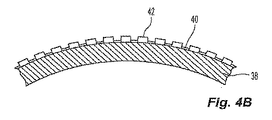
[0031]図4Bを参照すると、基礎バルーン38に一連の第1の繊維42が塗布して繊維層を形成し、第1の接着剤層40によって基礎バルーン38に付着させる。第1の接着剤層40の接着剤の一部は、隣接する繊維42間に形成される空間を部分的に埋めるように動き、最小または無視できるほどの量の接着剤だけが繊維42と基礎バルーン38の外側表面の間に直接残ることが好ましい。接着剤が隣接する繊維42間の空間へ動くことで、接着剤がバルーン壁の壁厚さに寄与するのを防ぎながら、依然として、第1の繊維42を基礎バルーン38に付着させるのに所望の粘着性を提供する。第1の繊維42は、バルーンまたはカテーテルの長手方向軸の方向に配置されることが好ましい。第1の繊維42は、異なるまたは変動する長さにわたってバルーン基礎38の外側表面に沿って延びることがより好ましい。第1の繊維42の繊維の一部は、バルーンの中心区間16のみの長さに沿って延び、第1の繊維42の一部は、中心区間16および円錐形の端部区間18、20を覆うようにバルーンの長さ全体に沿って延びることが最も好ましい。第1の繊維42に対して変動する繊維の長さを使用することで、円錐形の端部区間18、20の繊維をより少なくし、バルーンの直径が円錐形の端部区間18、20の長さに沿って低減するにつれて、第1の繊維42によって形成される繊維層が束またはしわになるのを防止する。
Referring to FIG. 4B, a series of
[0032]任意の強度の高い繊維またはフィラメントを使用して、バルーンに所望の特性を与えることが好ましい。適切な繊維の例には、Spectra(登録商標)もしくはDyneema(登録商標)繊維などの超高分子量ポリエチレン、ポリアミド繊維、ポリイミド繊維、Technora(登録商標)などの超高分子量ポリウレタン繊維、ポリエステルもしくはポリプロピレンから作られた繊維、またはステンレスもしくは高張力鋼などの細く延伸された金属の撚り線が含まれる。第1の繊維42は、約0.0127mm(0.0005インチ)×0.508mm(0.020インチ)の方形プロファイルまで平らにした約12ミクロンのフィラメント直径を有する超高分子量ポリエチレンまたはTechnora(登録商標)繊維であることが好ましい。
[0032] Preferably, any high strength fibers or filaments are used to give the balloon the desired properties. Examples of suitable fibers include ultra high molecular weight polyethylene such as Spectra® or Dyneema® fiber, polyamide fiber, polyimide fiber, ultra high molecular weight polyurethane fiber such as Technora®, polyester or polypropylene. Included are made fibers or strands of finely drawn metal such as stainless steel or high strength steel. The
[0033]図4Cを参照すると、基礎バルーン38、第1の接着剤層40、および第1の繊維42によって形成される複合物の外側に、さらなる接着剤が塗布される。第1の接着剤層40および第1の繊維42の外側に、放射線不透過性材料36をもつ接着剤34を塗布して、中間接着剤層43を形成することが好ましい。中間接着剤層43の接着剤は、噴霧として塗布され、あるいは浴槽内に浸漬することによって、またははけ塗りもしくは他の適切な手段によって堆積されることが好ましい。噴霧によって塗布することがより好ましい。代替手段では、中間層43は、複合物上の接着剤34の配置を制御することによって、または放射線不透過特性をもたず、所望のパターンで複合物の上に配置される別の接着剤を使用することによって、放射線不透過性のパターンを形成するように塗布される。
[0033] Referring to FIG. 4C, additional adhesive is applied to the outside of the composite formed by the
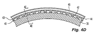
[0034]図4Dを参照すると、中間接着剤層43の上に第2の繊維44が配置され、接着剤34によって下にある第1の繊維42に付着される。第2の繊維44は、好ましくは前述の繊維材料のいずれかから構成され、より好ましくは第1の繊維42と同一の単一の超高分子量ポリエチレンまたはTechnora(登録商標)繊維である。第2の繊維44は、基礎バルーン38の周りに円周方向に巻き付けて、バルーン12の長手方向の長さに沿って螺旋状に延びる円周方向の繊維層を形成することが好ましい。図4D〜4Eを参照すると、好ましくは第1の接着剤層40と同一の第2の接着剤層46は、基礎バルーン38、層40および43、ならびに繊維42および44の複合物の外側に塗布される。これらの繊維は、第2の繊維44の層が第1の繊維42の層の上に配置されるように、層状に配置されることが好ましい。別法として、これらの繊維は、単一の層内に織物または編物の構造を形成し、第1の繊維42は、第1の織物層を形成するように配置され、また第2の繊維44は、それ自体で、または別の繊維とともに織物を形成して第2の織物層を形成するように配置される。別の代替手段では、第1の繊維42と第2の繊維44は、織物または編物の構造をともにつなぎ合わせて、単一の織物または編物層を形成する。
Referring to FIG. 4D, a
[0035]図4D〜4Eには図示しないが、第2の接着剤層46はまた、バルーン12の外側保護フィルム(図示せず)に接合することが好ましい。バルーン表面に対する耐摩耗性を提供するように、そして下にある繊維を保護するように、バルーンの外側表面上に外側保護フィルムを含むことが好ましい。このフィルムは、耐摩耗材料であることが好ましい。耐摩耗材料の例には、ポリエステル、ポリアミド、ポリアミド−ポリエーテルブロック共重合体、ポリウレタン、Surlyn(登録商標)などのイオノマー、ポリエチレン、ポリプロピレン、およびポリウレタンまたはポリエチレンなどの架橋性材料が含まれる。耐摩耗材料として、Pebax(登録商標)などのポリエーテルブロック共重合体が使用されることが好ましい。代替実施形態では、外側保護フィルムは、製造中に熱が加えられるとき、第2の接着剤層46を溶融および溶解させることによって形成される。別の代替手段では、外側保護フィルムは、バルーンに追加の放射線不透過性を与えるために、フィルム内に分散された放射線不透過性材料を含む。
[0035] Although not shown in FIGS. 4D-4E, the second
[0036]別の代替手段では、放射線不透過性接着剤をフィルムに接合する代わりに、または接着剤自体から保護フィルムを形成する代わりに、保護被覆がバルーンに塗布される。耐摩耗性を提供する保護被覆の例には、エポキシ、ポリウレタン、ポリエステル、アルキド樹脂、ポリビニルブチラール、硝酸セルロース、ポリ酢酸ビニル、フェノールホルムアルデヒド樹脂などのフェノール樹脂、およびアミノホルムアルデヒド樹脂などのアミノ樹脂が含まれる。保護被覆は、追加の放射線不透過性をバルーンに与えるために、その中に分散された何らかの放射線不透過性材料を含むことが好ましい。 [0036] In another alternative, a protective coating is applied to the balloon instead of joining the radiopaque adhesive to the film or forming a protective film from the adhesive itself. Examples of protective coatings that provide abrasion resistance include phenolic resins such as epoxies, polyurethanes, polyesters, alkyd resins, polyvinyl butyral, cellulose nitrate, polyvinyl acetate, phenol formaldehyde resins, and amino resins such as amino formaldehyde resins. It is. The protective coating preferably includes any radiopaque material dispersed therein to provide additional radiopacity to the balloon.
[0037]積層された複合構造(基礎バルーン、繊維、接着剤層、および外側保護フィルムまたは被覆からなる)を統合して溶解されたバルーン壁にするために、複合物は、これらの層をともに密接に接合させる条件に露出される。熱および圧力を使用してバルーン12の複合物を型の中で加熱して複合材料を溶解し、統合された構造にすることが好ましい。また、接着剤が熱可塑性ポリウレタンなどの熱可塑性材料である場合、熱を加えることで接着剤を柔らかくして流動化させ、バルーンの複合材料に接合させることが好ましい。また、接着剤が触媒を含有する場合、または硬化するには2つの成分の反応を必要とする2液型材料である場合、熱を加えることで硬化プロセスを加速させる手段を提供することが好ましい。
[0037] To integrate the laminated composite structure (consisting of the base balloon, fiber, adhesive layer, and outer protective film or coating) into a dissolved balloon wall, the composite is used to combine these layers together. Exposed to intimate bonding conditions. Preferably, the composite of
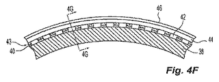
[0038]各接着剤層は、単一の塗布で塗布されることが好ましい。別法として、各接着剤層は、放射線不透過性材料の所望の層厚さまたは所望の性質を実現するために、複数の塗布の複合物として塗布される。接着剤34は、好ましくは2〜100ミクロンの径方向の厚さを有し、より好ましくは3〜50ミクロンの径方向の厚さを有し、また最も好ましくは10〜40ミクロンの径方向の厚さを有する。繊維強化材を有する好ましい実施形態では、中間層43は、第1の繊維42と第2の繊維44の間の径方向の接触を可能にする厚さを有し、その結果、中間層43の接着剤34を隣接する第1の繊維42または第2の繊維44の隣接する巻線間の空間内へ動かしてその空間を占有させ、それによって中間層43は、バルーン壁28内に存在できるが、バルーン壁28の径方向の厚さを増加させない。図4Fおよび4Gは、中間接着剤層43が存在するがバルーン壁28の径方向の厚さを増加させない、それぞれ図4Dおよび4Eに示す実施形態に対する代替実施形態を示す。代替手段では、放射線不透過特性または放射線不透過性でない特性を有する接着剤層の一部は、積層プロセス中に柔らかくなって流動する材料からなる。
[0038] Each adhesive layer is preferably applied in a single application. Alternatively, each adhesive layer is applied as a composite of multiple applications to achieve the desired layer thickness or desired properties of the radiopaque material.
[0039]放射線不透過性バルーンの他の実施形態も同様に構築されるが、強化繊維をもたない。この代替実施形態では、バルーンは、基礎バルーンと、基礎バルーンの外側表面上の放射線不透過性接着剤層と、放射線不透過性接着剤の外側表面を覆うフィルムまたは被覆などの最終の保護層とを有する。放射線不透過性接着剤は、バルーンに放射線不透過性を与え、また基礎バルーンを保護外層に接合させる。 [0039] Other embodiments of radiopaque balloons are similarly constructed but do not have reinforcing fibers. In this alternative embodiment, the balloon comprises a base balloon, a radiopaque adhesive layer on the outer surface of the base balloon, and a final protective layer such as a film or coating covering the outer surface of the radiopaque adhesive. Have The radiopaque adhesive imparts radiopacity to the balloon and bonds the base balloon to the protective outer layer.
[0040]別法として、接着剤34はパターンで塗布される。パターンは、中間接着剤層43を塗布するときに接着剤34を選択的に塗布することによって、たとえば、中間接着剤層43を塗布する前に狭いPTFEテープを使用して第1の繊維42を覆い、バルーン複合物の領域をマスキングすることによって作られることが好ましい。次いで、中間接着剤層43を塗布した後、PTFEテープを除去して、接着剤34をもたない領域を露出させる。次いで、放射線不透過性材料36をもたない放射線不透過性でない接着剤層を塗布して、バルーン複合物全体を被覆し、それによって第1の繊維42および中間接着剤層43の上に追加の放射線不透過性でない接着剤層を配置し、PTFEテープによって覆われていた領域を埋める。
[0040] Alternatively, the adhesive 34 is applied in a pattern. The pattern can be obtained by selectively applying adhesive 34 when applying intermediate adhesive layer 43, for example, using a narrow PTFE tape to apply
[0041]別法として、図4A〜4Eおよび図4F〜4Gに示す製造プロセスは、基礎バルーン38の代わりに金型または心金を使用することで実現される。バルーン12を積層した後、次に金型または心金を除去して基礎バルーン38をもたないバルーン壁を残し、第1の接着剤層40および第1の繊維42にバルーン12の内部表面を形成させる。
[0041] Alternatively, the manufacturing process shown in FIGS. 4A-4E and 4F-4G is accomplished by using a mold or mandrel instead of the
[0042]別の代替手段では、バルーン12は、バルーン12上の特有の位置を識別する放射線不透過性マーカを形成するために、バルーン端部15などの所定の位置で、好ましくは第1の繊維42と第2の繊維44の間にマーカストリップ、マーカフィラメント、またはマーカリングなどのマーカ材料層を含む。放射線不透過性マーカは、バルーン壁28の残り部分の放射線不透過性とは異なる放射線不透過性を有することが好ましい。マーカストリップ、フィラメント、またはリングは、放射線不透過性、可撓性、可鍛性、および加工性の好ましい特性を呈する材料から作られることが好ましい。マーカとして使用するのに適切な材料には、タンタル、スズ、銀、金、白金、レニウム、イリジウム、パラジウム、ハフニウム、タングステン、ランタン、およびX線を吸収する他の金属が含まれる。好ましい材料には、銀およびスズが含まれる。
[0042] In another alternative, the
[0043]収縮したバルーン12は、折り畳んでバルーン12自体に円周方向に巻き付けて、バルーン12に対するプロファイルを低減することが好ましい。巻き付けられたバルーン12は、カテーテルチューブ14の外径に類似またはほぼ一致する外径を有するプロファイルを呈することが好ましい。図5A〜5Bは、例示的な折り畳まれたバルーンを示す。図6A〜6Bを参照すると、巻き付けられたバルーン12の外側の一部分は、医療用デバイス48をしぼんだ状態で保持するように形成される。医療用デバイス48は、搬送径まで圧縮されまたはしぼんだステントであることが好ましい。バルーン12を膨張させることで、医療用デバイス48の内部に拡大する力を加えて、より大きい直径まで拡大させることが好ましい。医療用デバイス48が拡大した後、安定した拡大構成を維持するように設計される場合、バルーン12は、収縮されて医療用デバイス48の内部から撤退され、それによって医療用デバイス48から分離されることが好ましい。
[0043] The deflated
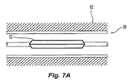
[0044]図7Aを参照すると、収縮したバルーン12は、管52内に挿入されて、当該領域50に対して位置決めされることが好ましい。図7Bを参照すると、位置決めした後、バルーン12を膨張させて、バルーンの外側表面を当該領域50内で管52の壁に接触させて押し付けることが好ましい。収縮したバルーン12上に医療用デバイス48が取り付けられた代替実施形態では、バルーン12を膨張させることで医療用デバイス48を拡大させて、医療用デバイスの外側を管壁に押し付け、治療効果を実現する。バルーン12は、管腔を通ってバルーンの内部へ搬送される膨張流体で膨張されることが好ましい。
Referring to FIG. 7A, the deflated
[0045]バルーン壁が製造されるとき、バルーン12に対する総放射線透過量を規定するバルーン壁28内の放射線不透過性材料36の量は固定される。図7Aおよび7Bを参照すると、バルーン12内で放射線不透過性材料の量を一定にすることで、折り畳まれたバルーン12内ではバルーン12の接着剤34内の放射線不透過性材料36がともにぎっしり詰まるため、バルーンが完全にまたは部分的に折り畳まれ、収縮し、空になり、しぼみ、かつ/または塩水などの膨張流体でごくわずかしか充填されないとき、放射線透過密度は、比較的強度の高い平均放射線不透過画像(膨張したバルーンの放射線不透過画像と比較)を呈する。収縮したバルーン12はまた、膨張したバルーンと比較すると、比較的大きい放射線不透過密度を有する。また、膨張プロセス中にバルーン壁28内の放射線不透過性材料36が、カテーテルの長手方向軸からの径方向の距離がより大きいところへ離れて相対的なより明るい放射線不透過画像をもたらすため、バルーン12の画像の中心部分は、バルーンが膨張流体で完全に膨張されたとき、比較的はっきりしない放射線不透過画像を呈する放射線透過画像強度を提供する。また、壁の画像が傾斜した角度で得られ、撮像放射が壁の表面に対してほぼ平行の方向に壁を通過または反射し、それによって放射が壁を直角に通過するときと比較すると、放射は追加の放射線不透過性材料の影響を受けるため、バルーンの画像内のバルーンの縁部は、膨張中およびその後、比較的強度の高い画像を維持する。また、バルーンの膨張に起因してバルーン体積が増大するため、全体として膨張したバルーン12の放射線透過画像強度は、バルーンが膨張するにつれて比較的低減される。また全体として、バルーン12の放射線不透過画像は、バルーン12がバルーン12の収縮した状態と膨張した状態で提供される放射線不透過性の両極端間を進むにつれて変動し、収縮したバルーンではより強度の高い放射線不透過画像が提供され、膨張したバルーンではより強度の低い放射線不透過画像が提供される。さらに、バルーンの画像の中心は、収縮したときは比較的強度の高い画像を呈し、膨張したときは比較的強度の低い画像を呈し、バルーンの画像の縁部は、比較的一定の放射線不透過画像を呈する。
[0045] When the balloon wall is manufactured, the amount of
[0046]したがって、バルーン12の放射線透過密度は、バルーン12が収縮した状態と膨張した状態の間を遷移するときに変化し、これは直接、バルーン壁28内の固定の量の放射線不透過性材料36に対する任意の点のバルーンの体積に対応する。具体的には、放射線不透過性のバルーン12の中心の画像は、放射線写真では、バルーンが膨張したときにより明るくなり、一方放射線不透過性でない従来のバルーンは、放射線不透過性の膨張流体が存在するため、放射線写真では、バルーン画像の中心でより暗くなる。
[0046] Accordingly, the radiation transmission density of the
[0047]バルーン12全体の放射線透過密度は、放射線不透過性材料36の固定の量とバルーン12の体積全体を比較することによって決定される。バルーン壁28内の放射線不透過性材料の総量が一定であるため、バルーン体積の変化により、放射線透過密度が変化する。図7Aを参照すると、折り畳まれたバルーン12の比較的小さい体積内に固定の体積の放射線不透過性材料36が含有されるため、折り畳まれたバルーン12の放射線透過密度は比較的高い。図7Bを参照すると、膨張したバルーン12の比較的大きい体積内に固定の体積の放射線不透過性材料36が含有されるため、膨張したバルーン12の放射線透過密度は比較的低い。完全に収縮して折り畳まれたバルーンと、完全に膨張したバルーンの間の放射線透過密度の平均的な変化は、これらの2つの状態のバルーンの直径の変化に比例する。
[0047] The radiation transmission density of the
[0048]典型的な医療手順中、バルーンは通常、蛍光透視鏡上で、バルーンの主軸に対して垂直な位置から撮像されると考えられる。この観点から考えると、バルーン壁内の放射線不透過性材料の分布により、均一でない画像が提供されると有利である。放射線不透過性のバルーン12は、バルーン画像の縁部およびそのすぐ近くでは材料の放射線不透過性がより高く、バルーンの中心領域ではより低いように見える画像を提供する。したがって、バルーンが膨張したとき、バルーンの中心領域の画像強度は、さらにいっそう減少する。以下の表1は、典型的な構造の様々な寸法のバルーンに対する平均放射線透過密度の変化、ならびにバルーンの中心領域で発生する変化を示す。
[0048] During a typical medical procedure, the balloon is usually considered to be imaged on a fluoroscope from a position perpendicular to the main axis of the balloon. From this point of view, it is advantageous if the distribution of radiopaque material within the balloon wall provides a non-uniform image. The
[0049]完全に収縮したバルーンと完全に膨張したバルーンの間の総放射線透過画像強度の低減は、好ましくは35〜95%の範囲であり、より好ましくは60〜90%の範囲である。 [0049] The reduction in total radiographic image intensity between a fully deflated balloon and a fully inflated balloon is preferably in the range of 35-95%, more preferably in the range of 60-90%.
[0050]蛍光透視鏡下で見たバルーン内の放射線不透過性接着剤の分布は、放射線不透過性の造影剤で充填された放射線不透過性でないバルーンと比較すると、本発明の重要な特徴である。図8および9は、この効果を示す。撮像に使用されるX線が、バルーンの長手方向軸に対して大まかに垂直の方向にバルーンを通過するため、X線ビームが遭遇する放射線不透過性接着剤の量は、バルーン画像の縁部内に配置されたバルーン画像の他の領域と比較すると、バルーンの縁部およびそのすぐ近くで著しく大きくなる。こうしてバルーン画像の縁部でX線との相互作用がより大きくなることで、バルーンの画像では、画像内の縁部がはっきりする。バルーン画像中心と比較したバルーン画像縁部における放射線透過密度の増大は、膨張したバルーンの直径および放射線不透過性接着剤層の厚さに応じる。表2は、いくつかのバルーン寸法および放射線不透過性接着剤厚さに対して、撮像されたバルーンの中心と比較した、撮像されたバルーンの縁部によって提示される放射線透過画像強度の違いを示す。撮像されたバルーン縁部における放射線透過画像強度の増大は、撮像されたバルーン中心と比較すると、560%から2000%を超える範囲である。 [0050] The distribution of radiopaque adhesive within the balloon as viewed under a fluoroscope is an important feature of the present invention compared to a non-radiopaque balloon filled with a radiopaque contrast agent. It is. Figures 8 and 9 illustrate this effect. Because the x-rays used for imaging pass through the balloon in a direction generally perpendicular to the longitudinal axis of the balloon, the amount of radiopaque adhesive encountered by the x-ray beam is within the edge of the balloon image. Compared to other regions of the balloon image placed at, it is significantly larger at and near the edge of the balloon. Thus, the interaction with X-rays becomes larger at the edge of the balloon image, so that the edge in the image becomes clear in the balloon image. The increase in radiation transmission density at the balloon image edge compared to the balloon image center depends on the diameter of the inflated balloon and the thickness of the radiopaque adhesive layer. Table 2 shows the difference in radiographic image intensity presented by the edge of the imaged balloon compared to the center of the imaged balloon for several balloon dimensions and radiopaque adhesive thickness. Show. The increase in radiation transmission image intensity at the imaged balloon edge ranges from 560% to over 2000% compared to the imaged balloon center.
[0051]放射線不透過性接着剤をもつバルーンの撮像されたバルーン縁部の強度は、放射線不透過性の造影剤で充填された従来の放射線不透過性でないバルーンによって生成される撮像されたバルーン縁部の強度と同等である。図8A〜9Bは、この比較で観察される放射線透過画像強度の分布を示す。X線が放射線不透過性接着剤をもつバルーンの側へ向けていることを示す図8A、およびX線撮像によって提供される画像強度を表す図8Bに示すように、バルーン画像強度は、バルーンの撮像された縁部で最も高くなる。それと比較して、X線が放射線不透過性の造影剤で充填された従来のバルーンの側へ向けられていることを示す図9A、およびX線撮像によって提供される画像強度を表す図9Bでは、バルーン画像強度は、バルーンの縁部には存在せず、造影剤によって提示される形の撮像された縁部近傍ではごくわずかの高さしかない。したがって、造影剤を使用する従来のバルーンでは、X線がバルーン自体を撮像しないため、そして造影剤によって輪郭を引き立たせた形の縁部付近で撮像される造影剤の厚さが、撮像されたバルーンの中心で提示される厚さと比較すると最小または無視できるほどしかないため、撮像されたバルーン縁部が曖昧でかつはっきりしないと考えられる。 [0051] The strength of the imaged balloon edge of a balloon with radiopaque adhesive is generated by a conventional non-radiopaque balloon filled with a radiopaque contrast agent It is equivalent to the strength of the edge. 8A-9B show the distribution of radiation transmission image intensity observed in this comparison. As shown in FIG. 8A, which shows that X-rays are directed to the side of the balloon with radiopaque adhesive, and FIG. 8B, which shows the image intensity provided by X-ray imaging, the balloon image intensity is Highest at the imaged edge. In comparison, FIG. 9A shows that X-rays are directed to the side of a conventional balloon filled with radiopaque contrast agent, and FIG. 9B shows the image intensity provided by X-ray imaging. The balloon image intensity is not present at the edge of the balloon, but only slightly above the imaged edge in the form presented by the contrast agent. Therefore, in a conventional balloon using a contrast agent, X-rays do not image the balloon itself, and the thickness of the contrast agent that is imaged near the edge of the contoured shape is imaged. The imaged balloon edge is considered ambiguous and unclear because it is minimal or negligible compared to the thickness presented at the center of the balloon.
[0052]図7Aおよび7Bを参照すると、バルーン壁28自体(あらゆる膨張流体またはバルーンの体積を無視)は、バルーン12の膨張状態に対して変化しない、すべての膨張状態でバルーン12の画像を提供する一定のバルーン壁放射線透過密度を有する。収縮したバルーン12が折り畳まれたとき、バルーン壁28は、バルーン壁28内の放射線不透過性材料36の密度が変化していないため、バルーンが膨張したときと同じ総放射線不透過性を呈する。図7Aに示すように、折り畳まれたバルーン壁28の提示された放射線不透過画像は、バルーン壁28の折り畳まれた部分の付加的な放射線不透過性であり、したがって、折り畳まれたバルーンの総放射線不透過性は、バルーン壁の各折畳みの放射線不透過性の寄与の関数または係数である。
[0052] Referring to FIGS. 7A and 7B, the
[0053]バルーン12の膨張は、カテーテルチューブ14を介してバルーン12の内部へ膨張流体を供給することによって実現されることが好ましい。膨張流体は、生理的食塩溶液と放射線不透過性の造影剤の混合物、または純粋な生理的食塩溶液であることが好ましい。利用可能な造影剤には、アセトリゾエート(Diaginol、Urokon)、ジアトリゾエート(Angiographin、Renografin、Urovison)、イオダミド(Uromiro)、イオグリケート(Rayvist)、イオタラメート(Conray)、イオキシタラメート(Telebrix)、イオトロラン(Isovist)、イオジキサノール(Visipaque)、イオヘキソール(Omnipaque)、イオペントール(Imagopaque)、およびイオベルソール(Optiray)を含めて、単量体または二量体構造のヨウ素化合物が含まれる。別のタイプの造影剤には、Dotarem、Omniscan、Eovist、Prohance、およびMultihance製品などの市販の製品内に提供されるガドリニウム、ホルミウム、マンガン、またはジスプロシウムなどの希土類または他の重金属種のキレートが含まれる。膨張流体は、放射線不透過性流体の濃度が50%未満になるように準備されることが好ましい。膨張流体の放射線不透過性流体の濃度は、好ましくは0%(純粋な塩水溶液)〜約40%であり、さらにより好ましくは約0〜20%の範囲内であり、さらにより好ましくは約0〜5%の範囲内であり、最も好ましくは0%の濃度である。
[0053] Inflation of the
[0054]通常、放射線不透過性流体は、純粋な生理的食塩水の粘性より大きい粘性を有すると考えられる。同様に、塩水と放射線不透過性流体の混合物は、希釈していない放射線不透過性流体より小さいが純粋な塩水の粘性よりやはり大きい粘性を有すると考えられる。放射線不透過性流体および塩水/放射線不透過性流体の混合物の粘性をより大きくすると、そのような流体は、所与の圧力で、同じ条件下で純粋な塩水で観察される動きよりゆっくりと管材内を流れることも考えられる。したがって、純粋な塩水と比較して放射線不透過性流体の粘性がより大きいことで、管材内で放射線不透過性流体を押し流すには、より大きいヘッド圧力を必要とし、また同じ条件下で、塩水で実現されるバルーン膨張時間を実現するには、より大きいヘッド圧力を必要とする。したがって、放射線不透過性流体の粘性が比較的高いことで、バルーン12は、純粋な塩水で膨張されるバルーンと比較すると、よりゆっくりと充填される。この効果は、バルーン収縮とともにさらにはっきりする。これは、膨張とは異なり、収縮中は、バルーン内の流体に高い圧力をかけてカテーテルから流し出すことができないためである。流体を流し出すのに利用可能な最大圧力は、利用可能な環境大気圧(14.7psiまたは1気圧)に依存する真空に制限される。したがって、バルーンの収縮には、カテーテル構造およびバルーン寸法に応じて相当な時間がかかる可能性がある。これらの要因はすべて、従来のバルーンおよび放射線不透過性の撮像の使用を伴う医療手順を完了するのに必要な時間および/または労力を増大させ、そしてバルーン膨張または収縮を実現するのに必要な時間を増大させると考えられる。
[0054] Usually, the radiopaque fluid is considered to have a viscosity greater than that of pure saline. Similarly, a mixture of brine and radiopaque fluid is believed to have a viscosity that is less than that of undiluted radiopaque fluid, but still greater than that of pure brine. Increasing the viscosity of the radiopaque fluid and the saltwater / radiopaque fluid mixture causes the fluid to be more slowly piped at a given pressure than the movement observed in pure saline under the same conditions. It is possible to flow inside. Therefore, the greater viscosity of the radiopaque fluid compared to pure brine requires a larger head pressure to flush the radiopaque fluid through the tubing and under the same conditions, A higher head pressure is required to achieve the balloon inflation time achieved with. Thus, the relatively high viscosity of the radiopaque fluid causes the
[0055]膨張流体の密度(塩水中の放射線不透過性流体の濃度)とバルーン収縮時間の関係を表3に示す。 [0055] The relationship between inflation fluid density (concentration of radiopaque fluid in salt water) and balloon deflation time is shown in Table 3.
[0056]したがって、例示的な放射線不透過性のバルーンは、放射線不透過性のバルーン壁をもたない既存のバルーンに勝る利点を提供する。例示的なバルーンは、従来のバルーンで使用されるより粘性の低い流体で膨張されながら、撮像システムで画像を生成するため、より速い膨張および収縮時間を提供する。また、例示的なバルーンは、放射線不透過性の低いまたは放射線不透過性のない流体を使用するバルーンを提供し、したがってバルーンを膨張させて撮像するより簡単でかつ安価な方法を提供する。膨張溶液が純粋な塩水溶液であるとき、溶液を混合する時間および費用は、バルーン膨張および収縮プロセスから完全に排除される。 [0056] Thus, exemplary radiopaque balloons offer advantages over existing balloons that do not have radiopaque balloon walls. Exemplary balloons provide faster inflation and deflation times to produce images with the imaging system while being inflated with the less viscous fluid used in conventional balloons. The exemplary balloon also provides a balloon that uses a fluid that is less radiopaque or non-radiopaque, thus providing a simpler and less expensive method of inflating and imaging the balloon. When the inflation solution is a pure salt solution, the time and expense of mixing the solution is completely eliminated from the balloon inflation and deflation process.
[0057]バルーン壁内に放射線不透過性接着剤を含有するバルーンを用いて、収縮時間を低減させ、バルーンを容易に収縮できるようにすることで、医療手順中に起こりうる潜在的に深刻な問題も回避する。造影剤を含有する粘性の流体では、バルーンが収縮したように見えるが、見掛けは収縮したバルーン内に著しい量の造影剤を残す可能性がより高いと考えられる。その後カテーテルを動かして、見掛けは収縮したバルーンを導入器チューブ内に移すことによって、患者からカテーテルの抜取りを開始するとき、見掛けは収縮したバルーン内に残っている造影剤をバルーンの遠位端部の方へ押しやってバルーンの最遠位端部を膨張させる可能性があり、これによりバルーンが導入器チューブ内へ完全に撤退するのを阻止する。この状態は、バルーン内に閉じ込められた造影剤のボーラスを押すにつれて、バルーンのさらなる収縮を可能にする膨張/収縮の通し穴が、構築する導入器チューブによりバルーンにかかる圧力によって閉じられる可能性があるため、さらに悪化する。理解できるように、そのような状況の結果、患者にとって有害な健康上のリスクを生じさせ、バルーンを破裂させて造影剤を放出し、また医療手順の長さおよび複雑さを増大させる可能性がある。 [0057] Using a balloon containing a radiopaque adhesive within the balloon wall to reduce deflation time and allow the balloon to be easily deflated, potentially potentially serious during medical procedures Avoid problems. For viscous fluids containing contrast agent, the balloon appears to be deflated, but the appearance is more likely to leave a significant amount of contrast agent in the deflated balloon. The catheter is then moved to move the apparently deflated balloon into the introducer tube, thereby initiating the contrast agent remaining in the deflated balloon at the distal end of the balloon. May be inflated toward the distal end of the balloon, thereby preventing the balloon from withdrawing completely into the introducer tube. This condition is that as the contrast bolus confined within the balloon is pushed, the inflation / deflation through-hole that allows further deflation of the balloon can be closed by the pressure applied to the balloon by the introducer tube being constructed. Because it is, it gets worse. As can be appreciated, such a situation can result in a health risk that is detrimental to the patient, ruptures the balloon and releases the contrast agent, and increases the length and complexity of the medical procedure. is there.
[0058]バルーン壁内に放射線不透過性接着剤をもつバルーンの追加の実施例を以下に提供する。
[0059]
[0058] Additional examples of balloons having radiopaque adhesive within the balloon wall are provided below.
[0059]
[0060]ガラス混合容器内に以下の成分を追加することによって、放射線不透過性接着剤が準備された。
1)溶剤中に約8.5重量%のポリウレタンを有するTecoflex(登録商標)1−MP接着剤として入手可能なポリウレタン積層接着剤54グラム、
2)公称粒子寸法0.5ミクロンのタングステン粉末24.5グラム、および
3)メチルエチルケトン(MEK)36.6グラム。
[0060] A radiopaque adhesive was prepared by adding the following components into a glass mixing vessel.
1) 54 grams of polyurethane laminating adhesive available as Tecoflex® 1-MP adhesive with about 8.5% by weight polyurethane in solvent,
2) 24.5 grams of tungsten powder with a nominal particle size of 0.5 microns, and 3) 36.6 grams of methyl ethyl ketone (MEK).
これらの成分を混合して、約25重量%の固体の均一の組成をもつ接着剤を作った。
[0061]直径12mmで、約0.0508mm(0.002インチ)の2倍の壁厚さをもつポリエチレンテレフタレート(PET)血管形成バルーンを心金に取り付けて、バルーンを膨張させた。膨張したバルーンに放射線不透過性接着剤を噴霧して、バルーンの表面を覆って均一の量の接着剤を配置した。接着剤は、バルーンの表面上で急速に乾燥させた。乾燥した接着剤は、約26体積%のタングステンおよび74体積%のポリウレタンを含有した。
These ingredients were mixed to make an adhesive with a uniform composition of about 25% by weight solids.
[0061] A polyethylene terephthalate (PET) angioplasty balloon having a diameter of 12 mm and a wall thickness of about 0.0508 mm (0.002 inches) was attached to the mandrel and the balloon was inflated. The inflated balloon was sprayed with a radiopaque adhesive to place a uniform amount of adhesive over the surface of the balloon. The adhesive was quickly dried on the surface of the balloon. The dried adhesive contained about 26% by volume tungsten and 74% by volume polyurethane.
[0062]次いでバルーンには、Pebax(登録商標)として市販のポリエーテル−ポリアミド共重合体フィルムの薄片を螺旋状に巻き付けた。巻き付ける間に、約0.0127mm(0.0005インチ)のフィルム厚さを延ばして、厚さをさらに低減させた。巻き付けた後、ある寸法および形状の積層型内にバルーンを配置して、熱および圧力をバルーン表面に加えた。バルーンは、約摂氏104.4度(華氏220度)の温度まで加熱され、バルーンの表面に加えられた圧力により、放射線不透過性の積層接着剤が流動し、バルーンとPebax(登録商標)フィルムを統合させた。 [0062] The balloon was then spirally wrapped with a thin piece of a polyether-polyamide copolymer film commercially available as Pebax®. During winding, the film thickness was increased by about 0.0127 mm (0.0005 inch) to further reduce the thickness. After wrapping, the balloon was placed in a mold of a certain size and shape, and heat and pressure were applied to the balloon surface. The balloon is heated to a temperature of about 104.4 degrees Celsius (220 degrees Fahrenheit) and the pressure applied to the surface of the balloon causes the radiopaque laminating adhesive to flow and the balloon and Pebax® film. Integrated.
[0063]その結果、放射線不透過性の血管形成バルーンでは、壁厚さが0.1143mm(0.0045インチ)と2倍になった。これらのバルーンは、X線撮像によって試験され、造影剤で充填する必要なく、優れた視認性を示した。同じ寸法の従来のPETバルーンの制御では、同じX線撮像下で可視画像を示さなかった。 [0063] As a result, the radiopaque angioplasty balloon doubled the wall thickness to 0.1143 mm (0.0045 inches). These balloons were tested by X-ray imaging and showed excellent visibility without having to be filled with contrast media. Control of a conventional PET balloon of the same dimensions did not show a visible image under the same x-ray imaging.
[0064] [0064]
[0065]ガラス混合コンテナ内に以下の成分を追加することによって、放射線不透過性の積層接着剤が準備された。
1)Tecoflex(登録商標)1−MP接着剤として入手可能なポリウレタン積層接着剤61グラム、
2)三酸化ビスマス粉末14.6グラム、
3)MEK24.4グラム、および
4)アセトン15グラム。
[0065] A radiopaque laminating adhesive was prepared by adding the following components into a glass mixing container.
1) 61 grams of polyurethane laminate adhesive available as Tecoflex® 1-MP adhesive,
2) 14.6 grams of bismuth trioxide powder,
3) 24.4 grams of MEK, and 4) 15 grams of acetone.
これらの成分を混合して、約17重量%の固体の均一の組成をもつ接着剤を作った。
[0066]実施例1に記載のように、直径12mmで、約0.0508mm(0.002インチ)の2倍の壁厚さをもつポリエチレンテレフタレート(PET)血管形成バルーンを取り付け、接着剤を噴霧して乾燥させた。乾燥した接着剤は、約26体積%の三酸化ビスマスおよび74体積%のポリウレタンを含有した。次いで実施例1に記載のように、バルーンにはPebax(登録商標)フィルムを螺旋状に巻き付け、熱および圧力下で積層させて、統合された積層バルーンを作った。
These ingredients were mixed to make an adhesive with a uniform composition of about 17% by weight solids.
[0066] As described in Example 1, a polyethylene terephthalate (PET) angioplasty balloon having a diameter of 12 mm and a wall thickness of about 0.0508 mm (0.002 inches) was attached and sprayed with adhesive. And dried. The dried adhesive contained about 26% by volume bismuth trioxide and 74% by volume polyurethane. The balloon was then spirally wrapped with Pebax® film and laminated under heat and pressure as described in Example 1 to create an integrated laminated balloon.
[0067]その結果、放射線不透過性の血管形成バルーンでは、壁厚さが0.1168mm(0.0046インチ)と2倍になった。これらのバルーンは、X線撮像によって試験され、造影剤で充填する必要なく、優れた視認性を示した。 [0067] As a result, the radiopaque angioplasty balloon doubled the wall thickness to 0.1168 mm (0.0046 inches). These balloons were tested by X-ray imaging and showed excellent visibility without having to be filled with contrast media.
[0068] [0068]
[0069]プラスチック混合コンテナ内に以下の成分を追加することによって、放射線不透過性の積層接着剤が準備された。
1)Tecoflex(登録商標)1−MP接着剤として入手可能なポリウレタン積層接着剤297グラム、
2)三酸化ビスマス粉末146グラム、
3)MEK119グラム、および
4)アセトン238グラム。
[0069] A radiopaque laminating adhesive was prepared by adding the following components into a plastic mixing container.
1) 297 grams of polyurethane laminate adhesive available as Tecoflex® 1-MP adhesive,
2) 146 grams of bismuth trioxide powder,
3) 119 grams of MEK, and 4) 238 grams of acetone.
これらの成分をともに簡単に混合し、次いで酸化アルミニウムセラミックボールを充填した実験用ボールミルジャー内に充填した。次いで、ジャーをボールミルローラ上で24時間転がして、三酸化ビスマスの粒子寸法を低減させ、その後、混合物をボールミルから取り出してガラスコンテナ内に貯蔵した。その結果、約18重量%の固体の均一の組成をもつ接着剤が得られた。 These ingredients were mixed together briefly and then filled into a laboratory ball mill jar filled with aluminum oxide ceramic balls. The jar was then rolled on a ball mill roller for 24 hours to reduce the particle size of bismuth trioxide, after which the mixture was removed from the ball mill and stored in a glass container. As a result, an adhesive having a uniform composition of about 18% by weight of solid was obtained.
[0070]実施例1に記載のように、直径12mmで、約0.0508mm(0.002インチ)の2倍の壁厚さをもつポリエチレンテレフタレート(PET)血管形成バルーンを取り付け、接着剤の薄い被覆を噴霧して乾燥させた。乾燥した接着剤は、約43体積%の三酸化ビスマスおよび57体積%のポリウレタンを含有した。次いで実施例1に記載のように、バルーンにはPebax(登録商標)フィルムを螺旋状に巻き付け、熱および圧力下で積層させて、統合された積層バルーンを作った。 [0070] As described in Example 1, a polyethylene terephthalate (PET) angioplasty balloon having a diameter of 12 mm and a wall thickness twice as large as about 0.0508 mm (0.002 inches) was attached and the adhesive was thin The coating was sprayed to dry. The dried adhesive contained about 43% by volume bismuth trioxide and 57% by volume polyurethane. The balloon was then spirally wrapped with Pebax® film and laminated under heat and pressure as described in Example 1 to create an integrated laminated balloon.
[0071]その結果、放射線不透過性の血管形成バルーンでは、壁厚さが0.1651mm(0.0065インチ)と2倍になった。これらのバルーンは、X線撮像によって試験され、造影剤で充填する必要なく、優れた視認性を示した。積層接着剤内の三酸化ビスマスの濃度がより高いため、また接着剤層がより厚いため、これらのバルーンに対する画像は、実施例2で準備したバルーンの場合より強度が高かった。 [0071] As a result, the wall thickness of the radiopaque angioplasty balloon was doubled to 0.1651 mm (0.0065 inches). These balloons were tested by X-ray imaging and showed excellent visibility without having to be filled with contrast media. Due to the higher concentration of bismuth trioxide in the laminating adhesive and the thicker adhesive layer, the images for these balloons were stronger than the balloon prepared in Example 2.
[0072] [0072]
[0073]ガラス混合コンテナ内に以下の成分を追加することによって、放射線不透過性の積層接着剤が準備された。
1)Tecoflex(登録商標)1−MP接着剤として入手可能なポリウレタン積層接着剤78グラム、
2)粒子寸法がミクロン以下のタングステン粉末78.2グラム、
3)MEK31.3グラム、および
4)アセトン62.5グラム。
[0073] A radiopaque laminating adhesive was prepared by adding the following components in a glass mixing container.
1) 78 grams of polyurethane laminate adhesive available as Tecoflex® 1-MP adhesive,
2) 78.2 grams of tungsten powder with particle size below micron,
3) 31.3 grams of MEK, and 4) 62.5 grams of acetone.
これらの成分を完全に混合して、約25.4重量%の固体の均一の組成を有する接着剤を作った。
[0074]実施例1に記載のように、直径12mmで、約0.0508mm(0.002インチ)の2倍の壁厚さをもつポリエチレンテレフタレート(PET)血管形成バルーンを取り付け、接着剤を噴霧して乾燥させた。乾燥した接着剤は、約42体積%のタングステンおよび58体積%のポリウレタンを含有した。次いで実施例1に記載のように、バルーンにはPebax(登録商標)フィルムを螺旋状に巻き付け、熱および圧力下で積層させて、統合された積層バルーンを作った。
These ingredients were thoroughly mixed to produce an adhesive having a uniform composition of about 25.4% by weight solids.
[0074] As described in Example 1, a polyethylene terephthalate (PET) angioplasty balloon having a diameter of 12 mm and a wall thickness twice as large as about 0.0508 mm (0.002 inches) was attached and the adhesive was sprayed. And dried. The dried adhesive contained about 42% by volume tungsten and 58% by volume polyurethane. The balloon was then spirally wrapped with Pebax® film and laminated under heat and pressure as described in Example 1 to create an integrated laminated balloon.
[0075]その結果、放射線不透過性の血管形成バルーンでは、壁厚さが0.1524mm(0.006インチ)と2倍になった。これらのバルーンは、X線撮像によって試験され、造影剤で充填する必要なく、優れた視認性を示した。実施例1と比較すると、積層接着剤内のタングステンの濃度がより高いため、また接着剤層がより厚いため、これらのバルーンに対する画像は、実施例1で準備したバルーンの場合より強度が高かった。 [0075] As a result, the radiopaque angioplasty balloon doubled the wall thickness to 0.1524 mm (0.006 inches). These balloons were tested by X-ray imaging and showed excellent visibility without having to be filled with contrast media. Compared to Example 1, because of the higher concentration of tungsten in the laminate adhesive and the thicker adhesive layer, the images for these balloons were stronger than the balloons prepared in Example 1. .
[0076] [0076]
[0077]プラスチック混合コンテナ内に以下の成分を追加することによって、放射線不透過性の積層接着剤が準備された。
1)Tecoflex(登録商標)1−MP接着剤として入手可能なポリウレタン積層接着剤308グラム、
2)公称粒子寸法5ミクロンの酸化セリウム粉末123グラム、
3)MEK123グラム、および
4)アセトン246グラム。
[0077] A radiopaque laminating adhesive was prepared by adding the following components into a plastic mixing container.
1) 308 grams of polyurethane laminate adhesive available as Tecoflex® 1-MP adhesive
2) 123 grams of cerium oxide powder with a nominal particle size of 5 microns,
3) 123 grams of MEK, and 4) 246 grams of acetone.
これらの成分をともに簡単に混合し、次いで酸化アルミニウムセラミックボールを充填した実験用ボールミルジャー内に充填した。次いで、ジャーをボールミルローラ上で24時間転がして、酸化セリウムの粒子寸法を低減させ、その後、混合物をボールミルから取り出してガラスコンテナ内に貯蔵した。その結果、約19重量%の固体の均一の組成をもつ接着剤が得られた。 These ingredients were mixed together briefly and then filled into a laboratory ball mill jar filled with aluminum oxide ceramic balls. The jar was then rolled on a ball mill roller for 24 hours to reduce the cerium oxide particle size, after which the mixture was removed from the ball mill and stored in a glass container. As a result, an adhesive having a uniform composition of about 19% by weight of solid was obtained.
[0078]実施例1に記載のように、直径12mmで、約0.0508mm(0.002インチ)の2倍の壁厚さをもつポリエチレンテレフタレート(PET)血管形成バルーンを取り付け、接着剤を噴霧して乾燥させた。乾燥した接着剤は、約43体積%の酸化セリウムおよび57体積%のポリウレタンを含有した。次いで実施例1に記載のように、バルーンにはPebax(登録商標)フィルムを螺旋状に巻き付け、熱および圧力下で積層させて、統合された積層バルーンを作った。 [0078] As described in Example 1, a polyethylene terephthalate (PET) angioplasty balloon with a diameter of 12 mm and a wall thickness of about 0.0508 mm (0.002 inches) was attached and the adhesive was sprayed. And dried. The dried adhesive contained about 43% by volume cerium oxide and 57% by volume polyurethane. The balloon was then spirally wrapped with Pebax® film and laminated under heat and pressure as described in Example 1 to create an integrated laminated balloon.
[0079]その結果、放射線不透過性の血管形成バルーンでは、壁厚さが約0.1575mm(0.0062インチ)と2倍になった。これらのバルーンは、X線撮像によって試験され、造影剤で充填する必要なく、優れた視認性を示した。 [0079] As a result, the radiopaque angioplasty balloon doubled the wall thickness to approximately 0.1575 mm (0.0062 inches). These balloons were tested by X-ray imaging and showed excellent visibility without having to be filled with contrast media.
[0080] [0080]
[0081]実施例5に記載のように、放射線不透過性の積層接着剤が準備された。直径12mmで、約0.0508mm(0.002インチ)の2倍の壁厚さをもつポリエチレンテレフタレート(PET)血管形成バルーンを取り付け、わずかな量の接着剤を噴霧して乾燥させた。次いで、接着剤層には、Spectra(登録商標)糸として市販の超高分子量ポリエチレン(UHMWPE)から構成された50デニールの糸を円周方向に巻き付けた。この糸を2.54cm(1インチ)当たり約50本のピッチで施して、バルーンに巻き付けた。次いで、巻き付けたバルーンには、追加の放射線不透過性接着剤を、繊維の周りを埋めて繊維を覆うのに十分なほど噴霧した。次いで実施例1に記載のように、バルーンにはPebax(登録商標)フィルムを螺旋状に巻き付け、熱および圧力下で積層させて、統合された積層繊維強化バルーンを作った。 [0081] A radiopaque laminating adhesive was prepared as described in Example 5. A polyethylene terephthalate (PET) angioplasty balloon with a diameter of 12 mm and a wall thickness twice as large as about 0.0508 mm (0.002 inches) was attached and sprayed with a small amount of adhesive to dry. Next, a 50 denier yarn composed of commercially available ultra high molecular weight polyethylene (UHMWPE) as a Spectra (registered trademark) yarn was wound around the adhesive layer in the circumferential direction. The yarn was applied at a pitch of approximately 50 per 2.54 cm (1 inch) and wrapped around a balloon. The wrapped balloon was then sprayed with additional radiopaque adhesive sufficient to fill around the fiber and cover the fiber. The balloon was then spirally wrapped with Pebax® film and laminated under heat and pressure as described in Example 1 to produce an integrated laminated fiber reinforced balloon.
[0082]その結果、繊維強化した放射線不透過性の血管形成バルーンでは、壁厚さが約0.1626mm(0.0064インチ)と2倍になった。これらのバルーンは、X線撮像によって試験され、造影剤で充填する必要なく、優れた視認性を示した。 [0082] As a result, the fiber reinforced radiopaque angioplasty balloon doubled the wall thickness to about 0.1626 mm (0.0064 inches). These balloons were tested by X-ray imaging and showed excellent visibility without having to be filled with contrast media.
[0083] [0083]
[0084]プラスチック混合コンテナ内に以下の成分を追加することによって、放射線不透過性の積層接着剤が準備された。
1)Tecoflex(登録商標)1−MP接着剤として入手可能なポリウレタン積層接着剤278グラム、
2)公称粒子寸法5ミクロンの酸化セリウム粉末89グラム、
3)MEK112グラム、
4)アセトン223グラム、および
5)フタロシアニングリーン顔料0.22グラム。
[0084] A radiopaque laminating adhesive was prepared by adding the following components into a plastic mixing container.
1) 278 grams of polyurethane laminate adhesive available as Tecoflex® 1-MP adhesive
2) 89 grams of cerium oxide powder with a nominal particle size of 5 microns,
3) MEK 112 grams,
4) 223 grams of acetone, and 5) 0.22 grams of phthalocyanine green pigment.
これらの成分をともに簡単に混合し、次いで酸化アルミニウムセラミックボールを充電した実験用ボールミルジャー内に充填した。次いで、ジャーをボールミルローラ上で24時間転がして、酸化セリウムの粒子寸法を低減させ、その後、混合物をボールミルから取り出してガラスコンテナ内に貯蔵した。その結果、約16重量%の固体の均一の薄緑色の組成をもつ接着剤が得られた。 These ingredients were mixed together briefly and then filled into a charged laboratory ball mill jar with aluminum oxide ceramic balls. The jar was then rolled on a ball mill roller for 24 hours to reduce the cerium oxide particle size, after which the mixture was removed from the ball mill and stored in a glass container. The result was an adhesive having a uniform light green composition of about 16% by weight solids.
[0085]実施例1に記載のように、直径12mmで、約0.0508mm(0.002インチ)の2倍の壁厚さをもつポリエチレンテレフタレート(PET)血管形成バルーンを取り付け、薄い接着剤層を噴霧して乾燥させた。乾燥した接着剤は、約38体積%の酸化セリウムおよび62体積%のポリウレタンを含有した。次いで、実施例6に記載のように、バルーンの周りに50デニールのSpectra(登録商標)糸を円周方向に巻き付けた。接着剤層の緑色は、巻付けプロセス中の繊維の視覚化を容易にした。次いで、追加の放射線不透過性接着剤を、繊維の周りを埋めて繊維を覆うのに十分なほど塗布した。次いで実施例1に記載のように、バルーンにはPebax(登録商標)フィルムを螺旋状に巻き付け、熱および圧力下で積層させて、統合された積層バルーンを作った。
[0085] As described in Example 1, a polyethylene terephthalate (PET)
[0086]その結果、放射線不透過性の血管形成バルーンでは、壁厚さが約0.1448mm(0.0057インチ)と2倍になった。これらのバルーンは、X線撮像によって試験され、造影剤で充填する必要なく、優れた視認性を示した。 [0086] As a result, the wall thickness of the radiopaque angioplasty balloon doubled to approximately 0.1448 mm (0.0057 inches). These balloons were tested by X-ray imaging and showed excellent visibility without having to be filled with contrast media.
[0087]本発明について、特定の実施形態を参照して開示したが、添付の特許請求の範囲に規定する本発明の領域および範囲を逸脱することなく、記載の実施形態に対して多数の修正、改変、および変更が可能である。たとえば、様々な実施形態で提供した範囲および数値は、公差のため、環境上の要因および材料品質の変動のため、そしてバルーンの構造および形状の修正のために変動する場合があり、したがって近似と見なすことができ、また「ほぼ(approximately)」という用語は、関連する値がそのような要因のために最小でも変動する可能性があることを意味する。したがって、本発明は、記載の実施形態に限定されるものではなく、以下の特許請求の範囲およびその等価物の言語によって定義される範囲全体を有するものとする。 [0087] Although the invention has been disclosed with reference to specific embodiments, numerous modifications to the described embodiments have been made without departing from the scope and scope of the invention as defined in the appended claims. , Modifications and changes are possible. For example, the ranges and values provided in the various embodiments may fluctuate due to tolerances, due to environmental factors and material quality variations, and due to balloon structure and shape modifications, and therefore approximate and The term “approximately” means that the associated value may vary at a minimum due to such factors. Accordingly, the present invention is not intended to be limited to the described embodiments, but is to have the full scope defined by the following claims and the language of their equivalents.
Claims (13)
前記第1のバルーン壁層(30)の外側に配置された第2のバルーン壁層(32)と、
前記第1のバルーン壁層(30)と前記第2のバルーン壁層(32)の間に配置された、放射線不透過特性を有する接着剤(34)と、を含み、
前記接着剤(34)は前記第1のバルーン壁層(30)と第2バルーン壁層(32)とを固定し、
前記接着剤(34)は第1の高分子材料を含み、前記第1のバルーン壁層(30)は前記第1の高分子材料とは異なる第2の高分子材料を含む、医療用バルーン(12)。 A first balloon wall layer (30) disposed about a longitudinal balloon axis;
A second balloon wall layer (32) disposed outside the first balloon wall layer (30);
An adhesive (34) having radiopaque properties disposed between the first balloon wall layer (30) and the second balloon wall layer (32);
The adhesive (34) fixes the first balloon wall layer (30) and the second balloon wall layer (32) ;
The adhesive (34) includes a first polymer material, and the first balloon wall layer (30) includes a second polymer material different from the first polymer material ( 12).
Applications Claiming Priority (3)
| Application Number | Priority Date | Filing Date | Title |
|---|---|---|---|
| US9469608P | 2008-09-05 | 2008-09-05 | |
| US61/094,696 | 2008-09-05 | ||
| PCT/US2009/055663 WO2010027998A1 (en) | 2008-09-05 | 2009-09-02 | Balloon with radiopaque adhesive |
Related Child Applications (1)
| Application Number | Title | Priority Date | Filing Date |
|---|---|---|---|
| JP2015153491A Division JP2015231555A (en) | 2008-09-05 | 2015-08-03 | Balloon with radiopaque adhesive |
Publications (3)
| Publication Number | Publication Date |
|---|---|
| JP2012501740A JP2012501740A (en) | 2012-01-26 |
| JP2012501740A5 JP2012501740A5 (en) | 2012-07-12 |
| JP5840949B2 true JP5840949B2 (en) | 2016-01-06 |
Family
ID=41797453
Family Applications (3)
| Application Number | Title | Priority Date | Filing Date |
|---|---|---|---|
| JP2011526140A Active JP5840949B2 (en) | 2008-09-05 | 2009-09-02 | Medical balloon with radiopaque adhesive |
| JP2015153491A Pending JP2015231555A (en) | 2008-09-05 | 2015-08-03 | Balloon with radiopaque adhesive |
| JP2018088121A Active JP6751733B2 (en) | 2008-09-05 | 2018-05-01 | Balloon with radiation opaque adhesive |
Family Applications After (2)
| Application Number | Title | Priority Date | Filing Date |
|---|---|---|---|
| JP2015153491A Pending JP2015231555A (en) | 2008-09-05 | 2015-08-03 | Balloon with radiopaque adhesive |
| JP2018088121A Active JP6751733B2 (en) | 2008-09-05 | 2018-05-01 | Balloon with radiation opaque adhesive |
Country Status (10)
| Country | Link |
|---|---|
| US (2) | US10806907B2 (en) |
| EP (2) | EP3088038B1 (en) |
| JP (3) | JP5840949B2 (en) |
| CN (2) | CN103520824A (en) |
| AU (1) | AU2009288184B2 (en) |
| BR (1) | BRPI0918616B8 (en) |
| CA (1) | CA2736084C (en) |
| ES (2) | ES2601234T3 (en) |
| MX (1) | MX347012B (en) |
| WO (1) | WO2010027998A1 (en) |
Families Citing this family (39)
| Publication number | Priority date | Publication date | Assignee | Title |
|---|---|---|---|---|
| WO2007075585A2 (en) | 2005-12-16 | 2007-07-05 | Interface Associates, Inc. | Multi-layer balloons for medical applications and methods for manufacturing the same |
| US11026822B2 (en) | 2006-01-13 | 2021-06-08 | C. R. Bard, Inc. | Stent delivery system |
| US8808346B2 (en) | 2006-01-13 | 2014-08-19 | C. R. Bard, Inc. | Stent delivery system |
| GB0615658D0 (en) | 2006-08-07 | 2006-09-13 | Angiomed Ag | Hand-held actuator device |
| GB0713497D0 (en) | 2007-07-11 | 2007-08-22 | Angiomed Ag | Device for catheter sheath retraction |
| US9186488B2 (en) | 2008-06-02 | 2015-11-17 | Loma Vista Medical, Inc. | Method of making inflatable medical devices |
| AU2009288184B2 (en) * | 2008-09-05 | 2014-12-04 | C.R. Bard, Inc. | Balloon with radiopaque adhesive |
| WO2010051488A1 (en) * | 2008-10-30 | 2010-05-06 | R4 Vascular, Inc. | Rupture-resistant compliant radiopaque catheter balloon and methods for use of same in an intravascular surgical procedure |
| DE102009025293A1 (en) * | 2009-06-15 | 2010-12-16 | Adolf Pfaff & Dr. Karl-Friedrich Reichenbach GbR (vertretungsberechtigter Gesellschafter: Adolf Pfaff, 79183 Waldkirch) | Radioopaque shape memory polymers |
| WO2011119536A1 (en) * | 2010-03-22 | 2011-09-29 | Abbott Cardiovascular Systems Inc. | Stent delivery system having a fibrous matrix covering with improved stent retention |
| EP3552655B1 (en) | 2010-07-13 | 2020-12-23 | Loma Vista Medical, Inc. | Inflatable medical devices |
| GB201017834D0 (en) | 2010-10-21 | 2010-12-01 | Angiomed Ag | System to deliver a bodily implant |
| US10188436B2 (en) | 2010-11-09 | 2019-01-29 | Loma Vista Medical, Inc. | Inflatable medical devices |
| WO2012076728A1 (en) * | 2010-12-10 | 2012-06-14 | Dsm Ip Assets B.V. | Hppe member and method of making a hppe member |
| CA2836294C (en) * | 2011-06-03 | 2021-01-19 | C.R. Bard, Inc. | Radiopaque medical balloon |
| GB2494113B (en) * | 2011-08-25 | 2013-07-17 | Cook Medical Technologies Llc | Medical balloon and balloon catheter assembly |
| KR20140133542A (en) * | 2012-03-09 | 2014-11-19 | 클리어스트림 테크놀러지스 리미티드 | Medical balloon with radiopaque end portion for precisely identifying a working surface location |
| ES2746914T3 (en) * | 2012-03-09 | 2020-03-09 | Clearstream Tech Ltd | Medical balloon with co-extruded radiopaque part |
| AU2013229825A1 (en) | 2012-03-09 | 2014-09-25 | Clearstream Technologies Limited | Parison for forming blow molded medical balloon with midified portion, medical balloon, and related methods |
| GB2501248B (en) * | 2012-04-13 | 2015-06-03 | Cook Medical Technologies Llc | High strength balloon |
| WO2015031616A2 (en) | 2013-08-28 | 2015-03-05 | Clearstream Technologies Limited | Apparatuses and methods for providing radiopaque medical balloons |
| US20150265816A1 (en) * | 2014-03-19 | 2015-09-24 | Mark Edward Campbell | Precision balloon catheter |
| JP2015181886A (en) * | 2014-03-26 | 2015-10-22 | テルモ株式会社 | Medical tube, balloon catheter and method of manufacturing medical tube |
| US20160296736A1 (en) * | 2015-04-08 | 2016-10-13 | University Of Iowa Research Foundation | Ventriculoperitoneal shunt with distal balloon |
| US10940299B2 (en) * | 2015-08-10 | 2021-03-09 | Gyms Acmi, Inc. | Center marker for dilatation balloon |
| BR112018008738B1 (en) * | 2015-10-30 | 2022-06-21 | Acotec Scientific Co. Ltd | Drug coated balloon catheter |
| AU2016364997B2 (en) | 2015-12-04 | 2022-05-19 | Molded Fiber Glass Companies | X-ray and metal detectable thermoset composites for use in food and pharmaceutical manufacturing |
| WO2017218601A1 (en) * | 2016-06-14 | 2017-12-21 | Boston Scientific Scimed, Inc. | Medical balloon |
| CN110248585B (en) * | 2016-10-07 | 2022-03-08 | 博迪维仁医疗有限公司 | Devices for use in interventions and surgical procedures and methods of use thereof |
| JP7205048B2 (en) * | 2017-06-30 | 2023-01-17 | 日本ゼオン株式会社 | Manufacturing method of balloon catheter for removal of foreign body |
| CN110891644A (en) * | 2017-07-17 | 2020-03-17 | 波士顿科学国际有限公司 | Porous balloon with radiopaque marker |
| US20190300281A1 (en) * | 2018-04-01 | 2019-10-03 | Frederick Thomas Strobl | Weighted lid for a trash container |
| JP7107182B2 (en) * | 2018-11-13 | 2022-07-27 | 株式会社島津製作所 | radiography equipment |
| US20200215316A1 (en) * | 2019-01-03 | 2020-07-09 | Konstantinos Marmagkiolis | Apparatus with Contrast-Coated Balloon and Methods |
| US11690978B2 (en) | 2019-07-03 | 2023-07-04 | Medtronic, Inc. | Catheter for ultrasound-guided delivery |
| CN110584770A (en) * | 2019-09-29 | 2019-12-20 | 上海凯利泰医疗科技股份有限公司 | High-pressure vertebral body expansion balloon and preparation method thereof |
| CN110760945B (en) * | 2019-10-28 | 2022-01-25 | 华峰化学股份有限公司 | Spandex fiber with good thermal stability and preparation method thereof |
| WO2021173126A1 (en) * | 2020-02-26 | 2021-09-02 | C.R. Bard, Inc. | Stent grafts having a radiopaque marker and methods of producing |
| CN213724314U (en) * | 2020-06-05 | 2021-07-20 | 贝克顿·迪金森公司 | Ultrasonic developing device |
Family Cites Families (67)
| Publication number | Priority date | Publication date | Assignee | Title |
|---|---|---|---|---|
| US3884242A (en) * | 1971-03-29 | 1975-05-20 | Mpc Kurgisil | Catheter assembly |
| US3755525A (en) * | 1971-07-12 | 1973-08-28 | D Sheridan | Method of making multiple lumen tubing for medico surgical tubes |
| US4413989A (en) * | 1980-09-08 | 1983-11-08 | Angiomedics Corporation | Expandable occlusion apparatus |
| US4364392A (en) * | 1980-12-04 | 1982-12-21 | Wisconsin Alumni Research Foundation | Detachable balloon catheter |
| US4946464A (en) * | 1981-07-22 | 1990-08-07 | Pevsner Paul H | Method of manufacturing miniature balloon catheter and product thereof |
| CH656312A5 (en) | 1983-05-18 | 1986-06-30 | Wilson Cook Medical Inc | Expansion of instrument. |
| US4702252A (en) * | 1983-10-13 | 1987-10-27 | Smiths Industries Public Limited Company | Catheters |
| JPH01305965A (en) | 1988-06-06 | 1989-12-11 | Fuji Syst Kk | Catheter provided with balloon |
| JPH0251556A (en) | 1988-08-15 | 1990-02-21 | Dainippon Ink & Chem Inc | Unsaturated polyester resin composition, molding material and molded article |
| JPH0251556U (en) * | 1988-10-04 | 1990-04-11 | ||
| US5112304A (en) * | 1989-03-17 | 1992-05-12 | Angeion Corporation | Balloon catheter |
| US5045072A (en) * | 1989-06-13 | 1991-09-03 | Cordis Corporation | Catheter having highly radiopaque, flexible tip |
| JPH03277374A (en) | 1990-03-28 | 1991-12-09 | Create Medic Kk | Improved balloon catheter for medical treatment |
| JPH0736662Y2 (en) * | 1990-05-22 | 1995-08-23 | 住友ベークライト株式会社 | Medical balloon catheter |
| JPH0576598A (en) * | 1991-05-21 | 1993-03-30 | Makiguchi Rubber Kk | Structure of bulging part of catheter for medical use |
| DE69313312T2 (en) | 1992-01-31 | 1998-01-02 | Advanced Cardiovascular System | Protective membrane for stent-carrying balloon catheters |
| US5261878A (en) * | 1992-05-19 | 1993-11-16 | The Regents Of The University Of California | Double balloon pediatric ductus arteriosus stent catheter and method of using the same |
| US5336178A (en) * | 1992-11-02 | 1994-08-09 | Localmed, Inc. | Intravascular catheter with infusion array |
| EP0738168B1 (en) * | 1993-10-01 | 2004-01-21 | Boston Scientific Corporation | Medical device balloons containing thermoplastic elastomers |
| US6673025B1 (en) * | 1993-12-01 | 2004-01-06 | Advanced Cardiovascular Systems, Inc. | Polymer coated guidewire |
| US5669879A (en) * | 1994-06-15 | 1997-09-23 | Duer; Edward Yeend | Catheter assembly for dilation of constricted blood vessel |
| US5752522A (en) * | 1995-05-04 | 1998-05-19 | Cardiovascular Concepts, Inc. | Lesion diameter measurement catheter and method |
| NL1001736C2 (en) | 1995-11-23 | 1997-05-27 | Cordis Europ | Medical device visible in magnetic resonance imaging (MRI). |
| US5807327A (en) * | 1995-12-08 | 1998-09-15 | Ethicon, Inc. | Catheter assembly |
| US5851464A (en) * | 1996-05-13 | 1998-12-22 | Cordis Corporation | Method of making a fuseless soft tip catheter |
| JPH09327515A (en) * | 1996-06-07 | 1997-12-22 | Toshio Takeshima | Film with x-ray non-transmissive marker |
| US6746425B1 (en) * | 1996-06-14 | 2004-06-08 | Futuremed Interventional | Medical balloon |
| US5713913A (en) * | 1996-11-12 | 1998-02-03 | Interventional Technologies Inc. | Device and method for transecting a coronary artery |
| US5792172A (en) * | 1996-12-23 | 1998-08-11 | Isostent, Inc. | Multifold balloon for stent deployment |
| US5873811A (en) | 1997-01-10 | 1999-02-23 | Sci-Med Life Systems | Composition containing a radioactive component for treatment of vessel wall |
| US5858556A (en) * | 1997-01-21 | 1999-01-12 | Uti Corporation | Multilayer composite tubular structure and method of making |
| US5759174A (en) * | 1997-01-29 | 1998-06-02 | Cathco, Inc. | Angioplasty balloon with an expandable external radiopaque marker band |
| US5891114A (en) * | 1997-09-30 | 1999-04-06 | Target Therapeutics, Inc. | Soft-tip high performance braided catheter |
| US6626939B1 (en) * | 1997-12-18 | 2003-09-30 | Boston Scientific Scimed, Inc. | Stent-graft with bioabsorbable structural support |
| US6997189B2 (en) * | 1998-06-05 | 2006-02-14 | Broncus Technologies, Inc. | Method for lung volume reduction |
| AU5333599A (en) * | 1998-08-06 | 2000-02-28 | Cardeon Corporation | Aortic catheter with porous aortic arch balloon and methods for selective aorticperfusion |
| KR20020016943A (en) * | 1999-08-12 | 2002-03-06 | 켐 호킨스 | Dilation balloon having multiple diameters |
| US6733513B2 (en) * | 1999-11-04 | 2004-05-11 | Advanced Bioprosthetic Surfaces, Ltd. | Balloon catheter having metal balloon and method of making same |
| US6652568B1 (en) * | 1999-12-22 | 2003-11-25 | Advanced Cardiovascular Systems, Inc. | Radiopaque balloon |
| US6743227B2 (en) * | 2000-03-31 | 2004-06-01 | Medtronic, Inc. | Intraluminal visualization system with deflectable mechanism |
| US6599448B1 (en) * | 2000-05-10 | 2003-07-29 | Hydromer, Inc. | Radio-opaque polymeric compositions |
| US6501992B1 (en) * | 2000-10-17 | 2002-12-31 | Medtronic, Inc. | Radiopaque marking of lead electrode zone in a continuous conductor construction |
| US6761708B1 (en) * | 2000-10-31 | 2004-07-13 | Advanced Cardiovascular Systems, Inc. | Radiopaque marker for a catheter and method of making |
| US6547804B2 (en) * | 2000-12-27 | 2003-04-15 | Scimed Life Systems, Inc. | Selectively permeable highly distensible occlusion balloon |
| US20020198559A1 (en) * | 2001-06-26 | 2002-12-26 | Bhavesh Mistry | Radiopaque balloon |
| US6652507B2 (en) * | 2001-07-03 | 2003-11-25 | Scimed Life Systems, Inc. | Intravascular catheter having multi-layered tip |
| US6884234B2 (en) * | 2001-11-01 | 2005-04-26 | Cardio Exodus Partners | Foldable and remotely imageable balloon |
| US7008979B2 (en) * | 2002-04-30 | 2006-03-07 | Hydromer, Inc. | Coating composition for multiple hydrophilic applications |
| US7488339B2 (en) * | 2002-10-21 | 2009-02-10 | Boston Scientific Scimed, Inc. | Multilayer medical device |
| US8088158B2 (en) * | 2002-12-20 | 2012-01-03 | Boston Scientific Scimed, Inc. | Radiopaque ePTFE medical devices |
| US7226473B2 (en) * | 2003-05-23 | 2007-06-05 | Brar Balbir S | Treatment of stenotic regions |
| US20060058867A1 (en) * | 2004-09-15 | 2006-03-16 | Thistle Robert C | Elastomeric radiopaque adhesive composite and prosthesis |
| US7309324B2 (en) * | 2004-10-15 | 2007-12-18 | Futuremed Interventional, Inc. | Non-compliant medical balloon having an integral woven fabric layer |
| US7195612B2 (en) * | 2005-03-31 | 2007-03-27 | Gordis Corporation | Esophageal balloon catheter with visual marker |
| US20070100280A1 (en) * | 2005-03-31 | 2007-05-03 | Van Sloten Leonard A | Catheter with balloon material having visual marker |
| US20070010845A1 (en) * | 2005-07-08 | 2007-01-11 | Gorman Gong | Directionally controlled expandable device and methods for use |
| US20070010844A1 (en) * | 2005-07-08 | 2007-01-11 | Gorman Gong | Radiopaque expandable body and methods |
| US20070244501A1 (en) * | 2006-04-18 | 2007-10-18 | Horn Daniel J | Medical balloons |
| US20080086082A1 (en) * | 2006-10-06 | 2008-04-10 | Brooks Jeffrey S | Radiopaque marking to detect balloon deflation |
| WO2008064245A2 (en) * | 2006-11-21 | 2008-05-29 | Ferry Steven J | Coating system |
| US20080243069A1 (en) * | 2007-04-02 | 2008-10-02 | Medtronic Vascular, Inc. | Self-Crimping Radiopaque marker |
| EP1992371A1 (en) * | 2007-05-15 | 2008-11-19 | Occlutech GmbH | Bio reabsorbable polymer materials opaque to X-rays and occlusion instruments made thereof |
| US9186488B2 (en) * | 2008-06-02 | 2015-11-17 | Loma Vista Medical, Inc. | Method of making inflatable medical devices |
| AU2009288184B2 (en) * | 2008-09-05 | 2014-12-04 | C.R. Bard, Inc. | Balloon with radiopaque adhesive |
| WO2010051488A1 (en) * | 2008-10-30 | 2010-05-06 | R4 Vascular, Inc. | Rupture-resistant compliant radiopaque catheter balloon and methods for use of same in an intravascular surgical procedure |
| US20100158193A1 (en) * | 2008-12-22 | 2010-06-24 | Bates Mark C | Interventional Devices Formed Using Compositions Including Metal-Coated Nanotubes Dispersed In Polymers, And Methods Of Making And Using Same |
| US8828040B2 (en) * | 2009-07-07 | 2014-09-09 | Thomas G. Goff | Device and methods for delivery and transfer of temporary radiopaque element |
-
2009
- 2009-09-02 AU AU2009288184A patent/AU2009288184B2/en active Active
- 2009-09-02 ES ES09812133.8T patent/ES2601234T3/en active Active
- 2009-09-02 CA CA2736084A patent/CA2736084C/en active Active
- 2009-09-02 JP JP2011526140A patent/JP5840949B2/en active Active
- 2009-09-02 ES ES16175677.0T patent/ES2661683T3/en active Active
- 2009-09-02 CN CN201310489265.8A patent/CN103520824A/en active Pending
- 2009-09-02 MX MX2011002375A patent/MX347012B/en active IP Right Grant
- 2009-09-02 US US13/062,131 patent/US10806907B2/en active Active
- 2009-09-02 CN CN200980134302.5A patent/CN102143777B/en active Active
- 2009-09-02 WO PCT/US2009/055663 patent/WO2010027998A1/en active Application Filing
- 2009-09-02 EP EP16175677.0A patent/EP3088038B1/en active Active
- 2009-09-02 BR BRPI0918616A patent/BRPI0918616B8/en not_active IP Right Cessation
- 2009-09-02 EP EP09812133.8A patent/EP2331186B1/en active Active
-
2015
- 2015-08-03 JP JP2015153491A patent/JP2015231555A/en active Pending
-
2018
- 2018-05-01 JP JP2018088121A patent/JP6751733B2/en active Active
-
2020
- 2020-09-22 US US17/027,791 patent/US11980728B2/en active Active
Also Published As
| Publication number | Publication date |
|---|---|
| CN102143777B (en) | 2015-01-14 |
| ES2661683T3 (en) | 2018-04-03 |
| MX2011002375A (en) | 2011-05-02 |
| EP3088038A1 (en) | 2016-11-02 |
| EP2331186A1 (en) | 2011-06-15 |
| US10806907B2 (en) | 2020-10-20 |
| MX347012B (en) | 2017-04-07 |
| JP6751733B2 (en) | 2020-09-09 |
| CA2736084C (en) | 2018-01-02 |
| EP2331186B1 (en) | 2016-08-17 |
| JP2015231555A (en) | 2015-12-24 |
| EP3088038B1 (en) | 2018-01-17 |
| WO2010027998A1 (en) | 2010-03-11 |
| US11980728B2 (en) | 2024-05-14 |
| EP2331186A4 (en) | 2011-11-09 |
| BRPI0918616A2 (en) | 2015-12-01 |
| AU2009288184A1 (en) | 2010-03-11 |
| US20110160661A1 (en) | 2011-06-30 |
| JP2018140204A (en) | 2018-09-13 |
| BRPI0918616B8 (en) | 2021-06-22 |
| ES2601234T3 (en) | 2017-02-14 |
| CA2736084A1 (en) | 2010-03-11 |
| US20210001095A1 (en) | 2021-01-07 |
| CN102143777A (en) | 2011-08-03 |
| CN103520824A (en) | 2014-01-22 |
| JP2012501740A (en) | 2012-01-26 |
| AU2009288184B2 (en) | 2014-12-04 |
| BRPI0918616B1 (en) | 2020-02-18 |
Similar Documents
| Publication | Publication Date | Title |
|---|---|---|
| JP5840949B2 (en) | Medical balloon with radiopaque adhesive | |
| CA2836294C (en) | Radiopaque medical balloon | |
| US20050215885A1 (en) | MRI visible catheter balloon | |
| RU2680062C2 (en) | Apparatus and methods for providing radiopaque medical balloons | |
| AU2015200032B2 (en) | Balloon with radiopaque adhesive | |
| RU2719979C1 (en) | Device and methods for producing radiopaque medical balloons |
Legal Events
| Date | Code | Title | Description |
|---|---|---|---|
| A521 | Request for written amendment filed |
Free format text: JAPANESE INTERMEDIATE CODE: A523 Effective date: 20120330 |
|
| A621 | Written request for application examination |
Free format text: JAPANESE INTERMEDIATE CODE: A621 Effective date: 20120330 |
|
| A521 | Request for written amendment filed |
Free format text: JAPANESE INTERMEDIATE CODE: A523 Effective date: 20120515 |
|
| A977 | Report on retrieval |
Free format text: JAPANESE INTERMEDIATE CODE: A971007 Effective date: 20130516 |
|
| A131 | Notification of reasons for refusal |
Free format text: JAPANESE INTERMEDIATE CODE: A131 Effective date: 20130603 |
|
| A601 | Written request for extension of time |
Free format text: JAPANESE INTERMEDIATE CODE: A601 Effective date: 20130903 |
|
| A602 | Written permission of extension of time |
Free format text: JAPANESE INTERMEDIATE CODE: A602 Effective date: 20130910 |
|
| A131 | Notification of reasons for refusal |
Free format text: JAPANESE INTERMEDIATE CODE: A131 Effective date: 20140402 |
|
| A521 | Request for written amendment filed |
Free format text: JAPANESE INTERMEDIATE CODE: A523 Effective date: 20140616 |
|
| A131 | Notification of reasons for refusal |
Free format text: JAPANESE INTERMEDIATE CODE: A131 Effective date: 20141212 |
|
| A521 | Request for written amendment filed |
Free format text: JAPANESE INTERMEDIATE CODE: A523 Effective date: 20150311 |
|
| A02 | Decision of refusal |
Free format text: JAPANESE INTERMEDIATE CODE: A02 Effective date: 20150402 |
|
| A521 | Request for written amendment filed |
Free format text: JAPANESE INTERMEDIATE CODE: A523 Effective date: 20150803 |
|
| A911 | Transfer to examiner for re-examination before appeal (zenchi) |
Free format text: JAPANESE INTERMEDIATE CODE: A911 Effective date: 20150811 |
|
| TRDD | Decision of grant or rejection written | ||
| A01 | Written decision to grant a patent or to grant a registration (utility model) |
Free format text: JAPANESE INTERMEDIATE CODE: A01 Effective date: 20151014 |
|
| A61 | First payment of annual fees (during grant procedure) |
Free format text: JAPANESE INTERMEDIATE CODE: A61 Effective date: 20151112 |
|
| R150 | Certificate of patent or registration of utility model |
Ref document number: 5840949 Country of ref document: JP Free format text: JAPANESE INTERMEDIATE CODE: R150 |
|
| R250 | Receipt of annual fees |
Free format text: JAPANESE INTERMEDIATE CODE: R250 |
|
| R250 | Receipt of annual fees |
Free format text: JAPANESE INTERMEDIATE CODE: R250 |
|
| R250 | Receipt of annual fees |
Free format text: JAPANESE INTERMEDIATE CODE: R250 |
|
| R250 | Receipt of annual fees |
Free format text: JAPANESE INTERMEDIATE CODE: R250 |
|
| R250 | Receipt of annual fees |
Free format text: JAPANESE INTERMEDIATE CODE: R250 |


