JP5834337B2 - Partially erosive system for the treatment of obstructive sleep apnea - Google Patents
Partially erosive system for the treatment of obstructive sleep apnea Download PDFInfo
- Publication number
- JP5834337B2 JP5834337B2 JP2011509587A JP2011509587A JP5834337B2 JP 5834337 B2 JP5834337 B2 JP 5834337B2 JP 2011509587 A JP2011509587 A JP 2011509587A JP 2011509587 A JP2011509587 A JP 2011509587A JP 5834337 B2 JP5834337 B2 JP 5834337B2
- Authority
- JP
- Japan
- Prior art keywords
- instrument
- airway
- tissue
- shape
- patient
- Prior art date
- Legal status (The legal status is an assumption and is not a legal conclusion. Google has not performed a legal analysis and makes no representation as to the accuracy of the status listed.)
- Expired - Fee Related
Links
Images


Classifications
-
- A—HUMAN NECESSITIES
- A61—MEDICAL OR VETERINARY SCIENCE; HYGIENE
- A61F—FILTERS IMPLANTABLE INTO BLOOD VESSELS; PROSTHESES; DEVICES PROVIDING PATENCY TO, OR PREVENTING COLLAPSING OF, TUBULAR STRUCTURES OF THE BODY, e.g. STENTS; ORTHOPAEDIC, NURSING OR CONTRACEPTIVE DEVICES; FOMENTATION; TREATMENT OR PROTECTION OF EYES OR EARS; BANDAGES, DRESSINGS OR ABSORBENT PADS; FIRST-AID KITS
- A61F5/00—Orthopaedic methods or devices for non-surgical treatment of bones or joints; Nursing devices; Anti-rape devices
- A61F5/56—Devices for preventing snoring
- A61F5/566—Intra-oral devices
Description
本発明は、閉塞性睡眠時無呼吸の治療のための方法及び技術の分野、特に閉塞性睡眠時無呼吸症候群を患っている患者の気道を開くことに関する。 The present invention relates to the field of methods and techniques for the treatment of obstructive sleep apnea, and in particular to opening the airways of patients suffering from obstructive sleep apnea syndrome.
〔関連出願の説明〕
本願は、2008年5月12日に出願された米国特許仮出願第61/052,586号明細書の権益主張出願であり、この米国特許仮出願を参照により引用し、その開示内容を本明細書の一部とする。
[Description of related applications]
This application is a claim for rights of US Provisional Patent Application No. 61 / 052,586, filed on May 12, 2008, the provisional application of which is incorporated herein by reference, the disclosure of which is incorporated herein by reference. Part of the book.
本明細書において言及される全ての刊行物、特許明細書及び特許出願明細書を、あたかも個々の刊行物、特許明細書又は特許出願明細書をその記載内容が本明細書に組み込まれているかのように具体的に且つ個々に指示されているのと同じ程度まで参照により引用し、これらの記載内容を本明細書の一部とする。 All publications, patent specifications and patent application specifications referred to in this specification should be considered as if individual publications, patent specifications or patent application specifications are incorporated herein. The contents of which are hereby incorporated by reference to the same extent as specifically and individually indicated.
睡眠時無呼吸は、睡眠中、10秒間又はこれよりも長い時間にわたる呼吸の停止として定義される。通常の睡眠中、喉の筋肉は、弛緩し、気道が細くなる。閉塞性睡眠時無呼吸症候群(OSA)を患っている患者の睡眠中、上気道は、通常よりも著しく細くなり、無呼吸事象中、空気流を停止させる完全な崩壊又は潰れを生じる。空気流の欠乏に応答して、患者は、少なくとも呼吸を再開するのに十分な程度まで目覚める。無呼吸事象及び関連の覚醒は、一晩当たり数百回にも上る場合があり、極めて睡眠を妨害するようになることがある。閉塞性睡眠時無呼吸は、肥満体と通常関連しているがこれには限られず、その結果、口腔咽頭気道が細くなる。 Sleep apnea is defined as cessation of breathing for 10 seconds or longer during sleep. During normal sleep, the throat muscles relax and the airways narrow. During sleep in patients suffering from obstructive sleep apnea syndrome (OSA), the upper airway becomes significantly thinner than normal, resulting in complete collapse or collapse that stops airflow during an apnea event. In response to the lack of airflow, the patient wakes up to at least enough to resume breathing. Apnea events and associated wakefulness can be hundreds of times per night and can become very disturbing to sleep. Obstructive sleep apnea is commonly associated with, but not limited to, obesity, resulting in narrowing of the oropharyngeal airways.
周期的酸素不飽和及び睡眠パターンの分断により、昼間の眠気、即ち、障害の症状の証拠が見られる。睡眠時無呼吸の別の結果としては、慢性頭痛やうつ病並びに例えば覚性、集中力、記憶力、運動機能及び身体的技能のような能力の低下が挙げられる。最終的には、睡眠時無呼吸は、死亡率の増加及び生命を脅かすような共存症と深いつながりがある。心臓病学的合併症としては、高血圧、うっ血性心不全、冠動脈疾患、不整脈及び心房細動が挙げられる。OSAは、米国における非常に流行している病態である。推定1,800万人のアメリカ人が中等度から重度までにわたる程度までOSAに患っており、これらアメリカ人のうちの多くは、少なくとも1つには、罹患した患者が自分自身の状態を知らない場合が多いので、診断未確定状態にある。 Periodic oxygen desaturation and disruption of sleep patterns provide evidence of daytime sleepiness, ie symptoms of disability. Other consequences of sleep apnea include chronic headaches and depression and reduced performance such as consciousness, concentration, memory, motor function and physical skills. Ultimately, sleep apnea is deeply linked to increased mortality and life-threatening comorbidities. Cardiological complications include hypertension, congestive heart failure, coronary artery disease, arrhythmia and atrial fibrillation. OSA is a very prevalent condition in the United States. An estimated 18 million Americans have OSA ranging from moderate to severe, and many of these Americans have at least one affected patient who does not know their condition Since there are many cases, it is in a diagnosis unconfirmed state.
OSAの治療は、通常、ライフスタイルの変更の提案で始まり、かかるライフスタイルの変更としては、減量及び睡眠癖(例えば睡眠時の体位及び枕の位置)への関心又は夜間着用でき、舌を気道の底から遠ざけて位置決めするのを助ける口用器具の使用が挙げられる。より積極的な物理的インターベンションとしては、患者が着用するマスクを介して陽圧を気道に提供し、呼吸器械に連結されている呼吸支援システムの使用が挙げられる。幾つかの場合において、薬学的インターベンションが有用な場合があるが、かかる薬学的インターベンションは、昼間の眠気に対する対処に向けられており、根本的原因に取り組んではいない。幾つかの外科的インターベンション、例えば鼻の手術、扁桃切除術及び/又はアデノイド切除術、軟口蓋、口蓋垂又は舌底の整復又は下顎骨への取り付けによって舌底を前進させると共に舌底を前方に引っ張ることが利用可能である。 OSA treatment usually begins with a lifestyle change suggestion, which can be weight loss and sleep worries (eg sleeping position and pillow position) or can be worn at night and the tongue is in the airway Use of mouth appliances that help to position them away from the bottom. More aggressive physical interventions include the use of a respiratory support system that provides positive pressure to the airways through a mask worn by the patient and is connected to a respiratory device. In some cases, pharmaceutical interventions may be useful, but such pharmaceutical interventions are aimed at addressing daytime sleepiness and not addressing the root cause. Advances the tongue base and pulls it forward by some surgical interventions such as nasal surgery, tonsillectomy and / or adenoidectomy, soft palate, uvula or tongue reduction or attachment to the mandible Is available.
関連の器具及び方法は、2008年1月3日に出願された米国特許出願第11/969,201号に記載されており、この米国特許出願を参照により引用し、その開示内容を本明細書の一部とする。 Related apparatus and methods are described in US patent application Ser. No. 11 / 969,201 filed Jan. 3, 2008, which is incorporated herein by reference and the disclosure of which is incorporated herein by reference. As part of
本発明は、閉塞性睡眠時無呼吸を治療する方法及び器具を提供する。本発明の実施形態は、気道を形成する種々の組織中に埋め込み可能な器具を用いて潰れた状態又は閉塞した状態の気道を開く方法を含む。器具の実施形態は、弾性変形可能な材料及び生体侵食性材料を有する。先ず最初に、器具の変形可能な部分を好ましい形状に形成し、次に、この好ましい形状を変形させ、生体侵食性材料の組み込み又は使用によってその変形後形状に安定化させて器具をその埋め込み可能な形態にする。器具の生体侵食性部分は、いったん組織部位に植え込まれ、かくして水性環境並びに細胞及び酵素作用に暴露されると、侵食され、それにより器具の変形可能な部分が休止時形態に向かって戻ることができる。かくして、本方法の実施形態は、これら実施形態の最も簡単な形態では、器具を植え込むステップ、器具の生体侵食性部分が生体侵食されるステップ、器具が生体侵食の結果として形状を変えるステップ、及び組織が器具の形状変化によって及ぼされる力に従って再造形するステップを含む。 The present invention provides methods and devices for treating obstructive sleep apnea. Embodiments of the present invention include a method of opening a collapsed or occluded airway using devices that can be implanted in the various tissues that form the airway. Instrument embodiments include elastically deformable materials and bioerodible materials. First, the deformable part of the device can be formed into a preferred shape, and then the preferred shape can be deformed and stabilized in its deformed shape by the incorporation or use of a bioerodible material so that the device can be implanted Form. The bioerodible part of the device is eroded once implanted in a tissue site and thus exposed to an aqueous environment and cellular and enzymatic action, thereby causing the deformable part of the device to return to a resting form. Can do. Thus, embodiments of the present method are, in their simplest form of these embodiments, implanting the device, bioeroding the bioerodible portion of the device, changing the shape of the device as a result of bioerosion, and Remodeling the tissue according to the force exerted by the shape change of the instrument.
本発明の一観点は、患者の気道の気道開存性を維持する方法を提供する。この方法は、器具を組織に取り付けずに器具を気道形成組織中に植え込むステップと、器具の生体侵食性部分が生体侵食されて力を気道形成組織に加えて気道開存性を維持するステップとを有する。幾つかの実施形態では、本方法は、器具を組織に取り付けないで、例えば器具の一部分が自己拡張できるようにすることによって、器具の一部分を拡張するステップを更に有する。種々の実施形態では、植え込みステップは、器具を患者の体内に下顎下で、舌下で及び/又は経口的に挿入するステップを含む。 One aspect of the present invention provides a method of maintaining airway patency of a patient's airway. The method includes implanting the device into the airway forming tissue without attaching the device to the tissue, and bioeroding the bioerodible portion of the device to apply force to the airway forming tissue to maintain airway patency. Have In some embodiments, the method further comprises expanding the portion of the instrument without attaching the device to the tissue, for example by allowing the portion of the instrument to self-expand. In various embodiments, the implanting step includes inserting the device into the patient's body submandibally, sublingually and / or orally.
幾つかの実施形態では、生体侵食可能ステップは、生体侵食性部分が生体侵食されると、例えば器具の長さ、曲率及び/又は幅を変化させることによって器具の形状を変化させるステップを含む。本方法は、新たに形成された組織が器具を浸潤することができるようにするステップを更に有し、場合によっては、新たに形成された組織は、力を気道形成組織に加える前に器具を少なくとも部分的に浸潤する。 In some embodiments, the bioerodible step includes changing the shape of the device when the bioerodible portion is bioeroded, for example, by changing the length, curvature, and / or width of the device. The method further comprises the step of allowing the newly formed tissue to infiltrate the device, and in some cases, the newly formed tissue is applied to the device before applying force to the airway forming tissue. At least partially infiltrate.
種々の実施形態では、植え込みステップは、器具を舌組織中、軟口蓋組織中、咽頭壁組織中且つ/或いは喉頭蓋組織中に挿入するステップを含む。本方法は、生体侵食性部分が生体侵食されると、生体侵食性部分から生体活性剤を放出するステップを更に有する。 In various embodiments, the implanting step includes inserting a device into the tongue tissue, soft palate tissue, pharyngeal wall tissue and / or epiglottis tissue. The method further comprises releasing a bioactive agent from the bioerodible portion when the bioerodible portion is bioerodible.
本発明の別の観点は、患者の気道の開存性を維持する器具を提供する。この器具は、休止時形状及び変形後形状を呈する本体を有し、本体は、患者の気道形成組織中に植え込まれるようになっており、この器具は、器具を組織に取り付けずに気道形成組織中に埋め込まれるようになった近位及び遠位アンカーを更に有し、近位及び遠位アンカーは、アンカーを気道形成組織に取り付けるよう組織によって浸潤されるようになっており、この器具は、本体を戻し力に抗して変形後形状に維持する少なくとも1つの生体侵食性要素を更に有し、本体は、侵食性要素の侵食時に休止時形状に向かって戻るよう構成されている。種々の実施形態では、本体は、舌組織中、軟口蓋中且つ/或いは咽頭壁組織中に挿入されるような寸法形状のものである。 Another aspect of the present invention provides a device for maintaining the patency of a patient's airway. The device has a body that exhibits a resting shape and a deformed shape, the body being adapted to be implanted into a patient's airway forming tissue, the device forming an airway without attaching the device to the tissue. The device further comprises proximal and distal anchors adapted to be implanted in the tissue, wherein the proximal and distal anchors are adapted to be infiltrated by the tissue to attach the anchor to the airway forming tissue, , Further comprising at least one bioerodible element that maintains the body in a deformed shape against the return force, the body being configured to return toward a resting shape upon erosion of the erodible element. In various embodiments, the body is sized and shaped to be inserted into tongue tissue, soft palate and / or into pharyngeal wall tissue.
種々の実施形態では、生体侵食性要素は、コイル及び/又はC字形要素から成る。幾つかの実施形態では、近位アンカー及び遠位アンカーのうちの少なくとも一方は、場合によっては自己拡張により拡張するようになっている。アンカーのうちの1つ又は2つ以上は、織布及び/又は不織布から成るのが良く、アンカーのうちの1つ又は2つ以上は、組織内方成長を可能にする貫通穴を有するのが良い。アンカーのうちの1つ又は2つ以上は、編組材料を更に含むのが良い。 In various embodiments, the bioerodible element consists of a coil and / or a C-shaped element. In some embodiments, at least one of the proximal anchor and the distal anchor is optionally expanded by self-expansion. One or more of the anchors may comprise a woven and / or non-woven fabric, and one or more of the anchors may have through holes that allow tissue ingrowth. good. One or more of the anchors may further comprise a braided material.
幾つかの実施形態では、器具の変形後形状は、休止時形状よりも長く、真っ直ぐであり且つ/或いは幅が広い。本器具は、幾つかの実施形態では、溶出可能な生体活性剤を更に有するのが良い。 In some embodiments, the deformed shape of the device is longer, straighter and / or wider than the resting shape. The device may further include an eluting bioactive agent in some embodiments.
本発明の新規な特徴は、添付の特許請求の範囲に具体的に記載されている。本発明の特徴及び利点の良好な理解は、本発明の原理を利用している例示の実施形態を記載した以下の詳細な説明及び添付の図面を参照すると得られよう。 The novel features of the invention are set forth with particularity in the appended claims. A better understanding of the features and advantages of the present invention will be obtained by reference to the following detailed description that sets forth illustrative embodiments, in which the principles of the invention are utilized, and the accompanying drawings of which:
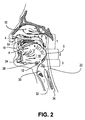
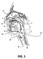
図1は、咽頭の気道4を形成する構造の矢状図である。これら構造のうちの幾つかは、これらが気道4を通る空気の通過を妨げ又は止め、かくして閉塞性睡眠時無呼吸の原因となる程度まで種々の条件下で悪化状態になっている場合がある。咽頭は、上から下へ、鼻部又は鼻咽腔1、口腔咽頭2及び咽頭3に分割されている。図1の変形形態が図2、図3及び図4に提供されており、これらの図は、気道閉塞部位5を咽頭気道内の種々の高さ位置で示している。図2は、例えば、閉塞部5を口腔咽頭2の高さ位置で示しており、ここでは、舌16の底及び肥厚した後咽頭壁22が、互いに対して崩壊し又は潰れている。図3は、閉塞部5を細長く且つ/或いは柔軟な軟組織が肥厚後方咽頭壁に当たって潰れている鼻咽腔1の高さ位置で示している。図4は、閉塞部5を喉頭部3の高さ位置で示しており、ここでは、細長い軟口蓋と柔軟な喉頭蓋の両方が咽頭壁22に当たって潰れている。
FIG. 1 is a sagittal view of the structure forming the pharyngeal airway 4. Some of these structures may be exacerbated under various conditions to the extent that they prevent or stop the passage of air through the airway 4 and thus cause obstructive sleep apnea. . The pharynx is divided into a nose or nasopharynx 1, an
図1〜図4を参照すると、鼻咽腔1は、軟口蓋6の高さ位置又はこれよりも高い位置にある咽頭の部分である。鼻咽腔内では、逸脱した鼻中隔又は拡大した鼻介骨は、場合によっては、上気道抵抗又は閉塞の一因となる場合がある。稀なこととして、鼻内塊、例えばポリープ、嚢腫又は腫瘍は、閉塞の原因である場合がある。口腔咽頭には、軟口蓋6から喉頭蓋12の上境界部までの構造を含み、かかる口腔咽頭は、硬口蓋14、舌16、扁桃腺18、口蓋舌弓20、後咽頭壁22及び下顎骨24を有する。下顎骨は、典型的には、前方に厚さ5mm〜約10mmの骨厚さを有し、側方に同様の厚さを有する。口腔咽頭2内の閉塞は、舌16が深い睡眠又はノンレム睡眠中に筋肉活動の減退の結果として睡眠中に後方に変位したときに結果として起こることがある。変位した舌16は、軟口蓋6を後方に押し、鼻咽腔1を口腔咽頭2から封止する場合がある。舌16は又、後咽頭壁に接触する場合があり、それにより気道の閉塞が一段と生じる。
1 to 4, the nasopharynx 1 is a portion of the pharynx at a height position of the
喉頭部3は、喉頭蓋12の上境界部から輪状軟骨14の下境界部までの領域を含む。喉頭部3は、舌骨28、即ち任意他の骨と関節連結されていないU字形の自由浮動骨を更に有する。舌骨28は、種々の筋肉及び結合組織により周りの構造に取り付けられている。舌骨28は、舌16の下に且つ甲状軟骨30の上に位置している。甲状舌骨膜17及び甲状舌骨筋18は、舌骨の下境界部及び甲状軟骨30の上境界部にくっついている。喉頭蓋12は、舌骨28の下後方に位置し、正中舌骨喉頭蓋靱帯により舌骨にくっついている。舌骨は、オトガイ舌骨筋によって下顎骨24の下後方特徴部に前方からくっついている。
The
本発明は、1つ又は2つ以上の器具を気道形成組織中に植え込み、器具の生体侵食性部分が生体侵食できるようにし、それにより力を気道形成組織に加えて例えば器具の曲率、長さ又は幅の変化により気道開存性を維持することによって、患者の気道の気道開存性を維持する方法を提供する。幾つかの実施形態では、1つ又は複数の器具は、器具を当初組織に取り付けないで植え込まれる。経時的に、器具中への組織の内方成長により、生体侵食及び器具の形状の変化に先立って気道形成組織への器具の幾分かの固定が可能になる。形状変化のインプラントの種々の実施形態を用いると、本発明を実施することができ、器具を必要に応じて患者の気道形成組織の種々の部分中に植え込むことができる。 The present invention implants one or more devices into the airway forming tissue so that the bioerodible portion of the device is bioerodible, thereby applying force to the airway forming tissue, eg, the curvature, length of the device. Alternatively, a method is provided for maintaining airway patency of a patient's airway by maintaining airway patency by varying width. In some embodiments, the one or more instruments are implanted without initially attaching the instrument to the tissue. Over time, tissue ingrowth into the device allows some fixation of the device to the airway-forming tissue prior to bioerosion and changes in the shape of the device. With various embodiments of the shape-changing implant, the present invention can be implemented and the device can be implanted into various portions of the patient's airway forming tissue as needed.
図5A〜図5Cは、患者の気道の開存性を維持するために気道形成組織中に植え込み可能な一実施形態としての器具500を示している。器具500は、幅の広い(wide)部分506によって分離された複数個の幅の狭い(narrow)部分504を備えた本体502を有している。図示のように、これら幅の狭い部分及び幅の広い部分は、円筒形であるが、他の形状を使用することができる。本体502は、弾性変形可能な材料、例えばシリコーンゴム、ポリウレタン若しくは他の弾性変形可能なポリマー又はステンレス鋼、ばね鋼若しくは超弾性ニッケルチタン合金のコイル又は他の弾性変形可能な金属又は弾性変形可能なポリマーと金属の複合材で作られるのが良い。
5A-5C illustrate an
図5Bは、本体502をその休止時形状で示している。図5Aでは、本体502は、変形後の形状に延伸されている。生体侵食性又は生体吸収性材料(例えば、ポリカプロラクトン、ポリ乳酸、ポリグリコール酸、ポリラクチドコグリコリド、ポリグラクチン、ポリ‐L‐ラクチド、ポリ水酸化アルカノエート、でんぷん、セルロース、キトサン又は構造タンパク質)で作られたスペーサ508が、器具をその変形後形状に維持するために大径部分506相互間に挿入されている。この実施形態では、スペーサ508は、射出成形され、C字形である。ただし、他の製造技術及び他の形状を所望に応じて用いることができる。
FIG. 5B shows the
アンカー510は、本体502の両端部のところに形成されている。この実施形態では、アンカー510は、組織内方成長を促進するために不織布(例えば、ポリプロピレン、ポリエチレン又はポリエステル)で作られている。他のアンカーを所望に応じて用いることができる。
器具500は、図5Aに示されている変形後形状で患者の気道形成組織中に植え込み可能である。幾つかの実施形態では、器具500は、植え込み時に気道形成組織には取り付けられない。経時的に、組織は、アンカー510の布中に成長して器具を気道形成組織に少なくとも部分的に取り付けることができる。また、経時的に、生体侵食性スペーサ508は、生体侵食され、それにより器具500は、図5Aに示されている休止時形態に向かって戻ることができる。器具500は、その休止時形状に戻ろうとするときにこれが植え込まれている気道形成組織に力を及ぼして患者の気道を開存状態に維持する。
The
図6A〜図6Jは、本発明の種々の他の実施形態をこれらの変形後状態で示している。図5の実施形態の場合と同様、気道の開存性を維持するためのこれらの器具は、図示の変形後状態で患者の気道形成組織中に植え込み可能である。経時的に、組織は、器具アンカー中に、そして場合によっては器具の他の部分中に成長することができ、それにより器具は、気道形成組織に少なくとも部分的に取り付けられる。また、経時的に、器具の生体侵食性スペーサ部分は、生体侵食され、それにより、器具は、短い休止時形状に向かって動こうとすることができ、それにより器具が植え込まれている気道形成組織に力が及ぼされて患者の気道を開存状態に維持する。これら器具の変形可能な本体を例えばシリコーンゴムで作るのが良い。 6A-6J illustrate various other embodiments of the present invention in these post-deformed states. As with the embodiment of FIG. 5, these devices for maintaining airway patency can be implanted into the patient's airway-forming tissue in the illustrated post-deformation state. Over time, the tissue can grow in the instrument anchor, and possibly in other parts of the instrument, so that the instrument is at least partially attached to the airway forming tissue. Also, over time, the bioerodible spacer portion of the instrument is bioeroded so that the instrument can attempt to move toward a short resting shape, thereby causing the airway in which the instrument is implanted A force is exerted on the forming tissue to keep the patient's airway open. The deformable body of these instruments may be made of, for example, silicone rubber.
図6A及び図6Bでは、器具600は、弾性変形可能な本体602をその延伸変形状態に維持するために大径部分606相互間で弾性変形可能な本体602の小径部分604内に螺旋に巻かれた剛性生体侵食性繊維608を有している。繊維608は、例えば、90%グリコリド及び10%L‐ラクチドのコポリマーであるポリグラクチン910で作られるのが良い。繊維608が生体侵食されると、本体602は、その休止時形状に向かって短くなろうとする。アンカー610は、本体602の両端部のところに設けられている。アンカー610は、組織の内方成長を可能にするよう織布ポリエステル、ポリエチレン又はポリプロピレンで作られるのが良い。
6A and 6B, the
図6C及び図6Dは、複数個の細長い開口部614が形成された弾性変形可能な本体612を有する器具611を示している。図示の変形状態では、生体侵食性のロッド状スペーサ618(例えば、ポリラクチドコグリコリド(PLG)で作られている)が本体の細長い変形形状を維持するために開口部614内に設けられている。複数個の穴又は窪み619を備えたパドル形アンカー領域620が本体612の両端部のところに設けられている。穴又は窪み619により、組織の内方成長が可能である。アンカー領域620は、本体612の中央部分と一体であっても良く、或いは、異なる材料、例えば強化ポリエステルで作られても良い。アンカー領域は又、本体612の中央部分と一体に作られると共に複合材補強要素、例えばポリエステル布を有しても良い。
6C and 6D show an
図6E及び図6Fは、図5A〜図5Cに示されている器具とほぼ同じ器具621を示しており、この器具では、生体侵食性部分628は、螺旋巻き生体侵食性繊維、例えば図6A及び図6Bを参照して上述した螺旋巻き生体侵食性繊維で作られており、この生体侵食性部分は、不織布(例えば、ポリエステル、ポリエチレン又はポリプロピレン)のアンカー又は繋留領域630を有している。
6E and 6F show a
図6G及び図6Hは、図6Aの本体602とほぼ同じ弾性変形可能な本体632を備えた器具631を示している。図示のように、本体632は、延伸変形後形状にある。生体侵食性スペーサ638(図5Aに示されている実施形態のスペーサとほぼ同じである)が本体をこの延伸形状に維持するために大径部分636相互間で小径部分634内に設けられている。両端部のところに設けられたアンカー640は、組織内方成長を促進するために連続気泡又は独立気泡発泡材料で作られている。
6G and 6H show an
図6I及び図6Jは、アンカー649,650を除き、図6E及び図6Fに示されている器具と実質的に同一の器具641を示している。この実施形態では、アンカー640,650は、植え込み中、アンカー650として示された形態まで圧縮可能な自己拡張型バスケットであり、これらアンカーは、配備後、アンカー649として示されている休止時形状に向かって自己拡張する。アンカーの開放領域は、組織内方成長及び取り付けのための材料ループ及び空間を提供している。
FIGS. 6I and 6J show an
気道維持器具の他の実施形態は、必要ならば図示の実施形態の種々の観点を用いることができる。例えば、器具本体の端部のところに設けられるアンカーは、互いに異なっていても良い。 Other embodiments of the airway maintenance device can use various aspects of the illustrated embodiments if desired. For example, the anchors provided at the end of the instrument body may be different from each other.
図7〜図9は、本発明の実施形態によって提供される治療の仕方を示している。図7A〜図7Cでは、運搬ツール702を患者700の体内に下顎下で挿入して気道維持器具710を患者の気道708の一部を形成する患者の舌704の領域中に送り込んでおり、患者の気道708は、図7Aでは閉塞状態として示されている。器具710は、例えば、図5及び図6を参照して上述した器具のうちのどれであっても良い。図7Bに示されているように、器具710を細長い変形後状態で送り込む。幾つかの実施形態では、器具710は、最初に送り込まれたとき、舌組織に取り付けられるわけではない。しかしながら、経時的に、組織が器具710のアンカー711及び/又は器具の他の部分中に成長することができる。また、経時的に、器具710の生体侵食性部分712は、生体侵食され、それにより器具710は、短い休止時形状に向かって動くことができ、それにより力が患者の組織に加えられて図7Cに示されているように気道の開存性が維持される。
7-9 illustrate the treatment methods provided by embodiments of the present invention. 7A-7C, a
図8A〜図8Cでは、運搬ツール802が患者800の体内に経口的に且つ舌下で挿入されて気道維持器具810を患者の気道808の一部を形成する患者の舌804の領域中に送り込んでおり、患者の気道808は、図8Aでは閉塞状態として示されている。器具810は、例えば、図5及び図6を参照して上述した器具のうちのどれであっても良い。図8Bに示されているように、器具810を細長い変形後状態で送り込む。幾つかの実施形態では、器具810は、最初に送り込まれたとき、舌組織に取り付けられるわけではない。しかしながら、経時的に、組織が器具810のアンカー811及び/又は器具の他の部分中に成長することができる。また、経時的に、器具810の生体侵食性部分812は、生体侵食され、それにより器具810は、短い休止時形状に向かって動くことができ、それにより力が患者の組織に加えられて図8Cに示されているように気道の開存性が維持される。
In FIGS. 8A-8C, a
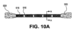
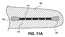
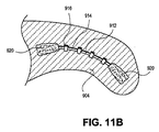
図9A〜図9Cでは、運搬ツール902が患者900の体内に経口的に挿入されて気道維持器具910を患者の気道908の一部を形成する患者の軟口蓋904の領域中に送り込んでおり、患者の気道908は、図9Aでは閉塞状態として示されている。器具910については図10及び図11を参照して以下に更に詳細に説明する。図9B及び図11Aに示されているように、器具910を細長い且つ真っ直ぐな変形後状態で送り込む。幾つかの実施形態では、器具910は、最初に送り込まれたとき、軟口蓋組織に取り付けられるわけではない。しかしながら、経時的に、組織が器具910のアンカー920及び/又は器具の他の部分中に成長することができる。また、経時的に、器具910の生体侵食性部分918は、生体侵食され、それにより器具910は、短い且つ一層湾曲した休止時形状に向かって動くことができ、それにより力が患者の軟口蓋組織に加えられて図9C及び図11Bに示されているように気道の開存性が維持される。
9A-9C, a
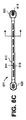
図10A〜図10C並びに図11A及び図11Bは、軟口蓋中への植え込みに適した気道維持器具910の細部を示している。器具の変形後形状が図10A及び図11Aに示されている。この形状では、生体侵食性材料で作られたスペーサ918は、本体912の大径領域914相互間で本体912の小径領域914内に設けられている。本体912は、弾性変形可能な材料(例えばシリコーンゴム、ポリウレタン若しくは他の弾性変形可能なポリマー又はステンレス鋼、ばね鋼若しくは超弾性ニッケルチタン合金のコイル又は他の弾性変形可能な金属又は弾性変形可能なポリマーと金属の複合材)で作られるのが良く、かかる本体は、図10A及び図11Aに示されている真っ直ぐで且つ細長い形態に変形される。本体912の短い且つより湾曲した休止時形状が図10Bに示されている。これは、生体侵食性部分916が生体侵食された後に器具が戻ろうとする形状であり、それにより、力が図11Bに示されているように軟口蓋の気道形成組織に加えられる。この実施形態では、アンカー920は、組織の内方成長を促進するために不織布(例えば、ポリプロピレン又はポリエステル)で作られている。所望ならば他のアンカーを用いても良い。この実施形態では、スペーサ918は、ポリカプロラクトン、ポリ乳酸、ポリグリコール酸、ポリラクチドコグリコリド、ポリグラクチン、ポリ‐L‐ラクチドから射出成形されていてC字形のものである。ただし、他の製造法(例えば、スペーサを弾性変形可能なポリマー又は金属に被着させるための浸漬法)、他の材料及び他の形状を所望に応じて用いることができる。
FIGS. 10A-10C and FIGS. 11A and 11B show details of an
図12A及び図12Bは、患者の気道の一部を形成している舌組織1201中に下顎下で植え込まれた別の実施形態としての気道維持器具1200を示している。器具1200は、互いに異なるアンカー1202,1204を有している。アンカー1204は、自己拡張型アンカー、例えば、図6Iを参照して上述した自己拡張型アンカー649であり、これに対し、アンカー1202は、拡張型のものではない。図12Aに示されているように、器具1200は、組織1201中への植え込み時、細長い変形後形状の状態にある。経時的に、器具1200の生体侵食性部分1206は、生体侵食され、それにより器具1200は、短い休止時形状に向かって動くことができ、それにより力が組織1201に加えられて図12Bに示されているように気道1208の開存性が維持される。
12A and 12B illustrate another embodiment
図13は、種々の植え込み型閉塞性睡眠時無呼吸治療器具により患者の気道形成組織に及ぼされる理論的な平均引張力をインプラントの生じる延伸量と比較して示すグラフ図である。テザー器具が正方形データ点によって形成された2本の線によって示されている。理解できるように、かかる剛性器具は、たるみが全て取られるまで患者の組織に引張力を及ぼさず、たるみが除かれた時点で、テザーは、ほぼ無限の力を及ぼし、場合によっては、患者の許容限度を超える。 FIG. 13 is a graph showing the theoretical average tensile force exerted on a patient's airway forming tissue by various implantable obstructive sleep apnea treatment devices compared to the amount of extension produced by the implant. A tethered instrument is indicated by two lines formed by square data points. As can be appreciated, such a rigid device does not exert a tensile force on the patient's tissue until all of the sagging is removed, and when the sagging is removed, the tether exerts an almost infinite force and, in some cases, the patient's tissue. The allowable limit is exceeded.
丸いデータ点により形成された曲線は、磁石を利用した閉塞性睡眠時無呼吸インプラントにより及ぼされる理論的引張力を示している。理解できるように、かかる器具は、最小治療力を加えることにより患者に利益をもたらす治療範囲に含まれる非常に狭い動作範囲を有している。 The curve formed by the round data points shows the theoretical tensile force exerted by the obstructive sleep apnea implant utilizing magnets. As can be appreciated, such devices have a very narrow operating range that falls within the therapeutic range that benefits the patient by applying minimal therapeutic power.
菱形及び十字形のデータ点により形成された曲線は、変形可能な器具本体に2つの互いに異なるばね定数を備えた本発明の2つの気道維持器具により及ぼされる理論的引張力を示している。図示のように、これら器具は、これらが広い長さ範囲にわたり有益な気道維持療法を患者に提供するよう設計可能である。 The curve formed by the diamond and cross data points shows the theoretical tensile force exerted by the two airway maintenance devices of the present invention with two different spring constants on the deformable device body. As shown, these devices can be designed to provide patients with beneficial airway maintenance therapy over a wide length range.
図14A〜図14C並びに図15A及び図15Bは、本発明の更に別の実施形態を示している。器具1400は、弾性変形可能な材料、例えばシリコーンゴムで作られた2本の細長いレール1402,1404を備えた器具本体を有している。複数個の互いに間隔を置いた長円形フランジ1406がレール1402,1404に取り付けられている。図14A及び図15Aに示されている変形後状態では、C字形生体侵食性スペーサ1408が器具をその細長い形状に維持するために隣り合うフランジ1406相互間に設けられている。スペーサ1408が経時的に生体侵食されると、器具1400は、図14Bに示されている休止時形状に向かって動き、それにより力を患者の気道形成組織(図15において舌1410として示されている)に及ぼして図15Bに示されているように気道1412の開存性を維持する。
14A-14C and 15A and 15B illustrate yet another embodiment of the present invention. The
図16A及び図16Bは、多数個の気道維持器具をどのようにすれば単一の患者の体内に、例えば上述の図8及び図9を参照してそれぞれ説明した舌器具810及び軟口蓋器具910中に植え込むことができるかを示している。
FIGS. 16A and 16B illustrate how multiple airway maintenance devices can be placed within a single patient, such as in the
同様に、図17A〜図17Cは、多数の気道維持器具を気道形成組織の同一領域中にどのようにすれば植え込むことができるかを示している。 Similarly, FIGS. 17A-17C illustrate how multiple airway maintenance devices can be implanted in the same region of the airway forming tissue.
図18A〜図18Cは、図18Aに示されている器具本体1802の変形後状態が図18Bに示されている器具1802の休止時形状よりも長く且つ幅が広い実施形態としての気道維持器具1800を示している。生体侵食性スペーサ1804が弾性変形可能な本体1802に形成された開口部1804内に設けられている。スペーサが侵食されると、本体1802は、その休止時形状に向かって動く。変形後形状1804及び休止時形状1806における開口部は、繋留要素となる。この実施形態は、解剖学的構造、例えば軟口蓋中に配置可能であり、開存性を維持するために力を二方向で気道形成組織に及ぼすことができる。
18A-18C show an
幾つかの実施形態において、器具は、生体侵食性部分に1種類又は2種類以上の生体活性剤を含むのが良い。生体侵食性材料の侵食中に溶出する生体活性剤、例えば薬剤又はホルモンは、例えば、インプラントによる創傷の治癒を促進し又は例えば植え込み状態の器具の周りに生じる線維性組織嚢を強化するのを促進することにより組織部位内における植え込み状態の器具の安定化を促進するのに役立つことができる。 In some embodiments, the device may include one or more bioactive agents in the bioerodible portion. A bioactive agent, such as a drug or hormone, that elutes during erosion of the bioerodible material, for example, promotes healing of the wound by the implant or strengthens the fibrous tissue sac that occurs, for example, around the implanted device This can help to stabilize the implanted device within the tissue site.
Claims (17)
延伸していない休止時形状及び軸線方向に延伸した変形後形状を呈するポリマー材料製の本体を有し、前記本体は、前記患者の気道形成組織中に植え込まれるようになっており、
前記器具を前記組織に取り付けずに前記気道形成組織中に埋め込まれるようになった近位及び遠位アンカーを有し、前記近位及び遠位アンカーは、前記アンカーを前記気道形成組織に取り付けるよう組織によって浸潤されるようになっており、
前記本体を戻し力に抗して前記軸線方向に延伸した変形後形状に維持する複数の生体侵食性要素を有し、
前記本体は、前記侵食性要素の侵食時に前記延伸していない休止時形状に向かって戻るよう構成されている、器具。 A device that maintains the patency of the patient's airway,
Having a body made of a polymeric material that exhibits a non-stretched resting shape and a deformed shape stretched in the axial direction, the body being adapted to be implanted in the airway forming tissue of the patient;
Proximal and distal anchors adapted to be implanted in the airway forming tissue without attaching the device to the tissue, the proximal and distal anchors attaching the anchor to the airway forming tissue Infiltrated by the tissue,
A plurality of bioerodible elements that maintain the body in a deformed shape stretched in the axial direction against the return force;
The instrument is configured to return toward the unstretched resting shape upon erosion of the erodible element.
Applications Claiming Priority (3)
| Application Number | Priority Date | Filing Date | Title |
|---|---|---|---|
| US5258608P | 2008-05-12 | 2008-05-12 | |
| US61/052,586 | 2008-05-12 | ||
| PCT/US2009/043450 WO2009140197A1 (en) | 2008-05-12 | 2009-05-11 | Partially erodable systems for treatment of obstructive sleep apnea |
Related Child Applications (1)
| Application Number | Title | Priority Date | Filing Date |
|---|---|---|---|
| JP2015125673A Division JP2015164627A (en) | 2008-05-12 | 2015-06-23 | Partially erodable systems for treatment of obstructive sleep apnea |
Publications (3)
| Publication Number | Publication Date |
|---|---|
| JP2011519710A JP2011519710A (en) | 2011-07-14 |
| JP2011519710A5 JP2011519710A5 (en) | 2012-06-28 |
| JP5834337B2 true JP5834337B2 (en) | 2015-12-16 |
Family
ID=41319016
Family Applications (2)
| Application Number | Title | Priority Date | Filing Date |
|---|---|---|---|
| JP2011509587A Expired - Fee Related JP5834337B2 (en) | 2008-05-12 | 2009-05-11 | Partially erosive system for the treatment of obstructive sleep apnea |
| JP2015125673A Ceased JP2015164627A (en) | 2008-05-12 | 2015-06-23 | Partially erodable systems for treatment of obstructive sleep apnea |
Family Applications After (1)
| Application Number | Title | Priority Date | Filing Date |
|---|---|---|---|
| JP2015125673A Ceased JP2015164627A (en) | 2008-05-12 | 2015-06-23 | Partially erodable systems for treatment of obstructive sleep apnea |
Country Status (4)
| Country | Link |
|---|---|
| US (4) | US8707960B2 (en) |
| EP (1) | EP2291152A4 (en) |
| JP (2) | JP5834337B2 (en) |
| WO (1) | WO2009140197A1 (en) |
Families Citing this family (28)
| Publication number | Priority date | Publication date | Assignee | Title |
|---|---|---|---|---|
| US8167787B2 (en) | 2008-01-03 | 2012-05-01 | Revent Medical, Inc. | Partially erodable systems for treatment of obstructive sleep apnea |
| US8685432B2 (en) * | 2008-03-25 | 2014-04-01 | University Of Utah Research Foundation | Controlled release tissue graft combination biomaterials |
| US8776799B2 (en) | 2010-03-19 | 2014-07-15 | Revent Medical, Inc. | Systems and methods for treatment of sleep apnea |
| US9439801B2 (en) * | 2012-06-29 | 2016-09-13 | Revent Medical, Inc. | Systems and methods for treatment of sleep apnea |
| JP5834337B2 (en) * | 2008-05-12 | 2015-12-16 | レベント メディカル インコーポレイテッド | Partially erosive system for the treatment of obstructive sleep apnea |
| US8733363B2 (en) * | 2010-03-19 | 2014-05-27 | Revent Medical, Inc. | Systems and methods for treatment of sleep apnea |
| US20110308529A1 (en) * | 2010-05-21 | 2011-12-22 | Gillis Edward M | Systems and methods for treatment of sleep apnea |
| WO2011146930A2 (en) | 2010-05-21 | 2011-11-24 | Revent Medical, Inc. | Systems and methods for treatment of sleep apnea |
| WO2012018545A2 (en) | 2010-07-26 | 2012-02-09 | Revent Medical, Inc. | Systems and methods for treatment of sleep apnea |
| JP2014502868A (en) * | 2010-11-30 | 2014-02-06 | レベント メディカル インコーポレイテッド | System and method for treating sleep apnea |
| US20120310274A1 (en) * | 2011-06-02 | 2012-12-06 | Al-Terki Abdulmohsen E A H | Method of performing multiple oral and nasal surgical procedures |
| EP2731561B1 (en) * | 2011-07-14 | 2016-03-23 | Cook Medical Technologies LLC | A sling to be used in the treatment of obstructive sleep apnea |
| WO2013182893A2 (en) | 2012-06-07 | 2013-12-12 | Fueglister Fabian Hermann Urban | Tongue deformation implant |
| WO2014189540A1 (en) | 2012-10-16 | 2014-11-27 | Catalano Peter J | Method and apparatus for treating obstructive sleep apnea (osa) |
| US9889235B2 (en) | 2013-02-05 | 2018-02-13 | University Of Utah Research Foundation | Implantable devices for bone or joint defects |
| US10123900B2 (en) | 2013-03-15 | 2018-11-13 | Cook Medical Technologies Llc | Devices, kits, and methods for the treatment of obstructive sleep apnea |
| AU2014229628B2 (en) | 2013-03-15 | 2019-02-28 | Fabian Hermann Urban Fuglister | Tongue deformation implant |
| EP3498239B1 (en) | 2013-08-01 | 2021-04-21 | Cook Medical Technologies LLC | Tissue adjustment implant |
| CN105392433B (en) | 2013-08-05 | 2019-02-19 | 库克医学技术有限责任公司 | Medical device and its application method with releasable tubular element |
| US9549806B2 (en) * | 2013-08-12 | 2017-01-24 | Abbott Cardiovascular Systems Inc. | Bioresorbable laryngotracheal stent and methods of treatment |
| EP2898920B1 (en) | 2014-01-24 | 2018-06-06 | Cook Medical Technologies LLC | Articulating balloon catheter |
| DE102015200034A1 (en) | 2014-03-31 | 2015-10-01 | Micro-Epsilon Optronic Gmbh | spectrometer |
| US9974563B2 (en) | 2014-05-28 | 2018-05-22 | Cook Medical Technologies Llc | Medical devices having a releasable member and methods of using the same |
| US9913661B2 (en) | 2014-08-04 | 2018-03-13 | Cook Medical Technologies Llc | Medical devices having a releasable tubular member and methods of using the same |
| EP3229744B1 (en) | 2014-12-12 | 2019-04-17 | Koninklijke Philips N.V. | A tongue advancer system for use in a tongue manipulation system |
| US11147574B2 (en) * | 2016-10-04 | 2021-10-19 | Erik S. Kass | Suspension uvulopalatopexy related methods, devices, and apparatuses |
| US11357660B2 (en) | 2017-06-29 | 2022-06-14 | Cook Medical Technologies, LLC | Implantable medical devices for tissue repositioning |
| CN113768584A (en) * | 2021-09-06 | 2021-12-10 | 乐畅医疗器械(上海)有限公司 | Snore relieving implant |
Family Cites Families (143)
| Publication number | Priority date | Publication date | Assignee | Title |
|---|---|---|---|---|
| US4424208A (en) | 1982-01-11 | 1984-01-03 | Collagen Corporation | Collagen implant material and method for augmenting soft tissue |
| US4582640A (en) | 1982-03-08 | 1986-04-15 | Collagen Corporation | Injectable cross-linked collagen implant material |
| US5506300A (en) | 1985-01-04 | 1996-04-09 | Thoratec Laboratories Corporation | Compositions that soften at predetermined temperatures and the method of making same |
| US5041138A (en) | 1986-11-20 | 1991-08-20 | Massachusetts Institute Of Technology | Neomorphogenesis of cartilage in vivo from cell culture |
| JP2502132B2 (en) | 1988-09-30 | 1996-05-29 | 三菱重工業株式会社 | Shape memory polyurethane elastomer molded body |
| US5222976A (en) | 1989-05-16 | 1993-06-29 | Inbae Yoon | Suture devices particularly useful in endoscopic surgery |
| US4978323A (en) | 1989-08-10 | 1990-12-18 | George Freedman | System and method for preventing closure of passageways |
| US5141522A (en) | 1990-02-06 | 1992-08-25 | American Cyanamid Company | Composite material having absorbable and non-absorbable components for use with mammalian tissue |
| US5665822A (en) | 1991-10-07 | 1997-09-09 | Landec Corporation | Thermoplastic Elastomers |
| US5372600A (en) | 1991-10-31 | 1994-12-13 | Instent Inc. | Stent delivery systems |
| US5258609A (en) | 1992-02-14 | 1993-11-02 | Santa Barbara Research Center | Wide field of view optical element having plural reflectors of different widths |
| US5269798A (en) | 1992-02-19 | 1993-12-14 | Linvatec Corporation | Surgical cutting instrument with movable, inner and outer tubular members |
| ATE193037T1 (en) | 1992-02-28 | 2000-06-15 | Collagen Corp | HIGHLY CONCENTRATED HOMOGENIZED COLLAGEN COMPOSITIONS |
| US5972000A (en) | 1992-11-13 | 1999-10-26 | Influence Medical Technologies, Ltd. | Non-linear anchor inserter device and bone anchors |
| US5762599A (en) | 1994-05-02 | 1998-06-09 | Influence Medical Technologies, Ltd. | Magnetically-coupled implantable medical devices |
| US5697779A (en) | 1995-06-07 | 1997-12-16 | Ormco Corporation | Temporary implant for use as an anchor in the mouth |
| US7090672B2 (en) | 1995-06-07 | 2006-08-15 | Arthrocare Corporation | Method for treating obstructive sleep disorder includes removing tissue from the base of tongue |
| US5792067A (en) | 1995-11-21 | 1998-08-11 | Karell; Manuel L. | Apparatus and method for mitigating sleep and other disorders through electromuscular stimulation |
| US5979456A (en) | 1996-04-22 | 1999-11-09 | Magovern; George J. | Apparatus and method for reversibly reshaping a body part |
| US6530896B1 (en) | 1996-05-13 | 2003-03-11 | James B. Elliott | Apparatus and method for introducing an implant |
| US5775322A (en) | 1996-06-27 | 1998-07-07 | Lucent Medical Systems, Inc. | Tracheal tube and methods related thereto |
| US5782636A (en) | 1996-10-02 | 1998-07-21 | Sulzer Calcitek Inc. | Bone contouring tool |
| US6395017B1 (en) | 1996-11-15 | 2002-05-28 | C. R. Bard, Inc. | Endoprosthesis delivery catheter with sequential stage control |
| US5981826A (en) | 1997-05-05 | 1999-11-09 | Georgia Tech Research Corporation | Poly(vinyl alcohol) cryogel |
| US5988171A (en) | 1997-06-26 | 1999-11-23 | Influence Medical Technologies, Ltd. | Methods and devices for the treatment of airway obstruction, sleep apnea and snoring |
| AU758800B2 (en) | 1998-02-23 | 2003-03-27 | Gkss-Forschungszentrum Geesthacht Gmbh | Shape memory polymers |
| GB9808517D0 (en) * | 1998-04-23 | 1998-06-17 | Aea Technology Plc | Electrical sensor |
| US6161541A (en) | 1998-06-09 | 2000-12-19 | Influent Ltd. | Hyoid expansion and suspension procedure |
| US6019779A (en) | 1998-10-09 | 2000-02-01 | Intratherapeutics Inc. | Multi-filar coil medical stent |
| US6165486A (en) | 1998-11-19 | 2000-12-26 | Carnegie Mellon University | Biocompatible compositions and methods of using same |
| AU5924099A (en) | 1998-12-31 | 2000-07-24 | Jeffrey E. Yeung | Tissue fastening devices and delivery means |
| US6350277B1 (en) * | 1999-01-15 | 2002-02-26 | Scimed Life Systems, Inc. | Stents with temporary retaining bands |
| DE19920114A1 (en) | 1999-05-03 | 2000-11-09 | Fege Wolfgang | Throat side implant |
| US6578763B1 (en) | 1999-09-15 | 2003-06-17 | Restore Products | Method and apparatus for vending a containerized liquid product utilizing an automatic self-service refill system |
| US6772944B2 (en) | 1999-09-15 | 2004-08-10 | Laurie J. Brown | Method and apparatus for vending a containerized liquid product utilizing an automatic self-service refill system |
| US6390096B1 (en) | 1999-09-17 | 2002-05-21 | Pi Medical, Inc. | Needle with pre-loaded implant for snoring treatment |
| US6502574B2 (en) | 1999-09-17 | 2003-01-07 | Pi Medical, Inc. | Lateral stiffening snoring treatment |
| US6523542B2 (en) | 1999-09-17 | 2003-02-25 | Pi Medical, Inc. | Snoring treatment implant and method |
| US6601584B2 (en) * | 1999-09-17 | 2003-08-05 | Pi Medical, Inc. | Contracting snoring treatment implant |
| US6513530B2 (en) | 1999-09-17 | 2003-02-04 | Pi Medical, Inc. | Braided palatal implant for snoring treatment |
| US6516806B2 (en) | 1999-09-17 | 2003-02-11 | Pi Medical, Inc. | Compliant snoring treatment implant |
| US6523541B2 (en) | 1999-09-17 | 2003-02-25 | Pi Medical, Inc. | Delivery system for snoring treatment implant and method |
| US6250307B1 (en) | 1999-09-17 | 2001-06-26 | Pi Medical, Inc. | Snoring treatment |
| US6450169B1 (en) | 1999-09-17 | 2002-09-17 | Pi Medical, Inc. | Braided implant for snoring treatment |
| US6431174B1 (en) | 2000-08-10 | 2002-08-13 | Pi Medical, Inc. | Method and apparatus to treat conditions of the naso-pharyngeal area |
| ES2265993T3 (en) | 1999-09-17 | 2007-03-01 | Restore Medical, Inc. | IMPLANTS FOR THE TREATMENT OF RONQUIDS. |
| US6453905B1 (en) | 1999-09-17 | 2002-09-24 | Pi Medical, Inc. | Multi-component snoring treatment |
| US6513531B2 (en) | 1999-09-17 | 2003-02-04 | Pi Medical, Inc. | Proximal placement of snoring treatment implant |
| US6415796B1 (en) | 1999-09-17 | 2002-07-09 | Pi Medical, Inc. | Placement tool for snoring treatment |
| US6636767B1 (en) | 1999-09-29 | 2003-10-21 | Restore Medical, Inc. | Implanatable stimulation device for snoring treatment |
| US6458127B1 (en) | 1999-11-22 | 2002-10-01 | Csaba Truckai | Polymer embolic elements with metallic coatings for occlusion of vascular malformations |
| US6703040B2 (en) * | 2000-01-11 | 2004-03-09 | Intralytix, Inc. | Polymer blends as biodegradable matrices for preparing biocomposites |
| US7347870B1 (en) | 2000-05-25 | 2008-03-25 | Bioring Sa | Device for shrinking or reinforcing the heart valvular orifices |
| US6439238B1 (en) | 2000-06-30 | 2002-08-27 | Pi Medical, Inc. | Snoring diagnostic and treatment |
| US6569191B1 (en) | 2000-07-27 | 2003-05-27 | Bionx Implants, Inc. | Self-expanding stent with enhanced radial expansion and shape memory |
| US6783554B2 (en) * | 2001-02-20 | 2004-08-31 | Atrium Medical Corporation | Pile mesh prosthesis |
| US6507675B1 (en) | 2001-03-23 | 2003-01-14 | Shih-Jong J. Lee | Structure-guided automatic learning for image feature enhancement |
| US6748950B2 (en) | 2001-04-30 | 2004-06-15 | Closure Medical Corporation | Compositions and medical procedure to treat snoring |
| US7647561B2 (en) * | 2001-08-28 | 2010-01-12 | Nvidia International, Inc. | System, method and computer program product for application development using a visual paradigm to combine existing data and applications |
| US6629988B2 (en) | 2001-08-28 | 2003-10-07 | Ethicon, Inc. | Composite staple for completing an anastomosis |
| US6467485B1 (en) | 2001-10-01 | 2002-10-22 | Bruno Schmidt | Anti-snoring device and method |
| US20030062050A1 (en) | 2001-10-01 | 2003-04-03 | Bruno Schmidt | Anti-snoring devices and methods |
| WO2003055417A1 (en) | 2001-12-28 | 2003-07-10 | Edwards Lifesciences Ag | Delayed memory device |
| SE524709C2 (en) | 2002-01-11 | 2004-09-21 | Edwards Lifesciences Ag | Device for delayed reshaping of a heart vessel and a heart valve |
| US7146981B2 (en) * | 2002-02-04 | 2006-12-12 | Restore Medical, Inc. | Pharyngeal wall treatment |
| US7017582B2 (en) | 2002-02-04 | 2006-03-28 | Restore Medical Inc. | Stiffening pharyngeal wall treatment |
| US8528564B2 (en) | 2002-09-06 | 2013-09-10 | Koninklijke Philips N.V. | Devices, systems and methods using magnetic force systems affecting both the tongue and the soft palate/uvula in the upper airway |
| US8074654B2 (en) | 2002-09-06 | 2011-12-13 | Koninklijke Philips Electronics N.V. | Implantable devices, systems, and methods for maintaining desired orientations in targeted tissue regions |
| US8707959B2 (en) * | 2002-09-06 | 2014-04-29 | Koninklijke Philips N.V. | Implantable devices, systems, and methods for maintaining desired orientations in targeted tissue regions |
| US20080066764A1 (en) | 2002-09-06 | 2008-03-20 | Apneon, Inc. | Implantable devices, systems, and methods for maintaining desired orientations in targeted tissue regions |
| US7360542B2 (en) | 2002-09-06 | 2008-04-22 | Apneon, Inc. | Devices, systems, and methods to fixate tissue within the regions of body, such as the pharyngeal conduit |
| US7216648B2 (en) | 2002-09-06 | 2007-05-15 | Apneon, Inc. | Systems and methods for moving and/or restraining tissue in the upper respiratory system |
| US20060289014A1 (en) | 2002-09-06 | 2006-12-28 | Apneon, Inc. | Devices, systems, and methods using magnetic force systems in or on tissue in an airway |
| US7845356B2 (en) | 2002-09-06 | 2010-12-07 | Koninklijke Philips Electronics N.V. | Implantable devices, systems, and methods for maintaining desired orientations in targeted tissue regions |
| US8047206B2 (en) | 2002-09-06 | 2011-11-01 | Koninklijke Philips Electronics N.V. | Magnetic devices, systems, and methods placed in or on a tongue |
| CA2497805A1 (en) | 2002-09-06 | 2004-03-18 | Apneon, Inc. | Systems and methods for moving and/or restraining tissue in the upper respiratory system |
| US7441559B2 (en) * | 2002-09-06 | 2008-10-28 | Koninklijke Philips Electronics N.V. | Devices, systems, and methods to fixate tissue within the regions of body, such as the pharyngeal conduit |
| US7188627B2 (en) | 2002-09-06 | 2007-03-13 | Apneon, Inc. | Magnetic force devices, systems, and methods for resisting tissue collapse within the pharyngeal conduit |
| WO2004032798A1 (en) | 2002-10-04 | 2004-04-22 | Brooks Stephen N | System and method for preventing closure of passageways |
| US7992566B2 (en) | 2002-12-30 | 2011-08-09 | Quiescence Medical, Inc. | Apparatus and methods for treating sleep apnea |
| CA2524271C (en) | 2003-05-02 | 2012-09-04 | Surmodics, Inc. | Controlled release bioactive agent delivery device |
| US20050065615A1 (en) | 2003-09-19 | 2005-03-24 | Restore Medical, Inc. | Airway implant and delivery tool and kit |
| USD536792S1 (en) | 2003-09-19 | 2007-02-13 | Restore Medical, Inc. | Implant delivery tool |
| US6899105B2 (en) | 2003-09-19 | 2005-05-31 | Restore Medical, Inc. | Airway implant cartridge and kit |
| US20050154412A1 (en) | 2003-09-19 | 2005-07-14 | Restore Medical, Inc. | Airway implant and delivery tool |
| US7237554B2 (en) | 2003-10-31 | 2007-07-03 | Restore Medical, Inc. | Airway implant |
| US7213599B2 (en) | 2003-10-31 | 2007-05-08 | Restore Medical, Inc. | Airway implant |
| AU2004288711A1 (en) | 2003-11-05 | 2005-05-26 | Pavad Medical, Inc. | Electrically activated alteration of body tissue stiffness for breathing disorders |
| US8080014B2 (en) | 2004-12-15 | 2011-12-20 | Koninklijke Philips Electronics N.V. | System and method for hyoidplasty |
| US10524954B2 (en) | 2004-02-26 | 2020-01-07 | Linguaflex, Inc. | Methods and devices for treating sleep apnea and snoring |
| CA2592999C (en) | 2004-02-26 | 2017-11-07 | Linguaflex Llc | A method and device for the treatment of obstructive sleep apnea and snoring |
| US8074655B2 (en) | 2004-02-26 | 2011-12-13 | Linguaflex, Inc. | Methods and devices for treating sleep apnea and snoring |
| US20050267321A1 (en) | 2004-06-01 | 2005-12-01 | Shadduck John H | Elastomeric magnetic nanocomposite biomedical devices |
| US20050274387A1 (en) | 2004-06-10 | 2005-12-15 | John Macken | Method and apparatus for treatment of snoring and sleep apnea |
| US20090038623A1 (en) | 2004-09-21 | 2009-02-12 | Pavad Medical, Inc. | Inductive power transfer system for palatal implant |
| US20090069866A1 (en) | 2004-09-21 | 2009-03-12 | Pavad Medical, Inc. | Implant tester |
| US7997266B2 (en) | 2004-10-04 | 2011-08-16 | Koninklijke Philips Electronics N.V. | System and method for airway manipulation |
| DE102005000702B4 (en) | 2005-01-04 | 2007-08-23 | Klinikum Der Universität Regensburg | Device for central implantation in a tongue body |
| US7972354B2 (en) * | 2005-01-25 | 2011-07-05 | Tyco Healthcare Group Lp | Method and apparatus for impeding migration of an implanted occlusive structure |
| EP2353494B1 (en) | 2005-02-08 | 2014-07-30 | Koninklijke Philips N.V. | System for percutaneous glossoplasty |
| US8220466B2 (en) | 2005-02-08 | 2012-07-17 | Koninklijke Philips Electronics N.V. | System and method for percutaneous palate remodeling |
| US8096303B2 (en) | 2005-02-08 | 2012-01-17 | Koninklijke Philips Electronics N.V | Airway implants and methods and devices for insertion and retrieval |
| US8371307B2 (en) | 2005-02-08 | 2013-02-12 | Koninklijke Philips Electronics N.V. | Methods and devices for the treatment of airway obstruction, sleep apnea and snoring |
| US9408742B2 (en) | 2005-02-08 | 2016-08-09 | Koninklijke Philips N.V. | Glossopexy adjustment system and method |
| US7322356B2 (en) | 2005-02-24 | 2008-01-29 | Restore Medical, Inc. | Combination sleep apnea treatment |
| US7337781B2 (en) * | 2005-04-15 | 2008-03-04 | Restore Medical, Inc. | Implant for tongue |
| US20060235380A1 (en) | 2005-04-15 | 2006-10-19 | Restore Medical, Inc. | Tissue incision tool |
| US9636250B2 (en) | 2005-05-19 | 2017-05-02 | John D. Summer | Tongue grasping and restraining apparatus and methods |
| US7947076B2 (en) | 2005-06-03 | 2011-05-24 | Medtronic Xomed, Inc. | Nasal valve treatment method and apparatus |
| JP2007097706A (en) | 2005-09-30 | 2007-04-19 | Terumo Corp | Stent |
| WO2007056583A1 (en) | 2005-11-09 | 2007-05-18 | Aspire Medical, Inc. | Glossoplasty using tissue anchor glossopexy with volumetric tongue reduction |
| US20070144534A1 (en) | 2005-11-30 | 2007-06-28 | Carlos Mery | System to prevent airway obstruction |
| EP1968418B1 (en) | 2005-12-09 | 2015-04-15 | Spinal Ventures, Inc. | Non-soft tissue repair |
| US7909038B2 (en) | 2006-04-20 | 2011-03-22 | Pavad Medical, Inc. | Tongue stabilization device and methods of using the same |
| US8256425B2 (en) | 2006-04-20 | 2012-09-04 | Medtronic Xomed, Inc. | Methods and devices for removal of a tongue stabilization device |
| US7909037B2 (en) | 2006-04-20 | 2011-03-22 | Pavad Medical | Tethered airway implants and methods of using the same |
| WO2007146338A2 (en) | 2006-06-13 | 2007-12-21 | Aspire Medical, Inc. | Glossal engagement system and method |
| US7934506B2 (en) | 2006-06-21 | 2011-05-03 | Koninklijke Philips Electronics N.V. | System and method for temporary tongue suspension |
| US8517028B2 (en) | 2006-06-23 | 2013-08-27 | Medtronic Xomed, Inc. | Stiffening procedure for sleep apnea |
| WO2008006090A2 (en) | 2006-07-06 | 2008-01-10 | Quiescence Medical, Inc. | Apparatus and methods for treating sleep apnea |
| US20080078412A1 (en) | 2006-10-03 | 2008-04-03 | Restore Medical, Inc. | Tongue implant |
| US20080078411A1 (en) | 2006-10-03 | 2008-04-03 | Restore Medical, Inc. | Tongue implant for sleep apnea |
| CA2665209C (en) | 2006-10-03 | 2016-01-05 | Alure Medical, Inc. | Minimally invasive tissue support |
| WO2008097890A2 (en) | 2007-02-02 | 2008-08-14 | Thomas Jefferson University | Method and use of a bioreplaceable tissue material implant for treating snoring |
| JP5802011B2 (en) | 2007-06-18 | 2015-10-28 | コーニンクレッカ フィリップス エヌ ヴェ | Implantable devices, systems, and methods for maintaining a desired orientation within a target tissue region. |
| US20120143134A1 (en) | 2007-11-16 | 2012-06-07 | Hollis Jeffrey D | Method of Performing Laparoscopic Surgery Using a Multi-Access Channel Surgical Trocar |
| US8167787B2 (en) | 2008-01-03 | 2012-05-01 | Revent Medical, Inc. | Partially erodable systems for treatment of obstructive sleep apnea |
| JP5834337B2 (en) | 2008-05-12 | 2015-12-16 | レベント メディカル インコーポレイテッド | Partially erosive system for the treatment of obstructive sleep apnea |
| US8776799B2 (en) | 2010-03-19 | 2014-07-15 | Revent Medical, Inc. | Systems and methods for treatment of sleep apnea |
| US20110308529A1 (en) | 2010-05-21 | 2011-12-22 | Gillis Edward M | Systems and methods for treatment of sleep apnea |
| US8733363B2 (en) | 2010-03-19 | 2014-05-27 | Revent Medical, Inc. | Systems and methods for treatment of sleep apnea |
| US20090319046A1 (en) | 2008-06-23 | 2009-12-24 | Biomerix Corporation | Hyoid suspension for obstructive sleep apnea |
| US8413661B2 (en) | 2008-08-14 | 2013-04-09 | Ethicon, Inc. | Methods and devices for treatment of obstructive sleep apnea |
| AU2009287269B2 (en) | 2008-08-29 | 2014-11-27 | Xiangmin Zhang | Implantable soft palate support and implantation method |
| WO2010045546A1 (en) | 2008-10-16 | 2010-04-22 | Linguaflex, Llc | Methods and devices for treating sleep apnea |
| US8561617B2 (en) | 2008-10-30 | 2013-10-22 | Ethicon, Inc. | Implant systems and methods for treating obstructive sleep apnea |
| US8783258B2 (en) | 2008-12-01 | 2014-07-22 | Ethicon, Inc. | Implant systems and methods for treating obstructive sleep apnea |
| US20100158854A1 (en) | 2008-12-19 | 2010-06-24 | Boston Scientific Scimed, Inc. | Method for treatment of airway and apparatus and kit for use therewith |
| ES2382369T3 (en) | 2008-12-30 | 2012-06-07 | Medartis Ag | Implant for the treatment of sleep apnea syndrome |
| WO2011146930A2 (en) | 2010-05-21 | 2011-11-24 | Revent Medical, Inc. | Systems and methods for treatment of sleep apnea |
| WO2012018545A2 (en) | 2010-07-26 | 2012-02-09 | Revent Medical, Inc. | Systems and methods for treatment of sleep apnea |
| JP2014502868A (en) | 2010-11-30 | 2014-02-06 | レベント メディカル インコーポレイテッド | System and method for treating sleep apnea |
| WO2012075503A2 (en) | 2010-12-03 | 2012-06-07 | Revent Medical, Inc. | Systems and methods for treatment of sleep apnea |
-
2009
- 2009-05-11 JP JP2011509587A patent/JP5834337B2/en not_active Expired - Fee Related
- 2009-05-11 US US12/937,564 patent/US8707960B2/en not_active Expired - Fee Related
- 2009-05-11 EP EP09747281.5A patent/EP2291152A4/en not_active Withdrawn
- 2009-05-11 WO PCT/US2009/043450 patent/WO2009140197A1/en active Application Filing
-
2011
- 2011-10-07 US US13/269,520 patent/US8327854B2/en not_active Expired - Fee Related
-
2012
- 2012-12-11 US US13/711,537 patent/US8991398B2/en not_active Expired - Fee Related
-
2015
- 2015-03-31 US US14/674,986 patent/US20150202074A1/en not_active Abandoned
- 2015-06-23 JP JP2015125673A patent/JP2015164627A/en not_active Ceased
Also Published As
| Publication number | Publication date |
|---|---|
| US20120024298A1 (en) | 2012-02-02 |
| EP2291152A1 (en) | 2011-03-09 |
| JP2015164627A (en) | 2015-09-17 |
| US20130098374A1 (en) | 2013-04-25 |
| JP2011519710A (en) | 2011-07-14 |
| US8707960B2 (en) | 2014-04-29 |
| US8991398B2 (en) | 2015-03-31 |
| US20150202074A1 (en) | 2015-07-23 |
| US8327854B2 (en) | 2012-12-11 |
| EP2291152A4 (en) | 2015-05-06 |
| US20110144421A1 (en) | 2011-06-16 |
| WO2009140197A1 (en) | 2009-11-19 |
Similar Documents
| Publication | Publication Date | Title |
|---|---|---|
| JP5834337B2 (en) | Partially erosive system for the treatment of obstructive sleep apnea | |
| JP6057889B2 (en) | Sleep apnea treatment system and method | |
| US9510922B2 (en) | Systems and methods for treatment of sleep apnea | |
| US9707122B2 (en) | Systems and methods for treatment of sleep apnea | |
| JP2011508650A (en) | Partially erosive system for the treatment of obstructive sleep apnea | |
| US20110308529A1 (en) | Systems and methods for treatment of sleep apnea | |
| US20120132214A1 (en) | Systems and methods for treatment of sleep apnea | |
| US9439801B2 (en) | Systems and methods for treatment of sleep apnea | |
| JP2013521952A (en) | Sleep apnea treatment system and method | |
| US20160022470A1 (en) | Systems and methods for treatment of sleep apnea | |
| US20140007885A1 (en) | Systems and methods for treatment of sleep apnea | |
| AU2014229628B2 (en) | Tongue deformation implant |
Legal Events
| Date | Code | Title | Description |
|---|---|---|---|
| A521 | Request for written amendment filed |
Free format text: JAPANESE INTERMEDIATE CODE: A523 Effective date: 20120509 |
|
| A621 | Written request for application examination |
Free format text: JAPANESE INTERMEDIATE CODE: A621 Effective date: 20120509 |
|
| A977 | Report on retrieval |
Free format text: JAPANESE INTERMEDIATE CODE: A971007 Effective date: 20130617 |
|
| A131 | Notification of reasons for refusal |
Free format text: JAPANESE INTERMEDIATE CODE: A131 Effective date: 20130619 |
|
| A521 | Request for written amendment filed |
Free format text: JAPANESE INTERMEDIATE CODE: A523 Effective date: 20130918 |
|
| A131 | Notification of reasons for refusal |
Free format text: JAPANESE INTERMEDIATE CODE: A131 Effective date: 20140203 |
|
| A601 | Written request for extension of time |
Free format text: JAPANESE INTERMEDIATE CODE: A601 Effective date: 20140331 |
|
| A602 | Written permission of extension of time |
Free format text: JAPANESE INTERMEDIATE CODE: A602 Effective date: 20140407 |
|
| A521 | Request for written amendment filed |
Free format text: JAPANESE INTERMEDIATE CODE: A523 Effective date: 20140804 |
|
| A02 | Decision of refusal |
Free format text: JAPANESE INTERMEDIATE CODE: A02 Effective date: 20150223 |
|
| A521 | Request for written amendment filed |
Free format text: JAPANESE INTERMEDIATE CODE: A523 Effective date: 20150623 |
|
| A521 | Request for written amendment filed |
Free format text: JAPANESE INTERMEDIATE CODE: A523 Effective date: 20150805 |
|
| A911 | Transfer to examiner for re-examination before appeal (zenchi) |
Free format text: JAPANESE INTERMEDIATE CODE: A911 Effective date: 20150812 |
|
| TRDD | Decision of grant or rejection written | ||
| A01 | Written decision to grant a patent or to grant a registration (utility model) |
Free format text: JAPANESE INTERMEDIATE CODE: A01 Effective date: 20151007 |
|
| A61 | First payment of annual fees (during grant procedure) |
Free format text: JAPANESE INTERMEDIATE CODE: A61 Effective date: 20151013 |
|
| R150 | Certificate of patent or registration of utility model |
Ref document number: 5834337 Country of ref document: JP Free format text: JAPANESE INTERMEDIATE CODE: R150 |
|
| LAPS | Cancellation because of no payment of annual fees |