JP5483023B2 - Cancer cell motility and cancer cell invasion inhibitor - Google Patents
Cancer cell motility and cancer cell invasion inhibitor Download PDFInfo
- Publication number
- JP5483023B2 JP5483023B2 JP2010505602A JP2010505602A JP5483023B2 JP 5483023 B2 JP5483023 B2 JP 5483023B2 JP 2010505602 A JP2010505602 A JP 2010505602A JP 2010505602 A JP2010505602 A JP 2010505602A JP 5483023 B2 JP5483023 B2 JP 5483023B2
- Authority
- JP
- Japan
- Prior art keywords
- antibody
- par1
- cells
- seq
- present
- Prior art date
- Legal status (The legal status is an assumption and is not a legal conclusion. Google has not performed a legal analysis and makes no representation as to the accuracy of the status listed.)
- Active
Links
- 206010028980 Neoplasm Diseases 0.000 title claims description 68
- 201000011510 cancer Diseases 0.000 title claims description 50
- 230000009087 cell motility Effects 0.000 title description 23
- 230000004709 cell invasion Effects 0.000 title description 16
- 239000003112 inhibitor Substances 0.000 title description 2
- 210000004027 cell Anatomy 0.000 claims description 100
- 230000000694 effects Effects 0.000 claims description 48
- 125000003275 alpha amino acid group Chemical group 0.000 claims description 37
- 239000012634 fragment Substances 0.000 claims description 30
- 238000000034 method Methods 0.000 claims description 28
- 230000002401 inhibitory effect Effects 0.000 claims description 20
- 210000004408 hybridoma Anatomy 0.000 claims description 16
- 210000004881 tumor cell Anatomy 0.000 claims description 16
- 239000008194 pharmaceutical composition Substances 0.000 claims description 14
- 230000004899 motility Effects 0.000 claims description 9
- 239000000203 mixture Substances 0.000 claims description 8
- 238000003384 imaging method Methods 0.000 claims description 7
- 238000004519 manufacturing process Methods 0.000 claims description 7
- 239000003814 drug Substances 0.000 claims description 6
- 238000005516 engineering process Methods 0.000 claims description 5
- 239000012216 imaging agent Substances 0.000 claims description 5
- 108010070519 PAR-1 Receptor Proteins 0.000 description 154
- 102100037136 Proteinase-activated receptor 1 Human genes 0.000 description 154
- 108090000765 processed proteins & peptides Proteins 0.000 description 79
- 238000003776 cleavage reaction Methods 0.000 description 24
- 230000007017 scission Effects 0.000 description 24
- 102000008394 Immunoglobulin Fragments Human genes 0.000 description 23
- 108010021625 Immunoglobulin Fragments Proteins 0.000 description 23
- 239000000243 solution Substances 0.000 description 21
- 241000699666 Mus <mouse, genus> Species 0.000 description 19
- 239000000523 sample Substances 0.000 description 19
- 230000027455 binding Effects 0.000 description 17
- 239000000427 antigen Substances 0.000 description 14
- 102000036639 antigens Human genes 0.000 description 14
- 108091007433 antigens Proteins 0.000 description 14
- 230000000890 antigenic effect Effects 0.000 description 12
- 206010003445 Ascites Diseases 0.000 description 9
- 150000001413 amino acids Chemical class 0.000 description 9
- 102000004196 processed proteins & peptides Human genes 0.000 description 9
- 239000002246 antineoplastic agent Substances 0.000 description 8
- 108010082117 matrigel Proteins 0.000 description 8
- 108090000631 Trypsin Proteins 0.000 description 7
- 102000004142 Trypsin Human genes 0.000 description 7
- 230000001419 dependent effect Effects 0.000 description 7
- 210000001519 tissue Anatomy 0.000 description 7
- 239000012588 trypsin Substances 0.000 description 7
- 102000014914 Carrier Proteins Human genes 0.000 description 6
- 108010078791 Carrier Proteins Proteins 0.000 description 6
- 238000003556 assay Methods 0.000 description 6
- 108010045069 keyhole-limpet hemocyanin Proteins 0.000 description 6
- 230000033001 locomotion Effects 0.000 description 6
- 238000011579 SCID mouse model Methods 0.000 description 5
- 108090000190 Thrombin Proteins 0.000 description 5
- 239000012228 culture supernatant Substances 0.000 description 5
- 238000001514 detection method Methods 0.000 description 5
- 239000011521 glass Substances 0.000 description 5
- 238000002347 injection Methods 0.000 description 5
- 239000007924 injection Substances 0.000 description 5
- 230000037023 motor activity Effects 0.000 description 5
- 229960004072 thrombin Drugs 0.000 description 5
- 102000000380 Matrix Metalloproteinase 1 Human genes 0.000 description 4
- 108010016113 Matrix Metalloproteinase 1 Proteins 0.000 description 4
- 230000004913 activation Effects 0.000 description 4
- XOJVVFBFDXDTEG-UHFFFAOYSA-N pristane Chemical compound CC(C)CCCC(C)CCCC(C)CCCC(C)C XOJVVFBFDXDTEG-UHFFFAOYSA-N 0.000 description 4
- 238000004445 quantitative analysis Methods 0.000 description 4
- 108091003079 Bovine Serum Albumin Proteins 0.000 description 3
- 206010006187 Breast cancer Diseases 0.000 description 3
- 208000026310 Breast neoplasm Diseases 0.000 description 3
- 108091006027 G proteins Proteins 0.000 description 3
- 102000030782 GTP binding Human genes 0.000 description 3
- 108091000058 GTP-Binding Proteins 0.000 description 3
- 101000741967 Homo sapiens Presequence protease, mitochondrial Proteins 0.000 description 3
- 241000699670 Mus sp. Species 0.000 description 3
- 102100038632 Presequence protease, mitochondrial Human genes 0.000 description 3
- 102000003790 Thrombin receptors Human genes 0.000 description 3
- 108090000166 Thrombin receptors Proteins 0.000 description 3
- 229940125644 antibody drug Drugs 0.000 description 3
- 229940098773 bovine serum albumin Drugs 0.000 description 3
- 210000000170 cell membrane Anatomy 0.000 description 3
- 229940079593 drug Drugs 0.000 description 3
- 230000009545 invasion Effects 0.000 description 3
- 239000011159 matrix material Substances 0.000 description 3
- 239000012528 membrane Substances 0.000 description 3
- 238000002360 preparation method Methods 0.000 description 3
- 230000009870 specific binding Effects 0.000 description 3
- 239000000126 substance Substances 0.000 description 3
- YBJHBAHKTGYVGT-ZKWXMUAHSA-N (+)-Biotin Chemical compound N1C(=O)N[C@@H]2[C@H](CCCCC(=O)O)SC[C@@H]21 YBJHBAHKTGYVGT-ZKWXMUAHSA-N 0.000 description 2
- FWMNVWWHGCHHJJ-SKKKGAJSSA-N 4-amino-1-[(2r)-6-amino-2-[[(2r)-2-[[(2r)-2-[[(2r)-2-amino-3-phenylpropanoyl]amino]-3-phenylpropanoyl]amino]-4-methylpentanoyl]amino]hexanoyl]piperidine-4-carboxylic acid Chemical compound C([C@H](C(=O)N[C@H](CC(C)C)C(=O)N[C@H](CCCCN)C(=O)N1CCC(N)(CC1)C(O)=O)NC(=O)[C@H](N)CC=1C=CC=CC=1)C1=CC=CC=C1 FWMNVWWHGCHHJJ-SKKKGAJSSA-N 0.000 description 2
- 108010088751 Albumins Proteins 0.000 description 2
- 102000009027 Albumins Human genes 0.000 description 2
- 238000002965 ELISA Methods 0.000 description 2
- 241001465754 Metazoa Species 0.000 description 2
- 241000283973 Oryctolagus cuniculus Species 0.000 description 2
- 108010058846 Ovalbumin Proteins 0.000 description 2
- 108090000901 Transferrin Proteins 0.000 description 2
- 102000004338 Transferrin Human genes 0.000 description 2
- 210000000683 abdominal cavity Anatomy 0.000 description 2
- 238000001261 affinity purification Methods 0.000 description 2
- 239000005667 attractant Substances 0.000 description 2
- 210000004204 blood vessel Anatomy 0.000 description 2
- 238000004113 cell culture Methods 0.000 description 2
- 230000031902 chemoattractant activity Effects 0.000 description 2
- 238000007796 conventional method Methods 0.000 description 2
- 230000008878 coupling Effects 0.000 description 2
- 238000010168 coupling process Methods 0.000 description 2
- 238000005859 coupling reaction Methods 0.000 description 2
- 239000006071 cream Substances 0.000 description 2
- 238000011161 development Methods 0.000 description 2
- 239000002552 dosage form Substances 0.000 description 2
- 238000010828 elution Methods 0.000 description 2
- 238000002474 experimental method Methods 0.000 description 2
- 238000010353 genetic engineering Methods 0.000 description 2
- 230000036541 health Effects 0.000 description 2
- 230000008595 infiltration Effects 0.000 description 2
- 238000001764 infiltration Methods 0.000 description 2
- 238000001802 infusion Methods 0.000 description 2
- 230000003834 intracellular effect Effects 0.000 description 2
- 238000002372 labelling Methods 0.000 description 2
- 238000013507 mapping Methods 0.000 description 2
- 239000002105 nanoparticle Substances 0.000 description 2
- 239000002674 ointment Substances 0.000 description 2
- 229940092253 ovalbumin Drugs 0.000 description 2
- 230000010118 platelet activation Effects 0.000 description 2
- 108090000623 proteins and genes Proteins 0.000 description 2
- 102000005962 receptors Human genes 0.000 description 2
- 108020003175 receptors Proteins 0.000 description 2
- 230000000717 retained effect Effects 0.000 description 2
- 238000002415 sodium dodecyl sulfate polyacrylamide gel electrophoresis Methods 0.000 description 2
- 239000007787 solid Substances 0.000 description 2
- 239000000758 substrate Substances 0.000 description 2
- 230000001629 suppression Effects 0.000 description 2
- 239000012581 transferrin Substances 0.000 description 2
- 239000004475 Arginine Substances 0.000 description 1
- 108090001008 Avidin Proteins 0.000 description 1
- 241000283690 Bos taurus Species 0.000 description 1
- 102000004190 Enzymes Human genes 0.000 description 1
- 108090000790 Enzymes Proteins 0.000 description 1
- 102000003688 G-Protein-Coupled Receptors Human genes 0.000 description 1
- 108090000045 G-Protein-Coupled Receptors Proteins 0.000 description 1
- 108010001336 Horseradish Peroxidase Proteins 0.000 description 1
- 102000001706 Immunoglobulin Fab Fragments Human genes 0.000 description 1
- 108010054477 Immunoglobulin Fab Fragments Proteins 0.000 description 1
- 206010061218 Inflammation Diseases 0.000 description 1
- 206010058467 Lung neoplasm malignant Diseases 0.000 description 1
- 241000124008 Mammalia Species 0.000 description 1
- 206010027476 Metastases Diseases 0.000 description 1
- 206010061902 Pancreatic neoplasm Diseases 0.000 description 1
- 102000035195 Peptidases Human genes 0.000 description 1
- 108091005804 Peptidases Proteins 0.000 description 1
- 206010060862 Prostate cancer Diseases 0.000 description 1
- 208000000236 Prostatic Neoplasms Diseases 0.000 description 1
- 239000004365 Protease Substances 0.000 description 1
- 229920002684 Sepharose Polymers 0.000 description 1
- 210000001744 T-lymphocyte Anatomy 0.000 description 1
- 101710120037 Toxin CcdB Proteins 0.000 description 1
- 238000002835 absorbance Methods 0.000 description 1
- 239000004480 active ingredient Substances 0.000 description 1
- 125000003277 amino group Chemical group 0.000 description 1
- 238000004458 analytical method Methods 0.000 description 1
- 239000005557 antagonist Substances 0.000 description 1
- ODKSFYDXXFIFQN-UHFFFAOYSA-N arginine Natural products OC(=O)C(N)CCCNC(N)=N ODKSFYDXXFIFQN-UHFFFAOYSA-N 0.000 description 1
- 210000003719 b-lymphocyte Anatomy 0.000 description 1
- 230000008901 benefit Effects 0.000 description 1
- 229960002685 biotin Drugs 0.000 description 1
- 235000020958 biotin Nutrition 0.000 description 1
- 239000011616 biotin Substances 0.000 description 1
- 230000000903 blocking effect Effects 0.000 description 1
- 210000004369 blood Anatomy 0.000 description 1
- 239000008280 blood Substances 0.000 description 1
- 230000015556 catabolic process Effects 0.000 description 1
- 230000001413 cellular effect Effects 0.000 description 1
- 238000006243 chemical reaction Methods 0.000 description 1
- 239000000470 constituent Substances 0.000 description 1
- 210000004748 cultured cell Anatomy 0.000 description 1
- 230000001472 cytotoxic effect Effects 0.000 description 1
- 238000006731 degradation reaction Methods 0.000 description 1
- 238000003745 diagnosis Methods 0.000 description 1
- 238000000502 dialysis Methods 0.000 description 1
- 201000010099 disease Diseases 0.000 description 1
- 208000037265 diseases, disorders, signs and symptoms Diseases 0.000 description 1
- 231100000673 dose–response relationship Toxicity 0.000 description 1
- 229940088598 enzyme Drugs 0.000 description 1
- 238000002073 fluorescence micrograph Methods 0.000 description 1
- 230000005484 gravity Effects 0.000 description 1
- 210000000987 immune system Anatomy 0.000 description 1
- 238000000338 in vitro Methods 0.000 description 1
- 238000001727 in vivo Methods 0.000 description 1
- 238000011503 in vivo imaging Methods 0.000 description 1
- 230000004054 inflammatory process Effects 0.000 description 1
- 238000010255 intramuscular injection Methods 0.000 description 1
- 239000007927 intramuscular injection Substances 0.000 description 1
- 238000010253 intravenous injection Methods 0.000 description 1
- 208000030776 invasive breast carcinoma Diseases 0.000 description 1
- 239000007788 liquid Substances 0.000 description 1
- 201000005202 lung cancer Diseases 0.000 description 1
- 208000020816 lung neoplasm Diseases 0.000 description 1
- 208000015486 malignant pancreatic neoplasm Diseases 0.000 description 1
- 230000009401 metastasis Effects 0.000 description 1
- 230000001394 metastastic effect Effects 0.000 description 1
- 208000037819 metastatic cancer Diseases 0.000 description 1
- 208000011575 metastatic malignant neoplasm Diseases 0.000 description 1
- 206010061289 metastatic neoplasm Diseases 0.000 description 1
- 230000004048 modification Effects 0.000 description 1
- 238000012986 modification Methods 0.000 description 1
- 230000035772 mutation Effects 0.000 description 1
- 201000002528 pancreatic cancer Diseases 0.000 description 1
- 208000008443 pancreatic carcinoma Diseases 0.000 description 1
- 239000002245 particle Substances 0.000 description 1
- 238000002823 phage display Methods 0.000 description 1
- 239000000843 powder Substances 0.000 description 1
- 230000002265 prevention Effects 0.000 description 1
- 102000004169 proteins and genes Human genes 0.000 description 1
- 230000035484 reaction time Effects 0.000 description 1
- 230000009257 reactivity Effects 0.000 description 1
- 210000002966 serum Anatomy 0.000 description 1
- 230000011664 signaling Effects 0.000 description 1
- 238000002603 single-photon emission computed tomography Methods 0.000 description 1
- 238000002791 soaking Methods 0.000 description 1
- 238000010186 staining Methods 0.000 description 1
- 230000000638 stimulation Effects 0.000 description 1
- 239000011550 stock solution Substances 0.000 description 1
- 238000010254 subcutaneous injection Methods 0.000 description 1
- 239000007929 subcutaneous injection Substances 0.000 description 1
- 239000006228 supernatant Substances 0.000 description 1
- 238000003786 synthesis reaction Methods 0.000 description 1
- 230000002194 synthesizing effect Effects 0.000 description 1
- 230000009885 systemic effect Effects 0.000 description 1
- 230000000699 topical effect Effects 0.000 description 1
- 210000003462 vein Anatomy 0.000 description 1
Images


Classifications
-
- C—CHEMISTRY; METALLURGY
- C07—ORGANIC CHEMISTRY
- C07K—PEPTIDES
- C07K16/00—Immunoglobulins [IGs], e.g. monoclonal or polyclonal antibodies
- C07K16/18—Immunoglobulins [IGs], e.g. monoclonal or polyclonal antibodies against material from animals or humans
- C07K16/28—Immunoglobulins [IGs], e.g. monoclonal or polyclonal antibodies against material from animals or humans against receptors, cell surface antigens or cell surface determinants
-
- A—HUMAN NECESSITIES
- A61—MEDICAL OR VETERINARY SCIENCE; HYGIENE
- A61P—SPECIFIC THERAPEUTIC ACTIVITY OF CHEMICAL COMPOUNDS OR MEDICINAL PREPARATIONS
- A61P35/00—Antineoplastic agents
-
- C—CHEMISTRY; METALLURGY
- C07—ORGANIC CHEMISTRY
- C07K—PEPTIDES
- C07K7/00—Peptides having 5 to 20 amino acids in a fully defined sequence; Derivatives thereof
- C07K7/04—Linear peptides containing only normal peptide links
- C07K7/06—Linear peptides containing only normal peptide links having 5 to 11 amino acids
-
- C—CHEMISTRY; METALLURGY
- C07—ORGANIC CHEMISTRY
- C07K—PEPTIDES
- C07K7/00—Peptides having 5 to 20 amino acids in a fully defined sequence; Derivatives thereof
- C07K7/04—Linear peptides containing only normal peptide links
- C07K7/08—Linear peptides containing only normal peptide links having 12 to 20 amino acids
-
- G—PHYSICS
- G01—MEASURING; TESTING
- G01N—INVESTIGATING OR ANALYSING MATERIALS BY DETERMINING THEIR CHEMICAL OR PHYSICAL PROPERTIES
- G01N33/00—Investigating or analysing materials by specific methods not covered by groups G01N1/00 - G01N31/00
- G01N33/48—Biological material, e.g. blood, urine; Haemocytometers
- G01N33/50—Chemical analysis of biological material, e.g. blood, urine; Testing involving biospecific ligand binding methods; Immunological testing
- G01N33/5005—Chemical analysis of biological material, e.g. blood, urine; Testing involving biospecific ligand binding methods; Immunological testing involving human or animal cells
- G01N33/5008—Chemical analysis of biological material, e.g. blood, urine; Testing involving biospecific ligand binding methods; Immunological testing involving human or animal cells for testing or evaluating the effect of chemical or biological compounds, e.g. drugs, cosmetics
- G01N33/502—Chemical analysis of biological material, e.g. blood, urine; Testing involving biospecific ligand binding methods; Immunological testing involving human or animal cells for testing or evaluating the effect of chemical or biological compounds, e.g. drugs, cosmetics for testing non-proliferative effects
- G01N33/5029—Chemical analysis of biological material, e.g. blood, urine; Testing involving biospecific ligand binding methods; Immunological testing involving human or animal cells for testing or evaluating the effect of chemical or biological compounds, e.g. drugs, cosmetics for testing non-proliferative effects on cell motility
-
- G—PHYSICS
- G01—MEASURING; TESTING
- G01N—INVESTIGATING OR ANALYSING MATERIALS BY DETERMINING THEIR CHEMICAL OR PHYSICAL PROPERTIES
- G01N33/00—Investigating or analysing materials by specific methods not covered by groups G01N1/00 - G01N31/00
- G01N33/48—Biological material, e.g. blood, urine; Haemocytometers
- G01N33/50—Chemical analysis of biological material, e.g. blood, urine; Testing involving biospecific ligand binding methods; Immunological testing
- G01N33/53—Immunoassay; Biospecific binding assay; Materials therefor
- G01N33/574—Immunoassay; Biospecific binding assay; Materials therefor for cancer
-
- A—HUMAN NECESSITIES
- A61—MEDICAL OR VETERINARY SCIENCE; HYGIENE
- A61K—PREPARATIONS FOR MEDICAL, DENTAL OR TOILETRY PURPOSES
- A61K39/00—Medicinal preparations containing antigens or antibodies
- A61K2039/505—Medicinal preparations containing antigens or antibodies comprising antibodies
-
- C—CHEMISTRY; METALLURGY
- C07—ORGANIC CHEMISTRY
- C07K—PEPTIDES
- C07K2317/00—Immunoglobulins specific features
- C07K2317/30—Immunoglobulins specific features characterized by aspects of specificity or valency
- C07K2317/34—Identification of a linear epitope shorter than 20 amino acid residues or of a conformational epitope defined by amino acid residues
-
- C—CHEMISTRY; METALLURGY
- C07—ORGANIC CHEMISTRY
- C07K—PEPTIDES
- C07K2317/00—Immunoglobulins specific features
- C07K2317/70—Immunoglobulins specific features characterized by effect upon binding to a cell or to an antigen
- C07K2317/76—Antagonist effect on antigen, e.g. neutralization or inhibition of binding
-
- G—PHYSICS
- G01—MEASURING; TESTING
- G01N—INVESTIGATING OR ANALYSING MATERIALS BY DETERMINING THEIR CHEMICAL OR PHYSICAL PROPERTIES
- G01N2500/00—Screening for compounds of potential therapeutic value
- G01N2500/02—Screening involving studying the effect of compounds C on the interaction between interacting molecules A and B (e.g. A = enzyme and B = substrate for A, or A = receptor and B = ligand for the receptor)
-
- G—PHYSICS
- G01—MEASURING; TESTING
- G01N—INVESTIGATING OR ANALYSING MATERIALS BY DETERMINING THEIR CHEMICAL OR PHYSICAL PROPERTIES
- G01N2500/00—Screening for compounds of potential therapeutic value
- G01N2500/10—Screening for compounds of potential therapeutic value involving cells
-
- G—PHYSICS
- G01—MEASURING; TESTING
- G01N—INVESTIGATING OR ANALYSING MATERIALS BY DETERMINING THEIR CHEMICAL OR PHYSICAL PROPERTIES
- G01N2800/00—Detection or diagnosis of diseases
Description
本発明は、がん細胞運動およびがん細胞浸潤抑制剤に関する。さらに詳細には、MMP1により誘導される細胞運動活性および細胞浸潤活性を抑制するPAR1抗体、ならびに該PAR1抗体を含有する医薬組成物などに関する。なお、本願は、日本国特許出願第2008−83588号に対して優先権を主張するものであり、参照により該日本国特許出願の内容を本願に一体化させる。 The present invention relates to a cancer cell motility and cancer cell infiltration inhibitor. More specifically, the present invention relates to a PAR1 antibody that suppresses cell motility activity and cell invasive activity induced by MMP1, and a pharmaceutical composition containing the PAR1 antibody. In addition, this application claims priority with respect to Japanese Patent Application No. 2008-83588, and the contents of the Japanese Patent Application are integrated into this application by reference.
細胞の運動能および浸潤能が異常に高まった結果生じる疾病の典型はがんである。がんの最たる脅威の1つとして転移が挙げられる。転移性がん細胞は細胞運動によって原発巣から血管を介して移動し、血管内に浸潤した後、血流に乗り他の組織へ転移する。PAR1(Protease activated receptor 1:プロテアーゼ活性化受容体1)は、がん細胞の運動能および浸潤能を活性化する7回膜貫通型受容体で、多種のがん(乳がん、肺がん、膵臓がんおよび前立腺がんなど)の転移に関与するものである。特に乳がんでは、転移能を有するがん培養細胞株のほとんどでPAR1が発現しており、がん特有のプロテアーゼ(MMP1:Matrix Metalloprotease 1:マトリックスメタロプロテアーゼ1)によって、PAR1のN末の細胞外領域(R41とS42の間)が切断されるとPAR1が活性化することが知られている。PAR1活性化の刺激は、PAR1の細胞内領域にカップルしているGタンパク質を活性化する。その結果、細胞内Ca2+濃度が局所的に高まり、このCa2+シグナルにより細胞運動能および細胞浸潤能が増強されると考えられている(非特許文献1参照)。A typical disease that results from abnormally increased cell motility and invasion is cancer. One of the greatest threats of cancer is metastasis. Metastatic cancer cells move from the primary focus through blood vessels by cell movement, infiltrate into the blood vessels, and then metastasize to other tissues by riding in the bloodstream. PAR1 (Protease activated receptor 1: Protease activated receptor 1) is a seven-transmembrane receptor that activates the motility and invasive ability of cancer cells, and various cancers (breast cancer, lung cancer, pancreatic cancer) And prostate cancer). In particular, in breast cancer, PAR1 is expressed in most cancer cell lines with metastatic potential, and the N-terminal extracellular region of PAR1 by a cancer-specific protease (MMP1: Matrix Metalloproteinase 1: Matrix metalloproteinase 1). When (between R 41 and S 42) is cut PAR1 is known to activate. Stimulation of PAR1 activation activates the G protein coupled to the intracellular region of PAR1. As a result, intracellular Ca 2+ concentration is locally increased, and it is considered that cell motility and cell invasion are enhanced by this Ca 2+ signal (see Non-Patent Document 1).
PAR1はもともと血小板を活性化させるために必要なトロンビン依存性の受容体(トロンビン受容体)として発見された。トロンビンは、MMP1と同じPAR1の部位を認識して切断する。また、血小板におけるトロンビン受容体の活性化もがん細胞の場合と同様に、Ca2+シグナルを活性化する。現在、PAR1のN末細胞ドメインに対するモノクローナル抗体がいくつか販売されており、これらの抗体は血小板の活性化を阻害するものである。しかしながら、これらの抗体とがん細胞の運動性および浸潤性との関係に関する報告はなく、ましてやがん細胞の運動性および浸潤性をPAR1抗体で阻害する、あるいはPAR1抗体をがん治療に使用するという報告はない。PAR1 was originally discovered as a thrombin-dependent receptor (thrombin receptor) necessary to activate platelets. Thrombin recognizes and cleaves the same PAR1 site as MMP1. In addition, activation of thrombin receptor in platelets activates Ca 2+ signal, as in the case of cancer cells. Currently, several monoclonal antibodies against the N-terminal cell domain of PAR1 are on the market, and these antibodies inhibit platelet activation. However, there is no report regarding the relationship between these antibodies and the motility and invasiveness of cancer cells. Furthermore, the PAR1 antibody inhibits the motility and invasiveness of cancer cells, or the PAR1 antibody is used for cancer treatment. There is no report.
また、PAR1のGタンパク質結合部位に相当するペプチドを合成し、このペプチドに膜透過性を持たせたペプデュシン(Pepducin)という物質が作製されている(非特許文献2参照)。CovicらはペプデュシンをGタンパク質の拮抗剤として利用することでPAR1の活性を阻害することに成功している(ペプデュシンの阻害効果は3−4μM)。これに対し、抗体を抗がん剤として応用する方法は、標的分子活性の阻害効果だけでなく、免疫系細胞による抗体依存性細胞障害活性の効果が期待できる。さらに抗体は、遺伝子工学により高親和性抗体を作製する技術が確立されており、また生体への安全性も保証されているなど抗体を抗がん剤として応用するメリットは非常に大きい。そのため、PAR1抗体を抗がん剤として応用すれば、ペプデュシンより効果的な抗がん剤となりうる可能性を秘めていると考えられる。 In addition, a peptide called Pepducin has been produced by synthesizing a peptide corresponding to the G protein binding site of PAR1 and making this peptide membrane permeable (see Non-Patent Document 2). Covic et al. Succeeded in inhibiting the activity of PAR1 by using pepducin as an antagonist of G protein (the inhibitory effect of pepducin is 3-4 μM). On the other hand, the method of applying an antibody as an anticancer agent can be expected not only to inhibit target molecule activity but also to effect antibody-dependent cytotoxic activity by immune system cells. Furthermore, for antibodies, the technology for producing high-affinity antibodies by genetic engineering has been established, and the merit of applying the antibodies as anticancer agents is extremely great, such as the safety to living bodies being guaranteed. Therefore, if the PAR1 antibody is applied as an anticancer agent, it is considered that it has the potential to be an effective anticancer agent than pepducin.
PAR1の52番目のTyrから56番目のTrpをエピトープとする抗体がファージディスプレイ法で作製され、該抗体は、トロンビンがPAR1を切断する活性を阻害する。該抗体がPAR1の切断活性を阻害すること(PAR1への結合)に関して、特に56番目のTrpがエピトープとして含まれることが重要であるとされている(特許文献1参照)。現在、がん細胞の運動性や浸潤性をPAR1抗体で阻害する報告や、阻害効果に有効なPAR1抗体のエピトープとして他のペプチド配列が見出されたとの報告はない。
本発明は、細胞運動性および浸潤性に関する機能を効果的に阻害する薬剤を提供すること、詳細には、細胞のPAR1の活性を抑制して細胞運動性および浸潤性に関する機能を効果的に阻害する薬剤を提供することを課題とする。 The present invention provides a drug that effectively inhibits functions related to cell motility and invasiveness, and in particular, effectively inhibits functions related to cell motility and invasiveness by suppressing cellular PAR1 activity. It is an object to provide a drug to be used.
本発明者らは、上記課題を解決すべく鋭意検討した結果、PAR1のMMP1による切断部位(Arg41とSer42の間)を含む領域に特異的に結合する抗体を見出した。さらにこの抗体が、MMP1により誘導される細胞運動活性および細胞浸潤活性を阻害することを見出し、本発明を完成するに至った。すなわち、本発明は:
(1)PAR1(プロテアーゼ活性化受容体1)に特異的に結合してMMP1(マトリックスメタロプロテアーゼ1)による切断を阻害し、がん細胞の運動活性および浸潤活性を阻害する抗体、または該抗体と同様の性質を保持する該抗体のフラグメント;
(2)PAR1(プロテアーゼ活性化受容体1)のMMP1(マトリックスメタロプロテアーゼ1)による切断部位(Arg41とSer42の間)を含む領域をエピトープとして特異的に結合し、がん細胞の運動活性および浸潤活性を阻害する(1)記載の抗体、または該抗体と同様の性質を保持する該抗体のフラグメント;
(3)該エピトープのアミノ酸配列が配列番号:1で示されるものである(2)記載の抗体、または該抗体と同様の性質を保持する該抗体のフラグメント;
(4)独立行政法人産業技術総合研究所 特許生物寄託センターから受託番号FERM BP−11105を付与されたハイブリドーマにより産生されるモノクローナル抗体である、(3)記載の抗体、または該抗体と同様の性質を保持する該抗体のフラグメント;
(5)キメラ化またはヒト化されている、(1)〜(4)のいずれかに記載の抗体、または該抗体と同様の性質を保持する該抗体のフラグメント;
(6)モノクローナル抗体である(1)〜(5)いずれかに記載の抗体;
(7)ポリクローナル抗体である(1)〜(3)または(5)のいずれかに記載の抗体;
(8)(1)〜(7)のいずれかに記載の抗体またはフラグメントを含む、がん細胞の運動活性および浸潤活性を阻害するための組成物;
(9)(1)〜(7)のいずれかに記載の抗体またはフラグメントを含む、がん治療用医薬組成物;
(10)(1)〜(7)のいずれかに記載の抗体またはフラグメントを含む、医薬組成物の有効量を治療を必要とする対象に投与することを含むがんの治療方法;
(11)がんを治療するための医薬の製造における(1)〜(7)のいずれかに記載の抗体またはフラグメントの使用;
(12)(1)〜(7)のいずれかに記載の抗体またはフラグメントを、対象から得た試料と接触させ、試料中の腫瘍細胞と結合させることを特徴とする、試料中の腫瘍細胞のイメージング方法または腫瘍の検出方法;
(13)(1)〜(7)のいずれかに記載の抗体またはフラグメントを含む、腫瘍細胞イメージング剤;
(14)(1)〜(7)のいずれかに記載の抗体または抗体フラグメントを対象に投与して、体内の腫瘍組織と本発明の抗体または抗体フラグメントとの結合を調べることを特徴とする、腫瘍の診断方法;
(15)配列番号:1で示されるアミノ酸配列からなるペプチド;
(16)配列番号:1で示されるアミノ酸配列からなるペプチドおよびキャリア蛋白からなる抗原性ペプチド;
(17)(15)記載のペプチドまたは(16)記載の抗原性ペプチドを用いることを特徴とする、PAR1に特異的に結合してMMP1による切断を阻害する抗体、または該抗体と同様の性質を保持する該抗体のフラグメントの作製方法;
(18)(15)記載のペプチドまたは(16)記載の抗原性ペプチドを用いることを特徴とする、PAR1に特異的に結合してMMP1による切断を阻害する抗体、または該抗体と同様の性質を保持する該抗体のフラグメントの精製方法;
(19)(15)記載のペプチドまたは(16)記載の抗原性ペプチドを含む、がん細胞の運動活性および浸潤活性を阻害するための組成物
を提供するものである。As a result of intensive studies to solve the above-mentioned problems, the present inventors have found an antibody that specifically binds to a region containing a cleavage site of PAR1 by MMP1 (between Arg 41 and Ser 42 ). Furthermore, this antibody was found to inhibit the cell motility activity and cell invasion activity induced by MMP1, and the present invention was completed. That is, the present invention provides:
(1) an antibody that specifically binds to PAR1 (protease-activated receptor 1) and inhibits cleavage by MMP1 (matrix metalloprotease 1), and inhibits the motor activity and invasive activity of cancer cells, or the antibody Fragments of the antibody that retain similar properties;
(2) Cancer cell motility activity by specifically binding as a epitope a region containing a cleavage site (between Arg 41 and Ser 42 ) by MMP1 (matrix metalloprotease 1) of PAR1 (protease activated receptor 1) And the antibody according to (1), which inhibits invasive activity, or a fragment of the antibody having the same properties as the antibody;
(3) The antibody according to (2), wherein the amino acid sequence of the epitope is represented by SEQ ID NO: 1, or a fragment of the antibody having the same properties as the antibody;
(4) The antibody according to (3), which is a monoclonal antibody produced by a hybridoma assigned with the accession number FERM BP-11105 from the Patent Organism Depositary, National Institute of Advanced Industrial Science and Technology, or a property similar to that of the antibody A fragment of the antibody retaining
(5) The antibody according to any one of (1) to (4), which is chimerized or humanized, or a fragment of the antibody that retains the same properties as the antibody;
(6) The antibody according to any one of (1) to (5), which is a monoclonal antibody;
(7) The antibody according to any one of (1) to (3) or (5), which is a polyclonal antibody;
(8) A composition for inhibiting motor activity and invasive activity of cancer cells, comprising the antibody or fragment according to any one of (1) to (7);
(9) A pharmaceutical composition for cancer treatment comprising the antibody or fragment according to any one of (1) to (7);
(10) A method for treating cancer comprising administering an effective amount of a pharmaceutical composition comprising the antibody or fragment according to any one of (1) to (7) to a subject in need of treatment;
(11) Use of the antibody or fragment according to any one of (1) to (7) in the manufacture of a medicament for treating cancer;
(12) The antibody or fragment according to any one of (1) to (7) is brought into contact with a sample obtained from a subject and allowed to bind to the tumor cells in the sample. An imaging method or a tumor detection method;
(13) A tumor cell imaging agent comprising the antibody or fragment according to any one of (1) to (7);
(14) The antibody or antibody fragment according to any one of (1) to (7) is administered to a subject, and the binding between the tumor tissue in the body and the antibody or antibody fragment of the present invention is examined, Tumor diagnosis method;
(15) a peptide consisting of the amino acid sequence represented by SEQ ID NO: 1;
(16) an antigenic peptide consisting of a peptide consisting of the amino acid sequence represented by SEQ ID NO: 1 and a carrier protein;
(17) An antibody that specifically binds to PAR1 and inhibits cleavage by MMP1, characterized by using the peptide according to (15) or the antigenic peptide according to (16), or a property similar to that of the antibody A method for producing a fragment of the antibody to be retained;
(18) An antibody that specifically binds to PAR1 and inhibits cleavage by MMP1, characterized by using the peptide according to (15) or the antigenic peptide according to (16), or a property similar to that of the antibody A method for purifying a fragment of the antibody to be retained;
(19) A composition for inhibiting the motor activity and invasive activity of cancer cells, comprising the peptide according to (15) or the antigenic peptide according to (16).
本発明のPAR1に特異的に結合してMMP1による切断を阻害し、がん細胞の運動活性および浸潤活性を阻害する抗体(以下、本明細書において「PAR1抗体」という)は、がん細胞の運動能および浸潤能を効果的に阻害することができるので、優れた抗がん剤を得ることができる。また、本発明のPAR1抗体を用いて腫瘍の検出やイメージングを行うこともできる。さらに本発明の抗原性ペプチドを用いて、有効なPAR1抗体を取得し、あるいは精製することができる。 An antibody that specifically binds to PAR1 of the present invention and inhibits cleavage by MMP1 and inhibits cancer cell motility activity and invasive activity (hereinafter referred to as “PAR1 antibody” in the present specification) Since the motor ability and the invasive ability can be effectively inhibited, an excellent anticancer agent can be obtained. In addition, tumor detection and imaging can be performed using the PAR1 antibody of the present invention. Furthermore, an effective PAR1 antibody can be obtained or purified using the antigenic peptide of the present invention.
本発明は、1の態様において、PAR1抗体、および該抗体と同様の性質を保持する該抗体のフラグメントを提供する。本発明のPAR1抗体は、具体的には、MMP1による切断部位を含むPAR1の特定の領域に特異的に結合し、その結果、がん細胞の運動活性および浸潤活性を阻害するものであってもよい。あるいは本発明のPAR1抗体は、PAR1に特異的に結合し、立体障害を生じさせることによりMMP1による切断部位へのMMP1のアクセスを阻害するものであってもよい。本発明のPAR1抗体は、このようにしてMMP1によるPAR1の切断を阻害することにより、がん細胞の運動活性および浸潤活性を抑制するものである。それゆえ、本発明のPAR1抗体はがん治療に有用である。なお、本発明のPAR1抗体はモノクローナル抗体であってもよく、あるいはポリクローナル抗体であってもよい。 In one aspect, the present invention provides a PAR1 antibody and fragments of the antibody that retain similar properties as the antibody. Specifically, the PAR1 antibody of the present invention specifically binds to a specific region of PAR1 including the cleavage site by MMP1, and as a result, inhibits the motor activity and invasive activity of cancer cells. Good. Alternatively, the PAR1 antibody of the present invention may inhibit MMP1 access to the cleavage site by MMP1 by specifically binding to PAR1 and causing steric hindrance. The PAR1 antibody of the present invention inhibits the motility and invasive activity of cancer cells by thus inhibiting PAR1 cleavage by MMP1. Therefore, the PAR1 antibody of the present invention is useful for cancer treatment. The PAR1 antibody of the present invention may be a monoclonal antibody or a polyclonal antibody.
本明細書において「該抗体と同様の性質を保持する該抗体のフラグメント」とは、エピトープのアミノ酸配列が本発明のPAR1抗体のエピトープのアミノ酸配列と同じまたは類似のものであり、かつ、本発明のPAR1抗体と同じまたは同様のがん細胞運動阻害活性および浸潤阻害活性を有するフラグメントをいう。本発明のPAR1抗体の「フラグメント」とは、本発明のPAR1抗体の一部分をいい、例えばFabフラグメントなどを包含する。なお、特に明記しない限り、本明細書において「PAR1抗体」という場合には、上述のフラグメントも包含するものとする。 In the present specification, the “fragment of the antibody having the same properties as the antibody” means that the amino acid sequence of the epitope is the same as or similar to the amino acid sequence of the epitope of the PAR1 antibody of the present invention, and the present invention A fragment having the same or similar cancer cell motility inhibitory activity and invasion inhibitory activity as the PAR1 antibody. The “fragment” of the PAR1 antibody of the present invention refers to a part of the PAR1 antibody of the present invention, and includes, for example, a Fab fragment. Unless otherwise specified, the term “PAR1 antibody” in the present specification includes the above-mentioned fragment.
なお、本明細書において「エピトープ」とは、本発明のペプチドおよび抗原性ペプチドを構成する、あるいはこれらに含まれるアミノ酸配列であって、本発明の抗体または抗体フラグメントによって認識され結合されるアミノ酸配列をいう。詳細には、本発明に用いられるエピトープは、MMP1による切断部位(Arg41とSer42の間)を含む、少なくとも数個、好ましくは少なくとも10個のアミノ酸配列であるが、これらに限定されるものではない。通常は、「エピトープ」は、約5個ないし約1000個のアミノ酸からなる。本発明の「エピトープ」はその変異体も包含する。例えば、上記アミノ酸配列を有する「エピトープ」において1個ないし数個のアミノ酸が欠失、付加、および置換されたアミノ酸配列を有するものであってもよく、構成アミノ酸が修飾されたものであってもよい。ただし、かかる変異または修飾「エピトープ」は、本来の「エピトープ」の性質、すなわち、本発明の抗体または抗体フラグメントによって認識され結合される性質を保持しているものである。In the present specification, the “epitope” is an amino acid sequence constituting or included in the peptide and antigenic peptide of the present invention, which is recognized and bound by the antibody or antibody fragment of the present invention. Say. Specifically, the epitope used in the present invention is an amino acid sequence of at least several, preferably at least 10, including, but not limited to, a cleavage site by MMP1 (between Arg 41 and Ser 42 ). is not. Usually, an “epitope” consists of about 5 to about 1000 amino acids. The “epitope” of the present invention also encompasses variants thereof. For example, the “epitope” having the above amino acid sequence may have an amino acid sequence in which one to several amino acids are deleted, added, or substituted, or may be a modified constituent amino acid. Good. However, such a mutation or modification “epitope” retains the properties of the original “epitope”, ie, the property recognized and bound by the antibody or antibody fragment of the present invention.
PAR1の特定の領域とは、MMP1によるPAR1の切断部位(Arg41とSer42の間)を含む領域である。好ましい特定の領域は、上記切断部位をまたぐ約10個のアミノ酸配列であり、最も好ましいのはPAR1の35番目から45番目のアミノ酸配列NATLDPRSFLL(配列番号:1)である。本発明の好ましい抗体、または抗体フラグメントは、配列番号:1からなるアミノ酸配列を抗原認識部位(エピトープ)とするものである。The specific region of PAR1 is a region including a PAR1 cleavage site (between Arg 41 and Ser 42 ) by MMP1. A preferred specific region is a sequence of about 10 amino acids across the cleavage site, and the most preferred is the 35th to 45th amino acid sequence NATLDPRSFLL (SEQ ID NO: 1) of PAR1. A preferred antibody or antibody fragment of the present invention has an amino acid sequence consisting of SEQ ID NO: 1 as an antigen recognition site (epitope).
なお、本明細書ではアミノ酸の表記は当該分野で公知の1文字法および3文字法を用いる。アミノ酸の右下の数字はPAR1のアミノ酸配列のN末端から数えたアミノ酸位置を示し、例えばArg41またはR41はPAR1のアミノ酸配列のN末端から41番目のアルギニンを表す。In this specification, amino acid notation uses a one-letter method and a three-letter method known in the art. The number on the lower right of the amino acid indicates the amino acid position counted from the N-terminus of the amino acid sequence of PAR1, for example, Arg 41 or R 41 represents the 41st arginine from the N-terminus of the amino acid sequence of PAR1.
本発明のPAR1抗体であって、配列番号:1からなるアミノ酸配列をエピトープとするもののなかで、がん細胞の運動活性および浸潤活性を強く阻害する好ましい抗体は、ブタペスト条約の下、平成20年(2008年)3月18日より、日本国茨城県つくば市東1丁目1番地1中央第6の、独立行政法人産業技術総合研究所 特許生物寄託センターにFERM BP−11105の受託番号で寄託してある、ハイブリドーマにより産生されるモノクローナル抗体である(以下、本明細書において「N2−11抗体」という)。 Among the PAR1 antibodies of the present invention, which have an amino acid sequence consisting of SEQ ID NO: 1 as an epitope, preferred antibodies that strongly inhibit cancer cell motility activity and invasive activity have been reported under the Budapest Treaty in 2008. (2008) From March 18th, deposited at FERM BP-11105 at the Patent Organism Depositary, National Institute of Advanced Industrial Science and Technology, 1st, 1st, 1st East, Tsukuba, Ibaraki, Japan. It is a monoclonal antibody produced by a certain hybridoma (hereinafter referred to as “N2-11 antibody” in the present specification).
実施例にて説明するように、本発明の抗体または抗体フラグメントの特徴は、アミノ酸配列NATLDPRSFLL(配列番号:1)を認識して特異的に結合することである。かかるアミノ酸配列は、本発明において新しく見出されたものである。上記アミノ酸配列を認識する利点は、本発明の抗体または抗体フラグメントがMMP−1によるPAR1の切断部位をまたぐ形のアミノ酸配列を認識することにより、効率よくMMP−1による分解を阻害するものであるが、認識部位の候補となる多くのペプチドの中から配列番号:1を選び出した点において本発明の意義は大きい。 As illustrated in the Examples, the feature of the antibody or antibody fragment of the present invention is to recognize and specifically bind to the amino acid sequence NATLDPRSFLL (SEQ ID NO: 1). Such an amino acid sequence has been newly found in the present invention. The advantage of recognizing the amino acid sequence is that the antibody or antibody fragment of the present invention recognizes an amino acid sequence in a form that crosses the cleavage site of PAR1 by MMP-1, thereby efficiently inhibiting degradation by MMP-1. However, the significance of the present invention is great in that SEQ ID NO: 1 is selected from many peptides that are candidates for recognition sites.
本発明のPAR1抗体は、例えば、以下のように調製することができる。動物、好ましくはヒトのPAR1のMMP1による切断部位(R41とS42の間)を含む少なくとも数個、好ましくは少なくとも10個のアミノ酸配列を標的ペプチドとして用いる。好ましい標的ペプチドは配列番号:1に示されるアミノ酸配列からなる。標的ペプチドは、公知の方法にて、例えばキーホールリンペットヘモシアニン(KLH)などのキャリアタンパク質に結合されてもよい。これを抗原としてマウスなどの動物を免疫し、ハイブリドーマを得ることができる。得られたハイブリドーマを培養し、培養上清を用いてがん細胞の運動活性および浸潤活性を、例えばマトリゲルアッセイなどの公知方法により検定し、強力な活性を有するハイブリドーマを選択し、それが産生する抗体を、例えばアフィニティー精製などの常法にて精製・単離することができる。得られた抗体について抗体特異性および抗体効果の解析を行うことができる。本発明のPAR1抗体は、上記方法と当該分野で公知のペプチドの化学合成法を組み合わせることによっても得ることができる。本発明のPAR1抗体の作製方法は上記方法に限定されないことはいうまでもない。The PAR1 antibody of the present invention can be prepared, for example, as follows. Animals, preferably at least several, including a cleavage site by MMP1 of PAR1 human (between R 41 and S 42), is preferably used at least 10 amino acid sequence as the target peptide. A preferred target peptide consists of the amino acid sequence shown in SEQ ID NO: 1. The target peptide may be bound to a carrier protein such as keyhole limpet hemocyanin (KLH) by a known method. Using this as an antigen, animals such as mice can be immunized to obtain hybridomas. The obtained hybridoma is cultured, and the motility and invasive activity of cancer cells are assayed by using a culture supernatant by a known method such as Matrigel assay, and a hybridoma having a strong activity is selected and produced. The antibody can be purified and isolated by a conventional method such as affinity purification. The antibody specificity and antibody effect of the obtained antibody can be analyzed. The PAR1 antibody of the present invention can also be obtained by combining the above-described method with a chemical synthesis method of a peptide known in the art. It goes without saying that the method for producing the PAR1 antibody of the present invention is not limited to the above method.
さらに、常法により本発明のPAR1抗体をキメラ化およびヒト化することもできる。また本発明の抗体は一本鎖抗体であってもよい。本発明のPAR1抗体という場合、これらのキメラ抗体、ヒト化抗体、1本鎖抗体を包含する。 Furthermore, the PAR1 antibody of the present invention can be chimerized and humanized by a conventional method. The antibody of the present invention may be a single chain antibody. The PAR1 antibody of the present invention includes these chimeric antibodies, humanized antibodies, and single chain antibodies.
また上述のごとく、本発明のPAR1抗体はポリクローナル抗体であってもよい。本発明のポリクローナルPAR1抗体は、当業者に公知の手法により得ることができる。その一例を実施例9に示す。 Further, as described above, the PAR1 antibody of the present invention may be a polyclonal antibody. The polyclonal PAR1 antibody of the present invention can be obtained by techniques known to those skilled in the art. An example is shown in Example 9.
上記アミノ酸配列とは異なるPAR1の領域を認識する抗体、例えば特開2002−10784号公報に記載された抗体や、それと同様の認識部位を有するN3−1抗体があるが、実施例にて説明するように、N3−1抗体には、がん細胞に対する浸潤抑制活性、運動抑制活性がない。これに対し、本発明の抗体、特に配列番号:1のアミノ酸配列をエピトープとして認識する抗体(好ましくは、N2−11抗体)または抗体フラグメント(好ましくは、N2−11抗体のフラグメント)は、がん細胞に対する浸潤抑制活性、運動抑制活性が高い。したがって、本発明の抗体または抗体フラグメントは、がん治療用として優れた効果を発揮する。 There are antibodies that recognize a PAR1 region different from the above amino acid sequence, for example, antibodies described in JP-A No. 2002-10784 and N3-1 antibodies having the same recognition site, which will be described in Examples. Thus, the N3-1 antibody has no invasion suppressing activity and no movement suppressing activity against cancer cells. In contrast, the antibody of the present invention, particularly an antibody that recognizes the amino acid sequence of SEQ ID NO: 1 as an epitope (preferably an N2-11 antibody) or an antibody fragment (preferably a fragment of an N2-11 antibody) High infiltration inhibitory activity and exercise inhibitory activity on cells. Therefore, the antibody or antibody fragment of the present invention exhibits an excellent effect for cancer treatment.
それゆえ、本発明は、もう1つの態様において、PAR1抗体、好ましくはN2−11抗体を含む、がん細胞の運動活性および浸潤活性を阻害するための組成物を提供する。本発明の組成物はインビトロにおいても、インビボにおいても効果的に作用することができる。特に、キメラ化またはヒト化された本発明のPAR1抗体を含む組成物は、ヒトのがん細胞の運動活性および浸潤活性を効果的に阻害することができる。 Therefore, in another aspect, the present invention provides a composition for inhibiting cancer cell motility and invasive activity, comprising a PAR1 antibody, preferably an N2-11 antibody. The compositions of the present invention can work effectively both in vitro and in vivo. In particular, a composition comprising a PAR1 antibody of the present invention that is chimerized or humanized can effectively inhibit the motor activity and invasive activity of human cancer cells.
本発明は、さらにもう1つの態様において、PAR1抗体、好ましくはN2−11抗体を含む、がん治療用医薬組成物を提供する。ヒトに用いる場合には、本発明の医薬組成物中のPAR1抗体はキメラ化されていることが好ましく、ヒト化されていることがさらに好ましい。本発明の医薬組成物は、任意の剤形に調製することができる。剤形は、対象のがんの部位、がんの種類、大きさ、PAR1抗体の特性、対象の身体状況、健康状態などに応じて適宜選択することができ、例えば、注射剤、輸液剤などの液剤、経皮パッチ、軟膏、クリーム、粉末などの半固形〜固形剤などにすることができる。本発明の医薬組成物は、任意の投与経路にて対象に投与することができる。投与経路は、対象のがんの部位、がんの種類、大きさ、PAR1抗体の特性、対象の身体状況、健康状態などに応じて適宜選択することができる。例えば、本発明の医薬組成物を皮内注射、皮下注射、筋肉注射、静脈注射などの注射により、輸液により、あるいは経皮パッチ、軟膏、クリームなどによる局所適用により、対象に投与することができる。本発明の医薬組成物によるPAR1抗体の投与量は、1回あたり通常5μg/kg〜125mg/kg、好ましくは10μg/kg〜100mg/kg、さらに好ましくは50μg/kg〜50mg/kgであり、1日1回〜数回投与することができる。投与は毎日、数日ごと、1週間〜数週間ごと、1ヶ月〜数ヶ月ごとにすることができる。本発明の医薬組成物中のPAR1抗体は1種類であってもよく、2種類以上であってもよい。好ましくは、本発明の医薬組成物はN2−11抗体またはそのフラグメントを含む。また本発明の医薬組成物はPAR1抗体以外の他の有効成分、例えば公知の抗がん剤やその他の薬剤を含んでいてもよい。 In yet another aspect, the present invention provides a pharmaceutical composition for treating cancer comprising a PAR1 antibody, preferably an N2-11 antibody. When used for human, the PAR1 antibody in the pharmaceutical composition of the present invention is preferably chimerized, and more preferably humanized. The pharmaceutical composition of the present invention can be prepared in any dosage form. The dosage form can be appropriately selected according to the target cancer site, the type and size of the cancer, the characteristics of the PAR1 antibody, the physical condition of the target, the health condition, etc., for example, injections, infusions, etc. Liquid preparations, transdermal patches, ointments, creams, powders and the like semi-solid to solid preparations. The pharmaceutical composition of the present invention can be administered to a subject by any route of administration. The administration route can be appropriately selected according to the target cancer site, the type and size of the cancer, the characteristics of the PAR1 antibody, the physical condition of the target, the health condition, and the like. For example, the pharmaceutical composition of the present invention can be administered to a subject by injection such as intradermal injection, subcutaneous injection, intramuscular injection, intravenous injection, etc., by infusion, or by topical application with a transdermal patch, ointment, cream or the like. . The dosage of the PAR1 antibody by the pharmaceutical composition of the present invention is usually 5 μg / kg to 125 mg / kg, preferably 10 μg / kg to 100 mg / kg, more preferably 50 μg / kg to 50 mg / kg per time. It can be administered once to several times a day. Administration can be daily, every few days, every week to every few weeks, every month to every few months. The PAR1 antibody in the pharmaceutical composition of the present invention may be one type or two or more types. Preferably, the pharmaceutical composition of the present invention comprises N2-11 antibody or fragment thereof. The pharmaceutical composition of the present invention may contain other active ingredients other than the PAR1 antibody, for example, known anticancer agents and other drugs.
本発明は、別の態様において、本発明のPAR1抗体またはフラグメントを含む、医薬組成物の有効量を治療を必要とする対象に投与することを含む、がんの治療方法を提供する。さらに本発明は、がんを治療するための医薬の製造における本発明のPAR1抗体またはフラグメントの使用も提供する。上記方法および使用に好ましいPAR抗体はN2−11抗体である。対象は、好ましくは哺乳動物であり、最も好ましくはヒトである。 In another aspect, the present invention provides a method for treating cancer comprising administering an effective amount of a pharmaceutical composition comprising a PAR1 antibody or fragment of the present invention to a subject in need of treatment. The present invention further provides the use of a PAR1 antibody or fragment of the present invention in the manufacture of a medicament for treating cancer. A preferred PAR antibody for the above methods and uses is the N2-11 antibody. The subject is preferably a mammal, most preferably a human.
腫瘍細胞は、PAR1蛋白を発現している。したがって、本発明は、さらなる態様において、これに結合する本発明の抗体または抗体フラグメントを、対象から得た試料と接触させ、試料中の腫瘍細胞と結合させることを特徴とする、試料中の腫瘍細胞のイメージング方法または腫瘍の検出方法を提供する。さらに本発明は、本発明の抗体または抗体フラグメントを含む、腫瘍細胞イメージング剤も提供する。 Tumor cells express the PAR1 protein. Accordingly, the present invention in a further aspect, a tumor in a sample, characterized in that an antibody or antibody fragment of the invention that binds to it is contacted with a sample obtained from a subject and binds to a tumor cell in the sample. Provided are a cell imaging method or a tumor detection method. The present invention further provides a tumor cell imaging agent comprising the antibody or antibody fragment of the present invention.
さらに本発明は、本発明の抗体または抗体フラグメントを対象に投与して、体内の腫瘍組織と本発明の抗体または抗体フラグメントとの結合を調べることを特徴とする、腫瘍の診断方法も提供する。 Furthermore, the present invention also provides a method for diagnosing a tumor, which comprises administering the antibody or antibody fragment of the present invention to a subject and examining the binding between the tumor tissue in the body and the antibody or antibody fragment of the present invention.
これらの方法およびイメージング剤において、本発明の抗体または抗体フラグメントには、検出可能な標識やタグを付すことが好ましい。検出可能な標識やタグとしては、蛍光物質、発光物質、セイヨウワサビペルオキシダーゼなどの酵素、ビオチンまたはアビジンなどの特異的結合物質、あるいはPETやSPECTなどに用いる陽電子放出核種などが例示されるが、これらに限定されない。また、これらの方法およびイメージング剤において、好ましい抗体はN2−11抗体であり、好ましい抗体フラグメントはN2−11抗体のフラグメントである。 In these methods and imaging agents, it is preferable to attach a detectable label or tag to the antibody or antibody fragment of the present invention. Examples of detectable labels and tags include fluorescent substances, luminescent substances, enzymes such as horseradish peroxidase, specific binding substances such as biotin or avidin, or positron emitting nuclides used for PET, SPECT, etc. It is not limited to. In these methods and imaging agents, a preferred antibody is N2-11 antibody, and a preferred antibody fragment is a fragment of N2-11 antibody.
本発明は、さらなる態様において、配列番号:1で示されるアミノ酸配列からなるペプチドおよびキャリア蛋白からなる抗原性ペプチドを提供する。上述のごとく、かかる抗原性ペプチドを用いて本発明の抗体または抗体フラグメントを取得または製造することができる。 In a further aspect, the present invention provides an antigenic peptide consisting of a peptide consisting of the amino acid sequence represented by SEQ ID NO: 1 and a carrier protein. As described above, such an antigenic peptide can be used to obtain or produce the antibody or antibody fragment of the present invention.
本明細書において「キャリア蛋白」とは、分子量の小さいペプチドに結合する、より大きなタンパク質であって、抗原として認識されないものをいう。キャリア蛋白の例として、限定するものではないが、ウシ血清アルブミン(BSA)、オボアルブミン(OVA)、およびキーホールリンペットヘモシアニン(KLH)などが挙げられる。キャリア蛋白は、ペプチドのN末端、C末端、あるいはペプチド中の適当なアミノ酸に、公知の方法にて結合させることができる。 As used herein, “carrier protein” refers to a larger protein that binds to a peptide having a lower molecular weight and is not recognized as an antigen. Examples of carrier proteins include, but are not limited to, bovine serum albumin (BSA), ovalbumin (OVA), keyhole limpet hemocyanin (KLH), and the like. The carrier protein can be bound by a known method to the N-terminus, C-terminus of the peptide, or an appropriate amino acid in the peptide.
本発明は、さらなる態様において、上記エピトープペプチドまたは抗原性ペプチドと本発明の抗体または抗体フラグメントとの特異的結合を利用して、本発明の抗体または抗体フラグメント作製する方法、ならびに精製する方法を提供する。好ましくは、かかる方法により得られ、あるいは精製される抗体または抗体フラグメントは、PAR1に特異的に結合してMMP1による切断を阻害する抗体または抗体フラグメントである。上記エピトープペプチドまたは抗原性ペプチドを不溶性担体に結合させておいて、本発明の抗体または抗体フラグメントを含む試料を該担体と接触させて特異的結合を生じさせ、適当な溶離条件を適用することにより、本発明の抗体または抗体フラグメントを精製することができる。 In a further aspect, the present invention provides a method for producing and purifying the antibody or antibody fragment of the present invention using the specific binding between the epitope peptide or antigenic peptide and the antibody or antibody fragment of the present invention. To do. Preferably, the antibody or antibody fragment obtained or purified by such a method is an antibody or antibody fragment that specifically binds to PAR1 and inhibits cleavage by MMP1. By binding the epitope peptide or antigenic peptide to an insoluble carrier, contacting the sample containing the antibody or antibody fragment of the present invention with the carrier to cause specific binding, and applying appropriate elution conditions The antibody or antibody fragment of the present invention can be purified.
上記エピトープペプチドまたは抗原性ペプチド、好ましくは配列番号:1に示すアミノ酸配列からなるペプチドを、癌の治療を必要とするあるいは癌の予防を必要とする対象に投与して、本発明の抗体または抗体フラグメントを対象において生成させることにより、対象のがん細胞の運動活性および浸潤活性を阻害して、癌を治療または予防することができる。したがって、本発明は、さらなる態様において、上記エピトープペプチドまたは抗原性ペプチドを含む、がん細胞の運動活性および浸潤活性を阻害するための組成物も提供する。 The above-mentioned epitope peptide or antigenic peptide, preferably a peptide consisting of the amino acid sequence shown in SEQ ID NO: 1 is administered to a subject in need of cancer treatment or in need of cancer prevention, and the antibody or antibody of the present invention By generating fragments in a subject, the cancer activity and invasive activity of the subject cancer cells can be inhibited to treat or prevent cancer. Therefore, in a further aspect, the present invention provides a composition for inhibiting cancer cell motility activity and invasive activity, comprising the epitope peptide or antigenic peptide.
本発明を以下の実施例によりさらに詳しく説明するが、これらに限定されるものではない。 The present invention is described in more detail by the following examples, but is not limited thereto.
実施例1:PAR1抗体の作製
(A)PAR1の切断部位(R41とS42の間)をまたぐような配列(N35ATLDPRSFLL45)(配列番号:1)、および(B)PAR1の切断部位(R41とS42の間)よりも内側に位置する配列(45LRNPNDKYEPFWEDEEKKNES64)(配列番号:22)を標的として選択し、(A)配列のN末および(B)配列のC末にCを付加したC−N35ATLDPRSFLL45(配列番号:2)ペプチドおよびL45RNPNDKYEPFWEDEEKKNES64−C(配列番号:23)ペプチドを合成した。CのSH基を用いて合成ペプチドをKLHと架橋し、これを抗原としてマウスを免疫し、11種のハイブリドーマを得た。これらのハイブリドーマを、それぞれPAR1 N2−1〜PAR1 N2−11およびPAR1 N3−1〜PAR1 N3−11と命名し、そのうち、ハイブリドーマの培養上清を用いたマトリゲルアッセイから効果が最も強力であったPAR1 N2−11および配列番号:23に非常に強い結合活性を示したPAR1 N3−1を選択し、抗体特異性および抗体効果の解析を行った。PAR1 N2−11により産生される抗体をN2−11、PAR1 N3−1により産生される抗体をN3−1と命名した。このうち、N2−11抗体を産生するハイブリドーマは、PAR1 N2−11と命名し、ブタペスト条約の下、平成20年(2008年)3月18日より、日本国茨城県つくば市東1丁目1番地1中央第6の、独立行政法人産業技術総合研究所 特許生物寄託センターにFERM BP−11105の受託番号で寄託してある。N2−11抗体およびN3−1抗体のクラス決定を行った結果、IgG1のクラスに属することが判明した。Example 1: Production of PAR1 antibody (A) Sequence (N 35 ATLDPSFLL 45 ) (SEQ ID NO: 1) across PAR1 cleavage site (between R 41 and S 42 ), and (B) PAR1 cleavage site The sequence ( 45 LRNPNDKYEPFWEEDEKKNES 64 ) (SEQ ID NO: 22) located inward of (between R 41 and S 42 ) is selected as a target, and (A) N at the end of the sequence and (C) at the C end of the sequence CN 35 ATLDPRSFLL 45 (SEQ ID NO: 2) peptide and L 45 RNPNDKYEPFWEEDEKKNES 64 -C (SEQ ID NO: 23) peptide to which was added were synthesized. The synthetic peptide was cross-linked with KLH using the SH group of C, and the mouse was immunized using this as an antigen to obtain 11 types of hybridomas. These hybridomas were designated as PAR1 N2-1 to PAR1 N2-11 and PAR1 N3-1 to PAR1 N3-11, respectively, and PAR1 was most effective from the Matrigel assay using the hybridoma culture supernatant. PAR1 N3-1, which showed very strong binding activity to N2-11 and SEQ ID NO: 23, was selected and analyzed for antibody specificity and antibody effect. The antibody produced by PAR1 N2-11 was named N2-11, and the antibody produced by PAR1 N3-1 was designated N3-1. Among these, the hybridoma producing the N2-11 antibody is designated as PAR1 N2-11, and from March 18, 2008 under the Budapest Treaty, 1-1 1-1 Higashi 1-chome Tsukuba City, Ibaraki Prefecture,
実施例2:N2−11抗体の抗原認識部位の決定
本発明のN2−11抗体の抗原認識部位を以下のように決定した。PAR1の部分アミノ酸配列A26RRPESKATNATLDPRSFLLRNPNDKYEP54(配列番号:3)に基づき12個のアミノ酸のペプチドを18種類作製し(以下、本明細書においてペプチド(1)〜(18)という)、ELISA法によるエピトープマッピングを行った。ペプチド(1)〜(18)のアミノ酸配列は以下のとおりであった。
ペプチド(1)ARRPESKATNAT(配列番号:4)
ペプチド(2)RRPESKATNATL(配列番号:5)
ペプチド(3)RPESKATNATLD(配列番号:6)
ペプチド(4)PESKATNATLDP(配列番号:7)
ペプチド(5)ESKATNATLDPR(配列番号:8)
ペプチド(6)SKATNATLDPRS(配列番号:9)
ペプチド(7)KATNATLDPRSF(配列番号:10)
ペプチド(8)ATNATLDPRSFL(配列番号:11)
ペプチド(9)TNATLDPRSFLL(配列番号:12)
ペプチド(10)NATLDPRSFLLR(配列番号:13)
ペプチド(11)ATLDPRSFLLRN(配列番号:14)
ペプチド(12)TLDPRSFLLRNP(配列番号:15)
ペプチド(13)LDPRSFLLRNPN(配列番号:16)
ペプチド(14)DPRSFLLRNPND(配列番号:17)
ペプチド(15)PRSFLLRNPNDK(配列番号:18)
ペプチド(16)RSFLLRNPNDKY(配列番号:19)
ペプチド(17)SFLLRNPNDKYE(配列番号:20)
ペプチド(18)FLLRNPNDLYEP(配列番号:21)Example 2: Determination of antigen recognition site of N2-11 antibody The antigen recognition site of the N2-11 antibody of the present invention was determined as follows. Eighteen peptides of 12 amino acids were prepared based on the partial amino acid sequence A 26 RRPESKATNATLPRPSFLRLNPNDKYEP 54 (SEQ ID NO: 3) of PAR1 (hereinafter referred to as peptides (1) to (18) in the present specification), and epitopes by ELISA method Mapping was performed. The amino acid sequences of peptides (1) to (18) were as follows.
Peptide (1) ARRPESKATNAT (SEQ ID NO: 4)
Peptide (2) RRPESCATNATL (SEQ ID NO: 5)
Peptide (3) RPESKATNALD (SEQ ID NO: 6)
Peptide (4) PESKATNATLDP (SEQ ID NO: 7)
Peptide (5) ESKATNALDPR (SEQ ID NO: 8)
Peptide (6) SKATNALDPRS (SEQ ID NO: 9)
Peptide (7) KATNALDPRSF (SEQ ID NO: 10)
Peptide (8) ATNATLDPRSFL (SEQ ID NO: 11)
Peptide (9) TNALDPRSFLL (SEQ ID NO: 12)
Peptide (10) NATLDPRSFLL (SEQ ID NO: 13)
Peptide (11) ATLDPRSFLLRN (SEQ ID NO: 14)
Peptide (12) TLDPRSFLLRNP (SEQ ID NO: 15)
Peptide (13) LDPRSFLLLRNPN (SEQ ID NO: 16)
Peptide (14) DPRSFLLLNPND (SEQ ID NO: 17)
Peptide (15) PRSFLLRNPNDK (SEQ ID NO: 18)
Peptide (16) RSFLLRNPNDKY (SEQ ID NO: 19)
Peptide (17) SFLLRNPDKYE (SEQ ID NO: 20)
Peptide (18) FLLRNPNDLYEP (SEQ ID NO: 21)
ELISA用の96ウェルに1ウェル当たり4μg/mlの溶液を75μl添加し、4℃で12時間静置し、ペプチド(1)〜(18)を貼り付けた。室温で1時間125μlのPBS含有1%BSAにてブロッキングした後、室温で1時間、一次抗体にハイブリドーマ培養上清の原液100μlを、次いで、二次抗体に5000倍希釈のHRP結合抗マウス抗体(Upstate社)100μlを用いて、OPDを基質としたHRPによる発色を行った。発色結果は、プレートリーダーにて492nmの吸光度(O.D.492nm)を検出し、N2−11抗体のペプチド(1)〜(18)に対する反応性を検討した。コントロールには、ハイブリドーマの培養に使用した培地(10%FBS含有RPM1640(GIBCO社))100μlを用いた。コントロール実験でのO.D.492nm値(約0.05)をN2−11抗体の発色値から引いた値を解析した。その結果、N2−11抗体の抗原認識部位がPAR1の35番目から45番目のアミノ酸配列NATLDPRSFLL(配列番号:1)であることが判明した(図1参照)。 75 μl of a 4 μg / ml solution per well was added to 96 wells for ELISA, and allowed to stand at 4 ° C. for 12 hours to attach peptides (1) to (18). After blocking with 125 μl of PBS-containing 1% BSA for 1 hour at room temperature, 100 μl of the stock solution of the hybridoma culture supernatant was used as the primary antibody for 1 hour at room temperature, and then the HRP-conjugated anti-mouse antibody diluted 5000 times in the secondary antibody ( Color development by HRP using OPD as a substrate was performed using 100 μl of Upstate). As the color development result, the absorbance at 492 nm (OD 492 nm) was detected with a plate reader, and the reactivity of the N2-11 antibody to peptides (1) to (18) was examined. As a control, 100 μl of a medium (RPM 1640 containing 10% FBS (GIBCO)) used for hybridoma culture was used. O. in control experiments. D. The value obtained by subtracting the 492 nm value (about 0.05) from the color value of the N2-11 antibody was analyzed. As a result, the antigen recognition site of the N2-11 antibody was found to be the 35th to 45th amino acid sequence NATLDPRSFLL (SEQ ID NO: 1) of PAR1 (see FIG. 1).
しかしながら、ペプチド(9)および(10)は同一のNATLDPRSFLL配列を含むにもかかわらず、N2−11抗体はペプチド(9)には反応を示したが、ペプチド(10)には反応を示さなかった(O.D.492値 ペプチド(9):約0.12、ペプチド(10):約0、図1参照)。これは、抗原ペプチドとしてCN35ATLDPRSFLL45配列を用いているので、NATLDPRSFLLのN末の露出したアミノ基がN2−11抗体と反応を示さなかったためであると考えられる。However, although peptides (9) and (10) contain the same NATLDPRSFLL sequence, the N2-11 antibody reacted to peptide (9) but not peptide (10). (O.D.492 value peptide (9): about 0.12, peptide (10): about 0, see FIG. 1). This is considered to be because the exposed amino group at the N-terminus of NATLDPRSFLL did not react with the N2-11 antibody because the CN 35 ATLDPRSFL 45 sequence was used as the antigen peptide.
培養上清中のハイブリドーマ由来の抗体濃度は0.1−10μg/mlが一般的であるが、ハイブリドーマの培養上清中には、培地中の血清(FBS)由来のウシIgGが10%FBS中に2mg/ml含まれていた。そのため、SCIDマウスの腹水を用いた方法で抗体の精製を行った。「SCIDマウス」とは、免疫不全マウスのことであり、機能的なT細胞およびB細胞を欠失しているため体内にIgGがほとんど存在していないマウスをいう。したがって、SCIDマウスの腹水から抗体の精製を行うことにより、高純度のN2−11抗体を精製することができる。本発明の抗体を以下のように精製した。8−10週齢のSCIDマウスの腹腔内にプリスタン(SIGMA社)を500μl注入し炎症を起こさせ、腹腔内をハイブリドーマの増殖に適した環境にした。プリスタンを注入して2週間後に、107個のN2−11抗体産生ハイブリドーマをマウス腹腔内に注入した。細胞注入後、1−3週間の間に腹水を採取し、遠心分離(20000Xg)した上清を腹水溶液として冷凍保存した。該腹水溶液から、プロテインGセファロースFF(GEヘルスケア社)を用いて抗体の精製を行った。精製された抗体を10%SDS−PAGEにより、腹水溶液中に含まれるトランスフェリンおよびアルブミンがN2−11抗体中に混入していないことを確認した。抗体の収量は腹水溶液1ml当たり約200μgであった。The antibody concentration derived from the hybridoma in the culture supernatant is generally 0.1-10 μg / ml. In the culture supernatant of the hybridoma, bovine IgG derived from serum (FBS) in the medium is in 10% FBS. Contained 2 mg / ml. Therefore, the antibody was purified by a method using ascites of SCID mice. “SCID mouse” refers to an immunodeficient mouse, which lacks functional T cells and B cells and thus has almost no IgG in the body. Therefore, high-purity N2-11 antibody can be purified by purifying the antibody from the ascites of SCID mice. The antibody of the present invention was purified as follows. 500 μl of pristane (SIGMA) was injected into the abdominal cavity of 8-10 week-old SCID mice to cause inflammation, and the abdominal cavity was made suitable for the growth of the hybridoma. Two weeks after the injection of pristane, 10 7 N2-11 antibody-producing hybridomas were injected intraperitoneally into mice. Ascites was collected during 1-3 weeks after cell injection, and the centrifuged (20000 × g) supernatant was stored frozen as an ascites solution. Antibody was purified from the ascites solution using Protein G Sepharose FF (GE Healthcare). It was confirmed by 10% SDS-PAGE that the purified antibody was free from transferrin and albumin contained in the ascites solution in the N2-11 antibody. The antibody yield was about 200 μg per ml of ascites solution.
実施例3:N3−1抗体の抗原認識部位の決定
本発明のN3−1抗体の抗原認識部位を以下のように決定した。PAR1の部分アミノ酸配列A36TLDPRSFLLRNPWEDEEKNESGLTEYRLVS73(配列番号:24)に基づき12個のアミノ酸のペプチドを27種類作製し(以下、本明細書においてPAR1−N3−(1)〜(27)という)、ELISA法によるエピトープマッピングを行った。PAR1−N3−(1)〜(27)のアミノ酸配列は以下のとおりであった。
PAR1−N3−(1)ATLDPRSFLLRN(配列番号:25)
PAR1−N3−(2)TLDPRSFLLRNP(配列番号:26)
PAR1−N3−(3)LDPRSFLLRNPN(配列番号:27)
PAR1−N3−(4)DPRSFLLRNPND(配列番号:28)
PAR1−N3−(5)PRSFLLRNPNDK(配列番号:29)
PAR1−N3−(6)RSFLLRNPNDKY(配列番号:30)
PAR1−N3−(7)SFLLRNPNDKYE(配列番号:31)
PAR1−N3−(8)FLLRNPNDKYEP(配列番号:32)
PAR1−N3−(9)LLRNPNDKYEPF(配列番号:33)
PAR1−N3−(10)LRNPNDKYEPFW(配列番号:34)
PAR1−N3−(11)RNPNDKYEPFWE(配列番号:35)
PAR1−N3−(12)NPNDKYEPFWED(配列番号:36)
PAR1−N3−(13)PNDKYEPFWEDE(配列番号:37)
PAR1−N3−(14)NDKYEPFWEDEE(配列番号:38)
PAR1−N3−(15)DKYEPFWEDEEK(配列番号:39)
PAR1−N3−(16)KYEPFWEDEEKN(配列番号:40)
PAR1−N3−(17)YEPFWEDEEKNE(配列番号:41)
PAR1−N3−(18)EPFWEDEEKNES(配列番号:42)
PAR1−N3−(19)PFWEDEEKNESG(配列番号:43)
PAR1−N3−(20)FWEDEEKNESGL(配列番号:44)
PAR1−N3−(21)WEDEEKNESGLT(配列番号:45)
PAR1−N3−(22)EDEEKNESGLTE(配列番号:46)
PAR1−N3−(23)DEEKNESGLTEY(配列番号:47)
PAR1−N3−(24)EEKNESGLTEYR(配列番号:48)
PAR1−N3−(25)EKNESGLTEYRL(配列番号:49)
PAR1−N3−(26)KNESGLTEYRLV(配列番号:50)
PAR1−N3−(27)NESGLTEYRLVS(配列番号:51)Example 3: Determination of the antigen recognition site of the N3-1 antibody The antigen recognition site of the N3-1 antibody of the present invention was determined as follows. 27 types of 12 amino acid peptides were prepared based on the partial amino acid sequence A 36 TLDPRSFLLLNPWEDEEKNEGLSTEYRLVS 73 (SEQ ID NO: 24) of PAR1 (hereinafter referred to as PAR1-N3- (1) to (27) in this specification). Epitope mapping by the method was performed. The amino acid sequences of PAR1-N3- (1) to (27) were as follows.
PAR1-N3- (1) ATLDPRSFLLRN (SEQ ID NO: 25)
PAR1-N3- (2) TLDPRSFLLRNP (SEQ ID NO: 26)
PAR1-N3- (3) LDPRSFFLRNPN (SEQ ID NO: 27)
PAR1-N3- (4) DPRSFLRLNPND (SEQ ID NO: 28)
PAR1-N3- (5) PRSFLLRNPNDK (SEQ ID NO: 29)
PAR1-N3- (6) RSFLLLNPNDKY (SEQ ID NO: 30)
PAR1-N3- (7) SFLLRNPDKYE (SEQ ID NO: 31)
PAR1-N3- (8) FLLLNPNDKYEP (SEQ ID NO: 32)
PAR1-N3- (9) LLRNNPNDKYEPF (SEQ ID NO: 33)
PAR1-N3- (10) LRNPNDKYEPFW (SEQ ID NO: 34)
PAR1-N3- (11) RNPNDKYEPFWE (SEQ ID NO: 35)
PAR1-N3- (12) NPNDKYEPFWED (SEQ ID NO: 36)
PAR1-N3- (13) PNDKYEPFWEDE (SEQ ID NO: 37)
PAR1-N3- (14) NDKYEPFWEDEE (SEQ ID NO: 38)
PAR1-N3- (15) DKYEPFWEDEEK (SEQ ID NO: 39)
PAR1-N3- (16) KYEPFWEEDEKN (SEQ ID NO: 40)
PAR1-N3- (17) YEPFWEEDEKNE (SEQ ID NO: 41)
PAR1-N3- (18) EPFWEEDEKNES (SEQ ID NO: 42)
PAR1-N3- (19) PFWEEDEKNESG (SEQ ID NO: 43)
PAR1-N3- (20) FWEEDEKNESGL (SEQ ID NO: 44)
PAR1-N3- (21) WEDEKNESGLT (SEQ ID NO: 45)
PAR1-N3- (22) EDEEKNESGLTE (SEQ ID NO: 46)
PAR1-N3- (23) DEEKNESGLTEY (SEQ ID NO: 47)
PAR1-N3- (24) EEKNESGLTEYR (SEQ ID NO: 48)
PAR1-N3- (25) EKNESGLTEYRL (SEQ ID NO: 49)
PAR1-N3- (26) KNESGLTEYRLV (SEQ ID NO: 50)
PAR1-N3- (27) NESGLTEYRLVS (SEQ ID NO: 51)
1ウェル当たり4μg/mlおよび400μg/mlの2種類の溶液を用いることを除き、実施例2と同様の方法を用いて、N3−1抗体のPAR1−N3−(1)〜(27)に対する反応性を検討した。4μg/mlのペプチド溶液ではS/N比が多少悪かったため、S/N比を上げるために400μg/mlのペプチド溶液を用いた。その結果、N3−1抗体の抗原認識部位がPAR1の46番目から57番目のアミノ酸配列RNPNDKYEPFWE(配列番号:35)であることが判明した(図2参照)。なお、OD値はOPDを基質とした反応時間によって変化するものであるため、抗原に対する結合の相対性は示すが、抗体の実質的な結合力を示す指標ではない。 Reaction of N3-1 antibody to PAR1-N3- (1) to (27) using the same method as in Example 2 except that two solutions of 4 μg / ml and 400 μg / ml are used per well. The sex was examined. Since the S / N ratio was somewhat poor in the 4 μg / ml peptide solution, the 400 μg / ml peptide solution was used to increase the S / N ratio. As a result, it was found that the antigen recognition site of the N3-1 antibody was the 46th to 57th amino acid sequence RNPNDKYEPFWE (SEQ ID NO: 35) of PAR1 (see FIG. 2). In addition, since the OD value changes depending on the reaction time using OPD as a substrate, it shows the relativity of the binding to the antigen, but is not an index showing the substantial binding force of the antibody.
実施例2と同様の方法を用いて、N3−1抗体を精製した。精製された抗体を10%SDS−PAGEにより、腹水溶液中に含まれるトランスフェリンおよびアルブミンがN3−1抗体中に混入していないことを確認した。抗体の収量は腹水溶液1ml当たり約400μgであった。 The N3-1 antibody was purified using the same method as in Example 2. It was confirmed by 10% SDS-PAGE that the purified antibody was free from transferrin and albumin contained in the ascites solution in the N3-1 antibody. The antibody yield was about 400 μg / ml of ascites solution.
実施例4:N2−11抗体およびN3−1抗体の比較
N2−11抗体およびN3−1抗体が、トロンビンに依存する細胞浸潤活性をどの程度阻害できるのかを調べるために、マトリゲルアッセイを用いて、抗体の浸潤阻害効果を試験した。第1に、アッセイを用いる細胞を調製するため、低浸潤性乳がん培養細胞KPL−4(PAR1低発現株)にPAR1−GFPを安定発現させた細胞株(以下、本明細書においてPAR1発現KPL−4という)を作製した。1.5x10⁵細胞のPAR1発現KPL−4細胞をトリプシン処理により浮遊させ、PBSで洗浄した。3および10μMのN2−11抗体または10μMのN3−1抗体またはコントロール抗体である3および10μMのマウスIgGを添加し、37℃で90分間保温した。次いで、セルカルチャーインサート(BD バイオサイエンス社)に細胞を蒔き、37℃で45時間培養した。インサートを浸す誘引物質溶液には、10%FBS含有2.5U/mlトロンビン(WAKO社)溶液を用い、45時間後にメンブレンフィルターの底面に移動してきた浸潤細胞をHE染色し、浸潤細胞数をカウントした。浸潤細胞数のカウントは、フィルター内のランダムな地点を0.66mmX0.66mmの大きさでスキャンし、その平均細胞数を算出した。次いで、マウスIgGで処理した場合の浸潤細胞数を100%としたときのN2−11抗体およびN3−1抗体処理時の浸潤細胞数の割合を図3に示す。なお、10μMのマウスIgGで処理した場合の浸潤細胞数は、約2.4x103細胞であった。図3において、(a)は3μM N2−11抗体(n=30)処理時、(b)は10μM N2−11抗体(n=80)処理時、および(c)は10μM N3−1抗体(n=40)処理時の細胞数の割合を示す。Example 4: Comparison of N2-11 and N3-1 antibodies To examine how much the N2-11 and N3-1 antibodies can inhibit thrombin-dependent cell invasion activity, a Matrigel assay was used. The invasion inhibitory effect of the antibody was tested. First, in order to prepare cells using the assay, a cell line in which PAR1-GFP is stably expressed in a low-invasive breast cancer cultured cell KPL-4 (PAR1 low-expressing strain) (hereinafter referred to as PAR1-expressing KPL- in this specification). 4). 1.5 × 10 5 PAR1-expressing KPL-4 cells were suspended by trypsin treatment and washed with PBS. 3 and 10 μM N2-11 antibody, 10 μM N3-1 antibody or control
結果
10μMの抗体濃度において、N2−11抗体は40%の阻害効果を有することが判明したが、N3−1抗体にはこのような活性はみられなかった(図3(b)および(c)参照)。このため、PAR1の52番目のTyrから56番目のTrpを含む配列をエピトープとする抗体は、細胞浸潤能を阻害しないことが分かる。Results N2-11 antibody was found to have a 40% inhibitory effect at 10 μM antibody concentration, but N3-1 antibody did not show such activity (FIGS. 3 (b) and (c)). reference). For this reason, it turns out that the antibody which makes the epitope the arrangement | sequence containing the 56th Trp from the 52nd Tyr of PAR1 does not inhibit cell invasion ability.
したがって、細胞浸潤阻害効果を有する抗体医薬品を作製するためには、N3−1抗体よりN2−11抗体が有効であると考えられるため、以後、N2−11抗体の効果について検討を行った。 Therefore, since the N2-11 antibody is considered to be more effective than the N3-1 antibody in order to produce an antibody drug having an inhibitory effect on cell invasion, the effect of the N2-11 antibody was subsequently examined.
実施例5:抗体の特性の検討
N2−11抗体が細胞表面のPAR1を特異的に認識できるかを評価するため、PAR1発現KPL−4を作製し、次いで、KPL−4およびPAR1発現KPL−4に対するN2−11抗体の特性について以下の(A)〜(D)により検討した:
(A)KPL−4細胞をトリプシン処理により浮遊させ、FBS不含L−15溶液(GIBCO社)で洗浄した。2μM マウスIgG(SIGMA社)含有L−15培地を添加し、37℃で15分間保温し、ブロッキングを行い、さらに40nMのN2−11抗体−QDプローブを添加し、37℃で30分間保温した。次いで、N2−11抗体−QDプローブをL−15培地で洗浄し、0.5%FBS含有L−15培地を添加し、ガラスボトムディシュに細胞を蒔き、細胞の観察および解析を行った。なお、N2−11抗体−QDプローブは、N2−11抗体と高輝度の蛍光性ナノ粒子(QD705(Invitrogen社))を結合させたプローブである。
(B)PAR1発現KPL−4をトリプシン処理により浮遊させ、FBS不含L−15溶液(GIBCO社)で洗浄した。2μM マウスIgG(SIGMA社)含有L−15培地を添加し、37℃で15分間保温し、ブロッキングを行い、さらに40nMのN2−11抗体−QDプローブを添加し、37℃で30分間保温した。次いで、N2−11抗体−QDプローブをL−15培地で洗浄し、0.5%FBS含有L−15培地を添加し、ガラスボトムディシュに細胞を蒔き、細胞の観察および解析を行った。
(C)PAR1発現KPL−4をトリプシン処理により浮遊させ、FBS不含L−15溶液(GIBCO社)で洗浄した。50nM MMP1(SIGMA社)を添加し、37℃で60分間保温した。2μM マウスIgG(SIGMA社)含有L−15培地を添加し、37℃で15分間保温し、ブロッキングを行い、さらに40nMのN2−11抗体−QDプローブを添加し、37℃で30分間保温した。次いで、N2−11抗体−QDプローブをL−15培地で洗浄し、0.5%FBS含有L−15培地を添加し、ガラスボトムディシュに細胞を蒔き、細胞の観察および解析を行った。
(D)PAR1発現KPL−4をトリプシン処理により浮遊させ、FBS不含L−15溶液(GIBCO社)で洗浄した。2μM マウスIgG(SIGMA社)含有L−15を添加し、37℃で15分間保温し、ブロッキングを行い、さらに40nMのN2−11抗体−QDプローブを添加し、37℃で30分間保温した。次いで、N2−11抗体−QDプローブをL−15培地で洗浄し、50nM MMP1を添加し、37℃で60分間保温した。さらに、L−15培地で洗浄し、0.5%FBS含有L−15培地を添加し、ガラスボトムディシュに細胞を蒔き、細胞の観察および解析を行った。Example 5: Examination of antibody characteristics In order to evaluate whether the N2-11 antibody can specifically recognize PAR1 on the cell surface, PAR1-expressing KPL-4 was prepared, and then KPL-4 and PAR1-expressing KPL-4 The characteristics of the N2-11 antibody against the above were examined by the following (A) to (D):
(A) KPL-4 cells were suspended by trypsin treatment and washed with an FBS-free L-15 solution (GIBCO). 2 μM mouse IgG (SIGMA) -containing L-15 medium was added, incubated at 37 ° C. for 15 minutes, blocked, 40 nM N2-11 antibody-QD probe was further added, and incubated at 37 ° C. for 30 minutes. Next, the N2-11 antibody-QD probe was washed with L-15 medium, L-15 medium containing 0.5% FBS was added, the cells were seeded on a glass bottom dish, and the cells were observed and analyzed. The N2-11 antibody-QD probe is a probe in which the N2-11 antibody and high-intensity fluorescent nanoparticles (QD705 (Invitrogen)) are bound.
(B) PAR1-expressing KPL-4 was suspended by trypsin treatment and washed with an FBS-free L-15 solution (GIBCO). 2 μM mouse IgG (SIGMA) -containing L-15 medium was added, incubated at 37 ° C. for 15 minutes, blocked, 40 nM N2-11 antibody-QD probe was further added, and incubated at 37 ° C. for 30 minutes. Next, the N2-11 antibody-QD probe was washed with L-15 medium, L-15 medium containing 0.5% FBS was added, the cells were seeded on a glass bottom dish, and the cells were observed and analyzed.
(C) PAR1-expressing KPL-4 was suspended by trypsin treatment and washed with an FBS-free L-15 solution (GIBCO). 50 nM MMP1 (SIGMA) was added and kept at 37 ° C. for 60 minutes. 2 μM mouse IgG (SIGMA) -containing L-15 medium was added, incubated at 37 ° C. for 15 minutes, blocked, 40 nM N2-11 antibody-QD probe was further added, and incubated at 37 ° C. for 30 minutes. Next, the N2-11 antibody-QD probe was washed with L-15 medium, L-15 medium containing 0.5% FBS was added, the cells were seeded on a glass bottom dish, and the cells were observed and analyzed.
(D) PAR1-expressing KPL-4 was suspended by trypsin treatment and washed with an FBS-free L-15 solution (GIBCO). L-15 containing 2 μM mouse IgG (SIGMA) was added, incubated at 37 ° C. for 15 minutes, blocked, 40 nM N2-11 antibody-QD probe was further added, and incubated at 37 ° C. for 30 minutes. Next, the N2-11 antibody-QD probe was washed with L-15 medium, 50 nM MMP1 was added, and the mixture was incubated at 37 ° C. for 60 minutes. Further, the cells were washed with L-15 medium, 0.5% FBS-containing L-15 medium was added, the cells were seeded on a glass bottom dish, and the cells were observed and analyzed.
さらに、N2−11抗体−QDプローブを用いて抗体の結合性の定量的解析を行った。蛍光性ナノ粒子は粒子数と蛍光強度が比例関係にある。露光時間100msでの蛍光画像をSOLISソフト(Andor社)のコントラスト範囲1150−1600でTIFFフォーマット画像に変換した。次いで、ImageJソフトで細胞1個の全グレー値(1−256階調の濃淡値)からバックグラウンドのグレー値を差し引き、N2−11抗体−QD蛍光に由来する1細胞当たりの全グレー値を算出した。 Furthermore, quantitative analysis of antibody binding was performed using N2-11 antibody-QD probe. Fluorescent nanoparticles have a proportional relationship between the number of particles and the fluorescence intensity. The fluorescence image at an exposure time of 100 ms was converted into a TIFF format image in the contrast range 1150-1600 of SOLIS software (Andor). Next, the background gray value is subtracted from the total gray value of one cell (lightness value of 1 to 256 gradations) with ImageJ software, and the total gray value per cell derived from N2-11 antibody-QD fluorescence is calculated. did.
結果
上記(A)〜(D)により得られた細胞およびそのデータに基づく定量的解析結果を図4に示す。なお、(A)〜(D)の結果は図中のカラム1〜4にそれぞれ対応している。細胞を観察した結果、KPL−4細胞ではPAR1の発現量が少ないことから、N2−11抗体−QDが僅かしか反応していなかったこと(カラム1)、PAR1発現KPL−4ではN2−11抗体−QDによる標識により、「細胞膜上のPAR1」および「細胞内に取り込まれたPAR1」がドット状に強く標識されていたこと(カラム2)、MMP1で細胞膜上のPAR1が切断されているため、N2−11抗体がPAR1を認識できず、N2−11抗体−QDで標識されなかったこと(カラム3)、ならびにPAR1発現KPL−4をN2−11抗体−QDで標識した後にMMP1で処理した場合には、N2−11抗体がカラム2と同程度細胞に結合していたこと(カラム4)が示された。また、定量的解析の右パネルにより、PAR1発現KPL−4に結合するN2−11抗体の量はKPL−4に結合する量の約6倍であること、PAR1発現KPL−4を50nM MMP1で処理した場合には、N2−11抗体の結合量はKPL−4と同レベルになること、ならびにN2−11抗体で標識後にMMP1処理した場合には、N2−11抗体がMMP1未処理の場合とほぼ同程度結合していることが示された。Results FIG. 4 shows the cells obtained by the above (A) to (D) and the quantitative analysis results based on the data. The results of (A) to (D) correspond to
したがって、N2−11抗体は細胞膜上のPAR1の未切断部位配列を認識し特異的に結合し、MMP1によるPAR1の切断を阻害することを示している。 Thus, the N2-11 antibody recognizes and specifically binds to the PAR1 uncleaved site sequence on the cell membrane and inhibits PAR1 cleavage by MMP1.
実施例6:抗体の細胞運動能への効果の検討
PAR1発現KPL−4細胞は、MMP1処理によりPAR1が活性化し、細胞浸潤能が促進される。そこで、PAR1発現KPL−4の細胞運動の経時的コマ撮り撮影(Time−laps)観察およびその運動軌跡解析を行い、N2−11抗体がMMP1依存的な細胞運動能を阻害するかを検討した。7x10⁴細胞のPAR1発現KPL−4細胞をトリプシン処理により浮遊させ、PBSで洗浄した。10μMのN2−11抗体またはコントロール抗体であるマウスIgGを添加し、37℃で60分間保温した。次いで、2.5nM MMP1を含む10%FBS含有L−15培地を添加し、ガラスボトムデッシュに細胞を蒔いた。細胞運動の経時的コマ撮り撮影画像を2分ごとに取得し、細胞運動の軌跡を追跡した。経時的コマ撮り撮影観察開始後、2−13時間の間の1時間当たりの平均運動速度を算出した。細胞運動の軌跡では、核の重心位置を指標とし、データに偏りが出ないように追跡可能な全ての細胞を解析した。また、コントロール実験として、KPL−4細胞およびMMP1未処理のPAR1発現KPL−4細胞についても同様に解析を行った。Example 6: Examination of effect of antibody on cell motility In PAR1-expressing KPL-4 cells, PAR1 is activated by MMP1 treatment, and cell invasion ability is promoted. Accordingly, time-lapse imaging (Time-laps) observation of cell movement of PAR1-expressing KPL-4 and its movement trajectory analysis were performed to examine whether the N2-11 antibody inhibits MMP1-dependent cell motility. 7 × 10 4 PAR1-expressing KPL-4 cells were suspended by trypsin treatment and washed with PBS. 10 μM N2-11 antibody or mouse IgG as a control antibody was added and incubated at 37 ° C. for 60 minutes. Subsequently, 10% FBS-containing L-15 medium containing 2.5 nM MMP1 was added, and the cells were seeded on a glass bottom dish. Time-lapse time-lapse images of cell movement were acquired every 2 minutes, and the trajectory of cell movement was tracked. After starting the time-lapse time-lapse photography observation, the average motion speed per hour for 2-13 hours was calculated. In the cell movement trajectory, all the cells that can be traced were analyzed using the position of the center of gravity of the nucleus as an index, so that there was no bias in the data. As a control experiment, KPL-4 cells and MMP1-untreated PAR1-expressing KPL-4 cells were analyzed in the same manner.
結果
N2−11抗体の算出された平均運動速度を図5に示し、その算出値を表1に示す。
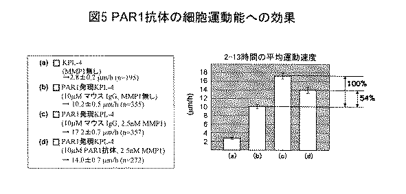
したがって、PAR1発現細胞はMMP1処理することにより運動速度が促進されるが、N2−11抗体で処理することによりMMP1によるPAR1切断効果が阻害され、運動能の活性化が約50%阻害されることを示している。 Thus, PAR1-expressing cells are accelerated by MMP1 treatment, but by treating with N2-11 antibody, the PAR1 cleavage effect by MMP1 is inhibited, and the activation of motility is inhibited by about 50%. Is shown.
実施例7:抗体の細胞浸潤能への効果の検討
PAR1発現KPL−4細胞は、MMP1処理によりPAR1が活性化し、細胞浸潤能が促進される。そこで、N2−11抗体存在下でPAR1発現KPL−4のマトリゲルアッセイを行い、N2−11抗体がMMP1依存的な細胞浸潤能を阻害するかを検討した。1.5x10⁵細胞のPAR1発現KPL−4細胞をトリプシン処理により浮遊させ、PBSで洗浄した。3および10μMのN2−11抗体またはコントロール抗体であるマウスIgGを添加し、37℃で90分間保温した。次いで、セルカルチャーインサートに細胞を蒔き、37℃で45時間培養した。インサートを浸す誘引物質溶液には、10%FBS含有2.5nM MMP1溶液を用い、45時間後にメンブレンフィルターの底面に移動してきた浸潤細胞をHE染色し、浸潤細胞数をカウントした。浸潤細胞数のカウントは、フィルター内のランダムな地点を0.66mmX0.66mmの大きさでスキャンし、その平均細胞数を算出した。次いで、マウスIgGで処理した場合の浸潤細胞数を100%としたときのN2−11抗体処理時の浸潤細胞数の割合を求めた。Example 7: Examination of effect of antibody on cell invasion ability In PAR1-expressing KPL-4 cells, PAR1 is activated by MMP1 treatment, and the cell invasion ability is promoted. Therefore, a Matrigel assay of PAR1-expressing KPL-4 was carried out in the presence of N2-11 antibody to examine whether N2-11 antibody inhibits MMP1-dependent cell invasion ability. 1.5 × 10 5 PAR1-expressing KPL-4 cells were suspended by trypsin treatment and washed with PBS. 3 and 10 μM of N2-11 antibody or mouse IgG as a control antibody was added and incubated at 37 ° C. for 90 minutes. Next, the cells were seeded on a cell culture insert and cultured at 37 ° C. for 45 hours. As an attractant solution for soaking the insert, a 2.5 nM MMP1 solution containing 10% FBS was used, and the infiltrating cells that had migrated to the bottom of the membrane filter after 45 hours were stained with HE, and the number of infiltrating cells was counted. The number of invading cells was counted by scanning random points in the filter with a size of 0.66 mm × 0.66 mm, and calculating the average number of cells. Subsequently, the ratio of the number of infiltrating cells at the time of N2-11 antibody treatment when the number of infiltrating cells when treated with mouse IgG was taken as 100% was determined.
結果
条件(b)の平均浸潤細胞数を100%とした場合におけるN2−11抗体処理時の浸潤細胞数の割合を図6に示し、その算出値を以下の表2に示す。
したがって、PAR1発現細胞はMMP1処理することにより浸潤能が促進されるが、N2−11抗体で処理することによりMMP1によるPAR1切断効果が用量依存的に阻害され、浸潤能の活性化が約60%阻害されることを示している。 Therefore, PAR1-expressing cells are promoted in invasive ability by treatment with MMP1, but treatment with N2-11 antibody inhibits the PAR1 cleavage effect by MMP1 in a dose-dependent manner, and activation of invasive ability is about 60%. It shows that it is inhibited.
上記実施例5〜7の結果より、N2−11抗体はPAR1の切断部位を認識し特異的に結合し、MMP1によるPAR1の切断活性を阻害することで、PAR1発現細胞のMMP1依存的な細胞運動活性および細胞浸潤活性を阻害することを示している。すなわち、N2−11抗体ががん治療において有効な抗体医薬品となりうることを示している。 From the results of Examples 5 to 7, the N2-11 antibody recognizes and specifically binds to the cleavage site of PAR1, and inhibits the cleavage activity of PAR1 by MMP1, so that MMP1-dependent cell movement of PAR1-expressing cells. Inhibiting activity and cell invasion activity. That is, it shows that the N2-11 antibody can be an effective antibody drug in cancer treatment.
実施例8:抗体の腫瘍細胞に対する特異性の検討
実施例5と同様のN2−11抗体−QDをプローブに用いて、独自のインビボイメージング法により腫瘍組織の生体染色を行い、上記により調製された抗体が腫瘍組織のPAR1に対し特異的であるかを検討した。SCIDマウスに1x10⁶細胞のPAR1発現KPL−4細胞を皮下注入した。次いで、4週間飼育しPAR1発現KPL−4細胞由来の腫瘍を作製した。腫瘍を露出させて観察用の小窓を作製した後、N2−11抗体−QDプローブを血中濃度10nMで尾静脈より注入した。次いで、独自のイメージング装置を用いて、生きたマウスの腫瘍組織を観察した。Example 8: Examination of specificity of antibody to tumor cells Using the same N2-11 antibody-QD as in Example 5 as a probe, living body staining of tumor tissue was performed by a unique in vivo imaging method, and the above was prepared. It was investigated whether the antibody was specific for PAR1 in tumor tissue. SCID mice were injected subcutaneously with 1 × 10 6 PAR1-expressing KPL-4 cells. Subsequently, it was raised for 4 weeks to produce a tumor derived from PAR1-expressing KPL-4 cells. After exposing the tumor to produce a small window for observation, an N2-11 antibody-QD probe was injected from the tail vein at a blood concentration of 10 nM. Next, the tumor tissue of live mice was observed using a unique imaging device.
結果
N2−11抗体−QDが腫瘍細胞に特異的に反応している像を図7に示す。図7より、マウス生体内において、N2−11抗体が腫瘍細胞に対し特異的であることが示された。したがって、濃度や結合能(結合定数)を高めることにより、N2−11抗体が生きた腫瘍細胞のPAR1活性を阻害する可能性を示唆している。Results An image of N2-11 antibody-QD reacting specifically with tumor cells is shown in FIG. FIG. 7 shows that the N2-11 antibody is specific for tumor cells in the mouse body. Therefore, by increasing the concentration and binding ability (binding constant), it is suggested that the N2-11 antibody may inhibit the PAR1 activity of living tumor cells.
実施例9:ポリクローナルPAR1抗体の調製と腫瘍細胞に対する活性の検討
PAR1の切断部位(R41とS42の間)をまたぐような配列(N35ATLDPRSFLL45)(配列番号:1)を標的として選択し、この配列のN末にCを付加したCN35ATLDPRSFLL45(配列番号:2)ペプチドを合成した。CのSH基を利用して合成ペプチドをKLHと架橋し、これを抗原としてウサギを免役し、PAR1に対する抗血清を調製した。次いで、抗血清のアフィニティー精製を行うため、抗原に用いたペプチドとSulfoLinkカップリングゲル(PIERCE社)を用いてカラムを作製した。カラムに抗血清をアプライした後、カラム体積の5倍以上のPBS(pH7.2)でカラムの洗浄を行った。その後、3M MgCl2でPAR1ポリクローナル抗体の溶出を行った。溶出後はPBSにて透析を行い、精製PAR1抗体とした(10mlの抗血清から約0.4mgの精製抗体を得た)。ポリクローナル抗体の細胞浸潤活性を、モノクローナル抗体のアッセイと同様の方法でマトリゲルを用いて検討した。コントロールとして300nMのウサギIgGを用いた。Example 9: Preparation of polyclonal PAR1 antibody and examination of activity against tumor cells A sequence (N 35 ATLDPRSFLL 45 ) (SEQ ID NO: 1) across the cleavage site of PAR1 (between R 41 and S 42 ) is selected as a target. Then, a CN 35 ATLDPRSFLL 45 (SEQ ID NO: 2) peptide in which C was added to the N terminus of this sequence was synthesized. The synthetic peptide was cross-linked with KLH using the SH group of C, and this was used as an antigen to immunize rabbits to prepare antiserum against PAR1. Subsequently, in order to perform affinity purification of the antiserum, a column was prepared using the peptide used for the antigen and SulfoLink coupling gel (PIERCE). After applying antiserum to the column, the column was washed with PBS (pH 7.2) at 5 times the column volume. Thereafter, the PAR1 polyclonal antibody was eluted with 3M MgCl 2 . After elution, dialysis was performed with PBS to obtain a purified PAR1 antibody (about 0.4 mg of purified antibody was obtained from 10 ml of antiserum). The cell invasion activity of the polyclonal antibody was examined using Matrigel in the same manner as the monoclonal antibody assay. As a control, 300 nM rabbit IgG was used.
結果
条件(a)の平均浸潤細胞数を100%とした場合におけるPAR1ポリクローナル抗体処理時の浸潤細胞数割合を図8に示し、その算出値を以下の表3に示す。
したがって、300nMのポリクローナルPAR1抗体は、10μMのモノクローナルPAR1抗体(阻害効果:約60%)と同レベルのがん細胞の浸潤能抑制効果を有することを示している。すなわち、遺伝子工学的手法により、モノクローナルPAR1抗体の遺伝子に改良を加えることで、10−100倍阻害効果を強化できることを示唆しており、本発明の抗体が有効な抗がん剤として使用できることを示している。 Therefore, it is shown that 300 nM polyclonal PAR1 antibody has the same level of cancer cell invasion suppression effect as 10 μM monoclonal PAR1 antibody (inhibitory effect: about 60%). That is, it is suggested that the inhibitory effect can be enhanced 10 to 100 times by improving the gene of the monoclonal PAR1 antibody by genetic engineering techniques, and that the antibody of the present invention can be used as an effective anticancer agent. Show.
さらに、これらの実験結果から、本発明の抗体は有効な抗がん剤として使用できるのみならず、腫瘍の検出にも使用できることも明らかである。 Furthermore, it is clear from these experimental results that the antibody of the present invention can be used not only as an effective anticancer agent but also for tumor detection.
本発明によれば、PAR1抗体が、PAR1発現細胞においてMMP1依存的な細胞運動能および細胞浸潤能を阻害すること、およびマウス生体内において腫瘍細胞に特異的に反応を示すことから、がん治療において有効な抗体医薬品となりうることを示唆している。また、本発明の抗体は腫瘍の検出等に使用することができる。 According to the present invention, the PAR1 antibody inhibits MMP1-dependent cell motility and cell invasion ability in PAR1-expressing cells, and specifically reacts with tumor cells in the mouse body. This suggests that it can be an effective antibody drug. Further, the antibody of the present invention can be used for tumor detection and the like.
Claims (7)
Priority Applications (1)
| Application Number | Priority Date | Filing Date | Title |
|---|---|---|---|
| JP2010505602A JP5483023B2 (en) | 2008-03-27 | 2009-03-19 | Cancer cell motility and cancer cell invasion inhibitor |
Applications Claiming Priority (4)
| Application Number | Priority Date | Filing Date | Title |
|---|---|---|---|
| JP2008083588 | 2008-03-27 | ||
| JP2008083588 | 2008-03-27 | ||
| JP2010505602A JP5483023B2 (en) | 2008-03-27 | 2009-03-19 | Cancer cell motility and cancer cell invasion inhibitor |
| PCT/JP2009/055479 WO2009119455A1 (en) | 2008-03-27 | 2009-03-19 | Cancer cell motility and cancer cell infiltration inhibitor |
Publications (2)
| Publication Number | Publication Date |
|---|---|
| JPWO2009119455A1 JPWO2009119455A1 (en) | 2011-07-21 |
| JP5483023B2 true JP5483023B2 (en) | 2014-05-07 |
Family
ID=41113650
Family Applications (1)
| Application Number | Title | Priority Date | Filing Date |
|---|---|---|---|
| JP2010505602A Active JP5483023B2 (en) | 2008-03-27 | 2009-03-19 | Cancer cell motility and cancer cell invasion inhibitor |
Country Status (4)
| Country | Link |
|---|---|
| US (1) | US8674079B2 (en) |
| EP (1) | EP2267125B1 (en) |
| JP (1) | JP5483023B2 (en) |
| WO (1) | WO2009119455A1 (en) |
Families Citing this family (8)
| Publication number | Priority date | Publication date | Assignee | Title |
|---|---|---|---|---|
| JP2012523438A (en) * | 2009-04-10 | 2012-10-04 | タフツ メディカル センター インコーポレイテッド | PAR-1 activation by metalloproteinase-1 (MMP-1) |
| WO2011102483A1 (en) * | 2010-02-19 | 2011-08-25 | 独立行政法人理化学研究所 | HUMAN LAP TGF-β BINDING ANTIBODY |
| JP5682975B2 (en) * | 2010-09-02 | 2015-03-11 | 国立大学法人東北大学 | Judgment method of cancer development or cancer development risk |
| US9745347B2 (en) | 2010-12-30 | 2017-08-29 | Hadasit Medical Research Services And Development Ltd. | PAR1 and PAR2 c-tail peptides and peptide mimetics |
| TWI549997B (en) | 2014-10-14 | 2016-09-21 | 財團法人工業技術研究院 | Method for manufacturing lignin-based biomass epoxy resin and lignin-based biomass epoxy resin compositions |
| JP7152156B2 (en) | 2015-01-14 | 2022-10-12 | ザ・ブリガーム・アンド・ウーメンズ・ホスピタル・インコーポレーテッド | Treatment of cancer with anti-LAP monoclonal antibodies |
| US11230601B2 (en) | 2017-10-10 | 2022-01-25 | Tilos Therapeutics, Inc. | Methods of using anti-lap antibodies |
| US11130802B2 (en) | 2018-10-10 | 2021-09-28 | Tilos Therapeutics, Inc. | Anti-lap antibody variants |
Family Cites Families (6)
| Publication number | Priority date | Publication date | Assignee | Title |
|---|---|---|---|---|
| US5776700A (en) * | 1996-09-05 | 1998-07-07 | Trustees Of The University Of Pennsylvania | Thrombine receptor activation assay for use in identifying thrombin receptor antagonists |
| IL125698A (en) | 1998-08-07 | 2007-03-08 | Hadasit Med Res Service | Use of molecules associated with protease activated receptors for the preparation of pharmaceutical compositions and pharmaceutical compositions comprising them |
| JP4469467B2 (en) | 2000-06-29 | 2010-05-26 | 帝人株式会社 | Cell growth inhibitor |
| US7074765B2 (en) * | 2003-05-01 | 2006-07-11 | The Regents Of The University Of Michigan | Synthetic peptide analogs of Arg-Pro-Pro-Gly-Phe as selective inhibitors of thrombin and thrombin activation of protease activated receptors 1 and 4 |
| US7888483B2 (en) | 2006-07-18 | 2011-02-15 | Irm Llc | Antagonists of protease activated receptor-1 (PAR1) |
| JP4861115B2 (en) | 2006-09-28 | 2012-01-25 | サントリーホールディングス株式会社 | Label, method for producing label, method for stabilizing line peeling strength of label over time, and container |
-
2009
- 2009-03-19 US US12/934,133 patent/US8674079B2/en active Active
- 2009-03-19 WO PCT/JP2009/055479 patent/WO2009119455A1/en active Application Filing
- 2009-03-19 JP JP2010505602A patent/JP5483023B2/en active Active
- 2009-03-19 EP EP09726307.3A patent/EP2267125B1/en not_active Not-in-force
Non-Patent Citations (4)
| Title |
|---|
| JPN6009016591; Cell, 2005, Vol.120, No.3, p.303-313 * |
| JPN6009016592; The Journal of biological chemistry, 1994, Vol.269, No.4, p.2943-2952 * |
| JPN6013024397; Cancer research. 1990, Vol.50, No.15, p.4485-4496 * |
| JPN6013040635; Biological chemistry. 2006, Vol.387, No.6, p.817-824 * |
Also Published As
| Publication number | Publication date |
|---|---|
| EP2267125A4 (en) | 2011-09-07 |
| US8674079B2 (en) | 2014-03-18 |
| EP2267125A1 (en) | 2010-12-29 |
| JPWO2009119455A1 (en) | 2011-07-21 |
| WO2009119455A1 (en) | 2009-10-01 |
| EP2267125B1 (en) | 2015-09-02 |
| US20110070163A1 (en) | 2011-03-24 |
Similar Documents
| Publication | Publication Date | Title |
|---|---|---|
| JP5483023B2 (en) | Cancer cell motility and cancer cell invasion inhibitor | |
| EP2723377B1 (en) | Anti-axl antibodies and uses thereof | |
| US9249228B2 (en) | Anti-Axl antibodies and uses thereof | |
| CN101573382B (en) | Monoclonal antibodies and directed against the human anti-mullerian hormone type ii receptor (amhr-ii) | |
| EP3419674A1 (en) | Targeting immunotherapy for amyloidosis | |
| CN105037551A (en) | Therapeutic agent | |
| CN103890010A (en) | Anti-human-HER3 antibodies and uses thereof | |
| US20160185856A1 (en) | Anti-Claudin 1 Antibodies and Uses Thereof | |
| CN107849138B (en) | anti-Aggrus monoclonal antibody and application thereof | |
| US20220389113A1 (en) | Binding proteins to cub domain-containing protein (cdcp1) | |
| Harrington et al. | Anti-CDCP1 immuno-conjugates for detection and inhibition of ovarian cancer | |
| CN111201244A (en) | anti-AQP 3 monoclonal antibodies that specifically bind to the extracellular domain of aquaporin 3(AQP3) and uses thereof | |
| US8586042B2 (en) | Hybridomas producing monoclonal anti-mortalin peptide antibodies | |
| CN101864398A (en) | A kind of monoclonal antibody of anti-gonadotropin-releasing hormone receptor and application | |
| US20220227882A1 (en) | Anti-adam8 antibodies and uses of the same | |
| WO2020158959A1 (en) | Anti-mc16 antibody | |
| WO2022056329A1 (en) | Snx9 subfamily-targeting antibodies |
Legal Events
| Date | Code | Title | Description |
|---|---|---|---|
| A521 | Request for written amendment filed |
Free format text: JAPANESE INTERMEDIATE CODE: A523 Effective date: 20120119 |
|
| A621 | Written request for application examination |
Free format text: JAPANESE INTERMEDIATE CODE: A621 Effective date: 20120119 |
|
| A131 | Notification of reasons for refusal |
Free format text: JAPANESE INTERMEDIATE CODE: A131 Effective date: 20130528 |
|
| A521 | Request for written amendment filed |
Free format text: JAPANESE INTERMEDIATE CODE: A523 Effective date: 20130723 |
|
| A131 | Notification of reasons for refusal |
Free format text: JAPANESE INTERMEDIATE CODE: A131 Effective date: 20130820 |
|
| A521 | Request for written amendment filed |
Free format text: JAPANESE INTERMEDIATE CODE: A523 Effective date: 20131015 |
|
| A02 | Decision of refusal |
Free format text: JAPANESE INTERMEDIATE CODE: A02 Effective date: 20131105 |
|
| A521 | Request for written amendment filed |
Free format text: JAPANESE INTERMEDIATE CODE: A523 Effective date: 20131118 |
|
| A911 | Transfer to examiner for re-examination before appeal (zenchi) |
Free format text: JAPANESE INTERMEDIATE CODE: A911 Effective date: 20140106 |
|
| TRDD | Decision of grant or rejection written | ||
| A01 | Written decision to grant a patent or to grant a registration (utility model) |
Free format text: JAPANESE INTERMEDIATE CODE: A01 Effective date: 20140121 |
|
| A61 | First payment of annual fees (during grant procedure) |
Free format text: JAPANESE INTERMEDIATE CODE: A61 Effective date: 20140204 |
|
| R150 | Certificate of patent or registration of utility model |
Ref document number: 5483023 Country of ref document: JP Free format text: JAPANESE INTERMEDIATE CODE: R150 |
|
| R250 | Receipt of annual fees |
Free format text: JAPANESE INTERMEDIATE CODE: R250 |
|
| R250 | Receipt of annual fees |
Free format text: JAPANESE INTERMEDIATE CODE: R250 |
|
| R250 | Receipt of annual fees |
Free format text: JAPANESE INTERMEDIATE CODE: R250 |
|
| R250 | Receipt of annual fees |
Free format text: JAPANESE INTERMEDIATE CODE: R250 |
|
| R250 | Receipt of annual fees |
Free format text: JAPANESE INTERMEDIATE CODE: R250 |
|
| R250 | Receipt of annual fees |
Free format text: JAPANESE INTERMEDIATE CODE: R250 |