JP5481383B2 - Deletion of mitochondrial DNA between about 12317 and about 16254 residues for use in detecting cancer - Google Patents
Deletion of mitochondrial DNA between about 12317 and about 16254 residues for use in detecting cancer Download PDFInfo
- Publication number
- JP5481383B2 JP5481383B2 JP2010532390A JP2010532390A JP5481383B2 JP 5481383 B2 JP5481383 B2 JP 5481383B2 JP 2010532390 A JP2010532390 A JP 2010532390A JP 2010532390 A JP2010532390 A JP 2010532390A JP 5481383 B2 JP5481383 B2 JP 5481383B2
- Authority
- JP
- Japan
- Prior art keywords
- cancer
- mtdna
- mitochondrial dna
- deletion
- pair
- Prior art date
- Legal status (The legal status is an assumption and is not a legal conclusion. Google has not performed a legal analysis and makes no representation as to the accuracy of the status listed.)
- Expired - Fee Related
Links
Images


Classifications
-
- C—CHEMISTRY; METALLURGY
- C12—BIOCHEMISTRY; BEER; SPIRITS; WINE; VINEGAR; MICROBIOLOGY; ENZYMOLOGY; MUTATION OR GENETIC ENGINEERING
- C12Q—MEASURING OR TESTING PROCESSES INVOLVING ENZYMES, NUCLEIC ACIDS OR MICROORGANISMS; COMPOSITIONS OR TEST PAPERS THEREFOR; PROCESSES OF PREPARING SUCH COMPOSITIONS; CONDITION-RESPONSIVE CONTROL IN MICROBIOLOGICAL OR ENZYMOLOGICAL PROCESSES
- C12Q1/00—Measuring or testing processes involving enzymes, nucleic acids or microorganisms; Compositions therefor; Processes of preparing such compositions
- C12Q1/68—Measuring or testing processes involving enzymes, nucleic acids or microorganisms; Compositions therefor; Processes of preparing such compositions involving nucleic acids
- C12Q1/6876—Nucleic acid products used in the analysis of nucleic acids, e.g. primers or probes
- C12Q1/6883—Nucleic acid products used in the analysis of nucleic acids, e.g. primers or probes for diseases caused by alterations of genetic material
- C12Q1/6886—Nucleic acid products used in the analysis of nucleic acids, e.g. primers or probes for diseases caused by alterations of genetic material for cancer
-
- C—CHEMISTRY; METALLURGY
- C12—BIOCHEMISTRY; BEER; SPIRITS; WINE; VINEGAR; MICROBIOLOGY; ENZYMOLOGY; MUTATION OR GENETIC ENGINEERING
- C12Q—MEASURING OR TESTING PROCESSES INVOLVING ENZYMES, NUCLEIC ACIDS OR MICROORGANISMS; COMPOSITIONS OR TEST PAPERS THEREFOR; PROCESSES OF PREPARING SUCH COMPOSITIONS; CONDITION-RESPONSIVE CONTROL IN MICROBIOLOGICAL OR ENZYMOLOGICAL PROCESSES
- C12Q1/00—Measuring or testing processes involving enzymes, nucleic acids or microorganisms; Compositions therefor; Processes of preparing such compositions
- C12Q1/68—Measuring or testing processes involving enzymes, nucleic acids or microorganisms; Compositions therefor; Processes of preparing such compositions involving nucleic acids
- C12Q1/6844—Nucleic acid amplification reactions
- C12Q1/6851—Quantitative amplification
-
- C—CHEMISTRY; METALLURGY
- C12—BIOCHEMISTRY; BEER; SPIRITS; WINE; VINEGAR; MICROBIOLOGY; ENZYMOLOGY; MUTATION OR GENETIC ENGINEERING
- C12Q—MEASURING OR TESTING PROCESSES INVOLVING ENZYMES, NUCLEIC ACIDS OR MICROORGANISMS; COMPOSITIONS OR TEST PAPERS THEREFOR; PROCESSES OF PREPARING SUCH COMPOSITIONS; CONDITION-RESPONSIVE CONTROL IN MICROBIOLOGICAL OR ENZYMOLOGICAL PROCESSES
- C12Q2600/00—Oligonucleotides characterized by their use
- C12Q2600/156—Polymorphic or mutational markers
-
- C—CHEMISTRY; METALLURGY
- C12—BIOCHEMISTRY; BEER; SPIRITS; WINE; VINEGAR; MICROBIOLOGY; ENZYMOLOGY; MUTATION OR GENETIC ENGINEERING
- C12Q—MEASURING OR TESTING PROCESSES INVOLVING ENZYMES, NUCLEIC ACIDS OR MICROORGANISMS; COMPOSITIONS OR TEST PAPERS THEREFOR; PROCESSES OF PREPARING SUCH COMPOSITIONS; CONDITION-RESPONSIVE CONTROL IN MICROBIOLOGICAL OR ENZYMOLOGICAL PROCESSES
- C12Q2600/00—Oligonucleotides characterized by their use
- C12Q2600/158—Expression markers
Description
〔関連出願の相互参照〕
本願は、2007年11月9日に出願されたUS特許出願番号61/002,637の優先権主張出願であり、当該出願の内容は、参照により本願に含まれる。
[Cross-reference of related applications]
This application is a priority claim application of US Patent Application No. 61 / 002,637 filed on Nov. 9, 2007, the contents of which are incorporated herein by reference.
本発明は、ミトコンドリアゲノミクス分野に関する。具体的には、本発明は、ヒトミトコンドリアゲノムの変異の検出、および癌の指標としてのそれらの使用に関する。 The present invention relates to the field of mitochondrial genomics. Specifically, the present invention relates to the detection of mutations in the human mitochondrial genome and their use as an indicator of cancer.
〔発明の背景〕
(診断ツールとしてのミトコンドリアDNA)
ミトコンドリアDNA(mtDNA)配列のダイナミクスは、重要な診断ツールである。mtDNA内の変異は、多くの場合、発病の事前的指標であり、核の変異を伴う場合が多い。mtDNA内の変異は、以下のもののバイオマーカーとして機能する。すなわち、疾病(例えば、喫煙および受動喫煙を原因とする組織損傷および癌などの疾病であるが、これらに限定されない)(Lee et al., 1998; Wei, 1998);20歳位から始まり、その後増加するミトコンドリアゲノム突然変異の蓄積による加齢(von Wurmb, 1998);突然変異、または、発癌物質、突然変異原、紫外線への曝露によって発症する転移性疾患(Birch-Machin, 2000);変形性関節症、心疾患、アルツハイマー症、パーキンソン病(Shoffner et al., 1993; Sherratt et al., 1997;Zhang et al, 1998);加齢による難聴(Seidman et al., 1997);視神経の変性および不整脈(Brown et al., 1997; Wallace et al., 1988);慢性進行性外眼筋麻痺(Taniike et al., 1992);アテローム性動脈硬化(Bogliolo et al., 1999);甲状腺乳頭癌および甲状腺腫瘍(Yeh et al., 2000);およびその他(例えば、Naviaux, 1997; ChinneryおよびTurnbull, 1999)。
BACKGROUND OF THE INVENTION
(Mitochondrial DNA as a diagnostic tool)
The dynamics of mitochondrial DNA (mtDNA) sequences is an important diagnostic tool. Mutations in mtDNA are often a preliminary indicator of pathogenesis and often accompany nuclear mutations. Mutations in mtDNA function as biomarkers for: That is, diseases (for example, diseases such as, but not limited to, tissue damage and cancer caused by smoking and passive smoking) (Lee et al., 1998; Wei, 1998); Aging with increasing accumulation of mitochondrial genomic mutations (von Wurmb, 1998); mutations or metastatic disease caused by exposure to carcinogens, mutagens, UV radiation (Birch-Machin, 2000); deformability Arthropathy, heart disease, Alzheimer's disease, Parkinson's disease (Shoffner et al., 1993; Sherratt et al., 1997; Zhang et al, 1998); deafness due to aging (Seidman et al., 1997); Arrhythmia (Brown et al., 1997; Wallace et al., 1988); Chronic progressive extraocular palsy (Taniike et al., 1992); Atherosclerosis (Bogliolo et al., 1999); Papillary thyroid cancer and Thyroid tumors (Yeh et al., 2000); and others (eg, Na viaux, 1997; Chinnery and Turnbull, 1999).
ミトコンドリアゲノムの特定部位における変異が、特定の疾病をもたらす可能性がある。例えば、4216、4217および4917の位置での変異は、レーベル遺伝性視神経症(LHON)と関連がある(Mitochondrial Research Society; Huoponen (2001); MitoMap)。ユビキノールシトクロムcレダクターゼ(錯体III)欠失症を患っている5人の患者のうちの5人において、15452の位置における変異が見出された(Valnot et al.1999)。 Mutations at specific sites in the mitochondrial genome can lead to specific diseases. For example, mutations at positions 4216, 4217 and 4917 are associated with the label hereditary optic neuropathy (LHON) (Mitochondrial Research Society; Huoponen (2001); MitoMap). A mutation at position 15452 was found in 5 out of 5 patients suffering from ubiquinol cytochrome c reductase (complex III) deficiency (Valnot et al. 1999).
具体的には、これらの変異または変性は、点突然変異(転位、転換)、欠失(1塩基〜数千塩基)、逆位、重複(1塩基〜数千塩基)、組換えおよび挿入(1塩基〜数千塩基)を含む。加えて、特定塩基対の変性、欠失またはそれらの組み合わせが、早期の前立腺癌、皮膚癌および肺癌、ならびに加齢に関連すること(例えばPolyak et al., 1998)、早期老化、発癌性物質への曝露に関連すること(Lee et al., 1998)などがわかっている。 Specifically, these mutations or modifications include point mutations (translocations, conversions), deletions (1 base to several thousand bases), inversions, duplications (1 base to several thousand bases), recombination and insertion ( 1 base to several thousand bases). In addition, certain base pair degenerations, deletions or combinations thereof are associated with early prostate cancer, skin and lung cancer, and aging (eg Polyak et al., 1998), premature aging, carcinogens (Lee et al., 1998) are known to be related to exposure to
(前立腺癌)
前立腺癌は、前立腺上皮に由来する可能性が最も高い固形癌と診断されることが多い(Huang et al. 1999)。1997年、約1000万人のアメリカ人男性が、その有無により前立腺癌を検知する前立腺特異抗原(PSA)の検査を受けた(Woodwell, 1999)。実際、初期の直腸指診(DRE)の検査を受けた男性の数は、より多い。同年、3100万人の男性がDREを受けた(Woodwell, 1999)。さらに、アメリカ合衆国において前立腺癌と新たに診断された年間の件数は、179,000件と推定される(Landis et al., 1999)。カナダ人男性の中では、前立腺癌は、癌の診断数および癌によって死亡する原因の第2位を占める。1997年、前立腺癌は、カナダ人男性の中で新たに癌と診断された件数のうちの、19,800件を占めた(28%)(National Cancer Institute of Canada)。49歳以上の全男性のうち、30〜40%が何らかの癌性前立腺細胞を有しており、そのうち20〜25%の男性が、臨床的に有意な形態の前立腺癌をもつと推測される(SpringNet - CE Connection, internet, www.springnet.com/ce/j803a.htm)。前立腺癌は、内生要因および外生要因の両方、すなわち、社会経済状況、食事、地理、ホルモン失調、家系、および遺伝子構成を含む広範な組織学的性質を示す(Konishi et al. 1997; Hayward et al. 1998)。特定のmtDNA変性が前立腺癌に関連付けられてきたが、前立腺癌の検出のためのさらなるマーカーが必要である。
(Prostate cancer)
Prostate cancer is often diagnosed as a solid cancer most likely derived from the prostate epithelium (Huang et al. 1999). In 1997, approximately 10 million American men were tested for prostate specific antigen (PSA) to detect prostate cancer based on their presence or absence (Woodwell, 1999). In fact, the number of men who have undergone an initial digital rectal exam (DRE) is higher. In the same year, 31 million men received DRE (Woodwell, 1999). In addition, the annual number of newly diagnosed prostate cancers in the United States is estimated to be 179,000 (Landis et al., 1999). Among Canadian men, prostate cancer is the second leading cause of cancer death and death from cancer. In 1997, prostate cancer accounted for 19,800 of the newly diagnosed cancer cases among Canadian men (28%) (National Cancer Institute of Canada). Of all men over the age of 49, 30-40% have some cancerous prostate cells, of which 20-25% are estimated to have clinically significant forms of prostate cancer ( SpringNet-CE Connection, internet, www.springnet.com/ce/j803a.htm). Prostate cancer exhibits a wide range of histological properties including both endogenous and exogenous factors, namely socio-economic status, diet, geography, hormonal dysfunction, ancestry, and genetic makeup (Konishi et al. 1997; Hayward et al. 1998). Although specific mtDNA degeneration has been associated with prostate cancer, additional markers for the detection of prostate cancer are needed.
(乳癌)
乳癌は、乳腺組織の癌であり、癌による死亡原因の第5位を占める。2005年、乳癌によって全世界で502,000人が亡くなった(癌死亡者の7%、全死亡者の1%近く)(世界保健機構 Cancer Fact Sheet No. 297)。世界の女性の中で、乳癌が最も一般的な癌であり、癌による死因の第1位を占める(世界保健機構 Cancer Fact Sheet No. 297)。例えばParrella et al.などは、特定のmtDNA変性が乳癌をもたらすとしたが(Cancer Research: 61, 2001)、乳癌の検出のためにさらなるマーカーが必要である。
(breast cancer)
Breast cancer is a cancer of the mammary gland tissue and is the fifth leading cause of death due to cancer. In 2005, 502,000 people worldwide died from breast cancer (7% of cancer deaths, nearly 1% of all deaths) (World Health Organization Cancer Fact Sheet No. 297). Breast cancer is the most common cancer among women in the world and is the leading cause of cancer death (World Health Organization Cancer Fact Sheet No. 297). For example, Parrella et al. And others have stated that certain mtDNA modifications result in breast cancer (Cancer Research: 61, 2001), but additional markers are needed for breast cancer detection.
上記背景技術の情報は、本発明に関連し得ると出願人が考える周知の情報を提供することを目的とする。よって、下記情報のうちのいずれかが本発明に対する先行技術を構成することを認めるものではなく、そのように解釈されるべきではない。 The background information is intended to provide well-known information that the applicant believes may be relevant to the present invention. Thus, no admission is made that any of the following information constitutes prior art to the present invention and should not be so construed.
〔発明の要約〕
本発明は、癌の検出に使用するミトコンドリアDNA変異に関する。本発明の一形態によれば、個体における癌の検出方法は、
a)個体から生物学的試料を得るステップ;
b)上記試料からミトコンドリアDNA(mtDNA)を抽出するステップ;
c)上記試料において、ヒトmtDNAゲノムの約12317残基〜約16254残基の間にmtDNA配列の欠失を有するmtDNAの量を定量するステップ、および
d)少なくとも1つの周知の参照値と、欠失を有する試料におけるmtDNAの量とを比較するステップ、を含む。
[Summary of the Invention]
The present invention relates to mitochondrial DNA mutations used for cancer detection. According to one aspect of the present invention, a method for detecting cancer in an individual comprises:
a) obtaining a biological sample from an individual;
b) extracting mitochondrial DNA (mtDNA) from the sample;
c) quantifying the amount of mtDNA having a deletion of the mtDNA sequence between about 12317 residues to about 16254 residues of the human mtDNA genome in the sample, and d) at least one known reference value, Comparing the amount of mtDNA in a sample having a loss.
本発明の別の形態によれば、癌の進行に関する個体の測定方法は、
a)生物学的試料を得るステップ;
b)上記試料からミトコンドリアDNA(mtDNA)を抽出するステップ;
c)上記試料において、ヒトmtDNAゲノムの約12317残基〜約16254残基の間にmtDNA配列の欠失を有するmtDNAの量を定量するステップ、および、
d)一定期間にわたってa)〜c)のステップを繰り返すステップを含み、一定期間を経た後の欠失の増加レベルが癌の指標である。
According to another aspect of the present invention, a method for measuring an individual relating to cancer progression comprises:
a) obtaining a biological sample;
b) extracting mitochondrial DNA (mtDNA) from the sample;
c) quantifying in the sample the amount of mtDNA having a deletion of the mtDNA sequence between about 12317 residues to about 16254 residues of the human mtDNA genome; and
d) It includes the step of repeating steps a) to c) over a certain period, and the increased level of deletion after a certain period is an indicator of cancer.
本発明の別の形態によると、個体における癌の検出方法は、
a)個体から生物学的試料を得るステップ;
b)上記試料からミトコンドリアDNA(mtDNA)を抽出するステップ;
c)上記試料において配列番号1または配列番号2に記載の配列に対応する配列を有するmtDNAの量を定量するステップ;および、
d)上記試料において配列番号1または配列番号2に対応するmtDNAの量と、少なくとも1つの周知の参照値とを比較するステップを含む。
According to another aspect of the invention, a method for detecting cancer in an individual comprises:
a) obtaining a biological sample from an individual;
b) extracting mitochondrial DNA (mtDNA) from the sample;
c) quantifying the amount of mtDNA having a sequence corresponding to the sequence set forth in SEQ ID NO: 1 or SEQ ID NO: 2 in the sample; and
d) comparing the amount of mtDNA corresponding to SEQ ID NO: 1 or SEQ ID NO: 2 in the sample with at least one known reference value.
本発明の別の形態によると、
a)1つ以上の生物学的試料を収集するための材料;および、
b)mtDNA欠失の検出に適したプライマーおよび試薬を含む、本発明の方法を実施するための診断キットを提供する。
According to another aspect of the invention,
a) material for collecting one or more biological samples; and
b) Provide a diagnostic kit for carrying out the method of the invention, comprising primers and reagents suitable for the detection of mtDNA deletions.
〔図面の簡単な説明〕
添付の図面の参照、および下記詳細な説明から、本発明の上記および他の特徴が明らかとなるであろう。
[Brief description of the drawings]
These and other features of the present invention will become apparent from a reading of the accompanying drawings and from the detailed description below.
図1は、実施例1のサイクル閾値(cycle threshhold)を示すグラフである。 FIG. 1 is a graph showing a cycle threshold (cycle threshhold) according to the first embodiment.
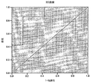
図2は、本発明の一実施形態の特異性および感度を示すROC曲線である。 FIG. 2 is a ROC curve showing the specificity and sensitivity of one embodiment of the present invention.
図3は、実施例2に関するサイクル閾値を示すグラフである。 FIG. 3 is a graph showing the cycle threshold for the second embodiment.
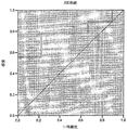
図4は、本発明の別の実施形態の特異性および感度を示すROC曲線である。 FIG. 4 is an ROC curve showing the specificity and sensitivity of another embodiment of the present invention.
図5は、4kbの欠失の検出に有効なプライマーの設計および配列を示す概略図である。 FIG. 5 is a schematic diagram showing the design and sequence of primers effective for the detection of a 4 kb deletion.
図6は、本発明の別の実施形態の特異性および感度を示すROC曲線である。 FIG. 6 is an ROC curve showing the specificity and sensitivity of another embodiment of the present invention.
〔発明の詳細な説明〕
本発明は、癌の予測、診断および検査方法を提供する。当該方法は、1つ以上の生物学的試料を得るステップと、試料からミトコンドリアDNA(mtDNA)を抽出するステップと、試料内のミトコンドリア変異の量を測定するステップと、参照値と試料内の変異の量とを比較するステップとを含む。即ち、方法は、発病を検知する、および個体が癌になる傾向を評価するための総合的ツールを提供する。本方法によって、経時的に個体の危険因子を測定することができ、および/または、治療薬および治療計画への患者の反応を測定することができる。
Detailed Description of the Invention
The present invention provides cancer prediction, diagnosis and testing methods. The method includes obtaining one or more biological samples, extracting mitochondrial DNA (mtDNA) from the sample, measuring the amount of mitochondrial mutations in the sample, reference values and mutations in the sample Comparing with the amount of. That is, the method provides a comprehensive tool for detecting disease onset and assessing the tendency of an individual to become cancerous. The method can measure an individual's risk factors over time and / or measure a patient's response to a therapeutic agent and treatment plan.
〔定義〕
特段別に定義されない限りは、本願に使用される全ての技術用語および科学用語は、本発明の属する技術分野の当業者が共通に理解する意味を有する。
[Definition]
Unless defined otherwise, all technical and scientific terms used herein have the meaning commonly understood by one of ordinary skill in the art to which this invention belongs.
本願にて使用するとき、「約」という用語は、言及した値から妥当な範囲の偏差があることを意図する。本願において提供されたあらゆる既定値は、具体的に示されているか否かに関わらず、このような偏差が常に有するものと解釈されるものである。 As used herein, the term “about” intends a reasonable range of deviation from the stated value. Any default values provided in this application are to be construed as always having such a deviation, whether or not specifically indicated.
本願にて定義するとき、「生物学的試料」とは、mtDNAが得られる細胞を含む組織または体液を意図する。例えば、生物学的試料を、乳房組織または前立腺組織などの組織、または血液、唾液、脳脊髄液、喀痰、尿、粘膜、滑液、腹水、羊水などから抽出することができる。生物学的試料は、外科検体(surgical specimen)または生検検体(biopsy specimen)であってもよい。生物学的試料は、供給源から得られたものをそのまま使用してもよいし、または、前処理を行ってその特性を変更したものであってもよい。したがって、血清、破壊細胞からプラズマまたは血清を調製する、固形原料から液体、希釈粘液、濾液、濃縮液、不活性化干渉要素、添加試薬などを調整することによって、使用前に生物学的試料に前処理を施すことができる。 As defined in this application, a “biological sample” intends a tissue or fluid that contains cells from which mtDNA is obtained. For example, a biological sample can be extracted from tissues such as breast tissue or prostate tissue, or blood, saliva, cerebrospinal fluid, sputum, urine, mucosa, synovial fluid, ascites, amniotic fluid, and the like. The biological sample may be a surgical specimen or a biopsy specimen. The biological sample obtained from the source may be used as it is, or may have been subjected to pretreatment to change its characteristics. Therefore, prepare plasma or serum from serum, disrupted cells, adjust liquids, diluted mucus, filtrates, concentrates, inactivated interference elements, added reagents, etc. from solid raw materials to biological samples before use. Pretreatment can be performed.
本願において、「サイクル閾値」(CT)という用語は、リアルタイムPCRを用いた目的物の増幅が、蛍光シグナルなどの信号によって示されるときに、バックグラウンドを超えて上昇する点である。CTは、検査される配列の量に反比例する。 In this application, the term “cycle threshold” (C T ) is the point where amplification of an object using real-time PCR rises above background when indicated by a signal such as a fluorescent signal. CT is inversely proportional to the amount of sequence examined.
本願において、「診断」とは、疾病の診断または管理における要素として、変異または変異の組み合わせが存在するか否かを使用することを意図する。変異の検出は、疾病の診断における1つのステップであり得る。 In this application, “diagnosis” intends to use whether a mutation or a combination of mutations exists as an element in the diagnosis or management of a disease. Mutation detection can be a step in the diagnosis of disease.
本願において、「欠失」とは、mtDNAの連続した配列から、mtDNAの或る領域が除去されることを意図する。欠失は、1塩基〜数千塩基、またはそれ以上の塩基の大きさに及ぶ。 In this application, “deletion” intends to remove a region of mtDNA from a contiguous sequence of mtDNA. Deletions range in size from one base to several thousand bases or more.
本願において、「ミトコンドリアDNA」または「mtDNA」とは、ミトコンドリア内に存在するDNAである。 In the present application, “mitochondrial DNA” or “mtDNA” is DNA present in mitochondria.
本願において、「変異」は、ミトコンドリアDNA内における、野生型配列からのあらゆる修飾または変化を含む。例えば、点突然変異、転位、挿入、転換、転座、欠失、逆位、重複、組み換え、またはその組み合わせを含むが、これらに限定されない。配列の修飾または変化は、一塩基の変化から、DNA断片全体の追加または除去までひろく含むものである。 As used herein, “mutation” includes any modification or change from the wild-type sequence in mitochondrial DNA. Examples include, but are not limited to, point mutations, transpositions, insertions, conversions, translocations, deletions, inversions, duplications, recombination, or combinations thereof. Sequence modifications or changes can range from a single base change to the addition or removal of an entire DNA fragment.
本願において、「感度」とは、本発明の方法を用いて得られる陽性な結果の割合(真陽性率)を意図する。 In the present application, “sensitivity” intends the proportion of positive results (true positive rate) obtained using the method of the present invention.
本願において、「特異性」は、本発明を用いて得られる偽陽性な結果の割合(偽陽性率)を意図する。 In the present application, “specificity” intends the rate of false positive results (false positive rate) obtained using the present invention.
「治療」および「処置」は、本願において互いに交換可能に使用され、被験者の状態を改善させることを意図して行われる診療行為を意図する。改善は、主観的または客観的であってよく、病状の緩和、疾病の進行の抑制、または病状を変化させることに関する。したがって、治療および処置という用語は、最も広義に使用され、様々な段階での疾病の防止(予防)、緩和、低減、治癒を含む。被験者の状態の悪化を防ぐことも本用語に含まれる。治療/処置を必要とする被験者は、疾病に既に罹患しているもの、および、疾病の進行の傾向もしくは危険性があるもの、疾病の予防を行うものを含む。 “Treatment” and “treatment” are used interchangeably in this application, and are intended to be practiced with the intention of improving a subject's condition. Improvement may be subjective or objective and relates to alleviating the condition, inhibiting the progression of the disease, or changing the condition. Accordingly, the terms treatment and treatment are used in the broadest sense and include prevention (prevention), alleviation, reduction, cure of disease at various stages. Preventing deterioration of the subject's condition is also included in this term. Subjects in need of treatment / treatment include those who are already afflicted with the disease, those who are prone to or at risk of progression of the disease, and those who are preventing the disease.
〔癌の予測、診断および測定のための分析〕
(ミトコンドリアの変異を検出するためのアッセイ)
ミトコンドリアDNA(mtDNA)ダイナミクスは、重要な診断ツールである。mtDNA内の変異は、発症の事前的な指標であることが多く、発病に関する危険因子を示すバイオマーカーとして機能する。本願において説明するように、生物学的試料内のミトコンドリアDNAの異常のレベルを測定することによって、1つ以上の癌の存在を検出することが可能であるとともに、1つ以上の癌が患者に及ぼす潜在的リスクまたは素因を特定することが可能である。さらに、定期的にmtDNAを測定することによって、健康管理の専門家に、経時的に患者の進行を測定するリアルタイム定量測定ツール、および/または、治療推奨の評価としてのツールを提供することができ、癌予防または治療における有効性の測定が可能になる。
[Analysis for cancer prediction, diagnosis and measurement]
(Assay to detect mitochondrial mutations)
Mitochondrial DNA (mtDNA) dynamics are important diagnostic tools. Mutations in mtDNA are often a preliminary indicator of onset and function as biomarkers that indicate risk factors for pathogenesis. As described herein, by measuring the level of mitochondrial DNA abnormalities in a biological sample, it is possible to detect the presence of one or more cancers and one or more cancers in a patient. It is possible to identify potential risks or predisposing factors. In addition, by periodically measuring mtDNA, health care professionals can be provided with real-time quantitative measurement tools that measure patient progression over time and / or tools as an assessment of treatment recommendations. It is possible to measure the effectiveness in preventing or treating cancer.
したがって、本発明は、1つ以上の生物学的試料を得るステップ、該試料からミトコンドリアDNA(mtDNA)を抽出するステップ、および該試料内のmtDNA異常の量を定量し、参照値と異常レベルとを比較することによって、ミトコンドリアの変異に関して該試料を分析するステップを含む、癌の予測、診断および測定方法を提供する。参照値は、当該方法が癌の予測、診断または測定のいずれを目的としているかに基づくものであることは、当業者には自明であろう。したがって、参照値は、1つ以上の周知の非癌性である生物学的試料、1つ以上の周知の癌性である生物学的試料、および/または、経時的に取られた1つ以上の生物学的試料、から採取されたmtDNAに関連していてもよい。これらの参照値は、1つ以上の生物学的試料から採取されたmtDNAデータとの比較に使用され、このとき、例えば参照試料と比較される生物学的試料における欠失が同量である、または増加しているとき、癌の兆候または癌の発病を示す。あるいは、経時的な欠失レベルの増加が癌の発病を示す。 Accordingly, the present invention provides a step of obtaining one or more biological samples, extracting mitochondrial DNA (mtDNA) from the sample, and quantifying the amount of mtDNA abnormalities in the sample; Provides a method for predicting, diagnosing and measuring cancer, comprising analyzing the sample for mitochondrial mutations. It will be apparent to those skilled in the art that the reference value is based on whether the method is intended to predict, diagnose or measure cancer. Thus, the reference value is one or more well-known non-cancerous biological samples, one or more well-known cancerous biological samples, and / or one or more taken over time May be associated with mtDNA taken from a biological sample. These reference values are used for comparison with mtDNA data taken from one or more biological samples, for example, with the same amount of deletions in the biological sample compared to the reference sample, Or, when increasing, indicates signs of cancer or the onset of cancer. Alternatively, increasing deletion levels over time indicate the onset of cancer.
本発明の形態によれば、癌の予測、測定および診断方法は、1つ以上のミトコンドリアの変異を検出および測定するための分析を含む。本発明の一実施形態によれば、変異は、mtDNAの欠失である。本発明の別の実施形態によれば、変異は、3926bpのmtDNAの欠失である(本願において、「4kb欠失」または「4kb配列」と称する)。さらなる別の実施形態によれば、変異は、配列番号1または配列番号2に記載された配列を有するmtDNAの欠失である。環状形態である場合に、配列番号1と配列番号2とに違いはない。 According to an aspect of the invention, a method for predicting, measuring and diagnosing cancer includes an analysis for detecting and measuring one or more mitochondrial mutations. According to one embodiment of the invention, the mutation is a deletion of mtDNA. According to another embodiment of the invention, the mutation is a deletion of 3926 bp mtDNA (referred to herein as a “4 kb deletion” or “4 kb sequence”). According to yet another embodiment, the mutation is a deletion of mtDNA having the sequence set forth in SEQ ID NO: 1 or SEQ ID NO: 2. There is no difference between SEQ ID NO: 1 and SEQ ID NO: 2 when it is in a circular form.
4kb欠失は、ヒトmtDNAゲノムの約12317番目のヌクレオチドから16254番目のヌクレオチドの範囲内で発生するものである。本願では、ヒトmtDNAゲノムを配列番号3と示す(Genbank accession no. AC_000021)。4kbの欠失は、大きさ12bpの直接隣接する反復を特徴とし、当該反復は、12317〜12328の位置、および16243〜16254の位置に配置される。反復配列は、5'-TGCAACTCCAAA-3'である。したがって、本発明の一実施形態によれば、上記変異は、ヒトmtDNAゲノムにおける約12317残基〜約16254残基の欠失である。 The 4 kb deletion occurs within the range of about nucleotides 12317 to 16254 in the human mtDNA genome. In the present application, the human mtDNA genome is shown as SEQ ID NO: 3 (Genbank accession no. AC_000021). The 4 kb deletion is characterized by 12 bp in size and immediately adjacent repeats, which are placed at positions 12317-12328 and 16243-16254. The repetitive sequence is 5'-TGCAACTCCAAA-3 '. Thus, according to one embodiment of the invention, the mutation is a deletion of about 12317 residues to about 16254 residues in the human mtDNA genome.
発明者らは、後述する実施例にて示すように、当該欠失が癌、特に前立腺癌および乳癌に関連していることを発見した。したがって、当該欠失が正確なバイオマーカーを提供し、少なくともこれらの組織における癌の検出、診断または測定に有益な手段を提供する。 The inventors have discovered that the deletion is associated with cancer, particularly prostate cancer and breast cancer, as shown in the Examples below. Thus, the deletion provides an accurate biomarker and at least provides a useful tool for detecting, diagnosing or measuring cancer in these tissues.
欠失の結果、2つの欠失モノマーが生成される。1つは4kbの大きさ(小サブリモン(small sublimon))であり、もう1つは約12.5kbの大きさ(大サブリモン(large sublimon))である。それぞれ4kbまたは12.5kbの配列である小サブリモンまたは大サブリモンの存在を特定することによって、欠失の発生を検出してもよい。 As a result of the deletion, two deletion monomers are produced. One is 4 kb in size (small sublimon) and the other is about 12.5 kb in size (large sublimon). The occurrence of a deletion may be detected by identifying the presence of small or large sublimons, each of 4 kb or 12.5 kb sequence.
実施例において、ミトコンドリアの変異を測定する方法の一例を挙げる。試料からのmtDNAの抽出は、好適な公知の方法にて行っても良い。mtDNAの抽出後、ミトコンドリアゲノムの全領域または特定の領域を増幅させる。このmtDNAの抽出は、公知のまたは、例えばCurrent Protocols in Molecular Biology (Ausubel et al., John Wiley & Sons, New York, 2007)に記載されたようなミトコンドリアゲノムの配列解析(sequencing)を含んでいてもよい。同様に、当業者に周知の好適な技術から、mtDNA内の変異の存在を検出する方法を選択することができる。例えば、mtDNAの分析は、mtDNAの配列解析、PCRによる増幅、サザンブロット法、ノーザンブロット法、ウェスタンサウスウェスタンブロット法、変性HPLC、マイクロアレイ、バイオチップまたは遺伝子チップを使用したハイブダイゼーション、分子マーカー分析、バイオセンサ、融解温度プロファイリング、のいずれか、またはこれらを組み合わせて含んでいてもよい。 In the Examples, an example of a method for measuring mitochondrial mutation is given. Extraction of mtDNA from a sample may be performed by a suitable known method. After extraction of mtDNA, the entire region or specific region of the mitochondrial genome is amplified. This extraction of mtDNA is known or includes sequencing of the mitochondrial genome as described, for example, in Current Protocols in Molecular Biology (Ausubel et al., John Wiley & Sons, New York, 2007). Also good. Similarly, methods for detecting the presence of mutations in mtDNA can be selected from suitable techniques well known to those skilled in the art. For example, mtDNA analysis includes mtDNA sequence analysis, PCR amplification, Southern blotting, Northern blotting, Western Southwestern blotting, denaturing HPLC, microarray, biochip or gene chip-based hybridization, molecular marker analysis , Biosensor, melting temperature profiling, or a combination thereof.
ミトコンドリアDNAの配列解析を行うために好適な手段を使用してもよい。配列解析の前に、PCRによってmtDNAを増幅させることが好ましい。PCR法は周知技術であり、Mullis and Faloona, 1987, Methods Enzymol., 155: 335に記載されているように実施してもよい。PCRによって得られたものを、直接配列解析してもよく、またはベクター中にてクローンを作成し、当該ベクターを、その後、バクテリアの宿主内に導入する。DNA配列解析法は、例えば次に挙げる文献に記載されている。Brumley, R. L. Jr. and Smith, L.M., 1991, Rapid DNA sequencing by horizontal ultrathin gel electrophoresis, Nucleic Acids Res. 19:4121-4126 および、Luckey, J.A., et al, 1993, High speed DNA sequencing by capillary gel electrophoresis, Methods Enzymol. 218: 154-172。PCRとmtDNAの配列解析とを組み合わせた使用について、例えば次に挙げる文献に記載されている。Hopgood, R., et al, 1992, Strategies for automated sequencing of human mtDNA directly from PCR products, Biotechniques 13:82-92、および、Tanaka, M. et al, 1996, Automated sequencing of mtDNA, Methods Enzymol. 264: 407-421。 Any suitable means for performing sequence analysis of mitochondrial DNA may be used. Prior to sequence analysis, mtDNA is preferably amplified by PCR. The PCR method is a well-known technique and may be performed as described in Mullis and Faloona, 1987, Methods Enzymol., 155: 335. Those obtained by PCR may be directly sequenced or cloned into a vector and the vector is then introduced into a bacterial host. DNA sequence analysis methods are described, for example, in the following documents. Brumley, RL Jr. and Smith, LM, 1991, Rapid DNA sequencing by horizontal ultrathin gel electrophoresis, Nucleic Acids Res. 19: 4121-4126 and Luckey, JA, et al, 1993, High speed DNA sequencing by capillary gel electrophoresis, Methods Enzymol. 218: 154-172. The combined use of PCR and mtDNA sequence analysis is described, for example, in the following literature. Hopgood, R., et al, 1992, Strategies for automated sequencing of human mtDNA directly from PCR products, Biotechniques 13: 82-92 and Tanaka, M. et al, 1996, Automated sequencing of mtDNA, Methods Enzymol. 264: 407-421.
下記の実施例にて示すように、リアルタイム定量PCR法を、4kbの欠失の有無の検出およびそれを測定するための好適な手段として挙げることができるが、当業者にとって他の方法も周知であり、上述したように利用することができる。さらに、Bio-Rad's Bioplex(TM)システムおよび、サスペンジョンアレイ技術を用いて、欠失の測定を実行することができる。一般的に、当該方法は、何らかのの周知の方法を使用した配列の増幅および測定を必要とする。 As shown in the examples below, real-time quantitative PCR can be mentioned as a suitable means for detecting and measuring the presence or absence of a 4 kb deletion, but other methods are well known to those skilled in the art. Yes, it can be used as described above. In addition, deletion measurements can be performed using the Bio-Rad's Bioplex ™ system and suspension array technology. In general, the method requires amplification and measurement of the sequence using any known method.
下記プライマーは、4kbの欠失の検出に使用されるプライマーの例である。 The following primers are examples of primers used to detect a 4 kb deletion.
4フォワード(ヒトmtDNAゲノムの塩基12313-12328/16255-16267に結合)
5'-TTGGTGCAACTCCAAAGCCACCCCTCACC-3' (配列番号4);
4リバース(ヒトmtDNAゲノムの塩基16391-16409に結合)
5'-AGGATGGTGGTCAAGGGAC-3' (配列番号5)。
4 forwards (binding to base 12313-12328 / 16255-16267 of human mtDNA genome)
5'-TTGGTGCAACTCCAAAGCCACCCCTCACC-3 '(SEQ ID NO: 4);
4 reverse (binds to base 16391-16409 of human mtDNA genome)
5′-AGGATGGTGGTCAAGGGAC-3 ′ (SEQ ID NO: 5).
本発明の一実施形態では、4kbの欠失の存在を明示する標的部位を増幅するために、一対の増幅プライマーを使用する。本実施形態では、一対の増幅プライマーのうちの1つは、4kb配列の欠失が起こり、mtDNAが環状mtDNA分子として再形成された後で、mtDNA領域の結合された部位と重複する(たとえば、mtDNAゲノムの12328と16255との間における結合)。したがって、4kbの部位が欠失する場合に限って、重複したプライマーの伸長が生じる。図5は、プライマーの設計および配列を示す概略図である(すなわち、配列番号4)。 In one embodiment of the present invention, a pair of amplification primers is used to amplify a target site that demonstrates the presence of a 4 kb deletion. In this embodiment, one of the pair of amplification primers overlaps with the bound site of the mtDNA region after deletion of the 4 kb sequence occurs and mtDNA is reshaped as a circular mtDNA molecule (eg, Binding between 12328 and 16255 of the mtDNA genome). Thus, overlapping primer extension occurs only when the 4 kb site is deleted. FIG. 5 is a schematic showing primer design and sequence (ie, SEQ ID NO: 4).
本発明の別の実施形態では、欠失した4kbの配列に関連する標的部位を増幅するために一対の増幅プライマーを使用する。欠失した4kbの配列は、欠失後、環状mtDNA分子として再形成されてもよい。本実施形態では、一対の増幅プライマーのうちの1つが、4kbの配列の末端の再結合部位に重複する。したがって、試料内で検出される4kbの分子の量の増大が、癌の指標である。 In another embodiment of the invention, a pair of amplification primers is used to amplify the target site associated with the deleted 4 kb sequence. The deleted 4 kb sequence may be reshaped as a circular mtDNA molecule after deletion. In this embodiment, one of the pair of amplification primers overlaps the recombination site at the end of the 4 kb sequence. Thus, an increase in the amount of 4 kb molecules detected in the sample is an indicator of cancer.
本発明のさらなる実施形態では、欠失の切断点が不明であるので、プライマー位置について2つの可能性が生じる。本実施形態では、2つの個別のフォワードプライマーは、欠失する4kbの配列に関連する標的部位を増幅させるように設計されていてもよい。下記プライマーは、本願において4kbの欠失の検出に使用され得るプライマーの例である。 In a further embodiment of the invention, two possibilities for the primer position arise because the breakpoint of the deletion is unknown. In this embodiment, the two separate forward primers may be designed to amplify the target site associated with the missing 4 kb sequence. The following primers are examples of primers that can be used in this application to detect a 4 kb deletion.
(フォワードプライマー)
プライマーA(ヒトmtDNAゲノムの塩基12313-12328/16255-16267に結合)
5'-TTGGTGCAACTCCAAAGCCACCCCTCACC-3'(配列番号4);
プライマーB(ヒトmtDNAゲノムの塩基12302-12316に結合)
5'- CCCAAAAATTTTGGTGCAACTCCAAAGCCAC-3'(配列番号6)。
(Forward primer)
Primer A (Binds to bases 12313-12328 / 16255-16267 of human mtDNA genome)
5'-TTGGTGCAACTCCAAAGCCACCCCTCACC-3 '(SEQ ID NO: 4);
Primer B (binds to bases 12302-12316 of human mtDNA genome)
5′-CCCAAAAATTTTGGTGCAACTCCAAAGCCAC-3 ′ (SEQ ID NO: 6).
(リバースプライマー)
プライマーC(ヒトmtDNAゲノムの塩基16391-16409に結合)
5'-AGGATGGTGGTCAAGGGAC-3'(配列番号5)。
(Reverse primer)
Primer C (binds to bases 16391-16409 of human mtDNA genome)
5′-AGGATGGTGGTCAAGGGAC-3 ′ (SEQ ID NO: 5).
当業者であればわかるように、フォワードプライマーAまたはBは、リバースプライマーCとともに使用され、qPCR分析において有益なPCR産物を生成する。 As will be appreciated by those skilled in the art, forward primer A or B is used in conjunction with reverse primer C to produce a useful PCR product in qPCR analysis.
(生物学的試料)
本発明は、1つ以上の生物学的試料の獲得および採取を含む診断テストを提供する。本発明では、生物学的試料は、mtDNAが得られる細胞を含む組織または体液を指す。例えば、生物学的試料は下記から得られる組織であるが、これらに限定されるものではない。例えば、乳房、前立腺、神経、筋肉、心臓、腹、結腸細胞などの組織。または、生物学的試料は、例えば、血液、唾液、脳脊髄液、喀痰、尿、粘膜、滑液、腹水、羊水などから得てもよい。生物学的試料は、癌性または非癌性の組織から得られてもよく、外科検体または生検検体であってもよい。
(Biological sample)
The present invention provides diagnostic tests that involve the acquisition and collection of one or more biological samples. In the present invention, a biological sample refers to a tissue or body fluid containing cells from which mtDNA is obtained. For example, the biological sample is a tissue obtained from the following, but is not limited thereto. For example, tissues such as breast, prostate, nerve, muscle, heart, abdomen, colon cells. Alternatively, the biological sample may be obtained from, for example, blood, saliva, cerebrospinal fluid, sputum, urine, mucosa, synovial fluid, ascites, amniotic fluid, and the like. The biological sample may be obtained from cancerous or non-cancerous tissue and may be a surgical specimen or a biopsy specimen.
生物学的試料は、供給源から得られたものをそのまま使用してもよいし、または、前処理を行って性質を修正した試料を使用してもよい。したがって、血液からの血清やプラズマの調整、細胞の破壊、固形原料からの液体の調整、粘液の希釈、液体のろ過、液体の上蒸留、液体の濃縮、妨害成分の不活性化、試薬の添加などの前処理を使用前に生物学的試料に施すことができる。 As the biological sample, a sample obtained from a source may be used as it is, or a sample which has been subjected to pretreatment to modify its properties may be used. Therefore, adjustment of serum and plasma from blood, destruction of cells, adjustment of liquid from solid raw materials, dilution of mucus, filtration of liquid, distillation on liquid, concentration of liquid, inactivation of interfering components, addition of reagents Can be applied to the biological sample prior to use.
当業者であれば、1回につき、1つ以上の試料タイプを分析することができることがわかる(すなわち、1つ以上の癌の検出)。さらに、経時的に試料を採取することが必要な場合には、例えば経時的な危険因子の測定または癌の測定のために、既定の試料を単独または、検査期間中に採取された他の試料と共に診断してもよい。この点に関して、生物学的試料を、1回のみ採取してもよいし、または隔週で1回、1ヶ月に1回、半年に1回、または1年に1回採取してもよい。 One skilled in the art will recognize that one or more sample types can be analyzed at a time (ie, detection of one or more cancers). In addition, if it is necessary to take a sample over time, for example to determine risk factors over time or to measure cancer, a predetermined sample alone or other sample taken during the examination period You may diagnose with. In this regard, the biological sample may be taken only once, or once every other week, once a month, once every six months, or once a year.
当業者であれば、ミトコンドリアのDNA標的が、核酸標的よりもはるかに豊富であり(約1000倍多い)、核酸では極端に低い収量しか得られないような量の試料でも、本発明においては、好適に採用できることが理解できよう。 Those skilled in the art will understand that in the present invention, mitochondrial DNA targets are much more abundant (approximately 1000 times more) than nucleic acid targets, and even in such quantities that nucleic acids yield extremely low yields, It will be understood that it can be suitably employed.
〔癌の予測、診断および観察への応用〕
(癌の診断および観察)
殆どのタイプの組織および年齢層における癌の罹患には、癌の存在を検出するだけでなく、疾病の発病、進行および転移を予防することを意図した予防措置および治療の成功および適否を測定するツールが必要である。1つ以上の個々の生物学的試料におけるミトコンドリアのDNA欠失のレベルを測定することによって、危険因子、癌、および/または疾病のステージの初期診断を行うことができる。
[Application to cancer prediction, diagnosis and observation]
(Diagnosis and observation of cancer)
For cancer incidence in most types of tissues and age groups, not only detect the presence of cancer, but also measure the success and suitability of preventive measures and treatments intended to prevent disease onset, progression and metastasis I need a tool. By measuring the level of mitochondrial DNA deletion in one or more individual biological samples, an initial diagnosis of risk factor, cancer, and / or disease stage can be made.
例えば初期段階にて癌を検出したり、あらゆる組織学的異常が発生する前に癌を検出するために、本発明のシステムおよび方法を使用することができる。さらに、隔週に1回、1ヶ月に1回、半年に1回、または1年に1回など定期的に試料検査を行うことによって、健康管理の専門家に、経時的に疾患の進行を測定するリアルタイム定量モニタリングツール、および/または、癌予防または治療における有効性を測定するための治療推奨の評価としてのツールを提供することができる。 For example, the system and method of the present invention can be used to detect cancer at an early stage or to detect cancer before any histological abnormality occurs. In addition, health care professionals can measure the progression of disease over time by conducting sample inspections regularly, such as once every other week, once a month, once every six months, or once a year. Real-time quantitative monitoring tools and / or tools as an assessment of treatment recommendations to measure efficacy in cancer prevention or treatment.
次に実施例を挙げる。本発明のある実施形態によれば、本発明は、前立腺癌および乳癌における潜在的な悪性腫瘍への前癌状態、新生組織形成の有無、および進行を検出するために使用されてもよい。ある形態では、本発明は、癌の検出、診断および/または観察のための4kbのmtDNAの欠失の検出および測定を含む。本方法では、生物学的試料(例えば生体組織、または尿、前立腺マッサージ体液(prostate massage fluid)などの体液)からmtDNAを抽出する。抽出されたmtDNAを検査し、試料における4kbの欠失レベル(すなわち量)を測定する。本願において実行される検査では、癌をもつ被験者から得られた試料における欠失レベルは、癌をもたない被験者から得られた試料と比べて、上昇していることがわかった。下記に示す情報およびデータに基づき、本発明者らは、ヒトmtDNAにおける4kbの欠失レベルの上昇が癌の指標であると結論づけた。 Examples are given below. According to certain embodiments of the present invention, the present invention may be used to detect pre-cancerous conditions, the presence or absence of neoplasia, and progression to potential malignancies in prostate and breast cancer. In one form, the invention includes the detection and measurement of a 4 kb mtDNA deletion for cancer detection, diagnosis and / or observation. In this method, mtDNA is extracted from a biological sample (eg, biological tissue or body fluid such as urine, prostate massage fluid). The extracted mtDNA is examined to determine the 4 kb deletion level (ie quantity) in the sample. Tests performed in this application have shown that the deletion level in samples obtained from subjects with cancer is elevated compared to samples obtained from subjects without cancer. Based on the information and data presented below, we conclude that an increased 4 kb deletion level in human mtDNA is an indicator of cancer.
別の実施形態では、個体から例えば前立腺組織、前立腺マッサージ体液、尿または乳房組織などを得て、経時的に(例えば数年間)検査をし、癌の発症または進行を測定する。4kbの欠失のレベルの経時的な上昇は、癌の発症および進行の指標となり得る。 In another embodiment, for example, prostate tissue, prostate massage fluid, urine or breast tissue is obtained from an individual and examined over time (eg, several years) to measure the onset or progression of cancer. Increasing levels of the 4 kb deletion over time can be an indicator of cancer development and progression.
当業者であれば、ミトコンドリアのDNA標的の測定のために、個体に由来する1つ以上の生物学的試料を分析することによって、医療従事者に治療方針の有効性を測定する手段を提供できることがわかる。当業者であれば、医療従事者がmtDNA分析を用いることによって、低質な食事および運動不足などの生活習慣、または周知の発癌性物質(タバコ、汚染物質など)への曝露を招く活動などを特定できる(そして生活習慣について推奨できる)ことがわかる。 A person skilled in the art can provide a health care professional with a means to measure the effectiveness of a treatment policy by analyzing one or more biological samples from an individual for measurement of mitochondrial DNA targets. I understand. If you are a person skilled in the art, health professionals use mtDNA analysis to identify lifestyles such as poor quality diet and lack of exercise, or activities that lead to exposure to well-known carcinogens (such as tobacco and pollutants) You can do it (and make recommendations on lifestyle).
本発明の別の形態は、生検試料(例えば前立腺癌、または乳癌)から癌生検テストの結果の適否を判定する方法であって、生検試料から非癌性組織を得るステップ、および無病組織における4kbのmtDNAの欠失の量を検出および測定するステップとを有する方法を提供する。 Another aspect of the invention is a method of determining the suitability of a cancer biopsy test result from a biopsy sample (eg, prostate cancer or breast cancer), obtaining non-cancerous tissue from the biopsy sample, and disease free Detecting and measuring the amount of a 4 kb mtDNA deletion in the tissue.
(癌に対する遺伝的素因の測定)
1つ以上の癌の各リスクに対する評価を十分に行うために、医療従事者は、患者の危険因子の理解および伝達を可能にする情報をできるだけ多く得る必要がある。mtDNA異常のレベルの測定に本願を利用することは、癌に対する個体の感受性を評価するだけでなく、より積極的な観察および治療措置を必要とする可能性がある、より大きいリスクをもつ患者を特定するツールとしても有用である。
(Measure genetic predisposition to cancer)
In order to fully assess each risk of one or more cancers, health professionals need to get as much information as possible to understand and communicate the patient's risk factors. Utilizing the present application to measure the level of mtDNA abnormalities not only assess an individual's susceptibility to cancer, but also to patients at higher risk who may require more aggressive observation and treatment measures. It is also useful as a tool to identify.
この点につき、下記に、癌を患った被験者から得られた試料と癌を患っていない被験者から得られた試料間における、4kbの欠失を有するmtDNAの量の違いを示す様々な例を挙げる。4kbの欠失の量は、癌を患った被験者から得られた試料の方が高いことがわかった。既知の癌細胞から得られた試料、および/または既知の非癌細胞から得られた試料における4kbの欠失の量を比較して、測定を行った。 In this regard, various examples showing the difference in the amount of mtDNA having a 4 kb deletion between a sample obtained from a subject suffering from cancer and a sample obtained from a subject not suffering from cancer are given below. . The amount of 4 kb deletion was found to be higher in samples obtained from subjects with cancer. Measurements were made comparing the amount of 4 kb deletion in samples obtained from known cancer cells and / or samples obtained from known non-cancer cells.
このようにして、発明者らは、1つ以上の癌に対する個体の傾向を特定する上で生物学的試料のスクリーニングが有益であると結論付けた。したがって、本発明の一実施形態によれば、1つ以上の生物学的試料から癌について複数人をスクリーニングする方法であって、1つ以上の試料を得るステップと、該試料における4kbのmtDNAの欠失レベルを検出および測定するステップとを含む、方法を提供する。本発明の特定の実施形態によれば、体液または組織試料から前立腺癌または乳癌について複数人をスクリーニングする方法であって、体液または組織試料を得るステップと、体液または組織試料における4kbのmtDNAの欠失レベルを検出および測定するステップとを含む、方法を提供する。 In this way, the inventors have concluded that screening biological samples is beneficial in identifying an individual's tendency for one or more cancers. Thus, according to one embodiment of the present invention, a method for screening a plurality of people for cancer from one or more biological samples, comprising obtaining one or more samples, of 4 kb mtDNA in the sample Detecting and measuring the level of deletion. According to certain embodiments of the present invention, a method of screening a plurality of persons for prostate cancer or breast cancer from a body fluid or tissue sample, comprising obtaining the body fluid or tissue sample, and lacking 4 kb mtDNA in the body fluid or tissue sample. Detecting and measuring a loss level.
加齢に伴って4kbのmtDNAの欠失が蓄積すると、中年以上の男性または中年以上の女性において、それぞれ高い確率で前立腺癌または乳癌等にかかりやすくなる。同様に、個体の食生活、運動習慣および周知の発癌性物質への曝露に基づく特定の生活習慣が、4kbのmtDNAの欠失の蓄積に関連している可能性もある。したがって、本発明の一形態によれば、経時的に、1つ以上の生物学的試料、例えば、限定するものではないが、乳房組織および前立腺組織、または前立腺マッサージ体液、尿などの体液における4kbの欠失の量を測定することよって定期的に癌スクリーニングを行う構成である方法を提供する。 Accumulation of a 4 kb mtDNA deletion with aging tends to cause prostate cancer or breast cancer with high probability in middle-aged men or older women. Similarly, certain lifestyle habits based on an individual's diet, exercise habits, and exposure to known carcinogens may be associated with the accumulation of a 4 kb mtDNA deletion. Thus, according to one aspect of the present invention, 4 kb in one or more biological samples over time, such as but not limited to breast and prostate tissue, or bodily fluids such as prostate massage fluid, urine, etc. A method is provided in which cancer screening is performed periodically by measuring the amount of deletion of.
(治療薬の評価)
癌治療に使用する可能性のある治療薬のスクリーニング、または、当該薬の治療効果の測定に本発明の方法を使用してもよい。本願において例示された癌に関連する様々なバイオマーカーを測定するために本発明の方法を使用してもよい。どの時間点でも生物学的試料のDNA損傷レベルを評価できるので、個体の健康について、情報量が多いスクリーニングテストを個々に構築することができる。また、既存および新たな治療薬および治療方法の安全性および有効性の評価を行うことができる。さらに、被験者の健康状態の根底を為す具体的な遺伝子的変化を特定することによって、特定の治療薬または治療方法が患者に効くかどうか、およびどの程度効くかを容易に診断できると考えられる。
(Evaluation of therapeutic drugs)
You may use the method of this invention for the screening of the therapeutic agent which may be used for cancer treatment, or the measurement of the therapeutic effect of the said medicine. The methods of the invention may be used to measure various biomarkers associated with cancer exemplified in this application. Since the level of DNA damage in a biological sample can be assessed at any time point, it is possible to individually construct screening tests that are informative about the health of an individual. In addition, the safety and efficacy of existing and new therapeutic agents and treatment methods can be evaluated. Furthermore, by identifying specific genetic changes that underlie the health condition of a subject, it can be easily diagnosed whether and to what extent a particular therapeutic agent or method works for a patient.
(キット)
本発明は、臨床環境にて使用するための診断/スクリーニングキットを提供する。当該キットは、1つ以上のサンプリング手段を含むだけでなく、mtDNAの異常の特定に必要な他の材料をも含む。
(kit)
The present invention provides a diagnostic / screening kit for use in a clinical environment. The kit not only includes one or more sampling means, but also includes other materials necessary to identify mtDNA abnormalities.
キットは、診断分析を行うために必要な緩衝剤、塩、検出試薬などの試薬を含んでいてもよい。生物学的試料の隔離および/または治療のための緩衝剤および溶液などの他の成分がキットに含まれていてもよい。キットにおける1つ以上の成分は、凍結乾燥されていてもよく、キットは、凍結乾燥成分の再構成に適した試薬をさらに含んでいてもよい。 The kit may contain reagents such as buffers, salts, and detection reagents necessary for performing a diagnostic analysis. Other components such as buffers and solutions for the isolation and / or treatment of biological samples may be included in the kit. One or more components in the kit may be lyophilized and the kit may further comprise reagents suitable for reconstitution of the lyophilized components.
適宜、キットが、反応槽、混合槽、および検査試料の調製を促進する他の要素を含んでいてもよい。キットは、使用説明書を有していてもよく、紙媒体、またはディスク、CD、DVDなどのコンピュータ読み取り可能な媒体であってもよい。 Optionally, the kit may include reaction vessels, mixing vessels, and other elements that facilitate the preparation of test samples. The kit may have instructions for use and may be a paper medium or a computer readable medium such as a disk, CD, DVD.
本発明の別の形態では、癌診断を行うキットがmtDNAの抽出手段、プライマー、試薬、および使用説明書を有する。 In another aspect of the present invention, a kit for cancer diagnosis has mtDNA extraction means, primers, reagents, and instructions for use.
本発明の別の形態では、例えば前立腺癌または乳癌などの癌診断のためのキットが、mtDNAの抽出手段、配列番号4および配列番号5に挙げられる核酸配列を有するプライマー、試薬および使用説明書を有する。 In another aspect of the present invention, a kit for diagnosing cancer such as prostate cancer or breast cancer comprises, for example, means for extracting mtDNA, primers having the nucleic acid sequences listed in SEQ ID NO: 4 and SEQ ID NO: 5, and reagents and instructions for use. Have.
本発明の別の形態では、例えば前立腺癌または乳癌などの癌診断のためのキットが、mtDNAの抽出手段、配列番号6および配列番号5に挙げられる核酸配列を有するプライマー、試薬および使用説明書を有する。 In another aspect of the present invention, for example, a kit for cancer diagnosis such as prostate cancer or breast cancer comprises a means for extracting mtDNA, primers having the nucleic acid sequences listed in SEQ ID NO: 6 and SEQ ID NO: 5, reagents and instructions for use. Have.
本願に記載された発明をよりよく理解するために、下記の実施例について説明する。これらの実施例は、本発明の例示を目的としており、いかなる場合も本発明の範囲を限定する意図はないことを理解されたい。 In order to better understand the invention described in this application, the following examples are set forth. It should be understood that these examples are for the purpose of illustrating the invention and are not intended to limit the scope of the invention in any way.
〔実施例〕
(実施例1:ヒトmtDNAにおける4kbの欠失と前立腺癌との関連)
前立腺癌と診断された5人の患者、および、針生検処置(needle biopsy procedure)を受けて前立腺悪性腫瘍が検出されなかった5人から、尿試料を採取した。前立腺細胞の採取を助ける直腸指針(DRE)を行った後で、これらの試料を採取した。
〔Example〕
(Example 1: Association of 4 kb deletion in human mtDNA with prostate cancer)
Urine samples were taken from 5 patients diagnosed with prostate cancer and 5 who had undergone a needle biopsy procedure and no prostate malignancy was detected. These samples were taken after a rectal guide (DRE) was performed to help with prostate cell collection.
複数の試料を受け取ると直ちに、5mlずつを除去し、2mlの試料を14,000xgにて遠心分離して沈殿物を形成させた。上澄液は除去して廃棄した。 As soon as multiple samples were received, 5 ml each was removed and the 2 ml sample was centrifuged at 14,000 × g to form a precipitate. The supernatant was removed and discarded.
200ulのリン酸緩衝生理食塩水中に沈殿物を再縣濁させた。再縣濁した沈殿物および尿試料全体を、製造者の指示に従って、QiaAMP DNA Mini Kit (Qiagen P/N 51304)を用いてDNA抽出処理にかけた。そして、NanoDrop ND-1000分光光度計を用いて、得られたDNA抽出物を測定し、0.1ng/ulの濃度に調製した。 The precipitate was resuspended in 200 ul phosphate buffered saline. The resuspended precipitate and the entire urine sample were subjected to a DNA extraction process using the QiaAMP DNA Mini Kit (Qiagen P / N 51304) according to the manufacturer's instructions. And the obtained DNA extract was measured using NanoDrop ND-1000 spectrophotometer, and it prepared to the density | concentration of 0.1 ng / ul.
下記に従って、4kbの欠失に特異的なプライマーを用いて、リアルタイム定量PCRによって試料を分析した。 Samples were analyzed by real-time quantitative PCR using primers specific for the 4 kb deletion according to the following.
1X iQ SYBR Green Supermix(Bio-Rad product no. 170-8880)。 1X iQ SYBR Green Supermix (Bio-Rad product no. 170-8880).
100nmolのフォワードプライマー(5'- TTGGTGCAACTCCAAAGCCACCCCTCACC -3')(配列番号4)。 100 nmol of forward primer (5′-TTGGTGCAACTCCAAAGCCACCCCTCACC-3 ′) (SEQ ID NO: 4).
100nmolのリバースプライマー(5'- AGGATGGTGGTCAAGGGAC -3')(配列番号5)。 100 nmol reverse primer (5'-AGGATGGTGGTCAAGGGAC-3 ') (SEQ ID NO: 5).
25ul反応液中の1ngのテンプレートDNA。 1 ng template DNA in 25 ul reaction.
下記のプロトコールに従って、Opticon 2 DNA Engine(Bio-Rad Canada)において反応サイクルをおこなった。つまり、
1.95℃にて3分間
2.95℃にて30秒間
3.69℃にて30秒間
4.72℃にて30秒間
5.プレート読み出し
6.ステップ2〜5を44回繰り返す
7.72℃にて10分間
8.50℃から105℃までの溶解曲線、1℃毎に読み出し、3秒間維持
9.10℃に保つ。
The reaction cycle was performed in Opticon 2 DNA Engine (Bio-Rad Canada) according to the following protocol. That means
1. 3 minutes at 1.95 ° C 2. 30 seconds at 95 ° C 3. 30 seconds at 69 ° C 4. 30 seconds at 4.72 ° C Plate readout Repeat steps 2-5 44 times 7. At 7.72 ° C for 10 minutes 8.50 ° C to 105 ° C dissolution curve Read every 1 ° C and maintain for 3 seconds 9. Keep at 10 ° C.
(結果)
尿沈殿物から得られた結果は、観測平均サイクル閾値または有効なカットオフポイントにおいて大きな違いはなかった。しかし、尿全体の試料から得られた結果には、大きな違いが見られた。
(result)
Results obtained from urine sediment were not significantly different in observed average cycle thresholds or effective cut-off points. However, there was a big difference in the results obtained from whole urine samples.
表1および表2、ならびに図1は、前立腺悪性組織を有する被験者から得られた尿試料、および危険率有意水準0.04の良性組織を有する被験者から得られた尿試料の平均CTスコアにおける違いを示す。 Tables 1 and 2 and FIG. 1 show the average CT scores of urine samples obtained from subjects with prostate malignant tissue and urine samples obtained from subjects with benign tissue with a significance level of 0.04. Showing the difference.
表1:CTスコアの平均値:尿試料 Table 1: Average C T Score: Urine samples

独立試料検査
図2は、尿を検査した場合の、前立腺癌のマーカーとしての4kb mtDNAの欠失の特異性および感度を示す受信者動作特性(Receiver Operating Characteristic:ROC)曲線である。36.255のカットオフCTを使用して上記結果を得た。上記CTでのマーカーの感度は86%であり、特異性は86%である。 FIG. 2 is a Receiver Operating Characteristic (ROC) curve showing the specificity and sensitivity of the deletion of 4 kb mtDNA as a marker for prostate cancer when examining urine. Using a cutoff C T of 36.255 to give the above results. Sensitivity of the marker in the C T is 86%, the specificity is 86%.
表3に、36.255のカットオフCTの測定結果を示す。表3に記載された結果は、36.255のカットオフCTが最も高い感度および特異性を提供することを示す。 Table 3 shows the results of measurement of a cutoff C T of 36.255. The results listed in Table 3 shows that provide the highest sensitivity and specificity cutoff C T of 36.255.
検査の精度は、前立腺癌を有している群と有していない群とをどれだけ正確に分けられるかによって決まる。ROC曲線の下の領域を用いて、精度を測定する。表4は、本実施例における、曲線の下の領域の算出を示す。 The accuracy of the test depends on how accurately the group with and without prostate cancer can be separated. The area under the ROC curve is used to measure accuracy. Table 4 shows the calculation of the area under the curve in this example.
表3:特異性および感度の測定 Table 3: Specificity and sensitivity measurements
表4:ROC曲線の下の領域を示す結果 Table 4: Results showing the area under the ROC curve
〔実施例2:ヒトmtDNAにおける4kb欠失と乳癌との関連〕
悪性腫瘍を有する10人、良性乳房疾患を有する、または何の異常もない10人の計20人の乳房組織試料を採取した。これらの試料を、ホルマリン固定パラフィン包埋し、製造者のプロトコールに従って、QiaAMP DNA Mini Kit (Qiagen P/N 51304)を用いて抽出するために、それぞれ20ミクロンの部位を個々の試料チューブ内に切断した。Nanodrop ND−1000を用いてDNAを測定し、2ng/ulの濃度に調整した。
[Example 2: Relationship between 4 kb deletion in human mtDNA and breast cancer]
A total of 20 breast tissue samples were collected: 10 with malignant tumor, 10 with benign breast disease or no abnormality. These samples are embedded in formalin-fixed paraffin and each 20 micron site is cut into individual sample tubes for extraction using the QiaAMP DNA Mini Kit (Qiagen P / N 51304) according to the manufacturer's protocol. did. DNA was measured using Nanodrop ND-1000 and adjusted to a concentration of 2 ng / ul.
下記に従って、リアルタイム定量PCRを用いて4kbの欠失に特異的なプライマーとともに試料を分析した。 Samples were analyzed with primers specific for the 4 kb deletion using real-time quantitative PCR as follows.
X iQ SYBR Green Supermix (Bio-Rad product no. 170-8880)
175 nmolのフォワードプライマー(5'- TTGGTGCAACTCCAAAGCCACCCCTCACC -3') (配列番号4)
175 nmolのリバースプライマー(5'- AGGATGGTGGTCAAGGGAC -3') (配列番号5)
25ulの反応液中の20 ngのテンプレートDNA
下記の手法に従って、Opticon 2 DNA Engine(Bio-Rad Canada)において反応液をサイクルさせた。
X iQ SYBR Green Supermix (Bio-Rad product no. 170-8880)
175 nmol forward primer (5'-TTGGTGCAACTCCAAAGCCACCCCTCACC -3 ') (SEQ ID NO: 4)
175 nmol reverse primer (5'-AGGATGGTGGTCAAGGGAC-3 ') (SEQ ID NO: 5)
20 ng template DNA in 25 ul reaction
The reaction was cycled in an Opticon 2 DNA Engine (Bio-Rad Canada) according to the following procedure.
1.95℃にて3分間
2.95℃にて30秒間
3.70℃にて30秒間
4.72℃にて30秒間
5.プレート読み出し
6.ステップ2〜5を44回繰り返す
7.72℃にて10分間
8.50℃から105℃までの溶解曲線、1℃毎に読み出し、3秒間維持
9.10℃に保つ。
1.95 ° C. for 3 minutes 2.95 ° C. for 30 seconds 3.70 ° C. for 30 seconds 4.72 ° C. for 30 seconds 5. Plate readout Repeat steps 2-5 44 times 7. At 7.72 ° C for 10 minutes 8.50 ° C to 105 ° C dissolution curve Read every 1 ° C and maintain for 3 seconds 9. Keep at 10 ° C.
表5および6、ならびに図3は、悪性の乳房組織を有する被験者からの乳房組織試料、および0.065レベルの良性の乳房組織を有する被験者からの試料についての平均CT値における違いを示す。 Tables 5 and 6 and FIG. 3 show the difference in mean CT values for breast tissue samples from subjects with malignant breast tissue and samples from subjects with benign breast tissue at the 0.065 level.
表5:CTスコアの平均値:乳房組織試料 Table 5: Average CT score: breast tissue sample
図4は、乳房組織を検査した場合の、乳癌のマーカーとしての4kbのmtDNAの欠失の特異性および感度を示すROC曲線である。19.845のカットオフCTを使用して上記結果を得た。上記CTでのマーカーの感度は78%であり、特異性は78%である。 FIG. 4 is an ROC curve showing the specificity and sensitivity of a 4 kb mtDNA deletion as a breast cancer marker when breast tissue is examined. Using a cutoff C T of 19.845 to give the above results. Sensitivity of the marker in the C T is 78%, the specificity is 78%.
表7に、19.845のカットオフCTの測定結果を示す。表7に記載された結果は、19.845のカットオフCTが最も高い感度および特異性を提供することを示す。 Table 7 shows the results of measurement of a cutoff C T of 19.845. The results listed in Table 7 shows that provide the highest sensitivity and specificity cutoff C T of 19.845.
検査の精度は、乳癌を有している群と有していない群とをどれだけ正確に分けられるかによって決まる。ROC曲線の下の領域を用いて精度を測定する。表8は、本例における曲線の下の領域の算出を示す。 The accuracy of the test depends on how accurately the group with and without breast cancer can be separated. The accuracy is measured using the area under the ROC curve. Table 8 shows the calculation of the area under the curve in this example.
表7:特異性および感度の測定 Table 7: Specificity and sensitivity measurements
表8:ROC曲線の下の領域を示す結果 Table 8: Results showing the area under the ROC curve
〔実施例3:針生検試料を用いた、ヒトmtDNAにおける4kb欠失と前立腺癌との関連〕
前立腺癌を有しない9人、および前立腺癌を有する10人の計19人から前立腺針生検試料を得た。針生検組織は、臨床診断では標準的であるホルマリン固定パラフィン包埋(FFPE)とした。各生検の10ミクロン部位を直接遠心分離管の中に入れ、QiaAMP DNA Mini Kit (Qiagen, p/n 51306)を用いてDNAを抽出した。NanoDrop ND-1000分光光度計を用いて260nmの吸光度に基づいてDNA抽出物を測定した。収率は、347ngから750ngの範囲内であった。これらの試料を2ng/ulまで希釈し、表9および下記に従って増幅反応を行った。
[Example 3: Association between 4 kb deletion in human mtDNA and prostate cancer using needle biopsy sample]
Prostate needle biopsy samples were obtained from a total of 19 people, including 9 without prostate cancer and 10 with prostate cancer. The needle biopsy tissue was formalin-fixed paraffin embedded (FFPE), which is standard for clinical diagnosis. The 10 micron site of each biopsy was placed directly into a centrifuge tube and DNA was extracted using the QiaAMP DNA Mini Kit (Qiagen, p / n 51306). The DNA extract was measured based on absorbance at 260 nm using a NanoDrop ND-1000 spectrophotometer. The yield was in the range of 347 ng to 750 ng. These samples were diluted to 2 ng / ul, and an amplification reaction was performed according to Table 9 and the following.
表9:増幅反応のための試薬および濃度 Table 9: Reagents and concentrations for amplification reactions
1.95℃にて3分間
2.その後下記3から5を45サイクル
3.95℃にて30秒間
4.69℃にて30秒間
5.72℃にて30秒間
6.プレート読み出し
そのあと、
7.72℃にて10分間
8.50℃から105℃までの溶解曲線、1℃毎に読み出し、3秒間維持
9.4℃に保つ。
1. 3 minutes at 95 ° C Then 45 cycles from 3 to 5 below 3.95 ° C for 30 seconds 4.69 ° C for 30 seconds 5.72 ° C for 30 seconds 6. After reading the plate,
7.72 ° C. for 10 minutes 8.50 ° C. to 105 ° C. dissolution curve, read every 1 ° C., hold for 3 seconds 9.4 ° C.
表10に示された結果は、前立腺癌を有しているヒトのCT値が、前立腺癌を有していないヒトよりも低く、前立腺組織における4kbの欠失のレベルが高いことを示している。前立腺癌を有している患者の平均CT値は30.7であり、一方、前立腺癌を有していない患者の平均CT値は36.4である。この5.7のCT差は、前立腺癌を有している群での4kbの欠失レベルが前立腺癌を有していない群の4kbの欠失レベルの100倍に相当する。 The results shown in Table 10, C T value of the person having prostate cancer is lower than the person not having prostate cancer, indicating that high levels of deletion 4kb in prostate tissue Yes. Mean C T values of patients having prostate cancer is 30.7, while the average C T values of patients who do not have prostate cancer is 36.4. C T difference between the 5.7 deletion level 4kb in the group has a prostate cancer is equivalent to 100 times the deletion level of 4kb of the group do not have prostate cancer.
表10:患者の診断および関連するCTスコア Table 10: Patient diagnosis and associated CT score
表11:CTスコアの平均値:前立腺針生検組織 Table 11: Average C T Score: prostate needle biopsy tissue
表13:特異度および感度の測定 Table 13: Specificity and sensitivity measurements
〔先行文献〕
Birch-Machin MA, Online Conference Report (Sunburnt DNA), International Congress of Biochemistry and Molecular Biology, New Scientist, 2000(a)
Birch-Machin MA, Taylor RW, Cochran B, Ackrell BAC, Tumbull DM. Ann Neurol 48: 330-335, 2000(b)
Birch-Machin, M.A. (2000). Mitochondria and skin disease. Clin Exp Dermatol, 25, 141-6.
Brown, M.D., et al., Am J. Humn Genet, 60: 381-387, 1997
Bogliolo, M, et al., Mutagenesis, 14: 77-82, 1999
Chinnery PF and Turnbull DM., Lancet 354 (supplement 1): 17-21, 1999
Huoponen, Kirsi, Leber hereditary optic neuropathy: clinical and molecular genetic findings, Neurogenetics (2001) 3: 119-125.
Hayward SW, Grossfeld GD, Tlsty TD, Cunha GR., Int J Oncol 13:35-47, 1998
Huang GM, Ng WL, Farkas J, He L, Liang HA, Gordon D, Hood R., Genomics 59(2):178-86,1999
Konishi N, Cho M, Yamamoto K, Hiasa Y. Pathol. Int. 47:735-747,1997
Landis SH, Murray T, Bolden S, Wingo PA. Cancer J. Clin. 49:8-31
Lee HC, Lu CY, Fahn HJ, Wei YHu. Federation of European Biochemical Societies, 441:292-296,1998
Mitochondrial Research Society http://www.mitoresearch.org/diseases.html.
MITOMAP: A human mt genome database (www.gen.emory.edu/mitomap.html)
Naviaux, RK., Mitochondrial Disease- Primary Care Physican's Guide. Psy-Ed. Corp D/B/A Exceptional Parents Guide: 3-10, 1997
Parrella P, Xiao Y, Fliss M, Sanchez-Cespedes M, Mazzarelli P, Rinaldi M, Nicol T, Gabrielson E, Cuomo C, Cohen D, Pandit S, Spencer M, Rabitti C, Fazio VM, Sidransky D: Detection of mitochondrial DNA mutations in primary breast cancer and fine-needle aspirates. Cancer Res 2001, 61:7623-7626
Polyak Y, et al., Nature Genet. 20 (3):291-293, 1998
Seidman, M.D. et al., Arch. Otolaryngol Head Neck Surg., 123: 1039-1045, 1997
Sherrat EJ, Thomas AW, Alcolado JC., Clin. Sci. 92:225-235,1997
Shoffner JM, Brown MD, Torroni A, Lott MT, Cabell MF, Mirra SS, Beal MF, Yang C, Gearing M, Salvo R, Watts RL, Juncos JL, Hansen LA, Crain BJ, Fayad M, Reckford CL, and Wallace DC., Genomics 17: 171-184, 1993
SpringNet - CE Connection: Screening, Diagnosis: Improving Primary Care Outcomes. Website: http://www.springnet.com/ce/j803a.htm
Taniike, M. et al., BioChem BioPhys Res Comun, 186: 47-53, 1992
Valnot, Isabelle, et al., A mitochondrial cytochrome b mutation but no mutations of nuclearly encoded subunits in ubiquinol cytochrome c reductase (complex III) deficiency, Human Genetics (1999) 104: 460-466
von Wurmb, N, Oehmichen, M, Meissner, C., Mutat Res. 422:247-254, 1998
Wallace et al., Mitochondiral DNA MUtatio Assoicated with Leber's Hereditary Optic Neuropathy, Science, 1427-1429
Wei YH. Proceedings of the Nat. Sci. Council of the Republic of China April 22(2):5567, 1998
Woodwell DA. National Ambulatory Medical Care Survey: 1997 Summary. Advance data from vital and health statistics; no. 305. Hyattsville, Maryland: National Center for Health Statistics. 1999
Yeh, J.J., et al., Oncogene Journal, 19: 2060-2066, 2000
Zhang et al., Multiple mitochondiral DNA deletions in an elderly human individual, FEBS Lett, 297, 34-38 1992
Zhang, C., et al., BioChem. BioPhys. Res. Comun., 195: 1104-1110, 1993
[Prior documents]
Birch-Machin MA, Online Conference Report (Sunburnt DNA), International Congress of Biochemistry and Molecular Biology, New Scientist, 2000 (a)
Birch-Machin MA, Taylor RW, Cochran B, Ackrell BAC, Tumbull DM. Ann Neurol 48: 330-335, 2000 (b)
Birch-Machin, MA (2000). Mitochondria and skin disease. Clin Exp Dermatol, 25, 141-6.
Brown, MD, et al., Am J. Humn Genet, 60: 381-387, 1997
Bogliolo, M, et al., Mutagenesis, 14: 77-82, 1999
Chinnery PF and Turnbull DM., Lancet 354 (supplement 1): 17-21, 1999
Huoponen, Kirsi, Leber hereditary optic neuropathy: clinical and molecular genetic findings, Neurogenetics (2001) 3: 119-125.
Hayward SW, Grossfeld GD, Tlsty TD, Cunha GR., Int J Oncol 13: 35-47, 1998
Huang GM, Ng WL, Farkas J, He L, Liang HA, Gordon D, Hood R., Genomics 59 (2): 178-86,1999
Konishi N, Cho M, Yamamoto K, Hiasa Y. Pathol. Int. 47: 735-747,1997
Landis SH, Murray T, Bolden S, Wingo PA. Cancer J. Clin. 49: 8-31
Lee HC, Lu CY, Fahn HJ, Wei YHu. Federation of European Biochemical Societies, 441: 292-296,1998
Mitochondrial Research Society http://www.mitoresearch.org/diseases.html.
MITOMAP: A human mt genome database (www.gen.emory.edu/mitomap.html)
Naviaux, RK., Mitochondrial Disease- Primary Care Physican's Guide. Psy-Ed. Corp D / B / A Exceptional Parents Guide: 3-10, 1997
Parrella P, Xiao Y, Fliss M, Sanchez-Cespedes M, Mazzarelli P, Rinaldi M, Nicol T, Gabrielson E, Cuomo C, Cohen D, Pandit S, Spencer M, Rabitti C, Fazio VM, Sidransky D: Detection of mitochondrial DNA mutations in primary breast cancer and fine-needle aspirates.Cancer Res 2001, 61: 7623-7626
Polyak Y, et al., Nature Genet. 20 (3): 291-293, 1998
Seidman, MD et al., Arch. Otolaryngol Head Neck Surg., 123: 1039-1045, 1997
Sherrat EJ, Thomas AW, Alcolado JC., Clin. Sci. 92: 225-235,1997
Shoffner JM, Brown MD, Torroni A, Lott MT, Cabell MF, Mirra SS, Beal MF, Yang C, Gearing M, Salvo R, Watts RL, Juncos JL, Hansen LA, Crain BJ, Fayad M, Reckford CL, and Wallace DC., Genomics 17: 171-184, 1993
SpringNet-CE Connection: Screening, Diagnosis: Improving Primary Care Outcomes. Website: http://www.springnet.com/ce/j803a.htm
Taniike, M. et al., BioChem BioPhys Res Comun, 186: 47-53, 1992
Valnot, Isabelle, et al., A mitochondrial cytochrome b mutation but no mutations of nuclearly encoded subunits in ubiquinol cytochrome c reductase (complex III) deficiency, Human Genetics (1999) 104: 460-466
von Wurmb, N, Oehmichen, M, Meissner, C., Mutat Res. 422: 247-254, 1998
Wallace et al., Mitochondiral DNA MUtatio Assoicated with Leber's Hereditary Optic Neuropathy, Science, 1427-1429
Wei YH. Proceedings of the Nat. Sci. Council of the Republic of China April 22 (2): 5567, 1998
Woodwell DA. National Ambulatory Medical Care Survey: 1997 Summary. Advance data from vital and health statistics; no. 305. Hyattsville, Maryland: National Center for Health Statistics. 1999
Yeh, JJ, et al., Oncogene Journal, 19: 2060-2066, 2000
Zhang et al., Multiple mitochondiral DNA deletions in an elderly human individual, FEBS Lett, 297, 34-38 1992
Zhang, C., et al., BioChem. BioPhys. Res. Comun., 195: 1104-1110, 1993
Claims (16)
上記標的部位は、ミトコンドリアDNAゲノムの12317番目〜16254番目のヌクレオチドの範囲にある、3926bpの欠失を有しているミトコンドリアDNAの領域を含んでいる、被験者内の癌を検出するためのキット。 A pair of PCR primers for amplifying a target site of mitochondrial DNA (mtDNA) in a biological sample obtained from a subject,
A kit for detecting cancer in a subject, wherein the target site comprises a region of mitochondrial DNA having a 3926 bp deletion in the range of nucleotides 12317 to 16254 in the mitochondrial DNA genome.
上記標的配列は、ミトコンドリアDNAゲノムの12317番目〜16254番目のヌクレオチドの範囲にある、3926bpの欠失した領域を含んでいる、被験者内の癌を検出するためのキット。 A pair of PCR primers for amplifying a target sequence of mitochondrial DNA (mtDNA) in a biological sample obtained from a subject,
The kit for detecting cancer in a subject, wherein the target sequence contains a 3926 bp deleted region in the range of nucleotides 12317 to 16254 in the mitochondrial DNA genome.
a)上記生物学的試料中のミトコンドリアDNAを一対のPCRプライマーを用いて増幅するステップ;
b)上記試料中の、上記欠失を有するミトコンドリアDNAの量を定量するステップ。 A method for quantifying the amount of mitochondrial DNA having a 3926 bp deletion (mtDNA) in the range from nucleotides 12317 to 16254 in the mitochondrial DNA genome in a biological sample comprising the following a) And b) steps.
a) amplifying mitochondrial DNA in the biological sample using a pair of PCR primers;
b) Quantifying the amount of mitochondrial DNA having the deletion in the sample.
上記一対の増幅プライマーのうちの1つのプライマーは、上記欠失の反対端のスプライス結合部位と重複する核酸配列を有している請求項9に記載の方法。 The amplifying step is performed using a pair of amplification primers,
10. The method of claim 9, wherein one primer of the pair of amplification primers has a nucleic acid sequence that overlaps with a splice binding site opposite the deletion.
a)上記生物学的試料中のミトコンドリアDNAを一対のPCRプライマーを用いて増幅するステップであって、上記ミトコンドリアDNAは、上記標的配列に対応する核酸配列を有するステップ;
b)上記標的配列の量を定量するステップ。 A method for quantifying the amount of a target sequence of mitochondrial DNA (mtDNA) having a 3926 bp deleted region in the range from nucleotides 12317 to 16254 in the mitochondrial DNA genome in a biological sample, A method having the following steps a) and b).
a) amplifying mitochondrial DNA in the biological sample using a pair of PCR primers, wherein the mitochondrial DNA has a nucleic acid sequence corresponding to the target sequence;
b) Quantifying the amount of the target sequence.
Applications Claiming Priority (3)
| Application Number | Priority Date | Filing Date | Title |
|---|---|---|---|
| US263707P | 2007-11-09 | 2007-11-09 | |
| US61/002,637 | 2007-11-09 | ||
| PCT/CA2008/001956 WO2009059414A1 (en) | 2007-11-09 | 2008-11-10 | Mitochondrial dna deletion between about residues 12317-16254 for use in the detection of cancer |
Publications (3)
| Publication Number | Publication Date |
|---|---|
| JP2011502487A JP2011502487A (en) | 2011-01-27 |
| JP2011502487A5 JP2011502487A5 (en) | 2012-01-05 |
| JP5481383B2 true JP5481383B2 (en) | 2014-04-23 |
Family
ID=40625325
Family Applications (1)
| Application Number | Title | Priority Date | Filing Date |
|---|---|---|---|
| JP2010532390A Expired - Fee Related JP5481383B2 (en) | 2007-11-09 | 2008-11-10 | Deletion of mitochondrial DNA between about 12317 and about 16254 residues for use in detecting cancer |
Country Status (9)
| Country | Link |
|---|---|
| US (5) | US20100311057A1 (en) |
| EP (2) | EP2220252B1 (en) |
| JP (1) | JP5481383B2 (en) |
| KR (1) | KR101644661B1 (en) |
| CN (1) | CN101883864B (en) |
| AU (1) | AU2008324675B2 (en) |
| CA (1) | CA2704361C (en) |
| NZ (1) | NZ584656A (en) |
| WO (1) | WO2009059414A1 (en) |
Families Citing this family (2)
| Publication number | Priority date | Publication date | Assignee | Title |
|---|---|---|---|---|
| EP2576816B1 (en) * | 2010-05-28 | 2017-09-27 | Biomérieux | Method and kit for discriminating between breast cancer and benign breast disease |
| CN107604061B (en) * | 2017-08-31 | 2021-02-02 | 中国科学院北京基因组研究所 | Screening method and application of mitochondria-nucleus DNA methylation combined site |
Family Cites Families (9)
| Publication number | Priority date | Publication date | Assignee | Title |
|---|---|---|---|---|
| US5800992A (en) * | 1989-06-07 | 1998-09-01 | Fodor; Stephen P.A. | Method of detecting nucleic acids |
| US6582908B2 (en) * | 1990-12-06 | 2003-06-24 | Affymetrix, Inc. | Oligonucleotides |
| DE19653439A1 (en) * | 1996-12-20 | 1998-07-02 | Svante Dr Paeaebo | Methods for the direct, exponential amplification and sequencing of DNA molecules and their application |
| US6472378B2 (en) * | 1998-08-31 | 2002-10-29 | Pro-Neuron, Inc. | Compositions and methods for treatment of mitochondrial diseases |
| CA2450403A1 (en) * | 2001-06-11 | 2002-12-19 | 1304854 Ontario Ltd. | Complete mitochondrial genome sequences as a diagnostic tool for the health sciences |
| US20050026167A1 (en) * | 2001-06-11 | 2005-02-03 | Mark Birch-Machin | Complete mitochondrial genome sequences as a diagnostic tool for the health sciences |
| EP1694695A4 (en) * | 2003-12-11 | 2007-10-31 | 1304854 Ontario Ltd | Complete mitochondrial genome sequences as a diagnostic tool for the health sciences |
| CN101248180B (en) * | 2005-04-18 | 2013-09-25 | 米托米克斯公司 | Mitochondrial mutations and rearrangements as a diagnostic tool for the detection of sun exposure, prostate cancer and other cancers |
| CA2700941A1 (en) * | 2007-09-26 | 2009-04-02 | Genesis Genomics Inc. | 3.4 kb mitochondrial dna deletion for use in the detection of cancer |
-
2008
- 2008-11-10 US US12/742,032 patent/US20100311057A1/en not_active Abandoned
- 2008-11-10 EP EP08846547.1A patent/EP2220252B1/en not_active Not-in-force
- 2008-11-10 AU AU2008324675A patent/AU2008324675B2/en not_active Ceased
- 2008-11-10 JP JP2010532390A patent/JP5481383B2/en not_active Expired - Fee Related
- 2008-11-10 CN CN200880115243.2A patent/CN101883864B/en not_active Expired - Fee Related
- 2008-11-10 CA CA2704361A patent/CA2704361C/en active Active
- 2008-11-10 NZ NZ584656A patent/NZ584656A/en not_active IP Right Cessation
- 2008-11-10 WO PCT/CA2008/001956 patent/WO2009059414A1/en active Application Filing
- 2008-11-10 KR KR1020107012456A patent/KR101644661B1/en active IP Right Grant
- 2008-11-10 EP EP18160726.8A patent/EP3409793A1/en not_active Withdrawn
-
2013
- 2013-01-18 US US13/745,204 patent/US20130288243A1/en not_active Abandoned
-
2014
- 2014-09-17 US US14/489,119 patent/US20150004619A1/en not_active Abandoned
-
2016
- 2016-06-21 US US15/188,604 patent/US20160289772A1/en not_active Abandoned
-
2018
- 2018-04-06 US US15/947,192 patent/US10400290B2/en active Active
Also Published As
| Publication number | Publication date |
|---|---|
| US20150004619A1 (en) | 2015-01-01 |
| US20100311057A1 (en) | 2010-12-09 |
| US20160289772A1 (en) | 2016-10-06 |
| NZ584656A (en) | 2012-08-31 |
| EP3409793A1 (en) | 2018-12-05 |
| CN101883864B (en) | 2014-02-12 |
| CA2704361C (en) | 2023-04-04 |
| WO2009059414A1 (en) | 2009-05-14 |
| KR101644661B1 (en) | 2016-08-01 |
| US20130288243A1 (en) | 2013-10-31 |
| CN101883864A (en) | 2010-11-10 |
| EP2220252A4 (en) | 2012-08-22 |
| JP2011502487A (en) | 2011-01-27 |
| CA2704361A1 (en) | 2009-05-14 |
| AU2008324675B2 (en) | 2015-04-23 |
| EP2220252A1 (en) | 2010-08-25 |
| EP2220252B1 (en) | 2018-03-14 |
| US20180230549A1 (en) | 2018-08-16 |
| US10400290B2 (en) | 2019-09-03 |
| AU2008324675A1 (en) | 2009-05-14 |
| KR20100090702A (en) | 2010-08-16 |
Similar Documents
| Publication | Publication Date | Title |
|---|---|---|
| Shi et al. | Sensitive and quantitative detection of KRAS2 gene mutations in pancreatic duct juice differentiates patients with pancreatic cancer from chronic pancreatitis, potential for early detection | |
| JP2008545418A (en) | Use of free circulating DNA for cancer diagnosis, prognosis, and treatment | |
| CN105671181B (en) | Gene marker, primer, probe and kit for detecting lung cancer | |
| US20230127823A1 (en) | Non-Invasive Gene Mutation Detection in Lung Cancer Patients | |
| CN110387421A (en) | DNA methylation qPCR kit and application method for lung cancer detection | |
| JP6606554B2 (en) | Use of the methylated site of the Y chromosome as a diagnostic marker for prostate cancer | |
| WO2017054325A1 (en) | Breast cancer combined diagnosis markers and detection kit | |
| US11111546B2 (en) | 3.4 KB mitochondrial DNA deletion for use in the detection of cancer | |
| CN107630093B (en) | Reagent, kit, detection method and application for diagnosing liver cancer | |
| US10400290B2 (en) | Mitochondrial DNA deletion between about residues 12317-16254 for use in the detection of cancer | |
| JP5518715B2 (en) | 3.4 kb mitochondrial DNA deletion for use in cancer detection | |
| CN105779612A (en) | Lynch syndrome gene detection reagent kit and application thereof | |
| US20190382852A1 (en) | Mitochondrial DNA deletion between about residues 12317-16254 for use in the detection of cancer | |
| CN113249484B (en) | Detection application of mutation number of group of genes as prostate cancer biomarker | |
| CN111363817B (en) | Lung cancer diagnostic agent and kit based on HOXD12 gene |
Legal Events
| Date | Code | Title | Description |
|---|---|---|---|
| A521 | Request for written amendment filed |
Free format text: JAPANESE INTERMEDIATE CODE: A523 Effective date: 20111110 |
|
| A621 | Written request for application examination |
Free format text: JAPANESE INTERMEDIATE CODE: A621 Effective date: 20111110 |
|
| A131 | Notification of reasons for refusal |
Free format text: JAPANESE INTERMEDIATE CODE: A131 Effective date: 20131001 |
|
| A521 | Request for written amendment filed |
Free format text: JAPANESE INTERMEDIATE CODE: A523 Effective date: 20131227 |
|
| TRDD | Decision of grant or rejection written | ||
| A01 | Written decision to grant a patent or to grant a registration (utility model) |
Free format text: JAPANESE INTERMEDIATE CODE: A01 Effective date: 20140128 |
|
| A61 | First payment of annual fees (during grant procedure) |
Free format text: JAPANESE INTERMEDIATE CODE: A61 Effective date: 20140217 |
|
| R150 | Certificate of patent or registration of utility model |
Ref document number: 5481383 Country of ref document: JP Free format text: JAPANESE INTERMEDIATE CODE: R150 |
|
| S111 | Request for change of ownership or part of ownership |
Free format text: JAPANESE INTERMEDIATE CODE: R313113 |
|
| R350 | Written notification of registration of transfer |
Free format text: JAPANESE INTERMEDIATE CODE: R350 |
|
| R250 | Receipt of annual fees |
Free format text: JAPANESE INTERMEDIATE CODE: R250 |
|
| R250 | Receipt of annual fees |
Free format text: JAPANESE INTERMEDIATE CODE: R250 |
|
| R250 | Receipt of annual fees |
Free format text: JAPANESE INTERMEDIATE CODE: R250 |
|
| LAPS | Cancellation because of no payment of annual fees |