JP5379119B2 - Gene sequences and proteins related to Alzheimer's disease and uses thereof - Google Patents
Gene sequences and proteins related to Alzheimer's disease and uses thereof Download PDFInfo
- Publication number
- JP5379119B2 JP5379119B2 JP2010282454A JP2010282454A JP5379119B2 JP 5379119 B2 JP5379119 B2 JP 5379119B2 JP 2010282454 A JP2010282454 A JP 2010282454A JP 2010282454 A JP2010282454 A JP 2010282454A JP 5379119 B2 JP5379119 B2 JP 5379119B2
- Authority
- JP
- Japan
- Prior art keywords
- presenilin
- protein
- seq
- antibody
- amino acid
- Prior art date
- Legal status (The legal status is an assumption and is not a legal conclusion. Google has not performed a legal analysis and makes no representation as to the accuracy of the status listed.)
- Expired - Lifetime
Links
Images


Abstract
Description
関連出願の相互参照
本出願は米国特許出願第08/509,359号(1995年7月31日出願)の一部継続出願である。また、米国特許出願第08/509,359号は米国特許出願第08/496,841号(1995年6月28日出願)の一部継続出願である。また、米国特許出願第08/496,841号は米国特許出願第08/431,048号(1995年4月28日出願)の一部継続出願である。これらすべての出願が、「アルツハイマー病に関連する遺伝子配列およびタンパク質、ならびにその使用」(発明者:PeterH. St. George-Hyslop、Johanna M. RommensおよびPaul E. Fraser)と表題され、そして
これらすべてが本明細書中で参考として援用される。
Cross-reference to related applications This application is a continuation-in-part of US patent application Ser. No. 08 / 509,359 (filed Jul. 31, 1995). In addition, US Patent Application No. 08 / 509,359 is a continuation-in-part of US Patent Application No. 08 / 496,841 (filed on June 28, 1995). U.S. Patent Application No. 08 / 496,841 is a continuation-in-part of U.S. Patent Application No. 08 / 431,048 (filed on April 28, 1995). All these applications are entitled “Gene sequences and proteins related to Alzheimer's disease and their use” (inventors: PeterH. St. George-Hyslop, Johanna M. Rommens and Paul E. Fraser), and all of these Is incorporated herein by reference.
発明の分野
本発明は、一般的にアルツハイマー病に関連する神経学的および生理学的機能障害に関する。より詳細には、本発明はアルツハイマー病に関連する遺伝子の同定、単離、およびクローニング、ならびにそれらの転写物、遺伝子産物、関連する配列情報、および関連遺伝子に関する。本発明はまた、これらの遺伝子の正常および変異の対立遺伝子の保因者を検出および診断する方法、アルツハイマー病を検出および診断する方法、アルツハイマーの遺伝子およびタンパク質と関連または相互作用する遺伝子およびタンパク質を同定する方法、アルツハイマー病の潜在的治療法をスクリーニングする方法、アルツハイマー病の処置法、ならびにアルツハイマー病に潜在的に有用な治療法をスクリーニングおよび評価するために有用な細胞株および動物モデル、に関する。
The present invention relates generally to neurological and physiological impairments associated with Alzheimer's disease. More particularly, the invention relates to the identification, isolation, and cloning of genes associated with Alzheimer's disease, and their transcripts, gene products, related sequence information, and related genes. The invention also includes methods for detecting and diagnosing normal and mutant allele carriers of these genes, methods for detecting and diagnosing Alzheimer's disease, genes and proteins associated with or interacting with Alzheimer's genes and proteins. It relates to methods of identifying, methods of screening potential therapies for Alzheimer's disease, methods of treating Alzheimer's disease, and cell lines and animal models useful for screening and evaluating therapeutics potentially useful for Alzheimer's disease.
発明の背景
種々の雑誌の論文の参照を容易にするために、本明細書の最後に論文のリストを提供する。
BACKGROUND OF THE INVENTION A list of articles is provided at the end of the specification to facilitate reference to articles from various journals.
アルツハイマー病(AD)はヒトの中枢神経系の変性障害であり、中年から老年期における進行性の記憶障害ならびに認識力および知能の低下によって特徴付けられる(Katzman
、1986)。この病気は一群の神経病理学的な特徴を伴い、その中で主要なものは、細胞外アミロイドまたは老人斑の存在および神経細胞の神経原線維の変性である。この病気の病因は複雑であるが、特定の家族においては常染色体優性の形質として遺伝するようにみえるものもある。しかしながら、これらの遺伝型のADにおいても、少なくとも3種の異なる遺伝子(この病気に遺伝感受性を与える)が存在する(St.George-Hyslopら、1990)。アポリポタンパク質E(ApoE)遺伝子のε4(C112R)対立遺伝子多型は、一生のうち遅い時期に発症する症例のうちのかなりの割合の症例において、ADと関連した(Saundersら、1993;Strittmatterら、1993)。同様に、65歳前に発症する家族性の症例の非常にわずかな
割合が、β-アミロイド前駆体タンパク質(APP)遺伝子における変異に関連した(Chartier-Harlinら、1991;Goateら、1991; Murrellら、1991; Karlinskyら、1992; Mullanら、1992)。早期発症ADの症例のより多くの割合と関連する三番目の遺伝子座(AD3)が、最近
、染色体14q24.3にマッピングされた(Schellenbergら、1992;St. George-Hyslopら、1992; Van Broeckhovenら、1992)。
Alzheimer's disease (AD) is a degenerative disorder of the human central nervous system that is characterized by progressive memory impairment and decreased cognitive and intelligence in middle to old age (Katzman
1986). The disease is accompanied by a group of neuropathological features, among which are the presence of extracellular amyloid or senile plaques and neuronal neurofibrillary degeneration. The etiology of this disease is complex, but in some families it appears to be inherited as an autosomal dominant trait. However, even in these genotypes of AD, there are at least three different genes that confer susceptibility to this disease (St. George-Hyslop et al., 1990). The ε4 (C112R) allelic polymorphism of the apolipoprotein E (ApoE) gene was associated with AD in a significant proportion of cases that develop later in life (Saunders et al., 1993; Strittmatter et al., 1993). Similarly, a very small percentage of familial cases that occurred before age 65 were associated with mutations in the β-amyloid precursor protein (APP) gene (Chartier-Harlin et al., 1991; Goate et al., 1991; Murrell 1991; Karlinsky et al., 1992; Mullan et al., 1992). A third locus (AD3) associated with a greater proportion of early-onset AD cases was recently mapped to chromosome 14q24.3 (Schellenberg et al., 1992; St. George-Hyslop et al., 1992; Van Broeckhoven Et al., 1992).
染色体14q領域は、AD3と関連する変異部位の候補遺伝子と考えられ得るいくつかの遺伝子(例えば、cFOS、α-1-抗キモトリプシン、およびカテプシンG)を有するが、これら
の候補遺伝子のほとんどが、AD3領域外のそれらの物理的位置および/またはそれらのそれぞれのオープンリーディングフレーム中に変異が存在しないことに基づいて除外された(
Schellenbergら、1992;Van Broeckhovenら、1992; Rogaevら、1993; Wongら、1993)。
The chromosome 14q region has several genes that can be considered as candidate genes for mutation sites associated with AD3 (eg, cFOS, α-1-antichymotrypsin, and cathepsin G), but most of these candidate genes Excluded based on their physical location outside the AD3 region and / or the absence of mutations in their respective open reading frames (
Schellenberg et al., 1992; Van Broeckhoven et al., 1992; Rogaev et al., 1993; Wong et al., 1993).
アルツハイマー病の処置およびその診断に関して、いくつもの開発および商業化の傾向または計画があった。公開されたPCT出願WO 94 23049には、高分子量YAC DNAの、特定の
マウス細胞へのトランスフェクションが記載されている。この方法は、大きな遺伝子複合体を解析するために用いられ得る。例えば、トランスジェニックマウスは、増大したAPP
遺伝子量を有し得る。これは、ダウン症候群に蔓延するトリソミー状態を模擬し、そしてアルツハイマー病の個体に蔓延するβ-アミロイド症に似た、β-アミロイド症の動物モデルを産生する。公開された国際出願WO94 00569には、大きなトランスジーン(例えばヒトAPP遺伝子を含むトランスジーン)を有するトランスジェニック非ヒト動物が記載されて
いる。そのような動物モデルは、アルツハイマー病のようなヒトの遺伝病の、有用なモデルを提供し得る。
There have been a number of development and commercialization trends or plans for the treatment of Alzheimer's disease and its diagnosis. Published PCT application WO 94 23049 describes the transfection of high molecular weight YAC DNA into specific mouse cells. This method can be used to analyze large gene complexes. For example, transgenic mice have increased APP
Can have gene dosage. This mimics the trisomy state prevalent in Down's syndrome and produces an animal model of β-amyloidosis, similar to β-amyloidosis prevalent in individuals with Alzheimer's disease. Published international application WO94 00569 describes transgenic non-human animals having a large transgene (eg, a transgene comprising a human APP gene). Such animal models can provide useful models for human genetic diseases such as Alzheimer's disease.
カナダ国特許出願第2096911号には、APP-切断プロテアーゼをコードする核酸(アルツ
ハイマー病およびダウン症候群に関連する)が記載されている。染色体19から単離された遺伝子情報は、アルツハイマー病を診断するために使用され得る。カナダ国特許出願第2071105号は、遺伝性または後天性のアルツハイマー病の、YACヌクレオチド配列を用いた検出および処置について記載している。YACは、番号23CB10、2BCA12、および26FF3で同定される。
Canadian Patent Application No. 2096911 describes nucleic acids encoding APP-cleaving proteases (associated with Alzheimer's disease and Down's syndrome). Genetic information isolated from chromosome 19 can be used to diagnose Alzheimer's disease. Canadian Patent Application No. 2071105 describes the detection and treatment of hereditary or acquired Alzheimer's disease using YAC nucleotide sequences. YACs are identified by the numbers 23CB10, 2BCA12, and 26FF3.
特許文献1には、染色体21のトリソミーに関連するアルツハイマー病の検出が記載されている。処置は、染色体21トリソミーの増殖を減少させる方法を包含する。カナダ国特許出願第2054302号には、モノクローナル抗体が記載され、これは、染色体21にコードされ
るヒト脳細胞核タンパク質を認識し、そしてアルツハイマー病またはダウン症候群による発現の変化を検出するために用いられる。そのモノクローナル抗体は、ヒト染色体21にコードされるタンパク質に特異的であり、そしてヒト脳組織の大きな錐体細胞中に見出される。
本発明は、例えば以下の項目を提供する。
(項目1) 単離された核酸であって、正常なプレセニリン-1タンパク質、変異プレセニリン-1タンパク質、正常なプレセニリン-2タンパク質、および変異プレセニリン-2タンパク質からなる群より選択されるタンパク質をコードするヌクレオチド配列を含む、核酸。
(項目2) 前記核酸が、正常なプレセニリンタンパク質をコードし、そして前記ヌクレオチド配列が、
(a) 配列番号2のヒトプレセニリン-1アミノ酸配列を含むタンパク質をコードする配列;(b) 配列番号4のヒトプレセニリン-1アミノ酸配列を含むタンパク質をコードする配列;(c) 配列番号17のマウスプレセニリン-1アミノ酸配列を含むタンパク質をコードする配列;
(d) 配列番号2のアミノ酸配列を含むタンパク質をコードする配列であって、ここで残基257がアラニンにより置換されており、そして残基258〜290が削除されている;
(e) 配列番号4のアミノ酸配列を含むタンパク質をコードする配列であって、ここで残基253がアラニンにより置換されており、そして残基254〜286が削除されている;
(f) 配列番号19のヒトプレセニリン-2アミノ酸配列を含むタンパク質をコードする配列;(g) 配列番号19のヒトプレセニリン-2アミノ酸配列を含むタンパク質をコードする配列であって、ここで残基263〜296が削除されている;および
(h) 正常なプレセニリンタンパク質をコードする配列であって、そしてストリンジェントなハイブリダイゼーション条件下で(a)〜(g)の任意の配列に相補的な配列にハイブリダイズし得る配列;
からなる群より選択される、項目1に記載の単離された核酸。
(項目3) 前記核酸が変異プレセニリンタンパク質をコードし、ここで前記ヌクレオチド配列が少なくとも1つの変異をコードし、該変異がA79?、V82L、V96F、Y115H、M139T、M139V、I143T、M146L、M146V、H163R、H163Y、L171P、G209V、I211T、A231T、A246E、A260V、C263R、P264L、P267S、E280A、E280G、A285V、L286V、Δ291-319、G384A、L392V、およびC410Yからなる群より選択される配列番号2の変異、またはM239V、N141I、およびI420Tからなる群より選択される配列番号19の変異に対応し;そして
ここで、該ヌクレオチド配列が他の点では、
(a) 配列番号2のヒトプレセニリン-1アミノ酸配列を含むタンパク質をコードする配列;(b) 配列番号4のヒトプレセニリン-1アミノ酸配列を含むタンパク質をコードする配列;(c) 配列番号17のマウスプレセニリン-1アミノ酸配列を含むタンパク質をコードする配列;
(d) 配列番号2のアミノ酸配列を含むタンパク質をコードする配列であって、ここで残基257がアラニンにより置換されており、そして残基258〜290が削除されている;
(e) 配列番号4のアミノ酸配列を含むタンパク質をコードする配列であって、ここで残基253がアラニンにより置換されており、そして残基254〜286が削除されている;
(f) 配列番号19のヒトプレセニリン-2アミノ酸配列を含むタンパク質をコードする配列;(g) 配列番号19のヒトプレセニリン-2アミノ酸配列を含むタンパク質をコードする配列であって、ここで残基263〜296が削除されている;および
(h) 正常なプレセニリンタンパク質をコードする配列であって、そしてストリンジェントなハイブリダイゼーション条件下で(a)〜(g)の任意の配列に相補的な配列にハイブリダイズし得る配列;
からなる群より選択されるヌクレオチド配列に対応する、項目1に記載の単離された核酸。
(項目4) 配列番号1、配列番号3、配列番号16、配列番号18、およびこれらの任意の配列に相補的な配列からなる群より選択されるヌクレオチド配列を含む、項目2に記載の単離された核酸。
(項目5) 前記ヌクレオチド配列が少なくとも1つの変異をコードし、該変異がA79?、V82L、V96F、Y115H、M139T、M139V、I143T、M146L、M146V、H163R、H163Y、L171P、G209V、I211T、A231T、A246E、A260V、C263R、P264L、P267S、E280A、E280G、A285V、L286V、
Δ291-319、G384A、L392V、およびC410Yからなる群より選択される配列番号2の変異、またはM239V、N141I、およびI420Tからなる群より選択される配列番号19の変異に対応し、
そして
ここで、該ヌクレオチド配列が他の点では、配列番号1、配列番号3、配列番号16、配列番号18、およびこれらの任意の配列に相補的な配列からなる群より選択されるヌクレオチド配列に対応する、項目3に記載の単離された核酸。
(項目6) 単離された核酸であって、
(a) 項目2、4、または6に記載の核酸の少なくとも10個の連続するヌクレオチド;
(b) 項目2、4、または6に記載の核酸の少なくとも15個の連続するヌクレオチド;および
(c) 項目2、4、または6に記載の核酸の少なくとも20個の連続するヌクレオチド;
からなる群より選択されるヌクレオチド配列を含む、核酸。
(項目7) 単離された核酸であって、配列番号5、配列番号6、配列番号7、配列番号8、配列番号9、配列番号10、配列番号11、配列番号12、配列番号13、配列番号14、および配列番号15からなる群より選択されるヌクレオチド配列を含む、核酸。
(項目8) 単離された核酸であって、ATCC受託番号97214、ATCC受託番号97508、ATCC受託番号97124、およびATCC受託番号97428からなる群より選択されるプラスミド内へのプレ
セニリン挿入によりコードされるポリペプチドをコードするヌクレオチド配列を含む、核酸。
(項目9) 単離された核酸であって、ヒトプレセニリン遺伝子の対立遺伝子変異体または異種特異的ホモログを含む、核酸。
(項目10) 前記核酸が、
(a) 配列番号21のDmPSアミノ酸配列を含むタンパク質をコードする配列;
(b) 配列番号20;および
(c) プレセニリンホモログタンパク質をコードする配列であって、そしてストリンジェントなハイブリダイゼーション条件下で(a)または(b)の配列に相補的な配列にハイブリダイズし得る配列;
からなる群より選択されるヌクレオチドを含む、項目9に記載の単離された核酸。
(項目11) 項目1〜10のいずれかに記載の単離された核酸を含む、組換えベクター。
(項目12) 項目11に記載のベクターを含む、宿主細胞。
(項目13) 非ヒトトランスジェニック動物であって、該動物のまたはその祖先のゲノムが、
(a) 異種特異的正常プレセニリン遺伝子の少なくとも機能的ドメインをコードする少なくとも1つのヌクレオチド配列の挿入;
(b) 異種特異的変異プレセニリン遺伝子の少なくとも機能的ドメインをコードする少なくとも1つのヌクレオチド配列の挿入;
(c) 異種特異的変異プレセニリン遺伝子の同種ホモログの少なくとも機能的ドメインをコードする少なくとも1つのヌクレオチド配列の挿入;および
(d) 内因性プレセニリン遺伝子の不活化;
からなる群より選択される改変の導入により改変されている、非ヒトトランスジェニック動物。
(項目14) 実質的に純粋なタンパク質であって、正常なプレセニリン-1タンパク質、変異プレセニリン-1タンパク質、正常なプレセニリン-2タンパク質、および変異プレセニリン-2タンパク質からなる群より選択される、タンパク質。
(項目15) 前記タンパク質が、
(a) 配列番号2のアミノ酸配列を含むタンパク質;
(b) 配列番号4のアミノ酸配列を含むタンパク質;
(c) 配列番号17のアミノ酸配列を含むタンパク質;
(d) 配列番号2のアミノ酸配列を含むタンパク質であって、ここで残基257がアラニンに
より置換されており、そして残基258〜290が削除されている;
(e) 配列番号4のアミノ酸配列を含むタンパク質であって、ここで残基253がアラニンに
より置換されており、そして残基254〜286が削除されている;
(f) 配列番号19のアミノ酸配列を含むタンパク質;および
(g) 配列番号19のアミノ酸配列を含むタンパク質であって、ここで残基263〜296が削除されている;
からなる群より選択される正常なプレセニリンタンパク質を含む、項目14に記載のタンパク質。
(項目16) 前記タンパク質が少なくとも1つの変異を含む、変異プレセニリンタンパク質を含むタンパク質であって、該変異がA79?、V82L、V96F、Y115H、M139T、M139T、I143T、M146L、M146V、H163R、H163Y、L171P、G209V、I211T、A231T、A246E、A260V、C263R
、P264L、P267S、E280A、E280G、A286G、L286V、Δ291-319、G384A、L392V、およびC410Yからなる群より選択される配列番号2の変異、またはM239V、N141I、およびI420Tからな
る群より選択される配列番号19の変異に対応し;そして
ここで、該タンパク質が他の点では、
(a) 配列番号2のアミノ酸配列;
(b) 配列番号4のアミノ酸配列;
(c) 配列番号17のアミノ酸配列;
(d) 配列番号2のアミノ酸配列であって、ここで残基257がアラニンにより置換されてお
り、そして残基258〜290が削除されている;
(e) 配列番号4のアミノ酸配列であって、ここで残基253がアラニンにより置換されてお
り、そして残基254〜286が削除されている;
(f) 配列番号19のアミノ酸配列;および
(g) 配列番号19のアミノ酸配列であって、ここで残基263〜296が削除されている;
からなる群より選択されるアミノ酸配列に対応する、項目14に記載のタンパク質。
(項目17) 実質的に純粋なポリペプチドであって、
(a) 項目15または16に記載のタンパク質の少なくとも5個の連続するアミノ酸残基;(b) 項目15または16に記載のタンパク質の少なくとも10個の連続するアミノ酸残基;および
(c) 項目15または16に記載のタンパク質の少なくとも15個の連続するアミノ酸残基;からなる群より選択されるアミノ酸配列を含む、ポリペプチド。
(項目18) 配列番号21のアミノ酸配列を含むタンパク質の実質的に純粋な調製物。
(項目19) 正常なプレセニリン-1タンパク質、変異プレセニリン-1タンパク質、正常なプレセニリン-2タンパク質、および変異プレセニリン-2タンパク質からなる群より選択されるプレセニリンタンパク質の少なくとも1つの機能的ドメインを含むポリペプチドの実質的に純粋な調製物。
(項目20) 実質的に純粋な抗体の調製物であって、該抗体が、正常なプレセニリン-1タンパク質、変異プレセニリン-1、正常なプレセニリン-2、および変異プレセニリン-2からなる群より選択されるプレセニリンタンパク質の抗原決定基に選択的に結合する、調製物。
(項目21) 前記抗体が、変異プレセニリンタンパク質の抗原決定基には選択的に結合し、そして正常なプレセニリンタンパク質には結合しない、項目20に記載の実質的に純粋な抗体の調製物。
(項目22) プレセニリン遺伝子の発現を調節し得る化合物の同定方法であって、以下の工程:
細胞を候補化合物と接触させる工程、ここで、該細胞がコード領域に作動可能に連結されたプレセニリン遺伝子の調節領域を含む;および
該コード領域の発現の変化を検出する工程;
を包含する、方法。
(項目23) プレセニリンタンパク質に選択的に結合し得る化合物の同定方法であって、以下の工程:
少なくとも1つのプレセニリン成分を含む調製物を提供する工程;
該調製物を、少なくとも1つの候補化合物を含有するサンプルと接触させる工程;および
該候補化合物への該プレセニリン成分の結合を検出する工程;
を包含する、方法。
(項目24) プレセニリンの活性を調節し得る化合物の同定方法であって、以下の工程:
正常または変異プレセニリン遺伝子を発現する細胞を提供する工程;
該細胞を、少なくとも1つの候補化合物と接触させる工程;および
該活性のマーカーの変化を検出する工程;
を包含する、方法。
(項目25) 被験体が変異プレセニリン遺伝子を有するかどうかを決定するための診断方法であって、以下の工程:
該被験体の生物学的サンプルを提供する工程;
該サンプル中に変異プレセニリン核酸、変異プレセニリンタンパク質、または変異プレセニリン活性を検出する工程;
を包含する、方法。
(項目26) 薬学的組成物であって、
(a) 実質的に純粋なプレセニリンタンパク質;
(b) プレセニリンタンパク質をコードするヌクレオチド配列を含む単離された核酸;
(c) プレセニリンタンパク質を作動可能にコードする発現ベクターであって、ここで該発現ベクターが、ヒト被験体中で該プレセニリンタンパク質を発現し得る;
(d) プレセニリンアンチセンス配列を作動可能にコードする発現ベクターであって、ここで該発現ベクターが、ヒト被験体中で該プレセニリンアンチセンス配列を発現し得る;および
(e) 実質的に純粋な抗体であって、ここで該抗体が、変異プレセニリンタンパク質に選択的に結合する;
からなる群より選択される活性成分、および薬学的に受容可能なキャリアを含有する、薬学的組成物。
(項目27) 哺乳動物プレセニリンタンパク質の生産方法であって、項目12に記載の宿主細胞を、該プレセニリンタンパク質の発現に適切な条件下で培養する工程を包含する、方法。
(項目28) 変異プレセニリン遺伝子を有する患者の処置方法であって、項目26に記載の薬学的調製物の治療有効量を該患者に投与する工程を包含する、方法。
For example, the present invention provides the following items.
(Item 1) An isolated nucleic acid that encodes a protein selected from the group consisting of normal presenilin-1 protein, mutant presenilin-1 protein, normal presenilin-2 protein, and mutant presenilin-2 protein A nucleic acid comprising a nucleotide sequence.
2. The nucleic acid encodes a normal presenilin protein and the nucleotide sequence is
(a) a sequence encoding a protein comprising the human presenilin-1 amino acid sequence of SEQ ID NO: 2; (b) a sequence encoding a protein comprising the human presenilin-1 amino acid sequence of SEQ ID NO: 4; (c) a mouse of SEQ ID NO: 17 A sequence encoding a protein comprising the presenilin-1 amino acid sequence;
(d) a sequence encoding a protein comprising the amino acid sequence of SEQ ID NO: 2, wherein
(e) a sequence encoding a protein comprising the amino acid sequence of SEQ ID NO: 4, wherein residue 253 is replaced by alanine and residues 254-286 are deleted;
(f) a sequence encoding a protein comprising the human presenilin-2 amino acid sequence of SEQ ID NO: 19; (g) a sequence encoding a protein comprising the human presenilin-2 amino acid sequence of SEQ ID NO: 19, wherein
(h) a sequence encoding a normal presenilin protein and capable of hybridizing to a sequence complementary to any sequence of (a)-(g) under stringent hybridization conditions;
The isolated nucleic acid of
(Item 3) The nucleic acid encodes a mutant presenilin protein, wherein the nucleotide sequence encodes at least one mutation, and the mutation is A79 ?, V82L, V96F, Y115H, M139T, M139V, I143T, M146L, M146V, SEQ ID NO: 2 selected from the group consisting of H163R, H163Y, L171P, G209V, I211T, A231T, A246E, A260V, C263R, P264L, P267S, E280A, E280G, A285V, L286V, Δ291-319, G384A, L392V, and C410Y Or a mutation of SEQ ID NO: 19 selected from the group consisting of M239V, N141I, and I420T; and wherein the nucleotide sequence is otherwise
(a) a sequence encoding a protein comprising the human presenilin-1 amino acid sequence of SEQ ID NO: 2; (b) a sequence encoding a protein comprising the human presenilin-1 amino acid sequence of SEQ ID NO: 4; (c) a mouse of SEQ ID NO: 17 A sequence encoding a protein comprising the presenilin-1 amino acid sequence;
(d) a sequence encoding a protein comprising the amino acid sequence of SEQ ID NO: 2, wherein
(e) a sequence encoding a protein comprising the amino acid sequence of SEQ ID NO: 4, wherein residue 253 is replaced by alanine and residues 254-286 are deleted;
(f) a sequence encoding a protein comprising the human presenilin-2 amino acid sequence of SEQ ID NO: 19; (g) a sequence encoding a protein comprising the human presenilin-2 amino acid sequence of SEQ ID NO: 19, wherein
(h) a sequence encoding a normal presenilin protein and capable of hybridizing to a sequence complementary to any sequence of (a)-(g) under stringent hybridization conditions;
2. The isolated nucleic acid of
(Item 4) The isolation according to
(Item 5) The nucleotide sequence encodes at least one mutation, and the mutation is A79 ?, V82L, V96F, Y115H, M139T, M139V, I143T, M146L, M146V, H163R, H163Y, L171P, G209V, I211T, A231T, A246E, A260V, C263R, P264L, P267S, E280A, E280G, A285V, L286V,
Corresponding to the mutation of SEQ ID NO: 2 selected from the group consisting of Δ291-319, G384A, L392V, and C410Y, or the mutation of SEQ ID NO: 19 selected from the group consisting of M239V, N141I, and I420T,
And wherein the nucleotide sequence is otherwise a nucleotide sequence selected from the group consisting of SEQ ID NO: 1, SEQ ID NO: 3, SEQ ID NO: 16, SEQ ID NO: 18, and sequences complementary to any of these sequences. Corresponding isolated nucleic acid according to
(Item 6) An isolated nucleic acid comprising:
(a) at least 10 consecutive nucleotides of the nucleic acid of
(b) at least 15 contiguous nucleotides of the nucleic acid of
(c) at least 20 consecutive nucleotides of the nucleic acid of
A nucleic acid comprising a nucleotide sequence selected from the group consisting of:
(Item 7) An isolated nucleic acid comprising SEQ ID NO: 5, SEQ ID NO: 6, SEQ ID NO: 7, SEQ ID NO: 8, SEQ ID NO: 9, SEQ ID NO: 10, SEQ ID NO: 11, SEQ ID NO: 12, SEQ ID NO: 13, SEQ ID NO: A nucleic acid comprising a nucleotide sequence selected from the group consisting of No. 14 and SEQ ID No. 15.
(Item 8) An isolated nucleic acid encoded by presenilin insertion into a plasmid selected from the group consisting of ATCC Accession No. 97214, ATCC Accession No. 97508, ATCC Accession No. 97124, and ATCC Accession No. 97428 A nucleic acid comprising a nucleotide sequence encoding a polypeptide.
(Item 9) An isolated nucleic acid comprising an allelic variant or a heterospecific homologue of the human presenilin gene.
(Item 10) The nucleic acid is
(a) a sequence encoding a protein comprising the DmPS amino acid sequence of SEQ ID NO: 21;
(b) SEQ ID NO: 20; and
(c) a sequence encoding a presenilin homolog protein and capable of hybridizing to a sequence complementary to the sequence of (a) or (b) under stringent hybridization conditions;
The isolated nucleic acid of item 9, comprising a nucleotide selected from the group consisting of:
(Item 11) A recombinant vector comprising the isolated nucleic acid according to any one of
(Item 12) A host cell comprising the vector according to
(Item 13) A non-human transgenic animal, wherein the genome of the animal or its ancestor is
(a) insertion of at least one nucleotide sequence encoding at least a functional domain of a heterospecific specific normal presenilin gene;
(b) insertion of at least one nucleotide sequence encoding at least a functional domain of a heterospecific mutant presenilin gene;
(c) insertion of at least one nucleotide sequence encoding at least a functional domain of a homologous homologue of a heterospecific mutant presenilin gene;
(d) inactivation of the endogenous presenilin gene;
A non-human transgenic animal that has been modified by the introduction of a modification selected from the group consisting of:
14. A protein that is substantially pure and is selected from the group consisting of normal presenilin-1 protein, mutated presenilin-1 protein, normal presenilin-2 protein, and mutated presenilin-2 protein.
(Item 15) The protein is
(a) a protein comprising the amino acid sequence of SEQ ID NO: 2;
(b) a protein comprising the amino acid sequence of SEQ ID NO: 4;
(c) a protein comprising the amino acid sequence of SEQ ID NO: 17;
(d) a protein comprising the amino acid sequence of SEQ ID NO: 2, wherein
(e) a protein comprising the amino acid sequence of SEQ ID NO: 4, wherein residue 253 is replaced by alanine and residues 254-286 are deleted;
(f) a protein comprising the amino acid sequence of SEQ ID NO: 19; and
(g) a protein comprising the amino acid sequence of SEQ ID NO: 19, wherein
15. A protein according to item 14, comprising a normal presenilin protein selected from the group consisting of:
(Item 16) A protein comprising a mutant presenilin protein, wherein the protein comprises at least one mutation, wherein the mutation is A79 ?, V82L, V96F, Y115H, M139T, M139T, I143T, M146L, M146V, H163R, H163Y, L171P, G209V, I211T, A231T, A246E, A260V, C263R
Selected from the group consisting of P264L, P267S, E280A, E280G, A286G, L286V, Δ291-319, G384A, L392V, and C410Y, or selected from the group consisting of M239V, N141I, and I420T Corresponds to the mutation of SEQ ID NO: 19; and wherein the protein is otherwise
(a) the amino acid sequence of SEQ ID NO: 2;
(b) the amino acid sequence of SEQ ID NO: 4;
(c) the amino acid sequence of SEQ ID NO: 17;
(d) the amino acid sequence of SEQ ID NO: 2, wherein
(e) the amino acid sequence of SEQ ID NO: 4, wherein residue 253 is replaced by alanine and residues 254-286 are deleted;
(f) the amino acid sequence of SEQ ID NO: 19; and
(g) the amino acid sequence of SEQ ID NO: 19, wherein
Item 15. The protein according to item 14, corresponding to an amino acid sequence selected from the group consisting of:
(Item 17) A substantially pure polypeptide comprising:
(a) at least 5 consecutive amino acid residues of the protein of item 15 or 16; (b) at least 10 consecutive amino acid residues of the protein of item 15 or 16; and
(c) A polypeptide comprising an amino acid sequence selected from the group consisting of: at least 15 consecutive amino acid residues of the protein according to item 15 or 16.
18. A substantially pure preparation of a protein comprising the amino acid sequence of SEQ ID NO: 21.
(Item 19) A polypeptide comprising at least one functional domain of a presenilin protein selected from the group consisting of a normal presenilin-1 protein, a mutant presenilin-1 protein, a normal presenilin-2 protein, and a mutant presenilin-2 protein A substantially pure preparation of
20. A substantially pure antibody preparation, wherein the antibody is selected from the group consisting of normal presenilin-1 protein, mutant presenilin-1, normal presenilin-2, and mutant presenilin-2. A preparation that selectively binds to an antigenic determinant of a presenilin protein.
21. The substantially pure antibody preparation of claim 20, wherein the antibody selectively binds to an antigenic determinant of a mutated presenilin protein and does not bind to a normal presenilin protein.
(Item 22) A method for identifying a compound capable of regulating the expression of a presenilin gene, comprising the following steps:
Contacting a cell with a candidate compound, wherein the cell comprises a regulatory region of a presenilin gene operably linked to a coding region; and detecting a change in expression of the coding region;
Including the method.
(Item 23) A method for identifying a compound capable of selectively binding to a presenilin protein, comprising the following steps:
Providing a preparation comprising at least one presenilin component;
Contacting the preparation with a sample containing at least one candidate compound; and detecting binding of the presenilin component to the candidate compound;
Including the method.
(Item 24) A method for identifying a compound capable of modulating the activity of presenilin, comprising the following steps:
Providing a cell expressing a normal or mutated presenilin gene;
Contacting the cell with at least one candidate compound; and detecting a change in the marker of activity;
Including the method.
(Item 25) A diagnostic method for determining whether a subject has a mutated presenilin gene, comprising the following steps:
Providing a biological sample of the subject;
Detecting a mutant presenilin nucleic acid, a mutant presenilin protein, or a mutant presenilin activity in the sample;
Including the method.
(Item 26) A pharmaceutical composition comprising:
(a) a substantially pure presenilin protein;
(b) an isolated nucleic acid comprising a nucleotide sequence encoding a presenilin protein;
(c) an expression vector that operably encodes a presenilin protein, wherein the expression vector is capable of expressing the presenilin protein in a human subject;
(d) an expression vector operatively encoding a presenilin antisense sequence, wherein the expression vector is capable of expressing the presenilin antisense sequence in a human subject; and
(e) a substantially pure antibody, wherein the antibody selectively binds to a mutated presenilin protein;
A pharmaceutical composition comprising an active ingredient selected from the group consisting of: and a pharmaceutically acceptable carrier.
(Item 27) A method for producing a mammalian presenilin protein, the method comprising culturing the host cell according to item 12 under conditions suitable for expression of the presenilin protein.
28. A method for treating a patient having a mutated presenilin gene, comprising administering to the patient a therapeutically effective amount of the pharmaceutical preparation of
本発明は、以下の項目もまた提供する。
(項目1)正常プレセニリン−1タンパク質、変異プレセニリン−1タンパク質、正常プレセニリン−2タンパク質、および変異プレセニリン−2タンパク質からなる群より選択されるタンパク質をコードするヌクレオチド配列を含む、単離された核酸。
(項目2)上記核酸が正常プレセニリン−1タンパク質をコードし、ここで上記ヌクレオチド配列は、以下:
(1)配列番号2のヒトプレセニリン−1アミノ酸配列を含むタンパク質をコードする配列;
(2)配列番号4のヒトプレセニリン−1アミノ酸配列を含むタンパク質をコードする配列;
(3)配列番号17のマウスプレセニリン−1タンパク質を含むタンパク質をコードする配列;
(4)配列番号2の配列のアミノ酸を含むタンパク質をコードする配列であって、残基257がアラニンで置換され、残基258〜290が欠失している、配列;
(5)配列番号4の配列のアミノ酸を含むタンパク質をコードする配列であって、残基253がアラニンで置換され、残基254〜286が欠失している、配列;および
(6)正常プレセニリン−1タンパク質をコードする配列であって、ストリンジェントなハイブリダイゼーション条件下で(1)〜(5)のいずれかの配列に相補的な配列にハイブリダイズし得る、配列、
からなる群より選択される、項目1に記載の単離された核酸。
(項目3)上記核酸は変異プレセニリン−1タンパク質をコードし、上記ヌクレオチド配列は、A79?、V82L,V96F、Y115H、M139T,M139V、I143T、M146L、M146V、H163R、H163Y、L171P、G209V、I211T,A231T、A246E、A260V、C263R、P264L、P267S、E280A、E280G、A285V,L286V、Δ291−319、G384A、L392VおよびC410Yからなる群より選択される配列番号2の変異に対応する少なくとも1つの変異をコードし;そして
そのヌクレオチド配列は、そうでなければ
(1)配列番号2のヒトプレセニリン−1アミノ酸配列を含むタンパク質をコードする配列;
(2)配列番号4のヒトプレセニリン−1アミノ酸配列を含むタンパク質をコードする
配列;
(3)配列番号17のマウスプレセニリン−1アミノ酸配列を含むタンパク質をコードする配列;
(4)配列番号2のアミノ酸配列を含むタンパク質をコードする配列であって、残基257がアラニンで置換され、残基258〜290が欠失している、配列;
(5)配列番号4の配列のアミノ酸を含むタンパク質をコードする配列であって、残基253がアラニンで置換され、残基254〜286が欠失している、配列;および
(6)正常プレセニリン−1タンパク質をコードする配列であって、ストリンジェントなハイブリダイゼーション条件下で(1)〜(5)のいずれかの配列に相補的な配列にハイブリダイズし得る、配列、
からなる群より選択されるヌクレオチド配列に対応する、項目1に記載の単離された核酸。
(項目4)上記核酸が変異プレセニリン−1タンパク質をコードし、上記ヌクレオチド配列は、M239V、N141IおよびI420Tからなる群より選択される配列番号19の変異に対応する少なくとも1つの変異をコードし;そして、
そのヌクレオチド配列は、そうでなければ、
(1)配列番号2のヒトプレセニリン−1アミノ酸配列を含むタンパク質をコードする配列;
(2)配列番号4のヒトプレセニリン−1アミノ酸配列を含むタンパク質をコードする配列;
(3)配列番号17のマウスプレセニリン−1アミノ酸配列を含むタンパク質をコードする配列;
(4)配列番号2のアミノ酸配列を含むタンパク質をコードする配列であって、残基257がアラニンで置換され、残基258〜290が欠失している、配列;
(5)配列番号4の配列のアミノ酸を含むタンパク質をコードする配列であって、残基253がアラニンで置換され、残基254〜286が欠失している、配列;および
(6)正常プレセニリン−1タンパク質をコードする配列であって、ストリンジェントなハイブリダイゼーション条件下で(1)〜(5)のいずれかの配列に相補的な配列にハイブリダイズし得る、配列、
からなる群より選択されるヌクレオチド配列に対応する、項目1に記載の単離された核酸。
(項目5)上記核酸は正常プレセニリン−2タンパク質をコードし、上記ヌクレオチド配列は
(1)配列番号19のヒトプレセニリン−2アミノ酸配列を含むタンパク質をコードする配列;
(2)配列番号19のヒトプレセニリン−2アミノ酸配列を含むタンパク質をコードする配列であって、残基263〜296が欠失している、配列;および
(3)正常プレセニリン−2タンパク質をコードする配列であって、ストリンジェントなハイブリダイゼーション条件下で配列(1)〜(2)のいずれか1つの配列に相補的な配列にハイブリダイズし得る、配列、
からなる群より選択される、項目1に記載の単離された核酸。
(項目6)上記核酸が変異プレセニリン−2タンパク質をコードし、上記ヌクレオチド配列は、M239V、N141IおよびI420Tからなる群より選択される配列番号19の変異に対応する少なくとも1つの変異をコードし;そして
そのヌクレオチド配列は、そうでなければ、以下:
(1)配列番号19のヒトプレセニリン−2アミノ酸配列を含むタンパク質をコードする配列;
(2)配列番号19の配列のヒトプレセニリン−2アミノ酸配列を含むタンパク質をコードする配列であって、残基263〜296が欠失している、配列;
(3)正常プレセニリン−2タンパク質をコードし、かつストリンジェントなハイブリ
ダイゼーション条件下で(1)〜(2)の配列のいずれかに相補的な配列にハイブリダイズし得る、配列、
からなる群より選択されるヌクレオチド配列に対応する、項目1に記載の単離された核酸。
(項目7)上記核酸は変異プレセニリン−2タンパク質をコードし、そのヌクレオチド配列は、A79?、V82L,V96F、Y115H、M139T、M139V、I143T、M146L、M146V、H163R、H163Y、L171P、G209V、I211T、A231T、A246E、A260V、C263R、P264L、P267S、E280A、E280G、A285V、L286V、Δ291−319、G384A、L392VおよびC410Yからなる群より選択される配列番号2の変異に対応する少なくとも1つの変異をコードし、そして
そのヌクレオチド配列は、そうでなければ、
(1)配列番号19のヒトプレセニリン−2アミノ酸配列を含むタンパク質をコードする配列;
(2)配列番号19の配列のヒトプレセニリン−2アミノ酸を含むタンパク質をコードする配列であって、残基263〜296が欠失している、配列;および
(3)正常プレセニリン−2タンパク質をコードし、ストリンジェントなハイブリダイゼーション条件下で(1)〜(2)のいずれかに相補的な配列にハイブリダイズし得る、配列、
からなる群より選択されるヌクレオチド配列に対応する、項目1に記載の単離された核酸。
(項目8)配列番号1、配列番号3、配列番号16、配列番号18、配列番号5、配列番号6、配列番号7、配列番号8、配列番号9、配列番号10、配列番号11、配列番号12、配列番号13、配列番号14、配列番号15、およびこれらの配列のいずれかに相補的な配列からなる群より選択される少なくとも10個の連続するヌクレオチドのヌクレオチド配列を含む、単離された核酸。
(項目9)配列番号1、配列番号3、配列番号16、配列番号18、配列番号5、配列番号6、配列番号7、配列番号8、配列番号9、配列番号10、配列番号11、配列番号12、配列番号13、配列番号14、配列番号15、およびこれらの配列のいずれかに相補的な配列からなる群より選択される少なくとも15個の連続するヌクレオチドのヌクレオチド配列を含む、単離された核酸。
(項目10)配列番号1、配列番号3、配列番号16、配列番号18、配列番号5、配列番号6、配列番号7、配列番号8、配列番号9、配列番号10、配列番号11、配列番号12、配列番号13、配列番号14、配列番号15、およびこれらの配列のいずれかに相補的な配列からなる群より選択される少なくとも20個の連続するヌクレオチドのヌクレオチド配列を含む、単離された核酸。
(項目11)ATCCアクセッション番号97214、ATCCアクセッション番号97508、ATCCアクセッション番号97214、およびATCCアクセッション番号97428から選択されるプラスミドにおけるプレセニリン挿入物由来の少なくとも10個の連続するヌクレオチドを含むヌクレオチド配列を含む、単離された核酸。
(項目12)正常プレセニリン−1タンパク質、変異プレセニリン−1タンパク質、正常プレセニリン−2タンパク質、および変異プレセニリン−2タンパク質からなる群より選択されるプレセニリンタンパク質の少なくとも1つの機能的ドメインをコードするヌクレオチド配列を含む、単離された核酸。
(項目13)項目12に記載の単離された核酸であって、上記機能的ドメインは、プレセニリン−1のN末端、TM1→2、TM2、TM2→3、TM3、TM3→4、TM4、TM4→5、TM5、TM5→6、TM6、TM6→7、およびC末端ドメインから選択されるドメインに対応するプレセニリン−1機能的ドメインである、単離された核酸。
(項目14)項目12に記載の単離された核酸であって、上記機能的ドメインは、プレセニリン−2のN末端、TM1→2、TM2、TM2→3、TM3、TM3→4、TM4、
TM4→5、TM5、TM5→6、TM6、TM6→7、およびC末端ドメインから選択されるドメインに対応するプレセニリン−2機能的ドメインである、単離された核酸。
(項目15)正常プレセニリン−1タンパク質、変異プレセニリン−1タンパク質、正常プレセニリン−2タンパク質および変異プレセニリン−2タンパク質からなる群より選択されるプレセニリンタンパク質の抗原決定基をコードするヌクレオチド配列を含む、単離された核酸。
(項目16)項目15に記載の単離された核酸であって、上記配列は、配列番号2のアミノ酸残基27−44、28−61、46−48、50−60、65−71、66−67、107−111、109−112、120−121、120−122、125−126、155−160、185−189、214−223、218−221、220−230、240−245、241−243、267−269、273−282、300−370、302−310、311−325、332−342、346−359、372−382、400−410および400−420からなる群より選択されるプレセニリン−1抗原決定基に対応するプレセニリン−1抗原決定基をコードする、単離された核酸。
(項目17)項目15に記載の単離された核酸であって、上記配列は、配列番号19のアミノ酸残基25−45、50−63、70−75、114−120、127−132、162−167、221−226、282−290、310−314、321338、345−352、380−390および430−435からなる群より選択されるプレセニリン−2抗原決定基に対応するプレセニリン−2抗原決定基をコードする、単離された核酸。
(項目18)ヒトプレセニリン遺伝子の対立遺伝子改変体または異種特異的ホモログを同定するための方法であって、その方法は、
ストリンジェントなハイブリダイゼーション条件下でヒトプレセニリン遺伝子配列にハイブリダイズし得る核酸プローブまたはプライマーを選択する工程;
その改変体またはホモログに対応する核酸を含み得る核酸のサンプルと、そのプローブまたはプライマーを混合する工程;
その改変体またはホモログに対応するその核酸へのそのプローブまたはプライマーのハイブリダイゼーションを検出する工程、
を包含する、方法。
(項目19)項目18に記載の方法であって、上記サンプルは、ヒトゲノムDNA、ヒトmRNA、およびヒトcDNAからなる群より選択される核酸のサンプルを含む、方法。(項目20)項目18に記載の方法であって、上記サンプルは、哺乳動物ゲノムDNA、哺乳動物mRNA、および哺乳動物cDNAからなる群より選択される核酸のサンプルを含む、方法。
(項目21)項項目18に記載の方法であって、上記サンプルは、無脊椎動物ゲノムDNA、無脊椎動物mRNA、および無脊椎動物cDNAからなる群より選択される核酸のサンプルを含む、方法。
(項目22)項目18に記載の方法であって、上記改変体またはホモログに対応する核酸を単離する工程をさらに包含する、方法。
(項目23)項目18に記載の方法であって、上記核酸は、ハイブリダイゼーションによって同定される、方法。
(項目24)項目18に記載の方法であって、上記核酸は、PCR増幅によって同定される、方法。
(項目25)ヒトプレセニリン遺伝子の対立遺伝子改変体または異種特異的ホモログを同定するための方法であって、その方法は、
ヒトプレセニリンタンパク質に選択的に結合し得る抗体を選択する工程;
その抗体と、その改変体またはホモログに対応するタンパク質を含み得るタンパク質のサンプルとを混合する工程;
その抗体の、その改変体またはホモログに対応するタンパク質への結合を検出する工程、
を包含する、方法。
(項目26)項目25に記載の方法であって、上記サンプルは、ヒトタンパク質、ヒト融合タンパク質、およびそれらのタンパク質分解フラグメントからなる群より選択されるタンパク質のサンプルを含む、方法。
(項目27)項目25に記載の方法であって、上記サンプルは、哺乳動物タンパク質、哺乳動物融合タンパク質、およびそれらのタンパク質分解フラグメントからなる群より選択されるタンパク質のサンプルを含む、方法。
(項目28)項目25に記載の方法であって、上記サンプルは、無脊椎動物タンパク質、無脊椎動物融合タンパク質、およびそれらのタンパク質分解フラグメントからなる群より選択されるタンパク質のサンプルを含む、方法。
(項目29)項目25に記載の方法であって、改変体またはホモログに対応するタンパク質を実質的に精製する工程をさらに包含する、方法。
(項目30)ヒトプレセニリン遺伝子の対立遺伝子改変体または異種特異的ホモログを含む、単離された核酸。
(項目31)ヒトプレセニリンタンパク質の対立遺伝子改変体または異種特異的ホモログを含む、単離された核酸。
(項目32)項目31に記載の単離された核酸であって、その核酸は、ヒトプレセニリン遺伝子のDrosophila melanoaasterホモログをコードする、単離された核酸。
(項目33)項目32に記載の単離された核酸であって、その核酸は、
(1)配列番号21のDmPSアミノ酸配列を含むタンパク質をコードする配列;
(2)プレセニリンホモログタンパク質をコードし、ストリンジェントなハイブリダイゼーション条件下で(1)の配列に相補的な配列にハイブリダイズし得る、配列、
からなる群より選択されるヌクレオチド配列を含む、単離された核酸。
(項目34)配列番号21および配列番号21に相補的な配列からなる群より選択される少なくとも10個の連続するヌクレオチドのヌクレオチド配列を含む、単離された核酸。(項目35)配列番号1〜34のいずれか1つのヌクレオチド配列を含む組換えベクターを含む、単離された核酸。
(項目36)項目35に記載の単離された核酸であって、上記ベクターは発現ベクターであり、上記プレセニリンヌクレオチド配列は、制御領域に作動可能に連結されている、単離された核酸。
(項目37)項目36に記載の単離された核酸であって、上記発現ベクターは、哺乳動物内で上記プレセニリン配列を発現し得る、単離された核酸。
(項目38)項目37に記載の単離された核酸であって、上記細胞は、線維芽細胞、肝細胞、腎細胞、脾臓細胞、骨髄細胞および神経細胞からなる群より選択される、単離された核酸。
(項目39)項目37に記載の単離された核酸であって、上記ベクターは、ワクシニアウイルス、アデノウイルス、レトロウイルス、向神経性ウイルス、および単純ヘルペスからなる群より選択される、単離された核酸。
(項目40)項目36に記載の単離された核酸であって、上記発現ベクターは、正常プレセニリン−1、変異プレセニリン−1、正常プレセニリン−2、および変異プレセニリン−2からなる群より選択されるプレセニリンタンパク質の少なくとも機能的ドメインをコードする、単離された核酸。
(項目41)項目36に記載の単離された核酸であって、上記ベクターは、上記プレセニリン配列に作動可能に連結された外因性タンパク質をコードする配列をさらに含み、それによってそのベクターはプレセニリン融合タンパク質をコードする、単離された核酸。
(項目42)項目41に記載の単離された核酸であって、上記外因性タンパク質は、lacZ、trpE、マルトース結合タンパク質、ポリ−Hisタグまたはグルタチオン−Sトランスフェラーゼから選択される、単離された核酸。
(項目43)プレセニリン遺伝子の内在性制御領域に対応するヌクレオチド配列を含む組
換え発現ベクターを含む、単離された核酸。
(項目44)項目43に記載の単離された核酸であって、上記内在性制御領域は、マーカー遺伝子に作動可能に連結されている、単離された核酸。
(項目45)項目36〜44のいずれか1項に記載の発現ベクター、またはそれらの従属項に記載の発現ベクターで形質転換された、宿主細胞。
(項目46)項目45に記載の宿主細胞であって、その宿主細胞は、細菌細胞および酵母細胞からなる群より選択される、宿主細胞。
(項目47)項目45に記載の宿主細胞であって、その宿主細胞は、胎児細胞、胚性幹細胞、接合子、および生殖細胞系細胞からなる群より選択される、宿主細胞。
(項目48)項目45に記載の宿主細胞であって、その細胞は、線維芽細胞、肝細胞、腎細胞、脾臓細胞、骨髄細胞および神経細胞からなる群より選択される、宿主細胞。
(項目49)上記細胞は無脊椎動物細胞である、請求項45に記載の宿主細胞。
(項目50)アルツハイマー病のための非ヒト動物モデルであって、その動物のゲノム、またはそれらの祖先は、少なくとも1つの組換え構築物によって改変されており、その組換え構築物は、
(1)異種特異的正常プレセニリン遺伝子の少なくとも機能的ドメインをコードするヌクレオチド配列の挿入、
(2)異種特異的変異プレセニリン遺伝子の少なくとも機能的ドメインをコードするヌクレオチド配列の挿入、
(3)異種特異的変異プレセニリン遺伝子の同種特異的ホモログの少なくとも機能的ドメインをコードするヌクレオチド配列の挿入、および
(4)内在性プレセニリン遺伝子の不活性化、
からなる群より選択される改変を導入している、動物。
(項目51)項目50に記載の動物であって、上記改変は、正常ヒトプレセニリン−1遺伝子の少なくとも機能的ドメインをコードするヌクレオチド配列の挿入である、動物。
(項目52)項目50に記載の動物であって、上記改変は、変異ヒトプレセニリン−1遺伝子の少なくとも機能的ドメインをコードするヌクレオチド配列の挿入である、動物。
(項目53)項目50に記載の動物であって、上記改変は、正常ヒトプレセニリン−2遺伝子の少なくとも機能的ドメインをコードするヌクレオチド配列の挿入である、動物。
(項目54)項目50に記載の動物であって、上記改変は、変異ヒトプレセニリン−2遺伝子の少なくとも機能的ドメインをコードするヌクレオチド配列の挿入である、動物。
(項目55)項目50に記載の動物であって、上記改変は、正常または変異ヒトプレセニリンタンパク質の少なくとも機能的ドメインをコードするヌクレオチド配列の挿入である、動物。
(項目56)項目50に記載の動物であって、その動物は、ラット、マウス、ハムスター、モルモット、ウサギ、イヌ、ネコ、ヤギ、ヒツジ、ブタおよび非ヒト霊長類からなる群より選択される、動物。
(項目57)上記動物が無脊椎動物である、項目50に記載の動物。
(項目58)プレセニリンタンパク質の少なくとも機能的ドメインを産生するための方法であって、項目45〜49のいずれか1項に記載の宿主細胞を、上記核酸を発現することによってそのプレセニリンを産生するのに適した条件下で培養する工程を包含する、方法。
(項目59)正常プレセニリン−1タンパク質、変異プレセニリン−1タンパク質、正常プレセニリン−2タンパク質、およびプレセニリン−2タンパク質からなる群より選択されるタンパク質の、実質的に純粋な調製物。
(項目60)項目59に記載の実質的に純粋な調製物であって、
(1)配列番号2のアミノ酸配列を含むタンパク質;
(2)配列番号4のアミノ酸配列を含むタンパク質;
(3)配列番号17のアミノ酸配列を含むタンパク質;
(4)配列番号2の配列のアミノ酸を含むタンパク質であって、残基257がアラニン
によって置換され、残基258〜290が欠失している、タンパク質;および
(5)配列番号4の配列のアミノ酸を含むタンパク質であって、残基253がアラニンによって置換され、残基254〜286が欠失している、タンパク質、
からなる群より選択される、実質的に純粋な調製物。
(項目61)項目59に記載の実質的に純粋な調製物であって、上記タンパク質は、A79?、V82L,V96F、Y115H、M139T、M139V、I143T、M146L、M146V、H163R、H163Y、L171P、G209V、I211T、A231T、A246E、A260V、C263R、P264L、P267S、E280A、E280G、A285V、L286V、Δ291−319、G384A、L392VおよびC410Yからなる群より選択される配列番号2の変異に対応する少なくとも1つの変異を含む、変異プレセニリン−1タンパク質を含み;
そのタンパク質は、そうでなければ、
(1)配列番号2のアミノ酸配列;
(2)配列番号4のアミノ酸配列;
(3)配列番号17のアミノ酸配列;
(4)配列番号2のアミノ酸配列であって、残基257がアラニンによって置換され、残基258〜290が欠失している、アミノ酸配列、
からなる群より選択されるアミノ酸配列に対応する、
実質的に純粋なタンパク質。
(項目62)項目59に記載の実質的に純粋な調製物であって、上記タンパク質は、
(1)配列番号19のアミノ酸配列を含むタンパク質;および
(2)配列番号19のアミノ酸配列を含むタンパク質であって、残基263〜296が欠失している、タンパク質;
からなる群より選択される正常プレセニリン−2タンパク質を含む、実質的に純粋な調製物。
(項目63)項目59に記載の実質的に純粋な調製物であって、上記タンパク質は、M239V、N141IおよびI420Tからなる群より選択される配列番号10の変異に対応する少なくとも1つの変異を含む変異プレセニリン−2タンパク質を含み;そして
そのタンパク質は、そうでなければ、
(1)配列番号19のアミノ酸配列;および
(2)配列番号19のアミノ酸配列であって、残基263〜296が欠失している、アミノ酸配列、
からなる群より選択されるアミノ酸配列に対応する、実質的に純粋な調製物。
(項目64)配列番号2、配列番号4、配列番号17、配列番号19および配列番号21からなる群より選択されるアミノ酸配列の少なくとも5個の連続するアミノ酸残基を含むポリペプチドの、実質的に純粋な調製物。
(項目65)配列番号2、配列番号4、配列番号17、配列番号19および配列番号21からなる群より選択されるアミノ酸配列の少なくとも10個の連続するアミノ酸残基を含むポリペプチドの、実質的に純粋な調製物。
(項目66)配列番号2、配列番号4、配列番号17、配列番号19および配列番号21からなる群より選択されるアミノ酸配列の少なくとも15個の連続するアミノ酸残基を含むポリペプチドの、実質的に純粋な調製物。
(項目67)正常プレセニリン−1タンパク質、変異プレセニリン−1タンパク質、正常プレセニリン−2タンパク質、および変異プレセニリン−2タンパク質からなる群より選択されるプレセニリンタンパク質の少なくとも1つの機能的ドメインを含むポリペプチドの、実質的に純粋な調製物。
(項目68)項目67に記載の実質的に純粋な調製物であって、上記機能的ドメインは、プレセニリン−1のN末端、TM1→2、TM2、TM2→3、TM3、TM3→4、TM4、TM4→5、TM5、TM5→6、TM6、TM6→7、およびC末端ドメインからなる群より選択されるドメインに対応するプレセニリン−1機能的ドメインである、実
質的に純粋な調製物。
(項目69)項目67に記載の実質的に純粋な調製物であって、上記機能的ドメインは、プレセニリン−2のN末端、TM1→2、TM2、TM2→3、TM3、TM3→4、TM4、TM4→5、TM5、TM5→6、TM6、TM6→7、およびC末端ドメインからなる群より選択されるドメインに対応するプレセニリン−2機能的ドメインである、実質的に純粋な調製物。
(項目70)正常プレセニリン−1タンパク質、変異プレセニリン−1タンパク質、正常プレセニリン−2タンパク質、および変異プレセニリン−2タンパク質からなる群より選択されるプレセニリンタンパク質の抗原決定基を含む、実質的に純粋な調製物。
(項目71)項目70に記載の実質的に純粋な調製物であって、上記ポリペプチドは、配列番号2のアミノ酸残基27−44、28−61、46−48、50−60、65−71、66−67、107−111、109−112、120−121、120−122、125−126、155−160、185−189、214−223、218−221、220−230、240−245、241−243、267−269、273−282、300−370、302−310、311−325、332−342、346−359、372−382、400−410および400−420からなるヌクレオチドの群から選択されるプレセニリン−1抗原決定基に対応する、プレセニリン−1抗原決定基を含む、実質的に純粋な調製物。
(項目72)項目70に記載の実質的に純粋な調製物であって、上記ポリペプチドは、配列番号19のアミノ酸残基25−45、50−63、70−75、114−120、127−132、162−167、221−226、282−290、310−314、321−338、345−352、380−390および430−435からなるヌクレオチドの群から選択されるプレセニリン−1抗原決定基に対応するプレセニリン−1抗原決定基を含む、実質的に純粋な調製物。
(項目73)プレセニリンに選択的に結合する抗体を産生する方法であって、
免疫学的に有効な量のプレセニリン免疫原を動物に投与する工程;
その動物に、その免疫原に対する抗体を産生させる工程;および
その動物から、またはその動物由来の細胞培養物から、その抗体を得る工程、
を包含する、方法。
(項目74)正常プレセニリン−1、変異プレセニリン−1、正常プレセニリン−2、および変異プレセニリン−2からなる群より選択されるプレセニリンタンパク質の抗原決定基に選択的に結合する抗体の、実質的に純粋な調製物。
(項目75)項目74に記載の抗体の実質的に純粋な調製物であって、その抗体は、変異プレセニリン−1の抗原決定基に選択的に結合し、正常プレセニリン−1タンパク質には結合できない、実質的に純粋な調製物。
(項目76)項目74に記載の抗体の実質的に純粋な調製物であって、その抗体は、変異プレセニリン−2の抗原決定基に選択的に結合し、正常プレセニリン−2タンパク質には結合できない、実質的に純粋な調製物。
(項目77)項目74〜76のいずれか1項に記載の抗体を産生する、細胞株。
(項目78)プレセニリン遺伝子の発現を調節し得る化合物を同定するための方法であって、
細胞と試験候補とを接触させる工程であって、その細胞はコード領域に作動可能に連結されたプレセニリン遺伝子の調節領域を含む、工程;および
そのコード領域の発現における変化を検出する工程、
を包含する、方法。
(項目79)項目78に記載の方法であって、上記変化は、上記コード領域によってコードされるmRNA転写物のレベルにおける変化を含む、方法。
(項目80)項目78に記載の方法であって、上記変化は、上記コード領域によってコードされるタンパク質のレベルにおける変化を含む、方法。
(項目81)項目78に記載の方法であって、上記変化は、上記コード領域によってコー
ドされるタンパク質の活性の結果である、方法。
(項目82)項目78に記載の方法であって、上記コード領域は、β−ガラクトシダーゼ、アルカリホスファターゼ、緑色蛍光タンパク質、およびルシフェラーゼからなる群より選択されるマーカータンパク質をコードする、方法。
(項目83)プレセニリンタンパク質に選択的に結合し得る化合物を同定するための方法であって、
少なくとも1種のプレセニリン成分を含む調製物を提供する工程;
その調製物と、少なくとも1種の候補化合物を含むサンプルとを接触させる工程;および
そのプレセニリン成分のその候補化合物への結合を検出する工程、
を包含する、方法。
(項目84)項目83に記載の方法であって、上記プレセニリン成分への結合は、アフィニティークロマトグラフィー、免疫沈降、生物分子相互作用アッセイ、および酵母ツーハイブリッド系からなる群より選択されるアッセイによって検出される、方法。
(項目85)プレセニリンの活性を調節し得る化合物を同定する方法であって、
正常または変異プレセニリン遺伝子を発現する細胞を提供する工程;
その細胞と、少なくとも1種の候補化合物とを接触させる工程;および
マーカーの活性における変化を検出する工程、
を包含する、方法。
(項目86)項目85に記載の方法であって、上記マーカーの測定が、発現した変異プレセニリン遺伝子を保持する細胞と、発現した変異プレセニリン遺伝子を有さない点以外は同一である細胞との間の差異を示す、方法。
(項目87)項目85に記載の方法であって、上記変化は、pH、細胞内カルシウム、サイクリックAMPレベル、GTP/GDP比、ホスファチジルイノシトール活性、およびタンパク質リン酸化からなる群より選択される細胞生理学の非特異的マーカーにおける変化を含む、方法。
(項目88)上記変化は、上記プレセニリンの発現における変化を含む、項目85に記載の方法。
(項目89)上記変化は、Ca2+、Na+、およびK+からなる群より選択されるイオンの細胞内濃度またはフラックスにおける変化を含む、項目85に記載の方法。
(項目90)上記変化は、アポトーシスまたは細胞死の発生または速度における変化を含む、項目85に記載の方法。
(項目91)上記変化は、Aβペプチドの産生における変化を含む、項目85に記載の方法。
(項目92)上記変化は、少なくとも1種の微小管関連タンパク質のリン酸化における変化を含む、項目85に記載の方法。
(項目93)上記細胞は、インビトロで培養された細胞である、項目85に記載の方法。(項目94)上記細胞は、項目45〜49のいずれか1項に記載の形質転換された宿主細胞である、項目93に記載の方法。
(項目95)上記細胞は、少なくとも1つの変異プレセニリン遺伝子を保持する宿主から体外移植される、項目93に記載の方法。
(項目96)上記細胞は、項目50〜57のいずれか1項に記載のトランスジェニック動物から対外移植される、項目93に記載の方法。
(項目97)上記細胞は、生きている動物における細胞である、項目85に記載の方法。(項目98)上記細胞は、項目50〜57のいずれか1項に記載のトランスジェニック動物の細胞である、項目97に記載の方法。
(項目99)上記細胞は、治験におけるヒト被験体である、項目85に記載の方法。
(項目100)被験体が変異プレセニリン遺伝子を保持しているか否かを決定するための診断法であって、
その被験体の生物学的サンプルを提供する工程;
そのサンプルにおいて、変異プレセニリン核酸、変異プレセニリンタンパク質、または変異プレセニリン活性を検出する工程、
を包含する、方法。
(項目101)項目100に記載の方法であって、変異プレセニリン核酸は、直接的核酸配列決定、プローブ特異的ハイブリダイゼーション、制限酵素消化およびマッピング、PCRマッピング、リガーゼ媒介PCR検出、RNaseプロテクション、電気泳動移動度シフト検出、および化学的ミスマッチ切断からなる群より選択されるアッセイによって検出される、方法。
(項目102)変異プレセニリンタンパク質は、イムノアッセイ、プロテアーゼアッセイ、および電気泳動移動度アッセイからなる群より選択されるアッセイによって検出される、項目100に記載の方法。
(項目103)実質的に純粋なプレセニリンタンパク質と、薬学的に受容可能なキャリアとを含む、薬学的調製物。
(項目104)プレセニリンタンパク質を作動可能にコードする発現ベクターと薬学的に受容可能なキャリアとを含む薬学的調製物であって、その発現ベクターは、ヒト被験体においてそのプレセニリンタンパク質を発現し得る、薬学的調製物。
(項目105)プレセニリンアンチセンス配列を作動可能にコードする発現ベクターと薬学的に受容可能なキャリアとを含む薬学的調製物であって、その発現ベクターは、ヒト被験体においてそのプレセニリンアンチセンス配列を発現し得る、薬学的調製物。
(項目106)実質的に純粋な抗体と薬学的に受容可能なキャリアとを含む薬学的調製物であって、その抗体は、変異プレセニリンタンパク質に選択的に結合する、薬学的調製物。
(項目107)上記調製物は、正常プレセニリンタンパク質に選択的に結合する抗体は本質的に含まない、項目106に記載の薬学的調製物。
(項目108)変異プレセニリンタンパク質の抗原決定基の実質的に純粋な調製物を含む、薬学的調製物。
(項目109)上記調製物は、正常プレセニリンタンパク質の抗原決定基を本質的に含まない、項目108に記載の薬学的調製物。
(項目110)変異プレセニリン遺伝子を保持する患者のための処置方法であって、その患者に、治療有効量の項目103〜109のいずれか1項に記載の薬学的調製物を投与する工程を包含する、方法。
(項目111)項目110に記載の方法であって、上記薬学的調製物は、心臓、脳、肺、肝臓、骨格筋、腎臓、膵臓および神経細胞からなる群より選択される細胞型に対して標的化される、項目110に記載の方法。
The present invention also provides the following items.
(Item 1) An isolated nucleic acid comprising a nucleotide sequence encoding a protein selected from the group consisting of normal presenilin-1 protein, mutant presenilin-1 protein, normal presenilin-2 protein, and mutant presenilin-2 protein.
(Item 2) The nucleic acid encodes a normal presenilin-1 protein, where the nucleotide sequence is:
(1) a sequence encoding a protein comprising the human presenilin-1 amino acid sequence of SEQ ID NO: 2;
(2) a sequence encoding a protein comprising the human presenilin-1 amino acid sequence of SEQ ID NO: 4;
(3) a sequence encoding a protein comprising the mouse presenilin-1 protein of SEQ ID NO: 17;
(4) a sequence encoding a protein comprising the amino acid sequence of SEQ ID NO: 2, wherein
(5) a sequence encoding a protein comprising the amino acid sequence of SEQ ID NO: 4, wherein residue 253 is replaced with alanine and residues 254 to 286 are deleted; and
(6) a sequence encoding a normal presenilin-1 protein, which is capable of hybridizing to a sequence complementary to any one of the sequences (1) to (5) under stringent hybridization conditions;
The isolated nucleic acid of
(Item 3) The nucleic acid encodes a mutant presenilin-1 protein, and the nucleotide sequence is A79? , V82L, V96F, Y115H, M139T, M139V, I143T, M146L, M146V, H163R, H163Y, L171P, G209V, I211T, A231T, A246E, A260V, C263R, P264L, P267S, E267L, E267L Encodes at least one mutation corresponding to the mutation of SEQ ID NO: 2 selected from the group consisting of: G384A, L392V and C410Y;
Its nucleotide sequence is otherwise
(1) a sequence encoding a protein comprising the human presenilin-1 amino acid sequence of SEQ ID NO: 2;
(2) encodes a protein comprising the human presenilin-1 amino acid sequence of SEQ ID NO: 4
Sequence;
(3) a sequence encoding a protein comprising the mouse presenilin-1 amino acid sequence of SEQ ID NO: 17;
(4) a sequence encoding a protein comprising the amino acid sequence of SEQ ID NO: 2, wherein
(5) a sequence encoding a protein comprising the amino acid sequence of SEQ ID NO: 4, wherein residue 253 is replaced with alanine and residues 254 to 286 are deleted; and
(6) a sequence encoding a normal presenilin-1 protein, which is capable of hybridizing to a sequence complementary to any one of the sequences (1) to (5) under stringent hybridization conditions;
2. The isolated nucleic acid of
(Item 4) The nucleic acid encodes a mutant presenilin-1 protein, and the nucleotide sequence encodes at least one mutation corresponding to a mutation of SEQ ID NO: 19 selected from the group consisting of M239V, N141I and I420T; and ,
The nucleotide sequence is otherwise
(1) a sequence encoding a protein comprising the human presenilin-1 amino acid sequence of SEQ ID NO: 2;
(2) a sequence encoding a protein comprising the human presenilin-1 amino acid sequence of SEQ ID NO: 4;
(3) a sequence encoding a protein comprising the mouse presenilin-1 amino acid sequence of SEQ ID NO: 17;
(4) a sequence encoding a protein comprising the amino acid sequence of SEQ ID NO: 2, wherein
(5) a sequence encoding a protein comprising the amino acid sequence of SEQ ID NO: 4, wherein residue 253 is replaced with alanine and residues 254 to 286 are deleted; and
(6) a sequence encoding a normal presenilin-1 protein, which is capable of hybridizing to a sequence complementary to any one of the sequences (1) to (5) under stringent hybridization conditions;
2. The isolated nucleic acid of
(Item 5) The nucleic acid encodes a normal presenilin-2 protein, and the nucleotide sequence is
(1) a sequence encoding a protein comprising the human presenilin-2 amino acid sequence of SEQ ID NO: 19;
(2) a sequence encoding a protein comprising the human presenilin-2 amino acid sequence of SEQ ID NO: 19, wherein
(3) A sequence encoding normal presenilin-2 protein, which can hybridize to a sequence complementary to any one of sequences (1) to (2) under stringent hybridization conditions ,
The isolated nucleic acid of
(Item 6) The nucleic acid encodes a mutant presenilin-2 protein, and the nucleotide sequence encodes at least one mutation corresponding to a mutation of SEQ ID NO: 19 selected from the group consisting of M239V, N141I and I420T; and
The nucleotide sequence is otherwise:
(1) a sequence encoding a protein comprising the human presenilin-2 amino acid sequence of SEQ ID NO: 19;
(2) a sequence encoding a protein comprising the human presenilin-2 amino acid sequence of SEQ ID NO: 19, wherein
(3) A stringent hybrid that encodes normal presenilin-2 protein
A sequence capable of hybridizing under complementary conditions to a sequence complementary to any of the sequences of (1) to (2),
2. The isolated nucleic acid of
(Item 7) The nucleic acid encodes a mutant presenilin-2 protein, whose nucleotide sequence is A79? , V82L, V96F, Y115H, M139T, M139V, I143T, M146L, M146V, H163R, H163Y, L171P, G209V, I211T, A231T, A246E, A260V, C263R, P264L, P267S, E267L, P267S, E267L Encodes at least one mutation corresponding to a mutation of SEQ ID NO: 2 selected from the group consisting of: G384A, L392V and C410Y; and
The nucleotide sequence is otherwise
(1) a sequence encoding a protein comprising the human presenilin-2 amino acid sequence of SEQ ID NO: 19;
(2) a sequence encoding a protein comprising human presenilin-2 amino acids of the sequence of SEQ ID NO: 19, wherein
(3) a sequence encoding a normal presenilin-2 protein and capable of hybridizing to a sequence complementary to any of (1) to (2) under stringent hybridization conditions;
2. The isolated nucleic acid of
(Item 8) SEQ ID NO: 1, SEQ ID NO: 3, SEQ ID NO: 16, SEQ ID NO: 18, SEQ ID NO: 5, SEQ ID NO: 6, SEQ ID NO: 7, SEQ ID NO: 8, SEQ ID NO: 9, SEQ ID NO: 10, SEQ ID NO: 11, SEQ ID NO: 12. An isolated comprising a nucleotide sequence of at least 10 contiguous nucleotides selected from the group consisting of 12, SEQ ID NO: 13, SEQ ID NO: 14, SEQ ID NO: 15, and sequences complementary to any of these sequences Nucleic acid.
(Item 9) SEQ ID NO: 1, SEQ ID NO: 3, SEQ ID NO: 16, SEQ ID NO: 18, SEQ ID NO: 5, SEQ ID NO: 6, SEQ ID NO: 7, SEQ ID NO: 8, SEQ ID NO: 9, SEQ ID NO: 10, SEQ ID NO: 11, SEQ ID NO: 12, comprising a nucleotide sequence of at least 15 contiguous nucleotides selected from the group consisting of 12, SEQ ID NO: 13, SEQ ID NO: 14, SEQ ID NO: 15 and sequences complementary to any of these sequences Nucleic acid.
(Item 10) SEQ ID NO: 1, SEQ ID NO: 3, SEQ ID NO: 16, SEQ ID NO: 18, SEQ ID NO: 5, SEQ ID NO: 6, SEQ ID NO: 7, SEQ ID NO: 8, SEQ ID NO: 9, SEQ ID NO: 10, SEQ ID NO: 11, SEQ ID NO: 12, comprising a nucleotide sequence of at least 20 contiguous nucleotides selected from the group consisting of 12, SEQ ID NO: 13, SEQ ID NO: 14, SEQ ID NO: 15, and sequences complementary to any of these sequences Nucleic acid.
(Item 11) A nucleotide sequence comprising at least 10 contiguous nucleotides from a presenilin insert in a plasmid selected from ATCC accession number 97214, ATCC accession number 97508, ATCC accession number 97214, and ATCC accession number 97428. An isolated nucleic acid comprising
(Item 12) A nucleotide sequence encoding at least one functional domain of a presenilin protein selected from the group consisting of normal presenilin-1 protein, mutant presenilin-1 protein, normal presenilin-2 protein, and mutant presenilin-2 protein An isolated nucleic acid comprising.
(Item 13) The isolated nucleic acid according to item 12, wherein the functional domain is N-terminal of presenilin-1, TM1 → 2, TM2, TM2 → 3, TM3, TM3 → 4, TM4, TM4 An isolated nucleic acid that is a presenilin-1 functional domain corresponding to a domain selected from: → 5, TM5, TM5 → 6, TM6, TM6 → 7, and the C-terminal domain.
(Item 14) The isolated nucleic acid according to item 12, wherein the functional domain is N-terminal of presenilin-2, TM1 → 2, TM2, TM2 → 3, TM3, TM3 → 4, TM4,
An isolated nucleic acid that is a presenilin-2 functional domain corresponding to a domain selected from TM4 → 5, TM5, TM5 → 6, TM6, TM6 → 7, and the C-terminal domain.
(Item 15) An isolation comprising a nucleotide sequence encoding an antigenic determinant of a presenilin protein selected from the group consisting of normal presenilin-1 protein, mutant presenilin-1 protein, normal presenilin-2 protein and mutant presenilin-2 protein Nucleic acid.
(Item 16) The isolated nucleic acid according to item 15, wherein the sequence is amino acid residues 27-44, 28-61, 46-48, 50-60, 65-71, 66 of SEQ ID NO: 2. -67, 107-111, 109-112, 120-121, 120-122, 125-126, 155-160, 185-189, 214-223, 218-221, 220-230, 240-245, 241-243 Presenilin-1 selected from the group consisting of 267-269, 273-282, 300-370, 302-310, 311-325, 332-342, 346-359, 372-382, 400-410 and 400-420. An isolated nucleic acid encoding a presenilin-1 antigenic determinant corresponding to the antigenic determinant.
(Item 17) The isolated nucleic acid according to item 15, wherein the sequence is amino acid residues 25-45, 50-63, 70-75, 114-120, 127-132, 162 of SEQ ID NO: 19. Presenilin-2 antigenic determinant corresponding to a presenilin-2 antigenic determinant selected from the group consisting of -167, 221-226, 282-290, 310-314, 321338, 345-352, 380-390 and 430-435 An isolated nucleic acid encoding
(Item 18) A method for identifying an allelic variant or heterospecific homologue of a human presenilin gene, the method comprising:
Selecting a nucleic acid probe or primer capable of hybridizing to the human presenilin gene sequence under stringent hybridization conditions;
Mixing a nucleic acid sample that may contain a nucleic acid corresponding to the variant or homolog with the probe or primer;
Detecting hybridization of the probe or primer to the nucleic acid corresponding to the variant or homolog;
Including the method.
(Item 19) The method according to item 18, wherein the sample comprises a sample of nucleic acid selected from the group consisting of human genomic DNA, human mRNA, and human cDNA. (Item 20) The method according to item 18, wherein the sample comprises a sample of nucleic acid selected from the group consisting of mammalian genomic DNA, mammalian mRNA, and mammalian cDNA.
21. The method of claim 18, wherein the sample comprises a sample of nucleic acid selected from the group consisting of invertebrate genomic DNA, invertebrate mRNA, and invertebrate cDNA.
(Item 22) The method according to item 18, further comprising the step of isolating a nucleic acid corresponding to the variant or homolog.
(Item 23) The method according to item 18, wherein the nucleic acid is identified by hybridization.
24. The method of claim 18, wherein the nucleic acid is identified by PCR amplification.
(Item 25) A method for identifying an allelic variant or heterospecific homologue of a human presenilin gene, the method comprising:
Selecting an antibody capable of selectively binding to human presenilin protein;
Mixing the antibody with a protein sample that may contain a protein corresponding to the variant or homolog;
Detecting the binding of the antibody to a protein corresponding to the variant or homolog,
Including the method.
26. The method of claim 25, wherein the sample comprises a sample of a protein selected from the group consisting of human proteins, human fusion proteins, and proteolytic fragments thereof.
27. The method of claim 25, wherein the sample comprises a protein sample selected from the group consisting of mammalian proteins, mammalian fusion proteins, and proteolytic fragments thereof.
28. The method of claim 25, wherein the sample comprises a sample of a protein selected from the group consisting of invertebrate proteins, invertebrate fusion proteins, and proteolytic fragments thereof.
(Item 29) The method according to item 25, further comprising the step of substantially purifying the protein corresponding to the variant or homolog.
(Item 30) An isolated nucleic acid comprising an allelic variant or a heterospecific homologue of the human presenilin gene.
(Item 31) An isolated nucleic acid comprising an allelic variant or heterospecific homologue of a human presenilin protein.
32. The isolated nucleic acid of claim 31, wherein the nucleic acid encodes a Drosophila melanoaaster homolog of the human presenilin gene.
(Item 33) The isolated nucleic acid according to
(1) a sequence encoding a protein comprising the DmPS amino acid sequence of SEQ ID NO: 21;
(2) a sequence encoding a presenilin homolog protein and capable of hybridizing to a sequence complementary to the sequence of (1) under stringent hybridization conditions;
An isolated nucleic acid comprising a nucleotide sequence selected from the group consisting of:
(Item 34) An isolated nucleic acid comprising a nucleotide sequence of at least 10 contiguous nucleotides selected from the group consisting of SEQ ID NO: 21 and a sequence complementary to SEQ ID NO: 21. (Item 35) An isolated nucleic acid comprising a recombinant vector comprising the nucleotide sequence of any one of SEQ ID NOs: 1-34.
36. The isolated nucleic acid of claim 35, wherein the vector is an expression vector and the presenilin nucleotide sequence is operably linked to a control region.
(Item 37) The isolated nucleic acid according to item 36, wherein the expression vector is capable of expressing the presenilin sequence in a mammal.
(Item 38) The isolated nucleic acid according to item 37, wherein the cell is selected from the group consisting of fibroblast, hepatocyte, kidney cell, spleen cell, bone marrow cell and nerve cell. Nucleic acid.
39. The isolated nucleic acid of claim 37, wherein the vector is selected from the group consisting of vaccinia virus, adenovirus, retrovirus, neurotropic virus, and herpes simplex. Nucleic acid.
(Item 40) The isolated nucleic acid according to item 36, wherein the expression vector is selected from the group consisting of normal presenilin-1, mutant presenilin-1, normal presenilin-2, and mutant presenilin-2. An isolated nucleic acid encoding at least a functional domain of a presenilin protein.
41. The isolated nucleic acid of claim 36, wherein the vector further comprises a sequence encoding an exogenous protein operably linked to the presenilin sequence, whereby the vector is a presenilin fusion. An isolated nucleic acid encoding a protein.
42. The isolated nucleic acid of claim 41, wherein the exogenous protein is selected from lacZ, trpE, maltose binding protein, poly-His tag or glutathione-S transferase. Nucleic acid.
(Item 43) A set comprising a nucleotide sequence corresponding to the endogenous regulatory region of the presenilin gene
An isolated nucleic acid comprising a replacement expression vector.
44. The isolated nucleic acid of claim 43, wherein the endogenous control region is operably linked to a marker gene.
(Item 45) A host cell transformed with the expression vector according to any one of items 36 to 44 or the expression vector according to a subordinate claim thereof.
(Item 46) The host cell according to
(Item 47) The host cell according to
48. The host cell according to
49. The host cell according to
(Item 50) A non-human animal model for Alzheimer's disease, wherein the genome of the animal, or their ancestry, has been modified by at least one recombinant construct,
(1) insertion of a nucleotide sequence encoding at least a functional domain of a heterospecific normal presenilin gene;
(2) insertion of a nucleotide sequence encoding at least a functional domain of a heterospecific mutant presenilin gene;
(3) insertion of a nucleotide sequence encoding at least a functional domain of a homospecific homologue of a heterospecific mutant presenilin gene, and
(4) inactivation of the endogenous presenilin gene,
An animal into which a modification selected from the group consisting of:
(Item 51) The animal according to item 50, wherein the modification is insertion of a nucleotide sequence encoding at least a functional domain of a normal human presenilin-1 gene.
(Item 52) The animal according to item 50, wherein the modification is insertion of a nucleotide sequence encoding at least a functional domain of a mutant human presenilin-1 gene.
(Item 53) The animal according to item 50, wherein the modification is insertion of a nucleotide sequence encoding at least a functional domain of a normal human presenilin-2 gene.
(Item 54) The animal according to item 50, wherein the modification is insertion of a nucleotide sequence encoding at least a functional domain of a mutated human presenilin-2 gene.
(Item 55) The animal according to item 50, wherein the modification is insertion of a nucleotide sequence encoding at least a functional domain of a normal or mutant human presenilin protein.
56. The animal of claim 50, wherein the animal is selected from the group consisting of rats, mice, hamsters, guinea pigs, rabbits, dogs, cats, goats, sheep, pigs and non-human primates. animal.
(Item 57) The animal according to item 50, wherein the animal is an invertebrate.
(Item 58) A method for producing at least a functional domain of a presenilin protein, wherein the host cell according to any one of
59. A substantially pure preparation of a protein selected from the group consisting of normal presenilin-1 protein, mutant presenilin-1 protein, normal presenilin-2 protein, and presenilin-2 protein.
60. A substantially pure preparation according to item 59, wherein
(1) a protein comprising the amino acid sequence of SEQ ID NO: 2;
(2) a protein comprising the amino acid sequence of SEQ ID NO: 4;
(3) a protein comprising the amino acid sequence of SEQ ID NO: 17;
(4) a protein comprising the amino acid sequence of SEQ ID NO: 2, wherein
A protein that is substituted by and in which residues 258-290 are deleted; and
(5) a protein comprising the amino acid of the sequence of SEQ ID NO: 4, wherein residue 253 is substituted with alanine and residues 254 to 286 are deleted;
A substantially pure preparation selected from the group consisting of:
61. A substantially pure preparation according to item 59, wherein the protein is A79? , V82L, V96F, Y115H, M139T, M139V, I143T, M146L, M146V, H163R, H163Y, L171P, G209V, I211T, A231T, A246E, A260V, C263R, P264L, P267S, E267L, P267S, E267L A mutated presenilin-1 protein comprising at least one mutation corresponding to a mutation of SEQ ID NO: 2 selected from the group consisting of G384A, L392V and C410Y;
The protein is otherwise
(1) the amino acid sequence of SEQ ID NO: 2;
(2) the amino acid sequence of SEQ ID NO: 4;
(3) the amino acid sequence of SEQ ID NO: 17;
(4) the amino acid sequence of SEQ ID NO: 2, wherein
Corresponding to an amino acid sequence selected from the group consisting of
A substantially pure protein.
62. A substantially pure preparation according to item 59, wherein the protein comprises
(1) a protein comprising the amino acid sequence of SEQ ID NO: 19; and
(2) a protein comprising the amino acid sequence of SEQ ID NO: 19, wherein
A substantially pure preparation comprising a normal presenilin-2 protein selected from the group consisting of:
63. A substantially pure preparation according to item 59, wherein the protein comprises at least one mutation corresponding to a mutation of SEQ ID NO: 10 selected from the group consisting of M239V, N141I and I420T. A mutated presenilin-2 protein; and
The protein is otherwise
(1) the amino acid sequence of SEQ ID NO: 19; and
(2) The amino acid sequence of SEQ ID NO: 19, wherein
A substantially pure preparation corresponding to an amino acid sequence selected from the group consisting of:
(Item 64) A polypeptide substantially comprising at least 5 consecutive amino acid residues of an amino acid sequence selected from the group consisting of SEQ ID NO: 2, SEQ ID NO: 4, SEQ ID NO: 17, SEQ ID NO: 19 and SEQ ID NO: 21 Pure preparation.
(Item 65) A polypeptide substantially comprising at least 10 consecutive amino acid residues of an amino acid sequence selected from the group consisting of SEQ ID NO: 2, SEQ ID NO: 4, SEQ ID NO: 17, SEQ ID NO: 19 and SEQ ID NO: 21 Pure preparation.
(Item 66) A polypeptide substantially comprising at least 15 consecutive amino acid residues of an amino acid sequence selected from the group consisting of SEQ ID NO: 2, SEQ ID NO: 4, SEQ ID NO: 17, SEQ ID NO: 19 and SEQ ID NO: 21 Pure preparation.
(Item 67) A polypeptide comprising at least one functional domain of a presenilin protein selected from the group consisting of a normal presenilin-1 protein, a mutant presenilin-1 protein, a normal presenilin-2 protein, and a mutant presenilin-2 protein. A substantially pure preparation.
68. The substantially pure preparation according to item 67, wherein the functional domain is N-terminal to presenilin-1, TM1 → 2, TM2, TM2 → 3, TM3, TM3 → 4, TM4. A presenilin-1 functional domain corresponding to a domain selected from the group consisting of TM4 → 5, TM5, TM5 → 6, TM6, TM6 → 7, and C-terminal domain,
Qualitatively pure preparation.
69. The substantially pure preparation according to item 67, wherein the functional domain is presenilin-2 N-terminus, TM1 → 2, TM2, TM2 → 3, TM3, TM3 → 4, TM4. A substantially pure preparation that is a presenilin-2 functional domain corresponding to a domain selected from the group consisting of: TM4 → 5, TM5, TM5 → 6, TM6, TM6 → 7, and the C-terminal domain.
70. A substantially pure preparation comprising antigenic determinants of a presenilin protein selected from the group consisting of normal presenilin-1 protein, mutant presenilin-1 protein, normal presenilin-2 protein, and mutant presenilin-2 protein object.
71. A substantially pure preparation according to item 70, wherein the polypeptide comprises amino acid residues 27-44, 28-61, 46-48, 50-60, 65- of SEQ ID NO: 2. 71, 66-67, 107-111, 109-112, 120-121, 120-122, 125-126, 155-160, 185-189, 214-223, 218-221, 220-230, 240-245, 241-243, 267-269, 273-282, 300-370, 302-310, 311-325, 332-342, 346-359, 372-382, 400-410 and 400-420 A substantially pure preparation comprising a presenilin-1 antigenic determinant corresponding to a presenilin-1 antigenic determinant.
72. A substantially pure preparation according to item 70, wherein the polypeptide comprises amino acid residues 25-45, 50-63, 70-75, 114-120, 127- of SEQ ID NO: 19. Corresponds to a presenilin-1 antigenic determinant selected from the group of nucleotides consisting of 132, 162-167, 221-226, 282-290, 310-314, 321-338, 345-352, 380-390 and 430-435 A substantially pure preparation comprising a presenilin-1 antigenic determinant.
(Item 73) A method for producing an antibody that selectively binds to presenilin,
Administering to the animal an immunologically effective amount of a presenilin immunogen;
Causing the animal to produce antibodies against the immunogen; and
Obtaining the antibody from the animal or from a cell culture derived from the animal;
Including the method.
(Item 74) A substantially pure antibody that selectively binds to an antigenic determinant of a presenilin protein selected from the group consisting of normal presenilin-1, mutant presenilin-1, normal presenilin-2, and mutant presenilin-2 Preparations.
75. A substantially pure preparation of the antibody of item 74, wherein the antibody selectively binds to an antigenic determinant of mutant presenilin-1 and cannot bind to normal presenilin-1 protein. A substantially pure preparation.
76. A substantially pure preparation of the antibody of item 74, wherein the antibody selectively binds to an antigenic determinant of mutant presenilin-2 and cannot bind to normal presenilin-2 protein. A substantially pure preparation.
(Item 77) A cell line that produces the antibody according to any one of items 74 to 76.
78. A method for identifying a compound capable of modulating presenilin gene expression comprising:
Contacting a cell with a test candidate, the cell comprising a regulatory region of a presenilin gene operably linked to a coding region; and
Detecting a change in expression of the coding region;
Including the method.
79. The method of claim 78, wherein the change comprises a change in the level of mRNA transcript encoded by the coding region.
80. The method of claim 78, wherein the change comprises a change in the level of the protein encoded by the coding region.
(Item 81) The method according to item 78, wherein the change is encoded by the code area.
A method that is the result of the activity of the protein being loaded.
82. The method of claim 78, wherein the coding region encodes a marker protein selected from the group consisting of β-galactosidase, alkaline phosphatase, green fluorescent protein, and luciferase.
83. A method for identifying a compound that can selectively bind to a presenilin protein comprising:
Providing a preparation comprising at least one presenilin component;
Contacting the preparation with a sample comprising at least one candidate compound; and
Detecting the binding of the presenilin component to the candidate compound;
Including the method.
84. The method of claim 83, wherein binding to the presenilin component is detected by an assay selected from the group consisting of affinity chromatography, immunoprecipitation, biomolecular interaction assay, and yeast two-hybrid system. The way it is.
85. A method for identifying a compound capable of modulating presenilin activity comprising:
Providing a cell expressing a normal or mutated presenilin gene;
Contacting the cell with at least one candidate compound; and
Detecting a change in the activity of the marker;
Including the method.
(Item 86) The method according to item 85, wherein the measurement of the marker is performed between a cell that retains the expressed mutant presenilin gene and a cell that is identical except that it does not have the expressed mutant presenilin gene. A method that shows the difference between
87. The method of claim 85, wherein the change is selected from the group consisting of pH, intracellular calcium, cyclic AMP level, GTP / GDP ratio, phosphatidylinositol activity, and protein phosphorylation. A method comprising a change in a non-specific marker of physiology.
88. The method of claim 85, wherein the change comprises a change in the expression of presenilin.
(Item 89) The above change is caused by Ca 2+ , Na + , And K + 86. The method of item 85, comprising a change in intracellular concentration or flux of ions selected from the group consisting of.
90. The method of claim 85, wherein the change comprises a change in the occurrence or rate of apoptosis or cell death.
91. The method of claim 85, wherein the change comprises a change in Aβ peptide production.
92. The method of claim 85, wherein the change comprises a change in phosphorylation of at least one microtubule associated protein.
(Item 93) The method according to item 85, wherein the cell is a cell cultured in vitro. (Item 94) A method according to item 93, wherein the cell is the transformed host cell according to any one of
(Item 95) The method according to item 93, wherein the cells are transplanted in vitro from a host carrying at least one mutated presenilin gene.
(Item 96) The method according to item 93, wherein the cells are transplanted externally from the transgenic animal according to any one of items 50 to 57.
(Item 97) The method according to item 85, wherein the cell is a cell in a living animal. (Item 98) The method according to item 97, wherein the cell is a cell of the transgenic animal according to any one of items 50 to 57.
(Item 99) The method according to item 85, wherein the cell is a human subject in a clinical trial.
(Item 100) A diagnostic method for determining whether a subject holds a mutated presenilin gene,
Providing a biological sample of the subject;
Detecting a mutant presenilin nucleic acid, a mutant presenilin protein, or a mutant presenilin activity in the sample;
Including the method.
101. The method of claim 100, wherein the mutated presenilin nucleic acid is a direct nucleic acid sequencing, probe-specific hybridization, restriction enzyme digestion and mapping, PCR mapping, ligase-mediated PCR detection, RNase protection, electrophoresis A method detected by an assay selected from the group consisting of mobility shift detection and chemical mismatch cleavage.
102. The method of claim 100, wherein the mutated presenilin protein is detected by an assay selected from the group consisting of an immunoassay, a protease assay, and an electrophoretic mobility assay.
103. A pharmaceutical preparation comprising a substantially pure presenilin protein and a pharmaceutically acceptable carrier.
104. A pharmaceutical preparation comprising an expression vector operably encoding a presenilin protein and a pharmaceutically acceptable carrier, the expression vector being capable of expressing the presenilin protein in a human subject. Pharmaceutical preparation.
105. A pharmaceutical preparation comprising an expression vector operably encoding a presenilin antisense sequence and a pharmaceutically acceptable carrier, the expression vector comprising the presenilin antisense sequence in a human subject. A pharmaceutical preparation that can be expressed.
106. A pharmaceutical preparation comprising a substantially pure antibody and a pharmaceutically acceptable carrier, wherein the antibody selectively binds to a mutated presenilin protein.
107. The pharmaceutical preparation of claim 106, wherein the preparation is essentially free of antibodies that selectively bind to normal presenilin protein.
108. A pharmaceutical preparation comprising a substantially pure preparation of antigenic determinants of a mutated presenilin protein.
109. The pharmaceutical preparation of claim 108, wherein the preparation is essentially free of normal presenilin protein antigenic determinants.
(Item 110) A treatment method for a patient carrying a mutated presenilin gene, comprising the step of administering to the patient a therapeutically effective amount of the pharmaceutical preparation according to any one of items 103 to 109. how to.
(Item 111) The method according to item 110, wherein the pharmaceutical preparation is for a cell type selected from the group consisting of heart, brain, lung, liver, skeletal muscle, kidney, pancreas and nerve cells. 111. The method of item 110, wherein the method is targeted.
発明の要旨
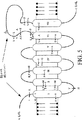
本発明は、一部、2つの哺乳動物の遺伝子の、同定、単離、クローニングおよび配列決定に基づいている。これらの遺伝子は、プレセニリン(presenilin)-1(PS1)およびプレセニリン-2(PS2)と命名されている。これら2つの遺伝子、およびそれらの対応するタンパク質産物は、高度に保存された遺伝子(プレセニリン)のファミリーのメンバーであり、それらは他の哺乳動物種(例えば、マウス、ラット)にホモログまたはオーソログを、また無脊椎動物種(例えば、C.elegans、D. melanogaster)にオーソログを有する。これらの遺伝子の変異は、ヒトにおける家族性アルツハイマー病の形態の発生に関係し、そして同様の他の障害(例えば、他の認識的、知能的、神経学的、または心理学的障害(例えば、脳溢血、精神分裂症、鬱病、精神遅滞および癲癇など))の原因でもあり得る。本開示は、ヒトPS1(hPS1)およびヒトPS2(hPS2)遺伝子、マウスPS1ホモログ(mPS1)、な
らびにC.elegans(sel-12、SPE-4)およびD. melanogaster(DmPS)由来の関連する遺伝
子のゲノムおよびcDNAヌクレオチド配列を提供する。本開示はまた、これらの遺伝子にコードされるプレセニリンタンパク質の予想アミノ酸配列、およびプレセニリンの構造の特徴付け(推定の機能的ドメインおよび抗原決定基を含む)を提供する。ヒトのアルツハイ
マー病(AD)の原因であるプレセニリン中の変異の多くもまた開示され、そしてそのタンパク質の機能的ドメインに関連づけられている。
SUMMARY OF THE INVENTION The present invention is based in part on the identification, isolation, cloning and sequencing of two mammalian genes. These genes have been named presenilin-1 (PS1) and presenilin-2 (PS2). These two genes, and their corresponding protein products, are members of a family of highly conserved genes (presenilin) that are homologous or orthologous to other mammalian species (eg, mouse, rat), It also has orthologs in invertebrate species (eg C. elegans , D. melanogaster ). Mutations in these genes are associated with the development of familial Alzheimer's disease forms in humans, and other similar disorders (eg, other cognitive, intelligent, neurological, or psychological disorders (eg, It can also be the cause of brain overflow, schizophrenia, depression, mental retardation and epilepsy)). The present disclosure relates to human PS1 (hPS1) and human PS2 (hPS2) genes, mouse PS1 homolog (mPS1), and C. elegans (sel-12, SPE-4) and D. melanogaster (DmPS). Provide genomic and cDNA nucleotide sequences of related genes from which they are derived. The present disclosure also provides the predicted amino acid sequences of the presenilin proteins encoded by these genes, and the structural characterization of presenilin, including putative functional domains and antigenic determinants. Many of the mutations in presenilin that are responsible for human Alzheimer's disease (AD) have also been disclosed and associated with functional domains of the protein.
従って、一連の実施様態において、本発明は、プレセニリン遺伝子を含むかまたはそれ由来のヌクレオチド配列を含む単離された核酸、および/またはプレセニリンタンパク質
を含むかまたはそれ由来のポリペプチドをコードする単離された核酸を提供する。本発明のプレセニリン配列は、特定の開示された配列、これらの配列のスプライス変異体、これらの配列の対立遺伝子変異体、同義配列、およびこれらの配列の相同またはオーソロガス変異体を包含する。従って、例えば、本発明は、hPS1遺伝子、hPS2遺伝子、mPS1遺伝子およびDmPS遺伝子由来のゲノムおよびcDNA配列を提供する。本発明はまた、対立遺伝子変異体および相同またはオーソロガス配列を、このような変異体が通常得られ得る方法を提供することによって、提供する。本発明はまた、プレセニリンの変異または病気誘因変異体を、多くの特異的変異配列を開示することによって、そして、他のこのような変異体を通常得ることができる方法を提供することによって、特に提供する。本発明の核酸が、種々の診断、治療、および組み換えの適用に使用され得るので、プレセニリン配列の種々のサブセットおよびプレセニリン配列の異種配列との組合せもまた提供される。例えば、対立遺伝子特異的ハイブリダイゼーションスクリーニングまたはPCR増幅技術における使用の
ためには、センス配列およびアンチセンス配列、正常配列および変異配列、ならびにイントロン配列、エキソン配列および非翻訳配列を含む、プレセニリン配列のサブセットが提供される。このような配列は、本明細書中で開示されるか、さもなければ実施可能とされる配列由来の少数の連続するヌクレオチドを含み得るが、好ましくは、プレセニリン配列由来の少なくとも8〜10個の、より好ましくは9〜25個の連続するヌクレオチドを含む。他の好ましいプレセニリン配列のサブセットは、プレセニリンタンパク質の機能的ドメインまたは抗原決定基の1つ以上をコードする配列を含み、そして特に、正常(野生型)配列または変異配列のいずれかを含み得る。本発明はまた、完全またはサブセットのいずれかのプレセニリン配列が、外因性の配列と作動可能に連結され、クローニングベクター、発現ベクター、融合ベクター、およびトランスジェニック構築物などを形成する種々の核酸構築物を提供する。従って、本発明の別の局面によれば、哺乳動物または無脊椎動物組織細胞を形質転換して、正常または変異プレセニリン配列を細胞中で発現する組換えベクターが提供される。
Accordingly, in a series of embodiments, the invention provides an isolated nucleic acid comprising a presenilin gene or comprising a nucleotide sequence derived therefrom, and / or an isolation encoding a polypeptide comprising or derived from a presenilin protein. Provided nucleic acid. The presenilin sequences of the present invention include certain disclosed sequences, splice variants of these sequences, allelic variants of these sequences, synonymous sequences, and homologous or orthologous variants of these sequences. Thus, for example, the present invention provides genomic and cDNA sequences derived from the hPS1 gene, hPS2 gene, mPS1 gene and DmPS gene. The present invention also provides allelic variants and homologous or orthologous sequences by providing methods by which such variants can usually be obtained. The present invention also specifically provides for presenilin mutations or disease-inducing mutants by disclosing a number of specific mutant sequences and by providing a method by which other such mutants can usually be obtained. provide. Since the nucleic acids of the invention can be used in various diagnostic, therapeutic, and recombinant applications, combinations of various subsets of presenilin sequences and heterologous sequences of presenilin sequences are also provided. For example, for use in allele-specific hybridization screening or PCR amplification techniques, a subset of presenilin sequences, including sense and antisense sequences, normal and mutated sequences, and intron, exon and untranslated sequences Is provided. Such sequences may comprise a small number of contiguous nucleotides from the sequences disclosed or otherwise enabled herein, but preferably at least 8-10 from the presenilin sequence. More preferably 9-25 contiguous nucleotides. Other preferred subsets of presenilin sequences include sequences encoding one or more of the functional domains or antigenic determinants of the presenilin protein, and in particular may include either normal (wild type) sequences or mutant sequences. The present invention also provides various nucleic acid constructs in which either a complete or subset of presenilin sequences are operably linked to exogenous sequences to form cloning vectors, expression vectors, fusion vectors, transgenic constructs, and the like. To do. Thus, according to another aspect of the present invention, there is provided a recombinant vector for transforming mammalian or invertebrate tissue cells to express normal or mutant presenilin sequences in the cells.
別の一連の実施形態において、本発明は、本発明の核酸の1つでトランスフェクトされたか、または形質転換された宿主細胞を提供する。細胞は、単に本発明の核酸構築物を増殖する目的のために形質転換され得るか、またはプレセニリン配列を発現するために形質転換され得る。本発明の形質転換された細胞は、タンパク質および/または正常または変
異プレセニリン発現に影響を与える、正常または変異プレセニリンタンパク質と相互作用する、および/または正常または変異タンパク質の機能または効果を調節する他の化合物
を同定するアッセイにおいて使用され得る。あるいは、本発明の形質転換された細胞は、プレセニリンタンパク質、融合タンパク質、機能的ドメイン、抗原決定基、および/また
は抗体を産生するアッセイにおいて使用され得る。形質転換された細胞はまた、ヒトを含む宿主に、治療または他の理由で移植され得る。好ましい宿主細胞は、神経、線維芽細胞、骨髄、脾臓、器官型のまたは混成の細胞培養物由来の哺乳動物細胞、ならびに細菌、酵母、線虫、昆虫および他の無脊椎動物細胞を包含する。以下に記載される使用のために、好ましい細胞はまた、胚幹細胞、接合、配偶子および生殖系列細胞を包含する。
In another series of embodiments, the present invention provides host cells that have been transfected or transformed with one of the nucleic acids of the present invention. The cells can be transformed simply for the purpose of growing the nucleic acid constructs of the invention or transformed to express presenilin sequences. The transformed cells of the present invention affect the expression of proteins and / or normal or mutant presenilin, interact with normal or mutant presenilin proteins, and / or regulate the function or effect of normal or mutant proteins. It can be used in assays to identify compounds. Alternatively, the transformed cells of the invention can be used in assays that produce presenilin proteins, fusion proteins, functional domains, antigenic determinants, and / or antibodies. Transformed cells can also be transplanted into hosts, including humans, for therapeutic or other reasons. Preferred host cells include mammalian cells from nerve, fibroblasts, bone marrow, spleen, organotypic or hybrid cell cultures, and bacteria, yeast, nematodes, insects and other invertebrate cells. For the uses described below, preferred cells also include embryonic stem cells, mating, gamete and germline cells.
別の一連の実施様態において、本発明は、ADおよび、プレセニリン遺伝子の変異に関連する他の病気または障害についての、トランスジェニック動物モデルを提供する。動物は、本質的に、ラット、マウス、ハムスター、モルモット、ウサギ、イヌ、ネコ、ヤギ、ヒツジ、ブタ、およびヒトではない霊長類を含む任意の哺乳動物であり得る。さらに、線虫および昆虫を含む無脊椎動物モデルは、特定の適用に使用され得る。動物モデルは、マイ
クロインジェクション、トランスフェクション、または胚幹細胞、接合子、配偶子、および生殖系列細胞と、ゲノムもしくはcDNAフラグメントを含むベクター、ミニ遺伝子、相同組換えベクター、およびウイルス挿入ベクターなどとの他の様式の形質転換を含む標準的な遺伝子組換え法によって産生される。適切なベクターは、ワクシニアウイルス、アデノウイルス、アデノ関連ウイルス、レトロウイルス、リポソーム輸送体、神経親和性ウイルス、および単純ヘルペスウイルスを含む。動物モデルは、正常および変異の配列、イントロン、エキソン、および非翻訳の配列を含むプレセニリンを含むか、またはそれ由来の遺伝子組換え配列、および機能的ドメインのようなプレセニリンのサブセットをコードする配列を含み得る。提供される動物モデルの主要なタイプは、以下を包含する:(1)正常ヒトプレセニリン遺伝子が、動物のゲノムに、外因性または内因性プロモーターエレメントいずれかの調節下で、付加的な遺伝子として、およびミニ遺伝子または大きなゲノムフラグメントのいずれかとして、組換え的に導入された動物;正常ヒトプレセニリン遺伝子が、動物の相同プレセニリン遺伝子の1つまたは両方のコピーと、相同組換えまたは遺伝子標的化によって、組換え的に置換された動物;および/または動物の相同プレセニリン
遺伝子の1つの1つまたは両方のコピーが、相同組換えまたは遺伝子標的化によりヒトホモログをコードする配列を部分的に置換することによって、組換え的に「ヒト化」された動物。(2)変異ヒトプレセニリン遺伝子が、動物のゲノムに、外因性または内因性プロモーターエレメントのいずれかの調節下で、付加的な遺伝子として、およびミニ遺伝子または大きなゲノムフラグメントのいずれかとして、組換え的に導入された動物;変異ヒトプレセニリン遺伝子が、動物の相同プレセニリン遺伝子の1つまたは両方のコピーと、相同組換えまたは遺伝子標的化によって、組換え的に置換された動物;および/または動物
の相同プレセニリン遺伝子の1つの1つまたは両方のコピーが、相同組換えまたは遺伝子標的化により変異ヒトホモログをコードする配列を部分的に置換することによって、組換え的に「ヒト化」された動物。(3)その動物のプレセニリン遺伝子の1つの変異バージョンが、動物のゲノムに、外因性または内因性プロモーターエレメントのいずれかの調節下で、付加的な遺伝子として、およびミニ遺伝子または大きなゲノムフラグメントのいずれかとして、組換え的に導入された動物;および/または、その動物のプレセニリン遺伝
子の1つの変異バージョンが、動物の相同プレセニリン遺伝子の1つまたは両方のコピーと、相同組換えまたは遺伝子標的化によって、組換え的に置換された動物。(4)動物のプレセニリン遺伝子の1つの1つまたは両方のコピーが、相同組換えまたは遺伝子標的化によって部分的にまたは完全に欠失した、または外因性の配列の相同組換えまたは遺伝子標的化による挿入または置換によって不活化された「ノックアウト」動物。好ましい実施様態において、ADのためのトランスジェニックマウスモデルは、正常ヒトPS1またはPS2タンパク質、変異ヒトまたはマウスPS1またはPS2タンパク質、あるいはヒト化された正常または変異マウスPS1またはPS2タンパク質をコードするトランスジーンを有する。
In another series of embodiments, the present invention provides transgenic animal models for AD and other diseases or disorders associated with mutations in the presenilin gene. The animal can be essentially any mammal, including rats, mice, hamsters, guinea pigs, rabbits, dogs, cats, goats, sheep, pigs, and non-human primates. In addition, invertebrate models, including nematodes and insects, can be used for specific applications. Animal models include microinjection, transfection, or embryonic stem cells, zygotes, gametes, and germline cells, as well as vectors containing genomic or cDNA fragments, minigenes, homologous recombination vectors, and viral insertion vectors. Produced by standard genetic recombination methods including transformation in this manner. Suitable vectors include vaccinia virus, adenovirus, adeno-associated virus, retrovirus, liposomal transporter, neurophilic virus, and herpes simplex virus. Animal models contain presenilin containing or derived from normal and mutated sequences, introns, exons, and untranslated sequences, and sequences encoding presenilin subsets such as functional domains. May be included. The main types of animal models provided include: (1) a normal human presenilin gene as an additional gene in the animal's genome under the control of either an exogenous or endogenous promoter element, And recombinantly introduced animals, either as minigenes or large genomic fragments; a normal human presenilin gene is copied by homologous recombination or gene targeting with one or both copies of the animal's homologous presenilin gene By recombinantly replacing an animal; and / or one or both copies of one of the animal's homologous presenilin genes partially replacing a sequence encoding a human homolog by homologous recombination or gene targeting, Recombinant “humanized” animals. (2) The mutated human presenilin gene is recombined into the animal's genome under the control of either an exogenous or endogenous promoter element as an additional gene and as either a minigene or a large genomic fragment Animals in which the mutant human presenilin gene has been recombinantly replaced by homologous recombination or gene targeting with one or both copies of the animal's homologous presenilin gene; and / or animal homology Animals in which one or both copies of one presenilin gene have been recombinantly "humanized" by partially replacing sequences encoding mutant human homologs by homologous recombination or gene targeting. (3) One mutated version of the animal's presenilin gene is added to the animal's genome under the control of either an exogenous or endogenous promoter element, either as an additional gene and either a minigene or a large genomic fragment As such, a recombinantly introduced animal; and / or one mutated version of the animal's presenilin gene may be transformed with one or both copies of the animal's homologous presenilin gene by homologous recombination or gene targeting. , Recombinantly substituted animals. (4) One or both copies of one of the animal presenilin genes has been partially or completely deleted by homologous recombination or gene targeting, or by homologous recombination or gene targeting of exogenous sequences “Knock-out” animals inactivated by insertion or replacement. In a preferred embodiment, the transgenic mouse model for AD comprises a normal human PS1 or PS2 protein, a mutant human or mouse PS1 or PS2 protein, or a transgene encoding a humanized normal or mutant mouse PS1 or PS2 protein. Have.
別の一連の実施様態において、本発明は、実質的に純粋なタンパク質の調製物(プレセニリンタンパク質を含むか、またはそれ由来のポリペプチドを含む)を提供する。本発明のプレセニリンタンパク質配列は、特定の開示された配列、オルタナティブmRNAスプライシングから得られるこれらの配列の変異体、これらの配列の対立遺伝子変異体、およびこれらの配列の相同またはオーソロガス変異体を含む。従って、例えば、本発明は、hPS1タンパク質、hPS2タンパク質、mPS1タンパク質、およびDmPSタンパク質由来のアミノ酸配列を提供する。本発明はまた、対立遺伝子変異体および相同またはオーソロガスタンパク質を、このような変異体が通常得られ得る方法を提供することによって、提供する。本発明はまた、プレセニリンの変異または病気誘因変異体を、多くの特異的変異配列を開示することによって、そして他のこのような変異体を通常得ることができる方法を提供することによって、特に提供する。本発明のタンパク質は、種々の診断、治療および組換え適用に使用され得るので、プレセニリンタンパク質配列の種々のサブセットおよびプレセニリンタンパク質配列と異種配列との組合せもまた提供される。例えば、免疫原としての使用または結合アッセイにおいて、プレセニリンタンパク質配列のサブセットが、正常配列およ
び変異配列の両方を含めて、提供される。このようなタンパク質配列は、本明細書中で開示されるか、さもなければ実施可能とされる配列由来の少数の連続するアミノ酸残基を含み得るが、好ましくは、プレセニリン配列由来の少なくとも4〜8個、そして好ましくは少なくとも9〜15個の連続するアミノ酸残基を含む。他の好ましいプレセニリンタンパク質配列のサブセットは、プレセニリンタンパク質の機能的ドメインまたは抗原決定基の1つ以上に対応する配列を含み、そして特に、正常(野生型)配列または変異配列のいずれかを含み得る。本発明はまた、完全かまたはサブセットのいずれかのプレセニリン配列が、外因性配列と結合し、融合タンパク質などを形成する種々のタンパク質構築物を提供する。これらの実施様態に従って、本発明はまた、プレセニリンを含むかまたはそれ由来の上記すべてのタンパク質を産生する方法を提供する。
In another series of embodiments, the present invention provides a substantially pure protein preparation comprising a presenilin protein or a polypeptide derived therefrom. The presenilin protein sequences of the present invention include certain disclosed sequences, variants of these sequences derived from alternative mRNA splicing, allelic variants of these sequences, and homologous or orthologous variants of these sequences. Thus, for example, the present invention provides amino acid sequences derived from hPS1 protein, hPS2 protein, mPS1 protein, and DmPS protein. The invention also provides allelic variants and homologous or orthologous proteins by providing methods by which such variants can usually be obtained. The present invention also specifically provides for presenilin mutations or disease-inducing mutants by disclosing a number of specific mutant sequences and by providing a method by which other such mutants can usually be obtained. To do. Since the proteins of the invention can be used in a variety of diagnostic, therapeutic and recombinant applications, various subsets of presenilin protein sequences and combinations of presenilin protein sequences and heterologous sequences are also provided. For example, in use as an immunogen or in a binding assay, a subset of presenilin protein sequences is provided, including both normal and variant sequences. Such protein sequences may comprise a small number of contiguous amino acid residues from the sequences disclosed or otherwise enabled herein, but preferably at least 4 to from the presenilin sequence. Contains 8 and preferably at least 9-15 contiguous amino acid residues. Other preferred subsets of presenilin protein sequences include sequences corresponding to one or more of the functional domains or antigenic determinants of the presenilin protein, and in particular may include either normal (wild type) sequences or mutated sequences. The invention also provides various protein constructs in which either a complete or subset of presenilin sequences are combined with exogenous sequences to form fusion proteins and the like. In accordance with these embodiments, the present invention also provides a method of producing all the above proteins comprising or derived from presenilin.
別の一連の実施様態において、本発明はポリクローナル抗体およびモノクローナル抗体の産生および使用を提供する。それらの抗体は、プレセニリンまたはプレセニリンの特定の抗原決定基に選択的に結合する、Fabフラグメント、F(ab')2、および単鎖抗体フラグメントを包含する抗体フラグメントを包含する。この抗体はマウス、ウサギ、ヤギ、または他の適切な動物中で惹起され得るか、またはハイブリドーマ細胞株のような培養細胞中で組換え的に産生され得る。好ましくは、抗体は、プレセニリン配列由来の少なくとも4〜8個、そして好ましくは少なくとも9〜15個の連続するアミノ酸残基を含むプレセニリン配列に対して惹起される。本発明の抗体は、本明細書中に記載の、種々の診断、治療、および技術適用に使用し得る。 In another series of embodiments, the present invention provides for the production and use of polyclonal and monoclonal antibodies. These antibodies include antibody fragments, including Fab fragments, F (ab ′) 2 , and single chain antibody fragments, that selectively bind to presenilin or specific antigenic determinants of presenilin. The antibody can be raised in mice, rabbits, goats, or other suitable animals, or can be produced recombinantly in cultured cells such as hybridoma cell lines. Preferably, the antibody is raised against a presenilin sequence comprising at least 4-8, and preferably at least 9-15 consecutive amino acid residues from the presenilin sequence. The antibodies of the present invention may be used in the various diagnostic, therapeutic, and technical applications described herein.
別の一連の実施様態において、本発明は、プレセニリン遺伝子およびタンパク質(例えば、PS1またはPS2)の発現を誘発または阻害し得るタンパク質、低分子、または他の化合物をスクリーニングまたは同定する方法を提供する。アッセイは、インビトロで、非形質転換細胞株、不死化細胞株、もしくは組換え細胞株を用いて、またはインビボで、本明細書中の実施可能とされるトランスジェニック動物モデルを用いて行い得る。特に、アッセイはPS1、PS2、または他のプレセニリン関連遺伝子もしくはタンパク質の増加または減少した発現の存在を、増加または減少したmRNA発現、増加または減少したプレセニリン関連タンパク質産物レベル、または組換え構築物中のプレセニリン5'調節領域に作動可能に連結したマーカー遺伝子(例えば、β-ガラクトシダーゼ、緑色蛍光タンパク質、アルカリ
ホスファターゼまたはルシファラーゼ)の増加または減少した発現レベルを基準に検出し得る。特定のプレセニリンを発現することが知られた細胞、または特定のプレセニリンを発現するために形質転換された細胞は、インキュベートされ、そして1つ以上の試験化合物が培地に添加される。化合物がプレセニリンの発現を誘発または阻害するのに充分な時間(例えば、0〜72時間)の経過後、確立したベースラインからの発現レベルの任意の変化が、上述の任意の技術を用いて検出され得る。特に好ましい実施様態において、細胞は、ヒト神経芽細胞腫、神経膠芽細胞腫、またはハイブリドーマ細胞株のような不死化された細胞株由来であるか、または本発明の形質転換細胞である。
In another series of embodiments, the present invention provides methods for screening or identifying proteins, small molecules, or other compounds that can induce or inhibit expression of presenilin genes and proteins (eg, PS1 or PS2). Assays can be performed in vitro, using non-transformed cell lines, immortalized cell lines, or recombinant cell lines, or in vivo, using the transgenic animal models enabled herein. In particular, the assay may indicate the presence of increased or decreased expression of PS1, PS2, or other presenilin related genes or proteins, increased or decreased mRNA expression, increased or decreased presenilin related protein product levels, or presenilin in recombinant constructs. It can be detected on the basis of increased or decreased expression levels of a marker gene (eg, β-galactosidase, green fluorescent protein, alkaline phosphatase or luciferase) operably linked to the 5 ′ regulatory region. Cells known to express a particular presenilin, or cells transformed to express a particular presenilin, are incubated and one or more test compounds are added to the medium. After a sufficient amount of time for the compound to induce or inhibit presenilin expression (eg, 0-72 hours), any change in expression level from an established baseline is detected using any of the techniques described above. Can be done. In particularly preferred embodiments, the cells are derived from an immortalized cell line, such as a human neuroblastoma, glioblastoma, or hybridoma cell line, or are transformed cells of the invention.
別の一連の実施様態において、本発明は、プレセニリンに結合するか、さもなくば直接相互作用するタンパク質および他の化合物を同定する方法を提供する。タンパク質および化合物は内因性の細胞成分を包含する。その細胞成分は、インビボでプレセニリンと相互作用し、従って、薬学的および治療的介入のための新しい標的、ならびに組換え化合物、合成化合物およびさもなくば外因性の化合物を提供する。それらの化合物は、プレセニリン結合能を有し得、従って、薬学的調製物の候補となり得る。従って、一連の実施様態において、細胞溶解物または組織ホモジネート(例えば、ヒト脳ホモジネート、リンパ球溶解物)は、正常または変異プレセニリンの一つに結合するタンパク質または他の化合物についてスクリーニングされ得る。あるいは、任意の種々の外因性の化合物は、天然に存在するおよび/または合成(例えば、小さな分子またはペプチドのライブラリー)の両方と
も、プレセニリン結合能についてスクリーニングされ得る。これら各々の実施様態におい
て、アッセイが、「プレセニリン成分」といくつかの他の部分との間の結合を検出するために行われる。これらのアッセイにおけるプレセニリン成分は、正常または変異プレセニリンタンパク質(プレセニリンのまたはプレセニリン融合タンパク質機能的ドメインまたは抗原決定基を含む)を含むかまたはそれ由来の任意のポリペプチドであり得る。結合は、非特異的測定(例えば、細胞内Ca2+、GTP/GDP比の変化)によって、または特異的測定
(例えば、Aβペプチド産生の変化、またはディファレンシャルディスプレイ、2Dゲル電
気泳動、ディファレンシャルハイブリダイゼーション、またはSAGE法によってモニターされ得る他の下流遺伝子の発現の変化)によって検出され得る。好ましい方法は、以下の技術の改変を含む:(1)アフィニティークロマトグラフィーによる直接的抽出;(2)免疫沈降によるプレセニリン成分および結合タンパク質または他の化合物の同時単離;(3)生物分子相互作用アッセイ(BiomolecularInteraction Assay)(BIAcore);および(4
)酵母ツーハイブリッドシステム。
In another series of embodiments, the present invention provides methods for identifying proteins and other compounds that bind to presenilin or otherwise interact directly. Proteins and compounds include endogenous cellular components. Its cellular components interact with presenilin in vivo, thus providing new targets for pharmaceutical and therapeutic intervention, as well as recombinant, synthetic and otherwise exogenous compounds. These compounds can have presenilin binding ability and can therefore be candidates for pharmaceutical preparations. Thus, in a series of embodiments, cell lysates or tissue homogenates (eg, human brain homogenates, lymphocyte lysates) can be screened for proteins or other compounds that bind to one of normal or mutant presenilin. Alternatively, any of a variety of exogenous compounds can be screened for presenilin binding ability, both naturally occurring and / or synthetic (eg, small molecule or peptide libraries). In each of these embodiments, an assay is performed to detect binding between the “presenilin component” and several other moieties. The presenilin component in these assays can be any polypeptide comprising or derived from a normal or mutant presenilin protein (including a presenilin or presenilin fusion protein functional domain or antigenic determinant). Binding can be by non-specific measurements (eg, changes in intracellular Ca 2+ , GTP / GDP ratio) or specific measurements (eg, changes in Aβ peptide production, or differential display, 2D gel electrophoresis, differential hybridization) Or changes in the expression of other downstream genes that can be monitored by the SAGE method). Preferred methods include the following technical modifications: (1) direct extraction by affinity chromatography; (2) simultaneous isolation of presenilin components and binding proteins or other compounds by immunoprecipitation; (3) biomolecular interactions. Assay (Biomolecular Interaction Action) (BIAcore); and (4
) Yeast two hybrid system.
別の一連の実施様態において、本発明は、正常または変異プレセニリンの活性を調節し得るタンパク質、低分子および他の化合物を同定する方法を提供する。正常細胞または動物、本発明の形質転換細胞およびトランスジェニック動物モデル、または正常もしくは変異プレセニリン遺伝子を有する被験体から得られる細胞を使用して、本発明は、プレセニリンの発現、プレセニリンの細胞内局在性、細胞内Ca2+、Na+、K+または他のイオンレベ
ルまたは代謝、アポトーシスまたは細胞死の頻度または割合、Aβペプチド産生のレベル
またはパターン、微小管結合タンパク質のリン酸化の有無またはレベル、または正常および変異プレセニリン配列を有する細胞を区別する他の生化学的、組織学的、または生理学的マーカーに影響を与えるそれら化合物の能力に基づき、このような化合物を同定する方法を提供する。本発明のトランスジェニック動物を用いて、このような化合物を同定する方法がまた、プレセニリンの変異に関連する挙動的、生理学的、組織学的表現型に影響を与えるこれらの化合物の能力に基づいて提供される。
In another series of embodiments, the present invention provides methods for identifying proteins, small molecules, and other compounds that can modulate the activity of normal or mutant presenilin. Using normal cells or animals, transformed cells of the present invention and transgenic animal models, or cells obtained from subjects with normal or mutated presenilin genes, the present invention provides for expression of presenilin, subcellular localization of presenilin. Sex, intracellular Ca 2+ , Na + , K + or other ion levels or metabolism, frequency or rate of apoptosis or cell death, level or pattern of Aβ peptide production, presence or level of phosphorylation of microtubule-associated proteins, Alternatively, methods of identifying such compounds based on their ability to affect other biochemical, histological, or physiological markers that distinguish cells having normal and mutated presenilin sequences are provided. Methods for identifying such compounds using the transgenic animals of the present invention are also based on the ability of these compounds to affect the behavioral, physiological, and histological phenotypes associated with presenilin mutations. Provided.
別の一連の実施様態において、本発明は、ADに関連するプレセニリン対立遺伝子の保因者をスクリーニングする方法、ADの羅患者を診断する方法、および関連する初老期および老年期の痴呆、精神分裂病および鬱病のような精神医学病、ならびに脳卒中および脳溢血のような神経性の病気(これらはPS1またはPS2遺伝子の変異に関連する)をスクリーニングおよび診断する方法を提供する。スクリーニングおよび/または診断は、本明細書中で
開示される、および実施可能な核酸(ゲノム配列およびmRNA/cDNA配列を含む)、タンパ
ク質、および/または抗体に基づく方法によって達成され得る。その方法は、正常プレセ
ニリン活性の減衰もしくは増大、および/または変異プレセニリンによって与えられる特
定の新しい活性の存在を検出するよう設計された機能的アッセイを含む。従って、プレセニリンタンパク質に基づくスクリーニングおよび診断が提供され、それらは変異プレセニリンと正常プレセニリンとの間の電気泳動移動度、タンパク質分解の切断パターン、種々のアミノ酸残基のモル比、特異的な抗体を結合する能力の差異を検出する。さらに、核酸(gDNA、cDNA、またはmRNA)に基づくスクリーニングおよび診断が提供され、それらは、直接的ヌクレオチド配列決定、対立遺伝子特異的オリゴヌクレオチドを使用するハイブリダイゼーション、制限酵素消化およびマッピング(例えば、RFLP、REF-SSCP)、電気泳動移動度(例えば、SSCP、DGGE)、PCRマッピング、RNaseプロテクション、化学的ミスマッチ切断(chemicalmismatch cleavage)、リガーゼ媒介検出(ligase-mediated detection)、ならびに種々の他の方法によってヌクレオチド配列の差異を検出する。PS1、PS2、APP、またはPS1、PS2、もしくはAPPと反応するタンパク質の異常なプロセシング(例えば、異常リン酸化、グリコシル化、グリケーション、アミド化、またはタンパク質分解切断);プレセニリンの転写、翻訳、および翻訳後修飾における変化;プレセニリン遺伝子産物の細胞内および細胞外のトラフィッキングにおける変化;またはプレセニリンの異常な細胞内局在を検出する他の方法もまた提供される。これらの実施様態に従って、診断キットもまた提供され、それらは上記の診断スクリーニングに必要な試薬を含む。
In another series of embodiments, the present invention relates to a method for screening carriers for presenilin alleles associated with AD, a method for diagnosing patients with AD, and related presenile and senile dementia, schizophrenia. Methods for screening and diagnosing psychiatric illnesses such as illness and depression, and neurological illnesses such as stroke and cerebral hyperemia, which are associated with mutations in the PS1 or PS2 gene, are provided. Screening and / or diagnosis can be accomplished by nucleic acid (including genomic and mRNA / cDNA sequences), protein, and / or antibody-based methods disclosed and practicable herein. The method includes a functional assay designed to detect the attenuation or increase of normal presenilin activity and / or the presence of certain new activities conferred by mutant presenilin. Thus, screening and diagnostics based on presenilin proteins are provided, which bind electrophoretic mobility, proteolytic cleavage patterns, various amino acid residue molar ratios, specific antibodies between mutant and normal presenilin Detect differences in ability to In addition, screening and diagnostics based on nucleic acids (gDNA, cDNA, or mRNA) are provided, which include direct nucleotide sequencing, hybridization using allele-specific oligonucleotides, restriction enzyme digestion and mapping (eg, RFLP REF-SSCP), electrophoretic mobility (eg, SSCP, DGGE), PCR mapping, RNase protection, chemical mismatch cleavage, ligase-mediated detection, and various other methods Detects nucleotide sequence differences. PS1, PS2, APP, or aberrant processing of proteins that react with PS1, PS2, or APP (eg, abnormal phosphorylation, glycosylation, glycation, amidation, or proteolytic cleavage); transcription, translation of presenilin, and Other methods of detecting changes in post-translational modifications; changes in intracellular and extracellular trafficking of presenilin gene products; or other abnormal subcellular localization of presenilin are also provided. According to these embodiments, diagnostic kits are also provided, which contain the reagents necessary for the diagnostic screening described above.
別の一連の実施様態において、本発明は、ADのようなプレセニリンに関連する病気の処置における使用のための方法および薬学的調製物を提供する。これらの方法および薬剤は以下に基づく:(1)正常PS1またはPS2タンパク質の投与、(2)変異遺伝子を補償または置換するための正常PS1またはPS2遺伝子による遺伝子療法、(3)変異PS1またはPS2遺伝子に対するアンチセンス配列に基づくかまたは変異遺伝子を「ノックアウト」する遺伝子療法、(4)PS1またはPS2の変異体の有害な効果を、ブロックするかまたは矯正するタンパク質をコードする配列に基づく遺伝子療法、(5)正常および/または変異のPS1もしくはPS2タンパク質に対する抗体に基づく免疫療法、または(6)PS1もしくはPS2の発現
を変化させるか、PS1またはPS2の変異形態と他のタンパク質またはリガンドとの間の異常相互作用をブロックする低分子(薬物)、または、そうでなければ変異PS1もしくはPS2タンパク質の異常な機能を、変異タンパク質の構造を変化させるか、またはそれらの代謝クリアランス(clearance)を増強するか、もしくはそれらの機能を阻害することによって
ブロックする低分子(薬物)。
In another series of embodiments, the present invention provides methods and pharmaceutical preparations for use in the treatment of diseases related to presenilin such as AD. These methods and agents are based on: (1) administration of normal PS1 or PS2 protein, (2) gene therapy with normal PS1 or PS2 gene to compensate or replace the mutant gene, (3) mutant PS1 or PS2 gene Gene therapy based on antisense sequences against or for “knocking out” mutant genes, (4) gene therapy based on sequences encoding proteins that block or correct the deleterious effects of PS1 or PS2 variants, ( 5) Antibody-based immunotherapy against normal and / or mutant PS1 or PS2 protein, or (6) Abnormality between PS1 or PS2 mutant forms and other proteins or ligands, changing PS1 or PS2 expression Small molecules (drugs) that block interactions, or otherwise abnormal functions of mutant PS1 or PS2 proteins Small molecules (drugs) that block by altering the structure of the mutated proteins or enhancing their metabolic clearance or inhibiting their function.
本発明の別の局面に従って、本発明のタンパク質は、リガンド、治療用薬物または他のタイプの小化学分子を提供する合理的なドラッグデザインのための出発点として使用され得る。あるいは、上記のスクリーニングアッセイによって同定される低分子または他の化合物は、合理的なドラッグデザインにおける「リード化合物(lead compounds)」として
働き得る。
In accordance with another aspect of the present invention, the proteins of the present invention can be used as a starting point for rational drug designs that provide ligands, therapeutic drugs or other types of small chemical molecules. Alternatively, small molecules or other compounds identified by the screening assays described above can serve as “lead compounds” in rational drug design.
本発明で特に開示されたヌクレオチドおよびアミノ酸の配列は、配列番号1〜25と番号付けされている。さらに、ブダペスト条約の約定に基づき、本明細書において開示された特定の核酸の生物的寄託が、ATCC(Rockville,MD)でなされた。これらの寄託物としては、受託番号97124(1995年4月28日寄託)、受託番号97508(1995年4月28日寄託)、受託番号97214(1995年6月28日寄託)、および受託番号97428(1996年1月26日寄託)が挙げられる。 Nucleotide and amino acid sequences specifically disclosed in the present invention are numbered SEQ ID NOs: 1-25. In addition, based on the Budapest Treaty commitments, biological deposits of specific nucleic acids disclosed herein were made at ATCC (Rockville, MD). These deposits include deposit number 97124 (deposited on April 28, 1995), deposit number 97508 (deposited on April 28, 1995), deposit number 97214 (deposited on June 28, 1995), and deposit number 97428. (Deposited on January 26, 1996).
発明の詳細な説明
I.定義
本発明の種々の実施様態の説明、ならびに本発明を実行および使用する際に用いられる種々の要素および成分の理解を容易にするために、本明細書の記載および添付の請求の範囲で用いられた特定の用語について以下に示す定義が提供される:
プレセニリン。 本明細書中でさらなる修飾がなされずに用いられる、用語「プレセニリン(単数)」または「プレセニリン(複数)」は、プレセニリン-1(PS1)および/ま
たはプレセニリン-2(PS2)の遺伝子/タンパク質を意味する。特に、修飾されない用語
「プレセニリン(単数)」または「プレセニリン(複数)」は、哺乳動物のPS1および/またはPS2の遺伝子/タンパク質、そして好ましくは、ヒトPS1および/またはPS2の遺伝子/タンパク質を意味する。
Detailed Description of the Invention Definitions Used in the description and the appended claims to facilitate the description of various embodiments of the present invention and to facilitate an understanding of the various elements and components employed in the practice and use of the present invention. The following definitions are provided for specific terms given:
Presenilin. As used herein without further modification, the terms “presenilin” or “presenilin” refer to genes / proteins of presenilin-1 (PS1) and / or presenilin-2 (PS2). means. In particular, the term “presenilin (s)” or “presenilin (s)” unmodified refers to mammalian PS1 and / or PS2 genes / proteins, and preferably human PS1 and / or PS2 genes / proteins. .
プレセニリン-1遺伝子。 本明細書中で用いられる、用語「プレセニリン-1遺伝子」または「PS1遺伝子」は、米国特許出願第08/431,048号(1995年4月28日出願)に最初に
開示および記載され、そして後にSherringtonら(1995)に記載された哺乳動物遺伝子(
任意の対立遺伝子変異体および異種特異的哺乳動物ホモログを含む)を意味する。一つのヒトプレセニリン-1(hPS1)cDNA配列が、本明細書中で、配列番号1として開示される
。hPS1mRNAの転写物のオルタナティブスプライシングから得られる別のヒトcDNA配列は、配列番号3として開示される。以下に記載されるように、33残基をコードする領域が、いくつかの転写産物においてスプライスアウト(spliced-out)され得るさらなるヒトスプ
ライス変異体もまた発見されている。マウスホモログ(mPS1)のcDNAは、配列番号16とし
て開示される。用語「プレセニリン-1遺伝子」または「PS1遺伝子」は、主としてコード配列に関するが、いくつかまたは全ての隣接調節領域および/またはイントロンもまた包
含し得る。用語PS1遺伝子は、特に、スプライス変異体に基づく組換え遺伝子を含むcDNA
またはゲノムDNAから作製された人工または組換え遺伝子を包含する。プレセニリン-1遺伝子はまた、S182遺伝子(例えば、Sherringtonら、1995)として、またはアルツハイマ
ー関連膜タンパク質(ARMP)遺伝子(例えば、米国特許出願第08/431,048号、1995年4月28日出願)として言及されている。
Presenilin-1 gene. As used herein, the term “presenilin-1 gene” or “PS1 gene” was first disclosed and described in US patent application Ser. No. 08 / 431,048 (filed Apr. 28, 1995), and later Sherrington. (1995) described mammalian genes (
Including any allelic variants and heterospecific mammalian homologs). One human presenilin-1 (hPS1) cDNA sequence is disclosed herein as SEQ ID NO: 1. Another human cDNA sequence obtained from alternative splicing of transcripts of hPS1 mRNA is disclosed as SEQ ID NO: 3. As described below, additional human splice variants have also been discovered in which a region encoding 33 residues can be spliced-out in several transcripts. The cDNA for the mouse homolog (mPS1) is disclosed as SEQ ID NO: 16. The term “presenilin-1 gene” or “PS1 gene” relates primarily to the coding sequence, but may also include some or all flanking regulatory regions and / or introns. The term PS1 gene is specifically a cDNA containing a recombinant gene based on a splice variant
Or an artificial or recombinant gene made from genomic DNA. The presenilin-1 gene is also referred to as the S182 gene (eg, Sherrington et al., 1995) or as the Alzheimer associated membrane protein (ARMP) gene (eg, US patent application Ser. No. 08 / 431,048, filed Apr. 28, 1995). Has been.
プレセニリン-1タンパク質。 本明細書中で用いられる、用語「プレセニリン-1タンパク質」または「PS1タンパク質」は、PS1遺伝子(対立遺伝子変異体および異種特異的哺乳動物ホモログを含む)によってコードされるタンパク質を意味する。1つのヒトプレセニリン-1(hPS1)タンパク質配列が、本明細書中で、配列番号2として開示される。hPS1mRNA転写物のオルタナティブスプライシングから得られる別のヒトPS1タンパク質配列は、配列番号4として開示される。以下に記載されるように、33残基を含む領域がいくつかの転写物においてスプライスアウトされ得る、さらなるヒトスプライス変異体もまた発見されている。これらの変異体はまた、本明細書中で用いられる、用語プレセニリン-1タ
ンパク質により包含される。マウスホモログ(mPS1)のタンパク質配列は、配列番号17として開示される。このタンパク質は、組換え細胞もしくは生物によって産生され得るか、天然の組織もしくは細胞株から実質的に単離され得るか、または化学的もしくは酵素的に合成され得る。従って、用語「プレセニリン-1タンパク質」または「PS1タンパク質」は、グリコシル化された、部分的にグリコシル化された、またはグリコシル化されていない形態、ならびにリン酸化された、部分的にリン酸化された、リン酸化されていない、硫酸化された、部分的に硫酸化された、または硫酸化されていない形態のタンパク質を包含することが意図される。この用語はまた、PS1アミノ酸配列の対立遺伝子変異体および他の
機能的等価物(生物学的に活性なタンパク質分解フラグメントまたは他のフラグメントを含む)を包含する。このタンパク質はまた、S182タンパク質(例えば、Sherringtonら、1995)またはアルツハイマー関連膜タンパク質(ARMP)(例えば、米国特許出願第08/431,048号、1995年4月28日出願)として言及されている。
Presenilin-1 protein. As used herein, the term “presenilin-1 protein” or “PS1 protein” means a protein encoded by the PS1 gene, including allelic variants and heterospecific mammalian homologues. One human presenilin-1 (hPS1) protein sequence is disclosed herein as SEQ ID NO: 2. Another human PS1 protein sequence obtained from alternative splicing of hPS1 mRNA transcript is disclosed as SEQ ID NO: 4. Additional human splice variants have also been discovered in which a region containing 33 residues can be spliced out in some transcripts, as described below. These variants are also encompassed by the term presenilin-1 protein as used herein. The protein sequence of the mouse homolog (mPS1) is disclosed as SEQ ID NO: 17. The protein can be produced by recombinant cells or organisms, can be substantially isolated from natural tissues or cell lines, or can be synthesized chemically or enzymatically. Thus, the term “presenilin-1 protein” or “PS1 protein” refers to glycosylated, partially glycosylated or unglycosylated forms, as well as phosphorylated, partially phosphorylated. It is intended to encompass unphosphorylated, sulfated, partially sulfated or unsulfated forms of the protein. The term also encompasses allelic variants and other functional equivalents of the PS1 amino acid sequence, including biologically active proteolytic fragments or other fragments. This protein is also referred to as S182 protein (eg, Sherrington et al., 1995) or Alzheimer related membrane protein (ARMP) (eg, US patent application Ser. No. 08 / 431,048, filed Apr. 28, 1995).
hPS1の遺伝子および/またはタンパク質。 本明細書中で用いられる、略語「hPS1」は
、PS1の遺伝子および/またはタンパク質のヒトホモログおよびヒト対立遺伝子変異体をいう。ヒトPS1遺伝子の2つのcDNA配列が、本明細書中で、配列番号1および配列番号3と
して開示される。対応するhPS1タンパク質配列は、配列番号2および配列番号4として、本明細書中で開示される。有害な変異体を含む数多くの対立遺伝子変異体が、以下の記載を通して開示され、そして実施可能とされる。
hPS1 gene and / or protein. As used herein, the abbreviation “hPS1” refers to human homologues and human allelic variants of the PS1 gene and / or protein. Two cDNA sequences of the human PS1 gene are disclosed herein as SEQ ID NO: 1 and SEQ ID NO: 3. The corresponding hPS1 protein sequences are disclosed herein as SEQ ID NO: 2 and SEQ ID NO: 4. Numerous allelic variants, including deleterious variants, are disclosed and made operable throughout the following description.
mPS1の遺伝子および/またはタンパク質。 本明細書中で用いられる、略語「mPS1」は
、PS1の遺伝子および/またはタンパク質のマウスホモログおよびマウス対立遺伝子変異体をいう。1つのマウスPS1遺伝子のcDNA配列が、本明細書中で、配列番号16として開示さ
れる。対応するmPS1タンパク質配列が、本明細書中で、配列番号17として開示される。有害な変異体を含む対立遺伝子変異体が、以下の記載において実施可能とされる。
mPS1 gene and / or protein. As used herein, the abbreviation “mPS1” refers to mouse homologues and mouse allelic variants of the PS1 gene and / or protein. The cDNA sequence of one mouse PS1 gene is disclosed herein as SEQ ID NO: 16. The corresponding mPS1 protein sequence is disclosed herein as SEQ ID NO: 17. Allelic variants, including deleterious variants, are made feasible in the following description.
プレセニリン-2遺伝子。 本明細書中で用いられる、用語「プレセニリン-2遺伝子」または「PS2遺伝子」は、米国特許出願第08/496,841号(1995年6月28日出願)に最初に
開示および記載され、そして後に、Rogaevら(1995)およびLevy-Lahadら(1995)に記載された哺乳動物遺伝子(任意の対立遺伝子変異体および異種特異的哺乳動物ホモログを含む)を意味する。1つのヒトプレセニリン-2(hPS2)cDNA配列が、配列番号18として、
本明細書中で開示される。以下に記載されるように、1つのコドンまたは33残基をコードする領域がいくつかの転写物においてスプライスアウトされ得るさらなるヒトスプライス変異体もまた、発見されている。用語「プレセニリン-2遺伝子」または「PS2遺伝子」は
、主としてコード配列に関するが、いくつかまたは全ての隣接調節領域および/またはイ
ントロンもまた包含し得る。用語PS2遺伝子は、特に、スプライス変異体に基づく組換え
遺伝子を含む、cDNAまたはゲノムDNAから作製された人工または組換え遺伝子を包含する
。プレセニリン-2遺伝子はまた、E5-1遺伝子(例えば、Rogaevら、1995;米国特許出願
第08/496,841、1995年6月28日出願)として、またはSTM2遺伝子(例えば、Levy-Lahadら、1995)として、言及されている。
Presenilin-2 gene. As used herein, the term “presenilin-2 gene” or “PS2 gene” was first disclosed and described in US patent application Ser. No. 08 / 496,841 (filed Jun. 28, 1995), and later Means the mammalian genes described in Rogaev et al. (1995) and Levy-Lahad et al. (1995), including any allelic variants and heterospecific mammalian homologs. One human presenilin-2 (hPS2) cDNA sequence is
Disclosed herein. Additional human splice variants have also been discovered in which a region encoding one codon or 33 residues can be spliced out in several transcripts, as described below. The term “presenilin-2 gene” or “PS2 gene” relates primarily to the coding sequence, but may also include some or all flanking regulatory regions and / or introns. The term PS2 gene specifically includes artificial or recombinant genes made from cDNA or genomic DNA, including recombinant genes based on splice variants. The presenilin-2 gene is also referred to as the E5-1 gene (eg, Rogaev et al., 1995; US Patent Application No. 08 / 496,841, filed June 28, 1995) or the STM2 gene (eg, Levy-Lahad et al., 1995). As mentioned.
プレセニリン-2タンパク質。 本明細書中で用いられる、用語「プレセニリン-2タンパク質」または「PS2タンパク質」は、PS2遺伝子(対立遺伝子変異体および異種特異的哺乳動物ホモログを含む)によってコードされるタンパク質を意味する。1つのヒトプレセニリン-2(hPS2)タンパク質配列が、本明細書中で、配列番号19として開示される。以
下に示されるように、一つの残基または33残基を含む領域が特定の転写物においてスプライスアウトされ得るさらなるヒトスプライス変異体もまた、発見されている。これらの変異体はまた、本明細書中で用いられる、用語プレセニリン-2タンパク質により包含され
る。このタンパク質は、組換え細胞または生物によって産生され得るか、または天然の組織または細胞株から実質的に精製され得るか、または化学的にもしくは酵素的に合成され得る。従って、用語「プレセニリン-2タンパク質」または「PS2タンパク質」は、グリコシル化された、部分的にグリコシル化された、またはグリコシル化されていない形態、ならびにリン酸化された、部分的にリン酸化された、リン酸化されていない、硫酸化された、部分的に硫酸化された、または硫酸化されていない形態のタンパク質を包含することが意図される。この用語はまた、PS2アミノ酸配列の対立遺伝子変異体および他の機能的等
価物(生物学的に活性なタンパク質分解フラグメントまたは他のフラグメントを含む)をも包含する。このタンパク質はまた、E5-1タンパク質(例えば、Sherringtonら、1995;
米国特許出願第08/496,841、1995年6月28日出願)またはSTM2タンパク質(例えば、Levy-Lahadら、1995)として言及されている。
Presenilin-2 protein. As used herein, the term “presenilin-2 protein” or “PS2 protein” means a protein encoded by the PS2 gene, including allelic variants and heterospecific mammalian homologs. One human presenilin-2 (hPS2) protein sequence is disclosed herein as SEQ ID NO: 19. As shown below, additional human splice variants have also been discovered in which a region containing one or 33 residues can be spliced out in a particular transcript. These variants are also encompassed by the term presenilin-2 protein, as used herein. The protein can be produced by recombinant cells or organisms, or can be substantially purified from natural tissues or cell lines, or synthesized chemically or enzymatically. Thus, the term “presenilin-2 protein” or “PS2 protein” refers to glycosylated, partially glycosylated or unglycosylated forms, as well as phosphorylated, partially phosphorylated. It is intended to encompass unphosphorylated, sulfated, partially sulfated or unsulfated forms of the protein. The term also encompasses allelic variants and other functional equivalents of the PS2 amino acid sequence, including biologically active proteolytic fragments or other fragments. This protein is also the E5-1 protein (eg, Sherrington et al., 1995;
US patent application Ser. No. 08 / 496,841, filed Jun. 28, 1995) or STM2 protein (eg Levy-Lahad et al., 1995).
hPS2の遺伝子および/またはタンパク質。 本明細書中で用いられる、略語「hPS2」は
、PS2の遺伝子および/またはタンパク質のヒトホモログおよびヒト対立遺伝子変異体をいう。ヒトPS2遺伝子の一つのcDNA配列が、本明細書中で、配列番号18として開示される。
対応するhPS2タンパク質配列は、配列番号19として、本明細書中で開示される。有害な変異体を含む数多くの対立遺伝子変異体が、以下の記載を通して開示され、そして実施可能とされる。
hPS2 gene and / or protein. As used herein, the abbreviation “hPS2” refers to human homologues and human allelic variants of the gene and / or protein of PS2. One cDNA sequence of the human PS2 gene is disclosed herein as SEQ ID NO: 18.
The corresponding hPS2 protein sequence is disclosed herein as SEQ ID NO: 19. Numerous allelic variants, including deleterious variants, are disclosed and made operable throughout the following description.
DmPSの遺伝子および/またはタンパク質。 本明細書中で用いられる、略字「DmPS」は
、PS1およびPS2の遺伝子/タンパク質のDrosophilaホモログおよび対立遺伝子変異体をい
う。この定義は、核酸およびアミノ酸配列多型のうち、遺伝子またはタンパク質配列中の置換、挿入、または欠失が、遺伝子産物の本質的機能に影響を与えないものを包含すると理解される。DmPS遺伝子の1つのcDNAのヌクレオチド配列が、本明細書中で、配列番号20として開示され、そして対応するアミノ酸配列が、配列番号21として開示される。用語「DmPS遺伝子」は、主として、コード配列に関するが、いくつかまたは全ての隣接調節領域および/またはイントロンもまた包含し得る。
DmPS gene and / or protein. As used herein, the abbreviation “DmPS” refers to Drosophila homologues and allelic variants of the PS1 and PS2 genes / proteins. This definition is understood to encompass nucleic acid and amino acid sequence polymorphisms where substitutions, insertions or deletions in the gene or protein sequence do not affect the essential function of the gene product. The nucleotide sequence of one cDNA of the DmPS gene is disclosed herein as SEQ ID NO: 20 and the corresponding amino acid sequence is disclosed as SEQ ID NO: 21. The term “DmPS gene” relates primarily to the coding sequence, but may also include some or all flanking regulatory regions and / or introns.
正常。 本明細書中で遺伝子に関して用いられる、用語「正常」は、正常タンパク質をコードする遺伝子をいう。本明細書中でタンパク質に関して用いられる、用語「正常」は、通常のまたは正常な生理学的役割を演じ、かつ病理的な状況または状態に関連しないかまたはその原因となることのないタンパク質を意味する。従って、本明細書中で用いられる、用語「正常」は、本質的に、用語「野生型」の通常の意味と同義である。任意の所与の遺伝子または対応するタンパク質について、多数の正常対立遺伝子変異体が存在し得るが、そのうちのどれも病理的な状況または状態の発展に関連しない。このような正常対立遺伝子変異体は、1つ以上のヌクレオチド置換が、コードされるアミノ酸配列の変化をも
たらさない変異体を包含するが、それらに限定されない。
normal. As used herein with respect to a gene, the term “normal” refers to a gene that encodes a normal protein. As used herein with respect to a protein, the term “normal” means a protein that plays a normal or normal physiological role and does not relate to or cause a pathological situation or condition. . Thus, as used herein, the term “normal” is essentially synonymous with the ordinary meaning of the term “wild type”. There can be a number of normal allelic variants for any given gene or corresponding protein, none of which is associated with the development of a pathological situation or condition. Such normal allelic variants include, but are not limited to, variants in which one or more nucleotide substitutions do not result in a change in the encoded amino acid sequence.
変異。 本明細書中で遺伝子に関して用いられる、用語「変異」は、変異タンパク質をコードする遺伝子をいう。本明細書中でタンパク質に関して用いられる、用語「変異」は、通常のまたは正常な生理学的役割を演じず、かつ病理的な状況または状態に関連し、またはその原因となるタンパク質を意味する。従って、本明細書中で用いられる、用語「変異」は、用語「機能障害の」、「病理的な」、「病気誘因の」および「欠失の」と、本質的に同義である。本発明のプレセニリン遺伝子およびタンパク質に関して、用語「変異」は、1つ以上のヌクレオチド/アミノ酸置換、挿入および/または欠失を有するプレセニリン遺伝子/タンパク質をいい、それらは、ヒトにおいて発現されたとき、代表的には、ア
ルツハイマー病および/または他の関連する遺伝的表現型(例えば、脳溢血、精神遅滞、
精神分裂病、精神病、および鬱病)の症状の発症をもたらす。この定義は、生来存在する種々の変異を包含すると理解され、それらには本明細書中で開示された変異、およびヒトの介入により産生された合成または組換え変異が包含されるが、それらに限定されない。用語「変異」は、プレセニリン遺伝子に適用されるときには、遺伝子コードの縮重によって、本明細書中で開示されるかまたはさもなくば実施可能とされる正常配列と同一のタンパク質をコードする配列変異体を包含すると意図されない。また、異なるタンパク質をコードするが、正常プレセニリンタンパク質と機能的に等価であるタンパク質をコードする配列変異体を包含することも意図されない。
Mutation. As used herein with respect to a gene, the term “mutation” refers to a gene encoding a mutant protein. The term “mutation” as used herein with respect to a protein means a protein that does not play a normal or normal physiological role and is associated with or responsible for a pathological situation or condition. Thus, as used herein, the term “mutation” is essentially synonymous with the terms “dysfunctional”, “pathological”, “disease-inducing” and “deletion”. With respect to the presenilin genes and proteins of the invention, the term “mutation” refers to a presenilin gene / protein having one or more nucleotide / amino acid substitutions, insertions and / or deletions, which is representative when expressed in humans. In particular, Alzheimer's disease and / or other related genetic phenotypes (eg, cerebral hyperemia, mental retardation,
Leading to the development of symptoms of schizophrenia, psychosis and depression. This definition is understood to encompass a variety of naturally occurring mutations, including those disclosed herein, and synthetic or recombinant mutations produced by human intervention, including It is not limited. The term “mutation” when applied to a presenilin gene, due to the degeneracy of the genetic code, a sequence variation that encodes the same protein as the normal sequence disclosed or otherwise made possible herein. Not intended to encompass the body. Nor is it intended to encompass sequence variants that encode different proteins but encode proteins that are functionally equivalent to normal presenilin proteins.
機能的等価。 遺伝子配列およびアミノ酸配列の記載において、本明細書中で用いられる、用語「機能的等価」は、列挙された配列が、特に開示される配列番号何番の配列と同一である必要はないが、開示される配列の等価物として、生物学的および/または化学的
に機能する配列を提供することだけが必要であることを意味する。
Functional equivalence. In the description of gene sequences and amino acid sequences, the term “functional equivalent” as used herein does not require that the listed sequences be specifically identical to the sequence of SEQ ID NO. It means that it is only necessary to provide a biologically and / or chemically functional sequence as an equivalent to the disclosed sequence.
実質的に純粋。 タンパク質(抗体を含む)または他の調製物に関して本明細書中で用いられる、用語「実質的に純粋」は、少なくとも60重量%(乾燥重量)が目的の化合物である調製物を意味する。好ましくは、調製物は、少なくとも75重量%、より好ましくは少なくとも90重量%、そして最も好ましくは少なくとも99重量%が目的の化合物である。純度は、任意の適切な方法、例えば、カラムクロマトグラフィー、ゲル電気泳動、またはHPLC分析によって、測定され得る。 Substantially pure. As used herein with respect to proteins (including antibodies) or other preparations, the term “substantially pure” means a preparation in which at least 60% by weight (dry weight) is the compound of interest. Preferably, the preparation is at least 75%, more preferably at least 90%, and most preferably at least 99% by weight of the compound of interest. Purity can be measured by any appropriate method, eg, column chromatography, gel electrophoresis, or HPLC analysis.
タンパク質(抗体を含む)に関して、調製物が2つ以上の異なる目的の化合物(例えば、本発明の2つ以上の異なる抗体、免疫原、機能的ドメイン、または他のポリペプチド)を含有する場合、「実質的に純粋」な調製物は、全ての目的の化合物の全重量(乾燥重量)が、全乾燥重量の少なくとも60%である調製物を意味する。同様に、2つ以上の目的の化合物を含有する調製物については、目的の化合物の全重量が、調製物の全乾燥質量の少なくとも75%、より好ましくは少なくとも90%、そして最も好ましくは少なくとも99%であることが好ましい。 With respect to proteins (including antibodies), where the preparation contains two or more different compounds of interest (eg, two or more different antibodies, immunogens, functional domains, or other polypeptides of the invention) A “substantially pure” preparation means a preparation in which the total weight (dry weight) of all compounds of interest is at least 60% of the total dry weight. Similarly, for preparations containing more than one compound of interest, the total weight of the compound of interest is at least 75%, more preferably at least 90%, and most preferably at least 99% of the total dry mass of the preparation. % Is preferred.
単離された核酸。 本明細書中で用いられる、「単離された核酸」は、リボ核酸、デオキシリボ核酸、またはその生物由来の天然に存在するゲノム中、直接隣接(5'末端のものおよび3'末端のもの)する配列から単離または分離されたポリヌクレオチド配列を含む核酸アナログである。この用語は従って、例えば、ベクター、自律的に複製するプラスミドもしくはウイルス、または原核生物もしくは真核生物のゲノムDNAに組み込まれた;ある
いは、分離した分子(例えば、PCRまたは制限エンドヌクレアーゼ処理によって産生され
たcDNAまたはゲノムDNAフラグメント)として、他の配列とは独立して存在する組み換え
核酸を包含する。また、さらなるポリペプチド配列をコードする、および/または外因性
の調節エレメントを含むハイブリッド遺伝子の一部である組換えDNAも包含される。
Isolated nucleic acid. As used herein, an “isolated nucleic acid” is a ribonucleic acid, deoxyribonucleic acid, or a directly flanking (one at the 5 ′ end and one at the 3 ′ end) in a naturally occurring genome derived from that organism. A nucleic acid analog comprising a polynucleotide sequence isolated or separated from the sequence to be isolated. The term is thus incorporated into, for example, vectors, autonomously replicating plasmids or viruses, or prokaryotic or eukaryotic genomic DNA; or isolated molecules (eg, produced by PCR or restriction endonuclease treatment). CDNA or genomic DNA fragment) include recombinant nucleic acids that exist independently of other sequences. Also included are recombinant DNAs that are part of a hybrid gene that encodes additional polypeptide sequences and / or includes exogenous regulatory elements.
実質的に同一の配列。 本明細書中で用いられる、「実質的に同一」なアミノ酸配列は、保存的アミノ酸置換、例えば、1つのアミノ酸の別の同じクラスのアミノ酸での置換(例えば、バリンの代わりにグリシン、アルギニンの代わりにリジンなど)のみが異なるか、または(アッセイされた、例えば、本明細書中で記載の)タンパク質の機能を破壊しないアミノ酸配列の位置に局在する、1つ以上の非保存的置換、欠失、挿入のみが異なるアミノ酸配列である。好ましくは、このような配列は、少なくとも85%、より好ましくは90%、最も好ましくは95%が、アミノ酸レベルで、比較されるタンパク質またはペプチドの配列と同一である。核酸については、比較配列の長さは、一般的に少なくとも50ヌクレオチド、好ましくは少なくとも60ヌクレオチド、より好ましくは少なくとも75ヌクレオチド、そして最も好ましくは110ヌクレオチドである。「実質的に同一」な核酸配列は、上記
のように定義される実質的に同一なアミノ酸配列をコードする。
Substantially identical sequence. As used herein, a “substantially identical” amino acid sequence includes conservative amino acid substitutions, eg, substitution of one amino acid with another of the same class of amino acids (eg, glycine, arginine instead of valine). One or more non-conservative substitutions that localize at amino acid sequence positions that differ only (instead, such as lysine) or that do not disrupt the function of the protein being assayed (eg, as described herein); The amino acid sequences differ only in deletion and insertion. Preferably, such a sequence is at least 85%, more preferably 90%, most preferably 95% identical at the amino acid level to the sequence of the protein or peptide being compared. For nucleic acids, the length of comparison sequences is generally at least 50 nucleotides, preferably at least 60 nucleotides, more preferably at least 75 nucleotides, and most preferably 110 nucleotides. A “substantially identical” nucleic acid sequence encodes a substantially identical amino acid sequence as defined above.
形質転換細胞。 本明細書中で用いられる、「形質転換細胞」は、その中へ(または、その先代の中へ)組換えDNA技術を用いて、目的の核酸分子が導入された細胞である。目
的の核酸は、ペプチドまたはタンパク質を代表的にコードする。形質転換細胞は、目的の配列を発現し得るか、またはその配列を増殖するためだけに使用され得る。用語「形質転換された」は、本明細書中で、外因性の核酸を導入する任意の方法(形質転換、トランスフェクション、エレクトロポレーション、マイクロインジェクション、およびウイルス媒介トランスフェクションなどを包含するが、これらに限定されない)を包含して用いられ得る。
Transformed cells. As used herein, a “transformed cell” is a cell into which a nucleic acid molecule of interest has been introduced using recombinant DNA technology (or into its predecessor). The nucleic acid of interest typically encodes a peptide or protein. Transformed cells can express the sequence of interest or can be used only to propagate the sequence. The term “transformed” as used herein includes any method of introducing exogenous nucleic acid, including transformation, transfection, electroporation, microinjection, and virus-mediated transfection, (Including but not limited to these).
作動可能に連結された。 本明細書中で用いられるように、調節領域の影響または制御下で、コード配列の発現または転写が生じるように、コード配列および調節領域が共有結合的に結合される場合、それらは「作動可能に連結された」といわれる。コード配列が機能的タンパク質に翻訳されることが望ましい場合、2つのDNA配列は、以下を満たせば、
作動可能に結合されたといわれる:プロモーターの機能の誘発が、コード配列の転写をもたらす場合、および2つのDNA配列間の結合の性質が、(1)フレームシフト変異の誘導
をもたらさない、(2)コード配列の転写を指向する調節領域の能力を妨害しない、または(3)対応するRNA転写物の、タンパク質に翻訳される能力を妨害しない場合。従って
、調節領域は、調節領域がDNA配列の転写をなし得、得られた転写物が所望のタンパク質
またはポリペプチドに翻訳され得た場合、作動可能にコード配列に結合される。
Operatively linked. As used herein, when a coding sequence and a regulatory region are covalently linked such that expression or transcription of the coding sequence occurs under the influence or control of the regulatory region, they are “operable. It is said that it was connected to. If it is desired that the coding sequence be translated into a functional protein, the two DNA sequences can be:
Operatively linked: if the induction of promoter function results in transcription of the coding sequence and the nature of the binding between the two DNA sequences (1) does not result in the induction of a frameshift mutation, (2) It does not interfere with the ability of the regulatory region to direct the transcription of the coding sequence, or (3) does not interfere with the ability of the corresponding RNA transcript to be translated into protein. Thus, a regulatory region is operably linked to a coding sequence if the regulatory region can transcribe the DNA sequence and the resulting transcript can be translated into the desired protein or polypeptide.
ストリンジェントなハイブリダイゼーション条件。 ストリンジェントなハイブリダイゼーション条件は、当業者によって理解される当該分野の用語である。任意の所与の核酸配列について、ストリンジェントなハイブリダイゼーション条件は、核酸配列のその相補的な配列への(実質的に異なる配列へではない)ハイブリダイゼーションを可能にする温度、カオトロピック(chaotrophic)酸、緩衝液、およびイオン強度を有する条件である
。「ストリンジェントな」条件を構成する正確な条件は、核酸配列の性質、配列の長さ、およびその配列のサブセットが他の非同一配列中に存在する頻度に依存する。ハイブリダイゼーション条件を、非特異的ハイブリダイゼーションが生じるストリンジェンシーのレベルから、特異的ハイブリダイゼーションのみ観察されるレベルへ変化させることによって、当業者は、過度の実験を伴わずに、所与の配列が相補的な配列とのみハイブリダイズし得る条件を決定し得る。このようなストリンジェンシー条件の適切な範囲は、KrauseおよびAaronson(1991)に記載されている。配列の長さおよび共通性に依存するハイブリダイゼーション条件は、20℃〜65℃の温度および5×〜0.1×SSCのイオン強度を包含し得る。高度にストリンジェントなハイブリダイゼーション条件は、40℃〜42℃の低い温度(ホルムアミドのような変性剤が含まれる場合)、または0.1×SSCの低いイオン強度においては60℃〜65℃までの温度を包含し得る。しかし、これらの範囲は、例示であるのみであって、標的配列の性質および可能な将来の技術的開発に依存して、必要であるよりもストリンジェントであり得る。ストリンジェント条件未満の条件が、任意の所与の配列と実質的
に類似する、対立遺伝子的または相同な核酸配列を単離するために用いられる。
Stringent hybridization conditions. Stringent hybridization conditions are art-recognized terms understood by those skilled in the art. For any given nucleic acid sequence, stringent hybridization conditions are such that the temperature, chaotrophic acid, allows hybridization of the nucleic acid sequence to its complementary sequence (not to a substantially different sequence). , Buffer, and ionic strength. The exact conditions that constitute "stringent" conditions depend on the nature of the nucleic acid sequence, the length of the sequence, and the frequency with which a subset of that sequence is present in other non-identical sequences. By changing the hybridization conditions from the level of stringency at which non-specific hybridization occurs to a level at which only specific hybridization is observed, one skilled in the art can determine that a given sequence without undue experimentation. Conditions that can hybridize only with complementary sequences can be determined. A suitable range of such stringency conditions is described in Krause and Aaronson (1991). Hybridization conditions that depend on the length and commonality of the sequences can include temperatures between 20 ° C. and 65 ° C. and ionic strengths between 5 × and 0.1 × SSC. Highly stringent hybridization conditions can be as low as 40 ° C to 42 ° C (when a denaturing agent such as formamide is included), or 60 ° C to 65 ° C at low ionic strength of 0.1xSSC. Can be included. However, these ranges are exemplary only and may be more stringent than necessary, depending on the nature of the target sequence and possible future technical development. Conditions less than stringent conditions are used to isolate allelic or homologous nucleic acid sequences that are substantially similar to any given sequence.
選択的に結合する。 抗体に関して本明細書中で用いられるように、抗体が目的の標的を認識および結合するが、サンプル(例えば、目的の標的を含む生物学的サンプル)中の他の分子を、実質的に認識および結合しない場合、抗体は標的に「選択的に結合する」といわれる。 Selectively combine. As used herein with respect to antibodies, antibodies recognize and bind a target of interest, but substantially recognize and bind other molecules in a sample (eg, a biological sample containing the target of interest). If it does not bind, the antibody is said to “selectively bind” to the target.
II.プレセニリン
本発明は、部分的に、変異した場合、アルツハイマー病の進展に関連する、哺乳動物遺伝子のファミリーの発見に基づいている。プレセニリン-1およびプレセニリン-2と命名されたこれらの遺伝子の発見、ならびにこれらの遺伝子、そのタンパク質産物、変異体、および可能な機能的役割の特徴付けを以下に記載する。プレセニリンの無脊椎動物のホモログをまた、そのプレセニリンの機能を明らかにし得るように、そして以下に記載の様々な実施態様において有用であり得る程度に記載する。
II. Presenilin The present invention is based, in part, on the discovery of a family of mammalian genes that, when mutated, are associated with the development of Alzheimer's disease. The discovery of these genes, named presenilin-1 and presenilin-2, and the characterization of these genes, their protein products, variants, and possible functional roles are described below. Invertebrate homologues of presenilin are also described to the extent that they can reveal their function and to be useful in the various embodiments described below.
1.ヒトプレセニリン-1遺伝子の単離
A.AD3領域の遺伝子マッピング
PS1遺伝子の最初の単離および特徴付け(次いでAD3遺伝子またはS182遺伝子といわれる)が、Sherringtonら(1995)中に記載された。アノニマスマイクロサテライトマーカーD14S43およびD14S53の近傍の、14q24.3へのAD3遺伝子座の最初の領域マッピング(Schellenbergら、1992;St.George-Hyslopら、1992;VanBroeckhovenら、1992)の後、21個の家系が
、ADを推定の常染色体の優性特性として分離するのに使用され(St.George-Hyslopら、1992)、そして高密度遺伝的連鎖地図に組み入れられた(Weissenbachら、1992;Gyapayら、1994)、14q24.3領域由来の18個のさらなる遺伝マーカーの分離を調べるために用いられた。この14q24.3領域は、高密度遺伝的連鎖地図に組み入れられている(Weissenbachら、1992
;Gyapayら、1994)。先に刊行されたペアワイズ最尤度分析(pairwisemaximum likelihood
analyses)は、家族性アルツハイマー病(FAD)と全てのこれらのマーカーとの間の連鎖に
対する実質的な累積的証拠を確認した。しかし、これらのマーカーへの連鎖を支持する多くの遺伝的データは、6つの大きな初期発症家系(FAD1(Neeら、1983)、FAD2(Frommeltら
、1991)、FAD3(Goudsmitら、1981;Pollen、1993)、FAD4(Foncinら、1985)、TOR1.1(Bergamini、1991)、および603(Pericak-Vanceら、1988))に由来し、これら各々は、14q24.3由来の少なくとも1つのアノニマス遺伝マーカーを提供する(St.George-Hyslopら、1992)。
1. Isolation of human presenilin-1 gene Gene mapping of AD3 region
The initial isolation and characterization of the PS1 gene (which is then referred to as the AD3 gene or S182 gene) was described in Sherrington et al. (1995). After the first region mapping of the AD3 locus to 14q24.3, near the anonymous microsatellite markers D14S43 and D14S53 (Schellenberg et al., 1992; St. George-Hyslop et al., 1992; VanBroeckhoven et al., 1992), 21 A family was used to isolate AD as a putative autosomal dominant feature (St. George-Hyslop et al., 1992) and incorporated into a high-density genetic linkage map (Weissenbach et al., 1992; Gyapay et al., 1994), was used to examine the separation of 18 additional genetic markers from the 14q24.3 region. This 14q24.3 region has been incorporated into the high-density genetic linkage map (Weissenbach et al., 1992
Gyapay et al., 1994). A previously published pairwise maximum likelihood analysis (pairwise maximum likelihood analysis)
analyses) confirmed substantial cumulative evidence for linkage between familial Alzheimer's disease (FAD) and all these markers. However, many genetic data supporting linkage to these markers include six large early-onset families (FAD1 (Nee et al., 1983), FAD2 (Frommelt et al., 1991), FAD3 (Goudsmit et al., 1981; Pollen, 1993), FAD4 (Foncin et al., 1985), TOR1.1 (Bergamini, 1991), and 603 (Pericak-Vance et al., 1988)), each of which is at least one anonymous genetic marker from 14q24.3 (St. George-Hyslop et al., 1992).
14q24.3由来の遺伝マーカーの既知の位置に対するAD3遺伝子の位置をより正確に規定するために、組換えのランドマーク(recombinationallandmark)を、染色体14に明確な連鎖
を示す6つの家系の遺伝子型決定された冒されたメンバー由来の稀なハプロタイプの生のデータを、直接的な観察により探索した。この特定の例におけるこの選択的なストラテジーは、減少した冒されたメンバーの再構築された遺伝子型由来、ならびに古い無症候性の(elderlyasymptomatic)大きな家系のメンバー由来のデータをほぼ放棄し、および不確実
な連鎖状態のより小さい家系は考慮に入れない。しかし、このストラテジーは非常に確実である。それはまた、非父方から生じる減少した冒されたメンバーの再構築された遺伝子型中のエラーを通して、または連鎖していない家系の包含、サンプリングエラーのいずれかを介して、潜在的に獲得された読み違えの遺伝子型データの獲得を避けるので、このストラテジーは非常に確実な根拠がある。
In order to more precisely define the position of the AD3 gene relative to the known position of the genetic marker from 14q24.3, the recombinational landmark was genotyped in six families with distinct linkages to chromosome 14. Raw data of rare haplotypes from affected affected members were searched by direct observation. This selective strategy in this particular example almost abandoned data from the reconstructed genotypes of the affected affected members, as well as the members of older elderly asymptomatic large families, and It does not take into account smaller families with certain linkages. However, this strategy is very certain. It also reads potentially acquired readings either through errors in the reconstructed genotypes of reduced affected members arising from non-paternal or through the inclusion of unlinked families, sampling errors This strategy has a very strong basis, as it avoids acquiring wrong genotype data.
冒された被験体に対するハプロタイプのデータの観察に基づいて、その遺伝子型が直接決定された6つの大きな家系のメンバーは、D14S48およびD14S53、ならびにD14S258およ
びD14S63において必須の組換え体であることが明らかとなった。D14S53における単一組換え体(FDA領域についてテロメリックな境界を描く)は、FAD1家系の同じADに冒された被
験体中で生じた。このFAD1家系は、D14S48を包む、D14S53にテロメリックに位置するいく
つかの他のマーカーにおいて組み換えられていることが既に見出されている(St.George-Hyslopら、1992)。逆に、D14S258における単一組換え体(FAD領域のセントロメリックな境界をマークする)はFAD3家系の冒されたメンバーにおいて生じる。このFAD3家系はまた、D14S63を含む、D14S258に対してセントロメリックないくつかの他のマーカーにおいて組
み換えられた。両方の組換え被験体は、そのファミリーの他の冒されたメンバー中の疾患についての標準的な臨床試験を通して確認されたアルツハイマー病の明白な証拠を有し、そして両方の組換え被験体の遺伝子型は、有益な情報を与え、そしてD14S53に対して中間でセントロメリックな、そしてD14S258に対してテロメリックなものの中の複数の遺伝子
座において共分離する。
Based on observations of haplotype data for affected subjects, six large family members whose genotypes were directly determined are found to be essential recombinants in D14S48 and D14S53, and D14S258 and D14S63. It became. A single recombinant in D14S53, demarcating the telomeric border for the FDA region, occurred in subjects affected by the same AD in the FAD1 family. This FAD1 family has already been found to be recombined at several other markers telomerically located at D14S53, encompassing D14S48 (St. George-Hyslop et al., 1992). Conversely, a single recombinant in D14S258 (marking the centromeric boundary of the FAD region) occurs in affected members of the FAD3 family. This FAD3 family was also recombined at several other markers centromeric to D14S258, including D14S63. Both recombinant subjects have clear evidence of Alzheimer's disease confirmed through standard clinical trials for disease in other affected members of the family, and the genes of both recombinant subjects The type provides useful information and co-segregates at multiple loci among those that are intermediate centromeric to D14S53 and telomeric to D14S258.
ハプロタイプ分析を、6つの大きな家系の減少した冒されたメンバーの再構築された遺伝子型ならびに0.95未満の連鎖に対する見込みを有する残りの15の家系由来のデータを含むように拡大すると、いくつかのさらなる組換え体が、D14S53とD14S258との中間の1つ
またはそれ以上のマーカー遺伝子座で検出された。このように、1つのさらなる組換え体が、3つのより大きなFAD家系(FAD1、FAD2および他の関連ファミリー)のそれぞれの減少
した冒されたメンバーの再構築された遺伝子型中に検出され、そして8つのさらなる組換え体が、5つのより小さなFAD家系の冒されたメンバー中に検出された。しかし、これら
の組換え体のいくつかは、さらに規定された標的領域内にAD3遺伝子を正確に配置したが
、最初に議論した理由のためだけでなく、これらがD14S53-D14S258の間隔の間にAD3遺伝
子に対して相互に一致しない位置を提供するために、これらの潜在的に近似である「内部組換え体」を信頼できないとみなすことは必要であった。
Expanding the haplotype analysis to include the reconstructed genotypes of reduced affected members of 6 large families as well as data from the remaining 15 families with prospects for linkages below 0.95, some additional Recombinants were detected at one or more marker loci intermediate D14S53 and D14S258. Thus, one additional recombinant is detected in the reconstructed genotype of each affected affected member of three larger FAD families (FAD1, FAD2 and other related families), and Eight additional recombinants were detected in affected members of five smaller FAD families. However, some of these recombinants placed the AD3 gene correctly within the further defined target region, not only for the reason discussed first, but also during the D14S53-D14S258 interval In order to provide non-matching positions for the AD3 gene, it was necessary to consider these potentially approximate “internal recombinants” unreliable.
B.AD3領域にわたる物理的コンティグの構築
AD3遺伝子のクローニングに対する最初の工程として、酵母人工染色体ベクター、ファ
ージ人工染色体ベクター、およびコスミドベクター中へクローン化された重複するゲノムDNAフラグメントのコンティグを構築した。YACクローン932c7および964f5由来のコスミドを用いるFISHマッピング研究は、AD3遺伝子を最も保有し得る間隔は、少なくとも5メガ
塩基のサイズであることを示唆した。この大きなサイズの最小共分離領域がポジショナルクローニングストラテジーを扱い難いものにしているために、D14S53およびD14S258によ
り隣接されるインターバル内の1つまたはそれ以上のサブ領域に対するAD3遺伝子につい
ての探索に焦点を合わせるさらなる遺伝的ポインターが求められている。D14S53とD14S258との間のマーカーでのハロタイプ分析は、この分析がFADの初期の発症形態を有する家系に制限されるか、または全ての家系を含むように一般化されるかどうかに無関係に、これらのマーカーのいずれかにおけるFAD形質と対立遺伝子との間の連鎖不均衡および/また
は対立遺伝子関連についての統計学的に有意な証拠を検出できない。この結果は、本発明者らの家系の多様な民族起源を考えれば意外ではなかった。しかし、同じ民族血統の家系を照合した場合、同じ民族起源の異なる家系において分離する疾患を有する染色体上で観察されたハプロタイプの直接観察は、マーカー遺伝子座の2つのクラスターを明らかにした。第1のこれらのクラスターはD14S77にセントロメリックに位置しており(D14S786、D14S277およびD14S268)、そしてYAC78842においては含まれる0.95 Mbの物理的インターバルにわたっている。第2のクラスターは、D14S77にテロメリックに位置しており(D14S43、D14S273、およびD14S76)、そしてオーバーラップするYACクローン964c2、74163、797d11および854f5の一部の中に含まれる約1Mbの物理的インターバルにわたっている。同一の対
立遺伝子が、同じ民族起源の少なくとも2つの家系中に観察された。ストラテジーの一部として、物理的にクラスター化したマーカー遺伝子座のこれらの1つの群において、共有する対立遺伝子の存在は、各種民族的集団において元々見出された染色体上のPS1遺伝子
を取り囲む小さな物理的領域の同時遺伝を反映し得るということが解った。重要なことに、各共有された範囲のハプロタイプは、正常な白色人種集団ではまれであり、そして共有する対立遺伝子は、染色体14q24.3上の他の同様の遺伝的間隔にわたるマーカーの他の群
においては観察されない。
B. Building physical contigs across the AD3 domain
As an initial step for the cloning of the AD3 gene, contigs of overlapping genomic DNA fragments cloned into yeast artificial chromosome vectors, phage artificial chromosome vectors, and cosmid vectors were constructed. FISH mapping studies using cosmids from YAC clones 932c7 and 964f5 suggested that the interval that could most carry the AD3 gene was at least 5 megabases in size. Because this large sized minimal co-segregation region makes the positional cloning strategy cumbersome, we focus on searching for the AD3 gene for one or more subregions within the interval flanked by D14S53 and D14S258 There is a need for additional genetic pointers. Halotype analysis at the marker between D14S53 and D14S258, regardless of whether this analysis is restricted to families with early-onset forms of FAD or generalized to include all families No statistically significant evidence for linkage disequilibrium between FAD traits and alleles and / or allelic association in any of these markers can be detected. This result was not surprising considering the diverse ethnic origins of our family. However, when matching families of the same ethnic pedigree, direct observation of haplotypes observed on chromosomes with disease segregating in different families of the same ethnic origin revealed two clusters of marker loci. The first of these clusters is centromerically located in D14S77 (D14S786, D14S277 and D14S268) and spans the 0.95 Mb physical interval included in YAC78842. The second cluster is telomerically located in D14S77 (D14S43, D14S273, and D14S76) and has a physical interval of approximately 1 Mb contained within part of overlapping YAC clones 964c2, 74163, 797d11 and 854f5 Over. The same allele was observed in at least two families with the same ethnic origin. As part of the strategy, in these one group of physically clustered marker loci, the presence of shared alleles is a small physics surrounding the PS1 gene on the chromosome originally found in various ethnic populations. It was found that it can reflect the co-inheritance of target areas. Importantly, each shared range haplotype is rare in the normal Caucasian population, and the shared allele is another of the markers across other similar genetic intervals on chromosome 14q24.3. Not observed in groups.
C.転写マッピングおよび候補遺伝子の分析
両方の重大な中間内にコードされる発現された配列を単離するために、ヒト脳mRNA由来の一次相補DNAプールから転写された配列を回収するために、ハイブリダイゼーション標
的として、固定化され、クローン化されたヒトゲノムDNAを含む直接選択ストラテジーを
用いた(Rommensら、1993)。大きさが100〜600塩基対のおよそ900個の推定のcDNAフラグメントをこれらの領域より回収した。これらのフラグメントを、ヒトおよび他の哺乳動物由来のオーバーラップするYACクローンおよびゲノムDNAクローンのそれぞれ由来のゲノムDNAを含有するサザンブロットに対してハイブリダイズした。これにより進化的保存および
/またはそれらがスプライスされたmRNA由来であることを示唆する複合体構造の証拠を示す151個のクローンのサブセットを同定した。このサブセット内のクローンを物理的地図
位置、クロス−ハイブリダイゼーションおよびヌクレオチド配列に基づいて照合しそしてこれらを用いてより長いcDNAのために従来のヒト脳cDNAライブラリーをスクリーニングした。長さ1kbを超える少なくとも19個の独立したcDNAクローンを単離し、そして次にAD3
領域の部分的転写地図に並べる。これらの転写物の内3個のみが既知の特徴付けされた遺伝子に対応した(cFOS、ジヒドロリポアミドスクイニルトランスフェラーゼ、および潜在
的な形質転換増殖因子結合タンパク質2)。
C. Hybridization to recover transcribed sequences from primary complementary DNA pools derived from human brain mRNA to isolate expressed sequences encoded within the critical middle of both transcriptional mapping and candidate gene analysis As a target, a direct selection strategy involving immobilized and cloned human genomic DNA was used (Rommens et al., 1993). Approximately 900 putative cDNA fragments ranging in size from 100 to 600 base pairs were recovered from these regions. These fragments were hybridized to Southern blots containing genomic DNA from overlapping YAC clones and genomic DNA clones from humans and other mammals, respectively. This identified a subset of 151 clones showing evolutionary conservation and / or evidence of complex structure suggesting that they were derived from spliced mRNA. Clones within this subset were matched based on physical map location, cross-hybridization and nucleotide sequence and used to screen a conventional human brain cDNA library for longer cDNAs. At least 19 independent cDNA clones exceeding 1 kb in length were isolated and then AD3
Line up a partial transcription map of the region. Only three of these transcripts corresponded to known characterized genes (cFOS, dihydrolipoamide succinyltransferase, and potential transforming growth factor binding protein 2).
D.候補遺伝子の回収
候補遺伝子の各オープンリーディングフレーム部分を、正常コントロール被験体の死後の脳組織から、および染色体14に明らかに連鎖する6つの家系の冒されたメンバーの死後の脳組織、または培養した線維芽細胞株由来の単離されたmRNAのいずれかからRT-PCRにより回収した。次いで、RT-PCR産物を、化学的切断および制限エンドヌクレアーゼフィンガープリンティング一本鎖配列コンホメーション的多型法(SaleebaおよびCotton、1993;LiuおよびSommer、1995)により、および直接的ヌクレオチド配列決定により、配列の相違についてスクリーニングした。1つの例外と共に、試験した全ての遺伝子(目的の物である
が)、冒された被験体に独特なまたは疾患と共分離する配列内には改変を含まなかった。
唯一の例外は、正常被験体中には観察されなかった一連のヌクレオチド変化を含むクローンS182により示される候補遺伝子であった。そしてこれは冒された被験体中のアミノ酸配列を改変することが予測された。このクローンに対応する遺伝子をプレセニリン-1(PS1)と称する。2つのPS1cDNA配列(以下に記載のオルタナティブスプライス変異体を示す)
は、配列番号1および配列番号3として本明細書中に開示される。対応する予想されるアミノ酸配列を、それぞれ配列番号2および配列番号4として開示する。これらのcDNAクローンを有するBluescriptプラスミドは、ATCC,Rockville,Mdにおいて、ATCC受託番号97124および97508として、1995年4月28日に寄託されている。配列番号1および配列番号2に
対応する配列はまた、GenBankデータベースに寄託されており、そして受託番号42110を通して検索され得る。
D. Recovery of Candidate Genes Each open reading frame portion of the candidate gene was cultured from the postmortem brain tissue of a normal control subject and postmortem brain tissue of affected members of six families that are clearly linked to chromosome 14. It was recovered by RT-PCR from any of the isolated mRNAs derived from fibroblast cell lines. RT-PCR products are then purified by chemical cleavage and restriction endonuclease fingerprinting single-stranded sequence conformational polymorphism (Saleeba and Cotton, 1993; Liu and Sommer, 1995) and by direct nucleotide sequencing. Screened for sequence differences. With one exception, all genes tested (though of interest) did not contain modifications in sequences that were unique to the affected subject or co-segregated with the disease.
The only exception was the candidate gene represented by clone S182 containing a series of nucleotide changes that were not observed in normal subjects. And this was predicted to alter the amino acid sequence in the affected subject. The gene corresponding to this clone is referred to as presenilin-1 (PS1). Two PS1 cDNA sequences (representing the alternative splice variants described below)
Are disclosed herein as SEQ ID NO: 1 and SEQ ID NO: 3. The corresponding predicted amino acid sequences are disclosed as SEQ ID NO: 2 and SEQ ID NO: 4, respectively. Bluescript plasmids carrying these cDNA clones have been deposited on April 28, 1995 at ATCC, Rockville, Md as ATCC accession numbers 97124 and 97508. Sequences corresponding to SEQ ID NO: 1 and SEQ ID NO: 2 have also been deposited in the GenBank database and can be searched through accession number 42110.
2.マウスプレセニリン-1遺伝子の単離
ヒトPS1遺伝子のマウスホモログ(mPS1)を、hPS1遺伝子由来の標識化したヒトDNAプローブでマウスcDNAライブラリーをスクリーニングすることにより回収した。この様式で、2kb部分転写物(遺伝子の3'末端を表す)および5'末端を表すいくつかのRT-PCR産物を回収した。マウスホモログのコンセンサスcDNA転写物の配列決定は、hPS1と実質的なアミノ酸同一性を明らかにした。重要なことに、以下に詳細に示すように、FAD家系中の変異した
全てのアミノ酸は、マウスホモログと正常ヒト変異体との間に保存されていた。PS1遺伝
子のこの保存は、マウス内に存在するオーソロガス(orthologous)な遺伝子の存在を示し(mPS1)、そしてヒトPS1プローブを用いるゲノムまたはcDNAライブラリーをスクリーニングすることにより他の哺乳動物のホモログまたはオートログをクローン化することを今や可能にする。従って、同様のアプローチは、他の種内でのPS1遺伝子を同定および特徴付け
することを可能にする。mPS1クローンの核酸配列を、配列番号16として本明細書中に開示
し、そして対応するアミノ酸配列を配列番号17として開示する。両配列をGenBankデータ
ベースに寄託し、そしてこれは受託番号42177を通して検索可能である。
2. Isolation of mouse presenilin-1 gene A mouse homologue (mPS1) of the human PS1 gene was recovered by screening a mouse cDNA library with a labeled human DNA probe derived from the hPS1 gene. In this manner, 2 kb partial transcripts (representing the 3 ′ end of the gene) and several RT-PCR products representing the 5 ′ end were recovered. Sequencing of a mouse homolog consensus cDNA transcript revealed substantial amino acid identity with hPS1. Importantly, as detailed below, all mutated amino acids in the FAD family were conserved between mouse homologues and normal human mutants. This conservation of the PS1 gene indicates the presence of an orthologous gene present in the mouse (mPS1), and by screening genomic or cDNA libraries using human PS1 probes, other mammalian homologs or autologous genes. It is now possible to clone logs. Thus, a similar approach makes it possible to identify and characterize the PS1 gene within other species. The nucleic acid sequence of the mPS1 clone is disclosed herein as SEQ ID NO: 16, and the corresponding amino acid sequence is disclosed as SEQ ID NO: 17. Both sequences are deposited in the GenBank database and can be searched through accession number 42177.
3.ヒトプレセニリン-2遺伝子の単離
今日プレセニリン-2(PS2)と命名された、第2のヒト遺伝子を単離し、そしてPS1遺伝
子と実質的な核酸およびアミノ酸相同性を共有することを実証した。この遺伝子の最初の単離は、Rogaevら(1995)に詳細に記載されている。ほぼ同一の方法によるヒトPS2遺伝子(「STM2」と言われる)の単離がまた、Levy-Lahadら(1995)に報告されている。簡潔には、Altschulら(1990)のBLASTNパラダイムを用いる検索データベースに、PS1に対するcDNAのヌクレオチド配列を用いることにより、PS2遺伝子を同定した。受託番号T03796、R14600、
およびR05907により同定された、実質的な相同性(p<1.0e-100、少なくとも100の連続的な塩基対にわたる97%より大きい)のある、3つの発現された配列タグ化部位(EST)を、位置付けた。
3. Isolation of the human presenilin-2 gene A second human gene, now designated presenilin-2 (PS2), was isolated and demonstrated to share substantial nucleic acid and amino acid homology with the PS1 gene. The initial isolation of this gene is described in detail in Rogaev et al. (1995). Isolation of the human PS2 gene (referred to as “STM2”) by nearly the same method has also been reported in Levy-Lahad et al. (1995). Briefly, the PS2 gene was identified by using the nucleotide sequence of the cDNA for PS1 in a search database using the BLASTN paradigm of Altschul et al. (1990). Accession number T03796, R14600,
And three expressed sequence tagging sites (ESTs) identified by R05907 and having substantial homology (p <1.0e -100 , greater than 97% over at least 100 consecutive base pairs) Positioned.
オリゴヌクレオチドプライマーを、これらの配列から作製し、そして逆転写酵素PCR(RT-PCR)によりPCR産物を生成するために用いた。これらの短いRT-PCR産物を部分的に配列決定してデータベース内の配列とのそれらの同一性を確認し、そして次いでハイブリダイゼーションプローブとして用いて全長cDNAライブラリーをスクリーニングした。1kb〜2.3kbの大きさにわたるいくつかの異なるcDNAをガン細胞cDNAライブラリー(Caco2)およびヒト脳cDNAライブラリー(E5-1、G1-1、cc54、cc32)から回収した。これらのクローンのヌクレオチド配列は、すべてが同じ転写物の誘導体であることを確認した。 Oligonucleotide primers were generated from these sequences and used to generate PCR products by reverse transcriptase PCR (RT-PCR). These short RT-PCR products were partially sequenced to confirm their identity with sequences in the database and then used as hybridization probes to screen a full-length cDNA library. Several different cDNAs ranging in size from 1 kb to 2.3 kb were recovered from cancer cell cDNA library (Caco2) and human brain cDNA library (E5-1, G1-1, cc54, cc32). The nucleotide sequences of these clones confirmed that all were derivatives of the same transcript.
転写物をコードする遺伝子である、PS2遺伝子は、物理的コンティグマップ(1q41に対
するFISHによりマップされたYACクローン750g7、921d12;および1p36.1-p35に対してマップされたYACクローン787g12)上に配置されたCEPHMega YACクローンの2つのクラスター
に対するハイブリッドマッピングパネルを用いてヒト染色体1にマップされた。hPS2クローンの核酸配列を配列番号18として本明細書中に開示し、そして対応するアミノ酸配列を配列番号19として開示する。両配列をGenBankデータベース中に寄託し、そしてこれらは
受託番号L44577を通して検索し得る。hPS2クローンのDNA配列はまた、ベクター中に組み
込まれ、そしてATCC、Rockville、MD.,ATCC受託番号97214として1995年の6月28日に寄託されている。
The PS2 gene, the gene encoding the transcript, is placed on a physical contig map (YAC clones 750g7, 921d12 mapped by FISH to 1q41; and YAC clone 787g12 mapped to 1p36.1-p35) Mapped to
4.C.elegansおよびD.melanogaster中のホモログの同定
A.C.elegansのSPE-4
BLASTアライメントパラダイムを用いる利用可能なデータベースでのPS1の核酸配列および予想されるアミノ酸配列の比較は、C.elegans精子内在性膜タンパク質SPE-4と適度なアミノ酸類似性(p=1.5e-25、少なくとも50残基の3群にわたる24〜37%の同一性)、および
哺乳動物クロモグラニンAおよび哺乳動物電圧依存性カルシウムチャンネルのαサブユニットを含むいくつかの他の膜貫通タンパク質の部分とのより弱い類似性を明らかにした(Altschulら、1990)。推定の膜貫通ドメインを横切るアミノ酸配列類似性は、限られた数の疎水性アミノ酸から簡易に生じるアライメントをときには生じ得るが、しかしいくつかの親水性ドメインにおいてPS1とSPE-4との間には伸長された配列アライメントが存在する。両方の推定PS1タンパク質およびSPE-4は、同等の大きさ(それぞれ、467残基および465残
基)であることが予想され、以下により十分に記載するように、最後の予測される膜貫通
ドメインに先行する大きな酸性ドメインを有する少なくとも7つの膜貫通ドメインを含むことが予測される。PS1タンパク質は、N末端により長い予測された親水性領域を有する
。
4). Identification of homologues in C. elegans and D. melanogaster C.elegans SPE-4
Comparison of the nucleic acid sequence of PS1 and the predicted amino acid sequence in an available database using the BLAST alignment paradigm was found to be moderate amino acid similarity (p = 1.5e-25, p. 1.5e-25) with C. elegans sperm integral membrane protein Weaker with portions of at least 50 residues of 3-37% identity across three groups) and several other transmembrane proteins including the mammalian chromogranin A and the α subunit of mammalian voltage-dependent calcium channels Similarities were revealed (Altschul et al., 1990). Amino acid sequence similarity across the putative transmembrane domain can sometimes result in alignments that easily occur from a limited number of hydrophobic amino acids, but in some hydrophilic domains between PS1 and SPE-4 There is an extended sequence alignment. Both putative PS1 proteins and SPE-4 are expected to be of equivalent size (467 and 465 residues, respectively), and the final predicted transmembrane domain, as described more fully below It is expected to contain at least 7 transmembrane domains with a large acidic domain preceding. PS1 protein has a predicted hydrophilic region that is longer at the N-terminus.
BLASTPアライメント分析はまたPS2とC.elegansSPE-4タンパク質との間の有意な相同性
(p=3.5e-26;同一性=少なくとも22残基における5つのドメインにわたって20-63%)、
および脳ナトリウムチャンネル(αIIIサブユニット)ならびに種々の種由来の電圧依存
性カルシウムチャンネルのαサブユニットに対する弱い相同性(p=0.02;少なくとも35残基の2以上のドメインにわたって20〜28%の同一性を)検出した(Altschul、1990)。これらのアライメントはPS1遺伝子についての上記のアライメントに類似している。
BLASTP alignment analysis also shows significant homology between PS2 and C. elegans SPE-4 protein (p = 3.5e-26; identity = 20-63% over 5 domains at least 22 residues),
Weak homology to the alpha subunit of brain and sodium channels (αIII subunit) and voltage-gated calcium channels from various species (p = 0.02; 20-28% identity over two or more domains of at least 35 residues) (Altschul, 1990). These alignments are similar to those described above for the PS1 gene.
B.C.elegansのSel-12
C.elegans由来の461残基のSel-12タンパク質およびS182(配列番号2)が、460アミノ酸
にわたり48%の配列同一性を共有することが見出された(LevitanおよびGreenwald、1995)。Sel-12タンパク質はまた複数の膜貫通ドメインを有すると考えられている。sel-12遺伝子(受託番号U35660)がin-12gain-of-function変異の抑制についてのスクリーニングによ
り同定され、そして形質転換レスキューによりクローン化された(LevitanおよびGreenwald、1995)。
B. C.elegans Sel-12
It was found that the 461-residue Sel-12 protein and S182 (SEQ ID NO: 2) from C. elegans share 48% sequence identity over 460 amino acids (Levitan and Greenwald, 1995). Sel-12 protein is also thought to have multiple transmembrane domains. The sel-12 gene (accession number U35660) was identified by screening for suppression of in-12 gain-of-function mutations and cloned by transformation rescue (Levitan and Greenwald, 1995).
C.D.melanogasterのDmPS
プレセニリン/Sel 12タンパク質の高度に保存された領域をコードする縮重オリゴヌクレオチドを調製し、そして成体および幼虫のD.melanogasterから関連したmRNAを同定するために用いた。これらのmRNAは配列決定され、そしてヒトのプレセニリンのアミノ酸配列に高度に相同的な推定のアミノ酸配列を有するオープンリーディングフレームを含むことが示された。DmPScDNAを配列番号20として同定する。
C. D.melanogaster DmPS
Degenerate oligonucleotides encoding highly conserved regions of the presenilin / Sel 12 protein were prepared and used to identify related mRNAs from adult and larval D. melanogaster . These mRNAs were sequenced and shown to contain an open reading frame with a deduced amino acid sequence that is highly homologous to the amino acid sequence of human presenilin. DmPS cDNA is identified as SEQ ID NO: 20.
この配列は、ヒトのプレセニリンに対して約52%の同一性を有する541アミノ酸(配列番号21)のポリペプチドをコードする。 This sequence encodes a polypeptide of 541 amino acids (SEQ ID NO: 21) with about 52% identity to human presenilin.
D.melanogasterホモログの構造は、少なくとも7つの推定の膜貫通ドメインを有するヒトのプレセニリンの構造に類似する(15のウィンドウおよび1.5のカットオフを用いるKyte-Doolittle疎水性分析)。少なくとも1つのオルタナティブスプライス型の証拠が、541アミノ酸のORFを含むクローンpds13中に検出され(一方クローンpds7、pds14、およびpds1は、ヌクレオチド1300-1341を欠いている)。このオルタナティブスプライシングは、推定のTM6→7ループ内の残基384におけるGlyからAlaへの改変、およびより長いORFのコドン399におけるGlu残基に対するインフレーム融合の結果生じる。D.melanogasterとヒト遺伝子との間のアミノ酸配列の主要な相違は、N末端酸性親水性ドメイン内およびTM6→7ループの酸性親水性部分内に存在する。TM6→7ループを取り囲む残基は特に保存されており(残基220〜313および451〜524)、これらが機能的に重要なドメインであることを示唆している。20残基中16残基がヒトPS1およびPS2中で変異されており、そしてヒトFADはD.melanogasterホモログ中で保存されていることが同定されている。
The structure of the D. melanogaster homolog is similar to that of human presenilin with at least 7 putative transmembrane domains (Kyte-Doolittle hydrophobicity analysis with 15 windows and 1.5 cut-off). Evidence for at least one alternative splice type is detected in clone pds13 containing an ORF of 541 amino acids (while clones pds7, pds14, and pds1 lack nucleotides 1300-1341). This alternative splicing results from a Gly to Ala modification at
DmPS遺伝子のDNA配列はクローンとしてBluescriptプラスミド中に組み込まれている。
安定なベクターはATCC受託番号97428の下に、1996年1月26日に、ATCC,Rockville,MD.,に寄託された。
The DNA sequence of the DmPS gene is incorporated as a clone in the Bluescript plasmid.
The stable vector was deposited at ATCC, Rockville, MD., On January 26, 1996, under ATCC Deposit Number 97428.
5.ヒトプレセニリン遺伝子の特徴付け
A.hPS1転写物および遺伝子構造
ノザンブロットへのPS1(S182)クローンのハイブリダイゼーションは、主要な約2.8kbの転写物およびマイナーな約7.5kbの転写物としての、脳および末梢組織の多くの領域にお
いて広く発現した転写物を同定した(例えば、Sherringtonらの図2、1995を参照のこと
)。PS1は脳のほとんどの領域において、および転写の低い肝臓を除くほとんどの末梢組
織でわずかに一様に発現する。約7.5kbの転写物の同一性は不明確であるにもかかわらず
、2つの観察は、約2.8kbの転写物がこの遺伝子の活性産物を示すことを示唆する。脳を
含む、種々のマウス組織由来のmRNAを含むノーザンブロットへのPS1クローンのハイブリ
ダイゼーションは、約2.8kbのヒト転写物に同一の大きさの単一の転写物のみを同定する
。今日までに発見された、全てのより長いcDNAクローン(2.6〜2.8kb)(5'および3'UTRの両方を含み、そしてノザンブロット上で約2.8kbのバンドを供給する)は、染色体14の同じ物理的領域に占有的にマップされている。
5. Characterization of the human presenilin gene Hybridization of PS1 (S182) clones to hPS1 transcripts and gene structure Northern blots is widely expressed in many regions of the brain and peripheral tissues as a major 2.8 kb transcript and a minor 7.5 kb transcript Transcripts were identified (see, eg, Sherrington et al., FIG. 2, 1995). PS1 is slightly uniformly expressed in most areas of the brain and in most peripheral tissues except the low transcription liver. Although the identity of the approximately 7.5 kb transcript is unclear, two observations suggest that the approximately 2.8 kb transcript represents an active product of this gene. Hybridization of PS1 clones to Northern blots containing mRNA from various mouse tissues, including brain, identifies only a single transcript of the same size as the approximately 2.8 kb human transcript. All longer cDNA clones discovered to date (2.6-2.8 kb) (including both 5 'and 3' UTRs and providing an approximately 2.8 kb band on the Northern blot) are the same on chromosome 14 It is mapped exclusively to the physical area.
これらの実験から、約7.5kbの転写物は、稀な選択的にスプライシングされるかまたは
約2.8kb転写物のポリアデニル化されたイソ型のいずれかを表し得るか、またはPS1に対する相同性を有する別の遺伝子を表し得る。PS1およびPS2両方を高レベルで発現するCaco2
細胞株由来のcDNAライブラリーを、長い転写物についてスクリーニングした。2つの異なるクローン(GL40およびB53)を得た。配列決定により、両方のクローンが、類似の5'UTR、および脳内のより短い2.8kbの転写物のORFと同一のORF含むことが明らかとなった。
From these experiments, the approximately 7.5 kb transcript can represent either a rare alternatively spliced or polyadenylated isoform of the approximately 2.8 kb transcript or have homology to PS1. It can represent another gene. Caco2 expresses both PS1 and PS2 at high levels
A cell line derived cDNA library was screened for long transcripts. Two different clones (GL40 and B53) were obtained. Sequencing revealed that both clones contained a similar 5'UTR and an ORF identical to the ORF of the shorter 2.8 kb transcript in the brain.
両方のクローンは異常に長い3'UTRを含んでいた。この長い3'UTRは、さらに下流約3kbの別のポリアデニル化部位の使用を表す。この長い3'UTRは、パリンドロームまたはステ
ムループ構造を生じる多くのヌクレオチド配列モチーフを含む。これらの構造は、mRNA安定性そしてまた翻訳効率に関係する。この観察の有用性は、上流のポリアデニル化部位が除去された組換え発現構築物および/またはトランスジーンを作製することを可能にし得、それにより下流のポリアデニル化部位およびより長い3'UTRの使用を強化することであ
る。ある例において、これは標的化された細胞株において、またはさらに脳内のインビボで(生殖系列治療により、または遺伝子療法の形態用に改変されたヘルペス単純ウイルス
ベクターのようなウイルスベクターの使用のいすれかにより)、変異体対野生型転写物の
バランスを変えることに利用され得る、優先的翻訳を用いて、選択されたmRNA種の安定性を促進し得る。
Both clones contained an unusually long 3'UTR. This long 3 ′ UTR represents the use of another polyadenylation site about 3 kb further downstream. This long 3'UTR contains many nucleotide sequence motifs that give rise to palindromic or stem loop structures. These structures are related to mRNA stability and also translation efficiency. The usefulness of this observation may allow the creation of recombinant expression constructs and / or transgenes in which the upstream polyadenylation site has been removed, thereby allowing the use of downstream polyadenylation sites and longer 3 ′ UTRs. It is to strengthen. In certain instances, this may involve the use of viral vectors such as herpes simplex viral vectors modified in targeted cell lines or even in vivo in the brain (by germline therapy or for gene therapy forms). Anyway, preferential translation, which can be used to change the balance of the mutant versus wild type transcript, can be used to promote the stability of the selected mRNA species.
hPS1遺伝子は、Roswell Park PACライブラリー由来の200 kbPAC1クローンRPCI-1 54D12内の少なくとも60 kbのゲノム間、およびLos Alamos Chromosome 14コスミドライブラリ
ー由来の3つの重複するコスミドクローン57-H10、1-G9、および24-D5にわたる。PS1遺伝子の転写物は、PACおよびコスミドクローンのサブクローン化された制限フラグメントへ
の、オリゴヌクレオチドおよび部分cDNAプローブの反復ハイブリダイゼーションにより、およびこれらのサブクローンの直接のヌクレオチド配列決定により同定されたエクソン13由来のRNAを含む。5'UTRは、その転写物の交互の5'末端を示すエクソン1および2と共に、エキソン1〜4内に含まれる。そのORFは、エクソン4の一部またはエクソン9の全て
の欠失をもたらすオールタネーティブスプライシング事象を用いて、エクソン4〜13内に含まれる。
The hPS1 gene is at least 60 kb of genome within the 200 kb PAC1 clone RPCI-1 54D12 from the Roswell Park PAC library, and three overlapping cosmid clones 57-H10, 1-G9 from the Los Alamos Chromosome 14 cosmid library. , And spans 24-D5. Transcripts of the PS1 gene were identified by repeated hybridization of oligonucleotides and partial cDNA probes to subcloned restriction fragments of PAC and cosmid clones, and by direct nucleotide sequencing of these subclones. Contains RNA from 13. The 5 ′ UTR is contained within
他に述べない限り、明瞭性および簡潔性のために、hPS1由来のヌクレオチド配列中のヌクレオチド位置に関する全ての参考は、配列番号1(L42110)(エクソン1で始まるhPS1cDNA配列)の塩基番号を用いる。このcDNAにおいて、エクソン1はエクソン3に直接スプライシングされ、これはエクソン4〜13にスプライシングされる。配列番号1において、エクソン1はヌクレオチド1〜113位にわたり、エクソン3は114〜195位にわたり、エクソ
ン4は196〜335位にわたり、エクソン5は336〜586位にわたり、エクソン6は587〜728位にわたり、エクソン7は729〜796位にわたり、エクソン8は797〜1017位にわたり、エク
ソン9は1018〜1116位にわたり、エクソン10は1117〜1203位にわたり、エクソン11は1204〜1377位にわたり、エクソン12は1378〜1496位にわたり、エクソン13は1497〜2765位にわたる。同様に、他に述べない限り、hSP1由来のタンパク質配列中のアミノ酸残基位に関する全ての参考は、配列番号1の翻訳産物である、配列番号2の残基数字を用いる。
Unless otherwise stated, for clarity and brevity, all references to nucleotide positions in the nucleotide sequence from hPS1 use the base number of SEQ ID NO: 1 (L42110) (hPS1 cDNA sequence starting with exon 1). In this cDNA,
隣接するゲノム配列をエクソン1〜12について得、そしてそれらを配列番号5〜14中に示す(受託番号L76518〜L76527)。エクソン13の5’のゲノム配列もまた決定し、そして配列番号15中に示す(受託番号L76528)。配列番号5〜14は完全なエクソン配列も含む。しかし、配列番号15は、エクソン13の3’末端を含まない。エクソン1および2に対応する
ゲノム配列は、2.6kbのBamHI-HindIIIフラグメント(配列番号5)上で約240 bp離れて位置する。エクソン3および4(ATG開始コドンを含む)は、別の3 kbのBamHIフラグメン
ト上に位置する。エクソン2の約850bp下流のBamHI部位と、エクソン3の約600 bp上流のBamHI部位との間のイントロン2の完全な配列はまだ同定されておらず、そして隣接するBamHI部位由来のプライマーを用いる拡張PCRにより即時に回収されておらず、このことは
、イントロン2が巨大であり得るという意味を含んでいる。
Flanking genomic sequences are obtained for exons 1-12 and are shown in SEQ ID NOs: 5-14 (Accession numbers L76518-L76527). The 5 'genomic sequence of
エクソン1および2(配列番号5)を取り囲むヌクレオチド配列の解析は、イントロン1中のNotI制限部位を含む多くのCpGジヌクレオチドを明らかにした。アクチベータータ
ンパク質-2(AP-2)の複数のクラスター、転写のシグナルトランスデューサーおよびアク
チベーター(STAT3)(SchindlerおよびDarnell、1995)、ガンマアクチベーター配列(GASま
たはSTAT1)、複数の開始部位エレメント下流(MED)(InceおよびScotto、1995)、およびGC
エレメントを含むいくつかの推定の転写調節タンパク質に対するコンセンサス配列は、イントロン1およびエクソン1の5’配列の両方の中に存在していた(配列番号5を参照のこと)。2つの推定のTATAボックスが、エクソン1の上流(配列番号5のbp925〜933 bp
および978〜987 bp)に存在し、そしてこの後に2つの推定の転写開始(CAPまたはChambon-Trifonov)コンセンサス配列(配列番号5の1002〜1007bpおよび1038〜1043 bp 488)が
続く。対照的に、エクソン2のすぐ上流の配列はTATAボックスもCAP部位を欠くが、CpG島のクラスターに富んでいる。
Analysis of the nucleotide
A consensus sequence for several putative transcriptional regulatory proteins containing elements was present in both the
And 978-987 bp) followed by two putative transcription initiation (CAP or Chambon-Trifonov) consensus sequences (1002-1007 bp and 1038-1043 bp 488 of SEQ ID NO: 5). In contrast, the sequence immediately upstream of
hPS1遺伝子の構造的な構成の概略地図を図1として示す。非コードエクソンを黒四角で示す。コードエクソンを白四角またはオルタナティブスプライシングされた配列に対しては斜線四角で表す。制限部位を:B=BamHI;E=EcoRI;H=HindIII;N=NotI;P=PstI;V=PvuII;X=XbaIとして示す。制限部位の間の水平線中の不連続部分は未決定のゲノム配列を表す。各エクソンを含むクローニングされたゲノムフラグメントを、両端が矢印の水平線で示す。ゲノムサブクローンの大きさおよび各ゲノム配列に対する受託番号もまた提供する。 A schematic map of the structural composition of the hPS1 gene is shown in FIG. Non-coded exons are indicated by black squares. Code exons are represented by open squares or hatched squares for alternative spliced sequences. Restriction sites are indicated as: B = BamHI; E = EcoRI; H = HindIII; N = NotI; P = PstI; V = PvuII; X = XbaI. The discontinuity in the horizontal line between the restriction sites represents the undetermined genomic sequence. Cloned genomic fragments containing each exon are indicated by horizontal lines with arrows at both ends. The size of the genomic subclone and the accession number for each genomic sequence are also provided.
エクソン1の上流290 bp内かつイントロン1内のヌクレオチド配列に基づく約DNA2次
構造の予測は、16kcal/molより大きい安定性を有するいくつかのパリンドームを明らかにする。これらの2次構造分析はまた、ヌクレオソーム(約76 bp)を取り囲むのに十分なル
ープサイズを有する3つの安定なステムループモチーフ(bp1119〜1129/1214〜1224;bp 1387〜1394/1462〜1469;およびbp 1422〜1429/1508〜1515;全て配列番号5中)の存在を
予測する。このようなステムループ構造は、TATA含有遺伝子の共通の特徴である(KollmarおよびFarnham、1993)。
Prediction of about DNA secondary structure based on the nucleotide sequence within 290 bp upstream of in
これらの5’領域中の特徴の要約を表1に提示する。塩基位に対するすべての参照は、配列番号5に関する。 A summary of the features in these 5 'regions is presented in Table 1. All references to base positions relate to SEQ ID NO: 5.
配列番号1中の最も長い予測されたオープンリーディングフレームは、467アミノ酸の
タンパク質(配列番号2)をコードする。このオープンリーディングフレームに対する開始コドンは、TGA停止コドンの下流に位置する最初の相内のATGである。最初の相内の2つのATGコドンの周囲には古典的なKozakコンセンサス配列は存在しない(Sherringtonら、1995)。古典的な「強力な」開始コドンを欠く他の遺伝子のように、ヒト転写物の推定の5
’UTRはGCに富んでいる。
The longest predicted open reading frame in SEQ ID NO: 1 encodes a 467 amino acid protein (SEQ ID NO: 2). The start codon for this open reading frame is the first phase ATG located downstream of the TGA stop codon. There is no classic Kozak consensus sequence around the two ATG codons in the first phase (Sherrington et al., 1995). Like other genes lacking the classic “strong” start codon, the human transcript putative 5
'UTR is rich in GC.
B.hSP15’UTRのオルタナティブ転写およびスプライシング
最初の3つのエクソンおよび第4のエクソンの部分が非翻訳領域を含むが、ヒト海馬cDNAライブラリー(Stratagene,LaJolla CA)からおよび結腸腺癌細胞株(J.RommensからのCaco2)から単離された複数の全長cDNAクローンの分析が、多くのクローンにおいて開始配列
がエクソン1に由来し、そしてエクソン1が直接エクソン3にスプライシングされることを明らかにした(受託番号L42110、配列番号1)。より頻繁でなく(9クローン中1クローン)、最初の転写された配列はエクソン2由来であり、そしてエクソン3上にスプライシングされた(受託番号L76517、配列番号3)。エクソン1中のプライマーを用いて単離した少なくとも40個の独立したRT-PCR転写物の直接的ヌクレオチド配列決定は、エクソン1およびエクソン2の両方を含むいかなるクローンを同定することにも失敗した。最終的に、エクソン2の上流のゲノム配列の観察は、3’スプライス部位配列を明らかにはしなかった。これらの観察は、エクソン2が、エクソン1中で開始する転写物のオルタナティブスプライス型またはcDNAクローニングの人工産物というよりはむしろ真の開始エクソンであることを主張する。さらに、エクソン2を含むクローン(cc44)は、同じモノクローナルCaco2細胞株から得られたので、エクソン1含有転写物およびエクソン2含有転写物の両
方が同じ細胞に存在するようである。
B. Alternative transcription and splicing of hSP15'UTR The first three exons and part of the fourth exon contain untranslated regions, but from a human hippocampal cDNA library (Stratagene, LaJolla CA) and from a colon adenocarcinoma cell line (J. Rommens Analysis of multiple full-length cDNA clones isolated from Caco2) revealed that in many clones the starting sequence was derived from
エクソン1近接の5’上流領域のヌクレオチド配列に基づく転写開始部位についての予測を試験するために、本発明者らはエクソン1を含有する3つの独立した「全長」cDNAクローン(cc33、cc58およびcc48)。およびエクソン3中に位置するアンチセンスプライマーを用いてプライマー伸長することにより回収した3つの配列の5’末端配列を調べた。最も遠位の5’伸長がcDNAG40L中に見られ、これはエクソン1(配列番号5(L76518))を含有するゲノム配列中の1214 bp位に最も近位の転写開始部位をマップし、従ってこれは配
列番号1の−10位に対応する。2つのさらなるクローン(cDNAcc48および5’RACE産物#
5)は、ゲノム配列(配列番号5)中の1259 bp位の共通開始部位を共有し、これは配列番号1中の34位に対応する。2つの残りのcDNA、ならびに残りの5’RACEクローンは、エクソン1内のより遠位の位置で開始した。5’RACEクローン#8は1224bpで開始し、これは配列番号1の1位に等しい。それゆえこれらのクローンは、いずれもエクソン1の上流の予想されたCAP部位まで伸長しなかった。エクソン2由来の開始配列を含有する転写物の
低普及率により、これらの開始部位の類似の研究は行われていない。
To test the prediction for the transcription start site based on the nucleotide sequence of the 5 ′ upstream region adjacent to
5) shares a common start site at position 1259 bp in the genomic sequence (SEQ ID NO: 5), which corresponds to position 34 in SEQ ID NO: 1. Two remaining cDNAs, as well as the remaining 5'RACE clone, started at a more distal location within
C.hPS1 ORFのオルタナティブスプライシング
異なる開始配列を有する転写物に加えて、種々のライブラリーから回収された複数のcDNAクローンの分析がまた、ORFに影響するPS1転写物中の2つの変異を明らかにした。
C. Alternative splicing of hPS1 ORF In addition to transcripts with different initiation sequences, analysis of multiple cDNA clones recovered from various libraries also revealed two mutations in the PS1 transcript that affected ORF.
これらの第1は、エクソン4の3’末端由来の12ヌクレオチドの比存在である(配列番号1のヌクレオチド324〜335)。これはヌクレオチド335の後の代わりにヌクレオチド323の後での、エクソン4のスプライシングの結果生じる。エクソン4のこのオルタナティブスプライシングから生じる転写物は、配列番号2のアミノ酸残基Val26-Arg27-Ser28-Gln29をコードしない。エクソン4に対するこれらの2つのオルタナティブスプライシング事
象から生じる転写物は、調査された全ての組織においてほぼ等しい頻度で検出された。マウスPS1転写物は、Ile26-Arg27-Ser28-Gln29に対するcDNA配列のみを含有し、そしてVal-Arg-Ser-Glnモチーフに対する配列は、Arg48-Ser49-Gln50としてヒトPS2中に部分的に保
存されるのみであるということが現在までに調べられたクローンにおいて顕著である(Rogaevら、1995)。各々のこれらの観察は、これらの相違が適切なPS1機能には重要でないと
いうことを示唆する。
The first of these is a ratio of 12 nucleotides from the 3 ′ end of exon 4 (nucleotides 324 to 335 of SEQ ID NO: 1). This occurs as a result of exon 4 splicing after nucleotide 323 instead of after nucleotide 335. The transcript resulting from this alternative splicing of exon 4 does not encode amino acid residue Val26-Arg27-Ser28-Gln29 of SEQ ID NO: 2. Transcripts resulting from these two alternative splicing events for exon 4 were detected with approximately equal frequency in all tissues examined. The mouse PS1 transcript contains only the cDNA sequence for Ile26-Arg27-Ser28-Gln29, and the sequence for the Val-Arg-Ser-Gln motif is partially conserved in human PS2 as Arg48-Ser49-Gln50 Only in clones examined to date is it prominent (Rogaev et al., 1995). Each of these observations suggests that these differences are not important for proper PS1 function.
ORFに影響する第2のスプライシング変異は、エクソン9(配列番号1内のヌクレオチ
ド1018〜1116)の非存在を生じる。種々の組織のmRNA由来のRT-PCR産物の分析は、脳(ADにより代表的に影響される新皮質範囲を含む)およびいくつかの他の組織(筋肉、心臓、肺、結腸)がエクソン9を有する単一転写物を優先的に発現するということを示した。他方、白血球(リンパ芽球ではない)はまた、エクソン9を欠くより短い形態を発現した。エクソン9のオルタナティブスプライシングは、配列番号2中の257位のアスパラギン酸
残基をアラニンに変化させ、次の33残基を排除し、そしてその結果エクソン10中にコード
された291位のトレオニンで開始する残りのタンパク質に対してインフレーム融合を生じ
ることが予測される。
A second splicing mutation that affects the ORF results in the absence of exon 9 (nucleotides 1018 to 1116 in SEQ ID NO: 1). Analysis of RT-PCR products from mRNAs of various tissues shows that the brain (including the neocortical range typically affected by AD) and some other tissues (muscle, heart, lung, colon) are exon 9 It was shown to preferentially express a single transcript with On the other hand, leukocytes (not lymphoblasts) also expressed a shorter form lacking exon 9. Alternative splicing of exon 9 changes the aspartic acid residue at
D.hPS2転写物
ヒトPS2遺伝子を含むゲノムDNAは、まだ完全には特徴付けされていない。それにもかかわらず、PS1遺伝子とPS2遺伝子との間の多くの類似性が明らかである。しかし、両方の遺伝子のイントロン/エクソン境界は、TM6→7ループの領域内を除いて非常に類似しているかまたは一致しているようである。
D. The genomic DNA containing the hPS2 transcript human PS2 gene has not yet been fully characterized. Nevertheless, many similarities between the PS1 and PS2 genes are evident. However, the intron / exon boundaries of both genes appear to be very similar or consistent except within the TM6 → 7 loop region.
ノザンブロットに対するPS2 cDNAクローンのハイブリダイゼーションは、脳の領域を含む多くの組織において約2.3kbのmRNAのバンド、ならびに筋肉、心筋および膵臓における
約2.6 kbのmRNAのバンドを検出した。PS2は、脳梁を除くほとんどの脳の領域において低
レベルで発現し、脳梁では転写が高い。骨格筋、心筋および膵臓において、PS2遺伝子は
、脳中におけるレベルよりも比較的高レベルでそして約2.3kbおよび約2.6 kbの2つの異
なる転写物として発現している。両方の転写物の大きさは、2.7 kbのPS1転写物の大きさ
と明らかに区別されるサイズを有し、そして高ストリンジェントにおいて、PS1プローブ
とはクロスハイブリダイズしなかった。1つのhPS2対立遺伝子のcDNA配列は配列番号18(
受託番号L44577)として同定される。
Hybridization of PS2 cDNA clones to Northern blots detected an approximately 2.3 kb mRNA band in many tissues, including brain regions, and an approximately 2.6 kb mRNA band in muscle, myocardium and pancreas. PS2 is expressed at low levels in most brain regions except the corpus callosum and is highly transcribed in the corpus callosum. In skeletal muscle, myocardium and pancreas, the PS2 gene is expressed at relatively higher levels than in the brain and as two different transcripts of about 2.3 kb and about 2.6 kb. Both transcript sizes had sizes that were clearly distinct from the 2.7 kb PS1 transcript size and did not cross-hybridize with the PS1 probe at high stringency. The cDNA sequence of one hPS2 allele is SEQ ID NO: 18 (
Accession number L44577).
このPS2 cDNAコンセンサスヌクレオチド配列内の最も長いORFは、最初の相内のATGコドンから数えて448アミノ酸(配列番号19)を含むポリペプチド(配列番号18において366〜368位)を予測し、これはKozakコンセンサス配列により取り囲まれた。停止コドンは、1710〜1712位である。 The longest ORF in this PS2 cDNA consensus nucleotide sequence predicts a polypeptide (positions 366-368 in SEQ ID NO: 18) containing 448 amino acids (SEQ ID NO: 19) counting from the ATG codon in the first phase, Surrounded by Kozak consensus sequence. The stop codon is at positions 1710-1712.
PS1についてと同様にいくつかの組織(脳および筋肉を含む)のRNA由来のPS2RT-PCR産
物の分析は、その中で比較的大きなセグメントがスプライスアウトされ得る2つのオルタナティブスプライス変異体を明らかにした。従って、比較的低い頻度で、PS2転写物のヌ
クレオチド1152〜1250(配列番号18)(配列番号19の残基263〜295をコードする)がオルタナティブでスプライスされる転写物が産生される。以下で議論するように、このスプライシング事象は、PS1のエクソン9のオルタナティブスプライシングに密接に対応する(Rogaevら、1995)。
Analysis of PS2RT-PCR products from RNA from several tissues (including brain and muscle) as well as for PS1 revealed two alternative splice variants in which a relatively large segment could be spliced out . Thus, a transcript is produced that is alternatively spliced with nucleotides 1152-1250 (SEQ ID NO: 18) (encoding residues 263-295 of SEQ ID NO: 19) of the PS2 transcript at a relatively low frequency. As discussed below, this splicing event closely corresponds to the alternative splicing of PS1 exon 9 (Rogaev et al., 1995).
配列番号18中のヌクレオチド1338〜1340位におけるGAAトリプレットを欠くPS2cDNA配列のさらなるスプライス変異体がまた、調べた全ての組織において見出された。このオルタナティブスプライシングは、アミノ酸325位におけるGlu残基の削除を生じる。 Additional splice variants of the PS2 cDNA sequence lacking the GAA triplet at nucleotide positions 1338 to 1340 in SEQ ID NO: 18 were also found in all tissues examined. This alternative splicing results in the deletion of the Glu residue at amino acid position 325.
6.プレセニリンタンパク質の構造
A.プレセニリンタンパク質ファミリー
プレセニリンは、多くの無脊椎動物および脊椎動物細胞に存在する推定の構造ドメインと関連する、共通の構造モチーフ、共通のオルタナティブスプライシングパターン、および共通の変異領域ホットスポットを有する高度に保存された膜内在性タンパク質の新規のファミリーとして開示される。HoppおよびWoodsアルゴリズムを用いるヒトプレセニリン
遺伝子の推定アミノ酸配列の分析は、このタンパク質が、多岐にわたる膜内在性タンパク質(例えば、レセプター、チャンネルタンパク質、または構造膜タンパク質)であることを示唆する。推定上のhPS1タンパク質のKyte-Doolittleヒドロパシープロットを図2に示す。ヒドロパシープロットおよび構造分析は、これらのタンパク質が、親水性の「ループ」により分離されるおよそ7つの疎水性膜貫通ドメイン(TM1〜TM7と称する)を有することを示唆する。他のモデルは、予測アルゴリズムにおいて用いられるパラメーターに依存して、5つ程度および10個程度の膜貫通ドメインを有すると予測され得る。しかし、7つの膜貫通ドメインの存在は、G-結合レセプタータンパク質のいくつかのクラスの特徴である
が、これはまた他のタンパク質(例えば、チャンネルタンパク質)で観察されている。認識可能なシグナルペプチドの非存在および少数のグリコシル化部位は注目すべきである。
6). Presenilin protein structure The presenilin protein family, presenilin, is highly conserved with common structural motifs, common alternative splicing patterns, and common mutation region hotspots associated with putative structural domains present in many invertebrate and vertebrate cells. Are disclosed as a novel family of integral membrane proteins. Analysis of the deduced amino acid sequence of the human presenilin gene using the Hopp and Woods algorithms suggests that this protein is a wide variety of integral membrane proteins (eg, receptors, channel proteins, or structural membrane proteins). A Kyte-Doolittle hydropathy plot of the putative hPS1 protein is shown in FIG. Hydropathy plots and structural analysis suggest that these proteins have approximately seven hydrophobic transmembrane domains (referred to as TM1-TM7) separated by hydrophilic “loops”. Other models can be predicted to have as many as 5 and as many as 10 transmembrane domains, depending on the parameters used in the prediction algorithm. However, the presence of seven transmembrane domains is characteristic of several classes of G-coupled receptor proteins, which have also been observed with other proteins (eg, channel proteins). The absence of a recognizable signal peptide and the small number of glycosylation sites are noteworthy.
hPS1およびmPS1タンパク質のアミノ酸配列を図3において比較し、そしてhPS1およびhPS2タンパク質の配列を図4で比較する。各図において、同一のアミノ酸残基を垂直線により示す。7つの推定上の膜貫通ドメインを配列の上または下の水平線により示す。 The amino acid sequences of hPS1 and mPS1 proteins are compared in FIG. 3, and the sequences of hPS1 and hPS2 proteins are compared in FIG. In each figure, identical amino acid residues are indicated by vertical lines. Seven putative transmembrane domains are indicated by horizontal lines above or below the sequence.
このファミリーのメンバー間の主要な差異は、N末端における親水性、酸性ループドメインおよびプレセニリンタンパク質の推定上のTM6およびTM7ドメインの間(TM6→7ループ)のアミノ酸配列内にある。hPS1エクソン9(これはいくつかの非神経組織においてオルタナティブスプライシングされる)によりコードされる残基のほとんどは、推定上のTM6
→7ループの一部を形成する。さらに、hPS2において同定された対応するオルタナティブ
スプライシング変異体はTM6→7ループの一部をコードするようである。この親水性ループの可変スプライシング、およびこのループのアミノ酸配列がこの遺伝子ファミリーのメンバー間で異なるという事実は、このループがタンパク質の重要な機能的ドメインであり、そして個々のプレセニリンタンパク質の生理学的および病理学的相互作用に対するいくつかの特異性を付与し得ることを示唆する。N末端の親水性ドメインが、TM6→7親水性酸ループと同じ酸性電荷を共有し、そして7つの膜貫通ドメインモデルにおいて膜に関して同じ方向を有するようであり、そしてプレセニリン間で変化し得るので、これらの2つのドメインは、調和しているかまたは独立した様式のいずれかで機能性(例えば、同一のまたは異なるリガンドあるいは機能的特性)を共有するようである。従って、N末端はまた、このタンパク質の重要な機能的ドメインであり、そして個々のプレセニリンタンパク質の生理学的および病理学的相互作用に対していくつかの特異性を付与し得るようである。
The major differences between members of this family are within the amino acid sequence between the hydrophilic, acidic loop domain at the N-terminus and the putative TM6 and TM7 domains of the presenilin protein (TM6 → 7 loop). Most of the residues encoded by hPS1 exon 9, which is alternatively spliced in some non-neural tissues, are putative TM6
→ Form a part of 7 loops. Furthermore, the corresponding alternative splicing variant identified in hPS2 appears to encode part of the TM6 → 7 loop. The variable splicing of this hydrophilic loop, and the fact that the amino acid sequence of this loop differs among members of this gene family, makes this loop an important functional domain of the protein and the physiological and disease states of individual presenilin proteins. It suggests that it can confer some specificity for physical interaction. Since the N-terminal hydrophilic domain shares the same acidic charge as the TM6 → 7 hydrophilic acid loop and appears to have the same orientation with respect to the membrane in the seven transmembrane domain model, and can vary between presenilin, These two domains appear to share functionality (eg, the same or different ligands or functional properties) in either a coordinated or independent manner. Thus, the N-terminus is also an important functional domain of this protein and appears to be able to confer some specificity on the physiological and pathological interactions of individual presenilin proteins.
以下に詳述されるように、PS1およびPS2クラスターにおける病理学的変異は、TM1→2ループおよびTM6→7ループのまわりに存在し、これらのドメインがこれらのタンパク質の機能的ドメインであることをさらに示唆する。図5および図6は、図面上に示された既知の変異部位とともに、それぞれ、PS1およびPS2遺伝子の予測される構造の模式図を示す。図面に示されるように、TM1→2結合配列は、N末端およびTM6→7ループのそれに対して膜の反対側に存在することが推定され、そして膜貫通連絡において重要であり得る。このことは、早発性(30〜40歳)の家族性ADを有する家系において観察されたPS1Y115H変異により、そしてループを不安定化することが予測され得る、TM1/2らせんにおけるさらなる変異に
より支持される。TM1→2ループは比較的短く(PS1:残基101〜132;PS2:残基107〜134)、このことは、これらの配列に従来のペプチド合成をより受け入れ易くしている。7つのPS1変異領域は、コドン82とコドン146との間の領域に存在し、この領域はPS1における推
定上の第1の膜貫通ドメイン(TM1)、TM1→2ループ、およびTM2ドメインを含む。同様に、PS2のコドン141における変異もまたTM2ドメインに位置する。これらの変異は、おそらく
、TM1およびTM2におけるTM1→2ループドメインおよびのその固着ポイントを不安定化する。12個のPS1変異は、約コドン246とコドン410との間のアミノ酸の変化を生じ、これらの
変異はTM6、TM6→7ループ、およびTM7ドメインに含まれる。これらの変異は、TM6→7ループの構造または安定性を(直接的か、あるいはTM6またはTM7のコンフォメーションを改変することによるかのいずれかで)改変し得る。
As detailed below, pathological mutations in the PS1 and PS2 clusters exist around the TM1 → 2 and TM6 → 7 loops, indicating that these domains are functional domains of these proteins. Suggest further. FIGS. 5 and 6 show schematic diagrams of the predicted structures of the PS1 and PS2 genes, respectively, with the known mutation sites shown on the drawings. As shown in the figure, the TM1 → 2 binding sequence is presumed to be on the opposite side of the membrane relative to that of the N-terminus and TM6 → 7 loop and may be important in transmembrane communication. This is supported by the PS1Y115H mutation observed in families with early-onset (30-40 years) familial AD and by further mutations in the TM1 / 2 helix that could be expected to destabilize the loop Is done. The TM1 → 2 loop is relatively short (PS1: residues 101-132; PS2: residues 107-134), making these sequences more amenable to conventional peptide synthesis. Seven PS1 mutation regions are present in the region between codon 82 and codon 146, which contains the putative first transmembrane domain (TM1), TM1 → 2 loop, and TM2 domain in PS1. Similarly, the mutation at codon 141 of PS2 is also located in the TM2 domain. These mutations probably destabilize the TM1 → 2 loop domain and its anchor point in TM1 and TM2. Twelve PS1 mutations result in amino acid changes between about codon 246 and codon 410, and these mutations are contained in the TM6, TM6 → 7 loop, and TM7 domains. These mutations can alter the structure or stability of the TM6 → 7 loop (either directly or by altering the conformation of TM6 or TM7).
TM6→7ループに存在する重要な機能的役割に関するさらなる証拠は、プレセニリンタンパク質ファミリーの異なるメンバー間のTM6→7ループの中央部分(おおよそアミノ酸300
〜371)における配列相違である。同様に、プレセニリンタンパク質ファミリーのメンバ
ーのN末端配列もまた互いに異なるので、わずかに異なる配列が各々の異なるプレセニリンタンパク質の機能に特異性を付与する役割を果たし、一方、保存された配列が共通の生物学的活性を付与するようである。これらの領域はリガンド結合部位を表し得る。そうである場合には、TM6→7領域中の変異は、リガンド結合活性を改変するようである。プレセ
ニリンタンパク質ファミリーの異なるメンバー間で保存されているTM1→2ループは、おそらく、反対の膜面上のエフェクタードメインを表す。エクソン10スプライシング変異を除いて、他の(ミスセンス)変異のほとんどは、推定上の膜貫通ヘリックスの同じ表面上に整列する。このことは、それらがリガンド結合またはチャンネル機能に影響し得ることを示唆する。従って、これらのドメイン(例えば、TM6→7ループおよびTM1→2ループ)は、変異の効果を阻害し、そして/またはアルツハイマー病を有する被験体においてプレセニリンタンパク質の正常機能を回復させる特異的結合因子を開発するための部位として用いられ得る。
Further evidence for an important functional role present in the TM6 → 7 loop is the central part of the TM6 → 7 loop between different members of the presenilin protein family (approximately 300 amino acids).
˜371). Similarly, the N-terminal sequences of members of the presenilin protein family are also different from each other, so that slightly different sequences serve to confer specificity for the function of each different presenilin protein, while conserved sequences are common organisms. Seems to confer biological activity. These regions can represent ligand binding sites. If so, mutations in the TM6 → 7 region appear to alter ligand binding activity. The TM1 → 2 loop conserved among different members of the presenilin protein family probably represents an effector domain on the opposite membrane surface. With the exception of exon 10 splicing mutations, most of the other (missense) mutations align on the same surface of the putative transmembrane helix. This suggests that they can affect ligand binding or channel function. Thus, these domains (eg, TM6 → 7 and TM1 → 2 loops) inhibit specific binding factors that inhibit the effects of mutations and / or restore normal function of presenilin proteins in subjects with Alzheimer's disease. It can be used as a site for development.
C.elegansSPE-4遺伝子の推定上の産物とPS1遺伝子の推定上の産物との間の類似性は、
これらが類似した活性を有し得ることを暗示している。SPE-4タンパク質は、精子発生の
間の線維体-膜オルガネラ(FBMO)複合体の形成および安定化に関与するようである。FBMO
は特定化されたゴルジ由来オルガネラであり、平行したタンパク質線維の複合体に接着し、そしてこれを部分的に取り囲む膜結合小胞からなり、そして可溶性ポリペプチドおよび膜結合ポリペプチドの輸送および貯蔵に関与し得る。SPE-4における変異はFBMO複合体を
解離させ、そして精子発生を停止させる。従って、SPE-4の病理学的機能は、膜内在出芽
(budding)と融合事象との間の相互作用を安定化するか、または精子発生の間のFBMO複
合体の細胞内輸送の間に膜タンパク質と線維性タンパク質との間の相互作用を安定化するかのいずれかであり得る。匹敵する機能が、プレセニリンについて予想され得る。例えば、PS1は、βAPPのような他の膜結合タンパク質のドッキング、または(例えば、ゴルジ器官またはエンドソーム-リソソーム系における)タンパク質輸送の間の膜結合小胞の軸索
輸送および融合出芽のいずれかに関与し得る。これらの仮説が正しい場合、変異は、βAPPの異常な輸送およびプロセシングならびに/または微小管会合タンパク質Tauのような細胞骨格タンパク質との異常な相互作用を生じることが予測され得る。βAPPおよびTauの両方の細胞内および細胞外配置における異常性は、実際、アルツハイマー病の神経病理学的特徴の必要不可欠な部分である。推定上のタンパク質の保存されたドメイン内の高度に保存された残基におけるPS1およびPS2変異の位置は、これらが病原性であることを示唆しているが、これらの変異のうちの少なくとも3つはそれら自身保存的であり、このことは成体生物における疾患の発症に対応する。発現または機能の完全な欠損を生じることが期待される欠失またはナンセンス変異は今日まで観察されていないで、本発明者らは、これらの変異が優性な機能獲得効果(gain-of-functioneffect)を有し、従って、βAPPの異常
なプロセシングまたは正常なβAPPプロセシングの停止を生じる優性な機能喪失効果(loss-of-functioneffect)を促進するかどうかを予測し得ない。エクソン10スプライシング
変異は、エクソン9からエクソン10へのインフレーム融合を引き起こし、そして細胞内標的化またはリガンド結合を改変し得るPS1タンパク質における構造的効果を有し得るか、
そうでなければPS1機能に影響し得る。
The similarity between the putative product of the C. elegans SPE-4 gene and the putative product of the PS1 gene is
It implies that they can have similar activities. SPE-4 protein appears to be involved in the formation and stabilization of the fibril-membrane organelle (FBMO) complex during sperm development. FBMO
Is a specialized Golgi-derived organelle consisting of membrane-bound vesicles that adhere to and partially surround parallel protein fiber complexes, and for the transport and storage of soluble and membrane-bound polypeptides Can be involved. Mutations in SPE-4 dissociate the FBMO complex and stop spermatogenesis. Thus, the pathological function of SPE-4 stabilizes the interaction between intramembrane budding and fusion events, or membranes during intracellular transport of the FBMO complex during spermatogenesis. It can either stabilize the interaction between the protein and the fibrillar protein. Comparable functions can be expected for presenilin. For example, PS1 is either docked with other membrane-bound proteins such as βAPP, or axon transport and fusion budding of membrane-bound vesicles during protein transport (eg, in the Golgi organ or endosome-lysosome system) Can be involved. If these hypotheses are correct, mutations can be expected to result in abnormal transport and processing of βAPP and / or abnormal interactions with cytoskeletal proteins such as the microtubule associated protein Tau. Abnormalities in the intracellular and extracellular arrangement of both βAPP and Tau are indeed an integral part of the neuropathological features of Alzheimer's disease. The location of PS1 and PS2 mutations at highly conserved residues within the conserved domain of the putative protein suggests that they are pathogenic, but at least three of these mutations Are themselves conservative, which corresponds to the onset of disease in adult organisms. To date, we have not observed deletion or nonsense mutations that are expected to result in complete loss of expression or function, and we have found that these mutations have a dominant gain-of-function effect. Therefore, it cannot be predicted whether it promotes a dominant loss-of-function effect that results in abnormal processing of βAPP or cessation of normal βAPP processing. Exon 10 splicing mutation may have an in-frame fusion from exon 9 to exon 10 and have a structural effect on the PS1 protein that can alter intracellular targeting or ligand binding,
Otherwise it can affect PS1 function.
別の可能性は、PS1遺伝子産物がレセプターまたはチャンネルタンパク質を表し得ると
いうことである。このようなタンパク質の変異は、脊椎動物(例えば、ヒトにおける悪性高熱、高カリウム血性周期性麻痺)および無脊椎動物細胞(C.elegansにおけるdeg-1(d)
変異)の両方におけるいくつかの他の神経学的疾患に原因として関連する。これらの他の優性疾患の病理はアルツハイマー病の病理とは類似していないが、アルツハイマー病おいてイオンチャンネルの機能性異常に関する証拠がある。例えば、テトラエチルアンモニウム感受性113pSカリウムチャンネルおよびカルシウムホメオスタシスにおける異常が報告
されている。PS1と電位依存性カルシウムチャンネルのα-IDサブユニットとの間の弱い相同性、および培養細胞における細胞内カルシウムの増大がアルツハイマー病の生化学的特徴のいくつか(例えば、Tau微小管結合タンパク質のリン酸化における改変およびAβペプチドの増加した産生)を複製し得るという観察から、膜貫通カルシウムフラックスにおける混乱が特に関連し得る。
Another possibility is that the PS1 gene product can represent a receptor or channel protein. Such protein mutations are associated with vertebrates (eg, malignant hyperthermia in humans, hyperkalemic periodic paralysis) and invertebrate cells (deg-1 (d) in C. elegans ).
Associated with several other neurological disorders in both). The pathology of these other dominant diseases is not similar to the pathology of Alzheimer's disease, but there is evidence for functional abnormalities of ion channels in Alzheimer's disease. For example, abnormalities in tetraethylammonium sensitive 113pS potassium channels and calcium homeostasis have been reported. The weak homology between PS1 and the α-ID subunit of voltage-gated calcium channels, and the increase in intracellular calcium in cultured cells, are some of the biochemical features of Alzheimer's disease (eg, Tau microtubule binding protein The observation that it can replicate modifications in phosphorylation and increased production of Aβ peptides) may be particularly relevant for perturbations in the transmembrane calcium flux.
B.hPS1構造
配列番号2に示すように、ヒトPS1タンパク質の最も大きい既知の形態は467アミノ酸を含有し、そして約51.37kDaの推定分子量を有する。上述のエクソン4のオルタナティブスプライシング(配列番号2の26〜29位に対応する残基が欠失される)を有する変異体は、アミノ酸が4個少なく、そして約50.93kDaの分子量を有する。同様に、上述のエクソン9のオルタナティブスプライシング(配列番号2の258〜290位に対応する残基が欠失される)を有する変異体は、アミノ酸が33個少なく、そして約47.74kDaの分子量を有する。推定上のドメインの位置を表2に示す。残基位置の番号付けは、配列番号2に関与し、そしておおよそ(すなわち、±2残基)であることにさらに留意されたい。
B. As shown in hPS1 structure SEQ ID NO: 2, the largest known form of human PS1 protein contains 467 amino acids and has an estimated molecular weight of approximately 51.37 kDa. The variant with alternative splicing of exon 4 described above (residues corresponding to positions 26-29 of SEQ ID NO: 2 are deleted) has 4 fewer amino acids and a molecular weight of approximately 50.93 kDa. Similarly, a variant having the alternative splicing of exon 9 described above (residues corresponding to positions 258 to 290 of SEQ ID NO: 2 are deleted) has 33 fewer amino acids and a molecular weight of approximately 47.74 kDa. . Table 2 shows the putative domain positions. Note further that residue numbering is involved in SEQ ID NO: 2 and is approximate (ie, ± 2 residues).
推定上のPS1構造の模式図を図5に示す。N末端はいくつかの潜在的なリン酸化ドメイ
ンを有する、高度に親水性の負に荷電したドメインであり、その後に順番に、約19残基の疎水性膜貫通ドメイン(TM1)、約32残基の荷電した親水性ループ(TM1→2)、短い(1〜15残基)親水性ドメイン(TM2→3からTM5→6)が点在する5つのさらなる疎水性膜貫通ド
メイン(TM2からTM6)、さらなるより大きな酸性親水性荷電ループ(TM6→7)、および少なくとも1つ(TM7)、おそらくは2つの他の疎水性の潜在的膜貫通ドメインが続き、C
末端の極性ドメインに達する。
A schematic diagram of the estimated PS1 structure is shown in FIG. The N-terminus is a highly hydrophilic negatively charged domain with several potential phosphorylation domains, followed by approximately 19 residues of the hydrophobic transmembrane domain (TM1), approximately 32 residues. 5 additional hydrophobic transmembrane domains (TM2 to TM6) interspersed with charged hydrophilic loops (TM1 → 2), short (1-15 residues) hydrophilic domains (TM2 → 3 to TM5 → 6) Followed by a further larger acidic hydrophilic charge loop (TM6 → 7) and at least one (TM7), possibly two other hydrophobic potential transmembrane domains, C
The terminal polar domain is reached.
このタンパク質はまた、多数の潜在的なリン酸化部位を含有する。そのうちの1つはMAPキナーゼコンセンサス部位であり、この部位はまた、神経原線維濃縮体への正常Tauの変換の間のTauのリン酸化過剰に関与する。このコンセンサス配列は、アルツハイマー病の
他の生化学的局面にこのタンパク質の活性と連結した推定上のエレメントを提供し得、そして有望な治療的標的を表す。タンパク質構造の検閲により、MAPキナーゼコンセンサス
配列である5/T-Pモチーフを表すYTPF(残基115〜118、配列番号2)およびSTPE(残基353〜356、配列番号2)の2つの配列が示される。いくつかの他のリン酸化部位は、プロテ
インキナーゼC(PKC)活性に対するコンセンサス配列とともに存在する。PKC活性がアルツハイマー病に関連するAPPの代謝における差異と関連するので、PS1タンパク質およびそのホモログ上のこれらの部位はまた、標的化療法のための部位である。予備的な証拠により、少なくともトランスフェクトされた細胞において、PS1タンパク質は微少な程度しかリ
ン酸化されず、一方、PS2タンパク質は有意にリン酸化されることが示されている。少な
くともPS2については、このリン酸化は、PKCに関与しない機構によりN末端ドメインのセリン残基上で起こるようである(Capellら、1996)。
This protein also contains a large number of potential phosphorylation sites. One of them is the MAP kinase consensus site, which is also involved in hyperphosphorylation of Tau during the conversion of normal Tau to neurofibrillary tangles. This consensus sequence may provide a putative element linked to the activity of this protein in other biochemical aspects of Alzheimer's disease and represents a promising therapeutic target. A censorship of the protein structure shows two sequences, YTPF (residues 115-118, SEQ ID NO: 2) and STPE (residues 353-356, SEQ ID NO: 2) representing the 5 / TP motif that is the MAP kinase consensus sequence . Several other phosphorylation sites exist with consensus sequences for protein kinase C (PKC) activity. Since PKC activity is associated with differences in APP metabolism associated with Alzheimer's disease, these sites on the PS1 protein and its homologues are also sites for targeted therapy. Preliminary evidence indicates that at least in transfected cells, PS1 protein is only slightly phosphorylated while PS2 protein is significantly phosphorylated. At least for PS2, this phosphorylation appears to occur on the serine residue of the N-terminal domain by a mechanism not involved in PKC (Capell et al., 1996).
エクソン4の末端におけるオルタナティブスプライシングは、親水性N末端ドメインから4個のアミノ酸を除去し、そしてリン酸化コンセンサス配列を除去することが予期されることに留意されたい。さらに、エクソン9のオルタナティブスプライシングは、短縮されたイソ型のPS1タンパク質を生じる。ここで、TM6のC末端の5つの疎水性残基、およびTM6に対して直ぐC末端の親水性の負に荷電したTM6→7ループの一部は欠けている。この
オルタナティブスプライシングされたイソ型は、N末端から配列番号2の256位における
チロシンまでそしてこのチロシンを含む配列の保存、257位のアスパラギン酸のアラニン
への変化、およびこのタンパク質のチロシン291から(そしてそれを含む)C末端への配
列のスプライシングによって特徴付けられる。このようなスプライシングの差異は、しばしばタンパク質の重要な機能的ドメインに関連する。このことは、この親水性ループ(従って、類似のアミノ酸電荷を有するN末端親水性ループ)がPS1産物の活性な機能的ドメ
インであり、従って治療的標的化のための部位であることを示している。
Note that alternative splicing at the end of exon 4 is expected to remove four amino acids from the hydrophilic N-terminal domain and remove the phosphorylated consensus sequence. Furthermore, alternative splicing of exon 9 yields a truncated isoform of PS1 protein. Here, the five hydrophobic residues at the C-terminus of TM6 and the part of the hydrophilic negatively charged TM6 → 7 loop immediately C-terminal to TM6 are missing. This alternative spliced isoform is from the N-terminus to the tyrosine at position 256 of SEQ ID NO: 2 and the conservation of the sequence containing this tyrosine, the change of aspartate to alanine at
C.ヒトPS2構造
ヒトPS1とPS2タンパク質は全アミノ酸にわたって63%の同一性を示し、そしていくつかのドメインは事実上完全に同一であることを示している。従って、期待されるように、疎水性分析は、両方のタンパク質がまた同様の構造的機構を共有することを示唆する。従っ
て、両方のタンパク質は7つの疎水性の推定上の膜貫通ドメインを有することが予測され、そして両方のタンパク質はN末端およびTM6とTM7との間に大きな酸性の親水性ドメインを有する。さらなる類似性が、脳および筋肉RNA由来のRT-PCR産物の上記の分析から明ら
かであり、これはPS2転写物のヌクレオチド1153〜1250がオルタナティブスプライシング
されることを明らかにした。これらのヌクレオチドはアミノ酸263〜296をコードし、これらのアミノ酸は、推定上のPS2タンパク質のTM6→7ループドメイン内に位置し、そしてPs1中のオルタナティブスプライシングされたアミノ酸257〜290と94%の配列同一性を共有する。
C. Human PS2 structure The human PS1 and PS2 proteins show 63% identity across all amino acids, indicating that some domains are virtually identical. Thus, as expected, hydrophobicity analysis suggests that both proteins also share similar structural mechanisms. Thus, both proteins are predicted to have seven hydrophobic putative transmembrane domains, and both proteins have a large acidic hydrophilic domain between the N-terminus and TM6 and TM7. Further similarities are evident from the above analysis of RT-PCR products from brain and muscle RNA, which revealed that nucleotides 1153-1250 of the PS2 transcript are alternatively spliced. These nucleotides encode amino acids 263-296, these amino acids are located within the TM6 → 7 loop domain of the putative PS2 protein, and the alternatively spliced amino acids 257-290 and 94% sequence in Ps1 Share identity.
hPS2タンパク質の推定上の機能的ドメインの位置を表3に記載する。残基位置は配列番号19の残基位置を参照し、そしてその位はおおよそである(すなわち、±2残基)であることに留意されたい。 The position of the putative functional domain of the hPS2 protein is listed in Table 3. Note that the residue position refers to the residue position of SEQ ID NO: 19, and that position is approximate (ie, ± 2 residues).
推定上のPS2構造の模式図を図6に示す。hPS1とhPS2との間の類似性は、PS1タンパク質のTM1とTM6との間およびTM7からC末端までの間隔に対応するタンパク質のいくつかのド
メインにおいて最も大きい。PS1とPS2との主要な差異は、負に荷電した親水性TM6→7ループのサイズおよびアミノ酸配列、ならびにN末端の親水性ドメインの配列にある。
A schematic diagram of the estimated PS2 structure is shown in FIG. The similarity between hPS1 and hPS2 is greatest in several domains of the protein corresponding to the distance between TM1 and TM6 of the PS1 protein and the distance from TM7 to the C-terminus. The main difference between PS1 and PS2 is in the negatively charged hydrophilic TM6 → 7 loop size and amino acid sequence, and the sequence of the N-terminal hydrophilic domain.
2つの予測されるアミノ酸配列間の最も顕著な差異は、TM6→7親水性ループ(hPS1の残基304〜374;hPS2の残基310〜355)の中心部分のアミノ酸配列内、そしてN末端の親水性ドメイン内に現れる。類推により、このドメインはまた、マウスPS1遺伝子とヒトPS1遺伝子との間で高度には保存されておらず(同一性=47/60残基)、そしてSPE-4の相当する領域に対してなんら類似性を示さない。 The most significant difference between the two predicted amino acid sequences is within the amino acid sequence of the central part of the TM6 → 7 hydrophilic loop (residues 304 to 374 of hPS1; residues 310 to 355 of hPS2) and N-terminal. Appears in the hydrophilic domain. By analogy, this domain is also not highly conserved between the mouse PS1 gene and the human PS1 gene (identity = 47/60 residues), and nothing to the corresponding region of SPE-4 Does not show similarity.
7.プレセニリン変異体
A.PS1変異体
PS1遺伝子内のいくつかの変異が同定されており、これらは重篤なタイプの家族性アル
ツハイマー病を引き起こす。これらの変異の1つまたは組み合わせは、この型のアルツハイマー病ならびにいくつかの他の神経学的疾患の原因であり得る。これらの変異は、予想されるアミノ酸配列中の変異を導くか、あるいは異常な転写物プロセシング、レベル、または安定性を導くヌクレオチド配列の置換、挿入、または欠損の任意の形態であり得る。ヌクレオチドの形態における変異および/またはアミノ酸欠損あるいは置換を引き起こす特異的な疾患が以下に記載されるが、さらなる変異が他のファミリーで見出されることが予測される。実際、8つの異なる家系間の5つの異なるミスセンス変異の最初の発見(Sherringtonら、1995)の後、他の遺伝病(例えば、Ca2+スーパーオキシドジスムターゼ遺
伝子中の変異と関連する筋萎縮性外側硬化症)での実験から、さらなる変異が同定されることが予測された。この予測は、プレセニリン中のさらなる変異の本発明者らの引き続く発見(Rogaevら、1995)により、そして他者による同様の観察(例えば、Crutsら、1995
;Campionら、1995)により実現されている。従って、PS1遺伝子およびタンパク質に関して本明細書中で用いられるように、用語「変異体」はこれらの特定の変異体に限定されるよりは、むしろ上記で規定されたように解釈されるべきである。
7). Presenilin mutant A. PS1 mutant
Several mutations in the PS1 gene have been identified, which cause a severe type of familial Alzheimer's disease. One or a combination of these mutations may be responsible for this type of Alzheimer's disease as well as several other neurological diseases. These mutations can be any form of nucleotide sequence substitution, insertion, or deletion that leads to mutations in the predicted amino acid sequence or leads to abnormal transcript processing, levels, or stability. Specific diseases that cause mutations in the form of nucleotides and / or amino acid deletions or substitutions are described below, but additional mutations are expected to be found in other families. In fact, after the first discovery of five different missense mutations between eight different families (Sherrington et al., 1995), the outer muscle atrophy associated with mutations in other genetic diseases (eg, Ca 2+ superoxide dismutase gene) Experiments with (sclerosis) predicted that additional mutations would be identified. This prediction is due to our subsequent discovery of further mutations in presenilin (Rogaev et al., 1995) and similar observations by others (eg, Cruts et al., 1995).
; Campion et al., 1995). Thus, as used herein with respect to PS1 genes and proteins, the term “variants” should be construed as defined above, rather than being limited to these particular variants. .
第14染色体に連鎖した6つの大きな家系の発症したメンバーから単離された2.8 kbのS182転写物にわたるオーバーラップRT-PCR産物の直接的な配列決定は、最初に、6つの家系の各々における5つのミスセンス変異の発見を導いた。これらの各変異は、それぞれの家系における疾患とともに同時分離され、そしてFAD家系と同じ民族起源から抜き出された142人以上の無関係な神経学的に正常な被験体(284個の無関係な染色体)には存在してい
なかった。AD3形質とともに分離する物理的間隔内のこの遺伝子の位置、第14染色体に最
終的に連鎖した6つの家系における疾患形質とともに同時分離する8つの異なるミスセンス変異の存在、および284人の個体の正常な染色体におけるこれらの変異の非存在は、PS1
遺伝子がAD3座位であるということを累積的に確認する。この仮説に対するさらなる生物
学的な支持は、FAD家系における変異した残基が進化において保存されている(例えば、hPS1v.mPS1)という事実、それらの変異がまた、他の脊椎動物および非脊椎動物ホモログ
において高度に保存されているタンパク質のドメインに位置するという事実、およびPS1
遺伝子産物が脳のほとんどの領域(ADが最も重篤に発症した領域を含む)において高レベルで発現しているという事実から生じる。
Direct sequencing of overlapping RT-PCR products across a 2.8 kb S182 transcript isolated from a diseased member of six large families linked to chromosome 14 was first performed with 5 in each of the six families. Led to the discovery of two missense mutations. Each of these mutations was segregated with disease in each family and over 142 unrelated neurologically normal subjects (284 unrelated chromosomes) extracted from the same ethnic origin as the FAD family Did not exist. The location of this gene within the physical interval segregating with the AD3 trait, the presence of 8 different missense mutations that cosegregate with the disease trait in 6 families finally linked to chromosome 14, and the normal of 284 individuals Absence of these mutations in the chromosome is PS1
Confirm cumulatively that the gene is at the AD3 locus. Further biological support for this hypothesis is the fact that mutated residues in FAD families are conserved in evolution (eg, hPS1v.mPS1), and these mutations also contribute to other vertebrate and invertebrate homologs The fact that it is located in a highly conserved protein domain in and PS1
The gene product arises from the fact that it is expressed at high levels in most areas of the brain, including areas where AD is most severely affected.
PS1遺伝子の最初の発見以来、ADの発生に関連した多くのさらなる変異がカタログに加
えられている。表4は多数のこれらを特徴付ける。各々の観察されたヌクレオチド欠損または置換は、PS1転写物の推定上のORF内に生じ、そして示された位置でコードされたアミノ酸を変化させることが予想された。それらの変異は、配列番号1中のそのヌクレオチド位置に関して、そして配列番号2中のそのアミノ酸位置に関して列挙される。「NA」の記入は、データが入手可能でなかったことを示す。
Since the first discovery of the PS1 gene, many additional mutations associated with the development of AD have been added to the catalog. Table 4 characterizes a number of these. Each observed nucleotide deletion or substitution occurred within the putative ORF of the PS1 transcript and was expected to change the encoded amino acid at the indicated position. Those mutations are listed with respect to their nucleotide positions in SEQ ID NO: 1 and with respect to their amino acid positions in SEQ ID NO: 2. An entry of “NA” indicates that data was not available.
次の節で議論されるように、多くのPS2変異がまた見出されている。hPS1配列とhPS2配
列との比較は、図4に示され、そしてこれらの病理学的変異が、PS1タンパク質内に保存
されているPS2タンパク質の領域内にあることを示す。それ故、PS1タンパク質内の対応する変異はまた、病理学的であることが予測され得、そして本明細書中で提供され、そして可能にされたPS1変異体中に含まれる。さらに、プレセニリン遺伝子の任意の保存された
領域において同定される任意の病理学的変異は、その保存された領域を共有する他のプレセニリンの変異体を表すことが想像され得る。
Many PS2 mutations have also been found, as discussed in the next section. A comparison of the hPS1 and hPS2 sequences is shown in FIG. 4 and indicates that these pathological variations are within the region of the PS2 protein that is conserved within the PS1 protein. Therefore, the corresponding mutations in the PS1 protein can also be predicted to be pathological and are included in the PS1 mutants provided and enabled herein. Furthermore, any pathological mutation identified in any conserved region of the presenilin gene can be imagined to represent other presenilin variants that share that conserved region.
興味深いことに、変異A260V、C263R、P264L、P267S、E280A、E280G、A285V、L286V、Δ291-319、G384A、L392V、およびC410Yはすべて、推定上の膜貫通ドメインであるTM6とTM7との間の酸性親水性ループ中またはその近傍に生じる。これらの変異のうち8つ(A260V
、C263R、P264L、P267S、E280A、E280G、A285V、L286V)はまた、オルタナティブスプラ
イシングドメイン(配列番号2の残基257〜290)中に位置する。
Interestingly, the mutations A260V, C263R, P264L, P267S, E280A, E280G, A285V, L286V, Δ291-319, G384A, L392V, and C410Y are all acidic between the putative transmembrane domains TM6 and TM7 It occurs in or near the hydrophilic loop. Eight of these mutations (A260V
, C263R, P264L, P267S, E280A, E280G, A285V, L286V) are also located in the alternative splicing domain (residues 257-290 of SEQ ID NO: 2).
これらすべての変異は、成熟mRNA/cDNA配列またはゲノムDNAを提示するRT-PCR産物を用いて、種々のストラテジー(直接的ヌクレオチド配列決定、対立遺伝子特異的オリゴヌクレオチド、連結ポリメラーゼ連鎖反応、SSCP、RFLP、新規の「DNAクリップ」技術など)
によりアッセイされ得る。
All of these mutations can be achieved using RT-PCR products that display mature mRNA / cDNA sequences or genomic DNA, and various strategies (direct nucleotide sequencing, allele-specific oligonucleotides, ligated polymerase chain reaction, SSCP, RFLP New “DNA clip” technology)
Can be assayed.
最終的に、明らかに有害な効果を有さないいくつかの多型がまた発見されていることに注意すべきである。これらのうちの1つは、配列番号1のヌクレオチド863のT→G変化で
あり、これはTM4中のF205L多型を生じる。他のもの(1700 bpにおけるC→A;2603 bpにおけるG→A;2620bpの欠損)は3OUTR中に存在する。
Finally, it should be noted that several polymorphisms have also been discovered that have no apparent detrimental effects. One of these is a T → G change at nucleotide 863 of SEQ ID NO: 1, which results in the F205L polymorphism in TM4. Others (C → A at 1700 bp; G → A at 2603 bp; deletion of 2620 bp) are present in 3OUTR.
B.PS2変異体
PS1遺伝子産物とPS2遺伝子産物との間の強い類似性は、PS2遺伝子が、第14染色体、第19染色体、および第21染色体を排除した遺伝的連鎖研究において、いくつかの週数の初期
発病型のAD家系中における、疾患を引き起こす変異の部位であり得る可能性を生じた。RT-PCRを用いて、βAPPおよびPS1遺伝子における変異が直接配列決定研究によりすでに排除されている、初期発病型FADを有する8つの家系の発症したメンバーの、リンパ芽球、線
維芽細胞、または死後の脳組織由来のPS2転写物に対応するcDNAが単離された。
B. PS2 mutant
The strong similarity between the PS1 and PS2 gene products is due to the fact that the PS2 gene was excluded in chromosomes 14, 19, and 21 in genetic linkage studies with several weeks of early pathogenesis. The possibility arises that it may be the site of a mutation causing disease in AD families. Using RT-PCR, lymphoblasts, fibroblasts, or postmortem of 8 affected families with early-onset FAD whose mutations in the βAPP and PS1 genes have already been eliminated by direct sequencing studies CDNAs corresponding to PS2 transcripts from human brain tissue were isolated.
これらのRT-PCR産物の調査により、初期発病型の、病理学的に確認されたFAD(50〜70歳で発病)を有するイタリア人起源(Flo10)の拡張した家系のすべての4つの発症したメンバー中のヌクレオチド1080におけるヘテロ接合のA→G置換が検出された。この変異はTM5中
のコドン239におけるMet→Valミスセンス変異を引き起こすことが予測される。
A survey of these RT-PCR products revealed that all four episodes of an expanded family of Italian origin (Flo10) with early-onset, pathologically confirmed FAD (onset at age 50-70) A heterozygous A → G substitution at nucleotide 1080 in the member was detected. This mutation is predicted to cause a Met → Val missense mutation at codon 239 in TM5.
TM2中のコドン141におけるAsn→Ile置換を引き起こす第2の変異(ヌクレオチド787に
おけるA→T)が、VolgaGerman祖先(細胞株AG09369、AG09907、AG09952、およびAG09905、Coriell Institute、Camden NJで示される)の関連家系の群の発症したメンバー中に見出
された。意味ありげに、1つの被験体(AG09907)はこの変異に対してホモ接合であった。
観察はこれらの家系の近交系の性質と矛盾しない。意味ありげに、この被験体はN141I変
異体に対してヘテロ接合である被験体の臨床像と顕著に異なる臨床像を有さなかった。いずれのPS2遺伝子変異も284人の正常白色人種コントロールには見出されず、これらはADのAD3型を有する家系の発症したメンバー中にも存在しない。
A second mutation that causes an Asn → Ile substitution at codon 141 in TM2 (A → T at nucleotide 787) of VolgaGerman ancestors (indicated by cell lines AG09369, AG09907, AG09952, and AG09905, Coriell Institute, Camden NJ) Found in affected members of a group of related families. Significantly, one subject (AG09907) was homozygous for this mutation.
Observations are consistent with the inbred nature of these families. Significantly, this subject did not have a clinical picture that was significantly different from that of a subject heterozygous for the N141I mutant. None of the PS2 gene mutations are found in 284 normal white race controls, and they are not present in affected members of families with AD type AD3.
これらのPS2変異の両方は、PS1/PS2遺伝子ファミリー内に高度に保存されている残基の置換を引き起こすことが予測される。 Both of these PS2 mutations are expected to cause substitution of residues that are highly conserved within the PS1 / PS2 gene family.
さらなるPS2変異は1624塩基対におけるT→C置換により引き起こされ、これはC末端の
コドン420におけるIleからThrへの置換を引き起こす。この変異は初期発病型(45歳)家族
性ADのさらなるケースにおいて見出された。
An additional PS2 mutation is caused by a T → C substitution at 1624 base pairs, which causes an Ile to Thr substitution at the C-terminal codon 420. This mutation was found in a further case of early-onset (45 years) familial AD.
これらのhPS2変異を、配列番号18中のそれらのヌクレオチド位置に関して、そして配列番号19中のそれらのアミノ酸位置に関して、表5中に列挙する。表中の「NA」の記述は、そのデータが入手不可能であったことを示す。 These hPS2 mutations are listed in Table 5 with respect to their nucleotide positions in SEQ ID NO: 18 and with respect to their amino acid positions in SEQ ID NO: 19. The description of “NA” in the table indicates that the data was not available.
前のセクションで議論したように、多くのPS1変異がまた見出されている。hPS1配列とhPS2配列との比較を図4に示し、そしてこれらの病理学的変異はPS1タンパク質のPS2タン
パク質中で大いに保存されている領域中に存在することを明らかにする。従って、PS2タ
ンパク質中の対応する変異もまた病理学的であることが予測され得、そして本明細書中に提供され、かつ実施可能であるPS2変異体中に含まれ得る。さらに、プレセニリン遺伝子
の任意の保存された領域において同定された任意の病理学的変異は、この保存された領域を共有する他のプレセニリンの変異体を表すことが予測され得る。
As discussed in the previous section, many PS1 mutations have also been found. A comparison of the hPS1 and hPS2 sequences is shown in FIG. 4 and reveals that these pathological variations are present in a highly conserved region in the PS2 protein of the PS1 protein. Accordingly, the corresponding mutations in PS2 protein can also be predicted to be pathological and can be included in the PS2 mutants provided and practicable herein. Furthermore, any pathological mutation identified in any conserved region of the presenilin gene can be expected to represent other presenilin variants that share this conserved region.
その産物が、PS1遺伝子産物と実質的なアミノ酸類似性および構造類似性を共有するこ
とが予測される遺伝子の発見は、これらのタンパク質が重複する機能について無関係なタンパク質として機能的に関与し得るが、多量体のポリペプチドの物理的に会合したサブユニットとして、または同じ経路において連続的な機能を行う無関係なタンパク質として、おそらくわずかに異なる特定の活性を有することを示唆する。
The discovery of genes whose products are predicted to share substantial amino acid and structural similarities with the PS1 gene product can be functionally involved as irrelevant proteins for their overlapping functions. Suggesting that it has a slightly different specific activity, either as a physically associated subunit of a multimeric polypeptide or as an unrelated protein that performs sequential functions in the same pathway.
ADの家族型を有する被験体中のPS2タンパク質の保存されたドメインにおける3つの異
なるミスセンス変異の観察は、これらの変異が、PS1遺伝子中の変異と同様にADの原因と
なることを示す。この結論は重要である。なぜなら、PS1遺伝子中の変異に関連する疾患
表現型(30〜50歳で発病、持続期間10年)がPS2遺伝子中の変異に関連する疾患表現型(40〜70歳で発病、持続期間20年まで)と微妙に異なるので、一般的な類似性は、この遺伝
子ファミリーのメンバーにより包含される生化学的経路が少なくとも初期発病型ADの発生の中心であることを明確に主張する。疾患表現型における微妙な差異は、CNS中のPS2転写物の低レベルの発現を反映し得るか、またはPS2遺伝子産物に対する異なる役割を反映し
得る。
Observation of three different missense mutations in the conserved domain of PS2 protein in subjects with AD family type indicates that these mutations cause AD as well as mutations in the PS1 gene. This conclusion is important. Because the disease phenotype associated with mutations in the PS1 gene (onset at 30-50 years old, duration 10 years) is the disease phenotype associated with mutations in the PS2 gene (onset at 40-70 years old, duration 20 years) The general similarity clearly asserts that the biochemical pathways encompassed by members of this gene family are at least central to the development of early-onset AD. Subtle differences in disease phenotypes may reflect low level expression of PS2 transcripts in the CNS, or may reflect different roles for the PS2 gene product.
PS1変異の効果に対する類似により、変異した場合のPS2は、APP(アミロイド前駆体タンパク質)からAβペプチドへの異常なプロセシング、Tau微小管結合タンパク質の高リン酸
化、および細胞内カルシウムホメオスタシスの異常性を引き起こし得る。これらの異常な相互作用の妨害は、ADにおける治療的介入を提供する。
By analogy to the effect of PS1 mutation, PS2 when mutated exhibits abnormal processing of APP (amyloid precursor protein) to Aβ peptide, hyperphosphorylation of Tau microtubule binding protein, and abnormalities of intracellular calcium homeostasis. Can cause. These abnormal interaction disruptions provide therapeutic intervention in AD.
最終的に、少なくとも1つのヌクレオチド多型は、コードされたアミノ酸配列中にいかなる変異も伴わないで、そのPS2 cDNAが配列番号18の626 bpでT→C変化を有する、ある正常な個体において見出されている。 Finally, at least one nucleotide polymorphism is found in one normal individual whose PS2 cDNA has a T → C change at 626 bp of SEQ ID NO: 18 without any mutation in the encoded amino acid sequence. Has been issued.
III. 好ましい実施態様
本明細書中に開示および記載される発見に部分的に基づいて、本発明の以下の好ましい実施態様を提供する。
III. Preferred Embodiments The following preferred embodiments of the present invention are provided based in part on the findings disclosed and described herein.
1.単離された核酸
ある一連の実施態様において、本発明は、本明細書中に開示されるプレセニリンの核酸配列に対応または関連する、単離された核酸を提供する。以下でより十分に記載されるように、これらの配列としては、ヒトおよび他の哺乳動物種由来の正常PS1および正常PS2の配列、ヒトおよび他の哺乳動物種由来の変異PS1および変異PS2の配列、DrosophilaおよびC.elegansのような非哺乳動物種由来の相同配列、プローブおよびPCRプライマーとして有用なこれらの配列のサブセット、プレセニリンタンパク質のフラグメントをコードするかまたは特定の構造ドメインもしくは多型領域に対応するこれらの配列のサブセット、プレセニリン遺伝子のフラグメントに対応する相補配列またはアンチセンス配列、プレセニリンコード領域が外因性調節領域に作動可能に結合されている配列、および発現のマーカーとして、精製のための「タグ」として、またはプレセニリンと相互作用するタンパク質についてのスクリーニングおよびアッセイにおいて有用な、他のタンパク質に融合したプレセニリンタンパク質の一部の融合タンパク質をコードする配列が挙げられる。
1. Isolated Nucleic Acids In one series of embodiments, the present invention provides an isolated nucleic acid corresponding to or associated with the presenilin nucleic acid sequence disclosed herein. As described more fully below, these sequences include normal PS1 and normal PS2 sequences from human and other mammalian species, mutant PS1 and mutant PS2 sequences from human and other mammalian species Homologous sequences from non-mammalian species such as Drosophila and C. elegans , a subset of these sequences useful as probes and PCR primers, encode fragments of presenilin proteins or correspond to specific structural domains or polymorphic regions A subset of these sequences, a complementary or antisense sequence corresponding to a fragment of the presenilin gene, a sequence in which the presenilin coding region is operably linked to an exogenous regulatory region, and a marker of expression for purification `` As a "tag" or for proteins that interact with presenilin Useful in cleaning and assay, the sequence encoding a part of a fusion protein of the presenilin proteins fused to other proteins.
従って、第1の一連の実施態様において、正常バージョンまたは変異バージョンのPS1
タンパク質およびPS2タンパク質をコードする、単離された核酸配列を提供する。このよ
うな核酸配列の例を、本明細書中に開示する。これらの核酸配列は、ゲノム配列(例えば
、配列番号5〜15)であり得るか、またはcDNA配列(例えば、配列番号1、3、16、および18)であり得る。さらに、核酸は、組換え遺伝子または「ミニ遺伝子」であり得、ここで
全てまたはいくつかのイントロンの、イントロンの種々の組合せ、ならびにエキソンおよび局所的なシス作用性調節エレメントは、増殖または発現の構築物またはベクターにおいて操作され得る。従って、例えば、本発明は、本明細書中に記載されるオルタナティブスプライス変異体がDNAレベルで取り込まれており、従って、これらの配列を含む細胞が、
各スプライシング部分で唯一のオルタナティブスプライス変異体を発現することを可能にする、核酸配列を提供する。一例として、PS1遺伝子のエキソン1の3’末端(配列番号5の1337bp)がエキソン3の5’末端(配列番号6の588bp)に直接連結され、その結果、優勢な転写物に対応する転写物のみが産生される、組換え遺伝子を産生し得る。明らかに、エキソン2およびエキソン3のオルタナティブスプライシングを「強制する」組換え遺伝子をまた作製し得る。同様に、PS1のエキソン4またはエキソン9のスプライス変異体(またはPS2の対応するTM6→7スプライス変異体)の1つがDNAに組み込まれた組換え遺伝子を作
製し得、その結果、この組換え遺伝子を含む細胞は、これらの変異体のうちの1つのみを発現し得る。組換えプレセニリン遺伝子のサイズを減少させるために、cDNA遺伝子を使用し得るか、またはイントロンおよび非翻訳エキソンの種々の組合せをDNA構築物から除去
し得る。最終的に、5'UTRが変化して、その結果、転写が2つの転写開始部位の一方また
は他方から必ず進行する、組換え遺伝子を作製し得る。このような構築物は、以下に記載するように、プレセニリンの発現を誘導または抑制し得る化合物の同定において特に有用であり得る。これらの実施態様の多くの改変はここで、本明細書中に提供されるプレセニリン遺伝子の詳細な説明によって可能になる。
Thus, in a first series of embodiments, a normal or mutant version of PS1
Isolated nucleic acid sequences encoding proteins and PS2 proteins are provided. Examples of such nucleic acid sequences are disclosed herein. These nucleic acid sequences can be genomic sequences (eg, SEQ ID NOs: 5-15) or can be cDNA sequences (eg, SEQ ID NOs: 1, 3, 16, and 18). Furthermore, the nucleic acids can be recombinant genes or “minigenes”, where all or some introns, various combinations of introns, and exons and local cis-acting regulatory elements are proliferating or expressed. It can be manipulated in constructs or vectors. Thus, for example, the invention provides that the alternative splice variants described herein are incorporated at the DNA level, and thus cells containing these sequences are
Nucleic acid sequences are provided that make it possible to express a unique alternative splice variant at each splicing moiety. As an example, the 3 ′ end of
開示されるプレセニリン配列に加えて、当業者は、現在、開示される配列の対立遺伝子であるプレセニリン遺伝子もしくはcDNA、または異種特異的ホモログであるプレセニリン遺伝子もしくはcDNAを表す核酸を同定しそして単離することが可能であする。従って、本
発明は、これらの対立遺伝子およびホモログ、ならびにこれらの配列由来の種々の上記の組換え構築物に対応する単離された核酸を、当該分野で周知の手段によって提供する。簡潔に記載すると、当業者は、現在、ゲノムまたはcDNAの調製物(個々の生物(例えば、ヒトのAD患者またはそれらの家族の一員)ならびに細菌、ウイルス、酵母、または他のゲノム
もしくはcDNAのライブラリーから調製したサンプルを含む)を、対立遺伝子配列または相
同配列を同定するためのプローブまたはPCRプライマーを使用してスクリーニングし得る
。ADまたは他の疾患の発達に寄与し得るさらなるプレセニリン遺伝子変異を同定することが望ましく、病原性でないさらなるプレセニリン多型を同定することが望ましく、そして、ADの研究および潜在的な治療のためスクリーニングに使用し得る種々の動物モデルの作製もまた望ましいので、さらなるプレセニリン配列が、ヒト核酸の他の調製物またはライブラリー、および動物(ラット、マウス、ハムスター、モルモット、ウサギ、イヌ、ネコ
、ヤギ、ヒツジ、ブタおよび非ヒト霊長類を含む)由来の調製物またはライブラリーから
単離されることが、特に意図される。さらに、酵母または無脊椎動物種(C.elegansおよび他の線虫、ならびにDrosophilaおよび他の昆虫を含む)由来のプレセニリンホモログは、
薬物のスクリーニングに特に有用性を有し得る。例えば、迅速に生じてそして容易に評点される表現型(例えば、数日後の異常な外陰または眼の発達)を生じる変異プレセニリンホモログ(または哺乳動物のプレセニリントランスジーン)を有する無脊椎動物を、変異遺伝子の効果をブロックする薬物のふるいとして使用し得る。このような無脊椎動物は、より大きな脊椎動物よりも大量のスクリーニングのためにはるかに迅速かつ有効であることを立証し得る。一旦、リード化合物が、このようなスクリーニングを通して見出されたら、これらは高等動物において試験され得る。
In addition to the disclosed presenilin sequences, one of ordinary skill in the art identifies and isolates nucleic acids representing presenilin genes or cDNAs that are alleles of the disclosed sequences, or presenilin genes or cDNAs that are heterospecific homologs. Be possible. Accordingly, the present invention provides isolated nucleic acids corresponding to these alleles and homologs, as well as the various above-described recombinant constructs derived from these sequences, by means well known in the art. Briefly, those skilled in the art are currently able to prepare genomic or cDNA preparations (individual organisms (e.g., human AD patients or members of their families) and live bacteria, viruses, yeast, or other genomic or cDNA). Can be screened using probes or PCR primers to identify allelic or homologous sequences. It is desirable to identify additional presenilin mutations that may contribute to the development of AD or other diseases, it is desirable to identify additional presenilin polymorphisms that are not pathogenic, and to screen for AD research and potential treatment Since the creation of various animal models that can be used is also desirable, additional presenilin sequences may be used in other preparations or libraries of human nucleic acids and animals (rats, mice, hamsters, guinea pigs, rabbits, dogs, cats, goats, sheep). It is specifically intended to be isolated from preparations or libraries derived from (including porcine and non-human primates). In addition, presenilin homologues from yeast or invertebrate species (including C. elegans and other nematodes, and Drosophila and other insects) are
It may have particular utility in drug screening. For example, mutating invertebrates with mutated presenilin homologs (or mammalian presenilin transgenes) that occur quickly and easily scored phenotypes (e.g., abnormal vulva or eye development after a few days) It can be used as a drug sieve that blocks the effects of genes. Such invertebrates can prove to be much faster and more effective for large-scale screening than larger vertebrates. Once lead compounds are found through such screening, they can be tested in higher animals.
標準的なハイブリダイゼーションスクリーニングまたはPCR技術を、比較的短いプレセ
ニリン遺伝子配列を使用して(例えば、mPS1遺伝子の同定において使用されたように)、このような対立遺伝子配列および相同配列を同定および/または単離するために使用し得る。配列は、標的配列の性質、使用する方法、および必要とされる特異性に依存する、8個以下のヌクレオチドを含み得る。将来の技術発展により、より短い配列の有利な使用ですら可能になるかもしれない。現在の技術では、9〜50ヌクレオチド、および好ましくは約18〜24の配列が好ましい。これらの配列は、本明細書中で開示された配列から選択され得るか、または本明細書中で可能になった他の対立遺伝子ホモログまたは異種特異的ホモログ由来であり得る。mRNAをプローブするかまたはcDNAライブラリーをスクリーニングする場合、コード配列(イントロンよりはむしろ)由来のプローブおよびプライマーを好ましくは使用し、そしてオルタナティブスプライシング変異体において削除される配列は、代表的には、これらの変異体を同定することが特に所望されない限り、避ける。プレセニリン遺伝子の対立遺伝子変異体は、本明細書中で規定されるように、ストリンジェントなハイブリダイゼーション条件下で、開示された配列とハイブリダイズすると予想され得るが、一方で、より低いストリンジェントを使用して異種特異的ホモログを同定し得る。
Standard hybridization screening or PCR techniques are used to identify such allelic and homologous sequences and / or using relatively short presenilin gene sequences (e.g., as used in the identification of the mPS1 gene). Can be used to isolate. The sequence may contain no more than 8 nucleotides depending on the nature of the target sequence, the method used and the specificity required. Future technological developments may even enable the advantageous use of shorter arrays. With current technology, sequences of 9-50 nucleotides, and preferably about 18-24 are preferred. These sequences can be selected from the sequences disclosed herein or can be derived from other allelic or heterospecific homologs made possible herein. When probing mRNA or screening cDNA libraries, probes and primers derived from coding sequences (rather than introns) are preferably used, and sequences that are deleted in alternative splicing variants typically Avoid identifying these variants unless specifically desired. Allelic variants of the presenilin gene can be expected to hybridize to the disclosed sequences under stringent hybridization conditions, as defined herein, while exhibiting lower stringency. Can be used to identify heterospecific homologues.
別の一連の実施態様において、本発明は、プレセニリン配列またはそれらの相補物のサブセットを含む単離された核酸を提供する。上記で注意したように、このような配列は、プレセニリン遺伝子の対立遺伝子変異体および相同変異体の同定および単離におけるプローブおよびPCRプライマーとしての有用性を有する。上記のように、プレセニリンの多型
領域に対応するサブ配列はまた、以下のように、診断目的のための個体のスクリーニングおよび/または遺伝子型決定における特別な有用性を有する。さらに、また以下に記載するように、このようなサブセットは、(1)融合タンパク質に含まれるプレセニリンタンパク質のフラグメント、(2)結合研究に使用するためのプレセニリンタンパク質の機能的ドメインを含むフラグメント、(3)プレセニリンタンパク質に対する抗体を生じるための免疫原として使用され得るプレセニリンタンパク質のフラグメント、および(4)競合的インヒビターとして、またはプレセニリン模倣物として作用して、プレセニリンの生理学的機能を阻害または模倣し得るプレセニリンのフラグメント、をコードするために有用性を有
する。最終的に、このようなサブセットは、プレセニリン遺伝子またはプレセニリンmRNA転写物に対して、これらの配列の転写または翻訳を阻害する生理学的条件下でハイブリダイズし得る相補的配列またはアンチセンス配列をコードまたは示し得る。それゆえ、意図される使用に依存して、本発明は、8〜10ヌクレオチド(例えば、PCRプライマーとしての使用のため)から、プレセニリンのゲノムDNAまたはcDNAのほぼ全長のサイズまで変化する長さを有し得るプレセニリン遺伝子の核酸配列を提供する。従って、本発明は、本明細書中で開示されたかさもなければ可能にしたように、プレセニリン遺伝子の少なくとも8〜10個、好ましくは15個、そしてより好ましくは少なくとも20個の連続するヌクレオチドまたはその相補物に対応する配列を含む、単離された核酸を提供する。しかし、上記で注意したように、異なる技術ではより短い配列が有用であり得る。
In another series of embodiments, the present invention provides an isolated nucleic acid comprising a subset of presenilin sequences or their complements. As noted above, such sequences have utility as probes and PCR primers in the identification and isolation of allelic and homologous variants of the presenilin gene. As noted above, subsequences corresponding to polymorphic regions of presenilin also have particular utility in screening and / or genotyping individuals for diagnostic purposes, as follows. In addition, as also described below, such subsets include (1) a fragment of a presenilin protein contained in a fusion protein, (2) a fragment comprising a functional domain of a presenilin protein for use in binding studies, ( 3) A fragment of a presenilin protein that can be used as an immunogen to generate antibodies to the presenilin protein, and (4) acting as a competitive inhibitor or as a presenilin mimetic to inhibit or mimic the physiological function of presenilin. It has utility for encoding fragments of presenilin. Ultimately, such subsets encode complementary or antisense sequences that can hybridize to presenilin genes or presenilin mRNA transcripts under physiological conditions that inhibit transcription or translation of these sequences or Can show. Thus, depending on the intended use, the present invention may vary in length from 8-10 nucleotides (eg, for use as a PCR primer) to the size of approximately the full length of presenilin genomic DNA or cDNA. A nucleic acid sequence of a presenilin gene that may be provided is provided. Accordingly, the present invention provides at least 8-10, preferably 15, and more preferably at least 20 contiguous nucleotides of a presenilin gene or its, as otherwise disclosed herein. An isolated nucleic acid comprising a sequence corresponding to the complement is provided. However, as noted above, shorter sequences may be useful in different techniques.
別の一連の実施態様において、本発明は、イントロンを有するかもしくは有さない、または上記のように組換えにより操作されたプレセニリンのコード配列が、内因性または外因性の5'および/または3'調節領域に作動可能に連結されている核酸を提供する。hPS1遺伝子の内因性調節領域は、本明細書中に詳細に記載および開示される。本明細書の開示および標準的な遺伝的技術(例えば、PCR伸長、標的遺伝子ウォーキング)を使用して、当
業者はまた、現在、対応するhPS2の5'および/または3'内因性調節領域をクローン化することを可能にする。同様に、hPS1およびhPS2の内因性調節領域の対立遺伝子変異体、ならびに他の哺乳動物のホモログ由来の外因性調節領域は、同様に過度の実験を行うことなく可能になる。あるいは、外因性調節領域(すなわち、異なる同種遺伝子または異種特異的
調節領域由来の調節領域)は、発現を駆動するためにプレセニリンのコード配列に作動可
能に連結され得る。適切な5'調節領域としてはプロモーターエレメントが挙げられ、そしてまたはオペレーター配列またはエンハンサー配列、リボソーム結合配列、RNAキャップ
形成配列などのようなさらなるエレメントも挙げ得る。調節領域は、原核生物細胞または真核生物細胞、それらのウイルス、およびそれらの組合せの遺伝子の発現を制御する配列から選択され得る。このような調節領域として、lac系、trp系、tac系、およびtrc系;λファージの主要なオペレーター領域およびプロモーター領域;fdコートタンパク質の制御領域;SV40の初期または後期プロモーター;ポリオーマ、アデノウイルス、レトロウイルス、バキュロウイルス、およびシミアンウイルス由来のプロモーター;3-ホスホグリセレートキナーゼプロモーター;酵母の酸性ホスファターゼプロモーター;酵母のα-交配因
子;ニューロンまたは他の細胞型において発現する他の真核生物遺伝子のプロモーターエレメント;およびそれらの組合せが挙げられるが、これらに限定されない。特に、これらの核酸で形質転換された細胞内でプレセニリン遺伝子の制御された、および/または操作可能な発現を可能にする誘導可能または抑制可能(例えば、β-ガラクトシダーゼプロモーター)である調節エレメントを選択し得る。あるいは、プレセニリンのコード領域を、多
細胞生物における組織特異的発現を提供する調節エレメントに作動可能に連結し得る。このような構築物は、適切な組織内でのみプレセニリン遺伝子の発現を生じるトランスジェニック生物の作製に特に有用である。適切な調節領域の選択は、当業者の能力および裁量の範囲内であり、そして多くのこのような調節領域の組換え体の使用は、現在当該分野で確立されている。
In another series of embodiments, the invention provides that the coding sequence of presenilin with or without introns or recombinantly engineered as described above is endogenous or exogenous 5 ′ and / or 3 'Providing a nucleic acid operably linked to a regulatory region. The endogenous regulatory region of the hPS1 gene is described and disclosed in detail herein. Using the disclosure herein and standard genetic techniques (e.g., PCR extension, target gene walking), one of skill in the art can also currently identify the corresponding 5 'and / or 3' endogenous regulatory regions of hPS2. Makes it possible to clone. Similarly, allelic variants of the endogenous regulatory regions of hPS1 and hPS2, as well as exogenous regulatory regions from other mammalian homologs, are likewise possible without undue experimentation. Alternatively, exogenous regulatory regions (ie, regulatory regions from different homologous or heterospecific regulatory regions) can be operably linked to the presenilin coding sequence to drive expression. Suitable 5 ′ regulatory regions include promoter elements, and may also include additional elements such as operator or enhancer sequences, ribosome binding sequences, RNA capping sequences, and the like. The regulatory region may be selected from sequences that control the expression of genes in prokaryotic or eukaryotic cells, their viruses, and combinations thereof. Such regulatory regions include lac, trp, tac, and trc systems; lambda phage major operator and promoter regions; fd coat protein control regions; SV40 early or late promoter; polyoma, adenovirus, Promoters from retroviruses, baculoviruses, and simian viruses; 3-phosphoglycerate kinase promoters; yeast acid phosphatase promoters; yeast α-mating factors; other eukaryotic genes expressed in neurons or other cell types Promoter elements; and combinations thereof, including but not limited to. In particular, select regulatory elements that are inducible or repressible (eg, β-galactosidase promoter) that allow for controlled and / or operable expression of the presenilin gene in cells transformed with these nucleic acids Can do. Alternatively, the coding region of presenilin can be operably linked to regulatory elements that provide tissue-specific expression in multicellular organisms. Such constructs are particularly useful for the production of transgenic organisms that produce presenilin gene expression only in appropriate tissues. Selection of appropriate regulatory regions is within the ability and discretion of those skilled in the art, and the use of many such regulatory region recombinants is now established in the art.
別の一連の実施態様において、本発明は、融合タンパク質の形態でプレセニリンタンパク質の全部または一部をコードする単離された核酸を提供する。これらの実施態様において、核酸調節領域(内因性または外因性)は、第2のコード領域にインフレームで共有結合的に連結されている第1のコード領域に作動可能に連結される。第2のコード領域は随意に、一つ以上のさらなるコード領域に共有結合的に連結され得、そして、最後のコード領域は、終止コドン、および随意に、適切な3'調節領域(例えば、ポリアデニル化シグナル)に連結される。 In another series of embodiments, the present invention provides an isolated nucleic acid encoding all or part of a presenilin protein in the form of a fusion protein. In these embodiments, the nucleic acid regulatory region (endogenous or exogenous) is operably linked to a first coding region that is covalently linked in-frame to the second coding region. The second coding region can optionally be covalently linked to one or more additional coding regions, and the last coding region can include a stop codon, and optionally an appropriate 3 ′ regulatory region (eg, polyadenyl). Signal).
融合タンパク質のプレセニリン配列は、第1、第2、または任意のさらなるコード領域
を示し得る。プレセニリン配列は、保存的ドメインまたは非保存的ドメインであり得、そして融合物の任意のコード領域に配置され得る。融合物の非プレセニリン配列を、実施者の要求および裁量に従って選択し得るが、本発明によっては制限されない。しかし、有用な非プレセニリン配列としては、得られる融合タンパク質の同定または精製を補助するために使用し得る抗原決定基またはポリHisタグのような短い配列の「タグ」が挙げられる
。あるいは、非プレセニリンコード領域は、タンパク質の同定および精製を補助し得るか、または以下に記載のようなアッセイにおいて有用であり得る、酵素または結合タンパク質のような大きなタンパク質またはタンパク質フラグメントをコードし得る。特に意図されるプレセニリン融合タンパク質としては、プレセニリンの単離および精製に有用であるポリHisおよびGST(グルタチオンS-トランスフェラーゼ)の融合物、および以下に記載の、プレセニリンと結合または相互作用する他のタンパク質を同定するためのアッセイにおいて有用である酵母のツーハイブリッド融合物が挙げられる。
The presenilin sequence of the fusion protein may indicate the first, second, or any additional coding region. The presenilin sequence can be a conserved or non-conserved domain and can be located in any coding region of the fusion. The non-presenilin sequence of the fusion may be selected according to the practitioner's requirements and discretion, but is not limited by the present invention. However, useful non-presenilin sequences include antigenic determinants or short sequence “tags” such as poly-His tags that can be used to aid in the identification or purification of the resulting fusion protein. Alternatively, the non-presenilin coding region may encode a large protein or protein fragment, such as an enzyme or binding protein, that may aid in protein identification and purification, or may be useful in assays such as those described below. Particularly contemplated presenilin fusion proteins include poly-His and GST (glutathione S-transferase) fusions that are useful for isolating and purifying presenilin, and other proteins that bind or interact with presenilin as described below. Yeast two-hybrid fusions that are useful in assays to identify.
別の一連の実施態様において、本発明は、マーカー遺伝子またはレポーター遺伝子(例
えば、β-ガラクトシダーゼ、ルシフェラーゼ)が、プレセニリン遺伝子の5’調節領域に作動可能に連結され、その結果、マーカー遺伝子の発現が、プレセニリン調節配列の制御下にある、組換えDNA構築物の形態の単離された核酸を提供する。本明細書中で開示され
るかさもなければ可能にされるプレセニリン調節領域(ヒトおよび他の哺乳動物種から得
たPS1遺伝子およびPS2遺伝子由来の調節領域を含む)を使用して、当業者は、現在、この
ような構築物を作成することが可能である。以下でより十分に議論するように、このような単離された核酸は、直接的または間接的に、差別的にプレセニリン発現に影響を与え得る化合物の同定に有用である、細胞、細胞株、またはトランスジェニック動物の作製に使用され得る。
In another series of embodiments, the invention provides that a marker gene or reporter gene (eg, β-galactosidase, luciferase) is operably linked to the 5 ′ regulatory region of the presenilin gene so that expression of the marker gene is An isolated nucleic acid in the form of a recombinant DNA construct under the control of a presenilin regulatory sequence is provided. Using the presenilin regulatory regions disclosed herein (including those derived from human and other mammalian species, including those derived from PS1 and PS2 genes), one skilled in the art Currently, it is possible to create such a construct. As discussed more fully below, such isolated nucleic acids are useful for identifying compounds that can differentially affect presenilin expression, either directly or indirectly, in cells, cell lines, Or it can be used for the production of transgenic animals.
最終的に、本発明の単離された核酸は、ベクターに含まれる場合、上記の任意の配列を含む。適切なベクターとしては、プラスミド、ファージミド、コスミド、エピソームなどを含む全ての型のクローニングベクターおよび発現ベクターならびに組込みベクターが挙げられる。ベクターはまた、それらで首尾よく形質転換された細胞の同定に有用である、種々のマーカー遺伝子(例えば、抗生物質耐性遺伝子または感受性遺伝子)を含み得る。さらに、ベクターは、本発明の核酸が作動可能に連結されている調節配列を含み得、そして/またはベクター中で適切に連結される場合、本発明の核酸が融合タンパク質として発現されるようなコード領域もまた含み得る。このようなベクターはまた、酵母「ツーハイブリッド」、バキュロウイルス、およびファージディスプレー系における使用のためのベクターを含み得る。ベクターは、特定の用途のために必要とされるかまたは所望されるような、原核生物、真核生物、またはウイルスの発現のために有用であるように選択され得る。例えば、SV40プロモーターを有するワクシニアウイルスベクターもしくはシミアンウイルスベクター(例えば、pSV2)、または単純ヘルペスウイルスまたはアデノ関連ウイルスは、培養中またはインビボでのニューロンを含む哺乳動物細胞のトランスフェクションに有用であり得る。そして、バキュロウイルスベクターは、昆虫細胞(例えば、チョウの細胞)のトランスフェクションに使用され得る。非常に多様な異なるベクターは、現在市販されており、そうでなければ当該分野で公知であり、そして適切なベクターの選択は、当業者の能力および裁量の範囲内である。 Finally, an isolated nucleic acid of the invention includes any of the sequences described above when included in a vector. Suitable vectors include all types of cloning and expression vectors and plasmids, including plasmids, phagemids, cosmids, episomes and the like. Vectors can also include various marker genes (eg, antibiotic resistance genes or susceptibility genes) that are useful in identifying cells that have been successfully transformed therewith. In addition, the vector may contain regulatory sequences to which the nucleic acid of the invention is operably linked and / or when properly linked in the vector, such that the nucleic acid of the invention is expressed as a fusion protein. Regions can also be included. Such vectors can also include vectors for use in yeast “two-hybrid”, baculovirus, and phage display systems. The vector can be selected to be useful for the expression of prokaryotes, eukaryotes, or viruses, as required or desired for a particular application. For example, a vaccinia virus vector or simian virus vector (eg, pSV2) having an SV40 promoter, or herpes simplex virus or adeno-associated virus may be useful for transfection of mammalian cells including neurons in culture or in vivo. Baculovirus vectors can then be used for transfection of insect cells (eg, butterfly cells). A great variety of different vectors are currently commercially available, otherwise known in the art, and the selection of an appropriate vector is within the ability and discretion of those skilled in the art.
2.実質的に純粋なタンパク質
本発明は、プレセニリンタンパク質、プレセニリンタンパク質のフラグメント、およびプレセニリンまたはそのフラグメントを含む融合タンパク質の実質的に純粋な調製物を提供する。タンパク質、フラグメント、および融合物は、本明細書中に記載されるように、正常プレセニリンおよび変異プレセニリンに対する抗体の生成、プレセニリン結合タンパク質の同定、ならびに診断方法および治療方法において有用性を有する。それゆえ、意図される用途に依存して、本発明は、完全なプレセニリンタンパク質のサブ配列であり、そ
して完全なプレセニリンタンパク質に対して、4〜10アミノ酸(例えば、免疫原としての
使用のため)または10〜100アミノ酸(例えば、結合アッセイにおける使用のため)で変化する長さを有し得るアミノ酸配列を含む、実質的に純粋なタンパク質またはペプチドを提供する。従って、本発明は、本明細書中に記載するかそうでなければ可能とされるように、プレセニリンタンパク質の少なくとも4〜5、好ましくは6〜10、そしてより好ましくは少なくとも50または100の連続するアミノ酸に対応する配列を含む、実質的に純粋なタン
パク質またはペプチドを提供する。
2. Substantially pure proteins The present invention provides presenilin proteins, fragments of presenilin proteins, and substantially pure preparations of fusion proteins comprising presenilin or fragments thereof. Proteins, fragments, and fusions have utility in generating antibodies to normal and mutant presenilin, identification of presenilin binding proteins, and diagnostic and therapeutic methods, as described herein. Thus, depending on the intended use, the present invention is a subsequence of the complete presenilin protein, and 4-10 amino acids (eg for use as an immunogen) relative to the complete presenilin protein. Alternatively, a substantially pure protein or peptide comprising an amino acid sequence that can have a length that varies from 10 to 100 amino acids (eg, for use in binding assays) is provided. Accordingly, the present invention provides at least 4-5, preferably 6-10, and more preferably at least 50 or 100 consecutive, presenilin proteins, as described herein or otherwise possible. A substantially pure protein or peptide comprising a sequence corresponding to an amino acid is provided.
本発明のタンパク質またはペプチドは、そのタンパク質配列によって明らかにされる特性に基づいて選択される、任意の種々の方法によって単離および精製され得る。プレセニリンは、細胞膜内タンパク質または膜貫通タンパク質の特性を有するので、プレセニリンが通常高度に発現される細胞(例えば、ニューロン、オリゴデンドログリア、筋肉、膵臓)の膜画分が単離され得、そしてタンパク質が、例えば、界面活性剤可溶化によって抽出され得る。あるいは、プレセニリンタンパク質、融合タンパク質、またはそれらのフラグメントは、以下の発現ベクターを用いて形質転換またはトランスフェクトされた細胞から精製され得る:例えば、pPbacおよびpMbacベクター(Stratagene, La Jolla, CA)のようなバキュロウイルス系;pYESHIS Xpressベクター(Invitrogen,San Diego, CA)のような酵母発現系;一定の構成的発現を有するpcDNA3(Invitrogen, San Diego, CA)または誘導可能で
あるLacSwitch(Stratagene,La Jolla, CA)のような真核生物発現系;またはpKK233-3(Clontech, Palo Alto, CA)のような原核生物発現ベクター。タンパク質またはフラグメント
が、組換え細胞(例えば、不死化ヒト細胞株または他の真核生物細胞)の小胞体または原形質膜内に組み込まれる場合、タンパク質を膜画分から精製し得る。あるいは、タンパク質が組換え細胞(例えば、原核生物細胞)内の封入体において、適切に局在化しないか、または凝集する場合、タンパク質を、溶解細胞全体または可溶化された封入体から精製し得る。
The protein or peptide of the invention can be isolated and purified by any of a variety of methods selected based on the properties revealed by the protein sequence. Since presenilin has the properties of an intracellular or transmembrane protein, the membrane fraction of cells in which presenilin is usually highly expressed (e.g. neurons, oligodendrogliae, muscle, pancreas) can be isolated and protein Can be extracted by, for example, surfactant solubilization. Alternatively, presenilin proteins, fusion proteins, or fragments thereof can be purified from cells transformed or transfected with the following expression vectors: for example, pPbac and pMbac vectors (Stratagene, La Jolla, CA) Baculovirus systems; yeast expression systems such as the pYESHIS Xpress vector (Invitrogen, San Diego, Calif.); PcDNA3 with constant constitutive expression (Invitrogen, San Diego, Calif.) Or inducible LacSwitch (Stratagene, La Jolla Eukaryotic expression systems such as CA, CA); or prokaryotic expression vectors such as pKK233-3 (Clontech, Palo Alto, CA). If the protein or fragment is incorporated into the endoplasmic reticulum or plasma membrane of a recombinant cell (eg, an immortalized human cell line or other eukaryotic cell), the protein can be purified from the membrane fraction. Alternatively, if the protein does not properly localize or aggregate in inclusion bodies in recombinant cells (eg, prokaryotic cells), the protein can be purified from whole lysed cells or solubilized inclusion bodies. .
精製を、標準的なタンパク質精製手順を使用して達成し得る。タンパク質精製手順としては、ゲル濾過クロマトグラフィー、イオン交換クロマトグラフィー、高速液体クロマトグラフィー(RP-HPLC、イオン交換HPLC、サイズ排除HPLC)、高性能クロマトフォーカシン
グクロマトグラフィー、疎水性相互作用クロマトグラフィー、免疫沈降、または免疫親和性精製が挙げられるが、これらに限定されない。ゲル電気泳動(例えば、PAGE、SDS-PAGE)を使用して、タンパク質またはペプチドの分子量、電荷特性、および疎水性に基づいてタンパク質またはペプチドをまた単離し得る。
Purification can be achieved using standard protein purification procedures. Protein purification procedures include gel filtration chromatography, ion exchange chromatography, high performance liquid chromatography (RP-HPLC, ion exchange HPLC, size exclusion HPLC), high performance chromatofocusing chromatography, hydrophobic interaction chromatography, immunoprecipitation , Or immunoaffinity purification, but is not limited to. Gel electrophoresis (eg, PAGE, SDS-PAGE) can also be used to isolate proteins or peptides based on the molecular weight, charge characteristics, and hydrophobicity of the protein or peptide.
プレセニリンタンパク質またはそのフラグメントはまた、例えば、抗原決定基またはポリHisタグ(例えば、QIAexpressベクター、QIAGENCorp., Chatsworth, CA)、またはより大きなタンパク質(例えば、pGEX-27ベクター(Amrad, USA)を使用するGST)またはGreenLanternベクター(GIBCO/BRL. Gaithersburg, MD)を使用する緑色蛍光タンパク質)の別のペプチドに融合した所望のプレセニリン配列を含む融合タンパク質を作製することによって、好都合に精製され得る。融合タンパク質は、原核生物細胞または真核生物細胞から発現および回収され得、そして融合ベクター配列に基づく任意の標準的な方法によって精製され得る。例えば、融合タンパク質を、融合物の非プレセニリン部分に対する抗体を用いて免疫親和性または免疫沈降によって、またはポリHisタグの場合は、ニッケルカラムに対する
親和性結合によって精製し得る。次いで、所望のプレセニリンタンパク質またはフラグメントを、融合タンパク質の酵素的切断によって融合タンパク質からさらに精製し得る。タンパク質の精製のためのこのような融合構築物の調製方法および使用方法は当該分野で周知であり、そしていくつかのキットが、現在、この目的のために市販されている。本発明の開示に照らせば、現在では、プレセニリンにこのような融合構築物を使用することが可能である。
Presenilin proteins or fragments thereof also use, for example, antigenic determinants or poly-His tags (e.g., QIAexpress vector, QIAGENCorp., Chatsworth, CA), or larger proteins (e.g., pGEX-27 vector (Amrad, USA)). GST) or GreenLantern vector (green fluorescent protein using GIBCO / BRL. Gaithersburg, MD) can be conveniently purified by creating a fusion protein comprising the desired presenilin sequence fused to another peptide. Fusion proteins can be expressed and recovered from prokaryotic or eukaryotic cells and purified by any standard method based on fusion vector sequences. For example, the fusion protein can be purified by immunoaffinity or immunoprecipitation using an antibody against the non-presenilin portion of the fusion, or in the case of a poly His tag by affinity binding to a nickel column. The desired presenilin protein or fragment can then be further purified from the fusion protein by enzymatic cleavage of the fusion protein. Methods for preparing and using such fusion constructs for protein purification are well known in the art, and several kits are currently commercially available for this purpose. In light of the present disclosure, it is now possible to use such fusion constructs for presenilin.
3.プレセニリンに対する抗体
本発明はまた、プレセニリンタンパク質またはそのフラグメントに選択的に結合する抗体および抗体の作製方法を提供する。特に重要なことは、プレセニリンの機能的ドメインおよびADに関連する多型領域を同定することによって、本発明は、プレセニリンタンパク質の正常形態および変異(すなわち、病原性)形態に選択的に結合し、それゆえこれらを同定および/または識別する抗体およびそのような抗体の作製方法を提供する。本発明の抗体は、とりわけ、プレセニリンの免疫親和性精製、プレセニリンを発現する細胞または組織を同定するためのウェスタンブロッティング、およびこのタンパク質の細胞以下の位置(subcellerlocation)を確証するための免疫化学的技術または免疫蛍光技術のための研究
室用試薬としての有用性を有する。さらに、下記のように、本発明の抗体は、ADに関連するプレセニリン対立遺伝子の保有者を同定するための診断手段として、またはインビボでプレセニリンタンパク質の病原性形態に選択的に結合して、これを阻害するための治療手段として使用され得る。
3. Antibodies to Presenilin The present invention also provides antibodies that selectively bind to a presenilin protein or fragment thereof and methods of making the antibodies. Of particular importance, by identifying the functional domain of presenilin and polymorphic regions associated with AD, the present invention selectively binds to normal and mutated (i.e. pathogenic) forms of presenilin protein, Thus, antibodies are provided that identify and / or identify them and methods for making such antibodies. The antibodies of the present invention include, inter alia, immunoaffinity purification of presenilin, western blotting to identify cells or tissues that express presenilin, and immunochemical techniques to confirm the subcellerlocation of this protein. Or has utility as a laboratory reagent for immunofluorescence technology. In addition, as described below, the antibodies of the present invention selectively bind to pathogenic forms of a presenilin protein as a diagnostic tool for identifying holders of presenilin alleles associated with AD, or in vivo. Can be used as a therapeutic means to inhibit
本発明の抗体は、本発明のプレセニリンタンパク質の全体を使用して、またはこのタンパク質の特徴であり、そしてこのタンパク質と他の宿主タンパク質とを実質的に区別する任意のプレセニリンエピトープを使用して作製され得る。このようなエピトープは、例えば、プレセニリン配列由来の4〜10アミノ酸残基の配列と、関連する宿主由来のタンパク質配列のコンピューターデータベースとを比較することによって同定され得る。好ましくは、エピトープは、N末端もしくはC末端、またはこのタンパク質の膜貫通ドメインを連結するするループドメインから選択される。特に、多型N末端領域、TM1→2ループ、またはTM6→7ループに対する抗体は、診断的および治療的の両方に最大の有用性を有すると期待される。一方で、高度に保存されたドメインに対する抗体は、プレセニリンの精製または同定に最大の有用性を有すると期待される。 Antibodies of the invention are generated using the entire presenilin protein of the invention or using any presenilin epitope that is characteristic of the protein and that substantially distinguishes the protein from other host proteins. Can be done. Such epitopes can be identified, for example, by comparing a sequence of 4-10 amino acid residues from a presenilin sequence with a computer database of protein sequences from the relevant host. Preferably, the epitope is selected from the N-terminus or C-terminus, or a loop domain linking the transmembrane domain of the protein. In particular, antibodies to the polymorphic N-terminal region, TM1 → 2 loop, or TM6 → 7 loop are expected to have the greatest utility both diagnostically and therapeutically. On the other hand, antibodies against highly conserved domains are expected to have the greatest utility for purification or identification of presenilin.
IBI Pustellプログラムを使用して、アミノ酸残基の位置を、hPS1タンパク質における
潜在的な抗原性部位として同定した。そして、これは、本発明の抗体の生成において有用であり得る。配列番号2における位置と対応するこれらの位置を表6に列記する。
Using the IBI Pustell program, amino acid residue positions were identified as potential antigenic sites in the hPS1 protein. This can then be useful in the production of antibodies of the invention. These positions corresponding to the positions in SEQ ID NO: 2 are listed in Table 6.
抗原決定基を選択する他の方法はもちろん、当該分野で公知であり、そして使用される。さらに、これらのエピトープのいくつかを含む、より大きなフラグメント(例えば、8
〜20残基、または好ましくは9〜15残基)もまた使用し得る。例えば、109-112エピトープを含むフラグメントは、残基107-114、または105-116を含み得る。例えば機能的ドメイン全体または多数の機能的ドメイン(例えば、TM1、TM1→2、およびTM2またはTM6、TM6→7、ならびにTM7)を含むさらに大きなフラグメントでさえまた好ましいかもしれない。他のプレセニリンタンパク質(例えば、mPS1もしくは他の非ヒトホモログ、またはPS2)について
は、相同部位が選択され得る。
Other methods of selecting antigenic determinants are of course known and used in the art. In addition, larger fragments containing some of these epitopes (eg, 8
~ 20 residues, or preferably 9-15 residues) may also be used. For example, a fragment comprising a 109-112 epitope can comprise residues 107-114, or 105-116. Even larger fragments including, for example, the entire functional domain or multiple functional domains (eg, TM1, TM1 → 2, and TM2 or TM6, TM6 → 7, and TM7) may also be preferred. For other presenilin proteins (eg, mPS1 or other non-human homolog, or PS2), homologous sites can be selected.
同じIBI Pustellプログラムを使用して、アミノ酸残基の位置を、hPS2タンパク質にお
ける潜在的な抗原性部位として同定した。そしてこれは、本発明の抗体の生成において有用であり得る。配列番号19における位置に対応するこれらの位置を表7に列記する。
The same IBI Pustell program was used to identify amino acid residue positions as potential antigenic sites in the hPS2 protein. And this can be useful in the production of antibodies of the invention. These positions corresponding to the positions in SEQ ID NO: 19 are listed in Table 7.
PS1についてのように、抗原決定基を選択する方法はもちろん、当該分野で公知であり
、そして使用される。さらに、これらのエピトープのいくつかを含む、より大きなフラグメント(例えば、8〜20残基、または好ましくは9〜15残基)もまた使用され得る。例えば、310-314エピトープを含むフラグメントは、残基308-316または307-317を含み得る。例
えば機能的ドメイン全体または多数の機能的ドメイン(例えば、TM1、TM1→2、およびTM2
またはTM6、TM6→7、ならびにTM7)を含むさらに大きなフラグメントでさえまた好ましい
かもしれない。他のプレセニリンタンパク質(例えば、mPS2もしくは他の非ヒトホモログ
、またはPS1)については、相同部位が選択され得る。
Methods for selecting antigenic determinants, as for PS1, are of course known and used in the art. In addition, larger fragments (eg, 8-20 residues, or preferably 9-15 residues) containing some of these epitopes may also be used. For example, a fragment containing the 310-314 epitope can include residues 308-316 or 307-317. For example, the entire functional domain or multiple functional domains (e.g., TM1, TM1 → 2, and TM2
Or even larger fragments including TM6, TM6 → 7, and TM7) may also be preferred. For other presenilin proteins (eg, mPS2 or other non-human homolog, or PS1), homologous sites can be selected.
プレセニリン免疫原調製物を、粗製抽出物(例えば、このタンパク質を高発現する細胞
の膜画分)から、天然もしくは組換え的にそれらを発現する細胞から実質的に精製された
タンパク質もしくはペプチドから、または短い免疫原については、化学的ペプチド合成によって生成し得る。プレセニリン免疫原はまた、非プレセニリン領域がそのアジュバント特性について選択される、融合タンパク質の形態であり得る。本明細書で使用される、プレセニリン免疫原は、本明細書中で開示されるかさもなければ可能にされるように、プレセニリンタンパク質の少なくとも4〜8個、そして好ましくは少なくとも9〜15個の連続するアミノ酸残基を含むペプチドを含有する調製物として定義されるべきである。もちろん、より少ない残基の配列もまた、意図される用途および将来の技術的発展に依存する有用性を有する。それゆえ、プレセニリンに対する抗体を生成するために使用されるプレセニリン由来の任意の配列は、プレセニリン免疫原と見なされるべきである。
A presenilin immunogen preparation is obtained from a crude extract (e.g., a membrane fraction of cells that highly express this protein), from a protein or peptide substantially purified from cells that naturally or recombinantly express them, Alternatively, short immunogens can be generated by chemical peptide synthesis. The presenilin immunogen can also be in the form of a fusion protein in which a non-presenilin region is selected for its adjuvant properties. As used herein, a presenilin immunogen is at least 4-8, and preferably at least 9-15, of a presenilin protein, as otherwise disclosed herein. It should be defined as a preparation containing a peptide containing consecutive amino acid residues. Of course, sequences of fewer residues also have utility depending on the intended use and future technological developments. Therefore, any sequence derived from presenilin used to generate antibodies against presenilin should be considered a presenilin immunogen.
本発明の抗体は、ポリクローナル抗体もしくはモノクローナル抗体であり得、またはFabフラグメント、F(ab')2、および単鎖抗体フラグメントを含む、抗体のフラグメントで
あり得る。さらに、本発明の方法によって有用な抗体を同定した後、上記で列記した任意の抗体フラグメント、ならびにプレセニリンタンパク質に対する非ヒト抗体に基づくヒト化抗体を含む、組換え抗体を生成し得る。プレセニリンタンパク質および本明細書中で可能になった他のプレセニリンの特徴付けの本開示に照らして、当業者は、当該分野で周知の任意の種々の標準的な手段によって上記の抗体を生成し得る。抗体技術の概観については、AntibodyEngineering: A Practical Guide, Borrebaek編、W. H. Freeman & Company, NY(1992), またはAntibody Engineering, 第2版、Borrebaek編、Oxford University Press,Oxford (1995)を参照のこと。
The antibodies of the invention can be polyclonal or monoclonal antibodies, or can be fragments of antibodies, including Fab fragments, F (ab ′) 2 , and single chain antibody fragments. Furthermore, after identifying useful antibodies by the methods of the invention, recombinant antibodies can be produced, including any of the antibody fragments listed above, as well as humanized antibodies based on non-human antibodies to presenilin protein. In light of the present disclosure of presenilin proteins and other presenilin characterizations made possible herein, one of ordinary skill in the art can generate the above antibodies by any of a variety of standard means well known in the art. . For an overview of antibody technology, see Antibody Engineering : A Practical Guide , Borrebaek, WH Freeman & Company, NY (1992), or Antibody Engineering , 2nd edition, Borrebaek, Oxford University Press, Oxford (1995).
一般的な事として、適切なキャリア中のプレセニリン免疫原で、マウス、ウサギ、ヤギ、または他の適切な動物を最初に免疫することによって、ポリクローナル抗体を生成し得る。調製物の免疫原性を増加させるために、免疫原をキャリアタンパク質にカップリングさせ得るか、またはアジュバント(例えば、Freundアジュバント)と混合し得る。必要ではないが、ブースター注射が、推奨される。体液性応答を発達させるに適切な期間の後(好
ましくは数週間後)、動物から採血し、そして血清を精製してイムノグロブリン成分を単
離し得る。
As a general matter, polyclonal antibodies can be generated by first immunizing mice, rabbits, goats, or other suitable animals with a presenilin immunogen in a suitable carrier. In order to increase the immunogenicity of the preparation, the immunogen can be coupled to a carrier protein or mixed with an adjuvant (eg, Freund adjuvant). Although not required, booster injections are recommended. After a suitable period of time to develop a humoral response (preferably several weeks later), the animal can be bled and the serum purified to isolate the immunoglobulin component.
同様に、一般的な事として、適切なキャリア中のプレセニリン免疫原で、マウス、ウサギ、ヤギ、または他の適切な動物を最初に注射することによってモノクローナル抗プレセニリン抗体を生成し得る。上記のように、キャリアタンパク質またはアジュバントを利用し得、そしてブースター注射(例えば、8〜10週間にわたって2または3週間おきに)が推奨される。体液性応答を発達させた後、動物を屠殺し、そして脾臓を取り出し、そして例えば、リン酸緩衝化生理食塩水(PBS)中に再懸濁する。脾細胞を、リンパ球の供給源とし
て供する。このリンパ球のいくつかは適切な特異性の抗体を産生する。次いでこれらの細胞を、不死化細胞株(例えば、ミエローマ)と融合し、そして融合産物を、HATのような選
択的薬剤の存在下で多数の組織培養ウェルに播種する。ウェルを連続的にスクリーニングし、そして有用な抗体を産生する細胞を選抜したそれぞれの時点で再播種する。代表的には、いくつかのスクリーニング手順および再播種手順を、90%を越えるウェルが、抗体産生について陽性である単一のクローンを含むまで実施する。このようなクローンによって産生されるモノクローナル抗体を、プロテインAセファロースを使用するアフィニティークロマトグラフィーのような標準的な方法、イオン交換クロマトグラフィー、またはこれらの技術の改変または組合せによって精製し得る。
Similarly, as a general matter, monoclonal anti-presenilin antibodies can be generated by first injecting mice, rabbits, goats, or other suitable animals with a presenilin immunogen in a suitable carrier. As above, carrier proteins or adjuvants may be utilized and booster injections (eg, every 2 or 3 weeks for 8-10 weeks) are recommended. After developing a humoral response, the animals are sacrificed and the spleen is removed and resuspended, for example, in phosphate buffered saline (PBS). Spleen cells serve as a source of lymphocytes. Some of these lymphocytes produce antibodies of appropriate specificity. These cells are then fused with an immortalized cell line (eg, myeloma) and the fusion product is seeded into multiple tissue culture wells in the presence of a selective agent such as HAT. Wells are screened serially and replated at each time point when cells producing useful antibodies are selected. Typically, several screening and replating procedures are performed until more than 90% of the wells contain a single clone that is positive for antibody production. Monoclonal antibodies produced by such clones can be purified by standard methods such as affinity chromatography using protein A sepharose, ion exchange chromatography, or modifications or combinations of these techniques.
本発明の抗体を、診断的使用および/または治療的使用のために、他の化合物または材料で標識またはコンジュゲート化し得る。例えば、本発明の抗体を、画像化もしくは治療
のために放射性核種、蛍光化合物、もしくは酵素と、または特定の組織位置にリポソーム中に含まれる化合物を標的化するためのリポソームと、カップリングし得る。
The antibodies of the invention can be labeled or conjugated with other compounds or materials for diagnostic and / or therapeutic use. For example, the antibodies of the invention can be coupled with radionuclides, fluorescent compounds, or enzymes for imaging or therapy, or with liposomes for targeting compounds contained in the liposomes at specific tissue locations. .
4.形質転換細胞株
本発明はまた、原核生物および真核生物の両方の細胞または細胞株を提供する。これらの細胞は、本発明の核酸で形質転換またはトランスフェクトされており、その結果、これらの核酸のクローン増殖および/またはそれらにコードされるタンパク質またはペプチドの発現を引き起こす。このような細胞または細胞株は、本発明の核酸およびタンパク質の増殖および生成の両方において有用性を有するだけでなく、本明細書中でさらに記載されるように、診断アッセイおよび治療アッセイのためのモデル系としても有用性を有する。本明細書で使用される用語「形質転換細胞」は、形質転換、トランスフェクション、感染、または他の手段のいずれかによって、本発明の任意の核酸が導入された、任意の細胞または任意の細胞の子孫を含むこと意図する。適切なベクターを作製する方法、それらのベクターで細胞を形質転換する方法、および形質転換体を同定する方法は、当該分野で周知であり、そして本明細書で簡潔にのみ概説される(例えば、Sambrookら、(1989) Molecular Cloning: A Laboratory Manual,第2版、Cold Spring Harbor Laboratory Press, Cold
Spring Harbor, New Yorkを参照のこと)。
4). Transformed Cell Lines The present invention also provides both prokaryotic and eukaryotic cells or cell lines. These cells have been transformed or transfected with the nucleic acids of the invention, resulting in clonal expansion of these nucleic acids and / or expression of the proteins or peptides encoded by them. Such cells or cell lines not only have utility in both the growth and production of the nucleic acids and proteins of the invention, but also for diagnostic and therapeutic assays, as further described herein. It is also useful as a model system. The term “transformed cell” as used herein refers to any cell or any cell into which any nucleic acid of the invention has been introduced, either by transformation, transfection, infection, or other means. Is intended to include the descendants of Methods for making appropriate vectors, transforming cells with those vectors, and identifying transformants are well known in the art and are only briefly outlined herein (e.g., Sambrook et al. (1989) Molecular Cloning: A Laboratory Manual , 2nd edition, Cold Spring Harbor Laboratory Press, Cold
(See Spring Harbor, New York).
本発明の形質転換細胞の作製に有用な原核生物細胞としては、Escherichia(例えば、E. coli)、Pseudomonas(例えば、P. aeruginosa)、およびBacillus(例えば、B. subtillus、B. stearothermophilus)の細菌属の一員、ならびに当該分野で周知であり、そしてしばしば使用されている他の多くものが挙げられる。原核生物細胞は、本発明のタンパク質またはペプチド(例えば、正常プレセニリンまたは変異プレセニリン、プレセニリンの
フラグメント、プレセニリンの融合タンパク質)の大量生産に特に有用である。細菌細胞(例えば、E.coli)を、例えば、T7 RNAポリメラーゼ/プロモーター系、λバクテリオファージ調節配列、またはM13ファージmGPI-2を有するプラスミドを含む、種々の発現ベクター
系と共に使用し得る。細菌宿主はまた、例えば、lacZ、trpE、マルトース結合タンパク質、ポリHisタグ、またはグルタチオン-S-トランスフェラーゼ融合タンパク質を生じる融合タンパク質ベクターで形質転換され得る。これらの全て、および多くの他の原核生物発現系は、当該分野で周知であり、そして、広範に市販されている(例えば、GST融合物については、pGEX-27(Amrad,USA))。
Prokaryotic cells useful for producing the transformed cells of the present invention include Escherichia (eg, E. coli ), Pseudomonas (eg, P. aeruginosa ), and Bacillus (eg, B. subtillus , B. stearothermophilus ) bacteria. The members of the genus, as well as many others that are well known and often used in the art. Prokaryotic cells are particularly useful for mass production of proteins or peptides of the invention (eg, normal or mutant presenilin, fragments of presenilin, fusion proteins of presenilin). Bacterial cells (eg, E. coli ) can be used with a variety of expression vector systems including, for example, plasmids with the T7 RNA polymerase / promoter system, lambda bacteriophage regulatory sequences, or M13 phage mGPI-2. Bacterial hosts can also be transformed with a fusion protein vector that produces, for example, lacZ, trpE, maltose binding protein, poly-His tag, or glutathione-S-transferase fusion protein. All of these, as well as many other prokaryotic expression systems, are well known in the art and are widely available commercially (eg, pGEX-27 (Amrad, USA) for GST fusions).
本発明の形質転換細胞の作製に有用な真核生物の細胞および細胞株としては、哺乳動物の細胞および細胞株(例えば、PC12、COS、CHO、線維芽細胞、ミエローマ、神経芽細胞腫
、ハイブリドーマ、ヒト胚腎293、卵母細胞、胚幹細胞)、昆虫細胞株(例えば、pPbacまたはpMbac(Strategene,La Jolla, CA)のようなバキュロウイルスベクターを使用する)、酵
母(例えば、pYESHIS(Invitrogen, CA)のような酵母発現ベクターを使用する)、および真
菌が挙げられる。真核生物細胞は、プレセニリンタンパク質またはその機能的ドメインが機能を果たすこと、および/または正常タンパク質または変異タンパク質のいずれかに関連する細胞内相互作用を受けることが必要である実施態様に特に有用である。従って、例えば、形質転換真核生物細胞は、プレセニリンの機能または相互作用のモデルとしての使用のために好ましく、そして候補の治療剤のスクリーニングのためのアッセイは、好ましくは、形質転換真核生物細胞を使用する。
Eukaryotic cells and cell lines useful for the production of the transformed cells of the present invention include mammalian cells and cell lines (eg, PC12, COS, CHO, fibroblasts, myeloma, neuroblastoma, hybridoma). Human embryonic kidney 293, oocytes, embryonic stem cells), insect cell lines (e.g., using baculovirus vectors such as pPbac or pMbac (Strategene, La Jolla, CA)), yeast (e.g., pYESHIS (Invitrogen, Use yeast expression vectors such as CA), and fungi. Eukaryotic cells are particularly useful for embodiments where the presenilin protein or functional domain thereof is required to function and / or undergo intracellular interactions associated with either normal or mutant proteins. is there. Thus, for example, transformed eukaryotic cells are preferred for use as models of presenilin function or interaction, and assays for screening for candidate therapeutic agents are preferably transformed eukaryotic cells. Is used.
真核生物細胞内での発現を達成するために、人工的なプロモーターエレメントの調節下で、プレセニリンヌクレオチド配列の誘導可能な(例えば、LacSwitch発現ベクター、Stratagene, La Jolla, CA)、または同じ性質の(例えば、pcDNA3ベクター、Invitrogen,Chatsworth, CA)発現を可能にする、広範な種類のベクターが開発されており、そして市販されている。このようなプロモーターエレメントは、しばしばCMVまたはSV40ウイルス遺伝子
由来であるが、真核生物細胞内で活性な他の強いプロモーターエレメントもまた、プレセ
ニリンヌクレオチド配列の転写を誘導するために使用し得る。代表的には、これらのベクターはまた、また外因性ウイルス遺伝子配列由来であり得るかまたは他の真核生物遺伝子由来であり得る、人工的なポリアデニル化配列および3'UTRを含む。さらに、いくつかの
構築物において、人工的で、非コードの、スプライス可能なイントロンおよびエキソンがベクター中に含まれて、目的のヌクレオチド配列(この場合プレセニリン配列)の発現を増強する。これらの発現系は、通常、市販であり、そしてpcDNA3およびpZeoSV(Invitrogen,San Diego, CA)のようなベクターによって代表される。後者のベクターの両方を首尾よく使用して、トランスフェクトされたCOS、CHO、およびPC12細胞(Levesqueら、1996)内でプレセニリンタンパク質の発現を生じる。無数の市販の発現ベクターおよび注文設計された発現ベクターは、市販であり、構成的にまたは特定の外因性刺激(例えば、テトラサイク
リンの使用中止またはIPTGへの曝露)への曝露後のいずれかで、多かれ少なかれ任意の所
望の細胞型における任意の所望のプレセニリン転写物の発現を可能にする。
To achieve expression in eukaryotic cells, inducible presenilin nucleotide sequences (e.g., LacSwitch expression vectors, Stratagene, La Jolla, CA), or of the same nature, under the control of artificial promoter elements A wide variety of vectors that allow expression (eg, pcDNA3 vectors, Invitrogen, Chatsworth, CA) have been developed and are commercially available. Such promoter elements are often derived from CMV or SV40 viral genes, but other strong promoter elements that are active in eukaryotic cells can also be used to induce transcription of presenilin nucleotide sequences. Typically, these vectors also contain artificial polyadenylation sequences and a 3 ′ UTR, which can also be derived from exogenous viral gene sequences or from other eukaryotic genes. In addition, in some constructs, artificial, non-coding, splicable introns and exons are included in the vector to enhance expression of the nucleotide sequence of interest (in this case, presenilin sequence). These expression systems are usually commercially available and are represented by vectors such as pcDNA3 and pZeoSV (Invitrogen, San Diego, CA). Both of the latter vectors are successfully used to produce presenilin protein expression in transfected COS, CHO, and PC12 cells (Levesque et al., 1996). A myriad of commercially available and custom-designed expression vectors are commercially available, either constitutively or after exposure to certain exogenous stimuli (e.g. discontinuation of tetracycline or exposure to IPTG), Allows expression of any desired presenilin transcript in more or less any desired cell type.
ベクターを、以下の当該分野で周知の種々の方法によって、レシピエントまたは「宿主」細胞に導入し得るが、これらに限定されない:リン酸カルシウムトランスフェクション、リン酸ストロンチウムトランスフェクション、DEAEデキストラントランスフェクション、エレクトロポレーション、リポフェクション(例えば、Dosperリポソームトランスフェ
クション試薬、BoehringerMannheim, Germany)、マイクロインジェクション、マイクロビーズでの弾道挿入(ballistic insertion)、プロトプラスト融合、またはウイルスベクタ
ーまたはファージベクターについては、組換えウイルスもしくはファージでの感染による。
Vectors can be introduced into recipient or “host” cells by various methods well known in the art, including but not limited to: calcium phosphate transfection, strontium phosphate transfection, DEAE dextran transfection, electroporation. , Lipofection (e.g., Dosper liposome transfection reagent, Boehringer Mannheim, Germany), microinjection, ballistic insertion with microbeads, protoplast fusion, or for viral or phage vectors, with recombinant virus or phage Due to infection.
5.トランスジェニック動物モデル
本発明はまた、アルツハイマー病の研究のため、候補の薬学的化合物のスクリーニングのため、変異型もしくは野生型のプレセニリン配列が発現されるか、またはプレセニリン遺伝子が不活化されている(例えば、「ノックアウト」欠失)、外植した哺乳動物CNS細胞
培養物(例えば、ニューロン、グリア、器官型、または混合した細胞の培養物)の作製のため、および潜在的な治療的介入の評価のための、トランスジェニック非ヒト動物モデルの作製を提供する。本発明より前に、アルツハイマー病についての部分的な動物モデルが、血小板由来増殖因子βレセプタープロモーターエレメントの調節下で、ミニ遺伝子としてヒトアミロイド前駆体タンパク質遺伝子の変異型を挿入し、そして過剰発現させることによって存在した(Gamesら、1995)。この変異体(βAPP717Val→Ile)は、高いコピー数でこ
のトランスジーンをを有するトランスジェニック動物の脳において、シナプス病理学およびアミロイドβペプチドの沈着の出現を引き起こす。トランスジェニック動物の脳におけるこれらの変化は、ヒトADに見られる変化と非常に類似している(Gamesら、1995)。しか
し、このような動物が痴呆になるかどうかはまだ明らかでないが、現在はマウスにおいてADの少なくともいくつかの局面を再現することは可能であるという一般的な総意が存在する。
5. Transgenic animal model The present invention is also used for the study of Alzheimer's disease, for the screening of candidate pharmaceutical compounds, the mutant or wild-type presenilin sequence is expressed or the presenilin gene is inactivated ( (E.g., "knockout" deletion), for the generation of explanted mammalian CNS cell cultures (e.g., cultures of neurons, glia, organotypes, or mixed cells) and evaluation of potential therapeutic interventions For the production of transgenic non-human animal models. Prior to the present invention, a partial animal model for Alzheimer's disease inserted and overexpressed a mutant form of the human amyloid precursor protein gene as a minigene under the control of the platelet derived growth factor beta receptor promoter element (Games et al., 1995). This mutant (βAPP 717 Val → Ile) causes the appearance of synaptic pathology and amyloid β peptide deposition in the brain of transgenic animals carrying this transgene at high copy number. These changes in the brains of transgenic animals are very similar to those seen in human AD (Games et al., 1995). However, it is not yet clear whether such animals will become demented, but there is now general consensus that it is possible to reproduce at least some aspects of AD in mice.
本発明の動物モデルにおける使用に適切な動物種として、ラット、マウス、ハムスター、モルモット、ウサギ、イヌ、ネコ、ヤギ、ヒツジ、ブタ、および非ヒト霊長類(例えば
アカゲザル、チンパンジー)が挙げられるが、これらに限定されない。最初の研究のため
には、トランスジェニック齧歯類(例えばマウス)が、維持が比較的容易であるため、および短い寿命のために好ましい。実際、上記で留意したように、トランスジェニック酵母または無脊椎動物(例えば線虫、昆虫)は、これらがさらにより迅速かつ安価なスクリーニングを可能とするために、いくつかの研究のために好ましいかもしれない。しかし、トランスジェニック非ヒト霊長類は、それらのヒトに対するより顕著な類似性のため、およびそれらの高い認知能力のために、さらに長期間の研究に好ましいかもしれない。
Animal species suitable for use in the animal model of the present invention include rats, mice, hamsters, guinea pigs, rabbits, dogs, cats, goats, sheep, pigs, and non-human primates (e.g. rhesus monkeys, chimpanzees), It is not limited to these. For initial studies, transgenic rodents (eg, mice) are preferred because of their relative ease of maintenance and short life span. In fact, as noted above, transgenic yeast or invertebrates (e.g., nematodes, insects) may be preferred for some studies because they allow for even faster and cheaper screening. unknown. However, transgenic non-human primates may be preferred for longer studies because of their more pronounced similarity to humans and because of their high cognitive ability.
本明細書中に開示されるかさもなければ可能とされる核酸を使用すれば、現在、アルツ
ハイマー病のためのトランスジェニック動物モデルの作製のためのいくつかの利用可能なアプローチが存在する。従って、可能になった動物モデルとしては、以下が挙げられる。(1)外因性もしくは内因性のプロモーターエレメントのいずれかの調節下でのさらなる遺伝子として、およびミニ遺伝子もしくは大きなゲノムフラグメントのいずれかとして、正常なヒトプレセニリン遺伝子が動物のゲノム中に組換えにより導入されている動物;相同組換えもしくは遺伝子標的化によって、正常ヒトプレセニリン遺伝子が動物の相同なプレセニリン遺伝子の1コピーもしくは両方のコピーと組換えにより置換されている動物;および/または動物の相同なプレセニリン遺伝子の1コピーもしくは両方のコピーが、相同組換えもしくは遺伝子標的化による、ヒトホモログをコードする配列の部分的置換によって、組換えにより「ヒト化」されている動物。これらの動物は、トランスジェニック手順の効果、およびヒトプレセニリン遺伝子またはヒト化プレセニリン遺伝子の導入または置換の結果を評価するために有用である。(2)外因性もしくは内因性プロモーターエレメントのいずれかの調節下でのさらなる遺伝子として、およびミニ遺伝子もしくは大きなゲノムフラグメントのいずれかとして、変異(すなわち病原性)ヒトプレセニリン遺伝子が動物のゲノム中に組換えにより導入されている動物;変異ヒトプレセニリン遺伝子が、相同組換えもしくは遺伝子標的化によって動物の相同なプレセニリン遺伝子の1コピーもしくは両方のコピーと組換えにより置換されている動物;および/または動物の相同なプレセニリン遺伝子の1コピーまたは両方のコピーが、相同組換えもしくは遺伝子標的化による変異ヒトホモログをコードする配列の部分的な置換によって、組換えにより「ヒト化」されている動物。これらの動物は、生化学的、生理学的、および/または挙動的レベルのいずれかで、アルツハイマー病またはプレセニリン遺伝子の変異に関連する他の疾患の病原である、1つ以上の対立遺伝子を有するいくつかまたは全てのヒトの特徴を示すモデルとして有用である。(3)外因性または内因性のプロモーターエレメントのいずれかの調節下でのさらなる遺伝子として、およびミニ遺伝子または大きなゲノムフラグメントのいずれかとして、その動物のプレセニリン遺伝子の1つの変異バージョン(例えば、ヒトプレセニ
リンの病原性変異の1つに対応するかもしくは類似する特異的変異を有する)が、動物の
ゲノム中に組換えにより導入されている動物;および/もしくは相同組換えもしくは遺伝子標的化によって、その動物のプレセニリン遺伝子の1つの変異バージョン(例えば、ヒ
トプレセニリンの病原性変異の1つに対応するかもしくは類似する特異的変異を有する)
が、動物の相同なプレセニリン遺伝子の1コピーもしくは両方のコピーと、組換えにより置換されている動物。これらの動物はまた、生化学的、生理学的、および/または挙動的レベルのいずれかで、アルツハイマー病の病原である1つ以上の対立遺伝子を有するいくつかまたは全てのヒトの特徴を示すモデルとして有用である。(4)この動物のプレセニリン遺伝子の1コピーもしくは両方のコピーが、相同組換えもしくは遺伝子標的化によって部分的または完全に欠失しているか、または外因性配列の(例えば、停止コドン、loxp部
位)相同組換えもしくは遺伝子標的化による挿入または置換によって不活化されている、
「ノックアウト」動物。このような動物は、プレセニリン遺伝子発現の損失が有し得る影響を研究するため、機能の損失が変異型の連続的な発現に好ましいかどうかを評価するため、およびADまたは他の疾患の処置として、変異プレセニリンを置換する(例えば、PS1をPS2で置換する)ため、またはADを引き起こす他の遺伝子(例えば、APPまたはApoE)の影響
に介入するため他の遺伝子が補充され得るかどうかを試験するためのモデルとして有用である。例えば、正常プレセニリン遺伝子は、実際にADとして発現されるために変異APP遺
伝子の作用に必要であり得、それゆえ、トランスジェニックプレセニリン動物モデルは、このような複遺伝子性相互作用の解明に有用であり得る。
There are currently several approaches available for the production of transgenic animal models for Alzheimer's disease, using the otherwise disclosed nucleic acids disclosed herein. Accordingly, examples of animal models that have become possible include the following. (1) Recombinant introduction of a normal human presenilin gene into the animal's genome as an additional gene under the control of either an exogenous or endogenous promoter element and as either a minigene or a large genomic fragment Animals in which the normal human presenilin gene is replaced by recombination with one or both copies of the animal's homologous presenilin gene by homologous recombination or gene targeting; and / or animal homologous presenilin An animal in which one or both copies of a gene have been “humanized” by recombination by partial replacement of sequences encoding human homologs by homologous recombination or gene targeting. These animals are useful for assessing the effects of the transgenic procedure and the results of introduction or replacement of the human presenilin gene or humanized presenilin gene. (2) Mutated (ie, pathogenic) human presenilin genes are assembled into the animal's genome as additional genes under the control of either exogenous or endogenous promoter elements, and as either minigenes or large genomic fragments. Animals introduced by replacement; animals in which the mutated human presenilin gene has been recombinantly replaced with one or both copies of the animal's homologous presenilin gene by homologous recombination or gene targeting; and / or An animal in which one or both copies of a homologous presenilin gene has been “humanized” by recombination by partial replacement of sequences encoding mutant human homologs by homologous recombination or gene targeting. These animals have several one or more alleles that are pathogenic for Alzheimer's disease or other diseases associated with mutations in the presenilin gene, either at the biochemical, physiological, and / or behavioral level. Or as a model showing all human characteristics. (3) One mutant version of the animal's presenilin gene (eg, human presenilin, for example as an additional gene under the control of either an exogenous or endogenous promoter element, and either as a minigene or large genomic fragment. Having a specific mutation corresponding to or similar to one of the pathogenic mutations of the animal), and / or the animal by homologous recombination or gene targeting A mutant version of the presenilin gene (eg, having a specific mutation corresponding to or similar to one of the pathogenic mutations of human presenilin)
Are recombinantly replaced with one or both copies of the animal's homologous presenilin gene. These animals also serve as models that exhibit some or all human characteristics that have one or more alleles that are pathogenic for Alzheimer's disease, either at biochemical, physiological, and / or behavioral levels. Useful. (4) One or both copies of the animal's presenilin gene have been partially or completely deleted by homologous recombination or gene targeting, or of exogenous sequences (eg, stop codon, loxp site) Inactivated by homologous recombination or gene targeting insertion or substitution,
"Knockout" animals. Such animals can be used to study the possible effects of loss of presenilin gene expression, to assess whether loss of function is favorable for continuous expression of mutants, and as a treatment for AD or other diseases. Test whether other genes can be supplemented to replace mutant presenilin (e.g., replace PS1 with PS2) or to intervene in the effects of other genes that cause AD (e.g., APP or ApoE) Useful as a model for For example, a normal presenilin gene may be required for the action of a mutated APP gene in order to be actually expressed as AD, and therefore transgenic presenilin animal models are useful for elucidating such multigenic interactions. possible.
動物モデル(例えば、トランスジェニックマウス)を作製するために、正常プレセニリン遺伝子もしくは変異プレセニリン遺伝子(例えば、正常または変異のhPS1、mPS1、hPS2、mPS2など)、またはプレセニリンの少なくとも1つの機能的ドメインをコードする正常バージョンまたは変異バージョン組換え核酸(例えば、ヒト変異配列に対応するヌクレオチド
配列が置換されているmPS1配列を含む組換え構築物)を、卵母細胞マイクロインジェクシ
ョンの標準的な技術、または胚幹細胞へのトランスフェクションもしくはマイクロインジェクションを使用して、生殖系列または幹細胞に挿入し得る。これらまたは同様の手順によって作製された動物を、トランスジェニックという。同様に、内因性プレセニリン遺伝子を不活化または置換することが所望される場合、胚幹細胞を使用する相同組換えが用いられ得る。これらまたは同様の手順によって作製された動物を、「ノックアウト」(不活
化)または「ノックイン」(置換)モデルという。
To create an animal model (eg, a transgenic mouse), it encodes a normal or mutant presenilin gene (eg, normal or mutant hPS1, mPS1, hPS2, mPS2, etc.) or at least one functional domain of presenilin. A normal or mutant version of a recombinant nucleic acid (e.g., a recombinant construct comprising an mPS1 sequence in which a nucleotide sequence corresponding to a human mutant sequence is replaced) into standard techniques of oocyte microinjection, or embryonic stem cells Transfection or microinjection can be used to insert into germline or stem cells. Animals created by these or similar procedures are referred to as transgenic. Similarly, homologous recombination using embryonic stem cells can be used when it is desired to inactivate or replace the endogenous presenilin gene. Animals produced by these or similar procedures are referred to as “knock-out” (inactivation) or “knock-in” (replacement) models.
卵母細胞注入のために、本発明の組換えDNA構築物の1つ以上のコピーを、受精直後の
卵母細胞の前核に挿入し得る。次いで、この卵母細胞を、偽妊娠した養母に再移植する。この生産動物を、挿入された組換えトランスジーン配列の存在について、DNA(例えば、子孫マウスの尾静脈由来)分析を使用して成分をスクリーニングする。トランスジーンは、YAC、BAC、PAC、もしくは他の染色体DNAフラグメントとして注入された完全なゲノム配列
、天然プロモーターまたは異種プロモーターのいずれかを有するcDNA、または全コード領域および最適な発現に必要であることが見出されている他のエレメントを含むミニ遺伝子のいずれかであり得る。
For oocyte injection, one or more copies of the recombinant DNA construct of the invention may be inserted into the pronucleus of the oocyte immediately after fertilization. The oocytes are then reimplanted into a pseudopregnant foster mother. The production animals are screened for components using DNA (eg, from the tail vein of offspring mice) analysis for the presence of the inserted recombinant transgene sequence. The transgene must be a complete genomic sequence injected as a YAC, BAC, PAC, or other chromosomal DNA fragment, cDNA with either the native or heterologous promoter, or the entire coding region and required for optimal expression Can be any of the minigenes containing other elements found.
初期胚のレトロウイルス感染もまた、本発明の組換えDNA構築物を挿入するために行わ
れ得る。この方法において、トランスジーン(例えば、正常または変異のhPS1またはPS2配列)を、キメラ(そのいくつかは、生殖系列への伝達を導く)を作製するために発達の初期
段階の間に、直接、胚(例えば、マウスまたは非ヒト霊長類の胚)を感染させるために使用されるレトロウイルスベクター中に挿入する。
Retroviral infection of early embryos can also be performed to insert the recombinant DNA construct of the present invention. In this method, a transgene (e.g., a normal or mutated hPS1 or PS2 sequence) is directly converted during the early stages of development to create a chimera, some of which lead to germline transmission. It is inserted into a retroviral vector used to infect embryos (eg, mouse or non-human primate embryos).
幹細胞を使用する相同組換えは、希少な相同組換え事象を同定するための遺伝子移入細胞のスクリーニングを可能にする。一旦同定されると、胚盤胞の注入によってキメラを作製するためにこれらを使用し得、そして得られる動物の割合は、組換え系列からの生殖系列伝達を示す。この方法論は、プレセニリン遺伝子の不活化が所望される場合に特に有用である。例えば、マウスにおけるmPS1遺伝子の不活化は、選択的マーカーに隣接するmPS1エキソン由来の配列を含むDNAフラグメントを設計することによって達成され得る。相同
組換えは、エキソンの中央のマーカー配列の挿入を導き、mPS1遺伝子の不活化および/または内部配列の欠失を引き起こす。次いで、個々のクローンのDNA分析を使用して、相同
組換え事象を認識し得る。
Homologous recombination using stem cells allows the screening of transgenic cells to identify rare homologous recombination events. Once identified, these can be used to create chimeras by blastocyst injection, and the percentage of animals obtained indicates germline transmission from the recombinant lineage. This methodology is particularly useful when inactivation of the presenilin gene is desired. For example, inactivation of the mPS1 gene in mice can be achieved by designing a DNA fragment that contains a sequence derived from an mPS1 exon flanking a selectable marker. Homologous recombination leads to the insertion of a marker sequence in the middle of the exon, leading to inactivation of the mPS1 gene and / or deletion of internal sequences. DNA analysis of individual clones can then be used to recognize homologous recombination events.
トランスジェニックマウスの作製技術、および相同組換えまたは遺伝子標的化の技術は、現在、広範に受け入れられそして実施される。例えば、マウス胚の操作の実験マニュアルは、入手可能であり、トランスジェニックマウスの作製のための標準的な実験技術(Hoganら、1986)に詳細に記載されている。トランスジーンを作製するために、目的の標的配
列(例えば、変異型または野生型のプレセニリン配列)を、代表的には、プレセニリン配列からのRNAの発現を調節するいくつかのプロモーターエレメントの下流に位置するクロー
ン化部位に連結する。プレセニリン配列の下流には、代表的には人工的なポリアデニル化配列が存在する。遺伝性のヒト神経変性性疾患の局面を模倣する動物を首尾よく作製するために使用されてきたトランスジェニックモデルにおいて、最もうまくいったプロモーターエレメントは、血小板由来増殖因子β遺伝子サブユニットプロモーターおよびハムスタープリオンタンパク質遺伝子プロモーターであるが、中枢神経系細胞における発現を導く他のプロモーターエレメントもまた有用である。トランスジーンを作製するための別のアプローチは、プレセニリントランスジーンの発現を駆動するための内因性のプレセニリンプロモーターおよび調節配列の使用である。最終的に、プレセニリン遺伝子全体およびその適切な調節配列を含むYACのような大きなゲノムDNAフラグメントを使用して、トランスジーンを作製することが可能である。このような構築物は、トランスジェニックマウスにおけるヒトAPP発現を駆動するために首尾よく使用されてきた(Lambら、1993)。
Transgenic mouse production techniques, and homologous recombination or gene targeting techniques are now widely accepted and implemented. For example, experimental manuals for the manipulation of mouse embryos are available and are described in detail in standard laboratory techniques for the production of transgenic mice (Hogan et al., 1986). To create a transgene, a target sequence of interest (e.g., a mutated or wild type presenilin sequence) is typically located downstream of several promoter elements that regulate the expression of RNA from the presenilin sequence. To the cloning site There is typically an artificial polyadenylation sequence downstream of the presenilin sequence. In transgenic models that have been used to successfully create animals that mimic aspects of hereditary human neurodegenerative disease, the most successful promoter elements are platelet-derived growth factor β gene subunit promoter and hamster prion. Other promoter elements that are protein gene promoters but also direct expression in central nervous system cells are also useful. Another approach to creating a transgene is the use of endogenous presenilin promoters and regulatory sequences to drive expression of the presenilin transgene. Finally, it is possible to create a transgene using a large genomic DNA fragment such as YAC containing the entire presenilin gene and its appropriate regulatory sequences. Such constructs have been successfully used to drive human APP expression in transgenic mice (Lamb et al., 1993).
動物モデルをまた、内因性プレセニリン配列を変化させるために、内因性プレセニリン遺伝子を相同組換えによって標的化することにより作製し得る。これらの標的化事象は、内因性配列を除去(ノックアウト)する効果、または内因性配列を変化させて、ヒトの疾患に関連するアミノ酸の変化または他の異常配列(例えば、元の動物の配列よりもヒトの配
列に似ている配列)を生じるという効果を有し得る(ノックイン動物モデル)。多数のベク
ターが、これを達成するために利用可能であり、そして標的化されるべきマウスおよび他の動物のゲノムについてのゲノムDNAの適切な供給源は、GenomeSystemsInc.(St. Louis, Missouri, USA)のような会社から市販されている。これらの標的化ベクター構築物の代表的な特徴は、2〜4kbのゲノムDNAが、選択マーカー(例えば、「ネオマイシンカセット」と呼ばれるネオマイシン耐性遺伝子自身のプロモーターエレメント下の細菌性ネオマイシン耐性遺伝子)の5’側に連結されていることである。次いで、目的の遺伝子由来の第2
のDNAフラグメントを、ネオマイシンカセットの下流だが第2の選択マーカー(例えば、チミジンキナーゼ)の上流に連結する。ベクターに含まれる配列のいずれか1つによる内因
性配列の相同置換によって、変異配列が標的化される動物の生殖系列に導入され得るように、このDNAフラグメントを選択する。あるいは、ネオマイシンカセットを取り囲むベク
ターの右のアームと左のアームの間に通常存在する配列の欠失を引き起こすために、配列を選択し得る。前者はノックインとして知られ、後者はノックアウトとして知られる。さらに、特に、神経変性性疾患に関連する遺伝子を含む遺伝子の標的化されたノックアウトのために、無数のモデル系が作製された(例えば、Zhengら、1995によるマウスAPP遺伝子
の標的化された欠失;Buelerら、1996による成人発症性ヒトCNS変性に関連するマウスプ
リオン遺伝子の標的化された欠失)。
An animal model can also be made by targeting the endogenous presenilin gene by homologous recombination to alter the endogenous presenilin sequence. These targeting events may have the effect of removing (knocking out) the endogenous sequence, or altering the endogenous sequence, resulting in amino acid changes or other abnormal sequences associated with human disease (e.g., from the original animal sequence). Can also have the effect of producing (similar to human sequences) (knock-in animal model). Numerous vectors are available to accomplish this, and suitable sources of genomic DNA for the genomes of mice and other animals to be targeted are GenomeSystems Inc. (St. Louis, Missouri, USA ). A typical feature of these targeting vector constructs is that a 2-4 kb genomic DNA is 5 ′ of a selectable marker (eg, a bacterial neomycin resistance gene under the promoter element of the neomycin resistance gene itself, called a “neomycin cassette”). It is connected to the side. Next, the second derived from the gene of interest
Are ligated downstream of the neomycin cassette but upstream of a second selectable marker (eg, thymidine kinase). This DNA fragment is selected so that the mutated sequence can be introduced into the germline of the targeted animal by homologous replacement of the endogenous sequence with any one of the sequences contained in the vector. Alternatively, the sequence may be selected to cause a deletion of the sequence normally present between the right and left arms of the vector surrounding the neomycin cassette. The former is known as knock-in and the latter is known as knock-out. In addition, a myriad of model systems have been created specifically for targeted knockout of genes, including genes associated with neurodegenerative diseases (e.g., targeted deletion of the mouse APP gene by Zheng et al., 1995). Loss; targeted deletion of the mouse prion gene associated with adult-onset human CNS degeneration according to Bueller et al., 1996).
最終的に、トランスジェニック動物(変異または不活化したプレセニリン遺伝子を有す
る動物を含む)の等価物を、接合子の化学的変異誘発またはx線変異誘発とその後の受精
を使用して作製し得る。本明細書中に開示されるかまたは可能とされる単離された核酸を使用して、当業者は、得られた子孫を、例えば、変異体を検出するための直接的配列決定RFLP、PCR、もしくはハイブリダイゼーション分析、または量により1つの対立遺伝子の
欠失を実証するためのサザンブロッティングによって、より迅速にスクリーニングし得る。
Finally, equivalents of transgenic animals (including those with mutated or inactivated presenilin genes) can be made using chemical or x-ray mutagenesis of zygotes and subsequent fertilization. Using the isolated nucleic acids disclosed or enabled herein, one of skill in the art can directly sequence the resulting progeny, eg, direct sequencing RFLP, PCR to detect mutants. Alternatively, it can be screened more rapidly by hybridization analysis or Southern blotting to demonstrate deletion of one allele by quantity.
6.プレセニリン発現に影響を与える薬物についてのアッセイ
別の一連の実施態様において、本発明は、プレセニリン遺伝子およびタンパク質(例え
ば、PS1またはPS2)の発現を誘導または阻害し得る低分子または他の化合物を同定するた
めのアッセイを提供する。このアッセイを、非形質転換細胞、不死化細胞株、もしくは組換え細胞株を使用してインビトロで実施し得るか、または本明細書中で可能になったトランスジェニック動物モデルを使用してインビボで実施し得る。
6). Assays for drugs that affect presenilin expression In a series of alternative embodiments, the present invention identifies small molecules or other compounds that can induce or inhibit the expression of presenilin genes and proteins (e.g., PS1 or PS2). An assay is provided. This assay can be performed in vitro using non-transformed cells, immortalized cell lines, or recombinant cell lines, or in vivo using the transgenic animal models made possible herein. Can be implemented.
特に、このアッセイは、mRNA発現の増大または減少に基づく、PS1、PS2、もしくは他のプレセニリン関連遺伝子またはタンパク質の発現の増大または減少(例えば、本明細書中
で開示されるかまたは可能になった核酸プローブを使用して)、PS1、PS2、もしくは他の
プレセニリン関連タンパク質産物のレベルの増大もしくは減少(例えば、本明細書中で開
示されるかまたは可能になった抗プレセニリン抗体を使用して)、または組換え構築物に
おいてプレセニリンの5’調節領域に作動可能に連結されたマーカー遺伝子(例えば、β-ガラクトシダーゼまたはルシフェラーゼ)の発現レベルの増大もしくは減少の存在を検出
し得る。
In particular, this assay increases or decreases the expression of PS1, PS2, or other presenilin-related genes or proteins based on increased or decreased mRNA expression (e.g., disclosed or enabled herein). Increase or decrease the level of PS1, PS2, or other presenilin related protein products (e.g., using an anti-presenilin antibody as disclosed or enabled herein) Or the presence of increased or decreased expression levels of a marker gene (eg, β-galactosidase or luciferase) operably linked to the 5 ′ regulatory region of presenilin in the recombinant construct.
従って、例えば、特定のプレセニリンを発現することが知られる細胞を培養し得、そして1つ以上の試験化合物の培養培地に添加し得る。化合物がプレセニリン発現を誘導または阻害するに十分な時間(例えば、0〜72時間)を経た後、発現レベルにおける確立されたベースラインからのいかなる変化も、上記および当該分野で周知の任意の技術を使用して
検出し得る。特に好ましい実施態様において、細胞は、ヒト神経芽細胞腫、神経膠芽細胞腫、またはハイブリドーマ細胞株のような不死化細胞株由来である。本明細書中で開示されそして可能になった核酸プローブおよび/または抗体を使用して、プレセニリンの発現における変化の検出、従って、プレセニリン発現のインデューサーまたはリプレッサーとしての化合物の同定は、日常的な実験しか必要としない。
Thus, for example, cells known to express a particular presenilin can be cultured and added to the culture medium of one or more test compounds. After a sufficient amount of time for the compound to induce or inhibit presenilin expression (e.g., 0-72 hours), any change from the established baseline in expression levels can be achieved using any technique known above and in the art. Can be detected using. In particularly preferred embodiments, the cells are derived from an immortalized cell line such as a human neuroblastoma, glioblastoma, or hybridoma cell line. Using the nucleic acid probes and / or antibodies disclosed and enabled herein, detection of changes in presenilin expression, and therefore identification of compounds as inducers or repressors of presenilin expression, is routine. Only a simple experiment is required.
特に好ましい実施態様において、β-ガラクトシダーゼ、緑色蛍光タンパク質、アルカ
リホスファターゼ、またはルシフェラーゼのようなレポーター遺伝子がプレセニリン遺伝子の5’調節領域に作動可能に連結される、組換えアッセイを使用する。好ましいベクターとしては、GreenLantern 1ベクター(GIBCO/BRL, Gaithersburg, MD)、およびGreat EScAPe pSEAPベクター(Clontech,Palo Alto)が挙げられる。本明細書中で開示されるhPS1調
節領域または他のプレセニリン調節領域は、これらの遺伝子のコード領域についての本明細書の開示に照らして、当業者によって容易に単離およびクローン化され得る。レポーター遺伝子および調節領域は、レポーター遺伝子の転写および翻訳が、プレセニリン調節エレメントの制御下で進行し得るようにインフレームで(または3つの可能なリーディング
フレームの各々において)連結される。次いで、組換え構築物を任意の適切な細胞型(哺乳動物細胞が好ましく、そしてヒト細胞が最も好ましいが)中に導入し得る。形質転換細胞
を培養して増殖し得、そしてレポーター遺伝子の発現レベルのベースラインを確立した後に、試験化合物を培地に添加し得る。レポーター遺伝子の発現の検出が容易であるため、プレセニリン遺伝子のインデューサーおよびリプレッサーの同定のための迅速で高処理量のアッセイが提供される。
In a particularly preferred embodiment, a recombinant assay is used in which a reporter gene such as β-galactosidase, green fluorescent protein, alkaline phosphatase, or luciferase is operably linked to the 5 ′ regulatory region of the presenilin gene. Preferred vectors include
この方法により同定された化合物は、PS1、PS2、または他のプレセニリン関連遺伝子の発現をインビボで改変するのに潜在的な有用性を有する。これらの化合物を、本明細書中で開示されそして可能になった動物モデルにおいてさらに試験し、最も強力なインビボでの効果を有する化合物を同定し得る。さらに、プレセニリン結合活性を有する低分子に関して本明細書中で記載したように、これらの分子は、例えば、連続的改変、分子のモデル化、および合理的なドラッグデザインにおいて使用される他の日常的な手順にこの化合物を供することによって、医薬品のさらなる開発のための「リード化合物」として役立ち得る。 Compounds identified by this method have potential utility in modifying the expression of PS1, PS2, or other presenilin-related genes in vivo. These compounds can be further tested in the animal models disclosed and enabled herein to identify compounds with the most potent in vivo effects. In addition, as described herein with respect to small molecules with presenilin binding activity, these molecules are used in other routine routines used in, for example, sequential modification, molecular modeling, and rational drug design. By subjecting this compound to a simple procedure, it can serve as a “lead compound” for further development of pharmaceuticals.
7.プレセニリン結合性能力を有する化合物の同定
本開示に照らして、当業者は、プレセニリンに結合する、そうでなければ直接的に相互反応するタンパク質または他の化合物の同定に有用である新たなスクリーニング方法を実施し得る。タンパク質および化合物は、インビボでプレセニリンと相互作用し、従って、薬学的および治療的介入、ならびにプレセニリン結合能力を有し得、従って、薬学的薬剤についての候補であり得る、組換え、合成、およびそうでなければ内因性の化合物についての新たな標的を提供する内因性細胞成分を含む。従って、一連の実施態様において、細胞溶解物または組織ホモジネート(例えば、ヒト脳ホモジネート、リンパ球溶解物)を、正常または変異プレセニリンの1つに結合するタンパク質または他の化合物についてスクリーニングし得る。あるいは、任意の種々の内因性化合物(天然に生じるおよび/または合成の両方(例えば、低分子またはペプチドのライブラリー))は、プレセニリン結合能力についてスクリーニングされ得る。低分子はこの意味で特に好ましい。なぜなら、それらは経口投与後により容易に吸収され、より少ない潜在的抗原決定基を有し、そして/あるいは核酸またはタンパク質のようなより大きい分子よりも、血液脳関門をより横断しそうであるからである。本発明の方法は、それらを用いて、(正常形態よりはむしろ)プレセニリンタンパク質の変異形態に選択的または優先的に結合する、従って、この優性常染色体疾患のヘテロ接合性の犠牲者の処置において特定の有用性を有し得る分子を同定し得る点で特に有用である。
7). Identification of Compounds with Presenilin-Binding Ability In light of this disclosure, one of ordinary skill in the art will find new screening methods useful for the identification of proteins or other compounds that bind to presenilin or otherwise interact directly. Can be implemented. Proteins and compounds interact with presenilin in vivo, and thus may have pharmaceutical and therapeutic interventions, as well as presenilin binding ability, and thus may be candidates for pharmaceutical agents, recombinant, synthetic, and so on. Otherwise, it contains endogenous cellular components that provide new targets for endogenous compounds. Thus, in a series of embodiments, cell lysates or tissue homogenates (eg, human brain homogenates, lymphocyte lysates) can be screened for proteins or other compounds that bind to one of normal or mutant presenilin. Alternatively, any of a variety of endogenous compounds (both naturally occurring and / or synthetic (eg, small molecule or peptide libraries)) can be screened for presenilin binding ability. Small molecules are particularly preferred in this sense. Because they are more easily absorbed after oral administration, have fewer potential antigenic determinants, and / or are more likely to cross the blood brain barrier than larger molecules such as nucleic acids or proteins. is there. The methods of the invention use them to selectively or preferentially bind to mutant forms of the presenilin protein (rather than the normal form) and thus in the treatment of heterozygous victims of this dominant autosomal disorder It is particularly useful in that it can identify molecules that can have particular utility.
PS1およびPS2の正常な生理学的役割はなお未知であるので、これらのプレセニリンの正常、変異または両方の形態に結合する化合物は、処置および診断に対して有用性を有し得る。正常プレセニリンにのみ結合する化合物は、例えば、その正常活性のエンハンサーとして作用し得、それにより、アルツハイマー病の犠牲者におけるプレセニリンの変異形態の失われたまたは異常な活性を少なくとも部分的に補い得る。プレセニリンの正常および変異の両方の形態に結合する化合物は、正常機能からの全体の逸脱を軽減するように、それらが2つの形態の活性にディファレンシャルに影響する場合、有用性を有し得る。あるいは、PS1またはPS2いずれかの正常および変異形態の両方の活性のブロックは、疾患の通常の進行よりも重篤でない生理学的および臨床的結果を有し得、従って、プレセニリンの正常および変異の両方の形態に結合し、そしてその活性を阻害する化合物は、治療的に有用であり得る。しかし、好ましくは、正常プレセニリンよりも変異プレセニリンに対して高い結合親和性を有し(例えば、少なくとも2〜10倍は高いKa)、そして変異形態の活
性を選択的または優先的に阻害する化合物が同定される。このような化合物は、本明細書中に記載の任意の技術を用い、次いで、PS1またはPS2の正常および変異形態に対して候補化合物(単数または複数)の結合親和性を比較することによって、同定され得る。
Since the normal physiological role of PS1 and PS2 is still unknown, compounds that bind to these normal, mutated, or both forms of presenilin may have utility for treatment and diagnosis. A compound that binds only to normal presenilin can, for example, act as an enhancer of its normal activity, thereby at least partially compensating for the lost or abnormal activity of the mutant form of presenilin in Alzheimer's disease victims. Compounds that bind to both the normal and mutated forms of presenilin may have utility if they affect the activity of the two forms differentially so as to reduce the overall deviation from normal function. Alternatively, blocking the activity of both normal and mutant forms of either PS1 or PS2 can have less severe physiological and clinical consequences than normal progression of the disease, and thus both normal and mutant presenilin Compounds that bind to and inhibit the activity of can be therapeutically useful. However, preferably, it has a high binding affinity for mutant presenilin than normal presenilin (e.g., at least 2-10 fold higher K a), and selectively or preferentially inhibit the compound the activity of mutant forms Is identified. Such compounds are identified by using any of the techniques described herein and then comparing the binding affinity of the candidate compound (s) against the normal and mutant forms of PS1 or PS2. Can be done.
プレセニリン(正常または変異形態)に結合する薬剤の効果は、装置(例えば、BIAcore,LKB Pharmacia,Sweden)を用いてこの結合を直接モニターして、この結合を、例えば
、可溶性相または基質−結合相のいずれかにおいて、結合薬剤またはプレセニリン成分のいずれかの蛍光、分子量、または濃度の変化によって検出することによって、モニターされ得る。
The effect of a drug that binds to presenilin (normal or mutant forms) can be monitored directly using a device (eg, BIAcore, LKB Pharmacia, Sweden) and the binding can be determined, for example, in a soluble or substrate-bound phase. In any of the above, it can be monitored by detecting by changes in fluorescence, molecular weight, or concentration of either the binding agent or the presenilin component.
一旦上記の方法によって同定されると、次いで、候補化合物は薬学的投与または試験のために十分な量で(例えば、μgまたはmgまたはより大きい量)で生産され得、そして薬
学的に受容可能なキャリア(例えば、Remington'sPharmaceutical Sciences, Gennaro, A.編, Mack Pub., 1990を参照のこと)中に処方さ得る。次いで、これらの候補化合物は、
本発明の形質転換細胞、本発明のトランスジェニック動物モデル、動物モデルまたはヒト患者由来の細胞株、またはアルツハイマー患者に投与され得る。本明細書中に記載されそして実施可能にされた動物モデルは、それらの治療効力について正常または変異プレセニリンに結合する候補化合物をさらに試験するのに特に有用である。
Once identified by the above method, the candidate compound can then be produced in a sufficient amount (eg, μg or mg or larger) for pharmaceutical administration or testing and is pharmaceutically acceptable It can be formulated in a carrier (see, for example, Remington's Pharmaceutical Sciences , Gennaro, A. Ed., Mack Pub., 1990). These candidate compounds are then
It can be administered to a transformed cell of the invention, a transgenic animal model of the invention, an animal model or a cell line derived from a human patient, or an Alzheimer patient. The animal models described and enabled herein are particularly useful for further testing candidate compounds that bind to normal or mutant presenilin for their therapeutic efficacy.
さらに、一旦上記の方法によって同定されると、候補化合物は、新たな医薬の設計および開発において「リード化合物」としても作用し得る。例えば、当該分野で周知であるように、低分子の連続的な修飾(例えば、ペプチドについてのアミノ酸残基置換;ペプチドまたは非ペプチド化合物についての官能基置換)は、新たな医薬の開発のための、医薬産業における標準的なアプローチである。一般に、このような開発は、所望の医薬の活性(例えば、PS1結合またはブロック活性)のうちの少なくともいくつかを有することが示さ
れている「リード化合物」から進行する。特に、目的の少なくともいくつかの活性(例えば、プレセニリン活性の調節)を有する1以上の化合物が同定されると、分子の構造的比較は、保存されているはずであるリード化合物の部分、および新たな候補化合物の設計において変化され得る部分を示唆することによって、当業者に大いに情報を与え得る。従って、本発明は、順次に修飾され得、アルツハイマー病の処置における使用のための新たな候補化合物を生成するリード化合物を同定する手段も提供する。次いで、これらの新たな化合物は、プレセニリン−結合またはブロックについて(例えば、上記の結合アッセイにおいて)、および治療効力について(例えば、本明細書に記載の動物モデルにおいて)の両方で試験され得る。この手順は、所望の治療活性および/または効力を有する化合物が同定されるまで、反復され得る。
In addition, once identified by the above methods, candidate compounds can also act as “lead compounds” in the design and development of new medicines. For example, as is well known in the art, sequential small molecule modifications (eg, amino acid residue substitution for peptides; functional group substitution for peptides or non-peptide compounds) can be used for the development of new medicines. A standard approach in the pharmaceutical industry. In general, such development proceeds from “lead compounds” that have been shown to have at least some of the desired pharmaceutical activity (eg, PS1 binding or blocking activity). In particular, once one or more compounds are identified that have at least some activity of interest (eg, modulation of presenilin activity), a structural comparison of the molecules can be performed on the portion of the lead compound that should be conserved, and new By suggesting parts that can be varied in the design of such candidate compounds, one of ordinary skill in the art can be greatly informed. Thus, the present invention also provides a means of identifying lead compounds that can be sequentially modified to generate new candidate compounds for use in the treatment of Alzheimer's disease. These new compounds can then be tested both for presenilin-binding or blocking (eg, in the binding assays described above) and for therapeutic efficacy (eg, in the animal models described herein). This procedure can be repeated until a compound having the desired therapeutic activity and / or efficacy is identified.
本発明の一連の各々の実施態様において、アッセイを行って、「プレセニリン成分」といくつかの他の部位との間の結合を検出する。特に有用なのは、連続的なアッセイであり
、ここで、化合物は、結合アッセイにおいて変異および正常プレセニリン成分を用いて、プレセニリンの機能的ドメインの正常形態のみまたは変異形態のみに結合する能力について試験される。このような化合物は、以下により詳細に記載するように、最大の治療的有用性を有すると予測される。これらのアッセイにおける「プレセニリン成分」は、プレセニリンタンパク質の完全に正常な形態または変異形態(例えば、hPS1またはhPS2変異体)であり得るが、そうである必要はない。むしろ、上記のように、プレセニリンの特定の機能的ドメインは、別々の分子としてまたは融合タンパク質の一部としてのいずれかで用いられ得る。例えば、これらの機能的ドメインと相互作用するタンパク質または化合物を単離するために、融合構築物および/またはこれらの領域に対応する合成ペプチドを用いてスクリーニングを行い得る。従って、PS2については、アミノ酸1〜87(N末端)、また
は269〜387(TM6→7ループ)に、あるいは目的の任意の他の保存ドメインにほぼ対応する配列を含めて、GST−融合ペプチドを作製し得る。より短い機能的ドメインについては、
例えば、アミノ酸107〜134(TM1→2ループ)にほぼ対応する合成ペプチドを生産し得る。同様に、PS1については、アミノ酸1〜81(N末端)または266〜410(TM6→7ループ)に
ほぼ対応する配列を含めて、GTS−または他の融合タンパク質を生産し得る。あるいは、
アミノ酸101〜131(TM1→2ループ)にほぼ対応する合成ペプチドが生産され得る。明らかに、融合タンパク質およびプレセニリン機能的ドメインの種々の組合せが可能であって、そしてこれらは例に過ぎない。さらに、機能的ドメインを変化させて、例えば、ドメインを基材上に固定化する(例えば、スルフヒドリル反応を用いて)ことを容易にする、反応性基またはアミノ酸残基(例えば、システイン)を、機能的ドメインに導入することによって、アッセイを補助し得る。従って、例えば、さらなるC末端システイン残基を含有する、PS1TM1→2ループフラグメント(31マー)が合成された。このペプチドは、アフィニ
ティークロマトグラフィー(Sulfo-link, Pierce)のためのアフィニティー基材を作製し
て、マイクロ配列決定のための結合タンパク質を単離するために使用される。同様に、修飾された残基を用いて、他の機能的ドメインまたは抗原性フラグメントを作製し得る(例えば、実施例10を参照のこと)。
In each series of embodiments of the invention, an assay is performed to detect binding between the “presenilin component” and several other sites. Particularly useful is a continuous assay in which compounds are tested for the ability to bind only to the normal or mutant form of the functional domain of presenilin using the mutant and normal presenilin components in the binding assay. . Such compounds are expected to have maximal therapeutic utility, as described in more detail below. The “presenilin component” in these assays can be, but need not be, a completely normal or mutated form of a presenilin protein (eg, hPS1 or hPS2 mutant). Rather, as noted above, certain functional domains of presenilin can be used either as separate molecules or as part of a fusion protein. For example, to isolate proteins or compounds that interact with these functional domains, screening can be performed using fusion constructs and / or synthetic peptides corresponding to these regions. Therefore, for PS2, a GST-fusion peptide is included, including sequences corresponding to amino acids 1-87 (N-terminus), or 269-387 (TM6 → 7 loop), or any other conserved domain of interest. Can be made. For shorter functional domains,
For example, a synthetic peptide corresponding approximately to amino acids 107-134 (TM1 → 2 loop) can be produced. Similarly, for PS1, GTS- or other fusion proteins can be produced, including sequences that roughly correspond to amino acids 1-81 (N-terminus) or 266-410 (TM6 → 7 loop). Or
Synthetic peptides approximately corresponding to amino acids 101-131 (TM1 → 2 loop) can be produced. Clearly, various combinations of fusion proteins and presenilin functional domains are possible and these are only examples. In addition, a reactive group or amino acid residue (eg, cysteine) that alters the functional domain to facilitate, for example, immobilizing the domain on a substrate (eg, using a sulfhydryl reaction), Introducing functional domains can aid the assay. Thus, for example, a PS1TM1 → 2 loop fragment (31 mer) containing an additional C-terminal cysteine residue was synthesized. This peptide is used to create an affinity substrate for affinity chromatography (Sulfo-link, Pierce) and to isolate binding proteins for microsequencing. Similarly, modified residues can be used to make other functional domains or antigenic fragments (see, eg, Example 10).
これらの方法によって同定されるタンパク質または他の化合物は、当該分野で公知の任意の標準的方法によって、精製および特徴付けされ得る。タンパク質は、例えば、電気泳動(例えば、SDS-PAGE、2D PAGE)またはクロマトグラフィー(例えば、HPLC)技術を用
いて精製および分離され得、次いで、マイクロ配列決定され得る。ブロックされたN末端を有するタンパク質については、特定の結合タンパク質の切断(例えば、CNBrおよび/またはトリプシンによる)を用いて、ペプチドフラグメントを放出させ得る。HPLCによるさらなる精製および/または特徴付け、ならびに従来法によるミクロ配列決定および/または質量分析は、このようなブロックされたタンパク質の内部配列データを提供する。非タンパク質化合物については、標準的有機化学分析技術(例えば、IR、NMRおよび質量分析
;官能基分析;X線結晶解析)を用いて、それらの構造および正体(identity)を決定し得る。
Proteins or other compounds identified by these methods can be purified and characterized by any standard method known in the art. Proteins can be purified and separated using, for example, electrophoresis (eg, SDS-PAGE, 2D PAGE) or chromatographic (eg, HPLC) techniques, and then microsequenced. For proteins with a blocked N-terminus, cleavage of specific binding proteins (eg, with CNBr and / or trypsin) can be used to release peptide fragments. Further purification and / or characterization by HPLC and microsequencing and / or mass spectrometry by conventional methods provide internal sequence data for such blocked proteins. For non-protein compounds, standard organic chemical analysis techniques (eg, IR, NMR and mass spectrometry; functional group analysis; X-ray crystallography) can be used to determine their structure and identity.
細胞溶解物、組織ホモジネート、または低分子ライブラリーを、候補プレセニリン−結合分子についてスクリーニングする方法は、当該分野で周知であり、そして本開示に照らして、今や、それを用いて、正常または変異プレセニリン化合物に結合する、あるいは非特異的尺度(例えば、細胞内Ca2+、GTP/GDP比の変化)によって、または特異的尺度(例えば、Aβペプチド産生の変化またはディファレンシアルディスプレイ、2Dゲル電気泳動
、ディファレンシアルハイブリダイゼーション、またはSAGE方法によってモニターされ得る、他の下流遺伝子の発現の変化)によって定義される、プレセニリン活性を調節する化合物を同定し得る。好ましい方法は、以下の技術の変形を含む:(1)アフィニティークロマトグラフィーによる直接的抽出;(2)免疫沈降によるプレセニリン成分および結合タンパク質または他の化合物の同時単離;(3)BiomolecularInteraction Assay (BIAcore);ならびに(4)酵母ツーハイブリッドシステム。これらおよび他のものを、以下に
別々に議論する。
Methods for screening cell lysates, tissue homogenates, or small molecule libraries for candidate presenilin-binding molecules are well known in the art and, in light of the present disclosure, are now used to normal or mutant presenilin. Bind to compounds or by non-specific measures (eg changes in intracellular Ca 2+ , GTP / GDP ratio) or specific measures (eg changes in Aβ peptide production or differential display, 2D gel electrophoresis Compounds that modulate presenilin activity, as defined by differential hybridization, or changes in the expression of other downstream genes that can be monitored by SAGE methods. Preferred methods include the following technical variations: (1) direct extraction by affinity chromatography; (2) simultaneous isolation of presenilin components and binding proteins or other compounds by immunoprecipitation; (3) Biomolecular Interaction Assay (BIAcore ); And (4) a yeast two-hybrid system. These and others are discussed separately below.
A.アフィニティークロマトグラフィー
本開示に照らして、当該分野で周知の種々のアフィニティー結合技術を用いて、本明細書において開示されたまたは実施可能にされたプレセニリンに結合するタンパク質または他の化合物を単離し得る。一般に、プレセニリン成分は、基材(例えば、カラムまたはフィルター)上に固定化され得、そして試験化合物(単数または複数)を含む溶液を、結合に許容性の条件下でプレセニリンタンパク質、融合体またはフラグメントと接触させる。次いで、基材を溶液で洗浄して、非結合のまたは弱く結合した分子を除去する。次いで、第2の洗浄により、固定化された正常または変異プレセニリン成分に強力に結合するこれらの化合物を溶出させ得る。あるいは、試験化合物を固定化し得、そして1つ以上のプレセニリン成分を含有する溶液を、カラム、フィルターまたは他の基材と接触させ得る。プレセニリン成分が試験化合物に結合する能力は、上記のように測定され得るか、またはプレセニリン成分の標識形態(例えば、放射性標識されたまたは化学ルミネセンス性の機能的ドメイン)を用いて、基材−固定化化合物への結合をより迅速に評価し得る。さらに、PS1およびPS2の両方は膜会合タンパク質であると考えられているので、プレセニリンタンパク質、融合体またはフラグメントを脂質二重層例えば、リポソーム)に組み込んで、これらの適切な折り畳みを促進することが好ましくあり得る。これは特に、少なくと別の膜貫通ドメインを含むプレセニリン成分を使用する場合に真である。このようなプレセニリン−リポソームは、基材に固定化され得(直接的またはリポソーム膜中の別のエレメントによる)、固定化された試験化合物で基材を通過されせ得るか、あるいは膜タンパク質のための任意の他の周知の種々の結合アッセイにおいて使用され得る。あるいは、プレセニリン成分は、成分を産生する細胞由来の膜画分中に単離され得、そしてこの膜画分は結合アッセイにおいて使用され得る。
A. Affinity Chromatography In light of the present disclosure, various affinity binding techniques well known in the art can be used to isolate proteins or other compounds that bind to presenilin as disclosed or enabled herein. In general, the presenilin component can be immobilized on a substrate (eg, a column or filter) and a solution containing the test compound (s) is subjected to presenilin proteins, fusions or fragments under conditions that are permissive for binding. Contact with. The substrate is then washed with a solution to remove unbound or weakly bound molecules. A second wash can then elute those compounds that bind strongly to the immobilized normal or mutant presenilin component. Alternatively, the test compound can be immobilized and a solution containing one or more presenilin components can be contacted with a column, filter or other substrate. The ability of the presenilin component to bind to the test compound can be measured as described above, or using a labeled form of the presenilin component (eg, a radiolabeled or chemiluminescent functional domain) Binding to the immobilized compound can be assessed more quickly. Furthermore, since both PS1 and PS2 are believed to be membrane-associated proteins, it is preferable to incorporate presenilin proteins, fusions or fragments into lipid bilayers (eg, liposomes) to facilitate their proper folding. possible. This is especially true when using presenilin components that contain at least another transmembrane domain. Such presenilin-liposomes can be immobilized on the substrate (directly or by another element in the liposome membrane), passed through the substrate with the immobilized test compound, or for membrane proteins Can be used in any other known various binding assays. Alternatively, the presenilin component can be isolated in a membrane fraction from the cell producing the component, and this membrane fraction can be used in a binding assay.
B.免疫共沈
プレセニリン成分およびそれらの関連タンパク質または他の化合物の単離のための、別の十分に特徴付けられた技術は、抗体を用いる直接的免疫沈降である。この手順は、例えば、多くのシナプス小胞関連タンパク質を単離するために首尾よく用いられた(PhizickyおよびFields, 1994)。従って、正常または変異、遊離または膜結合のいずれかのプレセ
ニリン成分を、結合に許容性の条件下で候補化合物(単数または複数)と溶液中で混合し得、そしてプレセニリン成分を、免疫沈降させ得る。次いで、プレセニリン成分と免疫共沈するタンパク質または他の化合物を、上記の標準的な技術によって同定し得る。免疫沈降のための一般的技術は、例えば、HarlowおよびLane(1988), Antibodies: A Laboratory
Manual, Cold Spring Harbor Press, ColdSpring Harbor, NYに見出され得る。
B. Another well-characterized technique for the isolation of co-immunoprecipitated presenilin components and their related proteins or other compounds is direct immunoprecipitation using antibodies. This procedure has been successfully used, for example, to isolate many synaptic vesicle-related proteins (Phizicky and Fields, 1994). Thus, either normal or mutated, free or membrane-bound presenilin components can be mixed in solution with the candidate compound (s) under conditions that are permissive for binding, and the presenilin components can be immunoprecipitated. . Proteins or other compounds that co-immunoprecipitate with the presenilin component can then be identified by standard techniques described above. General techniques for immunoprecipitation are described, for example, in Harlow and Lane (1988), Antibodies: A Laboratory
Can be found in Manual , Cold Spring Harbor Press, ColdSpring Harbor, NY.
このアッセイにおいて使用される抗体は、本明細書において記載されそして実施可能にされたように、ポリクーナルまたはモノクローナルであり得、そして種々の抗体フラグメント(例えば、Fab、F(ab')2)および単鎖抗体などを含む。 The antibodies used in this assay can be polyclonal or monoclonal, as described and enabled herein, and various antibody fragments (eg, Fab, F (ab ′) 2 ) and single antibodies. Including chain antibodies.
C.Biomolecular Interaction Assay
結合タンパク質の検出および単離のための別の有用な方法は、PharmaciaBiosensorによって開発され、そして製造者のプロトコルに記載されている(LKB Pharmacia,スウェー
デン)BiomolecularInteraction Assayまたは「BIAcore」システムである。本開示に照らして、当業者は、今や、このシステム、または実質的な等価物を使用して、プレセニリン結合能力を有するタンパク質または他の化合物を同定し得る。BIAcoreシステムは、アフ
ィニティー精製抗GST抗体を用いて、GST融合タンパク質をセンサーチップ上に固定化させる。明らかに、他の融合タンパク質および対応する抗体は、置換され得る。センサーは、屈折率の変化を検出する光学的現象である表面プラズモン共鳴を利用する。目的の組織のホモジネートを固定化融合タンパク質に通し、そしてタンパク質−タンパク質相互作用を
屈折率の変化として記録する。このシステムを用いて、結合の反応速度を測定し得、そして全ての観察された結合が生理学的関連であるか否かを評価し得る。
C. Biomolecular Interaction Assay
Another useful method for detection and isolation of binding proteins is the Biomolecular Interaction Assay or “BIAcore” system developed by PharmaciaBiosensor and described in the manufacturer's protocol (LKB Pharmacia, Sweden). In light of this disclosure, one of skill in the art can now use this system, or a substantial equivalent, to identify proteins or other compounds that have presenilin binding ability. The BIAcore system uses an affinity purified anti-GST antibody to immobilize the GST fusion protein on the sensor chip. Obviously, other fusion proteins and corresponding antibodies can be substituted. The sensor utilizes surface plasmon resonance, which is an optical phenomenon that detects a change in refractive index. The tissue homogenate of interest is passed through the immobilized fusion protein and the protein-protein interaction is recorded as a change in refractive index. Using this system, the kinetics of binding can be measured and it can be assessed whether all observed bindings are physiologically relevant.
D.酵母ツーハイブリッドシステム
酵母「ツーハイブリッド」システムは、2つの物理的に分離可能な、機能的ドメインからなる転写因子を利用する(PhizickyおよびField, 1994)。最も一般に使用されるのは、DNA結合ドメインおよび転写活性化ドメインよりなる酵母GAL4転写アクチベーターである
。2つの異なるクローニングベクターを用いて、潜在的な結合タンパク質をコードする遺伝子に対するGAL4ドメインの別々の融合体を生じさせる。融合タンパク質を同時発現させ、核に標的化し、そしてもし相互作用が起これば、レポーター遺伝子(例えば、lacZ)の活性化により検出可能な表現型を生じる。例えば、ClontechMatchmaker System-2を、プ
レセニリン−GAL4結合性ドメイン融合クローンを用いて、Clontech 脳cDNA GAL4活性化ドメイン融合ライブセリーとともに用い得る(Clontech,Palo Alto, CA)。本明細書におけ
る開示に照らして、当業者は、今や、プレセニリンタンパク質の正常または変異機能的ドメインのいずれかを含む融合を含む、種々のプレセニリン融合体を生産し、そしてプレセニリン結合タンパク質を同定するためにこのような融合ライブセリーをスクリーニングすることが実施可能である。
D. Yeast two-hybrid system The yeast “two-hybrid” system utilizes a transcription factor consisting of two physically separable functional domains (Phizicky and Field, 1994). Most commonly used is the yeast GAL4 transcriptional activator consisting of a DNA binding domain and a transcriptional activation domain. Two different cloning vectors are used to generate separate fusions of the GAL4 domain to the gene encoding the potential binding protein. The fusion protein is co-expressed and targeted to the nucleus, and if an interaction occurs, activation of a reporter gene (eg, lacZ) results in a detectable phenotype. For example, the Clontech Matchmaker System-2 can be used with a Clontech brain cDNA GAL4 activation domain fusion live celle using a presenilin-GAL4 binding domain fusion clone (Clontech, Palo Alto, Calif.). In light of the disclosure herein, one of skill in the art will now produce a variety of presenilin fusions, including fusions that include either normal or mutated functional domains of the presenilin protein, and identify presenilin binding proteins. It is feasible to screen such a fusion live cell.
E.他の方法
これらの核酸の変異形態および正常形態の両方、ならびにそれらの対応するタンパク質を含むヌクレオチド配列およびタンパク質産物を、前記技術とともに用いて、他の相互作用性タンパク質を単離し、そしてその発現が正常プレセニリン配列の過剰発現によつて、正常プレセニリン配列の過小発現によって、または変異プレセニリン配列の発現によって変化される他の遺伝子を同定し得る。これらの相互作用性タンパク質の同定、および(例えば)その発現レベルが変異プレセニリン配列に直面して変化される他の遺伝子の同定により、その臨床的または病理学的形態においてこの疾患の病状に対して直接的関連を有する他の遺伝子標的が同定される。詳細には、それ自体がアルツハイマー病を引き起こす他の変異の部位であり得る、あるいはこの疾患のための潜在的処置としてそれ自体が治療的に標的化され得る(例えば、それらの発現レベルを正常まで低下させるため、またはそれらの過剰発現の影響を薬理学的にブロックするために)、他の遺伝子が同定される。詳細には、これらの技術は、PCRに基づくおよび/またはハイブリダイゼーションに基づく方
法に依存して、2つの条件(変異プレセニリン配列を発現する同一細胞型と比較しての、正常プレセニリンを発現する細胞株)の間でディファレンシャルに発現される遺伝子を同定する。これらの技術は、ディファレンシャルディスプレイ、遺伝子発現の連続的分析(SAGE)、およびタンパク質2D−ゲルの質量分析、およびサブトラクティブハイブリダイゼーションを含む(Nowak,1995およびKahn, 1995に概説される)。
E. Other Methods Nucleotide sequences and protein products containing both mutated and normal forms of these nucleic acids, and their corresponding proteins, are used with the above techniques to isolate other interacting proteins and their expression By overexpression of normal presenilin sequences, other genes that are altered by underexpression of normal presenilin sequences or by expression of mutant presenilin sequences may be identified. By identifying these interacting proteins, and (for example) other genes whose expression levels are altered in the face of the mutated presenilin sequence, in its clinical or pathological form against this disease pathology Other gene targets with direct association are identified. In particular, it can be the site of other mutations that themselves cause Alzheimer's disease, or it can itself be therapeutically targeted as a potential treatment for this disease (eg, reducing their expression levels to normal To reduce or to pharmacologically block the effects of their overexpression) other genes are identified. In particular, these techniques rely on PCR-based and / or hybridization-based methods, depending on two conditions (cells expressing normal presenilin as compared to the same cell type expressing a mutant presenilin sequence). Differentially expressed genes among strains). These techniques include differential display, continuous analysis of gene expression (SAGE), and mass analysis of protein 2D-gels and subtractive hybridization (reviewed in Nowak, 1995 and Kahn, 1995).
当業者に明白なように、正常または変異プレセニリン成分に結合する分子を同定するための、個々のタンパク質または他の化合物、あるいはタンパク質または他の化合物の大きなライブラリー(例えば、Stratagene, La Jolla,カリフォルニアからのファージディス
プレイライブラリーおよびクローニングシステム)をスクリーニングする多数の他の方法が存在する。これらの方法の全ては、正常または変異のプレセニリンタンパク質、融合体、またはフラグメントを試験化合物と混合し、結合させ(もしあれば)、そして結合複合体についてアッセイする工程を包含する。全てのこのような方法が、今や、実質的に純粋なプレセニリン、実質的に純粋なプレセニリン機能的ドメインフラグメント、プレセニリン融合タンパク質、プレセニリン抗体、およびそれらを作製しそして使用する方法の本開示によって、実施可能にされる。
As will be apparent to those skilled in the art, individual proteins or other compounds, or large libraries of proteins or other compounds (eg, Stratagene, La Jolla, California) to identify molecules that bind to normal or mutant presenilin components. There are numerous other methods for screening phage display libraries and cloning systems from). All of these methods include mixing normal or mutant presenilin proteins, fusions or fragments with the test compound, binding (if any), and assaying for the bound complex. All such methods are now performed according to the present disclosure of substantially pure presenilin, substantially pure presenilin functional domain fragments, presenilin fusion proteins, presenilin antibodies, and methods of making and using them. Made possible.
8.プレセニリン活性を調節する化合物を同定する方法
別の一連の実施態様において、本発明は、正常および変異のプレセニリンの活性を調節
する能力を有する化合物を同定する方法を提供する。この一連の実施態様に関して用いられる、用語「活性」は、遺伝子およびタンパク質発現、プレセニリンタンパク質翻訳後プロセシング、トラフィッキングおよび局在化、ならびに任意の機能的活性(例えば、酵素、レセプター−エフェクター、結合、チャンネルの)、ならびに任意のこれらの下流影響を広く含む。プレセニリンは、通常は小胞体および/またはゴルジ装置と会合した内在性膜タンパク質であるようであり、そしてAPPの輸送またはトラフィッキングおよび/また
は細胞内カルシウムレベルの調節に関与する機能を有し得る。さらに、プレセニリン変異は、Aβペプチドの生産の増大、アミロイド斑および神経細線維もつれの出現、認識機能
の低下、およびアポトーシス細胞死に関連することが知られている。従って、本発明の形質転換細胞およびトランスジェニック動物モデル、変異プレセニリン遺伝子を有する被験体または天然に生じるプレセニリン変異を有する動物もしくはヒトの被験体から得られた細胞を用いて、今や、正常または変異のプレセニリン発現の1つ以上のこれらの機能的特徴または表現型発現の変化を検出することによって、候補医薬および処置を、それらの治療的効果についてスクリーニングし得る。
8). Methods for Identifying Compounds that Modulate Presenilin Activity In another series of embodiments, the present invention provides methods for identifying compounds that have the ability to modulate the activity of normal and mutant presenilin. As used in this series of embodiments, the term “activity” refers to gene and protein expression, presenilin protein post-translational processing, trafficking and localization, and any functional activity (eg, enzyme, receptor-effector, binding, channel). As well as any of these downstream effects. Presenilin appears to be an integral membrane protein that is normally associated with the endoplasmic reticulum and / or Golgi apparatus and may have functions involved in the transport or trafficking of APP and / or the regulation of intracellular calcium levels. Furthermore, presenilin mutations are known to be associated with increased production of Aβ peptide, the appearance of amyloid plaques and nerve fibrils, reduced cognitive function, and apoptotic cell death. Thus, using transformed cells and transgenic animal models of the present invention, subjects with a mutated presenilin gene, or cells obtained from animals or human subjects with naturally occurring presenilin mutations, now normal or mutated By detecting changes in one or more of these functional characteristics or phenotypic expression of presenilin expression, candidate drugs and treatments can be screened for their therapeutic effect.
従って、本発明は、細胞をインビボまたはインビトロで候補化合物と接触させ、そして正常または変異のプレセニリン活性に関連したマーカーの変化についてアッセイすることによって、プレセニリン活性を調節するタンパク質、低分子、または他の化合物をスクリーニングまたはアッセイする方法を提供する。プレセニリン活性に関連したマーカーは、プレセニリン発現に関連する、任意の測定可能な生物学的、生理学的、組織学的、および/または挙動的特徴であり得る。特に、有用なマーカーは、それらの正常なカウンターパートから、少なくとも1つの変異プレセニリン遺伝子を有する細胞、組織、動物、または個体を区別する、任意の測定可能な生物学的、生理学的、組織学的、および/または挙動的特徴を含む。さらに、マーカーは、任意のプレセニリン活性の特異的または非特異的尺度であり得る。プレセニリン特異的尺度は、本発明の核酸プローブまたは抗体を使用し得る、プレセニリン発現の尺度(例えば、プレセニリンmRNAまたはタンパク質レベル)を含む。非特異的尺度は、cytosensor microphysiometer(MolecularDevices Inc.,United States)のようなデバイスでモニターされ得る、pH、細胞内カルシウム、サイクリックAMPレベル、GTP/GDP比、ホスファチジルイノシトール活性、タンパク質リン酸化などのような
細胞生理学の変化を含む。その変異形態または正常形態におけるプレセニリン活性の活性化または阻害はまた、アルツハイマー病に至るプレセニリン経路に特異的な他の遺伝子の発現の変化を検討することによってモニターされ得る。これらは、ディファレンシャルディスプレイ、ディファレンシャルハイブリダイゼーション、およびSAGE(遺伝子発現の連続的分析)のような技術によって、ならびに細胞溶解物の二次元ゲル電気泳動によって、アッセイされ得る。各場合において、ディファレンシャルに発現される遺伝子は、候補化合物の適用の前後の同一の研究の点検によって確認され得る。さらに、他所で記載するように、その発現が候補化合物の投与によって調節される特定の遺伝子は、クローニング、ヌクレオチド配列決定、アミノ酸配列決定、または質量分析によって確認され得る(Nowak,1995に概説される)。
Thus, the present invention provides for proteins, small molecules, or other molecules that modulate presenilin activity by contacting the cell with the candidate compound in vivo or in vitro and assaying for changes in markers associated with normal or mutant presenilin activity. Methods of screening or assaying compounds are provided. A marker associated with presenilin activity can be any measurable biological, physiological, histological, and / or behavioral characteristic associated with presenilin expression. In particular, useful markers are any measurable biological, physiological, or histological that distinguishes cells, tissues, animals, or individuals having at least one mutated presenilin gene from their normal counterparts. And / or behavioral features. Furthermore, the marker can be a specific or non-specific measure of any presenilin activity. Presenilin-specific measures include measures of presenilin expression (eg, presenilin mRNA or protein level) that may use the nucleic acid probes or antibodies of the invention. Non-specific measures such as pH, intracellular calcium, cyclic AMP level, GTP / GDP ratio, phosphatidylinositol activity, protein phosphorylation, etc. can be monitored with devices such as cytosensor microphysiometer (Molecular Devices Inc., United States) Including changes in cell physiology. Activation or inhibition of presenilin activity in its mutant or normal form can also be monitored by examining changes in the expression of other genes specific for the presenilin pathway leading to Alzheimer's disease. These can be assayed by techniques such as differential display, differential hybridization, and SAGE (sequential analysis of gene expression), as well as by two-dimensional gel electrophoresis of cell lysates. In each case, the differentially expressed gene can be confirmed by inspection of the same study before and after application of the candidate compound. Furthermore, as described elsewhere, specific genes whose expression is regulated by administration of candidate compounds can be confirmed by cloning, nucleotide sequencing, amino acid sequencing, or mass spectrometry (reviewed in Nowak, 1995) ).
一般に、細胞を候補化合物と接触させ、そして適切な期間の後(例えば、培養細胞のほとんどの生化学的尺度については0〜72時間)、プレセニリン活性のマーカーをアッセイし、そしてベースライン測定値と比較し得る。ベースライン測定値は、細胞と候補化合物との接触の前になされ得、または他の実験によって確立されたもしくは当該分野で公知の外部ベースラインであり得る。細胞は、本発明の形質転換細胞であり得、または動物もしくは個体由来の外植片であり得る。特に、細胞は、プレセニリン変異の保因者(例えば、アルツハイマー病を有するヒト被験体)または本発明の動物モデル(例えば、変異プレセニリン遺伝子を有するトランスジェニック線虫またはマウス)由来の外植片であり得る。Aβ経路に対するプレセニリン変異の効果を増大させるためには、増加したAβ産生を有するトランスジェニック細胞または動物が使用され得る。好ましい細胞は、ニューロン細胞
、神経膠細胞または混合細胞の培養物のような神経組織;および培養線維芽細胞、肝臓、腎臓、脾臓、または骨髄由来の細胞を含む。細胞は、インビトロにおいて培養物中の候補化合物と接触され得るか、またはインビボにおいて生存動物もしくはヒト被験体に投与され得る。生存動物またはヒト被験体については、試験化合物を経口または化合物に適切な任意の非経口経路によって投与し得る。ヒト被験体の臨床試験については、測定は数ヶ月または数年の間定期的に(例えば、毎日、毎週、または毎月)行い得る。
In general, cells are contacted with a candidate compound, and after an appropriate period of time (eg, 0-72 hours for most biochemical measures of cultured cells), markers of presenilin activity are assayed and baseline measurements You can compare. Baseline measurements can be made prior to contacting the cell with the candidate compound, or can be an external baseline established by other experiments or known in the art. The cell can be a transformed cell of the invention or can be an explant derived from an animal or an individual. In particular, the cell is an explant derived from a carrier of a presenilin mutation (eg, a human subject having Alzheimer's disease) or an animal model of the present invention (eg, a transgenic nematode or mouse having a mutated presenilin gene). obtain. In order to increase the effect of presenilin mutations on the Aβ pathway, transgenic cells or animals with increased Aβ production can be used. Preferred cells include neural tissue such as neuronal cells, glial cells or mixed cell cultures; and cells derived from cultured fibroblasts, liver, kidney, spleen, or bone marrow. The cells can be contacted with the candidate compound in culture in vitro, or can be administered in vivo to a living animal or human subject. For living animals or human subjects, the test compound can be administered orally or by any parenteral route appropriate to the compound. For clinical trials of human subjects, measurements can be made periodically (eg, daily, weekly, or monthly) for months or years.
プレセニリン変異の保因者のほとんどはヘテロ接合型であるので(すなわち、1つの正常および1つの変異プレセニリン対立遺伝子を有する)、化合物を正常およびプレセニリン活性を調節するそれらの能力について試験し得る。従って、例えば、正常プレセニリンの機能を増強する化合物は、アルツハイマー病のようなプレセニリン関連障害の処置において有用性を有し得る。あるいは、ヘテロ接合型個体における正常および変異プレセニリンの両方の活性の抑制は、関連疾患の進行よりも低い重篤度の臨床的結果を有し得るので、全ての形態のプレセニリンを不活化または抑制する化合物を同定することが所望され得る。しかし、好ましくは、正常プレセニリンの遺伝子またはタンパク質の機能を破壊することなく、変異プレセニリンの活性を選択的または特異的に不活化または抑制する化合物が同定される。 Since most carriers of presenilin mutations are heterozygous (ie, have one normal and one mutant presenilin allele), compounds can be tested for their ability to modulate normal and presenilin activity. Thus, for example, compounds that enhance the function of normal presenilin may have utility in the treatment of presenilin related disorders such as Alzheimer's disease. Alternatively, suppression of both normal and mutant presenilin activity in heterozygous individuals may have less severe clinical consequences than progression of the associated disease, thus inactivating or suppressing all forms of presenilin It may be desirable to identify the compound. Preferably, however, a compound is identified that selectively or specifically inactivates or suppresses the activity of the mutant presenilin without disrupting the function of the normal presenilin gene or protein.
本発明のプレセニリンの遺伝子およびタンパク質の本明細書中における同定、特徴付け、および開示、プレセニリンの核酸プローブおよび抗体、ならびにプレセニリン形質転換細胞およびトランスジェニック動物に照らして、当業者は、今や、候補化合物によるプレセニリン活性の調節を検出する非常に多様なアッセイを行うことが実施可能にされる。特に好ましいそして意図される実施態様は、以下にいくぶん詳しく議論される。 In light of the identification, characterization, and disclosure herein of presenilin genes and proteins of the present invention, presenilin nucleic acid probes and antibodies, and presenilin transformed cells and transgenic animals, one of skill in the art will now be able to identify candidate compounds. It is possible to perform a very diverse assay that detects modulation of presenilin activity by. Particularly preferred and contemplated embodiments are discussed in some detail below.
A.プレセニリン発現
一連の実施態様において、プレセニリン発現の特異的尺度が、プレセニリン活性に影響するそれらの能力について候補化合物をスクリーニングするために使用される。従って、本明細書において開示された、そして実施可能にされたプレセニリンの核酸および抗体を用いて、mRNAレベルまたはタンパク質レベルを、プレセニリン活性を調節する候補化合物の能力のためのマーカーとして使用し得る。このようなプローブおよび抗体の、遺伝子およびタンパク質発現を測定するための使用は当該分野で周知であり、そして本明細書中の他所で議論される。特に目的とするのは、プレセニリンの異なるスプライス変異体の相対的レベルを変化させ得る化合物の同定であり得る。例えば、アルツハイマー病に関連したプレセニリン変異の多くは、いくつかの末梢組織(例えば、白血球)におけるオルタナティブスプライシングに供される推定TM6→7ループの領域に位置する。従って、このスプライシング事象の相対的頻度を増加させ得る化合物は、この領域における変異の発現を妨げるのに有効であり得る。
A. Presenilin expression In a series of embodiments, a specific measure of presenilin expression is used to screen candidate compounds for their ability to affect presenilin activity. Thus, using the presenilin nucleic acids and antibodies disclosed and enabled herein, mRNA or protein levels can be used as markers for the ability of candidate compounds to modulate presenilin activity. The use of such probes and antibodies for measuring gene and protein expression is well known in the art and is discussed elsewhere herein. Of particular interest may be the identification of compounds that can alter the relative levels of different splice variants of presenilin. For example, many of the presenilin mutations associated with Alzheimer's disease are located in a region of a putative TM6 → 7 loop that is subject to alternative splicing in some peripheral tissues (eg, leukocytes). Thus, compounds that can increase the relative frequency of this splicing event may be effective in preventing the expression of mutations in this region.
B.細胞内局在化
別の一連の実施態様において、プレセニリンのトラフィッキングおよび細胞内局在化に対するこれらの効果に基づいて、プレセニリンの活性を調節するそれらの能力について、化合物をスクリーニングし得る。プレセニリンは免疫細胞化学的に、小胞体およびゴルジ装置と会合する膜構造に局在化されると見られ、そして1つのプレセニリン変異体(H163R)(他は違う)は未知の機能の小さい細胞質小胞中で可視化された。従って、変異およ
び正常プレセニリンの局在化の差異は、プレセニリン関連疾患の病因に寄与し得る。従って、プレセニリンの局在化に影響し得る化合物は、潜在的治療剤として同定され得る。当該分野で公知の標準的技術を用いて、プレセニリンの局在化を検出し得る。一般に、これらの技術は、本発明の抗体、特に1つ以上の変異プレセニリンに選択的に結合するが、正常プレセニリンには選択的に結合しない抗体を使用する。当該分野で周知のように、このような抗体は、プレセニリンの細胞内位置を可視化することを補助するために、任意の種
々の技術(例えば、蛍光または放射性タグ、標識二次抗体、アビジン−ビオチンなど)によって標識され得る。当該分野で公知のように、プレセニリンは、これらの構造のマーカーに対する抗体(例えば、ゴルジに対するTGN38、ゴルジ後輸送小胞に対するトランスフ
ェリンレセプター、リソソームに対するLAMP2)を用いて、特定の構造に共局在化(co-localize)され得る。異なる細胞内膜結合オルガネラ(例えば、リソソーム、シナプトソーム、ゴルジ)についての富化された細胞溶解物由来の精製画分のウェスタンブロットもまた使用され得る。さらに、細胞ドメインを横切るプレセニリンの異なるドメインの相対的方向は、例えば、電子顕微鏡およびそれらのドメインに対して惹起された抗体を用いてアッセイされ得る。
B. In a series of alternative embodiments by subcellular localization, compounds may be screened for their ability to modulate the activity of presenilin based on their effects on presenilin trafficking and subcellular localization. Presenilin appears to be immunocytochemically localized to the membrane structure associated with the endoplasmic reticulum and the Golgi apparatus, and one presenilin variant (H163R) (other than others) has a small cytoplasmic small function of unknown function. Visualized in the follicle. Thus, mutational and normal presenilin localization differences may contribute to the pathogenesis of presenilin-related diseases. Thus, compounds that can affect the localization of presenilin can be identified as potential therapeutic agents. Presenilin localization can be detected using standard techniques known in the art. In general, these techniques use the antibodies of the invention, particularly those that selectively bind to one or more mutant presenilin, but do not selectively bind to normal presenilin. As is well known in the art, such antibodies can be used in any of a variety of techniques (eg, fluorescent or radioactive tags, labeled secondary antibodies, avidin-biotin to assist in visualizing the intracellular location of presenilin. Etc.). As is known in the art, presenilin co-localizes to specific structures using antibodies to markers of these structures (eg, TGN38 for Golgi, transferrin receptor for post-Golgi transport vesicles, LAMP2 for lysosomes) Can be (co-localized). Western blots of purified fractions from enriched cell lysates for different intracellular membrane-bound organelles (eg, lysosomes, synaptosomes, Golgi) can also be used. Furthermore, the relative orientation of different domains of presenilin across the cell domain can be assayed using, for example, electron microscopy and antibodies raised against those domains.
B.イオン調節/代謝
別の一連の実施態様において、化合物を、細胞内Ca2+、Na+またはK+レベル、あるいは
代謝における尺度に基づいて、プレセニリンの活性を調節するそれらの能力についてスクリーニングし得る。上記のように、プレセニリンはイオンレセプターまたはイオンチャンネルとして作用し得る、あるいはそれと相互作用し得る膜会合タンパク質である。従って、化合物を、インビボまたはインビトロのいずれかでプレセニリン関連のカルシウムまたは他のイオン代謝を調節するそれらの能力について、パッチクランプ、電圧クランプおよび細胞内カルシウムまたは膜貫通電圧に感受性の蛍光色素を用いて、イオンチャンネルフラックスおよび/または膜貫通電圧または電流フラックスの測定によって、スクリーニングし得る。イオンチャンネルまたはレセプター機能はまた、サイクリックAMP、cGMPチロ
シンキナーゼ、リン酸、細胞内Ca2+レベルの増加などのようなセカンドメッセンジャーの活性化の測定によってアッセイされ得る。また、組換えにより作製されたタンパク質を人工膜系中に再構築して、イオンチャンネルコンダクタンスを研究し得、そして従って、このようなアッセイで使用される「細胞」は人工膜または細胞を含み得る。イオン調節または代謝の変化についてのアッセイは、内因性正常または変異プレセニリンを発現する培養細胞に対して行われ得る。このような研究は、正常形態または変異形態の、プレセニリンの1つを、またはプレセニリンの1つの機能的ドメインを発現し得るベクターでトランスフェクトされた細胞に対して行い得る。さらに、このようなアッセイにおいて測定されたシグナルを増強させるために、細胞をイオンチャンネルタンパク質をコードする遺伝子で同時トランスフェクトし得る。例えば、Xenopus卵母細胞またはラット腎臓(HEK293)細
胞を、正常または変異プレセニリン配列およびラット脳Na+β1サブユニット、ウサギ骨格筋Ca2+β1サブユニット、またはラット心臓K+β1サブユニットをコードする配列で同時トランスフェクトし得る。プレセニリン関連またはプレセニリン媒介性イオンチャンネル活性の変化は、卵母細胞における2-ミクロ電極電圧クランプ記録によって、またはHEK293
細胞における全細胞パッチクランプ記録によって測定され得る。
B. In a series of embodiments by ion modulation / metabolism , compounds can be screened for their ability to modulate the activity of presenilin based on intracellular Ca 2+ , Na + or K + levels, or a measure in metabolism. As noted above, presenilin is a membrane associated protein that can act as or interact with an ion receptor or ion channel. Thus, compounds can be used for patchc, voltage clamp and fluorescent dyes sensitive to intracellular calcium or transmembrane voltage for their ability to modulate presenilin-related calcium or other ionic metabolism either in vivo or in vitro. May be screened by measuring ion channel flux and / or transmembrane voltage or current flux. Ion channel or receptor function can also be assayed by measuring second messenger activation such as cyclic AMP, cGMP tyrosine kinase, phosphate, increased intracellular Ca 2+ levels, and the like. Alternatively, recombinantly produced proteins can be reconstructed into artificial membrane systems to study ion channel conductance, and thus “cells” used in such assays can include artificial membranes or cells. . Assays for ion regulation or metabolic changes can be performed on cultured cells that express endogenous normal or mutant presenilin. Such studies can be performed on cells transfected with a normal or mutant form of one of the presenilin, or a vector capable of expressing one functional domain of presenilin. In addition, cells can be co-transfected with a gene encoding an ion channel protein to enhance the signal measured in such an assay. For example, Xenopus oocytes or rat kidney (HEK293) cells encode normal or mutant presenilin sequences and rat brain Na + β1 subunit, rabbit skeletal muscle Ca 2+ β1 subunit, or rat heart K + β1 subunit Can be co-transfected with the sequence. Changes in presenilin-related or presenilin-mediated ion channel activity can be detected by 2-microelectrode voltage clamp recording in oocytes or HEK293
It can be measured by whole cell patch clamp recordings on the cells.
C.アポトーシスまたは細胞死
別の一連の実施態様において、化合物を、プレセニリン関連またはプレセニリン媒介性アポトーシスまたは細胞死に対するそれらの効果に基づき、プレセニリンの活性を調節するそれらの能力についてスクリーニングし得る。従って、例えば、アポトーシスまたは細胞死のベースライン率を、培養物中の細胞について確立し得るか、または特定の年齢におけるニューロン損失のベースライン度を、動物モデルまたはヒト被験体について、死後に確立し得、そしてアポトーシスまたは細胞死を抑制するまたは阻害する候補化合物の能力を測定し得る。細胞死は標準的な顕微鏡技術(例えば、光学顕微鏡)によって測定され得、あるいはアポトーシスは、ヌクレオソームラダーを生じる特徴的な核形態またはDNAフ
ラグメント化パターンによって、より特異的に測定され得る(例えば、Gavieliら, 1992;
Jacobsonら, 1993;Vitoら, 1996を参照のこと)。TUNELも、脳における細胞死を評価するために用いられ得る(例えば、Lassmannら, 1995を参照のこと)。好ましい実施態様にお
いて、化合物を、本発明のトランスジェニック動物モデルにおけるニューロン損失を抑制または阻害するそれらの能力について、スクリーニングし得る。例えば、変異ヒト、変異
マウス、またはヒト化変異プレセニリン遺伝子を有するトランスジェニックマウスを使用して、アルツハイマー病に関連する神経変性を遅延または停止し得る化合物を同定または評価し得る。変異APP遺伝子を有する同様のトランスジェニックマウスモデルが、Gamesら(1995)によって最近報告されている。
C. In another series of embodiments apoptosis or cell death, the compounds, based on their effects on presenilin-related or presenilin-mediated apoptosis or cell death may be screened for their ability to modulate the activity of the presenilins. Thus, for example, a baseline rate of apoptosis or cell death can be established for cells in culture, or a baseline degree of neuronal loss at a particular age is established post mortem for an animal model or human subject. And the ability of the candidate compound to inhibit or inhibit apoptosis or cell death can be measured. Cell death can be measured by standard microscopy techniques (eg, light microscopy), or apoptosis can be measured more specifically by characteristic nuclear morphology or DNA fragmentation patterns that give rise to nucleosome ladders (eg, Gavieli Et al., 1992;
(See Jacobson et al., 1993; Vito et al., 1996). TUNEL can also be used to assess cell death in the brain (see, eg, Lassmann et al., 1995). In a preferred embodiment, compounds can be screened for their ability to suppress or inhibit neuronal loss in the transgenic animal model of the invention. For example, mutant humans, mutant mice, or transgenic mice with a humanized mutant presenilin gene can be used to identify or evaluate compounds that can delay or stop neurodegeneration associated with Alzheimer's disease. A similar transgenic mouse model with a mutated APP gene has recently been reported by Games et al. (1995).
D.Aβペプチド産生
別の一連の実施態様において、化合物を、APPプロセシングのプレセニリン関連または
プレセニリン媒介性変化を調節するそれらの能力について、スクリーニングし得る。APP
プロセシングにおける差違から生じたいくつかのイソ型で、Aβペプチドを産生する。Aβペプチドは、散在性および老人性斑において、およびADを有する被験体の血管において、進行性に沈積するβAPPの39〜43アミノ酸の誘導体である。ヒト脳において、AβペプチドはNおよびC末端の両方において不均質である。しかし、いくつかの観察は、全長形態および、残基42または43で終結する長テールAβペプチドのN末端短縮型(すなわち、Aβ1-42/43およびAβx-42/43)の両方が、残基40で終結するペプチドよりも、ADにおいてより重要な役割を有することを示唆する。従って、Aβ-42/43およびAβx-42/43は、老人性斑お
よび散在性斑の両方の初期かつ突出した特徴であるが、残基40で終結するペプチド(すなわち、Aβ1-40およびAβx-40)は、成熟斑のサブセットおよびアミロイド性血管に優勢に関連する(例えば、Iwatsuboら,1995; Gravinaら, 1995; Tamokaら, 1995; Podlisnyら, 1995を参照のこと)。さらに、長テールイソ型は、原線維形成に対してより大きい傾向を
有し、そしてAβ1-40ペプチドよりも神経毒性であると考えられる(Pikeら,1993; Hilbichら, 1991)。最後に、初期発症FADに関連するβAPP遺伝子のコドン717におけるミスセン
ス変異は、冒された変異体保因者の脳において、冒されそして前症候性の両方の保因者の末梢細胞および血漿において、ならびにβAPP717変異cDNAでトランスフェクトされた細胞株において、長テールAβの過剰産生を生じる(Tamaokaら,1994;Suzukiら, 1994)。下記
の実施例18に記載のように、本発明者らは、今や、Aβペプチドの長形態の産生の増大も
また、プレセニリン遺伝子における変異に関連することを開示する。
D. In a series of alternative embodiments for Aβ peptide production , compounds may be screened for their ability to modulate presenilin-related or presenilin-mediated changes in APP processing. APP
Several isoforms resulting from differences in processing produce Aβ peptides. Aβ peptide is a 39-43 amino acid derivative of βAPP that progressively deposits in sporadic and senile plaques and in the blood vessels of subjects with AD. In the human brain, Aβ peptides are heterogeneous at both the N and C terminus. However, some observations indicate that both the full-length form and the N-terminal truncated form of the long tail Aβ peptide that terminates at residues 42 or 43 (ie, Aβ1-42 / 43 and Aβx-42 / 43) It suggests that it has a more important role in AD than the peptide that terminates at 40. Thus, Aβ-42 / 43 and Aβx-42 / 43 are early and prominent features of both senile and diffuse plaques, but peptides that terminate at residue 40 (ie, Aβ1-40 and Aβx- 40) are predominantly associated with a subset of mature plaques and amyloid vessels (see, for example, Iwatsubo et al., 1995; Gravina et al., 1995; Tamoka et al., 1995; Podlisny et al., 1995). In addition, the long tail isoform has a greater tendency to fibril formation and is thought to be more neurotoxic than the Aβ1-40 peptide (Pike et al., 1993; Hilbich et al., 1991). Finally, a missense mutation in codon 717 of the βAPP gene associated with early-onset FAD, in the affected mutant carrier's brain, in the peripheral cells and plasma of both affected and presymptomatic carriers As well as overproduction of long tail Aβ in cell lines transfected with βAPP 717 mutant cDNA (Tamaoka et al., 1994; Suzuki et al., 1994). As described in Example 18 below, we now disclose that increased production of the long form of Aβ peptide is also associated with mutations in the presenilin gene.
従って、一連の実施態様において、本発明は、候補化合物を、変異プレセニリン遺伝子を発現する細胞またはトランスジェニック動物における、Aβペプチドの長イソ型の産生
の増大をブロックまたは阻害するそれらの能力について、スクリーニングする方法を提供する。特に、本発明は、脳細胞または線維芽細胞のような培養哺乳動物細胞が、本明細書に開示された方法に従って形質転換された、あるいは齧歯類または非ヒト霊長類のようなトランスジェニック動物が、本明細書に開示される方法によって生産されて、比較的高レベルの変異プレセニリンを発現する、このような方法を提供する。任意に、このような細胞またはトランスジェニック動物はまた、βAPPタンパク質の正常形態を比較的高レベル
で発現させるように形質転換され得る。
Accordingly, in a series of embodiments, the present invention screens candidate compounds for their ability to block or inhibit increased production of long isoforms of Aβ peptide in cells or transgenic animals expressing a mutated presenilin gene. Provide a way to do it. In particular, the invention relates to transgenic animals, such as rodents or non-human primates, in which cultured mammalian cells such as brain cells or fibroblasts are transformed according to the methods disclosed herein. Provides such a method that is produced by the methods disclosed herein to express relatively high levels of mutant presenilin. Optionally, such cells or transgenic animals can also be transformed to express a normal form of βAPP protein at a relatively high level.
この一連の実施態様において、候補化合物を細胞株またはトランスジェニック動物に投与し得(例えば、培養物中の細胞の培地に添加することによって;あるいは動物に経口または非経口投与することによって)、そして適切な期間の後(例えば、培養物中の細胞については0〜72時間、動物モデルについては数日または数カ月)、生物学的サンプルを収集し(例えば、培養物中の細胞由来の細胞培養上清または細胞溶解物;動物由来の組織ホモジネートまたは血漿)、そしてAβペプチドの長イソ型のレベルについて試験する。絶
対的な意味において(例えば、nMol/ml)または相対的意味において(例えば、短Aβイソ型に対する長イソ型の比)、ペプチドのレベルを決定し得る。Aβイソ型は、当該分野で
公知の任意の手段によって(例えば、電気泳動分離および配列決定)検出され得るが、好ましくは、長イソ型に対して特異的な抗体を使用して、Aβ1-42/43またはAβx-42/43ペプチドの絶対的または相対的レベルを測定する。これらの長Aβイソ型の絶対的または相対
的レベルを低下させる(特に、本発明のトランスジェニック動物モデルにおいて)候補医薬または治療は、アルツハイマー病、あるいはプレセニリン変異またはAPP代謝における
異常によって引き起こされる他の障害の処置において治療的有用性を有するようである。
In this series of embodiments, the candidate compound can be administered to a cell line or transgenic animal (eg, by adding to the culture medium of the cells in culture; or by oral or parenteral administration to the animal), and After an appropriate period (eg, 0-72 hours for cells in culture, days or months for animal models), collect biological samples (eg, on cell cultures derived from cells in culture) Tests are made for levels of clean or cell lysates (animal tissue homogenates or plasma), and long isoforms of Aβ peptide. Peptide levels can be determined in absolute sense (eg, nMol / ml) or in relative meaning (eg, ratio of long isoform to short Aβ isoform). Aβ isoforms can be detected by any means known in the art (eg, electrophoretic separation and sequencing), but preferably using an antibody specific for the long isoform, Aβ1-42 Measure absolute or relative levels of / 43 or Aβx-42 / 43 peptides. Candidate drugs or treatments that reduce the absolute or relative levels of these long Aβ isoforms (especially in the transgenic animal models of the invention) are Alzheimer's disease or other causes caused by presenilin mutations or abnormalities in APP metabolism. It appears to have therapeutic utility in the treatment of disorders.
E.微小管関連タンパク質のリン酸化
別の一連の実施態様において、候補化合物を、Tauのような微小管関連タンパク質(MAP)のリン酸化レベルに対する化合物の効果を評価することによって、プレセニリン活性を調節するそれらの能力についてスクリーニングし得る。アルツハイマー病の犠牲者の脳におけるTauおよび他のMAPの異常リン酸化は、当該分野において周知である。従って、MAPの異常リン酸化を防止するまたは阻害する化合物は、ADのようなプレセニリン関連疾患を治療することにおいて有用性を有し得る。上記のように、正常または変異動物または被験体、あるいは本発明の形質転換細胞株および動物モデルを使用し得る。好ましいアッセイは、変異ヒトまたはヒト化変異プレセニリン遺伝子で形質転換された細胞株または動物モデルを使用する。これらの細胞におけるMAPのベースラインリン酸化状態を確立し得、
次いで、候補化合物を変異体に関連する過剰リン酸化を防止、阻害または中和するそれらの能力について試験し得る。MAPのリン酸化状態は、当該分野で公知の任意の標準的方法
によって測定され得るが、好ましくは、リン酸化されたまたはリン酸化されていないエピトープに選択的に結合する抗体が使用される。Tauタンパク質のリン酸化エピトープに対
するこのような抗体は、当該分野において公知である(例えば、ALZ50)。
E. In another series of embodiments, candidate compounds are those that modulate presenilin activity by assessing the effect of the compound on the level of phosphorylation of microtubule-associated protein (MAP) such as Tau. Can be screened for Abnormal phosphorylation of Tau and other MAPs in the brains of Alzheimer's disease victims is well known in the art. Thus, compounds that prevent or inhibit abnormal phosphorylation of MAP may have utility in treating presenilin related diseases such as AD. As described above, normal or mutant animals or subjects, or transformed cell lines and animal models of the present invention may be used. Preferred assays use cell lines or animal models transformed with a mutated human or humanized mutated presenilin gene. Establish baseline phosphorylation status of MAP in these cells,
Candidate compounds can then be tested for their ability to prevent, inhibit or neutralize hyperphosphorylation associated with the mutant. The phosphorylation status of MAP can be measured by any standard method known in the art, but preferably antibodies that selectively bind to phosphorylated or non-phosphorylated epitopes are used. Such antibodies against phosphorylated epitopes of the Tau protein are known in the art (eg ALZ50).
9.アルツハイマー病についてのスクリーニングおよび診断
A.一般的診断方法
本明細書中で開示されまたは実施可能なプレセニリン遺伝子および遺伝子産物、ならびにプレセニリン由来のプローブ、プライマーおよび抗体は、アルツハイマー病に関連する対立遺伝子の保因者のスクリーニング、アルツハイマー病の罹患者の診断、ならびに関連する早老性および老人性の痴呆症、精神分裂病および鬱病のような精神医学的疾患、ならびに脳卒中および脳溢血のような神経学的疾患(これらの全ては、PS1またはPS2の遺伝子またはAPP遺伝子における変異を有する症候的ヒト被験体において多かれ少なかれ見られ
る)のスクリーニングおよび診断において有用である。アルツハイマー病の危険がある個体(例えば、家系に存在するADを有する個体)、あるいは危険があることが従前には知られていない個体を、種々の技術によって変異プレセニリン遺伝子またはタンパク質の存在を検出するためのプローブを用いて日常的にスクリーニングすることができる。これらの病気の遺伝の症例の診断は、正常なプレセニリン活性の欠損または増加、および/または変異プレセニリンによって付与された新しい特定の活性の存在を検出するように設計された機能的アッセイを含む、本明細書中で開示されまたは実施可能な(ゲノム配列およびmRNA/cDNA配列を含む)核酸、タンパク質、および/または抗体に基づく方法によって達成することができる。好ましくは、本明細書中で開示されまたは実施可能であるように、本方法および産物は、ヒトのPS1またはPS2の核酸、タンパク質、または抗体に基づく。しかしながら、当業者に明らかなように、ヒト、マウス、C.elegans、およびDrosophilaのよ
うな広い種においてさえ、PS1およびPS2のヌクレオチド配列およびアミノ酸配列の大きな割合の有意の進化的保存は、当業者に、ヒトまたは他の動物被験体に向けられた適用についてさえ、このような非ヒトプレセニリンホモログの核酸、タンパク質、および抗体を利用することを可能とする。従って、本発明の範囲を限定することなく説明を簡単にするために、以下の記載は、PS1およびPS2のヒトホモログの使用に焦点を当てる。しかしながら、本明細書に開示するものを含む他の種からの相同配列は、多くの目的のためには同等であることが理解される。
9. Screening and diagnosis for Alzheimer's disease GENERAL DIAGNOSTIC METHODS Presenilin genes and gene products as disclosed or practicable herein, as well as probes, primers and antibodies derived from presenilin can be used to screen for carriers of alleles associated with Alzheimer's disease, Alzheimer's disease Diagnosis of the elderly and associated psychiatric disorders such as premature and senile dementia, schizophrenia and depression, and neurological disorders such as stroke and cerebral hyperemia (all of these are PS1 or PS2 Useful in screening and diagnosis) (which are more or less found in symptomatic human subjects with mutations in the gene or APP gene). Detecting the presence of a mutated presenilin gene or protein by various techniques for individuals at risk for Alzheimer's disease (eg, individuals with AD present in the family), or individuals not previously known to be at risk Can be routinely screened using a probe for Diagnosis of genetic cases of these diseases includes functional assays designed to detect the deficiency or increase in normal presenilin activity and / or the presence of new specific activities conferred by mutant presenilin. It can be achieved by nucleic acid, protein, and / or antibody based methods (including genomic and mRNA / cDNA sequences) as disclosed or practicable herein. Preferably, the methods and products are based on human PS1 or PS2 nucleic acids, proteins, or antibodies, as disclosed or practicable herein. However, as will be apparent to those skilled in the art, even in broad species such as humans, mice, C. elegans, and Drosophila , significant evolutionary conservation of a large percentage of the nucleotide and amino acid sequences of PS1 and PS2 is In addition, it makes it possible to utilize such non-human presenilin homologous nucleic acids, proteins and antibodies, even for applications directed to human or other animal subjects. Accordingly, to simplify the description without limiting the scope of the invention, the following description will focus on the use of human homologues PS1 and PS2. However, it is understood that homologous sequences from other species, including those disclosed herein, are equivalent for many purposes.
当業者によって認識されるように、本発明の診断方法の選択は、テストすべき利用可能な生物学的サンプルの性質および必要とされる情報の性質によって影響される。例えば、PS1は、脳組織で高度に発現される。しかし、脳バイオプシーは、特に日常的なスクリー
ニングでは、侵襲的であって費用のかかる手順である。しかしながら、有意なレベルでPS1を発現する他の組織(例えば、リンパ球)は、オルタナティブスプライシングを示し得
る。従って、このような細胞からのPS1のmRNAまたはタンパク質は、あまり有益ではあり
得ない。従って、被験体のゲノムPS1DNAに基づくアッセイが好ましいかもしれない。なぜなら、どの情報も、オルタナティブスプライシングに依存せず、そして本質的に、任意の有核細胞は使用可能なサンプルを供し得るからである。他のプレセニリン(例えば、hPS2、mPS1)に基づく診断は、同様の考慮:組織の利用性、種々の組織における発現のレベル、およびオルタナティブスプライシングにより得られる別のmRNAおよびタンパク質産物を対象とする。
As will be appreciated by those skilled in the art, the choice of the diagnostic method of the invention is influenced by the nature of the available biological sample to be tested and the nature of the information required. For example, PS1 is highly expressed in brain tissue. However, brain biopsy is an invasive and expensive procedure, especially for routine screening. However, other tissues that express PS1 at significant levels (eg, lymphocytes) may exhibit alternative splicing. Thus, PS1 mRNA or protein from such cells may not be very beneficial. Thus, an assay based on the subject's genomic PS1 DNA may be preferred. This is because no information is dependent on alternative splicing, and essentially any nucleated cell can provide a usable sample. Diagnosis based on other presenilin (eg, hPS2, mPS1) covers similar considerations: tissue availability, levels of expression in various tissues, and other mRNA and protein products obtained by alternative splicing.
B.タンパク質に基づくスクリーニングおよび診断
診断アッセイがプレセニリンタンパク質に基づくべき場合、種々のアプローチが可能である。例えば、診断は、正常タンパク質と変異タンパク質との電気泳動移動度における差異をモニターすることによって達成できる。このようなアプローチは、荷電置換が存在する変異体、あるいは挿入、欠失、または置換の結果得られたタンパク質の電気泳動移動が有意に変化した変異体を同定するにおいて特に有用である。あるいは、診断は、正常タンパク質と変異タンパク質とのタンパク質分解切断パターンの差異、種々のアミノ酸残基のモル比の差異に基づくものであり得、あるいは遺伝子産物の変化した機能を示す機能的アッセイによるものであり得る。
B. When protein-based screening and diagnostic diagnostic assays should be based on presenilin protein, various approaches are possible. For example, diagnosis can be achieved by monitoring differences in electrophoretic mobility between normal and mutant proteins. Such an approach is particularly useful in identifying mutants in which charged substitutions are present, or mutants in which the electrophoretic migration of proteins resulting from insertions, deletions or substitutions has been significantly altered. Alternatively, diagnosis can be based on differences in proteolytic cleavage patterns between normal and mutant proteins, differences in molar ratios of various amino acid residues, or by functional assays that indicate altered function of the gene product It can be.
好ましい実施態様において、タンパク質に基づく診断は、正常および変異プレセニリンタンパク質(特に、hPS1またはhPS2)に結合する抗体の能力の差異を使用する。このような診断テストは、正常タンパク質に結合するが変異タンパク質には結合しない、あるいはその逆である抗体を使用できる。特に、複数のモノクローナル抗体(各々が、変異エピトープに結合できる)を使用できるアッセイ。テスト被験体から得られるサンプルにおける抗変異体抗体の結合のレベル(例えば、放射性標識、ELISA、または化学ルミネセンスに
より可視化される)が、コントロールサンプルへの結合のレベルと比較され得る。あるいは、正常プレセニリンには結合するが変異プレセニリンには結合しない抗体を使用することができ、そして抗体結合のレベルの減少を用いて、ホモ接合の正常個体を、変異したヘテロ接合体またはホモ接合体から区別することができる。このような抗体診断は、神経線維の絡みおよびアミロイド斑などのこれらの病気に関連する神経病理学的構造体を含む、生前または死後に得られるCNS組織のバイオプシーサンプルを用いるインサイチュ免疫組
織化学のために使用し得るか、あるいは脳脊髄液のような液体サンプル、あるいは白血球のような末梢組織を用いて使用し得る。
In a preferred embodiment, protein-based diagnostics use the difference in the ability of an antibody to bind to normal and mutant presenilin proteins (particularly hPS1 or hPS2). Such diagnostic tests can use antibodies that bind to normal proteins but not mutant proteins, or vice versa. In particular, assays that can use multiple monoclonal antibodies, each capable of binding to a mutated epitope. The level of anti-mutant antibody binding in the sample obtained from the test subject (eg, visualized by radiolabeling, ELISA, or chemiluminescence) can be compared to the level of binding to the control sample. Alternatively, antibodies that bind to normal presenilin but not to mutated presenilin can be used, and reduced levels of antibody binding can be used to convert homozygous normal individuals into mutated heterozygotes or homozygotes. Can be distinguished from Such antibody diagnostics are for in situ immunohistochemistry using biopsy samples of CNS tissue obtained before or after death, including neuropathological structures associated with these diseases such as nerve fiber entanglement and amyloid plaques Or can be used with fluid samples such as cerebrospinal fluid, or peripheral tissues such as leukocytes.
C.核酸に基づくスクリーニングおよび診断
診断アッセイがサンプルからの核酸に基づく場合、アッセイは、mRNA、cDNA、またはゲノムDNAに基づくことができる。サンプルからのmRNAを使用する場合、多くの同じ考慮が
、供給源組織およびオルタナティブスプライシングの可能性に関してなされる。すなわち、適切な組織供給源が選択されず、またはそれが入手可能でなければ、転写物の発現はほとんどまたは全くなく、そしてオルタナティブスプライシングの結果、いくつかの情報が失われるか、あるいは解釈が困難となる。しかしながら、本発明者らは、5'UTR、3' UTR
、オープンリーディングフレーム、ならびにPS1およびPS2の双方のスプライス部位における変異は、白血球および/または皮膚線維芽細胞から単離されたmRNA/cDNAで信頼性よく同定できることを既に示した(Sherringtonら,1995;Rogaev, 1995)。mRNA、cDNA、また
はゲノムDNAをアッセイするか否かにかかわらず、当該分野で周知の標準的な方法を用い
て、インサイチュまたはインビトロのいずれかで特定の配列の存在を検出することができる(例えば、Sambrookら,(1989) Molecular Cloning: A Laboratory Manual, 第2版, Cold Spring HarborPress, Cold Spring Harbor, NYを参照のこと)。しかしながら、一般的には、有核細胞を持つ任意の組織を調べることができる。
C. Where nucleic acid based screening and diagnostic diagnostic assays are based on nucleic acids from a sample, the assay can be based on mRNA, cDNA, or genomic DNA. When using mRNA from a sample, many of the same considerations are made regarding the source tissue and alternative splicing possibilities. That is, if an appropriate tissue source is not selected or available, there is little or no expression of the transcript and some information is lost or difficult to interpret as a result of alternative splicing It becomes. However, we have 5'UTR, 3'UTR
It has already been shown that mutations in the open reading frame and splice sites of both PS1 and PS2 can be reliably identified in mRNA / cDNA isolated from leukocytes and / or dermal fibroblasts (Sherrington et al., 1995; Rogaev, 1995). Regardless of whether mRNA, cDNA, or genomic DNA is assayed, the presence of a particular sequence can be detected either in situ or in vitro using standard methods well known in the art (eg, Sambrook et al. (1989) Molecular Cloning: A Laboratory Manual , 2nd edition, Cold Spring Harbor Press, Cold Spring Harbor, NY). In general, however, any tissue with nucleated cells can be examined.
診断のために用いられるゲノムDNAは、血液、組織バイオプシー、手術検体、またはオ
ートプシー材料に存在するもののような身体細胞から得ることができる。DNAを単離し、
そして特異的配列の検出に直接使用するか、あるいは分析に先立って、ポリメラーゼ連鎖反応(PCR)によって増幅することができる。同様に、RNAまたはcDNAはまた、PCR増幅と
共にまたはそれなしで用いることができる。特異的な核酸配列を検出するに、直接的ヌクレオチド配列決定、特異的オリゴヌクレオチドを用いるハイブリダイゼーション、制限酵素消化およびマッピング、PCRマッピング、RNaseプロテクション、化学的ミスマッチ切断、リガーゼ媒介検出、および種々の他の方法が使用できる。特定の配列に対して特異的なオリゴヌクレオチドを化学的に合成し、放射性または非放射性標識(例えば、ビオチンタグ、エチジウムブロミド)し、そして(例えば、ドットブロットまたは電気泳動後にゲルからの転写により)膜または他の固体支持体に固定化された、あるいは溶液中の個々のサンプルにハイブリダイズさせることができる。次いで、標的配列の存在または不存在を、オートラジオグラフィー、フルオロメトリー、または比色法のような方法を用いて可視化することができる。これらの手順は、高密度でシリコンチップに固定化された既知配列の縮重した短いオリゴヌクレオチドを用いて自動化できる。
Genomic DNA used for diagnosis can be obtained from body cells such as those present in blood, tissue biopsy, surgical specimen, or autopsy material. Isolate the DNA,
It can then be used directly for detection of specific sequences or amplified by polymerase chain reaction (PCR) prior to analysis. Similarly, RNA or cDNA can also be used with or without PCR amplification. Direct nucleotide sequencing, hybridization with specific oligonucleotides, restriction enzyme digestion and mapping, PCR mapping, RNase protection, chemical mismatch cleavage, ligase-mediated detection, and various others to detect specific nucleic acid sequences Can be used. An oligonucleotide specific for a particular sequence is chemically synthesized, radioactively or non-radioactively labeled (eg, biotin tag, ethidium bromide), and (eg, by transcription from a gel after dot blot or electrophoresis) It can be immobilized on a membrane or other solid support or can be hybridized to individual samples in solution. The presence or absence of the target sequence can then be visualized using methods such as autoradiography, fluorometry, or colorimetry. These procedures can be automated using degenerate short oligonucleotides of known sequence that are densely immobilized on a silicon chip.
(1)適切なプローブおよびプライマー
ハイブリダイゼーション、RNaseプロテクション、リガーゼ媒介検出、PCR増幅、または本明細書中に記載されそして当該分野で周知の任意の他の標準的な方法のいずれにせよ、本明細書中で開示されまたは実施可能なプレセニリン配列の種々のサブ配列は、プローブおよび/またはプライマーとして有用である。これらの配列またはサブ配列は、正常なプレセニリン配列および有害な変異配列の両方を含む。一般に、有用な配列は、プレセニリンのイントロン、エキソン、またはイントロン/エキソン境界からの、少なくとも8〜9、より好ましくは10〜50、そして最も好ましくは18〜24の連続するヌクレオチドを含む。標的配列、必要な特異性、および将来の技術開発に応じて、より短い配列もまた有用性を有し得る。従って、プレセニリン配列を単離し、クローン化し、増幅し、同定し、または操作するのに使用される任意のプレセニリン由来の配列は、適切なプローブまたはプライマーとみなすことができる。有用と特に考えられるのは、疾患原因の変異が存在することが知られているプレセニリン遺伝子のヌクレオチド位置を含む配列、あるいはこれらの位置に隣接する配列である。
(1) Any suitable probe and primer hybridization, RNase protection, ligase mediated detection, PCR amplification, or any other standard method described herein and well known in the art. Various subsequences of the presenilin sequence disclosed or operable in the document are useful as probes and / or primers. These sequences or subsequences include both normal presenilin sequences and deleterious mutated sequences. In general, useful sequences comprise at least 8-9, more preferably 10-50, and most preferably 18-24 consecutive nucleotides from an intron, exon, or intron / exon boundary of presenilin. Depending on the target sequence, the required specificity, and future technology development, shorter sequences may also have utility. Thus, any presenilin-derived sequence used to isolate, clone, amplify, identify, or manipulate a presenilin sequence can be considered an appropriate probe or primer. Particularly considered useful are sequences containing nucleotide positions of the presenilin gene known to have disease-causing mutations or sequences adjacent to these positions.
(a)PS1のプローブおよびプライマー
前記で考察したように、今や、種々の疾患原因の変異が、ヒトPS1遺伝子で同定された
。これらおよび他のPS1変異の検出は、今や、正常または変異のPS1遺伝子に由来する単離された核酸プローブまたはプライマーを用いて可能である。有用と特に考えられるのは、N末端、TM1−TM2領域、およびTM6−TM7領域をコードする配列に由来するプローブまたはプライマーである。しかしながら、前記に開示するように、PS1タンパク質の他の領域に
影響する変異は既に検出されており、本明細書中で開示する方法を用いて、さらに疑いもなく検出される。従って、本発明は、アルツハイマー病の発症に関連することを示し得る、イントロンならびに5'および3'UTRを含むPS1遺伝子のいずれかの部分からの正常配列および変異配列に対応する単離された核酸プローブおよびプライマーを提供する。
(A) PS1 probes and primers As discussed above, various disease-causing mutations have now been identified in the human PS1 gene. Detection of these and other PS1 mutations is now possible using isolated nucleic acid probes or primers derived from normal or mutant PS1 genes. Particularly considered useful are probes or primers derived from sequences encoding the N-terminus, TM1-TM2 region, and TM6-TM7 region. However, as disclosed above, mutations affecting other regions of the PS1 protein have already been detected and are further unquestionably detected using the methods disclosed herein. Thus, the present invention provides isolated nucleic acids corresponding to normal and mutated sequences from introns and any part of the PS1 gene, including the 5 ′ and 3 ′ UTRs, which can be shown to be associated with the development of Alzheimer's disease Probes and primers are provided.
単に例として、かつ本発明を限定することなく、丁度C410Yの変異付近のhPS1DNAセグメントに由来するプローブおよびプライマーを、スクリーニングおよび診断方法で使用することができる。この変異は、少なくとも特定の個体において、配列番号1の1477位におけるGからAへの置換から生じる。従って、この潜在的な変異部位を含むオリゴヌクレオチドプローブまたはプライマーを用いて、個体の末梢血液サンプルから獲得されたゲノムDNA、mRNA、またはcDNAをスクリーニングできる。この変異用のハイブリダイゼーションプ
ローブには、変異部位をまたぐ、8〜50、より好ましくは18〜24塩基のプローブ(例えば、配列番号1のbp1467〜1487)が使用できる。もしプローブがmRNAとともに使用されるならば、それは、勿論、mRNAに相補的であるべきである(従って、PS1遺伝子の非コ
ード鎖に対応する)。ゲノムDNAまたはcDNAとともに使用すべきプローブは、プローブは
、いずれかの鎖に相補的であり得る。PCR方法によるこの変異を含む配列を検出するため
に適切なプライマーは、いずれかの側の変異に隣接する領域に由来する、8〜50、好ましくは18〜24の長さのヌクレオチドの配列を含む。それは、変異部位から除去された、1〜1000bp、好ましくは1〜200bpの任意の位置に対応する。(コード鎖上の)変異の5'側に
あるPCRプライマーは、配列において、PS1遺伝子のコード鎖に対応すべきであり、他方、(コード鎖上の)変異部位の3'側にあるPCRプライマーは、非コード鎖もしくはアンセン
ス鎖に対応すべきである(例えば、配列番号1のbp1451〜1468に対応する5'プライマー、および配列番号14の719〜699の相補物に対応する3'プライマー)。
By way of example only and without limiting the present invention, probes and primers derived just from the hPS1 DNA segment near the C410Y mutation can be used in screening and diagnostic methods. This mutation results from a G to A substitution at position 1477 of SEQ ID NO: 1, at least in certain individuals. Thus, oligonucleotide probes or primers containing this potential mutation site can be used to screen genomic DNA, mRNA, or cDNA obtained from an individual's peripheral blood sample. As the hybridization probe for mutation, a probe having 8 to 50 bases, more preferably 18 to 24 bases across the mutation site (for example, bp 1467 to 1487 of SEQ ID NO: 1) can be used. If the probe is used with mRNA, it should, of course, be complementary to the mRNA (thus corresponding to the non-coding strand of the PS1 gene). For probes to be used with genomic DNA or cDNA, the probes can be complementary to either strand. A suitable primer for detecting a sequence containing this mutation by the PCR method comprises a sequence of 8-50, preferably 18-24 nucleotides in length, derived from the region flanking the mutation on either side. . It corresponds to any position from 1 to 1000 bp, preferably from 1 to 200 bp, removed from the mutation site. The PCR primer on the 5 'side of the mutation (on the coding strand) should correspond in sequence to the coding strand of the PS1 gene, while the PCR primer on the 3' side of the mutation site (on the coding strand) Should correspond to a non-coding strand or an antisense strand (eg, a 5 ′ primer corresponding to bps 1451-1468 of SEQ ID NO: 1 and a 3 ′ primer corresponding to the complement of 719-699 of SEQ ID NO: 14).
同様のプライマーを、他のPS1変異、または一般に変異の「ホットスポット」について
選択することができる。例えば、M146L変異(bp684におけるA→C)についての5' PCRプライマーは、配列番号1のほぼbp 601〜620に対応する配列を含むことができ、そして3'
プライマーは、配列番号8のほぼbp1328〜1309の相補物に対応し得る。この例は、イントロン配列およびエキソン配列の双方からのプライマーを使用することに注意されたい。別の例として、A246E変異(bp985におけるC→A)についての適切な5'プライマーは、配列番号1のほぼbp 907〜925に対応する配列、または配列番号1のほぼbp 1010〜990の相補
物に対応する3'プライマーを含み得る。別の例として、配列番号9のほぼbp581〜559の相補物に対応する3'プライマーと共に、配列番号9のほぼbp 354〜375に対応する配列を含
むH163R変異(配列番号1のbp 736または配列番号9のbp419におけるA→G)についての5'プライマー。同様に、イントロン配列またはエキソン配列を使用して、例えば、配列番号11のほぼbp 249〜268または配列番号1のbp1020〜1039に対応する配列を含むL286V変異(配列番号1のbp 1104または配列番号11のbp 398におけるC→G)についての5'プ
ライマーおよび配列番号11のほぼbp510〜491の相補物に対応する3'プライマーを生成させることができる。
Similar primers can be selected for other PS1 mutations, or generally “hot spots” of mutations. For example, a 5 ′ PCR primer for the M146L mutation (A → C at bp 684) can comprise a sequence corresponding to approximately bp 601-620 of SEQ ID NO: 1 and 3 ′
The primer may correspond to the complement of approximately bp 1328 to 1309 of SEQ ID NO: 8. Note that this example uses primers from both intron and exon sequences. As another example, a suitable 5 ′ primer for the A246E mutation (C → A in bp985) is a sequence corresponding to approximately bp 907-925 of SEQ ID NO: 1, or the complement of approximately bp 1010-990 of SEQ ID NO: 1. 3 'primers corresponding to As another example, an H163R mutation comprising a sequence corresponding to approximately bp 354 to 375 of SEQ ID NO: 9 (bp 736 of SEQ ID NO: 1 or a sequence) with a 3 'primer corresponding to the complement of approximately bp 581-559 of SEQ ID NO: 9 5 ′ primer for A → G) at bp 419 of number 9. Similarly, using an intron or exon sequence, for example, the L286V mutation (SEQ ID NO: 1 bp 1104 or SEQ ID NO: 1) comprising a sequence corresponding approximately to bp 249-268 of SEQ ID NO: 11 or bp 1020-39 of SEQ ID NO: 1. A 5 ′ primer for C → G) at 11 bp 398 and a 3 ′ primer corresponding to the approximately bp 510-491 complement of SEQ ID NO: 11 can be generated.
プローブおよびプライマーが特異的変異ヌクレオチドを含み得ることに注意すべきである。従って、例えば、ハイブリダイゼーションプローブまたは5'プライマーは、正常な対立遺伝子をスクリーニングもしくはそれを増幅するために、配列番号1のほぼbp 1468〜1486に対応する配列、あるいは変異した対立遺伝子をスクリーニングするかもしくはそれ
を増幅するために、同一であるが変異したbp1477(G→T)に対応するbpを有する配列に対応する配列を含むC410Y変異のために作製することができる。
Note that probes and primers may contain specific variant nucleotides. Thus, for example, does the hybridization probe or 5 ′ primer screen a sequence corresponding to approximately bp 1468-1486 of SEQ ID NO: 1 or a mutated allele to screen for or amplify a normal allele? Alternatively, to amplify it, it can be made for the C410Y mutation comprising a sequence corresponding to a sequence having the same but corresponding bp to bp 1477 (G → T).
(b)PS2のプローブおよびプライマー
PS1についてのプローブおよびプライマーに関する前記の同じ一般的考慮は、PS2についてのプローブおよびプライマーに同等に適用される。特に、プローブまたはプライマーは、イントロン、エキソン、またはイントロン/エキソン境界配列に対応してもよく、コード鎖または非コード(アンチセンス)鎖からの配列に対応してもよく、そして正常または変異配列に対応してもよい。
(B) PS2 probes and primers
The same general considerations above for the probe and primer for PS1 apply equally to the probe and primer for PS2. In particular, the probe or primer may correspond to an intron, exon, or intron / exon boundary sequence, may correspond to a sequence from a coding or non-coding (antisense) strand, and may be a normal or mutated sequence. May correspond.
単に例として、PS1 N141I変異(bp 787におけるA→T)を、配列番号18のほぼbp733〜751に対応する5'プライマーおよび配列番号18のほぼbp 846〜829の相補物に対応する3'プライマーを用い、周囲のDNA断片のPCR増幅によってスクリーニングすることができる。同様に、M239V変異(bp1080におけるA→G)についての5'プライマーは、ほぼbp 1009〜1026に対応する配列を含むことができ、そして3'プライマーは配列番号18のほぼbp1118〜1101の相補物に対応し得る。別の例として、I420T変異(bp 1624におけるT→C)の周囲の領域をコードする配列を、配列番号18のほぼbp1576〜1593に対応する5'プライマーおよび配列番号18のほぼbp 1721〜1701の相補物に対応する3'プライマーを用いて、
ゲノムDNAのPCR増幅によってスクリーニングして、146塩基対の産物を生成させることが
できる。次いで、この産物を、例えば、野生型(例えば、配列番号18のbp1616〜1632)
および/または変異体(例えば、bp 1624におけるT→Cを有する配列番号18のbp 1616〜1632)の配列について対立遺伝子特異的オリゴヌクレオチドでプローブすることができる。
By way of example only, the PS1 N141I mutation (A → T at bp 787) is a 5 ′ primer corresponding to approximately bp 733 to 751 of SEQ ID NO: 18 and a 3 ′ primer corresponding to the complement of approximately bp 846 to 829 of SEQ ID NO: 18. Can be screened by PCR amplification of surrounding DNA fragments. Similarly, the 5 ′ primer for the M239V mutation (A → G at bp 1080) can comprise a sequence corresponding approximately to bp 1009 to 1026, and the 3 ′ primer is the complement of approximately bp 1118 to 1101 of SEQ ID NO: 18. It can correspond to. As another example, the sequence encoding the region surrounding the I420T mutation (T → C at bp 1624) is a 5 ′ primer corresponding to approximately bp 1576 to 1593 of SEQ ID NO: 18 and approximately bp 1721 to 1701 of SEQ ID NO: 18. Using the 3 'primer corresponding to the complement,
Screening by PCR amplification of genomic DNA can produce a 146 base pair product. This product is then purified, for example, wild type (eg, bp 1616-1632 of SEQ ID NO: 18)
And / or can be probed with an allele-specific oligonucleotide for the sequence of a variant (eg, bp 1616-1632 of SEQ ID NO: 18 with T → C at bp 1624).
(2)ハイブリダイゼーションスクリーニング
正常または変異のPS1、PS2、または他のプレセニリン関連核酸配列のインサイチュ検出には、標準的な技術によって組織のサンプルを調製することができ、次いで、1つ以上の前記のプローブ、好ましくは検出を容易とするために標識されたプローブと接触させ、そして核酸ハイブリダイゼーションのためのアッセイを、プローブと高度にまたは完全に相補的な配列との間でのみハイブリダイゼーションを可能とするストリンジェントな条件下で行う。現在までに検出されたPS1およびPS2の変異のほとんどは、1つのヌクレオチド置換よりなるので、高いストリンジェンシィーのハイブリダイゼーション条件が、多くの変異配列から正常配列を区別するのに必要である。被験体の親のプレセニリン遺伝子型が知られている場合、それに従ってプローブを選択することができる。あるいは、種々の変異体に対するプローブを、連続的または組み合わせて使用することができる。プレセニリン変異体を有する個体の多くははヘテロ接合性であるので、正常配列に対するプローブも使用することができ、そしてホモ接合の正常個体を、(例えば、放射能シグナルの強度により)結合の量によって変異したヘテロ接合体から区別することができる。別の変形において、競合結合アッセイを使用することができる。この場合、正常および変異の両方のプローブを使用するが、1つのみを標識する。
(2) Hybridization screening For in situ detection of normal or mutated PS1, PS2, or other presenilin-related nucleic acid sequences, a sample of tissue can be prepared by standard techniques, and then one or more of the aforementioned Contact with a probe, preferably a labeled probe for ease of detection, and assay for nucleic acid hybridization only allows hybridization between the probe and a highly or completely complementary sequence Perform under stringent conditions. Since most PS1 and PS2 mutations detected to date consist of a single nucleotide substitution, high stringency hybridization conditions are necessary to distinguish normal sequences from many mutant sequences. If the parental presenilin genotype of the subject is known, the probe can be selected accordingly. Alternatively, probes for various variants can be used sequentially or in combination. Since many individuals with presenilin variants are heterozygous, probes to normal sequences can also be used, and homozygous normal individuals can be determined by the amount of binding (eg, by the intensity of the radioactivity signal). It can be distinguished from mutated heterozygotes. In another variation, a competitive binding assay can be used. In this case, both normal and mutant probes are used, but only one is labeled.
(3)制限マッピング
配列の変化をまた用いて、適切な酵素消化、続いてのゲル−ブロットハイブリダイゼーションの使用によって明らかにされる偶発的な制限酵素認識部位が生成または破壊され得る。部位(正常または変異)を有するDNA断片は、サイズおけるその増加または減少によ
って、あるいは対応する制限断片数の増加または減少によって検出される。このような制限断片長多型分析(RELP)、または制限マッピングは、ゲノムDNA、mRNAまたはcDNAを用
いて使用することができる。プレセニリン配列は、制限に先立って前記のプライマーを用いるPCRによって増幅することができ、この場合、PCR産物の長さは、特定の制限部位の存在または非存在を示すことができ、および/または増幅後に制限に供し得る。プレセニリン断片は、任意の便宜な手段によって可視化することができる(例えば、エチジウムブロミドの存在下におけるUV光下)。
(3) Changes in restriction mapping sequences can also be used to generate or destroy accidental restriction enzyme recognition sites revealed by appropriate enzyme digestion followed by the use of gel-blot hybridization. A DNA fragment having a site (normal or mutated) is detected by its increase or decrease in size or by an increase or decrease in the number of corresponding restriction fragments. Such restriction fragment length polymorphism analysis (RELP), or restriction mapping can be used with genomic DNA, mRNA or cDNA. The presenilin sequence can be amplified by PCR using the aforementioned primers prior to restriction, where the length of the PCR product can indicate the presence or absence of specific restriction sites and / or amplification It can be subjected to restrictions later. Presenilin fragments can be visualized by any convenient means (eg, under UV light in the presence of ethidium bromide).
単に例として、PS1のM146L変異(配列番号1のbp 684におけるA→C)はPsphI部位を
破壊し;H163R変異(bp736におけるA→G)はNlaIII部位を破壊し;A246E変異(bp 985
におけるC→A)はDdeI部位を生じ;およびL286V変異(bp 1104におけるC→G)はPvuIII部位を生じることに注意される。当業者は、多くの市販の制限酵素から容易に選択し、本明細書中で開示されそしてそうでなければ実施可能となる正常および変異配列に基づいて、いずれかのプレセニリン変異を実際に検出する制限マッピング分析を行うことができる。
By way of example only, the M146L mutation in PS1 (A → C in bp 684 of SEQ ID NO: 1) destroys the PsphI site; the H163R mutation (A → G in bp736) destroys the NlaIII site; A246E mutation (bp 985)
Note that the C → A) in を results in a DdeI site; and the L286V mutation (C → G in bp 1104) results in a PvuIII site. Those skilled in the art will readily select from a number of commercially available restriction enzymes and actually detect any presenilin mutation based on the normal and mutated sequences disclosed herein and otherwise practicable. Restriction mapping analysis can be performed.
(4)PCRマッピング
別の一連の実施態様において、一塩基置換変異を、異なるPCR産物の長さまたはPCRにおける産生に基づいて検出することができる。従って、変異部位にまたがる、あるいは好ましくは変異部位に3'末端を有するプライマーを使用して、被験体からのゲノムDNA、mRNA
、またはcDNAのサンプルを増幅することができる。変異部位におけるミスマッチは、ポリメラーゼ反応を促進し、それにより、正常な被験体と、ヘテロ接合および/またはホモ接合のプレセニリン変異体との間で異なる産物プロフィールが得られる、正常プライマーまたは変異プライマーの能力を変更することが予測できる。正常遺伝子および変異遺伝子のPCR産物は、異なって分離され、ポリアクリルアミドまたはアガロースゲル電気泳動、お
よび標識プローブ、エチジウムブロミド等を用いた可視化のような標準的な技術によって検出できる。変異部位の可能な非特異的プライミングまたは読み通しのため、ならびに変異対立遺伝子の保因者のほとんどがヘテロ接合であるとの事実のため、この技術の効果は低いかもしれない。
(4) PCR mapping In another series of embodiments, single base substitution mutations can be detected based on the length of different PCR products or production in PCR. Therefore, genomic DNA, mRNA from the subject using primers that span the mutation site, or preferably have a 3 ′ end at the mutation site
Or a sample of cDNA can be amplified. Mismatch at the mutation site promotes the polymerase reaction, thereby resulting in a different product profile between normal subjects and heterozygous and / or homozygous presenilin mutants. Can be expected to change. PCR products of normal and mutant genes are separated differently and can be detected by standard techniques such as polyacrylamide or agarose gel electrophoresis and visualization using labeled probes, ethidium bromide and the like. The effectiveness of this technique may be low due to possible non-specific priming or read-through of the mutation site and the fact that most of the mutant allele carriers are heterozygous.
(5)電気泳動移動度
DNA配列差異に基づく遺伝子テストはまた、ゲルにおけるDNA、mRNA、またはcDNAの断片の電気泳動移動度の変化の検出によって達成することができる。例えば、小さな配列の欠失および挿入は、一本鎖または二本鎖のDNAの高分解能ゲル電気泳動によって、あるいは
非変性ゲル電気泳動におけるDNAヘテロ二重鎖の移動パターンの変化として可視化するこ
とができる。プレセニリン変異体または多型はまた、mRNAまたは一本鎖DNAの二次構造に
関連した一本鎖コンフォメーション多型(SSCP)による移動度シフトを利用する方法によって検出することができる。
(5) Electrophoretic mobility
Genetic testing based on DNA sequence differences can also be accomplished by detecting changes in electrophoretic mobility of DNA, mRNA, or cDNA fragments in a gel. For example, small sequence deletions and insertions can be visualized by high-resolution gel electrophoresis of single- or double-stranded DNA or as changes in the migration pattern of DNA heteroduplexes in non-denaturing gel electrophoresis. it can. Presenilin variants or polymorphisms can also be detected by methods that utilize mobility shifts due to single stranded conformational polymorphisms (SSCPs) associated with the secondary structure of mRNA or single stranded DNA.
(6)ミスマッチの化学的切断
プレセニリンの変異はまた、ミスマッチの化学的切断(CCM)方法を使用することによ
って検出できる(例えば、SaleebaおよびCotton,1993,およびその中での引用文献を参照
のこと)。この技術では、プローブ(約1kbまで)は、被験体から得たゲノムDNA、cDNA、またはmRNAのサンプルと混合され得る。サンプルおよびプローブを混合し、そして(もしあれば)ヘテロ二重鎖の形成を可能とする条件に付す。好ましくは、プローブおよびサンプル核酸は共に二本鎖であるか、あるいはプローブおよびサンプルを一緒にPCR増幅して
、全ての可能なミスマッチヘテロ二重鎖の生成を確実にし得る。ミスマッチT残基は四酸化オスミウムに対して反応性であり、ミスマッチC残基はヒドロキシルアミンに対して反応性である。各ミスマッチAにはミスマッチTが伴い、そして各ミスマッチGにはミスマッチCが伴うので、プローブとサンプルとの間の任意のヌクレオチド差異(小さい挿入または欠失を含む)は、少なくとも1つの反応性ヘテロ二重鎖の形成に至る。いずれかのミスマッチ部位を修飾するための四酸化オスミウムおよび/またはヒドロキシルアミンでの処理の後、混合物を、例えば、ピペリジンとの反応によっていずれかの修飾されたミスマッチ部位での化学的切断に付す。次いで、混合物をゲル電気泳動のような標準的な技術によって分析して、プローブとサンプルとの間のミスマッチを示す切断産物を検出し得る。
(6) Mismatch chemical cleavage Presenilin mutations can also be detected by using the mismatch chemical cleavage (CCM) method (see, eg, Saleeba and Cotton, 1993, and references cited therein). ). In this technique, a probe (up to about 1 kb) can be mixed with a sample of genomic DNA, cDNA, or mRNA obtained from a subject. The sample and probe are mixed and subjected to conditions that allow heteroduplex formation (if any). Preferably, the probe and sample nucleic acid are both double stranded, or the probe and sample can be PCR amplified together to ensure production of all possible mismatched heteroduplexes. The mismatch T residue is reactive to osmium tetroxide and the mismatch C residue is reactive to hydroxylamine. Since each mismatch A is accompanied by a mismatch T and each mismatch G is accompanied by a mismatch C, any nucleotide difference (including small insertions or deletions) between the probe and the sample is at least one reactive heterogeneity. It leads to the formation of a duplex. After treatment with osmium tetroxide and / or hydroxylamine to modify any mismatch site, the mixture is subjected to chemical cleavage at any modified mismatch site, for example by reaction with piperidine. The mixture can then be analyzed by standard techniques such as gel electrophoresis to detect cleavage products that show a mismatch between the probe and the sample.
(7)他の方法
本明細書中で開示され、そしてそうでなければ実施可能なプレセニリン配列に基づいて、プレセニリン変異を検出する種々の他の方法は、当業者に明らかである。これらのいずれも本発明に従って使用され得る。これらは、限定されるものではないが、ヌクレアーゼプロテクションアッセイ(S1またはリガーゼ媒介)、連結PCR、変性グラジエントゲル電
気泳動(DGGE;例えば、FischerおよびLerman, 1983を参照のこと)、SSCPと組み合わせた制限エンドヌクレアーゼフィンガープリンティング(REF-SSCP;例えば、LiuおよびSommer,1995を参照のこと)等を含む。
(7) Other Methods Various other methods of detecting presenilin mutations based on presenilin sequences disclosed herein and otherwise feasible will be apparent to those skilled in the art. Any of these can be used in accordance with the present invention. These include, but are not limited to, nuclease protection assays (S1 or ligase mediated), ligated PCR, denaturing gradient gel electrophoresis (DGGE; see, eg, Fischer and Lerman, 1983), restrictions combined with SSCP Including endonuclease fingerprinting (REF-SSCP; see, for example, Liu and Sommer, 1995).
D.他のスクリーニングおよび診断剤
遺伝の場合には、一次事象として、および非遺伝の場合には病気状態による二次事象として、PS1、PS2、APP、またはPS1、PS2、もしくはAPPと反応するタンパク質の異常プロセシングが起こる。これは、身体組織または体液(例えば、CSFまたは血液)における異常
なリン酸化、グリコシル化、グリケーション、アミド化またはタンパク質分解の切断産物として検出され得る。
D. Absence of proteins that react with PS1, PS2, APP, or PS1, PS2, or APP as a primary event in the case of other screening and diagnostic genetics and as a secondary event in the case of non-genetics Processing occurs. This can be detected as a cleavage product of abnormal phosphorylation, glycosylation, glycation, amidation or proteolysis in body tissues or fluids (eg CSF or blood).
診断はまた、プレセニリン転写、翻訳、および翻訳後修飾およびプロセシングの変化、ならびに脳および末梢細胞におけるプレセニリン遺伝子産物の細胞内および細胞外のトラフィッキングにおける変化の観察によって行うことができる。このような変化は、プレセ
ニリンのメッセンジャーRNAおよび/またはタンパク質の量の変化、リン酸化状態の変化
、異常な細胞内位置/分布、異常な細胞外分布等を含む。このようなアッセイは、(プレセニリン特異的および非特異的ヌクレオチドプローブを用いる)ノーザンブロット、(グリコシル化およびリン酸化のイソ形態を含む種々の翻訳後修飾状態を含む、プレセニリンまたはプレセニリンの機能的ドメインに対して特異的に惹起した抗体を用いる)ウェスタンブロットおよび酵素結合イムノソルベントアッセイ(ELISA)を含む。これらのアッセ
イは、末梢組織(例えば、血液細胞、血漿、培養または他の線維芽細胞組織等)、ならびに生前または死後に得られたCNS組織のバイオプシー、および脳脊髄液で行うことができ
る。このようなアッセイはまた、(メッセンジャーRNAおよびタンパク質を、特定の細胞
より小さい(subcellular)区画および/または神経線維の絡みおよびアミロイド斑など
のこれらの疾患に関連した神経病理学構造体内で局在化するための)インサイチュハイブリダイゼーションおよび免疫組織化学を含む。
Diagnosis can also be made by observing changes in presenilin transcription, translation, and post-translational modifications and processing, as well as changes in intracellular and extracellular trafficking of presenilin gene products in brain and peripheral cells. Such changes include changes in the amount of presenilin messenger RNA and / or protein, changes in phosphorylation status, abnormal intracellular location / distribution, abnormal extracellular distribution, and the like. Such assays include Northern blots (using presenilin-specific and non-specific nucleotide probes), functional domains of presenilin or presenilin, including various post-translational modification states, including glycosylated and phosphorylated isoforms. Western blot and enzyme-linked immunosorbent assay (ELISA) using antibodies raised specifically against. These assays can be performed on peripheral tissues (eg, blood cells, plasma, culture or other fibroblast tissue, etc.), and biopsies of CNS tissues obtained before or after death, and cerebrospinal fluid. Such assays also localize messenger RNA and proteins within subcellular compartments and / or neuropathology structures associated with these diseases such as entanglement of nerve fibers and amyloid plaques. Including in situ hybridization and immunohistochemistry.
E.スクリーニングおよび診断キット
本発明によれば、前記の診断スクリーニングに必要な試薬を含む診断キットもまた提供される。例えば、1つ以上の変異エピトープに対して特異的な抗体または抗体の組を含むキットが提供され得る。これらの抗体は、特に、結合の可視化を容易にするいずれかの標準的な手段によって標識できる。あるいは、前記のように、オリゴヌクレオチドプローブまたはPCRプライマーが、変異のPS1、PS2または他のプレセニリン関連ヌクレオチド配列
の検出および/または増幅のために存在するキットが提供され得る。さらに、このようなプローブは、特異的なハイブリダイゼーションの、より容易な検出のために標識することができる。前記の種々の診断の実施態様に最適なものとして、このようなキットにおけるオリゴヌクレオチドプローブまたは抗体は基材に固定化され、そして適切なコントロールを提供し得る。
E. Screening and Diagnostic Kit According to the present invention, a diagnostic kit containing the reagents necessary for the above-described diagnostic screening is also provided. For example, a kit comprising an antibody or set of antibodies specific for one or more mutant epitopes can be provided. These antibodies can in particular be labeled by any standard means that facilitates visualization of binding. Alternatively, as described above, a kit can be provided in which oligonucleotide probes or PCR primers are present for the detection and / or amplification of mutant PS1, PS2 or other presenilin-related nucleotide sequences. Furthermore, such probes can be labeled for easier detection of specific hybridization. As optimal for the various diagnostic embodiments described above, the oligonucleotide probes or antibodies in such kits may be immobilized on a substrate and provide an appropriate control.
10.治療の方法
本発明は、今や、プレセニリンの変異によって引き起こされるか、あるいは引き起こされ得る疾患における治療的介入のための基礎を提供する。前記にて詳細に記載したように、hPS1およびhPS2遺伝子における変異は、初期発症型のアルツハイマー病の発症と関連付けられてきた。従って、本発明は、特に、アルツハイマー病と診断されたか、あるいはその発症の危険がある被験体の処置に指向する。しかしながら、種々の組織におけるPS1お
よびPS2遺伝子の発現を考慮すると、これらの遺伝子座における変異の効果は、脳には限
定されず、従って、アルツハイマー病に加えて、障害の原因となり得るようである。従って、本発明はまた、プレセニリン遺伝子および遺伝子産物における変異、誤った発現、誤った代謝あるいは他の遺伝的または後天的な変化から生じ得る、他の組織における疾患発現に指向する。加えて、アルツハイマー病は神経学的障害として発現するが、この発現はプレセニリンにおける変異によって引き起こされ得るのであり、これはまず他の器官組織(例えば、肝臓)に影響し、次いで、脳の活性に影響する因子を放出し、最後にはアルツハイマー病を引き起こす。よって、後記の種々の治療を考慮して、このような治療は、PS1および/またはPS2はまた、発現される、心臓、胎盤、肺、肝臓、骨格筋、腎臓および膵臓などの脳以外の組織で標的化され得ることが理解される。
10. Therapeutic Methods The present invention now provides the basis for therapeutic intervention in diseases caused by or possibly caused by presenilin mutations. As described in detail above, mutations in the hPS1 and hPS2 genes have been associated with the development of early-onset Alzheimer's disease. Accordingly, the present invention is particularly directed to the treatment of subjects diagnosed with or at risk for developing Alzheimer's disease. However, given the expression of the PS1 and PS2 genes in various tissues, the effects of mutations at these loci are not limited to the brain, and thus appear to be a cause of damage in addition to Alzheimer's disease. Thus, the present invention is also directed to disease expression in other tissues that can result from mutations, incorrect expression, incorrect metabolism or other genetic or acquired changes in the presenilin gene and gene product. In addition, Alzheimer's disease is expressed as a neurological disorder, but this expression can be caused by mutations in presenilin, which first affects other organ tissues (eg, liver) and then affects brain activity. Releases the influencing factors and eventually causes Alzheimer's disease. Thus, in view of the various treatments described below, such treatments may involve tissues other than the brain, such as heart, placenta, lung, liver, skeletal muscle, kidney and pancreas, where PS1 and / or PS2 are also expressed. It is understood that can be targeted with.
本発明のいずれかの特定の理論に拘束されることなく、プレセニリンにおけるアルツハイマー病関連の変異の影響は、新規機能の獲得、または正常機能の加速であるようであり、これは、脳におけるアミロイド前駆体タンパク質(APP)のAβペプチドへの異常なプ
ロセシング、異常なリン酸化ホメオスタシス、および/または異常なアポトーシスを直接的または間接的に引き起こす。このような機能の獲得または機能的モデルの加速は、徴候の成人発症およびアルツハイマー病の優性遺伝と一致する。それにもかかわらず、プレセニリンにおける変異が、これらの結果を引き起こし得るメカニズムは依然不明である。
Without being bound by any particular theory of the present invention, the effects of Alzheimer's disease-related mutations in presenilin appear to be gaining new function or accelerating normal function, which is an amyloid precursor in the brain Causes abnormal processing of body protein (APP) to Aβ peptide, abnormal phosphorylated homeostasis, and / or abnormal apoptosis, directly or indirectly. Such gain of function or acceleration of the functional model is consistent with adult onset of symptoms and dominant inheritance of Alzheimer's disease. Nevertheless, the mechanism by which mutations in presenilin can cause these results remains unclear.
APPは、2つの経路のいずれかを介して代謝され得ることが知られている。第1に、APPは、ゴルジネットワークの通過により、次いで、クラスリン被覆小胞を介する分泌経路に代謝される。次いで、成熟APPは原形質膜まで通過し、そこで、α−セクレターゼによっ
て切断されて、可溶性画分(プロテアーゼNexinII)+非アミロイド原性C末端ペプチドを生じる(Selkoeら, 1995; Gandyら, 1993)。あるいは、成熟APPをエンドソーム−リソソ
ーム経路に向けることができ、そこでは、β−およびγ−セクレターゼの切断を受けて、Aβペプチドを生成する。APPのAβペプチド誘導体は神経毒性である(Selkoeら,1994)
。細胞のリン酸化状態は、α−セクレターゼ(非アミロイド原性)またはAβ経路(アミロイド原性経路)の間の相対的バランスを決定し(Gandyら,1993)、ホルボールエステル
、ムスカリン様アゴニストおよび他の薬剤によって薬理学的に改変され得る。細胞のリン酸化状態は、ゴルジネットワークにおける1つ以上の細胞膜内膜タンパク質に作用するサイトゾル因子(特に、プロテインキナーゼC)によって媒介されるようである。
It is known that APP can be metabolized via either of two pathways. First, APP is metabolized by passage through the Golgi network and then into the secretory pathway via clathrin-coated vesicles. The mature APP then passes to the plasma membrane where it is cleaved by α-secretase to yield a soluble fraction (protease NexinII) + non-amyloidogenic C-terminal peptide (Selkoe et al., 1995; Gandy et al., 1993). . Alternatively, mature APP can be directed into the endosome-lysosome pathway where it undergoes cleavage of β- and γ-secretase to produce Aβ peptides. Aβ peptide derivatives of APP are neurotoxic (Selkoe et al., 1994)
. Cellular phosphorylation status determines the relative balance between α-secretase (non-amyloidogenic) or Aβ pathway (amyloidogenic pathway) (Gandy et al., 1993), phorbol esters, muscarinic agonists and others Can be pharmacologically modified by other drugs. The cellular phosphorylation state appears to be mediated by cytosolic factors (particularly protein kinase C) that act on one or more inner membrane proteins in the Golgi network.
本発明のいずれかの特定の理論に拘束されることなく、プレセニリン、特に(プロテインキナーゼCのいくつかのリン酸化コンセンサス配列を有する)hPS1またはhPS2は、そのリン酸化状態がα−セクレターゼとAβ経路との間の相対的なバランスを決定する細胞膜内膜タンパク質で有り得る。従って、PS1またはPS2遺伝子における変異は、それらの産物の構造および機能の改変を引き起こし、これは調節エレメント(例えば、プロテインキナーゼC)またはAPPとの欠陥相互作用に至り、これにより、APPがアミロイド原性のエンドソーム−リソソーム経路に向けられるのを促進し得る。環境因子(例えば、ウイルス、トキシン、または老化)もまた、PS1またはPS2に対して同様の効果を有し得る。 Without being bound by any particular theory of the present invention, presenilin, in particular hPS1 or hPS2 (having several phosphorylated consensus sequences for protein kinase C), is phosphorylated in the α-secretase and Aβ pathways. Can be an inner membrane protein that determines the relative balance between Thus, mutations in the PS1 or PS2 gene result in alterations in the structure and function of their products, leading to defective interactions with regulatory elements (eg, protein kinase C) or APP, whereby APP becomes amyloidogenic. May be directed to the sex endosome-lysosome pathway. Environmental factors (eg, viruses, toxins, or aging) can also have similar effects on PS1 or PS2.
さらに、本発明のいずれかの特定の理論に拘束されることなく、PS1およびPS2タンパク質は共に、ヒトイオンチャンネルタンパク質および受容体に対して実質的なアミノ酸配列相同性を有することにも注意されたい。例えば、Altschulら(1990)のBLASTP例を用いて、PS2タンパク質はヒトナトリウムチャンネルα−サブユニットに対する実質的な相同性(
E=0.18、P=0.16、同一性=22−27%(少なくとも35アミノ酸残基の2つの領域にわたる))を示す。他の疾患(例えば、ヒトにおける悪性高体温および高カリウム血性周期性麻痺、ならびにC.elegansにおける機械的感知ニューロンの変性)は、イオンチャ
ンネルまたは受容体タンパク質における変異を通じて生じる。従って、PS1またはPS2遺伝子の変異は、同様な機能に影響し得、そしてアルツハイマー病および/または他の精神医学的および神経学的疾患に至る。
It is further noted that, without being bound by any particular theory of the present invention, both PS1 and PS2 proteins have substantial amino acid sequence homology to human ion channel proteins and receptors. . For example, using the BLASTP example of Altschul et al. (1990), the PS2 protein has substantial homology to the human sodium channel α-subunit (
E = 0.18, P = 0.16, identity = 22-27% (over two regions of at least 35 amino acid residues)). Other diseases, such as malignant hyperthermia and hyperkalemic periodic palsy in humans, and degeneration of mechanosensitive neurons in C. elegans , occur through mutations in ion channels or receptor proteins. Thus, mutations in the PS1 or PS2 gene can affect similar functions and lead to Alzheimer's disease and / or other psychiatric and neurological diseases.
ADなどのプレセニリン関連疾患を処置するための治療は、以下に基づくことができる:(1)正常なPS1またはPS2のタンパク質の投与、(2)変異遺伝子を補うかまたはそれを置換するための正常なPS1またはPS2の遺伝子での遺伝子療法、(3)変異遺伝子を「ノックアウトする」、変異したPS1またはPS2の遺伝子に対するアンチセンス配列に基づく遺伝子療法、(4)PS1またはPS2の変異の有害効果をブロックまたは矯正するタンパク質をコードする配列に基づく遺伝子療法、(5)正常および/または変異なPS1またはPS2のタンパク質に対する抗体に基づく免疫療法、あるいは(6)PS1またはPS2の発現を変更させ、変形形態のPS1またはPS2と他のタンパク質またはリガンドとの間の異常な相互作用をブロックするか、あるいはそうでなければ変異タンパク質の構造を変化させること、それらの代謝クリアランスを増強すること、あるいはそれらの機能を阻害することによって変異PS1またはPS2タンパク質の異常な機能をブロックする低分子(薬物)。
A.タンパク質療法
プレセニリン関連のアルツハイマー病、またはプレセニリン変異に起因する他の障害の処置は、変異タンパク質を正常なタンパク質で置き換えること、変異タンパク質の機能を調節すること、あるいは過剰の正常タンパク質を提供して変異タンパク質のいずれかの異
常な機能の効果を低下させることによって行うことができる。
Therapies for treating presenilin related diseases such as AD can be based on: (1) normal administration of PS1 or PS2 protein, (2) normal to supplement or replace the mutated gene Gene therapy with the gene of PS1 or PS2, (3) “knock out” the mutated gene, gene therapy based on the antisense sequence against the mutated PS1 or PS2 gene, (4) the harmful effects of the mutation of PS1 or PS2 Gene therapy based on sequences encoding proteins to block or correct, (5) immunotherapy based on antibodies to normal and / or mutant PS1 or PS2 proteins, or (6) altered expression of PS1 or PS2 Block abnormal interactions between PS1 or PS2 and other proteins or ligands, or otherwise mutated Changing the structure of click quality, it enhances their metabolic clearance, or small molecules that block the aberrant function of mutant PS1 or PS2 proteins by inhibiting their function (drugs).
A. Treatment of protein therapy presenilin-related Alzheimer's disease, or other disorders resulting from presenilin mutations include replacing the mutant protein with a normal protein, modulating the function of the mutant protein, or providing an excess of normal protein to mutate This can be done by reducing the effect of any abnormal function of the protein.
これを達成するために、本明細書に記載されそして実施可能なように、このタンパク質を発現し得る培養細胞系からの大量の実質的に純粋なPS1タンパク質またはPS2タンパク質を得ることが必要である。次いで、タンパク質の患部脳領域または他の組織への送達は、例えば、標的細胞へのリポソーム媒介タンパク質送達を含む適切なパッケージングまたは投与系を用いて達成することができる。 To achieve this, it is necessary to obtain large quantities of substantially pure PS1 or PS2 protein from cultured cell lines capable of expressing this protein, as described and practicable herein. . Delivery of the protein to the affected brain region or other tissue can then be achieved using an appropriate packaging or administration system including, for example, liposome-mediated protein delivery to the target cells.
B.遺伝子療法
1連の実施態様において、PS1遺伝子またはPS2遺伝子の正常コピーを患者に導入して、1つ以上の異なる患部細胞型において正常なタンパク質を首尾よくコードする遺伝子療法が使用できる。遺伝子は、それが取り込まれ、効果的な機能を提供するのに、十分なタンパク質をコードし得る形態でそのような細胞に送達されなければならない。従って、組換え遺伝子は、強力なプロモーターに作動可能に連結され、その結果、変異タンパク質を補うかまたはそれ以上に競合する高レベル発現を提供するのが好ましい。前記のように、組換え構築物は、内因性または外因性の調節エレメント、誘導性または抑制性の調節エレメント、あるいは組織特異的な調節エレメントを含有し得る。
B. In a series of gene therapy embodiments, gene therapy can be used in which a normal copy of the PS1 gene or PS2 gene is introduced into the patient and successfully encodes a normal protein in one or more different affected cell types. The gene must be delivered to such cells in a form that can encode sufficient protein for it to be taken up and provide an effective function. Thus, it is preferred that the recombinant gene be operably linked to a strong promoter, thus providing high level expression that complements or competes with the mutant protein. As noted above, the recombinant construct may contain endogenous or exogenous regulatory elements, inducible or inhibitory regulatory elements, or tissue specific regulatory elements.
別の1連の実施態様において、遺伝子療法を用いて、組換え構築物による相同組換えにより変異遺伝子を置換することができる。組換え構築物は、標的化プレセニリン遺伝子の正常コピーを含有することができ、この場合、欠陥はインサイチュで矯正されるか、あるいは変異遺伝子の機能をなくす停止コドン、ミスセンス変異、または欠失を導入する「ノックアウト」構築物を含有することができる。この点に関して、このような構築物は、ヘテロ接合個体における標的化プレセニリンの正常および変異コピーの両方をノックアウトすることができるが、プレセニリン遺伝子機能の全喪失は、疾患状態の継続した進行よりも個体に対して有害性が低いであろうことに注意すべきである。 In another series of embodiments, gene therapy can be used to replace a mutated gene by homologous recombination with a recombinant construct. The recombinant construct can contain a normal copy of the targeted presenilin gene, in which case the defect is corrected in situ or a stop codon, missense mutation, or deletion is introduced that eliminates the function of the mutant gene A “knock-out” construct can be included. In this regard, such constructs can knock out both normal and mutated copies of targeted presenilin in heterozygous individuals, but total loss of presenilin gene function is more individualized than continued progression of the disease state. It should be noted that it would be less harmful to it.
別の1連の実施態様において、アンチセンス遺伝子療法を使用することができる。アンチセンス療法は、遺伝子発現の配列特異的抑制がmRNAまたはDNAと相補的アンチセンス種
との間の細胞内ハイブリダイゼーションによって達成できるという事実に基づく。次いで、ハイブリッド二重鎖の形成は、遺伝子の転写および/または標的プレセニリンmRNAのプロセシング、輸送、翻訳および/または安定性を妨害し得る。アンチセンスストラテジーは、アンチセンスオリゴヌクレオチドまたはアンチセンスオリゴヌクレオチドアナログ(例えば、ホスホロチオエート骨格を持つアナログ)の投与、およびアンチセンスRNA発現
ベクターでのトランスフェクションを含む種々のアプローチが使用できる。さらに、このようなベクターは、外因性または内因性の調節エレメント、誘導性または抑制性の調節エレメント、あるいは組織特異的調節エレメントを含むことができる。
In another series of embodiments, antisense gene therapy can be used. Antisense therapy is based on the fact that sequence-specific suppression of gene expression can be achieved by intracellular hybridization between mRNA or DNA and a complementary antisense species. Hybrid duplex formation may then interfere with gene transcription and / or processing, transport, translation and / or stability of the target presenilin mRNA. Antisense strategies can use a variety of approaches including administration of antisense oligonucleotides or antisense oligonucleotide analogs (eg, analogs with a phosphorothioate backbone) and transfection with antisense RNA expression vectors. Furthermore, such vectors can include exogenous or endogenous regulatory elements, inducible or inhibitory regulatory elements, or tissue-specific regulatory elements.
別の1連の実施態様において、遺伝子療法を用いて、変異プレセニリン遺伝子によって引き起こされる異常な機能を阻止するか、またはそうでなければ矯正するタンパク質またはペプチドをコードする組換え構築物を導入することができる。1つの実施態様において、組換え遺伝子は、別の細胞タンパク質または他の細胞リガンドと異常に相互作用することが判明しているプレセニリンの変異ドメインに対応するペプチドをコードし得る。従って、例えば、もし変異TM6→7ドメインが特定の細胞タンパク質と相互作用するが、対応する正常TM6→7ドメインがこの相互作用を受けないことが見出されているならば、遺伝子療法を用いて、変異タンパク質と競合でき、そして異常な相互作用を阻害または阻止できる過剰の変異TM6→7ドメインを提供することができる。あるいは、変異プレセニリンと相互作用するが、正常プレセニリンとは相互作用しないタンパク質の部分をコードし、組換え構築物により発現させて、異常な相互作用と競合させ、それにより、それを阻害または阻止することができる。最後に、別の実施態様において、同一の効果が、変異トランスジー
ンの挿入によってC.elegansにおける「Deg 1(d)」および「Mec 4(d)」の変異の修正と同
様のアプローチにおける遺伝子療法によって第2の変異タンパク質を挿入することにより獲得され得る。
In another series of embodiments, gene therapy may be used to introduce a recombinant construct encoding a protein or peptide that blocks or otherwise corrects the abnormal function caused by the mutated presenilin gene. it can. In one embodiment, the recombinant gene may encode a peptide corresponding to a mutated domain of presenilin that has been found to interact abnormally with another cellular protein or other cellular ligand. Thus, for example, if a mutant TM6 → 7 domain interacts with a particular cellular protein, but the corresponding
特に感染ならびに安定な組込みおよび発現の高効率のために、レトロウイルスベクターが、体細胞の遺伝子療法で使用することができる。しかしながら、標的化細胞を分け得なければならず、そしてこの疾患は優性のものであるので、正常タンパク質の発現レベルを高くすべきである。全長のPS1またはPS2遺伝子、プレセニリンの機能的ドメインをコードするサブ配列、または前記の他の治療ペプチドのいずれかをレトロウイルスベクターにクローン化し、そして内因性プロモーター、レトロウイルスの長末端反復、または目的の標的細胞型(例えば、ニューロン)に特異的なプロモーターから駆動することができる。使用することができる他のウイルスベクターは、アデノ関連ウイルス、ワクシニアウイルス、ウシ乳頭腫ウイルス、またはエプスタインバールウイルスなどのヘルペスウイルスを含む。 Retroviral vectors can be used in somatic gene therapy, especially because of the high efficiency of infection and stable integration and expression. However, it should be possible to divide the targeted cells, and since the disease is dominant, the level of normal protein expression should be high. Either a full-length PS1 or PS2 gene, a subsequence encoding the functional domain of presenilin, or any of the other therapeutic peptides described above is cloned into a retroviral vector and the endogenous promoter, retroviral long terminal repeat, or purpose Can be driven from a promoter specific for the target cell type (eg, neuron). Other viral vectors that can be used include herpes viruses such as adeno-associated virus, vaccinia virus, bovine papilloma virus, or Epstein-Barr virus.
C.免疫療法
免疫療法はまた、アルツハイマー病に可能である。抗体を変異PS1またはPS2タンパク質(またはその部分)に対して惹起させ、そして患者に投与して、変異タンパク質に結合するか、またはそれを阻止して、その有害効果を妨げる。同時に、正常なタンパク質産物の発現を刺激することができる。あるいは、変異または野生型のPS1またはPS2とそれらの相互作用パートナーとの間の特異的複合体に対して抗体を惹起させる。
C. Immunotherapy Immunotherapy is also possible for Alzheimer's disease. Antibodies are raised against the mutant PS1 or PS2 protein (or portion thereof) and administered to the patient to bind to or block the mutant protein and prevent its adverse effects. At the same time, normal protein product expression can be stimulated. Alternatively, antibodies are raised against specific complexes between mutant or wild-type PS1 or PS2 and their interaction partners.
さらなるアプローチは、所望の抗原に対する内因性の抗体産生を刺激することである。投与は、1回の免疫原性調製物またはワクチン免疫化の形態であり得る。免疫原性組成物は、注射剤、液状溶液またはエマルジョンとして調製され得る。PS1またはPS2タンパク質あるいは他の抗原を、タンパク質に適合する薬学的に受容可能な賦形剤と混合することができる。このような賦形剤は、水、生理食塩水、デキストロース、グリセロール、エタノールおよびそれらの組合せを含み得る。免疫原性組成物およびワクチンは、さらに、乳化剤またはアジュバントなどの補助物質を含有し、有効性を増強させることができる。免疫原性組成物およびワクチンは、皮下注射または筋肉内注射によって、非経口的に投与することができる。 A further approach is to stimulate endogenous antibody production against the desired antigen. Administration can be in the form of a single immunogenic preparation or vaccine immunization. The immunogenic composition can be prepared as an injection, liquid solution or emulsion. PS1 or PS2 protein or other antigen can be mixed with pharmaceutically acceptable excipients compatible with the protein. Such excipients can include water, saline, dextrose, glycerol, ethanol and combinations thereof. Immunogenic compositions and vaccines can further contain auxiliary substances such as emulsifiers or adjuvants to enhance efficacy. Immunogenic compositions and vaccines can be administered parenterally by subcutaneous or intramuscular injection.
免疫原性調製物およびワクチンは、治療的に有効で、保護的でかつ免疫原性となるような量で投与される。用量は、投与経路に依存し、そして宿主のサイズに応じて変化する。 Immunogenic preparations and vaccines are administered in amounts that are therapeutically effective, protective and immunogenic. The dose depends on the route of administration and varies depending on the size of the host.
D.低分子療法
本明細書中で記載されそして実施可能となるように、本発明は、アルツハイマー病、またはプレセニリンにおける変異によって引き起こされた他の障害の処置で有用であり得る低分子または他の化合物を同定する多数の方法を提供する。従って、例えば、本発明は、プレセニリン結合性タンパク質を同定する方法、特に、変異プレセニリンに結合するか、またはそうでなければそれと相互作用するが、正常なプレセニリンとはそうはならないタンパク質または他の細胞成分を同定する方法を提供する。本発明はまた、変異プレセニリンとこのようなタンパク質または他の細胞成分との間の異常な相互作用を破壊するのに使用できる低分子を同定する方法を提供する。
D. Small molecule therapy As described herein and enabled, the present invention provides small molecules or other compounds that may be useful in the treatment of Alzheimer's disease, or other disorders caused by mutations in presenilin. A number of ways to identify are provided. Thus, for example, the present invention provides methods for identifying presenilin binding proteins, particularly proteins or other cells that bind to or otherwise interact with mutant presenilin but not normal presenilin. A method of identifying a component is provided. The present invention also provides methods for identifying small molecules that can be used to disrupt abnormal interactions between mutant presenilin and such proteins or other cellular components.
正常なプレセニリンではなく変異プレセニリンを関するこのような相互作用は、プレセニリンの変異によって乱され、そしてアルツハイマー病の原因となる生化学経路を理解するのに有用な情報を提供するのみならず、疾患の病因学における介入のための中間的な治療標的を提供する。これらのタンパク質を同定し、そしてこれらの相互作用を分析することによって、相互作用に逆作用するか、またはそれを妨げる化合物についてスクリーニン
グして、それを設計することが可能であり、従って、異常な相互作用のための可能な処置を提供する。これらの処置は、これらのパートナーによるプレセニリンの相互作用を変化させるか、相互作用するタンパク質の機能を変化させるか、相互作用パートナーの量または組織分布あるいは発現を変化させるか、またはプレセニリン自体の同様の特性を変化させる。
Such interactions involving mutant presenilin but not normal presenilin are disturbed by mutations in presenilin and not only provide useful information to understand the biochemical pathways that cause Alzheimer's disease, but also the disease Provides an intermediate therapeutic target for intervention in etiology. By identifying these proteins and analyzing their interactions, it is possible to screen for and design compounds that adversely affect or interfere with the interactions, and thus abnormal Provides possible treatments for interaction. These treatments may alter the interaction of presenilin by these partners, change the function of the interacting protein, change the amount or tissue distribution or expression of the interaction partner, or similar to presenilin itself. Change the characteristics.
療法は、これらの相互作用を調節し、従って、アルツハイマー病ならびにPS1遺伝子ま
たはPS2遺伝子あるいはそれらの遺伝子産物の後天的または遺伝的な異常性に関連する他
の状態を調節するように設計することができる。これらの療法の潜在的な効率は、PS1遺
伝子、PS2遺伝子または他のプレセニリンホモログの機能的ドメインに対応する合成ペプ
チドまたは組換えタンパク質を用い、親和性(KdおよびVmax等)の標準的な薬物動態学測定によって、治療剤への暴露後のこれらの相互作用の親和性および機能を分析することによってテストできる。親水性ループなどの機能的ドメインを含む任意の相互作用の効果をアッセイする別の方法は、治療剤の存在下および非存在下におけるインサイチュハイブリダイゼーション、免疫組織化学、ウェスタンブロッティングおよび代謝パルス−チェイス標識実験によって、関連遺伝子の細胞内移動および翻訳後修飾の変化をモニターすることである。さらなる方法は、(i)APPおよびその産物の細胞内代謝、移動および標的化の変化;(ii)セカンドメッセンジャー事象、例えば、cAMP細胞内Ca2+、プロテインキナーゼ活性等の変化を含む「下流」事象の効果をモニターすることである。
Therapies can be designed to modulate these interactions and thus regulate other conditions associated with Alzheimer's disease and acquired or genetic abnormalities of the PS1 or PS2 gene or their gene products. it can. The potential efficiency of these therapies is based on standard pharmacokinetics of affinity (such as Kd and Vmax) using synthetic peptides or recombinant proteins corresponding to the functional domain of the PS1 gene, PS2 gene or other presenilin homologues. By means of scientific measurements, one can test by analyzing the affinity and function of these interactions after exposure to a therapeutic agent. Another method for assaying the effects of any interaction involving a functional domain such as a hydrophilic loop is in situ hybridization, immunohistochemistry, Western blotting and metabolic pulse-chase labeling in the presence and absence of therapeutic agents. The experiment is to monitor changes in intracellular movement of related genes and post-translational modifications. Further methods include (i) changes in intracellular metabolism, migration and targeting of APP and its products; (ii) second messenger events such as changes in cAMP intracellular Ca 2+ , protein kinase activity, etc. “downstream” It is to monitor the effect of the event.
前記のように、プレセニリンは、APP代謝に関与し得、そしてプレセニリンのリン酸化
状態は、α−セクレターゼとAPPプロセシングのAβ経路との間のバランスに対して重要
であり得る。本発明の形質転換細胞および動物モデルを用いると、これらの経路およびプレセニリン変異で起こる異常事象をよく理解できる。この知識を用いて、次いで、プレセニリン変異の有害効果に逆作用する治療ストラテジーを設計することができる。
As mentioned above, presenilin may be involved in APP metabolism, and the phosphorylation state of presenilin may be important for the balance between α-secretase and the Aβ pathway of APP processing. The transformed cells and animal models of the present invention can be used to better understand the abnormal events that occur with these pathways and presenilin mutations. This knowledge can then be used to design therapeutic strategies that counteract the adverse effects of presenilin mutations.
アルツハイマー病を処置するためには、例えば、PS1および/またはPS2のリン酸化状態を、プロテインキナーゼCおよび他のプロテインキナーゼの活性を変化させるか、またはプロテインホスファターゼの活性を変化させるか、あるいは翻訳後に修飾されるPS1の利
用性を改変する化学剤および生化学剤(例えば、薬物、ペプチドおよび他の化合物)によって変化させることができる。キナーゼおよびホスファターゼとプレセニリンタンパク質との相互作用、およびプレセニリンタンパク質とゴルジネットワーク内のAPPのトラフィ
ッキングに関与する他のタンパク質との相互作用を調節して、ゴルジ小胞のエンドソーム−リソソーム経路へのトラフィッキングを低下させ、それにより、Aβペプチド産生を阻害することができる。このような化合物は、APP、PS1、PS2のペプチドアナログおよび他
のプレセニリンホモログ、ならびに他の相互作用するタンパク質、脂質、糖、およびPS1
、PS2および/またはそれらのホモログの異なるグリコシル化を促進する薬剤;プレセニ
リンmRNAまたはタンパク質の生物学的半減期を変化させる薬剤(抗体およびアントセンスオリゴヌクレオチドを含む);ならびにPS1および/またはPS2の転写に作用する薬剤を含む。
To treat Alzheimer's disease, for example, the phosphorylation state of PS1 and / or PS2 may change the activity of protein kinase C and other protein kinases, or the activity of protein phosphatase, or post-translation It can be altered by chemical and biochemical agents (eg, drugs, peptides and other compounds) that alter the availability of PS1 to be modified. Reduces trafficking of the Golgi vesicles into the endosomal-lysosomal pathway by regulating the interaction of kinases and phosphatases with presenilin proteins and the interaction of presenilin proteins with other proteins involved in APP trafficking within the Golgi network Thereby inhibiting Aβ peptide production. Such compounds include APP, PS1, PS2 peptide analogs and other presenilin homologs, as well as other interacting proteins, lipids, sugars, and PS1
, Agents that promote different glycosylation of PS2 and / or their homologs; agents that alter the biological half-life of presenilin mRNA or protein (including antibodies and ansense oligonucleotides); and transcription of PS1 and / or PS2 Contains drugs that act on the skin.
これらの薬剤の細胞株および動物全体における効果は、PS1および/またはPS2の転写、翻訳および翻訳後修飾(例えば、リン酸化またはグリコシル化)、ならびに種々の細胞内および細胞より小さい区画を介するPS1および/またはPS2の細胞内移動をモニターすることによってモニターできる。これらの実験のための方法は、ウェスタンおよびノーザンブロット、放射性メチオニンおよびATPでの代謝標識(パルス−チェイス)後の免疫沈降、
および免疫組織化学を含む。これらの薬剤の効果はまた、プロテインキナーゼC、APP、PS1、PS2、または他のプレセニリンホモログに対する抗体を用いる標準的な結合親和性ア
ッセイまたは共沈殿およびウェスタンブロットのいずれかを用いて、プロテインキナーゼCおよび/またはAPPとの相互作用に関与するPS1および/またはPS2タンパク質の相対的
結合親和性および相対量を調べる実験を用いてモニターすることができる。これらの薬剤の効果はまた、予想される治療剤への暴露の前後に、ELISAによってAβペプチドの生産
を評価することによってモニターすることができる(例えば、Huangら,1993を参照のこと)。この効果はまた、アルツハイマー病において神経毒性であると考えられているアルミ
ニウム塩および/またはAβペプチドへの暴露の後に細胞株の生存率を評価することによってモニターすることができる。最後に、これらの薬剤の効果は、APPおよび/またはそ
れらのプレセニリンホモログにおいて正常な遺伝子型を有するか、(変異を含むまたは含まない)ヒトAPPトランスジーンを有するか、(変異を含むまたは含まない)ヒトプレセ
ニリントランスジーンを有するか、またはこれらのいずれかの組合せを有する動物の認識機能を評価することによってモニターすることができる。
The effects of these agents on cell lines and animals as a whole include PS1, and / or PS2 transcription, translation and post-translational modifications (eg, phosphorylation or glycosylation), and PS1 through various intracellular and subcellular compartments and This can be monitored by monitoring intracellular movement of PS2. Methods for these experiments include Western and Northern blots, immunoprecipitation after metabolic labeling (pulse-chase) with radiomethionine and ATP,
And immunohistochemistry. The effects of these agents are also demonstrated using either standard binding affinity assays with antibodies against protein kinase C, APP, PS1, PS2, or other presenilin homologs or protein kinase C using either coprecipitation and Western blots. And / or can be monitored using experiments examining the relative binding affinity and relative amount of PS1 and / or PS2 proteins involved in the interaction with APP. The effects of these agents can also be monitored by assessing Aβ peptide production by ELISA before and after exposure to the expected therapeutic agent (see, eg, Huang et al., 1993). This effect can also be monitored by assessing cell line viability after exposure to aluminum salts and / or Aβ peptides that are considered neurotoxic in Alzheimer's disease. Finally, the effects of these agents have normal genotypes in APP and / or their presenilin homologs, have a human APP transgene (with or without mutation), or (with or without mutation) It can be monitored by assessing the cognitive function of animals having a human presenilin transgene or any combination thereof.
同様に、前記のように、プレセニリンは、受容体またはイオンチャンネルとしてCa2+の調節に関与し得る。プレセニリンのこの役割はまた、本発明の形質転換細胞株および動物モデルを用いて調べることができる。これらの結果に基づいて、アルツハイマー病についてテストを行い、プレセニリン遺伝子およびそれらの産物、または相同遺伝子およびそれらの産物において後天的または遺伝的な異常性に関連する異常な受容体または異常なイオンチャンネル機能を検出することができる。このテストは、細胞内カルシウムまたは膜貫通電位に感受性のパッチクランプ、電位クランプおよび蛍光色素を用いて、イオンチャンネルフラックスおよび/または膜貫通電位または電流フラックスを測定することによって、インビボまたはインビトロのいずれかで達成することができる。欠陥イオンチャンネルまたは受容体機能はまた、環状AMP、cGMPチロシンキナーゼ、ホスファターゼなどのセカ
ンドメッセンジャーの活性化、細胞内Ca2+レベルの増加等の測定によって評価することができる。また、組換えにより作成されたタンパク質はまた、人工膜系において再構成され、イオンチャンネルコンダクタンスを調べることができる。(PS1遺伝子またはPS2遺伝子における後天的/遺伝的欠陥のため;APPにおける変異などのこの疾患に至る他の経路に
おける欠陥のため;および環境薬剤のために)アルツハイマー病に影響する療法を、プレセニリン遺伝子における変異によって誘導された異常なイオンチャンネルまたは受容体機能を修飾するそれらの能力の分析によってテストすることができる。療法はまた、プレセニリンタンパク質のイオンチャンネルまたは受容体の能力の正常な機能を改変するそれらの能力によってテストすることができる。このようなアッセイは、内因性の正常または変異PS1遺伝子/遺伝子産物あるいはPS2遺伝子/遺伝子産物を発現する培養細胞で行うことができる。このような研究はまた、正常または変異形態の、プレセニリンの1つ、またはプレセニリンの1つの機能的なドメインを発現し得るベクターでトランスフェクトした細胞で行うことができる。アルツハイマー病に対する療法は、PS1遺伝子またはPS2遺伝子の異常なイオンチャンネルまたは受容体機能を改変するように工夫し得る。このような療法は、通常の薬物、ペプチド、糖、または脂質、ならびに抗体、あるいはPS1またはPS2の遺伝子産物の特性に影響する他のリガンドであり得る。このような療法はまた、遺伝子療法によって、PS1遺伝子および/またはPS2遺伝子の直接的な置換によって実施され得る。イオンチャンネルの場合、遺伝子療法は、プレセニリン遺伝子の一部またはすべてのゲノムDNA配列を有する、ミニ遺伝子(cDNA+プロモーター)またはゲノム構造物のいずれかを
用いて行うことができる。また、C.elegansにおいてMec4およびDeg1の置換について最近
行われたように(HuangおよびChalfie、1994)、変異プレセニリンまたはホモログの遺伝子配列はまた、プレセニリン遺伝子の遺伝的または後天的な異常性の結果に対抗するために用いることができる。この療法はまた、PS2遺伝子などの1つのホモログの受容体また
はイオンチャンネルの機能を増大させるのに指向し、その結果、別のホモログの変異形態(例えば、後天的または遺伝的な欠陥により欠陥とされたPS1遺伝子)の機能を潜在的に
受け継ぐようにできる。正常なPS1またはPS2遺伝子を用いる遺伝子置換を施した、変異PS1遺伝子または変異PS2遺伝子の発現を阻止するためのアンチセンスオリゴヌクレオチドを用いる療法はまた、遺伝子療法またはタンパク質置換療法のいずれかの標準的な技術を用いて適用することができる。
Similarly, as mentioned above, presenilin may be involved in the regulation of Ca 2+ as a receptor or ion channel. This role of presenilin can also be investigated using the transformed cell lines and animal models of the present invention. Based on these results, testing for Alzheimer's disease and abnormal receptor or abnormal ion channel function associated with acquired or genetic abnormalities in presenilin genes and their products, or homologous genes and their products Can be detected. This test can be performed either in vivo or in vitro by measuring ion channel flux and / or transmembrane potential or current flux using patch clamps, potential clamps and fluorescent dyes sensitive to intracellular calcium or transmembrane potential. Can be achieved. Defective ion channel or receptor function can also be assessed by measuring activation of second messengers such as cyclic AMP, cGMP tyrosine kinase, phosphatase, increased intracellular Ca 2+ levels, and the like. In addition, proteins produced by recombination can also be reconstituted in an artificial membrane system to examine ion channel conductance. Presenilin gene for therapies affecting Alzheimer's disease (due to acquired / genetic defects in PS1 or PS2 gene; for defects in other pathways leading to this disease such as mutations in APP; and for environmental drugs) Can be tested by analyzing their ability to modify abnormal ion channels or receptor function induced by mutations in. Therapies can also be tested by their ability to alter the normal function of the ion channel or receptor ability of the presenilin protein. Such assays can be performed on cultured cells that express endogenous normal or mutant PS1 gene / gene product or PS2 gene / gene product. Such studies can also be performed on cells transfected with a normal or mutant form of a vector capable of expressing one of the presenilin, or one functional domain of presenilin. Therapies for Alzheimer's disease can be devised to alter abnormal ion channel or receptor function of the PS1 gene or PS2 gene. Such therapies can be normal drugs, peptides, sugars, or lipids, as well as antibodies or other ligands that affect the properties of the PS1 or PS2 gene product. Such therapy can also be performed by direct replacement of the PS1 gene and / or the PS2 gene by gene therapy. In the case of ion channels, gene therapy can be performed using either minigenes (cDNA + promoter) or genomic constructs that have part or all of the genomic DNA sequence of the presenilin gene. Also, as recently done for the replacement of Mec4 and Deg1 in C. elegans (Huang and Chalfie, 1994), the gene sequence of the mutated presenilin or homologue can also result in genetic or acquired abnormalities of the presenilin gene. Can be used to counter. This therapy is also directed at increasing the function of one homologous receptor or ion channel, such as the PS2 gene, so that a mutant form of another homologue (eg, an acquired or genetic defect causes a defect). The PS1 gene) function can be potentially inherited. Therapies using antisense oligonucleotides to block the expression of mutant PS1 or mutant PS2 genes that have undergone gene replacement with normal PS1 or PS2 genes are also standard for either gene therapy or protein replacement therapy Can be applied using various techniques.
実施例1 最小共分離領域の遺伝的、物理的「コンティグ(contig)」および転写マップの開発
遺伝連鎖研究ならびに付加的マーカーで使用される12 SSRマーカー遺伝子座の各々についてのオリゴヌクレオチドプローブを用いて、CEPHMegaYACおよびRPCI PACヒト全ゲノムDNAライブラリーを、染色体14q24.3のAD3領域からのゲノムDNAフラグメントを含有するク
ローンについてサーチした(Albertsenら,1990; Chumakovら, 1992; Ioannuら, 1994)。
各マーカー間の遺伝子マップ距離をコンティグ上方に描き、そしてそれは公表されたデータ(NIH/CEPHCollaborative Mapping Group, 1992; Wang, 1992; Weissenbachら, 1992; Gyapayら,1994)から得られる。4つの独立した方法を用い、最初のマーカー遺伝子座の各々について回収されたクローンを、部分的にオーバーラップするクローン(「コンティグ」)の順序立てたシリーズに並べた。最初に、逆PCR(Rileyら,1990)によってYACインサ
ートの末端を表す配列を単離し、そしてオーバーラップする配列を有する他のYACクロー
ンを同定するために、最初の遺伝子座の全てについて回収されたYACクローンの全て由来
のDNAの制限消化物を含有するサザンブロットパネルにハイブリダイズした。2番目に、Alu間PCRを各YACにおいて行い、そしてオーバーラップする配列を有する他のクローンを同定するために、得られたバンドパターンを、回収されたYACクローンのプールを交差して
比較した(Bellamne-Chartelotら,1992; Chumakovら, 1992)。3番目に、Alu-PCRフィン
ガープリンティングの特異性を改善するために、YAC DNAをHaeIIIまたはRsaIで制限酵素
処理し、制限産物をAluおよびL1Hの両コンセンサスプライマーで増幅し、そしてこの産物をポリアクリルアミドゲル電気泳動によって回収した。最後に、さらなるSTSが、転写さ
れた配列についてのサーチの間に生じたので、これらのSTSをまた用いてオーバーラップ
を同定した。得られたコンティグは、D14S53を有するYAC932C7と、D14S61を有するYAC746B4との間の単一の不連続性を除いて完全であった。コンティグ内のSTSの物理的マップ順
序は、この領域についての遺伝連鎖マップと大いに一致していた(NIH/CEPHCollaborative Mapping Group, 1992; WangおよびWeber, 1992; Weissenbachら, 1992;Gyapayら, 1994)。しかし、遺伝子マップに関してのように、D14S43/D14S71クラスターおよびD14S76/D14S273クラスター内の遺伝子座の相対的順序を明確に決定するのは不可能であった。PAC1クローンは、D14S277がD14S268に対してテロメリック(telomeric)であることを示唆し、
他方、遺伝子マップは逆の順序を示唆した。さらに、最も貧弱なコンセンサス物理的マップに基づき、かつ遺伝子マップから、STSを含有することが予測される少なくとも1つのYACクローンにおいて、数個のSTSプローブは、ハイブリダイゼーションパターンを検出す
ることができなかった。例えば、D14S268(AFM265)およびRSCAT7STSはYAC788H12には存在
しなかった。これらの結果は再現性があり、かついくつかの異なるSTSマーカーで起こっ
たので、これらの結果は、おそらく、YACクローンの1つの内の小さな隙間の欠失の存在
を反映する。
Example 1 Development of genetic , physical “contig” and transcriptional maps of minimal co-segregated regions Using oligonucleotide probes for each of the 12 SSR marker loci used in genetic linkage studies and additional markers CEPHMegaYAC and RPCI PAC human whole genomic DNA libraries were searched for clones containing genomic DNA fragments from the AD3 region of chromosome 14q24.3 (Albertsen et al., 1990; Chumakov et al., 1992; Ioannu et al., 1994).
The genetic map distance between each marker is drawn above the contig and is obtained from published data (NIH / CEPH Collaborative Mapping Group, 1992; Wang, 1992; Weissenbach et al., 1992; Gyapay et al., 1994). Using four independent methods, the clones recovered for each of the initial marker loci were arranged in an ordered series of partially overlapping clones (“Contig”). First, sequences representing the ends of the YAC insert were isolated by reverse PCR (Riley et al., 1990) and recovered for all of the first loci to identify other YAC clones with overlapping sequences. Hybridized to a Southern blot panel containing restriction digests of DNA from all of the YAC clones. Second, inter-Alu PCR was performed on each YAC, and the resulting band patterns were compared across pools of recovered YAC clones to identify other clones with overlapping sequences ( Bellamne-Chartelot et al., 1992; Chumakov et al., 1992). Third, to improve the specificity of Alu-PCR fingerprinting, YAC DNA is restricted with HaeIII or RsaI, the restriction product is amplified with both Alu and L1H consensus primers, and the product is polyacrylamide. Recovered by gel electrophoresis. Finally, since additional STSs occurred during the search for transcribed sequences, these STSs were also used to identify overlaps. The resulting contig was complete except for a single discontinuity between YAC932C7 with D14S53 and YAC746B4 with D14S61. The STS physical map order within the contig was in great agreement with the genetic linkage map for this region (NIH / CEPHCollaborative Mapping Group, 1992; Wang and Weber, 1992; Weissenbach et al., 1992; Gyapay et al., 1994). However, as with the genetic map, it was impossible to clearly determine the relative order of the loci within the D14S43 / D14S71 and D14S76 / D14S273 clusters. PAC1 clone suggests that D14S277 is telomeric to D14S268,
On the other hand, the genetic map suggested the reverse order. Furthermore, in at least one YAC clone based on the poorest consensus physical map and predicted to contain STS from the genetic map, several STS probes are unable to detect the hybridization pattern. It was. For example, D14S268 (AFM265) and RSCAT7STS were not present in YAC788H12. Since these results were reproducible and occurred with several different STS markers, these results probably reflect the presence of a small gap deletion within one of the YAC clones.
実施例2 染色体14q24.3マーカーについての累積的2−点ロッド(lod)スコア
各マイクロサテライト遺伝子座(Weissenbachら, 1992; Gyapayら,1994)について特異
的なプライマー配列を用い、以前に記載されたように(St. George-Hyslopら, 1992)、全ての利用可能な罹患したまたは罹患していない家系メンバーの100ngのゲノムDNAから、PCRによって、各多型マイクロサテライトマーカー遺伝子座での遺伝子型を決定した。同一
人種集団からの配偶者および他の神経学的に正常な被験体を用い、各対立遺伝子の正常集団頻度を決定したが、混成の白色人種集団について確立されたものと有意に異ならなかった(Weissenbachら,1992;Gyapayら, 1994)。最大尤度計算により、発症年齢の修正(an age of onset correction)、混成の白色人種被験体の公表されたシリーズから得られたマーカー対立遺伝子頻度、および以前に記載されたような(St.George-Hyslopら,1992)、1:1000のAD3変異についての見積もられた対立遺伝子頻度が推定された。同等なマーカー対
立遺伝子頻度、および以前に記載された罹患した家系メンバーのみからの表現型情報を用
い、分析を反復して、最大尤度計算で使用した見積もられたパメーターにおける不正確さが、分析を誤まらせないことを確実とした(St.George-Hyslopら,1992)。これらの補足的分析は、連鎖を支持する証拠または組換え事象の発見のいずれかを有意に変更しなかった。
Example 2 Cumulative 2-point lod score for chromosome 14q24.3 marker As previously described using specific primer sequences for each microsatellite locus (Weissenbach et al., 1992; Gyapay et al., 1994) (St. George-Hyslop et al., 1992) from 100 ng genomic DNA of all available affected or unaffected family members by PCR, genotype at each polymorphic microsatellite marker locus Were determined. The spouses from the same racial population and other neurologically normal subjects were used to determine the normal population frequency for each allele, but not significantly different from those established for the mixed white population (Weissenbach et al., 1992; Gyapay et al., 1994). Maximum likelihood calculations allow for an age of onset correction, marker allele frequencies obtained from published series of mixed white subjects, and as previously described (St. George-Hyslop et al., 1992), estimated allele frequencies for 1: 1000 AD3 mutations were estimated. Using equivalent marker allele frequencies and phenotypic information from only the affected family members described previously, the analysis was repeated and the inaccuracy in the estimated parameters used in the maximum likelihood calculation was It was ensured that the analysis was not misleading (St. George-Hyslop et al., 1992). These supplementary analyzes did not significantly change either the evidence supporting the linkage or the discovery of recombination events.
実施例3 FADにおいてAD3を有する隣接マーカー分離体間のハプロタイプ
14の早期発症FAD家系(家系NIH2、MGH1、Tor1.1、FAD4、FAD1、MEX1、およびFAD2)において、AD3とともに分離する染色体14の親のコピー上のセントロメリック(centromeric)およびテロメリック隣接マーカー間の拡張されたハプロタイプは、家系特異的ロッドスコア>+3.00を示し、D14S258およびD14S53の間に少なくとも1つのマーカーを有する。同様の人種集団起源のいくつかの家系において分離する染色体を有する疾患の2つの領域で、同一の部分的ハプロタイプが観察された。領域Aにおいて、共有される対立遺伝子が、D14S268(「B」;対立遺伝子サイズ=126bp、正常白色人種における対立遺伝子頻度=0.04;「C」;サイズ=124bp、頻度=0.38);D14S277(「B」:サイズ=156bp、頻度=0.19、「C」:サイズ=154bp、頻度=0.33);およびRSCAT6(「D」;サイズ=111bp、
頻度0.25;「E」:サイズ=109bp、頻度=0.20;「F」:サイズ=107bp、頻度=0.47)で観察される。領域Bにおいては、同一サイズの対立遺伝子が、D14S43(「A」:サイズ=193bp、頻度=0.01;「D」:サイズ=187bp、頻度=0.12;「E」:サイズ=185bp、
頻度=0.26;「I」:サイズ=160bp、頻度=0.38);D14S273(「3」:サイズ=193bp
、頻度=0.38;「4」サイズ=191bp、頻度=0.16;「5」:サイズ=189bp、頻度=0.34;「6」:サイズ=187bp、頻度=0.02)およびD14S76(「1」:サイズ=bp、頻度=0.01;「5」:サイズ=bp、頻度=0.38;「6」:サイズ=bp、頻度=0.07;「9」:サイ
ズ=bp、頻度=0.38)で観察される。詳細については、Sherringtonら,(1995)を参照のこと。
Example 3 Parent of chromosome 14 that segregates with AD3 in early-onset FAD family of haplotype 14 between adjacent marker isolates with AD3 in FAD (family NIH2, MGH1, Tor1.1, FAD4, FAD1, MEX1, and FAD2) The expanded haplotype between the centromeric and telomeric flanking markers on a copy of the shows a family-specific rod score> +3.00 and has at least one marker between D14S258 and D14S53. Identical partial haplotypes were observed in two regions of the disease with segregating chromosomes in several families of similar racial population origin. In region A, the shared allele is D14S268 (“B”; allele size = 126 bp, allele frequency in normal white race = 0.04; “C”; size = 124 bp, frequency = 0.38); D14S277 (“ B ”: size = 156 bp, frequency = 0.19,“ C ”: size = 154 bp, frequency = 0.33); and RSCAT6 (“ D ”; size = 111 bp,
Frequency 0.25; “E”: size = 109 bp, frequency = 0.20; “F”: size = 107 bp, frequency = 0.47). In region B, alleles of the same size are D14S43 (“A”: size = 193 bp, frequency = 0.01; “D”: size = 187 bp, frequency = 0.12; “E”: size = 185 bp,
Frequency = 0.26; “I”: size = 160 bp, frequency = 0.38); D14S273 (“3”: size = 193 bp)
, Frequency = 0.38; “4” size = 191 bp, frequency = 0.16; “5”: size = 189 bp, frequency = 0.34; “6”: size = 187 bp, frequency = 0.02) and D14S76 (“1”: size = bp) , Frequency = 0.01; “5”: size = bp, frequency = 0.38; “6”: size = bp, frequency = 0.07; “9”: size = bp, frequency = 0.38). For details, see Sherrington et al. (1995).
実施例4 AD3インターバルからの転写配列の回収
直接的ハイブリダイゼーション方法を用い、AD3インターバルにコードされた推定転写
配列を回収した。ここでは、ヒト脳mRNAから生じた短いcDNAフラグメントを、固定化したクローン化ゲノムDNAフラグメントにハイブリダイズさせた(Rommensら,1993)。得られた短い推定転写配列をプローブとして用いて、ヒト脳cDNAライブラリーから、より長い転写物を回収した(Stratagene, LaJolla)。元の短いクローンの物理的位置、および引き続いて獲得されたより長いcDNAクローンの物理的位置を、コンティグ内の個々のYACクローン
から単離したEcoRI消化した全DNAサンプルのパネルを含有するサザンブロットにプローブをハイブリダイズさせることによって生じたハイブリダイゼーションパターンの分析によって確立した。自動サイクル配列決定(AppliedBiosystems Inc., CA)によって、より長
いcDNAクローンの各々のヌクレオチド配列を決定し、そしてブラスト(blast)アルゴリ
ズム(Altschulら,1990)を用いて、ヌクレオチドおよびタンパク質データベースにおける
他の配列と比較した。転写された配列についての受託番号は:L40391、L40392、L40393、L40394、L40395、L40396、L40397、L40398、L40399、L40400、L40401、L40402、およびL40403である。
Example 4 Recovery of Transcription Sequence from AD3 Interval A putative transcription sequence encoded in the AD3 interval was recovered using a direct hybridization method. Here, a short cDNA fragment generated from human brain mRNA was hybridized to an immobilized cloned genomic DNA fragment (Rommens et al., 1993). Long transcripts were recovered from human brain cDNA libraries using the resulting short putative transcription sequence as a probe (Stratagene, LaJolla). Probe the physical location of the original short clone, and the subsequent physical location of longer cDNA clones, in a Southern blot containing a panel of total EcoRI digested DNA samples isolated from individual YAC clones in the contig Was established by analysis of the hybridization pattern produced by the hybridization. Determine the nucleotide sequence of each of the longer cDNA clones by automated cycle sequencing (AppliedBiosystems Inc., CA) and use the blast algorithm (Altschul et al., 1990) to determine other sequences in the nucleotide and protein databases. Compared with. The accession numbers for the transcribed sequences are: L40391, L40392, L40393, L40394, L40395, L40396, L40397, L40398, L40399, L40400, L40401, L40402, and L40403.
実施例5 制限酵素を用いるPS1遺伝子における変異の位置決定
95℃×20秒、60℃×20秒、72℃×5秒の30サイクルにて、100ngのゲノムDNA鋳型、2mMMgCl2、10ピコモルの各プライマー、0.5UのTaqポリメラーゼ、250μM dNTPを用いて、84bp
のゲノムエクソンフラグメントを増幅するために、配列番号1のbp907〜925に本質的に対応する末端標識プライマー、および配列番号1のbp1010〜990の相補物に対応する非標識
プライマーを用いるPCRによって、DdeI制限部位を生じるA246E変異の存在を、ゲノムDNA
においてアッセイした。製造業者のプロトコルに従って、産物を過剰のDdeIと共に2時間インキュベートし、そして得られた制限フラグメントを6%非変性ポリアクリルアミドゲルで分離し、そしてオートラジオグラフィーで可視化した。DdeI制限部位の存在による84
bpフラグメントの切断から、変異の存在が推定された。FAD1家系の全ての罹患したメンバー、および数人の危険な状態のメンバーは、DdeI部位を保有していた。真性逸脱者(疾患にかかっていない個体、年齢>70歳)のいずれも、そして正常コントロールのいずれも、DdeI変異を保有していなかった。
Example 5 Positioning of mutations in the PS1 gene using restriction enzymes
Using 30 cycles of 95 ° C x 20 seconds, 60 ° C x 20 seconds, 72 ° C x 5 seconds, using 100 ng genomic DNA template, 2 mM MgCl2, 10 pmol each primer, 0.5 U Taq polymerase, 250 μM dNTP, 84bp
To amplify the genomic exon fragment of SEQ ID NO: 1 by PCR using an end-labeled primer essentially corresponding to bp 907-925 and an unlabeled primer corresponding to the complement of bp 1010-990 of SEQ ID NO: 1. The presence of the A246E mutation that creates a restriction site
Assayed. According to the manufacturer's protocol, the product was incubated with excess DdeI for 2 hours and the resulting restriction fragments were separated on a 6% non-denaturing polyacrylamide gel and visualized by autoradiography. 84 due to the presence of the DdeI restriction site
The presence of the mutation was deduced from the cleavage of the bp fragment. All affected members of the FAD1 family and several at-risk members carried a DdeI site. None of the true deviants (unaffected individuals, age> 70 years), nor any of the normal controls, carried the DdeI mutation.
実施例6 対立遺伝子特異的オリゴヌクレオチドを用いるPS1遺伝子における変異の位
置決定
対立遺伝子特異的オリゴヌクレオチドを用い、C410Y変異の存在をアッセイした。2.5mMMgCl2を除く上記反応条件、および94℃×20秒、58℃×20秒、および72℃10秒のサイクル
条件を用い、配列番号1のbp1451〜1468に対応するエクソン配列プライマー、および配列番号14のbp719〜699と相補的な反対のイントロン配列プライマーを用いて、100ngのゲノ
ムDNAを増幅した。得られた216bpゲノムフラグメントを、0.4MNaOH、25mM EDTA中の10倍
希釈によって変性し、そして二連のナイロン膜に真空スロット−ブロットした。(配列番号1のbp1468〜1486に対応する)末端標識「野生型」プライマー、および(1477位におけるG→A置換を有する以外は同一配列に対応する)末端標識「変異」プライマーを、48℃にて、5×SSC、5×デンハルト、0.5%SDS中にて、スロット−ブロットフィルターの別
々のコピーに1時間ハイブリダイズさせ、次いで、23℃にて2×SSC中で、そして50℃に
て2×SSC、0.1%SDS中で連続的に洗浄し、次いで、X−線フィルムに感光させた。AD3およびNIH2家系の全てのテスト可能な罹患したメンバーならびに数人の危険な状態のメンバーは、C410Y変異を保有していた。SSCPによりC410Y変異を検出しようとする試みにより、共通のイントロン配列多型が同一SSCPパターンで移動することが明らかにされた。
Example 6 Position of mutations in the PS1 gene using allele-specific oligonucleotides
A placement allele-specific oligonucleotide was used to assay for the presence of the C410Y mutation. The reaction conditions except 2.5MMMgCl 2, and 94 ° C. × 20 sec, 58 ° C. × 20 seconds, and using cycle conditions of 72 ° C. 10 seconds, exon sequence primers corresponding to bp1451~1468 of SEQ ID NO: 1 and SEQ ID NO: 100 ng of genomic DNA was amplified using opposite intron sequence primers complementary to 14 bp 719-699. The resulting 216 bp genomic fragment was denatured by 10-fold dilution in 0.4 M NaOH, 25 mM EDTA and vacuum slot-blotted onto duplicate nylon membranes. End-labeled “wild-type” primer (corresponding to bp 1468-1486 of SEQ ID NO: 1) and end-labeled “mutant” primer (corresponding to the same sequence except having a G → A substitution at position 1477) at 48 ° C. Hybridize to separate copies of slot-blot filter in 5 × SSC, 5 × Denhardt, 0.5% SDS for 1 hour, then in 2 × SSC at 23 ° C. and 2 at 50 ° C. Washed continuously in xSSC, 0.1% SDS, then exposed to X-ray film. All testable affected members of the AD3 and NIH2 families and several at-risk members carried the C410Y mutation. Attempts to detect the C410Y mutation by SSCP revealed that common intron sequence polymorphisms migrate with the same SSCP pattern.
実施例7 種々の組織におけるPS1タンパク質mRNAの発現を示すノーザンハイブリダイ
ゼーション
CsCl精製のような標準的な手法を用い、外科的病理学から得られた種々の組織サンプル(心臓、脳、および胎盤、肺、肝臓、骨格筋、腎臓、および脾臓の異なる領域を含む)から、全細胞質RNAを単離した。次いで、RNAをホルムアルデヒドゲルにおいて電気泳動して、サイズ分画を行った。ニトロセルロース膜を調製し、次いで、RNAを膜に移した。32P-
標識cDNAプローブを調製し、そしてプローブとRNAとの間のハイブリダイゼーションが起
こるように膜に添加した。洗浄した後、膜をプラスチックフィルム中に包み、そしてX−線フィルムを含有するイメージングカセットに入れた。次いで、オートラジオグラフィーを行って、1〜数日間現像した。標準的なRNAマーカーとの比較によって、サイズ分類を
確立した。オートラジオグラフィーの解析により、3.0kbのサイズにおいて顕著なバンド
が明らかになった(Sherringtonら,1995の図2を参照のこと)。これらのノーザンブロットは、PS1遺伝子が調べた組織の全てで発現されることを示した。
Example 7 Northern hybrid showing expression of PS1 protein mRNA in various tissues
Zeation
From various tissue samples (including different regions of heart, brain, and placenta, lung, liver, skeletal muscle, kidney, and spleen) obtained from surgical pathology using standard techniques such as CsCl purification Total cytoplasmic RNA was isolated. The RNA was then electrophoresed on a formaldehyde gel to perform size fractionation. A nitrocellulose membrane was prepared and then the RNA was transferred to the membrane. 32 P-
A labeled cDNA probe was prepared and added to the membrane so that hybridization between the probe and RNA occurred. After washing, the membrane was wrapped in a plastic film and placed in an imaging cassette containing an X-ray film. Then, autoradiography was performed and developed for 1 to several days. Size classification was established by comparison with standard RNA markers. Autoradiographic analysis revealed a prominent band at a size of 3.0 kb (see FIG. 2 of Sherrington et al., 1995). These Northern blots showed that the PS1 gene is expressed in all of the tissues examined.
実施例8 真核生物および原核生物発現ベクター系
真核生物および原核生物発現系で使用するのに適した構築物を、PS1ヌクレオチドcDNA
配列インサートの3つの異なるクラスを用いて作製した。第1のクラス(全長構築物という)においては、全PS1cDNA配列を、正しい方向で発現プラスミドに挿入し、これは、天
然の5'UTRおよび3'UTRの両配列ならびに全オープンリーディングフレームを含んだ。このオープンリーディングフレームはヌクレオチド配列カセットを有し、このヌクレオチド配列カセットは、野生型オープンリーディングフレームが発現系に含まれることを可能とするか、あるいは、単一または二重変異の組合せをオープンリーディングフレームに挿入することができる。これは、酵素NarIおよびPflmIを用いて野生型オープンリーディングフ
レームから制限フラグメントを除去し、そして逆転写酵素PCRによって生じた類似のフラ
グメントでそれを置き換え、そしてM146L変異またはH163R変異のいずれかをコードするヌクレオチド配列を保有させることによって達成された。酵素PflmIおよびNcoIでの切断に
よって、オープンリーディングフレームについての野生型正常ヌクレオチド配列から第2の制限フラグメントを除去し、そしてA246E変異、A260V変異、A285V変異、L286V変異、L3
92V変異、またはC410Y変異をコードするヌクレオチド配列を有する制限フラグメントで置き換えた。タンデムに、M146LまたはH163R変異のいずれかと、残りの変異のうちの1つとの組合せを有する第3の変異体を、前者の変異の1つを有するNarI-PflmIフラグメント、および後者の変異の1つを有するPflmI-NcoIフラグメントを連結することによって作製した。
Example 8 Eukaryotic and Prokaryotic Expression Vector Systems A construct suitable for use in eukaryotic and prokaryotic expression systems is a PS1 nucleotide cDNA.
Made with three different classes of sequence inserts. In the first class (referred to as full-length constructs), the entire PS1 cDNA sequence was inserted into the expression plasmid in the correct orientation, including both the native 5'UTR and 3'UTR sequences and the entire open reading frame. The open reading frame has a nucleotide sequence cassette, which allows the wild type open reading frame to be included in the expression system, or a combination of single or double mutations in the open reading frame. Can be inserted into. This removes the restriction fragment from the wild-type open reading frame using the enzymes NarI and PflmI, replaces it with a similar fragment generated by reverse transcriptase PCR, and encodes either the M146L mutation or the H163R mutation This was accomplished by retaining the nucleotide sequence. Cleavage with the enzymes PflmI and NcoI removes the second restriction fragment from the wild-type normal nucleotide sequence for the open reading frame, and the A246E mutation, A260V mutation, A285V mutation, L286V mutation, L3
It was replaced with a restriction fragment having a nucleotide sequence encoding the 92V mutation or the C410Y mutation. In tandem, a third mutant with a combination of either the M146L or H163R mutation and one of the remaining mutations, a NarI-PflmI fragment with one of the former mutations, and one of the latter mutations Was generated by ligating PflmI-NcoI fragments with
全長野生型または変異cDNA配列から5'UTR、および3'UTR配列の一部を除去することによって、第2のクラス(短縮型構築物という)のcDNAインサートを構築した。5'UTR配列を
、KpnI制限部位(GGTAC/C)および小配列(GCCACC)を含有する合成オリゴヌクレオチド
で置き換えて、PS1ORFの始まり(配列番号1のbp249〜267)のATG付近にKozak開始部位を作製した。3'UTRを、5'末端に人工EcoRI部位を有する配列番号1のbp2568〜2586の相補物に対応するオリゴヌクレオチドで置き換えた。次いで、上記の変異配列を、上記のNarI-PflmIおよびPsImI-NcoI部位に挿入することによって、この構築物の変異体を作製した。
A second class (referred to as a truncated construct) of cDNA inserts was constructed by removing part of the 5′UTR and 3′UTR sequences from the full-length wild-type or mutant cDNA sequences. Replacing the 5'UTR sequence with a synthetic oligonucleotide containing a KpnI restriction site (GGTAC / C) and a small sequence (GCCACC), the Kozak start site near the ATG at the beginning of PS1ORF (bp 249-267 of SEQ ID NO: 1) Produced. The 3 ′ UTR was replaced with an oligonucleotide corresponding to the complement of bp 2568 to 2586 of SEQ ID NO: 1 with an artificial EcoRI site at the 5 ′ end. A mutant of this construct was then generated by inserting the above mutant sequence into the above NarI-PflmI and PsImI-NcoI sites.
第3のクラスの構築物は、クローンcc44由来の配列を含み、これは、エクソン4のオルタナティブスプライスの結果、N末端の4残基の削除が起こったものであった(配列番号3)。 The third class of constructs contained sequences from clone cc44, which resulted in the deletion of the N-terminal 4 residues as a result of the alternative splice of exon 4 (SEQ ID NO: 3).
真核生物発現については、制限消化によってSV60プロモーターカセットを除去し、そしてpcDNA3のCMVプロモーターエレメント(Invitrogen)で置き換えた発現ベクターpZeoSV中に、上記の野生型および変異配列を有するこれらの種々のcDNA構築物をクローン化した。原核生物発現については、グルタチオンS−トランスフェラーゼ(GST)融合ベクターpGEX-kgを用いて、構築物が作製されている。GST融合ヌクレオチド配列に付着させたインサ
ートは、正常のオープンリーディングフレームヌクレオチド配列を有するか、または上記の単一および二重変異の組合せを有する上記の同一ヌクレオチド配列である。これらのGST融合構築物は、変異または野生型GST融合タンパク質として原核細胞系における部分的または全長のタンパク質の発現を可能とし、従って、全長タンパク質の精製、続くトロンビン消化によるGST融合産物の除去を可能とする。GST融合ベクターを用いて、さらなるcDNA構築物を作製して、野生型ヌクレオチド配列として、またはA285V変異、L286V変異、もしくはL392V変異のいずれかを有する変異配列として、全長タンパク質のTM6とTM7との間の
親水性酸性ループドメインに対応するアミノ酸配列の産生を可能とする。これは、5'BamHI制限部位(G/GATCC)を有する配列番号1のbp1044〜1061に対応する5'オリゴヌクレオチドプライマー、および5'EcoRI制限部位(G/AATTC)を有する配列番号1のbp1476〜1458の相補物に対応する3'プライマーを用い、適切なRNAの供給源から野生型または変異配列を
回復させることによって達成された。これは、pGEX-KGベクター内のBamHIおよびEcoRI部
位における親水性酸性ループドメインに対応する適切な変異または野生型ヌクレオチド配列のクローニングを可能とした。
For eukaryotic expression, these various cDNA constructs with the above wild-type and mutant sequences in the expression vector pZeoSV with the SV60 promoter cassette removed by restriction digest and replaced with the CMV promoter element (Invitrogen) of pcDNA3 Was cloned. For prokaryotic expression, constructs have been made using the glutathione S-transferase (GST) fusion vector pGEX-kg. The insert attached to the GST fusion nucleotide sequence has the normal open reading frame nucleotide sequence or the same nucleotide sequence as described above with a combination of the single and double mutations described above. These GST fusion constructs allow the expression of partial or full-length proteins in prokaryotic systems as mutant or wild-type GST fusion proteins, thus allowing purification of the full-length protein followed by removal of the GST fusion product by thrombin digestion. To do. Using the GST fusion vector, additional cDNA constructs can be made between the full-length proteins TM6 and TM7, either as wild-type nucleotide sequences or as mutant sequences with either A285V, L286V, or L392V mutations. Enables production of amino acid sequences corresponding to hydrophilic acidic loop domains. This includes a 5 ′ oligonucleotide primer corresponding to bp 1044 to 1061 of SEQ ID NO: 1 with a 5′BamHI restriction site (G / GATCC), and bp1476 to SEQ ID NO: 1 with a 5 ′ EcoRI restriction site (G / AATTC). This was accomplished by using a 3 ′ primer corresponding to the complement of 1458 and recovering the wild type or mutant sequence from the appropriate RNA source. This allowed the cloning of appropriate mutations or wild type nucleotide sequences corresponding to the hydrophilic acidic loop domains at the BamHI and EcoRI sites in the pGEX-KG vector.
実施例9 PS1遺伝子におけるさらなる変異の位置決定
PS1遺伝子における変異は、成熟mRNA/cDNA配列またはゲノムDNAを表すRT-PCR産物を用い、種々のストラテジー(直接的ヌクレオチド配列決定、対立遺伝子特異的オリゴ、連結ポリメラーゼ連鎖反応、SSCP、RFLP)によってアッセイされ得る。A260VおよびA285V変異については、このエクソンを有するゲノムDNAを、L286V変異と同一のPCRプライマーおよ
び方法を用いて増幅し得る。
Example 9 Location of additional mutations in the PS1 gene
Mutations in the PS1 gene are assayed by a variety of strategies (direct nucleotide sequencing, allele-specific oligos, ligated polymerase chain reaction, SSCP, RFLP) using RT-PCR products representing mature mRNA / cDNA sequences or genomic DNA Can be done. For A260V and A285V mutations, genomic DNA with this exon can be amplified using the same PCR primers and methods as the L286V mutation.
次いで、PCR産物を変性し、そしてC410Y変異について記載したスロットブロットプロトコルを用い、二連のナイロン膜にスロットブロットした。 The PCR product was then denatured and slot blotted onto duplicate nylon membranes using the slot blot protocol described for the C410Y mutation.
48℃におけるハイブリダイゼーション、続く0.1%SDSを含有する3×SSC緩衝液中での52℃における洗浄によって、野生型配列(配列番号1のbp1017〜1036)または変異配列(b
p1027にC→Tを有するbp1017〜1036)に対応する末端標識対立遺伝子特異的オリゴヌク
レオチドとのハイブリダイゼーションを用いて、A260V変異をこれらのブロットにおいて
得点付けた。代わりに、48℃において野生型配列(配列番号1のbp1093〜1111)または変異プライマー(bp1102にC→Tを有する配列番号1のbp1093〜1111)に対する対立遺伝子特異的オリゴヌクレオチドを用いる以外は上記のように、続いて、洗浄溶液が2×SSCで
ある以外は上記のように52℃で洗浄することにより、A285V変異を、これらのスロットブ
ロットにおいて得点付けた。
Hybridization at 48 ° C. followed by washing at 52 ° C. in 3 × SSC buffer containing 0.1% SDS allows the wild type sequence (bp 1017-1036 of SEQ ID NO: 1) or the mutant sequence (b
The A260V mutation was scored in these blots using hybridization with end-labeled allele-specific oligonucleotides corresponding to bp 1017-1036) with C → T at p1027. Instead, at 48 ° C. the above except that allele-specific oligonucleotides for the wild type sequence (bp 1093 to 1111 of SEQ ID NO: 1) or the mutant primer (bp 1093 to 1111 of SEQ ID NO: 1 with C → T at bp 1102) are used. Thus, the A285V mutation was scored in these slot blots by washing at 52 ° C. as described above except that the wash solution was 2 × SSC.
マグネシウム濃度が2mMであり、かつサイクル条件が94℃×10秒、56℃×20秒、および72℃×10秒である以外は標準的なPCR緩衝液条件を用い、プライマー(配列番号14のbp439
〜456に対応する5'、および配列番号14の719〜699に相補的な3')を用い、ゲノムDNAからのエクソンの増幅によって、L392V変異を得点付けた。C410Y変異について記載したように、得られた200塩基対ゲノムフラグメントを変性し、そして二連でナイロン膜にスロット
−ブロットした。次いで、変異の存在または非存在を、45℃におけるハイブリダイゼーション、次いで23℃における2×SSCおよび次いでの68℃における2×SSC中での連続的な洗浄によって、野生型末端標識オリゴヌクレオチド(配列番号1のbp1413〜1431)または末端標識変異プライマー(bp1422にC→Gを有する配列番号1のbp1413〜1431)のいずれかへの差別的なハイブリダイゼーションによって得点付けた。
Standard PCR buffer conditions were used, except that the magnesium concentration was 2 mM and the cycling conditions were 94 ° C. × 10 seconds, 56 ° C. × 20 seconds, and 72 ° C. × 10 seconds, using primers (bp439 of SEQ ID NO: 14).
The L392V mutation was scored by amplification of exons from genomic DNA using 5 ′ corresponding to ˜456 and 3 ′ complementary to 719 to 699 of SEQ ID NO: 14. The resulting 200 base pair genomic fragment was denatured and slot-blotted in duplicate to nylon membranes as described for the C410Y mutation. The presence or absence of the mutation was then determined by hybridization at 45 ° C., followed by sequential washing in 2 × SSC at 23 ° C. and then 2 × SSC at 68 ° C. (SEQ ID NO: 1 by bp 1413 to 1431) or end-labeled mutant primer (bp 1413 to 1431 of SEQ ID NO: 1 with C → G at bp 1422).
実施例10 抗体産生
PS1タンパク質の一部に対応するペプチド抗原を固相技術によって合成し、そして逆相
高圧液体クロマトグラフィーによって精製した。プレセニリンフラグメントのペプチドC末端におけるシステイン残基の添加によって可能になったジスルフィド結合を介して、ペプチドをキーホールリンペットヘモシアニン(KLH)に共有結合させた。このさらなる残
基は、タンパク質配列が正常ではないと思われ、そしてKLH分子への結合を容易にするた
めに含まれたにすぎない。抗体が惹起された特異的プレセニリン配列は以下の通りである:
ポリクローナル抗体# hPS1抗原(配列番号:2)
1142 30〜44
519 109〜123
520 304〜318
1143 346〜360
これらの配列は、PS1タンパク質の特異的ドメイン内に含まれる。例えば、残基30〜44
はN末端内にあり、残基109〜123はTM1→2ループ内にあり、そして残基304〜318および346〜360は大きなTM6→7ループ内にある。これらのドメインの各々は、水性媒体に暴露され、そして疾患表現型の発生に重要な他のタンパク質への結合に関与し得る。ペプチドの選択は、IBIPustell抗原性予測アルゴリズムを用いるタンパク質配列の分析に基づくものであった。
Example 10 Antibody production
Peptide antigens corresponding to a portion of the PS1 protein were synthesized by solid phase technology and purified by reverse phase high pressure liquid chromatography. The peptide was covalently linked to keyhole limpet hemocyanin (KLH) via a disulfide bond made possible by the addition of a cysteine residue at the peptide C-terminus of the presenilin fragment. This additional residue appears to be non-normal in the protein sequence and was only included to facilitate binding to the KLH molecule. The specific presenilin sequence in which the antibody was raised is as follows:
Polyclonal antibody # hPS1 antigen (SEQ ID NO: 2)
1142 30-44
519 109〜123
520 304-318
1143 346 ~ 360
These sequences are contained within the specific domain of the PS1 protein. For example, residues 30-44
Is in the N-terminus, residues 109-123 are in the TM1 → 2 loop, and residues 304-318 and 346-360 are in the large TM6 → 7 loop. Each of these domains is exposed to aqueous media and may be involved in binding to other proteins important for the development of the disease phenotype. Peptide selection was based on analysis of protein sequences using the IBIPustell antigenicity prediction algorithm.
合計3匹のニュージランド白ウサギを、フロイントのアジュバントと組み合わせて各ペプチド抗原についてのペプチド-KLH複合体で免疫化し、続いて、7日間隔でブースター注射した。抗血清を各ペプチドについて収集し、そしてプールし、そして硫酸アンモニウムを用いてIgGを沈殿させた。次いで、抗体を、適切なペプチドにカップリングさせたスル
ホ−連結アガロース(Pierce)でアフィニティー精製した。この最終精製は、免疫前また
は免疫後のいずれかの血清中に存在する他の抗体の非特異的相互作用を除去するのに必要である。
A total of 3 New Zealand white rabbits were immunized with peptide-KLH conjugates for each peptide antigen in combination with Freund's adjuvant, followed by booster injections at 7-day intervals. Antisera was collected for each peptide and pooled and IgG was precipitated using ammonium sulfate. The antibody was then affinity purified with sulfo-linked agarose (Pierce) coupled to the appropriate peptide. This final purification is necessary to remove nonspecific interactions of other antibodies present in the serum either before or after immunization.
各抗体の特異性を、3つのテストで確認した。第1に、各々は、脳ホモゲネートのウェスタンブロットにおいてプレセニリン−1について予測されたおよそのサイズの単一の支配的バンドを検出した。第2に、各々は、適切な配列を有する組換え融合タンパク質と交
差反応した。第3に、各々は、組換えPS1または免疫化ペプチドでの予備吸着によって特
異的にブロックされ得た。
The specificity of each antibody was confirmed by three tests. First, each detected a single dominant band of the approximate size predicted for presenilin-1 in a Western blot of brain homogenates. Second, each cross-reacted with a recombinant fusion protein with the appropriate sequence. Third, each could be specifically blocked by pre-adsorption with recombinant PS1 or immunizing peptide.
さらに、2つの異なるPS1ペプチドグルタチオンS−トランスフェラーゼ(GST)融合タンパク質を用いて、PS1抗体を作製した。最初の融合タンパク質は、GSTに融合したPS1の
アミノ酸1〜81(N末端)を含んでいた。第2の融合タンパク質は、GSTに融合したPS1の
アミノ酸266〜410(TM6→7ループドメイン)を含んでいた。これらの融合タンパク質を
コードする構築物を、pGEX-2T発現プラスミド(Amrad)中に適切なヌクレオチド配列を挿
入することによって作製した。得られた構築物は、GSTをコードする配列、ならびにGSTとPS1ペプチドとの間のトロンビン感受性切断のための部位を含んでいた。発現構築物を、DH5aE. coliにトランスフェクトし、そして融合タンパク質の発現を、IPTGを用いて誘導した。細菌ペレットを溶解させ、そしてグルタチオンセファロースビーズ(Boehringer-Mannheim,Montreal)における単一工程アフィニティークロマトグラフィーによって、可溶性GST−融合タンパク質を精製した。標準的な手順を用い、このGST−融合タンパク質を用い
て、マウスを免疫化して、モノクローナル抗体を作製した。これらのマウスから得られたクローンを、精製されたプレセニリンフラグメントを用いてスクリーニングした。
In addition, PS1 antibodies were generated using two different PS1 peptide glutathione S-transferase (GST) fusion proteins. The first fusion protein contained
さらに、GST−融合タンパク質をトロンビンで切断して、PS1ペプチドを遊離させた。遊離されたペプチドをサイズ排除HPLCによって精製し、そしてこれを用いて、ポリクローナル抗血清の作製のためにウサギを免疫化した。 Furthermore, the GST-fusion protein was cleaved with thrombin to release the PS1 peptide. The released peptide was purified by size exclusion HPLC and used to immunize rabbits for the production of polyclonal antisera.
同様の方法によって、プレセニリン−2のアミノ酸1〜87(N末端)、または272〜390
(TM6→TM7ループ)のためのヌクレオチド配列を含む構築物を用い、GST融合タンパク質
を作製し、そしてこれを使用してこのタンパク質に対するモノクローナル抗体を作製した。PS2−GST融合タンパク質もまたトロンビンで切断し、そしてこの遊離され、精製されたペプチドを用いて、ウサギを免疫化して、ポリクローナル抗血清を調製した。
By similar methods, amino acids 1-87 (N-terminal) of presenilin-2, or 272-390
The construct containing the nucleotide sequence for (TM6 → TM7 loop) was used to make a GST fusion protein and used to make a monoclonal antibody against this protein. The PS2-GST fusion protein was also cleaved with thrombin and the released, purified peptide was used to immunize rabbits to prepare polyclonal antisera.
実施例11 PS2遺伝子における変異の同定
888bp産物については、配列番号18のbp478〜496に対応する5'プライマー、および配列
番号18のbp1366〜1348と相補的な3'プライマーを有する第1のオリゴヌクレオチドプライマー対、そして826bp産物については、配列番号18のbp1083〜1102に対応する5'プライマ
ーおよび配列番号18のbp1909〜1892と相補的な3'プライマーを有する第2のプライマー対を用い、PS2ORFに対応するRT-PCR産物を、リンパ芽球または凍結した死後脳組織のRNAか
ら生成した。250mMolのdNTP、2.5mM MgCl2、10pMolのオリゴヌクレオチドを用いて、10ml中で、94℃×20秒、58℃×20秒、72℃×45秒の40サイクルでサイクルさせるPCRを行った
。自動サイクル配列決定(ABI,Foster City, CA)によってPCR産物を配列決定し、蛍光ク
ロマトグラムを、直接的観察によって、およびFactura (バージョン1.2.0)およびSequenceNavigator (バージョン1.0.1bl5)ソフトウェアパッケージによって、ヘテロ接合ヌクレ
オチド置換についてスキャンした(データは示さず)。
Example 11 Identification of mutations in the PS2 gene
For the 888 bp product, a first oligonucleotide primer pair having a 5 ′ primer corresponding to bp 478-496 of SEQ ID NO: 18 and a 3 ′ primer complementary to bp 1366-1348 of SEQ ID NO: 18, and for the 826 bp product: Using a second primer pair having a 5 ′ primer corresponding to bp 1083 to 1102 of SEQ ID NO: 18 and a 3 ′ primer complementary to bp 1909 to 1892 of SEQ ID NO: 18, the RT-PCR product corresponding to PS2ORF was expressed as lymphoblast It was generated from RNA of spheres or frozen postmortem brain tissue. PCR was performed using 250 mM dNTP, 2.5 mM MgCl 2 , 10 pMol oligonucleotide in 40 ml cycles of 94 ° C. × 20 seconds, 58 ° C. × 20 seconds, 72 ° C. × 45 seconds. PCR products were sequenced by automated cycle sequencing (ABI, Foster City, CA) and fluorescence chromatograms were obtained by direct observation and by Factura (version 1.2.0) and SequenceNavigator (version 1.0.1bl5) software packages Scanned for heterozygous nucleotide substitutions (data not shown).
N141I変異の検出:ヌクレオチド787におけるA→T置換は、BclI制限部位を生じさせる。この変異を有するエクソンを、配列番号18のbp733〜751(末端標識)に対応するオリゴヌクレオチド、および配列番号18のbp846〜829の相補物の各10pMol、ならびにM239V変異
について下記するものと同様のPCR反応条件を用い、100ngのゲノムDNAから増幅した。製
造業者のプロトコルに従い、BclI(NEBL,Beverly, MA)を用いて2mlのPCR産物を10mlの反
応容量中で制限酵素処理し、そしてこの産物を非変性ポリアクリルアミドゲル電気泳動によって分離した。野生型配列を有する被験体においては、114bpのPCR産物を、68bpおよび46bpのフラグメントに切断する。変異配列は、産物を、53bp、46bp、および15bpに切断させる。
Detection of N141I mutation: A → T substitution at nucleotide 787 creates a BclI restriction site. Exons with this mutation were converted to 10 pMol each of the oligonucleotide corresponding to bp 733 to 751 (end label) of SEQ ID NO: 18 and the complement of bp 846 to 829 of SEQ ID NO: 18, and PCR similar to that described below for the M239V mutation Amplified from 100 ng genomic DNA using reaction conditions. According to the manufacturer's protocol, 2 ml of PCR product was restricted in 10 ml reaction volume with BclI (NEBL, Beverly, MA) and the product was separated by non-denaturing polyacrylamide gel electrophoresis. In subjects with the wild type sequence, the 114 bp PCR product is cleaved into 68 bp and 46 bp fragments. The mutant sequence cuts the product to 53 bp, 46 bp, and 15 bp.
M239V変異の検出:ヌクレオチド1080におけるA→G置換は、NlaIII制限部位を欠失さ
せ、配列番号18のbp1009〜1026に対応するオリゴヌクレオチド、および配列番号18のbp1118〜1101の相補物の各10pMolを用いる100ngのゲノムDNAからの増幅によって、M239V変異
の存在の検出を可能にする。PCR条件は:0.5UTaqポリメラーセ゛、250mM dNTP、1mCi α32P-dCTP、1.5mM MgCl2、10ml容量;94℃×30秒、58℃×20秒、72℃×20秒の30サイクルであり、110bp産物を生成した。2mlのPCR反応物を10mlに希釈し、そして3UのNlaIII(NEBL,Beverly, MA)で3時間制限酵素処理した。非変性ポリアクリルアミドゲル電気泳動によって制限産物を分離し、そしてオートラジオグラフィーにより可視化させた。正常被験体は、55、35、15、および6bpの切断産物を示し、他方、変異配列は、55、50、および6bpのフラグメントを与える。
Detection of M239V mutation: A → G substitution at nucleotide 1080 deletes the NlaIII restriction site, and each 10 pMol of the oligonucleotide corresponding to bp 1009 to 1026 of SEQ ID NO: 18 and the complement of bp 1118 to 1101 of SEQ ID NO: 18 Amplification from 100 ng of genomic DNA used allows detection of the presence of the M239V mutation. PCR conditions are: 0.5UTaq Polymerase, 250 mM dNTP, 1 mCi α 32 P-dCTP, 1.5 mM MgCl 2 , 10 ml capacity; 30 cycles of 94 ° C. × 30 seconds, 58 ° C. × 20 seconds, 72 ° C. × 20 seconds, 110 bp A product was produced. 2 ml PCR reaction was diluted to 10 ml and restricted with 3 U NlaIII (NEBL, Beverly, MA) for 3 hours. Restriction products were separated by non-denaturing polyacrylamide gel electrophoresis and visualized by autoradiography. Normal subjects show cleavage products of 55, 35, 15, and 6 bp, while mutant sequences give 55, 50, and 6 bp fragments.
I420T変異の検出:上記の手順と同様に、配列番号18のbp1576〜1593に対応するプライ
マー、および配列番号18のbp1721〜1701の相補物を用いるゲノムDNAのPCR増幅によって、I420T変異をスクリーニングして、146塩基対産物を生成し得る。次いで、この産物を、野生型配列(例えば、配列番号18のbp1616〜1632)、および変異配列(例えば、bp1624においてT→C置換を有する配列番号18のbp1616〜1632)について、対立遺伝子特異的オリゴヌクレオチドでプローブし得る。
Detection of the I420T mutation: Similar to the above procedure, the I420T mutation was screened by PCR amplification of genomic DNA using primers corresponding to bp 1576 to 1593 of SEQ ID NO: 18 and the complement of bp 1721 to 1701 of SEQ ID NO: 18. A 146 base pair product. This product is then isolated for allele-specific oligos for wild-type sequences (eg, bp 1616-1632 of SEQ ID NO: 18) and mutant sequences (eg, bp 1616-1632 of SEQ ID NO: 18 having a T → C substitution at bp 1624). Can be probed with nucleotides.
実施例12 トランスジェニックマウス
トランスジェニックマウスの調製において用いるために、一連の野生型および変異PS1
およびPS2遺伝子を構築した。PS1およびPS2の変異バージョンを、標準的な技術を用いて
、クローン化cDNAcc33(PS1)およびcc32(PS2)の部位特異的変異誘発によって作製した。
Example 12 Transgenic mice A series of wild type and mutant PS1 for use in the preparation of transgenic mice
And PS2 gene was constructed. Mutant versions of PS1 and PS2 were generated by site-directed mutagenesis of cloned cDNAs cc33 (PS1) and cc32 (PS2) using standard techniques.
cDNA cc33およびcc32、ならびにそれらの変異バージョンを用いて、実施例8に記載の
ように、変異および野生型PS1およびPS2cDNAの2つのクラスを調製した。第1のクラス(「全長」cDNAという)を、EcoRI(PS1)またはPvuII(PS2)での消化によって、ポリA部位の直前の3’非翻訳領域のほぼ200bpを除去することによって調製した。第2のクラス
(「短縮型」cDNAという)を、ATG開始コドンの5'側に隣接して配置したリボソーム結合
部位(Kozakコンセンサス配列)で5'非翻訳領域を置き換えることによって調製した。
Two classes of mutant and wild-type PS1 and PS2 cDNA were prepared as described in Example 8 using cDNA cc33 and cc32, and mutant versions thereof. The first class (referred to as “full length” cDNA) was prepared by removing approximately 200 bp of the 3 ′ untranslated region immediately preceding the poly A site by digestion with EcoRI (PS1) or PvuII (PS2). A second class (referred to as “truncated” cDNA) was prepared by replacing the 5 ′ untranslated region with a ribosome binding site (Kozak consensus sequence) located adjacent to the 5 ′ side of the ATG start codon.
上記のように調製した種々の全長および短縮型の野生型および変異PS1およびPS2cDNAを、以下の1つ以上のベクターに導入し、そして得られた構築物を、トランスジェニックマウスの作製のための遺伝子の供給源として使用した。 Various full-length and truncated wild-type and mutant PS1 and PS2 cDNAs prepared as described above were introduced into one or more of the following vectors, and the resulting constructs were transformed into genes for the generation of transgenic mice. Used as a source.
cos.TET発現ベクター:このベクターは、SyrianハムスターPrP遺伝子を含有するコスミドクローンに由来した。これは、Scottら(1992)およびHsiaoら(1995)によって詳細に記載されている。PS1およびPS2cDNA(全長または短縮型)を、このベクターにSalI部位で挿入した。最終構築物は、挿入されたcDNAに隣接する20kbの5'配列を含む。この5'隣接配列は、PrP遺伝子プロモーター、50bpのPrP遺伝子5'非翻訳領域エクソン、スプライスドナー部位、1kbイントロン、およびSalI部位(この中にPS1またはPS2cDNAが挿入された)に丁度隣接して位置するスプライスアクセプター部位を含む。挿入されたcDNAに隣接する3'配列は、ポリアデニル化シグナルを含むPrP3'非翻訳領域のほぼ8kbセグメントを含む。NotI
(PS1)またはFseI(PS2)でのこの構築物の消化は、PrPプロモーターの制御下にある変
異または野生型PS遺伝子を含有するフラグメントを遊離した。遊離されたフラグメントをゲル精製し、そしてHsiaoら(1995)の方法を用い、受精したマウス卵の生殖核に注入した
。
cos.TET expression vector : This vector was derived from a cosmid clone containing the Syrian hamster PrP gene. This is described in detail by Scott et al. (1992) and Hsiao et al. (1995). PS1 and PS2 cDNA (full length or truncated) were inserted into this vector at the SalI site. The final construct contains a 20 kb 5 'sequence flanking the inserted cDNA. This 5 'flanking sequence is located immediately adjacent to the PrP gene promoter, 50 bp PrP gene 5' untranslated region exon, splice donor site, 1 kb intron, and SalI site (in which PS1 or PS2 cDNA is inserted). A splice acceptor site. The 3 ′ sequence adjacent to the inserted cDNA contains an approximately 8 kb segment of the PrP3 ′ untranslated region containing the polyadenylation signal. NotI
Digestion of this construct with (PS1) or FseI (PS2) released a fragment containing the mutant or wild-type PS gene under the control of the PrP promoter. The released fragments were gel purified and injected into the germ nuclei of fertilized mouse eggs using the method of Hsiao et al. (1995).
血小板由来成長因子レセプターβ−サブユニット構築物:ヒト血小板由来成長因子レセプターβ−サブユニットプロモーターの3'末端において、SalI(全長PS1 cDNA)またはHi
ndIII(短縮型PS1 cDNA、全長PS2 cDNA、および短縮型PS2cDNA)の間にPS cDNAをまた挿
入し、そしてSV40ポリA配列の5'末端におけるEcoRI部位および全カセットを、pZeoSVベ
クター(Invitrogen,San Diego, CA)にクローン化した。ScaI/BamHI消化によって遊離さ
れたフラグメントをゲル精製し、そしてHsiaoら(1995)の方法を用い、受精したマウス
卵の生殖核に注入した。
Platelet-derived growth factor receptor β-subunit construct : SalI (full-length PS1 cDNA) or Hi at the 3 ′ end of the human platelet-derived growth factor receptor β-subunit promoter
PS cDNA was also inserted between ndIII (shortened PS1 cDNA, full-length PS2 cDNA, and truncated PS2 cDNA), and the EcoRI site and the entire cassette at the 5 ′ end of the SV40 polyA sequence were transferred to the pZeoSV vector (Invitrogen, San Diego). , CA). Fragments released by ScaI / BamHI digestion were gel purified and injected into the germ nuclei of fertilized mouse eggs using the method of Hsiao et al. (1995).
ヒトβ−アクチン構築物:PS1およびPS2 cDNAを、pBAcGHのSalI部位に挿入した。この
挿入によって生じた構築物は、3.4kbのヒトβアクチン5'隣接配列(ヒトβアクチンプロ
モーター、スプライシングされた78bpのヒトβアクチン5'非翻訳エクソンおよびイントロン)、およびPS1またはPS2インサート、続いて、いくつかのイントロンおよびエクソンを含有する2.2kbのヒト成長ホルモンゲノム配列、ならびにポリアデニル化シグナルを含む
。SfiIを用いて、PS-含有フラグメントを遊離させ、これをゲル精製し、そしてHsiaoら(1995)の方法を用い、受精したマウス卵の生殖核に注入した。
Human β-actin construct : PS1 and PS2 cDNA were inserted into the SalI site of pBAcGH. The construct resulting from this insertion consists of a 3.4 kb human β-
ホスホグリセレートキナーゼ構築物:PS1およびPS2 cDNAをpkJ90ベクターに導入した。このcDNAを、ヒトホスホグリセレートキナーゼプロモーターの下流のKpnI部位と、ヒトホスホグリセレートキナーゼ遺伝子の3'非翻訳領域の上流のXbaI部位との間に挿入した。PvuII/HindIII(PS1cDNA)またはPvuII(PS2 cDNA)消化を用いて、PS−含有フラグメント
を遊離させ、次いでこれをゲル精製し、そして上記のように受精したマウス卵の生殖核に注入した。
Phosphoglycerate kinase construct : PS1 and PS2 cDNAs were introduced into the pkJ90 vector. This cDNA was inserted between the KpnI site downstream of the human phosphoglycerate kinase promoter and the XbaI site upstream of the 3 ′ untranslated region of the human phosphoglycerate kinase gene. PvuII / HindIII (PS1 cDNA) or PvuII (PS2 cDNA) digestion was used to release the PS-containing fragment, which was then gel purified and injected into the germ nuclei of fertilized mouse eggs as described above.
実施例13 真核生物細胞における組換えPS1およびPS2の発現
pcDNA3ベクター(Invitrogen, San Diego, CA)を用い、組換えPS1およびPS2を、種々の細胞型(例えば、PC12、神経芽細胞、チャイニーズハムスター卵巣、およびヒト胚性腎臓293細胞)で発現させた。このベクターに挿入されたPS1およびPS2cDNAは、実施例8で記
載されたものと同一の全長および短縮型cDNAであった。
Example 13 Expression of recombinant PS1 and PS2 in eukaryotic cells
Recombinant PS1 and PS2 were expressed in various cell types (eg, PC12, neuroblasts, Chinese hamster ovary, and human embryonic kidney 293 cells) using the pcDNA3 vector (Invitrogen, San Diego, Calif.). The PS1 and PS2 cDNAs inserted into this vector were the same full-length and truncated cDNAs as described in Example 8.
これらのcDNAを、CMVプロモーターと、pcDNA3のウシ成長ホルモンポリアデニル化部位
との間に挿入した。トランスジーンは高レベルで発現された。
These cDNAs were inserted between the CMV promoter and the bovine growth hormone polyadenylation site of pcDNA3. The transgene was expressed at high levels.
さらに、PS1およびPS2を、pCMXベクターを用いてCOS細胞で発現した。プレセニリンタ
ンパク質の細胞内位置決定のタギングおよび追跡を容易にするために、ヒトc-myc抗原(
例えば、Evanら,1985を参照のこと)に由来する11アミノ酸の配列をコードし、かつモノクロメーナル抗-myc抗体MYC 1-9E10.2(Product CRL 1729,ATCC, Rockville, Md.)によって認識されるオリゴヌクレオチドを、PS1およびPS2 cDNAのオープンリーディングフレーム
の直前または直後のいずれかにインフレームで連結した。タグ化していないpCMX構築物
もまた調製した。また、c-myc-タグ化構築物もまた、CHO細胞へのトランスフェクション
のためにpcDNA3に導入した。
Furthermore, PS1 and PS2 were expressed in COS cells using the pCMX vector. To facilitate tagging and tracking of intracellular localization of presenilin protein, human c-myc antigen (
For example, the sequence of 11 amino acids derived from Evan et al., 1985) and recognized by the monoclonal anti-myc antibody MYC 1-9E10.2 (Product CRL 1729, ATCC, Rockville, Md.) The resulting oligonucleotides were ligated in frame either immediately before or immediately after the open reading frames of PS1 and PS2 cDNAs. An untagged pCMX construct was also prepared. A c-myc-tagged construct was also introduced into pcDNA3 for transfection into CHO cells.
製造業者のプロトコルに従ってLipofectamine (Gibco/BRL)を用い、これらの構築物の
一過性でかつ安定なトランスフェクションを達成している。48時間後、一過性発現について培養物をアッセイした。安定にトランスフェクトされた株を、0.5mg/mlゲネチシン(Geneticin)(Gibco/BRL)を用いて選択した。
Lipofectamine (Gibco / BRL) is used according to the manufacturer's protocol to achieve transient and stable transfection of these constructs. After 48 hours, cultures were assayed for transient expression. Stablely transfected strains were selected using 0.5 mg / ml Geneticin (Gibco / BRL).
上記の抗プレセニリン抗体1142、519、および520を用いるウェスタンブロットによって、トランスフェクトしたPSタンパク質の発現をアッセイした。略言すれば、培養したトランスフェクト細胞を可溶化し(2%SDS、5mMEDTA、1mg/mlロイペプチンおよびアプロチニ
ン)、そしてタンパク質濃度をLowryによって測定した。タンパク質をSDS-PAGEグラジエ
ントゲル(4〜20%Novex)で分離し、そして一定のボルト数(50V)にて2時間で、PVDF(10mMCAPS)に移した。非特異的結合をスキムミルク(5%)を用いて、1時間でブロッキ
ングした。次いで、タンパク質を、2つのウサギポリクローナル抗体(TBS、pH7.4中で約1mg/ml)を用いて12時間プローブした。西洋ワサビペルオキシダーゼ結合ストレプトアビジン(strepavadin)、第三級4−クロロ−ナフトール(napthol)、および過酸化水素を用いて可視化したビオチン化ヤギ−抗ウサギ二次抗体を用い、プレセニリン交差反応種を同定した。上記の両方の抗プレセニリン抗体を用い、c-myc-タグ化プレセニリンペプチドをウェスタンブロットによってアッセイし(プレセニリンペプチド抗原を検出するため)、そしてハイブリドーマMYC1-9E10.2からの培養上清を、ウェスタンブロットに関して1:10、および免疫細胞化学に関して1:3に希釈した(myc−エピトープを検出するため)。50
〜60kDaの免疫反応性の主要バンドを、種々のプレセニリン抗体の各々によって、およびmyc-エピトープ抗体によって(myc-含有プラスミドでトランスフェクトした細胞株につい
て)同定した。約10〜19kDaおよび約70kDaにおけるマイナーなバンドを、いくつかのプレセニリン抗体によって検出した。
Expression of the transfected PS protein was assayed by Western blot using the anti-presenilin antibodies 1142, 519, and 520 described above. Briefly, cultured transfected cells were solubilized (2% SDS, 5 mM EDTA, 1 mg / ml leupeptin and aprotinin), and protein concentration was measured by Lowry. Proteins were separated on a SDS-PAGE gradient gel (4-20% Novex) and transferred to PVDF (10 mMCAPS) at a constant voltage (50V) for 2 hours. Nonspecific binding was blocked with skim milk (5%) for 1 hour. The protein was then probed for 12 hours with two rabbit polyclonal antibodies (approximately 1 mg / ml in TBS, pH 7.4). Presenilin cross-reactive species were identified using biotinylated goat-anti-rabbit secondary antibodies visualized with horseradish peroxidase-conjugated streptavidin, tertiary 4-chloro-naphthol (napthol), and hydrogen peroxide . Using both anti-presenilin antibodies described above, c-myc-tagged presenilin peptide was assayed by Western blot (to detect presenilin peptide antigen), and culture supernatant from hybridoma MYC1-9E10.2 was Western blotted Diluted 1:10 for and 1: 3 for immunocytochemistry (to detect myc-epitope). 50
A major band of immunoreactivity of ˜60 kDa was identified by each of the various presenilin antibodies and by myc-epitope antibodies (for cell lines transfected with myc-containing plasmids). Minor bands at about 10-19 kDa and about 70 kDa were detected by several presenilin antibodies.
免疫細胞化学では、トランスフェクトした細胞を、Tris緩衝化生理食塩水(TBS)中の
4%ホルムアルデヒドで固定し、TBS+0.1%Tritonで十分に洗浄し、そして非特異的結合
を3%BSAでブロッキングした。固定した細胞を、プレセニリン抗体(例えば、上記の抗体520および1142;代表的には5〜10mg/ml)でプローブし、洗浄し、そしてFITC-またはロ
ーダミン−結合ヤギ−抗ウサギ二次抗体で可視化した。c-myc-タグ化プレセニリン構築物について、1:3に希釈したハイブリドーマMYC1-9E10.2上清を抗マウス2次抗体ととも
に用いた。スライドを、0.1%フェニレンジアミン(ICN)を含む90%グリセロール中に封入し、蛍光を維持した。抗BIP(または、抗カルネキシン(calnexin))(StressGen,Victoria, B.C.)および小麦胚芽凝集素(EY Labs,San Mateo, CA)を、それぞれ、小胞体お
よびゴルジのマーカーとして使用した。二重−免疫標識をまた、ニューロン株NSC34にお
いて抗アクチン(Sigma,St. Louis, Mo.)、抗アミロイド前駆体タンパク質(22C11, Boehringer Mannheim)、および抗ニューロフィラメント(NF-M特異的、Sigma)を用いて行った。これらの免疫蛍光研究は、トランスフェクション産物が、小胞体およびゴルジ装置を示唆する特に強い核周辺局在化(これは、トランスフェクトしていない細胞で観察されるものと同様であるが、より強く、時々核膜に溢れ出る)とともに、細胞内に広く分布することを証明する。myc-タグ化プレセニリン構築物で一過的にトランスフェクトしたCHOおよ
びCOS細胞で、c-mycおよびPSエピトープの同時免疫局所化が観察された。
For immunocytochemistry, transfected cells are fixed with 4% formaldehyde in Tris-buffered saline (TBS), washed thoroughly with TBS + 0.1% Triton, and non-specific binding with 3% BSA. Blocked. Fixed cells are probed with presenilin antibodies (eg, antibodies 520 and 1142 above; typically 5-10 mg / ml), washed, and visualized with FITC- or rhodamine-conjugated goat-anti-rabbit secondary antibody did. For the c-myc-tagged presenilin construct, the hybridoma MYC1-9E10.2 supernatant diluted 1: 3 was used with the anti-mouse secondary antibody. Slides were encapsulated in 90% glycerol containing 0.1% phenylenediamine (ICN) to maintain fluorescence. Anti-BIP (or anti-calnexin) (StressGen, Victoria, BC) and wheat germ agglutinin (EY Labs, San Mateo, CA) were used as markers for the endoplasmic reticulum and Golgi, respectively. Double-immunolabeling is also performed in neuronal strain NSC34 with anti-actin (Sigma, St. Louis, Mo.), anti-amyloid precursor protein (22C11, Boehringer Mannheim), and anti-neurofilament (NF-M specific, Sigma). It was performed using. These immunofluorescence studies show that the transfection products are particularly strong perinuclear localization suggesting the endoplasmic reticulum and the Golgi apparatus (this is similar to that observed in untransfected cells, but more strongly , Sometimes overflowing the nuclear membrane) and proves to be widely distributed within the cell. Co-immunolocalization of c-myc and PS epitopes was observed in CHO and COS cells transiently transfected with myc-tagged presenilin construct.
従って、トランスフェクトした細胞におけるトランスフェクトしたプレセニリン遺伝子の強力な発現が、免疫細胞化学、ノーザンブロット、ウェスタンブロット(上記のプレセニリンに対する抗体を用い、そして3'または5'c-mycタグを含有する構築物におけるmycタグに対するモノクローナル抗体MYC 1-9E10.2を用いる)によって判明した。 Thus, the strong expression of the transfected presenilin gene in the transfected cells was confirmed by immunocytochemistry, Northern blots, Western blots (constructs using antibodies against presenilin as described above and containing a 3 ′ or 5 ′ c-myc tag). In which the monoclonal antibody MYC 1-9E10.2 is used.
実施例14 アフィニティークロマトグラフィーによるプレセニリン結合タンパク質の単離
プレセニリンの生化学的機能に関与し得るタンパク質を同定するために、PS1−結合タ
ンパク質を、アフィニティークロマトグラフィーを用いて単離した。実施例8に記載したように調製したPS1TM6→7ループを含有するGST−融合タンパク質を用いて、生理食塩水
中で、ポリトロンにより脳組織をホモゲナイズすることによって調製したヒト脳抽出物をプローブした。グルタチオン−セファロースビーズとのインキュベーションにより内因性GST−結合成分の脳ホモゲネートを予め清澄化することによって、非特異的結合を排除し
た。次いで、これらのGSTを含まないホモゲネートを、GST-PS融合タンパク質と共にイン
キュベートして、機能的結合タンパク質との所望の複合体を作製した。次いで、これらの複合体を、アフィニティーグルタチオン−セファロースビーズを用いて回収した。リン酸緩衝化生理食塩水で十分に洗浄した後に、SDS-ポリアクリルアミドゲル電気泳動(SDS-PAGE;Tris-tricinグラジエントゲル4〜20%)によって、単離したタンパク質の収集物を
分離した。50〜60kDの範囲のいくつかの弱いバンドに加えて、2つの主要なバンドが、約
14kDおよび20kDで観察された。
Example 14 Isolation of Presenilin Binding Protein by Affinity Chromatography To identify proteins that may be involved in the biochemical function of presenilin, PS1-binding proteins were isolated using affinity chromatography. A GST-fusion protein containing PS1TM6 → 7 loop prepared as described in Example 8 was used to probe a human brain extract prepared by homogenizing brain tissue with polytron in saline. Nonspecific binding was eliminated by pre-clarification of the brain homogenate of the endogenous GST-binding component by incubation with glutathione-Sepharose beads. These homogenates without GST were then incubated with the GST-PS fusion protein to create the desired complex with the functional binding protein. These complexes were then recovered using affinity glutathione-Sepharose beads. After extensive washing with phosphate buffered saline, the isolated protein collection was separated by SDS-polyacrylamide gel electrophoresis (SDS-PAGE; Tris-tricin gradient gel 4-20%). In addition to some weak bands in the 50-60 kD range, the two main bands are approximately
Observed at 14 kD and 20 kD.
これらのタンパク質とTM6→7ループとの間の相互作用の薬理学的改変は、アルツハイマー病の処置において使用され得る。さらに、おそらくプレセニリン生化学経路内で作用するこれらのタンパク質は、アルツハイマー病を引き起こす新規の変異部位であり得る。 Pharmacological modification of the interaction between these proteins and the TM6 → 7 loop can be used in the treatment of Alzheimer's disease. Furthermore, these proteins, possibly acting within the presenilin biochemical pathway, may be novel mutation sites that cause Alzheimer's disease.
実施例15 2−ハイブリッド酵母系によるプレセニリン結合タンパク質の単離
プレセニリンタンパク質と相互作用するタンパク質を同定するために、PS1タンパク質
の残基266〜409、またはPS2タンパク質の残基272〜390のいずれかをコードするインフレ
ームの部分的cDNA配列を、ベクターのEcoRIおよびBamHI部位に連結することによって、酵母発現プラスミドベクター(pAS2-1、Clontech)を作製した。得られた融合タンパク質は
、PS1タンパク質のTM6→7ループ、またはPS2タンパク質のTM6→7ループのいずれかにインフレームで結合したGAL4DNA結合ドメインを含有する。Clontech Matchmaker酵母2−ハイブリッドキット(Clontech)のプロトコルを用い、これらの発現プラスミドを、ヒト脳cDNA:pACTライブラリー由来の精製したプラスミドDNAと共に、酵母に同時形質転換した。TM6→7ループドメインと相互作用するヒト脳cDNAを有する酵母クローンを、HIS抵抗性およびβgal+活性化によって選択した。このクローンを、シクロヘキサミド感受性によってさらに選択し、そしてヒト脳cDNAのインサートをPCRによって単離し、そして配列決定し
た。600万の最初の形質転換体のうち、200のポジティブクローンが、HIS選択の後に得ら
れ、そしてポジティブコロニーの選択についての製造業者のプロトコルに従って行ったβgal+色選択の後には42のポジティブクローンが得られた。これらの42のクローンのうち、同一遺伝子を表すいくつか(5〜8)の独立したクローンが存在した。このことは、これらの相互作用が生物学的に実在し、かつ再現性があることを示す。
Example 15 Isolation of Presenilin Binding Protein by Two-Hybrid Yeast System To identify proteins that interact with presenilin protein, either residues 266-409 of PS1 protein or residues 272-390 of PS2 protein A yeast expression plasmid vector (pAS2-1, Clontech) was created by ligating the encoding in-frame partial cDNA sequence to the EcoRI and BamHI sites of the vector. The resulting fusion protein contains a GAL4 DNA binding domain bound in-frame to either the TM1 → 7 loop of the PS1 protein or the TM6 → 7 loop of the PS2 protein. Using the protocol of the Clontech Matchmaker yeast two-hybrid kit (Clontech), these expression plasmids were co-transformed into yeast along with purified plasmid DNA from a human brain cDNA: pACT library. Yeast clones with human brain cDNA interacting with the TM6 → 7 loop domain were selected by HIS resistance and βgal + activation. This clone was further selected by cyclohexamide sensitivity and the human brain cDNA insert was isolated by PCR and sequenced. Of the 6 million initial transformants, 200 positive clones were obtained after HIS selection, and 42 positive clones were obtained after βgal + color selection performed according to the manufacturer's protocol for positive colony selection. It was. Of these 42 clones, there were some (5-8) independent clones representing the same gene. This indicates that these interactions are biologically real and reproducible.
実施例16 トランスジェニックC. elegans
トランスジェニックC. elegansを、卵母細胞のマイクロインジェクションによって得た。ベクターpPD49.3 hsp16-41およびpPD49.78 hsp16-2をこの目的のために選択した。これらのベクターのうち最初のものを用い、正常hPS1遺伝子または変異体(L392V)を導入し
たトランスジェニックC.elegansを作製した。上記のヒトcDNAプローブcc32および抗体519、520、および1142を用い、ノーザンブロットまたはウェスタンブロットにおいてヒトcDNAの発現をアッセイすることによって、形質転換動物を検出した。シス二重変異hPS1遺伝
子(M146LおよびL392V)、正常hPS2遺伝子、および変異(N141I)hPS2遺伝子を有するベ
クターをまた調製し、そして/または注入した。
Example 16 Transgenic C. elegans
Transgenic C. elegans were obtained by oocyte microinjection. Vectors pPD49.3 hsp16-41 and pPD49.78 hsp16-2 were selected for this purpose. Transgenic C. elegans into which normal hPS1 gene or mutant (L392V) was introduced was prepared using the first of these vectors. Transformed animals were detected by assaying human cDNA expression in Northern or Western blots using the human cDNA probe cc32 and antibodies 519, 520, and 1142 described above. Vectors with cis double mutant hPS1 gene (M146L and L392V), normal hPS2 gene, and mutant (N141I) hPS2 gene were also prepared and / or injected.
実施例17 DrosophilaプレセニリンホモログであるDmPSのクローニング
Trpで終わり/始まる(例えば、PS1における残基Trp247およびTrp404にて;PS2におけ
るTrp253およびTrp385にて)プレセニリン/sel-12タンパク質の高度に保存された領域の公表されたヌクレオチド配列データから、重複するオリゴヌクレオチド5'ctnccn gar tgg
acn gyc tgg(配列番号22)、および5'rca ngc (agt)at ngt ngt rtt cca(配列番号23
)を設計した。これらのプライマーを、成体および胚のD.melanogaster由来のmRNAから、RT-PCR(50ml容量、2mM MgCl2、94℃×30秒、57℃×20秒、72℃×20秒の30サイクル)の
ために用いた。次いで、94℃×1分、59℃×0.5分、および72℃×1分のサイクル条件およ
び内部の保存された重複プライマー5'tttttt ctc gag acn gcn car gar aga aay ga(配
列番号24)、および5'ttt ttt gga tcc tar aa(agt)atr aar tcn cc(配列番号25)を用
いて、生成物を再増幅した。約600bpの生成物を、pBSのBamHIおよびXhoI部位にクローン
化した。これらの生成物を配列決定し、そしてこれは、ヒトプレセニリンのアミノ酸配列に高度に相同な推定アミノ酸配列を有するオープンリーディングフレームを含有することが示された。次いで、このフラグメントを用いて、従来のD.melanogaster cDNA/Zapライ
ブラリー(Stratagene, CA)をスクリーニングして、サイズ約2〜2.5kbの6つの独立したcDNAクローン(クローンpds8、pds13、pds1、pds3、pds7、およびpds14)を回収し、これ
を配列決定した。最長のORFは、ヒトプレセニリンと52%の同一性を有する541アミノ酸のポリペプチドをコードする。
Example 17 Cloning of Drosophila Drosophila Presenilin Homolog
Ends / starts with Trp (eg, at residues Trp247 and Trp404 in PS1; at Trp253 and Trp385 in PS2), overlapping from published nucleotide sequence data of highly conserved regions of presenilin / sel-12 protein Oligonucleotide 5'ctnccn gar tgg
acn gyc tgg (SEQ ID NO: 22) and 5'rca ngc (agt) at ngt ngt rtt cca (SEQ ID NO: 23
) Designed. These primers were derived from mRNA from adult and embryonic D. melanogaster by RT-PCR (50 ml volume, 2 mM MgCl 2 , 94 ° C. × 30 seconds, 57 ° C. × 20 seconds, 72 ° C. × 20 seconds, 30 cycles). Used for. Then cycle conditions of 94 ° C. × 1 min, 59 ° C. × 0.5 min, and 72 ° C. × 1 min and internal stored overlapping primer 5'tttttt ctc gag acn gcn car gar aga aay ga (SEQ ID NO: 24), and The product was reamplified using 5'ttt ttt gga tcc tar aa (agt) atr aar tcn cc (SEQ ID NO: 25). The approximately 600 bp product was cloned into the BamHI and XhoI sites of pBS. These products were sequenced and shown to contain an open reading frame with a deduced amino acid sequence that is highly homologous to the amino acid sequence of human presenilin. This fragment was then used to screen a conventional D. melanogaster cDNA / Zap library (Stratagene, Calif.) To obtain six independent cDNA clones (clones pds8, pds13, pds1, pds3, approximately 2 to 2.5 kb in size). , Pds7, and pds14) were recovered and sequenced. The longest ORF encodes a 541 amino acid polypeptide with 52% identity to human presenilin.
実施例18 Aβペプチドの長いイソ型についてのアッセイ
PS1またはβAPP717変異を有するFAD;この病気の家族歴が知られていない散発性AD;他の成人発症神経変性性障害(HD=ハンチントン病;ALS=筋萎縮性側索硬化症);ダウン
症候群(DS);および神経学的徴候のないコントロール被験体の組織病理学的に確認された症例の凍結した大脳皮質から、Aβペプチドを99%ギ酸で60分間(20℃)抽出した。200,000×gで20分間遠心分離した後、上清をペレットから分離し、希釈し、中和し、そしてELISAによって調べた。Aβの異なる種を定量するために、4つのモノクローナル抗体
を使用した。抗体BNT-77(これは、Aβの中央由来のエピトープを検出する)、および抗体BAN-50(これは、N末端残基を検出する)を最初に用いて、N末端短縮型を有するかまたは有さない(BNT-77)、あるいはN末端短縮型のみを有さない(BAN-50)、異種形態を含むAβの全てのタイプに結合させた。次いで、残基40で終わる短いテールのAβ(抗体BA-27)または残基42/43で終わる長いテールのAβ(抗体BC-05)のいずれかを特異的に
検出する2つのさらなるモノクローナル抗体を用いて、Aβの異なるC末端形態を区別した。以前に記載されているように(Tamaokaら,1994; Suzukiら, 1994)、2部位ELISAを行った。略言すると、脳組織由来の100μgの標準ペプチドまたは上清を、BNT-77抗体でコーティングしたマイクロプレートに適用し、4℃で24時間インキュベートし、リン酸緩衝化生理食塩水で洗浄し、次いで、HRP-標識BA-27およびBC-05抗体と共に4℃で24時間インキュベートした。以前に記載されたように、TNBマイクロウェルペルオキシダーゼ系を用い
、発色によってHRP活性をアッセイした。皮質Aβレベルを、対(paired)Studentt-検定を用いて診断群間を比較した。平均検定のStudent-Newman-Keuls多重比較を用いる、全てのAβイソ型データの連合評価により、βAPP717および散発性AD被験体からのAβ1-42レベルがPS1変異症例についてのものとは区別されるが、コントロールとは類似することが
明らになった。対照的に、Aβx-42レベルを考慮した場合、3つの群が区別可能であった:高(PS1およびβAPP717AD)、中(散発性AD)および低(コントロール)。
Example 18 Assay for Long Isoform of Aβ Peptide
FAD with PS1 or βAPP 717 mutation; sporadic AD with unknown family history of the disease; other adult-onset neurodegenerative disorders (HD = Huntington's disease; ALS = amyotrophic lateral sclerosis); Down syndrome (DS); and Aβ peptide was extracted with 99% formic acid for 60 minutes (20 ° C.) from frozen cerebral cortex of histopathologically confirmed cases of control subjects without neurological signs. After centrifugation at 200,000 × g for 20 minutes, the supernatant was separated from the pellet, diluted, neutralized, and examined by ELISA. Four monoclonal antibodies were used to quantify the different species of Aβ. The antibody BNT-77 (which detects an epitope from the middle of Aβ) and the antibody BAN-50 (which detects the N-terminal residue) are used first to have an N-terminal truncated form or It was conjugated to all types of Aβ, including heterologous forms, without (BNT-77) or without the N-terminal truncated form (BAN-50). Two additional monoclonal antibodies that specifically detect either a short tail Aβ ending at residue 40 (antibody BA-27) or a long tail Aβ ending at residue 42/43 (antibody BC-05) Used to distinguish different C-terminal forms of Aβ. A two-site ELISA was performed as previously described (Tamaoka et al., 1994; Suzuki et al., 1994). Briefly, 100 μg standard peptide or supernatant from brain tissue is applied to a microplate coated with BNT-77 antibody, incubated at 4 ° C. for 24 hours, washed with phosphate buffered saline, then Incubated with HRP-labeled BA-27 and BC-05 antibodies for 24 hours at 4 ° C. HRP activity was assayed by color development using the TNB microwell peroxidase system as previously described. Cortical Aβ levels were compared between diagnostic groups using a paired Studentt-test. Association assessment of all Aβ isoform data using Student-Newman-Keuls multiple comparison of mean tests distinguishes Aβ1-42 levels from βAPP 717 and sporadic AD subjects from those for PS1 mutant cases However, it became clear that it was similar to the control. In contrast, when considering Aβx-42 levels, three groups were distinguishable: high (PS1 and βAPP 717 AD), medium (sporadic AD) and low (control).
詳細には、人生の40代および50代で発症する他の神経変性性疾患を有する5人の被験体を含む、14のコントロール被験体の大脳皮質における種々のAβイソ型の濃度の測定は、短いテールのAβ(Aβ1-40;0.06±0.02nMol/グラム湿潤組織±SEM;Aβx-40:0.17±0.40)、および長いテールのAβ(Aβ1-42/43:0.35±0.17;Aβx-42/43:1.17±0.80)の両方の低濃度のみを明らかにした。対照的に、長いテールのAβペプチドは、PS1変異を有す
る全ての4人の被験体の大脳皮質で有意に上昇していた(Aβ1-42/43:6.54±2.0、p=0.05;Aβx-42/43:23.91±4.00、p<0.01)。長いテールのAβペプチドの濃度における同様の増加が、βAPP717変異を有する被験体(Aβ1-42/43:2.03±1.04;Aβx-42/43;25.15±5.74)、および散発性ADを有する被験体(Aβ1-42/43:1.21±0.40、P=0.008;Aβx-42/43:14.45±2.81、P=0.001)の両方の皮質で検出された。PS1またはβAPP717変異を有する被験体
においては、Aβの長いテールのイソ型におけるこの増加は、短いテールのAβイソ型における小さいが有意でない増加を伴った(例えば、PS1変異体におけるAβx-40:3.08±1.31;βAPP717変異体における1.56±0.07)。従って、短いイソ型に対する長いイソ型の比
率も有意に増加した。しかし、散発性AD症例において、代表的には、長いテールのAβにおいて観察された増加は短いテールのAβイソ型におけるかなり大きな増加を伴った(Aβ1-40:3.92±1.42;Aβx-40:16.60±5.88)。短いテールのAβにおけるこの増加は、コ
ントロールと比較した場合に統計的に有意であった(Aβ1-40およびAβx-40の両方についてp<0.03)が、PS1およびβAPP717症例と比較した場合は、ボーダーラインで統計的に
有意であった(p0.05)。ダウン症候群を有する成人被験体由来の皮質サンプルの分析は
、散発性ADにおいて観察されたものと同様のパターンを示した。
Specifically, the measurement of the concentration of various Aβ isoforms in the cerebral cortex of 14 control subjects, including 5 subjects with other neurodegenerative diseases that develop in their 40s and 50s, Short tail Aβ (Aβ1-40; 0.06 ± 0.02 nMol / gram wet tissue ± SEM; Aβx-40: 0.17 ± 0.40), and long tail Aβ (Aβ1-42 / 43: 0.35 ± 0.17; Aβx-42 / 43 Only low concentrations of 1.17 ± 0.80) were revealed. In contrast, the long tail Aβ peptide was significantly elevated in the cerebral cortex of all four subjects with PS1 mutation (Aβ1-42 / 43: 6.54 ± 2.0, p = 0.05; Aβx-42 /43:23.91±4.00, p <0.01). A similar increase in the concentration of the long tail Aβ peptide has subjects with βAPP 717 mutation (Aβ1-42 / 43: 2.03 ± 1.04; Aβx-42 / 43; 25.15 ± 5.74), and subjects with sporadic AD (Aβ1-42 / 43: 1.21 ± 0.40, P = 0.008; Aβx-42 / 43: 14.45 ± 2.81, P = 0.001). In subjects with PS1 or βAPP 717 mutations, this increase in the Aβ long tail isoform was accompanied by a small but insignificant increase in the short tail Aβ isoform (eg, Aβx-40 in the PS1 mutant: 3.08 ± 1.31; 1.56 ± 0.07 in βAPP 717 mutant). Therefore, the ratio of long isoforms to short isoforms was also significantly increased. However, in sporadic AD cases, typically the increase observed in long tail Aβ was accompanied by a fairly large increase in short tail Aβ isoforms (Aβ1-40: 3.92 ± 1.42; Aβx-40: 16.60). ± 5.88). This increase in Aβ short tail, if the (p <0.03 for both Aβ1-40 and Aβx-40) was statistically significant when compared to control, compared to PS1 and 3APP 717 cases, It was statistically significant at the borderline (p0.05). Analysis of cortical samples from adult subjects with Down syndrome showed a pattern similar to that observed in sporadic AD.
本発明の好ましい実施態様を本明細書中に詳細に記載してきたが、本発明の精神または
添付の請求の範囲の範囲を逸脱することなく、本発明に変形をもたらし得ることは、当業者に理解される。
While preferred embodiments of the present invention have been described in detail herein, those skilled in the art will recognize that modifications can be made to the invention without departing from the spirit of the invention or the scope of the appended claims. Understood.

Claims (47)
ストリンジェントなハイブリダイゼーション条件下で該ヒトプレセニリン−1遺伝子配列にハイブリダイズし得る核酸プローブまたはプライマーを選択する工程;
該改変体に対応する核酸を含み得る核酸のサンプルと、該プローブまたはプライマーを混合する工程;
該改変体に対応する該核酸への該プローブまたはプライマーのハイブリダイゼーションを検出する工程、
を包含する、方法。 A method for identifying an allelic variant of the human presenilin-1 gene defined by a nucleic acid sequence selected from the group consisting of SEQ ID NO: 1 or SEQ ID NO: 3, comprising:
Selecting a nucleic acid probe or primer capable of hybridizing to the human presenilin-1 gene sequence under stringent hybridization conditions;
Mixing a nucleic acid sample that may contain a nucleic acid corresponding to the variant with the probe or primer;
Detecting hybridization of the probe or primer to the nucleic acid corresponding to the variant;
Including the method.
ストリンジェントなハイブリダイゼーション条件下で該ヒトプレセニリン−2遺伝子配列にハイブリダイズし得る核酸プローブまたはプライマーを選択する工程;
該改変体に対応する核酸を含み得る核酸のサンプルと、該プローブまたはプライマーを混合する工程;
該改変体に対応する該核酸への該プローブまたはプライマーのハイブリダイゼーションを検出する工程、
を包含し、該改変体が、配列番号18に対する番号でA787TおよびA1080Gからなる群より選択される少なくとも1つの変異を有する、方法。 A method of identifying an allelic variant of the human presenilin-2 gene defined by SEQ ID NO: 18, comprising:
Selecting a nucleic acid probe or primer capable of hybridizing to the human presenilin-2 gene sequence under stringent hybridization conditions;
Mixing a nucleic acid sample that may contain a nucleic acid corresponding to the variant with the probe or primer;
Detecting hybridization of the probe or primer to the nucleic acid corresponding to the variant;
Wherein the variant has at least one mutation selected from the group consisting of A787T and A1080G in the number relative to SEQ ID NO: 18.
配列番号2または4によって規定されるヒトプレセニリン−1タンパク質に選択的に結合し得る抗体を選択する工程;
該抗体と、該改変体に対応するタンパク質を含み得るタンパク質のサンプルとを混合する工程;
該抗体の、該改変体に対応するタンパク質への結合を検出する工程、
を包含する、方法。 A method of identifying an allelic variant of a human presenilin-1 gene defined by a nucleic acid selected from the group consisting of SEQ ID NOs: 1 and 3, comprising :
Selecting an antibody capable of selectively binding to the human presenilin-1 protein defined by SEQ ID NO: 2 or 4 ;
Mixing the antibody with a protein sample that may contain a protein corresponding to the variant;
Detecting the binding of the antibody to a protein corresponding to the variant,
Including the method.
配列番号19によって規定される改変ヒトプレセニリン−2タンパク質に選択的に結合し得る抗体を選択する工程;
該抗体と、該改変体に対応するタンパク質を含み得るタンパク質のサンプルとを混合する工程;
該抗体の、該改変体に対応するタンパク質への結合を検出する工程、
を包含し、該改変体が、配列番号19に対する番号でN141IおよびM239Vからなる群より選択される少なくとも1つの変異を有する、方法。 An allelic variant of the human presenilin-2 gene defined by SEQ ID NO: 18, wherein the variant is selected from the group consisting of A787T and A1080G in the number relative to SEQ ID NO: 18 A method for identifying a genetic variant comprising:
Selecting an antibody capable of selectively binding to the modified human presenilin - 2 protein defined by SEQ ID NO: 19 ;
Mixing the antibody with a protein sample that may contain a protein corresponding to the variant;
Detecting the binding of the antibody to a protein corresponding to the variant,
Encompasses,該改variant has at least one mutation selected from the group consisting of N141I and M239V in number for SEQ ID NO: 1 9, method.
を実質的に精製する工程をさらに包含する、方法。 14. The method according to any one of claims 8 to 13, further comprising the step of substantially purifying the protein corresponding to the variant.
b)配列番号4のヒトプレセニリン−1アミノ酸配列を含むタンパク質、
c)配列番号17のマウスプレセニリン−1アミノ酸配列を含むタンパク質、および
d)a)、b)またはc)のタンパク質のアミノ酸配列に少なくとも90%同一なアミノ酸配列からなるタンパク質
から選択される、プレセニリン−1タンパク質の抗原決定基に選択的に結合する単離された抗体。 a) a protein comprising the human presenilin-1 amino acid sequence of SEQ ID NO: 2,
b) a protein comprising the human presenilin-1 amino acid sequence of SEQ ID NO: 4,
c) a protein comprising the mouse presenilin-1 amino acid sequence of SEQ ID NO: 17, and d) a presenilin- selected from a protein comprising an amino acid sequence at least 90% identical to the amino acid sequence of the protein of a), b) or c) An isolated antibody that selectively binds to an antigenic determinant of a protein.
a)配列番号2において、残基257がアラニンに置換され、残基258〜290が削除されているアミノ酸配列を含む、プレセニリン−1タンパク質、および
b)配列番号4において、残基253がアラニンに置換され、残基254〜286が削除されているアミノ酸配列を含む、プレセニリン−1タンパク質
からなる群より選択されるプレセニリンタンパク質を含む、請求項15に記載の単離された抗体。 The protein is
a) a presenilin-1 protein comprising an amino acid sequence in which residue 257 is replaced by alanine in SEQ ID NO: 2 and residues 258-290 are deleted, and b) residue 253 in SEQ ID NO: 4 16. The isolated antibody of claim 15 , comprising a presenilin protein selected from the group consisting of presenilin-1 proteins, comprising an amino acid sequence that is substituted and wherein residues 254-286 are deleted.
a)配列番号2のアミノ酸配列、
b)配列番号4のアミノ酸配列、
c)配列番号17のアミノ酸配列、
d)配列番号2において、残基257がアラニンに置換され、残基258〜290が削除されている、アミノ酸配列、および
e)配列番号4において、残基253がアラニンに置換され、残基254〜286が削除されている、アミノ酸配列
からなる群より選択されるアミノ酸配列を含み、
該アミノ酸配列は、Ala79から別のアミノ酸への変異、V82L、V96F、Y115H、M139T、M139V、I143T、M146L、M146V、H163R、H163Y、L171P、G209V、I211T、A231T、A246E、A260V、C263R、P264L、P267S、E280A、E280G、A285V、L286V、Δ291−319、G384A、L392VおよびC410Yからなる群より選択される配列番号2の変異に対応する変異からなる群より選択される少なくとも1つの変異を有する、請求項15に記載の単離された抗体。 The protein is
a) the amino acid sequence of SEQ ID NO: 2,
b) the amino acid sequence of SEQ ID NO: 4,
c) the amino acid sequence of SEQ ID NO: 17,
d) in SEQ ID NO: 2, residue 257 is replaced with alanine and residues 258-290 are deleted, and e) in SEQ ID NO: 4, residue 253 is replaced with alanine, and residue 254 Comprising an amino acid sequence selected from the group consisting of amino acid sequences, wherein ~ 286 is deleted;
The amino acid sequence is a mutation from Ala79 to another amino acid, V82L, V96F, Y115H, M139T, M139V, I143T, M146L, M146V, H163R, H163Y, L171P, G209V, I211T, A231T, A246E, A260V, C263L, P Having at least one mutation selected from the group consisting of mutations corresponding to the mutation of SEQ ID NO: 2 selected from the group consisting of P267S, E280A, E280G, A285V, L286V, Δ291-319, G384A, L392V and C410Y Item 16. The isolated antibody according to Item 15 .
(i)該生物学的サンプルと、請求項19に記載の抗体とを接触させる工程、
(ii)該サンプルと該抗体とを、該抗体が該サンプルへ結合して複合体を形成するのを誘導する条件下でインキュベートする工程、
(iii)該複合体を該サンプルから分離する工程、および
(iv)該複合体を検出する工程
を包含する、方法。 A method for detecting the presence of a mammalian presenilin protein in a biological sample comprising:
(I) contacting the biological sample with the antibody of claim 19 ;
(Ii) incubating the sample and the antibody under conditions that induce the antibody to bind to the sample and form a complex;
(Iii) separating the complex from the sample, and (iv) detecting the complex.
Applications Claiming Priority (6)
| Application Number | Priority Date | Filing Date | Title |
|---|---|---|---|
| US08/431,048 US6531586B1 (en) | 1995-04-28 | 1995-04-28 | Genetic sequences related to Alzheimer's Disease |
| US08/431,048 | 1995-04-28 | ||
| US08/496,841 US6210919B1 (en) | 1995-04-28 | 1995-06-28 | Genetic sequences and proteins related to alzheimer's disease |
| US08/496,841 | 1995-06-28 | ||
| US50935995A | 1995-07-31 | 1995-07-31 | |
| US08/509,359 | 1995-07-31 |
Related Parent Applications (1)
| Application Number | Title | Priority Date | Filing Date |
|---|---|---|---|
| JP2007196770A Division JP5053746B2 (en) | 1995-04-28 | 2007-07-27 | Gene sequences and proteins related to Alzheimer's disease and uses thereof |
Related Child Applications (1)
| Application Number | Title | Priority Date | Filing Date |
|---|---|---|---|
| JP2012282617A Division JP2013107890A (en) | 1995-04-28 | 2012-12-26 | Gene sequence and protein related to alzheimer's disease, and use thereof |
Publications (3)
| Publication Number | Publication Date |
|---|---|
| JP2011101650A JP2011101650A (en) | 2011-05-26 |
| JP2011101650A5 JP2011101650A5 (en) | 2011-11-10 |
| JP5379119B2 true JP5379119B2 (en) | 2013-12-25 |
Family
ID=44192304
Family Applications (1)
| Application Number | Title | Priority Date | Filing Date |
|---|---|---|---|
| JP2010282454A Expired - Lifetime JP5379119B2 (en) | 1995-04-28 | 2010-12-17 | Gene sequences and proteins related to Alzheimer's disease and uses thereof |
Country Status (1)
| Country | Link |
|---|---|
| JP (1) | JP5379119B2 (en) |
Families Citing this family (1)
| Publication number | Priority date | Publication date | Assignee | Title |
|---|---|---|---|---|
| WO2024022911A1 (en) * | 2022-07-25 | 2024-02-01 | Vico Therapeutics B.V. | Antisense oligonucleotides for treating a disease or condition associated with an abnormal processing of app |
Family Cites Families (1)
| Publication number | Priority date | Publication date | Assignee | Title |
|---|---|---|---|---|
| DE69635207T2 (en) * | 1995-07-07 | 2006-06-22 | Darwin Molecular Corp. | Genome located on chromosome 1 whose gene product is associated with Alzheimer's disease. |
-
2010
- 2010-12-17 JP JP2010282454A patent/JP5379119B2/en not_active Expired - Lifetime
Also Published As
| Publication number | Publication date |
|---|---|
| JP2011101650A (en) | 2011-05-26 |
Similar Documents
| Publication | Publication Date | Title |
|---|---|---|
| JP5053746B2 (en) | Gene sequences and proteins related to Alzheimer's disease and uses thereof | |
| AU732508B2 (en) | Nucleic acids and proteins related to Alzheimer's disease, and uses therefor | |
| CA2066204C (en) | Cystic fibrosis gene | |
| US6902907B1 (en) | Cystic fibrosis gene | |
| US20080064638A1 (en) | Proteins related to schizophrenia and uses thereof | |
| JP2000516087A (en) | Gene sequences and proteins associated with Alzheimer's disease and uses thereof | |
| US6987004B1 (en) | Genes integrating signal transduction pathways | |
| US6998467B1 (en) | Antibody specific for presenilin 1 and method of use thereof | |
| JP5379119B2 (en) | Gene sequences and proteins related to Alzheimer's disease and uses thereof | |
| US20050032155A1 (en) | Mutation in the beta2 nicotinic acetycholine receptor subunit associated with nocturnal frontal lobe epilepsy | |
| CA2244412A1 (en) | Nucleic acids and proteins related to alzheimer's disease, and uses therefor |
Legal Events
| Date | Code | Title | Description |
|---|---|---|---|
| A521 | Written amendment |
Free format text: JAPANESE INTERMEDIATE CODE: A523 Effective date: 20110729 |
|
| A131 | Notification of reasons for refusal |
Free format text: JAPANESE INTERMEDIATE CODE: A131 Effective date: 20120831 |
|
| A601 | Written request for extension of time |
Free format text: JAPANESE INTERMEDIATE CODE: A601 Effective date: 20121128 |
|
| A602 | Written permission of extension of time |
Free format text: JAPANESE INTERMEDIATE CODE: A602 Effective date: 20121203 |
|
| A521 | Written amendment |
Free format text: JAPANESE INTERMEDIATE CODE: A523 Effective date: 20121226 |
|
| A02 | Decision of refusal |
Free format text: JAPANESE INTERMEDIATE CODE: A02 Effective date: 20130402 |
|
| A521 | Written amendment |
Free format text: JAPANESE INTERMEDIATE CODE: A523 Effective date: 20130801 |
|
| A911 | Transfer of reconsideration by examiner before appeal (zenchi) |
Free format text: JAPANESE INTERMEDIATE CODE: A911 Effective date: 20130808 |
|
| TRDD | Decision of grant or rejection written | ||
| A01 | Written decision to grant a patent or to grant a registration (utility model) |
Free format text: JAPANESE INTERMEDIATE CODE: A01 Effective date: 20130830 |
|
| A61 | First payment of annual fees (during grant procedure) |
Free format text: JAPANESE INTERMEDIATE CODE: A61 Effective date: 20130926 |
|
| R150 | Certificate of patent or registration of utility model |
Free format text: JAPANESE INTERMEDIATE CODE: R150 |
|
| EXPY | Cancellation because of completion of term |