JP5295759B2 - Anti-GM-CSF antibody and use thereof - Google Patents
Anti-GM-CSF antibody and use thereof Download PDFInfo
- Publication number
- JP5295759B2 JP5295759B2 JP2008511635A JP2008511635A JP5295759B2 JP 5295759 B2 JP5295759 B2 JP 5295759B2 JP 2008511635 A JP2008511635 A JP 2008511635A JP 2008511635 A JP2008511635 A JP 2008511635A JP 5295759 B2 JP5295759 B2 JP 5295759B2
- Authority
- JP
- Japan
- Prior art keywords
- csf
- seq
- human
- antibody
- antibodies
- Prior art date
- Legal status (The legal status is an assumption and is not a legal conclusion. Google has not performed a legal analysis and makes no representation as to the accuracy of the status listed.)
- Active
Links
Images


Classifications
-
- C—CHEMISTRY; METALLURGY
- C07—ORGANIC CHEMISTRY
- C07K—PEPTIDES
- C07K16/00—Immunoglobulins [IGs], e.g. monoclonal or polyclonal antibodies
- C07K16/18—Immunoglobulins [IGs], e.g. monoclonal or polyclonal antibodies against material from animals or humans
- C07K16/24—Immunoglobulins [IGs], e.g. monoclonal or polyclonal antibodies against material from animals or humans against cytokines, lymphokines or interferons
- C07K16/243—Colony Stimulating Factors
-
- C—CHEMISTRY; METALLURGY
- C07—ORGANIC CHEMISTRY
- C07K—PEPTIDES
- C07K16/00—Immunoglobulins [IGs], e.g. monoclonal or polyclonal antibodies
- C07K16/18—Immunoglobulins [IGs], e.g. monoclonal or polyclonal antibodies against material from animals or humans
- C07K16/24—Immunoglobulins [IGs], e.g. monoclonal or polyclonal antibodies against material from animals or humans against cytokines, lymphokines or interferons
-
- A—HUMAN NECESSITIES
- A61—MEDICAL OR VETERINARY SCIENCE; HYGIENE
- A61P—SPECIFIC THERAPEUTIC ACTIVITY OF CHEMICAL COMPOUNDS OR MEDICINAL PREPARATIONS
- A61P1/00—Drugs for disorders of the alimentary tract or the digestive system
-
- A—HUMAN NECESSITIES
- A61—MEDICAL OR VETERINARY SCIENCE; HYGIENE
- A61P—SPECIFIC THERAPEUTIC ACTIVITY OF CHEMICAL COMPOUNDS OR MEDICINAL PREPARATIONS
- A61P1/00—Drugs for disorders of the alimentary tract or the digestive system
- A61P1/04—Drugs for disorders of the alimentary tract or the digestive system for ulcers, gastritis or reflux esophagitis, e.g. antacids, inhibitors of acid secretion, mucosal protectants
-
- A—HUMAN NECESSITIES
- A61—MEDICAL OR VETERINARY SCIENCE; HYGIENE
- A61P—SPECIFIC THERAPEUTIC ACTIVITY OF CHEMICAL COMPOUNDS OR MEDICINAL PREPARATIONS
- A61P11/00—Drugs for disorders of the respiratory system
- A61P11/06—Antiasthmatics
-
- A—HUMAN NECESSITIES
- A61—MEDICAL OR VETERINARY SCIENCE; HYGIENE
- A61P—SPECIFIC THERAPEUTIC ACTIVITY OF CHEMICAL COMPOUNDS OR MEDICINAL PREPARATIONS
- A61P17/00—Drugs for dermatological disorders
-
- A—HUMAN NECESSITIES
- A61—MEDICAL OR VETERINARY SCIENCE; HYGIENE
- A61P—SPECIFIC THERAPEUTIC ACTIVITY OF CHEMICAL COMPOUNDS OR MEDICINAL PREPARATIONS
- A61P17/00—Drugs for dermatological disorders
- A61P17/06—Antipsoriatics
-
- A—HUMAN NECESSITIES
- A61—MEDICAL OR VETERINARY SCIENCE; HYGIENE
- A61P—SPECIFIC THERAPEUTIC ACTIVITY OF CHEMICAL COMPOUNDS OR MEDICINAL PREPARATIONS
- A61P19/00—Drugs for skeletal disorders
- A61P19/02—Drugs for skeletal disorders for joint disorders, e.g. arthritis, arthrosis
-
- A—HUMAN NECESSITIES
- A61—MEDICAL OR VETERINARY SCIENCE; HYGIENE
- A61P—SPECIFIC THERAPEUTIC ACTIVITY OF CHEMICAL COMPOUNDS OR MEDICINAL PREPARATIONS
- A61P25/00—Drugs for disorders of the nervous system
-
- A—HUMAN NECESSITIES
- A61—MEDICAL OR VETERINARY SCIENCE; HYGIENE
- A61P—SPECIFIC THERAPEUTIC ACTIVITY OF CHEMICAL COMPOUNDS OR MEDICINAL PREPARATIONS
- A61P29/00—Non-central analgesic, antipyretic or antiinflammatory agents, e.g. antirheumatic agents; Non-steroidal antiinflammatory drugs [NSAID]
-
- A—HUMAN NECESSITIES
- A61—MEDICAL OR VETERINARY SCIENCE; HYGIENE
- A61P—SPECIFIC THERAPEUTIC ACTIVITY OF CHEMICAL COMPOUNDS OR MEDICINAL PREPARATIONS
- A61P37/00—Drugs for immunological or allergic disorders
-
- A—HUMAN NECESSITIES
- A61—MEDICAL OR VETERINARY SCIENCE; HYGIENE
- A61P—SPECIFIC THERAPEUTIC ACTIVITY OF CHEMICAL COMPOUNDS OR MEDICINAL PREPARATIONS
- A61P37/00—Drugs for immunological or allergic disorders
- A61P37/08—Antiallergic agents
-
- A—HUMAN NECESSITIES
- A61—MEDICAL OR VETERINARY SCIENCE; HYGIENE
- A61P—SPECIFIC THERAPEUTIC ACTIVITY OF CHEMICAL COMPOUNDS OR MEDICINAL PREPARATIONS
- A61P7/00—Drugs for disorders of the blood or the extracellular fluid
-
- C—CHEMISTRY; METALLURGY
- C07—ORGANIC CHEMISTRY
- C07K—PEPTIDES
- C07K16/00—Immunoglobulins [IGs], e.g. monoclonal or polyclonal antibodies
-
- A—HUMAN NECESSITIES
- A61—MEDICAL OR VETERINARY SCIENCE; HYGIENE
- A61K—PREPARATIONS FOR MEDICAL, DENTAL OR TOILETRY PURPOSES
- A61K39/00—Medicinal preparations containing antigens or antibodies
- A61K2039/505—Medicinal preparations containing antigens or antibodies comprising antibodies
-
- C—CHEMISTRY; METALLURGY
- C07—ORGANIC CHEMISTRY
- C07K—PEPTIDES
- C07K2317/00—Immunoglobulins specific features
- C07K2317/10—Immunoglobulins specific features characterized by their source of isolation or production
- C07K2317/14—Specific host cells or culture conditions, e.g. components, pH or temperature
-
- C—CHEMISTRY; METALLURGY
- C07—ORGANIC CHEMISTRY
- C07K—PEPTIDES
- C07K2317/00—Immunoglobulins specific features
- C07K2317/20—Immunoglobulins specific features characterized by taxonomic origin
- C07K2317/21—Immunoglobulins specific features characterized by taxonomic origin from primates, e.g. man
-
- C—CHEMISTRY; METALLURGY
- C07—ORGANIC CHEMISTRY
- C07K—PEPTIDES
- C07K2317/00—Immunoglobulins specific features
- C07K2317/30—Immunoglobulins specific features characterized by aspects of specificity or valency
- C07K2317/33—Crossreactivity, e.g. for species or epitope, or lack of said crossreactivity
-
- C—CHEMISTRY; METALLURGY
- C07—ORGANIC CHEMISTRY
- C07K—PEPTIDES
- C07K2317/00—Immunoglobulins specific features
- C07K2317/50—Immunoglobulins specific features characterized by immunoglobulin fragments
- C07K2317/52—Constant or Fc region; Isotype
-
- C—CHEMISTRY; METALLURGY
- C07—ORGANIC CHEMISTRY
- C07K—PEPTIDES
- C07K2317/00—Immunoglobulins specific features
- C07K2317/50—Immunoglobulins specific features characterized by immunoglobulin fragments
- C07K2317/55—Fab or Fab'
-
- C—CHEMISTRY; METALLURGY
- C07—ORGANIC CHEMISTRY
- C07K—PEPTIDES
- C07K2317/00—Immunoglobulins specific features
- C07K2317/50—Immunoglobulins specific features characterized by immunoglobulin fragments
- C07K2317/56—Immunoglobulins specific features characterized by immunoglobulin fragments variable (Fv) region, i.e. VH and/or VL
- C07K2317/565—Complementarity determining region [CDR]
-
- C—CHEMISTRY; METALLURGY
- C07—ORGANIC CHEMISTRY
- C07K—PEPTIDES
- C07K2317/00—Immunoglobulins specific features
- C07K2317/70—Immunoglobulins specific features characterized by effect upon binding to a cell or to an antigen
- C07K2317/73—Inducing cell death, e.g. apoptosis, necrosis or inhibition of cell proliferation
-
- C—CHEMISTRY; METALLURGY
- C07—ORGANIC CHEMISTRY
- C07K—PEPTIDES
- C07K2317/00—Immunoglobulins specific features
- C07K2317/70—Immunoglobulins specific features characterized by effect upon binding to a cell or to an antigen
- C07K2317/76—Antagonist effect on antigen, e.g. neutralization or inhibition of binding
-
- C—CHEMISTRY; METALLURGY
- C07—ORGANIC CHEMISTRY
- C07K—PEPTIDES
- C07K2317/00—Immunoglobulins specific features
- C07K2317/90—Immunoglobulins specific features characterized by (pharmaco)kinetic aspects or by stability of the immunoglobulin
- C07K2317/92—Affinity (KD), association rate (Ka), dissociation rate (Kd) or EC50 value
-
- C—CHEMISTRY; METALLURGY
- C07—ORGANIC CHEMISTRY
- C07K—PEPTIDES
- C07K2319/00—Fusion polypeptide
Abstract
Description
発明の背景
顆粒球−マクロファージコロニー−刺激因子(GM−CSF)は当初は造血成長因子として特定された。これは、リンパ球、単球、内皮細胞、線維芽細胞、および一部の悪性細胞を含む多数の細胞種から産生される(Metcalfら:1986;ClarkとKamen,1987;Hartら、1988、Metcalfら、1986)。GM−CSFは、造血前駆細胞に対して成長促進と分化の機能を持つのに加え、GM−CSF受容体を発現する免疫系の細胞に対する各種作用を持つことが発見された(Hamilton,2002;de Grootら、1998を参照)。これらの機能のうち最も重要なのは、いくつかの免疫プロセスおよび炎症プロセスにおける単球、マクロファージ、および顆粒球の活性化である(Gassonら,1990b;Gassonら、1990a;Hartら、1988;Rapoportら、1992)。
BACKGROUND OF THE INVENTION Granulocyte-macrophage colony-stimulating factor (GM-CSF) was originally identified as a hematopoietic growth factor. It is produced from a number of cell types including lymphocytes, monocytes, endothelial cells, fibroblasts and some malignant cells (Metcalf et al .: 1986; Clark and Kamen, 1987; Hart et al., 1988, Metcalf. Et al., 1986). It has been discovered that GM-CSF has functions of promoting growth and differentiation of hematopoietic progenitor cells, as well as various effects on cells of the immune system expressing the GM-CSF receptor (Hamilton, 2002; de Groot et al., 1998). The most important of these functions is the activation of monocytes, macrophages and granulocytes in several immune and inflammatory processes (Gasson et al., 1990b; Gassson et al., 1990a; Hart et al., 1988; Rapoport et al., 1992).
成熟GM−CSFは2つの糖鎖付加部位を持つ127個のアミノ酸からなるモノマータンパク質である。可変する程度の糖鎖付加は14kDaから35kDaの間の分子量範囲をもたらす。非糖鎖付加型および糖鎖付加型GM−CSFは試験管内で同様の活性を示す(Cebonら、1990)。GM−CSFの結晶学的解析が四つの短いαヘリックスからなる樽型の構造を明らかにした(Diederichsら、1991)。総合的な所見は、成長ホルモン、インターロイキン‐2、インターロイキン‐4などの他の成長因子と同様である。 Mature GM-CSF is a monomeric protein consisting of 127 amino acids with two glycosylation sites. A variable degree of glycosylation results in a molecular weight range between 14 kDa and 35 kDa. Non-glycosylated and glycosylated GM-CSF show similar activity in vitro (Cebon et al., 1990). Crystallographic analysis of GM-CSF revealed a barrel-shaped structure consisting of four short α-helices (Diederichs et al., 1991). The overall findings are similar to other growth factors such as growth hormone, interleukin-2, and interleukin-4.
GM−CSFはその受容体に結合することによりその生物学的活性を発揮する(KasteleinとShanafelt,1993;SissonとDinarello,1988)。GM−CSF受容体(GM−CSF−R)の最も重要な部位が骨髄性細胞と内皮細胞の細胞表面である一方、リンパ球はGM−CSF−R陰性である。天然の受容体はαとβの少なくとも二つのサブユニットから構成される。αサブユニットはリガンド特異性を示し、ナノモル単位の親和性でGM−CSFを結合する(Gearingら、1989; Gassonら、1986)。βサブユニットもインターロイキン−3とインターロイキン−5受容体複合体の一部であり、GM−CSF受容体αサブユニットとGM−CSFと連携して、ピコモル単位の結合親和性で複合体の形成をもたらす(Hayashidaら、1990)。受容体に対するGM−CSF上の結合領域はマッピングされており、GM−CSFはその受容体のβサブユニットと、GM−CSFの第一のαへリックス内にある極めて限定された領域を介して反応する(Shanafeltら、1991b;Shanafeltら、1991a;Lopezら、1991)。αサブユニットへの結合は、第三のαへリックスであるへリックスC、へリックスCおよびDを結合するループの初めにある残基、GM−CSFのカルボキシ末端側へとマッピングされた(Brownら、1994)。 GM-CSF exerts its biological activity by binding to its receptor (Kastelein and Shanafelt, 1993; Sisson and Dinarello, 1988). While the most important site of the GM-CSF receptor (GM-CSF-R) is the cell surface of myeloid and endothelial cells, lymphocytes are GM-CSF-R negative. Natural receptors are composed of at least two subunits, α and β. The α subunit exhibits ligand specificity and binds GM-CSF with nanomolar affinity (Gearing et al., 1989; Gassson et al., 1986). The β subunit is also a part of the interleukin-3 and interleukin-5 receptor complex, and in combination with the GM-CSF receptor α subunit and GM-CSF, Leads to formation (Hayashida et al., 1990). The binding region on GM-CSF to the receptor has been mapped, and GM-CSF is bound via the β subunit of that receptor and a very limited region within the first α helix of GM-CSF. React (Shanafeld et al., 1991b; Shanafelt et al., 1991a; Lopez et al., 1991). Binding to the α subunit was mapped to the carboxy-terminal side of GM-CSF, the residue at the beginning of the loop connecting helix C, helices C and D, the third α helix (Brown). Et al., 1994).
GM−CSF三量体受容体複合体の形成は、JAK/STATファミリー、Shc、Ras、Raf、MAPキナーゼ、ホスファチジルイノシトール−3−キナーゼ、およびNFkBなどの分子が関与する複雑なシグナル伝達系の活性化をもたらし、c−myc、c−fos、およびc−junの転写へ最終的に導く。活性化は主に受容体のβサブユニットによって誘起される(Hayashidaら、1990;Kitamuraら、1991; Satoら、1993)。共有βサブユニットもIL−3、IL−5、およびGM−CSFによって発揮される重複する機能に関与する(de Grootら、1998を参照)。 Formation of the GM-CSF trimer receptor complex is an activity of a complex signaling system involving molecules such as the JAK / STAT family, Shc, Ras, Raf, MAP kinase, phosphatidylinositol-3-kinase, and NFkB. And ultimately leads to transcription of c-myc, c-fos, and c-jun. Activation is mainly induced by the β subunit of the receptor (Hayashida et al., 1990; Kitamura et al., 1991; Sato et al., 1993). The shared β subunit is also involved in the overlapping functions exerted by IL-3, IL-5, and GM-CSF (see de Groot et al., 1998).
GM−CSFは、その造血増殖と分化刺激活性だけでなく、特に炎症性サイトカインとして機能する。マクロファージと単球ならびに好中球と好酸球は、GM−CSFによって活性化され、他のサイトカインやケモカイン、マトリクス分解プロテアーゼの放出、HLA発現の増加、および細胞接着分子またはCCケモカインに対する受容体の発現の増加をもたらす。後者は、次に炎症組織への炎症細胞の走化性の増加をもたらす(Chantryら、1990;Hamilton,2002;SissonとDinarello,1988;Zhangら、1998;Hamiltonら、1993;Lopezら、1986;Chengら、2001;Gomez−Cambroneroら、2003)。しばしば、GM−CSFは他のサイトカインまたはLPSのような炎症刺激因子との相乗作用でその活性を発揮し、例えばLPSなどとの組み合わせでGM−CSFで処理された好中球などは酸化的破壊の増加を示す(Kaufmanら、1989; Rapoportら、1992)。 GM-CSF functions not only as its hematopoietic proliferation and differentiation stimulating activity but also as an inflammatory cytokine in particular. Macrophages and monocytes and neutrophils and eosinophils are activated by GM-CSF, releasing other cytokines and chemokines, matrix-degrading proteases, increasing HLA expression, and receptors for cell adhesion molecules or CC chemokines Leading to increased expression. The latter then results in increased chemotaxis of inflammatory cells to inflamed tissues (Chantry et al., 1990; Hamilton, 2002; Sisson and Dinarello, 1988; Zhang et al., 1998; Hamilton et al., 1993; Lopez et al., 1986; Cheng et al., 2001; Gomez-Cambrero et al., 2003). Often, GM-CSF exerts its activity in synergy with other cytokines or inflammatory stimulators such as LPS, for example, neutrophils treated with GM-CSF in combination with LPS and the like are oxidatively destroyed (Kaufman et al., 1989; Rapoport et al., 1992).
抗炎症療法の標的としてのGM−CSF:
免疫系におけるその多様な活性化機能のために、GM−CSFを抗炎症療法の標的として検討できる。関節リウマチ(RA)、多発性硬化症(MS)、クローン病、乾癬、喘息、アトピー性皮膚炎、またはショックなどの、慢性および急性炎症性疾患に対して、GM−CSF活性の阻害と、続くGM−CSF応答細胞の有害な活性化の減少が有効かもしれない(Hamiton,1993;Zhangら、1998;Hamilton, 2002)。
GM-CSF as a target for anti-inflammatory therapy:
Because of its diverse activation functions in the immune system, GM-CSF can be considered as a target for anti-inflammatory therapy. Inhibition of GM-CSF activity for chronic and acute inflammatory diseases such as rheumatoid arthritis (RA), multiple sclerosis (MS), Crohn's disease, psoriasis, asthma, atopic dermatitis, or shock, followed Reduction of deleterious activation of GM-CSF responsive cells may be effective (Hamiton, 1993; Zhang et al., 1998; Hamilton, 2002).
関節炎:
いくつかのグループがGM−CSFならびにその受容体が関節炎患者の滑膜関節内に存在することを示した(Alvaro−Graciaら、1991;Xuら、1989;Haworthetら、1991)。さらに、フェルティ症候群における好中球減少症のためにGM−CSFを用いて治療された患者(Hazenbergら、1989)、または化学療法後の患者(de Vriesら、1991)において、GM−CSFが関節リウマチの発赤の原因となることが示された。
arthritis:
Several groups have shown that GM-CSF and its receptor are present in the synovial joints of arthritic patients (Alvaro-Gracia et al., 1991; Xu et al., 1989; Haworthet et al., 1991). Furthermore, in patients treated with GM-CSF for neutropenia in Ferti syndrome (Hazenberg et al., 1989) or after chemotherapy (de Vries et al., 1991), GM-CSF is treated in the joints. It has been shown to cause redness of rheumatism.
GM−CSFを阻害する抗体の関節炎の治療に対する有用性に関する最初のヒントは、マウスの生体内試験に由来する(Campbellら、1997;Campbellら、1998;Cookら、2001)。特に、CookらはGM−CSFに対する中和抗体がコラーゲン誘導関節炎モデルにおいて有効性を示したことを示した。GM−CSFの阻害は、炎症、軟骨破壊、および最初に罹患した四肢における疾患の進行または他の四肢への進行について、疾患の重篤性の低減につながった。 The first hint about the usefulness of antibodies that inhibit GM-CSF for the treatment of arthritis comes from in vivo testing of mice (Campbell et al., 1997; Campbell et al., 1998; Cook et al., 2001). In particular, Cook et al. Showed that neutralizing antibodies to GM-CSF showed efficacy in a collagen-induced arthritis model. Inhibition of GM-CSF has led to a reduction in disease severity for inflammation, cartilage destruction, and disease progression in the first affected limb or progression to other limbs.
関節リウマチまたは他の炎症性疾患を持つ患者が利益を得られる抗GM−CSF療法のいくつかの作用が存在する。 There are several effects of anti-GM-CSF therapy that can benefit patients with rheumatoid arthritis or other inflammatory diseases.
GM−CSFの阻害は、
a) 成熟単球、マクロファージ、および好中球の活性と数を阻害または減少させることが予想される。特に好中球とマクロファージは滑液と滑膜に豊富である。滑膜中のマクロファージ数が関節リウマチの関節のびらんの程度と相関することが示されてきた(Mulherinら、1996;Burmesterら、1997)。マクロファージは様々な他の炎症性サイトカインマトリクス分解プロテアーゼの源である。好中球によるH202の産生は、関節炎の関節内で起こる破壊過程の一部である(Babior,2000)。
GM−CSFの阻害はまた、b)骨髄樹状細胞(DC)の分化と滑膜のDC(=滑膜細胞)の活性化を阻害または減少させることが予想される。GM−CSFはDCとRA滑膜細胞上のHLAクラスII発現を上方調節し維持する(Alvaro−Gracia JMら、1991)。DCは関節内で、炎症性T細胞の選択的活性化に関連する機能を獲得するように指示される。特異的HLA−DR対立遺伝子はRAの罹患性と関連付けられ、DCの抗体提示を介したT細胞の活性化はこの種の免疫性疾患において重要な役割を果たすかもしれない(Santiago−Schwarzら、2001)。
Inhibition of GM-CSF is
a) It is expected to inhibit or reduce the activity and number of mature monocytes, macrophages and neutrophils. Neutrophils and macrophages are particularly abundant in synovial fluid and synovium. It has been shown that the number of macrophages in the synovium correlates with the degree of joint erosion in rheumatoid arthritis (Mulherin et al., 1996; Burmester et al., 1997). Macrophages are a source of various other inflammatory cytokine matrix degrading proteases. The production of H202 by neutrophils is part of the destruction process that occurs within arthritic joints (Babior, 2000).
Inhibition of GM-CSF is also expected to inhibit or reduce b) myeloid dendritic cell (DC) differentiation and synovial DC (= synovial cell) activation. GM-CSF upregulates and maintains HLA class II expression on DC and RA synovial cells (Alvaro-Gracia JM et al., 1991). DCs are instructed to acquire functions associated with the selective activation of inflammatory T cells within the joint. Specific HLA-DR alleles are associated with RA susceptibility, and T cell activation through DC antibody presentation may play an important role in this type of immune disease (Santiago-Schwarz et al., 2001).
多発性硬化症:
多発性硬化症においては、GM−CSFレベルの上昇は、MSの活動期と相関し(Carrieriら、1998; McQualterら、2001)GM−CSF−/− マウスはMS、実験的脳脊髄炎、EAEのためのモデル系において疾患を発症できない(McQualterら、2001)。
Multiple sclerosis:
In multiple sclerosis, elevated GM-CSF levels correlate with the active phase of MS (Carrieri et al., 1998; McQualter et al., 2001) GM-CSF − / − mice are MS, experimental encephalomyelitis, EAE. Cannot develop disease in a model system for (McQualter et al., 2001).
喘息:
喘息では、肺におけるGM−CSF量の増加が報告されている(Broide and Firestein,1991)。好酸球が上昇すると同時に、GM−CSFはインターロイキン−5と相乗して i)前駆細胞から好酸球への分化を刺激する、ii)それらの機能性活性化を刺激する、ならびにiii)肺における好酸球の寿命を延長するという三つの方式で機能する(Broideら、1992;Yamashitaら、2002)。故に、GM−CSFの阻害による喘息の気道における好酸球の寿命の減少は、疾患を改善する可能性がある。抗GM−CSF中和抗体の有用性が、そのような抗体の投与が気道過敏性と気道炎症の著しい減少をもたらした、マウス喘息モデルにおいてさらに示された(Yamashitaら、2002)。
異なるマウスモデルにおいて、マウスでの抗GM−CSF抗体22E9の使用によって、肺のLPS依存性炎症を軽減できた(Bozinovskiら、2003)。
asthma:
In asthma, increased GM-CSF levels in the lung have been reported (Broide and Firestein, 1991). At the same time eosinophils rise, GM-CSF synergizes with interleukin-5 i) stimulates differentiation of progenitor cells into eosinophils, ii) stimulates their functional activation, and iii) It functions in three ways to extend the lifespan of eosinophils in the lung (Broide et al., 1992; Yamashita et al., 2002). Thus, a decrease in eosinophil life in the asthmatic airways by inhibition of GM-CSF may ameliorate the disease. The usefulness of anti-GM-CSF neutralizing antibodies was further demonstrated in a mouse asthma model where administration of such antibodies resulted in a significant reduction in airway hyperresponsiveness and airway inflammation (Yamashita et al., 2002).
In different mouse models, the use of anti-GM-CSF antibody 22E9 in mice was able to reduce LPS-dependent inflammation of the lung (Bozinovski et al., 2003).
有毒作用:
顆粒球/マクロファージコロニー刺激因子(GM−CSF)遺伝子をホモの状態でノックアウトしたマウスは正常に発育し、12週齢まで造血に大きな異常は見られない。大部分のGM−CSF欠乏マウスが一見健康で繁殖能を持つが、全てがコラーゲン線維束の破壊と減少を伴う乱れた血管細胞外基質と、肺界面活性物質クリアランスの障害と、肺での微生物病原体への抵抗の低下を伴う肺の異常を発現する。後者の病理の特徴は、ヒトの疾患である、肺胞タンパク症(PAP)に類似する。GM−CSFが正常レベルの主要な種類の成熟造血細胞と、血液、骨髄、および脾臓内にあるそれらの前駆物質を維持するのに必要不可欠とは思われない。しかし、それらはGM−CSFが正常な血管の発達、呼吸生理学、局所感染に対する抵抗に不可欠であるように示唆している(Stanleyら、1994;Dranoffら、1994;Plenzら、2003;Shibataら、2001)。近年では、GM−CSFに対する自己抗体とPAPの強い関連性が、ヒトにおけるPAPの発生機序におけるGM−CSFシグナル伝達異常をさらに示唆している。合わせて、これらの観察はGM−CSFが肺における肺胞マクロファージ固有の免疫機能と界面活性物質の恒常性の調節において重要な役割を担うことを示している(Bonfield ら、2002;TrapnellとWhitsett,2002;Uchidaら、2004;Kitamuraら、1999)。
Toxic action:
Mice knocked out in the homozygous state of granulocyte / macrophage colony stimulating factor (GM-CSF) gene develop normally, and no major abnormality in hematopoiesis is seen until 12 weeks of age. Most GM-CSF-deficient mice are seemingly healthy and fertile, but all are disrupted vascular extracellular matrix with disruption and loss of collagen fiber bundles, impaired pulmonary surfactant clearance, and pulmonary microorganisms Develops lung abnormalities with reduced resistance to pathogens. The latter pathological features are similar to the alveolar proteinosis (PAP), a human disease. GM-CSF does not appear to be essential for maintaining normal levels of major types of mature hematopoietic cells and their precursors in the blood, bone marrow, and spleen. However, they suggest that GM-CSF is essential for normal vascular development, respiratory physiology, resistance to local infection (Stanley et al., 1994; Dranoff et al., 1994; Plenz et al., 2003; Shibata et al., 2001). In recent years, the strong association between autoantibodies against GM-CSF and PAP further suggests abnormal GM-CSF signaling in the mechanism of PAP development in humans. Together, these observations indicate that GM-CSF plays an important role in regulating alveolar macrophage-specific immune function and surfactant homeostasis in the lung (Bonfield et al., 2002; Trapnell and Whitsett, 2002; Uchida et al., 2004; Kitamura et al., 1999).
GM−CSFへの阻害活性を持つ高力価の自己抗体が重症筋無力症の患者において説明されてきた。これらの患者は自己免疫現象または造血欠乏症または「他の明らかな臨床的相関」は何も示さなかった(Meagerら,1999)。 High titer autoantibodies with inhibitory activity against GM-CSF have been described in patients with myasthenia gravis. These patients showed no autoimmune phenomenon or hematopoietic deficiency or “other obvious clinical correlation” (Meager et al., 1999).
GM−CSFの機能を中和するGM−CSFの修飾形態である、化合物E21Rが第I相安全性試験で評価されており、癌患者において良好な安全性プロフィールを持つことが見出されている(Olverら、2002)。 Compound E21R, a modified form of GM-CSF that neutralizes the function of GM-CSF, has been evaluated in Phase I safety studies and has been found to have a good safety profile in cancer patients (Olver et al., 2002).
故に、抗GM−CSF療法を適用する際に、詳しく監視されるべき肺機能は別として、その他の副作用は予想されていない。 Therefore, no other side effects are expected when applying anti-GM-CSF therapy, apart from pulmonary function to be closely monitored.
これまでのところ、GM−CSF中和機能を持つ非ヒト由来の抗体のみが作製されてきた。例えば、EP0499161Alは、その配列がGM−CSF由来であるオリゴペプチドによってマウスに免疫することによって作製された抗体を記載している。さらに、該出願は、それを必要とする哺乳類における望ましくないGM−CSFの作用の軽減の方法を開示し、これは前述の哺乳類にGM−CSFを阻害する量の免疫グロブリンの投与を含む。しかし、その抗体はマウス抗体で、ヒトへの投与には適さない。 So far, only non-human-derived antibodies having a GM-CSF neutralizing function have been produced. For example, EP0499161Al describes an antibody made by immunizing mice with an oligopeptide whose sequence is derived from GM-CSF. Further, the application discloses a method of reducing the effects of undesirable GM-CSF in a mammal in need thereof, which includes administering to said mammal an amount of immunoglobulin that inhibits GM-CSF. However, the antibody is a mouse antibody and is not suitable for human administration.
さらに、WO 03/068920はマウス/ヒトキメラ型のIgG1阻害抗体を開示する。非ヒト配列を含む抗体は免疫応答を導き出す可能性があり、治療のための投与に適切ではない。例えば、長期の治療が必要となる疾患では、(例えば、関節リウマチ、喘息、および多発性硬化症などの慢性炎症性疾患)、非ヒト治療薬の継続投与は重篤な炎症性反応の可能性と、治療薬を中和するかもしれないヒト抗体の産生を高める。 Furthermore, WO 03/068920 discloses mouse / human chimeric IgG1 inhibitory antibodies. Antibodies containing non-human sequences can elicit an immune response and are not suitable for therapeutic administration. For example, in diseases that require long-term treatment (eg, chronic inflammatory diseases such as rheumatoid arthritis, asthma, and multiple sclerosis), continued administration of non-human therapeutics can be a serious inflammatory response And increase the production of human antibodies that may neutralize the therapeutic agent.
対応するように、抗GM−CSF抗体療法に対する大きな潜在性の観点から、GM−CSF/GM−CSF受容体相互作用を効果的に阻害する、高親和性を持つヒト抗GM−CSF抗体に対する高いニーズがある。さらに、一つ以上の非ヒト種のGM−CSFと交差反応できる一つ以上の抗体を持つことは、動物ベースの生体内モデルにおいてそれらの有効性を試験するために有利である。 Correspondingly, high potential for high-affinity human anti-GM-CSF antibodies that effectively inhibit GM-CSF / GM-CSF receptor interaction in terms of great potential for anti-GM-CSF antibody therapy There is a need. Furthermore, having one or more antibodies that can cross-react with GM-CSF of one or more non-human species is advantageous for testing their effectiveness in animal-based in vivo models.
本発明は、以下に記述する十分に高い有効性を持つ抗GM−CSF抗体を提供することによりこれらのニーズと他のニーズを満足する。 The present invention satisfies these and other needs by providing anti-GM-CSF antibodies with sufficiently high efficacy described below.
発明の概要
GM−CSF/GM−CSF受容体の相互作用を効果的に阻害できるヒトおよびヒト化抗体を提供することは本発明の目的である。
SUMMARY OF THE INVENTION It is an object of the present invention to provide human and humanized antibodies that can effectively inhibit GM-CSF / GM-CSF receptor interaction.
ヒトへの投与に安全な抗体を提供することが、本発明の別の目的である。 It is another object of the present invention to provide antibodies that are safe for human administration.
GM−CSFの存在に関連する病気および/または状態を、一つ以上の本発明の抗体を用いることによって治療するための方法を提供することも本発明の目的である。これらの目的と本発明の他の目的は本明細書でより詳しく説明する。 It is also an object of the present invention to provide a method for treating diseases and / or conditions associated with the presence of GM-CSF by using one or more antibodies of the present invention. These objects and other objects of the present invention are described in more detail herein.
一つの態様では、本発明はヒトGM−CSFに特異的な抗原結合領域を提供し、単離されたヒトまたはヒト化抗体またはそれらの機能性フラグメントが(i)0.5μg/mlのヒトGM−CSFと、約2×105個のCHO−K1細胞に発現させたヒトGM−CSF受容体のアルファ鎖との結合を、少なくとも50%の程度で、(a)CHO−Kl細胞に発現させたヒトGM−CSF受容体アルファ鎖の濃度が、約2×105個のCHO−GMRa#11細胞に発現させたヒトGM−CSF受容体アルファ鎖の濃度と同様である条件下と、(b)単離したヒト抗体またはヒト化抗体またはそれらの機能性フラグメントの濃度が約5μg/mlである条件下で抑制でき、ならびに(ii)対照抗体BVD2−21C11および/または対照抗体MAB215より少なくとも5倍低いIC50値を用いるTF−1増殖分析における0.25ng/mlのヒトGM−CSFを中和できる。本明細書での使用については、「TF−1増殖アッセイ」は特に5Bにて説明されるような実施例アッセイとして定義される。当業者は本明細書で提供される指示に従うことで、約2×105のCHO−GMRa#11細胞上に発現しているそれと同様の濃度にてヒトGM−CSF受容体アルファ鎖を発現するCHO−K1細胞を得ることができる。
In one aspect, the invention provides an antigen binding region specific for human GM-CSF, wherein the isolated human or humanized antibody or functional fragment thereof is (i) 0.5 μg / ml human GM The binding of CSF to the alpha chain of the human GM-CSF receptor expressed in about 2 × 10 5 CHO-K1 cells, to a degree of at least 50% (a) expressed in CHO-Kl cells And under conditions where the concentration of human GM-CSF receptor alpha chain is similar to the concentration of human GM-CSF receptor alpha chain expressed in about 2 × 10 5 CHO-
本発明は、本明細書に開示されるような抗原結合領域を含む単離されたヒトまたはヒト抗体または機能性抗体をさらに提供する。そのような抗体またはその機能性フラグメントは、配列番号:11、12、13、14、15、16、17、18、19、20、49、50、51、または52に表されるHCDR3領域を含む抗原結合領域を含んでもよく、この抗原結合領域は配列番号:11、12、13、14、15、16、17、18、19、20、49、50、51、または52に表されるHCDR2領域をさらに含んでもよく、この抗原結合領域は配列番号:11、12、13、14、15、16、17、18、19、20、49、50、51、または52に表されるHCDR1領域をさらにまた含んでもよい。そのような抗体またはその機能性フラグメントは配列番号:11、12、13、14、15、16、17、18、19、20、49、50、51、または52に表される重鎖可変領域を含む抗原結合領域を含んでもよい。本発明のそのようなGM−CSF特異性抗体は配列番号:31、32、33、34、35、36、37、38、39、40、58、59、60、または61に表されるLCDR3領域を含む抗原結合領域を含んでもよく、この抗原結合領域はさらに配列番号:31、32、33、34、35、36、37、38、39、40、58、59、60、または61に表されるLCDR2領域を含んでもよく、この抗原結合領域は配列番号:31、32、33、34、35、36、37、38、39、40、58、59、60、または61に表されるLCDR1をさらにまた含んでもよい。そのような抗体またはその機能性フラグメントは 配列番号:31、32、33、34、35、36、37、38、39、40、58、59、60、または61に表される軽鎖可変領域を含む抗原結合領域を含んでもよい。 The invention further provides an isolated human or human antibody or functional antibody comprising an antigen binding region as disclosed herein. Such an antibody or functional fragment thereof comprises the HCDR3 region represented by SEQ ID NO: 11, 12, 13, 14, 15, 16, 17, 18, 19, 20, 49, 50, 51, or 52. The antigen-binding region may comprise an HCDR2 region represented by SEQ ID NO: 11, 12, 13, 14, 15, 16, 17, 18, 19, 20, 49, 50, 51, or 52 The antigen-binding region further comprises an HCDR1 region represented by SEQ ID NO: 11, 12, 13, 14, 15, 16, 17, 18, 19, 20, 49, 50, 51, or 52. It may also be included. Such an antibody or functional fragment thereof has a heavy chain variable region represented by SEQ ID NO: 11, 12, 13, 14, 15, 16, 17, 18, 19, 20, 49, 50, 51, or 52. An antigen-binding region may be included. Such a GM-CSF specific antibody of the present invention is an LCDR3 region represented by SEQ ID NO: 31, 32, 33, 34, 35, 36, 37, 38, 39, 40, 58, 59, 60, or 61. The antigen-binding region may further comprise an antigen-binding region that is further represented by SEQ ID NO: 31, 32, 33, 34, 35, 36, 37, 38, 39, 40, 58, 59, 60, or 61. This antigen-binding region comprises the LCDR1 represented by SEQ ID NO: 31, 32, 33, 34, 35, 36, 37, 38, 39, 40, 58, 59, 60, or 61. It may also be included. Such an antibody or functional fragment thereof has a light chain variable region represented by SEQ ID NO: 31, 32, 33, 34, 35, 36, 37, 38, 39, 40, 58, 59, 60, or 61. An antigen-binding region may be included.
本明細書で開示される配列のペプチド変異体もまた本発明に包括される。それに応じて、本発明は配列番号:11、12、13、14、15、16、17、18、19、20、49、50、51、または52に表されるCDR領域と少なくとも60%の配列同一性を持つ、あるいは配列番号:11、12、13、14、15、16、17、18、19、20、49、50、51、または52に表されるCDR領域と少なくとも80%の配列相同性をCDR領域に持つ重鎖アミノ酸配列を持つ抗GM−CSF抗体を含む。さらに含まれるのは、配列番号:31、32、33、34、35、36、37、38、39、40、58、59、60、または61に表されるCDR領域と少なくとも60%配列同一性をCDR領域に持つ、あるいは、配列番号:31、32、33、34、35、36、37、38、39、40、58、59、60、または61に表されるCDR領域と少なくとも80%の配列相同性をCDR領域に持つ軽鎖アミノ酸配列を持つ抗GM−CSF抗体である。 Peptide variants of the sequences disclosed herein are also encompassed by the present invention. Accordingly, the present invention relates to a CDR region represented by SEQ ID NO: 11, 12, 13, 14, 15, 16, 17, 18, 19, 20, 49, 50, 51, or 52 and at least 60% of the sequence. At least 80% sequence homology with a CDR region having identity or represented by SEQ ID NO: 11, 12, 13, 14, 15, 16, 17, 18, 19, 20, 49, 50, 51, or 52 An anti-GM-CSF antibody having a heavy chain amino acid sequence having sex in the CDR region. Further included is at least 60% sequence identity with the CDR regions represented by SEQ ID NOs: 31, 32, 33, 34, 35, 36, 37, 38, 39, 40, 58, 59, 60, or 61. Or at least 80% of the CDR region represented by SEQ ID NO: 31, 32, 33, 34, 35, 36, 37, 38, 39, 40, 58, 59, 60, or 61 It is an anti-GM-CSF antibody having a light chain amino acid sequence having sequence homology in the CDR region.
抗体フラグメントが例えばFabまたはscFvであってもよい一方、本発明の一つの抗体は、IgG(例えばIgG1)であってもよい。それに応じて、発明の抗体フラグメントは本明細書で開示されるような一つ以上の様式で機能する抗原結合領域であってもよい、あるいはそれを含んでもよい。 While an antibody fragment may be, for example, Fab or scFv, one antibody of the invention may be IgG (eg, IgG 1 ). Accordingly, an antibody fragment of the invention may be or may include an antigen binding region that functions in one or more manners as disclosed herein.
本発明はまた、GM−CSFに特異的なヒトまたはヒト抗体または機能性抗体フラグメントの抗原結合領域をコードできる、それぞれが単離した核酸配列に関する。そのような核酸配列は、配列番号:1、2、3、4、5、6、7、8、9、10、44、45、46、47、または48を含む、単離されたヒトまたはヒト抗体またはそれらの機能性フラグメントの重鎖可変領域、または高ストリンジェントな条件下で配列番号:1、2、3、4、5、6、7、8、9、10、44、45、46、47、または48の相補鎖と交雑する核酸配列をコードしてもよい。核酸は、配列番号:21、22、23、24、25、26、27、28、29、30、53、54、55、56、または57を含む単離されたヒトまたはヒト抗体またはそれらの機能性フラグメントの軽鎖可変領域、または高ストリンジェントな条件下で配列番号:21、22、23、24、25、26、27、28、29、30、53、54、55、56、または57の相補鎖と交雑する核酸配列をコードしてもよい。 The invention also relates to isolated nucleic acid sequences, each capable of encoding the antigen binding region of a human or human antibody or functional antibody fragment specific for GM-CSF. Such a nucleic acid sequence comprises an isolated human or human comprising SEQ ID NO: 1, 2, 3, 4, 5, 6, 7, 8, 9, 10, 44, 45, 46, 47, or 48 Heavy chain variable region of the antibody or functional fragment thereof, or SEQ ID NOs: 1, 2, 3, 4, 5, 6, 7, 8, 9, 10, 44, 45, 46, under highly stringent conditions; It may encode a nucleic acid sequence that hybridizes to 47 or 48 complementary strands. The nucleic acid comprises SEQ ID NO: 21, 22, 23, 24, 25, 26, 27, 28, 29, 30, 53, 54, 55, 56, or 57, or an isolated human or human antibody or function thereof The light chain variable region of the sex fragment, or SEQ ID NO: 21, 22, 23, 24, 25, 26, 27, 28, 29, 30, 53, 54, 55, 56, or 57 under highly stringent conditions It may encode a nucleic acid sequence that hybridizes to a complementary strand.
この核酸配列は、ヒトGM−CSFに特異的なヒトおよびヒト化抗体または機能性抗体フラグメントの抗原結合領域をコードするかもしれず、抗体またはその機能性フラグメントは(i)0.5μg/mlのヒトGM−CSFと、2×105個のCHO−K1細胞に発現させたヒトGM−CSF受容体のアルファ鎖との結合を、少なくとも50%の程度で、(a)前述のCHO−Kl細胞に発現させた前述のヒトGM−CSF受容体アルファ鎖の濃度が、2×105個のCHO−GMRa#11細胞に発現させたヒトGM−CSF受容体アルファ鎖の濃度と同様である条件下と、(b)前述の単離したヒト抗体またはヒト化抗体またはそれらの機能性フラグメントの濃度が約5μg/mlである条件下で抑制でき、ならびに(ii)対照抗体BVD2−21C11および/または対照抗体MAB215より少なくとも5分の1低いIC50値を用いるTF−1増殖分析における0.25ng/mlのヒトGM−CSFを中和できる。
This nucleic acid sequence may encode the antigen binding region of human and humanized antibodies or functional antibody fragments specific for human GM-CSF, wherein the antibody or functional fragment thereof is (i) 0.5 μg / ml human Binding of GM-CSF to the alpha chain of the human GM-CSF receptor expressed in 2 × 10 5 CHO-K1 cells to a degree of at least 50%, (a) in the aforementioned CHO-Kl cells Under conditions where the concentration of the expressed human GM-CSF receptor alpha chain is similar to the concentration of the human GM-CSF receptor alpha chain expressed in 2 × 10 5 CHO-
本発明の核酸は組換え産生に適している。故に、本発明はまた本発明の核酸配列を含むベクターや宿主細胞に関する。そのような宿主細胞は、細菌細胞または真核細胞かもしれない。 The nucleic acids of the invention are suitable for recombinant production. Thus, the present invention also relates to vectors and host cells comprising the nucleic acid sequences of the present invention. Such host cells may be bacterial cells or eukaryotic cells.
本発明の化合物は治療的応用または予防的応用に使用されてもよい。従って本発明は、発明的な抗体(または機能性抗体フラグメント)を含む医薬的組成物および医薬的に許容できる担体、またはその賦形剤を含む。関連する態様では、本発明はGM−CSFまたはGM−CSF発現細胞の望ましくない存在に関連する疾患または状態を治療するための方法を提供する。そのような方法は、本明細書で記述するまたは意図するような発明的な抗体を含む効果的な量の医薬化合物を、それらを必要とする被験者に投与するステップを含む。そのような疾患または状態は、関節リウマチ、多発性硬化症、クローン病、乾癬、喘息、アトピー性皮膚炎、およびショックなどの炎症性疾患かもしれない。 The compounds of the present invention may be used for therapeutic or prophylactic applications. Accordingly, the present invention includes a pharmaceutical composition comprising an inventive antibody (or functional antibody fragment) and a pharmaceutically acceptable carrier, or excipient thereof. In a related aspect, the invention provides a method for treating a disease or condition associated with the undesirable presence of GM-CSF or GM-CSF expressing cells. Such methods include the step of administering to a subject in need thereof an effective amount of a pharmaceutical compound comprising an inventive antibody as described or contemplated herein. Such diseases or conditions may be inflammatory diseases such as rheumatoid arthritis, multiple sclerosis, Crohn's disease, psoriasis, asthma, atopic dermatitis, and shock.
本発明のヒトまたはヒト化抗体(およびそれらの機能性フラグメント)は、溶液平衡滴定(SET)、および/またはTF1増殖アッセイによって検討されるように、ラットおよび/またはアカゲザル(マカク属)GM−CSFと交差反応性であってもよい。 Human or humanized antibodies (and functional fragments thereof) of the present invention can be obtained from rat and / or rhesus monkey (GM macaque) GM-CSF, as examined by solution equilibrium titration (SET), and / or TF1 proliferation assay. And may be cross-reactive.
発明の詳細な説明
本発明は、GM−CSFに特異的な、または高い親和性を持ち、一つ以上のほかの新しい特性を持つ新しい抗体の発見に基づく。好ましくは、本発明の一つの抗体は被験者に治療上の利益をもたらすことができる。ヒトのまたはヒト化してよい本発明の抗体を、本明細書でより十分に説明する多くの状況で使用できる。
DETAILED DESCRIPTION OF THE INVENTION The present invention is based on the discovery of new antibodies with specific or high affinity for GM-CSF and one or more other new properties. Preferably, one antibody of the invention can provide a therapeutic benefit to the subject. The antibodies of the present invention, which may be human or humanized, can be used in many situations as described more fully herein.
「ヒト」抗体または機能性ヒト抗体フラグメントを本明細書では、キメラ(例えば「ヒト化」ではない)ではなく、非ヒト種から(全体または一部分のいずれも)ではないものとして定義する。ヒト抗体または機能性抗体フラグメントはヒトから得られるか、あるいは合成ヒト抗体であってもよい。「合成ヒト抗体」は本明細書では全体または一部において、既知のヒト抗体配列の解析に基づく合成配列からコンピュータ内で得られた配列を持つ抗体として定義する。ヒト抗体配列またはそのフラグメントのコンピュータ内設計は、例えばヒト抗体または抗体フラグメント配列のデータベースを解析することによって、またそこから得られたデータを利用してポリペプチド配列を考案することにより達成できる。ヒト抗体または機能性抗体フラグメントの別の例は、ヒト由来の抗体配列のライブラリーから単離された核酸によってコードされたものである(すなわち、ヒトの天然ソースから採取された抗体に基づくライブラリー)。 A “human” antibody or functional human antibody fragment is defined herein as not being chimeric (eg, not “humanized”) and not from a non-human species (either in whole or in part). Human antibodies or functional antibody fragments can be obtained from humans or can be synthetic human antibodies. A “synthetic human antibody” is defined herein in whole or in part as an antibody having a sequence derived in computer from a synthetic sequence based on the analysis of known human antibody sequences. In-computer design of human antibody sequences or fragments thereof can be accomplished, for example, by analyzing a database of human antibody or antibody fragment sequences and devising polypeptide sequences using data obtained therefrom. Another example of a human antibody or functional antibody fragment is one encoded by nucleic acids isolated from a library of human-derived antibody sequences (ie, a library based on antibodies taken from natural human sources) ).
「ヒト化抗体」または機能性ヒト化抗体フラグメントは本明細書では、(i)抗体がヒト生殖細胞配列に基づく、非ヒトソース由来(例えば、異種免疫系を持つトランスジェニックマウス)のもの、(ii)可変領域が非ヒト由来から得られ、定常領域がヒト由来から得られる、キメラのもの、または(iii)一つ以上の可変領域の骨格がヒト由来で一方、可変領域のCDRが非ヒト由来であり、定常領域(もしあれば)がヒト由来である、CDRグラフトとして定義される。 “Humanized antibody” or functional humanized antibody fragment as used herein (i) is derived from a non-human source (eg, a transgenic mouse with a heterologous immune system), where the antibody is based on human germline sequences ( ii) a chimeric region wherein the variable region is derived from a non-human origin and the constant region is derived from a human origin, or (iii) one or more variable region backbones are derived from a human while the CDRs of the variable region are non-human Defined as a CDR graft that is from and the constant region (if any) is from human.
結合特異性は絶対的なものではなく、相対的な特性であるため、本明細書では、抗体が「特異的に結合する」は、抗体(ここではGM−CSF)「に特異的である/に対して特異的な」または抗体を「特異的に認識する」ことになるのは、もしそのような抗体がそのような抗原と一つ以上の対照抗体を識別できる場合である。その最も一般的な形態において(そして定義された文献に言及されていない場合)、「特異的結合」は、例えば、以下の方法のうちの一つに従って決定されるような、目的の抗原と関係のない抗原を識別できるような抗体の能力を指す。そのような方法には、ウエスタンブロット法、ELISA法、RIA法、ECL法、IRMA法、およびペプチドスキャン法が含まれるがこれに限定されない。例えば、スタンダードELISAアッセイを用いてもよい。スコアリングは標準の発色法によって行われてもよい(例えば西洋わさびペルオキシダーゼ標識二次抗体と、過酸化水素を加えたテトラメチルベンジジン)。特定のウェル内での反応を、例えば450nmなどの光学的濃度によってスコアリングする。標準的なバックグラウンド(=陰性反応)は0.1ODであってもよく、標準的な陽性反応は1ODであってもよい。これは陽性と陰性の差が10倍以上でありうることを意味する。一般的に、単一の対照抗原ではなく、粉乳、BSA、トランスフェリン、または同様のものなど、約3〜5つの関連しない抗原結合特異性の測定を行う。 As binding specificity is not absolute, but rather a relative property, as used herein, an antibody “specifically binds” is specific for an antibody (here GM-CSF) / "Specific for" or "specifically recognize" an antibody if such an antibody can distinguish such an antigen from one or more control antibodies. In its most common form (and not mentioned in the defined literature), “specific binding” relates to the antigen of interest, as determined, for example, according to one of the following methods: It refers to the ability of an antibody to identify antigens without any. Such methods include, but are not limited to, Western blotting, ELISA, RIA, ECL, IRMA, and peptide scanning. For example, a standard ELISA assay may be used. Scoring may be performed by standard chromogenic methods (eg, horseradish peroxidase-labeled secondary antibody and tetramethylbenzidine with hydrogen peroxide). The reaction in a particular well is scored by optical density, for example 450 nm. The standard background (= negative reaction) may be 0.1 OD, and the standard positive reaction may be 1 OD. This means that the difference between positive and negative can be 10 times or more. Generally, about 3-5 unrelated antigen binding specificities are measured, such as milk powder, BSA, transferrin, or the like, rather than a single control antigen.
しかし、「特異的結合」はまた、標的抗原と一つ以上の密接に関連する抗原を識別するような抗体の能力を指してもよく、例えばGM−CSFとIL3、IL5、IL−4、IL13、またはM−CSFの間の対照点として使用される。さらに、「特異的結合」はその標的抗原の異なる部位、例えばGM−CSFの異なる領域または部位を、または一つ以上の鍵となるアミノ酸残基またはGM−CSFのアミノ酸残基のヒト配列を識別するような抗体の能力に関連してよい。 However, “specific binding” may also refer to the ability of an antibody to distinguish one or more closely related antigens from a target antigen, eg GM-CSF and IL3, IL5, IL-4, IL13. Or as a control point during M-CSF. Furthermore, “specific binding” identifies different sites of the target antigen, eg, different regions or sites of GM-CSF, or one or more key amino acid residues or the human sequence of GM-CSF amino acid residues. May relate to the ability of the antibody to
また、本明細書で用いるように、ここでの「免疫グロブリン」(Ig)はIgG、IgM、IgE、IgA、またはIgDのクラス(またはそれらのサブクラス)に属するタンパク質として定義され、従来より知られる全ての抗体とそれらの機能性フラグメントを含む。ここでの抗体/免疫グロブリンの「機能性フラグメント」は、抗原結合領域を持つ抗体/免疫グロブリンのフラグメント(例えばIgGの可変領域)として定義される。抗体の「抗原結合領域」は一般的に抗体の一つ以上の超可変領域、すなわちCDR1、CDR2、および/またはCDR3領域内に認められるが、可変領域の「フレームワーク」領域もまた、CDR領域に骨格を与えることなどによって抗原結合において重要な働きをする。好ましくは、「抗原結合領域」は、少なくともアミノ酸残基4番目から103番目の軽鎖可変領域(VL)と5番目から109番目の重鎖可変領域(VH)を含み、より好ましくは アミノ酸残基3番目から107番目のVLと4番目から111番目のVH、特に好ましいのは完全なVLとVHである(アミノ酸位置が1番目から109番目のVLと1番目から113番目のVHであり、WO 97/08320に従って番号を付けている)。本発明での使用のために好ましいクラスの免疫グロブリンはIgGである。本発明の「機能性フラグメント」は、一つの重鎖可変領域または一つのの軽鎖可変領域などの、F(ab’)2フラグメント、Fabフラグメント、scFv、または単一の免疫グロブリン可変領域または単一のドメイン抗体ポリペプチドを含む構造物のドメインを含む。F(ab’)2またはFabは、CHIとCLドメインの間に起こる分子間ジスルフィド結合を最小化する、または完全に除去するために作製されてよい。
Also, as used herein, “immunoglobulin” (Ig) herein is defined as a protein belonging to the class of IgG, IgM, IgE, IgA, or IgD (or a subclass thereof) and is conventionally known Includes all antibodies and their functional fragments. An antibody / immunoglobulin “functional fragment” herein is defined as an antibody / immunoglobulin fragment having an antigen binding region (eg, a variable region of IgG). The “antigen-binding region” of an antibody is generally found within one or more hypervariable regions of the antibody, ie, the CDR1, CDR2, and / or CDR3 regions, although the “framework” region of the variable region is also the CDR region. It plays an important role in antigen binding, for example by providing a skeleton. Preferably, the “antigen-binding region” comprises at least
本発明の抗体は、コンピュータ内で設計され、合成的に作製された核酸によってコードされたアミノ酸配列に基づく組換え抗体ライブラリーから得てもよい。抗体配列のコンピュータ内設計は、例えばヒトの配列のデータベースを解析することにより、またそこから得られたデータを利用してポリペプチド配列を考案することにより達成される。コンピュータ内で作成された配列を設計し得るための方法は、例えばKnappikら、J.Mol.Biol.(2000)296:57; Krebsら、J.Immunol.Methods.(2001)254:67;および米国特許番号第6,300,064号 Knappikらにおいて記述され、これらは参照することによりその全体が本明細書に組み込まれる。 The antibodies of the present invention may be obtained from a recombinant antibody library based on amino acid sequences encoded by nucleic acids designed in a computer and made synthetically. In-computer design of antibody sequences is accomplished, for example, by analyzing a human sequence database and devising polypeptide sequences using data obtained therefrom. Methods for designing computer generated sequences can be found in, for example, Knappik et al. Mol. Biol. (2000) 296: 57; Krebs et al., J. Biol. Immunol. Methods. (2001) 254: 67; and US Pat. No. 6,300,064 Knappik et al., Which are hereby incorporated by reference in their entirety.
本発明の抗体
本明細書を通して、以下の本発明の代表的な抗体に対して言及される。「antibody nos.」または「MOR」03684、04251、03929、04252、04287、04290、04302、04350、04354、04357、03682、04283、04297、および04342。MOR03684は、配列番号:1(DNA)/配列番号:11(タンパク質)に対応する重鎖可変領域領域と、配列番号:21(DNA)/配列番号:31(タンパク質)に対応する可変軽鎖領域を持つ抗体を表す。MOR04251は、配列番号:2(DNA)/配列番号:12(タンパク質)に対応する重鎖可変領域領域と、配列番号:22(DNA)/配列番号:32(タンパク質)に対応する可変軽鎖領域を持つ抗体を表す。MOR03929は、配列番号:3(DNA)/配列番号:13(タンパク質)に対応する重鎖可変領域領域と、配列番号:23(DNA)/配列番号:33(タンパク質)に対応する可変軽鎖領域を持つ抗体を表す。MOR04252は、配列番号:4(DNA)/配列番号:14(タンパク質)に対応する重鎖可変領域領域と、配列番号:24(DNA)/配列番号:34(タンパク質)に対応する可変軽鎖領域を持つ抗体を表す。
Antibodies of the Invention Throughout this specification, reference is made to the following representative antibodies of the invention. “Antibody nos.” Or “MOR” 03684, 04251, 03929, 04252, 04287, 04290, 04302, 04350, 04354, 04357, 03682, 04283, 04297, and 04342. MOR03684 is a heavy chain variable region corresponding to SEQ ID NO: 1 (DNA) / SEQ ID NO: 11 (protein) and a variable light chain region corresponding to SEQ ID NO: 21 (DNA) / SEQ ID NO: 31 (protein). Represents an antibody with MOR04251 is a heavy chain variable region corresponding to SEQ ID NO: 2 (DNA) / SEQ ID NO: 12 (protein) and a variable light chain region corresponding to SEQ ID NO: 22 (DNA) / SEQ ID NO: 32 (protein). Represents an antibody with MOR03929 includes a heavy chain variable region corresponding to SEQ ID NO: 3 (DNA) / SEQ ID NO: 13 (protein) and a variable light chain region corresponding to SEQ ID NO: 23 (DNA) / SEQ ID NO: 33 (protein). Represents an antibody with MOR04252 has a heavy chain variable region corresponding to SEQ ID NO: 4 (DNA) / SEQ ID NO: 14 (protein) and a variable light chain region corresponding to SEQ ID NO: 24 (DNA) / SEQ ID NO: 34 (protein). Represents an antibody with
MOR04287は、配列番号:5(DNA)/配列番号:15(タンパク質)に対応する重鎖可変領域領域と、配列番号:25(DNA)/配列番号:35(タンパク質)に対応する可変軽鎖領域を持つ抗体を表す。MOR04290は、配列番号:6(DNA)/配列番号:16(タンパク質)に対応する重鎖可変領域領域と、配列番号:26(DNA)/配列番号:36(タンパク質)に対応する可変軽鎖領域を持つ抗体を表す。MOR04302は、配列番号:7(DNA)/配列番号:17(タンパク質)に対応する重鎖可変領域領域と、配列番号:27(DNA)/配列番号:37(タンパク質)に対応する可変軽鎖領域を持つ抗体を表す。MOR04350は、配列番号:8(DNA)/配列番号:18(タンパク質)に対応する重鎖可変領域領域と、配列番号:28(DNA)/配列番号:38(タンパク質)に対応する可変軽鎖領域を持つ抗体を表す。MOR04354は、配列番号:9(DNA)/配列番号:19(タンパク質)に対応する重鎖可変領域領域と、配列番号:29(DNA)/配列番号:39(タンパク質)に対応する可変軽鎖領域を持つ抗体を表す。MOR04357は、配列番号:10または48(DNA)/配列番号:20(タンパク質)に対応する重鎖可変領域領域と、配列番号:30または57(DNA)/配列番号:40(タンパク質)に対応する可変軽鎖領域を持つ抗体を表す。MOR03682は、配列番号:44(DNA)/配列番号:49(タンパク質)に対応する重鎖可変領域領域と、配列番号:53(DNA)/配列番号:58(タンパク質)に対応する可変軽鎖領域を持つ抗体を表す。MOR04283は、配列番号:45(DNA)/配列番号:50(タンパク質)に対応する重鎖可変領域領域と、配列番号:54(DNA)/配列番号:59(タンパク質)に対応する可変軽鎖領域を持つ抗体を表す。MOR04297は、配列番号:46(DNA)/配列番号:51(タンパク質)に対応する重鎖可変領域領域と、配列番号:55(DNA)/配列番号:60(タンパク質)に対応する可変軽鎖領域を持つ抗体を表す。MOR04342は、配列番号:47(DNA)/配列番号:52(タンパク質)に対応する重鎖可変領域領域と、配列番号:56(DNA)/配列番号:61(タンパク質)に対応する可変軽鎖領域を持つ抗体を表す。 MOR04287 includes a heavy chain variable region corresponding to SEQ ID NO: 5 (DNA) / SEQ ID NO: 15 (protein) and a variable light chain region corresponding to SEQ ID NO: 25 (DNA) / SEQ ID NO: 35 (protein). Represents an antibody with MOR04290 is a heavy chain variable region corresponding to SEQ ID NO: 6 (DNA) / SEQ ID NO: 16 (protein) and a variable light chain region corresponding to SEQ ID NO: 26 (DNA) / SEQ ID NO: 36 (protein). Represents an antibody with MOR04302 is a heavy chain variable region corresponding to SEQ ID NO: 7 (DNA) / SEQ ID NO: 17 (protein), and a variable light chain region corresponding to SEQ ID NO: 27 (DNA) / SEQ ID NO: 37 (protein). Represents an antibody with MOR04350 consists of a heavy chain variable region corresponding to SEQ ID NO: 8 (DNA) / SEQ ID NO: 18 (protein) and a variable light chain region corresponding to SEQ ID NO: 28 (DNA) / SEQ ID NO: 38 (protein). Represents an antibody with MOR04354 is a heavy chain variable region corresponding to SEQ ID NO: 9 (DNA) / SEQ ID NO: 19 (protein) and a variable light chain region corresponding to SEQ ID NO: 29 (DNA) / SEQ ID NO: 39 (protein) Represents an antibody with MOR04357 corresponds to the heavy chain variable region corresponding to SEQ ID NO: 10 or 48 (DNA) / SEQ ID NO: 20 (protein) and SEQ ID NO: 30 or 57 (DNA) / SEQ ID NO: 40 (protein). Represents an antibody with a variable light chain region. MOR03682 includes a heavy chain variable region corresponding to SEQ ID NO: 44 (DNA) / SEQ ID NO: 49 (protein) and a variable light chain region corresponding to SEQ ID NO: 53 (DNA) / SEQ ID NO: 58 (protein). Represents an antibody with MOR04283 consists of a heavy chain variable region corresponding to SEQ ID NO: 45 (DNA) / SEQ ID NO: 50 (protein) and a variable light chain region corresponding to SEQ ID NO: 54 (DNA) / SEQ ID NO: 59 (protein). Represents an antibody with MOR04297 includes a heavy chain variable region corresponding to SEQ ID NO: 46 (DNA) / SEQ ID NO: 51 (protein) and a variable light chain region corresponding to SEQ ID NO: 55 (DNA) / SEQ ID NO: 60 (protein). Represents an antibody with MOR04342 is a heavy chain variable region corresponding to SEQ ID NO: 47 (DNA) / SEQ ID NO: 52 (protein) and a variable light chain region corresponding to SEQ ID NO: 56 (DNA) / SEQ ID NO: 61 (protein) Represents an antibody with
一つの態様では、本発明は、GM−CSFに特異的に結合できる、またはGM−CSFに高い親和性を持つ抗原結合領域を持つ抗体を提供する。もし親和性測定が少なくとも100nM(Fabフラグメントの一価結合)の場合、抗体は抗原に対して「高親和性」を持つと言われている。発明の抗体または抗原結合領域は、好ましくはGM−CSFと約100nM未満、より好ましくは約60nM未満、さらにより好ましくは約30nM未満の親和性で結合できる。さらに好ましいのは、約10nM未満、より好ましくは約3nM未満でGM−CSFに結合する抗体である。例えば、GM−CSFに対する本発明の抗体の親和性は約10.0nMまたは1pM(Fabフラグメントの一価結合)であってもよい。 In one aspect, the present invention provides an antibody having an antigen binding region that can specifically bind to GM-CSF or has a high affinity for GM-CSF. An antibody is said to have “high affinity” for an antigen if the affinity measurement is at least 100 nM (monovalent binding of the Fab fragment). The antibody or antigen binding region of the invention is preferably capable of binding GM-CSF with an affinity of less than about 100 nM, more preferably less than about 60 nM, and even more preferably less than about 30 nM. Even more preferred are antibodies that bind to GM-CSF at less than about 10 nM, more preferably less than about 3 nM. For example, the affinity of an antibody of the invention for GM-CSF may be about 10.0 nM or 1 pM (monovalent binding of a Fab fragment).
表1は、表面プラズモン共鳴(Biacore)と溶液平衡滴定(SET)解析によって測定されるような、本発明の代表的な抗体の親和性の概要を提供する。 Table 1 provides an affinity summary of representative antibodies of the invention as measured by surface plasmon resonance (Biacore) and solution equilibrium titration (SET) analysis.

Fab型はまた液平衡滴定(SET)による親和性の測定にも使用された。表1の右側の欄は、この方法における約16000から0.4pMの間のMORの結合活性を示す。
本発明の抗体は、好ましくはヒトおよび少なくとも一つのほかの種と交差反応する種であり、これは齧歯動物または非ヒト霊長類であってもよい。非ヒト霊長類はアカゲザルであってもよい。齧歯動物はラットであってもよい。少なくとも1種の齧歯動物と交差反応性を示す抗体は、同じ抗体を用いた複数の動物種における生体内試験を実施する目的のために、例えば既知の抗GM−CSF抗体を超えるより優れた柔軟性や利点を提供できる。
好ましくは、本発明の抗体はGM−CSFと結合できるだけでなく、ヒトGM−CSFとCHO−K1細胞に発現させたヒトGM−CSF受容体のアルファ鎖との結合を、少なくとも25%、好ましくは少なくとも50%、より好ましくは少なくとも60%、より好ましくは少なくとも70%、好ましくは少なくとも85%、最も好ましくは少なくとも100%の程度で阻害できる。好ましい実施形態では、本発明の抗体は、0.5μg/mlのヒトGM−CSFと2×105個のCHO−K1細胞に発現させたヒトGM−CSF受容体のアルファ鎖との結合を、少なくとも50%の程度で、CHO−K1細胞に発現させたヒトGM−CSF受容体アルファ鎖の濃度が、約2×105個のCHO−GMRa#11細胞に発現させたヒトGM−CSF受容体アルファ鎖の濃度と同様である条件下と、発明的な抗体の濃度が約5μg/mlである条件下で阻害できる。
The Fab type was also used to measure affinity by liquid equilibrium titration (SET). The right column of Table 1 shows the binding activity of MOR between about 16000 and 0.4 pM in this method.
The antibodies of the invention are preferably species that cross-react with humans and at least one other species, which may be rodents or non-human primates. The non-human primate may be a rhesus monkey. The rodent may be a rat. Antibodies that are cross-reactive with at least one rodent are superior to, for example, over known anti-GM-CSF antibodies for purposes of performing in vivo tests in multiple animal species using the same antibody. Provides flexibility and benefits.
Preferably, the antibody of the present invention not only binds to GM-CSF, but also binds at least 25%, preferably human GM-CSF to the alpha chain of human GM-CSF receptor expressed in CHO-K1 cells. It can be inhibited to the extent of at least 50%, more preferably at least 60%, more preferably at least 70%, preferably at least 85%, most preferably at least 100%. In a preferred embodiment, the antibody of the invention binds 0.5 μg / ml human GM-CSF to the alpha chain of the human GM-CSF receptor expressed in 2 × 10 5 CHO-K1 cells, Human GM-CSF receptor expressed in about 2 × 10 5 CHO-
この点において、当業者は、規定レベルのGM−CSF受容体αを発現する様々な安定した細胞株を生成する目的で、GM−CSF受容体αをコードしている適切な発現ベクターにCHO−K1細胞の集団をトランスフェクションすることにより、約2×10⁵個のCHO−GMRa#11細胞に発現させたものと同様の濃度にて、ヒトGM−CSF受容体αを発現するCHO−K1細胞を得られ、その後、安定した細胞株は実施例3Cにおいて本質的に記述されるようなプロトコールに従ってGM−CSF受容体α発現レベルを測定するために、FACS解析にて解析され、約2×10⁵個のCHO−GMRa#11細胞に発現させたものと同様の濃度にてヒトGM−CSF受容体αを発現する細胞株は、そのようなトランスフェクト細胞の中央蛍光値(MFL)を実施例3Cにおいて説明されるMFL値と比較することによって特定される。本明細書で使用されているように、実施例3Cにおいて説明されるように、もしトランスフェクト細胞株のMFL値が2倍の係数より大きい程度で、CHO−GMRa#11細胞に対するMFL値から偏位しない場合、細胞株は約2×10⁵個のCHO−GMRa#11細胞に発現させたものと「同様」の濃度にてGM−CSF受容体αを発現しているものと定義される。
In this regard, those skilled in the art will recognize that CHO--is an appropriate expression vector encoding GM-CSF receptor α for the purpose of generating various stable cell lines that express a defined level of GM-CSF receptor α. CHO-K1 cells expressing human GM-CSF receptor α at a concentration similar to that expressed in about 2 × 10 5 CHO-
さらに、本発明の抗体は、対照抗体BVD2−21C11および/またはMAB215より低いIC50値を持ち、好ましくは少なくとも5倍低いIC50値、より好ましくは対照抗体BVD2−21C11および/またはMAB215より少なくとも10倍低いIC50値、より好ましくは対照抗体BVD2−21C11および/またはMAB215より少なくとも15倍低いIC50値、より好ましくは対照抗体BVD2−21C11および/またはMAB215の20倍低いIC50値、より好ましくは対照抗体BVD2−21C11および/またはMAB215より30倍低いIC50値、より好ましくは対照抗体BVD2−21C11および/またはMAB215より50倍低いIC50値、より好ましくは対照抗体BVD2−21C11および/またはMAB215より100倍低いIC50値、および最も好ましくは対照抗体BVD2−21 CI 1および/またはMAB215より120倍低いIC50値を持つTF−1増殖アッセイにおいてヒトGM−CSFを中和できる。
Furthermore, the antibodies of the present invention have IC 50 values lower than control antibodies BVD2-21C11 and / or MAB215, preferably at least 5 times lower IC 50 values, more preferably at least 10 than control antibodies BVD2-21C11 and / or MAB215. times lower an IC 50 value, more preferably the control antibody BVD2-21C11 and / or at least 15 fold lower an IC 50 value than MAB215, more preferably 20-fold lower the IC 50 values of the control antibody BVD2-21C11 and / or MAB215, more preferably IC 50 values 30 times lower than control antibody BVD2-21C11 and / or MAB215, more preferably IC 50 values 50 times lower than control antibody BVD2-21C11 and / or MAB215, more preferably control antibody BVD2 -21C11 and / or 100-fold more MAB215 lower an IC 50 value, and most preferably medium-human GM-CSF in TF-1 proliferation assay with 120-fold lower an IC 50 value than the control antibody BVD2-21
ペプチド変異体(variants)
本発明の抗体はここで開示された特定のペプチド配列に限らない。むしろ、ポリペプチドの変異体も含む。当該開示内容、従来技術および参考文献を参照して、熟練労働者はここで開示された機能的な抗体の変異体を準備、テストおよび活用できると同時に、前記変異体も本発明の範囲内であることを理解する。この文脈において、「GM−CSFとGM−CSF受容体のアルファ鎖との相互作用をブロックする能力」は、本発明の抗GM−CSF抗体で説明された機能的特性を意味する。
Peptide variants (variants)
The antibodies of the present invention are not limited to the specific peptide sequences disclosed herein. Rather, polypeptide variants are also included. With reference to the disclosure, prior art and references, skilled workers can prepare, test and utilize the functional antibody variants disclosed herein, while the variants are also within the scope of the present invention. Understand that there is. In this context, “ability to block the interaction of GM-CSF with the alpha chain of the GM-CSF receptor” refers to the functional properties described in the anti-GM-CSF antibodies of the invention.
変異体は、例えば、少なくとも1つの変更相補性決定領域(CDR)(超可変性)および/またはフレームワーク(FR)(可変性)、およびここに開示されたペプチド配列に対する領域/位置を有する抗体を含む。この概念をより良く示す為に、抗体構造の簡単な記述を続ける。 Variants are, for example, antibodies having at least one altered complementarity determining region (CDR) (hypervariable) and / or framework (FR) (variable) and regions / positions to the peptide sequences disclosed herein including. In order to better illustrate this concept, a brief description of the antibody structure is continued.
抗体は、2種のペプチド鎖からなり、それぞれ1つ(軽鎖)または3つ(重鎖)の連続領域および可変領域(VL、VH)を含み、後者はそれぞれの場合において4つのFR領域と3つの空間CDRからなる。抗原結合部位は1つ以上のCDRから形成され、前記FR領域は前記CDRの構造的フレームワークを提供するとともに、抗原結合部位において重要な役割を持つ。CDRまたはFR領域における1つ以上のアミノ酸残基を変更することで、前記熟練労働者は日常的に、抗原に対してスクリーニング可能な変異性または多様性の抗体配列を新規かまたは改良された特性に関して、生成することが可能となる。 An antibody consists of two peptide chains, each containing one (light chain) or three (heavy chain) continuous and variable regions (VL, VH), the latter comprising in each case four FR regions and It consists of three spatial CDRs. The antigen binding site is formed from one or more CDRs, and the FR region provides a structural framework for the CDR and plays an important role in the antigen binding site. By altering one or more amino acid residues in the CDR or FR region, the skilled worker routinely develops new or improved properties of mutated or diverse antibody sequences that can be screened against antigens. Can be generated.
表2a(VH)および2b(VL)は本発明の特定の抗体に対するCDRおよびFR領域を描写し、それぞれの所定位置でのアミノ酸と対応コンセンサスまたは「マスター遺伝子」配列とを比較している(米国特許番号第6,300,064号に開示) Tables 2a (VH) and 2b (VL) depict the CDR and FR regions for specific antibodies of the invention, comparing the amino acid at each predetermined position with the corresponding consensus or “master gene” sequence (US (Disclosed in Patent No. 6,300,064)
熟練労働者は表2aおよび2bのデータを利用し、本発明の範囲内でペプチド変異体を設計できる。変異体は、1つ以上のCDR領域内にあるアミノ酸を変えることで構成されることが好ましく、変異体は1つ以上の変更フレームワーク領域も有する。新規抗体を互いに比較したものを参照して、変更可能な候補残基には、MOR04251の例えば可変性軽鎖の残基27または51および例えば可変性重鎖の残基32または56を含むが、これらが互いに向かい合う変化の位置だからである。変更(alteration)もフレームワーク領域内で発生する。例えば、ペプチドFR領域は、生殖細胞系配列と比較して、残基内に異常が見られる場所で変更されるかもしれない。
Skilled workers can use the data in Tables 2a and 2b to design peptide variants within the scope of the present invention. Variants are preferably constructed by changing amino acids within one or more CDR regions, and variants also have one or more altered framework regions. With reference to new antibodies compared to each other, candidate residues that can be altered include, for example,
新規抗体と対応コンセンサスまたは「マスター遺伝子」配列とを比較したものを参照して、変化可能な候補残基は、例えば、VLX3と比較したMOR04251の可変性軽鎖の残基27、50または90、およびVH3と比較したMOR04251の可変性重鎖の33、52または96を含む。代わりになるべきものとして、熟練労働者は本明細書で開示された前記アミノ酸配列とそのような抗体の同クラスの公知配列を比較し、例えば、Knappikらによって開示された手順(2000)とKnappikらに付与された米国特許番号第6,300,064号を利用することで、同じ分析ができる。
With reference to the new antibody compared to the corresponding consensus or “master gene” sequence, the candidate residues that can be altered are, for example,
さらに、MOR内の1つ以上のアミノ酸残基、好ましくは1つ以上のCDR内にあるアミノ酸残基を多様化させることで最適化する為の原点として1つのMORを利用し、そして改良特性を有する変異体に関して抗体変異体の結果として生じるコレクションをスクリーニングして、変異体を得てよい。特に、VLのCDR−3、VHのCDR−3、VLのCDR−1および/またはVHのCDR−2で1つ以上のアミノ酸残基の多様化が好ましい。トリヌクレオチド突然変異誘発法(TRIM)技術(Vimekasら、1994)を利用したDNA分子コレクションを合成することで、多様化が可能となる。 In addition, one MOR is used as a starting point for optimization by diversifying one or more amino acid residues in the MOR, preferably in one or more CDRs, and improved properties The resulting collection of antibody variants may be screened for variants having to obtain variants. In particular, diversification of one or more amino acid residues is preferred in CDR-3 of VL, CDR-3 of VH, CDR-1 of VL and / or CDR-2 of VH. Diversification is possible by synthesizing a collection of DNA molecules using trinucleotide mutagenesis (TRIM) technology (Vimekas et al., 1994).
保存アミノ酸変異体
ポリペプチド変異体は、本明細書で開示される全ての抗体ペプチド配列の分子構造を保存するよう構成される。各アミノ酸の特性を考慮にいれると、いくつかの合理的な代用は前記熟練労働者に認識される。アミノ酸の代用、すなわち「保存代用」が、例えば、極性、電荷、溶解度、疎水性、および/または関係する前記残基の両親媒性の特性における類似性を基本にして、製造される。
Conservative amino acid variants Polypeptide variants are configured to preserve the molecular structure of all antibody peptide sequences disclosed herein. Taking into account the properties of each amino acid, some reasonable substitutions are recognized by the skilled worker. Amino acid substitutions, or “conservative substitutions,” are made on the basis of, for example, similarity in polarity, charge, solubility, hydrophobicity, and / or the amphipathic properties of the residues involved.
例えば、(a)無極性(疎水性)アミノ酸は、アラニン、ロイシン、イソロイシン、バリン、プロリン、フェニルアラニン、トリプトファン、およびメチオニンを含み、(b)中性極性アミノ酸は、グリシン、セリン、スレオニン、システイン、チロシン、アスパラギン、およびグルタミンを含み、(c)正荷電(基本)アミノ酸は、アルギニン、リシン、ヒスチジンを含み、(d)負荷電(酸性)アミノ酸は、アスパラギン酸およびグルタミン酸を含む。一般に、グループ(a)−(d)内で置換してよい。さらに、グリシンおよびプロリンは、α−ヘリックスを分裂させる能力に基づいて別のものと置換してよい。 同様に、アラニン、システイン、ロイシン、メチオニン、グルタミン酸、グルタミン、ヒスチジンおよびリシンなどの特定のアミノ酸は、α−ヘリックス内でより一般的に見られ、バリン、イソロイシン、フェニルアラニン、チロシン、トリプトファンおよびスレオニンはより一般的にβ−プリーテッドシート(pleated sheet)で、より一般的に見られる。グリシン、セリン、アスパラギン酸、アスパラギンおよびプロリンは一般的に間欠的に(in turns)見られる。いくつかの好ましい置換は、以下のグループ内で構成される:(i)SとT、(ii)PとGおよび(iii)A、V、LおよびI。周知の遺伝子コード、組換えDNAおよび合成DNA技術を考慮すると、熟練科学者は容易にDNAコードおよび保存アミノ酸変異体を構成できる。 For example, (a) nonpolar (hydrophobic) amino acids include alanine, leucine, isoleucine, valine, proline, phenylalanine, tryptophan, and methionine; (b) neutral polar amino acids include glycine, serine, threonine, cysteine, Including tyrosine, asparagine, and glutamine; (c) positively charged (basic) amino acids include arginine, lysine, histidine; and (d) negatively charged (acidic) amino acids include aspartic acid and glutamic acid. In general, substitutions may be made within groups (a)-(d). In addition, glycine and proline may be substituted for another based on their ability to disrupt the α-helix. Similarly, certain amino acids such as alanine, cysteine, leucine, methionine, glutamic acid, glutamine, histidine and lysine are more commonly found within α-helices, and valine, isoleucine, phenylalanine, tyrosine, tryptophan and threonine are more common. It is generally seen more commonly with β-pleated sheets. Glycine, serine, aspartic acid, asparagine and proline are generally found in turns. Some preferred substitutions are comprised within the following groups: (i) S and T, (ii) P and G, and (iii) A, V, L and I. Given the well-known genetic codes, recombinant DNA and synthetic DNA techniques, skilled scientists can easily construct DNA codes and conserved amino acid variants.
本明細書で使用される、2種のポリペプチド配列間の「配列同一性」は、配列間で同一のアミノ酸のパーセンテージを示す。「配列相同性」は、同一であるかまたは保存アミノ酸置換を表すアミノ酸パーセンテージを示す。本発明の好ましいポリペプチド配列は、CDR領域で少なくとも60%の配列同一性を有するが、さらに好ましいのは70%または80%であればさらに好ましく、90%であればそれよりさらに好ましく、95%であれば最良である。抗体もCDR領域で少なくとも80%の配列相同性を有することが好ましく、90%であればさらに好ましく、95%であれば最良である。 As used herein, “sequence identity” between two polypeptide sequences indicates the percentage of amino acids that are identical between the sequences. “Sequence homology” refers to the percentage of amino acids that are identical or represent conservative amino acid substitutions. Preferred polypeptide sequences of the invention have at least 60% sequence identity in the CDR regions, more preferably 70% or 80%, more preferably 90% and even more preferably 95% If it is the best. The antibody also preferably has at least 80% sequence homology in the CDR regions, more preferably 90%, and most preferably 95%.
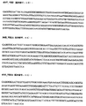
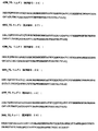
本発明のDNA分子
本発明は本発明の抗体をコードするDNA分子にも関する。該配列は、図1aおよび2aに示されるDNA分子を含むがこれに限らない。
DNA molecules of the invention The invention also relates to DNA molecules encoding the antibodies of the invention. Such sequences include, but are not limited to, the DNA molecules shown in FIGS. 1a and 2a.
本発明のDNA分子は、本明細書で開示された前記配列に限らないが、変異体を含み、本発明におけるDNA変異体はハイブリダイゼーションの物理的特性を参照にして開示される。熟練労働者は、DNAを用いてその相補体を同定することができ、そしてDNAが二本鎖であるから、均等物または相同物を、核酸ハイブリダイゼーション技術を利用して識別するためにDNAが用いられることを認識する。ハイブリダイゼーションは、100%以下の相補性で起こり得ることも認識される。しかし、条件の適切な選択を考慮すれば、ハイブリタイゼーション技術は構造的関連性に基づくDNA配列と特定のDNAプローブを区別するために利用される。前記条件に関しての手引きとして、Sambrookら、1989年(Sambrook,J.,Fritsch,E.F.およびManiatis,T.(1989)分子クローン:実験室マニュアル,Cold Spring Harbor Laboratory Press, Cold Spring Harbor, USA)およびAusuberlら、1995年 (Ausuberl,F.M.,Brent,R.,Kingston,R.E.,Moore,D.D.,Sedman,J.G.,Smith,J.A.およびStruhl,K.編集(1995).分子生物学における現代のプロトコル(Current Protocols in Molecular Biology)New York:John Wileyと息子達)を参照されたい。 The DNA molecules of the present invention include, but are not limited to, the sequences disclosed herein, and the DNA variants in the present invention are disclosed with reference to physical properties of hybridization. A skilled worker can use DNA to identify its complement, and since DNA is double stranded, DNA can be used to identify equivalents or homologs using nucleic acid hybridization techniques. Recognize that it is used. It is also recognized that hybridization can occur with less than 100% complementarity. However, given the appropriate choice of conditions, hybridization techniques are utilized to distinguish between DNA sequences based on structural relationships and specific DNA probes. For guidance on these conditions, see Sambrook et al., 1989 (Sambrook, J., Fritsch, EF and Maniatis, T. (1989) Molecular Clone: Laboratory Manual, Cold Spring Harbor Laboratory Press, Cold Spring Harbor, Cold Spring Harbor, Cold Spring Harbor, ) And Ausubel et al., 1995 (Ausubel, FM, Brent, R., Kingston, RE, Moore, DD, Sedman, JG, Smith, JA, and Struh, K. Editing (1995) Current Protocols in Molecular Biology New York: John Wil See y and sons).
2種のポリヌクレオチド配列間の構造的類似性は、2種の配列を互いに交雑させる条件の「ストリンジェンシー」の機能性として発現される。ここで使用される用語「ストリンジェンシー」は、ハイブリダイゼーションを嫌う条件までの範囲を言及する。ストリンジェントな条件は、ハイブリダイゼーションを強く嫌い、最も構造的に相関する分子だけが当該する条件で互いに交雑する。逆に言えば、非ストリンジェントな条件は構造的相関性が低い分子のハイブリダイゼーションを好む。ハイブリダイゼーションストリンジェンシーは、よって、2種の核酸配列の構造的関係と直接相互に関係がある。以下の関係は、ハイブリダイゼーションと相関性の相互関係を示すのに有効である(Tmは二重核酸の融点である):
a.Tm=69.3+0.41(G+C)%
b.二重DNAのTmは、不適当な塩基対の数が1%増加する度に1℃減少する。
c.(Tm)μ2−(Tm)μ1=18.5 log10μ2/μl
μlおよびμ2は、2つの溶液のイオン強度である。
The structural similarity between two polynucleotide sequences is expressed as a “stringency” functionality under conditions that allow the two sequences to cross each other. As used herein, the term “stringency” refers to a range up to conditions that hate hybridization. Stringent conditions strongly hate hybridization and only the most structurally correlated molecules will cross each other under those conditions. Conversely, non-stringent conditions prefer hybridization of molecules with low structural correlation. Hybridization stringency is thus directly related to the structural relationship between the two nucleic acid sequences. The following relationship is effective to show a correlation between hybridization and correlation (T m is the melting point of the double nucleic acid):
a. T m = 69.3 + 0.41 (G + C)%
b. The T m of the duplex DNA decreases by 1 ° C. for every 1% increase in the number of inappropriate base pairs.
c. (T m ) μ 2 − (T m ) μ 1 = 18.5 log 10 μ 2 / μl
μl and μ2 are the ionic strengths of the two solutions.
ハイブリダイゼーションストリンジェンシーは、多くの要因の1つの機能で、全体的なDNA濃度、イオン強度、温度、プローブの大きさ、および水素結合を妨害する薬剤を含む。ハイブリダイゼーションを促進する要因には、高いDNA濃度、高いイオン強度、低い温度、長いプローブおよび水素結合を妨害する薬剤の不使用が含まれる。ハイブリダイゼーションは2段階にわたって行われる:「結合」過程と「洗浄」過程である。 Hybridization stringency includes agents that interfere with overall DNA concentration, ionic strength, temperature, probe size, and hydrogen bonding in one function of many factors. Factors that promote hybridization include high DNA concentrations, high ionic strength, low temperatures, long probes and the absence of agents that interfere with hydrogen bonding. Hybridization takes place in two steps: a “binding” process and a “washing” process.
まず、結合過程において、プローブはハイブリダイゼーションを好む条件において的となるものに結合する。ストリンジェンシーは通常、温度調節によって、この段階で制御される。高ストリンジェンシーの場合、短い(<20 nt)オリゴヌクレオチドプローブが用いられない限り、温度は通常65℃から70℃の間である。典型的なハイブリダイゼーション溶液は、6X SSC、0.5% SDS、5X Denhardt溶液および非特異的担体DNA100μgを含む。Ausubelら、2.9節、別冊27(1994)参照。当然ながら、異なるところも多いが、機能性では同等であり、緩衝条件は周知である。相関性の程度が低い場所では、低温で設定される。低ストリンジェンシー結合温度は約25℃から40℃である。中ストリンジェンシーは少なくとも約40℃から65℃より低い。高ストリンジェンシーは少なくとも約65℃である。 First, in the binding process, the probe binds to the target in conditions that favor hybridization. Stringency is usually controlled at this stage by temperature regulation. For high stringency, the temperature is usually between 65 ° C. and 70 ° C. unless short (<20 nt) oligonucleotide probes are used. A typical hybridization solution contains 6X SSC, 0.5% SDS, 5X Denhardt solution and 100 μg of non-specific carrier DNA. See Ausubel et al., Section 2.9, Supplement 27 (1994). Of course, there are many differences, but the functionality is equivalent and buffering conditions are well known. In places where the degree of correlation is low, the temperature is set at a low temperature. The low stringency binding temperature is about 25 ° C to 40 ° C. Medium stringency is at least about 40 ° C to less than 65 ° C. High stringency is at least about 65 ° C.
第二に、超過のプローブは洗浄により除去される。よりストリンジェントな条件が通用適用されるのは、この段階である。それゆえに、ハイブリダイゼーションを通して相関性を定めるのに最も重要なのはこの「洗浄」過程である。洗浄溶液は一般的に低い塩濃度を有する。1つの実験的中ストリンジェンシー溶液は2X SSCおよび0.1%のSDSを含む。高ストリンジェンシー溶液は、約0.2X SSC以下と等量(イオン強度において)を含み、好ましいストリンジェント溶液は約0.1X SSCを含む。異なるストリンジェンシーと関連する温度は、「結合」過程で説明した温度と同等である。洗浄溶液は一般的に洗浄過程中に何度も取り替え可能である。例えば、一般的な高ストリンジェンシー洗浄条件は、55℃で30分の洗浄を2回、および60℃で15分の洗浄を3回が含まれる。 Second, excess probe is removed by washing. It is at this stage that the more stringent conditions apply. Therefore, it is this “washing” process that is most important in establishing correlation through hybridization. The cleaning solution generally has a low salt concentration. One experimental medium stringency solution contains 2X SSC and 0.1% SDS. High stringency solutions contain an equivalent (in ionic strength) equal to or less than about 0.2X SSC, and preferred stringent solutions contain about 0.1X SSC. The temperatures associated with different stringencies are equivalent to those described in the “binding” process. The cleaning solution can generally be replaced many times during the cleaning process. For example, typical high stringency wash conditions include two 30 minute washes at 55 ° C. and three 15 minute washes at 60 ° C.
よって、本発明は、高ストリンジェンシー結合および洗浄の条件下で図1aおよび2aで示された分子を交雑する核酸分子を含み、但し、該核酸は、本明細書で開示された特性を有する抗体または機能性フラグメントをコードする。好ましい分子(mRNAパースペクティブから)は、本明細書に明記されるDNA分子の1つと、少なくとも75%または80%(好ましくは85%、さらに好ましくは90%、95%が最良)の配列相同性または同一性を持つ。 Thus, the present invention includes a nucleic acid molecule that hybridizes to the molecule shown in FIGS. 1a and 2a under conditions of high stringency binding and washing, wherein the nucleic acid comprises an antibody having the properties disclosed herein. Or code a functional fragment. Preferred molecules (from the mRNA perspective) are at least 75% or 80% (preferably 85%, more preferably 90%, 95% best) sequence homology with one of the DNA molecules specified herein, or Have identity.
機能性同一変異体
本明細書に記載されるDNA分子の変異体はいくつかの異なる方法で構成されることが認識される。例えば、完全合成DNAとして構成されてよい。効果的に20から約150までのヌクレオチドのオリゴヌクレオチドを合成する方法は、多様にある。Ausubelら、2.11節、別冊21参照(1993)。部分的に一致するオリゴヌクレオチドは、Khoranaら、J.Mol.Biol.72:209−217(1971)の報告によれば、一応合成され、組み立てられる。Ausubelら、supra、8.2節も参照。合成DNAは、適切なベクターにクローンを促進するために遺伝子の5’および3’末端で作り変えられた適切な制限領域を伴って設計されることが望ましい。
Functionally identical variants It will be appreciated that variants of the DNA molecules described herein are constructed in several different ways. For example, it may be configured as fully synthetic DNA. There are a variety of methods for effectively synthesizing oligonucleotides of 20 to about 150 nucleotides. See Ausubel et al., Section 2.11, volume 21 (1993). Partially matched oligonucleotides are described in Khorana et al. Mol. Biol. 72: 209-217 (1971) reports that they are synthesized and assembled. See also Ausubel et al., Supra, section 8.2. The synthetic DNA is preferably designed with appropriate restriction regions reshaped at the 5 'and 3' ends of the gene to facilitate cloning into an appropriate vector.
示されている通り、変異体を生成する方法は本明細書に開示されるDNAの1つから始まり、部位特異的突然変異誘発法を実施する。Ausubelら、supra、第8章、別冊37参照。典型的な方法には、対象となるDNAを一重鎖DNAバクテリフォージ媒体にクローン化する。一重鎖DNAは、単離し、望ましいヌクレオチド変更物(alterations)を含むオリゴヌクレオチドと交雑する。相補鎖は交雑し、二重鎖ファージは宿主に結合する。結果生じた子孫はDNA配列を利用して確認できる望ましい突然変異体(mutant)を有する。さらに、子孫ファージが望ましい変異体(mutant)になる可能性を高める多様な方法がある。これらの方法は当該分野に従事する者にとって周知であり、このような変異体(mutants)を生成するキットは商業的に入手可能である。
As indicated, the method for generating variants starts with one of the DNAs disclosed herein and performs site-directed mutagenesis. See Ausubel et al., Supra,
組換えDNA構築物と発現
本発明はさらに、本発明のヌクレオチド配列を1つ以上含む組換えDNA構築物を提供する。本発明の組換えDNA構築物は、プラスミド、ファージミド、ファージまたはウィルスベクターなどのベクターとの関連で使用され、そこに本発明の抗体をコードするDNA分子を挿入する。
Recombinant DNA constructs and expression The present invention further provides recombinant DNA constructs comprising one or more nucleotide sequences of the present invention. The recombinant DNA construct of the present invention is used in the context of a vector such as a plasmid, phagemid, phage or viral vector, into which a DNA molecule encoding the antibody of the present invention is inserted.
コードされた遺伝子はSambrookら、1989年およびAusubelら、1989年に開示された技術によって生成されてよい。代わりになるべきものとして、DNA配列はシンセサイザーなどを利用して科学的に交雑されてよい。オリゴヌクレオチド合成(OLIGONUCLEOTIDE SYNTHESIS)(1984、Gait,編集、IRLプレス、オックスフォード)を参照、および開示内容はここで引用され援用される。本発明の組換え構築物は、コードされたDNAのRNAおよび/またはタンパク質生成物を発現することが可能な発現ベクターを含む。前記ベクターはさらに、読み取り枠(ORF)に接続されたプロモーターを含む調節配列からなってよい。前記ベクターはさらに、選択可能なマーカー配列からなってよい。特定の開始シグナルおよび細菌分泌シグナルが、挿入された遺伝子コード配列を効果的に翻訳するのに必要となるかもしれない。 The encoded gene may be generated by techniques disclosed in Sambrook et al., 1989 and Ausubel et al., 1989. As an alternative, DNA sequences may be crossed scientifically using synthesizers and the like. See OLIGONUCLEOTIDE SYNTHESIS (1984, Gait, Editorial, IRL Press, Oxford), the disclosure of which is hereby incorporated by reference. The recombinant constructs of the present invention include expression vectors capable of expressing RNA and / or protein products of the encoded DNA. The vector may further comprise regulatory sequences including a promoter connected to an open reading frame (ORF). The vector may further comprise a selectable marker sequence. Specific initiation and bacterial secretion signals may be required to effectively translate the inserted gene coding sequence.
本発明はさらに、本発明のDNAを少なくとも1つ含む宿主細胞を提供する。前記宿主細胞は実質的に発現ベクターが可能であるどの細胞にもなり得る。例えば、哺乳類細胞などの高等真核宿主細胞、酵母細胞などの下等真核宿主細胞、または細菌細胞などの原核細胞である。前記組換え構築物の前記宿主細胞への導入は、リン酸カルシウム移入、DEAE、デキストラン媒介移入、エレクトロポレーションまたはファージ感染によって影響を受けることもある。 The present invention further provides a host cell comprising at least one DNA of the present invention. The host cell can be virtually any cell capable of an expression vector. For example, higher eukaryotic host cells such as mammalian cells, lower eukaryotic host cells such as yeast cells, or prokaryotic cells such as bacterial cells. Introduction of the recombinant construct into the host cell may be affected by calcium phosphate transfer, DEAE, dextran mediated transfer, electroporation or phage infection.
細菌発現
細菌の使用に有効な発現ベクターは、望ましいタンパク質をコードした構造的DNA配列を適切な翻訳開始シグナルおよびターミネーションシグナルとともに、機能性プロモーターと作動可能なリーディングフェーズにおいて挿入することで構築される。前記ベクターは、1つ以上の表現型選択可能マーカーおよび複製オリジンからなり、前記ベクターを維持し、望ましくは、宿主内での増幅を提供する。形質転換に適した原核性の宿主は、大腸菌、枯草菌、ネズミチフス菌およびシュードモナス菌、ストレプトミセス菌およびブドウ球菌属内の多様種を含む。
Bacterial expression Effective expression vectors for use in bacteria are constructed by inserting a structural DNA sequence encoding the desired protein, together with appropriate translation initiation and termination signals, in a reading phase operable with a functional promoter. The vector consists of one or more phenotypically selectable markers and a replication origin and maintains the vector, desirably providing amplification within the host. Prokaryotic hosts suitable for transformation include E. coli, Bacillus subtilis, Salmonella typhimurium and Pseudomonas, Streptomyces and various species within the genus Staphylococcus.
細菌のベクターは、例えば、バクテリオファージ、プラスミドであってよく、あるいはファージミドに基づいてよい。これらのベクターは、選択可能なマーカーおよび公知のクローンベクターpBR322(ATCC 37017)を有する商業的に入手可能なプラスミド由来の細菌の複製オリジンを含み得る。適切な宿主株の形質転換および宿主株の適切な細胞密度への成長の後、選択可能なプロモーターは適切な手段(例:温度変換または化学的誘発)によって脱抑圧/誘発され、細胞は付加期間培養される。細胞は一般的に遠心分離で培養され、物理的または科学的手段で分裂し、その結果生成された未加工の抽出物はさらなる精製のため保管される。 Bacterial vectors may be, for example, bacteriophages, plasmids, or may be based on phagemids. These vectors may contain a bacterial origin of replication from a commercially available plasmid with a selectable marker and the known clonal vector pBR322 (ATCC 37017). After transformation of the appropriate host strain and growth of the host strain to the appropriate cell density, the selectable promoter is derepressed / induced by the appropriate means (eg temperature conversion or chemical induction) and the cell is subjected to an additional period Incubate. The cells are generally cultured by centrifugation and divided by physical or scientific means, and the resulting raw extract is stored for further purification.
細菌システムにおいて、発現ベクターの多くは、タンパク質を発現する用途をもとに都合よく選択してよい。例えば、抗体の生成またはペプチドライブラリーの選り分けのために大量のタンパク質が生成される場合、容易に精製される融合タンパク質生成物の高いレベルの発現を指示するベクターが望ましいかもしれない。 In bacterial systems, many of the expression vectors may be conveniently selected based on the use for expressing the protein. For example, if large quantities of protein are produced for antibody production or peptide library sorting, vectors that direct high level expression of easily purified fusion protein products may be desirable.
療法
療法は、治療効果のある量の本発明に含まれる抗体を、治療を必要とする対象に投与することを含む。ここでの「治療効果のある」量は、GM−CSFと、対象物の治療部位における受容体との間の相互作用を、一用量または複用量摂生に従って、単体でまたは他の製剤との組み合わせで、効果的にブロックするに十分な量と定義され、副作用を緩和しながらも、当該量は毒性学的に許容し得る。対象物は、ヒトまたはヒト以外の動物(例えば、ラットまたはアカゲザル)でよい。
Therapy Therapy involves the administration of a therapeutically effective amount of an antibody included in the present invention to a subject in need of treatment. A “therapeutically effective” amount herein refers to the interaction between GM-CSF and the receptor at the treatment site of the subject, alone or in combination with other formulations, according to one or multiple dose regimens. Is defined as an amount sufficient to effectively block, and the amount is toxicologically acceptable while mitigating side effects. The object may be a human or non-human animal (eg, rat or rhesus monkey).
本発明の抗体は公知の製剤と併用されるかもしれず、いくつかの場合においては、その抗体自体が修正されるかもしれない。例えば、抗体は効力をさらに高める為に、抗毒素または放射性同意元素に共役される。 The antibodies of the invention may be used in combination with known formulations, and in some cases, the antibodies themselves may be modified. For example, antibodies are conjugated to antitoxins or radioactive consent elements to further increase efficacy.
発明の抗体は、GM−CSFは望まれずに発現または発見された多様な状況において、治療または診断ツールとして使用される。本発明の抗体を利用した治療に特に適する疾患および状態は、関節リウマチ、多発性硬化症、クローン病、乾癬、喘息、アトピー性皮膚炎またはショックなどの炎症性疾患である。 The antibodies of the invention are used as therapeutic or diagnostic tools in a variety of situations where GM-CSF is undesirably expressed or discovered. Diseases and conditions particularly suitable for treatment using the antibodies of the present invention are inflammatory diseases such as rheumatoid arthritis, multiple sclerosis, Crohn's disease, psoriasis, asthma, atopic dermatitis or shock.
前述の疾患を治療するには、本発明に従った医薬組成物が1つ以上の生理的に許容し得る担体または賦形剤を使用した従来の方法で処方される。本発明の抗体は、治療される疾患の種類によって多岐にわたる適切な手段を用いて投与される。可能性のある投与経路は、非経口的(筋肉の、静脈内の、動脈内の、腹膜内の、または皮下の)、肺内および鼻腔内、そして部位免疫抑制治療を望む場合、病巣内投与が含まれる。さらに、本発明の抗体は抗体の用量を減らし脈に注入して投与してもよい。投薬が急性か慢性かによるが、注射により与えられた投薬が好ましいが、静脈または皮下注射が最も好ましい。投与される量は、臨床的症状、個人の体重、その他薬剤の服用があるかなどの要因に左右される。熟練労働者は、投薬経路が治療される疾患または症状によって異なることを理解する。 To treat the aforementioned diseases, a pharmaceutical composition according to the present invention is formulated in a conventional manner using one or more physiologically acceptable carriers or excipients. The antibodies of the invention are administered using a wide variety of appropriate means depending on the type of disease being treated. Possible routes of administration include parenteral (muscular, intravenous, intraarterial, intraperitoneal, or subcutaneous), intrapulmonary and nasal, and intralesional administration if local immunosuppressive treatment is desired. Is included. Furthermore, the antibody of the present invention may be administered by reducing the dose of the antibody and injecting it into the pulse. Depending on whether the medication is acute or chronic, medication given by injection is preferred, but intravenous or subcutaneous injection is most preferred. The amount administered depends on factors such as clinical symptoms, individual weight, and other medications. Skilled workers understand that the route of administration depends on the disease or condition being treated.
新規ポリペプチドの治療効果量を本発明に従い決定するには、特に患者の体質、投薬経路および地用される疾患の特質に大きく左右される。一般的な手引きは、例えば調和化に関する国際会議(the International Conference on Harmonisation)の出版物またはREMINGSTONの薬剤学(REMINGTON'S PHARMACEUTICAL SCIENCES)27章および28章、484〜528ページ(第18版、Alfonso R.Gennaro,編集、Easton,Pa.;Mack Pub.Co.,1990)に掲載されている。詳細には、治療効果量の決定には、製剤の毒性および効力等の要因にも左右される。毒性は当該技術および前述参照で公知の方法を用いて決定される。効力は、下記の例で解説される方法に関連した同じ手引きを利用して決定される。
The determination of a therapeutically effective amount of a novel polypeptide according to the present invention is highly dependent on, among other things, the patient's constitution, route of administration and the nature of the disease used. General guidance can be found, for example, in the publication of the International Conference on Harmonization or REMINGTON'S
診断方法
GM−CSFは、リンパ球、単球、内皮細胞、繊維芽細胞およびいくつかの悪性細胞を含む多様な細胞によって発現するため、本発明の非GM−CSF抗体は患者の異なる組織内でGM−CSFの蓄積部位を投影または視覚化するために適用される。これに関し、放射線同位元素、アフィニティーラベル(ビオチン、アビジンなど)、蛍光ラベル、常磁性原子などの使用により、抗体は検知可能となり分類される。分類手順は当該技術においては公知である。画像診断における抗体の臨床的使用は、Grossman, H. B.,Urol.Clin.North Amer.13:465−474(1986)), Unger,E.C.ら、Invest.Radiol.20:693−700(1985)),および、Khaw,B.A.ら、Science 209:295−297(1980)) により見直される。
検知可能で分類された抗体の病巣の検知は、例えば炎症部位から予測可能である。1つの実施態様において、組織または血液のサンプルを採取し、検知可能で分類された抗体内で前記サンプルを培養する実験が行われる。好ましい実施態様は、当該技術が、磁気画像、蛍光関節撮影法など非侵襲性の方法で実施される。そのような診断テストは、GM−CSFの存在または欠如が関連指標である疾患治療の結果を監視することで適用される。本発明は、ここで説明されている生体外での診断において、非GM−CSF抗体の使用も考慮に入れる。
Diagnostic Methods Because GM-CSF is expressed by a variety of cells, including lymphocytes, monocytes, endothelial cells, fibroblasts and some malignant cells, the non-GM-CSF antibodies of the invention are expressed in different tissues of patients. Applied to project or visualize the accumulation site of GM-CSF. In this regard, antibodies are detectable and classified by the use of radioisotopes, affinity labels (biotin, avidin, etc.), fluorescent labels, paramagnetic atoms, and the like. Classification procedures are well known in the art. The clinical use of antibodies in diagnostic imaging is described by Grossman, H. et al. B. Urol. Clin. North Amer. 13: 465-474 (1986)), Unger, E .; C. Invest. Radiol. 20: 693-700 (1985)), and Khaw, B .; A. Science 209: 295-297 (1980)).
Detection of the lesion of an antibody classified as detectable can be predicted, for example, from the site of inflammation. In one embodiment, an experiment is performed in which a sample of tissue or blood is taken and cultured in a detectable and classified antibody. In a preferred embodiment, the technique is implemented in a non-invasive manner such as magnetic imaging, fluorescence arthrography. Such diagnostic tests are applied by monitoring the outcome of disease treatment where the presence or absence of GM-CSF is a relevant indicator. The present invention also contemplates the use of non-GM-CSF antibodies in the in vitro diagnosis described herein.
治療および診断のための組成物
本発明の抗体は、薬剤学的に有効な組成物を準備する公知の方法に従い処方され、本発明の抗体(機能性フラグメントを含む)は薬剤学的に許容し得る担体との組み合わせで結合される。適切な媒体およびそれらの製剤は、例えばREMINGSTONの薬剤学(REMINGTON'S PHARMACEUTICAL SCIENCES)(第18版、Alfonso R.Gennaro,編集、Easton,Pa.:Mack Pub.Co.,1990)で解説されている。効果的な投与に最適で薬剤学的に許容し得る組成物を形成するために、当該組成物は、適切な担体と共に、本発明の1つ以上の抗体の効果的な用量を含む。
Compositions for Treatment and Diagnosis The antibodies of the invention are formulated according to known methods of preparing pharmaceutically effective compositions, and the antibodies (including functional fragments) of the invention are pharmaceutically acceptable. Combined in combination with the resulting carrier. Suitable media and their formulations are described, for example, in REMINGSTON'S PHARMACEUTICAL SCIENCES (18th edition, Alfonso R. Gennaro, edited, Easton, Pa .: Mack Pub. Co., 1990). Yes. In order to form a pharmaceutically acceptable composition that is optimal for effective administration, the composition will contain an effective dose of one or more antibodies of the present invention, together with a suitable carrier.
活性化合物を制御放出するために、製剤が適切に処方される。制御放出準備は非GM−CSF抗体を合成または吸収するためにポリマーを使用して実施される。制御放出は、放出を制御するために、適切な高分子(例えば、ポリエステル、ポリアミノ酸、ポリビニル、ピロリドン、エチレン酢酸ビニル、メチルセルロース、カルボキシメチルセルロース、プロタミン、硫酸塩)、高分子の濃縮および結合方法を選択することで実施される。制御放出製剤による運動期間を制御する別の方法には、非GM−CSF抗体を、ポリエステル、ポリアミノ酸、ヒドロゲル、ポリマー(乳酸)、エチレン酢酸ビニル共重合体などの重合体に、結合する。代わりになるべきものとして、これらの製剤を重合体粒子に結合する以外に、例えばコアセルベーション技術または界面重合によって、例えばヒドロキシメチルセルロースまたはゼラチンマイクロカプセルおよびポリマーメタクリル酸メチルマイクロカプセルをそれぞれ、またはコロイド状薬物送達システムにおいては例えば、リポソーム、アルブミン微小球、マイクロエマルジョン、ナノ粒子およびナノカプセルまたはマクロエマルジョンによって、製剤を準備されたマイクロカプセルに閉じ込めることも可能である。このような技術はRemingstonの薬剤学(Remington's Pharmaceutical Sciences)(1980)に開示されている。 The formulations are suitably formulated to give controlled release of the active compound. Controlled release preparation is performed using a polymer to synthesize or absorb non-GM-CSF antibodies. Controlled release uses appropriate polymer (eg polyester, polyamino acid, polyvinyl, pyrrolidone, ethylene vinyl acetate, methylcellulose, carboxymethylcellulose, protamine, sulfate), polymer concentration and binding methods to control release. It is carried out by selecting. In another method of controlling the duration of exercise with a controlled release formulation, non-GM-CSF antibodies are conjugated to polymers such as polyesters, polyamino acids, hydrogels, polymers (lactic acid), ethylene vinyl acetate copolymers. As an alternative, in addition to bonding these formulations to polymer particles, for example by coacervation techniques or interfacial polymerization, eg hydroxymethylcellulose or gelatin microcapsules and polymer methyl methacrylate microcapsules, respectively, or colloidal In drug delivery systems, it is also possible to enclose the formulation in prepared microcapsules, for example by liposomes, albumin microspheres, microemulsions, nanoparticles and nanocapsules or macroemulsions. Such techniques are disclosed in Remington's Pharmaceutical Sciences (1980).
上記化合物は例えばボーラス注入法または持続注入などの注射によって非経口的投与用に処方される。注入(injection)用の製剤は、アンプル、または複数回の投与のためのコンテナに防腐剤を加えた剤形で提供される。上記組成物は、油性または水生媒体の懸濁剤、液剤または乳濁液の形で構成されてよく、そして、懸濁剤、分解防止剤および/または分散剤などの処方されうる製剤を含んでよい。代わりになるべきものとして、活性成分は、例えば無菌で発熱源のない水などの適切な媒体と構成されるために使用前は、パウダー状になっていてよい。 The compounds are formulated for parenteral administration by injection, for example, by bolus injection or continuous infusion. Formulations for injection are provided in ampoules or dosage forms with preservatives added to containers for multiple administrations. The composition may be in the form of a suspension, solution or emulsion in an oily or aquatic medium, and may contain formulated formulations such as suspending agents, anti-degradation agents and / or dispersing agents. Good. As an alternative, the active ingredient may be in powder form prior to use in order to be configured with a suitable medium such as, for example, sterile and heat-free water.
望ましくは、前記組成物はパックまたはディスペンサー装置で提供され、活性成分を有する1つ以上の剤形を含む。パックには、例えば、ブリスターパックなどの金属またはプラスチックのホイルが含まれてもよい。パックまたはディスペンサー装置は、投与に関する説明とともに使用される。 Desirably, the composition is provided in a pack or dispenser device and comprises one or more dosage forms having an active ingredient. The pack may include a metal or plastic foil, such as a blister pack, for example. The pack or dispenser device is used with instructions for administration.
本発明はさらに、本発明に限ることなく説明する以下の実施例を参照して理解される。 The present invention will be further understood with reference to the following examples, which are not limited to the present invention.
実施例
実施例1:HuCAL GOLD(R)ライブラリーからのヒトGM−CSF特異性抗体の生成
A.ファージの増殖および精製
HuCAL GOLD(R)ライブラリーは、クロラムフェニコールを34μg/mlおよびブドウ糖1%を含む2xYT培地内で増殖させた(2xYT−CG)。0.5のOD600における(振動を与えずに37℃で30分、250rpmで振動させ37℃で30分)補助ファージ感染(VCSM13)後、細胞を遠沈し(4120g、5分、4℃)2xYT/34μg/ml、クロラムフェニコール/50μg/ml、カナマイシン/0.25mM IPTG、そして22℃で一晩増殖させた。ファージは、上清からPEG沈殿させ、PBS/20%グリセロールに再懸濁し、−80℃で保管した。パニングラウンド感におけるファージの増殖は、以下のように実施された:対数中期段階の大腸菌TG1細胞は、溶離したファージを感染させ、ブドウ糖1%とクロラムフェニコール34μg/mlを追加したLB寒天で培養した。30℃で一晩培養した後、コロニーを破片にし、0.5のOD600nmになるまで2xYT−CGを接種するために使用し、前述のとおり補助ファージが加えられた。
Examples Example 1: Generation of human GM-CSF specific antibodies from HuCAL GOLD (R) library Growth and Purification HuCAL GOLD (R) library of phage were grown chloramphenicol in 2xYT medium containing 1% 34 [mu] g / ml and glucose (2xYT-CG). Cells were spun down (4120 g, 5 min, 4 ° C.) after auxiliary phage infection (VCSM13) at an OD 600 of 0.5 (37 minutes at 37 ° C. without vibration, 30 minutes at 37 ° C. with shaking at 37 ° C.). ) 2 × YT / 34 μg / ml, chloramphenicol / 50 μg / ml, kanamycin / 0.25 mM IPTG and grown overnight at 22 ° C. Phages were PEG precipitated from the supernatant, resuspended in PBS / 20% glycerol and stored at −80 ° C. Phage growth in the sense of panning round was carried out as follows: E. coli TG1 cells in mid-log phase were infected with eluted phage and on LB agar supplemented with 1% glucose and 34 μg / ml chloramphenicol. Cultured. After overnight culture at 30 ° C., colonies were broken into pieces and used to inoculate 2 × YT-CG until an OD of 600 nm of 0.5, and auxiliary phage were added as described above.
B.HuCAL GOLD(R)とパニング
ヒトGM−CSFを認識する抗体の選択には、いくつかのパニング戦略が適用された。概要は、HuCAL GOLD(R)抗体−ファージを異なるVHマスター遺伝子を含む3つのプールに分割した。これらのプールは個々に、a)3ラウンドの固相支持体としてニュートラアビジン社のコーティングされた96ウェルプレートに直接コーティングされたビオチン化ヒトGM−CSFタンパク質(ミネソタ州ミネアポリス、R&D Systems社製造)上の固相パニングまたはb)3ラウンドのためにDynabeads(Dynal,ノルウェー オスロー)をコーティングしたストレプトアビジン上に捕獲されたビオチン化ヒトGM−CSFタンパク質上の溶液パニングに供した。
B. The choice of HuCAL GOLD (R) and panned human GM-CSF antibodies that recognize several panning strategies were applied. Summary, HuCAL GOLD (R) antibody - were divided into three pools containing the VH master genes different phage. These pools are individually a) on a biotinylated human GM-CSF protein (manufactured by R & D Systems, Minneapolis, Minn.) Directly coated onto a neutravidin-coated 96-well plate as a three-round solid support. B) solution panning on biotinylated human GM-CSF protein captured on streptavidin coated with Dynabeads (Dynal, Oslo, Norway) for 3 rounds.
詳細には、不動となったビオチン化GM−CSFをパニングするため、ニュートラビジン社のプレートのウエルを300μl PBSで洗浄した。抗原はPBSで3μg/ml (200nM)で薄め、そして各ウェルあたり0.1mlを室温で2時間でコーティングした。300μl PBSで2回の洗浄ステップの後、ウェルは1対1で薄められた2x Chemiblocker(ケミコ・インターナショナル社 カリフォルニア州、テメキュア)を含むブロッキングバッファーとインキュベートされた。 Specifically, in order to pan the immobilized biotinylated GM-CSF, the wells of Neutravidin plate were washed with 300 μl PBS. Antigen was diluted with PBS at 3 μg / ml (200 nM) and 0.1 ml per well was coated for 2 hours at room temperature. After two washing steps with 300 μl PBS, the wells were incubated with blocking buffer containing 2 × Chemiblocker (Chemico International, Temecure, Calif.) Diluted 1: 1.
選択の後、100μlのHuCAL GOLD(R)ファージは、0.4μl 25% Tween20室温で30分を含むブロッキングブッファー100μlに予め吸収させた。ブロックされたファージは、室温で30分ニュートラビジン社プレートのウェルにl00μ1アリコートに移した。このステップは前吸収のために2度繰り返された。
コーティングされ、ブロックされたニュートラアビジン社のマイクロタイタープレートの洗浄(2x 300μ1 PBS)の後、予め吸収された0.1mlのファージをコーティングされたウェルに加え、室温で1.5時間インキュベートして静かに振動させた。このインキュベートに続いて、PBS/0.05% Tween20での室温における10回の洗浄後、が実施された。
After selection, HuCAL GOLD (R) phage 100μl were preabsorbed blocking Warped fur 100μl containing 30 min 0.4 [mu]
After washing of coated and blocked Neutravidin microtiter plates (2 x 300 μl PBS), 0.1 ml of preabsorbed phage is added to the coated wells and incubated at room temperature for 1.5 hours and gently Vibrated. This incubation was followed by 10 washes at room temperature with PBS / 0.05% Tween20.
バウンドファージはウエルあたり10 mM Tris pH 8.0中の20mM DTTを120μl加えることで溶離された。溶離液は取り除かれ、14 mlの大腸菌TGIに加えられOD600nm of 0.6−0.8に増殖させた。ウェルはさらに200μl PBSで洗浄され、この溶液もTG1細胞に加えられた。大腸菌のファージ感染は振動せずに37℃で45分行った。追加で、0.6−0.8のOD600nm に成長した200μlの細胞を、振動なしで37℃で45分間かけて選択ウェルに加えた。このTG−I細胞は、第一溶離ステップからのファージを含む14mlの培養液に加えられる。5000rpmで10分間の遠心分離の後、細菌性のペレットはそれぞれ500μ1 2xYT培地に再懸濁され、2xYT−CG寒天プレート上で培養され、30℃で一晩インキュベートされた。コロニーは、その後、プレートから除去され、ファージは前述の通り増殖および精製された。
Bound phage was eluted by adding 120 μl of 20 mM DTT in 10 mM Tris pH 8.0 per well. The eluate was removed and added to 14 ml of E. coli TGI and grown to OD 600nm of 0.6-0.8. The wells were further washed with 200 μl PBS and this solution was also added to TG1 cells. E. coli phage infection was performed at 37 ° C. for 45 minutes without shaking. In addition, 200 μl of cells grown to an OD 600 nm of 0.6-0.8 was added to the selected wells for 45 minutes at 37 ° C. without shaking. The TG-I cells are added to 14 ml of culture medium containing the phage from the first elution step. After centrifugation at 5000 rpm for 10 minutes, each bacterial pellet was resuspended in 500
選択の第二および第三ラウンドは選択の第一ラウンドと同じ方法で実施されたが、ファージ結合後の洗浄条件がよりストリンジェントになるのが唯一の違いであった。さらに、選択の第三ラウンドでは、ファージは、ストレプトアビジンでコーティングされたビーズ(Dynabeads M−280; Dynal)上に追加の前吸着ステップを施される。エッペンドルフ型チューブは、Chemiblocker溶液で室温で30分のインキュベーションによりブロックされた。各ファージプール0.3mlは0.05% Tween20を含む2xChemiblocker溶液を1:1になるように混合され、ブロックされたエッペンドルフ型チューブ内で室温1時間インキュベートされ、50μ1のDynabeads M−280が吸着作用の為30分加えられた。ビーズは磁気装置(Dynal MPC−E)を使って取り除かれた。ファージ150μlの一部はニュートラアビジン社のプレートに第一および第二ラウンド同様移動された(上記参照)。
The second and third rounds of selection were performed in the same way as the first round of selection, with the only difference that the wash conditions after phage binding were more stringent. Furthermore, in the third round of selection, the phage is subjected to an additional pre-adsorption step on streptavidin-coated beads (Dynabeads M-280; Dynal). Eppendorf type tubes were blocked by incubation with Chemiblocker solution for 30 minutes at room temperature. 0.3 ml of each phage pool was mixed with a 2 × Chemiblocker solution containing 0.05
ビオチン化GM−CSを利用した溶液パニングでは、以下のプロトコルが適用された。1.5mlのエッペンドルフ型チューブは、一晩かけて4℃でPBSと1:1で薄められた1.5mlの2xChemiblockerでブロックされた。磁性ビーズ(Dyna−beads M−280;Dynal)でコーティングされた200μlのストレプトアビジンは、200μlのPBSで洗浄され200μl lxChemiblockerに再懸濁された(1x PBSで薄められた)。ビーズのブロッキングは、前ブロックチューブ内において4℃で一晩実施された。各パニング条件において500μlのPBSで薄められたファージは、500μl 2xChemiblocker/0.1% Tweenと1時間室温で混合される(回転装置)。ファージの吸着は2度実施される:50μlのブロックされたストレプトアビジン磁性ビーズはブロックされたファージに加えられ、回転装置内室温で30分間インキュベートされた。磁気装置(Dynal MPC−E)を通してビーズの分離が終わると、ファージの上清(−1ml)は新しくブロックされたチューブに移動し、吸着が30分間50μlのブロックされたビーズ上で繰り返された。その後、200nMのビオチン化hGM−CSFが、新たにブロックされた1.5mlのチューブ内でブロックされたファージに加えられ、回転装置内において室温1時間でインキュベートされた。ブロックされたストレプトアビジン磁性ビーズは各パニングファージプールに加えられ、回転装置内において室温10分インキュベートされた。ビオチン化GM−CSFに結合し、それによって磁性ビーズに流通停止したファージは、磁性粒子分離器(Dynal MPC−E)で集められた。ビーズは、回転装置を利用したPBS/0.05%Tweenで7x洗浄され、PBSで3度洗浄された。Dynabeadsからのファージの溶離は、300μlの10mM Tris/HCI中20mM DTT pH8を各チューブに10分間加えることで実施された。Dynabeadsは磁性粒子分離器によって分離され、上清は0.6−0.8のOD600nmに繁殖する大腸菌TG−1培養14m1に加えられた。
In solution panning using biotinylated GM-CS, the following protocol was applied. A 1.5 ml Eppendorf tube was blocked with 1.5
5000rpmで10分間遠心分離が終わると、細菌性のペレットは500μl 2xYT 培地で再懸濁され、2xYT−CG 寒天プレートの上で培養され、一晩30℃でインキュベートされた。コロニーはその後プレートから除去され、ファージは上記の通り増殖および精製された。
After centrifugation at 5000 rpm for 10 minutes, the bacterial pellet was resuspended in 500
ビオチン化GM−CSF上の溶液パニングの第一および第二ラウンドは、洗浄過程のストリンジェンシーが高まること以外は、第一ラウンドのプロトコルに従って実施された。
C. 選択されたFabフラグメントのサブクローニングと水溶性Fabフラグメントの 発現
選択されたHuCAL GOLD(R)ファージの挿入物をコードするFabは発現ベクターpMORPH(R)X9_Fab_FH(図5)にサブクローン化され、水溶性Fabの急速発現を促進した。選択されたクローンのDNAはXbalおよびEcoRIにより消化し、それによりFabコーディング挿入物(ompA−VLCLおよびphoA−Fd)を切除し、XbaI/EcoRIにより消化したベクターpMORPH(R)X9_Fab_FHにクローン化した。これらのベクター内で発現するFabは、検知および精製のため2つのC−末端タグ(それぞれFLAGTMおよび6xHis)を有した。
The first and second rounds of solution panning on biotinylated GM-CSF were performed according to the first round protocol, except that the stringency of the washing process was increased.
C. Subcloning of selected Fab fragments and expression of water-soluble Fab fragments The Fab encoding the selected insert of HuCAL GOLD (R) phage was subcloned into the expression vector pMORPH (R) X9_Fab_FH (FIG. 5) and soluble in water. Facilitated rapid expression of Fab. DNA of the selected clones was digested with Xbal and EcoRI, thereby excising the Fab-coding insert (ompA-VLCL and phoA-Fd), and cloned into the digested vector pMORPH (R) X9_Fab_FH by XbaI / EcoRI. Fabs expressed in these vectors had two C-terminal tags (FLAG ™ and 6xHis, respectively) for detection and purification.
D.大腸菌内のHuCAL GOLD(R) Fab抗体のミクロ発現
pMORPH(R)X9_Fab_FHにサブクローニングされた後に獲得された単体コロニーは、各ウェル100μl 2xTY/Cm/1% Glu培地を有するウェルに植え付けるために使用されて37℃で一晩成育させた。TG−1大腸菌培養の5μlは100μl 2xTY/Cm/0.1% Glu培地/ウェルを有する無菌性の96−ウェルマイクロタイタープレートに移した。マイクロタイタープレートは、培養物が0.5のOD600nmでわずかに混濁(2〜4時間)するまで、マイクロプレートシェイカーでこれらの発現プレートに、各ウェル20μl 2xYT/Cm/3mM IPTGが加えられ(濃縮終了0.5mM IPTG)、気体透過性テープで密封され、400rpmの振動で一晩30℃に設定されインキュベートされた。
D. HuCAL GOLD (R) acquired single colonies after being subcloned into micro expression pMORPH (R) X9_Fab_FH of Fab antibodies in E. coli is used to inoculate each well 100μl 2xTY / Cm / 1% Glu medium wells with And grown overnight at 37 ° C. 5 μl of the TG-1 E. coli culture was transferred to a sterile 96-well microtiter plate with 100
全体細胞溶解物の生成(BEL抽出物)
発現プレートの個々のウェルに、40μ1 BEL buffer(2xBBS/EDTA:24.7g/l ホウ酸, 18.7g NaCl/l,1.49g EDTA/l, pH8)が加えられたが、2.5mg/mlのリゾチームが含まれ、マイクロタイタープレートシェーカで22℃1時間(400 rpm)に設定されインキュベートされた。BEL抽出物は、ELISAまたはBioVeris M−series(R) 384 アナライザー(実施例2参照)による結合分析に利用された。
Generation of whole cell lysate (BEL extract)
40 μl BEL buffer (2 × BBS / EDTA: 24.7 g / l boric acid, 18.7 g NaCl / l, 1.49 g EDTA / l, pH 8) was added to each well of the expression plate, but 2.5 mg / ml of lysozyme was included and incubated on a microtiter plate shaker set at 22 ° C. for 1 hour (400 rpm). The BEL extract was utilized for binding analysis by ELISA or BioVeris M-series (R) 384 analyzer (see Example 2).
E.大腸菌のHuCAL(R) GOLD Fab抗体の発現と精製
TG−1 細胞内で、pMORPH(R)X9_Fab_FHによりコードされたFabフラグメントの発現は、34μg/mlのクロラムフェニコールで補完された2xYT 培地の1 1と培養するシェーカーフラスコ内で実施した。0.5mM IPTGの導入後、細胞は1.6時間22℃で増殖させた。細胞ペレットの全体細胞抽出物は、フレンチプレスまたはニッケル/ニトリオトリ酢酸クロマトグラフィーによって単離されたFabフラグメントによって調合された(キアゲン、ヒルデン、ドイツ)。濃縮は、紫外線光度法によって決定された(Krebsら、2001)。
E. Expression and Purification of E. coli HuCAL (R) GOLD Fab Antibody In TG-1 cells, expression of the Fab fragment encoded by pMORPH (R) X9_Fab_FH was expressed in 2xYT medium supplemented with 34 μg / ml chloramphenicol. 11 Performed in shaker flasks cultured with 1. After the introduction of 0.5 mM IPTG, the cells were grown for 1.6 hours at 22 ° C. Whole cell extracts of cell pellets were formulated with Fab fragments isolated by French press or nickel / nitriotriacetic acid chromatography (Qiagen, Hilden, Germany). Concentration was determined by ultraviolet photometry (Krebs et al., 2001).
実施例2:hGM−CSF特異性抗体の識別
hGM−CSF特異性のFabをコードするクローンを識別するために、上述のパニング法によって選択された個々の大腸菌クローンのBEL抽出物を、ELISAまたはBio Veris(Bio Veris M−series(R)384Analyzer)によって分析した。
Example 2: Identification of hGM-CSF-specific antibodies To identify clones encoding hGM-CSF-specific Fabs, BEL extracts of individual E. coli clones selected by the panning method described above were subjected to ELISA or Bio Analyzed by Veris (Bio Veris M-series (R) 384 Analyzer).
A.酵素結合免疫吸着測定(ELISA)法
ヒト組換えビオチン化GM−CSF(R&D Systems)を、PBS中1.5μg/mlで、室温で2時間ニュートラアビジン社マイクロタイタープレートにコーティングした。
A. Enzyme-linked immunosorbent assay (ELISA) method Human recombinant biotinylated GM-CSF (R & D Systems) was coated onto Neutravidin microtiter plates at 1.5 μg / ml in PBS for 2 hours at room temperature.
抗原のコーティング後、室温で1時間1%BSAを有するPBS/0.05%Tween(PBS−T)でウェルをブロックした。PBS−TBEL−抽出物でウェルを洗浄した後、精製したHu−CAL(R)Fabまたは対照IgGをPBSで希釈し、ウェルに追加し、また室温で1時間インキュベートした。一次抗体を検出するために、以下の二次抗体、アルカリホスファターゼ(AP)−共役アフィニティー精製F(ab')2フラグメント、ヤギ抗ヒトIgG、ヤギ抗マウスIgGまたはヤギ抗ラットIgG(Jackson Immuno Research)を適用した。AP−共役を検出するために、製造業者による取扱説明書に従って、AttoPhos(Roche)などの蛍光発生基質を使用した。全てのインキュベーションのステップの間に、マイクロタイタープレートのウェルをPBS−Tで3度、また二次抗体による最終インキュベーションの後で3度洗浄した。TECANSpectrafluorプレートリーダーで蛍光を測定した。 After antigen coating, wells were blocked with PBS / 0.05% Tween (PBS-T) with 1% BSA for 1 hour at room temperature. After washing the wells with PBS-TBEL- extract, the Hu-CAL (R) Fab or control IgG purified and diluted in PBS, added to the wells and incubated for 1 hour at room temperature. To detect primary antibodies, the following secondary antibodies, alkaline phosphatase (AP) -conjugated affinity purified F (ab ′) 2 fragment, goat anti-human IgG, goat anti-mouse IgG or goat anti-rat IgG (Jackson Immuno Research) Applied. To detect AP-conjugation, a fluorogenic substrate such as AttoPhos (Roche) was used according to the manufacturer's instructions. During all incubation steps, the wells of the microtiter plate were washed 3 times with PBS-T and 3 times after the final incubation with the secondary antibody. Fluorescence was measured with a TECAN Spectrafluor plate reader.
B.溶解物でのGM−CSF結合Fabを検出するための電気化学発光(BioVeris)ベースの結合分析
大腸菌溶解物(BEL抽出物)でのGM−CSF結合Fab抗体を検出するためのELISA実験に代わり、BioVerisM−SERIES(R)384AnalyzerBioVeris,Europe社、ウイットニー、オックスフォードシャー、英国)で結合を分析した。
B. Electrochemiluminescence (BioVeris) based binding analysis to detect GM-CSF bound Fab in lysate As an alternative to ELISA experiments to detect GM-CSF bound Fab antibody in E. coli lysate (BEL extract), Binding was analyzed on a BioVerisM-SERIES® 384 Analyzer BioVeris, Europe, Whitney, Oxfordshire, UK).
この目的を達成するために、BioVerisスクリーニング用に、アッセイバッファー(PBS/0,05%Tween20/0.5%BSA)で、少なくとも1:50および最大で1:1000にBEL抽出物を希釈した。0.1μg/mlの濃度で、ビオチン化GM−CSF(R&D Systems)をストレプトアビジンコーティングされた磁性ビーズ、Dynabeads(Dynal)に結合した。96のウェルまたは384ウェルプレートにつき、Dynabead原液の1:25希釈の25または15μlを使用した。ビオチン化GM−CSFに加える前に、ビーズを室温で30分間シェーカーでアッセイバッファーにより3度洗浄した。その後ビーズをアッセイバッファーにより3度洗浄し、未使用のアッセイバッファーで最終的に再懸濁した。BV−tagTM(BioVeris Europe社、ウイットニー、オックスフォードシャー、英国)を使用して、抗ヒト(Fab)'2(Dianova社)をルテニウム標識化した。使用直前に、6μg/mlの濃度でこの二次抗体をGM−CSF結合ビーズに加えた。Fab抗体を含む大腸菌発現培養の希釈BEL抽出物(上記参照)の100μlまたは60μlを96のウェルまたは384ウェルプレートに満たし、各々、GM−CSF結合ビーズとともに抗Fab−BV−tagTM二次抗体混合の25または15μlをそれぞれのウェルに加え、室温で2時間プレート振動装置でインキュベートした。Bio Veris M−SERIES(R)384Analyzerでプレートを分析した。
To achieve this goal, the BEL extract was diluted at least 1:50 and up to 1: 1000 with assay buffer (PBS / 0,05
配列分析の後で、十分強力な結合を示す、七十四(74)のユニーククローンを同定した(シグナル:ELISAで10:1またはBio−Verisで50:1より大きな雑音比)。機能性アッセイで、これらのクローンを発現、精製および検査した。 After sequence analysis, 74 (74) unique clones were identified that showed sufficiently strong binding (signal: noise ratio greater than 10: 1 in ELISA or 50: 1 in Bio-Veris). These clones were expressed, purified and tested in a functional assay.
C.選択した抗hGM−CSF Fabの分子特異性および種交差反応性の決定
以下の検体、ラットおよびマウスGM−CSF、ヒトIL−3、ヒトIL−4、ヒトIL−5、ヒトIL−13、ヒトM−CSF(全て英国、ロンドン、ペプロテック社から)に対して抗hGM−CSF抗体の交差反応性を決定した。
C. Determination of molecular specificity and species cross-reactivity of selected anti-hGM-CSF Fab The following specimens, rat and mouse GM-CSF, human IL-3, human IL-4, human IL-5, human IL-13, human The cross-reactivity of anti-hGM-CSF antibody was determined against M-CSF (all from Peprotech, London, UK).
表面プラズモン共鳴(Biacore3000、ウプサラ、スウエーデン)によって設定された捕獲でこれを実行した。 This was performed with capture set by surface plasmon resonance (Biacore 3000, Uppsala, Sweden).
標準EDC−NHSアミンカップリング化学反応を使用して、CM5chips(Biacore,スウェーデン)を、5000−6000RU抗F(ab)2(Dianova社,AffinipureF(ab)2フラグメントヤギ抗ヒトIgG、F(ab)2フラグメント特異的)、80μg/m1の10mM緩衝酢酸溶液、4つ全てのフローセルでpH4によってコーティングした。フローセル2〜4で、特異なGM−CSF Fab(5μl/mlの流速で500nM Fabフラグメントの20μ1、300−400RU)を捕獲した。特異性Fabの捕獲後、緩衝剤を注入し、抗Fab/Fab相互作用の解離を決定した。以下のサイクルでは、特異のシグナルを決定するために、検体の増殖因子を15から2000nMまでの濃度範囲で注入した(20μl、流速20μ1/分)。その後、得た緩衝剤センソグラムを特異性のものから手作業にて差し引いた。それぞれのサイクルの後で、100mM HCI(5μl)によってフローセルを発生させた。
MOR03684およびMOR03682を含む7つのHuCAL(R)抗hGM−CSF抗体を検査し、ヒトGM−CSFに対して特異的として、他のサイトカインまたはマウスもしくはラットGM−CSFのいずれとも結合しなかった。対照的に、Fab MOR03929は、ラットGM−CSFに対して著しい交差反応性を示した。
Using standard EDC-NHS amine coupling chemistry, CM5chips (Biacore, Sweden) was converted to 5000-6000RU anti-F (ab) 2 (Dianova, Affinipure F (ab) 2 fragment goat anti-human IgG, F (ab) 2 fragment specific), 80 μg /
Seven HuCAL® anti-hGM-CSF antibodies, including MOR03684 and MOR03682, were examined and did not bind to any other cytokine or mouse or rat GM-CSF as specific for human GM-CSF. In contrast, Fab MOR03929 showed significant cross-reactivity to rat GM-CSF.
実施例3:GM−CSFとGM−CSF受容体アルファ間の相互作用を抑制する抗ヒトGM−CSF Fab候補の識別
HuCAL GOLD(R)ライブラリーから選択された74の異なるhGM−CSF特異的抗体の、hGM−CSFとその受容体との間の相互作用を抑制するための効力を検査した。相互作用は、(i)一方はGM−CSF依存性TF−1細胞株(Kitamuraら、1989)を使用した増殖アッセイであり、また(ii)他方は、GM−CSF受容体のアルファ鎖を発現する組換え型CHO細胞株によるFACS分析である、2つの方法で検査した。TF−1増殖アッセイでは、アルファ鎖およびベータ鎖から成る内因性のGM−CSF受容体を有するGM−CSFの相互作用をブロックするための抗GM−CSF抗体の能力を分析し、細胞増殖の減少をもたらした。FACSアッセイでは、GM−CSF受容体のGM−CSFとアルファ鎖との間の相互作用の特異的抑制を決定した。
Example 3: Identification of anti-human GM-CSF Fab candidates that suppress the interaction between GM-CSF and GM-CSF receptor alpha 74 different hGM-CSF specific antibodies selected from the HuCAL GOLD (R) library Was tested for its efficacy to suppress the interaction between hGM-CSF and its receptor. The interaction is (i) one is a proliferation assay using the GM-CSF dependent TF-1 cell line (Kitamura et al., 1989) and (ii) the other expresses the alpha chain of the GM-CSF receptor. Two methods were examined, FACS analysis with recombinant CHO cell lines. The TF-1 proliferation assay analyzes the ability of anti-GM-CSF antibodies to block the interaction of GM-CSF with an endogenous GM-CSF receptor consisting of alpha and beta chains, reducing cell proliferation Brought about. In the FACS assay, specific inhibition of the interaction between GM-CSF of the GM-CSF receptor and the alpha chain was determined.
A.アカゲザルおよびヒトGM−CSFのクローニングおよび発現
アカゲザルGM−CSF完全長cDNA(GenBank受入番号:AY007376)を遺伝子合成(geneART GmbH、レーゲンスブルク、ドイツ)によって合成し、pCR−Script−Ampベクター(Stratagene,ラホーヤ、カルフォルニア州、米国)にクローン化した。次に、cDNAを真核性の発現ベクターpcDNA3.1(+)(Invitrogen,ペイズリー、英国)にクローン化してpcDNA−macGM−CSFを得た。Qiagen(ヒルデン、ドイツ)からのRNeasyキットを使用して、l×10e7TF−1細胞から単離させたRNAからRT−PCR技法によってヒトGM−CSFのcDNA(Genbank受入番号NP000749)をクローン化した。ランダムな六量体(Gibco)を使用してSuperscriptIIキットによって逆転写を実行し、続いてPCRによるGM−CSFcDNAの増幅を実行した。得たPCR生成物を発現ベクターpcDNA3.1(+)にクローン化してpcDNA−huGM−CSFを得た。
A. Cloning and expression of rhesus monkey and human GM-CSF Rhesus monkey GM-CSF full length cDNA (GenBank accession number: AY007376) was synthesized by gene synthesis (geneART GmbH, Regensburg, Germany) and the pCR-Script-Amp vector (Stratagene, La Jolla, In California, USA). The cDNA was then cloned into the eukaryotic expression vector pcDNA3.1 (+) (Invitrogen, Paisley, UK) to obtain pcDNA-macGM-CSF. Human GM-CSF cDNA (Genbank accession number NP000749) was cloned by RT-PCR technique from RNA isolated from lx10e7TF-1 cells using the RNeasy kit from Qiagen (Hilden, Germany). Reverse transcription was performed with Superscript II kit using random hexamers (Gibco) followed by amplification of GM-CSF cDNA by PCR. The obtained PCR product was cloned into the expression vector pcDNA3.1 (+) to obtain pcDNA-huGM-CSF.
HEK293細胞は、リポフェクトアミン(Stratagene,ラホーヤ、米国)を使用してそれぞれこれらの発現ベクターにより一時的に核酸を入れた。分泌する組換え型マカクまたはヒトGM−CSFを含む培地をトランスフェクションの4日後に採取した。 HEK293 cells were transiently transfected with each of these expression vectors using Lipofectamine (Stratagene, La Jolla, USA). Medium containing secreted recombinant macaque or human GM-CSF was harvested 4 days after transfection.
B.ヒトまたはサルGM−CSFを使用して抗体−hGM−CSF FabによるTF−1細胞のGM−CSF従属増殖の阻害
プロバイダーの手順(DSMZ,ブラウシュバイク、ヂオイツ; DSMZ No.AC C 334)によると、TF−1(Kitamuraら、1989)細胞は、増殖した。TF−1細胞は、RPMI1640培地(10%FCS)で二度洗浄され、その後、96穴平底マルチウェル培養皿のウェルあたり50μlに2×10⁵細胞/mlの濃度で播種された。0.5ng/mlのヒト組換えGM−CS(”Leucomax”、ESSEX Pharma,ミュンヘン)およびHuCAL(R)Fab抗体(RPMI1640培地内で200ng/ml〜200μg/mlに希釈、10%FCS)を30分混合し、GM−CSFの最終濃度が0.25ng/mlになるように、混合の50μlをTF−1細胞に加えた。最大細胞増殖(0%阻害)は、0.25ng/mlのGM−CSFの最終濃度で、抗体の追加なしで、TF−1細胞をインキュベートしながら測定された。TF−1増殖の100%阻害は、そのアッセイからGM−CSFを除外し、RPMI1640培地(10%FCS)内にのみ、細胞を保持することにより測定された。その後、TF1細胞は、加湿室内で、5%のCO2を使用して、37℃で72時間、インキュベートされた。製造者の推奨により、細胞活力は、MTTまたはXTT試薬(Roche,マンハイム、ドイツ)を加えることにより、測定された。全体の19のFabは、TF−1増殖の有意な阻害を示すことが確認された。結合MOR03682、MOR03684およびMOR03929は、2μMの濃度で、50%より大きいTF1細胞増殖の一貫した阻害を示した。これらの最適化されていないFabの阻害活性は、最大阻害度を達成できなかったため、IC50の投与量を決定するには十分強力ではなかった。相対的に、単クローン抗体BVD2−21 C1I(BD Biosciences Pharmingen; Cat#554503)およびMAB215(R&D Systems; Cat#MAB215)は、TF−1増殖を最大阻害できた。
B. Inhibition of GM-CSF-dependent proliferation of TF-1 cells by antibody-hGM-CSF Fab using human or monkey GM-CSF According to the provider's procedure (DSMZ, Blauschbaik, Deutsches; DSMZ No. AC C 334) , TF-1 (Kitamura et al., 1989) cells proliferated. TF-1 cells were washed twice with RPMI 1640 medium (10% FCS) and then seeded at a concentration of 2 × 10 5 cells / ml in 50 μl per well of a 96 well flat bottom multiwell culture dish. 30 ng / ml human recombinant GM-CS (“Leucomax”, ESSEX Pharma, Munich) and HuCAL® Fab antibody (diluted from 200 ng / ml to 200 μg / ml in RPMI 1640 medium, 10% FCS) Mix and add 50 μl of the mix to TF-1 cells so that the final concentration of GM-CSF is 0.25 ng / ml. Maximum cell proliferation (0% inhibition) was measured while incubating TF-1 cells at a final concentration of 0.25 ng / ml GM-CSF, without the addition of antibodies. 100% inhibition of TF-1 proliferation was measured by excluding GM-CSF from the assay and keeping the cells only in RPMI 1640 medium (10% FCS). TF1 cells were then incubated for 72 hours at 37 ° C. using 5% CO 2 in a humidified chamber. Cell viability was measured by adding MTT or XTT reagent (Roche, Mannheim, Germany) as recommended by the manufacturer. A total of 19 Fabs were confirmed to show significant inhibition of TF-1 proliferation. Bound MOR03682, MOR03684 and MOR03929 showed consistent inhibition of TF1 cell proliferation greater than 50% at a concentration of 2 μM. The inhibitory activity of these non-optimized Fabs was not strong enough to determine the IC 50 dose because the maximum degree of inhibition could not be achieved. In comparison, monoclonal antibodies BVD2-21 C1I (BD Biosciences Pharmingen; Cat # 554503) and MAB215 (R & D Systems; Cat # MAB215) were able to maximally inhibit TF-1 proliferation.
さらに、MOR03682およびMOR03684と天然ヒトGM−CSFの結合は、TF−1増殖アッセイにおいて、分析された。精製した組換えヒトGM−CSFをTF−1細胞に追加する代わりに、天然ヒトGM−CSFを培地に分泌する5637細胞の上清(DSMZ No.ACC 35)が使用された。5637上清の異なる希釈を備える組換えヒトGM−CSFの効果を比較する用量反応曲線から、培地が5ng/ml以下の天然ヒトGM−CSFを含むことを決定した。抗体ヒトGM−CSFのFab MOR03682またはMOR03684で5637上清の前培養により、細胞の生存率が組換えヒトGM−CSFを使用する実験と比較して軽減するように、天然ヒトGM−CSFとTF−1細胞との結合は、ブロックした。従って、MOR03684およびMOR03682は、天然ヒトGM−CSFに結合する。Fab MOR03929は、本アッセイにおいて分析されなかった。さらに、サルGM−CSFへの交叉反応がTF−1増殖アッセイにおいて分析された。精製したヒトGM−CSFをTF−1細胞に追加する代わりに、組換えサルGM−CSFを培地に分泌するトランスフェクトHEK293細胞の上清が使用された。 Furthermore, the binding of MOR03682 and MOR03684 to native human GM-CSF was analyzed in a TF-1 proliferation assay. Instead of adding purified recombinant human GM-CSF to TF-1 cells, the supernatant of 5637 cells (DSMZ No. ACC 35) secreting native human GM-CSF into the medium was used. From dose response curves comparing the effects of recombinant human GM-CSF with different dilutions of 5637 supernatant, it was determined that the media contained no more than 5 ng / ml native human GM-CSF. Pre-culture of 5637 supernatants with antibody human GM-CSF Fab MOR03682 or MOR03684 reduces natural cell viability compared to experiments using recombinant human GM-CSF and TF. -1 cell binding was blocked. Thus, MOR03684 and MOR03682 bind to native human GM-CSF. Fab MOR03929 was not analyzed in this assay. In addition, cross-reactivity to monkey GM-CSF was analyzed in a TF-1 proliferation assay. Instead of adding purified human GM-CSF to TF-1 cells, the supernatant of transfected HEK293 cells secreting recombinant monkey GM-CSF into the medium was used.
TF−1細胞は、上清を含むサルGM−CSFの存在において増殖されたが、非トランスフェクトHEK293細胞の存在において増殖されなかった。HEK−293培地の異なる希釈を備える組換えヒトGM−CSFの効果を比較する用量反応曲線から、トランスフェクト細胞の培地が2μg/ml以下のサルGM−CSFを含むことを決定した。抗体ヒトGM−CSFのFab MOR03682またはMOR03684でサルGM−CSF上清の前培養により、細胞の生存率が有意に軽減するように、サルGM−CSFとTF−1細胞との結合は、ブロックした。 TF-1 cells were grown in the presence of monkey GM-CSF with supernatant but not in the presence of untransfected HEK293 cells. From dose response curves comparing the effects of recombinant human GM-CSF with different dilutions of HEK-293 medium, it was determined that the media of transfected cells contained no more than 2 μg / ml monkey GM-CSF. Pre-culture of monkey GM-CSF supernatant with antibody human GM-CSF Fab MOR03682 or MOR03684 blocked the binding of monkey GM-CSF to TF-1 cells so that cell viability was significantly reduced. .
従って、MOR03682およびMOR03684は、サルGM−CSFで交差反応する。Fab MOR03929は、本アッセイにおいて分析されなかった。
C.抗体−hGM−CSF Fabによる、GM−CSF受容体アルファへのGM−CSF結合のブロック
GM−CSF受容体アルファ鎖を発現した細胞面へのGM−CSF結合を分析するために、cDNAが発現ベクターにクローン化され、CHO−K1細胞(DSMZ ACC 110)に安定に導入された。
Therefore, MOR03682 and MOR03684 cross-react with monkey GM-CSF. Fab MOR03929 was not analyzed in this assay.
C. Blocking GM-CSF Binding to GM-CSF Receptor Alpha by Antibody-hGM-CSF Fab In order to analyze GM-CSF binding to the cell surface expressing GM-CSF receptor alpha chain, cDNA is an expression vector. And stably introduced into CHO-K1 cells (DSMZ ACC 110).
GM−CSF受容体のアルファ鎖に発現する安定したCHO−K1細胞株のクローン化
ヒトGM−CSF受容体アルファ鎖のcDNA(Genbank受入番号M64445)は、Qiagen(ヒルデン、ドイツ)からのRNeasyキットを使用して、lx10e7 TF−1細胞から単離したRNAからRT−PCR技術によりクローン化された。逆転写は、ランダムヘキサマープライマー(Gibco)を使用して、SuperscriptIIキットとともに、実施された。その後、GM−CSF−受容体アルファ鎖cDNAは、以下のプライマーを使用して増幅された。
Cloning of a stable CHO-K1 cell line expressed in the alpha chain of the GM-CSF receptor The cDNA of the human GM-CSF receptor alpha chain (Genbank accession number M64445) is from Qiagen (Hilden, Germany). The RNeasy kit was used to clone from RNA isolated from lx10e7 TF-1 cells by RT-PCR technique. Reverse transcription was performed with the Superscript II kit using random hexamer primers (Gibco). GM-CSF-receptor alpha chain cDNA was then amplified using the following primers:
5’:N−GCRa−plusSS:TTCTCTGGATCCGCCACCATGCTTCTCCTGGTGACAAGCC
および
3’:C−flGCRa:ACCCTCCAATTGTCAGGTAATTTCCTTCACGGTCである。
5 ': N-GCRa-plusSS: TTCTCTGGATCCGCCCACCATGCCTTCCCTGGTGACAAGCC
And 3 ′: C-flGCRa: ACCCTCCAATTGTCAGGTAATTTCTCTCCAGGTC.
PCR反応は、EcoRIおよびBamHI(New England BioLabs)で消化された1250bp以下の製品を生じた。発現ベクターpcDNA3.1(+)(インビトロジェン、ペイズリー、UK)は、同一の酵素で消化された。消化ベクターおよびPCR製品の精製後、フラグメントは、E.coli DH1OB細胞へのエレクトロポレーションにより、結紮および形質転換された。適切なクローンは、プラスミドDNAおよびシークエンシングの前処理後、識別された。適切なクローン(pcDNA3.1(+)−GM−CSFRアルファ)は、完全ヒトGM−CSF受容体アルファcDNAを含んだ。 The PCR reaction yielded a product of less than 1250 bp digested with EcoRI and BamHI (New England BioLabs). The expression vector pcDNA3.1 (+) (Invitrogen, Paisley, UK) was digested with the same enzymes. After purification of the digestion vector and PCR product, the fragment was transformed into E. coli. Ligated and transformed by electroporation into E. coli DH1OB cells. Appropriate clones were identified after plasmid DNA and sequencing pretreatment. A suitable clone (pcDNA3.1 (+)-GM-CSFRalpha) contained fully human GM-CSF receptor alpha cDNA.
プロバイダーの手順(DSZM、ブラウンシュバイク、ドイツ;DSMZ No.ACC 110)によると、CHO−K1細胞は、増殖した。 According to the provider's procedure (DSZM, Braunschweig, Germany; DSMZ No. ACC 110), CHO-K1 cells proliferated.
トランスフェクション細胞は、6ウェル培養皿内で80%の密集度に増殖し、10μlのリポフェクトアミン2000試薬(インビトロジェン)と混合した5μg DNAのpcDNA3.1(+)−GM−CSFRアルファでインキュベートした。48時間後、細胞に1mg/mlのG418(Gibco)を与え、さらに24時間後、培地は、当該の含有する2mg/mlのG418で置換された。2週間後、単一細胞は、96穴平底マルチウェル培養皿のウェルに播種された。単一クローンは、成長し、それぞれのクローンの5×105細胞は、1μg/mlの濃度で一次抗体としてマウスIgG MAB 1006(ケミコン・インターナショナル社、テメキュラ、CA)、および1:200の希釈で二次抗体として(R−PE−AffiniPure(Fab’)2ヤギ−抗−マウス−IgG (Dianova社)を使用するFACS分析によりGM−CSFR−アルファ発現のために分析された。これらの工程の間、細胞はFACSバッファ(PBS、3%FCS)内で洗浄される一方、一次および二次抗体は、1時間連続的に細胞を使用してインキュベートされた。染色した細胞の蛍光は、FACSCaliburシステム(Becton Dickinson)を使用し、FL2チャネル内で定量化された。
クローンの間で、解析されたクローンCHO−GMRa#11が、蛍光強度の最高の中央値を示した。157の蛍光中央値(MFL値)が、CHO−GMRa#11(図6)に対して測定された。
Transfected cells were grown to 80% confluence in 6-well culture dishes and incubated with 5 μg DNA pcDNA3.1 (+)-GM-CSFRalpha mixed with 10 μl Lipofectamine 2000 reagent (Invitrogen). . After 48 hours, the cells were given 1 mg / ml G418 (Gibco) and after another 24 hours, the medium was replaced with 2 mg / ml G418 containing it. Two weeks later, single cells were seeded in the wells of a 96-well flat bottom multiwell culture dish. Single clones grew and 5 × 10 5 cells of each clone were mouse IgG MAB 1006 (Chemicon International, Temecula, Calif.) As primary antibody at a concentration of 1 μg / ml, and a dilution of 1: 200. Analyzed for GM-CSFR-alpha expression by FACS analysis using (R-PE-AffiniPure (Fab ') 2 goat-anti-mouse-IgG (Dianova) as secondary antibody. , Cells were washed in FACS buffer (PBS, 3% FCS), while primary and secondary antibodies were incubated with cells continuously for 1 hour.The fluorescence of the stained cells was measured using the FACSCalibur system ( Quantified in the FL2 channel using Becton Dickinson).
Among the clones, the analyzed clone CHO-
CHO−GMRa#11上に発現したGM−CSF受容体アルファへのGM−CSF結合のFACS分析:
細胞に加える前に、室温で30分間、FACSバッファ(PBS/3%FBS/NaN3 0.05%)内で、増加する濃度(0.1から100μg/ml)で抗体をビオチン化されたGM−CSF(0.5μg/ml)とともにコインキュベートした。
すべての染色は、ウェルあたり1−5×10⁵細胞で96穴丸底マルチウェル培養皿(Nalge Nunc)内に実施された。2×10E5 CHO−GMRa#11細胞は、FACSバッファを含有する50μlの抗体/GM−CSF内に取り、1時間4℃でインキュベートされた。その後、細胞は、150μlのFACSバッファ/ウェルで1度洗浄され、FACSバッファ内で1:400に希釈したl00μ1のフィコエリトリンのラベル化されたストレプトアビジン(BD Biosciences Pharmingen)内に取った。4℃で1時間インキュベートした後、細胞は、FACSバッファで2度洗浄され、100μlのFACSバッファ内で再懸濁され、ビオチン結合GM−CSFは、FACScalibur(Becton Dickinson)内で、細胞のFL2蛍光強度を介して測定された。IC50値は、非線形回帰曲線に適用するGraphPad Prism v3.03ソフトウェアを使用して、取得した用量反応曲線から測定された。Fab 抗体MOR03682、MOR03684およびMOR03929は、GM−CSF受容体アルファを発現した細胞面へのGM−CSF結合の有意な抑制を示した。
FACS analysis of GM-CSF binding to GM-CSF receptor alpha expressed on CHO-GMRa # 11:
The antibody was biotinylated GM at increasing concentrations (0.1-100 μg / ml) in FACS buffer (PBS / 3% FBS / NaN 3 0.05%) for 30 minutes at room temperature before adding to the cells. -Co-incubated with CSF (0.5 μg / ml).
All staining was performed in 96-well round bottom multiwell culture dishes (Nalge Nunc) at 1-5 × 10 5 cells per well. 2 × 10E5 CHO-
実施例4:CDRカセットの段階的交換による選択されたFabの親和性成熟
A.Fabライブラリーおよびパニングの親和性成熟の発生
抗−GM−CSF Fabフラグメントの親和性および阻害活性を増加するために、クローンMOR03682、MOR03684およびMOR03929は、親和性成熟を受けた。この点について、CDR領域は、トリヌクレオチド定方向突然変異誘発(Virnekasら、1994;Nagyら、2002)を使用して、カセット式変異誘発により最適化された。配列分析は、分析された3つの親クローンのCDRの間で配列相同性を示さなかった。表2aおよび2bは、親クローンMOR03682、MOR03684およびMOR03929に対して、6つのCDRペプチド配列を提供する。
Fab最適化として使用されたプロトコルを以下に簡潔に説明する。発現ベクターpMORPH(R)X9Fab_FHからのFabフラグメントは、ファージミドベクターにクローンされた(US 6,753,136)。その後、2つの異なる方法は、親Fabの親和性および有効性を最適化するために適用された。
Example 4: Affinity maturation of selected Fabs by stepwise exchange of CDR cassettes
A. Development of Fab Library and Panning Affinity Maturation To increase the affinity and inhibitory activity of anti-GM-CSF Fab fragments, clones MOR03682, MOR03684 and MOR03929 underwent affinity maturation. In this regard, the CDR regions were optimized by cassette mutagenesis using trinucleotide directed mutagenesis (Virnekas et al., 1994; Nagy et al., 2002). Sequence analysis showed no sequence homology between the CDRs of the three parent clones analyzed. Tables 2a and 2b provide six CDR peptide sequences for parental clones MOR03682, MOR03684 and MOR03929.
The protocol used as Fab optimization is briefly described below. The Fab fragment from the expression vector pMORPH (R) X9 Fab_FH was cloned into a phagemid vector (US 6,753,136). Two different methods were then applied to optimize the affinity and efficacy of the parent Fab.
まずは、1つのファージ抗体Fabライブラリーが各親のL−CDR3が単一のラムダ軽鎖CDR3配列のレパートリーにより置換され、生成される。二次ライブラリーにおいて、H−CDR2領域は、単一の重鎖CDR2配列のレパートリーにより置換される。
親和性成熟ライブラリーは、E.coli TOP10F’(インビトロジェン)へ多様性クローンを形質転換することにより生成された。ファージは、実施例1Aに記載されたように前処理された。MOR03929由来のL−CDR3およびH−CDR2ライブラリーが選択手順の間別々に保持されたが、MOR03684およびMOR03682の双方のL−CDR3ライブラリーは、プールされ、MOR03684およびMOR03682由来の双方のH−CDR2ライブラリーは、プールされた。
First, a single phage antibody Fab library is generated in which each parental L-CDR3 is replaced by a repertoire of single lambda light chain CDR3 sequences. In the secondary library, the H-CDR2 region is replaced by a repertoire of single heavy chain CDR2 sequences.
Affinity maturation libraries are E. coli. It was generated by transforming diversity clones into E. coli TOP10F ′ (Invitrogen). The phage was pretreated as described in Example 1A. While L-CDR3 and H-CDR2 libraries from MOR03929 were kept separately during the selection procedure, both MOR03684 and MOR03682 L-CDR3 libraries were pooled and both H-CDR2 from MOR03684 and MOR03682 The library was pooled.
パニングは、実施例1Bに記載されているように、元来、3期間のためのソリューションにビオチンGM−CSFを実施し、さらに厳しい選択状況に適用した。 Panning originally implemented biotin GM-CSF in a solution for 3 periods as described in Example 1B and applied it to more stringent selection situations.
B.溶解物において改良したGM−CSF結合Fabを検出するための電気化学発光(BioVeris)ベースの結合分析
大腸菌溶解物(BEL抽出物)におけるGM−CSF結合Fab抗体を検出するために、基本的に実施例2Bに説明したBio VerisM−384SERIES(R)Workstation(Bio Veris Europe社、ウィットニー、オックスフォードシャー、英国)で結合を分析した。最も高いECL値を有するFabを精製して、溶解平衡滴定による親和性測定(SET;Haenelら、2005)および表面プラズモン共鳴(Biacore)を行った(実施例4D参照)。
B. Electrochemiluminescence (BioVeris) based binding assay to detect improved GM-CSF binding Fab in lysate Basically performed to detect GM-CSF binding Fab antibody in E. coli lysates (BEL extract) Binding was analyzed with the Bio Veris M-384SERIES® Workstation (Bio Veris Europe, Whitney, Oxfordshire, UK) as described in Example 2B. The Fab with the highest ECL value was purified and subjected to affinity measurement by dissolution equilibrium titration (SET; Haenel et al., 2005) and surface plasmon resonance (Biacore) (see Example 4D).
C.改良したVH(H−CDR2)による改良したVL(L−CDR3)のX−クローニング
さらなる親和性の改良のために、同じ親クローンに由来した成熟Fabからの単独で最適化されたH−CDR2およびL−CDR3を、この組み合わせがさらなる親和性を得る結果となる確率が高いので、組み合わせた(Yangら、1995;Schierら、1996;Chenら、1999)。改良した親和性を有するFabが、H−CDR2およびL−CDR3ライブラリーの両方から識別されるので、X−クローニングと呼ばれるこの手順を、親クローンMOR03929に由来した結合剤に適用した。軽鎖全体(XbaI/SphIフラグメント)をL−CDR3−最適化ドナークローンからH−CDR2−最適化受容体クローンに移転することによりこれを達成した。
C. X-cloning of improved VL (L-CDR3) with improved VH (H-CDR2) For further affinity improvement, single optimized H-CDR2 from mature Fab derived from the same parent clone and L-CDR3 was combined because there is a high probability that this combination would result in further affinity (Yang et al., 1995; Schier et al., 1996; Chen et al., 1999). Since Fabs with improved affinity are distinguished from both H-CDR2 and L-CDR3 libraries, this procedure, called X-cloning, was applied to the binder derived from the parent clone MOR03929. This was achieved by transferring the entire light chain (XbaI / SphI fragment) from the L-CDR3-optimized donor clone to the H-CDR2-optimized receptor clone.
D.溶解平衡滴定(SET)および表面プラズモン共鳴(Biacore)を使用したピコモル親和性の決定
KDの決定のために、Fabの単量体比(分析的SECによって分析された少なくとも90%の単量体含有量;Superdex75、Amersham Pharmacia)を使用した。
D. For the determination of dissolution equilibrium titration (SET) and surface plasmon resonance (Biacore) picomolar affinities determined K D using, at least 90% of the monomer was analyzed by a monomer ratio of Fab (analytical SEC Content: Superdex 75, Amersham Pharmacia) was used.
溶液およびデータ評価における電気化学発光(ECL)ベースの親和性の決定は、基本的にHaenelら、2005が説明しているように実行した。溶液中ヒトGM−CSF(Leu−comax)の異なる濃度(段階的な5n希釈)によってFabの一定量を平衡化した。するedto磁性ビーズ(M−280ストレプトアビジン,Dynal)およびBV−tagTM(Bio Veris Europe社、ウィットニー、オックスフォードシャー、英国)標識抗ヒト(Fab)’2(Dianova社)に結合したビオチン化ヒトGM−CSF(R&DSystems)を加え、15〜30分インキュベートした。続いて、結合していないFabの濃度を、M−SERIES(R)384analyzer(Bio Veris Europe社)を使用して、ECL検出を経て定量化したFriguetら、1985に従い、検出の間、固相への著しい平衡移動を回避するために用心した。 Determination of electrochemiluminescence (ECL) based affinity in solution and data evaluation was performed essentially as described by Haenel et al., 2005. A fixed amount of Fab was equilibrated with different concentrations of human GM-CSF (Leu-comax) in solution (stepwise 5 n dilution). Biotinylated human bound to edto magnetic beads (M-280 streptavidin, Dynal) and BV-tag ™ (Bio Veris Europe, Witney, Oxfordshire, UK) labeled anti-human (Fab) ' 2 (Dianova) GM-CSF (R & D Systems) was added and incubated for 15-30 minutes. Subsequently, the concentration of unbound Fab was determined to the solid phase during detection according to Friguet et al., 1985, quantified via ECL detection using M-SERIES® 384 analyzer (Bio Veris Europe). Beware to avoid significant equilibrium movements.
上述のアッセイ条件を使用して、Fabの親和性を決定し、表4に示した。
標準EDC−NHSアミン結合反応を利用して、10mM Na−酢酸塩pH4.5で、〜100RU組換えヒトGM−CSF(Peprotech)の密度でコーティングされたF1チップ(Biacore、スウェーデン)上で動力SPR分析をさらに実行した。基準のフローセルでそれぞれの量のHASを固定した。PBS(136mM NaCI,2.7mM KCI,10mM Na2HPO4,1.76mM KH2PO4 pH7.4)+0.005%Tween20を泳動用緩衝剤として使用した。6.3〜200nMの濃度シリーズにおいて20μl/分の流速でFabを適用した。会合相を60sに、解離相を120s(親)または最大600s(親和性最適化)に設定した。長期にわたって解離相を監視するために、基本的にDrakeら、(2004)に従って以下の条件を使用した。200nMの単一濃度でFabを適用し、流速を100pl/分に、解離相を6000〜18.000sに設定した。これらのアッセイ条件下で決定した基準から外れた率に基づき、Fabの親和性を決定し、表4に示した。
Fab affinity was determined using the assay conditions described above and is shown in Table 4.
Power SPR on F1 chips (Biacore, Sweden) coated with 10 mM Na-acetate pH 4.5 at a density of ˜100 RU recombinant human GM-CSF (Peprotech) utilizing standard EDC-NHS amine coupling reaction Further analysis was performed. Each amount of HAS was fixed in a reference flow cell. PBS (136 mM NaCI, 2.7 mM KCI, 10 mM Na 2 HPO 4 , 1.76 mM KH 2 PO 4 pH 7.4) + 0.005
ヒトGM−CSFの代わりに溶液中の検体としてラットGM−CSF(Peprotech)を使用して、基本的に実施例4Dに説明したようにラットGM−CSFに対する親和性の決定を行った。Haenelら(2005)に従って親和性を計算した。このアッセイでは、ラットGM−CSFに対するFab MOR04357の親和性はKD=1.0nMであると決定した。
実施例5:GM−CSFとGM−CSF受容体アルファ鎖との間の相互作用を抑制する最適化抗ヒトGM−CSF Fabの特性決定
A.GM−CSF受容体アルファ結合アッセイ
0.5μg/ml(35nM)のビオチン化GM−CSFを使用して、GM−CSF受容体結合アッセイを上述のように実行した(実施例3C)。CHO−GMRa#11細胞(0%抑制)に対するGM−CSFの最大結合を、抗体を加えることなく、0.5μg/mlのビオチン化GM−CSFの最終GM−CSF濃度で細胞をインキュベートすることにより測定した。GM−CSF結合の100%抑制を、アッセイからGM−CSFを除外することにより測定した。非線形回帰曲線適合を適用したGraph Pad Prism v3.03ソフトウェアを使用して得た用量反応曲線からIC50値を決定した。
改良した親和性を有するFab、親Fabおよび単クローン性対照IgGを分析した。表5は、これらのアッセイから得たIC50値を要約している。5μg/mlの抗体濃度で得た%抑制もまた、表5に示している。
Example 5: Characterization of optimized anti-human GM-CSF Fab that inhibits the interaction between GM-CSF and GM-CSF receptor alpha chain. GM-CSF Receptor Alpha Binding Assay The GM-CSF receptor binding assay was performed as described above (Example 3C) using 0.5 μg / ml (35 nM) biotinylated GM-CSF. Maximal binding of GM-CSF to CHO-
Fab, parental Fab and monoclonal control IgG with improved affinity were analyzed. Table 5 summarizes the IC 50 values obtained from these assays. The percent inhibition obtained at an antibody concentration of 5 μg / ml is also shown in Table 5.
B.ヒトGM−CSFを使用した抗hGM−CSF FabによるTF−1GM−CSF依存増殖の抑制
実施例3Bに説明したようにTF−1増殖アッセイを実行した。単クローン性対照IgGと同様に、改良した親和性を有するFabおよび親Fabを分析した。非線形回帰曲線適合を適用したGraph Pad Prism v3.03ソフトウェアを使用して得た用量反応曲線からIC50値を決定した。表6は、これらのアッセイから得たIC50値を要約している。
表6:TF−1増殖アッセイにおける抗hGM−CSF Fabおよび対照IgGのIC50値
B. Inhibition of TF-1GM-CSF dependent proliferation by anti-hGM-CSF Fab using human GM-CSF The TF-1 proliferation assay was performed as described in Example 3B. Similar to the monoclonal control IgG, Fabs with improved affinity and parental Fabs were analyzed. IC 50 values were determined from dose response curves obtained using Graph Pad Prism v3.03 software with non-linear regression curve fitting. Table 6 summarizes the IC 50 values obtained from these assays.
Table 6: IC 50 values of anti-hGM-CSF Fab and control IgG in TF-1 proliferation assay
表7:TF−1増殖アッセイにおける抗hGM−CSF FabのIC50値 Table 7: IC 50 values of anti-hGM-CSF Fab in TF-1 proliferation assay
実施例6:ヒトIgG1型へのMOR04357の変換
A.哺乳類の発現系における発現のためのFab DNA配列の遺伝子最適化
哺乳類遺伝子発現のためのMOR04357のVHおよびVLのDNAを最適化するために(例えば、コドン使用頻度の変更、GC含有量など)、Geneart(ドイツ、レーゲンスブルク)で遺伝子合成をして、pPCR−Scriptvectorsにクローン化し055906pPCR−Scriptおよび055907pPCR−Scripを得る、最適化VHおよびVLDNA配列などを定義するのに、Geneart(ドイツ、レーゲンスブルク)によるGene OptimizerTMソフトウェアを利用した。配列番号:48は、それぞれのVH配列を示すが、配列番号:57はそれぞれのVL配列を示す。
Example 6: Conversion of MOR04357 to human IgG1 type Gene optimization of Fab DNA sequences for expression in mammalian expression systems To optimize VH and VL DNA of MOR04357 for mammalian gene expression (eg, changing codon usage, GC content, etc.) Geneart (Regensburg, Germany) is gene synthesized and cloned into pPCR-Scriptvectors to obtain 055906 pPCR-Script and 055907 pPCR-Script, etc. to define optimized VH and VL DNA sequences etc. Gene by Genet (Regensburg, Germany) Optimizer ™ software was utilized. SEQ ID NO: 48 shows each VH sequence, while SEQ ID NO: 57 shows each VL sequence.
B.ヒトIgG1型およびIgGI発現に変化するFab MOR04357のクローニング
全長免疫グロブリン(Ig)を発現するために、遺伝子最適化重鎖(VH)および軽鎖(VL)の可変領域フラグメントをpPCR−Scriptベクター(実施例5a)からサブクローン化して、ヒトIgGIのためにpMORPH(R)2hIgベクターシリーズに変化させた。MOR04357のコドン最適化VHを、NheIlBIpI温浸を経て055906pPCR−Scriptから単離し、同じ制限酵素によりpMorph2_h_IgGlfマスターベクターカットに挿入した。このベクターはすでにヒトガンマ1定常領域を含んでいた。得られた発現核外遺伝子をpMorph2_h_IgGlfMOR04357_coと命名した。MOR04357のコドン最適化VLを、NhellHpaT温浸を経て055907pPCR−Scriptから単離し、同じ制限酵素によりpMorph2_h_Iglambda2マスターベクターカットに挿入した。このベクターはすでにヒトラムダ定常領域を含んでいた。得られた発現核外遺伝子をpM2_h_Iglambda2_MOR04357_coと命名した。
B. Cloning of Fab MOR04357 Changing to Human IgG1 Type and IgGI Expression In order to express full-length immunoglobulin (Ig), gene optimized heavy chain (VH) and light chain (VL) variable region fragments were pPCR-Script vector (implemented) Subcloned from Example 5a) and changed to the pMORPH® 2hIg vector series for human IgGI. The codon optimized VH of MOR04357 was isolated from 055906 pPCR-Script via NheIlBIpI digestion and inserted into the pMorph2_h_IgGlf master vector cut with the same restriction enzymes. This vector already contained the
C.一時的発現およびヒトIgGの精製
等モル量のIgG重鎖および軽鎖発現ベクターDNAにより、真核性のHKB11細胞に核酸を入れた。トランスフェクションの3日から7日後に細胞培養浮遊物を採取した。浮遊物のpHを8.0に調節および除菌後、溶液を標準タンパク質A親和性クロマトグラフィー(rProteinA FFまたはMabSelect SURE,GE Healthcare)にかけた。緩衝剤交換を1xDulbcecco'sPBS(pH7,2,インビトロジェン社)に実行し、試料を除菌した(0,2μm)。MOR04357IgGIを1xDulbcecco'sPBS(pH6,5,インビトロジェン社)に対して透析した。変性させている間に、SDS−PAGEでの条件を軽減して、またはAgilentBioAnalyzerを使用して、SE−HPLCによる自然な状態で、IgGの純度を分析した。
C. Transient expression and purification of human IgG Nucleic acids were introduced into eukaryotic HKB11 cells with equimolar amounts of IgG heavy and light chain expression vector DNA. Cell culture suspensions were harvested 3-7 days after transfection. After adjusting and sterilizing the pH of the supernatant to 8.0, the solution was subjected to standard protein A affinity chromatography (rProtein A FF or MabSelect SURE, GE Healthcare). Buffer exchange was performed on 1 × Dulbecco's PBS (
D.溶解平衡滴定(SET)を使用してピコモル親和性を決定
KDの決定のために、IgGIの単量体比(分析的SECによって分析された少なくとも90%の単量体含有量;Superdex75、AmershamPharmacia)を使用した。溶液およびデータ評価における電気化学発光(ECL)ベースの親和性の決定は、基本的にHaenelら、2005が説明しているように、また実施例4Bに説明しているように実行した。ヒト組換えGM−CSFに対するMOR04357IgGIのKD値は、1.1pMであると決定した。
D. For the determination of decision K D picomolar affinities using dissolution equilibrium titration (SET), monomer ratio of IgGI (analytical at least 90% of the monomer content was analyzed by SEC; Superdex75, AmershamPharmacia )It was used. Determination of electrochemiluminescence (ECL) based affinity in solution and data evaluation was performed essentially as described by Haenel et al., 2005 and as described in Example 4B. K D values of MOR04357IgGI against human recombinant GM-CSF was determined to be 1.1 pm.
E.溶解平衡滴定(SET)を使用して、ラットGM−CSFに対する親和性の決定
ヒトGM−CSFの代わりに溶液中の検体としてラットGM−CSF(Peprotech)を使用して、基本的に実施例4Dに説明したようにラットGM−CSFに対する親和性の決定を行った。Haenelら(2005)に従って親和性を計算した。ラット組換え型GM−CSFに対するMOR04357IgGIのKD値は130pMであると決定した。
E. Determination of affinity for rat GM-CSF using dissolution equilibrium titration (SET) Basically, Example 4D using rat GM-CSF (Peprotech) as an analyte in solution instead of human GM-CSF. Determination of affinity for rat GM-CSF was performed as described in. Affinities were calculated according to Haenel et al. (2005). K D values of MOR04357IgGI against rat recombinant GM-CSF was determined to be 130PM.
実施例7:最適化抗ヒトGM−CSF Fabから誘導されるMOR04357IgG1の特徴付け
A.ヒトおよびアカゲザルGM−CSFを使用して、抗hGM−CSF IgGによるTF−1のGM−CSF依存増殖の抑制
実施例3Bに説明しているように、TF−1増殖アッセイを実行した。IgG1型でMOR04357を分析し、対照として単クローン性対照IgGを分析した。非線形回帰曲線適合を適用したGraph Pad Prism v3.03ソフトウェアを使用して得た用量反応曲線からIC50値を決定した。表8は、これらのアッセイから得たIC50値を要約している。まず第一に、0.25ng/mlの濃度で組換え型ヒトGM−CSFを大腸菌に生成し、第二に、組換えヒトGM−CSFを含み、pcDNA−huGM−CSFにより一時的に核酸を入れられたHEK293から浮遊物を培養し(実施例3A参照)、第三に、組換え型アカゲザル(アカゲザル)GM−CSFを含み、pcDNA−macGM−CSFにより一時的に核酸を入れられたHEK293細胞から浮遊物を培養する(実施例3A参照)。TF−1増殖アッセイについては、TF−1細胞が0.25ng/mlの確定濃度の大腸菌で生成した精製された組換えヒトGM−CSFで得られる増殖と比べて類似した増殖を示すような希釈で、それぞれのGM−CSFのソースとしてHEK293培養浮遊物を使用した。
Example 7: Characterization of MOR04357IgG1 derived from optimized anti-human GM-CSF Fab Inhibition of GM-CSF-dependent proliferation of TF-1 by anti-hGM-CSF IgG using human and rhesus monkey GM-CSF The TF-1 proliferation assay was performed as described in Example 3B. MOR04357 was analyzed with IgG1 type, and a monoclonal control IgG was analyzed as a control. IC 50 values were determined from dose response curves obtained using Graph Pad Prism v3.03 software with non-linear regression curve fitting. Table 8 summarizes the IC 50 values obtained from these assays. First, recombinant human GM-CSF is produced in E. coli at a concentration of 0.25 ng / ml, and secondly, the recombinant human GM-CSF is contained, and the nucleic acid is temporarily transferred by pcDNA-huGM-CSF. HEK293 cells cultured from suspended HEK293 (see Example 3A), and thirdly, HEK293 cells containing recombinant rhesus monkey (Rhesus monkey) GM-CSF and temporarily containing nucleic acid by pcDNA-macGM-CSF The suspension is cultured from (see Example 3A). For the TF-1 proliferation assay, dilution such that TF-1 cells show similar growth as compared to that obtained with purified recombinant human GM-CSF produced in E. coli at a defined concentration of 0.25 ng / ml. Then, HEK293 culture suspension was used as the source of each GM-CSF.
表8:TF−1増殖アッセイにおけるMOR04357IgGおよび対照IgGのIC50値 Table 8: IC 50 values of MOR04357 IgG and control IgG in TF-1 proliferation assay
参考文献:
Alvaro−Gracia JM,Zvaifler NJ,Brown CB,Kaushansky K,およびFirestein GS.(1991).慢性炎症性関節炎におけるサイトカイン.VI.IL−1および腫瘍壊死因子アルファによって間接リウマチおよびその調節における顆粒球マクロファージコロニー刺激因子の生成および遺伝子発現に関与する滑膜細胞の分析.J Immunol146,3365−3371.
Babior,B.(2000).食細胞および酸化的ストレス.AmJMed109,33−44.
Bonfield,T.L.,Kavuru,M.S.,およびThomassen,M.J.(2002).抗Anti−GM−CSFタイターは肺胞タンパク症におけるGM−CSF療法への反応を予測する.Clin Immunol105,342−350.
Bozinovski,S.,Jones,J.,Beavitt,S.J.,Cook,A.D.,Hamilton,J.A.,およびAnderson,G.P.(2003).マウス肺におけるリポ多糖体への先天性免疫応答は、TLR−4の抑制を介してGM−CSFの中和により抑制および逆転する。Am J Physiol Lung Cell Mol Physiol.
Broide,D.H.,Paine,M.M.,およびFirestein,G.S.(1992)。好酸球は、ぜんそく患者におけるアレルギー性炎の部位でのインターロイキン5および顆粒球マクロファージコロニー刺激因子mRNAを発現する。J Clin Invest90,1414−1424.
Brown,C.B.,Pihl,C.E.,およびKaushansky,K.(1994)。ヒト顆粒球マクロファージコロニー刺激因子受容体アルファサブユニットと相互作用するヒト顆粒球マクロファージコロニー刺激因子領域のマッピング.Eur J Biochem225,873−880.
Burmester,G.R.,Stuhlmuller,B.,Keyszer,G.,andKinne,R.W.(1997).単核の食細胞およびリウマチ性滑膜炎。関節炎における首謀者または馬車馬?関節炎Rheum40,5−18.
Campbell,I.K.,Bendele,A.,Smith,D.A.,およびHamilton,J.A.(1997).顆粒球マクロファージコロニー刺激因子は、マウスにおいてコラーゲン誘導関節炎を悪化させる。Ann RheumDis56,364−368.
Campbell,I.K.,Rich,M.J.,Bischof,R.J.,Dunn,A.R.,Grail,D.,およびHamilton,J.A.(1998).顆粒球マクロファージコロニー刺激因子欠乏マウスにおけるコラーゲン誘導関節炎からの予防。J Immunol161,3639−3644.
Carrieri,P.B.,Provitera,V.,DeRosa,T.,Tartaglia,G.,Gorga,F.,およびPerella,O.(1998)再発寛解型多発性硬化症の患者における脳脊髄液および血清サイトカインのプロフィール:臨床活性との相関。Immunopharmacol Immunotoxicol.20,373−382.
Cebon,J.,Nicola,N.A.,Ward,M.,Gardner,L,Dempsey,P.J.,Layton,J.E.,Duhrsen,U.,Bur−gess,A.,Nice,E.C.,およびMorstyn,G.(1990).ヒトリンパ球からの顆粒球マクロファージ刺激因子。J BiolChem265,4483−4491.
Chantry,D.,Turner,M.,Brennan,F.,Kingsbury,A.,およびFeldmann,M.(1990).顆粒球マクロファージコロニー刺激因子は、ヒト単球によってHLA−DR表現およびサイトカイン産生の両方を誘発する。Cytokine2,60−67.
ChenY.,WiesmannC.,FuhG.,LiB.,ChristingerH.W.,McKay.P,deVosA.M.,LowmanH.B.(1999)最適化抗VEGF抗体の選択および分析:抗原と複合したアフィニティー成熟FabフラグメントのJ Mol Biol.293,865−81.
Cheng,S.S.,Lai,J.J.,Lukacs,N.W.,およびKunkel,S.L.(2001).顆粒球マクロファージコロニー刺激因子は、ヒト好中球においてCCRIを上方調節する。J Immunol166,1178−1184.
Clark,S.C.およびKamen,R.(1987).ヒト造血コロニー刺激因子。Science236,1229−1237.
Cook,A.D.,Braine,E.L.,Campbell,LK.,Rich,M.J.,およびHamilton,J.A.(2001).抗体による顆粒球マクロファージコロニー刺激因子(GM−CSF)へのコラーゲン誘導関節炎開始後の閉塞:病気のエフェクター相におけるGM−CSFのための必要条件.Arthritis Res3,293−298.
deGroot,R.P.,Coffer,P.J.,およびKoenderman,L.(1998).IL−3/IL−5/GM−CSF受容体ファミリーによる増殖、分化および生存の調節。Cell Signal.10,619−628.
deVries,E.G.,Willemse,P.H.,Biesma,B.,Stern,A.C.,Limburg,P.C.,およびVellenga,E.(1991).化学療法後のGM−CSF治療の間の関節リウマチの再燃。Lancet338,517−518.
Diederichs,K.,Boone,T.,およびKarplus,P.A.(1991).顆粒球マクロファージコロニー刺激因子の新規層および推定受容体結合部位。Science254,1779−1782.
DrakeA.W.,MyszkaD.G.,KlakampS.L.(2004)動力および平衡ベースの方法による高親和性抗原/抗体複合体の特徴付け。Anal Biochem.328,35−43.
Dranoff,G.,Crawford,A.D.,Sadelain,M.,Ream,B.,Rashid,A.,Bronson,R.T.,Dickersin,G.R.,Bachurski,C.J.,Mark,E.L.,Whitsett,J.A.,および(1994).肺疾患の恒常性における顆粒球マクロファージコロニー刺激因子の関与。Science264,713−716.
FriguetB.,ChaffotteA..F,Djavadi−OhanianceL.,GoldbergM.E.(1985),酵素結合の免疫吸着法による抗原抗体複合体の溶体に恒常的な真の親和性の測定。J Immunol Methods.77,305−19.
Gasson,J.C.,Baldwin,G.C.,Sakamoto,K.M.,andDiPersio,J.F.(1990a).Thebiologyofhuman
顆粒球マクロファージコロニー刺激因子(GM−CSF).Prog Clin Biol Res352,375−384.
Gasson,J.C.,Fraser,J.K.,およびNimer,S.D.(1990b).ヒト顆粒球マクロファージコロニー刺激因子(GM−CSF):発現の調節.Prog Clin Biol Res338,27−41.
Gasson,J.C.,Kaufman,S.E.,Weisbart,R.H.,Tomonaga,M.,およびGolde,D.W.(1986).正常および白血病性ヒト骨髄性細胞に対する顆粒球マクロファージコロニー刺激因子の高親和性結合。Proc Nail Acad Sci USA.83,669−673.
Gearing,D.P.,King,J.A.,Gough,N.M.,およびNicola,N.A.(1989).ヒト顆粒球マクロファージコロニー刺激因子のための受容体の発現クローニング。EMBOJ.12,3667−3676.
Gomez−Cambronero,J.,Hom,J.,Paul,C.C.,およびBaumann,M.A.(2003).顆粒球マクロファージコロニー刺激因子は、ヒト好中球のための化学誘引物質サイトカインである:リボソームp70S6キナーゼシグナル経路の関与。J Immunol171,6846−6855.
Haenel,C.,Satzger,M.,DellaDucata,D.,Ostendorp,R.およびBrocks,B.(2005).電気化学発光ベースの平衡滴定による高親和性抗体の特徴付け。Analytical Biochemistry,339,1,182−184.
Hamilton,J.A.(1993).関節リウマチ:造血増殖因子の反対作用および遅効性の抗リウマチ薬。Lancet 342,536−539.
Hamilton,J.A. (2002). 炎症および自己免疫におけるGM−CSF. Trends Immunol 23, 403−408.
Hart,P.H., Whitty,G.A., Piccoli,D.S., および Hamilton,J.A. (1988).顆粒球マクロファージコロニー刺激因子 and IFN−ガンマによるヒト単球の相乗的活性化。TNF−アルファを増大させるがIL−I活性は増大させない。 J Immunol 141, 1516−1521.
Haworth,C., Brennan,F.M., Chantry,D., Turner,M., Maini,R.N., および Feldmann,M. (1991).関節リウマチにおける顆粒球マクロファージコロニー刺激因子の発現:腫瘍壊死因子アルファによる調節。 Eur J Immunol 21, 2575−2579.
Hayashida,K., Kitamura,T., Gorman,D.M., Arai,K., Yokota,K., および Miyajima,A. (1990).ヒト顆粒球マクロファージコロニー刺激因子(GM−CSF)ための受容体の第二サブユニットの分子クローニング:高親和性 GM−CSF 受容体の再構成。Proc Natl Acad Sci U S A 87,9655−9659.
Hazenberg BP, Van Leeuwen MA, Van Rijswijk MH, Stem AC, および Vellenga E (1989).顆粒球マクロファージコロニー刺激因子よるフェルティ症候群における顆粒球減少症の回復。インターロイキン−6放出の同時誘導および関節炎の再燃。 Blood 74, 2769−2780.
Kastelein,R.A. およびShanafelt,A.B. (1993). GM−CSF 受容体:相互作用および活性化。 On−cogene 8, 231−236.
Kaufrnan,S.E., DiPersio,J.F., および Gasson,J.C. (1989). ビトロにおける好中球の脱顆粒でのヒトGM−CSFの効果。 Exp Hematol 17, 800−804.
Kitamura,T., Sato,N., Arai,K., and Miyajima,A. (1991).ヒト IL−3 受容体 eDNAの発現クローニング は,ヒト IL−3 および GM−CSF 受容体のための共有ベータサブユニットを露呈する。 Cell 66,1165−1174.
Kitamura,T., Tanaka,N., Watanabe,J., Uchida, Kanegasaki,S., Yamada,Y., および Nakata,K.(1999).顆粒球マクロファージコロニー刺激因子に対する中和抗体を有する自己免疫疾患としての特発性肺胞タンパク症。 J Exp Med 190, 875−880.
Kitamura T., Tange T., Terasawa T., Chiba S., Kuwaki T., Miyagawa K., Piao Y.F., Miyazono K., Urabe A., Takaku F. (1989). GM−CSF, IL−3, またはエリスロポエチンで依存して増殖する一意的ヒト細胞株の確立および特徴付けJ Cell Physiol. 140, 323−34.
Knappik,A., Ge,L., Honegger,A., Pack,P., Fischer,M., Wellnhofer,G., Hoess,A., Wolle,J.,Pluckthun,A., および Virnekas,B. (2000).モジュールコンセンサス骨格およびトリヌクレオチドによるCDRランダム化に基づく合成完全ヒトモノクローナル抗体作製技術(HuCAL)。 J Mol Biol 296, 57−86.
Krebs,B., Rauchenberger,R., Reiffert,S., Rothe,C., Tesar,M., Thomassen,E., Cao,M., Dreier,T., Fischer,D., Hoss,A., Inge,L., Knappik,A., Marget,M., Pack,P., Meng,X.Q., Schier,R., Sohlemann,P., Winter,J., Wolle,J., およびKretzschmar,T. (2001).組換え型ヒト抗体の高スループット生成およびエンジニアリング。 J Immunol Methods 254, 67−84.
Lopez,A.F., Williamson,D.J., Gamble,J.R., Begley,C.G., Harlan,J.N., Klebanoff,S.J., Wal−tersdorph,A., Wong,G., Clark,S.C., および Vadas,M.A. (1986). 組換え型ヒト顆粒球マクロファージコロニー刺激因子は、ビトロ成熟ヒト好中球および好酸球機能、表面受容体発現、ならびに生存物において刺激する。 J Clin Invest 78, 1220−1228.
McQualter,J.L., Darwiche,R., Ewing,C., Onuki,M., Kay,T.W., Hamilton,J.A., Reid,H.H., および Bemard,C.C. (2001). 顆粒球マクロファージコロニー刺激因子:多発性硬化症における新推定治療標的。 J Exp Med 194, 873−882.
Meager,A., Wadhwa,M., Bird,C., Dilger,P., Thorpe,R., Newsom−Davis,J., and Willcox,N. (1999).自己免疫疾患を有する患者における顆粒球マクロファージコロニー刺激因子に対する自然発生的中和抗体。 Immunology 97, 526−532.
Metcalf,D., Begley,C.G., Johnson,G.R., Nicola,N.A., Vadas,M.A., Lopez,A.F., William−son,D.J., Wong,G.G., Clark,S.C., および Wang,E.A. (1986). 組換え型ヒト顆粒球マクロファージコロニー刺激因子のビトロにおける生物学的特性。 Blood 67, 37−45.
Mulherin,D., Fitzgerald,O., および Bresnihan,B. (1996). 関節リウマチにおける滑膜組織マクロファージ個体群および関節損傷。 Arthritis Rheum39, 115−124.
Nagy Z.A., Hubner B., Lohning C., Rauchenberger R., Reiffert S., Thomassen−Wolf E., Zahn S., Leyer S., Schier E.M., Zahradnik A., Brunner C., Lobenwein K., Rattel B., Stanglmaier M., Hallek M., Wing M., Anderson S., Dunn M., Kretzschmar T., Tesar M. (2002) 完全ヒト, HLA−DR特異的な単クローン性抗体は、悪性リンパ腫細胞のプログラム死を効率良く誘導する。 Nat Med. 8, 801−7
Olver,I.N., Hercus,T., Lopez,A., Vadas,M., Somogyi,A.A., Doyle,I., Foster,D.J., Keefe,D., Taylor,A., Brown,M., To,L.B., Cole,J., Rawling,T., Cambareri,B., Myers,M., Olszewski,N., Bastiras,S., Senn,C., Hey,A., Verma,M., and Wigley,P. (2002). GM−CSF抑制因子 E21Rの第1相試験。 Cancer Chemother Pharmacol 50, 171−178.
Plenz,G., Eschert,H., Beissert,S., Arps,V., Sindermann,J.R., Robenek,H., and Vdlker,W. (2003).顆粒球マクロファージコロニー刺激因子(GM−CSF)−欠損マウスの血管細胞外マトリックスにおける変化。 FASEB J. 17,1451−1457.
Rapoport,A.P., Abboud,C.N., and DiPersio,J.F. (1992). 顆粒球マクロファージコロニー刺激因子(GM−CSF) および顆粒球コロニー刺激因子(G−CSF):受容体生物学、情報伝達、および好中球活性化。 Blood Rev 6, 43−57.
Rauchenberger,R., Borges,E., Thomassen−Wolf,E., Rom,E., Adar,R., Yaniv,Y., Malka,M., Chumakov,I., Kotzer,S., Resnitzky,D., Knappik,A., Reiffert,S., Prassler,J., Jury,K., Wald−herr,D., Bauer,S., Kretzschmar,T., Yayon,A., および Rothe,C. (2003).ヒト線維芽細胞増殖因子受容体 3に対する特異的で機能的な抗体を産出するヒト組み合わせFabフラグメントライブラリー J Biol Chem.
Santiago−Schwarz F, Anand P,L.S., and Carsons SE (2001). 関節リウマチ(RA)における樹枝状細胞(DC): Thl 炎症性型反応を選択的に活性化する脊髄のDCのサブセットを産出するRA滑液に含まれる前駆細胞および可溶性因子。 J Immu−nol 167,1758−1768,
Sato,N., Sakamaki,K., Terada,N., Arai,K., および Miyajima,A. (1993).高親和性 GM−CSF 受容体による情報伝達:異なるシグナル伝達に関与する共有ベータサブユニットの2つの特異的な細胞質領域。 EMBO J 12,4181−4189.
Schier R., McCall A., Adams G..P, Marshall K.W., Merritt H., Yim M., Crawford R.S., Weiner L.M., Marks C., Marks J.D. (1996)抗体結合部位の中央での領域を決定する相補性の分子進化によるピコモル親和性抗c−erbB−2単一鎖Fv。 J Mol Biol. 8, 263:551−67.
Shanafelt,A.B., Johnson,K.E., および Kastelein,R.A. (1991a).,ヒトおよびマウス顆粒球マクロファージコロニー刺激因子ならびに種特異性での関与における必須アミノ酸残渣の識別。 J Biol Chem 266, 13804−13810.
Shanafelt,A.B., Miyajima,A., Kitamura,T., および Kastelein,R.A. (19916). GM−CSF および IL−5のアミノ末端基ヘリックスは、それらの受容体への高親和性 結合を抑制する。 EMBO J 10,4105−4112.
Shibata,Y., Berclaz,P.−Y., Chroneos,Z., Yoshida,H., および Trapnell,B.C. (2001). GM−CSF は、PUを通して肺での肺マクロファージ分化および先天性免疫を調節する。 Immunity 15, 557−567.
Sisson,S.D. および Dinarello,C.A. (1988).顆粒球マクロファージコロニー刺激因子により刺激されるヒト単核球によるインターロイキン−1 アルファ、インターロイキン−1 ベータおよび腫瘍壊死因子の生成。 Blood 72, 1368−1374.
Stanley,E., Lieschke,G.J., Grail,D., Metcalf,D., Hodgson,G., Gall,J.A., Maher,D.W., Ce−bon,J., Sinickas,V., および Dunn,A.R. (1994). 顆粒球/マクロファージコロニー刺激因子欠損マウスは、造血の主要な摂動を示さないが、特性肺病理学を発現する。 Proc Nail Acad Sci U S A 91, 5592−5596.
Trapnell,B.C. and Whitsett,J.A. (2002). GM−CSFは、肺界面活性剤恒常性および肺マクロファージ介在先天性宿主防衛を調節する。 Annu Rev Physiol 64, 775−802.
Uchida,K., Nakata,K., Trapnell,B.C., Terakawa,T., Hamano,E., Mikami,A., Matsushita,I., Seymour,J.F., Oh−Eda,M., Ishige,I., Eishi,Y., Kitamura,T., Yamada,Y., Hanaoka,K., および Keicho,N. (2004). 高親和性自己抗体は特に、特発性肺胞タンパク症を有する患者の肺での顆粒球マクロファージコロニー刺激因子の活性を除去する。 Blood 103, 1089−1098.
Virnekas, B., Ge, L., PlUckthun, A., Schneider, K.C., Wellnhofer, G. & Moroney, S.E., (1994). Nucleic Acids Research 22, 5600−5607.
Xu,W.D., Firestein,G.S., Taetle,R., Kaushansky,K., および Zvaifler,N.J. (1989).慢性炎症性関節炎におけるサイトカイン。 II. リウマチ性滑膜浸出における顆粒球マクロファージコロニー刺激因子。 J Clin Invest 83, 876−882.
Yamashita,N., Tashimo,H., Ishida,H., Kaneko,F., Nakano,J., Kato,H., Horiuchi,T., およびOhta,K. (2002). GM−CSFの中和によるマウスぜんそくモデルにおける気道過敏性の減衰。 Cell Immunol 219, 92−97.
Yang W.P., Green K., Pinz−Sweeney S., Briones A.T., Burton D.R., Barbas C.F. 3rd. (1995). 強力なヒト抗HIV−1 抗体 の親和性成熟のためのCDR歩行突然変異生成をピコモル範囲にする。 J Mol Biol. 254, 392−403 Zhang,Y., McCluskey,K., Fujii,K., and Wahl,L.M. (1998).単球マトリクスメタロプロテイナーゼ,およびTNFアルファ、顆粒球マクロファージCSF、ならびにプロスタグランジン依存性およびプロスタグランジン非依存性機構によるIL−I ベータによるTIMP−1 生成 の分別調節。 J Immunol 161, 3071−3076.
References:
Alvaro-Gracia JM, Zvaifler NJ, Brown CB, Kaushansky K, and Firestein GS. (1991). Cytokines in chronic inflammatory arthritis. VI. Analysis of synovial cells involved in generation and gene expression of granulocyte macrophage colony stimulating factor in indirect rheumatism and its regulation by IL-1 and tumor necrosis factor alpha. J Immunol 146, 3365-3371.
Baby, B.M. (2000). Phagocytic cells and oxidative stress. AmJMed109, 33-44.
Bonfield, T .; L. , Kavuru, M .; S. , And Thomasassen, M .; J. et al. (2002). Anti-GM-CSF titer predicts response to GM-CSF therapy in alveolar proteinosis. Clin Immunol 105, 342-350.
Bozinovski, S .; Jones, J .; , Beauvitt, S .; J. et al. Cook, A .; D. Hamilton, J .; A. , And Anderson, G. et al. P. (2003). The innate immune response to lipopolysaccharide in the mouse lung is suppressed and reversed by neutralization of GM-CSF via suppression of TLR-4. Am J Physiol Lung Cell Mol Physiol.
Broide, D.W. H. Paine, M .; M.M. , And Firestein, G .; S. (1992). Eosinophils express
Brown, C.I. B. Pihl, C.I. E. , And Kaushansky, K .; (1994). Mapping of human granulocyte-macrophage colony-stimulating factor region interacting with human granulocyte-macrophage colony-stimulating factor receptor alpha subunit. Eur J Biochem 225, 873-880.
Burmester, G .; R. Stuhlmuller, B .; Keyszer, G .; , And Kinne, R .; W. (1997). Mononuclear phagocytes and rheumatoid synovitis. Mastermind or carriage in arthritis?
Campbell, I.D. K. , Bendele, A .; Smith, D .; A. , And Hamilton, J. et al. A. (1997). Granulocyte-macrophage colony stimulating factor exacerbates collagen-induced arthritis in mice. Ann RheumDis56, 364-368.
Campbell, I.D. K. Rich, M .; J. et al. Bischof, R .; J. et al. Dunn, A .; R. Grail, D .; , And Hamilton, J. et al. A. (1998). Prevention from collagen-induced arthritis in granulocyte-macrophage colony-stimulating factor-deficient mice. J Immunol 161, 3639-3644.
Carrieri, P.A. B. , Providera, V .; , DeRosa, T .; , Tartaglia, G .; Gorga, F .; , And Perella, O .; (1998) Cerebrospinal fluid and serum cytokine profiles in patients with relapsing-remitting multiple sclerosis: Correlation with clinical activity. Immunopharmacol Immunotoxic. 20, 373-382.
Cebon, J. et al. Nicola, N .; A. , Ward, M .; Gardner, L, Dempsey, P .; J. et al. Layton, J .; E. Duhrsen, U., et al. Bur-gess, A .; , Nice, E .; C. , And Morstyn, G .; (1990). Granulocyte macrophage stimulating factor from human lymphocytes. J BiolChem265, 4483-4491.
Chantry, D.C. Turner, M .; Brennan, F .; Kingsbury, A .; , And Feldmann, M .; (1990). Granulocyte-macrophage colony stimulating factor induces both HLA-DR expression and cytokine production by human monocytes.
ChenY. WiesmannC. , FuhG. LiB. , Christinger H. W. McKay. P, deVosA. M.M. Lowman H. B. (1999) Selection and analysis of optimized anti-VEGF antibodies: J Mol Biol. 293, 865-81.
Cheng, S.M. S. Lai, J .; J. et al. , Lukacs, N .; W. , And Kunkel, S .; L. (2001). Granulocyte macrophage colony stimulating factor upregulates CCRI in human neutrophils. J Immunol 166, 1178-1184.
Clark, S.M. C. And Kamen, R .; (1987). Human hematopoietic colony stimulating factor. Science 236, 1229-1237.
Cook, A.M. D. Braine, E .; L. , Campbell, LK. Rich, M .; J. et al. , And Hamilton, J. et al. A. (2001). Occlusion after initiation of collagen-induced arthritis to granulocyte macrophage colony-stimulating factor (GM-CSF) by antibodies: a prerequisite for GM-CSF in the disease effector phase. Arthritis Res 3,293-298.
deGroot, R.D. P. Coffer, P .; J. et al. , And Koenderman, L .; (1998). Regulation of proliferation, differentiation and survival by the IL-3 / IL-5 / GM-CSF receptor family. Cell Signal. 10, 619-628.
deVries, E .; G. Willemse, P .; H. Biesma, B .; Stern, A .; C. Limburg, P .; C. , And Vellenga, E .; (1991). Relapse of rheumatoid arthritis during GM-CSF treatment after chemotherapy. Lancet 338, 517-518.
Diederichs, K .; Boone, T .; , And Karplus, P .; A. (1991). A novel layer of granulocyte macrophage colony stimulating factor and a putative receptor binding site. Science 254, 1779-1782.
Drake A. W. , MyszkaD. G. Klakamp S. L. (2004) Characterization of high affinity antigen / antibody complexes by kinetic and equilibrium based methods. Anal Biochem. 328, 35-43.
Dranoff, G.M. Crawford, A .; D. Sadelain, M .; , Ream, B .; , Rashid, A .; Bronson, R .; T. T. et al. Dickersin, G .; R. , Bachurski, C.I. J. et al. , Mark, E .; L. , Whitsett, J. et al. A. , And (1994). Involvement of granulocyte-macrophage colony-stimulating factor in lung disease homeostasis. Science 264, 713-716.
FriguetB. , Chaffettete A .; . F, Djavadi-OhananceL. , GoldbergM. E. (1985), Measurement of constant true affinity for antigen-antibody complex solutes by enzyme-linked immunosorbent assay. J Immunol Methods. 77, 305-19.
Gassson, J .; C. Baldwin, G .; C. , Sakamoto, K .; M.M. , And DiPersio, J .; F. (1990a). Thebiologyofhuman
Granulocyte-macrophage colony stimulating factor (GM-CSF). Prog Clin Biol Res352, 375-384.
Gassson, J .; C. Fraser, J .; K. , And Nimer, S .; D. (1990b). Human granulocyte macrophage colony stimulating factor (GM-CSF): regulation of expression. Prog Clin Biol Res338, 27-41.
Gassson, J .; C. Kaufman, S .; E. , Weisbart, R .; H. Tomonaga, M .; , And Golde, D .; W. (1986). High affinity binding of granulocyte macrophage colony stimulating factor to normal and leukemic human myeloid cells. Proc Nail Acad Sci USA. 83, 669-673.
Gearing, D.C. P. , King, J .; A. , Gough, N .; M.M. , And Nicola, N .; A. (1989). Expression cloning of receptor for human granulocyte macrophage colony stimulating factor. EMBOJ. 12, 3667-3676.
Gomez-Cambrero, J.A. , Hom, J .; Paul, C .; C. , And Baumann, M .; A. (2003). Granulocyte-macrophage colony stimulating factor is a chemoattractant cytokine for human neutrophils: involvement of the ribosomal p70S6 kinase signal pathway. J Immunol 171, 6846-6855.
Haenel, C.I. Satzger, M .; DellaDucata, D .; , Ostendorp, R .; And Blocks, B .; (2005). Characterization of high affinity antibodies by electrochemiluminescence-based equilibrium titration. Analytical Biochemistry, 339, 1, 182-184.
Hamilton, J.M. A. (1993). Rheumatoid arthritis: Anti-rheumatic drug with opposite and delayed action of hematopoietic growth factors. Lancet 342, 536-539.
Hamilton, J.M. A. (2002). GM-CSF in inflammation and autoimmunity.
Hart, P.M. H. , Whitty, G .; A. Piccoli, D .; S. , And Hamilton, J .; A. (1988). Synergistic activation of human monocytes by granulocyte-macrophage colony-stimulating factor and IFN-gamma. Increases TNF-alpha but not IL-I activity. J Immunol 141, 1516-1521.
Haworth, C.I. Brennan, F .; M.M. , Chantry, D .; Turner, M .; , Maini, R .; N. , And Feldmann, M .; (1991). Expression of granulocyte macrophage colony stimulating factor in rheumatoid arthritis: regulation by tumor necrosis factor alpha. Eur J Immunol 21, 2575-2579.
Hayashida, K .; Kitamura, T .; Gorman, D .; M.M. Arai, K .; Yokota, K .; , And Miyajima, A .; (1990). Molecular cloning of the second subunit of the receptor for human granulocyte macrophage colony stimulating factor (GM-CSF): Reconstitution of the high affinity GM-CSF receptor. Proc Natl Acad Sci USA 87, 9655-9659.
Hazenberg BP, Van Leeuwen MA, Van Rijswijk MH, Stem AC, and Velenga E (1989). Recovery of granulocytopenia in Ferti syndrome by granulocyte-macrophage colony-stimulating factor. Simultaneous induction of interleukin-6 release and relapse of arthritis. Blood 74, 2769-2780.
Kastelein, R.A. A. And Shanafelt, A .; B. (1993). GM-CSF receptor: interaction and activation. On-
Kaufrnan, S .; E. , DiPersio, J. et al. F. , And Gasson, J .; C. (1989). Effect of human GM-CSF on neutrophil degranulation in vitro.
Kitamura, T .; Sato, N .; Arai, K .; , And Miyajima, A .; (1991). Expression cloning of human IL-3 receptor eDNA reveals a shared beta subunit for human IL-3 and GM-CSF receptors. Cell 66, 1165-1174.
Kitamura, T .; Tanaka, N .; , Watanabe, J .; Uchida, Kanegasaki, S .; Yamada, Y .; , And Nakata, K .; (1999). Idiopathic alveolar proteinosis as an autoimmune disease having a neutralizing antibody against granulocyte-macrophage colony-stimulating factor. J Exp Med 190, 875-880.
Kitamura T. , Tage T. , Terasawa T .; , Chiba S. , Kuwaki T .; , Miyagawa K .; Piao Y. F. , Miyazono K. et al. , Urabe A. , Takaku F .; (1989). Establishment and Characterization of a Unique Human Cell Line that Proliferates Dependent on GM-CSF, IL-3, or Erythropoietin J Cell Physiol. 140, 323-34.
Knappik, A .; , Ge, L .; Honegger, A .; , Pack, P.M. Fischer, M .; Wellnhofer, G .; Hoess, A .; , Walle, J .; , Pluckthun, A .; , And Virnekas, B .; (2000). Synthetic fully human monoclonal antibody production technology (HuCAL) based on CDR randomization with modular consensus backbone and trinucleotide. J Mol Biol 296, 57-86.
Krebs, B.M. Rauchenberger, R .; , Reiffert, S .; Rothe, C .; Tesar, M .; Thomassen, E .; , Cao, M .; , Dreier, T .; Fischer, D .; Hoss, A .; Inge, L .; , Knappik, A .; , Marget, M.M. , Pack, P.M. Meng, X .; Q. Schier, R .; Sohlmann, P .; , Winter, J .; , Walle, J .; , And Kretzschmar, T .; (2001). High-throughput generation and engineering of recombinant human antibodies. J Immunol Methods 254, 67-84.
Lopez, A.M. F. Williamson, D .; J. et al. Gamble, J .; R. , Begley, C .; G. Harlan, J .; N. Klebanoff, S .; J. et al. , Wal-tersdorph, A .; Wong, G .; Clark, S .; C. , And Vadas, M .; A. (1986). Recombinant human granulocyte macrophage colony stimulating factor stimulates in vitro mature human neutrophil and eosinophil function, surface receptor expression, and survival. J Clin Invest 78, 1220-1228.
McQualter, J. et al. L. Darwiche, R .; , Ewing, C.I. Onuki, M .; , Kay, T .; W. , Hamilton, J .; A. , Reid, H .; H. , And Bemard, C.I. C. (2001). Granulocyte-macrophage colony-stimulating factor: a new putative therapeutic target in multiple sclerosis. J Exp Med 194, 873-882.
Meeger, A.M. , Wadhwa, M .; Bird, C .; Dilger, P .; , Thorpe, R .; Newsom-Davis, J .; , And Willcox, N .; (1999). Spontaneously neutralizing antibodies to granulocyte-macrophage colony-stimulating factor in patients with autoimmune disease. Immunology 97, 526-532.
Metcalf, D.M. , Begley, C .; G. , Johnson, G. R. Nicola, N .; A. Vadas, M .; A. , Lopez, A .; F. William-son, D .; J. et al. Wong, G .; G. Clark, S .; C. , And Wang, E .; A. (1986). Biological characteristics of recombinant human granulocyte macrophage colony stimulating factor in vitro. Blood 67, 37-45.
Mulherin, D.M. Fitzgerald, O .; , And Bresnihan, B .; (1996). Synovial tissue macrophage population and joint damage in rheumatoid arthritis.
Nagy Z. A. , Hubner B. et al. , Lohning C.M. Rauchenberger R. , Reiffert S. Thomassen-Wolf E .; , Zahn S .; Leyer S. , Schier E. et al. M.M. , Zahradnik A .; Brunner C .; , Lovenwein K .; Rattel B. , Stanglaire M .; Hallek M .; , Wing M .; , Anderson S. Dunn M. , Kretzschmar T .; Tesar M. (2002) Monoclonal antibodies specific for fully human, HLA-DR efficiently induce programmed death of malignant lymphoma cells. Nat Med. 8, 801-7
Olver, I. et al. N. Hercus, T .; , Lopez, A .; Vadas, M .; Somgyi, A .; A. Doyle, I .; Foster, D .; J. et al. Keefe, D .; Taylor, A .; Brown, M .; , To, L .; B. Cole, J .; Rawling, T .; , Cambareri, B .; Myers, M .; , Olszewski, N .; Bastiras, S .; Senn, C .; , Hey, A .; , Verma, M .; , And Wigley, P .; (2002).
Plenz, G.M. Eschert, H .; , Beissert, S .; , Arps, V .; , Sindermann, J .; R. Robenek, H .; , And Vdlker, W.M. (2003). Changes in the vascular extracellular matrix of granulocyte macrophage colony stimulating factor (GM-CSF) -deficient mice. FASEB J.H. 17, 1451-1457.
Rapoport, A.M. P. , Abboud, C.I. N. , And DiPersio, J. et al. F. (1992). Granulocyte-macrophage colony stimulating factor (GM-CSF) and granulocyte colony stimulating factor (G-CSF): receptor biology, signaling, and neutrophil activation.
Rauchenberger, R.A. Borges, E .; Thomassen-Wolf, E .; Rom, E .; , Adar, R .; Yaniv, Y .; , Malka, M .; , Chumakov, I .; Kotzer, S .; Resnitzky, D .; , Knappik, A .; , Reiffert, S .; Prassler, J .; , Jury, K .; Wald-herr, D .; Bauer, S .; Kretzschmar, T .; Yayon, A .; , And Rothe, C.I. (2003). Human combinatorial Fab fragment library producing specific and functional antibodies against human fibroblast growth factor receptor 3 J Biol Chem.
Santiago-Schwarz F, Anand P, L .; S. , And Carsons SE (2001). Dendritic cells (DCs) in rheumatoid arthritis (RA): Thl Progenitor cells and soluble factors contained in RA synovial fluid that produce a subset of spinal DCs that selectively activate inflammatory type responses. J Immu-nol 167, 1758-1768,
Sato, N .; , Sakaki, K .; Terada, N .; Arai, K .; , And Miyajima, A .; (1993). High affinity GM-CSF receptor signaling: two specific cytoplasmic regions of shared beta subunits that are involved in different signaling.
Schier R. McCall A .; Adams G .; . P, Marshall K.M. W. Merritt H., et al. Yim M. et al. , Crawford R. S. , Weiner L. M.M. , Marks C. , Marks J .; D. (1996) A picomolar affinity anti-c-erbB-2 single chain Fv with complementary molecular evolution that determines the region in the middle of the antibody binding site. J Mol Biol. 8, 263: 551-67.
Shanafelt, A .; B. Johnson, K .; E. , And Kastelein, R .; A. (1991a). Identification of essential amino acid residues in human, mouse granulocyte-macrophage colony-stimulating factor and species-specific involvement. J Biol Chem 266, 13804-13810.
Shanafelt, A .; B. Miyajima, A .; Kitamura, T .; , And Kastelein, R .; A. (19916). The amino end group helix of GM-CSF and IL-5 suppresses high affinity binding to their receptors.
Shibata, Y. et al. Berclaz, P .; -Y. Chroneos, Z .; , Yoshida, H .; , And Trapnell, B .; C. (2001). GM-CSF regulates lung macrophage differentiation and innate immunity in the lung through PU.
Sisson, S.M. D. And Dinarello, C.I. A. (1988). Generation of interleukin-1 alpha, interleukin-1 beta and tumor necrosis factor by human mononuclear cells stimulated by granulocyte macrophage colony stimulating factor. Blood 72, 1368-1374.
Stanley, E .; Lieske, G .; J. et al. Grail, D .; , Metcalf, D .; Hodgson, G .; Gall, J .; A. , Maher, D .; W. Ce-bon, J. et al. Sinicas, V .; , And Dunn, A .; R. (1994). Granulocyte / macrophage colony-stimulating factor-deficient mice show no major perturbation of hematopoiesis but develop characteristic pulmonary pathology. Proc Nail Acad Sci USA 91, 5592-5596.
Trapnell, B.M. C. and Whitsett, J. et al. A. (2002). GM-CSF regulates pulmonary surfactant homeostasis and pulmonary macrophage-mediated innate host defense. Annu Rev Physiol 64, 775-802.
Uchida, K .; Nakata, K .; Trapnell, B .; C. , Teragawa, T .; Hamano, E .; , Mikami, A .; , Matsushita, I .; Seymour, J .; F. Oh-Eda, M .; Ishige, I .; Eishi, Y .; Kitamura, T .; Yamada, Y .; Hanaka, K .; , And Keicho, N .; (2004). High affinity autoantibodies specifically eliminate the activity of granulocyte macrophage colony stimulating factor in the lungs of patients with idiopathic alveolar proteinosis.
Virnekas, B.H. , Ge, L .; PlUckthun, A .; Schneider, K .; C. Wellnhofer, G .; & Moroney, S .; E. , (1994).
Xu, W .; D. , Firestein, G .; S. Taetle, R .; Kaushansky, K .; , And Zvaifler, N .; J. et al. (1989). Cytokines in chronic inflammatory arthritis. II. Granulocyte-macrophage colony-stimulating factor in rheumatic synovial leaching. J Clin Invest 83, 876-882.
Yamashita, N .; , Tashimo, H .; Ishida, H .; Kaneko, F .; Nakano, J .; Kato, H .; , Moriuchi, T .; , And Ohta, K .; (2002). Attenuation of airway hyperresponsiveness in a mouse asthma model by neutralization of GM-CSF. Cell Immunol 219, 92-97.
Yang W. P. Green K. , Pinz-Sweeney S. , Briones A. T. T. et al. , Burton D. R. Barbas C .; F. 3rd. (1995). CDR walking mutagenesis for affinity maturation of potent human anti-HIV-1 antibodies is in the picomolar range. J Mol Biol. 254, 392-403 Zhang, Y. et al. McCluskey, K .; , Fujii, K .; , And Wahl, L .; M.M. (1998). Differential regulation of TIMP-1 production by IL-I beta by monocyte matrix metalloproteinases and TNF alpha, granulocyte macrophage CSF, and prostaglandin-dependent and prostaglandin-independent mechanisms. J Immunol 161, 3071-3076.
Claims (15)
a)配列番号:10及び配列番号:30(MOR04357);
b)配列番号:2及び配列番号:22(MOR04251);
c)配列番号:4及び配列番号:24(MOR04252);
d)配列番号:5及び配列番号:25(MOR04287);
e)配列番号:6及び配列番号:26(MOR04290);
f)配列番号:8及び配列番号:28(MOR04350);
g)配列番号:9及び配列番号:29(MOR04354);又は
h)配列番号:47及び配列番号:56(MOR04342);
から選択される核酸フラグメントの対を含む、抗体またはその抗原結合フラグメントをコードする単離された核酸フラグメント。 The following nucleic acid fragments a) SEQ ID NO: 10 and SEQ ID NO: 30 (MOR04357);
b) SEQ ID NO: 2 and SEQ ID NO: 22 (MOR04251);
c) SEQ ID NO: 4 and SEQ ID NO: 24 (MOR04252);
d) SEQ ID NO: 5 and SEQ ID NO: 25 (MOR04287);
e) SEQ ID NO: 6 and SEQ ID NO: 26 (MOR04290);
f) SEQ ID NO: 8 and SEQ ID NO: 28 (MOR04350);
g) SEQ ID NO: 9 and SEQ ID NO: 29 (MOR04354); or h) SEQ ID NO: 47 and SEQ ID NO: 56 (MOR04342);
An isolated nucleic acid fragment encoding an antibody or antigen-binding fragment thereof, comprising a pair of nucleic acid fragments selected from
Applications Claiming Priority (3)
| Application Number | Priority Date | Filing Date | Title |
|---|---|---|---|
| US68200905P | 2005-05-18 | 2005-05-18 | |
| US60/682,009 | 2005-05-18 | ||
| PCT/EP2006/004696 WO2006122797A2 (en) | 2005-05-18 | 2006-05-17 | Anti-gm-csf antibodies and uses therefor |
Related Child Applications (1)
| Application Number | Title | Priority Date | Filing Date |
|---|---|---|---|
| JP2013044573A Division JP5769744B2 (en) | 2005-05-18 | 2013-03-06 | Anti-GM-CSF antibody and use thereof |
Publications (2)
| Publication Number | Publication Date |
|---|---|
| JP2008545632A JP2008545632A (en) | 2008-12-18 |
| JP5295759B2 true JP5295759B2 (en) | 2013-09-18 |
Family
ID=37114333
Family Applications (2)
| Application Number | Title | Priority Date | Filing Date |
|---|---|---|---|
| JP2008511635A Active JP5295759B2 (en) | 2005-05-18 | 2006-05-17 | Anti-GM-CSF antibody and use thereof |
| JP2013044573A Active JP5769744B2 (en) | 2005-05-18 | 2013-03-06 | Anti-GM-CSF antibody and use thereof |
Family Applications After (1)
| Application Number | Title | Priority Date | Filing Date |
|---|---|---|---|
| JP2013044573A Active JP5769744B2 (en) | 2005-05-18 | 2013-03-06 | Anti-GM-CSF antibody and use thereof |
Country Status (26)
| Country | Link |
|---|---|
| US (5) | US7867495B2 (en) |
| EP (4) | EP1888643B1 (en) |
| JP (2) | JP5295759B2 (en) |
| KR (1) | KR101308087B1 (en) |
| CN (2) | CN103087190B (en) |
| AR (1) | AR053608A1 (en) |
| AU (1) | AU2006249062B2 (en) |
| BR (1) | BRPI0610796B8 (en) |
| CA (1) | CA2608498C (en) |
| DK (2) | DK1888643T3 (en) |
| ES (2) | ES2527428T3 (en) |
| HK (1) | HK1114620A1 (en) |
| HR (2) | HRP20220755T3 (en) |
| HU (1) | HUE058876T2 (en) |
| IL (1) | IL187375A (en) |
| LT (1) | LT3620171T (en) |
| ME (1) | ME02059B (en) |
| NO (1) | NO344992B1 (en) |
| NZ (1) | NZ564027A (en) |
| PL (2) | PL3620171T3 (en) |
| PT (2) | PT3620171T (en) |
| RS (2) | RS63382B1 (en) |
| RU (1) | RU2447085C2 (en) |
| SI (2) | SI1888643T1 (en) |
| TW (1) | TWI477512B (en) |
| WO (1) | WO2006122797A2 (en) |
Families Citing this family (52)
| Publication number | Priority date | Publication date | Assignee | Title |
|---|---|---|---|---|
| WO2006111353A2 (en) | 2005-04-18 | 2006-10-26 | Micromet Ag | Antibody neutralizers of human granulocyte macrophage colony stimulating factor |
| ES2527428T3 (en) | 2005-05-18 | 2015-01-23 | Morphosys Ag | Anti-GM-CSF antibodies and uses thereof |
| JP4736037B2 (en) | 2005-10-26 | 2011-07-27 | 株式会社イーベック | Human monoclonal antibody binding to human GM-CSF and antigen-binding portion thereof |
| KR101486183B1 (en) | 2006-02-08 | 2015-01-28 | 모르포테크, 인크. | Antigenic gm-csf peptides and antibodies to gm-csf |
| WO2008063898A2 (en) * | 2006-11-08 | 2008-05-29 | Kalobios Pharmaceuticals, Inc. | Methods of treating idiopathic thrombocytopenia purpura using a gm-csf antagonist |
| EP2402013A1 (en) | 2006-11-21 | 2012-01-04 | Kalobios Pharmaceuticals, Inc. | Methods of Treating Chronic Inflammatory Diseases using a GM-CSF Antagonist |
| US8398972B2 (en) | 2006-11-21 | 2013-03-19 | Kalobios Pharmaceuticals, Inc. | Methods of treating dementia using a GM-CSF antagonist |
| JP2010527939A (en) * | 2007-05-23 | 2010-08-19 | シーアールシー・フォー・アズマ・アンド・エアウェイズ・リミテッド | Neutralizing antibody |
| TW200918553A (en) * | 2007-09-18 | 2009-05-01 | Amgen Inc | Human GM-CSF antigen binding proteins |
| US20100297135A1 (en) * | 2007-11-12 | 2010-11-25 | Crc For Asthma And Airways Ltd. | Epitope for neutralizing antibodies |
| KR20100102108A (en) | 2007-11-13 | 2010-09-20 | 에베크 인코포레이티드 | Monoclonal antibodies that bind to hgm-csf and medical compositions comprising same |
| US9023351B2 (en) | 2007-11-26 | 2015-05-05 | Bayer Intellectual Property Gmbh | Anti-mesothelin antibodies and uses thereof |
| EP2088432A1 (en) * | 2008-02-11 | 2009-08-12 | MorphoSys AG | Methods for identification of an antibody or a target |
| WO2009134805A2 (en) * | 2008-04-28 | 2009-11-05 | Kalobios Pharmaceuticals, Inc. | Antibodies to granulocyte-macrophage colony-stimulating factor |
| WO2009142738A2 (en) * | 2008-05-21 | 2009-11-26 | Oncomed Pharmaceuticals, Inc. | Compositions and methods for diagnosing and treating cancer |
| BRPI0914005A2 (en) * | 2008-07-02 | 2015-11-17 | Emergent Product Dev Seattle | multi-target tnf-alpha antagonist proteins |
| WO2010071924A1 (en) * | 2008-12-22 | 2010-07-01 | The University Of Melbourne | Osteoarthritis treatment |
| AU2009329811B2 (en) * | 2008-12-22 | 2016-02-25 | The University Of Melbourne | Pain treatment |
| CA2759506A1 (en) * | 2009-04-23 | 2010-10-28 | Theraclone Sciences, Inc. | Granulocyte-macrophage colony-stimulating factor (gm-csf) neutralizing antibodies |
| UY32560A (en) | 2009-04-29 | 2010-11-30 | Bayer Schering Pharma Ag | IMMUNOCONJUGADOS OF ANTIMESOTELINA AND USES OF THE SAME |
| RU2539034C2 (en) * | 2009-05-05 | 2015-01-10 | МорфоСис АГ | Method of treating multiple sclerosis |
| RU2013156435A (en) * | 2011-07-06 | 2015-08-20 | МорфоСис АГ | THERAPEUTIC COMBINATIONS ANTI-CD20 AND ANTI-GM-CSF ANTIBODIES AND THEIR APPLICATIONS |
| US9901079B2 (en) * | 2011-08-18 | 2018-02-27 | New York University | Inhibition of oncogenic kras-induced GM-CSF production and function |
| WO2013090989A1 (en) | 2011-12-22 | 2013-06-27 | Csl Limited | Method of treating inflammatory bowel disease |
| US20150118237A1 (en) | 2012-04-23 | 2015-04-30 | Genefrontier Corporation | Anti-human cd69 antibody, and use thereof for medical purposes |
| EP3540438A1 (en) | 2012-08-17 | 2019-09-18 | MorphoSys AG | Complex-specific antibodies and antibody fragments and its use |
| KR20210021153A (en) * | 2012-09-20 | 2021-02-24 | 모르포시스 아게 | Treatment for rheumatoid arthritis |
| MA37946B1 (en) * | 2012-09-20 | 2018-09-28 | Morphosys Ag | Use of an anti-gm-csf antibody for the treatment of rheumatoid arthritis |
| EP2914289B1 (en) | 2012-10-31 | 2019-05-22 | Takeda GmbH | Lyophilized formulation comprising gm-csf neutralizing compound |
| AR093297A1 (en) | 2012-10-31 | 2015-05-27 | Amgen Res (Munich) Gmbh | LIQUID FORMULATION THAT INCLUDES A GM-CSF NEUTRALIZING COMPOUND |
| CN108424453B (en) | 2012-11-09 | 2021-08-06 | 基因先端领域株式会社 | anti-ADAM 28 antibodies for the treatment of cancer |
| US20160024200A1 (en) * | 2013-03-14 | 2016-01-28 | John Schrader | Human monoclonal antibodies that neutralize bioactivity of granulocyte macrophage colony-stimulating factor and methods and uses thereof |
| US10745475B2 (en) | 2013-08-30 | 2020-08-18 | Takeda Gmbh | Antibodies neutralizing GM-CSF for use in the treatment of rheumatoid arthritis or as analgesics |
| WO2015169742A1 (en) | 2014-05-07 | 2015-11-12 | Takeda Gmbh | Liquid formulation comprising gm-csf neutralizing compound |
| JP6490419B2 (en) * | 2014-12-24 | 2019-03-27 | 学校法人関西医科大学 | Regulatory T cell differentiation inducer |
| GB201503139D0 (en) * | 2015-02-25 | 2015-04-08 | Univ Leicester | Diagnostic and therapeutic target |
| WO2016143702A1 (en) | 2015-03-06 | 2016-09-15 | ジーンフロンティア株式会社 | Anti-human membrane-type adam28 antibody |
| JP7256116B2 (en) * | 2016-09-19 | 2023-04-11 | アイ-エムエービー バイオファーマ (ハンジョウ) カンパニー リミテッド | ANTI-GM-CSF ANTIBODY AND USES THEREOF |
| CN107840886B (en) * | 2016-09-19 | 2020-11-10 | 天境生物科技(上海)有限公司 | GM-CSF antibodies and uses thereof |
| US11130805B2 (en) | 2017-10-02 | 2021-09-28 | Humanigen, Inc. | Methods of treating CART-T cell therapy-induced neuroinflammation using a GM-CSF antagonist |
| JP7320498B2 (en) | 2017-10-02 | 2023-08-03 | ヒューマニジェン,インコーポレイテッド | Methods of treating immunotherapy-related toxicity using GM-CSF antagonists |
| US10899831B2 (en) | 2017-10-02 | 2021-01-26 | Humanigen, Inc. | Method of reducing the level of non-GM-CSF cytokines/chemokines in immunotherapy-related toxicity |
| US10927168B2 (en) | 2017-10-02 | 2021-02-23 | Humanicen, Inc. | Method of reducing tumor relapse rate in immunotherapy by administration of lenzilumab |
| EP3623382A1 (en) | 2018-09-14 | 2020-03-18 | Universität Zürich | Ligands to gm-csf or gm-csf-receptor for use in leukemia in a patient having undergone allo-hct |
| US11655293B2 (en) | 2018-02-22 | 2023-05-23 | Universitat Zurich | Ligands to GM-CSF or GM-CSF-receptor for use in leukemia in a patient having undergone allo-HCT |
| US11078264B2 (en) | 2018-10-17 | 2021-08-03 | University Of Connecticut | Inhibiting granulocyte macrophage-colony stimulating factor (GM-CSF) prevents preterm birth |
| BR112021008688A2 (en) | 2018-11-09 | 2021-08-10 | Kiniksa Pharmaceuticals, Ltd. | giant cell arteritis treatment |
| AU2020205951A1 (en) * | 2019-01-07 | 2021-07-22 | Hunan Siweikang Therapeutics Co. Ltd | Modified immune cells co-expressing chimeric antigen receptor and il-6 antagonist for reducing toxicity and uses thereof in adoptive cell therapy |
| US20220331425A1 (en) | 2019-06-03 | 2022-10-20 | Kiniksa Pharmaceuticals, Ltd. | Treatment of cancers with gm-csf antagonists |
| WO2021188409A1 (en) | 2020-03-15 | 2021-09-23 | Kiniksa Pharmaceuticals, Ltd. | Treatment of cytokine release syndrome with gm-csf antagonists |
| WO2021204649A1 (en) | 2020-04-06 | 2021-10-14 | Glaxosmithkline Intellectual Property Development Limited | Gm-csf antagonists for use in the treatment of severe pulmonary covid-19, cytokine release syndrome and/or acute respiratory distress syndrome |
| JP2023546657A (en) | 2020-10-26 | 2023-11-07 | キニクサ ファーマシューティカルズ, リミテッド | Cancer treatment using GM-CSF antagonists |
Family Cites Families (11)
| Publication number | Priority date | Publication date | Assignee | Title |
|---|---|---|---|---|
| US5530101A (en) * | 1988-12-28 | 1996-06-25 | Protein Design Labs, Inc. | Humanized immunoglobulins |
| US5859205A (en) * | 1989-12-21 | 1999-01-12 | Celltech Limited | Humanised antibodies |
| WO1994009149A1 (en) | 1990-08-10 | 1994-04-28 | Amrad Corporation Limited | Monoclonal antibody |
| CA2060741A1 (en) | 1991-02-11 | 1992-08-12 | Robert S. Greenfield | Gm-csf inhibiting oligopeptides |
| EP0859841B1 (en) | 1995-08-18 | 2002-06-19 | MorphoSys AG | Protein/(poly)peptide libraries |
| AUPP525198A0 (en) | 1998-08-13 | 1998-09-03 | Medvet Science Pty. Ltd. | Monoclonal antibody inhibitor of GM-CSF, IL-3 and IL-5 and other cytokines and uses thereof |
| EP1990409A3 (en) | 1999-07-20 | 2011-05-18 | MorphoSys AG | Bacteriophage |
| US7714113B2 (en) | 2002-02-13 | 2010-05-11 | Ludwig Institute For Cancer Research | Fusion proteins of humanized g250 specific antibodies and uses thereof |
| WO2004046330A2 (en) | 2002-11-15 | 2004-06-03 | Morphotek, Inc. | Methods of generating high-production of antibodies from hybridomas created by in vitro immunization |
| ATE435238T1 (en) | 2004-05-05 | 2009-07-15 | Micromet Ag | PREPARATION OF A SINGLE CHAIN FV ANTIBODY FRAGMENT |
| ES2527428T3 (en) * | 2005-05-18 | 2015-01-23 | Morphosys Ag | Anti-GM-CSF antibodies and uses thereof |
-
2006
- 2006-05-17 ES ES06742970.4T patent/ES2527428T3/en active Active
- 2006-05-17 BR BRPI0610796A patent/BRPI0610796B8/en active IP Right Grant
- 2006-05-17 EP EP06742970.4A patent/EP1888643B1/en active Active
- 2006-05-17 ES ES19180152T patent/ES2918209T3/en active Active
- 2006-05-17 ME MEP-2015-5A patent/ME02059B/en unknown
- 2006-05-17 PL PL19180152.1T patent/PL3620171T3/en unknown
- 2006-05-17 AU AU2006249062A patent/AU2006249062B2/en active Active
- 2006-05-17 LT LTEP19180152.1T patent/LT3620171T/en unknown
- 2006-05-17 DK DK06742970.4T patent/DK1888643T3/en active
- 2006-05-17 KR KR1020077029189A patent/KR101308087B1/en active IP Right Grant
- 2006-05-17 EP EP19180152.1A patent/EP3620171B1/en active Active
- 2006-05-17 RU RU2007147870/10A patent/RU2447085C2/en active
- 2006-05-17 EP EP11187236A patent/EP2468773A3/en not_active Withdrawn
- 2006-05-17 HR HRP20220755TT patent/HRP20220755T3/en unknown
- 2006-05-17 CN CN201210510236.0A patent/CN103087190B/en active Active
- 2006-05-17 SI SI200631880T patent/SI1888643T1/en unknown
- 2006-05-17 JP JP2008511635A patent/JP5295759B2/en active Active
- 2006-05-17 WO PCT/EP2006/004696 patent/WO2006122797A2/en active Application Filing
- 2006-05-17 US US11/914,599 patent/US7867495B2/en active Active
- 2006-05-17 CA CA2608498A patent/CA2608498C/en active Active
- 2006-05-17 SI SI200632423T patent/SI3620171T1/en unknown
- 2006-05-17 RS RS20220609A patent/RS63382B1/en unknown
- 2006-05-17 RS RS20150018A patent/RS53743B1/en unknown
- 2006-05-17 NZ NZ564027A patent/NZ564027A/en unknown
- 2006-05-17 HU HUE19180152A patent/HUE058876T2/en unknown
- 2006-05-17 PT PT191801521T patent/PT3620171T/en unknown
- 2006-05-17 EP EP16162171.9A patent/EP3150221A1/en not_active Withdrawn
- 2006-05-17 PL PL06742970T patent/PL1888643T3/en unknown
- 2006-05-17 DK DK19180152.1T patent/DK3620171T3/en active
- 2006-05-17 CN CN2006800249817A patent/CN101218255B/en active Active
- 2006-05-17 PT PT67429704T patent/PT1888643E/en unknown
- 2006-05-18 TW TW095117745A patent/TWI477512B/en active
- 2006-05-18 AR ARP060102017A patent/AR053608A1/en unknown
-
2007
- 2007-11-14 IL IL187375A patent/IL187375A/en active IP Right Grant
- 2007-12-13 NO NO20076437A patent/NO344992B1/en unknown
-
2008
- 2008-09-05 HK HK08109862.8A patent/HK1114620A1/en unknown
-
2010
- 2010-09-16 US US12/923,363 patent/US9751939B2/en active Active
-
2013
- 2013-03-06 JP JP2013044573A patent/JP5769744B2/en active Active
-
2015
- 2015-01-09 HR HRP20150023TT patent/HRP20150023T1/en unknown
-
2016
- 2016-12-21 US US15/386,152 patent/US10377820B2/en active Active
-
2019
- 2019-06-17 US US16/442,779 patent/US11028165B2/en active Active
-
2021
- 2021-05-07 US US17/314,198 patent/US20210347879A1/en not_active Abandoned
Also Published As
Similar Documents
| Publication | Publication Date | Title |
|---|---|---|
| JP5769744B2 (en) | Anti-GM-CSF antibody and use thereof | |
| AU2010202813B2 (en) | Anti- MCP-1 antibodies, compositions, methods and uses | |
| US20100254992A1 (en) | Anti-mcp-1 antibodies, compositions, methods and uses | |
| AU2013201224A1 (en) | Anti-GM-CSF antibodies and uses therefor |
Legal Events
| Date | Code | Title | Description |
|---|---|---|---|
| A621 | Written request for application examination |
Free format text: JAPANESE INTERMEDIATE CODE: A621 Effective date: 20090325 |
|
| RD04 | Notification of resignation of power of attorney |
Free format text: JAPANESE INTERMEDIATE CODE: A7424 Effective date: 20110908 |
|
| A131 | Notification of reasons for refusal |
Free format text: JAPANESE INTERMEDIATE CODE: A131 Effective date: 20111108 |
|
| A601 | Written request for extension of time |
Free format text: JAPANESE INTERMEDIATE CODE: A601 Effective date: 20120207 |
|
| A602 | Written permission of extension of time |
Free format text: JAPANESE INTERMEDIATE CODE: A602 Effective date: 20120214 |
|
| A601 | Written request for extension of time |
Free format text: JAPANESE INTERMEDIATE CODE: A601 Effective date: 20120307 |
|
| A602 | Written permission of extension of time |
Free format text: JAPANESE INTERMEDIATE CODE: A602 Effective date: 20120314 |
|
| A601 | Written request for extension of time |
Free format text: JAPANESE INTERMEDIATE CODE: A601 Effective date: 20120406 |
|
| A602 | Written permission of extension of time |
Free format text: JAPANESE INTERMEDIATE CODE: A602 Effective date: 20120413 |
|
| A521 | Request for written amendment filed |
Free format text: JAPANESE INTERMEDIATE CODE: A523 Effective date: 20120508 |
|
| A521 | Request for written amendment filed |
Free format text: JAPANESE INTERMEDIATE CODE: A821 Effective date: 20120508 |
|
| A02 | Decision of refusal |
Free format text: JAPANESE INTERMEDIATE CODE: A02 Effective date: 20121106 |
|
| A521 | Request for written amendment filed |
Free format text: JAPANESE INTERMEDIATE CODE: A523 Effective date: 20130306 |
|
| A521 | Request for written amendment filed |
Free format text: JAPANESE INTERMEDIATE CODE: A821 Effective date: 20130307 |
|
| A521 | Request for written amendment filed |
Free format text: JAPANESE INTERMEDIATE CODE: A523 Effective date: 20130313 |
|
| A521 | Request for written amendment filed |
Free format text: JAPANESE INTERMEDIATE CODE: A523 Effective date: 20130409 |
|
| A911 | Transfer to examiner for re-examination before appeal (zenchi) |
Free format text: JAPANESE INTERMEDIATE CODE: A911 Effective date: 20130415 |
|
| A912 | Re-examination (zenchi) completed and case transferred to appeal board |
Free format text: JAPANESE INTERMEDIATE CODE: A912 Effective date: 20130426 |
|
| A61 | First payment of annual fees (during grant procedure) |
Free format text: JAPANESE INTERMEDIATE CODE: A61 Effective date: 20130612 |
|
| R150 | Certificate of patent or registration of utility model |
Ref document number: 5295759 Country of ref document: JP Free format text: JAPANESE INTERMEDIATE CODE: R150 Free format text: JAPANESE INTERMEDIATE CODE: R150 |
|
| RD02 | Notification of acceptance of power of attorney |
Free format text: JAPANESE INTERMEDIATE CODE: R3D02 |
|
| RD04 | Notification of resignation of power of attorney |
Free format text: JAPANESE INTERMEDIATE CODE: R3D04 |
|
| R250 | Receipt of annual fees |
Free format text: JAPANESE INTERMEDIATE CODE: R250 |
|
| R250 | Receipt of annual fees |
Free format text: JAPANESE INTERMEDIATE CODE: R250 |
|
| R250 | Receipt of annual fees |
Free format text: JAPANESE INTERMEDIATE CODE: R250 |
|
| S531 | Written request for registration of change of domicile |
Free format text: JAPANESE INTERMEDIATE CODE: R313531 |
|
| R350 | Written notification of registration of transfer |
Free format text: JAPANESE INTERMEDIATE CODE: R350 |
|
| R250 | Receipt of annual fees |
Free format text: JAPANESE INTERMEDIATE CODE: R250 |
|
| R250 | Receipt of annual fees |
Free format text: JAPANESE INTERMEDIATE CODE: R250 |
|
| R250 | Receipt of annual fees |
Free format text: JAPANESE INTERMEDIATE CODE: R250 |
|
| R250 | Receipt of annual fees |
Free format text: JAPANESE INTERMEDIATE CODE: R250 |