EP3620171B1 - Anti-gm-csf antibodies and uses therefor - Google Patents
Anti-gm-csf antibodies and uses therefor Download PDFInfo
- Publication number
- EP3620171B1 EP3620171B1 EP19180152.1A EP19180152A EP3620171B1 EP 3620171 B1 EP3620171 B1 EP 3620171B1 EP 19180152 A EP19180152 A EP 19180152A EP 3620171 B1 EP3620171 B1 EP 3620171B1
- Authority
- EP
- European Patent Office
- Prior art keywords
- csf
- antibody
- human
- artificial sequence
- dna
- Prior art date
- Legal status (The legal status is an assumption and is not a legal conclusion. Google has not performed a legal analysis and makes no representation as to the accuracy of the status listed.)
- Active
Links
- 230000027455 binding Effects 0.000 claims description 44
- 239000000427 antigen Substances 0.000 claims description 24
- 108091007433 antigens Proteins 0.000 claims description 24
- 102000036639 antigens Human genes 0.000 claims description 24
- 206010039073 rheumatoid arthritis Diseases 0.000 claims description 17
- 125000003275 alpha amino acid group Chemical group 0.000 claims description 16
- 208000027866 inflammatory disease Diseases 0.000 claims description 15
- 201000006417 multiple sclerosis Diseases 0.000 claims description 11
- 208000006673 asthma Diseases 0.000 claims description 10
- 239000008194 pharmaceutical composition Substances 0.000 claims description 9
- 206010012438 Dermatitis atopic Diseases 0.000 claims description 7
- 201000008937 atopic dermatitis Diseases 0.000 claims description 7
- 208000011231 Crohn disease Diseases 0.000 claims description 5
- 201000004681 Psoriasis Diseases 0.000 claims description 5
- 239000003937 drug carrier Substances 0.000 claims description 4
- 239000000546 pharmaceutical excipient Substances 0.000 claims description 4
- 108010017213 Granulocyte-Macrophage Colony-Stimulating Factor Proteins 0.000 claims description 2
- 102000004457 Granulocyte-Macrophage Colony-Stimulating Factor Human genes 0.000 claims 1
- 210000004027 cell Anatomy 0.000 description 94
- 108020004414 DNA Proteins 0.000 description 85
- 108090000623 proteins and genes Proteins 0.000 description 47
- 101000746373 Homo sapiens Granulocyte-macrophage colony-stimulating factor Proteins 0.000 description 39
- 102000046157 human CSF2 Human genes 0.000 description 39
- 102000004169 proteins and genes Human genes 0.000 description 36
- 238000000034 method Methods 0.000 description 32
- 239000000243 solution Substances 0.000 description 31
- 102000016355 Granulocyte-Macrophage Colony-Stimulating Factor Receptors Human genes 0.000 description 27
- 108010092372 Granulocyte-Macrophage Colony-Stimulating Factor Receptors Proteins 0.000 description 27
- 230000014509 gene expression Effects 0.000 description 23
- 239000013598 vector Substances 0.000 description 22
- 102000005962 receptors Human genes 0.000 description 19
- 108020003175 receptors Proteins 0.000 description 19
- 241000588724 Escherichia coli Species 0.000 description 18
- 239000011324 bead Substances 0.000 description 17
- 238000003556 assay Methods 0.000 description 16
- 230000005764 inhibitory process Effects 0.000 description 15
- 239000002609 medium Substances 0.000 description 15
- 239000000872 buffer Substances 0.000 description 14
- 239000013604 expression vector Substances 0.000 description 14
- 238000005406 washing Methods 0.000 description 14
- 238000001516 cell proliferation assay Methods 0.000 description 13
- 238000009396 hybridization Methods 0.000 description 13
- 238000004091 panning Methods 0.000 description 13
- 239000012071 phase Substances 0.000 description 13
- 239000012634 fragment Substances 0.000 description 12
- 230000001976 improved effect Effects 0.000 description 12
- 239000000203 mixture Substances 0.000 description 12
- 241000282553 Macaca Species 0.000 description 11
- 230000000903 blocking effect Effects 0.000 description 11
- 238000010367 cloning Methods 0.000 description 11
- 208000037265 diseases, disorders, signs and symptoms Diseases 0.000 description 11
- 230000003993 interaction Effects 0.000 description 11
- 239000006228 supernatant Substances 0.000 description 11
- 101000746366 Rattus norvegicus Granulocyte-macrophage colony-stimulating factor Proteins 0.000 description 10
- 238000001514 detection method Methods 0.000 description 10
- 230000006870 function Effects 0.000 description 10
- 108010054477 Immunoglobulin Fab Fragments Proteins 0.000 description 9
- 102000001706 Immunoglobulin Fab Fragments Human genes 0.000 description 9
- 102100024952 Protein CBFA2T1 Human genes 0.000 description 9
- 230000001580 bacterial effect Effects 0.000 description 9
- 239000000284 extract Substances 0.000 description 9
- 238000001943 fluorescence-activated cell sorting Methods 0.000 description 9
- PCHJSUWPFVWCPO-UHFFFAOYSA-N gold Chemical compound [Au] PCHJSUWPFVWCPO-UHFFFAOYSA-N 0.000 description 9
- 239000010931 gold Substances 0.000 description 9
- 229910052737 gold Inorganic materials 0.000 description 9
- 238000004448 titration Methods 0.000 description 9
- 108010090804 Streptavidin Proteins 0.000 description 8
- 230000004913 activation Effects 0.000 description 8
- 239000002299 complementary DNA Substances 0.000 description 8
- 230000000694 effects Effects 0.000 description 8
- 230000035755 proliferation Effects 0.000 description 8
- 108091028043 Nucleic acid sequence Proteins 0.000 description 7
- 230000009824 affinity maturation Effects 0.000 description 7
- 238000004458 analytical method Methods 0.000 description 7
- 231100000673 dose–response relationship Toxicity 0.000 description 7
- 230000005291 magnetic effect Effects 0.000 description 7
- 238000011282 treatment Methods 0.000 description 7
- 102000004127 Cytokines Human genes 0.000 description 6
- 108090000695 Cytokines Proteins 0.000 description 6
- 238000002965 ELISA Methods 0.000 description 6
- 108060003951 Immunoglobulin Proteins 0.000 description 6
- 238000006243 chemical reaction Methods 0.000 description 6
- 238000010790 dilution Methods 0.000 description 6
- 239000012895 dilution Substances 0.000 description 6
- 201000010099 disease Diseases 0.000 description 6
- 239000003814 drug Substances 0.000 description 6
- 102000018358 immunoglobulin Human genes 0.000 description 6
- 238000011534 incubation Methods 0.000 description 6
- 210000004072 lung Anatomy 0.000 description 6
- 210000002540 macrophage Anatomy 0.000 description 6
- 108010087904 neutravidin Proteins 0.000 description 6
- 150000007523 nucleic acids Chemical group 0.000 description 6
- 238000000746 purification Methods 0.000 description 6
- 238000012413 Fluorescence activated cell sorting analysis Methods 0.000 description 5
- 229920001213 Polysorbate 20 Polymers 0.000 description 5
- 239000003795 chemical substances by application Substances 0.000 description 5
- 210000004443 dendritic cell Anatomy 0.000 description 5
- 230000001419 dependent effect Effects 0.000 description 5
- 208000035475 disorder Diseases 0.000 description 5
- 210000003979 eosinophil Anatomy 0.000 description 5
- 238000002474 experimental method Methods 0.000 description 5
- 230000004054 inflammatory process Effects 0.000 description 5
- 210000001616 monocyte Anatomy 0.000 description 5
- 210000000440 neutrophil Anatomy 0.000 description 5
- 239000000256 polyoxyethylene sorbitan monolaurate Substances 0.000 description 5
- 235000010486 polyoxyethylene sorbitan monolaurate Nutrition 0.000 description 5
- 108090000765 processed proteins & peptides Proteins 0.000 description 5
- 239000000047 product Substances 0.000 description 5
- 230000009467 reduction Effects 0.000 description 5
- 230000002829 reductive effect Effects 0.000 description 5
- 239000000523 sample Substances 0.000 description 5
- 241000894007 species Species 0.000 description 5
- 238000002198 surface plasmon resonance spectroscopy Methods 0.000 description 5
- 230000001225 therapeutic effect Effects 0.000 description 5
- 238000002560 therapeutic procedure Methods 0.000 description 5
- 238000001890 transfection Methods 0.000 description 5
- 239000003981 vehicle Substances 0.000 description 5
- 206010001881 Alveolar proteinosis Diseases 0.000 description 4
- 102000008394 Immunoglobulin Fragments Human genes 0.000 description 4
- 108010021625 Immunoglobulin Fragments Proteins 0.000 description 4
- 206010061218 Inflammation Diseases 0.000 description 4
- 241000699666 Mus <mouse, genus> Species 0.000 description 4
- 241000699670 Mus sp. Species 0.000 description 4
- FAPWRFPIFSIZLT-UHFFFAOYSA-M Sodium chloride Chemical compound [Na+].[Cl-] FAPWRFPIFSIZLT-UHFFFAOYSA-M 0.000 description 4
- 125000000539 amino acid group Chemical group 0.000 description 4
- 230000003321 amplification Effects 0.000 description 4
- 206010003246 arthritis Diseases 0.000 description 4
- 239000012131 assay buffer Substances 0.000 description 4
- 230000008901 benefit Effects 0.000 description 4
- 229960005091 chloramphenicol Drugs 0.000 description 4
- WIIZWVCIJKGZOK-RKDXNWHRSA-N chloramphenicol Chemical compound ClC(Cl)C(=O)N[C@H](CO)[C@H](O)C1=CC=C([N+]([O-])=O)C=C1 WIIZWVCIJKGZOK-RKDXNWHRSA-N 0.000 description 4
- 150000001875 compounds Chemical class 0.000 description 4
- 230000009260 cross reactivity Effects 0.000 description 4
- 239000012228 culture supernatant Substances 0.000 description 4
- 230000004069 differentiation Effects 0.000 description 4
- 238000010494 dissociation reaction Methods 0.000 description 4
- 230000005593 dissociations Effects 0.000 description 4
- 230000002607 hemopoietic effect Effects 0.000 description 4
- 238000000126 in silico method Methods 0.000 description 4
- 208000015181 infectious disease Diseases 0.000 description 4
- 238000002347 injection Methods 0.000 description 4
- 239000007924 injection Substances 0.000 description 4
- BPHPUYQFMNQIOC-NXRLNHOXSA-N isopropyl beta-D-thiogalactopyranoside Chemical compound CC(C)S[C@@H]1O[C@H](CO)[C@H](O)[C@H](O)[C@H]1O BPHPUYQFMNQIOC-NXRLNHOXSA-N 0.000 description 4
- 238000012417 linear regression Methods 0.000 description 4
- 239000006166 lysate Substances 0.000 description 4
- 239000000178 monomer Substances 0.000 description 4
- 230000003472 neutralizing effect Effects 0.000 description 4
- 238000003199 nucleic acid amplification method Methods 0.000 description 4
- 108091032973 (ribonucleotides)n+m Proteins 0.000 description 3
- 102000053602 DNA Human genes 0.000 description 3
- PEDCQBHIVMGVHV-UHFFFAOYSA-N Glycerine Chemical compound OCC(O)CO PEDCQBHIVMGVHV-UHFFFAOYSA-N 0.000 description 3
- MHAJPDPJQMAIIY-UHFFFAOYSA-N Hydrogen peroxide Chemical compound OO MHAJPDPJQMAIIY-UHFFFAOYSA-N 0.000 description 3
- 102000000743 Interleukin-5 Human genes 0.000 description 3
- 108010002616 Interleukin-5 Proteins 0.000 description 3
- 241000282560 Macaca mulatta Species 0.000 description 3
- 102000007651 Macrophage Colony-Stimulating Factor Human genes 0.000 description 3
- 108010046938 Macrophage Colony-Stimulating Factor Proteins 0.000 description 3
- 241001529936 Murinae Species 0.000 description 3
- 108091034117 Oligonucleotide Proteins 0.000 description 3
- 239000012980 RPMI-1640 medium Substances 0.000 description 3
- 241000283984 Rodentia Species 0.000 description 3
- 239000012491 analyte Substances 0.000 description 3
- 230000015572 biosynthetic process Effects 0.000 description 3
- 230000004663 cell proliferation Effects 0.000 description 3
- 238000005119 centrifugation Methods 0.000 description 3
- 230000001684 chronic effect Effects 0.000 description 3
- 210000002889 endothelial cell Anatomy 0.000 description 3
- 239000002158 endotoxin Substances 0.000 description 3
- 230000012010 growth Effects 0.000 description 3
- 239000003102 growth factor Substances 0.000 description 3
- 230000006872 improvement Effects 0.000 description 3
- 238000001727 in vivo Methods 0.000 description 3
- 230000002757 inflammatory effect Effects 0.000 description 3
- 230000002401 inhibitory effect Effects 0.000 description 3
- 229920006008 lipopolysaccharide Polymers 0.000 description 3
- 210000004698 lymphocyte Anatomy 0.000 description 3
- 239000003094 microcapsule Substances 0.000 description 3
- 108020004707 nucleic acids Proteins 0.000 description 3
- 102000039446 nucleic acids Human genes 0.000 description 3
- 230000005298 paramagnetic effect Effects 0.000 description 3
- 239000008188 pellet Substances 0.000 description 3
- 239000013612 plasmid Substances 0.000 description 3
- 229920001184 polypeptide Polymers 0.000 description 3
- 229920000136 polysorbate Polymers 0.000 description 3
- 102000004196 processed proteins & peptides Human genes 0.000 description 3
- 201000003489 pulmonary alveolar proteinosis Diseases 0.000 description 3
- 230000035939 shock Effects 0.000 description 3
- 230000009870 specific binding Effects 0.000 description 3
- 239000013589 supplement Substances 0.000 description 3
- 210000002437 synoviocyte Anatomy 0.000 description 3
- 238000012360 testing method Methods 0.000 description 3
- 210000001519 tissue Anatomy 0.000 description 3
- YBJHBAHKTGYVGT-ZKWXMUAHSA-N (+)-Biotin Chemical compound N1C(=O)N[C@@H]2[C@H](CCCCC(=O)O)SC[C@@H]21 YBJHBAHKTGYVGT-ZKWXMUAHSA-N 0.000 description 2
- 229920001817 Agar Polymers 0.000 description 2
- 241000283707 Capra Species 0.000 description 2
- KCXVZYZYPLLWCC-UHFFFAOYSA-N EDTA Chemical compound OC(=O)CN(CC(O)=O)CCN(CC(O)=O)CC(O)=O KCXVZYZYPLLWCC-UHFFFAOYSA-N 0.000 description 2
- WQZGKKKJIJFFOK-GASJEMHNSA-N Glucose Natural products OC[C@H]1OC(O)[C@H](O)[C@@H](O)[C@@H]1O WQZGKKKJIJFFOK-GASJEMHNSA-N 0.000 description 2
- 241000282412 Homo Species 0.000 description 2
- 101001033279 Homo sapiens Interleukin-3 Proteins 0.000 description 2
- 102000000646 Interleukin-3 Human genes 0.000 description 2
- 102000004388 Interleukin-4 Human genes 0.000 description 2
- 108090000978 Interleukin-4 Proteins 0.000 description 2
- 241000124008 Mammalia Species 0.000 description 2
- 241001465754 Metazoa Species 0.000 description 2
- 101000746372 Mus musculus Granulocyte-macrophage colony-stimulating factor Proteins 0.000 description 2
- PXHVJJICTQNCMI-UHFFFAOYSA-N Nickel Chemical compound [Ni] PXHVJJICTQNCMI-UHFFFAOYSA-N 0.000 description 2
- 108700026244 Open Reading Frames Proteins 0.000 description 2
- 230000010799 Receptor Interactions Effects 0.000 description 2
- 108020004511 Recombinant DNA Proteins 0.000 description 2
- 238000012300 Sequence Analysis Methods 0.000 description 2
- 108020004682 Single-Stranded DNA Proteins 0.000 description 2
- 210000001744 T-lymphocyte Anatomy 0.000 description 2
- 239000007983 Tris buffer Substances 0.000 description 2
- JLCPHMBAVCMARE-UHFFFAOYSA-N [3-[[3-[[3-[[3-[[3-[[3-[[3-[[3-[[3-[[3-[[3-[[5-(2-amino-6-oxo-1H-purin-9-yl)-3-[[3-[[3-[[3-[[3-[[3-[[5-(2-amino-6-oxo-1H-purin-9-yl)-3-[[5-(2-amino-6-oxo-1H-purin-9-yl)-3-hydroxyoxolan-2-yl]methoxy-hydroxyphosphoryl]oxyoxolan-2-yl]methoxy-hydroxyphosphoryl]oxy-5-(5-methyl-2,4-dioxopyrimidin-1-yl)oxolan-2-yl]methoxy-hydroxyphosphoryl]oxy-5-(6-aminopurin-9-yl)oxolan-2-yl]methoxy-hydroxyphosphoryl]oxy-5-(6-aminopurin-9-yl)oxolan-2-yl]methoxy-hydroxyphosphoryl]oxy-5-(6-aminopurin-9-yl)oxolan-2-yl]methoxy-hydroxyphosphoryl]oxy-5-(6-aminopurin-9-yl)oxolan-2-yl]methoxy-hydroxyphosphoryl]oxyoxolan-2-yl]methoxy-hydroxyphosphoryl]oxy-5-(5-methyl-2,4-dioxopyrimidin-1-yl)oxolan-2-yl]methoxy-hydroxyphosphoryl]oxy-5-(4-amino-2-oxopyrimidin-1-yl)oxolan-2-yl]methoxy-hydroxyphosphoryl]oxy-5-(5-methyl-2,4-dioxopyrimidin-1-yl)oxolan-2-yl]methoxy-hydroxyphosphoryl]oxy-5-(5-methyl-2,4-dioxopyrimidin-1-yl)oxolan-2-yl]methoxy-hydroxyphosphoryl]oxy-5-(6-aminopurin-9-yl)oxolan-2-yl]methoxy-hydroxyphosphoryl]oxy-5-(6-aminopurin-9-yl)oxolan-2-yl]methoxy-hydroxyphosphoryl]oxy-5-(4-amino-2-oxopyrimidin-1-yl)oxolan-2-yl]methoxy-hydroxyphosphoryl]oxy-5-(4-amino-2-oxopyrimidin-1-yl)oxolan-2-yl]methoxy-hydroxyphosphoryl]oxy-5-(4-amino-2-oxopyrimidin-1-yl)oxolan-2-yl]methoxy-hydroxyphosphoryl]oxy-5-(6-aminopurin-9-yl)oxolan-2-yl]methoxy-hydroxyphosphoryl]oxy-5-(4-amino-2-oxopyrimidin-1-yl)oxolan-2-yl]methyl [5-(6-aminopurin-9-yl)-2-(hydroxymethyl)oxolan-3-yl] hydrogen phosphate Polymers Cc1cn(C2CC(OP(O)(=O)OCC3OC(CC3OP(O)(=O)OCC3OC(CC3O)n3cnc4c3nc(N)[nH]c4=O)n3cnc4c3nc(N)[nH]c4=O)C(COP(O)(=O)OC3CC(OC3COP(O)(=O)OC3CC(OC3COP(O)(=O)OC3CC(OC3COP(O)(=O)OC3CC(OC3COP(O)(=O)OC3CC(OC3COP(O)(=O)OC3CC(OC3COP(O)(=O)OC3CC(OC3COP(O)(=O)OC3CC(OC3COP(O)(=O)OC3CC(OC3COP(O)(=O)OC3CC(OC3COP(O)(=O)OC3CC(OC3COP(O)(=O)OC3CC(OC3COP(O)(=O)OC3CC(OC3COP(O)(=O)OC3CC(OC3COP(O)(=O)OC3CC(OC3COP(O)(=O)OC3CC(OC3COP(O)(=O)OC3CC(OC3CO)n3cnc4c(N)ncnc34)n3ccc(N)nc3=O)n3cnc4c(N)ncnc34)n3ccc(N)nc3=O)n3ccc(N)nc3=O)n3ccc(N)nc3=O)n3cnc4c(N)ncnc34)n3cnc4c(N)ncnc34)n3cc(C)c(=O)[nH]c3=O)n3cc(C)c(=O)[nH]c3=O)n3ccc(N)nc3=O)n3cc(C)c(=O)[nH]c3=O)n3cnc4c3nc(N)[nH]c4=O)n3cnc4c(N)ncnc34)n3cnc4c(N)ncnc34)n3cnc4c(N)ncnc34)n3cnc4c(N)ncnc34)O2)c(=O)[nH]c1=O JLCPHMBAVCMARE-UHFFFAOYSA-N 0.000 description 2
- 239000004480 active ingredient Substances 0.000 description 2
- 239000008272 agar Substances 0.000 description 2
- 150000001412 amines Chemical class 0.000 description 2
- 238000011861 anti-inflammatory therapy Methods 0.000 description 2
- 239000011230 binding agent Substances 0.000 description 2
- 230000033228 biological regulation Effects 0.000 description 2
- 210000004369 blood Anatomy 0.000 description 2
- 239000008280 blood Substances 0.000 description 2
- 238000004113 cell culture Methods 0.000 description 2
- 238000012512 characterization method Methods 0.000 description 2
- 230000000295 complement effect Effects 0.000 description 2
- 230000008878 coupling Effects 0.000 description 2
- 238000010168 coupling process Methods 0.000 description 2
- 238000005859 coupling reaction Methods 0.000 description 2
- 238000011157 data evaluation Methods 0.000 description 2
- 239000003405 delayed action preparation Substances 0.000 description 2
- 238000013461 design Methods 0.000 description 2
- 238000002405 diagnostic procedure Methods 0.000 description 2
- 230000029087 digestion Effects 0.000 description 2
- 239000002552 dosage form Substances 0.000 description 2
- 238000004520 electroporation Methods 0.000 description 2
- 238000010828 elution Methods 0.000 description 2
- 239000013613 expression plasmid Substances 0.000 description 2
- 102000036444 extracellular matrix enzymes Human genes 0.000 description 2
- 108091007167 extracellular matrix enzymes Proteins 0.000 description 2
- 210000002950 fibroblast Anatomy 0.000 description 2
- 238000009472 formulation Methods 0.000 description 2
- 239000008103 glucose Substances 0.000 description 2
- 230000013595 glycosylation Effects 0.000 description 2
- 238000006206 glycosylation reaction Methods 0.000 description 2
- 210000003714 granulocyte Anatomy 0.000 description 2
- 229910052739 hydrogen Inorganic materials 0.000 description 2
- 239000001257 hydrogen Substances 0.000 description 2
- 210000000987 immune system Anatomy 0.000 description 2
- 230000006698 induction Effects 0.000 description 2
- 238000001802 infusion Methods 0.000 description 2
- 229920002521 macromolecule Polymers 0.000 description 2
- 239000006249 magnetic particle Substances 0.000 description 2
- 238000012423 maintenance Methods 0.000 description 2
- 230000003211 malignant effect Effects 0.000 description 2
- 238000004519 manufacturing process Methods 0.000 description 2
- 239000003550 marker Substances 0.000 description 2
- 239000000463 material Substances 0.000 description 2
- 238000005259 measurement Methods 0.000 description 2
- 108010032806 molgramostim Proteins 0.000 description 2
- 239000002773 nucleotide Substances 0.000 description 2
- 125000003729 nucleotide group Chemical group 0.000 description 2
- 238000005457 optimization Methods 0.000 description 2
- 239000002245 particle Substances 0.000 description 2
- 229920001308 poly(aminoacid) Polymers 0.000 description 2
- 229920001200 poly(ethylene-vinyl acetate) Polymers 0.000 description 2
- 229920000728 polyester Polymers 0.000 description 2
- 239000000843 powder Substances 0.000 description 2
- 238000011533 pre-incubation Methods 0.000 description 2
- 239000002243 precursor Substances 0.000 description 2
- 238000002360 preparation method Methods 0.000 description 2
- 230000000770 proinflammatory effect Effects 0.000 description 2
- 230000010076 replication Effects 0.000 description 2
- 108091008146 restriction endonucleases Proteins 0.000 description 2
- 230000000717 retained effect Effects 0.000 description 2
- 238000010839 reverse transcription Methods 0.000 description 2
- 238000003757 reverse transcription PCR Methods 0.000 description 2
- 238000012552 review Methods 0.000 description 2
- 239000011780 sodium chloride Substances 0.000 description 2
- 239000007790 solid phase Substances 0.000 description 2
- 230000004936 stimulating effect Effects 0.000 description 2
- 239000000126 substance Substances 0.000 description 2
- 230000004083 survival effect Effects 0.000 description 2
- 210000001258 synovial membrane Anatomy 0.000 description 2
- 229940124597 therapeutic agent Drugs 0.000 description 2
- 231100000419 toxicity Toxicity 0.000 description 2
- 230000001988 toxicity Effects 0.000 description 2
- 230000009466 transformation Effects 0.000 description 2
- LENZDBCJOHFCAS-UHFFFAOYSA-N tris Chemical compound OCC(N)(CO)CO LENZDBCJOHFCAS-UHFFFAOYSA-N 0.000 description 2
- 230000035899 viability Effects 0.000 description 2
- 108020001612 μ-opioid receptors Proteins 0.000 description 2
- LNAZSHAWQACDHT-XIYTZBAFSA-N (2r,3r,4s,5r,6s)-4,5-dimethoxy-2-(methoxymethyl)-3-[(2s,3r,4s,5r,6r)-3,4,5-trimethoxy-6-(methoxymethyl)oxan-2-yl]oxy-6-[(2r,3r,4s,5r,6r)-4,5,6-trimethoxy-2-(methoxymethyl)oxan-3-yl]oxyoxane Chemical compound CO[C@@H]1[C@@H](OC)[C@H](OC)[C@@H](COC)O[C@H]1O[C@H]1[C@H](OC)[C@@H](OC)[C@H](O[C@H]2[C@@H]([C@@H](OC)[C@H](OC)O[C@@H]2COC)OC)O[C@@H]1COC LNAZSHAWQACDHT-XIYTZBAFSA-N 0.000 description 1
- BFSVOASYOCHEOV-UHFFFAOYSA-N 2-diethylaminoethanol Chemical compound CCN(CC)CCO BFSVOASYOCHEOV-UHFFFAOYSA-N 0.000 description 1
- YRNWIFYIFSBPAU-UHFFFAOYSA-N 4-[4-(dimethylamino)phenyl]-n,n-dimethylaniline Chemical compound C1=CC(N(C)C)=CC=C1C1=CC=C(N(C)C)C=C1 YRNWIFYIFSBPAU-UHFFFAOYSA-N 0.000 description 1
- FWMNVWWHGCHHJJ-SKKKGAJSSA-N 4-amino-1-[(2r)-6-amino-2-[[(2r)-2-[[(2r)-2-[[(2r)-2-amino-3-phenylpropanoyl]amino]-3-phenylpropanoyl]amino]-4-methylpentanoyl]amino]hexanoyl]piperidine-4-carboxylic acid Chemical compound C([C@H](C(=O)N[C@H](CC(C)C)C(=O)N[C@H](CCCCN)C(=O)N1CCC(N)(CC1)C(O)=O)NC(=O)[C@H](N)CC=1C=CC=CC=1)C1=CC=CC=C1 FWMNVWWHGCHHJJ-SKKKGAJSSA-N 0.000 description 1
- 108010088751 Albumins Proteins 0.000 description 1
- 102000009027 Albumins Human genes 0.000 description 1
- 108700028369 Alleles Proteins 0.000 description 1
- 240000003291 Armoracia rusticana Species 0.000 description 1
- 235000011330 Armoracia rusticana Nutrition 0.000 description 1
- 108090001008 Avidin Proteins 0.000 description 1
- 244000063299 Bacillus subtilis Species 0.000 description 1
- 235000014469 Bacillus subtilis Nutrition 0.000 description 1
- 210000003771 C cell Anatomy 0.000 description 1
- 102000001902 CC Chemokines Human genes 0.000 description 1
- 108010040471 CC Chemokines Proteins 0.000 description 1
- 101100112922 Candida albicans CDR3 gene Proteins 0.000 description 1
- 229920002134 Carboxymethyl cellulose Polymers 0.000 description 1
- 102000016289 Cell Adhesion Molecules Human genes 0.000 description 1
- 108010067225 Cell Adhesion Molecules Proteins 0.000 description 1
- 102100035361 Cerebellar degeneration-related protein 2 Human genes 0.000 description 1
- 102000019034 Chemokines Human genes 0.000 description 1
- 108010012236 Chemokines Proteins 0.000 description 1
- 108700010070 Codon Usage Proteins 0.000 description 1
- 102000008186 Collagen Human genes 0.000 description 1
- 108010035532 Collagen Proteins 0.000 description 1
- 238000001712 DNA sequencing Methods 0.000 description 1
- 229920002307 Dextran Polymers 0.000 description 1
- 238000012286 ELISA Assay Methods 0.000 description 1
- 102000004190 Enzymes Human genes 0.000 description 1
- 108090000790 Enzymes Proteins 0.000 description 1
- 208000009386 Experimental Arthritis Diseases 0.000 description 1
- 102000010834 Extracellular Matrix Proteins Human genes 0.000 description 1
- 108010037362 Extracellular Matrix Proteins Proteins 0.000 description 1
- 208000028387 Felty syndrome Diseases 0.000 description 1
- 102000004269 Granulocyte Colony-Stimulating Factor Human genes 0.000 description 1
- 108010017080 Granulocyte Colony-Stimulating Factor Proteins 0.000 description 1
- 102100039620 Granulocyte-macrophage colony-stimulating factor Human genes 0.000 description 1
- 108010051696 Growth Hormone Proteins 0.000 description 1
- 102000006354 HLA-DR Antigens Human genes 0.000 description 1
- 108010058597 HLA-DR Antigens Proteins 0.000 description 1
- 101000737796 Homo sapiens Cerebellar degeneration-related protein 2 Proteins 0.000 description 1
- 101001076430 Homo sapiens Interleukin-13 Proteins 0.000 description 1
- 101001002709 Homo sapiens Interleukin-4 Proteins 0.000 description 1
- 101000960969 Homo sapiens Interleukin-5 Proteins 0.000 description 1
- 101001050288 Homo sapiens Transcription factor Jun Proteins 0.000 description 1
- 238000012695 Interfacial polymerization Methods 0.000 description 1
- 102000000589 Interleukin-1 Human genes 0.000 description 1
- 102000003816 Interleukin-13 Human genes 0.000 description 1
- 108090000176 Interleukin-13 Proteins 0.000 description 1
- 108010082786 Interleukin-1alpha Proteins 0.000 description 1
- 102000000588 Interleukin-2 Human genes 0.000 description 1
- 108010002350 Interleukin-2 Proteins 0.000 description 1
- 108010002386 Interleukin-3 Proteins 0.000 description 1
- 102100039064 Interleukin-3 Human genes 0.000 description 1
- 108010038452 Interleukin-3 Receptors Proteins 0.000 description 1
- 102000010786 Interleukin-5 Receptors Human genes 0.000 description 1
- 108010038484 Interleukin-5 Receptors Proteins 0.000 description 1
- 239000007836 KH2PO4 Substances 0.000 description 1
- 239000012097 Lipofectamine 2000 Substances 0.000 description 1
- 208000019693 Lung disease Diseases 0.000 description 1
- 239000006142 Luria-Bertani Agar Substances 0.000 description 1
- 102000043136 MAP kinase family Human genes 0.000 description 1
- 108091054455 MAP kinase family Proteins 0.000 description 1
- 239000012515 MabSelect SuRe Substances 0.000 description 1
- 239000004907 Macro-emulsion Substances 0.000 description 1
- 102000016943 Muramidase Human genes 0.000 description 1
- 108010014251 Muramidase Proteins 0.000 description 1
- 102100038895 Myc proto-oncogene protein Human genes 0.000 description 1
- 101710135898 Myc proto-oncogene protein Proteins 0.000 description 1
- 108010062010 N-Acetylmuramoyl-L-alanine Amidase Proteins 0.000 description 1
- 108091007491 NSP3 Papain-like protease domains Proteins 0.000 description 1
- 206010028980 Neoplasm Diseases 0.000 description 1
- 108020005187 Oligonucleotide Probes Proteins 0.000 description 1
- 108010038807 Oligopeptides Proteins 0.000 description 1
- 102000015636 Oligopeptides Human genes 0.000 description 1
- 108091007960 PI3Ks Proteins 0.000 description 1
- 108010067902 Peptide Library Proteins 0.000 description 1
- 102000003993 Phosphatidylinositol 3-kinases Human genes 0.000 description 1
- 108090000430 Phosphatidylinositol 3-kinases Proteins 0.000 description 1
- 108010004729 Phycoerythrin Proteins 0.000 description 1
- 102000007327 Protamines Human genes 0.000 description 1
- 108010007568 Protamines Proteins 0.000 description 1
- 108010071563 Proto-Oncogene Proteins c-fos Proteins 0.000 description 1
- 102000007568 Proto-Oncogene Proteins c-fos Human genes 0.000 description 1
- 241000589516 Pseudomonas Species 0.000 description 1
- 208000002200 Respiratory Hypersensitivity Diseases 0.000 description 1
- KJTLSVCANCCWHF-UHFFFAOYSA-N Ruthenium Chemical compound [Ru] KJTLSVCANCCWHF-UHFFFAOYSA-N 0.000 description 1
- 241000293869 Salmonella enterica subsp. enterica serovar Typhimurium Species 0.000 description 1
- 108010003723 Single-Domain Antibodies Proteins 0.000 description 1
- 102100038803 Somatotropin Human genes 0.000 description 1
- 241000191940 Staphylococcus Species 0.000 description 1
- 241000187747 Streptomyces Species 0.000 description 1
- QAOWNCQODCNURD-UHFFFAOYSA-L Sulfate Chemical compound [O-]S([O-])(=O)=O QAOWNCQODCNURD-UHFFFAOYSA-L 0.000 description 1
- 102100023132 Transcription factor Jun Human genes 0.000 description 1
- 101710150448 Transcriptional regulator Myc Proteins 0.000 description 1
- 102000004338 Transferrin Human genes 0.000 description 1
- 108090000901 Transferrin Proteins 0.000 description 1
- 108060008682 Tumor Necrosis Factor Proteins 0.000 description 1
- 102000000852 Tumor Necrosis Factor-alpha Human genes 0.000 description 1
- BTKMJKKKZATLBU-UHFFFAOYSA-N [2-(1,3-benzothiazol-2-yl)-1,3-benzothiazol-6-yl] dihydrogen phosphate Chemical compound C1=CC=C2SC(C3=NC4=CC=C(C=C4S3)OP(O)(=O)O)=NC2=C1 BTKMJKKKZATLBU-UHFFFAOYSA-N 0.000 description 1
- 230000002159 abnormal effect Effects 0.000 description 1
- 230000005856 abnormality Effects 0.000 description 1
- 238000010521 absorption reaction Methods 0.000 description 1
- 238000009825 accumulation Methods 0.000 description 1
- 239000008351 acetate buffer Substances 0.000 description 1
- 230000009471 action Effects 0.000 description 1
- 230000003213 activating effect Effects 0.000 description 1
- 230000001154 acute effect Effects 0.000 description 1
- 230000002411 adverse Effects 0.000 description 1
- 238000001042 affinity chromatography Methods 0.000 description 1
- 239000007801 affinity label Substances 0.000 description 1
- 230000010085 airway hyperresponsiveness Effects 0.000 description 1
- 208000037883 airway inflammation Diseases 0.000 description 1
- 230000004075 alteration Effects 0.000 description 1
- 210000001132 alveolar macrophage Anatomy 0.000 description 1
- 150000001413 amino acids Chemical class 0.000 description 1
- 239000003708 ampul Substances 0.000 description 1
- 238000009175 antibody therapy Methods 0.000 description 1
- 230000030741 antigen processing and presentation Effects 0.000 description 1
- 239000008135 aqueous vehicle Substances 0.000 description 1
- 230000002917 arthritic effect Effects 0.000 description 1
- 230000001363 autoimmune Effects 0.000 description 1
- 230000004071 biological effect Effects 0.000 description 1
- 229960002685 biotin Drugs 0.000 description 1
- 235000020958 biotin Nutrition 0.000 description 1
- 239000011616 biotin Substances 0.000 description 1
- KGBXLFKZBHKPEV-UHFFFAOYSA-N boric acid Chemical compound OB(O)O KGBXLFKZBHKPEV-UHFFFAOYSA-N 0.000 description 1
- 239000004327 boric acid Substances 0.000 description 1
- DQXBYHZEEUGOBF-UHFFFAOYSA-N but-3-enoic acid;ethene Chemical compound C=C.OC(=O)CC=C DQXBYHZEEUGOBF-UHFFFAOYSA-N 0.000 description 1
- 210000004899 c-terminal region Anatomy 0.000 description 1
- 239000001506 calcium phosphate Substances 0.000 description 1
- 229910000389 calcium phosphate Inorganic materials 0.000 description 1
- 235000011010 calcium phosphates Nutrition 0.000 description 1
- 201000011510 cancer Diseases 0.000 description 1
- 239000001768 carboxy methyl cellulose Substances 0.000 description 1
- 235000010948 carboxy methyl cellulose Nutrition 0.000 description 1
- 239000008112 carboxymethyl-cellulose Substances 0.000 description 1
- 239000000969 carrier Substances 0.000 description 1
- 210000000845 cartilage Anatomy 0.000 description 1
- 238000012219 cassette mutagenesis Methods 0.000 description 1
- 230000010261 cell growth Effects 0.000 description 1
- 239000003153 chemical reaction reagent Substances 0.000 description 1
- 230000035605 chemotaxis Effects 0.000 description 1
- 238000002512 chemotherapy Methods 0.000 description 1
- 210000004978 chinese hamster ovary cell Anatomy 0.000 description 1
- 238000004587 chromatography analysis Methods 0.000 description 1
- 208000037976 chronic inflammation Diseases 0.000 description 1
- 208000037893 chronic inflammatory disorder Diseases 0.000 description 1
- 239000013599 cloning vector Substances 0.000 description 1
- 238000005354 coacervation Methods 0.000 description 1
- 239000011248 coating agent Substances 0.000 description 1
- 238000000576 coating method Methods 0.000 description 1
- 229920001436 collagen Polymers 0.000 description 1
- 238000013270 controlled release Methods 0.000 description 1
- 239000000287 crude extract Substances 0.000 description 1
- 238000012926 crystallographic analysis Methods 0.000 description 1
- 238000005520 cutting process Methods 0.000 description 1
- 230000006378 damage Effects 0.000 description 1
- 230000007423 decrease Effects 0.000 description 1
- 230000007812 deficiency Effects 0.000 description 1
- 230000002950 deficient Effects 0.000 description 1
- 230000001066 destructive effect Effects 0.000 description 1
- 238000011161 development Methods 0.000 description 1
- 230000018109 developmental process Effects 0.000 description 1
- 238000003745 diagnosis Methods 0.000 description 1
- 238000002059 diagnostic imaging Methods 0.000 description 1
- BNIILDVGGAEEIG-UHFFFAOYSA-L disodium hydrogen phosphate Chemical compound [Na+].[Na+].OP([O-])([O-])=O BNIILDVGGAEEIG-UHFFFAOYSA-L 0.000 description 1
- 229910000397 disodium phosphate Inorganic materials 0.000 description 1
- 239000002270 dispersing agent Substances 0.000 description 1
- 229940079593 drug Drugs 0.000 description 1
- 238000012377 drug delivery Methods 0.000 description 1
- 230000002900 effect on cell Effects 0.000 description 1
- 239000000839 emulsion Substances 0.000 description 1
- 201000002491 encephalomyelitis Diseases 0.000 description 1
- 229940088598 enzyme Drugs 0.000 description 1
- 230000003628 erosive effect Effects 0.000 description 1
- 239000005038 ethylene vinyl acetate Substances 0.000 description 1
- 210000002744 extracellular matrix Anatomy 0.000 description 1
- 239000007850 fluorescent dye Substances 0.000 description 1
- 239000011888 foil Substances 0.000 description 1
- 238000002825 functional assay Methods 0.000 description 1
- 108020001507 fusion proteins Proteins 0.000 description 1
- 102000037865 fusion proteins Human genes 0.000 description 1
- 230000005021 gait Effects 0.000 description 1
- 230000014893 granulocyte macrophage colony-stimulating factor production Effects 0.000 description 1
- 239000000122 growth hormone Substances 0.000 description 1
- 230000009931 harmful effect Effects 0.000 description 1
- 210000003958 hematopoietic stem cell Anatomy 0.000 description 1
- 230000011132 hemopoiesis Effects 0.000 description 1
- 102000055276 human IL3 Human genes 0.000 description 1
- 102000055229 human IL4 Human genes 0.000 description 1
- 102000055228 human IL5 Human genes 0.000 description 1
- 102000019207 human interleukin-13 Human genes 0.000 description 1
- 239000000017 hydrogel Substances 0.000 description 1
- 229960002163 hydrogen peroxide Drugs 0.000 description 1
- 229940031574 hydroxymethyl cellulose Drugs 0.000 description 1
- 229920003063 hydroxymethyl cellulose Polymers 0.000 description 1
- 238000003384 imaging method Methods 0.000 description 1
- 230000036737 immune function Effects 0.000 description 1
- 230000028993 immune response Effects 0.000 description 1
- 208000026278 immune system disease Diseases 0.000 description 1
- 230000003053 immunization Effects 0.000 description 1
- 238000002649 immunization Methods 0.000 description 1
- 230000001506 immunosuppresive effect Effects 0.000 description 1
- 230000002637 immunotoxin Effects 0.000 description 1
- 239000002596 immunotoxin Substances 0.000 description 1
- 229940051026 immunotoxin Drugs 0.000 description 1
- 231100000608 immunotoxin Toxicity 0.000 description 1
- 230000001771 impaired effect Effects 0.000 description 1
- 238000000338 in vitro Methods 0.000 description 1
- 238000010348 incorporation Methods 0.000 description 1
- 210000004969 inflammatory cell Anatomy 0.000 description 1
- 230000000977 initiatory effect Effects 0.000 description 1
- 229940076264 interleukin-3 Drugs 0.000 description 1
- 229940028885 interleukin-4 Drugs 0.000 description 1
- 229940100602 interleukin-5 Drugs 0.000 description 1
- 238000001361 intraarterial administration Methods 0.000 description 1
- 238000007918 intramuscular administration Methods 0.000 description 1
- 238000007912 intraperitoneal administration Methods 0.000 description 1
- 238000001990 intravenous administration Methods 0.000 description 1
- 238000010253 intravenous injection Methods 0.000 description 1
- 238000005304 joining Methods 0.000 description 1
- 229930027917 kanamycin Natural products 0.000 description 1
- 229960000318 kanamycin Drugs 0.000 description 1
- SBUJHOSQTJFQJX-NOAMYHISSA-N kanamycin Chemical compound O[C@@H]1[C@@H](O)[C@H](O)[C@@H](CN)O[C@@H]1O[C@H]1[C@H](O)[C@@H](O[C@@H]2[C@@H]([C@@H](N)[C@H](O)[C@@H](CO)O2)O)[C@H](N)C[C@@H]1N SBUJHOSQTJFQJX-NOAMYHISSA-N 0.000 description 1
- 229930182823 kanamycin A Natural products 0.000 description 1
- 238000002372 labelling Methods 0.000 description 1
- 239000003446 ligand Substances 0.000 description 1
- 239000002502 liposome Substances 0.000 description 1
- 238000011866 long-term treatment Methods 0.000 description 1
- 230000004199 lung function Effects 0.000 description 1
- 239000003580 lung surfactant Substances 0.000 description 1
- 239000004325 lysozyme Substances 0.000 description 1
- 229960000274 lysozyme Drugs 0.000 description 1
- 235000010335 lysozyme Nutrition 0.000 description 1
- 210000004962 mammalian cell Anatomy 0.000 description 1
- 230000007246 mechanism Effects 0.000 description 1
- 230000001404 mediated effect Effects 0.000 description 1
- 238000002844 melting Methods 0.000 description 1
- 230000008018 melting Effects 0.000 description 1
- 239000012528 membrane Substances 0.000 description 1
- 229910052751 metal Inorganic materials 0.000 description 1
- 239000002184 metal Substances 0.000 description 1
- 229920000609 methyl cellulose Polymers 0.000 description 1
- 239000001923 methylcellulose Substances 0.000 description 1
- 239000004530 micro-emulsion Substances 0.000 description 1
- 244000000010 microbial pathogen Species 0.000 description 1
- 239000004005 microsphere Substances 0.000 description 1
- 239000008267 milk Substances 0.000 description 1
- 210000004080 milk Anatomy 0.000 description 1
- 235000013336 milk Nutrition 0.000 description 1
- 238000010369 molecular cloning Methods 0.000 description 1
- 238000012544 monitoring process Methods 0.000 description 1
- 229910000402 monopotassium phosphate Inorganic materials 0.000 description 1
- 238000010172 mouse model Methods 0.000 description 1
- 238000002703 mutagenesis Methods 0.000 description 1
- 231100000350 mutagenesis Toxicity 0.000 description 1
- 206010028417 myasthenia gravis Diseases 0.000 description 1
- 210000000066 myeloid cell Anatomy 0.000 description 1
- 239000002088 nanocapsule Substances 0.000 description 1
- 239000002105 nanoparticle Substances 0.000 description 1
- 208000004235 neutropenia Diseases 0.000 description 1
- 229910052759 nickel Inorganic materials 0.000 description 1
- 238000007899 nucleic acid hybridization Methods 0.000 description 1
- 239000002751 oligonucleotide probe Substances 0.000 description 1
- 238000002515 oligonucleotide synthesis Methods 0.000 description 1
- 230000003287 optical effect Effects 0.000 description 1
- 238000007911 parenteral administration Methods 0.000 description 1
- 230000008506 pathogenesis Effects 0.000 description 1
- 230000007170 pathology Effects 0.000 description 1
- 150000002978 peroxides Chemical class 0.000 description 1
- 230000035479 physiological effects, processes and functions Effects 0.000 description 1
- 229920003023 plastic Polymers 0.000 description 1
- 239000004033 plastic Substances 0.000 description 1
- 229920000747 poly(lactic acid) Polymers 0.000 description 1
- 229920000642 polymer Polymers 0.000 description 1
- 108091033319 polynucleotide Proteins 0.000 description 1
- 102000040430 polynucleotide Human genes 0.000 description 1
- 239000002157 polynucleotide Substances 0.000 description 1
- GNSKLFRGEWLPPA-UHFFFAOYSA-M potassium dihydrogen phosphate Chemical compound [K+].OP(O)([O-])=O GNSKLFRGEWLPPA-UHFFFAOYSA-M 0.000 description 1
- 239000003755 preservative agent Substances 0.000 description 1
- 230000002335 preservative effect Effects 0.000 description 1
- 230000008569 process Effects 0.000 description 1
- 210000001236 prokaryotic cell Anatomy 0.000 description 1
- 230000001737 promoting effect Effects 0.000 description 1
- 230000000069 prophylactic effect Effects 0.000 description 1
- 229940048914 protamine Drugs 0.000 description 1
- 238000000159 protein binding assay Methods 0.000 description 1
- 230000002685 pulmonary effect Effects 0.000 description 1
- HNJBEVLQSNELDL-UHFFFAOYSA-N pyrrolidin-2-one Chemical compound O=C1CCCN1 HNJBEVLQSNELDL-UHFFFAOYSA-N 0.000 description 1
- 238000001525 receptor binding assay Methods 0.000 description 1
- 230000001105 regulatory effect Effects 0.000 description 1
- 238000009877 rendering Methods 0.000 description 1
- 238000011160 research Methods 0.000 description 1
- 230000019254 respiratory burst Effects 0.000 description 1
- 239000012146 running buffer Substances 0.000 description 1
- 229910052707 ruthenium Inorganic materials 0.000 description 1
- 150000003839 salts Chemical class 0.000 description 1
- 238000012216 screening Methods 0.000 description 1
- 230000003248 secreting effect Effects 0.000 description 1
- 238000010187 selection method Methods 0.000 description 1
- 238000000926 separation method Methods 0.000 description 1
- 238000012163 sequencing technique Methods 0.000 description 1
- 230000019491 signal transduction Effects 0.000 description 1
- 230000011664 signaling Effects 0.000 description 1
- 238000002741 site-directed mutagenesis Methods 0.000 description 1
- 238000003998 size exclusion chromatography high performance liquid chromatography Methods 0.000 description 1
- PXIPVTKHYLBLMZ-UHFFFAOYSA-N sodium azide Substances [Na+].[N-]=[N+]=[N-] PXIPVTKHYLBLMZ-UHFFFAOYSA-N 0.000 description 1
- 238000002415 sodium dodecyl sulfate polyacrylamide gel electrophoresis Methods 0.000 description 1
- 239000007787 solid Substances 0.000 description 1
- 210000000952 spleen Anatomy 0.000 description 1
- 239000003381 stabilizer Substances 0.000 description 1
- 230000000087 stabilizing effect Effects 0.000 description 1
- 238000010186 staining Methods 0.000 description 1
- 210000000130 stem cell Anatomy 0.000 description 1
- 238000011146 sterile filtration Methods 0.000 description 1
- 230000000638 stimulation Effects 0.000 description 1
- 239000011550 stock solution Substances 0.000 description 1
- 238000007920 subcutaneous administration Methods 0.000 description 1
- 239000007929 subcutaneous injection Substances 0.000 description 1
- 238000010254 subcutaneous injection Methods 0.000 description 1
- 230000010955 surfactant homeostasis Effects 0.000 description 1
- 239000000375 suspending agent Substances 0.000 description 1
- 239000000725 suspension Substances 0.000 description 1
- 208000024891 symptom Diseases 0.000 description 1
- 210000001179 synovial fluid Anatomy 0.000 description 1
- 238000003786 synthesis reaction Methods 0.000 description 1
- 230000002194 synthesizing effect Effects 0.000 description 1
- 231100000331 toxic Toxicity 0.000 description 1
- 230000002588 toxic effect Effects 0.000 description 1
- 238000013518 transcription Methods 0.000 description 1
- 230000035897 transcription Effects 0.000 description 1
- 238000012546 transfer Methods 0.000 description 1
- 239000012581 transferrin Substances 0.000 description 1
- 230000001131 transforming effect Effects 0.000 description 1
- 230000010474 transient expression Effects 0.000 description 1
- 238000013519 translation Methods 0.000 description 1
- 230000014621 translational initiation Effects 0.000 description 1
- QORWJWZARLRLPR-UHFFFAOYSA-H tricalcium bis(phosphate) Chemical compound [Ca+2].[Ca+2].[Ca+2].[O-]P([O-])([O-])=O.[O-]P([O-])([O-])=O QORWJWZARLRLPR-UHFFFAOYSA-H 0.000 description 1
- 238000000870 ultraviolet spectroscopy Methods 0.000 description 1
- 241001515965 unidentified phage Species 0.000 description 1
- 230000002792 vascular Effects 0.000 description 1
- 230000006459 vascular development Effects 0.000 description 1
- 229920002554 vinyl polymer Polymers 0.000 description 1
- 239000013603 viral vector Substances 0.000 description 1
- XLYOFNOQVPJJNP-UHFFFAOYSA-N water Substances O XLYOFNOQVPJJNP-UHFFFAOYSA-N 0.000 description 1
- 238000001262 western blot Methods 0.000 description 1
- 239000012130 whole-cell lysate Substances 0.000 description 1
- 210000005253 yeast cell Anatomy 0.000 description 1
Images


Classifications
-
- C—CHEMISTRY; METALLURGY
- C07—ORGANIC CHEMISTRY
- C07K—PEPTIDES
- C07K16/00—Immunoglobulins [IGs], e.g. monoclonal or polyclonal antibodies
- C07K16/18—Immunoglobulins [IGs], e.g. monoclonal or polyclonal antibodies against material from animals or humans
- C07K16/24—Immunoglobulins [IGs], e.g. monoclonal or polyclonal antibodies against material from animals or humans against cytokines, lymphokines or interferons
- C07K16/243—Colony Stimulating Factors
-
- A—HUMAN NECESSITIES
- A61—MEDICAL OR VETERINARY SCIENCE; HYGIENE
- A61P—SPECIFIC THERAPEUTIC ACTIVITY OF CHEMICAL COMPOUNDS OR MEDICINAL PREPARATIONS
- A61P1/00—Drugs for disorders of the alimentary tract or the digestive system
-
- A—HUMAN NECESSITIES
- A61—MEDICAL OR VETERINARY SCIENCE; HYGIENE
- A61P—SPECIFIC THERAPEUTIC ACTIVITY OF CHEMICAL COMPOUNDS OR MEDICINAL PREPARATIONS
- A61P1/00—Drugs for disorders of the alimentary tract or the digestive system
- A61P1/04—Drugs for disorders of the alimentary tract or the digestive system for ulcers, gastritis or reflux esophagitis, e.g. antacids, inhibitors of acid secretion, mucosal protectants
-
- A—HUMAN NECESSITIES
- A61—MEDICAL OR VETERINARY SCIENCE; HYGIENE
- A61P—SPECIFIC THERAPEUTIC ACTIVITY OF CHEMICAL COMPOUNDS OR MEDICINAL PREPARATIONS
- A61P11/00—Drugs for disorders of the respiratory system
- A61P11/06—Antiasthmatics
-
- A—HUMAN NECESSITIES
- A61—MEDICAL OR VETERINARY SCIENCE; HYGIENE
- A61P—SPECIFIC THERAPEUTIC ACTIVITY OF CHEMICAL COMPOUNDS OR MEDICINAL PREPARATIONS
- A61P17/00—Drugs for dermatological disorders
-
- A—HUMAN NECESSITIES
- A61—MEDICAL OR VETERINARY SCIENCE; HYGIENE
- A61P—SPECIFIC THERAPEUTIC ACTIVITY OF CHEMICAL COMPOUNDS OR MEDICINAL PREPARATIONS
- A61P17/00—Drugs for dermatological disorders
- A61P17/06—Antipsoriatics
-
- A—HUMAN NECESSITIES
- A61—MEDICAL OR VETERINARY SCIENCE; HYGIENE
- A61P—SPECIFIC THERAPEUTIC ACTIVITY OF CHEMICAL COMPOUNDS OR MEDICINAL PREPARATIONS
- A61P19/00—Drugs for skeletal disorders
- A61P19/02—Drugs for skeletal disorders for joint disorders, e.g. arthritis, arthrosis
-
- A—HUMAN NECESSITIES
- A61—MEDICAL OR VETERINARY SCIENCE; HYGIENE
- A61P—SPECIFIC THERAPEUTIC ACTIVITY OF CHEMICAL COMPOUNDS OR MEDICINAL PREPARATIONS
- A61P25/00—Drugs for disorders of the nervous system
-
- A—HUMAN NECESSITIES
- A61—MEDICAL OR VETERINARY SCIENCE; HYGIENE
- A61P—SPECIFIC THERAPEUTIC ACTIVITY OF CHEMICAL COMPOUNDS OR MEDICINAL PREPARATIONS
- A61P29/00—Non-central analgesic, antipyretic or antiinflammatory agents, e.g. antirheumatic agents; Non-steroidal antiinflammatory drugs [NSAID]
-
- A—HUMAN NECESSITIES
- A61—MEDICAL OR VETERINARY SCIENCE; HYGIENE
- A61P—SPECIFIC THERAPEUTIC ACTIVITY OF CHEMICAL COMPOUNDS OR MEDICINAL PREPARATIONS
- A61P37/00—Drugs for immunological or allergic disorders
-
- A—HUMAN NECESSITIES
- A61—MEDICAL OR VETERINARY SCIENCE; HYGIENE
- A61P—SPECIFIC THERAPEUTIC ACTIVITY OF CHEMICAL COMPOUNDS OR MEDICINAL PREPARATIONS
- A61P37/00—Drugs for immunological or allergic disorders
- A61P37/08—Antiallergic agents
-
- A—HUMAN NECESSITIES
- A61—MEDICAL OR VETERINARY SCIENCE; HYGIENE
- A61P—SPECIFIC THERAPEUTIC ACTIVITY OF CHEMICAL COMPOUNDS OR MEDICINAL PREPARATIONS
- A61P7/00—Drugs for disorders of the blood or the extracellular fluid
-
- C—CHEMISTRY; METALLURGY
- C07—ORGANIC CHEMISTRY
- C07K—PEPTIDES
- C07K16/00—Immunoglobulins [IGs], e.g. monoclonal or polyclonal antibodies
-
- A—HUMAN NECESSITIES
- A61—MEDICAL OR VETERINARY SCIENCE; HYGIENE
- A61K—PREPARATIONS FOR MEDICAL, DENTAL OR TOILETRY PURPOSES
- A61K39/00—Medicinal preparations containing antigens or antibodies
- A61K2039/505—Medicinal preparations containing antigens or antibodies comprising antibodies
-
- C—CHEMISTRY; METALLURGY
- C07—ORGANIC CHEMISTRY
- C07K—PEPTIDES
- C07K2317/00—Immunoglobulins specific features
- C07K2317/10—Immunoglobulins specific features characterized by their source of isolation or production
- C07K2317/14—Specific host cells or culture conditions, e.g. components, pH or temperature
-
- C—CHEMISTRY; METALLURGY
- C07—ORGANIC CHEMISTRY
- C07K—PEPTIDES
- C07K2317/00—Immunoglobulins specific features
- C07K2317/20—Immunoglobulins specific features characterized by taxonomic origin
- C07K2317/21—Immunoglobulins specific features characterized by taxonomic origin from primates, e.g. man
-
- C—CHEMISTRY; METALLURGY
- C07—ORGANIC CHEMISTRY
- C07K—PEPTIDES
- C07K2317/00—Immunoglobulins specific features
- C07K2317/30—Immunoglobulins specific features characterized by aspects of specificity or valency
- C07K2317/33—Crossreactivity, e.g. for species or epitope, or lack of said crossreactivity
-
- C—CHEMISTRY; METALLURGY
- C07—ORGANIC CHEMISTRY
- C07K—PEPTIDES
- C07K2317/00—Immunoglobulins specific features
- C07K2317/50—Immunoglobulins specific features characterized by immunoglobulin fragments
- C07K2317/52—Constant or Fc region; Isotype
-
- C—CHEMISTRY; METALLURGY
- C07—ORGANIC CHEMISTRY
- C07K—PEPTIDES
- C07K2317/00—Immunoglobulins specific features
- C07K2317/50—Immunoglobulins specific features characterized by immunoglobulin fragments
- C07K2317/55—Fab or Fab'
-
- C—CHEMISTRY; METALLURGY
- C07—ORGANIC CHEMISTRY
- C07K—PEPTIDES
- C07K2317/00—Immunoglobulins specific features
- C07K2317/50—Immunoglobulins specific features characterized by immunoglobulin fragments
- C07K2317/56—Immunoglobulins specific features characterized by immunoglobulin fragments variable (Fv) region, i.e. VH and/or VL
- C07K2317/565—Complementarity determining region [CDR]
-
- C—CHEMISTRY; METALLURGY
- C07—ORGANIC CHEMISTRY
- C07K—PEPTIDES
- C07K2317/00—Immunoglobulins specific features
- C07K2317/70—Immunoglobulins specific features characterized by effect upon binding to a cell or to an antigen
- C07K2317/73—Inducing cell death, e.g. apoptosis, necrosis or inhibition of cell proliferation
-
- C—CHEMISTRY; METALLURGY
- C07—ORGANIC CHEMISTRY
- C07K—PEPTIDES
- C07K2317/00—Immunoglobulins specific features
- C07K2317/70—Immunoglobulins specific features characterized by effect upon binding to a cell or to an antigen
- C07K2317/76—Antagonist effect on antigen, e.g. neutralization or inhibition of binding
-
- C—CHEMISTRY; METALLURGY
- C07—ORGANIC CHEMISTRY
- C07K—PEPTIDES
- C07K2317/00—Immunoglobulins specific features
- C07K2317/90—Immunoglobulins specific features characterized by (pharmaco)kinetic aspects or by stability of the immunoglobulin
- C07K2317/92—Affinity (KD), association rate (Ka), dissociation rate (Kd) or EC50 value
-
- C—CHEMISTRY; METALLURGY
- C07—ORGANIC CHEMISTRY
- C07K—PEPTIDES
- C07K2319/00—Fusion polypeptide
Definitions
- Granulocyte-macrophage colony stimulating factor was originally identified as a hemopoietic growth factor. It is produced by a number of cell types including lymphocytes, monocytes, endothelial cells, fibroblasts and some malignant cells (Metcalf et al., 1986; Clark and Kamen, 1987; Hart et al., 1988; Metcalf et al., 1986). In addition to having a function of growth stimulation and differentiation on hemopoietic precursor cells, GM-CSF also was discovered as having a variety of effects on cells of the immune system expressing the GM-CSF receptor (for review see: Hamilton, 2002; de Groot et al., 1998).
- Mature GM-CSF is a monomeric protein of 127 amino acids with two glycosylation sites. The variable degree of glycosylation results in a molecular weight range between 14kDa and 35kDa.
- Non-glycosylated and glycosylated GM-CSF show similar activity in vitro (Cebon et al., 1990).
- the crystallographic analysis of GM-CSF revealed a barrel-shaped structure composed of four short alpha helices (Diederichs et al., 1991). The overall folding is similar to other growth factors like growth hormone, interleukin-2 and interleukin-4.
- GM-CSF exerts its biological activity by binding to its receptor (Kastelein and Shanafelt, 1993; Sisson and Dinarello, 1988).
- the most important sites of GM-CSF receptor (GM-CSF-R) expression are on the cell surface of myeloid cells and endothelial cells, whereas lymphocytes are GM-CSF-R negative.
- the native receptor is composed of at least two subunits, alpha and beta. The alpha subunit imparts ligand specificity and binds GM-CSF with nanomolar affinity (Gearing et al., 1989; Gasson et al., 1986).
- the beta subunit is also part of the interleukin-3 and interleukin-5 receptor complexes and, in association with the GM-CSF receptor alpha subunit and GM-CSF, leads to the formation of a complex with picomolar binding affinity (Hayashida et al., 1990).
- the binding domains on GM-CSF for the receptor have been mapped: GM-CSF interacts with the beta subunit of its receptor via a very restricted region in the first alpha helix of GM-CSF (Shanafelt et al., 1991b; Shanafelt et al., 1991a; Lopez et al., 1991).
- Binding to the alpha subunit could be mapped to the third alpha helix, helix C, the initial residues of the loop joining helices C and D, and to the carboxyterminal tail of GM-CSF (Brown et al., 1994).
- GM-CSF trimeric receptor complex leads to the activation of complex signaling cascades involving molecules of the JAK/STAT families, Shc, Ras, Raf, the MAP kinases, phosphatidylinositol-3-kinase and NFkB, finally leading to transcription of c-myc, c-fos and c-jun.
- Activation is mainly induced by the beta subunit of the receptor (Hayashida et al., 1990; Kitamura et al., 1991; Sato et al., 1993).
- the shared beta subunit is also responsible for the overlapping functions exerted by IL-3, IL-5 and GM-CSF (for review see: de Groot et al., 1998).
- GM-CSF functions especially as a proinflammatory cytokine. Macrophages and monocytes as well as neutrophils and eosinophils become activated by GM-CSF, resulting in the release of other cytokines and chemokines, matrix degrading proteases, increased HLA expression and increased expression of cell adhesion molecules or receptors for CC-chemokines.
- GM-CSF exerts its activity in synergy with other inflammatory stimulating factors like other cytokines or LPS, e.g. neutrophils treated with GM-CSF in combination with e.g. LPS will show increased oxidative burst (Kaufman et al., 1989; Rapoport et al., 1992).
- GM-CSF as target for anti-inflammatory therapy: Due to its diverse activating functions in the immune system, GM-CSF can be considered as a target for anti-inflammatory therapy. Chronic and acute inflammatory diseases like rheumatoid arthritis (RA), multiple sclerosis (MS), Crohn's disease, psoriasis, asthma, atopic dermatitis or shock may well benefit from the blocking of GM-CSF activity and subsequent reduction of harmful activities of GM-CSF responsive cells (Hamilton, 1993; Zhang et al., 1998; Hamilton, 2002).
- RA rheumatoid arthritis
- MS multiple sclerosis
- psoriasis psoriasis
- asthma atopic dermatitis or shock
- GM-CSF as well as its receptor, are present in the synovial joint of arthritis patients (Alvaro-Gracia et al., 1991; Xu et al., 1989; Haworth et al., 1991). Additionally, GM-CSF was shown to cause flares of rheumatoid arthritis in patients treated with GM-CSF for neutropenia in Felty's syndrome (Hazenberg et al., 1989) or after chemotherapy (de Vries et al., 1991).
- Blocking GM-CSF is expected to inhibit or reduce:
- Asthma In asthma, increased amounts of GM-CSF in the lung have been reported (Broide and Firestein, 1991). At the same time eosinophils are elevated, on which GM-CSF in synergy with interleukin-5 acts in three ways: i) it stimulates the differentiation from progenitor cells into eosinophils, ii) it stimulates their functional activation, and iii) it prolongs the survival of eosinophils in the lung (Broide et al., 1992; Yamashita et al., 2002). Thus, reduction of the survival of eosinophils in asthmatic airways by blocking GM-CSF is likely to ameliorate disease. The usefulness of anti-GM-CSF neutralizing antibodies was further shown in a model for murine asthma where the administration of such antibodies led to significant reduction of airway hyperresponsiveness and airway inflammation (Yamashita et al., 2002).
- LPS-dependent inflammation of the lung could be reduced by application of anti-GM-CSF antibody 22E9 in the mouse (Bozinovski et al., 2003).
- GM-CSF granulocyte/macrophage colony-stimulating factor
- GM-CSF does not seem to be essential for the maintenance of normal levels of the major types of mature hematopoietic cells and their precursors in blood, marrow, and spleen. However, they implicate GM-CSF as being essential for normal vascular development, pulmonary physiology, and for resistance to local infection (Stanley et al., 1994; Dranoff et al., 1994; Plenz et al., 2003; Shibata et al., 2001). Recently, a strong association of auto-antibodies to GM-CSF with PAP has additionally implicated GM-CSF signaling abnormalities in the pathogenesis of PAP in humans.
- GM-CSF has a critical role in regulation of surfactant homeostasis and alveolar macrophage innate immune functions in the lung (Bonfield et al., 2002; Trapnell and Whitsett, 2002; Uchida et al., 2004; Kitamura et al., 1999).
- the compound E21R a modified form of GM-CSF that antagonizes the function of GM-CSF, had been evaluated in a phase I safety trial and was found to have a good safety profile in cancer patients (Olver et al., 2002).
- EP 0499161 A1 describes an antibody generated by immunization of mice with oligopeptides, the sequence of which is derived from a GM-CSF. Furthermore, the application discloses a method of alleviating in a mammal in need thereof an undesirable effect of GM-CSF, which comprises administering to said mammal a GM-CSF-inhibiting amount of an immunoglobulin.
- that antibody is a murine antibody, rendering it unsuitable for human administration.
- WO 03/068920 discloses a chimeric mouse/human IgG1 antibody, referred to therein as chimeric 19/2, that is reported to demonstrate neutralizing activity of GM-CSF stimulated cell growth.
- Antibodies that contain non-human sequences are likely to elicit an immune response in the human patient and are not appropriate for the therapeutic administration. For instance, in diseases where long-term treatment is required (e.g. chronic inflammatory diseases like rheumatoid arthritis, asthma and multiple sclerosis), continued administration of a non-human therapeutic agent increases the likelihood of a severe inflammatory reaction and the production of human antibodies that may neutralize the therapeutic agent.
- the present invention satisfies these and other needs by providing fully highly efficacious anti-GM-CSF antibodies, which are described below.
- references in the description to methods of treatment refer to the compounds, pharmaceutical compositions and medicaments of the present invention for use in a method for treatment of the human (or animal) body by therapy (or for diagnosis).
- the invention provides an isolated human IgG1 antibody comprising an antigen-binding region that is specific for GM-CSF comprising
- compositions of the invention may be used for therapeutic or prophylactic applications.
- the invention therefore provides a pharmaceutical composition comprising a human IgG1 antibody comprising an antigen-binding region that is specific for GM-CSF comprising
- the invention provides a pharmaceutical composition comprising an isolated human IgG1 antibody comprising an antigen-binding region that is specific for GM-CSF comprising
- an isolated human IgG1 antibody comprising an antigen-binding region that is specific for GM-CSF comprising
- the inflammatory disease is taken from the list of rheumatoid arthritis, multiple sclerosis, Crohn's disease, psoriasis, asthma, atopic dermatitis and shock. In another embodiment the inflammatory disease is rheumatoid arthritis.
- Human antibodies of the present invention may be cross-reactive with rat and/or rhesus (macaca) GM-CSF, as determined by solution equilibrium titration (SET), and/or TF1 proliferation assay.
- SET solution equilibrium titration
- the present invention is based on the discovery of novel antibodies that are specific to or have a high affinity for GM-CSF and possess one or more other novel properties.
- an antibody of the invention can deliver a therapeutic benefit to a subject.
- the antibodies of the invention which are human, can be used in many contexts, which are more fully described herein.
- a "human” antibody or functional human antibody fragment is hereby defined as one that is not chimeric (e.g., not “humanized”) and not from (either in whole or in part) a non-human species.
- a human antibody or functional antibody fragment can be derived from a human or can be a synthetic human antibody.
- a "synthetic human antibody” is defined herein as an antibody having a sequence derived, in whole or in part, in silico from synthetic sequences that are based on the analysis of known human antibody sequences. In silico design of a human antibody sequence or fragment thereof can be achieved, for example, by analyzing a database of human antibody or antibody fragment sequences and devising a polypeptide sequence utilizing the data obtained therefrom.
- Another example of a human antibody or functional antibody fragment is one that is encoded by a nucleic acid isolated from a library of antibody sequences of human origin (i.e., such library being based on antibodies taken from a human natural source).
- an antibody “binds specifically to,” is “specific to/for” or “specifically recognizes” an antigen (here, GM-CSF) if such antibody is able to discriminate between such antigen and one or more reference antigen(s), since binding specificity is not an absolute, but a relative property.
- an antigen here, GM-CSF
- “specific binding” is referring to the ability of the antibody to discriminate between the antigen of interest and an unrelated antigen, as determined, for example, in accordance with one of the following methods. Such methods comprise, but are not limited to Western blots, ELISA-, RIA-,ECL-, IRMA-tests and peptide scans.
- a standard ELISA assay can be carried out.
- the scoring may be carried out by standard color development (e.g. secondary antibody with horseradish peroxide and tetramethyl benzidine with hydrogenperoxide).
- the reaction in certain wells is scored by the optical density, for example, at 450 nm.
- determination of binding specificity is performed by using not a single reference antigen, but a set of about three to five unrelated antigens, such as milk powder, BSA, transferrin or the like.
- binding also may refer to the ability of an antibody to discriminate between the target antigen and one or more closely related antigen(s), which are used as reference points, e.g. between GM-CSF and IL3, IL5, IL-4, IL13 or M-CSF. Additionally, “specific binding” may relate to the ability of an antibody to discriminate between different parts of its target antigen, e.g. different domains or regions of GM-CSF, or between one or more key amino acid residues or stretches of amino acid residues of GM-CSF.
- an “immunoglobulin” hereby is defined as a protein belonging to the class IgG, IgM, IgE, IgA, or IgD (or any subclass thereof), and includes all conventionally known antibodies and functional fragments thereof.
- a "functional fragment" of an antibody/immunoglobulin hereby is defined as a fragment of an antibody/immunoglobulin (e.g., a variable region of an IgG) that retains the antigen-binding region.
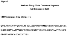
- an "antigen-binding region" of an antibody typically is found in one or more hypervariable region(s) of an antibody, i.e., the CDR-1, -2, and/or -3 regions; however, the variable "framework" regions can also play an important role in antigen binding, such as by providing a scaffold for the CDRs.
- the "antigen-binding region” comprises at least amino acid residues 4 to 103 of the variable light (VL) chain and 5 to 109 of the variable heavy (VH) chain, more preferably amino acid residues 3 to 107 of VL and 4 to 111 of VH, and particularly preferred are the complete VL and VH chains (amino acid positions 1 to 109 of VL and 1 to 113 of VH; numbering according to WO 97/08320 ).
- "Functional fragments” include the domain of a F(ab') 2 fragment, a Fab fragment, scFv or constructs comprising single immunoglobulin variable domains or single domain antibody polypeptides, e.g. single heavy chain variable domains or single light chain variable domains.
- the F(ab') 2 or Fab may be engineered to minimize or completely remove the intermolecular disulphide interactions that occur between the C H1 and C L domains.
- An antibody of the invention may be derived from a recombinant antibody library that is based on amino acid sequences that have been designed in silico and encoded by nucleic acids that are synthetically created.
- silico design of an antibody sequence is achieved, for example, by analyzing a database of human sequences and devising a polypeptide sequence utilizing the data obtained therefrom.
- Methods for designing and obtaining in silico-created sequences are described, for example, in Knappik et al., J. Mol. Biol. (2000) 296:57 ; Krebs et al., J. Immunol. Methods. (2001) 254:67 ; and U.S. Patent No. 6,300,064 issued to Knappik et al.
- MOR03684 represents an antibody having a variable heavy region corresponding to SEQ ID NO: 1 (DNA)/SEQ ID NO: 11 (protein) and a variable light region corresponding to SEQ ID NO: 21 (DNA)/SEQ ID NO: 31 (protein).
- MOR04251 represents an antibody having a variable heavy region corresponding to SEQ ID NO: 2 (DNA)/SEQ ID NO: 12 (protein) and a variable light region corresponding to SEQ ID NO: 22 (DNA)/SEQ ID NO: 32 (protein).
- MOR03929 represents an antibody having a variable heavy region corresponding to SEQ ID NO: 3 (DNA)/SEQ ID NO: 13 (protein) and a variable light region corresponding to SEQ ID NO: 23 (DNA)/SEQ ID NO: 33 (protein).
- MOR04252 represents an antibody having a variable heavy region corresponding to SEQ ID NO: 4 (DNA)/SEQ ID NO: 14 (protein) and a variable light region corresponding to SEQ ID NO: 24 (DNA)/SEQ ID NO: 34 (protein).
- MOR04287 represents an antibody having a variable heavy region corresponding to SEQ ID NO: 5 (DNA)/SEQ ID NO: 15 (protein) and a variable light region corresponding to SEQ ID NO: 25 (DNA)/SEQ ID NO: 35 (protein).
- MOR04290 represents an antibody having a variable heavy region corresponding to SEQ ID NO: 6 (DNA)/SEQ ID NO: 16 (protein) and a variable light region corresponding to SEQ ID NO: 26 (DNA)/SEQ ID NO: 36 (protein).
- MOR04302 represents an antibody having a variable heavy region corresponding to SEQ ID NO: 7 (DNA)/SEQ ID NO: 17 (protein) and a variable light region corresponding to SEQ ID NO: 27 (DNA)/SEQ ID NO: 37 (protein).
- MOR04350 represents an antibody having a variable heavy region corresponding to SEQ ID NO: 8 (DNA)/SEQ ID NO: 18 (protein) and a variable light region corresponding to SEQ ID NO: 28 (DNA)/SEQ ID NO: 38 (protein).
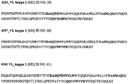
- MOR04354 represents an antibody having a variable heavy region corresponding to SEQ ID NO: 9 (DNA)/SEQ ID NO: 19 (protein) and a variable light region corresponding to SEQ ID NO: 29 (DNA)/SEQ ID NO: 39 (protein).
- MOR04357 represents an antibody having a variable heavy region corresponding to SEQ ID NO: 10 or 48 (DNA)/SEQ ID NO: 20 (protein) and a variable light region corresponding to SEQ ID NO: 30 or 57 (DNA)/SEQ ID NO: 40 (protein).
- MOR03682 represents an antibody having a variable heavy region corresponding to SEQ ID NO: 44 (DNA)/SEQ ID NO: 49 (protein) and a variable light region corresponding to SEQ ID NO: 53 (DNA)/SEQ ID NO: 58 (protein).
- MOR04283 represents an antibody having a variable heavy region corresponding to SEQ ID NO: 45 (DNA)/SEQ ID NO: 50 (protein) and a variable light region corresponding to SEQ ID NO: 54 (DNA)/SEQ ID NO: 59 (protein).
- MOR04297 represents an antibody having a variable heavy region corresponding to SEQ ID NO: 46 (DNA)/SEQ ID NO: 51 (protein) and a variable light region corresponding to SEQ ID NO: 55 (DNA)/SEQ ID NO: 60 (protein).
- MOR04342 represents an antibody having a variable heavy region corresponding to SEQ ID NO: 47 (DNA)/SEQ ID NO: 52 (protein) and a variable light region corresponding to SEQ ID NO: 56 (DNA)/SEQ ID NO: 61 (protein).
- the invention provides antibodies having an antigen-binding region that can bind specifically to or has a high affinity for GM-CSF.
- An antibody is said to have a "high affinity" for an antigen if the affinity measurement is at least 100 nM (monovalent affinity of Fab fragment).
- An inventive antibody or antigen-binding region preferably can bind to GM-CSF with an affinity of about less than 100 nM, more preferably less than about 60 nM, and still more preferably less than about 30 nM.
- Further preferred are antibodies that bind to GM-CSF with an affinity of less than about 10 nM, and more preferably less than about 3 nM.
- the affinity of an antibody of the invention against GM-CSF may be about 10.0 nM or 1 pM (monovalent affinity of Fab fragment).
- Table 1 provides a summary of affinities of representative antibodies, as determined by surface plasmon resonance (Biacore) and Solution Equilibrium Titration (SET) analysis: Table 1: Antibody Affinities Biacore SET MOR0 K D (pM) K D (pM) 3684 6420 16000 4251 70 7.4 3929 4260 2000 4302 174 63.5 4287 nd 17.9 4252 55 6 4290 122 11.1 4350 19 1.1 4354 21 2.8 4357 7 0.4 3682 nd 11406 4283 nd 113 4297 nd 49.2 4342 nd 4.9 "nd": not determined
- MOR03684, 04251, 03929, 04252, 04357, 04290, 04302, 04350 and 04354 was measured by surface plasmon resonance (Biacore) on immobilized recombinant GM-CSF.
- the Fab format of MOR03684, 04251, 03929, 04252, 04357, 04290, 04302, 04350 and 04354 exhibit a monovalent affinity range between about 6420 and 7 pM.
- the Fab format was also used for the determination of the affinities by solution equilibrium titration (SET).
- SET solution equilibrium titration
- An antibody of the invention preferably is species cross-reactive with humans and at least one other species, which may be a rodent species or a non-human primate.
- the non-human primate can be rhesus.
- the rodent species can be rat.
- An antibody that is cross reactive with at least one rodent species, for example, can provide greater flexibility and benefits over known anti-GM-CSF antibodies, for purposes of conducting in vivo studies in multiple species with the same antibody.
- an antibody of the invention not only is able to bind to GM-CSF, but also is able to block the interaction of human GM-CSF with the alpha chain of human GM-CSF receptor expressed on CHO-K1 cells by at least 25%, preferably by at least 50%, more preferably by at least 60%, more preferably by at least 70%, preferably by at least 85% and most preferably by at least 100%.
- an antibody of the invention is able to block interaction of 0.5 ⁇ g/ml human GM-CSF with the alpha chain of human GM-CSF receptor expressed on about 2 ⁇ 10 5 CHO-K1 cells by at least 50% under the following conditions: the concentration of the human GM-CSF receptor alpha chain expressed on the CHO-K1 cells is similar to the concentration of human GM-CSF receptor alpha chain expressed on about 2 ⁇ 10 5 CHO-GMRa#11 cells, and the concentration of the inventive antibody is about 5 ⁇ g/ml.
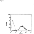
- the skilled worker can obtain CHO-K1 cells expressing human GM-CSF receptor alpha at a concentration similar to that which is expressed on about 2 ⁇ 10 5 CHO-GMRa#11 cells by, e.g., by transfecting a population of CHO-K1 cells with a suitable expression vector encoding GM-CSF receptor alpha to generate different stable cell lines expressing defined levels GM-CSF receptor alpha; then, the stable cell lines are analyzed in FACS analysis to determine GM-CSF receptor alpha expression levels according to the protocol essentially as described in Example 3C; a cell line that expresses human GM-CSF receptor alpha at a concentration similar to that which is expressed on about 2 ⁇ 10 5 CHO-GMRa#11 cells is identified by comparing the median fluorescence value (MFL) of such transfected cells to the MFL value set forth in Example 3C.
- MFL median fluorescence value
- a cell line is defined as expressing GM-CSF receptor alpha at a concentration "similar” to that which is expressed on about 2 ⁇ 10 5 CHO-GMRa#11 cells” if the MFL value of the transfected cell line does not deviate by more than a two-fold factor from the MFL value for the CHO-GMRa#11 cell as set forth in Example 3C.
- an antibody of the invention is able to neutralize human GM-CSF in a TF-1 proliferation assay with a lower IC 50 value than the reference antibody BVD2-21C11 and/or MAB215, preferably an at least five-fold lower IC 50 value, more preferably with an at least 10-fold lower IC 50 value than the reference antibody BVD2-21C11 and/or MAB215, more preferably with an at least 15-fold lower IC 50 value than the reference antibody BVD2-21C11 and/or MAB215, more preferably with an at least 20-fold lower IC 50 value than the reference antibody BVD2-21C11 and/or MAB215, more preferably with an at least 30-fold lower IC 50 value than the reference antibody BVD2-21C11 and/or MAB215, more preferably with an at least 50-fold lower IC 50 value than the reference antibody BVD2-21C11 and/or MAB215, more preferably with an at least 100-fold lower IC 50 value than the reference
- DNA molecules that encode an antibody of the invention include, but are not limited to, those DNA molecules set forth in Figures 1a and 2a .
- DNA can be used to identify its complement and, since DNA is double stranded, its equivalent or homolog, using nucleic acid hybridization techniques. It also will be recognized that hybridization can occur with less than 100% complementarity. However, given appropriate choice of conditions, hybridization techniques can be used to differentiate among DNA sequences based on their structural relatedness to a particular probe. For guidance regarding such conditions see, Sambrook et al., 1989 ( Sambrook, J., Fritsch, E. F. and Maniatis, T. (1989) Molecular Cloning: A laboratory manual, Cold Spring Harbor Laboratory Press, Cold Spring Harbor, USA ) and Ausubel et al., 1995 ( Ausubel, F. M., Brent, R., guitarist, R. E., Moore, D. D., Sedman, J. G., Smith, J. A., & Struhl, K. eds. (1995). Current Protocols in Molecular Biology. New York: John Wiley and Sons ).
- Structural similarity between two polynucleotide sequences can be expressed as a function of "stringency" of the conditions under which the two sequences will hybridize with one another.
- stringency refers to the extent that the conditions disfavor hybridization. Stringent conditions strongly disfavor hybridization, and only the most structurally related molecules will hybridize to one another under such conditions. Conversely, non-stringent conditions favor hybridization of molecules displaying a lesser degree of structural relatedness. Hybridization stringency, therefore, directly correlates with the structural relationships of two nucleic acid sequences. The following relationships are useful in correlating hybridization and relatedness (where T m is the melting temperature of a nucleic acid duplex):
- Hybridization stringency is a function of many factors, including overall DNA concentration, ionic strength, temperature, probe size and the presence of agents which disrupt hydrogen bonding. Factors promoting hybridization include high DNA concentrations, high ionic strengths, low temperatures, longer probe size and the absence of agents that disrupt hydrogen bonding. Hybridization typically is performed in two phases: the "binding" phase and the “washing” phase.
- the probe is bound to the target under conditions favoring hybridization.
- Stringency is usually controlled at this stage by altering the temperature.
- the temperature is usually between 65°C and 70°C, unless short ( ⁇ 20 nt) oligonucleotide probes are used.
- a representative hybridization solution comprises 6X SSC, 0.5% SDS, 5X Denhardt's solution and 100 ⁇ g of nonspecific carrier DNA. See Ausubel et al., section 2.9, supplement 27 (1994). Of course, many different, yet functionally equivalent, buffer conditions are known. Where the degree of relatedness is lower, a lower temperature may be chosen.
- Low stringency binding temperatures are between about 25°C and 40°C.
- Medium stringency is between at least about 40°C to less than about 65°C.
- High stringency is at least about 65°C.
- washing solutions typically contain lower salt concentrations.
- One exemplary medium stringency solution contains 2X SSC and 0.1% SDS.
- a high stringency wash solution contains the equivalent (in ionic strength) of less than about 0.2X SSC, with a preferred stringent solution containing about 0.1X SSC.
- the temperatures associated with various stringencies are the same as discussed above for "binding.”
- the washing solution also typically is replaced a number of times during washing. For example, typical high stringency washing conditions comprise washing twice for 30 minutes at 55° C and three times for 15 minutes at 60° C.
- variants of DNA molecules provided herein can be constructed in several different ways. For example, they may be constructed as completely synthetic DNAs. Methods of efficiently synthesizing oligonucleotides in the range of 20 to about 150 nucleotides are widely available. See Ausubel et al., section 2.11, Supplement 21 (1993). Overlapping oligonucleotides may be synthesized and assembled in a fashion first reported by Khorana et al., J. Mol. Biol. 72:209-217 (1971 ); see also Ausubel et al., supra, Section 8.2. Synthetic DNAs preferably are designed with convenient restriction sites engineered at the 5' and 3' ends of the gene to facilitate cloning into an appropriate vector.
- a method of generating variants is to start with one of the DNAs disclosed herein and then to conduct site-directed mutagenesis. See Ausubel et al., supra, chapter 8, Supplement 37 (1997).
- a target DNA is cloned into a single-stranded DNA bacteriophage vehicle.
- Single-stranded DNA is isolated and hybridized with an oligonucleotide containing the desired nucleotide alteration(s).
- the complementary strand is synthesized and the double stranded phage is introduced into a host.
- Some of the resulting progeny will contain the desired mutant, which can be confirmed using DNA sequencing.
- various methods are available that increase the probability that the progeny phage will be the desired mutant. These methods are well known to those in the field and kits are commercially available for generating such mutants.
- the present disclosure further provides recombinant DNA constructs comprising one or more of the nucleotide sequences of the disclosure.
- the recombinant constructs are used in connection with a vector, such as a plasmid, phagemid, phage or viral vector, into which a DNA molecule encoding an antibody of the invention is inserted.
- the encoded gene may be produced by techniques described in Sambrook et al., 1989, and Ausubel et al., 1989.
- the DNA sequences may be chemically synthesized using, for example, synthesizers. See, for example, the techniques described in OLIGONUCLEOTIDE SYNTHESIS (1984, Gait, ed., IRL Press, Oxford ).
- Recombinant constructs of the disclosure are comprised with expression vectors that are capable of expressing the RNA and/or protein products of the encoded DNA(s).
- the vector may further comprise regulatory sequences, including a promoter operably linked to the open reading frame (ORF).
- the vector may further comprise a selectable marker sequence. Specific initiation and bacterial secretory signals also may be required for efficient translation of inserted target gene coding sequences.
- the present disclosure further provides host cells containing at least one of the DNAs of the present disclosure.
- the host cell can be virtually any cell for which expression vectors are available. It may be, for example, a higher eukaryotic host cell, such as a mammalian cell, a lower eukaryotic host cell, such as a yeast cell or a prokaryotic cell, such as a bacterial cell.
- Introduction of the recombinant construct into the host cell can be effected by calcium phosphate transfection, DEAE, dextran mediated transfection, electroporation or phage infection.
- Useful expression vectors for bacterial use are constructed by inserting a structural DNA sequence encoding a desired protein together with suitable translation initiation and termination signals in operable reading phase with a functional promoter.
- the vector will comprise one or more phenotypic selectable markers and an origin of replication to ensure maintenance of the vector and, if desirable, to provide amplification within the host.
- Suitable prokaryotic hosts for transformation include E. coli, Bacillus subtilis, Salmonella typhimurium and various species within the genera Pseudomonas, Streptomyces, and Staphylococcus.
- Bacterial vectors may be, for example, bacteriophage-, plasmid- or phagemid-based. These vectors can contain a selectable marker and bacterial origin of replication derived from commercially available plasmids typically containing elements of the well known cloning vector pBR322 (ATCC 37017). Following transformation of a suitable host strain and growth of the host strain to an appropriate cell density, the selected promoter is de-repressed/induced by appropriate means (e.g., temperature shift or chemical induction) and cells are cultured for an additional period. Cells are typically harvested by centrifugation, disrupted by physical or chemical means, and the resulting crude extract retained for further purification.
- appropriate means e.g., temperature shift or chemical induction
- a number of expression vectors may be advantageously selected depending upon the use intended for the protein being expressed. For example, when a large quantity of such a protein is to be produced, for the generation of antibodies or to screen peptide libraries, for example, vectors which direct the expression of high levels of fusion protein products that are readily purified may be desirable.
- Therapeutic methods involve administering to a subject in need of treatment a therapeutically effective amount of an antibody contemplated by the invention.
- a "therapeutically effective" amount hereby is defined as the amount of an antibody that is of sufficient quantity to effectively block the interaction between GM-CSF and its receptor in a treated area of a subject-either as a single dose or according to a multiple dose regimen, alone or in combination with other agents, which leads to the alleviation of an adverse condition, yet which amount is toxicologically tolerable.
- the subject may be a human or non-human animal (e.g., rat or rhesus).
- An antibody of the invention might be co-administered with known medicaments, and in some instances the antibody might itself be modified.
- an antibody could be conjugated to an immunotoxin or radioisotope to potentially further increase efficacy.
- the inventive antibodies can be used as a therapeutic or a diagnostic tool in a variety of situations where GM-CSF is undesirably expressed or found.
- Disorders and conditions particularly suitable for treatment with an antibody of the inventions are inflammatory diseases such as rheumatoid arthritis (RA), multiple sclerosis, Crohn's disease, psoriasis, asthma, atopic dermatitis or shock.
- RA rheumatoid arthritis
- multiple sclerosis Crohn's disease
- psoriasis asthma
- atopic dermatitis or shock atopic dermatitis or shock.
- compositions for use in accordance with the present invention may be formulated in a conventional manner using one or more physiologically acceptable carriers or excipients.
- An antibody of the invention can be administered by any suitable means, which can vary, depending on the type of disorder being treated. Possible administration routes include parenteral (e.g., intramuscular, intravenous, intraarterial, intraperitoneal, or subcutaneous), intrapulmonary and intranasal, and, if desired for local immunosuppressive treatment, intralesional administration.
- an antibody of the invention might be administered by pulse infusion, with, e.g., declining doses of the antibody.
- the dosing is given by injections, most preferably intravenous or subcutaneous injections, depending in part on whether the administration is brief or chronic.
- the amount to be administered will depend on a variety of factors such as the clinical symptoms, weight of the individual, whether other drugs are administered. The skilled artisan will recognize that the route of administration will vary depending on the disorder or condition to be treated.
- Determining a therapeutically effective amount of the novel polypeptide largely will depend on particular patient characteristics, route of administration, and the nature of the disorder being treated. General guidance can be found, for example, in the publications of the International Conference on Harmonisation and in REMINGTON'S PHARMACEUTICAL SCIENCES, chapters 27 and 28, pp. 484-528 (18th ed., Alfonso R. Gennaro, Ed., Easton, Pa.: Mack Pub. Co., 1990 ). More specifically, determining a therapeutically effective amount will depend on such factors as toxicity and efficacy of the medicament. Toxicity may be determined using methods well known in the art and found in the foregoing references. Efficacy may be determined utilizing the same guidance in conjunction with the methods described below in the Examples.
- GM-CSF is expressed by various cell types including lymphocytes, monocytes, endothelial cells, fibroblasts and some malignant cells; thus, an anti-GM-CSF antibody of the invention may be employed in order to image or visualize a site of possible accumulation of GM-CSF in different tissues in a patient.
- an antibody can be detectably labeled, through the use of radioisotopes, affinity labels (such as biotin, avidin, etc.) fluorescent labels, paramagnetic atoms, etc. Procedures for accomplishing such labeling are well known to the art. Clinical application of antibodies in diagnostic imaging are reviewed by Grossman, H. B., Urol. Clin. North Amer. 13:465-474 (1986 )), Unger, E. C. et al., Invest. Radiol. 20:693-700 (1985 )), and Khaw, B. A. et al., Science 209:295-297 (1980 )).
- the detection of foci of such detectably labeled antibodies might be indicative of a site of inflammation, for example.
- this examination is done by removing samples of tissue or blood and incubating such samples in the presence of the detectably labeled antibodies.
- this technique is done in a non-invasive manner through the use of magnetic imaging, fluorography, etc. Such a diagnostic test may be employed in monitoring the success of treatment of diseases, where presence or absence of GM-CSF is a relevant indicator
- the antibodies of the present invention can be formulated according to known methods to prepare pharmaceutically useful compositions, wherein an antibody of the invention is combined in a mixture with a pharmaceutically acceptable carrier vehicle.
- Suitable vehicles and their formulation are described, for example, in REMINGTON'S PHARMACEUTICAL SCIENCES (18th ed., Alfonso R. Gennaro, Ed., Easton, Pa.: Mack Pub. Co., 1990 ).
- REMINGTON'S PHARMACEUTICAL SCIENCES 18th ed., Alfonso R. Gennaro, Ed., Easton, Pa.: Mack Pub. Co., 1990 .
- Such compositions will contain an effective amount of one or more of the antibodies of the present invention, together with a suitable amount of carrier vehicle.
- Preparations may be suitably formulated to give controlled-release of the active compound.
- Controlled-release preparations may be achieved through the use of polymers to complex or absorb anti- GM-CSF antibody.
- the controlled delivery may be exercised by selecting appropriate macromolecules (for example polyesters, polyamino acids, polyvinyl, pyrrolidone, ethylenevinyl-acetate, methylcellulose, carboxymethylcellulose, or protamine, sulfate) and the concentration of macromolecules as well as the methods of incorporation in order to control release.
- Another possible method to control the duration of action by controlled release preparations is to incorporate anti-GM-CSF antibody into particles of a polymeric material such as polyesters, polyamino acids, hydrogels, poly(lactic acid) or ethylene vinylacetate copolymers.
- a polymeric material such as polyesters, polyamino acids, hydrogels, poly(lactic acid) or ethylene vinylacetate copolymers.
- colloidal drug delivery systems for example, liposomes, albumin microspheres, microemulsions, nanoparticles, and nanocapsules or
- the compounds may be formulated for parenteral administration by injection, e.g., by bolus injection or continuous infusion.
- Formulations for injection may be presented in unit dosage form, e.g., in ampules, or in multi-dose containers, with an added preservative.
- the compositions may take such forms as suspensions, solutions or emulsions in oily or aqueous vehicles, and may contain formulatory agents such as suspending, stabilizing and/or dispersing agents.
- the active ingredient may be in powder form for constitution with a suitable vehicle, e.g., sterile pyrogen-free water, before use.
- compositions may, if desired, be presented in a pack or dispenser device, which may contain one or more unit dosage forms containing the active ingredient.
- the pack may for example comprise metal or plastic foil, such as a blister pack.
- the pack or dispenser device may be accompanied by instructions for administration.
- HuCAL GOLD ® library was amplified in 2xYT medium containing 34 ⁇ g/ml chloramphenicol and 1 % glucose (2xYT-CG). After helper phage infection (VCSM13) at an OD 600 of 0.5 (30 min at 37°C without shaking; 30 min at 37°C shaking at 250 rpm), cells were spun down (4120 g; 5 min; 4°C), resuspended in 2 ⁇ YT/ 34 ⁇ g/ml chloramphenicol / 50 ⁇ g/ml kanamycin / 0.25mM IPTG and grown overnight at 22°C.
- helper phage infection VCSM13
- OD 600 0.5
- cells were spun down (4120 g; 5 min; 4°C), resuspended in 2 ⁇ YT/ 34 ⁇ g/ml chloramphenicol / 50 ⁇ g/ml kanamycin / 0.25mM IPTG and grown overnight at 22°C.
- Phages were PEG-precipitated from the supernatant, resuspended in PBS / 20 % glycerol and stored at -80°C. Phage amplification between two panning rounds was conducted as follows: mid-log phase E. coli TG1 cells were infected with eluted phages and plated onto LB-agar supplemented with 1 % of glucose and 34 ⁇ g/ml of chloramphenicol. After overnight incubation at 30°C, colonies were scraped off and used to inoculate 2xYT-CG until an OD 600nm of 0.5 was reached and helper phage added as described above.
- HuCAL GOLD ® antibody-phages were divided into three pools comprising different VH master genes. These pools were individually subjected to either a) a solid phase panning on biotinylated human GM-CSF protein (custom made by R&D Systems, Minneapolis, MN) directly coated on neutravidin coated 96well plates (Pierce, Rockford, IL) as solid support for three rounds or b) a solution panning on biotinylated human GM-CSF protein captured onto streptavidin coated Dynabeads (Dynal, Oslo, Norway) for three rounds.
- HuCAL GOLD ® phages Prior to the selections, 100 ⁇ l of HuCAL GOLD ® phages were pre-adsorbed in 100 ⁇ l blocking buffer containing 0.4 ⁇ l 25% Tween20 for 0.5 h at RT. Blocked phages were transferred in 100 ⁇ l aliquots to wells of a neutravidin plate for 0.5 h at RT. This step was repeated twice for pre-absorption.
- Bound phages were eluted by adding 120 ⁇ l of 20 mM DTT in 10 mM Tris pH 8.0 per well for 10 min at RT. The eluate was removed and added to 14 ml E. coli TG1 grown to an OD 600nm of 0.6-0.8. Wells were additionally washed with 200 ⁇ l PBS and this solution was also added to the TG1 cells. Phage infection of E. coli was allowed for 45 min at 37°C without shaking. Additionally, 200 ⁇ l of TG1 cells grown to an OD 600nm of 0.6-0.8 were added to the selection wells for 45 minutes at 37°C without shaking.
- TG-1 cells were added to the 14ml culture already containing the phages from the first elution step. After centrifugation for 10 min at 5000rpm, the bacterial pellets were each resuspended in 500 ⁇ l 2xYT medium, plated on 2xYT-CG agar plates and incubated overnight at 30°C. Colonies were then scraped from the plates and phages were rescued and amplified as described above.
- the second and third rounds of selection were performed in an identical way to the first round of selection with the only difference that the washing conditions after binding of phage were more stringent. Additionally, in the third round of selection, phages were submitted to an additional preadsorption step on streptavidin-coated beads (Dynabeads M-280; Dynal). Eppendorf tubes were blocked with Chemiblocker solution by incubation for 30 min at RT. Of each phage pool 0.3ml were mixed 1:1 with 2xChemiblocker solution containing 0.05% Tween20 and incubated for 1h at RT in the blocked Eppendorf tubes on a rotator.
- Blocked phages were then transferred to newly blocked Eppendorf tubes and 50 ⁇ l of Dynabeads M-280 were added for another 30min for preadsorption. Beads were removed using a magnetic device (Dynal MPC-E). Aliquots of 150 ⁇ l of phages were then transferred to neutravidin plates for further preadsorption as in round 1 and 2 (see above).
- Phages diluted in 500 ⁇ l PBS for each panning condition were mixed with 500 ⁇ l 2xChemiblocker / 0.1% Tween 1 h at RT (rotator). Preadsorption of phages was performed twice: 50 ⁇ l of blocked Streptavidin magnetic beads were added to the blocked phages and incubated for 30 min at RT on a rotator. After separation of beads via a magnetic device (Dynal MPC-E) the phage supernatant ( ⁇ 1ml) was transferred to a new blocked tube and preadsorption was repeated on 50 ⁇ l blocked beads for 30 min.
- biotinylated hGM-CSF was added to blocked phages in a new blocked 1.5 ml tube and incubated for 1 h at RT on a rotator.
- 100 ⁇ l of blocked streptavidin magnetic beads were added to each panning phage pool and incubated 10 min at RT on a rotator.
- Phage bound to biotinylated GM-CSF and therefore immobilized to the magnetic beads were collected with a magnetic particle separator (Dynal MPC-E). Beads were then washed 7x in PBS/0.05% Tween using a rotator, followed by washing another three times with PBS.
- Elution of phage from the Dynabeads was performed adding 300 ⁇ l 20mM DTT in 10mM Tris/HCI pH8 to each tube for 10 min. Dynabeads were removed by the magnetic particle separator and the supernatant was added to 14ml of a E.coli TG-1 culture grown to OD 600nm of 0.6-0.8. Beads were then washed once with 200 ⁇ l PBS and PBS containing additional removed phage was added to the 14 ml E.coli TG-1 culture.
- the bacterial pellets were each resuspended in 500 ⁇ l 2xYT medium, plated on 2xYT-CG agar plates and incubated overnight at 30°C. Colonies were then scraped from the plates and phages were rescued and amplified as described above.
- the second and third rounds of the solution panning on biotinylated GM-CSF was performed according to the protocol of the first round except for increasing the stringency of the washing procedure.
- the Fab encoding inserts of the selected HuCAL GOLD ® phagemids were subcloned into the expression vector pMORPH ® X9_Fab_FH ( Fig 5 ) to facilitate rapid expression of soluble Fab.
- the DNA of the selected clones was digested with Xba l and Eco RI, thereby cutting out the Fab encoding insert (ompA-VLCL and phoA-Fd), and cloned into the Xba l / Eco RI digested vector pMORPH ® X9_Fab_FH.
- Fabs expressed in these vectors carry two C-terminal tags (FLAG TM and 6xHis, respectively) for detection and purification.
- BEL buffer (2xBBS/EDTA: 24.7 g/l boric acid, 18.7 g NaCl/l, 1.49 g EDTA/I, pH8) was added containing 2.5 mg/ml lysozyme and incubated for 1 h at 22°C on a microtiter plate shaker (400 rpm).
- BEL extracts were used for binding analysis by ELISA or a BioVeris M-series ® 384 analyzer (see Example 2).
- Fab fragments encoded by pMORPH ® X9_Fab_FH in TG-1 cells were carried out in shaker flask cultures with 1 I of 2 ⁇ YT medium supplemented with 34 ⁇ g/ml chloramphenicol. After induction with 0.5 mM IPTG, cells were grown at 22°C for 16 h. Whole cell extracts of cell pellets were prepared by French Press and Fab fragments isolated by nickel/NTA chromatography (Qiagen, Hilden, Germany). Concentrations were determined by UV-spectrophotometry (Krebs et al., 2001).
- BEL extracts of individual E. coli clones selected by the above mentioned panning strategies were analyzed by ELISA or BioVeris (BioVeris M-series ® 384 analyzer) in order to identify clones encoding hGM-CSF specific Fabs.
- fluorogenic substrates like AttoPhos (Roche) were used according to the instructions by the manufacturer. Between all incubation steps, the wells of the microtiter plate were washed with PBS-T three times and three times after the final incubation with secondary antibody. Fluorescence was measured in a TECAN Spectrafluor plate reader.
- BEL extract was diluted at least 1:50 and maximally 1:1000 in assay buffer (PBS/0,05%Tween20/0.5%BSA) for use in BioVeris screening.
- Biotinylated GM-CSF R&D Systems
- Dynabeads Dynal
- Per well of a 96 or 384 well plate 25 or 15 ⁇ l of a 1:25 dilution of the Dynabead-stock solution was used. Beads were washed three times with assay buffer before adding biotinylated GM-CSF for 30 min at RT on a shaker.
- coli expression cultures containing Fab antibodies was filled into wells of a 96 or 384 well plate and, respectively, 25 or 15 ⁇ l of the GM-CSF coupled beads plus anti-Fab-BV-tag TM secondary antibody mix was added to each well and incubated for 2h at RT on a plate shaking device. The plates were analyzed in a BioVeris M-SERIES ® 384 Analyzer.
- Crossreactivity of the anti-hGM-CSF antibodies was determined to the following analytes: rat and mouse GM-CSF, human IL-3, human IL-4, , human IL-5, , human IL-13, , human M-CSF (all from Peprotech, London, UK). This was performed in a capture set-up by surface plasmon resonance (Biacore 3000, Uppsala, Sweden).
- CM5 chips (Biacore, Sweden) were coated with 5000-6000RU anti-F(ab) 2 (Dianova, Affinipure F(ab) 2 Fragment Goat Anti-Human IgG, F(ab) 2 Fragment specific); 80 ⁇ g/ml 10mM acetate buffer, pH4 on all 4 flow cells, using standard EDC-NHS amine coupling chemistry.
- the analyte growth factor was injected (20 ⁇ l, flow rate 20 ⁇ l/min) at a concentration range between 15 and 2000nM for the determination of the specific signal. Afterwards the achieved buffer sensogram was manually subtracted from the specific one. After each cycle, the flow cells were regenerated with 100mM HCI (5 ⁇ l).
- HuCAL ® anti-hGM-CSF antibodies including MOR03684 and MOR03682 were tested and were specific for human GM-CSF and did not bind to any of the other cytokines or mouse or rat GM-CSF .
- Fab MOR03929 showed significant cross reactivity to rat GM-CSF.
- Example 3 Identification of anti-human GM-CSF Fab candidates that inhibit the interaction between GM-CSF and the GM-CSF receptor alpha
- hGM-CSF specific antibodies which were selected from the HuCAL GOLD ® library were tested for the potency to inhibit the interaction between hGM-CSF and its receptor.
- the interaction was tested in two ways, (i) one being a proliferation assay using the GM-CSF dependent TF-1 cell line (Kitamura et al., 1989) and (ii) the other being a FACS analysis with a recombinant CHO cell line expressing the alpha chain of the GM-CSF receptor.
- the TF-1 proliferation assay the ability of the anti-GM-CSF antibodies to block the interaction of GM-CSF with the endogenous GM-CSF receptor consisting of the alpha and beta chain was analyzed leading to reduction in cell proliferation.
- the FACS assay the specific inhibition of the interaction between GM-CSF and the alpha chain of the GM-CSF receptor was determined.
- Macaca mulatta GM-CSF full-length cDNA (GenBank accession no.: AY007376) was synthesized by gene synthesis (geneART GmbH, Regensburg, Germany) and cloned into the pCR-Script-Amp vector (Stratagene, LaJolla, CA, USA). Subsequently the cDNA was cloned into the eukaryotic expression vector pcDNA3.1 (+) (Invitrogen, Paisley, UK) yielding pcDNA-macGM-CSF.
- the cDNA of human GM-CSF (Genbank accession number NP_000749 ) was cloned by RT-PCR technique from RNA isolated from 1 ⁇ 10e7 TF-1 cells using the RNeasy kit from Qiagen (Hilden, Germany). Reverse transcription was performed with the SuperscriptII kit using random hexamers (Gibco) followed amplification of the GM-CSF cDNA by PCR. The obtained PCR-product was cloned into expression vector pcDNA3.1(+) yielding pcDNA-huGM-CSF.
- HEK293 cells were transiently transfected with these expression vectors respectively using lipofectamine (Stratagene, LaJolla, USA).
- lipofectamine Stratagene, LaJolla, USA.
- the medium containing the secreted recombinant macaca or human GM-CSF was harvested 4 days after transfection.
- TF-1 (Kitamura et al., 1989) cells were grown according to the provider's protocol (DSMZ, Braunschweig, Germany; DSMZ No. ACC 334). TF-1 cells were washed twice with RPMI1640 medium (10% FCS) and then seeded at a concentration of 2 ⁇ 10 5 cells/ml in 50 ⁇ l per well of a flat bottom 96well cell culture dish.
- GM-CSF Human recombinant GM-CSF ("Leucomax", ESSEX Pharma, Ober) at 0.5ng/ml and HuCAL ® Fab antibodies (200ng/ml - 200 ⁇ g/ml diluted in RPMI1640 medium, 10% FCS) were mixed for 30 min and 50 ⁇ l of the mix was added to the TF-1 cells, so that the final concentration of GM-CSF was 0.25ng/ml. Maximal cell proliferation (0% inhibition) was measured incubating TF-1 cells at a final GM-CSF of concentration of 0.25ng/ml, without the addition of antibody.
- TF-1 proliferation 100% inhibition was measured by omitting GM-CSF from the assay and keeping the cells in RPMI1640 medium (10% FCS) only. TF1 cells were then incubated for 72 hours at 37°C with 5% CO 2 in a humidified chamber. Cell vitality was measured by adding MTT or XTT reagent (Roche, Mannheim, Germany) according to the manufacturer's recommendation. Overall 19 Fab were identified that showed significant inhibition of TF-1 proliferation.
- the binders MOR03682, MOR03684 and MOR03929 showed consistent inhibition of TF1 cell proliferation of greater than 50% at a concentration of 2 ⁇ M.
- binding of MOR03682 and MOR03684 to native human GM-CSF was tested in the TF-1 proliferation assay. Instead of adding purified human recombinant GM-CSF to the TF-1 cells a supernatant of 5637 cells (DSMZ No. ACC 35) that secrete native human GM-CSF into the medium was used. From a dose response curve comparing the effect of recombinant human GM-CSF with different dilutions of the 5637 supernatant it was determined that the medium contained ⁇ 5 ng/ml of native human GM-CSF.
- TF-1 proliferation assay instead of adding purified human GM-CSF to the TF-1 cells a supernatant of transfected HEK293 cells that secrete recombinant macaca GM-CSF into the medium was used.
- TF-1 cells proliferated in the presence of macaca GM-CSF containing supernatant but not in the presence of supernatant from untransfected HEK293 cells. From a dose response curve comparing the effect of recombinant human GM-CSF with different dilutions of the HEK-293 medium it was determined that the medium of the transfected cells contained ⁇ 2 ⁇ g/ml macaca GM-CSF. By preincubation of the macaca GM-CSF supernatant with anti-human GM-CSF Fab MOR03682 or MOR03684 the binding of macaca GM-CSF to the TF-1 cells was blocked so that the viability of cells was significantly reduced. MOR03682 and MOR03684_thus are cross-reactive with macaca GM-CSF. Fab MOR03929 was not tested in this assay.
- GM-CSF GM-CSF receptor alpha chain
- cDNA was cloned into an expression vector and stably transfected into CHO-K1 cells (DSMZ ACC 110).
- the PCR reaction yielded a product of ⁇ 1250bp which was digested with Eco RI and Bam HI (New England BioLabs).
- the expression vector pcDNA3.1(+) (Invitrogen, Paisley, UK) was digested with the same enzymes. After purification of the digested vector and PCR product, the fragments were ligated and transformed by electroporation into E.coli DH10B cells. Correct clones were identified after preparation of plasmid DNA and sequencing. Correct clones (pcDNA3.1(+)-GM-CSFRalpha) contained the full length human GM-CSF receptor alpha cDNA.
- CHO-K1 cells were grown according to the provider's protocol (DSZM, Braunschweig, Germany; DSMZ No. ACC 110).
- DSMZ No. ACC 110 For transfection cells were grown to 80% confluence in a 6-well plate and incubated with 5 ⁇ g DNA of pcDNA3.1(+)-GM-CSFRalpha mixed with 10 ⁇ l of the Lipofectamine 2000 reagent (Invitrogen). After 48h cells were fed with 1mg/ml G418 (Gibco) and after another 24h medium was replaced with such containing 2mg/ml G418. After two weeks single cells were seeded into wells of a 96-well culture dish.
- Single clones were grown up and 5 ⁇ 10 5 cells of each clone were tested for GM-CSFR-alpha expression by FACS analysis using murine IgG MAB1006 (Chemicon International, Temecula, CA) as primary antibody at a concentration of 1 ⁇ g/ml and (R-PE-AffiniPure (Fab') 2 Goat-anti-mouse-IgG (Dianova) as secondary antibody at a 1:200 dilution. Primary and secondary antibodies were incubated with the cells for 1h sequentially, while cells were washed in FACS buffer (PBS, 3%FCS) between these steps. Fluorescence of stained cells was quantified in the FL2 channel using a FACSCalibur system (Becton Dickinson).
- FACSCalibur system Becton Dickinson
- antibodies at increasing concentrations Prior to adding to cells, antibodies at increasing concentrations (0.1 to 100 ⁇ g/ml) were co-incubated with biotinylated GM-CSF (0.5 ⁇ g/ml) in FACS buffer (PBS/3%FBS/NaN 3 0,05%) for 30min at RT.
- IC 50 values were determined from the dose response curves obtained using GraphPad Prism v3.03 software applying a non-linear regression curve fit.
- Fab antibodies MOR03682, MOR03684 and MOR03929 showed significant inhibition of GM-CSF binding to the cell surface expressed GM-CSF receptor alpha.
- one phage antibody Fab library was generated where the L-CDR3 of each parental was replaced by a repertoire of individual lambda light chain CDR3 sequences.
- the H-CDR2 region was replaced by a repertoire of individual heavy chain CDR2 sequences.
- Affinity maturation libraries were generated by transforming the diversified clones into E. coli TOP10F' (Invitrogen). Phages were prepared as described in Example 1A. Both L-CDR3 libraries of MOR03684 and MOR03682 were pooled and both H-CDR2 libraries derived from MOR03684 and MOR03682 were pooled, while the L-CDR3 and H-CDR2 libraries derived from MOR03929 were kept separately during the selection procedure. Pannings were performed on biotinylated GM-CSF in solution for three rounds essentially as described in Example1B and applying more stringent selection conditions.
- Electrochemiluminescence (ECL) based affinity determination in solution and data evaluation were basically performed as described by Haenel et al., 2005: A constant amount of Fab was equilibrated with different concentrations (serial 5 n dilutions) of human GM-CSF ( Leucomax ) in solution.
- Biotinylated human GM-CSF R&D Systems coupled to paramagnetic beads (M-280 Streptavidin, Dynal) and BV-tag TM (BioVeris Europe, Witney, Oxforfshire, UK) labeled anti-human (Fab)' 2 (Dianova) was added and incubated for 15 - 30 min. Subsequently, the concentration of unbound Fab was quantified via ECL detection using a M- SERIES ® 384 analyzer (BioVeris Europe).
- Association phase was set to 60 s and dissociation phase to 120 s (parental) or up to 600 s (affinity optimized).
- dissociation phase was set to 60 s and dissociation phase to 120 s (parental) or up to 600 s (affinity optimized).
- the following conditions basically according to Drake et al., (2004) were used: Fab was applied in a single concentration of 200 nM; flow rate was set to 100 ⁇ l/min and dissociation phase to 6000 - 18.000 s.
- affinities for the Fabs were estimated, which are shown in table 4.
- Table 4 Affinities of anti-hGM-CSF Fabs determined by Biacore and solution equilibrium titration (SET) Biacore SET MOR0 K D (pM) K D (pM) 3684 6420 16000 4251 70 7.4 3929 4260 2000 4302 174 63.5 4287 nd 17.9 4252 55 6 4290 122 11.1 4350 19 1.1 4354 21 2.8 4357 7 0.4 3682 nd 11406 4283 nd 113 4297 nd 49.2 4342 nd 4.9
- the GM-CSF receptor binding assay was performed as described above (Example 3C) using 0.5 ⁇ g/ml (35nM) of biotinylated GM-CSF. Maximal binding of GM-CSF to CHO-GMRa#11 cells (0% inhibition) was measured by incubating cells at a final GM-CSF concentration of 05 ⁇ g/ml of biotinylated GM-CSF, without the addition of antibody. 100% inhibition of GM-CSF binding was measured by omitting GM-CSF from the assay. IC 50 values were determined from the dose response curves obtained using GraphPad Prism v3.03 software applying a non-linear regression curve fit.
- Table 5 IC 50 values of anti-hGM-CSF Fabs in receptor inhibition assay MORO# 4251 4357 4354 4350 4252 4287 4290 4302 3684 3929 Mab215 21C11 IC 50 (nM) 53 26 26 24 26 25 27 24 >400 35 no fit* 9 % inhibition at 5 ⁇ g/ml antibody ⁇ 100% ⁇ 75% ⁇ 75% ⁇ 75% ⁇ 75% ⁇ 75% ⁇ 75% ⁇ 25% ⁇ 60% ⁇ 50% ⁇ 100% *no sigmoidal dose response curve could be fitted in this case
- This assay qualitatively showed that the Fabs obtained from affinity maturation and X-cloning prevent GM-CSF from binding to the GM-CSF receptor alpha chain and therefore retained the blocking mechanism of their parental Fabs.
- the assay needed to be performed with a concentration of 35nM (0.5 ⁇ g/ml) biotinylated GM-CSF in order to obtain a significant signal in FACS. Therefore 17.5 nM Fab (or 8.75 nM IgG) is theoretically needed to block 50% of the GM-CSF, thus setting a limit for determination of IC 50 values.
- TF-1 proliferation assay was performed as described in Example 3B. Fab with improved affinities and the parental Fabs as well as monoclonal reference IgGs were analyzed. IC 50 values were determined from the dose response curves obtained using GraphPad Prism v3.03 software applying a non-linear regression curve fit. Table 6 summarizes the IC 50 values obtained in these assays.
- Table 6 IC 50 values of anti-hGM-CSF Fabs and control IgGs in TF-1 proliferation assay MORO# 4251 4357 4354 4350 4252 4287 4290 4302 3684 3929 Mab215 BVD2-21C11 IC 50 ( pM ) 463 90 56 82 2010 3382 696 10678 >200000 >200000 4315 6560 IC 50 x-fold improved compared to Mab215 9.3 47.9 77.1 52.6 2.1 1.3 6.2 - - - - - IC 50 x-fold improved compared to BVD2-21C11 14.2 72.9 117.1 80.0 3.2 1.9 9.4 - - - 1.5 -
- IC 50 values in the TF-1 proliferation assay were determined for the parental Fab MOR03682, its affinity matured derivatives MOR04283, MOR04297 and the x-cloned variant MOR04342.
- Table 7 summarizes the IC 50 values obtained in these assays. Table 7: IC 50 values of anti-hGM-CSF Fabs in TF-1 proliferation assay MOR0 # 4342 4283 4297 3682 IC 50 ( pM ) 80 17293 13975 >200000
- GeneOptimizer TM software from Geneart (Regensburg, Germany ) was utilized to define such optimized VH and VL DNA sequences, which were gene synthesized at Geneart (Regensburg, Germany) and cloned into pPCR-Script vectors yielding 055906pPCR-Script and 055907pPCR-Script .
- SEQ ID NO: 48 shows the respective VH sequence
- SEQ ID NO: 57 shows the respective VL sequence.
- variable domain fragments of the gene optimized heavy (VH) and light chains (VL) were subcloned from the pPCR-Script vectors (Example5a) into the the pMORPH ® 2_h_Ig vector series for human IgG1.
- Codon-optimized VH of MOR04357 was isolated from 055906pPCR-Script via Nhe l/ BIp l digestion and inserted into pMorph2_h_IgG1f master vector cut with the same restriction enzymes. This vector already contained a human gamma 1 constant region.
- the resulting expression plasmid was termed pMorph2_h_IgG1f_MOR04357_co.
- Codon-optimized VL of MOR04357 was isolated from 055907pPCR-Script via Nhe l/ Hpa l digestion and inserted into pMorph2_h_Iglambda2 master vector cut with the same restriction enzymes. This vector already contained a human lambda constant region.
- the resulting expression plasmid was termed pM2_h_Iglambda2_MOR04357_co.
- Eukaryotic HKB11 cells were transfected with an equimolar amount of IgG heavy and light chain expression vector DNA.
- Cell culture supernatant was harvested from 3 to 7 days post transfection. After adjusting the pH of the supernatant to 8.0 and sterile filtration, the solution was subjected to standard protein A affinity chromatography (rProteinA FF or MabSelect SURE, GE Healthcare). Buffer exchange was performed to 1 x Dulbcecco's PBS (pH 7,2, Invitrogen) and samples were sterile filtered (0,2 ⁇ m).
- MOR04357 IgG1 was dialysed against 1 x Dulbcecco's PBS (pH 6,5, Invitrogen). Purity of IgG was analysed under denaturing, reducing conditions in SDS-PAGE or by using Agilent BioAnalyzer and in native state by SE-HPLC.
- K D determination monomer fractions (at least 90% monomer content, analyzed by analytical SEC; Superdex75, Amersham Pharmacia) of IgG1 were used. Electrochemiluminescence (ECL) based affinity determination in solution and data evaluation were basically performed as described by Haenel et al., 2005 and as described in Example4B. The K D values for MOR04357 IgG1 against human recombinant GM-CSF was determined to be 1.1 pM.
- Affinity determination to rat GM-CSF was done essentially as described in Example 4D using rat-GM-CSF (Peprotech) as analyte in solution instead of human GM-CSF. Affinities were calculated according to Haenel et al (2005). The K D value for the MOR04357 IgG1 against rat recombinant GM-CSF was determined to be 130 pM.
- TF-1 proliferation assay was performed as described in Example 3B.
- MOR04357 was analyzed in IgG1 format and as control monoclonal reference IgGs were analyzed.
- IC 50 values were determined from the dose response curves obtained using GraphPad Prism v3.03 software applying a non-linear regression curve fit. Table 8 summarizes the IC 50 values obtained in these assays.
- GM-CSF Three different variants of GM-CSF were used in this assay: Firstly, recombinant human GM-CSF at a concentration of 0.25 ng/ml, produced in E.coli , secondly, culture supernatant from HEK293 which have been transiently transfected with pcDNA-huGM-CSF (see Example 3A), containing recombinant human GM-CSF and thirdly, culture supernatant from HEK293 cells which have been transiently transfected with pcDNA-macGM-CSF (see Example 3A), containing recombinant macaca mulatta (rhesus) GM-CSF.
- TF-1 proliferation assays the HEK293 culture supernatants were used as a source of the respective GM-CSF in such dilutions that TF-1 cells showed a similar proliferation as compared to proliferation given at the defined concentration of 0.25 ng/ml purified recombinant human GM-CSF produced in E.coli.
- Table 8 IC 50 values of MOR04357 IgG and control IgGs in TF-1 proliferation assay IC 50 (pM) human GM-CSF (E.coli) human GM-CSF (HEK293) macaca GM-CSF (HEK293) MOR04357 IgG1 48 11 15 21C11 1668 144 128 Mab215 625 54 190
Landscapes
- Health & Medical Sciences (AREA)
- Chemical & Material Sciences (AREA)
- Organic Chemistry (AREA)
- Medicinal Chemistry (AREA)
- General Health & Medical Sciences (AREA)
- Life Sciences & Earth Sciences (AREA)
- Chemical Kinetics & Catalysis (AREA)
- General Chemical & Material Sciences (AREA)
- Public Health (AREA)
- Nuclear Medicine, Radiotherapy & Molecular Imaging (AREA)
- Veterinary Medicine (AREA)
- Pharmacology & Pharmacy (AREA)
- Animal Behavior & Ethology (AREA)
- Bioinformatics & Cheminformatics (AREA)
- Engineering & Computer Science (AREA)
- Immunology (AREA)
- Proteomics, Peptides & Aminoacids (AREA)
- Molecular Biology (AREA)
- Genetics & Genomics (AREA)
- Biophysics (AREA)
- Biochemistry (AREA)
- Dermatology (AREA)
- Pulmonology (AREA)
- Rheumatology (AREA)
- Neurosurgery (AREA)
- Pain & Pain Management (AREA)
- Hematology (AREA)
- Neurology (AREA)
- Biomedical Technology (AREA)
- Diabetes (AREA)
- Orthopedic Medicine & Surgery (AREA)
- Physical Education & Sports Medicine (AREA)
- Peptides Or Proteins (AREA)
- Medicines Containing Antibodies Or Antigens For Use As Internal Diagnostic Agents (AREA)
- Medicines That Contain Protein Lipid Enzymes And Other Medicines (AREA)
- Micro-Organisms Or Cultivation Processes Thereof (AREA)
- Preparation Of Compounds By Using Micro-Organisms (AREA)
Description
- Granulocyte-macrophage colony stimulating factor, GM-CSF, was originally identified as a hemopoietic growth factor. It is produced by a number of cell types including lymphocytes, monocytes, endothelial cells, fibroblasts and some malignant cells (Metcalf et al., 1986; Clark and Kamen, 1987; Hart et al., 1988; Metcalf et al., 1986). In addition to having a function of growth stimulation and differentiation on hemopoietic precursor cells, GM-CSF also was discovered as having a variety of effects on cells of the immune system expressing the GM-CSF receptor (for review see: Hamilton, 2002; de Groot et al., 1998). The most important of these functions is the activation of monocytes, macrophages and granulocytes in several immune and inflammatory processes (Gasson et al., 1990b; Gasson et al., 1990a; Hart et al., 1988; Rapoport et al., 1992).
- Mature GM-CSF is a monomeric protein of 127 amino acids with two glycosylation sites. The variable degree of glycosylation results in a molecular weight range between 14kDa and 35kDa. Non-glycosylated and glycosylated GM-CSF show similar activity in vitro (Cebon et al., 1990). The crystallographic analysis of GM-CSF revealed a barrel-shaped structure composed of four short alpha helices (Diederichs et al., 1991). The overall folding is similar to other growth factors like growth hormone, interleukin-2 and interleukin-4.
- GM-CSF exerts its biological activity by binding to its receptor (Kastelein and Shanafelt, 1993; Sisson and Dinarello, 1988). The most important sites of GM-CSF receptor (GM-CSF-R) expression are on the cell surface of myeloid cells and endothelial cells, whereas lymphocytes are GM-CSF-R negative. The native receptor is composed of at least two subunits, alpha and beta. The alpha subunit imparts ligand specificity and binds GM-CSF with nanomolar affinity (Gearing et al., 1989; Gasson et al., 1986). The beta subunit is also part of the interleukin-3 and interleukin-5 receptor complexes and, in association with the GM-CSF receptor alpha subunit and GM-CSF, leads to the formation of a complex with picomolar binding affinity (Hayashida et al., 1990). The binding domains on GM-CSF for the receptor have been mapped: GM-CSF interacts with the beta subunit of its receptor via a very restricted region in the first alpha helix of GM-CSF (Shanafelt et al., 1991b; Shanafelt et al., 1991a; Lopez et al., 1991). Binding to the alpha subunit could be mapped to the third alpha helix, helix C, the initial residues of the loop joining helices C and D, and to the carboxyterminal tail of GM-CSF (Brown et al., 1994).
- Formation of the GM-CSF trimeric receptor complex leads to the activation of complex signaling cascades involving molecules of the JAK/STAT families, Shc, Ras, Raf, the MAP kinases, phosphatidylinositol-3-kinase and NFkB, finally leading to transcription of c-myc, c-fos and c-jun. Activation is mainly induced by the beta subunit of the receptor (Hayashida et al., 1990; Kitamura et al., 1991; Sato et al., 1993). The shared beta subunit is also responsible for the overlapping functions exerted by IL-3, IL-5 and GM-CSF (for review see: de Groot et al., 1998).
- Apart from its hemopoietic growth and differentiation stimulating activity, GM-CSF functions especially as a proinflammatory cytokine. Macrophages and monocytes as well as neutrophils and eosinophils become activated by GM-CSF, resulting in the release of other cytokines and chemokines, matrix degrading proteases, increased HLA expression and increased expression of cell adhesion molecules or receptors for CC-chemokines. The latter, in turn, leads to increased chemotaxis of inflammatory cells into inflamed tissue (Chantry et al., 1990; Hamilton, 2002; Sisson and Dinarello, 1988; Zhang et al., 1998; Hamilton et al., 1993; Lopez et al., 1986; Cheng et al., 2001; Gomez-Cambronero et al., 2003). Often, GM-CSF exerts its activity in synergy with other inflammatory stimulating factors like other cytokines or LPS, e.g. neutrophils treated with GM-CSF in combination with e.g. LPS will show increased oxidative burst (Kaufman et al., 1989; Rapoport et al., 1992).
- GM-CSF as target for anti-inflammatory therapy:
Due to its diverse activating functions in the immune system, GM-CSF can be considered as a target for anti-inflammatory therapy. Chronic and acute inflammatory diseases like rheumatoid arthritis (RA), multiple sclerosis (MS), Crohn's disease, psoriasis, asthma, atopic dermatitis or shock may well benefit from the blocking of GM-CSF activity and subsequent reduction of harmful activities of GM-CSF responsive cells (Hamilton, 1993; Zhang et al., 1998; Hamilton, 2002). - Arthritis:
Several groups showed that GM-CSF, as well as its receptor, are present in the synovial joint of arthritis patients (Alvaro-Gracia et al., 1991; Xu et al., 1989; Haworth et al., 1991). Additionally, GM-CSF was shown to cause flares of rheumatoid arthritis in patients treated with GM-CSF for neutropenia in Felty's syndrome (Hazenberg et al., 1989) or after chemotherapy (de Vries et al., 1991). - First hints on the usefulness of antibodies blocking GM-CSF for the treatment of arthritis came from mouse in vivo studies (Campbell et al., 1997; Campbell et al., 1998; Cook et al., 2001). Specifically, Cook et al. showed that neutralizing antibodies to GM-CSF showed efficacy in a collagen-induced arthritis model. Blocking of GM-CSF led to a reduction of disease severity concerning inflammation, cartilage destruction and progression of disease in initially affected limbs or progression to other limbs.
- There are several effects of an anti-GM-CSF therapy from which the patients with rheumatoid arthritis or with other inflammatory diseases could benefit.
- Blocking GM-CSF is expected to inhibit or reduce:
- a) the activation and number of mature monocytes, macrophages, and neutrophils. Especially neutrophils and macrophages are abundant in synovial fluid and membrane. The macrophage number in the synovium has been shown to correlate with the degree of erosion in RA joints (Mulherin et al., 1996; Burmester et al., 1997). Macrophages are the source of a variety of other proinflammatory cytokines and matrix degrading proteases. Production of H2O2 by neutrophils is part of the destructive processes taking place in the arthritic joints (Babior, 2000).
- b) the differentiation of myeloid dendritic cells (DCs) and activation of synovial DCs (= synoviocytes). GM-CSF upregulates and maintains HLA class II expression on DCs and RA synoviocytes (Alvaro-Gracia JM et al., 1991). DCs are instructed within the joint to acquire functions associated with the selective activation of inflammatory T-cells. Specific HLA-DR alleles have been linked to susceptibility to RA, and activation of T-cells via antigen presentation of DC's may play a crucial role in this type of immune disease (Santiago-Schwarz et al., 2001).
- Multiple Sclerosis:
In multiple sclerosis, elevated levels of GM-CSF correlate with the active phase of MS (Carrieri et al., 1998; McQualter et al., 2001) and GM-CSF-/- mice fail to develop disease in the model system for MS, experimental encephalomyelitis, EAE (McQualter et al., 2001). - Asthma:
In asthma, increased amounts of GM-CSF in the lung have been reported (Broide and Firestein, 1991). At the same time eosinophils are elevated, on which GM-CSF in synergy with interleukin-5 acts in three ways: i) it stimulates the differentiation from progenitor cells into eosinophils, ii) it stimulates their functional activation, and iii) it prolongs the survival of eosinophils in the lung (Broide et al., 1992; Yamashita et al., 2002). Thus, reduction of the survival of eosinophils in asthmatic airways by blocking GM-CSF is likely to ameliorate disease. The usefulness of anti-GM-CSF neutralizing antibodies was further shown in a model for murine asthma where the administration of such antibodies led to significant reduction of airway hyperresponsiveness and airway inflammation (Yamashita et al., 2002). - In a different mouse model, LPS-dependent inflammation of the lung could be reduced by application of anti-GM-CSF antibody 22E9 in the mouse (Bozinovski et al., 2003).
- Toxic effects:
Mice homozygous for a disrupted granulocyte/macrophage colony-stimulating factor (GM-CSF) gene develop normally and show no major perturbation of hematopoiesis up to 12 weeks of age. While most GM-CSF-deficient mice are superficially healthy and fertile, all develop a disorganized vascular extracellular matrix with disrupted and reduced collagen bundles and abnormal lungs with impaired pulmonary surfactant clearance and reduced resistance to microbial pathogens in the lung. Features of the latter pathology resemble the human disorder pulmonary alveolar proteinosis (PAP). GM-CSF does not seem to be essential for the maintenance of normal levels of the major types of mature hematopoietic cells and their precursors in blood, marrow, and spleen. However, they implicate GM-CSF as being essential for normal vascular development, pulmonary physiology, and for resistance to local infection (Stanley et al., 1994; Dranoff et al., 1994; Plenz et al., 2003; Shibata et al., 2001). Recently, a strong association of auto-antibodies to GM-CSF with PAP has additionally implicated GM-CSF signaling abnormalities in the pathogenesis of PAP in humans. Together, these observations demonstrate that GM-CSF has a critical role in regulation of surfactant homeostasis and alveolar macrophage innate immune functions in the lung (Bonfield et al., 2002; Trapnell and Whitsett, 2002; Uchida et al., 2004; Kitamura et al., 1999). - High titers of autoantibodies with blocking activity to GM-CSF have been described in patients with myasthenia gravis. These patients did not show any other autoimmune phenomena or hemopoietic deficiencies or "other obvious clinical correlates" (Meager et al., 1999).
- The compound E21R, a modified form of GM-CSF that antagonizes the function of GM-CSF, had been evaluated in a phase I safety trial and was found to have a good safety profile in cancer patients (Olver et al., 2002).
- Thus, apart from the lung function, which should be monitored closely, other side effects are not expected when applying an anti-GM-CSF therapy.
- So far, only antibodies derived from non-human species with GM-CSF neutralizing function have been generated. For example,
EP 0499161 A1 describes an antibody generated by immunization of mice with oligopeptides, the sequence of which is derived from a GM-CSF. Furthermore, the application discloses a method of alleviating in a mammal in need thereof an undesirable effect of GM-CSF, which comprises administering to said mammal a GM-CSF-inhibiting amount of an immunoglobulin. However, that antibody is a murine antibody, rendering it unsuitable for human administration. - Additionally,
WO 03/068920 - Correspondingly, in light of the great potential for anti-GM-CSF antibody therapy, there is a high need for human anti-GM-CSF antibodies with high affinity that effectively block the GM-CSF/GM-CSF receptor interaction. Additionally, it would be advantageous to have one or more antibodies that can cross-react with GM-CSF of one or more non-human species in order to test their efficacy in animal-based in vivo models.
- The present invention satisfies these and other needs by providing fully highly efficacious anti-GM-CSF antibodies, which are described below.
- Any references in the description to methods of treatment refer to the compounds, pharmaceutical compositions and medicaments of the present invention for use in a method for treatment of the human (or animal) body by therapy (or for diagnosis).
- It is an object of the invention to provide human antibodies that can effectively block the GM-CSF/GM-CSF receptor interaction. The invention provides an isolated human IgG1 antibody comprising an antigen-binding region that is specific for GM-CSF comprising
- (i) a variable heavy chain amino acid sequence corresponding to SEQ ID NO: 20; and
- (ii) a variable light chain amino acid sequence corresponding to SEQ ID NO: 40.
- It is another object of the invention to provide antibodies that are safe for human administration.
- Compositions of the invention may be used for therapeutic or prophylactic applications. The invention, therefore provides a pharmaceutical composition comprising a human IgG1 antibody comprising an antigen-binding region that is specific for GM-CSF comprising
- (i) a variable heavy chain amino acid sequence corresponding to SEQ ID NO: 20; and
- (ii) a variable light chain amino acid sequence corresponding to SEQ ID NO: 40 and a pharmaceutically acceptable carrier or excipient therefor.
- In a related aspect, the invention provides a pharmaceutical composition comprising an isolated human IgG1 antibody comprising an antigen-binding region that is specific for GM-CSF comprising
- (i) a variable heavy chain amino acid sequence corresponding to SEQ ID NO: 20; and
- (ii) a variable light chain amino acid sequence corresponding to SEQ ID NO: 40 and a pharmaceutically acceptable carrier or excipient therefor, for use in treating an inflammatory disease.
- In another aspect of the invention, there is provided an isolated human IgG1 antibody comprising an antigen-binding region that is specific for GM-CSF comprising
- (i) a variable heavy chain amino acid sequence corresponding to SEQ ID NO: 20; and
- (ii) a variable light chain amino acid sequence corresponding to SEQ ID NO: 40,
- In one embodiment, the inflammatory disease is taken from the list of rheumatoid arthritis, multiple sclerosis, Crohn's disease, psoriasis, asthma, atopic dermatitis and shock. In another embodiment the inflammatory disease is rheumatoid arthritis.
- Human antibodies of the present invention may be cross-reactive with rat and/or rhesus (macaca) GM-CSF, as determined by solution equilibrium titration (SET), and/or TF1 proliferation assay.
-
-
Figure 1a provides nucleic acid sequences of various novel antibody variable heavy chain regions. -
Figure 1b provides amino acid sequences of various novel antibody variable heavy chain regions. CDR regions HCDR1, HCDR2 and HCDR3 are designated from N- to C-terminus in boldface. -
Figure 2a provides nucleic acid sequences of various novel antibody variable light chain regions. -
Figure 2b provides amino acid sequences of various novel antibody variable light chain regions. CDR regions LCDR1, LCDR2 and LCDR3 are designated from N- to C-terminus in boldface. -
Figure 3 provides amino acid sequences of variable heavy chain regions of consensus-based HuCAL® antibody master gene sequences. CDR regions HCDR1, HCDR2 and HCDR3 are designated from N- to C-terminus in boldface. -
Figure 4 provides amino acid sequences of variable light chain regions of consensus-based HuCAL® antibody master gene sequences. CDR regions LCDR1, LCDR2 and LCDR3 are designated from N- to C-terminus in boldface. -
Figure 5 provides an example of a DNA sequence of pMORPH®X9_MOR03929_FH expression vector (SEQ ID NO: 43). -
Figure 6 provides expression level of GM-CSF receptor alpha, as determined by FACS analysis using the GM-CSF receptor alpha specific antibody MAB1006. CHO-GMRa#11 (solid line) is shown in comparison to CHO-K1 (dotted line). The x-axis represents the relative fluorescence value (RFL), measured in FL2 channel; the y-axis represents cell count. - The present invention is based on the discovery of novel antibodies that are specific to or have a high affinity for GM-CSF and possess one or more other novel properties. Preferably, an antibody of the invention can deliver a therapeutic benefit to a subject. The antibodies of the invention, which are human, can be used in many contexts, which are more fully described herein.
- A "human" antibody or functional human antibody fragment is hereby defined as one that is not chimeric (e.g., not "humanized") and not from (either in whole or in part) a non-human species. A human antibody or functional antibody fragment can be derived from a human or can be a synthetic human antibody. A "synthetic human antibody" is defined herein as an antibody having a sequence derived, in whole or in part, in silico from synthetic sequences that are based on the analysis of known human antibody sequences. In silico design of a human antibody sequence or fragment thereof can be achieved, for example, by analyzing a database of human antibody or antibody fragment sequences and devising a polypeptide sequence utilizing the data obtained therefrom. Another example of a human antibody or functional antibody fragment is one that is encoded by a nucleic acid isolated from a library of antibody sequences of human origin (i.e., such library being based on antibodies taken from a human natural source).
- As used herein, an antibody "binds specifically to," is "specific to/for" or "specifically recognizes" an antigen (here, GM-CSF) if such antibody is able to discriminate between such antigen and one or more reference antigen(s), since binding specificity is not an absolute, but a relative property. In its most general form (and when no defined reference is mentioned), "specific binding" is referring to the ability of the antibody to discriminate between the antigen of interest and an unrelated antigen, as determined, for example, in accordance with one of the following methods. Such methods comprise, but are not limited to Western blots, ELISA-, RIA-,ECL-, IRMA-tests and peptide scans. For example, a standard ELISA assay can be carried out. The scoring may be carried out by standard color development (e.g. secondary antibody with horseradish peroxide and tetramethyl benzidine with hydrogenperoxide). The reaction in certain wells is scored by the optical density, for example, at 450 nm. Typical background (=negative reaction) may be 0.1 OD; typical positive reaction may be 1 OD. This means the difference positive/negative can be more than 10-fold. Typically, determination of binding specificity is performed by using not a single reference antigen, but a set of about three to five unrelated antigens, such as milk powder, BSA, transferrin or the like.
- However, "specific binding" also may refer to the ability of an antibody to discriminate between the target antigen and one or more closely related antigen(s), which are used as reference points, e.g. between GM-CSF and IL3, IL5, IL-4, IL13 or M-CSF. Additionally, "specific binding" may relate to the ability of an antibody to discriminate between different parts of its target antigen, e.g. different domains or regions of GM-CSF, or between one or more key amino acid residues or stretches of amino acid residues of GM-CSF.
- Also, as used herein, an "immunoglobulin" (Ig) hereby is defined as a protein belonging to the class IgG, IgM, IgE, IgA, or IgD (or any subclass thereof), and includes all conventionally known antibodies and functional fragments thereof. A "functional fragment" of an antibody/immunoglobulin hereby is defined as a fragment of an antibody/immunoglobulin (e.g., a variable region of an IgG) that retains the antigen-binding region. An "antigen-binding region" of an antibody typically is found in one or more hypervariable region(s) of an antibody, i.e., the CDR-1, -2, and/or -3 regions; however, the variable "framework" regions can also play an important role in antigen binding, such as by providing a scaffold for the CDRs. Preferably, the "antigen-binding region" comprises at least
amino acid residues 4 to 103 of the variable light (VL) chain and 5 to 109 of the variable heavy (VH) chain, more preferablyamino acid residues 3 to 107 of VL and 4 to 111 of VH, and particularly preferred are the complete VL and VH chains (amino acid positions 1 to 109 of VL and 1 to 113 of VH; numbering according toWO 97/08320 - An antibody of the invention may be derived from a recombinant antibody library that is based on amino acid sequences that have been designed in silico and encoded by nucleic acids that are synthetically created. In silico design of an antibody sequence is achieved, for example, by analyzing a database of human sequences and devising a polypeptide sequence utilizing the data obtained therefrom. Methods for designing and obtaining in silico-created sequences are described, for example, in Knappik et al., J. Mol. Biol. (2000) 296:57; Krebs et al., J. Immunol. Methods. (2001) 254:67; and
U.S. Patent No. 6,300,064 issued to Knappik et al. - Throughout this document, reference is made to the following antibodies: "antibody nos." or "MOR" 03684, 04251, 03929, 04252, 04287, 04290, 04302, 04350, 04354, 04357, 03682, 04283, 04297 and 04342. MOR03684 represents an antibody having a variable heavy region corresponding to SEQ ID NO: 1 (DNA)/SEQ ID NO: 11 (protein) and a variable light region corresponding to SEQ ID NO: 21 (DNA)/SEQ ID NO: 31 (protein). MOR04251 represents an antibody having a variable heavy region corresponding to SEQ ID NO: 2 (DNA)/SEQ ID NO: 12 (protein) and a variable light region corresponding to SEQ ID NO: 22 (DNA)/SEQ ID NO: 32 (protein). MOR03929 represents an antibody having a variable heavy region corresponding to SEQ ID NO: 3 (DNA)/SEQ ID NO: 13 (protein) and a variable light region corresponding to SEQ ID NO: 23 (DNA)/SEQ ID NO: 33 (protein). MOR04252 represents an antibody having a variable heavy region corresponding to SEQ ID NO: 4 (DNA)/SEQ ID NO: 14 (protein) and a variable light region corresponding to SEQ ID NO: 24 (DNA)/SEQ ID NO: 34 (protein). MOR04287 represents an antibody having a variable heavy region corresponding to SEQ ID NO: 5 (DNA)/SEQ ID NO: 15 (protein) and a variable light region corresponding to SEQ ID NO: 25 (DNA)/SEQ ID NO: 35 (protein). MOR04290 represents an antibody having a variable heavy region corresponding to SEQ ID NO: 6 (DNA)/SEQ ID NO: 16 (protein) and a variable light region corresponding to SEQ ID NO: 26 (DNA)/SEQ ID NO: 36 (protein). MOR04302 represents an antibody having a variable heavy region corresponding to SEQ ID NO: 7 (DNA)/SEQ ID NO: 17 (protein) and a variable light region corresponding to SEQ ID NO: 27 (DNA)/SEQ ID NO: 37 (protein). MOR04350 represents an antibody having a variable heavy region corresponding to SEQ ID NO: 8 (DNA)/SEQ ID NO: 18 (protein) and a variable light region corresponding to SEQ ID NO: 28 (DNA)/SEQ ID NO: 38 (protein). MOR04354 represents an antibody having a variable heavy region corresponding to SEQ ID NO: 9 (DNA)/SEQ ID NO: 19 (protein) and a variable light region corresponding to SEQ ID NO: 29 (DNA)/SEQ ID NO: 39 (protein). MOR04357 represents an antibody having a variable heavy region corresponding to SEQ ID NO: 10 or 48 (DNA)/SEQ ID NO: 20 (protein) and a variable light region corresponding to SEQ ID NO: 30 or 57 (DNA)/SEQ ID NO: 40 (protein). MOR03682 represents an antibody having a variable heavy region corresponding to SEQ ID NO: 44 (DNA)/SEQ ID NO: 49 (protein) and a variable light region corresponding to SEQ ID NO: 53 (DNA)/SEQ ID NO: 58 (protein). MOR04283 represents an antibody having a variable heavy region corresponding to SEQ ID NO: 45 (DNA)/SEQ ID NO: 50 (protein) and a variable light region corresponding to SEQ ID NO: 54 (DNA)/SEQ ID NO: 59 (protein). MOR04297 represents an antibody having a variable heavy region corresponding to SEQ ID NO: 46 (DNA)/SEQ ID NO: 51 (protein) and a variable light region corresponding to SEQ ID NO: 55 (DNA)/SEQ ID NO: 60 (protein). MOR04342 represents an antibody having a variable heavy region corresponding to SEQ ID NO: 47 (DNA)/SEQ ID NO: 52 (protein) and a variable light region corresponding to SEQ ID NO: 56 (DNA)/SEQ ID NO: 61 (protein).
- In one aspect, the invention provides antibodies having an antigen-binding region that can bind specifically to or has a high affinity for GM-CSF. An antibody is said to have a "high affinity" for an antigen if the affinity measurement is at least 100 nM (monovalent affinity of Fab fragment). An inventive antibody or antigen-binding region preferably can bind to GM-CSF with an affinity of about less than 100 nM, more preferably less than about 60 nM, and still more preferably less than about 30 nM. Further preferred are antibodies that bind to GM-CSF with an affinity of less than about 10 nM, and more preferably less than about 3 nM. For instance, the affinity of an antibody of the invention against GM-CSF may be about 10.0 nM or 1 pM (monovalent affinity of Fab fragment).
- Table 1 provides a summary of affinities of representative antibodies, as determined by surface plasmon resonance (Biacore) and Solution Equilibrium Titration (SET) analysis:
Table 1: Antibody Affinities Biacore SET MOR0 KD (pM) KD (pM) 3684 6420 16000 4251 70 7.4 3929 4260 2000 4302 174 63.5 4287 nd 17.9 4252 55 6 4290 122 11.1 4350 19 1.1 4354 21 2.8 4357 7 0.4 3682 nd 11406 4283 nd 113 4297 nd 49.2 4342 nd 4.9 "nd": not determined - With reference to Table 1, the affinity of MOR03684, 04251, 03929, 04252, 04357, 04290, 04302, 04350 and 04354 was measured by surface plasmon resonance (Biacore) on immobilized recombinant GM-CSF. The Fab format of MOR03684, 04251, 03929, 04252, 04357, 04290, 04302, 04350 and 04354 exhibit a monovalent affinity range between about 6420 and 7 pM.
- The Fab format was also used for the determination of the affinities by solution equilibrium titration (SET). The right column of Table 1 denotes the binding strength of between about 16000 and 0.4 pM of the MORs in this method.
- An antibody of the invention preferably is species cross-reactive with humans and at least one other species, which may be a rodent species or a non-human primate. The non-human primate can be rhesus. The rodent species can be rat. An antibody that is cross reactive with at least one rodent species, for example, can provide greater flexibility and benefits over known anti-GM-CSF antibodies, for purposes of conducting in vivo studies in multiple species with the same antibody.
- Preferably, an antibody of the invention not only is able to bind to GM-CSF, but also is able to block the interaction of human GM-CSF with the alpha chain of human GM-CSF receptor expressed on CHO-K1 cells by at least 25%, preferably by at least 50%, more preferably by at least 60%, more preferably by at least 70%, preferably by at least 85% and most preferably by at least 100%. In a preferred embodiment, an antibody of the invention is able to block interaction of 0.5 µg/ml human GM-CSF with the alpha chain of human GM-CSF receptor expressed on about 2×10⁵ CHO-K1 cells by at least 50% under the following conditions: the concentration of the human GM-CSF receptor alpha chain expressed on the CHO-K1 cells is similar to the concentration of human GM-CSF receptor alpha chain expressed on about 2×10⁵ CHO-
GMRa# 11 cells, and the concentration of the inventive antibody is about 5µg/ml. - In this regard, the skilled worker can obtain CHO-K1 cells expressing human GM-CSF receptor alpha at a concentration similar to that which is expressed on about 2×10⁵ CHO-
GMRa# 11 cells by, e.g., by transfecting a population of CHO-K1 cells with a suitable expression vector encoding GM-CSF receptor alpha to generate different stable cell lines expressing defined levels GM-CSF receptor alpha; then, the stable cell lines are analyzed in FACS analysis to determine GM-CSF receptor alpha expression levels according to the protocol essentially as described in Example 3C; a cell line that expresses human GM-CSF receptor alpha at a concentration similar to that which is expressed on about 2×10⁵ CHO-GMRa# 11 cells is identified by comparing the median fluorescence value (MFL) of such transfected cells to the MFL value set forth in Example 3C. As used herein, a cell line is defined as expressing GM-CSF receptor alpha at a concentration "similar" to that which is expressed on about 2×10⁵ CHO-GMRa# 11 cells" if the MFL value of the transfected cell line does not deviate by more than a two-fold factor from the MFL value for the CHO-GMRa# 11 cell as set forth in Example 3C. - Furthermore, an antibody of the invention is able to neutralize human GM-CSF in a TF-1 proliferation assay with a lower IC50 value than the reference antibody BVD2-21C11 and/or MAB215, preferably an at least five-fold lower IC50 value, more preferably with an at least 10-fold lower IC50 value than the reference antibody BVD2-21C11 and/or MAB215, more preferably with an at least 15-fold lower IC50 value than the reference antibody BVD2-21C11 and/or MAB215, more preferably with an at least 20-fold lower IC50 value than the reference antibody BVD2-21C11 and/or MAB215, more preferably with an at least 30-fold lower IC50 value than the reference antibody BVD2-21C11 and/or MAB215, more preferably with an at least 50-fold lower IC50 value than the reference antibody BVD2-21C11 and/or MAB215, more preferably with an at least 100-fold lower IC50 value than the reference antibody BVD2-21C11 and/or MAB215 and most preferably with an at least 120-fold lower IC50 value than the reference antibody BVD2-21C11 and/or MAB215.
- Disclosed herein are DNA molecules that encode an antibody of the invention. These sequences include, but are not limited to, those DNA molecules set forth in
Figures 1a and2a . - The skilled worker will recognize that DNA can be used to identify its complement and, since DNA is double stranded, its equivalent or homolog, using nucleic acid hybridization techniques. It also will be recognized that hybridization can occur with less than 100% complementarity. However, given appropriate choice of conditions, hybridization techniques can be used to differentiate among DNA sequences based on their structural relatedness to a particular probe. For guidance regarding such conditions see, Sambrook et al., 1989 (Sambrook, J., Fritsch, E. F. and Maniatis, T. (1989) Molecular Cloning: A laboratory manual, Cold Spring Harbor Laboratory Press, Cold Spring Harbor, USA) and Ausubel et al., 1995 (Ausubel, F. M., Brent, R., Kingston, R. E., Moore, D. D., Sedman, J. G., Smith, J. A., & Struhl, K. eds. (1995). Current Protocols in Molecular Biology. New York: John Wiley and Sons).
- Structural similarity between two polynucleotide sequences can be expressed as a function of "stringency" of the conditions under which the two sequences will hybridize with one another. As used herein, the term "stringency" refers to the extent that the conditions disfavor hybridization. Stringent conditions strongly disfavor hybridization, and only the most structurally related molecules will hybridize to one another under such conditions. Conversely, non-stringent conditions favor hybridization of molecules displaying a lesser degree of structural relatedness. Hybridization stringency, therefore, directly correlates with the structural relationships of two nucleic acid sequences. The following relationships are useful in correlating hybridization and relatedness (where Tm is the melting temperature of a nucleic acid duplex):
- a.

- b. The Tm of a duplex DNA decreases by 1°C with every increase of 1% in the number of mismatched base pairs.
- c.

- Hybridization stringency is a function of many factors, including overall DNA concentration, ionic strength, temperature, probe size and the presence of agents which disrupt hydrogen bonding. Factors promoting hybridization include high DNA concentrations, high ionic strengths, low temperatures, longer probe size and the absence of agents that disrupt hydrogen bonding. Hybridization typically is performed in two phases: the "binding" phase and the "washing" phase.
- First, in the binding phase, the probe is bound to the target under conditions favoring hybridization. Stringency is usually controlled at this stage by altering the temperature. For high stringency, the temperature is usually between 65°C and 70°C, unless short (< 20 nt) oligonucleotide probes are used. A representative hybridization solution comprises 6X SSC, 0.5% SDS, 5X Denhardt's solution and 100 µg of nonspecific carrier DNA. See Ausubel et al., section 2.9, supplement 27 (1994). Of course, many different, yet functionally equivalent, buffer conditions are known. Where the degree of relatedness is lower, a lower temperature may be chosen. Low stringency binding temperatures are between about 25°C and 40°C. Medium stringency is between at least about 40°C to less than about 65°C. High stringency is at least about 65°C.
- Second, the excess probe is removed by washing. It is at this phase that more stringent conditions usually are applied. Hence, it is this "washing" stage that is most important in determining relatedness via hybridization. Washing solutions typically contain lower salt concentrations. One exemplary medium stringency solution contains 2X SSC and 0.1% SDS. A high stringency wash solution contains the equivalent (in ionic strength) of less than about 0.2X SSC, with a preferred stringent solution containing about 0.1X SSC. The temperatures associated with various stringencies are the same as discussed above for "binding." The washing solution also typically is replaced a number of times during washing. For example, typical high stringency washing conditions comprise washing twice for 30 minutes at 55° C and three times for 15 minutes at 60° C.
- It is recognized that variants of DNA molecules provided herein can be constructed in several different ways. For example, they may be constructed as completely synthetic DNAs. Methods of efficiently synthesizing oligonucleotides in the range of 20 to about 150 nucleotides are widely available. See Ausubel et al., section 2.11, Supplement 21 (1993). Overlapping oligonucleotides may be synthesized and assembled in a fashion first reported by Khorana et al., J. Mol. Biol. 72:209-217 (1971); see also Ausubel et al., supra, Section 8.2. Synthetic DNAs preferably are designed with convenient restriction sites engineered at the 5' and 3' ends of the gene to facilitate cloning into an appropriate vector.
- As indicated, a method of generating variants is to start with one of the DNAs disclosed herein and then to conduct site-directed mutagenesis. See Ausubel et al., supra,
chapter 8, Supplement 37 (1997). In a typical method, a target DNA is cloned into a single-stranded DNA bacteriophage vehicle. Single-stranded DNA is isolated and hybridized with an oligonucleotide containing the desired nucleotide alteration(s). The complementary strand is synthesized and the double stranded phage is introduced into a host. Some of the resulting progeny will contain the desired mutant, which can be confirmed using DNA sequencing. In addition, various methods are available that increase the probability that the progeny phage will be the desired mutant. These methods are well known to those in the field and kits are commercially available for generating such mutants. - The present disclosure further provides recombinant DNA constructs comprising one or more of the nucleotide sequences of the disclosure. The recombinant constructs are used in connection with a vector, such as a plasmid, phagemid, phage or viral vector, into which a DNA molecule encoding an antibody of the invention is inserted.
- The encoded gene may be produced by techniques described in Sambrook et al., 1989, and Ausubel et al., 1989. Alternatively, the DNA sequences may be chemically synthesized using, for example, synthesizers. See, for example, the techniques described in OLIGONUCLEOTIDE SYNTHESIS (1984, Gait, ed., IRL Press, Oxford). Recombinant constructs of the disclosure are comprised with expression vectors that are capable of expressing the RNA and/or protein products of the encoded DNA(s). The vector may further comprise regulatory sequences, including a promoter operably linked to the open reading frame (ORF). The vector may further comprise a selectable marker sequence. Specific initiation and bacterial secretory signals also may be required for efficient translation of inserted target gene coding sequences.
- The present disclosure further provides host cells containing at least one of the DNAs of the present disclosure. The host cell can be virtually any cell for which expression vectors are available. It may be, for example, a higher eukaryotic host cell, such as a mammalian cell, a lower eukaryotic host cell, such as a yeast cell or a prokaryotic cell, such as a bacterial cell. Introduction of the recombinant construct into the host cell can be effected by calcium phosphate transfection, DEAE, dextran mediated transfection, electroporation or phage infection.
- Useful expression vectors for bacterial use are constructed by inserting a structural DNA sequence encoding a desired protein together with suitable translation initiation and termination signals in operable reading phase with a functional promoter. The vector will comprise one or more phenotypic selectable markers and an origin of replication to ensure maintenance of the vector and, if desirable, to provide amplification within the host. Suitable prokaryotic hosts for transformation include E. coli, Bacillus subtilis, Salmonella typhimurium and various species within the genera Pseudomonas, Streptomyces, and Staphylococcus.
- Bacterial vectors may be, for example, bacteriophage-, plasmid- or phagemid-based. These vectors can contain a selectable marker and bacterial origin of replication derived from commercially available plasmids typically containing elements of the well known cloning vector pBR322 (ATCC 37017). Following transformation of a suitable host strain and growth of the host strain to an appropriate cell density, the selected promoter is de-repressed/induced by appropriate means (e.g., temperature shift or chemical induction) and cells are cultured for an additional period. Cells are typically harvested by centrifugation, disrupted by physical or chemical means, and the resulting crude extract retained for further purification.
- In bacterial systems, a number of expression vectors may be advantageously selected depending upon the use intended for the protein being expressed. For example, when a large quantity of such a protein is to be produced, for the generation of antibodies or to screen peptide libraries, for example, vectors which direct the expression of high levels of fusion protein products that are readily purified may be desirable.
- Therapeutic methods involve administering to a subject in need of treatment a therapeutically effective amount of an antibody contemplated by the invention. A "therapeutically effective" amount hereby is defined as the amount of an antibody that is of sufficient quantity to effectively block the interaction between GM-CSF and its receptor in a treated area of a subject-either as a single dose or according to a multiple dose regimen, alone or in combination with other agents, which leads to the alleviation of an adverse condition, yet which amount is toxicologically tolerable. The subject may be a human or non-human animal (e.g., rat or rhesus).
- An antibody of the invention might be co-administered with known medicaments, and in some instances the antibody might itself be modified. For example, an antibody could be conjugated to an immunotoxin or radioisotope to potentially further increase efficacy.
- The inventive antibodies can be used as a therapeutic or a diagnostic tool in a variety of situations where GM-CSF is undesirably expressed or found. Disorders and conditions particularly suitable for treatment with an antibody of the inventions are inflammatory diseases such as rheumatoid arthritis (RA), multiple sclerosis, Crohn's disease, psoriasis, asthma, atopic dermatitis or shock.
- To treat any of the foregoing disorders, pharmaceutical compositions for use in accordance with the present invention may be formulated in a conventional manner using one or more physiologically acceptable carriers or excipients. An antibody of the invention can be administered by any suitable means, which can vary, depending on the type of disorder being treated. Possible administration routes include parenteral (e.g., intramuscular, intravenous, intraarterial, intraperitoneal, or subcutaneous), intrapulmonary and intranasal, and, if desired for local immunosuppressive treatment, intralesional administration. In addition, an antibody of the invention might be administered by pulse infusion, with, e.g., declining doses of the antibody. Preferably, the dosing is given by injections, most preferably intravenous or subcutaneous injections, depending in part on whether the administration is brief or chronic. The amount to be administered will depend on a variety of factors such as the clinical symptoms, weight of the individual, whether other drugs are administered. The skilled artisan will recognize that the route of administration will vary depending on the disorder or condition to be treated.
- Determining a therapeutically effective amount of the novel polypeptide, according to this invention, largely will depend on particular patient characteristics, route of administration, and the nature of the disorder being treated. General guidance can be found, for example, in the publications of the International Conference on Harmonisation and in REMINGTON'S PHARMACEUTICAL SCIENCES, ). More specifically, determining a therapeutically effective amount will depend on such factors as toxicity and efficacy of the medicament. Toxicity may be determined using methods well known in the art and found in the foregoing references. Efficacy may be determined utilizing the same guidance in conjunction with the methods described below in the Examples.
- GM-CSF is expressed by various cell types including lymphocytes, monocytes, endothelial cells, fibroblasts and some malignant cells; thus, an anti-GM-CSF antibody of the invention may be employed in order to image or visualize a site of possible accumulation of GM-CSF in different tissues in a patient. In this regard, an antibody can be detectably labeled, through the use of radioisotopes, affinity labels (such as biotin, avidin, etc.) fluorescent labels, paramagnetic atoms, etc. Procedures for accomplishing such labeling are well known to the art. Clinical application of antibodies in diagnostic imaging are reviewed by Grossman, H. B., Urol. Clin. North Amer. 13:465-474 (1986)), Unger, E. C. et al., Invest. Radiol. 20:693-700 (1985)), and Khaw, B. A. et al., Science 209:295-297 (1980)).
- The detection of foci of such detectably labeled antibodies might be indicative of a site of inflammation, for example. In one embodiment, this examination is done by removing samples of tissue or blood and incubating such samples in the presence of the detectably labeled antibodies. In a preferred embodiment, this technique is done in a non-invasive manner through the use of magnetic imaging, fluorography, etc. Such a diagnostic test may be employed in monitoring the success of treatment of diseases, where presence or absence of GM-CSF is a relevant indicator
- The antibodies of the present invention can be formulated according to known methods to prepare pharmaceutically useful compositions, wherein an antibody of the invention is combined in a mixture with a pharmaceutically acceptable carrier vehicle. Suitable vehicles and their formulation are described, for example, in REMINGTON'S PHARMACEUTICAL SCIENCES (18th ed., Alfonso R. Gennaro, Ed., Easton, Pa.: Mack Pub. Co., 1990). In order to form a pharmaceutically acceptable composition suitable for effective administration, such compositions will contain an effective amount of one or more of the antibodies of the present invention, together with a suitable amount of carrier vehicle.
- Preparations may be suitably formulated to give controlled-release of the active compound. Controlled-release preparations may be achieved through the use of polymers to complex or absorb anti- GM-CSF antibody. The controlled delivery may be exercised by selecting appropriate macromolecules (for example polyesters, polyamino acids, polyvinyl, pyrrolidone, ethylenevinyl-acetate, methylcellulose, carboxymethylcellulose, or protamine, sulfate) and the concentration of macromolecules as well as the methods of incorporation in order to control release. Another possible method to control the duration of action by controlled release preparations is to incorporate anti-GM-CSF antibody into particles of a polymeric material such as polyesters, polyamino acids, hydrogels, poly(lactic acid) or ethylene vinylacetate copolymers. Alternatively, instead of incorporating these agents into polymeric particles, it is possible to entrap these materials in microcapsules prepared, for example, by coacervation techniques or by interfacial polymerization, for example, hydroxymethylcellulose or gelatine-microcapsules and poly(methylmethacylate) microcapsules, respectively, or in colloidal drug delivery systems, for example, liposomes, albumin microspheres, microemulsions, nanoparticles, and nanocapsules or in macroemulsions. Such techniques are disclosed in Remington's Pharmaceutical Sciences (1980).
- The compounds may be formulated for parenteral administration by injection, e.g., by bolus injection or continuous infusion. Formulations for injection may be presented in unit dosage form, e.g., in ampules, or in multi-dose containers, with an added preservative. The compositions may take such forms as suspensions, solutions or emulsions in oily or aqueous vehicles, and may contain formulatory agents such as suspending, stabilizing and/or dispersing agents. Alternatively, the active ingredient may be in powder form for constitution with a suitable vehicle, e.g., sterile pyrogen-free water, before use.
- The compositions may, if desired, be presented in a pack or dispenser device, which may contain one or more unit dosage forms containing the active ingredient. The pack may for example comprise metal or plastic foil, such as a blister pack. The pack or dispenser device may be accompanied by instructions for administration.
- The invention further is understood by reference to the following working examples.
- HuCAL GOLD® library was amplified in 2xYT medium containing 34 µg/ml chloramphenicol and 1 % glucose (2xYT-CG). After helper phage infection (VCSM13) at an OD600 of 0.5 (30 min at 37°C without shaking; 30 min at 37°C shaking at 250 rpm), cells were spun down (4120 g; 5 min; 4°C), resuspended in 2×YT/ 34 µg/ml chloramphenicol / 50 µg/ml kanamycin / 0.25mM IPTG and grown overnight at 22°C. Phages were PEG-precipitated from the supernatant, resuspended in PBS / 20 % glycerol and stored at -80°C. Phage amplification between two panning rounds was conducted as follows: mid-log phase E. coli TG1 cells were infected with eluted phages and plated onto LB-agar supplemented with 1 % of glucose and 34 µg/ml of chloramphenicol. After overnight incubation at 30°C, colonies were scraped off and used to inoculate 2xYT-CG until an OD600nm of 0.5 was reached and helper phage added as described above.
- For the selection of antibodies recognizing human GM-CSF several panning strategies were applied. In summary, HuCAL GOLD® antibody-phages were divided into three pools comprising different VH master genes. These pools were individually subjected to either a) a solid phase panning on biotinylated human GM-CSF protein (custom made by R&D Systems, Minneapolis, MN) directly coated on neutravidin coated 96well plates (Pierce, Rockford, IL) as solid support for three rounds or b) a solution panning on biotinylated human GM-CSF protein captured onto streptavidin coated Dynabeads (Dynal, Oslo, Norway) for three rounds.
- In detail, for panning on immobilized biotinylated GM-CSF, wells of the neutravidin plate were washed three times with 300 µl PBS. The antigen was diluted to a concentration of 3 µg/ml (200 nM) in PBS and 0.1 ml was coated per well for 2h at room temperature. After two washing steps with 300 µl PBS the wells were incubated with blocking buffer containing 2x Chemiblocker (Chemicon, Temecula, CA) diluted 1:1 in PBS.
- Prior to the selections, 100 µl of HuCAL GOLD® phages were pre-adsorbed in 100µl blocking buffer containing 0.4
µl 25% Tween20 for 0.5 h at RT. Blocked phages were transferred in 100µl aliquots to wells of a neutravidin plate for 0.5 h at RT. This step was repeated twice for pre-absorption. - After washing (2× 300µl PBS) of the coated and blocked neutravidin microtiter plate, 0.1 ml of the pre-adsorbed phages were added to the coated wells and incubated for 1.5 h at RT shaking gently. This incubation was followed by 10 wash cycles with PBS/ 0.05% Tween20 at RT.
- Bound phages were eluted by adding 120µl of 20 mM DTT in 10 mM Tris pH 8.0 per well for 10 min at RT. The eluate was removed and added to 14 ml E. coli TG1 grown to an OD600nm of 0.6-0.8. Wells were additionally washed with 200µl PBS and this solution was also added to the TG1 cells. Phage infection of E. coli was allowed for 45 min at 37°C without shaking. Additionally, 200 µl of TG1 cells grown to an OD600nm of 0.6-0.8 were added to the selection wells for 45 minutes at 37°C without shaking. These TG-1 cells were added to the 14ml culture already containing the phages from the first elution step. After centrifugation for 10 min at 5000rpm, the bacterial pellets were each resuspended in 500µl 2xYT medium, plated on 2xYT-CG agar plates and incubated overnight at 30°C. Colonies were then scraped from the plates and phages were rescued and amplified as described above.
- The second and third rounds of selection were performed in an identical way to the first round of selection with the only difference that the washing conditions after binding of phage were more stringent. Additionally, in the third round of selection, phages were submitted to an additional preadsorption step on streptavidin-coated beads (Dynabeads M-280; Dynal). Eppendorf tubes were blocked with Chemiblocker solution by incubation for 30 min at RT. Of each phage pool 0.3ml were mixed 1:1 with 2xChemiblocker solution containing 0.05% Tween20 and incubated for 1h at RT in the blocked Eppendorf tubes on a rotator. Blocked phages were then transferred to newly blocked Eppendorf tubes and 50µl of Dynabeads M-280 were added for another 30min for preadsorption. Beads were removed using a magnetic device (Dynal MPC-E). Aliquots of 150µl of phages were then transferred to neutravidin plates for further preadsorption as in
round 1 and 2 (see above). - For the solution panning using biotinylated GM-CSF coupled to Dynabeads the following protocol was applied: 1.5 ml Eppendorf tubes were blocked with 1.5 ml 2xChemiblocker diluted 1:1 with PBS over night at 4°C. 200µl streptavidin coated magnetic beads (Dynabeads M-280; Dynal) were washed 1× with 200 µl PBS and resuspended in 200
µl 1×Chemiblocker (diluted in 1x PBS). Blocking of beads was performed in preblocked tubes over night at 4°C. Phages diluted in 500µl PBS for each panning condition were mixed with 500µl 2xChemiblocker / 0.1% Tween 1 h at RT (rotator). Preadsorption of phages was performed twice: 50 µl of blocked Streptavidin magnetic beads were added to the blocked phages and incubated for 30 min at RT on a rotator. After separation of beads via a magnetic device (Dynal MPC-E) the phage supernatant (~1ml) was transferred to a new blocked tube and preadsorption was repeated on 50 µl blocked beads for 30 min. Then, 200 nM biotinylated hGM-CSF was added to blocked phages in a new blocked 1.5 ml tube and incubated for 1 h at RT on a rotator. 100 µl of blocked streptavidin magnetic beads were added to each panning phage pool and incubated 10 min at RT on a rotator. Phage bound to biotinylated GM-CSF and therefore immobilized to the magnetic beads were collected with a magnetic particle separator (Dynal MPC-E). Beads were then washed 7x in PBS/0.05% Tween using a rotator, followed by washing another three times with PBS. Elution of phage from the Dynabeads was performed adding 300 µl 20mM DTT in 10mM Tris/HCI pH8 to each tube for 10 min. Dynabeads were removed by the magnetic particle separator and the supernatant was added to 14ml of a E.coli TG-1 culture grown to OD600nm of 0.6-0.8. Beads were then washed once with 200µl PBS and PBS containing additional removed phage was added to the 14 ml E.coli TG-1 culture. - After centrifugation for 10 min at 5000rpm, the bacterial pellets were each resuspended in 500µl 2xYT medium, plated on 2xYT-CG agar plates and incubated overnight at 30°C. Colonies were then scraped from the plates and phages were rescued and amplified as described above.
- The second and third rounds of the solution panning on biotinylated GM-CSF was performed according to the protocol of the first round except for increasing the stringency of the washing procedure.
- The Fab encoding inserts of the selected HuCAL GOLD® phagemids were subcloned into the expression vector pMORPH®X9_Fab_FH (
Fig 5 ) to facilitate rapid expression of soluble Fab. The DNA of the selected clones was digested with Xbal and EcoRI, thereby cutting out the Fab encoding insert (ompA-VLCL and phoA-Fd), and cloned into the Xbal / EcoRI digested vector pMORPH®X9_Fab_FH. Fabs expressed in these vectors carry two C-terminal tags (FLAG™ and 6xHis, respectively) for detection and purification. - Single colonies obtained after subcloning into pMORPH®X9_Fab_FH were used to inoculate wells of a sterile 96-well microtiter plate containing 100
µl 2×TY/Cm/1% Glu medium per well and grown overnight at 37°C. 5 µl of each TG-1 E.coli culture was transferred to a new sterile 96-well microtiter plate containing 100µl 2×TY/Cm/0.1% Glu medium per well. Microtiter plates were incubated at 30°C shaking at 400 rpm on a microplate shaker until the cultures were slightly turbid (~2-4 hrs) with an OD600nm of 0.5. To these expression plates, 20 µl 2xYT/Cm/ 3mM IPTG were added per well (end concentration 0.5 mM IPTG), sealed with a gas-permeable tape and incubated overnight at 30°C shaking at 400 rpm. - To each well of the expression plates, 40 µl BEL buffer (2xBBS/EDTA: 24.7 g/l boric acid, 18.7 g NaCl/l, 1.49 g EDTA/I, pH8) was added containing 2.5 mg/ml lysozyme and incubated for 1 h at 22°C on a microtiter plate shaker (400 rpm). BEL extracts were used for binding analysis by ELISA or a BioVeris M-series® 384 analyzer (see Example 2).
- Expression of Fab fragments encoded by pMORPH®X9_Fab_FH in TG-1 cells was carried out in shaker flask cultures with 1 I of 2×YT medium supplemented with 34 µg/ml chloramphenicol. After induction with 0.5 mM IPTG, cells were grown at 22°C for 16 h. Whole cell extracts of cell pellets were prepared by French Press and Fab fragments isolated by nickel/NTA chromatography (Qiagen, Hilden, Germany). Concentrations were determined by UV-spectrophotometry (Krebs et al., 2001).
- BEL extracts of individual E. coli clones selected by the above mentioned panning strategies were analyzed by ELISA or BioVeris (BioVeris M-series® 384 analyzer) in order to identify clones encoding hGM-CSF specific Fabs.
- Human recombinant biotinylated GM-CSF (R&D Systems) was coated at 1.5 µg/ml in PBS onto Neutravidin microtiter plates for 2h at RT.
- After coating of antigen the wells were blocked with PBS / 0.05 % Tween (PBS-T) with 1% BSA for 1 h at RT. After washing of the wells with PBS-T BEL-extract, purified HuCAL® Fab or control IgGs were diluted in PBS, added to the wells and incubated for 1 h at RT. For detection of the primary antibodies, the following secondary antibodies were applied: alkaline phospatase (AP)-conjugated AffiniPure F(ab')2 fragment, goat anti-human, -anti-mouse or -anti-rat IgG (Jackson Immuno Research). For the detection of AP-conjugates fluorogenic substrates like AttoPhos (Roche) were used according to the instructions by the manufacturer. Between all incubation steps, the wells of the microtiter plate were washed with PBS-T three times and three times after the final incubation with secondary antibody. Fluorescence was measured in a TECAN Spectrafluor plate reader.
- Alternative to the ELISA experiments for the detection of GM-CSF binding Fab antibodies in E. coli lysates (BEL extracts), binding was analyzed in BioVeris M-SERIES® 384 AnalyzerBioVeris, Europe, Witney, Oxforfshire, UK).
- To this end BEL extract was diluted at least 1:50 and maximally 1:1000 in assay buffer (PBS/0,05%Tween20/0.5%BSA) for use in BioVeris screening. Biotinylated GM-CSF (R&D Systems) was coupled to streptavidin coated paramagnetic beads, Dynabeads (Dynal), at a concentration of 0.1 µg/ml. Per well of a 96 or 384
well plate - After sequence analysis seventy-four (74) unique clones were identified that showed sufficiently strong binding (signal:noise ratio greater than 10:1 in ELISA or 50:1 in BioVeris). These clones were expressed, purified and were tested in functional assays.
- Crossreactivity of the anti-hGM-CSF antibodies was determined to the following analytes: rat and mouse GM-CSF, human IL-3, human IL-4, , human IL-5, , human IL-13, , human M-CSF (all from Peprotech, London, UK). This was performed in a capture set-up by surface plasmon resonance (Biacore 3000, Uppsala, Sweden).
- CM5 chips (Biacore, Sweden) were coated with 5000-6000RU anti-F(ab)2 (Dianova, Affinipure F(ab)2 Fragment Goat Anti-Human IgG, F(ab)2 Fragment specific); 80µg/ml 10mM acetate buffer, pH4 on all 4 flow cells, using standard EDC-NHS amine coupling chemistry. On the flow cells 2-4 specific GM-CSF Fabs (20µl of 500nM Fab at a flowrate of 5 µl/ml, 300-400RU) were captured. After capturing of the specific Fab, buffer was injected, to determine the dissociation of anti-Fab/Fab interaction. In a following cycle, the analyte growth factor was injected (20µl, flow rate 20µl/min) at a concentration range between 15 and 2000nM for the determination of the specific signal. Afterwards the achieved buffer sensogram was manually subtracted from the specific one. After each cycle, the flow cells were regenerated with 100mM HCI (5µl).
- Seven HuCAL® anti-hGM-CSF antibodies including MOR03684 and MOR03682 were tested and were specific for human GM-CSF and did not bind to any of the other cytokines or mouse or rat GM-CSF . In contrast Fab MOR03929 showed significant cross reactivity to rat GM-CSF.
- 74 different hGM-CSF specific antibodies which were selected from the HuCAL GOLD® library were tested for the potency to inhibit the interaction between hGM-CSF and its receptor. The interaction was tested in two ways, (i) one being a proliferation assay using the GM-CSF dependent TF-1 cell line (Kitamura et al., 1989) and (ii) the other being a FACS analysis with a recombinant CHO cell line expressing the alpha chain of the GM-CSF receptor. In the TF-1 proliferation assay, the ability of the anti-GM-CSF antibodies to block the interaction of GM-CSF with the endogenous GM-CSF receptor consisting of the alpha and beta chain was analyzed leading to reduction in cell proliferation. In the FACS assay the specific inhibition of the interaction between GM-CSF and the alpha chain of the GM-CSF receptor was determined.
- Macaca mulatta GM-CSF full-length cDNA (GenBank accession no.: AY007376) was synthesized by gene synthesis (geneART GmbH, Regensburg, Germany) and cloned into the pCR-Script-Amp vector (Stratagene, LaJolla, CA, USA). Subsequently the cDNA was cloned into the eukaryotic expression vector pcDNA3.1 (+) (Invitrogen, Paisley, UK) yielding pcDNA-macGM-CSF. The cDNA of human GM-CSF (Genbank accession number NP_000749 ) was cloned by RT-PCR technique from RNA isolated from 1×10e7 TF-1 cells using the RNeasy kit from Qiagen (Hilden, Germany). Reverse transcription was performed with the SuperscriptII kit using random hexamers (Gibco) followed amplification of the GM-CSF cDNA by PCR. The obtained PCR-product was cloned into expression vector pcDNA3.1(+) yielding pcDNA-huGM-CSF.
- HEK293 cells were transiently transfected with these expression vectors respectively using lipofectamine (Stratagene, LaJolla, USA). The medium containing the secreted recombinant macaca or human GM-CSF was harvested 4 days after transfection.
- TF-1 (Kitamura et al., 1989) cells were grown according to the provider's protocol (DSMZ, Braunschweig, Germany; DSMZ No. ACC 334). TF-1 cells were washed twice with RPMI1640 medium (10% FCS) and then seeded at a concentration of 2 × 10⁵ cells/ml in 50µl per well of a flat bottom 96well cell culture dish. Human recombinant GM-CSF ("Leucomax", ESSEX Pharma, München) at 0.5ng/ml and HuCAL® Fab antibodies (200ng/ml - 200 µg/ml diluted in RPMI1640 medium, 10% FCS) were mixed for 30 min and 50µl of the mix was added to the TF-1 cells, so that the final concentration of GM-CSF was 0.25ng/ml. Maximal cell proliferation (0% inhibition) was measured incubating TF-1 cells at a final GM-CSF of concentration of 0.25ng/ml, without the addition of antibody. 100% inhibition of TF-1 proliferation was measured by omitting GM-CSF from the assay and keeping the cells in RPMI1640 medium (10% FCS) only. TF1 cells were then incubated for 72 hours at 37°C with 5% CO2 in a humidified chamber. Cell vitality was measured by adding MTT or XTT reagent (Roche, Mannheim, Germany) according to the manufacturer's recommendation. Overall 19 Fab were identified that showed significant inhibition of TF-1 proliferation. The binders MOR03682, MOR03684 and MOR03929 showed consistent inhibition of TF1 cell proliferation of greater than 50% at a concentration of 2µM. The inhibitory activity of these nonoptimized Fabs was not strong enough to determine an IC50 dose, because full inhibition could not be achieved. In comparison, monoclonal antibodies BVD2-21C11 (BD Biosciences Pharmingen; Cat#554503) and MAB215 (R&D Systems; Cat#MAB215) were able to fully inhibit TF-1 proliferation.
- Additionally, binding of MOR03682 and MOR03684 to native human GM-CSF was tested in the TF-1 proliferation assay. Instead of adding purified human recombinant GM-CSF to the TF-1 cells a supernatant of 5637 cells (DSMZ No. ACC 35) that secrete native human GM-CSF into the medium was used. From a dose response curve comparing the effect of recombinant human GM-CSF with different dilutions of the 5637 supernatant it was determined that the medium contained ~ 5 ng/ml of native human GM-CSF. By preincubation of the 5637 supernatant with anti-human GM-CSF Fab MOR03682 or MOR03684 the binding of native human GM-CSF to the TF-1 cells was blocked so that the viability of cells was reduced comparably to the experiment with recombinant human GM-CSF. MOR03684 and MOR03682 thus binds to native human GM-CSF. Fab MOR03929 was not tested in this assay.
- Additionally, the cross reactivity to macaca GM-CSF was tested in the TF-1 proliferation assay. Instead of adding purified human GM-CSF to the TF-1 cells a supernatant of transfected HEK293 cells that secrete recombinant macaca GM-CSF into the medium was used.
- TF-1 cells proliferated in the presence of macaca GM-CSF containing supernatant but not in the presence of supernatant from untransfected HEK293 cells. From a dose response curve comparing the effect of recombinant human GM-CSF with different dilutions of the HEK-293 medium it was determined that the medium of the transfected cells contained ~2µg/ml macaca GM-CSF. By preincubation of the macaca GM-CSF supernatant with anti-human GM-CSF Fab MOR03682 or MOR03684 the binding of macaca GM-CSF to the TF-1 cells was blocked so that the viability of cells was significantly reduced. MOR03682 and MOR03684_thus are cross-reactive with macaca GM-CSF. Fab MOR03929 was not tested in this assay.
- In order to test binding of GM-CSF to a cell surface expressed GM-CSF receptor alpha chain the cDNA was cloned into an expression vector and stably transfected into CHO-K1 cells (DSMZ ACC 110).
- Cloning of a stable CHO-K1 cell line expressing the alpha chain of the GM-CSF receptor The cDNA of the human GM-CSF receptor alpha chain (Genbank accession number M64445) was cloned by RT-PCR technique from RNA isolated from 1×10e7 TF-1 cells using the RNeasy kit from Qiagen (Hilden, Germany). Reverse transcription was performed with the SuperscriptII kit using random hexamers (Gibco). The GM-CSF-receptor alpha chain cDNA was then amplified using the following primers:
- 5': N-GCRa-plusSS: TTCTCTGGATCCGCCACCATGCTTCTCCTGGTGACAAGCC and
- 3': C-fIGCRa: ACCCTCCAATTGTCAGGTAATTTCCTTCACGGTC.
- The PCR reaction yielded a product of ~1250bp which was digested with EcoRI and BamHI (New England BioLabs). The expression vector pcDNA3.1(+) (Invitrogen, Paisley, UK) was digested with the same enzymes. After purification of the digested vector and PCR product, the fragments were ligated and transformed by electroporation into E.coli DH10B cells. Correct clones were identified after preparation of plasmid DNA and sequencing. Correct clones (pcDNA3.1(+)-GM-CSFRalpha) contained the full length human GM-CSF receptor alpha cDNA.
- CHO-K1 cells were grown according to the provider's protocol (DSZM, Braunschweig, Germany; DSMZ No. ACC 110). For transfection cells were grown to 80% confluence in a 6-well plate and incubated with 5µg DNA of pcDNA3.1(+)-GM-CSFRalpha mixed with 10µl of the Lipofectamine 2000 reagent (Invitrogen). After 48h cells were fed with 1mg/ml G418 (Gibco) and after another 24h medium was replaced with such containing 2mg/ml G418. After two weeks single cells were seeded into wells of a 96-well culture dish. Single clones were grown up and 5×10⁵ cells of each clone were tested for GM-CSFR-alpha expression by FACS analysis using murine IgG MAB1006 (Chemicon International, Temecula, CA) as primary antibody at a concentration of 1µg/ml and (R-PE-AffiniPure (Fab')2 Goat-anti-mouse-IgG (Dianova) as secondary antibody at a 1:200 dilution. Primary and secondary antibodies were incubated with the cells for 1h sequentially, while cells were washed in FACS buffer (PBS, 3%FCS) between these steps. Fluorescence of stained cells was quantified in the FL2 channel using a FACSCalibur system (Becton Dickinson).
- Among the clones analyzed clone CHO-
GMRa# 11 showed the highest median fluorescent intensity. A median fluorescence value (MFL value) of 157 was determined for CHO-GMRa#11 (Fig. 6 ) - Prior to adding to cells, antibodies at increasing concentrations (0.1 to 100 µg/ml) were co-incubated with biotinylated GM-CSF (0.5µg/ml) in FACS buffer (PBS/3%FBS/NaN30,05%) for 30min at RT.
- All stainings were performed in round bottom 96-well culture plates (Nalge Nunc) with 1-5 × 10⁵ cells per well. 2×10E5 CHO-
GMRa# 11 cells were taken up in 50µl of the antibody/GM-CSF containing FACS buffer and incubated at 4°C for 1h. Cells were then washed once with 150µl FACS buffer/well and taken up in 100□µl phycoerythrin-labeled streptavidin (BD Biosciences Pharmingen) which has been diluted 1:400 in FACS buffer. After 1h incubation at 4°C cells were washed twice with FACS buffer, resuspended in 100□µl of FACS buffer and binding of biotinylated GM-CSF was measured via FL2 fluorescence intensity of cells in FACScalibur (Becton Dickinson). IC50 values were determined from the dose response curves obtained using GraphPad Prism v3.03 software applying a non-linear regression curve fit. - Fab antibodies MOR03682, MOR03684 and MOR03929 showed significant inhibition of GM-CSF binding to the cell surface expressed GM-CSF receptor alpha.
- In order to increase the affinity and inhibitory activity of the anti-GM-CSF Fab fragments clones MOR03682, MOR03684 and MOR03929 were subjected to affinity maturation. In this regard, CDR regions were optimized by cassette mutagenesis using trinucleotide directed mutagenesis (Virnekäs et al., 1994; Nagy et al., 2002). Sequence analysis revealed no sequence homology between the CDRs of the three parental clones analyzed. Table 2a and 2b provide the six CDR peptide sequences for the parental clones MOR03682, MOR03684 and MOR03929.
- The following briefly describes the protocol used for Fab optimization. Fab fragments from expression vector pMORPH®X9Fab_FH were cloned into a phagemid vector (
US 6,753,136 ). Then, two different strategies were applied to optimize the affinity and efficacy of the parental Fabs. - First, one phage antibody Fab library was generated where the L-CDR3 of each parental was replaced by a repertoire of individual lambda light chain CDR3 sequences. In a second library the H-CDR2 region was replaced by a repertoire of individual heavy chain CDR2 sequences.
- Affinity maturation libraries were generated by transforming the diversified clones into E. coli TOP10F' (Invitrogen). Phages were prepared as described in Example 1A. Both L-CDR3 libraries of MOR03684 and MOR03682 were pooled and both H-CDR2 libraries derived from MOR03684 and MOR03682 were pooled, while the L-CDR3 and H-CDR2 libraries derived from MOR03929 were kept separately during the selection procedure. Pannings were performed on biotinylated GM-CSF in solution for three rounds essentially as described in Example1B and applying more stringent selection conditions.
- For the detection of GM-CSF binding Fab antibodies in E. coli lysates (BEL extracts), binding was analyzed in the BioVeris M-384 SERIES® Workstation (BioVeris Europe, Witney, Oxforfshire, UK) essentially as described in Example 2B.
- Fabs with the highest ECL values were purified and subjected to affinity measurement by solution equilibrium titration (SET; Haenel et al, 2005) and surface plasmon resonance (Biacore) (see Example 4D)
- For a further improvement of affinity the independently optimized H-CDR2 and L-CDR3 from matured Fabs which were derived from the same parental clone were combined, because there was a high probability that this combination would lead to a further gain of affinity (Yang et al., 1995; Schier et al., 1996; Chen et al., 1999). This procedure, called X-cloning, was applied for binders that were derived from the parental clone MOR03929 as Fabs with improved affinities were identified from both the H-CDR2 and the L-CDR3 library. This was accomplished by transferring whole light chains (Xbal/Sphl fragment) from the L-CDR3-optimized donor clone to the H-CDR2-optimized acceptor clone.
Table 3: X-cloning combinations Parental VH donor VL donor Affinity Improved Fab after X-cloning MOR03929 MOR04287 x MOR04302 MOR04350 MOR04290 MOR04354 MOR04252 MOR04357 MOR03682 MOR04283 x MOR04297 MOR04342 - For the resulting 4 Fabs the VL and VH was sequenced to confirm transfer of the correct VL to the respective H-CDR2 improved vector backbone. Table 2a and 2b show the VH and VL protein sequences of all derivatives of MOR03929 and 3682, which are listed in table 3.
- For KD determination, monomer fractions (at least 90% monomer content, analyzed by analytical SEC; Superdex75, Amersham Pharmacia) of Fab were used. Electrochemiluminescence (ECL) based affinity determination in solution and data evaluation were basically performed as described by Haenel et al., 2005: A constant amount of Fab was equilibrated with different concentrations (serial 5n dilutions) of human GM-CSF (Leucomax) in solution. Biotinylated human GM-CSF (R&D Systems) coupled to paramagnetic beads (M-280 Streptavidin, Dynal) and BV-tag™ (BioVeris Europe, Witney, Oxforfshire, UK) labeled anti-human (Fab)'2 (Dianova) was added and incubated for 15 - 30 min. Subsequently, the concentration of unbound Fab was quantified via ECL detection using a M- SERIES® 384 analyzer (BioVeris Europe).
- In accordance with Friguet et al., 1985, care was taken to avoid significant equilibrium shift to solid phase during detection.
- Using the assay conditions described above affinities for the Fabs were determined, which are shown in table 4.
- Additionally kinetic SPR analysis was performed on an F1 chip (Biacore, Sweden) which was coated with a density of -100 RU recombinant human GM-CSF (Peprotech) in 10 mM Na-acetate pH 4.5 using standard EDC-NHS amine coupling chemistry. A respective amount of HSA was immobilized on the reference flow cell. PBS (136 mM NaCl, 2.7 mM KCI, 10 mM Na2HPO4, 1.76 mM KH2PO4 pH 7.4) + 0.005
% Tween 20 was used as running buffer. Fab was applied in concentration series of 6.3 - 200 nM at a flow rate of 20 µl/min. Association phase was set to 60 s and dissociation phase to 120 s (parental) or up to 600 s (affinity optimized). In order to monitor dissociation phase over a longer period, the following conditions, basically according to Drake et al., (2004) were used: Fab was applied in a single concentration of 200 nM; flow rate was set to 100 µl/min and dissociation phase to 6000 - 18.000 s. On the basis of the off-rates determined under these assay conditions affinities for the Fabs were estimated, which are shown in table 4.Table 4: Affinities of anti-hGM-CSF Fabs determined by Biacore and solution equilibrium titration (SET) Biacore SET MOR0 KD (pM) KD (pM) 3684 6420 16000 4251 70 7.4 3929 4260 2000 4302 174 63.5 4287 nd 17.9 4252 55 6 4290 122 11.1 4350 19 1.1 4354 21 2.8 4357 7 0.4 3682 nd 11406 4283 nd 113 4297 nd 49.2 4342 nd 4.9 - Affinity determination to rat GM-CSF was done essentially as described in Example 4D using rat-GM-CSF (Peprotech) as analyte in solution instead of human GM-CSF. Affinities were calculated according to Haenel et al (2005). In this assay affinity of Fab MOR04357 to rat GM-CSF was determined to be KD = 1.0 nM.
- The GM-CSF receptor binding assay was performed as described above (Example 3C) using 0.5µg/ml (35nM) of biotinylated GM-CSF. Maximal binding of GM-CSF to CHO-
GMRa# 11 cells (0% inhibition) was measured by incubating cells at a final GM-CSF concentration of 05µg/ml of biotinylated GM-CSF, without the addition of antibody. 100% inhibition of GM-CSF binding was measured by omitting GM-CSF from the assay. IC50 values were determined from the dose response curves obtained using GraphPad Prism v3.03 software applying a non-linear regression curve fit. - Fabs with improved affinities, the parental Fabs and monoclonal reference IgGs were analyzed. Table 5 summarizes the IC50 values obtained in these assays. The % inhibition achieved at an antibody concentration of 5µg/ml is also given in table 5.
Table 5: IC50 values of anti-hGM-CSF Fabs in receptor inhibition assay MORO# 4251 4357 4354 4350 4252 4287 4290 4302 3684 3929 Mab215 21C11 IC50 (nM) 53 26 26 24 26 25 27 24 >400 35 no fit* 9 % inhibition at 5µg/ml antibody ∼100% ∼75% ∼75% ∼75% ∼75% ∼75% ∼75% ∼75% ∼25% ∼60% ∼50% ∼100% *no sigmoidal dose response curve could be fitted in this case - This assay qualitatively showed that the Fabs obtained from affinity maturation and X-cloning prevent GM-CSF from binding to the GM-CSF receptor alpha chain and therefore retained the blocking mechanism of their parental Fabs. The assay needed to be performed with a concentration of 35nM (0.5 µg/ml) biotinylated GM-CSF in order to obtain a significant signal in FACS. Therefore 17.5 nM Fab (or 8.75 nM IgG) is theoretically needed to block 50% of the GM-CSF, thus setting a limit for determination of IC50 values.
- TF-1 proliferation assay was performed as described in Example 3B. Fab with improved affinities and the parental Fabs as well as monoclonal reference IgGs were analyzed. IC50 values were determined from the dose response curves obtained using GraphPad Prism v3.03 software applying a non-linear regression curve fit. Table 6 summarizes the IC50 values obtained in these assays.
Table 6: IC50 values of anti-hGM-CSF Fabs and control IgGs in TF-1 proliferation assay MORO# 4251 4357 4354 4350 4252 4287 4290 4302 3684 3929 Mab215 BVD2-21C11 IC 50 (pM) 463 90 56 82 2010 3382 696 10678 >200000 >200000 4315 6560 IC50 x-fold improved compared to Mab215 9.3 47.9 77.1 52.6 2.1 1.3 6.2 - - - - - IC50 x-fold improved compared to BVD2-21C11 14.2 72.9 117.1 80.0 3.2 1.9 9.4 - - - 1.5 - - In another set of experiments IC50 values in the TF-1 proliferation assay were determined for the parental Fab MOR03682, its affinity matured derivatives MOR04283, MOR04297 and the x-cloned variant MOR04342. Table 7 summarizes the IC50 values obtained in these assays.
Table 7: IC50 values of anti-hGM-CSF Fabs in TF-1 proliferation assay MOR0# 4342 4283 4297 3682 IC50 (pM) 80 17293 13975 >200000 - These experiments demonstrated the large improvements achieved in IC50 values after affinity maturation and X-cloning. For example, MOR04357, MOR04350, MOR04354 show >2000fold improved IC50 values compared to their parental MOR03929 and exceed the potency of BVD2-21C11 and Mab215
- To optimize DNA of the VH and VL of MOR04357 for mammalian gene expression (e.g. changing codon usage, GC content, etc.) GeneOptimizer™ software from Geneart (Regensburg, Germany ) was utilized to define such optimized VH and VL DNA sequences, which were gene synthesized at Geneart (Regensburg, Germany) and cloned into pPCR-Script vectors yielding 055906pPCR-Script and 055907pPCR-Script . SEQ ID NO: 48 shows the respective VH sequence, while SEQ ID NO: 57 shows the respective VL sequence.
- In order to express full length immunoglobulin (Ig), variable domain fragments of the gene optimized heavy (VH) and light chains (VL) were subcloned from the pPCR-Script vectors (Example5a) into the the pMORPH®2_h_Ig vector series for human IgG1. Codon-optimized VH of MOR04357 was isolated from 055906pPCR-Script via Nhel/BIpl digestion and inserted into pMorph2_h_IgG1f master vector cut with the same restriction enzymes. This vector already contained a
human gamma 1 constant region. The resulting expression plasmid was termed pMorph2_h_IgG1f_MOR04357_co. Codon-optimized VL of MOR04357 was isolated from 055907pPCR-Script via Nhel/Hpal digestion and inserted into pMorph2_h_Iglambda2 master vector cut with the same restriction enzymes. This vector already contained a human lambda constant region. The resulting expression plasmid was termed pM2_h_Iglambda2_MOR04357_co. - Eukaryotic HKB11 cells were transfected with an equimolar amount of IgG heavy and light chain expression vector DNA. Cell culture supernatant was harvested from 3 to 7 days post transfection. After adjusting the pH of the supernatant to 8.0 and sterile filtration, the solution was subjected to standard protein A affinity chromatography (rProteinA FF or MabSelect SURE, GE Healthcare). Buffer exchange was performed to 1 x Dulbcecco's PBS (
pH pH - For KD determination, monomer fractions (at least 90% monomer content, analyzed by analytical SEC; Superdex75, Amersham Pharmacia) of IgG1 were used. Electrochemiluminescence (ECL) based affinity determination in solution and data evaluation were basically performed as described by Haenel et al., 2005 and as described in Example4B. The KD values for MOR04357 IgG1 against human recombinant GM-CSF was determined to be 1.1 pM.
- Affinity determination to rat GM-CSF was done essentially as described in Example 4D using rat-GM-CSF (Peprotech) as analyte in solution instead of human GM-CSF. Affinities were calculated according to Haenel et al (2005). The KD value for the MOR04357 IgG1 against rat recombinant GM-CSF was determined to be 130 pM.
- TF-1 proliferation assay was performed as described in Example 3B. MOR04357 was analyzed in IgG1 format and as control monoclonal reference IgGs were analyzed. IC50 values were determined from the dose response curves obtained using GraphPad Prism v3.03 software applying a non-linear regression curve fit. Table 8 summarizes the IC50 values obtained in these assays. Three different variants of GM-CSF were used in this assay: Firstly, recombinant human GM-CSF at a concentration of 0.25 ng/ml, produced in E.coli , secondly, culture supernatant from HEK293 which have been transiently transfected with pcDNA-huGM-CSF (see Example 3A), containing recombinant human GM-CSF and thirdly, culture supernatant from HEK293 cells which have been transiently transfected with pcDNA-macGM-CSF (see Example 3A), containing recombinant macaca mulatta (rhesus) GM-CSF. For TF-1 proliferation assays the HEK293 culture supernatants were used as a source of the respective GM-CSF in such dilutions that TF-1 cells showed a similar proliferation as compared to proliferation given at the defined concentration of 0.25 ng/ml purified recombinant human GM-CSF produced in E.coli.
Table 8: IC50 values of MOR04357 IgG and control IgGs in TF-1 proliferation assay IC50 (pM) human GM-CSF (E.coli) human GM-CSF (HEK293) macaca GM-CSF (HEK293) MOR04357 IgG1 48 11 15 21C11 1668 144 128 Mab215 625 54 190 - This experiment demonstrated the large improvements achieved in IC50 values after affinity maturation and X-cloning where preserved after conversion from Fab to IgG1 format. IgG1 MOR04357 shows >2000fold improved IC50 values compared to Fab MOR03929 and exceeds the potency of BVD2-21C11 and Mab215.
-
- Alvaro-Gracia JM, Zvaifler NJ, Brown CB, Kaushansky K, and Firestein GS. (1991). Cytokines in chronic inflammatory arthritis. VI. Analysis of the synovial cells involved in granulocyte-macrophage colony-stimulating factor production and gene expression in rheumatoid arthritis and its regulation by IL-1 and tumor necrosis factor-alpha. J Immunol 146, 3365-3371.
- Babior, B. (2000). Phagocytes and oxidative stress. Am J Med 109, 33-44.
- Bonfield,T.L., Kavuru,M.S., and Thomassen,M.J. (2002). Anti-GM-CSF titer predicts response to GM-CSF therapy in pulmonary alveolar proteinosis. Clin Immunol 105, 342-350.
- Bozinovski,S., Jones,J., Beavitt,S.J., Cook,A.D., Hamilton,J.A., and Anderson,G.P. (2003). Innate immune responses to lipopolysaccharide in mouse lung are suppressed and reversed by neutralization of GM-CSF via repression of TLR-4. Am J Physiol Lung Cell Mol Physiol.
- Broide,D.H. and Firestein,G.S. (1991). Endobronchial allergen challenge in asthma. Demonstration of cellular source of granulocyte macrophage colony-stimulating factor by in situ hybridization. J Clin Invest 88, 1048-1053.
- Broide,D.H., Paine,M.M., and Firestein,G.S. (1992). Eosinophils express .
- Brown,C.B., Pihl,C.E., and Kaushansky,K. (1994). Mapping of human granulocyte-macrophage-colony-stimulating-factor domains interacting with the human granulocyte-macrophage-colony-stimulating-factor-receptor alpha-subunit. Eur J Biochem 225, 873-880.
- Burmester,G.R., Stuhlmuller,B., Keyszer,G., and Kinne,R.W. (1997). Mononuclear phagocytes and rheumatoid synovitis. Mastermind or workhorse in arthritis? .
- Campbell,I.K., Bendele,A., Smith,D.A., and Hamilton,J.A. (1997). Granulocyte-macrophage colony stimulating factor exacerbates collagen induced arthritis in mice. .
- Campbell,I.K., Rich,M.J., Bischof,R.J., Dunn,A.R., Grail,D., and Hamilton,J.A. (1998). Protection from collagen-induced arthritis in granulocyte-macrophage colony-stimulating factor-deficient mice. J Immunol 161, 3639-3644.
- Carrieri,P.B., Provitera,V., De Rosa,T., Tartaglia,G., Gorga,F., and Perella,O. (1998). Profile of cerebrospinal fluid and serum cytokines in patients with relapsing-remitting multiple sclerosis: a correlation with clinical activity. Immunopharmacol Immunotoxicol. 20, 373-382.
- Cebon,J., Nicola,N.A., Ward,M., Gardner,I., Dempsey,P.J., Layton,J.E., Dührsen,U., Burgess,A., Nice,E.C., and Morstyn,G. (1990). Granulocyte-macrophage stimulating factor from human lymphocytes. J Biol Chem 265, 4483-4491.
- Chantry,D., Turner,M., Brennan,F., Kingsbury,A., and Feldmann,M. (1990). Granulocyte-macrophage colony stimulating factor induces both HLA-DR expression and cytokine production by human monocytes. .
- Chen Y., Wiesmann C., Fuh G., Li B., Christinger H.W., McKay .P, de Vos A.M., Lowman H.B. (1999) Selection and analysis of an optimized anti-VEGF antibody: crystal structure of an affinity-matured Fab in complex with antigen. J Mol Biol. 293, 865-81.
- Cheng,S.S., Lai,J.J., Lukacs,N.W., and Kunkel,S.L. (2001). Granulocyte-macrophage colony stimulating factor up-regulates CCR1 in human neutrophils. J Immunol 166, 1178-1184.
- Clark,S.C. and Kamen,R. (1987). The human hematopoietic colony-stimulating factors. Science 236, 1229-1237.
- Cook,A.D., Braine,E.L., Campbell,I.K., Rich,M.J., and Hamilton,J.A. (2001). Blockade of collagen-induced arthritis post-onset by antibody to granulocyte-macrophage colony-stimulating factor (GM-CSF): requirement for GM-CSF in the effector phase of disease. .
- de Groot,R.P., Coffer,P.J., and Koenderman,L. (1998). Regulation of proliferation,differentiation and survival by the IL-3/IL-5/GM-CSF receptor family. Cell Signal. 10, 619-628.
- de Vries,E.G., Willemse,P.H., Biesma,B., Stern,A.C., Limburg,P.C., and Vellenga,E. (1991). Flare-up of rheumatoid arthritis during GM-CSF treatment after chemotherapy. Lancet 338, 517-518.
- Diederichs,K., Boone,T., and Karplus,P.A. (1991). Novel fold and putative receptor binding site of granulocyte-macrophage colony-stimulating factor. Science 254, 1779-1782.
- Drake A.W., Myszka D.G., Klakamp S.L. (2004) Characterizing high-affinity antigen/antibody complexes by kinetic- and equilibrium-based methods. Anal Biochem. 328, 35-43.
- Dranoff,G., Crawford,A.D., Sadelain,M., Ream,B., Rashid,A., Bronson,R.T., Dickersin,G.R., Bachurski,C.J., Mark,E.L., Whitsett,J.A., and (1994). Involvement of granulocyte-macrophage colony-stimulating factor in pulmonary homeostasis. Science 264, 713-716.
- Friguet B., Chaffotte A..F, Djavadi-Ohaniance L., Goldberg M.E. (1985),
- Measurements of the true affinity constant in solution of antigen-antibody complexes by enzyme-linked immunosorbent assay. J Immunol Methods. 77,305-19.
- Gasson,J.C., Baldwin,G.C., Sakamoto,K.M., and DiPersio,J.F. (1990a). The biology of human granulocyte-macrophage colony-stimulating factor (GM-CSF). Prog Clin Biol Res 352, 375-384.
- Gasson,J.C., Fraser,J.K., and Nimer,S.D. (1990b). Human granulocyte-macrophage colony-stimulating factor (GM-CSF): regulation of expression. Prog Clin Biol Res 338, 27-41.
- Gasson,J.C., Kaufman,S.E., Weisbart,R.H., Tomonaga,M., and Golde,D.W. (1986). High-affinity binding of granulocyte-macrophage colony-stimulating factor to normal and leukemic human myeloid cells. Proc Natl Acad Sci U S A. 83, 669-673.
- Gearing,D.P., King,J.A., Gough,N.M., and Nicola,N.A. (1989). Expression cloning of a receptor for human granulocyte-macrophage colony-stimulating factor. EMBO J. 12, 3667-3676.
- Gomez-Cambronero,J., Horn,J., Paul,C.C., and Baumann,M.A. (2003). Granulocyte-macrophage colony-stimulating factor is a chemoattractant cytokine for human neutrophils: involvement of the ribosomal p70 S6 kinase signaling pathway. J Immunol 171, 6846-6855.
- Haenel, C., Satzger, M., Della Ducata, D., Ostendorp, R. and Brocks, B. (2005). Characterization of high-affinity antibodies by electrochemiluminescence-based equilibrium titration. Analytical Biochemistry, 339, 1, 182-184.
- Hamilton,J.A. (1993). Rheumatoid arthritis: opposing actions of hemopoietic growth factors and slow acting anti-rheumatic drugs. Lancet 342, 536-539.
- Hamilton,J.A. (2002). GM-CSF in inflammation and autoimmunity. .
- Hart,P.H., Whitty,G.A., Piccoli,D.S., and Hamilton,J.A. (1988). Synergistic activation of human monocytes by granulocyte-macrophage colony-stimulating factor and IFN-gamma. Increased TNF-alpha but not IL-1 activity. J Immunol 141, 1516-1521.
- Haworth,C., Brennan,F.M., Chantry,D., Turner,M., Maini,R.N., and Feldmann,M. (1991). Expression of granulocyte-macrophage colony-stimulating factor in rheumatoid arthritis: regulation by tumor necrosis factor-alpha. .
- Hayashida,K., Kitamura,T., Gorman,D.M., Arai,K., Yokota,K., and Miyajima,A. (1990). Molecular cloning of a second subunit of the receptor for human granulocyte-macrophage colony-stimulating factor (GM-CSF): reconstitution of a high-affinity GM-CSF receptor. Proc Natl Acad Sci U S A 87, 9655-9659.
- Hazenberg BP, Van Leeuwen MA, Van Rijswijk MH, Stern AC, and Vellenga E (1989).
- Correction of granulocytopenia in Felty's syndrome by granulocyte-macrophage colony-stimulating factor. Simultaneous induction of interleukin-6 release and flare-up of the arthritis. Blood 74, 2769-2780.
- Kastelein,R.A. and Shanafelt,A.B. (1993). GM-CSF receptor: interactions and activation. .
- Kaufman,S.E., DiPersio,J.F., and Gasson,J.C. (1989). Effects of human GM-CSF on neutrophil degranulation in vitro. .
- Kitamura,T., Sato,N., Arai,K., and Miyajima,A. (1991). Expression cloning of the human IL-3 receptor cDNA reveals a shared beta subunit for the human IL-3 and GM-CSF receptors. Cell 66, 1165-1174.
- Kitamura,T., Tanaka,N., Watanabe,J., Uchida, Kanegasaki,S., Yamada,Y., and Nakata,K. (1999). Idiopathic pulmonary alveolar proteinosis as an autoimmune disease with neutralizing antibody against granulocyte/macrophage colony-stimulating factor. J Exp Med 190, 875-880.
- Kitamura T., Tange T., Terasawa T., Chiba S., Kuwaki T., Miyagawa K., Piao Y.F., Miyazono K., Urabe A., Takaku F. (1989).
- Establishment and characterization of a unique human cell line that proliferates dependently on GM-CSF, IL-3, or erythropoietin J Cell Physiol. 140, 323-34.
- Knappik,A., Ge,L., Honegger,A., Pack,P., Fischer,M., Wellnhofer,G., Hoess,A., Wolle,J., Pluckthun,A., and Virnekas,B. (2000). Fully synthetic human combinatorial antibody libraries (HuCAL) based on modular consensus frameworks and CDRs randomized with trinucleotides. J Mol Biol 296, 57-86.
- Krebs,B., Rauchenberger,R., Reiffert,S., Rothe,C., Tesar,M., Thomassen,E., Cao,M., Dreier,T., Fischer,D., Hoss,A., Inge,L., Knappik,A., Marget,M., Pack,P., Meng,X.Q., Schier,R., Sohlemann,P., Winter,J., Wolle,J., and Kretzschmar,T. (2001). High-throughput generation and engineering of recombinant human antibodies. J Immunol Methods 254, 67-84.
- Lopez,A.F., Williamson,D.J., Gamble,J.R., Begley,C.G., Harlan,J.N., Klebanoff,S.J., Waltersdorph,A., Wong,G., Clark,S.C., and Vadas,M.A. (1986). Recombinant human granulocyte-macrophage colony-stimulating factor stimulates in vitro mature human neutrophil and eosinophil function, surface receptor expression, and survival. J Clin Invest 78, 1220-1228.
- McQualter,J.L., Darwiche,R., Ewing,C., Onuki,M., Kay,T.W., Hamilton,J.A., Reid,H.H., and Bernard,C.C. (2001). Granulocyte macrophage colony-stimulating factor: a new putative therapeutic target in multiple sclerosis. J Exp Med 194, 873-882.
- Meager,A., Wadhwa,M., Bird,C., Dilger,P., Thorpe,R., Newsom-Davis,J., and Willcox,N. (1999). Spontaneously occurring neutralizing antibodies against granulocyte-macrophage colony-stimulating factor in patients with autoimmune disease. Immunology 97, 526-532.
- Metcalf,D., Begley,C.G., Johnson,G.R., Nicola,N.A., Vadas,M.A., Lopez,A.F., Williamson,D.J., Wong,G.G., Clark,S.C., and Wang,E.A. (1986). Biologic properties in vitro of a recombinant human granulocyte-macrophage colony-stimulating factor. Blood 67, 37-45.
- Mulherin,D., Fitzgerald,O., and Bresnihan,B. (1996). Synovial tissue macrophage populations and articular damage in rheumatoid arthritis. .
- Nagy Z.A., Hubner B., Lohning C., Rauchenberger R., Reiffert S., Thomassen-Wolf E., Zahn S., Leyer S., Schier E.M., Zahradnik A., Brunner C., Lobenwein K., Rattel B., Stanglmaier M., Hallek M., Wing M., Anderson S., Dunn M., Kretzschmar T., Tesar M. (2002) Fully human, HLA-DR-specific monoclonal antibodies efficiently induce programmed death of malignant lymphoid cells. Nat Med. 8, 801-7
- Olver,I.N., Hercus,T., Lopez,A., Vadas,M., Somogyi,A.A., Doyle,I., Foster,D.J., Keefe,D., Taylor,A., Brown,M., To,L.B., Cole,J., Rawling,T., Cambareri,B., Myers,M., Olszewski,N., Bastiras,S., Senn,C., Hey,A., Verma,M., and Wigley,P. (2002). A phase I study of the GM-CSF antagonist E21R. .
- Plenz,G., Eschert,H., Beissert,S., Arps,V., Sindermann,J.R., Robenek,H., and Völker,W. (2003). Alterations in the vascular extracellular matrix of granulocyte-macrophage colony-stimulating factor (GM-CSF)-deficient mice. FASEB J. 17, 1451-1457.
- Rapoport,A.P., Abboud,C.N., and DiPersio,J.F. (1992). Granulocyte-macrophage colony-stimulating factor (GM-CSF) and granulocyte colony-stimulating factor (G-CSF): receptor biology, signal transduction, and neutrophil activation. .
- Rauchenberger,R., Borges,E., Thomassen-Wolf,E., Rom,E., Adar,R., Yaniv,Y., Malka,M., Chumakov,I., Kotzer,S., Resnitzky,D., Knappik,A., Reiffert,S., Prassler,J., Jury,K., Waldherr,D., Bauer,S., Kretzschmar,T., Yayon,A., and Rothe,C. (2003). Human combinatorial Fab Library yielding specific and functional antibodies against the human fibroblast
- Santiago-Schwarz F, Anand P,L.S., and Carsons SE (2001). Dendritic cells (DCs) in rheumatoid arthritis (RA): progenitor cells and soluble factors contained in RA synovial fluid yield a subset of myeloid DCs that preferentially activate Th1 inflammatory-type responses. J Immunol 167, 1758-1768.
- Sato,N., Sakamaki,K., Terada,N., Arai,K., and Miyajima,A. (1993). Signal transduction by the high-affinity GM-CSF receptor: two distinct cytoplasmic regions of the common beta subunit responsible for different signaling. .
- Schier R., McCall A., Adams G..P, Marshall K.W., Merritt H., Yim M., Crawford R.S., Weiner L.M., Marks C., Marks J.D. (1996) Isolation of picomolar affinity anti-c-erbB-2 single-chain Fv by molecular evolution of the complementarity determining regions in the center of the antibody binding site. J Mol Biol. 8, 263:551-67.
- Shanafelt,A.B., Johnson,K.E., and Kastelein,R.A. (1991a). Identification of critical amino acid residues in human and mouse granulocyte-macrophage colony-stimulating factor and their involvement in species specificity. J Biol Chem 266, 13804-13810.
- Shanafelt,A.B., Miyajima,A., Kitamura,T., and Kastelein,R.A. (1991b). The amino-terminal helix of GM-CSF and IL-5 governs high affinity binding to their receptors. .
- Shibata,Y., Berclaz,P.-Y., Chroneos,Z., Yoshida,H., and Trapnell,B.C. (2001). GM-CSF regulates alveolar macrophage differentiation and innate immunity in the lung through PU. .
- Sisson,S.D. and Dinarello,C.A. (1988). Production of interleukin-1 alpha, interleukin-1 beta and tumor necrosis factor by human mononuclear cells stimulated with granulocyte-macrophage colony-stimulating factor. Blood 72, 1368-1374.
- Stanley,E., Lieschke,G.J., Grail,D., Metcalf,D., Hodgson,G., Gall,J.A., Maher,D.W., Cebon,J., Sinickas,V., and Dunn,A.R. (1994). Granulocyte/macrophage colony-stimulating factor-deficient mice show no major perturbation of hematopoiesis but develop a characteristic pulmonary pathology. Proc Natl Acad Sci U S A 91, 5592-5596.
- Trapnell,B.C. and Whitsett,J.A. (2002). GM-CSF regulates pulmonary surfactant homeostasis and alveolar macrophage-mediated innate host defense. Annu Rev Physiol 64, 775-802.
- Uchida,K., Nakata,K., Trapnell,B.C., Terakawa,T., Hamano,E., Mikami,A., Matsushita,I., Seymour,J.F., Oh-Eda,M., Ishige,I., Eishi,Y., Kitamura,T., Yamada,Y., Hanaoka,K., and Keicho,N. (2004). High-affinity autoantibodies specifically eliminate granulocyte-macrophage colony-stimulating factor activity in the lungs of patients with idiopathic pulmonary alveolar proteinosis. .
- Virnekäs, B., Ge, L., Plückthun, A., Schneider, K.C., Wellnhofer, G. & Moroney, S.E., (1994). .
- Xu,W.D., Firestein,G.S., Taetle,R., Kaushansky,K., and Zvaifler,N.J. (1989). Cytokines in chronic inflammatory arthritis. II. Granulocyte-macrophage colony-stimulating factor in rheumatoid synovial effusions. J Clin Invest 83, 876-882.
- Yamashita,N., Tashimo,H., Ishida,H., Kaneko,F., Nakano,J., Kato,H., Horiuchi,T., and Ohta,K. (2002). Attenuation of airway hyperresponsiveness in a murine asthma model by neutralization of GM-CSF. Cell Immunol 219, 92-97.
- Yang W.P., Green K., Pinz-Sweeney S., Briones A.T., Burton D.R., Barbas C.F. 3rd. (1995). CDR walking mutagenesis for the affinity maturation of a potent human anti-HIV-1 antibody into the picomolar range. J Mol Biol. 254, 392-403
- Zhang,Y., McCluskey,K., Fujii,K., and Wahl,L.M. (1998). Differential regulation of monocyte matrix metalloproteinase and TIMP-1 production by TNF-alpha, granulocyte-macrophage CSF, and IL-1 beta through prostaglandin-dependent and -independent mechanisms. J Immunol 161, 3071-3076.
-
- <110> MorphoSys AG
- <120> Anti-GM-CSF Antibodies and Uses Therefor
- <130> N1479 EP/1 S3
- <150>
EP 06 74 2970.4
<151> 2006-05-17 - <150>
US 60/682,009
<151> 2005-05-18 - <160> 63
- <170> PatentIn version 3.3
- <210> 1
<211> 360
<212> DNA
<213> Artificial Sequence - <220>
<221> Source
<223> /note = "Description of artificial sequence: Synthetic construct" - <400> 1

- <210> 2
<211> 354
<212> DNA
<213> Artificial Sequence - <220>
<221> Source
<223> /note = "Description of artificial sequence: Synthetic construct" - <400> 2

- <210> 3
<211> 345
<212> DNA
<213> Artificial Sequence - <220>
<221> Source
<223> /note = "Description of artificial sequence: Synthetic construct" - <400> 3

- <210> 4
<211> 351
<212> DNA
<213> Artificial Sequence - <220>
<221> Source
<223> /note = "Description of artificial sequence: Synthetic construct" - <400> 4

- <210> 5
<211> 351
<212> DNA
<213> Artificial Sequence - <220>
<221> Source
<223> /note = "Description of artificial sequence: Synthetic construct" - <400> 5


- <210> 6
<211> 351
<212> DNA
<213> Artificial Sequence - <220>
<221> Source
<223> /note = "Description of artificial sequence: Synthetic construct" - <400> 6

- <210> 7
<211> 345
<212> DNA
<213> Artificial Sequence - <220>
<221> Source
<223> /note = "Description of artificial sequence: Synthetic construct" - <400> 7

- <210> 8
<211> 351
<212> DNA
<213> Artificial Sequence - <220>
<221> Source
<223> /note = "Description of artificial sequence: Synthetic construct" - <400> 8

- <210> 9
<211> 351
<212> DNA
<213> Artificial Sequence - <220>
<221> Source
<223> /note = "Description of artificial sequence: Synthetic construct" - <400> 9

- <210> 10
<211> 351
<212> DNA
<213> Artificial Sequence - <220>
<221> Source
<223> /note = "Description of artificial sequence: Synthetic construct" - <400> 10

- <210> 11
<211> 120
<212> PRT
<213> Artificial Sequence - <220>
<221> Source
<223> /note = "Description of artificial sequence: Synthetic construct" - <400> 11

- <210> 12
<211> 118
<212> PRT
<213> Artificial Sequence - <220>
<221> Source
<223> /note = "Description of artificial sequence: Synthetic construct" - <400> 12


- <210> 13
<211> 115
<212> PRT
<213> Artificial Sequence - <220>
<221> Source
<223> /note = "Description of artificial sequence: Synthetic construct" - <400> 13

- <210> 14
<211> 117
<212> PRT
<213> Artificial Sequence - <220>
<221> Source
<223> /note = "Description of artificial sequence: Synthetic construct" - <400> 14

- <210> 15
<211> 117
<212> PRT
<213> Artificial Sequence - <220>
<221> Source
<223> /note = "Description of artificial sequence: Synthetic construct" - <400> 15


- <210> 16
<211> 117
<212> PRT
<213> Artificial Sequence - <220>
<221> Source
<223> /note = "Description of artificial sequence: Synthetic construct" - <400> 16

- <210> 17
<211> 115
<212> PRT
<213> Artificial Sequence - <220>
<221> Source
<223> /note = "Description of artificial sequence: Synthetic construct" - <400> 17

- <210> 18
<211> 117
<212> PRT
<213> Artificial Sequence - <220>
<221> Source
<223> /note = "Description of artificial sequence: Synthetic construct" - <400> 18


- <210> 19
<211> 117
<212> PRT
<213> Artificial Sequence - <220>
<221> Source
<223> /note = "Description of artificial sequence: Synthetic construct" - <400> 19


- <210> 20
<211> 117
<212> PRT
<213> Artificial Sequence - <220>
<221> Source
<223> /note = "Description of artificial sequence: Synthetic construct" - <400> 20

- <210> 21
<211> 324
<212> DNA
<213> Artificial Sequence - <220>
<221> Source
<223> /note = "Description of artificial sequence: Synthetic construct" - <400> 21


- <210> 22
<211> 324
<212> DNA
<213> Artificial Sequence - <220>
<221> Source
<223> /note = "Description of artificial sequence: Synthetic construct" - <400> 22

- <210> 23
<211> 318
<212> DNA
<213> Artificial Sequence - <220>
<221> Source
<223> /note = "Description of artificial sequence: Synthetic construct" - <400> 23

- <210> 24
<211> 318
<212> DNA
<213> Artificial Sequence - <220>
<221> Source
<223> /note = "Description of artificial sequence: Synthetic construct" - <400> 24

- <210> 25
<211> 318
<212> DNA
<213> Artificial Sequence - <220>
<221> Source
<223> /note = "Description of artificial sequence: Synthetic construct" - <400> 25

- <210> 26
<211> 318
<212> DNA
<213> Artificial Sequence - <220>
<221> Source
<223> /note = "Description of artificial sequence: Synthetic construct" - <400> 26

- <210> 27
<211> 318
<212> DNA
<213> Artificial Sequence - <220>
<221> Source
<223> /note = "Description of artificial sequence: Synthetic construct" - <400> 27

- <210> 28
<211> 318
<212> DNA
<213> Artificial Sequence - <220>
<221> Source
<223> /note = "Description of artificial sequence: Synthetic construct" - <400> 28

- <210> 29
<211> 318
<212> DNA
<213> Artificial Sequence - <220>
<221> Source
<223> /note = "Description of artificial sequence: Synthetic construct" - <400> 29


- <210> 30
<211> 318
<212> DNA
<213> Artificial Sequence - <220>
<221> Source
<223> /note = "Description of artificial sequence: Synthetic construct" - <400> 30

- <210> 31
<211> 108
<212> PRT
<213> Artificial Sequence - <220>
<221> Source
<223> /note = "Description of artificial sequence: Synthetic construct" - <400> 31

- <210> 32
<211> 108
<212> PRT
<213> Artificial Sequence - <220>
<221> Source
<223> /note = "Description of artificial sequence: Synthetic construct" - <400> 32

- <210> 33
<211> 106
<212> PRT
<213> Artificial Sequence - <220>
<221> Source
<223> /note = "Description of artificial sequence: Synthetic construct" - <400> 33


- <210> 34
<211> 106
<212> PRT
<213> Artificial Sequence - <220>
<221> Source
<223> /note = "Description of artificial sequence: Synthetic construct" - <400> 34

- <210> 35
<211> 106
<212> PRT
<213> Artificial Sequence - <220>
<221> Source
<223> /note = "Description of artificial sequence: Synthetic construct" - <400> 35

- <210> 36
<211> 106
<212> PRT
<213> Artificial Sequence - <220>
<221> Source
<223> /note = "Description of artificial sequence: Synthetic construct" - <400> 36


- <210> 37
<211> 106
<212> PRT
<213> Artificial Sequence - <220>
<221> Source
<223> /note = "Description of artificial sequence: Synthetic construct" - <400> 37

- <210> 38
<211> 106
<212> PRT
<213> Artificial Sequence - <220>
<221> Source
<223> /note = "Description of artificial sequence: Synthetic construct" - <400> 38


- <210> 39
<211> 106
<212> PRT
<213> Artificial Sequence - <220>
<221> Source
<223> /note = "Description of artificial sequence: Synthetic construct" - <400> 39

- <210> 40
<211> 106
<212> PRT
<213> Artificial Sequence - <220>
<221> Source
<223> /note = "Description of artificial sequence: Synthetic construct" - <400> 40

- <210> 41
<211> 120
<212> PRT
<213> Artificial Sequence - <220>
<221> Source
<223> /note = "Description of artificial sequence: Synthetic construct" - <400> 41


- <210> 42
<211> 107
<212> PRT
<213> Artificial Sequence - <220>
<221> Source
<223> /note = "Description of artificial sequence: Synthetic construct" - <400> 42

- <210> 43
<211> 4986
<212> DNA
<213> Artificial Sequence - <220>
<221> Source
<223> /note = "Description of artificial sequence: Synthetic construct" - <400> 43


- <210> 44
<211> 345
<212> DNA
<213> Artificial Sequence - <220>
<221> Source
<223> /note = "Description of artificial sequence: Synthetic construct" - <400> 44


- <210> 45
<211> 345
<212> DNA
<213> Artificial Sequence - <220>
<221> Source
<223> /note = "Description of artificial sequence: Synthetic construct" - <400> 45

- <210> 46
<211> 345
<212> DNA
<213> Artificial Sequence - <220>
<221> Source
<223> /note = "Description of artificial sequence: Synthetic construct" - <400> 46

- <210> 47
<211> 345
<212> DNA
<213> Artificial Sequence - <220>
<221> Source
<223> /note = "Description of artificial sequence: Synthetic construct" - <400> 47

- <210> 48
<211> 351
<212> DNA
<213> Artificial Sequence - <220>
<221> Source
<223> /note = "Description of artificial sequence: Synthetic construct" - <400> 48

- <210> 49
<211> 115
<212> PRT
<213> Artificial Sequence - <220>
<221> Source
<223> /note = "Description of artificial sequence: Synthetic construct" - <400> 49


- <210> 50
<211> 115
<212> PRT
<213> Artificial Sequence - <220>
<221> Source
<223> /note = "Description of artificial sequence: Synthetic construct" - <400> 50

- <210> 51
<211> 115
<212> PRT
<213> Artificial Sequence - <220>
<221> Source
<223> /note = "Description of artificial sequence: Synthetic construct" - <400> 51

- <210> 52
<211> 115
<212> PRT
<213> Artificial Sequence - <220>
<221> Source
<223> /note = "Description of artificial sequence: Synthetic construct" - <400> 52


- <210> 53
<211> 327
<212> DNA
<213> Artificial Sequence - <220>
<221> Source
<223> /note = "Description of artificial sequence: Synthetic construct" - <400> 53

- <210> 54
<211> 327
<212> DNA
<213> Artificial Sequence - <220>
<221> Source
<223> /note = "Description of artificial sequence: Synthetic construct" - <400> 54

- <210> 55
<211> 327
<212> DNA
<213> Artificial Sequence - <220>
<221> Source
<223> /note = "Description of artificial sequence: Synthetic construct" - <400> 55

- <210> 56
<211> 327
<212> DNA
<213> Artificial Sequence - <220>
<221> Source
<223> /note = "Description of artificial sequence: Synthetic construct" - <400> 56

- <210> 57
<211> 318
<212> DNA
<213> Artificial Sequence - <220>
<221> Source
<223> /note = "Description of artificial sequence: Synthetic construct" - <400> 57


- <210> 58
<211> 109
<212> PRT
<213> Artificial Sequence - <220>
<221> Source
<223> /note = "Description of artificial sequence: Synthetic construct" - <400> 58

- <210> 59
<211> 109
<212> PRT
<213> Artificial Sequence - <220>
<221> Source
<223> /note = "Description of artificial sequence: Synthetic construct" - <400> 59


- <210> 60
<211> 109
<212> PRT
<213> Artificial Sequence - <220>
<221> Source
<223> /note = "Description of artificial sequence: Synthetic construct" - <400> 60

- <210> 61
<211> 109
<212> PRT
<213> Artificial Sequence - <220>
<221> Source
<223> /note = "Description of artificial sequence: Synthetic construct" - <400> 61

- <210> 62
<211> 40
<212> DNA
<213> Artificial Sequence - <220>
<223> Primer - <400> 1
ttctctggat ccgccaccat gcttctcctg gtgacaagcc 40 - <210> 63
<211> 34
<212> DNA
<213> Artificial Seqence - <220>
<223> Primer - <400> 2
accctccaat tgtcaggtaa tttccttcac ggtc 34
Claims (10)
- An isolated human IgG1 antibody comprising an antigen-binding region that is specific for GM-CSF comprising(i) a variable heavy chain amino acid sequence ofand
 (ii) a variable light chain amino acid sequence of
- A pharmaceutical composition comprising an antibody as defined in claim 1 and a pharmaceutically acceptable carrier or excipient therefor.
- The pharmaceutical composition as defined in claim 2 for use in treating an inflammatory disease.
- The pharmaceutical composition for use according to claim 3, wherein said inflammatory disease is taken from the list of rheumatoid arthritis, multiple sclerosis, Crohn's disease, psoriasis, asthma and atopic dermatitis.
- The pharmaceutical composition for use according to claim 3 or claim 4, wherein said inflammatory disease is rheumatoid arthritis.
- The pharmaceutical composition for use according to claim 3 or claim 4, wherein said inflammatory disease is atopic dermatitis.
- An antibody as defined in claim 1, for use in treating an inflammatory disease.
- The antibody for use according to claim 7, wherein said inflammatory disease is taken from the list of rheumatoid arthritis, multiple sclerosis, Crohn's disease, psoriasis, asthma and atopic dermatitis.
- The antibody for use according to claim 7 or claim 8, wherein the inflammatory disease is rheumatoid arthritis.
- The antibody for use according to claim 7 or claim 8, wherein the inflammatory disease is atopic dermatitis.
Priority Applications (3)
| Application Number | Priority Date | Filing Date | Title |
|---|---|---|---|
| RS20220609A RS63382B1 (en) | 2005-05-18 | 2006-05-17 | ANTI-GM-CSF ANTIBODIES AND THEIR USE |
| HRP20220755TT HRP20220755T3 (en) | 2005-05-18 | 2006-05-17 | ANTI-GM-CSF ANTIBODIES AND THEIR USE |
| SI200632423T SI3620171T1 (en) | 2005-05-18 | 2006-05-17 | Antibodies to GM-CSF and uses thereof |
Applications Claiming Priority (5)
| Application Number | Priority Date | Filing Date | Title |
|---|---|---|---|
| US68200905P | 2005-05-18 | 2005-05-18 | |
| EP11187236A EP2468773A3 (en) | 2005-05-18 | 2006-05-17 | Anti-GM-CSF Antibodies and uses therefor |
| PCT/EP2006/004696 WO2006122797A2 (en) | 2005-05-18 | 2006-05-17 | Anti-gm-csf antibodies and uses therefor |
| EP16162171.9A EP3150221A1 (en) | 2005-05-18 | 2006-05-17 | Anti-gm-csf antibodies and uses therefor |
| EP06742970.4A EP1888643B1 (en) | 2005-05-18 | 2006-05-17 | Anti-gm-csf antibodies and uses therefor |
Related Parent Applications (4)
| Application Number | Title | Priority Date | Filing Date |
|---|---|---|---|
| EP16162171.9A Division EP3150221A1 (en) | 2005-05-18 | 2006-05-17 | Anti-gm-csf antibodies and uses therefor |
| EP16162171.9A Previously-Filed-Application EP3150221A1 (en) | 2005-05-18 | 2006-05-17 | Anti-gm-csf antibodies and uses therefor |
| EP06742970.4A Division EP1888643B1 (en) | 2005-05-18 | 2006-05-17 | Anti-gm-csf antibodies and uses therefor |
| EP11187236A Division EP2468773A3 (en) | 2005-05-18 | 2006-05-17 | Anti-GM-CSF Antibodies and uses therefor |
Publications (2)
| Publication Number | Publication Date |
|---|---|
| EP3620171A1 EP3620171A1 (en) | 2020-03-11 |
| EP3620171B1 true EP3620171B1 (en) | 2022-04-20 |
Family
ID=37114333
Family Applications (4)
| Application Number | Title | Priority Date | Filing Date |
|---|---|---|---|
| EP19180152.1A Active EP3620171B1 (en) | 2005-05-18 | 2006-05-17 | Anti-gm-csf antibodies and uses therefor |
| EP11187236A Withdrawn EP2468773A3 (en) | 2005-05-18 | 2006-05-17 | Anti-GM-CSF Antibodies and uses therefor |
| EP16162171.9A Withdrawn EP3150221A1 (en) | 2005-05-18 | 2006-05-17 | Anti-gm-csf antibodies and uses therefor |
| EP06742970.4A Active EP1888643B1 (en) | 2005-05-18 | 2006-05-17 | Anti-gm-csf antibodies and uses therefor |
Family Applications After (3)
| Application Number | Title | Priority Date | Filing Date |
|---|---|---|---|
| EP11187236A Withdrawn EP2468773A3 (en) | 2005-05-18 | 2006-05-17 | Anti-GM-CSF Antibodies and uses therefor |
| EP16162171.9A Withdrawn EP3150221A1 (en) | 2005-05-18 | 2006-05-17 | Anti-gm-csf antibodies and uses therefor |
| EP06742970.4A Active EP1888643B1 (en) | 2005-05-18 | 2006-05-17 | Anti-gm-csf antibodies and uses therefor |
Country Status (26)
| Country | Link |
|---|---|
| US (5) | US7867495B2 (en) |
| EP (4) | EP3620171B1 (en) |
| JP (2) | JP5295759B2 (en) |
| KR (1) | KR101308087B1 (en) |
| CN (2) | CN103087190B (en) |
| AR (1) | AR053608A1 (en) |
| AU (1) | AU2006249062B2 (en) |
| BR (1) | BRPI0610796B8 (en) |
| CA (1) | CA2608498C (en) |
| CY (2) | CY1115920T1 (en) |
| DK (2) | DK1888643T3 (en) |
| ES (2) | ES2527428T3 (en) |
| HR (2) | HRP20150023T1 (en) |
| HU (1) | HUE058876T2 (en) |
| IL (1) | IL187375A (en) |
| LT (1) | LT3620171T (en) |
| ME (1) | ME02059B (en) |
| NO (1) | NO344992B1 (en) |
| NZ (1) | NZ564027A (en) |
| PL (2) | PL1888643T3 (en) |
| PT (2) | PT3620171T (en) |
| RS (2) | RS53743B1 (en) |
| RU (1) | RU2447085C2 (en) |
| SI (2) | SI3620171T1 (en) |
| TW (1) | TWI477512B (en) |
| WO (1) | WO2006122797A2 (en) |
Families Citing this family (54)
| Publication number | Priority date | Publication date | Assignee | Title |
|---|---|---|---|---|
| PL1874819T3 (en) | 2005-04-18 | 2015-10-30 | Amgen Res Munich Gmbh | Antibody neutralizers of human granulocyte macrophage colony stimulating factor |
| JP5295759B2 (en) | 2005-05-18 | 2013-09-18 | モルフォシス・アクチェンゲゼルシャフト | Anti-GM-CSF antibody and use thereof |
| JP4736037B2 (en) | 2005-10-26 | 2011-07-27 | 株式会社イーベック | Human monoclonal antibody binding to human GM-CSF and antigen-binding portion thereof |
| KR101486183B1 (en) | 2006-02-08 | 2015-01-28 | 모르포테크, 인크. | Antigenic gm-csf peptides and antibodies to gm-csf |
| US20080171038A1 (en) * | 2006-11-08 | 2008-07-17 | Kalobios Pharmaceuticals, Inc. | Methods of treating idiopathic thrombocytopenia purpura using a gm-csf antagonist |
| CA2670288C (en) | 2006-11-21 | 2015-10-27 | Kalobios Pharmaceuticals, Inc. | Methods of treating chronic inflammatory diseases using a gm-csf antagonist |
| US8398972B2 (en) | 2006-11-21 | 2013-03-19 | Kalobios Pharmaceuticals, Inc. | Methods of treating dementia using a GM-CSF antagonist |
| JP2010527939A (en) * | 2007-05-23 | 2010-08-19 | シーアールシー・フォー・アズマ・アンド・エアウェイズ・リミテッド | Neutralizing antibody |
| TW200918553A (en) * | 2007-09-18 | 2009-05-01 | Amgen Inc | Human GM-CSF antigen binding proteins |
| EP2217626A4 (en) * | 2007-11-12 | 2011-06-22 | Crc For Asthma And Airways Ltd | Epitope for neutralizing antibodies |
| PT2215119E (en) | 2007-11-13 | 2013-02-11 | Boehringer Ingelheim Int | Monoclonal antibodies that bind to hgm-csf and medical compositions comprising same |
| HRP20160485T1 (en) | 2007-11-26 | 2016-06-03 | Bayer Intellectual Property Gmbh | ANTI-MESOTELINE ANTIBODIES AND THEIR USE |
| EP2088432A1 (en) * | 2008-02-11 | 2009-08-12 | MorphoSys AG | Methods for identification of an antibody or a target |
| DK2280732T3 (en) * | 2008-04-28 | 2019-01-28 | Humanigen Inc | ANTIBODIES AGAINST GRANULOCYT MACROPHAG COLONY STIMULATING FACTOR |
| WO2009142738A2 (en) * | 2008-05-21 | 2009-11-26 | Oncomed Pharmaceuticals, Inc. | Compositions and methods for diagnosing and treating cancer |
| BRPI0914005A2 (en) * | 2008-07-02 | 2015-11-17 | Emergent Product Dev Seattle | multi-target tnf-alpha antagonist proteins |
| AU2009329811B2 (en) | 2008-12-22 | 2016-02-25 | The University Of Melbourne | Pain treatment |
| CN106397591A (en) * | 2008-12-22 | 2017-02-15 | 墨尔本大学 | Osteoarthritis treatment |
| US8609101B2 (en) | 2009-04-23 | 2013-12-17 | Theraclone Sciences, Inc. | Granulocyte-macrophage colony-stimulating factor (GM-CSF) neutralizing antibodies |
| UY32560A (en) | 2009-04-29 | 2010-11-30 | Bayer Schering Pharma Ag | IMMUNOCONJUGADOS OF ANTIMESOTELINA AND USES OF THE SAME |
| EP2427495A1 (en) * | 2009-05-05 | 2012-03-14 | MorphoSys AG | Treatment for multiple sclerosis |
| KR20140061379A (en) * | 2011-07-06 | 2014-05-21 | 모르포시스 아게 | Therapeutic combinations of anti-cd20 and anti-gm-csf antibodies and uses thereof |
| WO2013026059A1 (en) * | 2011-08-18 | 2013-02-21 | New York University | Inhibition of oncogenic kras-induced gm-csf production and function |
| WO2013090989A1 (en) | 2011-12-22 | 2013-06-27 | Csl Limited | Method of treating inflammatory bowel disease |
| CA2871417C (en) | 2012-04-23 | 2021-08-31 | Genefrontier Corporation | Anti-human cd69 antibody, and use thereof for medical purposes |
| DK2885639T3 (en) | 2012-08-17 | 2019-07-08 | Morphosys Ag | COMPLEX SPECIFIC ANTIBODIES AND ANTIBODY FRAGMENTS AND THEIR APPLICATION |
| SG11201501595YA (en) * | 2012-09-20 | 2015-05-28 | Morphosys Ag | Treatment for rheumatoid arthritis |
| KR20210021153A (en) * | 2012-09-20 | 2021-02-24 | 모르포시스 아게 | Treatment for rheumatoid arthritis |
| AR093297A1 (en) | 2012-10-31 | 2015-05-27 | Amgen Res (Munich) Gmbh | LIQUID FORMULATION THAT INCLUDES A GM-CSF NEUTRALIZING COMPOUND |
| WO2014068029A1 (en) | 2012-10-31 | 2014-05-08 | Takeda Gmbh | Lyophilized formulation comprising gm-csf neutralizing compound |
| CN104937099B (en) | 2012-11-09 | 2018-03-30 | 基因先端领域株式会社 | Anti- ADAM28 antibody for treating cancer |
| US20160024200A1 (en) * | 2013-03-14 | 2016-01-28 | John Schrader | Human monoclonal antibodies that neutralize bioactivity of granulocyte macrophage colony-stimulating factor and methods and uses thereof |
| BR112016003293A8 (en) | 2013-08-30 | 2022-08-16 | Takeda Gmbh | GM-CSF NEUTRALIZING ANTIBODIES FOR USE IN THE TREATMENT OF RHEUMATOID ARTHRITIS OR AS ANALGESICS |
| WO2015169742A1 (en) | 2014-05-07 | 2015-11-12 | Takeda Gmbh | Liquid formulation comprising gm-csf neutralizing compound |
| JP6490419B2 (en) * | 2014-12-24 | 2019-03-27 | 学校法人関西医科大学 | Regulatory T cell differentiation inducer |
| GB201503139D0 (en) * | 2015-02-25 | 2015-04-08 | Univ Leicester | Diagnostic and therapeutic target |
| WO2016143702A1 (en) | 2015-03-06 | 2016-09-15 | ジーンフロンティア株式会社 | Anti-human membrane-type adam28 antibody |
| CN107840886B (en) * | 2016-09-19 | 2020-11-10 | 天境生物科技(上海)有限公司 | GM-CSF antibodies and uses thereof |
| PL3328887T3 (en) * | 2016-09-19 | 2021-12-27 | I-Mab Biopharma (Hangzhou) Co., Ltd. | Anti-gm-csf antibodies and uses thereof |
| US11130805B2 (en) | 2017-10-02 | 2021-09-28 | Humanigen, Inc. | Methods of treating CART-T cell therapy-induced neuroinflammation using a GM-CSF antagonist |
| US10927168B2 (en) | 2017-10-02 | 2021-02-23 | Humanicen, Inc. | Method of reducing tumor relapse rate in immunotherapy by administration of lenzilumab |
| US10899831B2 (en) | 2017-10-02 | 2021-01-26 | Humanigen, Inc. | Method of reducing the level of non-GM-CSF cytokines/chemokines in immunotherapy-related toxicity |
| WO2019070680A2 (en) | 2017-10-02 | 2019-04-11 | Humanigen, Inc. | Methods of treating immunotherapy-related toxicity using a gm-csf antagonist |
| US11655293B2 (en) | 2018-02-22 | 2023-05-23 | Universitat Zurich | Ligands to GM-CSF or GM-CSF-receptor for use in leukemia in a patient having undergone allo-HCT |
| EP3623382A1 (en) | 2018-09-14 | 2020-03-18 | Universität Zürich | Ligands to gm-csf or gm-csf-receptor for use in leukemia in a patient having undergone allo-hct |
| CN112805301B (en) | 2018-10-15 | 2023-07-21 | 安立玺荣生医(香港)有限公司 | Antibodies to granulocyte-macrophage colony-stimulating factor and uses thereof |
| US11078264B2 (en) | 2018-10-17 | 2021-08-03 | University Of Connecticut | Inhibiting granulocyte macrophage-colony stimulating factor (GM-CSF) prevents preterm birth |
| JP7495403B2 (en) | 2018-11-09 | 2024-06-04 | キニクサ ファーマシューティカルズ, リミテッド | Giant cell arteritis treatment |
| CA3127669A1 (en) * | 2019-01-07 | 2020-07-16 | Hunan Siweikang Therapeutics Co. Ltd | Modified immune cells co-expressing chimeric antigen receptor and il-6 antagonist for reducing toxicity and uses thereof in adoptive cell therapy |
| WO2020247521A1 (en) | 2019-06-03 | 2020-12-10 | Kiniksa Pharmaceuticals, Ltd. | Treatment of cancers with gm-csf antagonists |
| WO2021188409A1 (en) | 2020-03-15 | 2021-09-23 | Kiniksa Pharmaceuticals, Ltd. | Treatment of cytokine release syndrome with gm-csf antagonists |
| WO2021204649A1 (en) | 2020-04-06 | 2021-10-14 | Glaxosmithkline Intellectual Property Development Limited | Gm-csf antagonists for use in the treatment of severe pulmonary covid-19, cytokine release syndrome and/or acute respiratory distress syndrome |
| WO2022093855A1 (en) | 2020-10-26 | 2022-05-05 | Kiniksa Pharmaceuticals, Ltd. | Treatment of cancers with gm-csf antagonists |
| NL2036015B1 (en) | 2023-10-12 | 2025-04-30 | Micronit Holding B V | A method of manufacturing a nozzle assembly, comprising weakening a wafer at at least two focal zones in the depth direction by focusing a laser beam |
Family Cites Families (11)
| Publication number | Priority date | Publication date | Assignee | Title |
|---|---|---|---|---|
| US5530101A (en) * | 1988-12-28 | 1996-06-25 | Protein Design Labs, Inc. | Humanized immunoglobulins |
| US5859205A (en) * | 1989-12-21 | 1999-01-12 | Celltech Limited | Humanised antibodies |
| US5747032A (en) | 1990-08-10 | 1998-05-05 | Amrad Corporation Limited | Monoclonal antibody to human granulocyte macrophage colony stimulating factor |
| CA2060741A1 (en) | 1991-02-11 | 1992-08-12 | Robert S. Greenfield | Gm-csf inhibiting oligopeptides |
| AU725609C (en) | 1995-08-18 | 2002-01-03 | Morphosys Ag | Protein/(poly)peptide libraries |
| AUPP525198A0 (en) | 1998-08-13 | 1998-09-03 | Medvet Science Pty. Ltd. | Monoclonal antibody inhibitor of GM-CSF, IL-3 and IL-5 and other cytokines and uses thereof |
| EP1144607B1 (en) | 1999-07-20 | 2008-12-17 | MorphoSys AG | Methods for displaying (poly)peptides/proteins on bacteriophage particles via disulfide bonds |
| DE60329489D1 (en) * | 2002-02-13 | 2009-11-12 | Ludwig Inst Cancer Res | FUSION PROTEINS HUMANIZED G250-SPECIFIC ANTIBODY AND USES THEREOF |
| AU2003295576B2 (en) | 2002-11-15 | 2011-03-17 | Eisai, Inc. | Methods of generating high-production of antibodies from hybridomas created by in vitro immunization |
| ATE435238T1 (en) | 2004-05-05 | 2009-07-15 | Micromet Ag | PREPARATION OF A SINGLE CHAIN FV ANTIBODY FRAGMENT |
| JP5295759B2 (en) * | 2005-05-18 | 2013-09-18 | モルフォシス・アクチェンゲゼルシャフト | Anti-GM-CSF antibody and use thereof |
-
2006
- 2006-05-17 JP JP2008511635A patent/JP5295759B2/en active Active
- 2006-05-17 RS RS20150018A patent/RS53743B1/en unknown
- 2006-05-17 DK DK06742970.4T patent/DK1888643T3/en active
- 2006-05-17 EP EP19180152.1A patent/EP3620171B1/en active Active
- 2006-05-17 ES ES06742970.4T patent/ES2527428T3/en active Active
- 2006-05-17 ME MEP-2015-5A patent/ME02059B/en unknown
- 2006-05-17 RU RU2007147870/10A patent/RU2447085C2/en active
- 2006-05-17 KR KR1020077029189A patent/KR101308087B1/en active Active
- 2006-05-17 HU HUE19180152A patent/HUE058876T2/en unknown
- 2006-05-17 DK DK19180152.1T patent/DK3620171T3/en active
- 2006-05-17 PL PL06742970T patent/PL1888643T3/en unknown
- 2006-05-17 CN CN201210510236.0A patent/CN103087190B/en active Active
- 2006-05-17 EP EP11187236A patent/EP2468773A3/en not_active Withdrawn
- 2006-05-17 SI SI200632423T patent/SI3620171T1/en unknown
- 2006-05-17 EP EP16162171.9A patent/EP3150221A1/en not_active Withdrawn
- 2006-05-17 CA CA2608498A patent/CA2608498C/en active Active
- 2006-05-17 CN CN2006800249817A patent/CN101218255B/en active Active
- 2006-05-17 WO PCT/EP2006/004696 patent/WO2006122797A2/en active Application Filing
- 2006-05-17 PT PT191801521T patent/PT3620171T/en unknown
- 2006-05-17 SI SI200631880T patent/SI1888643T1/en unknown
- 2006-05-17 HR HRP20150023TT patent/HRP20150023T1/en unknown
- 2006-05-17 EP EP06742970.4A patent/EP1888643B1/en active Active
- 2006-05-17 NZ NZ564027A patent/NZ564027A/en unknown
- 2006-05-17 PL PL19180152.1T patent/PL3620171T3/en unknown
- 2006-05-17 ES ES19180152T patent/ES2918209T3/en active Active
- 2006-05-17 US US11/914,599 patent/US7867495B2/en active Active
- 2006-05-17 AU AU2006249062A patent/AU2006249062B2/en active Active
- 2006-05-17 LT LTEP19180152.1T patent/LT3620171T/en unknown
- 2006-05-17 HR HRP20220755TT patent/HRP20220755T3/en unknown
- 2006-05-17 PT PT67429704T patent/PT1888643E/en unknown
- 2006-05-17 BR BRPI0610796A patent/BRPI0610796B8/en active IP Right Grant
- 2006-05-17 RS RS20220609A patent/RS63382B1/en unknown
- 2006-05-18 TW TW095117745A patent/TWI477512B/en active
- 2006-05-18 AR ARP060102017A patent/AR053608A1/en unknown
-
2007
- 2007-11-14 IL IL187375A patent/IL187375A/en active IP Right Grant
- 2007-12-13 NO NO20076437A patent/NO344992B1/en unknown
-
2010
- 2010-09-16 US US12/923,363 patent/US9751939B2/en active Active
-
2013
- 2013-03-06 JP JP2013044573A patent/JP5769744B2/en active Active
-
2015
- 2015-01-14 CY CY20151100030T patent/CY1115920T1/en unknown
-
2016
- 2016-12-21 US US15/386,152 patent/US10377820B2/en active Active
-
2019
- 2019-06-17 US US16/442,779 patent/US11028165B2/en active Active
-
2021
- 2021-05-07 US US17/314,198 patent/US20210347879A1/en not_active Abandoned
-
2022
- 2022-06-22 CY CY20221100427T patent/CY1125427T1/en unknown
Also Published As
Similar Documents
| Publication | Publication Date | Title |
|---|---|---|
| US11028165B2 (en) | Nucleic acid encoding anti-GM-CSF antibodies | |
| AU2010202813B2 (en) | Anti- MCP-1 antibodies, compositions, methods and uses | |
| HK40013877B (en) | Anti-gm-csf antibodies and uses therefor | |
| HK40013877A (en) | Anti-gm-csf antibodies and uses therefor | |
| HK1229714A1 (en) | Anti-gm-csf antibodies and uses therefor | |
| AU2013201224A1 (en) | Anti-GM-CSF antibodies and uses therefor | |
| HK1114620B (en) | Anti-gm-csf antibodies and uses therefor |
Legal Events
| Date | Code | Title | Description |
|---|---|---|---|
| REG | Reference to a national code |
Ref country code: HR Ref legal event code: TUEP Ref document number: P20220755T Country of ref document: HR |
|
| PUAI | Public reference made under article 153(3) epc to a published international application that has entered the european phase |
Free format text: ORIGINAL CODE: 0009012 |
|
| STAA | Information on the status of an ep patent application or granted ep patent |
Free format text: STATUS: REQUEST FOR EXAMINATION WAS MADE |
|
| 17P | Request for examination filed |
Effective date: 20190614 |
|
| AC | Divisional application: reference to earlier application |
Ref document number: 1888643 Country of ref document: EP Kind code of ref document: P Ref document number: 2468773 Country of ref document: EP Kind code of ref document: P Ref document number: 3150221 Country of ref document: EP Kind code of ref document: P |
|
| AK | Designated contracting states |
Kind code of ref document: A1 Designated state(s): AT BE BG CH CY CZ DE DK EE ES FI FR GB GR HU IE IS IT LI LT LU LV MC NL PL PT RO SE SI SK TR |
|
| AX | Request for extension of the european patent |
Extension state: AL BA HR MK YU |
|
| REG | Reference to a national code |
Ref country code: HK Ref legal event code: DE Ref document number: 40013877 Country of ref document: HK |
|
| STAA | Information on the status of an ep patent application or granted ep patent |
Free format text: STATUS: EXAMINATION IS IN PROGRESS |
|
| STAA | Information on the status of an ep patent application or granted ep patent |
Free format text: STATUS: EXAMINATION IS IN PROGRESS |
|
| 17Q | First examination report despatched |
Effective date: 20201016 |
|
| GRAP | Despatch of communication of intention to grant a patent |
Free format text: ORIGINAL CODE: EPIDOSNIGR1 |
|
| STAA | Information on the status of an ep patent application or granted ep patent |
Free format text: STATUS: GRANT OF PATENT IS INTENDED |
|
| INTG | Intention to grant announced |
Effective date: 20210809 |
|
| GRAJ | Information related to disapproval of communication of intention to grant by the applicant or resumption of examination proceedings by the epo deleted |
Free format text: ORIGINAL CODE: EPIDOSDIGR1 |
|
| STAA | Information on the status of an ep patent application or granted ep patent |
Free format text: STATUS: EXAMINATION IS IN PROGRESS |
|
| GRAP | Despatch of communication of intention to grant a patent |
Free format text: ORIGINAL CODE: EPIDOSNIGR1 |
|
| STAA | Information on the status of an ep patent application or granted ep patent |
Free format text: STATUS: GRANT OF PATENT IS INTENDED |
|
| INTC | Intention to grant announced (deleted) | ||
| INTG | Intention to grant announced |
Effective date: 20211123 |
|
| GRAS | Grant fee paid |
Free format text: ORIGINAL CODE: EPIDOSNIGR3 |
|
| GRAA | (expected) grant |
Free format text: ORIGINAL CODE: 0009210 |
|
| STAA | Information on the status of an ep patent application or granted ep patent |
Free format text: STATUS: THE PATENT HAS BEEN GRANTED |
|
| AC | Divisional application: reference to earlier application |
Ref document number: 1888643 Country of ref document: EP Kind code of ref document: P Ref document number: 2468773 Country of ref document: EP Kind code of ref document: P Ref document number: 3150221 Country of ref document: EP Kind code of ref document: P |
|
| AK | Designated contracting states |
Kind code of ref document: B1 Designated state(s): AT BE BG CH CY CZ DE DK EE ES FI FR GB GR HU IE IS IT LI LT LU LV MC NL PL PT RO SE SI SK TR |
|
| REG | Reference to a national code |
Ref country code: GB Ref legal event code: FG4D |
|
| REG | Reference to a national code |
Ref country code: CH Ref legal event code: EP |
|
| REG | Reference to a national code |
Ref country code: DE Ref legal event code: R096 Ref document number: 602006060322 Country of ref document: DE |
|
| REG | Reference to a national code |
Ref country code: IE Ref legal event code: FG4D |
|
| REG | Reference to a national code |
Ref country code: AT Ref legal event code: REF Ref document number: 1484656 Country of ref document: AT Kind code of ref document: T Effective date: 20220515 |
|
| REG | Reference to a national code |
Ref country code: SE Ref legal event code: TRGR Ref country code: DK Ref legal event code: T3 Effective date: 20220602 |
|
| REG | Reference to a national code |
Ref country code: RO Ref legal event code: EPE Ref country code: PT Ref legal event code: SC4A Ref document number: 3620171 Country of ref document: PT Date of ref document: 20220630 Kind code of ref document: T Free format text: AVAILABILITY OF NATIONAL TRANSLATION Effective date: 20220623 |
|
| REG | Reference to a national code |
Ref country code: FI Ref legal event code: FGE |
|
| REG | Reference to a national code |
Ref country code: ES Ref legal event code: FG2A Ref document number: 2918209 Country of ref document: ES Kind code of ref document: T3 Effective date: 20220714 |
|
| REG | Reference to a national code |
Ref country code: NL Ref legal event code: FP |
|
| REG | Reference to a national code |
Ref country code: EE Ref legal event code: FG4A Ref document number: E022400 Country of ref document: EE Effective date: 20220620 |
|
| REG | Reference to a national code |
Ref country code: GR Ref legal event code: EP Ref document number: 20220401301 Country of ref document: GR Effective date: 20220808 |
|
| REG | Reference to a national code |
Ref country code: HR Ref legal event code: ODRP Ref document number: P20220755T Country of ref document: HR Payment date: 20220614 Year of fee payment: 17 |
|
| REG | Reference to a national code |
Ref country code: HR Ref legal event code: T1PR Ref document number: P20220755 Country of ref document: HR |
|
| REG | Reference to a national code |
Ref country code: HU Ref legal event code: AG4A Ref document number: E058876 Country of ref document: HU |
|
| REG | Reference to a national code |
Ref country code: HU Ref legal event code: HC9C Owner name: MORPHOSYS AG, DE |
|
| REG | Reference to a national code |
Ref country code: DE Ref legal event code: R097 Ref document number: 602006060322 Country of ref document: DE |
|
| PLBE | No opposition filed within time limit |
Free format text: ORIGINAL CODE: 0009261 |
|
| STAA | Information on the status of an ep patent application or granted ep patent |
Free format text: STATUS: NO OPPOSITION FILED WITHIN TIME LIMIT |
|
| 26N | No opposition filed |
Effective date: 20230123 |
|
| REG | Reference to a national code |
Ref country code: HR Ref legal event code: ODRP Ref document number: P20220755 Country of ref document: HR Payment date: 20230511 Year of fee payment: 18 |
|
| P01 | Opt-out of the competence of the unified patent court (upc) registered |
Effective date: 20230331 |
|
| PGFP | Annual fee paid to national office [announced via postgrant information from national office to epo] |
Ref country code: LU Payment date: 20240418 Year of fee payment: 19 |
|
| PGFP | Annual fee paid to national office [announced via postgrant information from national office to epo] |
Ref country code: NL Payment date: 20240418 Year of fee payment: 19 |
|
| REG | Reference to a national code |
Ref country code: HR Ref legal event code: ODRP Ref document number: P20220755 Country of ref document: HR Payment date: 20240425 Year of fee payment: 19 |
|
| PGFP | Annual fee paid to national office [announced via postgrant information from national office to epo] |
Ref country code: IS Payment date: 20240419 Year of fee payment: 19 |
|
| PGFP | Annual fee paid to national office [announced via postgrant information from national office to epo] |
Ref country code: IE Payment date: 20240419 Year of fee payment: 19 |
|
| PGFP | Annual fee paid to national office [announced via postgrant information from national office to epo] |
Ref country code: GB Payment date: 20240419 Year of fee payment: 19 |
|
| PGFP | Annual fee paid to national office [announced via postgrant information from national office to epo] |
Ref country code: LT Payment date: 20240418 Year of fee payment: 19 |
|
| PGFP | Annual fee paid to national office [announced via postgrant information from national office to epo] |
Ref country code: DE Payment date: 20240418 Year of fee payment: 19 |
|
| PGFP | Annual fee paid to national office [announced via postgrant information from national office to epo] |
Ref country code: MC Payment date: 20240422 Year of fee payment: 19 Ref country code: DK Payment date: 20240418 Year of fee payment: 19 |
|
| PGFP | Annual fee paid to national office [announced via postgrant information from national office to epo] |
Ref country code: GR Payment date: 20240419 Year of fee payment: 19 |
|
| REG | Reference to a national code |
Ref country code: AT Ref legal event code: UEP Ref document number: 1484656 Country of ref document: AT Kind code of ref document: T Effective date: 20220420 |
|
| PGFP | Annual fee paid to national office [announced via postgrant information from national office to epo] |
Ref country code: CH Payment date: 20240602 Year of fee payment: 19 |
|
| PGFP | Annual fee paid to national office [announced via postgrant information from national office to epo] |
Ref country code: ES Payment date: 20240603 Year of fee payment: 19 |
|
| PGFP | Annual fee paid to national office [announced via postgrant information from national office to epo] |
Ref country code: AT Payment date: 20240419 Year of fee payment: 19 Ref country code: CZ Payment date: 20240423 Year of fee payment: 19 |
|
| PGFP | Annual fee paid to national office [announced via postgrant information from national office to epo] |
Ref country code: SK Payment date: 20240423 Year of fee payment: 19 |
|
| PGFP | Annual fee paid to national office [announced via postgrant information from national office to epo] |
Ref country code: RO Payment date: 20240515 Year of fee payment: 19 Ref country code: IT Payment date: 20240418 Year of fee payment: 19 Ref country code: FR Payment date: 20240418 Year of fee payment: 19 Ref country code: FI Payment date: 20240418 Year of fee payment: 19 Ref country code: EE Payment date: 20240418 Year of fee payment: 19 Ref country code: CY Payment date: 20240419 Year of fee payment: 19 Ref country code: BG Payment date: 20240425 Year of fee payment: 19 Ref country code: SI Payment date: 20240425 Year of fee payment: 19 |
|
| PGFP | Annual fee paid to national office [announced via postgrant information from national office to epo] |
Ref country code: PL Payment date: 20240423 Year of fee payment: 19 Ref country code: PT Payment date: 20240423 Year of fee payment: 19 |
|
| PGFP | Annual fee paid to national office [announced via postgrant information from national office to epo] |
Ref country code: TR Payment date: 20240426 Year of fee payment: 19 Ref country code: SE Payment date: 20240418 Year of fee payment: 19 Ref country code: LV Payment date: 20240422 Year of fee payment: 19 Ref country code: HU Payment date: 20240509 Year of fee payment: 19 Ref country code: BE Payment date: 20240418 Year of fee payment: 19 |