JP4917883B2 - Nucleic acid amplification and detection equipment - Google Patents
Nucleic acid amplification and detection equipment Download PDFInfo
- Publication number
- JP4917883B2 JP4917883B2 JP2006504975A JP2006504975A JP4917883B2 JP 4917883 B2 JP4917883 B2 JP 4917883B2 JP 2006504975 A JP2006504975 A JP 2006504975A JP 2006504975 A JP2006504975 A JP 2006504975A JP 4917883 B2 JP4917883 B2 JP 4917883B2
- Authority
- JP
- Japan
- Prior art keywords
- nucleic acid
- detection
- reaction
- probe
- target
- Prior art date
- Legal status (The legal status is an assumption and is not a legal conclusion. Google has not performed a legal analysis and makes no representation as to the accuracy of the status listed.)
- Expired - Fee Related
Links
Images


Classifications
-
- B—PERFORMING OPERATIONS; TRANSPORTING
- B01—PHYSICAL OR CHEMICAL PROCESSES OR APPARATUS IN GENERAL
- B01L—CHEMICAL OR PHYSICAL LABORATORY APPARATUS FOR GENERAL USE
- B01L7/00—Heating or cooling apparatus; Heat insulating devices
- B01L7/52—Heating or cooling apparatus; Heat insulating devices with provision for submitting samples to a predetermined sequence of different temperatures, e.g. for treating nucleic acid samples
-
- G—PHYSICS
- G01—MEASURING; TESTING
- G01N—INVESTIGATING OR ANALYSING MATERIALS BY DETERMINING THEIR CHEMICAL OR PHYSICAL PROPERTIES
- G01N21/00—Investigating or analysing materials by the use of optical means, i.e. using sub-millimetre waves, infrared, visible or ultraviolet light
- G01N21/17—Systems in which incident light is modified in accordance with the properties of the material investigated
- G01N21/47—Scattering, i.e. diffuse reflection
- G01N21/4738—Diffuse reflection, e.g. also for testing fluids, fibrous materials
-
- G—PHYSICS
- G01—MEASURING; TESTING
- G01N—INVESTIGATING OR ANALYSING MATERIALS BY DETERMINING THEIR CHEMICAL OR PHYSICAL PROPERTIES
- G01N21/00—Investigating or analysing materials by the use of optical means, i.e. using sub-millimetre waves, infrared, visible or ultraviolet light
- G01N21/17—Systems in which incident light is modified in accordance with the properties of the material investigated
- G01N21/59—Transmissivity
-
- B—PERFORMING OPERATIONS; TRANSPORTING
- B01—PHYSICAL OR CHEMICAL PROCESSES OR APPARATUS IN GENERAL
- B01L—CHEMICAL OR PHYSICAL LABORATORY APPARATUS FOR GENERAL USE
- B01L2200/00—Solutions for specific problems relating to chemical or physical laboratory apparatus
- B01L2200/14—Process control and prevention of errors
- B01L2200/143—Quality control, feedback systems
- B01L2200/147—Employing temperature sensors
-
- B—PERFORMING OPERATIONS; TRANSPORTING
- B01—PHYSICAL OR CHEMICAL PROCESSES OR APPARATUS IN GENERAL
- B01L—CHEMICAL OR PHYSICAL LABORATORY APPARATUS FOR GENERAL USE
- B01L2300/00—Additional constructional details
- B01L2300/06—Auxiliary integrated devices, integrated components
- B01L2300/0627—Sensor or part of a sensor is integrated
- B01L2300/0636—Integrated biosensor, microarrays
-
- B—PERFORMING OPERATIONS; TRANSPORTING
- B01—PHYSICAL OR CHEMICAL PROCESSES OR APPARATUS IN GENERAL
- B01L—CHEMICAL OR PHYSICAL LABORATORY APPARATUS FOR GENERAL USE
- B01L2300/00—Additional constructional details
- B01L2300/06—Auxiliary integrated devices, integrated components
- B01L2300/0627—Sensor or part of a sensor is integrated
- B01L2300/0654—Lenses; Optical fibres
-
- B—PERFORMING OPERATIONS; TRANSPORTING
- B01—PHYSICAL OR CHEMICAL PROCESSES OR APPARATUS IN GENERAL
- B01L—CHEMICAL OR PHYSICAL LABORATORY APPARATUS FOR GENERAL USE
- B01L2300/00—Additional constructional details
- B01L2300/08—Geometry, shape and general structure
- B01L2300/0861—Configuration of multiple channels and/or chambers in a single devices
- B01L2300/0877—Flow chambers
-
- B—PERFORMING OPERATIONS; TRANSPORTING
- B01—PHYSICAL OR CHEMICAL PROCESSES OR APPARATUS IN GENERAL
- B01L—CHEMICAL OR PHYSICAL LABORATORY APPARATUS FOR GENERAL USE
- B01L3/00—Containers or dishes for laboratory use, e.g. laboratory glassware; Droppers
- B01L3/50—Containers for the purpose of retaining a material to be analysed, e.g. test tubes
- B01L3/502—Containers for the purpose of retaining a material to be analysed, e.g. test tubes with fluid transport, e.g. in multi-compartment structures
- B01L3/5027—Containers for the purpose of retaining a material to be analysed, e.g. test tubes with fluid transport, e.g. in multi-compartment structures by integrated microfluidic structures, i.e. dimensions of channels and chambers are such that surface tension forces are important, e.g. lab-on-a-chip
Landscapes
- Chemical & Material Sciences (AREA)
- Health & Medical Sciences (AREA)
- General Health & Medical Sciences (AREA)
- Life Sciences & Earth Sciences (AREA)
- Biochemistry (AREA)
- Analytical Chemistry (AREA)
- Physics & Mathematics (AREA)
- General Physics & Mathematics (AREA)
- Immunology (AREA)
- Pathology (AREA)
- Molecular Biology (AREA)
- Clinical Laboratory Science (AREA)
- Chemical Kinetics & Catalysis (AREA)
- Apparatus Associated With Microorganisms And Enzymes (AREA)
- Measuring Or Testing Involving Enzymes Or Micro-Organisms (AREA)
Description
本発明は、核酸を増幅して分子標的とプローブ分子との間の特有の相互作用を検出する装置および方法に関する。 The present invention relates to an apparatus and method for amplifying nucleic acids to detect unique interactions between molecular targets and probe molecules.
生物医学的検定は、知られている量および位置に存在する分子(分子プローブ)と、検出される未知の分子または検出される未知の複数の分子(分子標的分子)との間の相互作用の検出に基づくことが多い。現代の検定では、各種プローブで並列方式により試料が同時に分析され得るように、プローブは、担体、いわゆるマイクロアレイまたはチップ上に物質ライブラリの形で適切に配置されている(例えば、D.J.ロッカート、E.A.ウィンゼラー(D.J.Lockhart、E.A.Winzeler)「Genomics,gene expression and DNA arrays」Nature 2000年、405、827〜836ページを参照)。ここで、プローブは通常、例えば国際公開第00/12575号パンフレットで説明されているように(例えば、米国特許第5,412,087号明細書、国際公開第98/36827号パンフレットを参照)適当なマトリクス上に固定化されるか、またはマイクロアレイを作成するための所定の方法(例えば、米国特許5,143,854号明細書を参照)で合成により製造される。 Biomedical assays determine the interaction between a molecule present in a known quantity and location (molecular probe) and an unknown molecule to be detected or multiple unknown molecules to be detected (molecular target molecule). Often based on detection. In modern assays, probes are suitably arranged in the form of a substance library on a carrier, so-called microarray or chip, so that samples can be analyzed simultaneously in a parallel manner with various probes (eg DJ Rockert). EJ Lockhart, EA Winzeler "Genomics, gene expression and DNA arrays" Nature 2000, 405, pages 827-836). Here, the probe is usually suitable as described, for example, in WO 00/12575 (see, for example, US Pat. No. 5,412,087, WO 98/36827). Or prepared synthetically in a predetermined manner for making microarrays (see, for example, US Pat. No. 5,143,854).
プローブと標的分子との間の相互作用の検出は通常、以下のように実施される。1つまたは複数のプローブが所定の方法によりマイクロアレイの形で特定のマトリクスに固定された後、標的が溶液中のプローブと接触し、定められた条件下でインキュベートされる。インキュベートの結果、プローブと標的との間に特定の相互作用が発生する。ここで発生する結合は、標的分子と、標的分子に固有でないプローブとの結合よりも著しく安定している。特に結合されていない標的分子を除去するために、この系は、対応する溶液で洗浄または加熱される。 Detection of the interaction between the probe and the target molecule is usually performed as follows. After one or more probes are immobilized on a particular matrix in the form of a microarray by a predetermined method, the target is contacted with the probes in solution and incubated under defined conditions. Incubation results in a specific interaction between the probe and the target. The binding that occurs here is significantly more stable than the binding between the target molecule and a probe that is not unique to the target molecule. In order to remove specifically unbound target molecules, the system is washed or heated with the corresponding solution.
標的とそのプローブとの間の特定の相互作用の検出は、マーカーの種類によって通常は異なる各種方法を使用して実行可能であり、前記マーカーは、標的分子とマイクロアレイとの相互作用前、相互作用中、または相互作用後に標的分子に挿入されている。通常、このようなマーカーは蛍光基であり、したがって、特定の標的/プローブ相互作用は、高い局所分解能で蛍光光学的に読み出されることが可能であり、他の従来の検出方法、特に質量の影響を受けやすい方法と比較して労力が少ない(A.マーシャル、J.ホジスン(A.Marshall、J.Hodgson)「DNA chips:An array of possibilities」Nature Biotechnology 1998年、16、27〜31ページ、G.ラムゼイ(G.Ramsay)「DNA Chips:State of the art」Nature Biotechnology 1998年、16、40〜44ページ)。 Detection of a specific interaction between a target and its probe can be performed using a variety of methods that usually vary depending on the type of marker, which marker interacts with the target molecule prior to interaction with the microarray. Inserted in the target molecule during or after interaction. Usually such markers are fluorescent groups, and therefore specific target / probe interactions can be read out fluorimetrically with high local resolution, and other conventional detection methods, especially mass effects (A. Marshall, J. Hodgson) “DNA chips: An array of possibilities” Nature Biotechnology 1998, 16, 27-31, G G. Ramsay “DNA Chips: State of the Art” Nature Biotechnology 1998, 16, 40-44).
マイクロアレイ上に固定化された物質ライブラリおよび標的分子の化学的性質に応じて、核酸と核酸との相互作用、タンパク質とタンパク質との相互作用、および核酸とタンパク質との相互作用が、この検定原理を用いて調査されることができる(調査については、F.ロットシュパイヒ、H.ゾルバス(F.Lottspeich、H.Zorbas)、1998年、Bioanalytik、Spektrum Akademischer Verlag、Heidelberg/Berlin、Germanyを参照)。 Depending on the chemical nature of the substance library and target molecule immobilized on the microarray, nucleic acid-nucleic acid interactions, protein-protein interactions, and nucleic acid-protein interactions dictate this assay principle. (See F. Lotspeich, H. Zorbas, 1998, Bioanalytik, Spektrum Akademischer Verlag, Heidelberg / Berlin, Germany for research.)
ここで、抗体ライブラリ、受容体ライブラリ、ペプチドライブラリ、および核酸ライブラリは物質ライブラリとして考えられ、これらはマイクロアレイまたはチップ上に固定化されることができる。 Here, antibody libraries, receptor libraries, peptide libraries, and nucleic acid libraries can be considered as substance libraries, which can be immobilized on a microarray or chip.
核酸ライブラリは特に最も重要な役割を果たす。これらは、デオキシリボ核酸(DNA)分子またはリボ核酸(RNA)分子が固定化されるマイクロアレイである。
例えば、DNAまたはRNA分子の形態で、且つ蛍光基によって標識されている標的分子をマイクロアレイの核酸に結合するための前提条件は、標的分子とプローブ分子との両方が1本鎖核酸の形態で存在することである。効率的かつ特有のハイブリッド形成は、このような分子間でしか発生し得ない。1本鎖核酸標的分子および核酸プローブ分子は通常、熱変性と、螺旋不安定分子の温度、イオン強度、および濃度のようなパラメータの最適な選択とを使用して得られる。したがって、ほぼ完全な相補性を有するプローブ、即ち互いに密接に対応する配列を有するプローブのみが標的配列と対をなすことが保証される(A.A.リーチ、T.シュヴァルツアッハー、D.ジャクソン、I.J.リーチ(A.A.Leitch、T.Schwarzacher、D.Jackson、I.J.Leitch)、1994年、In vitro Hybridisierung、Spektrum Akademischer Verlag、Heidelberg/Berlin/Oxford)。
Nucleic acid libraries play a particularly important role. These are microarrays on which deoxyribonucleic acid (DNA) molecules or ribonucleic acid (RNA) molecules are immobilized.
For example, the prerequisite for binding a target molecule labeled with a fluorescent group in the form of a DNA or RNA molecule to a nucleic acid in a microarray is that both the target molecule and the probe molecule exist in the form of a single-stranded nucleic acid It is to be. Efficient and unique hybridization can only occur between such molecules. Single-stranded nucleic acid target molecules and nucleic acid probe molecules are usually obtained using heat denaturation and optimal selection of parameters such as temperature, ionic strength, and concentration of the helically unstable molecule. It is thus ensured that only probes with almost perfect complementarity, ie probes having sequences closely corresponding to each other, are paired with the target sequence (AA Reach, T. Schwarzacher, D. Jackson, I. J. Reach (AA Leitch, T. Schwarzacher, D. Jackson, I. J. Leitch), 1994, In vitro Hybridisierung, Spektrum Akademischer Verlag, Heidelberg / Biel.
生物学的検定方法でマイクロアレイを使用する典型的な例として、生物医学的診断における試料内の微生物の検出が挙げられる。ここで、リボソームRNA(rRNA)の遺伝子が遍在的に拡散されており、それぞれの種に特有の配列部分を有するという事実が利用される。前記種−特徴配列は、1本鎖DNAオリゴヌクレオチドの形でマイクロアレイ上に適切に配置される。調査される標的DNA分子は、まず調査される試料から分離され、マーカー、例えば蛍光マーカーに付着される。その後、標識された標的DNA分子が、マイクロアレイ上に適切に配置されたプローブとともに溶液中でインキュベートされ、対応する洗浄工程を用いて非特有の形で発生する相互作用が除去され、蛍光の光学的評価を用いて特有の相互作用が検出される。このような方法では、例えば1回の検定を用いて、同時に1つの試料中のさまざまな微生物を検出することが可能である。この検定方法では、検出可能な微生物の数は理論的に、マイクロアレイ上に適切に配置された特定のプローブの個数にのみ依存する。 A typical example of using a microarray in a biological assay method is the detection of microorganisms in a sample in biomedical diagnostics. Here, the fact that the gene of ribosomal RNA (rRNA) is ubiquitously diffused and has a sequence portion specific to each species is utilized. The species-characteristic sequence is suitably arranged on the microarray in the form of a single-stranded DNA oligonucleotide. The target DNA molecule to be investigated is first separated from the sample to be investigated and attached to a marker, for example a fluorescent marker. The labeled target DNA molecule is then incubated in solution with probes appropriately placed on the microarray to remove non-unique forms of interaction using the corresponding washing steps, and the fluorescence optical The evaluation is used to detect specific interactions. In such a method, it is possible to detect various microorganisms in one sample at the same time, for example, using a single assay. In this assay method, the number of detectable microorganisms theoretically depends only on the number of specific probes properly placed on the microarray.
市販されているものもあるが、固体表面上のマイクロアレイまたはプローブアレイを用いて分子相互作用を検出する各種方法および技術系を説明する。
分子相互作用を検出する古典系は、蛍光標識されてスペクトル的に励起された標的分子の蛍光強度の比較結果に基づく。蛍光とは、特定の波長の光により励起されたときに自ら光を放出する特定の分子の能力である。この場合、特性吸収および放出挙動が続いて生じる。分析では、例えば、標的分子とプローブ分子との間の分子間相互作用の効率が高まることで、官能化表面の標識された分子密度が増加するのと比例して、蛍光信号が増加すると仮定される。
Although some are commercially available, various methods and technical systems for detecting molecular interactions using microarrays or probe arrays on solid surfaces are described.
The classical system for detecting molecular interactions is based on a comparison of the fluorescence intensity of fluorescently labeled and spectrally excited target molecules. Fluorescence is the ability of a particular molecule to emit light itself when excited by light of a particular wavelength. In this case, characteristic absorption and release behavior subsequently occurs. The analysis assumes, for example, that the fluorescence signal increases in proportion to the increase in the density of labeled molecules on the functionalized surface, due to the increased efficiency of intermolecular interactions between the target and probe molecules. The
特に、蛍光信号の定量的検出は、修正された蛍光顕微鏡検査法を用いて行われる。ここで、フィルタまたはダイクロイトを用いて、吸収波長を有する光が発光波長を有する光から分離され、対物鏡およびレンズのような光学素子を用いて、例えば2次元CCDアレイのような適当な検出器上に測定信号が結像される。一般に、分析はデジタル画像処理を用いて行われる。 In particular, quantitative detection of the fluorescence signal is performed using a modified fluorescence microscopy method. Here, light having an absorption wavelength is separated from light having an emission wavelength using a filter or dichroite, and an appropriate detector such as a two-dimensional CCD array is used using an optical element such as an objective mirror and a lens. A measurement signal is imaged on top. In general, analysis is performed using digital image processing.
これまでに知られている技術的解決法は、その光学的設備および使用される要素に関して異なる。このような設備の問題および制限は信号雑音(背景)から生じ、これは、使用される着色剤の漂白および急冷のような効果、媒体の自己蛍光、組立要素、および光学要素によって本質的に決定されるとともに、光学的設備内の分散、反射、および2次光源によって本質的に決定される。 The technical solutions known so far differ with regard to the optical equipment and the elements used. Such equipment problems and limitations arise from signal noise (background), which is essentially determined by effects such as bleaching and quenching of the colorants used, media autofluorescence, assembly elements, and optical elements. And is essentially determined by the dispersion, reflection, and secondary light source in the optical installation.
その結果、プローブアレイの定性的および定量的比較用の高感度蛍光検出器の設備に対して高い技術的労力が必要となる。特に、中スループットおよび高スループットのスクリーニングでは、ある程度の自動化を示す特別に適合された検出システムが必要である。 As a result, a high technical effort is required for the installation of a sensitive fluorescence detector for qualitative and quantitative comparison of probe arrays. In particular, medium and high throughput screening requires a specially adapted detection system that exhibits some degree of automation.
分子アレイを読み出すように標準落射蛍光設備を最適化するCCD系の検出器が知られており、これは、分散および反射などの光学的効果の識別のための入射光または透過光を用いて、暗視野内で蛍光プローブの励起を行う(例えば、C.E.フーパーら(C.E.Hooper et al.)「Quantitative Photon Imaging in the Life Sciences Using Intensified CCD Cameras」Journal of Bioluminescence and Chemiluminescence(1990年)、S.337〜344ページを参照)。ここで、アレイの結像は、露光または高分解能光学系を用いるラスタライズによって行われる。マルチスペクトル光源の使用により、異なる励起フィルタ(組み合わせ)を用いて異なる蛍光プローブに比較的容易にアクセス可能である。しかし、アレイ上の照明均質性のような自己蛍光およびシステム関係の光学的効果が複雑な照明光学系およびフィルタシステムを必要とすることが欠点である。 CCD-based detectors that optimize standard epifluorescence equipment to read molecular arrays are known, which use incident or transmitted light for identification of optical effects such as dispersion and reflection, Excitation of fluorescent probes in the dark field (e.g., CE Hooper et al., "Quantitative Photon Imaging in the Life Sciences Using Intensive CCD Censas"). ), See pages S.337-344). Here, the image of the array is formed by exposure or rasterization using a high resolution optical system. By using a multi-spectral light source, different fluorescent probes can be accessed relatively easily using different excitation filters (combinations). However, the disadvantage is that autofluorescence and system related optical effects such as illumination homogeneity on the array require complex illumination optics and filter systems.
さらに、蛍光信号の定量的検出方法は、共焦点蛍光顕微鏡検査に基づく。通常の共焦点配置では、対物レンズの焦点面内の物体が点光源により照射される。物体により反射された光は、ビームスプリッタを用いて点光源光検出器の方向に鏡映されて検出される。ここで、点光源、物体および点光源検出器は、正確な光学的共役面上に配置される。したがって、焦点面の外からの光は検出器に鮮明な焦点を結ばず、このような理由から記録すらされない。そのため、焦点面の上または下に配置されている物体部分が削除される。共焦点法の利点は、焦点面外の分散光の散乱部分がこのように排除されることにある。全体像は、物体を横切って当てる光点の操作を1段ずつ下げることにより生成される。これは走査法とも呼ばれる。その後、このラスタライズ方法で記録された画像データ記録が組み合わされて、2Dまたは3D画像が形成される。 Furthermore, the quantitative detection method of the fluorescence signal is based on confocal fluorescence microscopy. In a normal confocal arrangement, an object in the focal plane of the objective lens is illuminated by a point light source. The light reflected by the object is reflected and detected in the direction of the point light source photodetector using a beam splitter. Here, the point light source, the object and the point light source detector are arranged on an accurate optical conjugate plane. Thus, light from outside the focal plane does not have a sharp focus on the detector and is not even recorded for this reason. Therefore, the object portion arranged above or below the focal plane is deleted. The advantage of the confocal method is that the scattered portion of the scattered light outside the focal plane is thus eliminated. The whole image is generated by lowering the operation of the light spot hitting the object one step at a time. This is also called a scanning method. Thereafter, the image data records recorded by this rasterization method are combined to form a 2D or 3D image.
顕微鏡下に配置された物体を横切って光線をラスタライズ方式で導くことが走査ユニットの仕事である。この目的に関して、主に以下の4つの可能性がある。
a)物体が可動テーブル上で静止レーザを横切る方向に移動する。ここで、レーザは不作動位置にある。
It is the job of the scanning unit to guide the rays in a rasterized manner across an object placed under the microscope. There are four main possibilities for this purpose:
a) The object moves on the movable table in the direction across the stationary laser. Here, the laser is in the inoperative position.
b)物体が移動可能であり、静止レーザを横切る方向に移動する。ここでも、レーザは不作動位置にある。
c)可動レーザビームが物体を横切る方向に移動する。ここで、物体は不作動位置にある。
b) The object is movable and moves in a direction across the stationary laser. Again, the laser is in the inoperative position.
c) The moving laser beam moves across the object. Here, the object is in the inoperative position.
d)レーザビームが一方の軸内で移動し、物体が他方の軸内で移動する。
共焦点走査系は、例えば米国特許第5,304,810号明細書に記載されているように、2つのピンホールを用いて、光軸に沿った蛍光信号を選択することに基づいている。このため、試料に対する調節の複雑度が高く、且つ効果的なオートフォーカスシステムがそれぞれ確立される。このようなシステムは、技術的実装に関して非常に複雑である。レーザ、ピンホール、例えばPMTのような状況に応じて冷却される検出器、アバランシェダイオードまたはCCD、複雑で非常に正確な機械式平行移動要素、および光学系などの必要な構成要素は最適な形で調整されなければならず、相当の労力を要する(例えば、米国特許第5,459,325号明細書、米国特許第5,192,980号明細書、米国特許第5,834,758号明細書を参照)。小型化の程度と価格は、構成要素の種類および機能によって制限される。
d) The laser beam moves in one axis and the object moves in the other axis.
The confocal scanning system is based on using two pinholes to select the fluorescence signal along the optical axis, as described, for example, in US Pat. No. 5,304,810. For this reason, each of the effective autofocus systems is established with a high degree of adjustment complexity for the sample. Such a system is very complex in terms of technical implementation. Necessary components such as lasers, pinholes, detectors that are cooled depending on the situation, such as PMT, avalanche diodes or CCDs, complex and highly accurate mechanical translation elements, and optics are in the optimum form And requires considerable effort (eg, US Pat. No. 5,459,325, US Pat. No. 5,192,980, US Pat. No. 5,834,758). See the book). The degree and price of miniaturization is limited by the type and function of the components.
この時点では、プローブアレイに基づく分析は一般に、蛍光光学的に読み出される(例えば、A.マーシャル、J.ホッジソン(A.Marshall、J.Hodgson)「DNA Chips:An array of possibilities」Nature Biotechnology、16、1998年、27〜31ページ、G.ラムゼイ(G.Ramsay)「DNA Chips:State of the Art」Nature Biotechnology、16、1998年1月、40〜44ページを参照)。しかし、上述の検出装置および方法の欠点は高レベルの信号バックグラウンドであり、このため、正確さが制限され、部分的にかなりの技術的な労力が発生するとともに、検出方法に関連するコストも高くなることである。 At this point, probe array-based analyzes are generally read out fluorimetrically (eg, A. Marshall, J. Hodgson, “DNA Chips: An Array of Possibilities” Nature Biotechnology, 16 1998, pp. 27-31, G. Ramsay “DNA Chips: State of the Art” Nature Biotechnology, 16, January 1998, pp. 40-44). However, a drawback of the detection apparatus and method described above is the high level of signal background, which limits accuracy, in part creates significant technical effort, and also costs associated with the detection method. To be higher.
流体室内に取り付けられるアレイフォーマットの低集積物質ライブラリの検出に適している各種系、特に共焦点系が知られている(例えば、米国特許第5,324,633号明細書、米国特許第6,027,880号明細書、米国特許第5,585,639号明細書、国際公開第00/12759号パンフレットを参照)。 Various systems, particularly confocal systems, are known that are suitable for the detection of low-integration material libraries in array format that are mounted in a fluid chamber (see, eg, US Pat. No. 5,324,633, US Pat. No. 027,880, US Pat. No. 5,585,639, WO 00/12759).
上述の方法およびシステムは、特に分散、反射、および光学収差が中で発生することから、非常に限られた方法で特に流体システム内に取り付けられる高集積分子アレイの検出にのみ適合することが可能である。さらに、このような高集積アレイでは、空間的分解能に関して大きな需要があるが、しかし、現在まで技術的に実装可能でない。 The methods and systems described above can only be adapted for the detection of highly integrated molecular arrays that are mounted in a very limited way, especially in a fluid system, especially since dispersion, reflection, and optical aberrations occur in them. It is. Furthermore, such highly integrated arrays have a great demand for spatial resolution, but have not been technically implementable to date.
そこで、プローブと標的との間の相互作用を非常に正確に、且つ比較的少ない技術的労力で定性的および/または定量的に検出可能である高集積アレイが必要である。
代替構成要素の選択の幅が広がって入手しやすくなれば、固形物に結合されている分析の蛍光偏光および時間分解蛍光法などの代替撮像技術の確立が促進される。しかし、特に高集積アレイに対するこのような解決法は、現在まで概念の形でしか存在していない。偏光方式で励起された蛍光プローブを用いて偏光軸をねじる効果を使用して、マイクロウェルフォーマットでの定量が行われる。さらに、高スループットを有する安価なシステム(HTSシステム)を、それに応じて修正されたポリマーフォイルを偏光フィルタとして用いて組み付ける提案がある(I.グリジンスキら(I.Gryczcynski et al.)「Polarisation sensing with visual detection」Anal.Chem.1999年、71、1241〜1251ページを参照)。しかし、現在利用可能な光量および検出器では、マイクロアレイの実装が困難になる。このような装置では、光源(例えば、レーザ、LED、高圧ランプ)、偏光フィルタ(場合によってはコーティングポリマー箔)、および検出器(CCD、CMOSカメラ)が必要になり、これまでに対応する解決方法は知られていない。
Thus, there is a need for a highly integrated array that can detect interactions between probes and targets very accurately and qualitatively and / or quantitatively with relatively little technical effort.
Increasing the availability of alternative component choices facilitates the establishment of alternative imaging techniques such as analytical fluorescence polarization and time-resolved fluorescence coupled to solids. However, such solutions, particularly for highly integrated arrays, have only existed in concept form to date. Quantification in the microwell format is performed using the effect of twisting the polarization axis with a fluorescent probe excited in the polarization mode. Furthermore, there is a proposal to assemble an inexpensive system (HTS system) with high throughput using a polymer foil modified accordingly as a polarizing filter (I. Gryczynski et al. "Polarization sensing with." visual detection "Anal. Chem. 1999, 71, 1241-2251). However, with currently available light quantities and detectors, it becomes difficult to implement a microarray. Such a device requires a light source (eg laser, LED, high-pressure lamp), polarizing filter (possibly coated polymer foil), and detector (CCD, CMOS camera), and so far a corresponding solution Is not known.
最近の開発では、ランタニドのような無機物質の蛍光(M.クイアトウスキら(M.Kwiatowski et al.)「Solid−phase synthesis of chelate−labelled oligonucleotides:application in triple−color ligase−mediated gene analysis」Nucleic Acids Research、1994年、22、13ページ)および量子ドット(M.P.ブルシェら(M.P.Bruchez et.al.)「Semiconductor nanocrystals as fluorescent biological labels」Science 1998年、281、2013ページ)を利用する。選択的定量化にナノ秒の範囲の蛍光プローブの特定の蛍光寿命を利用することは非常に複雑であり、この局所的に解決される用途の特異性にかかわらず商業利用されていない。ランタニドキレートのような数マイクロ秒の範囲内で長い放射時間を示す着色剤は、着色剤から移動相への転換を必要とし、これにより、局所的に解決される検出は可能でない。 In recent developments, fluorescence of inorganic substances such as lanthanides (M. Kwiatowski et al., “Solid-phase synthesis of oligoneclide acids: application in ribonucleoside genesisolideolideolideolideolideolideolideolideolideolideolideolideolideolideolideolideolideolideolideolideolideolideolideolideolideolideolideolideolideolideolideolideolideolideolideolideolideolideolideolideolidolec tides?)? Research, 1994, pages 22, 13) and quantum dots (MP Bruchez et. Al., “Semiconductor nanologicals as fluorescent biological labels”, Science 1998, page 28, 1998. To do. Utilizing the specific fluorescence lifetime of fluorescent probes in the nanosecond range for selective quantification is very complex and is not commercially available despite this locally resolved application specificity. Colorants that exhibit long emission times in the range of a few microseconds, such as lanthanide chelates, require a conversion from the colorant to the mobile phase, so that locally resolved detection is not possible.
受像管での使用から知られている微粒子の生物学的マーカーとしての使用(F.ファン・ド・レイケら(F.van de Rijke et al.)「Up−Converting Phosphors:A New Reporter Technology for Nucleic Acid Microarrays」European EC Meeting on Cytogenetics(2000年)Bari、Italy)は、特にデータ伝送の分野からの光源が励起(980nmダイオード・レーザ)に使用されることから、検出技術の設備の感度および小型化可能性に関して非常に有望である。しかし、アレイ上の標的/プローブ相互作用の検出については、この技術は今のところ市販されていない。検出器は、光放射用構成要素(例えば、レーザ、LED、高圧ランプ)、励起および検出光を変調して(例えば、チョッパー・ディスク、電子シャッター)時間遅延信号を検出する(例えば、CCD、CMOSカメラ)システムを備える。一般に、粒子と生物試料との親和性の低さが基本的な問題である。 Use of microparticles known from their use in picture tubes as biological markers (F. van de Rijke et al., "Up-Converting Phosphors: A New Reporter Technology for Nucleic" Acid Microarrays "European EC Meeting on Cytogenetics (2000) Bari, Italy), especially because light sources from the field of data transmission are used for excitation (980nm diode lasers), so the sensitivity and miniaturization of detection technology Very promising regarding the possibilities. However, for the detection of target / probe interactions on the array, this technique is not currently commercially available. The detector emits light emitting components (eg, laser, LED, high pressure lamp), modulates excitation and detection light (eg, chopper disk, electronic shutter) and detects time delay signals (eg, CCD, CMOS) Camera) system. In general, low affinity between particles and biological samples is a fundamental problem.
金ビーズを用いて標識された試料の検出、および銀増幅を用いた視覚化のための光学的設備については、国際公開第00/72018号パンフレットに記載されている。しかし、記載されている装置は、静的測定の検出にのみ適している。静的測定では、標的とプローブアレイ上に適切に配置されたプローブとの相互作用に続いて、これらのアレイ要素上に析出が生じる反応が始まって相互作用が発生し、画像が記録されて灰色値の測定された濃度が割り当てられるが、これは析出形成の程度に依存する。 Optical equipment for the detection of samples labeled with gold beads and visualization with silver amplification is described in WO 00/72018. However, the described device is only suitable for the detection of static measurements. In static measurements, the interaction between the target and the probe properly placed on the probe array is followed by a reaction that causes precipitation on these array elements, causing the interaction, and the image is recorded and grayed out. A measured concentration of value is assigned, which depends on the degree of precipitate formation.
国際特許出願公開第02/02180号パンフレットでは、この静的測定方法によって非常に狭い濃度範囲内でのみ満足な値が得られ、したがって、析出形成の大部分が直線的に発生しないことから、相互作用の特異性の評価についても問題があることが記載されていた。特に、析出形成の時間依存挙動は、時間による析出形成の指数関数的増大とともに、その後の飽和レベルを含む。時間による析出形成の指数関数的増大の範囲内からのグレー値のみにより、結合されている標的の量との相関関係が得られるが、それぞれのプローブ標的相互作用に依存し、したがって、それぞれのアレイ要素について異なる一定時間後のアレイ要素上の析出形成とともに到達される飽和レベルは、析出形成反応の完了後の定量化とは反対である。反応速度が温度、光、塩濃度、pH、およびその他の要因に大きく依存することから、アレイ要素のいずれもが飽和レベルに到達しないという点を疑うことなく保証され得るように実験パラメータを計画することは不可能である。 In WO 02/02180, this static measurement method yields satisfactory values only within a very narrow concentration range, and therefore the majority of precipitation formation does not occur linearly. It was described that there was a problem with the evaluation of the specificity of action. In particular, the time-dependent behavior of precipitate formation includes a subsequent saturation level with an exponential increase in precipitate formation over time. Only gray values from within an exponential increase in precipitate formation over time correlate with the amount of target bound, but depend on the respective probe-target interaction, and thus each array The saturation level reached with precipitation formation on the array elements after different time periods for the elements is opposite to quantification after completion of the precipitation formation reaction. Since the reaction rate is highly dependent on temperature, light, salt concentration, pH, and other factors, the experimental parameters are designed to ensure without doubt that none of the array elements will reach saturation levels It is impossible.
したがって、1つの画像のみを記録する場合、即ち静的測定では、全てのアレイ要素上の析出形成反応が時間に対する析出形成の依存関係の指数関数的範囲内にあることが保証され得ない。したがって、析出反応が析出形成の指数関数的範囲内にまだあるアレイ要素の信号強度と比較して既に飽和レベルにあるこれらのアレイ要素から得られる信号強度、例えばグレー値が間違って表される。 Therefore, if only one image is recorded, i.e. static measurements, it cannot be guaranteed that the precipitation formation reaction on all array elements is within the exponential range of the precipitation formation dependence on time. Thus, the signal intensity, eg, the gray value, obtained from those array elements that are already at a saturation level compared to the signal intensity of the array elements whose precipitation reaction is still within the exponential range of precipitation formation is erroneously represented.
上述の欠点を克服するために、プローブとプローブアレイ上の標的との分子間相互作用を用いて試料内の標的の定性的および/または定量的検出を行う方法が国際公開第02/02810号パンフレットに記載されており、アレイ要素での析出形成の時間依存挙動が信号強度の形で検出され、即ち動的測定が行われる。析出形成を時間の関数として記載する曲線関数に基づき、プローブとアレイ要素上の標的との間の相互作用を定量化する値、したがって結合されている標的の量が、各アレイ要素に割り当てられる。 In order to overcome the above-mentioned drawbacks, a method for qualitative and / or quantitative detection of a target in a sample using intermolecular interaction between a probe and a target on a probe array is disclosed in WO 02/02810. The time-dependent behavior of the precipitation formation at the array elements is detected in the form of signal intensity, ie a dynamic measurement is made. Based on a curve function describing the deposit formation as a function of time, a value that quantifies the interaction between the probe and the target on the array element, and hence the amount of bound target, is assigned to each array element.
そのような動的測定では、例えば特定の熱的条件の下で、または例えば記録時に特定の溶液が存在している場合のある手順の特定のフェーズにおいて、像系列を記録する必要がある。これは、特に遺伝子型特定の分野で使用する場合に、高集積アレイの個別の構成要素の複雑な連携を必要とする。 Such dynamic measurements require that the image series be recorded, for example, under certain thermal conditions, or in certain phases of the procedure where, for example, certain solutions may be present at the time of recording. This requires complex coordination of the individual components of the highly integrated array, especially when used in genotyping fields.
さらに、生物医学的診断における多くの検定において、まず検出に十分な量の標的分子が存在せず、したがって、まず実際の検定工程の前に試料から増幅されなければならないという問題が生じる。通常、DNA分子の増幅は、ポリメラーゼ連鎖反応(PCR)により行われる。RNAの増幅については、RNA分子は逆転写を用いて相補DNA(cDNA)にそれぞれ転換されなければならない。その後、前記cDNAは、PCRを使用して増幅されることができる。PCRは標準の実験室方式である(例えば、サムブルークら(Sambrook et al.)(2001年)「Molecular Cloning:A laboratory manual」3rd edition、Cold Spring Harbor、N.Y.、Cold Spring Harbor Laboratory Pressを参照)。 Furthermore, in many assays in biomedical diagnostics, the problem arises that first there is not a sufficient amount of target molecule for detection and therefore must first be amplified from the sample prior to the actual assay step. Usually, amplification of DNA molecules is performed by polymerase chain reaction (PCR). For RNA amplification, RNA molecules must each be converted to complementary DNA (cDNA) using reverse transcription. The cDNA can then be amplified using PCR. PCR is a standard laboratory method (eg, Sambrook et al. (2001) “Molecular Cloning: A laboratory manual” 3rd edition, Cold Spring Harbor, NY, Cold Spring Labor, Cold Spring Labor). reference).
PCRを用いたDNAの増幅は比較的高速であり、小型化方式を用いて小さなセットアップ量で高い試料スループットが得られ、自動化により運転が効率化される。
しかし、単なる増幅を用いた核酸の特徴付けは不可能である。むしろ、増幅の後にPCR生成物の特徴付けのために、核酸配列決定、ハイブリッド形成、および/または電気泳動分離および単離法のような分析方法を使用することが必要である。
Amplification of DNA using PCR is relatively fast, and a high sample throughput can be obtained with a small setup amount using a miniaturization method, and the operation is made efficient by automation.
However, characterization of nucleic acids using simple amplification is not possible. Rather, it is necessary to use analytical methods such as nucleic acid sequencing, hybridization, and / or electrophoretic separation and isolation methods for characterization of PCR products after amplification.
一般に、核酸の増幅およびその検出のための装置ならびに方法は、実験者による介入が可能な限り少なくて済むような方法で設計されなければならない。核酸の増幅およびその検出を可能にし、その過程で実験者が最低限介入するだけでよい方法の利点は明白である。一方で、汚染が避けられる。他方で、そのような方法の再現性は、それらの方法が自動化で使用可能なことから実質的に増大する。これは、さらに診断方法の承認を考慮する上でもきわめて重要である。 In general, devices and methods for nucleic acid amplification and its detection must be designed in such a way that minimal intervention by the experimenter is required. The advantages of a method that allows amplification and detection of nucleic acids with minimal intervention by the experimenter in the process are obvious. On the other hand, contamination is avoided. On the other hand, the reproducibility of such methods is substantially increased because they can be used in automation. This is also extremely important when considering the approval of diagnostic methods.
現在、核酸の増幅およびその検出のための方法は多数あり、まず標的物質がPCR増幅を用いて増幅され、続いてプローブアレイに対するハイブリッド形成を用いて、標的配列の識別または遺伝状態が決定される。一般に、ハイブリッド形成の範囲内で、定性的および定量的検出に十分な量を有するために、検出する核酸分子および/または標的分子の増幅が必要である。 Currently, there are many methods for amplification of nucleic acids and their detection, first the target material is amplified using PCR amplification, followed by hybridization to the probe array to determine target sequence identification or genetic status . In general, within the scope of hybridization, amplification of the nucleic acid molecule and / or target molecule to be detected is necessary to have an amount sufficient for qualitative and quantitative detection.
ハイブリッド形成を用いた核酸のPCR増幅およびその検出は、複数の初歩的問題に制約される。同様に、これはハイブリッド形成を用いた核酸のPCR増幅および検出を組み合わせる方法に適用される。 PCR amplification of nucleic acids using hybridization and its detection is constrained by a number of elementary problems. Similarly, this applies to methods that combine PCR amplification and detection of nucleic acids using hybridization.
PCRとハイブリッド形成とを組み合わせる方法で生じる問題のうちの1つは、標的分子の二重鎖に基づく。2本鎖テンプレート分子を仮定すると、古典的PCR増幅反応は通常、2本鎖DNA分子を形成する。これらは、前回の変性後のプローブアレイのプローブとのハイブリッドを形成することのみが可能である。ハイブリッド形成反応では、ハイブリッド形成による溶液中の2本鎖の超高速な形成が、プローブアレイの固定化されたプローブと競合する。ハイブリッド形成信号の強度、したがって、この方法の結果の定量的および定性的評価は、この競合反応により強く制限される。 One of the problems that arise with the method of combining PCR and hybridization is based on the duplex of the target molecule. Assuming a double stranded template molecule, classical PCR amplification reactions usually form double stranded DNA molecules. They can only form a hybrid with the probe of the probe array after the previous denaturation. In the hybridization reaction, the ultrafast formation of double strands in the solution due to hybridization competes with the immobilized probes of the probe array. The intensity of the hybridization signal, and thus the quantitative and qualitative evaluation of the results of this method, is strongly limited by this competitive reaction.
さらに、本質的にハイブリッド形成反応に基づいて、及びハイブリッドを形成するプローブおよび標的に基づく問題がある。アレイハイブリッド形成反応の標的として使用されるPCR生成物の長さは通常、少なくとも約60塩基対である。これは、PCR反応に使用されるフォワードプライマーおよびリバースプライマーの長さの和に対応するとともに、PCRにより増幅されてアレイ上のプローブに対して相補性を示す領域に対応する。この長さの1本鎖分子は、非構造化形態の溶液中にはほとんど存在しない。つまり、この長さの1本鎖分子は直線状に伸張されているが、例えばヘアピンまたはその他の螺旋構造のような、ほぼ安定した2次構造を有する。これらの2次構造が、プローブに対して相補性を示す標的領域に影響を及ぼす場合、前記2次構造の形成は、プローブへの標的の効率的ハイブリッド形成を妨げる。したがって、2次構造の形成はまた、効率的なハイブリッド形成を抑制し、また妨げないとしても、この方法の結果の定量的および定性的評価を困難なものにする可能性もある。 In addition, there are problems based essentially on the hybridization reaction and based on the probes and targets that form the hybrid. The length of the PCR product used as a target for the array hybridization reaction is usually at least about 60 base pairs. This corresponds to the sum of the lengths of the forward and reverse primers used in the PCR reaction and to the region that is amplified by PCR and is complementary to the probes on the array. Single-stranded molecules of this length are rarely present in unstructured forms of solution. That is, a single-stranded molecule of this length is elongated in a straight line, but has a substantially stable secondary structure, such as a hairpin or other helical structure. When these secondary structures affect target regions that are complementary to the probe, the formation of the secondary structure prevents efficient hybridization of the target to the probe. Thus, secondary structure formation can also make quantitative and qualitative evaluation of the results of this method difficult, even if it inhibits and does not prevent efficient hybridization.
そのため、さらに1つの反応室において、例えばハイブリッド形成反応のようなPCRおよび分析反応を実行することを可能にする装置が必要である。
例えば、蛍光標識プライマーの形で検出可能なマーカーが、ハイブリッド形成を用いてPCR増幅およびその検出を組み合わせた方法で検出する核酸分子または検出する標的分子内に挿入される場合、通常は、実際の検出前に洗浄工程が行われる。このような洗浄工程は、増幅生成物と比較して豊富に存在する非転換プライマーを除去するだけでなく、検出反応に関与しない、および/または特にマイクロアレイの核酸プローブとハイブリッドを形成しない蛍光マーカーを有するヌクレオチドを除去するために使用される。この方法で、これらの分子によって引き起こされる高レベルの信号バックグラウンドが低減される。しかし、このような追加手順工程は、検出方法を大幅に遅延させる。さらに、検出可能な信号は、マイクロアレイの核酸プローブと特にハイブリッドを形成する検出する核酸についても著しく低減される。後者の大部分は、ハイブリッド形成により結合されている標的と溶液中にある標的との間の平衡状態が洗浄工程の後にはもはや存在しなくなるという事実に基づく。アレイ上の核酸とハイブリッドを既に形成している核酸は、洗浄により結合部位から置換され、したがって溶液中の分子とともに洗い流される。要するに、溶液中の分子の洗浄工程またはすすぎ工程が、既にハイブリッド形成されている核酸の置換よりも高速に実行される場合にのみ、検出可能な信号が維持される。
Therefore, there is a need for a device that makes it possible to carry out PCR and analysis reactions such as, for example, hybridization reactions in one reaction chamber.
For example, if a detectable marker in the form of a fluorescently labeled primer is inserted into a nucleic acid molecule to be detected or a target molecule to be detected using a combination of PCR amplification and its detection using hybridization, the actual A washing step is performed before detection. Such washing steps not only remove abundant non-converted primers compared to the amplification product, but also remove fluorescent markers that do not participate in the detection reaction and / or do not specifically hybridize with the microarray nucleic acid probes. Used to remove nucleotides having. In this way, the high level of signal background caused by these molecules is reduced. However, such additional procedural steps greatly delay the detection method. Furthermore, the detectable signal is also significantly reduced for the nucleic acids to be detected, especially those that hybridize with the microarray nucleic acid probes. Most of the latter is based on the fact that the equilibrium between the target bound by hybridization and the target in solution no longer exists after the washing step. Nucleic acids that have already formed a hybrid with nucleic acids on the array are displaced from the binding site by washing and are therefore washed away with the molecules in solution. In short, a detectable signal is maintained only if the washing or rinsing step of the molecules in solution is performed faster than the replacement of the already hybridized nucleic acid.
したがって、プローブと標的との間の相互作用を非常に正確に、且つ比較的少ない技術的労力で定性的および/または定量的に検出可能である高集積アレイが必要である。
さらに、1つの反応室において、例えばハイブリッド形成反応のようなPCRおよび分析反応を行うことができる装置が必要である。
Therefore, there is a need for highly integrated arrays that can detect interactions between probes and targets very accurately and qualitatively and / or quantitatively with relatively little technical effort.
Furthermore, there is a need for an apparatus that can perform PCR and analysis reactions such as hybridization reactions in one reaction chamber.
さらに、析出形成の時間依存挙動を検出することにより、例えば金ビーズで標識された試料の検出および銀増幅を使用した視覚化により、プローブと標的との間の定量的相互作用を高い精度で検出することができる高集積アレイが必要である。 In addition, by detecting the time-dependent behavior of precipitate formation, it is possible to detect quantitative interactions between the probe and target with high accuracy, for example by detecting samples labeled with gold beads and visualizing using silver amplification. There is a need for highly integrated arrays that can be made.
したがって、本発明の基本的な問題は、特に検定システムを有する分析の互換性の欠如によって生じる上述の問題を克服することである。
特に、本発明の基本的な問題は、プローブアレイ上のプローブと標的との間の分子相互作用を非常に正確に、且つ高い感度により、更に使い易くてコスト効率の高い方法で、定性的および/または定量的に検出することができる方法および/または装置を提供することである。
Therefore, the basic problem of the present invention is to overcome the above-mentioned problems caused by the lack of compatibility of the analysis, particularly with the assay system.
In particular, the basic problem of the present invention is that the molecular interaction between the probe and the target on the probe array is very accurate and sensitive, making it easier to use and cost effective in a qualitative and It is to provide a method and / or device that can be detected quantitatively.
さらに、本発明の基本的な問題は、実験者による介入を最低限にすることが可能な核酸の増幅、核酸の定性的および定量的検出を行うための方法および/または装置を提供することである。 Furthermore, a fundamental problem of the present invention is to provide a method and / or apparatus for performing nucleic acid amplification, qualitative and quantitative detection of nucleic acids that can minimize intervention by an experimenter. is there.
さらに、本発明の基本的な問題は、検出される核酸とアレイ上の核酸プローブとの間のハイブリッド形成を損なうことなく、マイクロアレイ上の相互作用の検出において高い信号対雑音が保証される核酸の増幅、核酸の定性的および定量的検出を行うための方法および/または装置を提供することである。 Furthermore, a fundamental problem of the present invention is that of nucleic acids that ensure high signal-to-noise in detecting interactions on the microarray without compromising hybridization between the nucleic acids to be detected and the nucleic acid probes on the array. It is to provide a method and / or apparatus for performing amplification, qualitative and quantitative detection of nucleic acids.
さらに、本発明の基本的な問題は、検出の高い動的分解能が得られる、即ち強い信号間の弱いプローブ標的相互作用の検出が保証される装置を提供することである。
さらに、本発明の基本的な問題は、高いスループット率で核酸のほぼ同時の増幅および特徴付けを可能にする装置を提供することである。
Furthermore, a basic problem of the present invention is to provide a device that provides a high dynamic resolution of detection, i.e. ensures the detection of weak probe target interactions between strong signals.
Furthermore, a fundamental problem of the present invention is to provide an apparatus that allows for nearly simultaneous amplification and characterization of nucleic acids at high throughput rates.
本発明の基本的なこれらおよびその他の問題は、本願の中で特徴付けられる実施形態を提供することにより解決される。
具体的には、本発明の範囲内で提供される試料中の核酸の増幅、並びに定性的および定量的検出のための方法は、以下の工程を含む。
These and other basic problems of the present invention are solved by providing the embodiments characterized in this application.
Specifically, the method for amplification of nucleic acids in a sample and qualitative and quantitative detection provided within the scope of the present invention comprises the following steps.
a)室担体とマイクロアレイとの間に形成される反応室に試料を挿入する工程であって、該マイクロアレイは、核酸プローブがアレイ要素上に固定化された基板を備えることと、
b)循環増幅反応方法を用いて反応室内で検出される核酸を増幅する工程と、
c)基板上で固定化された核酸とハイブリッドを形成しない分子を反応室から除去することなく、検出される核酸と基板上に固定化された核酸プローブとの間のハイブリッド形成を検出する工程。
a) inserting a sample into a reaction chamber formed between the chamber carrier and the microarray, the microarray comprising a substrate having nucleic acid probes immobilized on the array elements;
b) amplifying the nucleic acid detected in the reaction chamber using a cyclic amplification reaction method;
c) detecting the hybridization between the nucleic acid to be detected and the nucleic acid probe immobilized on the substrate without removing from the reaction chamber molecules that do not form a hybrid with the nucleic acid immobilized on the substrate.
検出は、循環増幅反応時に、即ち増幅反応の1つまたは複数のサイクルの過程で、および/または循環増幅反応の完了後に実行可能であることが好ましい。
核酸の増幅およびその検出のための本発明に係る方法は、実験者による介入が可能な限り少なくて済むように設計される。このため、汚染がそれにより回避されるという重要な利点がある。さらに、本発明に係る方法が外部介入を最小限に留めていることから自動化に対応可能であり、本発明に係る方法の再現性は従来の方法に比べて本質的に高められる。上述の利点は、診断方法の承認に関して重要な役割を果たす。
Preferably, the detection can be carried out during the cyclic amplification reaction, ie in the course of one or more cycles of the amplification reaction and / or after the completion of the cyclic amplification reaction.
The method according to the invention for the amplification of nucleic acids and their detection is designed in such a way that minimal intervention by the experimenter is possible. This has the important advantage that contamination is thereby avoided. Furthermore, since the method according to the present invention minimizes external intervention, it can cope with automation, and the reproducibility of the method according to the present invention is substantially enhanced as compared with the conventional method. The advantages described above play an important role with respect to the approval of the diagnostic method.
さらに本発明では、上述のような方法を用いて核酸の増幅、核酸の定性的および定量的検出を行う装置が提供され、該装置は以下の要素を備える。
a)温度制御および/または調整ユニット、
b)室担体とマイクロアレイとの間に形成される反応室であって、マイクロアレイは核酸プローブがアレイ要素上に固定化された基板を備え、反応室内の温度が前記温度制御および調整ユニットによって制御および/または調整可能であり、検出される核酸と基板上に固定化された核酸プローブとの間のハイブリッド形成が、基板上で固定化された核酸とハイブリッドを形成しない分子を反応室から除去することなく装置を用いて検出され得る。
Furthermore, the present invention provides an apparatus for performing nucleic acid amplification, qualitative and quantitative detection of nucleic acid using the method as described above, and the apparatus comprises the following elements.
a) temperature control and / or regulation unit,
b) a reaction chamber formed between the chamber carrier and the microarray, the microarray comprising a substrate on which nucleic acid probes are immobilized on the array elements, the temperature in the reaction chamber being controlled and controlled by said temperature control and adjustment unit Removing molecules from the reaction chamber that are hybridizable between the nucleic acid to be detected and the nucleic acid probe immobilized on the substrate and that do not hybridize with the nucleic acid immobilized on the substrate. Without using a device.
好ましくは、本発明に係る装置の反応室は、室担体とマイクロアレイとの間の毛細管ギャップとして形成される。
本発明の他の態様では、問題は、少なくとも1つの温度制御および/または調整ユニットを備える核酸の増幅および検出装置、プローブ物質ライブラリが固定化されている検出領域を備える反応室、好ましくは検出領域上の析出形成の時間依存挙動を検出可能な光学系を提供することにより、本発明に従って解決される。
Preferably, the reaction chamber of the device according to the invention is formed as a capillary gap between the chamber carrier and the microarray.
In another aspect of the invention, the problem is that a nucleic acid amplification and detection device comprising at least one temperature control and / or adjustment unit, a reaction chamber comprising a detection region in which a probe substance library is immobilized, preferably a detection region This is solved according to the present invention by providing an optical system capable of detecting the time-dependent behavior of the above precipitate formation.
反応室内にチップが配置され、チップは検出領域を有する担体を備え、物質ライブラリはその上に固定化され、このチップにより、反応室内に非常に高いプローブ密度が提供され得ることが保証される。 A chip is placed in the reaction chamber, the chip comprises a carrier with a detection region, and the substance library is immobilized thereon, which ensures that a very high probe density can be provided in the reaction chamber.
温度制御および/または調整ユニットを用いた熱電制御および/または調整により、反応室内で調べられる試料の処理時と、ハイブリッド形成事象の検出時との両方で定義された温度を設定することができる。したがって、検出反応の制御および最適化が改善される。さらに、温度制御および/または調整ユニットによって定義された温度の設定により、複雑な反応、例えばPCRを用いた増幅反応を行うことができる。 Thermoelectric control and / or adjustment using a temperature control and / or adjustment unit can set a defined temperature both when processing the sample examined in the reaction chamber and when detecting a hybridization event. Therefore, control and optimization of the detection reaction is improved. Furthermore, a complex reaction, for example an amplification reaction using PCR, can be performed by setting the temperature defined by the temperature control and / or regulation unit.
本発明に係る装置はさらに、特に好ましくは集積光学系および/またはリーダーシステムによる分子間相互作用を手動操作でも検出することができることを特徴とする。これは、特に医療診断などの分野で好都合である。 The device according to the invention is further preferably characterized in that intermolecular interactions by means of integrated optics and / or reader systems can also be detected manually. This is particularly advantageous in fields such as medical diagnosis.
本発明の一態様において、本発明に係る装置は、好ましくは集積光学系を含み、それにより、検出領域上の析出形成の時間依存挙動が検出可能であり、物質ライブラリに結合されている核酸の相対的定量的量の正確な検出が保証される。 In one aspect of the invention, the device according to the invention preferably comprises an integrated optical system, whereby the time-dependent behavior of the precipitation formation on the detection region can be detected and the nucleic acid bound to the substance library Accurate detection of relative quantitative quantities is guaranteed.
一般に、本発明に係る装置では、ほとんど同時であり、且つ時間効率的であり、低い過失責任とともに核酸のマイクロチップを用いた特徴付けを示す処理および/または調整反応の実行が可能である。ここで、本発明により、処理および/または調整反応は、反応生成物がマイクロチップを用いた実験によって特徴付けられることができる反応を示すものと理解される。 In general, an apparatus according to the present invention is capable of performing processing and / or conditioning reactions that are characterized by a microchip of nucleic acids that are almost simultaneous and time efficient, with low negligence liability. Here, according to the present invention, processing and / or conditioning reactions are understood to indicate reactions in which the reaction product can be characterized by experiments with a microchip.
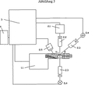
閉じた反応室内の分子間相互作用を検出する本発明に係る装置は、好ましくは4つの主要な機能要素から構成される(図1を参照)。反応室の機械的、電気的、および流体的記録は、記録モジュール(1)で実行される。以下では、反応室はマイクロリアクタとも呼ばれる。反応結果を検出するために、光学系(2)が備えられる。反応結果から分析結果への処理は、コントローラ(3)で実行されることができる。任意で、分析結果が記憶され、および/または適当な接続要素(4)により処理される。 The device according to the invention for detecting intermolecular interactions in a closed reaction chamber is preferably composed of four main functional elements (see FIG. 1). Mechanical, electrical and fluidic recording of the reaction chamber is performed in the recording module (1). In the following, the reaction chamber is also called a microreactor. In order to detect the reaction result, an optical system (2) is provided. The process from the reaction result to the analysis result can be executed by the controller (3). Optionally, the analysis results are stored and / or processed by a suitable connecting element (4).
好都合な態様で本発明に係る装置の構成要素として使用され得る反応室は、国際特許出願公開第01/02094号パンフレットに詳しく記載されており、その内容は本願において明示的に参照される。 Reaction chambers which can be used in an advantageous manner as components of the device according to the invention are described in detail in WO 01/02094, the contents of which are explicitly referred to in this application.
任意でバーコードを用いて識別され得る反応室が流体記録モジュール内に組み込まれ、1つまたは複数の反応液が充填されることができる。任意で反応室は更に電気接点を有し、それによって、例えば集積センサおよび/または加熱素子を用いて反応室内の反応の熱制御および/または調整が保証される。特に、このことは、例えば、対応して標識され、且つ物質ライブラリに結合されている標的分子における金属析出を用いて、DNAまたはRNAに対する熱の影響を受ける増幅反応、DNAまたはRNAのハイブリッド形式、または信号の増強のための反応を行うのに有利である。 A reaction chamber, which can optionally be identified using a bar code, can be incorporated into the fluid recording module and filled with one or more reaction solutions. Optionally, the reaction chamber further has electrical contacts, thereby ensuring thermal control and / or regulation of the reaction in the reaction chamber, for example using integrated sensors and / or heating elements. In particular, this means, for example, the use of metal deposition on target molecules that are correspondingly labeled and bound to a substance library, using heat-induced amplification reactions on DNA or RNA, DNA or RNA hybrid formats, Or it is advantageous to carry out a reaction for signal enhancement.
反応溶液および/またはすすぎ溶液のような増幅および検出反応の実行に任意で必要な溶液が、適当な接続要素、例えばチャネルを介して反応室に挿入されることができる。反応の過程を監視するために、適当なコントローラが使用されることができる。 Solutions necessary for carrying out amplification and detection reactions, such as reaction solutions and / or rinse solutions, can be inserted into the reaction chamber via suitable connecting elements, such as channels. A suitable controller can be used to monitor the reaction process.
本発明の一態様では、光学系により、例えば2次元の電気的読出可能検出要素の形で実装されている適当な検出器上での増幅および/または検出反応の実行中または完了後の物質ライブラリの撮像が保証される。本発明に係る装置の一実施形態において、試料は、照明モジュールまたは光学系の光源を用いて照射され、出現する信号は、使用される標識に応じてフィルタ処理されて撮像される。 In one aspect of the invention, a substance library during or after the completion of amplification and / or detection reactions on a suitable detector implemented by an optical system, for example in the form of a two-dimensional electrically readable detection element Imaging is guaranteed. In one embodiment of the device according to the invention, the sample is illuminated using an illumination module or a light source of an optical system, and the appearing signal is filtered and imaged according to the sign used.
本発明の一態様において、光学系は更に、反応事象の動的な、即ちダイナミックな記録を保証する。特に、本発明に係る装置の光学系は、好ましくは、金標識付き標的分子と物質ライブラリとの間のハイブリッド形成信号を増強するための銀析出の時間依存挙動を記録するのに適している。本発明に係る装置の高集積装備により、反応の過程において複数の画像を転送し、適当なデータ処理モジュールまたはコントローラ内で処理することができる。 In one aspect of the invention, the optical system further ensures a dynamic or dynamic recording of reaction events. In particular, the optical system of the device according to the invention is preferably suitable for recording the time-dependent behavior of silver deposition to enhance the hybridization signal between the gold labeled target molecule and the substance library. The highly integrated equipment of the apparatus according to the present invention allows multiple images to be transferred in the course of reaction and processed in an appropriate data processing module or controller.
各アレイ要素上の析出形成の時間依存挙動により引き起こされる変化は、特に国際特許出願公開第02/02810号パンフレットに詳しく記載されているように、本発明に係る装置で評価されることができる。国際特許出願公開第02/02810号パンフレットの内容は本願で明確に参照される。 Changes caused by the time-dependent behavior of the precipitation formation on each array element can be evaluated with the device according to the invention, in particular as described in detail in WO 02/02810. The contents of WO 02/02810 pamphlet are specifically referred to in this application.
本発明に係る装置は更に、生データまたは分析結果を外部コンピュータまたはコンピュータネットワークに転送し、任意で存在する電子インターフェイスを介して、例えば前記データを記憶することを保証する。 The device according to the invention further ensures that raw data or analysis results are transferred to an external computer or computer network and stored, for example, via an existing electronic interface, for example said data.
本発明の他の態様では、分子プローブが所定の領域上に固定化される基板を含むマイクロアレイが提供される。ここで、基板は本質的にセラミック材料を含む。
最後に、他の態様は、基板の所定の領域上に分子プローブが固定化されているマイクロアレイを製造するためのセラミック材料から本質的に形成されている基板の使用に関係する。
In another aspect of the present invention, a microarray is provided that includes a substrate on which molecular probes are immobilized on a predetermined region. Here, the substrate essentially comprises a ceramic material.
Finally, another aspect relates to the use of a substrate that is essentially formed from a ceramic material to produce a microarray in which molecular probes are immobilized on a predetermined area of the substrate.
さらに、以下の定義が特に本発明の説明のために使用される。
本発明の範囲内において、プローブ、プローブ分子、または分子プローブは、特定の特徴的結合挙動および/または特定の反応性による他の分子の検出に使用される分子を表すものと理解される。固体表面に結合可能で、且つ特異親和性を有する各種分子は、アレイ上に適切に配置されたプローブとして使用されることができる。好ましい実施形態において、これらはペプチド、タンパク質、核酸、および/またはそれらの類似物の群からの生重合体である。特に好ましくは、プローブは核酸および/または核酸類似物である。
Furthermore, the following definitions are used in particular for the description of the invention.
Within the scope of the present invention, a probe, probe molecule or molecular probe is understood to represent a molecule used for the detection of other molecules with a specific characteristic binding behavior and / or a specific reactivity. Various molecules that can bind to a solid surface and have specific affinity can be used as probes appropriately placed on the array. In preferred embodiments, these are biopolymers from the group of peptides, proteins, nucleic acids, and / or the like. Particularly preferably, the probe is a nucleic acid and / or a nucleic acid analogue.
特に、ハイブリッド形成方法で標的分子を検出するために使用され、且つ定義されるとともに周知の配列の核酸分子は、プローブと呼ばれる。DNA分子およびRNA分子の両方が、核酸として使用されることができる。例えば、核酸プローブまたはオリゴヌクレオチドプローブは、長さが10から100塩基、好ましくは15から50塩基、特に好ましくは20から30塩基のオリゴヌクレオチドとなることができる。通常、本発明に従って、これらのプローブは、1本鎖核酸分子または核酸類似物の分子、好ましくは標的分子の配列領域に相補的な少なくとも1つの配列領域を有する1本鎖DNA分子またはRNA分子である。検出方法および使用に応じて、これらのプローブは、固体担体基板上に例えばマイクロアレイの形で固定化されることができる。さらに、これらは、検出方法に応じて放射活性物質または非放射活性物質で標識されることができ、したがって、当技術分野で慣行の検出反応を用いて検出可能である。 In particular, nucleic acid molecules of a well-known sequence that are used and defined to detect target molecules in hybridization methods are called probes. Both DNA and RNA molecules can be used as nucleic acids. For example, the nucleic acid probe or oligonucleotide probe can be an oligonucleotide having a length of 10 to 100 bases, preferably 15 to 50 bases, particularly preferably 20 to 30 bases. Usually, according to the present invention, these probes are single-stranded nucleic acid molecules or nucleic acid analogue molecules, preferably single-stranded DNA molecules or RNA molecules having at least one sequence region complementary to the sequence region of the target molecule. is there. Depending on the detection method and use, these probes can be immobilized on a solid support substrate, for example in the form of a microarray. Furthermore, they can be labeled with radioactive or non-radioactive substances depending on the detection method and can therefore be detected using detection reactions customary in the art.
本発明の範囲内において、標的または標的分子は、分子プローブを用いて検出される分子を表すものと理解される。本発明の好ましい実施形態において、検出される標的は核酸である。しかし、本発明に係るプローブアレイは、類似の方法でタンパク質/プローブ間相互作用、抗体/プローブ間相互作用などの検出に使用されることもできる。 Within the scope of the present invention, a target or target molecule is understood to represent a molecule that is detected using a molecular probe. In a preferred embodiment of the invention, the target to be detected is a nucleic acid. However, the probe array according to the present invention can also be used for detecting protein / probe interaction, antibody / probe interaction, and the like in a similar manner.
本発明の範囲内において、標的がプローブアレイ上に適切に配置されたプローブに対するハイブリッド形成を用いて検出される核酸または核酸分子である場合、前記標的分子は通常、長さが40から10,000塩基、好ましくは60から2,000塩基、より好ましくは60から1,000塩基、特に好ましくは60から500塩基、最も好ましくは60から150塩基である配列を含む。状況に応じて、それらの配列は、プライマーの配列とともに、それらのプライマーによって定義されるテンプレートの配列領域を含む。特に、標的分子は1本鎖または2本鎖核酸分子であり、一方の鎖または両方の鎖が放射活性物質または非放射活性物質で標識され、これらは、当技術分野で慣行の検出方法を用いて検出可能である。 Within the scope of the present invention, when the target is a nucleic acid or nucleic acid molecule that is detected using hybridization to a probe that is appropriately placed on the probe array, the target molecule is typically 40 to 10,000 in length. It includes sequences that are bases, preferably 60 to 2,000 bases, more preferably 60 to 1,000 bases, particularly preferably 60 to 500 bases, and most preferably 60 to 150 bases. Depending on the circumstances, the sequences include the sequence region of the template defined by the primers, along with the sequences of the primers. In particular, the target molecule is a single-stranded or double-stranded nucleic acid molecule and one or both strands are labeled with a radioactive or non-radioactive substance, which uses conventional detection methods in the art. Can be detected.
本発明によれば、標的配列は、プローブとのハイブリッド形成を用いて検出される標的の配列領域を表す。本発明により、前記状況はさらに、プローブにより取り扱われるその領域とも呼ばれる。 According to the present invention, the target sequence represents the sequence region of the target that is detected using hybridization with the probe. According to the present invention, the situation is further referred to as that region handled by the probe.
本発明の範囲内において、物質ライブラリは、複数の異なるプローブ分子、好ましくは少なくとも2から1,000,000個の異なる分子、特に好ましくは少なくとも10から10,000個の異なる分子、および最も好ましくは100から1,000までの範囲の異なる分子を表すものと理解される。特別な設計の場合、物質ライブラリはさらに、少なくとも50以下または少なくとも30,000個の異なる分子のみを含むことができる。好ましくは、物質ライブラリは、本発明に係る装置の反応室内の担体上にアレイの形で適切に配置される。 Within the scope of the present invention, the substance library comprises a plurality of different probe molecules, preferably at least 2 to 1,000,000 different molecules, particularly preferably at least 10 to 10,000 different molecules, and most preferably It is understood to represent different molecules ranging from 100 to 1,000. For special designs, the substance library may further include only at least 50 or less or at least 30,000 different molecules. Preferably, the substance library is suitably arranged in the form of an array on a support in the reaction chamber of the device according to the invention.
本発明の範囲内において、プローブアレイは、各プローブの位置が別々に定義されている担体上の分子プローブまたは物質ライブラリの配置を表すものと理解される。好ましくは、アレイは定義された部位および/または所定の領域、いわゆるアレイ要素を含み、該アレイ要素は、特に好ましくは特定のパターンで適切に配置され、各アレイ要素は通常ただ1つの化学種のプローブを含む。ここで、担体上の分子またはプローブの配置は、共有結合または非共有結合相互作用を用いて形成されることができる。配置内、即ちアレイ内の位置は通常、スポットと呼ばれる。したがって、プローブアレイは検出領域を形成する。 Within the scope of the present invention, a probe array is understood to represent the arrangement of molecular probes or substance libraries on a carrier in which the position of each probe is defined separately. Preferably, the array comprises defined sites and / or predetermined regions, so-called array elements, which array elements are particularly preferably arranged appropriately in a specific pattern, each array element usually having only one chemical species. Includes probes. Here, the arrangement of molecules or probes on the support can be formed using covalent or non-covalent interactions. Positions in the arrangement, i.e. in the array, are usually called spots. Accordingly, the probe array forms a detection region.
本発明の範囲内において、アレイ要素、所定の領域、スポット、またはアレイスポットは、表面上の分子プローブの堆積に関して決定される特定の領域を表すと理解され、占有されている全てのアレイ要素がプローブアレイである。 Within the scope of the present invention, an array element, a predetermined area, a spot, or an array spot is understood to represent a specific area determined with respect to the deposition of molecular probes on the surface, and all occupied array elements are It is a probe array.
本発明の範囲内において、担体要素、担体、物質ライブラリ担体、または基板は、プローブアレイが配置される固形物を表すものと理解される。担体は通常、基板またはマトリクスとも呼ばれ、例えば物体担体またはウェハ若しくはセラミック材料にも関係する。 Within the scope of the present invention, a carrier element, carrier, substance library carrier or substrate is understood to represent a solid on which the probe array is arranged. The carrier is usually also referred to as a substrate or a matrix and relates for example to an object carrier or a wafer or ceramic material.
基板または検出領域上にアレイ配置で適切に配置された分子、または基板もしくは検出領域上にアレイ配置で適切に配置された物質ライブラリ、および担体または基板の全体も、「チップ」、「マイクロアレイ」、「DNAチップ」、「プローブアレイ」などとも呼ばれることが多い。 Molecules appropriately arranged in an array arrangement on a substrate or detection region, or a substance library appropriately arranged in an array arrangement on a substrate or detection region, and the entire carrier or substrate are also referred to as a “chip”, “microarray”, It is often called “DNA chip”, “probe array” or the like.
本発明の範囲内において、検出領域は、物質ライブラリが好ましくはアレイ配置で適切に配置される基板の領域を表すものと理解される。
本発明の範囲内において、室担体は、反応室の底を形成する固形物を表すものと理解される。好ましくは、室担体は、基板の反対側または物質ライブラリ担体の反対側に配置される。本発明に係る装置の代替実施形態において、室担体はまた、同時に基板および/または物質ライブラリ担体である。
Within the scope of the present invention, the detection region is understood to represent the region of the substrate where the substance library is suitably arranged, preferably in an array arrangement.
Within the scope of the present invention, the chamber carrier is understood to represent the solid that forms the bottom of the reaction chamber. Preferably, the chamber carrier is arranged on the opposite side of the substrate or on the opposite side of the substance library carrier. In an alternative embodiment of the device according to the invention, the chamber carrier is also a substrate and / or substance library carrier at the same time.
本発明の範囲内において、室本体は、反応室を形成する固形物を表すものと理解される。通常、物質ライブラリ担体またはチップは室本体の一部であり、その場合、物質ライブラリ担体は、残りの室本体と異なる材料で構成される。 Within the scope of the present invention, the chamber body is understood to represent a solid that forms the reaction chamber. Usually, the substance library carrier or chip is part of the chamber body, in which case the substance library carrier is composed of a material different from the remaining chamber body.
本発明の範囲内において、反応室または反応空間は、室担体とマイクロアレイとの間に形成された空間、および好ましくは毛細管ギャップの形で設計された空間を表すものと理解される。反応室または反応空間の底部は、アレイの底部または室担体の底部によってそれぞれ定義される。特に、室本体とマトリクスまたはマイクロアレイとの間の距離は、反応空間または反応室の厚さと呼ばれる。本発明の範囲内で、反応空間は通常、ごくわずかな厚さ、例えば最大で1cm、好ましくは最大で5mm、特に好ましくは最大で3mm、および最も好ましくは最大で1mmの厚さである。 Within the scope of the present invention, a reaction chamber or reaction space is understood to represent a space formed between the chamber carrier and the microarray, and preferably a space designed in the form of a capillary gap. The bottom of the reaction chamber or reaction space is defined by the bottom of the array or the bottom of the chamber carrier, respectively. In particular, the distance between the chamber body and the matrix or microarray is called the reaction space or reaction chamber thickness. Within the scope of the present invention, the reaction space is usually of a very small thickness, for example a maximum of 1 cm, preferably a maximum of 5 mm, particularly preferably a maximum of 3 mm, and most preferably a maximum of 1 mm.
本発明の範囲内において、毛細管ギャップは、室担体とマイクロアレイとの間に作用する毛管力を用いて満たされることができる反応空間を表すものと理解される。通常、毛細管ギャップはわずかな厚さ、例えば最大で1mm、好ましくは最大で750μm、および特に好ましくは最大で500μmの厚さである。本発明によれば、10から300μm、15μmから200μm、または25μmから150μmの範囲の厚さが毛細管ギャップの厚みとして好ましい。本発明の特別な実施形態では、毛細管ギャップの厚さは、50μm、60μm、70μm、80μm、または90μmである。本発明によれば、反応空間または反応室の厚さが2mmを超える場合、反応空間または反応室は、もはや毛細管ギャップと呼ばれない。 Within the scope of the present invention, a capillary gap is understood to represent a reaction space that can be filled using capillary forces acting between the chamber carrier and the microarray. Usually the capillary gap is of a slight thickness, for example a maximum of 1 mm, preferably a maximum of 750 μm and particularly preferably a maximum of 500 μm. According to the present invention, a thickness in the range of 10 to 300 μm, 15 μm to 200 μm, or 25 μm to 150 μm is preferred as the thickness of the capillary gap. In particular embodiments of the invention, the capillary gap thickness is 50 μm, 60 μm, 70 μm, 80 μm, or 90 μm. According to the invention, when the thickness of the reaction space or reaction chamber exceeds 2 mm, the reaction space or reaction chamber is no longer called a capillary gap.
本発明の範囲内において、共焦点蛍光検出システムは、物体、即ちマイクロアレイが点光源を用いて対物レンズの焦点面内で照射される蛍光検出システムを表すものと理解される。ここで、点光源、物体、および点光源検出器は、光学的共役面上に正確に配置される。共焦点システムの実施例については、A.ディアスプロ(A.Diaspro)、Confocal and 2−photon−microscopy:Foundations、Applications and Advances、Wiley−Liss、2002年に記載されている。 Within the scope of the present invention, a confocal fluorescence detection system is understood to represent a fluorescence detection system in which an object, i.e. a microarray, is illuminated in the focal plane of the objective lens using a point light source. Here, the point light source, the object, and the point light source detector are accurately arranged on the optical conjugate plane. For examples of confocal systems, see A. Diaspro, Confocal and 2-photo-microscopy: Foundations, Applications and Advances, Wiley-Liss, 2002.
本発明の範囲内において、反応室全体の全体積を撮像する蛍光光学系は、非共焦点蛍光検出システム、即ち点光源を用いた照明が物体、即ちマイクロアレイに制限されない蛍光検出システムを表すものと理解される。したがって、このような蛍光検出システムは焦点制限を有さない。 Within the scope of the present invention, a fluorescence optical system that images the entire volume of the entire reaction chamber represents a non-confocal fluorescence detection system, i.e. a fluorescence detection system in which illumination using a point light source is not restricted to an object, i.e. a microarray. Understood. Thus, such a fluorescence detection system does not have focus limitations.
本発明の範囲内の従来のアレイまたはマイクロアレイは、例えば1mmから4mm×1mmから4mm、好ましくは2mm×2mmの好ましくは正方形領域上で、約50から10,000、好ましくは150から2,000の異なる化学種のプローブ分子を含む。本発明の範囲内の他の実施形態において、マイクロアレイは、数mm2から数cm2、好ましくは約1mm2から10cm2、特に好ましくは2mm2から1cm2、および最も好ましくは約4mm2から6.25mm2の領域上で、約50から約80,000、好ましくは約100から約65,000、特に好ましくは約1,000から約10,000個の異なる化学種のプローブ分子を含む。例えば、従来のマイクロアレイは、2mm×2mmの領域上で100から65,000個の異なる化学種のプローブ分子を有する。 Conventional arrays or microarrays within the scope of the present invention are about 50 to 10,000, preferably 150 to 2,000, preferably on a square area, for example 1 mm to 4 mm × 1 mm to 4 mm, preferably 2 mm × 2 mm. Contains probe molecules of different species. In other embodiments within the scope of the present invention, the microarray is several mm 2 to several cm 2 , preferably about 1 mm 2 to 10 cm 2 , particularly preferably 2 mm 2 to 1 cm 2 , and most preferably about 4 mm 2 to 6. It includes about 50 to about 80,000, preferably about 100 to about 65,000, particularly preferably about 1,000 to about 10,000 probe molecules of different species on a .25 mm 2 area. For example, a conventional microarray has 100 to 65,000 different species of probe molecules on a 2 mm × 2 mm area.
本発明の範囲内において、マイクロウェルプレートは、複数の生物学的、化学的、および実験医学検定の自動実行を可能にする特定のラスタ内の反応容器のレ配置を表すものと理解される。 Within the scope of the present invention, a microwell plate is understood to represent a re-arrangement of reaction vessels in a particular raster that allows automatic execution of multiple biological, chemical and laboratory medical assays.
本発明の範囲内において、標識またはマーカーは、検出可能ユニット、例えば検出可能ユニットが結合可能な蛍光プローブまたはアンカー基を表すものと理解される。
本発明の範囲内において、試料または試料溶液は、増幅および/または検出される核酸分子とともに分析される液体である。
Within the scope of the present invention, a label or marker is understood to represent a detectable unit, for example a fluorescent probe or anchor group to which the detectable unit can be bound.
Within the scope of the present invention, a sample or sample solution is a liquid that is analyzed with the nucleic acid molecules to be amplified and / or detected.
本発明の範囲内において、増殖反応または増幅反応は通常、10から50以上の増幅サイクル、好ましくは約20から40サイクル、特に好ましくは約30サイクルを含む。本発明の範囲内において、循環増幅反応は好ましくはポリメラーゼ連鎖反応(PCR)である。 Within the scope of the present invention, the growth or amplification reaction usually comprises 10 to 50 or more amplification cycles, preferably about 20 to 40 cycles, particularly preferably about 30 cycles. Within the scope of the present invention, the cyclic amplification reaction is preferably the polymerase chain reaction (PCR).
本発明の範囲内において、増幅サイクルは循環増幅反応の単一増強工程を表す。PCRの増強工程はPCRサイクルとも呼ばれる。
本発明の範囲内において、増幅生成物は、循環増幅反応、好ましくはPCRを用いて増幅される核酸分子の増強または増殖もしくは増幅から得られる生成物を表す。PCRを用いて増幅される核酸分子はPCR生成物とも呼ばれる。
Within the scope of the present invention, an amplification cycle represents a single enhancement step of a cyclic amplification reaction. The PCR enhancement process is also called a PCR cycle.
Within the scope of the present invention, an amplification product refers to a product obtained from a cyclic amplification reaction, preferably an enhancement or growth or amplification of a nucleic acid molecule that is amplified using PCR. Nucleic acid molecules that are amplified using PCR are also referred to as PCR products.
本発明の範囲内において、変性温度は、2本鎖DNAが増幅サイクル内で分離される温度を表すものと理解される。変性温度は通常、特にPCRにおいて90℃を越えており、好ましくは約95℃である。 Within the scope of the present invention, denaturation temperature is understood to represent the temperature at which double-stranded DNA is separated within the amplification cycle. The denaturation temperature usually exceeds 90 ° C., especially in PCR, and is preferably about 95 ° C.
本発明の範囲内において、アニーリング温度は、プライマーと検出される核酸とがハイブリッド形成する際の温度を表すものと理解される。アニーリング温度は通常、特にPCRにおいて55℃から65℃の範囲内にあり、好ましくは約60℃である。 Within the scope of the present invention, the annealing temperature is understood to represent the temperature at which the primer and the nucleic acid to be detected hybridize. The annealing temperature is usually in the range of 55 ° C. to 65 ° C., especially in PCR, preferably about 60 ° C.
本発明の範囲内において、鎖伸展温度または伸展温度は、モノマー成分の挿入によって核酸が合成される温度を表すものと理解される。伸展温度は通常、特にPCRにおいて約70℃から約75℃の範囲内にあり、好ましくは約72℃である。 Within the scope of the present invention, chain extension temperature or extension temperature is understood to represent the temperature at which a nucleic acid is synthesized by the insertion of a monomer component. The extension temperature is usually in the range of about 70 ° C. to about 75 ° C., particularly in PCR, preferably about 72 ° C.
本発明の範囲内において、オリゴヌクレオチドプライマーまたはプライマーは、循環増幅反応において検出されるDNAの相補鎖の合成が結合部位から始まり、標的DNAとも呼ばれる検出されるDNAを結合またはハイブリッドを形成するオリゴヌクレオチドを表す。特に、プライマーは、より長いDNAまたはRNA分子の一部と相補的であり、遊離3−OH基を3’末端に有する好ましくは約12から30個の塩基を含む短いDNAまたはRNAオリゴヌクレオチドを表す。前記遊離3’OH基に起因して、プライマーは、任意のDNAまたはRNAポリメラーゼの基材として作用することができ、ヌクレオチドを5’−3’方向でプライマーに合成することができる。ここで、新しく合成されたヌクレオチドの配列は、プライマーの遊離3’OH基の向こうにあるプライマーとハイブリッドを形成するテンプレートの配列によって予め決定される。従来のプライマーの長さは、ヌクレオチド12から50個分、好ましくは15から30個分である。 Within the scope of the present invention, an oligonucleotide primer or primer is an oligonucleotide that binds to or forms a hybrid with the detected DNA, also called target DNA, where synthesis of the complementary strand of the DNA detected in the circular amplification reaction begins at the binding site Represents. In particular, a primer represents a short DNA or RNA oligonucleotide that is complementary to a portion of a longer DNA or RNA molecule and preferably contains about 12 to 30 bases with a free 3-OH group at the 3 ′ end. . Due to the free 3'OH group, the primer can act as a substrate for any DNA or RNA polymerase, and nucleotides can be synthesized into the primer in the 5'-3 'direction. Here, the sequence of the newly synthesized nucleotide is predetermined by the sequence of the template that hybridizes with the primer beyond the free 3'OH group of the primer. The length of the conventional primer is 12 to 50 nucleotides, preferably 15 to 30 nucleotides.
相補核酸鎖の合成のためにテンプレートとして作用する2本鎖核酸分子または核酸鎖は通常、テンプレートまたはテンプレート鎖と呼ばれる。
相補1本鎖核酸分子からの2本鎖核酸分子または2本鎖分子の形成は、ハイブリッド形成と呼ばれる。ここで、この関連性は、好ましくは常にAおよびTとGおよびCとの対として起こる。ハイブリッド形成の範囲内において、例えばDNA−DNA2本鎖、DNA−RNA2本鎖、またはRNA−RNA2本鎖が形成されることができる。ハイブリッド形成を用いて核酸類似物による2本鎖も形成可能であり、例えばDNA−PNA2本鎖、RNA−PNA2本鎖、DNA−LNA2本鎖、およびRNA−LNA2本鎖である。ハイブリッド形成の実験は通常、配列相補性、したがって2つの異なる核酸分子の識別を検出するために使用される。
Double-stranded nucleic acid molecules or nucleic acid strands that act as templates for the synthesis of complementary nucleic acid strands are usually referred to as templates or template strands.
Formation of a double stranded nucleic acid molecule or double stranded molecule from a complementary single stranded nucleic acid molecule is referred to as hybridization. Here, this relationship preferably always occurs as a pair of A and T and G and C. Within the scope of hybridization, for example, DNA-DNA duplexes, DNA-RNA duplexes, or RNA-RNA duplexes can be formed. Hybridization can also be used to form duplexes with nucleic acid analogs, such as DNA-PNA duplex, RNA-PNA duplex, DNA-LNA duplex, and RNA-LNA duplex. Hybridization experiments are usually used to detect sequence complementarity and thus the discrimination of two different nucleic acid molecules.
本発明の範囲内において、処理は、精製、標識付け、増幅、ハイブリッド形成、および/または洗浄およびすすぎ工程とともに、さらに物質ライブラリを用いて標的を検出する際に実行される手順工程を表すものと理解される。 Within the scope of the present invention, processing represents a procedural step performed in detecting a target using a substance library, along with purification, labeling, amplification, hybridization, and / or washing and rinsing steps. Understood.
本発明の範囲内において、実質的にセラミック材料からなる基板または実質的にセラミック材料を含む基板は、少なくとも75%、好ましくは少なくとも85%、および特に好ましくは少なくとも90%のセラミック材料を含む基板を表すものと理解される。 Within the scope of the present invention, a substrate consisting essentially of or comprising a ceramic material comprises a substrate comprising at least 75%, preferably at least 85%, and particularly preferably at least 90% ceramic material. It is understood to represent.
本発明の範囲内において、実質的に酸化アルミニウムセラミック材料からなる基板または実質的に酸化アルミニウムセラミック材料を含む基板は、少なくとも75%、好ましくは少なくとも85%、および特に好ましくは少なくとも90%の酸化アルミニウムセラミック材料を含む基板を表すものと理解される。 Within the scope of the present invention, a substrate consisting essentially of or comprising an aluminum oxide ceramic material is at least 75%, preferably at least 85%, and particularly preferably at least 90% aluminum oxide. It is understood to represent a substrate comprising a ceramic material.
本発明の範囲内において、アルミニウムセラミックは、実質的に酸化アルミニウムからなるセラミック材料を表すものと理解される。本発明の範囲内において、実質的に酸化アルミニウムからなるセラミック材料は、少なくとも75%、好ましくは少なくとも85%、および特に好ましくは少なくとも90%の酸化アルミニウムを含むセラミック材料を表すものと理解される。 Within the scope of the present invention, aluminum ceramic is understood to represent a ceramic material consisting essentially of aluminum oxide. Within the scope of the present invention, a ceramic material consisting essentially of aluminum oxide is understood to represent a ceramic material comprising at least 75%, preferably at least 85%, and particularly preferably at least 90% aluminum oxide.
したがって、本発明の第1の目的は、特に以下の要素を含み、且つ核酸を増幅および検出するための装置である。
a)温度制御および/または調整ユニットと、
b)化合物ライブラリが固定化される検出領域を有する担体を含む反応室であって、温度制御および調整ユニットを用いて反応室内の温度が制御および/または調整可能であることと、
c)検出領域上の析出形成の時間依存挙動を検出可能な光学系。
Accordingly, a first object of the present invention is an apparatus for amplifying and detecting nucleic acids, particularly comprising the following elements:
a) a temperature control and / or adjustment unit;
b) a reaction chamber comprising a carrier having a detection region on which the compound library is immobilized, the temperature in the reaction chamber being controllable and / or adjustable using a temperature control and adjustment unit;
c) An optical system capable of detecting the time-dependent behavior of precipitate formation on the detection region.
本発明のこの態様において、1つの装置内における温度制御および/または調整ユニット、温度調整可能な反応室、および析出形成を用いて信号増強反応の動的測定を可能にする光学系は、本発明に係る装置の1つの本質的な特徴である。 In this aspect of the invention, a temperature control and / or adjustment unit, a temperature-adjustable reaction chamber, and an optical system that enables dynamic measurement of a signal-enhanced reaction using precipitation formation within one apparatus are provided by the present invention. Is an essential feature of the apparatus according to.
本発明のこの態様では、本発明に係る装置は、分子間相互作用の検出が集積光学系またはリーダーシステムによって手動操作でも可能であることを特徴とする。このことは、特に医療診断などの分野で好都合である。この実施形態において、物質ライブラリに結合された核酸の相対的定量的量を正確に判定することは、本発明に係る装置が集積光学系を備え、それにより検出領域上の析出形成の時間依存挙動が検出可能であるという事実により確実なものになる。 In this aspect of the invention, the device according to the invention is characterized in that the detection of intermolecular interactions can also be performed manually by means of integrated optics or a reader system. This is particularly advantageous in fields such as medical diagnosis. In this embodiment, accurately determining the relative quantitative amount of nucleic acid bound to the substance library is that the device according to the invention comprises an integrated optical system, whereby the time-dependent behavior of precipitate formation on the detection region Is ensured by the fact that is detectable.
この光学系は、例えば2次元の電気的読出可能検出要素の形で実装されている適当な検出器上での増幅および/または検出反応の実行中または完了後の物質ライブラリの撮像を保証する。本発明に係る装置の一実施形態において、試料は、照明モジュールまたは光学系の光源を用いて照射され、出現する信号は、使用される標識に応じてフィルタ処理されて撮像される。 This optical system ensures imaging of the substance library during or after the amplification and / or detection reaction is performed on a suitable detector, for example implemented in the form of a two-dimensional electrically readable detection element. In one embodiment of the device according to the invention, the sample is illuminated using an illumination module or a light source of an optical system, and the appearing signal is filtered and imaged according to the sign used.
さらに、この光学系は、反応結果の動的な、即ちダイナミックな記録を保証する。特に、本発明に係る装置の光学系は、金標識付き標的分子と物質ライブラリとの間のハイブリッド形成信号を増強するため銀析出の時間依存挙動を記録するのに適している。本発明に係る装置の高集積装備は、反応の過程において複数の画像を転送し、適当なデータ処理モジュールまたはコントローラ内で処理することができる。 Furthermore, this optical system ensures a dynamic or dynamic recording of reaction results. In particular, the optical system of the device according to the invention is suitable for recording the time-dependent behavior of silver deposition in order to enhance the hybridization signal between the gold labeled target molecule and the substance library. The highly integrated equipment of the apparatus according to the present invention can transfer multiple images in the course of reaction and process them in a suitable data processing module or controller.
各アレイ要素上の析出形成の時間依存挙動から生じる変化は、特に国際特許出願公開第02/02810号パンフレットに詳しく記載されているように、本発明に係る装置で評価され得る。この結果、国際特許出願公開第02/02810号パンフレットの各内容も明確に参照される。 Changes resulting from the time-dependent behavior of the precipitate formation on each array element can be evaluated with the apparatus according to the invention, in particular as described in detail in WO 02/02810. As a result, the contents of International Patent Application Publication No. 02/02810 pamphlet are also clearly referred to.
例えば、本発明に係る装置は、検出領域を有するチップを含む反応室の室本体が室担体に密閉する形で貼り付けられ、室担体と検出領域またはチップの基板との間に毛細管ギャップを有する試料空間が形成され、その温度は調整可能であり、且つその体積流量率は制御可能であるように実装されることができる。この種の構造により、特定の温度範囲内でのみ効率よく実行される反応の実行、およびマイクロチップを用いた実験による反応生成物の好ましくは同時検出が可能である。 For example, in the apparatus according to the present invention, a chamber body of a reaction chamber including a chip having a detection region is attached to the chamber carrier so as to be sealed, and a capillary gap is provided between the chamber carrier and the substrate of the detection region or chip. The sample space is formed, its temperature can be adjusted, and its volume flow rate can be implemented to be controllable. With this type of structure, it is possible to carry out reactions that are carried out efficiently only within a specific temperature range, and preferably to simultaneously detect reaction products by experiments using microchips.
したがって、本発明に係る装置は、例えばPCRを用いて核酸分子を増幅し、且つマイクロチップを用いた実験によりPCR生成物をほぼ同時に検出するために使用され得る。このような反応に対する試料液は、温度調整のための対応する手段により効率よく加熱または冷却されることができる。 Thus, the device according to the present invention can be used to amplify nucleic acid molecules, for example using PCR, and to detect PCR products almost simultaneously by experiments using microchips. Sample solutions for such reactions can be efficiently heated or cooled by corresponding means for temperature regulation.
本発明に係る装置はまた、逆転写酵素反応を実行し、それによってmRNAをcDNAに転写し、そのチップとのハイブリッド形成により反応生成物を特徴付けるために使用されることもできる。この方法では、いわゆる「遺伝子プロファイリング」を実行することができる。逆転写酵素およびハイブリッド形成の両方が室内で実行されることから、この方法は時間効率が高く、且つ低い過失責任を示す。 The apparatus according to the present invention can also be used to perform a reverse transcriptase reaction, thereby transcribing mRNA to cDNA and characterizing the reaction product by hybridization with the chip. In this way, so-called “gene profiling” can be performed. Since both reverse transcriptase and hybridization are performed indoors, this method is time efficient and exhibits low negligence.
本発明に係る装置では、例えば所望の温度での制限酵素消化が更に反応室内で実行され、反応生成物をチップとのハイブリッド形成を用いて特徴付けることができる。酵素の変性は熱非活性化を用いて実行され得る。そのため、本発明に係る装置は、時間効率のよい制限酵素断片長多型マッピング(RFLPマッピング)が可能である。 In the device according to the invention, for example, restriction enzyme digestion at the desired temperature is further carried out in the reaction chamber and the reaction product can be characterized using hybridization with the chip. Enzymatic denaturation can be performed using heat inactivation. Therefore, the apparatus according to the present invention is capable of time-efficient restriction enzyme fragment length polymorphism mapping (RFLP mapping).
さらに、本発明に係る装置では、例えばライゲーションも実行可能である。
本発明に係る装置では、核酸標的/核酸プローブ複合体の温度依存溶融挙動も更に調査可能である。
Furthermore, in the apparatus according to the present invention, for example, ligation can also be executed.
In the apparatus according to the present invention, the temperature-dependent melting behavior of the nucleic acid target / nucleic acid probe complex can be further investigated.
さらに、本発明に係る装置は、タンパク質の温度依存結合挙動を実行するために使用されることができる。この方法において、例えば長時間加熱した後も抗体がまだ各抗原と結合することが可能であるかを検査することができる。この場合、チップは核酸分子により官能化されないが、各タンパク質またはペプチドにより官能化されることが前提条件である。 Furthermore, the device according to the invention can be used to carry out the temperature-dependent binding behavior of proteins. In this method, for example, it is possible to test whether an antibody can still bind to each antigen even after heating for a long time. In this case, the chip is not functionalized with nucleic acid molecules, but it is a prerequisite that it be functionalized with each protein or peptide.
反応室の室本体は、好ましくはガラス、合成材料および/または高品位の鋼鉄、アルミニウム、および真鍮などの金属から形成される。その製造に関しては、例えば射出成形に適した剛性材料が使用されることができる。特に、マイクロロン(macrolon)、ナイロン、PMMA、およびテフロンなどの剛性材料が考えられる。 The chamber body of the reaction chamber is preferably formed from glass, synthetic materials and / or metals such as high-grade steel, aluminum, and brass. For its manufacture, for example, a rigid material suitable for injection molding can be used. In particular, rigid materials such as microlon, nylon, PMMA, and Teflon are contemplated.
また、物質ライブラリ担体と室担体との間の反応空間は、例えば注射器を用いて反応空間を満たす隔壁を用いて閉鎖されることができる。好ましい実施形態において、室本体は、ガラス、PMMA、ポリカーボネート、ポリスチレン、および/またはトパーズなどの光学的に透明な材料からなる。ここで、材料の選択は、装置の意図した使用に合わせて調整される。例えば、材料の選択時に装置が曝される温度が考慮されるべきである。例えば、装置がPCRの実行に使用される場合、例えば長時間にわたり95℃程度の温度でも安定している合成材料のみが使用される。 Also, the reaction space between the substance library carrier and the chamber carrier can be closed with a partition that fills the reaction space using, for example, a syringe. In a preferred embodiment, the chamber body is made of an optically transparent material such as glass, PMMA, polycarbonate, polystyrene, and / or topaz. Here, the selection of materials is tailored to the intended use of the device. For example, the temperature to which the device is exposed during material selection should be considered. For example, if the device is used to perform PCR, only synthetic materials that are stable, for example, at temperatures as high as 95 ° C. over a long period of time are used.
室担体は、好ましくはガラス、合成材料、シリコン、金属、および/またはセラミック材料からなる。例えば、室担体は、酸化アルミニウムセラミック、ナイロン、および/またはテフロンからなる。 The chamber carrier is preferably made of glass, synthetic material, silicon, metal and / or ceramic material. For example, the chamber carrier is made of aluminum oxide ceramic, nylon, and / or Teflon.
一実施形態において、室担体は、ガラスおよび/または光学的に透明な合成材料、例えばPMMA、ポリカーボネート、ポリスチレン、またはアクリルなどの透明材料からなる。 In one embodiment, the chamber carrier is made of a transparent material such as glass and / or an optically transparent synthetic material, such as PMMA, polycarbonate, polystyrene, or acrylic.
好ましくは、室担体および/または基板は、本発明に係る装置に組み込まれる温度上昇用の手段で接続され、次いで、好ましくは熱伝導率が高い材料で構成される。このような熱伝導性材料は、反応空間の領域全体にわたって均一な温度プロファイルを保証するという大きな利点を有し、したがって、例えばPCRなどの温度依存反応は、反応室全体において均一に、且つ高い収率で、また制御可能な形でおよび/または調整可能な形で高い精度により実行されることができる。 Preferably, the chamber carrier and / or the substrate are connected by means for increasing the temperature incorporated in the device according to the invention, and are then preferably made of a material with high thermal conductivity. Such a thermally conductive material has the great advantage of ensuring a uniform temperature profile throughout the region of the reaction space, so that temperature dependent reactions such as PCR, for example, are uniform and high yielding throughout the reaction chamber. It can be performed with high accuracy in a rate and in a controllable and / or adjustable manner.
したがって、好ましい実施形態において、室担体および/または基板は、熱伝導率が高い、好ましくは15から500Wm−1K−1の範囲、特に好ましくは50から300Wm−1K−1の範囲、最も好ましくは100から200Wm−1K−1の範囲の熱伝導率を有する材料からなり、それらの材料は通常、光学的に透明ではない。適当な熱伝導材料の実施例として、シリコン、酸化アルミニウムセラミックなどのセラミック材料、および/または高品位の鋼鉄、アルミニウム、または真鍮などの金属が挙げられる。 Thus, in a preferred embodiment, the chamber carrier and / or substrate has a high thermal conductivity, preferably in the range of 15 to 500 Wm −1 K −1 , particularly preferably in the range of 50 to 300 Wm −1 K −1 , most preferably Consists of materials having a thermal conductivity in the range of 100 to 200 Wm −1 K −1 , which materials are usually not optically transparent. Examples of suitable heat conducting materials include ceramic materials such as silicon, aluminum oxide ceramic, and / or metals such as high grade steel, aluminum, or brass.
特に好ましい実施形態において、基板は、例えばセラミック材料などの熱伝導率が高い材料からなる。この実施形態では、基板は温度上昇手段により接続され、それによって、反対側の室担体は、明確に熱伝導率を有さない材料、例えば、残りの室本体にも使用される材料などで形成されることができる。したがって、室担体および基板の両方が高価な材料で形成されている実施形態とは反対に、この実施形態では高価な構成要素が排除されている。 In a particularly preferred embodiment, the substrate is made of a material with high thermal conductivity, such as a ceramic material. In this embodiment, the substrates are connected by a temperature raising means, whereby the opposite chamber carrier is formed of a material that does not clearly have thermal conductivity, such as a material that is also used for the remaining chamber body. Can be done. Thus, in contrast to the embodiment in which both the chamber carrier and the substrate are formed of expensive materials, this embodiment eliminates expensive components.
本発明に係る基板または装置の担体が実質的にセラミック材料からなる場合、酸化アルミニウムセラミックが好ましくは使用される。このような酸化アルミニウムセラミックの実施例として、京セラ(ドイツ、ノイス)(Kyocera(Neuss,Germany))のA−473、A−476、およびA−493が挙げられる。これらのセラミックは、各酸化アルミニウム含有量(A−473:93%、A−476:96%、およびA−493:99%)だけでなく、その表面の粗さも実質的に異なる。表面粗さが可能な限り低い酸化アルミニウムセラミックの使用が最も好ましい。 Aluminum oxide ceramics are preferably used when the substrate of the substrate or the device according to the invention consists essentially of a ceramic material. Examples of such aluminum oxide ceramics include Kyocera (Neus, Germany) A-473, A-476, and A-493. These ceramics differ not only in their aluminum oxide content (A-473: 93%, A-476: 96%, and A-493: 99%), but also in their surface roughness. Most preferred is the use of an aluminum oxide ceramic with the lowest possible surface roughness.
好ましくは、室担体および/または基板は、その逆側、即ち反応室から離れて面する側に、任意で小型化された温度センサおよび/または電極を備えるか、またはむしろ、この場所に加熱構造体を有し、試料液の焼き戻しとともに誘導電気浸透流を用いて試料液の混合を行うことができる。 Preferably, the chamber carrier and / or substrate comprises an optionally miniaturized temperature sensor and / or electrode on its opposite side, i.e. the side facing away from the reaction chamber, or rather a heating structure at this location. The sample liquid can be mixed using an induced electroosmotic flow together with tempering of the sample liquid.
温度センサは、例えばニッケル−クロム薄膜抵抗温度センサの形態で実装され得る。
電極は、例えば金−チタン電極の形態、特に4極の形態で実装され得る。
温度上昇手段は、好ましくは、毛細管ギャップ内の液体の高速加熱および冷却が可能なように選択されることができる。ここで、高速加熱および冷却は、温度上昇手段により0.2K/sから30K/sの範囲、好ましくは0.5K/sから15K/sの範囲、特に好ましくは2K/sから15K/sの範囲、および最も好ましくは8K/sから12K/sの範囲、または約10K/sの温度変化を媒介可能であることを表すものと理解される。好ましくは、1K/sから10K/sの温度変化も温度上昇手段により媒介されることができる。
The temperature sensor can be implemented, for example, in the form of a nickel-chrome thin film resistance temperature sensor.
The electrodes can be implemented, for example, in the form of gold-titanium electrodes, in particular in the form of four poles.
The temperature raising means can preferably be selected to allow fast heating and cooling of the liquid in the capillary gap. Here, the high-speed heating and cooling is performed in the range of 0.2 K / s to 30 K / s, preferably 0.5 K / s to 15 K / s, particularly preferably 2 K / s to 15 K / s, by the temperature increasing means. It is understood to represent a range, and most preferably in the range of 8 K / s to 12 K / s, or capable of mediating a temperature change of about 10 K / s. Preferably, a temperature change from 1 K / s to 10 K / s can also be mediated by the temperature raising means.
例えば、ヒーターの形態の温度上昇手段もまた、例えばニッケル−クロム薄膜抵抗ヒーターの形態で実装されることができる。
温度センサ、温度上昇手段、ならびに電極の仕様および寸法の詳細については、国際特許出願公開第01/02094号パンフレットの内容を参照する。
For example, the temperature raising means in the form of a heater can also be implemented in the form of a nickel-chrome thin film resistance heater, for example.
For details of the specifications and dimensions of the temperature sensor, temperature raising means, and electrodes, refer to the contents of International Patent Application Publication No. 01/02094.
チップまたは基板は、好ましくは、ボロフロートガラス、石英ガラス、単結晶CaF2、サファイアディスク、トパーズ、PMMA、ポリカーボネート、および/またはポリスチレンからなる。材料の選択はまた、装置および/またはチップの意図した使用に合わせて調整される。例えば、チップがPCR生成物の特徴付けに使用される場合、95℃の温度に耐えられる材料のみが使用可能である。 Chip or substrate is preferably boro float glass, quartz glass, monocrystalline CaF 2, composed of sapphire disk, topaz, PMMA, polycarbonate, and / or polystyrene. The choice of material is also tailored to the intended use of the device and / or chip. For example, if the chip is used to characterize PCR products, only materials that can withstand temperatures of 95 ° C. can be used.
好ましくは、チップは、核酸分子、特にDNAまたはRNA分子により官能化される。しかし、これらはまた、ペプチドおよび/またはタンパク質、例えば抗体、受容体分子、薬理活性ペプチド、および/またはホルモンなどにより官能化されることができる。 Preferably, the chip is functionalized with a nucleic acid molecule, in particular a DNA or RNA molecule. However, they can also be functionalized with peptides and / or proteins, such as antibodies, receptor molecules, pharmacologically active peptides, and / or hormones.
検出領域上の析出形成の時間依存挙動の検出が暗視野内で実行される場合、即ち検出領域の分散特性の変化が検出される場合、物質ライブラリ担体に好適な材料は、ガラス、特に好ましくはホウケイ酸ガラス、および例えばPMMA、ポリカーボネート、および/またはアクリルのような透明ポリマーなどの光学的に透明な材料であり、室担体に好適な材料は、ガラスおよび/または合成材料などの光学的に透明な材料、および特にシリコン、セラミック材料などの光学的に透明でない材料であり、反応室に好適な材料は、マクロロン、PMMA、ポリカーボネート、テフロンなどの合成材料、高品位の鋼鉄、アルミニウム、および/または真鍮などの金属、さらにはガラスである。本発明に係る装置の暗視野測定の実行において、室担体は、代替として光学的に透明な材料で構成されることができるが、物質ライブラリ担体は、光学的に透明でない材料からなる。 If the detection of the time-dependent behavior of the precipitation formation on the detection region is carried out in the dark field, ie if a change in the dispersion properties of the detection region is detected, a suitable material for the substance library carrier is glass, particularly preferably Borosilicate glass and optically transparent materials such as transparent polymers such as PMMA, polycarbonate and / or acrylic, suitable materials for chamber carriers are optically transparent such as glass and / or synthetic materials Materials, and in particular materials that are not optically transparent, such as silicon, ceramic materials, etc., and suitable materials for the reaction chamber include synthetic materials such as macrolon, PMMA, polycarbonate, Teflon, high-grade steel, aluminum, and / or It is a metal such as brass or even glass. In performing the dark field measurement of the device according to the invention, the chamber carrier can alternatively be composed of an optically transparent material, whereas the substance library carrier is composed of a material that is not optically transparent.
一実施形態において、本発明に係る装置はさらに、反応室に接続された少なくとも1つの流体容器、および任意で反応室への流体の装荷および除荷を制御するユニットを備える。本発明の範囲内において、流体は液体および気体を表すものと理解される。流体容器と反応室との接続は、例えば、国際特許出願公開第01/02094号パンフレットに記載されているように行われることができる。 In one embodiment, the apparatus according to the invention further comprises at least one fluid container connected to the reaction chamber and optionally a unit for controlling the loading and unloading of fluid into the reaction chamber. Within the scope of the present invention, fluid is understood to represent liquids and gases. The connection between the fluid container and the reaction chamber can be made, for example, as described in WO 01/02094.
他の実施形態において、本発明に係る装置は、光学系により記録される信号を処理するために光学系に接続されているユニットを備える。検出ユニットと処理ユニットとのこの結合により、反応結果の分析結果への変換が保証され、特に、本発明に係る装置を例えば医療診断における携帯型ユニットとして使用することができる。 In another embodiment, the device according to the invention comprises a unit connected to the optical system for processing signals recorded by the optical system. This combination of the detection unit and the processing unit guarantees the conversion of the reaction result into an analysis result, in particular the device according to the invention can be used as a portable unit, for example in medical diagnosis.
好ましくは、装置は、さらに外部コンピュータ用のインターフェイスを備える。これにより、装置の外部で記憶するためにデータを転送することができる。
チップの検出領域上の析出形成の時間依存挙動を検出可能な光学系は、好ましくは2次元読取可能検出器を備える。好ましくは、検出器はカメラ、特にCCDまたはCMOSカメラまたは類似のカメラである。電気的画像変換器、例えばCCDまたはCMOSチップなどを備えるカメラを使用することにより、高い局所分解能が実現可能である。
Preferably, the apparatus further comprises an interface for an external computer. This allows data to be transferred for storage outside the device.
The optical system capable of detecting the time-dependent behavior of precipitate formation on the detection area of the chip preferably comprises a two-dimensional readable detector. Preferably, the detector is a camera, in particular a CCD or CMOS camera or similar camera. By using a camera with an electrical image converter, such as a CCD or CMOS chip, a high local resolution can be achieved.
本発明に係る装置の光学系で使用されるカメラは、照度が撮像される領域上に均一に分散され、且つ検出される信号が、利用可能な動力学の範囲内で適用される検出技術を用いて反射、伝送変調、分散、偏光変調などにより撮像可能であることを保証する。このような照明方法は、例えば国際特許出願公開第00/72018号パンフレットに記載されており、市販されている(例えば、暗視野照明についてはビジョン&コントロールGmbH社(Vision & Control GmbH )[ドイツ、ズール(Suhl)所在]およびLED円偏光についてはエドムント インダストリオプティクGmbH社(Edmund Industrieoptik GmbH)[ドイツ、カルルスルーエ(Karlsruhe)]。 The camera used in the optical system of the apparatus according to the present invention has a detection technique in which the illuminance is uniformly distributed over the area where the illuminance is imaged and the detected signal is applied within the range of available dynamics. To ensure that imaging is possible by reflection, transmission modulation, dispersion, polarization modulation, and the like. Such illumination methods are described, for example, in the pamphlet of International Patent Application No. 00/72018, and are commercially available (for example, Vision & Control GmbH for dark field illumination [Germany, Suhl] and LED circular polarization for Edmund Industrieoptik GmbH [Karlsruhe, Germany].
検出する領域の高い局所分解能は、例えば独国特許出願公開第19914279号明細書で蛍光使用について説明されているように、例えばミラーアレイまたはLCD素子などの検出器上で撮像し、検出するパターンまたは定義される領域に応じて調整することでも達成可能である。反射または伝送変調の測定におけるそのような検出器の利点は、熱、電気、ならびに流体制御および/または調整の統合、光信号の処理の可能性、および関連するコンピュータ技術について要求される技術程度が低いことである。
The high local resolution of the area to be detected is, for example, a pattern that is imaged and detected on a detector such as a mirror array or LCD element, as described for example in
通常、検出器はプローブアレイの領域全体を記録する。
また、走査型検出器は、チップの読み出しにも使用されることができる。走査型点光源および/または走査型検出器の使用において、本発明に係る装置は、光の方向設定用の可動光学構成要素および/または反応室の取り付け用の可動機械構成要素を備え、これにより、走査される個々の位置、即ち各測定点に対する各構成要素の方向決めが保証される。この実施形態において、画像記録は、各測定点からの画像の計算による再構成を用いて実行される。特に、この実施形態のカメラは可動ラインカメラである。
Usually, the detector records the entire area of the probe array.
The scanning detector can also be used for chip reading. In the use of a scanning point light source and / or a scanning detector, the device according to the invention comprises a movable optical component for setting the direction of light and / or a movable mechanical component for mounting the reaction chamber, thereby The orientation of each component with respect to the individual positions to be scanned, ie each measuring point, is guaranteed. In this embodiment, image recording is performed using reconstruction by calculation of images from each measurement point. In particular, the camera of this embodiment is a movable line camera.
さらに、光学系は、好ましくは光源、特に好ましくはマルチスペクトルまたはコヒーレント光源を備える。本発明の範囲内における光源の実施例として、レーザ、発光ダイオード(LED)、および/または高圧ランプが挙げられる。光学系の光源は、好ましくは担体の均一照明を保証する。 Furthermore, the optical system preferably comprises a light source, particularly preferably a multispectral or coherent light source. Examples of light sources within the scope of the present invention include lasers, light emitting diodes (LEDs), and / or high pressure lamps. The light source of the optical system preferably ensures uniform illumination of the carrier.
点光源に加えて、照明アレイの形態の光源もまた、本発明に係る装置において使用されることができる。この実装において、担体の均一な照明は、例えばオーバーレイの結果均一な照明が得られる複数の散乱放射光源からなる光源により実現可能である。したがって、例えばマトリクスの形態で配向され、且つ広く分散されているLEDにより、試料からの短い距離で照明を均一なものにすることができる。 In addition to point light sources, light sources in the form of illumination arrays can also be used in the device according to the invention. In this implementation, uniform illumination of the carrier can be achieved by a light source consisting of a plurality of scattered radiation sources, for example, which results in uniform illumination as a result of overlay. Thus, for example, LEDs that are oriented in the form of a matrix and that are widely dispersed can provide uniform illumination at a short distance from the sample.
上記のように、本発明に係る装置は、検出領域を光源により1段ずつ走査可能であるように実装されることができる。検出領域を横切る光線のラスタのような方向というよりはむしろ走査方向が望ましい場合、本発明に係る装置の以下の実施形態が考えられる。 As described above, the apparatus according to the present invention can be mounted such that the detection region can be scanned step by step by the light source. If a scan direction is desired rather than a direction like a raster of rays across the detection area, the following embodiments of the device according to the invention are conceivable.
例えば、検出領域、したがって反応室は可動方式で実装されることができ、静止光源を横切る方向に移動可能である。光源がレーザの場合、ここでレーザは不作動位置にある。さらに、検出領域は不作動位置に配置されることができ、可動レーザビームは検出領域を横切ることができる。最後に、光源が一方の軸内で移動し、検出領域が他方の軸内で移動することもできる。 For example, the detection region, and thus the reaction chamber, can be implemented in a movable manner and can be moved in a direction across the stationary light source. If the light source is a laser, the laser is now in the inoperative position. Furthermore, the detection area can be placed in the inoperative position and the movable laser beam can cross the detection area. Finally, the light source can move in one axis and the detection area can move in the other axis.
本発明に係る装置の他の有利な実施形態において、装置は、さらにレンズ、鏡、および/またはフィルタを備える。一方でフィルタを使用することにより均一な照明のスペクトル制限が可能であり、他方で異なる波長による試料の照明が可能である。他の変更形態において、本発明に係る装置はフィルタチェンジャを備える。前記フィルタチェンジャを使用することにより、光学フィルタが素早く交換され、したがって場合によっては、例えば不純物により生じる不正な情報が、明確に認識されて排除されることができる。 In another advantageous embodiment of the device according to the invention, the device further comprises a lens, a mirror and / or a filter. On the one hand, it is possible to limit the spectrum of uniform illumination by using a filter, and on the other hand it is possible to illuminate the sample with different wavelengths. In another variant, the device according to the invention comprises a filter changer. By using the filter changer, the optical filter can be replaced quickly, so that in some cases, incorrect information, eg caused by impurities, can be clearly recognized and eliminated.
上記のように、検出領域が、好ましくは少なくとも50%、特に好ましくは少なくとも60%、最も好ましくは少なくとも70%の照度均一さで均一に照射されることができるような光学系が好ましくは開発される。 As mentioned above, an optical system is preferably developed so that the detection area can be illuminated uniformly with an illuminance uniformity of preferably at least 50%, particularly preferably at least 60%, most preferably at least 70%. The
本発明に係る装置の他の好ましい実施形態において、検出領域の透過特性の変更の時間依存挙動が検出可能であるような光学系が開発される。これは、例えば、反応室内の反対側に配置されている光源および検出器により保証され、反応室は、少なくとも光源から検出器に至る光学経路の領域内で光学的に透明である検出領域の担体を含む。 In another preferred embodiment of the device according to the invention, an optical system is developed in which the time-dependent behavior of the change of the transmission characteristics of the detection area can be detected. This is ensured, for example, by light sources and detectors arranged on opposite sides of the reaction chamber, the reaction chamber being optically transparent at least in the region of the optical path from the light source to the detector. including.
他の実施形態において、光学系は、検出領域の反射特性の変更の時間依存挙動が検出可能であるように配置される。反射率を測定するための好ましい実施形態において、表面鏡は担体要素の下側に配置される。この実施形態において、試料の反射がよくないという欠点は透過効果により埋め合わされ、照明光は、独立した鏡の形態、または試料担体の後側に塗布された層の形態で、試料の背後の鏡層を介して反射する。ここで、例えば平面型放射体は担体要素の反対側に配置されることができ、したがって、例えばCCDカメラのセンサに向けて配置されることができる。この方法では非常にコンパクトな配置が可能になる。さらに他の好ましい実施形態において、特に担体要素の反射率が測定される場合、装置は、さらに光源と担体要素との間に半透明鏡を備える。この実施形態において、光源の光は半透明鏡を通じて試料に到達し、像は、半透明鏡を通じて、且つ任意に光学読出システムを通じて反射してカメラ上に表示される。 In other embodiments, the optical system is arranged such that the time-dependent behavior of the change in the reflection characteristics of the detection region can be detected. In a preferred embodiment for measuring reflectivity, the surface mirror is arranged below the carrier element. In this embodiment, the disadvantage of poor sample reflection is compensated for by the transmission effect, and the illumination light is in the form of an independent mirror, or in the form of a layer applied on the back side of the sample carrier, in the mirror behind the sample. Reflect through the layer. Here, for example, the planar radiator can be arranged on the opposite side of the carrier element and can therefore be arranged, for example, towards the sensor of the CCD camera. This method allows a very compact arrangement. In yet another preferred embodiment, the apparatus further comprises a translucent mirror between the light source and the carrier element, particularly when the reflectance of the carrier element is measured. In this embodiment, the light from the light source reaches the sample through a translucent mirror, and the image is reflected on the camera through the translucent mirror and optionally through an optical readout system.
本発明に係る装置の特に好ましい実施形態において、光学系は、検出領域の分散特性の変更の時間依存挙動が検出可能であるように配置される。ここで、光源および検出器は、好ましくは検出される領域の同一側に配置される。この実施形態において、光学系は、例えば好ましくは45°未満、特に好ましくは30°未満の特定の角度で試料および/またはチップが照射されるように配置される。照明角度は、局所分散中心がない、即ち検出領域上の析出形成前に入射した照射光が光学検出経路内に直接反射して入らず、したがって信号が検出不能であるように選択される。例えば析出の形成により局所分散中心が検出領域上に生じる場合、照射光の一部は光学検出経路に到達し、したがって、本発明に係る装置の光学系内に測定可能な信号が得られる。 In a particularly preferred embodiment of the device according to the invention, the optical system is arranged such that the time-dependent behavior of the change in the dispersion characteristic of the detection region can be detected. Here, the light source and the detector are preferably arranged on the same side of the area to be detected. In this embodiment, the optical system is arranged such that the sample and / or the tip are illuminated at a specific angle, for example preferably less than 45 °, particularly preferably less than 30 °. The illumination angle is selected such that there is no center of local dispersion, i.e., the illumination light incident before the deposition on the detection region does not reflect directly into the optical detection path and therefore the signal is undetectable. For example, if a local dispersion center occurs on the detection region due to the formation of a precipitate, a part of the irradiation light reaches the optical detection path, and thus a measurable signal is obtained in the optical system of the device according to the invention.
特に好ましくは、反応室担体または物質ライブラリ担体は、検出領域の少なくともその領域内で、この実施形態において光学的に透明でない。好適な光学的に透明でない材料として、例えばシリコン、セラミック材料、または金属が挙げられる。光学的に透明でない反応室担体の使用により、担体材料の有利な物理的特性に起因して、反応室のより容易な、より正確な、且つより均一な温度制御が可能になり、PCRのように温度の影響を受ける反応の実行が成功することが保証される。 Particularly preferably, the reaction chamber carrier or substance library carrier is not optically transparent in this embodiment, at least within that region of the detection region. Suitable non-optically transparent materials include, for example, silicon, ceramic materials, or metals. The use of a reaction chamber support that is not optically transparent allows easier, more accurate and more uniform temperature control of the reaction chamber due to the advantageous physical properties of the support material, such as PCR. It is guaranteed that the temperature-sensitive reaction will be carried out successfully.
本発明に係る装置の更に好ましい実施形態において、光源の光励起経路は、平行光の領域が存在し、したがって干渉フィルタがその透過窓を動かさずに光学系内に挿入可能であるように設計される。 In a further preferred embodiment of the device according to the invention, the light excitation path of the light source is designed such that there is a region of collimated light and therefore the interference filter can be inserted into the optical system without moving its transmission window. .
本発明に係る装置の更に好ましい実施形態において、光検出経路は、平行光の領域が存在し、したがって、干渉フィルタがその透過窓を動かさずに光学系内に挿入可能であるように設計される。 In a further preferred embodiment of the device according to the invention, the light detection path is designed such that there is a region of parallel light and therefore the interference filter can be inserted into the optical system without moving its transmission window. .
非平行光学経路において、干渉フィルタは、そのスペクトル選択性を強く変更する。この光学系が、平行光の領域の存在に起因して、スペクトル選択性を変えることなく本発明に係る装置内に干渉フィルタを挿入または配置することができる場合、本発明に係る装置はまた、蛍光色素で標識された物質の分子間相互作用の検出に適している。したがって、普遍的特性のCCDベースの反応および検出装置が実装されることができる。 In the non-parallel optical path, the interference filter strongly changes its spectral selectivity. If this optical system can insert or place an interference filter in the device according to the invention without changing the spectral selectivity due to the presence of a region of parallel light, the device according to the invention also It is suitable for detecting the intermolecular interaction of a substance labeled with a fluorescent dye. Thus, a universal characteristic CCD-based reaction and detection device can be implemented.
蛍光標識の検出にも適している本発明に係る装置のこの実施形態において、例えばハロゲン照明、キセノン、白色光LEDなどの白色またはマルチスペクトル光を有する光学系は、例えば、光学的照明および検出経路内に2つのフィルタを備えるか、または例えばLEDもしくはレーザなどの単色光源を使用することにより、例えば光学的検出経路内に1つのフィルタを有することができる。 In this embodiment of the device according to the invention, which is also suitable for the detection of fluorescent labels, optical systems with white or multispectral light, for example halogen illumination, xenon, white light LEDs, for example, optical illumination and detection paths. It is possible to have one filter in the optical detection path, for example by providing two filters in it, or by using a monochromatic light source, for example an LED or a laser.
更に好ましい実施形態において、反応室は、データマトリックスを介して個別にマークされる。このため、本発明に係る装置の製造の際に、物質ライブラリ、検出反応の実行などに関する情報を含むデータレコードなどは、データベースに記憶される。したがって、このデータレコードは、特にアレイ上のプローブの配置に関する情報とともに、最も有益な方法で評価をどのように実施するかに関する情報をも含むことができる。データレコードまたはデータマトリクスは、さらに標的分子の増幅のために任意で実行されるPCRの温度−時間方式に関する情報も含むことができる。この方法で蓄積されたデータレコードは、好ましくはデータマトリクスの形態で担体に貼り付けられた番号を備える。データマトリクス内に登録されている番号を用いることにより、蓄積されたデータレコードは、次いで、物質ライブラリの読み出し時に任意でアクセスされることができる。最後に、データマトリクスは、流体容器を介して温度制御および/または調整ユニットおよび例えば反応室の装荷および除荷の制御などのその他のコントローラにより読み出され、したがって、増幅および検出反応の自動実行が保証され得る。 In a further preferred embodiment, reaction chambers are marked individually via a data matrix. For this reason, when manufacturing the apparatus according to the present invention, data records including information related to substance libraries, execution of detection reactions, and the like are stored in a database. Thus, this data record can include information on how to perform the evaluation in the most beneficial way, as well as information on the placement of the probes on the array in particular. The data record or data matrix may also contain information on the PCR temperature-time scheme optionally performed for amplification of the target molecule. Data records accumulated in this way preferably comprise numbers pasted on the carrier in the form of a data matrix. By using the numbers registered in the data matrix, the stored data records can then be optionally accessed when reading the substance library. Finally, the data matrix is read via the fluid container by the temperature control and / or regulation unit and other controllers such as, for example, reaction chamber loading and unloading control, so that the automatic execution of amplification and detection reactions can be performed. Can be guaranteed.
本発明の他の態様では、上述のような温度制御および/または調整ユニットとともに検出領域を有する担体を備える上述のような反応室も含む核酸の増幅および検出のための装置が提供され、そこでは、物質ライブラリが固定化され、温度制御および調整ユニットを用いて反応室内の温度を制御および/または調整することができる。 In another aspect of the invention, there is provided an apparatus for nucleic acid amplification and detection that also includes a reaction chamber as described above comprising a carrier having a detection region with a temperature control and / or adjustment unit as described above. The substance library is immobilized and the temperature in the reaction chamber can be controlled and / or adjusted using a temperature control and adjustment unit.
しかし、本発明のこの態様において、装置は、光学系の代わりに各アレイスポットで電気的接点を有する。これらの電気的接点は、例えば電極を介して接触可能である。例えば、物質ライブラリに結合された金標識付き標的の信号増強のためのアレイ要素上に金属析出を形成することに起因して、導体材料が、そのような結合が発生したそのアレイスポットのところで成長し、局所抵抗の変化が生じる。したがって、例えば伝導率、抵抗率、および透磁率などの特定の電気的パラメータの変調が、アレイスポットでの電気的接点を介して可能である。 However, in this aspect of the invention, the device has electrical contacts at each array spot instead of the optics. These electrical contacts can be contacted, for example, via electrodes. For example, due to the formation of metal deposits on the array elements for signal enhancement of gold-labeled targets bound to the substance library, the conductor material grows at the array spot where such binding occurs. Then, a change in local resistance occurs. Thus, modulation of certain electrical parameters such as conductivity, resistivity, and permeability is possible via electrical contacts at the array spot.
好ましくは、本発明の他の態様において、本発明に係る装置の物質ライブラリ担体は、例えばバンプ、底部、および/またはスルーホールにより形成される3次元構造を有し、それによって、成長する導体材料の溶融の効果が、電気的接点での析出形成およびそれから生じる電気的パラメータの変更によりサポートされる。 Preferably, in another aspect of the invention, the substance library carrier of the device according to the invention has a three-dimensional structure formed, for example, by bumps, bottoms and / or through-holes, thereby growing conductor material The effect of melting is supported by the formation of precipitates at the electrical contacts and the change in electrical parameters resulting therefrom.
光学的検出に基づく本発明に係る装置は、好ましくは、反応室内での溶液の交換用の流体ユニット、温度制御および/または調整ユニットとともに、動的測定に適している光学系を備える。装置関係について、上述のユニットは、任意で対応するインターフェイスの実装を用いて別に開発されることができる。 The device according to the invention based on optical detection preferably comprises an optical system suitable for dynamic measurement, together with a fluid unit for temperature exchange in the reaction chamber, a temperature control and / or adjustment unit. For device relations, the above-mentioned units can be developed separately, optionally with corresponding interface implementations.
特に、マイクロアレイまたはチップ上に固定化された物質ライブラリは、抗体、受容タンパク質、または膜タンパク質ライブラリのようなタンパク質ライブラリ、受容体リガンドライブラリのようなペプチドライブラリ、薬理活性ペプチドのライブラリまたはペプチドホルモンのライブラリ、およびDNAまたはRNA分子ライブラリのような核酸ライブラリである。特に好ましいのは核酸ライブラリである。 In particular, a substance library immobilized on a microarray or chip is a protein library such as an antibody, receptor protein, or membrane protein library, a peptide library such as a receptor ligand library, a library of pharmacologically active peptides, or a library of peptide hormones. And nucleic acid libraries such as DNA or RNA molecule libraries. Particularly preferred are nucleic acid libraries.
上記のように、物質ライブラリは、特に好ましくは1cm2当たり2から10,000個のアレイスポット密度、最も好ましくは1cm2当たり50から5,000個のアレイスポット密度のマイクロアレイの形態で、好ましくは物質ライブラリ担体上、または検出領域上に固定化される。 As described above, the substance library, particularly preferably 1 cm 10,000 pieces of array spots density from per 2 2, and most preferably a microarray in the form of 5,000 array spots density from 1 cm 2 per 50, preferably Immobilized on a substance library carrier or on a detection region.
上述の実施形態の全てにおいて、本発明に係る装置の反応室は、好ましくは毛細管ギャップの形態で開発される。毛細管ギャップは、好ましくは厚さが10μmから200μmの範囲内、特に好ましくは25μmから150μmの範囲内、最も好ましくは50μmから100μmの範囲内である。特別な実施形態において、毛細管ギャップの厚さは、60μm、70μm、80μm、または90μmである。 In all of the above embodiments, the reaction chamber of the device according to the invention is preferably developed in the form of a capillary gap. The capillary gap preferably has a thickness in the range of 10 μm to 200 μm, particularly preferably in the range of 25 μm to 150 μm, most preferably in the range of 50 μm to 100 μm. In particular embodiments, the capillary gap thickness is 60 μm, 70 μm, 80 μm, or 90 μm.
本発明に係る装置の代替実施形態において、反応空間または反応室は、例えば厚さが0.7mmから2.5mm、好ましくは1.0mmから2.0mm、特に好ましくは1.2mmから1.8mmである。特別な実施形態において、反応空間の厚さは1.5mmである。 In an alternative embodiment of the device according to the invention, the reaction space or reaction chamber has, for example, a thickness of 0.7 mm to 2.5 mm, preferably 1.0 mm to 2.0 mm, particularly preferably 1.2 mm to 1.8 mm. It is. In a special embodiment, the thickness of the reaction space is 1.5 mm.
さらに、本発明に係る装置の上述の実施形態の全てにおいて、分析する材料の事前増幅は必要ない。特に、本発明に係る装置または独国特許出願公開第10253966号明細書に記載されているような物質ライブラリ担体の存在下で、細菌、血液、またはその他の細胞から抽出された試料物質から、PCR(ポリメラーゼ連鎖反応)を用いて特定の区画が増幅されて担体とハイブリッドを形成され得る。これは、実験費用の大幅削減を意味する。 Furthermore, in all of the above-described embodiments of the device according to the invention, pre-amplification of the material to be analyzed is not necessary. In particular, PCR is carried out from sample material extracted from bacteria, blood or other cells in the presence of a device according to the invention or a substance library carrier as described in DE 10253966. (Polymerase chain reaction) can be used to amplify specific compartments to form hybrids with the carrier. This means a significant reduction in experimental costs.
したがって、本発明に係る装置は、PCRを用いて分析する標的分子の増幅と、標的分子および物質ライブラリ担体のハイブリッド形成を用いた検出との平行実行での使用に特に適している。ここで、検出される核酸は、まずPCRを用いて増幅され、好ましくは、PCRを用いて増幅される2つのテンプレート鎖のうちの1つの形成を抑制する好ましくは少なくとも1つのコンペティタが初めに反応に加えられる。特に、テンプレートに結合するためのテンプレートのPCR増幅に使用されるプライマーのうちの1つと競合し、且つ酵素的に伸展不可能なDNA分子がPCRに加えられる。PCRを用いて増幅された1本鎖核酸分子は、その後、相補的プローブとのハイブリッド形成を用いて検出される。また、検出される核酸は、まずPCRを用いて1本鎖余剰で増幅され、相補的プローブとのその後のハイブリッド形成を用いて検出されるが、そこで、DNA分子、またはテンプレートの2つの鎖のうちの1つにハイブリッド形成することが可能であるが、プローブハイブリッド形成を用いて検出される領域にはハイブリッド形成することができない核酸類似物の分子であり、且つ酵素的に伸展不可能なコンペティタが、初めにPCR反応に加えられる。 The device according to the invention is therefore particularly suitable for use in parallel execution of amplification of target molecules to be analyzed using PCR and detection using hybridization of target molecules and substance library carriers. Here, the nucleic acid to be detected is first amplified using PCR, preferably at least one competitor that initially suppresses the formation of one of the two template strands amplified using PCR reacts first. Added to. In particular, a DNA molecule that competes with one of the primers used for PCR amplification of the template to bind to the template and is not enzymatically extensible is added to the PCR. Single stranded nucleic acid molecules amplified using PCR are then detected using hybridization with a complementary probe. Also, the nucleic acid to be detected is first amplified with a single-strand excess using PCR and detected using subsequent hybridization with a complementary probe, where there are two strands of the DNA molecule or template. A competitor which is a molecule of a nucleic acid analogue that can hybridize to one of them, but cannot hybridize to the region detected using probe hybridization and cannot be extended enzymatically Is first added to the PCR reaction.
PCR反応内に存在する2つのテンプレート鎖のうちの1つのみの好ましい増幅を引き起こす全ての分子は、PCR内のコンペティタとして使用されることができる。本発明では、コンペティタは、タンパク質、ペプチド、DNAリガンド、挿入剤(intercalators)、核酸、またはその類似物であることができる。本発明では、配列特異性を有する1本鎖核酸を結合することが可能な、且つ上記で定義されている特性を有するタンパク質またはペプチドが、好ましくはコンペティタとして使用される。特に好ましくは、核酸分子および核酸類似分子が2次構造切断分子として使用される。 Any molecule that causes a preferred amplification of only one of the two template strands present in the PCR reaction can be used as a competitor in the PCR. In the present invention, competitors can be proteins, peptides, DNA ligands, intercalators, nucleic acids, or the like. In the present invention, a protein or peptide capable of binding a single-stranded nucleic acid having sequence specificity and having the characteristics defined above is preferably used as a competitor. Particularly preferably, nucleic acid molecules and nucleic acid-like molecules are used as secondary structure-cleaving molecules.
2つのテンプレート鎖のうちの1つの形成は、増幅時にPCRへのコンペティタの初期追加により実質的に抑制される。「実質的に抑制」とは、PCRの範囲内において、1本鎖余剰および一定量の他のテンプレート鎖が作成され、これは、ハイブリッド形成を用いて増幅された鎖を効率よく検出するのに十分であることを意味する。したがって、増幅は、2n(n=サイクル数)の形態の指数関数的反応速度に従わず、むしろ<2nの形態の減衰増幅反応速度に従う。 The formation of one of the two template strands is substantially suppressed by the initial addition of a competitor to the PCR during amplification. “Substantially suppressed” means that within the scope of PCR, a single stranded surplus and a certain amount of other template strands are created, which is useful for efficiently detecting the amplified strands using hybridization. Means enough. Thus, amplification does not follow an exponential reaction rate in the form of 2 n (n = number of cycles), but rather a decay amplification reaction rate in the form of <2 n .
非増幅鎖に関してPCRを用いて得られる1本鎖余剰は、ファクタ1.1に対して1,000、好ましくはファクタ1.1に対して300、また好ましくはファクタ1.1に対して100、特に好ましくはファクタ1.5に対して100、また特に好ましくはファクタ1.5に対して50、とりわけ好ましくはファクタ1.5に対して20、最も好ましくはファクタ1.5に対して10を有する。 The single stranded surplus obtained using PCR on the unamplified strand is 1,000 for factor 1.1, preferably 300 for factor 1.1, and preferably 100 for factor 1.1, Particularly preferred has a factor of 1.5, particularly preferred 50 for a factor 1.5, particularly preferred 20 for a factor 1.5, most preferred 10 for a factor 1.5. .
通常、コンペティタの機能は、2つのテンプレート鎖のうちの1つに選択的に結合すること、したがって、対応する相補鎖の増幅を抑制することである。したがって、コンペティタは、PCRで増幅される2つのテンプレート鎖のうちの1つに対する特異性を有する1本鎖DNAまたはRNA結合タンパク質であることができる。これらはまた、増幅される2つのテンプレート鎖のうちの1つの特定の領域にのみ配列特異的に結合するアプタマーであることができる。 Usually, the function of the competitor is to selectively bind to one of the two template strands and thus to suppress amplification of the corresponding complementary strand. Thus, the competitor can be a single stranded DNA or RNA binding protein with specificity for one of the two template strands amplified by PCR. They can also be aptamers that bind sequence-specifically only to a particular region of one of the two template strands to be amplified.
核酸または核酸類似物は、好ましくは、本発明に係る方法においてコンペティタとして使用される。従来、核酸または核酸類似物は、プライマー結合部位についてPCRに使用されるプライマーのうちの1つと競合するか、または配列相補性に起因して検出されるテンプレート鎖の領域とハイブリッド形成することができることにより、PCRのコンペティタとして作用する。この領域は、プローブにより検出される配列ではない。このような核酸コンペティタは酵素的に伸展可能でない。 Nucleic acids or nucleic acid analogues are preferably used as competitors in the method according to the invention. Conventionally, a nucleic acid or nucleic acid analog can compete with one of the primers used in PCR for the primer binding site or can hybridize with a region of the template strand that is detected due to sequence complementarity Therefore, it acts as a competitor of PCR. This region is not the sequence detected by the probe. Such nucleic acid competitors are not enzymatically extensible.
核酸類似物は、例えばいわゆるペプチド核酸(PNA)であることもできる。しかし、核酸類似物はまた、核酸分子であることもでき、ヌクレオチドは、リン酸結合の代わりにフォスフォチオエイト結合を介して互いに連結される。これらはまた、自然発生的糖成分リボースまたはデオキシリボースが、例えばアラビノースまたはトレハロースなどの代替糖で置き換えられた核酸類似物であることもできる。さらに、核酸誘導体は「ロックト核酸」(LNA)であることもできる。他の従来の核酸類似物は当業者に周知である。 The nucleic acid analogue can also be, for example, a so-called peptide nucleic acid (PNA). However, nucleic acid analogs can also be nucleic acid molecules, where the nucleotides are linked to each other via phosphothioate bonds instead of phosphate bonds. They can also be nucleic acid analogs in which the naturally occurring sugar component ribose or deoxyribose has been replaced by alternative sugars such as arabinose or trehalose. Furthermore, the nucleic acid derivative can also be a “locked nucleic acid” (LNA). Other conventional nucleic acid analogs are well known to those skilled in the art.
DNAまたはRNA分子、特に好ましくはDNAもしくはRNAオリゴヌクレオチドまたはその類似物は、好ましくはコンペティタとして使用される。
コンペティタとして使用される核酸分子または核酸類似物の配列に応じて、PCR反応の範囲内における2つのテンプレート鎖のうちの1つの増幅を抑制することは、異なるメカニズムに基づく。DNA分子の例を用いて、このことについて以下に説明する。
DNA or RNA molecules, particularly preferably DNA or RNA oligonucleotides or analogues thereof are preferably used as competitors.
Depending on the sequence of the nucleic acid molecule or nucleic acid analog used as a competitor, inhibiting the amplification of one of the two template strands within the PCR reaction is based on a different mechanism. This will be explained below using an example of a DNA molecule.
例えば、DNA分子がコンペティタとして使用される場合、DNAコンペティタ分子と対応するテンプレート鎖との特定のハイブリッド形成が厳格な条件下で可能であるような形でPCRに使用されるプライマーのうちの1つの配列と少なくとも部分的に同じである配列を有することができる。本発明によれば、この場合における競合に使用されるDNA分子はDNAポリメラーゼを用いて伸展可能でなく、DNA分子は、テンプレートとの結合に関してPCR反応時に各プライマーと競合する。したがって、DNAコンペティタ分子とプライマーとの比に応じて、プライマーにより定義されるテンプレート鎖の増幅は、このテンプレート鎖の作成が著しく低減されるような形で抑制され得る。ここで、PCRは、使用されるコンペティタの量に関して予想される以上に高い指数関数的反応速度により進行する。このようにして、1本鎖余剰が一定量で現れ、これは、ハイブリッド形成を用いて増幅された標的分子を効率よく検出するのに十分である。 For example, when a DNA molecule is used as a competitor, one of the primers used for PCR in such a way that a specific hybridization between the DNA competitor molecule and the corresponding template strand is possible under stringent conditions. It can have a sequence that is at least partially the same as the sequence. According to the present invention, the DNA molecules used for competition in this case are not extensible using DNA polymerase, and the DNA molecules compete with each primer during the PCR reaction for binding to the template. Thus, depending on the ratio of the DNA competitor molecule to the primer, amplification of the template strand defined by the primer can be suppressed in such a way that the creation of this template strand is significantly reduced. Here, PCR proceeds with an exponential reaction rate that is higher than expected for the amount of competitor used. In this way, a single stranded surplus appears in a certain amount, which is sufficient to efficiently detect target molecules amplified using hybridization.
この実施形態において、競合に使用される核酸分子または核酸分子類似物は、酵素的に伸展可能であってはならない。「酵素的に伸展可能でない」とは、増幅に使用されるDNAまたはRNAポリメラーゼが核酸コンペティタをプライマーとして使用できない、即ちコンペティタにより定義される配列からテンプレート3’の対応する反対の鎖を合成することができないことを意味する。
In this embodiment, the nucleic acid molecule or nucleic acid molecule analog used for competition must not be enzymatically extensible. “Not enzymatically extendable” means that the DNA or RNA polymerase used for amplification cannot use the nucleic acid competitor as a primer, ie, synthesize the corresponding opposite strand of the
上記の可能性のほかに、DNAコンペティタ分子はまた、プライマー配列のうちの1つによって扱われない、且つ酵素的に伸展可能でない検出されるテンプレート鎖の領域に対して相補的な配列を有することができる。PCRの範囲内において、DNAコンペティタ分子はその後、このテンプレート鎖とハイブリッドを形成し、それに応じてこの鎖の増幅を阻止する。 In addition to the above possibilities, the DNA competitor molecule also has a sequence that is complementary to the region of the template strand that is not handled by one of the primer sequences and is not enzymatically extensible. Can do. Within the scope of PCR, the DNA competitor molecule then hybridizes with the template strand and accordingly prevents amplification of the strand.
DNAコンペティタ分子または一般的に核酸コンペティタ分子の配列が対応して選択可能であることは当業者に周知である。核酸コンペティタ分子が、PCRに使用されるプライマーのうちの1つの配列に実質的に等しくないが、検出されるテンプレート鎖の他の領域に対して相補的である配列を有する場合、この配列は、ハイブリッド形成の範囲内においてプローブで検出されるテンプレート配列の領域内に入らないように選択される必要がある。このことは、PCRとハイブリッド形成反応との間の処理反応を有さないために必要である。検出される領域内に入る核酸分子がコンペティタとして使用された場合、プローブへの結合に関して1本鎖標的分子と競合するだろう。 It is well known to those skilled in the art that the sequence of DNA competitor molecules or generally nucleic acid competitor molecules can be selected correspondingly. If the nucleic acid competitor molecule has a sequence that is not substantially equal to the sequence of one of the primers used for PCR, but is complementary to the other region of the template strand to be detected, this sequence is It must be chosen so that it does not fall within the region of the template sequence detected by the probe within the scope of hybridization. This is necessary because there is no processing reaction between the PCR and the hybridization reaction. If a nucleic acid molecule that falls within the region to be detected is used as a competitor, it will compete with the single-stranded target molecule for binding to the probe.
このようなコンペティタは、好ましくは、プローブにより検出されたテンプレート配列の近くでハイブリッド形成する。ここで、「近く」という位置指定は、本発明において2次構造切断分子について与えられているのと同じように理解されるものである。しかし、本発明に係るコンペティタはまた、検出される配列のすぐ近くで、即ち検出される標的配列から厳密にヌクレオチド1個分の距離の範囲内でハイブリッド形成することができる。 Such competitors preferably hybridize near the template sequence detected by the probe. Here, the designation of “near” is understood in the same way as given for secondary structure-cleaving molecules in the present invention. However, the competitor according to the invention can also hybridize in the immediate vicinity of the detected sequence, ie within a distance of exactly one nucleotide from the detected target sequence.
酵素的に伸展可能でない核酸または核酸類似物が競合分子として使用される場合、それらは、DNAまたはRNAポリメラーゼにより酵素的に伸展可能でないようにその配列および構造に応じて選択される。好ましくは、核酸コンペティタの3’末端は、テンプレートに対する相補性を有さない、および/またはその3’末端で3−OH基の代わりに他の置換基を有するように設計される。 When nucleic acids or nucleic acid analogs that are not enzymatically extensible are used as competing molecules, they are selected according to their sequence and structure so that they are not enzymatically extensible by DNA or RNA polymerase. Preferably, the 3 'end of the nucleic acid competitor is designed to have no complementarity to the template and / or have other substituents in place of the 3-OH group at its 3' end.
核酸コンペティタの3’末端がテンプレートとの相補性を有さない場合、核酸コンペティタがテンプレートのプライマー結合部位の1つに結合するか、またはPCRを用いて増幅されるテンプレートの配列の1つに結合するかに関係なく、核酸コンペティタは、その3’末端で塩基相補性を欠いていることに起因して、従来のDNAポリメラーゼによって伸展されることができない。DNAポリメラーゼによる核酸コンペティタのこの種の非伸展性は当業者に周知である。好ましくは、核酸コンペティタは、最後の4塩基、特に好ましくは最後の3塩基、とりわけ好ましくは最後の2塩基、最も好ましくは最後の塩基に関してその3’末端で標的配列に対して相補性を有さない。説明されている位置において、そのようなコンペティタはまた、ハイブリッド形成を許さない非自然的な塩基を有することができる。 If the 3 ′ end of the nucleic acid competitor does not have complementarity with the template, the nucleic acid competitor binds to one of the template primer binding sites or to one of the template sequences amplified using PCR. Regardless, the nucleic acid competitor cannot be extended by conventional DNA polymerases due to the lack of base complementarity at its 3 ′ end. This type of non-extensibility of nucleic acid competitors by DNA polymerase is well known to those skilled in the art. Preferably, the nucleic acid competitor is complementary to the target sequence at its 3 ′ end with respect to the last 4 bases, particularly preferably the last 3 bases, particularly preferably the last 2 bases, most preferably the last base. Absent. At the positions described, such competitors can also have unnatural bases that do not allow hybridization.
酵素的に伸展可能でない核酸コンペティタが、酵素的に伸展可能でないようにそのバックボーンで、またはその3’末端で修飾されている場合、それらはまた、その標的配列に対する100%相補性を有することができる。 If nucleic acid competitors that are not enzymatically extensible are modified at their backbones or at their 3 ′ ends so that they are not enzymatically extensible, they may also have 100% complementarity to their target sequence. it can.
核酸コンペティタがその3’末端でOH基と異なる基を有する場合、それらの置換基は、好ましくはリン酸基、水素原子(ジデオキシヌクレオチド)、ビオチン基、またはアミノ基である。これらの基は、従来のポリメラーゼによって伸展されることができない。 When the nucleic acid competitor has a group different from the OH group at its 3 'end, the substituent is preferably a phosphate group, a hydrogen atom (dideoxynucleotide), a biotin group, or an amino group. These groups cannot be extended by conventional polymerases.
テンプレートの結合について、PCRに使用される2つのプライマーのうちの1つと競合し、化学合成中にその3’末端でアミノ連鎖を備えるDNA分子がそのような方法でコンペティタとして使用されることが、特に好ましい。そのようなコンペティタは、その標的配列に対して100%の相補性を有することができる。 A DNA molecule that competes with one of the two primers used for PCR for template binding and has an amino linkage at its 3 ′ end during chemical synthesis is used as a competitor in such a method, Particularly preferred. Such a competitor can have 100% complementarity to its target sequence.
しかし、例えば、PNAのような核酸コンペティタは、阻害3’OH基または非相補塩基をその3’末端に有する必要はないが、それは、バックボーンがペプチド結合により修飾されているためにDNAポリメラーゼによって認識されず、したがって伸展されないからである。DNAポリメラーゼにより認識されないリン酸基の他の対応する修飾は、当業者に周知である。これに属しているのは、特に例えば2'−5'アミノ結合などのバックボーン修飾を有する核酸(チャンら(Chan et al.)(1999年)J.Chem.Soc.、Perkin Trans.1、315〜320ページ)、スルフィド結合(カワイら(Kawai et al.)(1993年)Nucleic Acids Res.、1(6)、1473〜1479ページ)、LNA(ソレンセンら(Sorensen et al.)(2002年)J.Am.Chem.Soc.、124(10)、2164〜2176ページ)、およびTNA(ショーニングら(Schoning et al.)(2000年)Science、290(5495)、1347〜1351ページ)である。 However, for example, nucleic acid competitors such as PNA need not have inhibitory 3′OH groups or non-complementary bases at their 3 ′ ends, which are recognized by DNA polymerase because the backbone is modified by peptide bonds. Because it is not extended. Other corresponding modifications of phosphate groups that are not recognized by DNA polymerase are well known to those skilled in the art. This belongs in particular to nucleic acids having backbone modifications such as, for example, 2′-5 ′ amino bonds (Chan et al. (1999) J. Chem. Soc., Perkin Trans. 1, 315). -320), sulfide bonds (Kawai et al. (1993) Nucleic Acids Res., 1 (6), 1473-1479), LNA (Sorensen et al. (2002)). J. Am. Chem. Soc., 124 (10), 2164-2176), and TNA (Schoning et al. (2000) Science, 290 (5495), 1347-1351). .
テンプレートの異なる領域(例えば、特にプライマー結合部位)とハイブリッドを形成するいくつかのコンペティタもまた、PCRで同時に使用されることができる。ハイブリッド形成の効率は、コンペティタが2次構造切断分子の特性を有する場合に、さらに高められることができる。 Several competitors that hybridize with different regions of the template (eg in particular primer binding sites) can also be used simultaneously in PCR. The efficiency of hybridization can be further enhanced when the competitor has the properties of a secondary structure cleaving molecule.
代替実施形態において、DNAコンペティタ分子はまた、プライマーの1つに相補的な配列を有することができる。次いで、対応するテンプレート鎖ともはやハイブリッドを形成しないように、およびそれに応じて他のプライマーによって定義されたテンプレート鎖のみが増幅されるべく、アンチセンスDNAコンペティタ分子とプライマーとの比に応じて、そのような例えばアンチセンスDNAコンペティタ分子が使用されて、PCR反応においてプライマーを滴定することができる。当業者は、本発明のこの実施形態において、核酸コンペティタが酵素的に伸展可能であるが、そうしなければならないわけではないことに気づいている。 In an alternative embodiment, the DNA competitor molecule can also have a sequence complementary to one of the primers. Then, depending on the ratio of the antisense DNA competitor molecule to the primer, so that it no longer hybridizes with the corresponding template strand and accordingly only the template strand defined by the other primer is amplified. For example, antisense DNA competitor molecules can be used to titrate primers in a PCR reaction. Those skilled in the art are aware that in this embodiment of the invention, the nucleic acid competitor can be extended enzymatically, but this does not have to be the case.
本発明の範囲内において、核酸コンペティタについて取りあげられた場合、これは、各文脈から異なる意味が生じない限り核酸類似コンペティタを含む。核酸コンペティタは、可逆的に、または非可逆的にテンプレートの対応する鎖に結合することができる。この結合は、共有結合または非共有結合相互作用を用いて生じることができる。 Within the scope of the present invention, when referred to a nucleic acid competitor, this includes nucleic acid-like competitors unless a different meaning arises from each context. Nucleic acid competitors can reversibly or irreversibly bind to the corresponding strand of the template. This binding can occur using covalent or non-covalent interactions.
好ましくは、核酸コンペティタの結合は非共有結合相互作用を介して生じ、可逆である。特に好ましいのは、テンプレートへの結合がワトソン−クリック塩基対合の形成を介して生じることである。 Preferably, the binding of the nucleic acid competitor occurs via non-covalent interactions and is reversible. Particularly preferred is that binding to the template occurs through the formation of Watson-Crick base pairing.
核酸コンペティタの配列は通常、検出されるテンプレート鎖の配列に適合する。しかし、アンチセンスプライマーの場合、これらは、その後テンプレート配列により定義され、且つ滴定されるプライマー配列に適合する。 The sequence of the nucleic acid competitor usually matches the sequence of the template strand to be detected. However, in the case of antisense primers, these match the primer sequence that is then defined by the template sequence and titrated.
核酸のPCR増幅は標準的な実験室方式であり、各種の変更および開発が可能であることは、当業者にはよく知られている。原理上、PCRは、2本鎖核酸テンプレート、通常は2本鎖DNA分子が、まず95℃の温度で5分間熱変性を受け、それによって、2つの鎖が互いに分離することを特徴とする。いわゆる(低い溶融温度を有するプライマーにより定められる)「アニーリング」温度まで冷却した後、反応液中に存在する「フォワード」および「リバース」プライマーが、自配列に相補的な各テンプレート鎖のそれらの部位に蓄積する。ここで、プライマーの「アニーリング」温度は、プライマーの長さおよび塩基構造に適合する。これは、理論的考察に基づいて計算されることができる。「アニーリング」温度の計算に関する情報は、例えばサムブルックら(Sambrook et al.)(上記参照)において見出される。 It is well known to those skilled in the art that PCR amplification of nucleic acids is a standard laboratory method and that various modifications and developments are possible. In principle, PCR is characterized in that a double-stranded nucleic acid template, usually a double-stranded DNA molecule, is first subjected to thermal denaturation at a temperature of 95 ° C. for 5 minutes, whereby the two strands are separated from each other. After cooling to the so-called “annealing” temperature (as defined by the primer with a low melting temperature), the “forward” and “reverse” primers present in the reaction are those sites of each template strand that are complementary to their own sequence To accumulate. Here, the “annealing” temperature of the primer is compatible with the length and base structure of the primer. This can be calculated based on theoretical considerations. Information regarding the calculation of “annealing” temperatures is found, for example, in Sambrook et al. (See above).
通常、40から75℃までの間、好ましくは45から72℃までの間、特に好ましくは50から72℃までの間の温度範囲で実行されるプライマーのアニーリング後に伸長工程が続き、デオキシリボヌクレオチドが、反応液中に存在するDNAポリメラーゼの活性によりプライマーの3’末端と連鎖される。ここで、挿入されたdNTPの同定は、プライマーとハイブリッド形成されたテンプレート鎖の配列に依存する。通常の熱安定DNAポリメラーゼが使用される場合、伸長工程は普通68から72℃の間で実行される。 Usually an extension step follows the primer annealing performed at a temperature range of between 40 and 75 ° C., preferably between 45 and 72 ° C., particularly preferably between 50 and 72 ° C. It is linked to the 3 ′ end of the primer by the activity of DNA polymerase present in the reaction solution. Here, the identification of the inserted dNTP depends on the sequence of the template strand hybridized with the primer. If normal thermostable DNA polymerase is used, the extension step is usually performed between 68 and 72 ° C.
対称的PCRにおいて、プライマー配列により定義された標的の核酸領域の指数関数的増大は、プライマーの変性、アニーリング、および伸長の記載されたサイクルを繰り返すことにより達成される。PCRの緩衝条件、使用可能なDNAポリメラーゼ、2本鎖DNAテンプレートの生成、プライマーの設計、アニーリング温度の選択、および古典的PCRの変更形態に関して、当業者は多数の文献を自由に使用できる。 In symmetric PCR, an exponential increase of the target nucleic acid region defined by the primer sequence is achieved by repeating the described cycle of primer denaturation, annealing, and extension. One skilled in the art is free to use a number of documents regarding PCR buffering conditions, generation of usable DNA polymerases, generation of double-stranded DNA templates, primer design, selection of annealing temperatures, and variations of classical PCR.
また、例えばmRNAなどの1本鎖RNAがテンプレートとして使用されることができることも、当業者によく知られている。通常、これは逆転写を用いて2本鎖cDNAに予め転写されている。 It is also well known to those skilled in the art that single stranded RNA such as mRNA can be used as a template. Usually this is pre-transcribed into double stranded cDNA using reverse transcription.
好ましい実施形態において、熱安定DNA依存ポリメラーゼがポリメラーゼとして使用される。特に好ましい実施形態において、Taq−DNAポリメラーゼ(エペンドルフ社(Eppendorf)[ドイツ、ハンブルグ(Hamburg)所在]およびキアゲン社(Qiagen)[ドイツ、ヒルデン(Hilden)所在])、Pfu−DNAポリメラーゼ(ストラータジーン社(Stratagene)[米国、ラ ホーヤ(La Jolla)所在])、Tth−DNAポリメラーゼ(バイオジム エピセンター テクノル社(Biozym Epicenter Technol.)[米国、マディソン(Madison)所在])、Vent−DNAポリメラーゼ、DeepVent−DNAポリメラーゼ(ニューイングランド バイオラブズ社(New England Biolabs)[米国、ビバリー(Beverly)所在])、Expand−DNAポリメラーゼ(ローチ社(Roche)[ドイツ、マンハイム(Mannheim)所在])からなる群から選択された熱安定DNA依存DNAポリメラーゼが使用される。 In a preferred embodiment, a thermostable DNA dependent polymerase is used as the polymerase. In a particularly preferred embodiment, Taq-DNA polymerase (Eppendorf [Hamburg, Germany] and Qiagen [Hilden, Germany]), Pfu-DNA polymerase (Stratagene) (Stratagene [La Jolla, USA]), Tth-DNA polymerase (Biozym Epicenter Technol. [Madison, USA]), Vent-DNA polymerase, DeepVent- DNA polymerase (New England Biolabs [Beverly, USA Location]), Expand-DNA polymerase (Roche Inc. (Roche) [Germany, Mannheim (Mannheim) located]) is selected from the group consisting of a thermostable DNA-dependent DNA polymerase is used.
特定の変更または進化的変更による自然発生的ポリメラーゼから最適化されたポリメラーゼの使用も好ましい。物質ライブラリ担体の存在下でPCRが行われる場合、エッペンドルフ社(Eppendorf)[ドイツ所在]によるTaqポリメラーゼまたはクロンテック社(Clontech)[米国カリフォルニア州パロアルト(Palo Alto)所在]によるAdvantage cDNA Polymerase Mixの使用が特に好ましい。 Also preferred is the use of polymerases optimized from naturally occurring polymerases with specific or evolutionary changes. When PCR is carried out in the presence of a substance library carrier, the use of Taq polymerase by Eppendorf [Germany] or Advantage cDNA Polymerase Mix by Clontech [Palo Alto, Calif.] Particularly preferred.
本発明の他の態様において、以下の工程を含む核酸の検出方法が提供される。
a)上記のように本発明に係る装置を準備する工程、
b)検出される核酸と検出領域に固定化された物質ライブラリとを相互作用させる工程、および
c)前記相互作用を検出する工程。
In another aspect of the present invention, a method for detecting a nucleic acid comprising the following steps is provided.
a) preparing a device according to the invention as described above,
b) a step of allowing the nucleic acid to be detected to interact with the substance library immobilized on the detection region; and c) a step of detecting the interaction.
検査される標的は、あらゆる種類の試料、好ましくは生物試料が入手可能である。
好ましくは、標的は、検出および定量化の前に本発明の方法により分離され、精製され、複写され、および/または増幅される。
The target to be tested can be any kind of sample, preferably a biological sample.
Preferably, the target is separated, purified, copied and / or amplified by the method of the invention prior to detection and quantification.
通常、増幅は、上述のように従来のPCR法を用いて、または標的分子と物質ライブラリ担体とのハイブリッド形成によるPCRおよび検出を用いて分析される標的分子の増幅を並列実行する方法を用いて実行される。 Usually, amplification is performed using conventional PCR methods, as described above, or using a method that performs amplification of target molecules analyzed in parallel using PCR and detection by hybridization of target molecules and substance library carriers. Executed.
他の実施形態では、増幅は、2工程の多重PCRとして実行される(国際公開第97/45559号パンフレットも参照)。第1工程において、多重PCRは、3’末端が遺伝子固有であり、且つ5’末端が普遍的領域である融合プライマーを用いて実行される。後者は、多重反応で使用される全てのフォワードプライマーおよびリバースプライマーにおいて同じである。この第1工程において、プライマー量は制限的である。これにより、全ての多重生成物は、サイクル数が全ての生成物についてプライマー制限に到達するのに十分であるとした場合に、一様なモルレベルが達成されるまで増幅されることができる。第2工程において、融合プライマーの5’領域と同じ普遍的プライマーが存在する。増幅は、所望量のDNAが得られるまで実行される。 In other embodiments, amplification is performed as a two-step multiplex PCR (see also WO 97/45559). In the first step, multiplex PCR is performed using a fusion primer where the 3 'end is gene-specific and the 5' end is a universal region. The latter is the same for all forward and reverse primers used in multiplex reactions. In this first step, the amount of primer is limited. This allows all multiplex products to be amplified until a uniform molar level is achieved, given that the number of cycles is sufficient to reach the primer limit for all products. In the second step, the same universal primer is present as the 5 'region of the fusion primer. Amplification is performed until the desired amount of DNA is obtained.
本発明に係る方法において、検出は、好ましくは結合された標的が工程c)で検出される少なくとも1つの標識に連鎖されるような方法で実行される。
上記のように、標的またはプローブに結合されている標識は、好ましくは、検出可能ユニットまたはアンカー基を介して標的またはプローブに結合されている検出可能ユニットである。検出および/または標識付けの可能性に関して、本発明に係る方法は非常に柔軟性が高い。したがって、本発明に係る方法は、様々な物理的、化学的、または生化学的検出方法と親和性がある。唯一の前提条件は、検出されるユニットまたは構造が、プローブまたは標的、例えばオリゴヌクレオチドに直接結合可能であるか、および/またはオリゴヌクレオチドと結合可能なアンカー基を介して連鎖可能であるということである。
In the method according to the invention, the detection is preferably carried out in such a way that the bound target is linked to at least one label detected in step c).
As noted above, the label attached to the target or probe is preferably a detectable unit attached to the target or probe via a detectable unit or anchor group. Regarding the possibility of detection and / or labeling, the method according to the invention is very flexible. Thus, the method according to the present invention is compatible with various physical, chemical or biochemical detection methods. The only prerequisite is that the unit or structure to be detected can be linked directly to the probe or target, e.g. an oligonucleotide, and / or linked via an anchor group that can bind to the oligonucleotide. is there.
標識の検出は、蛍光、磁力、電荷、質量、親和性、酵素活性、反応性、金標識などに基づくことができる。したがって、標識は、例えば蛍光標識構造または成分の使用に基づくことができる。蛍光検出に関連して、標識は、合成中または合成後に標的またはプローブに結合可能な任意の着色剤であることができる。例として、Cy着色剤(アマーシャム ファーマシア バイオテク社(Amersham Pharmacia Biotech)[スエーデン、ウプサラ(Uppsala)所在])、アレクサ(Alexa)着色剤、テキサスレッド(Texas Red)、フルオレセイン、ローダミン(モレキュラー プローブ社(Molecular Probes)[米国オレゴン州ユージン(Eugene)所在])、サマリウムなどのランタニド、イッテルビウム、およびユウロピウム(イージー&ジー社(EG&G)[ドイツ、フライブルグ、ウォーレス(Wallac)所在])が挙げられる。 The detection of the label can be based on fluorescence, magnetic force, charge, mass, affinity, enzyme activity, reactivity, gold label, and the like. Thus, labeling can be based, for example, on the use of fluorescent label structures or components. In the context of fluorescence detection, the label can be any colorant that can bind to the target or probe during or after synthesis. Examples include Cy colorants (Amersham Pharmacia Biotech (Uppsala, Sweden)), Alexa colorants, Texas Red, Fluorescein, Rhodamine (Molecular Probes). Molecular Probes (Eugene, Oreg., USA)), lanthanides such as Samarium, Ytterbium, and Europium (Easy & G (EG & G) [Germany, Freiburg, Wallac)).
蛍光マーカーのほかに、ルミネセンスマーカー、金属マーカー、酵素マーカー、放射性マーカー、および/またはポリマーマーカーが、本発明の範囲内で標的またはプローブに結合される標識付けおよび/または検出ユニットとして使用されることができる。 In addition to fluorescent markers, luminescent markers, metal markers, enzyme markers, radioactive markers, and / or polymer markers are used as labeling and / or detection units that are bound to targets or probes within the scope of the present invention. be able to.
同様に、標識されたレポーターとのハイブリッド形成(サンドイッチハイブリッド形成)を用いて検出されることができる標識(タグ)として核酸が使用されることができる。タグの検出にプライマー伸張、ライゲーション、およびRCAなどの様々な分子生物学的検出反応が使用される。 Similarly, nucleic acids can be used as labels (tags) that can be detected using hybridization with a labeled reporter (sandwich hybridization). Various molecular biological detection reactions such as primer extension, ligation, and RCA are used for tag detection.
本発明に係る方法の代替実施形態において、検出可能ユニットは、アンカー基を介して標的またはプローブと結合される。好ましくは、使用されるアンカー基は、ビオチン、ジゴキシゲニンなどである。その後の反応において、アンカー基は、特異的結合成分、例えばストレプトアビジン複合体または抗体複合体とともに転換され、これらは、順に検出可能になるか、または検出可能反応を引き起こす。アンカー基を使用する場合、アンカー基の検出可能ユニットへの転換は、標的を含む試料を加える前、加えている間もしくは加えた後、または任意でプローブ内の選択的に開裂可能な結合の開裂の前、開裂中もしくは開裂後に実行されることができる。 In an alternative embodiment of the method according to the invention, the detectable unit is bound to the target or probe via an anchor group. Preferably, the anchor group used is biotin, digoxigenin or the like. In subsequent reactions, the anchor group is converted with a specific binding moiety, such as streptavidin conjugate or antibody conjugate, which in turn becomes detectable or causes a detectable reaction. If an anchor group is used, the conversion of the anchor group to a detectable unit may be performed before, during or after the addition of the sample containing the target, or optionally the cleavage of selectively cleavable bonds within the probe. Can be carried out before, during or after cleavage.
本発明によれば、標識付けもまた、標識された分子とプローブ分子との相互作用を用いて実行されることができる。例えば、標識付けは、上記のように、標識されたオリゴヌクレオチドとオリゴヌクレオチドプローブまたはオリゴヌクレオチド標的とのハイブリッド形成を用いて実行されることができる。 According to the present invention, labeling can also be performed using the interaction between the labeled molecule and the probe molecule. For example, labeling can be performed using hybridization of labeled oligonucleotides with oligonucleotide probes or oligonucleotide targets, as described above.
本発明の範囲内における好適な他の標識付け方法および検出システムは、例えばロトシュパイヒおよびゾルバス(Lottspeich and Zorbas)、Bioanalytik、Spektrum Akademischer Verlag、Heidelberg、Berlin、1998年、23.3章および23.4章に記載されている。 Other labeling methods and detection systems suitable within the scope of the present invention are, for example, Lotspeich and Zorbas, Bioanalytik, Spektrum Akademischer Verlag, Heidelberg, Berlin, chapters 23.3 and 23.3.3. It is described in.
本発明に係る方法の好ましい実施形態において、結果として析出が生じる特定の溶解度積を有する付加体が得られる検出方法が使用される。標識付けについては、特定の基板または抽出物が使用され、これらは難溶性の、且つ通常は着色された生成物に転換されることができる。この標識付け反応では、例えば基板の難溶性生成物への転換を触媒する酵素が使用されることができる。アレイ要素に析出を生じるのに好適な反応および析出の検出の可能性については、例えば国際特許出願公開第00/72018号パンフレットおよび国際特許出願公開第02/02810号パンフレットに記載されており、その内容は本願で明示的に参照される。 In a preferred embodiment of the method according to the invention, a detection method is used which results in an adduct having a specific solubility product resulting in precipitation. For labeling, specific substrates or extracts are used, which can be converted into poorly soluble and usually colored products. In this labeling reaction, for example, an enzyme that catalyzes the conversion of the substrate to a poorly soluble product can be used. Reactions suitable for causing precipitation in array elements and the possibility of detection of precipitation are described, for example, in International Patent Application Publication No. 00/72018 and International Patent Application Publication No. 02/02810. The contents are explicitly referred to in this application.
本発明に係る方法の特に好ましい実施形態において、結合された標的は、プローブ/標的相互作用が発生している場合、可溶性基板または抽出物の反応を触媒して難溶性析出物をアレイ要素に形成するか、またはプローブ/標的相互作用が発生している場合、可溶性基板または抽出物をアレイ要素で難溶性析出物に転換する結晶核として作用する標識を備えている。 In a particularly preferred embodiment of the method according to the invention, the bound target catalyzes the reaction of the soluble substrate or extract to form a sparingly soluble precipitate on the array element when a probe / target interaction occurs. Or a label that acts as a crystal nucleus that converts a soluble substrate or extract into a sparingly soluble precipitate at the array element when probe / target interactions are occurring.
このようにして、本発明に係る方法を使用することにより、様々なプローブ/標的相互作用の定性的および定量的分析を同時に実行することができ、その際に、サイズが1000μm以下、好ましくは100μm以下、特に好ましくは50μm以下の個々のアレイ要素が実装されることができる。 In this way, qualitative and quantitative analysis of various probe / target interactions can be performed simultaneously by using the method according to the invention, with a size of 1000 μm or less, preferably 100 μm. In the following, individual array elements of particularly preferably 50 μm or less can be implemented.
酵素標識を使用することは、免疫細胞化学およびマイクロウェルプレートに基づく免疫学的検査で知られている(E.リデルおよびI.ウィークス(E.Lidell and I.Weeks)、Antibody Technology、BIOS Scientific Publishers Limited、1995年を参照)。したがって、例えば酵素は、難溶性の通常は着色されている生成物への基板の転換を触媒する。 The use of enzyme labels is known in immunocytochemistry and immunoassays based on microwell plates (E. Lidell and I. Weeks, Antibody Technology, BIOS Scientific Publishers). Limited, 1995). Thus, for example, the enzyme catalyzes the conversion of the substrate to a poorly soluble, normally colored product.
特に好ましくは、アレイ要素で析出形成が生じる反応は、酵素により触媒された難溶性生成物への可溶性基板または抽出物の転換である。特別な実施形態において、アレイ要素で析出形成が生じる反応は、ペルオキシダーゼにより触媒された3,3’,5,5’−テトラメチルベンジジンの酸化である。 Particularly preferably, the reaction in which precipitation formation occurs at the array element is the conversion of a soluble substrate or extract into a sparingly soluble product catalyzed by the enzyme. In a particular embodiment, the reaction in which precipitation occurs at the array element is the oxidation of 3,3 ', 5,5'-tetramethylbenzidine catalyzed by peroxidase.
ホースラディシュペルオキシダーゼは、好ましくは3,3’,5,5’−テトラメチルベンジジンの酸化に使用される。しかし、当業者は、3,3’,5,5’−テトラメチルベンジジンの酸化に使用される他のペルオキシダーゼを知っている。 Horseradish peroxidase is preferably used for the oxidation of 3,3 ', 5,5'-tetramethylbenzidine. However, the person skilled in the art knows other peroxidases used for the oxidation of 3,3 ', 5,5'-tetramethylbenzidine.
第1工程において、3,3’,5,5’−テトラメチルベンジジンは、ペルオキシダーゼの触媒作用下で酸化されて青色着色ラジカルカチオンを形成すると推察される(例えば、ガラッチおよびプラクト(Gallati and Pracht)、J.Clin.Chem.Clin.Biochem.1985年、23、8、454ページを参照)。この青色着色ラジカルカチオンは、例えば硫酸デキストランなどの多価陰イオンを用いる錯体の形態で析出される。3,3’,5,5’−テトラメチルベンジジンのペルオキシダーゼ触媒酸化を用いる析出反応は、例えば欧州特許出願公開第0456782号明細書に記載されている。 In the first step, 3,3 ′, 5,5′-tetramethylbenzidine is presumed to be oxidized under the catalytic action of peroxidase to form a blue colored radical cation (eg, Gallati and Practt). J. Clin. Chem. Clin. Biochem. 1985, 23, 8, 454). The blue colored radical cation is precipitated in the form of a complex using a polyvalent anion such as dextran sulfate. A precipitation reaction using peroxidase-catalyzed oxidation of 3,3 ', 5,5'-tetramethylbenzidine is described, for example, in EP-A-0456782.
完全であるとは主張しないが、下記表1は、標的とプローブとの間の相互作用が発生したアレイ要素で析出を生じさせるのに適していると思われる複数の反応の調査を示す。 Although not claimed to be complete, Table 1 below shows a survey of multiple reactions that appear to be suitable for causing precipitation in an array element where the interaction between the target and the probe has occurred.

本発明に係る方法における不溶性析出物を介してプローブ/標的相互作用を検出する他の可能性については、Immunogold−Silver Staining,Principles,Methods and Applications,Hrsg.:M.A.Hayat,1995年,CRC Press;Eur J Immunogenet 1991年2月〜4月;18(1−2):33〜55ページ PCR増幅および固定化されたプローブを使用するHLA−DR、DQ、およびDPタイピング、アーリッヒH、ブガワンT、ベゴビッチAB、シャーフS、グリフィスR、サイキR、ヒグチR、ウォルシュPS(Erlich H,Bugawan T,Begovich AB,Scharf S,Griffith R,Saiki R,Higuchi R、Walsh PS)Department of Human Genetics、Cetus Corp.、Emeryville、California 94608;Mol Cell Probes 1993年6月;7(3):199〜207ページ リステリアモノサイトゲネスの特定の非放射性検出のための組み合わされた修飾逆ドットブロットおよびネストされたPCR分析、ブサットN(Bsat N)、Batt CA.Department of Food Science、Cornell University、Ithaca、NY 14853ページ.Immunogenetics 1990年;32(4):231〜41ページ Erratum in:Immunogenetics 1991年;34(6):413ページ 酵素的に増幅されたDNAおよび非放射性配列特異オリゴヌクレオチドプローブを使用した高速HLA−DPBタイピング、ブガワンTL、ベゴビッチAB、アーリッヒHA(Bugawan TL,Begovich AB,Erlich HA)、Department of Human Genetics,Cetus Corporation,Emeryville,CA 94608.Hum Immunol 1992年12月;35(4):215〜22ページ 非放射性逆ドットブロット方法による一般的なHLA−DRB1遺伝子オリゴタイピング、エリアオウJF、パルマードF、アビエンスO、エドゥアルドE、バラグアP、ニコラスJC、クロットJ(Eliaou JF,Palmade F,Avinens O,Edouard E,Ballaguer P,Nicolas JC,Clot J)、Laboratory of Immunology,Saint Eloi Hospital,CHU Montpellier,France.J Immunol Methods 1984年11月 30;74(2):353〜60ページ 免疫金および免疫金/銀染色法によるドットおよびブロットの免疫オーバーレイ分析における抗原抗体反応の感度の高いビジュアル化、モエレマンスM、ダヌエルG、ファン ダイクA、ランガンガーG、ドゥ メイJ(Moeremans M,Daneels G,Van Dijck A,Langanger G,De Mey J)、Histochemistry 1987年;86(6):609〜15ページ 非放射性in situハイブリッド形成法、反射コントラストおよび電子顕微鏡を使用した複数の免疫細胞化学的検出システム、クレメルAF、ヤンセン イン デ ワルN、ウィーガントJ、ダークスRW、ワイスビークP、ファン デル プロエグM、ランデジェントJE(Cremers AF,Jansen in de Wal N,Wiegant J,Dirks RW,Weisbeek P,van der Ploeg M,Landegent JE)に記載されている。
For other possibilities of detecting probe / target interactions via insoluble precipitates in the method according to the present invention, see Immunogold-Silver Staining, Principles, Methods and Applications, Hrsg. : M. A. Hayat, 1995, CRC Press; Eur J Immunogenet February-April 1991; 18 (1-2): 33-55 HLA-DR, DQ, and DP typing using PCR amplified and immobilized probes , Ahrich H, Bugawan T, Begovic AB, Sharf S, Griffith R, Saiki R, Higuchi R, Walsh PS of Human Genetics, Cetus Corp. Emeryville, California 94608; Mol Cell Probes June 1993; 7 (3): 199-207 Combined modified reverse dot blot and nested PCR analysis for specific non-radioactive detection of Listeria monocytogenes, Busat N (Bsat N), Batt CA. Department of Food Science, Cornell University, Ithaca, NY 14853. Immunogenetics 1990; 32 (4): 231-41 Erratum in: Immunogenetics 1991; 34 (6): 413 Fast HLA-DPB typing using enzymatically amplified DNA and non-radioactive sequence-specific oligonucleotide probes , Begawan TL, Begovic AB, Arich HA (Bugawan TL, Begovic AB, Erlich HA), Department of Human Genetics, Cetus Corporation, Emerville, CA 94608. Hum Immunol Dec 1992; 35 (4): 215-22
本発明の範囲内において、特に以下の変更が、本発明に係る方法における不溶性析出物を介したプローブ/標的相互作用の検出に関して考えられる。
本発明に係る方法の一実施形態において、標的は、可溶性基板または抽出物から不溶性生成物への転換を触媒する触媒、好ましくは酵素を備える。この場合、アレイ要素で析出形成が生じる反応は、標的の1つに結合された触媒、好ましくは酵素の存在下での可溶性基板または抽出物から不溶性生成物への転換である。好ましくは、酵素は、ホースラディシュペルオキシダーゼ、アルカリホスファターゼ、およびブドウ糖酸化酵素からなる群から選択される。可溶性基板または抽出物は、好ましくは3,3’−ジアミノベンジジン、4−クロロ−1−ナフトール、3−アミノ−9−エチルカルバゾール、p−フェニレンジアミン−HCl/ピロカテコール、3,3’,5,5’−テトラメチルベンジジン、ナフトール/ピロニン、ブロモ−クロロ−インドイル−リン酸塩、ニトロブルーテトラゾリウム、およびフェナジンメトサルフェートからなる群から選択される。例えば、無色の可溶性水素供与体、例えば3,3’−ジアミノベンジジンは、過酸化水素の存在下で不溶性着色生成物に転換される。酵素ホースラディシュペルオキシダーゼは、水を形成しながら水素イオンを供与体から過酸化水素に転移する。
Within the scope of the invention, in particular the following modifications are conceivable for the detection of probe / target interactions via insoluble precipitates in the method according to the invention.
In one embodiment of the method according to the invention, the target comprises a catalyst, preferably an enzyme, that catalyzes the conversion of a soluble substrate or extract into an insoluble product. In this case, the reaction in which precipitation formation occurs at the array element is the conversion of a soluble substrate or extract to an insoluble product in the presence of a catalyst, preferably an enzyme, bound to one of the targets. Preferably, the enzyme is selected from the group consisting of horseradish peroxidase, alkaline phosphatase, and glucose oxidase. The soluble substrate or extract is preferably 3,3′-diaminobenzidine, 4-chloro-1-naphthol, 3-amino-9-ethylcarbazole, p-phenylenediamine-HCl / pyrocatechol, 3,3 ′, 5 , 5′-tetramethylbenzidine, naphthol / pyronine, bromo-chloro-indoyl-phosphate, nitroblue tetrazolium, and phenazine methosulfate. For example, a colorless soluble hydrogen donor, such as 3,3′-diaminobenzidine, is converted to an insoluble colored product in the presence of hydrogen peroxide. The enzyme horseradish peroxidase transfers hydrogen ions from a donor to hydrogen peroxide while forming water.
不溶性析出物を介したプローブ/標的相互作用の検出の他の可能性については、特に国際公開第02/02810号パンフレットに記載されている。
本発明の好ましい実施形態において、アレイ要素で析出形成に至る反応は、金属析出の形成である。特に好ましくは、アレイ要素で析出形成に至る反応は、元素銀を形成する銀化合物、好ましくは硝酸銀、乳酸銀、酢酸銀、または酒石酸銀の化学的還元である。ホルムアルデヒドおよび/またはヒドロキノンが、好ましくは還元剤として使用される。
Other possibilities for detecting probe / target interactions via insoluble precipitates are described in particular in WO 02/02810.
In a preferred embodiment of the present invention, the reaction leading to precipitation formation at the array element is the formation of a metal precipitate. Particularly preferably, the reaction leading to precipitation formation at the array element is a chemical reduction of a silver compound that forms elemental silver, preferably silver nitrate, silver lactate, silver acetate, or silver tartrate. Formaldehyde and / or hydroquinone are preferably used as the reducing agent.
したがって、アレイ上の分子間相互作用の検出についての可能な他の方法は、金属標識の使用である。ここで、例えばコロイド金または定義された金の塊は、任意でストレプトアビジンのような特定の媒介分子を介して標的と結合される。金標識付けから生じる着色は、好ましくは例えば銀などの貴性の低い貴金属との後続の反応により増強され、標的と結合した金標識は、例えば銀イオンから銀析出物に還元する結晶核または触媒として作用する。金標識と結合されている標的は、以下では金接合体とも呼ばれる。 Thus, another possible method for detection of intermolecular interactions on the array is the use of metal labels. Here, for example, colloidal gold or defined gold chunks are optionally bound to the target via specific mediator molecules such as streptavidin. The color resulting from gold labeling is preferably enhanced by a subsequent reaction with a less noble noble metal such as silver, and the gold label bound to the target is, for example, a crystal nucleus or catalyst that reduces from silver ions to silver deposits. Acts as A target that is bound to a gold label is also called a gold conjugate in the following.
本発明に係る方法のこの実施形態において、プローブ/標的相互作用の相対的定量化も実行可能である。析出物の検出を用いたプローブアレイ上の結合標的の濃度の相対的定量化は、標的と結合されている標識の濃度を介して実行され、これは可溶性基板の反応を触媒し、プローブ/標的相互作用が発生しているアレイ要素上に難溶性析出物が形成されるか、またはそのような反応の結晶核として作用する。例えばナノ金で標識され、且つHPLCにより精製されているオリゴヌクレオチドプローブの場合、結合標的と金粒子との比は1:1である。本発明の他の実施形態において、この比は1よりも大きいか、またはその分数でもよい。 In this embodiment of the method according to the invention, a relative quantification of the probe / target interaction can also be performed. Relative quantification of the concentration of bound target on the probe array using precipitate detection is performed via the concentration of label bound to the target, which catalyzes the reaction of the soluble substrate, and the probe / target A sparingly soluble precipitate is formed on the array element in which the interaction occurs or acts as a crystal nucleus for such a reaction. For example, in the case of oligonucleotide probes labeled with nanogold and purified by HPLC, the ratio of bound target to gold particles is 1: 1. In other embodiments of the invention, this ratio may be greater than 1 or a fraction thereof.
したがって、本発明に係る検出方法のこの実施形態において、検出は、プローブ/標的相互作用が発生したアレイ要素上で結合標的と結合されている標識の触媒作用により生じる析出物によって引き起こされる透過率の変化、反射、または分散を測定することにより実行される。 Thus, in this embodiment of the detection method according to the invention, the detection is based on the permeability caused by precipitates produced by the catalysis of the label bound to the binding target on the array element where the probe / target interaction has occurred. This is done by measuring change, reflection, or dispersion.
コロイド金または定義された金の塊が標的と結合する場合、光吸収が既にそれらの金属標識の存在下で引き起こされている。しかし、光吸収を高めるために、好ましくは、そのようなインタラクティブハイブリッド、即ち例えばコロイド金または定義された金の塊のような標識を備える標的の触媒作用によって非透明析出物が析出される。金接合体の場合、析出物としての銀の使用が特に好ましいことが判明している。 When colloidal gold or defined gold chunks bind to a target, light absorption is already caused in the presence of their metal label. However, in order to enhance light absorption, non-transparent deposits are preferably deposited by catalysis of such interactive hybrids, ie targets with labels such as colloidal gold or defined gold lumps, for example. In the case of gold bonded bodies, it has been found that the use of silver as a deposit is particularly preferred.
したがって、本発明に係る方法の更に好ましい実施形態において、アレイ要素での析出形成の時間依存挙動は、工程c)で信号強度の形で検出される。この方法により、結合される標的の相対的定量的量を正確に決定することが可能になる。このような手順は、国際特許出願公開第02/02810号パンフレットに詳細に記載されており、その内容は本願で明示的に参照される。 Thus, in a further preferred embodiment of the method according to the invention, the time-dependent behavior of the precipitation formation at the array element is detected in the form of signal intensity in step c). This method makes it possible to accurately determine the relative quantitative amount of bound target. Such a procedure is described in detail in WO 02/02810, the contents of which are explicitly referred to in this application.
以下において、透過光の吸収を測定することによるプローブ/標的相互作用の定性的および/または定量的検出について、例を用いて説明する。当然のことながら、以下に記載されている手順は上記銀/金着色に制限されず、相応して全ての検出反応に適用可能であり、結合された標的は、可溶性基板または抽出物の反応を触媒してプローブ/標的相互作用が発生したアレイ要素上に難溶性析出物を形成するか、または可溶性基板をプローブ/標的相互作用が発生したアレイ要素上の難溶性析出物に転換する結晶核として作用する標識を備える。 In the following, the qualitative and / or quantitative detection of probe / target interactions by measuring the absorption of transmitted light will be described by way of example. Of course, the procedure described below is not limited to the above silver / gold coloration and can be applied to all detection reactions accordingly, and the bound target will react the soluble substrate or extract reaction. As a crystal nucleus that catalyzes to form a sparingly soluble precipitate on the array element where the probe / target interaction has occurred or to convert a soluble substrate to a sparingly soluble precipitate on the array element where the probe / target interaction has occurred Provide a working sign.
最初に、標的分子は、例えばPCRを用いてビオチン化される。PCR生成物は、物質ライブラリ、例えばDNAライブラリに対してハイブリッド形成される。その後、ビオチン化されたハイブリッド、例えばDNAハイブリッドと反応するストレプタビジン官能化金ビーズが反応室に加えられる。例えば、金の触媒作用下でヒドロキノンにより硝酸銀を還元することによって、銀析出物が金ビーズに生成されることができ、これらは表面で特異的に結合される(特に国際公開第00/72018号パンフレット、独国特許出願公開第10033334.6号明細書、M.A.Hayat、Immunogold−Silver Staining、CRC Press、New York,1995年を参照)。 Initially, the target molecule is biotinylated using, for example, PCR. The PCR product is hybridized to a substance library, such as a DNA library. A streptavidin-functionalized gold bead that reacts with a biotinylated hybrid, such as a DNA hybrid, is then added to the reaction chamber. For example, by reducing silver nitrate with hydroquinone under gold catalysis, silver precipitates can be produced on the gold beads, which are specifically bound on the surface (particularly WO 00/72018). (See pamphlet, German Patent Application No. 10033334, MA Hayat, Immunogold-Silver Staining, CRC Press, New York, 1995).
銀析出物による光吸収は、析出された銀の量によって異なる。したがって、析出物を通じて放射される光度は、ランバート−ビアの法則に類似の機能に応じて計算されることができる。 The light absorption by the silver precipitate varies depending on the amount of silver deposited. Thus, the light intensity emitted through the precipitate can be calculated according to a function similar to Lambert-Beer's law.
I=I0 *exp(−a*b) (I)
ここで、Iは吸収後の光度であり、I0は吸収前の光度であり、aは銀析出物による面積単位b当たりの陰影部分を乗じた吸収係数である。光度Iおよび時間tは測定量として利用可能である。これらの測定量は、物質ライブラリで担体要素を照射し、カメラを用いて透過光を記録することにより得られる。この記録は、銀析出が実行される間に定期的に繰り返される。個々のライブラリ領域(スポット)の明度値は記録毎に評価され、それにより、各スポットの光度Iが維持される。標準ソフトウェア、例えばIconoClust(登録商標)(ドイツ、Clondiag,Jena)所在)などにより、これらの明度値は自動的に計算可能である。測定曲線は、I/I0を時刻tについてプロットすることにより得られる。方程式(I)に対応するランバート−ビアの法則は、以下のようにして、この方法で得られた時間依存銀析出配列に結びつけられることができる。
I = I 0 * exp (−a * b) (I)
Here, I is the luminous intensity after absorption, I 0 is the luminous intensity before absorption, and a is an absorption coefficient obtained by multiplying the shaded portion per area unit b by the silver precipitate. The luminous intensity I and time t can be used as measurement quantities. These measured quantities are obtained by irradiating the carrier element with a substance library and recording the transmitted light using a camera. This recording is repeated periodically while silver deposition is performed. The brightness values of individual library areas (spots) are evaluated for each recording, thereby maintaining the luminous intensity I of each spot. These brightness values can be calculated automatically by standard software, such as IconoClast® (Clondiag, Jena, Germany). The measurement curve is obtained by plotting I / I 0 against time t. The Lambert-Beer law corresponding to equation (I) can be tied to the time-dependent silver precipitation sequence obtained in this way as follows.
1つの銀ビーズで隠される面積Fは、以下のとおりである。
F=π*r2 (II)
1つのスポットについての面積要素当たりの析出速度は一定と仮定され得ることから、銀ビーズの半径rも一定速度で成長する。
The area F hidden by one silver bead is as follows.
F = π * r 2 (II)
Since the deposition rate per area element for a spot can be assumed to be constant, the radius r of the silver beads also grows at a constant rate.
r=dr/dt*t (III)
銀析出物による面積単位b当たりの陰影部分は、面積単位当たりの銀ビーズの個数N、銀ビーズ当たりの陰影面積F、および定数kに比例する。
r = dr / dt * t (III)
The shaded portion per area unit b due to silver precipitates is proportional to the number N of silver beads per area unit, the shaded area F per silver bead, and a constant k.
b=N*F*k (III)
したがって、時刻tに依存する光度Iに対する関数は以下の式で計算される。
I=I0 *exp(−a’*t2) (IV)
ここで、a’は未知の複合銀吸収定数である。
b = N * F * k (III)
Therefore, the function for the luminous intensity I depending on the time t is calculated by the following equation.
I = I 0 * exp (−a ′ * t 2 ) (IV)
Here, a ′ is an unknown composite silver absorption constant.
銀析出反応毎に、個別の析出速度が仮定されなければならず、同様に、表面の不特定の反応についても仮定されなければならない。
I=I0 *exp(−a’H *t2) (V)
ここで、a’Hは不特定の銀吸収定数である。
For each silver deposition reaction, a separate deposition rate must be assumed, as well as an unspecified reaction on the surface.
I = I 0 * exp (-a 'H * t 2) (V)
Here, a ′ H is an unspecified silver absorption constant.
スポットi毎に、特定の析出反応および不特定の析出反応の両方が発生する。
Ii=I0i *(exp(−a’i *t2)+exp(−a’H *t2))+Oi (VI)
ここで、Oは装置関連オフセット値である。
For each spot i, both a specific precipitation reaction and an unspecific precipitation reaction occur.
I i = I 0i * (exp (−a ′ i * t 2 ) + exp (−a ′ H * t 2 )) + O i (VI)
Here, O is a device-related offset value.
面積単位当たりの析出される金ビーズの個数は、一方で金ビーズで標識された標的の量に依存し、他方で標的、例えば標的DNAとプローブ、例えばスポットDNAとの結合強度に依存する。各アレイ要素上のプローブと相互作用する標的が試料上に存在しない場合、このアレイ要素またはスポットの表面上の金ビーズの析出は発生しない。プローブと標的との間の結合が弱い場合、このアレイ要素の表面にはごくわずかの金ビーズしか堆積しない。 The number of gold beads deposited per area unit depends on the one hand on the amount of target labeled with gold beads and on the other hand on the binding strength between the target, for example target DNA and a probe, for example spot DNA. If there are no targets on the sample that interact with the probes on each array element, no gold bead deposition occurs on the surface of the array element or spot. If the binding between the probe and target is weak, only a few gold beads are deposited on the surface of the array element.
b即ちa’は金ビーズ、即ち銀ビーズの個数Nに直接比例することから、a’は、標的DNAの濃度およびアレイ要素またはスポットiでの標的DNAの結合強度の測定である。 Since b or a 'is directly proportional to the number N of gold or silver beads, a' is a measure of the concentration of the target DNA and the binding strength of the target DNA at the array element or spot i.
方程式(VI)を用いて非線形回帰を使用することにより、銀吸収定数a’は、得られた測定曲線から計算されることができる。定数a’の計算は、スポットでの標的DNAの結合強度および濃度に対する有意な測定量として使用されることができる。さらに、析出反応の時間定数τは、定数a’から次のように求められることができる。 By using non-linear regression using equation (VI), the silver absorption constant a 'can be calculated from the obtained measurement curve. The calculation of the constant a 'can be used as a significant measure for the binding strength and concentration of the target DNA at the spot. Further, the time constant τ of the precipitation reaction can be obtained from the constant a ′ as follows.
τl=(1/a’i)0.5 (VII)
I0およびOは、回帰を用いて他のパラメータとして決定されることができる。DNAライブラリの照明の不均一は、光度I0を用いて補正されることができる。また、後の時点で時間依存配列について記録された全ての更なる画像の平坦視野補正は、時刻t=0でDNAライブラリ全体の画像を介して行われることができる。パラメータOを用いることにより、測定の妥当性を推定することができる。
τ l = (1 / a ′ i ) 0.5 (VII)
I 0 and O can be determined as other parameters using regression. Uneven lighting DNA libraries can be corrected by using the light intensity I 0. Also, flat field correction of all further images recorded for the time-dependent sequence at a later time can be performed via the entire DNA library image at time t = 0. By using the parameter O, the validity of the measurement can be estimated.
指数関数を測定値に正しく適合させるには、非線形回帰アルゴリズム、例えばマルクワルト(Marquardt)(H.R.Schwarz,Numerische Mathematik,Teubner Verlag,Stuttgart,Germany,1998年)による非線形回帰が必要であることから、時間依存配列から測定値を決定するために、あまり正確ではないが、その代わりにより堅牢でより線形な方法に戻ることが有利である。この目的のために、時間値を平方して光度測定値の対数を取る。次に、この方法で得られた値を線形方程式に当てはめる。この方法で得られた回帰パラメータは、測定値として、および妥当性を検証するために使用されることができる。線形法を使用する場合、照明の不均一さは、I0を介してではそれ以上補正可能でないが、しかし、平坦視野補正の可能性はまだある。 In order to fit the exponential function correctly to the measured value, a non-linear regression algorithm, for example, non-linear regression by Marquardt (HR Schwarz, Numerisch Mathematic, Teubner Verlag, Stuttgart, Germany, 1998) is required. It is advantageous to return to a less robust and more linear method to determine measurements from time-dependent sequences, but instead it is less accurate. For this purpose, the time value is squared and the logarithm of the photometric measurement is taken. Next, the value obtained by this method is applied to a linear equation. The regression parameters obtained in this way can be used as measured values and to verify validity. When using the linear method, the illumination non-uniformity is no longer correctable via I 0 , but there is still the possibility of flat field correction.
評価の他の変更では、決定された期間後に測定値として個々のスポットのグレー値を直接使用する。しかし、この方法には、どの時点が評価に最適であるかを予め評価することができないという欠点、および測定値が低い統計的確度を示すという欠点がある。さらに、可能な照明不均一は、平坦視野補正を介してのみ実行されることができる。 Other changes in the evaluation use the individual spot gray values directly as measurements after a determined period of time. However, this method has the disadvantage that it is not possible to pre-evaluate which point in time is optimal for the evaluation, and that the measured value shows a low statistical accuracy. Furthermore, possible illumination non-uniformities can only be performed via flat field correction.
本発明の他の態様は、試料中の核酸を増幅するとともに、核酸の定性的および定量的検出を行う方法に関し、この方法は、
a)室担体とマイクロアレイとの間に形成される反応室に試料を挿入する工程であって、マイクロアレイは核酸プローブがアレイ要素上に固定化される基板を備えることと、
b)循環増幅反応を用いて反応室内で検出される核酸を増幅する工程と、
c)基板上で固定化された核酸プローブとハイブリッドを形成しない分子を反応室から除去することなく、検出される核酸と、基板上に固定化された核酸プローブとの間のハイブリッド形成を検出する工程とを備える。
Another aspect of the invention relates to a method for amplifying nucleic acid in a sample and performing qualitative and quantitative detection of the nucleic acid, comprising:
a) inserting a sample into a reaction chamber formed between the chamber carrier and the microarray, the microarray comprising a substrate on which the nucleic acid probes are immobilized on the array elements;
b) amplifying the nucleic acid detected in the reaction chamber using a cyclic amplification reaction;
c) Detecting hybridization between the nucleic acid to be detected and the nucleic acid probe immobilized on the substrate without removing from the reaction chamber molecules that do not form a hybrid with the nucleic acid probe immobilized on the substrate. A process.
本発明のこの態様において、検出される核酸と、マイクロアレイの基板上に固定化される核酸との間のハイブリッド形成の検出が、基板上に固定化された核酸とハイブリッドを形成しない分子を反応室から除去することなく実行されることは、本発明に係る方法の好ましい実施形態の本質的特徴である。このような分子は、例えば増幅反応時に転換されていない検出可能なマーカーを備えるプライマー、または相補核酸プローブが前記核酸と特異的にハイブリッド形成するアレイ上に存在しない検出可能なマーカーを備えるヌクレオチドまたは核酸分子であることができる。即ち、核酸標的と核酸プローブとの間の相互作用および/またはハイブリッド形成の検出は、ハイブリッド形成後に洗浄またはすすぎ工程を必要とすることなく実行可能である。 In this aspect of the invention, the detection of hybridization between the nucleic acid to be detected and the nucleic acid immobilized on the substrate of the microarray causes the reaction chamber to remove molecules that do not hybridize with the nucleic acid immobilized on the substrate. It is an essential feature of a preferred embodiment of the method according to the invention that it is carried out without being removed from. Such a molecule can be a primer or a detectable marker that is not converted during the amplification reaction, or a nucleotide or nucleic acid that has a detectable marker that is not present on an array in which a complementary nucleic acid probe specifically hybridizes with the nucleic acid. Can be a molecule. That is, detection of interaction and / or hybridization between a nucleic acid target and a nucleic acid probe can be performed without the need for a washing or rinsing step after hybridization.
本発明のこの態様では、本発明に係る方法において、好ましくは基板上に固定化された核酸とハイブリッドを形成しない分子を除去するための洗浄工程が実行されないが、標的核酸と基板上に固定化された核酸プローブとの間の特定のハイブリッド形成の正確で敏感な検出は、除去されていない前記分子により引き起こされる溶液のバックグラウンドにかかわらず保証されることが意外にも明らかにされた。 In this aspect of the present invention, in the method according to the present invention, a washing step is preferably not performed to remove molecules that do not form a hybrid with the nucleic acid immobilized on the substrate, but the target nucleic acid is immobilized on the substrate. It has surprisingly been shown that accurate and sensitive detection of specific hybridization between the detected nucleic acid probes is ensured regardless of the solution background caused by the unremoved molecules.
したがって、本発明のこの態様において、本発明に係る方法により、反応室内で核酸の増幅ならびに定性的および定量的検出が可能であり、好ましくは試料または反応液の交換を必要とせずに、循環増幅反応の完了直後に分子間相互作用またはハイブリッド形成の検出が実行可能である。さらに、本発明に係る方法はまた、増幅におけるハイブリッド形成事象の循環検出、即ち循環増幅反応におけるハイブリッド形成の検出を保証する。最後に、本発明に係る方法を用いて、増幅生成物を増幅反応中だけでなく増幅反応の完了後にも定量化することができる。 Thus, in this aspect of the present invention, the method according to the present invention allows amplification and qualitative and quantitative detection of nucleic acids in a reaction chamber, preferably circulating amplification without the need for sample or reaction solution exchange. Detection of intermolecular interactions or hybridization can be performed immediately after completion of the reaction. Furthermore, the method according to the invention also ensures cyclic detection of hybridization events in amplification, ie detection of hybridization in a cyclic amplification reaction. Finally, using the method according to the invention, the amplification product can be quantified not only during the amplification reaction but also after completion of the amplification reaction.
したがって、本発明に係る方法の好ましい実施形態において、検出は、循環増幅反応の実行中または循環増幅反応の完了後に実行される。好ましくは、検出は各増幅サイクルで増幅反応中に実行される。また、検出は、2番目のサイクル毎に、もしくは3番目のサイクル毎に、または他の間隔で任意に決定されることができる。 Thus, in a preferred embodiment of the method according to the invention, the detection is carried out during or after the completion of the circulation amplification reaction. Preferably, detection is performed during the amplification reaction at each amplification cycle. Also, detection can be arbitrarily determined every second cycle, every third cycle, or at other intervals.
好ましくは、循環増幅反応はPCRである。PCRでは通常、各PCRサイクルに対する3つの温度が生じる。好ましくは、ハイブリッド形成された核酸は、最高温度、つまり変性温度でマイクロアレイから脱離する。変性温度の好ましい値は95℃である。したがって、0値、厳密に言えば各PCRサイクルで検出される核酸に対する基準値として使用される測定値は、この変性温度で決定されることができる。 Preferably, the cyclic amplification reaction is PCR. PCR typically produces three temperatures for each PCR cycle. Preferably, the hybridized nucleic acids desorb from the microarray at the highest temperature, ie the denaturation temperature. A preferred value for the denaturation temperature is 95 ° C. Thus, the zero value, strictly speaking, the measurement used as a reference value for the nucleic acid detected in each PCR cycle can be determined at this denaturation temperature.
PCRサイクルにおいて、例えば約60℃のアニーリング温度後の温度では、検出される核酸とマイクロアレイの基板上に固定化された核酸との間のハイブリッド形成が容易になる。したがって、本発明に係る方法の一実施形態において、PCRサイクルで存在するオリゴヌクレオチドの検出はアニーリング温度で実行される。 In the PCR cycle, for example, at a temperature after an annealing temperature of about 60 ° C., hybridization between the nucleic acid to be detected and the nucleic acid immobilized on the substrate of the microarray is facilitated. Thus, in one embodiment of the method according to the invention, the detection of the oligonucleotide present in the PCR cycle is carried out at the annealing temperature.
本発明に係る方法の感度を高めるために、好ましくはさらに、検出が増幅サイクルのアニーリング温度以下の温度で実行されるように、温度をアニーリング温度以下に下げるとよい。例えば、検出は25℃から50℃の範囲、好ましくは30℃から40℃の範囲の温度で実行されることができる。 In order to increase the sensitivity of the method according to the invention, preferably the temperature is further lowered below the annealing temperature so that the detection is carried out at a temperature below the annealing temperature of the amplification cycle. For example, detection can be performed at a temperature in the range of 25 ° C to 50 ° C, preferably in the range of 30 ° C to 40 ° C.
本発明に係る方法の更なる代替実施形態において、その後のハイブリッド形成温度を上昇させるために、検出される核酸と、マイクロアレイの基板上に固定化された核酸との間のハイブリッド形成が、まず低い温度で実行される。このような実施形態には、相互作用の特異性を失うことなく50℃よりも低い温度でのハイブリッド形成に比べてハイブリッド形成時間が短縮されるという利点がある。 In a further alternative embodiment of the method according to the invention, the hybridization between the nucleic acids to be detected and the nucleic acids immobilized on the substrate of the microarray is initially low in order to increase the subsequent hybridization temperature. Run at temperature. Such an embodiment has the advantage that the hybridization time is reduced compared to hybridization at temperatures below 50 ° C. without losing the specificity of the interaction.
変性温度で決定された0値または基準値を、アニーリング温度またはそれ以下の温度で決定された測定値から差し引くと、変動およびドリフトが排除された外乱のない測定結果が得られる。 Subtracting the zero or reference value determined at the denaturation temperature from the measured value determined at or below the annealing temperature gives a disturbance-free measurement result in which variation and drift are eliminated.
本発明の範囲内において、プローブと標的分子との間の相互作用の検出は通常、以下のように実行される。1つまたは複数のプローブを所定の方法によりマイクロアレイの形態で特定のマトリクスに固定化する工程後、標的が溶液中のプローブと接触し、定められた条件下でインキュベートされる。インキュベートの結果、プローブと標的との間に特定の相互作用またはハイブリッド形成が発生する。ここで発生する結合は、標的分子と該標的分子に固有ではないプローブとの結合よりも著しく安定している。 Within the scope of the present invention, detection of the interaction between the probe and the target molecule is usually carried out as follows. Following the step of immobilizing one or more probes to a particular matrix in the form of a microarray by a predetermined method, the target is contacted with the probes in solution and incubated under defined conditions. Incubation results in a specific interaction or hybridization between the probe and the target. The binding that occurs here is significantly more stable than the binding between the target molecule and a probe that is not unique to the target molecule.
標的とそのプローブとの間の特定の相互作用の検出は、標的分子とマイクロアレイとの相互作用前、相互作用中、または相互作用後に標的分子に挿入されたマーカーの種類によって通常は異なる様々な方法を用いて実行されることができる。通常、このようなマーカーは蛍光基であり、したがって、特定の標的/プローブ相互作用は蛍光光学的に読み出されることが可能であり、局所分解能が高く、且つ他の従来の検出方法と比較して、特に質量の影響を受けやすいと比較して労力が少ない(例えばA.マーシャル、J.ホジスン(A.Marshall、J.Hodgson)「DNA chips:An array of possibilities」Nature Biotechnology 1998年、16、27〜31ページ、G.ラムゼイ(G.Ramsay)「DNA Chips:State of the art」Nature Biotechnology 1998年、16、40〜44ページを参照)。 Detection of a specific interaction between a target and its probe can be accomplished in a variety of ways that usually vary depending on the type of marker inserted into the target molecule before, during, or after the target molecule interacts with the microarray. Can be implemented using Typically, such markers are fluorescent groups, and therefore specific target / probe interactions can be read out fluorimetrically, have high local resolution, and compared to other conventional detection methods. , Especially less sensitive than mass sensitive (eg A. Marshall, J. Hodgson “DNA chip: An array of possibilities” Nature Biotechnology 1998, 16, 27 ˜31, G. Ramsay “DNA Chips: State of the Arts” Nature Biotechnology 1998, 16, 40-44).
マイクロアレイ上に固定化された物質ライブラリおよび標的分子の化学的性質に応じて、核酸と核酸との相互作用、タンパク質とタンパク質との相互作用、および核酸とタンパク質との相互作用が、この検定原理を用いて調査されることができる(調査については、F.ロットシュパイヒ、H.ゾルバス(F.Lottspeich、H.Zorbas)、1998年、Bioanalytik、Spektrum Akademischer Verlag、Heidelberg/Berlinを参照)。 Depending on the chemical nature of the substance library and target molecule immobilized on the microarray, nucleic acid-nucleic acid interactions, protein-protein interactions, and nucleic acid-protein interactions dictate this assay principle. (See F. Lotspeich, H. Zorbas, 1998, Bioanalytik, Spektrum Akademischer Verlag, Heidelberg / Berlin for research).
ここで、抗体ライブラリ、受容体ライブラリ、ペプチドライブラリ、および核酸ライブラリが物質ライブラリとして使用されることができ、これらはマイクロアレイまたはチップ上に固定化される。 Here, antibody libraries, receptor libraries, peptide libraries, and nucleic acid libraries can be used as substance libraries, which are immobilized on a microarray or chip.
核酸ライブラリは、特に最も重要な役割を果たす。これらは、デオキシリボ核酸(DNA)分子またはリボ核酸(RNA)分子が固定化されるマイクロアレイである。
好ましくは、検出される核酸は検出可能なマーカーを備える。特に好ましくは、検出可能なマーカーは蛍光マーカーである。アレイの核酸プローブとハイブリッドを形成しない溶液中の分子の信号、即ちバックグラウンドは、本発明に係る方法において特に毛細管ギャップの形態で、特に狭い反応室を使用することにより、核酸プローブとハイブリッドを形成する核酸の信号と比較して低く保たれることができる。反応室が好ましくは毛細管ギャップの形態で十分な狭さとなるように設計されている場合、プローブおよび標的の特異結合により引き起こされる標的分子の濃縮により、例えば反応室の容積全体を撮像する蛍光光学系を用いてマイクロアレイ上の信号が撮像されることもできる。
Nucleic acid libraries play a particularly important role. These are microarrays on which deoxyribonucleic acid (DNA) molecules or ribonucleic acid (RNA) molecules are immobilized.
Preferably, the nucleic acid to be detected comprises a detectable marker. Particularly preferably, the detectable marker is a fluorescent marker. The signal of the molecules in solution that do not hybridize with the nucleic acid probes of the array, i.e. the background, is hybridized with the nucleic acid probes in the method according to the invention, in particular in the form of a capillary gap, in particular by using a narrow reaction chamber. Can be kept low compared to the nucleic acid signal. If the reaction chamber is designed to be sufficiently narrow, preferably in the form of a capillary gap, fluorescence optics that image the entire volume of the reaction chamber, for example, due to concentration of target molecules caused by specific binding of the probe and target The signal on the microarray can also be imaged using.
したがって、本発明に係る方法の好ましい実施形態において、試料は、室担体とマイクロアレイとの間で毛細管ギャップの形態で設計されている反応室内に挿入される。好ましくは、毛細管ギャップの厚さは10μmから200μm、特に好ましくは25μmから150μm、最も好ましくは50μmから100μmの範囲である。特別な実施形態において、毛細管ギャップの厚さは60μm、70μm、80μm、または90μmである。 Thus, in a preferred embodiment of the method according to the invention, the sample is inserted into a reaction chamber designed in the form of a capillary gap between the chamber carrier and the microarray. Preferably, the thickness of the capillary gap ranges from 10 μm to 200 μm, particularly preferably from 25 μm to 150 μm, most preferably from 50 μm to 100 μm. In particular embodiments, the capillary gap thickness is 60 μm, 70 μm, 80 μm, or 90 μm.
代替実施形態において、本発明に係る方法は、例えば厚さが0.7mmから2.5mm、好ましくは1.0mmから2.0mm、特に好ましくは0.8mmから1.8mmである反応空間または反応室内で実行される。特別な実施形態において、反応空間の厚さは、1.1mmである。 In an alternative embodiment, the method according to the invention comprises a reaction space or reaction, for example having a thickness of 0.7 mm to 2.5 mm, preferably 1.0 mm to 2.0 mm, particularly preferably 0.8 mm to 1.8 mm. Performed indoors. In a special embodiment, the thickness of the reaction space is 1.1 mm.
驚いたことに、本発明に係る方法では、除去されていない可能性のある溶液中の標識されている分子により引き起こされるバックグラウンドにもかかわらず、正確で感度のよい検出を可能にする信号対雑音比が得られることが示された。以下の仮説は、それに束縛されることを意図することなく仮定される。溶液中で検出される核酸の濃度が上昇すると、チップまたはマイクロアレイの表面が飽和される。これは、相応して検出される信号の飽和に影響を及ぼす。特に、毛細管ギャップの形態で設計されている反応室を使用して、バックグラウンドに対する増幅生成物の合理的な検出を例えば厚さ200μm以下の反応室で実行され得るということは、反応室の厚さに対する溶液中の標的分子の濃度の依存関係に起因する。そのような狭い反応室を使用することにより、核酸プローブと標的核酸との間のハイブリッド形成後に洗浄またはすすぎ工程を行うことなく、単純な検出手法を使用することができる。さらに、例えば動的実験がプローブ/標的複合体の溶融挙動の調査またはリアルタイムでの検出の実行で要求されるように、動的実験も容易に実行される。 Surprisingly, the method according to the present invention provides a signal pair that allows accurate and sensitive detection despite the background caused by labeled molecules in solution that may not have been removed. It has been shown that a noise ratio can be obtained. The following hypothesis is assumed without intending to be bound by it. As the concentration of nucleic acid detected in solution increases, the surface of the chip or microarray becomes saturated. This affects the saturation of the correspondingly detected signal. In particular, using a reaction chamber designed in the form of a capillary gap, a reasonable detection of the amplification product relative to the background can be performed, for example, in a reaction chamber with a thickness of 200 μm or less. This is due to the dependence of the concentration of the target molecule in the solution on the thickness. By using such a narrow reaction chamber, a simple detection technique can be used without performing a washing or rinsing step after hybridization between the nucleic acid probe and the target nucleic acid. In addition, dynamic experiments are easily performed, for example as required by investigating the melting behavior of the probe / target complex or performing real-time detection.
その単純な構造に起因して、例えば白色光源、LED、有機LED(OLED)などを用いて、例えばCCD、CMOS、JFIT、または走査型PMT、さらには波長選択平面または点走査照明に基づく結像検出システムを備える落射蛍光技術設備は、特に毛細管ギャップの形態で設計されている反応室を使用することにより、ハイブリッド形成の検出に特に適している。それに加えて、焦点選択検出方法はまた、例えば全反射または導波管の使用に基づく試料基板内の励起光(TIRF)の消衰減結合に基づく方法を基にした深さ選択照明の使用に基づく例えば共焦点手法または方法のような本発明に係る方法で使用されることができる。このような焦点選択方法は、特に感度を上げるために、液体中に存在する蛍光分子により引き起こされたバックグラウンド信号をさらに除外する場合、即ちハイブリッド形成しない場合に好ましい。 Due to its simple structure, for example using white light sources, LEDs, organic LEDs (OLEDs) etc., for example imaging based on CCD, CMOS, JFIT or scanning PMT, and also on wavelength selective planes or point scanning illumination Epi-illumination fluorescence technology equipment with a detection system is particularly suitable for detection of hybridization, especially by using a reaction chamber designed in the form of a capillary gap. In addition, the focus selective detection method is also based on the use of depth selective illumination based on methods based on extinction coupling of excitation light (TIRF) in the sample substrate, eg based on total reflection or the use of a waveguide. For example, it can be used in a method according to the invention, such as a confocal technique or method. Such a focus selection method is preferred when further excluding background signals caused by fluorescent molecules present in the liquid, i.e. not hybridizing, in particular to increase sensitivity.
したがって、非常に狭い反応空間または反応室を、特に毛細管ギャップの形態で使用する場合、平面上で蛍光信号を測定することについて記載されている撮像蛍光検出のための全ての従来の装置および/または方法を使用することができる。 Thus, if using a very narrow reaction space or reaction chamber, especially in the form of a capillary gap, all conventional devices for imaging fluorescence detection described for measuring fluorescence signals on a plane and / or The method can be used.
これに対する実施例はCCDベースの検出器であり、これは、分散および反射などの光学的効果を識別するために、入射光または透過光を用いて暗視野内で蛍光プローブの励起を行う(例えば、C.E.フーパーら(C.E.Hooper et al.)「Quantitative Photone Imaging in the Life Sciences Using Intensified CCD Cameras,Journal of Bioluminescence and Chemoluminescence(1990年),337〜344ページを参照)。本発明に係る方法で使用されることができる蛍光検出システムの更なる代替実施形態は、例えば国際公開第00/12759号パンフレット、国際公開第00/25113号パンフレット、および国際公開第96/27025号パンフレットに記載されているような白色光設備、例えば米国特許第5,324,633号明細書、米国特許第6,027,880号明細書、米国特許第5,585,639号明細書、および国際公開第00/12759号パンフレットに記載されているような共焦点システム、例えば米国特許第5,760,950号明細書に記載されているような共焦点結像の場合のニプコーディスクに基づく共焦点励起システム、例えば国際公開第98/57151号パンフレットに記載されているような構造化励起分布に基づくシステム、例えば国際公開第99/27140号パンフレットに記載されているような微小光学系を使用する高集積蛍光検出システム、および例えば国際公開第00/12759号パンフレットに記載されているようなレーザ走査システムである。このような従来の蛍光検出システムを使用する蛍光検出法の一般的な経過は、例えば米国特許第5,324,633号明細書に記載されている。 An example for this is a CCD-based detector, which excites a fluorescent probe in the dark field using incident or transmitted light to identify optical effects such as dispersion and reflection (eg, CE Hooper et al. “Quantitative Photon Imaging in the Life Sciences Using Intense CCD Cameras, Journal of Biosciences. Further alternative embodiments of a fluorescence detection system that can be used in the method according to US Pat. 25113, and white light equipment such as described in WO 96/27025, such as US Pat. No. 5,324,633, US Pat. No. 6,027,880, US Confocal systems such as those described in US Pat. No. 5,585,639 and WO 00/12759, such as those described in US Pat. No. 5,760,950. A confocal excitation system based on a Nipcor disc in the case of confocal imaging, eg a system based on a structured excitation distribution as described in WO 98/57151, eg WO 99/27140 Highly integrated fluorescence detection systems using micro-optical systems as described in A laser scanning system such as that described in Kokai 00/12759. The general course of fluorescence detection using such a conventional fluorescence detection system is described, for example, in US Pat. It is described in the specification.
循環増幅反応時に標的核酸と基板上に固定化された核酸との間のハイブリッド形成を検出することにより、プローブアレイまたはマイクロアレイ上の信号増大を連続的に検出することができる。本発明に係る方法の他の実施形態において、試料中で検出される核酸の初期濃度は、検出される核酸と検出可能な基板上に固定化された核酸プローブとの間にハイブリッド形成を行わせるために必要な増幅サイクルの個数との相関を用いて決定される。 By detecting the hybridization between the target nucleic acid and the nucleic acid immobilized on the substrate during the circulation amplification reaction, the signal increase on the probe array or the microarray can be continuously detected. In another embodiment of the method according to the invention, the initial concentration of nucleic acid detected in the sample causes hybridization between the nucleic acid to be detected and a nucleic acid probe immobilized on a detectable substrate. This is determined using a correlation with the number of amplification cycles required for the purpose.
図9は、試料中の標的分子の初期濃度を変化させることで、標的の指数関数的増幅の経過を示している。従来の蛍光検出を使用して本発明に係る方法を実行する際、典型的な検出限界は、1pMから10pMまでの範囲の標的濃度である。図9は、この範囲が、様々な数の増幅サイクル後に試料中で検出される核酸の初期濃度に依存して到達することを示している。したがって、この検出限界に到達するために必要な増幅サイクルの数から、試料中で検出される核酸の初期濃度が決定されることができる。 FIG. 9 shows the course of exponential amplification of the target by changing the initial concentration of the target molecule in the sample. When carrying out the method according to the invention using conventional fluorescence detection, a typical detection limit is a target concentration in the range from 1 pM to 10 pM. FIG. 9 shows that this range is reached depending on the initial concentration of nucleic acid detected in the sample after various numbers of amplification cycles. Thus, from the number of amplification cycles required to reach this detection limit, the initial concentration of nucleic acid detected in the sample can be determined.
本発明に係る方法の特に好ましい実施形態において、試料は、知られている濃度のマイクロアレイの核酸プローブと相互作用するか、またはハイブリッドを形成する核酸を含む。本発明の範囲内において、知られている濃度のこのような核酸は、コントロール核酸またはコントロールとも呼ばれる。 In a particularly preferred embodiment of the method according to the invention, the sample comprises nucleic acids that interact or hybridize with known concentrations of the microarray nucleic acid probes. Within the scope of the present invention, such nucleic acids at known concentrations are also referred to as control nucleic acids or controls.
図10は、増幅サイクルの数および溶液中の標的核酸の初期濃度に依存するハイブリッド形成信号の発生を指数関数的増幅の結果として示している。図10から、標的量の定量化は、知られている濃度の対応するコントロール核酸も試料中に存在して、対応する較正が実行される場合に特に、検出限界に到達する時点を単に決定することにより可能であることも分かる。 FIG. 10 shows the generation of a hybridization signal as a result of exponential amplification depending on the number of amplification cycles and the initial concentration of target nucleic acid in solution. From FIG. 10, quantification of the target amount simply determines when the detection limit is reached, especially when a known concentration of the corresponding control nucleic acid is also present in the sample and the corresponding calibration is performed. It can also be seen that this is possible.
調査される標的は、あらゆる種類の試料、好ましくは生物試料内に存在することができる。
好ましくは、標的は検出および定量化の前に分離され、精製され、複写され、および/または増幅される。
The target to be investigated can be present in any kind of sample, preferably a biological sample.
Preferably, the target is separated, purified, copied, and / or amplified prior to detection and quantification.
通常、増幅は、上記のように従来のPCR法を用いるか、または標的分子と物質ライブラリ担体とのハイブリッド形成によるPCRおよび検出を使用して分析される標的分子の増幅を並列実行する方法を用いて実行される。 Usually, the amplification is performed using the conventional PCR method as described above, or using a method in which amplification of a target molecule to be analyzed is performed in parallel using PCR and detection by hybridization between a target molecule and a substance library carrier. Executed.
他の実施形態において、増幅は、2工程の多重PCRとして実行される(国際公開第97/45559号パンフレットも参照)。第1工程において、多重PCRは、3’末端が遺伝子固有であり、且つ5’末端が普遍的領域である融合プライマーを用いて実行される。後者は、多重反応で使用される全てのフォワードプライマーおよびリバースプライマーにおいて同じである。この第1工程において、プライマー量は制限的である。これにより、全ての多重生成物は、サイクル数が全ての生成物についてプライマー制限に到達するのに十分である場合に、一様なモルレベルが達成されるまで増幅されることができる。第2工程において、融合プライマーの5’領域と同じ普遍的プライマーが存在する。増幅は、所望量のDNAが得られるまで実行される。 In other embodiments, amplification is performed as a two-step multiplex PCR (see also WO 97/45559). In the first step, multiplex PCR is performed using a fusion primer where the 3 'end is gene-specific and the 5' end is a universal region. The latter is the same for all forward and reverse primers used in multiplex reactions. In this first step, the amount of primer is limited. This allows all multiplex products to be amplified until a uniform molar level is achieved if the number of cycles is sufficient to reach the primer limit for all products. In the second step, the same universal primer is present as the 5 'region of the fusion primer. Amplification is performed until the desired amount of DNA is obtained.
本発明に係る方法において、検出は、好ましくは結合された標的が工程c)で検出される少なくとも1つの標識を備えるように実行される。
上記のように、標的またはプローブに結合されている標識は、好ましくは検出可能ユニットまたはアンカー基を介して標的またはプローブに結合されている検出可能ユニットである。検出および/または標識付けの可能性に関して、本発明に係る方法は非常に柔軟性が高い。そのため、本発明に係る方法は、様々な物理的、化学的、または生化学的検出方法と親和性がある。唯一の前提条件は、検出されるユニットまたは構造が、プローブまたは標的、例えばオリゴヌクレオチドに直接結合可能であるか、および/またはオリゴヌクレオチドと結合可能なアンカー基を介して連鎖可能であることである。
In the method according to the invention, the detection is preferably carried out so that the bound target comprises at least one label which is detected in step c).
As noted above, the label attached to the target or probe is preferably a detectable unit attached to the target or probe via a detectable unit or anchor group. Regarding the possibility of detection and / or labeling, the method according to the invention is very flexible. Therefore, the method according to the present invention is compatible with various physical, chemical or biochemical detection methods. The only prerequisite is that the unit or structure to be detected can be bound directly to the probe or target, eg oligonucleotide, and / or can be linked via an anchor group capable of binding to the oligonucleotide. .
標識の検出は、蛍光、磁力、電荷、質量、親和性、酵素活性、反応性、金標識などに基づくことができる。したがって、標識は、例えば蛍光標識構造または成分を使用に基づくことができる。蛍光検出に関連して、標識は、合成中または合成後に標的またはプローブに結合可能な任意の着色剤であることができる。例として、Cy着色剤(アマーシャム ファーマシア バイオテク社(Amersham Pharmacia Biotech)[スエーデン、ウプサラ(Uppsala)所在])、アレクサ(Alexa)着色剤、テキサスレッド(Texas Red)、フルオレセイン、ローダミン(モレキュラー プローブ社(Molecular Probes)[米国オレゴン州ユージン(Eugene)所在])、サマリウムなどのランタニド、イッテルビウム、およびユウロピウム(イージー&ジー社(EG&G)[ドイツ、フライブルグ、ウォーレス(Wallac)所在])が挙げられる。 The detection of the label can be based on fluorescence, magnetic force, charge, mass, affinity, enzyme activity, reactivity, gold label, and the like. Thus, labeling can be based on the use of fluorescent labeling structures or components, for example. In the context of fluorescence detection, the label can be any colorant that can bind to the target or probe during or after synthesis. Examples include Cy colorants (Amersham Pharmacia Biotech (Uppsala, Sweden)), Alexa colorants, Texas Red, Fluorescein, Rhodamine (Molecular Probes). Molecular Probes (Eugene, Oreg., USA)), lanthanides such as Samarium, Ytterbium, and Europium (Easy & G (EG & G) [Germany, Freiburg, Wallac)).
蛍光マーカーのほかに、ルミネセンスマーカー、金属マーカー、酵素マーカー、放射性マーカー、および/またはポリマーマーカーが、本発明の範囲内で標的またはプローブに結合される標識付けおよび/または検出ユニットとして使用されることができる。 In addition to fluorescent markers, luminescent markers, metal markers, enzyme markers, radioactive markers, and / or polymer markers are used as labeling and / or detection units that are bound to targets or probes within the scope of the present invention. be able to.
同様に、標識されたレポーターとのハイブリッド形成を用いて検出されることができる標識(タグ)として核酸が使用されることができる。これは、「サンドイッチハイブリッド形成」を意味する。タグの検出にプライマー伸張、ライゲーション、およびRCAなどの様々な分子生物学的検出反応が使用される。 Similarly, nucleic acids can be used as labels (tags) that can be detected using hybridization with a labeled reporter. This means “sandwich hybridization”. Various molecular biological detection reactions such as primer extension, ligation, and RCA are used for tag detection.
本発明に係る方法の代替実施形態において、検出可能ユニットは、アンカー基を介して標的またはプローブと結合される。好ましくは、使用されるアンカー基は、ビオチン、ジゴキシゲニンなどである。その後の反応において、アンカー基は、特異的結合成分、例えばストレプトアビジン複合体または抗体複合体とともに転換され、これらは、順に検出可能になるか、または検出可能反応を引き起こす。アンカー基を使用する場合、アンカー基の検出可能ユニットへの転換は、標的を含む試料を加える前、加えている間もしくは加えた後、または任意でプローブ内の選択的に開裂可能な結合の開裂の前、開裂中もしくは開裂後に実行されることができる。 In an alternative embodiment of the method according to the invention, the detectable unit is bound to the target or probe via an anchor group. Preferably, the anchor group used is biotin, digoxigenin or the like. In subsequent reactions, the anchor group is converted with a specific binding moiety, such as streptavidin conjugate or antibody conjugate, which in turn becomes detectable or causes a detectable reaction. If an anchor group is used, the conversion of the anchor group to a detectable unit may be performed before, during or after the addition of the sample containing the target, or optionally the cleavage of selectively cleavable bonds within the probe. Can be carried out before, during or after cleavage.
本発明によれば、標識付けもまた、標識された分子とプローブ分子との相互作用を用いて実行されることができる。例えば、標識付けは、上記のように、標識されたオリゴヌクレオチドとオリゴヌクレオチドプローブまたはオリゴヌクレオチド標的とのハイブリッド形成を用いて実行されることができる。 According to the present invention, labeling can also be performed using the interaction between the labeled molecule and the probe molecule. For example, labeling can be performed using hybridization of labeled oligonucleotides with oligonucleotide probes or oligonucleotide targets, as described above.
本発明の範囲内における好適な他の標識付け方法および検出システムは、例えばロトシュパイヒおよびゾルバス(Lottspeich and Zorbas)、Bioanalytik、Spektrum Akademischer Verlag、Heidelberg、Berlin、1998年、23.3章および23.4章に記載されている。 Other labeling methods and detection systems suitable within the scope of the present invention are, for example, Lotspeich and Zorbas, Bioanalytik, Spektrum Akademischer Verlag, Heidelberg, Berlin, chapters 23.3 and 23.3.3. It is described in.
本発明に係る方法の好ましい実施形態において、結果として析出が生じる特定の溶解度積を有する付加体が得られる検出方法が使用される。標識付けについては、特定の基板または抽出物が使用され、これらは難溶性の、且つ通常は着色された生成物に転換されることができる。この標識付け反応では、例えば基板の難溶性生成物への転換を触媒する酵素が使用されることができる。アレイ要素に析出を生じるのに好適な反応および析出の検出の可能性については、例えば国際特許出願公開第00/72018号パンフレットおよび国際特許出願公開第02/02810号パンフレットに記載されており、その内容は本願で明示的に参照される。 In a preferred embodiment of the method according to the invention, a detection method is used which results in an adduct having a specific solubility product resulting in precipitation. For labeling, specific substrates or extracts are used, which can be converted into poorly soluble and usually colored products. In this labeling reaction, for example, an enzyme that catalyzes the conversion of the substrate to a poorly soluble product can be used. Reactions suitable for causing precipitation in array elements and the possibility of detection of precipitation are described, for example, in International Patent Application Publication No. 00/72018 and International Patent Application Publication No. 02/02810. The contents are explicitly referred to in this application.
本発明に係る方法の特に好ましい実施形態において、結合された標的は、プローブ/標的相互作用が発生している場合、可溶性基板または抽出物の反応を触媒して難溶性析出物をアレイ要素に形成するか、またはプローブ/標的相互作用が発生している場合、可溶性基板または抽出物をアレイ要素で難溶性析出物に転換する結晶核として作用する標識を備えている。 In a particularly preferred embodiment of the method according to the invention, the bound target catalyzes the reaction of the soluble substrate or extract to form a sparingly soluble precipitate on the array element when a probe / target interaction occurs. Or a label that acts as a crystal nucleus that converts a soluble substrate or extract into a sparingly soluble precipitate at the array element when probe / target interactions are occurring.
このようにして、本発明に係る方法を使用することにより、様々なプローブ/標的相互作用の定性的および定量的分析を同時に実行することができ、その際に、サイズが1000μm以下、好ましくは100μm以下、特に好ましくは50μm以下の個々のアレイ要素が実装されることができる。 In this way, qualitative and quantitative analysis of various probe / target interactions can be performed simultaneously by using the method according to the invention, with a size of 1000 μm or less, preferably 100 μm. In the following, individual array elements of particularly preferably 50 μm or less can be implemented.
酵素標識を使用することは、免疫細胞化学およびマイクロウェルプレートに基づく免疫学的検査で知られている(E.リデルおよびI.ウィークス(E.Lidell and I.Weeks)、Antibody Technology、BIOS Scientific Publishers Limited、1995年を参照)。したがって、例えば酵素は、難溶性の通常は着色されている生成物への基板の転換を触媒する。 The use of enzyme labels is known in immunocytochemistry and immunoassays based on microwell plates (E. Lidell and I. Weeks, Antibody Technology, BIOS Scientific Publishers). Limited, 1995). Thus, for example, the enzyme catalyzes the conversion of the substrate to a poorly soluble, normally colored product.
特に好ましくは、アレイ要素で析出形成が生じる反応は、酵素により触媒された難溶性生成物への可溶性基板または抽出物の転換である。特別な実施形態において、アレイ要素で析出形成が生じる反応は、ペルオキシダーゼにより触媒された3,3’,5,5’−テトラメチルベンジジンの酸化である。 Particularly preferably, the reaction in which precipitation formation occurs at the array element is the conversion of a soluble substrate or extract into a sparingly soluble product catalyzed by the enzyme. In a particular embodiment, the reaction in which precipitation occurs at the array element is the oxidation of 3,3 ', 5,5'-tetramethylbenzidine catalyzed by peroxidase.
ホースラディシュペルオキシダーゼは、好ましくは3,3’,5,5’−テトラメチルベンジジンの酸化に使用される。しかし、当業者は、3,3’,5,5’−テトラメチルベンジジンの酸化に使用される他のペルオキシダーゼを知っている。 Horseradish peroxidase is preferably used for the oxidation of 3,3 ', 5,5'-tetramethylbenzidine. However, the person skilled in the art knows other peroxidases used for the oxidation of 3,3 ', 5,5'-tetramethylbenzidine.
第1工程において、3,3’,5,5’−テトラメチルベンジジンは、ペルオキシダーゼの触媒作用下で酸化されて青色着色ラジカルカチオンを形成すると推察される(例えば、ガラッチおよびプラクト(Gallati and Pracht)、J.Clin.Chem.Clin.Biochem.1985年、23、8、454ページを参照)。この青色着色ラジカルカチオンは、例えば硫酸デキストランなどの多価陰イオンを用いる錯体の形態で析出される。3,3’,5,5’−テトラメチルベンジジンのペルオキシダーゼ触媒酸化を用いる析出反応は、例えば欧州特許出願公開第0456782号明細書に記載されている。 In the first step, 3,3 ′, 5,5′-tetramethylbenzidine is presumed to be oxidized under the catalytic action of peroxidase to form a blue colored radical cation (eg, Gallati and Practt). J. Clin. Chem. Clin. Biochem. 1985, 23, 8, 454). The blue colored radical cation is precipitated in the form of a complex using a polyvalent anion such as dextran sulfate. A precipitation reaction using peroxidase-catalyzed oxidation of 3,3 ', 5,5'-tetramethylbenzidine is described, for example, in EP-A-0456782.
完全であるとは主張しないが、前記表1は、標的とプローブとの間の相互作用が発生したアレイ要素で析出を生じさせるのに適していると思われる複数の反応の調査を示す。
酵素または金、特にナノ結晶金による生物試料の標識付けは十分に記載されている(特にF.ロトシュパイヒおよびH.ゾルバス(F.Lottspeich and H.Zorbas)、Bioanalytik、Spektrum Akademischer Verlag、Heidelberg、Berlin、1998年、E.リデルおよびI.ウィークス(E.Lidell and I.Weeks)、Antibody Technology、BIOS Scientific Publishers Limited、1995年を参照)。
Although not claimed to be complete, Table 1 above shows a survey of multiple reactions that appear to be suitable for causing precipitation in an array element where the interaction between the target and the probe has occurred.
The labeling of biological samples with enzymes or gold, in particular nanocrystalline gold, is well described (especially F. Lottspeich and H. Zorbas, Bioanalytik, Spektrum Akademischer Verlag, Heidelberg, Berlin, 1998, see E. Lidell and I. Weeks, Antibody Technology, BIOS Scientific Publishers Limited, 1995).
本発明に係る方法における不溶性析出物を介してプローブ/標的相互作用を検出する他の可能性については、Immunogold−Silver Staining,Principles,Methods and Applications,Hrsg.:M.A.Hayat,1995年,CRC Press;Eur J Immunogenet 1991年2月〜4月;18(1−2):33〜55ページ PCR増幅および固定化されたプローブを使用するHLA−DR、DQ、およびDPタイピング。アーリッヒH、ブガワンT、ベゴビッチAB、シャーフS、グリフィスR、サイキR、ヒグチR、ウォルシュPS(Erlich H,Bugawan T,Begovich AB,Scharf S,Griffith R,Saiki R,Higuchi R、Walsh PS)Department of Human Genetics、Cetus Corp.、Emeryville、California 94608;Mol Cell Probes 1993年6月;7(3):199〜207ページ リステリアモノサイトゲネスの特定の非放射性検出のための組み合わされた修飾逆ドットブロットおよびネストされたPCR分析、ブサットN(Bsat N)、Batt CA.Department of Food Science、Cornell University、Ithaca、NY 14853ページ.Immunogenetics 1990年;32(4):231〜41ページ Erratum in:Immunogenetics 1991年;34(6):413ページ 酵素的に増幅されたDNAおよび非放射性配列特異オリゴヌクレオチドプローブを使用した高速HLA−DPBタイピング、ブガワンTL、ベゴビッチAB、アーリッヒHA(Bugawan TL,Begovich AB,Erlich HA)、Department of Human Genetics,Cetus Corporation,Emeryville,CA 94608.Hum Immunol 1992年12月;35(4):215〜22ページ 非放射性逆ドットブロット方法による一般的なHLA−DRB1遺伝子オリゴタイピング、エリアオウJF、パルマードF、アビエンスO、エドゥアルドE、バラグアP、ニコラスJC、クロットJ(Eliaou JF,Palmade F,Avinens O,Edouard E,Ballaguer P,Nicolas JC,Clot J)、Laboratory of Immunology,Saint Eloi Hospital,CHU Montpellier,France.J Immunol Methods 1984年11月 30;74(2):353〜60ページ 免疫金および免疫金/銀染色法によるドットおよびブロットの免疫オーバーレイ分析における抗原抗体反応の感度の高いビジュアル化、モエレマンスM、ダヌエルG、ファン・ダイクA、ランガンガーG、ドゥ メイJ(Moeremans M,Daneels G,Van Dijck A,Langanger G,De Mey J)、Histochemistry 1987年;86(6):609〜15ページ 非放射性in situハイブリッド形成法、反射コントラストおよび電子顕微鏡を使用した複数の免疫細胞化学的検出システム、クレメルAF、ヤンセン イン デ ワルN、ウィーガントJ、ダークスRW、ワイスビークP、ファン デル プロエグM、ランデジェントJE(Cremers AF,Jansen in de Wal N,Wiegant J,Dirks RW,Weisbeek P,van der Ploeg M,Landegent JE)に記載されている。
For other possibilities of detecting probe / target interactions via insoluble precipitates in the method according to the present invention, see Immunogold-Silver Staining, Principles, Methods and Applications, Hrsg. : M. A. Hayat, 1995, CRC Press; Eur J Immunogenet February-April 1991; 18 (1-2): 33-55 HLA-DR, DQ, and DP typing using PCR amplified and immobilized probes . Arich H, Begawan T, Begovic AB, Sharf S, Griffith R, Saiki R, Higuchi R, Walsh PS Human Genetics, Cetus Corp. Emeryville, California 94608; Mol Cell Probes June 1993; 7 (3): 199-207 Combined modified reverse dot blot and nested PCR analysis for specific non-radioactive detection of Listeria monocytogenes, Busat N (Bsat N), Batt CA. Department of Food Science, Cornell University, Ithaca, NY 14853. Immunogenetics 1990; 32 (4): 231-41 Erratum in: Immunogenetics 1991; 34 (6): 413 Fast HLA-DPB typing using enzymatically amplified DNA and non-radioactive sequence-specific oligonucleotide probes , Begawan TL, Begovic AB, Arich HA (Bugawan TL, Begovic AB, Erlich HA), Department of Human Genetics, Cetus Corporation, Emerville, CA 94608. Hum Immunol Dec 1992; 35 (4): 215-22
本発明の範囲内において、特に以下の変更が、本発明に係る方法における不溶性析出物を介したプローブ/標的相互作用の検出に関して考えられる。
本発明に係る方法の一実施形態において、標的は、可溶性基板または抽出物から不溶性生成物への転換を触媒する触媒、好ましくは酵素を備える。この場合、アレイ要素で析出形成が生じる反応は、標的の1つに結合された触媒、好ましくは酵素の存在下での可溶性基板または抽出物から不溶性生成物への転換である。好ましくは、酵素は、ホースラディシュペルオキシダーゼ、アルカリホスファターゼ、およびブドウ糖酸化酵素からなる群から選択される。可溶性基板または抽出物は、好ましくは3,3’−ジアミノベンジジン、4−クロロ−1−ナフトール、3−アミノ−9−エチルカルバゾール、p−フェニレンジアミン−HCl/ピロカテコール、3,3’,5,5’−テトラメチルベンジジン、ナフトール/ピロニン、ブロモ−クロロ−インドイル−リン酸塩、ニトロブルーテトラゾリウム、およびフェナジンメトサルフェートからなる群から選択される。例えば、無色の可溶性水素供与体、例えば3,3’−ジアミノベンジジンは、過酸化水素の存在下で不溶性着色生成物に転換される。酵素ホースラディシュペルオキシダーゼは、水を形成しながら水素イオンを供与体から過酸化水素に転移する。
Within the scope of the invention, in particular the following modifications are conceivable for the detection of probe / target interactions via insoluble precipitates in the method according to the invention.
In one embodiment of the method according to the invention, the target comprises a catalyst, preferably an enzyme, that catalyzes the conversion of a soluble substrate or extract into an insoluble product. In this case, the reaction in which precipitation formation occurs at the array element is the conversion of a soluble substrate or extract to an insoluble product in the presence of a catalyst, preferably an enzyme, bound to one of the targets. Preferably, the enzyme is selected from the group consisting of horseradish peroxidase, alkaline phosphatase, and glucose oxidase. The soluble substrate or extract is preferably 3,3′-diaminobenzidine, 4-chloro-1-naphthol, 3-amino-9-ethylcarbazole, p-phenylenediamine-HCl / pyrocatechol, 3,3 ′, 5 , 5′-tetramethylbenzidine, naphthol / pyronine, bromo-chloro-indoyl-phosphate, nitroblue tetrazolium, and phenazine methosulfate. For example, a colorless soluble hydrogen donor, such as 3,3′-diaminobenzidine, is converted to an insoluble colored product in the presence of hydrogen peroxide. The enzyme horseradish peroxidase transfers hydrogen ions from a donor to hydrogen peroxide while forming water.
本発明の好ましい実施形態において、アレイ要素で析出形成に至る反応は、金属析出の形成である。特に好ましくは、アレイ要素で析出形成に至る反応は、元素銀を形成する銀化合物、好ましくは硝酸銀、乳酸銀、酢酸銀、または酒石酸銀の化学的還元である。ホルムアルデヒドおよび/またはヒドロキノンは、好ましくは還元剤として使用される。 In a preferred embodiment of the present invention, the reaction leading to precipitation formation at the array element is the formation of a metal precipitate. Particularly preferably, the reaction leading to precipitation formation at the array element is a chemical reduction of a silver compound that forms elemental silver, preferably silver nitrate, silver lactate, silver acetate, or silver tartrate. Formaldehyde and / or hydroquinone are preferably used as reducing agents.
したがって、アレイ上の分子間相互作用の検出についての可能な他の方法は、金属標識の使用である。ここで、例えばコロイド金または定義された金の塊は、任意でストレプトアビジンのような特定の媒介分子を介して標的と結合される。金標識付けから生じる着色は、好ましくは例えば銀などの貴性の低い貴金属との後続の反応により増強され、標的と結合した金標識は、例えば銀イオンから銀析出物に還元する結晶核または触媒として作用する。金標識と結合されている標的は、以下では金接合体とも呼ばれる。 Thus, another possible method for detection of intermolecular interactions on the array is the use of metal labels. Here, for example, colloidal gold or defined gold chunks are optionally bound to the target via specific mediator molecules such as streptavidin. The color resulting from gold labeling is preferably enhanced by a subsequent reaction with a less noble noble metal such as silver, and the gold label bound to the target is, for example, a crystal nucleus or catalyst that reduces from silver ions to silver deposits. Acts as A target that is bound to a gold label is also called a gold conjugate in the following.
不溶性析出物を介したプローブ/標的相互作用の検出の他の可能性については、特に国際公開第02/02810号パンフレットに記載されている。
本発明に係る方法のこの実施形態において、プローブ/標的相互作用の相対的定量化も実行可能である。析出物の検出を用いたプローブアレイ上の結合標的の濃度の相対的定量化は、標的と結合されている標識の濃度を介して実行され、これは可溶性基板の反応を触媒し、プローブ/標的相互作用が発生しているアレイ要素上に難溶性析出物が形成されるか、またはそのような反応の結晶核として作用する。例えばナノ金で標識され、且つHPLCにより精製されているオリゴヌクレオチドプローブの場合、結合標的と金粒子との比は1:1である。本発明の他の実施形態において、この比は1よりも大きいか、またはその分数でもよい。
Other possibilities for detecting probe / target interactions via insoluble precipitates are described in particular in WO 02/02810.
In this embodiment of the method according to the invention, a relative quantification of the probe / target interaction can also be performed. Relative quantification of the concentration of bound target on the probe array using precipitate detection is performed via the concentration of label bound to the target, which catalyzes the reaction of the soluble substrate, and the probe / target A sparingly soluble precipitate is formed on the array element in which the interaction occurs or acts as a crystal nucleus for such a reaction. For example, in the case of oligonucleotide probes labeled with nanogold and purified by HPLC, the ratio of bound target to gold particles is 1: 1. In other embodiments of the invention, this ratio may be greater than 1 or a fraction thereof.
したがって、本発明に係る検出方法のこの実施形態において、検出は、プローブ/標的相互作用が発生したアレイ要素上で結合標的と結合されている標識の触媒作用により生じる析出物によって引き起こされる透過率の変化、反射、または分散を測定することにより実行される。 Thus, in this embodiment of the detection method according to the invention, the detection is based on the permeability caused by precipitates produced by the catalysis of the label bound to the binding target on the array element where the probe / target interaction has occurred. This is done by measuring change, reflection, or dispersion.
コロイド金または定義された金の塊が標的と結合する場合、光吸収が既にそれらの金属標識の存在下で引き起こされている。しかし、光吸収を高めるために、好ましくは、そのようなインタラクティブハイブリッド、即ち例えばコロイド金または定義された金の塊のような標識を備える標的の触媒作用によって非透明析出物が析出される。金接合体の場合、析出物としての銀の使用が特に好ましいことが判明している。 When colloidal gold or defined gold chunks bind to a target, light absorption is already caused in the presence of their metal label. However, in order to enhance light absorption, non-transparent deposits are preferably deposited by catalysis of such interactive hybrids, ie targets with labels such as colloidal gold or defined gold lumps, for example. In the case of gold bonded bodies, it has been found that the use of silver as a deposit is particularly preferred.
したがって、本発明に係る方法の更に好ましい実施形態において、アレイ要素での析出形成の時間依存挙動は、工程c)で信号強度の形で検出される。この方法により、結合される標的の相対的定量的量を正確に決定することが可能になる。このような手順は、国際特許出願公開第02/02810号パンフレットに詳細に記載されており、その内容は本願で明示的に参照される。 Thus, in a further preferred embodiment of the method according to the invention, the time-dependent behavior of the precipitation formation at the array element is detected in the form of signal intensity in step c). This method makes it possible to accurately determine the relative quantitative amount of bound target. Such a procedure is described in detail in WO 02/02810, the contents of which are explicitly referred to in this application.
本発明のこの態様において、透過光の吸収を測定することによるプローブ/標的相互作用の定性的および/または定量的検出は、検出領域上の析出形成の時間依存挙動の検出に使用可能な光学系を備える本発明に係る装置を使用して、本発明に係る方法の上記実施例と類似の方法で実行されることができる。 In this aspect of the invention, the qualitative and / or quantitative detection of the probe / target interaction by measuring the absorption of transmitted light can be used to detect the time-dependent behavior of precipitate formation on the detection region. Can be carried out in a manner similar to the above embodiment of the method according to the invention.
例えば、DNAまたはRNA分子の形態で、且つ蛍光基によって標識されている標的分子をマイクロアレイの核酸に結合するための前提条件は、標的分子とプローブ分子との両方が1本鎖核酸の形態で存在することである。効率的かつ特有のハイブリッド形成は、このような分子間でしか発生し得ない。1本鎖核酸標的分子および核酸プローブ分子は通常、熱変性と、螺旋不安定分子の温度、イオン強度、および濃度のようなパラメータの最適な選択とを使用して得られる。したがって、ほぼ完全な相補性を有するプローブ、即ち互いに厳密に対応する配列を有するプローブのみが標的配列と対をなすことが保証される(A.A.リーチ、T.シュヴァルツアッハー、D.ジャクソン、I.J.リーチ(A.A.Leitch、T.Schwarzacher、D.Jackson、I.J.Leitch)、1994年、In vitro Hybridisierung、Spektrum Akademischer Verlag、Heidelberg/Berlin/Oxford)。 For example, the prerequisite for binding a target molecule labeled with a fluorescent group in the form of a DNA or RNA molecule to a nucleic acid in a microarray is that both the target molecule and the probe molecule exist in the form of a single-stranded nucleic acid It is to be. Efficient and unique hybridization can only occur between such molecules. Single-stranded nucleic acid target molecules and nucleic acid probe molecules are usually obtained using heat denaturation and optimal selection of parameters such as temperature, ionic strength, and concentration of the helically unstable molecule. Thus, it is ensured that only probes with almost perfect complementarity, i.e. probes having exactly corresponding sequences to each other, are paired with the target sequence (AA Reach, T. Schwarzacher, D. Jackson, I. J. Reach (A. A. Leitch, T. Schwarzacher, D. Jackson, I. J. Leitch), 1994, In vitro Hybridisierung, Spektrum Akademischer Verlag, Heidelberg / Bordel.
他の好ましい実施形態において、検出される核酸は、まずPCRを用いて増幅される場合、PCRを用いて増幅される複数のテンプレート鎖のうちの1つの形成を抑制する少なくとも1つのコンペティタが初めに反応に加えられる。PCRでは、特に、テンプレートに結合するためのテンプレートのPCR増幅に使用されるプライマーのうちの1つと競合し、且つ酵素的に伸展不可能なDNA分子が加えられる。PCRを用いて増幅された1本鎖核酸分子は、その後、相補的プローブとのハイブリッド形成を用いて検出される。また、検出される核酸は、まずPCRを用いて1本鎖余剰で増幅され、相補的プローブとのその後のハイブリッド形成を用いて検出されるが、そこで、DNA分子、またはテンプレートの2つの鎖のうちの1つにハイブリッド形成することが可能であるが、プローブハイブリッド形成を用いて検出される領域にはハイブリッド形成することができない核酸類似物の分子であり、且つ酵素的に伸展不可能なコンペティタが、初めにPCR反応に加えられる。 In other preferred embodiments, when the nucleic acid to be detected is first amplified using PCR, at least one competitor that initially suppresses the formation of one of the template strands amplified using PCR is first Added to the reaction. In PCR, in particular, a DNA molecule is added that competes with one of the primers used for PCR amplification of the template to bind to the template and is not enzymatically extensible. Single stranded nucleic acid molecules amplified using PCR are then detected using hybridization with a complementary probe. Also, the nucleic acid to be detected is first amplified with a single-strand excess using PCR and detected using subsequent hybridization with a complementary probe, where there are two strands of the DNA molecule or template. A competitor which is a molecule of a nucleic acid analogue that can hybridize to one of them, but cannot hybridize to the region detected using probe hybridization and cannot be extended enzymatically Is first added to the PCR reaction.
PCR反応内に存在する2つのテンプレート鎖のうちの1つのみの好ましい増幅を引き起こす全ての分子は、PCR内のコンペティタとして使用されることができる。本発明では、コンペティタは、タンパク質、ペプチド、DNAリガンド、挿入剤(intercalators)、核酸、またはその類似物であることができる。本発明では、配列特異性を有する1本鎖核酸を結合することが可能な、且つ上記で定義されている特性を有するタンパク質またはペプチドが、好ましくはコンペティタとして使用される。特に好ましくは、核酸分子および核酸類似分子が2次構造切断分子として使用される。 Any molecule that causes a preferred amplification of only one of the two template strands present in the PCR reaction can be used as a competitor in the PCR. In the present invention, competitors can be proteins, peptides, DNA ligands, intercalators, nucleic acids, or the like. In the present invention, a protein or peptide capable of binding a single-stranded nucleic acid having sequence specificity and having the characteristics defined above is preferably used as a competitor. Particularly preferably, nucleic acid molecules and nucleic acid-like molecules are used as secondary structure-cleaving molecules.
2つのテンプレート鎖のうちの1つの形成は、増幅時にPCRへのコンペティタの初期追加により実質的に抑制される。「実質的に抑制」とは、PCRの範囲内において、1本鎖余剰および一定量の他のテンプレート鎖が作成され、これは、ハイブリッド形成を用いて増幅された鎖を効率よく検出するのに十分であることを意味する。したがって、増幅は、2n(n=サイクル数)の形態の指数関数的反応速度に従わず、むしろ<2nの形態の減衰増幅反応速度に従う。 The formation of one of the two template strands is substantially suppressed by the initial addition of a competitor to the PCR during amplification. “Substantially suppressed” means that within the scope of PCR, a single stranded surplus and a certain amount of other template strands are created, which is useful for efficiently detecting the amplified strands using hybridization. Means enough. Thus, amplification does not follow an exponential reaction rate in the form of 2 n (n = number of cycles), but rather a decay amplification reaction rate in the form of <2 n .
非増幅鎖に関してPCRを用いて得られる1本鎖余剰は、ファクタ1.1に対して1,000、好ましくはファクタ1.1に対して300、また好ましくはファクタ1.1に対して100、特に好ましくはファクタ1.5に対して100、また特に好ましくはファクタ1.5に対して50、とりわけ好ましくはファクタ1.5に対して20、最も好ましくはファクタ1.5に対して10を有する。 The single stranded surplus obtained using PCR on the unamplified strand is 1,000 for factor 1.1, preferably 300 for factor 1.1, and preferably 100 for factor 1.1, Particularly preferred has a factor of 1.5, particularly preferred 50 for a factor 1.5, particularly preferred 20 for a factor 1.5, most preferred 10 for a factor 1.5. .
通常、コンペティタの機能は、2つのテンプレート鎖のうちの1つに選択的に結合すること、したがって、対応する相補鎖の増幅を抑制することである。したがって、コンペティタは、PCRで増幅される2つのテンプレート鎖のうちの1つ対する特異性を持つ1本鎖DNAまたはRNA結合タンパク質であることができる。これらはまた、増幅される2つのテンプレート鎖のうちの1つの特定の領域にのみ配列特異的に結合するアプタマーであることができる。 Usually, the function of the competitor is to selectively bind to one of the two template strands and thus to suppress amplification of the corresponding complementary strand. Thus, the competitor can be a single stranded DNA or RNA binding protein with specificity for one of the two template strands amplified by PCR. They can also be aptamers that bind sequence-specifically only to a particular region of one of the two template strands to be amplified.
核酸または核酸類似物は、好ましくは、本発明に係る方法においてコンペティタとして使用される。従来、核酸または核酸類似物は、プライマー結合部位についてPCRに使用されるプライマーのうちの1つと競合するか、または配列相補性に起因して検出されるテンプレート鎖の領域とハイブリッド形成することができることにより、PCRのコンペティタとして作用する。この領域は、プローブにより検出される配列ではない。このような核酸コンペティタは酵素的に伸展可能でない。 Nucleic acids or nucleic acid analogues are preferably used as competitors in the method according to the invention. Conventionally, a nucleic acid or nucleic acid analog can compete with one of the primers used in PCR for the primer binding site or can hybridize with a region of the template strand that is detected due to sequence complementarity Therefore, it acts as a competitor of PCR. This region is not the sequence detected by the probe. Such nucleic acid competitors are not enzymatically extensible.
核酸類似物は、例えばいわゆるペプチド核酸(PNA)であることもできる。しかし、核酸類似物はまた、核酸分子であることもでき、ヌクレオチドは、リン酸結合の代わりにフォスフォチオエイト結合を介して互いに連結される。これらはまた、自然発生的糖成分リボースまたはデオキシリボースが、例えばアラビノースまたはトレハロースなどの代替糖で置き換えられた核酸類似物であることもできる。さらに、核酸誘導体は「ロックト核酸」(LNA)であることもできる。他の従来の核酸類似物は当業者に周知である。 The nucleic acid analogue can also be, for example, a so-called peptide nucleic acid (PNA). However, nucleic acid analogs can also be nucleic acid molecules, where the nucleotides are linked to each other via phosphothioate bonds instead of phosphate bonds. They can also be nucleic acid analogs in which the naturally occurring sugar component ribose or deoxyribose has been replaced by alternative sugars such as arabinose or trehalose. Furthermore, the nucleic acid derivative can also be a “locked nucleic acid” (LNA). Other conventional nucleic acid analogs are well known to those skilled in the art.
DNAまたはRNA分子は、特に好ましくはDNAもしくはRNAオリゴヌクレオチドまたはその類似物は、好ましくはコンペティタとして使用される。
コンペティタとして使用される核酸分子または核酸類似物の配列に応じて、PCR反応の範囲内における2つのテンプレート鎖のうちの1つの増幅を抑制することは、異なるメカニズムに基づく。DNA分子の例を用いて、このことについて以下に説明する。
DNA or RNA molecules, particularly preferably DNA or RNA oligonucleotides or analogues thereof are preferably used as competitors.
Depending on the sequence of the nucleic acid molecule or nucleic acid analog used as a competitor, inhibiting the amplification of one of the two template strands within the PCR reaction is based on a different mechanism. This will be explained below using an example of a DNA molecule.
例えば、DNA分子がコンペティタとして使用される場合、DNAコンペティタ分子と対応するテンプレート鎖との特定のハイブリッド形成が厳格な条件下で可能であるような形でPCRに使用されるプライマーのうちの1つの配列と少なくとも部分的に同じである配列を有することができる。本発明によれば、この場合における競合に使用されるDNA分子はDNAポリメラーゼを用いて伸展可能でなく、DNA分子は、テンプレートとの結合に関してPCR反応時に各プライマーと競合する。したがって、DNAコンペティタ分子とプライマーとの比に応じて、プライマーにより定義されるテンプレート鎖の増幅は、このテンプレート鎖の作成が著しく低減されるような形で抑制され得る。ここで、PCRは、使用されるコンペティタの量に関して予想される以上に高い指数関数的反応速度により進行する。このようにして、1本鎖余剰が一定量で現れ、これは、ハイブリッド形成を用いて増幅された標的分子を効率よく検出するのに十分である。 For example, when a DNA molecule is used as a competitor, one of the primers used for PCR in such a way that a specific hybridization between the DNA competitor molecule and the corresponding template strand is possible under stringent conditions. It can have a sequence that is at least partially the same as the sequence. According to the present invention, the DNA molecules used for competition in this case are not extensible using DNA polymerase, and the DNA molecules compete with each primer during the PCR reaction for binding to the template. Thus, depending on the ratio of the DNA competitor molecule to the primer, amplification of the template strand defined by the primer can be suppressed in such a way that the creation of this template strand is significantly reduced. Here, PCR proceeds with an exponential reaction rate that is higher than expected for the amount of competitor used. In this way, a single stranded surplus appears in a certain amount, which is sufficient to efficiently detect target molecules amplified using hybridization.
この実施形態において、競合に使用される核酸分子または核酸分子類似物は、酵素的に伸展可能であってはならない。「酵素的に伸展可能でない」とは、増幅に使用されるDNAまたはRNAポリメラーゼが核酸コンペティタをプライマーとして使用できない、即ちコンペティタにより定義される配列からテンプレート3’の対応する反対の鎖を構成することができないことを意味する。
In this embodiment, the nucleic acid molecule or nucleic acid molecule analog used for competition must not be enzymatically extensible. “Not enzymatically extendable” means that the DNA or RNA polymerase used for amplification cannot use the nucleic acid competitor as a primer, ie, compose the corresponding opposite strand of the
上記の可能性のほかに、DNAコンペティタ分子はまた、プライマー配列のうちの1つによって扱われない、且つ酵素的に伸展可能でない検出されるテンプレート鎖の領域に対して相補的な配列を有することができる。PCRの範囲内において、DNAコンペティタ分子はその後、このテンプレート鎖とハイブリッドを形成し、それに応じてこの鎖の増幅を阻止する。 In addition to the above possibilities, the DNA competitor molecule also has a sequence that is complementary to the region of the template strand that is not handled by one of the primer sequences and is not enzymatically extensible. Can do. Within the scope of PCR, the DNA competitor molecule then hybridizes with the template strand and accordingly prevents amplification of the strand.
DNAコンペティタ分子または一般的に核酸コンペティタ分子の配列が対応して選択可能であることは当業者に周知である。核酸コンペティタ分子が、PCRに使用されるプライマーのうちの1つの配列に実質的に等しくないが、検出されるテンプレート鎖の他の領域に対し相補的である配列を有する場合、この配列は、ハイブリッド形成の範囲内においてプローブで検出されるテンプレート配列の領域内に入らないように選択される必要がある。このことは、PCRとハイブリッド形成反応との間の処理反応が生じないために必要である。検出される領域内に入る核酸分子がコンペティタとして使用された場合、プローブへの結合に関して1本鎖標的分子と競合するだあろう。 It is well known to those skilled in the art that the sequence of DNA competitor molecules or generally nucleic acid competitor molecules can be selected correspondingly. If the nucleic acid competitor molecule has a sequence that is not substantially equal to the sequence of one of the primers used for PCR, but is complementary to the other region of the template strand to be detected, this sequence is a hybrid. It must be chosen so that it does not fall within the region of the template sequence detected by the probe within the scope of formation. This is necessary because no processing reaction occurs between the PCR and the hybridization reaction. If a nucleic acid molecule that falls within the region to be detected is used as a competitor, it will compete with the single-stranded target molecule for binding to the probe.
このようなコンペティタは、好ましくは、プローブにより検出されたテンプレート配列の近くでハイブリッド形成する。ここで、「近く」という位置指定は、本発明において2次構造切断分子について与えられているのと同じように理解されるものである。しかし、本発明に係るコンペティタはまた、検出される配列のすぐ近くで、即ち検出される標的配列から厳密にヌクレオチド1個分の距離の範囲内でハイブリッド形成することができる。 Such competitors preferably hybridize near the template sequence detected by the probe. Here, the designation of “near” is understood in the same way as given for secondary structure-cleaving molecules in the present invention. However, the competitor according to the invention can also hybridize in the immediate vicinity of the detected sequence, ie within a distance of exactly one nucleotide from the detected target sequence.
酵素的に伸展可能でない核酸または核酸類似物が競合分子として使用される場合、それらは、DNAまたはRNAポリメラーゼにより酵素的に伸展可能でないようにその配列および構造に応じて選択される。好ましくは、核酸コンペティタの3’末端は、テンプレートに対する相補性を有さない、および/またはその3’末端で3−OH基の代わりに他の置換基を有するように設計される。 When nucleic acids or nucleic acid analogs that are not enzymatically extensible are used as competing molecules, they are selected according to their sequence and structure so that they are not enzymatically extensible by DNA or RNA polymerase. Preferably, the 3 'end of the nucleic acid competitor is designed to have no complementarity to the template and / or have other substituents in place of the 3-OH group at its 3' end.
核酸コンペティタの3’末端がテンプレートとの相補性を有さない場合、核酸コンペティタがテンプレートのプライマー結合部位の1つに結合するか、またはPCRを用いて増幅されるテンプレートの配列の1つに結合するかに関係なく、核酸コンペティタは、その3’末端で塩基相補性を欠いていることに起因して、従来のDNAポリメラーゼによって伸展されることができない。DNAポリメラーゼによる核酸コンペティタのこの種の非伸展性は当業者に周知である。好ましくは、核酸コンペティタは、最後の4塩基、特に好ましくは最後の3塩基、とりわけ好ましくは最後の2塩基、最も好ましくは最後の塩基に関してその3’末端で標的配列に対し相補性を有さない。説明されている位置において、そのようなコンペティタはまた、ハイブリッド形成を許さない非自然的な塩基を有することができる。 If the 3 ′ end of the nucleic acid competitor does not have complementarity with the template, the nucleic acid competitor binds to one of the template primer binding sites or to one of the template sequences amplified using PCR. Regardless, the nucleic acid competitor cannot be extended by conventional DNA polymerases due to the lack of base complementarity at its 3 ′ end. This type of non-extensibility of nucleic acid competitors by DNA polymerase is well known to those skilled in the art. Preferably, the nucleic acid competitor is not complementary to the target sequence at its 3 ′ end with respect to the last 4 bases, particularly preferably the last 3 bases, particularly preferably the last 2 bases, most preferably the last base. . At the positions described, such competitors can also have unnatural bases that do not allow hybridization.
酵素的に伸展可能でない核酸コンペティタが、酵素的に伸展可能でないようにそのバックボーンで、またはその3’末端で修飾されている場合、それらはまた、その標的配列に対する100%相補性を有することができる。 If nucleic acid competitors that are not enzymatically extensible are modified at their backbones or at their 3 ′ ends so that they are not enzymatically extensible, they may also have 100% complementarity to their target sequence. it can.
核酸コンペティタがその3’末端でOH基と異なる基を有する場合、それらの置換基は、好ましくはリン酸基、水素原子(ジデオキシヌクレオチド)、ビオチン基、またはアミノ基である。これらの基は、従来のポリメラーゼによって伸展されることができない。 When the nucleic acid competitor has a group different from the OH group at its 3 'end, the substituent is preferably a phosphate group, a hydrogen atom (dideoxynucleotide), a biotin group, or an amino group. These groups cannot be extended by conventional polymerases.
テンプレートの結合について、PCRに使用される2つのプライマーのうちの1つと競合し、化学合成中にその3’末端でアミノ連鎖を備えるDNA分子がそのような方法でコンペティタとして使用されることが、特に好ましい。そのようなコンペティタは、その標的配列に対して100%の相補性を有することができる。 A DNA molecule that competes with one of the two primers used for PCR for template binding and has an amino linkage at its 3 ′ end during chemical synthesis is used as a competitor in such a method, Particularly preferred. Such a competitor can have 100% complementarity to its target sequence.
しかし、例えば、PNAのような核酸類似コンペティタは、阻害3’OH基または非相補塩基をその3’末端に有する必要はないが、それは、バックボーンがペプチド結合により修飾されているためにDNAポリメラーゼにより認識されず、したがって伸展されないからである。DNAポリメラーゼにより認識されないリン酸基の他の対応する修飾は、当業者に周知である。これに属しているのは、特に例えば2'−5'アミノ結合などのバックボーン修飾を有する核酸(チャンら(Chan et al.)(1999年)J.Chem.Soc.、Perkin Trans.1、315〜320ページ)、スルフィド結合(カワイら(Kawai et al.)(1993年)Nucleic Acids Res.、1(6)、1473〜1479ページ)、LNA(ソレンセンら(Sorensen et al.)(2002年)J.Am.Chem.Soc.、124(10)、2164〜2176ページ)、およびTNA(ショーニングら(Schoning et al.)(2000年)Science、290(5495)、1347〜1351ページ)である。 However, for example, a nucleic acid-like competitor such as PNA does not need to have an inhibitory 3′OH group or non-complementary base at its 3 ′ end because it is modified by DNA polymerase because the backbone is modified by peptide bonds Because it is not recognized and is therefore not extended. Other corresponding modifications of phosphate groups that are not recognized by DNA polymerase are well known to those skilled in the art. This belongs in particular to nucleic acids having backbone modifications such as, for example, 2′-5 ′ amino bonds (Chan et al. (1999) J. Chem. Soc., Perkin Trans. 1, 315). -320), sulfide bonds (Kawai et al. (1993) Nucleic Acids Res., 1 (6), 1473-1479), LNA (Sorensen et al. (2002)). J. Am. Chem. Soc., 124 (10), 2164-2176), and TNA (Schoning et al. (2000) Science, 290 (5495), 1347-1351). .
テンプレートの異なる領域(例えば、特にプライマー結合部位)とハイブリッドを形成するいくつかのコンペティタもまた、PCRで同時に使用されることができる。ハイブリッド形成の効率は、コンペティタが2次構造切断分子の特性を有する場合に、さらに高められることができる。 Several competitors that hybridize with different regions of the template (eg in particular primer binding sites) can also be used simultaneously in PCR. The efficiency of hybridization can be further enhanced when the competitor has the properties of a secondary structure cleaving molecule.
代替実施形態において、DNAコンペティタ分子はまた、プライマーの1つに相補的な配列を有することができる。次いで、対応するテンプレート鎖ともはやハイブリッドを形成しないように、およびそれに応じて他のプライマーによって定義されたテンプレート鎖のみが増幅されるべく、アンチセンスDNAコンペティタ分子とプライマーとの比に応じて、そのような例えばアンチセンスDNAコンペティタ分子が使用されて、PCR反応においてプライマーを滴定することができる。当業者は、本発明のこの実施形態において、核酸コンペティタが酵素的に伸展可能であるが、そうしなければならないわけではないことに気づいている。 In an alternative embodiment, the DNA competitor molecule can also have a sequence complementary to one of the primers. Then, depending on the ratio of the antisense DNA competitor molecule to the primer, so that it no longer hybridizes with the corresponding template strand and accordingly only the template strand defined by the other primer is amplified. For example, antisense DNA competitor molecules can be used to titrate primers in a PCR reaction. Those skilled in the art are aware that in this embodiment of the invention, the nucleic acid competitor can be extended enzymatically, but this does not have to be the case.
本発明の範囲内において、核酸コンペティタについて取りあげられた場合、これは、各文脈から異なる意味が生じない限り核酸類似コンペティタを含む。核酸コンペティタは、可逆的に、または非可逆的にテンプレートの対応する鎖に結合することができる。この結合は、共有結合または非共有結合相互作用を用いて生じることができる。 Within the scope of the present invention, when referred to a nucleic acid competitor, this includes nucleic acid-like competitors unless a different meaning arises from each context. Nucleic acid competitors can reversibly or irreversibly bind to the corresponding strand of the template. This binding can occur using covalent or non-covalent interactions.
好ましくは、核酸コンペティタの結合は非共有結合相互作用を介して生じ、可逆である。特に好ましいのは、テンプレートへの結合がワトソン−クリック塩基対合の形成を介して生じることである。 Preferably, the binding of the nucleic acid competitor occurs via non-covalent interactions and is reversible. Particularly preferred is that binding to the template occurs through the formation of Watson-Crick base pairing.
核酸コンペティタの配列は通常、検出されるテンプレート鎖の配列に適合する。しかし、アンチセンスプライマーの場合、これらは、その後テンプレート配列により定義され、且つ滴定されるプライマー配列に適合する。 The sequence of the nucleic acid competitor usually matches the sequence of the template strand to be detected. However, in the case of antisense primers, these match the primer sequence that is then defined by the template sequence and titrated.
核酸のPCR増幅は標準的な実験室方式であり、各種の変更および開発が可能であることは、当業者にはよく知られている。原理上、PCRは、2本鎖核酸テンプレート、通常は2本鎖DNA分子が、まず95℃の温度で5分間熱変性を受け、それによって、2つの鎖が互いに分離することを特徴とする。いわゆる(低い溶融温度を有するプライマーにより定められる)「アニーリング」温度まで冷却した後、反応液中に存在するフォワードおよびリバースプライマーが、自配列に相補的な各テンプレート鎖のそれらの部位に蓄積する。ここで、プライマーの「アニーリング」温度は、プライマーの長さおよび塩基構造に適合する。これは、理論的考察に基づいて計算されることができる。「アニーリング」温度の計算に関する情報は、例えばサムブルックら(Sambrook et al.)(上記参照)において見出される。 It is well known to those skilled in the art that PCR amplification of nucleic acids is a standard laboratory method and that various modifications and developments are possible. In principle, PCR is characterized in that a double-stranded nucleic acid template, usually a double-stranded DNA molecule, is first subjected to thermal denaturation at a temperature of 95 ° C. for 5 minutes, whereby the two strands are separated from each other. After cooling to the so-called “annealing” temperature (determined by the primer with a low melting temperature), the forward and reverse primers present in the reaction accumulate at those sites of each template strand complementary to the own sequence. Here, the “annealing” temperature of the primer is compatible with the length and base structure of the primer. This can be calculated based on theoretical considerations. Information regarding the calculation of “annealing” temperatures is found, for example, in Sambrook et al. (See above).
通常、40から75℃までの間、好ましくは45から72℃までの間、特に好ましくは50から72℃までの間の温度範囲で実行されるプライマーのアニーリング後に伸長工程が続き、デオキシリボヌクレオチドが、反応液中に存在するDNAポリメラーゼの活性によりプライマーの3’末端と連鎖される。ここで、挿入されたdNTPの同定は、プライマーとハイブリッド形成されたテンプレート鎖の配列に依存する。通常の熱安定DNAポリメラーゼが使用される場合、伸長工程は普通68から72℃の間で実行される。 Usually an extension step follows the primer annealing performed at a temperature range of between 40 and 75 ° C., preferably between 45 and 72 ° C., particularly preferably between 50 and 72 ° C. It is linked to the 3 ′ end of the primer by the activity of DNA polymerase present in the reaction solution. Here, the identification of the inserted dNTP depends on the sequence of the template strand hybridized with the primer. If normal thermostable DNA polymerase is used, the extension step is usually performed between 68 and 72 ° C.
対称的PCRにおいて、プライマー配列により定義された標的の核酸領域の指数関数的増大は、プライマーの変性、アニーリング、および伸長の説明されたサイクルを繰り返すことにより達成される。PCRの緩衝条件、使用可能なDNAポリメラーゼ、2本鎖DNAテンプレートの生成、プライマーの設計、アニーリング温度の選択、および古典的PCRの変更形態に関して、当業者は多数の文献を自由に使える。 In symmetric PCR, an exponential increase of the target nucleic acid region defined by the primer sequence is achieved by repeating the described cycle of primer denaturation, annealing, and extension. One of ordinary skill in the art is free to use a number of documents regarding PCR buffering conditions, generation of usable DNA polymerases, generation of double stranded DNA templates, primer design, selection of annealing temperatures, and variations of classical PCR.
また、例えばmRNAなどの1本鎖RNAがテンプレートとして使用されることができることも、当業者によく知られている。通常、これは逆転写を用いて2本鎖cDNAに予め転写されている。 It is also well known to those skilled in the art that single stranded RNA such as mRNA can be used as a template. Usually this is pre-transcribed into double stranded cDNA using reverse transcription.
好ましい実施形態において、熱安定DNA依存ポリメラーゼがポリメラーゼとして使用される。特に好ましい実施形態において、Taq−DNAポリメラーゼ(エペンドルフ社(Eppendorf)[ドイツ、ハンブルグ(Hamburg)所在]およびキアゲン社(Qiagen)[ドイツ、ヒルデン(Hilden)所在])、Pfu−DNAポリメラーゼ(ストラータジーン社(Stratagene)[米国、ラ・ホーヤ(La Jolla)所在])、Tth−DNAポリメラーゼ(バイオジム・エピセンター・テクノル社(Biozym Epicenter Technol.)[米国、マディソン(Madison)所在])、Vent−DNAポリメラーゼ、DeepVent−DNAポリメラーゼ(ニューイングランド・バイオラブズ社(New England Biolabs)[米国、ビバリー(Beverly)所在])、Expand−DNAポリメラーゼ(ローチ社(Roche)[ドイツ、マンハイム(Mannheim)所在])からなる群から選択された熱安定DNA依存DNAポリメラーゼが使用される。 In a preferred embodiment, a thermostable DNA dependent polymerase is used as the polymerase. In a particularly preferred embodiment, Taq-DNA polymerase (Eppendorf [Hamburg, Germany] and Qiagen [Hilden, Germany]), Pfu-DNA polymerase (Stratagene) (Stratagene [La Jolla, USA]), Tth-DNA polymerase (Biozym Epicenter Technol. [Madison, USA]), Vent-DNA polymerase DeepVent-DNA polymerase (New England Biolabs [Beverl, USA y) location]), Expand-DNA polymerase (Roche [Mannheim, Germany]) selected from the group consisting of thermostable DNA-dependent DNA polymerases.
特定の変更または進化的変更により自然発生的ポリメラーゼから最適化されたポリメラーゼの使用も好ましい。物質ライブラリ担体の存在下でPCRが行われる場合、エッペンドルフ社(Eppendorf)[ドイツ所在]によるTaqポリメラーゼまたはクロンテック社(Clontech)[米国カリフォルニア州パロアルト(Palo Alto)所在]によるAdvantage cDNA Polymerase Mixの使用が特に好ましい。 Also preferred is the use of polymerases that are optimized from naturally occurring polymerases by specific or evolutionary changes. When PCR is carried out in the presence of a substance library carrier, the use of Taq polymerase by Eppendorf [Germany] or Advantage cDNA Polymerase Mix by Clontech [Palo Alto, Calif.] Particularly preferred.
本発明の他の態様では、上記のように、本発明に係る方法を用いて核酸の増幅を行い、且つ核酸の定性的および定量的検出を行うための装置が提供される。
本発明のこの態様において、装置は、温度制御および/または調整ユニットを備え、反応室は室担体とマイクロアレイとの間に形成され、マイクロアレイは核酸プローブがアレイ要素上に固定化された基板を備え、反応室内の温度は、温度制御および調整ユニットを用いて制御および/または調整されることができる。各核酸間のハイブリッド形成を検出することができ、基板上に固定化された核酸プローブを、基板上で固定化された核酸とハイブリッドを形成しない分子を反応室から除去することなく、装置を用いて検出することができるような装置が開発される。
In another aspect of the present invention, there is provided an apparatus for performing nucleic acid amplification and qualitative and quantitative detection of nucleic acid using the method according to the present invention as described above.
In this aspect of the invention, the apparatus comprises a temperature control and / or adjustment unit, the reaction chamber is formed between the chamber carrier and the microarray, the microarray comprising a substrate on which the nucleic acid probes are immobilized on the array elements. The temperature in the reaction chamber can be controlled and / or adjusted using a temperature control and adjustment unit. Hybridization between each nucleic acid can be detected, and the nucleic acid probe immobilized on the substrate can be used without removing molecules that do not hybridize with the nucleic acid immobilized on the substrate from the reaction chamber. A device is developed that can be detected.
反応室内にはチップまたはマイクロアレイが配置され、チップまたはマイクロアレイは検出領域を有する担体を備え、物質ライブラリはその上に固定化され、このチップまたはマイクロアレイにより、反応室内に非常に高いプローブ密度が提供され得ることが保証される。 A chip or microarray is placed in the reaction chamber, the chip or microarray comprises a carrier having a detection region, a substance library is immobilized thereon, and this chip or microarray provides a very high probe density in the reaction chamber. Guaranteed to get.
この態様において、本発明に係る装置の反応室は、好ましくは毛細管ギャップの形態で開発される。毛細管ギャップの厚さは、好ましくは10μmから200μmの範囲内、特に好ましくは25μmから150μmの範囲内、最も好ましくは50μmから100μmの範囲内である。特別な実施形態において、毛細管ギャップの厚さは、60μm、70μm、80μm、または90μmである。 In this embodiment, the reaction chamber of the device according to the invention is preferably developed in the form of a capillary gap. The thickness of the capillary gap is preferably in the range of 10 μm to 200 μm, particularly preferably in the range of 25 μm to 150 μm, most preferably in the range of 50 μm to 100 μm. In particular embodiments, the capillary gap thickness is 60 μm, 70 μm, 80 μm, or 90 μm.
本発明に係る装置の代替実施形態において、反応空間または反応室の厚さは、例えば0.7mmから2.5mm、好ましくは1.0mmから2.0mm、特に好ましくは0.8mmから1.8mmである。特別な実施形態において、反応空間の厚さは1.1mmである。 In an alternative embodiment of the device according to the invention, the thickness of the reaction space or reaction chamber is, for example, 0.7 mm to 2.5 mm, preferably 1.0 mm to 2.0 mm, particularly preferably 0.8 mm to 1.8 mm. It is. In a special embodiment, the thickness of the reaction space is 1.1 mm.
温度制御および/または調整ユニットを用いた熱電制御および/または調整により、反応室内で調べられる試料の処理時と、ハイブリッド形成事象の検出時との両方で定義された温度を設定することができる。したがって、検出反応の制御および最適化が改善される。さらに、温度制御および/または調整ユニットによって定義された温度の設定により、複雑な反応、例えばPCRを使用した増幅反応を行うことができる。 Thermoelectric control and / or adjustment using a temperature control and / or adjustment unit can set a defined temperature both when processing the sample examined in the reaction chamber and when detecting a hybridization event. Therefore, control and optimization of the detection reaction is improved. Furthermore, a complex reaction, for example an amplification reaction using PCR, can be performed by setting the temperature defined by the temperature control and / or regulation unit.
概して、本発明に係る装置により、ほとんど同時であり、時間効率的であり、且つ低い過失責任とともにマイクロチップを用いた核酸の特徴付けを示す処理および/または調整反応を実行することができる。ここで、本発明に従って、処理および/または調整反応は、マイクロチップを用いた実験により反応生成物が特徴付けられることができる反応を示すものと理解される。 In general, the apparatus according to the present invention allows to perform processing and / or conditioning reactions that are almost simultaneous, time efficient, and exhibit characterization of nucleic acids using microchips with low negligence. Here, in accordance with the present invention, processing and / or conditioning reactions are understood to indicate reactions in which the reaction product can be characterized by experiments with a microchip.
閉じた反応室内の分子間相互作用を検出する本発明に係る装置は、好ましくは4つの主要な機能要素からなる(図1を参照)。反応室の機械的、電気的、および流体的記録は記録モジュール(1)で実行される。以下において、反応室はマイクロリアクタとも呼ばれる。反応結果を検出するために、光学系(2)が備えられる。反応結果から分析結果への処理はコントローラ(3)で実行され得る。任意で、分析結果が記憶され、および/または適当な接続要素(4)により処理される。 The device according to the invention for detecting intermolecular interactions in a closed reaction chamber preferably consists of four main functional elements (see FIG. 1). Mechanical, electrical and fluid recording of the reaction chamber is performed in the recording module (1). In the following, the reaction chamber is also called a microreactor. In order to detect the reaction result, an optical system (2) is provided. The process from the reaction result to the analysis result can be executed by the controller (3). Optionally, the analysis results are stored and / or processed by a suitable connecting element (4).
好都合な形態で本発明に係る装置の構成要素として使用され得る反応室は、国際特許出願公開第01/02094号パンフレットに詳しく記載されており、その内容は本願において明示的に参照される。 Reaction chambers which can be used in an advantageous manner as components of the device according to the invention are described in detail in WO 01/02094, the contents of which are explicitly referred to in this application.
任意でバーコードを用いて識別され得る反応室が流体記録モジュール内に組み込まれ、1つまたは複数の反応液を充填されることができる。任意で反応室は更に電気接点を有し、それによって、例えば集積センサおよび/または加熱素子を用いて反応室内の反応の熱制御および/または調整が保証される。特に、このことは、例えば、対応して標識され、且つ物質ライブラリに結合されている標的分子における金属析出を用いて、DNAまたはRNAに対する熱の影響を受ける増幅反応、または信号の増強のための反応を行うのに有利である。 A reaction chamber, which can optionally be identified using a bar code, can be incorporated into the fluid recording module and filled with one or more reaction solutions. Optionally, the reaction chamber further has electrical contacts, thereby ensuring thermal control and / or regulation of the reaction in the reaction chamber, for example using integrated sensors and / or heating elements. In particular, this may be due to, for example, amplification reactions that are thermally affected by DNA or RNA, or signal enhancement using metal deposition on target molecules that are correspondingly labeled and bound to a substance library. It is advantageous to carry out the reaction.
反応溶液および/またはすすぎ溶液のような増幅および検出反応の実行に任意で必要な溶液が、適当な接続要素、例えばチャネルを介して反応室に挿入されることができる。反応の過程を監視するために、適当なコントローラが使用されることができる。 Solutions necessary for carrying out amplification and detection reactions, such as reaction solutions and / or rinse solutions, can be inserted into the reaction chamber via suitable connecting elements, such as channels. A suitable controller can be used to monitor the reaction process.
本発明に係る装置は、生データまたは分析結果を外部コンピュータまたはコンピュータネットワークに転送し、任意で存在する電子インターフェイスを介して、例えば前記データを記憶することを更に保証する。 The device according to the present invention further ensures that raw data or analysis results are transferred to an external computer or computer network and stored, for example, via an existing electronic interface, for example said data.
好ましくは、装置は、検出システム、特に光学系、特に好ましくは蛍光光学系を備える。
更に好ましい実施形態において、蛍光光学系は、反応室の容積全体を撮像するシステムである。共焦点蛍光システムまたは消衰場励起を用いて検出を行うシステム(例えばBiosensors and Bioelectronics、18(2003年)489〜497ページを参照)と比較して、このようなシステムは操作が簡単で、且つコスト効率が高いという利点を有する。
Preferably, the device comprises a detection system, in particular an optical system, particularly preferably a fluorescence optical system.
In a further preferred embodiment, the fluorescence optics is a system that images the entire volume of the reaction chamber. Compared to confocal fluorescence systems or systems that use extinction field excitation (see, for example, Biosensors and Bioelectronics, 18 (2003) pages 489-497), such systems are simple to operate and It has the advantage of being cost effective.
反応室の容積全体を撮像する蛍光光学系の実施例として、例えば白色光源、LED、有機LED(OLED)などを用いて、例えば、CCD、CMOS、JFIT、または走査型PMT、さらには波長選択平面または点走査照明に基づく結像検出システムを備える落射蛍光技術設備が挙げられる。 Examples of fluorescent optical systems that image the entire volume of the reaction chamber include, for example, a white light source, LED, organic LED (OLED), etc., for example, CCD, CMOS, JFIT, or scanning PMT, and wavelength selection plane Or an epifluorescence technology facility with an imaging detection system based on point scanning illumination.
それに加えて、焦点選択検出方法はまた、例えば全反射または導波管の使用に基づく試料基板内の励起光(TIRF)の消衰減結合に基づく方法を基にした深さ選択照明の使用に基づく、例えば共焦点手法または方法のような本発明に係る方法で使用されることができる。このような焦点選択方法は、特に感度を上げるために、液体中に存在する蛍光分子により引き起こされたバックグラウンド信号をさらに除外する場合、即ちハイブリッド形成しない場合に好ましい。 In addition, the focus selective detection method is also based on the use of depth selective illumination based on methods based on extinction coupling of excitation light (TIRF) in the sample substrate, eg based on total reflection or the use of a waveguide. Can be used in a method according to the invention, such as a confocal technique or method. Such a focus selection method is preferred when further excluding background signals caused by fluorescent molecules present in the liquid, i.e. not hybridizing, in particular to increase sensitivity.
したがって、非常に狭い反応空間または反応室を、特に毛細管ギャップの形態で使用する場合、平面上で蛍光信号を測定することについて説明されている撮像蛍光検出のための全ての従来の装置および/または方法を使用することができる。 Thus, when using a very narrow reaction space or reaction chamber, especially in the form of a capillary gap, all conventional devices for imaging fluorescence detection described for measuring fluorescence signals on a plane and / or The method can be used.
本発明に係る装置の他の好ましい実施形態において、核酸の増幅および検出装置は、光学系、好ましくは蛍光光学系、または検出領域の透過特性の変更の時間依存挙動が検出可能なように開発された光学系を備える。 In another preferred embodiment of the device according to the invention, the nucleic acid amplification and detection device is developed in such a way that it can detect the time-dependent behavior of the optical system, preferably a fluorescence optical system, or a change in the transmission properties of the detection region. Equipped with an optical system.
本発明のこの態様において、本発明に係る装置は、分子間相互作用の検出が集積光学系またはリーダーシステムにより手動操作でも可能であることを特徴とする。これは、特に、医療診断などの分野で好都合である。この実施形態において、物質ライブラリに結合された核酸の相対的定量的量を正確に判定することは、本発明に係る装置が集積光学系を備え、それにより検出領域上の析出形成の時間依存挙動が検出可能であるという事実により確実なものになる。 In this aspect of the invention, the device according to the invention is characterized in that the detection of intermolecular interactions can also be performed manually by means of integrated optics or a reader system. This is particularly advantageous in fields such as medical diagnosis. In this embodiment, accurately determining the relative quantitative amount of nucleic acid bound to the substance library is that the device according to the invention comprises an integrated optical system, whereby the time-dependent behavior of precipitate formation on the detection region Is ensured by the fact that is detectable.
この光学系は、例えば2次元の電気的読出可能検出要素の形で実装されている適当な検出器上での増幅および/または検出反応の実行中または完了後の物質ライブラリの撮像を保証する。本発明に係る装置の一実施形態において、試料は、照明モジュールまたは光学系の光源を用いて照射され、出現する信号は、使用される標識に応じてフィルタ処理されて撮像される。 This optical system ensures imaging of the substance library during or after the amplification and / or detection reaction is performed on a suitable detector, for example implemented in the form of a two-dimensional electrically readable detection element. In one embodiment of the device according to the invention, the sample is illuminated using an illumination module or a light source of an optical system, and the appearing signal is filtered and imaged according to the sign used.
さらに、この光学系は、反応結果の動的な、即ちダイナミックな記録を保証する。特に、本発明に係る装置の光学系は、金標識付き標的分子と物質ライブラリとの間のハイブリッド形成信号を増強するため銀析出の時間依存挙動を記録するのに適している。本発明に係る装置の高集積装備は、反応の過程において複数の画像を転送し、適当なデータ処理モジュールまたはコントローラ内で処理することが可能である。 Furthermore, this optical system ensures a dynamic or dynamic recording of reaction results. In particular, the optical system of the device according to the invention is suitable for recording the time-dependent behavior of silver deposition in order to enhance the hybridization signal between the gold labeled target molecule and the substance library. The highly integrated equipment of the apparatus according to the present invention can transfer a plurality of images in the course of reaction and process them in a suitable data processing module or controller.
各アレイ要素上の析出形成の時間依存挙動から生じる変化は、特に国際特許出願公開第02/02810号パンフレットに詳しく記載されているように、本発明に係る装置で評価され得る。国際特許出願公開第02/02810号パンフレットの各内容も明確に参照される。 Changes resulting from the time-dependent behavior of the precipitate formation on each array element can be evaluated with the apparatus according to the invention, in particular as described in detail in WO 02/02810. The contents of International Patent Application Publication No. 02/02810 pamphlet are also clearly referred to.
チップの検出領域上の析出形成の時間依存挙動を検出可能な光学系は、好ましくは2次元読取可能検出器を備える。好ましくは、検出器はカメラ、特にCCDまたはCMOSカメラまたは類似のカメラである。電気的画像変換器、例えばCCDまたはCMOSチップなどを備えるカメラを使用することにより、高い局所分解能を実現可能である。 The optical system capable of detecting the time-dependent behavior of precipitate formation on the detection area of the chip preferably comprises a two-dimensional readable detector. Preferably, the detector is a camera, in particular a CCD or CMOS camera or similar camera. By using a camera with an electrical image converter, such as a CCD or CMOS chip, a high local resolution can be achieved.
本発明に係る装置の光学系で使用されるカメラは、照度が撮像される領域上に均一に分散され、且つ検出される信号が、利用可能な動力学の範囲内で適用される検出技術を用いて反射、伝送変調、分散、偏光変調などにより撮像可能であることを保証する。このような照明方法は、例えば国際特許出願公開第00/72018号パンフレットに記載されており、市販されている(例えば、暗視野照明についてはビジョン&コントロールGmbH社(Vision & Control GmbH )[ドイツ、ズール(Suhl)所在]およびLED円偏光についてはエドムント インダストリオプティクGmbH社(Edmund Industrieoptik GmbH)[ドイツ、カルルスルーエ(Karlsruhe))。 The camera used in the optical system of the apparatus according to the present invention has a detection technique in which the illuminance is uniformly distributed over the area where the illuminance is imaged and the detected signal is applied within the range of available dynamics. To ensure that imaging is possible by reflection, transmission modulation, dispersion, polarization modulation, and the like. Such illumination methods are described, for example, in the pamphlet of International Patent Application No. 00/72018, and are commercially available (for example, Vision & Control GmbH for dark field illumination [Germany, Suhl] and LED circular polarization, Edmund Industrial Optics GmbH (Karlsruhe, Germany).
検出する領域の高い局所分解能は、例えば独国特許出願公開第19914279号明細書で蛍光使用について説明されているように、例えばミラーアレイまたはLCD素子などの検出器上で撮像し、検出するパターンまたは定義される領域に応じて調整することでも達成可能である。反射または伝送変調の測定におけるそのような検出器の利点は、熱、電気、ならびに流体制御および/または調整の統合、光信号の処理の可能性、および関連するコンピュータ技術について要求される技術程度が低いことである。
The high local resolution of the area to be detected is, for example, a pattern that is imaged and detected on a detector such as a mirror array or LCD element, as described for example in
通常、検出器はプローブアレイの領域全体を記録する。
また、走査型検出器は、チップの読み出しにも使用されることができる。走査型点光源および/または走査型検出器の使用において、本発明に係る装置は、光の方向設定用の可動光学構成要素および/または反応室の取り付け用の可動機械構成要素を備え、これにより、走査される個々の位置、即ち各測定点に対する各構成要素の方向決めが保証される。この実施形態において、画像記録は、各測定点からの画像の計算による再構成を使って実行される。特に、この実施形態のカメラは可動ラインカメラである。
Usually, the detector records the entire area of the probe array.
The scanning detector can also be used for chip reading. In the use of a scanning point light source and / or a scanning detector, the device according to the invention comprises a movable optical component for setting the direction of light and / or a movable mechanical component for mounting the reaction chamber, thereby The orientation of each component with respect to the individual positions to be scanned, ie each measuring point, is guaranteed. In this embodiment, image recording is performed using reconstruction by calculation of images from each measurement point. In particular, the camera of this embodiment is a movable line camera.
さらに、光学系は、好ましくは光源、特に好ましくはマルチスペクトルまたはコヒーレント光源を備える。本発明の範囲内における光源の実施例として、レーザ、発光ダイオード(LED)、および/または高圧ランプが挙げられる。光学系の光源は、好ましくは担体の均一照明を保証する。 Furthermore, the optical system preferably comprises a light source, particularly preferably a multispectral or coherent light source. Examples of light sources within the scope of the present invention include lasers, light emitting diodes (LEDs), and / or high pressure lamps. The light source of the optical system preferably ensures uniform illumination of the carrier.
点光源に加えて、照明アレイの形態の光源もまた、本発明に係る装置において使用されることができる。この実装において、担体の均一な照明は、例えばオーバーレイの結果均一な照明が得られる複数の散乱放射光源からなる光源により実現可能である。したがって、例えばマトリクスの形態で配向され、且つ広く分散されているLEDにより、試料からの短い距離で照明を均一なものにすることができる。 In addition to point light sources, light sources in the form of illumination arrays can also be used in the device according to the invention. In this implementation, uniform illumination of the carrier can be achieved by a light source consisting of a plurality of scattered radiation sources, for example, which results in uniform illumination as a result of overlay. Thus, for example, LEDs that are oriented in the form of a matrix and that are widely dispersed can provide uniform illumination at a short distance from the sample.
上記のように、本発明に係る装置は、検出領域を光源により1段ずつ走査可能であるように実装されることができる。検出領域を横切る光線のラスタのような方向または走査方向が望ましい場合、本発明に係る装置の以下の実施形態が考えられる。 As described above, the apparatus according to the present invention can be mounted such that the detection region can be scanned step by step by the light source. If a direction such as a raster of rays across the detection area or a scanning direction is desired, the following embodiments of the device according to the invention are conceivable.
例えば、検出領域および/または反応室は可動方式で実装さえることができ、静止光源を横切る方向に移動可能である。光源がレーザの場合、レーザは不作動位置にある。さらに、検出領域は不作動位置に配置されることができ、可動レーザビームは、検出領域を横切ることができる。最後に、光源が一方の軸内で移動し、検出領域が他方の軸内で移動することもできる。 For example, the detection area and / or the reaction chamber can be mounted in a movable manner and can be moved in a direction across the stationary light source. If the light source is a laser, the laser is in the inoperative position. Furthermore, the detection area can be placed in the inoperative position, and the movable laser beam can cross the detection area. Finally, the light source can move in one axis and the detection area can move in the other axis.
本発明に係る装置の他の有利な実施形態において、装置は、さらにレンズ、鏡、および/またはフィルタを備える。一方でフィルタを使用することにより均一な照明のスペクトル制限が可能であり、他方で異なる波長による試料の照明が可能である。他の変更形態において、本発明に係る装置はフィルタチェンジャを備える。前記フィルタチェンジャを使用することにより、光学フィルタを素早く交換され、したがって場合によっては、例えば不純物により生じる不正な情報が、明確に認識されて排除されることができる。 In another advantageous embodiment of the device according to the invention, the device further comprises a lens, a mirror and / or a filter. On the one hand, it is possible to limit the spectrum of uniform illumination by using a filter, and on the other hand it is possible to illuminate the sample with different wavelengths. In another variant, the device according to the invention comprises a filter changer. By using the filter changer, the optical filter can be replaced quickly, so that in some cases incorrect information, eg caused by impurities, can be clearly recognized and eliminated.
上記のように、検出領域が、好ましくは少なくとも50%、特に好ましくは少なくとも60%、最も好ましくは少なくとも70%の照度均一さで均一に照射されることができるような光学系が好ましくは開発される。 As mentioned above, an optical system is preferably developed so that the detection area can be illuminated uniformly with an illuminance uniformity of preferably at least 50%, particularly preferably at least 60%, most preferably at least 70%. The
本発明に係る装置の他の好ましい実施形態において、検出領域の透過特性の変更の時間依存挙動が検出可能であるような光学系が開発される。これは、例えば、反応室内の反対側に配置されている光源および検出器により保証され、反応室は、少なくとも光源から検出器に至る光学経路の領域内で光学的透明である検出領域の担体を含む。 In another preferred embodiment of the device according to the invention, an optical system is developed in which the time-dependent behavior of the change of the transmission characteristics of the detection area can be detected. This can be ensured, for example, by light sources and detectors arranged on opposite sides of the reaction chamber, the reaction chamber having a detection region carrier that is optically transparent at least in the region of the optical path from the light source to the detector. Including.
他の実施形態において、光学系は、検出領域の反射特性の変更の時間依存挙動が検出可能であるように配置される。反射率を測定するための好ましい実施形態において、表面鏡は担体要素の下側に追加される。この実施形態において、試料の反射がよくないという欠点は透過効果により埋め合わされ、照明光は、独立した鏡の形態、または試料担体の後側に塗布された層の形態で、試料の背後の鏡層を介して反射する。 In other embodiments, the optical system is arranged such that the time-dependent behavior of the change in the reflection characteristics of the detection region can be detected. In a preferred embodiment for measuring reflectivity, a surface mirror is added below the carrier element. In this embodiment, the disadvantage of poor sample reflection is compensated for by the transmission effect, and the illumination light is in the form of an independent mirror, or in the form of a layer applied on the back side of the sample carrier, in the mirror behind the sample. Reflect through the layer.
ここで、例えば平面型放射体は担体要素の反対側に配置されることができ、したがって、例えばCCDカメラのセンサに向けて配置されることができる。この方法では非常にコンパクトな配置が可能になる。さらに他の好ましい実施形態において、特に担体要素の反射率が測定される場合、装置は、さらに光源と担体要素との間に半透明鏡を備える。この実施形態において、光源の光は半透明鏡を通じて試料に到達し、像は、半透明鏡を通じて、且つ任意で光学読出システムを通じて反射してカメラ上で撮像される。 Here, for example, the planar radiator can be arranged on the opposite side of the carrier element and can therefore be arranged, for example, towards the sensor of the CCD camera. This method allows a very compact arrangement. In yet another preferred embodiment, the apparatus further comprises a translucent mirror between the light source and the carrier element, particularly when the reflectance of the carrier element is measured. In this embodiment, light from the light source reaches the sample through a translucent mirror and the image is reflected on the camera through the translucent mirror and optionally through an optical readout system.
本発明に係る装置の特に好ましい実施形態において、光学系は、検出領域の分散特性の変更の時間依存挙動が検出可能であるように配置される。ここで、光源および検出器は、好ましくは検出される領域の同じ側に配置される。この実施形態において、光学系は、例えば好ましくは45°未満、特に好ましくは30°未満の特定の角度で試料および/またはチップが照射されるように配置される。照明角度は、局所分散中心がない、即ち検出領域上の析出形成前の入射した照射光が光学検出経路内に直接反射して入らず、したがって信号が検出不能であるように選択される。例えば析出の形成により局所分散中心が検出領域上に生じる場合、照射光の一部は光学検出経路に到達し、したがって、本発明に係る装置の光学系内に測定可能な信号が得られる。 In a particularly preferred embodiment of the device according to the invention, the optical system is arranged such that the time-dependent behavior of the change in the dispersion characteristic of the detection region can be detected. Here, the light source and the detector are preferably arranged on the same side of the area to be detected. In this embodiment, the optical system is arranged such that the sample and / or the tip are illuminated at a specific angle, for example preferably less than 45 °, particularly preferably less than 30 °. The illumination angle is chosen such that there is no center of local dispersion, i.e. the incident illumination light before the deposition on the detection area does not reflect directly into the optical detection path and therefore the signal is undetectable. For example, if a local dispersion center occurs on the detection region due to the formation of a precipitate, a part of the irradiation light reaches the optical detection path, and thus a measurable signal is obtained in the optical system of the device according to the invention.
特に好ましくは、反応室担体または物質ライブラリ担体は、検出領域の少なくともその領域内で、この実施形態において光学的に透明でない。好適な光学的に透明でない材料として、例えばシリコン、セラミック材料、または金属が挙げられる。光学的に透明でない反応室担体の使用により、担体材料の有利な物理的特性に起因して、反応室のより容易な、より正確な、且つより均一な温度制御が可能になり、PCRのように温度の影響を受ける反応の実行が成功することが保証される。 Particularly preferably, the reaction chamber carrier or substance library carrier is not optically transparent in this embodiment, at least within that region of the detection region. Suitable non-optically transparent materials include, for example, silicon, ceramic materials, or metals. The use of a reaction chamber support that is not optically transparent allows easier, more accurate and more uniform temperature control of the reaction chamber due to the advantageous physical properties of the support material, such as PCR. It is guaranteed that the temperature-sensitive reaction will be carried out successfully.
本発明に係る装置の更に好ましい実施形態において、光源の光励起経路は、平行光の領域が存在し、したがって干渉フィルタをその透過窓を動かさずに光学系内に挿入可能であるように設計される。 In a further preferred embodiment of the device according to the invention, the light excitation path of the light source is designed such that there is a region of collimated light and therefore the interference filter can be inserted into the optical system without moving its transmission window. .
本発明に係る装置の更に好ましい実施形態において、光検出経路は、平行光の領域が存在し、したがって、干渉フィルタがその透過窓を動かさずに光学系内に挿入可能であるように設計される。 In a further preferred embodiment of the device according to the invention, the light detection path is designed such that there is a region of parallel light and therefore the interference filter can be inserted into the optical system without moving its transmission window. .
非平行光学経路において、干渉フィルタは、そのスペクトル選択性を強く変更する。この光学系が、平行光の領域が存在に起因して、スペクトル選択性を変えることなく本発明に係る装置内に干渉フィルタを挿入または配置することができる場合、本発明に係る装置はまた、蛍光色素で標識された物質の分子間相互作用の検出に適している。したがって、普遍的特性のCCDベースの反応および検出装置が実装されることができる。 In the non-parallel optical path, the interference filter strongly changes its spectral selectivity. If this optical system can insert or place an interference filter in the device according to the invention without changing the spectral selectivity due to the presence of the region of parallel light, the device according to the invention also It is suitable for detecting the intermolecular interaction of a substance labeled with a fluorescent dye. Thus, a universal characteristic CCD-based reaction and detection device can be implemented.
蛍光標識の検出にも適している本発明に係る装置のこの実施形態において、例えばハロゲン照明、キセノン、白色光LEDなどの白色またはマルチスペクトル光を有する光学系は、例えば、光学的照明および検出経路内に2つのフィルタを備えるか、または例えばLEDもしくはレーザなどの単色光源を使用することにより、例えば光学的検出経路内に1つのフィルタを有することができる。 In this embodiment of the device according to the invention, which is also suitable for the detection of fluorescent labels, optical systems with white or multispectral light, for example halogen illumination, xenon, white light LEDs, for example, optical illumination and detection paths. It is possible to have one filter in the optical detection path, for example by providing two filters in it, or by using a monochromatic light source, for example an LED or a laser.
例えば、本発明のこの態様に係る装置は、検出領域を有するチップを含む反応室の室本体が室担体に密閉する形で貼り付けられ、室担体と検出領域またはむしろチップの基板との間に毛細管ギャップを有する試料空間が形成され、その温度が調整可能であり、且つその体積流量率が制御可能であるように実装されることができる。この種の構造により、特定の温度範囲内でのみ効率よく実行される反応の実行、およびマイクロチップを用いた実験による反応生成物の好ましくは同時検出が可能である。 For example, in the apparatus according to this aspect of the present invention, a chamber body of a reaction chamber including a chip having a detection region is attached to a chamber carrier so as to be sealed, and the chamber carrier and the detection region or rather the substrate of the chip are attached. A sample space with a capillary gap can be formed, its temperature can be adjusted and its volumetric flow rate can be controlled. With this type of structure, it is possible to carry out reactions that are carried out efficiently only within a specific temperature range, and preferably to simultaneously detect reaction products by experiments using microchips.
したがって、本発明に係る装置は、例えばPCRを用いて核酸分子を増幅し、且つマイクロチップを用いた実験によりPCR生成物をほぼ同時に検出するために使用され得る。このような反応に対する試料液は、温度調整のための対応する手段により効率よく加熱または冷却されることができる。 Thus, the device according to the present invention can be used to amplify nucleic acid molecules, for example using PCR, and to detect PCR products almost simultaneously by experiments using microchips. Sample solutions for such reactions can be efficiently heated or cooled by corresponding means for temperature regulation.
本発明に係る装置はまた、逆転写酵素反応を実行し、それによってmRNAをcDNAに転写し、そのチップとのハイブリッド形成により反応生成物を特徴付けるために使用されることもできる。この方法では、いわゆる「遺伝子プロファイリング」を実行することができる。逆転写酵素およびハイブリッド形成の両方が室内で実行されることから、この方法は時間効率が高く、且つ低い過失責任を示す。 The apparatus according to the present invention can also be used to perform a reverse transcriptase reaction, thereby transcribing mRNA to cDNA and characterizing the reaction product by hybridization with the chip. In this way, so-called “gene profiling” can be performed. Since both reverse transcriptase and hybridization are performed indoors, this method is time efficient and exhibits low negligence.
本発明に係る装置では、例えば所望の温度での制限酵素消化が更に反応室内で実行され、反応生成物をチップとのハイブリッド形成を用いて特徴付けることができる。酵素の変性は熱非活性化を用いて実行され得る。そのため、本発明に係る装置は、時間効率のよい制限酵素断片長多型マッピング(RFLPマッピング)が可能である。 In the device according to the invention, for example, restriction enzyme digestion at the desired temperature is further carried out in the reaction chamber and the reaction product can be characterized using hybridization with the chip. Enzymatic denaturation can be performed using heat inactivation. Therefore, the apparatus according to the present invention is capable of time-efficient restriction enzyme fragment length polymorphism mapping (RFLP mapping).
さらに、本発明に係る装置では、例えばライゲーションも実行可能である。
本発明に係る装置では、核酸標的/核酸プローブ複合体の温度依存溶融挙動も更に調査可能である。
Furthermore, in the apparatus according to the present invention, for example, ligation can also be executed.
In the apparatus according to the present invention, the temperature-dependent melting behavior of the nucleic acid target / nucleic acid probe complex can be further investigated.
さらに、本発明に係る装置は、タンパク質の温度依存結合挙動を実行するために使用されることができる。この方法において、例えば長時間加熱した後も抗体がまだ各抗原と結合することが可能であるかを検査することができる。この場合、チップは核酸分子により官能化されないが、各タンパク質またはペプチドにより官能化されることが前提条件である。 Furthermore, the device according to the invention can be used to carry out the temperature-dependent binding behavior of proteins. In this method, for example, it is possible to test whether an antibody can still bind to each antigen even after heating for a long time. In this case, the chip is not functionalized with nucleic acid molecules, but it is a prerequisite that it be functionalized with each protein or peptide.
反応室の室本体は、好ましくはガラス、合成材料および/または高品位の鋼鉄、アルミニウム、および真鍮などの金属から形成される。その製造に関しては、例えば射出成形に適した剛性材料が使用されることができる。特に、マイクロロン(macrolon)、ナイロン、PMMA、およびテフロンなどの剛性材料が考えられる。また、物質ライブラリ担体と室担体との間の反応空間は、例えば注射器を用いて反応空間を満たす隔壁を用いて閉鎖されることができる。好ましい実施形態において、室本体は、ガラス、PMMA、ポリカーボネート、ポリスチレン、および/またはトパーズなどの光学的に透明な材料からなる。ここで、材料の選択は、装置の意図した使用に合わせて調整される。例えば、材料の選択時に装置が曝される温度が考慮されるべきである。例えば、装置がPCRの実行に使用される場合、例えば長時間にわたり95℃程度の温度でも安定している合成材料のみが使用されることができる。 The chamber body of the reaction chamber is preferably formed from glass, synthetic materials and / or metals such as high-grade steel, aluminum, and brass. For its manufacture, for example, a rigid material suitable for injection molding can be used. In particular, rigid materials such as microlon, nylon, PMMA, and Teflon are contemplated. Also, the reaction space between the substance library carrier and the chamber carrier can be closed with a partition that fills the reaction space using, for example, a syringe. In a preferred embodiment, the chamber body is made of an optically transparent material such as glass, PMMA, polycarbonate, polystyrene, and / or topaz. Here, the selection of materials is tailored to the intended use of the device. For example, the temperature to which the device is exposed during material selection should be considered. For example, if the device is used to perform PCR, only synthetic materials that are stable, for example, at temperatures as high as 95 ° C. over time can be used.
室担体は、好ましくはガラス、合成材料、シリコン、金属、および/またはセラミック材料からなる。例えば、室担体は、酸化アルミニウムセラミック、ナイロン、および/またはテフロンからなる。 The chamber carrier is preferably made of glass, synthetic material, silicon, metal and / or ceramic material. For example, the chamber carrier is made of aluminum oxide ceramic, nylon, and / or Teflon.
一実施形態において、室担体は、ガラスおよび/または光学的に透明な合成材料、例えばPMMA、ポリカーボネート、ポリスチレン、またはアクリルなどの透明材料からなる。 In one embodiment, the chamber carrier is made of a transparent material such as glass and / or an optically transparent synthetic material, such as PMMA, polycarbonate, polystyrene, or acrylic.
好ましくは、室担体および/または基板は、本発明に係る装置に組み込まれる温度上昇用の手段で接続され、次いで、好ましくは熱伝導率が高い材料で構成される。このような熱伝導性材料は、反応空間の領域全体にわたって均一な温度プロファイルを保証するという大きな利点を有し、したがって、例えばPCRなどの温度依存反応は、反応室全体において均一に、且つ高い収率で、また制御可能な形でおよび/または調整可能な形で高い精度により実行されることができる。 Preferably, the chamber carrier and / or the substrate are connected by means for increasing the temperature incorporated in the device according to the invention, and are then preferably made of a material with high thermal conductivity. Such a thermally conductive material has the great advantage of ensuring a uniform temperature profile throughout the region of the reaction space, so that temperature dependent reactions such as PCR, for example, are uniform and high yielding throughout the reaction chamber. It can be performed with high accuracy in a rate and in a controllable and / or adjustable manner.
したがって、好ましい実施形態において、室担体および/または基板は、熱伝導率の高い、好ましくは15から500Wm−1K−1の範囲、特に好ましくは50から300Wm−1K−1の範囲、最も好ましくは100から200Wm−1K−1の範囲の熱伝導率を有する材料からなり、それらの材料は通常、光学的に透明ではない。適当な熱伝導材料の実施例として、シリコン、酸化アルミニウムセラミックなどのセラミック材料、高品位の鋼鉄、アルミニウム、または真鍮などの金属が挙げられる。 Thus, in a preferred embodiment, the chamber carrier and / or substrate has a high thermal conductivity, preferably in the range of 15 to 500 Wm −1 K −1 , particularly preferably in the range of 50 to 300 Wm −1 K −1 , most preferably Consists of materials having a thermal conductivity in the range of 100 to 200 Wm −1 K −1 , which materials are usually not optically transparent. Examples of suitable heat conducting materials include silicon, ceramic materials such as aluminum oxide ceramic, high grade steel, aluminum, or metals such as brass.
特に好ましい実施形態において、基板は、例えばセラミック材料などの熱伝導率が高い材料からなる。この実施形態では、基板は温度上昇手段により接続され、それによって、反対側の室担体は、明確に熱伝導率を有さない材料、例えば、残りの室本体にも使用される材料などで形成されることができる。したがって、室担体および基板の両方が高価な材料で形成されている実施形態とは反対に、この実施形態では高価な構成要素が排除されている。 In a particularly preferred embodiment, the substrate is made of a material with high thermal conductivity, such as a ceramic material. In this embodiment, the substrates are connected by a temperature raising means, whereby the opposite chamber carrier is formed of a material that does not clearly have thermal conductivity, such as a material that is also used for the remaining chamber body. Can be done. Thus, in contrast to the embodiment in which both the chamber carrier and the substrate are formed of expensive materials, this embodiment eliminates expensive components.
本発明に係る基板または装置の担体が実質的にセラミック材料からなる場合、酸化アルミニウムセラミックが好ましくは使用される。このような酸化アルミニウムセラミックの実施例として、京セラ(ドイツ、ノイス)(Kyocera(Neuss,Germany))のA−473、A−476、およびA−493が挙げられる。これらのセラミックは、各酸化アルミニウム含有量(A−473:93%、A−476:96%、およびA−493:99%)だけでなく、その表面の粗さも実質的に異なる。表面粗さが可能な限り低い酸化アルミニウムセラミックの使用が最も好ましい。 Aluminum oxide ceramics are preferably used when the substrate of the substrate or the device according to the invention consists essentially of a ceramic material. Examples of such aluminum oxide ceramics include Kyocera (Neus, Germany) A-473, A-476, and A-493. These ceramics differ not only in their aluminum oxide content (A-473: 93%, A-476: 96%, and A-493: 99%), but also in their surface roughness. Most preferred is the use of an aluminum oxide ceramic with the lowest possible surface roughness.
好ましくは、室担体および/または基板は、その逆側、即ち反応室から離れて面する側に、任意で小型化された温度センサおよび/または電極を備えるか、またはむしろ、この場所に加熱構造体を有し、試料液の焼き戻しとともに誘導電気浸透流を用いて試料液の混合を行うことができる。 Preferably, the chamber carrier and / or substrate comprises an optionally miniaturized temperature sensor and / or electrode on its opposite side, i.e. the side facing away from the reaction chamber, or rather a heating structure at this location. The sample liquid can be mixed using an induced electroosmotic flow together with tempering of the sample liquid.
温度センサは、例えばニッケル−クロム薄膜抵抗温度センサの形で実装され得る。
電極は、例えば金−チタン電極の形態、特に4極の形態で実装され得る。
温度上昇手段は、好ましくは、毛細管ギャップ内の液体の高速加熱および冷却が可能なように選択されることができる。ここで、高速加熱および冷却は、温度上昇手段により0.2K/sから30K/sの範囲、好ましくは0.3K/sから15K/sの範囲、特に好ましくは0.5K/sから12K/sの範囲、および最も好ましくは2K/sから10K/sの範囲の温度変化を媒介可能であることを表すものと理解される。好ましくは、5K/sから11K/sの温度変化も温度上昇手段により媒介されることができる。
The temperature sensor may be implemented, for example, in the form of a nickel-chrome thin film resistance temperature sensor.
The electrodes can be implemented, for example, in the form of gold-titanium electrodes, in particular in the form of four poles.
The temperature raising means can preferably be selected to allow fast heating and cooling of the liquid in the capillary gap. Here, the high-speed heating and cooling are performed in the range of 0.2 K / s to 30 K / s, preferably 0.3 K / s to 15 K / s, particularly preferably 0.5 K / s to 12 K / s, by the temperature increasing means. It is understood to represent that it can mediate temperature changes in the range of s, and most preferably in the range of 2 K / s to 10 K / s. Preferably, a temperature change from 5 K / s to 11 K / s can also be mediated by the temperature raising means.
例えば、ヒーターの形態の温度上昇手段もまた、例えばニッケル−クロム薄膜抵抗ヒーターの形態で実装されることができる。
温度センサ、温度上昇手段、ならびに電極の仕様および寸法の詳細については、国際特許出願公開第01/02094号パンフレットの内容を参照する。
For example, the temperature raising means in the form of a heater can also be implemented in the form of a nickel-chrome thin film resistance heater, for example.
For details of the specifications and dimensions of the temperature sensor, temperature raising means, and electrodes, refer to the contents of International Patent Application Publication No. 01/02094.
チップ、厳密にいえば基板は、好ましくは、ボロフロートガラス、石英ガラス、単結晶CaF2、サファイアディスク、トパーズ、PMMA、ポリカーボネート、および/またはポリスチレンからなる。材料の選択はまた、装置および/またはチップの意図した使用に合わせて調整される。例えば、チップがPCR生成物の特徴付けに使用される場合、95℃の温度に耐えられる材料のみが使用可能である。 The chip, strictly speaking, the substrate is preferably made of borofloat glass, quartz glass, single crystal CaF 2 , sapphire disk, topaz, PMMA, polycarbonate, and / or polystyrene. The choice of material is also tailored to the intended use of the device and / or chip. For example, if the chip is used to characterize PCR products, only materials that can withstand temperatures of 95 ° C. can be used.
好ましくは、チップは、核酸分子、特にDNAまたはRNA分子により官能化される。しかし、これらはまた、ペプチドおよび/またはタンパク質、例えば抗体、受容体分子、薬理活性ペプチド、および/またはホルモンなどにより官能化されることができる。 Preferably, the chip is functionalized with a nucleic acid molecule, in particular a DNA or RNA molecule. However, they can also be functionalized with peptides and / or proteins, such as antibodies, receptor molecules, pharmacologically active peptides, and / or hormones.
検出領域上の析出形成の時間依存挙動の検出が暗視野内で実行される場合、即ち検出領域の分散特性の変化が検出された場合、物質ライブラリ担体に好適な材料は、ガラス、特に好ましくはホウケイ酸ガラス、および例えばPMMA、ポリカーボネート、および/またはアクリルのような透明ポリマーなどの光学的に透明な材料であり、室担体に好適な材料は、ガラスおよび/または合成材料などの光学的に透明な材料、および特にシリコン、セラミック材料などの光学的に透明でない材料であり、反応室に好適な材料は、マクロロン、PMMA、ポリカーボネート、テフロンなどの合成材料、高品位の鋼鉄、アルミニウム、および/または真鍮などの金属、さらにはガラスである。本発明に係る装置の暗視野測定の実行において、室担体は、代替として光学的透明材料で構成されることができるが、物質ライブラリ担体は、光学的に透明でない材料からなる。 If the detection of the time-dependent behavior of the precipitation formation on the detection region is carried out in the dark field, i.e. a change in the dispersion properties of the detection region is detected, a suitable material for the substance library carrier is glass, particularly preferably Borosilicate glass and optically transparent materials such as transparent polymers such as PMMA, polycarbonate and / or acrylic, suitable materials for chamber carriers are optically transparent such as glass and / or synthetic materials Materials, and in particular materials that are not optically transparent, such as silicon, ceramic materials, etc., and suitable materials for the reaction chamber include synthetic materials such as macrolon, PMMA, polycarbonate, Teflon, high-grade steel, aluminum, and / or It is a metal such as brass or even glass. In the execution of the dark field measurement of the device according to the invention, the chamber carrier can alternatively be composed of an optically transparent material, whereas the substance library carrier is composed of a material that is not optically transparent.
一実施形態において、本発明に係る装置はさらに、反応室に接続された少なくとも1つの流体容器、および任意で反応室への流体の装荷および除荷を制御するユニットを備える。本発明の範囲内において、流体は液体および気体を表すものと理解される。流体容器と反応室との接続は、例えば、国際特許出願公開第01/02094号パンフレットに記載されているように行われることができる。 In one embodiment, the apparatus according to the invention further comprises at least one fluid container connected to the reaction chamber and optionally a unit for controlling the loading and unloading of fluid into the reaction chamber. Within the scope of the present invention, fluid is understood to represent liquids and gases. The connection between the fluid container and the reaction chamber can be made, for example, as described in WO 01/02094.
他の実施形態において、本発明に係る装置は、光学系により記録される信号を処理するために光学系に接続されているユニットを備える。検出ユニットと処理ユニットとのこの結合により、反応結果の分析結果への変換が保証され、特に、本発明に係る装置を例えば医療診断における携帯型ユニットとして使用することができる。 In another embodiment, the device according to the invention comprises a unit connected to the optical system for processing signals recorded by the optical system. This combination of the detection unit and the processing unit guarantees the conversion of the reaction result into an analysis result, in particular the device according to the invention can be used as a portable unit, for example in medical diagnosis.
好ましくは、装置は、さらに外部コンピュータ用のインターフェイスを備える。これにより、装置の外部で記憶するためにデータを転送することができる。
更に好ましい実施形態において、反応室は、データマトリックスを介して個別にマークされる。このため、本発明に係る装置の製造の際に、物質ライブラリ、検出反応の実行などに関する情報を含むデータレコードは、データベースに記憶される。したがって、このデータレコードは、特にアレイ上のプローブの配置に関する情報とともに、最も有益な方法で評価をどのように実施するかに関する情報をも含むことができる。データレコードまたはデータマトリクスは、さらに標的分子の増幅のために任意で実行されるPCRの温度−時間方式に関する情報も含むことができる。この方法で蓄積されたデータレコードは、好ましくはデータマトリクスの形態で担体に貼り付けられた番号を備える。データマトリクス内に登録されている番号を用いることにより、蓄積されたデータレコードは、次いで、物質ライブラリの読み出し時に任意でアクセスされることができる。最後に、データマトリクスは、流体容器を介して温度制御および/または調整ユニットおよび例えば反応室の装荷および除荷の制御などのその他のコントローラにより読み出され、したがって、増幅および検出反応の自動実行が保証され得る。
Preferably, the apparatus further comprises an interface for an external computer. This allows data to be transferred for storage outside the device.
In a further preferred embodiment, reaction chambers are marked individually via a data matrix. For this reason, when the apparatus according to the present invention is manufactured, a data record including information on the substance library, execution of the detection reaction, and the like is stored in the database. Thus, this data record can include information on how to perform the evaluation in the most beneficial way, as well as information on the placement of the probes on the array in particular. The data record or data matrix may also contain information on the PCR temperature-time scheme optionally performed for amplification of the target molecule. Data records accumulated in this way preferably comprise numbers pasted on the carrier in the form of a data matrix. By using the numbers registered in the data matrix, the stored data records can then be optionally accessed when reading the substance library. Finally, the data matrix is read via the fluid container by the temperature control and / or regulation unit and other controllers such as, for example, reaction chamber loading and unloading control, so that the automatic execution of amplification and detection reactions can be performed. Can be guaranteed.
本発明の他の態様では、上述のような温度制御および/または調整ユニットとともに検出領域を有する担体を備える上述のような反応室も含む核酸の増幅および検出のための装置が提供され、そこでは、物質ライブラリが固定化され、温度制御および/または調整ユニットを用いて反応室内の温度を制御および/または調整することができる。 In another aspect of the invention, there is provided an apparatus for nucleic acid amplification and detection that also includes a reaction chamber as described above comprising a carrier having a detection region with a temperature control and / or adjustment unit as described above. The substance library is immobilized and the temperature in the reaction chamber can be controlled and / or adjusted using a temperature control and / or adjustment unit.
しかし、本発明のこの態様において、装置は、光学系の代わりに各アレイスポットで電気的接点を有する。これらの電気的接点は、例えば電極を介して接触可能である。例えば、物質ライブラリに結合された金標識付き標的の信号増強のためのアレイ要素上に金属析出を形成することに起因して、導体材料が、そのような結合が発生したそのアレイスポットのところでエピタキシーによって成長し、局所抵抗の変化が生じる。したがって、例えば伝導率、抵抗率、および透磁率などの特定の電気的パラメータの変調が、アレイスポットでの電気的接点を介して可能である。 However, in this aspect of the invention, the device has electrical contacts at each array spot instead of the optics. These electrical contacts can be contacted, for example, via electrodes. For example, due to the formation of metal deposits on the array elements for signal enhancement of gold-labeled targets bound to a substance library, the conductor material is epitaxy at the array spot where such binding occurs. To cause a change in local resistance. Thus, modulation of certain electrical parameters such as conductivity, resistivity, and permeability is possible via electrical contacts at the array spot.
好ましくは、本発明の他の態様において、本発明に係る装置の物質ライブラリ担体は、例えばバンプ、底部、および/またはスルーホールにより形成される3次元構造を有し、それにより、エピタキシーによって成長する導体材料の溶融の効果が、電気的接点での析出形成およびそれから生じる電気的パラメータの変更によりサポートされる。 Preferably, in another aspect of the invention, the substance library carrier of the device according to the invention has a three-dimensional structure formed, for example, by bumps, bottoms and / or through-holes, thereby growing by epitaxy The effect of melting of the conductive material is supported by the formation of deposits at the electrical contacts and the change of electrical parameters resulting therefrom.
光学的検出に基づく本発明に係る装置は、好ましくは、反応室内での溶液の交換用の流体ユニット、温度制御および/または調整ユニットとともに、動的測定に適している光学系を備える。装置関係について、上述のユニットは、任意で対応するインターフェイスの実装を用いて別に開発されることができる。 The device according to the invention based on optical detection preferably comprises an optical system suitable for dynamic measurement, together with a fluid unit for temperature exchange in the reaction chamber, a temperature control and / or adjustment unit. For device relations, the above-mentioned units can be developed separately, optionally with corresponding interface implementations.
特に、マイクロアレイまたはチップ上に固定化された物質ライブラリは、抗体、受容タンパク質、または膜タンパク質ライブラリのようなタンパク質ライブラリ、受容体リガンドライブラリのようなペプチドライブラリ、薬理活性ペプチドのライブラリまたはペプチドホルモンのライブラリ、およびDNAまたはRNA分子ライブラリのような核酸ライブラリである。特に好ましいのは核酸ライブラリである。 In particular, a substance library immobilized on a microarray or chip is a protein library such as an antibody, receptor protein, or membrane protein library, a peptide library such as a receptor ligand library, a library of pharmacologically active peptides, or a library of peptide hormones. And nucleic acid libraries such as DNA or RNA molecule libraries. Particularly preferred are nucleic acid libraries.
上記のように、物質ライブラリは、特に好ましくは1cm2当たり2から10,000個のアレイスポット密度、最も好ましくは1cm2当たり50から5,000個のアレイスポット密度のマイクロアレイの形態で、好ましくは物質ライブラリ担体上、または検出領域上に固定化される。 As described above, the substance library, particularly preferably 1 cm 10,000 pieces of array spots density from per 2 2, and most preferably a microarray in the form of 5,000 array spots density from 1 cm 2 per 50, preferably Immobilized on a substance library carrier or on a detection region.
さらに、本発明に係る装置の上述の実施形態の全てにおいて、分析される材料が予め増幅される必要はない。特に、本発明に係る装置または独国特許出願第10253966号明細書に記載されているような物質ライブラリ担体の存在下で、細菌、血液、または他の細胞から抽出された試料物質から、特定の区画がPCR(ポリメラーゼ連鎖反応)を用いて増幅され、且つ担体とハイブリッドを形成することができる。これは、実験費用の大幅削減を意味する。 Furthermore, in all the above-described embodiments of the device according to the invention, the material to be analyzed need not be pre-amplified. In particular, from a sample material extracted from bacteria, blood or other cells in the presence of a device according to the invention or a material library carrier as described in DE 10253966. The compartment can be amplified using PCR (polymerase chain reaction) and hybridized with the carrier. This means a significant reduction in experimental costs.
したがって、本発明に係る装置は、PCRを用いて分析する標的分子の増幅と、標的分子および物質ライブラリ担体のハイブリッド形成を用いた検出との平行実行で使用に特に適している。 Thus, the device according to the invention is particularly suitable for use in parallel execution of amplification of target molecules to be analyzed using PCR and detection using hybridization of target molecules and substance library carriers.
本発明の他の独立の態様において、分子プローブが所定の領域上に固定化される基板または担体を備えるマイクロアレイが提供され、基板または担体は、本質的にセラミック材料を備える。 In another independent aspect of the invention, a microarray is provided comprising a substrate or carrier on which molecular probes are immobilized on a predetermined area, the substrate or carrier essentially comprising a ceramic material.
本発明の範囲内において、担体要素、担体、物質ライブラリ担体、または基板は、プローブアレイが配置される固形物を表すものと理解される。
基板または検出領域上のアレイ配置で適切に配置された分子、または基板もしくは検出領域上のアレイ配置で適切に配置された物質ライブラリ、および担体または基板の全体も、マイクロアレイまたはプローブアレイとも呼ばれることが多い。
Within the scope of the present invention, a carrier element, carrier, substance library carrier or substrate is understood to represent a solid on which the probe array is arranged.
Molecules properly arranged in an array arrangement on a substrate or detection region, or a substance library and a carrier or substrate appropriately arranged in an array arrangement on a substrate or detection region, may also be referred to as a microarray or probe array. Many.
特に、本発明に係る基板上に固定化された物質ライブラリは、抗体、受容タンパク質、または膜タンパク質ライブラリのようなタンパク質ライブラリ、受容体リガンドライブラリのようなペプチドライブラリ、薬理活性ペプチドのライブラリまたはペプチドホルモンのライブラリ、およびDNAまたはRNA分子ライブラリのような核酸ライブラリである。特に好ましいのは核酸ライブラリである。 In particular, the substance library immobilized on the substrate according to the present invention is a protein library such as an antibody, a receptor protein, or a membrane protein library, a peptide library such as a receptor ligand library, a library of pharmacologically active peptides, or a peptide hormone. And nucleic acid libraries such as DNA or RNA molecule libraries. Particularly preferred are nucleic acid libraries.
上記のように、物質ライブラリは、特に好ましくは1cm2当たり2から10,000個のアレイスポットまたはアレイ要素の密度、最も好ましくは1cm2当たり50から5,000個のアレイスポットまたはアレイ要素の密度のマイクロアレイの形態で、好ましくは物質ライブラリ担体上、または検出領域上に固定化される。 As described above, the density of the material library, particularly preferably 1cm density from 2 per 2 10,000 array spots or arrays element, and most preferably 5,000 array spots or arrays element from 1cm 2 per 50 The microarray is preferably immobilized on a substance library carrier or on a detection region.
好ましくは、本質的にセラミック材料からなる基板は、そのような基板が高い熱伝導性を示し、したがって例えばPCRのような温度依存反応を実行する際に、物質ライブラリが配列される基板によって通常は片側に制限される反応空間の領域全体を均一な温度プロファイルが覆うことを保証するという重要な利点を有する。そのため、温度依存反応は、高い収率で、および/または高精度の調整を伴って実行されることができる。ここで、マイクロアレイの基板は、本発明に係る装置に関して既に記載されているように、好ましくは温度を上昇させるための手段と接続される。 Preferably, a substrate consisting essentially of a ceramic material is such that the substrate exhibits a high thermal conductivity, and thus is usually dependent on the substrate on which the substance library is arranged when performing a temperature dependent reaction such as PCR. It has the important advantage of ensuring that a uniform temperature profile covers the entire region of the reaction space limited to one side. Thus, temperature dependent reactions can be carried out with high yield and / or with high precision adjustment. Here, the substrate of the microarray is preferably connected with means for raising the temperature, as already described for the device according to the invention.
したがって、本発明に係るマイクロアレイの基板は、好ましくは本質的に熱伝導率が高い、好ましくは15から500Wm−1K−1の範囲、特に好ましくは50から300Wm−1K−1の範囲、最も好ましくは100から200Wm−1K−1の範囲の熱伝導率を有するセラミック材料からなる。 Therefore, the substrate of the microarray according to the present invention preferably has essentially high thermal conductivity, preferably in the range of 15 to 500 Wm −1 K −1 , particularly preferably in the range of 50 to 300 Wm −1 K −1 , most preferably Preferably, it is made of a ceramic material having a thermal conductivity in the range of 100 to 200 Wm −1 K −1 .
本発明の範囲内において、セラミック材料は、特にほとんど湿っている微粒子状の成形粘土を例えば1000から1,500℃の温度で焼きなまし、または焼成することにより製造されている材料である。 Within the scope of the present invention, a ceramic material is a material that is produced by annealing or firing particularly finely moistened finely shaped molding clay, for example at a temperature of 1000 to 1500 ° C.
好ましくは、セラミック材料は1つの成分、即ち1種のセラミック材料を含む。しかし、代替実施形態において、例えば、ラミネートとして使用可能なセラミック材料のブレンドも考えられる。 Preferably, the ceramic material comprises one component, ie one ceramic material. However, in alternative embodiments, for example, a blend of ceramic materials that can be used as a laminate is also contemplated.
好ましくは、基板は、本質的に1つまたは複数の酸化アルミニウムセラミックを含む。特に好ましい実施形態において、基板は少なくとも90%、好ましくは少なくとも95%、最も好ましくは少なくとも99.5%の1つまたは複数の酸化アルミニウムセラミックを含む。このような酸化アルミニウムセラミックの実施例として、京セラ(ドイツ、ノイス)(Kyocera(Neuss,Germany))のA−473、A−476、およびA−493が挙げられる。 Preferably, the substrate essentially comprises one or more aluminum oxide ceramics. In particularly preferred embodiments, the substrate comprises at least 90%, preferably at least 95%, most preferably at least 99.5% of one or more aluminum oxide ceramics. Examples of such aluminum oxide ceramics include Kyocera (Neus, Germany) A-473, A-476, and A-493.
さらに好ましくは、基板の表面粗さは0.04μmから0.12μm、好ましくは0.06μmから0.1μm、特に好ましくは約0.08μmである。
さらに、マイクロアレイが取り付けられた装置の構成に応じて、同様に使用される検出方法に応じて、基板が光学的に透明であることが好ましい場合がある。
More preferably, the surface roughness of the substrate is 0.04 μm to 0.12 μm, preferably 0.06 μm to 0.1 μm, particularly preferably about 0.08 μm.
Furthermore, depending on the configuration of the device to which the microarray is attached, it may be preferred that the substrate be optically transparent, depending on the detection method used as well.
一方、基板の透明性は、材料に関係なくそれに応じた薄さを有する基板を用いて保証されることができる。
他方、ガラスセラミックのような光学的に透明な材料が使用されることができる。ガラスと結晶相との間の屈折率に関して示す差があまり大きくないガラスセラミックの使用が好ましい。光学的透明材料の一実施例としては、Ceran(登録商標)(ドイツ、ショット(Schott)所在)が挙げられる。リチウム−ケイ酸アルミニウムガラスセラミックも使用可能である。
On the other hand, the transparency of the substrate can be ensured by using a substrate having a thickness corresponding to that regardless of the material.
On the other hand, optically transparent materials such as glass ceramics can be used. Preference is given to using glass-ceramics which do not show a very large difference in refractive index between the glass and the crystalline phase. One example of an optically transparent material is Ceran® (Schott, Germany). Lithium-aluminum silicate glass ceramic can also be used.
更に好ましい実施形態において、分子プローブは、ポリマーリンカー、例えば修飾されたシラン層を介して基板表面上に固定化される。このようなポリマーリンカーは、基板表面の誘導体合成に、したがって分子プローブの固定化に使用されることができる。プローブの共有結合の場合、エポキシドまたはアルデヒドのような反応性官能基を使用して官能化され、および/または修飾されたポリマー、例えばシランが使用される。さらに、当業者はまた、イソチオシアン酸塩、スクシニミド、およびイミドエステルを用いた表面の活性化に精通している。このため、アミノ官能化表面は多くの場合、それに応じて誘導体化される。さらに、例えばジシクロヘキシルカルボジイミドなどのカップリング試薬の添加は、分子プローブの対応する固定化を保証することができる。 In a further preferred embodiment, the molecular probe is immobilized on the substrate surface via a polymer linker, such as a modified silane layer. Such polymer linkers can be used for derivative synthesis on the substrate surface and thus for immobilization of molecular probes. In the case of covalent bonding of the probe, polymers that are functionalized and / or modified using reactive functional groups such as epoxides or aldehydes, such as silanes, are used. Moreover, those skilled in the art are also familiar with surface activation using isothiocyanate, succinimide, and imide esters. For this reason, amino functionalized surfaces are often derivatized accordingly. Furthermore, the addition of a coupling reagent such as dicyclohexylcarbodiimide can ensure the corresponding immobilization of the molecular probe.
特に、分子プローブは、抗体、タンパク質受容体、ペプチド、および核酸から選択される。
マイクロアレイの配置については、本発明に係る装置に関する上述の説明を参照する。
In particular, the molecular probe is selected from antibodies, protein receptors, peptides, and nucleic acids.
For the arrangement of the microarray, reference is made to the above description relating to the device according to the invention.
本発明の他の態様において、本質的にセラミック材料からなる基板の表面の所定の領域に分子プローブを固定化する工程を含む方法が、上記のように、本発明に係るマイクロアレイを製造するために提供される。 In another aspect of the present invention, a method comprising the step of immobilizing a molecular probe on a predetermined region of a surface of a substrate consisting essentially of a ceramic material, as described above, for producing a microarray according to the present invention. Provided.
好ましくは、基板は、本質的に酸化アルミニウムセラミックからなる。
マイクロアレイを製造するための本発明に係る方法の更に好ましい実施形態において、基板表面はポリマーリンカーにより被覆され、分子プローブは、ポリマーリンカーを介して基板表面上に固定化される。特に好ましくは、ポリマーリンカーは修飾されたシラン層である。
Preferably, the substrate consists essentially of an aluminum oxide ceramic.
In a further preferred embodiment of the method according to the invention for producing a microarray, the substrate surface is coated with a polymer linker and the molecular probe is immobilized on the substrate surface via the polymer linker. Particularly preferably, the polymer linker is a modified silane layer.
本発明に係るマイクロアレイは、該マイクロアレイ上での標的分子と分子プローブとの間の分子間相互作用を用いて試料中の標的分子の定性的および/または定量的検出を行う任意の方法で使用されることができる。特に、本発明に係るアレイは、細胞の遺伝型および/または生理学的状態を調べるために使用される。 The microarray according to the present invention is used in any method that performs qualitative and / or quantitative detection of a target molecule in a sample using an intermolecular interaction between a target molecule and a molecular probe on the microarray. Can. In particular, the arrays according to the invention are used for examining the genotype and / or physiological state of cells.
さらに、本発明に係るマイクロアレイは、特に本発明に係る上述の方法で、核酸を増幅および/または検出するために使用されることができる。
本発明の他の態様は、基板が本質的にセラミック材料を備え、分子プローブが所定の領域上に固定化されており、且つマイクロアレイを製造するための基板の使用に関する。本発明のこの態様において、セラミック材料は、本発明に係るマイクロアレイの説明の範囲内で上記のように実装されることができる。
Furthermore, the microarray according to the present invention can be used to amplify and / or detect nucleic acids, particularly in the above-described method according to the present invention.
Another aspect of the invention relates to the use of a substrate for manufacturing a microarray, wherein the substrate essentially comprises a ceramic material, the molecular probe is immobilized on a predetermined area. In this aspect of the invention, the ceramic material can be mounted as described above within the scope of the description of the microarray according to the invention.
当然のことながら、本発明の特定の態様について説明されている実施形態はまた、そこで明示的に参照されていないとしても、本発明の対応する他の態様の可能な代替である。
以下において、本発明の特に好ましい実施形態について説明する。
Of course, the embodiments described for particular aspects of the invention are also possible alternatives to corresponding other aspects of the invention, even if not explicitly referred to therein.
In the following, particularly preferred embodiments of the invention will be described.
図1は、本発明に係る装置の好ましい構成の概略を示す。検出領域上に配置された物質ライブラリ(5)を有する反応室(6)またはマイクロリアクタ(6)が装置内に適当に取り付けられている。反応室(6)は、温度処理ユニット(1.1)および流体処理ユニット(1.2)と電気的に、および流体的に接続されており、反応室(6)と流体処理ユニット(1.2)の容器との間の液体または気体の温度および/または交換の制御および/または調整を行うことができる。反応室(6)は、例えばバーコードリーダーなどの識別システム(2.5)を用いて、プロセスコントローラ(3)で定められた処理過程に割り当てられる。したがって、各パラメータを検定の処理のために処理ユニット(1.1)および(1.2)に転送することができる。検定固有の要件に応じて、流体処理および/または熱処理の最中またはその後に、プロセスコントローラ(3)を用いて光学系が活性化され、好ましくは動的な光学検出工程が実行されることができる。 FIG. 1 shows an outline of a preferred configuration of the device according to the present invention. A reaction chamber (6) or microreactor (6) with a substance library (5) arranged on the detection area is suitably mounted in the apparatus. The reaction chamber (6) is electrically and fluidly connected to the temperature processing unit (1.1) and the fluid processing unit (1.2), and the reaction chamber (6) and the fluid processing unit (1. Control and / or regulation of the temperature and / or exchange of the liquid or gas with the container of 2) can be performed. The reaction chamber (6) is assigned to a process defined by the process controller (3) using an identification system (2.5) such as a barcode reader. Therefore, each parameter can be transferred to the processing units (1.1) and (1.2) for the processing of the test. Depending on the specific requirements of the assay, the optical system is activated using the process controller (3) during or after the fluid and / or heat treatment, preferably a dynamic optical detection step is performed. it can.
図1に示される実施形態において、検出は、好ましくは少なくとも30%の強度分散均一さの光学照明システム(2.3)を用いて試料上で照射されることができ、且つ光学検出システム(2.2)を用いて例えばカメラなどの適当な検出器(2.1)上で撮像されることができる光源(2.4)を用いる透過光システム内の試料領域の透過特性の変化を検出することにより実行される。この方法で生成された画像は、検出システム内で、またはプロセスコントローラ(3)内で2値化され、後から分析されることもできる。また、生成された画像は、データインターフェイス(4)を介して外部コンピュータまたはコンピュータネットワークに転送されることもできる。 In the embodiment shown in FIG. 1, the detection can be illuminated on the sample using an optical illumination system (2.3), preferably with an intensity dispersion uniformity of at least 30%, and the optical detection system (2 .2) is used to detect changes in the transmission characteristics of the sample region in a transmitted light system using a light source (2.4) that can be imaged on a suitable detector (2.1) such as a camera, for example. Is executed. Images generated in this way can be binarized in the detection system or in the process controller (3) and analyzed later. The generated image can also be transferred to an external computer or computer network via the data interface (4).
これらの構成要素はモジュール式構造であることから、異なる温度、流体方向、および検出パラメータを使って異なる検定を処理することができる。データインターフェイス(4)を介して外部データベースと接続することにより、新しい工程管理パラメータプロトコルを随時行うことができる。特に医療診断の分野における特定の検定が外部データ接続なしで実行される場合、インターフェイス(4)はユーザからアクセス不能であることが好ましく、それにより、必要な全ての工程管理プロトコルとともに必要な分析がプロセスコントローラ(3)内に実装される。 Because these components are modular, different assays can be processed using different temperatures, fluid directions, and detection parameters. By connecting to an external database via the data interface (4), a new process control parameter protocol can be performed at any time. The interface (4) is preferably inaccessible to the user, especially when certain tests in the field of medical diagnostics are performed without an external data connection, so that the necessary analysis along with all necessary process control protocols can be performed. It is implemented in the process controller (3).
図2に示される本発明に係る装置の代替実施形態において、工程管理は外部コンピュータを介して実行されることができる。本発明に係る装置の個別の技術ユニットまたはモジュールへのデータの流れの分配は、インターフェイス(4)を介して行われることができる。この実施形態において、検出領域を下および上から対角線上で照射するために使用可能な任意で異なる照明システム(2.3)および(2.4)が更に示されている。したがって、この実施形態において、識別用検出器(2.5)、例えばデータマトリクスリーダーを用いて決定されるチップの種類に応じて、異なる検出方法が行われることができる。この実施形態において、例えば入射光内の暗視野で誘起され得る光透過特性の変化および分散特性の変化の時間依存挙動が検出されることができる。 In an alternative embodiment of the apparatus according to the invention shown in FIG. 2, process management can be performed via an external computer. The distribution of the data flow to the individual technical units or modules of the device according to the invention can be done via the interface (4). In this embodiment, optionally different illumination systems (2.3) and (2.4) that can be used to illuminate the detection area diagonally from below and above are further shown. Therefore, in this embodiment, different detection methods can be performed depending on the type of chip determined using an identification detector (2.5), for example a data matrix reader. In this embodiment, the time-dependent behavior of changes in light transmission properties and changes in dispersion properties that can be induced, for example, in a dark field in incident light can be detected.
本発明の他の実施形態において、本発明に係る装置は流体処理ユニットを有していない(図3を参照)。この実施形態は、処理中に特定の検定で液体を交換する必要がない場合、且つ反応室(6)を外部手動または自動充填ステーション内の本発明に係る装置内に挿入する前に反応空間の充填が実行可能である場合に有利である。 In another embodiment of the invention, the device according to the invention does not have a fluid treatment unit (see FIG. 3). This embodiment can be used when there is no need to change the liquid in a particular assay during processing and before the reaction chamber (6) is inserted into the device according to the invention in an external manual or automatic filling station. This is advantageous when filling is feasible.
以下の実施例は本発明を説明する目的に使用されており、制限するものと解釈すべきではない。 The following examples are used for purposes of illustrating the present invention and should not be construed as limiting.
以下の実施例では、本発明に係る装置を用いて行われる検出方法を本質的に取り扱う。熱パラメータの制御および液体の交換は、当業者に周知であり、且つ市販されているモジュールを用いて技術的に行われ得る。技術的手段において、特にTTLレベルなどの小電流接続を用いるとともに、記載されている転送プロトコルを用いて行われ得る特に活性化およびデータ転送が本願で特に注目されている。 In the following examples, the detection method performed using the apparatus according to the present invention is essentially handled. Control of thermal parameters and liquid exchange are well known to those skilled in the art and can be performed technically using commercially available modules. Particular attention is paid in this application to technical means, in particular activation and data transfer, which can be carried out using the described transfer protocol, in particular using low current connections, such as TTL levels.
実施例1:反応速度論的に進行する信号増強反応の光学検出
この実施例の以下の変更において、反応速度論的に進行する信号増強反応の光学検出は、特に透過、反射、分散、回折、および干渉の特定の光学パラメータの変調を記録することにより実行される。
Example 1: Optical detection of reaction-enhanced signal-enhanced reactions In the following modifications of this example, optical detection of reaction-enhanced signal-enhancing reactions includes, among others, transmission, reflection, dispersion, diffraction, And recording the modulation of specific optical parameters of interference.
例えば、固体表面または合成試料分子上に固定化された試料分子との特異的ハイブリッド形成後に金粒子により標識された標的分子での銀析出反応時の銀の蓄積はエピタキシャル層を成長させ、これにより、システム全体の光学特性が著しく変化する。金標識された標的分子の信号増強は、例えば国際特許出願公開第02/02810号パンフレットに詳しく記載されており、その内容は本願において明示的に参照される。 For example, the accumulation of silver during a silver precipitation reaction with a target molecule labeled with gold particles after specific hybridization with a sample molecule immobilized on a solid surface or a synthetic sample molecule causes an epitaxial layer to grow, thereby The optical properties of the entire system change significantly. Signal enhancement of gold-labeled target molecules is described in detail, for example, in WO 02/02810, the contents of which are explicitly referred to in this application.
a)透過
透過特性の変化の検出は、図1に示されているように透過光配置で実行される。ここで、光源(2.4)は、白色光源、例えばハロゲンランプ、白色LED、または狭帯域光源、例えばLED、レーザーダイオード、有機LEDとして実装される。レンズ、鏡、およびフィルタからなる光学系(2.3)は、検出される基板領域の一様な照射が少なくとも70%の照度均一さで実行されるように実装される。
a) Transmission The detection of the change in transmission characteristics is performed in a transmitted light arrangement as shown in FIG. Here, the light source (2.4) is implemented as a white light source, such as a halogen lamp, a white LED, or a narrow band light source, such as an LED, a laser diode, or an organic LED. The optical system (2.3) consisting of a lens, a mirror and a filter is mounted so that a uniform illumination of the detected substrate area is performed with an illuminance uniformity of at least 70%.
基板領域は、レンズ、鏡、およびフィルタからなる他の光学系(2.2)を用いて検出器上に結像される。検出器は、2次元CCD、CMOSカメラまたは可動ラインカメラであることができる。 The substrate region is imaged on the detector using another optical system (2.2) consisting of lenses, mirrors and filters. The detector can be a two-dimensional CCD, a CMOS camera or a movable line camera.
透過特性の変化は系列の形態で記録され、透過率の減少を表す曲線が異なる位置で比較される。各アレイスポットでの透過率の変化の増大に応じて、国際特許出願公開第02/02810号パンフレットに記載されているように、標的濃度の定量的測定が可能である。 Changes in transmission characteristics are recorded in series, and curves representing the decrease in transmittance are compared at different locations. Depending on the increase in transmittance change at each array spot, a quantitative measurement of the target concentration is possible as described in WO 02/02810.
反応室は、検出に使用される波長のために透明な基板で形成される。例えば、可視波長範囲の使用の場合には反応室がガラスで形成され、赤外線範囲内の光の使用の場合にはシリコンで形成される。 The reaction chamber is formed of a transparent substrate for the wavelength used for detection. For example, the reaction chamber is made of glass when used in the visible wavelength range, and is formed of silicon when using light in the infrared range.
b)反射
この実施例において、反射特性の変化の検出は、図4に示されているように入射光配置で実行される。ここで、光学系(2.3)は、検出される基板表面の一様な照射が少なくとも70%の照度均一さで保証されるように実装される。基板表面は、光学系(2.2)を用いて検出器で結像される。検出器は、2次元CCD、CMOSカメラまたは可動ラインカメラである。
b) Reflection In this example, the detection of the change in the reflection characteristic is performed in the incident light arrangement as shown in FIG. Here, the optical system (2.3) is mounted such that uniform illumination of the detected substrate surface is ensured with an illuminance uniformity of at least 70%. The substrate surface is imaged with a detector using the optical system (2.2). The detector is a two-dimensional CCD, a CMOS camera or a movable line camera.
反射特性の変化は系列の形態で記録され、反射の増大を表す曲線は、各アレイスポットでの反射変化の増大に応じて異なる位置で比較され、国際特許出願公開第02/02810号パンフレットに記載されているように、標的濃度の定量的測定が可能である。局所反射率は標的分子での銀粒子の濃縮の場合に増大することから、計算は透過率の変化の検出の計算と逆に実行される。 Changes in the reflection characteristics are recorded in a series format, and the curves representing the increase in reflection are compared at different positions according to the increase in reflection change at each array spot and described in WO 02/02810. As is done, a quantitative measurement of the target concentration is possible. Since the local reflectance increases in the case of concentration of silver particles at the target molecule, the calculation is carried out in reverse to the calculation for detecting the change in transmittance.
反応室は、検出に使用される波長のために透明な基板で形成される。例えば、可視波長範囲の光の使用の場合には反応室がガラスで形成され、赤外線範囲内の光の使用の場合にはシリコンで形成される。 The reaction chamber is formed of a transparent substrate for the wavelength used for detection. For example, the reaction chamber is formed of glass when using light in the visible wavelength range, and is formed of silicon when using light in the infrared range.
c)分散
この実施例において、分散特性の変化の検出は、図5に示されているように暗視野配置で実行される。レンズ、鏡、およびフィルタからなる光学系(2.3)は、検出される基板表面の一様な照射が少なくとも60%、好ましくは少なくとも70%の照度均一さで実行されるように実装される。基板領域は、光学系(2.2)を用いて検出器上に結像される。検出器は、2次元カメラ(例えばCCD、CMOS)または可動ラインカメラであることができる。
c) Dispersion In this example, the detection of the change in dispersion characteristics is performed in a dark field arrangement as shown in FIG. The optical system (2.3) consisting of a lens, a mirror and a filter is mounted such that a uniform illumination of the detected substrate surface is performed with an illuminance uniformity of at least 60%, preferably at least 70%. . The substrate region is imaged on the detector using the optical system (2.2). The detector can be a two-dimensional camera (eg CCD, CMOS) or a movable line camera.
分散特性の変化は系列の形態で記録され、分散の増大を表す曲線が異なる位置で比較される。各アレイ・スポットでの反射率の変化の増大に応じて、国際特許出願公開第02/02810号パンフレットに記載されているように、標的濃度の定量的測定が可能である。局所分散中心の個数、即ち分散は標的分子での銀粒子の濃縮の場合に増大することから、計算は透過率の変化の検出の計算と逆に実行される。 Changes in dispersion characteristics are recorded in the form of a series, and curves representing increased dispersion are compared at different positions. In response to the increasing change in reflectance at each array spot, a quantitative measurement of the target concentration is possible, as described in WO 02/02810. Since the number of local dispersion centers, i.e. the dispersion, increases in the case of concentration of silver particles at the target molecule, the calculation is carried out in reverse to the calculation of the detection of the change in transmittance.
相応して分類される試料領域の増大を評価する際、ハイブリッド形成された標的分子の正確な定性的および定量的検出は、好ましくは反応室内の均一な温度分布で実行される。
図6は、DNAベースの物質ライブラリ上で特異的に金標識された標的分子のハイブリッド形成後の銀エピタキシ反応の検出のための典型的な画像系列を示す。
In assessing the increase in the sample area correspondingly classified, accurate qualitative and quantitative detection of the hybridized target molecule is preferably carried out with a uniform temperature distribution in the reaction chamber.
FIG. 6 shows a typical image series for detection of silver epitaxy reaction after hybridization of a target molecule specifically labeled with gold on a DNA-based substance library.
反応室は、検出に使用される波長のために透明な基板で形成される。例えば、可視波長範囲の光の使用の場合には反応室がガラスで形成され、赤外線範囲内の光の使用の場合にはシリコンで形成される。 The reaction chamber is formed of a transparent substrate for the wavelength used for detection. For example, the reaction chamber is formed of glass when using light in the visible wavelength range, and is formed of silicon when using light in the infrared range.
実施例2:反応速度論的に進行する信号増強反応の電気的検出
この実施例の以下の変更において、反応速度論的に進行する信号増強反応の電気的検出は、特に伝導率、抵抗の変化、および透磁率の特定の電気的パラメータの変調を記録することにより実行される。
Example 2: Electrical detection of a signal-enhanced reaction proceeding kinetically In the following modification of this example, electrical detection of a signal-enhanced reaction proceeding kinetically is performed in particular for changes in conductivity and resistance. And recording the modulation of specific electrical parameters of permeability.
例えば、固体表面または合成試料分子上に固定化された試料分子との特異的ハイブリッド形成後に金粒子により標識された標的分子での銀析出反応時の銀の蓄積はエピタキシャル層を成長させ、これにより、システム全体の光学特性が著しく変化する。金標識された標的分子の信号増強は、例えば国際特許出願公開第02/02810号パンフレットに詳しく記載されており、その内容は本願において明示的に参照される。 For example, the accumulation of silver during a silver precipitation reaction with a target molecule labeled with gold particles after specific hybridization with a sample molecule immobilized on a solid surface or a synthetic sample molecule causes an epitaxial layer to grow, thereby The optical properties of the entire system change significantly. Signal enhancement of gold-labeled target molecules is described in detail, for example, in WO 02/02810, the contents of which are explicitly referred to in this application.
本発明のこの実施例で記載されている装置において、個々のアレイスポットは電気的に接触され、そこで発生する信号は、並列に配置された測定装置へ消散される。
各アレイスポットでの抵抗の変化を測定するために、図7に示されているように、電極を介してアレイスポットが電気的に接触されている。局所抵抗の変化は、エピタキシャル成長導体、例えば銀などとの融合によって測定される。検出領域上の3次元構造、例えばバンプ、底部、およびスルーホールは、融合作用とそこから生じる抵抗の変化とを支える。
In the device described in this embodiment of the invention, the individual array spots are brought into electrical contact and the signals generated there are dissipated to the measuring devices arranged in parallel.
In order to measure the change in resistance at each array spot, the array spot is in electrical contact through the electrodes as shown in FIG. The change in local resistance is measured by fusion with an epitaxially grown conductor such as silver. Three-dimensional structures on the detection area, such as bumps, bottoms, and through holes, support the fusion action and the resulting resistance change.
図8は、1つの特定の3次元アレイスポットで前記抵抗の変化を測定するための配置の概略を示す。
実施例3:狭い反応室内での核酸の増幅および検出
溶液中の信号を表面上または表面内の信号と比較して低く保持することが可能な方法の1つは、特に狭い反応室を使用することである。プローブおよび標的の特異的な結合により引き起こされるアレイ表面上の標的分子の濃縮は、反応室がそれ相応に狭い設計であれば、容積全体を照射する、および/または撮像する従来の蛍光光学系を用いてもプローブアレイ上で信号を撮像することを容易にする。
FIG. 8 shows an outline of an arrangement for measuring the change in resistance at one specific three-dimensional array spot.
Example 3: Amplification and detection of nucleic acids in a narrow reaction chamber One way in which the signal in solution can be kept low compared to the signal on or within the surface uses a particularly narrow reaction chamber That is. The concentration of target molecules on the surface of the array caused by the specific binding of the probe and target can be achieved using conventional fluorescence optics that illuminate and / or image the entire volume if the reaction chamber is designed to be correspondingly narrow. Even if it is used, it is easy to image a signal on the probe array.
図11は、層の厚さおよび/または室の厚さと、スポットの真上の上澄み中に置かれている蛍光マーカーで標識された分子の数との相関関係を示す。
8ビットの分解能を有する検出器、例えば落射蛍光顕微鏡(ドイツ、イエナ、ツアイス(Zeiss,Jena))を使用すると、本発明の範囲内で使用されるマイクロアレイが以下の表に示されているような信号発生特性を示すことが分かっている。
FIG. 11 shows the correlation between the layer thickness and / or chamber thickness and the number of molecules labeled with a fluorescent marker placed in the supernatant directly above the spot.
Using a detector with 8-bit resolution, for example an epifluorescence microscope (Germany, Jena, Zeiss), the microarrays used within the scope of the present invention are as shown in the table below: It has been shown to exhibit signal generation characteristics.
蛍光標識された標的(Cy3)を含む溶液を、厚さが100μmの反応室および適当なプローブアレイを有する本発明に係る装置に満たした。標的濃度は10nMであり、0.1%のSDSを含む2×SSPEを適当な緩衝として使用した。30℃で5分間のインキュベート後、蛍光像を記録した。ここで、落射蛍光顕微鏡(ドイツ、イエナ、ツアイス(Zeiss,Jena))、即ちPCO−CCDカメラを備え、蛍光励起用の水銀灯が有するAxioscopeを使用した。温度を、5℃から95℃まで1段ずつ連続して上昇させた。1段上げる毎に、5分間のインキュベート期間を置いて画像を記録した。ソフトウェアIconoclust(登録商標)(クロンディアグ(Clondiag))を用いて画像データを評価した。 A solution containing a fluorescently labeled target (Cy3) was filled into a device according to the invention having a reaction chamber with a thickness of 100 μm and a suitable probe array. The target concentration was 10 nM and 2 × SSPE containing 0.1% SDS was used as an appropriate buffer. Fluorescence images were recorded after 5 minutes incubation at 30 ° C. Here, an epifluorescence microscope (Zeiss, Jena, Germany), that is, an Axioscope having a PCO-CCD camera and having a mercury lamp for fluorescence excitation was used. The temperature was continuously raised from 5 ° C. to 95 ° C. step by step. Images were recorded with a 5 minute incubation period for each step up. Image data was evaluated using the software Iconoclast® (Clondiag).
温度依存効果を引いた後のデータが図12に例示されている。図12は、異なるプローブのそれぞれに対する1つの特性溶融曲線を示す。さらに、温度依存性の2本鎖解離の特異的な工程および各配列が明らかになる。 The data after subtracting the temperature dependent effect is illustrated in FIG. FIG. 12 shows one characteristic melting curve for each of the different probes. Furthermore, the specific steps and sequences of temperature-dependent double-strand dissociation are revealed.
実施例4:ハイブリッド形成事象の循環検出を用いた増幅反応時の核酸の検出
本発明に係る装置における増幅は、好ましくはPCRなどの指数関数的増幅手法を用いて実行されることから、試料に含まれる標的の量を定量化してプローブアレイ上の信号増大の連続検出を容易にする必要がある。
Example 4: Detection of nucleic acids during amplification reaction using cyclic detection of hybridization events Since amplification in the apparatus according to the present invention is preferably performed using an exponential amplification technique such as PCR, There is a need to quantify the amount of target involved to facilitate continuous detection of signal increase on the probe array.
実施例3に記載されているのと同じ分析を実行した。
2つの異なる希釈液内の大腸菌からのゲノムDNAを、増幅される標的材料として使用した。特定数のサイクル数後、実施例3に記載されている方法で画像を記録した。ソフトウェアIconoclust(登録商標)(イエナ、クロンディアグ(Clondiag))を用いてこれらの画像を分析した。
The same analysis as described in Example 3 was performed.
Genomic DNA from E. coli in two different dilutions was used as the target material to be amplified. After a certain number of cycles, an image was recorded by the method described in Example 3. These images were analyzed using the software Iconoclast® (Jona, Clondiag).
これらの結果を図10に示す。使用された2つの異なる希釈液中の増幅の時間的遅延が全く明らかである。同様に、一致および不一致の信号の差も全く明らかである。
実施例5:セラミック表面上の有機分子の酵素的析出法を用いたハイブリッド形成および該ハイブリッド形成後の検出
血栓症の発生に対する危険要因として識別されたヒトゲノムの少なくとも6個の遺伝子の突然変異については、文献中に記載されている。
These results are shown in FIG. The time delay of amplification in the two different dilutions used is quite obvious. Similarly, the difference between matched and unmatched signals is quite obvious.
Example 5: Hybridization using enzymatic precipitation of organic molecules on ceramic surface and detection after hybridization For mutations of at least 6 genes in the human genome identified as risk factors for the occurrence of thrombosis In the literature.
オーガム ディアグノスティックス社(Ogham Diagnostics)[ドイツ、ミュンスター(Munster)所在]は、ヒトDNA内の血栓症関係の突然変異を検出するための診断分析を開発した。この検定に使用されるオリゴヌクレオチドプローブモデルシステムを用いて、ハイブリッド形成の主な検出を行うとともに、セラミック表面上の有機着色剤の酵素的析出を用いてその後の検出を行った。 Ogham Diagnostics (Munster, Germany) has developed a diagnostic assay to detect thrombosis-related mutations in human DNA. The oligonucleotide probe model system used for this assay was used for the main detection of hybridization and subsequent detection using enzymatic precipitation of organic colorants on the ceramic surface.
a)プローブアレイの準備
この実施例において、アレイの準備とハイブリッド形成とに使用されたオリゴヌクレオチドは全て、親切にもオーガム ディアグノスティックス社(Ogham Diagnostics)[ドイツ、ミュンスター(Munster)所在]によって提供された。
a) Probe Array Preparation In this example, all oligonucleotides used for array preparation and hybridization were kindly organized by Ogham Diagnostics (Munster, Germany). Provided by.
25個のオリゴヌクレオチド(プローブ)を、定められた位置、いわゆるアレイ要素に配置し、MicroGrid−II−arrayer(バイオロボティックス社(BioRobotics))を用いて、75mm×25mmの物体支持サイズを有するエポキシド化されたセラミック表面(A−493、KYOCERAファインセラミックスGmbH(KYOCERA Fineceramics GmbH)[ドイツ、ノイス(Neuss)所在])上に共有結合で固定化した。プローブを対に分け、いずれの場合も、第1のプローブは野生型を表し、第2のプローブは突然変異を表す。さらに、このアレイはマーク(mark)およびコントロールプローブを備えていた。 25 oligonucleotides (probes) are placed in defined positions, so-called array elements, and are used with a MicroGrid-II-arrayer (BioRobotics) with an epoxide having an object support size of 75 mm × 25 mm Immobilized on a ceramic surface (A-493, KYOCERA Fineceramics GmbH, located in Neuss, Germany). The probes are split into pairs, in each case the first probe represents the wild type and the second probe represents the mutation. In addition, the array was equipped with a mark and a control probe.
物質担体の表面上の1つの特定の完全な(長方形)プローブアレイは、全体で10×10=100の配置済みプローブおよび12個のマークからなっていた。ここで、25個の各オリゴヌクレオチドプローブを、4回繰り返してプローブアレイ上に配置した。このアレイ配置を図13に示す。プローブは0.18mmの距離のところにあり、プローブアレイ全体が2.16mm×1.8mmの面積を覆っていた。 One particular complete (rectangular) probe array on the surface of the material carrier consisted of a total of 10 × 10 = 100 placed probes and 12 marks. Here, each of the 25 oligonucleotide probes was repeated four times and placed on the probe array. This array arrangement is shown in FIG. The probe was at a distance of 0.18 mm and the entire probe array covered an area of 2.16 mm × 1.8 mm.
いずれの場合も、プローブを、0.1Mのリン酸緩衝液/2.2%の硫酸ナトリウムの10μMのオリゴヌクレオチド溶液からの物質担体上に配置した。その後、30分間60℃の温度で焼くことにより、セラミック表面上でプローブをエポキシド基と共有結合させた。次いで、マルチセクション洗浄工程を以下の順序で行った。 In each case, the probe was placed on a material carrier from a 10 M oligonucleotide solution of 0.1 M phosphate buffer / 2.2% sodium sulfate. The probe was then covalently bonded to the epoxide group on the ceramic surface by baking at a temperature of 60 ° C. for 30 minutes. Subsequently, the multi-section washing process was performed in the following order.
・ 600mlのH2O バイデスト(bidest)+600μlのトライトン(Triton)×100で5分間
・ 600mlのH2O バイデスト+60μlのHCL(konz.)で2×2分間
・ 100mMのKCl溶液中で30分間
・ H2O bidest中で1分間のすすぎ
・ 圧縮空気を用いた乾燥
b)ArrayTube(登録商標)(クロンディアグ(Clondiag))によるプローブアレイのハイブリッド形成および接合
6個の異なるオリゴヌクレオチドの混合の形態で、オーガム社(Ogham)によってハイブリッド形成用に提供されたオリゴヌクレオチドを、6×SSPE緩衝液(NaOHでpH7.4に調整された1l H2O バイデスト中52.59g NaCl、8.28g NaH2PO4×H2O、2.22g EDTA×2H2O)/0,005%のトライトン中で、最終濃度100pMおよび最終容積50μlに溶解させた。
600 ml H 2 O bidest + 600 μl Triton × 100 for 5 minutes 600 ml H 2 O bidest + 60 μl HCL (konz.) For 2 × 2
プローブアレイをHybri−slip(Z36.590−4、ドイツ、タウフキルヒエン、シグマ(Sigma,Taufkirchen)所在)で被覆し、約20μlの容積を有する反応空間を前記プローブアレイ上に形成した。 The probe array was covered with Hybri-slip (Z36.590-4, Taufkirchen, Germany, Sigma, Taufkirchen), and a reaction space having a volume of about 20 μl was formed on the probe array.
ハイブリッド形成溶液の変性(5分間、95℃)後、その溶液をプローブアレイ上の反応空間内に満たし、その後、軽く揺すりながらプローブアレイを60分間、50℃でインキュベートした。 After denaturation of the hybridization solution (5 minutes, 95 ° C.), the solution was filled into the reaction space on the probe array, and then the probe array was incubated for 60 minutes at 50 ° C. with gentle shaking.
次いで、セラミック表面からHybri−slipを撤去した。その後、2×SSC/0.01%トライトンおよび2×SSCの室温で5分間の2回の洗浄工程を行った。次いで、200μlの調合された阻害溶液(6×SSPE/0,005%トライトン中の粉乳、オーガム社(Ogham、ドイツ、ミュンスター(Munster)))をプローブアレイに接触させ、プローブアレイをガラスカバーで覆った後に室温で10分間インキュベートした。その後、ガラスカバーを取り外し、500μlの調合された接合溶液(ストレプタビジン−ポリHRP、N200、Pierce、希釈は6×SSPE/0,005% Tritonで1:10,000)で阻害溶液をすすいだ。再び、200μlの接合溶液をプローブアレイに接触させ、プローブアレイを再びガラスカバーで覆い、室温で15分間インキュベートした。この期間の後、ガラスカバーを取り外し、いずれの場合も軽く揺すりながらプローブアレイをSSC/0.01%Triton、2×SSCおよび0.2×SSCにおいて室温で5分間洗浄した後、空気を吹き付けて乾燥させた。 Next, Hybri-slip was removed from the ceramic surface. Thereafter, two washing steps of 2 × SSC / 0.01% Triton and 2 × SSC at room temperature for 5 minutes were performed. 200 μl of the prepared inhibitor solution (milk powder in 6 × SSPE / 0.005% Triton, Orgum (Ogham, Munster, Germany)) is then contacted with the probe array and the probe array is covered with a glass cover. After covering, it was incubated for 10 minutes at room temperature. The glass cover was then removed and the inhibitor solution was rinsed with 500 μl of the formulated conjugation solution (streptavidin-polyHRP, N200, Pierce, dilution 1: 10,000 with 6 × SSPE / 0.005% Triton). Again, 200 μl of the conjugation solution was brought into contact with the probe array, the probe array was again covered with a glass cover and incubated at room temperature for 15 minutes. After this period, the glass cover is removed and the probe array is washed in SSC / 0.01% Triton, 2 × SSC and 0.2 × SSC for 5 minutes at room temperature with gentle shaking in either case, and then air is blown on Dried.
c)酵素的析出、検出、および評価
ハイブリッド形成を検出するため、100μlのHRP基質溶液(TrueBlue、71−00−64、KPL、米国ゲーサーズバーグ(Gaithersburg)所在、HRP=ホースラディシュペルオキシダーゼ)をプローブアレイに接触させ、プローブアレイをガラスカバーで覆いった後に室温で5分間インキュベートした。その後、ガラスカバーを慎重に取り外し、アレイをバイデストですすぎ、空気を吹き付けて乾燥させた。
c) Enzymatic precipitation, detection and
検出は、顕微鏡(Axioskop2、ドイツ、イエナ、ツアイス(Zeiss,Jena)所在)を用いて透過光中で行った。着色されたアレイの写真を図14に示す。
記録された画像を、画像評価ソフトウェアIconoClust(登録商標)(クロンディアグ(Clondiag))を用いて評価した。バックグラウンドを調整した結果を図15に示す。使用された6個のオリゴヌクレオチドの全ての特異的なハイブリッド形成を正常に検出することができた。
Detection was performed in transmitted light using a microscope (Axioskop2, Jena, Germany, Zeiss, Jena). A photograph of the colored array is shown in FIG.
The recorded images were evaluated using the image evaluation software IconoClast® (Clondiag). The result of adjusting the background is shown in FIG. All specific hybridization of the 6 oligonucleotides used could be detected normally.
セラミック基板および分析プロセッサは以下の仕様を有する。
寸法:12.0mm×12.4mm×0.635mm
支持材:セラミック担体
センサ/ヒーター構造:pt薄膜構造
不動態化:ガラス不動態化処理
接続:Ptペースト、スクリーン印刷
センサ:Pt100、Kl.A;TK=3850ppm/K
ヒーター:125Ω±8Ω
同様に、タイプPt1000、Pt10,000などのセンサが考えられる。
Dimensions: 12.0mm x 12.4mm x 0.635mm
Support material: Ceramic carrier Sensor / heater structure: pt thin film structure Passivation: Glass passivation treatment Connection: Pt paste, screen printing Sensor: Pt100, Kl. A; TK = 3850 ppm / K
Heater: 125Ω ± 8Ω
Similarly, sensors of type Pt1000, Pt10,000, etc. are conceivable.
1 カートリッジ
101 上部担体要素
102 下部担体要素
103 キャッチ/スライドを留める凹部
104 窓
105 冷却剤用媒体接続
106 スナップ式留め具
108 ドエルピン穴
403 加熱素子の接点
120 媒体接続側
109 反射防止構造
600 データマトリクス
117 冷却ダクト出口
118 カウンタベアリング
400 集積ヒーター/センサ基板を備えるグラウンド素子
401 集積ヒーター/センサ基板の担体
402 光学的に透明な凹部
403 ヒーター/センサ基板の接点
404 ヒーター/センサ基板の接点
300 密封、弾性、繰り返し穴開け可能な中間要素
301 反応室を定義する300内の閉鎖凹部
302 膨張バンド
304 調整ラグ
200 カバー要素
202 被覆担体要素
201 物質ライブラリまたはチップベアリング物質ライブラリ
500 コアアセンブリ、室、反応室または試料室
105 冷却ダクト
109 スナップ式留め具
110 スナップ式留め具
111 ヒーター/センサ基板の接点の凹部(403)
112 冷却剤入口
116 冷却剤出口
117 冷却剤入口
115 圧入
1000 コネクタ
1100 スライド
1103 案内用キャッチ
1001 ドエルピン穴
1002 取り付け穴
1206 電気ケーブル
1207 電源
1202 充填ホース
1210 半径方向棒
1211 ねじ山
1212 減衰
1204 冷却剤ホース
1205 冷却剤接続
1300 冷却ダクト
1201 中空針を突き刺す
2000 手動充填ステーション
2106 クイック−レリーズコネクタ
2401 使い捨て注射器
2404 中空針
2403 中空針を通気する
2402 中空針の先端
2403 中空針の先端
2100 手動充填ステーションの基本体
2200 手動充填ステーションのカバー
2101 凹型グリップ
2102 凹型グリップ
DESCRIPTION OF
112 Coolant inlet 116 Coolant outlet 117 Coolant inlet 115
Claims (11)
a)室担体とマイクロアレイとの間に形成された反応室内に該試料を配置する工程であって、該マイクロアレイは、核酸プローブがアレイ素子上に固定化された基板を備えることと、
b)循環増幅反応を用いて該反応室内で検出される核酸を増幅する工程と、
c)該検出される核酸が検出可能マーカーで標識され、該基板上に固定化された核酸プローブとハイブリッドを形成しない分子を該反応室から除去することなく、室担体とマイクロアレイとの間の500μm未満の距離で、該検出される核酸と該基板上に固定化された該核酸プローブとの間のハイブリッド形成を検出する工程とを備える方法。A method for performing amplification, qualitative detection and quantitative detection of a nucleic acid in a sample,
a) placing the sample in a reaction chamber formed between the chamber carrier and the microarray, the microarray comprising a substrate having nucleic acid probes immobilized on an array element;
b) amplifying the nucleic acid detected in the reaction chamber using a cyclic amplification reaction;
c) The nucleic acid to be detected is labeled with a detectable marker and 500 μm between the chamber carrier and the microarray without removing from the reaction chamber molecules that do not form a hybrid with the nucleic acid probe immobilized on the substrate. Detecting hybridization between the nucleic acid to be detected and the nucleic acid probe immobilized on the substrate at a distance of less than .
a)温度制御および/または調整ユニットと、
b)室担体とマイクロアレイとの間に形成される反応室とを備え、該マイクロアレイは、核酸プローブがアレイ素子上に固定化された基板を備え、反応室内の温度が、該温度制御および調整ユニットを用いて制御および/または調整されることができ、
前記装置が、PCRの実行、および/またはマイクロアレイベースの検定を行うために、基板上に固定化された核酸プローブとハイブリッドを形成しない分子を反応室から除去することなく、室担体とマイクロアレイとの距離が200μm未満である状態で核酸の検出が行われるように構成される、装置の使用方法。A method of using a device, the device comprising:
a) a temperature control and / or adjustment unit;
b) a reaction chamber formed between the chamber carrier and the microarray, the microarray comprising a substrate on which nucleic acid probes are immobilized on the array element, and the temperature in the reaction chamber is controlled by the temperature control and adjustment unit Can be controlled and / or adjusted using
In order for the apparatus to perform PCR and / or perform microarray-based assays , the chamber carrier and the microarray can be removed without removing from the reaction chamber molecules that do not hybridize with nucleic acid probes immobilized on the substrate . A method of using an apparatus configured to detect a nucleic acid in a state where the distance is less than 200 μm.
Applications Claiming Priority (3)
| Application Number | Priority Date | Filing Date | Title |
|---|---|---|---|
| DE10315074.9 | 2003-04-02 | ||
| DE10315074A DE10315074A1 (en) | 2003-04-02 | 2003-04-02 | Device for the duplication and detection of nucleic acids |
| PCT/EP2004/003532 WO2004087951A2 (en) | 2003-04-02 | 2004-04-02 | Device for the multiplication and detection of nucleic acids |
Publications (3)
| Publication Number | Publication Date |
|---|---|
| JP2006523095A JP2006523095A (en) | 2006-10-12 |
| JP2006523095A5 JP2006523095A5 (en) | 2007-05-31 |
| JP4917883B2 true JP4917883B2 (en) | 2012-04-18 |
Family
ID=32980970
Family Applications (1)
| Application Number | Title | Priority Date | Filing Date |
|---|---|---|---|
| JP2006504975A Expired - Fee Related JP4917883B2 (en) | 2003-04-02 | 2004-04-02 | Nucleic acid amplification and detection equipment |
Country Status (5)
| Country | Link |
|---|---|
| US (1) | US20060078929A1 (en) |
| EP (2) | EP2266699B1 (en) |
| JP (1) | JP4917883B2 (en) |
| DE (1) | DE10315074A1 (en) |
| WO (1) | WO2004087951A2 (en) |
Cited By (1)
| Publication number | Priority date | Publication date | Assignee | Title |
|---|---|---|---|---|
| KR102559634B1 (en) | 2020-08-26 | 2023-07-26 | 한국전자통신연구원 | Apparatus and method for pcr diagnosis based on multiwavelength light source |
Families Citing this family (64)
| Publication number | Priority date | Publication date | Assignee | Title |
|---|---|---|---|---|
| WO2006053770A1 (en) * | 2004-11-18 | 2006-05-26 | Eppendorf Array Technologies | Real-time pcr of targets on a micro-array |
| DE10315074A1 (en) | 2003-04-02 | 2004-10-14 | Clondiag Chip Technologies Gmbh | Device for the duplication and detection of nucleic acids |
| WO2005066613A1 (en) | 2003-12-31 | 2005-07-21 | President And Fellows Of Harvard College | Assay device and method |
| WO2005072858A1 (en) | 2004-01-26 | 2005-08-11 | President And Fellows Of Harvard College | Fluid delivery system and method |
| US8030057B2 (en) | 2004-01-26 | 2011-10-04 | President And Fellows Of Harvard College | Fluid delivery system and method |
| DE102005052713A1 (en) | 2005-11-04 | 2007-05-16 | Clondiag Chip Tech Gmbh | Apparatus and method for detecting molecular interactions |
| DE102004022263A1 (en) | 2004-05-06 | 2005-12-15 | Clondiag Chip Technologies Gmbh | Apparatus and method for detecting molecular interactions |
| DE102004056735A1 (en) * | 2004-11-09 | 2006-07-20 | Clondiag Chip Technologies Gmbh | Device for performing and analyzing microarray experiments |
| WO2007143669A2 (en) | 2006-06-05 | 2007-12-13 | California Institute Of Technology | Real time micro arrays |
| US11001881B2 (en) | 2006-08-24 | 2021-05-11 | California Institute Of Technology | Methods for detecting analytes |
| US8637436B2 (en) | 2006-08-24 | 2014-01-28 | California Institute Of Technology | Integrated semiconductor bioarray |
| DE102006027675B4 (en) * | 2006-06-14 | 2011-05-12 | Siemens Ag | Method for determining the concentration of nucleic acids |
| US11525156B2 (en) | 2006-07-28 | 2022-12-13 | California Institute Of Technology | Multiplex Q-PCR arrays |
| US8048626B2 (en) | 2006-07-28 | 2011-11-01 | California Institute Of Technology | Multiplex Q-PCR arrays |
| US11560588B2 (en) | 2006-08-24 | 2023-01-24 | California Institute Of Technology | Multiplex Q-PCR arrays |
| JP2008180677A (en) * | 2007-01-26 | 2008-08-07 | Konica Minolta Medical & Graphic Inc | Microchip inspection system, microchip inspection apparatus, and program |
| CA2984820C (en) * | 2007-04-04 | 2021-12-07 | Ande Corporation | Plastic microfluidic separation and detection platforms |
| DK2152417T3 (en) | 2007-05-04 | 2018-08-06 | Opko Diagnostics Llc | APPARATUS AND PROCEDURE FOR ANALYSIS IN MICROFLUID SYSTEMS |
| WO2009002225A2 (en) * | 2007-06-25 | 2008-12-31 | Closed Company 'molecular-Medicine Technologies' | Multifunctional diagnosis device and a method for testing biological objects |
| DE102007031526B4 (en) | 2007-07-06 | 2010-07-08 | Fraunhofer-Gesellschaft zur Förderung der angewandten Forschung e.V. | Use of an anode in a fuel cell for the oxidation of ethanol and / or at least one C3 to C10-containing alcohol |
| EP2197583A2 (en) | 2007-09-19 | 2010-06-23 | Claros Diagnostics, Inc. | Liquid containment for integrated assays |
| DE102007052281A1 (en) * | 2007-11-02 | 2009-05-07 | Zenteris Gmbh | Single-step multiplex immunoassay |
| KR100922577B1 (en) * | 2007-12-13 | 2009-10-23 | 한국전자통신연구원 | Portable biophotonic sensor measurement system |
| JP5226352B2 (en) * | 2008-03-21 | 2013-07-03 | オリンパス株式会社 | Living body observation apparatus and living body observation method |
| EP2285491A1 (en) | 2008-04-25 | 2011-02-23 | Claros Diagnostics, Inc. | Flow control in microfluidic systems |
| US10434514B2 (en) | 2008-12-05 | 2019-10-08 | Biocartis S.A. | Thermal cycling system comprising transport heater |
| US8591829B2 (en) | 2008-12-18 | 2013-11-26 | Opko Diagnostics, Llc | Reagent storage in microfluidic systems and related articles and methods |
| JP2012515927A (en) * | 2009-01-23 | 2012-07-12 | ドレクセル・ユニバーシティー | Apparatus and method using quantum dots for detecting inflammation |
| JP2010175355A (en) * | 2009-01-29 | 2010-08-12 | Hitachi High-Technologies Corp | Automatic analyzer |
| EP3278877B1 (en) | 2009-02-02 | 2020-06-03 | Opko Diagnostics, LLC | Structures for controlling light interaction with microfluidic devices |
| DE102009061008B4 (en) | 2009-03-20 | 2014-09-25 | Retina Implant Ag | Use of a test device and method for testing cells, cell cultures and / or organotypic cell aggregates |
| CA2811888A1 (en) * | 2009-09-21 | 2011-03-24 | Akonni Biosystems | Integrated cartridge |
| HUE053571T2 (en) | 2009-11-24 | 2021-07-28 | Opko Diagnostics Llc | Fluid mixing and delivery in microfluidic systems |
| EP2558203A1 (en) | 2010-04-16 | 2013-02-20 | Opko Diagnostics, LLC | Systems and devices for analysis of samples |
| USD645971S1 (en) | 2010-05-11 | 2011-09-27 | Claros Diagnostics, Inc. | Sample cassette |
| US9199080B2 (en) | 2011-09-12 | 2015-12-01 | Okuvision Gmbh | Method for treating an eye |
| EA030682B1 (en) | 2012-03-05 | 2018-09-28 | Ой Арктик Партнерс Аб | Methods and apparatuses for predicting risk of prostate cancer and prostate gland volume |
| WO2013135878A1 (en) | 2012-03-16 | 2013-09-19 | Stat-Diagnostica & Innovation, S.L. | A test cartridge with integrated transfer module |
| WO2014137228A1 (en) * | 2013-03-08 | 2014-09-12 | Otago Innovation Limited | Reaction vessel holder and molecule detection device |
| US9588027B2 (en) | 2013-03-13 | 2017-03-07 | UPKO Diagnostics, LLC | Mixing of fluids in fluidic systems |
| KR102041205B1 (en) | 2013-03-18 | 2019-11-06 | 주식회사 미코바이오메드 | Heating block for polymerase chain reaction comprising repetitively disposed patterned heater and device for polymerase chain reaction comprising the same |
| DE102014200509A1 (en) * | 2014-01-14 | 2015-07-16 | Robert Bosch Gmbh | Analysis unit for performing a nested polymerase chain reaction, analysis device, method for operating such an analysis unit and method for producing such an analysis unit |
| EP3594360B1 (en) | 2014-04-24 | 2021-06-23 | Lucira Health, Inc. | Colorimetric detection of nucleic acid amplification |
| MX2017007576A (en) | 2014-12-12 | 2018-03-09 | Opko Diagnostics Llc | Fluidic systems comprising an incubation channel, including fluidic systems formed by molding. |
| TR201910871T4 (en) * | 2015-01-20 | 2019-08-21 | Hyris Ltd | Detector for measuring fluorescence in a liquid sample. |
| US9708647B2 (en) | 2015-03-23 | 2017-07-18 | Insilixa, Inc. | Multiplexed analysis of nucleic acid hybridization thermodynamics using integrated arrays |
| USD804682S1 (en) | 2015-08-10 | 2017-12-05 | Opko Diagnostics, Llc | Multi-layered sample cassette |
| US9499861B1 (en) | 2015-09-10 | 2016-11-22 | Insilixa, Inc. | Methods and systems for multiplex quantitative nucleic acid amplification |
| US20180364243A1 (en) * | 2015-12-09 | 2018-12-20 | Intuitive Biosciences, Inc. | Automated silver enhancement system |
| EP3387447A4 (en) | 2015-12-11 | 2019-08-28 | Opko Diagnostics, LLC | Fluidic systems involving incubation samples and/or reagents |
| US11300386B2 (en) | 2015-12-31 | 2022-04-12 | Dupont Safety & Construction, Inc. | Ballistic materials incorporating spunlaced nonwovens |
| WO2017155858A1 (en) | 2016-03-07 | 2017-09-14 | Insilixa, Inc. | Nucleic acid sequence identification using solid-phase cyclic single base extension |
| CA3015368A1 (en) | 2016-03-14 | 2017-09-21 | Diassess Inc. | Systems and methods for performing biological assays |
| WO2017160840A1 (en) | 2016-03-14 | 2017-09-21 | Diassess Inc. | Selectively vented biological assay devices and associated methods |
| AU2017232342B2 (en) | 2016-03-14 | 2022-04-21 | Pfizer Inc. | Devices and methods for biological assay sample preparation and delivery |
| US11080848B2 (en) | 2017-04-06 | 2021-08-03 | Lucira Health, Inc. | Image-based disease diagnostics using a mobile device |
| US10549275B2 (en) | 2017-09-14 | 2020-02-04 | Lucira Health, Inc. | Multiplexed biological assay device with electronic readout |
| EP3864393A4 (en) * | 2018-10-09 | 2022-06-22 | Lucira Health, Inc. | Consumer-based disease diagnostics |
| CN112326553B (en) * | 2018-10-30 | 2022-11-11 | 深圳市真迈生物科技有限公司 | Method of regulation |
| CN111197002A (en) * | 2018-11-19 | 2020-05-26 | 武汉菲思特生物科技有限公司 | High-throughput hybridization recognizing and reading integrated machine |
| USD910200S1 (en) | 2018-12-21 | 2021-02-09 | Lucira Health, Inc. | Test tube |
| EP3937780A4 (en) | 2019-03-14 | 2022-12-07 | InSilixa, Inc. | Methods and systems for time-gated fluorescent-based detection |
| USD953561S1 (en) | 2020-05-05 | 2022-05-31 | Lucira Health, Inc. | Diagnostic device with LED display |
| USD962470S1 (en) | 2020-06-03 | 2022-08-30 | Lucira Health, Inc. | Assay device with LCD display |
Citations (5)
| Publication number | Priority date | Publication date | Assignee | Title |
|---|---|---|---|---|
| JP2000516460A (en) * | 1996-07-29 | 2000-12-12 | ナノスフェアー リミテッド ライアビリティ カンパニー | Oligonucleotide-attached nanoparticles and methods of using the particles |
| WO2002002810A2 (en) * | 2000-07-01 | 2002-01-10 | Clondiag Chip Technologies Gmbh | Method for qualitative and/or quantitative detecting of molecular interactions on probe arrays |
| US20020150933A1 (en) * | 1999-07-02 | 2002-10-17 | Ralf Ehricht | Microchip matrix device for duplicating and characterizing nucleic acids |
| WO2003004699A2 (en) * | 2001-07-06 | 2003-01-16 | Clondiag Chip Technologies Gmbh | Method for identifying nucleic acid molecules amplified in a polymerase chain reaction |
| WO2003043402A2 (en) * | 2001-10-19 | 2003-05-30 | Proligo Llc | Nucleic acid probes and methods to detect and/or quantify nucleic acid analytes |
Family Cites Families (36)
| Publication number | Priority date | Publication date | Assignee | Title |
|---|---|---|---|---|
| US5143854A (en) | 1989-06-07 | 1992-09-01 | Affymax Technologies N.V. | Large scale photolithographic solid phase synthesis of polypeptides and receptor binding screening thereof |
| US5013646A (en) | 1989-11-03 | 1991-05-07 | Transgenic Sciences, Inc. | TMB Formulation for soluble and precipitable HRP-ELISA |
| GB9014263D0 (en) | 1990-06-27 | 1990-08-15 | Dixon Arthur E | Apparatus and method for spatially- and spectrally- resolvedmeasurements |
| GB9015793D0 (en) | 1990-07-18 | 1990-09-05 | Medical Res Council | Confocal scanning optical microscope |
| US5324633A (en) | 1991-11-22 | 1994-06-28 | Affymax Technologies N.V. | Method and apparatus for measuring binding affinity |
| US5412087A (en) | 1992-04-24 | 1995-05-02 | Affymax Technologies N.V. | Spatially-addressable immobilization of oligonucleotides and other biological polymers on surfaces |
| DE4234086A1 (en) * | 1992-02-05 | 1993-08-12 | Diagen Inst Molekularbio | METHOD FOR DETERMINING NUCLEIC ACID SEQUENCES AMPLIFIED IN VITRO |
| US5422271A (en) * | 1992-11-20 | 1995-06-06 | Eastman Kodak Company | Nucleic acid material amplification and detection without washing |
| US6027880A (en) | 1995-08-02 | 2000-02-22 | Affymetrix, Inc. | Arrays of nucleic acid probes and methods of using the same for detecting cystic fibrosis |
| US5578832A (en) | 1994-09-02 | 1996-11-26 | Affymetrix, Inc. | Method and apparatus for imaging a sample on a device |
| DE4409436A1 (en) * | 1994-03-19 | 1995-09-21 | Boehringer Mannheim Gmbh | Process for processing nucleic acids |
| DE4409705A1 (en) * | 1994-03-22 | 1995-09-28 | Boehringer Mannheim Gmbh | Device for processing nucleic acids in preparations |
| US5459325A (en) | 1994-07-19 | 1995-10-17 | Molecular Dynamics, Inc. | High-speed fluorescence scanner |
| AU5171696A (en) | 1995-02-27 | 1996-09-18 | Ely Michael Rabani | Device, compounds, algorithms, and methods of molecular characterization and manipulation with molecular parallelism |
| US6524532B1 (en) * | 1995-06-20 | 2003-02-25 | The Regents Of The University Of California | Microfabricated sleeve devices for chemical reactions |
| US5585639A (en) | 1995-07-27 | 1996-12-17 | Hewlett-Packard Company | Optical scanning apparatus |
| EP1736554B1 (en) | 1996-05-29 | 2013-10-09 | Cornell Research Foundation, Inc. | Detection of nucleic acid sequence differences using coupled ligase detection and polymerase chain reactions |
| US5760950A (en) | 1996-07-25 | 1998-06-02 | Advanced Scanning, Ltd. | Scanning confocal microscope |
| DE19706570C1 (en) | 1997-02-19 | 1998-02-26 | Inst Physikalische Hochtech Ev | Production of structured, self-organising molecular mono:layer |
| DE19725050C2 (en) | 1997-06-13 | 1999-06-24 | Fraunhofer Ges Forschung | Arrangement for the detection of biochemical or chemical substances by means of fluorescent light excitation and method for their production |
| US6197503B1 (en) | 1997-11-26 | 2001-03-06 | Ut-Battelle, Llc | Integrated circuit biochip microsystem containing lens |
| WO1999040173A1 (en) * | 1998-02-09 | 1999-08-12 | Toyo Kohan Co., Ltd. | Substrates for immobilizing and amplifying dna, dna-immobilized chips having dna immobilized on the substrates, and method for amplifying dna |
| US6271042B1 (en) | 1998-08-26 | 2001-08-07 | Alpha Innotech Corporation | Biochip detection system |
| ES2283131T3 (en) | 1998-08-28 | 2007-10-16 | Poly-An Gesellschaft Zur Herstellung Von Polymeren Fur Spezielle Anwendungen Und Analytik Mbh | PROCEDURE FOR THE MANUFACTURE OF POLYMER SUPPORTS IN SOLID PHASE. |
| AT410718B (en) | 1998-10-28 | 2003-07-25 | Schindler Hansgeorg Dr | DEVICE FOR VISUALIZING MOLECULES |
| DE19914279C1 (en) | 1999-03-25 | 2000-09-07 | Jena Optronik Gmbh | Sensor for optical examination of stimulated sample pixels on e.g. nano-titration screening plates or bio-chips for gene analysis, employs e.g. a liquid crystal matrix shutter and fiber optics with photomultiplier detection |
| DE60000583T3 (en) | 1999-05-19 | 2009-04-30 | Eppendorf Array Technologies | METHOD FOR IDENTIFYING AND / OR QUANTIFYING A TARGET LINK |
| JP2001204463A (en) * | 2000-01-27 | 2001-07-31 | Toyo Kohan Co Ltd | Support for immobilizing nucleotide |
| US7875442B2 (en) * | 2000-03-24 | 2011-01-25 | Eppendorf Array Technologies | Identification and quantification of a plurality of biological (micro)organisms or their components |
| US6589202B1 (en) | 2000-06-29 | 2003-07-08 | Becton Dickinson And Company | Method and apparatus for transdermally sampling or administering a substance to a patient |
| EP1390537B1 (en) * | 2001-03-09 | 2013-11-13 | Nugen Technologies, Inc. | Methods and compositions for amplification of rna sequences |
| US6960437B2 (en) * | 2001-04-06 | 2005-11-01 | California Institute Of Technology | Nucleic acid amplification utilizing microfluidic devices |
| DE10117857B4 (en) * | 2001-04-10 | 2005-03-10 | Clondiag Chip Tech Gmbh | Method for detecting molecular interactions on probe arrays |
| DE10253966B4 (en) | 2002-11-19 | 2005-03-24 | Clondiag Chip Technologies Gmbh | Microarray-based method for the amplification and detection of nucleic acids in a continuous process |
| US20040110134A1 (en) * | 2002-12-05 | 2004-06-10 | Wenz H. Michael | Methods for quantitating nucleic acids using coupled ligation and amplification |
| DE10315074A1 (en) | 2003-04-02 | 2004-10-14 | Clondiag Chip Technologies Gmbh | Device for the duplication and detection of nucleic acids |
-
2003
- 2003-04-02 DE DE10315074A patent/DE10315074A1/en not_active Ceased
-
2004
- 2004-04-02 EP EP10179369.3A patent/EP2266699B1/en not_active Expired - Lifetime
- 2004-04-02 WO PCT/EP2004/003532 patent/WO2004087951A2/en active Application Filing
- 2004-04-02 JP JP2006504975A patent/JP4917883B2/en not_active Expired - Fee Related
- 2004-04-02 EP EP04725346A patent/EP1610899A2/en not_active Withdrawn
-
2005
- 2005-09-30 US US11/241,671 patent/US20060078929A1/en not_active Abandoned
Patent Citations (6)
| Publication number | Priority date | Publication date | Assignee | Title |
|---|---|---|---|---|
| JP2000516460A (en) * | 1996-07-29 | 2000-12-12 | ナノスフェアー リミテッド ライアビリティ カンパニー | Oligonucleotide-attached nanoparticles and methods of using the particles |
| US20020150933A1 (en) * | 1999-07-02 | 2002-10-17 | Ralf Ehricht | Microchip matrix device for duplicating and characterizing nucleic acids |
| WO2002002810A2 (en) * | 2000-07-01 | 2002-01-10 | Clondiag Chip Technologies Gmbh | Method for qualitative and/or quantitative detecting of molecular interactions on probe arrays |
| WO2003004699A2 (en) * | 2001-07-06 | 2003-01-16 | Clondiag Chip Technologies Gmbh | Method for identifying nucleic acid molecules amplified in a polymerase chain reaction |
| JP2004533839A (en) * | 2001-07-06 | 2004-11-11 | クロンディアグ チップ テヒノロギーズ ゲーエムベーハー | Method for detecting amplified nucleic acid molecule in polymerase chain reaction |
| WO2003043402A2 (en) * | 2001-10-19 | 2003-05-30 | Proligo Llc | Nucleic acid probes and methods to detect and/or quantify nucleic acid analytes |
Cited By (1)
| Publication number | Priority date | Publication date | Assignee | Title |
|---|---|---|---|---|
| KR102559634B1 (en) | 2020-08-26 | 2023-07-26 | 한국전자통신연구원 | Apparatus and method for pcr diagnosis based on multiwavelength light source |
Also Published As
| Publication number | Publication date |
|---|---|
| EP1610899A2 (en) | 2006-01-04 |
| JP2006523095A (en) | 2006-10-12 |
| EP2266699A1 (en) | 2010-12-29 |
| WO2004087951A3 (en) | 2005-01-27 |
| US20060078929A1 (en) | 2006-04-13 |
| EP2266699B1 (en) | 2014-06-04 |
| DE10315074A1 (en) | 2004-10-14 |
| WO2004087951A2 (en) | 2004-10-14 |
Similar Documents
| Publication | Publication Date | Title |
|---|---|---|
| JP4917883B2 (en) | Nucleic acid amplification and detection equipment | |
| US9580745B2 (en) | Method and device for the detection of molecular interactions | |
| JP4958770B2 (en) | Apparatus and method for detecting molecular interactions | |
| CN101466848B (en) | The microelectronic sensor device detected for DNA | |
| US20170058334A1 (en) | Reaction vessel for carrying out array processes | |
| EP2291539B1 (en) | Apparatus for melting curve analysis of nucleic acids in microarray format | |
| EP1812594B1 (en) | Real-time quantification of multiple targets on a micro-array | |
| US8580499B2 (en) | Detection and/or quantification method of target molecules on a solid support | |
| JP2013539654A (en) | Real-time amplification and microarray-based detection of nucleic acid targets in flow chip assays | |
| EP2138587A1 (en) | Amplification of nucleic acids using temperature zones | |
| WO2005100538A1 (en) | System for rapid nucleic acid amplification and detection | |
| EP3217165B1 (en) | Optical detector for pcr methods | |
| WO2006011346A1 (en) | Microarray reader |
Legal Events
| Date | Code | Title | Description |
|---|---|---|---|
| A521 | Written amendment |
Free format text: JAPANESE INTERMEDIATE CODE: A523 Effective date: 20070402 |
|
| A621 | Written request for application examination |
Free format text: JAPANESE INTERMEDIATE CODE: A621 Effective date: 20070402 |
|
| RD02 | Notification of acceptance of power of attorney |
Free format text: JAPANESE INTERMEDIATE CODE: A7422 Effective date: 20090515 |
|
| RD04 | Notification of resignation of power of attorney |
Free format text: JAPANESE INTERMEDIATE CODE: A7424 Effective date: 20090515 |
|
| A131 | Notification of reasons for refusal |
Free format text: JAPANESE INTERMEDIATE CODE: A131 Effective date: 20100330 |
|
| A601 | Written request for extension of time |
Free format text: JAPANESE INTERMEDIATE CODE: A601 Effective date: 20100624 |
|
| A602 | Written permission of extension of time |
Free format text: JAPANESE INTERMEDIATE CODE: A602 Effective date: 20100701 |
|
| A521 | Written amendment |
Free format text: JAPANESE INTERMEDIATE CODE: A523 Effective date: 20100930 |
|
| A131 | Notification of reasons for refusal |
Free format text: JAPANESE INTERMEDIATE CODE: A131 Effective date: 20101130 |
|
| A601 | Written request for extension of time |
Free format text: JAPANESE INTERMEDIATE CODE: A601 Effective date: 20110222 |
|
| A602 | Written permission of extension of time |
Free format text: JAPANESE INTERMEDIATE CODE: A602 Effective date: 20110301 |
|
| A521 | Written amendment |
Free format text: JAPANESE INTERMEDIATE CODE: A523 Effective date: 20110530 |
|
| TRDD | Decision of grant or rejection written | ||
| A01 | Written decision to grant a patent or to grant a registration (utility model) |
Free format text: JAPANESE INTERMEDIATE CODE: A01 Effective date: 20120117 |
|
| A01 | Written decision to grant a patent or to grant a registration (utility model) |
Free format text: JAPANESE INTERMEDIATE CODE: A01 |
|
| A61 | First payment of annual fees (during grant procedure) |
Free format text: JAPANESE INTERMEDIATE CODE: A61 Effective date: 20120127 |
|
| FPAY | Renewal fee payment (event date is renewal date of database) |
Free format text: PAYMENT UNTIL: 20150203 Year of fee payment: 3 |
|
| R150 | Certificate of patent or registration of utility model |
Ref document number: 4917883 Country of ref document: JP Free format text: JAPANESE INTERMEDIATE CODE: R150 Free format text: JAPANESE INTERMEDIATE CODE: R150 |
|
| R250 | Receipt of annual fees |
Free format text: JAPANESE INTERMEDIATE CODE: R250 |
|
| R250 | Receipt of annual fees |
Free format text: JAPANESE INTERMEDIATE CODE: R250 |
|
| R250 | Receipt of annual fees |
Free format text: JAPANESE INTERMEDIATE CODE: R250 |
|
| R250 | Receipt of annual fees |
Free format text: JAPANESE INTERMEDIATE CODE: R250 |
|
| R250 | Receipt of annual fees |
Free format text: JAPANESE INTERMEDIATE CODE: R250 |
|
| R250 | Receipt of annual fees |
Free format text: JAPANESE INTERMEDIATE CODE: R250 |
|
| LAPS | Cancellation because of no payment of annual fees |