JP4845566B2 - Image display device - Google Patents
Image display device Download PDFInfo
- Publication number
- JP4845566B2 JP4845566B2 JP2006102515A JP2006102515A JP4845566B2 JP 4845566 B2 JP4845566 B2 JP 4845566B2 JP 2006102515 A JP2006102515 A JP 2006102515A JP 2006102515 A JP2006102515 A JP 2006102515A JP 4845566 B2 JP4845566 B2 JP 4845566B2
- Authority
- JP
- Japan
- Prior art keywords
- image
- maximum
- amount
- display device
- image display
- Prior art date
- Legal status (The legal status is an assumption and is not a legal conclusion. Google has not performed a legal analysis and makes no representation as to the accuracy of the status listed.)
- Expired - Fee Related
Links
- 210000000056 organ Anatomy 0.000 claims description 68
- 230000002159 abnormal effect Effects 0.000 claims description 38
- 210000004204 blood vessel Anatomy 0.000 claims description 33
- 238000004364 calculation method Methods 0.000 claims description 17
- 238000002059 diagnostic imaging Methods 0.000 claims description 5
- 239000003550 marker Substances 0.000 claims description 3
- 238000013507 mapping Methods 0.000 claims 1
- 238000000034 method Methods 0.000 description 22
- 238000010586 diagram Methods 0.000 description 17
- 230000006870 function Effects 0.000 description 5
- 239000002131 composite material Substances 0.000 description 3
- 239000000284 extract Substances 0.000 description 3
- 238000000605 extraction Methods 0.000 description 3
- 230000005305 organ development Effects 0.000 description 3
- 210000000621 bronchi Anatomy 0.000 description 2
- 230000002308 calcification Effects 0.000 description 2
- 238000003745 diagnosis Methods 0.000 description 2
- 230000003902 lesion Effects 0.000 description 2
- 208000031481 Pathologic Constriction Diseases 0.000 description 1
- 230000005856 abnormality Effects 0.000 description 1
- 238000004040 coloring Methods 0.000 description 1
- 238000004891 communication Methods 0.000 description 1
- 210000004351 coronary vessel Anatomy 0.000 description 1
- 238000013500 data storage Methods 0.000 description 1
- 238000003384 imaging method Methods 0.000 description 1
- 210000000936 intestine Anatomy 0.000 description 1
- 210000002429 large intestine Anatomy 0.000 description 1
- 238000000059 patterning Methods 0.000 description 1
- 238000009877 rendering Methods 0.000 description 1
- 230000036262 stenosis Effects 0.000 description 1
- 208000037804 stenosis Diseases 0.000 description 1
Images
Landscapes
- Apparatus For Radiation Diagnosis (AREA)
Description
本発明は、画像表示装置に係り、特に医用画像内にある被検体の血管や気管支、腸などの管腔臓器内の異常部位を操作者に効率的に提示する技術に関する。 The present invention relates to an image display device, and more particularly to a technique for efficiently presenting an abnormal site in a luminal organ such as a blood vessel, bronchus, or intestine of a subject in a medical image to an operator.
医用画像診断装置により得られた被検体の画像内にある血管や気管支などの管腔臓器を表示する場合には、視点を管腔臓器内に置き、あたかも内視鏡で観察しているように管腔臓器の内表面を3次元的に表示する仮想内視鏡表示が用いられている(特許文献1参照)。また管腔臓器の内部が観察できるように管腔臓器の芯線に沿った曲面の断面像を表示する方法も提案されている(特許文献2参照)。これらの表示技術は例えば血管の狭窄や径の計測、大腸のポリープなどの発見に用いられている。また芯線を基準として管腔臓器を角度方向に展開し、径方向の濃度情報を管腔臓器の走行方向に並べて表示する方法も提案されている。
しかしながら、上記の手法では周辺組織との濃度差が小さい異常部位や周辺に同濃度値の組織が存在する異常部位、例えば血管壁周辺に存在するソフトプラークの分布を把握することは困難である。 However, with the above method, it is difficult to grasp the abnormal site where the concentration difference with the surrounding tissue is small, or the abnormal site where the tissue having the same density value exists, for example, the distribution of soft plaques around the blood vessel wall.
本発明はこのような事情を鑑みてなされたもので、臓器内の異常部位の有無や分布を視覚的に効率よく表示することができる画像表示装置を提供することを目的とする。 The present invention has been made in view of such circumstances, and an object of the present invention is to provide an image display device capable of visually and efficiently displaying the presence and distribution of abnormal sites in an organ.
上記目的を達成するために、本発明に係る画像表示装置は、医用画像撮影装置によって取得した被検体の画像データセットを読み込む画像読込み手段と、前記読み込まれた画像データセットから濃度情報を算出する対象となる対象臓器を抽出する対象臓器抽出手段と、前記抽出された対象臓器の任意の断面像を作成する断面像作成手段と、前記断面像上の任意の線分上に連続して存在する画素値の情報である濃度情報を算出する濃度情報算出手段と、前記算出された濃度情報と、正常濃度情報との差異の大きさを表す指標である特異量及びその特異量が算出された前記対象臓器内における位置を示す位置情報を求める特異量算出手段と、前記特異量算出手段により算出された特異量に関する情報を表示する表示手段と、を備えたことを特徴とする。また、本発明に係る画像表示装置は、前記算出された特異量及び前記位置情報に基づいて、前記対象臓器における特異量の分布を示す特異量マップを作成する特異量マップ作成手段を備え、前記表示手段は、前記特異量に関する情報として前記特異量マップを前記断面像の異常部位位置に重ねた画像を表示することを特徴とする。
In order to achieve the above object, an image display apparatus according to the present invention calculates image density information from an image reading unit that reads an image data set of a subject acquired by a medical imaging apparatus, and the read image data set. Target organ extracting means for extracting a target organ as a target, cross-sectional image creating means for creating an arbitrary cross-sectional image of the extracted target organ, and continuously existing on an arbitrary line segment on the cross-sectional image Density information calculation means for calculating density information that is pixel value information, a specific amount that is an index indicating the magnitude of a difference between the calculated density information and normal density information , and the specific amount calculated and characterized by including a specific amount calculating means for calculating position information indicating a position in the target organ, and display means for displaying information about the specific amount calculated by the specific amount calculating means That. Further, the image display device according to the present invention comprises singular quantity map creating means for creating a singular quantity map showing a distribution of singular quantities in the target organ based on the calculated singular quantity and the position information, The display means displays an image obtained by superimposing the singular amount map on the abnormal site position of the cross-sectional image as information on the singular amount.
ここで、「前記対象臓器における特異量の分布を示す」とは、特異量の位置を示す点をマーカにより指図するなどで一次元的に示すことと、特異量の形状を描画することで2次元的に示すこととが含まれる。 Here, “indicating the distribution of the specific amount in the target organ” means that the point indicating the position of the specific amount is indicated in a one-dimensional manner by, for example, directing with a marker, and the shape of the specific amount is drawn. Dimensional presentation.
本発明によれば、上記の手法では周辺組織との濃度差が小さい異常部位や周辺に同濃度値の組織が存在する異常部位、例えば血管壁周辺に存在するソフトプラークの有無や分布を視覚的に効率よく表示することが可能となる。 According to the present invention, in the above method, the presence / absence and distribution of soft plaques present in the vicinity of an abnormal site having a small concentration difference with the surrounding tissue or an abnormal site in which a tissue having the same concentration value exists, for example, around the blood vessel wall are visually observed. Can be displayed efficiently.
以下、添付図面に従って本発明に係る画像表示システムの好ましい実施の形態について詳説する。 Hereinafter, preferred embodiments of an image display system according to the present invention will be described in detail with reference to the accompanying drawings.
<第一実施形態>
図1は本発明の一実施形態による画像表示システムの概略構成図である。画像表示システム1は、医用画像撮影装置2と、医用画像撮影装置で撮影した画像データを格納する画像データベース3と、画像データに基づいて対象臓器内のソフトプラークなどの異常部位を抽出して表示した特異量マップを生成・表示する画像処理装置10と、医用画像撮影装置2、画像データベース3、画像処理装置10とを互いに接続するネットワーク4とを備える。ネットワーク4には、端末装置5を接続し、画像処理装置10から特異量マップを端末装置5に対して送信し、端末装置5がこれを受信して表示してもよい。
<First embodiment>
FIG. 1 is a schematic configuration diagram of an image display system according to an embodiment of the present invention. The
画像処理装置10は、主として各構成要素の動作を制御する中央処理装置(CPU)11と、画像表示システムの制御プログラムが格納された主メモリ12と、データ記録装置13と、被検体の画像データを一時記憶する表示メモリ14と、この表示メモリ14からの画像データに基づいて画像を表示する画像表示装置15と、画像表示装置(ディスプレイ)15上のソフトスイッチを操作するためのマウス16、トラックボール、タッチパネル等のポインティングデバイス及びポインティングデバイスコントローラ16aと、各種パラメータ設定用のキーやスイッチを備えたキーボード17と、画像表示システムをローカルエリアネットワーク、電話回線、インターネット等のネットワークに接続するためのネットワークアダプタ18と、上記各構成要素を接続するデータバス19とから構成される。データ記録装置13は、画像表示システムに内蔵又は外付けされたメモリ、磁気ディスク等の記憶装置や、取り出し可能な外部メディアに対してデータの書き込みや読み出しを行う装置や、外部記憶装置とネットワークを介してデータを送受信する装置でもよい。画像表示システム1は、ネットワークアダプタ及びネットワークを介して外部の医用画像撮影装置2や画像データベース3と接続してそれらとの間で画像データを送受信する。
The image processing apparatus 10 includes a central processing unit (CPU) 11 that mainly controls the operation of each component, a
図2は、画像処理装置10にインストールされる画像処理プログラムを示すブロック図である。 FIG. 2 is a block diagram illustrating an image processing program installed in the image processing apparatus 10.
画像処理プログラムは、医用画像撮影装置2によって取得した被検体の断層像を積み上げた画像データセット(ボリューム画像ということもある。)を読み込む画像読込部11a、画像データセットから濃度情報を算出する対象となる対象臓器を抽出する対象臓器抽出部11b、対象臓器の任意の断面像を作成する断面像作成部11c、各断面像における対象臓器領域内の任意の回転中心点又は回転中心領域を基準として径方向に連続する濃度情報を算出する濃度情報算出部11d、算出された濃度情報と、対象臓器内に異常部位がない状態における濃度情報を示す基準濃度情報と、の差異を示す特異量及びその特異量が算出された前記対象臓器内における位置を示す位置情報を求める特異量算出部11e、算出された特異量及び位置情報に基づいて、対象臓器における特異量の分布を示す特異量マップを作成する特異量マップ作成部11f、及び作成された特異量マップを表示するため制御を行う表示制御部11gを備える。これらのプログラムは主メモリ12にロードされ、CPU11により実行させることによりその機能を果たす。表示制御部11gは、画像処理装置10において特異量マップを表示しない場合には不要であり、代わりに、他の端末装置5に対して特異量マップを送信するための通信制御部を備えてもよい。
The image processing program includes an
図3は、以上のように構成された画像処理装置10によって管腔臓器内の異常部位を効率的に表示する処理の一例を示すフローチャートである。CPU11はこのフローチャートに従って画像処理装置10を制御する。以下では各々のステップについて説明する。
FIG. 3 is a flowchart showing an example of a process for efficiently displaying an abnormal site in a luminal organ by the image processing apparatus 10 configured as described above. The
(ステップS1)
ステップS1では、画像読込部11aは、医用画像撮影装置2によって撮影された複数の医療画像(画像データセット)をデータ記録装置13又は画像データベース3から読み出し、主メモリ12に展開する(S1)。
(Step S1)
In step S1, the
(ステップS2)
ステップS2では、対象臓器抽出部11bは、処理を行う対象臓器の抽出を行う(S2)。ステップS1において読み込まれた画像をディスプレイ15上に表示し、操作者はポインティングデバイスを使用して対象臓器の指定及び処理範囲を設定する。範囲の設定は、アキシャル像、MPR画像、ボリュームレンダリングなどの3D画像上の範囲を指定することで行われる。また対象臓器が血管等の管腔臓器の場合、公知の方法(例えば特願2003-313424)により管腔臓器の芯線を算出する。
(Step S2)
In step S2, the target
(ステップS3)
ステップS3では、断面像作成部11cが、抽出した対象臓器の断面像を作成する(S3)。断面像の断面位置、角度は操作者によって決定され、断面形状としては、体軸方向の直交断面、MPR(Multi Planner Reconstruction)画像、管腔臓器の芯線に直交する断面(CPR(Curved Planner Reconstruction)画像)などがある。ここで後述するステップS5で対象臓器全体に渡る特異量を算出する場合は、断面位置、角度の異なる複数の断面像を作成する。
(Step S3)
In step S3, the cross-sectional
(ステップS4)
ステップS4では、濃度情報算出部11dが、ステップS3で作成した断面像の濃度情報を算出する(S4)。ここで濃度情報とは断面像上の任意の線分上に連続して存在する画素値の情報であり、画像プロファイルとも呼ばれる。図4、5に濃度情報の一例を示す。図4はX線CT装置で撮影された造影血管40の芯線41に直交する断面像42を示したものである。図4の断面像42上の芯線41を通る線分L‐L’の濃度情報を図5に示す。図5のようにX線CT装置で撮影された造影血管40は芯線41付近で最大の濃度値(CT値)となり、芯線41を中心として径方向に濃度値が減衰する傾向がある。
(Step S4)
In step S4, the density
図5のような濃度情報を操作者が入力した任意の位置・角度で作成する。また後述するステップS5で断面像42上全体の特異量を算出する場合は、位置・角度の異なる複数の濃度情報を作成する。図6はその一例を示したものであり、造影血管40の芯線41を中心として、角度方向の異なる線分A‐A’, B‐B’, C‐C’, D‐D’,を設定し、各々線分から角度の異なる複数の濃度情報、Pa, Pb, Pc, Pdを算出する。
The density information as shown in FIG. 5 is created at an arbitrary position and angle input by the operator. In addition, when calculating the singular amount of the entire
図6では芯線41を中心として角度方向の異なる複数の濃度情報を作成したが、芯線の走行位置の異なる複数の濃度情報を算出しても良い。図7はその一例を示したものであり、造影血管40の芯線41を基準として走行位置の異なる複数の直交断面像42を設定し、各々の直行断面像42に設定した線分E‐E’, F‐F’, G‐G’, H‐H’から複数の濃度情報、Pe, Pf, Pg, Phを算出する。
In FIG. 6, a plurality of pieces of density information having different angular directions with respect to the
(ステップS5)
ステップS5では、特異量算出部11eが、S6で算出した濃度情報から特異量を算出する(S5)。ここで特異量とは、対象臓器にソフトプラークなどの異常部位がない状態、すなわち正常な状態における濃度情報と、ステップS4で算出した濃度情報との差異の大きさを表す指標である。例えば正常な造影血管の濃度情報は図5のようなほぼ正規分布の形状をとる。しかし、造影血管40にソフトプラークなどの異常部位43がある場合、図8のようにソフトプラーク43に相当する径位置に濃度値の差異が生じる。図8の濃度プロファイル44において、異常部位43付近では、濃度プロファイル44が正規形状とは異なる形状を示す。この形状変化や後述する濃度勾配の急激な変化に基づいて、濃度値の差異を操作者に提示することで、操作者は効率的に異常部位の有無や位置を把握することが可能となる。以下では特異量を算出する方法について説明する。
(Step S5)
In step S5, the specific amount calculation unit 11e calculates a specific amount from the concentration information calculated in S6 (S5). Here, the specific amount is an index representing the magnitude of the difference between the concentration information in a state where the target organ has no abnormal site such as soft plaque, that is, in a normal state, and the concentration information calculated in step S4. For example, the density information of a normal contrast blood vessel has a substantially normal distribution shape as shown in FIG. However, when there is an abnormal region 43 such as soft plaque in the contrasted
一つには予めファントム撮影や実際の医療画像から得られた異常部位の無い濃度情報の形状を画像処理装置10のデータ記憶装置13等に記憶しておき、その情報を正常な濃度情報とする方法である。図9は、図8の濃度プロファイル44のうち、芯線41位置からM’までの濃度プロファイル(濃度情報)44と、それと比較すべき正常濃度情報45を重ねて表示した模式図である。この濃度情報44と正常濃度情報45との差異が、特異量46のグラフとして求められる。
For example, the shape of density information having no abnormal part obtained from phantom imaging or an actual medical image is stored in the
正常濃度情報45は、上記のように予め用意したものだけでなく、操作者がステップS4で作成した濃度情報から正常らしい濃度情報を選択して、正常な濃度情報としてもよい。 The normal density information 45 is not limited to information prepared in advance as described above, and normal density information may be selected by selecting normal density information from the density information created in step S4 by the operator.
ステップS4で作成した濃度情報をディスプレイ15に表示し、操作者が表示された濃度情報のうち、正常であると判断した濃度情報の領域47を、マウスカーソル16cを操作してドラッグすることにより範囲指定する。この範囲指定された濃度情報を正常な濃度情報とする方法である。
The density information created in step S4 is displayed on the display 15, and the
特異量算出部11dは、特異量を算出した部位の位置を示す位置情報を、特異量と関連づけて求めておく。この位置情報としては、例えば図7では、造影血管40に沿った直交断面42のうちのどの直交断面から特異量を求めたかがわかるように、直交断面の造影血管40の走行方向における位置情報や、直交断面に連続番号が付されている場合にはその番号などがある。
The specific
以上の方法において、正常な濃度情報と特異量算出対象となる濃度情報との尺度が異なる場合は、波高や裾幅の比をとり縮尺を合わせる。 In the above method, when the normal density information and the density information that is the target for calculating the specific amount are different from each other, the scale is adjusted by taking the ratio of the wave height and the skirt width.
更に正常な濃度情報を決定する方法として、ステップS4で算出した濃度情報に対し、その濃度情報に近似した関数(ガウス関数、Sinc関数等)フィッティングを行い、フィッティング後の関数を正常な濃度情報とする。そしてフィッティング後の関数とステップS4で算出した濃度情報とに基づいて、特異量分布を算出する。 Further, as a method of determining normal density information, a function (Gauss function, Sinc function, etc.) fitting that approximates the density information is performed on the density information calculated in step S4, and the function after fitting is replaced with normal density information. To do. Then, based on the function after fitting and the concentration information calculated in step S4, a specific amount distribution is calculated.
濃度情報に基づいて特異量を求める方法として、上記のように正常濃度情報とステップS4で算出した濃度情報との差分量から特異量を求める方法の他に、濃度情報の濃度勾配に基づいて特異量を求める方法がある。 As a method for obtaining the specific amount based on the concentration information, in addition to the method for obtaining the specific amount from the difference amount between the normal concentration information and the concentration information calculated in step S4 as described above, the specific amount is determined based on the concentration gradient of the concentration information. There is a way to find the quantity.
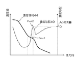
図10は、図8の濃度プロファイル44の濃度勾配49を示す。図10のように濃度情報に異常部位に起因する肩状のピークPeak1が発生した場合、同位置に濃度勾配のピークPeak2が現れる。このPeak2における濃度勾配値Aoを特異量とし、Peak2が現れた位置の位置情報を求めてもよい。
FIG. 10 shows a concentration gradient 49 of the
以上の方法によって求めた特異量の分布、あるいは特異量のピーク値を操作者に提示することで、操作者は効率的に異常部位の有無や位置を把握することが可能となる。 By presenting the distribution of the specific amount obtained by the above method or the peak value of the specific amount to the operator, the operator can efficiently grasp the presence / absence and position of the abnormal site.
(ステップS6)
ステップS6では、特異量マップ作成部11fが、特異量マップを作成する(S6)。ここで特異量マップとは、ステップS5で算出した複数の特異量を医療画像上の位置に対応させて並べ替えたものである。この特異量マップを操作者に表示することによって操作者は異常部位の観察を行う。以下では特異量マップの作成方法と表示方法について説明する。
(Step S6)
In step S6, the singular quantity
図11は、ステップS3で作成した断面像42上に、ステップS6として作成した特異量マップを表示する方法を示す。特異量マップ作成部11fは、図6に示す断面像42上の角度の異なる複数の濃度情報Pa,Pb,Pc,Pdから得られる特異量を再構成することで断面像上の位置に対応する特異量マップ50を作成する。次に、特異量マップ作成部11fは、断面像42の異常部位43位置に、特異量マップ50を合成(重ね合わせ)し、特異量マップ合成断面像51を作成する。断面像42では、異常部位43が臓器壁や臓器内の腔領域とは異なる値で表示されるが、特異量マップ合成断面像51では、異常部位43位置に特異量マップ50が重ねて表示されるため、断面像42に比べてより異常部位43が他の領域と明確に区別して表示される。その結果、操作者は異常部位の観察を効率良く行うことが可能となる。ここで特異量マップは特異量に相当する画素値で表示しても良いし、任意の閾値以上の特異量を持つ画像領域に相当する図形を、例えば操作者の注意を促すような色や模様を付けて表示しても良い。
FIG. 11 shows a method of displaying the singular quantity map created as step S6 on the
また図7で示した断面位置の異なる複数の濃度情報Pe,Pf,Pg,Phから得られる特異量を再構成することでも特異量マップ61を作成することができる。図12はその一例を示したものであり、造影血管40のCPR像60上の特異量マップ表示例を示している。図11と同様に、CPR像60に特異量マップ61を重ねて表示した特異量マップ合成CPR像62を作成することで、操作者は臓器の走行方向に沿って分布する異常部位の観察を効率良く行うことが可能となる。
Further, the singular quantity map 61 can also be created by reconstructing the singular quantities obtained from the plurality of density information Pe, Pf, Pg, Ph having different cross-sectional positions shown in FIG. FIG. 12 shows an example of this, and shows a specific quantity map display example on the
<第二実施形態>
第二実施形態は、特異量マップとして特異量の最大値や、特異量が最大となる対象臓器の位置を操作者に表示する実施形態である。
<Second embodiment>
In the second embodiment, the maximum value of the specific amount and the position of the target organ that maximizes the specific amount are displayed to the operator as the specific amount map.
図13は濃度勾配49を特異量とする場合を例として最大特異量と最大特異量位置の定義を説明したものである。ステップS5で算出した特異量のうち径方向に最大となる特異量を最大特異量、また特異量が最大となる径方向の位置を最大特異量位置と定義する。また、異常部位に対応する濃度値、例えばX線CT装置で撮影された造影血管の画像からソフトプラークに相当する特異量マップを作成する場合には、ソフトプラークが取りうる濃度値(CT値)を濃度値範囲130として設定し、濃度値範囲130内に現れる濃度勾配49のピークPeak2の濃度値を最大特異量として求めても良い。これにより、万一、濃度勾配49に異常部位の存在以外の原因でピークが現れたときにも、そのピークを異常部位として誤って抽出することを防ぐことができる。
FIG. 13 illustrates the definition of the maximum singular amount and the position of the maximum singular amount by taking the concentration gradient 49 as a specific amount as an example. Of the singular amounts calculated in step S5, the singular amount that is maximum in the radial direction is defined as the maximum singular amount, and the radial position where the singular amount is maximum is defined as the maximum singular amount position. In addition, when creating a specific amount map corresponding to a soft plaque from an image of a contrasted blood vessel taken by an X-ray CT apparatus, for example, a density value corresponding to an abnormal site, a density value (CT value) that the soft plaque can take May be set as the
最大特異量と最大特異量位置を用いて特異量マップを作成することで操作者に対して異常部位の位置を効率的に提示することが可能となる。以下にその例を示す。 By creating a specific amount map using the maximum specific amount and the maximum specific amount position, it is possible to efficiently present the position of the abnormal site to the operator. An example is shown below.
図14は、最大特異量位置を用いて特異量が最大となる対象臓器の位置、つまり異常部位の位置を操作者に提示した画面表示例である。 FIG. 14 is a screen display example in which the position of the target organ having the maximum specific amount using the maximum specific amount position, that is, the position of the abnormal site is presented to the operator.
図14(a)は、ソフトプラークを有する血管の走行方向に直交する断面像42を示したものである。この断面像42上で特異量が最も高い位置を操作者に提示するため、最大特異量位置に相当する断面像上の位置を指示する図形140を表示する。この図形140を最大特異量マークと定義する。最大特異量マークには三角形、矢印など操作者が最大特異量の位置を明確に認識できる形状のものが望ましい。図14(a)のように最大特異量マークを表示することによって操作者は異常部位の位置を効率的に把握することが可能となる。また最大特異量マークに最大特異量の値に相当する色付けや模様付けを行って表示することで操作者は異常部位の位置と異常の程度を同時に把握することが可能となる。
FIG. 14A shows a
図14(b)は、ソフトプラークを有する血管の芯線を通る断面像(CPR像)60に最大特異量マーク140を表示する例を示したものである。血管周辺の最大特異量位置に相当する座標に最大特異量マーク140を表示する。このように走行方向を基準とした断面像で診断を行う場合にも最大特異量マークは有効である。 FIG. 14B shows an example in which the maximum singularity mark 140 is displayed on a cross-sectional image (CPR image) 60 passing through the core line of a blood vessel having soft plaque. The maximum singularity mark 140 is displayed at the coordinates corresponding to the position of the maximum singularity around the blood vessel. In this way, the maximum singularity mark is also effective when diagnosis is performed using a cross-sectional image based on the traveling direction.
図14(c)は、心臓の三次元画像70上に最大特異量マークを表示する例を示したものである。冠動脈周辺の最大特異量位置に相当する座標に最大特異量マークを表示する。このように三次元画像上で診断を行う場合にも最大特異量マークは有効である。
FIG. 14C shows an example in which the maximum singularity mark is displayed on the three-
<第三実施形態>
第三実施形態は、複数の最大特異量位置を用いて特異量が最大となる対象臓器の領域、つまり異常部位の位置を操作者に提示する実施形態である。この複数の最大特異量位置から得られる最大特異量の分布を最大特異量マップと定義する。
<Third embodiment>
In the third embodiment, the region of the target organ having the maximum specific amount, that is, the position of the abnormal site is presented to the operator using a plurality of maximum specific amount positions. The distribution of the maximum singular amounts obtained from the plurality of maximum singular amount positions is defined as a maximum singular amount map.
図15(a)は血管の走行方向に直交する断面像42を示したものであるが、血管壁を取り囲む円弧を最大特異量マップ80として表示している。この様な形状のマップを作成するには図5のように芯線を中心とし、角度の異なる複数の濃度情報から最大特異量を算出する。算出した最大特異量を各々の角度に相当する円弧上に並べ替えることで最大特異量マップ80を作成する。この最大特異量マップ80を最大特異量に相当する画素値やパターンとして表示する。このように最大特異量マップを表示することによって操作者は血管の芯線を中心としたどの角度位置に異常部位があるのかを効率的に把握することが可能となる。
FIG. 15A shows a
図15(b)は血管の芯線を通る断面像(CPR像)60を示したものであるが、血管の走行方向に沿った帯状の図形を最大特異量マップ81として表示している。この様な形状のマップを作成するには図7のように血管の走行方向の位置の異なる複数の濃度情報から最大特異量を算出する。算出した走行位置の異なる最大特異量を走行位置に相当する帯状の図形として並べ替えることで最大特異量マップ81を作成する。この最大特異量マップ81を最大特異量に相当する画素値やパターンとして表示する。このように最大特異量マップ81を表示することによって操作者は血管の走行方向のどの位置に異常部位があるのかを効率的に把握することが可能となる。 FIG. 15B shows a cross-sectional image (CPR image) 60 passing through the blood vessel core, and a band-like figure along the blood vessel traveling direction is displayed as the maximum singularity map 81. In order to create a map having such a shape, the maximum singular amount is calculated from a plurality of pieces of density information having different positions in the running direction of blood vessels as shown in FIG. The maximum singular amount map 81 is created by rearranging the calculated maximum singular amounts having different travel positions as a band-like figure corresponding to the travel position. The maximum singularity map 81 is displayed as a pixel value or pattern corresponding to the maximum singularity. By displaying the maximum singularity map 81 in this manner, the operator can efficiently grasp at which position in the traveling direction of the blood vessel there is an abnormal site.
図15(c)は血管の仮想内視鏡画像(CEV)65を示したものであるが、血管壁上に最大特異量マップ82を表示している。この様なマップを作成するには血管の芯線を中心にした角度と走行位置の異なる複数の濃度情報から最大特異量を算出する。算出した最大特異量を仮想内視鏡画像65の血管壁に相当する位置に図形に並べ替えることで最大特異量マップを作成する。この最大特異量マップを最大特異量に相当する画素値やパターンとして表示する。このように最大特異量マップを表示することによって操作者は血管壁のどの位置に異常部位があるのかを効率的に把握することが可能となる。
FIG. 15C shows a virtual endoscopic image (CEV) 65 of a blood vessel, and a maximum singularity map 82 is displayed on the blood vessel wall. In order to create such a map, the maximum singular amount is calculated from a plurality of density information with different angles and travel positions around the blood vessel core line. A maximum singular amount map is created by rearranging the calculated maximum singular amount into a figure at a position corresponding to the blood vessel wall of the virtual
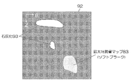
また最大特異量マップは管腔臓器展開画像(特願2005-132401)にも適用可能である。管腔臓器展開画像とは図16のように管腔臓器90の芯線(芯軸)91を基準として、径方向(r方向)の投影値を角度方向(θ方向)と管腔臓器90の走行方向(z方向)に展開して表示する方法である。例えば径方向の最大値を投影値とした場合、血管壁周辺に存在する石灰化などの病変の分布を効率的に把握することが可能である。しかし、この手法ではソフトプラークなど周辺臓器との濃度差が小さい病変の描出は難しい。 The maximum singularity map can also be applied to a luminal organ expansion image (Japanese Patent Application No. 2005-132401). As shown in FIG. 16, the luminal organ development image is based on the core line (core axis) 91 of the luminal organ 90, and the projected value in the radial direction (r direction) is set to the angular direction (θ direction) and the traveling of the luminal organ 90. This is a method of expanding and displaying in the direction (z direction). For example, when the maximum value in the radial direction is used as the projection value, it is possible to efficiently grasp the distribution of lesions such as calcification existing around the blood vessel wall. However, with this method, it is difficult to depict lesions such as soft plaque that have a small concentration difference from surrounding organs.
この問題を解決するには図17に示したように最大特異量マップ83を管腔臓器展開画像92に重ねて表示すればよい。図17は血管壁周辺に存在する石灰化の管腔臓器展開画像92を示したものであり、ソフトプラークに相当する位置に最大特異量マップ83を重ねて表示している。このように管腔臓器展開画像92と最大特異量マップ83とを組み合わせることで、石灰化93とソフトプラークなど性状の異なる異常部位の分布を1枚の画像上で把握することが可能となる。
In order to solve this problem, as shown in FIG. 17, the maximum singular amount map 83 may be displayed so as to overlap the luminal organ expanded
画面表示例として、例えば、CPR像を実際の曲がり形状に沿って表示した例、CPR像を直線状に表示した例、曲線状又は直線上の任意の断面位置の濃度プロファイル、展開画像に特異量マップを重ねて表示した例の何れか一つを表示したり、任意の組み合わせを並べて表示したりしてもよい。その際、濃度プロファイルの肩状の段差に三角形状のマーカを付してもよい。これにより特異量及びその存在位置がわかる。さらに、展開画像上にCPRの断面位置を示す点線を重ねて表示したり、展開画像やCPR像に特異量の具体的な数値を特異量マップとともに表示したりしてもよい。 Examples of screen display include, for example, an example in which a CPR image is displayed along an actual curved shape, an example in which a CPR image is displayed in a straight line, a density profile at an arbitrary cross-sectional position on a curved line or a straight line, and a specific amount in a developed image Any one of the examples in which the maps are displayed in an overlapping manner may be displayed, or any combination may be displayed side by side. At that time, a triangular marker may be attached to the shoulder-shaped step of the density profile. As a result, the specific amount and the position of the specific amount can be known. Furthermore, a dotted line indicating the cross-sectional position of the CPR may be displayed on the expanded image, or a specific numerical value of the specific amount may be displayed on the expanded image or the CPR image together with the specific amount map.
1…画像表示システム、2…医用画像撮影装置、3…画像データベース、4…ネットワーク、5…端末装置、10…画像処理装置、11…中央処理装置(CPU)、12…主メモリ、13…データ記録装置、15…画像表示装置、11a…画像読込部、11b…対象臓器抽出部、11c…断面像作成部、11d…濃度情報算出部、11e特異量算出部、11f…特異量マップ作成部、11g…表示制御部
DESCRIPTION OF
Claims (7)
前記読み込まれた画像データセットから濃度情報を算出する対象となる管腔臓器を抽出する対象臓器抽出手段と、
前記抽出された管腔臓器の芯線に直交した断面像を作成する断面像作成手段と、
前記断面像上において、前記芯線の位置から前記管腔臓器の臓器壁に向かう方向である径方向に沿った画素値の情報である濃度情報を算出する濃度情報算出手段と、
正常濃度情報との差異の大きさを表す指標である特異量が前記径方向に沿って最大となる位置である最大特異量位置を、前記算出された濃度情報の濃度勾配が極大となる径方向の位置を基に算出する特異量算出手段と、
前記最大特異量位置を示す情報を表示する表示手段と、
を備えたことを特徴とする画像表示装置。 Image reading means for reading an image data set of a subject acquired by a medical imaging apparatus;
A target organ extracting means for extracting a luminal organ of interest to calculate the density information from the read image data sets,
And a cross-sectional image generating means for generating a sectional image orthogonal to the core wire of the extracted lumen organ,
On the cross-sectional image , density information calculation means for calculating density information that is pixel value information along a radial direction that is a direction from the position of the core line toward the organ wall of the luminal organ ;
Diameter the largest singular amount regiospecific amount is an index representing the magnitude of the difference between the normal density information is the maximum a position along the radial direction, the concentration gradient of the calculated density information is maximized Singular amount calculation means for calculating based on the position in the direction ;
Display means for displaying information indicating the position of the maximum singular amount ;
An image display device comprising:
ことを特徴とする請求項1に記載の画像表示装置。The image display apparatus according to claim 1.
ことを特徴とする請求項1又は2に記載の画像表示装置。 The display means, the cross-sectional image, against at least one image of the three-dimensional image of the cross-sectional image, or the heart of the subject if the luminal organ is a blood vessel comprising a travel direction of the core wire, in the image Adding and displaying a marker that directs the maximum singular amount position in a one-dimensional manner;
The image display device according to claim 1, wherein the image display device is an image display device.
前記表示手段は、前記断面像上において前記管腔臓器を内包する環状のマップ上の前記最大特異位置を示す角度に対応する位置に、前記最大特異量をマッピングする、
ことを特徴とする請求項1又は2に記載の画像表示装置。 The singular amount calculation means is configured to calculate the maximum singular amount position and the concentration gradient value of the maximum singular amount position from concentration information on a plurality of line segments with different angles around the core line of the luminal organ on the cross-sectional image. The maximum singular amount that is
The display means, in a position corresponding to the angle indicating the maximum specific positions on the ring-shaped map enclosing the hollow organ on the cross-sectional image, mapping the maximum singular quantity,
The image display device according to claim 1, wherein the image display device is an image display device.
前記表示手段は、前記管腔臓器の走行方向に沿った帯状のマップ上の前記最大特異量位置に対応する位置に、前記最大特異量をマッピングする、The display means maps the maximum singular amount to a position corresponding to the position of the maximum singular amount on a band-shaped map along the traveling direction of the luminal organ.
ことを特徴とする請求項1又は2に記載の画像表示装置。The image display device according to claim 1, wherein the image display device is an image display device.
前記表示手段は、前記被検体の画像データセットを基に再構成された前記管腔臓器の仮想内視鏡画像に撮像された前記管腔臓器の臓器壁上の前記最大特異量位置に対応する位置に、前記最大特異量をマッピングする、The display means corresponds to the position of the maximum singular amount on the organ wall of the luminal organ captured in the virtual endoscopic image of the luminal organ reconstructed based on the image data set of the subject. Map the maximum specificity to a position;
ことを特徴とする請求項1又は2に記載の画像表示装置。The image display device according to claim 1, wherein the image display device is an image display device.
前記表示手段は、一方の軸が前記径方向の角度を示し、他方の軸が走行位置を示す管腔臓器展開画像上の前記最大特異位置に相当する位置に、前記最大特異量をマッピングする、The display means maps the maximum singular amount to a position corresponding to the maximum singular position on a luminal organ expansion image in which one axis indicates an angle in the radial direction and the other axis indicates a traveling position.
ことを特徴とする請求項1又は2に記載の画像表示装置。The image display device according to claim 1, wherein the image display device is an image display device.
Priority Applications (1)
| Application Number | Priority Date | Filing Date | Title |
|---|---|---|---|
| JP2006102515A JP4845566B2 (en) | 2006-04-03 | 2006-04-03 | Image display device |
Applications Claiming Priority (1)
| Application Number | Priority Date | Filing Date | Title |
|---|---|---|---|
| JP2006102515A JP4845566B2 (en) | 2006-04-03 | 2006-04-03 | Image display device |
Publications (3)
| Publication Number | Publication Date |
|---|---|
| JP2007275141A JP2007275141A (en) | 2007-10-25 |
| JP2007275141A5 JP2007275141A5 (en) | 2009-05-21 |
| JP4845566B2 true JP4845566B2 (en) | 2011-12-28 |
Family
ID=38677307
Family Applications (1)
| Application Number | Title | Priority Date | Filing Date |
|---|---|---|---|
| JP2006102515A Expired - Fee Related JP4845566B2 (en) | 2006-04-03 | 2006-04-03 | Image display device |
Country Status (1)
| Country | Link |
|---|---|
| JP (1) | JP4845566B2 (en) |
Families Citing this family (11)
| Publication number | Priority date | Publication date | Assignee | Title |
|---|---|---|---|---|
| JP5124852B2 (en) * | 2008-02-19 | 2013-01-23 | 独立行政法人国立高等専門学校機構 | Medical diagnostic imaging support device and diagnostic imaging support program for mesothelioma and asbestos lung |
| JP2010000306A (en) * | 2008-06-23 | 2010-01-07 | Toshiba Corp | Medical image diagnostic apparatus, image processor and program |
| CN101711681B (en) * | 2008-10-07 | 2012-06-27 | 株式会社东芝 | Three-dimensional image processing apparatus |
| US8582844B2 (en) * | 2008-11-13 | 2013-11-12 | Hitachi Medical Corporation | Medical image processing device and method |
| JP2010154944A (en) * | 2008-12-26 | 2010-07-15 | Toshiba Corp | Medical image diagnostic apparatus and fusion image generation method |
| JP5641707B2 (en) * | 2009-04-08 | 2014-12-17 | 株式会社東芝 | X-ray diagnostic equipment |
| JP5436125B2 (en) * | 2009-09-30 | 2014-03-05 | 富士フイルム株式会社 | Diagnosis support apparatus, operation method thereof, and diagnosis support program |
| JP5309109B2 (en) * | 2010-10-18 | 2013-10-09 | 富士フイルム株式会社 | Medical image processing apparatus and method, and program |
| JP5713748B2 (en) * | 2011-03-24 | 2015-05-07 | 株式会社東芝 | Plaque region extraction method and apparatus |
| JP6494927B2 (en) * | 2014-06-05 | 2019-04-03 | キヤノンメディカルシステムズ株式会社 | Medical image processing device |
| JP7393798B2 (en) * | 2018-09-07 | 2023-12-07 | 国立大学法人高知大学 | Diagnosis support device, diagnosis support method, and diagnosis support program |
Family Cites Families (5)
| Publication number | Priority date | Publication date | Assignee | Title |
|---|---|---|---|---|
| US6381350B1 (en) * | 1999-07-02 | 2002-04-30 | The Cleveland Clinic Foundation | Intravascular ultrasonic analysis using active contour method and system |
| JP2002207992A (en) * | 2001-01-12 | 2002-07-26 | Hitachi Ltd | Method and device for processing image |
| JP4336083B2 (en) * | 2002-06-20 | 2009-09-30 | 株式会社日立メディコ | Image diagnosis support apparatus and image diagnosis support method |
| JP2005198708A (en) * | 2004-01-13 | 2005-07-28 | Toshiba Corp | Vasoconstriction rate analyzer and vasoconstriction rate analyzing method |
| EP1735750A2 (en) * | 2004-04-12 | 2006-12-27 | The General Hospital Corporation | Method and apparatus for processing images in a bowel subtraction system |
-
2006
- 2006-04-03 JP JP2006102515A patent/JP4845566B2/en not_active Expired - Fee Related
Also Published As
| Publication number | Publication date |
|---|---|
| JP2007275141A (en) | 2007-10-25 |
Similar Documents
| Publication | Publication Date | Title |
|---|---|---|
| JP4845566B2 (en) | Image display device | |
| JP4676021B2 (en) | Diagnosis support apparatus, diagnosis support program, and diagnosis support method | |
| JP5551957B2 (en) | Projection image generation apparatus, operation method thereof, and projection image generation program | |
| JP5309109B2 (en) | Medical image processing apparatus and method, and program | |
| JP5191989B2 (en) | Medical image display apparatus and medical image display method | |
| JP4958901B2 (en) | Medical image display system and medical image display program | |
| JP4786307B2 (en) | Image processing device | |
| JP5417609B2 (en) | Medical diagnostic imaging equipment | |
| US7860282B2 (en) | Method for supporting an interventional medical operation | |
| US20080275467A1 (en) | Intraoperative guidance for endovascular interventions via three-dimensional path planning, x-ray fluoroscopy, and image overlay | |
| JP5377153B2 (en) | Image processing apparatus, image processing program, and medical diagnostic system | |
| JP5188693B2 (en) | Image processing device | |
| JP2009268693A (en) | X-ray imaging apparatus, image processing apparatus and image processing program | |
| JP2007151965A (en) | Medical image processor, medical image processing program, and medical image processing method | |
| JP5380231B2 (en) | Medical image display apparatus and method, and program | |
| JP2009022411A (en) | Medical image processor | |
| JP2008054763A (en) | Medical image diagnostic apparatus | |
| US20190197762A1 (en) | Cpr image generation apparatus, method, and program | |
| JP2009268735A (en) | Medical image processor | |
| JP2010075549A (en) | Image processor | |
| JP2009165718A (en) | Medical image display | |
| JP3654977B2 (en) | 3D image processing device | |
| JP2009022476A (en) | Image display apparatus, control method of image display apparatus and control program of image display apparatus | |
| JP5100041B2 (en) | Image processing apparatus and image processing program | |
| JP2017113390A (en) | Medical image processing apparatus, control method of the same, and program |
Legal Events
| Date | Code | Title | Description |
|---|---|---|---|
| A521 | Request for written amendment filed |
Free format text: JAPANESE INTERMEDIATE CODE: A523 Effective date: 20090330 |
|
| A621 | Written request for application examination |
Free format text: JAPANESE INTERMEDIATE CODE: A621 Effective date: 20090330 |
|
| RD02 | Notification of acceptance of power of attorney |
Free format text: JAPANESE INTERMEDIATE CODE: A7422 Effective date: 20090721 |
|
| RD04 | Notification of resignation of power of attorney |
Free format text: JAPANESE INTERMEDIATE CODE: A7424 Effective date: 20090727 |
|
| A977 | Report on retrieval |
Free format text: JAPANESE INTERMEDIATE CODE: A971007 Effective date: 20110414 |
|
| A131 | Notification of reasons for refusal |
Free format text: JAPANESE INTERMEDIATE CODE: A131 Effective date: 20110426 |
|
| A521 | Request for written amendment filed |
Free format text: JAPANESE INTERMEDIATE CODE: A523 Effective date: 20110624 |
|
| A131 | Notification of reasons for refusal |
Free format text: JAPANESE INTERMEDIATE CODE: A131 Effective date: 20110719 |
|
| A521 | Request for written amendment filed |
Free format text: JAPANESE INTERMEDIATE CODE: A523 Effective date: 20110913 |
|
| TRDD | Decision of grant or rejection written | ||
| A01 | Written decision to grant a patent or to grant a registration (utility model) |
Free format text: JAPANESE INTERMEDIATE CODE: A01 Effective date: 20111011 |
|
| A01 | Written decision to grant a patent or to grant a registration (utility model) |
Free format text: JAPANESE INTERMEDIATE CODE: A01 |
|
| A61 | First payment of annual fees (during grant procedure) |
Free format text: JAPANESE INTERMEDIATE CODE: A61 Effective date: 20111011 |
|
| FPAY | Renewal fee payment (event date is renewal date of database) |
Free format text: PAYMENT UNTIL: 20141021 Year of fee payment: 3 |
|
| R150 | Certificate of patent or registration of utility model |
Free format text: JAPANESE INTERMEDIATE CODE: R150 |
|
| S111 | Request for change of ownership or part of ownership |
Free format text: JAPANESE INTERMEDIATE CODE: R313111 |
|
| S533 | Written request for registration of change of name |
Free format text: JAPANESE INTERMEDIATE CODE: R313533 |
|
| R350 | Written notification of registration of transfer |
Free format text: JAPANESE INTERMEDIATE CODE: R350 |
|
| LAPS | Cancellation because of no payment of annual fees |


