JP4704570B2 - Radiolabeling kit and binding assay - Google Patents
Radiolabeling kit and binding assay Download PDFInfo
- Publication number
- JP4704570B2 JP4704570B2 JP2000602634A JP2000602634A JP4704570B2 JP 4704570 B2 JP4704570 B2 JP 4704570B2 JP 2000602634 A JP2000602634 A JP 2000602634A JP 2000602634 A JP2000602634 A JP 2000602634A JP 4704570 B2 JP4704570 B2 JP 4704570B2
- Authority
- JP
- Japan
- Prior art keywords
- antibody
- cells
- dtpa
- radioactivity
- radiolabeled
- Prior art date
- Legal status (The legal status is an assumption and is not a legal conclusion. Google has not performed a legal analysis and makes no representation as to the accuracy of the status listed.)
- Expired - Lifetime
Links
Images


Classifications
-
- G—PHYSICS
- G01—MEASURING; TESTING
- G01N—INVESTIGATING OR ANALYSING MATERIALS BY DETERMINING THEIR CHEMICAL OR PHYSICAL PROPERTIES
- G01N33/00—Investigating or analysing materials by specific methods not covered by groups G01N1/00 - G01N31/00
- G01N33/48—Biological material, e.g. blood, urine; Haemocytometers
- G01N33/50—Chemical analysis of biological material, e.g. blood, urine; Testing involving biospecific ligand binding methods; Immunological testing
- G01N33/53—Immunoassay; Biospecific binding assay; Materials therefor
- G01N33/531—Production of immunochemical test materials
- G01N33/532—Production of labelled immunochemicals
- G01N33/534—Production of labelled immunochemicals with radioactive label
-
- A—HUMAN NECESSITIES
- A61—MEDICAL OR VETERINARY SCIENCE; HYGIENE
- A61P—SPECIFIC THERAPEUTIC ACTIVITY OF CHEMICAL COMPOUNDS OR MEDICINAL PREPARATIONS
- A61P35/00—Antineoplastic agents
-
- G—PHYSICS
- G01—MEASURING; TESTING
- G01N—INVESTIGATING OR ANALYSING MATERIALS BY DETERMINING THEIR CHEMICAL OR PHYSICAL PROPERTIES
- G01N33/00—Investigating or analysing materials by specific methods not covered by groups G01N1/00 - G01N31/00
- G01N33/48—Biological material, e.g. blood, urine; Haemocytometers
- G01N33/50—Chemical analysis of biological material, e.g. blood, urine; Testing involving biospecific ligand binding methods; Immunological testing
- G01N33/58—Chemical analysis of biological material, e.g. blood, urine; Testing involving biospecific ligand binding methods; Immunological testing involving labelled substances
- G01N33/60—Chemical analysis of biological material, e.g. blood, urine; Testing involving biospecific ligand binding methods; Immunological testing involving labelled substances involving radioactive labelled substances
Landscapes
- Health & Medical Sciences (AREA)
- Life Sciences & Earth Sciences (AREA)
- Immunology (AREA)
- Engineering & Computer Science (AREA)
- Chemical & Material Sciences (AREA)
- Biomedical Technology (AREA)
- Hematology (AREA)
- Molecular Biology (AREA)
- Urology & Nephrology (AREA)
- General Health & Medical Sciences (AREA)
- Medicinal Chemistry (AREA)
- General Physics & Mathematics (AREA)
- Food Science & Technology (AREA)
- Microbiology (AREA)
- Physics & Mathematics (AREA)
- Analytical Chemistry (AREA)
- Biochemistry (AREA)
- Cell Biology (AREA)
- Biotechnology (AREA)
- Pathology (AREA)
- Pharmacology & Pharmacy (AREA)
- Nuclear Medicine, Radiotherapy & Molecular Imaging (AREA)
- Organic Chemistry (AREA)
- Chemical Kinetics & Catalysis (AREA)
- Animal Behavior & Ethology (AREA)
- Public Health (AREA)
- Veterinary Medicine (AREA)
- General Chemical & Material Sciences (AREA)
- Medicines Containing Antibodies Or Antigens For Use As Internal Diagnostic Agents (AREA)
- Peptides Or Proteins (AREA)
- Medicines That Contain Protein Lipid Enzymes And Other Medicines (AREA)
- Investigating Or Analysing Biological Materials (AREA)
- Steroid Compounds (AREA)
- Packages (AREA)
- Agricultural Chemicals And Associated Chemicals (AREA)
- Investigating Or Analysing Materials By The Use Of Chemical Reactions (AREA)
- Measuring Or Testing Involving Enzymes Or Micro-Organisms (AREA)
- Acyclic And Carbocyclic Compounds In Medicinal Compositions (AREA)
Abstract
Description
【0001】
【発明の背景】
1.発明の分野
本発明は、腫瘍及び腫瘍細胞を治療/画像化するための、治療用抗体の臨床的な効力をテストするための抗体結合アッセイ及び放射標識キット、凍結乾燥された細胞調製物、試薬、並びにプロトコールに関する。具体的には、本発明のキットは、B細胞表面抗原BP35(CD20)を標的とすることによって、B細胞性リンパ腫の治療及び画像化に使用されるであろう放射標識された抗体接合体(antibody conjugate)を作成し、評価するために使用される。
【0002】
2.技術の背景
各文献又は特許出願が明確且つ個別に参考文献として組み込まれる旨の表示がなされていなくても、全ての文献及び特許出願は、等しく参考文献として本明細書に組み込まれる。
【0003】
脊椎動物(例えば、ヒト、類人猿、サルなどを含む霊長類)の免疫系は、脊椎動物の宿主に侵入する外来微生物(「抗原」)を正確且つ特異的に認識し、このような外来微生物に特異的に結合し、このような外来微生物を除去/死滅させるように進化した数多くの臓器及び細胞種からなる。他の細胞種とともに、リンパ球は、免疫系にとって不可欠である。リンパ球は、胸腺、脾臓、及び骨髄(成体)で産生され、ヒト(成人)の循環系に存在する総白血球の約30%を占める。
【0004】
リンパ球には、T細胞とB細胞という2つの主要な亜集団が存在する。T細胞は、細胞媒介性免疫に必要とされるのに対して、B細胞は、抗体の産生(液性免疫)に必要とされる。しかしながら、T細胞とB細胞は、典型的な免疫応答において、相互依存的であると考えられている。抗原提示細胞の表面上に存在する主要組織適合遺伝子複合体(MHC)糖タンパク質に結合した抗原の断片に、T細胞受容体が結合すると、T細胞が活性化される。このような活性化は、基本的に、B細胞を分化させ、抗原に対する抗体(免疫グロブリン)を産生するように刺激する生物的伝達物質(インターロイキン)の放出を引き起こす。
【0005】
宿主内の各B細胞は、その表面上に異なる抗体を発現しているため、あるB細胞は、ある抗原に特異的な抗体を発現するのに対し、別のB細胞は異なる抗原に特異的な抗体を発現するであろう。従って、B細胞は極めて多様であり、この多様性が、免疫系にとって不可欠である。ヒトでは、各B細胞は、多数の抗体分子(すなわち、約107〜108)を産生することができる。このような抗体の産生は、最も典型的には、外来抗原が中和されたときに停止する(又は著しく減少する)。しかしながら、時には、ある種のB細胞の増殖は、衰えないことがある。このような増殖は、「B細胞性リンパ腫」と称されるガンをもたらし得る。
【0006】
T細胞とB細胞は何れも、分化と同定のための「マーカー」として利用できる細胞表面タンパク質を備える。このようなヒトB細胞のマーカーの1つが、ヒトBリンパ球限定分化抗原Bp35であり、「CD20」と称される。CD20は、プレB細胞の成長の初期に発現され、形質細胞の分化まで残存する。具体的には、CD20分子は、細胞周期の開始と分化に必要とされる活性化プロセス中の工程を制御するかもしれず、通常は、新生物性(腫瘍)B細胞上に極めて高いレベルで発現されている。CD20は、定義上、「正常な」B細胞と「悪性の」B細胞、すなわち、その衰えない増殖がB細胞性リンパ腫をもたらし得るB細胞の両者に存在する。このため、CD20表面抗原は、B細胞性リンパ腫の「ターゲッティング」の候補として役立つ可能性を有している。
【0007】
基本的に、このようなターゲッティングは、以下のように一般化することができる。例えば、患者に、B細胞のCD20表面抗原に特異的な抗体を注射する。これらの抗CD20抗体は、(見かけ上)正常なB細胞と悪性B細胞の両者のCD20細胞表面抗原に特異的に結合する。CD20表面抗原に結合した抗CD20抗体は、新生物性B細胞の破壊と喪失をもたらし得る。さらに、腫瘍を破壊する能力を有する化学物質又は放射性標識を抗CD20抗体に接合し、このような物質が、例えば、新生物性B細胞に特異的に「送達される」ようにすることもできる。アプローチにかかわらず、主要な最終目標は、腫瘍を破壊することである。具体的なアプローチは、利用される抗CD20抗体によって決せられるため、CD20抗原にターゲッティングするのに使用できるアプローチは、かなり多様であり得る。
【0008】
例えば、CD20表面抗原のこのようなターゲッティングの試みが報告されている。報告によれば、B細胞性リンパ腫患者への連続的な静脈内注入によって、マウスのモノクローナル抗体115(抗CD20抗体)が投与された。報告によれば、循環する腫瘍細胞を枯渇するには、極めて高レベル(>2グラム)の1F5が必要とされ、結果は、「一過性」のものであると記載されている。Pressら、「Monoclonal Antibody 1F5(Anti−CD20) Serotherapy of Human B−Cell Lymphomas」Blood 69/2:584―591(1987)。
【0009】
このアプローチに伴い得る問題点は、非ヒトモノクローナル抗体(例えば、マウスのモノクローナル抗体)は、典型的には、ヒトのエフェクターとしての機能性を欠いている、すなわち、とりわけ、抗体依存性の細胞毒性又はFc受容体を介した食作用を通じて、補体依存性のヒト標的細胞の溶解を媒介することはできない。さらに、非ヒトモノクローナル抗体は、ヒト宿主によって外来タンパク質として認識され得るので、このような外来抗体の反復投与によって、有害な過敏症反応をもたらす免疫応答の誘導が引き起こされ得る。マウスをベースとするモノクローナル抗体の場合、これは、しばしば、ヒト抗マウス抗体応答、又は「HAMA」応答と称される。さらに、これらの「外来」抗体は、宿主の免疫系によって攻撃され得ることがあり、実際には、それらの標的部位に到達する前に中和されてしまう。
【0010】
リンパ球とリンパ腫細胞は、本来的に、放射線療法に感受性がある。それ故、B細胞性の悪性腫瘍は、幾つかの理由で、放射免疫療法(RIT)の魅力的な目標である。放射標識された抗体の電離放射線を局所的に照射することにより、抗原に結合した抗体の近くに存在する、標的抗原(例えば、CD20)を有する又は有しない細胞を死滅させ得る。透過性放射線、すなわち、β放出体は、巨大な腫瘍又は多血管新生腫瘍において、抗体への接近が制限されるという問題を回避し得る。それ故、必要とされる抗体の総量を減らすことができる。放射性核種は、細胞の修復機構によって、細胞を生存し続けさせることができない程度まで、細胞のDNAを損傷することができる放射性粒子を放出する。それ故、標的細胞が腫瘍であれば、放射性標識は、腫瘍細胞を有益に死滅させる。放射標識された抗体には、当然、患者(すなわち、骨髄移植があり得る)と医療従事者(すなわち、放射能を扱うときに、高度の注意が必要)の両者にとって、用心が必要とされ得る放射性物質の使用が含まれる。
【0011】
それ故、マウスのモノクローナル抗体がB細胞性の疾患を治療する能力を改善するアプローチは、標識又は毒素が、腫瘍部位に局在化するように抗体に放射性標識を接合することであった。毒素(すなわち、ドキソルビシン又はマイトマイシンCのような化学療法剤)も、抗体に接合されてきた。例えば、PCT公開された出願WO92/07466(1992年5月14日公開)を参照。
【0012】
「キメラ」抗体、すなわち、2以上の異なる種(例えば、マウスとヒト)由来の部分を備えた抗体が、「接合」抗体の代替物として開発されてきた。マウス/ヒトのキメラ抗体が作成され、元のマウス抗体の結合特性と、ヒトの定常領域が有するエフェクター機能を発揮することが示されている。例えば、Cabillyら、米国特許第4,816,567号; Shoemakerら、米国特許第4,978,745号; Beaversら、米国特許第4,975,369号; 及び Bossら、米国特許第4,816,397号を参照(これらは、全て、参考文献として本明細書に組み込まれる)。一般的には、これらのキメラ抗体は、既存のマウスハイブリドーマから抽出されたDNAからゲノム遺伝子ライブラリーを調製することによって構築される。Nishimuraら、(1987)Cancer Research 47: 999。続いて、正しい抗体断片の再配列パターンを示している、重鎖と軽鎖の両者からの可変領域遺伝子を該ライブラリーからスクリーニングする。次に、適切な重鎖又は軽鎖ヒト定常領域遺伝子のクローニングされたカセットを含有する発現ベクター中に、クローニングした前記可変領域遺伝子を連結する。続いて、選択した細胞株、通常はマウスの骨髄腫株の中でキメラ遺伝子を発現させる。
【0013】
例えば、Liu, A.Yらの「Production of a Mouse−Human Chimeric Monoclonal Antibody to CD20 with Potent Fc−Dependent Biologic Activity」, J. Immun. 139/10:3521−3526 (1987)は、CD20抗原に対して誘導されたマウス/ヒトキメラ抗体を記載している。PCT公開番号第WO 88/04936号も参照。しかしながら、前記参考文献には、B細胞性疾患の治療にLiuのキメラ抗体を使用できること、その効力、又は実用性に関する情報が全く提供されていない。
【0014】
インビトロ機能アッセイ(例えば、補体依存性溶解(CDC);抗体依存性細胞性細胞傷害(ADCC)など)は、本来的に、ある抗体が、特異的な抗原を発現している標的細胞をインビボで破壊又は除去する能力を予測することができないことに留意しなければならない。例えば、Robinson, R.Dらの「Chimeric mouse−human anti−carcinoma antibodies that mediate different anti tumor cell biological activities」 Hum. Antibod. Hybridomas, 2:84−93 (1991) (chimeric mouse−human antibody having undetectable ADCC activity)を参照。それ故、抗体の潜在的な治療的効力は、インビボでの実験操作によらなければ、真に評価することはできない。
【0015】
この目的で、同時係属出願第08/475,813, 08/475,815及び08/478,967号(その全体が参考文献として本明細書に組み込まれる)は、治療用抗体の投与前に、B細胞性リンパ腫を診断用に「画像化」するための放射標識された抗CD20接合体を開示している。「In2B8」接合体は、二価のキレート物質、すなわち、1−イソチオシアナートベンジル−3−メチル−DTPAと1−メチル−3−イソチオサナートベンジル−DTPAが1:1で混合されたMX−DTPA(ジエチレントリアミン五酢酸)を介して、インジウム[111](111In)に付着された、ヒトCD20抗原に特異的なマウスモノクローナル抗体2B8を有する。インジウム[111]は、γ放射線を放出し、画像化剤としての重要な用途があるので、診断用の放射性核種として選択される。
【0016】
キレート物質及びキレート物質接合体に関する特許が、本分野において公知である。例えば、Gansowの米国特許第4,831,175号は、多置換されたジエチレントリアミン五酢酸キレート及び該キレートを含有するタンパク質接合体、並びにそれらの調製法に関する。Gansowの米国特許第5,099,069、5,246,692、5,286,850、及び5,124,471号も、多置換されたDTPAキレートに関する。これらの特許は、その全体が本明細書に組み込まれる。
【0017】
出願第08/475,813、08/475,815、及び08/478,967号で、キレート生成を促進するために使用された特別な二価のキレート物質は、三価の金属に高い親和性を有しており、腫瘍対非腫瘍比が増加し、骨による摂取が減少し、且つ標的部位、すなわちB細胞性リンパ腫部位における放射性核種のインビボでの保持が大きいので選択された。しかしながら、他の二価のキレート物質も本分野において公知であり、同じく、腫瘍の治療において有益であり得る。
【0018】
出願第08/475,813、08/475,815、及び08/478,967号には、B細胞性リンパ腫及び腫瘍細胞を標的とし、破壊するための放射標識された治療用抗体も開示されている。特に、Y2B8接合体は、同じ二価のキレート物質を介して、イットリウム[90](90Y)に付着された同じ抗ヒトCD20マウスモノクローナル抗体2B8を含んでいる。この放射性核種は、幾つかの理由で、治療用に選択された。64時間という90Yの半減期は、腫瘍による抗体の蓄積を可能とするのに十分長く、例えば131Iとは異なり、その崩壊中にγ線の放射を全く伴わず、細胞の直径の100〜1000の範囲を有する純粋な高エネルギーのβ放出体である。最小限の量の透過性放射線によって、90Y標識された抗体の外来投与が可能となる。さらに、細胞を死滅させるには、標識された抗体の取り込みは必要とされず、電離放射線を局所的に放出すれば、標的抗原を欠いた隣接する腫瘍細胞にとっても致死的なはずである。
【0019】
放射性核種90Yは、同じ二価のキレート物質分子であるMX−DTPAを用いて2B8抗体に付着されたので、Y2B8接合体は、上記したのと同じ利点(例えば、標的部位(腫瘍)での放射性核種の保持の増加)を有している。しかしながら、111Inとは異なり、γ線の放射を伴わないために、画像化する目的で使用することはできない。このため、腫瘍を縮小するために、治療用のキメラ抗体又は90Y標識抗体を投与する前及び/又は後に、腫瘍の位置及び相対的なサイズを決定するために、111Inのような診断用の「画像化」放射性核種を使用することができる。さらに、インジウム標識された抗体は、線量的な評価を行うことも可能にする。
【0020】
意図する抗体の用途に応じて、すなわち、診断用又は治療用の試薬として、他の放射性標識が本分野で公知であり、類似の目的のために使用されている。例えば、臨床診断で使用されている放射性核種には、111Inのほか、131I、125I、123I、99Tc、67Gaが含まれる。標的化された免疫療法で使用し得るために、抗体も様々な放射性核種で標識されてきた(Peirersz et al. (1987) The use of monoclonal antibody conjugates for the diagnosis and treatment of cancer. Immunol. Cell Biol. 65: 111 125)。これらの放射性核種には、90Yのほか、188Reと186Reが含まれ、より少ない程度で、199Auと67Cuが含まれる。I−[131]も、治療目的で使用されている。米国特許第5,460,785号は、このような放射性同位体を列記しており、本明細書に参考文献として組み込まれる。
【0021】
同時係属出願第08/475,813, 08/475,815及び08/478,967号に報告されているように、未標識のキメラ抗CD20抗体のみならず、放射標識されたY2B8接合体の投与も、B細胞リンパ球芽性腫瘍を有するマウスで、顕著な腫瘍な減少をもたらした。さらに、そこで報告されているヒトの臨床試験は、キメラ抗CD20抗体を注入されたリンパ腫患者で、顕著なB細胞の喪失を示した。実際に、キメラ2B8は、最近、Rituxan(登録商標)の名で、本邦で最初にFDAによって承認された抗ガン性モノクローナル抗体であると伝えられた。このように、少なくとも1つのキメラ抗CD20抗体が、B細胞性リンパ腫の治療に効力を有することが実証されている。
【0022】
さらに、米国特許出願第08/475,813号(本明細書に参考文献として組み込まれる)は、Rituxan(登録商標)(インジウムとイットリウムの両者で、又はインジウム若しくはイットリウムの何れかで標識された、キメラ抗CD20マウスモノクローナル抗体)の連続投与を開示している。これらの併用療法(combined therapeutic regimen)に使用される放射標識された抗体はマウスの抗体であるが、最初にキメラ抗CD20抗体で処置することにより、B細胞の集団が十分に消失するため、HAMA応答が減少し、それにより、併用治療及び診断法が促進される。
【0023】
このように、併用免疫療法において、マウスの抗体には、診断試薬としての用途を見出し得るであろう。さらに、米国出願第08/475,813号には、治療的有効量のイットリウム標識された抗CD20抗体に続く、Rituxan(登録商標)の投与は、(a)キメラ抗CD20抗体によって除去されない任意の残存する末梢血B細胞を除去し、(b)リンパ節からB細胞の消失を開始し、又は(c)他の組織からB細胞の消失を開始するのに十分であることが示された。
【0024】
このため、ガンの治療用抗体への放射標識の接合は、このような抗体の潜在的な治療的効力を評価し、治療の進行をモニターするための診断用試薬を作成し、キメラ抗体の最初の腫瘍殺傷能力を増大させるために使用し得るさらなる治療用試薬を考案するために使用し得る価値ある臨床用ツールを提供する。非ホジキンリンパ腫の治療で抗CD20抗体の有効性が証明され、放射能に対するリンパ球の感受性が知られているので、放射性標識で容易に修飾することができ、臨床の現場で患者に直接投与し得るこのような治療用抗体が、キットの形で商業的に入手できるようになれば、非常に有用であろう。
【0025】
抗体を放射標識するための方法及び試薬が多数存在するが、従来技術に欠けているのは、これらの試薬を容易に製造し、放射性標識の顕著な崩壊又は放射性標識による抗体の顕著な破壊が生じる前に患者に投与できるように、臨床の現場にこれらの試薬を置くための便利な手段である。この価値ある技術を商業化するためのこのような便利な手段がないのは、幾つかの公知の標識プロトコールによって実証されたように、取り込み効率が不良であり、放射標識操作後に試薬をカラム精製する必要があることによるものであり得る。このようなキットの開発の遅れは、これまで、その後の精製が不要な効率的に標識された生成物を作るために使用できる純粋な市販の放射性同位体が入手しにくかったことにもよるであろう。あるいは、一般的に、このようなキットが入手できない理由は、おそらく、ヒトの患者のリンパ腫の治療でTituxan(登録商標)が達成した承認又は効力の何れかを達成することができる抗体が実際に存在しなかったことである。
【0026】
例えば、米国特許出願第4,636,380号(参考文献として本明細書に組み込まれる)に記載されているように、科学業界では、放射性薬物を臨床的に利用できるようにするためには、長く、退屈な分離及び精製過程を経なければならないと一般に考えられている。実際に、患者に遊離の放射性標識を注射することは望ましくないであろう。さらなる精製工程が必要であるために、とりわけ、彼ら自身の治療剤を精製するための装置も時間も持たない医師にとっては、臨床の現場で抗体を放射標識する過程は不可能なものとなる。
【0027】
さらに、放射標識されたタンパク質、特に、タンパク質の近くに結合される時間が長くなるほど、抗体に損傷を引き起こす傾向がある90Yのような放射線分解性の同位体で標識されたタンパク質は、本来的に不安定であり得る。続いて、このような放射線分解によって、放射性標識の喪失及び/又は標的抗原への結合の減少によって、治療の効率が不確実になり、変性したタンパク質に対する望ましくない免疫応答が引き起こされるかもしれない。しかも、現場で抗体を標識し、精製するための施設がなければ、臨床医は、既に標識された治療用抗体を注文するか、あるいは、関連施設のように現場とは別の場所で標識し、標識後に患者に投与するために輸送するほかない。このような操作は全て、標識と投与の間に貴重な時間を費やすことによって、治療剤が不安定となる原因となり、臨床の現場での放射性標識キットの有用性を実際に減少させる。
【0028】
他の者は、抗体の分離精製工程がなくて済むほど十分に熟達した抗体放射標識キットの開発を試みたが不成功に終わっている。例えば、Cytogenは、最近、腫瘍関連糖タンパク質TAG−72に対するマウスモノクローナル抗体を放射標識するための市販のキットを発売した。しかしながら、Cytogenの抗体は、さらなる濾過工程によって、その後除去しなければならない粒状物が保存中に発生する傾向があるために、キットに適用するのには、特に適していない。さらに、Cytogenの抗体は、HAMA応答によって、患者に有害な反応を引き起こした。
【0029】
別の者は、精製工程が別個に必要とされない点で、キットフォーマットに適するであろう放射標識プロトコールを開発したと主張している(Richardson et al. (1987) Optimization and batch production of DTPA−labeled antibody kits for routine use in 111In immunoscintography. Nuc. Med. Commun. 8: 347−356; Chinol and Hnatowich (1987) Generator−produced yttrium−[90] for radioimmunotherapy. J. Nucl. Med. 28(9): 1465−1470)。しかしながら、このようなプロトコールは、少なくとも95%の取り込み効率をもたらす、本発明者らが本明細書に開示したプロトコールを用いて達成した取り込みレベルを達成することができなかった。このようなレベルの取り込みは、放射能の取り込みが低いために、患者に遊離の標識が注射されない点で、このようなレベルの取り込みによって、安全性の増加という利点がさらに得られる。
【0030】
本発明のキットに含まれるプロトコールによれば、約半時間、又は標識によっては、僅か5分で実施し得る迅速な標識が可能となる。さらに、本発明のキットプロトコールは、95%以上の標識効率を有するため、さらなる精製が不要である。さらなる精製が不要であることによって、抗体を標識した治療目的のために、放射性標識の半減期と抗体の完全性が保たれる。
【0031】
本出願は、再現性のある、確実、且つ便利な態様で、診断用及び治療用抗体を放射標識し、患者に投与し得る便利なキット及び方法を開示する。本発明のキットによって、抗体を放射標識する工程は、面倒が省け、苦労の要らない標準化された工程となり、患者の治療用プロトコールを著しく容易にする。本キットは、治療剤又は診断剤を標識し、投与するための至適パラメーターが決定されて、商品のコストが減少する点で、従来技術に比べて有利である。本明細書に記載したキットは、特定の標識の至適パラメーターを与えるので、特定の標識に対してデザインされたキットを使用すれば、すなわち、ある標識に対して不適切なキットが使用されたときに起こる分解(cannibalization)が最小になるであろう。このような分解の回避によって、最適な標識効率がもたらされる。さらに、無菌、発熱物質検査、及び標識後の試薬の精製が不要となるので、各キットとともに含まれるプロトコール及び無菌で、発熱物質が存在しない成分によって、工程がさらに扱いやすいものとなる。
【0032】
【発明の概要】
本発明は、患者への投与の前に、診断用又は治療用抗体を放射標識するためのキットであって、少なくとも、(1)キレート物質が結合された抗体を含有するバイアルと、(2)放射標識された抗体を安定化し、投与するための調合用緩衝液(formulation buffer)を含有するバイアルと、(3)抗体を放射標識するための説明書とを備え、前記キットの説明書に従って、前記バイアル成分が十分な純度と活性の放射標識と混合されたときに、前記患者への投与の前に前記標識された抗体のさらなる精製を要しない量と濃度で前記バイアル成分が与えられているキットを包含する。さらに、前記キットの説明書に従い、且つ十分な純度と活性の放射性同位体で標識したときには、このような同位体の取り込みは、95%を超えるレベル、さらには98%以上に達し得る。
【0033】
前記キットに含まれる抗体は、最も好ましくは抗CD20抗体である。前記抗体は、二価のキレート物質に結合された形態で与えられる。好ましくは、前記抗体は、MX−DTPAに接合されるが、フェニル又はベンジルが接合されたDTPA、シクロヘキシル−DTPA、EDTA誘導体、及びDOTAのような他のキレート物質も使用し得る。本発明のキレート物質は、少なくとも二価である任意のキレート物質、すなわち、少なくとも2つの結合部位(金属イオンをキレートする少なくとも1つの部位とタンパク質リガンドに結合するための少なくとも1つの部位)を有する任意のキレート物質であり得る。
【0034】
使用する抗体に応じて、接合抗体は、典型的には、0.5〜30mg/mL、より好ましくは2mg/mLの濃度で与えられる。接合抗体の容量は、放射性標識に応じた至適標識に必要とされる濃度及び量に依存するであろう。しかしながら、前記接合抗体は、全容量が無菌の注射器と無菌手法を用いて反応バイアルに添加されるであろう容量と濃度で与えられるべきである。これによって、再現性が増し、使用が容易になるであろう。本明細書に開示されたキットの試薬は全て無菌で、且つ発熱物質を含まず、抗体検査から直接投与に進む上で、平易且つ迅速なように特別にデザインされている。幾つかの標識を用いれば、標識効率を調べる必要もないかもしれない。
【0035】
前記キットの特に有用な成分は、放射線分解の影響に対して安定化し、前記放射標識された接合抗体を患者に投与するための調合用緩衝液である。調合用緩衝液は、標識された抗体のための希釈剤(diluent)と投与緩衝液の両者の役割を果たす薬学的に許容される担体である。患者に治療用又は診断用抗体を投与するために、任意の薬学的に許容される希釈剤を使用し得るが、本発明の調合用緩衝液は、とりわけ、放射標識された抗体を投与するのに適している。
【0036】
例えば、本発明の調合用緩衝液は、イトッリウムによる放射線分解と、これより程度は低いが、インジウムによる放射線分解を最小化する、ヒト血清アルブミン(HSA)又はアスコルビン酸塩のような放射線保護剤(radioprotectant)を含む。本分野では、他の放射線保護剤、すなわちフリーラジカルスカベンジャー(アスコルビン酸、フェノール、亜硫酸塩、グルタチオン、システイン、ゲンチジン酸、ニコチン酸、アスコルビン酸パルミテート、HOP(:O)H2、グリセロール、ホルムアルデヒド・スルホキシル酸ナトリウム、Na2S205、Na2S203、及びS02など)が公知であり、同様に、本発明の調合用緩衝液に使用することができる。
【0037】
放射線保護剤は、一般的には、放射線分解から抗体を保護するために調合用緩衝液中で用いられているが、反応緩衝液中にも放射線保護剤を含ませることによって、さらなる保護を与えることも可能であり得ることに留意しなければならない。以前は、標識工程を妨害するであろう金属の存在のために、すなわちHSAを用いてこのようなことは行われてこなかった。しかしながら、HSAを反応緩衝液にも含有せしめ得るように、キレート樹脂を用いてHSAを「浄化する」ことが可能かもしれない。アスコルビン酸塩又は他の放射線保護剤も、混在する金属を除去するための処置が必要かもしれない。
【0038】
本発明の調合用緩衝液は、過剰な未接合キレート物質も含む。未接合キレート物質を含有させる目的は、該キレート物質が、患者の中に存在するタンパク質に結合していない全ての放射標識を除去し、放射標識を排出することによって、患者の骨による、「骨に集まる(bone−seeking)」同位体、すなわち90Yの取り込みを減少させることである。例えば、前記キットの抗体をDTPAキレート物質に接合すると、過剰なDTPA又は他の任意のキレート物質を前記調合用緩衝液に含有させ得る。前記調合用緩衝液も、好ましくは、全内容物を反応バイアルに移せる容量で与えられる。正確な容量を測定して移し替える必要がないので、上述のように、これによって、使用の容易さと再現性が上昇する。
【0039】
好ましい調合用緩衝液は、リン酸緩衝化又は生理的食塩水、ヒト血清アルブミン、及びDTPAを含む。前記ヒト血清アルブミンは、好ましくは、約1〜25%(w/v)の濃度であり、より好ましくは、約7.5%(w/v)の濃度である。DTPAの濃度は、好ましくは、約1mMである。アスコルビン酸塩は、ヒト血清アルブミンの代替物として使用することができ、患者の安全を損なわずに、さらに幅広い範囲の濃度を使用し得るが、典型的には、約1〜100mg/mLの濃度で使用される。
【0040】
放射標識キットの抗体は、本発明の方法に従って、二価のキレート物質を介して、選択した放射性同位体で容易に標識される。この点でさらに簡易にするために、本発明のキットは、放射性同位体溶液のpHを調整するための緩衝液を含有するバイアルと、標識を行い、続いて、調合用緩衝液中に最終的な放射標識された抗体を再懸濁するための無菌のガラス反応バイアルを含んでもよい。
【0041】
典型的には、10mLの反応バイアルで十分であるが、5〜20mLを入れることができるバイアルも使用し得る。前記緩衝液は、好ましくは10〜1000mM、最も好ましくは50mMの濃度の低金属の酢酸ナトリウム溶液である。
【0042】
本発明のあるキットは、MX−DTPA接合抗体2B8−MX−DTPAを含む。2B8は、リンパ腫患者に投与したときに、B細胞を消失せしめることが示された抗CD20抗体である。しかしながら、他の抗CD20抗体又はDTPA若しくは他の多価のキレート物質に接合された他の任意の抗体の放射標識に対して、本発明の放射標識キットを最適化し得ることが、当業者には明らかなはずである。本発明の好ましいキットは、少なくとも(1)溶液中の、又は凍結乾燥された(再構成を要する)MX−DTPAが接合された2B8抗体を含有するバイアルと;(2)患者に放射標識された抗体を投与するための調合用緩衝液を含有するバイアルとを含み得る。好ましいキットは、(3)同位体のpHを調整するための緩衝液、及び(4)反応バイアルも含有するであろう。あるいは、より好ましくは、前記緩衝液を反応バイアル中に与えることにより、緩衝液を測定し、移し替える工程が省略され、キット成分の簡易さ、一定性、及び無菌性が増大する。しかしながら、まず同位体バイアルに緩衝液を加えた後、緩衝化された同位体が反応バイアルに移されるという他の態様も想定される。この場合には、前記反応バイアルには、必要な抗体容量を与えることができるであろう。あるいは、同位体/緩衝液バイアルは、すなわち、給者のバイアルに直接添加した抗体接合体を収容するのに十分な大きさを有するように作成してもよい。これによって、反応バイアルが不要となるであろう。
【0043】
上述のように、反応バイアル自体が、必要な容量の接合抗体(すなわち、111In及び90Yの場合、それぞれ1又は1.5mL)を含有するという別の好ましいキットの配置も包含される。前記抗体は、特定の所望の同位体に応じた、適切な放射標識pH(すなわち、111Inの場合pH3〜6、90Yの場合pH3〜5)を与える緩衝液中に与えられ得る。同位体に応じて、異なる緩衝液を使用し得る(すなわち、90Yの場合酢酸ナトリウム、111Inの場合クエン酸ナトリウム)。緩衝液のpHと組成も、標識すべき結合リガンドの性状に応じて、変化し得る(すなわち、標識するペプチドは、3未満のpHの使用を許容し得る)。続いて、原則的には、調合用緩衝液と同様に、前記同位体は直接反応バイアルに移されるであろう。キットの使用が2つの移し替え工程に限定されることによって、再現性と簡便さがさらに増大し、キット成分の操作中に、無菌状態が汚染される機会をさらに減少させるであろう。
【0044】
本発明の放射標識キットは、さらに、放射性同位体のバイアルを備えてもよく、あるいは放射性同位体は、適切な業者から別個に注文してもよい。本発明の好ましい放射性同位体は、HCl中の塩化111インジウム及び塩化90イットリウムであるが、開示した方法は、これらの同位体に限定されるものではない。
【0045】
すなわち、米国特許第4,634,586, 5,460,785、及び5,766,571号(参考文献として本明細書に組み込まれる)に記載されているように、画像化に応用されてきた他の放射性核種が、本分野において公知である。これらの目的又はその他の目的に適した他の放射性同位体、すなわち、抗体を接合するために使用したキレート物質に応じてα放出体を使用し得るが、B細胞腫瘍を画像化するには、インジウム−[111]が特に有利であり、放射治療剤としては、90Yのようなβ放出体が特に有用である。
【0046】
米国特許出願第08/475,813号に開示されている併用治療法の効果が証明されたのであれば、さらなるキットの態様は、放射標識された抗CD20抗体の前又は後に投与すべきキメラ抗体、すなわちRituxan(登録商標)のバイアルを別に含んでもよいであろう。放射標識された抗体の前に、キメラ抗体を投与するときには、マウスの抗CD20抗体の投与に応じて、一般的に起こり得るHAMA応答が著しく減少するかもしれず、それによって、放射標識されたマウスの抗体の治療的な利用性が増大する。さらに、循環するB細胞を除去するためにキメラ抗CD20を用いるときには、その後に、111Inで標識された抗体で得られる診断画像がずっと明瞭となり得る。
【0047】
併用治療法において、診断用の放射標識された抗体と治療用の放射標識された抗体の両者を一緒に使用し得ることも明らかなはずである。この点、診断用抗体は、治療用抗体の前又は後の何れかに、治療前又は後に腫瘍のサイズを可視化するために使用し得る。この場合には、本発明のキットは、使用すべき特定の放射性同位体で放射標識されている抗体の至適pH要求性に従って特別に調合された別の、おそらくは色分けされた緩衝液のバイアルを含み得る。このような系によって、各標識に対して適切な緩衝液が使用されることが保証され、臨床医は、あたかも2つのキットを購入したかのように、2つの抗体を同じ容易さで放射標識することが可能となろう。このようなキットは、事実上、2つの放射標識キットの成分を1つにする。
【0048】
本発明の放射標識キットの成分は、抗体投与の前に、無菌性が容易に維持され、さらなる緩衝液又は溶媒がほとんど不要であるように、適切な濃度とpHで与えられる。しかしながら、試薬の中には、現場で調製し、無菌化し、無菌性について検査し得るものもあることが、当業者には明らかなはずである。このように、消費者の予算と選択に応じて、本発明のキットの変形が想定される。
【0049】
本発明の放射標識キットは、患者に投与するためのキレート物質が接合された抗体(chelator−conjugated antibody)を放射標識する方法で使用され得る。本発明によれば、このような方法は、一般的には、(1)キレート物質が接合された抗体を、放射性同位体を含有する溶液と混合することと;(2)適切な温度で、適切な時間、混合物をインキュベートすることと;(3)前記放射標識された抗体が、さらなる精製なしに患者に直接投与できるように、適切な濃度になるまで、前記標識された抗体を調合用緩衝液で希釈することとを備える。
【0050】
最も好ましくは、前記抗体は、抗CD20抗体であり、とりわけ、抗CD20抗体は2B8であり得る。前記抗体は、任意の適切なキレート物質、すなわち、MX−DTPA、CHX−DTPA、フェニル−又はベンジル−DTPA、DOTA、EDTA誘導体などに接合され得る。MX−DTPAが好ましい。抗体の接合を実施する方法は、本分野において公知である(Kozak et al. (1989); Mirzadeh et al. (1990), Brachbiel et al. (1986))。
【0051】
本発明者らは、キレート物質が接合された抗体を放射標識する前記方法は、キレート物質が接合された抗体と放射標識を混合する前に、放射標識を含有する溶液が、約3.0〜6.0、さらに好ましくは4.2のpHに調整されると最も効率がよいことを見出した。pHを調整するには、低金属の酢酸ナトリウムが特に好ましいが、他の緩衝液を使用してもよい。好ましくは、酢酸ナトリウムの濃度は、約10〜1000mMの濃度であり、より好ましくは50mMの濃度である。
【0052】
放射性同位体が、塩化111インジウムの場合には、単回投与量を調製するために使用すべき111Inの容積量(volume quantity)は、典型的には、約5.5mCiを標識時の放射能濃度で除したものである。患者に投与するためには、111Inの典型的な診断用線量は、約2〜10mCiである。pHを調整するために使用される酢酸ナトリウムの量は、酢酸ナトリウムの濃度と同位体キャリアー溶液に応じて変わり、それ故、極めて幅が広いものであり得る。酢酸ナトリウムの濃度が50mMであるときには、pHを調整するのに必要とされる量は、典型的には、使用される塩化111インジウムの容積量の約1.2倍であるが、さらに多い容量を使用してもよい。HClに対する酢酸ナトリウムの比率が重要なものであり、使用する酢酸ナトリウムの量は緩衝液中のHClの量と濃度に応じて変化するであろうということを理解しなければならない。続いて、約2mg/mLの濃度のキレート物質が接合された抗体約1mLを、放射標識酢酸塩溶液と混合し、この混合物を約30分間、又は抗体の最適な標識を達成するのに十分な時間、インキュベートする。このような時間は、約30秒から約60分の範囲であり得る。続いて、約10mLの総最終用量を達成するのに必要な量の調合用緩衝液を加える。
【0053】
抗体を標識するのに必要な最適な時間は、用いた抗体、特定の放射標識、及び特定の接合体に応じて変化し得る。放射標識に割り当てられる時間の最適化に内在する要因は、標識すべき試薬の抗体に対するキレート物質の比率である。例えば、抗体に対するキレート物質の比率は、治療的に有用なレベルの取り込み(すなわち、放射性同位体に応じて、90〜95%)が得られる程度に十分高くなければならないが、抗体の構造的な完全さ又は免疫的な反応性が損なわれるほど高すぎてもいけない。これには、接合されたキレート物質のレベルをより低く、且つ標識時間をより長くし得る場合がある、ある種の釣り合いをとるための工程が必要である。
【0054】
例えば、2B8とMX−DTPAの場合、抗体に対するキレート物質のモル比が僅かに約1 1/2〜1モル比であれば、90Yでは5分以内で、111Inでは約30分で標識が完了し、所望のレベルの放射能の取り込みが達成され得ることが発見された。所望のレベルの放射能の取り込みが達成されるので、それ故、抗体に対するキレート物質の比率を増加させる必要はなかった。また、抗体の免疫反応性をもたらし得るので、接合されるキレート物質の量を増やすことは有利ではなかった。このようなパラメーターは、本発明に記載されているようなキットをデザインするために、他の抗体についても経験的に決定することができる。
【0055】
放射性同位体が塩化90イットリウムであるときには、単回投与量を調製するのに使用される塩化90イットリウムの容積量は、典型的には、標識時の放射能濃度で約10〜50mCiを除したものにわたり、好ましくは約45mCiを除したものである。pHを調整するために使用される酢酸ナトリウムの量は、酢酸ナトリウムの濃度と同位体キャリアーの濃度に応じて変わり、それ故、非常に幅広いものであり得る。酢酸ナトリウムの濃度が50mMであるときには、90Yは50mMのHCl中に与えられ、pHを調整するのに必要な量は、典型的には、使用される塩化90イットリウムの容積量の約1.2倍である。続いて、約2mg/mLの濃度のキレート物質が接合された抗体約1.5mLを、放射標識酢酸塩溶液と混合し、約5分間、又は抗体の最適な標識を達成するのに十分な時間、インキュベートする。このような時間は、約30秒から約60分の範囲であり得る。約10mLの総最終容量を達成するのに必要な量の調合用緩衝液を加える。
【0056】
好ましくは、本発明の放射標識法は、本明細書に記載された放射標識キットを用いて行われる。しかしながら、好ましい成分と条件は、本発明の方法を実施するための許容されるガイドラインにすぎず、適当な最適化によって、幾分変化させてもよいことが、当業者には明らかなはずである。好ましい条件とは異なるが、該方法の目的をなお達し得る条件は、本発明の範囲にあると考えられる。
【0057】
本発明の放射標識キットは、放射標識後に、抗体の結合親和性を簡便に確認するのに適した試薬とともに供給してもよい。このような場合には、本発明のキットは、患者に該抗体を投与する前に、その標的細胞への放射標識された抗体の結合のパーセントを決定するためにも使用され得る。本発明者らは、開示されている本結合アッセイキットは、精製された抗原が入手できない任意の抗体の親和性を一般的に調べるのに有用であり得ることも見出した。従って、前記結合アッセイの成分は、独立したキットとして販売してもよい。
【0058】
一般的には、結合アッセイ及び放射標識キットは、(1)少なくとも1つの、前記キット中の抗体によって認識される抗原を発現する凍結乾燥された細胞のバイアル;(2)キレート物質が接合された抗体を含有するバイアル;(3)調合用緩衝液を含有するバイアル;及び(4)抗体を放射標識するための説明書を備え、前記放射標識された抗体は、その後の精製を必要とせずに、患者に直接投与され得る。放射標識キットについて上記したように、該キットも、放射性同位体のpHを調整するための緩衝液を含有するバイアル、及び無菌のガラス反応バイアルも備え得る。好ましくは、前記緩衝液は、約10〜1000mMの濃度の低金属の酢酸ナトリウム溶液であり、前記ガラス反応バイアルは、少なくとも5mLの容量を収容する。前記抗体は、好ましくは、抗CD20抗体であり、前記キレート物質は、好ましくは、MX DTPAである。他のキレート物質は、前に記載したとおりに使用し得る。好ましい接合抗体は、2B8−MX−DTPAであるが、キレート物質が接合された任意の抗体を標識し、その親和性を評価してもよい。調合用緩衝液は、上述のように放射線保護剤と未接合のキレート物質を含むリン酸緩衝化された生理的食塩水であり、放射性同位体は含有されていても、いなくてもよいが、好ましくは塩化111インジウム又は塩化90イットリウムである。キレート物質に応じて、他の放射性同位体を使用してもよい。
【0059】
結合アッセイ/放射標識キットと上記放射標識キットとの差異は、抗体の親和性を調べるための基質標的としての役割を果たす抗原陽性細胞が含まれることである。抗原がCD20のときには、好ましいCD20陽性細胞は、SB細胞(ATCC番号CCL120)であるが、任意のCD20陽性細胞を使用し得る。結合アッセイと放射標識キットは、さらに、ネガティブコントロールとして使用するために、抗原陰性細胞を含んでもよい。好ましいCD20陰性細胞は、HSB細胞(ATCC番号CCL120.1)であるが、任意のCD20陰性細胞を使用し得る。
【0060】
もちろん、放射標識と結合アッセイを組み合わせたキットは、併用治療法を実施するために、又は診断像を得る前に、末梢B細胞を除去するために、標識すべき抗体に加えて、キメラ抗CD20抗体のバイアルをさらに備えてもよい。このような別の抗体は、好ましくはRituxan(登録商標)であるが、腫瘍細胞の死滅をもたらすことが示された任意の抗体であり得る。実際に、併用治療法が同じタイプの細胞、すなわち、B細胞性リンパ腫を標的とする役割を果たす限り、2つの異なる種類の抗体、すなわち2つの異なるB細胞抗原に対して誘導された抗体を1つのキットに組み合わせてもよい。
【0061】
キットの前記成分が他の抗体を標識するために使用され得るのと同様に、標的抗原に応じて、抗体の親和性を調べるための他の細胞を調製し得る。しかしながら、抗D20抗体の場合には、本発明の結合アッセイ及び放射標識キットは、標的細胞が凍結乾燥された形態で与えられる点で、商業的な状況にとりわけ適している。これにより、抗体の効力の確認を簡易且つ系統的に進めることが可能になり、組織培養の設備の維持に伴う面倒と支出が不要となる。凍結乾燥された細胞は、一般的には、本発明の方法のバイアル当たり0.5〜500×106細胞のアリコット中に供給される。
【0062】
ある施設では、既に放射標識された抗体を注文するほうが好ましいことがあり得るが、この場合、このような施設には、標的に対する親和性が確実に抗体に保持されるようにするための結合アッセイ試薬が望まれるかもしれない。この場合には、本発明は、その標的細胞への放射標識された前記抗体の結合のパーセントを決定するための結合アッセイキットも提供する。このようなキットは、少なくとも1つの固定され、及び/又は凍結乾燥された抗原陽性細胞のバイアルを含み、必要に応じて、結合アッセイ及び放射標識キットについて上述したような抗原陰性細胞を含有し得る。さらに、このようなキットの変形は、拮抗アッセイを介して、消費者の抗体の結合特異性を確認するための未標識の対照抗体を含み得ることが明らかなはずである。
【0063】
同じく、抗原がCD20の場合には、CD20陽性細胞は好ましくはSB細胞(ATCC番号CCL120)であり、CD20陰性細胞は好ましくはHSB細胞(ATCC番号CCL120.1)であり、0.5〜50×106細胞のアリコット中に凍結乾燥された形態で与えられる。この場合、前記抗体は、好ましくは、111In又は90Yで標識された2B8のMX DTPA接合体である。
【0064】
本明細書に開示されたさらなるキットの態様に関しては、さらなる精製工程が必要でなく、放射標識された抗体を患者に直接投与し得るために、貴重な時間を節約し、抗体の安定性を増加させ得ることが、本発明の放射標識キットと方法の利点の1つであることが強調されなければならない。それ故、臨床医が、投与の前に、放射標識された抗体の結合特異性と親和性を調べ、又は確認することが望ましいかもしれず、抗体の安定性と放射線分解の阻害が具体的な関心事であれば(すなわち、イットリウムを用いた場合のように)、このような試験は、特定の放射性同位体なしに行い得ることが強調される。結合親和性と特異性が試験され得るキットの態様を提供することによって、本発明者らは、本発明の方法又はキットにこのような試験が絶対に必要であることを示唆するものではない。このような抗体の有効性を試験するためのオプションは、純粋に、臨床医が任意に行うものである。
【0065】
本発明者らは、本発明の結合アッセイキット用の固定され、凍結乾燥された細胞を調製するのに使用された方法は、市販のキット用の細胞を調製するのにとりわけ適していることも見出した。細胞は、構造/安定性を向上させるために、凍結乾燥の前に固定してもよい。特に、本発明の細胞は、抗体結合アッセイに使用したときに、高い再現性を示す。
【0066】
とりわけ、本発明は、(1)遠心によって、0.5〜2×10⁶細胞/mLの細胞密度で細胞を採集することと;(2)平行塩類溶液、すなわちHBSS中で少なくとも1回細胞を洗浄し;(3)担体タンパク質と少なくとも1種類の糖を含有する平行塩類溶液を含む凍結乾燥緩衝液中に、沈降した細胞を再懸濁することと;(4)微量遠心管又はガラスバイアル中に再懸濁した細胞のアリコットを分配することと;(5)12〜96時間、より好ましくは24〜72時間、約30〜60ミリトルで凍結乾燥することとを備えた凍結乾燥された細胞の調製法を含む。該方法は、とりわけ、前記細胞がSB細胞(ATCC番号CCL120)又はHSB細胞(ATCC番号CCL120.1)である凍結乾燥された細胞を調製するのに特に適しているが、他の細胞種に対しても同様に適用することができる。
【0067】
好ましくは、前記緩衝液は、一般的には、担体タンパク質として1%(w/v)の濃度のウシ血清アルブミンと10%の濃度のマンニトールを含有する。しかしながら、他の担体タンパク質、すなわちHSA、及び他の糖も使用できると考えられる。細胞は、約1300rpmの速度での遠心によって採集され、塩溶液HBSS(Hankの平行塩溶液)が加えられる。細胞は、一般的には、50×106細胞/mLの濃度で再懸濁される。
【0068】
しかしながら、当業者であれば、上記の条件は、細胞の生存性を著しく損なうことなく、若干の変更を加え得ることが明らかなはずである。さらに上記の条件には、より大量の細胞用の工程、例えば、細胞を凍結乾燥用緩衝液中に交換するための接線(tangential)フローダイアフィルトレーションを最適化するようにデザインされたさらなる操作を補充し得る。
【0069】
本発明の結合アッセイキットは、放射標識された抗体の結合親和性を評価するためのアッセイで使用し得る。このようなアッセイも、本発明の主題である。その標的細胞への放射標識された抗体の結合のパーセントを決定するための結合アッセイは、一般的には、以下の工程:(1)少なくとも1つの放射標識された抗体のアリコットを、少なくとも1つの抗原陽性細胞のアリコットと混合することと;(2)対照として、少なくとも1つの工程(1)のアリコットと同じ放射標識された抗体のアリコットを、工程(1)の抗原陽性細胞のアリコットと同じ容量の少なくとも1つの希釈緩衝液のアリコットと混合することと;(3)遠心によって細胞を沈降させることと;(4)沈降された細胞と対照の上清中の放射能を測定することと;(5)細胞上清中の放射能の量を対照中の放射能の量と比較することとを備える。
【0070】
本発明の放射標識キットが、必要に応じて、塩化111インジウム又は塩化90イットリウムを含有するのと同じく、本発明の結合アッセイは、典型的には、111In又は90Yで標識された抗体を用いて行われる。111Inが放射性標識の場合には、アッセイチューブ中の放射能は、ガンマカウンターを用いて測定される。90Yが標識の場合には、放射能はシンチレーションカウンターを用いて測定されるが、ガンマカウンターを使用することもできる。
【0071】
本発明の結合アッセイについては、好ましい抗体は抗CD20抗体であり、抗CD20抗体は、好ましくは2B8であり、2B8抗体は、本発明の放射標識キットを用いて標識される。しかしながら、特定の抗原を発現している細胞を入手できれば、任意の放射標識された抗体を試験し得る。CD20が抗原であるときには、前記アッセイを行うための好ましい細胞はSB細胞(ATCC番号CCL120)であるが、該アッセイを至適化し、任意の放射標識された抗体と適切な標的細胞を用いて実施してもよい。
【0072】
該アッセイに使用される希釈緩衝液は、抗体の結合を維持しなければならず(すなわち、生理的緩衝液)、おそらくは、細胞への非特異的結合を最小にするために、例えば、担体タンパク質、例えばBSAを含有するであろう。希釈緩衝液を入れたチューブは対照としての役割を果たすが、抗原陰性細胞を用いることによって、さらなる対照を該アッセイに含めてもよい。この場合には、結合アッセイは、さらに以下のステップ:(1)少なくとも1つの放射標識された抗体のアリコットを、少なくとも1つの抗原陰性細胞のアリコットと混合し、インキュベートすることと;(2)遠心によって抗原陰性細胞を沈降させることと;(4)沈降された抗原陰性細胞の上清中の放射能を測定することと;(5)抗原陰性細胞上清中の放射能の量を、抗原陽性細胞上清と対照の上清中の放射能の量と比較することとを備える。このチューブから得られた放射能を、希釈緩衝液の対照と比較することは、抗原陽性細胞への非特異的な結合の量の測定として役立つであろう。CD20が抗原であるときには、CD20陽性細胞はSB細胞であり、CD20陰性細胞は、好ましくはHSB細胞(ATCC番号CCL120.1)である。
【0073】
上述のように、本発明の凍結乾燥された細胞は、放射標識された抗体の結合効力を調べるための単純で、効率的、且つ再現性のある標準を提供する。それ故、本発明の結合アッセイは、好ましくは、本発明の結合アッセイキット中に含まれる凍結乾燥された細胞を用いて行われる。加えて、本発明の放射標識アッセイは、本発明の結合アッセイと組み合わせてもよく、ここで、抗体は、まず、本発明に記載されているように、キレート物質が接合された抗体を標識する前記方法によってまず標識される。最も好ましくは、本発明の結合アッセイは、本明細書に記載された結合アッセイ及び放射標識キットのうちの1つを用いて行われる。
【0074】
抗体の親和性が調べられ、又は確認されるべきであるが、放射標識が付着していない例が幾つか存在し得る。例えば、ある種の状況(すなわち、トラブルシューティング)下では、放射標識する前に、抗体の結合親和性を調べることが有利であり得る。このような場合には、本発明は、(1)同じ抗原に対して特異的な公知の抗体を用いて、ルテニウム標識された対照抗体を調製することと;(2)固定された濃度の標的細胞及び微量のルテニウム標識された抗体とともに、増加する量の被検抗体と増加する量の未標識対照抗体をインキュベートすることと(ここで、各別の濃度の被検抗体と各別の濃度の対照抗体は、それぞれ別々のチューブに入れられている);(3)ORIGEN機器を用いた相対的な電気化学発光(ECL)に基づいて、各反応チューブ中の結合量を測定することと;(4)前記被検抗体の平均親和値を算出することを備えた、標的細胞への被検抗体の親和性を評価するための拮抗結合アッセイも包含する。平均親和性値は、Mullerの方法(J.Immunological Methods (1980) 34:345)又は他の任意の適切な方法を用いて、EC50値と濃度既知のトレース抗体から算出し得る。このアッセイは、放射標識された抗体、又はそれに対する抗原を精製することができず、抗原源として細胞が必要である抗体の親和性を調べるためにも使用し得ることに留意しなければならない。本発明の固定され、凍結乾燥された細胞は、標的細胞として使用し得る。
【0075】
抗CD20抗体の親和性を調べるために本発明の拮抗結合アッセイを行うときには、対照抗体は2B8、又は他の任意の未接合の抗CD20抗体であり得る。前記対照抗体は、キレート物質に接合された抗体であり得る。前記被検抗体も、前記対照抗体のキレート物質接合体であり得る。あるいは、被検抗体は、2B8と比較した、CD20に対するその結合親和性が興味の対象となっている別の抗CD20抗体であり得る。しかしながら、前記アッセイは、適切な標的細胞が利用できる限り、他の特異性を有する抗体を用いた用途に適合させ得る。
【0076】
本発明の拮抗結合アッセイでは、好ましい標的細胞は、CD20陽性細胞であり、より好ましくは、SB細胞(ATCC番号CCL120)、より好ましくは、本発明の方法によって調製された、再懸濁された凍結乾燥SB細胞である。他の方法を用いて凍結乾燥された細胞又は固定された細胞も使用し得る。ルテニウムで標識された抗体は、典型的には、ルテニウム(II)トリス−バイピリジン(bypyridine)キレート物質(TAG−NHS)のN−ヒドロキシスクシンイミドエステルとともに、対照抗体をインキュベートすることを備えた工程によって調製されるが、他の公知の抗体標識法も想定される。標識するためには、対照抗体とTAG−NHSは、好ましくは、約1:15のモル比でインキュベートされる。本発明のこれらの側面及び他の側面は、以下の図面、例、及び本発明の記載を参照することによって、明確に理解されるであろう。
【0077】
【発明の記述】
<定義>
特に明記していなければ、本明細書で使用する全ての技術的及び科学的用語は、当業者によって一般的に理解されるのと同一の意味を有する。本発明を実施又は試行する上で、本明細書に記載された方法と類似又は均等な任意の方法及び物質を使用することができるが、好ましい方法と物質が記載されている。本発明のために、以下の用語を以下に定義する。
【0078】
低金属−放射能の取り込みに影響を与えないレベルまで、金属の混入が減少するように処理された試薬を指す。
【0079】
抗原陽性−本発明の抗体が結合できるように、本発明のある抗体によって認識される抗原を発現していることを意味する。
【0080】
%放射能取り込み−最初に反応に加えられた放射性標識の総量に対する、抗体に接合された放射標識反応からの放射標識の量を指す。
【0081】
%結合−特異的又は非特異的に、標的抗原に結合するサンプルからの抗体の量を指す。
【0082】
%免疫反応性又は結合特異性−特異的に標的抗原に結合する抗体サンプルの量を指す。
【0083】
診断用抗体−腫瘍及び抗原陽性細胞の診断的画像化を行い得る111Iのような放射標識に接合された抗体を指す。
【0084】
治療用抗体−ターゲッティングされた抗原に結合したときに、細胞を死滅させ得る(90Yのような)α又はβ放出体に接合された抗体を指す。
【0085】
<発明の説明>
マウスモノクローナル抗CD20抗体2B8、接合2B8、
111In及び90Y標識2B8の前臨床開発
I. マウスモノクローナル抗CD20抗体2B8、接合2B8−MX−DTPA、111In標識された2B8−MX−DTPA、及びHPLC精製された90Y−MX−DTPAを開発するための物質と方法
A.物質
1.細胞
ヒト細胞SBとHSBは、アメリカン・タイプ・カルチャー・コレクションから取得し、10%のウシ胎児血清を含有するRPMI−1640中で培養した。CD20陽性細胞株は、急性リンパ球芽性白血病患者の末梢血バフィーコートに由来するBリンパ芽球腫細胞株である(1)。抗原陰性細胞株HSBは、新生仔シリアンハムスターに誘導した腫瘍から発育されたTリンパ芽球腫細胞株である(2)。マウスの骨髄腫細胞株SP2/0は、同様に、10%のウシ胎児血清を含有するRPMI−1640中で飼養した。
【0086】
2.抗体
抗CD20抗体B1及びLeu16は、それぞれ、Coulter Immunology及びBecton/Dickinsonから購入した。125I標識したヤギの抗マウスIgGとヤギの抗ヒトIgG抗体は、ICNから取得した。ヤギのF(ab’)2抗マウスIgGは、Cappelから取得した。
【0087】
3.試薬
フロイントの完全及び不完全アジュバントは、Sigma Chemical Companyから購入した。ポリエチレングリコール、HAT濃縮液、及びHT濃縮液は全て、Boehringer Mannheimから取得した。フルオレセインイソチオシアネート(FITC)は、Sigma Chemical Companyから購入した。塩化インジウム−[111]及び塩化90イットリウムは、Amersham又はNEN Dupontから購入した。塩化イットリウム−[89]は、Aldrich Chemical Companyから購入した。他の全ての試薬は、標準的な入手源から取得した。
【0088】
接合及び放射標識プロトコールに使用した試薬には、放射標識工程中に放射性同位体と拮抗し得る重金属イオンの混入を除去するために処理を施した。試薬は、典型的には、溶液をChelex100イオン交換樹脂(BioRad Industries)のカラムに通過させることによって、又は調製した溶液にChelex100を加えるバッチ処理によって処理した。
【0089】
全ての調製及び希釈には、低金属含有水(Milli−Q精製された水か、洗浄用水(WFIr)の何れか)を使用した。無金属溶液を無菌濾過し、無菌のプラスチック容器に集めた。
【0090】
B.方法
1.RIAによる2B8ハイブリドーマ上清の調製とスクリーニング
フロイントの完全アジュバントを含有するPBSに懸濁された2千万のSB細胞で、10匹のBALB/cマウスを免疫した。該細胞を、該動物の複数の部位に、皮下及び腹腔内に投与した。2週間の休止期間後、マウスに対して、フロイントの不完全アジュバント中に乳化したSB細胞の2度目の投与を行った。
【0091】
PBSに懸濁したSB細胞を用いて、毎週のスケジュールで、その後の免疫強化を行った。マウスには、6週〜4月の間、免役した。
【0092】
頸椎脱臼によって一度に2匹の動物を屠殺し、マウスの骨髄腫SP2/0と融合させるためにそれらの脾臓を取り出した。放射標識されたCoulterのB1抗CD20抗体のヒトSB細胞への結合を有効に阻害する免疫後血清の能力に基づいて、動物を選別した。各融合の3日前に、選択した動物に、PBS中の2千万のSB細胞を最後に1度静脈内(尾静脈)注射した。屠殺時には、無菌条件下で脾臓を取り出し、5:1(脾臓細胞:SP2/0)の比率で、脾臓細胞をSP2/0細胞と融合させた。組織培養液中で、融合細胞を洗浄し、HAT選択培地を含有する96ウェルプレート中に分配した。10〜14日後に、CoulterのB1抗体を用いた阻害放射線免疫検定によって、ハイブリドーマをスクリーニングした。
【0093】
抗CD20抗体を分泌するハイブリッドのスクリーニングは、確立された放射線免疫検定法を用いて行った。要約すれば、プロテインAアフィニティークロマトグラフィーによって、CoulterのB1抗CD20抗体を精製した。Iodobeads(Pierce Chemical Co.)の存在下での短時間の酸化後に、製造者の手順に従って、精製した抗体50μgを125Iに結合した。amberlite樹脂上で放射標識された抗体を脱塩し、希釈緩衝液(0.2%のゼラチン、0.02%のアジ化ナトリウム、及び1.0%のBSAを含有するPBS、pH7.4)中に保存した。テストウェルからのハイブリドーマ上清50μL及び50μLの希釈緩衝液中に懸濁された100,000のSB細胞とともに、予めブロックしたフィルターアッセイプレート(ブロッキング緩衝液:10%FBSを含有する希釈緩衝液)の各ウェルに、10ngの放射標識された抗体を加えた。室温で1時間、懸濁液をインキュベートした。V&P Scientificの真空マニフォールド上で、洗浄緩衝液(0.2%のゼラチン及び0.02%のアジ化ナトリウムを含有するPBS、pH7.4)を用いて、プレートを完全に洗浄し、捕捉されたSB細胞を含有するフィルターのボトムをガンマカウンターに移した。HAT培地と標識されたB1抗体のみを含有するウェルをバックグラウンド対照として使用し、SB細胞を含有する同一のウェルをポジティブコントロールとして使用した。阻害対照は、放射標識されたB1と2μg〜2ngにわたる様々な量の未標識のB1抗体を含有していた。
【0094】
2.フローサイトメトリー実験
a.細胞株
2B8ハイブリドーマの培養物から得られた上清を用いて、予備的なフローサイトメトリー実験を行った。1時間、室温で、100μLのハイブリドーマ上清をSB細胞とインキュベートした;続いて、2次抗体(ヤギF(ab’)2抗マウスIgG;Cappel)(1/400希釈して使用)を加え、暗所で1時間インキュベーションを続けた。細胞を5回洗浄した。対照は、自己蛍光が測定される細胞のみ(1次抗体又は2次抗体を含まない)、非特異的結合を測定するだけの2次抗体を有する細胞、CD20集団の対照用の、市販のフルオレセインイソチオシアネート接合B1(B1−FITC)を含有していた。
【0095】
幾つかの実験では、アミノ基のフルオレセインイソチオシアネート(FITC)との反応を介して、精製した2B8抗体にフルオレセインを結合した。簡潔に述べれば、pH9.5の0.1M炭酸ナトリウム緩衝液中で、150〜200μg FITC/mgタンパク質とともに、2B8抗体(1.2mg/mL)をインキュベートした。室温で2時間、該溶液をインキュベートし、Sephadex G−25カラムで、生じた2B8−FITC接合体を精製した。これらの研究に使用したB1及びLeu16のような他の試薬は、Coulter又はBecton Dickinsonから直接、フルオレセイン接合体として購入した。
【0096】
分析すべき細胞を採集し、0.2%のBSAと0.1%のアジ化ナトリウムを含有するPBSで3回洗浄した。90%を超える生存率を要求しながら、トリパンブルー排除によって生存率を測定した。96ウェルU字底プレートのウェル当たり50μLを加えて、細胞の濃度を3百万/mLに調整した。適切なウェルに1次抗体(50μL)を加え、室温で30分〜1時間、混合物をインキュベートした;続いて、200μL/ウェルの、0.2%のBSAと0.02%のアジ化ナトリウムを含有するPBSを用いて、細胞を5回洗浄した。Sorvall遠心装置中で、1時間、1300RPMでプレート中の細胞を遠心し、穏やかにプレートを「はじく」ことによって、上清を除去した。必要であれば、2次抗体を加え、暗所において、室温でさらに30分〜1時間、インキュベートした;その後、上記のようにウェルを洗浄した。最後に、200μLの固定用緩衝液(1%のパラホルムアルデヒドを含有する0.15M塩化ナトリウム、pH7.4)を各サンプルに加え、処理した細胞を、分析のために12×75mmチューブに移した。
【0097】
b.カニクイザルの全血
血漿を除去した後、遠心及びHBSS中での再懸濁により、細胞を2回洗浄した。ウシ胎児血清(2mL)を加え、細胞を再懸濁した。続いて、6、15mLの各円錐状遠心チューブに、100μLの再懸濁した細胞を分配した。蛍光標識されたモノクローナル抗体を以下のように加えた:
チューブ1:マウス抗CD2−FITC(AMAC)、2.5μg/mL、5μg;
チューブ2:ヤギ抗ヒトIGM−FITC(Fisher)、2.5μg/mL、5μg;
チューブ3:ヤギ抗マウスIgG−RPE(Fisher)、2.5μg/mL、5μg;
チューブ4:ヤギ抗ヒトIgM−FITC +ヤギ抗マウスIgG−RPE(absorbed)、2.5μg/mL、5μg;
チューブ5:抗ヒトCD20−FITC(anti−Leu 16、Becton Dickinson)、5μg;
チューブ6:細胞のみ(自己蛍光)
2分間、1500rpmで標識された抗体と細胞を遠心して、細胞と抗体を混合した後、6つのサンプルを全て氷上に置き、30分間インキュベートした。続いて、氷からチューブを取り出し、溶解緩衝液(予め37℃に加温)を加えて、12mLの容量にした。続いて、室温で15分間サンプルをインキュベートし、5分間、4℃、1500rpmで遠心し、上清を除去した。続いて、標識緩衝液(1%のBSAと0.05%のアジ化ナトリウムを含有するPBS)で、細胞沈降物を2回洗浄した。
【0098】
続いて、0.5mL/チューブの固定用緩衝液(1%のパラホルムアルデヒドを含有する0.15M塩化ナトリウム、pH7.4)を加えることによって細胞を固定し、Calibriteビーズによる自動補償とプレキャリブレーションを用いて、Becton DickinsonのFACScan装置上で分析した。FL1モードで、フルオレセインからの緑色蛍光を測定し、FL2モードでフィコエリトリンからの赤色蛍光を測定した。データは、対数型で表した。まず、ドットプロットビットマップ中の前方対直角の光散乱によって、生きたリンパ球の集団を同定した。続いて、他の全ての事象をゲーティングアウトすることによって、全てのリンパ球集団を単離した。その後の蛍光測定は、ゲーティングされた領域内に生じた特異的な事象のみを反映した。
【0099】
高用量の薬理学/毒性学試験については、各カクニイザルに対して、研究前のリンパ球のレベルを決定し、ベースライン値として使用した。T及びB細胞のパーセントとT:B比を算出し、消失の参照として用いた。研究前のB細胞集団は、Leu16と抗ヒトIgM抗体を用いて数えた。
【0100】
2B8をサルに投与した後、CD抗原が2B8で飽和したときに、ヤギ抗ヒトIgM−FITC、抗マウスIgG−RPE、及びこれら2つのマーカーを含有する二重染色集団を用いて、総集団中のB細胞のパーセントを概算した。二重染色集団は、動物の末梢血から全ての2B8が除去されるまで、定量用に使用した。総リンパ球集団中のT細胞のパーセントは、抗CD2−FITCを用いて推定した。データは、各サンプルを用いて3回、10000の事象に対して測定を平均した。続いて、各ケースにおいて、総リンパ球集団内のT及びB細胞亜集団を数えて、表記の各血液サンプルから得られた細胞を評定した。T:B比も調べた。B細胞の消失は、各サルの元のB細胞レベルに対するB細胞の減少のパーセントとして算出した。
【0101】
3.CD20の放射性ヨウ素化及び免疫沈降
125IとIodobeads(Pierce Chemical Co.)による表面ヨウ素化の後に、1億のSB細胞を2つの等しい部分に分けた。上清の放射能レベルがバックグラウンドに戻るまで、遠心によって、細胞を繰り返し洗浄した。2つの標識されたB細胞のサンプルのうちの何れかに、100μgの2B8又はB1(Coulter Immunology)抗体を加えた。抗体とSB細胞を一晩インキュベートした後、結合していない抗体が全て除去されるまで、遠心によって、3回洗浄した。続いて、結合した2B8とB1を含有する細胞沈降物を溶解し、0.1MのTris−HCl、pH8.0中の1% NP−40界面活性剤を添加することによって抽出した後、室温で1時間インキュベートした。微量遠心管の中で、高速度で30分間、抽出物を遠心し、上清を新しいチューブに移した。タンパク質Aセファロース(300μL)を各チューブに加え、遠心によって樹脂を沈降させた。続いて、タンパク質A−セファロースを20回洗浄して、非特異的に結合したヨウ素化されたタンパク質を除去した。ビーズ対上清の放射能比が100の値に達した時点で、SDS PAGEサンプル緩衝液を用いて沈降物を抽出し、沸騰するまで加熱した。冷却後、約15、000cpmの各抽出物を、10%ポリアクリルアミドゲルのウェルに加えた。予め染色された低分子量の標準(BioRad Inc.)を別のウェルに加え、分子量を推測するために使用した。電気泳動によってタンパク質を分離し、ゲルを乾燥させ、−70℃で24時間X線フィルムのシーツに暴露した;続いて、フィルムを展開し、分析した。
【0102】
4.2B8結合のスキャッチャード分析
スキャッチャード分析によって、精製された2B8の見かけの親和性を評価した。Iodobeadsの存在下で、125Iとの反応によって、放射標識された2B8を調製した。遊離のヨウ素を除去した後、放射標識された抗体を、5000ng/ウェルから35ng/ウェルにわたる様々な濃度で、2つずつ、10、000のSB細胞とともにインキュベートした。細胞に結合している抗体の量は、125I標識された2B8の比活性から算出した。結合した抗体のモル濃度に対して結合/遊離抗体の比率をプロットし、X及びY軸切片の比から見かけの親和定数を決定した。
【0103】
5.2B8−MX−DTPAの調製
a.MX−DTPAの入手源
幾つかの前臨床実験では、炭素14標識された1−イソチオシアナートベンジル−3−メチルジエチレントリアミン五酢酸(MX−DTPA)は、国立保健研究所のOtto Gansow博士から乾燥した固体として譲り受け、4℃で遮光しながら乾燥保存した。該キレートのストック溶液はMilli−Q水中に調製し、放射能を評価し、化合物の比活性を用いることによって濃度を決定した。ストック溶液は、一般に2〜5mMであり、ポリプロピレンチューブの中に−70℃で保存した。他の研究では、MX−DTPAはCoulter Immunologyから、水中の二ナトリウム塩として取得し、−70℃で保存した。
【0104】
b.無金属条件の維持
無金属試薬の使用に加えて、全ての試薬の取扱いは、金属の混入の可能性が最小になるように行った。可能なときには、フラスコ、ビーカー、及び目盛り付きのシリンダーのようなポリプロピレンのプラスチック容器を用いた。使用前に、これらをAlconoxで洗浄し、Milli−Q水で徹底的にリンスした。さらに、少量を正確に扱うために、無金属のピペットチップ(BioRad)を用いた。より大量の試薬を扱うためには、無菌のプラスチック血清ピペット(1〜25mLのサイズのものを利用し得る)を用いた。反応は、ネジ蓋付きのポリプロピレン微量遠心管(Sardstedt Industries;1.5mLの容量)又はポリプロピレン円錐チューブ(Costar;15mL及び50μL)の中で、簡便に行った。透析チュービングを扱うときには、予めMilli−Q水でリンスした使い捨ての手術用手袋を着用した。
【0105】
c.抗体の調製
まず、プロテインAとQAEクロマトグラフィーによって、腹水からマウス抗CD20抗体2B8を精製した。後の実験のために、同じ精製過程を用いて、中空繊維のバイオリアクター上清から2B8を精製した。中空繊維由来の抗体は、商業化のために、今では、例2に記載されたCHO由来の抗体に置き換えられた。
【0106】
150mMのNaClを含有する無金属の50mMのビシン−NaOH、pH8.6中に移し、透析又は緩衝液交換の繰り返しを用いることによって、接合のために抗体を調製した。幾つかの研究では、緩衝液の交換は、Centricon 30(Amicon)スピンフィルター(30,000D MWCO)を用いた限外濾過を繰り返し使用して行った。一般的には、フィルターユニットに50〜200μLのタンパク質(10mg/mL)を加え、2mLのビシン緩衝液を加えた。Sorval SS−34ローター中、6000rpmで45分間、4℃でフィルターを遠心した。残留容量は、約50〜100μLであった。この工程は、同じフィルターを用いて2回繰り返した。残留液をポリプロピレンの1.5mLネジ蓋付きチューブに移し、タンパク質をアッセイし、10.0mg/mLになるように希釈して、接合に使用するまで、4℃で保存した。幾つかの実験では、上記と同じプロトコールを用いて、150mMのNaClと0.05%のアジ化ナトリウムを含有する50mMのクエン酸ナトリウム、pH5.5に、タンパク質を移した。
【0107】
d.接合プロトコール
2B8のMX−DTPAとの接合は、室温でポリプロピレンチューブ中において行った。凍結したMX−DTPAのストック溶液は、使用直前に溶かした。典型的には、10mg/mLの抗体50〜200μLを、4:1のキレート対タンパク質のモル比でキレートと反応させた。キレートストック溶液を加え、穏やかに混合することによって反応を開始した。一晩(一般的には、14〜20時間)、室温で接合を進行させた。未反応のキレートは、上記のように、0.05%のアジ化ナトリウムを含有する無金属の通常の生理的食塩水(0.9%w/v)中への透析又は限外濾過の反復によって接合体から除去した。タンパク質濃度を10mg/mLに調整し、放射標識されるまで、ポリプロピレンチューブ中に4℃で保存した。
【0108】
e.キレートの取り込みの測定
キレートの取り込みは、シンチレーションカウンター及び精製された接合体で得られた値と炭素[14]標識されたキレートの比活性の比較によって測定した。Coulter Immunologyから取得した非放射性キレートを使用する後の研究のために、濃度及び比活性が既知の90Yの過剰な放射能キャリアー溶液と前記接合体をインキュベートすることによって、キレートの取り込みを評価した。
【0109】
簡単に述べれば、無金属の0.05NのHCl(ここに、キャリアーなしの90Y(塩化物塩)が加えられた)中に濃度既知の塩化イットリウムのストック溶液を調製した。液体シンチレーションカウンターによって、この溶液のアリコットを分析し、この試薬の正確な比活性を決定した。典型的には2モル/モル抗体で、抗体に付着されると予測されるキレートのモル数の3倍に等しい容量の塩化イットリウム試薬をポリプロピレンチューブに加え、2Mの酢酸ナトリウムを用いて、4.0〜4.5になるようにpHを調整した。続いて、接合抗体を加え、室温で15〜30分、混合物をインキュベートした。1mMの最終濃度になるように20mMのEDTAを加え、2Mの酢酸ナトリウムで溶液のpHが約6になるように溶液のpHを調整することによって反応を停止させた。
【0110】
5分のインキュベーションの後、下記のように、高性能サイズ排除クロマトグラフィーによって、全容量を精製した。溶出されたタンパク質を含有する画分を合わせ、タンパク質濃度を測定し、アリコットの放射能をアッセイした。キレートの取り込みは、塩化90イットリウム調製物の比活性とタンパク質濃度を用いて算出した。
【0111】
f.2B8−MX−DTPAの免疫反応性
ホールセルELISAを用いて、接合された2B8の免疫反応性を評価した。遠心によって、中間対数相のSB細胞を培養から採集し、1×HBSSで2回洗浄した。1〜2×10@細胞を/mLになるように、HBSS中に細胞を希釈し、50、000〜100、000細胞/ウェルで、96ウェルのポリスチレンマイクロタイタープレート中に分取した。40〜45℃で2時間、真空下でプレートを乾燥させ、細胞をプラスチックに固定した。使用するまで、該プレートは、−20℃で乾燥して保存した。アッセイのために、使用直前にプレートを室温まで加温した後、1%のBSAを含有する1×PBS、pH7.2〜7.4でブロックした(2時間)。1×PBS/1%BSA中にアッセイ用サンプルを希釈し、プレートに加え、同じ緩衝液中に系列希釈した。室温で1時間プレートをインキュベートした後、1×PBSでプレートを3回洗浄した。2次抗体(ヤギ抗マウスIgG1特異的HRP接合体)(50μL)をウェルに加え(1×PBS/1%BSA中に1:1500希釈)、室温で1時間インキュベートした。1×PBSでプレートを4回洗浄した後、ABTS基質溶液(0.01%のATBSと0.001%のH2O2を含有する50mMのクエン酸ナトリウム、pH4.5)を加えた。15〜30分のインキュベーションの後、プレートを405nmで読み取った。非特異的結合をモニターするために、アッセイには抗原陰性HSB細胞を含めた。接合体の免疫反応性は、各希釈因子に対して吸光度値をプロットし、同じプレート上で試験したネイティブ抗体(100%の免疫反応性に相当する)を用いて得た値と比較することによって算出した。滴定プロフィールの線形部分上の幾つかの値を比較し、平均値を決定した。
【0112】
g.ネイティブ2B8と2B8−MX−DTPAのインビトロでの安定性
抗体及び接合抗体を12週にわたってこのように評価するために、通常の生理的食塩水又は10mMのグリシン−HCl、pH6.8を含有する通常の生理的食塩水の何れかに、2B8抗体と2B8−MX−DTPAのアリコットを調合した。2組のサンプルを、4℃と30℃の両者でインキュベートし、以下の方法:SDS−PAGE(還元及び非還元の両者)、SB(抗原陽性)又はHSB(抗原陰性)細胞を捕捉物(capture)として使用したホールセル酵素免疫検定による免疫反応性、等電点ゲル電気泳動(pH域、3〜10)を用いて、サンプルを毎週アッセイした。さらに、第4、8、及び12週目に、接合体を90Yで放射標識することによって、接合体の放射標識効率を評価し、SDS−PAGEとオートラジオグラフ分析によって産物を分析した。最後に、別の研究で、4及び30℃で10週間インキュベートした2B8−MX−DTPAのアリコットを111Inで放射標識し、下記のように、BALB/cマウスでの生体分布研究で評価した。
【0113】
h.免疫組織学的研究
アセトンで固定したヒト組織の切片を用いて、IMPATH Laboratoriesにより、ネイティブと接合(2B8−MX−DTPA)抗体の両者による免疫組織学的研究を行った。プロテインAとQセファロースクロマトグラフィーにより、中空繊維バイオリアクター上清から抗体を精製した。臨床グレードの接合体は、上述のプロトコールに従って、Coulter Immunologyから得たMX−DTPAを用いて調製した。
【0114】
i.放射標識された2B8−MX DTPAのインビトロ免疫反応性
幾つかの実験では、未標識の2B8−MX−DTPAに対して使用したホールセルELISAプロトコールを用いた。後の実験では、Lindmoによって記載されたホールセル結合アッセイ(3)の修正バージョンを用いて、111In及び90Y標識された接合体(各々IDEC Pharmaceuticalsか、あるいはMPI Pharmacy Services, Inc.)の免疫反応性を決定した。簡潔に述べれば、濃度が増加する中間対数相の抗原陽性SB細胞又は抗原陰性HSB細胞[希釈緩衝液(1%BSA、0.1%ゼラチン、及び0.02%アジ化ナトリウムを含有するPBS、pH7.4)中、20〜30×106細胞/mL]中に、20〜30×106細胞/mL)を2組ずつのチューブに加えた。希釈用緩衝液により、1〜5ng/mLの最終抗体濃度になるように、放射標識された接合体を希釈し、0.35mLを各チューブに加えた。室温での75〜90分のインキュベーション時間後に、遠心によって細胞を沈降させ、上清を集めた。ガンマ又はシンチレーションカウンターを用いて、上清画分中に残存する放射能を測定した。データは、細胞数/チューブの逆数に対し、加えた総放射能を細胞に付着した放射能によって除した商としてデータをプロットした。y軸切片は、このため、免疫反応性のある割合を表している。
【0115】
j.ヒト血清中の放射標識された2B8−MX−DTPAのインビトロでの安定性
ヒト血清中、37℃で、96時間インキュベートすることによって、111In及び90Y標識された2B8−MX−DTPAのインビトロでの安定性を評価した。接合抗体を調製し、上記のように、111In(「ミックス&シュートプロトコール」)又は90Yで放射標識した。111In及び90Y標識された接合体の比活性は、それぞれ、2.5及び14.6mCi/mgであった;75mg/mLのヒト血清アルブミン(HSA)と1mMのDTPA(イットリウム標識された接合体)を含有する緩衝液又は50mg/mLのHSA(インジウム標識された接合体)を含有する緩衝液中に、放射標識された接合体を懸濁した。正常なヒト血清(非加熱−非活性化)で、放射標識された接合体を1:10に希釈し、無菌のキャップされたチューブの中に、アリコットを無菌的に置いた;続いて、96時間まで、これらのチューブを37℃でインキュベートした。選択した時点で、接合体のサンプルを取り出し、4〜20%のグラジエントゲル中の非還元SDS−PAGEの後にオートラジオグラフィー、及び簡易薄層クロマトグラフィーによって分析した。
【0116】
k.臨床的に調合された 111 In−2B8―MX−DTPAのインビトロでの安定性
2B8−MX−DTPA接合体を111Inで放射標識し、HPLC精製をせずに使用した(「ミックス&シュート」プロトコール)。放射標識された抗体をPBS中に希釈し、50mg/mLの最終濃度になるようにヒト血清アルブミン(HSA)を加えた。調合した放射標識された接合体の比活性は、2.2mCi/mgであった。続いて、調合された接合体を4℃で48時間インキュベートし、4〜20%のグラジエントゲル中の非還元SDS−PAGEを使用した後にオートラジオグラフィー、及び簡易薄層クロマトグラフィーによって、0、24時間、及び48時間の時点でアリコットを分析した。各時点での免疫反応性は、上記セクション1に記載したホールセル懸濁アッセイを用いて評価した。
【0117】
l.臨床的に調合された 90 Y−2B8―MX−DTPAのインビトロでの安定性
2B8−MX−DTPA接合体を90Yで放射標識し、1×PBSを溶出緩衝液として用いたHPLCでのサイズ排除クロマトグラフィーによって精製した。放射標識された接合体の画分をプールし、それぞれ、75mg/mL及び1mMの最終濃度になるように、ヒト血清アルブミンとDTPAを加えた。調合した放射標識された接合体の比活性は、14.6mCi/mgであった。続いて、調合した接合体を4℃で48時間インキュベートし、4〜20%のグラジエントゲル中の非還元SDS−PAGEを使用した後にオートラジオグラフィー、及び簡易薄層クロマトグラフィーによって、0、24時間、及び48時間の時点でアリコットを分析した。各時点での免疫反応性は、上記セクション1に記載したホールセル懸濁アッセイを用いて評価した。
【0118】
2.動物実験
a.2B8を用いた霊長類高用量薬理学/毒性学試験
White Sands Research CenterのGLP規則に従って実施した高用量の薬理学試験で、抗体2B8を評価した(研究番号920111)。
【0119】
成体のMacaca fascicularis(カニクイザル)を用いた;各試験群は、雄1匹と雌1匹からなった。合計7回、48時間おきに抗体を静脈内注射した。試験は、5つのグループ:第1グループ(生理的食塩水);第2グループ(0.6mg/kg);第3グループ(2.5mg/kg);第4グループ(10mg/kg);第5グループ(第0日のみに、10mg/kg)からなった。
【0120】
試験を開始する前に、10匹の動物全てから血液を採取し、試薬のバックグラウンドと最初のB細胞集団を測定するために用いた。以降の血液サンプルは全て、各抗体投与の前に採取した。完全な検死と組織病理学のために、13日目に、第3及び4グループを屠殺した。
【0121】
第0、1、3、7、13、21、37、及び52日に、第1、2、及び5グループの動物から採血した;ヘパリン処理されたチューブに約5mLの全血を吸引した。全血を4℃に保ち、24時間以内に分析した。各動物から得た血液を2000rpmで、5分間遠心し、RIAによる血清2B8レベルのアッセイのために、上清の血漿を取り除いた(具体的なアッセイ法についてはRIAの手順を参照)。PBLとRBCを含有する沈降した物体はFACS分析のために、FCS中に再懸濁した。
【0122】
b.2B8と2B8−MX−DTPAを用いた薬物動態学的研究
カニクイザル中での2B8の平均血清β半減期は、第5グループの動物(上記)を用いて決定した。2.0μg/mLになるように、10mMのホウ酸緩衝液、pH9.6中に、ヤギ抗マウスIgG1(Fisher Scientific)を希釈し、96ウェルのプレートの各ウェルに50μLを加えた。4℃で一晩、又は室温で2時間、該抗体をプレートに結合させた。150μL/ウェルの1%BSA含有PBSにより、30分間、室温で各プレートをブロックした。蒸留水でプレートを洗浄し、まず1:100に希釈した後、段階的に1:2に希釈した各ウェルに、血清又は血漿サンプルを3つずつ加えた。予め採血した血清に、精製された2B8を加え、0.5mg/mLから始まる標準曲線として使用するために希釈した;サンプルを1:100に希釈した後に、他のサンプルと同様に、系列希釈した。1時間室温でプレートをインキュベートし、蒸留水で4回洗浄した。続いて、1:4000の希釈で、2次試薬(ヤギ抗マウスIgG1−HRPO)を加え、室温でさらに1時間インキュベートした。再度、プレートを蒸留水で洗浄し、過酸化水素を含有する、0.1mLのペルオキシダーゼ基質を加えた。反応から20分間、呈色させた;続いて、マイクロプレートELISAリーダーを用いて、405nmの吸光度を測定した。結果は、μg抗体/mL血清でプロットした。
【0123】
さらに、BALB/cマウスで、2B8と2B8−MX−DTPAのβt1/2値を決定した。1×PBS、pH7.4/10%グリセロール中、−70℃で保存した未接合の2B8を融解し、0.5mg/mLに希釈し、無菌濾過した。標準的であるが、炭素[14]標識されたキレートを用いたプロトコールに従って、接合抗体を調製した。キレートの取り込みは、1.5モル/モル抗体であった。正常な生理的食塩水(0.9%)中に、0.5mg/mLになるように、精製された接合体を希釈し、滅菌濾過し、使用するまで、ネイティブの抗体とともに4℃で保存した。
【0124】
6〜8週齢のマウスに、250μg/mLの濃度で、100μLの精製された2B8抗体を注射した。続いて、0〜264時間にわたる様々な時点に、後眼窩穿刺によってマウスから採血し、抗原陽性B細胞株SBを捕捉物として使用したホールセル酵素免疫検定によって、ネイティブ及び接合2B8抗体の存在についてそれらの血清を分析した。得られたデータは、時間に対して2B8又は2B8−MX−DTPAの濃度としてプロットした;これらの結果から、線形回帰プロットが得られ、勾配はβt1/2値を決定するために使用した。
【0125】
c.カニクイザルにおける[89]−Y−2B8−MX−DTPAの薬理学/毒性学試験
イットリウム−[89]を有する2B8−MX−DTPAは、HPLC精製を使用しなかったことを除いて、90Yの挿入について記載したプロトコールを用いて調製した。75mg/mLのHSAと1mMのDTPAを含有する1×PBS中に、非放射性の、金属を有する接合体を調合し、White Sands Research Centerにおいて、GLP研究番号920611で計測した。4つの各グループには、雄1匹と雌1匹のサルが含まれた。動物には、合計7回、48時間おきに、以下の量の薬物:第1グループ(生理的食塩水);第2グループ(0.003mg/kg);第3グループ(0.03mg/kg);第4グループ(0.3mg/kg)を静脈内注射した。試験中、体重と温度、食物と水の消費、排泄、血清の化学、血液学、尿分析、及び身体的検査を測定することによって、動物を評価した。第1〜4グループの動物から、第0、2、7、10、及び14日目に、注射に先立って採血し、FACS分析によって、循環するB細胞のレベルについて血液を分析した。
【0126】
d.放射標識された2B8−MX−DTPAの生体分布
予備的実験では、6〜8週齢のBALB/cマウスでの組織生体分布について、111In標識された2B8−MX−DTPAを評価した。放射標識された接合体は、上記の「ミックス&シュート」プロトコールに従って、臨床グレードの2B8−MX−DTPAを用いて調製した。接合体の比活性は2.3mCi/mgであり、50mg/mLのHSAを含有するPBS、pH7.4の中に接合体を調合した。マウスの腹腔内に、100μLの111In標識された2B8−MX−DTPA(約21μCi)を静脈内注射し、頸椎脱臼によって、0、24、48、及び72時間目に、3匹のマウスからなるグループを屠殺した。屠殺後、尾、心臓、肺、肝臓、腎臓、脾臓、筋肉、及び大腿骨を取り出し、洗浄し、秤量した;血液のサンプルも分析のために採取した。ガンマカウンターによって、各試料に含まれる放射能を測定し、続いて、グラム組織当たりのパーセント投与線量を決定した。各臓器に付着した血液によってもたらされる活性の寄与を差し引くことは試みなかった。
【0127】
別のプロトコールでは、両調製物ともに、2.1mCi/mgの比活性になるように、4℃及び30℃で10週間インキュベートした2B8−MX−DTPAのアリコットを111Inで放射標識した。続いて、これらの接合体は、上記のように、マウスでの生体分布研究で使用した。
【0128】
線量測定のために、2.3mCi/mgの比活性になるように、2B8−MX−DTPAを111Inで放射標識し、約1.1μCiを20匹の各BALB/cマウスに注射した。続いて、それぞれ5匹のマウスからなるグループを、1、24、48、及び72時間目に屠殺し、それらの臓器を取り出し、分析のために調製した。さらに、皮膚、筋肉、及び骨の一部を取り出し、分析のために処理した;尿と糞も集め、24〜72時間の時点で分析した。
【0129】
同様のアプローチを用いて、2B8−MX−DTPAを90Yでも放射標識し、
72時間にわたって、BALB/cマウスにおける、その生物学的な分布を評価した。HPLCサイズ排除クロマトグラフィーによる精製に続いて、5匹のマウスからなる4つのグループそれぞれに、約1μCiの臨床的に調合された接合体(比活性:12.2mCi/mg)を静脈内に注射した;続いて、1、24、48、及び72時間目にグループを屠殺して、上記のように、それらの臓器と組織を分析した。各組織の試料に伴った放射能は、ガンマシンチレーションカウンターで制動放射エネルギーを測定することによって決定した。続いて、グラム組織当たりのパーセント投与線量又は臓器当たりのパーセント投与線量として、活性値を表した。表面の血液を除去するために、臓器及び他の組織は、繰り返しリンスしたが、臓器は潅流しなかった。このように、内部に含まれる血液によってもたらされる活性の寄与は、臓器の活性値から差し引かなかった。
【0130】
e. 111 In標識された2B8−MX−DTPAの腫瘍分布
放射標識された2B8−MX−DTPAの分布を、RamosのB細胞腫瘍を有する胸腺欠損マウスにおいて決定した。6〜8週齢の胸腺欠損マウスに、胸腺欠損マウスでの増殖に予め適合された1.2×10⁷のRamos腫瘍細胞を含有する0.1mLのRPMI−1640を皮下注射(左後ろの脇腹)した。2週以内に腫瘍が発生し、重量は0.07〜1.1gにわたった。マウスの静脈内に100μLの111Inで標識された2B8−MX−DTPA(16.7μCi)を注射し、頸椎脱臼によって、0、24、48、及び72時間に3匹のマウスからなるグループを屠殺した。屠殺後、尾、心臓、肺、肝臓、腎臓、脾臓、筋肉、大腿骨、及び腫瘍を取り出し、洗浄し、秤量した;血液のサンプルも分析のために採取した。ガンマカウンターによって、各試料に伴われる放射能を測定し、グラム組織当たりのパーセント投与線量を決定した。
【0131】
3.線量測定の計算
111In又は90Yで標識された2B8−MX−DTPAの何れかを投与したBALB/cマウスを用いて得られた生体分布データ(表1〜4及び5〜8)を使用して、核医学協会のMedical Internal Radiation Dose (MIRD) Committeeによって正式に承認されたアプローチを用い、70Kgの患者に投与された1.0mCiの線量から吸収される放射線量の概算値を計算した。前記放射標識された接合体の生物学的半減期は、各放射性免疫接合体の生体分布データから決定された臓器値当たりの投与線量から決定した。幾つかの組織、例えば、血液については、放射性接合体の生物学的な崩壊が、これらのコンパートメントから指数的に崩壊する2コンパートメントモデルに従うと仮定した。72時間の生体分布研究を通じて、その活性レベルが概ね一定を保ったその他の組織、例えば肝臓については、生物学的半減期が極めて長いと仮定し、1000時間の値を割り当てた。
【0132】
【表1】

【0133】
【表9−1】
【表9−2】
【表10−1】
【表10−2】
【0137】
【表11−1】
【表11−2】
【表11−3】
【表12−1】
【表12−2】
【表13−1】
【表13−2】
【表14−1】
【表14−2】
【表15−1】
【表15−2】
【表15−3】
【表16−1】
【表16−2】
【表16−3】
【表16−4】
【表17−1】
【表17−2】
【表17−3】
【表17−4】
【表18−1】
【表18−2】
【表18−3】
A.2B8と2B8−MX−DTPAを用いたインビトロ実験
1.抗CD20抗体2B8の作成と特性決定
合計9つの融合によって、放射標識されたCoulterのB1抗体と有効に競合する抗体を産生する3つのハイブリドーマが得られた。それぞれのケースで、ハイブリドーマは、24ウェルのプレート中に増殖された。融合3及び4から単離された最初の2つの抗体のイソタイプを決定し、何れもIgMと同定した。融合5で作られる第3の抗体は2B8と名付けられ、IgG1κイソタイプであることが決定され、持続研究のために選択した。クローン2B8.Hllを増殖させ、液体窒素中で、長期間保存した。クローン2B8.Hll.G3、さらにクローン2B8.クローンHll.G3.G作成するために、クローン2B8.Hllをサブクローニングした。さらなる研究のために、このクローンを増殖させ、プロテインAアフィニティークロマトグラフィーによって抗体を精製した。
【0160】
未標識の2B8、B1、及びLeu16、並びに放射標識されたCoulterのB1を用いた拮抗アッセイによって、同じ濃度のB1とLeu16の何れよりも効率的に、2B8が、CD20へのB1の結合を阻害できることが実証された(図1)。FITC接合2B8、ネイティブB1、及び無関係の抗体UPC−10、及びS−003(それぞれ、IgG2a及び1イソタイプ)を用いた拮抗研究において、同様の結果が得られた(データは示さず)。
【0161】
2B8とB1抗体による細胞のCD20抗原への直接的な結合は、CD20抗原陽性SB細胞とCD20抗原陰性HSB細胞を用いたFACS分析によって比較した。図2に示された結果は、同じような量の抗体であれば、B1より2B8のほうが多くSB細胞に結合したことを示している。SB細胞への有意な結合は、無関係な抗体では全く観察されなかった。HSB細胞を用いると、何れの試薬についても、バックグラウンド蛍光しか観察されなかった。これらの結果は、2B8のCD20抗原との相互作用の特異性を確認し、2B8が、B1に比べて、細胞表面の抗原に対して高い親和性を有するかもしれないということを示唆している。
【0162】
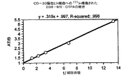
2B8の見かけの親和性を決定するために、125Iで精製された抗体を放射標識し、濃度が増加する標識抗体を抗原陽性SB細胞とともにインキュベートした;細胞に付随する放射能は、1時間のインキュベーション期間後に決定した(図3)。この結果は、2B8抗体が、4.3×10⁻⁹Mの見かけの親和定数でCD20抗原に結合することを示唆している。
【0163】
ヒトの正常な末梢血リンパ球を用いたフローサイトメトリー実験は、2B8がB細胞に特異的で、他の種類のリンパ球(例えば、T細胞、単球、マクロファージ)とは反応しないことが示された。同じヒトリンパ球の集団を用いて、FITC標識された2B8を、B1−FITC及びLeu16−FITCと比較した。表21に示された結果は、2B8が約14%の末梢血リンパ球と反応したのに対して、Leu16では約12%、B1では11%が反応したことを示している。別のBリンパ球マーカー(CD−19)に基づいたリンパ球の集団は、11〜14の間であった。最後に、ヒトの末梢血リンパ球を2B8及びB1又はLeu16の何れかとインキュベートした後、CD19マーカー(Becton/Dickinson)で対比染色を行うと、2B8ではBリンパ球の二重染色集団が9%であったが、B1又はLeu16では10%であった。これらの結果は、これらの試薬が類似していることを確認している。
【0164】
【表21】
【0165】
2.2B8−MX−DTPAの作成と特性決定
2B8−MX−DTPA接合体は、前記抗体を4:1モル過剰のイソチオシアナートベンジル−3−メチルジエチレントリアミン五酢酸と反応させることによって作成された(4)。典型的には、1モルの2B8抗体当たり、1〜2molのMX−DTPAキレートが導入された。図4に記載された結果に示されているように、2B8−MX−DTPA接合体は、ネイティブと接合2B8抗体は何れも、実質的に同一のB1阻害プロフィールを示されているように、見かけ上、ネイティブ2B8と比べて、免疫反応性が全く喪失していなかった;2B8及び2B8−MX−DTPAのIC50値は、それぞれ、約3〜4μg/mLであった。これらの結果は、SB細胞を用いて行ったホールセル放射線免疫検定で、125Iで標識されたB1抗体を用いて得られた。125I標識2B8のSB細胞への結合の阻害剤として、2B8又は2B8−MX−DTPAを用いても、同様の結果が得られた;2B8とそのMX−DTPA接合体は何れも、約3〜4μg/mLの濃度で、SB細胞への125I−2B8の結合を阻害した(データは示さず)。
【0166】
ネイティブの2B8抗体と2B8−MX−DTPA接合体のインビトロでの安定性を評価するために、通常の生理的食塩水又は10mMのグリシン−HCl、pH 6.8を含有する生理的食塩水の中のサンプルを、4℃と30℃で、12週間インキュベートし、以下のアッセイ:ホールセル酵素免疫検定による免疫反応性、還元及び非還元条件下でのSDS−PAGE、及び等電点ゲル電気泳動を用いて、アリコットを毎週アッセイした。免疫反応性アッセイが、何れの温度でインキュベートした抗体サンプルによる抗原認識についても喪失を検出しなかったのに対して(図5)、該抗体に対する等電点電気泳動の範囲(ゼロ週でpH7.30〜8.40)は、4℃で安定であり、6週後には、30℃で、0.2pH単位の減少を示した(表22)。しかしながら、該アッセイの実験誤差の限界なので、この結果は不確実かもしれない。
【0167】
【表22】
【0168】
【表23】
【0169】
最後に、別の実験では、4℃及び30℃で10週間インキュベートされた2B8−MX−DTPAのアリコットを111Inで放射標識し、BALB/cマウスにおけるそれらの組織生体分布を評価した。両インキュベーション温度からの接合体は、類似の生体分布をもたらした(データは示さず)。さらに、得られた結果は、4℃で保存した111In標識された接合体を用いて、BALB/cマウスで得られた生体分布の結果と同様であった(以下参照)。
【0170】
111Inと90Yの両者の放射標識プロトコールは、再現性があることが見出された。典型的には、111Inでは95%を超える放射能の取り込みが、90Yでは90%を超える放射能の取り込みが得られた。111Inと90Y標識された接合体の比活性は、それぞれ、一般的には、2〜3及び10〜15mCi/mg抗体の範囲であった。111Inと90Y放射標識プロトコールの最初の開発においては、HPLCゲル浸透クロマトグラフィーを用いて、複合体を形成していない放射性同位体を、放射標識された2B8−MX−DTPAから除去した。この同位体で(95%を超える)高い放射能の取り込みが得られたので、後の実験では、インジウム標識された接合体のHPLC精製は省略した。
【0171】
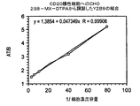
111In及び90Y標識された2B8−MX−DTPAの調製物の免疫反応性は、Lindmoの方法によって分析した(3)。111In標識された2B8−MX−DTPAは、100%免疫反応性を有することが見出され(図6)、90Y標識された接合体は60%が免疫反応性を有していると決定された(データは示さず)。
【0172】
3. 111 In及び 90 Y標識された2B8−MX−DTPAの特性決定
90Y標識された接合体を用いた予備的実験によって、10mCi/mg抗体を超える比活性で、著しい抗体の崩壊と免疫反応性の喪失が起こることが実証された。それ故、放射線分解の影響を最小にするための調合を開発した。数多くの低分子量フリーラジカルスカベンジャーを評価し、有効であることを見出したが、抗体の完全性と免疫反応性を保持するには、高濃度のヒト血清アルブミン(HSA)最も有効であった(図7〜9)。
【0173】
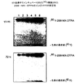
75mg/mLのHSAを含有する1×PBS、pH7.4の中で、90Y標識された抗体を調合した。抗体から失われ得る全ての90Yが確実にキレートされるように、最終濃度が1mMになるように、ジエチレントリアミン五酢酸(DTPA)も加えた。SDS−PAGEとオートラジオグラフィーを用いて、14.6mCi/mgの比活性まで放射標識された2B8−MX−DTPAの崩壊を、0及び48時間に評価した。図8と9は、4℃で48時間にわたってインキュベートしたときに、放射標識された抗体が、有意な崩壊を示さなかったことを示している。簡易薄層クロマトグラフィーを用いた分析によって、48時間のインキュベーション中に、90Yの喪失は2%未満であることが示された(表24)。免疫反応性は、60%でも比較的一定であった(表24)。
【0174】
【表24】
【0175】
【表25】
【0176】
【表26】
1.2B8及び2B8−MX−DTPAを用いた高用量薬理学/毒性学試験
White Sands Research Centerで実施したGLP試験(研究番号920111)では、カニクイザルに、様々な用量の2B8を静脈内投与した。各新しい注射の前に血液サンプルを採取し、リンパ球の集団をフローサイトメトリーで評価するために、血液を処理した(表27)。
【0177】
【表27−1】
【表27−2】
【表27−3】
【0180】
試験期間は14日であり、研究中に、以下のカテゴリー:臨床的な観察、体重、体温、食物と水の摂取、糞の排泄、血清の化学、血液学、尿分析、及び身体的検査について動物を調べた。さらに、0、1、7、及び13日目に、各グループの動物から採血し、血清の抗体(2B8)レベルとT及びB細胞レベルについて血液を分析した。13日目に、第3及び4グループの動物を屠殺し、試料調製後に、光学顕微鏡によって、選択した組織を調べた。調べた組織は、心臓、脾臓、肝臓、腎臓、肺、大脳皮質、脊髄、リンパ節、胃、回腸、結腸、骨格筋、精巣/卵巣、膵臓、及び骨髄であった。
【0181】
循環するT及びB細胞のレベルについて、処置した動物から得た血液を分析すると、第2〜5グループの動物が、13日を通じて、50%を超える循環B細胞の喪失を示した(図19);抗体の投与は、T細胞のレベルに全く影響を与えなかった(データは示さず)。2B8を与えたグループは全て、血漿中に、B細胞の飽和と過剰な抗体を示した(示さず)。10.0mg/Kg用量の2B8を単回投与した第5グループの動物も、他のグループの動物で観察されたものと同じ循環B細胞レベルの減少を示した。
【0182】
第1、2、及び5グループの動物は、52日にわたって調べた(図20)。グループ2の動物1匹(PRO804)とグループ5の動物1匹(PRO716)を除いて、38日までに、B細胞のレベルは、正常の70%を超えるまで回復した。これらの動物における循環B細胞のレベルは、52日後にも、正常レベルの約40%に留まった。
【0183】
本研究に加えて、White Sands Research Centerで実施したGLP試験(研究番号920611)では、カニクイザルで89Y−2B8−MX−DTPAの薬理学的毒性効果を評価した。非放射性の89Yとともに、臨床グレードの接合体をロードした。75mg/mLのヒト血清アルブミンと1mMのDTPAを含有するPBS、pH6.8中に、イットリウムを有する接合体を調合し(臨床用調合)、方法の部に記載したように、静脈内投与した。
【0184】
図21の結果に示されているように、投与した線量にかかわらず、89Y標識された2B8−MX−DTPAは、影響を有するとしても、これらの動物の循環B細胞に対してごく僅かな影響しか有していなかった。さらに、リンパ球が一般に消失(20〜43%)したこと以外は、血清の化学、尿分析、体重と体温を含む、調べた全ての臨床パラメーターには、有意な異常は見られなかった。
【0185】
2.2B8と2B8−MX−DTPAを用いた薬物動態学的研究
上述のように、GLP試験の第5グループの動物には、10.0mg/kgの2B8を単回投与した。データの線形回帰分析は、約4.5日のβt1/2値で、これらのサルの循環系からネイティブの抗体が除去されたことを示唆している。BALB/cマウスを用いた類似の研究では、線形回帰分析(示さず)によって、ネイティブと接合2B8のβt1/2値は8.75日であることが決定された(図22)。これらの結果は、2B8の接合が、BALB/cマウからの2B8のクリアランスに影響を与えないことを示唆している。
【0186】
3.放射標識された2B8−MX−DTPAを用いた生体分布及び腫瘍分布研究
上記の予備的生体分布実験に基づき(セクション2d)、2.3mCi/mgの比活性になるように、接合2B8を111Inで放射標識し、それぞれ20匹のBALB/cマウスに、約1.1μCiを注射し、放射標識された物質の生体分布を決定した。続いて、1、24、48、及び72時間に、5匹のマウスからなる各グループを屠殺し、それらの臓器と皮膚、筋肉、及び骨の一部を取り出し、分析のために処理した。さらに尿と糞を集め、24〜72時間の時点で分析した。血液中の放射能レベルは、1時間目における40.3%の投与線量/グラムから、72時間目には、18.9%に落ちた(表1〜4;図23)。心臓、腎臓、筋肉、及び脾臓の値は、実験を通じて、0.7〜9.8%の範囲に留まった。肺に存在する放射能のレベルは、1時間目の14.2%から72時間目には7.6%に減少した;同様に、グラム当たりの各肝臓の投与線量の値は、10.3%と9.9%であった。これらのデータは、111In−2B8−MX−DTPAの放射能吸収線量の概算値を測定する際に使用した(表19)。
【0187】
12.2mCi/mg抗体の比活性を有する90Y標識された接合体の生体分布をBALB/cマウスで評価した。90%を超える放射能の取り込みが得られ、放射標識された抗体をHPLCによって精製した。72時間にわたって、主要な臓器、並びに皮膚、筋肉、骨、尿、及び糞における放射能の組織蓄積を調べ、パーセント投与線量/グラム組織として表した。表5〜8及び図24に示された結果は、血液に付随する放射能のレベルが、1時間の時点での約39.2%の投与線量/グラムから、72時間後には約15.4%に減少しているのに対して;尾、心臓、腎臓、筋肉、及び脾臓に付随する放射能のレベルは、10.2%以下で、実験を通じて、かなり一定であったことを実証している。重要なことに、骨に含まれる放射能は、1時間の時点での4.4%の投与線量/グラム骨から、72時間での3.2%までの範囲にあった。総合すると、これらの結果は、遊離のイットリウムは、接合体にほとんど不随しておらず、研究の間に、遊離の放射性金属はほとんど放出されなかったことを示唆している。これらのデータは、90Y−2B8−MX−DTPAの放射能吸収線量の推測値を決定する際に用いた(表20)。
【0188】
腫瘍分布研究のために、2B8−MX−DTPAを調製し、2.7mCi/mgの比活性になるように、111Inで標識した。続いて、RamosのB細胞腫瘍を有する12匹の胸腺欠損マウスそれぞれに、100μLの標識された接合体(約24μCi)を注射した。腫瘍の重量は、0.1〜1.0gの範囲であった。注射から0、24、48、及び72時間の時点で、後眼窩穿刺によって50μLの血液を採取し、頸椎脱臼によってマウスを屠殺し、尾、心臓、肺、肝臓、腎臓、脾臓、筋肉、大腿骨、及び腫瘍を取り出した。組織の処理と秤量後に、ガンマカウンターを用いて、各組織試料に含まれる放射能を測定し、値をパーセント投与線量/グラムとして表した。
【0189】
結果(図25)は、111In−2B8−MX−DTPAの腫瘍濃度が、実験の間を通じて着実に増加したことを示している。投与した線量の13%が、72時間後に腫瘍の中に蓄積した。対照的に、血液のレベルは、実験中に、ゼロ時の30%以上から72時間で13%に減少した。(筋肉を除く)他の全ての組織は、実験の終了までに、1.3〜6.0%の投与線量/グラム組織を含有していた;筋肉組織は、約13%の投与線量/グラムを含有していた。
【0190】
C.線量測定
正常なBALB/cマウスでの生体分布研究から得られ、インジウム標識された接合体とイットリウム標識された接合体のそれぞれについて表19及び20に示されている線量測定データの要約は、投与線量のmCi当たりについて比較すると、文献のデータと一致しており(5)、2B8のイットリウム及びインジウム標識接合体がともに、リンパ腫患者で臨床的な効力を安全に示し得ることを示唆している。
【0191】
D.毒性学
1.2B8:単回投与の一般安全性試験
マウスとモルモットに、2B8を単回腹腔内投与し(それぞれ、0.5mL又は5.0mL)、7日間観察した。明白な毒性の徴候は検出されなかった。
【0192】
2.2B8及び2B8−MX−DTPA:ヒトの組織を用いた免疫組織学検査
アセトンで固定した32の異なるヒト組織の群を用いて、マウスのモノクローナル抗体2B8の組織反応性を調べた。抗体2B8は、極めて限局した組織分布のパターンを有する抗CD20抗原と反応し、造血系由来のものを含むリンパ系組織中の細胞のサブセット中にしか観察されない。
【0193】
リンパ節では、胚中心中の増殖性細胞に加えて、成熟した皮質Bリンパ球の集団に免疫反応性が観察された。陽性反応は、末梢血、扁桃腺のB細胞領域、白脾髄、及び胸腺に見出される髄性リンパ球の40〜70%に陽性の反応が見られた陽性の反応は、大腸の固有層(パイヤーの斑;Payer‘s Patches)の濾胞にも見られた。最後に、膀胱、乳房、子宮頸管、食道、肺、耳下腺、前立腺、小腸、及び胃を含む様々な臓器の間質の中の凝集又は散乱したリンパ球細胞も、抗体2B8で陽性であった。
【0194】
様々な臓器の重層上皮及び扁平上皮と同様に、全ての単層上皮細胞は、反応性がないことが見出された。同じく、脳、脊髄、及び末梢神経を含む神経外胚葉細胞でも反応性は見られなかった。骨格及び平滑筋細胞、繊維芽細胞、内皮細胞、及び多形核炎症細胞のような間葉成分も陰性であった。
【0195】
2B8−MX−DTPA接合体の組織反応性は、アセトンで固定された16のヒト組織群を用いて調べた。ネイティブの抗体で以前に実証されたように、2B8−MX−DTPA接合体は、極めて限局された分布パターンを示すCD20抗原を認識し、リンパ球由来の細胞のサブセット上だけに見出された。リンパ節では、免疫反応性は、B細胞の集団で観察された。白脾髄と胸腺の髄質リンパ球には、強い反応性が見られた。膀胱、心臓、大腸、肝臓、肺、及び子宮でも散在するリンパ球に免疫反応性が観察され、これらの組織に存在する炎症細胞の存在によるものであった。ネイティブの抗体で述べたように(上記)、神経外胚葉細胞又は間葉成分にも反応性は観察されなかった。
【0196】
III.考察
同じ表記のクローンによって産生されるマウスのモノクローナル抗CD20抗体2B8は、CD20抗原に特異性があることが知られている抗体との拮抗とスキャッチャード分析によって測定したところによれば、B1抗体で観察された親和性よりも高いかもしれない親和性を、B細胞のCD20抗原に対して示す。さらに、両抗体は、33及び35KDの相対分子量を有する2重線を沈降させたので、免疫沈降データは、2B8によって沈降した抗原は、B1によって沈降されたものと同じ抗原と思われることを示唆している。末梢血リンパ球に対する2B8抗体の特異性の細胞蛍光分析は、該抗体がB細胞と特異的に反応し、T細胞又は他の種類のリンパ球と反応性を示さないことを実証している。最後に、予備的な安定性データは、該抗体が、30℃で12週間、免疫反応性を失わず、安定であることを示唆している。
【0197】
2B8抗体をメチルベンジルジエチレントリアミン五酢酸(MX−DTPA)に接合しても、ネイティブの抗体と比較して、実質的に免疫反応性が減少しないことが観察された。さらに、111In又は90Yの何れかで該接合体を放射標識すると、それぞれ、100%及び60%の免疫反応性を有する標識された接合体が得られた。37℃で96時間、ヒト血清中でインキュベートした111In又は90Y標識された接合体の安定性試験は、該試験中に放射性金属の喪失が無視できることを示し、該接合体を臨床に用いたときに、該接合体が安定であることを示唆している。
【0198】
インジウム標識された2B8−MX−DTPAの調製物を用いた胸腺欠損マウスでの腫瘍分布研究によって、他の組織に異常な蓄積をせずに、実験の間、腫瘍細胞に結合する接合体の量が増加することが実証された。さらに、生体分布から線量測定の測定値が得られた。さらに、生体分布実験から得られた線量測定の推測値は、文献に公表されたデータと一致している。最後に、ネイティブ及び接合抗体を用いたヒト組織の交叉反応実験は、両抗体が極めて限局した組織分布を有する抗原を認識し、造血系由来のものを含むリンパ系組織中の細胞のサブセットのみと反応することを示した。総合すると、これらの結果は、接合が抗体の組織特異性を変化させず、放射標識された接合体がインビボで安定であり、胸腺欠損マウスに実験的に作った腫瘍の表面に存在するCD20抗原を認識することを示唆している。
【0199】
高用量の薬理学/毒性学試験で2B8を使用したときには、該抗体は、本研究中又は本研究後の何れかに調べた何れのパラメーターにも、有意な薬理学的毒性効果を与えなかった。同様に、光学顕微鏡によって調べた様々な組織病理用の標本の分析中にも、異常は見られなかった。驚くべきことに、用いた全ての用量の抗体が、循環するB細胞を顕著に消失させた。しかしながら、循環するB細胞のレベルは、抗体の投与を一旦中止すると、ほぼ正常なレベルに戻った。単回投与のサルのグループ(第5グループ)では、ネイティブの抗体は、約4.5日の見かけのβt1/2値で、循環系から除去された。予想できたように、BALB/cマウスで、この薬物動態学的研究を行うと、2B8抗体は、8.75日のβt1/2値で除去された。このように、これらのデータを総合すると、ネイティブの抗体も、放射標識された接合体に付加して投与すると、幾らかの臨床的な効果を与え得ることが示唆される。
【0200】
我々のデータ全体は、高親和性の2B8抗体とそのMX−DTPA接合体が、限局したパターンのヒト組織の反応性を示すことを示している。さらに、霊長類では、ネイティブの抗体は毒性がなく、B細胞を一過性に喪失させる。しかしながら、一旦抗体を循環系から取り除くと、B細胞のレベルは、かなり迅速に復活する。さらに、インジウム及びイットリウム標識された2B8−MX−DTPA接合体は、ヒトの血清中で長い間インキュベートしている間にも放射性金属は全く喪失せず、インビトロでは安定であるように見えた。最後に、BALB/cマウスでの90Y又は111In標識された2B8−MX−DTPAの生体分布から得られた放射線量の推測値は、mCiの投与線量当たりでは、これらの同位体で放射標識した接合抗共有イディオタイプ抗体を用いたヒトの臨床試験で得られた線量の推測値と一致している。
【0201】
IV.90Y−2B8−MX−DTPAを調製するための「ミックス&シュート」放射標識プロトコールの前臨床開発の概要
A.序論
再発したB細胞リンパ腫を治療するための第I相臨床試験で、90Y標識されたマウスのモノクローナル抗CD20抗体(2B8)を評価した。イットリウム標識された抗体を調製するために使用されていた元のプロトコールは、調合と患者への投与の前に、タンパク質に結合していない放射性同位体を除去するための高性能液体クロマトグラフィー(HPLC)工程を使用していた。残念なことに、この工程は非常に時間がかかり、抗体が放射性同位体に対して保護されていない状態に長い間暴露されることになる。これにより、抗体の放射線分解が増加し、それに伴って、免疫反応性が減少する。さらには、該工程には手がかかるので、放射線製薬局で1日に1投与量を超えて調製することが困難である。工程を簡易化することによって、NIPI Pharmacy Servicesを放射線製薬局として用いる代わりに、臨床現場での実行が促進されるであろう。
【0202】
従って、高い放射能の取り込みと改善された免疫反応性の保持を維持しながら、HPLC精製が不要である、「ミックス&シュート」法と称される改訂された放射標識操作を開発した。マウスでの生体分布研究に加えて、インビトロでの安定性試験も、「ミックス&シュート」法を用いて調製した放射標識抗体が、現在のHPLC工程を用いて作成したものと同等であることが示された。これらの前臨床試験の結果は、臨床試験での使用に適した90Y標識された2B8−MX−DTPAを調製するために、この新しい「ミックス&シュート」プロトコールを使用できることを示している。
【0203】
B.物質と方法
物質
1.細胞
ヒトリンパ球芽性細胞株SB(CD20陽性)とHSB(CD20陰性)は、アメリカン・タイプ・カルチャー・コレクションから取得し、10%のウシ胎児血清を含有し、グルタミンが補充されたRPMI−1640中で培養した。
【0204】
2.抗体
2B8抗体は、以前IND(BB−IND 4850/4851)に記載したプロトコールを用いて、中空繊維のバイオリアクター上清から、製造部が精製した。
【0205】
3.他の試薬
塩化イットリウム−[90]は、Amershamから取得した。他の試薬は全て、以下に示した添付の報告書に記載されている入手源から取得した。放射標識プロトコールに用いた試薬には、放射標識工程中に放射性同位体と競合し得る重金属イオンの混入を除去する処理を行った(方法の部、参照)。試薬は、GMP条件下で、確立されたBatch Production Recordsに従って、IDECの製造部が作成した。
【0206】
方法
1.2B8−D4X−DTPAの調製
水中の二ナトリウム塩として、Coulter Immunologyから臨床グレードのMX−DTPAを取得し、−70℃で保存した。接合体(2B8−MX−DTPA)は、製造部が調製した。これらの研究では、ロットが異なる2つの接合体を用いた;何れも、10mg/mLで、通常の生理的食塩水中に与えられた。滅菌された2mLのポリプロピレン注射器の中に接合体を詰め、2〜8℃で保存した。
【0207】
2.無金属条件の維持
全ての試薬の取扱いは、金属の混入の可能性が最小になるように行った。可能なときには、フラスコ、ビーカー、及び目盛り付きのシリンダーのようなポリプロピレン又はポリスチレンのプラスチック容器を用いた。使用前に、これらをAlconoxで洗浄し、Milli−Q又は洗浄用水(WFIr)で徹底的にリンスした。少量を正確に扱うために、無金属のピペットチップ(BioRad)を用いた。より大量の試薬を扱うためには、無菌のプラスチック血清ピペットを用いた。反応は、ネジ蓋付きの1.8mLのポリプロピレン製微量遠心管の中で、簡便に行った。
【0208】
3.放射能の取り込みの測定
放射能の取り込みは、SOP SP−13−008に従い、簡易薄層クロマトグラフィー(ITLC)を用いて3重に(tripliate)測定した。一般的には、該プロトコールは以下のとおりであった。1mMのDTPA又は5mMのEDTAを含有する1×PBSで、放射標識された接合体を1:20に希釈した後、ITLC SG紙(Gelman Sciences)の1×5cm片の一端から1.5cmに1μLを滴下した。メタノール:水(1:1;v/v)中の10%の酢酸アンモニウムを用いて紙を展開した。前記片を乾燥させ、横半分に切断し、シンチレーションカウンターによって、各部分に含まれる放射能を測定した。各片の下半分に含まれる放射能(タンパク質に含まれる放射能)は、上半分と下半分の両者の値を加えることによって求めた総放射能のパーセントとして表した。
【0209】
4.イットリウム−[90]−標識された2B8−MX−DTPAのための「ミックス&シュート」プロトコール
0.04MのHCl中のAmereshamから購入したキャリアーなしの塩化90イットリウムで抗体を放射標識した。放射性同位体のアリコット(10〜20mCi/mg抗体)をポリプロピレンチューブに移し、0.02×容量の無金属の2M酢酸ナトリウムを加えて、溶液のpHを3.6に調節した。すぐに、2B8−NaDTPA(0.3mg;10.0mg/mL、通常の生理的食塩水中)を加え、溶液を穏やかに混合した。溶液のpHが3.8〜4.1であることを確認するためにpH紙でチェックし、5分間インキュベートした。75mg/mLのヒト血清アルブミン(HSA)と1mMのジエチレントリアミン五酢酸(DTPA)を加えた1×PBSを含有する別のポリプロピレンチューブに反応混合物を移し、穏やかに混合することによって反応を停止させた。放射標識された抗体を2〜8℃で保存した。
【0210】
放射標識された接合体の適切なアリコットの放射能を測定することによって、比活性を決定した。この値を、280nmの吸光度によって測定したカウンターの効率(接合体のタンパク質濃度に関連する)について補正し、mCi/mgタンパク質として表した。
【0211】
5.イットリウム−[90]−2B8−MX−DTPAのインビトロでの免疫反応性
Lindmoによって記載されたホールセル結合アッセイの改良版に基づいたSOP#SP13−009を用いて、90Y標識された接合体の免疫反応性を測定した。濃度が増加する対数相のCD20陽性SB細胞又はCD20陰性HSB細胞を、1.5mLのポリプロピレンチューブ2組に加えた;細胞の最終容量は0.40mL。放射標識された接合体を、1〜2.5ng/mLの最終抗体濃度になるように希釈し、0.35mLを各チューブに加えた。90分インキュベートした後、遠心によって細胞を沈降させ、上清を集めた。上清画分に残存する放射能をシンチレーションカウンターで測定した。データは、細胞数/チューブの逆数に対し、加えた総放射能を細胞に付着した放射能によって除した商として、データをプロットした。y軸切片は、免疫反応性のある割合を表している。
【0212】
6.臨床的に調合されたイットリウム−[90]−2B8―MX−DTPAのインビトロでの安定性
2B8−MX−DTPA接合体を90Yで放射標識し、上記の「ミックス&シュート」プロトコールで記載したように調合した。2つのロットの放射標識接合体を調製した;一方のロットは、放射能の取り込みの安定性を評価するために使用し、他方のロットは、免疫反応性の保持を評価するために使用した。4℃で48時間、調合した接合体をインキュベートし、非還元SDS−PAGEとオートラジオグラフィーを用いて、0、24時間、及び48時間の時点でアリコットを分析した。各時点での免疫反応性は、上記アッセイを用いて評価した。
【0213】
7.ヒト血清中でのイットリウム−[90]−2B8―NTX−DTPAのインビトロでの安定性
ヒト血清中、37℃で72時間までインキュベートすることによって、90Y標識された2B8−MX−DTPAの安定性を評価した。上記のように、イットリウム−[90]で接合抗体を放射標識し、上記のように調合した。正常なヒト血清(非加熱−非活性化)で、放射標識された接合体を1:10に希釈し、分取した試料をプラスチックチューブ中で、37℃でインキュベートした。選択した時点で、サンプルを取り出し、非還元SDS−PAGEとオートラジオグラフィーによって分析した。
【0214】
8.イットリウム−[90]−2B8−MX−DTPAの生体分布
8〜10週齢のBALB/cマウスでの、イットリウム−[90]標識された2B8−MX−DTPAの組織生体分布について調べた。上述のように、放射標識された接合体を調製し、調合した。5μCiの90Y標識された2B8−MX−DTPAをマウスに静脈内注射し、5匹のマウスからなるグループを1、24、48、及び72時間に屠殺した。屠殺後、尾、心臓、肺、肝臓、腎臓、脾臓、筋肉、及び大腿骨を取り出し、洗浄し、秤量した;血液と皮膚のサンプルも分析のために採取した。ガンマカウンターを用いた制動放射を測定することによって、各組織試料に含まれる放射能を測定し、グラム組織当たりのパーセント投与線量と臓器当たりのパーセント投与線量を決定した。
【0215】
9.線量測定
90Y標識された2B8−MX−DTPAを投与したマウスを用いて取得した生体分布データを使用して、70Kgの患者に投与した1.0mCiの線量から吸収される放射線量の推測値を計算した。推測値は、核医学協会のMedical Internal Radiation Dose (MIRD) Committeeによって採用された方法に従って求めた。これらの計算は、カリフォルニア州92161、La Jolla、VA医療センター、核医学施設、Mr.Phillip L. Haganによって行われた。
【0216】
10.臨床的線量のイットリウム−[90]−2B8−MX−DTPAを調製するためのプロトコールのバリデーション
(「Validation of "Mix−and−Shoot" Radiolabeling Protocol for the Preparation of Clinical Doses of 90Y−2B8−MX−DTPA; author, P. Chinn; dated April 22, 1994」と題されたReference R&D report)。
【0217】
C.結果
1.「ミックス&シュート」プロトコールを用いたイットリウム−[90]標識された−2B8−MX−DTPAの調製
2B8−MX−DTPAと90Yとの放射標識反応の速度論を調べるための予備的な実験によって、5〜10分の反応時間に、pH3.6〜4.0で、95%の放射性同位体が取り込まれることが示された。続いて、スケールアッププロトコールのためのバリデーション試験で、この放射能の取り込み(95.7%±1.7%)の再現性を確認した(「Validation of "Mix−and−Shoot" Radiolabeling Protocol for the Preparation of Clinical Doses of 90Y−2B8−MX−DTPA; author, P. Chinn; dated April 22, 1994」と題されたReference R&D report)。この「ミックス&シュート」プロトコールを用いた90Y標識された2B8−MX−DTPAの調製によって、HPLC法(BB−IND 4850/4851参照)で作成したものと同等の産物が得られた。放射標識プロトコールは、典型的には、10〜15mCi/mg抗体にわたる比活性で再現性があることが分かった。
【0218】
このプロトコールを用いて調製した、90Y標識された2B8−MX−DTPAの免疫反応性は、HPLCプロトコールに対するバリデーションの実施で観察された55〜60%と比べて、典型的には、70%を超えた(図26)。この差は、おそらく、「ミックス&シュート」プロトコールでインキュベート時間が減少したために、放射線分解の影響が減少したことによるものであろう。この結果は、以下に論述されているように、典型的なものであり、臨床的線量の放射標識された接合体を調製するためのスケールアッププロトコールのバリデーションの実施のうちの代表的なものであった。
【0219】
2. 90 Y標識された2B8−MX−DTPAのインビトロでの安定性
HPLC法を用いて調製した保護されていない90Y標識された抗体接合体を用いた予備的実験によって、放射線分解がかなりの抗体の崩壊を引き起こし、免疫反応性が失われることが実証された。それ故、放射線分解の影響を最小にするための調合用緩衝液を開発した。ヒト血清アルブミン(HSA)は、放射線分解による抗体の崩壊を最小限に抑えるのに有効であることが示された。放射線分解を最小にする該調合の効力を確認するために、「ミックス&シュート」法で調製した放射標識された接合体を用いて、評価を行った。75mg/mLのHSAと1mMのDTPAを含有する1×PBS、pH7.4の中で90Y標識された抗体(14.5mCi/mg抗体の比活性になるように放射標識されている)を調合した。0、24、及び48時間の時点で、SDS−PAGEとオートラジオグラフィーを用いて、2B8−MX−DTPA接合体の崩壊を調べた。図2、3、及び4は、4℃でインキュベートしたときに、放射標識された接合体が、48時間にわたって、有意な崩壊を示さなかったことを示している。簡易薄層クロマトグラフィー分析によって、48時間のインキュベーション中に、90Yは喪失されないことが示された。免疫反応性は、60%でも比較的一定であった(表24)。これらの結果は、SDS−PAGE/オートラジオグラフィー分析によって確認された(表28)。免疫反応性も比較的一定であり、88%を超えていた(表29)。
【0220】
【表28】
【0221】
【表30】
簡易薄層クロマトグラフィー片とSDS−PAGE/オートラジオグラフィーを用いて、表記の時点で、90Y標識−2B8−MX−DTPA(比活性15.7mCi/mg)を含有するヒトの血清サンプルのタンパク質に結合していない90Yを分析した。
【0222】
3.イットリウム−[90]−2B8―NTX−DTPAを用いた生体分布研究
11.5mCi/mg抗体の比活性と95%を超える放射能の取り込みを有する90Y標識された接合体の生体分布をBALB/cマウスで調べた。主要な臓器、皮膚、筋肉、骨、尿、及び糞の組織への放射能の蓄積を、72時間にわたって調べ、グラム組織当たりのパーセント投与線量と臓器当たりのパーセント投与線量として表した。表31〜34及び図33に示された結果は、血液に含まれる放射能のレベルが、1時間での約43%投与線量/グラム(%ID/g)から、72時間後に約16%に減少した;24時間以降には、心臓、腎臓、及び脾臓に含まれる放射能のレベルは4〜8%で、かなり一定であった。肺と肝臓については、放射能は1時間での10〜12%から72時間での8〜10%に減少した。皮膚では、放射能は、24〜72時間にわたって、約3%と比較的一定であった。胃腸管の放射能は、24時間〜72時間にわたり、0.5〜1%で一定であった。筋肉の放射能は、研究期間中、約0.6%を維持した。大腿骨(骨)による放射能の取り込みは、全ての時点で、4%未満に留まり、接合体調製物中の遊離のイットリウム量が無視し得るものであり、本研究の間に、遊離の放射性金属は殆ど放出されないことを示している。
【0223】
【表31】
マウスの生体分布データ(表31〜34の%ID/臓器値)を用いて、90Y標識された接合体について算出された「標準的な」70Kgのヒトに対する放射能吸収線量が表35に示されている。これらの結果は、以前、HPLC放射標識法を用いて調製した90Y標識された2B8−MX−DTPAを使用して得られた結果と同等である。
【0224】
【表35】
MIP Pharmacy Services社で、合計10のバリデーションロットを調製した。各ロットに対する試験の結果が表36にまとめられている。各試験結果の平均値を計算し、適当な場合には、標準偏差を明記した。異なる標識時間による工程の変動を評価するために、ロット1〜8は、10分の標識時間を用いて調製し、ロット9と10は、5分の反応時間を用いて調製した。10個のバリデーションロットの試験結果に基づき、放出特性を確定した。放出特性は、表37にまとめられている。
【0225】
【表36】
%免疫反応性
内毒素(Eu/mL)
%放射能取り込み
抗体濃度(mg/mL)
放射能(mCi/mL)
比活性(mCi/mg)
D.考察
90Y標識された2B8−MX−DTPAを調製するための従来の放射標識プロトコールは、タンパク質に結合していない90Yを調製から除去するために、非常に手間と時間がかかるHPLC精製工程を利用していた。この工程を簡易化し、臨床の現場でより使いやすくするために、HPLC工程を省略することを目指し、「ミックス&シュート」プロトコールと名付けられたプロトコールを採択するに至った。最終的な目標は、接合体への同位体の放射能の取り込みが極めて高くなることによって、精製工程が不要となる放射標識条件を見出すことであった。5〜10分のインキュベーションにより、pH3.6で、95%を超える放射能の取り込みが得られ得ることが見出された。このプロトコールのさらなる利点は、おそらくは、放射線分解に対して保護を与えるヒト血清アルブミンを添加する前に、高エネルギーの放射性同位体への抗体の暴露時間が短くなるために、免疫反応性(>70%)の保持が増加することであった。この免疫反応性の保持は、HPLC法を用いたときに以前観察されたものより優れている。
【0226】
調合用緩衝液(75mg/mLのヒト血清アルブミンと1mMのDTPAを含有する1×PBS)中、4℃で、48時間までインキュベートし、「ミックス&シュート」プロトコールを用いて調製した90Y標識された接合体による安定性試験によって、放射性同位体が失われず、免疫反応性が完全に保持されることが示された。ヒト血清を用いて、37℃で72時間行った安定性試験も、最小限の放射性同位体が失われるにすぎないことを示した。これらの安定性の結果は、HPLCプロトコールを用いて放射標識された接合体を用いたときに、以前みられたものと同等である。
【0227】
「ミックス&シュート」法を用いて調製した90Y標識された接合体を用いたBALB/cマウスでの生体分布は、異常な組織への蓄積がないことを示した。これらの結果は、放射標識された抗体が有意に変化せず、抗体のインビボ特性に劇的に影響を与えなかったことを示唆していた。また、これらの結果は、HPLC放射標識法を用いて調製した放射標識接合体で以前得られた結果(BB−IND4850/4851参照)と同等である。
【0228】
マウスの生体分布データから計算された、「標準的な」70Kgのヒトに対する線量測定の推測値は、HPLC操作を用いて放射標識された接合体で得られた結果(BB−IND4850/4851参照)と一致している。さらに、線量測定の結果は、投与線量mCi当たりで比較すると、進行中の臨床試験(IDEC研究番号1315)に参加した患者で得られた結果と同等である。本研究の6人の患者では、全身、心臓、肝臓、及び脾臓の平均値(rads±SD)は、それぞれ、1.40±0.57、10.50±4.68、9.89±8.91、及び9.75±6.00であった。
【0229】
臨床グレードの90Y−2B8−MX−DTPAを調製するための「ミックス&シュート」標識プロトコールを実施する前に、本プロトコールの再現性を評価することが必要であった。それ故、ロットの異なる塩化90イットリウムを用いて、10個のバリデーションロットを調製した。調製した10個のロットに対して、「ミックス&シュート」法を用いて得られた免疫反応性の値は、平均が74.5%、中央値が72.1%で、60.6%〜93.3%のレンジであった。この免疫反応性の保持は、以前に、現行のHPLC法を用いて得られた約60%(54.9%〜65.1%のレンジ;60.2%の平均)よりも、有意に優れている。10個のロットに対する平均放射能の取り込みは、95.7%(93.5%〜97.5%のレンジ)であった。この値は、以前に、HPLCで得られたもの(91.7%〜93.7%のレンジ、93.1%の平均)と同等である。内毒素、抗体濃度、放射能濃度、比活性、総バイアル放射能、総タンパク質濃度、pH、及び無菌性の結果も、10個のロットで同等であった。総合すると、これらの結果は、「ミックス&シュート」法の再現性を確認した。さらに、我々は、5及び10分間、反応を行うことによって、異なる標識時間による反応の変動を調べた。2つの反応時間には有意な差異は見られなかったので、最後のプロトコールでは、より短いインキュベート時間が使用することに決定した。
【0230】
E.総括
我々は、現在使用されている、タンパク質に結合していない放射性同位体を除去するための高性能液体クロマトグラフィー(HPLC)工程が不要となる、臨床的な線量の90Y標識された2B8−MX−DTPAを調製するための「ミックス&シュート」法と称される標識操作を開発した。この簡易化されたプロトコールによれば、高レベルの放射性同位体の取り込み(>95%)と改善された免疫反応性の保持(>70%)を維持しつつ、この手間のかかる精製工程が省略される。放射性同位体と免疫反応性の保持に基づき、4℃で48時間インキュベートしたときに、臨床的に調合された放射標識接合体はインビトロで安定であることが明らかとなった。さらに、放射標識された接合体は、ヒトの血清中、37℃で72時間インキュベートしたときも安定であった。BLAB/cマウスでの生体分布研究によって、骨を含む組織に異常な蓄積がないことが実証された。「標準的な」70Kgのヒトへの放射線の吸収線量の推定値は、90Y標識された2B8−MX−DTPAを用いた進行中の臨床試験で得られたものと同等であった。これらの研究の結果によって、「ミックス&シュート」プロトコールを用いて作成した90Y標識された2B8−MX−DTPAが、従来のHPLC法を用いて調製したものと同等であることが示された。臨床グレードの放射標識された接合体を調製するためのスケールアッププロトコールのバリデーションによって、本方法に再現性があり、その産物は、現行のHPLC法を用いて作成されたものと同等であることが示された。これらの前臨床試験の結果は、この新しい「ミックス&シュート」プロトコールが、臨床試験での使用に適した90Y標識された2B8−MX−DTPAを調製するのに使用することができることを示している。
【0231】
【好ましい実施態様の詳細な説明】
例1.放射能の取り込み−キットとアッセイ
I.概要
本発明の目的の1つは、111In及び90Y標識された2B8−MX−DTPA(それぞれ、In2B8及びY2B8)を調製するための放射標識キットのプロトコールを考案し、臨床用産物の放出の規格を確立することであった。該放射標識キットのプロトコールは、放射能の取り込み及び抗原陽性SB細胞への結合に関して再現性を有し、放射標識キットが臨床試験に使用するのに適していることを示している。放射能の取り込みと結合に対するIn2B8とY2B8の放出の規格は、それぞれ95%以上及び70%以上で確率されることが推奨される。
【0232】
II.序論
90Y標識されたマウスのモノクローナル抗CD20抗体(Y2B8)は、現在、再発したB細胞リンパ腫を治療するための臨床試験で評価されている。イットリウム同位体は、ガンマ成分を欠くので、画像化システムには適していない。それ故、腫瘍の分布と、イットリウム標識した治療剤による治療の前又は後の患者における線量測定とを調べるためには、111In標識された2B8−MX−DTPA(In2B8)が使用されるであろう。現在Y2B8とIn2B8の調製に使用されているプロトコールは、「ミックス&シュート」法と呼ばれ、臨床試験に適した放射標識抗体が得られる。しかしながら、標識工程の単純化は、臨床現場での投薬の調製をはかどらせるであろう。
【0233】
新規放射標識キットは、好ましくは、4つの成分:1)低金属の通常の生理的食塩水中に2mg/mLの2B8−MX−DTPA、2)放射性同位体溶液を適切な標識pHに調製するために使用される50mMの酢酸ナトリウム、3)調合用緩衝液(7.5%のヒト血清アルブミンと1mMのDTPAを含有する1×PBS、pH7.4、4)空の10mLガラスバイアル(反応バイアル)から構成される。全ての成分は、無菌で、発熱物質がないことが検査される。
【0234】
この報告は、使用が簡便且つ容易で、95%以上の放射能が取り込まれ、抗原陽性細胞への結合が許容可能に保持された放射標識抗体を作出する該放射標識キットのバリデーションをまとめたものである。臨床用の産物には、放出試験の規格が推奨される。
【0235】
III.放射能を取り込むための物質と方法
A.放射標識キット中の試薬
1. 2B8−MX−DTPA、IDEC;Lot#082395RM2
2. 50mMの酢酸ナトリウム、低金属、IDEC;Lot#082395RM3
3. 調合用緩衝液(7.5%(w/v)のヒト血清アルブミンと1mMのDTPAを含有する1×PBS、pH7.4)、IDEC;Lot#082395RM1
4. 反応バイアル、10mL、IDEC
B.物質と装置
1. Biodex Tec−Control Radioincorporation Kit、Cat.#151−770
2. 手袋:粉末なし
3. 無菌ポリプロピレン注射器
4. 無菌注射針
5. 封栓付きの小チューブ;1.5mL
C.方法
1.放射標識キットを用いたY2B8とIn2B8の調製
キットの試薬を調製し、ガラスの隔壁(septum)バイアル中に充填した。無菌注射用水(WFI)でI型のホウケイ酸バイアル(2又は10mL)をリンスし、充填前に加圧滅菌した。無菌WFIでブチルラバーの隔壁をリンスし、使用前に加圧滅菌した。手で試薬を充填し、クラス100室の中で圧着し、USPの方法を用いて発熱物質と無菌性を調べた。
【0236】
a.In2B8の調製
<添加する試薬>:
1. インジウム−[111]:塩化物塩、キャリアーなし、HCl中
注意:
1. 全ての工程は、好ましくは、無菌手法を用いて実施する。
【0237】
2. 放射標識キットの成分は、使用前に、室温に戻すべきである。
【0238】
3. 最終産物は、以下の完成工程9から8時間以内に患者に投与すべきである。In2B8放射標識プロトコール
<操作>
1.反応バイアルに加えるべき111InCl3の容量は、以下のように計算した:
a.放射標識時の放射能濃度(mCi/mL):
C0=キャリブレーション時の放射能濃度(製造者の分析証明書参照)
Δt=時間の変化(正の数はキャリブレーション後、負の数はキャリブレーション前)。
【0239】
標識時の放射能濃度=C0/e0.00103(Δt)
b.反応バイアルに加えるべき111InCl3の容量:
5.5mCi/標識時の放射線濃度=反応バイアルに加える容量
2.反応バイアルに加えるべき50mM 酢酸ナトリウムの容量は、以下のように計算した:
加えた111InCl3の容量(工程1b)×(1.2)
=加えるべき50mM 酢酸ナトリウムの容量
3.反応バイアルの隔壁と50mMの酢酸ナトリウムのバイアルをアルコールで拭いた。1ccの注器を用いて、計算した容量の50mM 酢酸ナトリウム(工程2)を反応バイアルに移した。
【0240】
4.111InCl3源の隔壁をアルコールで拭いた。無菌の0.2μmフィルターを取り付けた針で、バイアルに穴を開けた。1ccの無菌注射器を用いて、必要な容量(工程1b)の111InCl3を反応バイアルに移した。数回逆さにして、バイアルを混合した。
【0241】
5.2B8−MX−DTPAバイアルの隔壁をアルコールで拭いた。1ccのシリンジを用いて、1.0mLの2B8−MX−DTPAをゆっくりと反応バイアルに移した。数回逆さにして、バイアルを混合した。
【0242】
6.室温で30分±5分、反応を進行させた。
【0243】
7.反応混合液の総容量は、加えた111InCl3の容量(工程4)と、加えた50mM 酢酸ナトリウムの容量(工程3)と、加えた2B8−MX−DTPAの容量(工程5)とを加算することによって算出した。
【0244】
8.10mLの最終容量を得るために、反応バイアルに加えるべき調合用緩衝液の容量は、10から工程7で計算した総量を差し引くことによって計算した。
【0245】
9.調合用緩衝液のバイアルをアルコールで拭き、バイアルに穴を開けた。調合用緩衝液の粘度のために、0.2μmの注射器フィルターを取り付けた針を用いて、反応バイアルに穴を開けた。適切なゲージの針を取り付けた10ccの無菌注射器を用いて、工程8で計算した容量の調合用緩衝液を反応バイアルに移した。反応バイアルから通気針を除去し、数回逆さにして、バイアルを混合した(最終産物)。「放射能取り込みアッセイ」を行う少なくとも5分前に、このバイアルをインキュベートした。溶液の色は琥珀色で、バイアルは一杯となっており、調合用緩衝液を加えたことが確認される。
【0246】
10.111In測定用の適切な装置を用いて、最終産物バイアルの総放射能を測定した。
【0247】
11.「結合アッセイ」と「放射能取り込みアッセイ」のために、2〜8℃で、すぐに最終産物を保存した。
【0248】
b.Y2B8の調製
添加する試薬:
1. イットリウム−[90]:塩化物塩、キャリアーなし、HCl中
注意:
1. 全ての工程は、無菌手法を用いて実施すべきである。
【0249】
2. 放射標識キットの成分は、使用前に、室温に戻すべきである。
【0250】
3. 最終産物は、以下の完成工程8から8時間以内に患者に投与すべきである。Y2B8放射標識プロトコール
1.反応バイアルに加えるべき90YCl3の容量は、以下のように計算した:
a.放射標識時の放射能濃度(mCi/mL):
C0=キャリブレーション時の放射能濃度(製造者の分析証明書参照)
Δt=時間の変化(正の数はキャリブレーション後、負の数はキャリブレーション前)。
【0251】
標識時の放射能濃度=C0/e0.00108(Δt)
b.反応バイアルに加えるべき90YCl3の容量:
45mCi/標識時の放射能濃度=反応バイアルに加える容量
2.反応バイアルに加えるべき50mM 酢酸ナトリウムの容量は、以下のように計算した:
a.0.040MのHCl中の90YCl3(Amersham)ついては:
90YCl3の容量(工程1b)×(0.8)=加えるべき酢酸ナトリウムの容量
b.0.050MのHCl中の90YCl3(Nordion)ついては:90YCl3の容量(工程1b)×(1.0)=加えるべき酢酸ナトリウムの容量
3.反応バイアルの隔壁と酢酸ナトリウムのバイアルをアルコールで拭いた。1ccの注射器を用いて、計算した容量(工程1a又は工程1b)の50mM 酢酸ナトリウム(工程2)を反応バイアルに移した。数回逆さにして、バイアルを混合した。
【0252】
4.90YCl3源バイアルの隔壁をアルコールで拭いた。無菌の0.2μmフィルターを取り付けた針で、バイアルに穴を開けた。1ccの無菌注射器を用いて、必要な容量(工程1b)の90InCl3を反応バイアルに移した。数回逆さにして、バイアルを混合した。
【0253】
5.2B8−MX−DTPAバイアルの隔壁をアルコールで拭いた。3ccの無菌シリンジを用いて、1.5mLの2B8−MX−DTPAを反応バイアルに移した。数回逆さにして、バイアルを混合した。
【0254】
6.反応混合液の総容量は、加えた塩化Y−90の量(工程4)と、加えた50mM 酢酸ナトリウムの容量(工程3)と、加えた2B8−MX−DTPAの量(工程5)とを加算することによって算出した。
【0255】
7.10mLの最終容量を得るために、反応バイアルに加えるべき調合用緩衝液の容量は、10から工程6で計算した総反応量を差し引くことによって計算した。
【0256】
8.調合用緩衝液のバイアルをアルコールで拭いた。調合用緩衝液の粘度のために、0.20μmの注射器フィルターを取り付けた針を用いて、反応バイアルに穴を開けた。適切なゲージの針を取り付けた10ccの無菌針を用いて、工程7で計算した容量の調合用緩衝液を反応バイアルに移した。反応バイアルから通気針を除去し、数回逆さにして、バイアルを混合した(最終産物)。「放射能取り込みアッセイ」を行う少なくとも5分前に、このバイアルをインキュベートした。溶液の色は琥珀色で、反応バイアルは一杯となっていることにより、調合用緩衝液を加えたことが確認される。
【0257】
9.90Y測定用の適切な装置を用いて、最終産物バイアルの総放射能を測定した。
【0258】
10.患者への投与のために必要となるまで、2〜8℃で、すぐに最終産物を保存した。
【0259】
11.免疫反応性試験:
1mLの注射器を用いて、反応バイアルから無菌的に0.1mLを取り出し、別の1.5mLのネジ蓋付きチューブに移した。「結合アッセイ」と「放射能取り込みアッセイ」のために、2〜8℃で、すぐにチューブを保存した。
【0260】
放射標識キットのプロトコールのバリデーションは、IDEC Pharmaceuticals(San Diego,CA)、MD Anderson Health Center(Houston,TX)、Mayo Clinic(Rochester,MN)、及びCity of Hope(Duarte,CA)で行った。臨床グレードの2B8−MX−DTPAを含む全てのキット成分は、GMP条件(連邦規則に基づく適正製造基準)下で、IDEC Pharmaceuticalsによって調製され、無菌で、発熱物質がないことを確認した。
【0261】
7.5%(w/v)のヒト血清アルブミン(HSA;臨床グレード;Baxter−Hyland)と1mMのDTPAを含有する1×PBSを用いて、放射標識された抗体を調合した。各バリデーションロットに対して行った放出試験の結果は以下に記載されている。
【0262】
5人の操作者によって、In2B8とY2B8それぞれの6つのバリデーションロットが調製された。これらのロットは以下のように表記し、以下の施設で実施した。
【0263】
In2B8:
#1:IDEC Pharmaceuticals
#2:IDEC Pharmaceuticals
#3:IDEC Pharmaceuticals
#4:MD Anderson Health Center
#5:Mayo Clinic
#6:City of Hope
Y2B8:
#1:IDEC Pharmaceuticals
#2:IDEC Pharmaceuticals
#3:IDEC Pharmaceuticals
#4:MD Anderson Health Center
#5:Mayo Clinic
#6:City of Hope
2.凍結乾燥されたSB及びHSBの調製
ヒト細胞株SB(CD20陽性)とHSB(CD20陰性)は、アメリカン・タイプ・カルチャー・コレクションから取得し、10%のウシ胎児血清を含有し、2%のグルタミンが補充されたRPMI−1640を用いて、Tフラスコ中で培養した。培養は、37℃、5%CO2に維持した。典型的には、1日おきに細胞を1:2に分割し、0.5〜2.5×10⁶細胞/mLと80%を超える生存率で採集した。血球計を用いて細胞の濃度を測定し、トリパンブルーの排除によって生存率を決定した。
【0264】
遠心(Sorvall遠心中で、1300rpm)によって、室温で、0.5〜2×10⁶細胞/mLの細胞密度で細胞を採集し、1×HBSSで2回洗浄した。1%(w/v)のウシ血清アルブミンと10%(w/v)のマンニトールを含有する1×HBSS(凍結乾燥緩衝液)中に、50×106細胞/mLになるように沈降した細胞を再懸濁し、oリングのパッキングを有する1.5mLのポリプロピレン微量遠心管の中に0.5mLを分配して、−70℃で保存し、30〜60ミリトルで、一晩凍結乾燥した。凍結乾燥した細胞のチューブを2〜8℃で乾燥保存し、アッセイ用の無菌水の中に再構成した;微量遠心管中の凍結乾燥した細胞のチューブを乾燥剤とともに保存した。
【0265】
3.分析アッセイ
In2B8とY2B8のバリデーションロットを検査するために使用した分析法を以下に記載する。以下のアッセイは、各バリデーションロットに対して実施した。
【0266】
1.凍結乾燥したSB細胞を用いた結合アッセイ
2.Biodex Kitを用いたY2B8/In2B8放射能取り込みアッセイ
a.結合アッセイ
それぞれ、以下のIn2B8とY2B8用プロトコールに従って、凍結乾燥されたCD20陽性細胞を用いて、各操作者によってパーセント結合を評価した。これらのアッセイは、放射標識された抗体が、なおCD20を抗原として認識するかどうかを確認する迅速且つ効率的な方法を与える。1つの診療機関では、CD20陰性のHSB細胞についても調べた。凍結乾燥した細胞を上記の方法、「凍結乾燥したSB及びHSB細胞の調製」に従って調製し、保存した。
【0267】
i.In2B8の結合アッセイ
添加する試薬:
1.インジウム−[111]−2B8−MX−DTPA
2.凍結乾燥したSB細胞;25×106細胞/チューブを含有する3つのチューブ
3.凍結乾燥したHSB細胞;25×106細胞/チューブを含有する3つのチューブ
4.洗浄用の無菌水又は注射用の無菌水
5.希釈用緩衝液(1%のウシ血清アルブミン(BSA)と0.02%のアジ化ナトリウムを含有する1×PBS、pH7.2〜7.4)、0.2μmのフィルターで濾過し、室温で保存
6.放射能をカウントするためのガラス又はプラスチックの試験チューブ
操作:
アッセイのセットアップ(非放射能部分)
1.凍結乾燥した細胞SB及びHSB細胞の3つのチューブを得た。
【0268】
2.0.50mLの容量のSWFI(sterile water for injection)を各チューブに加え、均一な懸濁液が得られるまでチューブをボルテックスした。
【0269】
3.4つの空の1.5mL微量遠心管。この遠心管のうちの3つには、0.50mLの希釈用緩衝液を加え、細胞のない対照とした。
【0270】
4.残りの1.5mL微量遠心管に、0.99mLの希釈用緩衝液を加えた;この遠心管を1:100で標識した。
【0271】
5.50mLのキャップ付き無菌ポリプロピレンチューブを取得し、該チューブに10mLの希釈用緩衝液を加えた。
【0272】
アッセイのセットアップ(放射能部分)
1.2〜8℃で保存された放射標識した抗体を取得した。
【0273】
2.P20を用いて0.01mLの容量を取り出し、0.99mLの希釈用緩衝液を含有する1.5mLの微量遠心管に添加した(1:100の希釈)。チップをリンスし、チューブを穏やかにボルテックスして混合した。
【0274】
3.1:100希釈のチューブから、P200を用いて0.20mLの容量を取り出し、10mLの希釈用緩衝液を含有する円錐チューブに添加した。チューブを完全に混合した。
【0275】
アッセイのプロトコール
1.全てのチューブに、0.50mLの容量の希釈した111In2B8−MX−DTPAを添加した。
【0276】
2.全てのチューブのキャップをしっかりと締め、チューブを60分間連続して混合した。
【0277】
3.室温での60分のインキュベーション後に、最小2000gで5分間、全てのチューブを遠心した。
【0278】
4.計測装置に適したチューブに、0.75mLの容量の各上清を移した。
【0279】
5.バックグラウンドに対して調整し、ガンマカウンターを用いて、チューブの中の放射能をカウントした。
【0280】
ii.Y2B8の結合アッセイ
添加する試薬:
1.90Y2B8−MX−DTPA
2.凍結乾燥したSB細胞;
3.洗浄用の無菌水又は注射用の無菌水
4.希釈用緩衝液(1%のウシ血清アルブミン(BSA)と0.02%のアジ化ナトリウムを含有する1×PBS、pH7.2〜7.4)
操作:
放射標識された抗体サンプルの調製
1.2〜8℃で保存された放射標識した抗体を取得した。
【0281】
2.P20を用いて10μLの容量を取り出し、990μLの希釈用緩衝液を含有する1.5mLの微量遠心管に添加した(1:100の希釈)。チップをリンスし、チューブを微かにボルテックスした。
【0282】
3.50mLのキャップ付き無菌ポリプロピレンチューブを取得し、10mLの血清ピペットを用いて、該チューブに10mLの希釈用緩衝液を加えた。
【0283】
4.1:100希釈のチューブから、P200を用いて35μLの容量を取り出し、10mLの希釈用緩衝液を含有する円錐チューブに添加した。完全に混合した。
【0284】
凍結乾燥された細胞の調製:
1.3つのチューブの凍結乾燥されたSB細胞を得た。
【0285】
2.各チューブに0.5mLの容量のSWFIを添加し、単一の細胞懸濁液が得られるまでチューブをボルテックスした。
【0286】
3.3つの空の1.5mL微量遠心管を得た;この遠心管のうちの3つには、0.5mLの希釈用緩衝液を加え、細胞のない対照とした。
【0287】
アッセイのプロトコール
1.0.5mLの容量の希釈した90Y2B8−MX−DTPAを各チューブに加えた。
【0288】
2.キャップがしっかり締められていることを確かめた後、エンドオーバーミキサーの上に、チューブを45分間置いた。
【0289】
3.45分室温でインキュベートした後、5分間の微量遠心によって、細胞を沈降させた。
【0290】
4.0.8mLの容量の上清をシンチレーションバイアルに移した。
【0291】
5.シンチレーションカクテルを各バイアルに加えた。
【0292】
6.バックグラウンドに対して調整し、シンチレーションカウンターを用いて、各バイアル中の放射能の量を測定した。
【0293】
b.放射能の取り込みアッセイ
放射能の取り込みのパーセントは、以下のプロトコールに従い、Biodex Tec−Control Raiochromatographic Kitを用いた簡易薄層クロマトグラフィー(ITLC)によって測定した。
【0294】
添加する物質と装置:
1.111In又は90Yで放射標識された2B8−MX−DTPA
2.放射性TLC片をカウントするためのチューブ
3.鋏
4.無菌注射器、1cc
5.無菌針、26G
6.ガンマカウンター又はシンチレーションカウンター
7.ピペッッター
操作:
1.まず、Biodex Operation Manual全体を読む
2.キットの説明書に従って、各放射標識された3組ずつのサンプルを検査した;バイアル当たり1つの片を展開した。
【0295】
3.クロマトグラフィー片上の放射標識されたサンプルをスポットするために、ピペッターを使用して開始線上に1μLを滴下した。あるいは、無菌の1cc注射器に付けられた26Gの針から小滴を1つ滴下させてもよい。
【0296】
4.バックグラウンドに対して調整された適当なカウンター(すなわち、111Inの場合ガンマカウンター、90Yの場合シンチレーションカウンター)を用いて、各断片の活性をカウントした。
【0297】
5.放射標識された抗体のパーセントを計算するためのBiodexの説明書に従った。
【0298】
IV.結果
In2B8又はY2B8の各バリデーションロットに対する試験の結果が、表38と39にまとめられている。
【0299】
【表38】
現行のIn2B8及びY2B8の放射標識法を簡易化するために、4成分のキットを開発した。注射器を用いて正確な容量を移すことができるように、酢酸ナトリウムの濃度と2B8−MX−DTPAを、それぞれ、50mMと2mg/mLに減らした。キットの成分は全て、好ましくは、ガラス隔壁バイアルに充填し、放出前に、IDECによって無菌性と発熱物質について検査し、これにより、臨床の現場でこれらのテストを行う必要をなくした。現場では、全ての試薬の取扱いは、無菌の注射器と針を用いて行う。それ故、放射線製薬局の環境で通例見られる無菌手法を固守すれば、調合した放射標識抗体は、患者への投与に適するものとなる。
【0300】
ロットが異なる各放射性同位体を用いた幾つかのバリデーションを実施することによって、In2B8とY2B8用の放射標識プロトコールの再現性と耐久性を調べた。調製した6つのIn2B8のバリデーションロットでは、結合は、82.1%〜86.8%の範囲で、平均は85.1%であり;放射能の取り込み値は、約99%(98.3%〜99.4%の範囲)であった。調製した6つのY2B8のバリデーションロットでは、得られたパーセント結合は、78.6%〜87.0%の範囲で、平均は82.3%であった。Y2B8の放射能の取り込み値は、平均98.8%(96.3%〜99.5%の範囲)であった。総合すると、これらの結果は、In2B8とY2B8の両者を調製するための放射標識キット法の再現性と耐久性を確認している。これらのバリデーションの結果に基づけば、放射能の取り込みと結合の放出規格は、In2B8とY2B8の両者について、それぞれ95%以上と70%以上に確立することが推奨される。
【0301】
さらに、使用の容易性が増加し、調製時のミスの可能性が減少するので、凍結乾燥されたCD20陽性細胞を用いたパーセント結合と放射能の取り込みは、臨床の現場で、In2B8とY2B8を放出検査するために使用することが推奨される。
【0302】
要約すれば、これらの結果は、前記放射標識キットを用いて調製したIn2B8とY2B8は、臨床の現場での使用に適していることを示している。さらに、何れの放射標識抗体についても、5人の異なる操作者による数個のバリデーションの実施結果を反映して、放出の規格が確立されている。
【0303】
例2.放射能の取り込みと結合−キットとアッセイ
I.概要
2B8と名付けられたマウスの抗CD20モノクローナル抗体をCHO細胞にクローニングして、高発現の細胞株を得た。CD20陽性のヒト細胞に対するCHO由来の抗体の特異性を、FACS分析と拮抗結合によって実証した。ヒトT細胞に対しては、無視できる結合が観察された。CD20陽性細胞に対する抗体の親和性は、拮抗結合アッセイを用いて、1.3×10⁻¹⁰Mであることが決定された。該抗体をキレート剤MX−DTPAと反応させて、免疫反応性を殆ど失わずに、接合体2B8−MX−DTPAを形成させた(親和性の値は、4.4×10⁻¹⁰Mであった)。111Inの放射能の取り込みを測定することによって決定したところ、最適なキレート物質の接合は、8時間の反応後に得られた。2B8−MX−DTPAの放射標識プロトコールを、pHとインキュベーション時間に関して、90Y又は111Inに対し最適化し、最大の放射能の取り込み(95%以上)と免疫反応性の維持(70%以上)を確保した。CHO由来の2B8−MX−DTPAを用いて臨床試験で調製したIn2B8とY2B8の放出特性は、放射能の取り込み(95%以上)と凍結乾燥され、再構成されたCD20陽性のヒト細胞への結合(70%以上)が推奨された。総合すれば、これらの結果は、CHO由来の2B8−MX−DTPAが臨床試験での使用に適していることを示している。
【0304】
II.序論
中空繊維のバイオリアクター中で、前に使用した2B8抗体を作成した。この抗体の製造コストを減らすために、これをCHO細胞でクローニングし、発現させて、高発現の産生細胞株を得た。本例は、CHO由来の2B8抗体、接合抗体(2B8−MX−DTPA)、及び臨床用放射標識キットプロトコールを用いて調製した90Yと111In標識された抗体産物のインビトロでの特性決定の結果を記載している。
【0305】
III.物質と方法
A.試薬
ヒト細胞株SB(CD20陽性)とHSB(CD20陰性)は、アメリカン・タイプ・カルチャー・コレクションから取得し、10%のウシ胎児血清を含有し、2%のグルタミンが補充されたRPMI−1640を用いて、Tフラスコ中で培養した。培養は、37℃、5%CO2に維持した。典型的には、1日おきに細胞を1:2に分割し、0.5〜2.5×10⁶細胞/mLと80%を超える生存率で採集した。血球計を用いて細胞の濃度を測定し、トリパンブルーの排除によって生存率を決定した。細胞のロットについての具体的な情報は、ノートブック#1553と「Cell Activity Logbook 1995&1996」と題されたRon Morena著作のバインダーに記録する。
【0306】
CHO由来の2B8は、IDECの製造施設において、GMP条件下で作成した。抗体は、低金属の通常の生理的食塩水中に、11.5mg/mLで調合した。抗体は、SDS−PAGEによって均一であることが決定された。2B8−MX−DTPAは、CHO由来の2B8から、PSBR−043に従ったGMP条件下で作成し、低金属の生理的食塩水中に2mg/mLで調合した(Lot#0165Aと0165B)。
【0307】
薬学的グレード塩化111インジウムは、Amersham(U.K.)又はCyclotron Products Inc.(Coral Gables,FL)から購入した。塩化イットリウム[90]は、Amersham(U.K.)、Nordion International(Kanatta、Canada)、又はPacific Northwest National Laboratory(Richland、WA)から取得した。GMPの下で調製されたMX−DTPAは、Hauser Chemical(Boulder、CO)から取得した。臨床グレードのカルシウム三ナトリウムジエチレントリアミン五酢酸(DTPA)は、Heyl(Berlin、Germany)から取得した。TAG−NHSは、IGEN Inc.(Rockville、MD)から取得した。マウスの抗CD19ビーズは、Dynal Inc.(Lake Success、NY)から購入した。ヤギの抗マウスFITC標識F(ab’)2は、Jackson ImmunoResearchから購入した。
【0308】
混入している重金属の除去を必要とする試薬は、溶液をカラムに通すことによって、Chelex100(BioRad Industries)又はChelating Cepharose(Pharmacia)でバッチ処理した。無菌洗浄用水(SWFIr)で、低金属のストック溶液を希釈した。溶液は、無菌のプラスチック容器中に保存した。
【0309】
本方法の添加試薬を以下に記載する。
【0310】
B.物質と装置
1.Origen Analyzer;IGEN Inc.モデル#1100−1000;IDEC #1492
2.Top−Countシンチレーションカウンター;Packard、モデル#A9912;IDEC#1329
3.ガンマカウンター;Isodata、Model#20−10;IDEC#0628
4.Tec−Control Radiochromatographic Kit、Biodex、Model#151−770
5.凍結乾燥機;Virtis、Model Freezemobile;IDEC#0458
本方法のための添加物質と装置を記載する。
【0311】
C.方法
1.凍結乾燥されたSB及びHSB細胞の調製
上述のように細胞を培養し、
遠心(Sorvall遠心中で、1300rpm)によって、室温で、0.5〜2×10⁶細胞/mLの細胞密度で細胞を採集し、1×HBSSで2回洗浄した。1%(w/v)のウシ血清アルブミン(BSA)と10%(w/v)のマンニトールを含有する1×HBSS(凍結乾燥緩衝液)中に、50×106細胞/mLになるように沈降した細胞を再懸濁し、oリングのパッキングを有する1.5mLのポリプロピレン微量遠心管の中に0.5mLを分配して、−70℃で保存し、30〜60ミリトルで、一晩凍結乾燥した。凍結乾燥した細胞のチューブを2〜8℃で乾燥保存し、アッセイ用の無菌水の中に再構成した;微量遠心管中の凍結乾燥した細胞のチューブを乾燥剤とともに保存した。
【0312】
2.FACS結合分析
フローサイトメトリーによって、ヒトB細胞への抗体の直接的な結合を測定した。1%(w/v)BSAを含有する1×PBS、pH7.2(結合緩衝液)中、30分間、氷上で、5×106のCD20陽性(SB)又はCD20陰性(HSB)細胞とともに、濃度が増加する抗体をインキュベートした。遠心によって細胞を洗浄し、結合緩衝液中に再懸濁し、FITC標識ヤギの抗マウスF(ab’)2とともに、30分間、氷上でインキュベートした。2次試薬とのインキュベーション後に、遠心によって細胞を洗浄し、1.1%(v/v)のホルムアルデヒドを含有する1×PBS中に再懸濁して細胞を固定した。平均蛍光強度は、フローサイトメトリーを用いて測定した。
【0313】
3.拮抗結合アッセイ
2B8と2B8−MX−DTPAの免疫反応性は、ORIGEN電気化学発光法(Leland and Powell)を用いてCD20陽性のSB細胞への拮抗結合によって測定した。対数相のSB細胞を培養から採集し、1×HBSSで2回洗浄した。1%の(w/v)ウシ血清アルブミンを含有する1×PBS pH7.2の中に、細胞を希釈した。幾つかの実験では、無菌水での再構成後に、凍結乾燥した細胞を使用した。
【0314】
ルテニウム標識されたトレーサー抗体は、1×PBS、pH7.2中で、15:1(TAG−NHS対抗体)のモル比で、ルテニウム(II)トリス−ビピリジンキレート物質(TAG−NHS)のN−ヒドロキシスクシンイミドエステルとともに、CHO由来の2B8(lot#165)をインキュベートすることによって調製された。遮光しながら、室温で1時間インキュベートした後に、グリシンで10分間、反応を停止させた。1×PBSで平衡化されたPharmaciaのPD−10カラムを用いたサイズ排除クロマトグラフィーによって、未反応のTAGを除去した。Bradfordのタンパク質アッセイを用いて、タンパク質の濃度を測定した。TAGの取り込みは、455nmの吸光度を測定することによって決定した。タンパク質に対するTAGのモル比は、3.0であると計算された。
【0315】
アッセイは、12×75mmのポリプロピレンチューブの中で行った。0.08μg/mLのAG標識されたCHO 2B8、0.08mg/mLの抗CD19ビーズ、及び167,000細胞/mLとともに、1%(w/v)BSAを含有する1×PBS、pH7.2の中で、様々な濃度の拮抗する抗体(0.002〜17μg/mL)をインキュベートした。軌道状に混合しながら、室温で3時間インキュベートした後、ORIGEN装置を用いて、相対電気化学発光(ECL)を測定した。2組ずつの試料に対して平均ECL値を求め、Kaleidagraphソフトウェアを用いて、拮抗する抗体濃度に対してプロットした。幾つかの実験では、パーセント阻害をプロットした。拮抗曲線をフィッティングし、以下の4パラメータープログラムを用いて、EC50値(最大結合の50%を与える抗体濃度)を決定した。
【0316】
y=((m1−m4)/(1+(m0/m3)^m2))+m4;m1=;m2=;m3=;m4=
m0=独立変数
m1=相対ECL単位でのゼロシグナル応答
m2=曲率パラメーター
m3=EC50(μg/mL)
m4=相対ECL単位での最大シグナル応答
平均親和性値はEC50値と濃度既知のトレース抗体から、Mullerの方法を用いて計算した。
【0317】
4.2B8−MX−DTPAの調製
乾燥した粉末(遊離の酸)として、キレート物質1−イソチオシアナートベンジル−3−メチルジエチレントリアミン五酢酸(MX−DTPA)を得、−20℃又は−70℃で乾燥保存した。1/10容量の50mMホウ酸ナトリウム、pH8.6を加えることによって、低金属の通常の生理的食塩水中の約3mgのCHO 2B8抗体をpH8.6に調整した。50mMのホウ酸ナトリウム、pH8.6の中に溶解したMX−DTPAを加えることにより、4:1(MX−DTPA対タンパク質)のモル比で、10〜11mg/mLの抗体をインキュベートした。室温でのインキュベーション(2〜24時間)後に、Centricon 30スピンフィルターを用いて、低金属の通常の生理的食塩水の中で限外濾過を繰り返すことによって、接合体から未反応のキレート物質を除去した。
【0318】
5.In2B8とY2B8の調製
本明細書に記載した放射標識キットプロトコールを用いてIn2B8を調製した。3mCi/mgの比活性で抗体を標識し、0.2mg/mLになるように調合した。簡潔に述べれば、無金属の微量遠心管に0.5〜2mCiの塩化111インジウムを移し、1.2×容量の低金属の50mM 酢酸ナトリウムを用いて、約pH4.2に調整した。2mg/mLの2B8−MX−DTPAを酢酸インジウム溶液に加え、室温で30分間インキュベートした後、7.5%(w/v)のヒト血清アルブミンと1mMのDTPAを含有する1×PBS、pH7.2中に、0.2mg/mLになるように、標識抗体を調合した(HSAの最終濃度は4〜6%)。全てのサンプルは3組ずつ、放射能の取り込みについて検査した;値は、95%を超えていた。
【0319】
例1に記載した放射標識キットプロトコールの小規模バージョンを用いて、Y2B8も調製した。15mCi/mgの比活性に抗体を標識し、0.3mg/mLになるように調合した。簡潔に述べれば、無金属の微量遠心管に0.5〜2mCiの塩化90イットリウムを移し、1.2×容量の低金属の50mM 酢酸ナトリウムを用いて、約pH4.2に調整した。2mg/mLの2B8−MX−DTPAを酢酸90イットリウム溶液に加え、室温で5分間インキュベートした後、7.5%(w/v)のヒト血清アルブミンと1mMのDTPAを含有する1×PBS、pH7.2中に、0.3mg/mLになるように標識抗体を調合した(HSAの最終濃度は4〜6%)。全てのサンプルは3組ずつ、放射能の取り込みについて検査した;値は、95%を超えていた。
【0320】
最終放射標識産物の放射能濃度は、反応混合物に加えた放射能の量に基づき、放射性同位体の分析証明書を参照して計算した。反応停止した反応混合物の抗体濃度は、加えた既知量の抗体から計算した。
【0321】
放射能の取り込みと結合に対するpHの影響を評価する放射標識速度論の研究のために、様々な量の低金属の50mM 酢酸ナトリウムを加えることによって(放射性同位体溶液の容量の0.8〜2.2倍)、反応混合物のpHを調整した。
【0322】
6.In2B8とY2B8の放射能の取り込みの測定
Biodex製の市販のキット(Tec−Control Radiochromatographic Kit;例1参照)を用いて、最終産物又はインキュベーションサンプル中の接合体に含まれる放射能の量(放射能の取り込み)を測定した。一般的には、マイクロピペッターを用いて、2又は3組ずつの被検サンプル1μLを加え、Biodexの添付指示書に従って展開した。下記のように、Isodataガンマカウンター又はPackard Top Count シンチレーションカウンターを用いて、ガラスチューブの中で、片の半分の放射能をカウントした。放射標識の取り込みは、片の上半分の放射能の量を上半分と下半分の両者に存在する総放射能で除することによって計算した。この値をパーセントで表し、平均値を求めた。
【0323】
7.In2B8とY2B8の免疫反応性の測定
免疫反応性は、Lindmoらの方法を用いて、例1で上述したように評価した。
【0324】
8.In2B8とY2B8に対する直接結合アッセイ
それぞれ、In2B8とY2B8に対するCD20陽性SB細胞への結合を測定するために、本明細書に記載したのと同じプロトコールを用いた。In2B8とY2B8は、上述のように調製し、調合した。アッセイのために、それぞれ、40ng/mLと11ng/mLになるように、アッセイ希釈緩衝液(1%(w/v)のウシ血清アルブミン(BSA)を含有する1×PBS、pH7.2)でIn2B8又はY2B8のサンプルを希釈した。
【0325】
37℃、5%CO2で、10%のウシ胎児血清を補充したRPMI−1640の中に抗原陽性(SB)及び抗原陰性(HSB)細胞を維持した。遠心(Sorvall遠心中で、1300rpm)によって、室温で、0.5〜2×10⁶細胞/mLの細胞密度で細胞(トリパンブルー排除で測定したときに90%を超える生存率)を採集し、50mLの1×HBSSで2回洗浄した。1%(w/v)のウシ血清アルブミン(BSA)と10%(w/v)のマンニトールを含有する予め冷却した1×HBSS(凍結乾燥緩衝液)中に、50×106細胞/mLになるように沈降した細胞を再懸濁した。oリングのパッキングを有する1.5mLのポリプロピレン微量遠心管の中に、細胞懸濁液を50×106細胞/mLに分配して(0.5mL/遠心管)、30〜60ミリトルで、一晩凍結乾燥した。凍結乾燥した細胞を2〜8℃で乾燥保存し、アッセイ用の無菌水の中に再構成した。
【0326】
無菌水を用いて、50×106細胞/mLになるように、1.5mLのポリプロピレンチューブ中の凍結乾燥されたSB及びHSB細胞を再構成した。希釈したIn2B8又はY2B8を3組ずつ細胞に加え、エンド−オーバー−エンドで混合しながら、室温で45〜60分間、それぞれインキュベートした。インキュベーション後に、遠心によって細胞を沈降させ、Isodataガンマカウンター又はPackard Top Count シンチレーションカウンター中でサンプルをカウントすることによって、以下に記載されているように、細胞に結合した放射能を測定した。細胞に結合した放射能(B)は、加えた総放射能から未結合の放射能(上清)を差し引くことによって計算した。総放射能は、細胞のないチューブでカウントされた放射能から決定した。結合したカウントを総カウントのパーセントとして表すことによって、パーセント結合を計算した。
【0327】
9.放射能の測定
Isodataガンマカウンターを用いて、放射能取り込みのサンプルを1分間カウントした。該カウンターは、100〜500KeVのエネルギーウインドウにセットし、111Inを用いたサンプルに対して、使用直前に、バックグラウンドをゼロに調整した。ITLC片の上に滴下した90Yを計数するためにも、Isodataガンマカウンターを用いた。
【0328】
制動放射を検出するためのエネルギーウインドウは、100〜1000KeVであった。
【0329】
結合アッセイのために、90Yのサンプルを24ウェルのプレートとMicroScint 40カクテルに移し、最小及び最大エネルギー設定を用いて、Packard Top Count中で1分間カウントした。インジウム−[111]のサンプルは、Isodataガンマカウンターを用いて、1分間カウントした。該カウンターは、100〜500KeVのエネルギーウインドウにセットし、使用直前にバックグラウンドをゼロに調整した。
【0330】
10.臨床的線量のIn2B8とY2B8放出特性
本発明に従って調製し、GMP条件下で充填された2つのロットの臨床グレードの2B8−MX−DTPA(ロット#0219と0220)を用いて、In2B8とY2B8のそれぞれにつき6つの線量を調製することによって、放射能の取り込みとCD20陽性細胞への結合の放出特性を確立した。放出アッセイは、上述のように行った。
【0331】
IV.結果
A.CHO由来の2B8の特性決定
フローサイトメトリー分析を用いて、CHO 2B8が、CD20陰性のHSB細胞には結合せずに、CD20陽性のSB細胞に直接結合することが実証された(図34)。無関係なイソタイプ(γ1κ)抗体(S004)では、SB又はHSB細胞への有意な結合は見られなかった。
【0332】
ORIGEN化学発光検出システムを用いた拮抗アッセイで、CD20陽性細胞へのCHO 2B8の結合を調べた。ルテニウム標識されたCHO 2B8トレーサーの存在下で、増加する量の抗体とともに、凍結乾燥され、再構成された抗原陽性SB細胞をインキュベートした。結果は、中空繊維バイオリアクター由来の抗体(2B8−49)と同程度に、CHO 2B8はCS20陽性細胞への結合を阻害することを示した(図35)。グラフからEC50値を求め、平均親和性値を計算するためにMuller(1980)の方法を用いた。CHO 2B8に対する親和性は、1.3×10⁻¹⁰Mと決定された;中空繊維のバイオリアクター由来の2B8抗体の親和性値は2.5×10⁻¹⁰Mであった。無関係なイソタイプの抗体S004との拮抗の欠如によって実証されたように、非特異的結合は無視できた。
【0333】
B.CHO由来の2B8−MX−DTPAの特性決定
以前特性決定した2B8−49に使用したのと同様のプロトコールを用いて、2B8接合体(2B8−MX−DTPA)を調製した。約3mgの抗体と、抗体に対して4:1のモル比のキレート物質を用いて、反応を実施した。CD20陽性細胞への許容可能な結合の保持と高い111Inの放射能取り込みを与える反応時間を決定するために、2、4、8、17、及び24時間のインキュベーション時間を調べた。CHO 2B8とCHO 2B8−MX−DTPA接合体を比較する8〜24時間反応した拮抗結合曲線は類似しており、接合工程は、CD20抗原への抗体の結合を著しく変化させないことを示している(図36)。グラフから決定したEC値を用いると(図36)、未接合及び接合抗体の親和定数は、2.3×10⁻¹⁰M〜5.9×10⁻¹⁰Mの範囲にあった(表40)。
【0334】
放射能の取り込みは、8〜24時間の接合時間の間、95%を超えていた(表30)。
【0335】
【表40】
中空繊維のバイオリアクター由来の抗体について、先に記載した小規模放射標識キットプロトコール(例1)を用いて、インジウム−[111]−標識されたCHO 2B8−MX−DTPA(In2B8)を調製した。簡潔に述べれば、表記のpHで、酢酸111インジウムとともに、室温で30分間、接合抗体(CHO由来の2B8−MX−DTPA;ロット#0165A)をインキュベートした。反応混合物は、7.5%(w/v)のヒト血清アルブミンと1mMのDTPAを含有するPBS、pH7.2を用いて調合した。簡易薄層クロマトグラフィーを用いて、調合したIn2B8のサンプルの放射能の取り込みをアッセイした。CD20陽性細胞へのIn2B8の結合は、凍結乾燥され、再構成されたSB細胞を用いて測定した。比較のために、ハイブリドーマによって産生された抗体(2B8−49)から調製した接合体を、pH4.2で30分間、酢酸111インジウムとともにインキュベートした(該抗体に対して以前確立した条件)。
【0336】
CD20陽性細胞への結合の保持を最大とし、高い放射能の取り込みを与える標識条件を決定するために、速度論実験を行った(表41と42)。pH4.2で、酢酸111インジウムとともに、接合抗体(CHO由来の2B8−MX−DTPA)を表記の時間室温でインキュベートした(表42)。
【0337】
【表41】
【0338】
接合抗体(CHO由来の2B8−MX−DTPA)を酢酸90イットリウムとともに、表記のpHで、5分間室温でインキュベートすることによって、イットリウム−[90]標識された抗体を調製した。7.5%(w/v)のヒト血清アルブミンと1mMのDTPAを含有するPBS、pH7.2の中で、反応混合物を調合した。調合したY2B8のサンプルの放射能の取り込みは、簡易薄層クロマトグラフィーを用いてアッセイした。CD20陽性の細胞へのY2B8の結合は、凍結乾燥され、再構成されたSB細胞を用いて測定した。比較のために、ハイブリドーマによって産生された抗体(2B8−49)から調製した接合体を、pH4.2で5分間、酢酸90イットリウムとともにインキュベートした(該抗体に対して既に確立された条件)。
【0339】
90Y標識された抗体(Y2B8)の調製を評価するために、類似の速度論実験を行った。pH3.9〜4.7の範囲、5分のインキュベーション時間での放射標識反応では、放射能の取り込みは96%を超え、CD20陽性細胞への結合の保持は80%を超えていた(表43)。3、5、及び10分のインキュベーション時間、pH2.9〜4.6の範囲についても、同様の結果が得られた(表44)。続いて、接合抗体(CHO由来の2B8−MX−DTPA)を、pH4.2で表記の時間、酢酸90イットリウムとともに、室温でインキュベートした(表44)。結果は、2B8−49抗体を用いたものと同等であった(表43と44)。
【0340】
【表43】
【0341】
D.CHO由来のIn2B8とY2B8の放出特性
In2B8とY2B8のそれぞれにつき6つのロットを調製するために、2つのロットの臨床グレードのIn2B8/Y2B8放射標識キットを用いた。In2B8とY2B8は、現在臨床試験で使用されている小規模バージョンの放射標識プロトコールを用いて調製した。放射標識された2B8−MX−DTPAの各ロットの放射能取り込みとCD20陽性(SB)及びCD20陰性(HSB)ヒト細胞への結合について調べた。これらの結果は、表45と46にまとめられている。調製した6つのロットのIn2B8では、放射能の取り込みは98.9〜99.3%の範囲にあり、平均は99.1%であった。CD20陽性細胞への結合は81.9〜85.1%の範囲にあり、平均は83.6%であった;CD20陰性細胞への結合は4%未満であった。調製した6つのロットのY2B8では、放射能の取り込みは97.4〜98.7%の範囲にあり、平均は98.2%であった。CD20陽性細胞への結合は81.4〜82.7%の範囲にあり、平均は81.9%であった;CD20陰性細胞への結合は8%未満であった。
【0342】
【表45】
CHO細胞中にクローニングされ、発現された抗CD20マウスモノクローナル抗体(2B8)(CHO由来の2B8)は、FACS及び拮抗結合分析によって、CD20陽性ヒト細胞に対する特異性を維持していることが示された。ヒトT細胞への結合は最小であった。ヒトCD20陽性細胞に対する抗体の親和性は、拮抗結合アッセイを用いて、1.3×10⁻¹⁰Mであると決定された。同じアッセイを用いて、中空繊維バイオリアクター由来の2B8抗体は、2.5×10⁻¹⁰Mの親和性値を与えた。
【0343】
CHO 2B8抗体をMX−DTPAと反応させて、適切な免疫反応性を保持させながら、接合体2B8−MX−DTPAを形成させた。111Inを用いて放射能の取り込みを測定することによって、最適なキレート物質の取り込みを測定し、室温で8時間インキュベートした後に最適な取り込みが得られた。最大の放射能の取り込みと免疫反応性の保持を確保するために、90Y又は111Inに対して、pHとインキュベーション時間に関して、2B8−MX−DTPA接合体に対する放射標識プロトコールを最適化した。
【0344】
In2B8とY2B8の数個の調製物の結果は、臨床的線量を調製するために使用される放射標識プロトコールの再現性を実証している。これらの放射標識結果に基づけば、放射能取り込みに対する放出特性と凍結乾燥されたCD20陽性細胞を用いた結合は、それぞれ95%以上と70%に確定されることが示唆される。総合すれば、これらの結果は、CHO由来の2B8と中空繊維由来の2B8−49の同等性と、CHO由来の2B8−MX−DTPAを臨床試験に使用する適切性を実証している。
【0345】
最後に、本発明は、タンパク質に結合しなかった放射性同位体を除去するために現在使用されている高性能液体クロマトグラフィー(HPLC)工程が不要となる、臨床的線量の放射標識抗体を調製するための「ミックス&シュート」法と称される標識操作を開示している。簡易化された該プロトコールによれば、高レベルの放射性同位体の取り込み(>95%)と向上した免疫反応性の保持(>70%)を維持しながら、この手間のかかる精製工程が省略できる。放射性同位体と免疫反応性の保持に基づき、4℃で48時間インキュベートしたときに、臨床的に調合された放射標識接合体は、インビトロで安定であることが明らかとなった。さらに、ヒト血清中で、37℃で72時間インキュベートしたときにも、放射標識された接合体は安定であった。BALB/cマウスでの生体分布研究によって、異常な組織への蓄積がなく、骨にも有意に蓄積しないことが実証された。「標準的な」70Kgのヒトについての放射線吸収線量の推測値は、90Y標識された2B8−MX−DTPAを用いた進行中の臨床試験で得られたものと同等であった。これらの研究の結果によって、「ミックス&シュート」プロトコールを用いて作成された90Y標識2B8−MX−DTPAが、従来のHPLC法を用いて調製したものと同等であることが示された。臨床グレードの放射標識接合体を調製するためのスケールアッププロトコールのバリデーションは、該方法には再現性があり、その産物は、現行のHPLC法を用いて作成されたものと同等であることを示した。これらの前臨床試験の結果は、臨床試験での試験に適した90Y標識された2B8−MX−DTPAを調製するために、この新しい「ミックス&シュート」プロトコールを使用できることを示している。
【0346】
【参考文献】
【図面の簡単な説明】
【図1】 125I標識されたB1を用いた放射線免疫検定法(RIA)での直接的な競合によって、ネイティブ2B8の免疫反応性を、市販の抗CD20抗体B1(Coulter)及びLeu 16 (Becton Dickinson)と比較した。V&Pフィルタープレートの各ウェルに、抗原陽性SB細胞(100,000)を添加した。10ngの放射標識されたB1を、様々な濃度の標識されていない競合と混合し、混合物を細胞に加えた。抗体を該細胞とともに、1時間、室温でインキュベートした。測定は、三重に行った。
続いて、ウェルを洗浄、乾燥し、フィルターに付着している放射能を測定した。示されているデータは、バックグラウンドの放射能に対して補正されており、三重測定の平均である。
【図2】 増加する濃度の未接合2B8のヒトB細胞(SB)への結合を、FACS分析を用いて分析した。市販の抗CD20モノクローナル抗体(B1)及び、2つの無関係なイソタイプの抗体と比較を行った。ヤギの抗マウスIgG−FITC F(ab)’2を2次試薬として用いた。結果は、2B8がCD20抗原に特異的であり、B1より多い結合を示すことを示している。
【図3】 ヒトB細胞(SB)を、増加する量の125I標識された2B8とインキュベートした。3つの試料を1時間インキュベートし、細胞を集めるために濾過した後、細胞に結合した放射能を測定した。スキャッチャード分析によって、4.3×10⁻⁹Mの見かけの親和定数が算出された。
【図4】 ネイティブ2B8、2B8−MX−DTPA、及びB1の免疫反応性。B1抗体は、方法の部に記載したとおりに、放射標識した。10ngの放射標識されたB1を、増加する濃度の拮抗物質と混合し、それぞれ100,000の抗原陽性SB細胞を含有するV&Pフィルタープレートのウェルに、該混合物を加えた。全ての測定は、三重に行った。室温で1時間のインキュベーションの後、ウェルを徹底的に洗浄した。続いて、フィルターを乾燥し、付着した放射能をガンマカウンターで測定した。全ての値は、バックグラウンドに対して補正した。示した値は、三重測定の平均である。
【図5A】 10mg/mLの最終濃度で、通常の生理的食塩水、又は10mMのグリシン−HCl、pH 6.8を含有する通常の生理的食塩水の中に抗体2B8を調合した。続いて、ネジ蓋付きのバイアルの中に、2組ずつのサンプルを入れ、バイアルに窒素を吹き付けた後、蓋をした。続いて、該サンプルを4℃又は30℃で12週間インキュベートした。サンプルの免疫反応性を毎週評価した。
12週の研究期間を通じて、何れの2B8サンプルにも、免疫反応性の喪失は観察されなかった。1週目(図5A)、6週目(図5B)、及び12週目(図5C)の免疫反応性が図示されている。
【図5B】 10mg/mLの最終濃度で、通常の生理的食塩水、又は10mMのグリシン−HCl、pH 6.8を含有する通常の生理的食塩水の中に抗体2B8を調合した。続いて、ネジ蓋付きのバイアルの中に、2組ずつのサンプルを入れ、バイアルに窒素を吹き付けた後、蓋をした。続いて、該サンプルを4℃又は30℃で12週間インキュベートした。サンプルの免疫反応性を毎週評価した。
12週の研究期間を通じて、何れの2B8サンプルにも、免疫反応性の喪失は観察されなかった。1週目(図5A)、6週目(図5B)、及び12週目(図5C)の免疫反応性が図示されている。
【図5C】 10mg/mLの最終濃度で、通常の生理的食塩水、又は10mMのグリシン−HCl、pH 6.8を含有する通常の生理的食塩水の中に抗体2B8を調合した。続いて、ネジ蓋付きのバイアルの中に、2組ずつのサンプルを入れ、バイアルに窒素を吹き付けた後、蓋をした。続いて、該サンプルを4℃又は30℃で12週間インキュベートした。サンプルの免疫反応性を毎週評価した。
12週の研究期間を通じて、何れの2B8サンプルにも、免疫反応性の喪失は観察されなかった。1週目(図5A)、6週目(図5B)、及び12週目(図5C)の免疫反応性が図示されている。
【図6A】 50 mg/mLのヒト血清アルブミンを含有するPBS、pH 7.4中でインキュベートした、111In標識された2B8−MX−DTPAの免疫反応性を測定するための結合アッセイ(48時間インキュベート)。図6A) 増加する容量のSB細胞(20×106細胞/mL)とともに、一定量の放射標識された抗体(5ng/mL)をインキュベートした。加えた細胞懸濁液の容量に対して、細胞に結合した放射能の量(cpm)をプロットした。
【図6B】 図6B) 結合した放射能に対する加えた総放射能(AT/B)をプロットした。線形外挿によって、y切片(0.997)を算出した。y切片の逆数×100によって、無限過剰の抗原における100%の免疫反応値が得られた。
【図7】 75mg/mLのヒト血清アルブミンと1mMのDTPAを含有するPBS、pH7.4中、4℃でインキュベートした、90Y標識された2B8−MX−DTPAのSDS−PAGE分析から得られたオートラジオグラム。表記の時点で、非還元条件、変性条件(SDS)を用いて、4〜20%のトリス−グリシンゲル上にサンプルを電気泳動した。5μL(レーン1、2)10μLのサンプル(レーン5、6)をロードした。約15分間、室温で、該ゲルをX線フィルムに暴露し、写真を撮った。
【図8】 75mg/mLのヒト血清アルブミンと1mMのDTPAを含有するPBS、pH7.4中、4℃でインキュベートした、90Y標識された2B8−MX−DTPAのSDS−PAGE分析から得られたゼロ時のオートラジオグラムの濃度測定スキャン。非還元条件を用いて、4〜20%のトリス−グリシンゲル上にサンプルを電気泳動した。
サンプルは、2つのウェルに5μL、10μL、及び20μLずつロードした。約15分間、室温で、ゲルをX線フィルムに暴露し、濃度計を用いてレーンのうちの1つをスキャンした。放射標識された接合体ピーク(#2)の相対面積は96.2%であった。
【図9】 75mg/mLのヒト血清アルブミンと1mMのDTPAを含有するPBS、pH7.4中、4℃でインキュベートした、90Y標識された2B8−MX−DTPAのSDS−PAGE分析から得られた48時間の時点でのオートラジオグラムの濃度測定スキャン。非還元条件を用いて、4〜20%のトリス−グリシンゲル上にサンプルを電気泳動した。
サンプルは、2つのウェルに5μL、10μL、及び20μLずつロードした。約15分間、室温で、ゲルをX線フィルムに暴露し、濃度計を用いてレーンのうちの1つをスキャンした。放射標識された接合体ピーク(#2)の相対面積は95.5%であった。
【図10】 50mg/mLのヒト血清アルブミンを含有するPBS、pH7.4中、4℃でインキュベートした、111In標識された2B8−MX−DTPAのSDS−PAGE分析から得られたオートラジオグラム。表記の時点で、非還元条件を用いて、4〜20%のトリス−グリシンゲル上にサンプルを電気泳動した。5μL(レーン1、2)、10μL(レーン3、4)、及び20μL(レーン5、6)のサンプルをロードした。約15分間、室温で、該ゲルをX線フィルムに暴露し、写真を撮った。(注:48時間のオートラジオグラムは、ゼロ時のオートラジオグラムと比べて、より強いシグナルを与える増感紙を用いて得た)。
【図11】 50mg/mLのヒト血清アルブミンを含有するPBS、pH7.4中、4℃でインキュベートした、111In標識された2B8−MX−DTPAのSDS−PAGE分析から得られたゼロ時のオートラジオグラムの濃度測定スキャン。非還元条件下で、4〜20%のトリス−グリシンゲル上にサンプルを電気泳動した。サンプルは、2つのウェルに5μL、10μL、及び20μLずつロードした。約15分間、室温で、ゲルをX線フィルムに暴露し、濃度計を用いてレーンのうちの1つをスキャンした。放射標識された接合体ピーク(#3)の相対面積は95.9%であった。
【図12】 50mg/mLのヒト血清アルブミンを含有するPBS、pH7.4中、4℃でインキュベートした、111In標識された2B8−MX−DTPAのSDS−PAGE分析から得られた48時間の時点でのオートラジオグラムの濃度測定スキャン。非還元条件下で、4〜20%のトリス−グリシンゲル上にサンプルを電気泳動した。サンプルは、2つのウェルに5μL、10μL、及び20μLずつロードした。約15分間、室温で、ゲルをX線フィルムに暴露し、濃度計を用いてレーンのうちの1つをスキャンした。放射標識された接合体の相対面積は97.0%であった(ピーク#2、3、及び4を合わせた面積)。
【図13】 ヒト血清中、37℃でインキュベートした、90Y標識された2B8−MX−DTPAのSDS−PAGE分析から得られたオートラジオグラム。表記の時点で、非還元条件を用いて、4〜20%のトリス−グリシンゲル上にサンプルを電気泳動した。5μL(レーン1、2)、10μL(レーン3、4)、及び20μL(レーン5、6)のサンプルをロードした。約15分間、室温で、該ゲルをX線フィルムに暴露し、写真を撮った。
【図14】 ヒト血清中、37℃でインキュベートした、90Y標識された2B8−MX−DTPAのSDS−PAGE分析から得られたゼロ時のオートラジオグラムの濃度測定スキャン。非還元条件を用いて、4〜20%のトリス−グリシンゲル上にサンプルを電気泳動した。サンプルは、2つのウェルに5μL、10μL、及び20μLずつロードした。約15分間、室温で、ゲルをX線フィルムに暴露し、濃度計を用いてレーンのうちの1つをスキャンした。放射標識された接合体ピーク(#2)の相対面積は97.9%であった。
【図15】 ヒト血清中、37℃でインキュベートした、90Y標識された2B8−MX−DTPAのSDS−PAGE分析から得られた98時間の時点でのオートラジオグラムの濃度測定スキャン。非還元条件を用いて、4〜20%のトリス−グリシンゲル上にサンプルを電気泳動した。サンプルは、2つのウェルに5μL、10μL、及び20μLずつロードした。約15分間、室温で、ゲルをX線フィルムに暴露し、濃度計を用いてレーンのうちの1つをスキャンした。放射標識された接合体ピーク(#2)の相対面積は94.7%であった。
【図16】 ヒト血清中、37℃でインキュベートした、111In標識された2B8−MX−DTPAのSDS−PAGE分析から得られたオートラジオグラム。表記の時点で、非還元条件を用いて、4〜20%のトリス−グリシンゲル上にサンプルを電気泳動した。5μL(レーン1、2)、10μL(レーン3、4)、及び20μL(レーン5、6)のサンプルをロードした。約16〜20時間、室温で、該ゲルをX線フィルムに暴露し、写真を撮った。
【図17】 ヒト血清中、37℃でインキュベートした、111In標識された2B8−MX−DTPAのSDS−PAGE分析から得られたゼロ時のオートラジオグラムの濃度測定スキャン。非還元条件を用いて、4〜20%のトリス−グリシンゲル上にサンプルを電気泳動した。サンプルは、2つのウェルに5μL、10μL、及び20μLずつロードした。約16〜20時間、室温で、ゲルをX線フィルムに暴露し、濃度計を用いてレーンのうちの1つをスキャンした。放射標識された接合体ピーク(#3)の相対面積は95.3%であった。
【図18】 ヒト血清中、37℃でインキュベートした、111In標識された2B8−MX−DTPAのSDS−PAGE分析から得られた96時間の時点でのオートラジオグラムの濃度測定スキャン。非還元条件を用いて、4〜20%のトリス−グリシンゲル上にサンプルを電気泳動した。サンプルは、2つのウェルに5μL、10μL、及び20μLずつロードした。約16〜20時間、室温で、ゲルをX線フィルムに暴露し、濃度計を用いてレーンのうちの1つをスキャンした。放射標識された接合体ピーク(#3)の相対面積は94.0%であった。
【図19】 カニクイザルに、合計7回、48時間おきに静脈内注射した;投与した量が示されている。抗CD2(T細胞)、抗Mo−IgG (2B8)、抗CD20 (Leu 16)、及び抗ヒトIgG(B細胞)を用いたFACS分析によって、循環するT細胞及びB細胞のレベルを測定した。循環するT細胞のレベルには、全く影響が観られなかった(第5グループには、単回投与を行った)。
【図20】 図19の簡単な記述に記載されている蛍光標識された抗体を用いたFACS分析によって、2B8を与えた動物における循環するB細胞レベルの回復を追跡した。13日目に屠殺したので、第3及び4グループの動物はモニターしなかった。
【図21】 臨床品質の2B8−MX−DTPAを用いて調製された89Y−2B8−MX−DTPAを、カニクイザルの静脈内に注射した。動物には、合計7回、上記の量を48時間毎に与えた。第0、2、7、10、及び14日目に、サルから採血し、血清化学血液学及び循環するB細胞のレベルを計測した(10日目の血清は、B細胞の含量を分析しなかった)。第2グループの1個体を除く全ての動物で総白血球の総数が減少したこと以外は、本研究の間に、顕著な異常は見られなかった。
【図22A】 ゼロ日目に、10mg/kgの単回注射を行った後のELISAによって、カニクイザルからのマウス抗CD20抗体2B8のクリアランスを測定した。パネルAに示されているように、抗体は、約4.5日のβt1/2値を示した。
【図22B】 BALB/cマウスの循環からの2B8抗体及びそのMX−DTPA接合体のクリアランスが、パネルBに示されている。25μgのネイティブ又は接合された2B8をマウスに静脈内注射し、注射後264時間までの様々な時点に血液サンプルを採取した。続いて、SB細胞を捕捉剤として使用した酵素免疫検定によって、血清を分析した。ネイティブの抗体と接合抗体は、ともに8.75日のクリアランス値を示した。
【図23】 20匹のBALB/cマウスに、それぞれ、50mg/mLのHSAを含有するPBS、pH7.4中で調合された、1.1μCiの放射標識された接合体(100μL)を注射した。それぞれ5匹のマウスのグループを、1、24、48、及び72時間目に屠殺した後、血液と様々な組織を調製し、含まれている放射能を分析した。
【図24】 20匹のBALB/cマウスに、それぞれ、75mg/mLのヒト血清アルブミンと1mMのMDPAを含有する1×PBS、pH7.4中で調合された、約1.0μCi(100μL中)の放射標識された接合体を静脈内注射した。それぞれ5匹のマウスのグループを、1、24、48、及び72時間目に屠殺した後、それらの血液と様々な組織を調製し、含まれている放射能を分析した。
【図25】 RamosのB細胞腫瘍を有する胸腺欠損マウスに、24μCiの111In−2B8−MX−DTPAを静脈内注射し、3匹のマウスからなる各グループを、0、24、48、及び72時間目に屠殺した。組織を調製し、含まれる放射能を測定した後、グラム組織当たりの注射したパーセントの用量の値を決定し、示されているようにプロットした。
【図26A】 50〜75mg/mLのヒト血清アルブミンを含有するPBS、pH 7.4の中でインキュベートした、「ミックス&シュート(mix−&−shoot)」90Y標識された2B8−MX−DTPAの免疫反応性を測定するための結合アッセイ(48時間インキュベート)。パネルA) 増加する量のSB細胞とともに、一定量の90Y標識された抗体(約1ng/mL)をインキュベートした。細胞の濃度に対して、細胞に結合した放射能の量(cpm)をプロットした。
【図26B】 パネルB) 結合した放射能に対する加えた90Y総放射能(AT/B)をプロットした。線形外挿によって、y切片(1.139)を算出した。y切片の逆数×100によって、無限過剰の抗原における87.9%の免疫反応値が得られた。CD20陰性細胞(HSB)を用いると、結合が観察されなかった。
【図27】75mg/mLのヒト血清アルブミンと1mMのDTPAを含有するPBS、pH7.4中、4℃でインキュベートした、90Y標識された2B8−MX−DTPAのSDS−PAGE分析から得られたオートラジオグラム。表記の時点で、非還元条件、変性条件(SDS)を用いて、4〜20%のトリス−グリシンゲル上にサンプルを電気泳動した。5μL(レーン1、2)、10μL(レーン5、6)のサンプルをロードした。約15分間、室温で、該ゲルをX線フィルムに暴露し、写真を撮った。
【図28】 75mg/mLのヒト血清アルブミン及び1mMのDTPAを含有するPBS、pH7.4中、4℃でインキュベートした、90Y標識された2B8−MX−DTPAのSDS−PAGE分析から得られたゼロ時のオートラジオグラムの濃度測定スキャン。非還元条件を用いて、4〜20%のトリス−グリシンゲル上にサンプルを電気泳動した。サンプルは、2つのウェルに5μL、10μL、及び20μLずつロードした。約15分間、室温で、ゲルをX線フィルムに暴露し、濃度計を用いてレーンのうちの1つをスキャンした。放射標識された接合体ピーク(#2)の相対面積は96.1%であった。
【図29】 75mg/mLのヒト血清アルブミンと1mMのDTPAを含有するPBS、pH7.4中、4℃でインキュベートした、90Y標識された2B8−MX−DTPAのSDS−PAGE分析から得られた48時間の時点でのオートラジオグラムの濃度測定スキャン。非還元条件を用いて、4〜20%のトリス−グリシンゲル上にサンプルを電気泳動した。サンプルは、2つのウェルに5μL、10μL、及び20μLずつロードした。約15分間、室温で、ゲルをX線フィルムに暴露し、濃度計を用いてレーンのうちの1つをスキャンした。放射標識された接合体ピーク(#2)の相対面積は94.1%であった。
【図30】 ヒト血清中、37℃でインキュベートした、「ミックス&シュート」90Y標識された2B8−MX−DTPAのSDS−PAGE分析から得られたオートラジオグラム。表記の時点で、非還元条件を用いて、4〜20%のトリス−グリシンゲル上にサンプルを電気泳動した。5μL(レーン1、2)、10μL(レーン3、4)、及び20μL(レーン5、6)のサンプルをロードした。約15分間、室温で、該ゲルをX線フィルムに暴露し、写真を撮った。
【図31】 ヒト血清中、37℃でインキュベートした、「ミックス&シュート」90Y標識された2B8−MX−DTPAのSDS−PAGE分析から得られたゼロ時のオートラジオグラムの濃度測定スキャン。非還元条件を用いて、4〜20%のトリス−グリシンゲル上にサンプルを電気泳動した。サンプルは、2つのウェルに5μL、10μL、及び20μLずつロードした。約15分間、室温で、ゲルをX線フィルムに暴露し、濃度計を用いてレーンのうちの1つをスキャンした。放射標識された接合体ピーク(#2)の相対面積は89.1%であった。
【図32】 ヒト血清中、37℃でインキュベートした、「ミックス&シュート」90Y標識された2B8−MX−DTPAのSDS−PAGE分析から得られた72時間の時点でのオートラジオグラムの濃度測定スキャン。非還元条件を用いて、4〜20%のトリス−グリシンゲル上にサンプルを電気泳動した。サンプルは、2つのウェルに5μL、10μL、及び20μLずつロードした。約15分間、室温で、ゲルをX線フィルムに暴露し、濃度計を用いてレーンのうちの1つをスキャンした。放射標識された接合体ピーク(#2)の相対面積は88.8%であった。
【図33】 20匹のBALB/cマウスに、それぞれ、75mg/mLのヒト血清アルブミンと1mMのDTPAを含有する1×PBS、pH7.4中で調合された、5μCiの90Y放射標識された2B8−MX−DTPAを静脈内注射した。それぞれ5匹のマウスのグループを、1、24、48、及び72時間目に屠殺した後、それらの血液と様々な組織を調製し、含まれている放射能を分析した。
【図34】 増加する量のCHO由来の標識された2B8抗体を、一定の濃度の採集した直後のCD20陽性B細胞(SB)又はCD20陰性T細胞(HSB)とともにインキュベートした。本明細書に記載されているように、ヤギの抗マウスIgG−FITC F(ab)’2を用いたFACS分析を用いて、細胞に結合している抗体を定量した。無関係なイソタイプ抗体(S004)に対して比較を行った。CHO由来の2B8抗体のみが、CD20陽性SB細胞に対して、何らかの確認し得る結合を示した。
【図35】 ORIGENアッセイでの直接的な拮抗によって、CHO由来の2B8の免疫反応性を、ハイブリドーマ細胞株中で産生された2B8−49親抗体と比較した。増加する量の抗体を、一定濃度のCD20陽性B細胞(SB)と微量のルテニウム標識されたCHO2B8とともにインキュベートした。室温で3時間インキュベートした後、物質と方法に記載されているように、ORIGEN装置を用いて、結合(相対電気化学発光(ECL)として表されている)を測定した。値は、二重の測定値の平均を表している。CHO 2B8及び2B8−49に対する平均親和定数は、それぞれ、1.3×10⁻¹⁰M及び2.5×10⁻¹⁰Mと算出された。比較のために、無関係のイソタイプ抗体(S004)を含めた。
【図36】 ORIGENアッセイでの直接的な競合によって、CHO由来の2B8から調製した2B8−MX−DTPA接合体の結合を、未接合の抗体と比較した。未反応のキレートを除去する8、17、及び24時間前に、2B8をMX−DTPAとインキュベートすることによって、接合体を調製した。結合を評価するために、抗体を、一定濃度のCD20陽性B細胞(SB)と微量のルテニウム標識されたCHO 2B8とともにインキュベートした。室温で3時間インキュベートした後、物質と方法に記載されているように、ORIGEN装置を用いて、結合(相対電気化学発光(ECL)として表された)を測定した。値は、二重の測定値の平均を表している。接合体調製物は、未接合の2B8抗体と比較して、同様の結合を示した。
【図37A】 A) SB細胞を洗浄し、90×106細胞/mLになるように、希釈緩衝液(1%(w/v)ウシ血清アルブミンを含有する1×PBS、pH 7.4)で再懸濁した。増加する濃度の細胞を、2B8−MX−DTPAロット0165Aを用いて調製した7.5ng/mLのIn2B8とともに3時間インキュベートした。
【図37B】 B) 細胞濃度vs.結合した放射能/総放射能(B/AT)の二重逆数プロット。免疫反応性は、1/y切片×100として算出した。免疫反応性及び相関係数(R)の値は、それぞれ、80.6%と0.981であった。
【図38A】 A) SB細胞を洗浄し、90×106細胞/mLになるように、希釈緩衝液(1%(w/v)ウシ血清アルブミンを含有する1×PBS、pH 7.4)で洗浄し、再懸濁した。増加する濃度の細胞を、2B8−MX−DTPAロット#0165Aを用いて調製した2ng/mLのY2B8とともに3時間インキュベートした。
【図38B】 B) 細胞濃度vs.結合した放射能/総放射能(B/AT)の二重逆数プロット。免疫反応性は、1/y切片×100として算出した。免疫反応性及び相関係数(R)値は、それぞれ、72.2%と0.999であった。[0001]
BACKGROUND OF THE INVENTION
1. Field of Invention
The present invention relates to antibody binding assays and radiolabeling kits, lyophilized cell preparations, reagents and protocols for testing the clinical efficacy of therapeutic antibodies to treat / image tumors and tumor cells. About. Specifically, the kit of the present invention targets the B cell surface antigen BP35 (CD20), thereby providing a radiolabeled antibody conjugate that would be used for the treatment and imaging of B cell lymphoma ( used to create and evaluate an antibody conjugate).
[0002]
2. Technology background
All references and patent applications are equally incorporated herein by reference, even if there is no indication that each reference or patent application is clearly and individually incorporated as a reference.
[0003]
The immune system of vertebrates (eg, primates including humans, apes, monkeys, etc.) accurately and specifically recognizes foreign microorganisms (“antigens”) that invade vertebrate hosts and It consists of a number of organs and cell types that have evolved to specifically bind and remove / kill such foreign microorganisms. Together with other cell types, lymphocytes are essential for the immune system. Lymphocytes are produced in the thymus, spleen, and bone marrow (adult) and occupy about 30% of the total white blood cells present in the human (adult) circulatory system.
[0004]
There are two main subpopulations of lymphocytes: T cells and B cells. T cells are required for cell-mediated immunity, whereas B cells are required for antibody production (humoral immunity). However, T cells and B cells are thought to be interdependent in a typical immune response. When the T cell receptor binds to a fragment of the antigen bound to the major histocompatibility complex (MHC) glycoprotein present on the surface of the antigen presenting cell, the T cell is activated. Such activation basically causes the release of biotransmitters (interleukins) that differentiate B cells and stimulate them to produce antibodies (immunoglobulins) to the antigen.
[0005]
Each B cell in the host expresses a different antibody on its surface, so that one B cell expresses an antibody specific for one antigen, while another B cell is specific for a different antigen. Will express the correct antibody. Thus, B cells are extremely diverse and this diversity is essential for the immune system. In humans, each B cell contains a large number of antibody molecules (ie, about 107-108) Can be produced. The production of such antibodies most typically ceases (or significantly decreases) when foreign antigens are neutralized. However, sometimes the proliferation of certain B cells may not fade. Such proliferation can result in cancer referred to as “B-cell lymphoma”.
[0006]
Both T and B cells contain cell surface proteins that can be used as “markers” for differentiation and identification. One such human B cell marker is the human B lymphocyte-restricted differentiation antigen Bp35, referred to as “CD20”. CD20 is expressed early in the growth of pre-B cells and remains until plasma cell differentiation. Specifically, CD20 molecules may control steps in the activation process that are required for cell cycle initiation and differentiation, and are usually expressed at very high levels on neoplastic (tumor) B cells. Has been. CD20, by definition, is present on both “normal” B cells and “malignant” B cells, ie, B cells whose indefinite proliferation can lead to B cell lymphoma. For this reason, the CD20 surface antigen has the potential to serve as a candidate for “targeting” of B-cell lymphoma.
[0007]
Basically, such targeting can be generalized as follows. For example, a patient is injected with an antibody specific for the CD20 surface antigen of B cells. These anti-CD20 antibodies bind (specifically) specifically to the CD20 cell surface antigen of both normal and malignant B cells. Anti-CD20 antibodies that bind to the CD20 surface antigen can lead to destruction and loss of neoplastic B cells. In addition, chemicals or radiolabels that have the ability to destroy tumors can be conjugated to anti-CD20 antibodies, such that such substances are specifically “delivered” to neoplastic B cells, for example. . Regardless of the approach, the primary goal is to destroy the tumor. Since the specific approach is determined by the anti-CD20 antibody utilized, the approaches that can be used to target the CD20 antigen can vary considerably.
[0008]
For example, such targeting attempts of the CD20 surface antigen have been reported. According to reports, mouse monoclonal antibody 115 (anti-CD20 antibody) was administered by continuous intravenous infusion into patients with B-cell lymphoma. Reports indicate that extremely high levels (> 2 grams) of 1F5 are required to deplete circulating tumor cells and the results are described as being “transient”. Press, et al., “Monoclonal Antibody 1F5 (Anti-CD20) Serotherapy of Human B-Cell Lymphomas” Blood 69/2: 584-591 (1987).
[0009]
A problem with this approach is that non-human monoclonal antibodies (eg, mouse monoclonal antibodies) typically lack functionality as human effectors, ie, inter alia, antibody-dependent cytotoxicity. Alternatively, complement-dependent human target cell lysis cannot be mediated through phagocytosis via Fc receptors. Furthermore, since non-human monoclonal antibodies can be recognized as foreign proteins by the human host, repeated administration of such foreign antibodies can induce an immune response that results in deleterious hypersensitivity reactions. In the case of mouse-based monoclonal antibodies, this is often referred to as the human anti-mouse antibody response, or “HAMA” response. Furthermore, these “foreign” antibodies may be attacked by the host's immune system, and in fact are neutralized before reaching their target site.
[0010]
Lymphocytes and lymphoma cells are inherently sensitive to radiation therapy. B cell malignancies are therefore an attractive target for radioimmunotherapy (RIT) for several reasons. By locally irradiating the ionizing radiation of the radiolabeled antibody, cells with or without the target antigen (eg, CD20) present near the antibody bound to the antigen can be killed. Penetrating radiation, i.e., beta emitters, can avoid the problem of limited access to antibodies in large or multiangiogenic tumors. Therefore, the total amount of antibody required can be reduced. The radionuclide releases radioactive particles capable of damaging the cell's DNA to the extent that the cell's repair mechanism cannot keep the cell alive. Therefore, if the target cell is a tumor, the radioactive label beneficially kills the tumor cell. Radiolabeled antibodies can, of course, require caution for both patients (ie, there can be bone marrow transplants) and medical personnel (ie, requiring high caution when dealing with radioactivity). Includes the use of radioactive materials.
[0011]
Therefore, an approach to improve the ability of murine monoclonal antibodies to treat B-cell disease was to conjugate a radiolabel to the antibody so that the label or toxin was localized at the tumor site. Toxins (ie chemotherapeutic agents such as doxorubicin or mitomycin C) have also been conjugated to antibodies. See, for example, PCT published application WO 92/07466 (published May 14, 1992).
[0012]
“Chimeric” antibodies, ie antibodies with portions from two or more different species (eg mouse and human) have been developed as an alternative to “conjugated” antibodies. A mouse / human chimeric antibody has been created and shown to exhibit the binding properties of the original mouse antibody and the effector functions of the human constant region. See, eg, Cabilly et al., U.S. Pat. No. 4,816,567; Shoemaker et al., U.S. Pat. No. 4,978,745; Beavers et al., U.S. Pat. No. 4,975,369; See 816,397, all of which are incorporated herein by reference. In general, these chimeric antibodies are constructed by preparing a genomic gene library from DNA extracted from existing murine hybridomas. Nishimura et al. (1987) Cancer Research 47: 999. Subsequently, variable region genes from both heavy and light chains showing the correct rearrangement pattern of antibody fragments are screened from the library. The cloned variable region gene is then ligated into an expression vector containing a cloned cassette of the appropriate heavy or light chain human constant region gene. The chimeric gene is then expressed in a selected cell line, usually a mouse myeloma line.
[0013]
For example, Liu, A. et al. Y, et al., “Production of a Mouse-Human Chimeric Monoantibodies to CD20 with Potential Fc-Dependent Biological Activity”, J. et al. Immun. 139/10: 3521-3526 (1987) describes mouse / human chimeric antibodies directed against the CD20 antigen. See also PCT Publication No. WO 88/04936. However, the reference does not provide any information regarding the availability, efficacy, or utility of Liu chimeric antibodies for the treatment of B cell diseases.
[0014]
In vitro functional assays (eg, complement dependent lysis (CDC); antibody dependent cellular cytotoxicity (ADCC), etc.) inherently target cells in which an antibody expresses a specific antigen in vivo. Note that the ability to destroy or remove cannot be predicted. For example, Robinson, R.A. D. et al., “Chimeric mouse-human anti-carcinoma antibodies that mediate differential anti-tumor cell biological activities” Hum. Antibody. See Hybridomas, 2: 84-93 (1991) (chimeric mouse-human antibody having undetectable ADCC activity). Therefore, the potential therapeutic efficacy of antibodies cannot be truly assessed without in vivo experimental manipulation.
[0015]
For this purpose, co-pending applications 08 / 475,813, 08 / 475,815 and 08 / 478,967, which are hereby incorporated by reference in their entirety, prior to the administration of therapeutic antibodies, Disclosed are radiolabeled anti-CD20 conjugates for “imaging” B-cell lymphoma for diagnosis. The “In 2
[0016]
Patents relating to chelating substances and chelating substance conjugates are known in the art. For example, U.S. Pat. No. 4,831,175 to Gansow relates to polysubstituted diethylenetriaminepentaacetic acid chelates and protein conjugates containing the chelates and methods for their preparation. Gansow, US Pat. Nos. 5,099,069, 5,246,692, 5,286,850, and 5,124,471 also relate to polysubstituted DTPA chelates. These patents are incorporated herein in their entirety.
[0017]
In applications 08 / 475,813, 08 / 475,815, and 08 / 478,967, the special divalent chelator used to promote chelation is highly compatible with trivalent metals. Selected, due to increased tumor to non-tumor ratio, decreased bone uptake, and greater retention of radionuclides in the target site, ie B cell lymphoma site in vivo. However, other divalent chelates are also known in the art and may be beneficial in the treatment of tumors as well.
[0018]
Applications 08 / 475,813, 08 / 475,815, and 08 / 478,967 also disclosed radiolabeled therapeutic antibodies for targeting and destroying B-cell lymphoma and tumor cells. Yes. In particular, the Y2B8 conjugate is yttrium [90] (through the same divalent chelate substance.90Y) containing the same anti-human CD20 mouse monoclonal antibody 2B8 attached to. This radionuclide was selected for treatment for several reasons. 64 hours90The half-life of Y is long enough to allow the accumulation of antibodies by the tumor, for example131Unlike I, it is a pure high energy beta emitter with no gamma radiation during its decay and having a cell diameter range of 100-1000. With a minimal amount of penetrating radiation,90Y-labeled antibody can be administered in an outpatient setting. Furthermore, the uptake of labeled antibodies is not required to kill the cells, and if ionizing radiation is released locally, it should be lethal to neighboring tumor cells lacking the target antigen.
[0019]
Radionuclide90Since Y was attached to the 2B8 antibody using MX-DTPA, the same divalent chelator molecule, Y2B8 conjugates have the same advantages as described above (eg, radionuclide at the target site (tumor)). Increase in retention). However,111Unlike In, it does not involve γ-ray emission and cannot be used for imaging purposes. Thus, to reduce the tumor, a therapeutic chimeric antibody or90To determine the location and relative size of the tumor before and / or after administering the Y-labeled antibody,111Diagnostic “imaging” radionuclides such as In can be used. Furthermore, indium-labeled antibodies also make it possible to perform dose evaluation.
[0020]
Depending on the intended use of the antibody, ie as a diagnostic or therapeutic reagent, other radiolabels are known in the art and used for similar purposes. For example, radionuclides used in clinical diagnosis include111In addition to In131I,125I,123I,99Tc,67Ga is included. Antibodies have also been labeled with a variety of radionuclides for use in targeted immunotherapy (Peiersz et al. (1987) The use of monoclonal antibodies for the strength of the diagnosis and treatment. 65: 111 125). These radionuclides include90Besides Y,188Re and186Re is included, and to a lesser extent,199With Au67Cu is included. I- [131] has also been used for therapeutic purposes. US Pat. No. 5,460,785 lists such radioisotopes and is incorporated herein by reference.
[0021]
Administration of radiolabeled Y2B8 conjugates as well as unlabeled chimeric anti-CD20 antibodies as reported in co-pending applications 08 / 475,813, 08 / 475,815 and 08 / 478,967. Also produced a marked tumor reduction in mice with B-cell lymphoblastic tumors. Furthermore, human clinical studies reported therein have shown significant B cell loss in lymphoma patients injected with chimeric anti-CD20 antibodies. Indeed, chimera 2B8 was recently reported to be the first anti-cancer monoclonal antibody approved by the FDA in Japan under the name Rituxan®. Thus, at least one chimeric anti-CD20 antibody has been demonstrated to have efficacy in the treatment of B cell lymphoma.
[0022]
In addition, US patent application Ser. No. 08 / 475,813 (incorporated herein by reference) is Rituxan® (labeled with both indium and yttrium, or with either indium or yttrium, Sequential administration of a chimeric anti-CD20 mouse monoclonal antibody) is disclosed. The radiolabeled antibody used in these combined therapies is a murine antibody, but the treatment with the chimeric anti-CD20 antibody first eliminates the B cell population sufficiently, so HAMA Response is reduced, thereby facilitating combination therapy and diagnostics.
[0023]
Thus, in combination immunotherapy, mouse antibodies could find use as diagnostic reagents. Further, in US application Ser. No. 08 / 475,813, administration of Rituxan® following a therapeutically effective amount of an yttrium-labeled anti-CD20 antibody includes (a) any that is not removed by the chimeric anti-CD20 antibody. It was shown to be sufficient to remove residual peripheral blood B cells, (b) initiate B cell loss from lymph nodes, or (c) initiate B cell loss from other tissues.
[0024]
For this reason, conjugation of radiolabels to therapeutic antibodies for cancer can be used to assess the potential therapeutic efficacy of such antibodies, create diagnostic reagents to monitor the progress of therapy, It provides a valuable clinical tool that can be used to devise additional therapeutic reagents that can be used to increase the tumor killing ability of a drug. Since the effectiveness of anti-CD20 antibodies has been demonstrated in the treatment of non-Hodgkin lymphoma and the sensitivity of lymphocytes to radioactivity is known, it can be easily modified with radiolabels and administered directly to patients in clinical settings. It would be very useful if such therapeutic antibodies obtained would be commercially available in kit form.
[0025]
There are a number of methods and reagents for radiolabeling antibodies, but the lack of prior art is that these reagents are easily manufactured and there is significant destruction of the radiolabel or significant destruction of the antibody by radiolabeling. It is a convenient means to place these reagents in the clinical setting so that they can be administered to the patient before they occur. The lack of such a convenient means to commercialize this valuable technology is due to poor uptake efficiency and column purification of the reagents after radiolabeling operations, as demonstrated by several known labeling protocols. Can be due to what needs to be done. The delay in the development of such kits is due in the past to the availability of pure commercial radioisotopes that can be used to produce efficiently labeled products that do not require subsequent purification. I will. Alternatively, in general, such kits are not available, probably because antibodies that can achieve either the approval or efficacy achieved by Tituxan® in the treatment of lymphoma in human patients are actually It did not exist.
[0026]
For example, as described in U.S. Patent Application No. 4,636,380 (incorporated herein by reference) in the scientific industry, to make radiopharmaceuticals available clinically, It is generally considered that a long and tedious separation and purification process must be performed. In fact, it may not be desirable to inject a patient with free radiolabel. The need for further purification steps makes the process of radiolabeling antibodies in the clinical setting impossible, especially for physicians who do not have the equipment and time to purify their own therapeutic agents.
[0027]
In addition, the longer the time that is bound to a radiolabeled protein, particularly a protein, tends to cause damage to the antibody.90Proteins labeled with radiolytic isotopes such as Y can be inherently unstable. Subsequently, such radiolysis may result in uncertain therapeutic efficiencies due to loss of radiolabel and / or decreased binding to the target antigen and may cause an undesirable immune response against the denatured protein. Moreover, if there is no facility to label and purify the antibody on site, the clinician can order a therapeutic antibody that has already been labeled, or label it at a different location from the site, such as a related facility. It must be transported for administration to the patient after labeling. All of these manipulations cause valuable instability between the labeling and administration, leading to instability of the therapeutic agent and actually reducing the usefulness of the radiolabeling kit in the clinical setting.
[0028]
Others have tried unsuccessfully to develop antibody radiolabeling kits that are sufficiently adept at eliminating the steps of antibody separation and purification. For example, Cytogen recently released a commercial kit for radiolabeling a mouse monoclonal antibody against the tumor-associated glycoprotein TAG-72. However, Cytogen's antibodies are not particularly suitable for application to kits, as additional filtration steps tend to generate particulates that must be subsequently removed during storage. In addition, Cytogen's antibodies caused adverse reactions to patients due to the HAMA response.
[0029]
Others argue that they have developed a radiolabeling protocol that would be suitable for the kit format in that a separate purification step is not required (Richardson et al. (1987) Optimization and batch production of DTPA-labeled anti-body kits for route use in111In immunoscintography. Nuc. Med. Commun. 8: 347-356; Chinol and Hanowich (1987) Generator-produced yttrium- [90] for radioimmunotherapy. J. et al. Nucl. Med. 28 (9): 1465-1470). However, such a protocol failed to achieve the level of uptake achieved by the inventors using the protocol disclosed herein, resulting in uptake efficiency of at least 95%. Such level uptake has the added advantage of increased safety in that the level of uptake is low, so that the patient is not injected with free label.
[0030]
The protocol included in the kit of the present invention allows for rapid labeling that can be performed in about half an hour or, depending on the label, in just 5 minutes. Furthermore, the kit protocol of the present invention has a labeling efficiency of 95% or more, so no further purification is required. The need for further purification preserves the half-life of the radioactive label and the integrity of the antibody for therapeutic purposes in which the antibody is labeled.
[0031]
The present application discloses convenient kits and methods that can radiolabel and administer diagnostic and therapeutic antibodies to a patient in a reproducible, reliable and convenient manner. With the kit of the present invention, the process of radiolabeling the antibody is a standardized process that is bothersome and labor-saving, making the treatment protocol for the patient significantly easier. The kit is advantageous over the prior art in that the optimal parameters for labeling and administering a therapeutic or diagnostic agent are determined and the cost of the product is reduced. The kits described herein provide optimal parameters for a particular label, so if a kit designed for a particular label is used, that is, a kit that is inappropriate for a certain label was used. Sometimes the cannibalization that occurs will be minimized. Avoiding such degradation results in optimal labeling efficiency. Furthermore, since sterility, pyrogen testing, and purification of the reagent after labeling are not required, the protocol included with each kit and the sterile, pyrogen-free components make the process easier to handle.
[0032]
SUMMARY OF THE INVENTION
The present invention relates to a kit for radiolabeling a diagnostic or therapeutic antibody prior to administration to a patient, comprising at least (1) a vial containing an antibody to which a chelating substance is bound, and (2) A vial containing a formulation buffer for stabilizing and administering the radiolabeled antibody, and (3) instructions for radiolabeling the antibody, according to the instructions of the kit, When the vial components are mixed with a sufficiently pure and active radiolabel, the vial components are provided in an amount and concentration that does not require further purification of the labeled antibody prior to administration to the patient. Includes kit. Furthermore, when following the kit instructions and labeled with a radioisotope of sufficient purity and activity, the uptake of such isotopes can reach levels greater than 95%, or even 98%.
[0033]
The antibody contained in the kit is most preferably an anti-CD20 antibody. The antibody is provided in a form bound to a divalent chelator. Preferably, the antibody is conjugated to MX-DTPA, but other chelating agents such as phenyl or benzyl conjugated DTPA, cyclohexyl-DTPA, EDTA derivatives, and DOTA may also be used. The chelating agent of the present invention is any chelating agent that is at least divalent, ie, any having at least two binding sites (at least one site that chelates metal ions and at least one site that binds to a protein ligand). It can be a chelating substance.
[0034]
Depending on the antibody used, the conjugated antibody is typically given at a concentration of 0.5-30 mg / mL, more preferably 2 mg / mL. The volume of conjugated antibody will depend on the concentration and amount required for optimal labeling depending on the radiolabel. However, the conjugated antibody should be given in a volume and concentration that the entire volume will be added to the reaction vial using a sterile syringe and aseptic technique. This will increase reproducibility and ease of use. All of the reagents in the kit disclosed herein are sterile and pyrogen-free, and are specifically designed to be simple and quick in going directly from antibody testing to administration. If several labels are used, it may not be necessary to examine the labeling efficiency.
[0035]
A particularly useful component of the kit is a formulation buffer that is stabilized against the effects of radiolysis and for administering the radiolabeled conjugated antibody to a patient. The formulation buffer is a pharmaceutically acceptable carrier that serves as both a diluent for the labeled antibody and a dosing buffer. Although any pharmaceutically acceptable diluent can be used to administer therapeutic or diagnostic antibodies to a patient, the formulation buffer of the present invention, among other things, administers radiolabeled antibodies. Suitable for
[0036]
For example, the formulation buffer of the present invention is a radioprotectant (such as human serum albumin (HSA) or ascorbate that minimizes radiolysis by yttrium and, to a lesser extent, radiolysis by indium ( radioprotectant). In this field, other radioprotectants, ie free radical scavengers (ascorbic acid, phenol, sulfite, glutathione, cysteine, gentisic acid, nicotinic acid, ascorbyl palmitate, HOP (: O) H2Glycerol, sodium formaldehyde sulfoxylate, Na2S205, Na2S203And S02And the like can be used for the preparation buffer of the present invention.
[0037]
Radioprotectants are commonly used in formulation buffers to protect antibodies from radiolysis, but provide additional protection by including a radioprotectant in the reaction buffer as well. It should be noted that it may also be possible. Previously, this has not been done due to the presence of metals that would interfere with the labeling process, ie with HSA. However, it may be possible to “purify” HSA using a chelating resin so that HSA can also be included in the reaction buffer. Ascorbate or other radiation protection agents may also require treatment to remove interstitial metals.
[0038]
The formulation buffer of the present invention also contains excess unconjugated chelate. The purpose of including the unconjugated chelator is that the chelator removes all radiolabel that is not bound to the protein present in the patient and ejects the radiolabel, resulting in “bone Isotopes, ie, “bone-seeking”90Reducing Y uptake. For example, when the antibody of the kit is conjugated to a DTPA chelator, excess DTPA or any other chelator can be included in the formulation buffer. The formulation buffer is also preferably provided in a volume that allows the entire contents to be transferred to a reaction vial. This increases ease of use and reproducibility, as described above, since it is not necessary to measure and transfer the exact volume.
[0039]
Preferred formulation buffers include phosphate buffered or physiological saline, human serum albumin, and DTPA. The human serum albumin is preferably at a concentration of about 1-25% (w / v), more preferably at a concentration of about 7.5% (w / v). The concentration of DTPA is preferably about 1 mM. Ascorbate can be used as an alternative to human serum albumin and can use a wider range of concentrations without compromising patient safety, but typically a concentration of about 1-100 mg / mL Used in.
[0040]
The antibody of the radiolabeling kit is easily labeled with a selected radioisotope via a divalent chelator according to the method of the present invention. To further simplify in this regard, the kit of the present invention performs a label containing a vial for adjusting the pH of the radioisotope solution and labeling, followed by final preparation in the formulation buffer. A sterile glass reaction vial may be included for resuspension of the appropriate radiolabeled antibody.
[0041]
Typically, a 10 mL reaction vial is sufficient, although vials that can hold 5-20 mL can also be used. The buffer is preferably a low metal sodium acetate solution with a concentration of 10 to 1000 mM, most preferably 50 mM.
[0042]
One kit of the present invention comprises MX-DTPA conjugated antibody 2B8-MX-DTPA. 2B8 is an anti-CD20 antibody that has been shown to eliminate B cells when administered to lymphoma patients. However, one of ordinary skill in the art will be able to optimize the radiolabeling kit of the present invention for the radiolabeling of other anti-CD20 antibodies or any other antibody conjugated to DTPA or other multivalent chelator. Should be obvious. A preferred kit of the invention comprises at least (1) a vial containing 2B8 antibody conjugated with MX-DTPA in solution or lyophilized (reconstitution required); (2) radiolabeled to the patient Vials containing formulation buffers for administering antibodies. A preferred kit will also contain (3) a buffer to adjust the pH of the isotope, and (4) a reaction vial. Alternatively, more preferably, by providing the buffer solution in a reaction vial, the step of measuring and transferring the buffer solution is omitted, and the simplicity, stability and sterility of the kit components are increased. However, other embodiments are envisioned, where the buffer is first added to the isotope vial and then the buffered isotope is transferred to the reaction vial. In this case, the reaction vial could be given the required antibody volume. Alternatively, isotope / buffer vials may be made large enough to accommodate antibody conjugates added directly to the supplier's vial. This will eliminate the need for a reaction vial.
[0043]
As mentioned above, the reaction vial itself contains the required volume of conjugated antibody (ie,111In and90In the case of Y, another preferred kit arrangement containing 1 or 1.5 mL each) is also included. The antibody may have an appropriate radiolabeled pH (ie, depending on the particular desired isotope (ie111In case of In, pH 3-6,90In the case of Y, it can be provided in a buffer giving pH 3-5). Depending on the isotope, different buffers can be used (ie90In the case of Y, sodium acetate,111In the case of In, sodium citrate). The pH and composition of the buffer can also vary depending on the nature of the binding ligand to be labeled (ie, the labeled peptide can tolerate the use of a pH below 3). Subsequently, in principle, the isotope will be transferred directly to the reaction vial, similar to the formulation buffer. By limiting the use of the kit to two transfer steps, the reproducibility and convenience will be further increased and the chances of contamination of the sterility will be further reduced during the operation of the kit components.
[0044]
The radiolabeling kit of the present invention may further comprise a radioactive isotope vial, or the radioactive isotope may be ordered separately from an appropriate vendor. The preferred radioisotope of the present invention is chloride in HCl.111Indium and chloride90Although yttrium, the disclosed method is not limited to these isotopes.
[0045]
That is, it has been applied to imaging as described in US Pat. Nos. 4,634,586, 5,460,785, and 5,766,571 (incorporated herein by reference). Other radionuclides are known in the art. Depending on the other radioisotope suitable for these or other purposes, i.e. the alpha emitter, depending on the chelator used to conjugate the antibody, but to image a B cell tumor, Indium- [111] is particularly advantageous, and as a radiotherapeutic agent,90A beta emitter such as Y is particularly useful.
[0046]
If the efficacy of the combination therapy disclosed in US patent application Ser. No. 08 / 475,813 has been demonstrated, a further kit embodiment is a chimeric antibody to be administered before or after the radiolabeled anti-CD20 antibody. That is, a separate Rituxan® vial may be included. When administering a chimeric antibody prior to the radiolabeled antibody, depending on the administration of the mouse anti-CD20 antibody, the HAMA response that can generally occur may be significantly reduced, thereby increasing the radiolabeled mouse's response. The therapeutic utility of the antibody is increased. Further, when using chimeric anti-CD20 to remove circulating B cells,111Diagnostic images obtained with In-labeled antibodies can be much clearer.
[0047]
It should also be apparent that in combination therapy, both diagnostic and therapeutic radiolabeled antibodies can be used together. In this regard, diagnostic antibodies can be used to visualize tumor size either before or after therapeutic antibody, either before or after treatment. In this case, the kit of the present invention will contain another, possibly color-coded buffer vial, specially formulated according to the optimum pH requirement of the antibody radiolabeled with the particular radioisotope to be used. May be included. Such a system ensures that the appropriate buffer is used for each label, and the clinician can radiolabel two antibodies with the same ease as if they had purchased two kits. It will be possible. Such a kit effectively combines the components of two radiolabel kits.
[0048]
The components of the radiolabeled kit of the present invention are given at the appropriate concentration and pH prior to antibody administration so that sterility is easily maintained and little additional buffer or solvent is required. However, it should be apparent to those skilled in the art that some reagents may be prepared in situ, sterilized, and tested for sterility. Thus, variations of the kit of the present invention are envisioned depending on the consumer's budget and selection.
[0049]
The radiolabeling kit of the present invention can be used in a method of radiolabeling a chelator-conjugated antibody for administration to a patient. According to the present invention, such a method generally comprises (1) mixing a chelator-conjugated antibody with a solution containing a radioisotope; (2) at an appropriate temperature, Incubating the mixture for an appropriate time; (3) combining the labeled antibody with a formulation buffer until the radiolabeled antibody is at an appropriate concentration so that it can be administered directly to a patient without further purification. Diluting with a liquid.
[0050]
Most preferably, the antibody is an anti-CD20 antibody, especially the anti-CD20 antibody may be 2B8. The antibody can be conjugated to any suitable chelating agent, such as MX-DTPA, CHX-DTPA, phenyl- or benzyl-DTPA, DOTA, EDTA derivatives, and the like. MX-DTPA is preferred. Methods for performing antibody conjugation are known in the art (Kozak et al. (1989); Mirzadeh et al. (1990), Brachbiel et al. (1986)).
[0051]
The present inventors have said that the method of radiolabeling an antibody conjugated with a chelating substance is carried out in such a manner that the solution containing the radiolabel is about 3.0 to It has been found that it is most efficient when adjusted to a pH of 6.0, more preferably 4.2. Low metal sodium acetate is particularly preferred to adjust the pH, but other buffers may be used. Preferably, the concentration of sodium acetate is about 10 to 1000 mM, more preferably 50 mM.
[0052]
Radioisotope is chloride111In the case of indium, it should be used to prepare a single dose111The volume of In is typically about 5.5 mCi divided by the radioactivity concentration at the time of labeling. To administer to patients,111A typical diagnostic dose for In is about 2-10 mCi. The amount of sodium acetate used to adjust the pH varies depending on the concentration of sodium acetate and the isotope carrier solution, and can therefore be quite wide. When the concentration of sodium acetate is 50 mM, the amount required to adjust the pH is typically the chloride used.111Although approximately 1.2 times the volume of indium, more capacity may be used. It should be understood that the ratio of sodium acetate to HCl is important and the amount of sodium acetate used will vary depending on the amount and concentration of HCl in the buffer. Subsequently, about 1 mL of antibody conjugated with a chelator at a concentration of about 2 mg / mL is mixed with the radiolabeled acetate solution, and the mixture is sufficient for about 30 minutes, or to achieve optimal labeling of the antibody. Incubate for hours. Such time can range from about 30 seconds to about 60 minutes. Subsequently, the amount of formulation buffer necessary to achieve a total final dose of about 10 mL is added.
[0053]
The optimal time required to label the antibody can vary depending on the antibody used, the particular radiolabel, and the particular conjugate. An inherent factor in optimizing the time allotted for radiolabeling is the ratio of chelating agent to antibody of the reagent to be labeled. For example, the ratio of chelator to antibody should be high enough to obtain a therapeutically useful level of uptake (ie, 90-95%, depending on the radioisotope), but the structural It should not be so high that integrity or immunoreactivity is compromised. This requires certain balancing steps that may result in lower levels of conjugated chelator and longer labeling times.
[0054]
For example, in the case of 2B8 and MX-DTPA, if the molar ratio of chelator to antibody is only about 11/2 to 1 molar ratio,90Y is less than 5 minutes,111It was discovered that In complete labeling in about 30 minutes and that a desired level of radioactivity uptake can be achieved. It was therefore not necessary to increase the ratio of chelator to antibody, since the desired level of radioactivity uptake was achieved. It was also not advantageous to increase the amount of chelating agent conjugated, as it could lead to immunoreactivity of the antibody. Such parameters can also be determined empirically for other antibodies in order to design a kit as described in the present invention.
[0055]
Radioisotope is chlorinated90When yttrium, the chloride used to prepare a single dose90The volume of yttrium typically ranges from about 10-50 mCi divided by the radioactivity concentration at the time of labeling, preferably about 45 mCi. The amount of sodium acetate used to adjust the pH depends on the concentration of sodium acetate and the concentration of the isotope carrier and can therefore be very wide. When the concentration of sodium acetate is 50 mM,90Y is given in 50 mM HCl and the amount needed to adjust the pH is typically the chloride used.90About 1.2 times the volume of yttrium. Subsequently, about 1.5 mL of antibody conjugated with a chelator at a concentration of about 2 mg / mL is mixed with the radiolabeled acetate solution for about 5 minutes, or sufficient time to achieve optimal labeling of the antibody. Incubate. Such time can range from about 30 seconds to about 60 minutes. Add the necessary amount of formulation buffer to achieve a total final volume of about 10 mL.
[0056]
Preferably, the radiolabeling method of the present invention is performed using a radiolabeling kit as described herein. However, it should be apparent to those skilled in the art that the preferred components and conditions are only acceptable guidelines for carrying out the methods of the present invention and may be varied somewhat by appropriate optimization. . Although different from the preferred conditions, conditions that can still meet the objectives of the process are considered to be within the scope of the present invention.
[0057]
The radiolabeling kit of the present invention may be supplied together with a reagent suitable for simply confirming the binding affinity of an antibody after radiolabeling. In such cases, the kit of the invention can also be used to determine the percent of radiolabeled antibody binding to its target cells prior to administering the antibody to the patient. The inventors have also found that the disclosed binding assay kits can be useful for generally examining the affinity of any antibody for which no purified antigen is available. Accordingly, the components of the binding assay may be sold as separate kits.
[0058]
In general, binding assays and radiolabel kits are (1) a vial of lyophilized cells that express at least one antigen recognized by the antibody in the kit; (2) a chelator is conjugated. A vial containing the antibody; (3) a vial containing the formulation buffer; and (4) instructions for radiolabeling the antibody, wherein the radiolabeled antibody does not require subsequent purification. Can be administered directly to the patient. As described above for the radiolabeling kit, the kit can also include a vial containing a buffer for adjusting the pH of the radioisotope, and a sterile glass reaction vial. Preferably, the buffer is a low metal sodium acetate solution at a concentration of about 10-1000 mM, and the glass reaction vial contains a volume of at least 5 mL. The antibody is preferably an anti-CD20 antibody, and the chelator is preferably MX DTPA. Other chelating agents can be used as previously described. A preferred conjugated antibody is 2B8-MX-DTPA, but any antibody conjugated with a chelator may be labeled and its affinity evaluated. The formulation buffer is a phosphate buffered physiological saline containing a radioprotectant and an unconjugated chelate as described above and may or may not contain a radioisotope. , Preferably chloride111Indium or chloride90Yttrium. Depending on the chelator, other radioisotopes may be used.
[0059]
The difference between the binding assay / radiolabel kit and the radiolabel kit is that it contains antigen positive cells that serve as substrate targets for examining antibody affinity. When the antigen is CD20, preferred CD20 positive cells are SB cells (ATCC number CCL120), but any CD20 positive cell can be used. The binding assay and radiolabel kit may further comprise antigen negative cells for use as a negative control. Preferred CD20 negative cells are HSB cells (ATCC number CCL120.1), although any CD20 negative cells can be used.
[0060]
Of course, a kit that combines a radiolabel and a binding assay can be used in addition to the antibody to be labeled in order to perform a combination therapy or to remove peripheral B cells prior to obtaining a diagnostic image. An antibody vial may further be provided. Such another antibody is preferably Rituxan®, but can be any antibody that has been shown to cause tumor cell death. Indeed, as long as the combination therapy serves to target the same type of cells, ie B-cell lymphoma, two different types of antibodies, ie antibodies directed against two different B-cell antigens, May be combined into one kit.
[0061]
Just as the components of the kit can be used to label other antibodies, other cells can be prepared to test antibody affinity depending on the target antigen. However, in the case of anti-D20 antibodies, the binding assays and radiolabeling kits of the invention are particularly suitable for commercial situations in that target cells are provided in lyophilized form. This makes it possible to confirm antibody efficacy in a simple and systematic manner and eliminates the hassle and expense associated with maintaining tissue culture equipment. Lyophilized cells are generally 0.5-500 x 10 per vial of the method of the invention.6Supplied in aliquots of cells.
[0062]
In some institutions, it may be preferable to order antibodies that are already radiolabeled, in which case such facilities will have binding assays to ensure that the antibody retains affinity for the target. Reagents may be desired. In this case, the present invention also provides a binding assay kit for determining the percentage of binding of the radiolabeled antibody to its target cells. Such kits include a vial of at least one fixed and / or lyophilized antigen positive cell, and may optionally contain antigen negative cells as described above for binding assays and radiolabel kits. . Furthermore, it should be apparent that variations of such kits can include an unlabeled control antibody to confirm the binding specificity of the consumer antibody via a competitive assay.
[0063]
Similarly, when the antigen is CD20, CD20 positive cells are preferably SB cells (ATCC number CCL120), CD20 negative cells are preferably HSB cells (ATCC number CCL120.1), 0.5-50 × 106Provided in lyophilized form in aliquots of cells. In this case, the antibody is preferably111In or902B8 MX DTPA conjugate labeled with Y.
[0064]
With respect to the additional kit embodiments disclosed herein, no additional purification steps are required and the radiolabeled antibody can be administered directly to the patient, saving valuable time and increasing antibody stability. It must be emphasized that this is one of the advantages of the radiolabeling kit and method of the present invention. Therefore, it may be desirable for a clinician to examine or confirm the binding specificity and affinity of a radiolabeled antibody prior to administration, and antibody stability and inhibition of radiolysis are of particular interest. If that is the case (ie as with yttrium), it is emphasized that such a test can be performed without a specific radioisotope. By providing kit embodiments in which binding affinity and specificity can be tested, we do not suggest that such testing is absolutely necessary for the methods or kits of the invention. Options for testing the effectiveness of such antibodies are purely optional by the clinician.
[0065]
The inventors have also found that the methods used to prepare fixed, lyophilized cells for the binding assay kits of the invention are particularly suitable for preparing cells for commercial kits. I found it. Cells may be fixed prior to lyophilization to improve structure / stability. In particular, the cells of the present invention show high reproducibility when used in antibody binding assays.
[0066]
In particular, the present invention provides (1) 0.5-2 × 10 5 by centrifugation.6Collecting cells at a cell density of cells / mL; (2) parallel salt solution, ie, washing the cells at least once in HBSS; (3) parallel salt solution containing carrier protein and at least one sugar. Resuspending the sedimented cells in a lyophilization buffer containing; (4) dispensing an aliquot of the resuspended cells in a microcentrifuge tube or glass vial; and (5) 12-96. And lyophilized at about 30-60 millitorr for a period of time, more preferably 24-72 hours. The method is particularly suitable for preparing lyophilized cells wherein the cells are SB cells (ATCC No. CCL120) or HSB cells (ATCC No. CCL120.1), but for other cell types However, the same can be applied.
[0067]
Preferably, the buffer generally contains bovine serum albumin at a concentration of 1% (w / v) and mannitol at a concentration of 10% as carrier protein. However, other carrier proteins, ie HSA, and other sugars could be used. Cells are harvested by centrifugation at a speed of about 1300 rpm and salt solution HBSS (Hank's parallel salt solution) is added. Cells are typically 50 x 106Resuspended at a concentration of cells / mL.
[0068]
However, it should be apparent to those skilled in the art that the above conditions can be modified slightly without significantly impairing cell viability. In addition, the above conditions include additional procedures designed to optimize processes for larger volumes of cells, eg, tangential flow diafiltration to replace cells in lyophilization buffer. Can be replenished.
[0069]
The binding assay kits of the invention can be used in assays to assess the binding affinity of radiolabeled antibodies. Such assays are also the subject of the present invention. A binding assay for determining the percent of radiolabeled antibody binding to its target cells generally comprises the following steps: (1) aliquoting at least one radiolabeled antibody with at least one Mixing with an aliquot of antigen-positive cells; (2) as a control, an aliquot of the same radiolabeled antibody as at least one aliquot of step (1), in the same volume as an aliquot of antigen-positive cells of step (1) Mixing with an aliquot of at least one dilution buffer of; (3) sedimenting the cells by centrifugation; (4) measuring the radioactivity in the sedimented cells and the control supernatant; 5) Comparing the amount of radioactivity in the cell supernatant with the amount of radioactivity in the control.
[0070]
If necessary, the radiolabeling kit of the present invention can be chlorinated.111Indium or chloride90As with yttrium, the binding assays of the present invention typically include111In or90This is performed using an antibody labeled with Y.111When In is a radiolabel, radioactivity in the assay tube is measured using a gamma counter.90When Y is a label, radioactivity is measured using a scintillation counter, but a gamma counter can also be used.
[0071]
For the binding assays of the invention, the preferred antibody is an anti-CD20 antibody, the anti-CD20 antibody is preferably 2B8, and the 2B8 antibody is labeled using the radiolabeling kit of the invention. However, any radiolabeled antibody can be tested if cells expressing a particular antigen are available. When CD20 is the antigen, the preferred cells for performing the assay are SB cells (ATCC number CCL120), but the assay was optimized and performed with any radiolabeled antibody and appropriate target cells. May be.
[0072]
The dilution buffer used in the assay must maintain antibody binding (ie, physiological buffer), possibly to minimize non-specific binding to cells, eg, carrier protein For example, it will contain BSA. A tube with dilution buffer serves as a control, but additional controls may be included in the assay by using antigen negative cells. In this case, the binding assay further comprises the following steps: (1) mixing and incubating an aliquot of at least one radiolabeled antibody with an aliquot of at least one antigen-negative cell; (2) centrifugation. (4) measuring the radioactivity in the supernatant of the precipitated antigen-negative cells; (5) determining the amount of radioactivity in the antigen-negative cell supernatant by antigen-positive. Comparing to the amount of radioactivity in the cell supernatant and the control supernatant. Comparing the radioactivity obtained from this tube with a dilution buffer control would serve as a measure of the amount of non-specific binding to antigen positive cells. When CD20 is the antigen, the CD20 positive cell is an SB cell, and the CD20 negative cell is preferably an HSB cell (ATCC number CCL120.1).
[0073]
As mentioned above, the lyophilized cells of the present invention provide a simple, efficient and reproducible standard for examining the binding efficacy of radiolabeled antibodies. Therefore, the binding assay of the present invention is preferably performed using lyophilized cells contained in the binding assay kit of the present invention. In addition, the radiolabel assay of the present invention may be combined with the binding assay of the present invention, wherein the antibody first labels the chelator-conjugated antibody as described in the present invention. It is first labeled by the method. Most preferably, the binding assays of the present invention are performed using one of the binding assays and radiolabel kits described herein.
[0074]
There may be several instances where the affinity of the antibody should be examined or confirmed, but the radiolabel is not attached. For example, under certain circumstances (ie troubleshooting), it may be advantageous to examine the binding affinity of the antibody prior to radiolabeling. In such cases, the present invention comprises (1) preparing a ruthenium labeled control antibody using a known antibody specific for the same antigen; and (2) a fixed concentration of target. Incubating increasing amounts of test antibody and increasing amounts of unlabeled control antibody with cells and traces of ruthenium-labeled antibody (where each concentration of test antibody and each concentration of test antibody Each control antibody is in a separate tube); (3) measuring the amount of binding in each reaction tube based on relative electrochemiluminescence (ECL) using an ORIGEN instrument; 4) It also includes an antagonistic binding assay for evaluating the affinity of the test antibody to the target cell, comprising calculating an average affinity value of the test antibody. Average affinity values can be calculated from EC50 values and known concentrations of trace antibodies using Muller's method (J. Immunological Methods (1980) 34: 345) or any other suitable method. It should be noted that this assay can also be used to determine the affinity of antibodies that are unable to purify radiolabeled antibodies, or antigens thereto, and that require cells as a source of antigen. The fixed and lyophilized cells of the present invention can be used as target cells.
[0075]
When performing an antagonistic binding assay of the invention to determine the affinity of an anti-CD20 antibody, the control antibody can be 2B8, or any other unconjugated anti-CD20 antibody. The control antibody can be an antibody conjugated to a chelator. The test antibody may also be a chelate conjugate of the control antibody. Alternatively, the test antibody can be another anti-CD20 antibody whose binding affinity for CD20 is of interest compared to 2B8. However, the assay can be adapted for use with antibodies with other specificities as long as appropriate target cells are available.
[0076]
In the competitive binding assay of the present invention, preferred target cells are CD20 positive cells, more preferably SB cells (ATCC number CCL120), more preferably resuspended frozen prepared by the method of the present invention. Dry SB cells. Cells lyophilized or fixed using other methods may also be used. Ruthenium labeled antibodies are typically prepared by a process comprising incubating a control antibody with an N-hydroxysuccinimide ester of a ruthenium (II) tris-bipyridine chelator (TAG-NHS). However, other known antibody labeling methods are envisioned. For labeling, the control antibody and TAG-NHS are preferably incubated in a molar ratio of about 1:15. These and other aspects of the invention will be clearly understood by reference to the following drawings, examples and description of the invention.
[0077]
DESCRIPTION OF THE INVENTION
<Definition>
Unless defined otherwise, all technical and scientific terms used herein have the same meaning as commonly understood by one of ordinary skill in the art. Although any methods and materials similar or equivalent to those described herein can be used in the practice or testing of the present invention, the preferred methods and materials are described. For purposes of the present invention, the following terms are defined below.
[0078]
Low metal—refers to a reagent that has been treated to reduce metal contamination to a level that does not affect uptake of radioactivity.
[0079]
Antigen positive-means that an antigen recognized by an antibody of the present invention is expressed so that the antibody of the present invention can bind.
[0080]
% Radioactivity incorporation—refers to the amount of radiolabel from the radiolabel reaction conjugated to the antibody relative to the total amount of radiolabel initially added to the reaction.
[0081]
% Binding—refers to the amount of antibody from a sample that binds to a target antigen, either specifically or non-specifically.
[0082]
% Immunoreactivity or binding specificity—refers to the amount of antibody sample that specifically binds to the target antigen.
[0083]
Diagnostic antibodies-can perform diagnostic imaging of tumors and antigen positive cells111Refers to an antibody conjugated to a radiolabel such as I.
[0084]
Therapeutic antibody—can kill cells when bound to targeted antigen (90Refers to an antibody conjugated to an α or β emitter (such as Y).
[0085]
<Description of the invention>
Mouse monoclonal anti-CD20 antibody 2B8, conjugated 2B8,
111In and90Preclinical development of Y-label 2B8
I. Mouse monoclonal anti-CD20 antibody 2B8, conjugated 2B8-MX-DTPA,111In-labeled 2B8-MX-DTPA and HPLC purified90Materials and methods for developing Y-MX-DTPA
A.material
1.cell
Human cells SB and HSB were obtained from the American Type Culture Collection and cultured in RPMI-1640 containing 10% fetal bovine serum. The CD20 positive cell line is a B lymphoblastoma cell line derived from the peripheral blood buffy coat of patients with acute lymphoblastic leukemia (1). Antigen negative cell line HSB is a T lymphoblastoma cell line developed from a tumor induced in neonatal Syrian hamsters (2). The mouse myeloma cell line SP2 / 0 was similarly housed in RPMI-1640 containing 10% fetal bovine serum.
[0086]
2.antibody
Anti-CD20 antibodies B1 and Leu16 were purchased from Coulter Immunology and Becton / Dickinson, respectively.125I-labeled goat anti-mouse IgG and goat anti-human IgG antibody were obtained from ICN. Goat F (ab ')2Anti-mouse IgG was obtained from Cappel.
[0087]
3.reagent
Freund's complete and incomplete adjuvants were purchased from Sigma Chemical Company. Polyethylene glycol, HAT concentrate, and HT concentrate were all obtained from Boehringer Mannheim. Fluorescein isothiocyanate (FITC) was purchased from Sigma Chemical Company. Indium chloride-[111] and chloride90Yttrium was purchased from Amersham or NEN Dupont. Yttrium chloride- [89] was purchased from Aldrich Chemical Company. All other reagents were obtained from standard sources.
[0088]
The reagents used in the conjugation and radiolabeling protocol were treated to remove heavy metal ion contamination that could antagonize the radioisotope during the radiolabeling step. Reagents were typically processed by passing the solution through a column of
[0089]
For all preparations and dilutions, low metal content water (either Milli-Q purified water or washing water (WFIr)) was used. The metal free solution was sterile filtered and collected in a sterile plastic container.
[0090]
B.Method
1.Preparation and screening of 2B8 hybridoma supernatant by RIA
Ten BALB / c mice were immunized with 20 million SB cells suspended in PBS containing Freund's complete adjuvant. The cells were administered subcutaneously and intraperitoneally at multiple sites in the animal. After a 2-week rest period, mice were given a second dose of SB cells emulsified in Freund's incomplete adjuvant.
[0091]
Subsequent immunization was performed on a weekly schedule using SB cells suspended in PBS. Mice were immunized from 6 weeks to April.
[0092]
Two animals were sacrificed at once by cervical dislocation and their spleens were removed for fusion with mouse myeloma SP2 / 0. Animals were selected based on the ability of post-immune sera to effectively inhibit the binding of radiolabeled Coulter B1 anti-CD20 antibody to human SB cells. Three days before each fusion, the selected animals were finally injected once intravenously (tail vein) with 20 million SB cells in PBS. At the time of sacrifice, the spleen was removed under aseptic conditions, and the spleen cells were fused with SP2 / 0 cells at a ratio of 5: 1 (spleen cells: SP2 / 0). The fused cells were washed in tissue culture and distributed into 96 well plates containing HAT selection media. After 10-14 days, hybridomas were screened by inhibitory radioimmunoassay using Coulter B1 antibody.
[0093]
Screening for hybrids that secrete anti-CD20 antibodies was performed using established radioimmunoassays. In summary, the Coulter B1 anti-CD20 antibody was purified by protein A affinity chromatography. After brief oxidation in the presence of Iodobeads (Pierce Chemical Co.), 50 μg of purified antibody was added according to the manufacturer's procedure.125Bound to I. Demineralized antibody radiolabeled on amberlite resin and diluted buffer (PBS containing 0.2% gelatin, 0.02% sodium azide, and 1.0% BSA, pH 7.4) Saved in. Pre-blocked filter assay plate (blocking buffer: dilution buffer containing 10% FBS) with 100,000 SB cells suspended in 50 μL of hybridoma supernatant from test wells and 50 μL of dilution buffer. To each well, 10 ng of radiolabeled antibody was added. The suspension was incubated for 1 hour at room temperature. Plates were thoroughly washed and captured using wash buffer (PBS containing 0.2% gelatin and 0.02% sodium azide, pH 7.4) on a V & P Scientific vacuum manifold The bottom of the filter containing SB cells was transferred to a gamma counter. Wells containing only HAT medium and labeled B1 antibody were used as a background control, and the same well containing SB cells was used as a positive control. Inhibition controls contained radiolabeled B1 and varying amounts of unlabeled B1 antibody ranging from 2 μg to 2 ng.
[0094]
2.Flow cytometry experiment
a.Cell line
Preliminary flow cytometry experiments were performed using supernatants obtained from 2B8 hybridoma cultures. 100 μL of hybridoma supernatant was incubated with SB cells for 1 hour at room temperature; followed by secondary antibody (goat F (ab ′)2Anti-mouse IgG; Cappel) (used at 1/400 dilution) was added and incubation continued in the dark for 1 hour. Cells were washed 5 times. Controls are only cells for which autofluorescence is measured (no primary or secondary antibody), cells with secondary antibodies that only measure non-specific binding, commercially available fluorescein for control of the CD20 population It contained the isothiocyanate conjugate B1 (B1-FITC).
[0095]
In some experiments, fluorescein was coupled to purified 2B8 antibody via reaction of the amino group with fluorescein isothiocyanate (FITC). Briefly, 2B8 antibody (1.2 mg / mL) was incubated with 150-200 μg FITC / mg protein in 0.1 M sodium carbonate buffer at pH 9.5. The solution was incubated for 2 hours at room temperature and the resulting 2B8-FITC conjugate was purified on a Sephadex G-25 column. Other reagents such as B1 and Leu16 used in these studies were purchased as fluorescein conjugates directly from Coulter or Becton Dickinson.
[0096]
Cells to be analyzed were collected and washed 3 times with PBS containing 0.2% BSA and 0.1% sodium azide. Viability was measured by trypan blue exclusion while requiring viability greater than 90%. The cell concentration was adjusted to 3 million / mL by adding 50 μL per well of a 96-well U-bottom plate. Primary antibody (50 μL) was added to the appropriate wells and the mixture was incubated for 30 minutes to 1 hour at room temperature; followed by 200 μL / well of 0.2% BSA and 0.02% sodium azide. Cells were washed 5 times with PBS containing. The supernatant was removed by centrifuging the cells in the plate at 1300 RPM for 1 hour in a Sorvall centrifuge and gently “flicking” the plate. If necessary, secondary antibodies were added and incubated for an additional 30 minutes to 1 hour at room temperature in the dark; then the wells were washed as described above. Finally, 200 μL of fixing buffer (0.15M sodium chloride containing 1% paraformaldehyde, pH 7.4) was added to each sample and the treated cells were transferred to 12 × 75 mm tubes for analysis. .
[0097]
b.Whole blood of cynomolgus monkey
After removing the plasma, the cells were washed twice by centrifugation and resuspension in HBSS. Fetal calf serum (2 mL) was added and the cells were resuspended. Subsequently, 100 μL of resuspended cells were dispensed into 6, 15 mL conical centrifuge tubes. Fluorescently labeled monoclonal antibody was added as follows:
Tube 1: Mouse anti-CD2-FITC (AMAC), 2.5 μg / mL, 5 μg;
Tube 2: goat anti-human IGM-FITC (Fisher), 2.5 μg / mL, 5 μg;
Tube 3: goat anti-mouse IgG-RPE (Fisher), 2.5 μg / mL, 5 μg;
Tube 4: goat anti-human IgM-FITC + goat anti-mouse IgG-RPE (absorbed), 2.5 μg / mL, 5 μg;
Tube 5: anti-human CD20-FITC (
Tube 6: cells only (autofluorescence)
The antibody and cells labeled at 1500 rpm were centrifuged for 2 minutes to mix the cells and antibody, and then all 6 samples were placed on ice and incubated for 30 minutes. Subsequently, the tube was removed from the ice and lysis buffer (previously warmed to 37 ° C.) was added to a volume of 12 mL. Subsequently, the sample was incubated at room temperature for 15 minutes, centrifuged at 4 ° C. and 1500 rpm for 5 minutes, and the supernatant was removed. Subsequently, the cell pellet was washed twice with labeling buffer (PBS containing 1% BSA and 0.05% sodium azide).
[0098]
Subsequently, cells were fixed by adding 0.5 mL / tube of fixing buffer (0.15 M sodium chloride containing 1% paraformaldehyde, pH 7.4), and auto-compensation and pre-calibration with Calibrite beads. Were analyzed on a Becton Dickinson FACScan instrument. Green fluorescence from fluorescein was measured in FL1 mode and red fluorescence from phycoerythrin was measured in FL2 mode. Data was expressed in logarithmic form. First, a population of live lymphocytes was identified by forward versus right angle light scattering in a dot plot bitmap. Subsequently, all lymphocyte populations were isolated by gating out all other events. Subsequent fluorescence measurements reflected only specific events that occurred within the gated area.
[0099]
For high dose pharmacology / toxicology studies, pre-study lymphocyte levels were determined and used as baseline values for each cynomolgus monkey. The percent T and B cells and the T: B ratio were calculated and used as a reference for disappearance. The pre-study B cell population was counted using Leu16 and anti-human IgM antibody.
[0100]
After administration of 2B8 to monkeys, when CD antigen was saturated with 2B8, using goat anti-human IgM-FITC, anti-mouse IgG-RPE, and double-staining population containing these two markers, The percentage of B cells was estimated. The double stained population was used for quantification until all 2B8 was removed from the peripheral blood of the animals. The percentage of T cells in the total lymphocyte population was estimated using anti-CD2-FITC. Data averaged measurements for 10000
[0101]
3.Radioiodination and immunoprecipitation of CD20
125After surface iodination with I and Iodobeads (Pierce Chemical Co.), 100 million SB cells were divided into two equal parts. The cells were washed repeatedly by centrifugation until the radioactivity level of the supernatant returned to the background. 100 μg of 2B8 or B1 (Coulter Immunology) antibody was added to either of the two labeled B cell samples. The antibody and SB cells were incubated overnight and then washed three times by centrifugation until all unbound antibody was removed. Subsequently, the cell sediment containing bound 2B8 and B1 was lysed and extracted by adding 1% NP-40 surfactant in 0.1 M Tris-HCl, pH 8.0, then at room temperature. Incubated for 1 hour. The extract was centrifuged in a microfuge for 30 minutes at high speed and the supernatant was transferred to a new tube. Protein A Sepharose (300 μL) was added to each tube and the resin was precipitated by centrifugation. Subsequently, protein A-Sepharose was washed 20 times to remove non-specifically bound iodinated protein. When the bead to supernatant radioactivity ratio reached a value of 100, the precipitate was extracted with SDS PAGE sample buffer and heated to boiling. After cooling, approximately 15,000 cpm of each extract was added to a 10% polyacrylamide gel well. A pre-stained low molecular weight standard (BioRad Inc.) was added to another well and used to infer molecular weight. Proteins were separated by electrophoresis, gels were dried and exposed to x-ray film sheets for 24 hours at -70 ° C; subsequently the films were developed and analyzed.
[0102]
4).Scatchard analysis of 2B8 binding
The apparent affinity of the purified 2B8 was assessed by Scatchard analysis. In the presence of Iodobeads,125Radiolabeled 2B8 was prepared by reaction with I. After removing free iodine, radiolabeled antibodies were incubated with 10,000 SB cells in duplicate at various concentrations ranging from 5000 ng / well to 35 ng / well. The amount of antibody bound to the cell is125It was calculated from the specific activity of I-labeled 2B8. The ratio of bound / free antibody was plotted against the molar concentration of antibody bound, and the apparent affinity constant was determined from the ratio of the X and Y axis intercepts.
[0103]
5.Preparation of 2B8-MX-DTPA
a.Source of MX-DTPA
In some preclinical experiments, carbon-14 labeled 1-isothiocyanatobenzyl-3-methyldiethylenetriaminepentaacetic acid (MX-DTPA) was received as a dry solid from Dr. Otto Gansow of the National Health Institute at 4 ° C. And stored in a dry place while shielding from light. The chelate stock solution was prepared in Milli-Q water, radioactivity was assessed, and the concentration was determined by using the specific activity of the compound. Stock solutions were generally 2-5 mM and stored at −70 ° C. in polypropylene tubes. In other studies, MX-DTPA was obtained from Coulter Immunology as the disodium salt in water and stored at -70 ° C.
[0104]
b.Maintenance of metal-free conditions
In addition to the use of metal-free reagents, all reagent handling was done to minimize the possibility of metal contamination. When possible, polypropylene plastic containers such as flasks, beakers, and graduated cylinders were used. Prior to use, they were washed with Alconox and rinsed thoroughly with Milli-Q water. In addition, a metal-free pipette tip (BioRad) was used to handle small amounts accurately. To handle larger quantities of reagents, sterile plastic serum pipettes (1-25 mL size could be used) were used. The reaction was conveniently performed in a polypropylene microcentrifuge tube (Sardsted Industries; 1.5 mL capacity) or a polypropylene conical tube (Costar; 15 mL and 50 μL) with a screw cap. When handling dialysis tubing, disposable surgical gloves previously rinsed with Milli-Q water were worn.
[0105]
c.Antibody preparation
First, mouse anti-CD20 antibody 2B8 was purified from ascites by protein A and QAE chromatography. For subsequent experiments, 2B8 was purified from hollow fiber bioreactor supernatant using the same purification process. Hollow fiber-derived antibodies have now been replaced for commercialization with the CHO-derived antibodies described in Example 2.
[0106]
Antibodies were prepared for conjugation by transfer into metal-free 50 mM bicine-NaOH, pH 8.6 containing 150 mM NaCl and using repeated dialysis or buffer exchange. In some studies, buffer exchange was performed using repeated ultrafiltration using a Centricon 30 (Amicon) spin filter (30,000D MWCO). Generally, 50-200 μL of protein (10 mg / mL) was added to the filter unit and 2 mL of bicine buffer was added. Filters were centrifuged at 6000 rpm for 45 minutes at 4 ° C. in a Sorval SS-34 rotor. The residual volume was about 50-100 μL. This process was repeated twice using the same filter. The residual solution was transferred to a polypropylene 1.5 mL screw cap tube, protein was assayed, diluted to 10.0 mg / mL and stored at 4 ° C. until used for conjugation. In some experiments, proteins were transferred to 50 mM sodium citrate, pH 5.5, containing 150 mM NaCl and 0.05% sodium azide using the same protocol described above.
[0107]
d. Joining protocol
Joining with 2B8 MX-DTPA was performed in polypropylene tubes at room temperature. The frozen MX-DTPA stock solution was thawed immediately before use. Typically, 50-200 μL of 10 mg / mL antibody was reacted with the chelate at a 4: 1 chelate to protein molar ratio. The reaction was started by adding the chelate stock solution and mixing gently. Bonding was allowed to proceed overnight at room temperature (typically 14-20 hours). Unreacted chelates are repeated dialysis or ultrafiltration in metal free normal saline (0.9% w / v) containing 0.05% sodium azide as described above. Was removed from the joined body. The protein concentration was adjusted to 10 mg / mL and stored at 4 ° C. in polypropylene tubes until radiolabeled.
[0108]
e.Measurement of chelate uptake
Chelate uptake was measured by comparing the specific activity of carbon [14] labeled chelates with values obtained with scintillation counters and purified conjugates. For later studies using non-radioactive chelates obtained from Coulter Immunology, concentrations and specific activities are known.90Chelate uptake was assessed by incubating the conjugate with an excess of a radioactive carrier solution of Y.
[0109]
Simply put, the metal-free 0.05N HCl (where there is no carrier90A stock solution of yttrium chloride of known concentration was prepared in (Y (chloride salt) added). An aliquot of this solution was analyzed by liquid scintillation counter to determine the exact specific activity of the reagent. 3. A volume of yttrium chloride reagent, typically 2 mol / mol antibody, equal to 3 times the number of moles of chelate expected to be attached to the antibody, is added to the polypropylene tube and 2M sodium acetate is used. The pH was adjusted to 0-4.5. Subsequently, conjugated antibody was added and the mixture was incubated for 15-30 minutes at room temperature. The reaction was stopped by adding 20 mM EDTA to a final concentration of 1 mM and adjusting the pH of the solution to about 6 with 2 M sodium acetate.
[0110]
After a 5 minute incubation, the entire volume was purified by high performance size exclusion chromatography as described below. Fractions containing eluted protein were combined, protein concentration was measured, and aliquots were assayed for radioactivity. Chelate uptake is chloride90Calculations were made using the specific activity and protein concentration of the yttrium preparation.
[0111]
f.Immunoreactivity of 2B8-MX-DTPA
The immunoreactivity of the conjugated 2B8 was evaluated using a whole cell ELISA. Mid-log phase SB cells were collected from the culture by centrifugation and washed twice with 1 × HBSS. Cells were diluted in HBSS to make 1-2 × 10 @ cells / mL and sorted into 96-well polystyrene microtiter plates at 50,000-100,000 cells / well. Plates were dried under vacuum at 40-45 ° C. for 2 hours and cells were fixed to plastic. The plates were stored dry at −20 ° C. until use. For the assay, the plate was warmed to room temperature immediately before use and then blocked with 1 × PBS, pH 7.2-7.4 containing 1% BSA (2 hours). Assay samples were diluted in 1 × PBS / 1% BSA, added to the plate and serially diluted in the same buffer. After incubating the plate for 1 hour at room temperature, the plate was washed 3 times with 1 × PBS. A secondary antibody (goat anti-mouse IgG1-specific HRP conjugate) (50 μL) was added to the wells (diluted 1: 1500 in 1 × PBS / 1% BSA) and incubated for 1 hour at room temperature. After washing the
[0112]
g.In vitro stability of native 2B8 and 2B8-MX-DTPA
To evaluate antibodies and conjugated antibodies in this manner over 12 weeks, either 2B8 antibody and 2B8 were added to either normal saline or normal saline containing 10 mM glycine-HCl, pH 6.8. -An aliquot of MX-DTPA was prepared. Two sets of samples are incubated at both 4 ° C. and 30 ° C., and the following method is used: capture SDS-PAGE (both reduced and non-reduced), SB (antigen positive) or HSB (antigen negative) cells. Samples were assayed weekly using immunoreactivity, isoelectric point gel electrophoresis (pH range, 3-10) by whole cell enzyme immunoassay used as). In addition, at 4th, 8th and 12th weeks, the zygote90The radiolabeling efficiency of the conjugate was assessed by radiolabeling with Y and the product was analyzed by SDS-PAGE and autoradiographic analysis. Finally, in another study, an aliquot of 2B8-MX-DTPA incubated for 10 weeks at 4 and 30 ° C.111Radiolabeled with In and evaluated in a biodistribution study in BALB / c mice as described below.
[0113]
h.Immunohistologic study
Immunohistological studies with both native and conjugated (2B8-MX-DTPA) antibodies were performed by IMPATH Laboratories using sections of human tissue fixed with acetone. The antibody was purified from the hollow fiber bioreactor supernatant by protein A and Q sepharose chromatography. Clinical grade conjugates were prepared using MX-DTPA obtained from Coulter Immunology according to the protocol described above.
[0114]
i.In vitro immunoreactivity of radiolabeled 2B8-MX DTPA
In some experiments, the whole cell ELISA protocol used for unlabeled 2B8-MX-DTPA was used. In later experiments, using a modified version of the whole cell binding assay (3) described by Lindmo,111In and90The immunoreactivity of Y-labeled conjugates (each IDEC Pharmaceuticals, or MPI Pharmaceutical Services, Inc.) was determined. Briefly, intermediate log phase antigen-positive SB cells or antigen-negative HSB cells with increasing concentrations [dilution buffer (PBS containing 1% BSA, 0.1% gelatin, and 0.02% sodium azide, 20-30 × 10 in pH 7.4)6In cells / mL) was added to duplicate tubes in 20-30 × 10 6 cells / mL). The radiolabeled conjugate was diluted with dilution buffer to a final antibody concentration of 1-5 ng / mL and 0.35 mL was added to each tube. After an incubation time of 75-90 minutes at room temperature, the cells were sedimented by centrifugation and the supernatant was collected. The radioactivity remaining in the supernatant fraction was measured using a gamma or scintillation counter. Data were plotted as the quotient of total radioactivity added divided by the radioactivity attached to the cells against the number of cells / inverse of the tube. The y-axis intercept thus represents a certain proportion of immunoreactivity.
[0115]
j.In vitro stability of radiolabeled 2B8-MX-DTPA in human serum
By incubating in human serum at 37 ° C. for 96 hours,111In and90The in vitro stability of Y-labeled 2B8-MX-DTPA was evaluated. Prepare the conjugated antibody, as above,111In (“Mix & Shoot Protocol”) or90Radiolabeled with Y.111In and90The specific activities of the Y-labeled conjugate were 2.5 and 14.6 mCi / mg, respectively; containing 75 mg / mL human serum albumin (HSA) and 1 mM DTPA (yttrium-labeled conjugate) The radiolabeled conjugate was suspended in a buffer containing 50 mg / mL HSA (indium labeled conjugate). With normal human serum (non-heat-deactivated), radiolabeled conjugate was diluted 1:10 and aliquots were placed aseptically in sterile capped tubes; Until time, these tubes were incubated at 37 ° C. At selected time points, conjugate samples were removed and analyzed by autoradiography and simple thin layer chromatography after non-reducing SDS-PAGE in a 4-20% gradient gel.
[0116]
k.Clinically formulated 111 In-2B8-MX-DTPA stability in vitro
2B8-MX-DTPA conjugate111Radiolabeled with In and used without HPLC purification (“mix and shoot” protocol). Radiolabeled antibody was diluted in PBS and human serum albumin (HSA) was added to a final concentration of 50 mg / mL. The specific activity of the formulated radiolabeled conjugate was 2.2 mCi / mg. Subsequently, the formulated conjugates were incubated at 4 ° C. for 48 hours, using non-reducing SDS-PAGE in a 4-20% gradient gel followed by autoradiography and simplified thin layer chromatography to 0, 24 Aliquots were analyzed at time and at 48 hours. Immunoreactivity at each time point was evaluated using the whole cell suspension assay described in
[0117]
l.Clinically formulated 90 In vitro stability of Y-2B8-MX-DTPA
2B8-MX-DTPA conjugate90Radiolabeled with Y and purified by size exclusion chromatography on HPLC using 1 × PBS as elution buffer. The fractions of radiolabeled conjugate were pooled and human serum albumin and DTPA were added to a final concentration of 75 mg / mL and 1 mM, respectively. The specific activity of the formulated radiolabeled conjugate was 14.6 mCi / mg. Subsequently, the prepared conjugates were incubated at 4 ° C. for 48 hours, followed by 0, 24 hours by autoradiography and simple thin layer chromatography after using non-reducing SDS-PAGE in a 4-20% gradient gel. Aliquots were analyzed at 48 and 48 hours. Immunoreactivity at each time point was evaluated using the whole cell suspension assay described in
[0118]
2.Animal experimentation
a.Primate high dose pharmacology / toxicology studies with 2B8
Antibody 2B8 was evaluated in a high dose pharmacological study conducted according to White Sands Research Center's GLP regulations (study number 920111).
[0119]
Adult Macaca fascicularis were used; each test group consisted of one male and one female. Antibodies were injected intravenously every 48 hours for a total of 7 times. The study consisted of 5 groups: 1st group (saline); 2nd group (0.6 mg / kg); 3rd group (2.5 mg / kg); 4th group (10 mg / kg); 5th It consisted of groups (10 mg / kg on
[0120]
Prior to initiating the test, blood was collected from all 10 animals and used to measure reagent background and initial B cell population. All subsequent blood samples were collected before each antibody administration. On day 13,
[0121]
On
[0122]
b.Pharmacokinetic study using 2B8 and 2B8-MX-DTPA
The mean serum beta half-life of 2B8 in cynomolgus monkeys was determined using a fifth group of animals (above). Goat anti-mouse IgG1 (Fisher Scientific) was diluted in 10 mM borate buffer, pH 9.6 to 2.0 μg / mL, and 50 μL was added to each well of a 96-well plate. The antibody was allowed to bind to the plate overnight at 4 ° C. or 2 hours at room temperature. Each plate was blocked with 150 μL / well of 1% BSA in PBS for 30 minutes at room temperature. The plate was washed with distilled water, first diluted 1: 100, and then three serum or plasma samples were added to each well diluted stepwise 1: 2. Purified 2B8 was added to pre-bleeded serum and diluted for use as a standard curve starting at 0.5 mg / mL; samples were diluted 1: 100 and then serially diluted like other samples . Plates were incubated for 1 hour at room temperature and washed 4 times with distilled water. Subsequently, a secondary reagent (goat anti-mouse IgG1-HRPO) was added at a dilution of 1: 4000 and incubated for an additional hour at room temperature. Again, the plate was washed with distilled water and 0.1 mL of peroxidase substrate containing hydrogen peroxide was added. The reaction was allowed to develop for 20 minutes; the absorbance at 405 nm was then measured using a microplate ELISA reader. Results were plotted in μg antibody / mL serum.
[0123]
Furthermore, βB of 2B8 and 2B8-MX-DTPA in BALB / c mice1/2The value was determined. Unconjugated 2B8 stored at −70 ° C. in 1 × PBS, pH 7.4 / 10% glycerol was thawed, diluted to 0.5 mg / mL, and sterile filtered. Conjugated antibodies were prepared according to a protocol using a standard but carbon [14] labeled chelate. Chelate uptake was 1.5 mol / mol antibody. The purified conjugate is diluted to 0.5 mg / mL in normal saline (0.9%), sterile filtered, and stored at 4 ° C. with native antibody until use. did.
[0124]
6-8 week old mice were injected with 100 μL of purified 2B8 antibody at a concentration of 250 μg / mL. Subsequently, at various time points ranging from 0 to 264 hours, blood was collected from the mice by retroorbital puncture and those for the presence of native and conjugated 2B8 antibodies were analyzed by whole cell enzyme immunoassay using antigen positive B cell line SB as a capture. Of sera were analyzed. The data obtained was plotted as the concentration of 2B8 or 2B8-MX-DTPA against time; from these results a linear regression plot was obtained and the slope was βt1/2Used to determine the value.
[0125]
c.Pharmacology / toxicology study of [89] -Y-2B8-MX-DTPA in cynomolgus monkeys
2B8-MX-DTPA with yttrium- [89] was used except that HPLC purification was not used.90Prepared using the protocol described for Y insertion. Non-radioactive, metal-containing conjugates were formulated in 1 × PBS containing 75 mg / mL HSA and 1 mM DTPA and measured at GLP study number 920611 at White Sands Research Center. Each of the four groups included one male and one female monkey. For animals, total 7 times, every 48 hours, the following amount of drug: group 1 (saline); group 2 (0.003 mg / kg); group 3 (0.03 mg / kg) A fourth group (0.3 mg / kg) was injected intravenously. During the study, animals were evaluated by measuring body weight and temperature, food and water consumption, excretion, serum chemistry, hematology, urine analysis, and physical examination. Blood was collected from groups 1-4 animals on
[0126]
d.Biodistribution of radiolabeled 2B8-MX-DTPA
In preliminary experiments, tissue biodistribution in 6-8 week old BALB / c mice111In labeled 2B8-MX-DTPA was evaluated. Radiolabeled conjugates were prepared using clinical grade 2B8-MX-DTPA according to the “mix and shoot” protocol described above. The specific activity of the conjugate was 2.3 mCi / mg and the conjugate was formulated in PBS, pH 7.4 containing 50 mg / mL HSA. In the abdominal cavity of the mouse, 100 μL111Groups of 3 mice were sacrificed by cervical dislocation at 0, 24, 48, and 72 hours by intravenous injection of In-labeled 2B8-MX-DTPA (approximately 21 μCi). After sacrifice, the tail, heart, lung, liver, kidney, spleen, muscle, and femur were removed, washed, and weighed; blood samples were also taken for analysis. The radioactivity contained in each sample was measured by a gamma counter, followed by the determination of the percent dose per gram tissue. No attempt was made to subtract the contribution of activity provided by blood attached to each organ.
[0127]
In another protocol, an aliquot of 2B8-MX-DTPA incubated for 10 weeks at 4 ° C. and 30 ° C. to give a specific activity of 2.1 mCi / mg for both preparations.111Radiolabeled with In. Subsequently, these conjugates were used in biodistribution studies in mice as described above.
[0128]
For dosimetry, use 2B8-MX-DTPA to a specific activity of 2.3 mCi / mg.111Radiolabeled with In and approximately 1.1 μCi were injected into each of 20 BALB / c mice. Subsequently, groups of 5 mice each were sacrificed at 1, 24, 48 and 72 hours and their organs were removed and prepared for analysis. In addition, portions of skin, muscle, and bone were removed and processed for analysis; urine and feces were also collected and analyzed at 24-72 hours.
[0129]
Using a similar approach, 2B8-MX-DTPA90Y is also radiolabeled,
Over 72 hours, its biological distribution in BALB / c mice was evaluated. Following purification by HPLC size exclusion chromatography, each of four groups of 5 mice was injected intravenously with approximately 1 μCi of clinically formulated conjugate (specific activity: 12.2 mCi / mg). Then the groups were sacrificed at 1, 24, 48, and 72 hours and their organs and tissues were analyzed as described above. The radioactivity associated with each tissue sample was determined by measuring the bremsstrahlung energy with a gun machine titration counter. Subsequently, activity values were expressed as percent dose per gram tissue or percent dose per organ. To remove superficial blood, the organs and other tissues were rinsed repeatedly, but the organs were not perfused. Thus, the activity contribution provided by the blood contained within was not subtracted from the activity value of the organ.
[0130]
e. 111 Tumor distribution of In-labeled 2B8-MX-DTPA
Distribution of radiolabeled 2B8-MX-DTPA was determined in athymic mice with Ramos B cell tumors. 6-8 week old athymic mice were pre-adapted for growth in athymic mice 1.2 × 1070.1 mL of RPMI-1640 containing 2 Ramos tumor cells was injected subcutaneously (left back flank). Tumors developed within 2 weeks and weighed 0.07-1.1 g. 100 μL in the mouse vein111Groups of 3 mice were sacrificed at 0, 24, 48, and 72 hours by injection with In-labeled 2B8-MX-DTPA (16.7 μCi) and cervical dislocation. After sacrifice, tail, heart, lung, liver, kidney, spleen, muscle, femur and tumor were removed, washed and weighed; blood samples were also taken for analysis. The radioactivity associated with each sample was measured by a gamma counter to determine the percent dose per gram tissue.
[0131]
3.Dosimetry calculations
111In or90Biodistribution data (Tables 1-4 and 5-8) obtained using BALB / c mice dosed with any of the 2B8-MX-DTPA labeled with Y were used for Medical Internal Medical Nuclear Medical Association. Using an approach officially approved by the Radiation Dose (MIRD) Committee, an estimate of the amount of radiation absorbed from a 1.0 mCi dose administered to a 70 Kg patient was calculated. The biological half-life of the radiolabeled conjugate was determined from the dose per organ value determined from the biodistribution data for each radioimmunoconjugate. For some tissues, such as blood, it was assumed that the biological decay of the radioconjugate follows a two-compartment model that decays exponentially from these compartments. Throughout the 72-hour biodistribution study, other tissues whose activity levels remained generally constant, such as the liver, were assigned a value of 1000 hours, assuming a very long biological half-life.
[0132]
[Table 1]
[0133]
[Table 9-1]
[Table 9-2]
[Table 10-1]
[Table 10-2]
[0137]
[Table 11-1]
[Table 11-2]
[Table 11-3]
[Table 12-1]
[Table 12-2]
[Table 13-1]
[Table 13-2]
[Table 14-1]
[Table 14-2]
[Table 15-1]
[Table 15-2]
[Table 15-3]
[Table 16-1]
[Table 16-2]
[Table 16-3]
[Table 16-4]
[Table 17-1]
[Table 17-2]
[Table 17-3]
[Table 17-4]
[Table 18-1]
[Table 18-2]
[Table 18-3]
A.In vitro experiments using 2B8 and 2B8-MX-DTPA
1.Preparation and characterization of anti-CD20 antibody 2B8
A total of nine fusions resulted in three hybridomas producing antibodies that effectively compete with the radiolabeled Coulter B1 antibody. In each case, the hybridomas were grown in 24-well plates. The isotypes of the first two antibodies isolated from
[0160]
Competitive assay with unlabeled 2B8, B1, and Leu16 and radiolabeled Coulter's B1 2B8 inhibits B1 binding to CD20 more efficiently than any of the same concentrations of B1 and Leu16 It was demonstrated that it was possible (FIG. 1). Similar results were obtained in antagonism studies with FITC-conjugated 2B8, native B1, and irrelevant antibodies UPC-10 and S-003 (IgG2a and 1 isotype, respectively) (data not shown).
[0161]
Direct binding of cells to CD20 antigen by 2B8 and B1 antibodies was compared by FACS analysis using CD20 antigen positive SB cells and CD20 antigen negative HSB cells. The results shown in FIG. 2 indicate that 2B8 bound more SB cells than B1 with similar amounts of antibody. No significant binding to SB cells was observed with irrelevant antibodies. When HSB cells were used, only background fluorescence was observed for any reagent. These results confirm the specificity of the interaction of 2B8 with the CD20 antigen and suggest that 2B8 may have a higher affinity for cell surface antigens than B1. .
[0162]
To determine the apparent affinity of 2B8,125The antibody purified with I was radiolabeled and increasing concentrations of labeled antibody were incubated with antigen-positive SB cells; the radioactivity associated with the cells was determined after a 1 hour incubation period (FIG. 3). This result shows that the 2B8 antibody is 4.3 × 10⁻⁹It suggests binding to the CD20 antigen with an apparent affinity constant of M.
[0163]
Flow cytometry experiments using normal human peripheral blood lymphocytes show that 2B8 is specific for B cells and does not react with other types of lymphocytes (eg, T cells, monocytes, macrophages). It was done. Using the same population of human lymphocytes, FITC-labeled 2B8 was compared to B1-FITC and Leu16-FITC. The results shown in Table 21 show that 2B8 reacted with about 14% peripheral blood lymphocytes, whereas Leu16 reacted with about 12% and B1 with 11%. The population of lymphocytes based on another B lymphocyte marker (CD-19) was between 11-14. Finally, human peripheral blood lymphocytes were incubated with 2B8 and either B1 or Leu16 and then counterstained with the CD19 marker (Becton / Dickinson). However, it was 10% for B1 or Leu16. These results confirm that these reagents are similar.
[0164]
[Table 21]
[0165]
2.Creation and characterization of 2B8-MX-DTPA
The 2B8-MX-DTPA conjugate was made by reacting the antibody with a 4: 1 molar excess of isothiocyanate benzyl-3-methyldiethylenetriaminepentaacetic acid (4). Typically, 1-2 mol of MX-DTPA chelate was introduced per mole of 2B8 antibody. As shown in the results described in FIG. 4, the 2B8-MX-DTPA conjugate appears to be apparent as both native and conjugated 2B8 antibodies show substantially the same B1 inhibition profile. Above, no immunoreactivity was lost compared to native 2B8; the IC50 values for 2B8 and 2B8-MX-DTPA were about 3-4 μg / mL, respectively. These results are the whole cell radioimmunoassay performed using SB cells,125It was obtained using a B1 antibody labeled with I.125Similar results were obtained using 2B8 or 2B8-MX-DTPA as an inhibitor of I-labeled 2B8 binding to SB cells; both 2B8 and its MX-DTPA conjugate were about 3-4 μg. To SB cells at a concentration of125Inhibition of I-2B8 binding (data not shown).
[0166]
To assess the in vitro stability of native 2B8 antibody and 2B8-MX-DTPA conjugate, in normal saline or saline containing 10 mM glycine-HCl, pH 6.8 Samples were incubated at 4 ° C. and 30 ° C. for 12 weeks and subjected to the following assays: immunoreactivity by whole cell enzyme immunoassay, SDS-PAGE under reducing and non-reducing conditions, and isoelectric focusing gel electrophoresis. In use, aliquots were assayed weekly. Whereas the immunoreactivity assay did not detect loss of antigen recognition by antibody samples incubated at any temperature (FIG. 5), the range of isoelectric focusing for the antibody (pH 7. 30-8.40) was stable at 4 ° C and after 6 weeks showed a decrease of 0.2 pH units at 30 ° C (Table 22). However, this result may be uncertain due to the experimental error limits of the assay.
[0167]
[Table 22]
[0168]
[Table 23]
[0169]
Finally, in another experiment, an aliquot of 2B8-MX-DTPA incubated at 4 ° C. and 30 ° C. for 10 weeks was used.111Radiolabeled with In and assessed their tissue biodistribution in BALB / c mice. Conjugates from both incubation temperatures resulted in similar biodistribution (data not shown). Furthermore, the results obtained were stored at 4 ° C.111The results of biodistribution obtained with BALB / c mice using In-labeled conjugates were similar (see below).
[0170]
111In and90Both Y radiolabeling protocols were found to be reproducible. Typically,111In Incorporation of radioactivity exceeding 95%,90In Y, radioactivity up to more than 90% was obtained.111In and90The specific activity of the Y-labeled conjugate was generally in the range of 2-3 and 10-15 mCi / mg antibody, respectively.111In and90In the initial development of the Y radiolabeling protocol, HPLC gel permeation chromatography was used to remove non-complexed radioisotopes from the radiolabeled 2B8-MX-DTPA. Since high radioactivity uptake (> 95%) was obtained with this isotope, HPLC purification of indium-labeled conjugates was omitted in later experiments.
[0171]
111In and90The immunoreactivity of the Y-labeled 2B8-MX-DTPA preparation was analyzed by the method of Lindmo (3).111In-labeled 2B8-MX-DTPA was found to have 100% immunoreactivity (FIG. 6),9060% of Y-labeled conjugates were determined to be immunoreactive (data not shown).
[0172]
3. 111 In and 90 Characterization of Y-labeled 2B8-MX-DTPA
90Preliminary experiments with Y-labeled conjugates demonstrated that significant antibody decay and loss of immunoreactivity occur with specific activity exceeding 10 mCi / mg antibody. Therefore, a formulation was developed to minimize the effects of radiolysis. A number of low molecular weight free radical scavengers have been evaluated and found to be effective, but high concentrations of human serum albumin (HSA) were most effective in maintaining antibody integrity and immunoreactivity (Figure 7-9).
[0173]
In 1 × PBS, pH 7.4 containing 75 mg / mL HSA,90Y-labeled antibody was prepared. All that can be lost from antibodies90Diethylenetriaminepentaacetic acid (DTPA) was also added to a final concentration of 1 mM to ensure that Y was chelated. Using SDS-PAGE and autoradiography, the decay of 2B8-MX-DTPA radiolabeled to a specific activity of 14.6 mCi / mg was evaluated at 0 and 48 hours. FIGS. 8 and 9 show that the radiolabeled antibody did not show significant decay when incubated at 4 ° C. for 48 hours. During the 48 hour incubation by analysis using simplified thin layer chromatography,90The loss of Y was shown to be less than 2% (Table 24). The immunoreactivity was relatively constant even at 60% (Table 24).
[0174]
[Table 24]
[0175]
[Table 25]
[0176]
[Table 26]
1.High dose pharmacology / toxicology studies with 2B8 and 2B8-MX-DTPA
In a GLP study (study number 920111) conducted at White Sands Research Center, cynomolgus monkeys were administered various doses of 2B8 intravenously. Blood samples were taken prior to each new injection and blood was processed to assess lymphocyte populations by flow cytometry (Table 27).
[0177]
[Table 27-1]
[Table 27-2]
[Table 27-3]
[0180]
The study period is 14 days and during the study the following categories: clinical observation, body weight, body temperature, food and water intake, fecal excretion, serum chemistry, hematology, urine analysis, and physical examination The animals were examined. In addition, blood was collected from each group of animals on
[0181]
When analyzing blood from treated animals for circulating T and B cell levels, animals in groups 2-5 showed greater than 50% loss of circulating B cells throughout 13 days (FIG. 19). Antibody administration had no effect on T cell levels (data not shown). All groups given 2B8 showed B cell saturation and excess antibody in plasma (not shown). The fifth group of animals that received a single dose of the 10.0 mg / Kg dose of 2B8 also showed the same reduction in circulating B cell levels as observed in the other groups of animals.
[0182]
The first, second, and fifth groups of animals were examined over 52 days (Figure 20). By day 38, except for one
[0183]
In addition to this study, the GLP study (study number 920611) conducted at White Sands Research Center89The pharmacological toxic effects of Y-2B8-MX-DTPA were evaluated. Non-radioactive89Along with Y, a clinical grade zygote was loaded. Conjugates with yttrium were formulated (clinical preparation) in PBS, pH 6.8 containing 75 mg / mL human serum albumin and 1 mM DTPA and administered intravenously as described in the methods section.
[0184]
As shown in the results of FIG. 21, regardless of the dose administered,89Y-labeled 2B8-MX-DTPA, if any, had negligible effects on circulating B cells in these animals. In addition, there were no significant abnormalities in all clinical parameters examined, including serum chemistry, urine analysis, body weight and body temperature, except that lymphocytes generally disappeared (20-43%).
[0185]
2.Pharmacokinetic study using 2B8 and 2B8-MX-DTPA
As mentioned above, the fifth group of animals in the GLP study received a single dose of 10.0 mg / kg of 2B8. A linear regression analysis of the data shows a βt of about 4.5 days1/2The values suggest that native antibodies have been removed from the circulatory system of these monkeys. In a similar study using BALB / c mice, βt of native and conjugated 2B8 was analyzed by linear regression analysis (not shown).1/2The value was determined to be 8.75 days (Figure 22). These results suggest that 2B8 conjugation does not affect 2B8 clearance from BALB / c Mau.
[0186]
3.Biodistribution and tumor distribution studies using radiolabeled 2B8-MX-DTPA
Based on the preliminary biodistribution experiment described above (section 2d), the conjugate 2B8 was adjusted to a specific activity of 2.3 mCi / mg.111Radiolabeled with In, each 20 BALB / c mice were injected with about 1.1 μCi to determine the biodistribution of the radiolabeled material. Subsequently, at 1, 24, 48, and 72 hours, each group of 5 mice was sacrificed, and their organs and skin, muscle, and bone portions were removed and processed for analysis. Further urine and feces were collected and analyzed at 24-72 hours. The radioactivity level in the blood dropped from 40.3% dose / gram at 1 hour to 18.9% at 72 hours (Tables 1-4; FIG. 23). Heart, kidney, muscle, and spleen values remained in the range of 0.7-9.8% throughout the experiment. The level of radioactivity present in the lung decreased from 14.2% at 1 hour to 7.6% at 72 hours; similarly, the dose value for each liver per gram was 10.3 % And 9.9%. These data are111It was used in measuring the approximate value of the radioactivity absorbed dose of In-2B8-MX-DTPA (Table 19).
[0187]
Has a specific activity of 12.2 mCi / mg antibody90The biodistribution of Y-labeled conjugates was evaluated in BALB / c mice. Radioactivity incorporation greater than 90% was obtained and the radiolabeled antibody was purified by HPLC. Over 72 hours, the tissue accumulation of radioactivity in the major organs and skin, muscle, bone, urine, and feces was examined and expressed as percent dose / gram tissue. The results shown in Tables 5-8 and FIG. 24 show that the level of radioactivity associated with blood is about 15.4 after 72 hours, from about 39.2% dose / gram at 1 hour. Demonstrated that the levels of radioactivity associated with the tail, heart, kidney, muscle, and spleen were below 10.2% and were fairly constant throughout the experiment. Yes. Importantly, the radioactivity contained in the bones ranged from 4.4% dose / gram bone at 1 hour to 3.2% at 72 hours. Taken together, these results suggest that free yttrium was hardly associated with the conjugate, and that during the study little free radioactive metal was released. These data are90It was used when determining the estimated value of the radioactive absorbed dose of Y-2B8-MX-DTPA (Table 20).
[0188]
For tumor distribution studies, 2B8-MX-DTPA was prepared to a specific activity of 2.7 mCi / mg,111Labeled with In. Subsequently, 12 athymic mice with Ramos B cell tumors were each injected with 100 μL of labeled conjugate (approximately 24 μCi). Tumor weights ranged from 0.1 to 1.0 g. At 0, 24, 48, and 72 hours after injection, 50 μL of blood was collected by retro-orbital puncture, mice were sacrificed by cervical dislocation, tail, heart, lung, liver, kidney, spleen, muscle, femur And the tumor was removed. After tissue processing and weighing, the radioactivity contained in each tissue sample was measured using a gamma counter, and the values were expressed as percent dose / gram.
[0189]
The result (Figure 25) is111It shows that the tumor concentration of In-2B8-MX-DTPA increased steadily throughout the experiment. 13% of the dose administered accumulated in the tumor after 72 hours. In contrast, blood levels decreased from over 30% at zero to 13% at 72 hours during the experiment. All other tissues (except muscle) contained 1.3-6.0% dose / gram tissue by the end of the experiment; muscle tissue was about 13% dose / gram. Contained.
[0190]
C. Dosimetry
A summary of the dosimetry data obtained from biodistribution studies in normal BALB / c mice and shown in Tables 19 and 20 for indium-labeled conjugates and yttrium-labeled conjugates, respectively, Comparisons per mCi are consistent with literature data (5), suggesting that both 2B8 yttrium and indium-labeled conjugates can safely demonstrate clinical efficacy in lymphoma patients.
[0191]
D. Toxicology
1.2B8:General safety study of a single dose
Mice and guinea pigs were given a single intraperitoneal injection of 2B8 (0.5 mL or 5.0 mL, respectively) and observed for 7 days. No obvious signs of toxicity were detected.
[0192]
2.2B8 and 2B8-MX-DTPA: immunohistological examination using human tissue
The tissue reactivity of the mouse monoclonal antibody 2B8 was examined using a group of 32 different human tissues fixed with acetone. Antibody 2B8 reacts with anti-CD20 antigen with a very limited tissue distribution pattern and is only observed in a subset of cells in lymphoid tissues, including those derived from the hematopoietic system.
[0193]
In lymph nodes, immunoreactivity was observed in a population of mature cortical B lymphocytes in addition to proliferating cells in the germinal center. The positive reaction was positive in 40-70% of the medullary lymphocytes found in peripheral blood, B cell region of tonsils, white spleen, and thymus. It was also found in follicles of Payer's Patches. Finally, aggregated or scattered lymphocyte cells in the stroma of various organs including bladder, breast, cervix, esophagus, lung, parotid gland, prostate, small intestine, and stomach were also positive with antibody 2B8. It was.
[0194]
Similar to the stratified epithelium and squamous epithelium of various organs, all monolayer epithelial cells were found to be non-reactive. Similarly, no reactivity was observed in neuroectodermal cells including brain, spinal cord, and peripheral nerve. Mesenchymal components such as skeletal and smooth muscle cells, fibroblasts, endothelial cells, and polymorphonuclear inflammatory cells were also negative.
[0195]
The tissue reactivity of the 2B8-MX-DTPA conjugate was examined using 16 human tissue groups fixed with acetone. As previously demonstrated with native antibodies, the 2B8-MX-DTPA conjugate recognized the CD20 antigen with a very limited distribution pattern and was found only on a subset of cells derived from lymphocytes. In lymph nodes, immunoreactivity was observed in a population of B cells. The white spleen and medullary lymphocytes of the thymus showed strong reactivity. Immunoreactivity was observed in lymphocytes scattered in the bladder, heart, large intestine, liver, lung and uterus, due to the presence of inflammatory cells present in these tissues. As described for native antibodies (above), no reactivity was observed with neuroectodermal cells or mesenchymal components.
[0196]
III. Consideration
The mouse monoclonal anti-CD20 antibody 2B8 produced by the clone with the same notation was determined to be B1 antibody as measured by antagonism with antibodies known to have specificity for the CD20 antigen and by Scatchard analysis. Affinities that may be higher than the observed affinity are shown for B cell CD20 antigen. Furthermore, both antibodies precipitated a doublet with a relative molecular weight of 33 and 35 KD, so immunoprecipitation data suggests that the antigen precipitated by 2B8 appears to be the same antigen that was precipitated by B1. is doing. Cytofluorescence analysis of the specificity of the 2B8 antibody for peripheral blood lymphocytes demonstrates that the antibody specifically reacts with B cells and does not show reactivity with T cells or other types of lymphocytes. Finally, preliminary stability data suggests that the antibody is stable without loss of immunoreactivity for 12 weeks at 30 ° C.
[0197]
It was observed that conjugation of the 2B8 antibody to methylbenzyldiethylenetriaminepentaacetic acid (MX-DTPA) did not substantially reduce immunoreactivity compared to the native antibody. further,111In or90Radiolabeling the conjugate with any of Y yielded labeled conjugates with 100% and 60% immunoreactivity, respectively. Incubated in human serum for 96 hours at 37 ° C111In or90The stability test of the Y-labeled conjugate shows that the loss of radioactive metal is negligible during the study, suggesting that the conjugate is stable when the conjugate is used clinically. .
[0198]
Tumor distribution studies in athymic mice using a preparation of indium-labeled 2B8-MX-DTPA show that the amount of conjugate that binds to tumor cells during the experiment without abnormal accumulation in other tissues Has been demonstrated to increase. Furthermore, the measured value of the dosimetry was obtained from the biodistribution. In addition, dosimetric estimates obtained from biodistribution experiments are consistent with data published in the literature. Finally, human tissue cross-reactivity experiments using native and conjugated antibodies have recognized that both antibodies recognize antigens with very limited tissue distribution, and only a subset of cells in lymphoid tissues, including those derived from the hematopoietic system. Shown to react. Taken together, these results indicate that conjugation does not change the tissue specificity of the antibody, the radiolabeled conjugate is stable in vivo, and is present on the surface of a tumor experimentally produced in athymic mice. Suggests to recognize.
[0199]
When 2B8 was used in high dose pharmacology / toxicology studies, the antibody did not have a significant pharmacological toxic effect on any parameter examined either during or after this study. . Similarly, no abnormalities were found during analysis of various histopathological specimens examined by light microscopy. Surprisingly, all doses of antibody used significantly eliminated circulating B cells. However, circulating B cell levels returned to near normal levels once antibody administration was discontinued. In the single dose monkey group (Group 5), the native antibody is about 4.5 days of apparent βt.1/2By value, removed from the circulatory system. As expected, when this pharmacokinetic study was performed in BALB / c mice, the 2B8 antibody was found to have a βt of 8.75 days.1/2Removed by value. Thus, when these data are combined, it is suggested that native antibodies can also have some clinical effect when administered in addition to radiolabeled conjugates.
[0200]
Our entire data shows that the high affinity 2B8 antibody and its MX-DTPA conjugate show a localized pattern of human tissue reactivity. Furthermore, in primates, native antibodies are not toxic and cause B cells to be lost transiently. However, once the antibody is removed from the circulatory system, B cell levels reappear fairly quickly. Furthermore, indium and yttrium labeled 2B8-MX-DTPA conjugates appeared to be stable in vitro with no loss of radiometal during long incubation in human serum. Finally, with BALB / c mice90Y or111Estimates of radiation dose obtained from the biodistribution of In-labeled 2B8-MX-DTPA are based on human clinical trials using conjugated anti-covalent idiotype antibodies radiolabeled with these isotopes per mCi dose. It is consistent with the estimated dose obtained in the study.
[0201]
IV.90Overview of preclinical development of a “mix and shoot” radiolabeling protocol to prepare Y-2B8-MX-DTPA
A. Introduction
In a phase I clinical trial to treat recurrent B-cell lymphoma,90Y-labeled mouse monoclonal anti-CD20 antibody (2B8) was evaluated. The original protocol used to prepare yttrium-labeled antibodies is high performance liquid chromatography (HPLC) to remove radioisotopes not bound to protein prior to preparation and administration to patients. ) Process was used. Unfortunately, this process is very time consuming and will result in long exposures where the antibody is not protected against radioactive isotopes. This increases the radiolysis of the antibody and concomitantly decreases the immunoreactivity. Furthermore, since this process takes time, it is difficult to prepare more than one dose per day at the radiopharmaceutical bureau. Simplifying the process would facilitate clinical practice instead of using NIPI Pharmacy Services as a radiopharmaceutical bureau.
[0202]
Therefore, a revised radiolabeling procedure called the “mix and shoot” method has been developed that does not require HPLC purification while maintaining high radioactivity uptake and retention of improved immunoreactivity. In addition to studying biodistribution in mice, in vitro stability studies have shown that radiolabeled antibodies prepared using the “mix and shoot” method are equivalent to those produced using current HPLC processes. Indicated. The results of these preclinical studies are suitable for use in clinical trials90It shows that this new “mix and shoot” protocol can be used to prepare Y-labeled 2B8-MX-DTPA.
[0203]
B. Substances and methods
material
1.cell
Human lymphoblastic cell lines SB (CD20 positive) and HSB (CD20 negative) were obtained from the American Type Culture Collection in RPMI-1640 containing 10% fetal calf serum and supplemented with glutamine. Cultured.
[0204]
2.antibody
The 2B8 antibody was purified from the hollow fiber bioreactor supernatant using the protocol previously described in IND (BB-IND 4850/4851).
[0205]
3.Other reagents
Yttrium chloride- [90] was obtained from Amersham. All other reagents were obtained from the sources listed in the accompanying report shown below. The reagents used in the radiolabeling protocol were treated to remove heavy metal ion contamination that could compete with the radioisotope during the radiolabeling process (see Methods section). Reagents were created by IDEC's manufacturing department under GMP conditions and according to established Batch Production Records.
[0206]
Method
1.Preparation of 2B8-D4X-DTPA
Clinical grade MX-DTPA was obtained from Coulter Immunology as the disodium salt in water and stored at -70 ° C. The zygote (2B8-MX-DTPA) was prepared by the manufacturing department. In these studies, two conjugates in different lots were used; both were given at 10 mg / mL in normal saline. The conjugate was packed in a sterile 2 mL polypropylene syringe and stored at 2-8 ° C.
[0207]
2.Maintenance of metal-free conditions
All reagents were handled to minimize the possibility of metal contamination. Where possible, polypropylene or polystyrene plastic containers such as flasks, beakers, and graduated cylinders were used. Prior to use, they were washed with Alconox and rinsed thoroughly with Milli-Q or wash water (WFIr). A metal-free pipette tip (BioRad) was used to handle small volumes accurately. To handle larger quantities of reagents, sterile plastic serum pipettes were used. The reaction was conveniently performed in a 1.8 mL polypropylene microcentrifuge tube with a screw cap.
[0208]
3.Measurement of radioactivity uptake
Incorporation of radioactivity was measured in triplicate using simple thin layer chromatography (ITLC) according to SOP SP-13-008. In general, the protocol was as follows. After dilution of the radiolabeled conjugate 1:20 with 1 × PBS containing 1 mM DTPA or 5 mM EDTA, 1 μL 1.5 cm from one end of a 1 × 5 cm piece of ITLC SG paper (Gelman Sciences) Was dripped. The paper was developed with 10% ammonium acetate in methanol: water (1: 1; v / v). The pieces were dried, cut into horizontal halves, and the radioactivity contained in each part was measured with a scintillation counter. The radioactivity contained in the lower half of each piece (the radioactivity contained in the protein) was expressed as a percentage of the total radioactivity determined by adding both upper and lower half values.
[0209]
4).“Mix & Shoot” protocol for yttrium- [90] -labeled 2B8-MX-DTPA
Carrier-free chlorination purchased from Amersham in 0.04M HCl90The antibody was radiolabeled with yttrium. A radioisotope aliquot (10-20 mCi / mg antibody) was transferred to a polypropylene tube and 0.02 × volume of metal free 2M sodium acetate was added to adjust the pH of the solution to 3.6. Immediately 2B8-NaDTPA (0.3 mg; 10.0 mg / mL in normal saline) was added and the solution was mixed gently. To confirm that the pH of the solution was 3.8 to 4.1, it was checked with pH paper and incubated for 5 minutes. The reaction was stopped by transferring the reaction mixture to another polypropylene tube containing 1 × PBS with 75 mg / mL human serum albumin (HSA) and 1 mM diethylenetriaminepentaacetic acid (DTPA) and gently mixing. Radiolabeled antibody was stored at 2-8 ° C.
[0210]
Specific activity was determined by measuring the radioactivity of appropriate aliquots of radiolabeled conjugates. This value was corrected for counter efficiency (related to conjugate protein concentration) measured by absorbance at 280 nm and expressed as mCi / mg protein.
[0211]
5.In vitro immunoreactivity of yttrium- [90] -2B8-MX-DTPA
Using SOP # SP13-009 based on a modified version of the whole cell binding assay described by Lindmo,90The immunoreactivity of the Y-labeled conjugate was measured. Increasing concentrations of log phase CD20 positive SB cells or CD20 negative HSB cells were added to two sets of 1.5 mL polypropylene tubes; the final volume of cells was 0.40 mL. The radiolabeled conjugate was diluted to a final antibody concentration of 1-2.5 ng / mL and 0.35 mL was added to each tube. After 90 minutes of incubation, the cells were sedimented by centrifugation and the supernatant was collected. The radioactivity remaining in the supernatant fraction was measured with a scintillation counter. Data were plotted as the quotient of total radioactivity added divided by the radioactivity attached to the cells versus the number of cells / inverse of the tube. The y-axis intercept represents the percentage of immunoreactivity.
[0212]
6).In vitro stability of clinically formulated yttrium- [90] -2B8-MX-DTPA
2B8-MX-DTPA conjugate90Radiolabeled with Y and formulated as described in the “Mix & Shoot” protocol above. Two lots of radiolabeled conjugate were prepared; one lot was used to assess the stability of radioactivity uptake and the other lot was used to assess the retention of immunoreactivity. Formulated conjugates were incubated for 48 hours at 4 ° C. and aliquots were analyzed at 0, 24, and 48 hours using non-reducing SDS-PAGE and autoradiography. Immunoreactivity at each time point was evaluated using the above assay.
[0213]
7).In vitro stability of yttrium- [90] -2B8-NTX-DTPA in human serum
By incubating in human serum at 37 ° C. for up to 72 hours,90The stability of Y-labeled 2B8-MX-DTPA was evaluated. The conjugated antibody was radiolabeled with yttrium- [90] as described above and formulated as described above. Radiolabeled conjugate was diluted 1:10 with normal human serum (non-heated-inactivated) and aliquoted samples were incubated at 37 ° C. in plastic tubes. At selected time points, samples were removed and analyzed by non-reducing SDS-PAGE and autoradiography.
[0214]
8).Biological distribution of yttrium- [90] -2B8-MX-DTPA
The tissue biodistribution of yttrium- [90] -labeled 2B8-MX-DTPA in 8-10 week old BALB / c mice was examined. Radiolabeled conjugates were prepared and formulated as described above. 5μCi90Mice were injected intravenously with Y-labeled 2B8-MX-DTPA and groups of 5 mice were sacrificed at 1, 24, 48, and 72 hours. After sacrifice, the tail, heart, lung, liver, kidney, spleen, muscle, and femur were removed, washed and weighed; blood and skin samples were also taken for analysis. The radioactivity contained in each tissue sample was measured by measuring bremsstrahlung using a gamma counter to determine the percent dose per gram tissue and the percent dose per organ.
[0215]
9.Dosimetry
90Using the biodistribution data obtained with mice administered Y-labeled 2B8-MX-DTPA, an estimate of the amount of radiation absorbed from a 1.0 mCi dose administered to a 70 Kg patient was calculated. The estimated value was determined according to the method adopted by the Medical Internal Radiation Dose (MIRD) Committee of the Nuclear Medicine Society. These calculations were performed in California 92161, La Jolla, VA Medical Center, Nuclear Medicine Facility, Mr. Phillip L. Made by Hagan.
[0216]
10.Validation of protocols for preparing clinical doses of yttrium- [90] -2B8-MX-DTPA
("Validation of" Mix-and-Shot "Radiolabeling Protocol for the Preparation of Clinical Doses of90Y-2B8-MX-DTPA; Reference R & D report) entitled “Chin; dated April 22, 1994”.
[0217]
C. result
1.Preparation of yttrium- [90] labeled -2B8-MX-DTPA using "mix and shoot" protocol
2B8-MX-DTPA and90Preliminary experiments to investigate the kinetics of the radiolabeling reaction with Y show that 95% of the radioisotope is incorporated at pH 3.6-4.0 in a reaction time of 5-10 minutes. It was. Subsequently, the reproducibility of this radioactivity uptake (95.7% ± 1.7%) was confirmed in a validation test for a scale-up protocol (“Validation of“ Mix-and-Shoot ”Radiolabeling Protocol for the). Preparation of Clinical Doses of90Y-2B8-MX-DTPA; Reference R & D report) entitled “Chin; dated April 22, 1994”. Using this “mix and shoot” protocol90Preparation of Y-labeled 2B8-MX-DTPA gave a product equivalent to that produced by the HPLC method (see BB-IND 4850/4851). Radiolabeling protocols have been found to be reproducible with specific activities typically ranging from 10-15 mCi / mg antibody.
[0218]
Prepared using this protocol,90The immunoreactivity of Y-labeled 2B8-MX-DTPA was typically greater than 70% compared to 55-60% observed in performing validation against the HPLC protocol (FIG. 26). This difference is probably due to the reduced radiolysis effect due to the reduced incubation time in the “mix and shoot” protocol. This result is typical, as discussed below, and is representative of the implementation of scale-up protocol validation to prepare clinical doses of radiolabeled conjugates. there were.
[0219]
2. 90 In vitro stability of Y-labeled 2B8-MX-DTPA
Unprotected prepared using HPLC method90Preliminary experiments with Y-labeled antibody conjugates demonstrated that radiolysis caused significant antibody decay and lost immunoreactivity. Therefore, a formulation buffer was developed to minimize the effects of radiolysis. Human serum albumin (HSA) has been shown to be effective in minimizing antibody degradation by radiolysis. To confirm the efficacy of the formulation that minimizes radiolysis, evaluation was performed using radiolabeled conjugates prepared by the “mix and shoot” method. In 1 × PBS, pH 7.4 containing 75 mg / mL HSA and 1 mM DTPA90Y-labeled antibody (radiolabeled to a specific activity of 14.5 mCi / mg antibody) was formulated. At 0, 24, and 48 hours, the decay of the 2B8-MX-DTPA conjugate was examined using SDS-PAGE and autoradiography. Figures 2, 3 and 4 show that the radiolabeled conjugate did not show significant decay over 48 hours when incubated at 4 ° C. During the 48 hour incubation by simple thin layer chromatography analysis,90Y was shown not to be lost. The immunoreactivity was relatively constant even at 60% (Table 24). These results were confirmed by SDS-PAGE / autoradiographic analysis (Table 28). The immunoreactivity was also relatively constant, exceeding 88% (Table 29).
[0220]
[Table 28]
[0221]
[Table 30]
Using simple thin layer chromatography strips and SDS-PAGE / autoradiography,90Not bound to protein in human serum sample containing Y-labeled-2B8-MX-DTPA (specific activity 15.7 mCi / mg)90Y was analyzed.
[0222]
3.Biodistribution study using yttrium- [90] -2B8-NTX-DTPA
Has a specific activity of 11.5 mCi / mg antibody and uptake of more than 95% radioactivity90The biodistribution of the Y-labeled conjugate was examined in BALB / c mice. The accumulation of radioactivity in the tissues of major organs, skin, muscle, bone, urine, and feces was examined over 72 hours and expressed as percent dose per gram tissue and percent dose per organ. The results shown in Tables 31-34 and FIG. 33 show that the level of radioactivity contained in the blood was reduced from about 43% dose / gram (% ID / g) in 1 hour to about 16% after 72 hours. After 24 hours, the level of radioactivity contained in the heart, kidney, and spleen was fairly constant at 4-8%. For the lung and liver, the radioactivity decreased from 10-12% at 1 hour to 8-10% at 72 hours. In the skin, the radioactivity was relatively constant at about 3% over 24-72 hours. The radioactivity of the gastrointestinal tract was constant at 0.5-1% over a 24-hour to 72-hour period. Muscle radioactivity was maintained at approximately 0.6% during the study period. Radioactivity uptake by the femur (bone) remains below 4% at all time points, and the amount of free yttrium in the conjugate preparation is negligible, and during this study free radioactivity It shows that almost no metal is released.
[0223]
[Table 31]
Using mouse biodistribution data (% ID / organ values in Tables 31-34),90The radioactivity absorbed dose for a “standard” 70 kg human calculated for Y-labeled conjugate is shown in Table 35. These results were previously prepared using the HPLC radiolabeling method90Equivalent to results obtained using Y-labeled 2B8-MX-DTPA.
[0224]
[Table 35]
A total of 10 validation lots were prepared at MIP Pharmacy Services. The test results for each lot are summarized in Table 36. The average value of each test result was calculated and the standard deviation was specified where appropriate. In order to evaluate process variations due to different labeling times, lots 1-8 were prepared using a 10 minute labeling time and
[0225]
[Table 36]
% Immunoreactivity
Endotoxin (Eu / mL)
% Radioactivity uptake
Antibody concentration (mg / mL)
Radioactivity (mCi / mL)
Specific activity (mCi / mg)
D. Consideration
90Conventional radiolabeling protocol for preparing Y-labeled 2B8-MX-DTPA is not bound to protein90A very laborious and time consuming HPLC purification step was used to remove Y from the preparation. In order to simplify this process and make it easier to use in the clinical setting, the protocol named “Mix & Shoot” protocol was adopted with the aim of omitting the HPLC process. The ultimate goal was to find radiolabeling conditions that eliminate the need for a purification step due to the extremely high incorporation of isotope radioactivity into the conjugate. It has been found that incubation of 5-10 minutes can result in greater than 95% radioactivity uptake at pH 3.6. A further advantage of this protocol is that immunoreactivity (> 70) is probably due to the shorter exposure time of the antibody to high energy radioisotopes prior to the addition of human serum albumin that provides protection against radiolysis. %) Retention increased. This retention of immunoreactivity is superior to that previously observed when using the HPLC method.
[0226]
Incubated in formulation buffer (1 × PBS containing 75 mg / mL human serum albumin and 1 mM DTPA) at 4 ° C. for up to 48 hours and prepared using the “mix and shoot” protocol90Stability studies with Y-labeled conjugates showed that radioisotopes were not lost and immunoreactivity was fully retained. Stability studies performed with human serum at 37 ° C. for 72 hours also showed that only minimal radioisotopes were lost. These stability results are comparable to those seen previously when using conjugates radiolabeled using the HPLC protocol.
[0227]
Prepared using “mix and shoot” method90Biodistribution in BALB / c mice using Y-labeled conjugates showed no accumulation in abnormal tissues. These results suggested that the radiolabeled antibody did not change significantly and did not dramatically affect the in vivo properties of the antibody. These results are also equivalent to the results previously obtained with the radiolabeled conjugate prepared using the HPLC radiolabeling method (see BB-IND 4850/4851).
[0228]
Estimates of dosimetry for “standard” 70 Kg humans calculated from mouse biodistribution data were obtained with radiolabeled conjugates using HPLC procedures (see BB-IND 4850/4951). Is consistent with Furthermore, dosimetry results are comparable to those obtained with patients participating in an ongoing clinical trial (IDEC study number 1315) when compared per dose mCi. In the six patients in this study, the mean values for whole body, heart, liver, and spleen (rads ± SD) were 1.40 ± 0.57, 10.50 ± 4.68, and 9.89 ± 8, respectively. .91 and 9.75 ± 6.00.
[0229]
Clinical grade90Prior to performing a “mix and shoot” labeling protocol to prepare Y-2B8-MX-DTPA, it was necessary to evaluate the reproducibility of this protocol. Therefore, different lots of chloride90Ten validation lots were prepared using yttrium. The immunoreactivity values obtained using the “mix and shoot” method for the 10 lots prepared were 74.5% on average, 72.1% on median, and 60.6% The range was 93.3%. This retention of immunoreactivity is significantly better than about 60% previously obtained using current HPLC methods (range of 54.9% -65.1%; average of 60.2%). ing. The average radioactivity uptake for 10 lots was 95.7% (range 93.5% to 97.5%). This value is comparable to that previously obtained with HPLC (range 91.7% -93.7%, average 93.1%). Endotoxin, antibody concentration, radioactivity concentration, specific activity, total vial radioactivity, total protein concentration, pH, and sterility results were also comparable in the 10 lots. Taken together, these results confirm the reproducibility of the “mix and shoot” method. In addition, we examined reaction variability with different labeling times by running the reaction for 5 and 10 minutes. Since there was no significant difference between the two reaction times, the final protocol was decided to use a shorter incubation time.
[0230]
E. Summary
We do not need a high-performance liquid chromatography (HPLC) step to remove the radioactive isotope that is not bound to the protein that is currently in use.90A labeling procedure called “mix and shoot” method for preparing Y-labeled 2B8-MX-DTPA has been developed. This simplified protocol eliminates this laborious purification step while maintaining high levels of radioisotope uptake (> 95%) and improved immunoreactivity retention (> 70%). Is done. Based on retention of radioreactivity with radioisotopes, clinically formulated radiolabeled conjugates were found to be stable in vitro when incubated at 4 ° C. for 48 hours. Furthermore, the radiolabeled conjugate was stable when incubated in human serum at 37 ° C. for 72 hours. Biodistribution studies in BLAB / c mice have demonstrated no abnormal accumulation in tissues including bone. An estimate of the absorbed dose of radiation to a “standard” 70 kg person is90Equivalent to that obtained in an ongoing clinical trial using Y-labeled 2B8-MX-DTPA. Based on the results of these studies, a “mix and shoot” protocol was used to create90Y-labeled 2B8-MX-DTPA was shown to be equivalent to that prepared using conventional HPLC methods. Validation of the scale-up protocol to prepare clinical grade radiolabeled conjugates ensures that the method is reproducible and that the product is comparable to that produced using current HPLC methods. Indicated. These preclinical results show that this new “mix and shoot” protocol is suitable for use in clinical trials.90It shows that it can be used to prepare Y-labeled 2B8-MX-DTPA.
[0231]
Detailed Description of Preferred Embodiments
Example 1. Radioactivity uptake-kits and assays
I. Overview
One of the objects of the present invention is to111In and90It was to devise a radiolabeling kit protocol to prepare Y-labeled 2B8-MX-DTPA (In2B8 and Y2B8, respectively) and to establish clinical product release standards. The protocol of the radiolabel kit is reproducible with respect to radioactivity uptake and binding to antigen positive SB cells, indicating that the radiolabel kit is suitable for use in clinical trials. It is recommended that In2B8 and Y2B8 release specifications for radioactivity uptake and binding be probable at 95% and 70%, respectively.
[0232]
II. Introduction
90Y-labeled mouse monoclonal anti-CD20 antibody (Y2B8) is currently being evaluated in clinical trials to treat relapsed B-cell lymphoma. Yttrium isotopes are not suitable for imaging systems because they lack a gamma component. Therefore, to examine tumor distribution and dosimetry in patients before or after treatment with yttrium-labeled therapeutic agents,111In labeled 2B8-MX-DTPA (In2B8) will be used. The protocol currently used for the preparation of Y2B8 and In2B8 is called the “mix and shoot” method, and radiolabeled antibodies suitable for clinical trials are obtained. However, the simplification of the labeling process will speed up the preparation of the medication at the clinical site.
[0233]
The new radiolabeling kit preferably has four components: 1) 2 mg / mL 2B8-MX-DTPA in low metal normal saline, 2) to prepare a radioisotope solution at the
[0234]
This report summarizes the validation of the radiolabeled kit that is easy and easy to use, produces a radiolabeled antibody that incorporates more than 95% of the radioactivity and acceptably binds to antigen-positive cells. It is. Standards for release testing are recommended for clinical products.
[0235]
III. Substances and methods for capturing radioactivity
A.Reagents in the radiolabeling kit
1. 2B8-MX-DTPA, IDEC; Lot # 082395RM2
2. 50 mM sodium acetate, low metal, IDEC; Lot # 082395RM3
3. Formulation buffer (1 × PBS, pH 7.4 containing 7.5% (w / v) human serum albumin and 1 mM DTPA), IDEC; Lot # 082395RM1
4). Reaction vial, 10 mL, IDEC
B.Materials and equipment
1. Biodex Tec-Control Radioincorporation Kit, Cat. # 151-770
2. Gloves: no powder
3. Aseptic polypropylene syringe
4). Sterile needle
5. Small tube with cap; 1.5 mL
C.Method
1. Preparation of Y2B8 and In2B8 using radiolabeling kit
Kit reagents were prepared and filled into glass septum vials. Type I borosilicate vials (2 or 10 mL) were rinsed with sterile water for injection (WFI) and autoclaved before filling. The butyl rubber septum was rinsed with sterile WFI and autoclaved before use. Reagents were filled by hand, crimped in a
[0236]
a.Preparation of In2B8
<Reagent to be added>:
1. Indium- [111]: Chloride salt, no carrier, in HCl
Note:
1. All steps are preferably performed using aseptic techniques.
[0237]
2. The components of the radiolabel kit should be returned to room temperature before use.
[0238]
3. The final product should be administered to the patient within 8 to 8 hours of
<Operation>
1. Should be added to the reaction vial111InCl3The capacity of was calculated as follows:
a. Radioactivity concentration at the time of radiolabeling (mCi / mL):
C0= Radioactivity concentration during calibration (see manufacturer's analysis certificate)
Δt = time change (positive number after calibration, negative number before calibration)
[0239]
Radioactivity concentration at the time of labeling = C0/e0.00103 (Δt)
b. Should be added to the reaction vial111InCl3Capacity:
5.5 mCi / radiation concentration at labeling = volume added to reaction vial
2. The volume of 50 mM sodium acetate to be added to the reaction vial was calculated as follows:
added111InCl3Capacity (step 1b) × (1.2)
= 50 mM sodium acetate volume to be added
3. The reaction vial septum and 50 mM sodium acetate vial were wiped with alcohol. Using a 1 cc syringe, the calculated volume of 50 mM sodium acetate (Step 2) was transferred to the reaction vial.
[0240]
4).111InCl3The source septum was wiped with alcohol. The vial was punctured with a needle fitted with a sterile 0.2 μm filter. Using a 1 cc sterile syringe, the required volume (step 1b)111InCl3Was transferred to a reaction vial.Several timesInvert and mix vial.
[0241]
The septum of the 5.2B8-MX-DTPA vial was wiped with alcohol. Using a 1 cc syringe, 1.0 mL of 2B8-MX-DTPA was slowly transferred to the reaction vial.Several timesInvert and mix vial.
[0242]
6). The reaction was allowed to proceed for 30 minutes ± 5 minutes at room temperature.
[0243]
7). The total volume of the reaction mixture was added111InCl3The volume (step 4), the added 50 mM sodium acetate volume (step 3), and the added 2B8-MX-DTPA volume (step 5) were added.
[0244]
8. To obtain a final volume of 10 mL, the volume of formulation buffer to be added to the reaction vial was calculated by subtracting the total volume calculated in step 7 from 10.
[0245]
9. The formulation buffer vial was wiped with alcohol and a hole was made in the vial. Due to the viscosity of the formulation buffer, the reaction vial was punctured with a needle fitted with a 0.2 μm syringe filter. Using a 10 cc sterile syringe fitted with an appropriate gauge needle, the volume of formulation buffer calculated in
[0246]
10.111The total radioactivity of the final product vial was measured using a suitable instrument for measuring In.
[0247]
11. The final product was stored immediately at 2-8 ° C. for “binding assay” and “radioactivity uptake assay”.
[0248]
b.Preparation of Y2B8
Reagents to add:
1. Yttrium- [90]: Chloride salt, no carrier, in HCl
Note:
1. All steps should be performed using aseptic techniques.
[0249]
2. The components of the radiolabel kit should be returned to room temperature before use.
[0250]
3. The final product should be administered to the patient within 8 to 8 hours of the following completion step. Y2B8 radiolabeling protocol
1. Should be added to the reaction vial90YCl3The capacity of was calculated as follows:
a. Radioactivity concentration at the time of radiolabeling (mCi / mL):
C0= Radioactivity concentration during calibration (see manufacturer's analysis certificate)
Δt = time change (positive number after calibration, negative number before calibration)
[0251]
Radioactivity concentration at the time of labeling = C0/e0.00108 (Δt)
b. Should be added to the reaction vial90YCl3Capacity:
45 mCi / activity concentration at the time of labeling = volume added to the reaction vial
2. The volume of 50 mM sodium acetate to be added to the reaction vial was calculated as follows:
a. In 0.040 M HCl90YCl3(Amersham) About:
90YCl3Volume (step 1b) × (0.8) = volume of sodium acetate to be added
b. In 0.050M HCl90YCl3About (Nordion):90YCl3Volume (step 1b) × (1.0) = volume of sodium acetate to be added
3. The reaction vial septum and sodium acetate vial were wiped with alcohol. Using a 1 cc syringe, the calculated volume (Step 1a or Step 1b) of 50 mM sodium acetate (Step 2) was transferred to the reaction vial.Several timesInvert and mix vial.
[0252]
4).90YCl3The source vial septum was wiped with alcohol. The vial was punctured with a needle fitted with a sterile 0.2 μm filter. Using a 1 cc sterile syringe, the required volume (step 1b)90InCl3Was transferred to a reaction vial.Several timesInvert and mix vial.
[0253]
The septum of the 5.2B8-MX-DTPA vial was wiped with alcohol. Using a 3 cc sterile syringe, 1.5 mL of 2B8-MX-DTPA was transferred to the reaction vial.Several timesInvert and mix vial.
[0254]
6). The total volume of the reaction mixture consists of the amount of added Y-90 chloride (step 4), the added 50 mM sodium acetate (step 3), and the added 2B8-MX-DTPA (step 5). Calculated by adding.
[0255]
7. To obtain a final volume of 10 mL, the volume of formulation buffer to be added to the reaction vial was calculated by subtracting the total reaction volume calculated in
[0256]
8). The formulation buffer vial was wiped with alcohol. Due to the viscosity of the formulation buffer, the reaction vial was punctured with a needle fitted with a 0.20 μm syringe filter. Using a 10 cc sterile needle fitted with an appropriate gauge needle, the volume of formulation buffer calculated in step 7 was transferred to the reaction vial. Remove the vent needle from the reaction vial,Several timesInverted and mixed vial (final product). The vials were incubated at least 5 minutes before performing the “Radioactivity Uptake Assay”. The solution color is amber and the reaction vial is full, confirming that the formulation buffer has been added.
[0257]
9.90The total radioactivity of the final product vial was measured using an appropriate device for Y measurement.
[0258]
10. The final product was immediately stored at 2-8 ° C. until needed for patient administration.
[0259]
11. Immunoreactivity test:
Using a 1 mL syringe, 0.1 mL was aseptically removed from the reaction vial and transferred to another 1.5 mL screw-capped tube. The tubes were stored immediately at 2-8 ° C. for “binding assay” and “radioactivity uptake assay”.
[0260]
Radiolabeling kit protocol validation was performed by IDEC Pharmaceuticals (San Diego, Calif.), MD Anderson Health Center (Houston, TX), Mayo Clinic (Rochester, MN), and City of Hope (Duarte, CA). All kit components, including clinical grade 2B8-MX-DTPA, were prepared by IDEC Pharmaceuticals under GMP conditions (good manufacturing practice based on federal regulations) and confirmed to be sterile and pyrogen-free.
[0261]
Radiolabeled antibodies were formulated with 1 × PBS containing 7.5% (w / v) human serum albumin (HSA; clinical grade; Baxter-Hyland) and 1 mM DTPA. The results of the release test performed for each validation lot are described below.
[0262]
Six validation lots of In2B8 and Y2B8 were prepared by five operators. These lots are written as follows and implemented at the following facilities.
[0263]
In2B8:
# 1: IDEC Pharmaceuticals
# 2: IDEC Pharmaceuticals
# 3: IDEC Pharmaceuticals
# 4: MD Anderson Health Center
# 5: Mayo Clinic
# 6: City of Hope
Y2B8:
# 1: IDEC Pharmaceuticals
# 2: IDEC Pharmaceuticals
# 3: IDEC Pharmaceuticals
# 4: MD Anderson Health Center
# 5: Mayo Clinic
# 6: City of Hope
2.Preparation of lyophilized SB and HSB
Human cell lines SB (CD20 positive) and HSB (CD20 negative) were obtained from American Type Culture Collection and used RPMI-1640 containing 10% fetal bovine serum and supplemented with 2% glutamine. And cultured in a T-flask. Culture is at 37 ° C, 5% CO2Maintained. Typically, cells are split 1: 2 every other day, 0.5-2.5 × 10⁶Harvested with cells / mL and viability> 80%. Cell concentration was measured using a hemocytometer and viability was determined by exclusion of trypan blue.
[0264]
Centrifugation (1300 rpm in a Sorvall centrifuge) at room temperature, 0.5-2 × 10⁶Cells were harvested at a cell density of cells / mL and washed twice with 1 × HBSS. 50 × 10 5 in 1 × HBSS (lyophilized buffer) containing 1% (w / v) bovine serum albumin and 10% (w / v) mannitol.6Resuspend cells sedimented to cells / mL, dispense 0.5 mL into 1.5 mL polypropylene microcentrifuge tubes with o-ring packing, store at -70 ° C., 30-60 Lyophilized overnight in Milittle. Lyophilized cell tubes were stored dry at 2-8 ° C. and reconstituted in sterile water for assay; lyophilized cell tubes in microfuge tubes were stored with desiccant.
[0265]
3.Analytical assay
The analytical methods used to inspect the validation lots of In2B8 and Y2B8 are described below. The following assay was performed for each validation lot.
[0266]
1. Binding assay using lyophilized SB cells
2. Y2B8 / In2B8 radioactivity uptake assay using Biodex Kit
a.Binding assay
Percent binding was assessed by each operator using lyophilized CD20 positive cells, respectively, according to the following protocol for In2B8 and Y2B8. These assays provide a quick and efficient way to confirm whether radiolabeled antibodies still recognize CD20 as an antigen. One clinic also examined CD20 negative HSB cells. Lyophilized cells were prepared and stored according to the method described above, “Preparation of lyophilized SB and HSB cells”.
[0267]
i.In2B8 binding assay
Reagents to add:
1. Indium- [111] -2B8-MX-DTPA
2. Lyophilized SB cells; 25 × 1063 tubes containing cells / tubes
3. Lyophilized HSB cells; 25 × 1063 tubes containing cells / tubes
4). Sterile water for washing or sterile water for injection
5. Dilution buffer (1 × PBS, pH 7.2-7.4 containing 1% bovine serum albumin (BSA) and 0.02% sodium azide), filtered through a 0.2 μm filter, at room temperature Save
6). Glass or plastic test tubes for counting radioactivity
operation:
Assay setup (non-radioactive part)
1. Three tubes of lyophilized cells SB and HSB cells were obtained.
[0268]
2. A 0.550 mL volume of SWFI (sterile water for injection) was added to each tube and the tubes vortexed until a uniform suspension was obtained.
[0269]
3. Four empty 1.5 mL microcentrifuge tubes. Three of these centrifuge tubes were supplemented with 0.50 mL of dilution buffer to serve as a cell free control.
[0270]
4). To the remaining 1.5 mL microcentrifuge tube, 0.99 mL of dilution buffer was added; the tube was labeled 1: 100.
[0271]
5. A 50 mL capped sterile polypropylene tube was obtained and 10 mL of dilution buffer was added to the tube.
[0272]
Assay setup (radioactivity part)
Radiolabeled antibodies stored at 1.2-8 ° C. were obtained.
[0273]
2. A 0.01 mL volume was removed using P20 and added to a 1.5 mL microcentrifuge tube containing 0.99 mL of dilution buffer (1: 100 dilution). The tip was rinsed and the tube was gently vortexed to mix.
[0274]
A volume of 0.20 mL was removed from the 3.1: 100 dilution tube using P200 and added to a conical tube containing 10 mL of dilution buffer. The tube was mixed thoroughly.
[0275]
Assay protocol
1. All tubes were diluted to a volume of 0.50 mL111In2B8-MX-DTPA was added.
[0276]
2. All tube caps were tightened and the tubes were mixed continuously for 60 minutes.
[0277]
3. After 60 minutes incubation at room temperature, all tubes were centrifuged for 5 minutes at a minimum of 2000 g.
[0278]
4). 0.75 mL of each supernatant was transferred to a tube suitable for the measuring device.
[0279]
5. Adjusted for background, radioactivity in the tubes was counted using a gamma counter.
[0280]
ii.Y2B8 binding assay
Reagents to add:
1.90Y2B8-MX-DTPA
2. Lyophilized SB cells;
3. Sterile water for washing or sterile water for injection
4). Dilution buffer (1 × PBS, pH 7.2-7.4 containing 1% bovine serum albumin (BSA) and 0.02% sodium azide)
operation:
Preparation of radiolabeled antibody samples
Radiolabeled antibodies stored at 1.2-8 ° C. were obtained.
[0281]
2. A 10 μL volume was removed using P20 and added to a 1.5 mL microcentrifuge tube containing 990 μL of dilution buffer (1: 100 dilution). The tip was rinsed and the tube was vortexed slightly.
[0282]
3. A 50 mL capped sterile polypropylene tube was obtained and 10 mL of dilution buffer was added to the tube using a 10 mL serum pipette.
[0283]
A 35 μL volume was removed from the 4.1: 100 dilution tube using P200 and added to a conical tube containing 10 mL of dilution buffer. Mix thoroughly.
[0284]
Preparation of lyophilized cells:
1. Three tubes of lyophilized SB cells were obtained.
[0285]
2. A 0.5 mL volume of SWFI was added to each tube and the tubes vortexed until a single cell suspension was obtained.
[0286]
3. Three empty 1.5 mL microcentrifuge tubes were obtained; three of these centrifuge tubes were supplemented with 0.5 mL of dilution buffer to serve as a cell free control.
[0287]
Assay protocol
1. Diluted in a volume of 0.5 mL90Y2B8-MX-DTPA was added to each tube.
[0288]
2. After making sure the cap was tight, the tube was placed on the endover mixer for 45 minutes.
[0289]
3. After 45 minutes incubation at room temperature, the cells were sedimented by microcentrifugation for 5 minutes.
[0290]
4. A 0.8 mL volume of the supernatant was transferred to a scintillation vial.
[0291]
5. Scintillation cocktail was added to each vial.
[0292]
6). Adjusted for background, the amount of radioactivity in each vial was measured using a scintillation counter.
[0293]
b.Radioactivity uptake assay
The percent radioactivity incorporation was measured by simplified thin layer chromatography (ITLC) using a Biodex Tec-Control Radiochromatographic Kit according to the following protocol.
[0294]
Substances and equipment to be added:
1.111In or902B8-MX-DTPA radiolabeled with Y
2. Tube for counting radioactive TLC pieces
3. scissors
4). Aseptic syringe, 1cc
5. Aseptic needle, 26G
6). Gamma counter or scintillation counter
7). Pipetter
operation:
1. First, read the entire Biodex Operation Manual
2. Three sets of each radiolabeled sample were examined according to the kit instructions; one strip per vial was developed.
[0295]
3. To spot the radiolabeled sample on the chromatographic strip, 1 μL was dropped onto the starting line using a pipettor. Alternatively, a single drop may be dropped from a 26G needle attached to a sterile 1 cc syringe.
[0296]
4). A suitable counter adjusted for background (ie111For In, a gamma counter,90In the case of Y, the activity of each fragment was counted using a scintillation counter).
[0297]
5. Biodex instructions for calculating the percent of radiolabeled antibody were followed.
[0298]
IV. result
The results of the tests for each In2B8 or Y2B8 validation lot are summarized in Tables 38 and 39.
[0299]
[Table 38]
In order to simplify the current In2B8 and Y2B8 radiolabeling methods, a four-component kit was developed. The concentration of sodium acetate and 2B8-MX-DTPA were reduced to 50 mM and 2 mg / mL, respectively, so that the correct volume could be transferred using a syringe. All kit components were preferably filled into glass septum vials and examined for sterility and pyrogens by IDEC prior to release, thereby eliminating the need to perform these tests in a clinical setting. In the field, all reagents are handled using sterile syringes and needles. Therefore, the formulated radiolabeled antibodies are suitable for administration to patients if adherence to aseptic techniques commonly found in the environment of radiopharmaceuticals.
[0300]
The reproducibility and durability of the radiolabeling protocol for In2B8 and Y2B8 was examined by performing several validations with each radioisotope in different lots. In the six In2B8 validation lots prepared, the binding ranged from 82.1% to 86.8% with an average of 85.1%; the radioactivity uptake value was approximately 99% (98.3%). ˜99.4% range). For the six Y2B8 validation lots prepared, the percent binding obtained ranged from 78.6% to 87.0% with an average of 82.3%. The radioactivity uptake value of Y2B8 averaged 98.8% (range 96.3% to 99.5%). Taken together, these results confirm the reproducibility and durability of the radiolabel kit method for preparing both In2B8 and Y2B8. Based on these validation results, it is recommended that radioactivity uptake and binding release standards be established at 95% or more and 70% or more for both In2B8 and Y2B8, respectively.
[0301]
In addition, percent binding and radioactivity uptake using lyophilized CD20 positive cells can be reduced in clinical settings by using In2B8 and Y2B8, since ease of use is increased and the potential for preparation errors is reduced. Recommended for use for emission testing.
[0302]
In summary, these results indicate that In2B8 and Y2B8 prepared using the radiolabel kit are suitable for use in clinical settings. Furthermore, for any radiolabeled antibody, release standards have been established reflecting the results of several validations performed by five different operators.
[0303]
Example 2. Radioactivity uptake and binding-kits and assays
I. Overview
A mouse anti-CD20 monoclonal antibody named 2B8 was cloned into CHO cells to obtain a highly expressed cell line. The specificity of CHO-derived antibodies against CD20 positive human cells was demonstrated by FACS analysis and antagonistic binding. Negligible binding was observed for human T cells. The affinity of the antibody for CD20 positive cells is determined to be 1.3 × 10 6 using an antagonist binding assay.-10M was determined. The antibody was reacted with chelating agent MX-DTPA to form conjugate 2B8-MX-DTPA with little loss of immunoreactivity (affinity value was 4.4 × 10 4-10M).111Optimal chelator conjugation was obtained after 8 hours of reaction as determined by measuring the incorporation of In radioactivity. The radiolabeling protocol for 2B8-MX-DTPA is as follows for pH and incubation time:90Y or111Optimized for In to ensure maximum radioactivity uptake (over 95%) and maintenance of immunoreactivity (over 70%). The release profile of In2B8 and Y2B8 prepared in clinical trials using CHO-derived 2B8-MX-DTPA is determined by radioactivity uptake (> 95%) and binding to lyophilized and reconstituted CD20 positive human cells (Over 70%) was recommended. Taken together, these results indicate that CHO-derived 2B8-MX-DTPA is suitable for use in clinical trials.
[0304]
II. Introduction
The previously used 2B8 antibody was made in a hollow fiber bioreactor. In order to reduce the production cost of this antibody, it was cloned in CHO cells and expressed to obtain a production cell line with high expression. This example was prepared using a CHO-derived 2B8 antibody, a conjugated antibody (2B8-MX-DTPA), and a clinical radiolabel kit protocol.90Y and111Describes the results of in vitro characterization of In-labeled antibody products.
[0305]
III. Substances and methods
A.reagent
Human cell lines SB (CD20 positive) and HSB (CD20 negative) were obtained from American Type Culture Collection and used RPMI-1640 containing 10% fetal bovine serum and supplemented with 2% glutamine. And cultured in a T-flask. Culture is at 37 ° C, 5% CO2Maintained. Typically, cells are split 1: 2 every other day, 0.5-2.5 × 10⁶Harvested with cells / mL and viability> 80%. Cell concentration was measured using a hemocytometer and viability was determined by exclusion of trypan blue. Specific information about the lot of cells is recorded in a notebook # 1553 and a binder of Ron Morena, entitled “Cell Activity Logbook 1995 & 1996”.
[0306]
CHO-derived 2B8 was produced under GMP conditions at the IDEC manufacturing facility. The antibody was formulated at 11.5 mg / mL in low metal normal saline. The antibody was determined to be homogeneous by SDS-PAGE. 2B8-MX-DTPA was made from 2B8 from CHO under GMP conditions according to PSBR-043 and formulated at 2 mg / mL in low metal saline (Lot # 0165A and 0165B).
[0307]
Pharmaceutical grade chloride111Indium is available from Amersham (UK) or Cyclotron Products Inc. (Coral Gables, FL). Yttrium chloride [90] was obtained from Amersham (UK), Nordion International (Kanatta, Canada), or Pacific Northwest National Laboratory (Richland, WA). MX-DTPA prepared under GMP was obtained from Hauser Chemical (Boulder, CO). Clinical grade calcium trisodium diethylenetriaminepentaacetic acid (DTPA) was obtained from Heyl (Berlin, Germany). TAG-NHS is available from IGEN Inc. (Rockville, MD). Mouse anti-CD19 beads are available from Dynal Inc. (Lake Success, NY). Goat anti-mouse FITC labeled F (ab ')2Was purchased from Jackson ImmunoResearch.
[0308]
Reagents that required removal of contaminating heavy metals were batched with Chelex 100 (BioRad Industries) or Chelating Cepharose (Pharmacia) by passing the solution through a column. The low metal stock solution was diluted with sterile wash water (SWFIr). The solution was stored in a sterile plastic container.
[0309]
The additive reagent of this method is described below.
[0310]
B.Materials and equipment
1. Origen Analyzer; IGEN Inc. Model # 1100-1000; IDEC # 1492
2. Top-Count scintillation counter; Packard, model # A9912; IDEC # 1329
3. Gamma counter; Isodata, Model # 20-10; IDEC # 0628
4). Tec-Control Radiochromatographic Kit, Biodex, Model # 151-770
5. Freeze dryer; Virtis, Model Freezemobile; IDEC # 0458
Additive materials and equipment for the method are described.
[0311]
C.Method
1.Preparation of lyophilized SB and HSB cells
Cultivate the cells as described above,
Centrifugation (1300 rpm in a Sorvall centrifuge) at room temperature, 0.5-2 × 10⁶Cells were harvested at a cell density of cells / mL and washed twice with 1 × HBSS. 50 × 10 5 in 1 × HBSS (lyophilized buffer) containing 1% (w / v) bovine serum albumin (BSA) and 10% (w / v) mannitol.6Resuspend cells sedimented to cells / mL, dispense 0.5 mL into 1.5 mL polypropylene microcentrifuge tubes with o-ring packing, store at -70 ° C., 30-60 Lyophilized overnight in Milittle. Lyophilized cell tubes were stored dry at 2-8 ° C. and reconstituted in sterile water for assay; lyophilized cell tubes in microfuge tubes were stored with desiccant.
[0312]
2.FACS binding analysis
Direct binding of the antibody to human B cells was measured by flow cytometry. 5 × 10 × on ice for 30 minutes in 1 × PBS, pH 7.2 (binding buffer) containing 1% (w / v) BSA6Antibodies of increasing concentrations were incubated with a CD20 positive (SB) or CD20 negative (HSB) cell. Cells were washed by centrifugation, resuspended in binding buffer, and FITC-labeled goat anti-mouse F (ab ')2And incubated on ice for 30 minutes. Following incubation with secondary reagents, cells were washed by centrifugation and resuspended in 1 × PBS containing 1.1% (v / v) formaldehyde to fix the cells. Average fluorescence intensity was measured using flow cytometry.
[0313]
3.Antagonistic binding assay
The immunoreactivity of 2B8 and 2B8-MX-DTPA was measured by competitive binding to CD20 positive SB cells using the ORIGEN electrochemiluminescence method (Leland and Powell). Log phase SB cells were harvested from the culture and washed twice with 1 × HBSS. Cells were diluted in 1 × PBS pH 7.2 containing 1% (w / v) bovine serum albumin. In some experiments, lyophilized cells were used after reconstitution with sterile water.
[0314]
The ruthenium-labeled tracer antibody is N— of ruthenium (II) tris-bipyridine chelate (TAG-NHS) at a molar ratio of 15: 1 (TAG-NHS to antibody) in 1 × PBS, pH 7.2. Prepared by incubating CHO-derived 2B8 (lot # 165) with hydroxysuccinimide ester. The reaction was stopped with glycine for 10 minutes after incubation at room temperature for 1 hour with light shielding. Unreacted TAG was removed by size exclusion chromatography using a Pharmacia PD-10 column equilibrated with 1 × PBS. Protein concentration was determined using the Bradford protein assay. TAG uptake was determined by measuring absorbance at 455 nm. The molar ratio of TAG to protein was calculated to be 3.0.
[0315]
The assay was performed in 12 x 75 mm polypropylene tubes. 1 × PBS, pH 7.2 containing 1% (w / v) BSA with 0.08 μg / mL AG-labeled CHO 2B8, 0.08 mg / mL anti-CD19 beads, and 167,000 cells / mL In various concentrations, antagonistic antibodies (0.002-17 μg / mL) were incubated. After incubating at room temperature for 3 hours while mixing in orbit, relative electrochemiluminescence (ECL) was measured using an ORIGEN apparatus. Average ECL values were determined for duplicate samples and plotted against antagonizing antibody concentration using Kaleidagraph software. In some experiments, percent inhibition was plotted. The competition curves were fitted and EC50 values (antibody concentration giving 50% of maximum binding) were determined using the following four parameter program.
[0316]
y = ((m1-m4) / (1+ (m0 / m3) ^ m2)) + m4; m1 =; m2 =; m3 =; m4 =
m0 = independent variable
m1 = zero signal response in relative ECL units
m2 = curvature parameter
m3 = EC50 (μg / mL)
m4 = maximum signal response in relative ECL units
Average affinity values were calculated from Muller's method from EC50 values and known concentrations of trace antibodies.
[0317]
4).Preparation of 2B8-MX-DTPA
A chelating substance 1-isothiocyanatobenzyl-3-methyldiethylenetriaminepentaacetic acid (MX-DTPA) was obtained as a dried powder (free acid), and was dried and stored at -20 ° C or -70 ° C. Approximately 3 mg of CHO 2B8 antibody in low metal normal saline was adjusted to pH 8.6 by adding 1/10 volume of 50 mM sodium borate, pH 8.6. 10-11 mg / mL antibody was incubated at a molar ratio of 4: 1 (MX-DTPA to protein) by adding MX-DTPA dissolved in 50 mM sodium borate, pH 8.6. After incubation at room temperature (2-24 hours), unreacted chelates are removed from the conjugate by repeated ultrafiltration in low metal normal saline using a
[0318]
5.Preparation of In2B8 and Y2B8
In2B8 was prepared using the radiolabel kit protocol described herein. The antibody was labeled with a specific activity of 3 mCi / mg and formulated to 0.2 mg / mL. Briefly, 0.5-2 mCi of chloride in a metal-free microcentrifuge tube111Indium was transferred and adjusted to about pH 4.2 using 1.2 × volume of
[0319]
Y2B8 was also prepared using a small version of the radiolabel kit protocol described in Example 1. The antibody was labeled with a specific activity of 15 mCi / mg and prepared to 0.3 mg / mL. Briefly, 0.5-2 mCi of chloride in a metal-free microcentrifuge tube90Yttrium was transferred and adjusted to about pH 4.2 using 1.2 × volume of
[0320]
The radioactivity concentration of the final radiolabeled product was calculated based on the amount of radioactivity added to the reaction mixture and with reference to the radioisotope certificate of analysis. The antibody concentration of the stopped reaction mixture was calculated from the known amount of antibody added.
[0321]
For studies of radiolabeling kinetics to assess the effect of pH on radioactivity uptake and binding, by adding various amounts of
[0322]
6).Measurement of radioactivity uptake of In2B8 and Y2B8
The amount of radioactivity contained in the conjugate in the final product or incubation sample (radioactivity uptake) was measured using a commercially available kit from Biodex (Tec-Control Radiochromatography Kit; see Example 1). In general, using a micropipette, 1 μL of each 2 or 3 test samples was added and developed according to the instructions attached to Biodex. Radioactivity in half of the pieces was counted in a glass tube using an Isodata gamma counter or a Packard Top Count scintillation counter as described below. Radiolabel incorporation was calculated by dividing the amount of radioactivity in the upper half of the strip by the total radioactivity present in both the upper and lower halves. This value was expressed as a percentage and the average value was determined.
[0323]
7).Measurement of immunoreactivity of In2B8 and Y2B8
Immunoreactivity was evaluated as described above in Example 1 using the method of Lindmo et al.
[0324]
8).Direct binding assay for In2B8 and Y2B8
The same protocol described herein was used to measure the binding of CD2 positive SB cells to In2B8 and Y2B8, respectively. In2B8 and Y2B8 were prepared and prepared as described above. For the assay, assay dilution buffer (1 × PBS containing 1% (w / v) bovine serum albumin (BSA), pH 7.2) to 40 ng / mL and 11 ng / mL, respectively. In2B8 or Y2B8 samples were diluted.
[0325]
37 ° C, 5% CO2Antigen positive (SB) and antigen negative (HSB) cells were maintained in RPMI-1640 supplemented with 10% fetal bovine serum. Centrifugation (1300 rpm in a Sorvall centrifuge) at room temperature, 0.5-2 × 10⁶Cells (viability greater than 90% as measured by trypan blue exclusion) were collected at a cell density of cells / mL and washed twice with 50 mL of 1 × HBSS. 50 × 10 5 in pre-chilled 1 × HBSS (lyophilized buffer) containing 1% (w / v) bovine serum albumin (BSA) and 10% (w / v) mannitol.6Cells that settled to cells / mL were resuspended. In a 1.5 mL polypropylene microcentrifuge tube with o-ring packing, the cell suspension is 50 × 106Distribute to cells / mL (0.5 mL / centrifuge tube) and lyophilize overnight at 30-60 mTorr. Lyophilized cells were stored dry at 2-8 ° C. and reconstituted in sterile water for assay.
[0326]
50 x 10 with sterile water6Lyophilized SB and HSB cells were reconstituted in 1.5 mL polypropylene tubes to cells / mL. Diluted In2B8 or Y2B8 was added to the cells in triplicate and incubated for 45-60 minutes at room temperature with end-over-end mixing. Following incubation, the cells bound radioactivity was measured as described below by sedimenting the cells by centrifugation and counting the samples in an Isodata gamma counter or Packard Top Count scintillation counter. Radioactivity bound to the cells (B) was calculated by subtracting unbound radioactivity (supernatant) from the total radioactivity added. Total radioactivity was determined from radioactivity counted in tubes without cells. Percent binding was calculated by expressing the combined count as a percentage of the total count.
[0327]
9.Radioactivity measurement
Radioactivity uptake samples were counted for 1 minute using an Isodata gamma counter. The counter is set to an energy window of 100-500 KeV,111For samples using In, the background was adjusted to zero immediately before use. Dropped on ITLC piece90An Isodata gamma counter was also used to count Y.
[0328]
The energy window for detecting bremsstrahlung was 100-1000 KeV.
[0329]
For binding assays,90Y samples were transferred to 24-well plates and
[0330]
10.In2B8 and Y2B8 release characteristics of clinical dose
By preparing 6 doses for each of In2B8 and Y2B8 using 2 lots of clinical grade 2B8-MX-DTPA (Lot # 0219 and 0220) prepared according to the present invention and filled under GMP conditions Established release properties of radioactivity uptake and binding to CD20 positive cells. Release assays were performed as described above.
[0331]
IV. result
A.Characterization of CHO-derived 2B8
Using flow cytometry analysis, it was demonstrated that CHO 2B8 binds directly to CD20 positive SB cells, but not to CD20 negative HSB cells (FIG. 34). The irrelevant isotype (γ1κ) antibody (S004) did not show significant binding to SB or HSB cells.
[0332]
Binding of CHO 2B8 to CD20 positive cells was examined in an antagonistic assay using the ORIGEN chemiluminescence detection system. Lyophilized and reconstituted antigen positive SB cells were incubated with increasing amounts of antibody in the presence of ruthenium labeled CHO 2B8 tracer. The results showed that CHO 2B8 inhibits binding to CS20 positive cells to the same extent as the antibody from the hollow fiber bioreactor (2B8-49) (FIG. 35). The EC50 value was determined from the graph and the method of Muller (1980) was used to calculate the average affinity value. The affinity for CHO 2B8 is 1.3 × 10⁻¹⁰The affinity value of the 2B8 antibody from the hollow fiber bioreactor was 2.5 × 10⁻¹⁰M. Non-specific binding was negligible, as demonstrated by the lack of antagonism with an irrelevant isotype of antibody S004.
[0333]
B.Characterization of 2B8-MX-DTPA from CHO
A 2B8 conjugate (2B8-MX-DTPA) was prepared using a protocol similar to that used for the previously characterized 2B8-49. The reaction was carried out using about 3 mg of antibody and a chelator with a molar ratio of 4: 1 to antibody. Highly acceptable retention and binding to CD20 positive cells111In order to determine the reaction time giving radioactivity uptake of In, incubation times of 2, 4, 8, 17, and 24 hours were examined. Competitive binding curves reacted in 8-24 hours comparing CHO 2B8 and CHO 2B8-MX-DTPA conjugates are similar, indicating that the conjugation process does not significantly alter the binding of the antibody to the CD20 antigen ( FIG. 36). Using the EC values determined from the graph (FIG. 36), the affinity constant of unconjugated and conjugated antibody is 2.3 × 10⁻¹⁰M to 5.9 × 10⁻¹⁰It was in the range of M (Table 40).
[0334]
Radioactivity uptake was greater than 95% during the 8-24 hour conjugation time (Table 30).
[0335]
[Table 40]
For antibodies from hollow fiber bioreactors, indium- [111] -labeled CHO 2B8-MX-DTPA (In2B8) was prepared using the small scale radiolabeling kit protocol described above (Example 1). Briefly, acetic acid at the indicated pH111Conjugated antibody (CHO derived 2B8-MX-DTPA; lot # 0165A) was incubated with indium for 30 minutes at room temperature. The reaction mixture was formulated with PBS, pH 7.2 containing 7.5% (w / v) human serum albumin and 1 mM DTPA. Simple thin layer chromatography was used to assay the radioactivity uptake of the prepared In2B8 samples. In2B8 binding to CD20 positive cells was measured using lyophilized and reconstituted SB cells. For comparison, a conjugate prepared from the antibody produced by the hybridoma (2B8-49) was treated with acetic acid at pH 4.2 for 30 minutes.111Incubated with indium (conditions previously established for the antibody).
[0336]
Kinetic experiments were performed to determine labeling conditions that maximize retention of binding to CD20 positive cells and confer high radioactivity uptake (Tables 41 and 42). Acetic acid at pH 4.2111A conjugated antibody (CHO-derived 2B8-MX-DTPA) was incubated with indium for the indicated times at room temperature (Table 42).
[0337]
[Table 41]
[0338]
Conjugated antibody (CHO-derived 2B8-MX-DTPA) with acetic acid90Yttrium- [90] labeled antibodies were prepared by incubating with yttrium at the indicated pH for 5 minutes at room temperature. The reaction mixture was formulated in PBS, pH 7.2 containing 7.5% (w / v) human serum albumin and 1 mM DTPA. Radioactivity incorporation of the prepared Y2B8 sample was assayed using simple thin layer chromatography. Y2B8 binding to CD20 positive cells was measured using lyophilized and reconstituted SB cells. For comparison, conjugates prepared from the antibody produced by the hybridoma (2B8-49) were treated with acetic acid at pH 4.2 for 5 minutes.90Incubated with yttrium (conditions already established for the antibody).
[0339]
90Similar kinetic experiments were performed to evaluate the preparation of Y-labeled antibody (Y2B8). In the radiolabeling reaction in the pH 3.9-4.7 range and 5 minutes incubation time, radioactivity uptake exceeded 96% and binding retention to CD20 positive cells exceeded 80% (Table 43). ). Similar results were obtained for incubation times of 3, 5, and 10 minutes, pH 2.9 to 4.6 (Table 44). Subsequently, the conjugated antibody (CHO-derived 2B8-MX-DTPA) was mixed with acetic acid at pH 4.2 for the time indicated.90Incubated with yttrium at room temperature (Table 44). The results were equivalent to those using 2B8-49 antibody (Tables 43 and 44).
[0340]
[Table 43]
[0341]
D.Release characteristics of CHO-derived In2B8 and Y2B8
Two lots of clinical grade In2B8 / Y2B8 radiolabeling kit were used to prepare 6 lots for each of In2B8 and Y2B8. In2B8 and Y2B8 were prepared using a small version of the radiolabeling protocol currently used in clinical trials. Each lot of radiolabeled 2B8-MX-DTPA was examined for radioactivity uptake and binding to CD20 positive (SB) and CD20 negative (HSB) human cells. These results are summarized in Tables 45 and 46. For the six lots of In2B8 prepared, radioactivity uptake ranged from 98.9 to 99.3%, with an average of 99.1%. Binding to CD20 positive cells ranged from 81.9 to 85.1% with an average of 83.6%; binding to CD20 negative cells was less than 4%. For the six lots of Y2B8 prepared, radioactivity uptake ranged from 97.4 to 98.7% with an average of 98.2%. Binding to CD20 positive cells ranged from 81.4 to 82.7% with an average of 81.9%; binding to CD20 negative cells was less than 8%.
[0342]
[Table 45]
Anti-CD20 mouse monoclonal antibody (2B8) (CHO derived 2B8) cloned and expressed in CHO cells was shown to maintain specificity for CD20 positive human cells by FACS and competitive binding analysis. . Binding to human T cells was minimal. The affinity of the antibody for human CD20 positive cells is determined to be 1.3 × 10 6 using an antagonistic binding assay.-10M was determined. Using the same assay, 2B8 antibody from hollow fiber bioreactor was 2.5 × 10⁻¹⁰An affinity value of M was given.
[0343]
The CHO 2B8 antibody was reacted with MX-DTPA to form a conjugate 2B8-MX-DTPA while maintaining appropriate immunoreactivity.111Optimal chelator uptake was measured by measuring radioactivity uptake with In, and optimal uptake was obtained after incubation at room temperature for 8 hours. To ensure maximum radioactivity uptake and retention of immunoreactivity,90Y or111For In, the radiolabeling protocol for 2B8-MX-DTPA conjugate was optimized with respect to pH and incubation time.
[0344]
The results of several preparations of In2B8 and Y2B8 demonstrate the reproducibility of the radiolabeling protocol used to prepare the clinical dose. Based on these radiolabeling results, it is suggested that the release characteristics for radioactivity uptake and binding using lyophilized CD20 positive cells are determined at 95% and 70%, respectively. Taken together, these results demonstrate the equivalence of CHO-derived 2B8 and hollow fiber-derived 2B8-49 and the suitability of CHO-derived 2B8-MX-DTPA for use in clinical trials.
[0345]
Finally, the present invention prepares a clinical dose of radiolabeled antibody that eliminates the high performance liquid chromatography (HPLC) step currently used to remove radioisotopes that did not bind to the protein. A labeling operation called “mix and shoot” method is disclosed. The simplified protocol eliminates this laborious purification step while maintaining high levels of radioisotope uptake (> 95%) and improved immunoreactivity retention (> 70%). . Based on retention of radioreactivity with radioisotopes, clinically formulated radiolabeled conjugates were found to be stable in vitro when incubated at 4 ° C. for 48 hours. Furthermore, the radiolabeled conjugate was stable when incubated in human serum at 37 ° C. for 72 hours. Biodistribution studies in BALB / c mice have demonstrated no accumulation in abnormal tissues and no significant accumulation in bone. Estimated radiation absorbed dose for a “standard” 70 kg person is90Equivalent to that obtained in an ongoing clinical trial using Y-labeled 2B8-MX-DTPA. The results of these studies were created using the “mix and shoot” protocol90Y-labeled 2B8-MX-DTPA was shown to be equivalent to that prepared using conventional HPLC methods. Validation of the scale-up protocol to prepare clinical grade radiolabeled conjugates indicates that the method is reproducible and the product is comparable to that produced using current HPLC methods. It was. These preclinical results are suitable for trials in clinical trials90It shows that this new “mix and shoot” protocol can be used to prepare Y-labeled 2B8-MX-DTPA.
[0346]
[References]
[Brief description of the drawings]
[Figure 1]125The immunoreactivity of native 2B8 was compared to commercially available anti-CD20 antibodies B1 (Coulter) and Leu 16 (Becton Dickinson) by direct competition with radioimmunoassay (RIA) using I-labeled B1. . Antigen positive SB cells (100,000) were added to each well of the V & P filter plate. 10 ng of radiolabeled B1 was mixed with various concentrations of unlabeled competitor and the mixture was added to the cells. The antibody was incubated with the cells for 1 hour at room temperature. The measurement was performed in triplicate.
Subsequently, the wells were washed and dried, and the radioactivity attached to the filter was measured. Data shown is corrected for background radioactivity and is an average of triplicate measurements.
FIG. 2: Binding of increasing concentrations of unconjugated 2B8 to human B cells (SB) was analyzed using FACS analysis. A comparison was made with a commercially available anti-CD20 monoclonal antibody (B1) and two unrelated isotype antibodies. Goat anti-mouse IgG-FITC F (ab) '2Was used as a secondary reagent. The results show that 2B8 is specific for the CD20 antigen and shows more binding than B1.
FIG. 3. Human B cells (SB) are increased in increasing amounts.125Incubated with I-labeled 2B8. Three samples were incubated for 1 hour, filtered to collect cells, and then the radioactivity bound to the cells was measured. 4.3 × 10 by Scatchard analysis-9The apparent affinity constant of M was calculated.
FIG. 4: Immunoreactivity of native 2B8, 2B8-MX-DTPA, and B1. The B1 antibody was radiolabeled as described in the method section. 10 ng of radiolabeled B1 was mixed with increasing concentrations of antagonist and the mixture was added to the wells of a V & P filter plate containing 100,000 antigen positive SB cells each. All measurements were done in triplicate. After 1 hour incubation at room temperature, the wells were washed thoroughly. Subsequently, the filter was dried, and the attached radioactivity was measured with a gamma counter. All values were corrected for background. The value shown is the average of triplicate measurements.
FIG. 5A: Antibody 2B8 was formulated in normal saline containing normal saline or 10 mM glycine-HCl, pH 6.8 at a final concentration of 10 mg / mL. Subsequently, two sets of samples were placed in a vial with a screw cap, and nitrogen was blown into the vial, followed by capping. Subsequently, the sample was incubated at 4 ° C. or 30 ° C. for 12 weeks. Samples were evaluated for immunoreactivity weekly.
Throughout the 12 week study period, no loss of immunoreactivity was observed in any of the 2B8 samples. The immunoreactivity at week 1 (FIG. 5A), week 6 (FIG. 5B), and week 12 (FIG. 5C) is illustrated.
FIG. 5B: Antibody 2B8 was formulated in normal saline containing normal saline or 10 mM glycine-HCl, pH 6.8, at a final concentration of 10 mg / mL. Subsequently, two sets of samples were placed in a vial with a screw cap, and nitrogen was blown into the vial, followed by capping. Subsequently, the sample was incubated at 4 ° C. or 30 ° C. for 12 weeks. Samples were evaluated for immunoreactivity weekly.
Throughout the 12 week study period, no loss of immunoreactivity was observed in any of the 2B8 samples. The immunoreactivity at week 1 (FIG. 5A), week 6 (FIG. 5B), and week 12 (FIG. 5C) is illustrated.
FIG. 5C: Antibody 2B8 was formulated in normal saline, or normal saline containing 10 mM glycine-HCl, pH 6.8, at a final concentration of 10 mg / mL. Subsequently, two sets of samples were placed in a vial with a screw cap, and nitrogen was blown into the vial, followed by capping. Subsequently, the sample was incubated at 4 ° C. or 30 ° C. for 12 weeks. Samples were evaluated for immunoreactivity weekly.
Throughout the 12 week study period, no loss of immunoreactivity was observed in any of the 2B8 samples. The immunoreactivity at week 1 (FIG. 5A), week 6 (FIG. 5B), and week 12 (FIG. 5C) is illustrated.
FIG. 6A was incubated in PBS, pH 7.4 containing 50 mg / mL human serum albumin.111Binding assay to measure the immunoreactivity of In-labeled 2B8-MX-DTPA (48 hour incubation). FIG. 6A) Increasing volume of SB cells (20 × 106A certain amount of radiolabeled antibody (5 ng / mL) was incubated with cells / mL). The amount of radioactivity bound to the cells (cpm) was plotted against the volume of cell suspension added.
FIG. 6B) The total radioactivity added (AT / B) versus the bound radioactivity was plotted. The y intercept (0.997) was calculated by linear extrapolation. The reciprocal of the y-intercept × 100 gave a value of 100% immune response for an infinite excess of antigen.
FIG. 7: Incubation at 4 ° C. in PBS, pH 7.4 containing 75 mg / mL human serum albumin and 1 mM DTPA.90Autoradiogram obtained from SDS-PAGE analysis of Y-labeled 2B8-MX-DTPA. At the time indicated, samples were electrophoresed on 4-20% Tris-Glycine gels using non-reducing and denaturing conditions (SDS). 5 μL (
FIG. 8: Incubation at 4 ° C. in PBS, pH 7.4 containing 75 mg / mL human serum albumin and 1 mM DTPA.90Densitometric scan of a zero-time autoradiogram obtained from SDS-PAGE analysis of Y-labeled 2B8-MX-DTPA. Samples were electrophoresed on 4-20% Tris-Glycine gels using non-reducing conditions.
Samples were loaded in 5 wells, 5 μL, 10 μL, and 20 μL each. The gel was exposed to X-ray film for about 15 minutes at room temperature, and one of the lanes was scanned using a densitometer. The relative area of the radiolabeled conjugate peak (# 2) was 96.2%.
FIG. 9: Incubation at 4 ° C. in PBS, pH 7.4 containing 75 mg / mL human serum albumin and 1 mM DTPA.90Autoradiogram densitometric scan at 48 hours obtained from SDS-PAGE analysis of Y-labeled 2B8-MX-DTPA. Samples were electrophoresed on 4-20% Tris-Glycine gels using non-reducing conditions.
Samples were loaded in 5 wells, 5 μL, 10 μL, and 20 μL each. The gel was exposed to X-ray film for about 15 minutes at room temperature, and one of the lanes was scanned using a densitometer. The relative area of the radiolabeled conjugate peak (# 2) was 95.5%.
FIG. 10: Incubation at 4 ° C. in PBS, pH 7.4 containing 50 mg / mL human serum albumin.111Autoradiogram obtained from SDS-PAGE analysis of In-labeled 2B8-MX-DTPA. At the indicated time points, samples were electrophoresed on 4-20% Tris-Glycine gels using non-reducing conditions. Samples of 5 μL (
FIG. 11: Incubation at 4 ° C. in PBS, pH 7.4 containing 50 mg / mL human serum albumin111Densitometric scan of a zero-time autoradiogram obtained from SDS-PAGE analysis of In-labeled 2B8-MX-DTPA. Samples were electrophoresed on 4-20% Tris-Glycine gels under non-reducing conditions. Samples were loaded in 5 wells, 5 μL, 10 μL, and 20 μL each. The gel was exposed to X-ray film for about 15 minutes at room temperature, and one of the lanes was scanned using a densitometer. The relative area of the radiolabeled conjugate peak (# 3) was 95.9%.
FIG. 12: Incubation at 4 ° C. in PBS, pH 7.4 containing 50 mg / mL human serum albumin111Autoradiogram densitometric scan at 48 hours obtained from SDS-PAGE analysis of In-labeled 2B8-MX-DTPA. Samples were electrophoresed on 4-20% Tris-Glycine gels under non-reducing conditions. Samples were loaded in 5 wells, 5 μL, 10 μL, and 20 μL each. The gel was exposed to X-ray film for about 15 minutes at room temperature, and one of the lanes was scanned using a densitometer. The relative area of the radiolabeled conjugate was 97.0% (area combining
FIG. 13: Incubation at 37 ° C. in human serum,90Autoradiogram obtained from SDS-PAGE analysis of Y-labeled 2B8-MX-DTPA. At the indicated time points, samples were electrophoresed on 4-20% Tris-Glycine gels using non-reducing conditions. Samples of 5 μL (
FIG. 14: Incubation at 37 ° C. in human serum,90Densitometric scan of a zero-time autoradiogram obtained from SDS-PAGE analysis of Y-labeled 2B8-MX-DTPA. Samples were electrophoresed on 4-20% Tris-Glycine gels using non-reducing conditions. Samples were loaded in 5 wells, 5 μL, 10 μL, and 20 μL each. The gel was exposed to X-ray film for about 15 minutes at room temperature, and one of the lanes was scanned using a densitometer. The relative area of the radiolabeled conjugate peak (# 2) was 97.9%.
FIG. 15: Incubation at 37 ° C. in human serum,90Densitometric scan of autoradiogram at 98 hours obtained from SDS-PAGE analysis of Y-labeled 2B8-MX-DTPA. Samples were electrophoresed on 4-20% Tris-Glycine gels using non-reducing conditions. Samples were loaded in 5 wells, 5 μL, 10 μL, and 20 μL each. The gel was exposed to X-ray film for about 15 minutes at room temperature, and one of the lanes was scanned using a densitometer. The relative area of the radiolabeled conjugate peak (# 2) was 94.7%.
FIG. 16: Incubation at 37 ° C. in human serum111Autoradiogram obtained from SDS-PAGE analysis of In-labeled 2B8-MX-DTPA. At the indicated time points, samples were electrophoresed on 4-20% Tris-Glycine gels using non-reducing conditions. Samples of 5 μL (
FIG. 17: Incubation at 37 ° C. in human serum111Densitometric scan of a zero-time autoradiogram obtained from SDS-PAGE analysis of In-labeled 2B8-MX-DTPA. Samples were electrophoresed on 4-20% Tris-Glycine gels using non-reducing conditions. Samples were loaded in 5 wells, 5 μL, 10 μL, and 20 μL each. The gel was exposed to X-ray film for about 16-20 hours at room temperature and one of the lanes was scanned using a densitometer. The relative area of the radiolabeled conjugate peak (# 3) was 95.3%.
FIG. 18: Incubation at 37 ° C. in human serum,111Densitometric scan of autoradiogram at 96 hours obtained from SDS-PAGE analysis of In-labeled 2B8-MX-DTPA. Samples were electrophoresed on 4-20% Tris-Glycine gels using non-reducing conditions. Samples were loaded in 5 wells, 5 μL, 10 μL, and 20 μL each. The gel was exposed to X-ray film for about 16-20 hours at room temperature and one of the lanes was scanned using a densitometer. The relative area of the radiolabeled conjugate peak (# 3) was 94.0%.
FIG. 19 Cynomolgus monkeys were intravenously injected every 48 hours for a total of 7 times; doses shown are shown. Circulating T and B cell levels were measured by FACS analysis using anti-CD2 (T cells), anti-Mo-IgG (2B8), anti-CD20 (Leu 16), and anti-human IgG (B cells). There was no effect on circulating T cell levels (
FIG. 20: Following recovery of circulating B cell levels in animals receiving 2B8 by FACS analysis with fluorescently labeled antibodies as described in the brief description of FIG. Since animals were sacrificed on day 13, the third and fourth groups of animals were not monitored.
FIG. 21: Prepared with clinical quality 2B8-MX-DTPA89Y-2B8-MX-DTPA was injected intravenously in cynomolgus monkeys. The animals were given the above amounts every 48 hours for a total of 7 times. On
FIG. 22A. Clearance of mouse anti-CD20 antibody 2B8 from cynomolgus monkey was measured by ELISA after a single injection of 10 mg / kg on day zero. As shown in Panel A, the antibody is approximately 4.5 days of βt.1/2The value is shown.
FIG. 22B shows clearance of 2B8 antibody and its MX-DTPA conjugate from the circulation of BALB / c mice. Mice were injected intravenously with 25 μg native or conjugated 2B8 and blood samples were collected at various time points up to 264 hours after injection. Subsequently, serum was analyzed by enzyme immunoassay using SB cells as a capture agent. Both the native antibody and the conjugated antibody showed a clearance value of 8.75 days.
FIG. 23: 20 BALB / c mice were each injected with 1.1 μCi of radiolabeled conjugate (100 μL) formulated in PBS, pH 7.4 containing 50 mg / mL HSA. . Groups of 5 mice each were sacrificed at 1, 24, 48, and 72 hours before blood and various tissues were prepared and analyzed for radioactivity.
FIG. 24. 20 BALB / c mice, each prepared in approximately 1.0 μCi (in 100 μL) formulated in 1 × PBS, pH 7.4 containing 75 mg / mL human serum albumin and 1 mM MDPA. Of the radiolabeled conjugate was injected intravenously. After groups of 5 mice each were sacrificed at 1, 24, 48, and 72 hours, their blood and various tissues were prepared and analyzed for radioactivity contained.
FIG. 25. Athymic mice with Ramos B cell tumors were treated with 24 μCi.111In-2B8-MX-DTPA was injected intravenously and each group of 3 mice was sacrificed at 0, 24, 48, and 72 hours. After preparing the tissue and measuring the radioactivity contained, the value of the dose injected per gram tissue was determined and plotted as indicated.
FIG. 26A “mix-&-shot” incubated in PBS, pH 7.4 containing 50-75 mg / mL human serum albumin.90Binding assay to measure immunoreactivity of Y-labeled 2B8-MX-DTPA (48 hour incubation). Panel A) A certain amount with an increasing amount of SB cells90Y-labeled antibody (about 1 ng / mL) was incubated. The amount of radioactivity bound to the cells (cpm) was plotted against the concentration of the cells.
FIG. 26B Panel B) Added to bound radioactivity90Y total radioactivity (AT / B) was plotted. The y intercept (1.139) was calculated by linear extrapolation. The reciprocal of x-intercept x 100 gave an immune response value of 87.9% for an infinite excess of antigen. No binding was observed with CD20 negative cells (HSB).
FIG. 27. Incubation at 4 ° C. in PBS, pH 7.4 containing 75 mg / mL human serum albumin and 1 mM DTPA.90Autoradiogram obtained from SDS-PAGE analysis of Y-labeled 2B8-MX-DTPA. At the time indicated, samples were electrophoresed on 4-20% Tris-Glycine gels using non-reducing and denaturing conditions (SDS). Samples of 5 μL (
FIG. 28 was incubated at 4 ° C. in PBS, pH 7.4 containing 75 mg / mL human serum albumin and 1 mM DTPA.90Densitometric scan of a zero-time autoradiogram obtained from SDS-PAGE analysis of Y-labeled 2B8-MX-DTPA. Samples were electrophoresed on 4-20% Tris-Glycine gels using non-reducing conditions. Samples were loaded in 5 wells, 5 μL, 10 μL, and 20 μL each. The gel was exposed to X-ray film for about 15 minutes at room temperature, and one of the lanes was scanned using a densitometer. The relative area of the radiolabeled conjugate peak (# 2) was 96.1%.
FIG. 29 was incubated at 4 ° C. in PBS, pH 7.4 containing 75 mg / mL human serum albumin and 1 mM DTPA.90Autoradiogram densitometric scan at 48 hours obtained from SDS-PAGE analysis of Y-labeled 2B8-MX-DTPA. Samples were electrophoresed on 4-20% Tris-Glycine gels using non-reducing conditions. Samples were loaded in 5 wells, 5 μL, 10 μL, and 20 μL each. The gel was exposed to X-ray film for about 15 minutes at room temperature, and one of the lanes was scanned using a densitometer. The relative area of the radiolabeled conjugate peak (# 2) was 94.1%.
FIG. 30 “Mix and shoot” incubated at 37 ° C. in human serum.90Autoradiogram obtained from SDS-PAGE analysis of Y-labeled 2B8-MX-DTPA. At the indicated time points, samples were electrophoresed on 4-20% Tris-Glycine gels using non-reducing conditions. Samples of 5 μL (
FIG. 31 “Mix and shoot” incubated at 37 ° C. in human serum.90Densitometric scan of a zero-time autoradiogram obtained from SDS-PAGE analysis of Y-labeled 2B8-MX-DTPA. Samples were electrophoresed on 4-20% Tris-Glycine gels using non-reducing conditions. Samples were loaded in 5 wells, 5 μL, 10 μL, and 20 μL each. The gel was exposed to X-ray film for about 15 minutes at room temperature, and one of the lanes was scanned using a densitometer. The relative area of the radiolabeled conjugate peak (# 2) was 89.1%.
FIG. 32: “Mix and shoot” incubated at 37 ° C. in human serum.90Densitometric scan of autoradiogram at 72 hours obtained from SDS-PAGE analysis of Y-labeled 2B8-MX-DTPA. Samples were electrophoresed on 4-20% Tris-Glycine gels using non-reducing conditions. Samples were loaded in 5 wells, 5 μL, 10 μL, and 20 μL each. The gel was exposed to X-ray film for about 15 minutes at room temperature, and one of the lanes was scanned using a densitometer. The relative area of the radiolabeled conjugate peak (# 2) was 88.8%.
FIG. 33: 20 BALB / c mice, each of 5 μCi formulated in 1 × PBS, pH 7.4 containing 75 mg / mL human serum albumin and 1 mM DTPA.90Y radiolabeled 2B8-MX-DTPA was injected intravenously. After groups of 5 mice each were sacrificed at 1, 24, 48, and 72 hours, their blood and various tissues were prepared and analyzed for radioactivity contained.
FIG. 34: Increasing amounts of labeled 2B8 antibody from CHO were incubated with CD20 positive B cells (SB) or CD20 negative T cells (HSB) immediately after collection at a constant concentration. Goat anti-mouse IgG-FITC F (ab) 'as described herein2FACS analysis using was used to quantify the antibody bound to the cells. A comparison was made to an irrelevant isotype antibody (S004). Only the CHO-derived 2B8 antibody showed some identifiable binding to CD20 positive SB cells.
FIG. 35. Immunoreactivity of CHO-derived 2B8 was compared to 2B8-49 parent antibody produced in a hybridoma cell line by direct antagonism in the ORIGEN assay. Increasing amounts of antibody were incubated with a constant concentration of CD20 positive B cells (SB) and trace amounts of ruthenium labeled CHO2B8. After 3 hours incubation at room temperature, binding (expressed as relative electrochemiluminescence (ECL)) was measured using an ORIGEN instrument as described in Materials and Methods. The value represents the average of duplicate measurements. The average affinity constants for CHO 2B8 and 2B8-49 are 1.3 × 10, respectively.-10M and 2.5 × 10⁻¹⁰M was calculated. For comparison, an irrelevant isotype antibody (S004) was included.
FIG. 36. Binding of 2B8-MX-DTPA conjugate prepared from 2B8 from CHO was compared to unconjugated antibody by direct competition in the ORIGEN assay. Conjugates were prepared by incubating 2B8 with MX-
FIG. 37A A) Wash SB cells, 90 × 106Resuspended in dilution buffer (1 × PBS with 1% (w / v) bovine serum albumin, pH 7.4) to cells / mL. Increasing concentrations of cells were incubated with 7.5 ng / mL In2B8 prepared using 2B8-MX-DTPA lot 0165A for 3 hours.
FIG. 37B) B) Cell concentration vs. Double reciprocal plot of bound radioactivity / total radioactivity (B / AT). Immunoreactivity was calculated as 1 / y intercept × 100. The values of immunoreactivity and correlation coefficient (R) were 80.6% and 0.981, respectively.
FIG. 38A A) Wash SB cells, 90 × 106Washed with dilution buffer (1 × PBS containing 1% (w / v) bovine serum albumin, pH 7.4) and resuspended to cells / mL. Increasing concentrations of cells were incubated with 2 ng / mL Y2B8 prepared using 2B8-MX-DTPA lot # 0165A for 3 hours.
FIG. 38B) B) Cell concentration vs. Double reciprocal plot of bound radioactivity / total radioactivity (B / AT). Immunoreactivity was calculated as 1 / y intercept × 100. The immunoreactivity and correlation coefficient (R) values were 72.2% and 0.999, respectively.
Claims (24)
(i)前記抗CD20抗体を、111Inを含有する溶液と混合して混合物を形成することと;
(ii)適切な温度で30分間、前記混合物をインキュベートして放射標識された前記抗体を形成し、該放射標識された抗体は、さらなる精製なしに前記患者に直接投与できるように95%を超える放射線取込み率を有することと;
(iii)前記放射標識された抗体を、前記患者に投与するための適切な濃度にまで調合用緩衝液で希釈することと
を備えた方法。A method of radiolabeling an anti-CD20 antibody covalently bound to MX-DTPA with 111 In for administration to a patient comprising:
(I) mixing the anti-CD20 antibody with a solution containing 111 In to form a mixture;
(Ii) 30 minutes at a suitable temperature, the mixture is incubated to form a radiolabeled said antibody, said radiolabelled antibody is greater than 95% so that it can be administered directly to the patient without further purification Having a radiation uptake rate;
(Iii) diluting the radiolabeled antibody with a formulation buffer to an appropriate concentration for administration to the patient.
(i)請求項1の方法に従ってMX−DTPAが共有結合された抗CD20抗体を放射標識し、放射標識された前記抗CD20抗体を製造することと;
(ii)少なくとも1つの放射標識された前記抗CD20抗体のアリコットを、少なくとも1つのCD20陽性細胞のアリコットと混合し、インキュベートすることと;
(iii)対照として、少なくとも1つの工程(ii)のアリコットと同じ放射標識された前記抗CD20抗体のアリコットを、工程(ii)のCD20陽性細胞のアリコットと同じ容量の少なくとも1つの希釈緩衝液のアリコットと混合し、インキュベートすることと;
(iv)遠心によって前記細胞を沈降させることと;
(v)沈降された細胞と対照の上清中の放射能を測定することと;
(vi)細胞上清中の放射能の量を対照中の放射能の量と比較することと
を備えた結合アッセイ。A binding assay for determining the percent binding of MX-DTPA covalently bound and radiolabeled anti-CD20 antibody to its target cells, comprising:
(I) radiolabeling the anti-CD20 antibody covalently bound to MX-DTPA according to the method of claim 1 to produce the radiolabeled anti-CD20 antibody;
(Ii) mixing and incubating an aliquot of at least one radiolabeled anti-CD20 antibody with an aliquot of at least one CD20 positive cell;
(Iii) As a control, an aliquot of the same radiolabeled anti-CD20 antibody as at least one aliquot of step (ii) is added to the same volume of at least one dilution buffer as the aliquot of CD20 positive cells of step (ii). Mixing and incubating with an aliquot;
(Iv) sedimenting the cells by centrifugation;
(V) measuring the radioactivity in the sedimented cells and the control supernatant;
(Vi) A binding assay comprising comparing the amount of radioactivity in the cell supernatant to the amount of radioactivity in the control.
(i)少なくとも1つの放射標識された前記抗CD20抗体のアリコットと少なくとも1つのCD20陰性細胞のアリコットを混合することと;
(ii)遠心によって前記CD20陰性細胞を沈降させることと;
(iii)前記CD20陰性の沈降された細胞の上清中の放射能を測定することと;
(iv)前記CD20陰性細胞上清中の放射能の量を、前記CD20陽性細胞上清と対照の上清中の放射能の量と比較することと
を備えた結合アッセイ。A binding assay according to any one of claims 20-22, further comprising (i) at least one and aliquots of radiolabeled said anti-CD20 antibody and admixing an aliquot of at least one CD20-negative cells ;
(Ii) precipitating the CD20 negative cells by centrifugation;
(Iii) measuring the radioactivity in the supernatant of the CD20 negative sedimented cells;
(Iv) A binding assay comprising comparing the amount of radioactivity in the CD20 negative cell supernatant to the amount of radioactivity in the CD20 positive cell supernatant and a control supernatant.
Applications Claiming Priority (3)
| Application Number | Priority Date | Filing Date | Title |
|---|---|---|---|
| US09/259,337 US20020102208A1 (en) | 1999-03-01 | 1999-03-01 | Radiolabeling kit and binding assay |
| US09/259,337 | 1999-03-01 | ||
| PCT/US2000/005061 WO2000052473A2 (en) | 1999-03-01 | 2000-02-29 | Radiolabeling kit and binding assay |
Publications (2)
| Publication Number | Publication Date |
|---|---|
| JP2002538179A JP2002538179A (en) | 2002-11-12 |
| JP4704570B2 true JP4704570B2 (en) | 2011-06-15 |
Family
ID=22984524
Family Applications (1)
| Application Number | Title | Priority Date | Filing Date |
|---|---|---|---|
| JP2000602634A Expired - Lifetime JP4704570B2 (en) | 1999-03-01 | 2000-02-29 | Radiolabeling kit and binding assay |
Country Status (35)
| Country | Link |
|---|---|
| US (2) | US20020102208A1 (en) |
| EP (2) | EP2112512B1 (en) |
| JP (1) | JP4704570B2 (en) |
| KR (1) | KR100729247B1 (en) |
| CN (1) | CN1235639C (en) |
| AT (1) | ATE433108T1 (en) |
| AU (1) | AU776747B2 (en) |
| BG (1) | BG65690B1 (en) |
| BR (2) | BRPI0017596B8 (en) |
| CA (2) | CA2362119C (en) |
| CY (2) | CY1109335T1 (en) |
| CZ (2) | CZ306959B6 (en) |
| DE (1) | DE60042326D1 (en) |
| DK (2) | DK2112512T3 (en) |
| EE (1) | EE05650B1 (en) |
| ES (2) | ES2526723T3 (en) |
| HK (2) | HK1045730B (en) |
| HR (1) | HRP20010714B1 (en) |
| HU (2) | HU229627B1 (en) |
| IL (1) | IL145109A0 (en) |
| ME (1) | ME00782B (en) |
| MX (1) | MXPA01008837A (en) |
| MY (1) | MY138674A (en) |
| NO (2) | NO329142B1 (en) |
| NZ (2) | NZ530868A (en) |
| PL (1) | PL205780B1 (en) |
| PT (2) | PT1192463E (en) |
| RS (1) | RS51778B (en) |
| RU (1) | RU2251110C2 (en) |
| SI (2) | SI2112512T1 (en) |
| SK (2) | SK288096B6 (en) |
| TW (1) | TWI294969B (en) |
| UA (1) | UA78484C2 (en) |
| WO (1) | WO2000052473A2 (en) |
| ZA (1) | ZA200106943B (en) |
Families Citing this family (90)
| Publication number | Priority date | Publication date | Assignee | Title |
|---|---|---|---|---|
| US6107090A (en) | 1996-05-06 | 2000-08-22 | Cornell Research Foundation, Inc. | Treatment and diagnosis of prostate cancer with antibodies to extracellur PSMA domains |
| US6136311A (en) | 1996-05-06 | 2000-10-24 | Cornell Research Foundation, Inc. | Treatment and diagnosis of cancer |
| MY133346A (en) | 1999-03-01 | 2007-11-30 | Biogen Inc | Kit for radiolabeling ligands with yttrium-90 |
| US20020102208A1 (en) * | 1999-03-01 | 2002-08-01 | Paul Chinn | Radiolabeling kit and binding assay |
| US6207858B1 (en) * | 1999-03-03 | 2001-03-27 | Idec Pharmaceuticals Corporation | Regioselective synthesis of DTPA derivatives |
| EP1918305A1 (en) | 1999-08-11 | 2008-05-07 | Biogen Idec Inc. | New clinical parameters for determining hematologic toxicity prior to radioimmunotherapy |
| AU2001296507A1 (en) | 2000-10-02 | 2002-04-15 | Chiron Corporation | Human anti-cd40 antibodies |
| WO2002079255A1 (en) * | 2001-04-02 | 2002-10-10 | Idec Pharmaceuticals Corporation | RECOMBINANT ANTIBODIES COEXPRESSED WITH GnTIII |
| AU2002305767B2 (en) | 2001-09-20 | 2008-04-10 | Cornell Research Foundation, Inc. | Methods and compositions for treating and preventing skin disorders using binding agents specific for PSMA |
| AU2003293556A1 (en) | 2003-01-10 | 2004-08-10 | Millennium Pharmaceuticals, Inc. | Methods of diagnosing and treating cancer |
| TWI353991B (en) | 2003-05-06 | 2011-12-11 | Syntonix Pharmaceuticals Inc | Immunoglobulin chimeric monomer-dimer hybrids |
| DE602004015454D1 (en) * | 2003-06-25 | 2008-09-11 | Peregrine Pharmaceuticals Inc | METHOD AND DEVICE FOR CONTINUOUS RADIOACTIVE MARKING OF LARGE-SCALE PROTEINS |
| PT1680141E (en) | 2003-11-04 | 2010-12-20 | Novartis Vaccines & Diagnostic | Methods of therapy for solid tumors expressing the cd40 cell-surface antigen |
| JP4810431B2 (en) | 2003-11-04 | 2011-11-09 | ノバルティス バクシンズ アンド ダイアグノスティックス,インコーポレーテッド | Method for treating cancer associated with B cells |
| EP2248830A1 (en) | 2003-11-04 | 2010-11-10 | Novartis Vaccines and Diagnostics, Inc. | Use of antagonist anti-CD40 antibodies for treatment of autoimmune and inflammatory diseases and organ transplant rejection |
| PL1682177T3 (en) | 2003-11-04 | 2011-03-31 | Novartis Vaccines & Diagnostics Inc | Use of antagonist anti-cd40 antibodies for treatment of chronic lymphocytic leukemia |
| PL1684805T3 (en) | 2003-11-04 | 2010-12-31 | Novartis Vaccines & Diagnostics Inc | Use of antagonist anti-cd40 monoclonal antibodies for treatment of multiple myeloma |
| AU2006247134B2 (en) | 2005-05-18 | 2012-05-10 | Novartis Ag | Methods for diagnosis and treatment of diseases having an autoimmune and/or inflammatory component |
| EP2394661A1 (en) | 2005-07-08 | 2011-12-14 | Biogen Idec MA Inc. | Sp35 antibodies and uses thereof |
| EP1921982A2 (en) * | 2005-08-25 | 2008-05-21 | Koninklijke Philips Electronics N.V. | 4d image-based planning methods and apparatus for targeted therapy |
| JP5199880B2 (en) * | 2005-11-23 | 2013-05-15 | ベンタナ・メデイカル・システムズ・インコーポレーテツド | Molecular conjugate |
| KR101317235B1 (en) | 2006-04-21 | 2013-10-15 | 조마 테크놀로지 리미티드 | Antagonist anti-cd40 antibody pharmaceutical compositions |
| PL2037967T3 (en) | 2006-06-16 | 2017-07-31 | The Trustees Of The University Of Pennsylvania | Prostaglandin d2 receptor antagonists for treating androgenetic alopecia |
| US9382327B2 (en) | 2006-10-10 | 2016-07-05 | Vaccinex, Inc. | Anti-CD20 antibodies and methods of use |
| JP5512275B2 (en) * | 2006-11-10 | 2014-06-04 | ラディオプロテクト アルファプトーゼ ウーゲー(ハフツングスベシュレンクト) ウント コー.カーゲー | Use of trisubstituted glycerol compounds for the treatment of radiation injury |
| CZ300074B6 (en) * | 2006-12-20 | 2009-01-21 | Azacycles S. R. O. | Smart tree-functional macro-conjugates and pharmaceutical composition in which the conjugates are comprised |
| GB0700133D0 (en) | 2007-01-04 | 2007-02-14 | Humabs Llc | Human cytomegalovirus neutralising antibodies and use thereof |
| JP2010515456A (en) | 2007-01-09 | 2010-05-13 | バイオジェン・アイデック・エムエイ・インコーポレイテッド | SP35 antibody and use thereof |
| RS53551B1 (en) * | 2007-12-21 | 2015-02-27 | F. Hoffmann La Roche Ag | Antibody formulation |
| CA2997870A1 (en) | 2008-07-09 | 2010-01-14 | Biogen Ma Inc. | Compositions comprising antibodies to lingo or fragments thereof |
| ES2682596T3 (en) | 2008-07-16 | 2018-09-21 | Institute For Research In Biomedicine | Neutralizing antibodies to human cytomegalovirus and their use |
| EP2303923B1 (en) | 2008-07-16 | 2015-06-24 | Institute for Research in Biomedicine | Human cytomegalovirus neutralising antibodies and use thereof |
| US8871207B2 (en) | 2008-07-25 | 2014-10-28 | Humabs, LLC | Neutralizing anti-influenza A virus antibodies and uses thereof |
| JP5607620B2 (en) | 2008-07-25 | 2014-10-15 | インスティテュート・フォー・リサーチ・イン・バイオメディシン | Anti-influenza A virus neutralizing antibody and use thereof |
| CN102272155B (en) | 2008-10-13 | 2018-05-25 | 生物医学研究所 | Dengue fever virus neutralizing antibody and application thereof |
| CN102341411A (en) | 2008-12-31 | 2012-02-01 | 比奥根艾迪克Ma公司 | Anti-lymphotoxin antibodies |
| WO2011092593A2 (en) | 2010-01-20 | 2011-08-04 | Institute For Research In Biomedicine | Hiv-1 neutralizing antibodies and uses thereof |
| EP3117709B1 (en) | 2010-03-12 | 2018-08-01 | Genzyme Corporation | Combination therapy for treating breast cancer |
| US20130216513A1 (en) | 2010-07-09 | 2013-08-22 | Biogen Idec Hemophilia Inc. | Chimeric Clotting Factors |
| PT2601214T (en) | 2010-08-06 | 2017-12-20 | Genzyme Corp | Vegf antagonist compositions and uses thereof |
| WO2012019266A1 (en) * | 2010-08-13 | 2012-02-16 | Filho Jose Raimundo | Method and composition for reducing the color of sugar |
| EP3045183B1 (en) | 2011-03-11 | 2018-07-04 | Genzyme Corporation | Pegylated apelin and uses thereof |
| MX352338B (en) | 2011-07-18 | 2017-11-17 | Inst Res Biomedicine | Neutralizing anti-influenza a virus antibodies and uses thereof. |
| UY34317A (en) | 2011-09-12 | 2013-02-28 | Genzyme Corp | T cell antireceptor antibody (alpha) / ß |
| CA2858806A1 (en) | 2011-12-23 | 2013-06-27 | Innate Pharma | Enzymatic conjugation of polypeptides |
| US9498531B2 (en) | 2012-03-20 | 2016-11-22 | Humabs Biomed Sa | Antibodies that neutralize RSV, MPV and PVM and uses thereof |
| WO2013175276A1 (en) | 2012-05-23 | 2013-11-28 | Argen-X B.V | Il-6 binding molecules |
| RU2522203C2 (en) * | 2012-06-26 | 2014-07-10 | Василий Александрович Ишутин | Method of controlling sterilisation of materials and products |
| WO2014009426A2 (en) * | 2012-07-13 | 2014-01-16 | Innate Pharma | Screening of conjugated antibodies |
| US9790268B2 (en) | 2012-09-12 | 2017-10-17 | Genzyme Corporation | Fc containing polypeptides with altered glycosylation and reduced effector function |
| EP3564259A3 (en) | 2012-11-09 | 2020-02-12 | Innate Pharma | Recognition tags for tgase-mediated conjugation |
| KR102420934B1 (en) | 2013-03-11 | 2022-07-15 | 젠자임 코포레이션 | Hyperglycosylated binding polypeptides |
| WO2014165216A1 (en) | 2013-03-12 | 2014-10-09 | The Trustees Of The University Of Pennsylvania | Diagnosing and treating cancer |
| EP2968582B1 (en) | 2013-03-15 | 2020-07-01 | Innate Pharma | Solid phase tgase-mediated conjugation of antibodies |
| EP3010547B1 (en) | 2013-06-20 | 2021-04-21 | Innate Pharma | Enzymatic conjugation of polypeptides |
| EP3010548A1 (en) | 2013-06-21 | 2016-04-27 | Innate Pharma | Enzymatic conjugation of polypeptides |
| RU2558929C2 (en) * | 2013-08-08 | 2015-08-10 | Федеральное государственное бюджетное учреждение "Научно-исследовательский институт кардиологии" (НИИ Кардиологии) | METHOD FOR DETECTING 99mTc-CIPROFLOXACIN RADIOPHARMACEUTICAL FIXATION TO BACTERIA |
| CR20190319A (en) | 2013-08-13 | 2019-08-29 | Sanofi Sa | Antibodies to plasminogen activator inhibitor-1 (pai-1) and uses thereof |
| US11052177B2 (en) | 2013-09-06 | 2021-07-06 | The Trustees Of The University Of Pennsylvania | Antimicrobial polymer layers |
| CN113667013B (en) | 2013-10-02 | 2024-04-09 | 免疫医疗有限责任公司 | Neutralizing anti-influenza A antibodies and uses thereof |
| SG11201606702SA (en) | 2014-02-27 | 2016-09-29 | Univ Texas | Methods and compositions for isolating exosomes |
| PT3129067T (en) | 2014-03-19 | 2023-03-22 | Genzyme Corp | Site-specific glycoengineering of targeting moieties |
| EP3122780B1 (en) | 2014-03-27 | 2022-03-09 | Bird Rock Bio, Inc. | Antibodies that bind human cannabinoid 1 (cb1) receptor |
| EP3169407A4 (en) | 2014-07-15 | 2018-04-25 | Medimmune, LLC | Neutralizing anti-influenza b antibodies and uses thereof |
| US20170296650A1 (en) | 2014-10-08 | 2017-10-19 | Novartis Ag | Combination of human cytomegalovirus neutralizing antibodies |
| MX2017004664A (en) | 2014-10-09 | 2017-06-30 | Genzyme Corp | Glycoengineered antibody drug conjugates. |
| WO2016075546A2 (en) | 2014-11-14 | 2016-05-19 | Antonio Lanzavecchia | Antibodies that neutralize ebola virus and uses thereof |
| EP3220947B1 (en) | 2014-11-18 | 2020-10-28 | Humabs Biomed S.A. | Antibodies that potently neutralize rabies virus and other lyssaviruses and uses thereof |
| CN107667114B (en) | 2015-06-01 | 2021-07-02 | 免疫医疗有限责任公司 | Neutralizing anti-influenza binding molecules and uses thereof |
| US20200078351A1 (en) | 2015-07-30 | 2020-03-12 | The Trustees Of The University Of Pennsylvania | Single nucleotide polymorphic alleles of human dp-2 gene for detection of susceptibility to hair growth inhibition by pgd2 |
| US10278438B2 (en) | 2015-08-31 | 2019-05-07 | Grabbies Media, Llc | Consumer usable device with redeemable member |
| CA2997673A1 (en) | 2015-09-15 | 2017-03-23 | Board Of Regents, The University Of Texas System | T-cell receptor (tcr)-binding antibodies and uses thereof |
| CN108513615B (en) | 2015-09-30 | 2022-03-08 | 鸟石生物公司 | Antibodies that bind to the human cannabinoid 1(CB1) receptor |
| WO2017059878A1 (en) | 2015-10-07 | 2017-04-13 | Humabs Biomed Sa | Antibodies that potently neutralize hepatitis b virus and uses thereof |
| IL260413B2 (en) | 2016-01-13 | 2024-01-01 | Medimmune Llc | Method of treating influenza a |
| WO2018010789A1 (en) * | 2016-07-13 | 2018-01-18 | Humabs Biomed Sa | Novel antibodies specifically binding to zika virus epitopes and uses thereof |
| BR112019021824A2 (en) | 2017-04-19 | 2020-06-02 | Institute For Research In Biomedicine | VACCINES AGAINST MALARIA AND PLASMODE SPOROZOYTE BINDING ANTIBODIES |
| CN110636858B (en) | 2017-05-05 | 2024-03-19 | 瓦西尼斯公司 | Human anti-semaphorin 4D antibodies |
| CN111032089A (en) | 2017-07-19 | 2020-04-17 | 霍夫曼技术公司 | Compositions for treating stress-related disorders |
| WO2019042555A1 (en) | 2017-08-31 | 2019-03-07 | Humabs Biomed Sa | Multispecific antibodies specifically binding to zika virus epitopes and uses thereof |
| CN109550061A (en) * | 2017-09-26 | 2019-04-02 | 南京江原安迪科正电子研究发展有限公司 | A kind of kit and application for radioisotope labeling antibody |
| RU2700584C1 (en) * | 2018-12-13 | 2019-09-18 | Общество С Ограниченной Ответственностью "Апто-Фарм" | Method for assessing the affinity of an oligonucleotide |
| AU2019403245A1 (en) | 2018-12-19 | 2021-07-22 | Humabs Biomed Sa | Antibodies that neutralize hepatitis B virus and uses thereof |
| CA3152511A1 (en) | 2019-08-29 | 2021-03-04 | Vir Biotechnology, Inc. | Antibody compositions and methods for treating hepatitis b virus infection |
| TW202200209A (en) | 2020-02-28 | 2022-01-01 | 美商健臻公司 | Modified binding polypeptides for optimized drug conjugation |
| WO2021252422A1 (en) | 2020-06-11 | 2021-12-16 | Vaccinex, Inc. | Use of cxcl13 binding molecules to promote peripheral nerve regeneration |
| WO2022164805A1 (en) | 2021-01-26 | 2022-08-04 | Vir Biotechnology, Inc. | Compositions and methods for treating hepatitis b virus infection |
| WO2024068944A1 (en) | 2022-09-30 | 2024-04-04 | Sanofi | Anti-cd28 antibodies |
| WO2024089609A1 (en) | 2022-10-25 | 2024-05-02 | Ablynx N.V. | Glycoengineered fc variant polypeptides with enhanced effector function |
| US20240209107A1 (en) | 2022-12-19 | 2024-06-27 | Sanofi | Cd28/ox40 bispecific antibodies |
Citations (2)
| Publication number | Priority date | Publication date | Assignee | Title |
|---|---|---|---|---|
| JPH08503468A (en) * | 1992-11-13 | 1996-04-16 | アイデック ファーマシューティカルズ コーポレイション | Therapeutic use of chimeric antibody and radiolabeled antibody against human B lymphocyte restricted differentiation antigen for treatment of B cell lymphoma |
| WO1998055154A1 (en) * | 1997-06-03 | 1998-12-10 | Coulter Pharmaceutical, Inc. | Radioprotectant for peptides labeled with radioisotope |
Family Cites Families (66)
| Publication number | Priority date | Publication date | Assignee | Title |
|---|---|---|---|---|
| US3994966A (en) * | 1972-09-28 | 1976-11-30 | The Board Of Trustees Of The Leland Stanford Junior University | Chelating agents |
| US4043998A (en) * | 1974-10-09 | 1977-08-23 | The Board Of Trustees Of The Leland Stanford Junior University | 1-(P-benzenediazonium)-ethylenediamine tetraacetic acid |
| US4315851A (en) * | 1978-12-29 | 1982-02-16 | Kureha Kagaku Kogyo Kabushiki Kaisha | Pharmaceutical composition having antitumor activity |
| US4361544A (en) * | 1980-03-03 | 1982-11-30 | Goldenberg Milton David | Tumor localization and therapy with labeled antibodies specific to intracellular tumor-associated markers |
| US4460559A (en) * | 1980-03-03 | 1984-07-17 | Goldenberg Milton David | Tumor localization and therapy with labeled antibodies specific to intracellular tumor-associated markers |
| US4331647A (en) * | 1980-03-03 | 1982-05-25 | Goldenberg Milton David | Tumor localization and therapy with labeled antibody fragments specific to tumor-associated markers |
| US4444744A (en) * | 1980-03-03 | 1984-04-24 | Goldenberg Milton David | Tumor localization and therapy with labeled antibodies to cell surface antigens |
| US4460561A (en) * | 1980-03-03 | 1984-07-17 | Goldenberg M David | Tumor localization and therapy with labeled antibodies specific to intracellular tumor-associated markers |
| US4348376A (en) * | 1980-03-03 | 1982-09-07 | Goldenberg Milton David | Tumor localization and therapy with labeled anti-CEA antibody |
| US4622420A (en) * | 1980-03-18 | 1986-11-11 | The Regents Of The University Of California | Chelating agents and method |
| US4472509A (en) * | 1982-06-07 | 1984-09-18 | Gansow Otto A | Metal chelate conjugated monoclonal antibodies |
| US4454106A (en) * | 1982-06-07 | 1984-06-12 | Gansow Otto A | Use of metal chelate conjugated monoclonal antibodies |
| JPS5982222U (en) * | 1982-11-26 | 1984-06-02 | 松井 三郎 | glasses |
| GB8308235D0 (en) | 1983-03-25 | 1983-05-05 | Celltech Ltd | Polypeptides |
| US4816567A (en) | 1983-04-08 | 1989-03-28 | Genentech, Inc. | Recombinant immunoglobin preparations |
| US4707352A (en) * | 1984-01-30 | 1987-11-17 | Enzo Biochem, Inc. | Method of radioactively labeling diagnostic and therapeutic agents containing a chelating group |
| US4636380A (en) | 1984-04-23 | 1987-01-13 | Wong Dennis W | Novel physiologic chemical method of labeling protein substances with the radionuclides of indium |
| US4634586A (en) * | 1984-05-21 | 1987-01-06 | The Board Of Trustees Of The Leland Stanford Junior University | Reagent and method for radioimaging leukocytes |
| US4722892A (en) * | 1984-08-31 | 1988-02-02 | Meares Claude F | Monoclonal antibodies against metal chelates |
| US4824986A (en) * | 1985-04-26 | 1989-04-25 | The United States Of America As Represented By The Secretary Of The Department Of Health And Human Services | Metal chelate protein conjugate |
| US4735210A (en) * | 1985-07-05 | 1988-04-05 | Immunomedics, Inc. | Lymphographic and organ imaging method and kit |
| US5101827A (en) * | 1985-07-05 | 1992-04-07 | Immunomedics, Inc. | Lymphographic and organ imaging method and kit |
| US4926869A (en) * | 1986-01-16 | 1990-05-22 | The General Hospital Corporation | Method for the diagnosis and treatment of inflammation |
| AU593611B2 (en) * | 1986-02-14 | 1990-02-15 | Nihon Medi-Physics Co., Ltd. | High molecular compounds having amino groups, and their utilization |
| US5246692A (en) * | 1986-09-05 | 1993-09-21 | The United States Of America As Represented By The Secretary Of Health And Human Services | Backbone polysubstituted chelates for forming a metal chelate-protein conjugate |
| EP0484989B1 (en) * | 1986-09-05 | 1996-04-24 | GANSOW, Otto A. | Polysubstituted diethylenetriamine chelates for forming a metal-chelate-protein conjugate |
| US5099069A (en) | 1986-09-05 | 1992-03-24 | Gansow Otto A | Backbone polysubstituted chelates for forming a metal chelate-protein conjugate |
| US4831175A (en) * | 1986-09-05 | 1989-05-16 | The United States Of America As Represented By The Secretary Of The Department Of Health And Human Services | Backbone polysubstituted chelates for forming a metal chelate-protein conjugate |
| US5034223A (en) * | 1986-10-09 | 1991-07-23 | Neorx Corporation | Methods for improved targeting of antibody, antibody fragments, hormones and other targeting agents, and conjugates thereof |
| US4921690A (en) * | 1986-12-29 | 1990-05-01 | City Of Hope | Method of enhancing the biodistribution of antibody for localization in lesions |
| IL85035A0 (en) | 1987-01-08 | 1988-06-30 | Int Genetic Eng | Polynucleotide molecule,a chimeric antibody with specificity for human b cell surface antigen,a process for the preparation and methods utilizing the same |
| US5217704A (en) | 1987-11-06 | 1993-06-08 | Abbott Laboratories | Methods and materials for the preparation of metal labelled antibody solutions |
| US5130118A (en) | 1987-11-06 | 1992-07-14 | Abbott Laboratories | Methods and materials for the preparation of metal labelled antibody solutions |
| AU619218B2 (en) | 1987-11-06 | 1992-01-23 | Abbott Laboratories | Methods and materials for the preparation of metal labelled antibody solutions |
| US4978745A (en) | 1987-11-23 | 1990-12-18 | Centocor, Inc. | Immunoreactive heterochain antibodies |
| US4861579A (en) * | 1988-03-17 | 1989-08-29 | American Cyanamid Company | Suppression of B-lymphocytes in mammals by administration of anti-B-lymphocyte antibodies |
| US4975369A (en) | 1988-04-21 | 1990-12-04 | Eli Lilly And Company | Recombinant and chimeric KS1/4 antibodies directed against a human adenocarcinoma antigen |
| ATE134999T1 (en) * | 1988-05-25 | 1996-03-15 | Us Commerce | MACROCYCLIC CHELATES AND METHODS OF USE |
| US4923985A (en) * | 1988-05-25 | 1990-05-08 | The United States Of America As Represented By The Department Of Health & Human Services | Process for synthesizing macrocyclic chelates |
| US5059518A (en) * | 1988-10-20 | 1991-10-22 | Coulter Corporation | Stabilized lyophilized mammalian cells and method of making same |
| CA2010511A1 (en) | 1989-03-01 | 1990-09-01 | Roberto L. Ceriani | Method of enhancing cancer therapy by administration of unsaturated fatty acids |
| US5376356A (en) * | 1989-03-14 | 1994-12-27 | Neorx Corporation | Imaging tissue sites of inflammation |
| US5162115A (en) * | 1989-05-09 | 1992-11-10 | Pietronigro Dennis D | Antineoplastic solution and method for treating neoplasms |
| US5460785A (en) | 1989-08-09 | 1995-10-24 | Rhomed Incorporated | Direct labeling of antibodies and other protein with metal ions |
| US5124471A (en) * | 1990-03-26 | 1992-06-23 | The United States Of America As Represented By The Secretary Of The Department Of Health And Human Services | Bifunctional dtpa-type ligand |
| US5219556A (en) * | 1990-07-09 | 1993-06-15 | Mallinckrodt Medical, Inc. | Stabilized therapeutic radiopharmaceutical complexes |
| US5009069A (en) * | 1990-08-24 | 1991-04-23 | Molini Alberto E | Method of recovering energy from ocean water |
| GB9022543D0 (en) * | 1990-10-17 | 1990-11-28 | Wellcome Found | Antibody production |
| CA2095141A1 (en) | 1990-11-05 | 1992-05-06 | Ingegerd Hellstrom | Synergistic therapy with combinations of anti-tumor antibodies and biologically active agents |
| US5208008A (en) * | 1990-11-14 | 1993-05-04 | University Of Pittsburgh | Regioselective chemical modification of monoclonal antibodies |
| EP0522134A1 (en) * | 1991-01-11 | 1993-01-13 | COBE Laboratories, Inc. | Method of detecting circulating antibody types using dried or lyophilized cells or cell-like material |
| US5403573A (en) * | 1992-04-23 | 1995-04-04 | The Curators Of The University Of Missouri | Radiolabeled protein composition and method for radiation synovectomy |
| US5541287A (en) | 1992-06-09 | 1996-07-30 | Neorx Corporation | Pretargeting methods and compounds |
| US5620675A (en) | 1992-06-23 | 1997-04-15 | Diatech, Inc. | Radioactive peptides |
| US5716596A (en) | 1992-06-23 | 1998-02-10 | Diatide, Inc. | Radioactively labeled somatostatin-derived peptides for imaging and therapeutic uses |
| US5736137A (en) | 1992-11-13 | 1998-04-07 | Idec Pharmaceuticals Corporation | Therapeutic application of chimeric and radiolabeled antibodies to human B lymphocyte restricted differentiation antigen for treatment of B cell lymphoma |
| US5650134A (en) | 1993-01-12 | 1997-07-22 | Novartis Ag (Formerly Sandoz Ltd.) | Peptides |
| US5595721A (en) * | 1993-09-16 | 1997-01-21 | Coulter Pharmaceutical, Inc. | Radioimmunotherapy of lymphoma using anti-CD20 |
| IL113610A0 (en) * | 1994-06-03 | 1995-08-31 | Immunomedics Inc | A method of radiolabeling a protein, radiolabeled proteins prepared thereby and diagnostic kits containing the same |
| US5686578A (en) | 1994-08-05 | 1997-11-11 | Immunomedics, Inc. | Polyspecific immunoconjugates and antibody composites for targeting the multidrug resistant phenotype |
| US5874540A (en) | 1994-10-05 | 1999-02-23 | Immunomedics, Inc. | CDR-grafted type III anti-CEA humanized mouse monoclonal antibodies |
| US5728369A (en) * | 1994-10-05 | 1998-03-17 | Immunomedics, Inc. | Radioactive phosphorus labeling of proteins for targeted radiotherapy |
| US5942210A (en) | 1994-11-15 | 1999-08-24 | Cytogen Corporation | Methods for lyoprotecting a macromolecule using tricine |
| US5830431A (en) | 1995-06-07 | 1998-11-03 | Mallinckrodt Medical, Inc. | Radiolabeled peptide compositions for site-specific targeting |
| US20020102208A1 (en) * | 1999-03-01 | 2002-08-01 | Paul Chinn | Radiolabeling kit and binding assay |
| MY133346A (en) * | 1999-03-01 | 2007-11-30 | Biogen Inc | Kit for radiolabeling ligands with yttrium-90 |
-
1999
- 1999-03-01 US US09/259,337 patent/US20020102208A1/en not_active Abandoned
-
2000
- 2000-02-28 MY MYPI20000754A patent/MY138674A/en unknown
- 2000-02-29 SI SI200031085T patent/SI2112512T1/en unknown
- 2000-02-29 NZ NZ530868A patent/NZ530868A/en not_active IP Right Cessation
- 2000-02-29 RS YU61601A patent/RS51778B/en unknown
- 2000-02-29 SK SK91-2010A patent/SK288096B6/en not_active IP Right Cessation
- 2000-02-29 JP JP2000602634A patent/JP4704570B2/en not_active Expired - Lifetime
- 2000-02-29 AU AU35052/00A patent/AU776747B2/en not_active Expired
- 2000-02-29 PL PL352531A patent/PL205780B1/en unknown
- 2000-02-29 CN CNB008057397A patent/CN1235639C/en not_active Expired - Lifetime
- 2000-02-29 PT PT00913645T patent/PT1192463E/en unknown
- 2000-02-29 NZ NZ513788A patent/NZ513788A/en not_active IP Right Cessation
- 2000-02-29 SI SI200031036T patent/SI1192463T1/en unknown
- 2000-02-29 ME MEP-2009-21A patent/ME00782B/en unknown
- 2000-02-29 AT AT00913645T patent/ATE433108T1/en active
- 2000-02-29 EE EEP200100465A patent/EE05650B1/en unknown
- 2000-02-29 EP EP09158514.1A patent/EP2112512B1/en not_active Expired - Lifetime
- 2000-02-29 DK DK09158514.1T patent/DK2112512T3/en active
- 2000-02-29 ES ES09158514.1T patent/ES2526723T3/en not_active Expired - Lifetime
- 2000-02-29 KR KR1020017011093A patent/KR100729247B1/en active IP Right Grant
- 2000-02-29 CA CA2362119A patent/CA2362119C/en not_active Expired - Lifetime
- 2000-02-29 CA CA2742153A patent/CA2742153C/en not_active Expired - Lifetime
- 2000-02-29 EP EP00913645A patent/EP1192463B1/en not_active Expired - Lifetime
- 2000-02-29 SK SK1216-2001A patent/SK287745B6/en not_active IP Right Cessation
- 2000-02-29 CZ CZ2010-746A patent/CZ306959B6/en not_active IP Right Cessation
- 2000-02-29 ES ES00913645T patent/ES2328100T3/en not_active Expired - Lifetime
- 2000-02-29 BR BRPI0017596A patent/BRPI0017596B8/en not_active IP Right Cessation
- 2000-02-29 WO PCT/US2000/005061 patent/WO2000052473A2/en active Application Filing
- 2000-02-29 DK DK00913645T patent/DK1192463T3/en active
- 2000-02-29 CZ CZ20013126A patent/CZ303262B6/en not_active IP Right Cessation
- 2000-02-29 UA UA2001096636A patent/UA78484C2/en unknown
- 2000-02-29 RU RU2001126400/15A patent/RU2251110C2/en active
- 2000-02-29 IL IL14510900A patent/IL145109A0/en not_active IP Right Cessation
- 2000-02-29 HU HU0202522A patent/HU229627B1/en unknown
- 2000-02-29 DE DE60042326T patent/DE60042326D1/en not_active Expired - Lifetime
- 2000-02-29 HU HU1300275A patent/HU230516B1/en unknown
- 2000-02-29 MX MXPA01008837A patent/MXPA01008837A/en active IP Right Grant
- 2000-02-29 BR BRPI0008719-0 patent/BRPI0008719B8/en not_active IP Right Cessation
- 2000-02-29 PT PT91585141T patent/PT2112512E/en unknown
- 2000-03-01 TW TW089103582A patent/TWI294969B/zh not_active IP Right Cessation
-
2001
- 2001-08-22 ZA ZA200106943A patent/ZA200106943B/en unknown
- 2001-08-31 NO NO20014245A patent/NO329142B1/en not_active IP Right Cessation
- 2001-09-27 BG BG105957A patent/BG65690B1/en unknown
- 2001-10-01 HR HR20010714A patent/HRP20010714B1/en not_active IP Right Cessation
-
2002
- 2002-10-03 HK HK02107258.0A patent/HK1045730B/en not_active IP Right Cessation
-
2005
- 2005-01-12 US US11/033,439 patent/US7608241B2/en not_active Expired - Fee Related
-
2009
- 2009-09-01 CY CY20091100912T patent/CY1109335T1/en unknown
- 2009-11-12 HK HK09110560.0A patent/HK1131208A1/en not_active IP Right Cessation
-
2010
- 2010-05-18 NO NO20100717A patent/NO330705B1/en not_active IP Right Cessation
-
2015
- 2015-02-16 CY CY20151100160T patent/CY1116265T1/en unknown
Patent Citations (2)
| Publication number | Priority date | Publication date | Assignee | Title |
|---|---|---|---|---|
| JPH08503468A (en) * | 1992-11-13 | 1996-04-16 | アイデック ファーマシューティカルズ コーポレイション | Therapeutic use of chimeric antibody and radiolabeled antibody against human B lymphocyte restricted differentiation antigen for treatment of B cell lymphoma |
| WO1998055154A1 (en) * | 1997-06-03 | 1998-12-10 | Coulter Pharmaceutical, Inc. | Radioprotectant for peptides labeled with radioisotope |
Also Published As
Similar Documents
| Publication | Publication Date | Title |
|---|---|---|
| JP4704570B2 (en) | Radiolabeling kit and binding assay | |
| US7618613B2 (en) | Method for radiolabeling antibodies with yttrium-90 |
Legal Events
| Date | Code | Title | Description |
|---|---|---|---|
| A621 | Written request for application examination |
Free format text: JAPANESE INTERMEDIATE CODE: A621 Effective date: 20070116 |
|
| A131 | Notification of reasons for refusal |
Free format text: JAPANESE INTERMEDIATE CODE: A131 Effective date: 20100713 |
|
| A521 | Request for written amendment filed |
Free format text: JAPANESE INTERMEDIATE CODE: A523 Effective date: 20101013 |
|
| A131 | Notification of reasons for refusal |
Free format text: JAPANESE INTERMEDIATE CODE: A131 Effective date: 20101102 |
|
| A521 | Request for written amendment filed |
Free format text: JAPANESE INTERMEDIATE CODE: A523 Effective date: 20110113 |
|
| TRDD | Decision of grant or rejection written | ||
| A01 | Written decision to grant a patent or to grant a registration (utility model) |
Free format text: JAPANESE INTERMEDIATE CODE: A01 Effective date: 20110208 |
|
| A61 | First payment of annual fees (during grant procedure) |
Free format text: JAPANESE INTERMEDIATE CODE: A61 Effective date: 20110310 |
|
| R150 | Certificate of patent or registration of utility model |
Ref document number: 4704570 Country of ref document: JP Free format text: JAPANESE INTERMEDIATE CODE: R150 |
|
| R250 | Receipt of annual fees |
Free format text: JAPANESE INTERMEDIATE CODE: R250 |
|
| R250 | Receipt of annual fees |
Free format text: JAPANESE INTERMEDIATE CODE: R250 |
|
| S533 | Written request for registration of change of name |
Free format text: JAPANESE INTERMEDIATE CODE: R313533 |
|
| R360 | Written notification for declining of transfer of rights |
Free format text: JAPANESE INTERMEDIATE CODE: R360 |
|
| R370 | Written measure of declining of transfer procedure |
Free format text: JAPANESE INTERMEDIATE CODE: R370 |
|
| R250 | Receipt of annual fees |
Free format text: JAPANESE INTERMEDIATE CODE: R250 |
|
| R250 | Receipt of annual fees |
Free format text: JAPANESE INTERMEDIATE CODE: R250 |
|
| S531 | Written request for registration of change of domicile |
Free format text: JAPANESE INTERMEDIATE CODE: R313531 |
|
| S533 | Written request for registration of change of name |
Free format text: JAPANESE INTERMEDIATE CODE: R313533 |
|
| R350 | Written notification of registration of transfer |
Free format text: JAPANESE INTERMEDIATE CODE: R350 |
|
| R250 | Receipt of annual fees |
Free format text: JAPANESE INTERMEDIATE CODE: R250 |
|
| R250 | Receipt of annual fees |
Free format text: JAPANESE INTERMEDIATE CODE: R250 |
|
| EXPY | Cancellation because of completion of term |