JP4594624B2 - Radiation detection device and nuclear medicine diagnostic device - Google Patents
Radiation detection device and nuclear medicine diagnostic device Download PDFInfo
- Publication number
- JP4594624B2 JP4594624B2 JP2004005130A JP2004005130A JP4594624B2 JP 4594624 B2 JP4594624 B2 JP 4594624B2 JP 2004005130 A JP2004005130 A JP 2004005130A JP 2004005130 A JP2004005130 A JP 2004005130A JP 4594624 B2 JP4594624 B2 JP 4594624B2
- Authority
- JP
- Japan
- Prior art keywords
- radiation detection
- detector
- holding member
- connector
- semiconductor
- Prior art date
- Legal status (The legal status is an assumption and is not a legal conclusion. Google has not performed a legal analysis and makes no representation as to the accuracy of the status listed.)
- Expired - Fee Related
Links
Images
Classifications
-
- G—PHYSICS
- G01—MEASURING; TESTING
- G01T—MEASUREMENT OF NUCLEAR OR X-RADIATION
- G01T1/00—Measuring X-radiation, gamma radiation, corpuscular radiation, or cosmic radiation
- G01T1/29—Measurement performed on radiation beams, e.g. position or section of the beam; Measurement of spatial distribution of radiation
- G01T1/2914—Measurement of spatial distribution of radiation
- G01T1/2921—Static instruments for imaging the distribution of radioactivity in one or two dimensions; Radio-isotope cameras
- G01T1/2928—Static instruments for imaging the distribution of radioactivity in one or two dimensions; Radio-isotope cameras using solid state detectors
Landscapes
- Physics & Mathematics (AREA)
- Health & Medical Sciences (AREA)
- Life Sciences & Earth Sciences (AREA)
- General Physics & Mathematics (AREA)
- High Energy & Nuclear Physics (AREA)
- Molecular Biology (AREA)
- Spectroscopy & Molecular Physics (AREA)
- Measurement Of Radiation (AREA)
- Nuclear Medicine (AREA)
- Solid State Image Pick-Up Elements (AREA)
- Light Receiving Elements (AREA)
Description
本発明は、放射線検出装置および核医学診断装置に係り、特に半導体放射線検出素子が交換可能な放射線検出装置、ならびにその放射線検出装置を用いた単光子放出型断層撮影(Single Photon Emission Tomography)装置(以下、SPECT装置という)および陽電子放出型断層撮影(Positron Emission Tomography)装置(以下、PET装置という)等の核医学診断装置に関する。 The present invention relates to a radiation detection apparatus and a nuclear medicine diagnosis apparatus, and more particularly to a radiation detection apparatus in which a semiconductor radiation detection element can be replaced, and a single photon emission tomography (Single Photon Emission Tomography) apparatus using the radiation detection apparatus ( The present invention relates to a nuclear medicine diagnostic apparatus such as a SPECT apparatus and a positron emission tomography apparatus (hereinafter referred to as a PET apparatus).
従来、γ線等の放射線を検出する放射線検出器としては、NaIシンチレータを用いたものが知られている(特許文献1)。NaIシンチレータを備えるガンマカメラ(核医学診断装置の一種)において、放射線(γ線)は、多数枚のコリメータによって制限された角度でシンチレータに入射し、NaIの結晶と相互作用を起こしてシンチレーション光を発する。この光はライトガイドを挟み、光電子増倍管に到達して電気信号となる。電気信号は、計測回路固定ボードに取り付けられた計測回路で整形され出力コネクタから外部のデータ収集系へと送られる。なお、これらシンチレータ、ライトガイド、光電子増倍管、計測回路、計測回路固定ボード等は全体が遮光シールドケースに収納され、外部の放射線以外の電磁波を遮断している。 Conventionally, as a radiation detector for detecting radiation such as γ-rays, one using a NaI scintillator is known (Patent Document 1). In a gamma camera (a type of nuclear medicine diagnostic apparatus) equipped with a NaI scintillator, radiation (γ rays) is incident on the scintillator at an angle limited by a large number of collimators and interacts with the crystal of NaI to produce scintillation light. To emit. This light sandwiches the light guide and reaches the photomultiplier tube to become an electric signal. The electrical signal is shaped by a measurement circuit attached to the measurement circuit fixing board and sent from the output connector to an external data collection system. Note that the scintillator, light guide, photomultiplier tube, measurement circuit, measurement circuit fixing board, etc. are entirely housed in a light shielding shield case to block electromagnetic waves other than external radiation.
一般にシンチレータを用いたガンマカメラでは、1枚の大きなNaI等の結晶の後に大きな光電子増倍管(フォトマルともいう)を置く構造としているため、位置分解能は数mm〜10数mm程度のレベルに留まる。また、シンチレータは放射線から可視光、可視光から電子と多段階の変換を経て検出を行うため、エネルギ分解能が悪いという問題点を持つ。そのため、混入した散乱線を分離できず、γ線を放出する真の位置情報をあらわす信号に対するS/N比が低下し、画質の劣化、もしくは、撮像時間の増加が課題として挙げられている。ちなみに、PET装置(陽電子放出型断層写真撮影装置)では、5〜6mm、ハイエンドのPET装置で4mm程度の位置分解能のものがあるが、同様にS/N比に起因する問題点を含んでいる。 In general, a gamma camera using a scintillator has a structure in which a large photomultiplier tube (also referred to as a photomultiplier) is placed after a large crystal such as NaI, so that the position resolution is on the order of several millimeters to several tens of millimeters. stay. Further, since the scintillator performs detection through multi-step conversion from radiation to visible light and from visible light to electrons, it has a problem of poor energy resolution. Therefore, the scattered light that has been mixed cannot be separated, and the S / N ratio with respect to a signal that represents the true position information that emits γ-rays is reduced, and degradation of image quality or increase in imaging time is raised as a problem. Incidentally, some PET devices (positron emission tomography devices) have a position resolution of 5 to 6 mm and a high-end PET device with a position resolution of about 4 mm. However, there are also problems due to the S / N ratio. .
このようなシンチレータとは異なる原理で放射線を検出する放射線検出器として、CdTe(テルル化カドミウム)、TlBr(臭化タリウム)、GaAs(ガリウム砒素)等の半導体材料を用いた半導体放射線検出素子を備えた半導体検出器がある(特許文献1、特許文献2参照)。
As a radiation detector for detecting radiation based on a principle different from that of such a scintillator, a semiconductor radiation detection element using a semiconductor material such as CdTe (cadmium telluride), TlBr (thallium bromide), GaAs (gallium arsenide) is provided. There are semiconductor detectors (see
この半導体検出器は、半導体放射線検出素子が、放射線と半導体材料との相互作用で生じた電荷を、直接、電気信号に変換するため、シンチレータよりも電気信号への変換効率がよく、エネルギ分解能が優れているため、注目されている。ここで、エネルギ分解能が優れていることは、真の位置情報を示す放射線検出信号のS/N比が向上する、すなわち検出精度が向上することを意味し、画像のコントラスト向上、撮像時間の短縮など様々な効果が期待できる。そして、この半導体放射線検出素子を基板上に二次元的に配置することによって、放射線の出射源の位置を検出することができる。 In this semiconductor detector, since the semiconductor radiation detection element directly converts the electric charge generated by the interaction between the radiation and the semiconductor material into an electric signal, the conversion efficiency to the electric signal is better than that of the scintillator, and the energy resolution is low. It is attracting attention because it is excellent. Here, the excellent energy resolution means that the S / N ratio of the radiation detection signal indicating the true position information is improved, that is, the detection accuracy is improved, and the contrast of the image is improved and the imaging time is shortened. Various effects can be expected. The position of the radiation emission source can be detected by two-dimensionally arranging the semiconductor radiation detection element on the substrate.
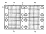
こうした半導体放射線検出素子を二次元的に配置して構成される半導体検出器において、感度とエネルギ分解能を向上させるためには、極力デッドスペースを少なくして、入射した放射線を漏れなく捕捉できるように、半導体放射線検出素子を基板上に稠密に配置することが求められる。しかし、多数、例えば10万個の半導体放射線検出素子を二次元的に配置した大面積の半導体検出器を装置に取り付けるための取り付け部材または取り付け部を設ける必要がある。この場合、取り付け部材または取り付け部を設けるためにデッドスペースが生じたり、あるいは半導体放射線検出素子が配置されない領域が発生したりする。例えば、図8(a)に示すように、検出器基板61の一面に多数の半導体放射線検出素子62を二次元的に設置し、他面に信号読出回路を搭載した回路基板63を、検出器基板61に設けたオスコネクタ61aに嵌装するメスコネクタ63aによって装着した半導体検出器64が知られている。このような半導体検出器64では、図8(b)の底面図に示すように、各半導体検出器64を核医学診断装置の放射線検出部に取り付けるために、検出器基板61の端部にボルト65a等の取り付け治具を装着するためのスペースS1を必要とし、このスペースS1には半導体放射線検出素子を配置できない。このスペースS1の存在は、半導体放射線検出素子を稠密に配置して半導体検出器61の感度の向上を妨げることになる。また、図8(a)および図8(b)に示す半導体検出器61では、一部の半導体放射線検出素子が故障または損傷のために、半導体検出器の放射線検出性能が低下した場合には、半導体検出器全体を交換するか、不具合が生じた半導体放射線検出素子を、多数の素子が設置されている中から取り外さなければならない。そこで、図9の底面図に示すように、半導体放射線検出素子が設置された基板ユニット71a,71b,71c,71dを作製し、各ユニット71a,71b,71c,71dを固定用基板(図示せず)に取り付けて大面積の半導体検出器を構成することも行われる。しかし、この図9に示す場合でも、各ユニット71a,71b,71c,71dを固定用基板に取り付けるための取り付け治具72を装着するスペースS2を必要とするため、このスペースS2には導体放射線検出素子を配置できず、半導体放射線検出素子を稠密に配置して、半導体検出器の感度および空間分解能を向上させる上で障害となる。
そこで、本発明の目的は、半導体放射線検出素子の配置密度を高めることができ、必要に応じて交換可能である放射線検出装置、およびその放射線検出装置を備える核医学診断装置を提供することにある。 SUMMARY OF THE INVENTION An object of the present invention is to provide a radiation detection device that can increase the arrangement density of semiconductor radiation detection elements and can be replaced as necessary, and a nuclear medicine diagnosis apparatus including the radiation detection device. .
前記の目的を達成するため、本発明の放射線検出装置は、複数の半導体放射線検出素子がマトリックス状に設置される検出器保持部材を、結合部材により、検出器ユニット保持部材に着脱可能に取り付け、その結合部材は、検出器保持部材の検出器ユニットと対向する部分で、第1コネクタ部が位置していない部分に位置させるとともに、検出器ユニット保持部材の検出器保持部材と対向する部分で、第2コネクタ部が位置していない部分に位置させることを特徴とする。 In order to achieve the above object, the radiation detection apparatus of the present invention detachably attaches a detector holding member in which a plurality of semiconductor radiation detection elements are installed in a matrix to a detector unit holding member by a coupling member, The coupling member is a portion facing the detector unit of the detector holding member, and is positioned at a portion where the first connector portion is not located, and at a portion facing the detector holding member of the detector unit holding member, The second connector portion is located at a portion where it is not located.
この放射線検出装置は、半導体放射線検出素子の配置密度を高めることができる。また、検出器保持部材ごとに検出器ユニット保持部材から脱着できるため、複数の半導体放射線検出素子および検出器保持部材を備える検出ユニットの交換が可能となる。 This radiation detection apparatus can increase the arrangement density of the semiconductor radiation detection elements. In addition, since each detector holding member can be detached from the detector unit holding member, it is possible to replace the detection unit including a plurality of semiconductor radiation detection elements and detector holding members.
また、好ましくは、核医学診断装置は、前記放射線検出装置の半導体放射線検出素子が放射線の出射源に対面するように配置されて構成された放射線検出部を備える。 Preferably, the nuclear medicine diagnosis apparatus includes a radiation detection unit configured so that a semiconductor radiation detection element of the radiation detection apparatus faces a radiation emission source.
この核医学診断装置は、半導体放射線検出素子が稠密に配置されるため、感度が向上し、また、故障した半導体放射線検出素子が設置された検出器搭載基板の交換が容易である。 In this nuclear medicine diagnostic apparatus, the semiconductor radiation detection elements are densely arranged, so that the sensitivity is improved, and the detector mounting board on which the failed semiconductor radiation detection elements are installed can be easily replaced.
本発明によれば、半導体放射線検出素子の配置密度を高めることができて感度が向上し、検出精度の向上を図ることができる。これにより、例えば、核医学診断装置において得られる画像のコントラストが向上し、鮮明な画像を得ることができる。
あるいは、同程度の画質ならば、実効的な感度の向上により、撮像時間を短縮できるようになる。
According to the present invention, the arrangement density of the semiconductor radiation detection elements can be increased, the sensitivity is improved, and the detection accuracy can be improved. Thereby, for example, the contrast of the image obtained in the nuclear medicine diagnostic apparatus is improved, and a clear image can be obtained.
Alternatively, if the image quality is comparable, the imaging time can be shortened by improving the effective sensitivity.
次に、本発明の放射線検出装置およびそれを用いた核医学診断装置の好適な実施形態について、適宜図面を参照しながら詳細に説明する。なお、以下において、実施形態の説明、および半導体放射線検出器、集積回路等の説明を行う。 Next, preferred embodiments of the radiation detection apparatus of the present invention and a nuclear medicine diagnosis apparatus using the same will be described in detail with reference to the drawings as appropriate. In the following description, embodiments and semiconductor radiation detectors, integrated circuits, and the like will be described.
本発明の核医学診断装置の実施形態に係るSPECT(Single Photon Emission Computer Tomography)装置1を、図1により説明する。
SPECT装置1は、一対の放射線検出装置2,2、回転支持台3、データ収集解析装置4、および表示装置5を備える。放射線検出装置2,2は、回転支持台(回転装置)3の周方向に180°や90°などのずれた位置で回転支持台3に設配置される。これらの放射線検出装置2,2は、それぞれが独立して回転し、放射線入射角を変えることもでき、2ユニットを並べて撮像面積を大きくすることや、平面撮像を行うガンマカメラとしても、用いることもできる。各放射線検出装置2,2は、それぞれ、固定用基板6、複数の検出器ユニット30、及び計測回路ユニット(信号処理装置)8を備える。各検出器ユニット30は、検出器搭載基板(検出器保持部材)7に多数の半導体放射線検出素子(半導体放射線検出器)12が格子状に配置されて配置されている。固定用基板(検出器ユニット保持部材)6には、回転支持台3の内側方向、すなわち、ベッドB側に複数の検出器搭載基板7が装着され、これと反対側に集積回路が配設置された計測回路ユニット8が配置されている。放射線検出装置2,2とベッドBとの間には、放射線遮蔽材(例えば、鉛、タングステン等)で形成され、多数の貫通孔(γ線通路)を有するコリメータ9が設けられる。コリメータ9は、放射線検出装置2からの視野角を制限している。また、各放射線検出装置2においては、コリメータ9、計測回路ユニット8および検出器ユニット30は、遮光・電磁シールド10の内部に収納されることで、被検者Pから出射されるγ線11以外の電磁波の影響が遮断されている。遮光・電磁シールド10は、アルミニウムやアルミニウム合金といった遮光性を有する材料で形成される。
A SPECT (Single Photon Emission Computer Tomography)
The
このSPECT装置1においては、放射性薬剤が投与された被検者Pが載っているベッドBが移動され、被検者Pは、一対の放射線検出装置2,2の間に移動される。被検者の体内からは、放射性薬剤から放出されたγ線(消滅γ線)が放出される。
In the
被検者Pの体内において、放射性薬剤が集積した集積部(例えば、がんの患部)CDから放出されるγ線11は、コリメータ9内の貫通孔を通って放射線検出装置2の各半導体放射線検出素子12に入射される。半導体放射線検出素子12から出力されたγ線検出信号は、計測回路ユニット8で信号処理される。このとき、計測回路ユニット8は、それぞれの半導体放射線検出素子12の出力であるγ線検出信号ごとにγ線の波高値のデータを計測するための集積回路(ASIC)を備え、検出した放射線(γ線)の波高値を測定するようになっている。データ収集解析装置(データ処理装置)4は、記憶装置、および断層像情報作成装置(図示せず)等を有する。
回転支持台3が回転することによって、各放射線検出装置2,2が被検者Pの周囲を旋回する。放射線検出装置2,2は旋回しながらγ線検出信号を出力する。回転支持台3の回転制御、放射線検出装置2,2と被検者Pとの間の距離の制御、およびベッドBによる被検者Pの位置制御は、操作パネル13によってSPECT装置1の近傍で行えると共に、データ入力装置14によって遠距離から行うことも可能である。
In the body of the subject P, γ rays 11 emitted from an accumulation part (for example, an affected part of cancer) CD in which a radiopharmaceutical is accumulated pass through a through hole in the
Each of the
次に、本実施形態に用いる放射線検出装置2について、図2〜図4を参照して説明する。なお、図2および図4は、説明の便宜のために、図中の紙面上方からγ線が入射することとし、放射線検出装置2のγ線入射側に装着されるコリメータ9は図示を省略した。
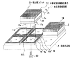
放射線検出装置2は、複数の検出器ユニット30、各検出器ユニット30を設置する固定用基板6を備える。各検出器ユニット30の検出器搭載基板7が固定用基板6に着脱可能に取り付けられる。各検出器搭載基板7は、半導体放射線検出素子12が設置される面7a、この面と反対側の面7bより突出して設けられた支持部31(図3)の一面に開口されたねじ孔16、および面7bに突出して設けられた位置決めピン17a,17bを備える。支持部31は後述する第1コネクタ7cの端面7d(図3)よりも突出している。通常、検出器搭載基板7には、例えば8×8個の半導体放射線検出素子12がマトリックス状に取り付けられる。固定用基板6は、その検出器搭載基板7を、例えば8×8個装着する。固定用基板6は、中央部に形成された貫通孔(結合部材挿入孔)18と、前記検出器搭載基板7の位置決めピン17a,17bが抜挿可能に嵌挿される位置決め孔19a,19bとを備える。複数の半導体放射線検出素子12が取り付けられた状態で、検出器搭載基板7の位置決めピン17a,17bを、固定用基板6の位置決め孔19a,19bにそれぞれ挿入する。位置決めピン17aおよび位置決め孔19aは、1つの位置決め装置を構成する。位置決めピン17bおよび位置決め孔19bは、他の位置決め装置を構成する。結合用ねじ(結合部材)20が、貫通孔18に挿入され、ねじ孔16と噛み合わされる。このようにして、検出器搭載基板7、すなわち検出器ユニット30が固定用基板6の所定の位置に取り付けられる。このとき、支持部31の一面(ねじ孔16が開口している面)が固定用基板6に接触する。このとき、結合用ねじ20を検出器搭載基板7のねじ孔16に噛み合わせる力によって、位置決め孔19a,19bへの位置決めピン17a,17bの挿入力が生じる。検出器ユニット30を交換する場合には、結合用ねじ20をねじ孔16から取り外すことによって固定用基板6から検出器ユニット30を取り外すことができる。
Next, the
The
また、位置決めピン17a,17bの先端部15は、位置決め孔19a,19bを貫通して、固定用基板6の背面側に突出するように形成される。これによって、固定用基板6の背面側に突出した位置決めピン17a,17bのそれぞれの先端部15は、結合用ねじ20を検出器搭載基板7のねじ孔16に噛み合わせる際の検出器搭載基板7の動きを規制して、結合用ねじ20の空回りを防止することができる。また、位置決め部材17a,17bの先端部15を引っ張ることによって、結合用ねじ20のネジ回し開始時に結合用ねじ20とねじ孔16を確実に合せることができる。これによって、検出器ユニット30を、交換するために、固定用基板6から取り外す際に、固定用基板6の背面側から位置決めピン17a,17bの先端を保持しながら、結合用ねじ20をねじ孔16から取り外すことができる。このとき、位置決めピン17a,17bの先端部15を、固定用基板6の背面側から押圧すれば、検出器搭載基板7を固定用基板6から浮かせた状態にして容易に検出器ユニット30を取り外すことができるため、検出器ユニット30の交換時の作業性の向上に有効である。放射線検出装置2に取り付けられている検出器ユニット30の交換は、この検出器ユニット30に設けられた半導体放射線検出素子12が故障したときに行われる。
Further, the
各検出器搭載基板7は、複数の半導体放射線検出素子12が設置される面7aとは反対の面に、第1コネクタ7cを取り付けている。第1コネクタ7cは、位置決めピン17a,17bおよび支持部31を取り囲んで検出器搭載基板7の4辺部に沿って配列されている。これによって、検出器搭載基板7の中央部には、支持部31および位置決めピン17a,17bが設置され、コネクタ等が存在しない領域(面b)が形成される。第1コネクタ(第1コネクタ部)7cには、複数の出力ピン21が突出して設置される。複数の出力ピン21のうち、後述の複数のアノード信号線のそれぞれの一端に別々に接続される出力ピン21を、第1コネクタ端子と称する。残りの複数の出力ピン21は、後述の複数のカソード信号線のそれぞれの一端に別々に接続され、第2コネクタ端子と称する。これらの出力ピン21は、前述した検出器ユニット30の固定用基板6への装着時に、固定用基板6に設けられた第2コネクタ(第2コネクタ部)6aのそれぞれの入力端子コネクタ22にはめ込まれる。複数の入力端子コネクタ22のうち、第1コネクタ端子がはめ込まれる複数の入力端子コネクタ22は、第3コネクタ端子と称する。残りの複数の入力端子コネクタ22は、第2コネクタ端子がはめ込まれ、第4コネクタ端子と称する。第2コネクタ6aは、1つの検出器ユニット30に対応して設けられる貫通孔18および位置決め孔19a,19bを取り囲むように矩形の環状に固定用基板6に取り付けられている。各出力ピン21は、後述の複数のアノード信号線32a、32b、……および複数のカソード信号線33a、33b、……のうちの該当する1本の信号線にそれぞれ接続されている。
Each
各検出器搭載基板7に設置される各半導体放射線検出素子12は、図5に示すように、放射線と相互作用を及ぼして電荷を生成する薄板状の半導体領域(半導体部)23と、半導体領域23を挟んで、アノード電極Aとカソード電極Cとが対峙して構成される。アノード電極Aとカソード電極Cは半導体領域23に設置される。半導体領域23は、CdTe(テルル化カドミウム)、TlBr(臭化タリウム)、GaAs(ガリウム砒素)等のいずれかの単結晶半導体材料で構成されている。また、電極(アノードA、カソードC)は、Pt(白金)、Au(金)、In(インジウム)等のいずれかの材料が用いられる。なお、以下の説明では、半導体領域23がCdTeの単結晶をスライスしたものであるとする。半導体領域23の厚み(アノード電極Aが取り付けられる面とカソード電極Cが取り付けられる面の間の厚み)は、0.2〜2mm(より好ましくは0.5〜1.5mmである。また、検出する放射線は、SPECT装置で用いる所定のエネルギのγ線であるとする。アノード電極Aおよびカソード電極Cは、半導体領域23の面に蒸着、その他の方法で形成される。膜状に形成されたアノード電極Aおよびカソード電極Cの膜厚は、それぞれ数百ナノメートル程度であり、約20〜80ミクロン程度の導電板を用いて配線する。
As shown in FIG. 5, each semiconductor
前述したように複数の半導体検出素子12がマトリックス状に各検出器搭載基板7に設置されている。マトリックス状に配置された複数の半導体放射線検出素子12の一部(半導体放射線検出素子12A,12B,12C,12D)が、例えば、図5に示すように配置されているとする。各半導体放射線検出素子12は、半導体領域23、これに設置されるアノード電極及びカソード電極が検出器搭載基板7に対して交差する、具体的には直交するように検出器搭載基板7に設置される。a方向で一列に配置された半導体放射線検出素子12Aおよび12Bの各アノード電極A1,A2が、a方向に延びるアノード信号線32aに、信号線36A,36Bにより接続される。a方向で一列に配置された半導体放射線検出素子12Cおよび12Dの各アノード電極A3,A4が、a方向に延びるアノード信号線32bに、信号線36C,36Dにより接続される。また、a方向と交差するb方向で一列に配置された半導体放射線検出素子12Aおよび12Cの各カソード電極C1,C3が、b方向に延びるカソード信号線33aに、信号線37A,37Cにより接続される。b方向で一列に配置された半導体放射線検出素子12Bおよび12Dの各カソード電極C2,C4が、b方向に延びるカソード信号線33bに、信号線37B,37Dにより接続される。このように、a方向に延びる各アノード信号線には、それぞれ一列に配置された複数の半導体放射線検出素子12の各アノード電極が接続され、b方向に延びる各カソード信号線には、それぞれ一列に配置された複数の半導体放射線検出素子12の各カソード電極が接続される。複数のアノード信号線32a、32b、……および複数のカソード信号線33a、33b、……のそれぞれの一端が、別々の出力ピン21に接続される。これらの信号線は検出器搭載基板7の面7aまたは内部に設置されている。半導体放射線検出素子12が、0.5〜1.5mm程度に薄く構成できる場合には、1つの半導体領域23を有する半導体放射線検出素子12を単独で稠密に配置することができるため、感度の向上に有効である。
As described above, a plurality of
放射線検出装置2の作用について説明する。
まず、半導体放射線検出素子12によるγ線の検出原理の概略を説明する。半導体放射線検出素子12にγ線が入射して、γ線と半導体領域23が相互作用を及ぼすと、正孔(hole)および電子(electron)が、対になってγ線が持つエネルギに比例した量だけ生成される。ここで、半導体放射線検出素子12のアノード電極(陽極)Aとカソード電極(陰極)Cの電極間には、高圧電源(図示せず)より電荷収集用の電圧がかけられている(例えば300V)。このため、正孔はカソード電極Cに引き寄せられて移動し、電子はアノード電極Aに引き寄せられて移動する。
The operation of the
First, the outline of the principle of detection of γ rays by the semiconductor
図5において、γ線が入射して半導体放射線検出素子12Aの半導体領域23が相互作用を及ぼしたとすると、アノード電極A1が接続されているアノード信号線32a、およびカソード電極C1が接続されたカソード信号線33aから電荷の情報が得られる。これらのアノード信号線32aとカソード信号線33aからの信号を同時判定して重ね合わせる、マトリックス読出しを行うことによって、どの位置の半導体放射線検出素子12にγ線が入射したかを検出することができる。この同時判定は、計測回路ユニット8の集積回路で行われる。この同時判定によって、位置読出し用の信号線の配線数を大幅に減らせることができ、また、信号線の数が減ることによって空くスペースを利用することができる。各アノード信号線32a、32b、……ならびに各カソード信号線33a、33b、……は、検出器搭載基板7の出力ピン21(図2参照)に、個別に接続され、各信号線に接続された出力ピン21から、固定用基板6の入力端子コネクタ22を介して、計測回路ユニット8の集積回路(ASIC)にγ線の入射による電荷の情報が入力される。集積回路は、半導体放射線検出素子12が出力する信号を処理するアナログ集積回路と、前記アナログ集積回路の出力であるアナログ信号をデジタル信号に変換するAD変換器と、AD変換された信号を処理するデジタル集積回路とを備え、ある半導体放射線検出素子12で検出した放射線(γ線)の波高値情報をデータ収集解析装置4に出力する。
In FIG. 5, if γ rays are incident and the
そして、データ収集解析装置4には、回転支持台3を回転させるモーター(図示せず)の回転軸に連結された角度計(図示せず)で検出された回転角度が入力される。この回転角度は、それぞれの放射線検出装置2の回転角度を示している。データ収集解析装置4は、この回転角度を基に、旋回している各放射線検出装置2の旋回軌道上での書く半導体放射線検出素子12の位置(位置座標)を求める。このため、γ線を検出した時点での該当する半導体放射線検出素子12の位置(位置座標)が求められる。データ収集解析装置4は、半導体放射線検出素子12の位置を基に、波高値情報が設定値(しきい値)以上になるγ線を計数する。この計数は、回転支持台3の回転中心を基準に0.5°づつ区切って得られる各領域に対してなされる。なお、波高値情報は、コリメータ9の放射線通路の延長線上に位置する複数の半導体放射線検出素子の各γ線検出信号の波高値の累計値である。データ収集解析装置4は、γ線を検出した時点での半導体放射線検出素子12の位置情報およびγ線の計数値(計数情報)を用いて、放射性薬剤の集積位置、すなわち悪性腫瘍位置での被検者の断層像情報を作成する。この断層像情報は表示装置5に表示される。また、このパケット情報、同時計測で得た計数値および半導体放射線検出素子12の位置情報、および断層像情報等の情報は、データ収集解析装置4の記憶装置に記憶される。
And the rotation angle detected by the goniometer (not shown) connected with the rotating shaft of the motor (not shown) which rotates the
このように、個別に素子を並べた構造を持つ半導体検出器ユニットにおいても、マトリックス読出しを行うことで、必要な回路数は個別にn×n個の画素から読出を行う場合に比べ2/n倍と大幅に削減される。これによって、空くスペースを利用することができる。 As described above, even in a semiconductor detector unit having a structure in which elements are arranged individually, the number of necessary circuits is 2 / n compared to the case of performing readout from n × n pixels by performing matrix readout. Doubled and greatly reduced. As a result, a vacant space can be used.
このように構成される本実施形態では、結合用ねじ20が、第2コネクタ6cが位置していない部分で固定用基板7に形成された貫通孔18を通して、第1コネクタ7が位置していない部分で前記検出器搭載基板7の固定用基板6と対向する部分に形成されたねじ孔16に噛み合わされるため、隣接する検出器ユニット30間に、結合用ねじ20等の結合部材が配置されない。このため、多数の半導体放射線検出素子12を有する検出器ユニット30が、固定用基板6の上に隙間なく装着できるため、固定用基板6またはその固定用基板6を複数枚並べた放射線検出装置2の全面にわたって、半導体放射線検出素子12を稠密に配置することができる。そのため、γ線の検出感度およびその感度の均一性、ならびに空間分解能を高めることができる。これによって、核医学診断装置(SPECT)における装置全体の検出感度を向上させ、撮像時間の短縮を図ることができる。そして、各検出器搭載基板7は、固定用基板6の背面側から位置決めピン17a,17bの先端を保持しながら、結合用ねじ20をねじ孔16から取り外せば、検出器搭載基板7を固定用基板6から浮かせた状態にして、検出器搭載基板7を固定用基板6から容易に取り外すことができる。これによって、半導体放射線検出素子の故障または破損時に、検出器搭載基板7を交換する作業が容易となる。また、故障または破損した半導体放射線検出素子12を搭載している検出器搭載基板7だけを交換すればよく、保守時の無駄な作業の低減(コストダウン)が図れる。さらに、位置決めピン17a,17bによって、検出器搭載基板7を固定用基板6の所定の位置に正確に装着することができ、取り付け時の作業を正確かつ容易に行うことができる。位置決めピン17a,17bが位置決め孔19a,19bに挿入されるため、結合用ねじ20をねじ孔16に噛み合わせる際において、結合用ねじ20の回転に伴って検出器搭載基板7が回転することを防止できる。
本実施形態は、検出器搭載基板7に支持部31が設けられているため、検出器ユニット30を固定用基板6に取り付けたとき、多数の半導体放射線検出素子12および検出器搭載基板6の重量は支持部31を介して固定用基板6に伝えられる。このため、嵌合されている出力ピン21と入力端子コネクタ22のコネクタ部に無理な力が加わらなく、それらのコネクタ部の損傷を避けることができる。また、支持部31が検出器ユニット30の中心軸に位置しているため、多数の半導体放射線検出素子12および検出器搭載基板6等を安定に支持できる。
本実施形態では、第1コネクタ7cを検出器搭載基板7の周辺部に位置させたが、以下のように配置することも可能である。検出器搭載基板7の一面においてその面の2つの角を結ぶ対角線よりも残りの1つの角側に寄せて、第1コネクタ7cを配置しても良い。この場合には、ねじ孔16を有する支持部31は、対角線よりも反対側の他の角側で、その一面内に設けられる。第2コネクタ6aも、その第1コネクタ7cの配置に対応させて固定用基板6に配置される。
支持部31を設けないで、結合用ねじ20を第1コネクタ7cが位置していない面bの位置で検出器搭載基板7に形成したねじ孔18に噛み合わせて、検出器搭載基板7を固定用基板6に取り付けても良い。
In the present embodiment configured as above, the
In the present embodiment, since the
In the present embodiment, the
Without providing the
本発明のSPECT装置の他の実施形態を以下に説明する。本実施形態のSPECT装置は、検出器ユニットに用いられる半導体放射線検出素子の構造が前述のSPECT装置1に用いられる半導体放射線検出素子12と異なっている。本実施形態で用いられる各半導体放射線検出素子35は、2つの半導体放射線検出素子12を組み合わせた構成であり、図6に示すように、2つの半導体領域23を有し、1つの半導体領域23の両面にアノード電極A1およびカソード電極C1が取り付けられ、他の半導体領域23の両面にアノード電極A2およびカソード電極C2が取り付けられている。アノード電極A1とアノード電極A2が対向するように、カソード電極C1、C2が外側に位置するように、隣り合う半導体放射線検出素子12が重ねられた構成となっている。アノード電極A1とアノード電極A2の間にこれら接触する導電板25aが配置される。アノード電極A1、A2は、導電板25aを挟む2つの半導体領域23と接する面に膜状に形成され、カソード電極C1、C2は、導電板25cの半導体領域23と接する面に膜状に形成されている。これによって、隣り合う2つの半導体放射線検出素子12の半導体領域23の電極間距離(アノード電極Aとカソード電極Cの間の距離)が短くなり、生じた電荷を適切に捉えることができるため、有利である。そして、各半導体放射線検出素子12は、検出器搭載基板7に対して半導体領域23が略垂直になる方向に検出器搭載基板7に設置されている。
Another embodiment of the SPECT apparatus of the present invention will be described below. The SPECT apparatus of the present embodiment is different from the semiconductor
そして、ある方向aに一列に配置された各半導体放射線検出素子35のアノード電極Aは、図6中の方向aに複数配線されたアノード信号線のうちの1本であるアノード信号線26Aに、導電板25aを介して信号線27により接続されている。一方、方向aと交差する方向bに一列に配置された各半導体放射線検出素子35のカソード電極C1は、方向bに配線されたカソード信号線28Aに、導電板25bに接続された信号線29Aを介して接続されている。また、方向bに一列に配置されたこれらの半導体放射線検出素子35の他のカソード電極C2は、方向bに配線された他のカソード信号線28Bに、導電板25cに接続された信号線29Bを介して接続されている。これらのアノード信号線26Aおよびカソード信号線28A,28Cは、検出器搭載基板7の面7aまたは内部に、方向aおよびその方向aと交差する方向bに沿って平行に複数配線される。そして、図7に示すように、検出器搭載基板7にマトリックス状に設置される各半導体放射線検出素子35a,35b,35c,35d,・・・・の各アノード電極Aおよびカソード電極Cに接続されるように、マトリックス状に配線される。図7において、26Bは他のアノード信号線であり、28C,28Dは他のカソード信号線である。
The anode electrodes A of the semiconductor radiation detection elements 35 arranged in a line in a certain direction a are connected to the
半導体放射線検出素子35の各半導体放射線検出素子12のアノード電極(陽極)Aとカソード電極(陰極)Cの電極間には、高圧電源(図示せず)より電荷収集用の電圧がかけられている(例えば300V)ため、正孔はカソード電極Cに引き寄せられて移動し、電子はアノード電極Aに引き寄せられて移動する。このとき、図7に示す構成において、γ線が入射して半導体放射線検出素子35aの半導体領域23aが相互作用を及ぼしたとすると、半導体領域23aのアノード電極Aaが接続されているアノード信号線26Aと、カソード電極Caが接続されたカソード信号線28Aとから電荷の情報が得られる。これらのアノード信号線26Aとカソード信号線28Aからの信号を同時判定して重ね合わせる、マトリックス読出しを行うことによって、どの位置の半導体放射線検出素子35にγ線が入射したかを検出することができる。各アノード信号線26A,26B,・・・のそれぞれの一端は、検出器搭載基板7の出力ピン21(図2参照)に、個別に接続される。ある1つの半導体放射線検出素子35に接続された一対のカソード信号線(例えば、信号線28A,28B)の一端は他の1つの出力ピン21に接続される。カソード信号線28C,28Dのそれぞれの一端も、別の1つの出力ピン21に接続される。各信号線に接続された出力ピン21から、固定用基板6の入力端子コネクタ22を介して、計測回路ユニット8の集積回路(ASIC)にγ線の入射による電荷の情報が入力される。本実施形態のSPECT装置における以上に述べた以外の構成は、SPECT装置1の構成と同じである。
本実施形態のSPECT装置は、SPECT装置1で生じる効果を得ることができる。
A voltage for collecting charges is applied between the anode electrode (anode) A and the cathode electrode (cathode) C of each semiconductor
The SPECT apparatus according to the present embodiment can obtain the effects produced by the
なお、以上の各実施形態では、核医学診断装置として単光子放出型断層撮影装置(SPECT(Single Photon Emission Computer Tomography)装置)を例に説明したが、SPECTに限らず、PET装置およびγカメラにも本発明を適用することができる。ちなみに、PET装置およびSPECT装置は、人体の3次元の機能画像を撮影することで共通するが、SPECT装置は、測定原理が単光子を検出するものであることから同時計測を行うことができず、このため、γ線の入射位置(角度)を規制するコリメータを備える。また、γカメラは、得られる機能画像が2次元的なものであり、かつ、γ線の入射角度を規制するコリメータを備える。これに対して、PET装置は基本的に180°反対方向に放射される一対のγ線を同時に検出することができるため、コリメータを必要としない。
なお、PET装置やSPECT装置と、X線CTを組み合わせた核医学診断装置の構成としてもよい。
In each of the above embodiments, a single photon emission tomography apparatus (SPECT (Single Photon Emission Computer Tomography) apparatus) has been described as an example of a nuclear medicine diagnosis apparatus. However, the present invention is not limited to SPECT, and includes PET apparatuses and γ cameras. The present invention can also be applied. Incidentally, the PET device and the SPECT device are common in taking a three-dimensional functional image of the human body, but the SPECT device cannot perform simultaneous measurement because the measurement principle is to detect single photons. For this reason, a collimator that regulates the incident position (angle) of γ rays is provided. The γ camera includes a collimator that obtains a two-dimensional functional image and regulates the incident angle of γ rays. On the other hand, since the PET apparatus can detect a pair of γ rays radiated in the opposite directions of 180 ° at the same time, no collimator is required.
In addition, it is good also as a structure of the nuclear medicine diagnostic apparatus which combined PET apparatus and SPECT apparatus, and X-ray CT.
また、以上の実施形態では、検出器搭載基板7を固定用基板6に装着するために、結合用ねじ20をねじ孔16に螺着する構成を備えるが、本発明は、この構成に制限されず、検出器搭載基板7の中央部の配線が存在しない領域を利用して、固定用基板6の背面側から装着して検出器搭載基板7を固定用基板6に装着できる構造のものであれば、いずれの構成によってもよい。例えば、ねじ孔16に挿入して弾性変形してねじ孔16の内周面に係止することができる結合用部材によって、検出器搭載基板7を固定用基板に装着するようにしてもよい。また、位置決めピン17a,17bおよび位置決め孔19a,19bは、2対に限定されず、検出器搭載基板7の装着位置を正確に案内できるものであれば、1対または多数対でもよく、さらに断面形状を変えることによって、その装着位置をより確実に特定できるようにしてもよい。また、結合用ねじ20を支持部31に取り付けて、この結合用ねじ20を固定用基板6の前面から貫通孔18に挿入し、固定用基板6の背面から結合用ねじ20にナットをかみ合わせ、検出器ユニット30を固定用基板6に取り付けてもよい。
Further, in the above embodiment, in order to mount the
1 SPECT装置
2 放射線検出装置
3 回転支持台
4 データ収集解析装置
5 表示装置
6 固定用基板
6a 第2コネクタ
7 検出器搭載基板
7c 第1コネクタ
8 計測回路ユニット
9 コリメータ
10 電磁シールド
11 γ線
12,12a 半導体放射線検出素子
16 ねじ孔
17a,17b 位置決めピン
18 貫通孔
19a,19b 位置決め孔
20 結合用ねじ
21 出力ピン
22 入力端子コネクタ
23,23a 半導体領域
25a,25c 導電板
26A,26B,32a,32b アノード信号線
28A,28B,28C,28D,33a,33b カソード信号線
30 検出器ユニット
31 支持部
A,A1,A2,A3,A4,Aa アノード電極
B ベッド
C,C1,C2,C3,C4,Ca カソード電極
P 被検者
DESCRIPTION OF
Claims (16)
複数の前記検出器ユニットが設置される検出器ユニット保持部材とを備え、
前記方向aの複数の列毎に配置され、前記方向aの該当する前記列に属する複数の前記半導体放射線検出素子のそれぞれのアノード電極に接続される複数のアノード信号線、および前記方向bの複数の列毎に配置され、前記方向aの該当する前記列に属する複数の前記半導体放射線検出素子のそれぞれのカソード電極に接続される複数のカソード信号線が、前記検出器保持部材に設けられ、
前記複数のアノード信号線の一端に別々に接続される複数の第1コネクタ端子、および前記複数のカソード信号線の一端に別々に接続される複数の第2コネクタ端子を有する第1コネクタ部を、前記検出器保持部材の前記半導体放射線検出素子と対向する部分において前記検出器保持部材の4辺部に沿って設け、
前記複数の第1コネクタ端子に別々に着脱可能に接続される複数の第3コネクタ端子、および前記複数の第2コネクタ端子に別々に着脱可能に接続される複数の第4コネクタ端子を有する第2コネクタ部を、前記検出器ユニット保持部材の前記第1コネクタ部と対向する部分に設けてなり、
前記検出器保持部材を前記検出器ユニット保持部材に着脱可能に取り付ける結合部材を、前記検出器保持部材の前記半導体放射線検出素子と対向する部分で、前記第1コネクタ部が位置していない部分に位置させるとともに、前記検出器ユニット保持部材の前記検出器保持部材と対向する部分で、前記第2コネクタ部が位置していない部分に位置させる位置決め装置を備え、
前記位置決め装置は、前記検出器保持部材から突出して設けられた位置決めピン、および前記検出器ユニット保持部材に形成されて前記位置決めピンが挿入される位置決め孔を含み、かつ、前記第1コネクタ部が位置していない部分および前記第2コネクタ部が位置していない部分に位置し、
前記位置決めピンは、前記位置決め孔より突出する長さを有することを特徴とする放射線検出装置。 A plurality of semiconductor radiation detection elements having a semiconductor region that interacts with radiation and generates electric charges, and an anode electrode and a cathode electrode are opposed to each other with the semiconductor region interposed therebetween, and these semiconductor radiation detections A detector unit having a plurality of rows of elements in a certain direction a, and detector holding members arranged in a matrix in a plurality of rows in the other direction b intersecting the direction a;
A detector unit holding member on which a plurality of the detector units are installed,
A plurality of anode signal lines arranged for each of the plurality of columns in the direction a and connected to respective anode electrodes of the plurality of semiconductor radiation detection elements belonging to the column corresponding to the direction a, and a plurality of the direction b A plurality of cathode signal lines connected to the respective cathode electrodes of the plurality of semiconductor radiation detection elements belonging to the corresponding column in the direction a are provided in the detector holding member,
A first connector portion having a plurality of first connector terminals separately connected to one end of the plurality of anode signal lines, and a plurality of second connector terminals separately connected to one end of the plurality of cathode signal lines; Provided along the four sides of the detector holding member at a portion of the detector holding member facing the semiconductor radiation detection element,
A second having a plurality of third connector terminals detachably connected to the plurality of first connector terminals and a plurality of fourth connector terminals detachably connected to the plurality of second connector terminals. a connector portion, it is provided in the first connector part and the opposing portions of the detector unit holding member,
A coupling member that detachably attaches the detector holding member to the detector unit holding member is a portion of the detector holding member that faces the semiconductor radiation detection element, and a portion where the first connector portion is not located. And a positioning device that is positioned at a portion of the detector unit holding member that faces the detector holding member and is not located at the second connector portion ,
The positioning device includes a positioning pin provided protruding from the detector holding member, a positioning hole formed in the detector unit holding member and into which the positioning pin is inserted, and the first connector portion includes Located in a portion that is not located and a portion in which the second connector portion is not located,
The radiation detecting apparatus according to claim 1, wherein the positioning pin has a length protruding from the positioning hole .
複数の前記検出器ユニットが設置される検出器ユニット保持部材とを備えた放射線検出装置であって、
前記方向aの複数の列毎に配置され、前記方向aの該当する前記列に属する複数の前記半導体放射線検出素子のそれぞれのアノード電極に接続される複数のアノード信号線、および前記方向bの複数の列毎に配置され、前記方向aの該当する前記列に属する複数の前記半導体放射線検出素子のそれぞれのカソード電極に接続される複数のカソード信号線が、前記検出器保持部材に設けられ、
前記複数のアノード信号線の一端に別々に接続される複数の第1コネクタ端子、および前記複数のカソード信号線の一端に別々に接続される複数の第2コネクタ端子を有する第1コネクタ部を、前記検出器保持部材の前記半導体放射線検出素子と対向する部分において前記検出器保持部材の4辺部に沿って設け、
前記複数の第1コネクタ端子に別々に着脱可能に接続される複数の第3コネクタ端子、および前記複数の第2コネクタ端子に別々に着脱可能に接続される複数の第4コネクタ端子を有する第2コネクタ部を、前記検出器ユニット保持部材の前記第1コネクタ部と対向する部分に設けてなり、
前記検出器保持部材を前記検出器ユニット保持部材に着脱可能に取り付ける結合部材を、前記検出器保持部材の前記半導体放射線検出素子と対向する部分で、前記第1コネクタ部が位置していない部分に位置させるとともに、前記検出器ユニット保持部材の前記検出器保持部材と対向する部分で、前記第2コネクタ部が位置していない部分に位置させる位置決め装置を前記放射線検出装置に備え、
前記位置決め装置は、前記検出器保持部材から突出して設けられた位置決めピン、および前記検出器ユニット保持部材に形成されて前記位置決めピンが挿入される位置決め孔を含み、かつ、前記第1コネクタ部が位置していない部分および前記第2コネクタ部が位置していない部分に位置し、
前記位置決めピンは、前記位置決め孔より突出する長さを有することを特徴とする核医学診断装置。 A plurality of semiconductor radiation detection elements having a semiconductor region that interacts with radiation and generates electric charges, and an anode electrode and a cathode electrode are opposed to each other with the semiconductor region interposed therebetween, and these semiconductor radiation detections A detector unit having a plurality of rows of elements in a certain direction a, and detector holding members arranged in a matrix in a plurality of rows in the other direction b intersecting the direction a;
A radiation detection apparatus comprising a detector unit holding member on which a plurality of the detector units are installed,
A plurality of anode signal lines arranged for each of the plurality of columns in the direction a and connected to respective anode electrodes of the plurality of semiconductor radiation detection elements belonging to the column corresponding to the direction a, and a plurality of the direction b A plurality of cathode signal lines connected to the respective cathode electrodes of the plurality of semiconductor radiation detection elements belonging to the corresponding column in the direction a are provided in the detector holding member,
A first connector portion having a plurality of first connector terminals separately connected to one end of the plurality of anode signal lines, and a plurality of second connector terminals separately connected to one end of the plurality of cathode signal lines; Provided along the four sides of the detector holding member at a portion of the detector holding member facing the semiconductor radiation detection element,
A second having a plurality of third connector terminals detachably connected to the plurality of first connector terminals and a plurality of fourth connector terminals detachably connected to the plurality of second connector terminals. a connector portion, it is provided in the first connector part and the opposing portions of the detector unit holding member,
A coupling member that detachably attaches the detector holding member to the detector unit holding member is a portion of the detector holding member that faces the semiconductor radiation detection element, and a portion where the first connector portion is not located. together are positioned, in the detector holding member facing the portion of the detector unit holding member, comprising a positioning device for positioning a portion where the second connector portion is not located in the radiation detecting device,
The positioning device includes a positioning pin provided protruding from the detector holding member, a positioning hole formed in the detector unit holding member and into which the positioning pin is inserted, and the first connector portion includes Located in a portion that is not located and a portion in which the second connector portion is not located,
The nuclear medicine diagnosis apparatus , wherein the positioning pin has a length protruding from the positioning hole .
前記放射線検出装置が、
放射線と相互作用を及ぼして電荷を生成する半導体領域を有し、この半導体領域を挟んで、アノード電極とカソード電極とが対峙して構成される複数の半導体放射線検出素子と、これらの半導体放射線検出素子が、ある方向aに複数列、および前記方向aに交差する他の方向bに複数列となるマトリックス状に設置された検出器保持部材とを有する検出器ユニットと、
複数の前記検出器ユニットが設置される検出器ユニット保持部材とを備え、
前記方向aの複数の列毎に配置され、前記方向aの該当する前記列に属する複数の前記半導体放射線検出素子のそれぞれのアノード電極に接続される複数のアノード信号線、および前記方向bの複数の列毎に配置され、前記方向aの該当する前記列に属する複数の前記半導体放射線検出素子のそれぞれのカソード電極に接続される複数のカソード信号線が、前記検出器保持部材に設けられ、
前記複数のアノード信号線の一端に別々に接続される複数の第1コネクタ端子、および前記複数のカソード信号線の一端に別々に接続される複数の第2コネクタ端子を有する第1コネクタ部を、前記検出器保持部材の前記半導体放射線検出素子と対向する部分において前記検出器保持部材の4辺部に沿って設け、
前記複数の第1コネクタ端子に別々に着脱可能に接続される複数の第3コネクタ端子、および前記複数の第2コネクタ端子に別々に着脱可能に接続される複数の第4コネクタ端子を有する第2コネクタ部を、前記検出器ユニット保持部材の前記第1コネクタ部と対向する部分に設けてなり、
前記検出器保持部材を前記検出器ユニット保持部材に着脱可能に取り付ける結合部材を、前記検出器保持部材の前記半導体放射線検出素子と対向する部分で、前記第1コネクタ部が位置していない部分に位置させるとともに、前記検出器ユニット保持部材の前記検出器保持部材と対向する部分で、前記第2コネクタ部が位置していない部分に位置させる位置決め装置を備え、
前記位置決め装置は、前記検出器保持部材から突出して設けられた位置決めピン、および前記検出器ユニット保持部材に形成されて前記位置決めピンが挿入される位置決め孔を含み、かつ、前記第1コネクタ部が位置していない部分および前記第2コネクタ部が位置していない部分に位置し、
前記位置決めピンは、前記位置決め孔より突出する長さを有することを特徴とする核医学診断装置。 A bed for supporting a subject, a radiation detection device, and a rotation device for rotating the radiation detection device around the bed;
The radiation detection device comprises:
A plurality of semiconductor radiation detection elements having a semiconductor region that interacts with radiation and generates electric charges, and an anode electrode and a cathode electrode are opposed to each other with the semiconductor region interposed therebetween, and these semiconductor radiation detections A detector unit having a plurality of rows of elements in a certain direction a, and detector holding members arranged in a matrix in a plurality of rows in the other direction b intersecting the direction a;
A detector unit holding member on which a plurality of the detector units are installed,
A plurality of anode signal lines arranged for each of the plurality of columns in the direction a and connected to respective anode electrodes of the plurality of semiconductor radiation detection elements belonging to the column corresponding to the direction a, and a plurality of the direction b A plurality of cathode signal lines connected to the respective cathode electrodes of the plurality of semiconductor radiation detection elements belonging to the corresponding column in the direction a are provided in the detector holding member,
A first connector portion having a plurality of first connector terminals separately connected to one end of the plurality of anode signal lines, and a plurality of second connector terminals separately connected to one end of the plurality of cathode signal lines; Provided along the four sides of the detector holding member at a portion of the detector holding member facing the semiconductor radiation detection element,
A second having a plurality of third connector terminals detachably connected to the plurality of first connector terminals and a plurality of fourth connector terminals detachably connected to the plurality of second connector terminals. a connector portion, it is provided in the first connector part and the opposing portions of the detector unit holding member,
A coupling member that detachably attaches the detector holding member to the detector unit holding member is a portion of the detector holding member that faces the semiconductor radiation detection element, and a portion where the first connector portion is not located. And a positioning device that is positioned at a portion of the detector unit holding member that faces the detector holding member and is not located at the second connector portion ,
The positioning device includes a positioning pin provided protruding from the detector holding member, a positioning hole formed in the detector unit holding member and into which the positioning pin is inserted, and the first connector portion includes Located in a portion that is not located and a portion in which the second connector portion is not located,
The nuclear medicine diagnosis apparatus , wherein the positioning pin has a length protruding from the positioning hole .
Priority Applications (3)
| Application Number | Priority Date | Filing Date | Title |
|---|---|---|---|
| JP2004005130A JP4594624B2 (en) | 2004-01-13 | 2004-01-13 | Radiation detection device and nuclear medicine diagnostic device |
| US11/032,091 US7202482B2 (en) | 2004-01-13 | 2005-01-11 | Radiation detection apparatus and radiological imaging apparatus |
| US11/714,216 US7528378B2 (en) | 2004-01-13 | 2007-03-06 | Radiation detection apparatus and radiological imaging apparatus |
Applications Claiming Priority (1)
| Application Number | Priority Date | Filing Date | Title |
|---|---|---|---|
| JP2004005130A JP4594624B2 (en) | 2004-01-13 | 2004-01-13 | Radiation detection device and nuclear medicine diagnostic device |
Publications (3)
| Publication Number | Publication Date |
|---|---|
| JP2005201642A JP2005201642A (en) | 2005-07-28 |
| JP2005201642A5 JP2005201642A5 (en) | 2007-03-01 |
| JP4594624B2 true JP4594624B2 (en) | 2010-12-08 |
Family
ID=34747137
Family Applications (1)
| Application Number | Title | Priority Date | Filing Date |
|---|---|---|---|
| JP2004005130A Expired - Fee Related JP4594624B2 (en) | 2004-01-13 | 2004-01-13 | Radiation detection device and nuclear medicine diagnostic device |
Country Status (2)
| Country | Link |
|---|---|
| US (2) | US7202482B2 (en) |
| JP (1) | JP4594624B2 (en) |
Families Citing this family (22)
| Publication number | Priority date | Publication date | Assignee | Title |
|---|---|---|---|---|
| ATE529045T1 (en) * | 2003-07-22 | 2011-11-15 | Koninkl Philips Electronics Nv | RADIATION MASK FOR A TWO-DIMENSIONAL CT DETECTOR |
| JP4594624B2 (en) * | 2004-01-13 | 2010-12-08 | 株式会社日立製作所 | Radiation detection device and nuclear medicine diagnostic device |
| JP3828896B2 (en) * | 2004-03-11 | 2006-10-04 | 株式会社日立製作所 | Positron emission tomography system |
| JP4777975B2 (en) * | 2004-04-06 | 2011-09-21 | コーニンクレッカ フィリップス エレクトロニクス エヌ ヴィ | Modular device for the detection and / or delivery of radiation |
| JP5128052B2 (en) * | 2005-04-22 | 2013-01-23 | 浜松ホトニクス株式会社 | Photodetection unit, photodetection device, and X-ray tomographic imaging apparatus |
| US7505554B2 (en) * | 2005-07-25 | 2009-03-17 | Digimd Corporation | Apparatus and methods of an X-ray and tomosynthesis and dual spectra machine |
| US7606346B2 (en) * | 2007-01-04 | 2009-10-20 | General Electric Company | CT detector module construction |
| JP5523820B2 (en) * | 2009-12-28 | 2014-06-18 | 株式会社東芝 | Image shooting device |
| JP5753802B2 (en) * | 2012-01-27 | 2015-07-22 | 株式会社日立製作所 | Semiconductor radiation detector and nuclear medicine diagnostic equipment |
| US8742522B2 (en) | 2012-04-10 | 2014-06-03 | Ev Products, Inc. | Method of making a semiconductor radiation detector |
| US9116022B2 (en) * | 2012-12-07 | 2015-08-25 | Analog Devices, Inc. | Compact sensor module |
| US9159849B2 (en) * | 2013-05-24 | 2015-10-13 | Oxford Instruments Analytical Oy | Semiconductor detector head and a method for manufacturing the same |
| KR20150073239A (en) * | 2013-12-20 | 2015-07-01 | 한국원자력연구원 | A monolithic radiation sensor to detect multiple radiation and method of producing the same |
| US9696439B2 (en) | 2015-08-10 | 2017-07-04 | Shanghai United Imaging Healthcare Co., Ltd. | Apparatus and method for PET detector |
| JP7166833B2 (en) * | 2018-08-03 | 2022-11-08 | キヤノンメディカルシステムズ株式会社 | Radiation detector and radiation detector module |
| CN109141311B (en) * | 2018-10-31 | 2020-07-21 | 浙江中超新材料股份有限公司 | Hole position degree secondary flywheel comprehensive detection tool with auxiliary detection block |
| EP3890612A4 (en) | 2018-12-06 | 2022-10-05 | Analog Devices, Inc. | Shielded integrated device packages |
| EP3891793A4 (en) | 2018-12-06 | 2022-10-05 | Analog Devices, Inc. | Integrated device packages with passive device assemblies |
| US11762108B2 (en) * | 2020-01-21 | 2023-09-19 | LightSpin Technologies Inc. | Modular pet detector comprising a plurality of modular one-dimensional arrays of monolithic detector sub-modules |
| US11664340B2 (en) | 2020-07-13 | 2023-05-30 | Analog Devices, Inc. | Negative fillet for mounting an integrated device die to a carrier |
| JP7239125B2 (en) * | 2020-10-06 | 2023-03-14 | 国立大学法人静岡大学 | Radiation imaging device |
| CN113782500B (en) * | 2021-11-09 | 2022-03-04 | 天霖(张家港)电子科技有限公司 | Semiconductor substrate fixing device |
Citations (11)
| Publication number | Priority date | Publication date | Assignee | Title |
|---|---|---|---|---|
| JPS57129176U (en) * | 1981-02-05 | 1982-08-12 | ||
| JPH11266001A (en) * | 1998-03-18 | 1999-09-28 | Mitsubishi Electric Corp | Solid-state image pick-up device and large scale soilid-state image pickup device |
| JPH11304930A (en) * | 1998-04-15 | 1999-11-05 | Japan Energy Corp | Two-dimensional matrix array radiation detector |
| JPH11337646A (en) * | 1998-05-29 | 1999-12-10 | Toshiba Corp | Radiation semiconductor detector, radiation semiconductor detector array and collimator installating device |
| JP2000504410A (en) * | 1996-01-16 | 2000-04-11 | イメイション・コーポレイション | Multi-module radiation detection apparatus and manufacturing method |
| JP2000514557A (en) * | 1996-07-11 | 2000-10-31 | シマゲ オユ | Imaging device with large sensing area |
| WO2002042797A1 (en) * | 2000-11-22 | 2002-05-30 | Kabushiki Kaisha Toshiba | Radiation image diagnostic system and radiation detector |
| JP2002214351A (en) * | 2001-01-12 | 2002-07-31 | Canon Inc | Radiation detector |
| JP2002267758A (en) * | 2000-11-30 | 2002-09-18 | Canon Inc | X-ray imaging device |
| JP2003255051A (en) * | 2002-02-28 | 2003-09-10 | Shimadzu Corp | Two-dimensional array type radiation detector |
| WO2003081282A1 (en) * | 2002-03-27 | 2003-10-02 | Hitachi, Ltd. | Radiation image pickup apparatus, radiation image pick up system, image pickup method using radiation, and radiation detector |
Family Cites Families (12)
| Publication number | Priority date | Publication date | Assignee | Title |
|---|---|---|---|---|
| EP0167119B1 (en) | 1984-06-30 | 1991-10-23 | Shimadzu Corporation | Semiconductor radiation detector |
| JP2922098B2 (en) | 1993-08-04 | 1999-07-19 | 株式会社ジャパンエナジー | Semiconductor radiation detector |
| GB2289983B (en) * | 1994-06-01 | 1996-10-16 | Simage Oy | Imaging devices,systems and methods |
| GB2332608B (en) * | 1997-12-18 | 2000-09-06 | Simage Oy | Modular imaging apparatus |
| US6236051B1 (en) * | 1998-03-27 | 2001-05-22 | Kabushiki Kaisha Toshiba | Semiconductor radiation detector |
| JP4107754B2 (en) | 1999-02-18 | 2008-06-25 | 株式会社日立メディコ | Semiconductor radiation detector |
| FR2793071B1 (en) * | 1999-04-30 | 2001-06-08 | Commissariat Energie Atomique | GAMMA MINIATURE CAMERA WITH SEMICONDUCTOR SENSORS |
| US6359281B1 (en) * | 1999-05-06 | 2002-03-19 | Siemens Medical Systems, Inc. | High voltage distribution system for solid state scintillation detectors and gamma camera system incorporating the same |
| US6751098B2 (en) * | 2001-11-08 | 2004-06-15 | Siemens Medical Solutions Usa, Inc. | Heat sink for a radiographic sensor device |
| US6990176B2 (en) * | 2003-10-30 | 2006-01-24 | General Electric Company | Methods and apparatus for tileable sensor array |
| DE10354497A1 (en) * | 2003-11-21 | 2005-06-30 | Siemens Ag | Detector for a tomography device |
| JP4594624B2 (en) * | 2004-01-13 | 2010-12-08 | 株式会社日立製作所 | Radiation detection device and nuclear medicine diagnostic device |
-
2004
- 2004-01-13 JP JP2004005130A patent/JP4594624B2/en not_active Expired - Fee Related
-
2005
- 2005-01-11 US US11/032,091 patent/US7202482B2/en active Active
-
2007
- 2007-03-06 US US11/714,216 patent/US7528378B2/en not_active Expired - Fee Related
Patent Citations (11)
| Publication number | Priority date | Publication date | Assignee | Title |
|---|---|---|---|---|
| JPS57129176U (en) * | 1981-02-05 | 1982-08-12 | ||
| JP2000504410A (en) * | 1996-01-16 | 2000-04-11 | イメイション・コーポレイション | Multi-module radiation detection apparatus and manufacturing method |
| JP2000514557A (en) * | 1996-07-11 | 2000-10-31 | シマゲ オユ | Imaging device with large sensing area |
| JPH11266001A (en) * | 1998-03-18 | 1999-09-28 | Mitsubishi Electric Corp | Solid-state image pick-up device and large scale soilid-state image pickup device |
| JPH11304930A (en) * | 1998-04-15 | 1999-11-05 | Japan Energy Corp | Two-dimensional matrix array radiation detector |
| JPH11337646A (en) * | 1998-05-29 | 1999-12-10 | Toshiba Corp | Radiation semiconductor detector, radiation semiconductor detector array and collimator installating device |
| WO2002042797A1 (en) * | 2000-11-22 | 2002-05-30 | Kabushiki Kaisha Toshiba | Radiation image diagnostic system and radiation detector |
| JP2002267758A (en) * | 2000-11-30 | 2002-09-18 | Canon Inc | X-ray imaging device |
| JP2002214351A (en) * | 2001-01-12 | 2002-07-31 | Canon Inc | Radiation detector |
| JP2003255051A (en) * | 2002-02-28 | 2003-09-10 | Shimadzu Corp | Two-dimensional array type radiation detector |
| WO2003081282A1 (en) * | 2002-03-27 | 2003-10-02 | Hitachi, Ltd. | Radiation image pickup apparatus, radiation image pick up system, image pickup method using radiation, and radiation detector |
Also Published As
| Publication number | Publication date |
|---|---|
| US20070176110A1 (en) | 2007-08-02 |
| US7202482B2 (en) | 2007-04-10 |
| JP2005201642A (en) | 2005-07-28 |
| US20050156114A1 (en) | 2005-07-21 |
| US7528378B2 (en) | 2009-05-05 |
Similar Documents
| Publication | Publication Date | Title |
|---|---|---|
| JP4594624B2 (en) | Radiation detection device and nuclear medicine diagnostic device | |
| KR100665144B1 (en) | Radiation detector and radiation image pickup device | |
| US7541593B2 (en) | Radiation detection module, printed circuit board, and radiological imaging apparatus | |
| US7247860B2 (en) | Radiation detection module, radiation detector and radiological imaging apparatus | |
| US7615757B2 (en) | Semiconductor radiological detector and semiconductor radiological imaging apparatus | |
| WO2009104573A1 (en) | Detector array substrate and nuclear medicine diagnosis device using same | |
| JP4582022B2 (en) | Radiation detector, radiation detection element, and radiation imaging apparatus | |
| JP4178402B2 (en) | Radiation detector | |
| JP4464998B2 (en) | Semiconductor detector module, and radiation detection apparatus or nuclear medicine diagnostic apparatus using the semiconductor detector module | |
| JP3815468B2 (en) | Radiation detector, radiation detection element, and radiation imaging apparatus | |
| JP4834427B2 (en) | Radiation detection module, printed circuit board, and nuclear medicine diagnostic apparatus | |
| JP2000056021A (en) | Radiation detector and nuclear medical diagnostic device | |
| JP2008004949A (en) | Semiconductor radiation detector and semiconductor radiation imaging device |
Legal Events
| Date | Code | Title | Description |
|---|---|---|---|
| A521 | Request for written amendment filed |
Free format text: JAPANESE INTERMEDIATE CODE: A523 Effective date: 20070112 |
|
| A621 | Written request for application examination |
Free format text: JAPANESE INTERMEDIATE CODE: A621 Effective date: 20070112 |
|
| A977 | Report on retrieval |
Free format text: JAPANESE INTERMEDIATE CODE: A971007 Effective date: 20090318 |
|
| A131 | Notification of reasons for refusal |
Free format text: JAPANESE INTERMEDIATE CODE: A131 Effective date: 20090331 |
|
| A521 | Request for written amendment filed |
Free format text: JAPANESE INTERMEDIATE CODE: A523 Effective date: 20090601 |
|
| A131 | Notification of reasons for refusal |
Free format text: JAPANESE INTERMEDIATE CODE: A131 Effective date: 20091215 |
|
| A521 | Request for written amendment filed |
Free format text: JAPANESE INTERMEDIATE CODE: A523 Effective date: 20100215 |
|
| TRDD | Decision of grant or rejection written | ||
| A01 | Written decision to grant a patent or to grant a registration (utility model) |
Free format text: JAPANESE INTERMEDIATE CODE: A01 Effective date: 20100914 |
|
| A01 | Written decision to grant a patent or to grant a registration (utility model) |
Free format text: JAPANESE INTERMEDIATE CODE: A01 |
|
| A61 | First payment of annual fees (during grant procedure) |
Free format text: JAPANESE INTERMEDIATE CODE: A61 Effective date: 20100917 |
|
| FPAY | Renewal fee payment (event date is renewal date of database) |
Free format text: PAYMENT UNTIL: 20130924 Year of fee payment: 3 |
|
| R150 | Certificate of patent or registration of utility model |
Ref document number: 4594624 Country of ref document: JP Free format text: JAPANESE INTERMEDIATE CODE: R150 Free format text: JAPANESE INTERMEDIATE CODE: R150 |
|
| LAPS | Cancellation because of no payment of annual fees |


