JP4591103B2 - X-ray CT inspection apparatus and X-ray CT inspection method - Google Patents
X-ray CT inspection apparatus and X-ray CT inspection method Download PDFInfo
- Publication number
- JP4591103B2 JP4591103B2 JP2005031394A JP2005031394A JP4591103B2 JP 4591103 B2 JP4591103 B2 JP 4591103B2 JP 2005031394 A JP2005031394 A JP 2005031394A JP 2005031394 A JP2005031394 A JP 2005031394A JP 4591103 B2 JP4591103 B2 JP 4591103B2
- Authority
- JP
- Japan
- Prior art keywords
- ray
- subject
- image
- data
- substrate
- Prior art date
- Legal status (The legal status is an assumption and is not a legal conclusion. Google has not performed a legal analysis and makes no representation as to the accuracy of the status listed.)
- Expired - Fee Related
Links
- XOSRFPUVGULRCH-UHFFFAOYSA-N CC(C)(C1C(C)(C)CC2C3CCCC2)N(C)C1(C)C3(C(CC1)C2CCCCC1C2)[IH]I Chemical compound CC(C)(C1C(C)(C)CC2C3CCCC2)N(C)C1(C)C3(C(CC1)C2CCCCC1C2)[IH]I XOSRFPUVGULRCH-UHFFFAOYSA-N 0.000 description 1
- 0 CCCC1*C(CC)CCC1 Chemical compound CCCC1*C(CC)CCC1 0.000 description 1
Images
Landscapes
- Length Measuring Devices By Optical Means (AREA)
- Analysing Materials By The Use Of Radiation (AREA)
Description
本発明は、X線CT(Computed Tomography)検査装置及びX線CT検査方法に関するものであり、特にX線投影データから画像再構成処理により断層像を得る画像再構成処理に関するものである。 The present invention relates to an X-ray CT (Computed Tomography) inspection apparatus and an X-ray CT inspection method, and more particularly to an image reconstruction process for obtaining a tomographic image from an X-ray projection data by an image reconstruction process.
実装基板の製造ラインでは、CCDカメラやレーザーセンシングにより電子基板の画像や表面データを計測し、その計測した画像データから、プリント基板上の電子部品やはんだの状態を認識し、その良否を判定する外観検査装置が一般的に用いられ、製品の品質保証などに用いられている。しかし近年実装されているBGA(Ball Grid Array)やCSP(Chip Size Package)等の電子部品は、はんだと基板の接合部が部品パッケージの下部に設けられているため、CCDカメラやレーザーを用いた外観検査装置では、そのはんだ接合状態を計測することができず、品質保証が困難となっている。 In the mounting board production line, the image and surface data of the electronic board are measured by a CCD camera and laser sensing, and the state of electronic components and solder on the printed board is recognized from the measured image data, and the quality is judged. An appearance inspection apparatus is generally used for quality assurance of products. However, electronic components such as BGA (Ball Grid Array) and CSP (Chip Size Package) that have been installed in recent years use a CCD camera or laser because the solder / substrate junction is provided at the bottom of the component package. In the appearance inspection apparatus, the solder joint state cannot be measured, and quality assurance is difficult.
これに対し、部品下部に隠れた半田の接合状態を検するため、X線検査装置の適用が検討されている。X線検査装置は、大きく透過型とCT型の2つの方式がある。一般に、透過装置は、リアルタイム撮影が可能で操作も容易であるなどの特徴をもつが、その反面、基板の表裏に部品が存在する場合その分離が行えない等の一般的な欠点を有する。一方CT装置は、多大な撮像時間や画像再構成時間を要する欠点を有するが、断層像を得られることから正確な検査を行うことが可能であるといった特徴をもつ。 On the other hand, application of an X-ray inspection apparatus is being studied in order to inspect the bonding state of the solder hidden under the component. There are two types of X-ray inspection apparatuses: a transmission type and a CT type. In general, the transmission device has features such as real-time imaging and easy operation, but on the other hand, the transmission device has general drawbacks such that separation cannot be performed when parts are present on the front and back of the substrate. On the other hand, the CT apparatus has a drawback that it requires a great amount of imaging time and image reconstruction time, but has a feature that a tomographic image can be obtained and an accurate inspection can be performed.
CT型においては、高速な撮像・画像化を可能とするラミノグラフィ方式などが開発されている。ラミノグラフィ方式では、断層面の位置を予め決定し、その断面のみの断層像を得るが、撮像対象となる電子基板においては、諸要因から基板歪が含まれているため、予め断層面を固定的に設定した場合、基板の歪量だけ撮像位置がずれてしまい、検査で必要な部位の断層像を得ることができない。本問題を解決する方法として、予め撮影対象となる基板の形状データ(高さデータ)を計測し、このデータから、被検体の位置と高さを決定し、断層像を撮像する方法が特許文献1に示されている。
しかしながら、前記従来の構成では、予め計測したデータから、被検体の高さ座標(Z値)と平面座用(X,Y)を決定し、これをもとに撮像する断層位置を決定するために、被検体が高さ方向に平行移動している場合には、目的とする断層像が得られるが、被検体に傾きが発生していたる場合、撮像した断層像においては撮影目的とする被検体の全部位が含まれないという課題を有している。例えば、BGA部品のはんだボールと基板面の接合位置を撮像したいとする場合、スライス分解能以上にBGA部品に傾きがある場合、すべてのボール接合面が1つの断層像に含まれなくなる。一般に実装基板は歪を有しており、傾きを有している。このため目的とする断層像を容易に得ることができない。 However, in the conventional configuration, the height coordinate (Z value) of the subject and the plane seat (X, Y) are determined from data measured in advance, and the tomographic position to be imaged is determined based on this. In addition, when the subject is translated in the height direction, a target tomographic image is obtained, but when the subject is tilted, the object to be photographed is taken in the captured tomographic image. There is a problem that not all parts of the specimen are included. For example, when it is desired to image the bonding position between the solder ball of the BGA component and the substrate surface, if the BGA component is inclined more than the slice resolution, all the ball bonding surfaces are not included in one tomographic image. In general, the mounting substrate is distorted and has an inclination. For this reason, the target tomographic image cannot be easily obtained.
また、本発明が目的の対象とするX線CT装置においては、上記従来の構成を適用した場合、目的とする画像再構成領域を、基板の傾き(図6−600)の最大値を考慮し、その全空間(図6−620)に対する画像再構成を行う必要があるため、本来必要な空間(図6―610)に対して不必要な領域の画像再構成処理が必要とる、このため膨大な計算量が発生する画像再構処理において、最適な演算を行う場合に比べ、余分な時間と資源を費やさなければならないという課題を有している。 Further, in the X-ray CT apparatus targeted by the present invention, when the above-described conventional configuration is applied, the target image reconstruction area is determined in consideration of the maximum value of the substrate tilt (FIG. 6-600). Since it is necessary to perform image reconstruction for the entire space (FIGS. 6-620), it is necessary to perform image reconstruction processing for unnecessary areas with respect to the originally necessary space (FIG. 6-610). In the image reconstruction processing in which a large amount of calculation occurs, there is a problem that extra time and resources must be spent compared to the case of performing an optimal calculation.
本発明では、前記従来の課題を解決するもので、X線CT検査装置の画像再構成処理において、被検体の傾きがある場合にも、最小限の画像再構成領域を設けることができ、画像再構成時間の短縮化を図ることを目的とする。また、画像マトリクスに対して、被検体を平行に配置することで、その後の各種画像処理時間の高速化を図ることを目的とする。 The present invention solves the above-described conventional problems. In the image reconstruction processing of the X-ray CT examination apparatus, even when there is an inclination of the subject, a minimum image reconstruction area can be provided. The purpose is to shorten the reconstruction time. Another object of the present invention is to speed up various subsequent image processing times by arranging the subject in parallel with the image matrix.
前記従来の課題を解決するために、本発明のX線CT検査装置は、電子基板に搭載される被検体を透過した透過X線像を用いて被検体の断層画像を生成するX線CT検査装置であって、前記被検体にX線を照射する手段と、前記被検体を透過したX線を計測する手段と、前記X線計測手段により計測したX線データを用いて前記被検体の断層画像の画像再構成を行う手段と、前記電子基板上の被検体の表面形状データを計測する手段と、
前記電子基板内の異なる2領域以上の表面形状データから、前記断層画像の基準面となる基板近似平面を決定する手段と、を備え、前記決定した基板近似平面を基準に、前記画像再構成手段により画像化する領域を決定することを特徴としたものである。
In order to solve the above-described conventional problems, an X-ray CT inspection apparatus according to the present invention generates an tomographic image of a subject using a transmission X-ray image transmitted through the subject mounted on an electronic substrate. An apparatus for irradiating the subject with X-rays, means for measuring X-rays transmitted through the subject, and a tomogram of the subject using X-ray data measured by the X-ray measurement means Means for image reconstruction of the image, means for measuring the surface shape data of the object on the electronic substrate,
Means for determining a substrate approximate plane serving as a reference plane of the tomographic image from surface shape data of two or more different areas in the electronic substrate, and the image reconstruction means based on the determined substrate approximate plane Thus, a region to be imaged is determined.
また、本発明のX線CT検査方法は、電子基板に搭載される被検体を透過した透過X線像を用いて被検体の断層画像を生成するX線CT検査方法であって、前記被検体にX線を照射し、前記被検体を透過したX線を計測し、前記透過したX線を計測して得られるX線データを用いて前記被検体の断層画像の画像再構成を行うとともに、前記電子基板上の被検体の表面形状データを計測し、前記電子基板内の異なる2領域以上の表面形状データから、前記断層画像の基準面となる基板近似平面を決定し、前記決定した基板近似平面を基準に、前記画像再構成を行って画像化する領域を決定することを特徴としたものである。 The X-ray CT inspection method of the present invention is an X-ray CT inspection method for generating a tomographic image of a subject using a transmission X-ray image transmitted through the subject mounted on an electronic substrate, wherein the subject And X-ray data obtained by measuring the transmitted X-ray, and reconstructing a tomographic image of the subject, The surface shape data of the subject on the electronic substrate is measured, a substrate approximate plane serving as a reference plane of the tomographic image is determined from the surface shape data of two or more different regions in the electronic substrate, and the determined substrate approximation An area to be imaged is determined by performing the image reconstruction on the basis of a plane.
本発明のX線CT検査装置及びX線CT検査方法によれば、X線CT装置の画像再構成処理において、基板の傾きがある場合にも、必要最小限の画像再構成領域を設定するのみで、画像再構成の際のデータ計算量を削減するとともに、目的とする領域の画像を的確に、かつ高速に得ることができる。 According to the X-ray CT inspection apparatus and the X-ray CT inspection method of the present invention, in the image reconstruction process of the X-ray CT apparatus, even when the substrate is inclined, only the minimum necessary image reconstruction area is set. As a result, the amount of data calculation at the time of image reconstruction can be reduced, and an image of a target area can be obtained accurately and at high speed.
以下に、本発明のX線CT検査装置及びX線CT検査方法の実施の形態を図面とともに詳細に説明する。 Embodiments of an X-ray CT inspection apparatus and an X-ray CT inspection method of the present invention will be described below in detail with reference to the drawings.
まず図1において、本発明を実施するシステムブロック図の一例を示す。 First, FIG. 1 shows an example of a system block diagram for implementing the present invention.
本発明のX線CT検査装置は、基板補正マークや基板の高さを予め計測する外観データ計測システムとコーンビームX線CTシステムが1つのシステムとして有するX線CT検査装置のブロック図の一例である。 The X-ray CT inspection apparatus of the present invention is an example of a block diagram of an X-ray CT inspection apparatus having an appearance data measurement system and a cone beam X-ray CT system as one system for measuring substrate correction marks and substrate height in advance. is there.
X線CT検査装置は、(1)外観データ計測やX線データ計測において用いる予め登録されている検査データを読み込む検査データ入力部110、(2)被検体129に対しレーザーユニット122によりレーザーを出力し、その反射光を計測するレーザー反射光受光センサ123からなる外観データ計測部121と、コーンビームX線CTの画像再構成原理に基づいて、コーンビームX線を照射するX線源126と、被検体129を透過したX線を計測するセンサ127からなるX線データ計測部125を有する計測部120、(3)外観データ計測部121に対してレーザー出力制御・位置制御・非検査対象の位置制御・タイミング制御等を行う外観データ計測制御部131と、X線出力制御・コーンビームCTデータを計測するための各種位置制御・タイミング制御等を行うX線データ計測制御部132、を有する計測制御部130、(4)検査データ入力部110によって読み込まれた検査データを記憶する検査データ記憶部141、外観データ計測部121によって計測した計測データを記憶する外観計測データ記憶部142、外観計測データ記憶部142にあるデータを用いて生成された外観画像データを記憶する外観画像記憶部143、X線データ計測部125において計測した投影データを記憶するX線計測データ記憶部144、X線計測データ記憶部144にある投影データから画像再構成処理により生成したCT画像データを記憶するX線CT画像記憶部145、外観画像記憶部143にある画像を用いて、被検体位置付近において決定した基板近似平面の情報を記憶しておく基板近似平面情報記憶部146、を有するデータ記憶部140、(5)外観データ記憶部142に記憶された外観計測データから、表面形状画像や各種画像を生成する外観画像生成部151、X線計測データ記憶部144に記憶されたX線投影データから、画像再構成処理によりCT画像を生成するX線CT画像生成部152、外観画像記憶部143に記憶された外観画像から、検査データにおいて被検体にリンク付けされたグランド情報に基づき、基板近似平面を決定する基板近似平面生成部153、を有するデータ演算部150、(6)システム全体の動作を制御するシステム制御部160、(7)各種データの入出力や、計測データ画像データ等を表示する表示部170から成る。
The X-ray CT inspection apparatus includes (1) an inspection
次に図2に、本発明を電子基板製造ラインにおいて、電子基板のリフロー後の検査工程に適用した際の処理フローの一例を示す。 Next, FIG. 2 shows an example of a processing flow when the present invention is applied to an inspection process after reflow of an electronic substrate in an electronic substrate production line.
まず検査データを読み込むステップ210においては、(1)基板の基準点の位置を決定するために、基準マークとなる対象(基準マーク)の位置(XY座標)と、この基準マークを検索する検索領域サイズと、基準マークを認識する手段を含むデータが登録された基準マーク情報と、(2)検査を行う部品(被検体)の周辺に、基板面の高さを計測する位置(XY座標)と、計測領域サイズを含むグランド情報と、(3)被検体の位置(XY座標)と、被検体の形体サイズと、被検体の実装状態の良否を計測した画像データから判定する検査データと、被検体が搭載されて基板面の高さを決定するためのグランド情報をリンク付けするデータを含んだ部品情報、などを有する検査データを読み込む。
First, in
次に、前処理ステップ220において、検査装置に搬入され基板と検査データの相対位置関係を合わせるために、検査データに登録されている部品情報の位置データの補正処理を行う。
Next, in a
詳細には、基準マーク領域データ計測ステップ221において、搬入された基板に対して、検査データに登録されている基準マーク情報における位置情報と検索領域情報に基づいて画像データを計測する。次に基準マーク位置決定ステップ222において、計測した画像データにおいて、検索領域内の画像データに対して、検査データに登録されている基準マーク認識アルゴリズムを実行し、基準マークの認識処理を行い、基準マークの位置座標を決定する。次に検査データ位置補正ステップ223において、ステップ222において決定した基準マーク位置に基づいて補正量を決定し、その補正量に基づいて、読み込んだ検査データに登録されている全部品情報と全グランド情報中の位置座標に対して、アフィン変換処理を行い位置補正処理を行う。この際、補正マーク情報が1点の場合平行移動を行い、2点の場合、平行・回転移動と拡大縮小処理を行う。
More specifically, in the reference mark area
次に、基板近似平面決定ステップ230において、ステップ220において補正された検査対象部品各々に対して基板近似平面を決定する。本処理において決定する近似平面をX線CTにおける画像再構成領域の位置決定基準として用いる。基板近似平面を決定する詳細は、表面形状データ計測ステップ231において、検査データに登録されている検査部品情報にリンク付けされたグランド情報に基づいて、ステップ223で補正されたグランドXY座標において高さデータを計測する。次に、グランド高さ決定ステップ232において、計測した高さデータとXY座標から、基板近似平面を決定する。部品にリンク付けされているグランド情報に基づいて基板近似平面を決定する。例えばグランドリンク付け情報が3点の場合、その3点を通る平面を基板近似平面として決定する。本詳細は別途詳細に説明する。
Next, in a board approximate
次に、X線CT装置画像計測ステップ240において、X線CTによるデータ計測を行い、必要最低限の画像再構成エリアにおいて画像を生成し、その画像に対して各種検査を実行する。その詳細は、まず画像再構成マトリクス座標決定ステップ241において、基板近似平面と平行な面に、画像再構成マトリクスのXY成分を配置し、3次元ボリュームデータを再構成する場合、基板平面と垂直方向に画像再構成マトリクスのZ成分を配置したマトリクス座標を設定する。画像マトリクスにおける1つのボクセルは、予め設定された画素分解能にしたがってサイズが決定され、XYZサイズは、検査データに登録されている画像分解能サイズを設定する。
Next, in X-ray CT apparatus
次にX線CTデータ計測ステップ242において、被検体に対してコーンビームCTの撮像原理により、被検体の投影データを多方向から計測する。
Next, in the X-ray CT
最後に、画像再構成ステップ243において、ステップ241において決定した画像再構成マトリクスに対して、画像再構成処理を行いX線CT画像データを生成する。
Finally, in
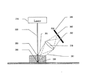
図3において、図1における各種外観データを計測するためのセンサの一実施例を示す。本センサは、三角測量の原理に基づいて被検体の表面形状データおよび輝度データを計測する一例である。本装置は、レーザーユニット310からレーザー光320を被検体330に照射し、被検体330からの反射光をPSD(Position Sensitive Detector)センサ(340)で受光する。三角測量の原理から、レーザーの照射座標、被検体上の計測座標(サンプリング座標)、センサにおける信号受信座標により、サンプリング点における高さが決定される。例えば、被検体330が存在する場合、レーザー光320はポイント350で反射し、集光レンズ370を通り、その反射光351はPSD340におけるポイント352へ到達する。これに対し、被検体330が存在しない場合、照射されたレーザー光320はポイント360で反射し、その反射光361は集光レンズ370を通り、PSD340におけるポイント362へ到達する。
FIG. 3 shows an embodiment of a sensor for measuring various appearance data in FIG. This sensor is an example of measuring surface shape data and luminance data of a subject based on the principle of triangulation. This apparatus irradiates the subject 330 with laser light 320 from the
このように、サンプリング座標における高さ(Z)の違いにより、PSDセンサが受光する位置が異なる。PSDは受光位置に応じた2つの信号を出力するセンサであり、この2つの信号を(式1)に基づいて計算することによりサンプリング座標における高さを決定することができる。また図3においては、簡略化のため投光光学系および受光光学系の記載を省略しているが、これらが組み込まれた場合においても、計測原理は同様である。 Thus, the position where the PSD sensor receives light varies depending on the difference in height (Z) in the sampling coordinates. The PSD is a sensor that outputs two signals according to the light receiving position, and the height at the sampling coordinates can be determined by calculating these two signals based on (Equation 1). In FIG. 3, the description of the light projecting optical system and the light receiving optical system is omitted for simplification, but the measurement principle is the same even when these are incorporated.
本センサにおいて、任意領域の表面形状データを計測するためには、本制御系を固定したまま被検体をXY平面内で平行移動させて信号計測を繰り返し行うか、あるいは被検体を固定しサンプリング座標点を平行移動させることによって、2次元領域における表面形状データを計測することが可能となる。このとき計測した表面形状データは、2次元マトリクスに格納され、その画素値が、各サンプリング点における高さを表す。以下高さ画像と記述する。 In this sensor, in order to measure surface shape data in an arbitrary area, the subject is translated in the XY plane while the control system is fixed, and signal measurement is repeated, or the subject is fixed and sampling coordinates are measured. By moving the points in parallel, it is possible to measure surface shape data in a two-dimensional region. The surface shape data measured at this time is stored in a two-dimensional matrix, and the pixel value represents the height at each sampling point. Hereinafter described as a height image.
また本装置において、PSDセンサの2つの出力端子から出力される2つの信号データIa(x,y)とIb(x,y)を足し合わせることにより、受光光量を反映した輝度データを生成することができる(輝度画像)。これを(式2)に示す。本輝度画像は、ステップ120の基準マーク位置座標認識処理などに適用する。
Further, in this apparatus, luminance data reflecting the amount of received light is generated by adding two signal data Ia (x, y) and Ib (x, y) output from the two output terminals of the PSD sensor. (Brightness image). This is shown in (Formula 2). This luminance image is applied to the reference mark position coordinate recognition process in
H(x、y)=Ia(x、y)/(Ia(x、y)+Ib(x、y))・・・(式1)
但し、H(x、y)は、任意座標(x、y)における表面形状データ
Ia(x、y)は、任意座標(x、y)におけるPSDの出力信号値1
Ib(x、y)は、任意座標(x、y)におけるPSDの出力信号値2
B(x、y)=Ia(x、y)+Ib(x、y)・・・(式2)
但し、B(x、y)は、検査面座標(x、y)における輝度値
Ia(x、y)は、検査面座標(x、y)におけるPSDの出力信号値1
Ib(x、y)は、検査面座標(x、y)におけるPSDの出力信号値2
なお、上記では、レーザー三角測量を用いた形状データ計測方法を示したが、その他形状データ計測においては、レーザー変位計を用いたり、CCDカメラやX線によるステレオ計測により基板面の高さを計測してもよい。ステレオ法の場合、基板上面の回路パターンの位置を画像処理により求め、その回路パターンの高さを基板面高さとして決定してもよい。
H (x, y) = Ia (x, y) / (Ia (x, y) + Ib (x, y)) (Formula 1)
However, H (x, y) is the surface shape data Ia (x, y) at the arbitrary coordinates (x, y), PSD output signal value 1 at the arbitrary coordinates (x, y)
Ib (x, y) is the PSD output signal value 2 at an arbitrary coordinate (x, y).
B (x, y) = Ia (x, y) + Ib (x, y) (Formula 2)
However, B (x, y) is the luminance value at the inspection plane coordinates (x, y) Ia (x, y) is the PSD output signal value 1 at the inspection plane coordinates (x, y)
Ib (x, y) is the PSD output signal value 2 at the inspection plane coordinates (x, y).
In the above, the shape data measurement method using laser triangulation was shown. However, in other shape data measurement, the height of the substrate surface is measured using a laser displacement meter or stereo measurement using a CCD camera or X-ray. May be. In the case of the stereo method, the position of the circuit pattern on the upper surface of the substrate may be obtained by image processing, and the height of the circuit pattern may be determined as the height of the substrate surface.
図4、図5において、図3のセンサを用いて計測した外観画像から、被検体座標における基板近似平面を決定し、X線CTにおいて画像再構成を行う再構成エリアを決定する一例を示す。 4 and 5 show an example in which a substrate approximate plane at object coordinates is determined from the appearance image measured using the sensor of FIG. 3, and a reconstruction area for image reconstruction in X-ray CT is determined.
まず検査データにおいて図1ステップ210で示したが、検査データにおいては、検査を行う部品(被検体)の周辺に基板面の高さを計測する位置(XY座標)と領域サイズを含むグランド情報と、被検体の位置(XY座標)やサイズの形体データと、その部品の底面高さを決定するためのグランド情報をリンク付けするデータを含む部品情報が含まれている。
First, as shown in
基板近似平面を決定するため、被検体400の高さを決定するためにリンク付けするグランド情報を被検体400に対して3点(413,414,415)登録する。通常の場合、基板高さを決定するためのグランド413、414、415は、レーザー反射光が安定している回路パターン411、412上に設定する。各グランドにおける高さの決定は、グランド領域内において、輝度画像の値が高い画素(グランド候補画素)を決定し、そのグランド候補画素における高さ画像の画素値の平均を求め、これをグランド候補座標重心(X,Y)における基板高さ(Z)として決定する。そして決定したグランド413,414,415の座標(X,Y,Z)から、この3点を通る平面を算出し、この平面を被検体における基板近似平面420として決定する。3点を通る平面は、(式3)により平面の法線ベクトルを決定し、(式4)の平面の方程式を用いて決定する。
In order to determine the board approximate plane, three points (413, 414, 415) of ground information to be linked to determine the height of the subject 400 are registered in the subject 400. In a normal case, the
V=(p1−p2)×(p3−p2)・・・(式3)
但し、Vは、法線ベクトル、p1は、座標(plx、ply、plz)、p2は、座標(p2x、p2y、p2z)、p3は、座標(p3x、p3y、p3z)、×は、外積を表す。
Ax+By+Cz+d=0・・・(式4)
そして(式4)にて決定した基板近似平面420を基準に、X線CTにおいて画像再構成を行う再構成マトリクス430の座標を決定する。具体的には、画像再構成マトリクス430のXY座標を、基板近似平面420と平行にし、Z座標を基板近似平面の法線方向に配置する。
V = (p1-p2) × (p3-p2) (Formula 3)
Where V is a normal vector, p1 is coordinates (plx, ply, plz), p2 is coordinates (p2x, p2y, p2z), p3 is coordinates (p3x, p3y, p3z), and x is an outer product. To express.
Ax + By + Cz + d = 0 (Formula 4)
Then, the coordinates of the
即ち、画像再構成して画像化する領域は、基準平面を1底面として構成して、検査する電子基板上の被検体を包含する画像再構成領域を直方体とすることにより、必要最小限の範囲で画像再構成を行うことができる。 That is, the area to be reconstructed and imaged is the minimum necessary area by configuring the reference plane as one bottom surface and making the image reconstruction area including the subject on the electronic substrate to be examined a rectangular parallelepiped. Image reconstruction can be performed.
この画像再構成マトリクスに対して、コーンビームCTにおける画像再構成演算を行い、CT画像を生成する。 An image reconstruction operation in cone beam CT is performed on this image reconstruction matrix to generate a CT image.
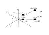
上記においては、被検体にリンク付けされるグランド情報のリンク付け数が3点の場合を示したが、2点の場合も簡略的に行うことができる。2点の場合の基板近似平面決定の一例を図5において示す。決定したグランド座標510(p1)とグランド座標520(p2)において、XY平面にそれぞれ垂線をおろした交点511(p1S)、あるいは521(p2s)の1つを決定し、前記グランド座標510、520と決定した交点(511あるいは521)の作る平面と垂直で、かつグランド座標(510、520)を結んだ線分を通る平面を基板近似平面として決定する。近似平面の法線ベクトルを決定する式を(式5)に示す。 In the above, the case where the number of links of ground information linked to the subject is three points is shown, but the case of two points can also be simply performed. An example of the board approximate plane determination in the case of two points is shown in FIG. In the determined ground coordinates 510 (p1) and ground coordinates 520 (p2), one of intersection points 511 (p1S) or 521 (p2s) perpendicular to the XY plane is determined, and the ground coordinates 510, 520 and A plane perpendicular to the plane formed by the determined intersection point (511 or 521) and passing through a line segment connecting the ground coordinates (510, 520) is determined as the substrate approximate plane. An expression for determining the normal vector of the approximate plane is shown in (Expression 5).
V=(p2−p1)×(pls−p1)×(p2−p1) ・・・(式5)
但し、Vは法線ベクトル、p1は、座標(plx、ply、plz)、p2は、座標(p2x、p2y、p2z)、plsは、座標(plx、ply、0)、×は、外積を表す。
V = (p2-p1) * (pls-p1) * (p2-p1) (Formula 5)
Where V is a normal vector, p1 is coordinates (plx, ply, plz), p2 is coordinates (p2x, p2y, p2z), pls is coordinates (plx, ply, 0), and x is an outer product. .
本発明にかかるX線CT検査装置は、画像再構成の計算範囲を必要最小限に設定することができることから、X線CT検査装置における再構成時間を高速化する用途において適している。 Since the X-ray CT inspection apparatus according to the present invention can set the calculation range of image reconstruction to the minimum necessary, the X-ray CT inspection apparatus is suitable for use in speeding up the reconstruction time in the X-ray CT inspection apparatus.
また本発明にかかるX線CT検査装置は、基板近似平面を基準に画像再構成範囲を決定することから、基板上に存在する被検体に対して、ユーザーが欲するとする領域のデータを的確に画像化することができることから、被検体の位置を意識することなく高精度な位置決めを実現する機能として有用であり、操作性を容易にすることに適している。 In addition, since the X-ray CT inspection apparatus according to the present invention determines the image reconstruction range based on the approximate plane of the substrate, the data of the region that the user desires is accurately obtained for the subject existing on the substrate. Since it can be imaged, it is useful as a function for realizing highly accurate positioning without being aware of the position of the subject, and is suitable for facilitating operability.
121 外観データ計測部
122 レーザーユニット
123 レーザー反射光受光センサ
125 X線データ計測部
126 X線源
127 X線センサ
129 被検体
121 External Appearance
Claims (7)
前記被検体にX線を照射する手段と、
前記被検体を透過したX線を計測する手段と、
前記X線計測手段により計測したX線データを用いて前記被検体の断層画像の画像再構成を行う手段と、
前記電子基板上の被検体の表面形状データを計測する手段と、
前記電子基板内の異なる2領域以上の表面形状データから、前記断層画像の基準面となる基板近似平面を決定する手段と、
を備え、
前記決定した基板近似平面を基準に、前記画像再構成手段により画像化する領域を決定することを特徴とするX線CT検査装置。 An X-ray CT examination apparatus that generates a tomographic image of a subject using a transmission X-ray image transmitted through the subject mounted on an electronic substrate,
Means for irradiating the subject with X-rays;
Means for measuring X-rays transmitted through the subject;
Means for reconstructing a tomographic image of the subject using the X-ray data measured by the X-ray measuring means;
Means for measuring surface shape data of an object on the electronic substrate;
Means for determining a substrate approximate plane serving as a reference plane of the tomographic image from surface shape data of two or more different regions in the electronic substrate;
With
An X-ray CT inspection apparatus characterized in that a region to be imaged by the image reconstruction means is determined based on the determined substrate approximate plane.
前記被検体にX線を照射し、
前記被検体を透過したX線を計測し、
前記透過したX線を計測して得られるX線データを用いて前記被検体の断層画像の画像再構成を行うとともに、
前記電子基板上の被検体の表面形状データを計測し、
前記電子基板内の異なる2領域以上の表面形状データから、前記断層画像の基準面となる基板近似平面を決定し、
前記決定した基板近似平面を基準に、前記画像再構成を行って画像化する領域を決定することを特徴とするX線CT検査方法。 An X-ray CT examination method for generating a tomographic image of a subject using a transmission X-ray image transmitted through the subject mounted on an electronic substrate,
Irradiating the subject with X-rays;
Measure X-rays transmitted through the subject,
While performing image reconstruction of the tomographic image of the subject using X-ray data obtained by measuring the transmitted X-ray,
Measure surface shape data of the object on the electronic substrate,
From the surface shape data of two or more different areas in the electronic substrate, determine a substrate approximate plane to be a reference surface of the tomographic image,
An X-ray CT inspection method, wherein an area to be imaged is determined by performing the image reconstruction on the basis of the determined substrate approximate plane.
Priority Applications (1)
| Application Number | Priority Date | Filing Date | Title |
|---|---|---|---|
| JP2005031394A JP4591103B2 (en) | 2005-02-08 | 2005-02-08 | X-ray CT inspection apparatus and X-ray CT inspection method |
Applications Claiming Priority (1)
| Application Number | Priority Date | Filing Date | Title |
|---|---|---|---|
| JP2005031394A JP4591103B2 (en) | 2005-02-08 | 2005-02-08 | X-ray CT inspection apparatus and X-ray CT inspection method |
Publications (2)
| Publication Number | Publication Date |
|---|---|
| JP2006220424A JP2006220424A (en) | 2006-08-24 |
| JP4591103B2 true JP4591103B2 (en) | 2010-12-01 |
Family
ID=36982860
Family Applications (1)
| Application Number | Title | Priority Date | Filing Date |
|---|---|---|---|
| JP2005031394A Expired - Fee Related JP4591103B2 (en) | 2005-02-08 | 2005-02-08 | X-ray CT inspection apparatus and X-ray CT inspection method |
Country Status (1)
| Country | Link |
|---|---|
| JP (1) | JP4591103B2 (en) |
Families Citing this family (6)
| Publication number | Priority date | Publication date | Assignee | Title |
|---|---|---|---|---|
| JP4580266B2 (en) * | 2005-04-07 | 2010-11-10 | 名古屋電機工業株式会社 | X-ray inspection apparatus, X-ray inspection method, and X-ray inspection program |
| JP4650076B2 (en) * | 2005-04-20 | 2011-03-16 | パナソニック株式会社 | Circuit pattern inspection apparatus and circuit pattern inspection method |
| JP4610590B2 (en) * | 2007-09-04 | 2011-01-12 | 名古屋電機工業株式会社 | X-ray inspection apparatus, X-ray inspection method, and X-ray inspection program |
| CN111033245B (en) * | 2017-10-23 | 2022-08-05 | 东丽株式会社 | Method for inspecting resin molded product, method for manufacturing resin molded product, and apparatus for inspecting resin molded product and apparatus for manufacturing resin molded product |
| CN113167567B (en) * | 2019-01-25 | 2023-03-24 | 东丽株式会社 | Structure inspection method and manufacturing method, structure inspection device and manufacturing device |
| JP7303069B2 (en) * | 2019-08-28 | 2023-07-04 | ヤマハ発動機株式会社 | inspection equipment |
Citations (5)
| Publication number | Priority date | Publication date | Assignee | Title |
|---|---|---|---|---|
| JPS58153807U (en) * | 1982-04-09 | 1983-10-14 | 株式会社日立メデイコ | X-ray CT device |
| JPH0618450A (en) * | 1992-07-06 | 1994-01-25 | Fujitsu Ltd | Plane sample tomography device |
| JPH0618451A (en) * | 1992-07-06 | 1994-01-25 | Fujitsu Ltd | Tomographic method for plane samples |
| JP2002200073A (en) * | 2000-12-21 | 2002-07-16 | Ge Medical Systems Global Technology Co Llc | X-ray ct system |
| JP2004037222A (en) * | 2002-07-03 | 2004-02-05 | Matsushita Electric Ind Co Ltd | Teaching method, electronic substrate inspection method, and electronic substrate inspection device |
-
2005
- 2005-02-08 JP JP2005031394A patent/JP4591103B2/en not_active Expired - Fee Related
Patent Citations (5)
| Publication number | Priority date | Publication date | Assignee | Title |
|---|---|---|---|---|
| JPS58153807U (en) * | 1982-04-09 | 1983-10-14 | 株式会社日立メデイコ | X-ray CT device |
| JPH0618450A (en) * | 1992-07-06 | 1994-01-25 | Fujitsu Ltd | Plane sample tomography device |
| JPH0618451A (en) * | 1992-07-06 | 1994-01-25 | Fujitsu Ltd | Tomographic method for plane samples |
| JP2002200073A (en) * | 2000-12-21 | 2002-07-16 | Ge Medical Systems Global Technology Co Llc | X-ray ct system |
| JP2004037222A (en) * | 2002-07-03 | 2004-02-05 | Matsushita Electric Ind Co Ltd | Teaching method, electronic substrate inspection method, and electronic substrate inspection device |
Also Published As
| Publication number | Publication date |
|---|---|
| JP2006220424A (en) | 2006-08-24 |
Similar Documents
| Publication | Publication Date | Title |
|---|---|---|
| US8233041B2 (en) | Image processing device and image processing method for performing three dimensional measurements | |
| US6665433B2 (en) | Automatic X-ray determination of solder joint and view Delta Z values from a laser mapped reference surface for circuit board inspection using X-ray laminography | |
| KR101948852B1 (en) | Hybrid image scanning method and apparatus for noncontact crack evaluation | |
| US6314201B1 (en) | Automatic X-ray determination of solder joint and view delta Z values from a laser mapped reference surface for circuit board inspection using X-ray laminography | |
| JP5125423B2 (en) | Method of inspecting solder electrode by X-ray tomographic image and board inspection apparatus using this method | |
| US10054432B2 (en) | X-ray inspection apparatus and control method | |
| US20110081070A1 (en) | Process and Apparatus for Image Processing and Computer-readable Medium Storing Image Processing Program | |
| JP2017151067A (en) | Three-dimensional image inspection device, three-dimensional image inspection method, three-dimensional image inspection program, and computer-readable recording medium and recorded device | |
| US9157874B2 (en) | System and method for automated x-ray inspection | |
| KR20140143724A (en) | High throughput and low cost height triangulation system and method | |
| CN111247424A (en) | Inspection position specifying method, three-dimensional image generating method, and inspection device | |
| JP4133753B2 (en) | Method of measuring optical interference of detour surface and interferometer device for detour surface measurement | |
| JP5830928B2 (en) | Inspection area setting method and X-ray inspection system | |
| JP2013130566A (en) | Lens testing device and method | |
| JP2008014882A (en) | 3D measuring device | |
| JP4591103B2 (en) | X-ray CT inspection apparatus and X-ray CT inspection method | |
| JP4449596B2 (en) | Mounting board inspection equipment | |
| JP2009162596A (en) | Method of supporting image confirmation work and substrate inspection apparatus utilizing x ray using the same | |
| JP2003240736A (en) | X-ray tomographic plane inspection method and X-ray tomographic plane inspection apparatus | |
| JP2009139285A (en) | Solder ball inspection device, inspection method thereof, and shape inspection device | |
| JP4333349B2 (en) | Mounting appearance inspection method and mounting appearance inspection apparatus | |
| JP4580266B2 (en) | X-ray inspection apparatus, X-ray inspection method, and X-ray inspection program | |
| JP2012013593A (en) | Calibration method for three-dimensional shape measuring machine, and three-dimensional shape measuring machine | |
| US20240062401A1 (en) | Measurement system, inspection system, measurement device, measurement method, inspection method, and program | |
| CN115004018A (en) | X-ray inspection system, X-ray inspection method and program |
Legal Events
| Date | Code | Title | Description |
|---|---|---|---|
| A621 | Written request for application examination |
Free format text: JAPANESE INTERMEDIATE CODE: A621 Effective date: 20071219 |
|
| RD01 | Notification of change of attorney |
Free format text: JAPANESE INTERMEDIATE CODE: A7421 Effective date: 20080115 |
|
| RD01 | Notification of change of attorney |
Free format text: JAPANESE INTERMEDIATE CODE: A7421 Effective date: 20091120 |
|
| A977 | Report on retrieval |
Free format text: JAPANESE INTERMEDIATE CODE: A971007 Effective date: 20100804 |
|
| TRDD | Decision of grant or rejection written | ||
| A01 | Written decision to grant a patent or to grant a registration (utility model) |
Free format text: JAPANESE INTERMEDIATE CODE: A01 Effective date: 20100817 |
|
| A01 | Written decision to grant a patent or to grant a registration (utility model) |
Free format text: JAPANESE INTERMEDIATE CODE: A01 |
|
| A61 | First payment of annual fees (during grant procedure) |
Free format text: JAPANESE INTERMEDIATE CODE: A61 Effective date: 20100830 |
|
| FPAY | Renewal fee payment (event date is renewal date of database) |
Free format text: PAYMENT UNTIL: 20130924 Year of fee payment: 3 |
|
| FPAY | Renewal fee payment (event date is renewal date of database) |
Free format text: PAYMENT UNTIL: 20130924 Year of fee payment: 3 |
|
| LAPS | Cancellation because of no payment of annual fees |


