JP4347539B2 - Spinal cord nuclear transfer - Google Patents
Spinal cord nuclear transfer Download PDFInfo
- Publication number
- JP4347539B2 JP4347539B2 JP2001526118A JP2001526118A JP4347539B2 JP 4347539 B2 JP4347539 B2 JP 4347539B2 JP 2001526118 A JP2001526118 A JP 2001526118A JP 2001526118 A JP2001526118 A JP 2001526118A JP 4347539 B2 JP4347539 B2 JP 4347539B2
- Authority
- JP
- Japan
- Prior art keywords
- spinal
- nucleus
- hydrophilic
- plastic
- expandable
- Prior art date
- Legal status (The legal status is an assumption and is not a legal conclusion. Google has not performed a legal analysis and makes no representation as to the accuracy of the status listed.)
- Expired - Fee Related
Links
Images


Classifications
-
- A—HUMAN NECESSITIES
- A61—MEDICAL OR VETERINARY SCIENCE; HYGIENE
- A61L—METHODS OR APPARATUS FOR STERILISING MATERIALS OR OBJECTS IN GENERAL; DISINFECTION, STERILISATION OR DEODORISATION OF AIR; CHEMICAL ASPECTS OF BANDAGES, DRESSINGS, ABSORBENT PADS OR SURGICAL ARTICLES; MATERIALS FOR BANDAGES, DRESSINGS, ABSORBENT PADS OR SURGICAL ARTICLES
- A61L27/00—Materials for grafts or prostheses or for coating grafts or prostheses
- A61L27/14—Macromolecular materials
- A61L27/20—Polysaccharides
-
- A—HUMAN NECESSITIES
- A61—MEDICAL OR VETERINARY SCIENCE; HYGIENE
- A61F—FILTERS IMPLANTABLE INTO BLOOD VESSELS; PROSTHESES; DEVICES PROVIDING PATENCY TO, OR PREVENTING COLLAPSING OF, TUBULAR STRUCTURES OF THE BODY, e.g. STENTS; ORTHOPAEDIC, NURSING OR CONTRACEPTIVE DEVICES; FOMENTATION; TREATMENT OR PROTECTION OF EYES OR EARS; BANDAGES, DRESSINGS OR ABSORBENT PADS; FIRST-AID KITS
- A61F2/00—Filters implantable into blood vessels; Prostheses, i.e. artificial substitutes or replacements for parts of the body; Appliances for connecting them with the body; Devices providing patency to, or preventing collapsing of, tubular structures of the body, e.g. stents
- A61F2/02—Prostheses implantable into the body
- A61F2/30—Joints
- A61F2/44—Joints for the spine, e.g. vertebrae, spinal discs
- A61F2/441—Joints for the spine, e.g. vertebrae, spinal discs made of inflatable pockets or chambers filled with fluid, e.g. with hydrogel
-
- A—HUMAN NECESSITIES
- A61—MEDICAL OR VETERINARY SCIENCE; HYGIENE
- A61L—METHODS OR APPARATUS FOR STERILISING MATERIALS OR OBJECTS IN GENERAL; DISINFECTION, STERILISATION OR DEODORISATION OF AIR; CHEMICAL ASPECTS OF BANDAGES, DRESSINGS, ABSORBENT PADS OR SURGICAL ARTICLES; MATERIALS FOR BANDAGES, DRESSINGS, ABSORBENT PADS OR SURGICAL ARTICLES
- A61L27/00—Materials for grafts or prostheses or for coating grafts or prostheses
- A61L27/14—Macromolecular materials
- A61L27/16—Macromolecular materials obtained by reactions only involving carbon-to-carbon unsaturated bonds
-
- A—HUMAN NECESSITIES
- A61—MEDICAL OR VETERINARY SCIENCE; HYGIENE
- A61L—METHODS OR APPARATUS FOR STERILISING MATERIALS OR OBJECTS IN GENERAL; DISINFECTION, STERILISATION OR DEODORISATION OF AIR; CHEMICAL ASPECTS OF BANDAGES, DRESSINGS, ABSORBENT PADS OR SURGICAL ARTICLES; MATERIALS FOR BANDAGES, DRESSINGS, ABSORBENT PADS OR SURGICAL ARTICLES
- A61L27/00—Materials for grafts or prostheses or for coating grafts or prostheses
- A61L27/14—Macromolecular materials
- A61L27/18—Macromolecular materials obtained otherwise than by reactions only involving carbon-to-carbon unsaturated bonds
-
- A—HUMAN NECESSITIES
- A61—MEDICAL OR VETERINARY SCIENCE; HYGIENE
- A61L—METHODS OR APPARATUS FOR STERILISING MATERIALS OR OBJECTS IN GENERAL; DISINFECTION, STERILISATION OR DEODORISATION OF AIR; CHEMICAL ASPECTS OF BANDAGES, DRESSINGS, ABSORBENT PADS OR SURGICAL ARTICLES; MATERIALS FOR BANDAGES, DRESSINGS, ABSORBENT PADS OR SURGICAL ARTICLES
- A61L27/00—Materials for grafts or prostheses or for coating grafts or prostheses
- A61L27/40—Composite materials, i.e. containing one material dispersed in a matrix of the same or different material
- A61L27/44—Composite materials, i.e. containing one material dispersed in a matrix of the same or different material having a macromolecular matrix
-
- A—HUMAN NECESSITIES
- A61—MEDICAL OR VETERINARY SCIENCE; HYGIENE
- A61L—METHODS OR APPARATUS FOR STERILISING MATERIALS OR OBJECTS IN GENERAL; DISINFECTION, STERILISATION OR DEODORISATION OF AIR; CHEMICAL ASPECTS OF BANDAGES, DRESSINGS, ABSORBENT PADS OR SURGICAL ARTICLES; MATERIALS FOR BANDAGES, DRESSINGS, ABSORBENT PADS OR SURGICAL ARTICLES
- A61L27/00—Materials for grafts or prostheses or for coating grafts or prostheses
- A61L27/50—Materials characterised by their function or physical properties, e.g. injectable or lubricating compositions, shape-memory materials, surface modified materials
- A61L27/52—Hydrogels or hydrocolloids
-
- A—HUMAN NECESSITIES
- A61—MEDICAL OR VETERINARY SCIENCE; HYGIENE
- A61F—FILTERS IMPLANTABLE INTO BLOOD VESSELS; PROSTHESES; DEVICES PROVIDING PATENCY TO, OR PREVENTING COLLAPSING OF, TUBULAR STRUCTURES OF THE BODY, e.g. STENTS; ORTHOPAEDIC, NURSING OR CONTRACEPTIVE DEVICES; FOMENTATION; TREATMENT OR PROTECTION OF EYES OR EARS; BANDAGES, DRESSINGS OR ABSORBENT PADS; FIRST-AID KITS
- A61F2/00—Filters implantable into blood vessels; Prostheses, i.e. artificial substitutes or replacements for parts of the body; Appliances for connecting them with the body; Devices providing patency to, or preventing collapsing of, tubular structures of the body, e.g. stents
- A61F2/02—Prostheses implantable into the body
- A61F2/30—Joints
- A61F2/44—Joints for the spine, e.g. vertebrae, spinal discs
- A61F2/442—Intervertebral or spinal discs, e.g. resilient
-
- A—HUMAN NECESSITIES
- A61—MEDICAL OR VETERINARY SCIENCE; HYGIENE
- A61F—FILTERS IMPLANTABLE INTO BLOOD VESSELS; PROSTHESES; DEVICES PROVIDING PATENCY TO, OR PREVENTING COLLAPSING OF, TUBULAR STRUCTURES OF THE BODY, e.g. STENTS; ORTHOPAEDIC, NURSING OR CONTRACEPTIVE DEVICES; FOMENTATION; TREATMENT OR PROTECTION OF EYES OR EARS; BANDAGES, DRESSINGS OR ABSORBENT PADS; FIRST-AID KITS
- A61F2/00—Filters implantable into blood vessels; Prostheses, i.e. artificial substitutes or replacements for parts of the body; Appliances for connecting them with the body; Devices providing patency to, or preventing collapsing of, tubular structures of the body, e.g. stents
- A61F2/02—Prostheses implantable into the body
- A61F2/30—Joints
- A61F2002/30001—Additional features of subject-matter classified in A61F2/28, A61F2/30 and subgroups thereof
- A61F2002/30003—Material related properties of the prosthesis or of a coating on the prosthesis
- A61F2002/3006—Properties of materials and coating materials
- A61F2002/30075—Properties of materials and coating materials swellable, e.g. when wetted
-
- A—HUMAN NECESSITIES
- A61—MEDICAL OR VETERINARY SCIENCE; HYGIENE
- A61F—FILTERS IMPLANTABLE INTO BLOOD VESSELS; PROSTHESES; DEVICES PROVIDING PATENCY TO, OR PREVENTING COLLAPSING OF, TUBULAR STRUCTURES OF THE BODY, e.g. STENTS; ORTHOPAEDIC, NURSING OR CONTRACEPTIVE DEVICES; FOMENTATION; TREATMENT OR PROTECTION OF EYES OR EARS; BANDAGES, DRESSINGS OR ABSORBENT PADS; FIRST-AID KITS
- A61F2/00—Filters implantable into blood vessels; Prostheses, i.e. artificial substitutes or replacements for parts of the body; Appliances for connecting them with the body; Devices providing patency to, or preventing collapsing of, tubular structures of the body, e.g. stents
- A61F2/02—Prostheses implantable into the body
- A61F2/30—Joints
- A61F2/44—Joints for the spine, e.g. vertebrae, spinal discs
- A61F2/442—Intervertebral or spinal discs, e.g. resilient
- A61F2002/4435—Support means or repair of the natural disc wall, i.e. annulus, e.g. using plates, membranes or meshes
-
- A—HUMAN NECESSITIES
- A61—MEDICAL OR VETERINARY SCIENCE; HYGIENE
- A61F—FILTERS IMPLANTABLE INTO BLOOD VESSELS; PROSTHESES; DEVICES PROVIDING PATENCY TO, OR PREVENTING COLLAPSING OF, TUBULAR STRUCTURES OF THE BODY, e.g. STENTS; ORTHOPAEDIC, NURSING OR CONTRACEPTIVE DEVICES; FOMENTATION; TREATMENT OR PROTECTION OF EYES OR EARS; BANDAGES, DRESSINGS OR ABSORBENT PADS; FIRST-AID KITS
- A61F2/00—Filters implantable into blood vessels; Prostheses, i.e. artificial substitutes or replacements for parts of the body; Appliances for connecting them with the body; Devices providing patency to, or preventing collapsing of, tubular structures of the body, e.g. stents
- A61F2/02—Prostheses implantable into the body
- A61F2/30—Joints
- A61F2/44—Joints for the spine, e.g. vertebrae, spinal discs
- A61F2/442—Intervertebral or spinal discs, e.g. resilient
- A61F2002/444—Intervertebral or spinal discs, e.g. resilient for replacing the nucleus pulposus
-
- A—HUMAN NECESSITIES
- A61—MEDICAL OR VETERINARY SCIENCE; HYGIENE
- A61F—FILTERS IMPLANTABLE INTO BLOOD VESSELS; PROSTHESES; DEVICES PROVIDING PATENCY TO, OR PREVENTING COLLAPSING OF, TUBULAR STRUCTURES OF THE BODY, e.g. STENTS; ORTHOPAEDIC, NURSING OR CONTRACEPTIVE DEVICES; FOMENTATION; TREATMENT OR PROTECTION OF EYES OR EARS; BANDAGES, DRESSINGS OR ABSORBENT PADS; FIRST-AID KITS
- A61F2210/00—Particular material properties of prostheses classified in groups A61F2/00 - A61F2/26 or A61F2/82 or A61F9/00 or A61F11/00 or subgroups thereof
- A61F2210/0061—Particular material properties of prostheses classified in groups A61F2/00 - A61F2/26 or A61F2/82 or A61F9/00 or A61F11/00 or subgroups thereof swellable
-
- A—HUMAN NECESSITIES
- A61—MEDICAL OR VETERINARY SCIENCE; HYGIENE
- A61L—METHODS OR APPARATUS FOR STERILISING MATERIALS OR OBJECTS IN GENERAL; DISINFECTION, STERILISATION OR DEODORISATION OF AIR; CHEMICAL ASPECTS OF BANDAGES, DRESSINGS, ABSORBENT PADS OR SURGICAL ARTICLES; MATERIALS FOR BANDAGES, DRESSINGS, ABSORBENT PADS OR SURGICAL ARTICLES
- A61L2430/00—Materials or treatment for tissue regeneration
- A61L2430/38—Materials or treatment for tissue regeneration for reconstruction of the spine, vertebrae or intervertebral discs
Abstract
Description
【0001】
【発明の属する技術分野】
本発明は、生きている脊椎動物、例えばヒトの脊髄円板から除去された髄核の全てまたは一部を置換する脊髄核移植片に関する。この脊髄核移植片は、異方的に膨張する能力のあるキセロゲルから形成される。
【0002】
【従来の技術】
脊髄の椎間板は、隣接脊椎の終板の間に配置される軟骨組織である。骨髄の椎間板は、脊椎の間の柔軟性のある関節として作用し、そして椎柱をたわませ、そして捻らせうる。脊髄の椎間板に対する損傷は、脊髄機能不全、有害な痛みおよび短期または長期の不自由を起しうる。この問題の広範な発生(骨髄の椎間板のため5%の年次発生の背中の痛みが報告される)のため、経済的結果が膨大である。ある種の円板の問題は、手術を必要とする。典型的な最新の手段は、米国特許番号第4,636,217号(Ogilvieら)、第5,489,308号(Kuslichら)および第5,716,415号(Steffee)で記述されるもののような、種々の技術およびデバイスを用いた隣接脊椎の融合である。核またはそれの一部の除去(椎弓切開術)、または隣接脊椎の融合のような全ての最近利用できる手術手段は、一方向または他方向での脊髄機能を弱める。
【0003】
この理由のために、円板またはその部分のプロテーゼの開発を含めた新たな手段が求められている。これは、引き受けるのが困難である。脊柱は、極端に込入った体の部分であり、そしてそれの適切な機能は、椎間板を含めたそれの構成要素の全ての途切れのない協力に依存する。椎間板は、多数の機能を行わなければならない。混合したひずみ、トルク、せん断および圧縮を含めて、非常に複雑な変形のモデルでの反復した高い緊張に耐えなければならない。さらに、脊髄の椎間板は、有効な衝撃吸収材、および円板への栄養および円板からの代謝物の流れを起動するポンプとして作用する。構造的には、円板は、複合体および込入った形態で組織化された数種の型の材料に関するむしろ複雑な複合体部分である。椎骨の終板は、コラーゲンマトリックス、糖タンパク成分、および水から構成される硝子質の軟骨の層に覆われている。さらに、その容積の約2−5%は、軟骨の構成成分を産生する生きた細胞によって占められている。
【0004】
それ自身の脊髄の椎間板は、主に、結晶性コラーゲン原線維および不定形親水性プロテオグリカンから構成される。その容積の約3−5%は、それの構成要素を産生する生きた細胞によって占められている。構造的には、脊髄の椎間板は、髄核と称されるヒドロゲル様コア、および線維輪と称される外側環から構成される。骨髄の椎間板の構造は、図1に模式図で描かれ、そして以下に記述される。
【0005】
脊髄の椎間板は、体重を支え、そして柔軟性のある関節として第一に作用する。それは、隣接椎骨の共有の回転、ひずみおよび平行移動を可能にする一方で、相当の軸負荷に耐える。さらに、脊髄の椎間板は、振動および機械的衝撃を弱め、そして骨格系を通してそれらの伝播を阻止する。選択された方向での負荷に耐える許容性および柔軟性は、線維輪および髄核の組合せによって達成される。線維輪は、放射方向に硬質であるが、しかし軸方向で、そしてトルクによって変形性がある積層構造物である。軸の負荷は、線維輪によって含まれる軸成分に部分的にそれを変形させる髄核によって生じられる。線維輪は、数種の層で組織化されるコラーゲン原線維によって主に形成される。各層は、ある角度でそれのコラーゲン原線維損傷を有し、そして連続層は、選択方向を示す。コラーゲン組織は、タイヤでの圧力容器またはコードのために使用される複合体でと同様に、線維強化の組織化と密接に類似する。それは、放射状圧力(または内部圧力)に最大限の耐性を保証する一方で、トルクおよびゆがみでの変形を可能にする。
【0006】
原線維末端は、隣接椎骨に、そして椎骨終板の軟骨様表面に付着される。結果的に、線維輪の内側空間は、実質的に封入されている。コアの内側または外側で透過するあらゆる液体は、線維輪組織を、または椎骨終板を通過しなければならない。十分な水圧透過性を達成するために、線維輪のコラーゲン様構造は、コラーゲン原線維の間に埋設されるプロテオグリカンによって補足される。プロテオグリカンは、水和され、その結果、線維輪は、一種の非常に組織化された異方性のハイドロゲル複合体を形成する。コラーゲンドメインは、細線維性メッシュを形成する。この配列の結果は、負荷に耐える機能のために必要とされる高い機械的強度、特に高いせん断強度、および破壊生長に対する耐性と組合された選択方向における十分な変形性である。
【0007】
髄核は、線維輪に接触されているが、終板には接触されていない。それは、いっそう低い濃度のコラーゲン(その濃度は、年齢と共に増加する)および高い濃度の親水性プロテオグリカンを有する。結果的に、それは、ある程度ハイドロゲルのようである天然の複合体であり、そして非常に高い平衡な水分含有率(若いヒトでより90重量%多い)を示す。水分含有率および髄核の容積は、膨張媒体の容積オスモル濃度に、そして機械的圧力に依存する。機械的圧力のため液体含有率の減少に対する耐性は、「膨張圧」と称される。膨張圧は、髄核の機能に対する真に鍵である。軸の負荷が、液体を放出するので、膨張圧は、それが、外部負荷と平衡に達するまで増加する。したがって、髄核は、軸ストレスを釣り合わせ、そして再分配し、それらを、線維輪によって制限されうる放射状構成要素に変換する能力がある。さらに、可変の負荷の下で髄核の脱水および再水和は、脊髄の椎間板の内外で代謝産物および栄養の輸送を駆動する。したがって、髄核は、脊髄円板および周囲の組織に対して、そしてそれから栄養および代謝産物の輸送を促進する透過ポンプとして作用する。この輸送機能は、軟骨性成分(椎骨終板の繊維輪、髄核および軟骨様層)が、血管新生化も、またはまれな拡散により栄養に補助されもし得ないので必須である。
【0008】
髄核は、実質的に顕微鏡下で等張性組織であるので、これらの機能全てを行うことは、それの分子および超分子レベルで組織化されなければならない。
【0009】
髄核構造は、むしろ巧妙である。髄核は、足場を形成する結晶性コラーゲンドメイン、および親水性フィラーを形成する糖タンパク質ドメインから構成される2層複合体から構築される。結晶性コラーゲンドメインは、高水和でさえ相当に高い強度に起因する。それらは、高い性能の複合体での線維強化に似ている細線維のメッシュを形成する。この配列の結果は、完全な水和においてさえ十分な機械的強度と組合された十分な変形性である。
【0010】
不定形ドメインは、水吸収に、そして膨張圧力の発生に起因する。それらは、主に、高分子、水溶性のグリコプロテオグリカンによって形成される。グリコプロテオグリカンは、高度に親水性で、そして水溶性の高分子である。グリコアミノグリカンの少量部分は、コラーゲン性足場に共有結合で結合され、それにより、それは、親水性で、そして水に非常に濡れがあるように変わる(これは、2相複合体の熱力学的安定性に必要である)。大部分は、足場に未付着であり、そしてグリコプロテオグリカン分子の大型サイズのために、足場内での捕捉により保持される。
【0011】
この物理的滞留を助けるために、グリコプロテオグリカン鎖は、会合して大型ユニットを形成する。グリコプロテオグリカン鎖は、ヒアルロン酸に対する付着のために隣接されたタンパク質末端配列を具備する。ヒアルロン酸およびGPGの複合体は、大きすぎて、コラーゲン性足場から逃げられない。これは、親水性部分の制限が、架橋により達成されるハイドロゲルでより非常に異なる配列である。髄核における配列は、ハイドロゲル中で通常であるネットワーク配列より所定の高分子濃度でより高い透過圧を供することを推測できる。
【0012】
不定形層中のグリコプロテオグリカンは、稠密な負の負荷に耐える。高い負の負荷密度は、それが、高い値のビリアル係数を生じ、したがって、高い水分含量で最大限の膨張圧を引起すので重要である。高い負荷密度は、髄核の複合体構造によって促進される。類似の負荷密度を示す合成的に架橋されたハイドロゲルは、脆く、そして機械的に非常に弱い。
【0013】
高い負の負荷は、低い湿潤摩擦に必要である高い表面水和にも起因する。これは、髄核と椎骨の終板の軟骨性表面との間の低摩擦接触に重要である。高い摩擦は、おそらく、軟骨の過剰の摩耗および髄骨での変性的変化を引起す。
【0014】
脊髄の椎間板のこの構造的複雑さは、複合体要求の結果であって、予測のつかない過剰な特性ではない。したがって、円板置換の機能、特性および構造は、それの機能の全てを行うことができるために、元来の円板の密接な近似値でなければならない。言い換えると、首尾よい円板交換は、達成できる最大限の程度まで生物模倣性であるべきである。
【0015】
これは、天然組織の構造、特性および機能を複写できうる合成材料がないので長年の間可能でなかった。それのために、プロテーゼのほとんどは、椎骨のある種の運動を可能にするが、しかし全てのSID特性を複写できない機械的関節として設計された。このようなプロテーゼは、例えば以下の米国特許:
第3,875,595号(Froning);第4,349,921号(Kuntz);第4,309,777号(Patil);第4,714,469号(Kenna);第4,904,261号(Doveら);第4,759,769号(Hedmanら);第4,863,476号(Shepperd);第5,053,034号(Olerud);第5,674,296号(Bryanら);第5,676,701号(Yuanら);第5,824,094号(Serhanら);第5,865,846号(Bryanら)
で記述される。
【0016】
これらのデバイスの主要な問題は、機能性が制限されることである。さらにより重要には、これらのデバイスの移殖は、多くの結合した危険、長期間の回復、および高い費用を伴う大きな脊髄の手術を必要とする非常に複雑な手段である。
【0017】
それの機械的機能をいっそう密接に複写する円板のより良いプロテーゼを開発する継続効果がある。例えば、Leeらは、米国特許番号第4,911,718号、「機能的および生物適合性椎間スペーサー」(1990年)で、天然円板の機械的特性を模倣する繊維で強化された生物適合性エラストマーから作られた円板の複合体置換を記述する。それは、髄核の形状に近似の形状を示すエラストマー性コアを有し、線維輪の構造を複写する繊維強化エラストマー性層によってすっぽり包まれた円板構造を複写する。強化繊維は、線維輪でのコラーゲン繊維の配向を刺激する配列を好む。組立物の面は、椎骨の終板の軟骨性層の機械的機能を刺激する硬いエラストマー性の層を具備する。この構造は、骨髄の椎間板構造およびそれの機械的機能をかなり密接に複写する。しかし、このデバイスの移殖は、さらに非常に複雑で、そして費用がかかり、そして重大な脊髄手術を必要とする。
【0018】
多くの場合に、痛みの解放は、髄核のみ(またはそれの一部のみさえ)が、全脊髄の椎間板よりむしろ除去されることを要求する。その場合に、軸負荷の主要部分は、線維輪に直接的に使用される。線維輪は、それが当てられる放射状負荷よりむしろ軸によって現在、圧迫されている。結果的に、線維輪は、徐々に、薄層に裂け、分断し、摩耗し、そして粉砕する。状況は、パンクしたタイヤで運転することにある程度同源である。この状況では、失った髄核(またはそれの一部)を置換して、それの適切な機能のために必要とされる線維輪における放射状ストレスを再確立する(または脊髄の椎間板を「再膨張」させる)のが通常である。髄核置換は、容易で、外傷性が低く、そして高価でない手術手段によって行われうる。
【0019】
髄核の首尾よい置換は、機械的機能のみならず、透過性ポンプの機能をも複写しなければならないことを認識することが重要である。それなしには、椎骨の終板軟骨および線維輪の生きた組織が、健全な状態で維持され得ない。これらの理由のため、髄核は、シリコーンゴムまたはポリウレタンのような、疎水性で、非ハイドロゲルエラストマーの一片によって置換され得ない。
【0020】
液体輸送機能を維持するこの必要性は、Baoら、米国特許番号第5,047,055号で最初に認識された。Baoは、それの十分に水和された状態で、失った天然の核に対して、すなわち、髄核組織を除去した後に残された空洞に対して一般に確認される形状およびサイズを示すハイドロゲル・プロテーゼを記述している。移殖片で使用されたハイドロゲルは、それの十分に水和された状態で、少なくとも30%の水分含有率、および少なくとも4MN/m2(すなわち、40kg/cm2または556psi)の圧縮強さを示す。この高い強度は、十分な水和で、および70から90%までの液体の好ましい範囲内でのような、非常に高い水分含有率でさえ達成されなければならない。おそらく、機械的強度におけるこの非常に高い必要性は、損傷を受け、そして弱った線維輪に移殖された等方性の材料の可能性のあるヘルニア形成によって影響される。このむしろ極端な要求は、このデバイスに有用な材料の選択を制限する。ハイドロゲルは、一般に、他のプラスチックおよびゴムより、特に高水分含有率で弱い。したがって、このような高い圧縮強度を示す高膨張ハイドロゲルの選択は、むしろ狭い。
【0021】
Baoによるハイドロゲル・プロテーゼは、それが小型化されるときに、一部または全部脱水された形状で移殖される、すなわち、それの容積は、完全に水和されたハイドロゲル移殖片の容積の10−70%である。結果的に、ハイドロゲル移殖片は、小さな切り口を通して挿入され、そしてその後、水性の体液を吸収することによってそれの完全なサイズに成長しうる。移殖片について使用されるハイドロゲルは、それの完全に水和された状態で、液体の30%、そして好ましくは70から90%までより高い水分含有率を示す。Baoによって使用された材料は、等方性であり、その結果、水和による移殖片の拡張は、全ての方向に等しい。移殖片は、除去される髄核によって空にされた空洞の2またはそれ以上の片の混合サイズおよび形状から構成されうる。
【0022】
この概念にはいくつかの欠点がある。ハイドロゲル拡張は、髄核によって空にされた空洞のサイズに限定され、その結果、完全に水和されそして拡張した状態でのそれの膨張圧は、非常に低いか、またはゼロでさえある。したがって、移殖片は、健全な椎間板で見ることができる椎骨分離について十分な軸力を発生しない。これは、椎間板の内側で過少膨張される天然の髄核と異なり、そして最大限の椎骨分離でさえ、正の膨張圧を生じる。Baoは、髄核移植片は、損傷を受けた線維輪に移殖され(手術的な切れ口によるか、または元来の外傷によるかのいずれかで)、そして空洞サイズを越えて脊髄核移植片の拡張は、それの突出を、天然の髄核のヘルニア形成に類似させるので、そのような「過剰サイズのデザイン」を使用できなかった。Baoが言及するとおり、ストレス下での移殖片のふくらみは、変形に対する線維輪の耐性によって避けられる。線維輪の完全性が、弱められるので、プロテーゼで使用されるハイドロゲルは、ヘルニア形成または突出(特に、完全な水和では、4MN/平方メートルより大きい)に抵抗するのが天然の髄核よりいっそう強くなければならない。
【0023】
この制限は、Baoの脊髄核移植片の膨張が、等方性であること、特に、それは、放射および軸方向に同じであるという事実によって引起される。結果的に、軸方向での集約的拡張は、損傷を受けた線維輪に対する圧力を生じる放射方向に匹敵する拡張を引起し、そしてそれを破裂、膨張またはヘルニア形成させる。さらに、Baoによって記述される種類のハイドロゲルは、あらゆる方向で同じ変形性を示す等方性エラストマーである。記述されたデザインでは、軸負荷は、少なくとも耐性の方向で、すなわち、線維輪が、手術によって、または円板に対する先の外傷によって弱められた位置で、最大である放射状変形を引起す。これは、移殖片のふくらみ、ヘルニア形成または突出、すなわち、最初の場所での手術についての理由である円板損傷に類似する問題を生じうる。
【0024】
これらの欠点の内のいくつかは、米国特許番号第5,192,326号で記述されるBaoらによる続く発明で向けられた。プロテーゼ核は、少なくとも30%の水含有率を示すハイドロゲルビーズの多重性によって形成され、そして上記ビーズは、柔軟性の半透明性カバーによって囲まれている。多孔性カバーは、完全に伸長されている場合には、除去された髄核によって空にされた空洞のサイズおよび形状を示す。ビーズのサイズは、上記カバー中の孔のサイズより少なくとも3倍大きく、結果として、ハイドロゲルは、カバー内に安全に制限される。ハイドロゲル・ビーズは、完全に水和された場合、99%と同じくらい多い液体を含有する。枠組みが、膨張を制限し、そしてカバーの内部容積および寸法を超えたハイドロゲル拡張を避けるので、完全に水和されたハイドロゲル・ビーズの全体の容積は、髄核除去によって空にされた空洞の容積のものより大きい可能性がある。カバーは、編込まれた繊維から作製されうる。好ましくは、枠組みは、非常に生物適合性のある重合体によって被覆されて、移殖片に対する有害な反応を避ける。しかし、コーティングを伴ってさえ、微小孔性枠組みは、外来の体内反応を誘導し、タンパク質沈着を開始し、細菌性コロニー形成の遺伝子座になるか、または他の問題を引起す。そのカバーの使用は、高い生物適合性および表面濡れ性のようなハイドロゲルのある程度の利益を犠牲にする。さらに、ビーズは、比較的低い包装密度および比較的大きな間隙空間分画を示す。
【0025】
Rayらは、ある程度‘326号に類似のデザインを発明した。米国特許番号第4,772,287号では、Rayは、流動体、好ましくは揺変性流動体で充填された2つの柔軟性のある円筒形の嚢から構成される髄核への移殖片を記述している。嚢は、強力な繊維性枠組みによって囲まれ、好ましくは成長中の組織を促進する生物分解性重合体と混合される。都合により、嚢は、流動体を添加または引っ込めるための管を具備している。このデバイスは、明らかに、髄核の形状および特性を複写せず、そしてそれの機能の内のいくつかを刺激することを試みるのみである。繊維性枠組みは、残りの椎間板組織への移殖変の組込みを促進するように設計され、したがって、部分的形態の椎骨関節を引起している。
【0026】
米国特許番号第4,904,260号では、Rayは、被膜が、半透明性材料から作製され、そして移殖片から組織までのゆっくりした拡散の能力のある治療的材料を含む水性流動体で充填される彼の基本的なデザインの改善を記述する。
【0027】
米国特許番号第5,674,295号では、Rayは、ハイドロゲル円筒体が、流動体が充填された嚢の代わりに使用されている彼の基本的なデザインの別の改善を記述する。強力な繊維性枠組みは、放射方向でより軸方向でいっそう膨張を可能にし、そしてそれにより、拡張および/または変形ハイドロゲルからの過剰圧力に対して線維輪を保護しながら、十分な軸拡張を可能にするように設計されている。
【0028】
ハイドロゲル体が、卵型の断面を示し、そして拘束するジャケットは、完全な水和および負荷の下でのハイドロゲルの全般的形状を維持するように設計されているこのデザインは、米国特許番号第5,824,093号(‘295号に対する一部継続)でさらに修飾される。
【0029】
全てのRayのデザインで、デバイスは、髄核形状、サイズ、特性または完全な機能を模倣していない。水和ハイドロゲルの容積は、天然髄核容積より実質的に小さい。Rayの移殖片の形状は、髄核形状と実質的に異なり、そしてだれもが、このような移殖片の位置安定性にともなうある種の問題を予測しうる。安定性を改善するために、多孔性または繊維性拘束ジャケットは、残りの椎間板組織に組込まれる。しかし、これは、椎骨関節の部分的形態を、そしてそれにより、部分的固定化を引起す。Rayのデバイスは、ある種の条件下でデバイスを歪め、そして突出する傾向を引起しうる髄核のために設計された空間を充填しない。 説明から見られるとおり、先行技術の発明で、髄核置換の問題に対する満足な解決法を供するものも、そして本発明を教示したり、そして本発明を明らかにさせたりするものもなかった。
【0030】
先行技術にもかかわらず、本発明は、教示されも、それにより明らかにさせもしない。
【0031】
【発明が解決しようとする課題】
本発明は、髄核組織の一部または全部が、生きた脊椎動物、例えばヒトの円板から除去された後に、骨髄円板および椎骨関節の機能を保有するように設計された生物擬態的脊髄核移植片を提供するものである。
【0032】
【課題を解決するための手段】
本発明による脊髄核移植片は、異方性変形性を示すハイドロゲル移殖片の形態への異方性膨張の能力のある膨張性プラスチック製デバイスである。本発明による脊髄核移植片は、それにより手術を促進し、手術の外傷を最小限にし、そしてデバイスの安全性を改善する小さな切れ口を通して骨髄平板に移殖できる。
【0033】
本発明は、そこに関連する手術用移殖片手段にも関する。
本発明は、ここにある明細書が、これに付随した図面と結合して解されるときに、いっそう十分に理解されるに違いない。
【0034】
【発明の実施の形態】
本発明によるデバイスは、全てのそれの必須の機能を複写して、弱められた線維輪に移殖される髄核移植片の位置安定性を達成し、そして脊椎核移植片を、小さな切り口を通して移殖できるようなるために必要とされる範囲まで、天然の髄核の構造および材料特性を複写するように設計される。
【0035】
本発明による好ましい脊髄核移植片は、
●完全な水和で90%周辺またはそれより高い平衡な水分含有率を示す親水性材料;
●高い結晶性および低い水分含有率を示すドメイン(「疎水性ドメイン」)および低い結晶性および高い水分含有率を示すドメイン(「親水性ドメイン」)を含む2相構造。
●特に高い水分含有率を示すドメインで、そしてデバイスの表面で高い含有率のカルボン酸基。
●高い水分含有率を示す親水性ドメインで濃縮される会合性水溶解性重合体。
●膨張媒体のオスモル濃度に強力に依存する水分含有率。
●高度に水和された負に負荷された平滑な表面
のような天然の髄核の必須の特性を密接に模倣する特性を示す。
本発明による脊髄核移植片は、天然の髄核と以下の差異をも示す:
●それの十分に水和された状態での移殖片サイズは、天然の髄核に順応しない;
●それの十分に水和された状態での移殖片形状は、天然の髄核に順応しない;
●脱水された移殖片は、回転の軸方向(垂直平面)で好ましい膨張を、そして放射方向(水平平面)で抑制を示す異方性の膨張を示す;
●移殖片は、異方性変形性を示す(それは、軸でいっそう変形性であり、そして放射状にいっそう硬い)。
【0036】
これらの特徴は、以下にいっそう詳細に説明される。
髄核機能を複写または置換するために、本発明は、水の存在下で膨張する能力があり、そして圧力、温度、オスモル濃度またはpHのような外部条件に応答して水分含有率を変化する能力がある材料を包含する。それらは、本発明に適する2つの型の膨張性材料である:軟骨性組織の構造に類似するある程度の疎水性を示すいっそう組織化された親水性複合体、およびより均質なハイドロゲルである。
【0037】
本発明の親水性複合体は、連続的疎水性ドメインおよび別個の親水性ドメインを伴う「細胞の」(または「ドメイン」)型のもの、そして両方の型のドメインが継続性である相互に浸透するネットワークでありうる。
【0038】
脊髄核移植片についての好ましい材料は、髄核に類似の構造を示す細胞の型の合成複合体である。構造は、強力な原線維性結晶性相、および荷電された会合性重合体を含む不定形相を包含する。複合体に関与する重合体は、非生物分解性で、好ましくは炭素−炭素骨格を伴う。好ましい型の重合体は、多ブロックアクリル酸重合体である。複合体は、会合性重合体が、不連続な親水性ドメインに配置される「ドメイン型」のもの、または「相互に浸透するネットワーク」のものでありうる。
【0039】
本発明による脊髄核移植片についての要求は、ある種の型のハイドロゲルにも適合されうる。複合体と反対に、ハイドロゲルは、単一型のネットワーク(共有結合、物理的または混合)によって形成される。ある種の特性の組合せを示すハイドロゲルのみが、下にいっそう詳細に示されるとおり、本発明に適切である。
【0040】
本発明により、完全に水和された脊髄核移植片の高さ(すなわち、軸寸法)は、うつ伏せの位置で椎骨の間の最大分離より大きい。しかし、脊髄核移植片の完全に水和した直径は、髄核除去によって空にされた空洞の直径と実質的に同じである。
【0041】
以下の説明で、語句「膨張性プラスチック」は、複合体およびハイドロゲルの両方を含むために使用される。
【0042】
膨張性プラスチックは、1つまたはそれ以上の重合体成分を含有する。好ましくは、脊髄核移植片に適切な膨張性プラスチックは、C−C骨格を示す重合体成分を包含する。ポリビニルアルコール、ポリビニルピロリドンまたはポリアクリル酸またはポリメタクリル酸の誘導体のようなこのような重合体は、ポリウレタンまたはポリエステルのようなそれらの骨格に異種原子を有する重合体より生物分解にいっそう耐性である。
【0043】
好ましくは、少なくとも1つの重合体成分が、親水性、疎水性の両方の基を含有する。
【0044】
好ましい膨張性プラスチックは、異なる親水性の2つの重合体層、疎水性基のより高い含有率を示すより低い親水性の層、および親水性基のより高い勧誘量を示す親水性基を包含する。低い親水性層は、好ましくは、結晶性であり、そして多い親水性層は、X線回折から確立されうるとおり、好ましくは、不定形である。
【0045】
好ましい疎水性基は、ポリ(アクリロニトリル)またはポリ(メタクリロニトリル)でのようなポリメチレン骨格における1,3の位置で、ぶら下りニトリル置換基である。親水性層は、好ましくは、高濃度のイオン性基を含む。好ましい親水性基は、塩、アクリルアミジン、N−置換アクリルアミジン、アクリルアミドおよびN−置換アクリルアミド、並びにそれの種々の組合せを含むアクリル酸および/またはメタクリル酸の誘導体である。特に好ましい組合せは、およそ3分の2のアクリル酸およびそれの塩(またはモル基準で)を含み、そして残りは、純粋なものおよびN−置換アクリルアミドおよびアクリルアミジンの組合せである。
【0046】
少なくとも1つの重合体成分は、好ましくは、親水性および疎水性基の改変配列を有する多ブロック共重合体である。このような配列は、通常、2つの重合体層に分離する能力があり、そして強力な物理的に架橋されたハイドロゲルを形成する。このような多ブロック共重合体は、例えば、ポリアクリロニトリルまたはポリメタクリロニトリルおよびそれの共重合体の加水分解またはアミノ分解の産物である。簡潔さのために、我々は、それらの組成物中に少なくとも80モル%のアクリロニトリルおよび/またはメタクリロニトリル単位を有する全ての重合体および共重合体を「PAN」と称する。PANの加水分解およびアミノ分解およびそれの産物は、例えば、米国特許番号第4,107,121号;第4,331,783号;第4,337,327号;第4,369,294号;第4,370.451号;第4,379,874号;第4,420,589号;第4,943,618号および第5,252,692号で記述され、そしてこれを参照して組込まれる。
【0047】
膨張性プラスチックは、相互に浸透するネットワークとして配列される少なくとも2つの重合体成分から構成されうる。その場合には、1つの成分は、網状結晶性原線維のメッシュまたは足場を形成する能力のある基本的に疎水性の重合体である。このような重合体の例は、ポリウレタン、ポリウレア、PAN、伸縮ポリテトラフルオロエチレン、セルローストリアセテートおよびポリビニルアルコールである。原線維の間の空間は、3次元物理的または共有結合のネットワークを有する親水性重合体(すなわち、架橋ポリビニルアルコールまたはポリビニルピロリドンのようなハイドロゲル)の連続相で満たされている。この役割のための最も適切なハイドロゲルは、ポリアクリル酸およびポリメタクリル酸の親水性誘導体に基づいたものである。
【0048】
脊髄核移植片について好ましい材料は、強度および形状安定性を示す複合体を供する「閉鎖セル」海綿体構造を形成する低から中程度の水分含有率を示す疎水性重合体または親水性重合体によって形成される連続相を有するセルの(またはドメイン)型の合成複合体である。適切な重合体の例は、ポリウレタン、ポリウレア、PAN、ポリジメチルシロキサン(シリコーンゴム)、および高度に結晶性の多ブロック・アクリル酸とメタクリル酸との共重合体である。重合体は、水について十分に透過性でなければならない。シリコーンゴムのようなはっきりと疎水性の重合体でさえ、膨張性複合体を形成しうることが知られている。さらに好ましくは、連続相は、水について十分な透過性を示すが、重合性溶質について不透過である強力な親水性重合体によって形成される。このような重合体の例は、セグメント化されたポリウレタンに基づいた高度に結晶性のハイドロゲル、アクリル酸の誘導体を伴うポリビニルアルコールまたは多ブロック・アクリロニトリル共重合体である。典型的には、細胞の複合体中の連続相について適切な重合体は、約60重量%と90重量%との間、好ましくは70と85重量%との間の完全に水和した状態での水分含有率を示す。
【0049】
第二成分は、連続相を透過できない十分に高い分子量の高度に親水性重合体である。この成分は、連続相のマトリックスの内側に制限される。捕捉された親水性重合体は、高分子量水溶性重合体、会合性水溶性重合体または完全に水和した状態で、少なくとも95%の水および99.8%の水を含有する高度に膨張性のハイドロゲルでありうる。このようなハイドロゲルは、機械的に非常に弱い。しかし、このような重合体の役割が、負荷担持よりむしろ浸透圧の発生である複合体に関係なく、そして完全水和での圧縮強度は、0.01MN/m2またはそれより低い範囲にある。
【0050】
高度に膨張性または水溶性の重合体を含む閉鎖セル(またはドメイン)を有するこのようなシステムは、脊髄核移植片機能のために必要とされるときに非常に高い膨張圧を示す複合体を形成しうる。適切な親水性重合体の例は、高分子量のポリアクリルアミド、ポリアクリル酸、ポリビニルピロリドン、ポリエチレンオキシド、エチレンオキシドとプロピレンオキシドとの共重合体またはヒアルロン酸;ポリアクリル酸またはポリメタクリル酸の親水性エステルまたはアミドのような共役結合で架橋されたハイドロゲル;およびPANの加水分解物またはアミノ分解物のような物理的に架橋されたハイドロゲルである。
【0051】
特に適切なのは、非常に高度に粘性の溶液または軟質物理的ゲルでさえ形成する能力のある会合性水溶性重合体である。好ましいのは、カルボキシレート、スルホ基、リン酸基または硫酸基のような負に荷電された基を含む会合性重合体である。特に好ましいのは、未反応の特定数のニトリル基(特に、5と25モル%との間)を遊離する、高いが、有限の変換に対するPANの加水分解および/またはアミノ分解によって形成される会合性重合体である。
【0052】
好ましい複合体は、PANの加水分解またはアミノ分解の異なる産物によって形成される連続相および分散相の両方を有する。この場合には、両方の成分は、適合性があり、そしてそれらの疎水性ブロックは、同じ結晶性ドメインで関与しうる。これは、親水性の多い成分の固定手段を改善し、そしてそれの引抜を防止する。親水性の多いドメインのサイズは、ナノメートルからミリメートルまで、好ましくは10ナノメートルからミクロンまで広範に変化できる。
【0053】
連続および不連続相の間(すなわち、いっそう疎水性な成分といっそう親水性な成分の間)の比は、乾燥重量基本で約1:2から1:100までに変化しうるが、しかし好ましい比は、約1:5から1:20までの範囲にある。
【0054】
あらゆる膨張性プラスチック(例えば、ハイドロゲル)は、種々の方法で特徴づけられうる。最も重要な特徴は、完全水和の状態での液体含有率である。我々は、通常の感覚で、すなわち、定義された温度で、平衡に達するのに十分な時間、限定された空間、膨張液体の利用性またはハイドロゲルに対して用いられた外部負荷または圧力のために、サンプル伸縮のなんらの制限なしに、定義された組成の過剰の液体との完全および未制限接触での平衡水和を意味する語句「完全水和」を使用する。特に規定されない限り、液体媒体は、水中の等張性の未緩衝の0.9重量%NaCl溶液であり、そして温度は、体温の36.5℃+/−0.5℃である。
【0055】
脱水された膨張性プラスチックは、しばしば、「キセロゲル」と称される。アンカー特徴は、圧縮強度である。ASTM法D695によって測定でき、水性浸漬で行われうる。特に規定されない限り、圧縮強度は、完全水和および周囲温度で意図される。
【0056】
本発明で使用される膨張性プラスチックは、以下の基本的特徴を示さなければならない:
●70%の水より高い、そして好ましくは95%の水より高い、周囲温度で、脱イオン水での完全水和の状態での液体含有率。
【0057】
●65%の液体より高い、そして好ましくは85%の液体より高い、体温で、NaClの0.9%水性溶液との完全水和の状態での液体含有率。
【0058】
●異方性膨張の許容性、すなわち、選択方法または他の方向でより多い方向で、外部負荷または任意の外部制限の不在下でさえ、それの水和により伸縮するキセロゲルの許容性。例えば、異方性キセロゲルのロッドは、それの水和によりそれの長さを減少させながら、それの直径を増加する。
【0059】
●本発明に特に有用な膨張性プラスチックは、変形で増加する弾性のモジュラスを示す。これは、放射状変形の制限、すなわち、高い軸負荷下での脹らみおよびヘルニア形成の防止にとって重要である。この型の作用を示す膨張性プラスチックは、特に、それらの構造での結晶性相を含むものである。
【0060】
●本発明に適する膨張性プラスチックは、「凍結変形」を保持し、完全に水和した状態でこのような変形を放出する能力のあるものである。このような材料は、しばしば、「記憶ハイドロゲル」または「記憶ハイドロゲル複合体」と称される。特に有用なのは、体温より低い温度で、可塑化状態でさえ、「凍結変形」を保持できる材料のものである。凍結変形は、水和、体温まで加熱すること、または両方の組合せにより放出される。
【0061】
●本発明に特に有用な膨張性プラスチックは、高い水圧透過性を示すものである。ハイドロゲルを含む全ての親水性プラスチックは、水および水性溶質について相当に高い拡散的透過性を示す。しかし、脊髄核移植片使用は、輸送が、濃度勾配よりむしろ圧力勾配によって促進される、いわゆる水圧透過性と称される様々の型の透過性を要求する。水圧透過性は、「濾過係数」Kfによって特徴づけられうる。本発明に適する膨張性プラスチックは、Kf>5.10−14[cc.cmの厚み/秒cm2(dyn/cm2)]および好ましくは、Kf>1.10−12[cc.cmの厚み/秒cm2(dyn/cm2)]を示す。
【0062】
本発明による脊髄核移植片は、以下の特徴を示す:
脊髄核移植片は、髄核の部分的または完全除去によって作り出された容積空洞より大きい十分に水和された容積を示す。体温で体液に十分に膨張される脊髄核移殖片の容積は、好ましくは、脊髄核移殖片が移殖される空洞の容積より少なくとも5%まで大きく、そしてさらに好ましくは、少なくとも10%まで大きい。空洞容積は、椎骨の最大限の天然の分離で、すなわち、水平位置での体で測定される。
【0063】
本発明による脊髄核移植片は、3つの基本的形状を示す:
固有の形態A
挿入形状B
内在する形状C
固有の形態Aは、膨張性プラスチックの完全水和の状態で(すなわち、最小自由エンタルピーを伴う状態で)最も弛緩した重合体ネットワークに対応する。固有の形態Aでの脊髄核移植片は、髄核組織の除去によって空にされた空洞の断面領域に実質的に等しい断面領域を、そしてこのような上記空洞の高さより実質的に大きな高さを示す。(「高さ」によって、骨髄軸と実質的に平行な寸法を意味する一方で、「断面領域」は、骨髄軸に横の領域である。)挿入形状Bは、それが、骨髄軸の好ましい方向で、挿入および異方性膨張を促進するような方法で変形されたキセロゲルの形状である。形状Bおよび異方的に脱水した状態のキセロゲルは、線維輪での小さな切り口を通しての空洞への挿入に最適化された形状を示す。好ましい形状は、その長さが、およそ、髄核断面の長い方の軸の長さである円筒体のおよその形状である。体液の存在、および外部負荷または他の空間に関する制限の不在下で、脊髄核移植片は、形状Bから形状Aに自発的に変化する。
【0064】
内在する形状Cは、実質的に、髄核組織の部分的または完全な除去によって作り出された空洞の形状である。脊髄核移植片は、部分的に脱水した状態で、そして挿入状態Aで移殖される。いったん挿入されると、それが、形状Cに達するまで、それは、体液からさらに水を吸収し、そしてそれの容積を増加する。状態Cでの容積は、状態Aでのものより小さく、そしてそれの主要な寸法は異なる。周囲の構造の空間および圧力の制限が、キセロゲルを完全水和に達しさせないので、脊髄核移植片は、形状Bで部分的に脱水される。形状Cでは、キセロゲルは、状態Bの実質的に完全に水和した断面を達成した。そのため、それは、弱った線維輪を過剰に緊張さ、そして突出またはヘルニア形成をさせうる放射状膨張圧力を生じる。しかし、形状Cでの高さは、完全に水和した形状Aのものより小さく、その結果、脊髄核移植片は、軸方向で優先的に膨張圧力を生じる。
【0065】
いったん円板内の空洞に移殖されると、本発明による脊髄核移植片は、形状Bから形状Cまで異方的に、すなわち、種々の方向で異なって膨張する。脊髄核移植片にこれまで使用されるハイドロゲルに典型的である等方的膨張については、全ての線状の寸法の相対的増加は、同じであり、そしてあらゆる寸法の相対的増加は、相対的容積拡張の立方根である。例えば、等方的ハイドロゲルの容積は、水和により8倍増加する場合、それの線状寸法(湾曲の厚み、直径、半径などのような)のいずれもが、二倍になる。
【0066】
異方的膨張の場合には、容積変化は、選択方向での優先的拡張によって達成される。さらに特別に、本発明による脊髄核移植片は、放射状方向(脊椎に関して)でより軸方向で、移殖後にさらに膨張する。それは、軸方向でのみ膨張さえできるか、または放射状方向で収縮しながら軸方向で膨張する。好ましい方向でのこの異方的膨張は、線維輪に対する過剰の放射状膨張圧力を発生することなしに、軸方向(椎骨分離のために必要である)での膨張圧力の発生を可能にする。この特徴は、線維輪組織の除去によって作り出された空洞容積より大きな完全に水和された容積を有する脊髄核移植片の使用を可能にする。軸方向での相対的変化は、少なくとも25%まで、そして好ましくは少なくとも100%まで(すなわち、好ましくは、2倍大きい)の横方向での相対的変化より高い。
【0067】
変形形状Bは、遺伝的形状と異なる形状であり、そして小さな切り口を通したそれの挿入を促進するために、最小限にされたそれの断面を示す。変形状態は、脊髄核移植片ハイドロゲルが、部分的に、または完全に脱水されている限り、そして温度が、ハイドロゲル中の少なくとも1つの重合体相のガラス遷移温度および/または溶融温度より低い限り、安定である。好ましい変形形状Bは、タコス−スタイルでの挿入のために折畳まれるか、またはおよそ円筒形状(「ブリトー・スタイル」)への挿入のために巻かれうる平板円板のものでありうる。それは、文字Mの形状に、または他の都合のよい形状にも折畳まれうる。
【0068】
本発明による脊髄核移植片は、1つまたはそれ以上の部分から作製され、その部分の各々は、上に記述される脊髄核移植片の異方的膨張を示す。このような部分は、脊髄核移殖片の単独片より個々に小さい可能性があるが、しかし、上に記述される基本的な脊髄核移植片の要求に適する部分に組み合わされうる。個々の部分は、より小さな切れ口を通して挿入され、そして椎間板空洞の内側に合わされて、脊髄核移植片機能を担いうる。
【0069】
例えば、脊髄核移植片は、個々の薄い円板の規模によって形成され得て、そして各々は、個々に、異方的に膨張性がある(すなわち、足型よりむしろ厚みを増加する)。これらの円板は、椎間板空洞の内側に積重ねられる。それらの膨張の間の圧縮およびそれらが、完全に水和になれないという事実は、層の間の十分な粘着を確保する。層の間の共通の位置は、縫合糸、ピン、スパイク、接着層などのような種々の手段によって確保されうる。
【0070】
形状Bでの脊髄核移植片は、小さな切れ口を通って空洞の内側に押す縦軸形状の単独片(テープのような)によっても形成され、そして折畳むか、またはそうでなければ積重ねて、所望の内在形状を形成することによって組立てられうる。
【0071】
脊髄核移植片形状の化合物の別の利点は、デバイスの内外で改善された液体輸送である。小さな切れ口を通しての挿入は、挿入状態で十分に変形性のある膨張性プラスチックを使用することによって促進される。多くの膨張性プラスチックが、完全に脱水された状態では、硬質であるか、または脆くさえあるので、このようなプラスチックは、塩溶液、グリセロール、ポリエチレングリコール、グリセロールジアセテート、グリセロールホルマル、ジメチルスルホキシドおよび同等物のような適切な非毒性混和性液体、単独、または水との組合せによって可塑化されうる。適切な水濃度の長期制御が、困難である可能性があるが、別の可能性は、ほぼ限定された水を用いた可塑化である。
【0072】
脊髄核移植片は、多かれ少なかれ損傷を受けた線維輪に移殖される。異方的膨張は、脊髄核移植片材料のヘルニア形成または突出を導く可能性のある過剰の放射状膨張に対して線維輪を保護する。別の保護は、脊髄核移植片の異方的変形性によって供されうる。すなわち、脊髄核移植片は、軸のそれの放射方向でいっそう変形性があることが望ましい。これは、複数の方法によって達成されうる。1つの方法は、変形を伴う弾性のそれのモジュラスを増加する膨張性プラスチックを使用することである。この型の作用は、天然ゴム、腱、軟骨、ある種の型の複合体、および相互に浸透するネットワークのような、結晶性成分を有する多くの材料によって示される。
【0073】
この型の作用は、機械的試験から容易に検出されうる。利益は、このような膨張性プラスチックから作られる脊髄核移植片デバイスでの結晶性ネットワークの放射方向によってさらに改善されうる。
【0074】
放射変形を制限する別の方法は、金属、プラスチック、重合性繊維などのような硬質材料から埋設された強化物を使用するときである。重要なのは、強化の適切な構築であり、その結果、それは、軸性変形を制限しない。好まれるのは、埋設された金属スプリングまたは螺旋状に傷つけられた繊維のような螺旋状配列である。別の可能な配列は、埋設された積重ねられた同心環である。適切な強化の1つは、編込まれた構造、例えば異方性変形で類似の要求を示す血管移植片である。このような移植片は、別の利点である医療的に試験された材料から作製される。重合体強化は、医療グレードのポリウレタン、ポリエステル、ポリアミドおよび十分な硬度の他の重合体から作製されうる。
【0075】
強化は、透過性の中空繊維、好ましくは体外の酸素発生装置、腎臓透析またはハイブリッド臓器のために使用される医療グレードの中空繊維から有利に行われうる。このような中空繊維は、適切な脊髄核移植片機能のために重要である水力液体輸送を改善しうる。
【0076】
環または螺旋状スプリングのような金属性強化要素は、移植片の位置および変形状況の監視を可能にするX線マーカーとして有益に使用されうる。それらは、単独で、または別の強化または別のX線マーケットと組合せてのいずれかで使用されうる。
【0077】
脊髄核移植片は、一生の移植片として意図され、そしてそれの高い生物適合性は、非常に好ましい。これは、連続的で非常に水和された表面を有する、好ましくはカルボン酸基のような負に荷電された基の高い含有率を伴う脊髄核を設計することによって最高に達成されうる。特に、好ましいのは、塊から表面まで増加するカルボン酸濃度および水和を示す勾配表面である。このような表面は、非常に生物適合性であるのみならず、椎骨終板の硝子質の軟骨のような隣接組織を浸食しないように非常に低い湿性摩擦をも示す。さらに、それらは、移植片の運動を制限しうる粘着を防止し、液体輸送で妨げ、そしてそれが必要になる場合に移植片除去または置換を複雑にする。好ましい方法は、共に係属中の米国特許番号第5,939,208号(1999年8月17日に公示されたP.Stoy:生物模倣的表面の作製のための方法)で記述される。
【0078】
好ましい製造方法は、以下の段階を包含する:
(1)適切な膨張性プラスチックからのデバイスの製造。これは、釣り上げ、強化の組込み、生物模倣的表面層の作成、および選択された脊髄核移植片デザインの製造のために必要でありうる他の操作のような操作を含む。
【0079】
(2)滅菌水または等張性塩溶液のような適切な水性液体による脊髄核移植片の完全水和の状態での不純物の抜取り。この段階は、グリセロールのような可塑剤の水溶液で膨張させることを含めた複数操作から構成されうる。
【0080】
(3)変形状態で現在の程度までの水の蒸散による脱水。基本的に、軸圧は、変形を強いる脱水の間に使用される。圧力は、工程の間じゅう使用されるか、または脱水工程の最終点でのみ使用されうる。その場合に、基本的に脱水されたデバイスは、加熱され、適切な装置を用いた圧力によって変形され、そして冷却される。この最終段階は、好ましくは、クリーンルームまたは滅菌条件下でさえ行われ得る。
【0081】
(4)滅菌は、脱水または変形工程の後または間じゅう行われる。
発明および好ましい実施形態のいくつかは、以下の限定されない実施例によってさらに示される。
【0082】
【実施例】
(実施例1)
アクアクリル90MDハイドロゲルは、チェコ共和国プラーグ5、Vシブクカック51のゲルメッド・インターナショナル.エス.アール.オー.から購入された。それは、選択性親水性および疎水性ブロックを伴うアクリル酸多ブロック共重合体として記述されており、疎水性ブロックは、アクリロニトリル単位から、親水性ブロックは、アクリル酸、アクリルアミジンおよびアクリルアミド単位の組み合わせから構成される。モル組成重合体は、以下のとおり報告されている:
アクリロニトリル単位 55%
アクリル酸単位: 30%
アクリルアミド単位:9%
アクリルアミジン単位:6%
ハイドロゲルは、純水による完全水和で98.6重量%の液体、そして等張性NaCl溶液(水中0.9重量%のNaCl)で水和された場合に90.6重量%を含む。
【0083】
等張性溶液による周囲温度および完全水和での引張強度は、平方cm当たり6kgである。水によって完全に水和された場合、ハイドロゲルは、脆すぎて、それの引張りまたは圧縮強度を測定できない(両方の場合に、1kg/平方cm以下と概算された)。
【0084】
アクアクリルは、チオシアン酸ナトリウム溶媒中の10重量%重合体溶液(55%重量%水溶液)として供給された。
【0085】
アクアクリルを、図14に示される髄核の近似の形状(全てではないが、それの寸法を通して)71まで半開放多孔性金型に注型した。髄核の足型または断面は、およそ40mmの最大寸法で腎臓形状である。我々は、直交同位でX軸の方向でのこの最長の寸法を合わせる場合、いっそうY軸の方向での最長寸法は、およそ20mmである。髄核によって占領された2つの椎骨の間の空間の高さ(直交系の回転軸とZ軸の方向で、)は、およそ15mmである。これらの値は、およそおよび平均である。髄核の寸法は、円板から円板まで、そしてヒトからヒトまでで異なると理解されるべきである。高さも、円板で使用される時間および負荷で、はっきり感じ取れるほど異なる。
【0086】
多孔性金型は、髄核断面のおよその寸法まで計算された断面を示す。計算は、出発溶液および最終の完全に水和したハイドロゲルでの重合体成分の既知容積分画からなされてた。溶液の容積対ゲルの比は、2つのシステム中の重合体の容積分画の反比例比である。その後、任意の金型寸法対、対応するハイドロゲル寸法の比は、対応する容積の比の第三の立方である。その金型の高さは、実質的に、髄核の高さより大きい。
【0087】
アクアクリル溶液を、金型に供給し、そして過剰の等張性生理食塩水での凝集によって固形化した。固形化ハイドロゲルを、型抜きし、そして全てのチオシアン酸ナトリウムが除去されるまで、等張性溶液で十分に洗浄した。
【0088】
洗浄した後、等張性溶液中の主要な完全に水和された寸法は、以下のとおりである:
長さ:31mm
幅:18mm
高さ:86mm
【0089】
試験片を、25mm切片に切断し、そして元来の断面寸法を維持するのに十分な増加する負荷によって軸性圧縮下で乾燥させる。乾燥は、周囲温度で開始する。乾燥温度は、100℃に達するまで徐々に増加され、そしてキセロゲルは、24時間、この温度で保持される。その後、それを、周囲温度に冷却する。圧力は、冷却が完了するまで維持される。この工程の結果は、およそ32×19mmの断面およびおよそ2.5mmの厚みのキセロゲル物品である。キセロゲル状態でのデバイスは、81として図15aで示される一方で、それの固有の完全に水和された状態への再膨張後の同じデバイスは、81bとして図15bで示される。
【0090】
このゼロゲル物品が、体温で等張性生理食塩水に浸漬される場合、それは、完全に水和し、そして31×18×25mmの元来の寸法まで膨張する。個々の軸での膨張因子は、以下のとおりである:
X=0.97;Y=0.95;Z=10。
【0091】
髄核の一部は、手術で除去されて、移植片断面に近づく断面の空洞を作り出しうる。それのキセロゲル挿入状態でのこのデバイスは、環状髄中の水平スリット切れ口を介して円板に挿入されうる硬質ウエハーである。その切れ口は、縫合糸によって確保されうる。いったん移植片が、数時間膨張すると、それが、上部終板に対して走るまで、その高さを増す。継続中の膨張は、椎骨分離を増加し、そして環状髄を、それの長期機能のために要求される形状および張度まで伸縮させる。移植片は、部分的に水和になり、そして組織の除去により作り出される空洞の形状に実質的に順応する。それの部分的に水和された内在状態(B)でのデバイスは、31×18×15mmのおよその寸法を示す。個々の軸での膨張因子は、以下の通りである:X=0.97;Y=0.95;Z=6。
【0092】
(実施例2)
アクアクリル80MDは、実施例1から得られるハイドロゲルと同じ源から獲得された。このグレードのアクアクリルは、異なる比率ではあるが、同じ構造および同じ官能基を有する:
アクリロニトリル単位 79.7%
アクリル酸単位 13.5%
アクリルアミド単位 4.1%
アクリルアミジン単位 2.7%
ハイドロゲルは、純水による完全水和で90.3重量%の液体、そして等張性NaCl溶液(水中0.9重量%のNaCl)で水和された場合に79.8重量%を含む。等張性溶液による周囲温度および完全水和での引張強度は、平方cm当たり17.3kgである。アクアクリル80MDは、チオシアン酸ナトリウム溶媒中の10重量%重合体溶液(55%重量%水溶液)として供給された。
【0093】
ビサクリルT2会合性重合体は、チェコ共和国プラーグ5、Vシブクカック51のゲルメッド・インターナショナル.エス.アール.オー.から購入された。それは、選択性親水性および疎水性ブロックを伴うアクリル酸多ブロック共重合体として記述されており、疎水性ブロックは、アクリロニトリル単位から;親水性ブロックは、アクリル酸、アクリルアミジンおよびアクリルアミド単位の組み合わせから構成される。アミドおよびアミジン単位の一部は、スルホエチレン基で置換される。モル組成重合体は、以下のとおり報告されている:
アクリロニトリル単位 22.2%
アクリル酸単位: 51.9%
アクリルアミド単位:8.5%
N−スルホエチルアクリルアミド単位 6.6
アクリルアミジン単位:6.1%
N−スルホエチルアクリルアミジン単位 4.7%
【0094】
重合体は、せん断減粘性を形成する上昇した温度で純水に溶解性であり、周囲温度でチキソトロピー性溶液である。周囲温度で、重合体は、溶解しないが、しかし純水中で99.5重量%、等張性生理食塩水中で97.6重量%の水濃度を示す軟質ゲルを形成する。重量%は、5重量%の固形を有する顆粒ゲルとして供給された。
【0095】
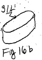
20重量部のビサクリルT2濃縮物を、80重量部のアクアクリル80MD溶液と混合し、そして高速混合装置で混合して、粘性ペーストを形成した。その後、混合物を、12時間、閉鎖容器内で60℃に加熱して、捕捉された空気を除去した。ペーストを、実施例1から得た金型に充填し、そして凝固させ、そして実施例1で記述されるとおり洗浄した。生じた親水性複合体は、90重量%を越える液体含有率を示し、そして実施例1から得られたハイドロゲルに比べて、弾性のモジュラスおよび膨張圧力を改善した。注型された物品を、12.5重量部のグリセロール、0.9重量部のNaClおよび86.6重量部の脱イオン化水の混合物中で24時間、浸漬させた。その後、上に記述されるとおり、ストレス下で切断し、そして乾燥させた。乾燥後、周囲温度で、60%相対湿度の空気中で、24時間、物品を調整した。生じた脊髄核移植片は、可塑化され、そして小さな切れ口を通して容易に挿入するため変形性がある。キセロゲル移植片は、図16aで91aとして示された円筒状挿入形状に示された挿入形状に巻き取られうる。いったん、手術で作製された空洞に挿入されると、それは、折畳みを解き、そして部分的に水和された挿入形状までそれの異方性拡張を開始する。それが、機械的制限なしに完全に水和される場合、それは、16bとして示されるそれの固有の形態まで膨張する。移植片は、同様の異方性を示すが、実施例1で記述される製品より早い膨張および高い膨張圧力を示す。
【0096】
(実施例3)
実施例1から得たハイドロゲルは、以下の方法で加工された:
重合体溶液を、7.5%に希釈して、それの粘度を減少させ、そして92.3重量%の等張性生理食塩水に対して生じる含有率で液体含有率を増加させた。
【0097】
撚り合わされたポリエステル繊維の鎖を、溶液に含浸させ、ガラス繊維(250ミクロン直径)から作られた格子の周りに螺旋状に包み、そして25mmに縮められた実施例1から得た多孔性金型に挿入した。その後、金型を、希釈重合体溶液で充填し、そして水道水で凝集させた。
【0098】
型から外した後、ガラス繊維を、ゲルから除去した。ポリエステル繊維強化を、デバイスと共軸の螺旋を形成するハイドロゲルに完全に埋設された。そのチャンネルは、軸圧力下でハイドロゲルからにじみ出た液体の排水を促進した。
【0099】
その後、金型を等張性生理食塩水で洗浄し、そして12.5%のグリセロール、0.9%のNaClおよび86.6%の純水(全て重量%)を含む混合物に浸漬させた。
【0100】
その後、可塑化ハイドロゲルを、実施例1に記述されるとおりの圧力下で乾燥させた。結果物は、長円形断面および3.5mm厚みの柔軟性ウエハーであった。ウエハーは、実施例2で記述されるとおり移殖を促進するために容易に畳込みまたは巻取られうる。移殖後、デバイスは、軸でのみ膨張して、膨張圧力による椎骨分離を達成する。
【0101】
それは、圧力下で放射状拡張に抵抗し、線維輪が損傷を受ける場合に、それによる突出またはヘルニア形成を防止する。図17aは、103aが、それの拡張形状で埋設された螺旋線維性強化であることを特徴とするそれの完全に水和された固有の形態での移植片101aを示す。図17bは、それの圧縮状態での線維性強化103bを伴うそれのキセロゲル挿入形状101bでの移植片を示す。
【0102】
(実施例4)
25.2mmの直径および45%の多孔性を示す拡張したPTFE GORTEX、Gore Associatesの円筒形のロッドを、相互に浸透するネットワークを膨張的に形成するために使用された。PTFEは、軸で伸縮され(3倍まで)、そして25%グリセロールジアセテート、およびHEMA(93.4%)、EGDMA(0.5%)およびメタクリル酸(6%)を包含する75%のモノマー混合物、およびジベンゾイルペルオキシド(0.1%)を含む液体に浸漬させる。その後、それの元来の長さの50%まで軸的に圧縮され、そしてモノマーを、65℃で窒素下で重合される。IPN複合体は、圧縮下で80℃よる上に加熱し、圧縮および冷却したときに、都合のよい挿入のために作り直されうる。
【0103】
PTFEマトリックスの外側、等張性生理食塩水中、そして体温でハイドロゲル成分の膨張は、73重量%である。完全に膨張した状態で、そしてPTFEマトリックスの外側でそれの圧縮強度は、およそ0.05MN/m2である。等張性生理食塩水中で膨張した場合、複合体は、軸方向に一次的に拡張する。膨張したIPN複合体は、放射状方向でより軸方向でいっそう変形性がある。複合体は、以上に強力で、十分に高い軸膨張圧力を発生しながら軸圧力下で放射状拡張に抵抗する。それの成分は、長期移植での素晴らしい歴史を示しながら非常に生物適合性で、そして生物安定性である。形状記憶は、周囲の保存条件下で変形した挿入形状の維持を可能にする。特性のこの組合せは、脊髄核移植片として使用するために適するこの複合体物品をなす。
【0104】
(実施例5)
実施例1から得られる重合体溶液は、およそ2mm厚の膜として外形である。膜を、平衡までチオシアン酸ナトリウム溶液に浸漬し、そしてその後、予め計算した重量まで部分的に乾燥させる。状態を、計算し、その結果、膨張液体中のチオシアン酸ナトリウムの最終濃度は、45重量%である。
【0105】
さらに、15mm直径の環は、0.75mmの直径を示すニッケル−チタンアロイ線から作られる。箔を、ここで、箔の各2層の間に置かれた線環を積重ねる。その後、積重ねられた組立体を、95℃で圧縮して、重合体を溶融および融合させる。冷却および洗浄の後、埋設された環形状強化を示す親水性重合体のブロックは、部分111aが、水和された重合体であり、そして113aが、金属強化であることを特徴とする図18でのそれの完全に水和された挿入形状で、それの完全に水和された挿入形状で示されるとおり形成される。実施例2で記述されるとおり、重合体を、等張性生理食塩水で十分に洗浄し、そして希釈グリセロールで浸漬し、そして圧力下で乾燥させる。
【0106】
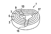
ここで図1に関して、示されたヒトまたは類人猿のような生きた脊椎動物の椎間板がある。それは、髄核中心11および環状積層体3、5、7および9を含む。図2は、垂直軸2に対して60°角で線維を示す1つの環状積層体5の平らにされた部分的断面を示す。
【0107】
図3、4、5および6は、本発明の手術的手段の種々の段階での図1の椎間平板の斜視正面図を示す。図1で示される同一部分は、これらの図で同一に番号を付された。
【0108】
図3では、切れ口15が作られ、そして損傷を受けた髄核が除去された。残りの髄核は、損傷を受けた髄核の除去から作り出される空間である空洞17に隣接の領域13として示される。
【0109】
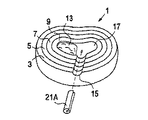
図4では、本発明の脊髄核移植片21Aは、それの部分的に水和された折り畳み形態で示され、そして切れ口15を通して、そして空洞17に挿入される。図5は、空洞17内に配置される脊髄核移植片21b(折畳まれていない)を示し、そして図6は、脊髄核移植片21cおよび上に記述される浸透圧変化、ストレス、および運動から生じる容積的および液体含有率の変化にかけられる、それの完全に水和され、そして拡大した状態を示す。この脊髄核移植片は、上に説明された実施例のいずれかによって最初に形成される。
【0110】
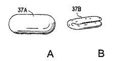
図7a、7b、8a、8b、9a、9b、10aおよび10bは、種々の形態での本発明の脊髄核移植片を示す。それらの完全に水和された固有の形態にある脊髄核移植片は、それぞれ、球体31a、円筒形33a、螺旋35a、および卵型37aの形態で示される。それらのキセロゲル挿入形状での同じ移植片は、両凸レンズ円板31b、巻き込み円板33b、平らにされた螺旋35bおよび折畳まれた長円形円板37bとして示される。全ての形態は、図面に示されていない任意の埋設強化構造を含みうる。図11は、軟質の高い水分含有率のコア41および高い結晶性重合体含有率を示す外側ジャケット43を有する本発明の脊髄核移植片39の頂部図を示す。それらのインターフェース45で、任意の構造的支持体または遷移層を包含しうる。
【0111】
図12は、コア53およびジャケット55を有する本発明の脊髄核移植片51を示し、そしてさらに、コアに構造的支持体要素57a、そしてジャケットに57bを包含する。
【0112】
図13は、ラインABにそって切断された、部分的に切断された本発明の脊髄核移植片球61を示す。それは、外側層69に向けて重合体含有率での勾配のある増加および水分含有率での減少と共に、低い重合体含有率および高い水分含有率を示す中心63を有する。例えば、中心63は、10パーセント重合体および90パーセント水を含有する可能性があり、そして層65は、80パーセントの重合体および20パーセントの水を含有する可能性があり、そして層67は、30パーセント重合体および70パーセント水を含有する可能性があり、そして外側表面69は、平滑で負に荷電された表面を有するよりいっそう多い水分を含有しうる。
【0113】
明らかに、本発明の膨大な修飾および変動は、上記教示の点で可能である。したがって、付随の請求項の範囲内で、本発明は、ここに特に記述されない以外実施されうることが分かる。
【図面の簡単な説明】
【図1】 図1は椎間板の断面の斜視正面図を示す。
【図2】 図2は椎間板の管状積層の繊維輪の正面拡張切断断面図を示す。
【図3】 図3は空洞作製(髄核除去)を示す本発明の手術的移殖手段の種々の段階の斜視正面図を示す。
【図4】 図4は脊髄核移植片挿入を示す本発明の手術的移殖手段の種々の段階の斜視正面図を示す。
【図5】 図5は折畳まれていないものを示す本発明の手術的移殖手段の種々の段階の斜視正面図を示す。
【図6】 図6は完全に水和されたものを示す本発明の手術的移殖手段の種々の段階の斜視正面図を示す。
【図7】 図7は球形の脊髄核移植片の斜視正面図を示す。
【図8】 図8は円筒形の脊髄核移植片の斜視正面図を示す。
【図9】 図9は螺旋型の脊髄核移植片の斜視正面図を示す。
【図10】 図10は卵型の脊髄核移植片の斜視正面図を示す。
【図11】 図11は本発明の脊髄核移植片構造の正面切断図を示す。
【図12】 図12は本発明の脊髄核移植片構造の正面切断図を示す。
【図13】 図13は本発明の脊髄核移植片構造の正面切断図を示す。[0001]
BACKGROUND OF THE INVENTION
The present invention relates to a spinal nucleus graft that replaces all or part of the nucleus pulposus removed from a living vertebrate, such as a human spinal disc. This spinal nucleus graft is capable of expanding anisotropically Xerogel Formed from.
[0002]
[Prior art]
The spinal disc is cartilage tissue that is placed between the endplates of adjacent vertebrae. The bone marrow disc acts as a flexible joint between the vertebrae and can deflect and twist the vertebral column. Damage to the spinal disc can cause spinal cord dysfunction, harmful pain, and short- or long-term inconvenience. Due to the widespread occurrence of this problem (reported 5% annual back pain due to bone marrow discs), the economic consequences are enormous. Certain disc problems require surgery. Typical state-of-the-art means are those described in US Pat. Nos. 4,636,217 (Ogilvie et al.), 5,489,308 (Kuslich et al.) And 5,716,415 (Steffee). Fusion of adjacent vertebrae using various techniques and devices. All recently available surgical means such as removal of the nucleus or part of it (laminectomy) or fusion of adjacent vertebrae impair spinal cord function in one or the other direction.
[0003]
For this reason, new means are needed, including the development of a prosthesis for the disc or part thereof. This is difficult to undertake. The spinal column is an extremely complicated body part, and its proper functioning depends on all uninterrupted cooperation of its components, including the intervertebral disc. The intervertebral disc must perform a number of functions. Must withstand repeated high tensions in very complex deformation models, including mixed strain, torque, shear and compression. In addition, the spinal disc acts as an effective shock absorber and a pump that activates nutrients to the disc and the flow of metabolites from the disc. Structurally, the disc is a rather complex part of the composite and several types of materials organized in a complicated form. The end plate of the vertebra is covered with a layer of hyaline cartilage composed of a collagen matrix, a glycoprotein component, and water. In addition, about 2-5% of its volume is occupied by living cells that produce cartilage components.
[0004]
Its own spinal disc is composed primarily of crystalline collagen fibrils and amorphous hydrophilic proteoglycans. About 3-5% of its volume is occupied by living cells that produce its components. Structurally, the spinal disc is composed of a hydrogel-like core called the nucleus pulposus and an outer ring called the annulus fibrosus. The structure of the bone marrow intervertebral disc is depicted schematically in FIG. 1 and described below.
[0005]
The spinal discs support weight and act primarily as flexible joints. It withstands considerable axial loading while allowing shared rotation, strain and translation of adjacent vertebrae. In addition, the spinal discs dampen vibrations and mechanical shocks and prevent their propagation through the skeletal system. Tolerance and flexibility to withstand loads in selected directions is achieved by a combination of annulus fibrosus and nucleus pulposus. The annulus is a laminated structure that is rigid in the radial direction but deformable in the axial direction and by torque. Axial loading is caused by the nucleus pulposus that partially deforms the axial component contained by the annulus fibrosus. Annulus fibrosus is mainly formed by collagen fibrils organized in several layers. Each layer has its collagen fibril damage at an angle, and the continuous layer shows the selected direction. Collagen tissue is closely similar to fiber reinforced organization, as is the complex used for pressure vessels or cords in tires. It allows deformation in torque and distortion while ensuring maximum resistance to radial pressure (or internal pressure).
[0006]
The fibril ends are attached to the adjacent vertebra and to the cartilage-like surface of the vertebral endplate. As a result, the inner space of the annulus is substantially enclosed. Any fluid that permeates inside or outside the core must pass through the annulus fibrosis or through the vertebral endplate. In order to achieve sufficient hydraulic permeability, the collagen-like structure of the annulus is supplemented by proteoglycans embedded between collagen fibrils. Proteoglycans are hydrated so that the annulus fibrosus forms a kind of highly organized anisotropic hydrogel complex. The collagen domain forms a fibrillar mesh. The result of this arrangement is sufficient deformability in the selected direction combined with the high mechanical strength required for the ability to withstand loads, particularly high shear strength, and resistance to fracture growth.
[0007]
The nucleus pulposus is in contact with the annulus but not the endplate. It has a lower concentration of collagen (its concentration increases with age) and a higher concentration of hydrophilic proteoglycans. Consequently, it is a natural complex that is somewhat like a hydrogel and exhibits a very high equilibrium water content (90% more than in younger humans). The moisture content and nucleus pulposus volume depend on the osmolality of the inflation medium and on the mechanical pressure. The resistance to liquid content reduction due to mechanical pressure is referred to as “expansion pressure”. Inflation pressure is truly key to the function of the nucleus pulposus. As the shaft load releases liquid, the inflation pressure increases until it reaches equilibrium with the external load. The nucleus pulposus is therefore capable of balancing and redistributing axial stress and converting them into radial components that can be limited by the annulus fibrosus. Moreover, dehydration and rehydration of the nucleus pulposus under variable loading drives the transport of metabolites and nutrients inside and outside the spinal disc. Thus, the nucleus pulposus acts as a permeation pump that facilitates transport of nutrients and metabolites to and from the spinal disc and surrounding tissues. This transport function is essential because the cartilage components (the annulus vertebral annulus, nucleus pulposus and cartilage-like layer) cannot be vascularized or nourished by rare diffusion.
[0008]
Since the nucleus pulposus is essentially an isotonic tissue under a microscope, to perform all these functions must be organized at its molecular and supramolecular level.
[0009]
The nucleus pulposus structure is rather clever. The nucleus pulposus is constructed from a bilayer complex composed of a crystalline collagen domain that forms a scaffold and a glycoprotein domain that forms a hydrophilic filler. Crystalline collagen domains result from considerably higher strength even at high hydration. They form a fine fiber mesh that resembles fiber reinforcement in high performance composites. The result of this arrangement is sufficient deformability combined with sufficient mechanical strength even at full hydration.
[0010]
The amorphous domain is due to water absorption and the generation of expansion pressure. They are mainly formed by polymeric, water-soluble glycoproteoglycans. Glycoproteoglycans are highly hydrophilic and water-soluble polymers. A small portion of the glycoaminoglycan is covalently bound to the collagenous scaffold, thereby making it hydrophilic and very wet in water (this is the thermodynamics of the two-phase complex) Necessary for stability). Most are unattached to the scaffold and are retained by capture within the scaffold due to the large size of the glycoproteoglycan molecules.
[0011]
To aid this physical retention, glycoproteoglycan chains associate to form large units. The glycoproteoglycan chain has a protein terminal sequence flanked for attachment to hyaluronic acid. The hyaluronic acid and GPG complex is too large to escape the collagenous scaffold. This is a much different arrangement in hydrogels where the restriction of the hydrophilic part is achieved by crosslinking. It can be inferred that the sequence in the nucleus pulposus provides a higher permeation pressure at a given polymer concentration than the network sequence that is normal in hydrogels.
[0012]
Glycoproteoglycans in the amorphous layer withstand dense negative loads. A high negative load density is important because it results in a high value of virial coefficient and thus causes maximum expansion pressure at high moisture content. High loading density is facilitated by the complex structure of the nucleus pulposus. Synthetic crosslinked hydrogels that exhibit similar loading densities are brittle and very mechanically weak.
[0013]
The high negative load is also due to the high surface hydration required for low wet friction. This is important for low friction contact between the nucleus pulposus and the cartilaginous surface of the vertebral endplate. High friction probably causes excessive wear of the cartilage and degenerative changes in the medulla.
[0014]
This structural complexity of the spinal disc is a result of the complex requirement and not an unpredictable excess characteristic. Thus, the function, properties and structure of the disk replacement must be a close approximation of the original disk in order to be able to perform all of its functions. In other words, a successful disc exchange should be biomimetic to the maximum extent achievable.
[0015]
This has not been possible for many years because there is no synthetic material that can replicate the structure, properties and functions of natural tissues. To that end, most of the prostheses have been designed as mechanical joints that allow some kind of movement of the vertebrae, but cannot replicate all SID properties. Such prostheses are described, for example, in the following US patents:
3,875,595 (Froning); 4,349,921 (Kuntz); 4,309,777 (Patil); 4,714,469 (Kenna); 4,904,261 (Dove et al.); 4,759,769 (Hedman et al.); 4,863,476 (Shepperd); 5,053,034 (Olerud); 5,674,296 (Bryan et al.) No. 5,676,701 (Yuan et al.); No. 5,824,094 (Serhan et al.); No. 5,865,846 (Bryan et al.)
It is described by.
[0016]
The main problem with these devices is their limited functionality. Even more importantly, the transfer of these devices is a very complex means requiring large spinal surgeries with many combined risks, long term recovery, and high costs.
[0017]
There is a continuing effect of developing a better prosthesis of the disc that more closely replicates its mechanical function. For example, Lee et al., US Pat. No. 4,911,718, “Functional and Biocompatible Intervertebral Spacer” (1990), a fiber reinforced organism mimicking the mechanical properties of a natural disc. Describes composite replacement of discs made from compatible elastomers. It has an elastomeric core that exhibits a shape that approximates the shape of the nucleus pulposus and replicates a disc structure that is completely wrapped by a fiber reinforced elastomeric layer that replicates the structure of the annulus fibrosus. Reinforcing fibers prefer an array that stimulates the orientation of collagen fibers in the annulus. The face of the assembly comprises a hard elastomeric layer that stimulates the mechanical function of the cartilage layer at the endplate of the vertebra. This structure replicates the bone marrow disc structure and its mechanical function fairly closely. However, the transfer of this device is much more complicated and expensive and requires significant spinal surgery.
[0018]
In many cases, pain relief requires that only the nucleus pulposus (or even only a portion thereof) be removed rather than the entire spinal disc. In that case, the main part of the axial load is used directly on the annulus. The annulus is currently compressed by the shaft rather than the radial load to which it is applied. As a result, the annulus gradually tears, splits, wears and crushes into thin layers. The situation is somewhat comparable to driving with punctured tires. In this situation, replace the lost nucleus pulposus (or part of it) and re-establish the radial stress in the annulus needed for its proper functioning (or “re-inflate the spinal disc” "). The nucleus pulposus replacement is easy, less traumatic and can be performed by inexpensive surgical means.
[0019]
It is important to recognize that successful replacement of the nucleus pulposus must replicate not only the mechanical function, but also the function of the permeable pump. Without it, the vertebral endplate cartilage and the living tissue of the annulus fibrosus cannot be maintained in a healthy state. For these reasons, the nucleus pulposus cannot be replaced by a piece of hydrophobic, non-hydrogel elastomer, such as silicone rubber or polyurethane.
[0020]
This need to maintain liquid transport function was first recognized in Bao et al., US Pat. No. 5,047,055. Bao is a hydrogel that, in its fully hydrated state, exhibits a shape and size commonly identified against the lost natural nucleus, ie, the cavity left after removal of nucleus pulposus tissue・ Describes the prosthesis. The hydrogel used in the grafts, in its fully hydrated state, has a water content of at least 30% and at least 4 MN / m 2 (Ie 40 kg / cm 2 Or a compression strength of 556 psi). This high strength must be achieved even with very high moisture content, such as with sufficient hydration and within the preferred range of 70 to 90% liquid. Perhaps this very high need for mechanical strength is affected by the possible hernia formation of isotropic material that has been damaged and transplanted into weakened annulus fibrosus. This rather extreme requirement limits the choice of materials useful for this device. Hydrogels are generally weaker than other plastics and rubbers, especially at high moisture contents. Therefore, the selection of high expansion hydrogels that exhibit such high compressive strength is rather narrow.
[0021]
The Bao hydrogel prosthesis is transplanted in a partially or fully dehydrated form when it is miniaturized, i.e., its volume is that of a fully hydrated hydrogel implant. 10-70% of the volume. As a result, the hydrogel graft can be inserted through a small cut and then grown to its full size by absorbing aqueous bodily fluids. The hydrogel used for the graft shows a higher water content of 30% of the liquid, and preferably from 70 to 90%, in its fully hydrated state. The material used by Bao is isotropic, so that the expansion of the graft by hydration is equal in all directions. The graft may be composed of a mixed size and shape of two or more pieces of cavities emptied by the nucleus pulposus to be removed.
[0022]
This concept has several drawbacks. Hydrogel expansion is limited to the size of the cavity emptied by the nucleus pulposus, so that its expansion pressure in the fully hydrated and expanded state is very low or even zero. Thus, the graft does not generate sufficient axial force for vertebral separation that can be seen with a healthy disc. This is different from the natural nucleus pulposus which is underexpanded inside the intervertebral disc, and even with maximal vertebral separation, produces positive inflation pressure. Bao, nucleus pulposus grafts are transplanted into damaged annulus fibrosus (either by surgical cuts or by original trauma), and spinal nucleus transplantation beyond the cavity size Such an “oversized design” could not be used, as the extension of the piece resembles its protrusions similar to the herniation of the natural nucleus pulposus. As Bao mentions, swelling of the graft under stress is avoided by the resistance of the annulus fibrosus to deformation. Because the integrity of the annulus fibrosus is weakened, hydrogels used in prostheses are more resistant to hernia formation or protrusion (especially greater than 4 MN / square meter for full hydration) than the natural nucleus pulposus Must be strong.
[0023]
This limitation is caused by the fact that the expansion of the Bao spinal nucleus implant is isotropic, in particular it is the same in the radial and axial directions. As a result, intensive expansion in the axial direction causes a radial expansion that produces pressure on the damaged annulus and causes it to rupture, expand or herniate. Furthermore, the type of hydrogel described by Bao is an isotropic elastomer that exhibits the same deformability in all directions. In the described design, the axial load causes a radial deformation that is greatest at least in the direction of resistance, ie where the annulus is weakened by surgery or by previous trauma to the disc. This can cause problems similar to graft bulge, herniation or protrusion, ie disc damage which is the reason for surgery in the first place.
[0024]
Some of these drawbacks were addressed in the subsequent invention by Bao et al. Described in US Pat. No. 5,192,326. The prosthetic nucleus is formed by the multiplicity of hydrogel beads exhibiting a water content of at least 30%, and the beads are surrounded by a flexible translucent cover. The porous cover, when fully extended, indicates the size and shape of the cavity emptied by the removed nucleus pulposus. The size of the beads is at least 3 times larger than the size of the holes in the cover, and as a result, the hydrogel is safely confined within the cover. Hydrogel beads contain as much as 99% liquid when fully hydrated. Since the framework limits expansion and avoids hydrogel expansion beyond the internal volume and dimensions of the cover, the total volume of fully hydrated hydrogel beads is a cavity emptied by nucleus pulposus removal. There is a possibility that it is larger than that of the volume. The cover can be made from knitted fibers. Preferably, the framework is coated with a very biocompatible polymer to avoid deleterious reactions to the graft. However, even with a coating, the microporous framework induces foreign body reactions, initiates protein deposition, becomes a locus for bacterial colonization, or causes other problems. The use of the cover sacrifices some benefits of the hydrogel such as high biocompatibility and surface wettability. Furthermore, the beads exhibit a relatively low packing density and a relatively large void space fraction.
[0025]
Ray et al. Invented a design similar to the '326 to some extent. In U.S. Pat. No. 4,772,287, Ray describes a graft to the nucleus pulposus composed of two flexible cylindrical sac filled with a fluid, preferably a thixotropic fluid. It is described. The sac is surrounded by a strong fibrous framework and is preferably mixed with a biodegradable polymer that promotes growing tissue. For convenience, the sac is provided with a tube for adding or retracting fluid. This device clearly does not replicate the shape and characteristics of the nucleus pulposus and only attempts to stimulate some of its functions. The fibrous framework is designed to facilitate the incorporation of graft changes into the remaining disc tissue, thus causing a partial form of vertebral joint.
[0026]
In US Pat. No. 4,904,260, Ray is an aqueous fluid in which the coating is made from a translucent material and contains a therapeutic material capable of slow diffusion from the graft to the tissue. Describes his basic design improvements to be filled.
[0027]
In US Pat. No. 5,674,295, Ray describes another improvement of his basic design where a hydrogel cylinder is used instead of a sac filled with fluid. A strong fibrous framework allows for more expansion in the radial direction and more axial expansion, thereby providing sufficient axial expansion while protecting the annulus against excessive pressure from the expanded and / or deformed hydrogel. Designed to allow.
[0028]
The hydrogel body exhibits an oval cross section and the constraining jacket is designed to maintain the overall shape of the hydrogel under full hydration and load. Further modification with No. 5,824,093 (partial continuation to '295).
[0029]
In all Ray designs, the device does not mimic the nucleus pulposus shape, size, characteristics, or full function. The volume of the hydrated hydrogel is substantially smaller than the natural nucleus pulposus volume. The shape of Ray grafts is substantially different from the nucleus pulposus shape, and anyone can predict certain problems with the positional stability of such grafts. To improve stability, a porous or fibrous restraint jacket is incorporated into the remaining disc tissue. However, this causes a partial form of the vertebral joint and thereby a partial fixation. Ray's device does not fill the space designed for the nucleus pulposus that can distort the device under certain conditions and cause a tendency to protrude. As can be seen from the description, none of the prior art inventions provided a satisfactory solution to the problem of nucleus pulposus replacement, nor taught or clarified the present invention.
[0030]
Despite the prior art, the present invention is neither taught nor clarified thereby.
[0031]
[Problems to be solved by the invention]
The present invention is a biomimetic spinal cord designed to retain bone marrow disc and vertebral joint functions after some or all of nucleus pulposus tissue has been removed from a living vertebrate, eg, a human disc. A nuclear graft is provided.
[0032]
[Means for Solving the Problems]
The spinal nucleus implant according to the present invention is an expandable plastic device capable of anisotropic expansion into the form of a hydrogel graft exhibiting anisotropic deformability. The spinal nucleus implants according to the present invention can be transplanted to the bone marrow plate through small cuts thereby facilitating surgery, minimizing surgical trauma and improving device safety.
[0033]
The invention also relates to the surgical graft means associated therewith.
The present invention should be better understood when the specification herein is taken in conjunction with the accompanying drawings.
[0034]
DETAILED DESCRIPTION OF THE INVENTION
The device according to the present invention replicates all its essential functions to achieve positional stability of the nucleus pulposus graft implanted in the weakened annulus and the spinal nucleus graft through the small incision Designed to replicate the structure and material properties of the natural nucleus pulposus to the extent required to be able to be transplanted.
[0035]
A preferred spinal nucleus implant according to the present invention is:
A hydrophilic material that exhibits an equilibrium moisture content around 90% or higher with complete hydration;
A two-phase structure that includes domains that exhibit high crystallinity and low water content (“hydrophobic domains”) and domains that exhibit low crystallinity and high water content (“hydrophilic domains”).
● Domains exhibiting a particularly high moisture content, and high content of carboxylic acid groups on the surface of the device.
● Associative water-soluble polymer concentrated in hydrophilic domains with high water content.
• Moisture content that strongly depends on the osmolality of the expansion medium.
● Highly hydrated negatively loaded smooth surface
It exhibits properties that closely mimic the essential properties of natural nucleus pulposus such as
The spinal nucleus implants according to the present invention also show the following differences from the natural nucleus pulposus:
● Its fully hydrated graft size does not adapt to the natural nucleus pulposus;
● Its fully hydrated graft shape does not adapt to the natural nucleus pulposus;
● Dehydrated grafts exhibit favorable expansion in the axial direction of rotation (vertical plane) and anisotropic expansion showing suppression in the radial direction (horizontal plane);
● Transplanted pieces exhibit anisotropic deformability (it is more deformable on the shaft and harder radially).
[0036]
These features are described in more detail below.
In order to replicate or replace nucleus pulposus function, the present invention is capable of swelling in the presence of water and changes moisture content in response to external conditions such as pressure, temperature, osmolarity or pH Includes capable materials. They are two types of expandable materials suitable for the present invention: more organized hydrophilic complexes that exhibit some degree of hydrophobicity similar to the structure of cartilage tissue, and more homogeneous hydrogels.
[0037]
The hydrophilic complex of the present invention is of the “cellular” (or “domain”) type with a continuous hydrophobic domain and a separate hydrophilic domain, and interpenetrating both types of domains are continuous Network.
[0038]
A preferred material for a spinal nucleus graft is a synthetic complex of a type of cell that exhibits a structure similar to the nucleus pulposus. The structure includes a strong fibrillar crystalline phase and an amorphous phase comprising a charged associative polymer. The polymer involved in the composite is non-biodegradable, preferably with a carbon-carbon skeleton. A preferred type of polymer is a multiblock acrylic acid polymer. The complex may be of “domain type”, or “interpenetrating network”, in which the associating polymer is arranged in discrete hydrophilic domains.
[0039]
The requirements for spinal nucleus implants according to the present invention can also be adapted to certain types of hydrogels. Contrary to composites, hydrogels are formed by a single type of network (covalent bonding, physical or mixed). Only hydrogels that exhibit certain property combinations are suitable for the present invention, as shown in more detail below.
[0040]
In accordance with the present invention, the height (ie, axial dimension) of a fully hydrated spinal nucleus implant is greater than the maximum separation between the vertebrae at the position of prone. However, the fully hydrated diameter of the spinal nucleus graft is substantially the same as the diameter of the cavity evacuated by nucleus pulposus removal.
[0041]
In the following description, the phrase “expandable plastic” is used to include both composites and hydrogels.
[0042]
The expandable plastic contains one or more polymer components. Preferably, expansible plastics suitable for spinal nucleus implants include a polymer component that exhibits a C-C backbone. Such polymers such as polyvinyl alcohol, polyvinyl pyrrolidone or derivatives of polyacrylic acid or polymethacrylic acid are more resistant to biodegradation than polymers with heteroatoms in their backbone, such as polyurethane or polyester.
[0043]
Preferably, at least one polymer component contains both hydrophilic and hydrophobic groups.
[0044]
Preferred expandable plastics include two polymer layers of different hydrophilicity, a lower hydrophilic layer that exhibits a higher content of hydrophobic groups, and a hydrophilic group that exhibits a higher solicitation of hydrophilic groups. . The low hydrophilic layer is preferably crystalline and the high hydrophilic layer is preferably amorphous, as can be established from X-ray diffraction.
[0045]
Preferred hydrophobic groups are dangling nitrile substituents at the 1,3 positions in the polymethylene skeleton, such as poly (acrylonitrile) or poly (methacrylonitrile). The hydrophilic layer preferably contains a high concentration of ionic groups. Preferred hydrophilic groups are derivatives of acrylic acid and / or methacrylic acid, including salts, acrylamidines, N-substituted acrylamidines, acrylamides and N-substituted acrylamides, and various combinations thereof. A particularly preferred combination comprises approximately two-thirds of acrylic acid and its salts (or on a molar basis) and the remainder is pure and a combination of N-substituted acrylamide and acrylamidine.
[0046]
The at least one polymer component is preferably a multi-block copolymer having modified sequences of hydrophilic and hydrophobic groups. Such an array is usually capable of separating into two polymer layers and forms a strong physically cross-linked hydrogel. Such multiblock copolymers are, for example, the products of hydrolysis or aminolysis of polyacrylonitrile or polymethacrylonitrile and copolymers thereof. For brevity we refer to all polymers and copolymers having at least 80 mole% acrylonitrile and / or methacrylonitrile units in their compositions as “PAN”. PAN hydrolysis and aminolysis and its products are described, for example, in US Pat. Nos. 4,107,121; 4,331,783; 4,337,327; 4,369,294; No. 4,370.451; 4,379,874; 4,420,589; 4,943,618 and 5,252,692 and incorporated herein by reference It is.
[0047]
The expandable plastic may be composed of at least two polymer components arranged as a network that penetrates each other. In that case, one component is an essentially hydrophobic polymer capable of forming a mesh or scaffold of reticulated crystalline fibrils. Examples of such polymers are polyurethane, polyurea, PAN, stretchable polytetrafluoroethylene, cellulose triacetate and polyvinyl alcohol. The space between the fibrils is filled with a continuous phase of a hydrophilic polymer (ie, a hydrogel such as cross-linked polyvinyl alcohol or polyvinyl pyrrolidone) having a three-dimensional physical or covalent network. The most suitable hydrogel for this role is based on hydrophilic derivatives of polyacrylic acid and polymethacrylic acid.
[0048]
Preferred materials for spinal nucleus implants are hydrophobic or hydrophilic polymers with low to moderate moisture content that form a “closed cell” cavernous structure that provides a composite that exhibits strength and shape stability. A cell (or domain) type synthetic complex with a continuous phase formed. Examples of suitable polymers are polyurethane, polyurea, PAN, polydimethylsiloxane (silicone rubber), and highly crystalline multiblock acrylic and methacrylic acid copolymers. The polymer must be sufficiently permeable to water. It is known that even clearly hydrophobic polymers such as silicone rubber can form expandable composites. More preferably, the continuous phase is formed by a strong hydrophilic polymer that is sufficiently permeable to water but impervious to polymerizable solutes. Examples of such polymers are highly crystalline hydrogels based on segmented polyurethanes, polyvinyl alcohol with derivatives of acrylic acid or multiblock acrylonitrile copolymers. Typically, a suitable polymer for the continuous phase in the cell complex is in a fully hydrated state between about 60% and 90% by weight, preferably between 70 and 85% by weight. The moisture content of is shown.
[0049]
The second component is a highly hydrophilic polymer of sufficiently high molecular weight that cannot penetrate the continuous phase. This component is limited to the inside of the continuous phase matrix. The entrapped hydrophilic polymer is a high molecular weight water-soluble polymer, associative water-soluble polymer or highly swellable containing at least 95% water and 99.8% water in a fully hydrated state. It can be a hydrogel. Such hydrogels are mechanically very weak. However, the role of such polymers is rather than load bearing Osmotic pressure And the compressive strength at full hydration is 0.01 MN / m 2 Or in a lower range.
[0050]
Such a system with closed cells (or domains) containing highly expansible or water soluble polymers can produce complexes that exhibit very high expansivity when needed for spinal nucleus graft function. Can form. Examples of suitable hydrophilic polymers are high molecular weight polyacrylamide, polyacrylic acid, polyvinylpyrrolidone, polyethylene oxide, copolymers of ethylene oxide and propylene oxide or hyaluronic acid; hydrophilic esters of polyacrylic acid or polymethacrylic acid Or hydrogels cross-linked with conjugated bonds such as amides; and physically cross-linked hydrogels such as PAN hydrolysates or amino hydrolysates.
[0051]
Particularly suitable are associative water-soluble polymers capable of forming very highly viscous solutions or even soft physical gels. Preference is given to associative polymers containing negatively charged groups such as carboxylates, sulfo groups, phosphate groups or sulfate groups. Particular preference is given to associations formed by hydrolysis and / or aminolysis of PAN for high but finite transformations, liberating a certain number of unreacted nitrile groups (especially between 5 and 25 mol%). It is a functional polymer.
[0052]
Preferred complexes have both continuous and dispersed phases formed by different products of PAN hydrolysis or aminolysis. In this case, both components are compatible and their hydrophobic block may be involved in the same crystalline domain. This improves the fixing means of the highly hydrophilic component and prevents its withdrawal. The size of the highly hydrophilic domains can vary widely from nanometers to millimeters, preferably from 10 nanometers to microns.
[0053]
The ratio between the continuous and discontinuous phases (ie, between the more hydrophobic and more hydrophilic components) can vary from about 1: 2 to 1: 100 on a dry weight basis, but the preferred ratio Is in the range of about 1: 5 to 1:20.
[0054]
Any expandable plastic (eg, hydrogel) can be characterized in a variety of ways. The most important feature is the liquid content in the fully hydrated state. We are in the normal sense, i.e. at a defined temperature, sufficient time to reach equilibrium, limited space, availability of expanding liquid or external load or pressure used for the hydrogel The term “fully hydrated” is used to mean equilibrium hydration in full and unrestricted contact with excess liquid of defined composition without any restriction of sample stretching. Unless otherwise specified, the liquid medium is an isotonic unbuffered 0.9 wt% NaCl solution in water and the temperature is 36.5 ° C. + / − 0.5 ° C. of body temperature.
[0055]
Dehydrated expansible plastics are often " Xerogel ". The anchor feature is compressive strength. It can be measured by ASTM method D695 and can be performed by aqueous dipping. Unless otherwise specified, compressive strength is intended at full hydration and ambient temperature.
[0056]
The expandable plastic used in the present invention must exhibit the following basic characteristics:
Liquid content in fully hydrated state with deionized water at ambient temperature higher than 70% water and preferably higher than 95% water.
[0057]
Liquid content in full hydration with 0.9% aqueous solution of NaCl at body temperature higher than 65% liquid and preferably higher than 85% liquid.
[0058]
● Tolerance of anisotropic expansion, ie more in the selection method or other direction, stretches due to its hydration, even in the absence of external loads or any external restrictions Xerogel Tolerance. For example, anisotropic Xerogel The rod increases its diameter while decreasing its length due to its hydration.
[0059]
● Expandable plastics that are particularly useful in the present invention exhibit an elastic modulus that increases with deformation. This is important for limiting radial deformation, i.e. preventing swelling and hernia formation under high axial loads. Intumescent plastics that exhibit this type of action are particularly those that contain a crystalline phase in their structure.
[0060]
-Expansible plastics suitable for the present invention are those that retain "freezing deformation" and are capable of releasing such deformation in a fully hydrated state. Such materials are often referred to as “memory hydrogels” or “memory hydrogel composites”. Particularly useful are those materials that can retain “freeze deformation” at temperatures below body temperature, even in a plasticized state. Freeze deformation is released by hydration, heating to body temperature, or a combination of both.
[0061]
● An expansible plastic that is particularly useful in the present invention exhibits high water pressure permeability. All hydrophilic plastics, including hydrogels, exhibit a fairly high diffusive permeability for water and aqueous solutes. However, the use of spinal nucleus grafts requires various types of permeability, referred to as so-called hydraulic permeability, in which transport is facilitated by pressure gradients rather than concentration gradients. Water pressure permeability is "filtration coefficient" K f Can be characterized by An expandable plastic suitable for the present invention is K f > 5.10 -14 [Cc. cm thickness / sec cm 2 (Dyn / cm 2 )] And preferably K f > 1.10 -12 [Cc. cm thickness / sec cm 2 (Dyn / cm 2 )].
[0062]
A spinal nucleus implant according to the present invention exhibits the following characteristics:
Spinal nucleus implants exhibit a fully hydrated volume that is larger than the volume cavity created by partial or complete removal of the nucleus pulposus. The volume of the spinal cord nuclear implant that is sufficiently expanded into body fluid at body temperature is preferably at least 5% greater than the volume of the cavity into which the spinal nucleus implant is implanted, and more preferably at least 10%. large. Cavity volume is measured at the maximum natural separation of the vertebrae, i.
[0063]
A spinal nucleus implant according to the present invention exhibits three basic shapes:
Unique form A
Insertion shape B
Inherent shape C
Unique form A corresponds to the most relaxed polymer network in the fully hydrated state of the expandable plastic (ie, with minimal free enthalpy). Unique form The spinal nucleus implant at A exhibits a cross-sectional area substantially equal to the cross-sectional area of the cavity emptied by removal of nucleus pulposus tissue and a height substantially greater than the height of such a cavity. (By “height” is meant a dimension substantially parallel to the bone marrow axis, while “cross-sectional area” is the area transverse to the bone marrow axis.) Insert shape B is preferred for the bone marrow axis. Directionally deformed in a way that promotes insertion and anisotropic expansion Xerogel It is the shape. Form B and anisotropically dehydrated Xerogel Shows a shape optimized for insertion into the cavity through a small cut in the annulus. A preferred shape is the approximate shape of a cylinder whose length is approximately the length of the longer axis of the nucleus pulposus cross section. In the absence of bodily fluids and the absence of external loads or other space restrictions, spinal nucleus implants spontaneously change from shape B to shape A.
[0064]
The underlying shape C is substantially the shape of a cavity created by partial or complete removal of nucleus pulposus tissue. Spinal nucleus implants are transplanted in a partially dehydrated state and in insertion state A. Once inserted, it absorbs more water from the body fluid and increases its volume until it reaches shape C. The volume in state C is smaller than that in state A, and its major dimensions are different. The space and pressure limitations of the surrounding structure Xerogel Does not reach full hydration, the spinal nucleus graft is partially dehydrated in shape B. In shape C, Xerogel Achieved a substantially fully hydrated cross-section in state B. Thus, it creates a radial inflation pressure that can over-tense the weakened annulus and cause it to protrude or form hernia. However, the height in shape C is smaller than that in fully hydrated shape A, so that the spinal nucleus implant preferentially produces inflation pressure in the axial direction.
[0065]
Once implanted in a cavity in the disc, a spinal nucleus implant according to the present invention expands anisotropically from shape B to shape C, ie differently in various directions. For the isotropic expansion typical of hydrogels used so far in spinal nucleus implants, the relative increase in all linear dimensions is the same, and the relative increase in all dimensions is relative It is the cubic root of the dynamic volume expansion. For example, if the volume of an isotropic hydrogel increases by a factor of 8 due to hydration, any of its linear dimensions (such as curvature thickness, diameter, radius, etc.) are doubled.
[0066]
In the case of anisotropic expansion, the volume change is achieved by preferential expansion in the selection direction. More particularly, the spinal nucleus implant according to the present invention is more axial in the radial direction (with respect to the spine) and further expanded after implantation. It can only expand in the axial direction or expand in the axial direction while contracting in the radial direction. This anisotropic expansion in the preferred direction allows the generation of expansion pressure in the axial direction (needed for vertebral separation) without generating excessive radial expansion pressure on the annulus fibrosus. This feature allows the use of spinal nucleus implants that have a fully hydrated volume that is larger than the cavity volume created by removal of the annulus fibrosus tissue. The relative change in the axial direction is higher than the relative change in the lateral direction by at least 25%, and preferably by at least 100% (ie preferably by a factor of 2).
[0067]
The deformed shape B is a shape that is different from the genetic shape and shows its cross section minimized to facilitate its insertion through a small cut. The deformation state is that as long as the spinal nucleus graft hydrogel is partially or completely dehydrated and the temperature is lower than the glass transition temperature and / or melting temperature of at least one polymer phase in the hydrogel. As long as it is stable. A preferred deformation shape B may be that of a flat disk that can be folded for insertion in a taco-style or rolled for insertion into an approximately cylindrical shape (“burrito style”). It can be folded into the shape of the letter M, or any other convenient shape.
[0068]
A spinal nucleus implant according to the present invention is made from one or more parts, each of which exhibits an anisotropic expansion of the spinal nucleus implant described above. Such portions may be individually smaller than a single piece of spinal cord nuclear implant, but may be combined into a portion suitable for the basic spinal nucleus graft requirements described above. Individual sections can be inserted through smaller cuts and fitted inside the disc cavity to carry out spinal nucleus implant functions.
[0069]
For example, spinal nucleus implants can be formed by the scale of individual thin discs, and each is individually inflatable (ie, increases thickness rather than the foot shape). These discs are stacked inside the intervertebral disc cavity. The compression during their expansion and the fact that they cannot become fully hydrated ensures sufficient adhesion between the layers. The common location between the layers can be ensured by various means such as sutures, pins, spikes, adhesive layers and the like.
[0070]
A spinal nucleus implant in shape B is also formed by a single piece of longitudinal shape (like tape) that pushes into the cavity through a small cut and can be folded or otherwise stacked Can be assembled by forming the desired inherent shape.
[0071]
Another advantage of compounds in the form of spinal nucleus implants is improved fluid transport inside and outside the device. Insertion through a small cut is facilitated by using an expandable plastic that is sufficiently deformable in the inserted state. Since many expansible plastics are hard or even brittle when fully dehydrated, such plastics are salt solutions, glycerol, polyethylene glycol, glycerol diacetate, glycerol formal, dimethyl sulfoxide and It can be plasticized by a suitable non-toxic miscible liquid, such as the equivalent, alone or in combination with water. Although long-term control of proper water concentration can be difficult, another possibility is plasticization with almost limited water.
[0072]
Spinal cord nuclear grafts are transplanted into more or less damaged annulus fibrosus. Anisotropic expansion protects the annulus against excessive radial expansion that can lead to herniation or protrusion of the spinal nucleus graft material. Another protection may be provided by the anisotropic deformability of the spinal cord nuclear graft. That is, the spinal nucleus implant is desirably more deformable in the radial direction of that of the shaft. This can be achieved in several ways. One way is to use an expandable plastic that increases its modulus of elasticity with deformation. This type of action is demonstrated by many materials with crystalline components, such as natural rubber, tendons, cartilage, certain types of composites, and interpenetrating networks.
[0073]
This type of action can be easily detected from mechanical tests. The benefits can be further improved by the radial direction of the crystalline network in spinal nucleus implant devices made from such expandable plastics.
[0074]
Another way to limit radiative deformation is when using reinforcements embedded from hard materials such as metals, plastics, polymerizable fibers and the like. What is important is the proper construction of reinforcement so that it does not limit axial deformation. Preference is given to helical arrays such as embedded metal springs or helically damaged fibers. Another possible arrangement is an embedded stacked concentric ring. One suitable reinforcement is a braided structure, such as a vascular graft that exhibits similar requirements with anisotropic deformation. Such an implant is made from a medically tested material which is another advantage. The polymer reinforcement can be made from medical grade polyurethane, polyester, polyamide and other polymers of sufficient hardness.
[0075]
The reinforcement can be advantageously performed from permeable hollow fibers, preferably medical grade hollow fibers used for extracorporeal oxygen generators, kidney dialysis or hybrid organs. Such hollow fibers can improve hydraulic fluid transport, which is important for proper spinal nucleus graft function.
[0076]
Metallic reinforcing elements such as rings or helical springs can be beneficially used as x-ray markers that allow monitoring of implant position and deformation status. They can be used either alone or in combination with another enhancement or another x-ray market.
[0077]
The spinal cord nuclear implant is intended as a lifetime implant and its high biocompatibility is highly preferred. This can best be achieved by designing a spinal nucleus with a continuous and highly hydrated surface, preferably with a high content of negatively charged groups such as carboxylic acid groups. Particularly preferred are gradient surfaces that exhibit carboxylic acid concentration and hydration increasing from the mass to the surface. Such a surface is not only very biocompatible, but also exhibits very low wet friction so as not to erode adjacent tissue such as the hyaline cartilage of the vertebral endplates. In addition, they prevent sticking that can limit the movement of the graft, impede liquid transport, and complicate graft removal or replacement when it is needed. A preferred method is described in co-pending US Pat. No. 5,939,208 (P. Toy published on August 17, 1999: a method for the creation of a biomimetic surface).
[0078]
A preferred manufacturing method includes the following steps:
(1) Manufacture of devices from suitable expansible plastics. This includes operations such as fishing, integration of reinforcement, creation of biomimetic surface layers, and other operations that may be necessary for the manufacture of selected spinal nucleus implant designs.
[0079]
(2) Extraction of impurities in the fully hydrated state of the spinal nucleus graft with a suitable aqueous liquid such as sterile water or isotonic salt solution. This step can consist of multiple operations including swelling with an aqueous solution of a plasticizer such as glycerol.
[0080]
(3) Dehydration by transpiration of water to the current level in a deformed state. Basically, axial pressure is used during dehydration that forces deformation. The pressure can be used throughout the process or only at the end of the dehydration process. In that case, the essentially dehydrated device is heated, deformed by pressure with a suitable apparatus and cooled. This final stage can preferably take place under clean room or even sterilization conditions.
[0081]
(4) Sterilization is performed after or throughout the dehydration or deformation process.
Some of the inventions and preferred embodiments are further illustrated by the following non-limiting examples.
[0082]
【Example】
Example 1
Acrylic 90MD hydrogel is a brand name of Gelmed International. S. R. Oh. Purchased from. It is described as an acrylic acid multiblock copolymer with selective hydrophilic and hydrophobic blocks, where the hydrophobic block is from an acrylonitrile unit and the hydrophilic block is from a combination of acrylic acid, acrylamidine and acrylamide units. Composed. Molar composition polymers have been reported as follows:
Acrylic acid unit: 30%
Acrylamide unit: 9%
Acrylic amidine unit: 6%
The hydrogel contains 98.6% by weight liquid with complete hydration with pure water and 90.6% by weight when hydrated with isotonic NaCl solution (0.9% NaCl in water).
[0083]
The tensile strength at ambient temperature and full hydration with an isotonic solution is 6 kg per square centimeter. When fully hydrated with water, the hydrogel is too brittle to measure its tensile or compressive strength (in both cases estimated to be 1 kg / square cm or less).
[0084]
Acryl was supplied as a 10 wt% polymer solution (55% wt% aqueous solution) in sodium thiocyanate solvent.
[0085]
Acryacrylic was cast into a semi-open porous mold to 71 (approximately, if not all, dimensions) of the nucleus pulposus shown in FIG. The nucleus pulposus foot or section is kidney-shaped with a maximum dimension of approximately 40 mm. When we match this longest dimension in the X-axis direction with orthogonal isotopes, the longest dimension in the Y-axis direction is about 20 mm. The height of the space between the two vertebrae occupied by the nucleus pulposus (in the direction of the orthogonal rotation axis and the Z axis) is approximately 15 mm. These values are approximate and average. The dimensions of the nucleus pulposus should be understood to vary from disk to disk and from person to person. The height will also vary clearly with the time and load used in the disc.
[0086]
The porous mold exhibits a cross section calculated to the approximate size of the nucleus pulposus cross section. Calculations were made from known volume fractions of polymer components in the starting solution and the final fully hydrated hydrogel. The ratio of solution volume to gel is the inverse proportion of the volume fraction of polymer in the two systems. Thereafter, the ratio of any mold dimension to the corresponding hydrogel dimension is the third cubic of the corresponding volume ratio. The mold height is substantially greater than the nucleus pulposus height.
[0087]
The acrylic solution was fed into the mold and solidified by flocculation with excess isotonic saline. The solidified hydrogel was die cut and washed thoroughly with an isotonic solution until all the sodium thiocyanate was removed.
[0088]
After washing, the major fully hydrated dimensions in the isotonic solution are as follows:
Length: 31mm
Width: 18mm
Height: 86mm
[0089]
Specimens are cut into 25 mm sections and dried under axial compression with increasing load sufficient to maintain the original cross-sectional dimensions. Drying begins at ambient temperature. The drying temperature is gradually increased until it reaches 100 ° C., and Xerogel Is held at this temperature for 24 hours. It is then cooled to ambient temperature. The pressure is maintained until cooling is complete. The result of this process is a cross section of approximately 32 x 19 mm and a thickness of approximately 2.5 mm. Xerogel It is an article. Xerogel The device in the state is shown in FIG. Specific The same device after re-expansion to a fully hydrated state is shown in FIG. 15b as 81b.
[0090]
When this zero gel article is immersed in isotonic saline at body temperature, it hydrates completely and expands to its original dimensions of 31 × 18 × 25 mm. The expansion factors for the individual axes are as follows:
X = 0.97; Y = 0.95; Z = 10.
[0091]
A portion of the nucleus pulposus can be removed by surgery to create a cross-sectional cavity that approaches the cross-section of the graft. Its Xerogel The device in the inserted state is a rigid wafer that can be inserted into the disc through a horizontal slit cut in the annular medulla. The cut can be secured by a suture. Once the implant has expanded for several hours, it increases its height until it runs against the upper endplate. Ongoing expansion increases vertebral separation and stretches the annular medulla to the shape and tension required for its long-term function. The implant is partially hydrated and substantially conforms to the shape of the cavity created by tissue removal. The device in its partially hydrated intrinsic state (B) exhibits an approximate dimension of 31 × 18 × 15 mm. The expansion factors on the individual axes are as follows: X = 0.97; Y = 0.95; Z = 6.
[0092]
(Example 2)
Aquacrylic 80MD was obtained from the same source as the hydrogel obtained from Example 1. This grade of acryl has different proportions but the same structure and the same functional groups:
Acrylonitrile unit 79.7%
Acrylic acid unit 13.5%
Acrylamide unit 4.1%
Acrylic amidine unit 2.7%
The hydrogel contains 90.3 wt% liquid with complete hydration with pure water and 79.8 wt% when hydrated with isotonic NaCl solution (0.9 wt% NaCl in water). The tensile strength at ambient temperature and complete hydration with an isotonic solution is 17.3 kg per square centimeter. Aquacrylic 80MD was supplied as a 10 wt% polymer solution (55% wt% aqueous solution) in sodium thiocyanate solvent.
[0093]
Bisacryl T2 associative polymer is available from Gelmed International, Prague,
Acrylonitrile unit 22.2%
Acrylic acid unit: 51.9%
Acrylamide unit: 8.5%
N-sulfoethylacrylamide unit 6.6
Acrylic amidine unit: 6.1%
N-sulfoethylacrylamidine unit 4.7%
[0094]
The polymer is soluble in pure water at elevated temperatures that form shear thinning and is a thixotropic solution at ambient temperatures. At ambient temperature, the polymer does not dissolve, but forms a soft gel that exhibits a water concentration of 99.5% by weight in pure water and 97.6% by weight in isotonic saline. The wt% was supplied as a granular gel with 5 wt% solids.
[0095]
20 parts by weight of Bisacryl T2 concentrate was mixed with 80 parts by weight of Acryacryl 80MD solution and mixed with a high speed mixer to form a viscous paste. The mixture was then heated to 60 ° C. in a closed container for 12 hours to remove trapped air. The paste was filled into the mold obtained from Example 1 and allowed to solidify and washed as described in Example 1. The resulting hydrophilic composite exhibited a liquid content of greater than 90% by weight and improved elastic modulus and expansion pressure compared to the hydrogel obtained from Example 1. The cast article was immersed in a mixture of 12.5 parts by weight glycerol, 0.9 parts by weight NaCl and 86.6 parts by weight deionized water for 24 hours. It was then cut under stress and dried as described above. After drying, the article was conditioned for 24 hours in air at 60% relative humidity at ambient temperature. The resulting spinal nucleus implant is plasticized and deformable for easy insertion through small cuts. Xerogel The implant can be wound into the insertion shape shown in the cylindrical insertion shape shown as 91a in FIG. 16a. Once inserted into a surgically created cavity, it unfolds and begins its anisotropic expansion to a partially hydrated insertion shape. If it is fully hydrated without mechanical limitation, it is shown as that of 16b Unique form Expands to The implant exhibits similar anisotropy, but exhibits faster expansion and higher expansion pressure than the product described in Example 1.
[0096]
(Example 3)
The hydrogel obtained from Example 1 was processed in the following manner:
The polymer solution was diluted to 7.5% to reduce its viscosity and increase the liquid content with the resulting content for 92.3% by weight isotonic saline.
[0097]
A porous mold obtained from Example 1 in which strands of twisted polyester fibers are impregnated into the solution, spirally wrapped around a lattice made of glass fibers (250 micron diameter), and shrunk to 25 mm Inserted into. The mold was then filled with the diluted polymer solution and agglomerated with tap water.
[0098]
After removal from the mold, the glass fibers were removed from the gel. The polyester fiber reinforcement was completely embedded in the hydrogel forming a coaxial helix with the device. The channel facilitated drainage of liquid that exuded from the hydrogel under axial pressure.
[0099]
The mold was then washed with isotonic saline and immersed in a mixture containing 12.5% glycerol, 0.9% NaCl and 86.6% pure water (all by weight).
[0100]
The plasticized hydrogel was then dried under pressure as described in Example 1. The result was a flexible wafer with an oval cross section and a thickness of 3.5 mm. The wafer can be easily folded or rolled up to facilitate transfer as described in Example 2. After implantation, the device expands only at the shaft to achieve vertebral separation due to expansion pressure.
[0101]
It resists radial expansion under pressure and prevents protrusion or hernia formation when the annulus is damaged. FIG. 17a shows its fully hydrated, characterized in that 103a is a helical fibrous reinforcement embedded in its expanded shape Unique form The
[0102]
(Example 4)
Expanded PTFE Gortex, Gore Associates cylindrical rods exhibiting a 25.2 mm diameter and 45% porosity were used to expand the interpenetrating network. PTFE is axially stretched (up to 3x) and 75% monomer including 25% glycerol diacetate and HEMA (93.4%), EGDMA (0.5%) and methacrylic acid (6%) Immerse the mixture and a liquid containing dibenzoyl peroxide (0.1%). It is then axially compressed to 50% of its original length and the monomer is polymerized at 65 ° C. under nitrogen. The IPN composite can be remade for convenient insertion when heated above 80 ° C. under compression, and compressed and cooled.
[0103]
On the outside of the PTFE matrix, in isotonic saline and at body temperature, the swelling of the hydrogel component is 73% by weight. In the fully expanded state and outside the PTFE matrix, its compressive strength is approximately 0.05 MN / m 2 It is. When inflated in isotonic saline, the complex expands primarily in the axial direction. The expanded IPN composite is more deformable in the axial direction than in the radial direction. The composite is more powerful and resists radial expansion under axial pressure while generating sufficiently high axial expansion pressure. Its components are very biocompatible and biostable while showing a great history in long-term transplantation. Shape memory allows the insertion shape to be deformed under ambient storage conditions. This combination of properties makes this composite article suitable for use as a spinal nucleus implant.
[0104]
(Example 5)
The polymer solution obtained from Example 1 is contoured as a film approximately 2 mm thick. The membrane is immersed in sodium thiocyanate solution until equilibrium and then partially dried to a precalculated weight. The state is calculated so that the final concentration of sodium thiocyanate in the expanding liquid is 45% by weight.
[0105]
In addition, a 15 mm diameter ring is made from a nickel-titanium alloy wire having a diameter of 0.75 mm. The foil is now stacked with a line ring placed between each two layers of foil. The stacked assembly is then compressed at 95 ° C. to melt and fuse the polymer. After cooling and washing, the hydrophilic polymer block showing embedded ring shape reinforcement is characterized in that part 111a is a hydrated polymer and 113a is metal reinforcement. With its fully hydrated insert shape, it is formed as shown in its fully hydrated insert shape. As described in Example 2, the polymer is washed thoroughly with isotonic saline, immersed in diluted glycerol and dried under pressure.
[0106]
Referring now to FIG. 1, there is a live vertebrate disc such as the human or ape shown. It includes a nucleus pulposus
[0107]
3, 4, 5 and 6 show perspective front views of the intervertebral plate of FIG. 1 at various stages of the surgical procedure of the present invention. The same parts shown in FIG. 1 are numbered identically in these figures.
[0108]
In FIG. 3, a
[0109]
In FIG. 4, the
[0110]
Figures 7a, 7b, 8a, 8b, 9a, 9b, 10a and 10b show spinal nucleus implants of the present invention in various configurations. Those fully hydrated Unique form The spinal nucleus implants are shown in the form of a sphere 31a, a cylinder 33a, a helix 35a, and an oval 37a, respectively. Them Xerogel The same implant in the inserted configuration is shown as a biconvex lens disc 31b, an entrainment disc 33b, a flattened helix 35b and a folded oval disc 37b. All forms may include any embedded reinforcement structure not shown in the drawings. FIG. 11 shows a top view of a
[0111]
FIG. 12 shows a spinal nucleus implant 51 of the present invention having a core 53 and a
[0112]
FIG. 13 shows a partially cut spinal
[0113]
Obviously, numerous modifications and variations of the present invention are possible in light of the above teachings. It is therefore to be understood that within the scope of the appended claims, the invention may be practiced other than as specifically described herein.
[Brief description of the drawings]
FIG. 1 shows a perspective front view of a cross section of an intervertebral disc.
FIG. 2 shows a frontal expanded cut-away view of an intervertebral tubular laminate fiber annulus.
FIG. 3 shows a perspective front view of the various stages of the surgical transfer means of the present invention showing cavitation (nucleus removal).
FIG. 4 shows perspective front views of various stages of the surgical transfer means of the present invention showing spinal nucleus implant insertion.
FIG. 5 shows perspective front views of the various stages of the surgical transfer means of the present invention showing unfolded.
FIG. 6 shows perspective front views of the various stages of the surgical transfer means of the present invention showing fully hydrated.
FIG. 7 shows a perspective front view of a spherical spinal nucleus implant.
FIG. 8 shows a perspective front view of a cylindrical spinal nucleus implant.
FIG. 9 shows a perspective front view of a helical spinal nucleus implant.
FIG. 10 shows a perspective front view of an oval spinal nucleus implant.
FIG. 11 shows a front cutaway view of a spinal nucleus implant structure of the present invention.
FIG. 12 shows a front cutaway view of the spinal nucleus implant structure of the present invention.
FIG. 13 shows a front cutaway view of the spinal nucleus implant structure of the present invention.
Claims (25)
該生物模倣的プラスチックは、完全な水和の状態では弛緩した重合体ネットワークをもつ固有の形態もち、そしてキセロゲル状態まで少なくとも部分的に脱水され且つ外科的挿入に最も効率的であるコンパクトな形状に形成でき、該空洞のサイズおよび形状と実質的に一致する膨張形態にその場での部分的再水和により異方性膨張でき、且つ外部圧の変化に応答して液体の浸透性の動きを可能にして、該プラスチックの水和状態での液体含量を増減させることができる挿入型の形態をもち、そして該異方性膨張可能な生物模倣的プラスチックは垂直面に好ましい膨張性をもち、水平面に抑制された膨張性をもつことを特徴とする、脊髄核移植片。At least one made of a non-intumescent material having a hydrophobic phase with high crystallinity and low water content and a hydrophilic phase with low crystallinity and high water content embedded therein Replacing at least a portion of nucleus pulposus tissue removed from the intervertebral disc to restore the function of the live vertebrate disc and associated vertebral joints comprising an expandable biomimetic plastic containing a reinforcing component A spinal nucleus implant capable of transplanting into a cavity created for and by the removal of nucleus pulposus tissue,
The biomimetic plastic has a unique form with a relaxed polymer network in the fully hydrated state, and a compact shape that is at least partially dehydrated to the xerogel state and is most efficient for surgical insertion. Can be formed and anisotropically expanded by in-situ partial rehydration to an expanded configuration that substantially matches the size and shape of the cavity, and osmotic movement of the liquid in response to changes in external pressure Enabling the liquid content in the hydrated state of the plastic to have an insertable form, and the anisotropically expandable biomimetic plastic has favorable expansibility in the vertical plane, A spinal nucleus graft characterized by having suppressed swelling.
(a)膨張性プラスチックの内側コア;および
(b)該コアを囲み、そして完全に水和した状態で、該内側コアより膨張性がより低い該膨張性プラスチックから作られた外側ジャケット、の少なくとも2つの構造上の構成要素を有する請求項1記載の脊髄核移植片。Portable devices
At least: an inner core of expandable plastic; and (b) an outer jacket made from the expandable plastic that surrounds the core and is less fully expandable than the inner core in a fully hydrated state. The spinal nucleus implant of claim 1 having two structural components.
Applications Claiming Priority (3)
| Application Number | Priority Date | Filing Date | Title |
|---|---|---|---|
| US09/410,268 | 1999-09-30 | ||
| US09/410,268 US6264695B1 (en) | 1999-09-30 | 1999-09-30 | Spinal nucleus implant |
| PCT/US2000/027027 WO2001022902A2 (en) | 1999-09-30 | 2000-09-28 | Spinal nucleus implant |
Publications (3)
| Publication Number | Publication Date |
|---|---|
| JP2003510129A JP2003510129A (en) | 2003-03-18 |
| JP2003510129A5 JP2003510129A5 (en) | 2007-07-26 |
| JP4347539B2 true JP4347539B2 (en) | 2009-10-21 |
Family
ID=23623986
Family Applications (1)
| Application Number | Title | Priority Date | Filing Date |
|---|---|---|---|
| JP2001526118A Expired - Fee Related JP4347539B2 (en) | 1999-09-30 | 2000-09-28 | Spinal cord nuclear transfer |
Country Status (9)
| Country | Link |
|---|---|
| US (4) | US6264695B1 (en) |
| EP (1) | EP1231868B8 (en) |
| JP (1) | JP4347539B2 (en) |
| AT (1) | ATE512643T1 (en) |
| AU (1) | AU768903B2 (en) |
| CA (1) | CA2388794C (en) |
| CZ (1) | CZ20021137A3 (en) |
| ES (1) | ES2367626T3 (en) |
| WO (1) | WO2001022902A2 (en) |
Families Citing this family (400)
| Publication number | Priority date | Publication date | Assignee | Title |
|---|---|---|---|---|
| US20050027004A1 (en) * | 1993-06-09 | 2005-02-03 | Martek Biosciences Corporation | Methods of treating senile dementia and Alzheimer's diseases using docosahexaenoic acid and arachidonic acid compositions |
| US7879095B2 (en) * | 1994-03-18 | 2011-02-01 | Madhavan Pisharodi | Method of inserting, rotating and releasing a spring-loaded artificial disk |
| EP0873145A2 (en) | 1996-11-15 | 1998-10-28 | Advanced Bio Surfaces, Inc. | Biomaterial system for in situ tissue repair |
| US7087082B2 (en) * | 1998-08-03 | 2006-08-08 | Synthes (Usa) | Bone implants with central chambers |
| EP1056413A4 (en) * | 1998-02-27 | 2003-08-20 | Bioelastics Res Ltd | Injectable implants for tissue augmentation and restoration |
| US7674259B2 (en) * | 2000-12-09 | 2010-03-09 | Tsunami Medtech | Medical instruments and techniques for thermally-mediated therapies |
| US7476250B1 (en) * | 1999-04-06 | 2009-01-13 | Mansmann Kevin A | Semi-permeable membranes to assist in cartilage repair |
| US20050267481A1 (en) | 2003-10-17 | 2005-12-01 | Allen Carl | Systems, devices and apparatuses for bony fixation and disk repair and replacement and methods related thereto |
| US20070038231A1 (en) | 1999-05-28 | 2007-02-15 | Ferree Bret A | Methods and apparatus for treating disc herniation and preventing the extrusion of interbody bone graft |
| US7273497B2 (en) | 1999-05-28 | 2007-09-25 | Anova Corp. | Methods for treating a defect in the annulus fibrosis |
| US20060247665A1 (en) | 1999-05-28 | 2006-11-02 | Ferree Bret A | Methods and apparatus for treating disc herniation and preventing the extrusion of interbody bone graft |
| US6969404B2 (en) * | 1999-10-08 | 2005-11-29 | Ferree Bret A | Annulus fibrosis augmentation methods and apparatus |
| FR2797179B1 (en) * | 1999-08-03 | 2002-03-08 | Michel Gau | INTERVERTEBRAL NUCLEAR PROSTHESIS AND SURGICAL IMPLANTATION METHOD |
| WO2002054978A2 (en) | 1999-08-18 | 2002-07-18 | Intrinsic Orthopedics Inc | Devices and method for nucleus pulposus augmentation and retention |
| US7998213B2 (en) | 1999-08-18 | 2011-08-16 | Intrinsic Therapeutics, Inc. | Intervertebral disc herniation repair |
| US6936072B2 (en) * | 1999-08-18 | 2005-08-30 | Intrinsic Therapeutics, Inc. | Encapsulated intervertebral disc prosthesis and methods of manufacture |
| US20040010317A1 (en) * | 1999-08-18 | 2004-01-15 | Gregory Lambrecht | Devices and method for augmenting a vertebral disc |
| US7553329B2 (en) | 1999-08-18 | 2009-06-30 | Intrinsic Therapeutics, Inc. | Stabilized intervertebral disc barrier |
| US20040044412A1 (en) * | 1999-08-18 | 2004-03-04 | Gregory Lambrecht | Devices and method for augmenting a vertebral disc |
| US7972337B2 (en) | 2005-12-28 | 2011-07-05 | Intrinsic Therapeutics, Inc. | Devices and methods for bone anchoring |
| US6883520B2 (en) * | 1999-08-18 | 2005-04-26 | Intrinsic Therapeutics, Inc. | Methods and apparatus for dynamically stable spinal implant |
| US6821276B2 (en) * | 1999-08-18 | 2004-11-23 | Intrinsic Therapeutics, Inc. | Intervertebral diagnostic and manipulation device |
| US7717961B2 (en) | 1999-08-18 | 2010-05-18 | Intrinsic Therapeutics, Inc. | Apparatus delivery in an intervertebral disc |
| US8323341B2 (en) | 2007-09-07 | 2012-12-04 | Intrinsic Therapeutics, Inc. | Impaction grafting for vertebral fusion |
| WO2004100841A1 (en) | 1999-08-18 | 2004-11-25 | Intrinsic Therapeutics, Inc. | Devices and method for augmenting a vertebral disc nucleus |
| US6264695B1 (en) * | 1999-09-30 | 2001-07-24 | Replication Medical, Inc. | Spinal nucleus implant |
| US7201774B2 (en) * | 1999-10-08 | 2007-04-10 | Ferree Bret A | Artificial intervertebral disc replacements incorporating reinforced wall sections |
| US6645247B2 (en) * | 1999-10-08 | 2003-11-11 | Bret A. Ferree | Supplementing engineered annulus tissues with autograft of allograft tendons |
| US20040186573A1 (en) * | 1999-10-08 | 2004-09-23 | Ferree Bret A. | Annulus fibrosis augmentation methods and apparatus |
| US8679180B2 (en) * | 1999-10-08 | 2014-03-25 | Anova Corporation | Devices used to treat disc herniation and attachment mechanisms therefore |
| US20030040796A1 (en) * | 1999-10-08 | 2003-02-27 | Ferree Bret A. | Devices used to treat disc herniation and attachment mechanisms therefore |
| US7615076B2 (en) | 1999-10-20 | 2009-11-10 | Anulex Technologies, Inc. | Method and apparatus for the treatment of the intervertebral disc annulus |
| US6592625B2 (en) | 1999-10-20 | 2003-07-15 | Anulex Technologies, Inc. | Spinal disc annulus reconstruction method and spinal disc annulus stent |
| US20030153976A1 (en) | 1999-10-20 | 2003-08-14 | Cauthen Joseph C. | Spinal disc annulus reconstruction method and spinal disc annulus stent |
| US8632590B2 (en) | 1999-10-20 | 2014-01-21 | Anulex Technologies, Inc. | Apparatus and methods for the treatment of the intervertebral disc |
| US7951201B2 (en) | 1999-10-20 | 2011-05-31 | Anulex Technologies, Inc. | Method and apparatus for the treatment of the intervertebral disc annulus |
| US7004970B2 (en) | 1999-10-20 | 2006-02-28 | Anulex Technologies, Inc. | Methods and devices for spinal disc annulus reconstruction and repair |
| US7052516B2 (en) | 1999-10-20 | 2006-05-30 | Anulex Technologies, Inc. | Spinal disc annulus reconstruction method and deformable spinal disc annulus stent |
| US7935147B2 (en) | 1999-10-20 | 2011-05-03 | Anulex Technologies, Inc. | Method and apparatus for enhanced delivery of treatment device to the intervertebral disc annulus |
| US8128698B2 (en) | 1999-10-20 | 2012-03-06 | Anulex Technologies, Inc. | Method and apparatus for the treatment of the intervertebral disc annulus |
| DK1244388T3 (en) * | 1999-12-06 | 2007-05-14 | Warsaw Orthopedic Inc | Device for treating intervertebral discs |
| US6899716B2 (en) * | 2000-02-16 | 2005-05-31 | Trans1, Inc. | Method and apparatus for spinal augmentation |
| US7744599B2 (en) | 2000-02-16 | 2010-06-29 | Trans1 Inc. | Articulating spinal implant |
| AR027685A1 (en) | 2000-03-22 | 2003-04-09 | Synthes Ag | METHOD AND METHOD FOR CARRYING OUT |
| US9314339B2 (en) * | 2000-03-27 | 2016-04-19 | Formae, Inc. | Implants for replacing cartilage, with negatively-charged hydrogel surfaces and flexible matrix reinforcement |
| US6805695B2 (en) | 2000-04-04 | 2004-10-19 | Spinalabs, Llc | Devices and methods for annular repair of intervertebral discs |
| US6579291B1 (en) | 2000-10-10 | 2003-06-17 | Spinalabs, Llc | Devices and methods for the treatment of spinal disorders |
| US6402750B1 (en) * | 2000-04-04 | 2002-06-11 | Spinlabs, Llc | Devices and methods for the treatment of spinal disorders |
| DE50015060D1 (en) * | 2000-04-04 | 2008-05-08 | Link Spine Group Inc | Intervertebral plastic implant |
| US6689125B1 (en) | 2000-04-04 | 2004-02-10 | Spinalabs, Llc | Devices and methods for the treatment of spinal disorders |
| US6723335B1 (en) * | 2000-04-07 | 2004-04-20 | Jeffrey William Moehlenbruck | Methods and compositions for treating intervertebral disc degeneration |
| US20030040800A1 (en) * | 2000-04-26 | 2003-02-27 | Li Lehmann K. | Apparatus and method for replacing the nucleus pulposus of an intervertebral disc or for replacing an entire intervertebral disc |
| US8622739B2 (en) * | 2001-05-09 | 2014-01-07 | Ben-Zion Karmon | Method for enlarging a jaw bone using a hollow dental implant having a side perforation |
| US7771482B1 (en) * | 2000-05-09 | 2010-08-10 | Ben-Zion Karmon | Method for tissue expansion and regeneration using bioresorbable inflatable devices |
| DE60139262D1 (en) * | 2000-08-28 | 2009-08-27 | Disc Dynamics Inc | SYSTEM FOR RECONSTRUCTING JOINT SURFACES OF MAMMALS |
| US6620196B1 (en) * | 2000-08-30 | 2003-09-16 | Sdgi Holdings, Inc. | Intervertebral disc nucleus implants and methods |
| US20020026244A1 (en) * | 2000-08-30 | 2002-02-28 | Trieu Hai H. | Intervertebral disc nucleus implants and methods |
| AU2001293581A1 (en) * | 2000-09-26 | 2002-04-08 | Neurortho Implants Design, Llc | Inter-vertebral disc prosthesis for lumbar rachis through posterior surgery thereof |
| AU1538702A (en) * | 2000-10-24 | 2002-05-06 | Cryolife Inc | In situ bioprosthetic filler and methods, particularly for the in situ formationof vertebral disc bioprosthetics |
| JP2004515311A (en) * | 2000-10-25 | 2004-05-27 | エスディージーアイ・ホールディングス・インコーポレーテッド | Vertically expanding intervertebral fusion device |
| US6613089B1 (en) * | 2000-10-25 | 2003-09-02 | Sdgi Holdings, Inc. | Laterally expanding intervertebral fusion device |
| EP1416946B1 (en) * | 2000-11-07 | 2017-12-20 | CryoLife, Inc. | Expandable foam-like biomaterials and methods |
| US6692528B2 (en) * | 2000-11-09 | 2004-02-17 | The Polymer Technology Group Incorporated | Devices that change size/shape via osmotic pressure |
| US20110212152A1 (en) * | 2001-02-28 | 2011-09-01 | Ditizio Valerio | Modified anti-microbial surfaces, devices and methods |
| US6776800B2 (en) * | 2001-02-28 | 2004-08-17 | Synthes (U.S.A.) | Implants formed with demineralized bone |
| US6478822B1 (en) * | 2001-03-20 | 2002-11-12 | Spineco, Inc. | Spherical spinal implant |
| WO2002085262A1 (en) * | 2001-04-24 | 2002-10-31 | Galley Geoffrey H | Surgical restoration of an intervertebral disc |
| WO2003002040A1 (en) * | 2001-06-27 | 2003-01-09 | Mathys Medizinaltechnik Ag | Intervertebral disk prosthesis |
| US8252040B2 (en) | 2001-07-20 | 2012-08-28 | Microvention, Inc. | Aneurysm treatment device and method of use |
| US7572288B2 (en) | 2001-07-20 | 2009-08-11 | Microvention, Inc. | Aneurysm treatment device and method of use |
| US8715312B2 (en) | 2001-07-20 | 2014-05-06 | Microvention, Inc. | Aneurysm treatment device and method of use |
| US20040024463A1 (en) * | 2001-08-27 | 2004-02-05 | Thomas James C. | Expandable implant for partial disc replacement and reinforcement of a disc partially removed in a discectomy and for reduction and maintenance of alignment of cancellous bone fractures and methods and apparatuses for same |
| US6736815B2 (en) * | 2001-09-06 | 2004-05-18 | Core Medical, Inc. | Apparatus and methods for treating spinal discs |
| EP1432371B1 (en) * | 2001-10-02 | 2009-07-08 | Rex Medical, L.P. | Spinal implant |
| US6783721B2 (en) | 2001-10-30 | 2004-08-31 | Howmedica Osteonics Corp. | Method of making an ion treated hydrogel |
| US6572653B1 (en) * | 2001-12-07 | 2003-06-03 | Rush E. Simonson | Vertebral implant adapted for posterior insertion |
| US6761723B2 (en) * | 2002-01-14 | 2004-07-13 | Dynamic Spine, Inc. | Apparatus and method for performing spinal surgery |
| DK175391B1 (en) * | 2002-02-13 | 2004-09-20 | Danfoss As | Column implant and method of manufacture thereof |
| AR038680A1 (en) | 2002-02-19 | 2005-01-26 | Synthes Ag | INTERVERTEBRAL IMPLANT |
| US20030195630A1 (en) * | 2002-04-10 | 2003-10-16 | Ferree Bret A. | Disc augmentation using materials that expand in situ |
| US20030195631A1 (en) * | 2002-04-12 | 2003-10-16 | Ferree Bret A. | Shape-memory spacers for artificial disc replacements |
| US8696749B2 (en) | 2002-04-25 | 2014-04-15 | Blackstone Medical, Inc. | Artificial intervertebral disc |
| WO2003090650A1 (en) * | 2002-04-25 | 2003-11-06 | Blackstone Medical, Inc. | Artificial intervertebral disc |
| US7291171B2 (en) * | 2002-05-10 | 2007-11-06 | Ferree Bret A | Artificial disc replacement (ADR) using elastic tether member |
| US8388684B2 (en) | 2002-05-23 | 2013-03-05 | Pioneer Signal Technology, Inc. | Artificial disc device |
| US7001433B2 (en) * | 2002-05-23 | 2006-02-21 | Pioneer Laboratories, Inc. | Artificial intervertebral disc device |
| US20040049283A1 (en) * | 2002-06-04 | 2004-03-11 | Tushar Patel | Medical implant and method of reducing back pain |
| US6793678B2 (en) | 2002-06-27 | 2004-09-21 | Depuy Acromed, Inc. | Prosthetic intervertebral motion disc having dampening |
| US6723097B2 (en) * | 2002-07-23 | 2004-04-20 | Depuy Spine, Inc. | Surgical trial implant |
| US7745532B2 (en) * | 2002-08-02 | 2010-06-29 | Cambridge Polymer Group, Inc. | Systems and methods for controlling and forming polymer gels |
| US7485670B2 (en) * | 2002-08-02 | 2009-02-03 | Cambridge Polymer Group, Inc. | Systems and methods for controlling and forming polymer gels |
| US20040054413A1 (en) * | 2002-09-16 | 2004-03-18 | Howmedica Osteonics Corp. | Radiovisible hydrogel intervertebral disc nucleus |
| US7309359B2 (en) * | 2003-08-21 | 2007-12-18 | Warsaw Orthopedic, Inc. | Allogenic/xenogenic implants and methods for augmenting or repairing intervertebral discs |
| US7744651B2 (en) * | 2002-09-18 | 2010-06-29 | Warsaw Orthopedic, Inc | Compositions and methods for treating intervertebral discs with collagen-based materials |
| CA2499116A1 (en) * | 2002-09-18 | 2004-04-01 | Sdgi Holdings, Inc. | Natural tissue devices and methods of implantation |
| US20040054414A1 (en) * | 2002-09-18 | 2004-03-18 | Trieu Hai H. | Collagen-based materials and methods for augmenting intervertebral discs |
| EP1549260B1 (en) | 2002-09-19 | 2010-01-20 | Malan De Villiers | Intervertebral prosthesis |
| ES2343951T3 (en) * | 2002-09-24 | 2010-08-13 | Bogomir Gorensek | STABILIZING DEVICE FOR INTERVERTEBRAL DISK AND METHODS FOR IT. |
| US8679179B2 (en) * | 2002-10-03 | 2014-03-25 | Anova Corp. | Annular repair devices and methods |
| EP1553991A1 (en) * | 2002-10-11 | 2005-07-20 | Cartificial A/S | Medical device comprising a bio-compatible polymeric product with a layered structure |
| CA2504591C (en) * | 2002-11-05 | 2011-09-13 | Spineology Inc. | A semi-biological intervertebral disc replacement system |
| US7828804B2 (en) | 2002-11-08 | 2010-11-09 | Warsaw Orthopedic, Inc. | Transpedicular intervertebral disk access methods and devices |
| EP1567069A4 (en) * | 2002-11-08 | 2008-11-12 | Warsaw Orthopedic Inc | Transpedicular intervertebral disk access methods and devices |
| AU2003279506A1 (en) * | 2002-11-12 | 2004-06-03 | Regenex Ltd. | Expandable devices and methods for tissue expansion, regenerationand fixation |
| CN100394989C (en) * | 2002-11-15 | 2008-06-18 | 华沙整形外科股份有限公司 | Collagen-based materials and methods for augmenting intervertebral discs |
| US7101398B2 (en) * | 2002-12-31 | 2006-09-05 | Depuy Acromed, Inc. | Prosthetic facet joint ligament |
| US7004971B2 (en) * | 2002-12-31 | 2006-02-28 | Depuy Acromed, Inc. | Annular nucleus pulposus replacement |
| US20070173943A1 (en) * | 2003-01-17 | 2007-07-26 | Dulak Gary R | Artificial nucleus pulposus and method of injecting same |
| AU2004208821B2 (en) * | 2003-01-31 | 2009-01-15 | Zimmer Orthobiologics Inc. | Hydrogel compositions comprising nucleus pulposus tissue |
| EP2329778A3 (en) | 2003-01-31 | 2012-06-20 | Spinalmotion, Inc. | Spinal midline indicator |
| WO2004066884A1 (en) | 2003-01-31 | 2004-08-12 | Spinalmotion, Inc. | Intervertebral prosthesis placement instrument |
| EP2457541A1 (en) * | 2003-02-06 | 2012-05-30 | Synthes GmbH | Implant between vertebrae |
| CN1774220A (en) | 2003-02-14 | 2006-05-17 | 德普伊斯派尔公司 | In-situ formed intervertebral fusion device and method |
| US20060015182A1 (en) * | 2003-02-25 | 2006-01-19 | Tsou Paul M | Patch material for intervertebral disc annulus defect repair |
| US7824444B2 (en) * | 2003-03-20 | 2010-11-02 | Spineco, Inc. | Expandable spherical spinal implant |
| US7819903B2 (en) | 2003-03-31 | 2010-10-26 | Depuy Spine, Inc. | Spinal fixation plate |
| WO2004089240A2 (en) * | 2003-04-04 | 2004-10-21 | Theken Disc, Llc | Artificial disc prosthesis |
| US6969405B2 (en) * | 2003-04-23 | 2005-11-29 | Loubert Suddaby | Inflatable intervertebral disc replacement prosthesis |
| WO2004098420A2 (en) | 2003-05-06 | 2004-11-18 | Replication Medical, Inc. | Fiber implant system for soft tissue augmentation |
| US6966931B2 (en) * | 2003-05-21 | 2005-11-22 | Tain-Yew Shi | Artificial intervertebral disc with reliable maneuverability |
| ATE480203T1 (en) | 2003-05-27 | 2010-09-15 | Spinalmotion Inc | INTERVERTEBRAL DISC PROSTHESIS FOR INTERVERTEBRAL INSERTION |
| US10052211B2 (en) | 2003-05-27 | 2018-08-21 | Simplify Medical Pty Ltd. | Prosthetic disc for intervertebral insertion |
| US20090076614A1 (en) * | 2007-09-17 | 2009-03-19 | Spinalmotion, Inc. | Intervertebral Prosthetic Disc with Shock Absorption Core |
| US7575599B2 (en) | 2004-07-30 | 2009-08-18 | Spinalmotion, Inc. | Intervertebral prosthetic disc with metallic core |
| CA2526507A1 (en) * | 2003-05-30 | 2004-12-16 | George P. Teitelbaum | Methods and devices for transpedicular discectomy |
| WO2004108022A1 (en) * | 2003-06-02 | 2004-12-16 | Sdgi Holdings, Inc. | Intervertebral disc implants and methods for manufacturing and using same |
| US6984246B2 (en) * | 2003-06-06 | 2006-01-10 | Tain-Yew Shi | Artificial intervertebral disc flexibly oriented by spring-reinforced bellows |
| JP2007515988A (en) | 2003-06-20 | 2007-06-21 | イントリンジック セラピューティックス インコーポレイテッド | Device and method for delivering an implant from an annular defect of an intervertebral disc |
| US20040267367A1 (en) | 2003-06-30 | 2004-12-30 | Depuy Acromed, Inc | Intervertebral implant with conformable endplate |
| US20050043796A1 (en) * | 2003-07-01 | 2005-02-24 | Grant Richard L. | Spinal disc nucleus implant |
| NL1023926C2 (en) * | 2003-07-15 | 2005-01-18 | Univ Groningen | Prosthesis based on a fiber-reinforced hydrogel and method for manufacturing the prosthesis and its application. |
| US20050020899A1 (en) * | 2003-07-25 | 2005-01-27 | Rubicor Medical, Inc. | Post-biopsy cavity treatmetn implants and methods |
| US7744852B2 (en) | 2003-07-25 | 2010-06-29 | Rubicor Medical, Llc | Methods and systems for marking post biopsy cavity sites |
| US7537788B2 (en) * | 2003-07-25 | 2009-05-26 | Rubicor Medical, Inc. | Post-biopsy cavity treatment implants and methods |
| US7153325B2 (en) * | 2003-08-01 | 2006-12-26 | Ultra-Kinetics, Inc. | Prosthetic intervertebral disc and methods for using the same |
| EP1651150B1 (en) * | 2003-08-07 | 2021-03-24 | Dynamic Spine, Inc. | Intervertebral prosthetic device and associated devices and methods for implanting the intervertebral prosthetic device |
| US7226482B2 (en) * | 2003-09-02 | 2007-06-05 | Synthes (U.S.A.) | Multipiece allograft implant |
| US20050055099A1 (en) * | 2003-09-09 | 2005-03-10 | Ku David N. | Flexible spinal disc |
| US7632294B2 (en) * | 2003-09-29 | 2009-12-15 | Promethean Surgical Devices, Llc | Devices and methods for spine repair |
| EP1523963B1 (en) * | 2003-10-17 | 2007-12-12 | Co-Ligne AG | Fusion implant |
| US20050116400A1 (en) * | 2003-11-14 | 2005-06-02 | White Moreno J. | Non-linear fiber/matrix architecture |
| US7670377B2 (en) | 2003-11-21 | 2010-03-02 | Kyphon Sarl | Laterally insertable artifical vertebral disk replacement implant with curved spacer |
| US7481839B2 (en) * | 2003-12-02 | 2009-01-27 | Kyphon Sarl | Bioresorbable interspinous process implant for use with intervertebral disk remediation or replacement implants and procedures |
| DE502004010737D1 (en) * | 2003-12-09 | 2010-03-25 | Biedermann Motech Gmbh | Height-adjustable intervertebral implant |
| JP4199653B2 (en) * | 2003-12-09 | 2008-12-17 | 松崎 浩巳 | Bone filling material |
| US20100063174A1 (en) * | 2004-02-04 | 2010-03-11 | Ruberti Jeffrey W | Systems and methods for controlling and forming polymer gels |
| WO2005077013A2 (en) | 2004-02-06 | 2005-08-25 | Georgia Tech Research Corporation | Surface directed cellular attachment |
| WO2005077304A1 (en) | 2004-02-06 | 2005-08-25 | Georgia Tech Research Corporation | Load bearing biocompatible device |
| US8945223B2 (en) * | 2004-03-12 | 2015-02-03 | Warsaw Orthopedic, Inc. | In-situ formable nucleus pulposus implant with water absorption and swelling capability |
| US8480742B2 (en) * | 2005-08-02 | 2013-07-09 | Perumala Corporation | Total artificial disc |
| US8029511B2 (en) * | 2004-03-22 | 2011-10-04 | Disc Dynamics, Inc. | Multi-stage biomaterial injection system for spinal implants |
| AU2005225205B9 (en) * | 2004-03-26 | 2009-11-19 | Nuvasive, Inc. | Prosthetic spinal disc |
| US7282165B2 (en) * | 2004-04-27 | 2007-10-16 | Howmedica Osteonics Corp. | Wear resistant hydrogel for bearing applications |
| WO2005113032A2 (en) * | 2004-05-21 | 2005-12-01 | Synthes (U.S.A.) | Replacement of nucleus pulposus using a hydrogel |
| US20050273172A1 (en) * | 2004-06-07 | 2005-12-08 | Patil Arun A | Artificial disc and uses therefor |
| EP1614402A1 (en) * | 2004-07-09 | 2006-01-11 | Paul M. Tsou | Patch material for inervertebral disc annulus defect repair |
| US7585326B2 (en) | 2004-08-06 | 2009-09-08 | Spinalmotion, Inc. | Methods and apparatus for intervertebral disc prosthesis insertion |
| US20060036241A1 (en) * | 2004-08-11 | 2006-02-16 | Tzony Siegal | Spinal surgery system and method |
| JP2008511420A (en) | 2004-08-30 | 2008-04-17 | スピネオベイションズ・インコポレーテッド | Methods of treatment of spinal disc injury |
| JP2008511405A (en) * | 2004-08-30 | 2008-04-17 | アリーン、ネビル | Methods of using grafts for treatment of ligaments and tendons |
| US8697139B2 (en) | 2004-09-21 | 2014-04-15 | Frank M. Phillips | Method of intervertebral disc treatment using articular chondrocyte cells |
| EP1796603A2 (en) * | 2004-09-23 | 2007-06-20 | Akiva Raphael Katz | Inter-vertebral disc prosthesis |
| US20090088846A1 (en) | 2007-04-17 | 2009-04-02 | David Myung | Hydrogel arthroplasty device |
| US20060095134A1 (en) * | 2004-10-28 | 2006-05-04 | Sdgi Holdings, Inc. | Materials, devices and methods for implantation of transformable implants |
| US7799078B2 (en) * | 2004-11-12 | 2010-09-21 | Warsaw Orthopedic, Inc. | Implantable vertebral lift |
| US20090132047A1 (en) * | 2004-11-30 | 2009-05-21 | Mansmann Kevin A | Anchoring systems and interfaces for flexible surgical implants for replacing cartilage |
| DE602005016714D1 (en) * | 2004-12-06 | 2009-10-29 | Eastman Chem Co | COBALT CONCENTRATES POLYESTER BASED ON OXYGEN-SPILLING COMPOSITIONS |
| WO2006066223A2 (en) | 2004-12-16 | 2006-06-22 | Replication Medical, Inc. | Radially compressed dehydrated spinal nucleus implants |
| JP2008526373A (en) * | 2005-01-08 | 2008-07-24 | アルファスパイン インコーポレイテッド | Modular disk device |
| US8083797B2 (en) | 2005-02-04 | 2011-12-27 | Spinalmotion, Inc. | Intervertebral prosthetic disc with shock absorption |
| US7267690B2 (en) * | 2005-03-09 | 2007-09-11 | Vertebral Technologies, Inc. | Interlocked modular disc nucleus prosthesis |
| US7857856B2 (en) * | 2005-03-15 | 2010-12-28 | Warsaw Ortho Pedic, Inc. | Composite spinal nucleus implant with water absorption and swelling capabilities |
| US20060235525A1 (en) * | 2005-04-19 | 2006-10-19 | Sdgi Holdings, Inc. | Composite structure for biomedical implants |
| US20060235523A1 (en) * | 2005-04-19 | 2006-10-19 | Sdgi Holdings, Inc. | Implant having a sheath with a motion-limiting attribute |
| US7674296B2 (en) | 2005-04-21 | 2010-03-09 | Globus Medical, Inc. | Expandable vertebral prosthesis |
| US20060241759A1 (en) * | 2005-04-25 | 2006-10-26 | Sdgi Holdings, Inc. | Oriented polymeric spinal implants |
| US7182783B2 (en) * | 2005-04-25 | 2007-02-27 | Sdgi Holdings, Inc. | Selectively expandable composite structures for spinal arthroplasty |
| US20060247781A1 (en) * | 2005-04-29 | 2006-11-02 | Sdgi Holdings, Inc. | Implant |
| US20060247776A1 (en) * | 2005-05-02 | 2006-11-02 | The Board Of Trustees Of The Leland Stanford Junior University | Systems and methods for augmenting intervertebral discs |
| US20060253198A1 (en) * | 2005-05-03 | 2006-11-09 | Disc Dynamics, Inc. | Multi-lumen mold for intervertebral prosthesis and method of using same |
| JP4857437B2 (en) * | 2005-06-13 | 2012-01-18 | 日本メディカルマテリアル株式会社 | Spinal disc replacement material for nucleus pulposus and method for producing the same |
| US7601172B2 (en) * | 2005-06-15 | 2009-10-13 | Ouroboros Medical, Inc. | Mechanical apparatus and method for artificial disc replacement |
| JP2008543449A (en) * | 2005-06-17 | 2008-12-04 | アボット・ラボラトリーズ | Improved treatment of degenerative spinal disease |
| DE102005028887A1 (en) * | 2005-06-22 | 2007-01-04 | Tutogen Medical Gmbh | Implant for correction of position of vertebral canal, comprises upper and lower part and made of bone substance |
| US20070010889A1 (en) * | 2005-07-06 | 2007-01-11 | Sdgi Holdings, Inc. | Foldable nucleus replacement device |
| US8366773B2 (en) | 2005-08-16 | 2013-02-05 | Benvenue Medical, Inc. | Apparatus and method for treating bone |
| JP4944111B2 (en) | 2005-08-16 | 2012-05-30 | ベンベニュー メディカル, インコーポレイテッド | Spinal distractor |
| US20070050032A1 (en) | 2005-09-01 | 2007-03-01 | Spinal Kinetics, Inc. | Prosthetic intervertebral discs |
| EP1928362B1 (en) | 2005-09-01 | 2012-07-04 | Spinal Kinetics, Inc. | Prosthetic intervertebral discs |
| US7731753B2 (en) | 2005-09-01 | 2010-06-08 | Spinal Kinetics, Inc. | Prosthetic intervertebral discs |
| US20070074980A1 (en) * | 2005-09-02 | 2007-04-05 | Bankoski Brian R | Implant rehydration packages and methods of use |
| US20070073397A1 (en) * | 2005-09-15 | 2007-03-29 | Mckinley Laurence M | Disc nucleus prosthesis and its method of insertion and revision |
| US20070083210A1 (en) * | 2005-09-16 | 2007-04-12 | Zimmer Spine, Inc. | Apparatus and method for minimally invasive spine surgery |
| EP1767161A1 (en) * | 2005-09-22 | 2007-03-28 | Zimmer Spine, Inc. | Spinal fixation rod contouring system |
| WO2007038337A2 (en) * | 2005-09-22 | 2007-04-05 | Blackstone Medical, Inc. | Artificial intervertebral disc |
| US20070083200A1 (en) * | 2005-09-23 | 2007-04-12 | Gittings Darin C | Spinal stabilization systems and methods |
| CA2634176A1 (en) * | 2005-10-26 | 2007-05-03 | Biocure, Inc. | Hydrogel spinal disc implants with swellable articles |
| US20070093906A1 (en) * | 2005-10-26 | 2007-04-26 | Zimmer Spine, Inc. | Nucleus implant and method |
| WO2007057908A2 (en) * | 2005-11-21 | 2007-05-24 | Nicast Ltd. | Spinal nucleus prosthesis device |
| US20070118218A1 (en) * | 2005-11-22 | 2007-05-24 | Hooper David M | Facet joint implant and procedure |
| EP1968683B1 (en) | 2005-12-29 | 2013-09-18 | K2M, Inc. | Apparatuses for bony fixation and disk repair |
| GB0600336D0 (en) * | 2006-01-09 | 2006-02-15 | Isis Innovation | Composition and uses thereof |
| US7799079B2 (en) * | 2006-01-18 | 2010-09-21 | Zimmer Spine, Inc. | Vertebral fusion device and method |
| US7662183B2 (en) * | 2006-01-24 | 2010-02-16 | Timothy Haines | Dynamic spinal implants incorporating cartilage bearing graft material |
| US8603171B2 (en) | 2006-01-25 | 2013-12-10 | Mimedx Group, Inc. | Spinal disc implants with flexible keels and methods of fabricating implants |
| US8038920B2 (en) * | 2006-01-25 | 2011-10-18 | Carticept Medical, Inc. | Methods of producing PVA hydrogel implants and related devices |
| US20070191956A1 (en) * | 2006-02-10 | 2007-08-16 | Replication Medical, Inc. | Radially extended support member for spinal nucleus implants and methods of use |
| US20070233252A1 (en) * | 2006-02-23 | 2007-10-04 | Kim Daniel H | Devices, systems and methods for treating intervertebral discs |
| US20070203580A1 (en) * | 2006-02-24 | 2007-08-30 | Paonan Biotech Co., Ltd. | Intervertebral filling |
| US7918889B2 (en) * | 2006-02-27 | 2011-04-05 | Warsaw Orthopedic, Inc. | Expandable spinal prosthetic devices and associated methods |
| US20100305704A1 (en) * | 2006-02-27 | 2010-12-02 | Synthes Gmbh | Intervertebral implant with fixation geometry |
| WO2007122947A1 (en) * | 2006-03-31 | 2007-11-01 | Zeon Corporation | Lithium ion secondary battery |
| JP2009533187A (en) | 2006-04-12 | 2009-09-17 | スパイナルモーション, インコーポレイテッド | Posterior spine apparatus and method |
| US20070270952A1 (en) * | 2006-04-19 | 2007-11-22 | Spinal Kinetics, Inc. | Prosthetic intervertebral discs implantable by minimally invasive surgical techniques |
| US20070255286A1 (en) * | 2006-04-27 | 2007-11-01 | Sdgi Holdings, Inc. | Devices, apparatus, and methods for improved disc augmentation |
| US8133279B2 (en) * | 2006-04-27 | 2012-03-13 | Warsaw Orthopedic, Inc. | Methods for treating an annulus defect of an intervertebral disc |
| US20070255406A1 (en) * | 2006-04-27 | 2007-11-01 | Sdgi Holdings, Inc. | Devices, apparatus, and methods for bilateral approach to disc augmentation |
| US20080071379A1 (en) * | 2006-05-10 | 2008-03-20 | Mark Rydell | Intervertebral disc replacement |
| US20070280986A1 (en) * | 2006-06-01 | 2007-12-06 | Carlos Gil | Intra-operative coating of implants |
| US8187984B2 (en) * | 2006-06-09 | 2012-05-29 | Malden Mills Industries, Inc. | Temperature responsive smart textile |
| US8236057B2 (en) | 2006-06-12 | 2012-08-07 | Globus Medical, Inc. | Inflatable multi-chambered devices and methods of treatment using the same |
| US20070299201A1 (en) * | 2006-06-23 | 2007-12-27 | Replication Medical, Inc. | Process for manufacturing hydrogels |
| US8118779B2 (en) | 2006-06-30 | 2012-02-21 | Warsaw Orthopedic, Inc. | Collagen delivery device |
| US20080004703A1 (en) * | 2006-06-30 | 2008-01-03 | Warsaw Orthopedic, Inc. | Method of treating a patient using a collagen material |
| US20080004431A1 (en) * | 2006-06-30 | 2008-01-03 | Warsaw Orthopedic Inc | Method of manufacturing an injectable collagen material |
| US8399619B2 (en) | 2006-06-30 | 2013-03-19 | Warsaw Orthopedic, Inc. | Injectable collagen material |
| US20080026032A1 (en) * | 2006-07-27 | 2008-01-31 | Zubery Yuval | Composite implants for promoting bone regeneration and augmentation and methods for their preparation and use |
| US20080027554A1 (en) * | 2006-07-31 | 2008-01-31 | Talmadge Karen D | Kit and methods of treatment of an intervertebral disc |
| US8034110B2 (en) | 2006-07-31 | 2011-10-11 | Depuy Spine, Inc. | Spinal fusion implant |
| US7758649B2 (en) * | 2006-08-04 | 2010-07-20 | Integrity Intellect Inc. | Reversibly deformable implant |
| US20080039942A1 (en) * | 2006-08-11 | 2008-02-14 | Bergeron Brian J | Spinal implant |
| JP2008057099A (en) * | 2006-08-29 | 2008-03-13 | Mmi-Ipco Llc | Temperature responsive smart textile |
| US8389100B2 (en) * | 2006-08-29 | 2013-03-05 | Mmi-Ipco, Llc | Temperature responsive smart textile |
| JP2008057100A (en) * | 2006-08-29 | 2008-03-13 | Mmi-Ipco Llc | Temperature and moisture responsive smart textile |
| US8173551B2 (en) * | 2006-09-07 | 2012-05-08 | Taiwan Semiconductor Manufacturing Co., Ltd. | Defect reduction using aspect ratio trapping |
| US8715350B2 (en) | 2006-09-15 | 2014-05-06 | Pioneer Surgical Technology, Inc. | Systems and methods for securing an implant in intervertebral space |
| EP2063817A4 (en) | 2006-09-15 | 2012-04-18 | Pioneer Surgical Technology Inc | Joint arthroplasty devices having articulating members |
| US9278007B2 (en) | 2006-09-26 | 2016-03-08 | Spinal Kinetics, Inc. | Prosthetic intervertebral discs having cast end plates and methods for making and using them |
| US8403987B2 (en) | 2006-09-27 | 2013-03-26 | Spinal Kinetics Inc. | Prosthetic intervertebral discs having compressible core elements bounded by fiber-containing membranes |
| US9381098B2 (en) * | 2006-09-28 | 2016-07-05 | Spinal Kinetics, Inc. | Tool systems for implanting prosthetic intervertebral discs |
| US7557167B2 (en) * | 2006-09-28 | 2009-07-07 | Gore Enterprise Holdings, Inc. | Polyester compositions, methods of manufacturing said compositions, and articles made therefrom |
| US8066750B2 (en) | 2006-10-06 | 2011-11-29 | Warsaw Orthopedic, Inc | Port structures for non-rigid bone plates |
| US9439948B2 (en) | 2006-10-30 | 2016-09-13 | The Regents Of The University Of Michigan | Degradable cage coated with mineral layers for spinal interbody fusion |
| US8275594B2 (en) * | 2006-10-30 | 2012-09-25 | The Regents Of The University Of Michigan | Engineered scaffolds for intervertebral disc repair and regeneration and for articulating joint repair and regeneration |
| CA2668655A1 (en) | 2006-11-16 | 2008-05-29 | Rex Medical, L.P. | Spinal implant and method of use |
| US9737414B2 (en) | 2006-11-21 | 2017-08-22 | Vertebral Technologies, Inc. | Methods and apparatus for minimally invasive modular interbody fusion devices |
| WO2008070863A2 (en) | 2006-12-07 | 2008-06-12 | Interventional Spine, Inc. | Intervertebral implant |
| US8715352B2 (en) | 2006-12-14 | 2014-05-06 | Depuy Spine, Inc. | Buckling disc replacement |
| US8663328B2 (en) * | 2006-12-21 | 2014-03-04 | Warsaw Orthopedic, Inc. | Methods for positioning a load-bearing component of an orthopedic implant device by inserting a malleable device that hardens in vivo |
| US8334044B2 (en) * | 2007-02-06 | 2012-12-18 | The Board Of Trustees Of The Leland Stanford Junior University | Hydrogel-metal assembly |
| EP2124778B1 (en) | 2007-02-21 | 2019-09-25 | Benvenue Medical, Inc. | Devices for treating the spine |
| EP2124777A4 (en) | 2007-02-21 | 2013-06-05 | Benvenue Medical Inc | Devices for treating the spine |
| US7648521B2 (en) * | 2007-03-15 | 2010-01-19 | Zimmer Spine, Inc. | System and method for minimally invasive spinal surgery |
| US20080255664A1 (en) * | 2007-04-10 | 2008-10-16 | Mdesign International | Percutaneously deliverable orthopedic joint device |
| WO2008124737A2 (en) * | 2007-04-10 | 2008-10-16 | Mdesign International | Percutaneous delivery and retrieval systems for shape-changing orthopedic joint devices |
| US20090012612A1 (en) * | 2007-04-10 | 2009-01-08 | David White | Devices and methods for push-delivery of implants |
| US20080269903A1 (en) * | 2007-04-25 | 2008-10-30 | Warsaw Orthopedic, Inc. | Intervertebral disc nucleus replacement implants and methods |
| US20080269897A1 (en) * | 2007-04-26 | 2008-10-30 | Abhijeet Joshi | Implantable device and methods for repairing articulating joints for using the same |
| US20080268056A1 (en) * | 2007-04-26 | 2008-10-30 | Abhijeet Joshi | Injectable copolymer hydrogel useful for repairing vertebral compression fractures |
| US20080288074A1 (en) * | 2007-05-15 | 2008-11-20 | O'neil Michael J | Internally reinforced elastomeric intervertebral disc implants |
| FR2917287B1 (en) * | 2007-06-15 | 2010-09-03 | Ldr Medical | INTERVERTEBRAL PROSTHESIS |
| EP2162101B1 (en) | 2007-06-25 | 2019-02-20 | MicroVention, Inc. | Self-expanding prosthesis |
| US8900307B2 (en) | 2007-06-26 | 2014-12-02 | DePuy Synthes Products, LLC | Highly lordosed fusion cage |
| US20090043391A1 (en) | 2007-08-09 | 2009-02-12 | Spinalmotion, Inc. | Customized Intervertebral Prosthetic Disc with Shock Absorption |
| US20110196492A1 (en) | 2007-09-07 | 2011-08-11 | Intrinsic Therapeutics, Inc. | Bone anchoring systems |
| CZ301086B6 (en) * | 2007-10-17 | 2009-11-04 | Bio-Skin, A. S. | Sterile autogenous, allogenic or xenogenic implant and process for preparing thereof |
| WO2009055478A1 (en) | 2007-10-22 | 2009-04-30 | Spinalmotion, Inc. | Vertebral body replacement and method for spanning a space formed upon removal of a vertebral body |
| US20090149958A1 (en) * | 2007-11-01 | 2009-06-11 | Ann Prewett | Structurally reinforced spinal nucleus implants |
| US8122510B2 (en) * | 2007-11-14 | 2012-02-21 | Bank Of America Corporation | Method for analyzing and managing unstructured data |
| US8540774B2 (en) | 2007-11-16 | 2013-09-24 | DePuy Synthes Products, LLC | Low profile intervertebral implant |
| CN101909548B (en) | 2008-01-17 | 2014-07-30 | 斯恩蒂斯有限公司 | An expandable intervertebral implant and associated method of manufacturing the same |
| US20090222099A1 (en) * | 2008-02-28 | 2009-09-03 | Warsaw Orthopedics, Inc. | Self Centering Nucleus Implant |
| US20090222096A1 (en) * | 2008-02-28 | 2009-09-03 | Warsaw Orthopedic, Inc. | Multi-compartment expandable devices and methods for intervertebral disc expansion and augmentation |
| US8469961B2 (en) * | 2008-03-05 | 2013-06-25 | Neville Alleyne | Methods and compositions for minimally invasive capsular augmentation of canine coxofemoral joints |
| US8764833B2 (en) | 2008-03-11 | 2014-07-01 | Spinalmotion, Inc. | Artificial intervertebral disc with lower height |
| US20090240337A1 (en) * | 2008-03-21 | 2009-09-24 | David Myung | Methods, Devices and Compositions for Adhering Hydrated Polymer Implants to Bone |
| FR2929502B1 (en) | 2008-04-04 | 2011-04-08 | Clariance | NUCLEIC IMPLANT. |
| JP5441997B2 (en) | 2008-04-05 | 2014-03-12 | ジンテス ゲゼルシャフト ミット ベシュレンクテル ハフツング | Expandable intervertebral implant |
| US9034038B2 (en) | 2008-04-11 | 2015-05-19 | Spinalmotion, Inc. | Motion limiting insert for an artificial intervertebral disc |
| US8920491B2 (en) * | 2008-04-22 | 2014-12-30 | Boston Scientific Scimed, Inc. | Medical devices having a coating of inorganic material |
| KR101464983B1 (en) | 2008-05-01 | 2014-11-25 | 스파인셀 프러프라이어테리 리미티드 | System methods and apparatuses for formation and insertion of tissue prothesis |
| JP2011519637A (en) | 2008-05-05 | 2011-07-14 | スパイナルモーション, インコーポレイテッド | Polyaryletherketone artificial disc |
| US20090297603A1 (en) * | 2008-05-29 | 2009-12-03 | Abhijeet Joshi | Interspinous dynamic stabilization system with anisotropic hydrogels |
| US8114156B2 (en) * | 2008-05-30 | 2012-02-14 | Edwin Burton Hatch | Flexibly compliant ceramic prosthetic meniscus for the replacement of damaged cartilage in orthopedic surgical repair or reconstruction of hip, knee, ankle, shoulder, elbow, wrist and other anatomical joints |
| US9220603B2 (en) | 2008-07-02 | 2015-12-29 | Simplify Medical, Inc. | Limited motion prosthetic intervertebral disc |
| US20120209396A1 (en) | 2008-07-07 | 2012-08-16 | David Myung | Orthopedic implants having gradient polymer alloys |
| CA2727777A1 (en) * | 2008-07-07 | 2010-01-14 | Biomimedica, Inc. | Hydrophilic interpenetrating polymer networks derived from hydrophobic polymers |
| EP2299944A4 (en) | 2008-07-17 | 2013-07-31 | Spinalmotion Inc | Artificial intervertebral disc placement system |
| US8172902B2 (en) * | 2008-07-17 | 2012-05-08 | Spinemedica, Llc | Spinal interbody spacers |
| WO2010009153A1 (en) | 2008-07-18 | 2010-01-21 | Spinalmotion, Inc. | Posterior prosthetic intervertebral disc |
| KR101614561B1 (en) * | 2008-07-23 | 2016-04-21 | 마르크 아이. 말베르크 | Modular nucleus pulposus prosthesis |
| US9364338B2 (en) | 2008-07-23 | 2016-06-14 | Resspond Spinal Systems | Modular nucleus pulposus prosthesis |
| KR20110040969A (en) | 2008-08-05 | 2011-04-20 | 바이오미메디카, 인코포레이티드 | Polyurethane-grafted hydrogels |
| US9700431B2 (en) | 2008-08-13 | 2017-07-11 | Smed-Ta/Td, Llc | Orthopaedic implant with porous structural member |
| US9616205B2 (en) | 2008-08-13 | 2017-04-11 | Smed-Ta/Td, Llc | Drug delivery implants |
| EP2326281A4 (en) * | 2008-08-13 | 2013-05-29 | Smed Ta Td Llc | Orthopaedic implant with porous structural member |
| US20100042213A1 (en) | 2008-08-13 | 2010-02-18 | Nebosky Paul S | Drug delivery implants |
| US10842645B2 (en) | 2008-08-13 | 2020-11-24 | Smed-Ta/Td, Llc | Orthopaedic implant with porous structural member |
| JP5687622B2 (en) | 2008-08-29 | 2015-03-18 | スメド−ティーエイ/ティーディー・エルエルシー | Orthopedic implant |
| US7927375B2 (en) | 2008-09-12 | 2011-04-19 | Doty Keith L | Dynamic six-degrees-of-freedom intervertebral spinal disc prosthesis |
| AU2009291581A1 (en) * | 2008-09-12 | 2010-03-18 | Articulinx, Inc. | Tether-based orthopedic joint device delivery methods |
| US8187333B2 (en) * | 2008-09-18 | 2012-05-29 | Mayer Peter L | Intervertebral disc prosthesis and method for implanting and explanting |
| US8814937B2 (en) | 2008-09-18 | 2014-08-26 | Peter L. Mayer | Intervertebral disc prosthesis, method for assembling, method for implanting prosthesis, and method for explanting |
| WO2010040131A2 (en) * | 2008-10-03 | 2010-04-08 | Replication Medical, Inc. | Vessel protection device |
| US8163022B2 (en) | 2008-10-14 | 2012-04-24 | Anulex Technologies, Inc. | Method and apparatus for the treatment of the intervertebral disc annulus |
| KR101506170B1 (en) * | 2008-10-15 | 2015-03-26 | 하이드라 메디컬, 인코포레이티드 | Swellable interspinous stabilization implant |
| US9192419B2 (en) | 2008-11-07 | 2015-11-24 | DePuy Synthes Products, Inc. | Zero-profile interbody spacer and coupled plate assembly |
| US8226723B2 (en) * | 2008-11-20 | 2012-07-24 | Dynatek Laboratories, Inc. | Self-retaining artificial spinal disc, and associated methods |
| AU2009335771B2 (en) * | 2008-12-18 | 2015-01-29 | 4-Web, Inc. | Truss implant |
| US8721723B2 (en) | 2009-01-12 | 2014-05-13 | Globus Medical, Inc. | Expandable vertebral prosthesis |
| WO2010099256A1 (en) * | 2009-02-25 | 2010-09-02 | Replication Medical, Inc. | Swellable articulating prosthesis |
| EP2403548A1 (en) * | 2009-03-05 | 2012-01-11 | DSM IP Assets B.V. | Spinal fusion cage |
| US8535327B2 (en) | 2009-03-17 | 2013-09-17 | Benvenue Medical, Inc. | Delivery apparatus for use with implantable medical devices |
| US20100249932A1 (en) * | 2009-03-27 | 2010-09-30 | Warsaw Orthopedic, Inc. | Materials, Devices and Methods for Intervertebral Stabilization Via Use of In Situ Shape Recovery |
| US9526620B2 (en) | 2009-03-30 | 2016-12-27 | DePuy Synthes Products, Inc. | Zero profile spinal fusion cage |
| US8636803B2 (en) * | 2009-04-07 | 2014-01-28 | Spinal Stabilization Technologies, Llc | Percutaneous implantable nuclear prosthesis |
| US8226724B2 (en) * | 2009-06-18 | 2012-07-24 | Doty Keith L | Intervertebral spinal disc prosthesis |
| US20100331979A1 (en) * | 2009-06-30 | 2010-12-30 | Mcdade Robert L | Biphasic implant device transmitting mechanical stimulus |
| WO2011032043A1 (en) | 2009-09-11 | 2011-03-17 | Articulinx, Inc. | Disc-shaped orthopedic devices |
| US8403988B2 (en) | 2009-09-11 | 2013-03-26 | Depuy Spine, Inc. | Minimally invasive intervertebral staple distraction devices |
| US9615933B2 (en) * | 2009-09-15 | 2017-04-11 | DePuy Synthes Products, Inc. | Expandable ring intervertebral fusion device |
| GB201002959D0 (en) * | 2010-02-22 | 2010-04-07 | Isis Innovation | Tissue expander |
| US9168138B2 (en) | 2009-12-09 | 2015-10-27 | DePuy Synthes Products, Inc. | Aspirating implants and method of bony regeneration |
| US9393129B2 (en) | 2009-12-10 | 2016-07-19 | DePuy Synthes Products, Inc. | Bellows-like expandable interbody fusion cage |
| EP2512354A4 (en) * | 2009-12-18 | 2015-09-09 | Biomimedica Inc | Method, device, and system for shaving and shaping of a joint |
| US8652153B2 (en) | 2010-01-11 | 2014-02-18 | Anulex Technologies, Inc. | Intervertebral disc annulus repair system and bone anchor delivery tool |
| US9301850B2 (en) | 2010-04-12 | 2016-04-05 | Globus Medical, Inc. | Expandable vertebral implant |
| US8591585B2 (en) | 2010-04-12 | 2013-11-26 | Globus Medical, Inc. | Expandable vertebral implant |
| US8282683B2 (en) | 2010-04-12 | 2012-10-09 | Globus Medical, Inc. | Expandable vertebral implant |
| US8870880B2 (en) | 2010-04-12 | 2014-10-28 | Globus Medical, Inc. | Angling inserter tool for expandable vertebral implant |
| US8876870B2 (en) | 2010-04-27 | 2014-11-04 | Adnan Iqbal Qureshi | Intraspinal device deployed through percutaneous approach into subarachnoid or intradural space of vertebral canal to protect spinal cord from external compression |
| US8979860B2 (en) | 2010-06-24 | 2015-03-17 | DePuy Synthes Products. LLC | Enhanced cage insertion device |
| US9282979B2 (en) | 2010-06-24 | 2016-03-15 | DePuy Synthes Products, Inc. | Instruments and methods for non-parallel disc space preparation |
| WO2012003175A1 (en) | 2010-06-29 | 2012-01-05 | Synthes Usa, Llc | Distractible intervertebral implant |
| CA2808528A1 (en) | 2010-08-27 | 2012-03-01 | Biomimedica, Inc. | Hydrophobic and hydrophilic interpenetrating polymer networks derived from hydrophobic polymers and methods of preparing the same |
| US9402732B2 (en) | 2010-10-11 | 2016-08-02 | DePuy Synthes Products, Inc. | Expandable interspinous process spacer implant |
| US9241809B2 (en) | 2010-12-21 | 2016-01-26 | DePuy Synthes Products, Inc. | Intervertebral implants, systems, and methods of use |
| EP2654626B1 (en) | 2010-12-21 | 2016-02-24 | Synthes GmbH | Intervertebral implants and systems |
| JP2014522263A (en) | 2011-05-11 | 2014-09-04 | マイクロベンション インコーポレイテッド | Device for occluding a lumen |
| CA3048443C (en) | 2011-05-26 | 2021-01-05 | Cartiva, Inc. | Tapered joint implant and related tools |
| US8277505B1 (en) | 2011-06-10 | 2012-10-02 | Doty Keith L | Devices for providing up to six-degrees of motion having kinematically-linked components and methods of use |
| US8814873B2 (en) | 2011-06-24 | 2014-08-26 | Benvenue Medical, Inc. | Devices and methods for treating bone tissue |
| US20140228867A1 (en) * | 2011-09-30 | 2014-08-14 | Covidien Lp | Implantable devices having swellable grip members |
| CA2885996A1 (en) | 2011-10-03 | 2013-04-11 | Biomimedica, Inc. | Polymeric adhesive for anchoring compliant materials to another surface |
| FR2981263B1 (en) * | 2011-10-17 | 2013-11-15 | Clariance | IMPROVEMENTS TO NUCLEIC SILICONE IMPLANTS |
| US9114024B2 (en) | 2011-11-21 | 2015-08-25 | Biomimedica, Inc. | Systems, devices, and methods for anchoring orthopaedic implants to bone |
| US8287598B1 (en) | 2011-12-05 | 2012-10-16 | TrueMotion Spine, Inc. | True spinal motion preserving, shock absorbing, intervertebral spinal disc prosthesis |
| US9241807B2 (en) | 2011-12-23 | 2016-01-26 | Pioneer Surgical Technology, Inc. | Systems and methods for inserting a spinal device |
| US9510953B2 (en) | 2012-03-16 | 2016-12-06 | Vertebral Technologies, Inc. | Modular segmented disc nucleus implant |
| US20130282121A1 (en) * | 2012-03-22 | 2013-10-24 | Ann Prewett | Spinal facet augmentation implant and method |
| US9393126B2 (en) | 2012-04-20 | 2016-07-19 | Peter L. Mayer | Bilaterally placed disc prosthesis for spinal implant and method of bilateral placement |
| US9364339B2 (en) | 2012-04-30 | 2016-06-14 | Peter L. Mayer | Unilaterally placed expansile spinal prosthesis |
| AU2013323602B2 (en) | 2012-09-25 | 2018-02-22 | 4Web, Inc. | Programmable implants and methods of using programmable implants to repair bone structures |
| US9737294B2 (en) | 2013-01-28 | 2017-08-22 | Cartiva, Inc. | Method and system for orthopedic repair |
| AU2014209124A1 (en) | 2013-01-28 | 2015-09-17 | Cartiva, Inc. | Systems and methods for orthopedic repair |
| US9522070B2 (en) | 2013-03-07 | 2016-12-20 | Interventional Spine, Inc. | Intervertebral implant |
| US20140277467A1 (en) | 2013-03-14 | 2014-09-18 | Spinal Stabilization Technologies, Llc | Prosthetic Spinal Disk Nucleus |
| US9295479B2 (en) | 2013-03-14 | 2016-03-29 | Spinal Stabilization Technologies, Llc | Surgical device |
| US10085783B2 (en) | 2013-03-14 | 2018-10-02 | Izi Medical Products, Llc | Devices and methods for treating bone tissue |
| US9913728B2 (en) | 2013-03-14 | 2018-03-13 | Quandary Medical, Llc | Spinal implants and implantation system |
| CA2911880C (en) | 2013-03-15 | 2022-02-08 | 4-Web, Inc. | Traumatic bone fracture repair systems and methods |
| US9782270B2 (en) * | 2014-08-08 | 2017-10-10 | Warsaw Orthopedic, Inc. | Spinal implant system and method |
| JP2017526432A (en) * | 2014-08-14 | 2017-09-14 | ザ・セカント・グループ・エルエルシー | Compositions, methods, and devices useful for making implantable articles |
| US9867718B2 (en) | 2014-10-22 | 2018-01-16 | DePuy Synthes Products, Inc. | Intervertebral implants, systems, and methods of use |
| EP3215069B1 (en) | 2014-11-04 | 2023-03-08 | Spinal Stabilization Technologies LLC | Percutaneous implantable nuclear prosthesis |
| US10314714B2 (en) | 2014-11-04 | 2019-06-11 | Spinal Stabilization Technologies Llc | Percutaneous implantable nuclear prosthesis |
| ES2693579T3 (en) | 2015-01-16 | 2018-12-12 | Spineovations, Inc. | Method of treatment of the intervertebral disc |
| US11426290B2 (en) | 2015-03-06 | 2022-08-30 | DePuy Synthes Products, Inc. | Expandable intervertebral implant, system, kit and method |
| EP3753531A1 (en) | 2015-03-31 | 2020-12-23 | Cartiva, Inc. | Hydrogel implants with porous materials |
| CA2981064C (en) | 2015-03-31 | 2024-01-02 | Cartiva, Inc. | Carpometacarpal (cmc) implants and methods |
| WO2016168363A1 (en) | 2015-04-14 | 2016-10-20 | Cartiva, Inc. | Tooling for creating tapered opening in tissue and related methods |
| US9730773B2 (en) | 2015-04-22 | 2017-08-15 | Maxillent Ltd. | Bone graft injection methods |
| US11077228B2 (en) | 2015-08-10 | 2021-08-03 | Hyalex Orthopaedics, Inc. | Interpenetrating polymer networks |
| PL3344156T3 (en) | 2015-09-01 | 2020-07-27 | Spinal Stabilization Technologies Llc | Implantable nuclear prosthesis |
| US9814496B2 (en) | 2015-09-15 | 2017-11-14 | Hydra Medical, LLC | Interspinous stabilization implant |
| IL243401A (en) | 2015-12-29 | 2017-12-31 | Zion Karmon Ben | Devices and methods for elevating the schneiderian membrane |
| RU2634472C2 (en) * | 2016-04-22 | 2017-10-30 | Федеральное государственное бюджетное образовательное учреждение высшего образования Московский авиационный институт (национальный исследовательский университет) (МАИ) | Endoprosthesis of intervertebral disc nucleus pulposus |
| US11510788B2 (en) | 2016-06-28 | 2022-11-29 | Eit Emerging Implant Technologies Gmbh | Expandable, angularly adjustable intervertebral cages |
| WO2018002715A2 (en) | 2016-06-28 | 2018-01-04 | Eit Emerging Implant Technologies Gmbh | Expandable and angularly adjustable articulating intervertebral cages |
| UA126859C2 (en) * | 2016-10-06 | 2023-02-15 | Медіцем Текнолоджи Ес.Ер.О | Combined osmotic and hydrogel cervical dilators and method of making same |
| IL248472A0 (en) | 2016-10-13 | 2017-01-31 | Zion Karmon Ben | Devices for tissue regeneration |
| US10888433B2 (en) | 2016-12-14 | 2021-01-12 | DePuy Synthes Products, Inc. | Intervertebral implant inserter and related methods |
| US10398563B2 (en) | 2017-05-08 | 2019-09-03 | Medos International Sarl | Expandable cage |
| US11344424B2 (en) | 2017-06-14 | 2022-05-31 | Medos International Sarl | Expandable intervertebral implant and related methods |
| US10940016B2 (en) | 2017-07-05 | 2021-03-09 | Medos International Sarl | Expandable intervertebral fusion cage |
| EP3678602A4 (en) | 2017-09-08 | 2021-10-06 | Pioneer Surgical Technology, Inc. | Intervertebral implants, instruments, and methods |
| USD907771S1 (en) | 2017-10-09 | 2021-01-12 | Pioneer Surgical Technology, Inc. | Intervertebral implant |
| US10881762B2 (en) * | 2017-11-15 | 2021-01-05 | De Novo Orthopedics Inc. | Method for manufacturing bioinductive patch |
| US10869950B2 (en) | 2018-07-17 | 2020-12-22 | Hyalex Orthopaedics, Inc. | Ionic polymer compositions |
| EP3829468A4 (en) | 2018-09-04 | 2022-08-10 | Spinal Stabilization Technologies LLC | Implantable nuclear prosthesis, kits, and related methods |
| US11446156B2 (en) | 2018-10-25 | 2022-09-20 | Medos International Sarl | Expandable intervertebral implant, inserter instrument, and related methods |
| US11752482B2 (en) | 2020-02-21 | 2023-09-12 | Research & Business Foundation Sungkyunkwan University | Restructured hydrogel and preparing method of the same |
| KR102585893B1 (en) * | 2020-02-21 | 2023-10-10 | 성균관대학교산학협력단 | Reconstructed hydrogel and preparing method of the same |
| US11426286B2 (en) | 2020-03-06 | 2022-08-30 | Eit Emerging Implant Technologies Gmbh | Expandable intervertebral implant |
| KR102206827B1 (en) * | 2020-04-03 | 2021-01-22 | 손정완 | Prosthetic device using high strength hydrogel |
| US11850160B2 (en) | 2021-03-26 | 2023-12-26 | Medos International Sarl | Expandable lordotic intervertebral fusion cage |
| US11752009B2 (en) | 2021-04-06 | 2023-09-12 | Medos International Sarl | Expandable intervertebral fusion cage |
| US11701241B2 (en) * | 2021-06-28 | 2023-07-18 | Spine Wave, Inc. | Bellows shaped spinal implant |
| US11826265B2 (en) | 2021-06-28 | 2023-11-28 | Spine Wave, Inc. | Bellows shaped spinal implant having gyroid lattice structures |
| CN115779153A (en) * | 2022-12-08 | 2023-03-14 | 暨南大学 | Multilayer spiral bone-like structure material and preparation method and application thereof |
Family Cites Families (55)
| Publication number | Priority date | Publication date | Assignee | Title |
|---|---|---|---|---|
| US3875595A (en) | 1974-04-15 | 1975-04-08 | Edward C Froning | Intervertebral disc prosthesis and instruments for locating same |
| CS179567B1 (en) | 1974-11-25 | 1977-11-30 | Vladimir Stoy | Ionogennic hydrophilic in water insoluble gels based on partial saponificated polymers or copolymers acrylonitrile and method of preparing them |
| US4220152A (en) * | 1978-05-08 | 1980-09-02 | Pfizer Inc. | Delivery system |
| CA1146301A (en) | 1980-06-13 | 1983-05-17 | J. David Kuntz | Intervertebral disc prosthesis |
| US4292695A (en) * | 1980-06-25 | 1981-10-06 | Lord Corporation | Prosthesis stem |
| US4370451A (en) | 1980-07-07 | 1983-01-25 | S.K.Y. Polymers | Novel block copolymers including acrylonitrile sequences and sequences including units derived from glutarimide units and processes for preparing same |
| US4420589A (en) | 1980-07-07 | 1983-12-13 | Stoy Vladimir A | Polymer composition including polyacrylonitrile polymers and process for preparing same |
| US4337327A (en) | 1980-07-07 | 1982-06-29 | S.K.Y. Polymers, Inc. | Novel block copolymers including acrylonitrile sequences and sequences including units derived from glutarimide units and processes for preparing same |
| US4379874A (en) | 1980-07-07 | 1983-04-12 | Stoy Vladimir A | Polymer composition comprising polyacrylonitrile polymer and multi-block copolymer |
| US4369294A (en) | 1980-09-17 | 1983-01-18 | S.K.Y. Polmers | Novel block copolymers including acrylonitrile sequences and glutarimide units and processes for preparing same |
| US4331783A (en) | 1980-09-17 | 1982-05-25 | S.K.Y. Polymers | Novel block copolymers including acrylonitrile sequences and glutarimide units and processes for preparing same |
| US4309777A (en) | 1980-11-13 | 1982-01-12 | Patil Arun A | Artificial intervertebral disc |
| US4366901A (en) * | 1981-02-02 | 1983-01-04 | Medtronic, Inc. | In situ rehydrating in sterile packages |
| US4636217A (en) | 1985-04-23 | 1987-01-13 | Regents Of The University Of Minnesota | Anterior spinal implant |
| US4867969A (en) * | 1986-02-07 | 1989-09-19 | Alza Corporation | Hydrogel formulation for administering non-steroidal drugs |
| GB8620937D0 (en) | 1986-08-29 | 1986-10-08 | Shepperd J A N | Spinal implant |
| CA1283501C (en) | 1987-02-12 | 1991-04-30 | Thomas P. Hedman | Artificial spinal disc |
| US4714469A (en) | 1987-02-26 | 1987-12-22 | Pfizer Hospital Products Group, Inc. | Spinal implant |
| GB8718627D0 (en) | 1987-08-06 | 1987-09-09 | Showell A W Sugicraft Ltd | Spinal implants |
| US4772287A (en) | 1987-08-20 | 1988-09-20 | Cedar Surgical, Inc. | Prosthetic disc and method of implanting |
| US4943618A (en) | 1987-12-18 | 1990-07-24 | Kingston Technologies Limited Partnership | Method for preparing polyacrylonitrile copolymers by heterogeneous reaction of polyacrylonitrile aquagel |
| US4837111A (en) * | 1988-03-21 | 1989-06-06 | Alza Corporation | Dosage form for dispensing drug for human therapy |
| US4911718A (en) | 1988-06-10 | 1990-03-27 | University Of Medicine & Dentistry Of N.J. | Functional and biocompatible intervertebral disc spacer |
| US5458638A (en) | 1989-07-06 | 1995-10-17 | Spine-Tech, Inc. | Non-threaded spinal implant |
| DE59100448D1 (en) * | 1990-04-20 | 1993-11-11 | Sulzer Ag | Implant, in particular intervertebral prosthesis. |
| SE9002569D0 (en) | 1990-08-03 | 1990-08-03 | Sven Olerud | SPINAL KNUT |
| US5252692A (en) | 1990-11-23 | 1993-10-12 | Kingston Technologies, Inc. | Hydrophilic acrylic copolymers and method of preparation |
| US5192326A (en) | 1990-12-21 | 1993-03-09 | Pfizer Hospital Products Group, Inc. | Hydrogel bead intervertebral disc nucleus |
| US5047055A (en) | 1990-12-21 | 1991-09-10 | Pfizer Hospital Products Group, Inc. | Hydrogel intervertebral disc nucleus |
| JP3007903B2 (en) | 1991-03-29 | 2000-02-14 | 京セラ株式会社 | Artificial disc |
| US5676701A (en) | 1993-01-14 | 1997-10-14 | Smith & Nephew, Inc. | Low wear artificial spinal disc |
| US5534028A (en) * | 1993-04-20 | 1996-07-09 | Howmedica, Inc. | Hydrogel intervertebral disc nucleus with diminished lateral bulging |
| DE69408792T2 (en) * | 1993-06-11 | 1998-09-24 | Johnson & Johnson Professional | SURGICAL IMPLANT |
| US5522898A (en) * | 1993-09-16 | 1996-06-04 | Howmedica Inc. | Dehydration of hydrogels |
| CN1156255C (en) | 1993-10-01 | 2004-07-07 | 美商-艾克罗米德公司 | Spinal implant |
| GB9413855D0 (en) * | 1994-07-08 | 1994-08-24 | Smith & Nephew | Prosthetic devices |
| EP0700671B1 (en) * | 1994-09-08 | 2001-08-08 | Stryker Technologies Corporation | Hydrogel intervertebral disc nucleus |
| US5824093A (en) | 1994-10-17 | 1998-10-20 | Raymedica, Inc. | Prosthetic spinal disc nucleus |
| CA2201607C (en) | 1994-10-17 | 2001-02-13 | Charles D. Ray | Prosthetic spinal disc nucleus |
| US5674296A (en) | 1994-11-14 | 1997-10-07 | Spinal Dynamics Corporation | Human spinal disc prosthesis |
| US5674269A (en) * | 1995-02-06 | 1997-10-07 | Augustine Medical, Inc. | Patient warming system with user-configurable access panel |
| US5855613A (en) * | 1995-10-13 | 1999-01-05 | Islet Sheet Medical, Inc. | Retrievable bioartificial implants having dimensions allowing rapid diffusion of oxygen and rapid biological response to physiological change |
| US5834094A (en) * | 1996-09-30 | 1998-11-10 | Surface Technologies Ltd. | Bearing having micropores and design method thereof |
| US5863551A (en) * | 1996-10-16 | 1999-01-26 | Organogel Canada Ltee | Implantable polymer hydrogel for therapeutic uses |
| US5824094A (en) | 1997-10-17 | 1998-10-20 | Acromed Corporation | Spinal disc |
| US6602291B1 (en) * | 1999-04-05 | 2003-08-05 | Raymedica, Inc. | Prosthetic spinal disc nucleus having a shape change characteristic |
| US6110210A (en) * | 1999-04-08 | 2000-08-29 | Raymedica, Inc. | Prosthetic spinal disc nucleus having selectively coupled bodies |
| WO2000064385A1 (en) | 1999-04-26 | 2000-11-02 | Li Medical Technologies, Inc. | Prosthetic apparatus and method |
| DE60037433T2 (en) * | 1999-07-20 | 2008-12-04 | Sri International, Menlo Park | Electroactive polymer generators |
| US6783546B2 (en) | 1999-09-13 | 2004-08-31 | Keraplast Technologies, Ltd. | Implantable prosthetic or tissue expanding device |
| US6264695B1 (en) * | 1999-09-30 | 2001-07-24 | Replication Medical, Inc. | Spinal nucleus implant |
| US6533817B1 (en) * | 2000-06-05 | 2003-03-18 | Raymedica, Inc. | Packaged, partially hydrated prosthetic disc nucleus |
| WO2002002156A2 (en) * | 2000-07-03 | 2002-01-10 | Osteotech, Inc. | Osteogenic implants derived from bone |
| US6620196B1 (en) * | 2000-08-30 | 2003-09-16 | Sdgi Holdings, Inc. | Intervertebral disc nucleus implants and methods |
| US7004971B2 (en) * | 2002-12-31 | 2006-02-28 | Depuy Acromed, Inc. | Annular nucleus pulposus replacement |
-
1999
- 1999-09-30 US US09/410,268 patent/US6264695B1/en not_active Ceased
-
2000
- 2000-09-28 AU AU78432/00A patent/AU768903B2/en not_active Ceased
- 2000-09-28 EP EP00968536A patent/EP1231868B8/en not_active Expired - Lifetime
- 2000-09-28 CZ CZ20021137A patent/CZ20021137A3/en unknown
- 2000-09-28 JP JP2001526118A patent/JP4347539B2/en not_active Expired - Fee Related
- 2000-09-28 CA CA002388794A patent/CA2388794C/en not_active Expired - Fee Related
- 2000-09-28 AT AT00968536T patent/ATE512643T1/en not_active IP Right Cessation
- 2000-09-28 ES ES00968536T patent/ES2367626T3/en not_active Expired - Lifetime
- 2000-09-28 WO PCT/US2000/027027 patent/WO2001022902A2/en active IP Right Grant
-
2001
- 2001-05-10 US US09/853,002 patent/US6726721B2/en not_active Expired - Lifetime
-
2003
- 2003-07-23 US US10/625,390 patent/USRE42837E1/en not_active Expired - Lifetime
-
2004
- 2004-04-23 US US10/830,578 patent/US7204897B2/en not_active Expired - Fee Related
Also Published As
| Publication number | Publication date |
|---|---|
| US6726721B2 (en) | 2004-04-27 |
| USRE42837E1 (en) | 2011-10-11 |
| US6264695B1 (en) | 2001-07-24 |
| CA2388794A1 (en) | 2001-04-05 |
| AU7843200A (en) | 2001-04-30 |
| JP2003510129A (en) | 2003-03-18 |
| EP1231868A2 (en) | 2002-08-21 |
| CA2388794C (en) | 2008-12-30 |
| EP1231868A4 (en) | 2010-01-06 |
| US20050171611A1 (en) | 2005-08-04 |
| ATE512643T1 (en) | 2011-07-15 |
| WO2001022902A2 (en) | 2001-04-05 |
| EP1231868B8 (en) | 2011-10-19 |
| WO2001022902A9 (en) | 2009-01-08 |
| US20030055506A1 (en) | 2003-03-20 |
| AU768903B2 (en) | 2004-01-08 |
| WO2001022902A3 (en) | 2001-10-25 |
| CZ20021137A3 (en) | 2002-09-11 |
| US7204897B2 (en) | 2007-04-17 |
| ES2367626T3 (en) | 2011-11-07 |
| EP1231868B1 (en) | 2011-06-15 |
Similar Documents
| Publication | Publication Date | Title |
|---|---|---|
| JP4347539B2 (en) | Spinal cord nuclear transfer | |
| US5047055A (en) | Hydrogel intervertebral disc nucleus | |
| US5192326A (en) | Hydrogel bead intervertebral disc nucleus | |
| JP2735517B2 (en) | Hydrogel intervertebral disc nucleus | |
| JP3909049B2 (en) | Radiovisible hydrogel intervertebral disc nucleus | |
| US20070191956A1 (en) | Radially extended support member for spinal nucleus implants and methods of use | |
| JP4401299B2 (en) | Hydrogel prosthesis for exchanging at least part of the nucleus of an intervertebral disc | |
| WO2006066223A2 (en) | Radially compressed dehydrated spinal nucleus implants | |
| JP2011528571A (en) | Prosthesis comprising a core of gel material having a woven envelope, method of making the same and use thereof | |
| AU2003205191B2 (en) | Hydrogel-based prosthetic device for replacing at least a part of the nucleus of a spinal disc | |
| AU2007203293A1 (en) | Hydrogel-based prosthetic device for replacing at least a part of the nucleus of a spinal disc |
Legal Events
| Date | Code | Title | Description |
|---|---|---|---|
| A621 | Written request for application examination |
Free format text: JAPANESE INTERMEDIATE CODE: A621 Effective date: 20040407 |
|
| A131 | Notification of reasons for refusal |
Free format text: JAPANESE INTERMEDIATE CODE: A131 Effective date: 20061107 |
|
| A601 | Written request for extension of time |
Free format text: JAPANESE INTERMEDIATE CODE: A601 Effective date: 20070206 |
|
| A602 | Written permission of extension of time |
Free format text: JAPANESE INTERMEDIATE CODE: A602 Effective date: 20070214 |
|
| A521 | Request for written amendment filed |
Free format text: JAPANESE INTERMEDIATE CODE: A523 Effective date: 20070507 |
|
| A524 | Written submission of copy of amendment under article 19 pct |
Free format text: JAPANESE INTERMEDIATE CODE: A524 Effective date: 20070507 |
|
| A521 | Request for written amendment filed |
Free format text: JAPANESE INTERMEDIATE CODE: A821 Effective date: 20070507 |
|
| A131 | Notification of reasons for refusal |
Free format text: JAPANESE INTERMEDIATE CODE: A131 Effective date: 20090210 |
|
| A521 | Request for written amendment filed |
Free format text: JAPANESE INTERMEDIATE CODE: A523 Effective date: 20090508 |
|
| TRDD | Decision of grant or rejection written | ||
| A01 | Written decision to grant a patent or to grant a registration (utility model) |
Free format text: JAPANESE INTERMEDIATE CODE: A01 Effective date: 20090623 |
|
| A01 | Written decision to grant a patent or to grant a registration (utility model) |
Free format text: JAPANESE INTERMEDIATE CODE: A01 |
|
| A61 | First payment of annual fees (during grant procedure) |
Free format text: JAPANESE INTERMEDIATE CODE: A61 Effective date: 20090716 |
|
| R150 | Certificate of patent or registration of utility model |
Free format text: JAPANESE INTERMEDIATE CODE: R150 |
|
| FPAY | Renewal fee payment (event date is renewal date of database) |
Free format text: PAYMENT UNTIL: 20120724 Year of fee payment: 3 |
|
| FPAY | Renewal fee payment (event date is renewal date of database) |
Free format text: PAYMENT UNTIL: 20130724 Year of fee payment: 4 |
|
| R250 | Receipt of annual fees |
Free format text: JAPANESE INTERMEDIATE CODE: R250 |
|
| R250 | Receipt of annual fees |
Free format text: JAPANESE INTERMEDIATE CODE: R250 |
|
| R250 | Receipt of annual fees |
Free format text: JAPANESE INTERMEDIATE CODE: R250 |
|
| R250 | Receipt of annual fees |
Free format text: JAPANESE INTERMEDIATE CODE: R250 |
|
| R250 | Receipt of annual fees |
Free format text: JAPANESE INTERMEDIATE CODE: R250 |
|
| LAPS | Cancellation because of no payment of annual fees |