JP4339855B2 - How to make an artificial joint - Google Patents
How to make an artificial joint Download PDFInfo
- Publication number
- JP4339855B2 JP4339855B2 JP2005512607A JP2005512607A JP4339855B2 JP 4339855 B2 JP4339855 B2 JP 4339855B2 JP 2005512607 A JP2005512607 A JP 2005512607A JP 2005512607 A JP2005512607 A JP 2005512607A JP 4339855 B2 JP4339855 B2 JP 4339855B2
- Authority
- JP
- Japan
- Prior art keywords
- cell
- cells
- cell mass
- cartilage
- carrier
- Prior art date
- Legal status (The legal status is an assumption and is not a legal conclusion. Google has not performed a legal analysis and makes no representation as to the accuracy of the status listed.)
- Expired - Lifetime
Links
- 238000000034 method Methods 0.000 claims abstract description 37
- 210000000845 cartilage Anatomy 0.000 claims abstract description 27
- 230000004069 differentiation Effects 0.000 claims abstract description 10
- 238000012258 culturing Methods 0.000 claims abstract description 7
- 210000004027 cell Anatomy 0.000 claims description 127
- 238000004519 manufacturing process Methods 0.000 claims description 15
- 210000001612 chondrocyte Anatomy 0.000 claims description 14
- 239000003102 growth factor Substances 0.000 claims description 14
- 210000002901 mesenchymal stem cell Anatomy 0.000 claims description 13
- 230000001939 inductive effect Effects 0.000 claims description 5
- 238000000338 in vitro Methods 0.000 claims description 4
- 239000011148 porous material Substances 0.000 claims description 4
- 239000011159 matrix material Substances 0.000 claims description 2
- 210000001519 tissue Anatomy 0.000 description 13
- 241001465754 Metazoa Species 0.000 description 10
- 241000283973 Oryctolagus cuniculus Species 0.000 description 9
- 210000000988 bone and bone Anatomy 0.000 description 9
- 239000010410 layer Substances 0.000 description 9
- 102000010834 Extracellular Matrix Proteins Human genes 0.000 description 8
- 108010037362 Extracellular Matrix Proteins Proteins 0.000 description 8
- 210000002744 extracellular matrix Anatomy 0.000 description 8
- 239000000463 material Substances 0.000 description 8
- 210000001188 articular cartilage Anatomy 0.000 description 7
- 229910000394 calcium triphosphate Inorganic materials 0.000 description 7
- 239000006285 cell suspension Substances 0.000 description 7
- RFWLACFDYFIVMC-UHFFFAOYSA-D pentacalcium;[oxido(phosphonatooxy)phosphoryl] phosphate Chemical compound [Ca+2].[Ca+2].[Ca+2].[Ca+2].[Ca+2].[O-]P([O-])(=O)OP([O-])(=O)OP([O-])([O-])=O.[O-]P([O-])(=O)OP([O-])(=O)OP([O-])([O-])=O RFWLACFDYFIVMC-UHFFFAOYSA-D 0.000 description 7
- 230000007547 defect Effects 0.000 description 6
- 229920000642 polymer Polymers 0.000 description 6
- 210000000689 upper leg Anatomy 0.000 description 6
- 102000008186 Collagen Human genes 0.000 description 5
- 108010035532 Collagen Proteins 0.000 description 5
- 102000004887 Transforming Growth Factor beta Human genes 0.000 description 5
- 108090001012 Transforming Growth Factor beta Proteins 0.000 description 5
- 229920001436 collagen Polymers 0.000 description 5
- ZRKFYGHZFMAOKI-QMGMOQQFSA-N tgfbeta Chemical compound C([C@H](NC(=O)[C@H](C(C)C)NC(=O)CNC(=O)[C@H](CCC(O)=O)NC(=O)[C@H](CCCNC(N)=N)NC(=O)[C@H](CC(N)=O)NC(=O)[C@H](CC(C)C)NC(=O)[C@H]([C@@H](C)O)NC(=O)[C@H](CCC(O)=O)NC(=O)[C@H]([C@@H](C)O)NC(=O)[C@H](CC(C)C)NC(=O)CNC(=O)[C@H](C)NC(=O)[C@H](CO)NC(=O)[C@H](CCC(N)=O)NC(=O)[C@@H](NC(=O)[C@H](C)NC(=O)[C@H](C)NC(=O)[C@@H](NC(=O)[C@H](CC(C)C)NC(=O)[C@@H](N)CCSC)C(C)C)[C@@H](C)CC)C(=O)N[C@@H]([C@@H](C)O)C(=O)N[C@@H](C(C)C)C(=O)N[C@@H](CC=1C=CC=CC=1)C(=O)N[C@@H](C)C(=O)N1[C@@H](CCC1)C(=O)N[C@@H]([C@@H](C)O)C(=O)N[C@@H](CC(N)=O)C(=O)N[C@@H](CCC(O)=O)C(=O)N[C@@H](C)C(=O)N[C@@H](CC=1C=CC=CC=1)C(=O)N[C@@H](CCCNC(N)=N)C(=O)N[C@@H](C)C(=O)N[C@@H](CC(C)C)C(=O)N1[C@@H](CCC1)C(=O)N1[C@@H](CCC1)C(=O)N[C@@H](CCCNC(N)=N)C(=O)N[C@@H](CCC(O)=O)C(=O)N[C@@H](CCCNC(N)=N)C(=O)N[C@@H](CO)C(=O)N[C@@H](CCCNC(N)=N)C(=O)N[C@@H](CC(C)C)C(=O)N[C@@H](CC(C)C)C(O)=O)C1=CC=C(O)C=C1 ZRKFYGHZFMAOKI-QMGMOQQFSA-N 0.000 description 5
- 102000007350 Bone Morphogenetic Proteins Human genes 0.000 description 4
- 108010007726 Bone Morphogenetic Proteins Proteins 0.000 description 4
- CURLTUGMZLYLDI-UHFFFAOYSA-N Carbon dioxide Chemical compound O=C=O CURLTUGMZLYLDI-UHFFFAOYSA-N 0.000 description 4
- 102000018233 Fibroblast Growth Factor Human genes 0.000 description 4
- 108050007372 Fibroblast Growth Factor Proteins 0.000 description 4
- 108090000723 Insulin-Like Growth Factor I Proteins 0.000 description 4
- 241000699666 Mus <mouse, genus> Species 0.000 description 4
- 239000004698 Polyethylene Substances 0.000 description 4
- 102000013275 Somatomedins Human genes 0.000 description 4
- 210000002449 bone cell Anatomy 0.000 description 4
- 229940112869 bone morphogenetic protein Drugs 0.000 description 4
- 239000000919 ceramic Substances 0.000 description 4
- 229940126864 fibroblast growth factor Drugs 0.000 description 4
- 230000001965 increasing effect Effects 0.000 description 4
- 229910052751 metal Inorganic materials 0.000 description 4
- 239000002184 metal Substances 0.000 description 4
- -1 polyethylene Polymers 0.000 description 4
- 229920000573 polyethylene Polymers 0.000 description 4
- 102000004169 proteins and genes Human genes 0.000 description 4
- 108090000623 proteins and genes Proteins 0.000 description 4
- 238000002054 transplantation Methods 0.000 description 4
- 241000283690 Bos taurus Species 0.000 description 3
- 241000699670 Mus sp. Species 0.000 description 3
- 108010038512 Platelet-Derived Growth Factor Proteins 0.000 description 3
- 102000010780 Platelet-Derived Growth Factor Human genes 0.000 description 3
- 108010073929 Vascular Endothelial Growth Factor A Proteins 0.000 description 3
- 102000005789 Vascular Endothelial Growth Factors Human genes 0.000 description 3
- 108010019530 Vascular Endothelial Growth Factors Proteins 0.000 description 3
- 230000010261 cell growth Effects 0.000 description 3
- 230000006870 function Effects 0.000 description 3
- 208000015181 infectious disease Diseases 0.000 description 3
- 238000011580 nude mouse model Methods 0.000 description 3
- 230000008929 regeneration Effects 0.000 description 3
- 238000011069 regeneration method Methods 0.000 description 3
- 239000005871 repellent Substances 0.000 description 3
- 108091032973 (ribonucleotides)n+m Proteins 0.000 description 2
- 206010063659 Aversion Diseases 0.000 description 2
- 229920001342 Bakelite® Polymers 0.000 description 2
- 102000000905 Cadherin Human genes 0.000 description 2
- 108050007957 Cadherin Proteins 0.000 description 2
- 102000016289 Cell Adhesion Molecules Human genes 0.000 description 2
- 108010067225 Cell Adhesion Molecules Proteins 0.000 description 2
- 239000006144 Dulbecco’s modified Eagle's medium Substances 0.000 description 2
- 230000005526 G1 to G0 transition Effects 0.000 description 2
- 206010023203 Joint destruction Diseases 0.000 description 2
- 241000282887 Suidae Species 0.000 description 2
- 230000002776 aggregation Effects 0.000 description 2
- 238000004220 aggregation Methods 0.000 description 2
- 239000012237 artificial material Substances 0.000 description 2
- 239000004637 bakelite Substances 0.000 description 2
- 239000011324 bead Substances 0.000 description 2
- 230000015572 biosynthetic process Effects 0.000 description 2
- 210000001185 bone marrow Anatomy 0.000 description 2
- 239000001569 carbon dioxide Substances 0.000 description 2
- 229910002092 carbon dioxide Inorganic materials 0.000 description 2
- 230000002950 deficient Effects 0.000 description 2
- 238000011161 development Methods 0.000 description 2
- 230000018109 developmental process Effects 0.000 description 2
- 238000010586 diagram Methods 0.000 description 2
- 238000007667 floating Methods 0.000 description 2
- 230000012010 growth Effects 0.000 description 2
- 239000001963 growth medium Substances 0.000 description 2
- 230000002706 hydrostatic effect Effects 0.000 description 2
- 210000000987 immune system Anatomy 0.000 description 2
- 230000006698 induction Effects 0.000 description 2
- NOESYZHRGYRDHS-UHFFFAOYSA-N insulin Chemical compound N1C(=O)C(NC(=O)C(CCC(N)=O)NC(=O)C(CCC(O)=O)NC(=O)C(C(C)C)NC(=O)C(NC(=O)CN)C(C)CC)CSSCC(C(NC(CO)C(=O)NC(CC(C)C)C(=O)NC(CC=2C=CC(O)=CC=2)C(=O)NC(CCC(N)=O)C(=O)NC(CC(C)C)C(=O)NC(CCC(O)=O)C(=O)NC(CC(N)=O)C(=O)NC(CC=2C=CC(O)=CC=2)C(=O)NC(CSSCC(NC(=O)C(C(C)C)NC(=O)C(CC(C)C)NC(=O)C(CC=2C=CC(O)=CC=2)NC(=O)C(CC(C)C)NC(=O)C(C)NC(=O)C(CCC(O)=O)NC(=O)C(C(C)C)NC(=O)C(CC(C)C)NC(=O)C(CC=2NC=NC=2)NC(=O)C(CO)NC(=O)CNC2=O)C(=O)NCC(=O)NC(CCC(O)=O)C(=O)NC(CCCNC(N)=N)C(=O)NCC(=O)NC(CC=3C=CC=CC=3)C(=O)NC(CC=3C=CC=CC=3)C(=O)NC(CC=3C=CC(O)=CC=3)C(=O)NC(C(C)O)C(=O)N3C(CCC3)C(=O)NC(CCCCN)C(=O)NC(C)C(O)=O)C(=O)NC(CC(N)=O)C(O)=O)=O)NC(=O)C(C(C)CC)NC(=O)C(CO)NC(=O)C(C(C)O)NC(=O)C1CSSCC2NC(=O)C(CC(C)C)NC(=O)C(NC(=O)C(CCC(N)=O)NC(=O)C(CC(N)=O)NC(=O)C(NC(=O)C(N)CC=1C=CC=CC=1)C(C)C)CC1=CN=CN1 NOESYZHRGYRDHS-UHFFFAOYSA-N 0.000 description 2
- 210000001503 joint Anatomy 0.000 description 2
- 210000003127 knee Anatomy 0.000 description 2
- JVTAAEKCZFNVCJ-UHFFFAOYSA-N lactic acid Chemical compound CC(O)C(O)=O JVTAAEKCZFNVCJ-UHFFFAOYSA-N 0.000 description 2
- 108020004999 messenger RNA Proteins 0.000 description 2
- 230000008569 process Effects 0.000 description 2
- 230000001172 regenerating effect Effects 0.000 description 2
- 238000011160 research Methods 0.000 description 2
- 239000002356 single layer Substances 0.000 description 2
- 210000000130 stem cell Anatomy 0.000 description 2
- FHVDTGUDJYJELY-UHFFFAOYSA-N 6-{[2-carboxy-4,5-dihydroxy-6-(phosphanyloxy)oxan-3-yl]oxy}-4,5-dihydroxy-3-phosphanyloxane-2-carboxylic acid Chemical compound O1C(C(O)=O)C(P)C(O)C(O)C1OC1C(C(O)=O)OC(OP)C(O)C1O FHVDTGUDJYJELY-UHFFFAOYSA-N 0.000 description 1
- 241000282472 Canis lupus familiaris Species 0.000 description 1
- 241000283707 Capra Species 0.000 description 1
- 241000700198 Cavia Species 0.000 description 1
- 102000029816 Collagenase Human genes 0.000 description 1
- 108060005980 Collagenase Proteins 0.000 description 1
- 208000035473 Communicable disease Diseases 0.000 description 1
- 241000557626 Corvus corax Species 0.000 description 1
- 241000257465 Echinoidea Species 0.000 description 1
- 102000004190 Enzymes Human genes 0.000 description 1
- 108090000790 Enzymes Proteins 0.000 description 1
- WQZGKKKJIJFFOK-GASJEMHNSA-N Glucose Natural products OC[C@H]1OC(O)[C@H](O)[C@@H](O)[C@@H]1O WQZGKKKJIJFFOK-GASJEMHNSA-N 0.000 description 1
- 241000282412 Homo Species 0.000 description 1
- 101000911772 Homo sapiens Hsc70-interacting protein Proteins 0.000 description 1
- 102000004877 Insulin Human genes 0.000 description 1
- 108090001061 Insulin Proteins 0.000 description 1
- 102000055008 Matrilin Proteins Human genes 0.000 description 1
- 108010072582 Matrilin Proteins Proteins 0.000 description 1
- 241000699660 Mus musculus Species 0.000 description 1
- 102000016611 Proteoglycans Human genes 0.000 description 1
- 108010067787 Proteoglycans Proteins 0.000 description 1
- 241000700159 Rattus Species 0.000 description 1
- 239000004809 Teflon Substances 0.000 description 1
- 229920006362 Teflon® Polymers 0.000 description 1
- 108010009583 Transforming Growth Factors Proteins 0.000 description 1
- 102000009618 Transforming Growth Factors Human genes 0.000 description 1
- 230000001464 adherent effect Effects 0.000 description 1
- 239000000853 adhesive Substances 0.000 description 1
- 230000001070 adhesive effect Effects 0.000 description 1
- 230000002411 adverse Effects 0.000 description 1
- 229940072056 alginate Drugs 0.000 description 1
- 235000010443 alginic acid Nutrition 0.000 description 1
- 229920000615 alginic acid Polymers 0.000 description 1
- 238000010171 animal model Methods 0.000 description 1
- 239000000560 biocompatible material Substances 0.000 description 1
- 229920002988 biodegradable polymer Polymers 0.000 description 1
- 239000004621 biodegradable polymer Substances 0.000 description 1
- 210000004369 blood Anatomy 0.000 description 1
- 239000008280 blood Substances 0.000 description 1
- 230000004956 cell adhesive effect Effects 0.000 description 1
- 230000022131 cell cycle Effects 0.000 description 1
- 230000024245 cell differentiation Effects 0.000 description 1
- 230000033383 cell-cell recognition Effects 0.000 description 1
- 239000000512 collagen gel Substances 0.000 description 1
- 230000037319 collagen production Effects 0.000 description 1
- 229960002424 collagenase Drugs 0.000 description 1
- 210000002808 connective tissue Anatomy 0.000 description 1
- 238000007796 conventional method Methods 0.000 description 1
- 230000007797 corrosion Effects 0.000 description 1
- 238000005260 corrosion Methods 0.000 description 1
- 238000012136 culture method Methods 0.000 description 1
- 230000007423 decrease Effects 0.000 description 1
- 229920006237 degradable polymer Polymers 0.000 description 1
- 230000001419 dependent effect Effects 0.000 description 1
- 238000013461 design Methods 0.000 description 1
- 201000010099 disease Diseases 0.000 description 1
- 208000037265 diseases, disorders, signs and symptoms Diseases 0.000 description 1
- 239000003814 drug Substances 0.000 description 1
- 238000005516 engineering process Methods 0.000 description 1
- 229940088598 enzyme Drugs 0.000 description 1
- 210000003414 extremity Anatomy 0.000 description 1
- 210000004700 fetal blood Anatomy 0.000 description 1
- 238000011049 filling Methods 0.000 description 1
- 230000003861 general physiology Effects 0.000 description 1
- 239000011521 glass Substances 0.000 description 1
- 239000008103 glucose Substances 0.000 description 1
- 210000004394 hip joint Anatomy 0.000 description 1
- 230000003053 immunization Effects 0.000 description 1
- 230000006872 improvement Effects 0.000 description 1
- 238000001727 in vivo Methods 0.000 description 1
- 238000011534 incubation Methods 0.000 description 1
- 229940125396 insulin Drugs 0.000 description 1
- 230000003993 interaction Effects 0.000 description 1
- 239000004310 lactic acid Substances 0.000 description 1
- 235000014655 lactic acid Nutrition 0.000 description 1
- 239000007788 liquid Substances 0.000 description 1
- 238000011068 loading method Methods 0.000 description 1
- 210000004962 mammalian cell Anatomy 0.000 description 1
- 230000035800 maturation Effects 0.000 description 1
- 230000007246 mechanism Effects 0.000 description 1
- 239000002609 medium Substances 0.000 description 1
- 150000002739 metals Chemical class 0.000 description 1
- 238000002156 mixing Methods 0.000 description 1
- 238000004264 monolayer culture Methods 0.000 description 1
- 238000000465 moulding Methods 0.000 description 1
- 210000000056 organ Anatomy 0.000 description 1
- 230000008212 organismal development Effects 0.000 description 1
- 201000008482 osteoarthritis Diseases 0.000 description 1
- 210000000963 osteoblast Anatomy 0.000 description 1
- 239000012188 paraffin wax Substances 0.000 description 1
- 230000001737 promoting effect Effects 0.000 description 1
- 230000000644 propagated effect Effects 0.000 description 1
- 239000002994 raw material Substances 0.000 description 1
- 230000002940 repellent Effects 0.000 description 1
- 229920005989 resin Polymers 0.000 description 1
- 239000011347 resin Substances 0.000 description 1
- 206010039073 rheumatoid arthritis Diseases 0.000 description 1
- 230000028327 secretion Effects 0.000 description 1
- 230000035939 shock Effects 0.000 description 1
- 241000894007 species Species 0.000 description 1
- 230000002269 spontaneous effect Effects 0.000 description 1
- 230000000638 stimulation Effects 0.000 description 1
- 238000012360 testing method Methods 0.000 description 1
- 231100000167 toxic agent Toxicity 0.000 description 1
- 239000003440 toxic substance Substances 0.000 description 1
- 230000014616 translation Effects 0.000 description 1
- XLYOFNOQVPJJNP-UHFFFAOYSA-N water Substances O XLYOFNOQVPJJNP-UHFFFAOYSA-N 0.000 description 1
- 230000029663 wound healing Effects 0.000 description 1
Images


Classifications
-
- A—HUMAN NECESSITIES
- A61—MEDICAL OR VETERINARY SCIENCE; HYGIENE
- A61L—METHODS OR APPARATUS FOR STERILISING MATERIALS OR OBJECTS IN GENERAL; DISINFECTION, STERILISATION OR DEODORISATION OF AIR; CHEMICAL ASPECTS OF BANDAGES, DRESSINGS, ABSORBENT PADS OR SURGICAL ARTICLES; MATERIALS FOR BANDAGES, DRESSINGS, ABSORBENT PADS OR SURGICAL ARTICLES
- A61L27/00—Materials for grafts or prostheses or for coating grafts or prostheses
- A61L27/36—Materials for grafts or prostheses or for coating grafts or prostheses containing ingredients of undetermined constitution or reaction products thereof, e.g. transplant tissue, natural bone, extracellular matrix
- A61L27/38—Materials for grafts or prostheses or for coating grafts or prostheses containing ingredients of undetermined constitution or reaction products thereof, e.g. transplant tissue, natural bone, extracellular matrix containing added animal cells
- A61L27/3804—Materials for grafts or prostheses or for coating grafts or prostheses containing ingredients of undetermined constitution or reaction products thereof, e.g. transplant tissue, natural bone, extracellular matrix containing added animal cells characterised by specific cells or progenitors thereof, e.g. fibroblasts, connective tissue cells, kidney cells
- A61L27/3817—Cartilage-forming cells, e.g. pre-chondrocytes
-
- A—HUMAN NECESSITIES
- A61—MEDICAL OR VETERINARY SCIENCE; HYGIENE
- A61F—FILTERS IMPLANTABLE INTO BLOOD VESSELS; PROSTHESES; DEVICES PROVIDING PATENCY TO, OR PREVENTING COLLAPSING OF, TUBULAR STRUCTURES OF THE BODY, e.g. STENTS; ORTHOPAEDIC, NURSING OR CONTRACEPTIVE DEVICES; FOMENTATION; TREATMENT OR PROTECTION OF EYES OR EARS; BANDAGES, DRESSINGS OR ABSORBENT PADS; FIRST-AID KITS
- A61F2/00—Filters implantable into blood vessels; Prostheses, i.e. artificial substitutes or replacements for parts of the body; Appliances for connecting them with the body; Devices providing patency to, or preventing collapsing of, tubular structures of the body, e.g. stents
- A61F2/02—Prostheses implantable into the body
- A61F2/30—Joints
-
- A—HUMAN NECESSITIES
- A61—MEDICAL OR VETERINARY SCIENCE; HYGIENE
- A61L—METHODS OR APPARATUS FOR STERILISING MATERIALS OR OBJECTS IN GENERAL; DISINFECTION, STERILISATION OR DEODORISATION OF AIR; CHEMICAL ASPECTS OF BANDAGES, DRESSINGS, ABSORBENT PADS OR SURGICAL ARTICLES; MATERIALS FOR BANDAGES, DRESSINGS, ABSORBENT PADS OR SURGICAL ARTICLES
- A61L27/00—Materials for grafts or prostheses or for coating grafts or prostheses
- A61L27/36—Materials for grafts or prostheses or for coating grafts or prostheses containing ingredients of undetermined constitution or reaction products thereof, e.g. transplant tissue, natural bone, extracellular matrix
- A61L27/3604—Materials for grafts or prostheses or for coating grafts or prostheses containing ingredients of undetermined constitution or reaction products thereof, e.g. transplant tissue, natural bone, extracellular matrix characterised by the human or animal origin of the biological material, e.g. hair, fascia, fish scales, silk, shellac, pericardium, pleura, renal tissue, amniotic membrane, parenchymal tissue, fetal tissue, muscle tissue, fat tissue, enamel
- A61L27/3612—Cartilage, synovial fluid
-
- A—HUMAN NECESSITIES
- A61—MEDICAL OR VETERINARY SCIENCE; HYGIENE
- A61L—METHODS OR APPARATUS FOR STERILISING MATERIALS OR OBJECTS IN GENERAL; DISINFECTION, STERILISATION OR DEODORISATION OF AIR; CHEMICAL ASPECTS OF BANDAGES, DRESSINGS, ABSORBENT PADS OR SURGICAL ARTICLES; MATERIALS FOR BANDAGES, DRESSINGS, ABSORBENT PADS OR SURGICAL ARTICLES
- A61L27/00—Materials for grafts or prostheses or for coating grafts or prostheses
- A61L27/36—Materials for grafts or prostheses or for coating grafts or prostheses containing ingredients of undetermined constitution or reaction products thereof, e.g. transplant tissue, natural bone, extracellular matrix
- A61L27/3641—Materials for grafts or prostheses or for coating grafts or prostheses containing ingredients of undetermined constitution or reaction products thereof, e.g. transplant tissue, natural bone, extracellular matrix characterised by the site of application in the body
- A61L27/3645—Connective tissue
- A61L27/3654—Cartilage, e.g. meniscus
-
- A—HUMAN NECESSITIES
- A61—MEDICAL OR VETERINARY SCIENCE; HYGIENE
- A61L—METHODS OR APPARATUS FOR STERILISING MATERIALS OR OBJECTS IN GENERAL; DISINFECTION, STERILISATION OR DEODORISATION OF AIR; CHEMICAL ASPECTS OF BANDAGES, DRESSINGS, ABSORBENT PADS OR SURGICAL ARTICLES; MATERIALS FOR BANDAGES, DRESSINGS, ABSORBENT PADS OR SURGICAL ARTICLES
- A61L27/00—Materials for grafts or prostheses or for coating grafts or prostheses
- A61L27/36—Materials for grafts or prostheses or for coating grafts or prostheses containing ingredients of undetermined constitution or reaction products thereof, e.g. transplant tissue, natural bone, extracellular matrix
- A61L27/38—Materials for grafts or prostheses or for coating grafts or prostheses containing ingredients of undetermined constitution or reaction products thereof, e.g. transplant tissue, natural bone, extracellular matrix containing added animal cells
- A61L27/3839—Materials for grafts or prostheses or for coating grafts or prostheses containing ingredients of undetermined constitution or reaction products thereof, e.g. transplant tissue, natural bone, extracellular matrix containing added animal cells characterised by the site of application in the body
- A61L27/3843—Connective tissue
- A61L27/3852—Cartilage, e.g. meniscus
-
- A—HUMAN NECESSITIES
- A61—MEDICAL OR VETERINARY SCIENCE; HYGIENE
- A61L—METHODS OR APPARATUS FOR STERILISING MATERIALS OR OBJECTS IN GENERAL; DISINFECTION, STERILISATION OR DEODORISATION OF AIR; CHEMICAL ASPECTS OF BANDAGES, DRESSINGS, ABSORBENT PADS OR SURGICAL ARTICLES; MATERIALS FOR BANDAGES, DRESSINGS, ABSORBENT PADS OR SURGICAL ARTICLES
- A61L27/00—Materials for grafts or prostheses or for coating grafts or prostheses
- A61L27/36—Materials for grafts or prostheses or for coating grafts or prostheses containing ingredients of undetermined constitution or reaction products thereof, e.g. transplant tissue, natural bone, extracellular matrix
- A61L27/38—Materials for grafts or prostheses or for coating grafts or prostheses containing ingredients of undetermined constitution or reaction products thereof, e.g. transplant tissue, natural bone, extracellular matrix containing added animal cells
- A61L27/3895—Materials for grafts or prostheses or for coating grafts or prostheses containing ingredients of undetermined constitution or reaction products thereof, e.g. transplant tissue, natural bone, extracellular matrix containing added animal cells using specific culture conditions, e.g. stimulating differentiation of stem cells, pulsatile flow conditions
-
- C—CHEMISTRY; METALLURGY
- C12—BIOCHEMISTRY; BEER; SPIRITS; WINE; VINEGAR; MICROBIOLOGY; ENZYMOLOGY; MUTATION OR GENETIC ENGINEERING
- C12N—MICROORGANISMS OR ENZYMES; COMPOSITIONS THEREOF; PROPAGATING, PRESERVING, OR MAINTAINING MICROORGANISMS; MUTATION OR GENETIC ENGINEERING; CULTURE MEDIA
- C12N5/00—Undifferentiated human, animal or plant cells, e.g. cell lines; Tissues; Cultivation or maintenance thereof; Culture media therefor
- C12N5/06—Animal cells or tissues; Human cells or tissues
- C12N5/0602—Vertebrate cells
- C12N5/0652—Cells of skeletal and connective tissues; Mesenchyme
- C12N5/0655—Chondrocytes; Cartilage
-
- A—HUMAN NECESSITIES
- A61—MEDICAL OR VETERINARY SCIENCE; HYGIENE
- A61L—METHODS OR APPARATUS FOR STERILISING MATERIALS OR OBJECTS IN GENERAL; DISINFECTION, STERILISATION OR DEODORISATION OF AIR; CHEMICAL ASPECTS OF BANDAGES, DRESSINGS, ABSORBENT PADS OR SURGICAL ARTICLES; MATERIALS FOR BANDAGES, DRESSINGS, ABSORBENT PADS OR SURGICAL ARTICLES
- A61L2430/00—Materials or treatment for tissue regeneration
- A61L2430/06—Materials or treatment for tissue regeneration for cartilage reconstruction, e.g. meniscus
-
- A—HUMAN NECESSITIES
- A61—MEDICAL OR VETERINARY SCIENCE; HYGIENE
- A61L—METHODS OR APPARATUS FOR STERILISING MATERIALS OR OBJECTS IN GENERAL; DISINFECTION, STERILISATION OR DEODORISATION OF AIR; CHEMICAL ASPECTS OF BANDAGES, DRESSINGS, ABSORBENT PADS OR SURGICAL ARTICLES; MATERIALS FOR BANDAGES, DRESSINGS, ABSORBENT PADS OR SURGICAL ARTICLES
- A61L2430/00—Materials or treatment for tissue regeneration
- A61L2430/24—Materials or treatment for tissue regeneration for joint reconstruction
Landscapes
- Health & Medical Sciences (AREA)
- Life Sciences & Earth Sciences (AREA)
- Engineering & Computer Science (AREA)
- Biomedical Technology (AREA)
- Chemical & Material Sciences (AREA)
- General Health & Medical Sciences (AREA)
- Zoology (AREA)
- Oral & Maxillofacial Surgery (AREA)
- Public Health (AREA)
- Veterinary Medicine (AREA)
- Transplantation (AREA)
- Animal Behavior & Ethology (AREA)
- Dermatology (AREA)
- Medicinal Chemistry (AREA)
- Cell Biology (AREA)
- Epidemiology (AREA)
- Chemical Kinetics & Catalysis (AREA)
- Botany (AREA)
- Wood Science & Technology (AREA)
- Rheumatology (AREA)
- Biotechnology (AREA)
- Bioinformatics & Cheminformatics (AREA)
- Genetics & Genomics (AREA)
- Organic Chemistry (AREA)
- Vascular Medicine (AREA)
- Urology & Nephrology (AREA)
- Microbiology (AREA)
- General Engineering & Computer Science (AREA)
- Biochemistry (AREA)
- Molecular Biology (AREA)
- Developmental Biology & Embryology (AREA)
- Orthopedic Medicine & Surgery (AREA)
- Cardiology (AREA)
- Heart & Thoracic Surgery (AREA)
- Materials For Medical Uses (AREA)
- Prostheses (AREA)
- Micro-Organisms Or Cultivation Processes Thereof (AREA)
Abstract
Description
本発明は、人工関節の作製方法に関する。具体的には、任意の曲面に適切な厚みを持つ細胞層をロードし、軟骨層を関節面全体に及んで修復するための基礎技術に関する。 The present invention relates to a method for producing an artificial joint. Specifically, the present invention relates to a basic technique for loading a cell layer having an appropriate thickness on an arbitrary curved surface and repairing the cartilage layer over the entire joint surface.
軟骨は、軟骨細胞と軟骨基質から構成される結合組織であり、関節骨は、十分に厚みのある軟骨により覆われている組織である。そして、関節は、スムーズな動きができるように軟骨によって支えられている。しかし、軟骨が一旦損傷や破壊を受けると、自然治癒することは極めて稀である。そのため従来、変形性関節症や、関節リウマチなどにより、関節としての機能が高度に低下した症例に対しては、金属やセラミックス、ポリエチレンからなる人工関節置換術が選択されてきた。この方法は、損傷した関節軟骨を切除し、金属やセラミックスやポリエチレンなどの人工物で関節面を覆うようにかぶせる手術法であり、世界中で広く普及し、現在でも主要な治療法である。また、長年にわたり素材の改良又はデザインの改良などで、10年以上の長期にわたって有用性が確認されている。
しかし、金属やセラミックス、ポリエチレンは、生体にとって異物であり、また自己修復能力が望めない素材である。さらに素材に起因する様々な問題、例えば、摩擦、腐食疲労、人工関節と骨界面のゆるみ、沈み込み、感染などが生じる。実際に、人工関節が摩耗及び損傷し、再手術を余儀なくされている症例は数多くある。
従って、人工物を素材として利用する現在の人工関節を用いた治療法を代替する、あらたな治療法の開発が望まれていた。
近年、ES細胞や、自己由来の細胞をベースとした組織や器官の再生の研究が活発となっている。関節軟骨においても、部分欠損に関して様々な組織工学を利用した技術が開発されてきた。例えば、欠損部に自己由来の細胞を移植することにより、周囲の健常部からの再生因子の分泌を誘導し、欠損の治癒を期待する方法が開発された(Treatment of deep cartilage defects in the knee with autologous chondrocyte transplantation.N Engl J Med.1994 Oct 6;331(14):889−95)。しかし、この技術は欠損部を埋めるという方法に過ぎないため、広範囲の関節破壊に対する治療法として応用することは困難である。
一方、Vacantiらによって、欠損した耳介や指の再生方法に関する研究が発表された(Transplantation of chondrocytes utilizing a polymer−cell construct to produce tissue−engineered cartilage in the shape of a human ear.Plast Reconstr Surg.1997 Aug;100(2):297−302)。この方法は、予め耳や指の形に成型したポリマーなどの担体の上に患者の細胞を付着させ、これをヌードマウスの皮下に移植して、マウスから供給される栄養を用いてコラーゲンなどの細胞外マトリックスの産生を促し、移植組織の形状を得るという方法である。ヌードマウスは免疫系が欠損したマウスであり、患者由来の細胞をマウス体内で増殖させた後、患者に再移植しても、拒絶反応がないという特徴を有するため利用価値が高い。
しかし、マウスの大きさには限界があり、そのため、この方法で作製可能な組織の大きさはマウスの大きさに依存する。また、この方法には宗教上の、あるいは動物愛護の観点から問題が生じる。さらに、動物に対する拒否感、狂牛病に代表される感染症のリスクなどを常に考慮しなければならない。マウスより大型の動物、例えばブタの免疫能を欠損させ、再生医療に応用する試みもあるが、この場合においても、他種動物の体内を経た組織をヒトに移植する行為に対する嫌悪感や未知の感染症のリスクなどの問題点に何らの解決を見出していない。
また、ゲルで鋳型を作り、鋳型の周囲に細胞懸濁液を付着した上で、ポリマーなどに細胞を付着させる方法が開示されている(特開2003−144139号公報)。この方法は、細胞懸濁液の付着を目的とするものであるため、実際には膜状のものが作製されるに過ぎず、関節軟骨のように厚みをもった組織を作製することは困難である。
このため、関節軟骨相当部分に厚みをもたせるため、あらかじめ分解性ポリマーで作製した、綿状の足場に軟骨細胞を付着させて厚みを持たせる方法が発表されている。しかし、この方法によれば関節軟骨部分にポリマーが残存するため、分解される過程において有毒な物質に変わり、細胞に悪影響を与える可能性が危惧される(Chondrogenesis in a cell−polymer−bioreactor system.Exp Cell Res.1998 Apr 10;240(1):58−65.)。
他方、コラーゲンゲル内に細胞を埋没させて軟骨再生を行なう試みがなされているが、コラーゲンは牛など他の動物由来である点、移植母床との親和性に関して問題があると考えられる(Transplantation of cartilage−like tissue made by tissue engineering in the treatment of cartilage defects of the knee.J Bone Joint Surg Br.2002 May;84(4):571−8.))。Cartilage is a connective tissue composed of chondrocytes and a cartilage matrix, and articular bone is a tissue covered with sufficiently thick cartilage. The joint is supported by cartilage so that it can move smoothly. However, once cartilage is damaged or destroyed, it is extremely rare to heal spontaneously. Therefore, artificial joint replacement made of metal, ceramics, and polyethylene has been selected for cases in which the function as a joint is highly reduced due to osteoarthritis or rheumatoid arthritis. This method is a surgical method in which damaged articular cartilage is excised and covered with an artificial material such as metal, ceramics, or polyethylene so as to cover the joint surface. It is widely used all over the world and is still the main treatment method. In addition, usefulness has been confirmed over a long period of 10 years or more by improvement of materials or design for many years.
However, metals, ceramics, and polyethylene are foreign materials for living bodies and are materials that cannot be expected to have self-repairing ability. Furthermore, various problems caused by the material, such as friction, corrosion fatigue, loosening of the joint joint and bone interface, sinking, infection, etc., occur. In fact, there are many cases where artificial joints are worn and damaged and have to be re-operated.
Accordingly, there has been a demand for the development of a new treatment method that replaces the current treatment method using an artificial joint using an artificial material as a material.
In recent years, research on regeneration of tissues and organs based on ES cells or autologous cells has been active. In articular cartilage, various tissue engineering techniques have been developed for partial defects. For example, a method has been developed in which autologous cells are transplanted into the defect part to induce secretion of a regeneration factor from the surrounding healthy part, and the defect is expected to heal (Treatment of deep cartridge defects in the knee with). autologous chondrocyte transplantation. N Engl J Med. 1994 Oct 6; 331 (14): 889-95). However, since this technique is only a method of filling the defect, it is difficult to apply as a treatment method for a wide range of joint destruction.
On the other hand, Vacanti et al. Published a study on the method of regenerating deficient auricles and fingers (Transplantation of chemistry of the polymer constellation and production of the circulators to the circulators and circulators. Aug; 100 (2): 297-302). In this method, a patient's cells are attached on a carrier such as a polymer that has been molded in the form of an ear or a finger in advance, and this is transplanted under the skin of a nude mouse. This is a method of promoting the production of extracellular matrix and obtaining the shape of the transplanted tissue. Nude mice are mice with a deficient immune system, and are highly useful because they have the feature that even if cells derived from a patient are propagated in the mouse and then re-transplanted into the patient, there is no rejection.
However, the size of the mouse is limited, and therefore the size of the tissue that can be produced by this method depends on the size of the mouse. This method also has problems from the viewpoint of religion and animal welfare. In addition, the refusal of animals and the risk of infections such as mad cow disease must always be considered. There is also an attempt to apply to regenerative medicine by immunizing animals larger than mice, such as pigs, but in this case as well, there is an aversion to the act of transplanting tissues from other animal bodies into humans and unknown No solution has been found for problems such as risk of infection.
Also disclosed is a method of making a template with a gel, attaching a cell suspension around the template, and then attaching the cells to a polymer or the like (Japanese Patent Laid-Open No. 2003-144139). Since this method is intended to attach a cell suspension, it is actually only a membrane-like material, and it is difficult to produce a thick tissue like articular cartilage. It is.
For this reason, in order to give thickness to the joint cartilage equivalent part, the method of attaching a chondrocyte to the cotton-like scaffold previously produced with the degradable polymer and having thickness was announced. However, according to this method, since the polymer remains in the articular cartilage portion, it is changed to a toxic substance in the process of being decomposed, and there is a concern that it may adversely affect the cells (Chronogenesis in a cell-polymer-bioreactor system. Exp. Cell Res. 1998 Apr 10; 240 (1): 58-65.).
On the other hand, attempts have been made to regenerate cartilage by burying cells in a collagen gel, but collagen is derived from other animals such as cattle, and it is considered that there is a problem with the affinity with the transplanted mother bed (Transplantation). of cartridge-like tissue made by tissue engineering in the treatment of cartridges of the knee.J Bone Joint Surge Br. 2002) 4) May;
上記のように、これまでの金属やセラミックス、ポリエチレンによる人工関節を代替し、広範囲の関節破壊に有効であり、さらに他種動物を用いることへの嫌悪感や未知の感染症のリスクを払拭できる、新たな人工関節の開発が望まれていた。
本発明者は、上記課題を解決するため鋭意研究を行なった結果、任意の担体の表面に細胞塊を付着させて所定の条件で培養することにより、目的とする軟骨が形成されることを見出し、本発明を完成するに至った。
すなわち、本発明は、目的の形状に加工した担体の表面に細胞塊を付着させ、軟骨組織への分化誘導条件下で前記細胞塊を培養することを特徴とする軟骨の作製方法である。さらに、本発明は、目的の関節の形状に加工した担体の表面に細胞塊を付着させ、軟骨組織への分化誘導条件下で前記細胞塊を培養することを特徴とする人工関節の作製方法である。
関節面としては、細胞、細胞が産生するマトリックス又はこれらの組合せにより形成されるものが挙げられる。
本発明の方法において、担体としてはトリリン酸カルシウム製のものを例示することができ、その表面が微細孔を有するものであることが好ましい。また、細胞としては間葉系幹細胞又は軟骨細胞が挙げられる。上記培養は、例えば生体外かつ成長因子又は増殖因子の存在下により行われる。As described above, it replaces conventional joints made of metal, ceramics, and polyethylene, is effective for wide-ranging joint destruction, and can dispel the aversion to using other animals and the risk of unknown infectious diseases. The development of a new artificial joint has been desired.
As a result of earnest research to solve the above problems, the present inventor has found that the target cartilage is formed by attaching a cell mass to the surface of an arbitrary carrier and culturing under a predetermined condition. The present invention has been completed.
That is, the present invention is a method for producing cartilage, comprising attaching a cell mass to the surface of a carrier processed into a target shape and culturing the cell mass under conditions for inducing differentiation into cartilage tissue. Furthermore, the present invention is a method for producing an artificial joint, characterized in that a cell mass is attached to the surface of a carrier processed into the shape of a target joint, and the cell mass is cultured under conditions for inducing differentiation into cartilage tissue. is there.
Examples of the articular surface include cells, a matrix produced by the cells, or a combination thereof.
In the method of the present invention, examples of the carrier include those made of calcium triphosphate, and the surface thereof preferably has fine pores. Examples of the cells include mesenchymal stem cells or chondrocytes. The culture is performed in vitro and in the presence of a growth factor or a growth factor, for example.
図1は、細胞だけからなる球状の関節面を示す図である。
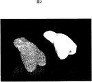
図2は、ウサギ大腿骨遠位部の3次元データをもとに設計した担体の図である。
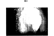
図3は、担体に細胞塊を付着させたことを示す図である。
図4は、担体に細胞塊を再度付着させたことを示す図である。
図5は、ウサギ大腿骨遠位関節面の全体像を示す図である。FIG. 1 is a view showing a spherical joint surface composed only of cells.
FIG. 2 is a diagram of a carrier designed based on the three-dimensional data of the distal part of the rabbit femur.
FIG. 3 is a view showing that a cell mass is attached to a carrier.
FIG. 4 is a diagram showing that the cell mass is reattached to the carrier.
FIG. 5 is a view showing an entire image of a rabbit femoral distal joint surface.
以下、本発明を詳細に説明する。
本発明は、細胞層が必要十分な厚みをもち、任意の曲面に付与することが可能である自己細胞由来の軟骨及び人工関節を作製する方法を提供するものであり、予め目的の形状に加工した担体の表面に細胞塊を付着させ、これを軟骨組織に分化し得る条件下で培養することを特徴とする。これにより、細胞塊は担体の形状にそって細胞塊同士が融合し関節面が再生され、人工関節として機能する。さらに、成長因子の添加や力学的負荷により、細胞外マトリックスの産生が増進され、強度の面においても理想的な関節軟骨を生体外で作製させることを特徴とするものである。
1.細胞
培養の対象となる細胞は、幹細胞(ES細胞、臍帯血由来細胞、未分化間葉系幹細胞等)などの未分化細胞又はその分化型細胞である。特に、未分化間葉系幹細胞が好ましい。
骨芽細胞、軟骨細胞は未分化間葉系幹細胞から容易に分化誘導することが可能なため、これらの分化誘導した細胞(関節軟骨細胞、骨細胞等)も使用することができる。また、成体間葉系幹細胞を使用することもできる。
さらに、幹細胞は、複数の種類の細胞、例えば骨細胞と軟骨細胞とを組み合わせて任意の部位に混合して培養することができる。例えば、上層が軟骨細胞から作った細胞塊だけからなる層、下層が骨細胞由来の細胞塊のように2層に組み合わせることができる。また、大腿骨の場合は、関節部分は軟骨系細胞塊を接着させ、骨の部分は骨系の細胞塊を接着させることができる。このような混合培養は、関節面を含む骨全体の再生に応用することが可能である。
上記細胞は、例えばマウス、ウサギ、ラット、モルモット、イヌ、ブタ、ヤギ、ウシなどの実験動物又は患者の骨髄から、Dexter法、磁気ビーズ法、セルソーティング法などの公知の手法により採取することができる。
ところで、細胞には、浮遊系細胞と足場依存性細胞とに大きく分類され、前者には血液系や免疫系の細胞が属し、後者には皮膚や骨などの細胞が属する。皮膚や骨などの細胞は、培養液中で浮いている状態では死んでしまうため、ガラスなどのシャーレに付着することで増殖する。このため、テフロン(登録商標)中に細胞を一カ所に集めるようにすると、細胞は足場を求めて、お互いに接着し合い、細胞凝集塊、すなわちスフェロイドが形成される。さらに、スフェロイド同士が接着・融合すると大きな形状のものができる。
Molecular Biology of the cell第三版に記載されいているように、酵素処理などでばらばらにした細胞は、自然に凝集することが知られており、この現象はウニなどの下等動物から哺乳類の細胞でもみられることが知られている。この自然凝集は、カドヘリンを含むCell adhesion molecule(CAM)という細胞外接着因子によって引き起こされており、生物の発生初期における四肢の形成の際におこる間葉系幹細胞凝集とほぼ同様の現象が、成熟個体でも再現されていると考えられる(Gerisch,G.Curr.Top.Dev.Biol.14:243−270.1980.;Hennings,H.Exp.Cell Res.143:127−142.1983.;Moscona,A.A.;Hausman,R.E.Biological and biochemical studies on embryonic cell−cell recognition.In Cell and Tissue Interactions,Society of General Physiologists Series(J.W.Lash,M.M.Burger,eds.),Vol.32,pp.173−185.New York:Raven Press,1977.;Roth,S.;Weston,J.Proc.Natl.Acad.Sci.USA 58:974−980.1967.)。
さらに近年、間葉系幹細胞から軟骨細胞への分化の際には、カドヘリンを介した細胞同士の接着がスイッチとなり、コラーゲンなどの発現が開始されることを示唆する報告がなされた(Yoon YM,J Cell Biochem 2002;87(3):342−59)。
そこで本発明においては、一旦スフェロイドにしてから所定の形状に形成することが好ましい。本発明においては、上記間葉系幹細胞などを単層培養した後、撥水性又は細胞非接着性の丸底マルチウェル又はU字型ウェルに移してインキュベートする。その結果、細胞は自然に凝集して細胞塊(細胞凝集塊)を生ずる。細胞塊を生ずるまでのインキュベート時間は、6〜48時間、好ましくは24〜48時間である。細胞塊の作成方法は、上記の方法のほか、旋回している溶液中に細胞懸濁液を入れる旋回培養法、試験管に細胞懸濁液を入れ、遠心分離器で沈殿させる方法、アルギネートビーズ法などが採用され、特に限定されるものではない。均一の細胞塊を大量に処理できる点で、撥水性や細胞非接着性のマルチウェルに細胞懸濁液を入れる方法が、効率がよい点で好ましい。
上記スフェロイドを形成することにより、細胞は細胞周期における静止期に移行し、タンパク質の産生が増加すると考えられる。なお、細胞を静止期に誘導してから分化させることを「細胞が増殖サイクルから外れて、細胞分化へ移行する」という。
2.細胞塊の培養
次に、あらかじめ任意の形状に成形しておいた担体に上記の通り作製した細胞塊を付着させ、培養する。細胞塊の個数は特に限定されるものではなく、任意に設定することができる。さらに、必要であれば、付着後にさらに細胞塊を追加して付着させても、既存の細胞塊とも融合する。
本発明において、細胞塊を付着させる担体は、ヒトやウサギの関節構造に応じて任意に形状を設計することができる。さらに、耳介、鼻など、立体的な形状を得る目的の場合は、あらかじめ3次元データをもとにした鋳型を設計して担体を作製することが可能である。
「担体」とは、細胞塊が接着できる素材であって人工関節の全体又は一部を構成し、細胞塊の支持体として機能するものを意味する。この担体は、生体適合性の素材があり、生体内で吸収、自己組織に置換されるものが好ましい。また、担体の表面は微細孔を有していることが好ましい。このような担体として、例えばトリリン酸カルシウム製の担体が挙げられ、高分子の乳酸ポリマーなどの生体分解性のポリマーも適用可能である。
なお、担体のうち細胞塊を付着させる必要がない領域は、撥水性や細胞非接着性の素材でマスキングすることも可能である。
担体上に付着させた細胞塊同士は、生体が本来具備している創傷治癒とほぼ同等の機序に基づき、それぞれが融合し、結果的に目的の大きさ及び細胞構造を有する構造体を形成する。
本発明では立体構造を有する担体への細胞塊の付着を目的とするため、縦方向(高さ又は厚み)を長くする必要がある。その結果、細胞は液面から深いところに存在することとなり、ガス交換の効率が低下することになる。
そこで本発明では、培養液を、細胞塊の一部が気相に若干ふれる程度(気相にふれても乾燥しない程度)の量に調節することとした。そして、担体の微細孔から培養液が出入りして、細胞塊に届くようにした。「微細孔」とは、細胞塊は通過せず、培養液が通過できる程度の大きさの孔を意味する(直径10〜500μm)。微細孔は、担体自体の成形過程で生じる場合もあるが、担体を細い針で突き刺して形成することも可能である。
培養液は自由に担体の微細孔を通過できるため、培養液を常に細胞に供給することができ、細胞塊は分化・成熟・細胞塊同士の融合、成熟を満足に行うことができる。なお、この場合の成熟とは、細胞が産生する各種コラーゲンやプロテオグリカンなどの細胞外マトリックスの増加を指しており、本来、生体内の細胞は豊富な細胞外マトリックスに囲まれていることが多い。
培養液の量は、細胞が増殖・分化することができる限り特に限定されるものではないが、1個の細胞塊あたり、全量で少なくとも0.5mlの培養液が必要である。
培養に用いられる培養液としては、10%FBS含有DMEM培地、血清無添加DMEMハイグルコース培養液などが挙げられる。
培養は、細胞塊が軟骨に分化し誘導し得る条件下で行なわれる。細胞塊から軟骨への分化誘導は、成長因子又は細胞増殖因子の存在下で培養すればよい。成長因子又は細胞増殖因子としては、骨形成タンパク質(BMP:Bone Morphogenetic Protein)、繊維芽細胞増殖因子(FGF:Fibroblast Growth Factor)、トランスフォーミング増殖因子(TGF−β:Transforming Growth Factor−β)、インスリン様増殖因子(IGF:Insulin−likc Growth Factor)、血小板由来増殖因子(PDGF:Platelet Derived Growth Factor)、血管内皮細胞増殖因子(VEGF:Vascular Endothelial Growth Factor)などを用いることができる。
上記因子は、増殖させる大きさ、細胞外マトリックスの産生量に応じて、単独で、又は適宜組み合わせて種類や量を調整して添加する。例えば、間葉系幹細胞から軟骨細胞に分化するにはTGF−βを添加するが、さらに、BMP、FGF、IGFなどを追加することにより、よりコラーゲンの豊富な軟骨様組織を得ることができる。
ここで、細胞に何らかの刺激が加わると、一般にmRNA量が増加し、その後タンパク質の産生が増加することが知られている。この点に着目し、本発明において、培養条件(培養時間、その他の条件)を変え、細胞の成熟度を調整した。「成熟度」とは、細胞塊を骨や関節等の欠損部に移植するために適した度合いを意味する。その移植時期を、(1)mRNAが増加した時点で移植するか、(2)タンパク質(この場合、コラーゲンなどの細胞外マトリックス)が増加途中の時点で移植するか、あるいは(3)タンパク質が十分量産生された(=成熟)時点で移植するかを検討することが可能である。
人工関節型の場合、タンパク質が十分産生された時期に移植を行うのが望ましく、培養期間は1ヶ月以上が好ましい。このため、長期にわたって、in vitroで変性しない担体を用いることが重要である。
一般に、RNA産生は誘導培養開始2週間でピークになり、その後コラーゲン産生の上昇が見られ、5〜6週で安定する。
さらに、各種成長因子又は増殖因子を添加することにより、細胞外マトリックスの量を調整することが可能である。また、静水圧やせん断弾力などの機械的刺激、超音波や衝撃波などを加えることにより、細胞外マトリックスを増やすことができる。従って、上記成長因子や静水圧や超音波などの機械的刺激を加えることで、RNA産生をピークにする時期を早めることができる。
成長因子又は細胞増殖因子としては、前記と同様、BMP、FGF、TGF−β、IGF、PDGF、VEGFなどを用いることができる。
このように、培養条件や培養期間を調節することにより、成熟度を任意に調節することができる。
本発明の方法によれば、従来のヌードマウスの皮下に一時的に移植して再生組織を熟成させる方法とは異なり、動物を必要としない。そのため、マウスの体の大きさに制限されることなく、任意の大きさの形状が得られる。
患者本人の未分化間葉系幹細胞を用いることにより、骨、軟骨等の自己由来細胞を用いた組織を再生することが可能である。
以下、実施例により、本発明を具体的に説明する。但し、本発明はこれら実施例に限定されるものではない。Hereinafter, the present invention will be described in detail.
The present invention provides a method for producing a cartilage and an artificial joint derived from autologous cells that have a necessary and sufficient thickness and can be applied to an arbitrary curved surface. A cell mass is attached to the surface of the carrier and cultured under conditions capable of differentiating into cartilage tissue. As a result, the cell masses fuse with each other along the shape of the carrier to regenerate the joint surface and function as an artificial joint. Furthermore, the production of extracellular matrix is enhanced by the addition of growth factors and mechanical load, and the ideal articular cartilage is produced in vitro in terms of strength.
1. Cells to be cultured are undifferentiated cells such as stem cells (ES cells, umbilical cord blood-derived cells, undifferentiated mesenchymal stem cells, etc.) or differentiated cells thereof. In particular, undifferentiated mesenchymal stem cells are preferred.
Since osteoblasts and chondrocytes can be easily induced to differentiate from undifferentiated mesenchymal stem cells, these differentiation-induced cells (articular chondrocytes, bone cells, etc.) can also be used. Adult mesenchymal stem cells can also be used.
Furthermore, stem cells can be cultured by combining a plurality of types of cells, for example, bone cells and chondrocytes, and mixing them at an arbitrary site. For example, the upper layer can be combined into two layers, such as a layer composed only of cell clusters made from chondrocytes, and the lower layer can be a cell cluster derived from bone cells. In the case of the femur, the joint portion can adhere the chondrocyte cell mass, and the bone portion can adhere the bone cell mass. Such mixed culture can be applied to regeneration of the entire bone including the joint surface.
The above cells can be collected from laboratory animals such as mice, rabbits, rats, guinea pigs, dogs, pigs, goats and cows or bone marrows of patients by known techniques such as Dexter method, magnetic bead method, cell sorting method and the like. it can.
By the way, the cells are roughly classified into floating cells and anchorage-dependent cells. The former includes cells of the blood system and the immune system, and the latter includes cells such as skin and bone. Since cells such as skin and bone die in a floating state in a culture solution, they proliferate by attaching to a petri dish such as glass. For this reason, when cells are collected in one place in Teflon (registered trademark), the cells seek a scaffold and adhere to each other to form cell aggregates, that is, spheroids. Furthermore, when spheroids are bonded and fused together, a large shape is formed.
As described in Molecular Biology of the cell 3rd edition, cells that have been separated by enzyme treatment are known to aggregate naturally, and this phenomenon is observed from lower animals such as sea urchins to mammalian cells. But it is known to be seen. This spontaneous aggregation is caused by an extracellular adhesion factor called CAM (cell adhesion molecule) containing cadherin, and almost the same phenomenon as the mesenchymal stem cell aggregation that occurs during limb formation in the early stage of organism development, It is thought to have been reproduced in individuals (Gerisch, G. Curr. Top. Dev. Biol. 14: 243-270.1980 .; Hennings, H. Exp. Cell Res. 143: 127-142.1983 .; Moscona Hausman, R. E. Biological and biochemical studies on embryonic cell-cell recognition.In Cell and Tissue Interactions, S., A.A. city of General Physiology Series (JW Lash, MM Burger, eds.), Vol. 32, pp. 173-185. New York: Raven Press, 1977 .; Proc. Natl. Acad. Sci. USA 58: 974-980.1967.).
Furthermore, recently, upon differentiation from mesenchymal stem cells to chondrocytes, there has been a report suggesting that adhesion between cells via cadherin serves as a switch and expression of collagen and the like is started (Yon YM, J Cell Biochem 2002; 87 (3): 342-59).
Therefore, in the present invention, it is preferable that the spheroid is once formed into a predetermined shape. In the present invention, the mesenchymal stem cells and the like are cultured in a single layer, then transferred to a water-repellent or non-adherent round bottom multiwell or U-shaped well and incubated. As a result, the cells naturally aggregate to form a cell mass (cell aggregate mass). The incubation time until the cell mass is generated is 6 to 48 hours, preferably 24 to 48 hours. In addition to the above method, the cell mass can be created by a swirling culture method in which the cell suspension is put in a swirling solution, a method in which the cell suspension is put in a test tube and precipitated with a centrifuge, an alginate bead Laws are adopted and not particularly limited. The method of putting a cell suspension in a water repellent or non-cell-adhesive multiwell is preferable in terms of efficiency because a large amount of a uniform cell mass can be processed.
By forming the spheroids, the cells are considered to shift to the stationary phase in the cell cycle, and protein production is increased. In addition, differentiation after inducing a cell in a stationary phase is referred to as “the cell moves out of the growth cycle and shifts to cell differentiation”.
2. Next, the cell mass produced as described above is attached to a carrier that has been previously shaped into an arbitrary shape and cultured. The number of cell clusters is not particularly limited and can be set arbitrarily. Furthermore, if necessary, even if an additional cell mass is attached after attachment, it is also fused with the existing cell mass.
In the present invention, the shape of the carrier to which the cell mass is attached can be arbitrarily designed according to the joint structure of a human or rabbit. Furthermore, for the purpose of obtaining a three-dimensional shape such as the pinna or nose, it is possible to prepare a carrier by designing a template based on three-dimensional data in advance.
The “carrier” means a material to which the cell mass can adhere, which constitutes the whole or part of the artificial joint and functions as a support for the cell mass. This carrier has a biocompatible material, and is preferably a material that can be absorbed in vivo and replaced by self-organization. The surface of the carrier preferably has fine pores. Examples of such a carrier include a carrier made of calcium triphosphate, and a biodegradable polymer such as a high-molecular lactic acid polymer is also applicable.
In addition, the area | region which does not need to adhere a cell clump of a support | carrier can also be masked with a water-repellent or cell non-adhesive raw material.
Cell clumps attached on a carrier are fused based on a mechanism that is almost equivalent to the wound healing inherent in living bodies, resulting in the formation of a structure with the desired size and cell structure. To do.
In the present invention, since the purpose is to attach a cell mass to a carrier having a three-dimensional structure, it is necessary to lengthen the vertical direction (height or thickness). As a result, the cells exist deep from the liquid surface, and the efficiency of gas exchange decreases.
Therefore, in the present invention, the culture solution is adjusted to an amount that allows a part of the cell mass to slightly touch the gas phase (so that it does not dry even if it touches the gas phase). Then, the culture solution entered and exited from the micropores of the carrier so as to reach the cell mass. The “micropore” means a pore having a size that allows a culture solution to pass through without passing through a cell mass (diameter: 10 to 500 μm). The micropores may occur during the molding process of the carrier itself, but can also be formed by piercing the carrier with a thin needle.
Since the culture solution can freely pass through the micropores of the carrier, the culture solution can always be supplied to the cells, and the cell mass can be satisfactorily differentiated, matured, fused between cell masses, and matured. Note that maturation in this case refers to an increase in extracellular matrix such as various collagens and proteoglycans produced by cells, and naturally cells in a living body are often surrounded by abundant extracellular matrix.
The amount of the culture solution is not particularly limited as long as the cells can proliferate and differentiate, but a total amount of at least 0.5 ml of the culture solution is required per cell mass.
Examples of the culture medium used for culture include 10% FBS-containing DMEM medium, serum-free DMEM high glucose culture medium, and the like.
Culturing is performed under conditions that allow the cell mass to differentiate and induce cartilage. Differentiation induction from a cell mass to cartilage may be performed in the presence of a growth factor or a cell growth factor. Examples of the growth factor or cell growth factor include bone morphogenetic protein (BMP), fibroblast growth factor (FGF), transforming growth factor (TGF-β), and insulin growth factor-β. Insulin-like growth factor (IGF), platelet-derived growth factor (PDGF), vascular endothelial growth factor (VEGF), etc. can be used.
The above-mentioned factors are added alone or in combination as appropriate depending on the size to be grown and the amount of extracellular matrix produced. For example, TGF-β is added to differentiate from mesenchymal stem cells to chondrocytes, but by adding BMP, FGF, IGF, and the like, a more collagen-rich cartilage-like tissue can be obtained.
Here, it is known that when some kind of stimulation is applied to cells, the amount of mRNA generally increases and then the production of protein increases. Focusing on this point, in the present invention, the culture conditions (culture time, other conditions) were changed to adjust the maturity of the cells. “Maturity” means a degree suitable for transplanting a cell mass to a defect such as a bone or a joint. Either (1) transplant when mRNA is increased, (2) transplant when protein (in this case, extracellular matrix such as collagen) is increasing, or (3) protein is sufficient It is possible to examine whether to transplant at the time of mass production (= maturity).
In the case of an artificial joint type, it is desirable to perform transplantation when protein is sufficiently produced, and the culture period is preferably one month or longer. For this reason, it is important to use a carrier that does not denature in vitro over a long period of time.
In general, RNA production peaks at 2 weeks after the start of induction culture, then increases in collagen production is observed, and stabilizes at 5-6 weeks.
Furthermore, the amount of extracellular matrix can be adjusted by adding various growth factors or growth factors. In addition, the extracellular matrix can be increased by applying mechanical stimuli such as hydrostatic pressure and shear elasticity, ultrasonic waves, shock waves, and the like. Therefore, by applying mechanical stimuli such as the growth factor, hydrostatic pressure and ultrasonic waves, the time for peaking RNA production can be advanced.
As the growth factor or cell growth factor, BMP, FGF, TGF-β, IGF, PDGF, VEGF and the like can be used as described above.
Thus, the maturity can be arbitrarily adjusted by adjusting the culture conditions and the culture period.
According to the method of the present invention, an animal is not required unlike the conventional method in which a nude mouse is temporarily transplanted subcutaneously to mature a regenerated tissue. Therefore, a shape of any size can be obtained without being limited by the size of the mouse body.
By using the patient's own undifferentiated mesenchymal stem cells, it is possible to regenerate a tissue using autologous cells such as bone and cartilage.
Hereinafter, the present invention will be described specifically by way of examples. However, the present invention is not limited to these examples.
本実施例は、ヒト股関節、大腿骨頭を模した実施例である。
ヒト骨髄由来間葉系幹細胞を単層培養した。最終的に間葉系幹細胞を15cmディッシュ一枚あたり1.0x10⁶得た。この細胞をトリプシン処理して細胞懸濁液にし、住友ベークライト社製スフェロイドプレートにそれぞれ1.0x10⁵個の細胞が入るように播種した。その後37℃、5%二酸化炭素の条件下で培養を行い、翌日には直径が平均0.5mmの細胞塊が1プレートあたり96個得られた。この細胞塊を、半球状に加工したトリリン酸カルシウムの上に付着させた。その後、TGF−βを0.01μg/ml量添加し、48時間培養することにより、細胞塊を軟骨に分化させた。この曲面に付着した細胞塊同士は、24から48時間程度で融合し、細胞だけからなる球状の関節面が得られた(図1)。図1において、BはAの拡大図である。The present embodiment is an embodiment simulating a human hip joint and a femoral head.
Human bone marrow-derived mesenchymal stem cells were cultured in a monolayer. Finally, 1.0 × 10 6 mesenchymal stem cells were obtained per 15 cm dish. The cells were trypsinized to obtain a cell suspension, and seeded so that 1.0 × 10 5 cells were placed in each spheroid plate manufactured by Sumitomo Bakelite. Thereafter, the cells were cultured under conditions of 37 ° C. and 5% carbon dioxide, and the next day, 96 cell clusters having an average diameter of 0.5 mm were obtained per plate. This cell mass was allowed to adhere on calcium triphosphate processed into a hemisphere. Thereafter, TGF-β was added in an amount of 0.01 μg / ml and cultured for 48 hours to differentiate the cell mass into cartilage. The cell clumps attached to the curved surface were fused in about 24 to 48 hours, and a spherical joint surface consisting only of cells was obtained (FIG. 1). In FIG. 1, B is an enlarged view of A.
本実施例は、ウサギ大腿骨遠位関節面の作製例である。
ウサギ関節軟骨切片にコラゲナーゼ処理を行い、得られた軟骨細胞の単層培養を行った。最終的に軟骨細胞を15cmディッシュ一枚あたり1.0x10⁶個得た。この細胞をトリプシン処理して細胞懸濁液にし、住友ベークライト社製スフェロイドプレートにそれぞれ1.0x10⁵個の細胞が入るように播種した。その後37℃、5%二酸化炭素の条件下で培養を行い、翌日には直径が平均0.5mmの細胞塊が1プレートあたり96個得られた。
一方、ウサギ大腿骨遠位関節面の形状を模して予め3次元データを作成し(図2)、この形状のトリリン酸カルシウム製担体を作製した。
上記細胞塊をトリリン酸カルシウムの上に付着させた。このトリリン酸カルシウムの大腿骨遠位関節面には微細孔が付与されている。その後、TGF−βを0.01μg/ml量添加し、48時間培養することにより、細胞塊を軟骨に分化させた。この曲面に付着した細胞塊同士は、24から48時間程度で融合し、細胞だけからなる関節面が得られた(図3)。図3において、やや褐色の部分が細胞塊を付着させた箇所である。さらに翌週(7日後)に、このようにして得られた関節面(図3)の上に320個の細胞塊を再度付着させた。その結果、新たに付着させた細胞塊は、既に付着している細胞塊と融合してスムーズな曲面を形成した(図4A)。
さらに上記関節面を320個の細胞塊を付着させた結果、最終的に図4Bに示すように目的の関節を作製することができた。図4Bにおいて、茶色の部分が細胞塊を付着させた領域である。周囲の白色部分が、余分な部分に細胞塊が付着しないようマスクとして用いたパラフィン樹脂である。
図5に、ウサギ大腿骨遠位関節面の全体像を示す。
図5Aはコンピュータに入力した3次元データをもとに作成した、トリリン酸カルシウムからなるウサギ大腿骨遠位関節面型の担体(細胞付着前)である。
図5B、図5Cは細胞塊付着翌日の概観である。軟骨相当部分にのみ細胞層が形成されているのがわかる。図5Dは細胞付着部(図5B、C)の拡大図である。若干細胞塊の境界が判明できる程度に融合している。This example is an example of making a rabbit femur distal joint surface.
A rabbit articular cartilage slice was treated with collagenase and the resulting chondrocytes were subjected to monolayer culture. Finally, 1.0 × 10 6 chondrocytes were obtained per 15 cm dish. The cells were trypsinized to obtain a cell suspension, and seeded so that 1.0 × 10 5 cells were placed in each spheroid plate manufactured by Sumitomo Bakelite. Thereafter, the cells were cultured under conditions of 37 ° C. and 5% carbon dioxide, and the next day, 96 cell clusters having an average diameter of 0.5 mm were obtained per plate.
On the other hand, three-dimensional data was created in advance to simulate the shape of the distal joint surface of the rabbit femur (FIG. 2), and a calcium triphosphate carrier having this shape was prepared.
The cell mass was deposited on calcium triphosphate. A micropore is provided in the distal femoral joint surface of this calcium triphosphate. Thereafter, TGF-β was added in an amount of 0.01 μg / ml and cultured for 48 hours to differentiate the cell mass into cartilage. The cell masses attached to the curved surface were fused in about 24 to 48 hours, and an articular surface consisting only of cells was obtained (FIG. 3). In FIG. 3, the slightly brown part is the place where the cell mass is adhered. Further, in the next week (after 7 days), 320 cell clusters were reattached on the joint surface thus obtained (FIG. 3). As a result, the newly attached cell mass fused with the already adhered cell mass to form a smooth curved surface (FIG. 4A).
Furthermore, as a result of attaching 320 cell masses to the joint surface, the target joint could be finally produced as shown in FIG. 4B. In FIG. 4B, the brown part is the area where the cell mass is attached. The surrounding white part is a paraffin resin used as a mask so that cell clumps do not adhere to the excess part.
FIG. 5 shows an overall image of the rabbit femur distal joint surface.
FIG. 5A is a rabbit femur distal joint surface type carrier (before cell attachment) made of calcium triphosphate, created based on three-dimensional data input to a computer.
FIG. 5B and FIG. 5C are overviews on the day after the cell mass attachment. It can be seen that the cell layer is formed only in the cartilage equivalent part. FIG. 5D is an enlarged view of the cell attachment part (FIGS. 5B and 5C). Fused to such an extent that the boundaries of the cell mass can be found.
本発明により、軟骨及び人工関節の作製方法が提供される。本発明により提供される技術は、細胞凝集塊を多量に作成し、それぞれを融合する事で、ポリマーなどの担体を利用する必要がなく、自己由来の細胞単独で関節軟骨層を作製することが可能である。更には、関節を成熟させるための動物への移植を必要としないため、他種動物を経由することなく、任意の形状の軟骨層を作製することが可能となる。 The present invention provides a method for producing cartilage and an artificial joint. The technology provided by the present invention makes it possible to produce an articular cartilage layer with self-derived cells alone by creating a large amount of cell aggregates and fusing them together without the need to use a carrier such as a polymer. Is possible. Furthermore, since it is not necessary to transplant the animal to mature the joint, it is possible to produce a cartilage layer of any shape without going through other species.
Claims (6)
Applications Claiming Priority (3)
| Application Number | Priority Date | Filing Date | Title |
|---|---|---|---|
| JP2003283703 | 2003-07-31 | ||
| JP2003283703 | 2003-07-31 | ||
| PCT/JP2004/011391 WO2005011765A1 (en) | 2003-07-31 | 2004-08-02 | Method of constructing artificial joint |
Publications (2)
| Publication Number | Publication Date |
|---|---|
| JPWO2005011765A1 JPWO2005011765A1 (en) | 2006-10-26 |
| JP4339855B2 true JP4339855B2 (en) | 2009-10-07 |
Family
ID=34113817
Family Applications (1)
| Application Number | Title | Priority Date | Filing Date |
|---|---|---|---|
| JP2005512607A Expired - Lifetime JP4339855B2 (en) | 2003-07-31 | 2004-08-02 | How to make an artificial joint |
Country Status (7)
| Country | Link |
|---|---|
| US (1) | US20080039939A1 (en) |
| EP (1) | EP1649875B1 (en) |
| JP (1) | JP4339855B2 (en) |
| KR (1) | KR101183066B1 (en) |
| CN (1) | CN100571791C (en) |
| AT (1) | ATE528026T1 (en) |
| WO (1) | WO2005011765A1 (en) |
Families Citing this family (27)
| Publication number | Priority date | Publication date | Assignee | Title |
|---|---|---|---|---|
| US7067123B2 (en) * | 2003-04-29 | 2006-06-27 | Musculoskeletal Transplant Foundation | Glue for cartilage repair |
| US20050064042A1 (en) * | 2003-04-29 | 2005-03-24 | Musculoskeletal Transplant Foundation | Cartilage implant plug with fibrin glue and method for implantation |
| US20050222687A1 (en) * | 2004-04-02 | 2005-10-06 | Gordana Vunjak-Novakovic | Cartilage implant assembly and method for implantation |
| US7901457B2 (en) * | 2003-05-16 | 2011-03-08 | Musculoskeletal Transplant Foundation | Cartilage allograft plug |
| ES2403357T3 (en) | 2003-12-11 | 2013-05-17 | Isto Technologies Inc. | Particle Cartilage System |
| US7837740B2 (en) | 2007-01-24 | 2010-11-23 | Musculoskeletal Transplant Foundation | Two piece cancellous construct for cartilage repair |
| US20080220044A1 (en) * | 2007-03-06 | 2008-09-11 | Semler Eric J | Cancellous construct with support ring for repair of osteochondral defects |
| US7815926B2 (en) * | 2005-07-11 | 2010-10-19 | Musculoskeletal Transplant Foundation | Implant for articular cartilage repair |
| WO2007025290A2 (en) | 2005-08-26 | 2007-03-01 | Isto Technologies, Inc. | Implants and methods for repair, replacement and treatment of joint disease |
| US8921109B2 (en) | 2005-09-19 | 2014-12-30 | Histogenics Corporation | Cell-support matrix having narrowly defined uniformly vertically and non-randomly organized porosity and pore density and a method for preparation thereof |
| EP1764117A1 (en) * | 2005-09-20 | 2007-03-21 | Zimmer GmbH | Implant for the repair of a cartilage defect and method for manufacturing the implant |
| US8163549B2 (en) | 2006-12-20 | 2012-04-24 | Zimmer Orthobiologics, Inc. | Method of obtaining viable small tissue particles and use for tissue repair |
| US8435551B2 (en) | 2007-03-06 | 2013-05-07 | Musculoskeletal Transplant Foundation | Cancellous construct with support ring for repair of osteochondral defects |
| US20090012629A1 (en) | 2007-04-12 | 2009-01-08 | Isto Technologies, Inc. | Compositions and methods for tissue repair |
| JP5495486B2 (en) * | 2007-10-31 | 2014-05-21 | 恒夫 高橋 | Cultured cartilage manufacturing method and cultured cartilage |
| KR101450943B1 (en) | 2007-12-21 | 2014-10-21 | 쿄세라 메디칼 가부시키가이샤 | Device for cell transplantation |
| EP2254508A4 (en) * | 2008-03-03 | 2012-03-28 | Univ Rice William M | Methods of fabricating enhanced tissue-engineered cartilage |
| WO2009111069A1 (en) | 2008-03-05 | 2009-09-11 | Musculoskeletal Transplant Foundation | Cancellous constructs, cartilage particles and combinations of cancellous constructs and cartilage particles |
| KR20130005279A (en) * | 2010-03-01 | 2013-01-15 | 후지필름 가부시키가이샤 | Cell structure comprising cells and macromolecular blocks having biocompatibility |
| JP5095855B2 (en) * | 2010-12-13 | 2012-12-12 | 株式会社 資生堂 | Method for forming cell aggregate |
| US20140178343A1 (en) | 2012-12-21 | 2014-06-26 | Jian Q. Yao | Supports and methods for promoting integration of cartilage tissue explants |
| JP5815006B2 (en) * | 2013-11-08 | 2015-11-17 | 恒夫 高橋 | Cultured cartilage manufacturing method and cultured cartilage |
| US10077420B2 (en) | 2014-12-02 | 2018-09-18 | Histogenics Corporation | Cell and tissue culture container |
| KR101733137B1 (en) * | 2015-12-30 | 2017-05-08 | (주)엑셀세라퓨틱스 | Method of production for 3D cartilage organoid block |
| JP6996556B2 (en) * | 2017-04-20 | 2022-01-17 | 株式会社ニコン | Medical devices and artificial joints |
| CN109251887A (en) * | 2018-10-12 | 2019-01-22 | 山东麦德克斯生物科技有限公司 | A kind of preparation method and cartilaginous tissue of cartilage cell's primer layer |
| JPWO2023032441A1 (en) * | 2021-08-31 | 2023-03-09 |
Family Cites Families (9)
| Publication number | Priority date | Publication date | Assignee | Title |
|---|---|---|---|---|
| CA2245350A1 (en) | 1998-08-19 | 2000-02-19 | Ian Drew | Leakcage diversion tile construction method |
| CA2254350A1 (en) * | 1998-11-17 | 2000-05-17 | Hsc Research And Development Limited Partnership | Method for bioengineering cartilage |
| US6197061B1 (en) * | 1999-03-01 | 2001-03-06 | Koichi Masuda | In vitro production of transplantable cartilage tissue cohesive cartilage produced thereby, and method for the surgical repair of cartilage damage |
| DE10042484A1 (en) * | 2000-08-29 | 2002-03-28 | Merck Patent Gmbh | Preparing human cartilage implants, useful for repairing damaged joints, comprises culturing autologous chondrocytes in alginate gel at reduced oxygen partial pressure |
| JP2003111831A (en) * | 2001-07-30 | 2003-04-15 | Olympus Optical Co Ltd | Cartilage implant |
| JP3599701B2 (en) * | 2001-11-15 | 2004-12-08 | 独立行政法人理化学研究所 | Cell engraftment method on three-dimensional structure surface |
| JP2003304866A (en) * | 2002-04-17 | 2003-10-28 | National Institute Of Advanced Industrial & Technology | Differentiation control of cell by three-dimensional agglutinate formation |
| JP2003038635A (en) * | 2002-06-10 | 2003-02-12 | Olympus Optical Co Ltd | Osteochondroimplant material |
| DE10253066A1 (en) * | 2002-11-07 | 2004-05-27 | Co.Don Aktiengesellschaft | Tissue substitute, useful for repairing lesions, e.g. in cardiac muscle, also for production of e.g. cytokines, comprises preformed three-dimensional tissue grown from suspension culture |
-
2004
- 2004-08-02 US US10/566,362 patent/US20080039939A1/en not_active Abandoned
- 2004-08-02 AT AT04748282T patent/ATE528026T1/en not_active IP Right Cessation
- 2004-08-02 KR KR1020067001854A patent/KR101183066B1/en active IP Right Grant
- 2004-08-02 CN CNB2004800284224A patent/CN100571791C/en not_active Expired - Lifetime
- 2004-08-02 EP EP04748282A patent/EP1649875B1/en not_active Expired - Lifetime
- 2004-08-02 WO PCT/JP2004/011391 patent/WO2005011765A1/en active Application Filing
- 2004-08-02 JP JP2005512607A patent/JP4339855B2/en not_active Expired - Lifetime
Also Published As
| Publication number | Publication date |
|---|---|
| CN1859933A (en) | 2006-11-08 |
| KR101183066B1 (en) | 2012-09-20 |
| EP1649875A1 (en) | 2006-04-26 |
| CN100571791C (en) | 2009-12-23 |
| WO2005011765A1 (en) | 2005-02-10 |
| US20080039939A1 (en) | 2008-02-14 |
| EP1649875B1 (en) | 2011-10-12 |
| EP1649875A4 (en) | 2008-02-13 |
| JPWO2005011765A1 (en) | 2006-10-26 |
| KR20060052908A (en) | 2006-05-19 |
| ATE528026T1 (en) | 2011-10-15 |
Similar Documents
| Publication | Publication Date | Title |
|---|---|---|
| JP4339855B2 (en) | How to make an artificial joint | |
| EP1076533B1 (en) | Guided development and support of hydrogel-cell compositions | |
| EP0739631B1 (en) | Laminar bone support for cartilage growth | |
| JP4406283B2 (en) | Tissue regeneration substrate, transplant material, and production method thereof | |
| JP6152985B2 (en) | Scaffold-free self-organized 3D artificial tissue and bone composite for osteochondral regeneration | |
| JP2003510108A (en) | Biological joint structures | |
| US20230098674A1 (en) | Engineered tissue constructs | |
| JP4122280B2 (en) | Manufacturing method of tissue plug | |
| KR102479530B1 (en) | Method of Preparing Pellets of Chondrocytes differentiated from human induced pluripotent stem cell and use of the same | |
| Bücheler et al. | Tissue engineering in otorhinolaryngology | |
| US20050002982A1 (en) | Hybrid tissues for tissue engineering | |
| CA2483660A1 (en) | Injectable chondrocyte implant | |
| US10167447B2 (en) | Supports and methods for promoting integration of cartilage tissue explants | |
| JP4061487B2 (en) | Transplant material for angiogenesis and method for producing the same | |
| US20150344847A1 (en) | Method For Production Of Large Numbers Of Cartilage Cells With Phenotype Retention | |
| US20140341859A1 (en) | Method for treatment of damaged site of bone | |
| JP2011156329A (en) | Bone/cartilage-like structure |
Legal Events
| Date | Code | Title | Description |
|---|---|---|---|
| A521 | Request for written amendment filed |
Free format text: JAPANESE INTERMEDIATE CODE: A523 Effective date: 20060629 |
|
| A621 | Written request for application examination |
Free format text: JAPANESE INTERMEDIATE CODE: A621 Effective date: 20070720 |
|
| A711 | Notification of change in applicant |
Free format text: JAPANESE INTERMEDIATE CODE: A711 Effective date: 20080522 |
|
| A521 | Request for written amendment filed |
Free format text: JAPANESE INTERMEDIATE CODE: A821 Effective date: 20080522 |
|
| A131 | Notification of reasons for refusal |
Free format text: JAPANESE INTERMEDIATE CODE: A131 Effective date: 20090217 |
|
| A521 | Request for written amendment filed |
Free format text: JAPANESE INTERMEDIATE CODE: A523 Effective date: 20090420 |
|
| TRDD | Decision of grant or rejection written | ||
| A01 | Written decision to grant a patent or to grant a registration (utility model) |
Free format text: JAPANESE INTERMEDIATE CODE: A01 Effective date: 20090623 |
|
| A01 | Written decision to grant a patent or to grant a registration (utility model) |
Free format text: JAPANESE INTERMEDIATE CODE: A01 |
|
| A61 | First payment of annual fees (during grant procedure) |
Free format text: JAPANESE INTERMEDIATE CODE: A61 Effective date: 20090702 |
|
| R150 | Certificate of patent or registration of utility model |
Ref document number: 4339855 Country of ref document: JP Free format text: JAPANESE INTERMEDIATE CODE: R150 Free format text: JAPANESE INTERMEDIATE CODE: R150 |
|
| FPAY | Renewal fee payment (event date is renewal date of database) |
Free format text: PAYMENT UNTIL: 20120710 Year of fee payment: 3 |
|
| FPAY | Renewal fee payment (event date is renewal date of database) |
Free format text: PAYMENT UNTIL: 20120710 Year of fee payment: 3 |
|
| S533 | Written request for registration of change of name |
Free format text: JAPANESE INTERMEDIATE CODE: R313533 |
|
| FPAY | Renewal fee payment (event date is renewal date of database) |
Free format text: PAYMENT UNTIL: 20120710 Year of fee payment: 3 |
|
| R350 | Written notification of registration of transfer |
Free format text: JAPANESE INTERMEDIATE CODE: R350 |
|
| FPAY | Renewal fee payment (event date is renewal date of database) |
Free format text: PAYMENT UNTIL: 20120710 Year of fee payment: 3 |
|
| FPAY | Renewal fee payment (event date is renewal date of database) |
Free format text: PAYMENT UNTIL: 20130710 Year of fee payment: 4 |
|
| R250 | Receipt of annual fees |
Free format text: JAPANESE INTERMEDIATE CODE: R250 |
|
| R250 | Receipt of annual fees |
Free format text: JAPANESE INTERMEDIATE CODE: R250 |
|
| S531 | Written request for registration of change of domicile |
Free format text: JAPANESE INTERMEDIATE CODE: R313531 |
|
| R350 | Written notification of registration of transfer |
Free format text: JAPANESE INTERMEDIATE CODE: R350 |
|
| R250 | Receipt of annual fees |
Free format text: JAPANESE INTERMEDIATE CODE: R250 |
|
| R250 | Receipt of annual fees |
Free format text: JAPANESE INTERMEDIATE CODE: R250 |
|
| R250 | Receipt of annual fees |
Free format text: JAPANESE INTERMEDIATE CODE: R250 |
|
| S111 | Request for change of ownership or part of ownership |
Free format text: JAPANESE INTERMEDIATE CODE: R313117 |
|
| R350 | Written notification of registration of transfer |
Free format text: JAPANESE INTERMEDIATE CODE: R350 |
|
| R250 | Receipt of annual fees |
Free format text: JAPANESE INTERMEDIATE CODE: R250 |
|
| R250 | Receipt of annual fees |
Free format text: JAPANESE INTERMEDIATE CODE: R250 |
|
| R250 | Receipt of annual fees |
Free format text: JAPANESE INTERMEDIATE CODE: R250 |
|
| R250 | Receipt of annual fees |
Free format text: JAPANESE INTERMEDIATE CODE: R250 |
|
| R250 | Receipt of annual fees |
Free format text: JAPANESE INTERMEDIATE CODE: R250 |
|
| R250 | Receipt of annual fees |
Free format text: JAPANESE INTERMEDIATE CODE: R250 |
|
| R250 | Receipt of annual fees |
Free format text: JAPANESE INTERMEDIATE CODE: R250 |
|
| R250 | Receipt of annual fees |
Free format text: JAPANESE INTERMEDIATE CODE: R250 |