JP4295386B2 - Highly active alkaline phosphatase - Google Patents
Highly active alkaline phosphatase Download PDFInfo
- Publication number
- JP4295386B2 JP4295386B2 JP12649499A JP12649499A JP4295386B2 JP 4295386 B2 JP4295386 B2 JP 4295386B2 JP 12649499 A JP12649499 A JP 12649499A JP 12649499 A JP12649499 A JP 12649499A JP 4295386 B2 JP4295386 B2 JP 4295386B2
- Authority
- JP
- Japan
- Prior art keywords
- biap
- seq
- cdna
- biapi
- iii
- Prior art date
- Legal status (The legal status is an assumption and is not a legal conclusion. Google has not performed a legal analysis and makes no representation as to the accuracy of the status listed.)
- Expired - Lifetime
Links
Images


Classifications
-
- C—CHEMISTRY; METALLURGY
- C12—BIOCHEMISTRY; BEER; SPIRITS; WINE; VINEGAR; MICROBIOLOGY; ENZYMOLOGY; MUTATION OR GENETIC ENGINEERING
- C12N—MICROORGANISMS OR ENZYMES; COMPOSITIONS THEREOF; PROPAGATING, PRESERVING, OR MAINTAINING MICROORGANISMS; MUTATION OR GENETIC ENGINEERING; CULTURE MEDIA
- C12N9/00—Enzymes; Proenzymes; Compositions thereof; Processes for preparing, activating, inhibiting, separating or purifying enzymes
- C12N9/14—Hydrolases (3)
- C12N9/16—Hydrolases (3) acting on ester bonds (3.1)
Abstract
Description
【0001】
【発明の属する分野】
本発明は、3000U/mg以上の比活性を有する真核生物高活性アルカリ性ホスファターゼをコードするDNAに関する。また、本発明は、本発明のDNAの産生方法、本発明のDNAを含むベクター、ならびにかかるベクターを含む細胞系に関する。本発明は、さらに、本発明のDNAによってコードされる、3000U/mg以上の比活性を有する組換え高活性アルカリ性ホスファターゼに関する。
【0002】
【従来の技術】
アルカリ性ホスファターゼ(AP)は、E.coliから哺乳動物に至る全ての生物において見出される、二量体、亜鉛含有の非特異的ホスホモノエステラーゼである(McCombら, 1979)。種々のアルカリ性ホスファターゼの一次構造を比較したところ、高度に相同性があることが示された(E.coliと哺乳動物APの間の相同性は25〜30%)(Millan, 1988;Harris, 1989)。
【0003】
ヒトおよび高等動物においては、APファミリーは異なる遺伝子座においてコードされる4つのメンバーからなる(Millan, 1988;Harris, 1989)。アルカリ性ホスファターゼファミリーとしては、組織特異的AP(胎盤AP(PLAP)、生殖細胞AP(GCAP)および腸AP(IAP))ならびに主に肝臓、腎臓および骨に局在する非組織特異的AP(TNAP)が挙げられる。
【0004】
従来公知のAPの決め手となる特性は、比活性がE.coliAPよりも10〜100倍高い哺乳動物APの触媒活性の大きな変動性である。哺乳動物APの中で、ウシ腸由来のAP(bIAP)が最も高い比活性を示す。この特性により、bIAPは診断試薬またはDNAの脱リン酸化のための酵素抱合体などのバイオテクノロジー用途に対して魅力的である。1985年、BesmanおよびColemanは、ウシ腸に2つのIAPイソ酵素、すなわち子ウシ腸由来のIAPおよび成熟ウシの腸由来のIAP(bIAP)が存在することを、クロマトグラフィーにて精製したAP画分のアミノ末端配列決定によって立証した。アミノ末端における明確な相違が、成熟ウシのbIAP(LVPVEEED)と子ウシ腸由来のbIAP(LIPAEEEN)の間に示されている。1993年、Weissigらは、比活性約3000U/mgでN-末端がLVPVEEEDである組換えbIAP(bIAPI)をクローニングすることによって正確な生化学的特性付けを行った。しかしながら、最大8000U/mgまでの比活性を有する子ウシ由来のbIAPも市販されているが(Boehringer Mannheim, Biozyme, Oriental Yeast)、さらなる特性付けは未だなされていない。これらの高活性アルカリ性ホスファターゼをクローニングする試みはすべてうまくいかなかった。したがって、組換え高活性アルカリ性ホスファターゼを産生することはできなかった。しかしながら、組換え体産生の可能性は、高活性アルカリ性ホスファターゼの経済的な産生には絶対必須である。
【0005】
【発明が解決しようとする課題】
したがって、本発明の目的は、クローニングすることもできる組換え手段による高活性アルカリ性ホスファターゼを提供することである。本発明の意味に含まれる「高活性」は、本発明によるアルカリ性ホスファターゼが従来公知のアルカリ性ホスファターゼと比べて少なくとも10%高い活性を有することを意味するものである。
【0006】
【課題を解決するための手段】
この目的は、本発明にしたがって、322位のアミノ酸残基がアスパラギン酸残基よりも小さく、3000U/mg以上、好ましくは少なくとも3500U/mgの比活性を有する真核生物高活性アルカリ性ホスファターゼをコードするDNAの提供によって達成された。本発明の意味においては、真核生物DNAが好ましい。もはやイントロンを含まないDNAを意味する真核生物cDNAが特に好ましい。「アスパラギン酸残基よりも小さいアミノ酸残基」という用語は、アミノ酸であるアスパラギン酸残基の構造よりも小さい空間的寸法を有するあらゆるアミノ酸、好ましくは天然アミノ酸もしくは天然アミノ酸から誘導されるアミノ酸と理解される。本発明のDNAは、アミノ酸残基322がグリシン、アラニン、トレオニン、バリンまたはセリンであるものが好ましい。本発明のDNAは、アミノ酸残基322がグリシンまたはセリンであるのものが特に好ましい。配列番号(SEQ ID NO.)1、3および5(図1、3、5)に記載のDNAならびに配列番号2、4および6(図2、4、6)に記載の関連アミノ酸配列は、本発明の一部である。また、本発明は、N末端が配列番号2、4および6に記載のcDNAと比べて長いか短いということのみが前述と異なるcDNAにも関する。そのような場合、配列番号2、4および6の322位に対する名称はそれに応じて変化する。例えば、N末端が配列番号2、4および6よりもXアミノ酸長いか短い場合、関連する322位もXアミノ酸の分だけシフトする。
【0007】
配列番号1は、高活性bAIP IIイソ酵素の配列をコードするDNAを含む。天然酵素は公知であるが、特性付けされておらず、クローニングすることができない。したがって、高活性bIAP IIイソ酵素のアミノ酸配列決定が本発明の主題である。子ウシ腸由来の、高比活性を有する高度に精製された画分(Boehringer Mannheim)を用いて配列を決定した。高活性APのペプチド地図をエンドプロテイナーゼLysC、AspN、GluC、トリプシンによる切断ならびにブロムシアンによる化学的切断によって作製した。この手法で産生させたペプチドを逆相HPLCによって分離し、単離した。各ペプチドをエレクトロスプレー質量分光法によって分析し、エドマン分解によって配列決定した。この方法で得られた配列を、刊行物に記載されたbIAPIの配列(Weissigら, 1993)と比較した。予想どおり、bIAP IIのアミノ末端はBesmanおよびColeman(J. Biol. Chem. 260, 11190-11193(1985))によって記載されたような出発配列LIPAEEENを有している。bIAP IIの完全アミノ酸配列を配列番号2に示す(図2)。この配列番号2によれば、bIAP IIはbIAPIと比べて合計24個のアミノ酸置換を有する。単離された高活性bIAP IIイソ酵素のアミノ酸数は、480アミノ酸である。1798bpのヌクレオチド配列(図1)は、514アミノ酸のコード領域を含む。481位から514位までの可能なアミノ酸は、広い制限範囲内で変動し得る。
【0008】
以下、本発明は従来知られていない2つの新規bIAP(bIAP IIIおよびbIAP IV)のクローニングおよび完全特性付けを記載する。ウシ腸の異なる切片由来のRNA試料においてノーザンブロット分析を行った。最も強いハイブリダイゼーションシグナルを有するプローブのcDNAバンクを、ベクターIZAP II(Stratagene、サンディエゴ、CA、USA)においてオリゴdTプライマー(Stratagene、サンディエゴ、CA、USA)を用いて準備した。完全なバンク(1.0×10⁶ 組換えクローン)をbIAPI遺伝子のエキソンI〜VIIIの領域をカバーするbIAPIの1075bp HindIII断片を用いてスクリーニングした。65クローンを単離して配列決定した。このプロセスにおいて、2つの新規bIAPが同定された(bIAP IIIおよびbIAP IV)。その特性付けをさらに下記に記載する。これらはbIAPIともbIAP IIとも完全に相同ではなかった。bIAP IIIおよびIVのヌクレオチド配列を図3および5に示す。bIAPI〜IVの配列の相違を図7に示す。しかしながら、新規bIAPはどれも予想したN末端LIPAEEENを有しておらず、従来記載されていない新規のN末端を有していた(図7参照)。2つの新規bIAPイソ酵素のcDNAを適当な制限酵素を用いて再切断し、CHO発現ベクターpcDNA-3(例えば、Invitrogen社、サンディエゴ、CA、USA製)にライゲーション(連結)によって挿入した。新規bIAPイソ酵素を含むクローンをInvitrogenによって記載された方法にしたがって発現させ、イソ酵素の特性付けを行った。種々の宿主におけるbIAP遺伝子の発現は、WO93/18139に記載されている(CHO細胞、E.coli、バキュロウイルス系)。この特許文献に記載されている方法、ベクターおよび発現系は、本出願の開示の一部である。本発明は、さらに、天然および組換え高活性アルカリ性ホスファターゼbIAP IIIおよびbIAP IVに関する。配列番号4および6に記載のアルカリ性ホスファターゼが特に好ましい。bIAP IIIおよびbIAP IV遺伝子を含むCHO細胞系はDSMZ、すなわち「Deutsche Sammlung von Mikroorganismen und Zellkulturen GmbH」、Mascheroder Weg 1b、D-38124 Braunschweigに寄託した(DSM ACC 2349、DSM ACC 2350)。
【0009】
以下、本発明は、bIAPI、IIIおよびIVの突然変異断片および野生型断片のライゲーションによるbIAP II配列の構築を記載する。このプロセスによって機能的イソ酵素をコードする一連の中間産物(L1N8、INT1、INT2およびINT3)を作製した。これらの中間産物を構築するために、各場合において、改変されるbIAP−cDNAの一部を適当な制限酵素を用いて切り出し、制限酵素を用いた消化による適合末端を有する、所望の突然変異を含む別のbIAP−cDNAのセグメントで置換した。異なるbIAP−cDNAセグメントのライゲーションによって導入することができない突然変異は、部位特異的突然変異誘発によって導入した。その後、突然変異断片を適当な制限酵素を用いて再切断し、同様に切断した、適合末端を有するbIAP−cDNAに連結した(図8)。次いで、この方法において導入された突然変異を制限分析および配列決定によって調べた。
【0010】
したがって、本発明の主題は、1つまたは複数のアルカリ性ホスファターゼのDNAの突然変異断片および野生型断片が連結されていることを特徴とする、本発明のDNAの産生方法である。さらに、本発明は、機能的イソ酵素をコードし、本発明の上記方法において中間産物として形成されるcDNAに関する。さらに、本発明は、本発明のcDNAを含むベクターに関する。
【0011】
本発明のさらなる主題は、本発明のベクターを含む細胞系である。適当な細胞は、例えば、CHO、Pichia、HansenulaもしくはSaccharomyces cerevisiaeのような真核細胞およびAspergillus、E.coliのような原核細胞であり、E.coli、酵母およびCHO細胞が特に好ましい。E.coli株に適した出発ベクターは、例えば、pTE、pTaq、bPL、pBluescriptである。適当なE.coli株は、例えば、XL1-Blue、HB101、RR1 Δ M15、BL21(DE)、MC 1000などである。適当なPichiaベクターは、例えば、pGAPZαおよびpPICZα(Invitrogen、サンディエゴ、CA、USA)である。CHO細胞系に適したベクターは、例えば、pcDNA-3(Invitrogen、サンディエゴ、CA、USA)である。bIAP II遺伝子を含むCHO細胞系は、DSMZ、すなわち「Deutsche Sammlung von Mikroorganismen und Zellkulturen GmbH」、Mascheroder Weg 1b、D-38124 Braunschweigに寄託した(DSM ACC 2348)。
【0012】
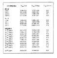
組換えbIAPI、II、IIIおよびIVイソ酵素の速度論的特性付けにより、触媒特性に関してかなりの相違が示された(図9)。例えば、bIAP II(約8600U/mg)はbIAPI(約2700U/mg)よりも比活性が300%以上、すなわち3倍以上高い。また、bIAP IIIおよびbIAP IVも、bIAPIよりもそれぞれ約1.8倍(約4700U/mg)および約2.6倍(>6700U/mg)高い活性を示し(図9)、百分率では、それぞれ約170%および250%の増加に相当する。さらに、イソ酵素の熱安定性においてかなりの測定可能な相違が存在した。bIAPIは、最も熱安定性のあるイソ酵素であり、bIAP IIおよびIIIのTm 値はbIAPIよりも7℃低く、bIAP IVのTm 値はbIAPIよりも13℃低い(図9)。Tm 値は、10分間のインキュベーション後に50%残留活性が測定される温度として理解される。
【0013】
以下、本発明は、bIAPの比活性に影響を及ぼすアミノ酸残基の同定を記載する。これは、中間産物によって促進される。中間キメラ(chimers)L1N8、INT1、INT2およびINT3の発現により、24アミノ酸の中の11が活性の増大のためのエフェクターから除外することができた(図7)。
・L1N8突然変異酵素は、bIAPIに匹敵する比活性を有しており;したがって、この場合において導入された突然変異V2I、V4AおよびD8Nは比活性の増大には関連していない。V2Iという表記は、2位のアミノ酸バリンがイソロイシンによって置換されることを意味する。
【0014】
・INT1突然変異体はbIAP IIに匹敵する比活性を有しており、したがってこの領域は重要である。
・INT2突然変異体はINT1およびbIAP IIに匹敵する比活性を有しており、したがって、INT2由来の突然変異S380G、D411G、D416E、Q420R、Q427L、E453QおよびT480Aも除外することができる。
【0015】
・INT3突然変異体の作製において、高比活性における変化は観察されず、したがって突然変異N192Yの効果が除外された。
13個の残りの残基の中のどれが高比活性にとって重要であるかを同定するために、本発明においては、bIAP II cDNAをbIAPIの対応するアミノ酸に対する単一突然変異のための鋳型として用いた。単一突然変異体N122K、I133M、A142S、K180M、M205K、E210V、E236A、G322DおよびI332G、ならびに結合したA289Q-A294V-Q297R-L299V bIAP II突然変異体が構築された(図9)。
【0016】
驚くべきことに、主に、突然変異G322Dは、bIAP IIの高比活性(約8600U/mg)を3分の1以下(2817U/mg)に低下させ得るものであり、よって、bIAP IIをbIAPIと同程度の低さの比活性に変換し得るものであることがわかった。
この結果を検証するために、本発明において、復帰突然変異D322GをbIAPIに導入した。驚くべきことに、この場合、逆の効果、すなわち、10148U/mgという3倍以上の比活性の増大が測定され、したがってbIAP IIに匹敵する値が得られた(図9)。比較的活性の高いbIAP III(約4700U/mg)とより高活性のbIAP IV(>6700U/mg)のアミノ酸配列の比較により、再び、この結果が裏付けられる。bIAP IIIは322位にセリンを有し、bIAP IVはグリシンを有する。
【0017】
さらに、本発明においては、作製された突然変異体を熱安定性について調べた。その結果、bIAPIとbIAP IIの間の熱安定性の相違は、1以上の置換の組み合わせ効果によるものである。[G322]bIAPIならびに[D322]bIAP II突然変異体は、bIAPIイソ酵素とbIAP IIイソ酵素の間にある安定度値を示す(図9)。D322G突然変異は、bIAPIイソ酵素においてわずかな不安定化作用を有する(T50においてほぼ4℃)のに対して、bIAP IIにおける置換G322Dにより、この突然変異酵素の安定性において対応する増大が生じる。しかしながら、野生型bIAPIの熱安定性は得られない。
【0018】
したがって、本発明の主題は、特に、真核生物cDNAによってコードされる、3000U/mg以上の比活性を有する高活性組換えアルカリ性ホスファターゼを提供することである。本発明による高活性組換えアルカリ性ホスファターゼは、322位にグリシン、アラニン、トレオニン、バリンまたはセリンがあるものが特に好ましい。本発明によるアルカリ性ホスファターゼは、322位にグリシンがあることが特に好ましい。
【0019】
本発明の高活性組換えアルカリ性ホスファターゼは、好ましくは、さらに、下記の位置の1つまたは複数に突然変異を有することが可能である:
その突然変異が活性の増大をもたらす1、108、125、149、181、188、219、221、222、223、224、231、252、258、260、282、304、321、330、331、354、383、385、400、405、413、428、431および461位のアミノ酸残基。さらに、本発明は、本発明の高活性アルカリ性ホスファターゼの産生方法に関する。また、本発明のアルカリ性ホスファターゼは、例えば、それらの熱安定性に関して特異的に突然変異誘発することによって改良することもできる。
【0020】
【発明の実施の形態】
本発明の高活性アルカリ性ホスファターゼの活性は、E.Mossnerら, Z. Physiol. Chem. 361(1980), 543-549にしたがって測定した。ただし、試験は、この論文に記載されている25℃ではなく37℃で行った。37℃での測定は、ジエタノール緩衝液中で活性を測定する際の世界的な通常の温度である(BM試験法5426)。
【0021】
本発明のAPおよび公知のAPのタンパク質測定は、280nmにおける水に対するタンパク質溶液の吸光度を測定することによって行われる。1mg/mlの濃度の低活性および高活性AP溶液の吸光度は280nmで1.0である(A280nm(1mg/ml)は1に等しい)。
比活性は、活性とそれに伴うタンパク質の量の商を計算することによって決定される。
【0022】
【実施例】
本発明は、さらに、下記の実施例によって説明される:
実施例1:クローニング
成熟ウシの腸から調製したλgt 11cDNAバンク(Clontech Laboratories,パロアルト(Palo Alto)、CA、USA)を、プローブとしてbIAPIcDNAの 5'末端から1075bpのHind III断片を用いてスクリーニングした(Weissigら, 1993)。このcDNAバンク由来のクローンを用いて成熟ウシの肝臓から調製されたEMBL-3 SP6/T7ゲノムcDNAバンク(Clontech Laboratories,パロアルト、CA、USA)をスクリーニングした。成熟ウシの小腸からTrisolv(商標名)試薬を用いて単離されたmRNA由来のオリゴdTプライマー(Stratagene、サンディエゴ、CA、USA)によって非増幅λZAP IIcDNAバンクを準備し、プローブとしてbIAPIcDNAの1075bpのHind III断片を用いてスクリーニングした。プローブをランダムプライムした(primed)DNA標識キット(Boehringer Mannheim)を用いて放射能標識した。ファージDNAをλgt 11およびEMBL-3 SP6/T7クローンについて記載したようにして調製した(Tsonis & Manes, 1988)。λZAP IIクローンのin vivo切断を製造業者(Stratagene、サンディエゴ、CA)の指示にしたがって行った。ゲノムクローンを記載されているようなサザンブロット分析によって特性付けした(Sambrookら, 1989)。λgt 11クローンのEcoRI cDNA断片およびその他のバンクのクローン由来の種々の制限断片をKS+ベクター(Stratagene、サンディエゴ、CA、USA)にサブクローニングした。プラスミドDNAをアルカリ溶解によって調製した(Sambrookら, 1989)。Sequenaseを製造業者プロトコル(Amersham)にしたがって用いて配列決定を行った。bIAP IIIおよびIVの配列決定に使用したオリゴヌクレオチドを下記に記載する:
【0023】

核酸配列は、MacVector配列分析プログラム(International Biotechnologies社、ニューヘブン、CT、USA)を用いて分析した。
実施例2:bIAP IIのアミノ酸配列の決定
約500μgの精製した高活性(約6000U/mg)子ウシ腸APを、450μlの6M塩酸グアニジン、0.25MのTris、1mMのEDTA、pH8.5に溶解し、次いで30μlのメルカプトエタノールを加えた。100℃で30分間還元した後、35μlのビニルピリジンを加えることによってシステインをアルキル化し、この混合物を暗所で室温にて45分間インキュベートした。次いで、短い逆相HPLC Aquapore RP300カラム(30×2.1mm、Applied Biosystems、Weiterstadt)上で反応混合物をただちに脱塩した。0.1%トリフルオロ酢酸中のアセトニトリルの段階グラジエントを用いて結合した酵素を溶離させた。タンパク質を含む画分を蒸発乾固した。酵素を脱グリコシル化するために、125μgのAPを15μlの蒸留水および6μlのインキュベーション緩衝液(250mMのNa2HPO4、50mMのEDTA、pH7.2)および15UのEndoF/PNGアーゼ(Boehringer Mannheim、Penzberg)に溶解した。混合物を一晩37℃に維持し、その後、切断に用いた。還元され、アルキル化されたAPを、種々の酵素(エンドプロテイナーゼLysC、エンドプロテイナーゼAspN、エンドプロテイナーゼGluCおよびトリプシン(Boehringer Mannheim、Penzberg))を用いて個々の酵素のデータシートの指示にしたがって酵素的に切断した。臭化シアン分解を、70%(v/v)ギ酸中10%(w/w)CNBrを用いて8時間行った。水を用いて溶解した後、溶液の容量をSpeedVac濃縮器(Savant)を用いて減らし、逆相HPLCに使用した。C−末端トリプシンペプチドをカルボキシペプチダーゼY(8ng/μl)を用いて4分間消化し、放出されたペプチドを、Bruker Reflex III機器を用いるマトリックス支持レーザー脱着/イオン化質量分析法により製造業者の指示にしたがって分析した。アセトニトリル/水(50/50、v/v)に2,5ジヒドロキシ安息香酸を加えたもの(10mg/ml)をマトリックスとして使用した。酵素的または化学的切断から得られたペプチドを逆相HPLCによって、LiChrospher C18 selBカラム125×2mm(Merck、Darmstadt)上で、0.1%トリフルオロ酢酸/アセトニトリル溶媒系を用いて分離した。流量は300μl/分であった。溶離剤を206nmにおけるUVモニタリングによって検出し、画分を手動で回収した。ペプチドの質量測定を、API IIIエレクトロスプレー質量分析計(PE-Sciex、Langen)を用いて、製造業者の指示にしたがって行った。アミノ酸配列を492Aタンパク質シークエンサー(Applied Biosystems、Weiterstadt)を用いて、製造業者の指示にしたがって決定した。
実施例3:bIAP II cDNAの調製およびbIAP II突然変異誘発
bIAP IIをコードするcDNAを調製するために、野生型制限断片ならびにcDNAbIAPI、IIIおよびIVの部位特異的突然変異誘発PCR断片を互いにライゲートして、L1N8(3断片)およびINT1(9断片)cDNA中間構築物を作製した。次いで、INT1およびbIAP IIIを部位特異的突然変異誘発用の鋳型として使用し、これにより得られた断片を組み立てて完全INT2(8断片)cDNAを形成させた。次に、INT2の制限断片およびINT2の部位特異的突然変異誘発断片を組み立ててINT3(5断片)cDNAを形成させ、最後にbIAP II(4断片)cDNAを形成させた。部位特異的突然変異誘発は、Tomicら(1990)の方法にしたがって、その認識配列(GGTCTCN1/N5)からの距離で切断する制限酵素としてBsa I(II型)を用いて行った。二次的な突然変異が無いことを確認するために、PCR産物全てを配列決定した。全ての構築物を配列決定および制限消化によって確認した。部位特異的突然変異誘発断片の増幅に用いられるオリゴヌクレオチドプライマーの配列は下記のとおりである。プライマーの名称を最初に記載し、その後に配列を記載する(突然変異を示す位置に下線を引く)。
【0025】
種々のPCR反応を1〜16と番号付けし、鋳型は、野生型cDNAbIAPI、IIIもしくはIVまたはキメラ構築物INT1もしくはINT2のいずれかである。オリゴヌクレオチドプライマー(括弧内)は上記で述べられている。1.bIAP IV(KS、1L);2.bIAP IV(8N、122);3.bIAP III(1S、M133I);4.bIAPI(S142A、180);5.bIAPI(M180K、K205M);6.bIAPI(V210E、A236E);7.bIAPI(236、289);8.bIAP IV(E289A、330);9.bIAP III(330E、V332I、XIa);10.INT1(N192Y、S380G);11.INT1(N192Y、D411G);12.bIAP III(D416E、S428A);13.INT1(D416E、T480S);14.INT1(480、SP6);15.INT2(236、Q304R-);16.INT2(Q304R+、E321D)。下記のライゲーション反応を、全ての場合においてpcDNA-3(Invitrogen、サンディエゴ、CA)発現ベクターを用いて行った。断片を、上記のPCR反応番号にしたがって番号付けし、または野生型もしくはキメラcDNAの名称を付けて、その後制限酵素を用いてこの断片の付着末端を形成させた。L1N8=pcDNA-3/EcoRI-XbaI+1/EcoRI-BsaI+2/BsaI-BamHI+bIAP I/BamHI-XbaI。INT1=pcDNA-3/EcoRI-XbaI+L1N8/EcoRI-NcoI+3/NcoI-BsaI+4/BsaI+5/BsaI+6/BsaI+7/BsaI+8/BsaI+9/BsaI-StuI+bIAPI/StuI-XbaI。INT2=pcDNA-3/EcoRI-NotI+INT1/EcoRI-PstI+10/PstI-StuI+11/StuI-BsaI+12/BsaI+13/BsaI+14/BsaI+bIAPI/BsaI-NotI。INT3=pcDNA-3/EcoRI-XbaI+INT2/EcoRI-NcoI+INT2/NcoI-PvuII+10/PvuII-EagI+INT2/EagI-HindIII+INT2/HindIII-XbaI。bIAP II=pcDNA-3/EcoRI-XbaI+INT3/EcoRI-EagI+15/EagI-SmaI+16/SmaI-HindIII+INT3/HindIII-XbaI。
【0027】
10個のさらなる構築物をbIAPIおよびIIの種々の動力学的特性の原因である残基を同定するために調製した。全構築物をpcDNA-3/EcoRI-XbaIにサブクローニングした。5個の構築物をL1N8またはbIAPI(I)およびbIAP II(II)間の制限断片の交換によって調製した。L1N8 EcoRI-Pm1Iおよび(II)Pm1I-XbaIを[N122K]bIAP II突然変異cDNAを調製するためにライゲートした。(II)EcoRI-BstEII、(I)BstEII-PvuII、(II)PvuII XbaIを[K180M]bIAP II突然変異cDNAに対して結合した。(II)EcoRI-EagI、(I)EagI-BstEII、(II)BstEII-XbaIを[A289Q、A294V、Q297R、L299V]bIAP II突然変異体に対してライゲートした。(II)EcoRI-EagI、(II)EagI-BstEII、(I)BstEII-HindIII、(II)HindIII-XbaIを[G322D]bIAP II突然変異体に対してライゲートした。(II)EcoRI-HindIII、(I)HindIII-SacI、(II)SacI-XbaIを[I332G]bIAP II突然変異体に対してライゲートした。5個のその他の位置は新たな部位特異的突然変異誘発が必要であった。下記のオリゴヌクレオチドをこれに用いた:
【0028】
鋳型としてbIAP IIを用いる下記の8個のPCR反応(a〜h)を、これらのオリゴヌクレオチドおよび前に挙げたオリゴヌクレオチドを用いて行った:a.1s,I133M-;b.S142A+,M205K-;c.1s,A142S-;d.V210E+,330-;e.E210V+,330-;f.M180K+,E236A-;g.236+,330-;h.S142A,K205M-。これから形成された産物をサブクローニングし、配列決定し、次いで断片を下記のライゲーションのために単離した:I133Mに対して(II)EcoRI-NcoI、(a)NcoI-BsaI、(b)BsaI、PvuII、(II)PvuII-XbaI。A142Sに対して(II)EcoRI-NcoI、(c)NcoI-BstEII、(II)BstEII-PvuII、(II)PvuII-XbaI。M205Kに対して(II)EcoRI-BstEII、(b)BstEII-BsaI、(d)BsaI-HindIII、(II)HindIII-XbaI。E210Vに対して(II)EcoRI-BstEII、(h)BstEII-BsaI、(e)BsaI-HindIII、(II)HindIII-XbaI。E236Aに対して(II)EcoRI-NcoI、(II)NcoI-PvuII、(f)PvuII-BsaI、(g)BsaI-HindIII、(II)HindIII-XbaI。
実施例4:組換え酵素の産生および特性付け
全cDNA(bIAPI、bIAP II、bIAP III、bIAP IVおよび対応する突然変異体)を、pcDNA-3発現ベクター(Invitrogen、サンディエゴ、CA、USA)にクローニングし、チャイニーズハムスターの卵細胞(CHO細胞)に移入し、安定なトランスフェクタントを500μg/mlのジェネティシン(Gibco、BRL)の存在下で細胞を増殖させることによって選択した。組換えAPを、安定に移入されたCHO細胞から記載されたようにして抽出した(Hoylaertsら, 1997)。0.1μg/mlの高親和性抗子ウシAPモノクローナル抗体(Scottish Antibody Production Unit、Lanarkshire、スコットランド)で被覆したマイクロタイタープレートを、kcat を測定するために酵素濃度を高めながらインキュベートした。結合酵素の活性を、基質としての30mMのp-ニトロフェニルホスフェートを含む1.0Mジエタノールアミン緩衝液(pH9.8)、1mM MgCl2、および20μM ZnCl2を添加した後の、405nm、20℃における経時的な吸光度の変化として測定した。生成したp-ニトロフェノールの濃度を、吸光係数 10,080リットル/モル/cmを用いて計算した。比活性既知の市販の調製物(Biozyme Laboratories、7822U/mgおよびBoehringer Mannheim、3073U/mg)ならびに精製bIAP II(8600U/mg)を標準として用いた。抗体を飽和させたこれらの溶液の酵素濃度(E0 )を、同一試験条件下での既知の酵素濃度に対する活性の標準曲線から計算した。次いで、最大基質転化(Vmax )をE0 で割ってkcat を計算した。Km を計算するために、基質濃度を0.25〜2.0mMのp-ニトロフェニルホスフェート(pNPP)間で変化させ、20℃における初期反応速度を10分間以上測定した。[pNPP]/v対X軸としての[pNPP]の回帰曲線(Hanes曲線)により−Km が得られた。回帰において各x値に対して計算したy値の標準偏差の、Km ・Vmax ±標準偏差の標準偏差を得た回帰の勾配による割り算は、Km ±標準偏差をy切片±標準偏差で割ることによって適当な方程式を用いて計算した。比活性を、抗体飽和活性に基づいて、Biozymeに対して計算した。熱安定性曲線を、前に記載されたように(Weissigら, 1993)、10分毎に5℃ずつ上昇させながら45〜75℃で抽出物をインキュベーションすることによって確立した。次いで、各試料の活性を上記のようにして測定し、残留活性を、非加熱試料に対する残留割合として計算した。50%残留活性が残る温度(T50)を、温度曲線に対する残留活性から計算した。
【0030】
【配列表】
【配列表フリーテキスト】
配列番号7:bIAP IIIおよびIVを配列決定するために使用したオリゴヌクレオチド
配列番号8:bIAP IIIおよびIVを配列決定するために使用したオリゴヌクレオチド
配列番号9:bIAP IIIおよびIVを配列決定するために使用したオリゴヌクレオチド
配列番号10:bIAP IIIおよびIVを配列決定するために使用したオリゴヌクレオチド
配列番号11:bIAP IIIおよびIVを配列決定するために使用したオリゴヌクレオチド
配列番号12:bIAP IIIおよびIVを配列決定するために使用したオリゴヌクレオチド
配列番号13:bIAP IIIおよびIVを配列決定するために使用したオリゴヌクレオチド
配列番号14:bIAP IIIおよびIVを配列決定するために使用したオリゴヌクレオチド
配列番号15:bIAP IIIおよびIVを配列決定するために使用したオリゴヌクレオチド
配列番号16:bIAP IIIおよびIVを配列決定するために使用したオリゴヌクレオチド
配列番号17:bIAP IIIおよびIVを配列決定するために使用したオリゴヌクレオチド
配列番号18:bIAP IIIおよびIVを配列決定するために使用したオリゴヌクレオチド
配列番号19:部位特異的突然変異断片を増幅するために使用したオリゴヌクレオチドプライマー
配列番号20:部位特異的突然変異断片を増幅するために使用したオリゴヌクレオチドプライマー
配列番号21:部位特異的突然変異断片を増幅するために使用したオリゴヌクレオチドプライマー
配列番号22:部位特異的突然変異断片を増幅するために使用したオリゴヌクレオチドプライマー
配列番号23:部位特異的突然変異断片を増幅するために使用したオリゴヌクレオチドプライマー
配列番号24:部位特異的突然変異断片を増幅するために使用したオリゴヌクレオチドプライマー
配列番号25:部位特異的突然変異断片を増幅するために使用したオリゴヌクレオチドプライマー
配列番号26:部位特異的突然変異断片を増幅するために使用したオリゴヌクレオチドプライマー
配列番号27:部位特異的突然変異断片を増幅するために使用したオリゴヌクレオチドプライマー
配列番号28:部位特異的突然変異断片を増幅するために使用したオリゴヌクレオチドプライマー
配列番号29:部位特異的突然変異断片を増幅するために使用したオリゴヌクレオチドプライマー
配列番号30:部位特異的突然変異断片を増幅するために使用したオリゴヌクレオチドプライマー
配列番号31:部位特異的突然変異断片を増幅するために使用したオリゴヌクレオチドプライマー
配列番号32:部位特異的突然変異断片を増幅するために使用したオリゴヌクレオチドプライマー
配列番号33:部位特異的突然変異断片を増幅するために使用したオリゴヌクレオチドプライマー
配列番号34:部位特異的突然変異断片を増幅するために使用したオリゴヌクレオチドプライマー
配列番号35:部位特異的突然変異断片を増幅するために使用したオリゴヌクレオチドプライマー
配列番号36:部位特異的突然変異断片を増幅するために使用したオリゴヌクレオチドプライマー
配列番号37:部位特異的突然変異断片を増幅するために使用したオリゴヌクレオチドプライマー
配列番号38:部位特異的突然変異断片を増幅するために使用したオリゴヌクレオチドプライマー
配列番号39:部位特異的突然変異断片を増幅するために使用したオリゴヌクレオチドプライマー
配列番号40:部位特異的突然変異断片を増幅するために使用したオリゴヌクレオチドプライマー
配列番号41:部位特異的突然変異断片を増幅するために使用したオリゴヌクレオチドプライマー
配列番号42:部位特異的突然変異断片を増幅するために使用したオリゴヌクレオチドプライマー
配列番号43:部位特異的突然変異断片を増幅するために使用したオリゴヌクレオチドプライマー
配列番号44:部位特異的突然変異断片を増幅するために使用したオリゴヌクレオチドプライマー
配列番号45:部位特異的突然変異断片を増幅するために使用したオリゴヌクレオチドプライマー
配列番号46:部位特異的突然変異断片を増幅するために使用したオリゴヌクレオチドプライマー
配列番号47:部位特異的突然変異断片を増幅するために使用したオリゴヌクレオチドプライマー
配列番号48:部位特異的突然変異断片を増幅するために使用したオリゴヌクレオチドプライマー
配列番号49:部位特異的突然変異断片を増幅するために使用したオリゴヌクレオチドプライマー
配列番号50:部位特異的突然変異断片を増幅するために使用したオリゴヌクレオチドプライマー
配列番号51:部位特異的突然変異断片を増幅するために使用したオリゴヌクレオチドプライマー
配列番号52:部位特異的突然変異断片を増幅するために使用したオリゴヌクレオチドプライマー
配列番号53:部位特異的突然変異断片を増幅するために使用したオリゴヌクレオチドプライマー
配列番号54:部位特異的突然変異断片を増幅するために使用したオリゴヌクレオチドプライマー
【図面の簡単な説明】
【図1】 (A)配列番号1:bIAP IIのヌクレオチド配列(1798bp)。108位が成熟bIAP IIのコード領域の開始位置。1649位が末端。
(B)配列番号1:bIAP IIのヌクレオチド配列(1798bp)。108位が成熟bIAP IIのコード領域の開始位置。1649位が末端。
【図2】配列番号2:切断部位を有するbIAP IIのアミノ酸配列(480アミノ酸)。
【図3】 (A)配列番号3:bIAP IIIのヌクレオチド配列(2460bp)。123位が成熟bIAP IIIのコード領域の開始位置。1655位が末端。
(B)配列番号3:bIAP IIIのヌクレオチド配列(2460bp)。123位が成熟bIAP IIIのコード領域の開始位置。1655位が末端。
【図4】配列番号4:bIAP IIIのアミノ酸配列(511アミノ酸)。
【図5】 (A)配列番号5:bIAP IVのヌクレオチド配列(2542bp)。122位が成熟bIAP IVのコード領域の開始位置。1654位が末端。
(B)配列番号5:bIAP IVのヌクレオチド配列(2542bp)。122位が成熟bIAP IVのコード領域の開始位置。1654位が末端。
【図6】配列番号6:bIAP IVのアミノ酸配列(511アミノ酸)。
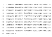
【図7】bIAPI、bIAP II、bIAP IIIおよびbIAP IVイソ酵素間のアミノ酸の相違。異なる残基のみを示す。アステリスクは、bIAP IIの触媒活性の増大の原因である残基を同定するために個々の突然変異誘発に対して選択された位置を特定するものである。
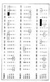
【図8】 (A)bIAP II DNAのライゲーション戦略。
(B)bIAP II DNAのライゲーション戦略。
【図9】組換え野生型およびキメラbIAP酵素、ならびに部位特異的突然変異誘発によって変化させた該bIAP酵素の突然変異体の動力学的パラメーターおよび熱安定性。*[QVRV]bIAP IIは[Q289、V294、R297、V299]bIAP II突然変異体の略語である。[0001]
[Field of the Invention]
The present invention relates to a DNA encoding a eukaryotic highly active alkaline phosphatase having a specific activity of 3000 U / mg or more. The present invention also relates to a method for producing the DNA of the present invention, a vector comprising the DNA of the present invention, and a cell line comprising such a vector. The present invention further relates to a recombinant highly active alkaline phosphatase encoded by the DNA of the present invention and having a specific activity of 3000 U / mg or more.
[0002]
[Prior art]
Alkaline phosphatase (AP) is a dimeric, zinc-containing non-specific phosphomonoesterase found in all organisms from E. coli to mammals (McComb et al., 1979). Comparison of the primary structures of various alkaline phosphatases showed a high degree of homology (25-30% homology between E. coli and mammalian AP) (Millan, 1988; Harris, 1989) ).
[0003]
In humans and higher animals, the AP family consists of four members encoded at different loci (Millan, 1988; Harris, 1989). The alkaline phosphatase family includes tissue-specific APs (placental AP (PLAP), germ cell AP (GCAP) and intestinal AP (IAP)) and non-tissue specific AP (TNAP) localized mainly in the liver, kidney and bone Is mentioned.
[0004]
A decisive characteristic of conventionally known APs is the great variability in the catalytic activity of mammalian AP, whose specific activity is 10 to 100 times higher than that of E. coli AP. Among mammalian APs, bovine intestinal AP (bIAP) shows the highest specific activity. This property makes bIAP attractive for biotechnological applications such as diagnostic reagents or enzyme conjugates for dephosphorylation of DNA. In 1985, Besman and Coleman confirmed that the presence of two IAP isoenzymes in the bovine intestine: IAP from calf intestine and IAP from mature bovine intestine (bIAP). Was verified by amino-terminal sequencing. A clear difference at the amino terminus is shown between mature bovine bIAP (LVPVEEED) and calf intestinal bIAP (LIPAEEEN). In 1993, Weissig et al. Performed accurate biochemical characterization by cloning recombinant bIAP (bIAPI) with a specific activity of about 3000 U / mg and N-terminal LVPVEEED. However, bIAPs derived from calves with specific activities up to 8000 U / mg are also commercially available (Boehringer Mannheim, Biozyme, Oriental Yeast) but have not yet been characterized. All attempts to clone these highly active alkaline phosphatases were unsuccessful. Therefore, recombinant high activity alkaline phosphatase could not be produced. However, the possibility of recombinant production is absolutely essential for the economical production of highly active alkaline phosphatase.
[0005]
[Problems to be solved by the invention]
Accordingly, it is an object of the present invention to provide a highly active alkaline phosphatase by recombinant means that can also be cloned. “High activity” within the meaning of the present invention means that the alkaline phosphatase according to the present invention has an activity that is at least 10% higher than that of conventionally known alkaline phosphatases.
[0006]
[Means for Solving the Problems]
This object is in accordance with the present invention to encode a eukaryotic highly active alkaline phosphatase in which the amino acid residue at
[0007]
SEQ ID NO: 1 comprises DNA encoding the sequence of a highly active bAIP II isoenzyme. Natural enzymes are known but have not been characterized and cannot be cloned. Thus, amino acid sequencing of highly active bIAP II isoenzymes is the subject of the present invention. The sequence was determined using a highly purified fraction (Boehringer Mannheim) with high specific activity derived from calf intestine. Peptide maps of highly active AP were generated by cleavage with endoproteinases LysC, AspN, GluC, trypsin and chemical cleavage with bromocyan. Peptides produced in this manner were separated and isolated by reverse phase HPLC. Each peptide was analyzed by electrospray mass spectroscopy and sequenced by Edman degradation. The sequence obtained in this way was compared with the sequence of bIAPI described in the publication (Weissig et al., 1993). As expected, the amino terminus of bIAP II has the starting sequence LIPAEEEN as described by Besman and Coleman (J. Biol. Chem. 260, 11190-11193 (1985)). The complete amino acid sequence of bIAP II is shown in SEQ ID NO: 2 (FIG. 2). According to SEQ ID NO: 2, bIAP II has a total of 24 amino acid substitutions compared to bIAPI. The isolated highly active bIAP II isoenzyme has 480 amino acids. The 1798 bp nucleotide sequence (Figure 1) contains a coding region of 514 amino acids. The possible amino acids from positions 481 to 514 can vary within wide limits.
[0008]
In the following, the present invention describes the cloning and complete characterization of two novel bIAPs (bIAP III and bIAP IV) that have not been known so far. Northern blot analysis was performed on RNA samples from different sections of bovine intestine. A cDNA bank of probes with the strongest hybridization signal was prepared using oligo dT primers (Stratagene, San Diego, CA, USA) in the vector IZAP II (Stratagene, San Diego, CA, USA). Complete bank (1.0 × 10 6 Recombinant clones) were screened with a 1075 bp HindIII fragment of bIAPI covering the exon I to VIII region of the bIAPI gene. 65 clones were isolated and sequenced. In this process, two new bIAPs were identified (bIAPIII and bIAPIV). The characterization is further described below. They were not completely homologous to bIAPI or bIAPII. The nucleotide sequences of bIAP III and IV are shown in FIGS. Differences in the sequence of bIAPI-IV are shown in FIG. However, none of the new bIAPs had the expected N-terminal LIPAEEEN, and had a new N-terminal not previously described (see FIG. 7). Two new bIAP isoenzyme cDNAs were recut using appropriate restriction enzymes and inserted into a CHO expression vector pcDNA-3 (eg, manufactured by Invitrogen, San Diego, CA, USA) by ligation. Clones containing the novel bIAP isoenzyme were expressed according to the method described by Invitrogen and the isoenzyme was characterized. Expression of the bIAP gene in various hosts is described in WO93 / 18139 (CHO cells, E. coli, baculovirus system). The methods, vectors and expression systems described in this patent document are part of the disclosure of the present application. The invention further relates to natural and recombinant highly active alkaline phosphatases bIAPIII and bIAPIV. The alkaline phosphatase described in SEQ ID NOs: 4 and 6 is particularly preferred. The CHO cell line containing the bIAP III and bIAP IV genes was deposited with DSMZ, namely “Deutsche Sammlung von Mikroorganismen und Zellkulturen GmbH”, Mascheroder Weg 1b, D-38124 Braunschweig (DSM ACC 2349, DSM ACC 2350).
[0009]
In the following, the present invention describes the construction of bIAP II sequences by ligation of mutant and wild type fragments of bIAPI, III and IV. This process produced a series of intermediate products (L1N8, INT1, INT2 and INT3) encoding functional isoenzymes. In order to construct these intermediate products, in each case, a part of the bIAP-cDNA to be modified is excised with an appropriate restriction enzyme and the desired mutations having compatible ends by digestion with restriction enzymes are introduced. It was replaced with another bIAP-cDNA segment containing it. Mutations that could not be introduced by ligation of different bIAP-cDNA segments were introduced by site-directed mutagenesis. The mutated fragment was then recut with the appropriate restriction enzyme and ligated to the bIAP-cDNA with compatible ends that was cut in the same way (FIG. 8). The mutations introduced in this method were then examined by restriction analysis and sequencing.
[0010]
The subject of the present invention is therefore a method for the production of DNA according to the invention, characterized in that a mutant fragment and a wild-type fragment of one or more alkaline phosphatase DNAs are linked. Furthermore, the present invention relates to a cDNA encoding a functional isozyme and formed as an intermediate product in the above method of the present invention. Furthermore, the present invention relates to a vector comprising the cDNA of the present invention.
[0011]
A further subject matter of the invention is a cell line comprising the vector of the invention. Suitable cells are, for example, eukaryotic cells such as CHO, Pichia, Hansenula or Saccharomyces cerevisiae and prokaryotic cells such as Aspergillus, E. coli, with E. coli, yeast and CHO cells being particularly preferred. Suitable starting vectors for E. coli strains are, for example, pTE, pTaq, bPL, pBluescript. Suitable E. coli strains are, for example, XL1-Blue, HB101, RR1ΔM15, BL21 (DE), MC1000 and the like. Suitable Pichia vectors are, for example, pGAPZα and pPICZα (Invitrogen, San Diego, CA, USA). A suitable vector for the CHO cell line is, for example, pcDNA-3 (Invitrogen, San Diego, CA, USA). The CHO cell line containing the bIAP II gene was deposited with DSMZ, ie “Deutsche Sammlung von Mikroorganismen und Zellkulturen GmbH”, Mascheroder Weg 1b, D-38124 Braunschweig (DSM ACC 2348).
[0012]
Kinetic characterization of recombinant bIAPI, II, III and IV isoenzymes showed considerable differences in catalytic properties (FIG. 9). For example, bIAP II (about 8600 U / mg) has a specific activity of 300% or more, that is, 3 times higher than bIAPI (about 2700 U / mg). BIAP III and bIAP IV also showed about 1.8 times (about 4700 U / mg) and about 2.6 times (> 6700 U / mg) higher activity than bIAPI, respectively (FIG. 9), with percentages of about 170% and 250%, respectively. % Increase. In addition, there were significant measurable differences in the thermostability of isoenzymes. bIAPI is the most thermostable isoenzyme, and TIA of bIAP II and III m The value is 7 ° C lower than bIAPI, and the TIA of bIAP IV m The value is 13 ° C. lower than bIAPI (FIG. 9). T m The value is understood as the temperature at which 50% residual activity is measured after 10 minutes incubation.
[0013]
Hereinafter, the present invention describes the identification of amino acid residues that affect the specific activity of bIAP. This is facilitated by intermediate products. Expression of the intermediate chimers L1N8, INT1, INT2, and INT3 allowed 11 out of 24 amino acids to be excluded from the effector for increased activity (FIG. 7).
The L1N8 mutant enzyme has a specific activity comparable to bIAPI; therefore, the mutations V2I, V4A and D8N introduced in this case are not associated with an increase in specific activity. The notation V2I means that the amino acid valine at
[0014]
• The INT1 mutant has a specific activity comparable to bIAP II, so this region is important.
The INT2 mutant has a specific activity comparable to INT1 and bIAP II, and therefore the mutations S380G, D411G, D416E, Q420R, Q427L, E453Q and T480A from INT2 can also be excluded.
[0015]
• No changes in high specific activity were observed in the generation of INT3 mutants, thus eliminating the effect of mutation N192Y.
In order to identify which of the 13 remaining residues are important for high specific activity, in the present invention, bIAP II cDNA is used as a template for a single mutation to the corresponding amino acid of bIAPI. Using. Single mutants N122K, I133M, A142S, K180M, M205K, E210V, E236A, G322D and I332G, and a bound A289Q-A294V-Q297R-L299V bIAP II mutant were constructed (FIG. 9).
[0016]
Surprisingly, mainly the mutation G322D is capable of reducing the high specific activity of bIAP II (about 8600 U / mg) to less than one third (2817 U / mg) and thus bIAP II is reduced to bIAPI. It was found that the specific activity can be converted to a low specific activity.
In order to verify this result, in the present invention, the back mutation D322G was introduced into bIAPI. Surprisingly, in this case, the opposite effect, ie an increase in specific activity of more than 3 times of 10148 U / mg was measured, thus obtaining values comparable to bIAP II (FIG. 9). The comparison of the amino acid sequences of the relatively active bIAP III (about 4700 U / mg) and the more active bIAP IV (> 6700 U / mg) again supports this result. bIAP III has a serine at
[0017]
Furthermore, in the present invention, the prepared mutants were examined for thermal stability. As a result, the difference in thermal stability between bIAPI and bIAP II is due to the combined effect of one or more substitutions. [G 322 ] BIAPI and [D 322 The bIAP II mutant exhibits a stability value that lies between the bIAPI and bIAP II isoenzymes (FIG. 9). The D322G mutation has a slight destabilizing effect on the bIAPI isoenzyme (T 50 The substitution G322D in bIAP II results in a corresponding increase in the stability of this mutant enzyme. However, the thermal stability of wild type bIAPI is not obtained.
[0018]
Thus, the subject of the present invention is in particular to provide highly active recombinant alkaline phosphatase encoded by eukaryotic cDNA and having a specific activity of 3000 U / mg or more. The highly active recombinant alkaline phosphatase according to the present invention is particularly preferably one having glycine, alanine, threonine, valine or serine at
[0019]
The highly active recombinant alkaline phosphatase of the present invention is preferably further capable of having a mutation at one or more of the following positions:
The mutation results in increased activity 1,108,125,149,181,188,219,221,222,223,224,231,252,258,260,282,304,321,330,331,354 , 383, 385, 400, 405, 413, 428, 431 and 461 amino acid residues. Furthermore, the present invention relates to a method for producing the highly active alkaline phosphatase of the present invention. The alkaline phosphatases of the present invention can also be improved, for example, by mutagenesis specifically with respect to their thermal stability.
[0020]
DETAILED DESCRIPTION OF THE INVENTION
The activity of the highly active alkaline phosphatase of the present invention is determined by E. Mossner et al., Z. Physiol. Chem. 361 (1980), 543-549. However, the test was conducted at 37 ° C instead of 25 ° C described in this paper. The measurement at 37 ° C. is the worldwide normal temperature for measuring activity in diethanol buffer (BM test method 5426).
[0021]
The protein measurement of the AP of the present invention and the known AP is performed by measuring the absorbance of the protein solution with respect to water at 280 nm. The absorbance of a low activity and high activity AP solution at a concentration of 1 mg / ml is 1.0 at 280 nm (A280 nm (1 mg / ml) is equal to 1).
Specific activity is determined by calculating the quotient of activity and the amount of protein associated therewith.
[0022]
【Example】
The invention is further illustrated by the following examples:
Example 1 : Cloning
A
[0023]
Nucleic acid sequences were analyzed using the MacVector sequence analysis program (International Biotechnologies, New Haven, CT, USA).
Example 2: Determination of the amino acid sequence of bIAP II
About 500 μg of purified highly active (about 6000 U / mg) calf intestinal AP was dissolved in 450 μl 6 M guanidine hydrochloride, 0.25 M Tris, 1 mM EDTA, pH 8.5, then 30 μl mercaptoethanol was added. After reduction at 100 ° C. for 30 minutes, cysteine was alkylated by adding 35 μl of vinylpyridine and the mixture was incubated for 45 minutes at room temperature in the dark. The reaction mixture was then immediately desalted on a short reverse phase HPLC Aquapore RP300 column (30 × 2.1 mm, Applied Biosystems, Weiterstadt). Bound enzyme was eluted using a step gradient of acetonitrile in 0.1% trifluoroacetic acid. The fraction containing protein was evaporated to dryness. To deglycosylate the enzyme, 125 μg AP was added to 15 μl distilled water and 6 μl incubation buffer (250 mM Na 2 HPO Four , 50 mM EDTA, pH 7.2) and 15 U EndoF / PNGase (Boehringer Mannheim, Penzberg). The mixture was maintained at 37 ° C. overnight and then used for cleavage. Reduced and alkylated AP is enzymatically converted using various enzymes (endoproteinase LysC, endoproteinase AspN, endoproteinase GluC and trypsin (Boehringer Mannheim, Penzberg)) according to the instructions in the individual enzyme data sheets. Disconnected. Cyanogen bromide decomposition was performed with 10% (w / w) CNBr in 70% (v / v) formic acid for 8 hours. After dissolving with water, the volume of the solution was reduced using a SpeedVac concentrator (Savant) and used for reverse phase HPLC. The C-terminal tryptic peptide is digested with carboxypeptidase Y (8 ng / μl) for 4 minutes and the released peptide is subjected to matrix-supported laser desorption / ionization mass spectrometry using a Bruker Reflex III instrument according to the manufacturer's instructions. analyzed. Acetonitrile / water (50/50, v / v) plus 2,5 dihydroxybenzoic acid (10 mg / ml) was used as the matrix. Peptides obtained from enzymatic or chemical cleavage were separated by reverse phase HPLC on a LiChrospher
Example 3: bIAP II cDNA preparation and bIAP II mutagenesis
To prepare the cDNA encoding bIAP II, the wild type restriction fragment and the site-directed mutagenesis PCR fragments of cDNA bIAPI, III and IV were ligated together so that the L1N8 (3 fragment) and INT1 (9 fragment) cDNA intermediates A construct was made. INT1 and bIAPIII were then used as templates for site-directed mutagenesis, and the resulting fragments were assembled to form complete INT2 (8 fragments) cDNA. Next, the restriction fragment of INT2 and the site-directed mutagenesis fragment of INT2 were assembled to form INT3 (5 fragment) cDNA and finally bIAP II (4 fragment) cDNA. Site-directed mutagenesis was performed according to the method of Tomic et al. (1990) using Bsa I (type II) as a restriction enzyme that cleaves at a distance from its recognition sequence (GGTCTCN1 / N5). All PCR products were sequenced to confirm that there were no secondary mutations. All constructs were confirmed by sequencing and restriction digests. The sequence of the oligonucleotide primer used for amplification of the site-directed mutagenesis fragment is as follows. The name of the primer is listed first, followed by the sequence (underlined positions indicating mutations).
[0025]
The various PCR reactions are numbered 1-16 and the template is either wild type cDNAbIAPI, III or IV or the chimeric construct INT1 or INT2. Oligonucleotide primers (in parentheses) are described above. 1. bIAP IV (KS, 1L); 2. bIAP IV (8N, 122); bIAP III (1S, M133I); 4. bIAPI (S142A, 180); bIAPI (M180K, K205M); 6. bIAPI (V210E, A236E); bIAPI (236, 289); bIAP IV (E289A, 330); bIAP III (330E, V332I, XIa); INT1 (N192Y, S380G); 11. INT1 (N192Y, D411G); 12. bIAPIII (D416E, S428A); INT1 (D416E, T480S); 14. INT1 (480, SP6); 15. INT2 (236, Q304R-); 16. INT2 (Q304R +, E321D). The following ligation reactions were performed with pcDNA-3 (Invitrogen, San Diego, CA) expression vector in all cases. Fragments were numbered according to the PCR reaction numbers described above, or named wild type or chimeric cDNA, and then a restriction enzyme was used to form the sticky ends of this fragment. L1N8 = pcDNA-3 / EcoRI-
[0027]
Ten additional constructs were prepared to identify the residues responsible for the various kinetic properties of bIAPI and II. All constructs were subcloned into pcDNA-3 / EcoRI-XbaI. Five constructs were prepared by exchanging restriction fragments between L1N8 or bIAPI (I) and bIAPII (II). L1N8 EcoRI-Pm1I and (II) Pm1I-XbaI were ligated to prepare [N122K] bIAP II mutant cDNA. (II) EcoRI-BstEII, (I) BstEII-PvuII, (II) PvuII XbaI were ligated to the [K180M] bIAPII mutant cDNA. (II) EcoRI-EagI, (I) EagI-BstEII, (II) BstEII-XbaI were ligated to the [A289Q, A294V, Q297R, L299V] bIAP II mutant. (II) EcoRI-EagI, (II) EagI-BstEII, (I) BstEII-HindIII, (II) HindIII-XbaI were ligated to the [G322D] bIAPII mutant. (II) EcoRI-HindIII, (I) HindIII-SacI, (II) SacI-XbaI were ligated to the [I332G] bIAPII mutant. Five other positions required new site-directed mutagenesis. The following oligonucleotides were used for this:
[0028]
The following eight PCR reactions (a-h) using bIAP II as template were performed using these oligonucleotides and the oligonucleotides listed above: a. 1s, I133M-; b. S142A +, M205K-; c. 1s, A142S-; d. V210E +, 330-; e. E210V +, 330-; f. M180K +, E236A-; g. 236+, 330-; h. S142A, K205M-. The product formed from this was subcloned and sequenced, and then the fragment was isolated for ligation as follows: (II) EcoRI-NcoI, (a) NcoI-BsaI, (b) BsaI, PvuII against I133M (II) PvuII-XbaI. (II) EcoRI-NcoI, (c) NcoI-BstEII, (II) BstEII-PvuII, (II) PvuII-XbaI against A142S. (II) EcoRI-BstEII, (b) BstEII-BsaI, (d) BsaI-HindIII, (II) HindIII-XbaI against M205K. (II) EcoRI-BstEII, (h) BstEII-BsaI, (e) BsaI-HindIII, (II) HindIII-XbaI against E210V. (II) EcoRI-NcoI, (II) NcoI-PvuII, (f) PvuII-BsaI, (g) BsaI-HindIII, (II) HindIII-XbaI against E236A.
Example 4: Production and characterization of recombinant enzymes
All cDNAs (bIAPI, bIAPII, bIAPIII, bIAPIV and corresponding mutants) were cloned into pcDNA-3 expression vector (Invitrogen, San Diego, CA, USA) and transferred to Chinese hamster egg cells (CHO cells). Stable transfectants were then selected by growing the cells in the presence of 500 μg / ml geneticin (Gibco, BRL). Recombinant AP was extracted as described from stably transfected CHO cells (Hoylaerts et al., 1997). A microtiter plate coated with 0.1 μg / ml high affinity anti-calf AP monoclonal antibody (Scottish Antibody Production Unit, Lanarkshire, Scotland) cat Was incubated with increasing enzyme concentration. The activity of the conjugating enzyme was determined using 1.0 M diethanolamine buffer (pH 9.8), 30 mM p-nitrophenyl phosphate as a substrate, 1 mM MgCl. 2 And 20 μM ZnCl 2 Was measured as the change in absorbance over time at 405 nm and 20 ° C. The concentration of p-nitrophenol produced was calculated using an extinction coefficient of 10,080 l / mol / cm. Commercial preparations with known specific activity (Biozyme Laboratories, 7822 U / mg and Boehringer Mannheim, 3073 U / mg) and purified bIAP II (8600 U / mg) were used as standards. Enzyme concentration of these solutions saturated with antibodies (E 0 ) Was calculated from a standard curve of activity against known enzyme concentrations under the same test conditions. The maximum substrate conversion (V max ) E 0 Divide by k cat Was calculated. K m Was calculated by changing the substrate concentration between 0.25-2.0 mM p-nitrophenyl phosphate (pNPP) and measuring the initial reaction rate at 20 ° C. for more than 10 minutes. [PNPP] / v vs. [pNPP] as a regression curve (Hanes curve) -K m was gotten. K of the standard deviation of the y value calculated for each x value in the regression m ・ V max The division by the slope of the regression that obtained the standard deviation of ± standard deviation is K m Calculations were made using the appropriate equation by dividing ± standard deviation by y intercept ± standard deviation. Specific activity was calculated for Biozyme based on antibody saturation activity. A thermostability curve was established by incubating the extract at 45-75 ° C. with increasing 5 ° C. every 10 minutes as previously described (Weissig et al., 1993). The activity of each sample was then measured as described above, and the residual activity was calculated as the residual ratio relative to the unheated sample. Temperature at which 50% residual activity remains (T 50 ) Was calculated from the residual activity against the temperature curve.
[0030]
[Sequence Listing]
[Sequence Listing Free Text]
SEQ ID NO: 7: oligonucleotide used to sequence bIAP III and IV
SEQ ID NO: 8: oligonucleotide used to sequence bIAP III and IV
SEQ ID NO: 9: oligonucleotide used to sequence bIAP III and IV
SEQ ID NO: 10: oligonucleotide used to sequence bIAP III and IV
SEQ ID NO 11: Oligonucleotide used to sequence bIAP III and IV
SEQ ID NO: 12: oligonucleotide used to sequence bIAP III and IV
SEQ ID NO: 13: oligonucleotide used to sequence bIAP III and IV
SEQ ID NO: 14: Oligonucleotide used to sequence bIAP III and IV
SEQ ID NO: 15: oligonucleotide used to sequence bIAP III and IV
SEQ ID NO: 16: oligonucleotide used to sequence bIAP III and IV
SEQ ID NO: 17: oligonucleotide used to sequence bIAP III and IV
SEQ ID NO: 18: oligonucleotide used to sequence bIAP III and IV
SEQ ID NO: 19: oligonucleotide primer used to amplify site-specific mutant fragment
SEQ ID NO: 20: oligonucleotide primer used to amplify site-specific mutant fragment
SEQ ID NO: 21: oligonucleotide primer used to amplify site-specific mutant fragment
SEQ ID NO: 22: oligonucleotide primer used to amplify site-specific mutant fragment
SEQ ID NO: 23: oligonucleotide primer used to amplify site-specific mutant fragment
SEQ ID NO: 24: oligonucleotide primer used to amplify site-specific mutant fragment
SEQ ID NO: 25: oligonucleotide primer used to amplify site-specific mutant fragment
SEQ ID NO: 26: oligonucleotide primer used to amplify site-specific mutant fragment
SEQ ID NO: 27: oligonucleotide primer used to amplify site-specific mutant fragment
SEQ ID NO: 28: oligonucleotide primer used to amplify site-specific mutant fragment
SEQ ID NO: 29: oligonucleotide primer used to amplify site-specific mutant fragment
SEQ ID NO: 30: oligonucleotide primer used to amplify site-specific mutant fragment
SEQ ID NO: 31: oligonucleotide primer used to amplify site-specific mutant fragment
SEQ ID NO: 32: oligonucleotide primer used to amplify site-specific mutant fragment
SEQ ID NO: 33: oligonucleotide primer used to amplify site-specific mutant fragment
SEQ ID NO: 34: oligonucleotide primer used to amplify site-specific mutant fragment
SEQ ID NO: 35: oligonucleotide primer used to amplify site-specific mutant fragment
SEQ ID NO: 36: oligonucleotide primer used to amplify site-specific mutant fragment
SEQ ID NO: 37: oligonucleotide primer used to amplify site-specific mutant fragment
SEQ ID NO: 38: oligonucleotide primer used to amplify site-specific mutant fragment
SEQ ID NO: 39: oligonucleotide primer used to amplify site-specific mutant fragment
SEQ ID NO: 40: oligonucleotide primer used to amplify site-specific mutant fragment
SEQ ID NO: 41: oligonucleotide primer used to amplify site-specific mutant fragment
SEQ ID NO: 42: oligonucleotide primer used to amplify site-specific mutant fragment
SEQ ID NO: 43: oligonucleotide primer used to amplify site-specific mutant fragment
SEQ ID NO: 44: oligonucleotide primer used to amplify site-specific mutant fragment
SEQ ID NO: 45: oligonucleotide primer used to amplify site-specific mutant fragment
SEQ ID NO: 46: oligonucleotide primer used to amplify site-specific mutant fragment
SEQ ID NO: 47: oligonucleotide primer used to amplify site-specific mutant fragment
SEQ ID NO: 48: oligonucleotide primer used to amplify site-specific mutant fragment
SEQ ID NO: 49: oligonucleotide primer used to amplify site-specific mutant fragment
SEQ ID NO: 50: oligonucleotide primer used to amplify site-specific mutant fragment
SEQ ID NO: 51: oligonucleotide primer used to amplify site-specific mutant fragment
SEQ ID NO: 52: oligonucleotide primer used to amplify site-specific mutant fragment
SEQ ID NO: 53: oligonucleotide primer used to amplify site-specific mutant fragment
SEQ ID NO: 54: oligonucleotide primer used to amplify site-specific mutant fragment
[Brief description of the drawings]
FIG. 1 (A) SEQ ID NO: 1 nucleotide sequence of bIAP II (1798 bp).
(B) SEQ ID NO: 1 nucleotide sequence of bIAP II (1798 bp).
FIG. 2: SEQ ID NO: 2: amino acid sequence of bIAP II having a cleavage site (480 amino acids).
FIG. 3 (A) SEQ ID NO: 3: nucleotide sequence of bIAP III (2460 bp). Position 123 is the start of the mature bIAP III coding region. Position 1655 ends.
(B) SEQ ID NO: 3: nucleotide sequence of bIAP III (2460 bp). Position 123 is the start of the mature bIAP III coding region. Position 1655 ends.
FIG. 4: SEQ ID NO: 4: amino acid sequence of bIAP III (511 amino acids).
FIG. 5 (A) SEQ ID NO: 5: nucleotide sequence of bIAP IV (2542 bp).
(B) SEQ ID NO: 5: nucleotide sequence of bIAP IV (2542 bp).
FIG. 6: SEQ ID NO: 6: amino acid sequence of bIAP IV (511 amino acids).
FIG. 7. Amino acid differences between bIAPI, bIAP II, bIAP III and bIAP IV isoenzymes. Only the different residues are shown. The asterisks identify the positions selected for individual mutagenesis to identify the residues responsible for the increased catalytic activity of bIAP II.
FIG. 8 (A) Ligation strategy of bIAP II DNA.
(B) Ligation strategy for bIAP II DNA.
FIG. 9: Kinetic parameters and thermal stability of recombinant wild type and chimeric bIAP enzymes and mutants of the bIAP enzyme altered by site-directed mutagenesis. * [QVRV] bIAP II is [Q 289 , V 294 , R 297 , V 299 ] Abbreviation for bIAP II mutant.
Claims (4)
Applications Claiming Priority (2)
| Application Number | Priority Date | Filing Date | Title |
|---|---|---|---|
| DE19819962A DE19819962A1 (en) | 1998-05-05 | 1998-05-05 | Highly active alkaline phosphatase |
| DE19819962:7 | 1998-05-05 |
Publications (2)
| Publication Number | Publication Date |
|---|---|
| JPH11332586A JPH11332586A (en) | 1999-12-07 |
| JP4295386B2 true JP4295386B2 (en) | 2009-07-15 |
Family
ID=7866683
Family Applications (1)
| Application Number | Title | Priority Date | Filing Date |
|---|---|---|---|
| JP12649499A Expired - Lifetime JP4295386B2 (en) | 1998-05-05 | 1999-05-06 | Highly active alkaline phosphatase |
Country Status (8)
| Country | Link |
|---|---|
| US (1) | US6406899B1 (en) |
| EP (1) | EP0955369B1 (en) |
| JP (1) | JP4295386B2 (en) |
| AT (1) | ATE323769T1 (en) |
| AU (1) | AU765392B2 (en) |
| DE (2) | DE19819962A1 (en) |
| NZ (1) | NZ335500A (en) |
| ZA (1) | ZA993079B (en) |
Cited By (1)
| Publication number | Priority date | Publication date | Assignee | Title |
|---|---|---|---|---|
| US11932860B2 (en) | 2017-10-03 | 2024-03-19 | Kikkoman Corporation | Method for producing alkaline phosphatase, alkaline phosphatase obtained using said method, and vector and transformant for production thereof |
Families Citing this family (14)
| Publication number | Priority date | Publication date | Assignee | Title |
|---|---|---|---|---|
| DE10036491A1 (en) | 2000-07-25 | 2002-02-07 | Roche Diagnostics Gmbh | Expression of alkaline phosphatase in yeast |
| DE10213201A1 (en) * | 2002-03-25 | 2003-10-16 | Roche Diagnostics Gmbh | Generation of weakly active or inactive mutants of alkaline phosphatase and their expression in yeast |
| EP1460425A1 (en) | 2003-03-17 | 2004-09-22 | Boehringer Mannheim Gmbh | Deglycosylated enzymes for conjugates |
| CN1917899B (en) | 2004-02-04 | 2013-04-17 | 塞浦西斯阿瓦药物集团 | Use of alkaline phosphatase for the detoxification of lps |
| US20110142817A1 (en) * | 2004-02-04 | 2011-06-16 | Pharmaaware Sepsis B.V. | Means and method for treating and/or preventing necrotizing enterocolitis |
| KR102166110B1 (en) | 2014-01-24 | 2020-10-16 | 에이엠-파마 비.브이. | Chimeric alkaline phosphatase-like proteins |
| CN112048491A (en) | 2014-01-24 | 2020-12-08 | 安-法玛公司 | Downstream treatment of alkaline phosphatase |
| JP6977301B2 (en) * | 2016-04-13 | 2021-12-08 | 東ソー株式会社 | Gene encoding mutant alkaline phosphatase |
| EP3737750A4 (en) | 2018-01-09 | 2021-11-17 | Synthetic Biologics, Inc. | Alkaline phosphatase agents for treatment of neurodevelopmental disorders |
| WO2019183208A1 (en) | 2018-03-20 | 2019-09-26 | Synthetic Biologics, Inc. | Intestinal alkaline phosphatase formulations |
| CA3094174A1 (en) | 2018-03-20 | 2019-09-26 | Synthetic Biologics, Inc. | Alkaline phosphatase agents for treatment of radiation disorders |
| CN112771158A (en) * | 2018-09-25 | 2021-05-07 | 东丽株式会社 | Alkaline phosphatase composition, and method for producing dephosphorylated nucleic acid and labeled nucleic acid |
| JP2021185806A (en) | 2020-05-28 | 2021-12-13 | シスメックス株式会社 | Alkaline phosphatase fusion antibody and method for producing the same, and immunoassay reagent and immunoassay method |
| JP2024046503A (en) | 2022-09-22 | 2024-04-03 | シスメックス株式会社 | Method, calibrator and conjugate for measuring alkaline phosphatase activity contained in extracellular vesicles |
Family Cites Families (2)
| Publication number | Priority date | Publication date | Assignee | Title |
|---|---|---|---|---|
| GB8328918D0 (en) * | 1983-10-28 | 1983-11-30 | Unilever Plc | Alkaline phosphatase |
| DE69323131T2 (en) * | 1992-03-10 | 1999-09-16 | Jolla Cancer Res Found | RECOMBINED ALKALINE PHOSPHATASE FROM CALF COLON |
-
1998
- 1998-05-05 DE DE19819962A patent/DE19819962A1/en not_active Withdrawn
-
1999
- 1999-04-30 AT AT99108502T patent/ATE323769T1/en not_active IP Right Cessation
- 1999-04-30 DE DE59913343T patent/DE59913343D1/en not_active Expired - Lifetime
- 1999-04-30 NZ NZ335500A patent/NZ335500A/en unknown
- 1999-04-30 EP EP99108502A patent/EP0955369B1/en not_active Expired - Lifetime
- 1999-05-04 ZA ZA9903079A patent/ZA993079B/en unknown
- 1999-05-04 AU AU26926/99A patent/AU765392B2/en not_active Ceased
- 1999-05-05 US US09/305,681 patent/US6406899B1/en not_active Expired - Lifetime
- 1999-05-06 JP JP12649499A patent/JP4295386B2/en not_active Expired - Lifetime
Cited By (1)
| Publication number | Priority date | Publication date | Assignee | Title |
|---|---|---|---|---|
| US11932860B2 (en) | 2017-10-03 | 2024-03-19 | Kikkoman Corporation | Method for producing alkaline phosphatase, alkaline phosphatase obtained using said method, and vector and transformant for production thereof |
Also Published As
| Publication number | Publication date |
|---|---|
| AU2692699A (en) | 1999-11-11 |
| EP0955369A2 (en) | 1999-11-10 |
| AU765392B2 (en) | 2003-09-18 |
| EP0955369A3 (en) | 2002-02-06 |
| DE19819962A1 (en) | 1999-11-11 |
| ZA993079B (en) | 2000-11-06 |
| EP0955369B1 (en) | 2006-04-19 |
| US6406899B1 (en) | 2002-06-18 |
| ATE323769T1 (en) | 2006-05-15 |
| NZ335500A (en) | 2000-02-28 |
| JPH11332586A (en) | 1999-12-07 |
| DE59913343D1 (en) | 2006-05-24 |
Similar Documents
| Publication | Publication Date | Title |
|---|---|---|
| JP4450767B2 (en) | Materials and methods for cyclic GMP binding, cyclic GMP specific phosphodiesterase | |
| JP4295386B2 (en) | Highly active alkaline phosphatase | |
| JP3001933B2 (en) | Thermostable cytosine deaminase | |
| US5773226A (en) | Recombinant calf intestinal alkaline phosphatase | |
| AU770667B2 (en) | CHP polypeptide, a ligand of PAK65 | |
| WO1995008624A1 (en) | HUMAN PHOSPHOLIPASE C-α AND DNA SEQUENCE CODING FOR THE SAME | |
| CA2387695A1 (en) | A novel member of the heparanase protein family | |
| Gibson et al. | Expression of dopamine beta-hydroxylase in Drosophila Schneider 2 cells. Evidence for a mechanism of membrane binding other than uncleaved signal peptide | |
| Maranville et al. | The carboxyl terminus of coffee bean α-galactosidase is critical for enzyme activity | |
| US20070026491A1 (en) | Candida Kefyr Cytosine Deaminase | |
| US20020102616A1 (en) | Islet cell antigen 1851 | |
| Ginger et al. | Expression, purification and characterisation of a functional phosphatidylinositol‐specific phospholipase C‐δ1 protein in Escherichia coli | |
| JP3942212B6 (en) | Novel kinase responsible for TGF-β family information transmission system | |
| JP3942212B2 (en) | Novel kinase responsible for TGF-β family information transmission system | |
| US6300473B1 (en) | SLM-1: a novel Sam68-like mammalian protein | |
| Mucha et al. | Two closely related forms of UDP-GlcNAc: α6-D-mannoside β1, 2-N-acetylglucosaminyltransferase II occur in the clawed frog Xenopus laevis | |
| JP4169845B2 (en) | Mouse secretory phospholipase A2 | |
| Hsu et al. | Human stomach aldehyde dehydrogenase, ALDH3 | |
| JPH10201479A (en) | Phospholipase al and nucleic acid encoding the same | |
| US20040038360A1 (en) | Ethanolaminephosphate cytidylyltransferase gene and promoter | |
| JPH099977A (en) | Lacto-n-biosidase gene |
Legal Events
| Date | Code | Title | Description |
|---|---|---|---|
| A521 | Request for written amendment filed |
Free format text: JAPANESE INTERMEDIATE CODE: A523 Effective date: 20060425 |
|
| A621 | Written request for application examination |
Free format text: JAPANESE INTERMEDIATE CODE: A621 Effective date: 20060425 |
|
| A131 | Notification of reasons for refusal |
Free format text: JAPANESE INTERMEDIATE CODE: A131 Effective date: 20081216 |
|
| A521 | Request for written amendment filed |
Free format text: JAPANESE INTERMEDIATE CODE: A523 Effective date: 20090227 |
|
| TRDD | Decision of grant or rejection written | ||
| A01 | Written decision to grant a patent or to grant a registration (utility model) |
Free format text: JAPANESE INTERMEDIATE CODE: A01 Effective date: 20090324 |
|
| A01 | Written decision to grant a patent or to grant a registration (utility model) |
Free format text: JAPANESE INTERMEDIATE CODE: A01 |
|
| A61 | First payment of annual fees (during grant procedure) |
Free format text: JAPANESE INTERMEDIATE CODE: A61 Effective date: 20090410 |
|
| FPAY | Renewal fee payment (event date is renewal date of database) |
Free format text: PAYMENT UNTIL: 20120417 Year of fee payment: 3 |
|
| R150 | Certificate of patent or registration of utility model |
Free format text: JAPANESE INTERMEDIATE CODE: R150 |
|
| FPAY | Renewal fee payment (event date is renewal date of database) |
Free format text: PAYMENT UNTIL: 20130417 Year of fee payment: 4 |
|
| FPAY | Renewal fee payment (event date is renewal date of database) |
Free format text: PAYMENT UNTIL: 20140417 Year of fee payment: 5 |
|
| R250 | Receipt of annual fees |
Free format text: JAPANESE INTERMEDIATE CODE: R250 |
|
| R250 | Receipt of annual fees |
Free format text: JAPANESE INTERMEDIATE CODE: R250 |
|
| R250 | Receipt of annual fees |
Free format text: JAPANESE INTERMEDIATE CODE: R250 |
|
| R250 | Receipt of annual fees |
Free format text: JAPANESE INTERMEDIATE CODE: R250 |
|
| R250 | Receipt of annual fees |
Free format text: JAPANESE INTERMEDIATE CODE: R250 |
|
| R250 | Receipt of annual fees |
Free format text: JAPANESE INTERMEDIATE CODE: R250 |
|
| EXPY | Cancellation because of completion of term |