JP4231237B2 - X-ray CT system that enables interactive image processing and network distribution - Google Patents
X-ray CT system that enables interactive image processing and network distribution Download PDFInfo
- Publication number
- JP4231237B2 JP4231237B2 JP2002115483A JP2002115483A JP4231237B2 JP 4231237 B2 JP4231237 B2 JP 4231237B2 JP 2002115483 A JP2002115483 A JP 2002115483A JP 2002115483 A JP2002115483 A JP 2002115483A JP 4231237 B2 JP4231237 B2 JP 4231237B2
- Authority
- JP
- Japan
- Prior art keywords
- image
- data
- processing
- image reconstruction
- ray
- Prior art date
- Legal status (The legal status is an assumption and is not a legal conclusion. Google has not performed a legal analysis and makes no representation as to the accuracy of the status listed.)
- Expired - Fee Related
Links
- 238000012545 processing Methods 0.000 title claims description 212
- 230000002452 interceptive effect Effects 0.000 title description 3
- 238000000034 method Methods 0.000 claims description 35
- 238000013500 data storage Methods 0.000 claims description 32
- 238000012546 transfer Methods 0.000 description 22
- 238000007781 pre-processing Methods 0.000 description 11
- 238000007689 inspection Methods 0.000 description 8
- 238000005516 engineering process Methods 0.000 description 6
- 238000010586 diagram Methods 0.000 description 4
- 230000003287 optical effect Effects 0.000 description 3
- 230000005540 biological transmission Effects 0.000 description 2
- 238000013480 data collection Methods 0.000 description 2
- 238000011161 development Methods 0.000 description 2
- 238000003745 diagnosis Methods 0.000 description 2
- 208000033748 Device issues Diseases 0.000 description 1
- 230000002159 abnormal effect Effects 0.000 description 1
- 230000000694 effects Effects 0.000 description 1
Images
Landscapes
- Image Analysis (AREA)
- Apparatus For Radiation Diagnosis (AREA)
- Image Processing (AREA)
Description
【0001】
【発明の属する技術】
本発明は,ネットワーク経由で接続した外部利用者が画像再構成処理と画像処理およびその結果のネットワーク配信を対話的に行うことを可能にしたX線CT装置に関する。
【0002】
【従来の技術】
電子技術や電子情報技術の進歩・発展によって,医用機器が発生するデータ量は急速に増大している。例えばX線CT装置では,1977年頃には一人の患者の検査で発生するデータ量は画素数312×312,画像枚数10枚程度で合計2MB程度であった。一日10人程度の検査が普通であったので一日あたり20MB程度のデータが発生していた。ところが最近の複数列検出器X線CT装置では一人の患者の検査で発生するデータ量は画素数512×512,画像枚数1500枚程度で合計750MB程度になるものもある。現在では一日30人程度の検査が普通であるので一日あたり23GB程度のデータが発生している。25年間に一台のX線CT装置が発生するデータ量は1000倍にもなっている。
【0003】
X線CT装置では寝台の天板上に横臥した被検者の周囲を回るX線管から被検者にX線を照射し,被検者を透過したX線の強度をX線検出器で検出することによって被検者の横断面の投影データを収集する。次に寝台の天板を移動させた後に,同様に被検者の横断面の投影データを収集する。この天板の移動と投影データの収集を繰り返すことによって被検者の複数横断面の投影データを収集する。このようにして収集した被検者の複数横断面の投影データに対して高速データ処理装置で画像再構成処理を行うことによって被検者の複数横断面の画像データを得る。
【0004】
X線CT装置で検査した結果の利用方法としては,X線CT装置で複数横断面の投影データを収集し,高速データ処理装置でこの複数横断面の投影データに対して画像再構成処理を行って複数横断面の画像データを作成し,画像表示装置でこの複数横断面の画像データを表示し,表示した複数横断面の画像をフィルムに焼き付け,複数横断面の画像を焼き付けたフィルムを観察することによって,被検者の異常部位を発見することが広く行われてきた。初期のX線CT装置では再構成された画像データの枚数は十枚から数十枚であったのでこのような方法が可能であった。
【0005】
X線CT装置で検査した結果の利用方法としては,X線CT装置で複数横断面の投影データを収集し,高速データ処理装置でこの複数横断面の投影データに対して画像再構成処理を行って複数横断面の画像データを作成し,この複数横断面の画像データを磁気ディスク装置,光磁気ディスク装置,光ディスク装置などの記憶媒体に保存する。この記憶媒体に保存された複数横断面の画像データを読み出し,画像観察装置にこの画像データを表示して観察することが行われている。また,この記憶媒体に保存された複数横断面の画像データを読み出し,画像処理装置でこの画像データに対して画像処理を行うことがある。例えば,三次元画像処理装置でこの画像データから三次元画像を構築し,その三次元画像を観察することが行われている。
【0006】
初期のX線CT装置では被検者が横臥した天板の移動とX線照射による被検者の投影データの収集とは交互に行われていた。X線CT装置の技術が急速に進歩し,最近のX線CT装置では被検者が横臥した天板の移動を行いながら同時にX線照射を行って投影データを収集するようになった。このような投影データの収集方式はヘリカルスキャンと呼ばれている。
【0007】
初期のX線CT装置では被検者が横臥した天板の移動とX線照射による被検者の投影データの収集とは交互に行われていたので,投影データを収集した横断面が画像再構成を行って得た画像データの横断面であった。すなわち,投影データを収集した横断面と画像再構成を行って得た画像データの横断面とは一致しており,投影データを収集した横断面の数と画像再構成を行って得た画像データの枚数は同一であり,また投影データを収集した横断面の位置は患者の体軸方向に離散的に分布していた。ところがヘリカルスキャン方式では被検者が横臥した天板の移動を行いながら同時にX線照射を行って投影データを収集するため,離散的な投影データ収集横断面は存在しないので,一般に仮想的な横断面を設定して投影データの補間処理を行って仮想的な横断面の投影データを求め,この仮想的な横断面の投影データを使用して画像再構成処理を行っている。この投影データの仮想的な横断面はかなり自由に設定できるので,投影データの仮想的な横断面の設定次第で画像再構成処理によって得た画像データの枚数や横断面の間隔はかなり自由に選択できる。
【0008】
初期のX線CT装置では,被検者を透過したX線の強度を検出するX線検出器は被検者の体軸方向には一列で,被検者の一横断面の投影データを収集していた。X線CT装置の技術が急速に進歩し,最近のX線CT装置では被検者の体軸方向に複数列のX線検出器を配置し,被検者の複数横断面の投影データを同時に収集する多列検出器を使用している。この多列検出器とヘリカルスキャン方式とを同時に使用することによって,初期のX線CT装置よりも格段に多い投影データを収集するようになった。この多列検出器・ヘリカルスキャン方式では,初期のX線CT装置のような離散的な投影データの収集横断面は存在せず,一般に仮想的な横断面を設定して投影データの補間処理を行って仮想的な横断面の投影データを求め,この仮想的な横断面の投影データを使用して画像再構成処理を行っている。この投影データの仮想的な横断面は自由に設定できるので,投影データの仮想的な横断面の設定次第で画像再構成処理によって得た画像データの枚数や横断面の間隔を自由に選択できる。
【0009】
X線CT装置を使用した検査では,現状では,X線CT装置で投影データを収集し,高速データ処理装置でこの投影データに対して画像再構成処理を行い,この画像再構成処理の結果として得た複数横断面の画像データを磁気ディスク装置,光磁気ディスク装置,光ディスク装置などの記憶媒体に保存している。X線CT装置で検査した結果の利用方法としては,画像再構成処理の結果として得た複数横断面の画像データを使用して作成した画像をフィルムに焼き付け,このフィルムを観察する方法と,画像再構成処理の結果として得た複数横断面の画像データを使用して画像観察装置に画像を表示して観察する方法,画像再構成処理の結果として得た複数横断面の画像データに対して画像処理装置で画像処理を行い,その結果を観察する方法などがある。この利用方法ごとに最適の画像再構成処理のパラメータがあるが,それぞれの利用方法ごとに最適の画像再構成処理パラメータで再度再構成処理を行うことは行われていない。現状では長期間にわたって保存されているデータは画像再構成後の画像データであって,画像再構成前の投影データを長期間にわたって保存することは行われていないことによる。
【0010】
特に,複数横断面の画像データを使用して三次元画像を構築する場合には,横断面の画像を直接観察する場合に適した画像再構成処理パラメータとは異なった三次元画像処理に適した画像再構成処理パラメータを使用して画像再構成した複数横断面の画像データを使用したい場合がしばしばあるが,一般には長期間保存されているのは画像再構成後の画像データのみで,投影データは長期間保存されていないので,このような使用目的別の画像再構成処理は一般には行うことができない。
【0011】
また,X線CT装置がたとえ投影データを長期間保存しているとしても,X線CT装置ではルーチン的に多数の検査がほとんど連続して行われているので,外部の利用者がこの投影データに対して利用者の希望する画像再構成パラメータを使用して画像再構成を行うことは,現実ではほとんど不可能である。このため投影データの画像再構成はルーチン検査ではあらかじめ決められた画像再構成パラメータによって行われている。
【0012】
最近のX線CT装置では画像再構成で得られた画像データに対して三次元画像処理などの高度な画像処理を行える機種も存在する。しかしながら,このような機能が備わっていても,X線CT装置ではルーチン的に多数の検査がほとんど連続して行われているので,外部の利用者がこの機能を使用して三次元画像処理を行うことは,現実ではほとんど不可能である。
【0013】
ネットワーク技術の進歩・発展とその利用の拡大によって,ネットワーク上に大規模のデータ蓄積装置を設置し,このデータ蓄積装置に医用画像データを蓄積し,このデータ蓄積装置に蓄積した医用画像データを複数の利用者が使用することを可能にしたPACSと呼ばれるシステムが構築されている。
【0014】
このPACSのサーバーに蓄積されているデータは,たとえばX線CT検査の場合には,X線CT装置で画像再構成処理を行った結果として得られる画像データが保存されている。このPACSが蓄積している画像データを使用して,例えば三次元画像を作成する利用者は,利用者の保有するワークステーションなどの高速にデータ処理を行うことが可能なコンピュータ装置にPACSサーバーから画像データを転送し,この画像データに対してワークステーションで画像処理を行い,その結果を表示している。三次元画像を作成する場合には,多数の横断面の画像データが必要になるので,ネットワークを経由した大容量の画像データの転送によって,ネットワークトラフィックが増大し,データ転送時間も長い。
【0015】
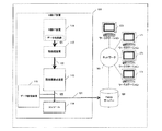
図1は従来のX線CT装置とネットワーク上の利用者を模擬的に示すブロック図である。101は従来のX線CT装置で,111はX線CT装置のスキャナ部を模擬的に表している。X線CT装置のスキャナ部111で収集されたX線検出器の出力データ121は前処理部112に送られ,ここでデータの雑音除去や較正などの処理が行われる。122は前処理部112で前処理した投影データである。113は画像再構成装置である。投影データ122に対して画像再構成装置113で画像再構成処理が行われる。114はX線CT装置のコンソールを,115は画像データ用データ蓄積装置を示している。画像再構成された画像データ123はコンソール114に送られて表示されると共に,画像データ用データ蓄積装置115に蓄積される。131は外部のPACSサーバー151に送られる画像データを示している。
【0016】
151は外部のPACSサーバーである。131はPACSサーバーに送られる画像データを示している。161はPACSサーバーと外部利用者とを接続するネットワークを示している。171はX線CTデータを使用して画像処理を行う外部利用者とワークステーションなどのコンピュータを模擬的に示している。
【0017】
X線CT装置のスキャナ部111は寝台上に横臥する被検者にX線を走査し,X線検出器で透過データを検出する。X線CT装置のスキャナ部111で収集されたX線検出器の出力データ121は前処理部112に送られ,ここでデータの雑音除去や較正などの処理が行われる。X線検出器の出力データを前処理部112で前処理した投影データ122は,画像再構成装置113に送られる。画像再構成装置113は投影データ122に対して画像再構成処理を行う。画像再構成された画像データ123はコンソール114に送られて表示される。また画像データ用データ蓄積装置115に蓄積される。また蓄積された画像データは外部のPACSサーバーに送られる。
【0018】
PACSサーバー151はネットワーク161を経由して外部利用者171と接続されている。外部利用者がX線CTデータの三次元画像処理などの画像処理を行う場合は,外部利用者はPACSサーバー151に蓄積されているX線CTデータをネットワーク161経由でワークステーション171に転送した後,画像処理を行う。三次元画像処理を行う場合には,多数の横断面の画像データが必要になる。ネットワークを経由した大容量の画像データの転送によって,ネットワークトラフィックが増大し,データ転送時間も長い。
【0019】
X線CT装置101には投影データは保存されていない。また保存されていても,X線CT装置の画像再構成装置はほとんどの時間をルーチンで使用しているので,外部の利用者には開放されていない。PACSサーバー151には画像再構成処理を行った結果として得られた画像データが保存されているだけで,投影データは蓄積されていない。投影データは標準化が進んでいないので,PACSサーバーに保存されていても,これを使用して画像再構成処理を行うことができる利用者は限定される。従って,現状では外部利用者が投影データを使用して画像再構成を行うことは事実上不可能である。
【0020】
【発明が解決しようとする課題】
X線CT装置を使用した検査では,X線CT装置で投影データを収集し,高速データ処理装置でこの投影データに対して画像再構成処理を行い,この画像再構成処理の結果として得た複数横断面の画像データを磁気ディスク装置,光磁気ディスク装置,光ディスク装置などの記憶媒体に保存している。X線CT装置で検査した結果を利用する方法ごとに適当な画像再構成処理のパラメータがあるが,現状では長期間にわたって保存されているのは画像再構成処理後の画像データであって,投影データは長期間保存はされていないので,それぞれの利用方法ごとに最適の画像再構成処理のパラメータで画像再構成処理を行うことは行われていない。特に,複数横断面の画像データを使用して三次元画像を構築する場合には,横断面の画像を直接観察する場合に適した画像再構成処理パラメータとは異なった三次元画像処理に適した画像再構成処理パラメータを使用して画像再構成した画像データを使用したい場合がしばしばあるが,このような使用目的別の画像再構成処理は一般には行われていない。この理由としては,投影データは標準化が進んでいないので,外部の大容量蓄積システムに保存していてもこれを利用して画像再構成ができる利用者が限定されること,X線CT装置の画像再構成装置はほとんどの時間をルーチンで使用しているので外部の利用者に開放されていないことなどによる。
【0021】
【課題を解決するための手段】
本発明は上記の課題を解決するために,X線CT装置に対して,X線走査によって収集した投影データを蓄積する機構と,この投影データの画像再構成処理を行う第二の機構と,この画像再構成処理で得られた画像データに対して三次元画像処理等の画像処理を行う機構とを追加して,ネットワーク経由で接続した外部利用者からの画像処理の要求に対応して外部利用者の希望する投影データを読み出し,この投影データに対して外部利用者の希望する画像再構成処理パラメータを使用して画像再構成処理を行い,この画像再構成処理で得られた画像データに対して外部利用者の希望する画像処理パラメータを使用して三次元画像処理等の画像処理を行い,この画像処理の結果をネットワーク経由で外部利用者に転送し,ネットワーク経由で接続した複数の外部利用者がこの一連の操作を対話的に行うことを可能にする機構を備えた。これによって画像再構成処理および画像処理並びにネットワーク配信を対話的に行うことを可能にしたX線CT装置を実現した。
【0022】
投影データの標準化が進んでいないために,投影データを外部の大容量蓄積システムに保存しておいてもこれを利用して画像再構成ができる利用者が限定されるが,この発明では,投影データをX線CT装置の内部に保存しているので,投影データに機種依存性や装置依存性があっても対応が可能である。また画像再構成装置もX線CT装置の内部にあるので,画像再構成パラメータの進歩による変更も即座に対応できる。また投影データと画像再構成に関するX線CT装置の製造者のノウハウも細部を公開する必要が無いので,実現可能性が高い。
【0023】
X線CT装置の画像再構成装置はほとんどの時間をルーチンで使用しているので外部の利用者に開放することが難しいが,本発明では外部利用者用として専用の画像再構成装置を用意したので,X線CT装置を使用したルーチン業務には影響を与えない。
【0024】
画像再構成装置や高速の画像処理装置は高価であるが,複数の利用者がネットワーク上で共同利用できるようにしたので,利用者あたりの費用を低く抑えることができた。
【0025】
また,ネットワーク上の利用者が使用を希望するX線CT装置の投影データに対して利用者が希望する画像再構成処理パラメータを使用して画像再構成処理を行い,この画像再構成処理を行って得た画像データに対して三次元画像処理などの画像処理を行うことを可能にしたので,三次元画像処理などの画像処理に適した画像再構成パラメータによる画像データを使用することが可能になった。
【0026】
従来は,三次元画像処理などの画像処理を行う利用者は,高速データ処理が可能なワークステーションなどのコンピュータ装置を設置し,大容量画像データをこのコンピュータ装置に蓄積し,この蓄積された画像データを画像処理して利用している。この発明では,X線CT装置に内蔵した外部利用者用画像処理装置を使用するので,利用者は高速データ処理が可能なワークステーションなどの高価なコンピュータ装置を用意することが不要になった。利用者の端末装置は,投影データ選択の指示と画像再構成処理パラメータおよび画像処理パラメータの指示を行い,X線CT装置の外部利用者用画像処理装置から送られてくる処理結果の画像データを表示するだけであるので,ラップトップコンピュータやノートパソコンなどの一般的なパーソナルコンピュータで従来のシステムのワークステーションと同等以上の結果を得ることが可能になった。
【0027】
【発明の実施の形態】
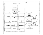
以下,本発明による画像再構成処理と画像処理およびネットワーク配信を対話的に行うことを可能にしたX線CT装置のシステムについて説明する。図2は本発明によるX線CT装置を模擬的に示すブロック図である。201は本発明によるX線CT装置で,211はX線CT装置のスキャナ部を模擬的に表している。X線CT装置のスキャナ部211で収集されたX線検出器の出力データ221は前処理部212に送られ,ここでデータの雑音除去や較正などの処理が行われる。222は前処理部212で前処理した投影データである。213は画像再構成装置,251は外部利用者用画像再構成装置,253は投影データ用データ蓄積装置である。投影データ222は画像再構成装置213に送られると共に,投影データ用データ蓄積装置253に蓄積される。また外部利用者から要求がある場合には外部利用者用画像再構成装置251に同時に送られる。261は外部利用者から要求によって投影データ用データ蓄積装置253から外部利用者用画像再構成装置251に送られる投影データを示している。投影データ222に対して画像再構成装置213で画像再構成処理が行われる。214はX線CT装置のコンソールを,215は画像データ用データ蓄積装置,252は外部利用者用画像処理装置を示している。画像再構成された画像データ223はコンソール214に送られて表示されると共に,画像データ用データ蓄積装置215に蓄積される。252は外部利用者用画像処理装置を,262は外部利用者から要求によって画像データ用データ蓄積装置215から外部利用者用画像処理装置252に送られる画像データを示している。
【0028】
301はX線CT装置と外部利用者とを接続するネットワークを示している。311はX線CT装置で収集された投影データに対して画像再構成処理を行い,その画像再構成によって得られた画像データに対して画像処理を行う外部利用者と端末装置を模擬的に示している。
【0029】
X線CT装置のスキャナ部211は寝台上に横臥する被検者にX線を走査し,X線検出器で透過データを検出する。X線CT装置のスキャナ部211で収集されたX線検出器の出力データ221は前処理部212に送られ,ここでデータの雑音除去や較正などの処理が行われる。X線検出器の出力データを前処理部212で前処理した投影データ222は,画像再構成装置213に送られると共に,投影データ用データ蓄積装置253に蓄積される。また外部利用者から要求がある場合には外部利用者用画像再構成装置251に同時に送られる。画像再構成装置213は投影データ222に対して画像再構成処理を行う。画像再構成された画像データ223はコンソール214に送られて表示されると共に,画像データ用データ蓄積装置215に蓄積される。また外部利用者から要求がある場合には外部利用者用画像処理装置252に同時に送られる。また,外部利用者から要求がある場合は,画像データ用データ蓄積装置215に蓄積されている画像データ262は外部利用者用画像処理装置252に送られる。
【0030】
X線CT装置201は外部利用者端末装置311とネットワーク301を経由して接続されている。外部利用者は端末装置311を使用して,X線CT装置で収集した投影データの画像再構成を外部利用者用画像再構成装置251で行うことができる。また,それによって得られた画像データを使用して画像処理を外部利用者用画像処理装置252で行うことができる。
【0031】
診断や治療,患者への説明のために新しい画像再構成処理パラメータで再構成された画像データを使用する利用者311はX線CT装置201に必要な投影データを指定するとともに,画像再構成処理のパラメータを送付する。X線CT装置201は依頼された投影データを投影データ用データ蓄積装置253から読み出し,画像再構成装置251に転送する。画像再構成装置251は利用者311から送付された画像再構成処理パラメータに基づいてこの投影データの画像再構成処理を行い,画像再構成処理の結果の画像データをネットワーク経由で利用者311に転送する。利用者311はパーソナルコンピュータなどの端末装置でこのデータを表示する。
【0032】
診断や治療,患者への説明のために画像データに対して画像処理たとえば三次元画像処理を行う利用者311はX線CT装置201に必要な投影データを指定するとともに,画像再構成処理と画像処理のパラメータを送付する。X線CT装置201は依頼された投影データを投影データ用データ蓄積装置253から読み出し,画像再構成装置251に転送する。画像再構成装置251は利用者311から送付された画像再構成処理パラメータに基づいてこの投影データの画像再構成処理を行って画像データを作成し,画像処理装置252に転送する。利用者311から送付された三次元画像処理パラメータに基づいてこの画像データに対して三次元画像処理を行い,画像処理の結果の三次元画像データをネットワーク経由で利用者311に転送する。利用者311はパーソナルコンピュータなどの端末装置でこのデータを表示する。
【0033】
従来のPACSでは,PACSサーバーに蓄積された大容量データを利用者が使用する場合には,利用者の保有するワークステーションなどのデータ処理能力の高いコンピュータ装置に大容量データを転送した後,転送されたデータのデータ処理をこのワークステーションで行い,結果をワークステーションに表示している。従って,大容量データを取り扱う利用者はワークステーションなどのデータ処理能力の高いコンピュータ装置を用意する必要があった。また大容量データをPACSサーバーから利用者の保有するワークステーションにネットワーク経由で転送するので,ネットワークトラフィックが高くなる。また,高速ネットワークを使用しても大容量データのネットワーク転送にはかなりの時間が必要になる。
【0034】
本発明のX線CT装置では,X線CT装置に蓄積された大容量データを利用者が使用する場合には,必要とするデータの指定とデータ処理のパラメータをX線CT装置に送付すると,X線CT装置は指定されたデータをデータ蓄積装置から読み出し,指定されたデータ処理パラメータを使用してデータ処理装置でデータ処理して,処理結果のデータを利用者に送付する。従って,利用者はワークステーションなどのデータ処理能力の高いコンピュータ装置を用意する必要は無く,ノートブックコンピュータなどのパーソナルコンピュータを使用できる。また大容量データをX線CT装置から利用者の保有する端末までネットワーク経由で転送することが無いので,ネットワークトラフィックは高くならない。また,処理済みの結果データのみを転送するので,高速ネットワークを使用しなくてもネットワーク転送時間は長くならない。
【0035】
X線CT装置201に蓄積されている投影データを使用して画像再構成を行うことを希望する利用者311は,パーソナルコンピュータから必要とする投影データを問合せるパラメータをネットワーク経由でX線CT装置201に送信する。このデータ取得要求を受信したX線CT装置201は,投影データ用データ蓄積装置253から該当する投影データ261を読み出し,画像再構成装置251に転送する。
【0036】
利用者311は画像再構成処理パラメータをネットワーク経由でX線CT装置201に送信する。この画像再構成処理パラメータを受信したX線CT装置201の画像再構成装置251は,投影データ用データ蓄積装置253から転送された投影データ261に対してこの画像再構成処理パラメータを使用して画像再構成処理を行う。画像再構成装置251はこの画像再構成処理によって再構成した画像データをパーソナルコンピュータ311に転送する。このデータを受信したパーソナルコンピュータ311はこの画像データを表示する。
【0037】
表示した画像データを観察した操作者が画像再構成処理パラメータを指定する操作を再度行うと,パーソナルコンピュータ311は修正した画像再構成処理パラメータを画像再構成装置251に再度送信し,画像再構成装置251はこの修正された画像再構成処理パラメータに基づいて画像再構成処理を行い,作成した画像データをパーソナルコンピュータ311に転送する。このデータを受信したパーソナルコンピュータ311はこの結果データを表示する。この一連の流れを繰り返し行うことによって,対話的に画像再構成処理を行うことができる。
【0038】
X線CT装置201に蓄積されている投影データを使用して画像処理を行うこと,例えば三次元画像を作成することを希望する利用者311は,パーソナルコンピュータから必要とする投影データを問合せるパラメータをネットワーク経由でX線CT装置201に送信する。このデータ取得要求を受信したX線CT装置201は,投影データ用データ蓄積装置253から該当する投影データ261を読み出し,画像再構成装置251に転送する。
【0039】
利用者311は画像再構成処理パラメータと三次元画像処理パラメータをネットワーク経由でX線CT装置201に送信する。この画像再構成処理パラメータを受信したX線CT装置201の画像再構成装置251は,投影データ用データ蓄積装置253から転送された投影データ261に対してこの画像再構成処理パラメータを使用して画像再構成処理を行う。画像再構成装置251はこの画像再構成処理によって再構成した画像データを画像処理装置252に転送する。三次元画像処理パラメータを受信した画像処理装置252は,画像再構成装置251から転送された画像データ263に対して三次元画像処理を行い,この画像再構成処理によって作成した三次元画像データをパーソナルコンピュータ311に転送する。このデータを受信したパーソナルコンピュータ311はこの画像データを表示する。
【0040】
表示した画像データを観察した操作者が画像処理パラメータを指定する操作を再度行うと,パーソナルコンピュータ311は修正した画像処理パラメータを画像処理装置252に再度送信し,画像処理装置252はこの修正された画像処理パラメータに基づいて三次元画像処理を行い,作成した三次元画像データをパーソナルコンピュータ311に転送する。このデータを受信したパーソナルコンピュータ311はこの結果データを表示する。この一連の流れを繰り返し行うことによって,対話的に画像処理を行うことができる。
【0041】
【実施例】
これまでの説明はネットワーク上の利用者がX線CT装置の投影データの蓄積とこの投影データの画像再構成処理,そして画像再構成した画像データに対して画像処理を行うことを可能とするシステムについて説明したが,X線CT装置の投影データだけでなくPET装置のいわゆる生データやMR装置のいわゆる生データを蓄積しておき,必要に応じて新しいパラメータを適用して画像再構成を行うシステムも同様に含まれる。
【0042】
これまでの説明は画像再構成した画像データに対して三次元画像処理を行うシステムについて説明したが,三次元画像処理は一例であり,画像再構成した画像データに対して行う全ての画像処理が含まれる。
【0043】
【発明の効果】
本発明は,X線CT装置に対して,投影データを蓄積する機構と,投影データの画像再構成処理を行う第二の機構と,この画像再構成処理で得られた画像データに対して画像処理を行う機構とを追加して,ネットワーク経由で接続した外部利用者からの画像処理の要求に対応して外部利用者の希望する投影データを読み出し,この投影データに対して外部利用者の希望する画像再構成処理パラメータを使用して画像再構成処理を行い,この画像再構成処理で得られた画像データに対して外部利用者の希望する画像処理パラメータを使用して画像処理を行い,この画像処理の結果をネットワーク経由で外部利用者に転送する機能と,ネットワーク経由で接続した複数の外部利用者がこの一連の操作を対話的に行うことを可能にする機能を備えた。これによって画像再構成処理と画像処理およびネットワーク配信を対話的に行うことを可能にしたX線CT装置を実現した。
【0044】
投影データはその標準化が進んでいないために,投影データを外部の大容量蓄積システムに保存しておいてもこれを利用して画像再構成ができる利用者は限定される。この発明では,投影データをX線CT装置の内部に保存しているので,投影データに機種依存性や装置依存性があっても対応が可能になった。また画像再構成装置もX線CT装置の内部にあるので,画像再構成パラメータの進歩による変更も即座に対応できる。また投影データと画像再構成に関するX線CT装置の製造者のノウハウの細部を公開する必要が無いので普及する可能性が高い。
【0045】
X線CT装置の画像再構成装置はほとんどの時間をルーチンで使用しているので外部の利用者に開放することが難しいが,本発明では外部利用者用として専用のデータ蓄積装置、画像再構成装置および画像処理装置を用意したので,X線CT装置のルーチン業務には影響を与えずに,外部利用者の要求に対応できる。
【0046】
画像再構成装置や高速の画像処理装置は高価であるが,複数の利用者がネットワーク上で共同で使用できるようにしたので,利用者あたりの費用を低く抑えることができた。
【0047】
また,ネットワーク上の利用者が使用を希望するX線CT装置の投影データに対して利用者が希望する再構成処理パラメータを使用して画像再構成処理を行い,この画像再構成処理を行って得た画像データに対して三次元画像処理などの画像処理を行うことを可能にしたので,三次元画像処理などの画像処理に最も適した画像再構成パラメータによる画像データを使用することが可能になった。
【0048】
従来は,三次元画像処理などの画像処理を行う利用者は,ワークステーションなどの高速データ処理が可能なコンピュータ装置を設置し,大容量画像データをこのコンピュータ装置に蓄積し,この蓄積された画像データを画像処理に使用している。この発明では,X線CT装置に内蔵した外部利用者用画像処理装置を使用するので,利用者はワークステーションなどの高速にデータ処理が可能なコンピュータ装置を用意することが不要になった。利用者の端末装置は,投影データの選択指示と再構成パラメータおよび画像処理パラメータの指示を行い,X線CT装置の外部利用者用画像処理装置から送られてくる処理結果の画像データを表示するだけであるので,ラップトップコンピュータやノートパソコンなどの一般的なパーソナルコンピュータで従来のシステムのワークステーションと同等以上の結果を得ることが可能になった。
【0049】
従来は,ネットワーク上の利用者がPACSサーバーなどの大規模データ蓄積装置に蓄積されている大容量データを使用する場合には,その大容量データを大規模データ蓄積装置から利用者の保有するワークステーションなどのデータ処理装置までネットワークを経由して転送する必要があった。投影データのような大容量データの場合にはこのデータ転送にはかなりの時間を必要とすると共に,このデータ転送によってネットワークトラフィックが増大する。本発明では,X線CT装置に内蔵されている高速データ処理装置で処理するので,ネットワーク上の利用者が大容量データを使用する時のネットワークトラフィックの増大を抑制することが可能になった。
【0050】
複数の利用者がデータ処理の結果を共有することができる。データ処理に使用したデータ処理パラメータはデータ蓄積装置に保存しておくことが可能なであるので,他の利用者がこのパラメータを使用して,データ処理を復元することができる。
【図面の簡単な説明】
【図1】従来のX線CT装置とネットワーク上の利用者を説明するブロック図
【図2】本発明による画像処理とネットワーク配信を対話的に行うことを可能にしたX線CT装置のブロック図
【符号の説明】
101 従来のX線CT装置
111 X線CT装置のスキャナ部
112 前処理装置
113 画像再構成装置
114 コンソール
115 画像データ蓄積装置
121 X線CT装置のスキャナ部で収集された投影データ
122 前処理部で前処理された投影データ
123 画像再構成された画像データ
131 PACSサーバーへ送られる画像データ
151 PACSサーバー
161 ネットワーク
171 X線CTデータを使用する利用者とワークステーション
201 本発明によるX線CT装置
211 X線CT装置のスキャナ部
212 前処理装置
213 画像再構成装置
214 コンソール
215 画像データ蓄積装置
221 X線CT装置のスキャナ部で収集された投影データ
222 前処理部で前処理された投影データ
223 画像再構成された画像データ
251 外部利用者用画像再構成装置
252 外部利用者用画像処理装置
253 投影データ蓄積装置
261 投影データ蓄積装置から外部利用者用画像再構成装置に転送される投影データ
262 画像データ蓄積装置から外部利用者用画像処理装置に転送される画像データ
263 外部利用者用画像再構成装置で画像再構成された画像データ
301 ネットワーク
311 X線CTデータを使用する外部利用者と端末装置[0001]
[Technology to which the invention belongs]
The present invention relates to an X-ray CT apparatus that enables an external user connected via a network to interactively perform image reconstruction processing, image processing, and network distribution of the result.
[0002]
[Prior art]
With the advancement and development of electronic technology and electronic information technology, the amount of data generated by medical devices is rapidly increasing. For example, in an X-ray CT apparatus, around 1977, the amount of data generated by examination of one patient was about 312 pixels × 312 and about 10 images, totaling about 2 MB. Since an inspection of about 10 people per day was normal, data of about 20 MB per day was generated. However, in recent multi-row detector X-ray CT apparatuses, the amount of data generated in the examination of one patient is about 512 × 512, about 1500 images, and about 750 MB in total. At present, examinations of about 30 people a day are normal, so data of about 23 GB is generated per day. The amount of data generated by one X-ray CT apparatus over 25 years has increased by 1000 times.
[0003]
In the X-ray CT apparatus, the subject is irradiated with X-rays from an X-ray tube that circulates around the subject lying on the top of the bed, and the intensity of the X-rays transmitted through the subject is measured with an X-ray detector. By detecting, projection data of the cross section of the subject is collected. Next, after moving the couch top, similarly, the projection data of the cross section of the subject is collected. By repeating the movement of the top plate and the collection of projection data, projection data of a plurality of cross sections of the subject are collected. The image data of the plurality of cross sections of the subject is obtained by performing image reconstruction processing on the collected projection data of the plurality of cross sections of the subject by the high-speed data processing apparatus.
[0004]
As a method of using the results of the inspection by the X-ray CT apparatus, the projection data of a plurality of cross sections are collected by the X-ray CT apparatus, and the image reconstruction processing is performed on the projection data of the plurality of cross sections by the high speed data processing apparatus. The image data of multiple cross sections is created, the image data of the multiple cross sections is displayed on the image display device, the displayed images of the multiple cross sections are printed on the film, and the film on which the images of the multiple cross sections are printed is observed. Thus, it has been widely performed to find abnormal sites in subjects. In the early X-ray CT apparatus, the number of reconstructed image data was 10 to several tens, so such a method was possible.
[0005]
As a method of using the results of the inspection by the X-ray CT apparatus, the projection data of a plurality of cross sections are collected by the X-ray CT apparatus, and the image reconstruction processing is performed on the projection data of the plurality of cross sections by the high speed data processing apparatus Thus, image data of a plurality of cross sections is created, and the image data of the plurality of cross sections is stored in a storage medium such as a magnetic disk device, a magneto-optical disk device, or an optical disk device. Reading image data of a plurality of cross-sections stored in this storage medium, displaying the image data on an image observation apparatus, and observing is performed. In some cases, image data of a plurality of cross sections stored in the storage medium is read out and image processing is performed on the image data by an image processing apparatus. For example, a three-dimensional image is constructed from this image data by a three-dimensional image processing apparatus, and the three-dimensional image is observed.
[0006]
In the early X-ray CT apparatus, the movement of the top plate on which the subject lay down and the collection of the projection data of the subject by X-ray irradiation were performed alternately. The technology of the X-ray CT apparatus has advanced rapidly, and in recent X-ray CT apparatuses, the X-ray irradiation is simultaneously performed while the subject lies on the tabletop and the projection data is collected. Such a projection data collection method is called helical scan.
[0007]
In the early X-ray CT apparatus, the movement of the table on which the subject lay down and the acquisition of the projection data of the subject by X-ray irradiation were performed alternately. It was a cross section of the image data obtained by performing the configuration. That is, the cross-section from which the projection data was collected and the cross-section of the image data obtained by image reconstruction coincide with each other, and the number of cross-sections from which projection data was collected and the image data obtained by image reconstruction. The position of the cross-section where the projection data was collected was distributed discretely along the patient's body axis. However, in the helical scan method, since the subject is moving the table on the side, X-ray irradiation is performed at the same time to collect projection data, so there is no discrete projection data collection cross section. A plane is set and projection data is interpolated to obtain projection data of a virtual cross section, and image reconstruction processing is performed using the projection data of the virtual cross section. Since the virtual cross section of this projection data can be set quite freely, the number of image data obtained by image reconstruction processing and the interval between the cross sections can be selected fairly freely depending on the setting of the virtual cross section of the projection data. it can.
[0008]
In early X-ray CT systems, X-ray detectors that detect the intensity of X-rays that have passed through a subject collect projection data of one cross section of the subject in a row in the body axis direction of the subject. Was. The technology of X-ray CT equipment has advanced rapidly. In recent X-ray CT equipment, multiple rows of X-ray detectors are arranged in the direction of the body axis of the subject, and projection data of multiple cross sections of the subject can be simultaneously transmitted. Use multi-row detector to collect. By using this multi-row detector and the helical scan method at the same time, much more projection data than the initial X-ray CT apparatus has been collected. In this multi-row detector / helical scan system, there is no discrete projection data acquisition cross section as in the early X-ray CT system. Generally, a virtual cross section is set and projection data interpolation processing is performed. The virtual cross-section projection data is obtained and image reconstruction processing is performed using the virtual cross-section projection data. Since the virtual cross section of the projection data can be set freely, the number of image data obtained by the image reconstruction process and the interval between the cross sections can be freely selected depending on the setting of the virtual cross section of the projection data.
[0009]
In an inspection using an X-ray CT apparatus, currently, projection data is collected by an X-ray CT apparatus, and an image reconstruction process is performed on the projection data by a high-speed data processing apparatus. As a result of the image reconstruction process, The obtained image data of a plurality of cross sections is stored in a storage medium such as a magnetic disk device, a magneto-optical disk device, or an optical disk device. There are two methods for using the results of inspection with an X-ray CT apparatus: a method of printing an image created using image data of a plurality of cross sections obtained as a result of image reconstruction processing on a film, and observing the film; A method of displaying and observing an image on an image observation apparatus using image data of a plurality of cross sections obtained as a result of the reconstruction process, and an image with respect to the image data of a plurality of cross sections obtained as a result of the image reconstruction process There is a method of performing image processing with a processing device and observing the result. There is an optimum image reconstruction processing parameter for each usage method, but the reconstruction processing is not performed again with the optimum image reconstruction processing parameter for each usage method. At present, the data stored for a long time is image data after image reconstruction, and projection data before image reconstruction is not stored for a long time.
[0010]
In particular, when constructing a 3D image using multiple cross-sectional image data, it is suitable for 3D image processing that is different from the image reconstruction processing parameters suitable for direct observation of the cross-sectional image. In many cases, it is desirable to use multiple cross-sectional image data that has been reconstructed using image reconstruction processing parameters, but in general, only the image data after image reconstruction is stored for a long period of time. Since the image is not stored for a long time, the image reconstruction process for each purpose of use cannot generally be performed.
[0011]
Even if the X-ray CT apparatus stores projection data for a long period of time, many inspections are routinely performed almost continuously in the X-ray CT apparatus. However, it is almost impossible in practice to perform image reconstruction using image reconstruction parameters desired by the user. For this reason, the image reconstruction of the projection data is performed by a predetermined image reconstruction parameter in the routine inspection.
[0012]
In recent X-ray CT apparatuses, there are models that can perform advanced image processing such as three-dimensional image processing on image data obtained by image reconstruction. However, even with such a function, an X-ray CT apparatus routinely performs a large number of examinations almost continuously, so an external user can use this function to perform 3D image processing. It is almost impossible to do in reality.
[0013]
With the advancement and development of network technology and the expansion of its use, a large-scale data storage device is installed on the network, medical image data is stored in this data storage device, and multiple medical image data stored in this data storage device are stored. A system called PACS has been constructed that can be used by other users.
[0014]
For example, in the case of X-ray CT examination, image data obtained as a result of image reconstruction processing performed by an X-ray CT apparatus is stored as data stored in the PACS server. Using this image data stored in the PACS, for example, a user who creates a three-dimensional image can connect a PACS server to a computer device capable of high-speed data processing such as a workstation owned by the user. The image data is transferred, the image processing is performed on the image data at the workstation, and the result is displayed. When creating a three-dimensional image, image data of a large number of cross sections is required, and network traffic increases due to the transfer of large-capacity image data via the network, and the data transfer time is also long.
[0015]
FIG. 1 is a block diagram schematically showing a conventional X-ray CT apparatus and users on a network.
[0016]
151 is an external PACS server. Reference numeral 131 denotes image data sent to the PACS server. Reference numeral 161 denotes a network connecting the PACS server and an external user.
[0017]
The scanner unit 111 of the X-ray CT apparatus scans a subject lying on the bed with X-rays, and detects transmission data with an X-ray detector. Output data 121 of the X-ray detector collected by the scanner unit 111 of the X-ray CT apparatus is sent to the preprocessing unit 112, where processing such as data noise removal and calibration is performed.
[0018]
The PACS server 151 is connected to the
[0019]
Projection data is not stored in the
[0020]
[Problems to be solved by the invention]
In an inspection using an X-ray CT apparatus, projection data is collected by the X-ray CT apparatus, an image reconstruction process is performed on the projection data by a high-speed data processing apparatus, and a plurality of images obtained as a result of the image reconstruction process are obtained. The image data of the cross section is stored in a storage medium such as a magnetic disk device, a magneto-optical disk device, or an optical disk device. There are appropriate parameters for image reconstruction processing for each method that uses the results of examination by an X-ray CT apparatus, but currently, image data after image reconstruction processing is stored for a long period of time and is projected. Since the data is not stored for a long time, the image reconstruction process is not performed with the optimum image reconstruction process parameter for each usage method. In particular, when constructing a 3D image using multiple cross-sectional image data, it is suitable for 3D image processing that is different from the image reconstruction processing parameters suitable for direct observation of the cross-sectional image. In many cases, it is desired to use image data reconstructed using image reconstruction processing parameters, but such image reconstruction processing for each purpose of use is not generally performed. This is because the standardization of projection data has not progressed, and the number of users who can reconstruct images using this data is limited even if stored in an external large-capacity storage system. This is because the image reconstructing apparatus uses most of the time in a routine and is not open to external users.
[0021]
[Means for Solving the Problems]
In order to solve the above problems, the present invention provides a mechanism for storing projection data collected by X-ray scanning in an X-ray CT apparatus, this The second mechanism that performs image reconstruction processing of projection data and the image data obtained by this image reconstruction processing For 3D image processing etc. Perform image processing Machine In response to an image processing request from an external user connected via the network, the projection data desired by the external user is read out, and the image data desired by the external user is read from the projection data. Image reconstruction processing is performed using the configuration processing parameters, and the image processing parameters desired by the external user are used for the image data obtained by the image reconstruction processing. 3D image processing, etc. Performs image processing and transfers the image processing results to external users via the network Shi , Equipped with a mechanism that allows multiple external users connected via a network to perform this series of operations interactively. As a result, image reconstruction processing and Image processing And An X-ray CT system that enables interactive network distribution has been realized.
[0022]
Since standardization of projection data has not progressed, users who can reconstruct images using projection data stored in an external large-capacity storage system are limited. Since the data is stored inside the X-ray CT apparatus, it is possible to cope even if the projection data has model dependence or apparatus dependence. Further, since the image reconstruction apparatus is also inside the X-ray CT apparatus, it is possible to respond immediately to changes due to the advancement of image reconstruction parameters. In addition, the know-how of the manufacturer of the X-ray CT apparatus regarding projection data and image reconstruction does not need to be disclosed in detail, and is highly feasible.
[0023]
Since the image reconstruction device of the X-ray CT apparatus uses most of the time in routine, it is difficult to open it to external users, but in the present invention, a dedicated image reconstruction device is prepared for external users. Therefore, it does not affect the routine work using the X-ray CT apparatus.
[0024]
Image reconstruction devices and high-speed image processing devices are expensive, but multiple users can share them on the network, so the cost per user can be kept low.
[0025]
In addition, the user desires the projection data of the X-ray CT apparatus that the user on the network desires to use. image Image reconstruction processing is performed using the reconstruction processing parameters, and image processing such as 3D image processing can be performed on the image data obtained by performing this image reconstruction processing. Image data based on image reconstruction parameters suitable for image processing such as processing can be used.
[0026]
Conventionally, a user who performs image processing such as three-dimensional image processing installs a computer device such as a workstation capable of high-speed data processing, stores a large amount of image data in the computer device, and stores the stored image. Data is used after image processing. According to the present invention, since an image processing apparatus for an external user incorporated in the X-ray CT apparatus is used, it becomes unnecessary for the user to prepare an expensive computer device such as a workstation capable of high-speed data processing. The user terminal device instructs the projection data selection, the image reconstruction processing parameter, and the image processing parameter, and receives the image data of the processing result sent from the external user image processing device of the X-ray CT apparatus. Since it is only displayed, it has become possible to obtain results equivalent to or better than those of conventional system workstations on general personal computers such as laptop computers and laptop computers.
[0027]
DETAILED DESCRIPTION OF THE INVENTION
Hereinafter, an X-ray CT apparatus system capable of interactively performing image reconstruction processing, image processing, and network distribution according to the present invention will be described. FIG. 2 is a block diagram schematically showing an X-ray CT apparatus according to the present invention.
[0028]
A network 301 connects the X-ray CT apparatus and an external user. 311 schematically shows an external user and a terminal device that perform image reconstruction processing on the projection data collected by the X-ray CT apparatus and perform image processing on the image data obtained by the image reconstruction. ing.
[0029]
The
[0030]
The
[0031]
A
[0032]
A
[0033]
In the conventional PACS, when a user uses a large amount of data stored in a PACS server, the large amount of data is transferred to a computer device having a high data processing capability such as a workstation owned by the user, and then transferred. The processed data is processed by this workstation, and the result is displayed on the workstation. Therefore, a user who handles a large amount of data needs to prepare a computer device having a high data processing capability such as a workstation. In addition, since a large amount of data is transferred from the PACS server to the workstation owned by the user via the network, the network traffic becomes high. Even if a high-speed network is used, a considerable amount of time is required for network transfer of large-capacity data.
[0034]
In the X-ray CT apparatus of the present invention, when a user uses a large amount of data stored in the X-ray CT apparatus, if the user designates necessary data and sends data processing parameters to the X-ray CT apparatus, The X-ray CT apparatus reads the designated data from the data storage device, processes the data with the data processing apparatus using the designated data processing parameters, and sends the processing result data to the user. Therefore, the user does not need to prepare a computer device with high data processing capability such as a workstation, and can use a personal computer such as a notebook computer. In addition, network traffic does not increase because large-capacity data is not transferred from the X-ray CT apparatus to the terminal owned by the user via the network. In addition, since only the processed result data is transferred, the network transfer time does not become long without using a high-speed network.
[0035]
A
[0036]
The
[0037]
When the operator observing the displayed image data performs the operation of specifying the image reconstruction processing parameter again, the
[0038]
A
[0039]
The
[0040]
When the operator observing the displayed image data performs the operation of specifying the image processing parameter again, the
[0041]
【Example】
The description so far is based on a system that allows a user on a network to store projection data of an X-ray CT apparatus, to perform image reconstruction processing of the projection data, and to perform image processing on the reconstructed image data. However, in addition to the projection data of the X-ray CT apparatus, the so-called raw data of the PET apparatus and the so-called raw data of the MR apparatus are stored, and image reconstruction is performed by applying new parameters as necessary. Is included as well.
[0042]
The description so far has described a system that performs three-dimensional image processing on image data that has been reconstructed. However, three-dimensional image processing is an example, and all image processing that is performed on image data that has been reconstructed. included.
[0043]
【The invention's effect】
The present invention relates to an X-ray CT apparatus, a mechanism for storing projection data, a second mechanism for performing image reconstruction processing of projection data, and an image for image data obtained by this image reconstruction processing. Process Machine In response to an image processing request from an external user connected via the network, the projection data desired by the external user is read out, and the image data desired by the external user is read from the projection data. Image reconstruction processing is performed using the configuration processing parameters, and image processing is performed on the image data obtained by the image reconstruction processing using image processing parameters desired by an external user. It has a function to transfer the results to external users via the network and a function that allows multiple external users connected via the network to perform this series of operations interactively. As a result, an X-ray CT apparatus capable of interactively performing image reconstruction processing, image processing, and network distribution has been realized.
[0044]
Since standardization of projection data has not progressed, users who can reconstruct an image using projection data stored in an external large-capacity storage system are limited. In this invention, since the projection data is stored inside the X-ray CT apparatus, even if the projection data has model dependence or equipment dependence, it can be handled. Effectively became. Further, since the image reconstruction apparatus is also inside the X-ray CT apparatus, it is possible to respond immediately to changes due to the advancement of image reconstruction parameters. Further, since it is not necessary to disclose the details of the know-how of the manufacturer of the X-ray CT apparatus regarding projection data and image reconstruction, there is a high possibility that it will be widely used.
[0045]
Since the image reconstruction device of the X-ray CT apparatus uses most of the time in the routine, it is difficult to open it to external users. Data storage device, Image reconstruction device And image processing apparatus Therefore, it is possible to meet the demands of external users without affecting the routine work of the X-ray CT apparatus.
[0046]
Image reconstructing devices and high-speed image processing devices are expensive, but multiple users can use them jointly on the network, so the cost per user can be kept low.
[0047]
Further, the image reconstruction processing is performed on the projection data of the X-ray CT apparatus that the user on the network desires to use using the reconstruction processing parameter desired by the user, and this image reconstruction processing is performed. Since image processing such as 3D image processing can be performed on the obtained image data, it is possible to use image data with image reconstruction parameters most suitable for image processing such as 3D image processing. became.
[0048]
Conventionally, a user who performs image processing such as three-dimensional image processing installs a computer device capable of high-speed data processing such as a workstation, accumulates large-capacity image data in the computer device, and stores the stored image. Data is used for image processing. In the present invention, since the image processing apparatus for external users incorporated in the X-ray CT apparatus is used, it becomes unnecessary for the user to prepare a computer device capable of high-speed data processing such as a workstation. The user terminal device issues an instruction for selecting projection data, a reconstruction parameter, and an image processing parameter, and displays processing result image data sent from the image processing device for external users of the X-ray CT apparatus. As a result, it has become possible to obtain results equivalent to or better than those of conventional system workstations on general personal computers such as laptop computers and laptop computers.
[0049]
Conventionally, when a user on a network uses large-capacity data stored in a large-scale data storage device such as a PACS server, the large-capacity data is transferred from the large-scale data storage device to the work held by the user. It was necessary to transfer the data processing device such as a station via a network. In the case of large-capacity data such as projection data, this data transfer requires a considerable amount of time, and this data transfer increases network traffic. In the present invention, since processing is performed by a high-speed data processing device built in the X-ray CT apparatus, it is possible to suppress an increase in network traffic when a user on the network uses a large amount of data.
[0050]
Multiple users can share data processing results. Since the data processing parameter used for data processing can be stored in the data storage device, other users can use this parameter to restore the data processing.
[Brief description of the drawings]
FIG. 1 is a block diagram for explaining a conventional X-ray CT apparatus and users on a network.
FIG. 2 is a block diagram of an X-ray CT apparatus that enables interactive image processing and network distribution according to the present invention.
[Explanation of symbols]
101 Conventional X-ray CT system
111 Scanner section of X-ray CT system
112 Pretreatment device
113 Image reconstruction device
114 console
115 Image data storage device
121 Projection data collected by the scanner section of the X-ray CT system
122 Projection data preprocessed by the preprocessing unit
123 Reconstructed image data
131 Image data sent to PACS server
151 PACS server
161 network
171 Users and workstations using X-ray CT data
201 X-ray CT apparatus according to the present invention
211 Scanner section of X-ray CT system
212 Pretreatment device
213 Image reconstruction device
214 console
215 Image data storage device
221 Projection data collected by X-ray CT scanner unit
222 Projection data preprocessed by the preprocessor
223 Image reconstructed image data
251 Image reconstruction device for external users
252 Image processing apparatus for external users
253 Projection data storage device
261 Projection data transferred from the projection data storage device to the image reconstruction device for external users
262 Image data transferred from the image data storage device to the image processing device for external users
263 Image data reconstructed by the image reconstruction device for external users
301 network
311 External users and terminal devices using X-ray CT data
Claims (2)
Priority Applications (1)
| Application Number | Priority Date | Filing Date | Title |
|---|---|---|---|
| JP2002115483A JP4231237B2 (en) | 2002-03-14 | 2002-03-14 | X-ray CT system that enables interactive image processing and network distribution |
Applications Claiming Priority (1)
| Application Number | Priority Date | Filing Date | Title |
|---|---|---|---|
| JP2002115483A JP4231237B2 (en) | 2002-03-14 | 2002-03-14 | X-ray CT system that enables interactive image processing and network distribution |
Publications (2)
| Publication Number | Publication Date |
|---|---|
| JP2003265461A JP2003265461A (en) | 2003-09-24 |
| JP4231237B2 true JP4231237B2 (en) | 2009-02-25 |
Family
ID=29207691
Family Applications (1)
| Application Number | Title | Priority Date | Filing Date |
|---|---|---|---|
| JP2002115483A Expired - Fee Related JP4231237B2 (en) | 2002-03-14 | 2002-03-14 | X-ray CT system that enables interactive image processing and network distribution |
Country Status (1)
| Country | Link |
|---|---|
| JP (1) | JP4231237B2 (en) |
Families Citing this family (3)
| Publication number | Priority date | Publication date | Assignee | Title |
|---|---|---|---|---|
| JP4528596B2 (en) * | 2004-10-14 | 2010-08-18 | 株式会社東芝 | Medical image management system, medical image management method, and medical image management program |
| JP5367043B2 (en) * | 2011-10-11 | 2013-12-11 | 株式会社東芝 | Medical image support diagnosis device |
| JP7602954B2 (en) * | 2021-04-02 | 2024-12-19 | キヤノンメディカルシステムズ株式会社 | Medical image processing apparatus, medical image diagnostic apparatus, magnetic resonance imaging apparatus, and medical image processing method |
Family Cites Families (1)
| Publication number | Priority date | Publication date | Assignee | Title |
|---|---|---|---|---|
| JP2001177849A (en) * | 1999-12-15 | 2001-06-29 | Mitsubishi Electric Corp | Three-dimensional image generation system and three-dimensional image generation device |
-
2002
- 2002-03-14 JP JP2002115483A patent/JP4231237B2/en not_active Expired - Fee Related
Also Published As
| Publication number | Publication date |
|---|---|
| JP2003265461A (en) | 2003-09-24 |
Similar Documents
| Publication | Publication Date | Title |
|---|---|---|
| JP4502426B2 (en) | Multi-slice imaging device | |
| JP4739225B2 (en) | Workflow optimization for high-throughput imaging environments | |
| CN101233521B (en) | Form the method and apparatus of multinomial research | |
| US20040068167A1 (en) | Computer aided processing of medical images | |
| US20040161139A1 (en) | Image data navigation method and apparatus | |
| JP2006297092A (en) | 4D display creation, evaluation and segmentation system by computed tomography of the patient's heart | |
| JP2004105728A (en) | Computer aided acquisition of medical image | |
| JP2002330958A (en) | Method and device for selecting and displaying medical image data | |
| JP2004180785A (en) | Data management system, X-ray computed tomography apparatus, and X-ray computed tomography system | |
| US7148688B2 (en) | Magnetic resonance imaging apparatus and method of controlling magnetic resonance imaging apparatus | |
| JPH07323027A (en) | Computed tomography equipment | |
| CN102028492A (en) | Centralized image reconstruction for tomographic imaging techniques in medical engineering | |
| JP5269283B2 (en) | Medical image diagnostic apparatus and image reconstruction method | |
| JP2004113730A (en) | Computer-aided diagnosis system | |
| JP4231237B2 (en) | X-ray CT system that enables interactive image processing and network distribution | |
| CN101077303B (en) | Method and associated device for determining positron emission measurement information of a body part | |
| JP4681790B2 (en) | CT data storage and processing system in network environment | |
| JP4625701B2 (en) | Image observation apparatus and image observation method | |
| JP4361268B2 (en) | 3D image display device for directly creating a 3D image from projection data of an X-ray CT apparatus | |
| EP2206084B1 (en) | Image processing with computer aided detection and/or diagnosis | |
| JP5179897B2 (en) | X-ray CT system | |
| EP1555633A2 (en) | Multi-positional CT image producing method and x-ray CT apparatus | |
| JPH08299327A (en) | Computed tomography equipment | |
| CN109243585A (en) | The generation method and magic magiscan and its exchange method of medical image | |
| CN119214676B (en) | Image acquisition method and device for C-arm X-ray equipment and C-arm X-ray equipment |
Legal Events
| Date | Code | Title | Description |
|---|---|---|---|
| A621 | Written request for application examination |
Free format text: JAPANESE INTERMEDIATE CODE: A621 Effective date: 20050314 |
|
| RD02 | Notification of acceptance of power of attorney |
Free format text: JAPANESE INTERMEDIATE CODE: A7422 Effective date: 20050314 |
|
| A977 | Report on retrieval |
Free format text: JAPANESE INTERMEDIATE CODE: A971007 Effective date: 20080410 |
|
| A131 | Notification of reasons for refusal |
Free format text: JAPANESE INTERMEDIATE CODE: A131 Effective date: 20080422 |
|
| A521 | Request for written amendment filed |
Free format text: JAPANESE INTERMEDIATE CODE: A523 Effective date: 20080620 |
|
| A131 | Notification of reasons for refusal |
Free format text: JAPANESE INTERMEDIATE CODE: A131 Effective date: 20080902 |
|
| A521 | Request for written amendment filed |
Free format text: JAPANESE INTERMEDIATE CODE: A523 Effective date: 20080909 |
|
| TRDD | Decision of grant or rejection written | ||
| A01 | Written decision to grant a patent or to grant a registration (utility model) |
Free format text: JAPANESE INTERMEDIATE CODE: A01 Effective date: 20081202 |
|
| A01 | Written decision to grant a patent or to grant a registration (utility model) |
Free format text: JAPANESE INTERMEDIATE CODE: A01 |
|
| A61 | First payment of annual fees (during grant procedure) |
Free format text: JAPANESE INTERMEDIATE CODE: A61 Effective date: 20081205 |
|
| R150 | Certificate of patent or registration of utility model |
Ref document number: 4231237 Country of ref document: JP Free format text: JAPANESE INTERMEDIATE CODE: R150 Free format text: JAPANESE INTERMEDIATE CODE: R150 |
|
| FPAY | Renewal fee payment (event date is renewal date of database) |
Free format text: PAYMENT UNTIL: 20111212 Year of fee payment: 3 |
|
| FPAY | Renewal fee payment (event date is renewal date of database) |
Free format text: PAYMENT UNTIL: 20121212 Year of fee payment: 4 |
|
| R250 | Receipt of annual fees |
Free format text: JAPANESE INTERMEDIATE CODE: R250 |
|
| FPAY | Renewal fee payment (event date is renewal date of database) |
Free format text: PAYMENT UNTIL: 20131212 Year of fee payment: 5 |
|
| R250 | Receipt of annual fees |
Free format text: JAPANESE INTERMEDIATE CODE: R250 |
|
| R250 | Receipt of annual fees |
Free format text: JAPANESE INTERMEDIATE CODE: R250 |
|
| R250 | Receipt of annual fees |
Free format text: JAPANESE INTERMEDIATE CODE: R250 |
|
| R250 | Receipt of annual fees |
Free format text: JAPANESE INTERMEDIATE CODE: R250 |
|
| R250 | Receipt of annual fees |
Free format text: JAPANESE INTERMEDIATE CODE: R250 |
|
| R250 | Receipt of annual fees |
Free format text: JAPANESE INTERMEDIATE CODE: R250 |
|
| R250 | Receipt of annual fees |
Free format text: JAPANESE INTERMEDIATE CODE: R250 |
|
| R250 | Receipt of annual fees |
Free format text: JAPANESE INTERMEDIATE CODE: R250 |
|
| R250 | Receipt of annual fees |
Free format text: JAPANESE INTERMEDIATE CODE: R250 |
|
| LAPS | Cancellation because of no payment of annual fees |