JP4067407B2 - Method and system for detecting colorimetric anomalies in vivo - Google Patents
Method and system for detecting colorimetric anomalies in vivo Download PDFInfo
- Publication number
- JP4067407B2 JP4067407B2 JP2002572089A JP2002572089A JP4067407B2 JP 4067407 B2 JP4067407 B2 JP 4067407B2 JP 2002572089 A JP2002572089 A JP 2002572089A JP 2002572089 A JP2002572089 A JP 2002572089A JP 4067407 B2 JP4067407 B2 JP 4067407B2
- Authority
- JP
- Japan
- Prior art keywords
- image
- color
- vivo
- images
- blood
- Prior art date
- Legal status (The legal status is an assumption and is not a legal conclusion. Google has not performed a legal analysis and makes no representation as to the accuracy of the status listed.)
- Expired - Fee Related
Links
Images


Classifications
-
- A—HUMAN NECESSITIES
- A61—MEDICAL OR VETERINARY SCIENCE; HYGIENE
- A61B—DIAGNOSIS; SURGERY; IDENTIFICATION
- A61B1/00—Instruments for performing medical examinations of the interior of cavities or tubes of the body by visual or photographical inspection, e.g. endoscopes; Illuminating arrangements therefor
- A61B1/04—Instruments for performing medical examinations of the interior of cavities or tubes of the body by visual or photographical inspection, e.g. endoscopes; Illuminating arrangements therefor combined with photographic or television appliances
- A61B1/041—Capsule endoscopes for imaging
-
- A—HUMAN NECESSITIES
- A61—MEDICAL OR VETERINARY SCIENCE; HYGIENE
- A61B—DIAGNOSIS; SURGERY; IDENTIFICATION
- A61B1/00—Instruments for performing medical examinations of the interior of cavities or tubes of the body by visual or photographical inspection, e.g. endoscopes; Illuminating arrangements therefor
- A61B1/00002—Operational features of endoscopes
- A61B1/00004—Operational features of endoscopes characterised by electronic signal processing
- A61B1/00009—Operational features of endoscopes characterised by electronic signal processing of image signals during a use of endoscope
- A61B1/000094—Operational features of endoscopes characterised by electronic signal processing of image signals during a use of endoscope extracting biological structures
-
- A—HUMAN NECESSITIES
- A61—MEDICAL OR VETERINARY SCIENCE; HYGIENE
- A61B—DIAGNOSIS; SURGERY; IDENTIFICATION
- A61B1/00—Instruments for performing medical examinations of the interior of cavities or tubes of the body by visual or photographical inspection, e.g. endoscopes; Illuminating arrangements therefor
- A61B1/00002—Operational features of endoscopes
- A61B1/00043—Operational features of endoscopes provided with output arrangements
- A61B1/00045—Display arrangement
-
- A—HUMAN NECESSITIES
- A61—MEDICAL OR VETERINARY SCIENCE; HYGIENE
- A61B—DIAGNOSIS; SURGERY; IDENTIFICATION
- A61B5/00—Measuring for diagnostic purposes; Identification of persons
- A61B5/0002—Remote monitoring of patients using telemetry, e.g. transmission of vital signals via a communication network
- A61B5/0031—Implanted circuitry
-
- A—HUMAN NECESSITIES
- A61—MEDICAL OR VETERINARY SCIENCE; HYGIENE
- A61B—DIAGNOSIS; SURGERY; IDENTIFICATION
- A61B5/00—Measuring for diagnostic purposes; Identification of persons
- A61B5/0059—Measuring for diagnostic purposes; Identification of persons using light, e.g. diagnosis by transillumination, diascopy, fluorescence
- A61B5/0075—Measuring for diagnostic purposes; Identification of persons using light, e.g. diagnosis by transillumination, diascopy, fluorescence by spectroscopy, i.e. measuring spectra, e.g. Raman spectroscopy, infrared absorption spectroscopy
-
- A—HUMAN NECESSITIES
- A61—MEDICAL OR VETERINARY SCIENCE; HYGIENE
- A61B—DIAGNOSIS; SURGERY; IDENTIFICATION
- A61B5/00—Measuring for diagnostic purposes; Identification of persons
- A61B5/0059—Measuring for diagnostic purposes; Identification of persons using light, e.g. diagnosis by transillumination, diascopy, fluorescence
- A61B5/0082—Measuring for diagnostic purposes; Identification of persons using light, e.g. diagnosis by transillumination, diascopy, fluorescence adapted for particular medical purposes
- A61B5/0084—Measuring for diagnostic purposes; Identification of persons using light, e.g. diagnosis by transillumination, diascopy, fluorescence adapted for particular medical purposes for introduction into the body, e.g. by catheters
-
- A—HUMAN NECESSITIES
- A61—MEDICAL OR VETERINARY SCIENCE; HYGIENE
- A61B—DIAGNOSIS; SURGERY; IDENTIFICATION
- A61B5/00—Measuring for diagnostic purposes; Identification of persons
- A61B5/02—Detecting, measuring or recording pulse, heart rate, blood pressure or blood flow; Combined pulse/heart-rate/blood pressure determination; Evaluating a cardiovascular condition not otherwise provided for, e.g. using combinations of techniques provided for in this group with electrocardiography or electroauscultation; Heart catheters for measuring blood pressure
- A61B5/02042—Determining blood loss or bleeding, e.g. during a surgical procedure
-
- A—HUMAN NECESSITIES
- A61—MEDICAL OR VETERINARY SCIENCE; HYGIENE
- A61B—DIAGNOSIS; SURGERY; IDENTIFICATION
- A61B5/00—Measuring for diagnostic purposes; Identification of persons
- A61B5/07—Endoradiosondes
- A61B5/073—Intestinal transmitters
-
- A—HUMAN NECESSITIES
- A61—MEDICAL OR VETERINARY SCIENCE; HYGIENE
- A61B—DIAGNOSIS; SURGERY; IDENTIFICATION
- A61B5/00—Measuring for diagnostic purposes; Identification of persons
- A61B5/74—Details of notification to user or communication with user or patient ; user input means
- A61B5/742—Details of notification to user or communication with user or patient ; user input means using visual displays
- A61B5/743—Displaying an image simultaneously with additional graphical information, e.g. symbols, charts, function plots
-
- G—PHYSICS
- G06—COMPUTING; CALCULATING OR COUNTING
- G06T—IMAGE DATA PROCESSING OR GENERATION, IN GENERAL
- G06T7/00—Image analysis
- G06T7/0002—Inspection of images, e.g. flaw detection
- G06T7/0012—Biomedical image inspection
-
- G—PHYSICS
- G06—COMPUTING; CALCULATING OR COUNTING
- G06T—IMAGE DATA PROCESSING OR GENERATION, IN GENERAL
- G06T7/00—Image analysis
- G06T7/90—Determination of colour characteristics
-
- G—PHYSICS
- G06—COMPUTING; CALCULATING OR COUNTING
- G06T—IMAGE DATA PROCESSING OR GENERATION, IN GENERAL
- G06T2207/00—Indexing scheme for image analysis or image enhancement
- G06T2207/10—Image acquisition modality
- G06T2207/10024—Color image
-
- G—PHYSICS
- G06—COMPUTING; CALCULATING OR COUNTING
- G06T—IMAGE DATA PROCESSING OR GENERATION, IN GENERAL
- G06T2207/00—Indexing scheme for image analysis or image enhancement
- G06T2207/10—Image acquisition modality
- G06T2207/10068—Endoscopic image
-
- G—PHYSICS
- G06—COMPUTING; CALCULATING OR COUNTING
- G06T—IMAGE DATA PROCESSING OR GENERATION, IN GENERAL
- G06T2207/00—Indexing scheme for image analysis or image enhancement
- G06T2207/30—Subject of image; Context of image processing
- G06T2207/30004—Biomedical image processing
- G06T2207/30028—Colon; Small intestine
Abstract
Description
発明の分野
この発明は、生体内、特定的には胃腸(GI)管内での比色分析の異常を検出するための方法およびシステムに関する。
The present invention relates to a method and system for detecting colorimetric abnormalities in vivo, specifically in the gastrointestinal (GI) tract.
発明の背景
胃腸(GI)管の病理は、さまざまな理由で存在することが考えられる。病理のいくつかの例には、出血、病変、血管異形成症、クローン(Crohn)病、ポリープ、腹腔の疾患等が含まれる。病理の大多数は、GI管の内側表面における色および/または組織構造の変化を生じる。
BACKGROUND OF THE INVENTION Gastrointestinal (GI) tract pathology may exist for a variety of reasons. Some examples of pathologies include bleeding, lesions, angiodysplasia, Crohn's disease, polyps, peritoneal diseases, and the like. The majority of pathologies result in changes in color and / or histology on the inner surface of the GI tract.
一例として、色の変化は、出血によることが考えられる。血液は、潰瘍、癌、または他の病状などのさまざまな病理学的理由により、消化管内に存在することが考えられる。GI管内において血液の存在を検出することは、難しいことが多い。なぜなら、出血が、手の届きにくい部位で生じるおそれがあるためである。加えて、小腸等の特に手の届きにくい部分において、管内を「見る」ことは難しい。 As an example, the color change may be due to bleeding. Blood can be present in the gastrointestinal tract for a variety of pathological reasons such as ulcers, cancer, or other medical conditions. It is often difficult to detect the presence of blood in the GI tract. This is because bleeding may occur at sites that are difficult to reach. In addition, it is difficult to “see” the inside of the duct, particularly in the small intestine and the like that are difficult to reach.
GI管内の血液の存在を検出しようとして、いくつかの手法が用いられてきた。1つの手法は、視覚的および/または化学的な手段による、便内の血液の検出であった。この手法の主な欠点は、便内の血液の濃度が、出血部位の血液の濃度よりも低いことであった。なぜなら、GI管に沿って、物質がさらに堆積されるためである。したがって、この手法の感度は低い。加えて、GI管沿いの特定の出血部位を突き止めることができない。 Several approaches have been used in an attempt to detect the presence of blood in the GI tract. One approach has been the detection of blood in the stool by visual and / or chemical means. The main drawback of this approach was that the blood concentration in the stool was lower than the blood concentration at the bleeding site. This is because more material is deposited along the GI tract. Therefore, the sensitivity of this method is low. In addition, specific bleeding sites along the GI tract cannot be located.
第2の、より観血的な技術が、内視鏡または小腸内視鏡の使用であった。この手法により、GI管の一部を直接見ることができるようになった。しかしながら、小腸の大部分には、この方法によっても接近することができない。 The second, more invasive technique has been the use of endoscopes or small intestine endoscopes. With this method, a part of the GI tract can be seen directly. However, the majority of the small intestine cannot be accessed by this method.
スペクトルの赤色部分に基づいて検出することのできる病理の他の例には、進行中の出血、血餅、ポリープ、病変、潰瘍、血管異形成症および毛細血管拡張症が含まれる。青/紫色によって特徴付けることのできる病理には、動静脈奇形(AVM)および粘膜下出血が含まれる。AVMは、赤色においても出現することがあり得る。加えて、潰瘍の種類の中には、白色によって特徴付けられるものもある。 Other examples of pathologies that can be detected based on the red portion of the spectrum include ongoing bleeding, blood clots, polyps, lesions, ulcers, angiodysplasia and telangiectasia. Pathologies that can be characterized by blue / purple color include arteriovenous malformations (AVM) and submucosal hemorrhage. AVM can also appear in red. In addition, some ulcer types are characterized by white color.
発明の概要
この発明の一実施形態に従い、体内腔での比色分析の異常を検出するための方法が提供される。この方法は、少なくとも1つの基準値とスペクトル特性との比較に基づいて、体内腔内において異常な色が存在する確率指標を計算するステップを含む。
SUMMARY OF THE INVENTION In accordance with one embodiment of the present invention, a method for detecting colorimetric abnormalities in a body lumen is provided. The method includes calculating a probability index that an abnormal color is present in the body lumen based on a comparison of the at least one reference value and the spectral characteristics.
この発明の別の実施形態に従い、組織に対する基準値を計算するための方法が提供される。この方法は、体内腔内から少なくとも第1の画像および第2の画像を受信するステップと、比色分析パラメータに基づいて、画像内の画素のブロックを選択するステップと、第1および第2の画像の画素からなる選択されたブロックの比色分析パラメータを平均化するステップと、比色分析パラメータをフィルタ処理して、それによって組織に対する基準値を得るステップとを含む。 In accordance with another embodiment of the invention, a method is provided for calculating a reference value for a tissue. The method includes receiving at least a first image and a second image from within a body lumen, selecting a block of pixels in the image based on colorimetric parameters, and first and second Averaging the colorimetric parameters of the selected block of pixels of the image and filtering the colorimetric parameters, thereby obtaining a reference value for the tissue.
この発明の別の実施形態に従い、胃腸管内での比色分析の異常を検出するための嚥下可能なカプセルが提供される。このカプセルは、胃腸管から画像を受信するための画像受信機と、画像の色の内容を少なくとも1つの基準値と比較することによって、比色分析の異常が存在することに対する確率指標を生成するための処理機とを含む。 In accordance with another embodiment of the present invention, a swallowable capsule for detecting colorimetric abnormalities in the gastrointestinal tract is provided. The capsule generates a probability indicator for the presence of a colorimetric abnormality by comparing an image receiver for receiving an image from the gastrointestinal tract and comparing the color content of the image with at least one reference value. And a processing machine for.
この発明の別の実施形態に従い、体内腔内の比色分析の異常を検出するための装置が提供される。この装置は、体内腔から画像を受信するための画像受信機と、画像の色の内容を判定するためのスペクトル分析器と、色の内容を少なくとも1つの基準値と比較することによって、異状が存在することに対する確率指標を生成するための処理機とを含む。 In accordance with another embodiment of the invention, an apparatus for detecting colorimetric abnormalities in a body lumen is provided. The apparatus comprises: an image receiver for receiving an image from a body lumen; a spectrum analyzer for determining the color content of the image; and comparing the color content with at least one reference value to And a processor for generating a probability index for being present.
この発明の別の実施形態に従った、体内腔内での血液を検出するためのシステムが提供される。このシステムは、体内腔内から画像を得るための生体内イメージャを有する嚥下可能なカプセルと、受信機に画像を送信するための送信機と、受信画像の色の内容と少なくとも1つの基準値との比較に基づいて、血液が存在する確率指標を生成するための処理機とを含む。 A system for detecting blood in a body lumen is provided in accordance with another embodiment of the invention. The system includes a swallowable capsule having an in-vivo imager for obtaining an image from within the body lumen, a transmitter for transmitting the image to the receiver, the color content of the received image, and at least one reference value. And a processor for generating a probability index for the presence of blood based on the comparison.
この発明は、以下の詳細な説明を図面とともに読まれると、より完全に理解され、認識されるであろう。 The invention will be more fully understood and appreciated when the following detailed description is read in conjunction with the drawings.
発明の詳細な説明
この発明は、可動式の生体内ビデオカメラシステムによって取り込まれた画像のスペクトル分析によって病理を検出する方法およびシステムに関する。この分析は、比色分析の異常の検出、すなわち期待されるスペクトルからの偏差に基づく。生体内ビデオカメラシステムは、内視鏡、嚥下可能なカプセル、または内部を観察するために体内に挿入される他の任意の機器に含まれ得る。
DETAILED DESCRIPTION OF THE INVENTION The present invention relates to a method and system for detecting pathology by spectral analysis of images captured by a mobile in-vivo video camera system. This analysis is based on the detection of colorimetric anomalies, ie deviations from the expected spectrum. The in-vivo video camera system may be included in an endoscope, a swallowable capsule, or any other device that is inserted into the body to observe the interior.
本出願と共通の譲受人に譲渡され、本明細書において引用により援用される米国特許第5,604,531号は、嚥下可能なカプセルによって運ばれる生体内カメラシステムを教示する。生体内ビデオカメラシステムは、カプセルがGI内腔を通過する間に、GI管の画像を取り込んで送信する。カプセルは、カメラシステムに加え、カメラシステム上に対象領域を画像化するための光学システムと、カメラのビデオ出力を送信するための送信機とを含む。カプセルは、全消化管を通過することができ、自律型のビデオ内視鏡として作動する。それは、小腸の、手の届きにくい領域も画像化する。 US Pat. No. 5,604,531, assigned to a common assignee with the present application and incorporated herein by reference, teaches an in-vivo camera system carried by a swallowable capsule. The in-vivo video camera system captures and transmits an image of the GI tract while the capsule passes through the GI lumen. In addition to the camera system, the capsule includes an optical system for imaging a region of interest on the camera system and a transmitter for transmitting the video output of the camera. The capsule can pass through the entire digestive tract and acts as an autonomous video endoscope. It also images areas of the small intestine that are hard to reach.
図1を参照されたい。図1は、米国特許第5,604,531号に記載されたシステムの概略図を示す。このシステムは、イメージャ46と、照明源42と、送信機41とを有するカプセル40を含む。患者の体外には、画像受信機12(通常はアンテナアレイ)、記憶装置19、データ処理機14、画像モニタ18および位置モニタ16が存在する。図1は別個のモニタを示しているが、画像およびその位置を両方共1つのモニタ上に示すこともできる。
Please refer to FIG. FIG. 1 shows a schematic diagram of the system described in US Pat. No. 5,604,531. The system includes a
カプセル40内のイメージャ46は、同様にカプセル40内に位置付けられた送信機41に接続される。送信機41は、画像受信機12に画像を送信し、画像受信機12は、データ処理機14および記憶装置19にデータを送る。データ処理機14は、データを分析して記憶装置19と通信し、記憶装置19との間でフレームデータを送信し合う。データ処理機14はまた、画像モニタ18および位置モニタ16に対して分析されたデータを与え、そこで、医師はそのデータを観察する。画像モニタはGI内腔の画像を示し、位置モニタはその画像が撮影されたGI管内の位置を示す。データ処理機14は、リアルタイム
処理用に、または、後日観察される後処理用に、構成することができる。このシステムは、GI管の病理学的状態を明らかにすることに加え、これらの病理の位置に関する情報も提供することができる。
The
この発明の好ましい実施形態において、受信画像は色の内容に関して分析される。以下に述べるように、この分析に基づき、比色分析の異常の有無に関する判定を行なうことができる。比色分析の異常は、出血等の病理学的状態を示すことができる。スペクトルの赤色部分に基づいて検出され得る病理の他の例には、進行中の出血、血餅、ポリープ、病変、潰瘍、血管異形成症および毛細血管拡張症が含まれる。青/紫色によって特徴付けることのできる病理には、動静脈奇形(AVM)および粘膜下出血が含まれる。AVMは、赤色においても出現することがあり得る。加えて、潰瘍の種類の中には、白色によって特徴付けられるものもある。以下に説明する方法およびシステムが、病理学的状態が存在するか否かに関係なく体内腔の正常な色の内容からのあらゆる比色分析上の偏差を検出する際に、有用となり得ることは、明らかであろう。 In a preferred embodiment of the invention, the received image is analyzed for color content. As described below, based on this analysis, a determination can be made regarding the presence or absence of an abnormality in the colorimetric analysis. Colorimetric abnormalities can indicate pathological conditions such as bleeding. Other examples of pathologies that can be detected based on the red portion of the spectrum include ongoing bleeding, blood clots, polyps, lesions, ulcers, angiodysplasia and telangiectasia. Pathologies that can be characterized by blue / purple color include arteriovenous malformations (AVM) and submucosal hemorrhage. AVM can also appear in red. In addition, some ulcer types are characterized by white color. The methods and systems described below can be useful in detecting any colorimetric deviation from the normal color content of a body lumen, regardless of whether a pathological condition exists. It will be clear.
次に、サンプルのスペクトル成分に応じたサンプル分類の概略図である図2を参照されたい。各テストサンプルTが、以下の変数、すなわち、色相H、彩度S、および明度Vによって表わされる座標系内に位置付けられる。色相Hは、色刺激の主波長に関連する数を表わし、色が赤から黄、緑、シアン、青、マゼンタ、そして再び赤へと変化するにつれて、0から1に変化する。彩度Sは色の純度に相当し、純色の場合、100%に等しい。明度Vは、色の相対強度の測定値であり、赤、青および緑(RBG)の輝度を表わす。テストサンプルTと理想の病理サンプルBとの間の距離ベクトルr(B,T)が計算される。テストサンプルTと健康な組織Rの基準サンプルとの間の別の距離ベクトルr(R,T)が計算される。距離ベクトルr(B,T)と距離ベクトルr(R,T)との関係が計算される。各テストサンプルTは、距離ベクトルr(B,T)と距離ベクトルr(R,T)との間の関係に基づいて分類される。簡潔には、距離ベクトルr(B,T)が距離ベクトルr(R,T)に比べて小さい場合に、病理学的な色の陽性指標が存在する。好ましい実施形態において、この分析は、擬陰性よりも擬陽性の、より高い確率を含むように調整され、陽性の診断をし損なう可能性を最小にする。しかしながら、分析の他の実施形態もまた可能である。 Reference is now made to FIG. 2, which is a schematic diagram of sample classification according to the spectral content of the sample. Each test sample T is positioned in a coordinate system represented by the following variables: Hue H, Saturation S, and Lightness V. Hue H represents the number associated with the dominant wavelength of the color stimulus and changes from 0 to 1 as the color changes from red to yellow, green, cyan, blue, magenta, and again red. Saturation S corresponds to the purity of the color, and is equal to 100% for a pure color. Lightness V is a measure of the relative intensity of the color and represents the brightness of red, blue and green (RBG). A distance vector r (B, T) between the test sample T and the ideal pathological sample B is calculated. Another distance vector r (R, T) between the test sample T and the healthy tissue R reference sample is calculated. The relationship between the distance vector r (B, T) and the distance vector r (R, T) is calculated. Each test sample T is classified based on the relationship between the distance vector r (B, T) and the distance vector r (R, T). Briefly, there is a pathological color positive indicator when the distance vector r (B, T) is smaller than the distance vector r (R, T). In a preferred embodiment, this analysis is adjusted to include a higher probability of false positives than false negatives to minimize the possibility of failing to make a positive diagnosis. However, other embodiments of analysis are also possible.
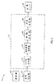
次に、図3および図4を参照されたい。図3および図4は、システム15と、消化管内における血液の内容または色による識別可能な任意の他の病理を確認するために、システム15を用いるステップを示すフロー図とを示す。システム15は、照明源42’、画像受信機12’、データ処理機14’および画像モニタ18’を含む。データ処理機14’は、スペクトル分析器22、適応基準構築器24、距離計算機26および判定計算機28を含む。この発明の一実施形態によると、データ処理機14’は、標準コンピュータアクセラレータボード、高性能コンピュータ、マルチプロセッサ、または他の任意の直列もしくは並列の高性能処理機器である。画像モニタ18’は、ビデオ表示装置、グラフ、表、または任意の他の表示器であってよい。
Reference is now made to FIGS. 3 and 4 show the
図4のステップは、図3のシステム15を用いて達成することができる。一実施形態において、画像はカプセル内で取り込まれて処理される。別の実施形態において、画像は生体内システムによって取り込まれて遠隔地に送信され、そこで処理される。画像受信機12’は、図1の生体内カメラシステムまた任意の他の生体内イメージャによって取り込まれた画像を受信する(ステップ101)。データ処理機14’は、色の画像を画素の格子に分割する(ステップ102)。他の画像化の適用例におけるように、画素の数が画像の解像度を決定する。このことを論じるために、画像は8×8の画素からなるブロック(i,j)に分割される。一実施形態において、本来の画像が256×256画素の画像であるために、8画素に分割して色の成分を判定した結果、32×32×3の行列の色の成分
値のブロックとなる。スペクトル分析器22は、各ブロックの色の成分、すなわち、各画像に対する色相Hi,j、彩度Si,j、および輝度値Vi,jを計算する(ステップ104)。
The steps of FIG. 4 can be accomplished using the
スペクトル分析器は、また、病理サンプルBおよび健康な基準組織Rのブロックの色の成分も計算する(ステップ105およびステップ106〜110)。スペクトル分析器22は、血液を含む既知の画像から、病理サンプルBのブロックの色の成分を計算する(ステップ105)。
The spectrum analyzer also calculates the color components of the block of pathology sample B and healthy reference tissue R (
次に、図5を参照されたい。図5は、図4の適応的に基準を構築するステップ106〜110の概略図である。適応基準構築器24は、健康な組織の基準サンプルを構築するために、組織の基準となる色の成分を計算する(ステップ106〜110)。適応的手法は、後の画像に現れる健康な組織を平均化することに基づく。平均値が用いられるのは、GI管に沿った健康な組織のパラメータが変化し得るためである。適応基準構築器24は、明度V(輝度)および色相Hに基づいてブロックを選択する(ステップ107)。一実施形態において、それらの条件は、0.1<Vi,j<0.9および0<Hi,j<0.09である。これらの条件は、健康な組織が存在することを示す。図5に示されるように、健康な組織の領域Ri、Ri-1、およびRi-2を伴った画像Pi、Pi-1、およびPi-2が得られる。適応基準構築器24は、GI管に沿って得られた画像Pi、Pi-1、およびPi-2の健康な領域Ri、Ri-1、およびRi-2(すなわち、選択されたブロック)の色の成分を平均化する(ステップ108)。データを平滑化して、特定の画像への感度を除去するために、適応基準構築器24は、現在の画像Piおよび以前の画像Pi-1の組織の色の平均をフィルタ処理する(ステップ110)。
Reference is now made to FIG. FIG. 5 is a schematic diagram of steps 106-110 for constructing the criteria of FIG. 4 adaptively. The
一実施形態において、以下の反復計算を伴った無限インパルス応答(Infinite Impulse
Response)(IIR)フィルタが用いられる。
In one embodiment, an infinite impulse response (Infinite Impulse) with the following iteration:
Response) (IIR) filters are used.

式中、tiは現在のフレームiの時間指数を表わし、ti-1は以前のフレームi−1の時間指数を表わす。 Wherein, t i represents the time index of the current frame i, t i-1 represents the previous frame i-1 of the time index.
再び図4を参照すると、次に、距離計算機26が、行列内の各ブロックと血液の基準値Bとの間のユークリッド(Euclidian)距離を計算する(ステップ112)。血液の基準値Bは、血液を含む既知の画像から得られ、上で述べたように、スペクトル分析器22によって分析される。別の実施形態において、他の異常な色を示すために、比色分析の異なった基準値を用いることもできる。例示的な実施形態において、この計算の結果は、32×32の要素βi,jの行列である。この計算は、以下の式に従って行なわれる。
Referring again to FIG. 4, the
式中、Hb、Sb、およびVbは、それぞれ血液の色相、彩度および輝度に対する基準値で
ある。
In the formula, H b , S b , and V b are reference values for the hue, saturation, and luminance of blood, respectively.
同様の距離の計算が、適応基準となる組織(健康な組織)の色の成分に対して計算され、その結果、以下のように32×32の行列Ii,jが生じる。 A similar distance calculation is calculated for the color component of the adaptation reference tissue (healthy tissue), resulting in a 32 × 32 matrix I i, j as follows:
式中、Ht、St、およびVtは、それぞれ健康な組織の色相、彩度および輝度に対する基準値である。 Where H t , S t , and V t are reference values for the hue, saturation, and brightness of healthy tissue, respectively.
距離の行列が得られると、判定計算機28が、以下の式に従って確率指標関数Λを計算する(ステップ116)。
When the distance matrix is obtained, the
しきい値は、任意の値に設定することができる。好ましい実施形態において、しきい値は以下のとおりである。すなわち、BloodThreshold=0.15およびTissueRatioThreshold=4である。血液は、Λ>0であれば存在する。 The threshold value can be set to an arbitrary value. In the preferred embodiment, the thresholds are: That is, BloodThreshold = 0.15 and TissueRatioThreshold = 4. Blood is present if Λ> 0.
最後に、画像モニタ18’が、血液の存在を示すカラービデオとして、または、レベルおよび/またはしきい値を示すグラフまたは表として、のいずれかにおいて結果を表示する(ステップ118)。 Finally, the image monitor 18 'displays the results either as a color video indicating the presence of blood or as a graph or table indicating the level and / or threshold (step 118).
結果の表示は、位置表示器を組込むことを含んでよく、エンドユーザは色の変化がGI管内に存在するか、または他の体腔内に存在するかを判定することができる。したがって、医師はこの問題となる領域に処置を行なうことができる。 The resulting display may include incorporating a position indicator so that the end user can determine whether the color change is in the GI tract or in another body cavity. Therefore, the doctor can take action on the problem area.
当業者は、この発明が、上で特に示され説明された内容に限定されないことを認識されるであろう。むしろ、この発明の範囲は、前掲の請求項によってのみ規定される。 Those skilled in the art will recognize that the present invention is not limited to what has been particularly shown and described above. Rather, the scope of the present invention is defined only by the appended claims.
Claims (8)
嚥下可能生体内デバイスから生体内画像のセットを受けとり、該画像に基づいてカラーコンテントの解析を行い、前画像内の色変化を判定するデータプロセッサと、
カラービデオとして前記画像を表示するとともに、さらに血液の存在と相関を有する前記色変化について胃腸管の位置を表示する表示器とを備えていることを特徴とするシステム。A system for displaying in vivo information,
A data processor that receives a set of in-vivo images from a swallowable in-vivo device, analyzes color content based on the images, and determines a color change in the previous image;
When displaying the image as a color video together, system characterized in that it comprises a display for displaying the position of the gastrointestinal tract for the color change, further comprising a correlation with the presence of blood.
Applications Claiming Priority (2)
| Application Number | Priority Date | Filing Date | Title |
|---|---|---|---|
| US27548601P | 2001-03-14 | 2001-03-14 | |
| PCT/IL2002/000210 WO2002073507A2 (en) | 2001-03-14 | 2002-03-14 | Method and system for detecting colorimetric abnormalities |
Related Child Applications (1)
| Application Number | Title | Priority Date | Filing Date |
|---|---|---|---|
| JP2006160078A Division JP4504951B2 (en) | 2001-03-14 | 2006-06-08 | Method and system for detecting colorimetric anomalies in vivo |
Publications (3)
| Publication Number | Publication Date |
|---|---|
| JP2004521693A JP2004521693A (en) | 2004-07-22 |
| JP2004521693A5 JP2004521693A5 (en) | 2007-04-05 |
| JP4067407B2 true JP4067407B2 (en) | 2008-03-26 |
Family
ID=23052498
Family Applications (2)
| Application Number | Title | Priority Date | Filing Date |
|---|---|---|---|
| JP2002572089A Expired - Fee Related JP4067407B2 (en) | 2001-03-14 | 2002-03-14 | Method and system for detecting colorimetric anomalies in vivo |
| JP2006160078A Expired - Lifetime JP4504951B2 (en) | 2001-03-14 | 2006-06-08 | Method and system for detecting colorimetric anomalies in vivo |
Family Applications After (1)
| Application Number | Title | Priority Date | Filing Date |
|---|---|---|---|
| JP2006160078A Expired - Lifetime JP4504951B2 (en) | 2001-03-14 | 2006-06-08 | Method and system for detecting colorimetric anomalies in vivo |
Country Status (9)
| Country | Link |
|---|---|
| US (4) | US20020177779A1 (en) |
| EP (1) | EP1372474B1 (en) |
| JP (2) | JP4067407B2 (en) |
| CN (2) | CN100469308C (en) |
| AT (1) | ATE509328T1 (en) |
| AU (1) | AU2002241215A1 (en) |
| ES (1) | ES2365696T3 (en) |
| IL (1) | IL157892A0 (en) |
| WO (1) | WO2002073507A2 (en) |
Cited By (2)
| Publication number | Priority date | Publication date | Assignee | Title |
|---|---|---|---|---|
| US9324145B1 (en) | 2013-08-08 | 2016-04-26 | Given Imaging Ltd. | System and method for detection of transitions in an image stream of the gastrointestinal tract |
| JP7040910B2 (en) | 2017-10-11 | 2022-03-23 | 聡 織田 | Ultrasonic probe |
Families Citing this family (189)
| Publication number | Priority date | Publication date | Assignee | Title |
|---|---|---|---|---|
| IL126727A (en) | 1998-10-22 | 2006-12-31 | Given Imaging Ltd | Method for delivering a device to a target location |
| US8636648B2 (en) | 1999-03-01 | 2014-01-28 | West View Research, Llc | Endoscopic smart probe |
| US10973397B2 (en) * | 1999-03-01 | 2021-04-13 | West View Research, Llc | Computerized information collection and processing apparatus |
| WO2001053792A2 (en) * | 2000-01-19 | 2001-07-26 | Given Imaging Ltd. | A system for detecting substances |
| WO2002073507A2 (en) * | 2001-03-14 | 2002-09-19 | Given Imaging Ltd. | Method and system for detecting colorimetric abnormalities |
| DE60228266D1 (en) | 2001-06-18 | 2008-09-25 | Given Imaging Ltd | SWITCHABLE IN VIVO CAPSULE WITH A RIGID AND FLEXIBLE SECTION CIRCUIT BOARD |
| US7160258B2 (en) * | 2001-06-26 | 2007-01-09 | Entrack, Inc. | Capsule and method for treating or diagnosing the intestinal tract |
| US20050187433A1 (en) * | 2001-07-26 | 2005-08-25 | Given Imaging Ltd. | In-vivo imaging device providing constant bit rate transmission |
| US20030043263A1 (en) * | 2001-07-26 | 2003-03-06 | Arkady Glukhovsky | Diagnostic device using data compression |
| US9113846B2 (en) * | 2001-07-26 | 2015-08-25 | Given Imaging Ltd. | In-vivo imaging device providing data compression |
| US6951536B2 (en) * | 2001-07-30 | 2005-10-04 | Olympus Corporation | Capsule-type medical device and medical system |
| US8428685B2 (en) * | 2001-09-05 | 2013-04-23 | Given Imaging Ltd. | System and method for magnetically maneuvering an in vivo device |
| JP2005501630A (en) * | 2001-09-05 | 2005-01-20 | ギブン・イメージング・リミテッド | System and method for three-dimensional display of body lumen |
| JP3756797B2 (en) | 2001-10-16 | 2006-03-15 | オリンパス株式会社 | Capsule type medical equipment |
| JP4234605B2 (en) * | 2002-02-12 | 2009-03-04 | ギブン イメージング リミテッド | System and method for displaying an image stream |
| US8022980B2 (en) * | 2002-02-12 | 2011-09-20 | Given Imaging Ltd. | System and method for displaying an image stream |
| US7474327B2 (en) * | 2002-02-12 | 2009-01-06 | Given Imaging Ltd. | System and method for displaying an image stream |
| WO2003094723A1 (en) * | 2002-05-09 | 2003-11-20 | Given Imaging Ltd. | System and method for in vivo sensing |
| US7662094B2 (en) | 2002-05-14 | 2010-02-16 | Given Imaging Ltd. | Optical head assembly with dome, and device for use thereof |
| WO2004014227A1 (en) | 2002-08-13 | 2004-02-19 | Given Imaging Ltd. | System for in vivo sampling and analysis |
| WO2004028336A2 (en) | 2002-09-30 | 2004-04-08 | Given Imaging Ltd. | Reduced size imaging device |
| US8449452B2 (en) | 2002-09-30 | 2013-05-28 | Given Imaging Ltd. | In-vivo sensing system |
| JP4746876B2 (en) | 2002-10-15 | 2011-08-10 | ギブン イメージング リミテッド | Apparatus, system and method for transferring a signal to a mobile device |
| US7195588B2 (en) * | 2004-03-01 | 2007-03-27 | Olympus Corporation | Endoscope image pick-up apparatus |
| AU2003282373A1 (en) * | 2002-11-29 | 2004-06-23 | Given Imaging Ltd. | Methods device and system for in vivo diagnosis |
| AU2003288517A1 (en) | 2002-12-26 | 2004-07-22 | Given Imaging Ltd. | In vivo imaging device and method of manufacture thereof |
| US7833151B2 (en) | 2002-12-26 | 2010-11-16 | Given Imaging Ltd. | In vivo imaging device with two imagers |
| AU2003288516A1 (en) | 2002-12-26 | 2004-07-22 | Given Imaging Ltd. | Immobilizable in vivo sensing device |
| IL155175A (en) * | 2003-03-31 | 2012-01-31 | Given Imaging Ltd | Diagnostic device using data compression |
| JP4547401B2 (en) * | 2003-04-25 | 2010-09-22 | オリンパス株式会社 | Image display device, image display method, and image display program |
| JP4547402B2 (en) * | 2003-04-25 | 2010-09-22 | オリンパス株式会社 | Image display device, image display method, and image display program |
| JP4554647B2 (en) * | 2003-04-25 | 2010-09-29 | オリンパス株式会社 | Image display device, image display method, and image display program |
| JP3810381B2 (en) * | 2003-04-25 | 2006-08-16 | オリンパス株式会社 | Image display device, image display method, and image display program |
| CN100431475C (en) * | 2003-04-25 | 2008-11-12 | 奥林巴斯株式会社 | Device, method and program for image processing |
| JP4493386B2 (en) * | 2003-04-25 | 2010-06-30 | オリンパス株式会社 | Image display device, image display method, and image display program |
| ATE553690T1 (en) * | 2003-05-01 | 2012-05-15 | Given Imaging Ltd | PANORAMA FIELD OF VIEW DISPLAY DEVICE |
| IL162740A (en) | 2003-06-26 | 2010-06-16 | Given Imaging Ltd | Device, method and system for reduced transmission imaging |
| JP4656825B2 (en) * | 2003-09-08 | 2011-03-23 | オリンパス株式会社 | In-subject introduction apparatus and wireless in-subject information acquisition system |
| US7604589B2 (en) * | 2003-10-01 | 2009-10-20 | Given Imaging, Ltd. | Device, system and method for determining orientation of in-vivo devices |
| US20050075537A1 (en) * | 2003-10-06 | 2005-04-07 | Eastman Kodak Company | Method and system for real-time automatic abnormality detection for in vivo images |
| WO2005039399A1 (en) * | 2003-10-27 | 2005-05-06 | Olympus Corporation | Image processing device, image processing method, and image processing program |
| JP4574983B2 (en) * | 2003-11-04 | 2010-11-04 | オリンパス株式会社 | Image display apparatus, image display method, and image display program |
| WO2005053518A1 (en) | 2003-12-05 | 2005-06-16 | Olympus Corporation | Display processing device |
| US20050137468A1 (en) * | 2003-12-18 | 2005-06-23 | Jerome Avron | Device, system, and method for in-vivo sensing of a substance |
| US7647090B1 (en) | 2003-12-30 | 2010-01-12 | Given Imaging, Ltd. | In-vivo sensing device and method for producing same |
| WO2005062717A2 (en) | 2003-12-31 | 2005-07-14 | Given Imaging Ltd. | In-vivo sensing device with detachable part |
| JP5248780B2 (en) * | 2003-12-31 | 2013-07-31 | ギブン イメージング リミテッド | System and method for displaying an image stream |
| JP4652694B2 (en) * | 2004-01-08 | 2011-03-16 | オリンパス株式会社 | Image processing method |
| WO2005069887A2 (en) * | 2004-01-16 | 2005-08-04 | The City College Of The University Of New York | Micro-scale compact device for in vivo medical diagnosis combining optical imaging and point fluorescence spectroscopy |
| US20050196023A1 (en) * | 2004-03-01 | 2005-09-08 | Eastman Kodak Company | Method for real-time remote diagnosis of in vivo images |
| US7623690B2 (en) * | 2004-03-30 | 2009-11-24 | Carestream Health, Inc. | System and method for classifying in vivo images according to anatomical structure |
| US7605852B2 (en) | 2004-05-17 | 2009-10-20 | Micron Technology, Inc. | Real-time exposure control for automatic light control |
| US7938775B2 (en) * | 2004-06-28 | 2011-05-10 | Given Imaging, Ltd. | Device, system, and method for in-vivo analysis |
| WO2006005075A2 (en) * | 2004-06-30 | 2006-01-12 | Amir Belson | Apparatus and methods for capsule endoscopy of the esophagus |
| US7336833B2 (en) * | 2004-06-30 | 2008-02-26 | Given Imaging, Ltd. | Device, system, and method for reducing image data captured in-vivo |
| US8500630B2 (en) | 2004-06-30 | 2013-08-06 | Given Imaging Ltd. | In vivo device with flexible circuit board and method for assembly thereof |
| US7643865B2 (en) | 2004-06-30 | 2010-01-05 | Given Imaging Ltd. | Autonomous in-vivo device |
| US7596403B2 (en) | 2004-06-30 | 2009-09-29 | Given Imaging Ltd. | System and method for determining path lengths through a body lumen |
| JP4885432B2 (en) * | 2004-08-18 | 2012-02-29 | オリンパス株式会社 | Image display device, image display method, and image display program |
| US7986337B2 (en) * | 2004-09-27 | 2011-07-26 | Given Imaging Ltd. | System and method for editing an image stream captured in vivo |
| AU2005229684A1 (en) * | 2004-11-04 | 2006-05-18 | Given Imaging Ltd | Apparatus and method for receiving device selection and combining |
| US7486981B2 (en) * | 2004-11-15 | 2009-02-03 | Given Imaging Ltd. | System and method for displaying an image stream |
| KR100891766B1 (en) | 2004-12-10 | 2009-04-07 | 올림푸스 가부시키가이샤 | Medical image processing apparatus |
| US8738106B2 (en) * | 2005-01-31 | 2014-05-27 | Given Imaging, Ltd | Device, system and method for in vivo analysis |
| EP1849402B1 (en) * | 2005-02-15 | 2018-05-16 | Olympus Corporation | Medical image processing device, lumen image processing device, lumen image processing method, and programs for them |
| US20060217593A1 (en) * | 2005-03-24 | 2006-09-28 | Zvika Gilad | Device, system and method of panoramic multiple field of view imaging |
| US20090216082A1 (en) * | 2005-04-01 | 2009-08-27 | Elisha Rabinovitz | Device, System and Method for In Vivo Magnetic Immunoassay Analysis |
| IL174531A0 (en) * | 2005-04-06 | 2006-08-20 | Given Imaging Ltd | System and method for performing capsule endoscopy diagnosis in remote sites |
| WO2006112227A1 (en) | 2005-04-13 | 2006-10-26 | Olympus Medical Systems Corp. | Image processing device and method |
| US7907775B2 (en) | 2005-04-27 | 2011-03-15 | Olympus Medical Systems Corp. | Image processing apparatus, image processing method and image processing program |
| JP4418400B2 (en) * | 2005-05-20 | 2010-02-17 | オリンパスメディカルシステムズ株式会社 | Image display device |
| JP4767591B2 (en) * | 2005-06-01 | 2011-09-07 | オリンパスメディカルシステムズ株式会社 | Endoscope diagnosis support method, endoscope diagnosis support device, and endoscope diagnosis support program |
| ATE523862T1 (en) * | 2005-09-09 | 2011-09-15 | Given Imaging Ltd | SIMULTANEOUS TRANSFER AND PROCESSING AND REAL-TIME VIEWING OF IN-VIVO IMAGES |
| WO2007031946A2 (en) * | 2005-09-12 | 2007-03-22 | Dvp Technologies Ltd. | Medical image processing |
| US20070066875A1 (en) * | 2005-09-18 | 2007-03-22 | Eli Horn | System and method for identification of images in an image database |
| US8423123B2 (en) * | 2005-09-30 | 2013-04-16 | Given Imaging Ltd. | System and method for in-vivo feature detection |
| US7482593B2 (en) * | 2005-10-20 | 2009-01-27 | The Research Foundation Of State University Of New York | Method to determine the depth-of-interaction function for PET detectors |
| EP2412301B1 (en) | 2005-12-28 | 2014-04-23 | Olympus Medical Systems Corp. | Image processing device and image processing method in image processing device |
| US20070167834A1 (en) * | 2005-12-29 | 2007-07-19 | Amit Pascal | In-vivo imaging optical device and method |
| US20070156051A1 (en) * | 2005-12-29 | 2007-07-05 | Amit Pascal | Device and method for in-vivo illumination |
| US9320417B2 (en) | 2005-12-29 | 2016-04-26 | Given Imaging Ltd. | In-vivo optical imaging device with backscatter blocking |
| EP1997076B1 (en) * | 2006-03-13 | 2010-11-10 | Given Imaging Ltd. | Cascade analysis for intestinal contraction detection |
| WO2007105213A2 (en) * | 2006-03-13 | 2007-09-20 | Given Imaging Ltd. | Device, system and method for automatic detection of contractile activity in an image frame |
| IL182332A (en) * | 2006-03-31 | 2013-04-30 | Given Imaging Ltd | System and method for assessing a patient condition |
| ATE531199T1 (en) * | 2006-04-03 | 2011-11-15 | Given Imaging Ltd | DEVICE, SYSTEM AND METHOD FOR IN VIVO ANALYSIS |
| DE202006006268U1 (en) * | 2006-04-12 | 2006-06-14 | Branofilter Gmbh | Device for detachable fastening of dust filter bag in dust evacuation equipment has flange part which is pluggable to adaptor plate radially outside of annular seal and is pivotally connected to adaptor plate |
| JP2007319442A (en) * | 2006-06-01 | 2007-12-13 | Fujifilm Corp | Capsule endoscope system and image processing unit |
| US8335362B2 (en) * | 2006-06-12 | 2012-12-18 | Given Imaging Ltd. | Device, system and method for measurement and analysis of contractile activity |
| WO2008007172A1 (en) * | 2006-07-11 | 2008-01-17 | Stanley Kim | Test preparation device |
| JP4912787B2 (en) | 2006-08-08 | 2012-04-11 | オリンパスメディカルシステムズ株式会社 | Medical image processing apparatus and method of operating medical image processing apparatus |
| US8588887B2 (en) | 2006-09-06 | 2013-11-19 | Innurvation, Inc. | Ingestible low power sensor device and system for communicating with same |
| US8615284B2 (en) | 2006-09-06 | 2013-12-24 | Innurvation, Inc. | Method for acoustic information exchange involving an ingestible low power capsule |
| JP5121204B2 (en) | 2006-10-11 | 2013-01-16 | オリンパス株式会社 | Image processing apparatus, image processing method, and image processing program |
| WO2008102803A1 (en) * | 2007-02-22 | 2008-08-28 | Olympus Medical Systems Corp. | Intrasubject introduction system |
| WO2008139812A1 (en) | 2007-05-08 | 2008-11-20 | Olympus Corporation | Image processing device and image processing program |
| EP2149332B1 (en) * | 2007-05-17 | 2014-12-17 | Olympus Medical Systems Corp. | Image information display processing device and display processing method |
| JP2008295490A (en) | 2007-05-29 | 2008-12-11 | Olympus Medical Systems Corp | Capsule endoscope image display device |
| JP5106928B2 (en) | 2007-06-14 | 2012-12-26 | オリンパス株式会社 | Image processing apparatus and image processing program |
| JP5403880B2 (en) * | 2007-06-27 | 2014-01-29 | オリンパスメディカルシステムズ株式会社 | Image information display processing device |
| US9197470B2 (en) | 2007-10-05 | 2015-11-24 | Innurvation, Inc. | Data transmission via multi-path channels using orthogonal multi-frequency signals with differential phase shift keying modulation |
| US20090105532A1 (en) * | 2007-10-22 | 2009-04-23 | Zvika Gilad | In vivo imaging device and method of manufacturing thereof |
| US20100329520A2 (en) * | 2007-11-08 | 2010-12-30 | Olympus Medical Systems Corp. | Method and System for Correlating Image and Tissue Characteristic Data |
| US9017248B2 (en) | 2007-11-08 | 2015-04-28 | Olympus Medical Systems Corp. | Capsule blood detection system and method |
| US9131847B2 (en) | 2007-11-08 | 2015-09-15 | Olympus Corporation | Method and apparatus for detecting abnormal living tissue |
| WO2009060460A2 (en) * | 2007-11-09 | 2009-05-14 | Given Imaging Ltd. | Apparatus and methods for capsule endoscopy of the esophagus |
| EP2057934B1 (en) * | 2007-11-12 | 2017-01-04 | Novineon Healthcare Technology Partners Gmbh | Device for hemorrhage detection |
| US8529441B2 (en) | 2008-02-12 | 2013-09-10 | Innurvation, Inc. | Ingestible endoscopic optical scanning device |
| US20100016662A1 (en) * | 2008-02-21 | 2010-01-21 | Innurvation, Inc. | Radial Scanner Imaging System |
| JP5336749B2 (en) * | 2008-03-24 | 2013-11-06 | オリンパス株式会社 | Capsule type medical device and operation method thereof |
| US8515507B2 (en) | 2008-06-16 | 2013-08-20 | Given Imaging Ltd. | Device and method for detecting in-vivo pathology |
| US8888680B2 (en) | 2008-07-07 | 2014-11-18 | Olympus Medical Systems Corp. | Method and apparatus for foreign matter detection for blood content sensors |
| WO2010005571A2 (en) | 2008-07-09 | 2010-01-14 | Innurvation, Inc. | Displaying image data from a scanner capsule |
| JP5117353B2 (en) * | 2008-11-07 | 2013-01-16 | オリンパス株式会社 | Image processing apparatus, image processing program, and image processing method |
| US8516691B2 (en) | 2009-06-24 | 2013-08-27 | Given Imaging Ltd. | Method of assembly of an in vivo imaging device with a flexible circuit board |
| US20110097690A1 (en) * | 2009-10-22 | 2011-04-28 | Perry Franklin Samuel-Cutts | Training system for surfing and method of use |
| US9192353B2 (en) * | 2009-10-27 | 2015-11-24 | Innurvation, Inc. | Data transmission via wide band acoustic channels |
| US8945010B2 (en) | 2009-12-23 | 2015-02-03 | Covidien Lp | Method of evaluating constipation using an ingestible capsule |
| US8446465B2 (en) * | 2010-01-05 | 2013-05-21 | Given Imaging Ltd. | System and method for displaying an image stream captured in-vivo |
| US10300296B2 (en) | 2010-03-17 | 2019-05-28 | Photopill Medical Ltd. | Capsule phototherapy |
| US8682142B1 (en) | 2010-03-18 | 2014-03-25 | Given Imaging Ltd. | System and method for editing an image stream captured in-vivo |
| US8647259B2 (en) | 2010-03-26 | 2014-02-11 | Innurvation, Inc. | Ultrasound scanning capsule endoscope (USCE) |
| US9060673B2 (en) | 2010-04-28 | 2015-06-23 | Given Imaging Ltd. | System and method for displaying portions of in-vivo images |
| JP5622461B2 (en) * | 2010-07-07 | 2014-11-12 | オリンパス株式会社 | Image processing apparatus, image processing method, and image processing program |
| WO2012063623A1 (en) * | 2010-11-08 | 2012-05-18 | オリンパスメディカルシステムズ株式会社 | Image display apparatus and capsule endoscopy system |
| US8913807B1 (en) | 2010-12-30 | 2014-12-16 | Given Imaging Ltd. | System and method for detecting anomalies in a tissue imaged in-vivo |
| US8873816B1 (en) | 2011-04-06 | 2014-10-28 | Given Imaging Ltd. | Method and system for identification of red colored pathologies in vivo |
| US20140055400A1 (en) | 2011-05-23 | 2014-02-27 | Haworth, Inc. | Digital workspace ergonomics apparatuses, methods and systems |
| WO2012162411A1 (en) | 2011-05-23 | 2012-11-29 | Haworth, Inc. | Digital whiteboard collaboration apparatuses, methods and systems |
| US8929629B1 (en) * | 2011-06-29 | 2015-01-06 | Given Imaging Ltd. | Method and system for image-based ulcer detection |
| US8897523B2 (en) | 2011-07-09 | 2014-11-25 | Gauss Surgical | System and method for counting surgical samples |
| US10426356B2 (en) | 2011-07-09 | 2019-10-01 | Gauss Surgical, Inc. | Method for estimating a quantity of a blood component in a fluid receiver and corresponding error |
| US9047663B2 (en) | 2011-07-09 | 2015-06-02 | Gauss Surgical | Method for triggering blood salvage |
| US9870625B2 (en) | 2011-07-09 | 2018-01-16 | Gauss Surgical, Inc. | Method for estimating a quantity of a blood component in a fluid receiver and corresponding error |
| US9646375B2 (en) | 2011-07-09 | 2017-05-09 | Gauss Surgical, Inc. | Method for setting a blood transfusion parameter |
| JP5851160B2 (en) | 2011-08-31 | 2016-02-03 | オリンパス株式会社 | Image processing apparatus, operation method of image processing apparatus, and image processing program |
| JP6042817B2 (en) * | 2011-10-06 | 2016-12-14 | オリンパス株式会社 | Fluorescence observation equipment |
| JP2015509744A (en) * | 2011-12-15 | 2015-04-02 | ギブン イメージング リミテッドGiven Imaging Ltd. | Apparatus, system and method for in vivo detection of bleeding in the gastrointestinal tract |
| US8923585B1 (en) * | 2012-01-31 | 2014-12-30 | Given Imaging Ltd. | Method and system for image-based ulcer detection |
| US9854970B2 (en) * | 2012-02-21 | 2018-01-02 | Massachusetts Eye & Ear Infirmary | Calculating conjunctival redness |
| WO2013173356A1 (en) | 2012-05-14 | 2013-11-21 | Gauss Surgical | System and methods for managing blood loss of a patient |
| CN104662559B (en) | 2012-05-14 | 2019-02-05 | 高斯外科公司 | System and method for estimating the amount of the blood constituent in liquid tank |
| US9479549B2 (en) | 2012-05-23 | 2016-10-25 | Haworth, Inc. | Collaboration system with whiteboard with federated display |
| US9479548B2 (en) | 2012-05-23 | 2016-10-25 | Haworth, Inc. | Collaboration system with whiteboard access to global collaboration data |
| JP6067264B2 (en) | 2012-07-17 | 2017-01-25 | Hoya株式会社 | Image processing apparatus and endoscope apparatus |
| JP6027803B2 (en) * | 2012-07-17 | 2016-11-16 | Hoya株式会社 | Image processing apparatus and endoscope apparatus |
| KR102201603B1 (en) * | 2012-07-25 | 2021-01-12 | 인튜어티브 서지컬 오퍼레이션즈 인코포레이티드 | Efficient and interactive bleeding detection in a surgical system |
| JP2014140335A (en) * | 2013-01-24 | 2014-08-07 | Dainippon Printing Co Ltd | Medium image analyzing device, medium information registration system and program, and hygiene management system |
| US10304037B2 (en) | 2013-02-04 | 2019-05-28 | Haworth, Inc. | Collaboration system including a spatial event map |
| US11861561B2 (en) | 2013-02-04 | 2024-01-02 | Haworth, Inc. | Collaboration system including a spatial event map |
| JP6097629B2 (en) * | 2013-04-26 | 2017-03-15 | Hoya株式会社 | Lesion evaluation information generator |
| WO2015029033A1 (en) | 2013-08-29 | 2015-03-05 | Given Imaging Ltd. | System and method for maneuvering coils power optimization |
| US9430706B1 (en) | 2013-10-02 | 2016-08-30 | Given Imaging Ltd. | System and method for detection of in-vivo pathology sequences |
| US8736685B1 (en) * | 2013-12-11 | 2014-05-27 | Anritsu Company | Systems and methods for measuring brightness response of a camera operating in automatic exposure mode |
| EP3080998B1 (en) | 2013-12-11 | 2023-08-02 | Given Imaging Ltd. | System and method for controlling the display of an image stream |
| WO2015160997A1 (en) | 2014-04-15 | 2015-10-22 | Gauss Surgical, Inc. | Method for estimating a quantity of a blood component in a fluid canister |
| EP3132253B1 (en) | 2014-04-15 | 2019-02-13 | Gauss Surgical, Inc. | Method for estimating a quantity of a blood component in a fluid canister |
| ES2893541T3 (en) | 2014-05-02 | 2022-02-09 | Massachusetts Eye & Ear Infirmary | Qualification of corneal fluorescein staining |
| US9633276B2 (en) * | 2014-07-14 | 2017-04-25 | Sony Corporation | Blood detection system with real-time capability and method of operation thereof |
| EP3197336B1 (en) | 2014-09-25 | 2020-12-23 | Progenity, Inc. | Electromechanical pill device with localization capabilities |
| CN104658014B (en) * | 2015-02-10 | 2017-09-22 | 重庆金山科技(集团)有限公司 | A kind of method for detecting colorimetric abnormality in organism |
| EP3292524B1 (en) | 2015-05-06 | 2020-07-08 | Haworth, Inc. | Virtual workspace viewport follow mode in collaboration systems |
| US10555675B2 (en) | 2015-05-15 | 2020-02-11 | Gauss Surgical, Inc. | Method for projecting blood loss of a patient during a surgery |
| WO2016187071A1 (en) | 2015-05-15 | 2016-11-24 | Gauss Surgical, Inc. | Systems and methods for assessing fluids from a patient |
| WO2016187072A1 (en) | 2015-05-15 | 2016-11-24 | Gauss Surgical, Inc. | Methods and systems for characterizing fluids from a patient |
| JP6113386B1 (en) | 2015-08-13 | 2017-04-12 | Hoya株式会社 | Evaluation value calculation apparatus and electronic endoscope system |
| CN106687023B (en) | 2015-08-13 | 2018-12-18 | Hoya株式会社 | Evaluate value calculation apparatus and electronic endoscope system |
| US10572997B2 (en) | 2015-12-18 | 2020-02-25 | Given Imaging Ltd. | System and method for detecting anomalies in an image captured in-vivo using color histogram association |
| WO2017112913A1 (en) | 2015-12-23 | 2017-06-29 | Gauss Surgical, Inc. | System and method for estimating an amount of a blood component in a volume of fluid |
| JP6934253B2 (en) | 2015-12-23 | 2021-09-15 | ガウス サージカル, インコーポレイテッドGauss Surgical, Inc. | How to assess the amount of blood components in a surgical fabric |
| TWI616180B (en) * | 2016-06-29 | 2018-03-01 | 國立成功大學 | Upper gastrointestinal bleeding monitoring system |
| CN109890299A (en) * | 2016-08-18 | 2019-06-14 | 普罗根尼蒂公司 | Sampling system and associated materials and method |
| JP2019526423A (en) | 2016-09-09 | 2019-09-19 | ミッチェル・ローレンス・ジョーンズMitchell Lawrence JONES | Electromechanically ingestible device for delivery of dispenseable substances |
| WO2018106931A1 (en) | 2016-12-07 | 2018-06-14 | Progenity Inc. | Gastrointestinal tract detection methods, devices and systems |
| JP7268879B2 (en) | 2017-01-02 | 2023-05-08 | ガウス サージカル,インコーポレイテッド | Tracking Surgical Items Predicting Duplicate Imaging |
| KR101875004B1 (en) | 2017-01-04 | 2018-07-05 | 금오공과대학교 산학협력단 | Automated bleeding detection method and computer program in wireless capsule endoscopy videos |
| US11229368B2 (en) | 2017-01-13 | 2022-01-25 | Gauss Surgical, Inc. | Fluid loss estimation based on weight of medical items |
| CA3055762A1 (en) | 2017-03-31 | 2018-10-04 | Progenity, Inc. | Localization systems and methods for an ingestible device |
| US11126325B2 (en) | 2017-10-23 | 2021-09-21 | Haworth, Inc. | Virtual workspace including shared viewport markers in a collaboration system |
| US11934637B2 (en) | 2017-10-23 | 2024-03-19 | Haworth, Inc. | Collaboration system including markers identifying multiple canvases in multiple shared virtual workspaces |
| WO2019164277A1 (en) * | 2018-02-20 | 2019-08-29 | (주)휴톰 | Method and device for evaluating bleeding by using surgical image |
| US20210267475A1 (en) * | 2018-07-25 | 2021-09-02 | Check-Cap Ltd. | System and method for polyp detection through capsule dynamics |
| EP3883635A1 (en) | 2018-11-19 | 2021-09-29 | Progenity, Inc. | Methods and devices for treating a disease with biotherapeutics |
| JP7238390B2 (en) * | 2018-12-21 | 2023-03-14 | セイコーエプソン株式会社 | Information system and identification method |
| WO2020176517A1 (en) | 2019-02-25 | 2020-09-03 | Haworth, Inc. | Gesture based workflows in a collaboration system |
| US11707610B2 (en) | 2019-12-13 | 2023-07-25 | Biora Therapeutics, Inc. | Ingestible device for delivery of therapeutic agent to the gastrointestinal tract |
| US11750672B2 (en) | 2020-05-07 | 2023-09-05 | Haworth, Inc. | Digital workspace sharing over one or more display clients in proximity of a main client |
| US11212127B2 (en) | 2020-05-07 | 2021-12-28 | Haworth, Inc. | Digital workspace sharing over one or more display clients and authorization protocols for collaboration systems |
| JP7461221B2 (en) * | 2020-05-25 | 2024-04-03 | 富士フイルムヘルスケア株式会社 | Medical image processing device and medical imaging device |
| CN112991325B (en) * | 2021-04-14 | 2021-08-17 | 上海孚慈医疗科技有限公司 | Intelligent coding-based speckled red-emitting image acquisition and processing method and system |
Family Cites Families (28)
| Publication number | Priority date | Publication date | Assignee | Title |
|---|---|---|---|---|
| US3971362A (en) * | 1972-10-27 | 1976-07-27 | The United States Of America As Represented By The Administrator Of The National Aeronautics And Space Administration | Miniature ingestible telemeter devices to measure deep-body temperature |
| JPS5519124A (en) * | 1978-07-27 | 1980-02-09 | Olympus Optical Co | Camera system for medical treatment |
| US5993378A (en) * | 1980-10-28 | 1999-11-30 | Lemelson; Jerome H. | Electro-optical instruments and methods for treating disease |
| JPS59188301A (en) * | 1983-04-08 | 1984-10-25 | Japanese National Railways<Jnr> | Controller of electric railcar |
| US5042494A (en) * | 1985-11-13 | 1991-08-27 | Alfano Robert R | Method and apparatus for detecting cancerous tissue using luminescence excitation spectra |
| US4689621A (en) * | 1986-03-31 | 1987-08-25 | The United States Of America As Represented By The Administrator Of The National Aeronautics And Space Administration | Temperature responsive transmitter |
| US4844076A (en) * | 1988-08-26 | 1989-07-04 | The Johns Hopkins University | Ingestible size continuously transmitting temperature monitoring pill |
| US5279607A (en) * | 1991-05-30 | 1994-01-18 | The State University Of New York | Telemetry capsule and process |
| JP3285235B2 (en) * | 1992-11-05 | 2002-05-27 | オリンパス光学工業株式会社 | Capsule device for in vivo observation |
| US6095989A (en) * | 1993-07-20 | 2000-08-01 | Hay; Sam H. | Optical recognition methods for locating eyes |
| IL108352A (en) * | 1994-01-17 | 2000-02-29 | Given Imaging Ltd | In vivo video camera system |
| CA2145232A1 (en) * | 1994-03-24 | 1995-09-25 | Arie Avny | Viewing method and apparatus particularly useful for viewing the interior of the large intestine |
| US5590660A (en) * | 1994-03-28 | 1997-01-07 | Xillix Technologies Corp. | Apparatus and method for imaging diseased tissue using integrated autofluorescence |
| DE69630935T2 (en) * | 1995-09-29 | 2004-11-04 | Koninklijke Philips Electronics N.V. | Image processing method and apparatus for automatically detecting areas of a predetermined type of cancer in an intensity image |
| US5833603A (en) * | 1996-03-13 | 1998-11-10 | Lipomatrix, Inc. | Implantable biosensing transponder |
| US6459919B1 (en) * | 1997-08-26 | 2002-10-01 | Color Kinetics, Incorporated | Precision illumination methods and systems |
| US6016038A (en) * | 1997-08-26 | 2000-01-18 | Color Kinetics, Inc. | Multicolored LED lighting method and apparatus |
| US6422994B1 (en) * | 1997-09-24 | 2002-07-23 | Olympus Optical Co., Ltd. | Fluorescent diagnostic system and method providing color discrimination enhancement |
| US6240312B1 (en) * | 1997-10-23 | 2001-05-29 | Robert R. Alfano | Remote-controllable, micro-scale device for use in in vivo medical diagnosis and/or treatment |
| GB9810771D0 (en) * | 1998-05-19 | 1998-07-15 | Active Silicon Limited | Method of detecting colours |
| DE19844618A1 (en) | 1998-09-29 | 2000-03-30 | Zahnradfabrik Friedrichshafen | Method for reducing the thermal load on an automatic transmission for a motor vehicle in an emergency mode |
| IL126727A (en) | 1998-10-22 | 2006-12-31 | Given Imaging Ltd | Method for delivering a device to a target location |
| JP2001037718A (en) | 1999-05-26 | 2001-02-13 | Olympus Optical Co Ltd | Image diagnostic device and endoscope device |
| AU6178900A (en) * | 1999-07-26 | 2001-02-13 | Haim Shani | An improved method and apparatus for the detection of medical conditions of shock and pre-shock |
| US7039453B2 (en) * | 2000-02-08 | 2006-05-02 | Tarun Mullick | Miniature ingestible capsule |
| IL155045A0 (en) * | 2000-09-27 | 2003-10-31 | Given Imaging Ltd | An immobilizable in vivo sensing device |
| US6632175B1 (en) * | 2000-11-08 | 2003-10-14 | Hewlett-Packard Development Company, L.P. | Swallowable data recorder capsule medical device |
| WO2002073507A2 (en) * | 2001-03-14 | 2002-09-19 | Given Imaging Ltd. | Method and system for detecting colorimetric abnormalities |
-
2002
- 2002-03-14 WO PCT/IL2002/000210 patent/WO2002073507A2/en active Search and Examination
- 2002-03-14 JP JP2002572089A patent/JP4067407B2/en not_active Expired - Fee Related
- 2002-03-14 EP EP02707063A patent/EP1372474B1/en not_active Expired - Lifetime
- 2002-03-14 AU AU2002241215A patent/AU2002241215A1/en not_active Abandoned
- 2002-03-14 IL IL15789202A patent/IL157892A0/en unknown
- 2002-03-14 CN CNB028098560A patent/CN100469308C/en not_active Expired - Lifetime
- 2002-03-14 US US10/097,096 patent/US20020177779A1/en not_active Abandoned
- 2002-03-14 ES ES02707063T patent/ES2365696T3/en not_active Expired - Lifetime
- 2002-03-14 CN CN200810125961XA patent/CN101305906B/en not_active Expired - Lifetime
- 2002-03-14 AT AT02707063T patent/ATE509328T1/en not_active IP Right Cessation
-
2006
- 2006-06-08 JP JP2006160078A patent/JP4504951B2/en not_active Expired - Lifetime
-
2012
- 2012-07-03 US US13/541,111 patent/US8626268B2/en not_active Expired - Fee Related
-
2013
- 2013-10-04 US US14/046,258 patent/US8918164B2/en not_active Expired - Fee Related
-
2014
- 2014-11-17 US US14/543,154 patent/US9364139B2/en not_active Expired - Lifetime
Cited By (2)
| Publication number | Priority date | Publication date | Assignee | Title |
|---|---|---|---|---|
| US9324145B1 (en) | 2013-08-08 | 2016-04-26 | Given Imaging Ltd. | System and method for detection of transitions in an image stream of the gastrointestinal tract |
| JP7040910B2 (en) | 2017-10-11 | 2022-03-23 | 聡 織田 | Ultrasonic probe |
Also Published As
| Publication number | Publication date |
|---|---|
| US20150141782A1 (en) | 2015-05-21 |
| WO2002073507A9 (en) | 2003-01-23 |
| US20120275683A1 (en) | 2012-11-01 |
| AU2002241215A1 (en) | 2002-09-24 |
| CN100469308C (en) | 2009-03-18 |
| EP1372474A4 (en) | 2007-02-28 |
| US9364139B2 (en) | 2016-06-14 |
| WO2002073507A3 (en) | 2003-10-23 |
| CN101305906B (en) | 2012-02-08 |
| JP2004521693A (en) | 2004-07-22 |
| WO2002073507A2 (en) | 2002-09-19 |
| US8918164B2 (en) | 2014-12-23 |
| EP1372474A2 (en) | 2004-01-02 |
| JP2006297118A (en) | 2006-11-02 |
| US8626268B2 (en) | 2014-01-07 |
| JP4504951B2 (en) | 2010-07-14 |
| US20140039287A1 (en) | 2014-02-06 |
| CN101305906A (en) | 2008-11-19 |
| EP1372474B1 (en) | 2011-05-11 |
| ATE509328T1 (en) | 2011-05-15 |
| US20020177779A1 (en) | 2002-11-28 |
| ES2365696T3 (en) | 2011-10-10 |
| IL157892A0 (en) | 2004-03-28 |
| CN1509152A (en) | 2004-06-30 |
Similar Documents
| Publication | Publication Date | Title |
|---|---|---|
| JP4067407B2 (en) | Method and system for detecting colorimetric anomalies in vivo | |
| JP4629143B2 (en) | System for detecting contents in vivo | |
| CN110325100B (en) | Endoscope system and method of operating the same | |
| US7567692B2 (en) | System and method for detecting content in-vivo | |
| US10251538B2 (en) | Endoscope system and method for controlling the same | |
| US7319781B2 (en) | Method and system for multiple passes diagnostic alignment for in vivo images | |
| US8639011B2 (en) | Fluoroscopy apparatus | |
| JP2004236952A (en) | Electronic endoscope apparatus | |
| JPWO2019220848A1 (en) | Endoscope device, endoscope operation method, and program | |
| US11642005B2 (en) | Endoscope system, endoscope image processing method, and storage medium | |
| WO2020090729A1 (en) | Medical image processing apparatus, medical image processing method and program, and diagnosis assisting apparatus | |
| JP5581237B2 (en) | Image processing device, processor device for electronic endoscope, operation method of image processing device, and computer program for image processing | |
| JP2001037718A (en) | Image diagnostic device and endoscope device | |
| JP4855709B2 (en) | Image processing apparatus, image processing method, and image processing program | |
| US10646102B2 (en) | Processor for electronic endoscope, and electronic endoscope system | |
| US20220237795A1 (en) | Image processing device and method of operating the same | |
| JP2013048646A (en) | Diagnostic system | |
| JP2003220019A (en) | Image diagnostic equipment and endoscopic equipment | |
| WO2022230607A1 (en) | Medical image processing device, endoscope system, and operation method for medical image processing device | |
| US20230360298A1 (en) | Endoscope processor, endoscope apparatus, and diagnostic image display method | |
| JP2023106327A (en) | Inspection support device, inspection support method, and inspection support program | |
| JP2003204925A (en) | Image diagnostic device and endoscope system |
Legal Events
| Date | Code | Title | Description |
|---|---|---|---|
| A621 | Written request for application examination |
Free format text: JAPANESE INTERMEDIATE CODE: A621 Effective date: 20050307 |
|
| A521 | Request for written amendment filed |
Free format text: JAPANESE INTERMEDIATE CODE: A523 Effective date: 20050330 |
|
| A521 | Request for written amendment filed |
Free format text: JAPANESE INTERMEDIATE CODE: A523 Effective date: 20060608 |
|
| RD02 | Notification of acceptance of power of attorney |
Free format text: JAPANESE INTERMEDIATE CODE: A7422 Effective date: 20061129 |
|
| RD03 | Notification of appointment of power of attorney |
Free format text: JAPANESE INTERMEDIATE CODE: A7423 Effective date: 20061129 |
|
| A521 | Request for written amendment filed |
Free format text: JAPANESE INTERMEDIATE CODE: A821 Effective date: 20061129 |
|
| A521 | Request for written amendment filed |
Free format text: JAPANESE INTERMEDIATE CODE: A523 Effective date: 20070216 |
|
| A131 | Notification of reasons for refusal |
Free format text: JAPANESE INTERMEDIATE CODE: A131 Effective date: 20070618 |
|
| A601 | Written request for extension of time |
Free format text: JAPANESE INTERMEDIATE CODE: A601 Effective date: 20070918 |
|
| A602 | Written permission of extension of time |
Free format text: JAPANESE INTERMEDIATE CODE: A602 Effective date: 20070926 |
|
| A601 | Written request for extension of time |
Free format text: JAPANESE INTERMEDIATE CODE: A601 Effective date: 20071018 |
|
| A602 | Written permission of extension of time |
Free format text: JAPANESE INTERMEDIATE CODE: A602 Effective date: 20071025 |
|
| A521 | Request for written amendment filed |
Free format text: JAPANESE INTERMEDIATE CODE: A523 Effective date: 20071119 |
|
| TRDD | Decision of grant or rejection written | ||
| A01 | Written decision to grant a patent or to grant a registration (utility model) |
Free format text: JAPANESE INTERMEDIATE CODE: A01 Effective date: 20071217 |
|
| A61 | First payment of annual fees (during grant procedure) |
Free format text: JAPANESE INTERMEDIATE CODE: A61 Effective date: 20080108 |
|
| FPAY | Renewal fee payment (event date is renewal date of database) |
Free format text: PAYMENT UNTIL: 20110118 Year of fee payment: 3 |
|
| R150 | Certificate of patent or registration of utility model |
Free format text: JAPANESE INTERMEDIATE CODE: R150 Ref document number: 4067407 Country of ref document: JP Free format text: JAPANESE INTERMEDIATE CODE: R150 |
|
| FPAY | Renewal fee payment (event date is renewal date of database) |
Free format text: PAYMENT UNTIL: 20110118 Year of fee payment: 3 |
|
| FPAY | Renewal fee payment (event date is renewal date of database) |
Free format text: PAYMENT UNTIL: 20120118 Year of fee payment: 4 |
|
| R250 | Receipt of annual fees |
Free format text: JAPANESE INTERMEDIATE CODE: R250 |
|
| FPAY | Renewal fee payment (event date is renewal date of database) |
Free format text: PAYMENT UNTIL: 20130118 Year of fee payment: 5 |
|
| R250 | Receipt of annual fees |
Free format text: JAPANESE INTERMEDIATE CODE: R250 |
|
| R250 | Receipt of annual fees |
Free format text: JAPANESE INTERMEDIATE CODE: R250 |
|
| R250 | Receipt of annual fees |
Free format text: JAPANESE INTERMEDIATE CODE: R250 |
|
| R250 | Receipt of annual fees |
Free format text: JAPANESE INTERMEDIATE CODE: R250 |
|
| R250 | Receipt of annual fees |
Free format text: JAPANESE INTERMEDIATE CODE: R250 |
|
| R250 | Receipt of annual fees |
Free format text: JAPANESE INTERMEDIATE CODE: R250 |
|
| R250 | Receipt of annual fees |
Free format text: JAPANESE INTERMEDIATE CODE: R250 |
|
| R250 | Receipt of annual fees |
Free format text: JAPANESE INTERMEDIATE CODE: R250 |
|
| R250 | Receipt of annual fees |
Free format text: JAPANESE INTERMEDIATE CODE: R250 |
|
| R250 | Receipt of annual fees |
Free format text: JAPANESE INTERMEDIATE CODE: R250 |
|
| LAPS | Cancellation because of no payment of annual fees |