JP4054373B2 - Flk-1 is a receptor for vascular endothelial growth factor - Google Patents
Flk-1 is a receptor for vascular endothelial growth factor Download PDFInfo
- Publication number
- JP4054373B2 JP4054373B2 JP51171994A JP51171994A JP4054373B2 JP 4054373 B2 JP4054373 B2 JP 4054373B2 JP 51171994 A JP51171994 A JP 51171994A JP 51171994 A JP51171994 A JP 51171994A JP 4054373 B2 JP4054373 B2 JP 4054373B2
- Authority
- JP
- Japan
- Prior art keywords
- flk
- vegf
- cells
- expression
- receptor
- Prior art date
- Legal status (The legal status is an assumption and is not a legal conclusion. Google has not performed a legal analysis and makes no representation as to the accuracy of the status listed.)
- Expired - Lifetime
Links
Images


Classifications
-
- G—PHYSICS
- G01—MEASURING; TESTING
- G01N—INVESTIGATING OR ANALYSING MATERIALS BY DETERMINING THEIR CHEMICAL OR PHYSICAL PROPERTIES
- G01N33/00—Investigating or analysing materials by specific methods not covered by groups G01N1/00 - G01N31/00
- G01N33/48—Biological material, e.g. blood, urine; Haemocytometers
- G01N33/50—Chemical analysis of biological material, e.g. blood, urine; Testing involving biospecific ligand binding methods; Immunological testing
- G01N33/5005—Chemical analysis of biological material, e.g. blood, urine; Testing involving biospecific ligand binding methods; Immunological testing involving human or animal cells
- G01N33/5008—Chemical analysis of biological material, e.g. blood, urine; Testing involving biospecific ligand binding methods; Immunological testing involving human or animal cells for testing or evaluating the effect of chemical or biological compounds, e.g. drugs, cosmetics
- G01N33/5044—Chemical analysis of biological material, e.g. blood, urine; Testing involving biospecific ligand binding methods; Immunological testing involving human or animal cells for testing or evaluating the effect of chemical or biological compounds, e.g. drugs, cosmetics involving specific cell types
- G01N33/5064—Endothelial cells
-
- A—HUMAN NECESSITIES
- A61—MEDICAL OR VETERINARY SCIENCE; HYGIENE
- A61K—PREPARATIONS FOR MEDICAL, DENTAL OR TOILETRY PURPOSES
- A61K31/00—Medicinal preparations containing organic active ingredients
- A61K31/21—Esters, e.g. nitroglycerine, selenocyanates
- A61K31/215—Esters, e.g. nitroglycerine, selenocyanates of carboxylic acids
- A61K31/235—Esters, e.g. nitroglycerine, selenocyanates of carboxylic acids having an aromatic ring attached to a carboxyl group
-
- A—HUMAN NECESSITIES
- A61—MEDICAL OR VETERINARY SCIENCE; HYGIENE
- A61K—PREPARATIONS FOR MEDICAL, DENTAL OR TOILETRY PURPOSES
- A61K31/00—Medicinal preparations containing organic active ingredients
- A61K31/275—Nitriles; Isonitriles
-
- A—HUMAN NECESSITIES
- A61—MEDICAL OR VETERINARY SCIENCE; HYGIENE
- A61K—PREPARATIONS FOR MEDICAL, DENTAL OR TOILETRY PURPOSES
- A61K31/00—Medicinal preparations containing organic active ingredients
- A61K31/275—Nitriles; Isonitriles
- A61K31/277—Nitriles; Isonitriles having a ring, e.g. verapamil
-
- A—HUMAN NECESSITIES
- A61—MEDICAL OR VETERINARY SCIENCE; HYGIENE
- A61K—PREPARATIONS FOR MEDICAL, DENTAL OR TOILETRY PURPOSES
- A61K31/00—Medicinal preparations containing organic active ingredients
- A61K31/33—Heterocyclic compounds
- A61K31/38—Heterocyclic compounds having sulfur as a ring hetero atom
-
- A—HUMAN NECESSITIES
- A61—MEDICAL OR VETERINARY SCIENCE; HYGIENE
- A61K—PREPARATIONS FOR MEDICAL, DENTAL OR TOILETRY PURPOSES
- A61K31/00—Medicinal preparations containing organic active ingredients
- A61K31/33—Heterocyclic compounds
- A61K31/395—Heterocyclic compounds having nitrogen as a ring hetero atom, e.g. guanethidine or rifamycins
- A61K31/40—Heterocyclic compounds having nitrogen as a ring hetero atom, e.g. guanethidine or rifamycins having five-membered rings with one nitrogen as the only ring hetero atom, e.g. sulpiride, succinimide, tolmetin, buflomedil
-
- A—HUMAN NECESSITIES
- A61—MEDICAL OR VETERINARY SCIENCE; HYGIENE
- A61K—PREPARATIONS FOR MEDICAL, DENTAL OR TOILETRY PURPOSES
- A61K31/00—Medicinal preparations containing organic active ingredients
- A61K31/33—Heterocyclic compounds
- A61K31/395—Heterocyclic compounds having nitrogen as a ring hetero atom, e.g. guanethidine or rifamycins
- A61K31/41—Heterocyclic compounds having nitrogen as a ring hetero atom, e.g. guanethidine or rifamycins having five-membered rings with two or more ring hetero atoms, at least one of which being nitrogen, e.g. tetrazole
- A61K31/415—1,2-Diazoles
-
- A—HUMAN NECESSITIES
- A61—MEDICAL OR VETERINARY SCIENCE; HYGIENE
- A61K—PREPARATIONS FOR MEDICAL, DENTAL OR TOILETRY PURPOSES
- A61K31/00—Medicinal preparations containing organic active ingredients
- A61K31/33—Heterocyclic compounds
- A61K31/395—Heterocyclic compounds having nitrogen as a ring hetero atom, e.g. guanethidine or rifamycins
- A61K31/41—Heterocyclic compounds having nitrogen as a ring hetero atom, e.g. guanethidine or rifamycins having five-membered rings with two or more ring hetero atoms, at least one of which being nitrogen, e.g. tetrazole
- A61K31/42—Oxazoles
-
- A—HUMAN NECESSITIES
- A61—MEDICAL OR VETERINARY SCIENCE; HYGIENE
- A61K—PREPARATIONS FOR MEDICAL, DENTAL OR TOILETRY PURPOSES
- A61K31/00—Medicinal preparations containing organic active ingredients
- A61K31/33—Heterocyclic compounds
- A61K31/395—Heterocyclic compounds having nitrogen as a ring hetero atom, e.g. guanethidine or rifamycins
- A61K31/495—Heterocyclic compounds having nitrogen as a ring hetero atom, e.g. guanethidine or rifamycins having six-membered rings with two or more nitrogen atoms as the only ring heteroatoms, e.g. piperazine or tetrazines
- A61K31/50—Pyridazines; Hydrogenated pyridazines
- A61K31/502—Pyridazines; Hydrogenated pyridazines ortho- or peri-condensed with carbocyclic ring systems, e.g. cinnoline, phthalazine
-
- A—HUMAN NECESSITIES
- A61—MEDICAL OR VETERINARY SCIENCE; HYGIENE
- A61K—PREPARATIONS FOR MEDICAL, DENTAL OR TOILETRY PURPOSES
- A61K31/00—Medicinal preparations containing organic active ingredients
- A61K31/33—Heterocyclic compounds
- A61K31/395—Heterocyclic compounds having nitrogen as a ring hetero atom, e.g. guanethidine or rifamycins
- A61K31/495—Heterocyclic compounds having nitrogen as a ring hetero atom, e.g. guanethidine or rifamycins having six-membered rings with two or more nitrogen atoms as the only ring heteroatoms, e.g. piperazine or tetrazines
- A61K31/505—Pyrimidines; Hydrogenated pyrimidines, e.g. trimethoprim
- A61K31/517—Pyrimidines; Hydrogenated pyrimidines, e.g. trimethoprim ortho- or peri-condensed with carbocyclic ring systems, e.g. quinazoline, perimidine
-
- A—HUMAN NECESSITIES
- A61—MEDICAL OR VETERINARY SCIENCE; HYGIENE
- A61K—PREPARATIONS FOR MEDICAL, DENTAL OR TOILETRY PURPOSES
- A61K31/00—Medicinal preparations containing organic active ingredients
- A61K31/33—Heterocyclic compounds
- A61K31/395—Heterocyclic compounds having nitrogen as a ring hetero atom, e.g. guanethidine or rifamycins
- A61K31/535—Heterocyclic compounds having nitrogen as a ring hetero atom, e.g. guanethidine or rifamycins having six-membered rings with at least one nitrogen and one oxygen as the ring hetero atoms, e.g. 1,2-oxazines
-
- A—HUMAN NECESSITIES
- A61—MEDICAL OR VETERINARY SCIENCE; HYGIENE
- A61P—SPECIFIC THERAPEUTIC ACTIVITY OF CHEMICAL COMPOUNDS OR MEDICINAL PREPARATIONS
- A61P35/00—Antineoplastic agents
-
- A—HUMAN NECESSITIES
- A61—MEDICAL OR VETERINARY SCIENCE; HYGIENE
- A61P—SPECIFIC THERAPEUTIC ACTIVITY OF CHEMICAL COMPOUNDS OR MEDICINAL PREPARATIONS
- A61P43/00—Drugs for specific purposes, not provided for in groups A61P1/00-A61P41/00
-
- C—CHEMISTRY; METALLURGY
- C07—ORGANIC CHEMISTRY
- C07C—ACYCLIC OR CARBOCYCLIC COMPOUNDS
- C07C229/00—Compounds containing amino and carboxyl groups bound to the same carbon skeleton
- C07C229/52—Compounds containing amino and carboxyl groups bound to the same carbon skeleton having amino and carboxyl groups bound to carbon atoms of six-membered aromatic rings of the same carbon skeleton
- C07C229/54—Compounds containing amino and carboxyl groups bound to the same carbon skeleton having amino and carboxyl groups bound to carbon atoms of six-membered aromatic rings of the same carbon skeleton with amino and carboxyl groups bound to carbon atoms of the same non-condensed six-membered aromatic ring
- C07C229/60—Compounds containing amino and carboxyl groups bound to the same carbon skeleton having amino and carboxyl groups bound to carbon atoms of six-membered aromatic rings of the same carbon skeleton with amino and carboxyl groups bound to carbon atoms of the same non-condensed six-membered aromatic ring with amino and carboxyl groups bound in meta- or para- positions
-
- C—CHEMISTRY; METALLURGY
- C07—ORGANIC CHEMISTRY
- C07C—ACYCLIC OR CARBOCYCLIC COMPOUNDS
- C07C255/00—Carboxylic acid nitriles
- C07C255/01—Carboxylic acid nitriles having cyano groups bound to acyclic carbon atoms
- C07C255/32—Carboxylic acid nitriles having cyano groups bound to acyclic carbon atoms having cyano groups bound to acyclic carbon atoms of a carbon skeleton containing at least one six-membered aromatic ring
- C07C255/36—Carboxylic acid nitriles having cyano groups bound to acyclic carbon atoms having cyano groups bound to acyclic carbon atoms of a carbon skeleton containing at least one six-membered aromatic ring the carbon skeleton being further substituted by hydroxy groups
-
- C—CHEMISTRY; METALLURGY
- C07—ORGANIC CHEMISTRY
- C07C—ACYCLIC OR CARBOCYCLIC COMPOUNDS
- C07C255/00—Carboxylic acid nitriles
- C07C255/01—Carboxylic acid nitriles having cyano groups bound to acyclic carbon atoms
- C07C255/32—Carboxylic acid nitriles having cyano groups bound to acyclic carbon atoms having cyano groups bound to acyclic carbon atoms of a carbon skeleton containing at least one six-membered aromatic ring
- C07C255/40—Carboxylic acid nitriles having cyano groups bound to acyclic carbon atoms having cyano groups bound to acyclic carbon atoms of a carbon skeleton containing at least one six-membered aromatic ring the carbon skeleton being further substituted by doubly-bound oxygen atoms
-
- C—CHEMISTRY; METALLURGY
- C07—ORGANIC CHEMISTRY
- C07C—ACYCLIC OR CARBOCYCLIC COMPOUNDS
- C07C255/00—Carboxylic acid nitriles
- C07C255/01—Carboxylic acid nitriles having cyano groups bound to acyclic carbon atoms
- C07C255/32—Carboxylic acid nitriles having cyano groups bound to acyclic carbon atoms having cyano groups bound to acyclic carbon atoms of a carbon skeleton containing at least one six-membered aromatic ring
- C07C255/41—Carboxylic acid nitriles having cyano groups bound to acyclic carbon atoms having cyano groups bound to acyclic carbon atoms of a carbon skeleton containing at least one six-membered aromatic ring the carbon skeleton being further substituted by carboxyl groups, other than cyano groups
-
- C—CHEMISTRY; METALLURGY
- C07—ORGANIC CHEMISTRY
- C07C—ACYCLIC OR CARBOCYCLIC COMPOUNDS
- C07C255/00—Carboxylic acid nitriles
- C07C255/63—Carboxylic acid nitriles containing cyano groups and nitrogen atoms further bound to other hetero atoms, other than oxygen atoms of nitro or nitroso groups, bound to the same carbon skeleton
- C07C255/65—Carboxylic acid nitriles containing cyano groups and nitrogen atoms further bound to other hetero atoms, other than oxygen atoms of nitro or nitroso groups, bound to the same carbon skeleton with the nitrogen atoms further bound to nitrogen atoms
- C07C255/66—Carboxylic acid nitriles containing cyano groups and nitrogen atoms further bound to other hetero atoms, other than oxygen atoms of nitro or nitroso groups, bound to the same carbon skeleton with the nitrogen atoms further bound to nitrogen atoms having cyano groups and nitrogen atoms being part of hydrazine or hydrazone groups bound to the same carbon skeleton
-
- C—CHEMISTRY; METALLURGY
- C07—ORGANIC CHEMISTRY
- C07C—ACYCLIC OR CARBOCYCLIC COMPOUNDS
- C07C317/00—Sulfones; Sulfoxides
- C07C317/44—Sulfones; Sulfoxides having sulfone or sulfoxide groups and carboxyl groups bound to the same carbon skeleton
- C07C317/46—Sulfones; Sulfoxides having sulfone or sulfoxide groups and carboxyl groups bound to the same carbon skeleton the carbon skeleton being further substituted by singly-bound oxygen atoms
-
- C—CHEMISTRY; METALLURGY
- C07—ORGANIC CHEMISTRY
- C07C—ACYCLIC OR CARBOCYCLIC COMPOUNDS
- C07C327/00—Thiocarboxylic acids
- C07C327/38—Amides of thiocarboxylic acids
- C07C327/40—Amides of thiocarboxylic acids having carbon atoms of thiocarboxamide groups bound to hydrogen atoms or to acyclic carbon atoms
- C07C327/44—Amides of thiocarboxylic acids having carbon atoms of thiocarboxamide groups bound to hydrogen atoms or to acyclic carbon atoms to carbon atoms of an unsaturated carbon skeleton
-
- C—CHEMISTRY; METALLURGY
- C07—ORGANIC CHEMISTRY
- C07D—HETEROCYCLIC COMPOUNDS
- C07D209/00—Heterocyclic compounds containing five-membered rings, condensed with other rings, with one nitrogen atom as the only ring hetero atom
- C07D209/02—Heterocyclic compounds containing five-membered rings, condensed with other rings, with one nitrogen atom as the only ring hetero atom condensed with one carbocyclic ring
- C07D209/04—Indoles; Hydrogenated indoles
- C07D209/10—Indoles; Hydrogenated indoles with substituted hydrocarbon radicals attached to carbon atoms of the hetero ring
- C07D209/18—Radicals substituted by carbon atoms having three bonds to hetero atoms with at the most one bond to halogen, e.g. ester or nitrile radicals
-
- C—CHEMISTRY; METALLURGY
- C07—ORGANIC CHEMISTRY
- C07D—HETEROCYCLIC COMPOUNDS
- C07D239/00—Heterocyclic compounds containing 1,3-diazine or hydrogenated 1,3-diazine rings
- C07D239/70—Heterocyclic compounds containing 1,3-diazine or hydrogenated 1,3-diazine rings condensed with carbocyclic rings or ring systems
- C07D239/72—Quinazolines; Hydrogenated quinazolines
- C07D239/86—Quinazolines; Hydrogenated quinazolines with hetero atoms directly attached in position 4
- C07D239/93—Sulfur atoms
-
- C—CHEMISTRY; METALLURGY
- C07—ORGANIC CHEMISTRY
- C07D—HETEROCYCLIC COMPOUNDS
- C07D239/00—Heterocyclic compounds containing 1,3-diazine or hydrogenated 1,3-diazine rings
- C07D239/70—Heterocyclic compounds containing 1,3-diazine or hydrogenated 1,3-diazine rings condensed with carbocyclic rings or ring systems
- C07D239/72—Quinazolines; Hydrogenated quinazolines
- C07D239/86—Quinazolines; Hydrogenated quinazolines with hetero atoms directly attached in position 4
- C07D239/94—Nitrogen atoms
-
- C—CHEMISTRY; METALLURGY
- C07—ORGANIC CHEMISTRY
- C07D—HETEROCYCLIC COMPOUNDS
- C07D241/00—Heterocyclic compounds containing 1,4-diazine or hydrogenated 1,4-diazine rings
- C07D241/36—Heterocyclic compounds containing 1,4-diazine or hydrogenated 1,4-diazine rings condensed with carbocyclic rings or ring systems
- C07D241/38—Heterocyclic compounds containing 1,4-diazine or hydrogenated 1,4-diazine rings condensed with carbocyclic rings or ring systems with only hydrogen or carbon atoms directly attached to the ring nitrogen atoms
- C07D241/40—Benzopyrazines
- C07D241/42—Benzopyrazines with only hydrogen atoms, hydrocarbon or substituted hydrocarbon radicals, directly attached to carbon atoms of the hetero ring
-
- C—CHEMISTRY; METALLURGY
- C07—ORGANIC CHEMISTRY
- C07D—HETEROCYCLIC COMPOUNDS
- C07D241/00—Heterocyclic compounds containing 1,4-diazine or hydrogenated 1,4-diazine rings
- C07D241/36—Heterocyclic compounds containing 1,4-diazine or hydrogenated 1,4-diazine rings condensed with carbocyclic rings or ring systems
- C07D241/38—Heterocyclic compounds containing 1,4-diazine or hydrogenated 1,4-diazine rings condensed with carbocyclic rings or ring systems with only hydrogen or carbon atoms directly attached to the ring nitrogen atoms
- C07D241/40—Benzopyrazines
- C07D241/44—Benzopyrazines with hetero atoms or with carbon atoms having three bonds to hetero atoms with at the most one bond to halogen, e.g. ester or nitrile radicals, directly attached to carbon atoms of the hetero ring
-
- C—CHEMISTRY; METALLURGY
- C07—ORGANIC CHEMISTRY
- C07D—HETEROCYCLIC COMPOUNDS
- C07D487/00—Heterocyclic compounds containing nitrogen atoms as the only ring hetero atoms in the condensed system, not provided for by groups C07D451/00 - C07D477/00
- C07D487/02—Heterocyclic compounds containing nitrogen atoms as the only ring hetero atoms in the condensed system, not provided for by groups C07D451/00 - C07D477/00 in which the condensed system contains two hetero rings
- C07D487/04—Ortho-condensed systems
-
- C—CHEMISTRY; METALLURGY
- C07—ORGANIC CHEMISTRY
- C07K—PEPTIDES
- C07K14/00—Peptides having more than 20 amino acids; Gastrins; Somatostatins; Melanotropins; Derivatives thereof
- C07K14/435—Peptides having more than 20 amino acids; Gastrins; Somatostatins; Melanotropins; Derivatives thereof from animals; from humans
- C07K14/705—Receptors; Cell surface antigens; Cell surface determinants
- C07K14/71—Receptors; Cell surface antigens; Cell surface determinants for growth factors; for growth regulators
-
- C—CHEMISTRY; METALLURGY
- C07—ORGANIC CHEMISTRY
- C07K—PEPTIDES
- C07K16/00—Immunoglobulins [IGs], e.g. monoclonal or polyclonal antibodies
- C07K16/18—Immunoglobulins [IGs], e.g. monoclonal or polyclonal antibodies against material from animals or humans
- C07K16/28—Immunoglobulins [IGs], e.g. monoclonal or polyclonal antibodies against material from animals or humans against receptors, cell surface antigens or cell surface determinants
- C07K16/2863—Immunoglobulins [IGs], e.g. monoclonal or polyclonal antibodies against material from animals or humans against receptors, cell surface antigens or cell surface determinants against receptors for growth factors, growth regulators
-
- G—PHYSICS
- G01—MEASURING; TESTING
- G01N—INVESTIGATING OR ANALYSING MATERIALS BY DETERMINING THEIR CHEMICAL OR PHYSICAL PROPERTIES
- G01N33/00—Investigating or analysing materials by specific methods not covered by groups G01N1/00 - G01N31/00
- G01N33/48—Biological material, e.g. blood, urine; Haemocytometers
- G01N33/50—Chemical analysis of biological material, e.g. blood, urine; Testing involving biospecific ligand binding methods; Immunological testing
- G01N33/5005—Chemical analysis of biological material, e.g. blood, urine; Testing involving biospecific ligand binding methods; Immunological testing involving human or animal cells
- G01N33/5008—Chemical analysis of biological material, e.g. blood, urine; Testing involving biospecific ligand binding methods; Immunological testing involving human or animal cells for testing or evaluating the effect of chemical or biological compounds, e.g. drugs, cosmetics
- G01N33/5011—Chemical analysis of biological material, e.g. blood, urine; Testing involving biospecific ligand binding methods; Immunological testing involving human or animal cells for testing or evaluating the effect of chemical or biological compounds, e.g. drugs, cosmetics for testing antineoplastic activity
-
- G—PHYSICS
- G01—MEASURING; TESTING
- G01N—INVESTIGATING OR ANALYSING MATERIALS BY DETERMINING THEIR CHEMICAL OR PHYSICAL PROPERTIES
- G01N33/00—Investigating or analysing materials by specific methods not covered by groups G01N1/00 - G01N31/00
- G01N33/48—Biological material, e.g. blood, urine; Haemocytometers
- G01N33/50—Chemical analysis of biological material, e.g. blood, urine; Testing involving biospecific ligand binding methods; Immunological testing
- G01N33/5005—Chemical analysis of biological material, e.g. blood, urine; Testing involving biospecific ligand binding methods; Immunological testing involving human or animal cells
- G01N33/5008—Chemical analysis of biological material, e.g. blood, urine; Testing involving biospecific ligand binding methods; Immunological testing involving human or animal cells for testing or evaluating the effect of chemical or biological compounds, e.g. drugs, cosmetics
- G01N33/502—Chemical analysis of biological material, e.g. blood, urine; Testing involving biospecific ligand binding methods; Immunological testing involving human or animal cells for testing or evaluating the effect of chemical or biological compounds, e.g. drugs, cosmetics for testing non-proliferative effects
-
- G—PHYSICS
- G01—MEASURING; TESTING
- G01N—INVESTIGATING OR ANALYSING MATERIALS BY DETERMINING THEIR CHEMICAL OR PHYSICAL PROPERTIES
- G01N33/00—Investigating or analysing materials by specific methods not covered by groups G01N1/00 - G01N31/00
- G01N33/48—Biological material, e.g. blood, urine; Haemocytometers
- G01N33/50—Chemical analysis of biological material, e.g. blood, urine; Testing involving biospecific ligand binding methods; Immunological testing
- G01N33/68—Chemical analysis of biological material, e.g. blood, urine; Testing involving biospecific ligand binding methods; Immunological testing involving proteins, peptides or amino acids
- G01N33/6893—Chemical analysis of biological material, e.g. blood, urine; Testing involving biospecific ligand binding methods; Immunological testing involving proteins, peptides or amino acids related to diseases not provided for elsewhere
-
- A—HUMAN NECESSITIES
- A61—MEDICAL OR VETERINARY SCIENCE; HYGIENE
- A61K—PREPARATIONS FOR MEDICAL, DENTAL OR TOILETRY PURPOSES
- A61K38/00—Medicinal preparations containing peptides
-
- C—CHEMISTRY; METALLURGY
- C07—ORGANIC CHEMISTRY
- C07K—PEPTIDES
- C07K2319/00—Fusion polypeptide
- C07K2319/32—Fusion polypeptide fusions with soluble part of a cell surface receptor, "decoy receptors"
-
- G—PHYSICS
- G01—MEASURING; TESTING
- G01N—INVESTIGATING OR ANALYSING MATERIALS BY DETERMINING THEIR CHEMICAL OR PHYSICAL PROPERTIES
- G01N2333/00—Assays involving biological materials from specific organisms or of a specific nature
- G01N2333/435—Assays involving biological materials from specific organisms or of a specific nature from animals; from humans
- G01N2333/705—Assays involving receptors, cell surface antigens or cell surface determinants
- G01N2333/71—Assays involving receptors, cell surface antigens or cell surface determinants for growth factors; for growth regulators
-
- G—PHYSICS
- G01—MEASURING; TESTING
- G01N—INVESTIGATING OR ANALYSING MATERIALS BY DETERMINING THEIR CHEMICAL OR PHYSICAL PROPERTIES
- G01N2500/00—Screening for compounds of potential therapeutic value
- G01N2500/04—Screening involving studying the effect of compounds C directly on molecule A (e.g. C are potential ligands for a receptor A, or potential substrates for an enzyme A)
Landscapes
- Health & Medical Sciences (AREA)
- Chemical & Material Sciences (AREA)
- Organic Chemistry (AREA)
- Life Sciences & Earth Sciences (AREA)
- General Health & Medical Sciences (AREA)
- Medicinal Chemistry (AREA)
- Engineering & Computer Science (AREA)
- Immunology (AREA)
- Veterinary Medicine (AREA)
- Pharmacology & Pharmacy (AREA)
- Animal Behavior & Ethology (AREA)
- Public Health (AREA)
- Biomedical Technology (AREA)
- Epidemiology (AREA)
- Molecular Biology (AREA)
- Urology & Nephrology (AREA)
- Hematology (AREA)
- Cell Biology (AREA)
- Biochemistry (AREA)
- Microbiology (AREA)
- Pathology (AREA)
- Toxicology (AREA)
- Biotechnology (AREA)
- Food Science & Technology (AREA)
- Physics & Mathematics (AREA)
- Analytical Chemistry (AREA)
- Bioinformatics & Cheminformatics (AREA)
- General Physics & Mathematics (AREA)
- Tropical Medicine & Parasitology (AREA)
- Proteomics, Peptides & Aminoacids (AREA)
- Biophysics (AREA)
- Chemical Kinetics & Catalysis (AREA)
- General Chemical & Material Sciences (AREA)
- Genetics & Genomics (AREA)
- Nuclear Medicine, Radiotherapy & Molecular Imaging (AREA)
- Gastroenterology & Hepatology (AREA)
- Zoology (AREA)
- Emergency Medicine (AREA)
- Medicines That Contain Protein Lipid Enzymes And Other Medicines (AREA)
- Micro-Organisms Or Cultivation Processes Thereof (AREA)
Abstract
Description
1.序論
本発明は、脈管形成(angiogenesis)および血管形成(vasculogenesis)の変調(modulation)のための、Flk−1受容体のリガンドの使用に関する。本発明は、Flk−1チロシンキナーゼ受容体の発現が内皮細胞と結びついているという立証、およびFlk−1の高親和性リガンドとしての血管内皮増殖因子(VEGF)の同定、に部分的に基づく。これらの結果は、血管形成および脈管形成過程の信号系におけるFlk−1の主要な役割を示す。Flk−1を発現する宿主細胞を遺伝子工学的に作製すること、および発現されたFlk−1をアゴニストまたはアンタゴニスト活性によってFlk−1変調に関与する薬物ならびにVEGF類似体を評価およびスクリーニングするために使用すること、が記述される。
本発明はまた、VEGFアゴニストおよびアンタゴニストを含むFlk−1リガンドを、血管形成および脈管形成を変調することによりガンを含む障害を治療するのに使用することに関する。
2.発明の背景
受容体チロシンキナーゼ類は、多様な生物学的活性を有するポリペプチド増殖因子の膜貫通受容体の大ファミリーを構成する。それらの固有なチロシンキナーゼ機能は、リガンド結合によって活性化され、受容体および多数の細胞基質のリン酸化と、それに続く種々の細胞応答をもたらす(Ullrich A.およびSchlessinger,J.,1990,Cell 61:2-3-212)。
胎児肝キナーゼ1(Flk−1)と称される受容体チロシンキナーゼのcDNAは、造血幹細胞および前駆細胞を富化したマウス細胞集団からクローン化された。この受容体は、造血幹細胞の更新に関与することが示唆された(Matthewsら、1991,Proc.Natl.Acad.Sci.USA 88:9026-9030)。Flk−1クローンの配列分析は、受容体キナーゼのc−Kitサブファミリー、特にFlt遺伝子産物とのかなりの相同性を示した。これらの受容体はすべて共通に、イムノグロブリン様の構造を含む細胞外ドメインを持つ。
血管の形成および広がり、つまり血管形成および脈管形成は、種々の生理学的過程(例えば、胚発生、傷の治癒、臓器再生)および雌の生殖過程(例えば、排卵期の黄体における小胞発生、妊娠後の胎盤成長)等において、それぞれ重要な役割を果たす。制御されない脈管形成は、その増殖を血管新生に依存する固形癌の増殖におけるように、病理学的となりうる。
脈管形成は、血管内皮細胞の増殖、移行および浸透を含み、ポリペプチド増殖因子によって制御されているようである。In vitro内皮細胞増殖促進活性を有する数個のポリペプチドが同定されている。これらの例には、酸性および塩基性繊維芽細胞増殖因子、血管内皮増殖因子、および胎盤増殖因子が含まれる。FGFファミリーの異なるメンバーに対する4つの別個の受容体が性状決定されたが、これらはいずれもin vivoで血管中に発現されるとは報告されていない。
FGFは多数の異なる細胞型に対するマイトジェンであると思われるが、VEGFは内皮細胞特異的マイトジェンであることが最近報告された(Ferrara,N.およびHenzel,W.J.,1989,Biochem.Biophys.Res.Comm.161:851-858)。最近、fmsに似たチロシン受容体fltがVEGFに親和性を有することが示された(DeVries,C.ら、1992,Science 255:989-991)。
3.発明の概要
本発明は、血管形成および脈管形成の変調のための、Flk−1受容体のリガンドの使用に関する。本発明は、Flk−1チロシンキナーゼ受容体が内皮細胞の表面に発現されるという発見、およびFlk−1の高親和性リガンドとしての血管内皮増殖因子(VEGF)の同定、に部分的に基づく。血管形成および脈管形成過程の内皮細胞増殖および移行の役割は、これらの過程におけるFlk−1の重要な役割を示す。本発明はマウスを例にとって記述されているが、その原理はヒトを含む他の種に適用できる。
Flk−1/VEGF相互作用を阻止するために設計された医薬は、腫瘍増殖の阻止に有用でありうる。VEGFおよび/またはVEGFアゴニストは、傷の治癒を促進するために使用できる。本発明は、Flk−1タンパク質を産生するために設計された発現系および/またはFlk−1受容体を発現する細胞系に関する。可溶性組換えFlk−1タンパク質の発現は、Flk−1/VEGF相互作用を阻止する分子についてペプチドライブラリーをスクリーニングするのに使用できる。その表面にFlk−1を発現する遺伝子工学的に作製した細胞系は、VEGFアゴニストおよびアンタゴニストをスクリーニングし、同定するのに使用できる。
従って、本発明は、以下の態様:
1.末端切断型組換えFlk−1を生産する方法であって、(a)VEGF結合の細胞効果を阻害する機能的Flk−1細胞外および膜貫通ドメインを有する末端切断型のFlk−1をコードしているヌクレオチド配列を含む組換えDNA発現ベクターで形質転換された、末端切断型Flk−1を発現する宿主細胞を培養し、そして(b)細胞培養物から末端切断型Flk−1遺伝子産物を回収する、ことを含む方法;
2.組換えFlk−1融合タンパク質を生産する方法であって、(a)機能的Flk−1細胞外および膜貫通ドメインを有し、VEGF結合の細胞効果を阻害する、末端切断型Flk−1融合タンパク質をコードするヌクレオチド配列を含む組換えDNA発現ベクターで形質転換された、末端切断型Flk−1融合タンパク質を発現する宿主細胞を培養し、そして(b)細胞培養物からFlk−1融合タンパク質を回収する、ことを含む方法;
3.VEGFのアンタゴニストをスクリーニングして同定する方法であって、(a)機能的Flk−1細胞外および膜貫通ドメインを有する末端切断型Flk−1を発現する細胞系と試験化合物とをVEGFの存在下で接触させ、そして(b)試験化合物が細胞系へのVEGFの結合および細胞効果を阻害するか否かを調べる、ことを含み、その際該アンタゴニストが細胞系へのVEGFの結合および細胞効果の両方を阻害する化合物として同定される、上記の方法;
4.VEGFのアゴニストをスクリーニングして同定する方法であって、(a)機能的Flk−1細胞外および膜貫通ドメインを有する末端切断型Flk−1を発現する細胞系と試験化合物とをVEGFの存在下および不在下で接触させ、(b)VEGFの存在下で、試験化合物が細胞系へのVEGFの結合を阻害するか否かを調べ、そして(c)VEGFの不在下で、試験化合物が細胞系へのVEGFの細胞効果を模倣するか否かを調べる、ことを含み、その際該アゴニストが細胞系へのVEGFの結合を阻害するが該細胞効果を模倣する試験化合物として同定される、上記の方法;
5.前記の細胞系が遺伝子工学的に操作された細胞系である、上記3または4に記載の方法;
6.前記の細胞系が内因的にFlk−1を発現するものである、上記3または4に記載の方法;
7.VEGFのアンタゴニストをスクリーニングして同定する方法であって、(a)機能的Flk−1細胞外および膜貫通ドメインを有する末端切断型Flk−1タンパク質とランダムペプチドライブラリーとを接触させて、Flk−1が該ライブラリーに含まれる1以上のペプチド種を認識して結合するようにし、(b)Flk−1/ペプチドの組合せを単離し、(c)工程(b)で単離したペプチドの配列を決定し、そして(d)該ペプチドがVEGFの結合および細胞効果を阻害するか否かを調べる、ことを含み、その際該アンタゴニストがVEGFの結合および細胞効果の両方を阻害するペプチドとして同定される、上記の方法;
8.VEGFのアゴニストをスクリーニングして同定する方法であって、(a)機能的Flk−1細胞外および膜貫通ドメインを有する末端切断型Flk−1タンパク質とランダムペプチドライブラリーとを接触させて、Flk−1が該ライブラリーに含まれる1以上のペプチド種を認識して結合するようにし、(b)Flk−1/ペプチドの組合せを単離し、(c)工程(b)で単離したペプチドの配列を決定し、そして(d)VEGFの不在下で、該ペプチドがVEGFの細胞効果を模倣するか否かを調べる、ことを含み、その際該アゴニストがVEGFの結合を阻害するが該細胞効果を模倣するペプチドとして同定される、上記の方法;
9.Flk−1タンパク質が遺伝子工学的に操作されたものである、上記7または8に記載の方法。
10.哺乳動物におけるチロシンキナーゼFlk−1受容体の内因的酵素活性を変調するための組成物であって、該酵素活性を変調させる有効量のFlk−1受容体タンパク質に対するリガンドを含む組成物;
11.Flk−1受容体に対するリガンドがVEGFである、上記10に記載の組成物;
12.Flk−1受容体に対するリガンドがVEGFアゴニストである、上記11に記載の組成物;
13.Flk−1受容体に対するリガンドがVEGFのアンタゴニストである、上記10に記載の組成物;
14.Flk−1のエピトープと免疫特異的に結合するモノクローナル抗体である、上記13に記載のアンタゴニスト;
15.可溶性のFlk−1受容体である、上記14に記載のアンタゴニスト;
16.前記の受容体タンパク質の酵素活性が増大する、上記10に記載の組成物;
17.前記の受容体タンパク質の酵素活性が低下する、上記10に記載の組成物;
18.前記のリガンドが内皮細胞の増殖を刺激する、上記16に記載の組成物;
19.前記のリガンドが内皮細胞の増殖を阻害する、上記17に記載の組成物;
20.前記のリガンドが脈管形成を阻害する、上記17に記載の組成物;
21.VEGF結合の細胞効果を阻害する機能的Flk−1細胞外および膜貫通ドメインを有する末端切断型のFlk−1をコードしているヌクレオチド配列を含む組換えベクター;
22.Flk−1のアミノ酸1から806をコードしているヌクレオチド配列を含む、上記21に記載の組換えベクター;
23.前記のベクターがレトロウイルスベクター、アデノ随伴性ウイルスベクター、またはヘルペスウイルスベクターである、上記21に記載の組換えベクター;
24.Flk−1のアミノ酸1から806をコードしているヌクレオチド配列を含む、上記23に記載の組換えベクター;
25.上記21の組換えDNAベクターを含みかつ末端切断型のFLK−1を発現する、遺伝子工学的に操作された細胞系;
26.上記23または24の組換えベクターを含みかつ末端切断型のFlk−1を発現する伝染性ウイルス粒子を産生する、遺伝子工学的に操作された細胞系;
27.機能的Flk−1細胞外および膜貫通ドメインを有し、VEGF結合の細胞効果を阻害するドミナント−陰性活性を有する、単離された組換え末端切断型Flk−1受容体タンパク質;
28.哺乳動物におけるVEGFの細胞効果を変調するための組成物であって、VEGF結合の細胞効果を阻害する、有効量の末端切断型Flk−1受容体タンパク質を含む組成物;
29.下記の(a)、(b):
(a)機能性Flk−1細胞外および膜貫通ドメイン、および欠失または変異された細胞質ドメインを有するポリペプチドを、コードされた該ポリペプチドが信号伝達不能であり内因的野生型Flk−1をVEGFに対して無応答性とするようにコードするポリヌクレオチドを含み、該ポリヌクレオチドが宿主細胞内での該ポリヌクレオチドの発現を制御する調節エレメントに機能しうる状態で連結された発現ベクター、
(b)製薬上許容しうる担体、
を含んでなる医薬組成物;
30.前記発現ベクターが、レトロウイルスベクター、およびアデノ−随伴ウイルスベクター、およびヘルペスウイルスベクターからなる群より選択される、上記29に記載の医薬組成物;
31.前記発現ベクターが、ウイルス粒子中にパッケージングされている、上記30に記載の医薬組成物;
32.前記発現ベクターが、二量体化しうるが信号伝達不能であるFlk−1ポリペプチドをコードする、上記30または31に記載の医薬組成物;
33.前記発現ベクターが、1以上の細胞質ドメインが欠失されたFlk−1ポリペプチドをコードする、上記30または31に記載の医薬組成物;
34.前記Flk−1ポリペプチドがヒト受容体である、上記30または31に記載の医薬組成物;
35.前記Flk−1ポリペプチドがマウス受容体である、上記30または31に記載の医薬組成物;
36.前記ポリヌクレオチドが、配列番号2のアミノ酸残基番号1から806をコードする、上記35に記載の医薬組成物;
37.上記29の医薬組成物を含んでなる、哺乳動物においてVEGFおよび/またはFlk−1媒介性血管増殖に関連する疾患状態を治療するための組成物;
38.上記36に記載の医薬組成物を含んでなる、腫瘍を有する被験者を治療するための組成物;
39.前記疾患状態が、リューマチ性関節炎、網膜炎および癌腫からなる群より選択される、上記37記載の組成物;
40.Flk−1の酵素活性を阻害する化合物を含んでなる、VEGF−誘導性の内皮細胞の増殖を阻害するための組成物であって、該化合物が、機能性Flk−1細胞外および膜貫通ドメイン、および欠失または変異された細胞質ドメインを有するポリペプチドを、コードされた該ポリペプチドが信号伝達不能であるようにコードするポリヌクレオチドである組成物;
41.前記ポリヌクレオチドが、宿主細胞内での該ポリヌクレオチドの発現を制御する調節エレメントに機能しうる状態で連結された、上記40に記載の組成物;
42.上記29、30または31のいずれかに記載の医薬組成物を含んでなる、VEGF−誘導性の内皮細胞の増殖を阻害するための組成物;
43.Flk−1の酵素活性を阻害する化合物を含んでなる、内皮細胞中のFlk−1の信号伝達を阻害するための組成物であって、該化合物が、機能性Flk−1細胞外および膜貫通ドメイン、および欠失または変異された細胞質ドメインを有するポリペプチドを、コードされた該ポリペプチドが信号伝達不能であるようにコードするポリヌクレオチドである組成物;
44.前記ポリヌクレオチドが、宿主細胞内での該ポリヌクレオチドの発現を制御する調節エレメントに機能しうる状態で連結された、上記43に記載の組成物;
45.前記ポリヌクレオチドが、配列番号2のアミノ酸残基番号1から806をコードする、上記43に記載の医薬組成物;
46.上記29、30または31のいずれかに記載の医薬組成物を含んでなる、内皮細胞においてFlk−1の信号伝達を阻害するための組成物;
47.Flk−1の酵素活性を阻害する化合物を含んでなる、脈管形成および/または血管形成を阻害するための組成物であって、該化合物が、機能性Flk−1細胞外および膜貫通ドメイン、および欠失または変異された細胞質ドメインを有するポリペプチドを、コードされた該ポリペプチドが信号伝達不能であるようにコードするポリヌクレオチドである組成物;
48.前記Flk−1ポリペプチドがヒト受容体である、上記40、43または47のいずれかに記載の組成物;
49.前記Flk−1ポリペプチドがマウス受容体である、上記40、43または47のいずれかに記載の組成物;
50.前記ポリヌクレオチドが、宿主細胞内での該ポリヌクレオチドの発現を制御する調節エレメントに機能しうる状態で連結された、上記49に記載の組成物;
51.上記32に記載の医薬組成物を含んでなる、VEGF−誘導性の内皮細胞の増殖を阻害するための組成物;
52.上記33に記載の医薬組成物を含んでなる、VEGF−誘導性の内皮細胞の増殖を阻害するための組成物;
53.上記34に記載の医薬組成物を含んでなる、VEGF−誘導性の内皮細胞の増植を阻害するための組成物;
54.上記35に記載の医薬組成物を含んでなる、VEGF−誘導性の内皮細胞の増殖を阻害するための組成物;
55.上記36に記載の医薬組成物を含んでなる、VEGF−誘導性の内皮細胞の増殖を阻害するための組成物;
56.上記32に記載の医薬組成物を含んでなる、被験者においてFlk−1の信号伝達を阻害するための組成物;
57.上記33に記載の医薬組成物を含んでなる、VEGF−誘導性の内皮細胞の増殖を阻害するための組成物;
58.上記34に記載の医薬組成物を含んでなる、VEGF−誘導性の内皮細胞の増殖を阻害するための組成物;
59.上記35に記載の医薬組成物を含んでなる、VEGF−誘導性の内皮細胞の増殖を阻害するための組成物;
60.上記36に記載の医薬組成物を含んでなる、VEGF−誘導性の内皮細胞の増殖を阻害するための組成物;
61.VEGFのFlk−1への結合により影響を受ける信号伝達応答を阻害する薬物を含んでなる、阻害の必要な被験者において脈管形成および/または血管形成を阻害するための組成物;
62.VEGF依存性増殖アッセイにおいてVEGFのFlk−1への結合により影響を受ける信号伝達応答を阻害する薬物を含んでなる、阻害の必要な被検者において脈管形成および/または血管形成を阻害するための組成物;
63.Flk−1の信号伝達応答時のVEGFの結合による影響を阻害する薬物を含んでなる、哺乳動物においてVEGFおよび/またはFlk−1媒介性の血管の増殖に関連した疾患を治療するための組成物;
64.前記疾患が癌腫である、上記63に記載の組成物;
65.前記疾患がリューマチ性関節炎である、上記63に記載の組成物;
66.前記疾患が網膜炎である、上記63に記載の組成物;
67.前記Flk−1がヒト受容体である、上記61、62、63、64、65、または66のいずれかに記載の組成物;
68.前記Flk−1がマウス受容体である、上記61、62、63、64、65、または66のいずれかに記載の組成物;
69.前記薬物が、Flk−1の酵素活性を阻害する、上記61、62、63、64、65、または66のいずれかに記載の組成物;
70.前記薬物が、機能性Flk−1受容体の数を減少させる、上記61、62、63、64、65、または66のいずれかに記載の組成物;
71.前記薬物が、機能性Flk−1の二量体化を阻害する、上記61、62、63、64、65、または66のいずれかに記載の組成物;
72.前記薬物が、Flk−1の細胞内ドメインに結合する、上記61、62、63、64、65、または66のいずれかに記載の組成物;
73.前記薬物が、Flk−1の触媒性ドメインに結合する、上記61、62、63、64、65、または66のいずれかに記載の組成物;
74.前記薬物が、医薬組成物中に存在する、上記61、62、63、64、65、または66のいずれかに記載の組成物;
75.前記薬物が、Flk−1の酵素活性を阻害する、上記67に記載の組成物;
76.前記薬物が、Flk−1の細胞内ドメインに結合する、上記67に記載の組成物;
77.前記薬物が、Flk−1の触媒性ドメインに結合する、上記67に記載の組成物;
78.前記薬物が、医薬組成物中に存在する、上記67に記載の組成物;
79.前記薬物が、遺伝子工学的に操作された細胞において、Flk−1により伝達された信号に対するVEGFの影響を阻害する、上記61、62、63、64、65、または66のいずれかに記載の組成物;
80.前記薬物が、遺伝子工学的に操作された細胞において、Flk−1により伝達された信号に対するVEGFの影響を阻害する、上記67に記載の組成物;
81.前記薬物がペプチド化合物である、上記61、62、63、64、65、または66のいずれかに記載の組成物;
82.前記薬物がペプチド化合物である、上記67に記載の組成物;
に関する。
【図面の簡単な説明】
図1.Flk−1アミノ酸配列と関連RTKとの比較。Flk−1と、ヒトKDRおよびラットTKr−Cとのアミノ酸配列比較。3つの受容体すべてについて公知である配列の一部を比較し、Flk−1配列との相違のみを示してある。
図2.Flk−1遺伝子発現のノーザンブロット分析。(A)9.5〜18.5日齢のマウス胚におけるFlk−1 RNAの発現。全マウス胚由来の全RNAの試料(10μg)を各レーンで分析した。28Sおよび18SリボソームRNAの位置がマークされている。(B)出生後4日齢の脳由来の毛細管断片と比較した、出生後4日齢および成体の脳におけるFlk−1 mRNAの発現。1μgのポリ(A+)RNAを各レーンに負荷した。Flk−1cDNAの5’末端2619bpをプローブとして使用した。GAPDH cDNAプローブを用いた対照ハイブリダイゼーションを下のパネルに示してある。
図3.胚組織におけるFlk−1遺伝子の大量発現。14.5日齢のマウス胚におけるFlk−1発現のin situハイブリダイゼーション分析。(A)35Sで標識したアンチセンスプローブ(5’末端2619bp)を用いてハイブリダイズさせた全胚の、2つに矢状切開した断面の明視野照明。(B)同一断面の暗視野照明。(C)隣接する断面の、センスプローブによるハイブリダイゼーション。略語:Ao、大動脈;At、心房;L、肺;Li、肝臓;Ma、下顎骨;Mn、髄膜;Ms、中脳;T、終脳;V、心室;Vt、椎骨。
図4.胚器官におけるFlk−1RNAの発現は、特定の細胞に限定される。より高倍率で見た、14.5日齢マウス胚におけるFlk−1 RNAの発現。(A)心臓領域を35Sで標識したアンチセンスプローブで精査した。(B)センスプローブを用いてハイブリダイズさせた隣接切片。(C)細胞レベルで示された大動脈壁の一部。内皮細胞層を矢印で示してある。(D)Flk−1アンチセンスプローブで精査した肺。(E)センスプローブを用いてハイブリダイズさせた隣接切片の対照ハイブリダイゼーション。略語:At、心房;B、気管支;Ed、内皮細胞層;En、心内膜;L、肺;Li、肝臓;Lu、大動脈の内腔;Ml、筋肉;My、心筋層。
図5.発生しつつあるマウス脳におけるFlk−1遺伝子の発現。異なる発生段階の脳におけるFlk−1遺伝子の発現のin situハイブリダイゼーション分析。すべての切片は、Flk−1アンチセンスプローブを用いて精査した。(A)11.5日齢マウス胚終脳の矢状切開した断面。髄膜から神経外胚葉に突き出している、Flk−1を発現している単一の血管が、矢印で示されている。(B)14.5日齢脳の矢状切断面、および(C)出生後4日齢の脳の矢状切断面。中脳の領域が示されている。Flk−1を発現している分枝毛細管および血管が、矢印で示されている。(D)成体脳の矢状切断面。中脳の領域が示されている。Flk−1を発現している細胞が、矢印で示されている。略語:M、髄膜;V、脳室。
図6.成体脳の脈絡膜叢におけるFlk−1の発現。(A)Flk−1アンチセンスプローブを用いてハイブリダイズさせた成体マウス脳の脈絡膜叢の暗視野照明。(B)より高倍率で示された脈絡膜叢。矢印は、盛んなFlk−1発現を示す単細胞を指し示す。略語:CP、脈絡膜叢;E、上衣;Ep、上皮細胞;V、脳室。
図7.Flk−1は腎臓の糸球体で発現される。(A)Flk−1アンチセンスプローブを用いてハイブリダイズさせた、出生後4日齢腎臓の矢状切断面。ハイブリダイゼーションシグナルは、矢印によって示されるように、糸球体に集まっている。(B)センスプローブを用いた隣接する切片の対照ハイブリダイゼーション。(C)Flk−1を用いて精査した成体腎臓の矢状切断面。矢印は糸球体を示す。(D)より高倍率で見た成体腎臓の糸球体。(A)および(D)の矢印は、糸球体近接領域に房状に並んだ、Flk−1を発現している細胞を示す。
図8.初期胚および胚体外組織におけるFlk−1発現のin situハイブリダイゼーション分析。(A)Flk−1を用いて精査した、母体脱落膜中の8.5日齢マウス胚の矢状切断面。(B)より高倍率で見た脱落膜。矢印は、盛んにFlk−1を発現している母体血管内皮を示す。(C)高倍率で見た9.5日齢マウス胚の卵黄嚢および栄養外胚葉。(D)高倍率で見た血島。略語:A、尿膜;Bi、血島;Bv、母体血管;D、脱落膜;En、卵黄嚢の内胚葉層;M、間葉;Ms、卵黄嚢の中胚葉層;NF、神経ひだ;T、栄養膜;Y、卵黄嚢。
図9.Flk−1はVEGFの受容体である。(A)COS細胞への125I−VEGFの架橋。一時的にFlk−1受容体を発現しているCOS細胞および対照細胞を、125I−VEGFとともに4℃で一晩インキュベートし、リン酸緩衝生理食塩溶液(PBS)を用いて2回洗浄し、次にPBS中で0.5mMの架橋剤DSSに1時間4℃で暴露した。細胞を溶解し、Flk−1受容体を免疫沈降させ、ポリアクリルアミドゲル電気泳動によって分析し、次にオートラジオグラフィーにかけた。分子量マーカーはキロダルトンで示されている。(B)Flk−1を発現するCOS細胞への125I−VEGFの特異的結合。一時的にFlk−1を発現しているCOS細胞をプレートから除去し、結合培地(DMEM、25Mm Hepes、0.15%ゼラチン)に再懸濁した。結合は、15℃で90分間、2x10⁵個の細胞、15,000cpmの125I−VEGF、および指示された濃度の非標識化リガンドを含む全容量0.5mlの溶液中で実施した。細胞をPBS/0.1%BSAを用いて2回洗浄し、ガンマカウンターで計測した。
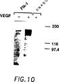
図10.Flk−1のVEGF誘導自己リン酸化。一時的にFkk−1受容体を発現しているCOS細胞および対照細胞を24時間、0.5%ウシ胎児血清を含有するDMEM中で飢餓状態におき、次にVEGFで10分間刺激した。細胞を可溶化し、Flk−1受容体をそのC末端に対するポリクローナル抗体を用いて免疫沈降させ、ポリアクリルアミドゲル電気泳動により分離して、ニトロセルロースに移した。ブロットを、抗ホスホチロシン抗体(5B2)を用いて精査した。タンパク質バンドを、西洋ワサビ−ペルオキシダーゼ結合二次抗体およびBCL(登録商標)(Amersham)検出アッセイを用いて、可視化した。
図11.マウスFlk−1のヌクレオチド配列。
図12.レトロウイルスベクター構築体のプラスミドマップ。pLXSN Flk−1 TM Cl.1およびpLXSN Flk−1 TM cl.3は、Flk−1のアミノ酸1から806まで含有する。pNTK−cfms−TMは、c−fmsのN末端アミノ酸541個を含有する。
図13.Flk−1のトランスドミナント−陰性阻止(transdominant-negative inhibition)によるC6ダリア芽腫増殖の阻止。C6細胞を単独で、またはウイルス産生細胞とともに移植した。細胞数は各パネルに示すとおりである。2つの異なるウイルス産生細胞系を使用した:Flk−1 TM(トランスドミナント−陰性)変異体を発現する細胞系と、対照としてトランスドミナント−陰性c−fms変異体(c−fms TM)を発現する細胞系であった。最初の腫瘍が出現した時から開始して、腫瘍容量を2〜3日置きに測定し、増殖曲線を得た。1群4匹のマウスを用いた。
図14.Flk−1のトランスドミナント−陰性阻止によるC6グリア芽腫腫瘍増殖の阻止。C6細胞を単独で、またはウイルス産生細胞とともに移植した。細胞数は各パネルに示すとおりである。2つの異なるウイルス産生細胞系を使用した:Flk−1 TM(トランスドミナント−陰性)変異体を発現する細胞系と、対照としてトランスドミナント−陰性c−fms変異体(cfms TM)を発現する細胞系であった。最初の腫瘍が出現した時から開始して、腫瘍容量を2〜3日置きに測定し、増殖曲線を得た。1群4匹のマウスを用いた。
5.発明の詳細な説明
本発明は、脈管形成および/または血管形成を変調するための、Flk−1受容体のリガンドの使用に関する。本発明はまた、受容体の活性化、調節および脱共役に関与しているかもしれない薬物ならびにVEGF類似体を評価およびスクリーニングするために、Flk−1を発現させることを包含する。このようなFlk−1の調節物質は、治療に使用できる。例えば、VEGFのアゴニストは、傷の治癒の過程等に使用できる。対照的に、VEGFのアンタゴニストは、増殖を血管新生に依存する腫瘍の治療に使用できる。
本発明は、その一部において、Flk−1が内皮細胞特異的RTKであることを示すin situハイブリダイゼーションおよびノーザンブロット分析の結果に基づいている。さらに、架橋実験は、Flk−1が血管内皮増殖因子(VEGF)の高親和性受容体であることを示し、Flk−1が血管芽細胞の発生および分化、ならびにそれに続く血管形成および脈管形成の過程における内皮細胞増殖に、極めて重要な役割を果たすことを示した。
本発明はまた、トランスドミナント−陰性変異体形態のFlk−1分子の発現が、内因性天然型Flk−1の生物学的活性を阻害することが可能である、という発見に基づいている。腫瘍細胞ならびに先端切断型のFlk−1受容体をコードする組換えレトロウイルスを産生する細胞を混合し、マウスに注射しする実験がここに記述される。血管形成および注射された腫瘍細胞の阻止が、先端切断型のFlk−1受容体を発現するマウスにおいて観察された。トランスドミナント陰性形態のFlk−1分子の発現は、血管の異常増殖に起因する病気、例えば、慢性関節リューマチ、網膜症および固形癌の増殖、等の治療に有用でありうる。
後述の実施例で説明されるように、ポリメラーゼチェーンリアクション(PCR)法が、移植後の胚および内皮細胞に特異的に発現される新規受容体チロシンキナーゼを単離するのに使用された。このようなクローンの1つは、造血細胞と選択された胎児肝キナーゼ−1(Flk−1)を富化した細胞集団より単離された、以前に同定されたcDNAクローンとほぼ同じ配列相同性を有するRTKをコードすることが判明した(Matthewsら、1991,Proc.Natl.Acad.Sci.U.S.A.88:9026-9030)(図11参照)。
議論を明確にするため、本発明は以下の分節においてマウスFlk−1を例として記述される。しかし、この原理はヒトを含む他の種のFlk−1をクローン化し、発現するのに類似的に適用しうる。
5.1.Flk−1コード配列
マウスFlk−1遺伝子のヌクレオチドコード配列および推定されるアミノ酸配列を図11(配列番号1)に示す。これらは最近Matthewsら、1991,Proc.Natl.Acad.Sci.U.S.A.88:9026-9030に記載されたものである。本発明によれば、ヒトを含む哺乳動物におけるFlk−1タンパク質またはその機能的等価物のヌクレオチド配列は、Flk−1の発現を引き出す組換え分子を作製するのに使用できる;以後、この受容体を、どの種から誘導されたものかに関係なく、「Flk−1」と称する。
ここに記述される特定の態様において、マウスFlk−1遺伝子は、受容体チロシンキナーゼのキナーゼドメイン内において高度に保存された配列に基づいて設計された2つの縮重オリゴヌクレオチドプライマープールを用いて、ポリメラーゼチェーンリアクション(PCR)を実施することにより単離された(Hanksら、1988)。鋳型として、8.5日齢マウス胚より調製されたλgt10 cDNAライブラリー由来のDNAを使用した。並行的アプローチにおいて、同様なプライマーを、出生後4〜8日齢のマウス脳から単離された毛細管内皮細胞由来のRTK cDNA配列を増幅するのに使用した。この時期は、脳内皮細胞増殖が最大となるときである。両方のアプローチとも、最近記述された胎児肝RTK、すなわちFlk−1(Matthewsら、1991)をコードするcDNA配列をもたらした。アミノ酸の相同性に基づき、この受容体は細胞外ドメインにイムノグロブリンに似た反復配列を含む(図1参照)RTKのタイプIIIサブクラスのメンバーである(UllrichおよびSchlessinger)。
本発明はまた、Flk−1活性が存在する他の種(ヒトを含む)より単離したFlk−1遺伝子に関する。Flk−1ファミリーのメンバーは、本明細書では、VEGFまたはそのペプチドの断片と結合する受容体として定義される。このような受容体は、DNA配列の実質的範囲におけるアミノ酸レベルで約80%の相同性を示しうる。バクテリオファージcDNAライブラリーを、低減したストリンジェンシーのもとで、マウスFlk−1クローンの放射標識化断片を用いてスクリーニングすることができる。
または、マウスFlk−1配列を、PCRプローブとして使用可能な、またはバクテリオファージcDNAライブラリーのスクリーニングに使用可能な、縮重または完全縮重オリゴヌクレオチドプローブを設計するのに使用できる。ポリメラーゼチェーンリアクション(PCR)に基づく方法は、ヒトFlk−1をクローン化するのに使用できる。マウスFlk−1と受容体チロシンキナーゼとの間で保存されてきたモチーフに対応する、縮重オリゴヌクレオチドの2つのプールは、PCR反応におけるプライマーとして役立つように設計することができる。反応のための鋳型は、ヒトFlk−1を発現することが知られている細胞系または組織より調製したmRNAの逆転写によって得られるcDNAである。PCR産物は、増幅させた配列がFlk−1配列を表すことを確実にするため、サブクローン化し、配列決定することができる。PCR断片は、増幅させた断片を放射活性的に標識し、バクテリオファージcDNAライブラリーをスクリーニングすることによって、全長Flk−1cDNAクローンを単離するのに使用できる。または、上記の標識化断片は、ゲノムライブラリーをスクリーニングするのに使用できる。使用できるクローン化方法の検討には、例えば、Maniatisら、1989,Molecular Cloning,A Laboratory Manual,Cold Springs Harbor Press,N.Y.;およびAusubelら、1989,Current Protocols in Molecular Biology(Green Publishing Associates and Wiley Interscience,N.Y.)を参照されたい。
ヒトFlk−1 cDNAの単離は、COS細胞に移入された時にプラスミドの多数コピー発現を可能とするSV40複製起点配列を含有する哺乳動物の発現ベクター(例えば,pcDNA1等)に、cDNAライブラリーを構築することによっても達成できる。トランスフェクトされたCOS細胞の表面におけるFlk−1の発現は、種々の方法によって検出できる。これらの方法には、標識化リガンド、例えば、放射標識、蛍光標識または酵素によって標識化されたVEGFまたはVEGFのアゴニストの使用が含まれる。ヒトFlk−1を発現する細胞は、トランスフェクトされた細胞を、FACS(蛍光活性化細胞ソーター)による選別にかけることにより富化できる。
本発明によれば、Flk−1、Flk−1のペプチド断片、Flk−1融合タンパク質またはそれらの機能的等価物をコードするFlk−1ヌクレオチド配列を、Flk−1タンパク質またはその機能的等価物の発現を適切な宿主細胞において引き出す組換えDNA分子の作製に使用することができる。または、Flk−1配列の部分とハイブリダイズするヌクレオチド配列を、核酸ハイブリダイゼーションアッセイ、サザンおよびノーザンブロット分析、等に使用することができる。
固有の遺伝暗号の縮重により、実質的に同一な、または機能的に等価なアミノ酸配列をコードする他のDNA配列を、本発明の実施においてFlk−1タンパク質のクローン化および発現に使用することができる。このようなDNA配列には、ストリンジェント条件下でマウスFlk−1配列とハイブリダイズ可能なDNA配列が含まれる。
本発明にしたがって使用することができる変更されたDNA配列は、同一の、または機能的に等価な遺伝子産物をコードする配列に帰着する、様々なヌクレオチド残基の欠失、付加または置換を含む。遺伝子産物それ自体が、表に現れない、したがって機能的に等価なFlk−1を産生する変化に帰着する、アミノ酸の欠失、付加または置換をFlk−1配列内に含有することもありうる。このようなアミノ酸置換は、関与する残基の極性、電荷、溶解性、疎水性、親水性および/または両親媒性の類似に基づいてなされる。例えば、負に荷電したアミノ酸にはアスパラギン酸およびグルタミン酸が含まれ、正に荷電したアミノ酸にはリシンおよびアルギニンが含まれ、類似した親水性値をもつ非荷電の複数の極性ヘッド基(polar head group)を有するアミノ酸には以下のものが含まれる:すなわち、ロイシン、イソロイシン、バリン、グリシン、アラニン、アスパラギン、グルタミン、セリン、トレオニン、フェニルアラニン、チロシンである。本明細書中で用いる「機能的に等価なFlk−1」とは、VEGFまたはその断片に結合するが、必ずしもその対応物である天然のFlk−1と同一の結合親和性を有するわけではない受容体、をいう。
本発明のDNA配列は、遺伝子産物のプロセシングと発現を修正する変更を含むがそれらだけに限定されない種々の目的に合わせて、Flk−1コード配列を変更するために、遺伝子工学的に操作することができる。例えば、当分野の技術で周知の技法(例えば、部位特異的突然変異誘導法)を使用して突然変異を導入することにより、新しい制限部位を挿入する、グリコシル化パターン、リン酸化等を変える、等が可能である。例えば、酵母等の発現系において、宿主細胞は遺伝子産物を過度にグリコシル化することがある。そのような発現系を使用するときは、Flk−1コード配列を変更して任意のN−結合グリコシル化部位を除去することが好ましいかもしれない。
本発明の別な態様においては、Flk−1配列または修飾されたFlk−1配列を異種配列に連結して、融合タンパク質をコードさせることができる。例えば、ペプチドライブラリーをスクリーニングするには、市販されている抗体によって認識される異種エピトープを発現するキメラFlk−1タンパク質をコードすることが役立ちうる。融合タンパク質は、Flk−1を異種部分から切り離せるように、Flk−1配列と異種タンパク質配列との間に位置する開裂部位を含有するように遺伝子操作することも可能である。
本発明のまた別な態様においては、当分野の技術で周知の化学的方法を用いて、Flk−1のコード配列を全部または部分的に合成することが可能であろう。例えば、Caruthersら、1980,Nuc.Acids Res.Symp.Ser.7:215-233; CreaおよびHorn,180,Nuc.Acids Res.9(10):2331; MatteucciおよびCaruthers,1980,Tetrahedron Letters 21:719; ならびにChowおよびKempe,1981,Nuc.Acids Res.9(12):2807-2817参照。または、Flk−1アミノ酸配列を全部または部分的に合成する方法を用いて、タンパク質自体を製造することも可能であろう。例えば、ペプチドは固相法により合成し、樹脂から切り離し、分取高速液体クロマトグラフィーにより精製することができる(例えば、Creighton,1983,Proteins Structures and Molecular Principles,W.H.Freeman and Co.,N.Y.,pp.50-60参照)。合成ペプチドの組成は、アミノ酸分析または配列決定により確認できる(例えば、Edman degradation procedure; Creighton,1983,Proteins Structures and Molecular Principles,W.H.Freeman and Co.,N.Y.,pp.34-49参照)。
5.2.Flk−1受容体の発現およびFlk−1を発現する細胞系の作製
生物学的に活性なFlk−1を発現するため、Flk−1または上記第5.1節に記述したその機能的等価物をコードするヌクレオチド配列は、適切な発現ベクターすなわち、挿入されたコード配列の転写および翻訳に必要な諸要素を含むベクターに挿入される。Flk−1遺伝子産物、ならびに組換えFlk−1発現ベクターによってトランスフェクトまたは形質転換された宿主細胞または細胞系は、種々の目的に使用できる。これらの目的には、以下のものが含まれるがそれらだけに限定されない。すなわち、この受容体に結合する、VEGFの結合を競合的に阻害しFlk−1の活性を「中和する」ものを含む抗体類(つまりモノクローナルおよびポリクローナル抗体)の作製、およびFlk−1受容体によって作用するVEGF類似体または薬物のスクリーニングおよび選択、等である。
5.2.1.発現系
Flk−1コード配列および適切な転写/翻訳制御シグナルを含有する発現ベクターの構築には、当業者に周知の方法を用いることができる。これらの方法には、in vitro組換えDNA技法、合成技法およびin vivo組換え/遺伝子組換えが含まれる。例えば、Maniatisら,1989,Molecular Cloning,A Laboratory Manual,Cold Springs Harbor Press,N.Y.;およびAusubelら、1989,Current Protocols in Molecular Biology(Green Publishing Associates and Wiley Interscience,N.Y.を参照されたい。
種々の宿主−発現ベクター系がFlk−1コード配列を発現するのに使用できる。これらには、Flk−1コード配列を含有する組換えバクテリオファージDNA、プラスミドDNAまたはコスミドDNA発現ベクターによって形質転換された細菌等の微生物;Flk−1コード配列を含有する組換え酵母発現ベクターによって形質転換された酵母;Flk−1コード配列を含有する組換えウイルス発現ベクター(例えば、バキュロウイルス)を感染させた昆虫細胞系;組換えウイルス発現ベクター(例えば、カリフラワーモザイクウイルスCaMV;タバコモザイクウイルスTMV)を感染させた、またはFlk−1コード配列を含有する組換えプラスミド発現ベクター(例えば、Tiプラスミド)によって形質転換された植物細胞系;DM染色体中に安定に増幅された(CHO/dhfr)または不安定に増幅されたFlk−1 DNAの多数コピーを含有するように遺伝子操作された細胞系を含む、組換えウイルス発現ベクター(例えば、アデノウイルス、ワクシニアウイルス)を感染させた動物細胞系(例えば、マウス細胞系)が含まれるが、これらだけに限定されない。
これらの系の発現要素は、その強度と特異性において異なる。使用する宿主/ベクター系により、構成的および誘導性プロモーターを含む多数の適切な転写および翻訳要素の任意のものを発現ベクターに使用できる。例えば、細菌系でクローン化を行うときは、バクテリオファージλのpL、plac、ptrp、ptac(ptrp−lacハイブリッドプロモーター)等の誘導性プロモーターが使用できる;昆虫細胞系でクローン化を行うときは、バキュロウイルスポリヘドリン(polyhedrin)プロモーター等が使用できる;植物細胞系でクローン化を行うときは、植物細胞ゲノムから誘導されたプロモーター(例えば、熱ショックプロモーター;RUBISCOの小サブユニットのプロモーター;クロロフィルa/b結合タンパク質のプロモーター)または植物ウイルスから誘導したプロモーター(例えば、CaMVの35S RNAプロモーター;TMVのコートタンパク質プロモーター)が使用できる;哺乳動物細胞系でクローン化を行うときは、哺乳動物細胞のゲノムから誘導されたプロモーター(例えば、メタロチオネインプロモーター)、または哺乳動物ウイルスから誘導されたプロモーター(例えば、アデノウイルス後期プロモーター;ワクシニアウイルス7.5Kプロモーター)が使用できる;Flk−1 DNAの多数コピーを含有する細胞系を作製するときは、SV40、BPV、およびEBVに基づくベクターを適切な選択マーカーとともに使用できる。
細菌系においては、発現されるFlk−1の意図された用途によって、多数の発現ベクターが有利に選択できる。例えば、抗体の作製またはペプチドライブラリーのスクリーニングのために大量のFlk−1を産生しなければならないときは、容易に精製される融合タンパク質産物の高レベル発現を引き出すベクターが望ましいだろう。このようなベクターには以下のものが含まれるが、それらだけに限定されない。すなわち、大腸菌発現ベクターpUR278(Rutherら、1983,EMBO J.2:1791)(ここでFlk−1コード配列は、ハイブリッドAS-lac Zタンパク質が産生されるようにlac Zコード領域の読み枠に合わせてベクターに連結することができる);pINベクター(InouyeおよびInouye,1985,Nucleic Acids Res.13:3101-3109; Van HeekeおよびSchuster,1989,J.Biol.Chem.264:5503-5509);等である。pGEXベクターもまた、外来ポリペプチドをグルタチオンS-トランスフェラーゼ(GST)を有する融合タンパク質として発現するのに使用できる。一般に、このような融合タンパク質は可溶性で、グルタチオン−アガロースビーズに吸着させ、遊離グルタチオンの存在下で溶出させることにより、溶解した細胞から容易に精製できる。このpGEXベクターは、興味のあるクローン化されたポリペプチドをGST部分から解放できるように、トロンビンまたは因子Xaプロテアーゼ開裂部位を含むように設計されている。酵母においては、構成的または誘導性プロモーターを含有する多くのベクターが使用できる。プロモーターの検討には、Current Protocols in Molecular Biology,Vol.2,1988,Ausubelら(編)、Green Publish.Assoc. & Wiley Interscience,Ch.13; Grantら、1987,Expression and Secretion Vectors for Yeast,in Methods in Enzymology,WuおよびGrossman(編),1987,Acad.Press,N.Y.,Vol.153,pp.516-544; Glover,1986,DNA Cloning,1987,Heterologous Gene Expression in Yeast,Methods in Enzymology,BergerおよびKimmel(編)、Acad.Press,N.Y.,Vol.152,pp.673-684;およびThe Molecular Biology of the Yeast Scahharomyces,1982,Strathernら(編)、Cold Spring Harbor Press,Vols.IおよびIIを参照されたい。
植物発現ベクターを使用する場合には、Flk−1コード配列の発現は、多数のプロモーターのうち任意のものによって推進させることができる。例えば、CaMVの35S RNAおよび19S RNAプロモーター(Brissonら、1984,Nature 310:511-514)、またはTMVのコートタンパク質プロモーター(Takamatsuら、1987,EMBO J.6:307-311)等のウイルスプロモーターが使用できる。または、RUBISCOの小サブユニット(Coruzziら、1984,EMBO J.3:1671-1680; Broglieら、1984,Science 224:838-843)等の植物プロモーター;または、大豆hsp17.5-Eもしくはhsp17.3-B(Gurleyら、1986,Mol.Cell.Biol.6:559-565)等の熱ショックプロモーターが使用できる。これらの構築体は、Tiプラスミド、Riプラスミド、植物ウイルスベクター、直接DNA形質転換(direct DNA transformation)、マイクロインジェクション、エレクトロポレーション、等を用いて植物細胞に導入することができる。これらの技法の検討には、例えば、WeissbachおよびWeissback,1988,Methods for Plant Molecular Biology,Academic Press,NY,Section VIII,pp.421-463;ならびに、GriersonおよびCorey,1988,Plant Molecular Biology,2d Ed.,Blackie,London Ch.7-9を参照されたい。
Flk−1を発現するのに使用できる別の発現系は、昆虫系である。このような系の1つにおいては、オートグラファ・カリフォルニカ(Autographa californica)核多角体病ウイルス(AcNPV)が外来遺伝子を発現するベクターとして使用される。このウイルスは、スポドプテラ・フルジペルダ(Spodoptera frugiperda)細胞中で増殖する。Flk−1コード配列は、このウイルスの非必須領域(例えばポリヘドリン遺伝子)にクローン化し、AcNPVプロモーター(例えばポリヘドリンプロモーター)の制御下に置くことができる。Flk−1コード配列の挿入がうまくゆくと、ポリヘドリン遺伝子の不活性化と外被をもたない(non-occluded)組換えウイルス(すなわち、ポリヘドリン遺伝子によってコードされるタンパク様のコートを欠くウイルス)の産生をもたらすであろう。次に、これらの組換えウイルスを、そこで挿入遺伝子を発現させるスポドプテラ・フルジペルダ細胞を感染させるのに使用する(例えば、Smithら、1983,J.Viol.46:584; Smith,米国特許第4,215,051参照)。
哺乳動物細胞においては、ウイルスに基づく多数の発現系が使用できる。アデノウイルスを発現ベクターとして使用する場合は、Flk−1コード配列をアデノウイルスの転写/翻訳調節複合体、例えば後期プロモーターおよび3分節リーダー配列に連結することができる。次に、このキメラ遺伝子をin vitroまたはin vivo組換えによってアデノウイルスゲノムに挿入することができる。ウイルスゲノムの非必須領域(例えば、E1またはE3領域)への挿入は、感染した宿主中で生育可能であり、かつFlk−1を発現することができる組換えウイルスをもたらすであろう〔LogantおよびShenk,1984,Proc.Natl.Acad.Sci.(USA),81:3655-3659〕。または、ワクシニア7.5Kプロモーターを使用することもできる。[例えば、Mackettら、1982,Proc.Natl.Acad.Sci.(USA)79:7415-7419; Mackettら、1984,J.Virol.49:857-864; Panicaliら、1982,Proc.Natl.Acad.Sci.79:4927-4931参照。]
挿入されたFlk−1コード配列の効率的な翻訳のためには、特定の開始シグナルも必要とされることがある。これらのシグナルには、ATG開始コドンおよび隣接配列が含まれる。それ自身の開始コドンおよび隣接配列を含む完全なFlk−1遺伝子を適切な発現ベクターに挿入する場合は、付加的翻訳調節シグナルはなんら必要とされない。しかし、Flk−1コード配列の一部のみを挿入する場合は、ATG開始コドンを含む外因性翻訳調節シグナルを供給しなければならない。さらに、その開始コドンは、挿入配列全体の翻訳を確実にするために、Flk−1コード配列の読み枠と合致していなければならない。これらの外因性翻訳調節シグナルおよび開始コドンは、天然および合成の多様な起源のものでありうる。発現の効率は、適切な転写エンハンサー要素、転写ターミネーター、等を包含することにより増強されうる(Bittnerら、1987,Methods in Enzymol.153:516-544参照)。
さらに、挿入配列の発現を変調する、または所望の特異的方法で遺伝子産物を修飾しプロセシングする宿主細胞株を選択することができる。このようなタンパク質産物の修飾(例えば、グリコシル化)およびプロセシング(例えば、開裂)は、そのタンパク質の機能にとって重要でありうる。異なる宿主細胞は、タンパク質の翻訳後プロセシングおよび修飾のための、特徴的で特異的な作用機構をもつ。適切な細胞系または宿主系は、発現された外来タンパク質の適正な修飾およびプロセシングを確実にするように選択することができる。この目的のため、一次転写物の適切なプロセシング、または遺伝子産物のグリコシル化およびリン酸化のための細胞機構をもつ真核宿主細胞が使用できる。このような哺乳動物宿主細胞には、CHO、VERO、BHK、HeLa、COS、MDCK、293、WI38等が含まれるが、これらだけに限定されない。
組換えタンパク質の長期間にわたる高収率産生のためには、安定した発現が望ましい。例えば、Flk−1を安定的に発現する細胞系を遺伝子工学的に作製することが可能である。ウイルスの複製起点を含有する発現ベクターを使用するのではなく、適切な発現調節要素(例えば、プロモーター、エンハンサー配列、転写ターミネーター、ポリアデニル化部位、等)および選択マーカーによって制御されるFlk−1 DNAによって宿主細胞を形質転換することができる。外来DNAの導入に続いて、遺伝子操作した細胞を1〜2日間、富化した培地中で増殖させてもよい。そして次に選択培地に替える。組換えプラスミド中の選択マーカーは、選択に対する耐性を付与し、細胞がプラスミドをその染色体に安定に組み入れ、フォーカス(細胞増殖巣)を形成するまで増殖することを可能とする。次にこのフォーカスをクローン化し、細胞系へと発展させることができる。この方法は、Flk−1を細胞表面に発現し、VEGFが仲介するシグナル伝達に応答する細胞系を遺伝子工学的に作製するのに有利に使用することができる。このように遺伝子工学的に作製された細胞系は、VEGF類似体をスクリーニングするのに特に有用である。
多数の選択系を使用することができる。これらの選択系には以下のものが含まれるが、それらだけに限定されない。すなわち、単純ヘルペスウイルスチミジンキナーゼ遺伝子(Wiglerら、1977,Cell 11:223)、ヒポキサンチン−グアニンホスホリボシルトランスフェラーゼ遺伝子(SzybalskaおよびSzybalski,1962,Proc.Natl.Acad.Sci.USA 48:2026)、およびアデニンホスホリボシルトランスフェラーゼ遺伝子(Lowyら、1989,Cell 22:817)を、それぞれtk-、hgprt-またはaprt-細胞に採用することができる。また、代謝拮抗物質耐性を、メトトレキセートへの耐性を付与するdhfr遺伝子(Wiglerら、1980,Natl.Acad.Sci.USA 77:3567; 0'Hareら、1981,Proc.Natl.Acad.Sci.USA 78:1527);マイコフェノール酸への耐性を付与するgpt遺伝子(MulliganおよびBerg,1981,Proc.Natl.Acad.Sci.U.S.A.78:2072);アミノグリコシドG−418への耐性を付与するneo遺伝子(Colberre-Gerapinら、1981,J.Mol.Biol.150:1);ハイグロマイシンへの耐性を付与するhygro遺伝子(Santerreら、1984,Gene 30:147)の選択の基礎として使用できる。最近、さらなる選択遺伝子が記述された。それらは、細胞にトリプトファンの代わりにインドールを利用可能とするtrpB;細胞にヒスチジンの代わりにヒスチノールを利用可能とするhisD(HartmanおよびMulligan,1988,Proc.Natl.Acad.Sci.U.S.A.85:8047);オルニチンデカルボキシラーゼ阻害剤である2−(ジフルオロメチル)−DL−オルニチン(DFMO)への耐性を付与するODC(オルニチンデカルボキシラーゼ)[McConlogue L.,1987,In: Current Communications in Molecular Biology,Cold Spring Harbor laboratory(編)]である。
5.2.2. Flk−1を発現するトランスフェクタントおよびトランスフォーマントの同定
コード配列を含み、生物学的活性をもつ遺伝子産物を発現する細胞は、少なくとも4種の一般的アプローチによって同定することができる;(a)DNA−DNAまたはDNA−RNAハイブリダイゼーション;(b)「マーカー」遺伝子機能の存否;(c)宿主細胞中におけるFlk−1メッセンジャーRNA転写物の発現の測定による、転写レベルの評価;(d)イムノアッセイまたは生物学的活性の測定を介した遺伝子産物の検出。
第一のアプローチでは、発現ベクター中に挿入したFlk−1のコード配列の存在は、Flk−1のコード配列またはその一部、またはその誘導体と相同のヌクレオチド配列を含むプローブを用いた、DNA−DNAまたはDNA−RNAハイブリダイゼーションによって検出することができる。
第二のアプローチでは、組換え発現ベクター/宿主系は、特定の「マーカー」遺伝子機能(たとえばチミジンキナーゼ活性、抗生物質に対する抵抗性、メトトレキセートに対する抵抗性、トランスフォーメーション表現型、バキュロウイルスにおける閉塞(occlusion)体の形成など)の存在または不在に基づいて同定および選別することができる。たとえば、もしFlk−1コード配列がベクターのマーカー遺伝子配列中に挿入されたとすると、Flk−1コード配列を含む組換え体は、マーカー遺伝子機能の不在によって同定することができるだろう。これとはちがって、Flk−1のコード配列の発現を制御するために用いられる、同一または異なるプロモーターの制御下にあるように、マーカー遺伝子をFlk−1配列とタンデムに配置させることができる。誘導または選択に応答するマーカーの発現は、Flk−1のコード配列の存在を示す。
第三番目のアプローチでは、Flk−1のコード領域に対する転写活性は、ハイブリダイゼーション検定によって評価することができる。たとえば、RNAは、Flk−1のコード配列またはその特定の部分に対する相同のプローブを用いて、ノーザンブロットによって単離および解析することができる。これとは別に、宿主細胞の全核酸を抽出し、同様のプローブを用いたハイブリダイゼーションで検定を行ってもよい。
第四のアプローチでは、Flk−1のタンパク産物の発現は、たとえばウェスタンブロット、放射性免疫沈澱、酵素−連結イムノアッセイおよび同様の手段など、免疫学的な手法によって評価できる。しかし、発現系の成功をはかる究極の試験は、生物学的に活性のあるFlk−1遺伝子産物の検出を含むものである。これだけに限るものではないが、VEGF結合試験;および、試験用基質として遺伝子工学的操作を施した細胞系を用いたVEGF生物検定などを含む、数多くの検定を受容体活性の検出のために用いることができる。
5.3. Flk−1受容体および遺伝子工学的に操作した細胞系の使用
脈管形成(angiogenesis)、即ち、新しい毛細血管の生育は、傷の治癒、組織および器官の再成、妊娠後の胎盤の形成および胚発生などにまたがる、数多くの生理学的なプロセスに必要とされる。血管の異常な増殖は、リューマチ性関節炎、網膜症、乾癬などさまざまな疾患の重要な構成要素となっている。毛細血管形成はまた、血管形成に依存した癌腫(solid fumor)の増殖および転移活性における重要な要素である。したがって、脈管形成阻害剤は、血管の異常な生育に由来または付随する疾患の治療および癌腫の成長および拡がりを含む悪性腫瘍の治療に有効に用いることができる。
本発明の一態様において、Flk−1受容体および/またはFlk−1受容体を発現する細胞系は、抗体、ペプチド、またはFlk−1受容体によって媒介される脈管形成または血管形成のアゴニストまたはアンタゴニストとして作用する他のリガンドのスクリーニングに用いることができる。たとえば、VEGFの活性を中和することのできる抗Flk−1抗体は、Flk−1機能を阻害するために用いることができる。さらに、VEGFの活性を模倣する抗Flk−1抗体は、傷の治癒に用いるために選ぶことができるであろう。あるいは、組換え技術によって発現させた可溶性のFlk−1タンパクまたはFlk−1タンパクを発現する細胞系によるペプチドライブラリーのスクリーニングは、Flk−1の生物学的活性の阻害によって機能する治療作用のある分子の同定に有用であろう。
本発明のある態様において、全Flk−1コード領域またはそのリガンド結合領域を発現する、遺伝子工学的に操作した細胞は、VEGFのアゴニストおよびアンタゴニストのスクリーニングおよび同定に用いることができる。合成化合物、天然物、および生物学的活性をもつ可能性のある他の原材料はさまざまな方法でスクリーニングを行うことができる。試験化合物がVEGFのFlk−1への結合を阻害する能力は、6.1.9節で述べたような標準の受容体結合技術を用いて測定することができる。Flk−1を発現する細胞に対する、VEGFの結合のシグナル伝達応答への効果を妨害または模倣する能力を測定することが可能である。たとえば、Flk−1キナーゼ活性、セカンドメッセンジャー産生の調節または細胞の代謝の変化などを追跡することができる。これらの検定はこれらの目的のためにつくられた従来の技術を用いて実施することができる。
5.3.1. Flk−1タンパクまたは遺伝子工学的に操作した細胞系を用いたペプチドライブラリーのスクリーニング
固相支持体に付着させた、アミノ酸のあらゆる可能な組合せにより成るランダムペプチドライブラリーを、特定のリガンドの結合部位またはキナーゼ領域など、受容体の他の機能領域と結合することのできるペプチドの同定に用いることができる(Lam,K.S.et al.,1991,Nature 354: 82-84)。ペプチドライブラリーのスクリーニングは、特定の受容体との相互作用を介して受容体の生物活性を阻害するように作用する、薬理作用をもつ物質の発見において、治療上の価値をもちうるであろう。
Flk−1に結合する能力のある分子の同定は、ペプチドライブラリーを可溶の組換えFlk−1タンパクによってスクリーニングすることによって遂行しうる。Flk−1の発現および精製法は5.2.1節に述べられており、問題とする機能領域に応じて全長の組換えFlk−1またはFlk−1の断片の発現のために用いることができる。たとえば、Flk−1のキナーゼ領域および細胞外のリガンド結合領域を別々に発現させ、ペプチドライブラリーのスクリーニングに用いることができる。
Flk−1と相互作用を行い複合体を形成するペプチド/固相支持体を同定し単離するためには、Flk−1分子を標識するか「タグ」をつけることが必要である。Flk−1タンパクはアルカリフォスファターゼまたは西洋ワサビペルオキシダーゼのような酵素、または蛍光性イソチアネート(FITC)、フィコエリスリン(PE)またはローダミンなどの蛍光性標識などの、他の試薬と結合させることができる。Flk−1に対するいかなる特定の標識の結合も、当業界によく知られた技術を用いて実施することができる。一方、Flk−1発現ベクターは市販の抗体に対するエピトープを含むキメラFlk−1タンパクを発現させるように操作することもできる。エピトープ特異的抗体は、酵素、蛍光色素、または着色あるいは磁性のビーズなどによる標識を含む、当業界でよく知られた方法を用いて「タグ」をつけることができる。
「タグ」をつけたFlk−1結合体をランダムペプチドライブラリーと共に22℃で30分間から1時間インキュベートし、Flk−1とライブラリー中のペプチド種との複合体形成を行わせる。ライブラリーを次いで洗い、結合しなかったFlk−1タンパクを除去する。Flk−1がアルカリフォスファターゼまたは西洋ワサビペルオキシダーゼに結合している場合には、全ライブラリーを、たとえば5−ブロモ−4−クロロ−3−インドイルフォスフェート(BCIP)または3,3’,4,4”−ジアミノベンチジン(DAS)など、それぞれアルカリフォスファターゼまたはペルオキシダーゼに対する基質を含んだペトリ皿に注げばよい。数分間インキュベートした後、ペプチド/固相−Flk−1複合体の色は変化し、ミクロマニピュレーターを装備した分析用顕微鏡(dissecting microscope)を用い、容易に同定でき、物理的に単離することができる。蛍光「タグ」がつけられたFlk−1分子を用いた場合には、複合体は蛍光活性化ソーティング(fluorescent activated sorting)によって単離できるであろう。異種のエピトープを発現するキメラFlk−1タンパクを用いた場合には、ペプチド/Flk−1複合体は標識されたエピトープ特異的抗体を用いて検出することができる。一たび単離されれば、固相支持体に付着したペプチドの同定は、ペプチドのアミノ酸配列決定によって行うことができる。
可溶性のFlk−1分子の使用に加え、他の態様において、無傷の細胞を用いて細胞表面受容体と結合するタンパクを検出することも可能である。無傷の細胞の使用は、多くのサブユニットより成る、または不安定な、または機能的であるためには細胞膜の脂質領域を必要とする受容体などを使用する場合に望ましい。Flk−1を発現する細胞系統の作成の方法は、5.2.1および5.2.2節に述べた。この技術に用いる細胞は、生細胞または固定細胞のいずれでもよい。細胞はランダムペプチドライブラリーとインキュベートされ、ライブラリー中の特定のペプチドと結合して、標的細胞および適当な固相支持体/ペプチドとの間に「ロセット」を形成する。ロセットは次いで遠心分離によって分離、または分析用顕微鏡を用いて物理的にとりだすことができる。
膜結合の受容体または機能的であるために細胞膜の脂質領域を必要とする受容体のための全細胞検定の別法として、受容体分子を標識または「タグ」をつけることのできるリポゾーム中へと再構成することができる。
5.3.2. 抗体の産生とスクリーニング
組換え技術を用いて作成したFlk−1受容体のエピトープに対する抗体の産生のためには、当業界で知られたさまざまな方法を用いることができよう。そのような抗体には、以下に限るものではないが、ポリクローナル、モノクローナル、キメラ、単鎖、FabフラグメントおよびFab発現ベクターによってつくられたフラグメントなどが含まれる。中和抗体、即ち、受容体のVEGF結合部位と競する抗体は、診断と治療にとりわけ好ましい。
Flk−1に結合するモノクローナル抗体は、放射性の標識を行い、これにより注射後の体内での存在部位や分布を追跡が可能となる。放射性の「タグ」をつけた抗体は、リューマチ性関節炎、黄斑変性、および腫瘍の形成および転移などを含む多くの疾患に伴う、de novoの血管形成の造影のための、非侵入性の診断用材料として用いることができる。
細胞毒性物質に体内の特定部位を標的とさせるイムノトキシンの説計も可能である。例えば、高親和性の特異的モノクローナル抗体を、たとえばジフテリアトキシン、アブリンまたはリシンなどのバクテリアまたは植物トキシンと共有結合で結合させることができる。抗体/ハイブリッド分子の一般的な調製法は、抗体の一級アミノ基を攻撃し、ついでジスルフィド交換によってトキシンを抗体に結合させる、チオール架橋剤の使用を含むものである。ハイブリッド抗体はFlk−1を発現する内皮細胞の特異的な除去のために用いることができる。
抗体の産生を増強するためには宿主の種によってこれに限るものではないが、ウサギ、マウス、ラット等を含むさまざまな動物をFlk−1タンパクの注射による免疫化を行うことができる。免疫応答を増強させるために、これに限るものではないが、フロイント(完全および不完全)、水酸化アルミニウムなどの鉱物ゲル、リゾレシチンなどの表面活性物質、プルロニックポリオール、ポリアニオン、ペプチド、オイルエマルジョン、キーホールリンプレットヘモシアニン、ジニトロフェノール、およびBCG(bacille calmette-Guerin)および コリネバクテリウム・パービュム(Corynebacterium marvum)などの有用な可能性のあるヒトのアジュバントなどを含むアジュバントを用いることができる。
Flk−1に対するモノクローナル抗体は、培養中に継続的な細胞系統によって生産される抗体分子を供給する諸技術のいずれを用いて調製してもよい。これらには以下に限るものではないが、KohlerとMilstein(Natute,1975,256: 495-497)によってはじめに記述されたハイブリドーマ技術、ヒトB細胞ハイブリドーマ技術(Kosbor et al.,1983,Immunology Today,4; 72; Cote et al.,1983,Proc.Natl.Acad.Sci.,80: 2026-2030)およびEBV−ハイブリドーマ技術(Cole et al.,1985,Mono clonal Antibodies and Cancer Therapy,Alan R.Liss,Inc.,pp.77-96)などが含まれる。このほかに、適当な抗原特異性をもつマウスの抗体分子の遺伝子と適当な生物活性をもつヒト抗体の遺伝子とのスプライシングによる「キメラ抗体」の生産のために発展した技術(Morrison et al.,1984,Proc.Natl.Acad.Sci.,81; 6851-6855; Neuberger et al.,1984,Nature,312: 604-608; Takeda et al.,1983,Nature,314: 452-454)を用いることができる。あるいは、単鎖抗体の生産のために発表された技術(米国特許4,946,773)をFlk−1特異的な単鎖抗体を生産するために適用することもできる。
Flk−1の特異的結合部位を含む抗体断片は、既知の技術によってつくり出すことができる。たとえば、そのような断片は以下に限るものではないが抗体分子のペプシン消化によって作りだすことのできるF(ab')2断片、およびF(ab')断片のジスルフィド架橋の還元によって生成させることのできるFab断片などを含む。これとは別に、Flk−1に対する望ましい特異性をもったモノクローナルFab断片の速やかで容易な検出を可能にする、Fab発現ライブラリーを構築してもよい(Huse et al.,1989,Science,246: 1275-1281)。
5.4. Flk−1コード配列の利用
Flk−1コード配列は、Flk−1発現の検出のための診断目的に用いることができる。本発明の範囲に含まれるのは、アンチセンスRNAおよびDNA分子を含むオリゴリボヌクレオチド配列、およびFlk−1の翻訳を阻害する機能をもつリボザイムなどである。さらに、ドミナント−陰性の効果をもつ、変異体のFlk−1を、内在的に発現した野性型のFlk−1の活性を阻害するために、標的細胞集団中で発現させることができる。
5.4.1. 診断薬および治療薬におけるFlk−1コード配列の利用
Flk−1 DNAはFlk−1の異常な発現に由来する疾患の診断への数多くの用途をもちうる。たとえば、Flk−1 DNA配列はFlk−1の発現の異常を診断するためのバイオプシーまたはオートプシーのハイブリダイゼーション検定、たとえばin situのハイブリダイゼーション試験を含むサザンまたはノーザン分析に用いることができる。
Flk−1 cDNAはFlk−1 mRNAの発現を検出するためのプローブとして用いることができる。ここで記述された特定の実施例では、異なる発生段階にあるマウスの胚におけるFlk−1 mRNAの発現を分析した。ノーザンブロット分析は、9.5から18.5日にかけて主成分の5.5kb mRNAの多量の発現があり、妊娠の末期へ向けて明らかな減少をみせる(図2A)ことを示している。生後4−8日にかけて脳の毛細血管のFlk−1 mRNAは脳全体のRNAに比較してきわめて多量にみつかり(図2B)、内皮細胞の増殖におけるFlk−1の役割が示唆される。
胚発生の過程および血管形成の初期におけるFlk−1の発現についてより詳細な知識を得るために、6.1.4節に述べたようなin situハイブリダイゼーション実験を行った。in situハイブリダイゼーションは、胚発生におけるin vivoのFlk−1の発現が主として内皮細胞およびその前駆体に限局していることを示した(図3および図4)。Flk−1は内皮細胞の増殖によって特徴づけられる生理的な過程で内皮細胞中で発現し、胎児の脳の中でみられる発現の時間的および空間的なパターンはBar(1980)が記載した神経の血管系の発生と正確に対応する。前神経叢(preneural plexus)に発する血管芽(vascular sprouts)は放射状に神経外胚葉へと成長し、そこで枝分れし、これら側芽(branch)は高レベルのmRNAを発現することがわかった(図5)。生後の初期段階では内皮細胞の増殖は依然として顕著でありFlk−1は発現している一方、成体では血管形成の完了後、内皮細胞の増殖の減退とFlk−1の発現の低下には平行関係にある。
さらに本発明の範囲内には、アンチセンスRNAおよびDNA分子を含むオリゴヌクレオチド配列およびFlk−1 mRNAの翻訳を妨げる機能をもつリボザイムがある。アンチセンスRNAおよびDNAは、標的mRNAに結合し、タンパクの翻訳を妨げることによって、mRNAの翻訳を直接に遮断する。アンチセンスDNAについて言えば、翻訳開始部位、たとえばFlk−1ヌクレオチド配列の−10から+10の間の領域から得られたオリゴデオキシリボヌクレオチドが好ましい。
リボザイムはRNAの特異的な切断を触媒することのできる酵素作用のあるRNA分子である。リボザイムの作用機構には相補的な標的RNAに対するリボザイム分子の配列特異的なハイブリダイゼーションとこれにエンドヌクレアーゼ作用による切断が続くプロセスか含まれる。本発明の範囲に含まれるのは、Flk−1 RNA配列のエンドヌクレアーゼ作用による切断を特異的にかつ効率よく触媒する、遺伝子操作したハンマーヘッドモチーフ(hammerhead motif)リボザイム分子である。
可能な標的RNA中の特異的なリボザイムの切断部位は、標的分子をGUA、GUUおよびGUCを含むリボザイム切断部位について走査することによってはじめ同定された。一たび同定されれば切断部位を含む標的遺伝子の領域に対応する、15から20のリボヌクレオチドの間の短いRNA配列は、このオリゴヌクレオチドを不適当なものとするかもしれない2次構造などの予見できる構造について評価することができる。標的候補の妥当性は、リボヌクレアーゼ保護試験を用い、相補的なオリゴヌクレオチドとのハイブリダイゼーションの可能性をテストすることによって評価することもできる。
本発明のアンチセンスRNAとDNA分子およびリボザイムは、RNA分子の合成について当業者間に知られているいかなる方法を用いて調製してもよい。これらは、たとえば固相フォスフォラミダイト化学合成などの当業界によく知られた化学合成のオリゴデオキシリボヌクレオチドに対する技術を含む。あるいはRNA分子はアンチセンスRNA分子をコードするDNA配列のin vitroまたはin vivoの転写によって生成させることもできる。このようなDNAは、T7またはSP6ポリメラーゼプロモーターなどの適当なRNAポリメラーゼのプロモーターをとり入れたさまざまな種類のベクター中にとり込むことができる。あるいは、用いるプロモーターによって、アンチセンスRNAを構成的あるいは誘導的に合成するcDNA構築物を、細胞系に安定に導入することができる。
細胞内での安定性または半減期を増大させる手段として、DNA分子に対しさまざまな改変を行うことができる。考えられる改変としては、以下に限るものではないが、分子の5'および/または3'末端をはさむリボヌクレオチドまたはデオキシリボヌクレオチド配列を付加するか、またはオリゴデオキシリボヌクレオチド骨格内のフォスフォジエステラーゼ連結の代わりにフォスフォチオエートまたは2'o−メチルを用いることが含まれる。
5.4.2. 遺伝子治療におけるドミナント−陰性変異株の利用
リガンドによってひき起こされる受容体の二量体化は、リガンドの結合とキナーゼ活性の刺激とを共役させる働きをもつ、アロステリックな調節シグナルを提供するものと考えられている。欠陥のある受容体は、機能のないヘテロ二量体を形成することによって、正常な受容体の活性化と応答を抑制する、ドミナント−陰性変異として働らくことができる。したがって、欠陥受容体を組換えウイルスベクターに人工的にとりこませ、不適切にFlk−1を発現する患者の遺伝子治療に用いることができる。
本発明のある態様に於いては、ドミナント−陰性効果をもつ変異型のFlk−1分子は、選択された細胞内での発現によって同定することができる。野性型のFlk−1タンパクと二量体を形成する能力は保持するが、シグナル伝達の機能をもたないFlk−1の欠失またはミスセンス変異は、内在性の野性型Flk−1の生物活性を阻害させるために用いることができる。たとえば、Flk−1の細胞内キナーゼ領域を欠失させ、野性受容体との二量体化の形成は依然として行いうるが、シグナルの伝達はできないよう一部を欠いたFlk−1分子をつくることができる。
血管の異常な増殖は、リューマチ性関節炎、網膜炎および乾癬などのさまざまな病変の重要な要素である。制御を欠いた脈管形成は、また癌腫の成長や転移における重要な要素となっている。野性型の内在性Flk−1の活性を阻害するために用いることができる、ドミナント−陰性型のFlk−1を発現するような組換えウイルスを人工的に構築することができるであろう。これらウイルスはFlk−1の異常な発現または機能に由来する疾患の処置に、治療的に用いることができる。
レトロウイルス、ワクシニアウイルス、アデノ関連ウイルス、ヘルペスウイルスまたはウシパピローマウイルス等に由来する発現ベクターは、標的細胞集団へ組換えFlk−1を導入するために用いることができる。組換え技術に十分な経験のある者にはよく知られた方法を、Flk−1のコード配列を含む組換えウイルスベクターの構築に用いることができる。たとえばManiatis et al.,1989,Molecular Cloning,A Laboratory Manual,Cold Spring Harbor Laboratory,N.Y.およびAusubel et al.,1989,Current Protocols in Molecular Biology,Greene Publishing Associates and Wiley Interscience,N.Y.など参照。別の方法として、組換えFlk−1分子は標的細胞に到達させるために、リポゾーム中に組み入れることができる。
本発明の特定の態様において、Flk−1受容体の欠失変異を人工的に組換えレトロウイルスベクターに組込ませた。pLXSN F1k-1 TM cl.1およびpLXSN F1k-1 TM cl.3と名付けられた二つのクローン単離物は、561カルボキシル末端アミノ酸を欠く末端切断したFlk−1受容体を含んでいる。ウイルス産生細胞系統を得るために、PA37細胞を組換えベクターでトランスフェクトし、次いでウイルスを含む条件培地(conditioned media)を用いてGPE細胞を感染させた。
シグナル伝達欠損変異の発現が内在性のFlk−1受容体活性を阻害するかどうかをテストするために、C6ラット神経膠芽腫(腫瘍細胞)および組換えレトロウイルスを産生するマウス細胞を混合し、これをヌードマウスに皮下注射した。通常、腫瘍細胞のヌードマウスへの注射により、腫瘍細胞の増殖および生じた腫瘍部の血管形成が生じる。Flk−1は血管の形成に不可欠であると考えられているので、末端切断した受容体の発現によるFlk−1の活性の阻害は、発達しつつある腫瘍の血管形成を阻害し、その結果、腫瘍の生長を阻害する働らきをもつかもしれない。図13および14に示されているように、末端切断したFlk−1受容体を発現する、ウイルス産生細胞を共に注射することによって、腫瘍細胞のみの注射をうけた対照にくらべ、腫瘍の成長が著しく阻害された。
5.5. Flk−1受容体またはリガンドの使用
Flk−1およびVEGFの間の受容体/リガンドの相互作用は、血管形成および脈管形成期間のシグナル伝達系に大きな役割を果たすと考えられている。異常な血管の増殖は数多くの疾患の重要な構成要素である。
Flk−1 RNAの発現が脳の発達および内皮細胞の増殖と関係があることは、Flk−1が血管形成の過程におけるシグナルの伝達にかかわる受容体であるらしいことを示唆している。VEGFは内皮細胞にのみ働く分裂促進成長因子であることが示されている(Ferrata,N.and Henzel,W.J.,1989,Biochem.Biophys.Res.Comm.161: 851-858)。VEGFはFlk−1のリガンドであるかどうかを決定するために、6.1.9および6.1.10にそれぞれ述べられているように、架橋実験およびリガンド結合実験を行った。結果はFlk−1が高い親和性をもつ、VEGFの真正の受容体であることを示した(図9)。
本発明の一態様において、脈管形成および/または血管形成を変調するために、Flk−1のリガンド、Flk−1受容体自体、またはVEGF結合部位を含むFlk−1の断片などをin vivoで投与することができるであろう。たとえば、Flk−1受容体またはVEGF結合部位を含む断片の投与は、VEGFと拮抗的に結合し、in vivoにおける内在性のFlk−1受容体との相互作用を阻害し、脈管形成および/または血管形成を阻害することができるだろう。あるいはまた、抗Flk−1抗体またはその断片を含む、Flk−1のリガンドを脈管形成および/または血管形成を調節するために用いることができる。VEGF活性のアゴニストは傷の治癒を促進するために用いられるが、一方、VEGF活性のアンタゴニストは腫瘍の成長を阻害するために用いることができる。
特定の治療条件に依存するが、これら薬剤を全身的にまたは局所的に処方し、投与することが考えられる。処方および投与の技術は、“Remington's Pharmaceutical Sciences”Mack Publishing Co.,Easton,PA,latest editionに見出すことができる。適切な経路としては、経口、経直腸、経粘膜(transmucosal)、腸内投与;またはいくつか例をあげるだけでも、筋肉内、皮下、髄内注射、および鞘内、直接の脳室内、静脈内、腹腔内、鼻腔内、眼内注射などを含む非経口的な投与が含まれる。注射のためには、本発明の薬剤は水溶液で、好ましくはハンクス氏液、リンゲル氏液または生理食塩水のような生理的に適合性のある緩衝液で調剤することができる。経粘膜投与のためには通過の障壁にとって適当な浸透剤を処方の中に用いられる。そのような浸透剤は当業界では一般に知られている。
6. 実施例 VEGFに対する高親和性受容体Flk−1のクローニングと発現パターン
以下の小節でFlk−1 cDNAクローンのクローニングおよびその性質について述べる。ノーザンブロットおよびin situハイブリダイゼーション解析は、Flk−1が内皮細胞で発現していることを示す。架橋実験およびリガンド結合実験はさらに、Flk−1がVEGFに対する高親和性をもった受容体であることを示す。
6.1. 材料および方法
6.1.1. Flk−1のcDNAクローニング
(受胎後)8.5日のマウス胚のλgt10 cDNAライブラリー(Fahrner et al.,1987,EMBO.J.6: 1497-1508)から抽出したDNAをポリメラーゼ連鎖反応(RCR; Saiki R.K.et al.,1985,Science 230: 1350-1354)の鋳型として用いた。これとは別のアプローチとして、生後4−8日のマウスの脳から単離された毛細血管の内皮細胞のcDNAを増幅に用いた

Flk−1の全長cDNAクローンは別の(受胎後)8.5日のマウス胚から、OkayamaとBerg(1983)の方法に従って調製したcDNAライブラリー、および11.5日のマウス胚λgt11ライブラリー(Clonetech)から、32P-ラベルの(Feinberg,A.P.and Vogelstein,B.1983 Anal.Biochem.132: 6-13)210-塩基対のPCR断片を用いて単離した。
6.1.2. マウス胚
Balb/cマウスを一夜交配させ、膣栓の検出の朝を妊娠1/2と定義した。ノーザンブロット解析のために、冷凍した胚を5Mグアニジニウムチオシアネート中でホモジネートし、RNAは報告されている方法(Ullrich,A.et al.,1985,Nature 313: 756-761)によって単離した。in situハイブリダイゼーションのためには、胚をTissue-Tek(Miles)に包埋し、液体窒素表面で冷凍し、使用するまで−70℃で保存した。
6.1.3. プローブの調製
受容体cDNAの5'側に位置する2619塩基対を、EcoRI/BamHI断片としてpGem 32ベクター(Promega)にサブクローンした。ノーザンブロットハイブリダイゼーションのためのプローブはランダムヘキサヌクレオチドプライミング(Boehringer; Feinberg,A.P.and Vogelstein,B.,1983 Anal.Biochem.132: 6-13)法を用い、α−32PdATP(Amersham)によってcDNA断片を標識化することによって調製した。
in situハイブリダイゼーションのためには、
![]()
6.1.4. RNA抽出およびノーザン分析
全細胞質RNAをChromczynskiとSacchi(1987)の酸性フェノール法に従って分離した。ポリ(A+)RNAアリコートは、1.2%アガロースホルムアルデヒドゲル(Sambrook,J.et al.,1989,Molecular Cloning: A Laboratory Manual 2nd ed.Cold Spring Harbor Laboratory press)で電気泳動を行い、ニトロセルロース膜に転写し(Schleicher & Schell)、ハイブリダイゼーションは50%ホルムアミド、5×SSC(750mM塩化ナトリウム、75mMクエン酸ナトリウム)、5×Denhardt溶液(0.1%フィコール400,0.1%ポリビニルピロリドン、0.1%BSA)および0.5%SDS中、1−3×10⁶cpm/mlの32P−ランダムプライミングDNAプローブの存在下で42℃で一夜行い、ついで52℃で0.2×SSC、0.5%SDSにより高いストリンジェシーを提供する洗浄(high stringency washes)を行った。フィルターは4〜8日間露出させた。
6.1.5 in situハイブリダイゼーション
サブクローニング固定後及びハイブリダイゼーションは基本的にHogan et al.(1986)の方法に従って行った。10μm厚の切片は−18℃でLeitzのクリオスタットを用いて得た。ハイブリダイゼーション前の処理には、塩基性タンパクを除くための0.2M HClによるインキュベーションは行わなかった。切片を50%ホルムアミド300mM NaCl,10mM Tris-HCl,10mM NaPO4(pH6.8),5mM EDTA,0.02%フィコール400,0.01%ポリビニルピロリドン,0.02%BSA,10m/ml酵母RNA,10%デキストランサルフェートを含む緩衝液中で35S−cDNAプローブ(5×104cpm/μl)と52℃でインキュベートし、次いで10mM NaCl,10mM Tris-HCl,10mM NaPO4(pH6.8),5mM EDTA,10mM DTTで洗浄した。オートラジオグラフィーのためには、切片をKodak NTB2フィルム乳剤でコートした後、8日間露出した。現像後、切片をトルイジンブルーまたはMay-Grimwaldによって対比染色を行った。
6.1.6. 抗血清の調製
Flk−1のC末端アミノ酸128を含む3'側プライマー化EcoRV/HindII断片を融合タンパク発現ベクターpGEX3X(Smith,D.B.and Johnson,K.S.,1990 Gene,67:31-40; Pharmacia)にサブクローンした。融合タンパクは述べられた方法によって精製し、ウサギの免疫に用いた。二度目の追加免疫後、ウサギより採血し抗血清を免疫沈澱のために用いた。
6.1.7. COS−1細胞におけるFlk−1の一時的発現
COS−1細胞のトランスフェクションは基本的にChen and Okayama(1987 Mol.Cell.Biol.7: 2745-2752)およびGorman et al.(1989 Virology 171: 377-385)で述べられた方法によって行った。簡単に言えば、細胞を10cmのペトリ皿あたり1.0×10⁶の濃度に播き、10%ウシ胎児血清(Gibco)を含むDMEM中で一夜インキュベートした。サイトメガロウイルスプロモーター駆動の発現ベクターにクローンした受容体cDNA 20μgを0.5mlの0.25M CaCl2、0.5mlの2×BBS(280mM NaCl,1.5mM Na2HPO4,50mM BES,pH6.96)と混ぜ、室温で30分間インキュベートした。このリン酸カルシウム/DNA溶液を次いで細胞に加え、ゆっくり混ぜ合わせた後3%CO2下37℃で18時間インキュベートした。リガンド結合実験のためには、細胞はプレートより除き、以下に記すように処理した。
VEGF条件化(conditioned)培地を得るためには細胞を15cmのペトリ皿でトランスフェクトした。培地を48時間後集め、VEGFをヘパリンHigh Trapカラム(Pharmacia)を用いたアフィニティークロマトグラフィーで部分的に精製し、限外濾過(Ferrara,N.and Henzel,W.J.1989 Biochem.Biophys.Res.Comm.16: 851-858)で濃縮した。VEGFの濃度はウシ大動脈内皮細胞によるリガンド競合試験によって決定した。
自己リン酸化試験のためには、細胞を6ウェルのプレートに播き(1ウェルあたり2×10⁵細胞)、さきに述べたようにトランスフェクトし、0.5%ウシ胎児血清を含むDMEM中で24時間飢餓させた。次いで細胞を無処理または500pMのVEGFで37℃10分間処理し、その後Kris et al.(1985)によって述べられた方法により溶解した。受容体のC末端に対してウサギを免疫して得た抗血清を用いてFlk−1の免疫沈澱を行った。免疫沈澱物は7.5%SDSポリアククリルアミドゲルにより分離し、ニトロセルロースに転写し、これをホスホチロシン(5E2;Fendly,B.M.et al.,1990 Cancer Research 50: 1556-1558)に対するマウスモノクローナルとともにインキュベートした。タンパクのバンドは西洋ワサビペルオキシダーゼと結合させたヤギ抗マウス抗体およびECLTM(Amersham)検出システムを用いて視覚化した。
6.1.8. VEGFの放射性ヨウ素化
組換えヒトVEGF(5μg;H.Wiech博士より恵贈を受けた)を110μlのりん酸ナトリウム緩衝液(pH7.6)に溶かし、HunterとGreen wood(1962)の方法に基づいてヨ素化を行った。反応生成物を標識したタンパクから0.7%ウシ血清アルブミン(BSA)を含むリン酸緩衝生理食塩水(PBS)で平衡化したセファデックスG50カラムムを用いて分離し、集めた各分画の一部を分取し、これを20%トリクロル酢酸による沈澱の前後にカウントを行った。ヨウ素化生成物の純度はゲル電気泳動に基づいて90%以上であると推定され、比放射能は77000cpm/ngであった。ヨウ素化VEGFの生物活性はClauss M.et al.(1990.J.Exp.Mod.172: 1535-1545)によって述べられた組織ファクター誘導試験を用い、天然のVEGFの生物活性と比較することによって確認した。
6.1.9. VEGFのFlk−1への架橋化
Flk−1を一時的に発現するCOS−1細胞とトランスフェクションを行わなかった細胞を200pMの125I−VEGFと4℃で一夜インキュベートした後、これをPBSで二度洗い、PBS中に溶かした0.5mMジスクシニイミジルスベレート(DSS)で処理した。細胞を溶解し、Flk−1を免疫沈澱し、7%ポリアクリルアミド電気泳動で分離後オートラジオグラフィーを行った。
6.1.10. VEGF結合
リガンド結合実験を先に記載したようにして実施し(R.Schumacherら.,1991,J.Biol.Chem.266: 19288-19295)、トランスフェクション後48時間の間15cmの培養皿中のDMEM内でCOS−1細胞を増殖させた。細胞は次にPBSで注意深く洗い、PBS中で25mMのEDTA5mlとともに10分間インキュベートした。細胞を次にプレートから脱離させ、結合緩衝液(DMEM,25mM HEPES,pH7.5,0.15%ゼラチン)で一度洗い、そして細胞数を測定するために5mlの結合緩衝液中に再懸濁させた。この細胞懸濁物を全量500μlにて、10pMの125I−VEGEとともに、非標識化リガンド(これはVEGFを過度的に発現するCOS−1細胞の順化培地から部分的に精製されたものである(164個のアミノ酸型;Breierら.,1992))の濃度を増大させながら(0から7×10⁻⁹)、15℃で90分間インキュベートした。インキュベート後に、細胞を冷時PBS 0.1%PBSで洗った。遊離のリガンドを遠心分離及び結合緩衝液への再懸濁の繰り返しにより除去した。最後に細胞に結合した125I放射活性をガンマ計測器(Riastar)で測定した。得られたデータはムンソン(Munson,P.J.)及びロッドバード(Rodbard,D.)の方法(1980 Anal.Biochem.107: 220-235)で分析した。
6.1.11. Flk−1のトランスドミナント−陰性の突然変異体をコード化するレトロウイルスベクター
Flk−1受容体(pLX Flk-1 cl.1及びcl.3,図12)のアミノ酸1から806に対するコドン領域を含む組み換えレトロウイルスベクターを構築した。末端を切断したc−fms受容体突然変異体を含む組換えウイルスを対照として使用した。ウイルス産生細胞を得るために、マウスGPE細胞を組換えレトロウイルスDNAでトランスフェクトされた両栄養性ウイルス含有PA317細胞の順化培地で感染させた。C6膠芽腫腫瘍細胞を単独で又はウイルス産生細胞と共にヌードマウスに移植した。2組の実験について注入された細胞数を以下に示す。腫瘍が最初に現われた時点から開始して、腫瘍容積を2〜3日毎に測定して、増殖カーブを得た。
実験No.1
6.2.1. Flk−1の分離
マウスの成長の過程で発現されたRTKsを同定するために、RTKsのキナーゼ領域内に高度に保存されている配列に基づいてデザインされた2個の変成オリゴヌクレオチドプライマープールを使用するPCRアッセイを実施した(Hanks,S.K.ら,1988,Science 241: 42-52)。8.5日齢のマウス胚(Fahrner,K.ら,1987,EMBO.J.,6: 1497-1508)(マウスの成長において多くの分化の過程が始まる段階である)のλgt10 cDNAライブラリーから抽出されたDNAがPCRアッセイにおいて鋳型として使用された。並行実験において、血管形成を制御するRTKsを同定する意図で、生後4〜8日のマウスの脳(この時期に脳の内皮細胞の増殖が最大となる)から分離した毛細管内皮細胞から得られたRTK cDNA配列を増殖させるために類似のプライマーを使用した(Robertson,P.L.ら.,1985,Devel.Brain Res.23: 219-223)。いずれのアプローチも最近記述された胎児の肝臓RTK,Flk−1をコード化するcDNA配列(図11,配列番号:)を得た(Matthews,W.ら,1991,Proc.Natl.Acad.Sci.U.S.A.88: 9026-9030)。アミノ酸の相同性に基づけば、この受容体はRTKsのIII型サブクラスのメンバーであり(Ullrich,A.及びSchlessinger,J.1990,Cell 61: 203-212)、免疫グロブリン−様繰り返し構造体を5個のみ含有するそのサブファミリーの他のRTKsと対比して、その細胞外領域に7個の免疫グロブリン−様繰り返し体(repeats)を含有する、ヒトのfltに密接に関係している(Matthews,W.ら,1991,Proc.Natl,Acad.Sci.U.S.A.88: 9026-9030)。Flk−1とKDR(Terman,B.I.ら.,1991,Oncogene 6:1677-1683)及びTKr−C(Sarzani,R.ら.,1992,Biochem.Biophys.Res.Comm.186: 706-714)との配列とを比較すると、これらはそれぞれFlk−1のヒト及びラット相同体であることが示唆された(図1)。
6.2.2. 胎児の発達の間のFlk−1 mRNAの発現
Flk−1の生物学的機能の解決に向けての第一段階として、種々の発達段階におけるマウスの胎児においてFlk−1 mRNAの発現を分析した。ノーザンブロットハイブリダイゼーション実験によれば、9.5日から18.5日の間においてメジャー(major)5.5KbmRNAの豊富な発現が示され、妊娠の終わりに向けて明らかに減少している(図2A)。出産後4〜8日で、脳の毛細管のFlk−1 mRNAが脳の全mRNAに比較して極めて豊富であることが見い出された(図2B)。
異なる胎児段階の間のFlk−1の発現に関してより詳細な情報を得るために、in situハイブリダイゼーション実験を実施した。Flk−1細胞外領域を含む2619ヌクレオチド長のDNAプローブである一本鎖アンチセンスをプローブとして使用した。なぜならば、これが最も特異的なハイブリダイゼーションシグナルを生成するからである。14.5日齢の胚の矢状切開した断面が例として図3に示されている。高レベルのハイブリダイゼーションが心室、肺、及び髄膜で検出された;脳、肝臓、及び下顎のような他の組織はより少ないFlk−1 mRNA発現細胞を含有するように見えた。Flk−1発現の薄いストランド(thin strands)は、脊椎の体節間領域並びに心房及び大動脈の内部表面でも観察された。高倍率では、Flk−1の発現は毛細管及び血管に限定されていたようにみえた。例えば、心臓をより詳細に試験すると、心室毛細管及び心房の内皮内層(endothelial lining)でのみ陽性のシグナルが示された(図4A)。肺では気管支周囲の毛細管で検出されたが、気管支の上皮には存在しなかった(図4D)。大動脈では内皮細胞で強いハイブリダイゼーションが示されたが、筋肉層では見られなかった(図4C)。
6.2.3. 器官の血管形成の間のFlk−1発現
11.5日齢のマウスの胚の終脳の神経外胚葉は著しく駆血性(avascular)である;最初の血管の発芽は神経周囲脈管叢から始まって器官への放射状の侵入を開始する(Bar,J.,1980,Adv.Anat.Embryol.Cell.Biol.59: 1-62; Risau,W.及びLemmon,V.1988,Dev.Biol.125: 441-450)。この段階で、Flk−1の発現は図5Aに示されるように、神経周囲脈管叢及び侵入する血管の発芽において高い。これらのin situハイブリダイゼーション分析は、血管形成性発芽の増殖している内皮細胞がFlk−1 mRNAを発現することを示した。14.5日において、神経外胚葉がすでに高度に脈管化しているときは、多数の放射状血管及び神経内叢の分枝化している血管は多量のFlk−1 mRNAを含有していた(図5B)。出産後4日において、発芽及び内皮細胞増殖が最大時点にあるとき、内皮細胞においてFlk−1 mRNAの強力な発現が観察された(図5C)。逆に、血管形成が終了した大人の脳においては、Flk−1発現は極めて低く(図5D)、そして主に脈絡膜叢(choroid plexus)に限定されているように見えた(図6)。脈絡膜叢において、内部血管層にある細胞はFlk−1 mRNAを発現したが、上皮細胞は発現しなかった(図6A及びB)。
胎児性の腎臓は、血管形成プロセスにより脈管化される(Ekblom,P.ら.,1982,Cell Diff.11: 35-39)。糸球体及び管周の毛管は、上皮の形態形成と同時期に成長する。出産後4日齢の腎臓において、他の毛管に加えて、Flk−1の顕著な発現が推定糸球体毛管で観察された(図7A)。この発現は大人の腎臓でも存続し(図7C及びD)、そして、出産後間もない腎臓と比較してより糸球体に限定されるように思われる。
6.2.4. 内皮細胞始原体におけるFlk−1発現
血管発達の初期の段階におけるFlk−1の関与の可能性を研究するために、血島形成の間における異なる段階で胚の分析を実施した。8.5日齢のマウスの胎児の脱落膜(deciduum)の矢状切断面において、Flk−1発現は脱落膜、卵黄嚢及び栄養外胚葉にある母体の血管において検出された。また、Flk−1mRNAは、アラントイン中及び胚の内側において見い出され、主に間充織がみられる部分に存在していた(図8A)。母体の脱落膜を高倍率でみると、高レベルのFlk−1 mRNA発現が、内皮細胞から構成される、血管の内部の層で見られた(図8B)。卵黄嚢において、ハイブリダイゼーションシグナルは中胚葉の層に限定されており、ここで血管芽細胞が分化する(図8C)。図8Dは高倍率でみた血島を示しており、末梢血管芽細胞が高レベルのFlk−1 mRNAを発現していた。
6.2.5. Flk−1はVEGFに対する高親和性の受容体である
in situハイブリダイゼーションの詳細な試験結果及びブライアー(Breier,G.)らによって最近報告されたVEGFについての結果(1992,Development114: 521-532)との比較は、発現形態における驚くべき類似性を示した。さらに、糸球体の内皮におけるFlk−1発現及び周囲の上皮細胞におけるVEGF(Breier,G.ら.,1992,Development114: 521-532)は、これらの細胞の型の間でのパラ分泌の関係の可能性を提起し、したがってVEGF及びFlk−1のそれぞれに対するリガンド−受容体の関係を示唆した。この仮説を試験するため、標準の長さ(full length)のFlk−1 cDNAを哺乳動物発現ベクターpCMV(これはヒトのサイトメガロウイルスの転写調節要素を有している)にクローン化させた(Gorman,C.M.ら.,1989,Virology171: 377-385)。次いで、受容体の過度的な発現のために、Flk−1発現)プラスミドをCOS−1線維芽細胞にトランスフェクトした。
VEGFのFlk−1 RTKへの特異的結合が、架橋及び競合結合実験により示された。精製された125I−ラベル化VEGFを、pCMV−Flk−1発現ベクターをトランスフェクトしたCOS−1細胞とともにインキュベートした。DSSによる架橋及びその後の免疫沈降、PAGE、及びオートラジオグラフィー分析は、トランスフェクトされていないCOS−1細胞による対照実験では検出されなかった約220KDの帯を示しており、VEGF/Flk−1受容体複合体を表しているように思われる(図59A)。加えて、VEGFは、COS−1細胞を発現するFlk−1に結合する 125I−VEGFと競合したのに対し(図9B)、トランスフェクトされていないCOS−1細胞は125I−VEGFを結合しなかった。PDGF−BBは125I−VEGFの結合と競合しなかったので、トランスフェクトされた細胞上の受容体とVEGFの相互作用は特異的であった。結合データの分析は、約10-10MのKdを示しており、これはFlk−1がVEGFの高親和性受容体であることを示唆している。この発現は、Flk−1及びVEGFのin situハイブリダイゼーション結果とともに、Flk−1はVEGFに対する生理的に適切な受容体であることを強力に示唆している。
Flk−1受容体に結合するVEGFの生物学的適切性を確認するために、自己リン酸化(autophosphorylation)アッセイを実施した。過度的にFlk−1を発現したCOS−1細胞を0.5%のウシ胎児血清を含有するDMEM中で24時間飢餓状態におき、0.5mM VEGFで刺激し、そして溶解させた。この受容体をFlk−1特異的ポリクローナル抗体CT128で免疫沈降させ、次いで、SDS−PAGE及び抗ホスホチロシン抗体5E2を使用するその後のイムノブロッティングで分析した(Fendly,B.M.ら.,1990,Cancer Research 50: 1550-1558)。図10に示されるように、Flk−1発現細胞のVEGF刺激は180KD Flk−1受容体のチロシンリン酸化を有意に誘導した。
6.2.6. Flk−1のトランスドミナント−陰性の阻止による腫瘍の増殖の阻止
Flk−1受容体は、脈管形成及び血管形成において主要な役割を果たすものと信じられている。それゆえ、Flk−1活性の阻止は進行する腫瘍の脈管形成を抑制可能であり、そしてその成長を阻止しうる。この仮説を試験するために、腫瘍細胞(C6ラット膠芽腫)及び末端を切断したFlk−1受容体をコード化する組換えレトロウイルスを産生するマウス細胞を混合し、ヌードマウスの皮下に移植した。移植されたC6膠芽腫細胞は、マウスの内皮細胞の表面上で発現されるFlk−受容体に結合し、これを活性化するVEGFを分泌する。脈管形成の抑制体が不在の場合には、内皮細胞が増殖し、腫瘍細胞に向かって移動するであろう。代わりに、注入時に、腫瘍細胞がドミナント−陰性のFlk−1をコード化する組換えレトロウイルスを産生する細胞とともに注入されるならば、移植された腫瘍細胞に向けて成長する内皮細胞はドミナント−陰性のFlk−1変異体発現をもたらし、かつ内因性のFlk−1シグナル化を阻止しうる組換えレトロウイルスで感染されるであろう。内皮細胞増殖及び移動の抑制は、移植された腫瘍細胞が脈管形成することができず、これにより腫瘍の成長を抑制するであろう。図12及び13に示されるように、末端を切断したFlk−1を産生する細胞が移植されたマウスにおいて腫瘍成長が有意に阻止されるということは、末端を切断したFlk−1受容体の発現は内因性の野性型(wide-type)Flk−1の活性を阻止するためにドミナント−陰性の態様で作用しうることを示している。
本発明は、本発明の一つの局面を示すものとして意図される実施態様により範囲が限定されるものではなく、機能的に均等であるいずれのクローン、DNA又はアミノ酸配列も本発明の範囲内にある。事実、本明細書に記載されたものに加えて本発明の種々の変形は、前記した記述及び付属の図面から当業者には明らかになるであろう。かかる変形は添付の請求の範囲の範囲内に入ることが意図される。
さらに、ヌクレオチドに関するすべての塩基対のサイズはおおよそのものであり、記載の目的で使用されることも理解されるであろう。
配列表
配列番号:1
配列の長さ:5470
配列の型:核酸
鎖の数:不明
トポロジー:不明
配列の種類:DNA(genomic)
配列の特徴
特徴を表す記号:CDS
存在位置:286..4386
配列
配列の長さ:1367
配列の型:アミノ酸
トポロジー:直鎖状
配列の種類:タンパク質
配列
The present invention relates to the use of ligands of the Flk-1 receptor for the modulation of angiogenesis and vasculogenesis. The present invention is based in part on the demonstration that Flk-1 tyrosine kinase receptor expression is associated with endothelial cells and the identification of vascular endothelial growth factor (VEGF) as a high affinity ligand for Flk-1. These results indicate a major role of Flk-1 in the signal system of angiogenesis and angiogenesis processes. Genetically engineering Flk-1 expressing host cells, and using expressed Flk-1 to evaluate and screen for drugs and VEGF analogs involved in Flk-1 modulation by agonist or antagonist activity Is described.
The invention also relates to the use of Flk-1 ligands, including VEGF agonists and antagonists, to treat disorders including cancer by modulating angiogenesis and angiogenesis.
2.Background of the Invention
Receptor tyrosine kinases constitute a large family of polypeptide growth factor transmembrane receptors with diverse biological activities. Their intrinsic tyrosine kinase functions are activated by ligand binding, leading to phosphorylation of receptors and numerous cellular substrates, followed by various cellular responses (Ullrich A. and Schlessinger, J., 1990, Cell 61 : 2-3-212).
A receptor tyrosine kinase cDNA called fetal liver kinase 1 (Flk-1) was cloned from a mouse cell population enriched in hematopoietic stem and progenitor cells. This receptor has been suggested to be involved in the renewal of hematopoietic stem cells (Matthews et al., 1991, Proc. Natl. Acad. Sci. USA 88: 9026-9030). Sequence analysis of Flk-1 clones showed considerable homology with the c-Kit subfamily of receptor kinases, particularly the Flt gene product. All of these receptors have an extracellular domain that contains an immunoglobulin-like structure in common.
The formation and spread of blood vessels, or angiogenesis and vasculogenesis, can be attributed to various physiological processes (eg, embryogenesis, wound healing, organ regeneration) and female reproductive processes (eg, vesicle development in the ovulatory corpus luteum, Each plays an important role in the development of the placenta after pregnancy). Uncontrolled angiogenesis can be pathological, as in the growth of solid cancers whose growth depends on angiogenesis.
Angiogenesis involves the proliferation, migration and penetration of vascular endothelial cells and appears to be controlled by polypeptide growth factors. Several polypeptides have been identified that have in vitro endothelial cell proliferation promoting activity. Examples of these include acidic and basic fibroblast growth factor, vascular endothelial growth factor, and placental growth factor. Four distinct receptors for different members of the FGF family have been characterized, none of which have been reported to be expressed in blood vessels in vivo.
Although FGF appears to be a mitogen for a number of different cell types, it has recently been reported that VEGF is an endothelial cell specific mitogen (Ferrara, N. and Henzel, WJ., 1989, Biochem. Biophys. Res. Comm. 161: 851-858). Recently, it was shown that the tyrosine receptor flt similar to fms has affinity for VEGF (DeVries, C. et al., 1992, Science 255: 989-991).
3.Summary of the Invention
The present invention relates to the use of Flk-1 receptor ligands for the modulation of angiogenesis and angiogenesis. The present invention is based in part on the discovery that the Flk-1 tyrosine kinase receptor is expressed on the surface of endothelial cells and the identification of vascular endothelial growth factor (VEGF) as a high affinity ligand for Flk-1. The role of endothelial cell proliferation and migration in angiogenesis and angiogenesis processes represents an important role for Flk-1 in these processes. Although the present invention has been described with a mouse as an example, the principles are applicable to other species, including humans.
A medicament designed to block Flk-1 / VEGF interaction may be useful for blocking tumor growth. VEGF and / or VEGF agonists can be used to promote wound healing. The present invention relates to an expression system designed to produce Flk-1 protein and / or a cell line expressing the Flk-1 receptor. The expression of soluble recombinant Flk-1 protein can be used to screen peptide libraries for molecules that block the Flk-1 / VEGF interaction. A genetically engineered cell line expressing Flk-1 on its surface can be used to screen and identify VEGF agonists and antagonists.
Accordingly, the present invention provides the following aspects:
1. A method of producing truncated recombinant Flk-1, which encodes (a) a truncated Flk-1 having functional Flk-1 extracellular and transmembrane domains that inhibits the cellular effects of VEGF binding Culturing a host cell expressing truncated Flk-1 transformed with a recombinant DNA expression vector comprising the nucleotide sequence of interest, and (b) recovering the truncated Flk-1 gene product from the cell culture A method comprising:
2. A method for producing a recombinant Flk-1 fusion protein comprising (a) a truncated Flk-1 fusion protein having functional Flk-1 extracellular and transmembrane domains and inhibiting the cellular effect of VEGF binding Culturing a host cell expressing a truncated Flk-1 fusion protein transformed with a recombinant DNA expression vector comprising a nucleotide sequence coding for and (b) recovering the Flk-1 fusion protein from the cell culture A method comprising:
3. A method for screening and identifying antagonists of VEGF comprising: (a) a cell line expressing a truncated Flk-1 having functional Flk-1 extracellular and transmembrane domains and a test compound in the presence of VEGF. And (b) examining whether the test compound inhibits the binding of VEGF to the cell line and the cellular effect, wherein the antagonist binds the VEGF to the cell line and affects the cellular effect. A method as described above, identified as a compound that inhibits both;
4). A method for screening and identifying an agonist of VEGF, comprising: (a) a cell line expressing a truncated Flk-1 having functional Flk-1 extracellular and transmembrane domains and a test compound in the presence of VEGF. Contact in the absence of (b) in the presence of VEGF to determine whether the test compound inhibits binding of VEGF to the cell line, and (c) in the absence of VEGF, the test compound is Wherein the agonist is identified as a test compound that inhibits binding of VEGF to a cell line but mimics the cellular effect, wherein the agonist is identified as a test compound that inhibits binding of VEGF to the cell line but mimics the cellular effect. Method;
5. The method according to 3 or 4 above, wherein the cell line is a genetically engineered cell line;
6). The method according to 3 or 4 above, wherein the cell line expresses Flk-1 endogenously;
7. A method for screening and identifying an antagonist of VEGF, comprising: (a) contacting a truncated Flk-1 protein having a functional Flk-1 extracellular and transmembrane domain with a random peptide library, and causing Flk- 1 recognizes and binds to one or more peptide species contained in the library, (b) isolates Flk-1 / peptide combination, (c) sequence of peptides isolated in step (b) And (d) examining whether the peptide inhibits VEGF binding and cellular effects, wherein the antagonist is identified as a peptide that inhibits both VEGF binding and cellular effects. The above method;
8). A method for screening and identifying an agonist of VEGF, comprising: (a) contacting a truncated Flk-1 protein having functional Flk-1 extracellular and transmembrane domains with a random peptide library, and then supplying Flk- 1 recognizes and binds to one or more peptide species contained in the library, (b) isolates Flk-1 / peptide combination, (c) sequence of peptides isolated in step (b) And (d) examining whether the peptide mimics the cellular effect of VEGF in the absence of VEGF, wherein the agonist inhibits VEGF binding but inhibits the cellular effect. A method as described above, identified as a mimetic peptide;
9. 9. The method according to 7 or 8 above, wherein the Flk-1 protein has been genetically engineered.
10. A composition for modulating the endogenous enzyme activity of a tyrosine kinase Flk-1 receptor in a mammal comprising an effective amount of a ligand for the Flk-1 receptor protein that modulates the enzyme activity;
11. The composition according to 10 above, wherein the ligand for the Flk-1 receptor is VEGF;
12 12. The composition according to 11 above, wherein the ligand for the Flk-1 receptor is a VEGF agonist;
13. The composition of
14 14. The antagonist according to the above 13, which is a monoclonal antibody that immunospecifically binds to an epitope of Flk-1.
15. 15. The antagonist according to 14 above, which is a soluble Flk-1 receptor;
16. The composition according to 10 above, wherein the enzyme activity of the receptor protein is increased;
17. 11. The composition according to 10 above, wherein the enzyme activity of the receptor protein is reduced;
18. 17. The composition of
19. 18. The composition of claim 17, wherein the ligand inhibits endothelial cell proliferation;
20. 18. The composition of claim 17, wherein the ligand inhibits angiogenesis;
21. A recombinant vector comprising a nucleotide sequence encoding a truncated Flk-1 having a functional Flk-1 extracellular and transmembrane domain that inhibits the cellular effects of VEGF binding;
22. The recombinant vector according to 21 above, comprising a nucleotide sequence encoding
23. The recombinant vector according to 21 above, wherein the vector is a retrovirus vector, an adeno-associated virus vector, or a herpesvirus vector;
24. 24. The recombinant vector of 23, comprising a nucleotide sequence encoding
25. A genetically engineered cell line comprising the above 21 recombinant DNA vectors and expressing truncated FLK-1;
26. A genetically engineered cell line containing the above 23 or 24 recombinant vectors and producing infectious viral particles expressing truncated Flk-1;
27. An isolated recombinant truncated Flk-1 receptor protein having functional Flk-1 extracellular and transmembrane domains and having a dominant-negative activity that inhibits the cellular effects of VEGF binding;
28. A composition for modulating the cellular effect of VEGF in a mammal, comprising an effective amount of a truncated Flk-1 receptor protein that inhibits the cellular effect of VEGF binding;
29. The following (a), (b):
(A) a polypeptide having a functional Flk-1 extracellular and transmembrane domain, and a deleted or mutated cytoplasmic domain, wherein the encoded polypeptide is unable to transduce endogenous wild type Flk-1. An expression vector comprising a polynucleotide that is encoded to be non-responsive to VEGF, wherein the polynucleotide is operably linked to a regulatory element that controls expression of the polynucleotide in a host cell;
(B) a pharmaceutically acceptable carrier,
A pharmaceutical composition comprising:
30. 30. The pharmaceutical composition according to 29 above, wherein the expression vector is selected from the group consisting of retroviral vectors, adeno-associated viral vectors, and herpes viral vectors;
31. 31. The pharmaceutical composition according to 30 above, wherein the expression vector is packaged in a virus particle;
32. 32. The pharmaceutical composition according to 30 or 31 above, wherein the expression vector encodes a Flk-1 polypeptide that can dimerize but cannot signal;
33. 32. The pharmaceutical composition according to 30 or 31 above, wherein the expression vector encodes a Flk-1 polypeptide from which one or more cytoplasmic domains have been deleted;
34. 32. The pharmaceutical composition according to 30 or 31 above, wherein the Flk-1 polypeptide is a human receptor;
35. 32. The pharmaceutical composition according to 30 or 31 above, wherein the Flk-1 polypeptide is a mouse receptor;
36. 36. The pharmaceutical composition according to 35 above, wherein the polynucleotide encodes
37. A composition for treating a disease state associated with VEGF and / or Flk-1 mediated vascular growth in a mammal comprising the 29 pharmaceutical composition;
38. 37. A composition for treating a subject having a tumor, comprising the pharmaceutical composition according to 36 above;
39. 38. The composition according to 37, wherein the disease state is selected from the group consisting of rheumatoid arthritis, retinitis and carcinoma;
40. A composition for inhibiting VEGF-induced endothelial cell proliferation comprising a compound that inhibits the enzymatic activity of Flk-1, wherein the compound comprises a functional Flk-1 extracellular and transmembrane domain And a composition that is a polynucleotide that encodes a polypeptide having a deleted or mutated cytoplasmic domain such that the encoded polypeptide is incapable of signaling;
41. 41. The composition of
42. 32. A composition for inhibiting VEGF-induced endothelial cell proliferation, comprising the pharmaceutical composition according to any of 29, 30 or 31 above;
43. A composition for inhibiting Flk-1 signaling in endothelial cells, comprising a compound that inhibits Flk-1 enzymatic activity, wherein the compound comprises functional Flk-1 extracellular and transmembrane A composition that is a polynucleotide that encodes a domain and a polypeptide having a deleted or mutated cytoplasmic domain such that the encoded polypeptide is non-signalable;
44. 44. The composition of paragraph 43, wherein said polynucleotide is operably linked to a regulatory element that controls expression of said polynucleotide in a host cell;
45. 44. The pharmaceutical composition according to 43 above, wherein the polynucleotide encodes
46. 32. A composition for inhibiting Flk-1 signaling in endothelial cells, comprising the pharmaceutical composition according to any of 29, 30 or 31 above;
47. A composition for inhibiting angiogenesis and / or angiogenesis, comprising a compound that inhibits the enzyme activity of Flk-1, wherein the compound comprises a functional Flk-1 extracellular and transmembrane domain, And a composition that is a polynucleotide that encodes a polypeptide having a deleted or mutated cytoplasmic domain such that the encoded polypeptide is incapable of signaling;
48. 48. The composition according to any of 40, 43 or 47 above, wherein the Flk-1 polypeptide is a human receptor;
49. 48. The composition according to any of 40, 43 or 47 above, wherein the Flk-1 polypeptide is a mouse receptor;
50. 50. The composition of 49, wherein the polynucleotide is operably linked to a regulatory element that controls expression of the polynucleotide in a host cell;
51. 33. A composition for inhibiting VEGF-induced endothelial cell proliferation, comprising the pharmaceutical composition according to 32 above;
52. 34. A composition for inhibiting VEGF-induced endothelial cell proliferation, comprising the pharmaceutical composition according to 33 above;
53. 35. A composition for inhibiting VEGF-induced endothelial cell expansion comprising the pharmaceutical composition according to 34 above;
54. 36. A composition for inhibiting VEGF-induced endothelial cell proliferation, comprising the pharmaceutical composition according to 35 above;
55. 37. A composition for inhibiting VEGF-induced endothelial cell proliferation, comprising the pharmaceutical composition according to 36 above;
56. A composition for inhibiting Flk-1 signaling in a subject, comprising the pharmaceutical composition according to 32 above;
57. 34. A composition for inhibiting VEGF-induced endothelial cell proliferation, comprising the pharmaceutical composition according to 33 above;
58. 35. A composition for inhibiting VEGF-induced endothelial cell proliferation, comprising the pharmaceutical composition according to 34 above;
59. 36. A composition for inhibiting VEGF-induced endothelial cell proliferation, comprising the pharmaceutical composition according to 35 above;
60. 37. A composition for inhibiting VEGF-induced endothelial cell proliferation, comprising the pharmaceutical composition according to 36 above;
61. A composition for inhibiting angiogenesis and / or angiogenesis in a subject in need of inhibition, comprising a drug that inhibits a signaling response affected by the binding of VEGF to Flk-1;
62. To inhibit angiogenesis and / or angiogenesis in a subject in need of inhibition comprising a drug that inhibits a signaling response affected by binding of VEGF to Flk-1 in a VEGF-dependent proliferation assay A composition of;
63. Composition for treating diseases associated with VEGF and / or Flk-1 mediated blood vessel growth in a mammal comprising a drug that inhibits the effects of VEGF binding upon Flk-1 signaling response ;
64. 64. The composition of above 63, wherein the disease is carcinoma;
65. 64. The composition according to 63, wherein the disease is rheumatoid arthritis;
66. 64. the composition of claim 63, wherein the disease is retinitis;
67. 68. The composition according to any of the above 61, 62, 63, 64, 65, or 66, wherein the Flk-1 is a human receptor;
68. The composition according to any of the above 61, 62, 63, 64, 65, or 66, wherein the Flk-1 is a mouse receptor;
69. 67. The composition according to any of the above 61, 62, 63, 64, 65, or 66, wherein the drug inhibits the enzyme activity of Flk-1.
70. The composition according to any of the above 61, 62, 63, 64, 65, or 66, wherein the drug reduces the number of functional Flk-1 receptors;
71. 68. The composition of any of 61, 62, 63, 64, 65, or 66 above, wherein the drug inhibits functional Flk-1 dimerization;
72. 68. The composition of any of 61, 62, 63, 64, 65, or 66 above, wherein the drug binds to the intracellular domain of Flk-1.
73. 68. The composition of any of 61, 62, 63, 64, 65, or 66 above, wherein the drug binds to the catalytic domain of Flk-1.
74. The composition according to any of the above 61, 62, 63, 64, 65, or 66, wherein the drug is present in a pharmaceutical composition;
75. 68. The composition according to 67, wherein the drug inhibits the enzyme activity of Flk-1;
76. 68. The composition according to 67, wherein the drug binds to an intracellular domain of Flk-1.
77. 68. The composition according to 67, wherein the drug binds to the catalytic domain of Flk-1.
78. 68. The composition according to 67, wherein the drug is present in a pharmaceutical composition;
79. The composition according to any of the above 61, 62, 63, 64, 65, or 66, wherein the drug inhibits the influence of VEGF on the signal transmitted by Flk-1 in a genetically engineered cell. object;
80. 68. The composition according to 67, wherein the drug inhibits the effect of VEGF on the signal transmitted by Flk-1 in genetically engineered cells;
81. The composition according to any of the above 61, 62, 63, 64, 65, or 66, wherein the drug is a peptide compound;
82. 68. The composition according to 67, wherein the drug is a peptide compound;
About.
[Brief description of the drawings]
FIG. Comparison of Flk-1 amino acid sequence with related RTKs. Amino acid sequence comparison between Flk-1 and human KDR and rat TKr-C. Some of the known sequences for all three receptors are compared and only the differences from the Flk-1 sequence are shown.
FIG. Northern blot analysis of Flk-1 gene expression. (A) Flk-1 RNA expression in 9.5 to 18.5 day old mouse embryos. Samples of total RNA from total mouse embryos (10 μg) were analyzed in each lane. The positions of 28S and 18S ribosomal RNA are marked. (B) Flk-1 mRNA expression in 4 days postnatal and adult brain compared to 4 days postnatal brain derived capillary fragments. 1 μg of poly (A+) RNA was loaded into each lane. The 5 'end 2619 bp of Flk-1 cDNA was used as a probe. Control hybridization using the GAPDH cDNA probe is shown in the lower panel.
FIG. Mass expression of Flk-1 gene in embryonic tissue. In situ hybridization analysis of Flk-1 expression in 14.5 day old mouse embryos. (A)35Bright field illumination of a cross-section with two sagittal incisions of the whole embryo hybridized with an S-labeled antisense probe (5'-end 2619 bp). (B) Dark field illumination of the same cross section. (C) Hybridization of adjacent cross sections with a sense probe. Abbreviations: Ao, aorta; At, atrium; L, lung; Li, liver; Ma, mandible; Mn, meninges; Ms, midbrain; T, telencephalon; V, ventricle;
FIG. Flk-1 RNA expression in embryonic organs is limited to specific cells. Flk-1 RNA expression in 14.5 day old mouse embryos viewed at higher magnification. (A) heart region35The probe was probed with an antisense probe labeled with S. (B) Adjacent section hybridized with sense probe. (C) A portion of the aortic wall shown at the cellular level. The endothelial cell layer is indicated by arrows. (D) Lung probed with Flk-1 antisense probe. (E) Control hybridization of adjacent sections hybridized with sense probe. Abbreviations: At, atrium; B, bronchi; Ed, endothelial cell layer; En, endocardium; L, lung; Li, liver; Lu, lumen of the aorta; Ml, muscle;
FIG. Expression of the Flk-1 gene in the developing mouse brain. In situ hybridization analysis of Flk-1 gene expression in brains at different developmental stages. All sections were probed with Flk-1 antisense probe. (A) Sagittal section of 11.5 day old mouse embryonic telencephalon. A single blood vessel expressing Flk-1 protruding from the meninges into the neuroectoderm is indicated by an arrow. (B) Sagittal section of a 14.5 day old brain, and (C) Sagittal section of a 4 day old brain. The midbrain region is shown. Branched capillaries and blood vessels expressing Flk-1 are indicated by arrows. (D) Sagittal section of the adult brain. The midbrain region is shown. Cells expressing Flk-1 are indicated by arrows. Abbreviations: M, meninges; V, ventricles.
FIG. Flk-1 expression in the choroid plexus of the adult brain. (A) Dark field illumination of choroid plexus of adult mouse brain hybridized with Flk-1 antisense probe. (B) Choroid plexus shown at higher magnification. The arrow points to a single cell showing active Flk-1 expression. Abbreviations: CP, choroid plexus; E, upper garment; Ep, epithelial cells; V, ventricle.
FIG. Flk-1 is expressed in the glomeruli of the kidney. (A) Sagittal section of 4 day old postnatal kidney hybridized with Flk-1 antisense probe. Hybridization signals are gathered in the glomeruli as indicated by the arrows. (B) Control hybridization of adjacent sections using sense probe. (C) Sagittal section of an adult kidney examined using Flk-1. The arrow indicates the glomerulus. (D) Adult kidney glomeruli seen at higher magnification. The arrows in (A) and (D) indicate cells expressing Flk-1 arranged in a tuft in the glomerular proximity region.
FIG. In situ hybridization analysis of Flk-1 expression in early embryos and extraembryonic tissues. (A) Sagittal cut surface of 8.5 day old mouse embryo in maternal decidua, probed with Flk-1. (B) Deciduous membrane seen at higher magnification. Arrows indicate maternal vascular endothelium that actively expresses Flk-1. (C) Yolk sac and trophectoderm of 9.5 day old mouse embryo viewed at high magnification. (D) Blood island viewed at high magnification. Abbreviations: A, allanto; Bi, blood island; Bv, maternal blood vessel; D, decidua; En, endoderm layer of yolk sac; M, mesenchyme; Ms, mesodermal layer of yolk sac; NF, nerve fold; T, trophoblast; Y, yolk sac.
FIG. Flk-1 is a receptor for VEGF. (A) To COS cells125Cross-linking of I-VEGF. COS cells and control cells transiently expressing the Flk-1 receptor,125Incubated with I-VEGF at 4 ° C. overnight, washed twice with phosphate buffered saline solution (PBS), then exposed to 0.5 mM crosslinker DSS in PBS for 1 hour at 4 ° C. Cells were lysed and the Flk-1 receptor was immunoprecipitated and analyzed by polyacrylamide gel electrophoresis and then subjected to autoradiography. Molecular weight markers are indicated in kilodaltons. (B) To COS cells expressing Flk-1125Specific binding of I-VEGF. COS cells that temporarily express Flk-1 were removed from the plates and resuspended in binding medium (DMEM, 25 Mm Hepes, 0.15% gelatin). Binding is 2 × 10 90 min at 15 ° C.FiveCells, 15,000 cpm125Performed in a total volume of 0.5 ml solution containing I-VEGF and the indicated concentration of unlabeled ligand. Cells were washed twice with PBS / 0.1% BSA and counted with a gamma counter.
FIG. VEGF-induced autophosphorylation of Flk-1. COS cells and control cells transiently expressing Fkk-1 receptor were starved in DMEM containing 0.5% fetal calf serum for 24 hours and then stimulated with VEGF for 10 minutes. Cells were solubilized and the Flk-1 receptor was immunoprecipitated with a polyclonal antibody against its C-terminus, separated by polyacrylamide gel electrophoresis and transferred to nitrocellulose. The blot was probed with an anti-phosphotyrosine antibody (5B2). Protein bands were visualized using horseradish-peroxidase conjugated secondary antibody and BCL® (Amersham) detection assay.
FIG. Nucleotide sequence of mouse Flk-1.
FIG. Plasmid map of retroviral vector construct. pLXSN Flk-1 ™ Cl. 1 and pLXSN Flk-1 ™ cl. 3 contains
FIG. Inhibition of C6 dahlia blastoma growth by transdominant-negative inhibition of Flk-1. C6 cells were transplanted alone or with virus producing cells. Cell numbers are as shown in each panel. Two different virus producing cell lines were used: a cell line expressing the Flk-1 TM (transdominant-negative) mutant and a transdominant-negative c-fms mutant (c-fms TM) as a control. It was a cell line. Starting from the first tumor appearance, tumor volume was measured every 2-3 days to obtain a growth curve. One group of 4 mice was used.
FIG. Inhibition of C6 glioblastoma tumor growth by transdominant-negative inhibition of Flk-1. C6 cells were transplanted alone or with virus producing cells. Cell numbers are as shown in each panel. Two different virus producing cell lines were used: a cell line expressing the Flk-1 TM (transdominant-negative) mutant and a cell line expressing the transdominant-negative c-fms mutant (cfms TM) as a control. Met. Starting from the first tumor appearance, tumor volume was measured every 2-3 days to obtain a growth curve. One group of 4 mice was used.
5.Detailed Description of the Invention
The present invention relates to the use of Flk-1 receptor ligands to modulate angiogenesis and / or angiogenesis. The invention also encompasses expressing Flk-1 to evaluate and screen for drugs and VEGF analogs that may be involved in receptor activation, modulation and uncoupling. Such modulators of Flk-1 can be used for therapy. For example, an agonist of VEGF can be used in the process of wound healing. In contrast, antagonists of VEGF can be used to treat tumors whose growth depends on angiogenesis.
The present invention is based in part on the results of in situ hybridization and Northern blot analysis showing that Flk-1 is an endothelial cell specific RTK. Furthermore, cross-linking experiments indicate that Flk-1 is a high affinity receptor for vascular endothelial growth factor (VEGF), and Flk-1 develops and differentiates hemangioblasts, followed by angiogenesis and angiogenesis. It has been shown to play a vital role in endothelial cell proliferation during
The present invention is also based on the discovery that expression of a transdominant-negative mutant form of Flk-1 molecule is capable of inhibiting the biological activity of endogenous native Flk-1. Described herein are experiments in which tumor cells as well as cells producing recombinant retrovirus encoding truncated Flk-1 receptor are mixed and injected into mice. Angiogenesis and inhibition of injected tumor cells were observed in mice expressing truncated Flk-1 receptor. Expression of the transdominant negative form of the Flk-1 molecule may be useful in the treatment of diseases resulting from abnormal growth of blood vessels, such as the growth of rheumatoid arthritis, retinopathy and solid cancer.
As explained in the examples below, the polymerase chain reaction (PCR) method was used to isolate novel receptor tyrosine kinases that are specifically expressed in embryos and endothelial cells after transplantation. One such clone is about the same sequence homology as a previously identified cDNA clone isolated from a population of cells enriched with hematopoietic cells and selected fetal liver kinase-1 (Flk-1). (Matthews et al., 1991, Proc. Natl. Acad. Sci. USA. 88: 9026-9030) (see FIG. 11).
For clarity of discussion, the present invention will be described with mouse Flk-1 as an example in the following segments. However, this principle can be applied analogously to cloning and expressing other species of Flk-1, including humans.
5.1.Flk-1 coding sequence
The nucleotide coding sequence of the mouse Flk-1 gene and the deduced amino acid sequence are shown in FIG. 11 (SEQ ID NO: 1). These have recently been described in Matthews et al., 1991, Proc. Natl. Acad. Sci. U.S.A. 88: 9026-9030. In accordance with the present invention, the nucleotide sequence of the Flk-1 protein or functional equivalent thereof in mammals, including humans, can be used to create a recombinant molecule that elicits the expression of Flk-1; Is referred to as “Flk-1” regardless of which species it is derived from.
In certain embodiments described herein, the mouse Flk-1 gene uses two degenerate oligonucleotide primer pools designed based on sequences that are highly conserved within the kinase domain of receptor tyrosine kinases. Isolated by performing polymerase chain reaction (PCR) (Hanks et al., 1988). As a template, DNA derived from a λgt10 cDNA library prepared from an 8.5-day-old mouse embryo was used. In a parallel approach, similar primers were used to amplify RTK cDNA sequences from capillary endothelial cells isolated from 4-8 days old mouse brain. This time is when brain endothelial cell proliferation is maximal. Both approaches resulted in a cDNA sequence encoding the recently described fetal liver RTK, Flk-1 (Matthews et al., 1991). Based on amino acid homology, this receptor is a member of the RTK type III subclass (Ullrich and Schlessinger) containing an immunoglobulin-like repeat in the extracellular domain (see FIG. 1).
The invention also relates to the Flk-1 gene isolated from other species (including humans) in which Flk-1 activity is present. A member of the Flk-1 family is defined herein as a receptor that binds to VEGF or a fragment of its peptide. Such a receptor may exhibit about 80% homology at the amino acid level in the substantial range of the DNA sequence. A bacteriophage cDNA library can be screened with a radiolabeled fragment of the mouse Flk-1 clone under reduced stringency.
Alternatively, the mouse Flk-1 sequence can be used to design degenerate or fully degenerate oligonucleotide probes that can be used as PCR probes or used to screen bacteriophage cDNA libraries. A method based on polymerase chain reaction (PCR) can be used to clone human Flk-1. Two pools of degenerate oligonucleotides, corresponding to motifs that have been conserved between mouse Flk-1 and receptor tyrosine kinases, can be designed to serve as primers in PCR reactions. The template for the reaction is cDNA obtained by reverse transcription of mRNA prepared from a cell line or tissue known to express human Flk-1. The PCR product can be subcloned and sequenced to ensure that the amplified sequence represents the Flk-1 sequence. PCR fragments can be used to isolate full length Flk-1 cDNA clones by radioactively labeling the amplified fragments and screening a bacteriophage cDNA library. Alternatively, the above labeled fragment can be used to screen a genomic library. For example, Maniatis et al., 1989, Molecular Cloning, A Laboratory Manual, Cold Springs Harbor Press, NY; and Ausubel et al., 1989, Current Protocols in Molecular Biology (Green Publishing Associates and Wiley Interscience, See NY).
Isolation of human Flk-1 cDNA can be accomplished by placing a cDNA library in a mammalian expression vector (eg, pcDNA1) containing an SV40 origin of replication that allows expression of multiple copies of the plasmid when transferred to COS cells. It can also be achieved by building. Flk-1 expression on the surface of transfected COS cells can be detected by various methods. These methods include the use of labeled ligands such as radiolabels, fluorescent labels or enzymatically labeled VEGF or agonists of VEGF. Cells that express human Flk-1 can be enriched by subjecting the transfected cells to sorting with FACS (fluorescence activated cell sorter).
According to the present invention, a Flk-1 nucleotide sequence encoding Flk-1, a peptide fragment of Flk-1, a Flk-1 fusion protein or a functional equivalent thereof is obtained from the Flk-1 protein or a functional equivalent thereof. It can be used to create a recombinant DNA molecule that elicits expression in a suitable host cell. Alternatively, nucleotide sequences that hybridize to portions of the Flk-1 sequence can be used for nucleic acid hybridization assays, Southern and Northern blot analysis, and the like.
Due to the degeneracy of the unique genetic code, other DNA sequences encoding substantially identical or functionally equivalent amino acid sequences are used for cloning and expression of the Flk-1 protein in the practice of the present invention. Can do. Such DNA sequences include DNA sequences that can hybridize to the mouse Flk-1 sequence under stringent conditions.
Altered DNA sequences that can be used in accordance with the present invention include deletions, additions or substitutions of various nucleotide residues that result in sequences encoding the same or functionally equivalent gene product. The gene product itself may contain amino acid deletions, additions or substitutions in the Flk-1 sequence that do not appear in the table and thus result in changes that produce a functionally equivalent Flk-1. Such amino acid substitutions are made based on the polarity, charge, solubility, hydrophobicity, hydrophilicity and / or amphipathic similarity of the residues involved. For example, negatively charged amino acids include aspartic acid and glutamic acid, and positively charged amino acids include lysine and arginine, with multiple uncharged polar head groups with similar hydrophilicity values. ) Include the following: leucine, isoleucine, valine, glycine, alanine, asparagine, glutamine, serine, threonine, phenylalanine, tyrosine. As used herein, “functionally equivalent Flk-1” binds to VEGF or a fragment thereof, but does not necessarily have the same binding affinity as its counterpart, native Flk-1. Receptor.
The DNA sequences of the present invention are genetically engineered to alter the Flk-1 coding sequence for various purposes, including but not limited to alterations that modify the processing and expression of the gene product. Can do. For example, by introducing mutations using techniques well known in the art (eg, site-directed mutagenesis) to change the glycosylation pattern, phosphorylation, etc., insert new restriction sites, Etc. are possible. For example, in an expression system such as yeast, the host cell may excessively glycosylate the gene product. When using such an expression system, it may be preferable to alter the Flk-1 coding sequence to remove any N-linked glycosylation sites.
In another aspect of the invention, the Flk-1 sequence or modified Flk-1 sequence can be linked to a heterologous sequence to encode a fusion protein. For example, to screen a peptide library, it may be useful to encode a chimeric Flk-1 protein that expresses a heterologous epitope recognized by a commercially available antibody. The fusion protein can also be engineered to contain a cleavage site located between the Flk-1 sequence and the heterologous protein sequence so that Flk-1 can be separated from the heterologous moiety.
In yet another aspect of the present invention, it may be possible to synthesize all or part of the Flk-1 coding sequence using chemical methods well known in the art. For example, Caruthers et al., 1980, Nuc. Acids Res. Symp. Ser. 7: 215-233; Crea and Horn, 180, Nuc. Acids Res. 9 (10): 2331; Matteucci and Caruthers, 1980, Tetrahedron Letters 21: 719; and Chow and Kempe, 1981, Nuc. Acids Res. 9 (12): 2807-2817. Alternatively, it may be possible to produce the protein itself using methods that synthesize all or part of the Flk-1 amino acid sequence. For example, peptides can be synthesized by solid phase methods, cleaved from resins, and purified by preparative high performance liquid chromatography (see, for example, Creighton, 1983, Proteins Structures and Molecular Principles, WH. Freeman and Co., NY., pp. 50-60). The composition of the synthetic peptide can be confirmed by amino acid analysis or sequencing (see, eg, Edman degradation procedure; Creighton, 1983, Proteins Structures and Molecular Principles, W. H. Freeman and Co., NY, pp. 34-49).
5.2.Expression of Flk-1 receptor and generation of cell lines expressing Flk-1
In order to express biologically active Flk-1, the nucleotide sequence encoding Flk-1 or its functional equivalent described in section 5.1 above is a suitable expression vector, ie the inserted coding sequence. Is inserted into a vector containing elements necessary for transcription and translation of. Flk-1 gene products as well as host cells or cell lines transfected or transformed with recombinant Flk-1 expression vectors can be used for a variety of purposes. These objectives include, but are not limited to, the following: The production of antibodies (ie, monoclonal and polyclonal antibodies) that bind to this receptor, including those that competitively inhibit VEGF binding and "neutralize" Flk-1 activity, and Flk-1 receptor Screening and selection of VEGF analogs or drugs that act by, etc.
5.2.1.Expression system
Methods which are well known to those skilled in the art can be used to construct expression vectors containing a Flk-1 coding sequence and appropriate transcriptional / translational control signals. These methods include in vitro recombinant DNA techniques, synthetic techniques and in vivo recombination / genetic recombination. For example, Maniatis et al., 1989, Molecular Cloning, A Laboratory Manual, Cold Springs Harbor Press, N.Y. And Ausubel et al., 1989, Current Protocols in Molecular Biology (Green Publishing Associates and Wiley Interscience, N.Y.).
A variety of host-expression vector systems may be used to express the Flk-1 coding sequence. These include microorganisms such as bacteria transformed with recombinant bacteriophage DNA, plasmid DNA or cosmid DNA expression vectors containing the Flk-1 coding sequence; and recombinant yeast expression vectors containing the Flk-1 coding sequence. Transformed yeast; insect cell line infected with a recombinant viral expression vector (eg baculovirus) containing the Flk-1 coding sequence; recombinant viral expression vector (eg cauliflower mosaic virus CaMV; tobacco mosaic virus TMV) A plant cell line infected with a recombinant plasmid expression vector (eg, Ti plasmid) containing the Flk-1 coding sequence; stably amplified in the DM chromosome (CHO / dhfr) or not Stablely amplified F Animal cell lines (eg, mouse cell lines) infected with recombinant viral expression vectors (eg, adenovirus, vaccinia virus), including cell lines genetically engineered to contain multiple copies of k-1 DNA. Including, but not limited to.
The expression elements of these systems differ in their strength and specificity. Depending on the host / vector system used, any of a number of suitable transcription and translation elements, including constitutive and inducible promoters, can be used in the expression vector. For example, when cloning in bacterial systems, inducible promoters such as bacteriophage λ pL, lac, ptrp, ptac (ptrp-lac hybrid promoter) can be used; when cloning in insect cell lines, Baculovirus polyhedrin promoters and the like can be used; when cloning in a plant cell system, a promoter derived from the plant cell genome (eg, heat shock promoter; RUBISCO small subunit promoter; chlorophyll a / B binding protein promoters) or promoters derived from plant viruses (eg, CaMV 35S RNA promoter; TMV coat protein promoter); mammalian cells when cloning in mammalian cell systems Promoters derived from the genome (eg, metallothionein promoter) or promoters derived from mammalian viruses (eg, adenovirus late promoter; vaccinia virus 7.5K promoter) can be used; contain multiple copies of Flk-1 DNA When generating cell lines that do, vectors based on SV40, BPV, and EBV can be used with appropriate selectable markers.
In bacterial systems, a number of expression vectors may be advantageously selected depending upon the use intended for the Flk-1 being expressed. For example, when large amounts of Flk-1 must be produced for antibody production or peptide library screening, vectors that elicit high level expression of easily purified fusion protein products may be desirable. Such vectors include, but are not limited to, the following: E. coli expression vector pUR278 (Ruther et al., 1983, EMBO J. 2: 1791) (where the Flk-1 coding sequence is aligned with the reading frame of the lac Z coding region so that a hybrid AS-lac Z protein is produced). PIN vector (Inouye and Inouye, 1985, Nucleic Acids Res. 13: 3101-3109; Van Heeke and Schuster, 1989, J. Biol. Chem. 264: 5503-5509); It is. pGEX vectors can also be used to express foreign polypeptides as fusion proteins with glutathione S-transferase (GST). In general, such fusion proteins are soluble and can be readily purified from lysed cells by adsorption to glutathione-agarose beads and elution in the presence of free glutathione. This pGEX vector is designed to contain a thrombin or factor Xa protease cleavage site so that the cloned polypeptide of interest can be released from the GST moiety. In yeast, many vectors containing constitutive or inducible promoters can be used. For examination of promoters, Current Protocols in Molecular Biology, Vol. 2, 1988, Ausubel et al. (Eds.), Green Publish. Assoc. & Wiley Interscience, Ch.13; Grant et al., 1987, Expression and Secretion Vectors for Yeast,in Methods in Enzymology, Wu and Grossman (eds.), 1987, Acad. Press, N.Y. , Vol.153, pp. 516-544; Glover, 1986, DNA Cloning, 1987, Heterologous Gene Expression in Yeast, Methods in Enzymology, Berger and Kimmel (ed.), Acad. Press, N.Y. , Vol.152, pp. 673-684; and The Molecular Biology of the Yeast Scahharomyces, 1982, Straathern et al. (Ed.), Cold Spring Harbor Press, Vols. See I and II.
When using plant expression vectors, the expression of the Flk-1 coding sequence can be driven by any of a number of promoters. For example, viral promoters such as the CaMV 35S RNA and 19S RNA promoters (Brisson et al., 1984, Nature 310: 511-514), or the TMV coat protein promoter (Takamatsu et al., 1987, EMBO J. 6: 307-311). Can be used. Or a plant promoter such as the small subunit of RUBISCO (Coruzzi et al., 1984, EMBO J. 3: 1671-1680; Broglie et al., 1984, Science 224: 838-843); or soybean hsp17.5-E or hsp17. A heat shock promoter such as 3-B (Gurley et al., 1986, Mol. Cell. Biol. 6: 559-565) can be used. These constructs can be introduced into plant cells using Ti plasmids, Ri plasmids, plant viral vectors, direct DNA transformation, microinjection, electroporation, and the like. For review of these techniques, see, for example, Weissbach and Weissback, 1988, Methods for Plant Molecular Biology, Academic Press, NY, Section VIII, pp. 421-463; and Grierson and Corey, 1988, Plant Molecular Biology, 2d Ed. , Blackie, London Ch. See 7-9.
Another expression system that can be used to express Flk-1 is the insect system. In one such system, Autographa californica nuclear polyhedrosis virus (AcNPV) is used as a vector to express foreign genes. The virus grows in Spodoptera frugiperda cells. The Flk-1 coding sequence can be cloned into a nonessential region (eg, polyhedrin gene) of the virus and placed under the control of an AcNPV promoter (eg, polyhedrin promoter). Successful insertion of the Flk-1 coding sequence results in inactivation and non-occluded recombinant virus of the polyhedrin gene (ie, a virus lacking a proteinaceous coat encoded by the polyhedrin gene). Will result in the production of These recombinant viruses are then used to infect Spodoptera frugiperda cells in which the inserted gene is expressed (see, eg, Smith et al., 1983, J. Viol. 46: 584; Smith, US Pat. No. 4,215,051). ).
In mammalian cells, a number of viral-based expression systems can be used. In cases where an adenovirus is used as an expression vector, the Flk-1 coding sequence may be ligated to an adenovirus transcription / translation control complex, eg, the late promoter and tripartite leader sequence. This chimeric gene can then be inserted into the adenovirus genome by in vitro or in vivo recombination. Insertion into a non-essential region of the viral genome (eg, the E1 or E3 region) will result in a recombinant virus that can grow in the infected host and express Flk-1 [Logant and Shenk, 1984, Proc. Natl. Acad. Sci. (USA), 81: 3655-3659]. Alternatively, the vaccinia 7.5K promoter can be used. [See, eg, Macckett et al., 1982, Proc. Natl. Acad. Sci. (USA) 79: 7415-7419; Mackett et al., 1984, J. Am. Virol. 49: 857-864; Panicali et al., 1982, Proc. Natl. Acad. Sci. See 79: 4927-4931. ]
Specific initiation signals may also be required for efficient translation of the inserted Flk-1 coding sequence. These signals include the ATG start codon and adjacent sequences. If the complete Flk-1 gene, including its own initiation codon and adjacent sequences, is inserted into the appropriate expression vector, no additional translational control signals are required. However, if only a portion of the Flk-1 coding sequence is inserted, an exogenous translational control signal including the ATG start codon must be provided. In addition, the initiation codon must be consistent with the reading frame of the Flk-1 coding sequence to ensure translation of the entire insert. These exogenous translational control signals and initiation codons can be of a variety of natural and synthetic origins. The efficiency of expression can be enhanced by including appropriate transcription enhancer elements, transcription terminators, etc. (see Bittner et al., 1987, Methods in Enzymol. 153: 516-544).
In addition, a host cell strain may be chosen which modulates the expression of the inserted sequences, or modifies and processes the gene product in the specific fashion desired. Such modifications (eg, glycosylation) and processing (eg, cleavage) of protein products can be important for the function of the protein. Different host cells have characteristic and specific mechanisms of action for post-translational processing and modification of proteins. Appropriate cell lines or host systems can be chosen to ensure the correct modification and processing of the foreign protein expressed. For this purpose, eukaryotic host cells with appropriate processing of the primary transcript or cellular mechanisms for glycosylation and phosphorylation of the gene product can be used. Such mammalian host cells include, but are not limited to, CHO, VERO, BHK, HeLa, COS, MDCK, 293, WI38, and the like.
Stable expression is desirable for long-term, high-yield production of recombinant proteins. For example, a cell line that stably expresses Flk-1 can be produced by genetic engineering. Rather than using expression vectors containing viral origins of replication, by appropriate expression control elements (eg, promoters, enhancer sequences, transcription terminators, polyadenylation sites, etc.) and Flk-1 DNA controlled by a selectable marker Host cells can be transformed. Following introduction of the foreign DNA, the genetically engineered cells may be grown in enriched media for 1-2 days. Then, it is changed to a selective medium. The selectable marker in the recombinant plasmid confers resistance to selection, allowing the cell to stably integrate the plasmid into its chromosome and grow until it forms a focus (cell growth foci). This focus can then be cloned and developed into a cell line. This method can be advantageously used to engineer cell lines that express Flk-1 on the cell surface and respond to VEGF-mediated signaling. Cell lines engineered in this way are particularly useful for screening VEGF analogs.
A number of selection systems can be used. These selection systems include, but are not limited to: A herpes simplex virus thymidine kinase gene (Wigler et al., 1977, Cell 11: 223), a hypoxanthine-guanine phosphoribosyltransferase gene (Szybalska and Szybalski, 1962, Proc. Natl. Acad. Sci. USA 48: 2026), and The adenine phosphoribosyltransferase gene (Lowy et al., 1989, Cell 22: 817)-, Hgprt-Or aprt-Can be used for cells. In addition, antimetabolite resistance is determined by the dhfr gene conferring resistance to methotrexate (Wigler et al., 1980, Natl. Acad. Sci. USA 77: 3567; 0'Hare et al., 1981, Proc. Natl. Acad. Sci. USA. 78: 1527); gpt gene conferring resistance to mycophenolic acid (Mulligan and Berg, 1981, Proc. Natl. Acad. Sci. USA. 78: 2072); neo gene conferring resistance to aminoglycoside G-418 (Colberre-Gerapin et al., 1981, J. Mol. Biol. 150: 1); can be used as the basis for selection of the hygro gene conferring resistance to hygromycin (Santerre et al., 1984, Gene 30: 147). Recently, additional selection genes have been described. They make trpB available to cells for indole instead of tryptophan; hisD (Hartman and Mulligan, 1988, Proc. Natl. Acad. Sci. USA. 85: 8047) making histinol available for cells instead of histidine. ODC (ornithine decarboxylase) conferring resistance to the ornithine decarboxylase inhibitor 2- (difluoromethyl) -DL-ornithine (DFMO) [McConlogue L. 1987, In: Current Communications in Molecular Biology, Cold Spring Harbor laboratory (ed.)].
5.2.2. Identification of transfectants and transformants expressing Flk-1
Cells that contain a coding sequence and express a gene product with biological activity can be identified by at least four general approaches; (a) DNA-DNA or DNA-RNA hybridization; (b) “ Presence or absence of "marker" gene function; (c) assessment of transcription level by measuring expression of Flk-1 messenger RNA transcripts in host cells; (d) detection of gene products through measurement of immunoassay or biological activity .
In the first approach, the presence of the Flk-1 coding sequence inserted into the expression vector is determined by DNA--using a probe comprising a nucleotide sequence homologous to the Flk-1 coding sequence or part thereof, or a derivative thereof. It can be detected by DNA or DNA-RNA hybridization.
In the second approach, the recombinant expression vector / host system is responsible for specific “marker” gene function (eg thymidine kinase activity, antibiotic resistance, resistance to methotrexate, transformation phenotype, occlusion in baculoviruses). ) Identification and sorting based on the presence or absence of body formation etc.). For example, if the Flk-1 coding sequence is inserted into the marker gene sequence of the vector, a recombinant containing the Flk-1 coding sequence could be identified by the absence of marker gene function. In contrast, the marker gene can be placed in tandem with the Flk-1 sequence so that it is under the control of the same or different promoter used to control the expression of the Flk-1 coding sequence. Expression of the marker in response to induction or selection indicates the presence of the Flk-1 coding sequence.
In the third approach, transcriptional activity against the Flk-1 coding region can be assessed by hybridization assays. For example, RNA can be isolated and analyzed by Northern blot using a homologous probe to the coding sequence of Flk-1 or a specific portion thereof. Alternatively, the total nucleic acid of the host cell may be extracted and assayed by hybridization using the same probe.
In the fourth approach, the expression of the Flk-1 protein product can be assessed by immunological techniques, such as Western blot, radioimmunoprecipitation, enzyme-linked immunoassay and similar means. However, the ultimate test for successful expression systems involves the detection of biologically active Flk-1 gene products. Numerous assays are used for the detection of receptor activity including, but not limited to, VEGF binding assays; and VEGF bioassays using genetically engineered cell lines as test substrates. be able to.
5.3. Use of Flk-1 receptor and genetically engineered cell lines
Angiogenesis, the growth of new capillaries, is required for a number of physiological processes that span wound healing, tissue and organ regeneration, placenta formation after pregnancy and embryogenesis, etc. The Abnormal growth of blood vessels is an important component of various diseases such as rheumatoid arthritis, retinopathy, and psoriasis. Capillary angiogenesis is also an important factor in the growth and metastatic activity of solid fumors that depend on angiogenesis. Accordingly, angiogenesis inhibitors can be used effectively in the treatment of diseases originating from or associated with abnormal growth of blood vessels and the treatment of malignant tumors, including the growth and spread of carcinomas.
In one embodiment of the invention, the Flk-1 receptor and / or the cell line expressing the Flk-1 receptor is an antibody, peptide, or agonist of angiogenesis or angiogenesis mediated by the Flk-1 receptor or It can be used to screen for other ligands that act as antagonists. For example, anti-Flk-1 antibodies that can neutralize the activity of VEGF can be used to inhibit Flk-1 function. In addition, anti-Flk-1 antibodies that mimic the activity of VEGF could be selected for use in wound healing. Alternatively, screening of peptide libraries with soluble Flk-1 protein or cell lines expressing Flk-1 protein expressed by recombinant techniques has therapeutic effects that function by inhibiting biological activity of Flk-1. Useful for molecular identification.
In certain embodiments of the invention, genetically engineered cells that express the entire Flk-1 coding region or its ligand binding region can be used for screening and identification of VEGF agonists and antagonists. Synthetic compounds, natural products, and other raw materials that may have biological activity can be screened in a variety of ways. The ability of a test compound to inhibit the binding of VEGF to Flk-1 can be measured using standard receptor binding techniques as described in Section 6.1.9. It is possible to measure the ability to interfere with or mimic the effects of VEGF binding on signaling responses to Flk-1 expressing cells. For example, Flk-1 kinase activity, regulation of second messenger production or changes in cellular metabolism can be followed. These assays can be performed using conventional techniques created for these purposes.
5.3.1. Screening of peptide libraries using Flk-1 protein or genetically engineered cell lines
Identification of peptides capable of binding a random peptide library of all possible combinations of amino acids attached to a solid support to other functional regions of the receptor, such as the binding site or kinase region of a specific ligand (Lam, KS. Et al., 1991, Nature 354: 82-84). Peptide library screening may have therapeutic value in the discovery of pharmacological agents that act to inhibit the biological activity of a receptor through interaction with a specific receptor. .
Identification of molecules capable of binding to Flk-1 can be accomplished by screening peptide libraries with soluble recombinant Flk-1 protein. Flk-1 expression and purification methods are described in Section 5.2.1 and can be used for the expression of full-length recombinant Flk-1 or Flk-1 fragments, depending on the functional region of interest. For example, the kinase region of Flk-1 and the extracellular ligand binding region can be expressed separately and used for screening of a peptide library.
In order to identify and isolate a peptide / solid support that interacts with Flk-1 to form a complex, it is necessary to label or “tag” the Flk-1 molecule. The Flk-1 protein can be conjugated with other reagents such as enzymes such as alkaline phosphatase or horseradish peroxidase, or fluorescent labels such as fluorescent isothiocyanate (FITC), phycoerythrin (PE) or rhodamine. The conjugation of any specific label to Flk-1 can be performed using techniques well known in the art. On the other hand, the Flk-1 expression vector can also be manipulated to express a chimeric Flk-1 protein containing an epitope for a commercially available antibody. Epitope specific antibodies can be “tagged” using methods well known in the art, including labeling with enzymes, fluorescent dyes, or colored or magnetic beads.
The “tagged” Flk-1 conjugate is incubated with a random peptide library at 22 ° C. for 30 minutes to 1 hour to allow complex formation between Flk-1 and the peptide species in the library. The library is then washed to remove unbound Flk-1 protein. If Flk-1 is conjugated to alkaline phosphatase or horseradish peroxidase, the entire library can be transformed into, for example, 5-bromo-4-chloro-3-indoyl phosphate (BCIP) or 3,3 ′, 4, It can be poured into a Petri dish containing a substrate for alkaline phosphatase or peroxidase, respectively, such as 4 "-diaminobenzidine (DAS). After incubation for a few minutes, the color of the peptide / solid phase-Flk-1 complex changes, Using a dissecting microscope equipped with a micromanipulator, it can be easily identified and physically isolated, and if a Flk-1 molecule with a fluorescent “tag” is used, a complex The body could be isolated by fluorescent activated sorting. When a chimeric Flk-1 protein that expresses a heterologous epitope is used, the peptide / Flk-1 complex can be detected using a labeled epitope-specific antibody. Once isolated, the peptide attached to the solid support can be identified by amino acid sequencing of the peptide.
In addition to the use of soluble Flk-1 molecules, in other embodiments, intact cells can be used to detect proteins that bind to cell surface receptors. The use of intact cells is desirable when using receptors that consist of many subunits, or that require the lipid region of the cell membrane to be unstable or functional. Methods for generating Flk-1 expressing cell lines were described in Sections 5.2.1 and 5.2.2. Cells used in this technique may be either live cells or fixed cells. The cells are incubated with a random peptide library and bind to specific peptides in the library to form a “losset” between the target cell and the appropriate solid support / peptide. The rosette can then be separated by centrifugation or physically removed using an analytical microscope.
As an alternative to whole cell assays for membrane-bound receptors or receptors that require the lipid region of the cell membrane to be functional, into the liposomes where the receptor molecule can be labeled or "tagged" And can be reconfigured.
5.3.2. Antibody production and screening
Various methods known in the art could be used for the production of antibodies against the Flk-1 receptor epitope generated using recombinant techniques. Such antibodies include, but are not limited to, polyclonal, monoclonal, chimeric, single chain, Fab fragments, fragments produced by Fab expression vectors, and the like. Neutralizing antibodies, ie antibodies that compete with the VEGF binding site of the receptor, are particularly preferred for diagnosis and therapy.
The monoclonal antibody that binds to Flk-1 is radioactively labeled, thereby making it possible to trace the location and distribution in the body after injection. Radio-tagged antibodies are non-invasive diagnostics for imaging of de novo angiogenesis associated with many diseases, including rheumatoid arthritis, macular degeneration, and tumor formation and metastasis It can be used as a material.
An immunotoxin theory that targets cytotoxic substances to specific sites in the body is also possible. For example, high affinity specific monoclonal antibodies can be covalently linked to bacteria or plant toxins such as diphtheria toxin, abrin or ricin. A common method for preparing antibody / hybrid molecules involves the use of a thiol crosslinker that attacks the primary amino group of the antibody and then binds the toxin to the antibody by disulfide exchange. The hybrid antibody can be used for specific removal of endothelial cells expressing Flk-1.
In order to enhance antibody production, although not limited to this depending on the species of the host, various animals including rabbits, mice, rats, etc. can be immunized by injection of Flk-1 protein. To enhance the immune response, but not limited to, Freund (complete and incomplete), mineral gels such as aluminum hydroxide, surface active substances such as lysolecithin, pluronic polyols, polyanions, peptides, oil emulsions, key Adjuvants including whole limpet hemocyanin, dinitrophenol, and potentially useful human adjuvants such as BCG (bacille calmette-Guerin) and Corynebacterium marvum can be used.
Monoclonal antibodies against Flk-1 may be prepared using any of the techniques that provide antibody molecules produced by continuous cell lines during culture. These include, but are not limited to, the hybridoma technology first described by Kohler and Milstein (Natute, 1975, 256: 495-497), the human B cell hybridoma technology (Kosbor et al., 1983, Immunology Today, 4 72; Cote et al., 1983, Proc. Natl. Acad. Sci., 80: 2026-2030) and EBV-hybridoma technology (Cole et al., 1985, Monoclonal Antibodies and Cancer Therapy, Alan R. Liss, Inc., pp. 77-96). In addition, a technique developed for the production of “chimeric antibodies” by splicing a gene of a mouse antibody molecule having an appropriate antigen specificity and a gene of a human antibody having an appropriate biological activity (Morrison et al., 1984, Proc. Natl. Acad. Sci., 81; 6851-6855; Neuberger et al., 1984, Nature, 312: 604-608; Takeda et al., 1983, Nature, 314: 452-454). Can do. Alternatively, techniques published for the production of single chain antibodies (US Pat. No. 4,946,773) can be applied to produce Flk-1 specific single chain antibodies.
Antibody fragments that contain specific binding sites for Flk-1 can be generated by known techniques. For example, such fragments include, but are not limited to, F (ab ′) that can be generated by pepsin digestion of antibody molecules.2Fragments, Fab fragments that can be generated by reduction of disulfide bridges of F (ab ′) fragments, and the like. Alternatively, Fab expression libraries may be constructed that allow for rapid and easy detection of monoclonal Fab fragments with the desired specificity for Flk-1 (Huse et al., 1989, Science, 246 : 1275-1281).
5.4. Use of the Flk-1 coding sequence
The Flk-1 coding sequence can be used for diagnostic purposes for the detection of Flk-1 expression. Included within the scope of the present invention are oligoribonucleotide sequences, including antisense RNA and DNA molecules, and ribozymes that function to inhibit the translation of Flk-1. Furthermore, a mutant Flk-1 having a dominant-negative effect can be expressed in the target cell population to inhibit the activity of the endogenously expressed wild-type Flk-1.
5.4.1. Use of the Flk-1 coding sequence in diagnostics and therapeutics
Flk-1 DNA may have numerous uses for the diagnosis of diseases resulting from abnormal expression of Flk-1. For example, the Flk-1 DNA sequence may be a biopsy or autopsy hybridization assay for diagnosing abnormal Flk-1 expression, eg,in situIt can be used for Southern or Northern analysis including hybridization tests.
Flk-1 cDNA can be used as a probe for detecting the expression of Flk-1 mRNA. In the specific example described here, the expression of Flk-1 mRNA in mouse embryos at different developmental stages was analyzed. Northern blot analysis shows that there is abundant expression of the major component 5.5 kb mRNA from day 9.5 to 18.5, with a clear decrease towards the end of pregnancy (FIG. 2A). From 4 to 8 days after birth, the brain capillaries of Flk-1 mRNA were found in an extremely large amount as compared with the RNA of the whole brain (FIG. 2B), suggesting a role of Flk-1 in the proliferation of endothelial cells.
To obtain a more detailed knowledge of the process of embryonic development and the expression of Flk-1 in the early stages of angiogenesis, as described in section 6.1.4in situHybridization experiments were performed.in situHybridization is in embryonic developmentin vivoIt was shown that the expression of Flk-1 was restricted mainly to endothelial cells and their precursors (FIGS. 3 and 4). Flk-1 is expressed in endothelial cells in a physiological process characterized by endothelial cell proliferation, and the temporal and spatial pattern of expression seen in the fetal brain is the nerve described by Bar (1980). Corresponds precisely to the development of the vascular system. The vascular sprouts that originate in the preneural plexus grow radially into the neuroectoderm, where they branch off, and these branches are found to express high levels of mRNA ( FIG. 5). In early stages of life, endothelial cell proliferation is still prominent and Flk-1 is expressed, whereas in adults, after completion of angiogenesis, there is a parallel relationship between decreased endothelial cell proliferation and decreased Flk-1 expression. It is in.
Further within the scope of the present invention are oligonucleotide sequences including antisense RNA and DNA molecules and ribozymes that function to prevent translation of Flk-1 mRNA. Antisense RNA and DNA directly block mRNA translation by binding to the target mRNA and preventing protein translation. For antisense DNA, oligodeoxyribonucleotides obtained from the translation initiation site, eg, the region between −10 and +10 of the Flk-1 nucleotide sequence, are preferred.
Ribozymes are enzymatic RNA molecules that can catalyze the specific cleavage of RNA. The mechanism of ribozyme action involves a sequence-specific hybridization of a ribozyme molecule to a complementary target RNA followed by a process followed by endonuclease cleavage. Included within the scope of the present invention are genetically engineered hammerhead motif ribozyme molecules that specifically and efficiently catalyze cleavage of Flk-1 RNA sequences by endonuclease action.
Specific ribozyme cleavage sites in possible target RNAs were first identified by scanning the target molecule for ribozyme cleavage sites including GUA, GUU and GUC. Once identified, a short RNA sequence between 15 and 20 ribonucleotides that corresponds to the region of the target gene containing the cleavage site, such as secondary structures that may render this oligonucleotide inappropriate The structure that can be foreseen can be evaluated. The validity of a target candidate can also be assessed by testing the possibility of hybridization with a complementary oligonucleotide using a ribonuclease protection test.
The antisense RNA and DNA molecules and ribozymes of the present invention may be prepared using any method known to those skilled in the art for the synthesis of RNA molecules. These include techniques for chemically synthesized oligodeoxyribonucleotides well known in the art, such as solid phase phosphoramidite chemical synthesis. Alternatively, the RNA molecule is a DNA sequence encoding an antisense RNA molecule.in vitroOrin vivoIt can also be generated by transcription. Such DNA can be incorporated into various types of vectors incorporating a promoter for a suitable RNA polymerase such as a T7 or SP6 polymerase promoter. Alternatively, a cDNA construct that synthesizes an antisense RNA constitutively or inducibly can be stably introduced into a cell line depending on the promoter used.
Various modifications can be made to DNA molecules as a means of increasing intracellular stability or half-life. Possible modifications include, but are not limited to, adding ribonucleotide or deoxyribonucleotide sequences sandwiching the 5 ′ and / or 3 ′ end of the molecule, or of phosphodiesterase linkages within the oligodeoxyribonucleotide backbone. It includes using phosphothioate or 2'o-methyl instead.
5.4.2. Use of dominant-negative mutants in gene therapy
Receptor dimerization caused by ligands is thought to provide an allosteric regulatory signal that serves to couple ligand binding to stimulation of kinase activity. Defective receptors can act as dominant-negative mutations that suppress normal receptor activation and response by forming nonfunctional heterodimers. Therefore, the defective receptor can be artificially incorporated into a recombinant viral vector and used for gene therapy in patients who inappropriately express Flk-1.
In certain embodiments of the invention, mutant Flk-1 molecules having a dominant-negative effect can be identified by expression in selected cells. A deletion or missense mutation in Flk-1 that retains the ability to form a dimer with the wild type Flk-1 protein, but does not have a signaling function, is a biological activity of endogenous wild type Flk-1. Can be used to inhibit. For example, by creating an Flk-1 molecule that lacks the intracellular kinase region of Flk-1 and can still dimerize with the wild-type receptor but does not transmit signals. Can do.
Abnormal growth of blood vessels is an important component of various lesions such as rheumatoid arthritis, retinitis and psoriasis. Uncontrolled angiogenesis is also an important factor in carcinoma growth and metastasis. It would be possible to artificially construct a recombinant virus that expresses a dominant-negative form of Flk-1, which can be used to inhibit the activity of wild-type endogenous Flk-1. These viruses can be used therapeutically in the treatment of diseases resulting from abnormal expression or function of Flk-1.
Expression vectors derived from retrovirus, vaccinia virus, adeno-associated virus, herpes virus, bovine papilloma virus, etc. can be used to introduce recombinant Flk-1 into the target cell population. Methods well known to those skilled in the art of recombination can be used to construct recombinant viral vectors containing the Flk-1 coding sequence. For example, Maniatis et al. 1989, Molecular Cloning, A Laboratory Manual, Cold Spring Harbor Laboratory, N.Y. and Ausubel et al. 1989, Current Protocols in Molecular Biology, Greene Publishing Associates and Wiley Interscience, N.Y. See etc. Alternatively, recombinant Flk-1 molecules can be incorporated into liposomes to reach the target cell.
In a particular embodiment of the invention, the Flk-1 receptor deletion mutation was artificially incorporated into a recombinant retroviral vector. Two clonal isolates named pLXSN F1k-1 ™ cl.1 and pLXSN F1k-1 ™ cl.3 contain truncated Flk-1 receptors lacking the 561 carboxyl terminal amino acid. To obtain a virus producing cell line, PA37 cells were transfected with a recombinant vector and then infected with GPE cells using conditioned media containing virus.
To test whether the expression of a signal transduction deficient mutation inhibits endogenous Flk-1 receptor activity, C6 rat glioblastoma (tumor cells) and mouse cells producing recombinant retrovirus were mixed. This was injected subcutaneously into nude mice. Usually, injection of tumor cells into nude mice results in tumor cell proliferation and angiogenesis of the resulting tumor site. Since Flk-1 is believed to be essential for blood vessel formation, inhibition of Flk-1 activity by expression of a truncated receptor inhibits angiogenesis in a developing tumor, resulting in: It may have the role of inhibiting tumor growth. As shown in FIGS. 13 and 14, by co-injecting virus-producing cells expressing the truncated Flk-1 receptor, tumor growth was increased compared to controls that received only tumor cell injection. Remarkably inhibited.
5.5. Use of Flk-1 receptor or ligand
The receptor / ligand interaction between Flk-1 and VEGF is believed to play a major role in the signaling system during angiogenesis and angiogenesis. Abnormal blood vessel growth is an important component of many diseases.
The relationship between Flk-1 RNA expression and brain development and endothelial cell proliferation suggests that Flk-1 appears to be a receptor involved in signal transduction during angiogenesis. VEGF has been shown to be a mitogenic growth factor that acts only on endothelial cells (Ferrata, N. and Henzel, W.J., 1989, Biochem. Biophys. Res. Comm. 161: 851-858). To determine whether VEGF is a ligand for Flk-1, crosslinking and ligand binding experiments were performed as described in 6.1.9 and 6.1.10, respectively. The results showed that Flk-1 is a genuine receptor for VEGF with high affinity (FIG. 9).
In one aspect of the invention, to modulate angiogenesis and / or angiogenesis, a Flk-1 ligand, the Flk-1 receptor itself, or a Flk-1 fragment comprising a VEGF binding site, etc.in vivoCould be administered. For example, administration of a fragment comprising a Flk-1 receptor or VEGF binding site binds antagonistically to VEGF,in vivoWill be able to inhibit the interaction with the endogenous Flk-1 receptor in and inhibit angiogenesis and / or angiogenesis. Alternatively, Flk-1 ligands, including anti-Flk-1 antibodies or fragments thereof, can be used to modulate angiogenesis and / or angiogenesis. Agonists of VEGF activity are used to promote wound healing, while antagonists of VEGF activity can be used to inhibit tumor growth.
Depending on the specific treatment conditions, these agents may be formulated and administered systemically or locally. Formulation and administration techniques are described in “Remington's Pharmaceutical Sciences” Mack Publishing Co. , Easton, PA, latest edition. Suitable routes are oral, rectal, transmucosal, enteral; or just to name a few, intramuscular, subcutaneous, intramedullary, and intrathecal, direct intraventricular, intravenous Parenteral administration, including intraperitoneal, intranasal, intraocular injection, and the like. For injection, the agents of the invention can be formulated in aqueous solutions, preferably in physiologically compatible buffers such as Hanks's solution, Ringer's solution, or physiological saline. For transmucosal administration, penetrants appropriate to the barrier to passage are used in the formulation. Such penetrants are generally known in the art.
6. Example Cloning and expression pattern of high affinity receptor Flk-1 for VEGF
The following subsections describe the cloning and properties of the Flk-1 cDNA clone. Northern blot andin situHybridization analysis indicates that Flk-1 is expressed in endothelial cells. Cross-linking and ligand binding experiments further indicate that Flk-1 is a receptor with high affinity for VEGF.
6.1. Materials and methods
6.1.1. Flk-1 cDNA cloning
(After conception) DNA extracted from the 8.5 day mouse embryo λgt10 cDNA library (Fahrner et al., 1987, EMBO. J.6: 1497-1508) was subjected to polymerase chain reaction (RCR; Saiki RK. Et al., 1985, Science 230: 1350-1354). As an alternative approach, capillary endothelial cell cDNA isolated from 4-8 days old mouse brain was used for amplification.
Flk-1 full-length cDNA clones were obtained from another (after conception) 8.5 day mouse embryo, a cDNA library prepared according to the method of Okayama and Berg (1983), and a 11.5 day mouse embryo λgt11 library (Clonetech).32P-labeled (Feinberg, A.P. and Vogelstein, B. 1983 Anal. Biochem. 132: 6-13) was isolated using a 210-base pair PCR fragment.
6.1.2. Mouse embryo
Balb / c mice were mated overnight and the morning of vaginal plug detection was defined as
6.1.3. Preparation of the probe
The 2619 base pair located 5 ′ of the receptor cDNA was subcloned into pGem 32 vector (Promega) as an EcoRI / BamHI fragment. As a probe for Northern blot hybridization, random hexanucleotide priming (Boehringer; Feinberg, A.P. and Vogelstein, B., 1983 Anal. Biochem. 132: 6-13) was used.32Prepared by labeling the cDNA fragment with PdATP (Amersham).
in situFor hybridization,
![]()
6.1.4. RNA extraction and Northern analysis
Total cytoplasmic RNA was isolated according to the acidic phenol method of Chromczynski and Sacchi (1987). Poly (A+) RNA aliquots were electrophoresed on 1.2% agarose formaldehyde gel (Sambrook, J. et al., 1989, Molecular Cloning: A Laboratory Manual 2nd ed. Cold Spring Harbor Laboratory press) and transferred to a nitrocellulose membrane (Schleicher & Schell), hybridization is in 50% formamide, 5 × SSC (750 mM sodium chloride, 75 mM sodium citrate), 5 × Denhardt solution (0.1% Ficoll 400, 0.1% polyvinylpyrrolidone, 0.1% BSA) and 0.5% SDS, 1-3 × 10⁶cpm / ml32Performed overnight at 42 ° C. in the presence of P-random priming DNA probe, followed by washing at 52 ° C. to provide high stringency with 0.2 × SSC, 0.5% SDS. The filter was exposed for 4-8 days.
6.1.5in situHybridization
After subcloning fixation and hybridization are basically as described by Hogan et al. (1986).
6.1.6. Preparation of antiserum
Subclone 3'-primed EcoRV / HindII fragment containing C-terminal amino acid 128 of Flk-1 into fusion protein expression vector pGEX3X (Smith, DB. And Johnson, KS., 1990 Gene, 67: 31-40; Pharmacia) did. The fusion protein was purified by the method described and used to immunize rabbits. After the second boost, blood was collected from rabbits and antiserum was used for immunoprecipitation.
6.1.7. Transient expression of Flk-1 in COS-1 cells
Transfection of COS-1 cells is basically performed by Chen and Okayama (1987 Mol. Cell. Biol. 7: 2745-2752) and Gorman et al. (1989 Virology 171: 377-385). Simply put, the cells are 1.0 x 10 per 10 cm Petri dish.6And incubated overnight in DMEM containing 10% fetal bovine serum (Gibco). Cytomegalovirus promoter-driven expression vector cloned receptor cDNA 20μg 0.5ml 0.25M CaCl20.5
To obtain VEGF conditioned medium, cells were transfected in 15 cm Petri dishes. The medium was collected after 48 hours, and VEGF was partially purified by affinity chromatography using a heparin High Trap column (Pharmacia) and ultrafiltered (Ferrara, N. and Henzel, WJ. 1989 Biochem. Biophys. Res. Comm). 16: 851-858). The concentration of VEGF was determined by a ligand competition test with bovine aortic endothelial cells.
For autophosphorylation testing, cells are seeded in 6-well plates (2 x 10 per well).FiveCells), transfected as described above, and starved in DMEM with 0.5% fetal calf serum for 24 hours. Cells were then untreated or treated with 500 pM VEGF for 10 minutes at 37 ° C., after which Kris et al. (1985). Flk-1 was immunoprecipitated using antiserum obtained by immunizing rabbits against the C-terminus of the receptor. The immunoprecipitate was separated by 7.5% SDS polyacrylamide gel, transferred to nitrocellulose, which was incubated with mouse monoclonal against phosphotyrosine (5E2; Fendly, BM. Et al., 1990 Cancer Research 50: 1556-1558). did. The protein band is a goat anti-mouse antibody conjugated with horseradish peroxidase and ECL.TMVisualized using (Amersham) detection system.
6.1.8. Radioiodination of VEGF
Recombinant human VEGF (5 μg; gifted by Dr. H. Wiech) is dissolved in 110 μl of sodium phosphate buffer (pH 7.6), and iodine is performed according to the method of Hunter and Green wood (1962). It was. The reaction product was separated from the labeled protein using a Sephadex G50 column equilibrated with phosphate buffered saline (PBS) containing 0.7% bovine serum albumin (BSA), and a part of each collected fraction was collected. This was collected and counted before and after precipitation with 20% trichloroacetic acid. The purity of the iodinated product was estimated to be greater than 90% based on gel electrophoresis and the specific activity was 77000 cpm / ng. The biological activity of iodinated VEGF is described in Clauss M. et al. et al. (1990. J. Exp. Mod. 172: 1535-1545) was used and confirmed by comparison with the biological activity of native VEGF.
6.1.9. Cross-linking of VEGF to Flk-1
200 pM of COS-1 cells that transiently express Flk-1 and cells that were not transfected125After overnight incubation with I-VEGF at 4 ° C., it was washed twice with PBS and treated with 0.5 mM disuccinimidyl suberate (DSS) dissolved in PBS. Cells were lysed, Flk-1 was immunoprecipitated, separated by 7% polyacrylamide electrophoresis, and then autoradiographed.
6.1.10.VEGF binding
Ligand binding experiments were performed as previously described (R. Schumacher et al., 1991, J. Biol. Chem. 266: 19288-19295) and in DMEM in a 15 cm culture dish for 48 hours after transfection. COS-1 cells were grown in The cells were then carefully washed with PBS and incubated for 10 minutes with 5 ml of 25 mM EDTA in PBS. Cells are then detached from the plate, washed once with binding buffer (DMEM, 25 mM HEPES, pH 7.5, 0.15% gelatin) and resuspended in 5 ml binding buffer to determine cell number. It was. This cell suspension is 10 μM in a total volume of 500 μl.125Along with I-VEGE, an unlabeled ligand (which was partially purified from the conditioned medium of COS-1 cells overexpressing VEGF (164 amino acid form; Breier et al., 1992) ) While increasing the concentration (0 to 7 × 10⁻⁹) And incubated at 15 ° C. for 90 minutes. After incubation, the cells were washed with cold PBS 0.1% PBS. Free ligand was removed by repeated centrifugation and resuspension in binding buffer. Finally bound to the cell125I radioactivity was measured with a gamma counter (Riastar). The obtained data were analyzed by the method of Munson (P.J.) and Roddbard (D.) (1980 Anal. Biochem. 107: 220-235).
6.1.11.Retroviral vector encoding a transdominant-negative mutant of Flk-1
A recombinant retroviral vector containing the codon region for
Experiment No. 1
6.2.1.Separation of Flk-1
To identify RTKs expressed during mouse growth, perform PCR assay using two modified oligonucleotide primer pools designed based on sequences highly conserved within the kinase region of RTKs (Hanks, SK et al., 1988, Science 241: 42-52). Extracted from λgt10 cDNA library of 8.5 day old mouse embryo (Fahrner, K. et al., 1987, EMBO, J., 6: 1497-1508), which is the stage where many differentiation processes begin in mouse growth DNA was used as a template in the PCR assay. In parallel experiments, with the intention of identifying RTKs that control angiogenesis, they were obtained from capillary endothelial cells isolated from 4-8 days old mouse brains (where brain endothelial cell proliferation is maximal at this time) Similar primers were used to propagate RTK cDNA sequences (Robertson, PL et al., 1985, Devel. Brain Res. 23: 219-223). Either approach resulted in the recently described cDNA sequence (FIG. 11, SEQ ID NO :) encoding fetal liver RTK, Flk-1 (Matthews, W. et al., 1991, Proc. Natl. Acad. Sci. USA.88: 9026-9030). Based on amino acid homology, this receptor is a member of the type III subclass of RTKs (Ullrich, A. and Schlessinger, J. 1990, Cell 61: 203-212), and the immunoglobulin-like repeat structure is 5 Closely related to human flt, which contains seven immunoglobulin-like repeats in its extracellular region, in contrast to other RTKs in its subfamily that contain only one (Matthews, W. et al., 1991, Proc. Natl, Acad. Sci. USA 88: 9026-9030). Flk-1 and KDR (Terman, BI et al., 1991, Oncogene 6: 1677-1683) and TKr-C (Sarzani, R. et al., 1992, Biochem. Biophys. Res. Comm. 186: 706-714) When compared to the sequences of, it was suggested that these are human and rat homologues of Flk-1, respectively (FIG. 1).
6.2.2.Expression of Flk-1 mRNA during fetal development
As a first step towards resolving the biological function of Flk-1, the expression of Flk-1 mRNA was analyzed in mouse fetuses at various developmental stages. Northern blot hybridization experiments showed abundant expression of major 5.5 Kb mRNA between days 9.5 and 18.5, with a clear decrease towards the end of pregnancy (FIG. 2A). Four to eight days after delivery, it was found that FIk-1 mRNA in brain capillaries was extremely abundant compared to total brain mRNA (FIG. 2B).
In situ hybridization experiments were performed to obtain more detailed information regarding the expression of Flk-1 during different fetal stages. A single-stranded antisense, a 2619 nucleotide long DNA probe containing the Flk-1 extracellular region, was used as a probe. This is because it produces the most specific hybridization signal. An example of a sagittal section of a 14.5 day old embryo is shown in FIG. High levels of hybridization were detected in the ventricles, lungs, and meninges; other tissues such as the brain, liver, and mandible appeared to contain fewer Flk-1 mRNA expressing cells. Thin strands of Flk-1 expression were also observed in the intersegmental region of the spine and the inner surface of the atrium and aorta. At high magnification, Flk-1 expression appeared to be restricted to capillaries and blood vessels. For example, a more detailed examination of the heart showed a positive signal only in the ventricular capillaries and the endothelial lining of the atrium (FIG. 4A). In the lung, it was detected in the capillaries around the bronchi, but not in the bronchial epithelium (FIG. 4D). In the aorta, strong hybridization was shown in endothelial cells, but not in the muscle layer (FIG. 4C).
6.2.3.Flk-1 expression during organ angiogenesis
The telencephalic neuroectoderm of 11.5-day-old mouse embryos is markedly avascular; the first sprouting of blood vessels begins in the perineural vascular plexus and begins radial invasion into the organ (Bar, J., 1980, Adv. Anat. Embryol. Cell. Biol. 59: 1-62; Risau, W. and Lemmon, V. 1988, Dev. Biol. 125: 441-450). At this stage, Flk-1 expression is high in the germination of the perineural vascular plexus and invading blood vessels, as shown in FIG. 5A. These in situ hybridization analyzes indicated that proliferating endothelial cells with angiogenic sprouting express Flk-1 mRNA. At 14.5 days, when the neuroectodermal was already highly vascularized, many radial vessels and branching vessels of the inner plexus contained large amounts of Flk-1 mRNA (FIG. 5B). . On
The fetal kidney is vascularized by the angiogenic process (Ekblom, P. et al., 1982, Cell Diff. 11: 35-39). The glomeruli and the peripheral capillaries grow at the same time as epithelial morphogenesis. In the 4-day-old kidney, in addition to other capillaries, significant Flk-1 expression was observed in the putative glomerular capillaries (FIG. 7A). This expression persists in adult kidneys (FIGS. 7C and D) and appears to be more confined to the glomeruli compared to the newborn kidney.
6.2.4.Flk-1 expression in endothelial cell progenitors
To study the possible involvement of Flk-1 in the early stages of blood vessel development, embryo analysis was performed at different stages during blood island formation. Flk-1 expression was detected in maternal blood vessels in the decidua, yolk sac and trophectoderm in the sagittal section of the deciduum of 8.5 day old mouse fetuses. Flk-1 mRNA was found in allantoin and inside the embryo, and was present mainly in the portion where the mesenchyme was observed (FIG. 8A). When the maternal decidua was viewed at high magnification, a high level of Flk-1 mRNA expression was seen in the inner layer of blood vessels composed of endothelial cells (FIG. 8B). In the yolk sac, the hybridization signal is confined to the mesoderm layer, where hemangioblasts differentiate (FIG. 8C). FIG. 8D shows blood islands viewed at high magnification, with peripheral hemangioblasts expressing high levels of Flk-1 mRNA.
6.2.5.Flk-1 is a high affinity receptor for VEGF
Detailed in situ hybridization test results and comparison with the results for VEGF recently reported by Breier, G. et al. (1992, Development 114: 521-532) show surprising similarities in expression forms. It was. Furthermore, Flk-1 expression in the glomerular endothelium and VEGF in surrounding epithelial cells (Breier, G. et al., 1992, Development 114: 521-532) are related to the paracrine relationship between these cell types. A possibility was raised, thus suggesting a ligand-receptor relationship to VEGF and Flk-1, respectively. To test this hypothesis, a full length Flk-1 cDNA was cloned into the mammalian expression vector pCMV, which has a human cytomegalovirus transcriptional regulatory element ( Gorman, CM et al., 1989, Virology 171: 377-385). The Flk-1 expression) plasmid was then transfected into COS-1 fibroblasts for overexpression of the receptor.
Specific binding of VEGF to Flk-1 RTK was shown by cross-linking and competitive binding experiments. Purified125I-labeled VEGF was incubated with COS-1 cells transfected with the pCMV-Flk-1 expression vector. Cross-linking with DSS and subsequent immunoprecipitation, PAGE, and autoradiographic analysis show a band of approximately 220 KD that was not detected in control experiments with untransfected COS-1 cells, and VEGF / Flk-1 receptor It appears to represent a body complex (Figure 59A). In addition, VEGF binds to Flk-1, which expresses COS-1 cells125Whereas it competed with I-VEGF (FIG. 9B), untransfected COS-1 cells125I-VEGF did not bind. PDGF-BB is125Since it did not compete with the binding of I-VEGF, the interaction between the receptor on the transfected cells and VEGF was specific. Analysis of combined data is about 10-TenThe Kd of M is indicated, suggesting that Flk-1 is a high affinity receptor for VEGF. This expression, together with the in situ hybridization results of Flk-1 and VEGF, strongly suggests that Flk-1 is a physiologically relevant receptor for VEGF.
To confirm the biological suitability of VEGF binding to the Flk-1 receptor, an autophosphorylation assay was performed. COS-1 cells that overexpressed Flk-1 were starved in DMEM containing 0.5% fetal bovine serum for 24 hours, stimulated with 0.5 mM VEGF, and lysed. This receptor was immunoprecipitated with Flk-1 specific polyclonal antibody CT128 and then analyzed by subsequent immunoblotting using SDS-PAGE and anti-phosphotyrosine antibody 5E2 (Fendly, BM et al., 1990, Cancer Research 50: 1550-1558). As shown in FIG. 10, VEGF stimulation of Flk-1 expressing cells significantly induced tyrosine phosphorylation of 180 KD Flk-1 receptor.
6.2.6.Inhibition of tumor growth by blocking transdominant-negative of Flk-1
The Flk-1 receptor is believed to play a major role in angiogenesis and angiogenesis. Therefore, inhibition of Flk-1 activity can inhibit angiogenesis of an ongoing tumor and can inhibit its growth. To test this hypothesis, tumor cells (C6 rat glioblastoma) and mouse cells producing recombinant retrovirus encoding the truncated Flk-1 receptor were mixed and transplanted subcutaneously into nude mice. did. The transplanted C6 glioblastoma cells secrete VEGF that binds to and activates the Flk-receptor expressed on the surface of mouse endothelial cells. In the absence of angiogenesis inhibitors, endothelial cells will proliferate and migrate towards tumor cells. Alternatively, at the time of injection, if the tumor cells are injected with cells that produce a recombinant retrovirus encoding dominant-negative Flk-1, then the endothelial cells that grow towards the transplanted tumor cells are dominant- It will be infected with a recombinant retrovirus that results in negative Flk-1 mutant expression and can block endogenous Flk-1 signaling. Inhibition of endothelial cell proliferation and migration will prevent the transplanted tumor cells from angiogenesis, thereby inhibiting tumor growth. As shown in FIGS. 12 and 13, tumor growth is significantly blocked in mice transplanted with cells that produce truncated Flk-1, indicating expression of the truncated Flk-1 receptor. Show that it can act in a dominant-negative manner to block the activity of endogenous wide-type Flk-1.
The present invention is not limited in scope by the embodiments intended to illustrate one aspect of the invention, and any clone, DNA or amino acid sequence that is functionally equivalent is within the scope of the invention. is there. In fact, various modifications of the present invention in addition to those described herein will become apparent to those skilled in the art from the foregoing description and accompanying drawings. Such modifications are intended to fall within the scope of the appended claims.
It will be further understood that the size of all base pairs for nucleotides is approximate and used for the purposes of description.
Sequence listing
SEQ ID NO: 1
Array length: 5470
Sequence type: Nucleic acid
Number of chains: unknown
Topology: Unknown
Sequence type: DNA (genomic)
Sequence features
Characteristic symbol: CDS
Location: 286..4386
Array
Sequence length: 1367
Sequence type: amino acid
Topology: Linear
Sequence type: Protein
Array
Claims (3)
(a)機能性Flk−1細胞外および膜貫通ドメインを有するが細胞内キナーゼドメインを欠失しているポリペプチドをコードするポリヌクレオチドを含み、該ポリヌクレオチドが宿主細胞内での該ポリヌクレオチドの発現を制御する調節エレメントに機能しうる状態で連結された発現ベクター、並びに
(b)製薬上許容しうる担体、
を含んでなる医薬組成物。 A composition for treating a disease state resulting from VEGF and Flk-1 mediated blood vessel growth in a mammal, comprising:
(A) has a functional Flk-1 extracellular and transmembrane domains comprises a polynucleotide codes for a polypeptide lacking the intracellular kinase domain, said polypeptide of said polynucleotide in a host cell An expression vector operably linked to a regulatory element that controls the expression of the nucleotide , and (b) a pharmaceutically acceptable carrier,
A pharmaceutical composition comprising
Applications Claiming Priority (5)
| Application Number | Priority Date | Filing Date | Title |
|---|---|---|---|
| US97575092A | 1992-11-13 | 1992-11-13 | |
| US3859693A | 1993-03-26 | 1993-03-26 | |
| US08/038,596 | 1993-03-26 | ||
| US07/975,750 | 1993-03-26 | ||
| PCT/EP1993/003191 WO1994011499A1 (en) | 1992-11-13 | 1993-11-15 | Flk-1 is a receptor for vascular endothelial growth factor |
Publications (2)
| Publication Number | Publication Date |
|---|---|
| JPH08505763A JPH08505763A (en) | 1996-06-25 |
| JP4054373B2 true JP4054373B2 (en) | 2008-02-27 |
Family
ID=26715366
Family Applications (1)
| Application Number | Title | Priority Date | Filing Date |
|---|---|---|---|
| JP51171994A Expired - Lifetime JP4054373B2 (en) | 1992-11-13 | 1993-11-15 | Flk-1 is a receptor for vascular endothelial growth factor |
Country Status (12)
| Country | Link |
|---|---|
| EP (2) | EP0669978B1 (en) |
| JP (1) | JP4054373B2 (en) |
| CN (2) | CN1173991C (en) |
| AT (2) | ATE384791T1 (en) |
| AU (1) | AU5562794A (en) |
| CA (1) | CA2149298C (en) |
| DE (2) | DE69332907T2 (en) |
| DK (1) | DK0669978T3 (en) |
| ES (2) | ES2198415T3 (en) |
| HK (1) | HK1012025A1 (en) |
| PT (1) | PT669978E (en) |
| WO (1) | WO1994011499A1 (en) |
Families Citing this family (73)
| Publication number | Priority date | Publication date | Assignee | Title |
|---|---|---|---|---|
| US6749853B1 (en) | 1992-03-05 | 2004-06-15 | Board Of Regents, The University Of Texas System | Combined methods and compositions for coagulation and tumor treatment |
| US6004555A (en) * | 1992-03-05 | 1999-12-21 | Board Of Regents, The University Of Texas System | Methods for the specific coagulation of vasculature |
| US5877289A (en) * | 1992-03-05 | 1999-03-02 | The Scripps Research Institute | Tissue factor compositions and ligands for the specific coagulation of vasculature |
| US5965132A (en) | 1992-03-05 | 1999-10-12 | Board Of Regents, The University Of Texas System | Methods and compositions for targeting the vasculature of solid tumors |
| US5776427A (en) * | 1992-03-05 | 1998-07-07 | Board Of Regents, The University Of Texas System | Methods for targeting the vasculature of solid tumors |
| US6093399A (en) * | 1992-03-05 | 2000-07-25 | Board Of Regents, The University Of Texas System | Methods and compositions for the specific coagulation of vasculature |
| US6036955A (en) * | 1992-03-05 | 2000-03-14 | The Scripps Research Institute | Kits and methods for the specific coagulation of vasculature |
| EP1306095A3 (en) * | 1992-03-05 | 2003-06-25 | Board Of Regents, The University Of Texas System | Methods and compositions for targeting the vasculature of solid tumors |
| CA2158745C (en) | 1993-03-25 | 2007-06-19 | Richard L. Kendall | Inhibitor of vascular endothelial cell growth factor |
| AUPM379394A0 (en) * | 1994-02-10 | 1994-03-03 | Ludwig Institute For Cancer Research | Immunointeractive molecules - i |
| US6811779B2 (en) | 1994-02-10 | 2004-11-02 | Imclone Systems Incorporated | Methods for reducing tumor growth with VEGF receptor antibody combined with radiation and chemotherapy |
| US6448077B1 (en) | 1994-02-10 | 2002-09-10 | Imclone Systems, Inc. | Chimeric and humanized monoclonal antibodies specific to VEGF receptors |
| US5840301A (en) * | 1994-02-10 | 1998-11-24 | Imclone Systems Incorporated | Methods of use of chimerized, humanized, and single chain antibodies specific to VEGF receptors |
| US5861499A (en) * | 1994-02-10 | 1999-01-19 | Imclone Systems Incorporated | Nucleic acid molecules encoding the variable or hypervariable region of a monoclonal antibody that binds to an extracellular domain |
| US7063840B2 (en) | 1994-10-07 | 2006-06-20 | Regeneron Pharmaceuticals, Inc. | TIE-2 ligands, methods of making and uses thereof |
| JPH10512140A (en) * | 1994-12-23 | 1998-11-24 | ルードヴィッヒ・インスティテュート・フォア・キャンサー・リサーチ | Assays, receptor proteins and ligands |
| CA2225460A1 (en) * | 1995-06-23 | 1997-01-09 | Winston Campbell Patterson | Transcriptional regulation of genes encoding vascular endothelial growth factor receptors |
| US5888765A (en) * | 1995-06-23 | 1999-03-30 | President And Fellows Of Harvard College | Endothelial-cell specific promoter |
| EP0848755B2 (en) | 1995-09-08 | 2011-02-09 | Genentech, Inc. | Vegf-related protein |
| AU769175B2 (en) * | 1995-10-26 | 2004-01-15 | Chiron Corporation | Method and reagent for the treatment of diseases or conditions related to levels of vascular endothelial growth factor receptor |
| US7034009B2 (en) | 1995-10-26 | 2006-04-25 | Sirna Therapeutics, Inc. | Enzymatic nucleic acid-mediated treatment of ocular diseases or conditions related to levels of vascular endothelial growth factor receptor (VEGF-R) |
| US6346398B1 (en) * | 1995-10-26 | 2002-02-12 | Ribozyme Pharmaceuticals, Inc. | Method and reagent for the treatment of diseases or conditions related to levels of vascular endothelial growth factor receptor |
| EP1408111B1 (en) * | 1995-10-26 | 2010-06-09 | Sirna Therapeutics, Inc. | Method and reagent for the treatment of diseases or conditions related to levels of vascular endothelial growth factor receptor |
| US6994989B1 (en) | 1995-11-08 | 2006-02-07 | Immunex Corp. | FLK-1 binding proteins |
| AU1116297A (en) | 1995-11-08 | 1997-05-29 | Immunex Corporation | Flk-1 binding protein |
| FR2742662B1 (en) * | 1995-12-21 | 1998-01-23 | Centre Nat Rech Scient | ANTI-IDIOTYPIC ANTIBODIES OF VASCULAR ENDOTHELIAL GROWTH FACTOR AND THEIR USE AS MEDICAMENTS |
| US6030955A (en) * | 1996-03-21 | 2000-02-29 | The Trustees Of Columbia University In The City Of New York And Imclone Systems, Inc. | Methods of affecting intracellular phosphorylation of tyrosine using phosphorothioate oligonucleotides, and antiangiogenic and antiproliferative uses thereof |
| US5942385A (en) * | 1996-03-21 | 1999-08-24 | Sugen, Inc. | Method for molecular diagnosis of tumor angiogenesis and metastasis |
| US6100071A (en) | 1996-05-07 | 2000-08-08 | Genentech, Inc. | Receptors as novel inhibitors of vascular endothelial growth factor activity and processes for their production |
| ES2194203T3 (en) | 1996-06-19 | 2003-11-16 | Regeneron Pharma | BINDING THE TIE-2 RECEIVER (TIE-BINDING-4) AND ITS USE. |
| US6846914B2 (en) | 1996-06-19 | 2005-01-25 | Regeneron Pharmaceuticals, Inc. | Tie-2 ligand-3 |
| US5851797A (en) * | 1996-06-19 | 1998-12-22 | Regeneron Pharmaceuticals, Inc. | Tie ligand-3, methods of making and uses thereof |
| EP0935001B2 (en) | 1996-07-15 | 2011-08-10 | Chugai Seiyaku Kabushiki Kaisha | Novel vegf-like factors |
| KR980010423A (en) * | 1996-07-23 | 1998-04-30 | 조규향 | Adjuvant method for stomach cancer judgment using angiogenic factor |
| DE19638745C2 (en) | 1996-09-11 | 2001-05-10 | Schering Ag | Monoclonal antibodies against the extracellular domain of the human VEGF receptor protein (KDR) |
| CA2266419A1 (en) * | 1996-09-24 | 1998-04-02 | Merck & Co., Inc. | Gene therapy for inhibition of angiogenesis |
| US6750044B1 (en) | 1996-10-17 | 2004-06-15 | Genentech, Inc. | Variants of vascular endothelial cell growth factor having antagonistic properties, nucleic acids encoding the same and host cells comprising those nucleic acids |
| GB9708265D0 (en) * | 1997-04-24 | 1997-06-18 | Nycomed Imaging As | Contrast agents |
| US6057156A (en) | 1997-01-31 | 2000-05-02 | Robozyme Pharmaceuticals, Inc. | Enzymatic nucleic acid treatment of diseases or conditions related to levels of epidermal growth factor receptors |
| US6204011B1 (en) * | 1997-06-18 | 2001-03-20 | Merck & Co., Inc. | Human receptor tyrosine kinase, KDR |
| US6635421B1 (en) | 1997-12-09 | 2003-10-21 | Children's Medical Center Corporation | Neuropilins and use thereof in methods for diagnosis and prognosis of cancer |
| AU1633999A (en) | 1997-12-09 | 1999-06-28 | Children's Medical Center Corporation | Soluble inhibitors of vascular endothelial growth factor and use thereof |
| EP1037994A1 (en) * | 1997-12-09 | 2000-09-27 | Children's Medical Center Corporation | Peptide antagonists of vascular endothelial growth factor |
| AU2469299A (en) * | 1998-01-23 | 1999-08-09 | Cornell Research Foundation Inc. | Purified populations of stem cells |
| AU757554B2 (en) * | 1998-02-11 | 2003-02-27 | Bracco International B.V. | Angiogenesis targeting molecules |
| US6524553B2 (en) | 1998-03-31 | 2003-02-25 | Bristol-Myers Squibb Pharma Company | Quinolone vitronectin receptor antagonist pharmaceuticals |
| US6548663B1 (en) | 1998-03-31 | 2003-04-15 | Bristol-Myers Squibb Pharma Company | Benzodiazepine vitronectin receptor antagonist pharmaceuticals |
| US6537520B1 (en) | 1998-03-31 | 2003-03-25 | Bristol-Myers Squibb Pharma Company | Pharmaceuticals for the imaging of angiogenic disorders |
| EA200001007A1 (en) | 1998-03-31 | 2001-04-23 | Дюпон Фармасьютикалз Компани | PHARMACEUTICAL PREPARATIONS FOR THE VISUALIZATION OF ANGIOGENIC DISORDERS |
| CA2338928A1 (en) | 1998-07-30 | 2000-02-10 | Hynda K. Kleinman | Thymosin .beta.4 promotes wound repair |
| CA2349333A1 (en) | 1998-12-18 | 2000-06-22 | Du Pont Pharmaceuticals Company | Vitronectin receptor antagonist pharmaceuticals |
| US6794518B1 (en) | 1998-12-18 | 2004-09-21 | Bristol-Myers Squibb Pharma Company | Vitronectin receptor antagonist pharmaceuticals |
| US6569402B1 (en) | 1998-12-18 | 2003-05-27 | Bristol-Myers Squibb Pharma Company | Vitronectin receptor antagonist pharmaceuticals |
| US6511649B1 (en) | 1998-12-18 | 2003-01-28 | Thomas D. Harris | Vitronectin receptor antagonist pharmaceuticals |
| US6511648B2 (en) | 1998-12-18 | 2003-01-28 | Bristol-Myers Squibb Pharma Company | Vitronectin receptor antagonist pharmaceuticals |
| EP1185559A2 (en) | 1999-04-28 | 2002-03-13 | Board Of Regents, The University Of Texas System | Compositions and methods for cancer treatment by selectively inhibiting vegf |
| US6703020B1 (en) | 1999-04-28 | 2004-03-09 | Board Of Regents, The University Of Texas System | Antibody conjugate methods for selectively inhibiting VEGF |
| GB9918057D0 (en) * | 1999-07-30 | 1999-10-06 | Univ Bristol | Therapeutic agents |
| JP2003517816A (en) * | 1999-08-11 | 2003-06-03 | イオス バイオテクノロジー, インコーポレイテッド | Novel methods of diagnosing angiogenesis, compositions and methods of screening for angiogenic regulators |
| US6867348B1 (en) | 1999-12-16 | 2005-03-15 | Xenogen Corporation | Methods and compositions for screening for angiogenesis modulating compounds |
| JP3597140B2 (en) | 2000-05-18 | 2004-12-02 | 日本たばこ産業株式会社 | Human monoclonal antibody against costimulatory molecule AILIM and pharmaceutical use thereof |
| CA2369893A1 (en) * | 2001-02-12 | 2002-08-12 | Warner-Lambert Company | Vegf responsive cell-based assay for determining vegf bioactivity |
| US20050148530A1 (en) | 2002-02-20 | 2005-07-07 | Sirna Therapeutics, Inc. | RNA interference mediated inhibition of vascular endothelial growth factor and vascular endothelial growth factor receptor gene expression using short interfering nucleic acid (siNA) |
| US9056087B2 (en) | 2001-08-29 | 2015-06-16 | Regenerx Biopharmaceuticals, Inc. | Methods of healing or preventing inflammation, damage and other changes that occur prior to, during or immediately after a myocardial event with thymosin beta 4, analogues, isoforms and other derivatives |
| US6734017B2 (en) | 2001-09-28 | 2004-05-11 | Isis Pharmaceuticals, Inc. | Antisense modulation of vascular endothelial growth factor receptor-2 expression |
| GB0124317D0 (en) | 2001-10-10 | 2001-11-28 | Celltech R&D Ltd | Biological products |
| EP2270042B1 (en) | 2002-09-12 | 2015-01-14 | Oncotherapy Science, Inc. | KDR peptides and vaccines comprising the same |
| CA2590671A1 (en) | 2004-11-18 | 2006-05-26 | Imclone Systems Incorporated | Antibodies against vascular endothelial growth factor receptor-1 |
| UA96139C2 (en) | 2005-11-08 | 2011-10-10 | Дженентек, Інк. | Anti-neuropilin-1 (nrp1) antibody |
| TW201109029A (en) | 2009-06-11 | 2011-03-16 | Oncotherapy Science Inc | Vaccine therapy for choroidal neovascularization |
| WO2012006503A1 (en) | 2010-07-09 | 2012-01-12 | Genentech, Inc. | Anti-neuropilin antibodies and methods of use |
| CN110862335B (en) * | 2019-12-10 | 2022-03-15 | 安莱博医药(苏州)有限公司 | Preparation method of VEGFR2 selective inhibitor SU1498 |
| CN117717632A (en) * | 2023-12-13 | 2024-03-19 | 深圳市眼科医院(深圳市眼病防治研究所) | Medicament for blocking abnormal angiogenesis by using enhanced lead editing and preparation method thereof |
Family Cites Families (3)
| Publication number | Priority date | Publication date | Assignee | Title |
|---|---|---|---|---|
| CA2050318C (en) * | 1989-03-24 | 2011-04-12 | Napoleone Ferrara | Endothelial cell growth factor, methods of isolation and expression |
| US20030125519A1 (en) * | 1990-08-27 | 2003-07-03 | Peter Besmer | Ligand for the c-kit receptor and methods of use thereof |
| US5185438A (en) * | 1991-04-02 | 1993-02-09 | The Trustees Of Princeton University | Nucleic acids encoding hencatoporetic stem cell receptor flk-2 |
-
1993
- 1993-11-13 CN CNB93115345XA patent/CN1173991C/en not_active Expired - Lifetime
- 1993-11-13 CN CNA2004100789771A patent/CN1701814A/en active Pending
- 1993-11-15 DE DE69332907T patent/DE69332907T2/en not_active Expired - Lifetime
- 1993-11-15 DE DE69334201T patent/DE69334201T2/en not_active Expired - Lifetime
- 1993-11-15 AU AU55627/94A patent/AU5562794A/en not_active Abandoned
- 1993-11-15 AT AT03009148T patent/ATE384791T1/en not_active IP Right Cessation
- 1993-11-15 EP EP94900810A patent/EP0669978B1/en not_active Expired - Lifetime
- 1993-11-15 DK DK94900810T patent/DK0669978T3/en active
- 1993-11-15 ES ES94900810T patent/ES2198415T3/en not_active Expired - Lifetime
- 1993-11-15 WO PCT/EP1993/003191 patent/WO1994011499A1/en active IP Right Grant
- 1993-11-15 PT PT94900810T patent/PT669978E/en unknown
- 1993-11-15 CA CA002149298A patent/CA2149298C/en not_active Expired - Lifetime
- 1993-11-15 JP JP51171994A patent/JP4054373B2/en not_active Expired - Lifetime
- 1993-11-15 EP EP03009148A patent/EP1378570B1/en not_active Expired - Lifetime
- 1993-11-15 AT AT94900810T patent/ATE238418T1/en not_active IP Right Cessation
- 1993-11-15 ES ES03009148T patent/ES2300516T3/en not_active Expired - Lifetime
-
1998
- 1998-12-11 HK HK98113184A patent/HK1012025A1/en not_active IP Right Cessation
Also Published As
| Publication number | Publication date |
|---|---|
| DE69332907D1 (en) | 2003-05-28 |
| ES2300516T3 (en) | 2008-06-16 |
| DE69334201T2 (en) | 2009-01-15 |
| CN1094445A (en) | 1994-11-02 |
| CA2149298A1 (en) | 1994-05-26 |
| CN1173991C (en) | 2004-11-03 |
| EP1378570B1 (en) | 2008-01-23 |
| ES2198415T3 (en) | 2004-02-01 |
| AU5562794A (en) | 1994-06-08 |
| PT669978E (en) | 2003-09-30 |
| EP1378570A1 (en) | 2004-01-07 |
| CA2149298C (en) | 2009-05-05 |
| DE69334201D1 (en) | 2008-03-13 |
| ATE238418T1 (en) | 2003-05-15 |
| HK1012025A1 (en) | 1999-07-23 |
| DK0669978T3 (en) | 2003-08-11 |
| EP0669978B1 (en) | 2003-04-23 |
| CN1701814A (en) | 2005-11-30 |
| WO1994011499A1 (en) | 1994-05-26 |
| JPH08505763A (en) | 1996-06-25 |
| ATE384791T1 (en) | 2008-02-15 |
| EP0669978A1 (en) | 1995-09-06 |
| DE69332907T2 (en) | 2004-02-26 |
Similar Documents
| Publication | Publication Date | Title |
|---|---|---|
| JP4054373B2 (en) | Flk-1 is a receptor for vascular endothelial growth factor | |
| US6177401B1 (en) | Use of organic compounds for the inhibition of Flk-1 mediated vasculogenesis and angiogenesis | |
| Wong et al. | The TGF-α precursor expressed on the cell surface binds to the EGF receptor on adjacent cells, leading to signal transduction | |
| EP0730606B1 (en) | Hu-B1.219, A NOVEL HUMAN HEMATOPOIETIN RECEPTOR | |
| US6492129B1 (en) | Fragments of connective tissue growth factor that induce extracellular matrix synthesis, collagen synthesis and/or myofibroblast differentiation | |
| US6908984B2 (en) | Megakaryocytic protein tyrosine kinase I | |
| KR100501550B1 (en) | Antibody against LAR phosphatase subunit | |
| US5874562A (en) | Nucleic acid encoding developmentally-regulated endothelial cell locus-1 | |
| US7279554B2 (en) | Notch receptor ligands and uses thereof | |
| WO1995013387A1 (en) | Tie-2, a novel receptor tyrosine kinase | |
| JP2003519463A (en) | TCL-1b gene, protein, methods related thereto and substances related thereto | |
| WO1996040769A9 (en) | Developmentally-regulated endothelial cell locus-1 | |
| US5763211A (en) | Isolated nucleic acid encoding Hu-B1.219, a novel human hematopoietin | |
| US6555322B1 (en) | α2-macroglobulin receptor as a receptor for CTGF | |
| EP0721352A1 (en) | Novel receptor-type phosphotyrosine phosphatase-sigma | |
| US6045797A (en) | Treatment or diagnosis of diseases or conditions associated with a BLM domain | |
| US7964399B2 (en) | Cell line comprising vector encoding truncated FLK-1 receptor | |
| US7041801B1 (en) | Antibodies binding to polypeptides encoded by developmentally-regulated endothelial cell locus-1 | |
| US7037677B2 (en) | Megakaryocytic protein tyrosine kinase I | |
| EP1613769A1 (en) | Insulin-induced gene as therapeutic target in diabetes | |
| WO1996000586A2 (en) | Methods for treating cell proliferative disorders by modulating signal transduction | |
| JP2000152792A (en) | New g protein conjugation-type receptor protein, dna and its use |
Legal Events
| Date | Code | Title | Description |
|---|---|---|---|
| A02 | Decision of refusal |
Free format text: JAPANESE INTERMEDIATE CODE: A02 Effective date: 20040309 |
|
| A521 | Request for written amendment filed |
Free format text: JAPANESE INTERMEDIATE CODE: A523 Effective date: 20040607 |
|
| A521 | Request for written amendment filed |
Free format text: JAPANESE INTERMEDIATE CODE: A523 Effective date: 20040707 |
|
| A911 | Transfer to examiner for re-examination before appeal (zenchi) |
Free format text: JAPANESE INTERMEDIATE CODE: A911 Effective date: 20040909 |
|
| A912 | Re-examination (zenchi) completed and case transferred to appeal board |
Free format text: JAPANESE INTERMEDIATE CODE: A912 Effective date: 20041125 |
|
| A601 | Written request for extension of time |
Free format text: JAPANESE INTERMEDIATE CODE: A601 Effective date: 20070913 |
|
| A521 | Request for written amendment filed |
Free format text: JAPANESE INTERMEDIATE CODE: A523 Effective date: 20070919 |
|
| A602 | Written permission of extension of time |
Free format text: JAPANESE INTERMEDIATE CODE: A602 Effective date: 20070925 |
|
| A61 | First payment of annual fees (during grant procedure) |
Free format text: JAPANESE INTERMEDIATE CODE: A61 Effective date: 20071210 |
|
| FPAY | Renewal fee payment (event date is renewal date of database) |
Free format text: PAYMENT UNTIL: 20101214 Year of fee payment: 3 |
|
| R150 | Certificate of patent or registration of utility model |
Free format text: JAPANESE INTERMEDIATE CODE: R150 |
|
| FPAY | Renewal fee payment (event date is renewal date of database) |
Free format text: PAYMENT UNTIL: 20111214 Year of fee payment: 4 |
|
| FPAY | Renewal fee payment (event date is renewal date of database) |
Free format text: PAYMENT UNTIL: 20121214 Year of fee payment: 5 |
|
| FPAY | Renewal fee payment (event date is renewal date of database) |
Free format text: PAYMENT UNTIL: 20121214 Year of fee payment: 5 |
|
| FPAY | Renewal fee payment (event date is renewal date of database) |
Free format text: PAYMENT UNTIL: 20131214 Year of fee payment: 6 |
|
| EXPY | Cancellation because of completion of term |