JP3973876B2 - New bacterial analysis method - Google Patents
New bacterial analysis method Download PDFInfo
- Publication number
- JP3973876B2 JP3973876B2 JP2001333016A JP2001333016A JP3973876B2 JP 3973876 B2 JP3973876 B2 JP 3973876B2 JP 2001333016 A JP2001333016 A JP 2001333016A JP 2001333016 A JP2001333016 A JP 2001333016A JP 3973876 B2 JP3973876 B2 JP 3973876B2
- Authority
- JP
- Japan
- Prior art keywords
- membrane filter
- phage
- bacteria
- luminescent
- gene
- Prior art date
- Legal status (The legal status is an assumption and is not a legal conclusion. Google has not performed a legal analysis and makes no representation as to the accuracy of the status listed.)
- Expired - Fee Related
Links
Images


Landscapes
- Measuring Or Testing Involving Enzymes Or Micro-Organisms (AREA)
- Investigating Or Analysing Materials By The Use Of Chemical Reactions (AREA)
Description
【0001】
【発明の属する技術分野】
本発明は、新規の細菌分析方法に関する。
【0002】
【従来の技術】
従来の細菌の検出及び同定は、通常、分離培地上の孤立集落を純培養した後、その生化学的性状を鑑別表に照合することにより、行なっている。しかし、このような方法では、分析対象細菌に対応した多くの生化学的性状検査が必要で、試験項目によっては24〜72時間、あるいは1週間以上を要する。また、分析対象細菌によっては培養だけでも数日又は数週間を必要とするものもあり、検査結果がでるまでに甚大なる労力と時間とを要する。
【0003】
【発明が解決しようとする課題】
このように、或る特定の細菌を検出し、同定する操作は、食品細菌検査、臨床細菌検査、又は環境細菌検査において、繁雑であり、時間を要する。また、分析対象細菌が試料中に少数しか存在しない場合が多く、従って、培養操作が必須である。本発明の課題は、これらの問題点に着目し、数個の分析対象細菌であっても、簡便に、しかも、短時間に分析することができる方法を提供することにある。
【0004】
【課題を解決するための手段】
前記課題は、本発明による、(1)分析対象細菌を含む可能性のある液状被検試料を、メンブレンフィルターと接触させる工程;
(2)前記工程(1)で得られた、液状被検試料を接触させたメンブレンフィルターに、更に、発光遺伝子を導入したファージ液を通過させる工程;及び
(3)前記工程(2)で得られた、液状被検試料を接触させ、ファージ液を通過させたメンブレンフィルターと、分析対象細菌用培地を吸収させた吸収パッドとを接触させ、前記メンブレンフィルターにおける発光を分析する工程
を含むことを特徴とする、細菌の分析方法により解決することができる。
本明細書における「分析」には、分析対象細菌の存在の有無を判定する「検出」と、分析対象細菌の数を定量的又は半定量的に決定する「測定」との両方が含まれる。
【0005】
【発明の実施の形態】
本発明の細菌分析方法は、
(1)メンブレンフィルターに、分析対象細菌を含む可能性のある液状被検試料を接触させる工程(以下、液状被検試料接触工程と称する);
(2)前記液状被検試料接触工程(1)で得られた、液状被検試料を接触させたメンブレンフィルター(液状被検試料接触メンブレンフィルター;以下、単に「試料接触メンブレンフィルター」と称することがある)に、更に、発光遺伝子を導入したファージ(以下、発光ファージと称することがある)液を接触させる工程(以下、ファージ液接触工程と称する);及び
(3)前記ファージ液接触工程(2)で得られた、液状被検試料及びファージ液を接触させたメンブレンフィルター(液状被検試料及びファージ液接触メンブレンフィルター;以下、単に「試料及びファージ接触メンブレンフィルター」と称することがある)と、分析対象細菌用培地を吸収させた吸収パッドとを接触させ、前記試料及びファージ接触メンブレンフィルターにおける発光を分析する工程(以下、分析工程と称する)
を含む。
【0006】
本発明の細菌分析方法で分析可能な分析対象細菌は、その分析対象細菌に感染可能なファージが知られている細菌である限り、特に限定されるものではない。また、本発明の細菌分析方法で分析可能な被検試料は、分析対象細菌を含む可能性のある被検試料である限り、特に限定されるものではなく、例えば、食品、臨床、環境、又は化成品等の細菌検査における種々の検体を用いることができる。
食品検体としては、例えば、飲料水、各種ジュース類、牛乳、乳飲料、乳製品、卵若しくは液卵、卵加工品、食肉、食肉加工品、魚介類、水産加工品、野菜、果実、生菓子、穀物製品、又は総菜等を挙げることができる。
臨床検体としては、例えば、血液、尿、便、喀痰、咽頭拭い液、髄液、胸水、胆汁、その他の体液、皮膚、粘膜、毛包、涙嚢分泌液、角膜擦過物、膣内容物、又は子宮内容物等を挙げることができる。
環境検体としては、例えば、工業用水、ふき取り検査液、排水、又は空中細菌等を挙げることができる。
その他、化成品の原料、中間品、又は製品、あるいは、医薬品、注射液、輸液、又は家畜飼料等を挙げることができる。
【0007】
本発明の細菌分析方法では、これらの被検試料を液状で使用するので、前記被検試料が液体試料の場合には、そのままで、あるいは、適当な溶媒で希釈した希釈液を使用することができる。固形検体の場合には、適当な溶媒(例えば、水、生理食塩水、緩衝液、又は培地)中に前記検体を入れ、例えば、ストマッカー処理し、液状にするか、あるいは、洗い出しを行なう等の処理を行ない、所望により、プレフィルター又は低速遠心(例えば、1000回転/1分間で5分間)等で検体破砕物又は混濁物の除去を行なって得られる液を使用することができる。また、液状検体であっても、著しく混濁していたり、あるいは、浮遊物がある場合には、固体検体と同様の操作を行なうことが好ましい。
【0008】
液状被検試料接触工程で用いるメンブレンフィルターは、細菌用支持体上に前記分析対象細菌を担持することのできる限り、その材質、フィルター孔径、フィルター形状及び大きさ、並びに色などは、特に限定されるものではない。
前記メンブレンフィルターは、その材質としては、例えば、セルロースアセテート、ポリカーボネイト、セルロース混合エステルセルロースナイトレート、再生セルロース、ポリテトラフルオロエチレン(PTFE)、ポリアミド、セルローストリアセテート、又はポリスルフォン等を挙げることができる。メンブレンフィルターの孔径は、例えば、0.2μm〜3μmであることができる。メンブレンフィルターの形状としては、例えば、円形若しくは楕円形、又は多角形(例えば、正方形又は長方形)を挙げることができ、円形である場合には、その直径は、例えば、5mm〜90mmであることができる。メンブレンフィルターの色は、分析工程における発光分析を妨げない限り、特に限定されるものではない。また、周縁疎水性加工したメンブレンフィルターを使用すると、メンブレンフィルターの周縁から細菌試料液又は発光ファージ液が漏れるのを防ぐことができるため、好ましい。
【0009】
本発明の細菌分析方法では、ファージ液接触工程で用いる発光ファージとして、発光遺伝子を導入したファージを用いる。
本発明の細菌分析方法で用いる発光遺伝子としては、発光ファージが分析対象細菌に感染した場合に、少なくとも分析可能な程度の発光を生じることのできる発光タンパク質をコードする遺伝子である限り、特に限定されるものではなく、例えば、ルシフェラーゼ遺伝子、グリーン蛍光タンパク質(GFP)遺伝子、又はブルー蛍光タンパク質(BFP)遺伝子などを挙げることができる。
前記ルシフェラーゼ遺伝子としては、例えば、微生物、昆虫、甲殻類、軟体動物、こう腸類、又は魚類などから得られる種々のルシフェラーゼ遺伝子を挙げることができる。取り扱いの容易さの点で、海洋細菌[特には、ビブリオ・フィシェリ(Vibrio fischeri)]のルシフェラーゼ遺伝子群であるluxA遺伝子及びluxB遺伝子を用いることが好ましい。
前記グリーン蛍光タンパク質は、オワンクラゲに存在する発光タンパク質であり、発光のためにCa2+や基質を必要としない。また、オワンクラゲ以外の生物からも、グリーン蛍光タンパク質と類似のタンパク質が確認されており、これらの類似タンパク質をコードする遺伝子も、発光遺伝子として使用可能である。
【0010】
発光遺伝子を導入するために用いるファージ(すなわち、発光遺伝子を導入する前のファージ)は、分析対象細菌に感染可能なファージであって、発光遺伝子を発現可能な状態で導入可能なファージである限り、特に限定されるものではなく、分析対象細菌の種類に応じて適宜決定することができる。また、分析対象細菌に感染した後、溶原化することなく、速やかに、ファージ増殖及び溶菌化を起こすファージであることが好ましい。
例えば、分析対象細菌が大腸菌である場合には、例えば、T4ファージ、P2ファージ、T2ファージ、T7ファージ、λファージ、φX174ファージ、MS2ファージ、あるいは、クローニング用に改良されたλファージ(例えば、EMBL3又はEMBL4)を用いることができる。分析対象細菌がキサントモナス属に属する細菌(例えば、Xanthomonas campestris pv.citri)である場合には、例えば、CP1ファージ、CP2ファージ、又はCP3ファージ等を用いることができる。分析対象細菌がシュードモナスの場合は、例えば、φ6ファージ又はPM2ファージを用いることができる。また、分析対象細菌がサルモネラ菌の場合は、例えば、FELIX01ファージを用いることができる。これ以外にも、分析対象細菌に特異的に結合することのできる公知のファージはすべて用いることができる。
【0011】
本発明の細菌分析方法において、発光ファージとして、発光タンパク質が基質を必要とする発光遺伝子(例えば、ルシフェラーゼ遺伝子)を導入したファージを用いる場合には、後述の分析工程において発光ファージを感染させた分析対象細菌内で発光が生じるように、前記分析対象細菌中に前記発光タンパク質の基質を存在させることが必要である。前記基質を存在させる方法の違いに基づいて、本発明の細菌分析方法には、例えば、
(A)前記発光ファージとして、発光遺伝子でコードされる発光タンパク質の基質の合成酵素をコードする遺伝子を更に導入したファージを使用する態様(以下、基質合成酵素利用型態様と称する)、又は
(B)分析工程において、発光遺伝子でコードされる発光タンパク質の基質の存在下で、試料及びファージ接触メンブレンフィルターと分析対象細菌用培地を吸収させた吸収パッドとを接触させる態様(以下、基質添加型態様と称する)
を挙げることができる。
【0012】
本発明の細菌分析方法の一態様である基質合成酵素利用型態様では、発光ファージとして、発光遺伝子と、前記発光遺伝子でコードされる発光タンパク質の基質の合成酵素をコードする遺伝子(以下、基質合成酵素遺伝子と称する)とを導入したファージを用いる。
基質合成酵素利用型態様で用いる前記基質合成酵素遺伝子としては、発光ファージが分析対象細菌に感染した場合に、発光遺伝子でコードされる発光タンパク質の基質を、前記細菌内で合成可能な酵素をコードする遺伝子である限り、特に限定されるものではなく、用いる発光遺伝子の種類に応じて適宜選択することができる。
例えば、発光遺伝子としてルシフェラーゼ遺伝子を用いる場合には、例えば、ルシフェラーゼ基質合成酵素遺伝子、すなわち、ルシフェラーゼの基質である脂肪族アルデヒドを生産する酵素又は酵素群をコードする遺伝子又は遺伝子群を用いることができる。前記ルシフェラーゼ基質合成酵素遺伝子としては、例えば、微生物、昆虫、甲殻類、又はクラゲなどから得られる種々のルシフェラーゼ基質合成酵素遺伝子を挙げることができ、取り扱いの容易さの点で、海洋細菌(特にはビブリオ・フィシェリ)のルシフェラーゼ基質合成酵素遺伝子群(すなわち、アルデヒド合成酵素群をコードする遺伝子群)であるluxC遺伝子、luxD遺伝子、及びluxE遺伝子を用いることが好ましい。なお、前記海洋細菌のlux遺伝子群は、ルシフェラーゼ遺伝子であるluxA遺伝子及びluxB遺伝子と、ルシフェラーゼ基質合成酵素遺伝子であるluxC遺伝子、luxD遺伝子、及びluxE遺伝子とが、5’から3’方向に向かって、luxC遺伝子、luxD遺伝子、luxA遺伝子、luxB遺伝子、及びluxE遺伝子の順に並んでいる遺伝子群である。
【0013】
分析工程で用いる分析対象細菌用培地を吸収させた吸収パッドは、分析対象細菌用培地を吸収可能であり、滅菌(例えば、オートクレーブ滅菌、エチレンオキサイド滅菌、又はγ線滅菌)可能であり、更に、試料及びファージ接触メンブレンフィルターと密着して重ねることができる限り、特に限定されるものではなく、例えば、多孔性フィルター、濾紙、又はスポンジ等を挙げることができる。
【0014】
以下、本発明の細菌分析方法について、発光遺伝子として、発光タンパク質が基質を必要としない発光遺伝子(例えば、GFP遺伝子)を用いる場合を例にとり、その各実施工程に沿って、更に説明する。
本発明の細菌分析方法における液状被検試料接触工程では、液状被検試料とメンブレンフィルターとを接触させ、液状被検試料中に分析対象細菌が含まれている場合には、分析対象細菌をメンブレンフィルターに担持させる。メンブレンフィルターに液状被検試料を接触させる方法は、特に限定されるものではなく、例えば、適当な容器(例えば、シャーレ)内に配置したメンブレンフィルター上に、液状被検試料を滴下することもできるし、あるいは、適当な容器に入れた液状被検試料中に、メンブレンフィルターを浸漬することもできる。
【0015】
本発明の細菌分析方法では、液体吸収体(例えば、吸収パッド)上に前記メンブレンフィルターを配置した状態で、液状被検試料接触工程を実施することが好ましい。より具体的には、メンブレンフィルターの一方の面を、液体吸収体と接触させた状態で、前記メンブレンフィルターの反対側の面に、液状被検試料を加えることにより、メンブレンフィルターに液状被検試料を接触させることができる。液状被検試料接触工程において、液体吸収体を使用すると、メンブレンフィルター中を大量の液状被検試料を短時間で通過させることができ、その結果として、液状被検試料中に分析対象細菌が含まれている場合には、分析対象細菌を濃縮してメンブレンフィルターに担持させることができる。
【0016】
本発明の細菌分析方法におけるファージ液接触工程では、液状被検試料接触工程で得られた試料接触メンブレンフィルターと、発光ファージ液とを接触させる。試料接触メンブレンフィルターに分析対象細菌が担持されている場合(すなわち、液状被検試料中に分析対象細菌が含まれており、前記液状被検試料接触工程において、分析対象細菌がメンブレンフィルターに担持された場合)には、発光ファージはメンブレンフィルター上の分析対象細菌に感染する。試料接触メンブレンフィルターに発光ファージ液を接触させる方法は、特に限定されるものではなく、例えば、適当な容器(例えば、シャーレ)内に配置した試料接触メンブレンフィルター上に、発光ファージ液を滴下することができる。
【0017】
本発明の細菌分析方法では、液体吸収体(例えば、吸収パッド)上に試料接触メンブレンフィルターを配置した状態で、ファージ液接触工程を実施することが好ましい。より具体的には、試料接触メンブレンフィルターの一方の面(液状被検試料接触工程において液状被検試料を供給した面と反対側の面)を、液体吸収体と接触させた状態で、前記試料接触メンブレンフィルターの反対側の面に、発光ファージ液を加えることにより、試料接触メンブレンフィルターに発光ファージ液を接触させることができる。
【0018】
液状被検試料接触工程及び/又はファージ液接触工程で用いることのできる前記液体吸収体は、滅菌(例えば、オートクレーブ滅菌、エチレンオキサイド滅菌、又はγ線滅菌)可能な液体吸収体である限り、特に限定されるものではないが、例えば、多孔性フィルター、濾紙、スポンジ、又は綿などを挙げることができる。液体吸収体の色、形状、又は大きさは、特に限定されるものではなく、例えば、液状被検試料及び/又は発光ファージ液の量に応じて適宜決定することができる。また、前記液体吸収体を2枚以上重ねて使用することもできる。
【0019】
本発明の細菌分析方法における分析工程では、ファージ液接触工程で得られた試料及びファージ接触メンブレンフィルターと、分析対象細菌用培地を吸収させた吸収パッドとを接触させ、所定時間経過した後、前記試料及びファージ接触メンブレンフィルターにおける発光を分析する。
試料及びファージ接触メンブレンフィルターと分析対象細菌用培地を吸収させた吸収パッドとを接触させると、液状被検試料中に分析対象細菌が存在する場合には、試料及びファージ接触メンブレンフィルター上に担持された分析対象細菌に感染した発光ファージが、前記分析対象細菌中で増殖すると共に、発光ファージに導入されている発光遺伝子(例えば、GFP遺伝子)が発現し、発光が生じる。分析対象細菌用培地を吸収させた吸収パッドに含まれている分析対象細菌用培地は、分析対象細菌の代謝を促進させ、その結果、発光ファージによる発光遺伝子の発現を促進することができるため、発光を増強する作用を有する。
【0020】
分析工程において、試料及びファージ接触メンブレンフィルターと分析対象細菌用培地を吸収させた吸収パッドとを接触させた後、前記試料及びファージ接触メンブレンフィルターにおける発光を分析するまでの時間(すなわち、接触時間)は、液状被検試料中に分析対象細菌が存在する場合に、分析対象細菌に感染した発光ファージが、前記分析対象細菌中で増殖すると共に、発光ファージに導入されている発光遺伝子が発現し、発光が生じるのに充分な時間である限り、特に限定されるものではなく、分析対象細菌の種類及び用いるファージの種類に応じて適宜決定することができる。例えば、分析対象細菌が大腸菌であり、ファージとしてλファージを用いる場合には、通常、20分間〜4時間であり、好ましくは1時間〜3時間である。
【0021】
発光は、公知の発光分析方法、例えば、フォトメーター、ケミルミッセンスリーダー、シンチレーションカウンター、又はルミメーターなどの公知の手段を使用することによって分析することができる。更に、高感度フィルムの感光や高感度ビデオカメラによる画像解析によっても、分析することが可能である。
【0022】
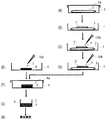
本発明の細菌分析方法は、特に限定されるものではないが、例えば、図1に示す各工程(A)〜(H)に従って実施することができる。
工程(A)に示すように、蓋1aの付いた適当な容器1[例えば、シャーレ(例えば、直径=60mm)]に、滅菌済みのプレフィルター2[例えば、吸収パッド(例えば、直径=47mm)]を入れた後、工程(B)に示すように、前記プレフィルター2の上に、滅菌済みのメンブレンフィルター3(例えば、孔径=0.8μm、直径=25mm)を載せる。前記メンブレンフィルター3上に、工程(C)に示すように、適当な供給手段10a(例えば、滅菌ピペット)で液状被検試料4(例えば、100μL〜100mL)を供給し、試料接触メンブレンフィルター3’を得る。液状被検試料4がプレフィルター2に吸収されるまで放置した後、前記試料接触メンブレンフィルター3’に、工程(D)に示すように、適当な供給手段10b(例えば、滅菌ピペット)で発光ファージ液5(例えば、液状被検試料と同量)を供給し、試料及び発光ファージ接触メンブレンフィルター3”を得る。前記試料及び発光ファージ接触メンブレンフィルター3”は、発光ファージ液5がプレフィルター2に吸収されるまで放置する。
【0023】
工程(E)に示すように、別の容器6[例えば、シャーレ(例えば、直径=60mm)]に、滅菌済みのプレフィルター(例えば、直径=25mm)を入れた後、適当な供給手段10c(例えば、滅菌ピペット)で適当量(例えば、650μL)の分析対象細菌用培地7[例えば、0.2%マルトース及び10mM硫酸マグネシウムを含むLB培地(LB−Mg−Mal培地)]を染み込ませることにより、分析対象細菌用培地吸収プレフィルター8が得られる。プレフィルターに吸収させる分析対象細菌用培地の液量は、プレフィルターの大きさ及び厚さに応じて適宜決定することができ、プレフィルターの底部から培地が容器内に流れ出る寸前まで充分に染み込ませることが好ましい。工程(F)に示すように、前記の分析対象細菌用培地吸収プレフィルター8上に、工程(D)で得られた試料及び発光ファージ接触メンブレンフィルター3”を重ねた後、試料及び発光ファージ接触メンブレンフィルター3”及び分析対象細菌用培地吸収プレフィルター8が乾燥しないように、容器6に蓋6aをし、適当な温度[例えば、10℃〜40℃、好ましくは室温(例えば、20℃)]にて所定時間(例えば、1〜4時間、好ましくは2〜3時間)インキュベートする。工程(G)に示すように、試料及び発光ファージ接触メンブレンフィルター3”及び分析対象細菌用培地吸収プレフィルター8を、あるいは、試料及び発光ファージ接触メンブレンフィルター3”のみを、発光測定機専用カップ9に入れた後、工程(H)に示すように、発光測定機にかけ、発光度を測定する。
【0024】
以上、発光遺伝子として、発光タンパク質が基質を必要としない発光遺伝子(例えば、GFP遺伝子)を用いる場合を例にとり、本発明の細菌分析方法を説明した。本発明の細菌分析方法の一態様である基質合成酵素利用型態様では、発光ファージとして、発光遺伝子(例えば、ルシフェラーゼ遺伝子)と基質合成酵素遺伝子(例えば、ルシフェラーゼ基質合成酵素遺伝子)とを導入したファージを用いること以外は、発光タンパク質が基質を必要としない発光遺伝子(例えば、GFP遺伝子)を用いる場合と同様にして、液状被検試料接触工程、ファージ液接触工程、及び分析工程をそれぞれ実施することができる。
【0025】
本発明の基質合成酵素利用型態様における分析工程では、試料及びファージ接触メンブレンフィルターと分析対象細菌用培地を吸収させた吸収パッドとを接触させると、液状被検試料中に分析対象細菌が存在する場合には、試料及びファージ接触メンブレンフィルター上に担持された分析対象細菌に感染した発光ファージが、前記分析対象細菌中で増殖すると共に、発光ファージに導入されている発光遺伝子(例えば、ルシフェラーゼ遺伝子)と基質合成酵素遺伝子(例えば、ルシフェラーゼ基質合成酵素遺伝子)とが発現する。基質合成酵素遺伝子が発現すると、発光タンパク質(例えば、ルシフェラーゼ)の基質(例えば、脂肪族アルデヒド)が分析対象細菌中で産生されるため、発光タンパク質による発光が生じる。分析対象細菌用培地を吸収させた吸収パッドに含まれている分析対象細菌用培地は、分析対象細菌の代謝を促進させ、その結果、発光ファージによる発光遺伝子及び基質合成酵素遺伝子の発現を促進し、更に、発光タンパク質の基質の産生を促進することができるため、発光を増強する作用を有する。
【0026】
本発明において、細菌分析方法の別の態様である基質添加型態様では、分析工程における試料及びファージ接触メンブレンフィルターと分析対象細菌用培地を吸収させた吸収パッドとの接触を、発光タンパク質(例えば、ルシフェラーゼ)の基質の存在下で実施すること以外は、発光タンパク質が基質を必要としない発光遺伝子(例えば、GFP遺伝子)を用いる場合と同様にして、液状被検試料接触工程、ファージ液接触工程、及び分析工程をそれぞれ実施することができる。
本発明の基質添加型態様において用いる発光タンパク質の基質は、使用する発光タンパク質に応じて適宜決定することができる。例えば、発光タンパク質としてルシフェラーゼを用いる場合には、その基質として、例えば、炭素数10〜20の飽和脂肪族アルデヒドを用いることができ、特にテトラデカナールが好ましい。テトラデカナールを用いる場合には、最終濃度が、好ましくは0.05〜2%、より好ましくは0.1〜0.25%となるように添加することができる。
【0027】
本発明の基質添加型態様において、発光タンパク質の基質を添加する方法は、分析工程において、試料及びファージ接触メンブレンフィルターと分析対象細菌用培地を吸収させた吸収パッドとを接触させ、所定時間経過した後、発光分析する直前に、前記試料及びファージ接触メンブレンフィルター上に前記基質が存在する限り、特に限定されるものではないが、例えば、
(1)分析対象細菌用培地を吸収させた吸収パッドに、分析対象細菌用培地と一緒に、基質を担持(例えば、吸収)させておく方法;
(2)試料及びファージ接触メンブレンフィルターと分析対象細菌用培地を吸収させた吸収パッドとを接触させ、所定時間経過した後、発光分析する直前に、前記試料及びファージ接触メンブレンフィルターに、基質液を接触させる方法;又は
(3)試料及びファージ接触メンブレンフィルターと分析対象細菌用培地を吸収させた吸収パッドとを接触させ、所定時間経過した後、発光分析する直前に、前記試料及びファージ接触メンブレンフィルターと基質液含有担体とを接触させる方法
などを挙げることができる。
【0028】
前記の基質添加方法(1)、すなわち、分析対象細菌用培地を吸収させた吸収パッドに、分析対象細菌用培地と一緒に、基質を担持させておく方法では、例えば、分析対象細菌用培地に基質を添加した混合液を、吸収パッドに吸収させることにより、あるいは、分析対象細菌用培地を吸収させた吸収パッドに、更に、基質液を吸収させることにより、分析対象細菌用培地を吸収させた吸収パッドに基質を担持させることができる。
【0029】
前記の基質添加方法(2)、すなわち、試料及びファージ接触メンブレンフィルターと分析対象細菌用培地を吸収させた吸収パッドとを接触させ、所定時間経過した後、発光分析する直前に、前記試料及びファージ接触メンブレンフィルターに、基質液を接触させる方法では、例えば、適当な容器(例えば、シャーレ)内に配置した試料及びファージ接触メンブレンフィルター上に、基質液を滴下することにより、試料及びファージ接触メンブレンフィルターに基質液を接触させることができる。この場合には、液体吸収体(例えば、吸収パッド)上に試料及びファージ接触メンブレンフィルターを配置した状態で、基質液を滴下することが好ましい。
【0030】
前記の基質添加方法(3)、すなわち、試料及びファージ接触メンブレンフィルターと分析対象細菌用培地を吸収させた吸収パッドとを接触させ、所定時間経過した後、発光分析する直前に、前記試料及びファージ接触メンブレンフィルターと基質液含有担体とを接触させる方法では、基質液含有担体として、例えば、基質液を吸収させた吸収パッドを挙げることができる。前記吸収パッドは、基質液を吸収可能であり、滅菌(例えば、オートクレーブ滅菌、エチレンオキサイド滅菌、又はγ線滅菌)可能であり、更に、試料及びファージ接触メンブレンフィルターと密着して重ねることができる限り、特に限定されるものではなく、例えば、多孔性フィルター、濾紙、スポンジ、又は綿などを挙げることができる。なお、前記基質液含有担体は、基質液を吸収させた後、湿潤状態のままで、試料及びファージ接触メンブレンフィルターとの接触に用いることもできるし、あるいは、一度、乾燥させ、乾燥状態で保存することもできる。乾燥状態の基質液含有担体を、試料及びファージ接触メンブレンフィルターとの接触に用いる場合には、適当な溶媒(例えば、水、生理食塩水、又は緩衝液等)で湿らせ、湿潤状態にした後、接触させることが好ましい。
【0031】
【実施例】
以下、実施例によって本発明を具体的に説明するが、これらは本発明の範囲を限定するものではない。
【実施例1】
《本発明の細菌分析方法における発光経時変化の測定》
(A)試験菌液の調製
発光ファージに感受性である大腸菌LE392株(ATCC 33572)をLB(Luria−Bertani)寒天平板培地に接種し、37℃で一晩培養して得られた1集落を、LB−Mg−Mal培地(0.2%マルトース及び10mmol/L硫酸マグネシウムを含むLB培地)50mLに接種した。37℃で一晩振とう培養した培養液1mLを、新たなLB−Mg−Mal培地10mLに接種し、更に37℃で2時間振とう培養したもの(1×109cfu/mL)を試験菌液とした。
【0032】
(B)発光ファージ液の調製
本実施例では、発光ファージとして、海洋細菌ビブリオ・フィシェリの発光遺伝子群を導入したλファージを使用した。具体的には、λファージの誘導体ベクターEMBL4を制限酵素EcoRIで切断したDNA断片の間に、プラスミドpHSK728(特開平10−179161号公報)を制限酵素EcoRIで切断して得られる10kbのDNA断片を挿入することにより、発光ファージを作製した。プラスミドpHSK728には、ビブリオ・フィシェリの発光遺伝子(lux)群(すなわち、ルシフェラーゼ遺伝子であるluxA遺伝子及びluxB遺伝子と、ルシフェラーゼ基質合成酵素遺伝子であるluxC遺伝子、luxD遺伝子、及びluxE遺伝子とが、5’から3’方向に向かって、luxC遺伝子、luxD遺伝子、luxA遺伝子、luxB遺伝子、及びluxE遺伝子の順に並んでいる遺伝子群)のカセットの直前に、マルチクローニング部位、挿入したウイルス遺伝子の翻訳を防ぐための3つの読み枠に配慮した終始暗号、及びシャイン・ダルガルノ(SD)配列を組み入れてある。
【0033】
得られた発光ファージ(λファージ:EMBL4)を、一般に行われている寒天重層法で増殖させ、クロロホルム添加後、4℃で保存したものを、一般的にファージの希釈に用いられるSMバッファーで10倍希釈して使用した(2×10⁸pfu/mL)。
【0034】
(C)発光経時変化の測定
本実施例の操作は、図1に示す手順で実施した。なお、以下の操作で用いる滅菌済みプレフィルター及びメンブレンフィルターは、121℃で15分高圧蒸気滅菌により滅菌した後、乾燥して使用した。
蓋付きの滅菌プラスチックシャーレ(直径=60mm)内に、滅菌した吸収パッド[プレフィルター(直径=47mm;ミリポア社)]を入れ、その上に滅菌したメンブレンフィルター(孔径=0.8μm,直径=25mm;ミリポア社)を重ねた。前項(A)で調製した菌液100μLを、前記メンブレンフィルター上に供給した。菌液がメンブレンフィルター上から見えなくなるまで放置した後、前項(B)で調製した発光ファージ液100μLを、更に、メンブレンフィルター上に供給し、発光ファージ液がメンブレンフィルター上から見えなくなるまで放置した。
【0035】
別の滅菌プラスチックシャーレ(直径=60mm)内に、滅菌したプレフィルター(直径=25mm;ミリポア社)を入れ、LB−Mg−Mal培地650μLを染み込ませた。続いて、このプレフィルター上に、先に菌液及び発光ファージ液を接触させたメンブレンフィルターを密着するように重ねた。
プレフィルターとメンブレンフィルターとを重ねた後、直ぐに、微弱発光測定機(浜松ホトニクス社)専用の発光測定カップ内に入れ、測定機にセットし、室内を20℃に設定し、1分間毎の発光カウントを60分間にわたって測定した。
【0036】
なお、対照(菌液のみをメンブレンフィルターに供給した場合)として、発光ファージ液をメンブレンフィルター上に供給しなかったこと以外は、発光経時変化測定のための前記操作を繰り返した。
また、比較例(死菌の場合)として、前項(A)で調製した菌液の代わりに、121℃で15分間高圧蒸気滅菌した菌液を用いたこと以外は、発光経時変化測定のための前記操作を繰り返した。
【0037】
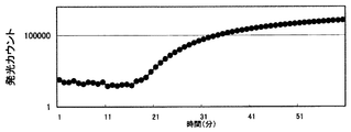
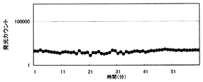
(D)結果
結果を図2〜図4に示す。図2〜図4は、それぞれ、前項(C)で実施した対照、実施例、及び比較例の結果である。図2〜図4において、縦軸は、発光カウント(対数表示)を示し、横軸は、プレフィルターとメンブレンフィルターとを重ねてからの経過時間を示す。
図2に示すように、発光ファージを供給しなかった場合(すなわち、菌液のみをメンブレンフィルターに供給した場合)には、1時間経過後においても発光カウントは変化しなかった(平均発光カウント=29cps)。
これに対して、図3に示すように、本発明方法によれば、約18分後に発光カウントが対数増加し、発光ファージに感染した菌が発光していることが明らかであった(1時間後の発光カウント=1,153,877cps)。
なお、図4に示すように、試験菌液として死菌を用いた場合には、図2に示す結果と同様に、発光カウントの増加は見られなかった(平均発光カウント=33cps)。死菌ではファージが感染しないため、図3に示す発光カウントは、菌が発光ファージに感染したための発光であることが証明された。
以上の結果から、本発明方法によれば、発光ファージにより細菌を検出することができることが判明した。
【0038】
【実施例2】
《LB−Mg−Mal培地の有無による発光カウントの比較》
(A)試験菌液の調製
実施例1(A)と同様に培養した菌を、LB−Mg−Mal培地で1×10⁴cfu/mL又は1×105cfu/mLに希釈した各菌液を試験菌液とした。
【0039】
(B)発光ファージ液の調製
実施例1(B)と同様に調製した希釈液(2×10⁸pfu/mL)を、発光ファージ液とした。
【0040】
(C)発光カウントの測定
(i)試験菌液として、前項(A)で調製した2種類の菌液(1×10⁴cfu/mL及び1×105cfu/mL)を使用したこと、そして、(ii)プレフィルターとメンブレンフィルターとを重ねた後、直ぐに、発光カウントを測定する代わりに、シャーレの蓋を閉じ、20℃で2時間静置した後、発光測定を実施したこと以外は、実施例1(C)の操作を繰り返した。なお、発光測定は、1秒間毎に10回測定した平均値が表示されるように設定して実施した。
【0041】
また、比較例(LB−Mg−Mal培地を染み込ませたプレフィルターを使用しない場合)として、(i)前項(A)で調製した2種類の菌液(1×10⁴cfu/mL及び1×105cfu/mL)を使用したこと、(ii)プレフィルターとメンブレンフィルターとを重ねた後、直ぐに、発光カウントを測定する代わりに、シャーレの蓋を閉じ、20℃で2時間静置した後、発光測定を実施したこと、そして、(iii)LB−Mg−Mal培地650μLを染み込ませたプレフィルターの代わりに、培地を染み込ませていないプレフィルターを用いること以外は、実施例1(C)の操作を繰り返した。
【0042】
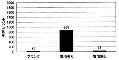
(D)結果
結果を図5及び図6に示す。
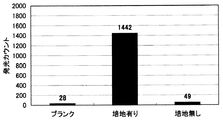
図5は、試験菌液として、1×10⁴cfu/mLに希釈した菌液を用いた場合の結果であり、図6は、1×105cfu/mLに希釈した菌液を用いた場合の結果である。図5及び図6において、「ブランク」は、対照(菌液のみをメンブレンフィルターに供給した場合)の結果を意味し、「培地有り」は、実施例の結果を意味し、「培地無し」は、比較例の結果を意味する。
図5に示すように、1×10⁴cfu/mLに希釈した菌液を用いた場合には、培地を染み込ませたプレフィルター上に、菌液と発光ファージ液とを供給したメングレンフィルターを重ねた方[実施例(培地有り)]が、あきらかに発光カウントが高く、この操作を行わなかった培地無しの系(比較例)では、ブランク(対照)カウントとほぼ同等のカウントしか得られなかった。
更に、図6に示すように、1×10⁵cfu/mLに希釈した菌液を使用した場合においても同様な結果が得られた。
以上の結果から、培地を染み込ませたプレフィルター上に、菌液とファージ液とを供給したメンブレンフィルターを重ねると、プレフィルターに染み込ませた培地により、菌の代謝が活発になり、菌体内に挿入された発光遺伝子群によるルシフェラーゼとその基質脂肪族アルデヒドの産生性が高まり、発光することが判明した。
【0043】
【実施例3】
《メンブレンフィルター上に供給する試験菌液量及び発光ファージ液量による発光カウントの比較》
(A)試験菌液の調製
実施例1(A)と同様に培養した菌を、LB−Mg−Mal培地で100cfu/mLに希釈した各菌液を試験菌液とした。
【0044】
(B)発光ファージ液の調製
実施例1(B)と同様に調製した希釈液(2×10⁸pfu/mL)を、発光ファージ液とした。
【0045】
(C)発光カウントの測定
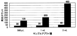
(i)試験菌液として、前項(A)で調製した菌液(100cfu/mL)を使用したこと、そして、(ii)メンブレンフィルターへの試験菌液及び発光ファージ液の各供給量を、500μL、1mL、及び3mLの3段階としたこと以外は、実施例2(C)の操作を繰り返した。
なお、メンブレンフィルターへの試験菌液及び発光ファージ液の各供給量が500μLの場合には、メンブレンフィルターの下に配置する吸収パッド(プレフィルター)を、実施例2(C)と同様に、1枚とし、前記各供給量が1mLの場合には吸収パッドを2枚とし、前記各供給量が3mLの場合には吸収パッドを3枚とした。
【0046】
(D)結果
結果を図7に示す。
図7において、黒棒は、実施例の結果を示し、灰色棒は、ブランク(菌液のみをメンブレンフィルターに供給した場合)の結果を示す。
図7に示すように、メンブレンフィルター上に供給する試験菌量及び発光ファージ量が多い程、発光カウントが上昇した。従って、サンプル量が多いほど、ブランク値に対するSN比が上がり、有利になる。本発明方法では、吸収パットの吸収量を上げる(例えば、吸収パットを大きくしたり、厚くしたり、あるいは、枚数を重ねる)ことで、大量の試験菌及び発光ファージを供給することができる優れた方法であることが判明した。
【0047】
【実施例4】
《検出感度の評価》
(A)試験菌液の調製
実施例1(A)と同様に培養した菌を、LB−Mg−Mal培地で、1000cfu/mL、100cfu/mL、10cfu/mL、又は1cfu/mLに希釈した4種類の菌液を試験菌液とした。
【0048】
(B)発光ファージ液の調製
実施例1(B)と同様に調製した希釈液(2×10⁸pfu/mL)を、発光ファージ液とした。
【0049】
(C)発光カウントの測定
(i)試験菌液として、前項(A)で調製した4種類の菌液(1000cfu/mL、100cfu/mL、10cfu/mL、及び1cfu/mL)を使用したこと、そして、(ii)メンブレンフィルターへの試験菌液及び発光ファージ液の各供給量を、3mLとしたこと以外は、実施例3(C)の操作を繰り返した。
なお、メンブレンフィルター上に供給された菌数を証明するために、同様にメンブレンフィルター上に各希釈試験菌液3mLを供給したメンブレンをハートインフュージョン寒天平板培地上に置き、37℃で一晩培養した後、メンブレンフィルター上の集落数を測定した。
【0050】
(D)結果
結果を図8〜図10に示す。図8は、試験菌液として、1cfu/mL又は10cfu/mLに希釈した菌液を用いた場合の結果であり、図9は、100cfu/mL又は1000cfu/mLに希釈した菌液を用いた場合の結果である。図8及び図9において、黒棒は、実施例の結果を示し、灰色棒は、ブランク(菌液のみをメンブレンフィルターに供給した場合)の結果を示す。また、図10は、菌液の菌数と発光カウントとの定量性を示すグラフである。図10の縦軸及び横軸は対数表示である。
【0051】
図8及ぶ図9に示すように、1cfu/mLの菌数試料を測定することができ、しかも、同一菌数サンプルを供給した後、培養したメンブレンフィルター上には、1cfu/mLの菌液を供給したメンブレンフィルターから1個の集落が確認することができたことから、本発明方法によれば、1個の菌を2時間で検出可能であることが判明した。
また、どの菌数においても、感染させる発光ファージ数は2×10⁸pfu/mLの同一ファージ液を使用することができ、1濃度のファージ液の使用で行えることが確認された。
更に、図10に示すように、定量性も認められ、発光カウントから試料中の菌数を知ることができる。
【0052】
【発明の効果】
本発明方法によれば、(1)メンブレンフイルター上に固定化された細菌に、発光ファージを通過させることで、感染確率を高めることによって;
(2)分析対象細菌用培地を吸収させた吸収パッド上に試料及びファージ接触メンブレンフィルターを重ねることで、発光ファージが細菌に感染後、菌体内で発光タンパク質(又は発光タンパク質及びその基質)の産生を促進することによって;
好ましくは更に、
(3)メンブレンフィルター下に液体吸収体を用いることで、液状被検試料の供給量を飛躍的に高めることによって;
1個の細菌の検出であっても短時間(例えば、2時間)で検出することができる。しかも、操作が極めて簡便であることの効果は絶大である。
【0053】
更に、本発明方法によれば、発光ファージを代えることにより、分析対象細菌が1mL当たり数個であっても、細菌の検出が可能である。従って、臨床細菌検査分野では、現在問題になっている薬剤耐性菌のふき取り検査による検出、あるいは、感染症の起因菌の検出等に応用することができ、食品細菌検査分野では、食中毒原因菌の検出等に、また、無菌状態でなければならない各種試料の細菌検査に、更に培養に時間を要する菌の検出等に応用することができる。しかも、本発明方法によれば、細菌検査の抱えている現状の問題点である培養に時間がかかる点や、操作が繁雑である点を一挙に解決することができる。従って、臨床分野では院内感染防止を未然に防ぐことができ、食品検査では食中毒を未然に防ぐことができ、細菌検査の現状を大きく変えることができる画期的な効果を有するものである。
【0054】
以上、述べたように、本発明方法の特徴は、(1)同一メンブレンフィルター上に発光ファージを供給できる簡便性と、(2)そのメンブレンフィルターを、分析対象細菌用培地を吸収させた吸収パッド上に移動させるだけで、メンブレンフィルター上の分析対象細菌をそのまま発光測定することができる迅速性である。また、本発明方法の好適態様における更なる特徴は、(3)液体吸収体上のメンブレンフィルターに、分析対象細菌を含む液状被検試料を大量に供給することで、分析対照細菌を濃縮及び固定することによる高感度化である。
【図面の簡単な説明】
【図1】本発明の細菌分析方法による細菌分析操作法を模式的に示す説明図である。
【図2】対照(発光ファージを供給しなかった場合)における発光経時変化を示した図である。
【図3】本発明方法による発光経時変化を示した図である。
【図4】比較例(死菌の場合)における発光経時変化を示した図である。
【図5】本発明方法において、培地の有無による発光カウントを比較した結果(試験菌数=1×10⁴cfu/mL)を示すグラフである。
【図6】本発明方法において、培地の有無による発光カウントを比較した結果(試験菌数=1×10⁵cfu/mL)を示すグラフである。
【図7】本発明方法において、試験菌液量及び発光ファージ液量による発光カウントを比較した結果を示すグラフである。
【図8】本発明方法において、試験菌液量(試験菌数=1cfu/mL又は10cfu/mL)による発光カウントを比較した結果を示すグラフである。
【図9】本発明方法において、試験菌液量(試験菌数=100cfu/mL又は1000cfu/mL)による発光カウントを比較した結果を示すグラフである。
【図10】本発明方法における、菌液の菌数と発光カウントとの定量性を示すグラフである。
【符号の説明】
1・・・シャーレ;2・・・プレフィルター;3・・・メンブレンフィルター;
4・・・液状被検試料;5・・・発光ファージ液;6・・・シャーレ;
7・・・分析対象細菌用培地;
8・・・分析対象細菌用培地吸収プレフィルター;
9・・・発光測定機専用カップ。[0001]
BACKGROUND OF THE INVENTION
The present invention relates to a novel bacterial analysis method.
[0002]
[Prior art]
Conventional detection and identification of bacteria are usually performed by purely cultivating isolated colonies on a separation medium and then comparing the biochemical properties with a discrimination table. However, such a method requires many biochemical characterization tests corresponding to the analysis target bacteria, and requires 24 to 72 hours, or one week or more depending on the test item. In addition, some bacteria to be analyzed require several days or weeks even for culturing alone, and enormous labor and time are required until a test result is obtained.
[0003]
[Problems to be solved by the invention]
As described above, the operation of detecting and identifying a specific bacterium is complicated and time-consuming in food bacterium test, clinical bacterium test, or environmental bacterium test. In addition, there are many cases where there are only a small number of bacteria to be analyzed in a sample, and therefore, a culture operation is essential. An object of the present invention is to provide a method that can easily and quickly analyze even a few bacteria to be analyzed, focusing on these problems.
[0004]
[Means for Solving the Problems]
The above-mentioned problem is that according to the present invention, (1) a liquid test sample that may contain an analysis target bacterium, Membrane filter Contacting with;
(2) The liquid test sample obtained in the step (1) was contacted Membrane filter In addition, a phage solution into which a luminescent gene has been introduced Let through Steps; and
(3) Liquid test obtained in step (2) Charge Contact , Membrane filter through which phage solution was passed When, Absorption pad with medium for bacteria to be analyzed Contact with the Membrane filter For analyzing luminescence in water
It can solve by the analysis method of bacteria characterized by including this.
“Analysis” in the present specification includes both “detection” for determining the presence or absence of the analysis target bacteria and “measurement” for quantitatively or semi-quantitatively determining the number of analysis target bacteria.
[0005]
DETAILED DESCRIPTION OF THE INVENTION
The bacterial analysis method of the present invention comprises:
(1) Membrane filter And a step of bringing a liquid test sample possibly containing the analysis target bacteria into contact (hereinafter referred to as a liquid test sample contact step);
(2) The liquid test sample obtained in the liquid test sample contact step (1) was contacted Membrane filter (Liquid test sample contact Membrane filter Hereafter, simply “sample contact Membrane filter And a phage solution into which a luminescent gene has been introduced (hereinafter also referred to as a luminescent phage) (hereinafter referred to as a phage solution contact step);
(3) The liquid test sample obtained in the phage solution contact step (2) was brought into contact with the phage solution. Membrane filter (Liquid test sample and phage solution contact Membrane filter ; Hereinafter, simply “sample and phage contact Membrane filter ”), Absorption pad with medium for bacteria to be analyzed The sample and phage contact Membrane filter Process for analyzing luminescence in the sample (hereinafter referred to as analysis process)
including.
[0006]
The analysis target bacteria that can be analyzed by the bacterial analysis method of the present invention are not particularly limited as long as the phages capable of infecting the analysis target bacteria are known. Further, the test sample that can be analyzed by the bacterial analysis method of the present invention is not particularly limited as long as it is a test sample that may contain the analysis target bacteria. For example, food, clinical, environment, or Various specimens in a bacterial test such as a chemical product can be used.
Examples of food samples include drinking water, various juices, milk, milk drinks, dairy products, eggs or liquid eggs, egg processed products, meat, processed meat products, seafood, processed fish products, vegetables, fruits, fresh confectionery, Examples include cereal products or prepared dishes.
Clinical specimens include, for example, blood, urine, stool, sputum, throat swab, spinal fluid, pleural effusion, bile, other body fluids, skin, mucous membranes, hair follicles, lacrimal fluid, corneal scrapings, vaginal contents, Or the uterine contents etc. can be mentioned.
Examples of the environmental sample include industrial water, a wipe test solution, waste water, or airborne bacteria.
In addition, the raw material of chemical products, an intermediate product, or a product, a pharmaceutical, an injection solution, an infusion solution, or livestock feed can be mentioned.
[0007]
In the method for analyzing bacteria of the present invention, these test samples are used in a liquid state. Therefore, when the test sample is a liquid sample, it may be used as it is or a diluted solution diluted with an appropriate solvent may be used. it can. In the case of a solid specimen, the specimen is placed in an appropriate solvent (for example, water, physiological saline, buffer solution, or culture medium) and, for example, treated by stomacher to make it liquid or washed out. If desired, a liquid obtained by removing the crushed sample or turbidity by pre-filtering or low-speed centrifugation (for example, 1000 rpm / 1 minute for 5 minutes) or the like can be used. Further, even in the case of a liquid sample, if it is extremely turbid or has suspended matter, it is preferable to perform the same operation as that for a solid sample.
[0008]
Used in liquid test sample contact process Rume Filter Is The material, the filter pore diameter, the filter shape and size, the color, and the like are not particularly limited as long as the analysis target bacteria can be supported on the bacteria support.
Said Membrane filter ー Examples of the material include cellulose acetate, polycarbonate, cellulose mixed ester cellulose nitrate, regenerated cellulose, polytetrafluoroethylene (PTFE), polyamide, cellulose triacetate, and polysulfone. The pore size of the membrane filter can be, for example, 0.2 μm to 3 μm. Examples of the shape of the membrane filter include a circle or an ellipse, or a polygon (for example, a square or a rectangle). In the case of a circle, the diameter thereof is, for example, 5 mm to 90 mm. it can. The color of the membrane filter is not particularly limited as long as it does not hinder emission analysis in the analysis process. In addition, it is preferable to use a membrane filter that has been processed with hydrophobic peripheral edges because it is possible to prevent leakage of bacterial sample liquid or luminescent phage liquid from the peripheral edge of the membrane filter.
[0009]
In the bacterial analysis method of the present invention, a phage into which a luminescent gene is introduced is used as the luminescent phage used in the phage solution contact step.
The luminescent gene used in the method for analyzing bacteria of the present invention is not particularly limited as long as it is a gene encoding a luminescent protein capable of producing at least a luminescent level that can be analyzed when a luminescent phage is infected with a bacterium to be analyzed. Examples thereof include a luciferase gene, a green fluorescent protein (GFP) gene, and a blue fluorescent protein (BFP) gene.
Examples of the luciferase gene include various luciferase genes obtained from microorganisms, insects, crustaceans, mollusks, enemas, fish, and the like. From the viewpoint of ease of handling, it is preferable to use the luxA gene and the luxB gene which are luciferase gene groups of marine bacteria [particularly Vibrio fischeri].
The green fluorescent protein is a photoprotein that exists in Aequorea jellyfish, 2+ And no substrate is required. In addition, proteins similar to green fluorescent protein have been confirmed from organisms other than Aequorea jellyfish, and genes encoding these similar proteins can also be used as luminescent genes.
[0010]
As long as the phage used for introducing the luminescent gene (that is, the phage before introducing the luminescent gene) is a phage that can infect the bacterium to be analyzed and can be introduced in a state in which the luminescent gene can be expressed. However, it is not particularly limited, and can be appropriately determined according to the type of bacteria to be analyzed. Moreover, it is preferable that it is a phage which raise | generates a phage proliferation and lysis quickly, without lysogenizing after infecting the analysis object bacteria.
For example, when the bacterium to be analyzed is Escherichia coli, for example, T4 phage, P2 phage, T2 phage, T7 phage, λ phage, φX174 phage, MS2 phage, or λ phage improved for cloning (for example, EMBL3 Alternatively, EMBL4) can be used. When the bacterium to be analyzed is a bacterium belonging to the genus Xanthomonas (for example, Xanthomonas campestris pv.citri), for example, CP1 phage, CP2 phage, or CP3 phage can be used. When the bacterium to be analyzed is Pseudomonas, for example, φ6 phage or PM2 phage can be used. In addition, when the analysis target bacteria is Salmonella, for example, FELIX01 phage can be used. In addition, any known phage that can specifically bind to the bacterium to be analyzed can be used.
[0011]
In the method for analyzing bacteria according to the present invention, when a luminescent protein in which a luminescent gene that requires a substrate (for example, a luciferase gene) is used as a luminescent phage, an analysis in which the luminescent phage is infected in an analysis step described later. It is necessary that the substrate of the photoprotein is present in the analysis target bacterium so that luminescence occurs in the target bacterium. Based on the difference in the method in which the substrate is present, the bacterial analysis method of the present invention includes, for example,
(A) an embodiment in which a phage into which a gene encoding a synthase of a substrate of a photoprotein encoded by a luminescent gene is further introduced as the luminescent phage (hereinafter referred to as a substrate synthase utilization type embodiment), or
(B) In the analysis step, sample and phage contact in the presence of a substrate of a photoprotein encoded by a photoluminescent gene Membrane filter When Absorption pad with medium for bacteria to be analyzed (Hereinafter referred to as a substrate addition type embodiment)
Can be mentioned.
[0012]
In a substrate synthesizing enzyme utilization type embodiment which is an embodiment of the bacterial analysis method of the present invention, a luminescent phage and a gene encoding a synthesizing enzyme of a substrate of a luminescent protein encoded by the luminescent gene (hereinafter referred to as a substrate) (Referred to as a synthase gene).
The substrate synthase gene used in the substrate synthase utilization mode is an enzyme capable of synthesizing a substrate of a luminescent protein encoded by a luminescent gene when the luminescent phage is infected with a bacterium to be analyzed. The gene is not particularly limited as long as it is a gene to be encoded, and can be appropriately selected according to the type of luminescent gene to be used.
For example, when a luciferase gene is used as the luminescent gene, for example, a luciferase substrate synthase gene, that is, a gene or gene group encoding an enzyme or enzyme group that produces an aliphatic aldehyde that is a luciferase substrate can be used. . Examples of the luciferase substrate synthase gene include various luciferase substrate synthase genes obtained from microorganisms, insects, shellfish, jellyfish, etc., and marine bacteria (particularly, in terms of ease of handling). It is preferable to use luxC gene, luxD gene, and luxE gene which are luciferase substrate synthase gene groups of Vibrio Fischeri (that is, gene groups encoding aldehyde synthase groups). The lux gene group of the marine bacterium includes a luciferase gene, luxA gene and luxB gene, and a luciferase substrate synthase gene, luxC gene, luxD gene, and luxE gene, in a 5 ′ to 3 ′ direction. , LuxC gene, luxD gene, luxA gene, luxB gene, and luxE gene.
[0013]
Used in the analysis process Minute Absorption pad that absorbs bacterial culture medium Do Can absorb the culture medium for the bacteria to be analyzed, can be sterilized (for example, autoclave sterilization, ethylene oxide sterilization, or γ-ray sterilization). Membrane filter As long as it can be closely adhered and stacked, it is not particularly limited, and examples thereof include a porous filter, a filter paper, and a sponge.
[0014]
Hereinafter, the method for analyzing bacteria according to the present invention will be further described along with each implementation step, taking as an example the case where a luminescent gene that does not require a substrate (for example, a GFP gene) is used as the luminescent gene.
In the liquid test sample contact step in the bacterial analysis method of the present invention, the liquid test sample and Membrane filter If the analysis target bacteria are contained in the liquid test sample, the analysis target bacteria Membrane filter To support. Membrane filter The method of bringing the liquid test sample into contact with the liquid is not particularly limited, and for example, it is placed in an appropriate container (for example, a petri dish). Membrane filter On top of this, the liquid test sample can be dropped, or in a liquid test sample placed in an appropriate container, Membrane filter Can also be immersed.
[0015]
In the bacterial analysis method of the present invention, the liquid absorbent (for example, an absorbent pad) is used for the above-described analysis. Membrane filter It is preferable to carry out the liquid test sample contacting step in a state where the is placed. More specifically, Membrane filter In a state where one surface of the liquid crystal is in contact with the liquid absorber. Membrane filter By adding a liquid test sample to the opposite side of Membrane filter A liquid test sample can be brought into contact with the liquid. In the liquid test sample contact process, if a liquid absorber is used, Membrane filter A large amount of liquid test sample can be passed through in a short time. As a result, if the liquid test sample contains analysis target bacteria, the analysis target bacteria are concentrated. Membrane filter It can be supported on.
[0016]
In the phage solution contact step in the bacterial analysis method of the present invention, the sample contact obtained in the liquid test sample contact step Membrane filter And a luminescent phage solution are brought into contact with each other. Sample contact Membrane filter (I.e., the analysis target bacteria are contained in the liquid test sample, and the analysis target bacteria are not contained in the liquid test sample contact step). Membrane filter The luminescent phage is Membrane filter Infects the above target bacteria. Sample contact Membrane filter The method of bringing the luminescent phage solution into contact with the sample is not particularly limited. For example, the sample is placed in a suitable container (eg, petri dish). Membrane filter On top of this, a luminescent phage solution can be dropped.
[0017]
In the method for analyzing bacteria according to the present invention, the sample is contacted on a liquid absorber (for example, an absorbent pad). Membrane filter It is preferable to carry out the phage solution contact step in a state where the is placed. More specifically, sample contact Membrane filter The sample contact in a state where one surface (the surface opposite to the surface to which the liquid test sample is supplied in the liquid test sample contact process) is in contact with the liquid absorber Membrane filter Sample contact by adding luminescent phage solution to the opposite side of Membrane filter Can be contacted with a luminescent phage solution.
[0018]
As long as the liquid absorber that can be used in the liquid sample contact step and / or the phage liquid contact step is a liquid absorber that can be sterilized (for example, autoclave sterilization, ethylene oxide sterilization, or γ-ray sterilization), Although not limited, For example, a porous filter, filter paper, sponge, cotton etc. can be mentioned. The color, shape, or size of the liquid absorber is not particularly limited, and can be appropriately determined according to the amount of the liquid test sample and / or the luminescent phage solution, for example. Further, two or more liquid absorbers can be used in an overlapping manner.
[0019]
In the analysis step of the bacterial analysis method of the present invention, the sample obtained in the phage solution contact step and the phage contact Membrane filter When, Absorption pad with medium for bacteria to be analyzed And contact the sample and the phage after a predetermined time has passed. Membrane filter Analyze luminescence at.
Sample and phage contact Membrane filter When Absorption pad with medium for bacteria to be analyzed If the target bacteria are present in the liquid test sample, contact with the sample and phage Membrane filter Luminescent phages infected with the bacterium to be analyzed carried thereon grow in the bacterium to be analyzed, and a luminescent gene (for example, a GFP gene) introduced into the luminescent phage is expressed to produce luminescence. Absorption pad with medium for bacteria to be analyzed The medium for the bacterium to be analyzed contained in the sample promotes the metabolism of the bacterium to be analyzed and, as a result, can promote the expression of the luminescent gene by the luminescent phage, and thus has an action of enhancing luminescence.
[0020]
Sample and phage contact in the analysis process Membrane filter When Absorption pad with medium for bacteria to be analyzed The sample and phage contact Membrane filter The time (ie, contact time) until the luminescence is analyzed is determined by the fact that the luminescent phage infected with the analysis target bacteria grows in the analysis target bacteria when the analysis target bacteria exist in the liquid test sample. As long as it is a sufficient time for the luminescent gene introduced into the luminescent phage to be expressed and luminescence to occur, it is not particularly limited, and is appropriately determined according to the type of bacteria to be analyzed and the type of phage to be used. be able to. For example, when the bacterium to be analyzed is Escherichia coli and λ phage is used as the phage, it is usually 20 minutes to 4 hours, preferably 1 hour to 3 hours.
[0021]
Luminescence can be analyzed by using a known luminescence analysis method, for example, a known means such as a photometer, a chemiluminescence reader, a scintillation counter, or a luminometer. Furthermore, it is possible to analyze by sensitivity of a high-sensitivity film or image analysis by a high-sensitivity video camera.
[0022]
The method for analyzing bacteria of the present invention is not particularly limited, and can be carried out according to the steps (A) to (H) shown in FIG.
As shown in step (A), in a suitable container 1 [for example, petri dish (for example, diameter = 60 mm)] with a
[0023]
As shown in the step (E), after putting a sterilized prefilter (for example, diameter = 25 mm) into another container 6 [for example, a petri dish (for example, diameter = 60 mm)], an appropriate supply means 10c ( For example, by immersing an appropriate amount (for example, 650 μL) of the
[0024]
As described above, the method for analyzing bacteria according to the present invention has been described by taking as an example the case where a luminescent gene (for example, GFP gene) that does not require a substrate is used as a luminescent protein. In the substrate synthase-utilizing mode that is one mode of the bacterial analysis method of the present invention, a luminescent gene (for example, luciferase gene) and a substrate synthase gene (for example, luciferase substrate synthase gene) are introduced as luminescent phages. Except for the use of phage, the liquid test sample contact step, the phage solution contact step, and the analysis step are performed in the same manner as when the photoprotein uses a luminescent gene that does not require a substrate (for example, a GFP gene). be able to.
[0025]
In the analysis step in the substrate synthase utilization type aspect of the present invention, the sample and the phage contact Membrane filter When Absorption pad with medium for bacteria to be analyzed If the target bacteria are present in the liquid test sample, contact with the sample and phage Membrane filter Luminescent phages infected with the bacterium to be analyzed carried thereon grow in the bacterium to be analyzed, and a luminescent gene (for example, luciferase gene) introduced into the luminescent phage and a substrate synthase gene (for example, luciferase substrate) Synthase gene). When the substrate synthase gene is expressed, a photoprotein (for example, luciferase) substrate (for example, an aliphatic aldehyde) is produced in the bacterium to be analyzed. Absorption pad with medium for bacteria to be analyzed The medium for the bacteria to be analyzed contained in the sample promotes the metabolism of the bacteria to be analyzed, and as a result, promotes the expression of the luminescent gene and the substrate synthase gene by the luminescent phage, and further promotes the production of the substrate of the luminescent protein. Therefore, it has an effect of enhancing luminescence.
[0026]
In the present invention, in the substrate addition type aspect which is another aspect of the bacterial analysis method, the sample and phage contact in the analysis step Membrane filter When Absorption pad with medium for bacteria to be analyzed Except that the photoprotein is used in the presence of a substrate of a photoprotein (eg, luciferase), as in the case of using a photoprotein (eg, GFP gene) that does not require a substrate. A test sample contact step, a phage solution contact step, and an analysis step can be performed.
The substrate of the photoprotein used in the substrate addition type embodiment of the present invention can be appropriately determined according to the photoprotein used. For example, when luciferase is used as the photoprotein, for example, a saturated aliphatic aldehyde having 10 to 20 carbon atoms can be used as the substrate, and tetradecanal is particularly preferable. When tetradecanal is used, it can be added so that the final concentration is preferably 0.05 to 2%, more preferably 0.1 to 0.25%.
[0027]
In the substrate addition type embodiment of the present invention, the method of adding a substrate of a photoprotein is a sample and phage contact in the analysis step. Membrane filter When Absorption pad with medium for bacteria to be analyzed Contact the sample and the phage immediately after the elapse of a predetermined time and immediately before the luminescence analysis. Membrane filter As long as the substrate is present on the substrate, it is not particularly limited.
(1) Absorption pad with medium for bacteria to be analyzed And a method of supporting (eg, absorbing) the substrate together with the medium for the bacteria to be analyzed;
(2) Sample and phage contact Membrane filter When Absorption pad with medium for bacteria to be analyzed Contact the sample and the phage immediately after the elapse of a predetermined time and immediately before the luminescence analysis. Membrane filter A method of contacting a substrate solution with
(3) Sample and phage contact Membrane filter When Absorption pad with medium for bacteria to be analyzed Contact the sample and the phage immediately after the elapse of a predetermined time and immediately before the luminescence analysis. Membrane filter Method of contacting substrate with substrate solution
And so on.
[0028]
Said substrate addition method (1), ie, Absorption pad with medium for bacteria to be analyzed In addition, in the method of supporting the substrate together with the culture medium for the analysis target bacteria, for example, the mixed solution obtained by adding the substrate to the culture medium for the analysis target bacteria is absorbed by the absorption pad, or for the analysis target bacteria. By further absorbing the substrate solution into the absorption pad that has absorbed the medium, Absorption pad with medium for bacteria to be analyzed The substrate can be supported on the substrate.
[0029]
Substrate addition method (2) above, ie sample and phage contact Membrane filter When Absorption pad with medium for bacteria to be analyzed Contact the sample and the phage immediately after the elapse of a predetermined time and immediately before the luminescence analysis. Membrane filter Further, in the method of contacting the substrate solution, for example, the sample placed in an appropriate container (eg, petri dish) and the phage contact Membrane filter Sample and phage contact by dropping substrate solution on top Membrane filter Can be contacted with the substrate solution. In this case, sample and phage contact on a liquid absorber (eg, an absorbent pad) Membrane filter It is preferable to drop the substrate solution in a state where the is placed.
[0030]
Said substrate addition method (3), ie sample and phage contact Membrane filter When Absorption pad with medium for bacteria to be analyzed Contact the sample and the phage immediately after the elapse of a predetermined time and immediately before the luminescence analysis. Membrane filter In the method of bringing the substrate liquid-containing carrier into contact, examples of the substrate liquid-containing carrier include an absorbent pad that has absorbed the substrate liquid. The absorption pad can absorb the substrate solution and can be sterilized (for example, autoclave sterilization, ethylene oxide sterilization, or γ-ray sterilization). Membrane filter As long as it can be closely adhered and stacked, it is not particularly limited, and examples thereof include a porous filter, filter paper, sponge, and cotton. The substrate liquid-containing carrier is in contact with the sample and the phage in a wet state after absorbing the substrate liquid. Membrane filter It can also be used for contact with the substrate, or it can be dried once and stored in a dry state. Contact substrate and phage with dried substrate solution Membrane filter In the case of using for contact with water, it is preferable to wet the substrate with an appropriate solvent (for example, water, physiological saline or buffer solution), bring it into a wet state, and then contact it.
[0031]
【Example】
EXAMPLES Hereinafter, the present invention will be specifically described by way of examples, but these do not limit the scope of the present invention.
[Example 1]
<< Measurement of luminescence change over time in the bacterial analysis method of the present invention >>
(A) Preparation of test bacterial solution
One colony obtained by inoculating Escherichia coli LE392 strain (ATCC 33572) sensitive to luminescent phages on LB (Luria-Bertani) agar plate medium and culturing overnight at 37 ° C. was obtained from LB-Mg-Mal medium (0 (LB medium containing 2% maltose and 10 mmol / L magnesium sulfate) was inoculated into 50 mL. 1 mL of a culture solution cultured overnight at 37 ° C. with shaking is inoculated into 10 mL of a new LB-Mg-Mal medium, and further cultured with shaking at 37 ° C. for 2 hours (1 × 10 9 cfu / mL) was used as a test bacterial solution.
[0032]
(B) Preparation of luminescent phage solution
In this example, λ phage into which a luminescent gene group of the marine bacterium Vibrio fischeri was introduced was used as the luminescent phage. Specifically, a 10-kb DNA fragment obtained by cleaving plasmid pHSK728 (Japanese Patent Laid-Open No. 10-179161) with restriction enzyme EcoRI between DNA fragments obtained by cleaving λ phage derivative vector EMBL4 with restriction enzyme EcoRI. A luminescent phage was prepared by insertion. The plasmid pHSK728 includes a Vibrio Fischer luminescent gene (lux) group (that is, a luciferase gene, luxA gene and luxB gene, and a luciferase substrate synthase gene, luxC gene, luxD gene, and luxE gene. To prevent translation of the multicloning site and the inserted viral gene immediately before the cassette of luxC gene, luxD gene, luxA gene, luxB gene, and luxE gene in the order from 3 to 3 ' The entire code that takes into account the three reading frames, and the Shine-Dalgarno (SD) sequence are incorporated.
[0033]
The resulting luminescent phage (λ phage: EMBL4) was grown by a conventional agar overlay method, added with chloroform, and stored at 4 ° C., with 10 SM buffer generally used for dilution of phage. Used diluted 2 times (2 x 10 8 pfu / mL).
[0034]
(C) Measurement of luminescence change with time
The operation of this example was performed according to the procedure shown in FIG. The sterilized prefilter and membrane filter used in the following operations were sterilized by high-pressure steam sterilization at 121 ° C. for 15 minutes, and then used after drying.
A sterilized absorbent pad [prefilter (diameter = 47 mm; Millipore)] is placed in a sterilized plastic petri dish (diameter = 60 mm) with a lid, and a sterilized membrane filter (pore diameter = 0.8 μm, diameter = 25 mm) is placed thereon. ; Millipore). 100 μL of the bacterial solution prepared in the previous section (A) was supplied onto the membrane filter. After leaving the bacterial solution to be invisible on the membrane filter, 100 μL of the luminescent phage solution prepared in the previous section (B) was further supplied onto the membrane filter and allowed to stand until the luminescent phage solution was no longer visible on the membrane filter.
[0035]
In another sterilized plastic petri dish (diameter = 60 mm), a sterilized pre-filter (diameter = 25 mm; Millipore) was put and 650 μL of LB-Mg-Mal medium was infiltrated. Subsequently, the membrane filter previously contacted with the bacterial solution and the luminescent phage solution was overlaid on the prefilter so as to be in close contact therewith.
Immediately after stacking the pre-filter and membrane filter, place it in a luminescence measuring cup dedicated to the weak luminescence measuring device (Hamamatsu Photonics), set it in the measuring device, set the room to 20 ° C, and emit light every minute. Counts were measured over 60 minutes.
[0036]
In addition, as a control (when only the bacterial solution was supplied to the membrane filter), the above-described operation for measurement of luminescence with time was repeated except that the luminescent phage solution was not supplied onto the membrane filter.
In addition, as a comparative example (in the case of dead bacteria), in place of the bacterial solution prepared in the previous section (A), a bacterial solution sterilized by autoclaving at 121 ° C. for 15 minutes was used for measurement of luminescence over time. The above operation was repeated.
[0037]
(D) Result
The results are shown in FIGS. 2 to 4 show the results of the control, the example, and the comparative example implemented in the previous section (C), respectively. 2 to 4, the vertical axis represents the light emission count (logarithmic display), and the horizontal axis represents the elapsed time since the prefilter and the membrane filter were overlapped.
As shown in FIG. 2, when no luminescent phage was supplied (that is, when only the bacterial solution was supplied to the membrane filter), the luminescence count did not change even after 1 hour (average luminescence count = 29 cps).
On the other hand, as shown in FIG. 3, according to the method of the present invention, the luminescence count increased logarithmically after about 18 minutes, and it was clear that the bacteria infected with the luminescent phage were luminescent (1 hour). Later luminescence count = 1,153,877 cps).
As shown in FIG. 4, when dead bacteria were used as the test bacterial solution, no increase in luminescence count was observed (average luminescence count = 33 cps), as in the result shown in FIG. Since phages do not infect dead bacteria, the luminescence counts shown in FIG. 3 proved to be luminescence due to bacteria infected with luminescent phages.
From the above results, it was found that according to the method of the present invention, bacteria can be detected by luminescent phages.
[0038]
[Example 2]
<< Comparison of luminescence count with and without LB-Mg-Mal medium >>
(A) Preparation of test bacterial solution
Bacteria cultured in the same manner as in Example 1 (A) were added in LB-Mg-Mal medium at 1 × 10 Four cfu / mL or 1 × 10 Five Each bacterial solution diluted to cfu / mL was used as a test bacterial solution.
[0039]
(B) Preparation of luminescent phage solution
Diluent prepared as in Example 1 (B) (2 × 10 8 pfu / mL) was used as a luminescent phage solution.
[0040]
(C) Measurement of luminescence count
(I) As the test bacterial solution, two types of bacterial solutions (1 × 10 Four cfu / mL and 1 × 10 Five cii / mL), and (ii) immediately after stacking the prefilter and membrane filter, instead of measuring the luminescence count, the petri dish lid was closed and left at 20 ° C. for 2 hours. The operation of Example 1 (C) was repeated except that the luminescence measurement was performed. Note that the luminescence measurement was performed by setting so that an average value measured 10 times per second was displayed.
[0041]
In addition, as a comparative example (when a prefilter soaked with an LB-Mg-Mal medium is not used), (i) the two types of bacterial liquids prepared in (A) above (1 × 10 Four cfu / mL and 1 × 10 Five (ii) Immediately after stacking the prefilter and membrane filter, instead of measuring the luminescence count, the petri dish lid was closed and allowed to stand at 20 ° C. for 2 hours. The procedure of Example 1 (C) except that the measurement was performed and (iii) a prefilter not soaked with the medium was used instead of the prefilter soaked with 650 μL of the LB-Mg-Mal medium. Was repeated.
[0042]
(D) Result
The results are shown in FIGS.
FIG. 5
As shown in FIG. 5, 1 × 10 Four When a bacterial solution diluted to cfu / mL is used, a Menglen filter supplied with a bacterial solution and a luminescent phage solution is overlaid on a prefilter soaked with a medium [Example (with medium)] However, the luminescence count was clearly high, and the medium-free system (comparative example) in which this operation was not performed only obtained a count almost equal to the blank (control) count.
Furthermore, as shown in FIG. Five Similar results were obtained when using a bacterial solution diluted to cfu / mL.
From the above results, when the membrane filter supplied with the bacterial solution and the phage solution is layered on the prefilter soaked with the medium, the medium soaked in the prefilter activates the metabolism of the bacteria, It was found that the productivity of luciferase and its substrate aliphatic aldehyde was increased by the inserted luminescent gene group, and light was emitted.
[0043]
[Example 3]
<< Comparison of luminescence count by the amount of test bacterial solution and luminescent phage solution supplied on the membrane filter >>
(A) Preparation of test bacterial solution
Each bacterial solution obtained by diluting the bacteria cultured in the same manner as in Example 1 (A) to 100 cfu / mL with LB-Mg-Mal medium was used as a test bacterial solution.
[0044]
(B) Preparation of luminescent phage solution
Diluent prepared as in Example 1 (B) (2 × 10 8 pfu / mL) was used as a luminescent phage solution.
[0045]
(C) Measurement of luminescence count
(I) The bacterial solution (100 cfu / mL) prepared in (A) above was used as the test bacterial solution, and (ii) the amount of each of the test bacterial solution and the luminescent phage solution supplied to the membrane filter was 500 μL. The operation of Example 2 (C) was repeated except that 1 mL and 3 mL were used.
In addition, when each supply amount of the test bacterial solution and the luminescent phage solution to the membrane filter is 500 μL, the absorption pad (prefilter) disposed under the membrane filter is 1 as in Example 2 (C). Two absorbent pads were used when each supply amount was 1 mL, and three absorption pads were used when each supply amount was 3 mL.
[0046]
(D) Result
The results are shown in FIG.
In FIG. 7, black bars show the results of the examples, and gray bars show the results of the blank (when only the bacterial solution is supplied to the membrane filter).
As shown in FIG. 7, the luminescence count increased as the amount of test bacteria and the amount of luminescent phage supplied on the membrane filter increased. Accordingly, the larger the sample amount, the higher the SN ratio with respect to the blank value, which is advantageous. In the method of the present invention, the absorption amount of the absorption pad is increase It has been found that this is an excellent method capable of supplying a large amount of test bacteria and luminescent phages (for example, by increasing the absorption pad, increasing the thickness of the pad, or increasing the number of sheets).
[0047]
[Example 4]
<Evaluation of detection sensitivity>
(A) Preparation of test bacterial solution
Four types of bacterial solutions obtained by diluting the bacteria cultured in the same manner as in Example 1 (A) to 1000 cfu / mL, 100 cfu / mL, 10 cfu / mL, or 1 cfu / mL with LB-Mg-Mal medium were used as test bacterial solutions. It was.
[0048]
(B) Preparation of luminescent phage solution
Diluent prepared as in Example 1 (B) (2 × 10 8 pfu / mL) was used as a luminescent phage solution.
[0049]
(C) Measurement of luminescence count
(I) The four types of bacterial solutions (1000 cfu / mL, 100 cfu / mL, 10 cfu / mL, and 1 cfu / mL) prepared in the previous section (A) were used as the test bacterial solution, and (ii) the membrane filter The operation of Example 3 (C) was repeated except that the amount of each of the test bacterial solution and luminescent phage solution supplied to was 3 mL.
In order to prove the number of bacteria supplied on the membrane filter, a membrane supplied with 3 mL of each diluted test bacterial solution was similarly placed on the heart infusion agar plate medium and cultured overnight at 37 ° C. After that, the number of settlements on the membrane filter was measured.
[0050]
(D) Result
The results are shown in FIGS. FIG. 8 shows the results when the bacterial solution diluted to 1 cfu / mL or 10 cfu / mL is used as the test bacterial solution, and FIG. 9 shows the case where the bacterial solution diluted to 100 cfu / mL or 1000 cfu / mL is used. Is the result of 8 and 9, the black bars indicate the results of the examples, and the gray bars indicate the results of the blank (when only the bacterial solution is supplied to the membrane filter). FIG. 10 is a graph showing the quantitativeness of the number of bacteria in the bacterial solution and the luminescence count. The vertical and horizontal axes in FIG. 10 are logarithmic displays.
[0051]
As shown in FIGS. 8 and 9, a 1 cfu / mL bacterial count sample can be measured, and after supplying the same bacterial count sample, a 1 cfu / mL bacterial solution is placed on the cultured membrane filter. Since one colony could be confirmed from the supplied membrane filter, it was found that according to the method of the present invention, one bacterium can be detected in 2 hours.
In addition, the number of luminescent phages to be infected is 2 × 10 6 for any number of bacteria. 8 It was confirmed that the same phage solution of pfu / mL can be used, and it can be achieved by using one concentration of phage solution.
Furthermore, as shown in FIG. 10, quantitative property is also recognized, and the number of bacteria in the sample can be known from the luminescence count.
[0052]
【The invention's effect】
According to the method of the present invention, (1 ) Nblen filter Over By increasing the probability of infection by allowing luminescent phages to pass through bacteria immobilized on
(2) Absorption pad with medium for bacteria to be analyzed Sample and phage contact on top Membrane filter By accelerating the production of photoprotein (or photoprotein and its substrate) in the cell after the luminescent phage infects the bacterium;
Preferably further
(3) Membrane filter By using a liquid absorber underneath, dramatically increasing the amount of liquid test sample supplied;
Even a single bacterium can be detected in a short time (for example, 2 hours). Moreover, the effect of the extremely simple operation is enormous.
[0053]
Furthermore, according to the method of the present invention, the bacteria can be detected even if there are several bacteria to be analyzed per mL by replacing the luminescent phage. Therefore, in the field of clinical bacteria testing, it can be applied to detection by wiping inspection of drug-resistant bacteria, which is currently a problem, or detection of bacteria causing infectious diseases, etc. The present invention can be applied to detection and the like, to bacterial inspection of various samples that must be sterile, and to detection of bacteria that require more time for culture. Moreover, according to the method of the present invention, it is possible to solve at once the points that the culture is time-consuming and the operation is complicated, which are the current problems of the bacterial test. Therefore, prevention of nosocomial infections can be prevented in the clinical field, food poisoning can be prevented in food inspection, and the current state of bacterial inspection can be greatly changed. Epoch It has a special effect.
[0054]
As described above, the features of the method of the present invention are as follows: (1) the convenience of supplying luminescent phages on the same membrane filter, and (2) the membrane filter. , Absorption pad with medium for analysis bacteria It is quickness that the bacterium to be analyzed on the membrane filter can be measured as it is by simply moving it up. Further, in the preferred embodiment of the method of the present invention, (3) the analysis control bacteria are concentrated and fixed by supplying a large amount of a liquid test sample containing the bacteria to be analyzed to the membrane filter on the liquid absorber. It is high sensitivity by doing.
[Brief description of the drawings]
FIG. 1 is an explanatory view schematically showing a bacterial analysis operation method according to the bacterial analysis method of the present invention.
FIG. 2 is a graph showing changes in luminescence over time in a control (when no luminescent phage was supplied).
FIG. 3 is a diagram showing a change in light emission over time according to the method of the present invention.
FIG. 4 is a graph showing changes in luminescence over time in a comparative example (in the case of dead bacteria).
FIG. 5 shows the result of comparing the luminescence counts with and without the medium in the method of the present invention (number of test bacteria = 1 × 10). Four cfu / mL).
FIG. 6 shows the results of comparing the luminescence counts with and without the medium in the method of the present invention (number of test bacteria = 1 × 10 Five cfu / mL).
FIG. 7 is a graph showing the results of comparing luminescence counts according to the amount of test bacterial solution and the amount of luminescent phage solution in the method of the present invention.
FIG. 8 is a graph showing the results of comparing luminescence counts according to the amount of test bacterial solution (test bacterial count = 1 cfu / mL or 10 cfu / mL) in the method of the present invention.
FIG. 9 is a graph showing the results of comparing luminescence counts according to the amount of test bacterial solution (test bacterial count = 100 cfu / mL or 1000 cfu / mL) in the method of the present invention.
FIG. 10 is a graph showing the quantitativeness of the number of bacteria in a bacterial solution and the luminescence count in the method of the present invention.
[Explanation of symbols]
1 ... Petri dish; 2 ... Pre-filter; 3 ... Membrane filter ;
4 ... Liquid test sample; 5 ... Luminescent phage solution; 6 ... Petri dish;
7 ... medium for analysis bacteria;
8 ... Medium absorption pre-filter for analysis bacteria;
9 ... Cup for luminescence measuring machine.
Claims (3)
(2)前記工程(1)で得られた、液状被検試料を接触させたメンブレンフィルターに、更に、発光遺伝子を導入したファージ液を通過させる工程;及び
(3)前記工程(2)で得られた、液状被検試料を接触させ、ファージ液を通過させたメンブレンフィルターと、分析対象細菌用培地を吸収させた吸収パッドとを接触させ、前記メンブレンフィルターにおける発光を分析する工程
を含むことを特徴とする、細菌の分析方法。(1) A step of bringing a liquid test sample possibly containing the analysis target bacteria into contact with a membrane filter ;
(2) further passing the phage solution into which the luminescent gene has been introduced, through the membrane filter obtained by contacting the liquid test sample obtained in the step (1); and (3) obtained in the step (2). was contacting a liquid to be Ken試 fee, include a membrane filter having passed through the phage solution, the culture medium for analyte bacteria by contacting the absorbent pad was absorbed, the step of analyzing the emission in the membrane filter A method for analyzing bacteria.
Priority Applications (1)
| Application Number | Priority Date | Filing Date | Title |
|---|---|---|---|
| JP2001333016A JP3973876B2 (en) | 2001-10-30 | 2001-10-30 | New bacterial analysis method |
Applications Claiming Priority (1)
| Application Number | Priority Date | Filing Date | Title |
|---|---|---|---|
| JP2001333016A JP3973876B2 (en) | 2001-10-30 | 2001-10-30 | New bacterial analysis method |
Publications (3)
| Publication Number | Publication Date |
|---|---|
| JP2003135094A JP2003135094A (en) | 2003-05-13 |
| JP2003135094A5 JP2003135094A5 (en) | 2005-03-10 |
| JP3973876B2 true JP3973876B2 (en) | 2007-09-12 |
Family
ID=19148351
Family Applications (1)
| Application Number | Title | Priority Date | Filing Date |
|---|---|---|---|
| JP2001333016A Expired - Fee Related JP3973876B2 (en) | 2001-10-30 | 2001-10-30 | New bacterial analysis method |
Country Status (1)
| Country | Link |
|---|---|
| JP (1) | JP3973876B2 (en) |
Families Citing this family (1)
| Publication number | Priority date | Publication date | Assignee | Title |
|---|---|---|---|---|
| US9540675B2 (en) * | 2013-10-29 | 2017-01-10 | GeneWeave Biosciences, Inc. | Reagent cartridge and methods for detection of cells |
Family Cites Families (5)
| Publication number | Priority date | Publication date | Assignee | Title |
|---|---|---|---|---|
| CA1277931C (en) * | 1984-06-05 | 1990-12-18 | Shimon Ulitzur | Detection and/or identification of microorganisms in a test sample usingbioluminescence or other exogenous genetically-introduced marker |
| EP0574977B1 (en) * | 1987-11-05 | 1997-01-15 | BERG, James D | Direct method for detecting very low levels of coliform contamination |
| US5279935A (en) * | 1990-03-01 | 1994-01-18 | Becton, Dickinson And Company | Method of immunossay including deactivation of endogenous alkaline phosphatase |
| JP2000270892A (en) * | 1999-03-26 | 2000-10-03 | Japan Organo Co Ltd | Detection of bacillus |
| JP2002045183A (en) * | 2000-07-31 | 2002-02-12 | Univ Waseda | Method for detecting bacterium |
-
2001
- 2001-10-30 JP JP2001333016A patent/JP3973876B2/en not_active Expired - Fee Related
Also Published As
| Publication number | Publication date |
|---|---|
| JP2003135094A (en) | 2003-05-13 |
Similar Documents
| Publication | Publication Date | Title |
|---|---|---|
| CN102203588B (en) | Methods for separation, characterization and/or identification of microorganisms using spectroscopy | |
| JP6186414B2 (en) | Method for characterizing microorganisms on solid or semi-solid media | |
| CN102460172B (en) | Methods for antimicrobial resistance determination | |
| Tortorello et al. | Antibody-direct epifluorescent filter technique for rapid, direct enumeration of Escherichia coli O157: H7 in beef | |
| RU2704245C1 (en) | Method and device for detecting bacteria | |
| RU2517618C2 (en) | Method and system for determining quality of cultivated cells | |
| CN102272585A (en) | Methods for separation, characterization, and/or identification of microorganisms using raman spectroscopy | |
| JP2004513628A (en) | Viable bacteria detection method | |
| CN102713617B (en) | Produce the rapid detection of the mould of glucose oxidase | |
| US6803208B2 (en) | Automated epifluorescence microscopy for detection of bacterial contamination in platelets | |
| CA2734321A1 (en) | Flow cytometry-based systems and methods for detecting microbes | |
| CN104561354B (en) | A kind of bacteria quantified detection method alive based on FISH technology | |
| EP2916939A1 (en) | Method for treating at least one biological sample | |
| JPH02503747A (en) | Qualitative and/or quantitative testing method for microorganisms and equipment for carrying out the method | |
| JP4911423B2 (en) | Microorganism measurement method | |
| JP3973876B2 (en) | New bacterial analysis method | |
| JPH10215859A (en) | Filter device for trapping microbe, microbial numbermeasuring kit and method for measuring microbial number | |
| CN106841011A (en) | Flow cytometry quick detection Escherichia coli O 157:The method of H7 | |
| WO2004079015A1 (en) | Method of detecting escherichia coli and phage for detecting escherichia coli | |
| JP2022530652A (en) | Rapid method for determining microbial growth in human-derived samples | |
| JP3385078B2 (en) | How to detect microorganisms | |
| CN112391441A (en) | Method for identifying mycobacterium tuberculosis rifampicin heterogeneous drug resistance based on fluorescein flow cytometry | |
| Shimakita et al. | Rapid separation and counting of viable microbial cells in food by nonculture method with bioplorer, a focusing-free microscopic apparatus with a novel cell separation unit | |
| Fung | Rapid methods and automation in food microbiology: 25 years of development and predictions | |
| JP4278936B2 (en) | Microorganism measurement method |
Legal Events
| Date | Code | Title | Description |
|---|---|---|---|
| A521 | Request for written amendment filed |
Free format text: JAPANESE INTERMEDIATE CODE: A523 Effective date: 20040406 |
|
| A621 | Written request for application examination |
Free format text: JAPANESE INTERMEDIATE CODE: A621 Effective date: 20040406 |
|
| RD02 | Notification of acceptance of power of attorney |
Free format text: JAPANESE INTERMEDIATE CODE: A7422 Effective date: 20051101 |
|
| A131 | Notification of reasons for refusal |
Free format text: JAPANESE INTERMEDIATE CODE: A131 Effective date: 20070206 |
|
| A521 | Request for written amendment filed |
Free format text: JAPANESE INTERMEDIATE CODE: A523 Effective date: 20070409 |
|
| TRDD | Decision of grant or rejection written | ||
| A01 | Written decision to grant a patent or to grant a registration (utility model) |
Free format text: JAPANESE INTERMEDIATE CODE: A01 Effective date: 20070612 |
|
| A61 | First payment of annual fees (during grant procedure) |
Free format text: JAPANESE INTERMEDIATE CODE: A61 Effective date: 20070613 |
|
| R150 | Certificate of patent or registration of utility model |
Ref document number: 3973876 Country of ref document: JP Free format text: JAPANESE INTERMEDIATE CODE: R150 |
|
| FPAY | Renewal fee payment (event date is renewal date of database) |
Free format text: PAYMENT UNTIL: 20100622 Year of fee payment: 3 |
|
| S111 | Request for change of ownership or part of ownership |
Free format text: JAPANESE INTERMEDIATE CODE: R313111 |
|
| FPAY | Renewal fee payment (event date is renewal date of database) |
Free format text: PAYMENT UNTIL: 20100622 Year of fee payment: 3 |
|
| R371 | Transfer withdrawn |
Free format text: JAPANESE INTERMEDIATE CODE: R371 |
|
| S111 | Request for change of ownership or part of ownership |
Free format text: JAPANESE INTERMEDIATE CODE: R313111 |
|
| FPAY | Renewal fee payment (event date is renewal date of database) |
Free format text: PAYMENT UNTIL: 20100622 Year of fee payment: 3 |
|
| R350 | Written notification of registration of transfer |
Free format text: JAPANESE INTERMEDIATE CODE: R350 |
|
| FPAY | Renewal fee payment (event date is renewal date of database) |
Free format text: PAYMENT UNTIL: 20110622 Year of fee payment: 4 |
|
| FPAY | Renewal fee payment (event date is renewal date of database) |
Free format text: PAYMENT UNTIL: 20110622 Year of fee payment: 4 |
|
| FPAY | Renewal fee payment (event date is renewal date of database) |
Free format text: PAYMENT UNTIL: 20120622 Year of fee payment: 5 |
|
| FPAY | Renewal fee payment (event date is renewal date of database) |
Free format text: PAYMENT UNTIL: 20130622 Year of fee payment: 6 |
|
| S531 | Written request for registration of change of domicile |
Free format text: JAPANESE INTERMEDIATE CODE: R313531 |
|
| S533 | Written request for registration of change of name |
Free format text: JAPANESE INTERMEDIATE CODE: R313533 |
|
| R350 | Written notification of registration of transfer |
Free format text: JAPANESE INTERMEDIATE CODE: R350 |
|
| R250 | Receipt of annual fees |
Free format text: JAPANESE INTERMEDIATE CODE: R250 |
|
| R250 | Receipt of annual fees |
Free format text: JAPANESE INTERMEDIATE CODE: R250 |
|
| R250 | Receipt of annual fees |
Free format text: JAPANESE INTERMEDIATE CODE: R250 |
|
| LAPS | Cancellation because of no payment of annual fees |