JP3954092B2 - Nucleic acid sequence hybridization and sequencing - Google Patents
Nucleic acid sequence hybridization and sequencing Download PDFInfo
- Publication number
- JP3954092B2 JP3954092B2 JP50308495A JP50308495A JP3954092B2 JP 3954092 B2 JP3954092 B2 JP 3954092B2 JP 50308495 A JP50308495 A JP 50308495A JP 50308495 A JP50308495 A JP 50308495A JP 3954092 B2 JP3954092 B2 JP 3954092B2
- Authority
- JP
- Japan
- Prior art keywords
- probe
- target
- probes
- extension
- sequence
- Prior art date
- Legal status (The legal status is an assumption and is not a legal conclusion. Google has not performed a legal analysis and makes no representation as to the accuracy of the status listed.)
- Expired - Fee Related
Links
Images


Classifications
-
- C—CHEMISTRY; METALLURGY
- C12—BIOCHEMISTRY; BEER; SPIRITS; WINE; VINEGAR; MICROBIOLOGY; ENZYMOLOGY; MUTATION OR GENETIC ENGINEERING
- C12Q—MEASURING OR TESTING PROCESSES INVOLVING ENZYMES, NUCLEIC ACIDS OR MICROORGANISMS; COMPOSITIONS OR TEST PAPERS THEREFOR; PROCESSES OF PREPARING SUCH COMPOSITIONS; CONDITION-RESPONSIVE CONTROL IN MICROBIOLOGICAL OR ENZYMOLOGICAL PROCESSES
- C12Q1/00—Measuring or testing processes involving enzymes, nucleic acids or microorganisms; Compositions therefor; Processes of preparing such compositions
- C12Q1/68—Measuring or testing processes involving enzymes, nucleic acids or microorganisms; Compositions therefor; Processes of preparing such compositions involving nucleic acids
- C12Q1/6813—Hybridisation assays
- C12Q1/6827—Hybridisation assays for detection of mutation or polymorphism
-
- B—PERFORMING OPERATIONS; TRANSPORTING
- B01—PHYSICAL OR CHEMICAL PROCESSES OR APPARATUS IN GENERAL
- B01J—CHEMICAL OR PHYSICAL PROCESSES, e.g. CATALYSIS OR COLLOID CHEMISTRY; THEIR RELEVANT APPARATUS
- B01J19/00—Chemical, physical or physico-chemical processes in general; Their relevant apparatus
- B01J19/0046—Sequential or parallel reactions, e.g. for the synthesis of polypeptides or polynucleotides; Apparatus and devices for combinatorial chemistry or for making molecular arrays
-
- B—PERFORMING OPERATIONS; TRANSPORTING
- B82—NANOTECHNOLOGY
- B82Y—SPECIFIC USES OR APPLICATIONS OF NANOSTRUCTURES; MEASUREMENT OR ANALYSIS OF NANOSTRUCTURES; MANUFACTURE OR TREATMENT OF NANOSTRUCTURES
- B82Y30/00—Nanotechnology for materials or surface science, e.g. nanocomposites
-
- C—CHEMISTRY; METALLURGY
- C07—ORGANIC CHEMISTRY
- C07H—SUGARS; DERIVATIVES THEREOF; NUCLEOSIDES; NUCLEOTIDES; NUCLEIC ACIDS
- C07H21/00—Compounds containing two or more mononucleotide units having separate phosphate or polyphosphate groups linked by saccharide radicals of nucleoside groups, e.g. nucleic acids
-
- C—CHEMISTRY; METALLURGY
- C12—BIOCHEMISTRY; BEER; SPIRITS; WINE; VINEGAR; MICROBIOLOGY; ENZYMOLOGY; MUTATION OR GENETIC ENGINEERING
- C12Q—MEASURING OR TESTING PROCESSES INVOLVING ENZYMES, NUCLEIC ACIDS OR MICROORGANISMS; COMPOSITIONS OR TEST PAPERS THEREFOR; PROCESSES OF PREPARING SUCH COMPOSITIONS; CONDITION-RESPONSIVE CONTROL IN MICROBIOLOGICAL OR ENZYMOLOGICAL PROCESSES
- C12Q1/00—Measuring or testing processes involving enzymes, nucleic acids or microorganisms; Compositions therefor; Processes of preparing such compositions
- C12Q1/68—Measuring or testing processes involving enzymes, nucleic acids or microorganisms; Compositions therefor; Processes of preparing such compositions involving nucleic acids
- C12Q1/6813—Hybridisation assays
- C12Q1/6834—Enzymatic or biochemical coupling of nucleic acids to a solid phase
- C12Q1/6837—Enzymatic or biochemical coupling of nucleic acids to a solid phase using probe arrays or probe chips
-
- C—CHEMISTRY; METALLURGY
- C12—BIOCHEMISTRY; BEER; SPIRITS; WINE; VINEGAR; MICROBIOLOGY; ENZYMOLOGY; MUTATION OR GENETIC ENGINEERING
- C12Q—MEASURING OR TESTING PROCESSES INVOLVING ENZYMES, NUCLEIC ACIDS OR MICROORGANISMS; COMPOSITIONS OR TEST PAPERS THEREFOR; PROCESSES OF PREPARING SUCH COMPOSITIONS; CONDITION-RESPONSIVE CONTROL IN MICROBIOLOGICAL OR ENZYMOLOGICAL PROCESSES
- C12Q1/00—Measuring or testing processes involving enzymes, nucleic acids or microorganisms; Compositions therefor; Processes of preparing such compositions
- C12Q1/68—Measuring or testing processes involving enzymes, nucleic acids or microorganisms; Compositions therefor; Processes of preparing such compositions involving nucleic acids
- C12Q1/6869—Methods for sequencing
- C12Q1/6874—Methods for sequencing involving nucleic acid arrays, e.g. sequencing by hybridisation
-
- G—PHYSICS
- G16—INFORMATION AND COMMUNICATION TECHNOLOGY [ICT] SPECIALLY ADAPTED FOR SPECIFIC APPLICATION FIELDS
- G16B—BIOINFORMATICS, i.e. INFORMATION AND COMMUNICATION TECHNOLOGY [ICT] SPECIALLY ADAPTED FOR GENETIC OR PROTEIN-RELATED DATA PROCESSING IN COMPUTATIONAL MOLECULAR BIOLOGY
- G16B25/00—ICT specially adapted for hybridisation; ICT specially adapted for gene or protein expression
-
- B—PERFORMING OPERATIONS; TRANSPORTING
- B01—PHYSICAL OR CHEMICAL PROCESSES OR APPARATUS IN GENERAL
- B01J—CHEMICAL OR PHYSICAL PROCESSES, e.g. CATALYSIS OR COLLOID CHEMISTRY; THEIR RELEVANT APPARATUS
- B01J2219/00—Chemical, physical or physico-chemical processes in general; Their relevant apparatus
- B01J2219/00274—Sequential or parallel reactions; Apparatus and devices for combinatorial chemistry or for making arrays; Chemical library technology
- B01J2219/00277—Apparatus
- B01J2219/00351—Means for dispensing and evacuation of reagents
- B01J2219/00427—Means for dispensing and evacuation of reagents using masks
- B01J2219/00432—Photolithographic masks
-
- B—PERFORMING OPERATIONS; TRANSPORTING
- B01—PHYSICAL OR CHEMICAL PROCESSES OR APPARATUS IN GENERAL
- B01J—CHEMICAL OR PHYSICAL PROCESSES, e.g. CATALYSIS OR COLLOID CHEMISTRY; THEIR RELEVANT APPARATUS
- B01J2219/00—Chemical, physical or physico-chemical processes in general; Their relevant apparatus
- B01J2219/00274—Sequential or parallel reactions; Apparatus and devices for combinatorial chemistry or for making arrays; Chemical library technology
- B01J2219/00277—Apparatus
- B01J2219/00497—Features relating to the solid phase supports
- B01J2219/00527—Sheets
- B01J2219/00529—DNA chips
-
- B—PERFORMING OPERATIONS; TRANSPORTING
- B01—PHYSICAL OR CHEMICAL PROCESSES OR APPARATUS IN GENERAL
- B01J—CHEMICAL OR PHYSICAL PROCESSES, e.g. CATALYSIS OR COLLOID CHEMISTRY; THEIR RELEVANT APPARATUS
- B01J2219/00—Chemical, physical or physico-chemical processes in general; Their relevant apparatus
- B01J2219/00274—Sequential or parallel reactions; Apparatus and devices for combinatorial chemistry or for making arrays; Chemical library technology
- B01J2219/00277—Apparatus
- B01J2219/0054—Means for coding or tagging the apparatus or the reagents
- B01J2219/00572—Chemical means
- B01J2219/00576—Chemical means fluorophore
-
- B—PERFORMING OPERATIONS; TRANSPORTING
- B01—PHYSICAL OR CHEMICAL PROCESSES OR APPARATUS IN GENERAL
- B01J—CHEMICAL OR PHYSICAL PROCESSES, e.g. CATALYSIS OR COLLOID CHEMISTRY; THEIR RELEVANT APPARATUS
- B01J2219/00—Chemical, physical or physico-chemical processes in general; Their relevant apparatus
- B01J2219/00274—Sequential or parallel reactions; Apparatus and devices for combinatorial chemistry or for making arrays; Chemical library technology
- B01J2219/00583—Features relative to the processes being carried out
- B01J2219/00585—Parallel processes
-
- B—PERFORMING OPERATIONS; TRANSPORTING
- B01—PHYSICAL OR CHEMICAL PROCESSES OR APPARATUS IN GENERAL
- B01J—CHEMICAL OR PHYSICAL PROCESSES, e.g. CATALYSIS OR COLLOID CHEMISTRY; THEIR RELEVANT APPARATUS
- B01J2219/00—Chemical, physical or physico-chemical processes in general; Their relevant apparatus
- B01J2219/00274—Sequential or parallel reactions; Apparatus and devices for combinatorial chemistry or for making arrays; Chemical library technology
- B01J2219/00583—Features relative to the processes being carried out
- B01J2219/00596—Solid-phase processes
-
- B—PERFORMING OPERATIONS; TRANSPORTING
- B01—PHYSICAL OR CHEMICAL PROCESSES OR APPARATUS IN GENERAL
- B01J—CHEMICAL OR PHYSICAL PROCESSES, e.g. CATALYSIS OR COLLOID CHEMISTRY; THEIR RELEVANT APPARATUS
- B01J2219/00—Chemical, physical or physico-chemical processes in general; Their relevant apparatus
- B01J2219/00274—Sequential or parallel reactions; Apparatus and devices for combinatorial chemistry or for making arrays; Chemical library technology
- B01J2219/00583—Features relative to the processes being carried out
- B01J2219/00603—Making arrays on substantially continuous surfaces
- B01J2219/00605—Making arrays on substantially continuous surfaces the compounds being directly bound or immobilised to solid supports
- B01J2219/00608—DNA chips
-
- B—PERFORMING OPERATIONS; TRANSPORTING
- B01—PHYSICAL OR CHEMICAL PROCESSES OR APPARATUS IN GENERAL
- B01J—CHEMICAL OR PHYSICAL PROCESSES, e.g. CATALYSIS OR COLLOID CHEMISTRY; THEIR RELEVANT APPARATUS
- B01J2219/00—Chemical, physical or physico-chemical processes in general; Their relevant apparatus
- B01J2219/00274—Sequential or parallel reactions; Apparatus and devices for combinatorial chemistry or for making arrays; Chemical library technology
- B01J2219/00583—Features relative to the processes being carried out
- B01J2219/00603—Making arrays on substantially continuous surfaces
- B01J2219/00605—Making arrays on substantially continuous surfaces the compounds being directly bound or immobilised to solid supports
- B01J2219/00612—Making arrays on substantially continuous surfaces the compounds being directly bound or immobilised to solid supports the surface being inorganic
-
- B—PERFORMING OPERATIONS; TRANSPORTING
- B01—PHYSICAL OR CHEMICAL PROCESSES OR APPARATUS IN GENERAL
- B01J—CHEMICAL OR PHYSICAL PROCESSES, e.g. CATALYSIS OR COLLOID CHEMISTRY; THEIR RELEVANT APPARATUS
- B01J2219/00—Chemical, physical or physico-chemical processes in general; Their relevant apparatus
- B01J2219/00274—Sequential or parallel reactions; Apparatus and devices for combinatorial chemistry or for making arrays; Chemical library technology
- B01J2219/00583—Features relative to the processes being carried out
- B01J2219/00603—Making arrays on substantially continuous surfaces
- B01J2219/00605—Making arrays on substantially continuous surfaces the compounds being directly bound or immobilised to solid supports
- B01J2219/00614—Delimitation of the attachment areas
- B01J2219/00617—Delimitation of the attachment areas by chemical means
-
- B—PERFORMING OPERATIONS; TRANSPORTING
- B01—PHYSICAL OR CHEMICAL PROCESSES OR APPARATUS IN GENERAL
- B01J—CHEMICAL OR PHYSICAL PROCESSES, e.g. CATALYSIS OR COLLOID CHEMISTRY; THEIR RELEVANT APPARATUS
- B01J2219/00—Chemical, physical or physico-chemical processes in general; Their relevant apparatus
- B01J2219/00274—Sequential or parallel reactions; Apparatus and devices for combinatorial chemistry or for making arrays; Chemical library technology
- B01J2219/00583—Features relative to the processes being carried out
- B01J2219/00603—Making arrays on substantially continuous surfaces
- B01J2219/00605—Making arrays on substantially continuous surfaces the compounds being directly bound or immobilised to solid supports
- B01J2219/00623—Immobilisation or binding
- B01J2219/00626—Covalent
-
- B—PERFORMING OPERATIONS; TRANSPORTING
- B01—PHYSICAL OR CHEMICAL PROCESSES OR APPARATUS IN GENERAL
- B01J—CHEMICAL OR PHYSICAL PROCESSES, e.g. CATALYSIS OR COLLOID CHEMISTRY; THEIR RELEVANT APPARATUS
- B01J2219/00—Chemical, physical or physico-chemical processes in general; Their relevant apparatus
- B01J2219/00274—Sequential or parallel reactions; Apparatus and devices for combinatorial chemistry or for making arrays; Chemical library technology
- B01J2219/00583—Features relative to the processes being carried out
- B01J2219/00603—Making arrays on substantially continuous surfaces
- B01J2219/00605—Making arrays on substantially continuous surfaces the compounds being directly bound or immobilised to solid supports
- B01J2219/00632—Introduction of reactive groups to the surface
- B01J2219/00637—Introduction of reactive groups to the surface by coating it with another layer
-
- B—PERFORMING OPERATIONS; TRANSPORTING
- B01—PHYSICAL OR CHEMICAL PROCESSES OR APPARATUS IN GENERAL
- B01J—CHEMICAL OR PHYSICAL PROCESSES, e.g. CATALYSIS OR COLLOID CHEMISTRY; THEIR RELEVANT APPARATUS
- B01J2219/00—Chemical, physical or physico-chemical processes in general; Their relevant apparatus
- B01J2219/00274—Sequential or parallel reactions; Apparatus and devices for combinatorial chemistry or for making arrays; Chemical library technology
- B01J2219/00583—Features relative to the processes being carried out
- B01J2219/00603—Making arrays on substantially continuous surfaces
- B01J2219/00659—Two-dimensional arrays
-
- B—PERFORMING OPERATIONS; TRANSPORTING
- B01—PHYSICAL OR CHEMICAL PROCESSES OR APPARATUS IN GENERAL
- B01J—CHEMICAL OR PHYSICAL PROCESSES, e.g. CATALYSIS OR COLLOID CHEMISTRY; THEIR RELEVANT APPARATUS
- B01J2219/00—Chemical, physical or physico-chemical processes in general; Their relevant apparatus
- B01J2219/00274—Sequential or parallel reactions; Apparatus and devices for combinatorial chemistry or for making arrays; Chemical library technology
- B01J2219/00583—Features relative to the processes being carried out
- B01J2219/00603—Making arrays on substantially continuous surfaces
- B01J2219/00675—In-situ synthesis on the substrate
-
- B—PERFORMING OPERATIONS; TRANSPORTING
- B01—PHYSICAL OR CHEMICAL PROCESSES OR APPARATUS IN GENERAL
- B01J—CHEMICAL OR PHYSICAL PROCESSES, e.g. CATALYSIS OR COLLOID CHEMISTRY; THEIR RELEVANT APPARATUS
- B01J2219/00—Chemical, physical or physico-chemical processes in general; Their relevant apparatus
- B01J2219/00274—Sequential or parallel reactions; Apparatus and devices for combinatorial chemistry or for making arrays; Chemical library technology
- B01J2219/0068—Means for controlling the apparatus of the process
- B01J2219/00686—Automatic
- B01J2219/00689—Automatic using computers
-
- B—PERFORMING OPERATIONS; TRANSPORTING
- B01—PHYSICAL OR CHEMICAL PROCESSES OR APPARATUS IN GENERAL
- B01J—CHEMICAL OR PHYSICAL PROCESSES, e.g. CATALYSIS OR COLLOID CHEMISTRY; THEIR RELEVANT APPARATUS
- B01J2219/00—Chemical, physical or physico-chemical processes in general; Their relevant apparatus
- B01J2219/00274—Sequential or parallel reactions; Apparatus and devices for combinatorial chemistry or for making arrays; Chemical library technology
- B01J2219/00709—Type of synthesis
- B01J2219/00711—Light-directed synthesis
-
- B—PERFORMING OPERATIONS; TRANSPORTING
- B01—PHYSICAL OR CHEMICAL PROCESSES OR APPARATUS IN GENERAL
- B01J—CHEMICAL OR PHYSICAL PROCESSES, e.g. CATALYSIS OR COLLOID CHEMISTRY; THEIR RELEVANT APPARATUS
- B01J2219/00—Chemical, physical or physico-chemical processes in general; Their relevant apparatus
- B01J2219/00274—Sequential or parallel reactions; Apparatus and devices for combinatorial chemistry or for making arrays; Chemical library technology
- B01J2219/00718—Type of compounds synthesised
- B01J2219/0072—Organic compounds
- B01J2219/00722—Nucleotides
-
- C—CHEMISTRY; METALLURGY
- C07—ORGANIC CHEMISTRY
- C07B—GENERAL METHODS OF ORGANIC CHEMISTRY; APPARATUS THEREFOR
- C07B2200/00—Indexing scheme relating to specific properties of organic compounds
- C07B2200/11—Compounds covalently bound to a solid support
-
- C—CHEMISTRY; METALLURGY
- C12—BIOCHEMISTRY; BEER; SPIRITS; WINE; VINEGAR; MICROBIOLOGY; ENZYMOLOGY; MUTATION OR GENETIC ENGINEERING
- C12Q—MEASURING OR TESTING PROCESSES INVOLVING ENZYMES, NUCLEIC ACIDS OR MICROORGANISMS; COMPOSITIONS OR TEST PAPERS THEREFOR; PROCESSES OF PREPARING SUCH COMPOSITIONS; CONDITION-RESPONSIVE CONTROL IN MICROBIOLOGICAL OR ENZYMOLOGICAL PROCESSES
- C12Q2600/00—Oligonucleotides characterized by their use
- C12Q2600/156—Polymorphic or mutational markers
-
- C—CHEMISTRY; METALLURGY
- C40—COMBINATORIAL TECHNOLOGY
- C40B—COMBINATORIAL CHEMISTRY; LIBRARIES, e.g. CHEMICAL LIBRARIES
- C40B40/00—Libraries per se, e.g. arrays, mixtures
- C40B40/04—Libraries containing only organic compounds
- C40B40/06—Libraries containing nucleotides or polynucleotides, or derivatives thereof
-
- C—CHEMISTRY; METALLURGY
- C40—COMBINATORIAL TECHNOLOGY
- C40B—COMBINATORIAL CHEMISTRY; LIBRARIES, e.g. CHEMICAL LIBRARIES
- C40B60/00—Apparatus specially adapted for use in combinatorial chemistry or with libraries
- C40B60/14—Apparatus specially adapted for use in combinatorial chemistry or with libraries for creating libraries
Description
政府の権利
本明細書中に記載する発明は、エネルギー省とAffymaxとの間の契約第DE−FG03−92ER 81275号(認可第21012−92−II)の中で又はその下で;並びにNIH契約第1R01HG 00813−01号の中で又はその下で生じた。
発明の背景
本発明は、核酸分析、検出、及び配列決定の分野に関する。より特に、1の態様においては、本発明は、核酸のアレイ(arrays)の合成、核酸のハイブリダイジング、一本鎖プローブと標的核酸から成る2本鎖核酸内のミスマッチの検出、及びDHA又はRNAあるいは他のポリマーの配列の決定のための改良技術を提供する。
多くの分野において核酸の配列を決定することは重要である。なぜなら、例えば核酸は酵素、構造タンパク質、及び生物学的機能の他のエフェクターをコードしているからである。ポリペプチドをコードする核酸のセグメントに加えて、遺伝子発現の制御及び調節に関係する多くの核酸配列が存在する。
ヒト:ゲノム計画は、核酸配列決定技術を使用した計画の1例である。この計画は、ヒト生物のゲノムの完全な配列を決定することを目的としている。このような配列は、各々の特定の個人の配列とは必ずしも一致しないであろうけれども、その生物一般に関する有意義な情報と特定の個人からのゲノム・セグメント内に含まれる特定配列を提供するであろう。このヒト・ゲノム計画は、さらに詳細な研究に有用なマッピング情報をも提供するであろう。
配列決定計画、例えばヒト・ゲノム計画の要求におけるよりも明らかな、高迅速で、正確で、そして安価が配列決定技術についての必要性は、どこにもない。ヒト・ゲノムの配列決定を完成するためには、約3×10⁹、又は30億の、塩基対の決定が必要であろう。
配列決定のために今日典型的に使用される手順は、Sanger et al.,Proc.Natl.Acad.Sci.USA(1977)74:5463-5467、とMaxam et al.,Methods in Enzymology(1980)65:499-559中に記載された方法を含む。このサンガー法は、鎖延長停止ジデオキシ・ヌクレオチドによる酵素的伸長を使用する。マキサム・ギルバート法は、塩基特異的解裂反応を示す化学反応を使用する。両方法は、多数の複雑な操作、例えば同種DNA断片の単離、サンプルの骨の折れる、かつ、のろい調製、分離用ゲルの調製、そのゲルへのサンプルの適用、そのゲル上でのサンプルの電気泳動、最終ゲルの調製、及びその手順の結果の分析を必要とする。
他の技術も核酸の配列決定のために提案されてきた。全目的をもって引用により本明細書中に取り込むPCT特許公開第92/10588号は、標識された、標的核酸の配列が支持体上の核酸プローブのアレイへのハイブリダイゼーションにより決定されるような1の改良技術を記載している。各プローブは、その支持体上に位置的に区別できる場所に位置する。この標識された標的がその支持体に晒されるとき、それは、相補的ヌクレオチド配列を含む位置において結合する。その結合位置におけるプローブの配列の知識を通じて、ある者は、標的核酸のヌクレオチド配列を決定することができる。この技術は、ひじょうに大きなアレイの核酸プローブが使用されるとき、特に効率的である。このようなアレイは、Pirrung et al.に付与された米国特許第5,143,854号中に記載された技術に従って形成されることができる。また、米国出願逐次番号第07/805,727号をも参照のこと。両方を、全目的をもって引用により本明細書中に取り込む。
これらの核酸プローブが標的よりも短い長さをもつとき、ある者は、より短いプローブからのアフィニティー・データに基づき、より大きな標的の配列を決定するための再構築技術を使用することができる。Drmanac et al.への米国特許第5,202,231号、とSouthernへのPCT特許公開第89/10977号を参照のこと。この困難性を克服するための1つの技術は、ハイブリダイゼーション又はSBHによる配列決定といわれてきた。例えば、12−merの標的DNA 5′−AGCCTAGCTGAAが全部で8ヌクレオチドのプローブのアレイと混合されると仮定する。この標的が正確に相補的なヌクレオチド配列をもつようなプローブだけと結合する場合、65,536のオクタマーの中のたった5つのプローブ(3′−TCGGATCG,CGGATCGA,GGATCGAC,GATCGACT、とATCGACT)がこの標的にハイブリダイズするであろう。これらのハイブリダイジング・プローブからの重複配列の整列は、その元の12−merの標的の相補物を再構築する:

発明の要約
核酸(オリゴヌクレオチド)を合成し、ハイブリダイズし、分析し、そして配列決定するための改良された技術を本発明により提供する。
本発明の1の態様に従って、標的オリゴヌクレオチドが多数の、より短い長さの固定化されたプローブに晒される。これらのプローブを、集合的に、“アレイ(array)”という。本法においては、ある者は、標的に高いアフィニティーをもつコア・プローブを最初に固定し、そして次にこのコア・プローブと比較したとき1塩基のミスマッチをもつ全てのプローブの結合特性を評価することにより、標的核酸がそのアレイ内のプローブに相補的であるかどうかを同定する。この1塩基ミスマッチ・プローブが特徴的な結合又はアフィニティー・パターンを示す場合、このときそのコア・プローブは、その標的核酸の少なくとも一部に正確に相補的である。
本法は、コア・プローブの“左”と“右”伸長といわれることができるプローブの結合アフィニティーを評価することによりそのアレイ内のいずれのプローブよりも大きな標的核酸を配列決定するために拡大されることができる。コアの正しい左と右伸長は、1塩基ミスマッチ・プローブの特定のハイブリダイゼーション・パターン及び/又は最も強い結合アフィニティーを示すようなものである。1塩基ミスマッチ・プローブの結合アフィニティー特定は、その3′又は5′末端上にミスマッチをもつプローブ/標的複合体が内部ミスマッチをもつプローブ/標的複合体よりも安定である特性パターンに従う。この方法は、次に、核酸標的の配列を提供するために、コア・プローブの追加の左と右伸長を決定するために繰り返される。
いくつかの態様、例えば診断においては、標的は、特定の配列をもつと予想される。標的が予想配列をもつかどうかを決定するために、相補的プローブ並びに1塩基ミスマッチ・プローブの全ての全サブセット又はその中のいくつかのサブセットを含むプローブのアレイが合成される。このようなプローブへのこの標的のハイブリダイゼーション・パターンの分析を通じて、その標的が予想配列をもつかどうか決定されることができ、そしてそうでない場合その標的の配列が場合により決定されることができる。
核酸標的の分析のためのキットも、本発明により提供される。1の態様に従えば、キットは、核酸プローブのアレイを含む。このプローブは、標的核酸に対する完全な相補物を含むことができる。これらのプローブは、その完全に相補的なプローブの1塩基置換物であるプローブをも含む。このキットは、完全相補物の1以上のA,C,T,G、及び/又はU置換を含むことができる。このようなキットは、特定の遺伝子配列についての標的の分析を含むさまざまな用途、例えば遺伝子疾患のための分析におけるものをもつであろう。
本明細書中における本発明の性質と利点のさらなる理解は、本明細書の残りの部分と添付図面を参照することにより理解されるであろう。
【図面の簡単な説明】
図1は、オリゴヌクレオチドの光指定合成(light-directed synthesis)を説明する。光保護されたヒドロキシル(OX)を担持する表面(2)が、その光脱保護された領域内で遊離のヒドロキシル(OH)を生成するフォトリトグラフ・マスク(photolithographic mask)(M1)を通して照射される。これらのヒドロキシル基は次に、5′−光保護されたデオキシヌクレオシド・ホスホルアミジット(phosphoramidite)(例えば、T−X)にカップリングされる。新たなマスク(M2)を、その表面上に新たなパターンを照射するために使用し、そして第2の光保護されたホスホルアミジット(例えば、C−X)を次にカップリングする。照射とカップリングのラウンドを、オリゴヌクレオチド・プローブの所望のセットが得られるまで繰り返す。場合により標識(*)をもつ、標的(R)を、これらのオリゴヌクシオチドに晒す。標的がアレイに結合する位置を、その標的の配列を決定するために使用する;
図2は、オリゴヌクレオチドのハイブリダイゼーションと熱解離を説明し、プローブのアレイにハイブリダイズした標的核酸(5′−GCGTAGGC−フルオレセイン)の蛍光スキャンを示す。この支持体表面は、488nmアルゴン・イオン・レーザー励起を使用してZeiss Axioscop 20顕微鏡により走査された。520nmを超える蛍光エミッションを、光子計数モードにおいて操作された冷却光増幅装置(Hamamatsu 934-02)を使用して検出した。シグナル強度は、この画像の右に示したスケール上に示される。温度は℃において各パネルの右に示される;
図3は、ハイブリダイゼーションの配列特異性を説明する。(A)は、各合成部位におけるプローブ組成の係数である。3′−CGCATCCG表面固定プローブ(本明細書中S−3′−CGCATCCGという。)がストライプ1,3、と5内で合成され、そしてプローブS−3′−CGCTTCCGがストライプ2,4、と6内で合成された。(B)は、標的核酸(10nM 5′−GCGTAGGC−フルオレセイン)とのこの支持体のハイブリダイゼーションを示す蛍光画像である。ハイブリダイゼーションを15分間、15℃において6×SSPE,0.1% Triton X−100中で行った。(C)は、(B)のハイブリダイゼーション溶液に添加された第2核酸(10nM 5′−GCGAAGGC)とのハイブリダイゼーションを示す蛍光画像である。(D)は、(1)(C)からのフルオレセイン化標的の高湿度解離;及び(2)15分間15℃における標的核酸(10nM 5′−GCGAAGGC)との支持体のインキュベーション、の後のハイブリダイゼーション結果を示す蛍光画像である。(E)は、(D)のハイブリダイゼーション溶液に添加された第2核酸(10nM 5′−GCGTAGGC)とのハイブリダイゼーションを示す蛍光画像である;
図4は、44テトラヌクレオチドの組合せ合成(combinatorial synthesis)を説明する。ラウンド1においては、合成領域の1/4が、第1MeNPoc−ヌクレオシド(このケースにおいてはT)のカップリングのためにマスク1を通しての照射により活性化される。ラウンド1のサイクル2においては、マスク2は、その合成支持体の異なる1/4セクションを活性化し、そして異なるヌクレオシド(C)がカップリングされる。さらなるアレイのリトグラフィー的亜分割及び化学的カップリングは、完全な256テトラヌクレオチドのセットを生り出す;
図5Aと5Bは、256オクタヌクレオチドのアレイへのハイブリダイゼーションを説明する。図5Aは、15℃における15分間6×SSPE,0.1% Triton X−100中での標的核酸(10nM 5′−GCGGCGGC−フルオレセイン)とのアレイのハイブリダイゼーション後の蛍光画像である。図5Bは、S−3′CG(A+G+C+T)4CGの合成の間に作られた各々のプローブがどこに位置するかを示すマトリックス解読器である。プローブ配列S−3′−CGCGCCCGを含む部位を暗領域として示す。本明細書中に使用する組合せ合成の表記法(notation)は、全目的をもって引用により本明細書中に取り込む米国出願逐次番号第07/624,120号中に十分に記載されている;
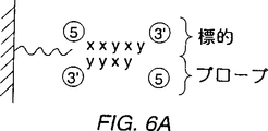
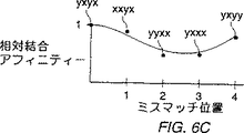
図6A〜6Cは、k−merプローブを使用したn−mer標的を配列決定するための技術を説明する。図6Aは、支持体上のプローブにハイブリダイズした標的を説明する。図6Bと6Cは、通常の結合アフィニティー対ミスマッチ位置のプロットを説明する;
図7は、ハイブリダイゼーション実験の蛍光画像を説明する;
図8は、1塩基ミスマッチの関数としてハイブリダイゼーション事件をグラフにより説明する;
図9は、ミスマッチの対の関数としての蛍光強度を説明する;
図10は、1塩基ミスマッチ実験の蛍光画像を説明する;
図11A〜11Cは、各種1塩基ミスマッチ特性を説明する;
図12A〜12Dは、より短いk−員プローブからのハイブリダイゼーション結果に基づくn−員(そのヌクレオチジ内のモノマーの数)の標的オリゴヌクレオチドのヌクレオチド配列の決定方法を説明する。特に、図12A〜12Dは、4−塩基プローブにより10塩基標的を配列決定するための本発明の適用について説明する;
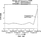
図13は、ヌクレオチド配列の決定のためのコンピューター・システムを説明する;
図14は、標的核酸のミスマッチ分析のための、そして、その決定のためのコンピューター・プログラムを説明する;
図15Aと15Bは、いくつかの可能性の中の選択により標的核酸のヌクレオチド配列を決定するためのコンピューター・プログラム、並びにこのコンピューター・プログラムにおける使用のためのスコアリング・ルーチン(Scoring routine)の例を説明する;
図16は、ヌクレオチド配列の決定における使用のための指定グラフを説明する;
図17Aと17Bは、1塩基ミスマッチ特性を使用した野生型と突然変異の分析を説明する;
図18は、1塩基ミスマッチ・テストの蛍光画像である;そして
図19A〜19Dは、核酸配列同定のための技術を説明する。
好ましい態様の説明
目 次
A.合成
B.ハイブリダイゼーション
C.ミスマッチ分析
D.適用
E.結論
定義
プローブ−典型的には固体表面上で形成された公知の組成又はモノマー配列の分子であって、標的分子に晒され又は晒されてもよく、そしてそのプローブがその標的にハイブリダイズされたかどうかを決定するために検査され又は検査されてもよいもの。“コア(core)”プローブは、標的に強いアフィニティーを示すプローブである。“伸長(extension)”プローブは、コア・プローブ配列の全部又は一部に加えそのコア・プローブ配列の1以上の可能性のある伸長を含むプローブである。本明細書は、プローブの3′−末端における伸長として“左”伸長と、そしてプローブの5′−末端における伸長として“右”伸長という。但し、反対の表記法も明らかに採用されることができるであろう。
標的−それについて、その組成又はモノマー配列を研究することが望ましい、典型的には、未知の組成又はモノマー配列をもつ分子。標的は、より大きな分子の一部、例えば、より長い核酸内の数塩基であることができる。
n−塩基ミスマッチ−nが1以上である、コアプローブ内の対応モノマーとは異なる、nモノマーをその中にもつプローブ。
A,T,C,G,U−は、それぞれ、以下のヌクレオチド、アデニン、チミン、シトシン、グアニン、とウリジンについての略号である。
ライブラリー−1以上の支持体内にしばしば形成される、所定ヌクレオチド配列の核酸プローブのコレクションであって、標的核酸のハイブリダイゼーション試験において使用されるもの。
A.合成
光指定オリゴヌクレオチド合成のための方法を図1中に示す。このような戦略は、本発明者の譲受人に譲渡され、そして全目的をもって引用により本明細書中に取り込む米国特許第5,143,854号中により詳細に記載されている。
図1において説明する光指定合成法においては、光不安定性保護基又は基(X)により誘導体化された表面(2)を、フォトリトグラフ・マスク(photolithographic mask)M1を通して照射して、反応性ヒドロキシル(OH)基を露出させる。(光不安定性の保護基により5′−ヒドロキシルにおいて保護された)ホスホルアミジット活性化ヌクレオシドのシリーズの第1の(T−X)を次に表面全体に晒す。先行する照射の間に先に晒された部位においてのみカップリングが生じる。
上記カップリング反応が完了した後、支持体を濯ぎ、そしてその表面を再び、新たな又は平行移動されたマスクM2を通して照射して、カップリングのための異なる基を露出させる。(光不安定性の保護基により5′−ヒドロキシルにおいて再び保護された)新たなホスホルアミジット活性化ヌクレオシドC−Xを添加し、そして上記露出部位にカップリングさせる。上記工程を、光脱保護とカップリングのサイクルを通じて繰り返して、支持体上にオリゴヌクレオチド・プローブの所望のセットを作り出す。フォトリトグラフィーが使用されるので、この工程は微細にされることができる。さらに、反応が光により場所的にアドレスされた部位においてのみ生じるので、各部位におけるプローブのヌクレオチド配列は正確に知られ、そして標的分子(標的核酸又は、他の態様においては、タンパク質、例えばレセプタのいずれか)との、各部位におけるオリゴヌクレオチド・プローブの相互作用を評価することができる。
5′−O−(α−メチル−6−ニトロピペロニルオキシカルボニル)−N−アシル−2′−デオキシヌクレオシド、又はMeNPoc−N−アシル−デオキシヌクレオシド、MeNPoc−dT,MeNPoc−dCibu,MeNPoc−dGPAC、及びMeNPoc−dAPACを含む光保護デオキシヌクレオシドを本工程のために開発した。保護基化学は、PCT特許公開第92/10092号及び1990年12月6日に出願された米国出願逐次番号第07/624,12O号、及び1992年12月2日に出願された第07/971,181号であって、共に本発明の譲受人に譲渡され、そして全目的をもって引用により本明細書中に取り込まれたものの中にかなり詳細に開示されている。
実施例
1.保護基
塩基は280nm領域内に強いπ−π*が遷移をもつので、光除去性保護基の脱保護波長は、不所望のヌクレオシド光化学を回避するために280nmよりも長い波長でなければならない。さらに、4つのデオキシヌクレオシドの光脱保護速度は、光が、全照射合成部位内で、ヒドロキシル(又は他の官能基、例えばスルフヒドリル又はアミノ基)を均しく脱保護するであろうように、近似していなければならない。
これらの基準に適合するために、1セットの、5′−O−(α−メチル−6−ニトロピペロニルオキシカルボニル)−N−アシル−2′−デオキシヌクレオシド(MeNPoc−N−アシル−デオキシヌクレオシド)が光指定合成のために開発され、そして保護されたヌクレオシドの光速度論的挙動が測定された。5′−O−(α−メチル−6−ニトロピペロニルオキシカルボニル)−N−アシル−2′−デオキシヌクレオシド・ホスホルアミジットを合成するための合成系路をスキームIに示す。
4つのデオキシヌクレオシド、MeNPoc-dT,MeNPoc−dCibu,MeNPoc−dGPAC、とMeNPoc−dAPACのそれぞれの0.1mM溶液をジオキサン中で調製した。アリコート(200mL)を、さまざまな時間にわたり狭い経路(2mm)の石英キュベット内で14.5mW/cm2の365nm光で照射した。4〜5の時間点を、各塩基について採取し、そしてそれらの溶液を、0.1(v/v)%TFAを含む水中60(v/v)%の移動相により溶出して、280nmにおけるHPLC装置とヌクレオジル5−C8HPLCカラムにより出発材料の損失について分析した(MeNPoc−dTは水中70(v/v)%メタノールの移動相を必要とした。)。残ったMeNPoc−N−アシル−デオキシヌクレオシドのピーク面積を計算して、MeNPoc−dT,MeNPoc−dCibu,MeNPoc−dGPAC、とMeNPoc−dAPACについて、それぞれ28秒、31秒、27秒、と18秒の光分解半減期を得た。その後のリトグラフ実験において、4,5分間の照射時間(9*t1/2 MeNPoc-dC)は、99%以上のMeNPoc保護基の除去を導いた。
光指定合成においては、全体の合成収率は、光脱保護の収率、光脱保護のコントラスト、と化学的カップリングの効率に依存する。光速度論的条件は、好ましくは、光脱保護の収率が99%を超えることを確保するように選ばれる。支持体の通常暗い領域内の不所望の光分解は、その合成忠実度(fidelity)に悪影響を及ぼすことができるが、高吸光度(5ODU)によるリトグラフ・マスクを使用することにより、そして光学表面の注意深いインデックス・マッチング(index matching)により最小化されることができる。ガラス支持体へのDMT−N−アシル−デオキシヌクレオシド・ホスホルアミジットの縮合効率は、95%〜99%のレンジ内で測定された。MeNPoc−N−アシル−デオキシヌクレオシド・ホスホルアミジットの縮合効率も、90%を上廻って測定された。但し、それらの効率は、合成間で変動することができ、そして監視されなければならない。
2.カップリング効率の測定
光保護されたヌクレオシドのカップリング効率を調べるために、4つのMeNPoc−アミジットのそれぞれを最初に、(DMT化学物質を介して)支持体にカップリングした。その支持体の領域を照射し、そしてMeNPoc−ホスホルアミジットを保護基なしで添加した。その支持体の新たな領域を次に照射し;蛍光デオキシヌクレオシド・ホスホルアミジット(FAM−ホスホルアミジットApplied Biosystems)をカップリングして;そしてその支持体をシグナルについて走査した。蛍光標識されたホスホルアミジットが、新たに露出されたヒドロキシル基と先に未反応とヒドロキシル基の両方において反応する場合、その時、2つの部位の間の蛍光強度の比は、そのカップリング効率の尺度を提供する。この計測は、表面光分解の収率が不変に近いということを受け入れる。この又は近似の検定を使用した化学的カップリング収率は、変動牲であるが、高く、80〜95%の間のレンジにある。
別個の検定において、化学的カップリング効率は、ヘキサエチレングリコール誘導体化支持体上で測定される。まず、グリコール・リンカーを脱トリチル化し、MeNPoc−デオキシヌクレオシド−O−シアノエチルホスホルアミジットをキャッピングなしでその樹脂にカップリングした。次に、DMT−デオキシヌクレオシド−シアノエチルホスホルアミジット(リポーター−アミジット)をその樹脂にカップリングした。このリポーター−アミジットは、上記第1段階からのいずれかの未反応のヒドロキシル基にカップリングする。このトリチル溶出液を集め、そして吸収分光光度計により定量した。溶出液を、そのデリバリー・ライン内に残った残存トリチルを測定するためにMeNPoc−ホスホルアミジット・カップリング直後にそのラインからも集めた。この検定において、カップリング効率は、100%の、リポーター−アミジットのカップリング効率を呈すると測定される。ヘキサエチレングリコール・リンカーへのMeNPoc−デオキシリボヌクレオシド−O−シアノエチルホスホルアミジットのカップリング効率と16のジヌクレオチドの効率が測定され、そしてDMT−デオキシヌクレオシド・ホスホルアミジットとは区別できなかった。
3.オリゴヌクレオチド・プローブの場所的に指定された合成
オリゴヌクレオチド・プローブの合成を始めるために、支持体を調製し、そしてMeNPoc−dCibu−3′−O−ホスホルアミジットを合成リンカーを通じて合成サポートに付着させた。このサポートに付着させた。このサポートの領域を、リトグラフ・マスクの800×1280μmの孔を通しての照射により合成のために活性化した。7つの追加のホスホルアミジット合成サイクルを(対応のDMT保護されたデオキシヌクレオシドをもって)行い、S−3′−CGCATCCGを作り出した。室温において4時間濃NH4OHにより、ホスフェートと環外アミン保護基の除去の後、支持体を、水ジャケット付熱制御されたハイブリダイゼーション・チャンバー内に載せた。この支持体を、以下に述べるミスマッチ実験において使用した。
B.ハイブリダイゼーション
オリゴヌクレオチド・アレイは、ハイブリダイゼーション研究を含む、多種多様の用途において使用されることができる。ハイブリダイゼーション研究においては、そのアレイは、図1中に示すように、着目のレセプタ(R)に晒されることができる。このレセプタは、適当な標識(*)、例えばフルオレセインにより標識されることかできる。このレセプタが結合した支持体上の位置が測定され、そして、その位置におけるオリゴヌクレオチド・プローブの配列の知識を通じて、ある者は次に、そのレセプタがオリゴヌクレオチドである場合、そのレセプタ配列を決定することができる。
ハイブリダイゼーションにより配列決定(SBH)は、各部位におけるプローブの同一性が知られているアレイを形成するために、多くのプローブを表面に付着させることにより最も効率良く行われる。標識された標的DNA又はRNAは次にそのアレイにハイブリダイズされ、そしてそのハイブリダイゼーション・パターンが、そのアレイ内の全相補的プローブの同一性を決定するために調べられる。ミスマッチ・プローブ/標的複合体が重要でないことを教示する従来の技術とは反対に、本発明は、ミスマッチ・プローブ/標的複合体のハイブリダイゼーション・シグナルが、そのアレイ上の完全にマッチしたプローブ/標的複合体の同一性を同定し又は確認するような分析方法を提供する。
オリゴヌクレオチドのアレイは、光指定合成技術を用いたハイブリダイゼーション研究のために効率的に生成される。以下に討議するように、全テトラヌクレオチドのアレイを16サイクルにおいて作り出した。これは完成するのにたった4時間しか必要としなかった。組合せ戦略が使用されるので、アレイ上の異なる化合物の数は合成の間に指数関数的に増加する。一方、化学的カップリング・サイクルの数は、直線的に増加する。例えば、48(65,536)オリゴヌクレオチドの完全セットにこの合成を拡張することは、16の追加のサイクルが必要であるためにその合成にほんの4時間(又はこれ未満)を追加する。さらに、組合せ合成戦略を、いずれかの所望のプローブ組成を作り出すために行われることができる。例えば、ドデカマー(412)の完全セットは、48以下の光分解とカップリング・サイクルにおいて作り出されるので(bn化合物は多くともb×nサイクルを必要とする。)、(より短いオリゴヌクレオチドのいずれかのサブセットを含む)ドデカマーのいずれかのサブセットは、48以下の化学的カップリング段階において構築されることができる。アレイ内の化合物の数は、合成部位の密度と全体のアレイ・サイズによってのみ制限される。本発明は、片面上25ミクロンの正方形部位内で合成されたプローブをもつアレイにより行われている。この解像度において、65,536オクタヌクレオチドの完全セットは、たったO.64cm2の寸法のアレイ内に置かれることができる。1,048,576ドデカヌクレオチドのセットは、この個々のプローブ部位サイズにおいてたった2.56cm2のアレイを必要とする。
ゲノム配列決定プロジェクトの成功は、効率的なDNA配列決定技術に依存する。最近の方法は、複雑な手順に高く頼り、そして実質的な手作業を必要とする。SBHは、最近の実施における手作業の多くを自動化するための潜在能力を提供する。光指定合成は、SBHのためだけでなく多くの他の用途のためにも微細化されたアレイの大規模生産のための効率的な手段を提供する。
オリゴヌクレオチド・アレイは、一次的な配列決定用途のために使用されることができるけれども、多くの診断方法は、標的核酸配列内のほんの数個のヌクレオチドの位置の分析を含む。1塩基の変更は、プローブ・アレイ上の標的のハイブリダイゼーション・パターン内に多数の変化を引き起こすので、本発明に係るオリゴヌクレオチド・アレイと方法は、ある者が、先に明白されたDNA配列の正確さをチェックし、又は標的核酸内での特定の特異的配列内の変化又は突然変異について走査することを可能にする。例えば、後者は、遺伝子、疾患、品質管理、及び法延分析のために重要である。オクタヌクレオチド・プローブセットにより、標的核酸内の1塩基変化は、8つの完全ハイブリッドの損失により、そして8つの新たな完全ハイブリッドの生成により、検出されることができる。この1塩基変化は、アレイ上の変更されたミスマッチ・プローブ/標的複合体の形成を通じて検出されることもできる。おそらく、さらに驚ろくべきことに、複雑な核酸内のこのような1塩基対の変化は、アレイ上の標的の全ハイブリダイゼーション・パターンを劇的に変更する。本発明に従って、この全ハイブリダイゼーション・パターンにおけるこのような変化は、この分析を実際に単純化するために使用される。
光指定オリゴヌクレオチド・アレイの高い情報含有量は、遺伝子診断テストにかなり有益である。100〜1,000の異なる突然変異の配列比較は、1時間に1回の形式におけるものの代わって、同時に検定されることができる。アレイは、多種多様な病因生物の迅速な同定についての遺伝子マーカーを含み、そして、RNA/RNA,RNA/DNA、タンパク質/RNA又はタンパク質/DNA、相互作用の配列特異性を研究するために、構築されることもできる。ある者は、アンチセンス、トリプル・ヘリックス、又は他の用途のために非ワトソン・クリック・オリゴヌクレオチド及び新規の合成ヌクレオシド・アナロブを使用することができる。好適に保護されたRNAモノマーが、RNA合成のために使用されることができ、そして多種多様の合成及び非天然核酸アナロブが、実施者の動機付けに依存して、使用されることができる。例えば、引用により本明細書に取り込む、PCT特許公開第91/19813号、第92/05285号、及び第92/14843号を参照のこと。さらに、オリゴヌクレオチド検定は、オリゴヌクレオチド複合体の形成及び安定性を支配する熱力学と速度論の規則を演繹するために使用されることができる。
実施例
1.表面オリゴヌクレオチドへの標的のハイブリダイゼーション
先に討議した支持体結合オクタヌクレオチド・プローブを、15℃における15分間のインキュベーションによりハイブリダイゼーション・チャンバー内で5′GCGTAGGC−フルオレセインの標的にハイブリダイズさせた。アレイ表面を次に蛍光外(epifluorescence)顕微鏡(488nmアルゴン・イオン励起)に応答信号を送った(interrogated)。この走査の蛍光画像を図2に示す。この蛍光強度パターンは、プローブの合成を指定するために使用される800×1280μmストライプにマッチする。さらに、シグナル強度は、(ガラス支持体の背景を4倍上廻って)高く、そのプローブへの標的の特異的結合を立証する。
標的−プローブ複合体の挙動を、そのハイブリダイゼーション溶液の温度を上昇させることにより調べた。各温度において10分間の平衡化の後、支持体をシグナルについて走査した。2本鎖は、研究下配列について予想される温度レンジ内でメルトした(Tm=〔2°(A+T)+4°(G+C)〕の規則から得られたTm≒28℃)。アレイ内のプローブは、標的DNAの再ハイブリダイゼーションにより立証されるように、標的−プローブ複合体の温度変性に対して安定であった。
2.標的ハイブリダイゼーションの配列特異性
標的ハイブリダイゼーションの配列特異性を立証するために、2つの異なるプローブを800×1280μmのストライプ内で合成した。図3Aは、2つのプローブの位置を同定する。プローブS−3′−CGCATCCGをストライプ1,3と5内で合成した。プローブS−3′−CGCTTCCGをストライプ2,4と6内で合成した。図3Bは、15℃における支持体への5′−GCGTAGGC−フルオレセイン標的のハイブリダイゼーションの結果を示している。これらのプローブはたった1の内部塩基が異なるけれども、上記標的は、(ストライプ1,3と5内の背景を〜500カウント上廻って)その相補的配列に特異的にハイブリダイズし、位置2,4と6(〜10カウント)内には検出可能なシグナルはほとんど又は全くない。図3Cは、両配列への標的のハイブリダイゼーションの結果を示す。図3中のすべての位置内のシグナルは、図3Bにおけるシグナルの非存在が1塩基ミスマッチの不安定性にのみ依ることを説明している。標的には、等モル濃度において存在するけれども、図3B中のストライプ2,4と6内のシグナルの比は、領域1,3と5内のシグナルよりも約1.6倍高い。この2本鎖は、領域2,4と6を占める2本鎖よりもわずかに高い予想Tmをもつ。これらの2本鎖は、15分間で45℃までその温度を上昇させることにより解離され、そしてそのハイブリダイゼーションは、上記と逆の順番で繰り返され(図3Dと3E)、これは、逆方向におけるハイブリダイゼーションの特異性を立証している。
3.プローブ・マトリックスの組合せ合成とそれへの核酸標的のハイブリダイゼーション
光指定合成においては、製品の位置と組成は、照射と化学的カップリング試薬の順番に依存する(完全な説明については、Fodor et al.,Science(1991)251:767-773を参照のこと。)図4において説明するように、256テトラヌクレオチドの合成について考える。マスク1は、合成の第1ラウンドにおける4つのヌクレオシドの第1とのカップリングのために支持体表面の1/4を活性化する。サイクル2において、マスク2は第2ヌクレオシドとのカップリングのためにその支持体の別の1/4を活性化する。この工程がモノヌクレオチドの4つの領域を作り上げるために続けられる。ラウンド2のマスクは、ラウンド1のものに垂直であり、そしてラウンド2の各サイクルは、4つの新たなヌクレオチドを作り出す。この工程は、図4において説明するように16のヌクレオチドを形成するためにラウンド2まで続けられる。ラウンド3のマスクは、各カップリングのサイクルが16のトリマーを作り出すように上記合成領域をさらに亜分割する。この支持体の亜分割は、テトラヌクレオチドを形成するためにラウンド4まで続けられる。このプローブ・マトリックスの合成は、(A+C+G+T)4の多項表記法において簡単に表現されることができる。この多項式の展開は、256のテトラヌクレオチドを作り出す。
プローブ・マトリックスを生り出すために光指定組合せ合成により合成された256プローブのアレイの適用を、図5Aに説明する。この合成のための多項式は:3′−CG(A+G+C+T)4CGにより与えられる。この合成マップを図5Bに与える。全ての可能性のあるテトラヌクレオチドを、3′−末端と5′−末端におけるCGに隣接するように合成した。15℃におけるこのアレイへの標的5′−GCGGCGGC−フルオレセインのハイブリダイゼーションは、最も強い位置(2,698カウント)としてS−3′−CGCCGCCG相補プローブを正確に作り出した。以下のミスマッチ:S−3′−CGCAGCCG(554カウント)、S−3′−CGCCGACG(317カウント)、S−3′−CGCCGTCG(272カウント)、S−3′−CGACGCCG(242カウント)、S−3′−CGTCGCCG(203カウント)、S−3′−CGCCCCCG(180カウント)、S−3′−CGCTGCCG(163カウント)、S−3′−CGCCACCG(125カウント)、とS−3′−CGCCTCCG(78カウント)についても有意な強度が観察された。
C.ミスマッチ分析
先に討議したアレイを、より短い長さkのプローブのアレイを使用して長さnのオリゴヌクレオチドの核酸配列を決定するための本発明に係る方法において使用されることができる。図6は単純な例について説明する。この標的は、配列5′−XXYXY−3′{ここで、XとYは相補的核酸、例えばAとT又はCとGである。}をもつ。討議目的のために、図6における説明を、たった2つの塩基とひじょうに短い配列の使用により単純化するが、この技術は、例えば全部で4つのRNA又はDNAの塩基によるより大きな核酸に容易に拡張されることができる。
標的の配列は、一般に最初から(ab initio)知られていない。ある者は、より短いプローブのアレイを用いて本発明を使用して標的の配列を決定することができる。本実施例においては、全ての可能性のあるXとYの4−merのアレイを合成し、そして次に5mer標的の配列を決定するために使用する。
最初に、“コア(core)”プローブを同定する。このコア・プローブは、本発明に係るミスマッチ分析法を使用して標的内の配列に正確に相補的である。このコア・プローブは、以下の基準の中の1又は両方を使用して同定する:
1.コア・プローブは、他のプローブよりも強い標的への結合アフィニティー、典型的には、(先の分析サイクル内でコア・プローブとして同定されていない)アレイ内のいずれかのプローブの最高の結合アフィニティーを示す。
2.コア・プローブ配列に比較したとき、標的とミスマッチであるプローブは、以下により詳細に討議するような特徴的なパターンを示す。ここで、そのプローブの3′−と5′−末端においてミスマッチするプローブは、内部の並置においてミスマッチするプローブよりも強く結合する。この特定の実施例においては、選定基準#1は、そのプローブが標的にハイブリダイズしたものとして示されている図6A中に示すように、配列3′−YYXYをもつ標的に最高の結合アフィニティーをもつコア4−merプローブを同定する。(標的の5′−XXYX位に一致する)プローブ3′−YYXYは、それ故、“コア”プローブとして選ばれる。
選定基準#2は、このコア・プローブが標的核酸に正確に相補的であることを確保するための“チェック(check)”として使用される。この第2の選定基準は、そのコア・プローブと比較するとき1塩基ミスマッチをもつプローブのハイブリダイゼーション・データ(例えば、支持体上のプローブのアレイにハイブリダイズした標識された標的の蛍光強度、但し、他の技術も当業者によく知られている。)を評価する。この特定のケースにおいては、コア・プローブはS−3′−YYXYとして選ばれた。このコア・プローブの1塩基ミスマッチ・プローブは:S−3′−XYXY,S−3′−YXXY,S−3′−YYYY、とS−3′−YYXYである。これらの1塩基ミスマッチの結合アフィニティー特性は、“正しい(correct)”コアが選ばれたことを確かめ、又は類似の結合アフィニティーを示すプローブのセットの中からそのコア・プローブを選ぶために使用される。
予想される結合アフィニティー対ミスマッチ位置の例示的、仮定的なプロットを図6Bにおいて提供する。これらの結合アフィニティー値(典型的には、プローブにハイブリダイズされた標識された標的の蛍光強度、但し、アフィニティーに関する多くの他の要因を使用することもできる。)は、このグラフの左手の位置上に1の値としてプロットされた、標的へのS−3′−YYXYの結合アフィニティーに対して全て正規化される。たった2つのヌクレオチドが本例において含意されるので、1位(そのプローブの3′−末端におけるヌクレオチド)においてミスマッチするプローブについてプロットされた値は、S−3′−XYXYの正規化された結合アフィニティーである。2位におけるミスマッチについてプロットされた値は、S−3′−YXXYの正規化されたアフィニティーである。3位におけるミスマッチについてプロットされた値は、S−3′−YYYYの正規化されたアフィニティーであり、そして4位におけるミスマッチについてプロットされた値は、S−3′−YYXXの正規化されたアフィニティーである。上記のように、“アフィニティー(affinity)”は、例えば、標的上の蛍光マーカーからの光子カウントの数を含む、多くの方法において測定されることかできる。
全部で3つのミスマッチのアフィニティーは、この説明においてそのコアよりも低い。その上、アフィニティー・プロットは、そのプローブの3′−末端におけるミスマッチが、この特定のケースにおいてはプローブの5′−末端におけるミスマッチよりもより少ない影響力をもつことを示している。但し、これは、常にそうではない。さらに、このプローブの末端におけるミスマッチは、このプローブの中央におけるミスマッチよりも少ない妨害をもたらす。図6B中において示すようにプロットされたとき“スマイル(Smile)”形状のグラフをもたらすこれらの特徴は、“正しい”コア・プローブの選定後、又は、標的配列の他の部分に関してのコア・プローブであるミスマッチ・プローブを考慮した後の、1塩基ミスマッチのほとんどのプロットにおいて見られるであろう。この情報は、コア・プローブの最初の選定、又は正しくマッチしたコア・プローブが選ばれていることを確保するためのチェッキングのいずれかにおいて使用されることができるであろう。もちろん、特定の情報においては、先のセクションB中に述べたように、例えば、法延の又は遺伝子の研究、等において必要とされるコアの同定が、全てである。
配列決定研究においては、この工程を次に、このコア・プローブの左及び/又は右伸長のために繰り返す。図6において説明する例においては、このコア・プローブの右伸長だけが可能である。このコア・プローブの可能性のある4−mer伸長プローブは3′−YXYYと3′−YXYXである。再び同一の選定基準を使用する。3′−YXYYと3′−YXYXの間では、3′−YXYXが最も強い結合アフィニティーをもつであろうことが通常見い出されるであろうし、そしてこのプローブは、正しいプローブ伸長として選ばれる。この選定は、上記コア・プローブと比較するとき1塩基ミスマッチをもつプローブの正規化された結合アフィニティーを再びプロットすることにより確かめられることができる。推定プロットを図6Cに説明する。再び、特徴的な“スマイル”パターンが観察され、これは、“正しい”伸長、すなわち3′−YXYXが選ばれていることを示している。この情報から、ある者は、標的の配列が5′−XXYXYであると正しく結論付けるであろう。
実施例
1.(A+T) 8 アレイと1塩基ミスマッチの安定性
20ステップの、4−レプリカ組合せ合成を、Men Poc−dAとMen Poc−dTを使用して行った。リトグラフ・マークを、1セットの256オクタヌクレオチドの各メンバーが1.28×1.28cmアレイ上の4つの別々の位置内で合成され、1024の異なる合成部位であって、各々がオリゴヌクレオチド・プローブを含み、各々の部位がサイズ400×400μmである部位を作り出すように選んだ。dAアミンの合成とフェノキシアセチル脱保護の後、支持体を、サーモスタットで調節された染色及びフロー・セル内に置き、15℃において1nM 5′−AAAAAAAA−フルオロセインとインキュベートし、そして次にZeiss蛍光外顕微鏡内で走査した。得られた蛍光画像を図7に示す。
1塩基ミスマッチの関数としてのハイブリダイゼーション事件の蛍光強度を図8にグラフにより提供する。1塩基においてそのコア・プローブと異なる各オリゴヌクレオチド・プローブについての4つの独立した強度のそれぞれをプロットする。0位ミスマッチ(すなわち、完全な相補物3′−TTTTTTTT)は、〜900カウントにおいてアレイ上に最も明るい位置であり;このアレイの背景シグナルは、約220カウントである。ミスマッチ1位(プローブの3′−末端)は、〜760カウントにおいて次に明るい。これに続く位置の“スマイル”又は“U”形曲線は、プローブ/標的複合体の各位置におけるミスマッチの相対的安定性を示している。この“ミスマッチ・ファミリー”は、プローブのアレイとの核酸の相互作用を特徴付け、そして標的配列の同定を提供し又は確かなものにする。3,4,5と6位におけるミスマッチは、より脱安定性であり、そして背景からは事実上区別されることができない強度を作り出す。1位におけるミスマッチ(オクタヌクレオチドの3′−末端が支持体につながれている点)は、8位における対応のミスマッチ(その遊離の5′−末端)よりもより脱安定性されていない。アレイ合成の均一性と標的ハイブリダイゼーションは、上記の4つの2連の合成部位の間の強度の低い変化を反映している。
本発明に係る方法は、2以上のミスマッチをプローブとの標的ハイブリダイゼーションからの情報をも利用することができる。ミスマッチの対の関数としての蛍光強度を図9に表わす。このケースにおいては、強度データは、完全なマッチが強度1をもつように正規化された。例えば、インデックス1,8におけるデータは、そのプローブ/標的2本鎖の各末端におけるミスマッチに対応する。2項(インデックス1,1〜8,8)を、図8に説明した1ミスマッチに対応する。この最高強度は、プローブ/標的複合体の末端におけるミスマッチの1と対に対応する。
2.(G+T) 8 アレイと配列再構築
Men Poc−dGとMen Poc−dTのオクタヌクレオチド・アレイを合成した。合成の形式は、先に討議した(A+T)8アレイのためのものと同様であり、そして4の反復(全部で1024)におけるGとTの256オクタヌクレオチドをもたらす。最終脱保護と温度制御された(15℃)ハイブリダイゼーション・チャンバーへの接続後、プローブ・アレイを1nM 5′−AACCCAAACCC−フルオレセイン標的とインキュベートし、そして走査した。得られた画像を図10に与える。4つの明確であるが重複性の、完全に相補的なオクタヌクレオチド・ハイブリダイゼーションが予想される:3′−TTGGGTTT,TGGGTTTG,GGGTTTGG、とGGTTTGGG。本明細書中に示すように、1塩基対のミスマッチをもつプローブ/標的複合体の中程度の安定性は、中程度のシグナルをもつプローブのファミリーを作り出す。図10の多くの強度特性の粗略な検査は、複雑なパターンを現わした。
本発明により提供される再構築された実践的研究(heuristic)は、図10における複雑なデータ・パターンを使用する。このアルゴリズムは、完全にマッチしたプローブ/標的複合体がより高い蛍光強度をもち、そして完全なマッチと関連の1塩基ミスマッチが典型的には図6に示すものと類似の特性を形成するという一般的な規則として推定される。
最高の強度をもつプローブは、その標的への完全なマッチであるはずである。対応のミスマッチ曲線を図11A〜11Cに示す。ある者は、そのプローブが標的に正確に相補的であることを確認するために最高強度をもつプローブ(このケースにおいてはS−3′−TGGGTTTG)についてのミスマッチ曲線を最初にプロットする。このプローブが標的の断片に相補的であると仮定して、我々は、その標的の3′−末端上に1塩基の“伸長”を考える。この場合、2つのプローブの選択がある。2つの8−merプローブS−3′−GGGTTTGTとS−3′−GGGTTTGGの中の1は、標的核酸に正確に相補的であろう。これらの2つのプローブのそれぞれについての並びにプローブS−3′−TGGGTTTGについてのミスマッチ曲線を図11Aに強度値により示す。プローブS−3′−TGGGTTTGがプローブS−3′−TGGGTTTGのもの(典型的な“スマイル”プロット)と最も近似したミスマッチ曲線をもつということに注目のこと。それ故、ある者は、正しい伸長プローブがS−3′−GGGTTTGGであると結論付けるであろう。
図11Bは、標的配列の3′−末端を評価するための本工程の反復を示す。プローブS−3′−GGTTTGGはコアS−3′−GGGTTTGGに最も近似したスマイル形のミスマッチ曲線をもち、そしてプローブS−3′−GGTTTGGTはそうではないので、ある者は、プローブS−3′−GGGTTTGGが正しい伸長プローブであると正しく結論付けるであろう。この工程は、曲線が正しい形をもつまでか、又は標的の“末端”に到達していることを示す。絶対強度が最高強度のものよりもかなり下廻るまでのいずれかまで、繰り返されることができる。同様の方法は、5′−末端に伸長する標的の配列を提供する。図11Cは、完全にマッチしたプローブの全てのミスマッチ曲線を示しており;各曲線は、この標的について予測された無矛盾の形状をもつ。
上記技術は、もちろん、図12A〜12Dの各パネルにおいて説明するように、いずれかの長さの核酸に容易に延長されることができる。図12Aにおいて示すように、10−mer標的が配列決定されるであろうし、そしてその配列は、5′−N1N2N3N4N5N6N7N8N9N10−3′{ここで、Nはいずれかのヌクレオチド又は核酸モノマーであり、そして下付き添字はそのプローブ内のヌクレオチド位置を示し、3はその3′−末端モノマーであることを示す。}により示される。当業者は、プローブが支持体に付着された5′−末端をもって合成された場合、本発明に係る方法が、適当な修飾をもって適用されることができることを理解する。
より短いオリゴヌクレオチドのアレイを、本発明の1の態様に従ってより大きなヌクレオチドを配列決定するために使用することができる。図12A〜12Dに示す特定の例においては、4−mer(長さ4モノマーのオリゴヌクレオチド・プローブ)を未知の10−mer標的を配列決定するために使用する。実際には、より長いプローブと標的が典型的には使用されるが、この例示的な例は、本発明の理解を容易にする。4−merアレイの1のメンバーは図12Aに示され、そして配列S−3′−P3P4P5P6{ここで、各P(プローブ)ヌクレオチドは、その用途に依存してA,T,C,U,Gと他のモノマーからなる群から選ばれ、そしてその添字は、標的に対する位置を示す。}をもつ。討議目的のために、このハイブリダイゼーション・データは1のアレイから入手されることができると推定される。しかしながら、ある者は、本法を実施するために、多数のアレイ、異なる時において合成されたアレイ、又はさらに個々のプローブを使用することができる。上述のように、4の長さのプローブを討議を容易にするために選ばれ;実際には、より長いプローブが典型的には使用されるであろう。
S−3′−P3P4P5P6は、標的への強い結合アフィニティーと正しいミスマッチ特性をそれが示すために、そのアレイからのコア・プローブとして選ばれる。全4−merのアレイにおいて、配列S−3′−P3P4P5P6がそのコア配列として選ばれる。なぜなら、フルオレセイン標識された標的(図12A中5′−N1N2N3N4N5N6N7N8N9N10−3′として示す)が支持体に晒されるとき、その標的が図12Aに示す矢印により示されるように、そのプローブにハイブリダイズし、そして高い蛍光強度(すなわち、多数の光子カウント)がその支持体の他の部分と比較するとき、プローブS−3′−P3P4P5P6を含む支持体の部分内で観察されるからである。通常、最も強い結合アフィニティーを示す配列が第1コア配列として選ばれるであろう。
ある者は、好ましくは、最初に選ばれたコア配列が、1塩基においてコア・プローブと異なるアレイ内のプローブの蛍光強度を調べることにより標的に完全に相補的であるかどうか確認する。図12Bは、1塩基ミスマッチの相対強度対S−3′−P3P4P5P6コア・プローブについてのミスマッチの位置の典型的なプロットを定量的に説明する。簡単な例としてS−3′−P3P4P5P6においてヌクレオチドCが存在しないと仮定する。図12Bは、配列S−3′−P3P4P5P6内へのCの置換によりそのコア配列プローブと異なるプローブの正規化された蛍光強度を定性的に説明しており、そして、ここで、C−含有ミスマッチ・プローブのいずれも標的内の他の配列に正確に相補的ではない。従って、図12Bは、それらが標的とハイブリダイズされるとき、そのコア・プローブに正規化された、以下のプローブ・セット
S−3′−CP4P5P6,
S−3′−P3CP5P6,
S−3′−P3P4CP6、及び
S−3′−P3P4P5C
の相対的蛍光強度をプロットする。他の態様においては、平均曲線が、各位置における可能性のあるヌクレオチドの全ての置換(ミスマッチ・プローブの“ファミリー”)についてプロットされ、又は最も高い強度が各位置についてプロットされる。従って、図12BにおけるグラフのX軸上の0位は、置換がないことを表し、そしてコア・プローブS−3′−P3P4P5P6への標的ハイブリダイゼーションのための蛍光強度を示す。図12B中の全ての値がこの値に関して正規化されているので、“置換無し”のケースは、1の正規化強度をもつ。Cが3,4,5と6位において置換されるとき、その相対強度値は、通常、小さい。なぜなら、これらの配列のいずれも本例においては標的に正確に相補的でないからである。
3′−又は5′−末端においてミスマッチをもつプローブ/標的複合体の相対蛍光強度が、プローブ/標的複合体の中央においてミスマッチをもつ複合体よりも典型的には高い。なぜなら、このプローブの末端におけるミスマッチは、プローブ/標的複合体の中央におけるミスマッチよりもより少なく脱安定化される傾向をもつからである。このプローブの3′−末端にミスマッチをもつプローブ/標的複合体は、推定するに、この態様においては支持体表面へのそのプローブの3′−末端の近接のために、そのプローブの5′−末端におけるミスマッチをもつものよりもより少なく影響を及ぼす(そしてそれ故、より高い蛍光強度をもつ)ことができる。それ故、結合アフィニティーに関する正規化係数対ミスマッチ位置をプロットする曲線は、図12B中に示すような“ゆがんだスマイル(crooked smile)”の形をもつ傾向がある。
この方法論を使用して、ある者は、そのコア・プローブと同一の配列であるが一端において伸長しており、そして場合により他端においてより短いものをもつアレイ上のプローブを調べることによりそのコア配列を延長することができる。これらのプローブは、標的に完全にハイブリダイズする。プローブを決定するための第2のコア・配列として評価されることができる。この工程の繰返しにより、ある者は、その標的のヌクレオチド配列の全体を決定することができる。
本法を説明するために、図12Cは、コア・プローブS−3′−P3P4P5P6の4つの可能性のある4−員“左伸長”を示す。図示するように、S−3′−P3P4P5P6に相補的な標的の配列に隣接するヌクレオチドはA,T,C、又はGのいずれかであり、又はその標的上に隣接するヌクレオチドは全く存在しない(すなわち、P3は標的の5′−末端である。)。それ故、P3P4P5P6コア・プローブの可能性のある左伸長は、プローブS−3′−AP3P4P5,S−3′−TP3P4P5,S−3′−CP3P4P5、とS−3′−GP3P4P5である。この説明の目的のために、Tが実際に“正しい”ものであると推定される。なぜならAは、その標的核酸の相補位置内にあるからである。
図12Dにおける上左のプロットは、S−3′−AP3P4P5プローブのミスマッチ特性について予想されるハイブリダイゼーション・データを説明する。全てのデータがS−3′−AP3P4P5に対して正規化されている。2−5位の各々における置換の全てについてのデータ点を示すが、各々の位置における3つの値についての平均データも使用されることができるであろうし、各の位置における1の置換を使用することができ、3つの値の最高のものを利用してもよく、又はいくつかの他の組合せでもよい。S−3′−AP3P4P5グラフにおいて示されるように、1の点は残りのものよりもかなり高い結合アフィニティーを示す。これは、2位におけるAについてのT置換である。AP3P4P5グラフ内の残りのデータは、図12B中に示す通常の“スマイル”特徴をもつ。同様のプロットが図12Bの底部分において示されたCとG置換について顕出された。それぞれのケースにおいては、全データ点は、そのグラフ内の推定“コア”プローブに対して正規化されている。
図12Dの上右部分において示されるT伸長グラフは、3′−AP3P4P5グラフその他のような異常曲線をもたないであろう。なぜなら、3′−TP3P4P5プローブの2位におけるモノ置換のいずれも標的に正確に相補的であるであろうからである。従って、2位におけるA,C、とGの置換は全て、標的に対して1塩基ミスマッチをもつプローブについて予想される特徴的な“スマイル”プロットを作り出す。さらに、T置換プローブ/標的複合体の蛍光強度は、C,G、とAプローブ/標的複合体の蛍光強度よりも通常高いであろう。これらのデータは、各種組合せにおいてどの伸長が“正しい”かどうかを、そしてそれによりその標的核酸の配列を決定するために、使用されることができる。
図12A〜12D中に示されたデータから、ある者は、コア・プローブに相補的な配列に対してその標的の左伸長物に正確に相補的なプローブが、その標的内の2位においてAモノマーをもつと結論付ける。
この工程は、グラフのいずれも適当な特徴をもたなくなるまで繰り返され、この時に、標的の末端に達している。同様に、右伸長が、その標的の末端(又は着目の配列の末端)に達するまで評価される。
上記技術は、明らかに、ハイブリダイゼーション・データのマニュアル観察を通して行うことができる。しかしながら、好ましい態様においては、データは、1以上の適切にプログラムされたデジタル・コンピューターを使用して分析される。例示のシステムを、図13中に説明する。図中に示すように、このシステムは、CPUの制御下でオペレートされ、そしてメモリー(304)、例えばハード・ディスク、とメモリー(306)、例えばダイナミック・ランダム・アクセス・メモリー(dynamic random access memory)を含む1又は複数のコンピューターを含む。このコンピューターは、支持体(2)の部分にカップリングされた標識された標的ヌクレオチドからの蛍光強度又は他の関連情報を測定するスキャニング装置(308)を制御するために使用される。
この支持体(2)は、その上に既知の位置において既知配列のプローブ・ヌクレオチドを含む。ユーザーは、インプット装置(313)を介してインプットを提供する。
蛍光強度又は他の関連情報は、メモリー(304/306)内に保存される。CPU(310)は、その蛍光データを処理して、プリント装置(312)又はディスプレイ(314)の1又は両方にアウトプットを提供する。本明細書中に記載する方法に従ってデータを処理し、そして先に示したようなグラフの形態における、又は核酸モノマーの配列における、又は単純な(+)/(−)アウトプットにおけるアウトプット又はこのようなデータの分析の他の結果を得ることができる。好適なコンピューターは、例えば、IBM PC又は互換性SPARCワークステーション、又は同様の装置を含む。
図14は、n−mersのアレイを評価するための、そして(ミスマッチ分析のための)正確に相補的な配列又は(配列決定又は他の目的のための)より大きなk−merを同定するための、典型的なコンピューター・プログラムのためのフローチャートである。図中に示すように、システムは最初に、例えば、プローブのいくつかの特定のセットの最も高い結合アフィニティーをもつプローブを選ぶことによりステップ(402)においてコア・プローブを同定する。本法は、しばしば、反復工程においてオペレーション可能であろう。ここでアレイ内の最も高いアフィニティーのプローブは、第1反復(iteration)後に選ばれず、そして他の場合においては、例えば、1,2,3、又はそれ以上の最高結合プローブを選び、そして各々について左と右の伸長を行い、そして次にこの情報を保存し、そして最終的なアウトプットを提供する前に他のデータとこの情報を比較することが価値のあることである。これらの結果は、正しい配列の確認において援助することができる。
ステップ(404)において、このシステムは、コアn−merの左伸長の全てを同定する。ステップ(406)において、本システムは:
−どの左伸長物がプリセットされたモノマー置換パターンと最も無矛盾である挙動を示すかを決定し、そして/又は
−その最高の結合アフィニティーを示す左伸長を選択する、
の1又は両方により、適切な左伸長選定する。
上記選択基準その他は、いくつかの態様において、AND法において使用されることができる。すなわち、これらの基準の両方が適合し、又はこのシステムは、1が末端モノマーに達したか又はそのシステムが許容可能な状態で行われないかのいずれかを仮定する。他の態様においては、上記基準の中の1が、主な選択機構として選ばれることができ、そして他のものが、注意、すなわち、可能性のある誤った選定、又は他の選定をユーザーに提供するために使用されることができる。
その後、本システムは、選定基準がステップ408においていくつかの最小標準に適合したかどうかを決定する。そうでない場合、本システムは、その配列の末端がステップ(410)に達したと仮定する。この選定基準が適合する場合、先のコアからの正しい伸長物として選ばれた新たな“コア”によりステップ(404)の始めに戻される。
その後、本工程は、右伸長について有効に繰り返される。ステップ(412)において右伸長が同定される。ステップ(414)において、プリセットされたミスマッチ特性のプローブ及び/又は高アフィニティー右伸長物が同定される。ステップ(416)において、本システムは、この分子の末端に達しているかどうかを決定する。そうでない場合、この工程はステップ(412)に戻される。そうである場合、その分子は配列決定されていると仮定し、そしてこの工程は、プリンター又は他のアウトプット装置への適当なアウトプットをもって終了される。
ミスマッチ分析の使用方法の他の態様を、図15a中に例示する。この図は、未知の標的断片の配列を決定するためにデザインされた典型的なコンピューター・プログラムのためのフローチャートを表す。このような方法は通常、適当にプログラムされたデジタル・コンピューター、例えばIBM PC又は同等なもの、Sunワークステーション、あるいは他の類似のコンピューター・システムにおいて行われるであろう。ステップ(502)に示すように、ハイブリダイゼーション・データ(例えば、上記のような蛍光強度、但し当業者によく知られた他の技術を通じて得られた他のデータも使用することができる。)をこのシステムに入れる。いくつかの態様においては、このデータは実験システムから直接的にインプットされることができ、一方、その他においては、これらのデータは、別個のシステムにおいて採取されることができる。ステップ(504)において、このシステムは、このデータ・セット内の“最良”データに対応するプローブ配列を選択する。蛍光強度データを使用する態様においては、この最良データは、最高強度レベルを示すようなプローブに典型的には対応するであろう。このデータ・セットの不履行パーセンテージが使用されることができ、又は場合により、オペレーターは、そのデータ・セットのサイズ、そのシステムの計算能力、及びそのデータ・セットのサイズのような要因に基づいて選ばれたパーセンテージにより、最強強度データの特定パーセンテージを選ぶためにそのシステムに指令する。ステップ(506)においては、このシステムは、ステップ(504)において選ばれたプローブ配列の組合せを通じて誘導されることができる特定長さの全断片のセットを同定する。このように同定された断片を“候補”断片という。
ステップ(508)においては、このシステムは、核酸プローブから測定されたハイブリダイゼーション・データを使用して、上記セット内の各候補断片についての完全にマッチしたプローブについて先に記載されたようなミスマッチ分析アフィニティー・プロットを構築する。ステップ(510)においては、各アフィニティー・プロットは、それが、その標的DNAの他の部分とのそのプローブの交差ハイブリダイゼーションについて考慮した後アフィニティー・プロットの予測された形状にどのくらいよく一致するかを反映するように格付けされる。
この格付けステップ(510)が一旦終了すると、このプログラムは、各候補断片(ステップ512)についてのすべてのアフィニティー・プロットからの等級を合計し、そしてステップ(514)において次の候補断片を検査するために前進する。ステップ(516)において、全候補断片が検査されたとき、このシステムは、個々の候補断片についての合計されたアフィニティー・プロット等級を比較する。最後に、ステップ(518)において、このシステムは、それについて、その等級がその鋳型との最も近い全体的なマッチを示すところの候補断片(単数又は複数)を選択する。この候補断片の配列は、未知の標的断片の配列に対応する。
図15bは、アフィニティー・プロットを格付けするための1の好ましい態様についてのフローチャートである。最初に、予想された結合アフィニティー対ミスマッチ位置(“鋳型”)の所定プロットをシステムにエンターする(ステップ602)。典型的には、この鋳型は上記のような“スマイル”形を示すであろう。ステップ(604)においては、このシステムは、ステップ606〜620において特定されるようにその鋳型と各アフィニティー・プロットを比較する。ステップ(606)と(608)においては、このシステムは、その鋳型内の対応ミスマッチ位置の値と各ミスマッチ位置についてのアフィニティー値を比較する。ステップ(610)においては、このアフィニティー値を、最初に、測定された値が鋳型の値の選ばれた間隔内にあるかどうかを決定するために検査される。いくつかの態様においては、この間隔は、不履行レベルにセットされ;その他においては、それは、その実験誤差の予想される大きさのような要因に基づいて、オペレーターにより選ばれる。
所定のミスマッチ位置における値がその間隔内にある場合、ステップ(612)において、このシステムは、完全候補断片とミスマッチを含む塩基配列(“ミスマッチ配列”)を比較する。ステップ(614)において、このプロセスは、そのミスマッチ配列と上記候補断片のいずれかの部分との間の完全なマッチが存在するかどうかを決定する。このようなマッチが見つかった場合、このシステムは、そのプロットのその等級付けからそのミスマッチ位置を除外し(ステップ616)、そしてステップ(620)に続く;その他の場合、その位置は、その後の計算における包含のためにマークされる(618)。ステップ(620)において、この処理は、各ミスマッチ位置が検査されるまで続けられる。鋳型値と実験値のフィットは次に、ステップ(618)における包含のためにマークされた各ミスマッチ位置についてのステップ(622)の計算において決定される。いくつかの態様においては、この計算は、その間隔内又はその間隔の外側のいずれかとして各点を等級付けすることを含むことができ、その他の態様は、それによりそれぞれの含まれた位置がその予想された値からのその逸脱を反映するように等級付けされるような方法であって、根−平均−二乗(Root−Mean−Squares)分析としてよく知られるような技術を含むものを利用する。最後に、このシステムは、ステップ(624)において各アフィニティー・プロットについて全等級を計算する。
実施例
8−merプローブのアレイを、Pirrung et al.に付与された米国特許第5,143,854号と米国出願逐次番号第07/805,727号(両方を全目的をもって引用により本明細書中に取り込む)中に記載された方法に従って構築した。このアレイを16−mer標的とインキュベートし、そして公知の方法を使用して蛍光強度について走査した(例えば、引用により全目的をもって本明細書中に取り込む、米国出願逐次番号第08/195,889号を参照のこと。)。得られた蛍光強度データを、これまでの方法に従って分析した。アフィニティー・プロットを作り、そして等級付けした後、69−71のレンジにある等級をもつ4つの候補断片:5′−AGTTGTAGTGGATGGT,TGTTGTAGTGGATGGT,GGTTGTAGTGGATGGT、とCGTTGTAGTGGATGGTを同定した。この4つの類似の断片は、その断片の5′末端における塩基の同一性においてのみ異なり、それは、他の位置の受ける2本鎖分析の利益を受けない。他の候補断片についての等級からのこれらの4つの候補についての等級の分離は実質的であり;次に高い等級の候補が54の等級を受け取った。
さらに他の本発明の態様においては、先に討議したアレイを、以下の方法により未知の標的の断片の配列を同定するために使用することができる。
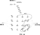
長さ“n”のプローブからのハイブリダイゼーション・データのセット(ここで、“n”は、プローブ内の塩基の数に等しく、そして未知の標的断片の長さ末端である。)を、適当にプログラムされたコンピューター・システムにエンターする。本明細書中に企図されたハイブリダイゼーション・データ・セットは、先に記載されたような蛍光強度データのセット、並びに当業者に知られた他の技術を通じて得られたデータのセットを含む。このシステムは、(Harary,F.,Graph Theory,Addition-Wesley,Reading,MA(1969)及びAhuja,R.K.,Magnanti,T.L.,Orlin,J.B.,Network Flows,Thoory,Algorithms,and Applications,Prentice Hall, New York(1993)、(両方を全目的をもって本明細書中に取り込む。)中に記載されたような)指定グラフを構築した。ここで、頂点(vertices)(又は“節点(nodes)”)は、上記データ・セットの全(n−1)−mersに対応する塩基配列であり、そしてその端は、そのセットの全n−mersに対応する塩基配列である。各端は、その端が最初に含まれた(n−1)−merから終りに含まれた(n−1)−merまで接続するように構築される。標的k−merがACTGTTGとして知られており、そしてn−mersが3−mersである上記のような指定グラフのサンプルは、図16として包含される。図16においては、3−mer配列に対応する端だけが含まれ;その標的塩基配列が知られていない典型的な場合においては、このシステムは、その分析においてn−mer節点に接続する全ての可能性のある端を含むであろう。
さらに、このシステムは、(n−1)−mer節点のいずれとも異なる、指定グラフ内のソース節点、並びにそのソース節点からそのグラフ内の(n−1)−mer節点のそれぞれまでの端を作り出す。最終的にこのシステムは、先に作り出された節点のいずれとも異なる、グラフ内の沈み節点(sink nodes)、並びにそのグラフの(n−1)−mer節点の各々からその沈み節点までの端を作り出す。
本システムは次に上記グラフの端のそれぞれに値を“コスト(cost)”を指定する。出願節点からの端先導(edges leading)と沈み節点までの端先導は全てゼロのコストと指定される。他の全ての端は、その端に対応する塩基配列が標的k−mer内のセグメントの完全マッチである確率の関数としてのコストを指定される。この確率を、“完全マッチの確率(probability of a perfect match)”という。好ましい態様においては、各n−mer端に指定されたコストは、その端に対応する配列が完全にマッチしない確率により除された、その端に対応する配列がその標的断片内のセグメントの完全マッチである確率の自然対数の負に等しい。いくつかの態様においては、これらの端は、別々の、追加の値“キャパシティー(capacity)”を指定されることができ;このような態様については、出発節点から他の節の各々までの端先導、並びに沈み節点までの端先導は、全て1のキャパシティーを指定され;このグラフの他の全ての端は、無限キャパシティーを指定される。
出発節点から始まり、そしてその指定端に沿って続き、多くの経路がそのグラフを通じてトレースされることができ、沈み節点に終わり、それが、標的断片の配列におそらく対応することができる。このグラフの各々端がコストを指定されているので、これらの経路の各々についての全コストを決定することができる。この方法の最終段階においては、このシステムは、出発節点に始まり、そして沈み節点において終り、グラフを通してのそれぞれの可能性のある経路についてのこの全コストを決定するためにこのグラフにアルゴリズムを適用する。この段階における使用についてもくろまれるアルゴリズムは、当業者によく知られた最小コスト/最大フロー・プログラミング・アルゴリズム、例えば、引用により本目的をもって本明細書中に取り込まれるAhuja et al.,Network Flows,Theory,Algorithms,and Applications,Prentice Hall, New York(1993)中に記載されるものを含む。このようなアルゴリズムを使用して、このシステムは、それについて完全マッチの最高の組合せの確率が存在する合計コストをもつ経路を選び出す。例えば、最小コスト/最大フロー・アルゴリズムを使用する態様においては、完全マッチの最大確率をもつ経路は、最小コストをもつ経路に対応する。それ故、この経路に関連する塩基の配列は、その標的についての最も類似する配列に対応する。
好ましい態様においては、個々のプローブについての完全マッチの確率は、以下のように蛍光ハイブリダイゼーション・データから決定される。選ばれたアレイの各々のプローブについて、一連の関連プローブについてのハイブリダイゼーション・データがこのシステムにエンターされ;着目のプローブに関連するプローブの強度のパターンを次に、上記のミスマッチ分析を使用して公知の標的配列とのマッチについて分析する。この段階により考慮される関連プローブのセットは、典型的には、利用可能なデータ・セットのサイズと計算キャパシティーのような要因に基づいてオペレーターにより選ばれるような、1以上の塩基のミスマッチをもつようなプローブを含む。このセットに含まれるデータは、典型的には、そのプローブの強度と塩基組成を含み、そして場合により、そのプローブがその標的内のどこかと完全マッチになることを引き起こすであろう塩基置換に関するデータ(“クロストーク(crosstalk)”)を含むであろう。好ましくは、これらのデータは、標的として既知のDNA配列を使用した大シリーズの実験のために集められるであろうし;異なる実験からの投入データを次に神経ネットワーク(Neural Network)、又は(例えば、Rich,E.,Knight,K.Artificial Intelligence,2nd,McGraw-Hill,Inc.,New York(1991)、これを全目的をもって本明細書中に取り込む、中に記載されているような)他のよく知られた学習に基づく分類法を通じて分析して、プローブが強度、塩基組成、及び場合によりクロストークの関数として完全マッチであるかどうかを予測するためのアルゴリズムを開発することができる。このような予測の信頼レベルは、このアルゴリズム内で推定されることができ;この信頼レベルは、コスト値を上記指定グラフの端に指定することにおいて使用される完全マッチの確率に対応する。
D.適用
本明細書中に記載する技術は、特に、標的核酸が特定のヌクレオチド配列又は既知配列と異なるいくつかの他の配列をもつかどうかを決定することが望まれる場合に、広いレンジの適用をもつであろう。例えば、本明細書中の本発明の1の適用は、突然変異検出に見い出される。これらの技術は、診断、法延、生体分析論、その他を含む多種多様な分野に適用されることができる。
例えば、“野生型”の核酸が配列5′−N1N2N3N4{ここで、再びNは、モノマー、例えば核酸中のヌクレオチドを表し、そして添字は位置番号を表す。}をもつと仮定する。標的核酸が、それが5′−N1N2N3N4と同一であるかどうか又はそれがこの配列と異なり、そしてそれが突然変異又は突然変異体配列を含むかどうかを決定するように評価されると仮定する。この標的核酸は、先に討議したように、典型的にはより短いプローブのアレイに最初に晒される。その後、1以上の“コア”配列が同定され、その各々が、その標的が突然変異配列又は突然変異を含まない場合、その標的に高い結合アフィニティーをもつと予想されるであろう。この特定の例においては、高い結合アフィニティーを示すと予想されるであろう1のプローブは、3−merアレイが使用されると仮定して、5′−N1N2N3(3′−P1P2P3)に相補的であるであろう。再び、このプローブ及び/又は標的がより長い核酸分子の一部であることができると認められるであろう。
最初のスクリーリング・ツールとして、3′−P1P2P3プローブへの標的の絶対結合アフィニティーがその標的の最初の3つの位置が予想された配列をもつかどうかを決定するために使用されるであろう。5′−N1N2N3への相補性がその標的への強い結合を示さない場合、その標的が野生型をもたないということが適切に結論付けられることができる。
1塩基ミスマッチ特性も、標的が突然変異体又は野生型配列を含むかどうかを決定するために本発明に従って使用されることができる。図17Aと17Bは、野生型(図17A)と突然変異体(図17B)である標的から生じる典型的な例示プロットを説明する。図示するように、野生型標的のための1塩基ミスマッチは、一般的には、典型的な、スマイル形のプロットに従う。これに反し、標的が特定の位置において突然変異をもつとき、特定コア・プローブへの標的の絶対結合アフィニティーが小さいであろうばかりでなく、その1塩基ミスマッチ特定が予想される挙動から逸脱するであろう。
本発明の1の態様に従って、核酸の選択された群をもつ支持体(他には、核酸の“ライブラリー”と本明細書中にいう。)を、特定の核酸が野生型又は他の予想された核酸と同一であるか又は異なるかどうかの決定において使用される。核酸のライブラリーは、通常、プローブのアレイ又は“プローブ・アレイ”として提供されるであろう。このようなプローブ・アレイは、好ましくは、プローブの同一性がその支持体上のその位置によって決定されるような単一支持体上で形成される。場合により、このような支持体は、その標的のヌクレオチドが野生型と同一であるかどうかを決定するだけではなく、その標的に関する配列情報をも提供するであろう。このような支持体は、上述の分野、例えば、法延、診断、その他における用途を見い出されるであろう。単に特定の例により、本発明は、鎌状赤血球貧血に関連する診断、多数のP−53突然変異のいずれかの検出、多数の嚢胞性線維症突然変異のいずれかのために、高く多形のHLAクラス1又はクラス2遺伝子(特にクラス2DP,DQとDRベータ遺伝子)に関連するいずれかの特定変異体配列のために、並びに遺伝子疾患、遺伝子素因、及び遺伝子評価に関連する多くの他の配列のために、使用されることができる。
支持体がこのような用途において使用されるとき、その支持体上に特定の長さの可能性のある核酸の全てを提供することは必要でない。実際、本発明を使用して可能性のある配列の全ての比較的小さなサブセットだけを提供することが必要であろう。例えば、標的核酸が特に着目の5−塩基配列を含んで成り、そしてある者がその5−塩基配列内の1置換を検出するために使用されることができる支持体を開発することを欲すると想定する。本発明の1の態様に従って、支持体を、5塩基配列の1塩基ミスマッチ・プローブの全て又はほとんどと一緒に、その表面上に形成された予想5塩基配列により形成されるであろう。従って、支持体上に全ての可能性のある5塩基配列を含むことは必要でないであろう。但し、より大きなアレイがしばしば好ましいであろう。典型的には、本発明に従う支持体上の核酸プローブの長さは、約5と100塩基の間、約5と50塩基の間、約8と30塩基の間、又は約8と15塩基の間であろう。
特定の長さの全ての可能性のあるプローブの中での1塩基ミスマッチ・プローブの選択により、支持体上のプローブの数がかなり制限されることができる。例えば、3塩基配列においては、69の可能性のあるDNA塩基配列が存在するが、予想された配列に正確に相補的なたった1のものと9つの可能性のある1塩基ミスマッチ・プローブが存在するであろう。これらのプローブだけを選ぶことにより、スクリーニングに必要な多様性が減少されるであろう。好ましくは、必ずしもそうではないが、このような1塩基ミスマッチ・プローブの全てが1の支持体上で合成される。支持体はその1塩基ミスマッチに加えて着目の他のプローブを含むようにしばしば形成されるけれども、このような支持体は通常、n−塩基の可能性のあるプローブの全ての50%未満、しばしば、n−塩基の可能性のあるプローブの全ての20%未満、n−塩基の可能性のあるプローブの10%未満、そしてしばしばn−塩基の可能性のあるプローブの5%未満をもつであろう。
核酸プローブは、しばしば、特定の遺伝子配列の分析のためのキットにおいて提供されるであろう。1の態様に従えば、このキットは、着目の標的核酸に相補的なプローブを含むであろう。さらに、キットは、標的の1塩基ミスマッチを含むであろう。このキットは通常、このようなプローブの1以上のC,G,T,A及び/又はUの1塩基ミスマッチを含むであろう。このようなキットは、しばしば、本明細書中の教示に従って特定の核酸サンプルの配列の決定において、相補的プローブと1塩基ミスマッチの使用のための適当な指示を提供されるであろう。本発明の1の態様に従って、本キットは、1塩基ミスマッチだけと共々に、その標的に対する相補的を提供する。このようなキットは、しばしば、それが特定の遺伝子の特徴を示すかどうかを決定するための遺伝子材料の適当なサンプルの評価において使用されるであろう。このようなキットは、鎌状赤血球貧血の検出、多数のP−53突然変異の中のいずれかの検出、多数の嚢胞性線維症突然変異の検出、高く多形のHLAクラス1又はクラス2遺伝子(特にクラス2DP,DQ及びDRベータ遺伝子)に関連した特定の変異体配列の検出、並びに遺伝子疾患、遺伝子素因、及び遺伝子評価に関連した他の多くの配列の検出における、上述のようなサンプルの評価において使用されることができる。
従って、本発明に従って選ばれるプローブをもつ支持体は、多くの突然変異の検出並びに他の働きを行うことができるであろうが、このような働きを行うために限定された数のプローブだけを必要とするであろう。
実施例
1.(G+T)8アレイと示差配列決定(Differential Sequencing)
(G+T)8アレイを調製し、そして(5′−AACCCAAA CCCと比べたとき突然変異体配列を表す)1nM 5′−AACCCAACCCC−フルオレセインと共にインキュベートし、そしてその配列が“野生型”又は“突然変異体”であるかどうかをテストするために走査した。得られた画像を図18に与える。4つの重複した、正確に相補性のオクタヌクレオチド・プローブ/標的ハイブリダイゼーションが、ある者が、その標的がプローブ;S−3′−TTGGGTTG,TGGGTTGG,GGGTTGGG、とGGTTGGGGにより5′−AACCCAAACCCでなければならないと仮定する場合に予想される。これらの結果は、1塩基変更の効果が、特に、アレイ上に形成する異なるミスマッチ・プローブ/標的複合体の数と同一性においてきわめて劇的であるということを立証する。図18においてシグナルを作り出す標的核酸が5′−AACCCAAACCC(すなわち、野生型)であるとある者が仮定する場合、相補的プローブS−3′−TTGGGTTTのためのミスマッチ特性を図19Aに示す。このミスマッチ特性は、予想された形状をもたず、そしてプローブ/標的複合体は低い蛍光強度をもつ。8位におけるミスマッチに対応する強いピークは、この標的内のこの位置内の“正しい”塩基がおそらくAであるということを示している。なぜなら、AとCだけがこの実験において標的内にあるからである。6位のミスマッチも小さなピークを示す。これに反し、コアとしてS−3′−TTGGGTTGプローブ配列を用いた同様のプロットは、“スマイル”形と高い蛍光強度を作り出した。図19Bにおいて、次の8−merプローブについての同一の特性を示す。このピークは、左に1位ミストしており、再び、この配列が標的内の8位において野生型から変化していることを確かなものとする。これらは、その元の11−merの標的断片内の同一の位置に対応していた。これらのデータは、野生型に比べたとき、標的の8位において1塩基の変化が存在することを予測させる。
仮定断片5′−AACCCAACCCCに対応するミスマッチ・プローブの特性を図19Cに示す。ある者は、上記配列から下位に“動く(moving)”突然変異体の位置を観察する。最終的に、図19Dにおいて、このミスマッチ・プロットは、予想されたスマイル特徴を伴って、5′−AACCCAACCCCに相補する4つのプローブに対応することが示される。
E.結論
本発明は、ヌクレオチド配列及び核酸の他の分子との相互作用の研究のための改良方法と装置を提供する。これまでの記載は例示的であり、そして制限的なものではない。本発明の多くの変更は、本開示のレビューに基づき当業者に明らかとなるであろう。単に、実施例によって、本明細書中に記載した本発明の特定のものが、他のポリマー、例えばペプチドとタンパク質への適用をもつであろうし、そして他の合成技術を使用することができる。それ故、本発明の範囲は、これまでの説明を参照して決められるべきではないが、その代わりに、それらの十分な均等の範囲と共に添付クレームを参照して決定されなければならない。Government rights
The invention described herein is within or under contract DE-FG03-92ER 81275 (authorization 21012-92-II) between the Department of Energy and Affymax; and NIH contract 1R01HG. Occurs in or under -01.
Background of the Invention
The present invention relates to the fields of nucleic acid analysis, detection, and sequencing. More particularly, in one aspect, the present invention relates to the synthesis of arrays of nucleic acids, the hybridization of nucleic acids, the detection of mismatches in a double stranded nucleic acid comprising a single stranded probe and a target nucleic acid, and DHA or Provides improved techniques for the sequencing of RNA or other polymers.
It is important to determine the sequence of nucleic acids in many fields. For example, nucleic acids encode enzymes, structural proteins, and other effectors of biological function. In addition to the segment of nucleic acid that encodes a polypeptide, there are many nucleic acid sequences involved in the control and regulation of gene expression.
The human: genome project is an example of a program that uses nucleic acid sequencing techniques. This project aims to determine the complete sequence of the genome of a human organism. Such a sequence will not necessarily match the sequence of each particular individual, but will provide meaningful information about the organism in general and the specific sequence contained within the genome segment from that particular individual. Let's go. This human genome project will also provide useful mapping information for further studies.
There is no need for a sequencing technique that is clear, fast, accurate, and cheaper than in the demands of sequencing programs such as the human genome project. To complete sequencing of the human genome, approximately 3 x 10⁹Or 3 billion base pair determinations would be required.
The procedure typically used today for sequencing is the Sangeret al.,Proc.Natl.Acad.Sci.USA(1977)74: 5463-5467, and Maxamet al.,Methods in Enzymology(1980)65: The method described in 499-559 is included. This Sanger method uses enzymatic extension with chain extension terminated dideoxy nucleotides. The Maxam-Gilbert method uses a chemical reaction that exhibits a base-specific cleavage reaction. Both methods involve a number of complex operations, such as isolating homogenous DNA fragments, laborious and curative preparation of samples, preparation of separation gels, application of samples to the gels, and sample preparation on the gels. Requires analysis of the results of electrophoresis, final gel preparation, and the procedure.
Other techniques have also been proposed for nucleic acid sequencing. PCT Patent Publication No. 92/10588, which is incorporated herein by reference for all purposes, is one in which the sequence of a labeled target nucleic acid is determined by hybridization to an array of nucleic acid probes on a support. An improved technique is described. Each probe is located in a location where it can be distinguished on its support. When this labeled target is exposed to the support, it binds at a position containing a complementary nucleotide sequence. Through knowledge of the sequence of the probe at its binding site, one can determine the nucleotide sequence of the target nucleic acid. This technique is particularly efficient when very large arrays of nucleic acid probes are used. Such an array is Pirrunget al.Can be formed according to the technique described in U.S. Pat. No. 5,143,854. See also US Application Serial No. 07 / 805,727. Both are incorporated herein by reference for all purposes.
When these nucleic acid probes have a shorter length than the target, one can use reconstruction techniques to determine larger target sequences based on affinity data from shorter probes. Drmanacet al.See U.S. Pat. No. 5,202,231 and PCT Patent Publication No. 89/10977 to Southern. One technique for overcoming this difficulty has been referred to as hybridization or SBH sequencing. For example, assume that 12-mer target DNA 5'-AGCCTAGCTGAA is mixed with an array of probes of total 8 nucleotides. If this target only binds to probes that have exactly complementary nucleotide sequences, only 5 of the 65,536 octamers (3'-TCGGATCG, CGGATCGA, GGATCGAC, GATCGACT, and ATCGACT) will be attached to this target. Will hybridize. Alignment of overlapping sequences from these hybridizing probes reconstructs the complement of its original 12-mer target:
Summary of invention
Improved techniques for synthesizing, hybridizing, analyzing, and sequencing nucleic acids (oligonucleotides) are provided by the present invention.
According to one aspect of the invention, the target oligonucleotide is exposed to a number of shorter length immobilized probes. These probes are collectively referred to as an “array”. In this method, one first fixes a core probe with high affinity to the target, and then evaluates the binding properties of all probes with a single base mismatch when compared to the core probe. Thereby identifying whether the target nucleic acid is complementary to the probes in the array. If the single base mismatch probe exhibits a characteristic binding or affinity pattern, then the core probe is exactly complementary to at least a portion of the target nucleic acid.
The method is extended to sequence larger target nucleic acids than any probe in the array by assessing the binding affinity of the probe, which can be referred to as the “left” and “right” extension of the core probe. Can. The correct left and right extension of the core is such that it exhibits a specific hybridization pattern and / or the strongest binding affinity of the single base mismatch probe. The binding affinity identification of a single base mismatch probe follows a characteristic pattern in which a probe / target complex with a mismatch on its 3 'or 5' end is more stable than a probe / target complex with an internal mismatch. This method is then repeated to determine additional left and right extensions of the core probe to provide the sequence of the nucleic acid target.
In some embodiments, such as diagnostics, the target is expected to have a particular sequence. To determine if the target has the expected sequence, an array of probes is synthesized that includes the complementary probes as well as all the entire subsets of one base mismatch probes or some subsets therein. Through analysis of the hybridization pattern of this target to such a probe, it can be determined whether the target has the expected sequence, and otherwise the sequence of the target can optionally be determined .
Kits for analysis of nucleic acid targets are also provided by the present invention. According to one embodiment, the kit includes an array of nucleic acid probes. The probe can contain a complete complement to the target nucleic acid. These probes also include probes that are single base substitutions of their fully complementary probes. The kit can include one or more A, C, T, G, and / or U substitutions of perfect complement. Such a kit would have a variety of uses including analysis of targets for specific gene sequences, such as in analysis for genetic diseases.
A further understanding of the nature and advantages of the invention herein may be realized by reference to the remaining portions of the specification and the attached drawings.
[Brief description of the drawings]
FIG. 1 illustrates light-directed synthesis of oligonucleotides. A photolithographic mask (M) in which a surface (2) bearing a photoprotected hydroxyl (OX) produces free hydroxyl (OH) in its photodeprotected region.1) Through. These hydroxyl groups are then coupled to 5'-photoprotected deoxynucleoside phosphoramidites (eg TX). New mask (M2) Is used to irradiate a new pattern on the surface, and a second photoprotected phosphoramidite (eg C—X) is then coupled. The irradiation and coupling rounds are repeated until the desired set of oligonucleotide probes is obtained. In some cases, signs (*The target (R) with) is exposed to these oligonucleotides. The position where the target binds to the array is used to determine the sequence of the target;
FIG. 2 illustrates oligonucleotide hybridization and thermal dissociation and shows a fluorescence scan of a target nucleic acid (5′-GCGTAGGC-fluorescein) hybridized to an array of probes. The support surface was scanned with a Zeiss Axioscop 20 microscope using a 488 nm argon ion laser excitation. Fluorescence emissions above 520 nm were detected using a cooled light amplifier (Hamamatsu 934-02) operated in photon counting mode. The signal intensity is shown on the scale shown to the right of this image. The temperature is shown to the right of each panel in ° C;
FIG. 3 illustrates the sequence specificity of hybridization. (A) is a coefficient of the probe composition at each synthesis site. A 3'-CGCATCCG surface-fixed probe (referred to herein as S-3'-CGCATCCG) is synthesized in
FIG.FourThe combinatorial synthesis of tetranucleotide will be described. In
Figures 5A and 5B illustrate hybridization to an array of 256 octanucleotides. FIG. 5A is a fluorescent image after hybridization of the array with the target nucleic acid (10
6A-6C illustrate a technique for sequencing an n-mer target using a k-mer probe. FIG. 6A illustrates the target hybridized to the probe on the support. Figures 6B and 6C illustrate plots of normal binding affinity versus mismatch position;
FIG. 7 illustrates a fluorescence image of a hybridization experiment;
FIG. 8 graphically illustrates hybridization events as a function of single base mismatch;
FIG. 9 illustrates fluorescence intensity as a function of mismatch pair;
FIG. 10 illustrates a fluorescence image of a single base mismatch experiment;
Figures 11A-11C illustrate various single base mismatch properties;
12A-12D illustrate a method for determining the nucleotide sequence of an n-membered (number of monomers in the nucleotide) target oligonucleotide based on hybridization results from a shorter k-membered probe. In particular, FIGS. 12A-12D illustrate the application of the present invention for sequencing a 10 base target with a 4-base probe;
FIG. 13 illustrates a computer system for nucleotide sequence determination;
FIG. 14 illustrates a computer program for target nucleic acid mismatch analysis and for its determination;
Figures 15A and 15B show examples of a computer program for determining the nucleotide sequence of a target nucleic acid by selection among several possibilities, as well as a scoring routine for use in this computer program. Explain
FIG. 16 illustrates a designated graph for use in determining nucleotide sequences;
Figures 17A and 17B illustrate wild type and mutation analysis using single base mismatch properties;
FIG. 18 is a fluorescence image of a single base mismatch test; and
Figures 19A-19D illustrate techniques for nucleic acid sequence identification.
Description of preferred embodiments
table of contents
A. Composition
B. Hybridization
C. Mismatch analysis
D. Apply
E. Conclusion
Definition
probe-A molecule of known composition or monomer sequence, typically formed on a solid surface, which may or may be exposed to a target molecule and whether the probe has been hybridized to the target To be inspected or inspected to do. A “core” probe is a probe that exhibits a strong affinity for a target. An “extension” probe is a probe that contains all or part of the core probe sequence as well as one or more potential extensions of the core probe sequence. This specification refers to the “left” extension as the extension at the 3′-end of the probe and the “right” extension as the extension at the 5′-end of the probe. However, the opposite notation could obviously be adopted.
target-For which it is desirable to study its composition or monomer sequence, typically a molecule with an unknown composition or monomer sequence. The target can be part of a larger molecule, for example a few bases within a longer nucleic acid.
n-base mismatchA probe with n monomers in it, different from the corresponding monomers in the core probe, where n is 1 or more.
A, T, C, G, U-Are abbreviations for the following nucleotides, adenine, thymine, cytosine, guanine, and uridine, respectively.
LibraryA collection of nucleic acid probes of a given nucleotide sequence, often formed in one or more supports, used in a target nucleic acid hybridization test.
A.Composition
The method for photodesignated oligonucleotide synthesis is shown in FIG. Such a strategy is described in more detail in US Pat. No. 5,143,854, assigned to the assignee of the present inventor and incorporated herein by reference for all purposes.
In the photodesignated synthesis method illustrated in FIG. 1, a surface (2) derivatized with a photolabile protecting group or group (X) is applied to a photolithographic mask M.1To expose reactive hydroxyl (OH) groups. The first (TX) of a series of phosphoramidite activated nucleosides (protected at the 5'-hydroxyl by a photolabile protecting group) is then exposed to the entire surface. Coupling occurs only at sites previously exposed during prior irradiation.
After the coupling reaction has been completed, the support is rinsed and the surface is again renewed or translated mask M2Through to expose different groups for coupling. A new phosphoramidite activated nucleoside C—X (protected again at the 5′-hydroxyl by a photolabile protecting group) is added and coupled to the exposed site. The above process is repeated throughout the photodeprotection and coupling cycle to create the desired set of oligonucleotide probes on the support. Since photolithography is used, this process can be refined. In addition, since the reaction occurs only at sites that are locally addressed by light, the nucleotide sequence of the probe at each site is known precisely and the target molecule (target nucleic acid or, in other embodiments, a protein such as a receptor). The interaction of the oligonucleotide probe at each site can be evaluated.
5′-O- (α-methyl-6-nitropiperonyloxycarbonyl) -N-acyl-2′-deoxynucleoside, or MeNPoc-N-acyl-deoxynucleoside, MeNPoc-dT, MeNPoc-dCibu, MeNPoc-dGPAC, And MeNPoc-dAPACA photoprotected deoxynucleoside containing was developed for this process. Protecting group chemistry is described in PCT Patent Publication No. 92/10092 and US Application Serial No. 07 / 624,12O filed on December 6, 1990, and 07 / 624,120 filed on December 2, 1992. No. 971,181, both assigned to the assignee of the present invention and disclosed in considerable detail in what is incorporated herein by reference for all purposes.
Example
1.Protecting group
Base is strong π-π in 280nm region*Has a transition, the deprotection wavelength of the photoremovable protecting group must be longer than 280 nm to avoid undesired nucleoside photochemistry. Furthermore, the photodeprotection rates of the four deoxynucleosides are approximate so that light will equally deprotect hydroxyl (or other functional groups such as sulfhydryl or amino groups) within the total irradiated synthesis site. Must be.
To meet these criteria, a set of 5'-O- (α-methyl-6-nitropiperonyloxycarbonyl) -N-acyl-2'-deoxynucleosides (MeNPoc-N-acyl-deoxy) Nucleosides) were developed for photodesignated synthesis and the photokinetic behavior of protected nucleosides was measured. Scheme I shows a synthetic route for synthesizing 5'-O- (α-methyl-6-nitropiperonyloxycarbonyl) -N-acyl-2'-deoxynucleoside phosphoramidites.
4 deoxynucleosides, MeNPoc-dT, MeNPoc-dCibu, MeNPoc-dGPAC, And MeNPoc-dAPACEach 0.1 mM solution of was prepared in dioxane. Aliquot (200 mL) is 14.5 mW / cm in a narrow path (2 mm) quartz cuvette for various times2Irradiated with 365 nm light. A time point of 4-5 was taken for each base and the solutions were eluted with 60 (v / v)% mobile phase in water containing 0.1 (v / v)% TFA, HPLC instrument at 280 nm. And nucleosyl 5-C8Analyzed for loss of starting material by HPLC column (MeNPoc-dT required a mobile phase of 70 (v / v)% methanol in water). The peak area of the remaining MeNPoc-N-acyl-deoxynucleoside was calculated, and MeNPoc-dT, MeNPoc-dCibu, MeNPoc-dGPAC, And MeNPoc-dAPACFor the photodegradation half-lives of 28 seconds, 31 seconds, 27 seconds and 18 seconds, respectively. In subsequent lithographic experiments, irradiation times of 4 and 5 minutes (9 * t1/2 MeNPoc-dC) Led to the removal of more than 99% of MeNPoc protecting groups.
In photodesignated synthesis, the overall synthesis yield depends on the photodeprotection yield, the photodeprotection contrast, and the efficiency of chemical coupling. The photokinetic conditions are preferably chosen to ensure that the yield of photodeprotection exceeds 99%. Undesirable photodegradation in the normally dark areas of the support can adversely affect its synthesis fidelity, but by using a lithographic mask with high absorbance (5 ODU) and of the optical surface It can be minimized by careful index matching. The condensation efficiency of DMT-N-acyl-deoxynucleoside phosphoramidites on a glass support was measured in the range of 95% to 99%. The condensation efficiency of MeNPoc-N-acyl-deoxynucleoside phosphoramidites was also measured above 90%. However, their efficiency can vary from synthesis to synthesis and must be monitored.
2.Measuring coupling efficiency
In order to investigate the coupling efficiency of photoprotected nucleosides, each of the four MeNPoc-amidites was first coupled to the support (via the DMT chemistry). The area of the support was irradiated and MeNPoc-phosphoramidite was added without protecting groups. A new area of the support was then irradiated; fluorescent deoxynucleoside phosphoramidites (FAM-phosphoramidite Applied Biosystems) were coupled; and the support was scanned for signals. If the fluorescently labeled phosphoramidite reacts with both the newly exposed hydroxyl group and the previously unreacted and hydroxyl group, then the ratio of the fluorescence intensity between the two sites is the coupling efficiency Provides a measure of This measurement accepts that the yield of surface photolysis is nearly unchanged. Chemical coupling yields using this or approximate test are variable but high, in the range between 80-95%.
In a separate assay, chemical coupling efficiency is measured on a hexaethylene glycol derivatized support. First, the glycol linker was detritylated and MeNPoc-deoxynucleoside-O-cyanoethyl phosphoramidite was coupled to the resin without capping. Next, DMT-deoxynucleoside-cyanoethyl phosphoramidite (reporter-amidite) was coupled to the resin. This reporter-amidite couples to any unreacted hydroxyl group from the first stage above. The trityl eluate was collected and quantified with an absorption spectrophotometer. The eluate was also collected from the line immediately after MeNPoc-phosphoramidite coupling to determine the residual trityl remaining in the delivery line. In this assay, the coupling efficiency is measured to exhibit a reporter-amidite coupling efficiency of 100%. The coupling efficiency of MeNPoc-deoxyribonucleoside-O-cyanoethyl phosphoramidite to hexaethylene glycol linker and the efficiency of 16 dinucleotides were measured and could not be distinguished from DMT-deoxynucleoside phosphoramidite .
3.Site-specific synthesis of oligonucleotide probes
To begin synthesis of oligonucleotide probes, a support is prepared and MeNPoc-dCibu-3'-O-phosphoramidite was attached to the synthetic support through a synthetic linker. Attached to this support. This area of support was activated for synthesis by irradiation through an 800 × 1280 μm hole in the lithographic mask. Seven additional phosphoramidite synthesis cycles (with the corresponding DMT protected deoxynucleosides) were performed to create S-3'-CGCATCCG. Concentrated NH for 4 hours at room temperatureFourAfter removal of the phosphate and exocyclic amine protecting group with OH, the support was placed in a water-jacketed, thermally controlled hybridization chamber. This support was used in the mismatch experiments described below.
B.Hybridization
Oligonucleotide arrays can be used in a wide variety of applications, including hybridization studies. In hybridization studies, the array can be exposed to the receptor (R) of interest, as shown in FIG. This receptor has a suitable label (*For example, fluorescein. The position on the support to which the receptor is bound is measured, and through knowledge of the sequence of the oligonucleotide probe at that position, one then determines the receptor sequence if the receptor is an oligonucleotide. be able to.
Sequencing by hybridization (SBH) is most efficiently performed by attaching many probes to the surface in order to form an array of known probe identity at each site. The labeled target DNA or RNA is then hybridized to the array and the hybridization pattern is examined to determine the identity of all complementary probes in the array. Contrary to conventional techniques that teach that mismatch probe / target complexes are not important, the present invention provides that the hybridization signal of mismatch probe / target complexes is such that the fully matched probe / target complex on the array Analytical methods are provided that identify or confirm the identity of the target complex.
Oligonucleotide arrays are efficiently generated for hybridization studies using photodesignated synthesis techniques. As discussed below, an array of all tetranucleotides was created in 16 cycles. This required only 4 hours to complete. Since a combinatorial strategy is used, the number of different compounds on the array increases exponentially during synthesis. On the other hand, the number of chemical coupling cycles increases linearly. For example, 48Extending this synthesis to a complete set of (65,536) oligonucleotides adds only 4 hours (or less) to the synthesis because 16 additional cycles are required. In addition, combinatorial synthesis strategies can be performed to create any desired probe composition. For example, dodecamer (412) Is produced in a photolysis and coupling cycle of 48 or less (bnThe compound requires at most b × n cycles. ), Any subset of dodecamers (including any subset of shorter oligonucleotides) can be constructed in 48 or fewer chemical coupling steps. The number of compounds in the array is limited only by the density of the synthesis site and the overall array size. The present invention is implemented with an array having probes synthesized within a 25 micron square area on one side. At this resolution, the complete set of 65,536 octanucleotides is only O.64cm2Can be placed in an array of dimensions. A set of 1,048,576 dodecanucleotides is only 2.56 cm at this individual probe site size.2Requires an array of
The success of genomic sequencing projects depends on efficient DNA sequencing technology. Modern methods rely heavily on complex procedures and require substantial manual labor. SBH offers the potential to automate much of the manual work in recent implementations. Light directed synthesis provides an efficient means for large scale production of miniaturized arrays not only for SBH but also for many other applications.
Although oligonucleotide arrays can be used for primary sequencing applications, many diagnostic methods involve the analysis of only a few nucleotide positions within the target nucleic acid sequence. Since a single base change causes a large number of changes in the hybridization pattern of the target on the probe array, the oligonucleotide arrays and methods according to the present invention enable one to modify the DNA sequence previously revealed. Allows checking for accuracy or scanning for changes or mutations in specific specific sequences within the target nucleic acid. For example, the latter is important for gene, disease, quality control, and forensic analysis. With the octanucleotide probe set, single base changes in the target nucleic acid can be detected by the loss of 8 complete hybrids and by the generation of 8 new complete hybrids. This single base change can also be detected through the formation of an altered mismatch probe / target complex on the array. Perhaps even more surprisingly, such single base pair changes in complex nucleic acids dramatically alter the overall hybridization pattern of the target on the array. In accordance with the present invention, such changes in this overall hybridization pattern are used to actually simplify this analysis.
The high information content of the photodesignated oligonucleotide array is quite beneficial for genetic diagnostic tests. Sequence comparisons of 100-1,000 different mutations can be tested simultaneously, instead of in a format once per hour. The array contains genetic markers for rapid identification of a wide variety of pathogens and is constructed to study the sequence specificity of RNA / RNA, RNA / DNA, protein / RNA or protein / DNA, interactions Can also be done. Some can use non-Watson-Crick oligonucleotides and novel synthetic nucleoside analogs for antisense, triple helix, or other applications. Suitably protected RNA monomers can be used for RNA synthesis, and a wide variety of synthetic and non-natural nucleic acid analogs can be used, depending on the motivation of the practitioner. See, for example, PCT Patent Publication Nos. 91/19813, 92/05285, and 92/14843, which are incorporated herein by reference. Furthermore, oligonucleotide assays can be used to deduce the thermodynamic and kinetic rules governing the formation and stability of oligonucleotide complexes.
Example
1.Hybridization of targets to surface oligonucleotides
The support-bound octanucleotide probe discussed above was hybridized to the 5′GCGTAGGC-fluorescein target in a hybridization chamber by incubation at 15 ° C. for 15 minutes. The array surface was then interrogated to an epifluorescence microscope (488 nm argon ion excitation). The fluorescence image of this scanning is shown in FIG. This fluorescence intensity pattern matches the 800 × 1280 μm stripe used to specify probe synthesis. Furthermore, the signal intensity is high (4 times above the background of the glass support), demonstrating specific binding of the target to the probe.
The behavior of the target-probe complex was examined by increasing the temperature of the hybridization solution. After 10 minutes equilibration at each temperature, the support was scanned for signal. The duplex melted within the expected temperature range for the sequence under study (Tm= T obtained from the rule of [2 ° (A + T) + 4 ° (G + C)]m≒ 28 ℃. The probes in the array were stable to temperature denaturation of the target-probe complex as evidenced by target DNA rehybridization.
2.Sequence specificity of target hybridization
To verify the sequence specificity of the target hybridization, two different probes were synthesized within an 800 × 1280 μm stripe. FIG. 3A identifies the location of the two probes. Probe S-3'-CGCATCCG was synthesized in
3.Probe-matrix combinatorial synthesis and hybridization of nucleic acid targets to it
In photo-directed synthesis, product location and composition depend on the order of irradiation and chemical coupling reagents (for a complete description, see Fodoret al.,Science(1991)251: See 767-773. ) Consider the synthesis of 256 tetranucleotides as illustrated in FIG.
The application of an array of 256 probes synthesized by light-directed combinatorial synthesis to produce a probe matrix is illustrated in FIG. 5A. The polynomial for this synthesis is: 3'-CG (A + G + C + T)FourGiven by CG. This composite map is given in FIG. 5B. All possible tetranucleotides were synthesized adjacent to the CG at the 3'-end and 5'-end. Hybridization of
C.Mismatch analysis
The arrays discussed above can be used in the method according to the invention for determining the nucleic acid sequence of oligonucleotides of length n using a shorter array of probes of length k. FIG. 6 illustrates a simple example. This target is the sequence 5'-XXYXY-3 '{where X and Y are complementary nucleic acids, eg A and T or C and G. }. For discussion purposes, the explanation in FIG. 6 is simplified by the use of only two bases and a very short sequence, but this technique is easily extended to larger nucleic acids, for example with all four RNA or DNA bases. Can be done.
The target sequence is generally from the beginning (ab initio)unknown. One can use the present invention with a shorter array of probes to determine the target sequence. In this example, all possible X and Y 4-mer arrays are synthesized and then used to determine the sequence of the 5mer target.
First, a “core” probe is identified. This core probe is exactly complementary to the sequence in the target using the mismatch analysis method according to the present invention. This core probe is identified using one or both of the following criteria:
1. The core probe has a stronger binding affinity for the target than the other probes, typically the highest binding affinity of any probe in the array (not identified as a core probe in the previous analysis cycle) Indicates.
2. When compared to the core probe sequence, probes that are mismatched with the target exhibit a characteristic pattern that will be discussed in more detail below. Here, the mismatched probe at the 3'- and 5'-ends of the probe binds stronger than the mismatched probe in the internal juxtaposition. In this particular example,
An exemplary hypothetical plot of expected binding affinity versus mismatch position is provided in FIG. 6B. These binding affinity values (typically the fluorescence intensity of the labeled target hybridized to the probe, although many other factors related to affinity can also be used) All normalized to the binding affinity of S-3'-YYXY to the target, plotted as a value of 1 above. Since only two nucleotides are implied in this example, the value plotted for the mismatched probe at position 1 (the nucleotide at the 3'-end of the probe) is the normalized binding affinity of S-3'-XYXY It is. The value plotted for the mismatch at
The affinity of all three mismatches is lower than its core in this description. Moreover, the affinity plot shows that the mismatch at the 3'-end of the probe has less influence in this particular case than the mismatch at the 5'-end of the probe. However, this is not always the case. Furthermore, mismatches at the end of the probe result in less interference than mismatches in the center of the probe. These features, resulting in a “Smile” shape graph when plotted as shown in FIG. 6B, are the result of selecting the “correct” core probe or with respect to other parts of the target sequence. Will be seen in most plots of single base mismatches after considering mismatch probes that are This information could be used either in the initial selection of core probes or in checking to ensure that the correct matched core probe is selected. Of course, in the specific information, as mentioned in section B above, all is the identification of the core that is required in, for example, legal or genetic studies.
In sequencing studies, this process is then repeated for the left and / or right extension of the core probe. In the example described in FIG. 6, only the right extension of this core probe is possible. Possible 4-mer extension probes for this core probe are 3'-YXYY and 3'-YXYX. Use the same selection criteria again. Between 3'-YXYY and 3'-YXYX, it will usually be found that 3'-YXYX will have the strongest binding affinity, and this probe is chosen as the correct probe extension. This selection can be verified by plotting again the normalized binding affinity of the probe with a single base mismatch when compared to the core probe. The estimated plot is illustrated in FIG. 6C. Again, a characteristic “smile” pattern is observed, indicating that the “correct” extension,
Example
1.(A + T) 8 Stability of array and single base mismatch
A 20-step, 4-replica combinatorial synthesis was performed using Men Poc-dA and Men Poc-dT. A lithographic mark, each member of a set of 256 octanucleotides was synthesized in four separate locations on a 1.28 × 1.28 cm array, 1024 different synthesis sites, each containing an oligonucleotide probe, Each site was chosen to create a site with a size of 400 × 400 μm. After synthesis of dA amine and phenoxyacetyl deprotection, the support is placed in a thermostat-controlled staining and flow cell, incubated with 1 nM 5'-AAAAAAAA-fluorescein at 15 ° C, and then Zeiss fluorescence. Scanned in an outer microscope. The obtained fluorescence image is shown in FIG.
The fluorescence intensity of the hybridization event as a function of single base mismatch is provided graphically in FIG. Each of the four independent intensities for each oligonucleotide probe that differs from its core probe at one base is plotted.
The method according to the present invention can also utilize information from target hybridization with two or more mismatches with the probe. The fluorescence intensity as a function of mismatch pair is represented in FIG. In this case, the intensity data was normalized so that the perfect match has an intensity of 1. For example, the data at
2.(G + T) 8 Array and sequence reconstruction
Men Poc-dG and Men Poc-dT octanucleotide arrays were synthesized. The form of synthesis was discussed earlier (A + T)8Similar to that for the array and results in 256 octanucleotides of G and T in 4 repeats (1024 in total). After final deprotection and connection to a temperature controlled (15 ° C.) hybridization chamber, the probe array was incubated with 1
The reconstructed heuristic provided by the present invention uses the complex data pattern in FIG. This algorithm is generally used in that a perfectly matched probe / target complex has a higher fluorescence intensity and the single base mismatch associated with the perfect match typically produces properties similar to those shown in FIG. As a general rule.
The probe with the highest intensity should be a perfect match to its target. Corresponding mismatch curves are shown in FIGS. Some first plot the mismatch curve for the probe with the highest intensity (in this case S-3'-TGGGTTTG) to confirm that the probe is exactly complementary to the target. Assuming that this probe is complementary to the target fragment, we consider a single base “extension” on the 3′-end of the target. In this case, there are two probe choices. One of the two 8-mer probes S-3'-GGGTTTGT and S-3'-GGGTTTGG will be exactly complementary to the target nucleic acid. The mismatch curves for each of these two probes and for the probe S-3'-TGGGTTTG are shown by intensity values in FIG. 11A. Note that probe S-3'-TGGGTTTG has a mismatch curve that most closely resembles that of probe S-3'-TGGGTTTG (typical "smile" plot). One will therefore conclude that the correct extension probe is S-3'-GGGTTTGG.
FIG. 11B shows an iteration of this process to evaluate the 3′-end of the target sequence. Since probe S-3'-GGTTTGG has a smiley mismatch curve that most closely resembles core S-3'-GGGTTTGG, and probe S-3'-GGTTTGGT is not, one has probe S-3 ' It will conclude correctly that GGGTTTGG is the correct extension probe. This step indicates that the curve has reached the correct shape or has reached the “end” of the target. It can be repeated until the absolute strength is well below that of the highest strength. Similar methods provide target sequences that extend to the 5'-end. FIG. 11C shows all mismatch curves for perfectly matched probes; each curve has the consistent shape expected for this target.
The above techniques can of course be easily extended to nucleic acids of any length, as described in the panels of FIGS. As shown in FIG. 12A, a 10-mer target will be sequenced and the sequence is 5′-N1N2NThreeNFourNFiveN6N7N8N9NTen-3 '{where N is any nucleotide or nucleic acid monomer, and the subscript indicates the nucleotide position in the probe and 3 indicates the 3'-terminal monomer. }. One skilled in the art understands that when the probe is synthesized with a 5'-end attached to the support, the method according to the present invention can be applied with appropriate modifications.
Shorter arrays of oligonucleotides can be used to sequence larger nucleotides according to one embodiment of the invention. In the particular example shown in FIGS. 12A-12D, a 4-mer (4-monomer oligonucleotide probe) is used to sequence an unknown 10-mer target. In practice, longer probes and targets are typically used, but this illustrative example facilitates understanding of the present invention. One member of the 4-mer array is shown in FIG. 12A and the sequence S-3′-PThreePFourPFiveP6{Wherein each P (probe) nucleotide is selected from the group consisting of A, T, C, U, G and other monomers depending on its application, and the subscripts indicate the position relative to the target. }. For discussion purposes, it is assumed that this hybridization data can be obtained from one array. However, one can use multiple arrays, arrays synthesized at different times, or even individual probes to perform the method. As mentioned above, a 4 length probe was chosen to facilitate discussion; in practice, longer probes would typically be used.
S-3'-PThreePFourPFiveP6Is chosen as the core probe from the array because it exhibits strong binding affinity to the target and the correct mismatch properties. In an all 4-mer array, the sequence S-3'-PThreePFourPFiveP6Is selected as the core sequence. This is because fluorescein labeled target (5′-N in FIG.1N2NThreeNFourNFiveN6N7N8N9NTen-3 ') is exposed to the support, its target hybridizes to the probe, as indicated by the arrow shown in FIG. 12A, and high fluorescence intensity (ie, multiple photon counts) is present in the support. Probe S-3'-P when compared to other parts of the bodyThreePFourPFiveP6It is because it is observed in the part of the support body containing. Usually, the sequence showing the strongest binding affinity will be chosen as the first core sequence.
Someone preferably determines whether the initially chosen core sequence is completely complementary to the target by examining the fluorescence intensity of probes in the array that differ from the core probe at one base. FIG. 12B shows the relative intensity of a single base mismatch versus S-3′-P.ThreePFourPFiveP6A typical plot of mismatch positions for the core probe is described quantitatively. As a simple example, S-3'-PThreePFourPFiveP6Assume that no nucleotide C is present. FIG. 12B shows the arrangement S-3′-PThreePFourPFiveP6Qualitatively describes the normalized fluorescence intensity of a probe that differs from its core sequence probe by substitution of C into it, and where any of the C-containing mismatch probes are Not exactly complementary to Thus, FIG. 12B shows the following probe set normalized to its core probe when they are hybridized to the target:
S-3'-CPFourPFiveP6,
S-3'-PThreeCPFiveP6,
S-3'-PThreePFourCP6,as well as
S-3'-PThreePFourPFiveC
Plot the relative fluorescence intensity. In other embodiments, an average curve is plotted for all possible substitutions of nucleotides at each position (a “mismatch probe“ family ”), or the highest intensity is plotted for each position. Thus, the 0 position on the X axis of the graph in FIG.ThreePFourPFiveP6The fluorescence intensity for target hybridization to is shown. Since all values in FIG. 12B are normalized with respect to this value, the “no replacement” case has a normalized strength of 1. When C is substituted at
The relative fluorescence intensity of a probe / target complex with a mismatch at the 3′- or 5′-end is typically higher than a complex with a mismatch at the center of the probe / target complex. This is because mismatches at the end of the probe tend to destabilize less than mismatches at the center of the probe / target complex. A probe / target complex with a mismatch at the 3'-end of the probe, presumably, in this embodiment, due to the proximity of the 3'-end of the probe to the support surface, is the 5'- of the probe. Can have less influence (and therefore have higher fluorescence intensity) than those with mismatches at the ends. Therefore, the curve plotting the normalization factor versus the mismatch position for binding affinity tends to have the shape of “crooked smile” as shown in FIG. 12B.
Using this methodology, one can examine the core by examining a probe on an array that is identical in sequence to the core probe but is extended at one end and possibly shorter at the other end. The sequence can be extended. These probes hybridize completely to the target. It can be evaluated as a second core sequence to determine the probe. By repeating this process, one can determine the entire nucleotide sequence of its target.
To illustrate the method, FIG. 12C shows the core probe S-3'-P.ThreePFourPFiveP6The four possible 4-membered “left stretches” are shown. As shown, S-3'-PThreePFourPFiveP6The nucleotides adjacent to the target sequence complementary to A are either A, T, C, or G, or there are no nucleotides adjacent to that target (ie PThreeIs the 5'-end of the target. ). Therefore, PThreePFourPFiveP6A possible left extension of the core probe is the probe S-3'-APThreePFourPFive, S-3'-TPThreePFourPFive, S-3'-CPThreePFourPFive, And S-3'-GPThreePFourPFiveIt is. For the purposes of this description, it is assumed that T is actually “correct”. This is because A is in the complementary position of its target nucleic acid.
The upper left plot in FIG. 12D shows S-3′-AP.ThreePFourPFiveThe expected hybridization data for the mismatch characteristics of the probe is described. All data is S-3'-APThreePFourPFiveIs normalized to. Although data points for all of the substitutions at each of the 2-5 positions are shown, the average data for the three values at each position could also be used, using one substitution at each position. The highest of the three values may be utilized, or some other combination. S-3'-APThreePFourPFiveAs shown in the graph, one point indicates a much higher binding affinity than the rest. This is a T substitution for A at
The T extension graph shown in the upper right part of FIG. 12D is 3′-AP.ThreePFourPFiveIt will not have anomalous curves like graphs and others. Because 3'-TPThreePFourPFiveThis is because any mono-substitution at
From the data shown in FIGS. 12A-12D, one has found that a probe that is exactly complementary to its target's left extension relative to the sequence complementary to the core probe has an A Conclusion with monomer.
This process is repeated until none of the graphs have the appropriate characteristics, at which point the end of the target has been reached. Similarly, the right extension is evaluated until it reaches the end of its target (or the end of the sequence of interest).
Obviously, the above technique can be performed through manual observation of hybridization data. However, in a preferred embodiment, the data is analyzed using one or more appropriately programmed digital computers. An exemplary system is illustrated in FIG. As shown in the figure, this system is operated under the control of the CPU and memory (304), eg hard disk, and memory (306), eg dynamic random access memory. Including one or more computers. This computer is used to control a scanning device (308) that measures fluorescence intensity or other relevant information from labeled target nucleotides coupled to a portion of the support (2).
This support (2) contains probe nucleotides of known sequence at known positions thereon. The user provides input via the input device (313).
The fluorescence intensity or other relevant information is stored in memory (304/306). CPU (310) processes the fluorescence data and provides output to one or both of printing device (312) or display (314). Data is processed according to the methods described herein and output in the form of a graph as shown above, or in the sequence of nucleic acid monomers, or in a simple (+) / (−) output or this Other results of such data analysis can be obtained. Suitable computers include, for example, an IBM PC or compatible SPARC workstation, or similar device.
FIG. 14 is for evaluating an array of n-mers and for identifying exactly complementary sequences (for mismatch analysis) or larger k-mers (for sequencing or other purposes) Figure 2 is a flowchart for a typical computer program. As shown in the figure, the system first identifies the core probe in step (402), for example by choosing the probe with the highest binding affinity of several specific sets of probes. The method will often be operable in an iterative process. Where the highest affinity probe in the array is not selected after the first iteration, and in other cases, for example, 1, 2, 3, or more highest binding probes are selected and for each It is worth doing a left and right stretch and then storing this information and comparing this information with other data before providing the final output. These results can assist in confirming the correct sequence.
In step (404), the system identifies all of the left extension of the core n-mer. In step (406), the system:
-Determine which left extension exhibits the most consistent behavior with the preset monomer substitution pattern and / or
-Select the left extension showing its highest binding affinity,
Select one or both of the appropriate left extension.
The above selection criteria and others can be used in the AND method in some embodiments. That is, both of these criteria are met, or the system assumes that either 1 has reached the terminal monomer or that the system is not performed in an acceptable state. In other embodiments, one of the above criteria can be chosen as the main selection mechanism, and the other can give the user attention, i.e. possible misselection, or other choices. Can be used to provide.
The system then determines whether the selection criteria meet some minimum standards at
The process is then effectively repeated for right stretching. In step (412) a right extension is identified. In step (414), preset mismatch profile probes and / or high affinity right extensions are identified. In step (416), the system determines whether the end of the molecule has been reached. Otherwise, the process returns to step (412). If so, it is assumed that the molecule has been sequenced and the process is terminated with the appropriate output to a printer or other output device.
Another embodiment of how to use mismatch analysis is illustrated in FIG. 15a. This figure represents a flowchart for a typical computer program designed to determine the sequence of an unknown target fragment. Such a method is usually a suitably programmed digital computer, such as an IBM PC or equivalent, SunIt would be done on a workstation or other similar computer system. As shown in step (502), hybridization data (eg, fluorescence intensity as described above, but other data obtained through other techniques well known to those skilled in the art can also be used). Enter this system. In some embodiments, this data can be input directly from the experimental system, while in others, these data can be collected in a separate system. In step (504), the system selects the probe sequence corresponding to the “best” data in the data set. In embodiments that use fluorescence intensity data, this best data will typically correspond to a probe that exhibits the highest intensity level. The default percentage of this data set can be used, or in some cases, the operator can choose based on factors such as the size of the data set, the computing power of the system, and the size of the data set Command the system to choose a specific percentage of the strongest intensity data. In step (506), the system identifies a set of total fragments of a particular length that can be derived through the combination of probe sequences selected in step (504). Fragments thus identified are referred to as “candidate” fragments.
In step (508), the system uses the hybridization data measured from the nucleic acid probes to perform mismatch analysis as described above for the perfectly matched probes for each candidate fragment in the set. Build an affinity plot. In step (510), each affinity plot shows how well it matches the predicted shape of the affinity plot after considering cross-hybridization of the probe with other parts of the target DNA. Rated to reflect.
Once this rating step (510) is finished, the program sums the grades from all affinity plots for each candidate fragment (step 512) and checks the next candidate fragment in step (514) Go forward. In step (516), when all candidate fragments have been examined, the system compares the summed affinity plot grades for the individual candidate fragments. Finally, in step (518), the system selects the candidate fragment (s) for which the grade shows the closest overall match with the template. The sequence of this candidate fragment corresponds to the sequence of an unknown target fragment.
FIG. 15b is a flowchart for one preferred embodiment for grading affinity plots. First, a predetermined plot of expected binding affinity versus mismatch position (“template”) is entered into the system (step 602). Typically, this mold will exhibit a “smile” shape as described above. In step (604), the system compares the template with each affinity plot as specified in steps 606-620. In steps (606) and (608), the system compares the value of the corresponding mismatch position in the template with the affinity value for each mismatch position. In step (610), this affinity value is first examined to determine if the measured value is within a selected interval of the template value. In some embodiments, this interval is set to the default level; in others it is chosen by the operator based on factors such as the expected magnitude of the experimental error.
If the value at a given mismatch position is within that interval, in step (612) the system compares the complete candidate fragment with the base sequence containing the mismatch (“mismatch sequence”). In step (614), the process determines whether there is a perfect match between the mismatch sequence and any part of the candidate fragment. If such a match is found, the system excludes the mismatch position from the grading of the plot (step 616) and continues to step (620); otherwise, the position is calculated afterwards. Marked for inclusion in (618). In step (620), this process continues until each mismatch location is examined. The fit between the template value and the experimental value is then determined in the calculation of step (622) for each mismatch position marked for inclusion in step (618). In some aspects, the calculation can include grading each point either within the interval or outside the interval, and other aspects can thereby determine whether each included position is Use a method that is graded to reflect its deviation from its expected value, including a technique well known as Root-Mean-Squares analysis To do. Finally, the system calculates all grades for each affinity plot in step (624).
Example
An array of 8-mer probes, Pirrunget al.US Pat. No. 5,143,854 and US Application Serial No. 07 / 805,727, both of which are incorporated by reference herein for all purposes. This array was incubated with a 16-mer target and scanned for fluorescence intensity using known methods (see, eg, US Application Serial No. 08 / 195,889, incorporated herein by reference in its entirety). Of that.) The obtained fluorescence intensity data was analyzed according to the conventional method. After making and grading the affinity plot, four candidate fragments with grades in the 69-71 range were identified: 5'-AGTTGTAGTGGATGGT, TGTTGTAGTGGATGGT, GGTTGTAGTGGATGGT, and CGTTGTAGTGGATGGT. The four similar fragments differ only in the identity of the base at the 5 'end of the fragment, which does not benefit from the double-strand analysis experienced by other positions. The separation of grades for these four candidates from the grades for the other candidate fragments was substantial; the next highest grade candidate received 54 grades.
In yet another aspect of the present invention, the previously discussed arrays can be used to identify the sequence of an unknown target fragment by the following method.
A set of hybridization data from a probe of length “n” where “n” is equal to the number of bases in the probe and is the length end of the unknown target fragment. Enter a programmed computer system. Hybridization data sets contemplated herein include sets of fluorescence intensity data as described above, as well as data sets obtained through other techniques known to those skilled in the art. This system is (Harary, F.,Graph theory, Addition-Wesley, Reading, MA (1969) and Ahuja, R.K., Magnanti, T.L., Orlin, J.B.,Network Flows, Thoory, Algorithms, and Applications, Prentice Hall, New York (1993), as specified in (both incorporated herein for all purposes). Here, vertices (or “nodes”) are base sequences corresponding to all (n−1) -mers of the data set, and their ends are all n− of the set. The nucleotide sequence corresponding to mers. Each end is constructed to connect from the (n-1) -mer where the end was first included to the (n-1) -mer included at the end. A sample of the designated graph as above, where the target k-mer is known as ACTGTTG and the n-mers is 3-mers is included as FIG. In FIG. 16, only the end corresponding to the 3-mer sequence is included; in the typical case where the target base sequence is not known, the system will connect all n-mer nodes in the analysis. Will include possible edges.
In addition, the system creates a source node in the specified graph that is different from any of the (n-1) -mer nodes, as well as an edge from that source node to each of the (n-1) -mer nodes in the graph. . Eventually, the system determines the sink nodes in the graph that are different from any of the previously created nodes, as well as the edges from each of the (n-1) -mer nodes of the graph to the sink node. produce.
The system then assigns a value “cost” to each end of the graph. Edge leading from the filing node and end leading to the sinking node are all designated as zero cost. All other ends are assigned a cost as a function of the probability that the base sequence corresponding to that end is a perfect match of the segment in the target k-mer. This probability is called “probability of a perfect match”. In a preferred embodiment, the cost assigned to each n-mer end is a perfect match for the sequence corresponding to that end.do not doThe sequence corresponding to that end, divided by the probability, is equal to the negative of the natural logarithm of the probability that the segment in the target fragment is a perfect match. In some aspects, these ends can be assigned a separate, additional value “capacity”; for such aspects, from the starting node to each of the other nodes Edge leads, as well as edge leads to sink nodes, are all assigned a capacity of 1; all other edges of this graph are assigned an infinite capacity.
Starting from the starting node and continuing along its designated edge, many paths can be traced through the graph, ending at the sinking node, which can possibly correspond to the sequence of the target fragment. Since each end of the graph is assigned a cost, the total cost for each of these paths can be determined. In the final stage of the method, the system applies an algorithm to this graph to determine this total cost for each possible path through the graph, starting at the starting node and ending at the sinking node. . Algorithms involved for use at this stage include minimum cost / maximum flow programming algorithms well known to those skilled in the art, such as Ahuja, which is incorporated herein for this purpose by reference.et al.,Network Flows, Theory, Algorithms, and Applications,Including those described in Prentice Hall, New York (1993). Using such an algorithm, the system picks a path with a total cost for which the probability of the best combination of perfect matches exists. For example, in an embodiment that uses a minimum cost / maximum flow algorithm, the path with the maximum probability of a perfect match corresponds to the path with the minimum cost. Therefore, the sequence of bases associated with this pathway corresponds to the most similar sequence for that target.
In a preferred embodiment, the probability of a perfect match for an individual probe is determined from the fluorescence hybridization data as follows. For each probe in the selected array, hybridization data for a series of related probes is entered into this system; the pattern of probe intensity associated with the probe of interest is then used to determine the mismatch analysis described above. Analyze for matches with known target sequences. The set of related probes considered by this stage typically has one or more base mismatches as chosen by the operator based on factors such as the size of the available data set and the computational capacity. Includes a probe that has The data included in this set typically includes the strength and base composition of the probe, and in some cases data about base substitutions that would cause the probe to be a perfect match somewhere in the target. ("Crosstalk"). Preferably, these data will be collected for a large series of experiments using a known DNA sequence as a target; input data from different experiments will then be sent to a neural network, or (eg, Rich, E., Knight, K.Artificial Intelligence, 2nd, McGraw-Hill, Inc., New York (1991), incorporated by reference herein for all purposes, and analyzed through other well-known learning-based taxonomies (as described in) Thus, an algorithm can be developed to predict whether the probe is a perfect match as a function of intensity, base composition, and possibly crosstalk. The confidence level of such a prediction can be estimated within this algorithm; this confidence level corresponds to the probability of a perfect match used in assigning a cost value to the end of the designated graph.
D.Apply
The techniques described herein have a wide range of applications, particularly when it is desired to determine whether a target nucleic acid has a specific nucleotide sequence or some other sequence that differs from a known sequence. Will. For example, one application of the invention herein is found in mutation detection. These techniques can be applied to a wide variety of fields including diagnosis, legal extension, bioanalysis, etc.
For example, a “wild type” nucleic acid has the
3'-P as the first screening tool1P2PThreeThe absolute binding affinity of the target to the probe will be used to determine if the first three positions of the target have the expected sequence. 5'-N1N2NThreeIf the complementarity to does not show strong binding to its target, it can be reasonably concluded that the target has no wild type.
Single base mismatch properties can also be used in accordance with the present invention to determine whether a target contains a mutant or wild type sequence. Figures 17A and 17B illustrate exemplary exemplary plots resulting from targets that are wild type (Figure 17A) and mutant (Figure 17B). As shown, single base mismatches for wild-type targets generally follow a typical smiley plot. On the other hand, when the target has a mutation at a particular position, not only will the absolute binding affinity of the target to a particular core probe be small, but its single base mismatch identification will deviate from the expected behavior. I will.
In accordance with one aspect of the present invention, a support having a selected group of nucleic acids (otherwise referred to herein as a “library” of nucleic acids) is used to identify a particular nucleic acid as a wild type or other predictor. Used in determining whether the nucleic acid is identical or different. A library of nucleic acids will usually be provided as an array of probes or a “probe array”. Such a probe array is preferably formed on a single support such that the identity of the probe is determined by its position on the support. In some cases, such a support will not only determine whether the target nucleotide is identical to the wild type, but will also provide sequence information about the target. Such a support will find use in the above-mentioned fields, for example, law enforcement, diagnostics, etc. By way of specific example only, the present invention is highly polymorphic because of the diagnosis associated with sickle cell anemia, the detection of any of a number of P-53 mutations, any of a number of cystic fibrosis mutations. For any specific variant sequence associated with
When a support is used in such an application, it is not necessary to provide all of the potential nucleic acids of a particular length on the support. In fact, it will be necessary to provide only a relatively small subset of all possible sequences using the present invention. For example, if the target nucleic acid specifically comprises a 5-base sequence of interest, and one wants to develop a support that can be used to detect one substitution within that 5-base sequence Suppose. In accordance with one aspect of the present invention, a support will be formed with the expected 5 base sequence formed on its surface along with all or most of the 5
The selection of a single base mismatch probe among all possible probes of a particular length can significantly limit the number of probes on the support. For example, in a 3 base sequence, there are 69 possible DNA base sequences, but there is only one that is exactly complementary to the predicted sequence and 9 possible single base mismatch probes. Will do. By choosing only these probes, the diversity required for screening will be reduced. Preferably, but not necessarily, all such single base mismatch probes are synthesized on one support. Although supports are often formed to include other probes of interest in addition to their one base mismatch, such supports are usually less than 50% of all possible n-base probes, often Less than 20% of all potential probes of n-base, less than 10% of potential probes of n-base, and often less than 5% of potential probes of n-base Let's go.
Nucleic acid probes will often be provided in kits for the analysis of specific gene sequences. According to one embodiment, the kit will contain a probe complementary to the target nucleic acid of interest. In addition, the kit will contain a single base mismatch of the target. The kit will typically contain one or more C, G, T, A and / or U single base mismatches of such probes. Such kits will often be provided with appropriate instructions for the use of complementary probes and single base mismatches in determining the sequence of a particular nucleic acid sample in accordance with the teachings herein. In accordance with one aspect of the present invention, the kit provides complementarity to its target along with only one base mismatch. Such a kit will often be used in the evaluation of an appropriate sample of genetic material to determine whether it exhibits the characteristics of a particular gene. Such kits detect sickle cell anemia, detect any of a number of P-53 mutations, detect a number of cystic fibrosis mutations, highly
Thus, a support with probes selected according to the present invention will be able to detect many mutations as well as perform other functions, but only a limited number of probes to perform such functions. You will need it.
Example
1. (G + T)8Arrays and Differential Sequencing
(G + T)8An array was prepared and 1 nM 5'-AACCCAA (representing the mutant sequence when compared to 5'-AACCCAAA CCC)CIncubated with CCC-fluorescein and scanned to test whether the sequence was “wild type” or “mutant”. The resulting image is given in FIG. 4 overlapping, exactly complementary octanucleotide probe / target hybridizations, if one is not 5'-AACCCAAACCC by S-3'-TTGGGTTG, TGGGTTGG, GGGTTGGG, and GGTTGGGG Expected to assume that These results demonstrate that the effect of a single base change is very dramatic, especially in the number and identity of the different mismatch probe / target complexes that form on the array. If one assumes that the target nucleic acid producing signal in FIG. 18 is 5′-AACCCAAACCC (ie, wild type), the mismatch characteristics for the complementary probe S-3′-TTGGGTTT are shown in FIG. 19A. This mismatch property does not have the expected shape and the probe / target complex has a low fluorescence intensity. The strong peak corresponding to the mismatch at
FIG. 19C shows the characteristics of the mismatch probe corresponding to the
E.Conclusion
The present invention provides improved methods and apparatus for studying nucleotide sequences and nucleic acid interactions with other molecules. The foregoing description is illustrative and not restrictive. Many modifications of the invention will become apparent to those skilled in the art based on a review of the present disclosure. By way of example only, certain of the invention described herein will have application to other polymers, such as peptides and proteins, and other synthetic techniques may be used. The scope of the invention should, therefore, not be determined with reference to the preceding description, but instead should be determined with reference to the appended claims along with their full equivalent scope.
Claims (7)
(a)そのプローブをその標的と接触させる段階と;
(b)その標的に最も高い結合アフィニティーを有する第1プローブを同定する段階と;
(c)少なくとも2の異なるプローブを含んでなる伸長プローブの第1セットを選ぶ段階であって、その少なくとも2の異なるプローブは、第1プローブの配列の全てもしくは一部に加え、その第1プローブ配列の1以上の可能性のある伸長を含む、段階と;および
(d)その伸長プローブの第1セットの他のものよりも強くその標的に特異的にハイブリダイズするその伸長プローブの第1セットの中の1を同定し、それによりその伸長プローブの第1セット中の1がその標的核酸の中の1つの塩基を同定する段階
とを含んで成り、
上記同定の段階(d)が、1塩基ミスマッチ・プローブであって、伸長プローブの第1セットのA,C,T,U、及びGモノ置換の中の少なくとも2を含んで成るものを同定し;
その1塩基ミスマッチ・プローブのハイブリダイゼーション・アフィニティー・データを記録し;そして
そのハイブリダイゼーション・アフィニティー・データがその塩基ミスマッチ・プローブの予想ハイブリダイゼーション・アフィニティー・データを確信させるときその第1プローブの正しい伸長として伸長プローブの第1セットの中の1を選ぶ、
段階をさらに含み、
その予想されるハイブリダイゼーション・データが:
伸長プローブの末端においてミスマッチをもつプローブ/標的複合体についてのより高い結合アフィニティー;及び
その複合体の内部においてミスマッチをもつプローブ/標的複合体についてのより低い結合アフィニティー、
を含んで成る
方法。A method of sequencing a target nucleic acid with a large number of nucleic acid probes having fewer bases than the target:
Comprising the steps of: (a) Ru contacting the probe and its target;
(B) identifying a first probe having the highest binding affinity for the target;
(C) a first set of selected department stages of at least two of comprising different probes extender probes, at least two different probes, in addition to all or part of the sequence of the first probe, the first 1 or more potential elongation of including the probe sequence, phase and; and
(D) identifying one of the first set of extended probes that specifically hybridizes to the target more strongly than the other of the first set of extended probes, thereby the first set of extended probes step 1 in to identify single bases in the target nucleic acid
Ri comprising the door,
The identification step (d) identifies a single base mismatch probe comprising at least two of the first set of extension probe A, C, T, U, and G monosubstitutions. ;
Record the hybridization affinity data for the one base mismatch probe; and
Choosing one of the first set of extension probes as the correct extension of the first probe when the hybridization affinity data is confident of the expected hybridization affinity data of the base mismatch probe;
Further comprising steps,
The expected hybridization data is:
Higher binding affinity for probe / target complexes with mismatches at the ends of the extension probe; and
Lower binding affinity for probe / target complexes with mismatches within the complex,
A method comprising :
その伸長プローブの第2セットの他のものよりも強く標的に特異的にハイブリダイズするその伸長プローブの第2セットの中の1を同定し、それにより、その伸長プローブの第2セットの中の1が標的核酸内の第2の塩基を同定する、
段階をさらに含んで成る、請求項1に記載の方法。Choosing a second set of extension probes extending in the opposite direction from the first set of extension probes; and the second set of extension probes that specifically hybridize to the target more strongly than the other of the second set of extension probes. Identifying one in two sets, whereby one in the second set of extension probes identifies a second base in the target nucleic acid;
The method of claim 1, further comprising a step.
請求項1に記載の方法。The hybridization data is normalized to the hybridization value for 1 in the extension probe; and the identification step (d) is normalized higher than one normalized value in the extension probe Selecting one of the extension probes having a terminal single base mismatch probe that does not have a defined hybridization value,
The method of claim 1 .
Applications Claiming Priority (3)
| Application Number | Priority Date | Filing Date | Title |
|---|---|---|---|
| US8293793A | 1993-06-25 | 1993-06-25 | |
| US08/082,937 | 1993-06-25 | ||
| PCT/US1994/007106 WO1995000530A1 (en) | 1993-06-25 | 1994-06-24 | Hybridization and sequencing of nucleic acids |
Related Child Applications (1)
| Application Number | Title | Priority Date | Filing Date |
|---|---|---|---|
| JP2003362608A Division JP2004105189A (en) | 1993-06-25 | 2003-10-22 | Hybridization and sequencing of nucleic acid |
Publications (2)
| Publication Number | Publication Date |
|---|---|
| JPH09501561A JPH09501561A (en) | 1997-02-18 |
| JP3954092B2 true JP3954092B2 (en) | 2007-08-08 |
Family
ID=22174407
Family Applications (3)
| Application Number | Title | Priority Date | Filing Date |
|---|---|---|---|
| JP50308495A Expired - Fee Related JP3954092B2 (en) | 1993-06-25 | 1994-06-24 | Nucleic acid sequence hybridization and sequencing |
| JP2003362608A Withdrawn JP2004105189A (en) | 1993-06-25 | 2003-10-22 | Hybridization and sequencing of nucleic acid |
| JP2004293140A Pending JP2005058238A (en) | 1993-06-25 | 2004-10-05 | Hybridization and sequencing of nucleic acid sequence |
Family Applications After (2)
| Application Number | Title | Priority Date | Filing Date |
|---|---|---|---|
| JP2003362608A Withdrawn JP2004105189A (en) | 1993-06-25 | 2003-10-22 | Hybridization and sequencing of nucleic acid |
| JP2004293140A Pending JP2005058238A (en) | 1993-06-25 | 2004-10-05 | Hybridization and sequencing of nucleic acid sequence |
Country Status (6)
| Country | Link |
|---|---|
| US (4) | US6284460B1 (en) |
| EP (1) | EP0705271B1 (en) |
| JP (3) | JP3954092B2 (en) |
| AU (1) | AU7212494A (en) |
| DE (1) | DE69431719T2 (en) |
| WO (1) | WO1995000530A1 (en) |
Families Citing this family (203)
| Publication number | Priority date | Publication date | Assignee | Title |
|---|---|---|---|---|
| US6040138A (en) * | 1995-09-15 | 2000-03-21 | Affymetrix, Inc. | Expression monitoring by hybridization to high density oligonucleotide arrays |
| EP0834576B1 (en) * | 1990-12-06 | 2002-01-16 | Affymetrix, Inc. (a Delaware Corporation) | Detection of nucleic acid sequences |
| US5858659A (en) * | 1995-11-29 | 1999-01-12 | Affymetrix, Inc. | Polymorphism detection |
| US5861242A (en) * | 1993-06-25 | 1999-01-19 | Affymetrix, Inc. | Array of nucleic acid probes on biological chips for diagnosis of HIV and methods of using the same |
| US6027880A (en) | 1995-08-02 | 2000-02-22 | Affymetrix, Inc. | Arrays of nucleic acid probes and methods of using the same for detecting cystic fibrosis |
| US7115364B1 (en) | 1993-10-26 | 2006-10-03 | Affymetrix, Inc. | Arrays of nucleic acid probes on biological chips |
| US6045996A (en) | 1993-10-26 | 2000-04-04 | Affymetrix, Inc. | Hybridization assays on oligonucleotide arrays |
| US20060229824A1 (en) | 1993-10-26 | 2006-10-12 | Affymetrix, Inc. | Arrays of nucleic acid probes for analyzing biotransformation genes |
| US5871918A (en) * | 1996-06-20 | 1999-02-16 | The University Of North Carolina At Chapel Hill | Electrochemical detection of nucleic acid hybridization |
| US5807522A (en) * | 1994-06-17 | 1998-09-15 | The Board Of Trustees Of The Leland Stanford Junior University | Methods for fabricating microarrays of biological samples |
| US5795716A (en) | 1994-10-21 | 1998-08-18 | Chee; Mark S. | Computer-aided visualization and analysis system for sequence evaluation |
| US6239273B1 (en) | 1995-02-27 | 2001-05-29 | Affymetrix, Inc. | Printing molecular library arrays |
| US5959098A (en) | 1996-04-17 | 1999-09-28 | Affymetrix, Inc. | Substrate preparation process |
| US6416714B1 (en) * | 1995-04-25 | 2002-07-09 | Discovery Partners International, Inc. | Remotely programmable matrices with memories |
| US6720149B1 (en) * | 1995-06-07 | 2004-04-13 | Affymetrix, Inc. | Methods for concurrently processing multiple biological chip assays |
| US6180346B1 (en) | 1995-06-27 | 2001-01-30 | The Universtiy Of North Carolina At Chapel Hill | Electropolymerizable film, and method of making and use thereof |
| US6127127A (en) * | 1995-06-27 | 2000-10-03 | The University Of North Carolina At Chapel Hill | Monolayer and electrode for detecting a label-bearing target and method of use thereof |
| US6361951B1 (en) | 1995-06-27 | 2002-03-26 | The University Of North Carolina At Chapel Hill | Electrochemical detection of nucleic acid hybridization |
| CN1192249A (en) * | 1995-06-27 | 1998-09-02 | 查珀尔希尔北卡罗来纳大学 | Electrochemical detection of nucleic acid hybridization |
| US6132971A (en) * | 1995-06-27 | 2000-10-17 | The University Of North Carolina At Chapel Hill | Microelectronic device |
| US5968745A (en) * | 1995-06-27 | 1999-10-19 | The University Of North Carolina At Chapel Hill | Polymer-electrodes for detecting nucleic acid hybridization and method of use thereof |
| US6346387B1 (en) | 1995-06-27 | 2002-02-12 | Xanthon, Inc. | Detection of binding reactions using labels detected by mediated catalytic electrochemistry |
| US6387625B1 (en) | 1995-06-27 | 2002-05-14 | The University Of North Carolina At Chapel Hill | Monolayer and electrode for detecting a label-bearing target and method of use thereof |
| US5723320A (en) * | 1995-08-29 | 1998-03-03 | Dehlinger; Peter J. | Position-addressable polynucleotide arrays |
| US5843655A (en) | 1995-09-18 | 1998-12-01 | Affymetrix, Inc. | Methods for testing oligonucleotide arrays |
| US6300063B1 (en) | 1995-11-29 | 2001-10-09 | Affymetrix, Inc. | Polymorphism detection |
| US6953663B1 (en) | 1995-11-29 | 2005-10-11 | Affymetrix, Inc. | Polymorphism detection |
| US6864059B2 (en) | 1996-01-23 | 2005-03-08 | Affymetrix, Inc. | Biotin containing C-glycoside nucleic acid labeling compounds |
| US7468243B2 (en) | 2001-03-12 | 2008-12-23 | Affymetrix, Inc. | 2-aminopyrimidin-4-one nucleic acid labeling compounds |
| US6965020B2 (en) | 1996-01-23 | 2005-11-15 | Affymetrix, Inc. | Nucleic acid labeling compounds |
| US20010018514A1 (en) | 1998-07-31 | 2001-08-30 | Mcgall Glenn H. | Nucleic acid labeling compounds |
| US7423143B2 (en) | 1996-01-23 | 2008-09-09 | Affymetrix. Inc. | Nucleic acid labeling compounds |
| US7282327B2 (en) | 1996-01-23 | 2007-10-16 | Affymetrix, Inc. | Nucleic acid labeling compounds |
| US7291463B2 (en) | 1996-01-23 | 2007-11-06 | Affymetrix, Inc. | Nucleic acid labeling compounds |
| US6706875B1 (en) | 1996-04-17 | 2004-03-16 | Affyemtrix, Inc. | Substrate preparation process |
| US6391550B1 (en) | 1996-09-19 | 2002-05-21 | Affymetrix, Inc. | Identification of molecular sequence signatures and methods involving the same |
| US6329140B1 (en) * | 1996-09-19 | 2001-12-11 | Affymetrix, Inc. | Identification of molecular sequence signatures and methods involving the same |
| US6458584B1 (en) | 1996-12-23 | 2002-10-01 | University Of Chicago | Customized oligonucleotide microchips that convert multiple genetic information to simple patterns, are portable and reusable |
| AU5716098A (en) * | 1996-12-23 | 1998-07-17 | University Of Chicago, The | Customized oligonucleotide microchips as multiple biosensors |
| US6214548B1 (en) | 1997-01-29 | 2001-04-10 | The United States Of America As Represented By The Secretary Of The Army | Diagnostic methods for Cyclospora |
| HUP0002038A2 (en) * | 1997-02-21 | 2000-10-28 | Burstein Laboratories, Inc. | Gene sequencer and methods |
| NZ516848A (en) | 1997-06-20 | 2004-03-26 | Ciphergen Biosystems Inc | Retentate chromatography apparatus with applications in biology and medicine |
| NL1006733C2 (en) * | 1997-08-07 | 1999-02-09 | Sang He Lee | Hybridisation assay for identifying alleles |
| US6013486A (en) * | 1997-09-17 | 2000-01-11 | Yale University | Method for selection of insertion mutants |
| US7875440B2 (en) | 1998-05-01 | 2011-01-25 | Arizona Board Of Regents | Method of determining the nucleotide sequence of oligonucleotides and DNA molecules |
| US6780591B2 (en) | 1998-05-01 | 2004-08-24 | Arizona Board Of Regents | Method of determining the nucleotide sequence of oligonucleotides and DNA molecules |
| US6699659B2 (en) | 1998-06-01 | 2004-03-02 | Affymetrix, Inc. | Products and methods for analyzing nucleic acids including identification of substitutions, insertions and deletions |
| DE19824900B4 (en) | 1998-06-04 | 2006-05-04 | Roche Diagnostics Gmbh | DNA detection via a strand-annealing complex |
| WO2000075377A2 (en) * | 1999-06-03 | 2000-12-14 | Jacques Schrenzel | Non-cognate hybridization system (nchs) |
| US6582920B2 (en) | 2000-09-01 | 2003-06-24 | Gen-Probe Incorporated | Amplification of HIV-1 RT sequences for detection of sequences associated with drug-resistance mutations |
| US20030188326A1 (en) | 2000-11-03 | 2003-10-02 | Dana Farber Cancer Institute | Methods and compositions for the diagnosis of cancer susceptibilities and defective DNA repair mechanisms and treatment thereof |
| WO2002072892A1 (en) * | 2001-03-12 | 2002-09-19 | California Institute Of Technology | Methods and apparatus for analyzing polynucleotide sequences by asynchronous base extension |
| US7047141B2 (en) * | 2001-03-22 | 2006-05-16 | Ge Healthcare Bio-Sciences Ab | Ratio-based oligonucleotide probe selection |
| EP1390526A4 (en) * | 2001-04-18 | 2005-02-02 | Perlegen Sciences Inc | Methods for identifying the evolutionarily conserved sequences |
| US7138506B2 (en) * | 2001-05-09 | 2006-11-21 | Genetic Id, Na, Inc. | Universal microarray system |
| US20030009294A1 (en) * | 2001-06-07 | 2003-01-09 | Jill Cheng | Integrated system for gene expression analysis |
| US7202028B2 (en) | 2001-09-24 | 2007-04-10 | The University Of North Carolina At Chapel Hill | Methods for the electrochemical detection of multiple target compounds |
| WO2003031947A2 (en) * | 2001-10-04 | 2003-04-17 | Genovoxx Gmbh | Device for sequencing nucleic acid molecules |
| US20030157700A1 (en) * | 2001-12-19 | 2003-08-21 | Affymetrix, Inc. | Apparatus and methods for constructing array plates |
| US7504215B2 (en) | 2002-07-12 | 2009-03-17 | Affymetrix, Inc. | Nucleic acid labeling methods |
| WO2004022758A1 (en) * | 2002-09-05 | 2004-03-18 | Plant Bioscience Limited | Genome partitioning |
| US7348420B2 (en) | 2003-04-25 | 2008-03-25 | North Carolina State University | Lactobacillus acidophilus nucleic acid sequences encoding cell surface protein homologues and uses therefore |
| CA3084542A1 (en) | 2003-06-10 | 2005-01-06 | The Trustees Of Boston University | Gene expression analysis of airway epithelial cells for diagnosing lung cancer |
| US20050053980A1 (en) | 2003-06-20 | 2005-03-10 | Illumina, Inc. | Methods and compositions for whole genome amplification and genotyping |
| US20040259100A1 (en) * | 2003-06-20 | 2004-12-23 | Illumina, Inc. | Methods and compositions for whole genome amplification and genotyping |
| US20050181394A1 (en) * | 2003-06-20 | 2005-08-18 | Illumina, Inc. | Methods and compositions for whole genome amplification and genotyping |
| JP4866725B2 (en) | 2003-06-23 | 2012-02-01 | ノース キャロライナ ステイト ユニヴァーシティー | Acidophilus nucleic acid encoding a fructooligosaccharide-utilizing compound and use thereof |
| US7169560B2 (en) | 2003-11-12 | 2007-01-30 | Helicos Biosciences Corporation | Short cycle methods for sequencing polynucleotides |
| JPWO2005056834A1 (en) * | 2003-12-10 | 2007-07-05 | オリンパス株式会社 | Nucleic acid sequence analysis method |
| US20050191682A1 (en) | 2004-02-17 | 2005-09-01 | Affymetrix, Inc. | Methods for fragmenting DNA |
| WO2005080605A2 (en) | 2004-02-19 | 2005-09-01 | Helicos Biosciences Corporation | Methods and kits for analyzing polynucleotide sequences |
| US7459289B2 (en) | 2004-03-08 | 2008-12-02 | North Carolina State University | Lactobacillus acidophilus nucleic acid sequences encoding carbohydrate utilization-related proteins and uses therefor |
| US7919277B2 (en) | 2004-04-28 | 2011-04-05 | Danisco A/S | Detection and typing of bacterial strains |
| US20060073506A1 (en) | 2004-09-17 | 2006-04-06 | Affymetrix, Inc. | Methods for identifying biological samples |
| US20060073511A1 (en) | 2004-10-05 | 2006-04-06 | Affymetrix, Inc. | Methods for amplifying and analyzing nucleic acids |
| US7968287B2 (en) | 2004-10-08 | 2011-06-28 | Medical Research Council Harvard University | In vitro evolution in microfluidic systems |
| US7682782B2 (en) | 2004-10-29 | 2010-03-23 | Affymetrix, Inc. | System, method, and product for multiple wavelength detection using single source excitation |
| CA2524964A1 (en) | 2004-10-29 | 2006-04-29 | Affymetrix, Inc. | Automated method of manufacturing polymer arrays |
| US20060160102A1 (en) | 2005-01-18 | 2006-07-20 | Hossein Fakhrai-Rad | Identification of rare alleles by enzymatic enrichment of mismatched heteroduplexes |
| JP2008537885A (en) | 2005-04-15 | 2008-10-02 | ノース・キャロライナ・ステイト・ユニヴァーシティ | Methods and compositions for modifying bacterial adhesion and stress tolerance |
| US20060286571A1 (en) | 2005-04-28 | 2006-12-21 | Prometheus Laboratories, Inc. | Methods of predicting methotrexate efficacy and toxicity |
| EP1736780A1 (en) | 2005-06-24 | 2006-12-27 | Eppendorf Array Technologies S.A. | Method and means for detecting and/or quantifying hierarchical molecular change of a cell in response to an external stimulus |
| US7666593B2 (en) | 2005-08-26 | 2010-02-23 | Helicos Biosciences Corporation | Single molecule sequencing of captured nucleic acids |
| ATE389035T1 (en) | 2005-09-13 | 2008-03-15 | Eppendorf Array Tech Sa | METHOD FOR DETECTING HOMOLOGOUS SEQUENCES THAT DIFFER BY A BASE ON A MICROARRAY |
| JP2009516516A (en) * | 2005-11-21 | 2009-04-23 | シモンズ ハプロミクス リミテッド | Methods and probes for identifying nucleotide sequences |
| US7634363B2 (en) * | 2005-12-07 | 2009-12-15 | Affymetrix, Inc. | Methods for high throughput genotyping |
| US9445025B2 (en) | 2006-01-27 | 2016-09-13 | Affymetrix, Inc. | System, method, and product for imaging probe arrays with small feature sizes |
| US20070259347A1 (en) * | 2006-05-03 | 2007-11-08 | Agilent Technologies, Inc. | Methods of increasing the effective probe densities of arrays |
| US20080058218A1 (en) * | 2006-05-03 | 2008-03-06 | Gordon David B | Arrays of compound probes and methods using the same |
| US20070259345A1 (en) * | 2006-05-03 | 2007-11-08 | Agilent Technologies, Inc. | Target determination using compound probes |
| US20070259346A1 (en) * | 2006-05-03 | 2007-11-08 | Agilent Technologies, Inc. | Analysis of arrays |
| US9562837B2 (en) | 2006-05-11 | 2017-02-07 | Raindance Technologies, Inc. | Systems for handling microfludic droplets |
| US20080003142A1 (en) | 2006-05-11 | 2008-01-03 | Link Darren R | Microfluidic devices |
| JP5244103B2 (en) | 2006-08-09 | 2013-07-24 | ホームステッド クリニカル コーポレイション | Organ-specific protein and method of use thereof |
| US9845494B2 (en) | 2006-10-18 | 2017-12-19 | Affymetrix, Inc. | Enzymatic methods for genotyping on arrays |
| US8293684B2 (en) * | 2006-11-29 | 2012-10-23 | Exiqon | Locked nucleic acid reagents for labelling nucleic acids |
| WO2008097559A2 (en) | 2007-02-06 | 2008-08-14 | Brandeis University | Manipulation of fluids and reactions in microfluidic systems |
| US8592221B2 (en) | 2007-04-19 | 2013-11-26 | Brandeis University | Manipulation of fluids, fluid components and reactions in microfluidic systems |
| US8200440B2 (en) | 2007-05-18 | 2012-06-12 | Affymetrix, Inc. | System, method, and computer software product for genotype determination using probe array data |
| US20090062138A1 (en) * | 2007-08-31 | 2009-03-05 | Curry Bo U | Array-based method for performing SNP analysis |
| EP2253713B1 (en) | 2008-03-11 | 2015-02-25 | National Cancer Center | Method for measuring chromosome, gene or specific nucleotide sequence copy numbers using snp array |
| US20110118125A1 (en) * | 2008-05-03 | 2011-05-19 | Tufts Medical Center, Inc. | Neonatal salivary genomics |
| WO2010009365A1 (en) | 2008-07-18 | 2010-01-21 | Raindance Technologies, Inc. | Droplet libraries |
| ES2742251T3 (en) | 2009-03-16 | 2020-02-13 | Pangu Biopharma Ltd | Compositions and methods comprising variants of histidyl tarn synthetase splicing that have non-canonical biological activities |
| CA2757289A1 (en) | 2009-03-31 | 2010-10-21 | Atyr Pharma, Inc. | Compositions and methods comprising aspartyl-trna synthetases having non-canonical biological activities |
| US8501122B2 (en) | 2009-12-08 | 2013-08-06 | Affymetrix, Inc. | Manufacturing and processing polymer arrays |
| US8835358B2 (en) | 2009-12-15 | 2014-09-16 | Cellular Research, Inc. | Digital counting of individual molecules by stochastic attachment of diverse labels |
| US9315857B2 (en) | 2009-12-15 | 2016-04-19 | Cellular Research, Inc. | Digital counting of individual molecules by stochastic attachment of diverse label-tags |
| EP2517020B1 (en) | 2009-12-23 | 2015-10-21 | Hill's Pet Nutrition, Inc. | Compositions and methods for diagnosing and treating kidney disorders in a canine |
| US10351905B2 (en) | 2010-02-12 | 2019-07-16 | Bio-Rad Laboratories, Inc. | Digital analyte analysis |
| WO2011100604A2 (en) | 2010-02-12 | 2011-08-18 | Raindance Technologies, Inc. | Digital analyte analysis |
| US9399797B2 (en) | 2010-02-12 | 2016-07-26 | Raindance Technologies, Inc. | Digital analyte analysis |
| WO2011139714A2 (en) | 2010-04-26 | 2011-11-10 | Atyr Pharma, Inc. | Innovative discovery of therapeutic, diagnostic, and antibody compositions related to protein fragments of cysteinyl-trna synthetase |
| CN103096911B (en) | 2010-04-27 | 2018-05-29 | Atyr 医药公司 | Treatment relevant with the protein fragments of Isoleucyl-tRNA synthetase, diagnosis and the innovation of antibody compositions are found |
| WO2011139853A2 (en) | 2010-04-28 | 2011-11-10 | Atyr Pharma, Inc. | Innovative discovery of therapeutic, diagnostic, and antibody compositions related to protein fragments of alanyl trna synthetases |
| CN103097523B (en) | 2010-04-29 | 2016-09-28 | Atyr医药公司 | The innovation for the treatment of, diagnosis and the antibody compositions relevant to the protein fragments of Asparaginyl-tRNA synthetase finds |
| WO2011150279A2 (en) | 2010-05-27 | 2011-12-01 | Atyr Pharma, Inc. | Innovative discovery of therapeutic, diagnostic, and antibody compositions related to protein fragments of glutaminyl-trna synthetases |
| AU2011248457B2 (en) | 2010-04-29 | 2017-02-16 | Pangu Biopharma Limited | Innovative discovery of therapeutic, diagnostic, and antibody compositions related to protein fragments of valyl tRNA synthetases |
| US8961961B2 (en) | 2010-05-03 | 2015-02-24 | a Tyr Pharma, Inc. | Innovative discovery of therapeutic, diagnostic, and antibody compositions related protein fragments of arginyl-tRNA synthetases |
| CN105820252B (en) | 2010-05-03 | 2020-07-21 | Atyr 医药公司 | Innovative discovery of therapeutic, diagnostic, and antibody compositions related to protein fragments of phenylalanyl- α -tRNA synthetases |
| US8981045B2 (en) | 2010-05-03 | 2015-03-17 | Atyr Pharma, Inc. | Innovative discovery of therapeutic, diagnostic, and antibody compositions related to protein fragments of methionyl-tRNA synthetases |
| CA2798139C (en) | 2010-05-04 | 2019-09-24 | Atyr Pharma, Inc. | Innovative discovery of therapeutic, diagnostic, and antibody compositions related to protein fragments of p38 multi-trna synthetase complex |
| CN103200953B (en) | 2010-05-14 | 2017-02-15 | Atyr 医药公司 | Innovative discovery of therapeutic, diagnostic, and antibody compositions related to protein fragments of phenylalanyl-beta-trna synthetases |
| EP2575857B1 (en) | 2010-06-01 | 2018-01-24 | aTyr Pharma, Inc. | Innovative discovery of therapeutic, diagnostic, and antibody compositions related to protein fragments of lysyl-trna synthetases |
| AU2011289831C1 (en) | 2010-07-12 | 2017-06-15 | Pangu Biopharma Limited | Innovative discovery of therapeutic, diagnostic, and antibody compositions related to protein fragments of glycyl-tRNA synthetases |
| US9157128B2 (en) * | 2010-08-23 | 2015-10-13 | Hanwha Techwin Co., Ltd. | Kit for detecting HIV-2 and method for detecting HIV-2 using the same |
| CN103108650A (en) | 2010-08-25 | 2013-05-15 | Atyr医药公司 | Innovative discovery of therapeutic, diagnostic, and antibody compositions related to protein fragments of tyrosyl-trna synthetases |
| US9163289B2 (en) * | 2010-08-27 | 2015-10-20 | Hanwha Techwin Co., Ltd. | Kit for detecting HIV-1 and method for detecting HIV-1 using the same |
| DK2623613T3 (en) | 2010-09-21 | 2016-10-03 | Population Genetics Tech Ltd | Increasing the reliability of the allele-indications by molecular counting |
| EP3447155A1 (en) | 2010-09-30 | 2019-02-27 | Raindance Technologies, Inc. | Sandwich assays in droplets |
| US9364803B2 (en) | 2011-02-11 | 2016-06-14 | Raindance Technologies, Inc. | Methods for forming mixed droplets |
| US9150852B2 (en) | 2011-02-18 | 2015-10-06 | Raindance Technologies, Inc. | Compositions and methods for molecular labeling |
| EP3062108B1 (en) | 2011-02-24 | 2019-06-19 | Hill's Pet Nutrition, Inc. | Methods for diagnosing kidney disorders in a feline |
| WO2012167142A2 (en) | 2011-06-02 | 2012-12-06 | Raindance Technolgies, Inc. | Enzyme quantification |
| US10144969B2 (en) | 2011-06-15 | 2018-12-04 | Colgate-Palmolive Company | Compositions and methods for diagnosing and monitoring hyperthyroidism in a feline |
| US8658430B2 (en) | 2011-07-20 | 2014-02-25 | Raindance Technologies, Inc. | Manipulating droplet size |
| CA2856107C (en) | 2011-11-18 | 2022-10-18 | Memorial Sloan-Kettering Cancer Center | 2-hydroxyglutarate as a biomarker for chronic hypoxia |
| US8748097B1 (en) | 2011-12-02 | 2014-06-10 | President And Fellows Of Harvard College | Identification of agents for treating calcium disorders and uses thereof |
| JP2012105648A (en) * | 2011-12-12 | 2012-06-07 | Gen Probe Inc | Amplification of hiv-1 sequence for sequence detection associated with drug resistant mutation |
| CA2859149A1 (en) | 2011-12-19 | 2013-06-27 | Hill's Pet Nutrition, Inc. | Compositions and methods for diagnosing and treating hyperthyroidism in companion animals |
| EP3311847A1 (en) | 2012-02-16 | 2018-04-25 | Atyr Pharma, Inc. | Histidyl-trna synthetases for treating autoimmune and inflammatory diseases |
| EP2827882B1 (en) | 2012-02-21 | 2020-04-08 | Cytonics Corporation | Systems, compositions, and methods for transplantation |
| CA2865575C (en) | 2012-02-27 | 2024-01-16 | Cellular Research, Inc. | Compositions and kits for molecular counting |
| US11177020B2 (en) | 2012-02-27 | 2021-11-16 | The University Of North Carolina At Chapel Hill | Methods and uses for molecular tags |
| EP3287531B1 (en) | 2012-02-28 | 2019-06-19 | Agilent Technologies, Inc. | Method for attaching a counter sequence to a nucleic acid sample |
| US20160040229A1 (en) | 2013-08-16 | 2016-02-11 | Guardant Health, Inc. | Systems and methods to detect rare mutations and copy number variation |
| PT2893040T (en) | 2012-09-04 | 2019-04-01 | Guardant Health Inc | Systems and methods to detect rare mutations and copy number variation |
| US10876152B2 (en) | 2012-09-04 | 2020-12-29 | Guardant Health, Inc. | Systems and methods to detect rare mutations and copy number variation |
| US11913065B2 (en) | 2012-09-04 | 2024-02-27 | Guardent Health, Inc. | Systems and methods to detect rare mutations and copy number variation |
| DE102012217676B4 (en) * | 2012-09-27 | 2016-05-04 | Secopta Gmbh | Method for identifying the composition of a sample |
| MX367366B (en) | 2012-11-27 | 2019-08-16 | Univ Pontificia Catolica Chile | Compositions and methods for diagnosing thyroid tumors. |
| US10119134B2 (en) | 2013-03-15 | 2018-11-06 | Abvitro Llc | Single cell bar-coding for antibody discovery |
| CA2922806A1 (en) | 2013-08-28 | 2015-03-05 | Cytonics Corporation | Systems, compositions, and methods for transplantation and treating conditions utilizing a composition comprising a wild-type or a variant a2m polypeptide or portion thereof |
| AU2014312208B2 (en) | 2013-08-28 | 2019-07-25 | Becton, Dickinson And Company | Massively parallel single cell analysis |
| US11901041B2 (en) | 2013-10-04 | 2024-02-13 | Bio-Rad Laboratories, Inc. | Digital analysis of nucleic acid modification |
| WO2015054292A1 (en) | 2013-10-07 | 2015-04-16 | Cellular Research, Inc. | Methods and systems for digitally counting features on arrays |
| WO2015085183A2 (en) * | 2013-12-06 | 2015-06-11 | Swift Biosciences, Inc. | Cleavable competitor polynucleotides |
| US9944977B2 (en) | 2013-12-12 | 2018-04-17 | Raindance Technologies, Inc. | Distinguishing rare variations in a nucleic acid sequence from a sample |
| AU2014369841B2 (en) | 2013-12-28 | 2019-01-24 | Guardant Health, Inc. | Methods and systems for detecting genetic variants |
| ES2727656T3 (en) | 2014-09-15 | 2019-10-17 | Abvitro Llc | High performance sequencing of nucleotide banks |
| SG10202012249YA (en) | 2014-12-08 | 2021-01-28 | Berg Llc | Use of markers including filamin a in the diagnosis and treatment of prostate cancer |
| US10697010B2 (en) | 2015-02-19 | 2020-06-30 | Becton, Dickinson And Company | High-throughput single-cell analysis combining proteomic and genomic information |
| CN107208158B (en) | 2015-02-27 | 2022-01-28 | 贝克顿迪金森公司 | Spatially addressable molecular barcode |
| EP3835431B1 (en) | 2015-03-30 | 2022-11-02 | Becton, Dickinson and Company | Methods for combinatorial barcoding |
| EP3286326A1 (en) | 2015-04-23 | 2018-02-28 | Cellular Research, Inc. | Methods and compositions for whole transcriptome amplification |
| US11124823B2 (en) | 2015-06-01 | 2021-09-21 | Becton, Dickinson And Company | Methods for RNA quantification |
| US10647981B1 (en) | 2015-09-08 | 2020-05-12 | Bio-Rad Laboratories, Inc. | Nucleic acid library generation methods and compositions |
| JP6940484B2 (en) | 2015-09-11 | 2021-09-29 | セルラー リサーチ, インコーポレイテッド | Methods and compositions for library normalization |
| US20180305748A1 (en) | 2015-10-18 | 2018-10-25 | Affymetrix, Inc. | Multiallelic Genotyping of Single Nucleotide Polymorphisms and Indels |
| EP3390668A4 (en) | 2015-12-17 | 2020-04-01 | Guardant Health, Inc. | Methods to determine tumor gene copy number by analysis of cell-free dna |
| AU2017261189B2 (en) | 2016-05-02 | 2023-02-09 | Becton, Dickinson And Company | Accurate molecular barcoding |
| US10301677B2 (en) | 2016-05-25 | 2019-05-28 | Cellular Research, Inc. | Normalization of nucleic acid libraries |
| WO2017205691A1 (en) | 2016-05-26 | 2017-11-30 | Cellular Research, Inc. | Molecular label counting adjustment methods |
| US10202641B2 (en) | 2016-05-31 | 2019-02-12 | Cellular Research, Inc. | Error correction in amplification of samples |
| US10640763B2 (en) | 2016-05-31 | 2020-05-05 | Cellular Research, Inc. | Molecular indexing of internal sequences |
| CN109791157B (en) | 2016-09-26 | 2022-06-07 | 贝克顿迪金森公司 | Measuring protein expression using reagents with barcoded oligonucleotide sequences |
| CN109952612B (en) | 2016-11-08 | 2023-12-01 | 贝克顿迪金森公司 | Method for classifying expression profiles |
| JP7228510B2 (en) | 2016-11-08 | 2023-02-24 | ベクトン・ディキンソン・アンド・カンパニー | Cell labeling method |
| EP3568234B1 (en) | 2017-01-13 | 2023-09-06 | Cellular Research, Inc. | Hydrophilic coating of fluidic channels |
| EP3577232A1 (en) | 2017-02-01 | 2019-12-11 | Cellular Research, Inc. | Selective amplification using blocking oligonucleotides |
| CA3059559A1 (en) | 2017-06-05 | 2018-12-13 | Becton, Dickinson And Company | Sample indexing for single cells |
| EP3728636A1 (en) | 2017-12-19 | 2020-10-28 | Becton, Dickinson and Company | Particles associated with oligonucleotides |
| EP3788171B1 (en) | 2018-05-03 | 2023-04-05 | Becton, Dickinson and Company | High throughput multiomics sample analysis |
| EP3788170A1 (en) | 2018-05-03 | 2021-03-10 | Becton, Dickinson and Company | Molecular barcoding on opposite transcript ends |
| EP3861134A1 (en) | 2018-10-01 | 2021-08-11 | Becton, Dickinson and Company | Determining 5' transcript sequences |
| JP2022506546A (en) | 2018-11-08 | 2022-01-17 | ベクトン・ディキンソン・アンド・カンパニー | Single-cell whole transcriptome analysis using random priming |
| EP3894552A1 (en) | 2018-12-13 | 2021-10-20 | Becton, Dickinson and Company | Selective extension in single cell whole transcriptome analysis |
| WO2020150356A1 (en) | 2019-01-16 | 2020-07-23 | Becton, Dickinson And Company | Polymerase chain reaction normalization through primer titration |
| ES2945227T3 (en) | 2019-01-23 | 2023-06-29 | Becton Dickinson Co | Antibody Associated Oligonucleotides |
| WO2020214642A1 (en) | 2019-04-19 | 2020-10-22 | Becton, Dickinson And Company | Methods of associating phenotypical data and single cell sequencing data |
| KR20220015394A (en) | 2019-04-30 | 2022-02-08 | 라리마 테라퓨틱스, 인코포레이티드 | Frataxin sensitivity markers for determining the efficacy of prataxin replacement therapy |
| US11939622B2 (en) | 2019-07-22 | 2024-03-26 | Becton, Dickinson And Company | Single cell chromatin immunoprecipitation sequencing assay |
| US11773436B2 (en) | 2019-11-08 | 2023-10-03 | Becton, Dickinson And Company | Using random priming to obtain full-length V(D)J information for immune repertoire sequencing |
| CN115244184A (en) | 2020-01-13 | 2022-10-25 | 贝克顿迪金森公司 | Methods and compositions for quantifying protein and RNA |
| WO2021231779A1 (en) | 2020-05-14 | 2021-11-18 | Becton, Dickinson And Company | Primers for immune repertoire profiling |
| US11932901B2 (en) | 2020-07-13 | 2024-03-19 | Becton, Dickinson And Company | Target enrichment using nucleic acid probes for scRNAseq |
| WO2022047359A1 (en) | 2020-08-31 | 2022-03-03 | Berg Llc | Protein biomarkers for pancreatic cancer |
| EP4247967A1 (en) | 2020-11-20 | 2023-09-27 | Becton, Dickinson and Company | Profiling of highly expressed and lowly expressed proteins |
| US20230059578A1 (en) | 2021-04-06 | 2023-02-23 | Berg Llc | Protein markers for estrogen receptor (er)-positive-like and estrogen receptor (er)-negative-like breast cancer |
| EP4320440A1 (en) | 2021-04-06 | 2024-02-14 | BPGbio, Inc. | Protein markers for estrogen receptor (er)-positive luminal a(la)-like and luminal b1 (lb1)-like breast cancer |
| CA3214833A1 (en) | 2021-04-06 | 2022-10-13 | Bpgbio, Inc. | Protein markers for the prognosis of breast cancer progression |
| US20230357851A1 (en) | 2022-04-06 | 2023-11-09 | Larimar Therapeutics, Inc. | Frataxin-sensitive markers for monitoring frataxin-replacement therapy |
| WO2023240201A1 (en) | 2022-06-08 | 2023-12-14 | Larimar Therapeutics, Inc. | Frataxin-sensitive markers for monitoring progression and treatment of leigh syndrome |
Family Cites Families (43)
| Publication number | Priority date | Publication date | Assignee | Title |
|---|---|---|---|---|
| DE2126743C3 (en) | 1971-05-28 | 1974-05-16 | Spectrospin Ag, Faellanden (Schweiz) | Method for recording spin 'resonance spectra |
| US3824451A (en) | 1972-11-15 | 1974-07-16 | Varian Associates | Pulse modulated fourier transform magnetic resonance spectrometer utilizing quadrature phase detection and high pass filter to eliminate undesired strong resonance lines |
| US4438400A (en) | 1982-01-18 | 1984-03-20 | Varian Associates, Inc. | Method for suppression of acoustic ringing in NMR measurements |
| FI65365C (en) | 1982-07-07 | 1984-05-10 | Instrumentarium Oy | SPOLANORDNING |
| EP0114405B1 (en) | 1982-12-28 | 1987-12-02 | Kabushiki Kaisha Toshiba | Nuclear magnetic resonance diagnostic apparatus |
| US4638253A (en) | 1984-10-29 | 1987-01-20 | General Electric Company | Mutual inductance NMR RF coil matching device |
| US4678995A (en) | 1984-12-12 | 1987-07-07 | Yale University | Apparatus and method for determining the presence of substances in a sample by NMR and producing an NMR image thereof |
| GB8520587D0 (en) | 1985-08-16 | 1985-09-25 | Picker Int Ltd | Spectroscopy method |
| US4717881A (en) | 1985-11-15 | 1988-01-05 | Technicare Corporation | Radio frequency coils for nuclear magnetic resonance imaging systems |
| FR2590993B1 (en) | 1985-11-29 | 1988-04-29 | Thomson Cgr | DEVICE AND METHOD FOR ADJUSTING A RADIO FREQUENCY ANTENNA OF A NUCLEAR MAGNETIC RESONANCE APPARATUS |
| CA1284931C (en) * | 1986-03-13 | 1991-06-18 | Henry A. Erlich | Process for detecting specific nucleotide variations and genetic polymorphisms present in nucleic acids |
| US4965189A (en) * | 1986-07-01 | 1990-10-23 | University Of Massachusetts Medical School | Probes for the determination of the proclivity for development of autoimmune diseases |
| US4701708A (en) | 1986-08-01 | 1987-10-20 | General Electric Company | Polarization transfer by selective homonuclear technique for suppression of uncoupled spins in NMR spectroscopy |
| GB8701695D0 (en) | 1987-01-27 | 1987-03-04 | Smith J A S | Detecting compounds |
| US5202231A (en) * | 1987-04-01 | 1993-04-13 | Drmanac Radoje T | Method of sequencing of genomes by hybridization of oligonucleotide probes |
| US5525464A (en) | 1987-04-01 | 1996-06-11 | Hyseq, Inc. | Method of sequencing by hybridization of oligonucleotide probes |
| DE68919722T2 (en) | 1988-02-15 | 1995-04-20 | Esselte Meto Int Gmbh | Devices and markers using magnetic or spin resonance phenomena. |
| GB8810400D0 (en) * | 1988-05-03 | 1988-06-08 | Southern E | Analysing polynucleotide sequences |
| US5002867A (en) * | 1988-04-25 | 1991-03-26 | Macevicz Stephen C | Nucleic acid sequence determination by multiple mixed oligonucleotide probes |
| US6054270A (en) * | 1988-05-03 | 2000-04-25 | Oxford Gene Technology Limited | Analying polynucleotide sequences |
| DE68928853T2 (en) | 1988-05-20 | 1999-08-05 | Cetus Corp | FASTENING OF SEQUENCE-SPECIFIC SAMPLES |
| WO1990000626A1 (en) | 1988-07-14 | 1990-01-25 | Baylor College Of Medicine | Solid phase assembly and reconstruction of biopolymers |
| GB8822228D0 (en) | 1988-09-21 | 1988-10-26 | Southern E M | Support-bound oligonucleotides |
| DE68927612T2 (en) | 1988-10-07 | 1997-07-31 | Hitachi Ltd | DEVICE FOR DETECTING PARTICLES |
| US4897604A (en) | 1989-02-21 | 1990-01-30 | The Regents Of The University Of California | Method and apparatus for selective adjustment of RF coil size for magnetic resonance imaging |
| JPH02299598A (en) | 1989-04-14 | 1990-12-11 | Ro Inst For Molecular Genetics & Geneteic Res | Determination by means of hybridization, together with oligonucleotide probe of all or part of extremely short sequence in sample of nucleic acid connecting with separate particle of microscopic size |
| US5143854A (en) | 1989-06-07 | 1992-09-01 | Affymax Technologies N.V. | Large scale photolithographic solid phase synthesis of polypeptides and receptor binding screening thereof |
| US5527681A (en) | 1989-06-07 | 1996-06-18 | Affymax Technologies N.V. | Immobilized molecular synthesis of systematically substituted compounds |
| US5049821A (en) | 1990-02-15 | 1991-09-17 | University Of Florida | Continuously variable field of view surface coil for NMR imaging |
| GB9007636D0 (en) | 1990-04-04 | 1990-05-30 | Instrumentarium Corp | Mri coil |
| EP0834576B1 (en) * | 1990-12-06 | 2002-01-16 | Affymetrix, Inc. (a Delaware Corporation) | Detection of nucleic acid sequences |
| ATE244065T1 (en) | 1990-12-06 | 2003-07-15 | Affymetrix Inc | METHODS AND REAGENTS FOR VERY LARGE SCALE IMMOBILIZED POLYMER SYNTHESIS |
| RU1794088C (en) * | 1991-03-18 | 1993-02-07 | Институт Молекулярной Биологии Ан@ Ссср | Method of dna nucleotide sequence determination and a device for its realization |
| GB9106789D0 (en) | 1991-04-02 | 1991-05-22 | Nat Res Dev | Nqr methods and apparatus |
| US5254950A (en) | 1991-09-13 | 1993-10-19 | The Regents, University Of California | DC superconducting quantum interference device usable in nuclear quadrupole resonance and zero field nuclear magnetic spectrometers |
| IL103339A0 (en) | 1991-10-07 | 1993-03-15 | Elbit Ati Ltd | Nmr receiver with sigma-delta a/d converter |
| US5324633A (en) * | 1991-11-22 | 1994-06-28 | Affymax Technologies N.V. | Method and apparatus for measuring binding affinity |
| GB9125883D0 (en) | 1991-12-05 | 1992-02-05 | Nat Res Dev | Improvements in nqr testing |
| DE4201211C2 (en) | 1992-01-18 | 2001-04-26 | Leybold Ag | Coating system |
| EP1382386A3 (en) | 1992-02-19 | 2004-12-01 | The Public Health Research Institute Of The City Of New York, Inc. | Novel oligonucleotide arrays and their use for sorting, isolating, sequencing, and manipulating nucleic acids |
| US5633134A (en) * | 1992-10-06 | 1997-05-27 | Ig Laboratories, Inc. | Method for simultaneously detecting multiple mutations in a DNA sample |
| US5273632A (en) * | 1992-11-19 | 1993-12-28 | University Of Utah Research Foundation | Methods and apparatus for analysis of chromatographic migration patterns |
| US5365171A (en) | 1992-11-30 | 1994-11-15 | The United States Of America As Represented By The Secretary Of The Navy | Removing the effects of acoustic ringing and reducing temperature effects in the detection of explosives by NQR |
-
1994
- 1994-06-24 DE DE69431719T patent/DE69431719T2/en not_active Expired - Lifetime
- 1994-06-24 WO PCT/US1994/007106 patent/WO1995000530A1/en active IP Right Grant
- 1994-06-24 AU AU72124/94A patent/AU7212494A/en not_active Abandoned
- 1994-06-24 EP EP94921367A patent/EP0705271B1/en not_active Expired - Lifetime
- 1994-06-24 JP JP50308495A patent/JP3954092B2/en not_active Expired - Fee Related
-
1997
- 1997-06-17 US US08/877,196 patent/US6284460B1/en not_active Expired - Fee Related
-
2000
- 2000-04-06 US US09/543,789 patent/US6268152B1/en not_active Expired - Fee Related
-
2001
- 2001-02-06 US US09/776,768 patent/US6852488B2/en not_active Expired - Fee Related
-
2003
- 2003-10-22 JP JP2003362608A patent/JP2004105189A/en not_active Withdrawn
-
2004
- 2004-10-05 JP JP2004293140A patent/JP2005058238A/en active Pending
- 2004-11-10 US US10/985,032 patent/US20050208517A1/en not_active Abandoned
Also Published As
| Publication number | Publication date |
|---|---|
| US20020177131A1 (en) | 2002-11-28 |
| US6268152B1 (en) | 2001-07-31 |
| EP0705271B1 (en) | 2002-11-13 |
| EP0705271A4 (en) | 1999-12-29 |
| JPH09501561A (en) | 1997-02-18 |
| WO1995000530A1 (en) | 1995-01-05 |
| US20050208517A1 (en) | 2005-09-22 |
| DE69431719T2 (en) | 2003-09-18 |
| US6852488B2 (en) | 2005-02-08 |
| EP0705271A1 (en) | 1996-04-10 |
| AU7212494A (en) | 1995-01-17 |
| JP2004105189A (en) | 2004-04-08 |
| US6284460B1 (en) | 2001-09-04 |
| DE69431719D1 (en) | 2002-12-19 |
| JP2005058238A (en) | 2005-03-10 |
Similar Documents
| Publication | Publication Date | Title |
|---|---|---|
| JP3954092B2 (en) | Nucleic acid sequence hybridization and sequencing | |
| US5968740A (en) | Method of Identifying a Base in a Nucleic Acid | |
| EP1138782B1 (en) | Multiplex sequence variation analysis of DNA samples by mass spectrometry | |
| US5700637A (en) | Apparatus and method for analyzing polynucleotide sequences and method of generating oligonucleotide arrays | |
| Hacia et al. | Mutational analysis using oligonucleotide microarrays | |
| Deyholos et al. | High‐density microarrays for gene expression analysis | |
| US6054270A (en) | Analying polynucleotide sequences | |
| US6027880A (en) | Arrays of nucleic acid probes and methods of using the same for detecting cystic fibrosis | |
| US7888494B2 (en) | Analysing polynucleotide sequences | |
| EP0730663B1 (en) | Arrays of nucleic acid probes on biological chips | |
| US6582908B2 (en) | Oligonucleotides | |
| US20030087232A1 (en) | Methods for screening polypeptides | |
| US20030032035A1 (en) | Microfluidic device for analyzing nucleic acids and/or proteins, methods of preparation and uses thereof | |
| JP2001509027A (en) | Methods and compositions for detecting or quantifying nucleic acid species | |
| JP2001514906A (en) | Methods and compositions for detecting or quantifying nucleic acid species | |
| US7115364B1 (en) | Arrays of nucleic acid probes on biological chips | |
| US7396646B2 (en) | Alien sequences | |
| US20050019760A1 (en) | Analysing polynucleotide sequences | |
| JP2002357606A (en) | Method for predicting nucleic acid hybridization affinity and computer software product | |
| US20050239082A1 (en) | Alien sequences | |
| Takahashii et al. | A photochemical/chemical direct method of synthesizing high-performance deoxyribonucleic acid chips for rapid and parallel gene analysis | |
| US20080261832A1 (en) | Arrays of nucleic acid probes for detecting cystic fibrosis | |
| Keene et al. | Mark Ghee, Robert Yang, Earl Hubbell, Anthony Berno, Xiaohua C. Huang, David Stern, Jim Winkler. David J. Lockhart, Macdonald 8. Morris, Stephen PA Fodor | |
| Green | Oligonucleotide microarray synthesis with a micromirror array |
Legal Events
| Date | Code | Title | Description |
|---|---|---|---|
| A131 | Notification of reasons for refusal |
Free format text: JAPANESE INTERMEDIATE CODE: A131 Effective date: 20050524 |
|
| A601 | Written request for extension of time |
Free format text: JAPANESE INTERMEDIATE CODE: A601 Effective date: 20050818 |
|
| A602 | Written permission of extension of time |
Free format text: JAPANESE INTERMEDIATE CODE: A602 Effective date: 20051003 |
|
| A521 | Request for written amendment filed |
Free format text: JAPANESE INTERMEDIATE CODE: A523 Effective date: 20051117 |
|
| A02 | Decision of refusal |
Free format text: JAPANESE INTERMEDIATE CODE: A02 Effective date: 20060411 |
|
| A521 | Request for written amendment filed |
Free format text: JAPANESE INTERMEDIATE CODE: A523 Effective date: 20060802 |
|
| A911 | Transfer to examiner for re-examination before appeal (zenchi) |
Free format text: JAPANESE INTERMEDIATE CODE: A911 Effective date: 20060824 |
|
| A131 | Notification of reasons for refusal |
Free format text: JAPANESE INTERMEDIATE CODE: A131 Effective date: 20070126 |
|
| A521 | Request for written amendment filed |
Free format text: JAPANESE INTERMEDIATE CODE: A523 Effective date: 20070131 |
|
| TRDD | Decision of grant or rejection written | ||
| A01 | Written decision to grant a patent or to grant a registration (utility model) |
Free format text: JAPANESE INTERMEDIATE CODE: A01 Effective date: 20070403 |
|
| A61 | First payment of annual fees (during grant procedure) |
Free format text: JAPANESE INTERMEDIATE CODE: A61 Effective date: 20070426 |
|
| R150 | Certificate of patent or registration of utility model |
Free format text: JAPANESE INTERMEDIATE CODE: R150 |
|
| FPAY | Renewal fee payment (event date is renewal date of database) |
Free format text: PAYMENT UNTIL: 20110511 Year of fee payment: 4 |
|
| FPAY | Renewal fee payment (event date is renewal date of database) |
Free format text: PAYMENT UNTIL: 20110511 Year of fee payment: 4 |
|
| FPAY | Renewal fee payment (event date is renewal date of database) |
Free format text: PAYMENT UNTIL: 20120511 Year of fee payment: 5 |
|
| FPAY | Renewal fee payment (event date is renewal date of database) |
Free format text: PAYMENT UNTIL: 20120511 Year of fee payment: 5 |
|
| FPAY | Renewal fee payment (event date is renewal date of database) |
Free format text: PAYMENT UNTIL: 20130511 Year of fee payment: 6 |
|
| LAPS | Cancellation because of no payment of annual fees |