JP3702330B2 - How to prevent transplant rejection - Google Patents
How to prevent transplant rejection Download PDFInfo
- Publication number
- JP3702330B2 JP3702330B2 JP52134496A JP52134496A JP3702330B2 JP 3702330 B2 JP3702330 B2 JP 3702330B2 JP 52134496 A JP52134496 A JP 52134496A JP 52134496 A JP52134496 A JP 52134496A JP 3702330 B2 JP3702330 B2 JP 3702330B2
- Authority
- JP
- Japan
- Prior art keywords
- apc
- donor tissue
- photosensitizer
- composition
- light
- Prior art date
- Legal status (The legal status is an assumption and is not a legal conclusion. Google has not performed a legal analysis and makes no representation as to the accuracy of the status listed.)
- Expired - Fee Related
Links
- 206010052779 Transplant rejections Diseases 0.000 title description 6
- 239000003504 photosensitizing agent Substances 0.000 claims description 83
- 238000000034 method Methods 0.000 claims description 49
- 239000000203 mixture Substances 0.000 claims description 35
- 239000003795 chemical substances by application Substances 0.000 claims description 18
- 238000000338 in vitro Methods 0.000 claims description 10
- 210000004153 islets of langerhan Anatomy 0.000 claims description 9
- 210000000612 antigen-presenting cell Anatomy 0.000 claims description 8
- 239000003814 drug Substances 0.000 claims description 8
- 239000000243 solution Substances 0.000 claims description 8
- 230000001472 cytotoxic effect Effects 0.000 claims description 7
- 230000000694 effects Effects 0.000 claims description 7
- 231100000433 cytotoxic Toxicity 0.000 claims description 6
- 230000005847 immunogenicity Effects 0.000 claims description 6
- 230000004913 activation Effects 0.000 claims description 5
- 239000008151 electrolyte solution Substances 0.000 claims description 5
- 239000003446 ligand Substances 0.000 claims description 5
- 230000002020 noncytotoxic effect Effects 0.000 claims description 5
- 239000012634 fragment Substances 0.000 claims description 4
- 230000008685 targeting Effects 0.000 claims description 4
- 230000003013 cytotoxicity Effects 0.000 claims description 2
- 231100000135 cytotoxicity Toxicity 0.000 claims description 2
- 238000004519 manufacturing process Methods 0.000 claims description 2
- 239000008194 pharmaceutical composition Substances 0.000 claims 3
- 238000001228 spectrum Methods 0.000 claims 2
- 210000001519 tissue Anatomy 0.000 description 60
- 238000002054 transplantation Methods 0.000 description 33
- 210000004027 cell Anatomy 0.000 description 31
- 210000003491 skin Anatomy 0.000 description 28
- 230000004083 survival effect Effects 0.000 description 17
- 238000009472 formulation Methods 0.000 description 16
- 239000003018 immunosuppressive agent Substances 0.000 description 15
- 108700018351 Major Histocompatibility Complex Proteins 0.000 description 12
- 230000000735 allogeneic effect Effects 0.000 description 12
- 239000000427 antigen Substances 0.000 description 12
- 230000020382 suppression by virus of host antigen processing and presentation of peptide antigen via MHC class I Effects 0.000 description 12
- 108091007433 antigens Proteins 0.000 description 11
- 102000036639 antigens Human genes 0.000 description 11
- 150000001875 compounds Chemical class 0.000 description 11
- 241001465754 Metazoa Species 0.000 description 10
- 241000699666 Mus <mouse, genus> Species 0.000 description 9
- 241000700159 Rattus Species 0.000 description 9
- 230000001506 immunosuppresive effect Effects 0.000 description 9
- 229940125721 immunosuppressive agent Drugs 0.000 description 9
- 210000000056 organ Anatomy 0.000 description 9
- 108020003175 receptors Proteins 0.000 description 9
- 102000005962 receptors Human genes 0.000 description 9
- 210000004698 lymphocyte Anatomy 0.000 description 8
- 238000002428 photodynamic therapy Methods 0.000 description 8
- PMATZTZNYRCHOR-CGLBZJNRSA-N Cyclosporin A Chemical compound CC[C@@H]1NC(=O)[C@H]([C@H](O)[C@H](C)C\C=C\C)N(C)C(=O)[C@H](C(C)C)N(C)C(=O)[C@H](CC(C)C)N(C)C(=O)[C@H](CC(C)C)N(C)C(=O)[C@@H](C)NC(=O)[C@H](C)NC(=O)[C@H](CC(C)C)N(C)C(=O)[C@H](C(C)C)NC(=O)[C@H](CC(C)C)N(C)C(=O)CN(C)C1=O PMATZTZNYRCHOR-CGLBZJNRSA-N 0.000 description 7
- 108010036949 Cyclosporine Proteins 0.000 description 7
- -1 Porphyrin compound Chemical class 0.000 description 7
- 229960001265 ciclosporin Drugs 0.000 description 7
- 229940079593 drug Drugs 0.000 description 7
- 210000003734 kidney Anatomy 0.000 description 7
- 229930105110 Cyclosporin A Natural products 0.000 description 6
- 206010062016 Immunosuppression Diseases 0.000 description 6
- 238000002360 preparation method Methods 0.000 description 6
- 239000000126 substance Substances 0.000 description 6
- 108091003079 Bovine Serum Albumin Proteins 0.000 description 5
- 241000124008 Mammalia Species 0.000 description 5
- 241000699670 Mus sp. Species 0.000 description 5
- 210000004369 blood Anatomy 0.000 description 5
- 239000008280 blood Substances 0.000 description 5
- 239000000839 emulsion Substances 0.000 description 5
- 239000012091 fetal bovine serum Substances 0.000 description 5
- 150000004676 glycans Chemical class 0.000 description 5
- 239000010410 layer Substances 0.000 description 5
- 210000000265 leukocyte Anatomy 0.000 description 5
- 208000017983 photosensitivity disease Diseases 0.000 description 5
- 231100000434 photosensitization Toxicity 0.000 description 5
- 229920001282 polysaccharide Polymers 0.000 description 5
- 239000005017 polysaccharide Substances 0.000 description 5
- 241000894007 species Species 0.000 description 5
- 230000000781 anti-lymphocytic effect Effects 0.000 description 4
- 230000006378 damage Effects 0.000 description 4
- 230000006870 function Effects 0.000 description 4
- 239000000499 gel Substances 0.000 description 4
- 229960003444 immunosuppressant agent Drugs 0.000 description 4
- 230000003993 interaction Effects 0.000 description 4
- 239000002953 phosphate buffered saline Substances 0.000 description 4
- 230000002165 photosensitisation Effects 0.000 description 4
- 150000004032 porphyrins Chemical class 0.000 description 4
- 239000000047 product Substances 0.000 description 4
- 150000003431 steroids Chemical class 0.000 description 4
- 239000004094 surface-active agent Substances 0.000 description 4
- UJKPHYRXOLRVJJ-MLSVHJFASA-N CC(O)C1=C(C)/C2=C/C3=N/C(=C\C4=C(CCC(O)=O)C(C)=C(N4)/C=C4\N=C(\C=C\1/N\2)C(C)=C4C(C)O)/C(CCC(O)=O)=C3C Chemical class CC(O)C1=C(C)/C2=C/C3=N/C(=C\C4=C(CCC(O)=O)C(C)=C(N4)/C=C4\N=C(\C=C\1/N\2)C(C)=C4C(C)O)/C(CCC(O)=O)=C3C UJKPHYRXOLRVJJ-MLSVHJFASA-N 0.000 description 3
- 102000008186 Collagen Human genes 0.000 description 3
- 108010035532 Collagen Proteins 0.000 description 3
- SXRSQZLOMIGNAQ-UHFFFAOYSA-N Glutaraldehyde Chemical compound O=CCCCC=O SXRSQZLOMIGNAQ-UHFFFAOYSA-N 0.000 description 3
- PEDCQBHIVMGVHV-UHFFFAOYSA-N Glycerine Chemical compound OCC(O)CO PEDCQBHIVMGVHV-UHFFFAOYSA-N 0.000 description 3
- 102000003886 Glycoproteins Human genes 0.000 description 3
- 108090000288 Glycoproteins Proteins 0.000 description 3
- 102000018713 Histocompatibility Antigens Class II Human genes 0.000 description 3
- 108010027412 Histocompatibility Antigens Class II Proteins 0.000 description 3
- RVGRUAULSDPKGF-UHFFFAOYSA-N Poloxamer Chemical compound C1CO1.CC1CO1 RVGRUAULSDPKGF-UHFFFAOYSA-N 0.000 description 3
- 230000009471 action Effects 0.000 description 3
- 238000011316 allogeneic transplantation Methods 0.000 description 3
- 238000010171 animal model Methods 0.000 description 3
- 238000013459 approach Methods 0.000 description 3
- LMEKQMALGUDUQG-UHFFFAOYSA-N azathioprine Chemical compound CN1C=NC([N+]([O-])=O)=C1SC1=NC=NC2=C1NC=N2 LMEKQMALGUDUQG-UHFFFAOYSA-N 0.000 description 3
- 229960002170 azathioprine Drugs 0.000 description 3
- 210000004204 blood vessel Anatomy 0.000 description 3
- 238000006243 chemical reaction Methods 0.000 description 3
- 229920001436 collagen Polymers 0.000 description 3
- 238000007796 conventional method Methods 0.000 description 3
- 210000004907 gland Anatomy 0.000 description 3
- 210000000987 immune system Anatomy 0.000 description 3
- 239000007943 implant Substances 0.000 description 3
- 238000001802 infusion Methods 0.000 description 3
- 210000002510 keratinocyte Anatomy 0.000 description 3
- 230000001404 mediated effect Effects 0.000 description 3
- 210000004379 membrane Anatomy 0.000 description 3
- 239000012528 membrane Substances 0.000 description 3
- 210000000496 pancreas Anatomy 0.000 description 3
- 230000010412 perfusion Effects 0.000 description 3
- 229960005205 prednisolone Drugs 0.000 description 3
- OIGNJSKKLXVSLS-VWUMJDOOSA-N prednisolone Chemical compound O=C1C=C[C@]2(C)[C@H]3[C@@H](O)C[C@](C)([C@@](CC4)(O)C(=O)CO)[C@@H]4[C@@H]3CCC2=C1 OIGNJSKKLXVSLS-VWUMJDOOSA-N 0.000 description 3
- 230000002035 prolonged effect Effects 0.000 description 3
- 230000004044 response Effects 0.000 description 3
- 230000001629 suppression Effects 0.000 description 3
- 238000002560 therapeutic procedure Methods 0.000 description 3
- 231100000331 toxic Toxicity 0.000 description 3
- 230000002588 toxic effect Effects 0.000 description 3
- UPXRTVAIJMUAQR-UHFFFAOYSA-N 4-(9h-fluoren-9-ylmethoxycarbonylamino)-1-[(2-methylpropan-2-yl)oxycarbonyl]pyrrolidine-2-carboxylic acid Chemical compound C1C(C(O)=O)N(C(=O)OC(C)(C)C)CC1NC(=O)OCC1C2=CC=CC=C2C2=CC=CC=C21 UPXRTVAIJMUAQR-UHFFFAOYSA-N 0.000 description 2
- 208000037260 Atherosclerotic Plaque Diseases 0.000 description 2
- 241000283690 Bos taurus Species 0.000 description 2
- 241000282472 Canis lupus familiaris Species 0.000 description 2
- 241000700198 Cavia Species 0.000 description 2
- 229920001651 Cyanoacrylate Polymers 0.000 description 2
- RTZKZFJDLAIYFH-UHFFFAOYSA-N Diethyl ether Chemical compound CCOCC RTZKZFJDLAIYFH-UHFFFAOYSA-N 0.000 description 2
- JOYRKODLDBILNP-UHFFFAOYSA-N Ethyl urethane Chemical compound CCOC(N)=O JOYRKODLDBILNP-UHFFFAOYSA-N 0.000 description 2
- 241000282326 Felis catus Species 0.000 description 2
- WSFSSNUMVMOOMR-UHFFFAOYSA-N Formaldehyde Chemical compound O=C WSFSSNUMVMOOMR-UHFFFAOYSA-N 0.000 description 2
- 241000699694 Gerbillinae Species 0.000 description 2
- 108010044091 Globulins Proteins 0.000 description 2
- 102000006395 Globulins Human genes 0.000 description 2
- 102000008949 Histocompatibility Antigens Class I Human genes 0.000 description 2
- 108010088652 Histocompatibility Antigens Class I Proteins 0.000 description 2
- 241000282412 Homo Species 0.000 description 2
- 102000043131 MHC class II family Human genes 0.000 description 2
- 108091054438 MHC class II family Proteins 0.000 description 2
- FQISKWAFAHGMGT-SGJOWKDISA-M Methylprednisolone sodium succinate Chemical compound [Na+].C([C@@]12C)=CC(=O)C=C1[C@@H](C)C[C@@H]1[C@@H]2[C@@H](O)C[C@]2(C)[C@@](O)(C(=O)COC(=O)CCC([O-])=O)CC[C@H]21 FQISKWAFAHGMGT-SGJOWKDISA-M 0.000 description 2
- 241001529936 Murinae Species 0.000 description 2
- 206010028980 Neoplasm Diseases 0.000 description 2
- 206010029155 Nephropathy toxic Diseases 0.000 description 2
- 241000288906 Primates Species 0.000 description 2
- 108010023197 Streptokinase Proteins 0.000 description 2
- 210000001744 T-lymphocyte Anatomy 0.000 description 2
- 206010054094 Tumour necrosis Diseases 0.000 description 2
- 238000000862 absorption spectrum Methods 0.000 description 2
- 230000000259 anti-tumor effect Effects 0.000 description 2
- 230000000890 antigenic effect Effects 0.000 description 2
- QVGXLLKOCUKJST-UHFFFAOYSA-N atomic oxygen Chemical compound [O] QVGXLLKOCUKJST-UHFFFAOYSA-N 0.000 description 2
- 239000003633 blood substitute Substances 0.000 description 2
- OZVBMTJYIDMWIL-AYFBDAFISA-N bromocriptine Chemical compound C1=CC(C=2[C@H](N(C)C[C@@H](C=2)C(=O)N[C@]2(C(=O)N3[C@H](C(N4CCC[C@H]4[C@]3(O)O2)=O)CC(C)C)C(C)C)C2)=C3C2=C(Br)NC3=C1 OZVBMTJYIDMWIL-AYFBDAFISA-N 0.000 description 2
- 229960002802 bromocriptine Drugs 0.000 description 2
- 230000004956 cell adhesive effect Effects 0.000 description 2
- 238000001816 cooling Methods 0.000 description 2
- 230000007423 decrease Effects 0.000 description 2
- 208000037265 diseases, disorders, signs and symptoms Diseases 0.000 description 2
- 208000035475 disorder Diseases 0.000 description 2
- 210000003238 esophagus Anatomy 0.000 description 2
- MHMNJMPURVTYEJ-UHFFFAOYSA-N fluorescein-5-isothiocyanate Chemical compound O1C(=O)C2=CC(N=C=S)=CC=C2C21C1=CC=C(O)C=C1OC1=CC(O)=CC=C21 MHMNJMPURVTYEJ-UHFFFAOYSA-N 0.000 description 2
- 125000000524 functional group Chemical group 0.000 description 2
- 230000002068 genetic effect Effects 0.000 description 2
- 210000002216 heart Anatomy 0.000 description 2
- 210000003958 hematopoietic stem cell Anatomy 0.000 description 2
- 238000010562 histological examination Methods 0.000 description 2
- 230000036039 immunity Effects 0.000 description 2
- 230000001861 immunosuppressant effect Effects 0.000 description 2
- 229940124589 immunosuppressive drug Drugs 0.000 description 2
- 238000002513 implantation Methods 0.000 description 2
- 208000015181 infectious disease Diseases 0.000 description 2
- 238000003780 insertion Methods 0.000 description 2
- 230000037431 insertion Effects 0.000 description 2
- 238000007912 intraperitoneal administration Methods 0.000 description 2
- 238000005304 joining Methods 0.000 description 2
- 229960004184 ketamine hydrochloride Drugs 0.000 description 2
- 239000007788 liquid Substances 0.000 description 2
- 230000007246 mechanism Effects 0.000 description 2
- 229960004584 methylprednisolone Drugs 0.000 description 2
- 238000002156 mixing Methods 0.000 description 2
- 238000012986 modification Methods 0.000 description 2
- 230000004048 modification Effects 0.000 description 2
- 210000003205 muscle Anatomy 0.000 description 2
- 230000007694 nephrotoxicity Effects 0.000 description 2
- 231100000417 nephrotoxicity Toxicity 0.000 description 2
- 239000001301 oxygen Substances 0.000 description 2
- 229910052760 oxygen Inorganic materials 0.000 description 2
- 229940109328 photofrin Drugs 0.000 description 2
- 229920001983 poloxamer Polymers 0.000 description 2
- 229960000502 poloxamer Drugs 0.000 description 2
- 230000002265 prevention Effects 0.000 description 2
- 230000000069 prophylactic effect Effects 0.000 description 2
- 102000004169 proteins and genes Human genes 0.000 description 2
- 108090000623 proteins and genes Proteins 0.000 description 2
- 150000003230 pyrimidines Chemical class 0.000 description 2
- 210000002966 serum Anatomy 0.000 description 2
- 210000004989 spleen cell Anatomy 0.000 description 2
- 238000010561 standard procedure Methods 0.000 description 2
- 238000003860 storage Methods 0.000 description 2
- 229960005202 streptokinase Drugs 0.000 description 2
- 210000001685 thyroid gland Anatomy 0.000 description 2
- XLYOFNOQVPJJNP-UHFFFAOYSA-N water Substances O XLYOFNOQVPJJNP-UHFFFAOYSA-N 0.000 description 2
- BPICBUSOMSTKRF-UHFFFAOYSA-N xylazine Chemical compound CC1=CC=CC(C)=C1NC1=NCCCS1 BPICBUSOMSTKRF-UHFFFAOYSA-N 0.000 description 2
- 229960001600 xylazine Drugs 0.000 description 2
- MTCFGRXMJLQNBG-REOHCLBHSA-N (2S)-2-Amino-3-hydroxypropansäure Chemical compound OC[C@H](N)C(O)=O MTCFGRXMJLQNBG-REOHCLBHSA-N 0.000 description 1
- KIUKXJAPPMFGSW-DNGZLQJQSA-N (2S,3S,4S,5R,6R)-6-[(2S,3R,4R,5S,6R)-3-Acetamido-2-[(2S,3S,4R,5R,6R)-6-[(2R,3R,4R,5S,6R)-3-acetamido-2,5-dihydroxy-6-(hydroxymethyl)oxan-4-yl]oxy-2-carboxy-4,5-dihydroxyoxan-3-yl]oxy-5-hydroxy-6-(hydroxymethyl)oxan-4-yl]oxy-3,4,5-trihydroxyoxane-2-carboxylic acid Chemical compound CC(=O)N[C@H]1[C@H](O)O[C@H](CO)[C@@H](O)[C@@H]1O[C@H]1[C@H](O)[C@@H](O)[C@H](O[C@H]2[C@@H]([C@@H](O[C@H]3[C@@H]([C@@H](O)[C@H](O)[C@H](O3)C(O)=O)O)[C@H](O)[C@@H](CO)O2)NC(C)=O)[C@@H](C(O)=O)O1 KIUKXJAPPMFGSW-DNGZLQJQSA-N 0.000 description 1
- QZCJOXAIQXPLNS-UHFFFAOYSA-N 1,1,2,2,3,3,4,4,4a,5,5,6,6,7,7,8,8,8a-octadecafluoronaphthalene 4-(2-aminoethyl)benzene-1,2-diol Chemical compound NCCc1ccc(O)c(O)c1.FC1(F)C(F)(F)C(F)(F)C2(F)C(F)(F)C(F)(F)C(F)(F)C(F)(F)C2(F)C1(F)F QZCJOXAIQXPLNS-UHFFFAOYSA-N 0.000 description 1
- FUFLCEKSBBHCMO-UHFFFAOYSA-N 11-dehydrocorticosterone Natural products O=C1CCC2(C)C3C(=O)CC(C)(C(CC4)C(=O)CO)C4C3CCC2=C1 FUFLCEKSBBHCMO-UHFFFAOYSA-N 0.000 description 1
- NHBKXEKEPDILRR-UHFFFAOYSA-N 2,3-bis(butanoylsulfanyl)propyl butanoate Chemical compound CCCC(=O)OCC(SC(=O)CCC)CSC(=O)CCC NHBKXEKEPDILRR-UHFFFAOYSA-N 0.000 description 1
- UEJJHQNACJXSKW-UHFFFAOYSA-N 2-(2,6-dioxopiperidin-3-yl)-1H-isoindole-1,3(2H)-dione Chemical compound O=C1C2=CC=CC=C2C(=O)N1C1CCC(=O)NC1=O UEJJHQNACJXSKW-UHFFFAOYSA-N 0.000 description 1
- JKMHFZQWWAIEOD-UHFFFAOYSA-N 2-[4-(2-hydroxyethyl)piperazin-1-yl]ethanesulfonic acid Chemical compound OCC[NH+]1CCN(CCS([O-])(=O)=O)CC1 JKMHFZQWWAIEOD-UHFFFAOYSA-N 0.000 description 1
- MHIITNFQDPFSES-UHFFFAOYSA-N 25,26,27,28-tetrazahexacyclo[16.6.1.13,6.18,11.113,16.019,24]octacosa-1(25),2,4,6,8(27),9,11,13,15,17,19,21,23-tridecaene Chemical class N1C(C=C2C3=CC=CC=C3C(C=C3NC(=C4)C=C3)=N2)=CC=C1C=C1C=CC4=N1 MHIITNFQDPFSES-UHFFFAOYSA-N 0.000 description 1
- 206010063659 Aversion Diseases 0.000 description 1
- PLVAJLBZYYGQNL-UHFFFAOYSA-N C12CC=C(N1)C=C1C=CC(=N1)C=C1C=CC(N1)=CC=1C3=C(C(N=1)=C2)C=CC=C3 Chemical class C12CC=C(N1)C=C1C=CC(=N1)C=C1C=CC(N1)=CC=1C3=C(C(N=1)=C2)C=CC=C3 PLVAJLBZYYGQNL-UHFFFAOYSA-N 0.000 description 1
- GDOPTJXRTPNYNR-UHFFFAOYSA-N CC1CCCC1 Chemical compound CC1CCCC1 GDOPTJXRTPNYNR-UHFFFAOYSA-N 0.000 description 1
- JUDGRMABQJKRPW-XIADSQHASA-N CCC1=C(/C=C2\N=C(/C(\CC3=O)=C(/[C@@H](CCC(O)=O)[C@@H]4C)\N/C\4=C\C(C(C)=C4C=C)=N/C\4=C4)C3=C\2C)NC/4=C1C Chemical compound CCC1=C(/C=C2\N=C(/C(\CC3=O)=C(/[C@@H](CCC(O)=O)[C@@H]4C)\N/C\4=C\C(C(C)=C4C=C)=N/C\4=C4)C3=C\2C)NC/4=C1C JUDGRMABQJKRPW-XIADSQHASA-N 0.000 description 1
- 241000283707 Capra Species 0.000 description 1
- 229920002134 Carboxymethyl cellulose Polymers 0.000 description 1
- 241000218645 Cedrus Species 0.000 description 1
- MFYSYFVPBJMHGN-ZPOLXVRWSA-N Cortisone Chemical compound O=C1CC[C@]2(C)[C@H]3C(=O)C[C@](C)([C@@](CC4)(O)C(=O)CO)[C@@H]4[C@@H]3CCC2=C1 MFYSYFVPBJMHGN-ZPOLXVRWSA-N 0.000 description 1
- MFYSYFVPBJMHGN-UHFFFAOYSA-N Cortisone Natural products O=C1CCC2(C)C3C(=O)CC(C)(C(CC4)(O)C(=O)CO)C4C3CCC2=C1 MFYSYFVPBJMHGN-UHFFFAOYSA-N 0.000 description 1
- 108090000695 Cytokines Proteins 0.000 description 1
- 102000004127 Cytokines Human genes 0.000 description 1
- 229920002307 Dextran Polymers 0.000 description 1
- 206010012713 Diaphragmatic hernia Diseases 0.000 description 1
- MYMOFIZGZYHOMD-UHFFFAOYSA-N Dioxygen Chemical compound O=O MYMOFIZGZYHOMD-UHFFFAOYSA-N 0.000 description 1
- 229920005682 EO-PO block copolymer Polymers 0.000 description 1
- 208000017452 Epidermal disease Diseases 0.000 description 1
- 241000283086 Equidae Species 0.000 description 1
- 208000012671 Gastrointestinal haemorrhages Diseases 0.000 description 1
- 239000007995 HEPES buffer Substances 0.000 description 1
- 241001272567 Hominoidea Species 0.000 description 1
- 108091006905 Human Serum Albumin Proteins 0.000 description 1
- 102000008100 Human Serum Albumin Human genes 0.000 description 1
- UFHFLCQGNIYNRP-UHFFFAOYSA-N Hydrogen Chemical compound [H][H] UFHFLCQGNIYNRP-UHFFFAOYSA-N 0.000 description 1
- 229920000663 Hydroxyethyl cellulose Polymers 0.000 description 1
- 239000004354 Hydroxyethyl cellulose Substances 0.000 description 1
- 206010020772 Hypertension Diseases 0.000 description 1
- 206010020880 Hypertrophy Diseases 0.000 description 1
- 208000013016 Hypoglycemia Diseases 0.000 description 1
- DGAQECJNVWCQMB-PUAWFVPOSA-M Ilexoside XXIX Chemical compound C[C@@H]1CC[C@@]2(CC[C@@]3(C(=CC[C@H]4[C@]3(CC[C@@H]5[C@@]4(CC[C@@H](C5(C)C)OS(=O)(=O)[O-])C)C)[C@@H]2[C@]1(C)O)C)C(=O)O[C@H]6[C@@H]([C@H]([C@@H]([C@H](O6)CO)O)O)O.[Na+] DGAQECJNVWCQMB-PUAWFVPOSA-M 0.000 description 1
- 108060003951 Immunoglobulin Proteins 0.000 description 1
- 102000018071 Immunoglobulin Fc Fragments Human genes 0.000 description 1
- 108010091135 Immunoglobulin Fc Fragments Proteins 0.000 description 1
- 108010002350 Interleukin-2 Proteins 0.000 description 1
- 240000007049 Juglans regia Species 0.000 description 1
- 235000009496 Juglans regia Nutrition 0.000 description 1
- KDXKERNSBIXSRK-YFKPBYRVSA-N L-lysine Chemical compound NCCCC[C@H](N)C(O)=O KDXKERNSBIXSRK-YFKPBYRVSA-N 0.000 description 1
- COLNVLDHVKWLRT-QMMMGPOBSA-N L-phenylalanine Chemical compound OC(=O)[C@@H](N)CC1=CC=CC=C1 COLNVLDHVKWLRT-QMMMGPOBSA-N 0.000 description 1
- 206010025323 Lymphomas Diseases 0.000 description 1
- KDXKERNSBIXSRK-UHFFFAOYSA-N Lysine Natural products NCCCCC(N)C(O)=O KDXKERNSBIXSRK-UHFFFAOYSA-N 0.000 description 1
- 239000004472 Lysine Substances 0.000 description 1
- 206010028851 Necrosis Diseases 0.000 description 1
- XVEBRHQOOYJTHD-WORMITQPSA-N OC(C)C=1C(=C2NC1C=C1C=CC(=N1)C=C1C=CC(N1)=CC=1C=CC(N1)=C2)[2H] Chemical compound OC(C)C=1C(=C2NC1C=C1C=CC(=N1)C=C1C=CC(N1)=CC=1C=CC(N1)=C2)[2H] XVEBRHQOOYJTHD-WORMITQPSA-N 0.000 description 1
- 208000008589 Obesity Diseases 0.000 description 1
- 241000283973 Oryctolagus cuniculus Species 0.000 description 1
- 101710160107 Outer membrane protein A Proteins 0.000 description 1
- 241001504519 Papio ursinus Species 0.000 description 1
- 241001494479 Pecora Species 0.000 description 1
- 206010035226 Plasma cell myeloma Diseases 0.000 description 1
- 241000276498 Pollachius virens Species 0.000 description 1
- MTCFGRXMJLQNBG-UHFFFAOYSA-N Serine Natural products OCC(N)C(O)=O MTCFGRXMJLQNBG-UHFFFAOYSA-N 0.000 description 1
- 244000191761 Sida cordifolia Species 0.000 description 1
- FAPWRFPIFSIZLT-UHFFFAOYSA-M Sodium chloride Chemical compound [Na+].[Cl-] FAPWRFPIFSIZLT-UHFFFAOYSA-M 0.000 description 1
- 229920002472 Starch Polymers 0.000 description 1
- 108010090804 Streptavidin Proteins 0.000 description 1
- 208000011963 Substance-induced psychotic disease Diseases 0.000 description 1
- 102000004887 Transforming Growth Factor beta Human genes 0.000 description 1
- 108090001012 Transforming Growth Factor beta Proteins 0.000 description 1
- QYKIQEUNHZKYBP-UHFFFAOYSA-N Vinyl ether Chemical compound C=COC=C QYKIQEUNHZKYBP-UHFFFAOYSA-N 0.000 description 1
- 208000036142 Viral infection Diseases 0.000 description 1
- 208000027418 Wounds and injury Diseases 0.000 description 1
- 239000004480 active ingredient Substances 0.000 description 1
- 230000002730 additional effect Effects 0.000 description 1
- 230000002411 adverse Effects 0.000 description 1
- 150000001413 amino acids Chemical class 0.000 description 1
- 210000001691 amnion Anatomy 0.000 description 1
- 238000002583 angiography Methods 0.000 description 1
- 230000003092 anti-cytokine Effects 0.000 description 1
- 230000003302 anti-idiotype Effects 0.000 description 1
- 230000001064 anti-interferon Effects 0.000 description 1
- 230000000340 anti-metabolite Effects 0.000 description 1
- 230000003409 anti-rejection Effects 0.000 description 1
- 239000003146 anticoagulant agent Substances 0.000 description 1
- 229940127219 anticoagulant drug Drugs 0.000 description 1
- 229940100197 antimetabolite Drugs 0.000 description 1
- 239000002256 antimetabolite Substances 0.000 description 1
- 230000004888 barrier function Effects 0.000 description 1
- 230000009286 beneficial effect Effects 0.000 description 1
- 230000008901 benefit Effects 0.000 description 1
- 230000001588 bifunctional effect Effects 0.000 description 1
- 210000000013 bile duct Anatomy 0.000 description 1
- 230000008827 biological function Effects 0.000 description 1
- 239000012620 biological material Substances 0.000 description 1
- 230000033228 biological regulation Effects 0.000 description 1
- 230000005540 biological transmission Effects 0.000 description 1
- 230000015572 biosynthetic process Effects 0.000 description 1
- 210000001124 body fluid Anatomy 0.000 description 1
- 239000010839 body fluid Substances 0.000 description 1
- 210000000988 bone and bone Anatomy 0.000 description 1
- 210000001185 bone marrow Anatomy 0.000 description 1
- 239000000872 buffer Substances 0.000 description 1
- 244000309466 calf Species 0.000 description 1
- 150000001718 carbodiimides Chemical class 0.000 description 1
- 235000014633 carbohydrates Nutrition 0.000 description 1
- 239000001768 carboxy methyl cellulose Substances 0.000 description 1
- 235000010948 carboxy methyl cellulose Nutrition 0.000 description 1
- 239000008112 carboxymethyl-cellulose Substances 0.000 description 1
- 239000000969 carrier Substances 0.000 description 1
- 210000000845 cartilage Anatomy 0.000 description 1
- 230000015556 catabolic process Effects 0.000 description 1
- 230000005779 cell damage Effects 0.000 description 1
- 230000030833 cell death Effects 0.000 description 1
- 230000007910 cell fusion Effects 0.000 description 1
- 208000037887 cell injury Diseases 0.000 description 1
- 230000001413 cellular effect Effects 0.000 description 1
- 230000036755 cellular response Effects 0.000 description 1
- 230000004700 cellular uptake Effects 0.000 description 1
- 239000001913 cellulose Substances 0.000 description 1
- 229920002678 cellulose Polymers 0.000 description 1
- 230000008859 change Effects 0.000 description 1
- 238000012512 characterization method Methods 0.000 description 1
- 238000011260 co-administration Methods 0.000 description 1
- 210000001953 common bile duct Anatomy 0.000 description 1
- 230000000295 complement effect Effects 0.000 description 1
- 201000005890 congenital diaphragmatic hernia Diseases 0.000 description 1
- 230000021615 conjugation Effects 0.000 description 1
- 239000013068 control sample Substances 0.000 description 1
- 239000003246 corticosteroid Substances 0.000 description 1
- 229960001334 corticosteroids Drugs 0.000 description 1
- 229960004544 cortisone Drugs 0.000 description 1
- 238000004132 cross linking Methods 0.000 description 1
- 210000004748 cultured cell Anatomy 0.000 description 1
- 229930182912 cyclosporin Natural products 0.000 description 1
- 230000016396 cytokine production Effects 0.000 description 1
- 230000001086 cytosolic effect Effects 0.000 description 1
- 230000007547 defect Effects 0.000 description 1
- 238000006731 degradation reaction Methods 0.000 description 1
- 238000012217 deletion Methods 0.000 description 1
- 230000037430 deletion Effects 0.000 description 1
- 238000004925 denaturation Methods 0.000 description 1
- 230000036425 denaturation Effects 0.000 description 1
- 238000009795 derivation Methods 0.000 description 1
- 238000000502 dialysis Methods 0.000 description 1
- 239000000539 dimer Substances 0.000 description 1
- LOKCTEFSRHRXRJ-UHFFFAOYSA-I dipotassium trisodium dihydrogen phosphate hydrogen phosphate dichloride Chemical compound P(=O)(O)(O)[O-].[K+].P(=O)(O)([O-])[O-].[Na+].[Na+].[Cl-].[K+].[Cl-].[Na+] LOKCTEFSRHRXRJ-UHFFFAOYSA-I 0.000 description 1
- 238000009826 distribution Methods 0.000 description 1
- 230000003828 downregulation Effects 0.000 description 1
- 210000001198 duodenum Anatomy 0.000 description 1
- 239000008344 egg yolk phospholipid Substances 0.000 description 1
- 230000002124 endocrine Effects 0.000 description 1
- 210000003979 eosinophil Anatomy 0.000 description 1
- 210000002919 epithelial cell Anatomy 0.000 description 1
- 238000002474 experimental method Methods 0.000 description 1
- 210000001723 extracellular space Anatomy 0.000 description 1
- 229910052731 fluorine Inorganic materials 0.000 description 1
- 239000011737 fluorine Substances 0.000 description 1
- 125000001153 fluoro group Chemical group F* 0.000 description 1
- MKXKFYHWDHIYRV-UHFFFAOYSA-N flutamide Chemical compound CC(C)C(=O)NC1=CC=C([N+]([O-])=O)C(C(F)(F)F)=C1 MKXKFYHWDHIYRV-UHFFFAOYSA-N 0.000 description 1
- 238000007710 freezing Methods 0.000 description 1
- 230000008014 freezing Effects 0.000 description 1
- 230000004927 fusion Effects 0.000 description 1
- 208000030304 gastrointestinal bleeding Diseases 0.000 description 1
- 201000004528 gastrointestinal lymphoma Diseases 0.000 description 1
- 102000018146 globin Human genes 0.000 description 1
- 108060003196 globin Proteins 0.000 description 1
- 229960000587 glutaral Drugs 0.000 description 1
- 208000024908 graft versus host disease Diseases 0.000 description 1
- 230000005484 gravity Effects 0.000 description 1
- 238000000227 grinding Methods 0.000 description 1
- 239000003102 growth factor Substances 0.000 description 1
- 230000036541 health Effects 0.000 description 1
- 210000003709 heart valve Anatomy 0.000 description 1
- 229960003569 hematoporphyrin Drugs 0.000 description 1
- 229920002674 hyaluronan Polymers 0.000 description 1
- 229960003160 hyaluronic acid Drugs 0.000 description 1
- 239000001257 hydrogen Substances 0.000 description 1
- 229910052739 hydrogen Inorganic materials 0.000 description 1
- 230000002209 hydrophobic effect Effects 0.000 description 1
- 235000019447 hydroxyethyl cellulose Nutrition 0.000 description 1
- 230000002218 hypoglycaemic effect Effects 0.000 description 1
- 230000002519 immonomodulatory effect Effects 0.000 description 1
- 230000008076 immune mechanism Effects 0.000 description 1
- 230000028993 immune response Effects 0.000 description 1
- 230000003053 immunization Effects 0.000 description 1
- 229940127121 immunoconjugate Drugs 0.000 description 1
- 102000018358 immunoglobulin Human genes 0.000 description 1
- 230000004957 immunoregulator effect Effects 0.000 description 1
- 239000003547 immunosorbent Substances 0.000 description 1
- 238000002650 immunosuppressive therapy Methods 0.000 description 1
- 230000002637 immunotoxin Effects 0.000 description 1
- 229940051026 immunotoxin Drugs 0.000 description 1
- 239000002596 immunotoxin Substances 0.000 description 1
- 231100000608 immunotoxin Toxicity 0.000 description 1
- 238000001727 in vivo Methods 0.000 description 1
- 238000011065 in-situ storage Methods 0.000 description 1
- 230000036512 infertility Effects 0.000 description 1
- 230000008595 infiltration Effects 0.000 description 1
- 238000001764 infiltration Methods 0.000 description 1
- 230000005764 inhibitory process Effects 0.000 description 1
- 208000014674 injury Diseases 0.000 description 1
- 229960003130 interferon gamma Drugs 0.000 description 1
- 210000000936 intestine Anatomy 0.000 description 1
- 238000001361 intraarterial administration Methods 0.000 description 1
- 238000007918 intramuscular administration Methods 0.000 description 1
- 238000001990 intravenous administration Methods 0.000 description 1
- 238000010253 intravenous injection Methods 0.000 description 1
- 239000007951 isotonicity adjuster Substances 0.000 description 1
- 239000008274 jelly Substances 0.000 description 1
- 201000002364 leukopenia Diseases 0.000 description 1
- 231100001022 leukopenia Toxicity 0.000 description 1
- 125000005647 linker group Chemical group 0.000 description 1
- 210000004185 liver Anatomy 0.000 description 1
- 210000005229 liver cell Anatomy 0.000 description 1
- 230000007774 longterm Effects 0.000 description 1
- 210000004072 lung Anatomy 0.000 description 1
- 210000001165 lymph node Anatomy 0.000 description 1
- 239000000463 material Substances 0.000 description 1
- HUXSMOZWPXDRTN-UHFFFAOYSA-N methyl 16-ethenyl-11-ethyl-4-hydroxy-22-(3-methoxy-3-oxopropyl)-12,17,21,26-tetramethyl-7,23,24,25-tetrazahexacyclo[18.2.1.15,8.110,13.115,18.02,6]hexacosa-1,4,6,8(26),9,11,13(25),14,16,18(24),19-undecaene-3-carboxylate Chemical compound CCC1=C(C2=NC1=CC3=C(C4=C(C(C(=C5C(C(C(=CC6=NC(=C2)C(=C6C)C=C)N5)C)CCC(=O)OC)C4=N3)C(=O)OC)O)C)C HUXSMOZWPXDRTN-UHFFFAOYSA-N 0.000 description 1
- 229920000609 methyl cellulose Polymers 0.000 description 1
- 239000001923 methylcellulose Substances 0.000 description 1
- 235000010981 methylcellulose Nutrition 0.000 description 1
- 238000010172 mouse model Methods 0.000 description 1
- 201000000050 myeloid neoplasm Diseases 0.000 description 1
- 230000017074 necrotic cell death Effects 0.000 description 1
- 230000001338 necrotic effect Effects 0.000 description 1
- 230000007935 neutral effect Effects 0.000 description 1
- 231100000065 noncytotoxic Toxicity 0.000 description 1
- 231100000252 nontoxic Toxicity 0.000 description 1
- 230000003000 nontoxic effect Effects 0.000 description 1
- 231100000956 nontoxicity Toxicity 0.000 description 1
- 235000020824 obesity Nutrition 0.000 description 1
- 230000001590 oxidative effect Effects 0.000 description 1
- 210000004738 parenchymal cell Anatomy 0.000 description 1
- 238000007911 parenteral administration Methods 0.000 description 1
- 244000052769 pathogen Species 0.000 description 1
- 210000005105 peripheral blood lymphocyte Anatomy 0.000 description 1
- 210000003819 peripheral blood mononuclear cell Anatomy 0.000 description 1
- 239000012466 permeate Substances 0.000 description 1
- 235000019271 petrolatum Nutrition 0.000 description 1
- COLNVLDHVKWLRT-UHFFFAOYSA-N phenylalanine Natural products OC(=O)C(N)CC1=CC=CC=C1 COLNVLDHVKWLRT-UHFFFAOYSA-N 0.000 description 1
- 150000003904 phospholipids Chemical class 0.000 description 1
- 201000003144 pneumothorax Diseases 0.000 description 1
- 229920001993 poloxamer 188 Polymers 0.000 description 1
- 125000003367 polycyclic group Chemical group 0.000 description 1
- 229920000642 polymer Polymers 0.000 description 1
- 102000054765 polymorphisms of proteins Human genes 0.000 description 1
- 229920001184 polypeptide Polymers 0.000 description 1
- 230000002980 postoperative effect Effects 0.000 description 1
- XOFYZVNMUHMLCC-ZPOLXVRWSA-N prednisone Chemical compound O=C1C=C[C@]2(C)[C@H]3C(=O)C[C@](C)([C@@](CC4)(O)C(=O)CO)[C@@H]4[C@@H]3CCC2=C1 XOFYZVNMUHMLCC-ZPOLXVRWSA-N 0.000 description 1
- 229960004618 prednisone Drugs 0.000 description 1
- 230000008569 process Effects 0.000 description 1
- 108090000765 processed proteins & peptides Proteins 0.000 description 1
- 102000004196 processed proteins & peptides Human genes 0.000 description 1
- 230000009467 reduction Effects 0.000 description 1
- 238000010992 reflux Methods 0.000 description 1
- 238000002271 resection Methods 0.000 description 1
- 230000000241 respiratory effect Effects 0.000 description 1
- 230000004043 responsiveness Effects 0.000 description 1
- 230000000717 retained effect Effects 0.000 description 1
- 239000012465 retentate Substances 0.000 description 1
- 150000003839 salts Chemical class 0.000 description 1
- 238000012216 screening Methods 0.000 description 1
- 238000007789 sealing Methods 0.000 description 1
- 230000003248 secreting effect Effects 0.000 description 1
- 239000002356 single layer Substances 0.000 description 1
- 210000004927 skin cell Anatomy 0.000 description 1
- 239000002002 slurry Substances 0.000 description 1
- 238000002791 soaking Methods 0.000 description 1
- 229910052708 sodium Inorganic materials 0.000 description 1
- 239000011734 sodium Substances 0.000 description 1
- 239000011780 sodium chloride Substances 0.000 description 1
- 210000004872 soft tissue Anatomy 0.000 description 1
- 210000000952 spleen Anatomy 0.000 description 1
- 235000019698 starch Nutrition 0.000 description 1
- 239000008107 starch Substances 0.000 description 1
- 238000011146 sterile filtration Methods 0.000 description 1
- 238000007920 subcutaneous administration Methods 0.000 description 1
- 238000001356 surgical procedure Methods 0.000 description 1
- 208000024891 symptom Diseases 0.000 description 1
- 230000009885 systemic effect Effects 0.000 description 1
- 238000012360 testing method Methods 0.000 description 1
- ZRKFYGHZFMAOKI-QMGMOQQFSA-N tgfbeta Chemical compound C([C@H](NC(=O)[C@H](C(C)C)NC(=O)CNC(=O)[C@H](CCC(O)=O)NC(=O)[C@H](CCCNC(N)=N)NC(=O)[C@H](CC(N)=O)NC(=O)[C@H](CC(C)C)NC(=O)[C@H]([C@@H](C)O)NC(=O)[C@H](CCC(O)=O)NC(=O)[C@H]([C@@H](C)O)NC(=O)[C@H](CC(C)C)NC(=O)CNC(=O)[C@H](C)NC(=O)[C@H](CO)NC(=O)[C@H](CCC(N)=O)NC(=O)[C@@H](NC(=O)[C@H](C)NC(=O)[C@H](C)NC(=O)[C@@H](NC(=O)[C@H](CC(C)C)NC(=O)[C@@H](N)CCSC)C(C)C)[C@@H](C)CC)C(=O)N[C@@H]([C@@H](C)O)C(=O)N[C@@H](C(C)C)C(=O)N[C@@H](CC=1C=CC=CC=1)C(=O)N[C@@H](C)C(=O)N1[C@@H](CCC1)C(=O)N[C@@H]([C@@H](C)O)C(=O)N[C@@H](CC(N)=O)C(=O)N[C@@H](CCC(O)=O)C(=O)N[C@@H](C)C(=O)N[C@@H](CC=1C=CC=CC=1)C(=O)N[C@@H](CCCNC(N)=N)C(=O)N[C@@H](C)C(=O)N[C@@H](CC(C)C)C(=O)N1[C@@H](CCC1)C(=O)N1[C@@H](CCC1)C(=O)N[C@@H](CCCNC(N)=N)C(=O)N[C@@H](CCC(O)=O)C(=O)N[C@@H](CCCNC(N)=N)C(=O)N[C@@H](CO)C(=O)N[C@@H](CCCNC(N)=N)C(=O)N[C@@H](CC(C)C)C(=O)N[C@@H](CC(C)C)C(O)=O)C1=CC=C(O)C=C1 ZRKFYGHZFMAOKI-QMGMOQQFSA-N 0.000 description 1
- 229960003433 thalidomide Drugs 0.000 description 1
- 230000001225 therapeutic effect Effects 0.000 description 1
- 230000000699 topical effect Effects 0.000 description 1
- 230000001988 toxicity Effects 0.000 description 1
- 231100000419 toxicity Toxicity 0.000 description 1
- 230000009466 transformation Effects 0.000 description 1
- 210000004881 tumor cell Anatomy 0.000 description 1
- 229960005088 urethane Drugs 0.000 description 1
- 230000002477 vacuolizing effect Effects 0.000 description 1
- 230000002792 vascular Effects 0.000 description 1
- 210000003462 vein Anatomy 0.000 description 1
- 229960000834 vinyl ether Drugs 0.000 description 1
- 230000009385 viral infection Effects 0.000 description 1
- 235000020234 walnut Nutrition 0.000 description 1
- 230000029663 wound healing Effects 0.000 description 1
Images


Classifications
-
- A—HUMAN NECESSITIES
- A61—MEDICAL OR VETERINARY SCIENCE; HYGIENE
- A61K—PREPARATIONS FOR MEDICAL, DENTAL OR TOILETRY PURPOSES
- A61K41/00—Medicinal preparations obtained by treating materials with wave energy or particle radiation ; Therapies using these preparations
-
- A—HUMAN NECESSITIES
- A61—MEDICAL OR VETERINARY SCIENCE; HYGIENE
- A61K—PREPARATIONS FOR MEDICAL, DENTAL OR TOILETRY PURPOSES
- A61K41/00—Medicinal preparations obtained by treating materials with wave energy or particle radiation ; Therapies using these preparations
- A61K41/0057—Photodynamic therapy with a photosensitizer, i.e. agent able to produce reactive oxygen species upon exposure to light or radiation, e.g. UV or visible light; photocleavage of nucleic acids with an agent
- A61K41/0071—PDT with porphyrins having exactly 20 ring atoms, i.e. based on the non-expanded tetrapyrrolic ring system, e.g. bacteriochlorin, chlorin-e6, or phthalocyanines
-
- A—HUMAN NECESSITIES
- A61—MEDICAL OR VETERINARY SCIENCE; HYGIENE
- A61P—SPECIFIC THERAPEUTIC ACTIVITY OF CHEMICAL COMPOUNDS OR MEDICINAL PREPARATIONS
- A61P37/00—Drugs for immunological or allergic disorders
- A61P37/02—Immunomodulators
- A61P37/06—Immunosuppressants, e.g. drugs for graft rejection
Description
技術分野
本発明は、治療において同種異系移植片を供給する方法及びレシピエント(recipient)による同種異系移植片の拒絶の防止に関する。特に、抗原提示細胞の組織を消耗させ、そこに存在する細胞の免疫原性を減少させる光力学的治療技術を使用したドナー(donor)の組織の処置に関する。
背景技術
宿主への同種異系移植片(allograft)の移植の成功は、外来性のレシピエントにより認識され拒絶反応を誘起し得る移植組織に存する抗原、拒絶を媒介するレシピエント免疫系に存する細胞、及び外来性抗原の提示又は細胞性応答の何れかを修飾する反応のような因子に依存する。同種異系移植片拒絶の重要な構成成分はドナーの組織中における非-実質細胞(パッセンジャー白血球)の存在によるということが知られている。
主要組織適合性複合体(MHC)の生産物はレシピエントに対する移植組織による攻撃を媒介する際重要な役割を果たすことが同様に知られている。そのMHCは、それが多くの異なる遺伝子座を包含し、その各々は別々の細胞-表面抗原をコードし、かつその遺伝子座は広範な多型性を有するので、通常は複合体である。MHCの遺伝子座は二つのクラス、その組織の分布、発現する抗原及びその機能に基づくクラスI又はクラスIIの一つに含まれる。クラスI抗原は全ての有核細胞に存し、細胞障害性T(CD8+)リンパ球の第一次標的として活動する。クラスII抗原は広く組織中に分布されておらず、ヘルパーT(CD8-)リンパ球のための第一次標的として活動する。
ヒト白血球抗原(HLA)の個々の遺伝子座の多型性の形態、ヒトのMHCは、抗体により、又T-リンパ球の認識を測定する種々のインビトロの技術により認識されてきた。ドナー中の多型性のレシピエントの認識により媒介されるこれ等の応答は、インビトロで生ずる強力な拒絶反応と相関する。インビトロ及びインビボの両方の研究を用いての、移植片拒絶の細胞原理の研究により、CD4+及びCD8+リンパ球の両方が拒絶応答に関わっていることが明らかになった。
実験モデル及び医学の実務の両方において、同種異系移植片及び異種移植片の生存を延ばす試みが主としてレシピエントの免疫機構の抑制に集約されてきた。この処置は、その目的として予防的免疫抑制及び/又は移植片拒絶の処置を有する。
免疫抑制のために使用される薬剤は、細胞毒性の薬剤、代謝拮抗剤、副腎皮質ステロイド、及び抗リンパ球血清を含む。予防的免疫抑制に特に効果的に見出される非特異性免疫抑制剤(アザチオプリン、ブロモクリプチン、メチルプレドニソロン[methylprednisolone]、プレドニソン[prednisone]、及びサイクロスポリンA)は、移植の臨床上の成功をかなり高めてきた。腎臓移植後のサイクロスポリンAの腎毒性は、プレドニソロンのようなステロイド又はアザチオプリンと組合せたプレドニソロンの同時投与により減少してきている。更に、腎臓は、抗-リンパ球グロブリン及びこれに続くサイクロスポリンAを用いて移植が成功してきた。他の手順は、移植前のレシピエントの全リンパ球の露光、及びこれに続く移植後の最小の免疫抑制である。拒絶に対する処置は、ステロイド、2−アミノ−6−アリール−5−置換ピリミジン、異種抗-リンパ球グロブリン、及び種々の白血球集団に対するモノクローナル抗体の使用を含んでいる。
免疫抑制剤の主な合併症は感染症である。更に、全身的な免疫抑制は好ましくない毒性作用を伴い、例えばサイクロスポリンAが腎臓移植後に使用されるときの腎毒性、及び造血幹細胞レベルの低下が起こる。免疫抑制剤は、同様に肥満、傷治癒の低下、ステロイド低血糖、ステロイド精神病、白血球減少症、消化管出血、リンパ腫、及び高血圧症に導く。
これ等の合併症に鑑み、移植免疫学者は、ドナーの同種異系抗原に対する応答のみが失われるように、抗原−特異的手法における免疫応答性を抑制する方法を探究してきた。そのような特異的免疫抑制は一般的には移植される組織の抗原性又は拒絶を媒介することができる特異的細胞のどちらかを修飾することにより達成されてきた。ある実例では、抗原が免疫系に提示される様相に応じて免疫性又は耐性のどちらかが誘起される。他の抗−拒絶戦略は、移植以前のドナーの組織中に存するMHC-保持パッセンジャー白血球、例えば抗原-提示細胞(“APC's”)の除去又は減衰に向けられてきた。
この目的のために報告されてきた技術として、ドナーの組織を培養する時間を延長したもの(Lafferty et al.,“Thyroid Allograft Immunogenicity is Reduced after a Period in Organ Culture”,Science,188:259(1975))を含む。移植前の組織培養の成長により同種異系移植を前処置することが、二つのマウスモデル系で見出されてMHC障害を越えて永久的な許容に導いた(Lafferty et al.,Transplantation,22:138-49(1976);Bowen et al.,Lancet,2:585-86(1979))。
そのような処置が結果としてパッセンジャーリンパ球細胞の減衰になり、それ故に組織免疫性に必要な刺激細胞集団の欠如となるとの仮説がなされてきた。例えば、幾つかのドナー−レシピエントHLA適合が、例えば血管及び腎臓移植において、かつ時々輸血の前に使用された。
ドナーの組織は、TGF-ベータのような成長因子で(Czarniecki等の米国特許第5,135,915号、1992年8月4日発行)、時々延長される培養時間のいくつかの組合せで(Orton,の米国特許第5,192,312号、1993年3月9日発行)処置されてきた。
ドナーの組織は、紫外線で(Reemtsma等の米国特許第4,946,438号、1990年8月7日発行;及びLau et al.,“Prolongation of Rat Islet Allograft Survival by Direct Ultraviolet Irradiation of the Graft,Science,223:607(1984))、時々マイクロカプセル化体との接合体で(Weber等の米国特許第5,227,298号、1993年7月13日発行)処置されることもできる。他の研究者は、Cochrumによる1987年9月29日発行の米国特許第4,696,286号に教示される第一の非-細胞毒性層膜及び生体適合性半透膜状ポリマー物質の第二の外層を含む二重層のような隔膜のみを使用した。
ドナーの組織は、広範囲の種類の物質、例えばHewitt等の1991年2月26日発行の米国特許第4,996,193号に開示されているような皮膚移植に対するサイクロスポリンの局所的適用やJones等の1981年10月13日発行の米国特許第4,294,824号に開示されているようなリンパ性ケイロンでのドナーの腎臓の灌流で処置された。皮膚移植の生存期間は、実験動物に移植する前にコルチゾン、サリドマイド、又はウレタンでインビトロの処置により延長された。皮膚に局所的に適用される薬剤の量は、通常レシピエントに全身的に薬剤を投与することによる類似の効果を達成するのに要求される量よりも少ない。ドナーの皮膚は、移植される皮膚の抗原性を減少するために移植の前にストレプトキナーゼ/ストレプトドルナーゼ、レシピエントのRNA及びDNA製剤、又はグルタルアルデヒド溶液を用いてインビトロで処置された。
より高級な取組は、ドナーの組織を補体とともにMHCに生産物に対するモノクローナル抗体での処置(Faustman et al.,“Prolongation of Murine Islet Allograft Survival by Pretreatment of Islets with Antibody Directly to Ia Determinants”,Proc.Natl.Acad.Sci.USA,78,:5156(1981))、又はMHCに対する抗体の免疫複合体によるドナー組織の処置(Shizuru,J.A.,et al.,“Inhibition of Rat Mixed Lymphocyte Pancreatic Islet Cultures with Anti-Ia Immunotoxin”,Transplantation,42:660(1986))を含んでいた。可変性の結果がこれ等の方法により得られた。故に、毒性及び免疫抑制剤の大量投与の使用から生じる他の副作用を最小化する移植の手法における移植片の生存を延長する方法のための技術が必要である。
これ等のAPC細胞を選択的に破壊するために本発明で採用される技術は、ドナーの組織を光感作物質と接触し、続いて光に曝し、その後移植することを含む。類似の光力学的方法は、以前から腫瘍組織、アテローム斑、表皮疾患、及び血液の病原体のような組織を破壊するために第一次的に使用されてきた(Levy等,米国特許第5,283,255号1994年2月1日発行;第4,883,790号1989年11月28日発行;第4,920,143号1990年4月24日発行;第5,095,030号1992年3月10日発行;及び第5,171,749号1992年12月15日発行参照。これ等の開示はここに参考により組み込まれる。)。又、同様にDougherty等の米国特許:1990年6月12日発行の第4,932,934号;1989年12月26日発行の第4,889,129号;1991年7月2日発行の第5,028,621号;1989年9月12日発行の第4,866,168号;1992年9月8日発行の第4,145,863号;及び1987年3月10日発行の第4,649,151号の各米国特許が参考とされ、これ等も同様に参考によりここに組み込まれる。
例えば、Dougherty等の米国特許:第4,866,168号は、商標「Photofrin II」の下で販売され、ヘマトポルフィリン誘導体の高凝集性-分子量部分を回収することにより取得される組成物を開示する。他の特殊な例として、Levy等,米国特許第4,883,790号は、類似の目的のために“モノハイドロベンゾボルフィリン”として指定される一群の関連化合物の使用を開示する。
更に、類似構造の多種多様な光感作物質の使用が記載されている。例えば、(1−ヒドロキシエチル)デューテロポルフィリン、疎水性ヘマトポルフィリンエーテル、及びメチルフェオフォルビド(methylpheophorbide)から調製される化合物(Pandey等の1991年3月26日発行の米国特許第5,002,962号);ピロフェオフォルビド(pyropheophorbide)接合体(Pandey等の米国特許第5,314,905号);バクテリオクロロフィル-誘導体(Doughertyの米国特許:第5,171,741号及び5,173,504号参照);モノビニル及びジビニルエーテル-結合二量体(Wardの米国特許第4,961,920号);ベンゾポルフィリン誘導体(Allison等の米国特許第5,214,036号参照);ジベンゾポルフィリン化合物(Dolphin等の米国特許第5,308,608号及び第5,149,708号参照);モノベンゾポルフィリン誘導体のようないわゆる“緑色”ポルフィリン(Jamieson等の米国特許第5,087,636号参照);環外の二重結合を有するポルフィリン化合物(Chang等の米国特許第5,064,952号参照);及びポルフィマー(porfimer)ナトリウム組成物(Clauss等の米国特許第5,244,914号参照)が参照される。全てのこれ等の特許の開示は参考により、ここに(本明細書中に)組み込まれる。一般的に、これ等の薬剤は、最初の概算としては、光力学的治療に関するその用途において可換性であるとみなされている。
光力学的治療は、第一に腫瘍細胞の治療に関心があるが、追加の応用が以前から示されていた。例えば、これ等の光感作性薬剤はアテローム斑を取り除く手順や、感染した組織体を破壊するための血液及び他の体液の治療に使用され得る。しかしながら、光力学的治療は、ドナーの同種異系移植組織におけるパッセンジャー白血球を根絶するために、明らかにこれまで使用されたことがない。
生物組織体への光感作剤の投与に関する治療法と異なり、ドナーの組織が実際の移植の手術以前にインビトロで最適に処置され得ることが本発明の特に有利な点である。この方法で、例えば組織体内で標的細胞と結び付く接合体の露光の適当なレベルを確保することに伴って生じる問題が未然に除去される。
更に、本発明の方法は、結果として適当な宿主中では免疫的に安定で、生物学的機能を有し、移植以前に保存されることができる移植片ということになる。故に、本発明は、短期間の保存に使用され得る光力学的に処置された移植片のバンクの設立を可能にする。
発明の開示
本発明は動物体における移植の拒絶を最小化する方法を提供する。移植の前に、抗原-提示細胞(APC's)を含有するドナーの組織は、光感作剤と接触されてAPC'sを消耗するに十分な時間、光感作剤により吸収される波長を有する光に曝される。この処置はケラチン生成細胞を殺さないが、それらが分泌するサイトカインと同様そのクラスI及び/又はクラスII抗原の発現を変化させ、その結果皮膚自体の免疫原性が低下する。
一つの実施態様では、光感作剤は光感作剤と標的APC's間の相互作用を増大する標的-特異性成分を含む接合体の形態にある。光感作を起こす薬剤は、同種異系移植片が光感作剤により吸収される適当な波長において露光される時、APC'sの破壊を媒介する。
【図面の簡単な説明】
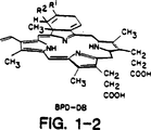
図1は、本発明における光感作剤として特に有用なBPD化合物の構造を示す。
発明の実施態様
下記に記載される本発明に従って、ドナー組織の移植以前に、それは光感作剤と接触される。“ドナー組織(donor tissue)”という用語は、APC'sを含有するレシピエント以外のドナーからの如何なるタイプの移植可能な(transplantable or implantable)組織を包含する。本発明に使用されているドナーの組織は、広範囲の種類の組織の何れか一つでよく、例えば生まれたての羊膜、骨髄、造血前駆体細胞、コラーゲン、及び軟骨の成長を刺激する骨蛋白のような柔らかい組織;皮膚、心臓、肝臓、脾臓、膵臓、甲状腺突出部、肺臓、腎臓、管状器官(例えば、腸、血管、又は食道)のような器官;及び心臓弁及び膵臓の膵島細胞又は肝臓細胞のような分離された細胞又は細胞集団のような器官の一部が含まれる。
管状器官は、食道、血管、又は胆管の障害を受けた部位を交換するのに使用され得る。皮膚移植片はやけどのみならず、障害を受けた腸のドレッシング(dressing)のために、又は横隔膜ヘルニアのようなある特定の欠損を閉鎖するためにも使用されることができる。特に好ましい実施態様では、ドナーの組織は皮膚組織又は膵島(pancreatic islet)の細胞である。
ここで使用される“移植片(graft)”という用語は、レシピエントへの移植のためのドナーに由来する生体物質を意味する。“移植(transplant)”という用語及びその変形体は、レシピエントへの移植片の挿入を意味するが、移植すること(Transplantation)は、同系(syngeneic)(ドナー及びレシピエントが遺伝学的に同一である場合)、同種異系(allogeneic)(ドナー及びレシピエントが異なる遺伝子的起源を有するが同種(species)である場合)、又は異種系(xenogeneic)(ドナー及びレシピエントが異なる種(species)である場合)の何れかである。故に、典型的な筋書きでは、宿主はヒトであり、移植片は、同一又は異なる遺伝子的起源のヒト由来の同系移植片である。もう一つの筋書きでは、移植片はそれが移植されるものとは異なる種から由来するもので、これは系統学的に広く分かれた種からの動物、例えばヒト宿主へ移植されているヒヒの心臓を含んでいる。
ドナーの組織は、如何なる原料からも取得可能で、死体からでも生存するドナーからでもよい。適当なドナーの例は、実験動物、例えば犬、猫、マウス、ラット、スナネズミ、モルモット、牛、霊長目の動物、又は人間のような生きた動物を含む。ドナーは人間を含むほ乳動物が好ましい。
ヒトのドナーは、交差性の主要血液群障壁が同種異系移植片の生存を害するので、健康診断で正常で同一の主要ABO血液群である任意の血縁関係のドナーであることが望ましい。しかしながら、例えばO型ドナーの腎臓をA,B、又はAB型のレシピエントへ移植することは可能である。
“同種異系移植片(Allograft)”という用語は、レシピエントと同じ種のドナーに起源を有するか、由来する細胞及び組織を意味する。ドナーはレシピエントと同じ種であることが好ましい。
ここで使用される“レシピエント(recipient)”という用語は、何れかの適合性を有する移植宿主のことを意味する。可能性のある有用なレシピエントの例は、動物、好ましくは、家畜、例えば馬、牛又は羊;家庭用ペット、例えば犬又は猫;実験動物、例えばマウス、ラット、スナネズミ又はモルモット;又は霊長目の動物、例えば類人猿又は人間のようなほ乳動物を含む。最も好ましくは、レシピエントは人間である。移植片のドナー及び宿主の両方がヒトであるときは、それ等は組織適合性を改善するようにHLAクラスII抗原のために好ましく調和する。
光感作剤
一般的に、古典的な光力学的治療において有用な多種の化合物の何れか一つが本発明における使用に適している。この分野の通常の技術を有する者に知られているように、主要なクラスの公知光感作剤はポルフィリン関連化合物である。幾らかは詳細に上記した如く、これ等の薬剤は、ヘマトポルフィリン誘導体;光感作性組成物及びその活性成分フォートフリンII(Photofrin II)として市販されているトヘマトポルフィリン誘導体の高分子量画分;同様にベンゾポルフィリン-誘導体(BPD又はBPD's)と呼ばれているモノヒドロベンゾポルフィリンのようなポルフィリンの種々の合成誘導体;緑色ポルフィリン;及び種々の他の多環式化合物が、露光されるとき一重項酸素を発生し、故に組織を破壊すると信じられている。適当な光感作性の化合物の調製方法は上記に記載の特許やその中で引用されている刊行物に十分開示されている。好ましくは、光感作剤はBPDである。
原則として、何れかの光感作剤の重要な特徴は、それが光感作物質により吸収されることができる波長の光に曝される時、局在化される細胞に対する細胞毒性効果を示すというその性質にある。多くの例で細胞毒性効果は露光に際して一重項酸素の形成の結果であると信じられているが、作用の正確な態様は本発明にとって重要ではない。
前記Dougherty等の特許においてある長さ検討されたように、多くの付加的特性が効果的光感作剤と典型的に結びついている。本発明の実施において特に重要である通常の光感作物質の性質の中には、光化学作用の欠如における細胞に対する細胞毒性の相対的欠乏、及び特定の細胞と光感作物質との間での標的-特異性相互作用の欠乏における組織からの迅速な解除がある。
本発明の実際の特に重要な一般的な光感作物質の性質の中には、特定の細胞と光感作物質との間での標的-特異性相互作用がない組織からの光化学作用及び迅速な解除がない比較的細胞に対する毒性がない。
本発明の光感作剤は、350nmから1200nmの間の領域の波長内にある吸収スペクトルを好ましくは有するが、この吸収スペクトルはそれ自体公知の方法で目的とする透過に合わせることができる。そして、好ましくは400nmから900nm、及び最も好ましくは600nmから800nmの間である。
本発明の光感作剤は、移植及び治療される障害の性質、ドナーの種、個々のレシピエントの医学的状態、ドナーの組織中又はレシピエントの体内中に存する如何なる他の薬剤、及び医師に知られているその他の要素を考慮して正しい医療行為に適合した方法で投与される。移植片に接触するために使用される光感作物質の治療上の有効量は、移植片の免疫原性を減少せしめてレシピエントと適合し拒絶されないようにするのに効果を有する量である。この目的のために通常の有効量は、約0.1から約10μg/mL、好ましくは約0.1から約2.0μg/mL、及び最も好ましくは約0.25から約1.0μg/mLの範囲に存する。
光感作剤は、移植片に対する免疫抑制効果を増大する免疫抑制剤の一つ又は二つ以上と結合することができる。そのような他の薬剤の有効量は、製剤中に存在する光感作剤の量、移植のタイプ、移植の原因、供給部位(site of delivery)、投与方法、投与の計画、上記に検討された他の要素、及び医師に知られているその他の要素に依存する。
典型的には、光感作剤は、室温、適切なpH値、及び目的とする純度で、一つ又は二つ以上の医学上許容される担体、即ち採用される投与量及び濃度でレシピエントに対して非毒性である担体とともに、それを混合することにより製剤化される。製剤のpH値は、特定の用途、及び光感作物質の濃度とに主として依存するが、好ましくは約3から約8のどこかの範囲である。
好ましくは、光感作物質は、pH値が生理学的水準に接近すると発生するようなその中の含有物への付着を防止し、かつ光感作物質の活性化を確保するために中性のpH値(例えば、約6.5から約7.5)に維持される。従って、pH値6.5に調整された塩による緩衝液を含有するが、ウシ胎児血清(“FBS”)を含有しない電解質溶液中での製剤化が適切な実施態様である。FBSが除去される理由は、同種異系移植片の反応を憎悪させる抗原成分をそれが含むからである。移植片が処置されている容器に光感作剤が付着する場合、ヒト血清アルブミンのような適当な非抗原性成分が、処置される移植片に灌流し又は付着する光感作剤と相互作用を起こさない量で任意的に添加される。
光感作剤の製剤が局所的に適用される場合、例えば移植以前に皮膚移植片に塗布される場合、非粘性の溶液よりも寧ろゲルのような粘性のある溶液を使用するのが好ましい。ゲルは、例えば光感作剤の溶液と多糖、好ましくは水溶性多糖、例えばヒアルロン酸、澱粉、及びセルロース誘導体、例えばメチルセルロース、ヒドロキシエチルセルロース、及びカルボキシメチルセルロースのようなゲル化剤を混合することにより調製されることができる。多糖がゲル製剤中に存在する場合、通常存在する量は約1-90のゲル重量%、より好ましくは約1-20%の範囲に存する。この目的のための他の適当な多糖の例、及び多糖の溶解性の測定は、1988年5月11日発行の欧州特許(EP)267,015号明細書に見出され、この開示は参考によりここに組み込まれる。
処置される移植片がある期間保存される場合、光感作剤は、好ましくはフッ素置換化合物のエマルジョン(血液代替物として使用)中に製剤化又は添加されて移植片に到達する高濃度の酸素を可能にする。そのようなエマルジョンは、水中界面活性剤で乳化されるフッ素置換デカリン及び/又はフッ素置換トリプロピルアミンを含む。フッ素置換化合物はレシピエントに対して最も毒性が少なくなるように選択される。
適当な界面活性剤はポロクサマー(poloxamer)界面活性剤を含み、これはエチレンオキサイドとプロピレンオキサイドのブロック共重合体である一連の分子を表し、単独又は卵のレシチンのようなリン脂質と混合されている。他の例のミドリ十字社(Green Cross)から販売、購入可能なエマルジョンは、フルオゾル-ディーエー(Fluosol-DA)20%で、これはポロクサマー界面活性剤、プルロニック(Pluronic)F-68で乳化されたフッ素置換デカリン及びフッ素置換トリプロピルアミンを含有する。フッ素置換化合物のエマルジョン及びそのほ乳動物における効果は、Bollands et al.,J.Pharm.Pharmacol.,39:1021-1024(1987)により十分に記載され、この開示はここに(本明細書中に)参考により組み込まれる。
治療上の投与に使用される光感作物質の製剤化は、好ましくは無菌性である。無菌性にするには、薄膜(0.2ミクロン)を通過する無菌濾過により容易に達成される。一度製剤化し無菌化にすると、光感作物質は酸化的な変成に対して安定ではない。しかしながら、例えばBPDを含有する再構成のための凍結乾燥製剤は、保存に適している。
標的系
移植片-対-宿主反応を開始するドナーの組織の機能を破壊する場合のこれ等の光感作剤の使用は、標的-特異性薬剤への光感作物質の接合体により増強される。特に、光感作性物質は、(1)ドナーの組織中の抗原-提示細胞(antigen-presenting cells[APC's])を直接特異的に標的にする部分;(2)その接合体により標的にするAPC'sを標識する媒介物質を特異的に標的にする部分;又は(3)宿主の場所に対し移植するT-細胞に結合されることができる。いずれの場合も、一度ドナーの組織がその接合体との相互作用により修飾された場合、それ自体公知の方法でドナーの組織中のAPC's群の実質的な消耗を果たすようにそれは露光される。
本発明は、一般的に光力学的治療(photodynamic therapy:[“PDT”])によりドナーの組織中のAPC'sを破壊する方法と同様、同種異系のドナーの組織中のAPC'sを標的にするのに有用な、特定の光感作物質-含有接合体を提供する。本方法において有用な一つの製剤は、実質的に光感作剤及び“ホーミング剤(homing agent)”を光感作物質と結合する系により構成される。もう一つの製剤は、APC-標的系及びAPC-標的系のためのホーミング剤と結合される光感作剤の組合せを含む。どちらの製剤とも、光感作性の薬剤をAPC'sに供給するという最終の目的は同じである。
標的にされるAPC'sは、MHC糖蛋白質生産物及びこれ等細胞がレセプターを保持するリンホカイン因子に免疫特異的な部分を含む種々異なるタイプの標的-特異性薬剤により接近(アクセス)されることができる。典型的には、MHC糖蛋白質との反応のためには、これ等の糖蛋白質に対して生じる抗体、ポリゴーナル(polygonal)又はモノクローナルのどちらかが使用されることができる。
ポリクローナル抗血清は、常法により、例えば適当なほ乳動物に目的とされる抗体に対する抗原を注射し、その抗原に対する血清中の抗体のレベルを測定し、その後その力価が高いとき抗-血清を調製することにより、製造される。モノクローナル抗体の製剤は、同様に常法により、例えばKoehler及びMilsten,“Nature,Vol 256,pp.495-497(1975),“Continuous Cultures of Fused Cells Secreting Antibody of Predefined Specificity”及びEuropean Journal of Immunol.,Vol 6,pp511-519(1976)“Derivation of Specific Antibody-Producing Tissue Culture and Tumor Lines by Cell Fusion”
の方法により製造される。
この方法は、例えば、免疫化された動物からの末梢血リンパ球又は脾臓細胞を使用して、これら細胞をウイルス感染、骨髄腫との融合、又は他の簡便な手法によって不活化し、次に単離されたコロニーにより目的とする抗体の生産のためのスクリーニングを行うものである。
抗体の他に、Fab、Fab'、又はF(ab)2フラグメントのような適当な免疫反応性のフラグメントが同様に採用されることができる。標的にするメカニズムを形成するための使用に適した多くの抗体が、既にこの分野で入手可能である。例えば、全ての抗体の代用として免疫学的反応性のフラグメントの使用が、E.L.Morgan et al.,Scand.J.Immunol.,Vol 10,pp.395-402(1979),“Comparison of the Binding of Radiolabelled Human IgG and Fc Fragments to Murine Spleen Cells”及びHans P.Kocher et al.,The Journal of Immunol.,Vol 122,No.4,pp.1190-1195(1979),“Tryptic Degradation of the CH1 and VL Regions of OgD and IgE1”.
により記載される。
免疫反応性に加えて、標的にすることがAPC細胞表面のレセプターを標的にするレセプターリガンドを、例えばレセプターとリガンド間の輪郭線又は電荷像の相補性に基づいて使用することにより成し遂げられることができる。ここで使用されるように、“レセプターリガンド(receptor ligand)”という用語は、APC細胞表面のレセプターに特異的に結合する天然又は合成の如何なる物質をも意味する。これ等のレセプターリガンドはリンホカイン因子、例えばIL-2を含む。
本発明の特定の実施態様に従って、光感作剤は、次にAPCに対して特異的である媒介物のための標的-特異性薬剤と結合される。例えば、Iaを提示するラット細胞の場合、マウス抗-ラットIa抗体は媒介物として使用されることができる。この場合、抗-マウス抗体に結合される光感作剤の接合体は、抗-ラットIa抗体を含む接合体が直接行うのと正確に同様の方法でマウス抗体で標識される細胞を標的とするはずである。
接合方法
標的系を通常の方法を用いて、直接光感作薬に接合されることができる。これは一般に知られている技術であり、前述のLevy et al.特許の実施例に記述されている。
Ig及び他のポリペプチドのようなタンパク質に対する直接共有結合は、光感作剤と例えばカルボジイミドのような脱水剤を用いる標的-特異的構成分との間に影響されるかもしれない。接合体の活性成分は、勿論、二機能性であり、2つの活性構成成分のそれぞれに共有結合できるリンカー化合物を用いて接合されてもよい。
2つの化学的部分(Chemical moiety)を結合する適切な技術において知られている効果的な技術は、本発明の領域に入る。リンカー部分は、その技術での共有結合又は標準の技術に用いられるものからの推測されるものが広範囲に解釈される。
代わりに、標的物を付加的な特異的薬剤により媒介することができる。例えば、下記に説明されるように、APC's特異的抗体に配向する2次抗体は直接的に光感作剤に結合されても良く、MHC-糖蛋白質標的剤を免疫接合体と標的細胞との間の架橋として用いても良い。
処理手順
本発明による、APC'sの除去又は機能減衰、又はケラチノサイトのような他の皮膚細胞の調節は、供与組織が光感作剤と直接接触することにより、光感作剤(又は光感作剤含有接合体の標的特異的構成成分)と標的APCの間での強い結合形態を可能にする状態下、比較的素直な方法で効果的である。
適切な接触は、1又は2以上移植片の表面又は器官移植片の培養若しくは灌流と本発明の光感作剤との組み合わせを適用することを含む。処理は、一般的に少なくとも1分、好ましくは約2分から約72時間、更に好ましくは約2分から24時間行う。接触時間は、例えば製剤における光感作剤の濃度、処理される移植片及び製剤の特有の形態などの要因に依存する。灌流は幾つかの適当な方法によって行われる。例えば、ある器官は、一定の圧力を供給する装置を介して灌流することができ、又は圧力調節及びポンプと器官の間にオーバーフローの場を持った灌流ができる。代わりに、器官はシーリングドア(sealing door)を介して高圧チャンバーの中に置かれてもよく、リザーバーから液体を引き出すポンプによってチャンバーに輸送された灌流液は、任意に灌流液が消費されている間、バルブによりリザーバーに戻される。
皮膚移植では、製剤を移植される皮膚の下部表面に塗布又はスプレーすることができ、その結果ドナーの下部表面とレシピエントの組織との間に光感作剤の層が生じる。しかしながら、好ましくは、全移植皮膚は光感作剤成分中に浸す。
接触処理は広範な温度において行うことができるが、かなりの変性を引き起こすような高い、若しくは移植片に有害に影響する高い温度及び光感作剤の細胞内取り込みを最小にするような温度は回避する。好ましくは、接触処理は、約50Cから約400C、より好ましくは約150Cから約370C、最も好ましくは外界温度である。
光感作剤が標的APCと適切に結合することを確実にする光感作剤を適切に分布させた後、当該処理されたドナー組織は、光感作剤によって吸収され、光感作剤によって細胞毒性特性の活性を導く波長を持った光で露光する。そのような露光は、光感作治療の技術において勿論一般的な方法である。
移植片を光感作剤に接触させ、露光した後、24-48時間の間貯蔵することができる。しかしながら、好ましくは移植進行中すぐに用いる。貯蔵耐用期間は、上記のように製剤中に血液代用物(例えば、水素をフッ素で置換した化合物の乳濁液(perfluorochemical emulsion))を用いたり、又は移植片を、1985年4月9日に発行されたJP60061501に記載のように、細胞のわずかな破壊で移植片を冷凍させることができるグリセロール処理後の冷却等張剤及び抗凝固剤を含有する光感作剤の製剤で還流することにより延長させることができる。さらに、細胞壊死することなしに半永久的に組織を保存するために、移植片は、凍結温度で冷却している間、その製剤を含有する異なった液体で保存することができる。
移植前に、好ましくは、組織学的生理食塩水に浸すことにより又は他のこの目的のために適切な他の手段により、移植片を光感作剤成分が入ってないもので洗浄する。また、移植に先だって、レシピエントは、移植片の生存を助けるために、PDT処理された末梢血単核細胞を持った1又は2以上のドナー特異的血液の輸液を供されてもよい。移植手術に先だって、他の方法で全リンパ球露光をレシピエントに行ってもよい。特別な移植レシピエントに有益である他の前移植方法が本発明の方法の一部として行うことができる。
ある場合には、適当なアミノ酸又は高分子を用いたり、生理的に受けいられる電荷を与えられた官能基のソース(source)を付けることにより、正又は負電荷を負わせた官能基を与えるために移植片表面を修飾することが望まれる。又、ある場合では、例えばフェニルアラニン、セリン又はリジンを表面に結合することにより、表面に疎水性又は親水性を与えることが望まれる。これら表面修飾に対して特に効果的な免疫抑制剤はグルタルアルデヒドである。
移植方法それ自身は、特に処理されるものの障害、患者の健康状態等に依存する。医師が与えられた症状に用いられる適切な方法を認識する。移植は、重要な手術後期間(最初の3ヶ月)体系的に、放射線核静脈血管造影剤のような適切な方法により、任意に観察される。移植後、適切な免疫抑制薬を用いる免疫抑制治療が、移植された移植片生存において重要なものとしてよく用いられる。
本発明の方法は、体系的にドナー、インビトロでドナー組織、又は居所的や体系的にレシピエントに投与するのと同時に、免疫抑制剤と同様な又は引き下げられた容量によって補足又は用いられることができる。ここで用いられる“免疫抑制剤”という言葉は、移植片が移植された宿主の免疫系を抑制又は遮蔽する作用がある物質を言う。これは、サイトカイン産生の抑制、自己抗原発現の下方制御又は抑制、又はMHC抗原の遮蔽する物質を含む。
そのような薬剤の例は、2-アミノ-6-アリール-5-置換ピリミジン、アザチオプリン、ブロモクリプチン、グルタルアルデハイド、MHC抗原に対する抗イディオタイプの抗体、サイクロスポリンA、1又は2以上のステロイド、及びデキサメタゾン;抗インターフェロンガンマ抗体;抗腫瘍壊死因子アルファ抗体;抗腫瘍壊死因子ベータ抗体;抗インターロイキン2抗体;抗IL-2レセプター抗体のような抗サイトカインレセプター抗体;異種の抗リンパ球グロビン;汎T抗体、好ましくはOKT-3モノクローナル抗体;CD4に対する抗体;ストレプトキナーゼ、ストレプトドルナーゼ;又は宿主からのRNA又はDNAを含む。
効果的な量は、これらの考慮により決定されるが、レシピエントによる移植片の拒絶が生じる免疫応答を抑制するのに必要な最小の量であり、より長い移植片の生存期間に達成するくらいの量である。そのような量は、好ましくは、レシピエントに対する毒性があり、レシピエントに感染に対して著しく影響を与える量よりは少ない方がよい。本発明で必要とする免疫抑制剤の量は、前処理していない移植された移植片に通常必要量よりは典型的に少なく、個々の移植を取りまく環境及び用いられる免疫抑制剤の種類に依存する。
特異な例として、1投与量あたり非経口的に投与される調剤的に影響する免疫抑制剤、サイクロスポリンAの全量は、1日あたり患者の体重に対しておよそ0.1から20mg/kgの範囲内であると、最近通常の免疫抑制治療で用いられるおよそ5から15ml/kg/dayの典型的な範囲と比較して予想される。腎移植では、通常、短期間に大量の投与量の糖質ステロイドの投与が実施され、例えばメチルプレドニゾロンは、移植組織の光感作剤前処理がない場合、1日当たり7グラム投与量3から5日間与えられ、その後に20から100mgのプレドニゾロンが与えられる。本発明の前処理を行った場合、著しく低い投与量が可能である。
上記のように、これらから示唆される免疫抑制剤の量は、治療に非常に大量費やされる。適切な投与量及び計画の選択における重要な要因は、即ち移植生存期間から得られる結果である。例えば、比較的高投与量は、初期では抗体媒介の移植片の破壊に起因させることができる超急性の移植拒絶の処理のためで、又は後期では移植片機能における急激な減衰に特徴づけられ、必要とされるかもしれない。
免疫抑制剤が用いられるとき、非経口、好ましくは局所の免疫抑制処理、病巣内投与を含む幾つかの適切な方法によって投与されてよい。非経口投与は、筋内、静脈内、動脈内、腹腔内又は皮下投与を含む。さらに、免疫抑制剤が用いられるとき、パルス注入により特に引き下げられた投与量で、又は持続注入により適宜投与される。
次の実施例により、より明らかになるが、本発明を制限しない
実施例1
BPD-Ra-MIg接合体の調製
図1に示す光感作剤BPD-MAを、暗所にて、1mg/mlから200pg/mlの濃度にリン酸緩衝食塩水で希釈し、既知含量のラット抗マウスIg(RaMIg)と混ぜ合わせる。RaMIgは、シーダーレーン研究室(Cedar Lane)から入手、又はマウス免疫グロブリンでウサギを免疫化し、抗体を免疫吸着カラムに通して精製することにより調製される。その混合物を室温にて1時間インキュベートし、生じる接合体を終夜4℃下3リットルのPBSで、12-14kdよりも小さい分子量を持つ分子を透過できる膜を通して透析する。標識BPD-MAでのモデル研究は、保持された接合体がBPDを所持することを示す(10-20のAb率)。その透析からのretentateは凍らせて凍結乾燥させ、暗所に保存する。
実施例2
当該接合体による同種異系移植組織の処理
ドナーの膵島細胞は、ラットから次のように単離した。
雄SDラット(200-250g)を腹腔内ウレタン(100mg/kg)の投与にて麻酔して正中線を通して開腹し、呼吸循環停止は双方の気胸(切除)により誘導された。近位の総胆管にカニューレ挿入し、十二指腸への挿入地点で、遠位に閉鎖した。膵臓を冷却(4/C)コラーゲン(type XI,Sigma Chemicals)溶液0.42mg(650U)/mlの濃度による逆行性法で膨張させた。インサイチュのコラーゲン膨張の後、全膵切除が遂行された。
腺は22分間37/Cの水浴中において消化された。消化された腺は、滅菌、シリコン処理したピペットでの粉砕により分散させた。粗製の組織スラリーを200ミクロンスクリーンフィルターに通して、非消化の管、血管及びリンパ節を取り除き、それぞれ比重1.065と1.031の2つの単層からなる不連続デキストラン勾配にて遠心した。低密度の島組織を単層境界から吸引し、洗浄、及び更に解剖顕微鏡下の手選により精製した。この技術を用いて、膵臓あたり、300-400の機能的及び形態学的に無傷の膵島を収穫した。
島(細胞)をインビトロで1日、25%子ウシ血清、15mM HEPES及び1%ストレプトアビジンを補ったハム(Ham)のF-12培地で培養した。即座に、培養島細胞(islet)をSelaLabから入手した商業利用のマウス抗ラットIaモノクローナル抗体(OX-6と命名)0.2ml/ml存在下1時間培養した。OX-6はMHCクラスII生産物に対して免疫特異的である。
OX-6処理島細胞の一部は、それぞれ(1)BPD:1Ab比6.5のRa-MIgG-BPD接合体;(2)BPD:1Ab比20の同接合体;(3)BPD:1Ab比6.5の無関係の抗体GA-7sIgGとのBPD接合体;(4)BPDのみにて;及び(5)培地のみで、20℃下2時間暗所にて培養した。培養混合液を10Joules/cm2の400-800nmの光エネルギーに曝した。露光された培養細胞は組織学的に試験され、APC枯渇であった。
実施例3
ドナー組織の特徴づけ
組織学的研究において、BPD:1Ab比6.5の接合体で処理した約75-100島細胞を同系のSDラット及び同種異系WFラットの腎莢膜下に移植した。同系移植も同種異系移植でも、すべてのレシピエントは喜ばしい結果であった。特に、リンパ球浸潤で完全な腺の置換が観察され、同系移植も同種異系移植も確認できる内分泌組織は観察されなかった。
APC欠失の研究において、島細胞は0X-6抗体で1次免疫染色された。当該処理された細胞をFITC標識したヤギ抗マウスIg(Jackson Laboratories)で2次染色し、蛍光顕微鏡で観察した。当該接合体で処理した標品において、MHCクラスII細胞は確認されなかった。しかしながら、コントロール(無関係な抗体との接合体、BPDのみ及び培地のみ)では、FITC標識した第2次抗体がAPCをラベルし、緑色の蛍光を発していることから、これら細胞の存在が蛍光顕微鏡により検出された。
実施例4
皮膚同種異系拒絶の阻止
最小の拒絶を示すベースラインを確立するために、9つの同系移植(ドナーとレシピエントが同じ動物)を、次に示すような標準的方法(Billingham et al.,“The Technique of Free Skin Grafting in Mammals”,J.Exp.Biol.,28:285-99(1951))に従って、BALA/cマウスで行った。
移植されたマウス躯幹の皮膚は毛を剃り、除毛した。そのマウスに10Tlケタミン塩酸塩、10Tlキシラジン、70Tl PBSの混合液を静脈注射により麻酔し、続いて適当な移植土台に置き、皮筋層を傷つけないように注意して、十分な厚さの皮膚(1cm×1cm)を注意深く切開した。
自己の皮膚移植は移植部位に再適用し、おおよそ4滴のVetbond細胞接着剤を移植片と移植土台との間の境界に注いで固定した。移植片をベトローリアム-ジェリーガーゼスポンジに押しあて、体のまわりにくるみ”体幹ギプス(body cast)”を形成させた。
長期間同系移植の成功率、すなわち120日を越えるものは、90%以上であった。移植片の壊死が少なくとも80%存在したとき、移植拒絶は完全だと考えられている。移植生存物は日あたりの生存時間±標準偏差に置き換えた平均値として示された。
それぞれの移植片がドナーマウスからのもので、異なるレシピエントマウスの移植土台が適用されたという点を除き上記と同じ方法で、C57BL/6(ドナー、H-2b)とBALB/c(レシピエント、H-2d)のマウス同種異系間の皮膚移植を対照として行われた。簡単にいうと、ドナーの躯幹の皮膚は、毛を剃り、除毛し、その後十分な皮膚移植片(1cm×1cm)を得た。レシピエントマウスは毛を剃り、20Tlのケタミン塩酸塩、10Tlのキシラジン及び70Tl PBSの混合液の静脈注射により麻酔した。皮筋層を傷つけないように、それぞれのレシピエントの移植土台を躯幹皮膚(1cm×1cm)を注意深い切開により調製した。移植片は同種異系の移植部位にあて、Vetbond細胞接着剤及びVetrapの包帯テープを用いてペトローリアム-ジェリーガーゼスポンジに固定し、“体幹ギプス(body cast)”を形成した。平均の生存期間は11.1日(標準偏差1.9)であった。
本発明の方法によると、レシピエントに移植された皮膚標品は最初、インビトロで1.0Tg/ml溶液の光感作剤BPDで1時間接触させた。その皮膚はウシ胎児血清(FBS)を含まない電解質溶液にて30分間懸濁し、発光ダイオード(“赤”)(10J/cm2 at 690nm±10mn)の赤色光にさらした。この光処理後、露光された皮膚をレシピエントマウスに上記のように移植した。その動物を移植後8日にわたり観察した。同種異系移植のための平均の生存期間は、増加して18.5日(標準偏差2.1)であった。
これらの結果をそれぞれの比較と照らして下記の表1に作表する。

実験は、光感作剤BPDの濃度(0.25から1.0μg/mL)を変えて、標準、対照の同種異系移植を伴って、本発明により行われた同種異系移植と比較して繰り返された。これらの試験の結果を下表2に要約する。
実施例5
移植された皮膚の組織学的な試験
生存期間を延ばす結果が得られる条件下、皮膚移植の光感作治療で処理の効果を調べるため、皮膚標本を入手し、異なる範囲の光感作剤濃度すなわち0.25又は0.50μg/mLのBPDである点を除き、上記のように処理した。幾らかの組織は、光感作剤を入れずに、電解質溶液のみで培養し、対照とした。すべての細胞は24時間にわたり培養し、その後、対応する数を光に曝した。曝されたとき、露光は10J/cm2のエネルギーレベルで赤色光であった。
すべての標本はホルマリンの中に置き、組織学的試験を行った。組織は電解質溶液のみ(対照標本)又は0.50μg/mLのBPD溶液で、露光しないで培養し、正常を示した。しかしながら、0.25も0.50μg/mLでもBPDで処理した標本は、赤色光で処理した後、次のような変化:
核の肥大化、核周囲の空胞化、上皮細胞での好酸球の増加、細胞質容積の増大、及び上皮表面のケラチノサイト間の増加した細胞間スペースを示した。これらの組織学的知見を基に、本発明の光感作処理は細胞死よりもむしろ、ある程度の細胞障害を生じることが推定された。この予期せぬ非細胞毒性効果のメカニズムは解らない。
情報公開した内容の修飾及び/又は変化は下記の発明の請求項の見地から逸脱すること無しに実施されることにより、本技術において技術化されたものがより明らかになる。 Technical field
The present invention relates to a method for supplying allografts in therapy and to prevention of rejection of allografts by recipients. In particular, it relates to the treatment of donor tissue using photodynamic therapy techniques that deplete the tissue of antigen presenting cells and reduce the immunogenicity of the cells present therein.
Background art
Successful transplantation of an allograft into a host includes antigens present in the transplanted tissue that can be recognized by foreign recipients and can induce rejection, cells present in the recipient immune system that mediate rejection, and It depends on factors such as the reaction that modifies either the presentation of the foreign antigen or the cellular response. It is known that an important component of allograft rejection is due to the presence of non-parenchymal cells (passenger leukocytes) in the donor tissue.
Major histocompatibility complex (MHC) products are likewise known to play an important role in mediating transplant tissue attack on recipients. The MHC is usually complex because it contains many different loci, each of which encodes a separate cell-surface antigen, and the loci have a wide range of polymorphisms. MHC loci are included in one of two classes, class I or class II based on their tissue distribution, expressed antigen and their function. Class I antigens are present in all nucleated cells and have cytotoxic T (CD8+) Act as primary target for lymphocytes. Class II antigens are not widely distributed in tissues and helper T (CD8-) Act as primary target for lymphocytes.
Polymorphic forms of individual loci of human leukocyte antigen (HLA), human MHC, have been recognized by antibodies and by various in vitro techniques that measure T-lymphocyte recognition. These responses mediated by the recognition of polymorphic recipients in the donor correlate with the strong rejection that occurs in vitro. A study of the cellular principle of graft rejection using both in vitro and in vivo studies, CD4+And CD8+It became clear that both lymphocytes were involved in the rejection response.
In both experimental models and medical practice, attempts to prolong allograft and xenograft survival have been focused primarily on the suppression of recipient immune mechanisms. This treatment has as its purpose the treatment of prophylactic immunosuppression and / or graft rejection.
Drugs used for immunosuppression include cytotoxic drugs, antimetabolites, corticosteroids, and antilymphocyte sera. Non-specific immunosuppressants (azathioprine, bromocriptine, methylprednisolone, prednisone, and cyclosporin A) that are found to be particularly effective for prophylactic immunosuppression significantly enhance the clinical success of transplantation I came. The nephrotoxicity of cyclosporin A after kidney transplantation has been reduced by co-administration of prednisolone in combination with steroids such as prednisolone or azathioprine. In addition, kidneys have been successfully transplanted with anti-lymphocyte globulin followed by cyclosporin A. Another procedure is exposure of the recipient's total lymphocytes prior to transplantation, followed by minimal immunosuppression after transplantation. Treatment for rejection includes the use of steroids, 2-amino-6-aryl-5-substituted pyrimidines, heterologous anti-lymphocyte globulins, and monoclonal antibodies against various leukocyte populations.
The main complication of immunosuppressive drugs is infection. Furthermore, systemic immunosuppression is accompanied by undesirable toxic effects, such as nephrotoxicity when cyclosporin A is used after kidney transplantation, and a reduction in hematopoietic stem cell levels. Immunosuppressants also lead to obesity, reduced wound healing, steroid hypoglycemia, steroid psychosis, leukopenia, gastrointestinal bleeding, lymphoma, and hypertension.
In view of these complications, transplant immunologists have sought ways to suppress immune responsiveness in antigen-specific approaches so that only the donor's response to allogeneic antigens is lost. Such specific immunosuppression has generally been achieved by modifying either the antigenicity of the transplanted tissue or specific cells that can mediate rejection. In certain instances, either immunity or resistance is induced depending on the manner in which the antigen is presented to the immune system. Other anti-rejection strategies have been directed to the removal or attenuation of MHC-bearing passenger leukocytes, such as antigen-presenting cells (“APC's”), present in donor tissue prior to transplantation.
A technique that has been reported for this purpose is to extend the time to culture donor tissue (Lafferty et al., “Thyroid Allograft Immunogenicity is Reduced after a Period in Organ Culture”,Science,188: 259 (1975)). Pretreatment of allogeneic transplantation by growth of tissue culture prior to transplantation was found in two mouse model systems and led to permanent tolerance beyond MHC injury (Lafferty et al.,Transplantation,twenty two: 138-49 (1976); Bowen et al.,Lancet,2: 585-86 (1979)).
It has been hypothesized that such treatment results in the loss of passenger lymphocyte cells and hence the lack of stimulator cell populations necessary for tissue immunity. For example, several donor-recipient HLA matches have been used in, for example, vascular and kidney transplants, and sometimes prior to transfusion.
Donor tissues are growth factors such as TGF-beta (Czarniecki et al., US Pat. No. 5,135,915, issued Aug. 4, 1992), with some combination of occasionally prolonged culture times (Orton, USA Patent No. 5,192,312, issued March 9, 1993).
Donor tissue is ultraviolet (Reemtsma et al., US Pat. No. 4,946,438, issued Aug. 7, 1990; and Lau et al., “Prolongation of Rat Islet Allograft Survival by Direct Ultraviolet Irradiation of the Graft,Science,223: 607 (1984)), sometimes in a conjugate with microencapsulates (Weber et al. US Pat. No. 5,227,298, issued July 13, 1993). Other researchers have developed a first non-cytotoxic layer membrane and a second outer layer of biocompatible semipermeable membrane-like polymeric material taught in US Pat. No. 4,696,286 issued September 29, 1987 by Cochrum. Only a diaphragm such as a bilayer containing was used.
Donor tissue can be applied to a wide variety of substances, such as topical application of cyclosporine to skin grafts as disclosed in Hewitt et al., US Pat. No. 4,996,193 issued Feb. 26, 1991, and Jones et al. 1981. Treated with perfusion of the donor's kidneys with lymphocylone as disclosed in US Pat. No. 4,294,824 issued Oct. 13, 2003. Skin graft survival was prolonged by in vitro treatment with cortisone, thalidomide, or urethane prior to transplantation into experimental animals. The amount of drug applied topically to the skin is usually less than that required to achieve a similar effect by systemically administering the drug to the recipient. Donor skin was treated in vitro with streptokinase / streptodolnase, recipient RNA and DNA formulations, or glutaraldehyde solution prior to transplantation to reduce the antigenicity of the transplanted skin.
A more advanced approach is to treat donor tissue with complement to monoclonal antibodies against the product (Faustman et al., “Prolongation of Murine Islet Allograft Survival by Pretreatment of Islets with Antibody Directly to Ia Determinants”,Proc.Natl.Acad.Sci.USA,78,: 5156 (1981)), or treatment of donor tissue with immune complexes of antibodies to MHC (Shizuru, JA, et al., “Inhibition of Rat Mixed Lymphocyte Pancreatic Islet Cultures with Anti-Ia Immunotoxin”,Transplantation,42: 660 (1986)). Variability results were obtained by these methods. Therefore, there is a need for techniques for methods to prolong graft survival in transplantation procedures that minimize toxicity and other side effects resulting from the use of large doses of immunosuppressive agents.
The technique employed in the present invention to selectively destroy these APC cells involves contacting the donor tissue with a photosensitizer followed by exposure to light followed by transplantation. Similar photodynamic methods have long been used primarily to destroy tissues such as tumor tissue, atherosclerotic plaques, epidermal diseases, and blood pathogens (Levy et al., US Pat. No. 5,283,255). Issued February 1, 1994; 4,883,790 issued November 28, 1989; 4,920,143 issued April 24, 1990; 5,095,030 issued March 10, 1992; and 5,171,749 issued December 15, 1992 See published in Japan, the disclosure of which is hereby incorporated by reference). Similarly, US patents by Dougherty et al .: 4,932,934 issued June 12, 1990; 4,889,129 issued December 26, 1989; 5,028,621 issued July 2, 1991; September 1989 Reference is made to US Pat. Nos. 4,866,168 issued on 12th; 4,145,863 issued on 8th September 1992; and 4,649,151 issued 10 March 1987, which are also incorporated herein by reference. Incorporated.
For example, US Patent No. 4,866,168 to Dougherty et al. Discloses a composition sold under the trademark “Photofrin II” and obtained by recovering the highly cohesive-molecular weight portion of a hematoporphyrin derivative. As another specific example, Levy et al., US Pat. No. 4,883,790 discloses the use of a group of related compounds designated as “monohydrobenzoborphyrins” for similar purposes.
Furthermore, the use of a wide variety of photosensitizers of similar structure is described. For example, a compound prepared from (1-hydroxyethyl) deuteroporphyrin, hydrophobic hematoporphyrin ether, and methylpheophorbide (Pandey et al., US Pat. No. 5,002,962 issued Mar. 26, 1991); Pyropheophorbide conjugates (Pandey et al., US Pat. No. 5,314,905); bacteriochlorophyll-derivatives (see US Pat. Nos. 5,171,741 and 5,173,504); monovinyl and divinyl ether-linked dimers (Ward Benzoporphyrin derivatives (see US Pat. No. 5,214,036 to Allison et al.); Dibenzoporphyrin compounds (see US Pat. Nos. 5,308,608 and 5,149,708 to Dolphin et al.); So-called monobenzoporphyrin derivatives “Green” porphyrin (see US Pat. No. 5,087,636 to Jamieson et al.); Porphyrin compound having a double bond (see US Pat. No. 5,064,952, such as Chang); and porfimer (porfimer) sodium composition (see U.S. Pat. No. 5,244,914, such as Clauss) is referred to. The disclosures of all these patents are hereby incorporated herein by reference. In general, these agents are considered as interchangeable in their use for photodynamic therapy as an initial estimate.
Photodynamic therapy is primarily concerned with the treatment of tumor cells, but additional applications have been shown previously. For example, these photosensitizing agents can be used in procedures to remove atherosclerotic plaques and in the treatment of blood and other body fluids to destroy infected tissues. However, photodynamic therapy has clearly not been used to date to eradicate passenger leukocytes in donor allografts.
Unlike therapies involving the administration of photosensitizers to biological tissues, it is a particular advantage of the present invention that donor tissue can be optimally treated in vitro prior to the actual transplantation procedure. This method obviates the problems that arise with ensuring an appropriate level of exposure of the conjugate that is associated with the target cells within the tissue, for example.
Furthermore, the method of the present invention results in a graft that is immunologically stable in a suitable host, has biological function, and can be stored prior to transplantation. Thus, the present invention allows the establishment of a bank of photodynamically treated grafts that can be used for short-term storage.
Disclosure of the invention
The present invention provides a method for minimizing transplant rejection in an animal body. Prior to transplantation, donor tissue containing antigen-presenting cells (APC's) is exposed to light having a wavelength that is absorbed by the photosensitizer for a sufficient amount of time to contact and deplete the APC's. Be exposed. This treatment does not kill keratinocytes, but alters the expression of its class I and / or class II antigens as well as the cytokines they secrete, resulting in a decrease in the immunogenicity of the skin itself.
In one embodiment, the photosensitizer is in the form of a conjugate that includes a target-specific component that increases the interaction between the photosensitizer and the target APC's. Agents that cause photosensitization mediate the destruction of APC's when allografts are exposed at the appropriate wavelengths that are absorbed by the photosensitizer.
[Brief description of the drawings]
FIG. 1 shows the structure of a BPD compound particularly useful as a photosensitizer in the present invention.
Embodiment of the Invention
In accordance with the invention described below, prior to transplantation of donor tissue, it is contacted with a photosensitizer. The term “donor tissue” encompasses any type of implantable or implantable tissue from donors other than recipients containing APC's. The donor tissue used in the present invention may be any one of a wide variety of tissues, such as fresh amnion, bone marrow, hematopoietic progenitor cells, collagen, and bone proteins that stimulate the growth of cartilage. Soft tissues; organs such as skin, heart, liver, spleen, pancreas, thyroid protrusion, lung, kidney, tubular organs (eg, intestine, blood vessels, or esophagus); and heart valves and pancreatic islet cells or liver cells A part of an organ such as an isolated cell or cell population is included.
Tubular organs can be used to replace damaged sites of the esophagus, blood vessels, or bile ducts. Skin grafts can be used not only for burns, but also for injured bowel dressing or to close certain defects such as diaphragmatic hernia. In a particularly preferred embodiment, the donor tissue is skin tissue or pancreatic islet cells.
The term “graft” as used herein refers to biological material derived from a donor for transplantation into a recipient. The term “transplant” and its variants refer to the insertion of a graft into a recipient, but transplantation is syngeneic (donor and recipient are genetically identical). ), Allogeneic (if the donor and recipient have different genetic origins but are homologous), or xenogeneic (species whose donor and recipient are different) If any). Thus, in a typical scenario, the host is a human and the graft is a syngeneic graft from a human of the same or different genetic origin. In another scenario, the graft is derived from a different species than the one it is transplanted into, which is an animal from a phylogenetically separated species, such as a baboon heart transplanted into a human host. Is included.
The donor tissue can be obtained from any source and can be from a cadaver or a living donor. Examples of suitable donors include laboratory animals such as dogs, cats, mice, rats, gerbils, guinea pigs, cows, primates, or live animals such as humans. The donor is preferably a mammal including a human.
The human donor is preferably any related donor who is a normal and identical primary ABO blood group in a medical examination, because the crossed major blood group barrier impairs allograft survival. However, it is possible, for example, to transplant a kidney of an O-type donor into an A, B, or AB type recipient.
The term “Allograft” refers to cells and tissues that originate or originate from the same type of donor as the recipient. The donor is preferably the same species as the recipient.
As used herein, the term “recipient” means any compatible host for transplantation. Examples of possible useful recipients are animals, preferably domestic animals such as horses, cattle or sheep; household pets such as dogs or cats; laboratory animals such as mice, rats, gerbils or guinea pigs; or primates Including mammals such as apes or humans. Most preferably, the recipient is a human. When both the graft donor and host are human, they are preferably matched for HLA class II antigens to improve histocompatibility.
Photosensitizer
In general, any one of a wide variety of compounds useful in classical photodynamic therapy is suitable for use in the present invention. As known to those having ordinary skill in the art, a major class of known photosensitizers are porphyrin-related compounds. As described in some detail above, these agents are hematoporphyrin derivatives; high molecular weight fractions of tohematoporphyrin derivatives marketed as photosensitizing compositions and their active ingredients Photofrin II. Various synthetic derivatives of porphyrins, such as monohydrobenzoporphyrins, also called benzoporphyrin-derivatives (BPD or BPD's); green porphyrins; and various other polycyclic compounds when exposed to a single It is believed to generate term oxygen and hence destroy tissue. Methods for the preparation of suitable photosensitizing compounds are well disclosed in the patents listed above and the publications cited therein. Preferably, the photosensitizer is BPD.
In principle, an important feature of any photosensitizer is that it exhibits a cytotoxic effect on the localized cells when exposed to light of a wavelength that can be absorbed by the photosensitizer. It is in that nature. Although in many instances it is believed that the cytotoxic effect is a result of the formation of singlet oxygen upon exposure, the precise mode of action is not critical to the invention.
As discussed for some length in the Dougherty et al. Patent, a number of additional properties are typically associated with effective photosensitizers. Among the properties of common photosensitizers that are particularly important in the practice of the present invention are the relative lack of cytotoxicity to cells in the absence of photochemical action, and between certain cells and photosensitizers. There is a rapid release from tissue in the absence of target-specific interactions.
Among the particularly important general photosensitizer properties of the present invention are the photochemical action and rapidity from tissues without target-specific interactions between specific cells and photosensitizers. There is relatively no toxicity to cells without any release.
The photosensitizer of the present invention preferably has an absorption spectrum in the wavelength region between 350 nm and 1200 nm, but this absorption spectrum can be adjusted to the desired transmission in a manner known per se. And preferably between 400 nm and 900 nm, and most preferably between 600 nm and 800 nm.
The photosensitizers of the present invention may be used to characterize the nature of the disorder being transplanted and treated, the species of the donor, the medical condition of the individual recipient, any other agent present in the donor tissue or in the recipient's body, and the physician. It is administered in a manner that is compatible with the correct medical practice taking into account other factors known to The therapeutically effective amount of a photosensitizer used to contact the graft is that amount that is effective to reduce the immunogenicity of the graft so that it is compatible with the recipient and not rejected. . Usual effective amounts for this purpose will range from about 0.1 to about 10 μg / mL, preferably from about 0.1 to about 2.0 μg / mL, and most preferably from about 0.25 to about 1.0 μg / mL.
The photosensitizer can be combined with one or more of the immunosuppressive agents that increase the immunosuppressive effect on the graft. Effective amounts of such other drugs are discussed above, the amount of photosensitizer present in the formulation, the type of transplant, the cause of the transplant, the site of delivery, the method of administration, the schedule of administration, and above. Depends on other factors and other factors known to the physician.
Typically, the photosensitizer is administered to the recipient at one or more medically acceptable carriers, ie, the dosage and concentration employed, at room temperature, an appropriate pH value, and the desired purity. It is formulated by mixing it with a carrier that is non-toxic to. The pH value of the formulation depends primarily on the particular application and the concentration of photosensitizer, but preferably ranges anywhere from about 3 to about 8.
Preferably, the photosensitizer is neutral to prevent adherence to its inclusions, such as occurs when the pH value approaches physiological levels, and to ensure activation of the photosensitizer. Maintained at a pH value (eg, about 6.5 to about 7.5). Thus, formulation in an electrolyte solution containing a salt buffer adjusted to a pH value of 6.5 but not fetal bovine serum (“FBS”) is a suitable embodiment. The reason FBS is removed is because it contains antigenic components that make the allograft reaction aversion. When the photosensitizer adheres to the container in which the graft is being treated, an appropriate non-antigenic component such as human serum albumin interacts with the photosensitizer that perfuses or adheres to the graft to be treated. Is optionally added in an amount that does not cause
When the photosensitizer formulation is applied topically, for example when applied to a skin graft prior to implantation, it is preferred to use a viscous solution such as a gel rather than a non-viscous solution. Gels are prepared, for example, by mixing a solution of a photosensitizer with a polysaccharide, preferably a water-soluble polysaccharide such as hyaluronic acid, starch, and a cellulose derivative such as methylcellulose, hydroxyethylcellulose, and carboxymethylcellulose. Can be done. When the polysaccharide is present in the gel formulation, the amount normally present will be in the range of about 1-90% by gel weight, more preferably about 1-20%. Examples of other suitable polysaccharides for this purpose, and measuring the solubility of the polysaccharide, are found in EP 267,015 issued May 11, 1988, the disclosure of which is hereby incorporated by reference Embedded in.
If the graft to be treated is stored for a period of time, the photosensitizer is preferably formulated or added in an emulsion of a fluorine-substituted compound (used as a blood substitute) to reach a high concentration of oxygen Enable. Such emulsions contain fluorine-substituted decalin and / or fluorine-substituted tripropylamine emulsified with a surfactant in water. The fluorine-substituted compound is selected to be least toxic to the recipient.
Suitable surfactants include poloxamer surfactants, which represent a series of molecules that are block copolymers of ethylene oxide and propylene oxide, either alone or mixed with a phospholipid such as egg lecithin. Yes. Another example of a commercially available emulsion available from Green Cross is Fluosol-DA 20%, which was emulsified with a poloxamer surfactant, Pluronic F-68. Contains fluorine-substituted decalin and fluorine-substituted tripropylamine. Fluorine-substituted compound emulsions and their effects in mammals are described in Bollands et al.,J.Pharm.Pharmacol.,39: 1021-1024 (1987), the disclosure of which is hereby incorporated by reference.
The formulation of the photosensitizer used for therapeutic administration is preferably sterile. Sterility is easily achieved by sterile filtration through a membrane (0.2 micron). Once formulated and sterilized, the photosensitizer is not stable against oxidative transformation. However, lyophilized formulations for reconstitution containing eg BPD are suitable for storage.
Target system
The use of these photosensitizers in disrupting the function of the donor tissue that initiates the graft-versus-host reaction is enhanced by the conjugation of the photosensitizer to the target-specific agent. In particular, the photosensitizer is (1) a moiety that directly and specifically targets antigen-presenting cells (APC's) in the donor tissue; (2) it is targeted by the conjugate. A moiety that specifically targets a mediator that labels APC's; or (3) can be bound to a T-cell that is transplanted to a host location. In either case, once the donor tissue has been modified by interaction with its conjugate, it is exposed to perform substantial depletion of APC's in the donor tissue in a manner known per se.
The present invention generally targets APC's in allogeneic donor tissue as well as methods of destroying APC's in donor tissue by photodynamic therapy ("PDT"). Specific photosensitizer-containing conjugates useful for the present invention are provided. One formulation useful in the present method consists essentially of a system that combines a photosensitizer and a “homing agent” with the photosensitizer. Another formulation includes a combination of a photosensitizer combined with an APC-target system and a homing agent for the APC-target system. In both formulations, the ultimate goal of supplying photosensitizing drugs to APC's is the same.
Targeted APC's can be accessed by different types of target-specific drugs, including MHC glycoprotein products and portions that are immunospecific for the lymphokine factor that these cells carry the receptor. . Typically, for the reaction with MHC glycoproteins, either antibodies raised against these glycoproteins, polygonal or monoclonal can be used.
Polyclonal antiserum is obtained by a conventional method, for example, by injecting an appropriate mammal with an antigen against the target antibody, measuring the level of antibody in the serum against the antigen, and then anti-serum when the titer is high. It is manufactured by preparing. Monoclonal antibody preparations are similarly prepared by conventional methods, for example, Koehler and Milsten, “Nature, Vol 256, pp. 495-497 (1975),“ Continuous Cultures of Fused Cells Secreting Antibody of Predefined Specificity ”and European Journal of Immunol. , Vol 6, pp511-519 (1976) “Derivation of Specific Antibody-Producing Tissue Culture and Tumor Lines by Cell Fusion”
It is manufactured by the method.
This method uses, for example, peripheral blood lymphocytes or spleen cells from the immunized animal and inactivates these cells by viral infection, fusion with myeloma, or other convenient technique, and then Screening for production of the target antibody is performed by the isolated colony.
In addition to antibodies, suitable immunoreactive fragments such as Fab, Fab ′, or F (ab) 2 fragments can be employed as well. Many antibodies suitable for use to form the targeting mechanism are already available in the field. For example, the use of immunologically reactive fragments as a surrogate for all antibodies is described in ELMorgan et al., Scand. J. Immunol., Vol 10, pp. 395-402 (1979), “Comparison of the Binding of Radiolabelled Human IgG and Fc Fragments to Murine Spleen Cells ”and Hans P. Kocher et al., The Journal of Immunol., Vol 122, No. 4, pp. 1190-1195 (1979),“ Tryptic Degradation of the CH1 and VL Regions of OgD and IgE1 ”.
It is described by.
In addition to immunoreactivity, targeting can be accomplished by using receptor ligands that target receptors on the surface of APC cells, for example, based on contour lines or charge image complementarity between receptors and ligands. it can. As used herein, the term “receptor ligand” means any natural or synthetic substance that specifically binds to a receptor on the surface of an APC cell. These receptor ligands include lymphokine factors such as IL-2.
According to a particular embodiment of the invention, the photosensitizer is then combined with a target-specific agent for a mediator that is specific for APC. For example, in the case of rat cells presenting Ia, a mouse anti-rat Ia antibody can be used as a mediator. In this case, the conjugate of the photosensitizer bound to the anti-mouse antibody targets cells labeled with the mouse antibody in exactly the same way as the conjugate containing the anti-rat Ia antibody does directly. Should do.
Joining method
The target system can be conjugated directly to the photosensitizer using conventional methods. This is a generally known technique and is described in the embodiment of the aforementioned Levy et al. Patent.
Direct covalent binding to proteins such as Ig and other polypeptides may be affected between photosensitizers and target-specific components using dehydrating agents such as carbodiimides. The active component of the conjugate is of course bifunctional and may be joined using a linker compound that can be covalently bonded to each of the two active components.
Effective techniques known in the appropriate art of joining two chemical moieties fall within the scope of the present invention. The linker moiety is to be interpreted broadly from what is inferred from covalent bonding in the art or from standard techniques.
Alternatively, the target can be mediated by additional specific agents. For example, as described below, a secondary antibody directed to an APC's-specific antibody may be directly coupled to a photosensitizer and the MHC-glycoprotein targeting agent can be combined with an immunoconjugate and target cell. You may use as a bridge | crosslinking between.
Procedure
The removal of APC's or attenuation of function according to the present invention, or modulation of other skin cells such as keratinocytes, can be achieved by direct contact of the donor tissue with the photosensitizer (or photosensitizer-containing conjugate). It is effective in a relatively straightforward manner under conditions that allow a strong binding form between the target-specific component of the body) and the target APC.
Appropriate contact involves applying a combination of one or more graft surfaces or organ graft culture or perfusion and a photosensitizer of the invention. The treatment is generally carried out for at least 1 minute, preferably about 2 minutes to about 72 hours, more preferably about 2 minutes to 24 hours. The contact time depends on factors such as, for example, the concentration of photosensitizer in the formulation, the graft being treated and the specific form of the formulation. Perfusion is accomplished by several suitable methods. For example, an organ can be perfused through a device that supplies a constant pressure, or can be perfused with pressure regulation and an overflow field between the pump and the organ. Alternatively, the organ may be placed in a high pressure chamber through a sealing door, and the perfusate transported to the chamber by a pump that draws liquid from the reservoir is optionally consumed by the perfusate Meanwhile, it is returned to the reservoir by a valve.
In skin grafts, the formulation can be applied or sprayed onto the lower surface of the skin to be transplanted, resulting in a layer of photosensitizer between the lower surface of the donor and the recipient tissue. Preferably, however, the entire transplanted skin is immersed in the photosensitizer component.
Contact treatment can be performed over a wide range of temperatures, but avoids temperatures that cause significant denaturation or that adversely affect the graft and that minimize cellular uptake of photosensitizers. To do. Preferably, the contact treatment is from about 50C to about 400C, more preferably from about 150C to about 370C, and most preferably at ambient temperature.
After properly distributing the photosensitizer, which ensures that the photosensitizer binds properly to the target APC, the treated donor tissue is absorbed by the photosensitizer and is absorbed by the photosensitizer. Exposure with light having a wavelength that induces activity of cytotoxic properties. Such exposure is of course a common method in the art of photosensitization therapy.
The implant can be stored for 24-48 hours after contact with the photosensitizer and exposure. However, it is preferably used immediately during the transplantation process. The shelf life can be determined by using a blood substitute (eg, a perfluorochemical emulsion of a compound in which hydrogen is replaced with fluorine) in the preparation as described above, or by using the graft on April 9, 1985. By refluxing with a formulation of a photosensitizer containing a glycerol-treated cooling isotonic agent and an anticoagulant that can freeze the graft with slight disruption of cells as described in published JP60061501 Can be extended. Furthermore, in order to preserve the tissue semi-permanently without cell necrosis, the implant can be stored in a different liquid containing the formulation while cooling at the freezing temperature.
Prior to implantation, the implant is preferably washed with no photosensitizer component by soaking in histological saline or by other means suitable for this purpose. Also, prior to transplantation, the recipient may be provided with one or more donor-specific blood infusions with PDT-treated peripheral blood mononuclear cells to help graft survival. Prior to transplantation surgery, the recipient may be exposed to total lymphocytes by other methods. Other pre-transplantation methods that are beneficial to a particular transplant recipient can be performed as part of the method of the present invention.
In some cases, a functional group with a positive or negative charge is provided by using an appropriate amino acid or polymer, or by attaching a physiologically acceptable charged functional group source. Therefore, it is desirable to modify the graft surface. In some cases, it is desirable to impart hydrophobicity or hydrophilicity to the surface, for example, by binding phenylalanine, serine or lysine to the surface. A particularly effective immunosuppressive agent for these surface modifications is glutaraldehyde.
The transplantation method itself depends on the disorder being treated, the health of the patient, etc. The physician recognizes the appropriate method used for the given symptoms. Transplantation is optionally observed in a critical post-operative period (first 3 months) systematically, by appropriate methods such as radionuclear vein angiography. After transplantation, immunosuppressive treatment with appropriate immunosuppressive drugs is often used as important in the survival of the transplanted graft.
The methods of the present invention can be supplemented or used with a similar or reduced volume as an immunosuppressive agent at the same time that it is systematically administered to a donor, in vitro donor tissue, or a recipient or systematically recipient. it can. As used herein, the term “immunosuppressive agent” refers to a substance that acts to suppress or shield the immune system of the host into which the graft has been transplanted. This includes suppression of cytokine production, down-regulation or suppression of self-antigen expression, or substances that mask MHC antigens.
Examples of such agents include 2-amino-6-aryl-5-substituted pyrimidines, azathioprine, bromocriptine, glutaraldehyde, anti-idiotype antibodies to MHC antigens, cyclosporin A, one or more steroids, and Anti-interferon gamma antibody; anti-tumor necrosis factor alpha antibody; anti-tumor necrosis factor beta antibody; anti-interleukin 2 antibody; anti-cytokine receptor antibody such as anti-IL-2 receptor antibody; heterologous anti-lymphocyte globin; An antibody, preferably an OKT-3 monoclonal antibody; an antibody against CD4; a streptokinase, streptodolase; or RNA or DNA from a host.
The effective amount is determined by these considerations, but is the minimum amount necessary to suppress the immune response that results in the rejection of the graft by the recipient and is sufficient to achieve longer graft survival Is the amount. Such an amount is preferably less than an amount that is toxic to the recipient and significantly affects the recipient against infection. The amount of immunosuppressive agent required in the present invention is typically less than that normally required for untreated transplanted grafts and depends on the environment surrounding the particular transplant and the type of immunosuppressant used. To do.
As a specific example, the total amount of cyclosporin A, a pharmacologically affecting immunosuppressant administered parenterally per dose, ranges from approximately 0.1 to 20 mg / kg of patient weight per day. Is expected compared to the typical range of approximately 5 to 15 ml / kg / day, which is currently used in normal immunosuppressive therapy. In renal transplantation, large doses of carbohydrate steroids are usually administered in a short period of time. For example, methylprednisolone is administered at a dose of 3 to 5 grams per day in the absence of photosensitizer pretreatment of the transplanted tissue. Given daily, followed by 20 to 100 mg of prednisolone. When the pretreatment according to the invention is carried out, significantly lower dosages are possible.
As noted above, the amount of immunosuppressive agent suggested by these is very large for treatment. An important factor in selecting an appropriate dose and schedule is the result obtained from the survival of the transplant. For example, relatively high doses are characterized by hyperacute transplant rejection that can be attributed initially to antibody-mediated graft destruction, or at a later time a rapid decline in graft function, May be needed.
When an immunosuppressive agent is used, it may be administered parenterally, preferably by a number of suitable methods including local immunosuppressive treatment, intralesional administration. Parenteral administration includes intramuscular, intravenous, intraarterial, intraperitoneal or subcutaneous administration. Furthermore, when an immunosuppressive agent is used, it is suitably administered at a dose reduced particularly by pulse infusion or by continuous infusion.
The following examples will make it more apparent, but do not limit the invention
Example 1
Preparation of BPD-Ra-MIg conjugate
The photosensitizer BPD-MA shown in FIG. 1 is diluted with phosphate buffered saline to a concentration of 1 mg / ml to 200 pg / ml in the dark and mixed with rat anti-mouse Ig (RaMIg) of known content. . RaMIg is obtained from Cedar Lane or prepared by immunizing a rabbit with mouse immunoglobulin and purifying the antibody through an immunosorbent column. The mixture is incubated for 1 hour at room temperature and the resulting conjugate is dialyzed overnight with 3 liters of PBS at 4 ° C. through a membrane that can permeate molecules having a molecular weight of less than 12-14 kd. Model studies with labeled BPD-MA show that retained conjugates carry BPD (10-20 Ab rate). The retentate from the dialysis is frozen and lyophilized and stored in the dark.
Example 2
Treatment of allogeneic transplanted tissue with the conjugate
Donor islet cells were isolated from rats as follows.
Male SD rats (200-250 g) were anesthetized with intraperitoneal urethane (100 mg / kg) and laparotomized through the midline, and respiratory circulation arrest was induced by both pneumothorax (resection). The proximal common bile duct was cannulated and closed distally at the point of insertion into the duodenum. The pancreas was swollen by a retrograde method with a concentration of 0.42 mg (650 U) / ml of cold (4 / C) collagen (type XI, Sigma Chemicals) solution. Following in situ collagen expansion, a total pancreatectomy was performed.
The glands were digested in a 37 / C water bath for 22 minutes. The digested glands were dispersed by grinding with a sterile, siliconized pipette. The crude tissue slurry was passed through a 200 micron screen filter to remove undigested tracts, blood vessels and lymph nodes and centrifuged in a discontinuous dextran gradient consisting of two monolayers with specific gravity of 1.065 and 1.031, respectively. Low density islet tissue was aspirated from the monolayer boundary, washed and further purified by manual selection under a dissecting microscope. Using this technique, 300-400 functionally and morphologically intact islets were harvested per pancreas.
Islets (cells) were cultured in vitro in Ham's F-12 medium supplemented with 25% calf serum, 15 mM HEPES and 1% streptavidin for 1 day. Immediately, cultured islet cells (islets) were cultured for 1 hour in the presence of 0.2 ml / ml of a commercially available mouse anti-rat Ia monoclonal antibody (named OX-6) obtained from SelaLab. OX-6 is immunospecific for MHC class II products.
Some of the OX-6 treated islet cells are: (1) Ra-MIgG-BPD conjugate with a BPD: 1Ab ratio of 6.5; (2) Homozygote with a BPD: 1Ab ratio of 20; BPD conjugate with irrelevant antibody GA-7sIgG; (4) BPD alone; and (5) Medium alone, cultured at 20 ° C. for 2 hours in the dark. 10Joules / cm of culture mixture2Exposed to 400-800nm light energy. The exposed cultured cells were examined histologically and APC depleted.
Example 3
Characterization of donor tissue
In histological studies, approximately 75-100 islet cells treated with a BPD: 1Ab ratio of 6.5 were transplanted subrenal in syngeneic SD and allogeneic WF rats. All recipients were pleased with both allogeneic and allogeneic transplants. In particular, complete gland replacement was observed with lymphocyte infiltration, and no endocrine tissue that could confirm both syngeneic and allogeneic transplantation was observed.
In the study of APC deletion, islet cells were primary immunostained with 0X-6 antibody. The treated cells were secondarily stained with FITC-labeled goat anti-mouse Ig (Jackson Laboratories) and observed with a fluorescence microscope. MHC class II cells were not confirmed in the preparation treated with the conjugate. However, in the control (conjugate with irrelevant antibody, BPD only and medium only), the FITC-labeled secondary antibody is labeled with APC and emits green fluorescence. Detected by.
Example 4
Prevention of skin allogeneic rejection
To establish a baseline showing minimal rejection, nine syngeneic transplants (animals with the same donor and recipient) were performed using standard methods (Billingham et al., “The Technique of Free Skin Grafting in Mammals ”,J.Exp.Biol.,28: 285-99 (1951)) and performed in BALA / c mice.
The transplanted mouse trunk skin was shaved and depilated. The mouse is anesthetized with a mixture of 10Tl ketamine hydrochloride, 10Tl xylazine, and 70Tl PBS intravenously, then placed on a suitable transplantation base, taking care to avoid damaging the skin muscle layer, with sufficient thickness ( A 1 cm × 1 cm) was carefully incised.
Autologous skin grafts were reapplied to the graft site and approximately 4 drops of Vetbond cell adhesive were poured into the interface between the graft and the graft base and fixed. The graft was pressed against a Vetororium-jelly gauze sponge to form a walnut “body cast” around the body.
The success rate of long-term syngeneic transplantation, ie more than 120 days, was over 90%. Transplant rejection is considered complete when at least 80% of the graft is necrotic. Transplant survival was expressed as the mean value with the survival time per day ± standard deviation.
C57BL / 6 (donor, H-2) in the same manner as above except that each graft was from a donor mouse and a different recipient mouse transplant base was applied.b) And BALB / c (Recipient, H-2d) Mouse allogeneic skin transplantation was performed as a control. Briefly, the donor trunk skin was shaved and depilated before obtaining sufficient skin grafts (1 cm × 1 cm). Recipient mice were shaved and anesthetized by intravenous injection of a mixture of 20 Tl ketamine hydrochloride, 10 Tl xylazine and 70 Tl PBS. In order not to damage the cutaneous muscle layer, each recipient's transplant base was prepared by careful incision of trunk skin (1 cm × 1 cm). The graft was applied to an allogeneic transplant site and fixed to a Petroleum-Jelly gauze sponge using Vetbond cell adhesive and Vetrap bandage tape to form a “body cast”. The average survival time was 11.1 days (standard deviation 1.9).
According to the method of the present invention, the skin preparation transplanted into the recipient is initiallyIn vitroContact was made with a photosensitizer BPD in a 1.0 Tg / ml solution for 1 hour. The skin is suspended in an electrolyte solution that does not contain fetal bovine serum (FBS) for 30 minutes, and a light-emitting diode (“red”) (10 J / cm2 at 690nm ± 10mn). After this light treatment, the exposed skin was transplanted into recipient mice as described above. The animals were observed for 8 days after transplantation. The average survival time for allogeneic transplantation increased to 18.5 days (standard deviation 2.1).
These results are tabulated in Table 1 below in light of each comparison.
The experiment was repeated as compared to the allogeneic transplants performed according to the present invention, with varying concentrations of photosensitizer BPD (0.25 to 1.0 μg / mL), with standard and control allogeneic transplants. It was. The results of these tests are summarized in Table 2 below.
Example 5
Histological examination of transplanted skin
To examine the effects of treatment with photosensitizing treatment of skin transplants under conditions that will result in prolonged survival, obtain skin specimens with different ranges of photosensitizer concentrations, i.e. 0.25 or 0.50 μg / mL BPD. The procedure was as above except for certain points. Some tissues were cultured with only the electrolyte solution without the photosensitizer and served as controls. All cells were cultured for 24 hours, after which the corresponding number was exposed to light. When exposed, the exposure is 10 J / cm2It was red light at the energy level.
All specimens were placed in formalin for histological examination. Tissues were cultured in electrolyte solution only (control sample) or 0.50 μg / mL BPD solution without exposure and showed normality. However, specimens treated with BPD at both 0.25 and 0.50 μg / mL change after treatment with red light as follows:
It showed nuclear hypertrophy, perinuclear vacuolation, increased eosinophils in epithelial cells, increased cytoplasmic volume, and increased intercellular space between keratinocytes on the epithelial surface. Based on these histological findings, it was estimated that the photosensitization treatment of the present invention caused some degree of cell damage rather than cell death. The mechanism of this unexpected non-cytotoxic effect is not understood.
Modifications and / or changes to the disclosed content will be made without departing from the scope of the following claims, and what has become technical in the art will become more apparent.
Claims (18)
(1)ドナーの組織を前記組成物と接触して修飾されたドナーの組織を取得し;そして
(2)当該修飾されたドナーの組織を、ドナーの組織中の当該APC'sに細胞毒性効果なしに免疫原性を減少させるに十分な時間当該光感作剤により吸収される波長を有する光に曝すことを含む方法である、請求項10記載の使用。 The method comprises
(1) contacting the donor tissue with the composition to obtain a modified donor tissue; and
(2) Exposing the modified donor tissue to light having a wavelength absorbed by the photosensitizer for a time sufficient to reduce immunogenicity without cytotoxic effects on the APC's in the donor tissue. The use according to claim 10, wherein the method comprises:
(1)ドナーの組織を当該光感作剤と接触して修飾されたドナーの組織を取得し;
(2)当該修飾されたドナーの組織を、ドナーの組織中のAPC'sを消耗させるに十分な時間当該光感作剤により吸収される波長を有する光に曝すことを含む方法。 In vitro method for reducing rejection of allografts containing a donor tissue containing the APC's by reducing the immunogenicity of the donor tissue without cytotoxicity against antigen presenting cells (APC's) Because
(1) obtaining a modified donor tissue by contacting the donor tissue with the photosensitizer;
(2) A method comprising exposing the modified donor tissue to light having a wavelength absorbed by the photosensitizer for a time sufficient to deplete APC's in the donor tissue .
Applications Claiming Priority (3)
| Application Number | Priority Date | Filing Date | Title |
|---|---|---|---|
| US37170795A | 1995-01-13 | 1995-01-13 | |
| US08/371,707 | 1995-01-13 | ||
| PCT/CA1996/000019 WO1996021466A1 (en) | 1995-01-13 | 1996-01-12 | Method to prevent transplant rejection |
Publications (2)
| Publication Number | Publication Date |
|---|---|
| JP2001526623A JP2001526623A (en) | 2001-12-18 |
| JP3702330B2 true JP3702330B2 (en) | 2005-10-05 |
Family
ID=23465087
Family Applications (1)
| Application Number | Title | Priority Date | Filing Date |
|---|---|---|---|
| JP52134496A Expired - Fee Related JP3702330B2 (en) | 1995-01-13 | 1996-01-12 | How to prevent transplant rejection |
Country Status (18)
| Country | Link |
|---|---|
| EP (1) | EP0804238B1 (en) |
| JP (1) | JP3702330B2 (en) |
| KR (1) | KR19980701317A (en) |
| CN (1) | CN1192157A (en) |
| AT (1) | ATE277636T1 (en) |
| AU (1) | AU693634B2 (en) |
| CA (1) | CA2210109C (en) |
| CZ (1) | CZ214697A3 (en) |
| DE (1) | DE69633494T2 (en) |
| ES (1) | ES2227578T3 (en) |
| FI (1) | FI972953A (en) |
| HU (1) | HUP9801123A3 (en) |
| NO (1) | NO973251L (en) |
| NZ (1) | NZ298355A (en) |
| PL (1) | PL321246A1 (en) |
| SK (1) | SK93797A3 (en) |
| TW (1) | TW426518B (en) |
| WO (1) | WO1996021466A1 (en) |
Families Citing this family (8)
| Publication number | Priority date | Publication date | Assignee | Title |
|---|---|---|---|---|
| US5882328A (en) * | 1995-01-13 | 1999-03-16 | Qlt Phototherapeutics, Inc. | Method to prevent transplant rejection |
| US6096776A (en) * | 1995-01-17 | 2000-08-01 | Qlt Phototherapeutics, Inc. | Green porphyrins as immunomodulators |
| US6107325A (en) * | 1995-01-17 | 2000-08-22 | Qlt Phototherapeutics, Inc. | Green porphyrins as immunomodulators |
| US5789433A (en) * | 1995-01-17 | 1998-08-04 | Quadra Logic Technologies, Inc. | Green porphyrins as immunomodulators |
| US5824080A (en) * | 1995-09-28 | 1998-10-20 | The General Hospital Corporation | Photochemistry for the preparation of biologic grafts--allografts and xenografts |
| CN1149101C (en) * | 1997-05-16 | 2004-05-12 | Qlt光治疗股份有限公司 | Green porphyrins as immunomodulators |
| US6364907B1 (en) | 1998-10-09 | 2002-04-02 | Qlt Inc. | Method to prevent xenograft transplant rejection |
| US6197294B1 (en) | 1998-10-26 | 2001-03-06 | Neurotech S.A. | Cell surface molecule-induced macrophage activation |
Family Cites Families (3)
| Publication number | Priority date | Publication date | Assignee | Title |
|---|---|---|---|---|
| US5028594A (en) * | 1988-12-27 | 1991-07-02 | Naxcor | Use of photodynamic compositions for cytotoxic effects |
| US5147289A (en) * | 1990-03-29 | 1992-09-15 | Therakos, Inc | Non-specific immune system enhancement |
| US5820872A (en) * | 1992-11-18 | 1998-10-13 | Yale University | Methods and compositions for improving the effectiveness of X-irradiation therapy for the treatment of an internal solid tumor |
-
1996
- 1996-01-12 PL PL96321246A patent/PL321246A1/en unknown
- 1996-01-12 WO PCT/CA1996/000019 patent/WO1996021466A1/en active IP Right Grant
- 1996-01-12 EP EP96900220A patent/EP0804238B1/en not_active Expired - Lifetime
- 1996-01-12 JP JP52134496A patent/JP3702330B2/en not_active Expired - Fee Related
- 1996-01-12 NZ NZ298355A patent/NZ298355A/en unknown
- 1996-01-12 ES ES96900220T patent/ES2227578T3/en not_active Expired - Lifetime
- 1996-01-12 DE DE69633494T patent/DE69633494T2/en not_active Expired - Fee Related
- 1996-01-12 CA CA002210109A patent/CA2210109C/en not_active Expired - Fee Related
- 1996-01-12 AT AT96900220T patent/ATE277636T1/en not_active IP Right Cessation
- 1996-01-12 SK SK937-97A patent/SK93797A3/en unknown
- 1996-01-12 KR KR1019970704706A patent/KR19980701317A/en not_active Application Discontinuation
- 1996-01-12 CZ CZ972146A patent/CZ214697A3/en unknown
- 1996-01-12 CN CN96191452A patent/CN1192157A/en active Pending
- 1996-01-12 AU AU43818/96A patent/AU693634B2/en not_active Ceased
- 1996-01-12 HU HU9801123A patent/HUP9801123A3/en unknown
- 1996-02-05 TW TW085101405A patent/TW426518B/en not_active IP Right Cessation
-
1997
- 1997-07-11 FI FI972953A patent/FI972953A/en unknown
- 1997-07-11 NO NO973251A patent/NO973251L/en not_active Application Discontinuation
Also Published As
| Publication number | Publication date |
|---|---|
| NO973251L (en) | 1997-09-08 |
| CZ214697A3 (en) | 1997-12-17 |
| DE69633494T2 (en) | 2005-10-20 |
| KR19980701317A (en) | 1998-05-15 |
| MX9705268A (en) | 1997-10-31 |
| AU693634B2 (en) | 1998-07-02 |
| NO973251D0 (en) | 1997-07-11 |
| HUP9801123A2 (en) | 1998-08-28 |
| DE69633494D1 (en) | 2004-11-04 |
| CA2210109A1 (en) | 1996-07-18 |
| EP0804238B1 (en) | 2004-09-29 |
| ATE277636T1 (en) | 2004-10-15 |
| FI972953A0 (en) | 1997-07-11 |
| CN1192157A (en) | 1998-09-02 |
| PL321246A1 (en) | 1997-11-24 |
| ES2227578T3 (en) | 2005-04-01 |
| JP2001526623A (en) | 2001-12-18 |
| TW426518B (en) | 2001-03-21 |
| SK93797A3 (en) | 1998-01-14 |
| NZ298355A (en) | 2000-02-28 |
| EP0804238A1 (en) | 1997-11-05 |
| CA2210109C (en) | 2008-09-23 |
| AU4381896A (en) | 1996-07-31 |
| FI972953A (en) | 1997-09-08 |
| WO1996021466A1 (en) | 1996-07-18 |
| HUP9801123A3 (en) | 1999-12-28 |
Similar Documents
| Publication | Publication Date | Title |
|---|---|---|
| US5882328A (en) | Method to prevent transplant rejection | |
| US5135915A (en) | Method for the treatment of grafts prior to transplantation using TGF-.beta. | |
| EP0621786B1 (en) | Induced tolerance to xenografts | |
| US20090035257A1 (en) | Devices, compositions and methods for the protection and repair of cells and tissues | |
| JP3702330B2 (en) | How to prevent transplant rejection | |
| JPH07502727A (en) | Prevention of allograft rejection using antibodies against adhesion molecules | |
| US5885570A (en) | Induction of tolerance with modified immunogens | |
| MXPA97005268A (en) | Method to prevent the rejection of transplan | |
| JP3553941B2 (en) | Xenograft thymus | |
| EDELMAN et al. | Fetal islet allotransplantation in rabbits | |
| Skarsgard | Invitro alteration of rat pancreatic islet immunogenicity in an allogeneic transplant model | |
| Gorantla | Mechanisms that mediate graft versus host disease after composite tissue allotransplantation | |
| Grogan | Antigen nonspecific rejection of allogeneic skin implants in the anterior chamber of sensitized rats. | |
| WO1998056823A1 (en) | Induction of immune tolerance by immunotoxin |
Legal Events
| Date | Code | Title | Description |
|---|---|---|---|
| TRDD | Decision of grant or rejection written | ||
| A01 | Written decision to grant a patent or to grant a registration (utility model) |
Free format text: JAPANESE INTERMEDIATE CODE: A01 Effective date: 20050607 |
|
| A61 | First payment of annual fees (during grant procedure) |
Free format text: JAPANESE INTERMEDIATE CODE: A61 Effective date: 20050623 |
|
| A72 | Notification of change in name of applicant |
Free format text: JAPANESE INTERMEDIATE CODE: A721 Effective date: 20050623 |
|
| R150 | Certificate of patent or registration of utility model |
Free format text: JAPANESE INTERMEDIATE CODE: R150 |
|
| FPAY | Renewal fee payment (event date is renewal date of database) |
Free format text: PAYMENT UNTIL: 20090729 Year of fee payment: 4 |
|
| FPAY | Renewal fee payment (event date is renewal date of database) |
Free format text: PAYMENT UNTIL: 20090729 Year of fee payment: 4 |
|
| FPAY | Renewal fee payment (event date is renewal date of database) |
Free format text: PAYMENT UNTIL: 20090729 Year of fee payment: 4 |
|
| FPAY | Renewal fee payment (event date is renewal date of database) |
Free format text: PAYMENT UNTIL: 20090729 Year of fee payment: 4 |
|
| LAPS | Cancellation because of no payment of annual fees |