JP3689111B2 - Interleukin 15 - Google Patents
Interleukin 15 Download PDFInfo
- Publication number
- JP3689111B2 JP3689111B2 JP52629995A JP52629995A JP3689111B2 JP 3689111 B2 JP3689111 B2 JP 3689111B2 JP 52629995 A JP52629995 A JP 52629995A JP 52629995 A JP52629995 A JP 52629995A JP 3689111 B2 JP3689111 B2 JP 3689111B2
- Authority
- JP
- Japan
- Prior art keywords
- polypeptide
- dna
- seq
- sequence
- virus
- Prior art date
- Legal status (The legal status is an assumption and is not a legal conclusion. Google has not performed a legal analysis and makes no representation as to the accuracy of the status listed.)
- Expired - Fee Related
Links
Images


Classifications
-
- Y—GENERAL TAGGING OF NEW TECHNOLOGICAL DEVELOPMENTS; GENERAL TAGGING OF CROSS-SECTIONAL TECHNOLOGIES SPANNING OVER SEVERAL SECTIONS OF THE IPC; TECHNICAL SUBJECTS COVERED BY FORMER USPC CROSS-REFERENCE ART COLLECTIONS [XRACs] AND DIGESTS
- Y02—TECHNOLOGIES OR APPLICATIONS FOR MITIGATION OR ADAPTATION AGAINST CLIMATE CHANGE
- Y02A—TECHNOLOGIES FOR ADAPTATION TO CLIMATE CHANGE
- Y02A50/00—TECHNOLOGIES FOR ADAPTATION TO CLIMATE CHANGE in human health protection, e.g. against extreme weather
- Y02A50/30—Against vector-borne diseases, e.g. mosquito-borne, fly-borne, tick-borne or waterborne diseases whose impact is exacerbated by climate change
Description
産業上の利用分野
本発明は概ね、哺乳動物上皮由来のT細胞因子(“ETF”)ポリペプチド−本明細書中ではインターロイキン15(“IL-15”)と呼ぶ−に関する。本発明は特に、IL-15の生物学的活性を持つポリペプチドをコードする単離されたcDNA配列、単離したIL-15ポリペプチドの配列及び誘導体、組み換えDNA技術を用いてIL-15ポリペプチドを作る方法、T細胞の増殖及び分化を誘導する方法、並びに抗がん及び抗感染症免疫を形成するIL-15を含む組成物に関する。
背景技術
種々のT細胞はTリンパ球とも言われ、免疫エフェクター細胞の1つのクラスである。その細胞表面のCD4及びCD8分子が相互に排他的に発現することに基づいて、末梢組織ではT細胞は2つの大きなグループに分けられる。典型的なCD8+T細胞は活性化すると細胞傷害性T細胞になり、抗原を表示する標的細胞に直接結合して破壊する。CD4+T細胞は一般的に正のシグナルを出す。例えばB細胞に対する“ヘルパー”(B細胞が分化して抗体産生細胞になるのを可能にする)の機能をすることから、ヘルパーT細胞と呼ばれる。
今までに6つのT細胞成長因子が同定されていた。この6つはそれぞれインターロイキン(IL)-2、4、7、9、12及び補因子のIL-10である。IL-2のオープンリーディングフレームは153アミノ酸からなる15kDaのポリペプチドをコードする。IL-2はある種のT細胞及び大顆粒リンパ球によって産生される。IL-2は元々、ヒトT細胞の長期に渡る成長を促進する因子として発見された。T細胞の成長に加え、その効果は細胞傷害性T細胞(CTL)以外に、ナチュラルキラー(NK)細胞やリンポカインに活性化されるキラー(LAK)細胞及びマクロファージを活性化し、B細胞の成長を促進することも含む。
IL-4は、活性化したT細胞、骨髄間質細胞及びマスト細胞によって産生される15から20kDaのタンパク質である。IL-4のオープンリーディングフレームは、マウスでは140アミノ酸のIL-4を、ヒトでは153アミノ酸のIL-4をコードする。IL-4は元々、B細胞の成長と分化を活性化する因子として同定されていた。その効果には、マクロファージの活性化とクラスII MHC分子の誘導、ある種のT細胞及びマスト細胞の成長、ヒト末梢血T細胞の増殖とCTLの産生、B細胞による免疫グロブリンの産生の増強、及び幹細胞からの造血細胞の成長における補因子の機能も含まれる。IL-4はIL-2によって誘導されたNK細胞及びLAK細胞の活性に対する負の調節において重要な役割を果たす。ヒトIL-4はマウスの細胞に対しては活性を持たない。
IL-7は、骨髄及び胸腺間質細胞によって産生される20から25kDaの177アミノ酸からなるポリペプチドである。IL-7は元々は前B細胞成長因子とされていたが、前B細胞のみならずB細胞前駆細胞の成長も促進する。また、ヒト末梢血T細胞の増殖とCTLの産生、IL-2受容体の発現、IL-2の産生、及びCD4+及びCD8+細胞の増殖を促進する。IL-7はさらに、IL-2を助けて胸腺でのT細胞の増殖を上昇させ、CD4-及びCD8-胸腺細胞の増殖を誘導する。
IL-9は、活性化したTリンパ球によって産生される、144アミノ酸、30から40kDaのポリペプチドである。IL-9は最初、ヘルパーT細胞成長因子として同定された。IL-9は赤血球の発達を促進し、IL-3によって誘導される骨髄由来マスト細胞の増殖を促進する。また、IL-4存在下でIgE及びIgGのB細胞による産生を調節する。マウスIL-9はヒト細胞に対して活性を持つが、ヒトIL-9はマウス細胞に作用しない。
ヒトIL-10は、マクロファージ及びTH2 Tヘルパー細胞によって産生される(TH1 Tヘルパー細胞では産生されない)、178アミノ酸、16から20kDaのポリペプチドである。IL-2、IL-4及びIL-7と同じく、IL-10はいくつかの異なった生物活性を持っている。IL-10は、活性化したT細胞によるサイトカインの産生を阻害する能力に基づいて発見された。ヒト及びマウスIL-10のどちらも胸腺細胞及びT細胞の成長促進補因子であり、IL-7と共同または、IL-2+IL-4と共同で働く。IL-10は、IL-4と共同または、IL-3+IL-4と共同で働いてマスト細胞の生存及び成長を促進する。また、IL-10はIgGの分泌及びB細胞上のMHCクラスII分子の発現を誘導し、培養液中のB細胞の生存能を上昇させる。
IL-12は、リンパ芽球腫細胞系列中で構成的に発現するか、ホルボールエステル及びカルシウムイオン透過担体によって誘導され、LPS刺激を受けたマクロファージによって産生される。IL-12の分子量は70kDaであり、ジスルフィド結合した2つの糖タンパク質からなる特異なヘテロ2量体構造をとる。2つの糖タンパク質サブユニットのうち大きい方は328アミノ酸、40kDaのポリペプチドである。小さい糖タンパク質サブユニットは253アミノ酸、35kDaのポリペプチドである。どちらの糖タンパク質サブユニットも生物活性に必要である。IL-12は、活性化したT細胞のうちCD4+とCD8+のどちらの増殖もIL-2とは独立に促進する。またIL-2はNK細胞を介した細胞傷害を活性化し、IL-2を助けてLAK細胞を生成する。IL-2及びIL-7とは異なり、IL-12は休止中の末梢血単核細胞の増殖はほとんど引き起こさない(これはIL-4と同様である)。
発明の概要
新規のT細胞成長因子を単離し精製した。以後本明細書中では、これをインターロイキン15(“IL-15”)と呼称する。サルIL-15ポリペプチドをコードするcDNA配列を単離した。この配列には、489bpのオープンリーディングフレームと、その上流の483bpの5'非コーディング領域、303bpの3'非コーディング領域が存在する。ヒトIL-15をコードするcDNA配列には、489bpのオープンリーディングフレームとその上流の316bpの5'非コーディング領域、397bpの3'非コーディング領域が存在する。サル及びヒトのオープンリーディングフレームの塩基配列と推定されるアミノ酸配列を配列番号1及び4に開示した。サル及びヒトのオープンリーディングフレームのどちらも、前駆体ポリペプチドをコードしている(配列番号2及び5)。それぞれの前駆体は、48アミノ酸のリーダー配列及び成熟サルまたはヒトIL-15ポリペプチドをコードする配列からなる。活性型サル及びヒトIL-15ポリペプチドを、それぞれ配列番号3及び6に開示した。
さらに本発明には、他のIL-15ポリペプチドが含まれる。これらのポリペプチドは、配列番号1または4の塩基145から489によって定義される複数のプローブと、またはこれらのプローブに相補的なDNA鎖またはRNA鎖と緩い条件から厳しい条件においてハイブリッド形成する塩基配列によってコードされ、発現に際してはTリンパ球の増殖及び分化を促進するポリペプチドをコードする塩基配列によってコードされるものである。さらに本発明には、遺伝コードの縮重のために、上に記載した塩基配列がコードするIL-15ポリペプチドをコードする塩基配列及びこれに相補的な配列も含まれる。
さらに本発明では、前述の塩基配列を含む、IL-15ポリペプチドを産生するのに有用な組み換えDNA分子−例えば発現ベクターまたはプラスミド及び形質転換した宿主細胞−並びにそのような分子を用いた組み換えIL-15ポリペプチドを産生する方法を提供する。
【図面の簡単な説明】
図1は、サルの活性型IL-15の塩基配列及び推定されるアミノ酸配列を示す。
図2は、ヒトの活性型IL-15の塩基配列及び推定されるアミノ酸配列を示す。
図3は、IL-15ポリペプチドを単離するのに有用な精製法及びタンパク質シークエンス決定法を示す。
図4は、ヒト及びサルのIL-15をコードする塩基配列間の相同性を示す。ヒトの配列をサルの上に示した。
図5は、ヒト及びサルのIL-15のアミノ酸配列間の相同性を示す。ヒトの配列をサルの上に示した。どちらの配列においても、リーダー配列(アミノ酸1から48)が前駆体から切断されると成熟したポリペプチド(49から162)が形成される。
図6は、CTLL-2細胞を用いた試験管内での(in vitro)組み換えIL-15、IL-2及びIL-4の生物活性を示す。試験管内でのCTLL-2細胞の増殖刺激応答は、組み換え体サイトカインの濃度の上昇(ng/mlで表す)で計測した。データは取り込まれた3H-チミジンのcpm(×10-3)で示した。
図7は、rIL-15及びIL-2によるCTLの細胞溶解活性の誘導を示す。抗原特異的細胞傷害性Tリンパ球(CTL)を試験管内で確立した。IL-2またはヒトrIL-15を種々の濃度で含む培地中で、あるドナーからのヒト末梢血単核細胞(PBL)を、異型のドナーからの放射線照射したPBLで刺激した。この刺激細胞由来の51Cr標識した標的に対して、培養液の細胞溶解活性を検定した。特異的に遊離する51Crの最大値の50%を生成するのに必要であるそれぞれの培養液画分を逆数にしたもので、細胞溶解単位を表す。
図8は、rIL-15及びIL-2によるLAK細胞の細胞溶解活性の誘導を示す。リンホカインによって活性化されるキラー(LAK)細胞を試験管内で確立した。ヒトPBLを放射線照射した自己由来のPBLで刺激し、Daudiリンパ芽球腫細胞系列に対して細胞溶解活性を測定した。特異的に遊離する51Crの最大値の30%を生成するのに必要であるそれぞれの培養液画分を逆数にしたもので、細胞溶解単位を表す。
図9は、rIL-15及びIL-2によるNK細胞の細胞溶解活性の誘導を示す。常磁性マイクロスフェアに対する抗体親和性によって単離したヒト全PBLから、CD16に対する単クローン抗体を用いてナチュラルキラー(NK)細胞を単離した。精製したNK細胞を3日間培養し、K562赤白血病細胞系列に対して細胞溶解活性を測定した。
発明の詳細な説明
“インターロイキン15”または“IL-15”とは、本明細書中で開示したポリペプチドと類似の構造を有する哺乳類のポリペプチドで、Tリンパ球の増殖及び分化を促進するものを指す。IL-15は、その構造及び細胞起源において、IL-2、IL-4、IL-7、IL-9、IL-10及びIL-12と区別される(表1)。霊長類におけるIL-15ポリペプチドは、162アミノ酸のIL-15ポリペプチド前駆体として先ず上皮細胞で産生される。この前駆体には48アミノ酸のリーダー配列が存在し、これが前駆体ポリペプチドから切り放されると成熟ポリペプチドができる。成熟IL-15ポリペプチドは、T細胞前駆細胞または成熟細胞の増殖及び/または分化のシグナルを出すことができる。即ち、このタンパク質を用いて、試験管内でTリンパ球及びT細胞系列を長期間培養することが容易になる。
IL-15、sIL-15またはhIL-15という用語には、ある特定の条件において図1及び2の核酸配列(塩基145から489、配列番号1及び4)に結合する核酸によってコードされる、Tリンパ球の増殖及び分化を誘導し、T細胞系列及び単離したPBTの増殖を促進する天然の哺乳類ポリペプチドの類似分子(analog)またはサブユニットが含まれる。
本明細書中で使用する“組み換えDNA技術”または“組み換え”とは、単離または合成したDNAを形質転換または感染させて、異種構造のポリペプチドを生合成するようになった微生物(例えば細菌、真菌、または酵母)または哺乳類細胞または個体(例えば遺伝子組み換え体)から、特定のポリペプチドを生成する技術及び方法を指す。天然のグリコシル化パターンは、哺乳類細胞発現系でのみ形成される。酵母では異なったグリコシル化パターンになる。原核細胞(例えば大腸菌)による発現では、一般的にゴリコシル化されないポリペプチドが生成する。
“生物活性を持つ”とは、特定のIL-15ポリペプチドがTリンパ球の増殖及び/または分化を促進することができるということである。sIL-15及びhIL-15の場合、この生物活性はマウスまたは霊長類(例えばヒト)のT細胞系列またはPBTの増殖促進にも当てはまる。
“塩基配列”とは、大きいDNA構築物の一部または独立の断片の形のポリヌクレオチドを指す。このヌクレオチドとは、少なくとも一度は実質的に精製された形で(例えば、細胞内物質が混在していない形で)、クローニングベクターのような基本的な生化学的方法(例えば、Sambrook et al,.Molecular Cloning:A Laboratory Manual第2版,Cold Spring Harbor Laboratory,Cold Spring Harbor,NY(1989)に概略されているような方法)によってその塩基配列を同定、操作、及び回収しうる量または濃度で単離したDNAに由来するものを指す。そのような配列は、真核細胞に一般的に存在する内在性の非翻訳領域(即ちイントロン)に分断されていない1つのオープンリーディングフレームの形で提供されることが望ましい。オープンリーディングフレームの5'または3'にある非翻訳領域はコーディング領域の操作または発現に支障ないので、そのまま存在していても良い。
“組み換え発現ベクター”とは、1つの転写単位からなるプラスミドを指す。この転写単位は、(1)遺伝子発現において制御的な役割をになう単数または複数の遺伝的要素(例えばプロモーターやエンハンサー)、(2)mRNAに転写されてIL-15の生物活性を持つポリペプチドに翻訳される構造またはコーディング配列、及び(3)適当な転写及び翻訳開始及び終結配列、の集合からなる。利用できる種々の調節因子については、後述する(組み換えDNA技術参照)。酵母発現系で利用される構造因子には、翻訳されたポリペプチドを酵母宿主細胞の外に分泌するためのリーダー配列が含まれていることが望ましい。一方細菌発現系においては、組み換えポリペプチドにはN端メチオニン残基を含んでもよい。このN端メチオニン残基は発現した組み換えポリペプチドから順次切断され、さらなる精製に適した産物ができる。
“組み換え微生物発現系”とは、染色体DNAに組み換え転写単位を安定に組み込むか、または組み換え転写単位を内在性のプラスミドという要素として保持している、適当な宿主微生物(例えば、大腸菌のような細菌、S.cerevisiaeのような酵母)の実質的に均質の単一培養物を指す。一般的に、組み換え微生物発現系を構成する宿主細胞は、形質転換された1つの親細胞の子孫である。組み換え微生物発現系は、発現すべき構造塩基配列につながった調節因子の誘導によって、異種構造のポリペプチドを発現する。
形質転換された宿主細胞とは、組み換え発現ベクターを形質転換または感染させた細胞を言う。発現された哺乳類IL-15は宿主細胞内に存在及び/または培養上清に分泌されるが、これは宿主細胞及び宿主細胞に導入された遺伝子構築物の性質による。
緩いハイブリッド形成の条件とは本明細書中で規定する通りであり、当業者には周知のことであり、例えばSambrook et al.,supra,Vol.2,pp.8.46-8.49及び9.47-9.55に記載された条件である。緩い条件とは、Sambrook et al.に規定された通り、例えば55℃、5×SSC、0.5%SDSでのハイブリッド形成反応一晩、ハイブリッド形成後の洗い等を含む。厳しい条件とは、より高い温度または低い塩濃度でのハイブリッド形成反応及びハイブリッド形成後の洗い等を含む。
IL-15ポリペプチド
我々はサルIL-15(sIL-15)を精製し、成熟sIL-15のN末端ペプチド配列を決定した。N末端アミノ酸配列及びPCRを用いてsIL-15をコードするcDNAを単離し、成熟cIL-15の塩基配列及び推定されるアミノ酸配列(図1)、並びにsIL-15ポリペプチドの前駆体の塩基配列及び推定されるアミノ酸配列を決定した(配列番号1及び配列番号2)。サルにおけるIL-15前駆体ポリペプチドの配列は、成熟した活性型タンパク質(配列番号3)及びその前にある48アミノ酸のリーダー配列からなる。このリーダー配列は、配列番号2のアミノ酸1から48である。霊長類IL-15はマウスT細胞系列(例えばCTLL-2)の増殖を促進し、ヒトPBT細胞の増殖及び分化を促進する。
また本発明には、IL-15生物活性を有し、配列番号5に規定するプローブと緩い条件から厳しい条件においてハイブリッド形成する塩基配列によってコードされる、他の哺乳類IL-15(ヒトIL-15を含む)が含まれる。特許手続上の微生物の寄託の国際的承認に関するブタペスト条約に従って、1993年2月19日に、ヒトIL-15cDNAの組み換えクローンを含む1つのプラスミドをAmerican Type Culture Colle ction,12301 Parklawn Drive,Rockville,MD 20852 USA(“ATCC”)に寄託した。寄託番号はATCC69245である。この寄託物は“I41-hETF”と名付けられ、プラスミドhETF/pDC406を持つ大腸菌からなる。このプラスミドには、SalIアダプターが両端に付いた、316bpの5'非コーディング領域、続く489bpのオープンリーディングフレーム及び397bpの3'非コーディング領域が含まれる。これらは配列番号7及び8に示されている。寄託物の一般の利用に関する制限は、特許付与と同時に完全に撤去される。
本明細書中で開示するIL-15ポリペプチドのアミノ酸構造は、他の化学種(例えば糖質、脂質、リン酸、アセチル基等)との共有結合または会合による結合体を作ることによって、またはアミノ酸配列の突然変異によって、変化し得る。哺乳類IL-15の共有結合による誘導体は、哺乳類IL-15のアミノ酸側鎖またはN末端またはC末端に特定の官能基を結合させることによって生成する。本発明の範囲内にある哺乳類IL-15の他の誘導体には、哺乳類IL-15またはその断片と他のタンパク質またはポリペプチドとの、共有結合または会合による結合体(例えば組み換え培養によるN端またはC端融合体としての合成によるもの)が含まれる。この結合したポリペプチドには例えば、哺乳類IL-15ポリペプチドのN端領域のシグナル(またはリーダー)ポリペプチド配列で、IL-15の合成場所から細胞膜または壁の内または外に輸送させるためのもの(例えば酵母α因子のリーダー)がある。さらに、慣用された手法を用いて、付帯的なポリペプチド配列(例えばFcまたは他の免疫グロブリン配列、リンカー配列、またはIL-15ポリペプチドの精製及び同定を容易にする他の配列)を含む融合ポリペプチドとしてIL-15ポリペプチドを発現させることができる。さらにIL-15融合ポリペプチドには、新規の多機能体を提供する他のサイトカインとの融合体が含まれる。他のサイトカインには例えば、インターロイキン1から13、腫瘍壊死因子(TNF)、顆粒球マクロファージコロニー刺激因子(GM-CSF)、顆粒球コロニー刺激因子(G-CSF)、マスト細胞成長因子(MGF)、及び免疫細胞の成長、分化、機能に影響する他のサイトカインが含まれる。
さらに本発明には、グリコシル化の異なるIL-15ポリペプチドが含まれる。酵母または哺乳類発現系(例えばCOS-7細胞(ATCC CRL 1651))で発現したIL-15ポリペプチドは、天然のIL-15ポリペプチドに比べてその分子量及びグリコシル化パターンにおいて類似しているかまたは有意な差異がありうる。これを発現系の選択による。大腸菌のような細菌発現系でIL-15ポリペプチドを発現すると、グリコシル化の無い分子が得られる。
ヒトまたは他の哺乳類IL-15の活性型変異体類似物(analog)は例えば、オリゴヌクレオチド合成及びライゲーション、または部位特異的突然変異導入によるN-グリコシル化部位の不活性化によって合成される。酵母発現系を用いて、低炭水化物型の均質のIL-15ポリペプチド誘導体を発現させることができる。真核生物のポリペプチドにおけるN-グリコシル化部位は、Asn-φ-Ωという三連アミノ酸の特徴を持つ。ここでφはPro以外のアミノ酸、ΩはSerまたはThrである。IL-15の変異型誘導体とは、本明細書中で示すように、天然の哺乳類IL-15の配列と実質的に同質であるが、欠失、挿入、または置換変異のために天然の哺乳類IL-15ポリペプチドとは異なったアミノ酸配列を持つポリペプチドである。
IL-15ポリペプチドと生物的に同等の変異型IL-15は、生物活性に必須でない、アミノ酸残基もしくは配列の種々の置換、あるいは末端または内部残基もしくは配列の欠失によって構築される。例えば、Cys残基を欠失または置換することによって、復元に際して誤った分子内ジスルフィド架橋を形成しないようにすることができる。他の突然変異導入の方法には、KEX2プロテアーゼ活性を持つ酵母系での発現を促進するために二塩基性アミノ酸残基を修飾することが含まれる。一般的に、天然のアミノ酸残基と類似の物理化学的性質を持つ残基で置換することによって、置換を保存的に行うことができる。
アンチセンスまたはセンスオリゴヌクレオチドとは、IL-15センスmRNAまたはIL-15アンチセンスcDNA配列に結合できる一本鎖核酸配列(RNAまたはDNA)を含む。本発明に関するアンチセンスまたはセンスオリゴヌクレオチドとは、図1または2のヌクレオチド配列、または図1または2のヌクレオチド配列の相補DNAまたはRNAのヌクレオチド配列の断片を含む。そのような断片は少なくとも14ヌクレオチドからなり、IL-15DNAに結合することができるものとする。IL-15のcDNA配列に基づいてアンチセンスまたはセンスオリゴヌクレオチドを制作する方法については、例えばStein and Cohen,Cancer Res.48:2659(1988)及びvan der Krol et al.,Biotechniques 6:958(1988)に記載されている。
非組み換え細胞系からIL-15を単離し同定するには、IL-15を産生する哺乳類細胞系列及び、IL-15刺激に応答して増殖する、応答細胞系列が必要である。哺乳類IL-15の生物学的検定では、リンパ球細胞の増殖を誘導する因子の検出用に、成長因子依存性T細胞系列を使用することができる。また、ヒトまたは他の哺乳類から得た血液試料から単離したT細胞を用いて、哺乳類IL-15ポリペプチドを検定できる。
IL-15依存性細胞系列は、マウスCTLL-2細胞から作製できる。この細胞系列は精製したヒト、マウス、及び組み換えIL-2及びマウスIL-4に応答するが、IL-1、IL-3、ヒトIL-4、またはその他の既知の成長因子には応答しない。
本明細書中で開示したサルまたはヒトIL-15cDNA配列を利用して、サルまたはヒトIL-15の他の哺乳類における相同物(homolog)を他種間ハイブリッド形成法によって得ることができる。簡潔に述べると、図1または配列番号1に記載するsIL-15、または図2または配列番号4に記載するhIL-15cDNAのタンパク質コーディング領域の塩基配列からプローブを作製する。このプローブは、Sambrook et al.前述にあるような一般的な方法によって作製できる。サルまたはヒトのプローブを用いて、緩い条件下で哺乳類cDNAライブラリーまたは染色体ライブラリーでのスクリーニングができる。哺乳類cDNAライブラリーは、例えばマウス末梢血リンパ球から単離したmRNAから作製できる。あるいは、他のcDNAライブラリー、または種々の組織もしくは細胞系列から単離したmRNAでノザンハイブリッド形成法によるスクリーニングを行い、哺乳類IL-15DNAまたはRNAの適当な材料を決定することができる。
CV-1/EBNA IL-15の精製
我々は非均質的なタンパク質溶液(例えばIL-15を発現している細胞から回収した馴化培地)から単離したポリペプチド試料を提供するために、IL-15を精製した。天然馴化培地試料中のIL-15活性は、現在利用できるIL-15生物検定法を用いては必ずしも検出できない。本明細書中に記載する生物検定を利用してIL-15生物活性を検出できるまでには、少なくとも1段階の精製が必要である。
組織培養液中のアフリカミドリザルの腎臓細胞CV-1/EBNA系列(C.J.McMahan et al.,EMBO J.,10(10):2821-2832(1991);ATCC CRL 10478)を培養して、非均質的なタンパク質溶液、例えば馴化培地を調製する。
培地は、高グルコースDulbecco's Modified Essential Medium(“DMEM”,Gibco)が望ましい。増殖培地及び生産培地を用いるのが最も望ましい。本明細書中に記載するようにsIL-15を単離するにあたって用いた増殖培地は、高グルコース(4500mg/L)DMEMに7.5%ウシ胎児血清、50μl/mlペニシリン、50μg/mlストレプトマイシン、3から4.0mM L-グルタミン、1mMピルビン酸ナトリウム、0.1mM非必須アミノ酸及び10mM N-[2-ヒドロキシエチル]ピペラジン-N'-[2-エタンスルホン酸](“HEPES”)緩衝液を加えたものである。IL-15産生及び精製のため、DMEMに50μl/mlペニシリン、50μg/mlストレプトマイシン、3から4.0mM L-グルタミン、1mMピルビン酸ナトリウム、0.1mM非必須アミノ酸及び10mM HEPESを加えた、フェノールレッドを抜いた血清を加えない生産培地を開発した。CV-1/EBNA細胞は付着依存性であり、プレート、フラスコ、ローラーボトル、または微小担体中で培養できる。
さらに詳しく言うと、制御した生物反応機中の微小担体上でCV-1/EBNA細胞を培養することによって、IL-15を産生した。細胞のストックをローラーボルトフラスコ中で維持した。産生サイクルの開始に当たって、細胞をトリプシン消化し、上述した増殖培地及び5g/1 Cytodex(登録商標)3微小担体(Pharmacia)を含むスピナーフラスコに植えた。最初の接種濃度は1.5から3.5×10⁵細胞/mlである。細胞が微小担体に効率的に付着するように、細胞を2から24時間スピナーフラスコ中に置いた。スピナーフラスコ中の培養液を37℃に保温し、25から40RPMで振とうした。付着期間の後、培養液を制御した生物反応機中に移した。反応機の温度、pH、酸素濃度及び振とう設定値はそれぞれ37℃、7.0、20%飽和(対大気値)、及び75から85RPMである。細胞の増殖及び状態の通常の観察に際しては、試料を明視野顕微鏡で観察した。細胞増殖の測定は、100mMクエン酸及び0.1%クリスタルバイオレット溶液で処理した後遊離した核を数えることによって行った。
培養液のアンモニア値が5.0mMに達する都度、追加の増殖培地を加えた。これを、微小担体がコンフルエントになるまで繰り返した。次に培地を前に記載した無血清生産培地に交換した。この過程は、反応機の底に微小担体を沈降させ、増殖培地を扱い取り、生産培地を代わりに加えることで行った。これを、約3125倍希釈になるまで繰り返した。2回から6回の生産が期待できる。1回につき、細胞を4から7日間生産培地に置き、その後生産培地の80%を回収した。これを、細胞が微小担体から完全に分離するまで繰り返した。
sIL-15N端アミノ酸配列決定用のタンパク質を精製、提供するために、約64リットルのCV-1/EBNA馴化培地を用いた。図3に示すように、精製法には限外濾過、疎水性クロマトグラフィー、陰イオン交換クロマトグラフィー、逆相高速液体クロマトグラフィー(RP-HPLC)、及びドデシル硫酸ナトリウムポリアクリルアミドゲル電気泳動(SDS-PAGE)が含まれる。タンパク質をSDSゲルからフッ化ポリビニリデン(“PVDF”)膜に吸着させ、PVDF膜から直接エドマン分解してN末端アミノ酸配列を決定することで、タンパク質の配列を決定した。N末端アミノ酸配列を決定することで、配列番号3に示す最初の33アミノ酸が明らかになった。続いて、CV-1/EBNA cDNAライブラリーから得たcDNAクローンの配列を決定して、配列番号2のポリペプチドをコードするDNA配列を得た。このクローンには、配列番号2の比較的短い48アミノ酸のリーダー配列及び配列番号3に示す成熟ポリペプチドが含まれている。
組み換えDNA技術
ヒト、サル及び他の哺乳類IL-15ポリペプチドは、好ましくは、組み換えDNA技術によって産生することができる。そのような技術は、ヒトまたは他の哺乳類IL-15ポリペプチドまたはその誘導体を発現ベクターに挿入することを含む。
IL-15ポリペプチドまたはその誘導体の組み換えによる発現には先ず、発現に際してIL-15ポリペプチドまたはその誘導体をコードするDNAクローン(即ちcDNA)が必要である。cDNAクローンは、哺乳類IL-15ポリペプチドを発現する親細胞または細胞系列から得られる。先ず細胞全mRNAを単離し、次に逆転写によってmRNAからcDNAライブラリーを作る。本明細書中で提供するDNA配列の情報を用いて、前に記載したように他種間ハイブリッド形成用プローブまたはPCRプライマーを設計し、あるcDNAクローンを単離し同定することができる。
単離したcDNAは、内在性の非翻訳配列(即ちイントロン)によって分断されないオープンリーディングフレームの形式であることが望ましい。また、哺乳類IL-15ポリペプチドの発現をコードする、関連した塩基配列を含むゲノムDNAを用いて、コーディング配列を構築するのに有用な遺伝情報が得られる。当該技術分野において周知の方法を用いて、単離したcDNAに突然変異を導入し、IL-15生物活性を示すIL-15誘導体または類似体を作製することができる。
組み換え発現ベクターは、IL-15または生物活性を持つその誘導体をコードする合成またはcDNA由来のDNA断片を含む。IL-15またはその誘導体をコードするDNAを、適当な転写または翻訳調節配列または構造塩基配列(例えば哺乳類、微生物、ウィルスまたは昆虫遺伝子由来のもの)に機能可能なように(operably)つなぐことができる。調節配列の例としては、遺伝子発現において調節的な役割を持つ遺伝子配列(例えば転写プロモーターまたはエンハンサー)、転写を調節するための付帯的なオペレーター配列、適当なmRNAリボソーム結合部位をコードする配列、及び転写及び翻訳の開始及び終結を調節する適当な配列がある。調節配列が構造遺伝子と機能上の相関を持つ場合、塩基配列が機能可能なように結合しているという。あるシグナルペプチド(分泌リーダー)が哺乳類IL-15またはその誘導体の前駆体アミノ酸配列の一部として発現されその分泌に関わる場合、シグナルペプチドををコードするDNA配列を、哺乳類IL-15またはその誘導体をコードする構造遺伝子のDNA配列に機能可能なようにつなぐことができる。さらに、あるプロモーターの塩基配列が、つながれたコーディング配列の塩基配列の転写を調節する場合、プロモーターの塩基配列をコーディング配列(例えば構造遺伝子DNA)に機能可能なようにつなぐことができる。またさらに、構造遺伝子コーディング塩基配列(例えば哺乳類IL-15)につないだリボソーム結合部位が、翻訳を促進するようにベクター内に配置される場合、構造遺伝子コーディング塩基配列にリボソーム結合部位を機能可能なようにつなぐことができる。
哺乳類IL-15またはその誘導体の発現に適した細胞には、適当なプロモーターの制御下にある原核生物、酵母、または高等真核細胞がある。原核生物にはグラム陰性または陽性生物がある(例えば大腸菌またはバチルス)。形質転換に適した宿主原核細胞には、大腸菌、Bacillus subtilis、Salmonella typhimurium、及びPseudomonas、Streptomyces、Staphylococcus属内の他の様々な種が含まれる。また、より詳しく後述するよに、適した宿主細胞には、S.cerevisiaeのような酵母、チャイニーズハムスター卵巣(CHO)細胞のような哺乳類細胞系列、または昆虫細胞がある。また、本明細書中に開示するDNA構築物由来のRNAから、無細胞翻訳系を用いて哺乳類IL-15またはその誘導体を産生することができる。細菌、真菌、酵母、及び哺乳類宿主細胞において利用される適当なクローニング及び発現ベクターは、例えばPouwels et al. Cloning vectors:A Laboratory Manual,Elsevier,New York,1985に記載されている。
哺乳類IL-15またはその誘導体を酵母宿主細胞で発現させる場合、哺乳類IL-15またはその誘導体をコードする塩基配列(例えば構造遺伝子)は、リーダー配列を含ませることができる。このリーダー配列によって、翻訳されたポリペプチドは酵母宿主細胞によってより良く細胞外に分泌されるようになる。
哺乳類IL-15を酵母宿主細胞で発現させる場合、Saccharomyces属(例えばS.cerevisiae)の細胞が望ましい。また、PichiaまたはKluyveromycesのような酵母の他の属も使用できる。酵母ベクターには、2μ酵母プラスミド由来の複製開始配列、自己複製配列(ARS)、プロモーター領域、ポリアデニル化部位、及び転写終結配列が含まれることが多い。酵母ベクターは、複製開始配列及び選択マーカーを含むのが望ましい。酵母ベクターの適当なプロモーター配列には、メタロチオネイン、3-ホスホグリセリン酸キナーゼ(Hitzeman et al.,J. Biol. Chem. 255:2073,1980)、または他の解糖系の酵素(Hess et al.,J. Adv. Enzyme Reg. 7:149,1968;及びHolland et al.,Biochem. 17:4900,1978)のプロモーターが含まれる。解糖系の酵素とは、例えば、エノラーゼ、グリセルアルデヒド三リン酸脱水酵素、ヘキソキナーゼ、ピルビン酸デカルボキシラーゼ、ホスホフルクトキナーゼ、グルコース-6-リン酸イソメラーゼ、3-ホスホグリセリン酸ムターゼ、ピルビン酸キナーゼ、トリオースリン酸イソメラーゼ、ホスホグルコースイソメラーゼ、及びグルコキナーゼなどである。酵母での発現に使用される他の好ましいベクター及びプロモーターについては、Hitzeman,EP-A-73,657にさらに詳しく記載されている。
酵母ベクターの構築に当たっては、例えば大腸菌での選択及び複製のために、pBR322由来のDNA配列(Ampr遺伝子及び複製開始点)を利用することができる。酵母用の発現構築物に含まれ得る他の酵母DNA配列には、グルコース抑制性ADH2プロモーター及びα因子分泌リーダー配列がある。ADH2プロモーターは、Russel et al.(J. Biol. Chem. 258:2674,1982)及びBeier et al.(Nature 300:724,1982)によって記載されている。酵母α因子リーダー配列は異種のポリペプチドの分泌を促進する。α因子リーダー配列は、プロモーター配列と構造遺伝子配列との間に挿入されることが多い(例えば、Kurjan et al.,Cell 30:933,1982;及びBitter et al.,Proc. Natl. Acsd. Sci. USA 81:5330,1984参照)。リーダー配列の3'端を改良して、1つ以上の制限酵素部位を加えることができる。これによって、リーダー配列と構造遺伝子との融合が容易になる。
酵母形質転換法は、当該業者には周知である。そのような方法の1つは、Hinnen et al.,Proc Natl. Acad. Sci. USA 75:1929,1978に記載されている。Hinnenらの方法は、0.67% yeast nitrogen base、0.5%カザミノ酸、2%グルコース、10mg/mlアデニン、及び20mg/mlウラシル選択培地でTrp+形質転換体を選択するものである。
ADH2プロモーター配列を持つベクターで形質転換した酵母宿主細胞の発現を誘導するために、栄養培地で培養する。栄養培地とは例えば、1% yeast extract、2%ペプトン、及び1%グルコースに80mg/mlのアデニン及び80mg/mlのウラシルを加えたものである。ADH2プロモーターの活性化は、培地中のグルコースが枯渇すると起こる。
一方大腸菌のような原核宿主細胞では、哺乳類IL-15またはその誘導体は、原核宿主細胞内での組み換えポリペプチドの発現を促進するN末端メチオニン残基を持っていてもよい。このN末端Metは、発現した組み換え哺乳類IL-15から切断される。
組み換え哺乳類IL-15またはその誘導体の構造遺伝子塩基配列を持つ組み換え発現ベクターは、適当な宿主微生物または哺乳類細胞系列に形質転換または感染させることができる。
原核宿主細胞に感染した発現ベクターは一般的に1つ以上の表現型選択マーカーを含む。表現型選択マーカーとは例えば抗生物質耐性を与えるタンパク質または栄養要求性を補うタンパク質をコードする遺伝子で、宿主内での増幅を確実にするための、宿主によって認識される複製開始点を含む。原核宿主細胞用のその他の有用な発現ベクターには、商業的に入手できるプラスミド由来の細菌起源の選択マーカーが含まれているものがある。この選択マーカーには、クローニングベクターpBR322(ATCC 37017)の遺伝要素からなるものがある。pBR322はアンピシリン及びテトラサイクリン耐性遺伝子を含むので、簡単な方法で形質転換した細胞を同定することができる。pBR322“骨格”部分を、適当なプロモーター及び哺乳類IL-15構造遺伝子配列と組み合せてある。他の商業的に入手できるベクターには例えば、pKK223-3(Phamacia Fine Chemicals,Uppsala,Sweden)やpGEM1(Promega Biotec,Madison,WI,USA)がある。
プロモーター配列は、組み換え原核宿主細胞発現ベクターでは一般的に使われている。一般的なプロモーター配列には、βラクタマーゼ(ペニシリナーゼ)、ラクトースプロモーター系(Chang et al.,Nature 275:615,1978;及びGoeddel et al.,Nature 281:544,1979)、トリプトファン(trp)プロモーター系(Geddel et al.,Nucl. Acids Res. 8:4057,1980;及びEPA 36776)及びtacプロモーター(Sambrook et al.,Molecular Cloning:A Laboratory Manual,Cold Spring Harbor Laboratory,(1989))がある。特に有用な原核宿主細胞発現系では、λファージPLプロモーター及びcI857ts熱不安定性抑制因子の配列が用いられている。American Type Culture Collectionから入手できるλPLプロモーター誘導体を組み込んだプラスミドベクターには、プラスミドpHUB2(大腸菌株JMB9(ATCC 37092)中に入っている)及びpPLc28(大腸菌株RR1(ATCC 53082)中に入っている)がある。
また、哺乳類または昆虫宿主細胞系を用いて組み換え哺乳類IL-15またはその誘導体を発現することができる。適当な哺乳類宿主細胞系列には、サル腎臓細胞のCOS-7細胞(Gluzman et al.,Cell 23:175(1981);ATCC CRL 1651)、L細胞、C127細胞、3T3細胞(ATCC CCL 163)、CHO細胞、Hela細胞(ATCC CCL 2)、及びBHK(ATCC CRL 10)細胞系列がある。適当な哺乳類発現ベクターに含まれる非転写要素には、複製開始点、プロモーター配列、構造遺伝子につながったエンハンサー、その他の5'または3'周辺非転写配列(例えばリボソーム結合部位、ポリアデニル化部位、スプライシング供与及び受容部位、及び転写終結配列)がある。
哺乳類宿主細胞発現ベクター中の転写及び翻訳調節配列は、ウィルス材料から得られる。例えば一般的に利用される哺乳類プロモーター配列及びエンハンサー配列は、ポリオーマ、アデノウィルス2、サルウィルス40(SV40)、及びヒトサイトメガロウィルス由来である。SV40ウィルスゲノム由来のDNA配列(例えばSV40複製開始点、初期及び後期プロモーター、エンハンサー、スプライシング部位、及びポリアデニル化部位)を用いて、哺乳類宿主細胞細胞で構造遺伝子を発現させるのに必要な他の遺伝因子も提供される。ウィルス初期及び後期プロモーターはウィルス複製開始点をも含む断片としてウィルスゲノムから容易に得られるので、特に有用である(Fiers et al.,Nature 273:113,1978)。SV40ウィルス複製開始点中に位置するHindIII部位からBglI部位の約250bpの配列が含まれていれば、小さいまたは大きいSV40断片のどちらも利用することができる。
典型的な哺乳類発現ベクターは、Okayama and Berg(Mol. Cell. Biol. 3:280,1983)によって開示された通りに作ることができる。その他の有用な発現ベクターは、1990年2月14日出願の米国特許出願第07/480,694号及び1990年6月5日出願の米国特許出願第07/543,193号に記述されている。
組み換え哺乳類IL-15の精製
IL-15ポリペプチドは、IL-15ポリペプチドまたはその誘導体を発現するのに必要な培養条件下で形質転換した宿主細胞を培養することによって調製できる。次に発現産物のポリペプチドを培地または細胞抽出液から精製する。哺乳類IL-15ポリペプチドまたはその誘導体は、商業的に利用できるタンパク質濃縮フィルター(例えばAmiconまたはMillipore Pellicon限外濾過キット)を用いて濃縮できる。濃縮工程の有無に関わらず、培養液を精製担体(例えば疎水性クロマトグラフィー担体)にかけることができる。フェニルセファロース(登録商標)CL-4B(Pharmacia)は、適した媒体である。一方、陰イオン交換樹脂(例えばペンデントなジエチルアミノエチル(DEAE)基を持つ担体または基質)を用いることもできる。担体はアクリルアミド、アガロース、デキストラン、セルロース、またはタンパク質精製に用いられる他種のものでも良い。または、ゲルろ過基質を用いることもできる。組み換えIL-15は酸性の水性緩衝液中では安定なので、陽イオン交換樹脂を用いることもできる。
最後に、疎水性RP-HPLC媒体(例えば、ペンデントなメチル基または他の脂肪族基を持つシリカゲル)を用いた逆相高速液体クロマトグラフィー(RP-HPLC)を1回または複数回行って、IL-15をさらに精製することができる。前述の精製工程の一部または全てを種々に組み合せて、実質的に均質の組み換えタンパク質を提供することができる。あるいは、以前に記載したサルIL-15の精製法に用いた過程の一部または全てを利用することもできる。
細菌培養液から産生した組み換えタンパク質を単離するには通常、先ず宿主細胞を破砕し、遠心し、不溶性のポリペプチドならば細胞塊から、可溶性のポリペプチドならば上清から抽出し、濃縮、塩析、イオン交換またはサイズ排除クロマトグラフィーを1回以上行う。微生物細胞は、凍結解凍サイクル、超音波破砕、機械的な破壊、または細胞溶解試薬の使用などの簡易法によって破砕できる。
形質転換した酵母宿主細胞は、IL-15を分泌タンパク質として発現する目的に好んで用いられる。これを用いると、精製が簡略化される。酵母から分泌された組み換えポリペプチドは、Urdal et al,(J. Chromatog. 296:171,1984)に開示された方法と類似の方法で精製できる。Urdalらは、予備的なHPLCカラムでRP-HPLCを2回連続して行って組み換えヒトIL-2を精製したと記載している。
哺乳類IL-15ポリペプチド及びその誘導体の投与
本発明は、有効量のIL-15を適当な希釈剤または担体中に入れたものを含む、治療用組成物の使用方法を提供する。治療目的のために、精製したIL-15またはその生物活性を持つ誘導体を、処置に際して兆候に適した方法で罹患者(ヒトであることが望ましい)に投与する。例えば、ある種の貧血症を抑制するために投与するIL-15組成物は、大丸薬の注入、連続的な注射、挿入体からの持続的な放出、または他の適当な方法によって与えられる。投与は、静脈注射、皮下注射、または非経口または腹膜内注射によって行うことができる。特に、IL-15治療剤は、生理的に受容可能な担体、補形薬、または希釈剤と精製したポリペプチドを組み合せた薬剤組成物の形式で投与することが考えられる。そのような担体は、使用する量と濃度においては患者に対して無害なものであろう。一般にそのような組成物の調製には、哺乳類IL-15ポリペプチドまたはその誘導体と以下のものとの組み合せが含まれる。即ち緩衝液、アスコルビン酸のような抗酸化剤、低分子量(約10残基以下)のポリペプチド、タンパク質、アミノ酸、炭水化物(例えばグルコース、スクロース、またはデキストランを含む)、EDTAのようなキレート剤、グルタチオン及びその他の安定化剤及び補形薬である。中性に緩衝した塩水または非特異的な血清アルブミンと混合した塩水は、典型的な適当な希釈剤の例である。
以下に示す実施例は具体例提示のためであって、これによって本発明の範囲が限定されるものではない。
実施例1
天然sIL-15の精製と配列決定
限外濾過
限外濾過はIL-15を精製するに際して絶対的に必要なものではない。しかし,その過程によって確かに、少量混入しているいくつかのタンパク質を除去し、そして体積を減少させることができ、よって精製機構の速度を上昇させることができる。限外濾過工程はYM10またはYM30らせん状カートリッジ、空洞状繊維カートリッジ、または様々な種類の限外濾過装置のディスク膜のいずれかを利用することによって行うことができる。しかし、Amicon限外濾過装置にYM30らせん状カートリッジを使用するのが好ましかった。この工程の前後にはバッファー交換の必要はなかった。
均質ではないタンパク質溶液、即ち馴化培地を、増殖しているCV-1/EBNA細胞を血清およびフェノールレッドを欠くDMEMにおいてバイオリアクターで5g/l Cytodex(登録商標)3マイクロキャリアーとともに培養することにより得た。8×8リットルのバイオリアクター(合計約64リットル)が収集され,細胞とマイクロキャリアーを除去するために遠心し,0.22ミクロンのセルロースアセテート膜フィルターを通して濾過し,YM30らせん状カートリッジを用いて最終量を約2リットルになるように濃縮した。YM30濃縮物は疎水性クロマトグラフィーにかける前に濾過しておくことが望ましい。この工程でバクテリアや他の微粒子を除去することによってコンタミネーションを最小限に抑えることができる。フィルターは小孔のサイズが0.1から0.45ミクロンでタンパク質を結合しないものであればあらゆるフィルターが使用できると思われるが,0.22ミクロンのセルロースアセテート膜フィルターが望ましい。
疎水性クロマトグラフィー
疎水性クロマトグラフィーはタンパク質を陰イオン交換カラムにのせられるような低塩濃度のバッファーに移すための迅速な方法であった。さらに,これによって精製の度合は3倍から6倍になった。そのうえ,この工程の前後にはバッファー交換は一切必要でなかった。様々な疎水性カラムが適している。フェニルセファロース(登録商標)CL-4Bカラム(Pharmacia)が望ましい。本明細書中に記載されている疎水性クロマトグラフィーの工程に代わるものとしては,ジアフィルトレーション(diafiltration)または透析の工程が使用できる。
好ましくは,限外濾過された濃縮物に最終濃度が約0.2Mになるように硫酸アンモニウムを加えた。限外濾過濃縮物を、pH約8.5のHEPES緩衝液で最終濃度が約20mMとなるように処理した。この濃縮物をフェニルセファロース(登録商標)CL-4Bカラムに注入し,結合していないタンパク質を除去するために0.2M硫酸アンモニウム10mM HEPES pH約8.5で洗浄した。結合タンパク質は10mM HEPES pH約8.5によって溶出された。溶出したタンパク質のピーク(IL-15を含む)を陰イオン交換カラムにかけた。
陰イオン交換クロマトグラフィー
陰イオン交換クロマトグラフィーによる精製によって,透析またはバッファー交換を必要とししない、さらなる精製を行うことができた。この工程においては,フェニルセファロース(登録商標)タンパク質プールを陰イオン交換媒体を含むカラムに2回通すことが好ましかった。通した後はいずれも結合タンパク質はNaClとともにHEPESに溶出された。種々の陰イオン交換媒体および緩衝液系はpHが約8から約9のものが適しているが,DEAE Sephacel(登録商標)(Pharmacia)および続くMono Qではバッファー交換または透析を経ずに次の陰イオン交換工程に進むことが可能であった。DEAE Sephacel(登録商標)によって,高解離度Mono Q高速液体クロマトグラフィー("FPLC," Pharmacia)にのせる前に多少の混入タンパク質を除去することができた。NaCl濃度は選んだ陰イオン交換体および緩衝液のpHに依存して使用された。タンパク質を陰イオン交換ゲルから溶出するためにはNaClの代替物として他の塩も使用できる。
最も好ましいのは,フェニルセファロースプールに最終伝導率が約1.2ミリシーメンス/センチメートル("mS/cm")(および0.1M以下のNaCl)となるようにNaClを加えること,およびpH約8.5の約10mM HEPES中の0.1M NaClによって平衡化されたDEAE Sephacel(登録商標)カラムに注入することであった。結合タンパク質は,pH約8.5の約10mM HEPESにおけるNaClが約0.1から約0.3Mの範囲で直線勾配で溶出された。溶出されたタンパク質活性画分(IL-15を含んでいる)を回収し、Mono Q陰イオン交換カラムにかけた。
活性DEAE Sephacel(登録商標)プールは約10mM HEPESで最終伝導率が1.6mS/cm以下(0.14M NaCl以下)になるように希釈した。希釈したプールを,pH約8.5の約10mM HEPES中の約0.14M NaClによって平衡化されたMono Q FPLC高速液体クロマトグラフィーカラムに注入した。結合タンパク質はpH約8.5の約10mM HEPES中の約0.14Mから約0.5MのNaClによって勾配的に溶出した。活性画分(IL-15を含む)を回収して逆相高速液体クロマトグラフィー(RP-HPLC)にかけた。
RP-HPLC
天然の哺乳類IL-15ポリペプチドはpH約7から約9において,および約pH2.5の0.1%トリフルオロ酢酸("TFA")およびアセトニトリル("AcN")溶液において安定である。IL-15活性は酸性水溶液の緩衝液を使用すると回収されなかった。従って,多くのHPLC緩衝液系は除外され,精製スキームに含まれるべき陽イオン交換クロマトグラフィーは使用できなかった。C4 RP-HPLCカラム(Vydac(登録商標)0.46×25cm,5ミクロン)によって最高の精製が供給された。他の逆相カラム(C8またはC18)は機能しなかった。すなわち,タンパク質は高濃度のAcNでC4から溶出したが、C8またはC18からはまったく回収できなかった。我々が試した他の緩衝液系(すなわち,酢酸アンモニウム/メタノールpH7および酢酸ピリジン/プロパノール)も成功しなかった。
RP-HPLC精製では,Mono Q活性プールをVydac(商標)C4マトリックスに2回通すことが好ましい。最初に通すときは,Mono Q活性プールは約1ml/minでC4 HPLCカラムに注入し,0.1% TFA/H2Oから0.1% TFA/100% AcNへと次に示す勾配をかけて溶出した。その勾配は以下の通りであった:
0から45% AcN,1% AcN/分
45から60% AcN,0.5% AcN/分
60から100% AcN,2% AcN/分
ピークとなる活性画分(IL-15を含む)は約48%から約15%の間に溶出された。バイオアッセイによって決定された活性画分を回収し,0.1% TFA/H2Oで希釈してAcN濃度を低下させ,同じC4カラムにかけた。
C4 TFA/AcNから回収された活性プールを希釈したものをC4カラムに戻し,0.1% TFA/H2Oで洗浄し,0.1% TFA/H2O(緩衝液A)から0.1% TFA/60% n-プロパノール(緩衝液B)まで直線勾配によって約0.5ml/minで溶出した。勾配は0.5%B/minでかける。画分をバイオアッセイしてIL-15を含む画分を特定した。IL-15を含む画分を回収した。
SDS-PAGE
精製したIL-15は銀染色SDS-PAGEによって見ることができる。SDS-PAGEによって分離された、精製した哺乳類IL-15タンパク質のバンドを電気ブロットしてN末端アミノ酸配列の決定解析ができる。IL-15タンパク質バンドはバイオアッセイによって特定できる。
C4 TFA/n-プロパノールHPLCを通って精製されたIL-15タンパク質画分を高速吸引によって乾燥させ,還元SDS試料緩衝液に再懸濁してポリアクリルアミンSDSゲルにかけた。HPLC精製されたIL-15は2本の隣接したレーンでSDS-PAGE(Phastgel(登録商標)8-25%,Pharmacia)にかけるのが好ましい。固定および染色の前に,約1mmのゲルの薄片をPhastgel(登録商標)の1つのレーンから切り出し,直接バイオアッセイにかけた。残りのゲルは展開して銀染色したバンドをバイオアッセイにかけた薄片と対応させた。IL-15活性は15-17kDaに対応した。特異活性決定のために,精製したIL-15を還元SDS試料緩衝液に再懸濁し,14%ポリアクリルアミドSDSゲル(Novex)にかけて銀染色した。15-17kDaに対応するsIL-15ポリペプチドの純度は精製スキームの始めノCV-1/EBNA馴化培地におけるsIL-15ポリペプチドの純度の約222,000倍に上昇していた(表2)。純度およびタンパク質のデータに加えて,表2には精製過程の各工程の後に行ったCTLL-2バイオアッセイ(以下の実施例2に記載されている)における天然のsIL-15ポリペプチドの活性も示した。
14%ポリアクリルアミドSDSゲルを約60Vにセットした定常電流で約1時間,PVDF膜(Applied Biosystems社のProBlot(登録商標))にブロットした。タンパク質のバンドはPVDF膜をクマシーブルー(0.1%;10%酢酸,50%メタノール)で染色することによって視覚化することができる。タンパク質のバンドを浮き上がらせるために,クマシーブルーを含まない同溶液を用いて膜を脱染色してもよい。IL−15活性に対応するタンパク質のバンドを切り出し,PVDF膜から直接タンパク質のN末端の配列を決定した。
sIL−15ポリペプチド配列決定
前述のRP−HPLC工程によって得られたIL−15調製物を、SDS−PAGEによって解析した。ゲルを銀染色してタンパク質のバンドの存在を示した。視覚化されたバンドに対応する未染色ゲルの薄片をバイオアッセイすることによって,IL−15活性は15−17kDaの範囲に分子量を持つタンパク質に関連していることが示された。PVDF膜にブロットされたその15−17kDaポリペプチドのN末端をエドマン分解法によってApplied Biosystems社のタンパク質配列決定装置で配列決定した。結果は配列番号3に示した最初の33アミノ酸の同一性が示された。サルのライブラリーから得たcDNAクローンをそれに続いて配列決定したところ,配列番号2のポリペプチドをコードする配列が得られた。配列番号2のポリペプチドは比較的短い48アミノ酸のリーダー配列および配列番号3に表される成熟ポリペプチドを含む。
実施例2
バイオアッセイ
CTLL−2細胞はIL−15ポリペプチドの検出のための迅速および感度の良いバイオアッセイを供給する。IL−15に応答して増殖する,様々な感度を持つその他の細胞系列はCTLL−2.4(Valentine et al., Eur J. Immunol., 21(4):913(1991)),32D(Greenberger,Federation Proceedings 42:2762(1983)),BAF−BO3(Hatakeyama et al., Cell, 59:837(1989)),MO7e(Avanzi et al., Br. J. Haematol. 69:359(1988)),およびTF1(Kitamura et al., J. Cell. Physiol., 140:323(1989))である。
好ましくは,CTLL−2細胞を,30ng/ml IL−2、5%ウシ胎児血清(FBS)、5×10⁻⁵M 2−メルカプトエタノール、50μg/mlペニシリン、50μg/mlストレプトマイシン、および3−4.0mM L−グルタミンを補った高グルコースDMEMで37℃,10%CO2および湿度97%で成長させた。CTLL−2は成長にIL−2を必要とする因子依存性の細胞系列である。従って,アッセイを行うためには,IL−2を除去するためにCTLL−2細胞を,5%FBS、5×10⁻⁵M 2−メルカプトエタノール,50μg/mlペニシリン,50μg/mlストレプトマイシン,および3−4.0mM L−グルタミンを補った高グルコースDMEMで2回洗浄した。アッセイするIL−15サンプルは96穴平底マイクロタイタープレートの中でDMEM 5%FBSで滴定した。洗浄したCTLL−2細胞を加え(最終アッセイ体積は100μl,2000細胞/穴),プレートを約24時間,37℃および10% CO2でインキュベートした。プレートを3H−チミジン(25Ci/mモル)によって0.5μCi/穴で5時間パルスし,細胞を回収し(Inotech 96穴細胞回収器),CPMを測定した(Packard Matrix 96ガス比例計測システム)。ユニットは,1ユニットが50%最大刺激を与えるマイクロリットル数に等しいとしてCPMから計算する。
実施例3
sIL−15 cDNAクローンの調製
精製したsIL−15ポリペプチドのN末端31アミノ酸の配列(配列番号3のアミノ酸1−31)を,IL−15−特異的なDNA配列を増幅するPCRのための合成オリゴヌクレオチドプライマーを設計するために使用した。N末端の最初の6アミノ酸(Asn−Trp−Val−Asn−Val−Ile)から一つのプライマーを設計し,最初の6アミノ酸残基に考えられる全てのコドンをコードする混合の縮重混合物は以下の通りである
5’−AAYTGGGTNAAYGTNATH−3’
これは配列番号9に示されているが、YはTまたはC,HはA,TまたはC,およびNはA,C,G,またはTを指す。サルの成熟したN末端26−31のアミノ酸配列(Tyr−Thr−Glu−Ser−Asp−Val)を第2のプライマーの設計に使用し,26−31のアミノ酸残基に考えられる、Valの3個目を除く全てのコドンの相補体をコードする縮重混合物は以下の通りである
5’−ACRTCNGAYTCNGTRTA−3’ および
5’−ACRTCRCTYTCNGTRTA−3’
これはそれぞれ配列番号10および11に示されているが,YおよびNは上記のように定義され,RはAまたはGを指す。
10ng/mlのホルボール 12−ミリスチン酸13−酢酸(”PMA”)によって24時間,37時間,および72時間刺激を受けたCV−1/EBNA細胞から抽出したポリアデニル化されたRNAを第1鎖cDNA合成のための分離した鋳型鎖として用いた。商業的に入手可能である,オリゴヌクレオチドプライマーを含むPCR反応混合試薬に、第1鎖の反応の部分を加えた。この混合試薬は標準緩衝液による100μl反応スケールで濃度1μMのプライマーとともに31サイクルのPCR増幅を行った。サイクルは次のように設定した。すなわち,94℃0.5分間の変性,0.5分間のアニーリング工程,および72℃ 0.75分間の伸長工程である。55℃,53℃,および51℃のそれぞれにおける2サイクルのアニーリングに続き、49℃のアニーリング温度で25サイクル行った。
増幅に続いて,試料を精製してアガロースゲル電気泳動にかけた。これによってCV−1/EBNA細胞に関連した2つの別々の反応から,ゲルのレーンから切り出した92塩基対のDNA断片が生成された。92塩基対DNA断片はElutip−Dカラム(Schleicher & Schuell, Keene NH)を用いて精製し,pBluescript SK-(Stratagene,La Jolla, CA)にクローニングし,ジデオキシDNA配列決定に用いた。
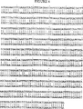
ハイブリダイゼーションのプローブは,サブクローニングされた92塩基対のDNA断片をランダムプライミングによって標識して調製した。ハイブリダイゼーションのプローブは,CV−1/EBNAのポリアデニル化RNAから調製したcDNAを挿入したプラスミドのライブラリーの一部を検索することに用いた。この結果,C85.sIL-15クローンが単離された。オープンリーディングフレームをもつこのクローンは、配列番号1に示すヌクレオチド配列を含んでいる。
sIL−15のコード領域のポリペプチドのヌクレオチド配列は配列番号1に示してある。この配列はC85.sIL-15挿入配列に由来している。SalIリンカーが付加されたC85.sIL-15を発現ベクターpDC406のSalI部位に挿入した(C.J. McMahan et al., EMBO J., 10(10):2821-2832(1991))。ポリアデニル化mRNAはCV−1/EBNA細胞系列から調製し,cDNAは標準的な技術を用いて調製した。CV−1/EBNA細胞系列はsIL−15の生産者である。cDNA末端にはSalIアダプターを付加した(Haymerle et al., Nucleic Acid Res, 14:8615-24(1986))。すなわち,
5’−TCGACTGGAACGAGACGACCTGCT−3’
3’−GACCTTGCTCTGCTGGACGA−5’
(それぞれ配列番号7および8),そしてベクターpDC406に挿入した。プラスミドをもつ約500個の独立な単離体をプレートに広げ,DNAプローブ断片とハイブリダイゼーションすることによってスクリーニングを行った。DNAプローブ断片はCV−1/EBNA細胞系列のcDNAのsIL−15の配列をPCRによって調製した。
実施例4
ヒトIL−15のクローニング
sIL−15プローブは単離,調製,および放射性標識されたSalI断片を含む(約1.37kb)sIL−15のcDNAからランダムプライミング標識によって調製された。プローブの特異活性は約1×10⁶cpm/ngであった。ノーザンブロット解析では,プローブは,IMTLH細胞系列を含む様々な部分からのヒトのRNAとハイブリダイズした。IMTLH細胞系列はヒト骨髄ストローマ細胞をpSV3Neoで安定に形質転換したものに由来する。このプローブは,ヒトRNAと約40%ホルムアミド中、約42℃,18時間でハイブリダイズした。ハイブリダイゼーションに続いて,6xSSCで約10分間洗浄し,続いて2xSSCで42℃,約30分間洗浄した。放射線写真によってIMTLHのレーンに正のシグナルが確認された。
ヒトIL−15 cDNAを含むプールを同定するために,IMTLH cDNAライブラリーをSalI消化した産物についてサザンブロット法によりプローブ探査をおこなった。前述のCV−1/EBNAライブラリーの際に使用したHaymerle et al., Nucleic Acid Res, 14:8615-24(1986)の方法によってIMLTHライブラリーを発現ベクターpDC406に構成した。約1000の異なるcDNAクローンからなるプールであるプール“I41”が陽性として同定された。“I41”の約4000コロニーをプレートに広げ,ヒトIL−15 cDNAを含むコロニーを同定するために,慣用されたコロニーハイブリダイゼーション法によってプローブ探査した。唯一のクローン,I41.hETFのみがヒトIL−15をコードすることが示された。ヒトとサルのIL−15オープンリーディングフレーム配列の間には約96%のヌクレオチド配列の同一性,および約96%のアミノ酸配列の同一性が確認された(図4および5)。
実施例5
rIL−15によるCTLL−2増殖の刺激
IL−15の成熟型をコードするcDNA(ヌクレオチド145から489,配列番号1および4に含まれている)を異種哺乳類分泌シグナルの下流に挿入し,sIL−15およびhIL−15のためのrIL−15発現プラスミドを作製した。分泌シグナルは大部分がアミノ酸の疎水性領域(通常はポリペプチドのN末端)であり,分泌およびシグナルペプチドのC末端と成熟な分泌タンパク質のN末端との間の切断を調節している(von Heijne, Eur. J. Biochem., 116:419(1981))。ここで使用した分泌シグナルはネズミのIL−7シグナル配列である。生成したプラスミドをCOS−7細胞に感染させた。トランスフェクションさせた細胞培養液の上清はCTLL−2の増殖を刺激した。さらに,hIL−15の前駆体型のコード領域(配列番号3)をpDC406に挿入し,CV−1/EBNA細胞にトランスフェクションさせた。pDC406:hIL−15(即ち,hETF/pDC406)をトランスフェクションさせた細胞の上清はCTLL−2細胞の増殖を刺激したが,空のベクターをトランスフェクションさせたものは増殖を刺激しなかった。
実施例6
rIL−15によるCTLL−2増殖の誘導
サルIL−15の組換え体(サルrIL−15)は実施例1に記載した精製法から限外濾過およびイオン交換の工程を省略した方法で,sIL−15 cDNAを発現させた酵母の上清から精製した。精製したサルrIL−15と,精製した組換えIL−2およびIL−4との生物活性を比較した。3つのタンパク質のそれぞれの純度はアミノ酸解析によって確認された。3つの組換えタンパク質のそれぞれの生物活性をCTLL−2細胞を用いて試験管内でアッセイした。CTLL−2培養液は100μlの補充した培地の中に培養液あたり2000細胞とともに示したようなサイトカインを含んでいた。CTLL−2培養液は24時間の培養時間の最後の4時間に,培養液当たり0.5μCiの[3H]チミジンでパルスし,ガラス繊維フィルターの上に回収し,流れ落ちる気体のイオン化によって放射活性を決定した。試験管内におけるCTLL−2細胞の増殖応答は組換えサイトカインの濃度の上昇によって測定した(ng/mlで表される)。データは図6に取込まれた3H−チミジンのcpm(x10-3)として表されている。
実施例7
rIL−15によるCTL,LAK,およびNK溶解活性の誘導
抗原特異的な細胞障害性Tリンパ球(CTL)を試験管内で生成した。一人の提供者に由来するヒト末梢血液単一核細胞(PBL)(5x10⁵個/培養液)を,同種異系の提供者に由来する,様々な濃度のIL−2またはヒトrIL−15を持つか,あるいはサイトカインを持たない非照射PBL培養細胞(5x105個/培養液)によって刺激した。培養はM.B. Widmer et al., J. Exp. Med., 166:1447(1987)に記載されている方法に従っておこない,7日後に回収し,元来の刺激性のドナーに由来する51Cr標識された標的に対する細胞の溶解活性をアッセイした。溶解アッセイは1000個の標識された標的とともに200μlの培地でV型孔で培養された様々な数の応答性PBLを含んでおり,4時間のインキュベーションの後に上清を回収した。溶解単位は特異的51Cr放出の最大値の50%を生成するために必要な応答性培養液に対応する画分の逆数として計算される。サイトカイン処理したCTLに関するデータは図7にまとめてある。
リンホカインにより活性化されたキラー(LAK)細胞は上記のCTLと同一の培養条件下で産生したが,ヒトPBLは同種異系の提供者に由来するPBLによって刺激されない点は除く。そのかわりに,自己由来の非照射PBLで置き換え,溶解活性はDaudiリンパ芽球様細胞系列に対する活性を測定した。LAKアッセイにおいては,溶解単位は,特異的51Cr放出の最大値の30%を産生するために必要な培養液に対応する画分の逆数として計算される。サイトカイン処理したLAK細胞に関するデータは図8にまとめてある。
ナチュラルキラー(NK)細胞はヒトPBL全体からMACS(Miltenyi Biotec, Sunnyvale CA)とともに常磁性ミクロスフェアに対する抗体の親和性によって,抗CD16モノクローナル抗体を使用して単離された。精製されたNK細胞を3日間培養し,K652赤芽性白血病細胞系列に対する細胞溶解活性を測定した。NKアッセイにおいては溶解単位は、特異的51Cr放出の最大値の30%を産生するために必要な培養液に対応する画分の逆数として計算される。サイトカイン処理したNK細胞に関するデータは図9にまとめてある。
本明細書中の実施例6および7におけるIL−2およびIL−15の活性の類似性により,当該技術分野において通常の知識を有する者の中には,IL−15がCTL,LAK,およびNK細胞の活性を刺激し,腫瘍細胞およびウイルス感染細胞を破壊するT細胞の数が増加することを期待する者もあろう。IL−15ポリペプチドの投与の項で既に記載されているように,適当な希釈液または担体に含ませたIL−15の効果量を以下の患者に投与することが可能である。すなわち、癌腫、黒色腫、肉腫、白血病又はリンパ腫の患者、あるいはサイトメガロウイルス属を含むヘルペスウイルス科のウイルス、ポリオーマウイルス(Polyomaviridae)、HIVを含むレトロウイルス科のウイルス、インフルエンザウイルス、ヘパドナウイルス科のウイルス、A型肝炎ウイルス、B型肝炎ウイルス、C型肝炎ウイルス、デルタ型肝炎ウイルス又はD型肝炎ウイルスに感染している患者である。
配列表
(2)配列情報 SEQ ID NO:1:
(i)配列の特性:
(A)配列の長さ: 489塩基対
(B)型: 核酸
(C)鎖の数: 一本鎖
(D)トポロジー: 直線状
(ii)配列の種類: cDNA
(iii)ハイポセティカル: NO
(iv)アンチセンス: NO
(ix)特徴:
(A)NAME/KEY: CDS
(B)存在位置: 1..489
(xi)配列: SEQ ID NO:1:
(i)配列の特性:
(A)配列の長さ: 162アミノ酸
(B)型: アミノ酸
(D)トポロジー: 直線状
(ii)配列の種類: タンパク質
(xi)配列: SEQ ID NO:2:
(i)配列の特性:
(A)配列の長さ: 114アミノ酸
(B)型: アミノ酸
(D)トポロジー: 直線状
(ii)配列の種類: タンパク質
(iii)ハイポセティカル: NO
(xi)配列: SEQ ID NO:3:
(i)配列の特性:
(A)配列の長さ: 489塩基対
(B)型: 核酸
(C)鎖の数: 一本鎖
(D)トポロジー: 直線状
(ii)配列の種類: cDNA
(ix)特徴:
(A)NAME/KEY: CDS
(B)存在位置: 1..489
(xi)配列: SEQ ID NO:4:
(i)配列の特性:
(A)配列の長さ: 162アミノ酸
(B)型: アミノ酸
(D)トポロジー: 直線状
(ii)配列の種類: タンパク質
(xi)配列: SEQ ID NO:5:
(i)配列の特性:
(A)配列の長さ: 114アミノ酸
(B)型: アミノ酸
(D)トポロジー: 直線状
(ii)配列の種類: タンパク質
(iii)ハイポセティカル: NO
(xi)配列: SEQ ID NO:6:
(i)配列の特性:
(A)配列の長さ: 24塩基対
(B)型: 核酸
(C)鎖の数: 一本鎖
(D)トポロジー: 直線状
(ii)配列の種類: cDNA
(iii)ハイポセティカル: NO
(iv)アンチセンス: NO
(xi)配列: SEQ ID NO:7:
![]()
(i)配列の特性:
(A)配列の長さ: 20塩基対
(B)型: 核酸
(C)鎖の数: 一本鎖
(D)トポロジー: 直線状
(ii)配列の種類: cDNA
(iii)ハイポセティカル: NO
(iv)アンチセンス: NO
(xi)配列: SEQ ID NO:8:
![]()
(i)配列の特性:
(A)配列の長さ: 18塩基対
(B)型: 核酸
(C)鎖の数: 一本鎖
(D)トポロジー: 直線状
(ii)配列の種類: cDNA
(iii)ハイポセティカル: NO
(iv)アンチセンス: NO
(xi)配列: SEQ ID NO:9:
![]()
(i)配列の特性:
(A)配列の長さ: 17塩基対
(B)型: 核酸
(C)鎖の数: 一本鎖
(D)トポロジー: 直線状
(ii)配列の種類: cDNA
(iii)ハイポセティカル: NO
(iv)アンチセンス: YES
(xi)配列: SEQ ID NO:10:
![]()
(i)配列の特性:
(A)配列の長さ: 17塩基対
(B)型: 核酸
(C)鎖の数: 一本鎖
(D)トポロジー: 直線状
(ii)配列の種類: cDNA
(iii)ハイポセティカル: NO
(iv)アンチセンス: YES
(xi)配列: SEQ ID NO:11:
![]()
(i)配列の特性:
(A)配列の長さ: 114アミノ酸
(B)型: アミノ酸
(D)トポロジー: 直線状
(ii)配列の種類: タンパク質
(iii)ハイポセティカル: NO
(xi)配列: SEQ ID NO:12:
The present invention relates generally to mammalian epithelial T cell factor (“ETF”) polypeptides—referred to herein as interleukin 15 (“IL-15”). The present invention specifically relates to isolated cDNA sequences encoding polypeptides having IL-15 biological activity, sequences and derivatives of isolated IL-15 polypeptides, IL-15 polypeptides using recombinant DNA technology. It relates to a method of making peptides, a method of inducing T cell proliferation and differentiation, and a composition comprising IL-15 that forms anticancer and antiinfective immunity.
Background art
Various T cells, also called T lymphocytes, are a class of immune effector cells. In peripheral tissues, T cells are divided into two large groups based on their mutual expression of CD4 and CD8 molecules on the cell surface. Typical CD8+When activated, T cells become cytotoxic T cells that directly bind to and destroy target cells displaying the antigen. CD4+T cells generally give a positive signal. For example, it is called a helper T cell because it functions as a “helper” for B cells (allowing B cells to differentiate into antibody-producing cells).
To date, six T cell growth factors have been identified. The six are interleukin (IL) -2, 4, 7, 9, 12 and the cofactor IL-10, respectively. The open reading frame of IL-2 encodes a 15 kDa polypeptide consisting of 153 amino acids. IL-2 is produced by certain T cells and large granular lymphocytes. IL-2 was originally discovered as a factor that promotes long-term growth of human T cells. In addition to the growth of T cells, the effect is to activate natural killer (NK) cells and killer (LAK) cells and macrophages activated by lymphokines, in addition to cytotoxic T cells (CTL). Including promotion.
IL-4 is a 15-20 kDa protein produced by activated T cells, bone marrow stromal cells and mast cells. The open reading frame of IL-4 encodes 140 amino acids IL-4 in mice and 153 amino acids IL-4 in humans. IL-4 was originally identified as a factor that activates B cell growth and differentiation. Its effects include macrophage activation and induction of class II MHC molecules, growth of certain T cells and mast cells, proliferation of human peripheral blood T cells and production of CTL, enhancement of immunoglobulin production by B cells, Also included are cofactor functions in the growth of hematopoietic cells from stem cells. IL-4 plays an important role in negative regulation of the activity of NK and LAK cells induced by IL-2. Human IL-4 has no activity on mouse cells.
IL-7 is a 20 to 25 kDa 177 amino acid polypeptide produced by bone marrow and thymic stromal cells. IL-7 was originally considered a pre-B cell growth factor, but it promotes the growth of not only pre-B cells but also B cell progenitors. Also, human peripheral blood T cell proliferation and CTL production, IL-2 receptor expression, IL-2 production, and CD4+And CD8+Promotes cell growth. IL-7 also helps IL-2 to increase T cell proliferation in the thymus, and CD4-And CD8-Induces thymocyte proliferation.
IL-9 is a 144 amino acid, 30-40 kDa polypeptide produced by activated T lymphocytes. IL-9 was first identified as a helper T cell growth factor. IL-9 promotes red blood cell development and promotes proliferation of bone marrow-derived mast cells induced by IL-3. It also regulates production of IgE and IgG by B cells in the presence of IL-4. Mouse IL-9 has activity against human cells, but human IL-9 does not act on mouse cells.
Human IL-10 is a 178 amino acid, 16-20 kDa polypeptide produced by macrophages and TH2 T helper cells (not produced by TH1 T helper cells). Like IL-2, IL-4 and IL-7, IL-10 has several different biological activities. IL-10 was discovered based on its ability to inhibit cytokine production by activated T cells. Both human and mouse IL-10 are thymocyte and T cell growth-promoting cofactors that work in conjunction with IL-7 or IL-2 + IL-4. IL-10 works with IL-4 or IL-3 + IL-4 to promote mast cell survival and growth. IL-10 also induces the secretion of IgG and the expression of MHC class II molecules on B cells, increasing the viability of B cells in culture.
IL-12 is constitutively expressed in lymphoblastoma cell lines or is induced by phorbol esters and calcium ion permeable carriers and produced by LPS stimulated macrophages. IL-12 has a molecular weight of 70 kDa and takes a unique heterodimeric structure consisting of two disulfide-linked glycoproteins. The larger of the two glycoprotein subunits is a 328 amino acid, 40 kDa polypeptide. The small glycoprotein subunit is a 253 amino acid, 35 kDa polypeptide. Both glycoprotein subunits are required for biological activity. IL-12 promotes proliferation of both CD4 + and CD8 + among activated T cells independently of IL-2. IL-2 also activates NK cell-mediated cytotoxicity and helps IL-2 to generate LAK cells. Unlike IL-2 and IL-7, IL-12 causes little proliferation of resting peripheral blood mononuclear cells (this is similar to IL-4).
Summary of the Invention
A novel T cell growth factor was isolated and purified. Hereinafter, this is referred to as interleukin 15 (“IL-15”). A cDNA sequence encoding monkey IL-15 polypeptide was isolated. This sequence has a 489 bp open reading frame, a 483
Furthermore, the present invention includes other IL-15 polypeptides. These polypeptides have a base sequence that hybridizes with a plurality of probes defined by bases 145 to 489 of SEQ ID NO: 1 or 4 or with DNA strands or RNA strands complementary to these probes under mild to severe conditions It is encoded by a nucleotide sequence encoding a polypeptide that promotes proliferation and differentiation of T lymphocytes during expression. Furthermore, the present invention includes a base sequence encoding the IL-15 polypeptide encoded by the base sequence described above and a sequence complementary thereto due to the degeneracy of the genetic code.
Furthermore, in the present invention, a recombinant DNA molecule useful for producing an IL-15 polypeptide comprising the aforementioned base sequence, such as an expression vector or plasmid and a transformed host cell, and a recombinant IL using such a molecule. A method for producing a -15 polypeptide is provided.
[Brief description of the drawings]
FIG. 1 shows the base sequence and deduced amino acid sequence of monkey active IL-15.
FIG. 2 shows the nucleotide sequence of human active IL-15 and the deduced amino acid sequence.
FIG. 3 shows a purification method and protein sequence determination method useful for isolating IL-15 polypeptides.
FIG. 4 shows the homology between the nucleotide sequences encoding human and monkey IL-15. The human sequence is shown above monkeys.
FIG. 5 shows the homology between the amino acid sequences of human and monkey IL-15. The human sequence is shown above monkeys. In either sequence, a mature polypeptide (49-162) is formed when the leader sequence (amino acids 1-48) is cleaved from the precursor.
FIG. 6 shows the biological activity of recombinant IL-15, IL-2 and IL-4 in vitro using CTLL-2 cells. The growth stimulatory response of CTLL-2 cells in vitro was measured by increasing the recombinant cytokine concentration (expressed in ng / ml). Data was capturedThreeH-thymidine cpm (× 10-3).
FIG. 7 shows the induction of CTL cytolytic activity by rIL-15 and IL-2. Antigen-specific cytotoxic T lymphocytes (CTL) were established in vitro. Human peripheral blood mononuclear cells (PBL) from one donor were stimulated with irradiated PBL from atypical donors in media containing various concentrations of IL-2 or human rIL-15. Derived from this stimulator cell51The cytolytic activity of the culture was assayed against the Cr-labeled target. Specific release51The reciprocal number of each culture fraction required to produce 50% of the maximum value of Cr and represents the unit of cell lysis.
FIG. 8 shows the induction of cytolytic activity of LAK cells by rIL-15 and IL-2. Lymphokine activated killer (LAK) cells were established in vitro. Human PBL was stimulated with irradiated autologous PBL and cytolytic activity was measured against the Daudi lymphoblastoma cell line. Specific release51The reciprocal number of each culture fluid fraction required to produce 30% of the maximum value of Cr and represents the cell lysis unit.
FIG. 9 shows the induction of cytolytic activity of NK cells by rIL-15 and IL-2. Natural killer (NK) cells were isolated from whole human PBL isolated by antibody affinity for paramagnetic microspheres using a monoclonal antibody against CD16. Purified NK cells were cultured for 3 days and cytolytic activity was measured against the K562 erythroleukemia cell line.
Detailed Description of the Invention
“
The terms IL-15, sIL-15, or hIL-15 include a T encoded by a nucleic acid that binds to the nucleic acid sequence of FIGS. 1 and 2 (bases 145 to 489, SEQ ID NOS: 1 and 4) under certain conditions. Included are analogs or subunits of natural mammalian polypeptides that induce the proliferation and differentiation of lymphocytes and promote the proliferation of T cell lines and isolated PBT.
As used herein, “recombinant DNA technology” or “recombinant” refers to a microorganism (eg, a bacterium) that has been transformed or infected with isolated or synthesized DNA to biosynthesize heterologous polypeptides. , Fungi, or yeast) or mammalian cells or individuals (eg, genetically modified) to refer to techniques and methods for producing specific polypeptides. Natural glycosylation patterns are only formed in mammalian cell expression systems. Yeast has a different glycosylation pattern. Expression by prokaryotic cells (eg, E. coli) generally produces polypeptides that are not golicosylated.
“Having biological activity” means that a particular IL-15 polypeptide can promote proliferation and / or differentiation of T lymphocytes. In the case of sIL-15 and hIL-15, this biological activity also applies to the promotion of mouse or primate (eg human) T cell lineage or PBT growth.
“Base sequence” refers to a polynucleotide in the form of a part of a large DNA construct or an independent fragment. This nucleotide is in a substantially purified form at least once (eg, in the absence of interstitial material) and in basic biochemical methods such as cloning vectors (eg, Sambrook et al, Molecular Cloning: A Laboratory Manual Second Edition, Cold Spring Harbor Laboratory, a method as outlined in Cold Spring Harbor, NY (1989)) in such an amount or concentration that its base sequence can be identified, manipulated and recovered. Refers to the one derived from isolated DNA. Such sequences are desirably provided in the form of one open reading frame that is not divided into endogenous untranslated regions (ie, introns) that are commonly present in eukaryotic cells. The untranslated region at 5 ′ or 3 ′ of the open reading frame may be present as it is because it does not interfere with the manipulation or expression of the coding region.
“Recombinant expression vector” refers to a plasmid consisting of one transcription unit. This transcription unit consists of (1) one or more genetic elements (eg, promoters and enhancers) that play a regulatory role in gene expression, and (2) polytranscripts that are transcribed into mRNA and have IL-15 biological activity. It consists of a set of structures or coding sequences that are translated into peptides, and (3) appropriate transcriptional and translational initiation and termination sequences. Various regulators that can be used are described later (see recombinant DNA technology). The structural factor utilized in the yeast expression system preferably includes a leader sequence for secreting the translated polypeptide out of the yeast host cell. On the other hand, in bacterial expression systems, the recombinant polypeptide may contain an N-terminal methionine residue. This N-terminal methionine residue is sequentially cleaved from the expressed recombinant polypeptide to yield a product suitable for further purification.
A “recombinant microbial expression system” refers to a suitable host microorganism (eg, a bacterium such as E. coli) that stably incorporates a recombinant transcription unit into chromosomal DNA or holds the recombinant transcription unit as an element of an endogenous plasmid. Substantially homogenous single culture of yeast, such as S. cerevisiae). In general, the host cells that make up the recombinant microbial expression system are the progeny of one transformed parent cell. A recombinant microorganism expression system expresses a heterologous polypeptide by induction of a regulatory factor linked to the structural base sequence to be expressed.
A transformed host cell refers to a cell that has been transformed or infected with a recombinant expression vector. Expressed mammalian IL-15 is present in the host cell and / or secreted into the culture supernatant, depending on the nature of the host cell and the gene construct introduced into the host cell.
The conditions for loose hybridization are as defined herein and are well known to those skilled in the art, for example in Sambrook et al., Supra, Vol. 2, pp. 8.46-8.49 and 9.47-9.55. The described conditions. Loose conditions include, for example, overnight hybridization at 55 ° C., 5 × SSC, 0.5% SDS, washing after hybridization, etc., as defined by Sambrook et al. Severe conditions include hybridization reactions at higher temperatures or lower salt concentrations, washing after hybridization, and the like.
IL-15 polypeptide
We purified monkey IL-15 (sIL-15) and determined the N-terminal peptide sequence of mature sIL-15. A cDNA encoding sIL-15 was isolated using N-terminal amino acid sequence and PCR, and the nucleotide sequence of mature cIL-15 and the deduced amino acid sequence (FIG. 1), as well as the precursor sequence of sIL-15 polypeptide. And the deduced amino acid sequence was determined (SEQ ID NO: 1 and SEQ ID NO: 2). The sequence of IL-15 precursor polypeptide in monkeys consists of the mature active protein (SEQ ID NO: 3) and the preceding 48 amino acid leader sequence. This leader sequence is
The present invention also includes other mammalian IL-15 (human IL-15) that has IL-15 biological activity and is encoded by a base sequence that hybridizes with a probe defined in SEQ ID NO: 5 under mild to severe conditions. Included). In accordance with the Budapest Treaty on the International Approval of Deposits of Microorganisms in Patent Proceedings, on February 19, 1993, one plasmid containing a recombinant clone of human IL-15 cDNA was transferred to American Type Culture Collection, 12301 Parklawn Drive, Rockville, MD Deposited at 20852 USA (“ATCC”). The deposit number is ATCC 69245. This deposit is named “I41-hETF” and consists of E. coli with the plasmid hETF / pDC406. This plasmid contains a 316 bp 5 'noncoding region followed by a 489 bp open reading frame and a 397 bp 3' noncoding region, flanked by SalI adapters. These are shown in SEQ ID NOs: 7 and 8. Restrictions on the general use of the deposit will be removed entirely upon grant of the patent.
The amino acid structure of the IL-15 polypeptide disclosed herein can be determined by making a covalent or associated conjugate with other species (eg, carbohydrates, lipids, phosphates, acetyl groups, etc.) or It can be altered by amino acid sequence mutations. Covalent derivatives of mammalian IL-15 are generated by attaching specific functional groups to the amino acid side chain or N-terminus or C-terminus of mammalian IL-15. Other derivatives of mammalian IL-15 within the scope of the present invention include covalently linked or associated conjugates of mammalian IL-15 or fragments thereof with other proteins or polypeptides (eg, N-terminal or recombinant culture). Synthesized by C-terminal fusion). This bound polypeptide is, for example, a signal (or leader) polypeptide sequence in the N-terminal region of a mammalian IL-15 polypeptide for transport into or out of the cell membrane or wall from the site of IL-15 synthesis. (For example, the yeast alpha factor leader). In addition, fusions involving incidental polypeptide sequences (eg, Fc or other immunoglobulin sequences, linker sequences, or other sequences that facilitate the purification and identification of IL-15 polypeptides) using conventional techniques IL-15 polypeptide can be expressed as a polypeptide. In addition, IL-15 fusion polypeptides include fusions with other cytokines that provide novel multifunctional entities. Other cytokines include, for example,
Furthermore, the present invention includes IL-15 polypeptides with different glycosylation. IL-15 polypeptides expressed in yeast or mammalian expression systems (eg COS-7 cells (ATCC CRL 1651)) are similar or significant in their molecular weight and glycosylation pattern compared to native IL-15 polypeptides There can be significant differences. This depends on the choice of expression system. Expression of IL-15 polypeptide in a bacterial expression system such as E. coli results in a molecule without glycosylation.
Active variant analogs of human or other mammalian IL-15 are synthesized, for example, by oligonucleotide synthesis and ligation, or inactivation of N-glycosylation sites by site-directed mutagenesis. A yeast expression system can be used to express a low carbohydrate, homogeneous IL-15 polypeptide derivative. The N-glycosylation site in eukaryotic polypeptides is characterized by the triple amino acid Asn-φ-Ω. Here, φ is an amino acid other than Pro, and Ω is Ser or Thr. A variant derivative of IL-15, as shown herein, is substantially homologous to the sequence of a natural mammalian IL-15, but is naturally occurring due to deletion, insertion, or substitution mutations. A polypeptide having an amino acid sequence different from an IL-15 polypeptide.
A variant IL-15 that is bioequivalent to an IL-15 polypeptide is constructed by various substitutions of amino acid residues or sequences, or deletion of termini or internal residues or sequences that are not essential for biological activity. For example, deletion or substitution of the Cys residue can prevent erroneous intramolecular disulfide bridges from forming upon restoration. Other methods of mutagenesis include modifying dibasic amino acid residues to promote expression in yeast systems with KEX2 protease activity. In general, substitutions can be made conservatively by substitution with a residue having similar physicochemical properties as the naturally occurring amino acid residue.
Antisense or sense oligonucleotides include single-stranded nucleic acid sequences (RNA or DNA) that can bind to an IL-15 sense mRNA or IL-15 antisense cDNA sequence. An antisense or sense oligonucleotide in accordance with the present invention includes a nucleotide sequence of FIG. 1 or 2 or a fragment of a nucleotide sequence of DNA or RNA complementary to the nucleotide sequence of FIG. 1 or 2. Such a fragment shall consist of at least 14 nucleotides and be capable of binding to IL-15 DNA. For methods of making antisense or sense oligonucleotides based on IL-15 cDNA sequences, see, for example, Stein and Cohen, Cancer Res. 48: 2659 (1988) and van der Krol et al., Biotechniques 6: 958 (1988). )It is described in.
Isolating and identifying IL-15 from non-recombinant cell lines requires a mammalian cell line that produces IL-15 and a responsive cell line that proliferates in response to IL-15 stimulation. In a biological assay for mammalian IL-15, a growth factor-dependent T cell line can be used for the detection of factors that induce proliferation of lymphocyte cells. In addition, mammalian IL-15 polypeptides can be assayed using T cells isolated from blood samples obtained from humans or other mammals.
IL-15 dependent cell lines can be generated from mouse CTLL-2 cells. This cell line responds to purified human, mouse, and recombinant IL-2 and mouse IL-4, but not to IL-1, IL-3, human IL-4, or other known growth factors.
Using the monkey or human IL-15 cDNA sequences disclosed herein, other mammalian homologues of monkey or human IL-15 can be obtained by cross species hybridization. Briefly, a probe is prepared from the nucleotide sequence of the protein coding region of sIL-15 described in FIG. 1 or SEQ ID NO: 1 or hIL-15 cDNA described in FIG. 2 or SEQ ID NO: 4. This probe can be made by a general method as described above in Sambrook et al. Monkey or human probes can be used to screen against mammalian cDNA libraries or chromosomal libraries under mild conditions. A mammalian cDNA library can be prepared from mRNA isolated from, for example, mouse peripheral blood lymphocytes. Alternatively, screening by Northern hybridization with other cDNA libraries or mRNA isolated from various tissues or cell lines can be used to determine the appropriate material for mammalian IL-15 DNA or RNA.
Purification of CV-1 / EBNA IL-15
We purified IL-15 to provide a polypeptide sample isolated from a heterogeneous protein solution (eg, conditioned media recovered from cells expressing IL-15). IL-15 activity in natural conditioned media samples cannot always be detected using currently available IL-15 bioassays. In order to be able to detect IL-15 biological activity using the bioassays described herein, at least one stage of purification is required.
African homogenous kidney cell CV-1 / EBNA lineage (CJMcMahan et al., EMBO J., 10 (10): 2821-2832 (1991); ATCC CRL 10478) in tissue culture medium is heterogeneous Prepare a typical protein solution, eg, conditioned medium.
The medium is preferably high glucose Dulbecco's Modified Essential Medium (“DMEM”, Gibco). Most preferably, growth and production media are used. The growth medium used in isolating sIL-15 as described herein was 7.5% fetal calf serum, 50 μl / ml penicillin, 50 μg / ml streptomycin, 3 in high glucose (4500 mg / L) DMEM. 4.0mM L-glutamine, 1mM sodium pyruvate, 0.1mM non-essential amino acid and 10mM N- [2-hydroxyethyl] piperazine-N '-[2-ethanesulfonic acid] (“HEPES”) buffer is there. For IL-15 production and purification, DMEM is supplemented with 50 μl / ml penicillin, 50 μg / ml streptomycin, 3 to 4.0 mM L-glutamine, 1 mM sodium pyruvate, 0.1 mM non-essential amino acids and 10 mM HEPES, and phenol red is removed. A production medium without added serum was developed. CV-1 / EBNA cells are adherent dependent and can be cultured in plates, flasks, roller bottles, or microcarriers.
More specifically, IL-15 was produced by culturing CV-1 / EBNA cells on microcarriers in a controlled bioreactor. Cell stocks were maintained in roller bolt flasks. At the start of the production cycle, the cells were trypsinized and planted in a spinner flask containing the growth medium described above and 5 g / 1 Cytodex® 3 microcarrier (Pharmacia). The initial concentration is 1.5 to 3.5 x 10FiveCells / ml. Cells were placed in a spinner flask for 2 to 24 hours so that the cells attached efficiently to the microcarriers. The culture solution in the spinner flask was kept at 37 ° C. and shaken at 25 to 40 RPM. After the attachment period, the culture broth was transferred into a controlled bioreactor. Reactor temperature, pH, oxygen concentration and shake settings are 37 ° C., 7.0, 20% saturation (against atmospheric value), and 75 to 85 RPM, respectively. Samples were observed with a bright field microscope during normal observation of cell growth and status. Cell proliferation was measured by counting free nuclei after treatment with 100 mM citric acid and 0.1% crystal violet solution.
Each time the ammonia level of the culture reached 5.0 mM, additional growth medium was added. This was repeated until the microcarriers were confluent. The medium was then replaced with the serum-free production medium described previously. This process was performed by precipitating microcarriers at the bottom of the reactor, handling the growth medium, and adding production medium instead. This was repeated until the dilution was about 3125 times. Two to six productions can be expected. At one time, cells were placed in production medium for 4 to 7 days, after which 80% of the production medium was recovered. This was repeated until the cells were completely detached from the microcarrier.
Approximately 64 liters of CV-1 / EBNA conditioned medium was used to purify and provide the protein for sIL-15N terminal amino acid sequencing. As shown in Figure 3, purification methods include ultrafiltration, hydrophobic chromatography, anion exchange chromatography, reverse phase high performance liquid chromatography (RP-HPLC), and sodium dodecyl sulfate polyacrylamide gel electrophoresis (SDS- PAGE). Proteins were sequenced by adsorbing proteins from SDS gels onto polyvinylidene fluoride (“PVDF”) membranes and Edman degradation directly from PVDF membranes to determine the N-terminal amino acid sequence. Determination of the N-terminal amino acid sequence revealed the first 33 amino acids shown in SEQ ID NO: 3. Subsequently, the sequence of a cDNA clone obtained from the CV-1 / EBNA cDNA library was determined to obtain a DNA sequence encoding the polypeptide of SEQ ID NO: 2. This clone contains the relatively short 48 amino acid leader sequence of SEQ ID NO: 2 and the mature polypeptide shown in SEQ ID NO: 3.
Recombinant DNA technology
Human, monkey and other mammalian IL-15 polypeptides can preferably be produced by recombinant DNA techniques. Such techniques include inserting a human or other mammalian IL-15 polypeptide or derivative thereof into an expression vector.
Recombinant expression of an IL-15 polypeptide or derivative thereof first requires a DNA clone (ie, cDNA) encoding the IL-15 polypeptide or derivative thereof. A cDNA clone is obtained from a parent cell or cell line that expresses a mammalian IL-15 polypeptide. First, total cellular mRNA is isolated, and then a cDNA library is constructed from the mRNA by reverse transcription. The DNA sequence information provided herein can be used to design cross-species hybridization probes or PCR primers as described previously to isolate and identify certain cDNA clones.
The isolated cDNA is preferably in the form of an open reading frame that is not interrupted by endogenous untranslated sequences (ie, introns). Genetic information useful for constructing a coding sequence can also be obtained using genomic DNA containing the relevant nucleotide sequence encoding the expression of a mammalian IL-15 polypeptide. Methods isolated in the art can be used to introduce mutations into the isolated cDNA to produce IL-15 derivatives or analogs that exhibit IL-15 biological activity.
Recombinant expression vectors include synthetic or cDNA-derived DNA fragments encoding IL-15 or biologically active derivatives thereof. DNA encoding IL-15 or a derivative thereof can be operably linked to an appropriate transcriptional or translational regulatory sequence or structural base sequence (eg, from a mammalian, microbial, viral or insect gene). . Examples of regulatory sequences include gene sequences that have a regulatory role in gene expression (eg, transcription promoters or enhancers), incidental operator sequences to regulate transcription, sequences encoding appropriate mRNA ribosome binding sites, and There are suitable sequences that regulate the initiation and termination of transcription and translation. When the regulatory sequence has a functional correlation with the structural gene, the base sequence is said to be functionally linked. When a signal peptide (secretory leader) is expressed as part of the precursor amino acid sequence of mammalian IL-15 or a derivative thereof and is involved in its secretion, the DNA sequence encoding the signal peptide can be expressed as a mammalian IL-15 or derivative thereof. It can be operably linked to the DNA sequence of the structural gene that it encodes. Furthermore, when the base sequence of a certain promoter regulates transcription of the base sequence of the linked coding sequence, the base sequence of the promoter can be operably linked to the coding sequence (for example, structural gene DNA). Furthermore, when the ribosome binding site connected to the structural gene coding base sequence (eg, mammalian IL-15) is arranged in the vector so as to promote translation, the ribosome binding site can function in the structural gene coding base sequence. Can be connected.
Suitable cells for expression of mammalian IL-15 or its derivatives include prokaryotes, yeast, or higher eukaryotic cells under the control of appropriate promoters. Prokaryotes include gram negative or positive organisms (eg E. coli or Bacillus). Suitable host prokaryotic cells for transformation include E. coli, Bacillus subtilis, Salmonella typhimurium, and various other species within the genus Pseudomonas, Streptomyces, Staphylococcus. Also, as described in more detail below, suitable host cells include yeasts such as S. cerevisiae, mammalian cell lines such as Chinese hamster ovary (CHO) cells, or insect cells. In addition, mammalian IL-15 or a derivative thereof can be produced from RNA derived from the DNA construct disclosed herein using a cell-free translation system. Suitable cloning and expression vectors utilized in bacterial, fungal, yeast, and mammalian host cells are described, for example, in Pouwels et al. Cloning vectors: A Laboratory Manual, Elsevier, New York, 1985.
When mammalian IL-15 or a derivative thereof is expressed in a yeast host cell, a base sequence (eg, a structural gene) encoding mammalian IL-15 or a derivative thereof can include a leader sequence. This leader sequence allows the translated polypeptide to be better secreted extracellularly by the yeast host cell.
When expressing mammalian IL-15 in yeast host cells, cells of the genus Saccharomyces (eg, S. cerevisiae) are preferred. Other genera of yeast such as Pichia or Kluyveromyces can also be used. Yeast vectors often include a 2μ yeast plasmid-derived replication initiation sequence, self-replicating sequence (ARS), promoter region, polyadenylation site, and transcription termination sequence. The yeast vector preferably contains a replication initiator sequence and a selectable marker. Suitable promoter sequences for yeast vectors include metallothionein, 3-phosphoglycerate kinase (Hitzeman et al., J. Biol. Chem. 255: 2073, 1980), or other glycolytic enzymes (Hess et al. , J. Adv. Enzyme Reg. 7: 149, 1968; and Holland et al., Biochem. 17: 4900, 1978). Examples of glycolytic enzymes include enolase, glyceraldehyde triphosphate dehydrase, hexokinase, pyruvate decarboxylase, phosphofructokinase, glucose-6-phosphate isomerase, 3-phosphoglycerate mutase, pyruvate Kinase, triose phosphate isomerase, phosphoglucose isomerase, and glucokinase. Other preferred vectors and promoters used for expression in yeast are described in more detail in Hitzeman, EP-A-73,657.
In constructing a yeast vector, a DNA sequence derived from pBR322 (Amp, for example, for selection and replication in E. coli).rGene and replication origin). Other yeast DNA sequences that can be included in yeast expression constructs include the glucose repressive ADH2 promoter and the alpha factor secretion leader sequence. The ADH2 promoter has been described by Russel et al. (J. Biol. Chem. 258: 2674, 1982) and Beier et al. (Nature 300: 724, 1982). The yeast alpha factor leader sequence facilitates secretion of a heterologous polypeptide. The α-factor leader sequence is often inserted between the promoter sequence and the structural gene sequence (eg Kurjan et al., Cell 30: 933, 1982; and Bitter et al., Proc. Natl. Acsd. Sci). USA 81: 5330, 1984). The 3 ′ end of the leader sequence can be modified to add one or more restriction enzyme sites. This facilitates the fusion of the leader sequence and the structural gene.
Yeast transformation methods are well known to those skilled in the art. One such method is described in Hinnnen et al., Proc Natl. Acad. Sci. USA 75: 1929,1978. Hinnen et al.'S method uses Trp with 0.67% yeast nitrogen base, 0.5% casamino acid, 2% glucose, 10 mg / ml adenine, and 20 mg / ml uracil selective medium.+A transformant is selected.
To induce the expression of yeast host cells transformed with a vector having an ADH2 promoter sequence, the cells are cultured in a nutrient medium. The nutrient medium is, for example, 1% yeast extract, 2% peptone, and 1% glucose added with 80 mg / ml adenine and 80 mg / ml uracil. Activation of the ADH2 promoter occurs when glucose in the medium is depleted.
On the other hand, in prokaryotic host cells such as E. coli, mammalian IL-15 or a derivative thereof may have an N-terminal methionine residue that facilitates expression of the recombinant polypeptide in the prokaryotic host cell. This N-terminal Met is cleaved from the expressed recombinant mammalian IL-15.
A recombinant expression vector having a structural gene base sequence of recombinant mammalian IL-15 or a derivative thereof can be transformed or infected into an appropriate host microorganism or mammalian cell line.
Expression vectors infected with prokaryotic host cells generally contain one or more phenotypic selection markers. A phenotypic selectable marker is, for example, a gene encoding a protein that confers antibiotic resistance or a protein that supplements auxotrophy, and includes a replication origin recognized by the host to ensure amplification within the host. Other useful expression vectors for prokaryotic host cells include those containing selectable markers of bacterial origin from commercially available plasmids. Some of these selectable markers consist of the genetic elements of the cloning vector pBR322 (ATCC 37017). Since pBR322 contains ampicillin and tetracycline resistance genes, transformed cells can be identified by a simple method. The pBR322 “backbone” portion is combined with a suitable promoter and a mammalian IL-15 structural gene sequence. Other commercially available vectors include, for example, pKK223-3 (Phamacia Fine Chemicals, Uppsala, Sweden) and pGEM1 (Promega Biotec, Madison, WI, USA).
Promoter sequences are commonly used in recombinant prokaryotic host cell expression vectors. Common promoter sequences include β-lactamase (penicillinase), lactose promoter system (Chang et al., Nature 275: 615, 1978; and Goeddel et al., Nature 281: 544, 1979), tryptophan (trp) promoter system (Geddel et al., Nucl. Acids Res. 8: 4057, 1980; and EPA 36776) and the tac promoter (Sambrook et al., Molecular Cloning: A Laboratory Manual, Cold Spring Harbor Laboratory, (1989)). A particularly useful prokaryotic host cell expression system is lambda phage PLPromoter and cI857ts thermolabile repressor sequences are used. ΛP available from American Type Culture CollectionLPlasmid vectors incorporating a promoter derivative include plasmids pHUB2 (in E. coli strain JMB9 (ATCC 37092)) and pPLc28 (in E. coli strain RR1 (ATCC 53082)).
A mammalian or insect host cell system can also be used to express recombinant mammalian IL-15 or a derivative thereof. Suitable mammalian host cell lines include monkey kidney cell COS-7 cells (Gluzman et al., Cell 23: 175 (1981); ATCC CRL 1651), L cells, C127 cells, 3T3 cells (ATCC CCL 163), There are CHO cells, Hela cells (ATCC CCL 2), and BHK (ATCC CRL 10) cell lines. Non-transcribed elements contained in appropriate mammalian expression vectors include origins of replication, promoter sequences, enhancers linked to structural genes, and other 5 'or 3' peripheral non-transcribed sequences (eg, ribosome binding sites, polyadenylation sites, splicing) Donor and acceptor sites, and transcription termination sequences).
Transcriptional and translational regulatory sequences in mammalian host cell expression vectors are obtained from viral material. For example, commonly used mammalian promoter and enhancer sequences are derived from polyoma,
A typical mammalian expression vector can be made as disclosed by Okayama and Berg (Mol. Cell. Biol. 3: 280, 1983). Other useful expression vectors are described in US patent application Ser. No. 07 / 480,694 filed Feb. 14, 1990 and US Patent Application No. 07 / 543,193 filed Jun. 5, 1990.
Purification of recombinant mammalian IL-15
IL-15 polypeptides can be prepared by culturing transformed host cells under the culture conditions necessary to express the IL-15 polypeptide or derivative thereof. The polypeptide of the expression product is then purified from the medium or cell extract. Mammalian IL-15 polypeptides or derivatives thereof can be concentrated using commercially available protein concentration filters (eg, Amicon or Millipore Pellicon ultrafiltration kits). The culture solution can be applied to a purification carrier (for example, a hydrophobic chromatography carrier) with or without the concentration step. Phenyl Sepharose® CL-4B (Pharmacia) is a suitable medium. On the other hand, an anion exchange resin (for example, a carrier or substrate having a pendent diethylaminoethyl (DEAE) group) can also be used. The carrier may be acrylamide, agarose, dextran, cellulose, or other types used for protein purification. Alternatively, a gel filtration substrate can be used. Since recombinant IL-15 is stable in acidic aqueous buffer, a cation exchange resin can also be used.
Finally, reverse phase high performance liquid chromatography (RP-HPLC) using hydrophobic RP-HPLC media (eg, silica gel with pendent methyl or other aliphatic groups) is performed one or more times to obtain IL -15 can be further purified. Various combinations of some or all of the purification steps described above can be provided to provide a substantially homogeneous recombinant protein. Alternatively, some or all of the processes used in the previously described methods for purifying monkey IL-15 can be used.
To isolate a recombinant protein produced from a bacterial culture, the host cell is usually first disrupted, centrifuged, if it is an insoluble polypeptide, extracted from the cell mass, if it is a soluble polypeptide, extracted from the supernatant, concentrated, Perform at least one salting out, ion exchange or size exclusion chromatography. Microbial cells can be disrupted by simple methods such as freeze-thaw cycles, ultrasonic disruption, mechanical disruption, or the use of cell lysis reagents.
The transformed yeast host cell is preferably used for the purpose of expressing IL-15 as a secreted protein. Using this simplifies purification. Recombinant polypeptides secreted from yeast can be purified by methods similar to those disclosed in Urdal et al, (J. Chromatog. 296: 171, 1984). Urdal et al. Describe that recombinant human IL-2 was purified by two successive RP-HPLC runs on a preparative HPLC column.
Administration of mammalian IL-15 polypeptides and derivatives thereof
The present invention provides a method of using a therapeutic composition comprising an effective amount of IL-15 in a suitable diluent or carrier. For therapeutic purposes, purified IL-15 or a biologically active derivative thereof is administered to an affected person (preferably a human) in a manner appropriate to the symptoms upon treatment. For example, an IL-15 composition that is administered to suppress certain anemias is given by infusion of a pill, continuous injection, sustained release from an insert, or other suitable method. Administration can be by intravenous injection, subcutaneous injection, or parenteral or intraperitoneal injection. In particular, it is contemplated that IL-15 therapeutics will be administered in the form of a pharmaceutical composition that combines a purified polypeptide with a physiologically acceptable carrier, excipient, or diluent. Such a carrier would be harmless to the patient in the amount and concentration used. In general, the preparation of such compositions includes a combination of a mammalian IL-15 polypeptide or derivative thereof with: Buffers, antioxidants such as ascorbic acid, low molecular weight (less than about 10 residues) polypeptides, proteins, amino acids, carbohydrates (eg containing glucose, sucrose, or dextran), chelating agents such as EDTA, Glutathione and other stabilizers and excipients. Neutral buffered saline or saline mixed with non-specific serum albumin are examples of typical suitable diluents.
The following examples are provided for the purpose of providing specific examples, and are not intended to limit the scope of the present invention.
Example 1
Purification and sequencing of native sIL-15
Ultrafiltration
Ultrafiltration is not absolutely necessary to purify IL-15. However, the process can certainly remove some contaminating proteins and reduce the volume, thus increasing the speed of the purification mechanism. The ultrafiltration process can be performed by utilizing either YM10 or YM30 helical cartridges, hollow fiber cartridges, or disk membranes of various types of ultrafiltration devices. However, it was preferred to use a YM30 helical cartridge for the Amicon ultrafiltration device. There was no need for buffer exchange before and after this step.
A non-homogeneous protein solution, ie conditioned medium, is obtained by culturing growing CV-1 / EBNA cells with 5 g / l Cytodex® 3 microcarriers in a bioreactor in DMEM lacking serum and phenol red. It was. An 8 x 8 liter bioreactor (approximately 64 liters in total) is collected, centrifuged to remove cells and microcarriers, filtered through a 0.22 micron cellulose acetate membrane filter, and the final volume is measured using a YM30 helical cartridge. Concentrated to about 2 liters. The YM30 concentrate is preferably filtered before being subjected to hydrophobic chromatography. Contamination can be minimized by removing bacteria and other particulates in this process. Any filter can be used as long as the pore size is 0.1 to 0.45 microns and does not bind protein, but a 0.22 micron cellulose acetate membrane filter is preferred.
Hydrophobic chromatography
Hydrophobic chromatography was a rapid method for transferring proteins to low salt buffers such that they can be loaded onto an anion exchange column. This further increased the degree of purification from 3 to 6 times. Moreover, no buffer exchange was required before or after this step. A variety of hydrophobic columns are suitable. A phenyl Sepharose® CL-4B column (Pharmacia) is preferred. As an alternative to the hydrophobic chromatography step described herein, a diafiltration or dialysis step can be used.
Preferably, ammonium sulfate was added to the ultrafiltered concentrate to a final concentration of about 0.2M. The ultrafiltration concentrate was treated with HEPES buffer having a pH of about 8.5 to a final concentration of about 20 mM. This concentrate was injected onto a Phenyl Sepharose® CL-4B column and washed with 0.2 M
Anion exchange chromatography
Purification by anion exchange chromatography allowed further purification without the need for dialysis or buffer exchange. In this step, it was preferred to pass the Phenyl Sepharose® protein pool twice through the column containing the anion exchange medium. After passing, the bound protein was eluted with HEPES together with NaCl. Various anion exchange media and buffer systems with pH of about 8 to about 9 are suitable, but DEAE Sephacel® (Pharmacia) and subsequent Mono Q do not undergo buffer exchange or dialysis without It was possible to proceed to the anion exchange process. DEAE Sephacel® was able to remove some contaminating proteins prior to high dissociation Mono Q high performance liquid chromatography ("FPLC," Pharmacia). The NaCl concentration was used depending on the pH of the selected anion exchanger and buffer. Other salts can be used as an alternative to NaCl to elute proteins from anion exchange gels.
Most preferably, NaCl is added to the phenyl sepharose pool so that the final conductivity is about 1.2 milliSiemens / centimeter ("mS / cm") (and no more than 0.1M NaCl), and about pH 8.5 The injection was on a DEAE Sephacel® column equilibrated with 0.1 M NaCl in 10 mM HEPES. The bound protein was eluted with a linear gradient of NaCl from about 0.1 to about 0.3 M in about 10 mM HEPES at a pH of about 8.5. The eluted protein active fraction (containing IL-15) was collected and applied to a Mono Q anion exchange column.
The active DEAE Sephacel® pool was diluted with about 10 mM HEPES to a final conductivity of 1.6 mS / cm or less (0.14 M NaCl or less). The diluted pool was injected onto a Mono Q FPLC high performance liquid chromatography column equilibrated with about 0.14 M NaCl in about 10 mM HEPES at about pH 8.5. The bound protein was eluted with about 0.14 M to about 0.5 M NaCl in about 10 mM HEPES at pH about 8.5. The active fraction (including IL-15) was collected and subjected to reverse phase high performance liquid chromatography (RP-HPLC).
RP-HPLC
Natural mammalian IL-15 polypeptides are stable at a pH of about 7 to about 9, and in a 0.1% trifluoroacetic acid ("TFA") and acetonitrile ("AcN") solution at about pH 2.5. IL-15 activity was not recovered using acidic aqueous buffer. Therefore, many HPLC buffer systems were excluded and cation exchange chromatography to be included in the purification scheme could not be used. The best purification was supplied by a C4 RP-HPLC column (Vydac® 0.46 × 25 cm, 5 microns). Other reverse phase columns (C8 or C18) did not work. That is, the protein eluted from C4 with a high concentration of AcN, but could not be recovered from C8 or C18 at all. Other buffer systems we tried (ie ammonium acetate / methanol pH 7 and pyridine acetate / propanol) were also unsuccessful.
For RP-HPLC purification, it is preferable to pass the Mono Q activity pool twice through a Vydac ™ C4 matrix. When first passed, the Mono Q active pool is injected onto a C4 HPLC column at approximately 1 ml / min and 0.1% TFA / H.2Elution was performed from O to 0.1% TFA / 100% AcN with the following gradient. The gradient was as follows:
0 to 45% AcN, 1% AcN / min
45-60% AcN, 0.5% AcN / min
60-100% AcN, 2% AcN / min
The peak active fraction (including IL-15) was eluted between about 48% and about 15%. The active fraction determined by bioassay is collected and 0.1% TFA / H2Diluted with O to reduce AcN concentration and applied to the same C4 column.
Dilute the active pool recovered from C4 TFA / AcN back to the C4 column and add 0.1% TFA / H2Wash with O, 0.1% TFA / H2Elution was carried out at a rate of about 0.5 ml / min from O (buffer A) to 0.1% TFA / 60% n-propanol (buffer B) by a linear gradient. The gradient is applied at 0.5% B / min. Fractions were bioassayed to identify fractions containing IL-15. Fractions containing IL-15 were collected.
SDS-PAGE
Purified IL-15 can be seen by silver staining SDS-PAGE. The purified mammalian IL-15 protein band separated by SDS-PAGE can be electroblotted to determine and analyze the N-terminal amino acid sequence. IL-15 protein bands can be identified by bioassay.
The IL-15 protein fraction purified through C4 TFA / n-propanol HPLC was dried by high speed aspiration, resuspended in reducing SDS sample buffer and run on a polyacrylamine SDS gel. The HPLC purified IL-15 is preferably subjected to SDS-PAGE (Phastgel® 8-25%, Pharmacia) in two adjacent lanes. Prior to fixation and staining, approximately 1 mm gel slices were cut from one lane of Phastgel® and directly subjected to bioassay. The remaining gel developed and matched the silver stained band with the bioassayed slice. IL-15 activity corresponded to 15-17 kDa. To determine specific activity, purified IL-15 was resuspended in reducing SDS sample buffer and silver stained on a 14% polyacrylamide SDS gel (Novex). The purity of sIL-15 polypeptide corresponding to 15-17 kDa increased to approximately 222,000 times the purity of sIL-15 polypeptide in the CV-1 / EBNA conditioned medium at the beginning of the purification scheme (Table 2). In addition to purity and protein data, Table 2 also shows the activity of native sIL-15 polypeptide in the CTLL-2 bioassay (described in Example 2 below) performed after each step of the purification process. Indicated.
A 14% polyacrylamide SDS gel was blotted onto a PVDF membrane (Applied Biosystems ProBlot®) for about 1 hour at a constant current set at about 60V. Protein bands can be visualized by staining PVDF membranes with Coomassie blue (0.1%; 10% acetic acid, 50% methanol). The membrane may be destained with the same solution without Coomassie Blue to raise the protein bands. A protein band corresponding to IL-15 activity was excised, and the N-terminal sequence of the protein was determined directly from the PVDF membrane.
sIL-15 polypeptide sequencing
The IL-15 preparation obtained by the aforementioned RP-HPLC process was analyzed by SDS-PAGE. The gel was silver stained to indicate the presence of protein bands. Bioassaying unstained gel slices corresponding to the visualized bands showed that IL-15 activity was related to proteins with molecular weights in the range of 15-17 kDa. The N-terminus of the 15-17 kDa polypeptide blotted onto the PVDF membrane was sequenced with an Applied Biosystems protein sequencer by Edman degradation. The result showed the identity of the first 33 amino acids shown in SEQ ID NO: 3. The cDNA clone obtained from the monkey library was subsequently sequenced, resulting in the sequence encoding the polypeptide of SEQ ID NO: 2. The polypeptide of SEQ ID NO: 2 comprises a relatively short 48 amino acid leader sequence and the mature polypeptide represented by SEQ ID NO: 3.
Example 2
Bioassay
CTLL-2 cells provide a rapid and sensitive bioassay for the detection of IL-15 polypeptides. Other cell lines with various sensitivities that proliferate in response to IL-15 are CTLL-2.4 (Valentine et al., Eur J. Immunol., 21 (4): 913 (1991)), 32D ( Greenberger, Federation Proceedings 42: 2762 (1983)), BAF-BO3 (Hatakeyama et al., Cell, 59: 837 (1989)), MO7e (Avanzi et al., Br. J. Haematol. 69: 359 (1988) ), And TF1 (Kitamura et al., J. Cell. Physiol., 140: 323 (1989)).
Preferably, CTLL-2 cells are treated with 30 ng / ml IL-2, 5% fetal bovine serum (FBS), 5 × 10-FiveHigh glucose DMEM supplemented with M 2-mercaptoethanol, 50 μg / ml penicillin, 50 μg / ml streptomycin, and 3-4.0 mM L-glutamine at 37 ° C., 10% CO2And grown at 97% humidity. CTLL-2 is a factor-dependent cell line that requires IL-2 for growth. Therefore, to perform the assay, CTLL-2 cells were treated with 5% FBS, 5 × 10 5 to remove IL-2.-FiveWashed twice with high glucose DMEM supplemented with M 2-mercaptoethanol, 50 μg / ml penicillin, 50 μg / ml streptomycin, and 3-4.0 mM L-glutamine. The IL-15 sample to be assayed was titrated with
Example 3
Preparation of sIL-15 cDNA clone
To design a synthetic oligonucleotide primer for PCR that amplifies an IL-15-specific DNA sequence from the N-terminal 31 amino acid sequence of purified sIL-15 polypeptide (amino acids 1-31 of SEQ ID NO: 3) Used for. A primer is designed from the first 6 amino acids at the N-terminus (Asn-Trp-Val-Asn-Val-Ile), and the mixed degenerate mixture encoding all possible codons in the first 6 amino acid residues is Is
5'-AAYTGGGTNAAYGTNATH-3 '
This is shown in SEQ ID NO: 9, where Y refers to T or C, H refers to A, T or C, and N refers to A, C, G, or T. The mature N-terminal 26-31 amino acid sequence of monkey (Tyr-Thr-Glu-Ser-Asp-Val) was used in the design of the second primer and is considered to be the amino acid residue of 26-31. The degenerate mixture encoding the complements of all codons except the individual is
5'-ACRTCNGAYTCNGTRTA-3 'and
5'-ACRTCRCTYTCNGTRTA-3 '
This is shown in SEQ ID NOs: 10 and 11, respectively, where Y and N are defined as above and R refers to A or G.
First strand cDNA of polyadenylated RNA extracted from CV-1 / EBNA cells stimulated with 10 ng / ml phorbol 12-myristic acid 13-acetic acid (“PMA”) for 24, 37, and 72 hours Used as a separate template strand for synthesis. A first-strand reaction portion was added to a commercially available PCR reaction mix reagent containing oligonucleotide primers. This mixed reagent was subjected to 31 cycles of PCR amplification with a primer of 1 μM concentration on a 100 μl reaction scale with a standard buffer. The cycle was set as follows. That is, denaturation at 94 ° C. for 0.5 minutes, annealing step for 0.5 minutes, and extension step at 72 ° C. for 0.75 minutes. Following two cycles of annealing at 55 ° C, 53 ° C, and 51 ° C, respectively, 25 cycles were performed at an annealing temperature of 49 ° C.
Following amplification, the sample was purified and subjected to agarose gel electrophoresis. This produced a 92 base pair DNA fragment excised from the gel lane from two separate reactions associated with CV-1 / EBNA cells. The 92 base pair DNA fragment was purified using an Elutip-D column (Schleicher & Schuell, Keene NH), cloned into pBluescript SK- (Stratagene, La Jolla, CA) and used for dideoxy DNA sequencing.
Hybridization probes were prepared by labeling the subcloned 92 base pair DNA fragment by random priming. The hybridization probe was used to search a part of a library of plasmids into which cDNA prepared from polyadenylated RNA of CV-1 / EBNA was inserted. As a result, C85.sIL-15 clone was isolated. This clone with an open reading frame contains the nucleotide sequence shown in SEQ ID NO: 1.
The nucleotide sequence of the polypeptide of the coding region of sIL-15 is shown in SEQ ID NO: 1. This sequence is derived from the C85.sIL-15 insertion sequence. C85.sIL-15 to which a SalI linker was added was inserted into the SalI site of the expression vector pDC406 (C.J. McMahan et al., EMBO J., 10 (10): 2821-2832 (1991)). Polyadenylated mRNA was prepared from the CV-1 / EBNA cell line and cDNA was prepared using standard techniques. The CV-1 / EBNA cell line is a producer of sIL-15. A SalI adapter was added to the cDNA end (Haymerle et al., Nucleic Acid Res, 14: 8615-24 (1986)). That is,
5'-TCGACTGGAACGAGAGCACCTGCT-3 '
3'-GACCTGTCCTGCTGGACGA-5 '
(SEQ ID NOs: 7 and 8, respectively) and inserted into vector pDC406. Screening was performed by spreading about 500 independent isolates with plasmids on plates and hybridizing with DNA probe fragments. The DNA probe fragment was prepared by PCR of the sIL-15 sequence of CV-1 / EBNA cell line cDNA.
Example 4
Cloning of human IL-15
The sIL-15 probe was isolated, prepared, and prepared by random priming labeling from the cDNA for sIL-15 containing a radiolabeled SalI fragment (approximately 1.37 kb). Specific activity of the probe is about 1 × 10⁶cpm / ng. For Northern blot analysis, the probe hybridized with human RNA from various parts including the IMTLH cell line. The IMTLH cell line is derived from a stable transformation of human bone marrow stromal cells with pSV3Neo. This probe hybridized with human RNA in about 40% formamide at about 42 ° C. for 18 hours. Following hybridization, the cells were washed with 6 × SSC for about 10 minutes, and then washed with 2 × SSC at 42 ° C. for about 30 minutes. Radiographs confirmed a positive signal in the IMTLH lane.
In order to identify a pool containing human IL-15 cDNA, probes were probed by Southern blotting on the product of SalI digested IMTLH cDNA library. The IMLTH library was constructed into the expression vector pDC406 by the method of Haymerle et al., Nucleic Acid Res, 14: 8615-24 (1986) used for the aforementioned CV-1 / EBNA library. Pool “I41”, a pool of about 1000 different cDNA clones, was identified as positive. Approximately 4000 colonies of “I41” were spread on plates and probed by conventional colony hybridization methods to identify colonies containing human IL-15 cDNA. Only one clone, I41.hETF, was shown to encode human IL-15. About 96% nucleotide sequence identity and about 96% amino acid sequence identity were confirmed between human and monkey IL-15 open reading frame sequences (FIGS. 4 and 5).
Example 5
Stimulation of CTLL-2 proliferation by rIL-15
A cDNA encoding the mature form of IL-15 (nucleotides 145 to 489, contained in SEQ ID NOs: 1 and 4) is inserted downstream of the heterologous mammalian secretion signal and rIL- for sIL-15 and hIL-15 15 expression plasmids were generated. The secretion signal is mostly a hydrophobic region of amino acids (usually the N-terminus of the polypeptide) and regulates secretion and cleavage between the C-terminus of the signal peptide and the N-terminus of the mature secreted protein (von Heijne, Eur. J. Biochem., 116: 419 (1981)). The secretory signal used here is the murine IL-7 signal sequence. The resulting plasmid was infected into COS-7 cells. Transfected cell culture supernatants stimulated the growth of CTLL-2. In addition, the precursor type coding region of hIL-15 (SEQ ID NO: 3) was inserted into pDC406 and transfected into CV-1 / EBNA cells. The supernatant of cells transfected with pDC406: hIL-15 (ie hETF / pDC406) stimulated the growth of CTLL-2 cells, whereas the one transfected with the empty vector did not stimulate proliferation.
Example 6
Induction of CTLL-2 proliferation by rIL-15
A recombinant of monkey IL-15 (monkey rIL-15) was obtained by removing the ultrafiltration and ion exchange steps from the purification method described in Example 1, and the supernatant of yeast expressing sIL-15 cDNA. Purified from. The biological activity of purified monkey rIL-15 was compared with purified recombinant IL-2 and IL-4. The purity of each of the three proteins was confirmed by amino acid analysis. The biological activity of each of the three recombinant proteins was assayed in vitro using CTLL-2 cells. CTLL-2 medium contained cytokines as shown with 2000 cells per medium in 100 μl supplemented medium. CTLL-2 broth is 0.5 μCi per broth in the last 4 hours of the 24-hour incubation period [ThreeH] thymidine was pulsed, collected on a glass fiber filter, and radioactivity was determined by ionization of the flowing gas. The proliferative response of CTLL-2 cells in vitro was measured by increasing recombinant cytokine concentration (expressed in ng / ml). Data was captured in Figure 6ThreeH-thymidine cpm (x10-3).
Example 7
Induction of CTL, LAK, and NK lytic activity by rIL-15
Antigen-specific cytotoxic T lymphocytes (CTL) were generated in vitro. Human peripheral blood mononuclear cells (PBL) derived from one donor (5 × 10FiveCells / culture medium) with non-irradiated PBL culture cells (5 × 10 5) with various concentrations of IL-2 or human rIL-15, derived from allogeneic donors, or without cytokines.FiveCells / culture medium). Culture is performed according to the method described in M.B. Widmer et al., J. Exp. Med., 166: 1447 (1987), harvested after 7 days, and derived from the original stimulating donor51Cells were assayed for lytic activity against Cr-labeled targets. The lysis assay contained various numbers of responsive PBLs cultured in V-shaped holes in 200 μl medium with 1000 labeled targets, and supernatants were collected after 4 hours of incubation. Lysis unit is specific51Calculated as the reciprocal of the fraction corresponding to the responsive culture required to produce 50% of the maximum value of Cr release. Data on cytokine treated CTL are summarized in FIG.
Lymphokine activated killer (LAK) cells were produced under the same culture conditions as CTLs described above, except that human PBL is not stimulated by PBL from allogeneic donors. Instead, it was replaced with autologous non-irradiated PBL and lytic activity was measured for activity against the Daudi lymphoblastoid cell line. In the LAK assay, the lysis unit is specific51Calculated as the reciprocal of the fraction corresponding to the culture required to produce 30% of the maximum value of Cr release. Data on cytokine-treated LAK cells are summarized in FIG.
Natural killer (NK) cells were isolated from whole human PBLs using anti-CD16 monoclonal antibodies by MACS (Miltenyi Biotec, Sunnyvale CA) and antibody affinity for paramagnetic microspheres. Purified NK cells were cultured for 3 days, and cytolytic activity against the K652 erythroblastic leukemia cell line was measured. In the NK assay, the lysis unit is specific51Calculated as the reciprocal of the fraction corresponding to the culture required to produce 30% of the maximum value of Cr release. Data on cytokine-treated NK cells are summarized in FIG.
Due to the similarities in the activity of IL-2 and IL-15 in Examples 6 and 7 herein, among those with ordinary knowledge in the art, IL-15 is CTL, LAK, and NK. Some would expect an increase in the number of T cells that stimulate cell activity and destroy tumor cells and virus-infected cells. As already described in the section on administration of IL-15 polypeptide, an effective amount of IL-15 contained in a suitable diluent or carrier can be administered to the following patients: Namely, patients with carcinoma, melanoma, sarcoma, leukemia or lymphoma, herpesviridae virus including cytomegalovirus genus, polyomavirus (Polyomaviridae), retroviridae virus including HIV, influenza virus, hepadnavirus Patients who are infected with a family virus, hepatitis A virus, hepatitis B virus, hepatitis C virus, delta hepatitis virus or hepatitis D virus.
Sequence listing
(2) Sequence information SEQ ID NO: 1:
(I) Array characteristics:
(A) Sequence length: 489 base pairs
(B) Type: Nucleic acid
(C) Number of chains: single chain
(D) Topology: Linear
(Ii) Sequence type: cDNA
(Iii) Hypothetical: NO
(Iv) Antisense: NO
(Ix) Features:
(A) NAME / KEY: CDS
(B) Location: 1..489
(Xi) Sequence: SEQ ID NO: 1:
(I) Array characteristics:
(A) Sequence length: 162 amino acids
(B) Type: Amino acid
(D) Topology: Linear
(Ii) Sequence type: protein
(Xi) Sequence: SEQ ID NO: 2:
(I) Array characteristics:
(A) Sequence length: 114 amino acids
(B) Type: Amino acid
(D) Topology: Linear
(Ii) Sequence type: protein
(Iii) Hypothetical: NO
(Xi) Sequence: SEQ ID NO: 3:
(I) Array characteristics:
(A) Sequence length: 489 base pairs
(B) Type: Nucleic acid
(C) Number of chains: single chain
(D) Topology: Linear
(Ii) Sequence type: cDNA
(Ix) Features:
(A) NAME / KEY: CDS
(B) Location: 1..489
(Xi) Sequence: SEQ ID NO: 4:
(I) Array characteristics:
(A) Sequence length: 162 amino acids
(B) Type: Amino acid
(D) Topology: Linear
(Ii) Sequence type: protein
(Xi) Sequence: SEQ ID NO: 5:
(I) Array characteristics:
(A) Sequence length: 114 amino acids
(B) Type: Amino acid
(D) Topology: Linear
(Ii) Sequence type: protein
(Iii) Hypothetical: NO
(Xi) Sequence: SEQ ID NO: 6:
(I) Array characteristics:
(A) Sequence length: 24 base pairs
(B) Type: Nucleic acid
(C) Number of chains: single chain
(D) Topology: Linear
(Ii) Sequence type: cDNA
(Iii) Hypothetical: NO
(Iv) Antisense: NO
(Xi) Sequence: SEQ ID NO: 7:
![]()
(I) Array characteristics:
(A) Sequence length: 20 base pairs
(B) Type: Nucleic acid
(C) Number of chains: single chain
(D) Topology: Linear
(Ii) Sequence type: cDNA
(Iii) Hypothetical: NO
(Iv) Antisense: NO
(Xi) Sequence: SEQ ID NO: 8:
![]()
(I) Array characteristics:
(A) Sequence length: 18 base pairs
(B) Type: Nucleic acid
(C) Number of chains: single chain
(D) Topology: Linear
(Ii) Sequence type: cDNA
(Iii) Hypothetical: NO
(Iv) Antisense: NO
(Xi) Sequence: SEQ ID NO: 9:
![]()
(I) Array characteristics:
(A) Sequence length: 17 base pairs
(B) Type: Nucleic acid
(C) Number of chains: single chain
(D) Topology: Linear
(Ii) Sequence type: cDNA
(Iii) Hypothetical: NO
(Iv) Antisense: YES
(Xi) Sequence: SEQ ID NO: 10:
![]()
(I) Array characteristics:
(A) Sequence length: 17 base pairs
(B) Type: Nucleic acid
(C) Number of chains: single chain
(D) Topology: Linear
(Ii) Sequence type: cDNA
(Iii) Hypothetical: NO
(Iv) Antisense: YES
(Xi) Sequence: SEQ ID NO: 11:
![]()
(I) Array characteristics:
(A) Sequence length: 114 amino acids
(B) Type: Amino acid
(D) Topology: Linear
(Ii) Sequence type: protein
(Iii) Hypothetical: NO
(Xi) Sequence: SEQ ID NO: 12:
Claims (19)
(a)以下の配列
を含む哺乳動物のIL−15のポリペプチドをコードしているDNA;および
(b)(a)のDNA、またはその相補鎖にストリンジェントな条件下で検出可能な程度にハイブリダイズし、哺乳動物のIL−15の生物学的活性をもつポリペプチドを発現するようにコードしているDNA
からなるグループから選択される、前記DNA。An isolated DNA encoding a polypeptide exhibiting mammalian IL-15 biological activity, wherein:
(A) The following sequence
A DNA encoding a mammalian IL-15 polypeptide; and (b) a mammal that hybridizes to the DNA of (a) or a complementary strand thereof to a detectable level under stringent conditions. DNA encoding to express a polypeptide having the biological activity of IL-15
The DNA selected from the group consisting of:
(a)配列番号1のDNA配列のヌクレオチド145から489からなるDNA;
(b)配列番号4のDNA配列のヌクレオチド145から489からなるDNA;
(c)(a)または(b)のDNA、またはその相補鎖にストリンジェントな条件下で検出可能な程度にハイブリダイズし、哺乳動物のIL−15の生物学的活性をもつポリペプチドを発現するようにコードしているDNA;および
(d)遺伝暗号の縮重によって前述のDNAのいずれかにコードされるポリペプチドをコードするDNA
からなるグループから選択されるDNAを含む、請求項1に記載の単離されたDNA。The above-mentioned DNA encoding a polypeptide exhibiting mammalian IL-15 activity comprises: (a) a DNA consisting of nucleotides 145 to 489 of the DNA sequence of SEQ ID NO: 1;
(B) DNA consisting of nucleotides 145 to 489 of the DNA sequence of SEQ ID NO: 4;
(C) expresses a polypeptide having biological activity of mammalian IL-15 by hybridizing to DNA of (a) or (b) or its complementary strand to a detectable level under stringent conditions DNA encoding to do; and (d) DNA encoding a polypeptide encoded by any of the aforementioned DNAs due to degeneracy of the genetic code
The isolated DNA of claim 1, comprising DNA selected from the group consisting of:
(a)配列番号1のDNA配列のヌクレオチド145から489からなるDNA;
(b)配列番号4のDNA配列のヌクレオチド145から489からなるDNA;
(c)(a)または(b)のDNA、またはその相補鎖にストリンジェントな条件下で検出可能な程度にハイブリダイズし、哺乳動物のIL−15の生物学的活性をもつポリペプチドを発現するようにコードしているDNA;および
(d)遺伝暗号の縮重によって前述のDNAのいずれかにコードされるポリペプチドをコードするDNA
からなるグループから選択される、請求項3に記載の組換え発現ベクター。The above-mentioned DNA encoding a polypeptide exhibiting mammalian IL-15 activity comprises: (a) a DNA consisting of nucleotides 145 to 489 of the DNA sequence of SEQ ID NO: 1;
(B) DNA consisting of nucleotides 145 to 489 of the DNA sequence of SEQ ID NO: 4;
(C) expresses a polypeptide having biological activity of mammalian IL-15 by hybridizing to DNA of (a) or (b) or its complementary strand to a detectable level under stringent conditions DNA encoding to do; and (d) DNA encoding a polypeptide encoded by any of the aforementioned DNAs due to degeneracy of the genetic code
The recombinant expression vector according to claim 3, which is selected from the group consisting of:
配列番号3によって定義されるポリペプチド;および
配列番号6によって定義されるポリペプチド
からなるグループから選択される、前記薬剤組成物。14. A pharmaceutical composition comprising an isolated IL-15 polypeptide according to claim 13, wherein the isolated IL-15 polypeptide is:
The pharmaceutical composition selected from the group consisting of the polypeptide defined by SEQ ID NO: 3; and the polypeptide defined by SEQ ID NO: 6.
配列番号2によって定義されるポリペプチドを発現するようにコードしているDNA;および
配列番号5によって定義されるポリペプチドを発現するようにコードしているDNA
からなるグループから選択される、前記DNA。An isolated DNA encoding a biologically active IL-15 polypeptide precursor polypeptide, wherein the DNA is:
DNA encoding to express the polypeptide defined by SEQ ID NO: 2; and DNA encoding to express the polypeptide defined by SEQ ID NO: 5
The DNA selected from the group consisting of:
配列番号1によって定義されるDNA;および
配列番号4によって定義されるDNA
からなるグループから選択されるDNAを含む、請求項15に記載の単離されたDNA。The above-mentioned DNA encoding a biologically active IL-15 polypeptide precursor polypeptide is:
DNA defined by SEQ ID NO: 1; and DNA defined by SEQ ID NO: 4
16. The isolated DNA of claim 15, comprising DNA selected from the group consisting of:
Applications Claiming Priority (1)
| Application Number | Priority Date | Filing Date | Title |
|---|---|---|---|
| PCT/US1994/003793 WO1995027722A1 (en) | 1994-04-06 | 1994-04-06 | Interleukin-15 |
Publications (1)
| Publication Number | Publication Date |
|---|---|
| JP3689111B2 true JP3689111B2 (en) | 2005-08-31 |
Family
ID=35004138
Family Applications (1)
| Application Number | Title | Priority Date | Filing Date |
|---|---|---|---|
| JP52629995A Expired - Fee Related JP3689111B2 (en) | 1994-04-06 | 1994-04-06 | Interleukin 15 |
Country Status (1)
| Country | Link |
|---|---|
| JP (1) | JP3689111B2 (en) |
-
1994
- 1994-04-06 JP JP52629995A patent/JP3689111B2/en not_active Expired - Fee Related
Similar Documents
| Publication | Publication Date | Title |
|---|---|---|
| EP0772624B1 (en) | Interleukin-15 | |
| US5552303A (en) | DNA encoding epithelium-derived T-cell factor | |
| KR100607609B1 (en) | IL-2 Selective Agonists And Antagonists | |
| US7875709B2 (en) | Nucleic acids encoding interleukin-18 mutants | |
| US5985262A (en) | Method of treatment with epithelium derived T-cell factor | |
| AU2018372167A1 (en) | Partial agonists of interleukin-2 | |
| JP2002514887A (en) | Method of secreting thrombopoietin polypeptide | |
| JP2749838B2 (en) | Interleukin-7 | |
| JP2001509371A (en) | Chimeric interleukin-6 soluble receptor / ligand proteins, analogs thereof and uses thereof | |
| AU2002324249A1 (en) | Interleukin-18 mutants, their production and use | |
| JPH10262686A (en) | Polypeptide | |
| JP5012926B2 (en) | IL-6 receptor / IL-6 direct fusion protein | |
| AU680909B2 (en) | Interleukin-15 | |
| AU620537B2 (en) | Human interleukin-4 muteins | |
| JP3689111B2 (en) | Interleukin 15 | |
| JP4524816B2 (en) | IL-6 receptor / IL-6 direct fusion protein | |
| WO1995023861A1 (en) | HUMAN MEGAKARYOCYTOPOIETIN AND ISOLATION THEREOF, cDNA CLONING, AND PREPARATION OF THE RECOMBINANT PROTEIN | |
| JPH06277065A (en) | Adipocyte-formation suppressing factor derivative |
Legal Events
| Date | Code | Title | Description |
|---|---|---|---|
| A601 | Written request for extension of time |
Free format text: JAPANESE INTERMEDIATE CODE: A601 Effective date: 20031125 |
|
| A602 | Written permission of extension of time |
Free format text: JAPANESE INTERMEDIATE CODE: A602 Effective date: 20040119 |
|
| A521 | Written amendment |
Free format text: JAPANESE INTERMEDIATE CODE: A523 Effective date: 20040224 |
|
| A02 | Decision of refusal |
Free format text: JAPANESE INTERMEDIATE CODE: A02 Effective date: 20040511 |
|
| A521 | Written amendment |
Free format text: JAPANESE INTERMEDIATE CODE: A523 Effective date: 20040908 |
|
| A911 | Transfer of reconsideration by examiner before appeal (zenchi) |
Free format text: JAPANESE INTERMEDIATE CODE: A911 Effective date: 20040930 |
|
| TRDD | Decision of grant or rejection written | ||
| A01 | Written decision to grant a patent or to grant a registration (utility model) |
Free format text: JAPANESE INTERMEDIATE CODE: A01 Effective date: 20050513 |
|
| A61 | First payment of annual fees (during grant procedure) |
Free format text: JAPANESE INTERMEDIATE CODE: A61 Effective date: 20050610 |
|
| R150 | Certificate of patent or registration of utility model |
Free format text: JAPANESE INTERMEDIATE CODE: R150 |
|
| FPAY | Renewal fee payment (event date is renewal date of database) |
Free format text: PAYMENT UNTIL: 20080617 Year of fee payment: 3 |
|
| FPAY | Renewal fee payment (event date is renewal date of database) |
Free format text: PAYMENT UNTIL: 20090617 Year of fee payment: 4 |
|
| FPAY | Renewal fee payment (event date is renewal date of database) |
Free format text: PAYMENT UNTIL: 20100617 Year of fee payment: 5 |
|
| FPAY | Renewal fee payment (event date is renewal date of database) |
Free format text: PAYMENT UNTIL: 20110617 Year of fee payment: 6 |
|
| FPAY | Renewal fee payment (event date is renewal date of database) |
Free format text: PAYMENT UNTIL: 20110617 Year of fee payment: 6 |
|
| FPAY | Renewal fee payment (event date is renewal date of database) |
Free format text: PAYMENT UNTIL: 20120617 Year of fee payment: 7 |
|
| LAPS | Cancellation because of no payment of annual fees |