JP3674529B2 - Rapid and simple detection method of urinary disease marker protein using absorption label - Google Patents
Rapid and simple detection method of urinary disease marker protein using absorption label Download PDFInfo
- Publication number
- JP3674529B2 JP3674529B2 JP2001105742A JP2001105742A JP3674529B2 JP 3674529 B2 JP3674529 B2 JP 3674529B2 JP 2001105742 A JP2001105742 A JP 2001105742A JP 2001105742 A JP2001105742 A JP 2001105742A JP 3674529 B2 JP3674529 B2 JP 3674529B2
- Authority
- JP
- Japan
- Prior art keywords
- protein
- dye
- solution
- solid phase
- bence jones
- Prior art date
- Legal status (The legal status is an assumption and is not a legal conclusion. Google has not performed a legal analysis and makes no representation as to the accuracy of the status listed.)
- Expired - Fee Related
Links
Images


Landscapes
- Investigating Or Analysing Biological Materials (AREA)
- Investigating Or Analysing Materials By The Use Of Chemical Reactions (AREA)
Description
【0001】
【発明の属する技術分野】
本発明は、多発性骨髄腫に伴って過剰に産出され、尿中に排泄されることから、多発性骨髄腫の診断マーカーとして有用であるベンスジョーンズタンパク質を、高感度でしかも簡便迅速に検出及び/又は測定することのできる、微量ベンスジョーンズタンパク質の検出方法及び試薬に関する。特には、水性二相分配と吸光ラベルを用いる尿中骨髄腫マーカーの迅速・簡易検出方法に関する。
また、本発明は、腎前性タンパク質に属し、疾患の診断マーカーとして有用なベンスジョーンズタンパク質などの等電点の高い塩基性タンパク質を、高感度でしかも簡易迅速に検出及び測定することのできる、微量尿中塩基性タンパク質の検出法及び試薬に関する。特には、塩基性タンパク質の固相抽出/色素結合による検出法に関する。
【0002】
【従来の技術】
ベンスジョーンズタンパク質(Bence Jones Protein; BJP)は、多発性骨髄腫において多量に尿中に排泄されることの知られているタンパク質である。本尿中タンパク質は、1847年にHenry Bence Jones 博士が初めて発見したことにより「ベンスジョーンズタンパク質」と名付けられた。このタンパク質は、他の尿中のタンパク質と比較して特異な性質を有し、したがって「尿中に排泄され、60℃に加熱することにより一度は凝固するが、さらに100 ℃に加熱することにより再溶解するタンパク質」という定義が、発見者であるBence Jones 博士によりなされた。その後の研究で、該ベンスジョーンズタンパク質は、免疫グロブリンのL鎖に相当することが見出され、現在では上記の定義に加えて「モノクローナル免疫グロブリン(M蛋白)の軽鎖(Light chain: L鎖) に相当するもの」との項目が付け加えられている。
ところで、通常、健常人の血中、尿中にもポリクローナルなL鎖は微量に存在することが知られているが、これはM蛋白由来でなく、したがって化学的には同一物質であってもベンスジョーンズタンパク質とは言わない。
【0003】
ベンスジョーンズタンパク質のサブクラスとしては、κ型とλ型が存在しており、κ、λ鎖のいずれも基本的にはアミノ末端側を構成する可変領域とカルボキシ末端側を構成する定常領域とからなる。そして定常領域は、生物種によってほぼ決まったアミノ酸配列を持っているが、可変領域は生体防御の際に認識する抗原によって決まるそれぞれ固有のアミノ酸配列となっている。そのため人などの同一種においてさえ、血中、尿中に排泄されるベンスジョーンズタンパク質が、全体的に共通の構造をもつということはなく、結果としてイムノアッセイなどによる定量法では、サンプル間における再現性という点で大きな問題を与えることとなっている。
また、ベンスジョーンズタンパク質は様々な存在形態を示すことが知られており、尿中においてはモノマー、ダイマーが存在し、血液中ではトリマー、テトラマーなども存在することが報告されている。さらに、ベンスジョーンズタンパク質はサブクラス(κ型、λ型)によって異なった存在形態となる傾向を示す。すなわち、κ型ではジスルフィド結合形成が比較的困難であり、ほぼ等量の共有結合型と非共有結合型のダイマーが存在するとされているが、一方、λ型ではほとんど全てがジスルフィド結合形成に由来する共有結合型ダイマーをとるというように、複雑な存在様式である。こうしたこともその測定をより困難なものとしていることが窺えよう。
【0004】
【発明が解決しようとする課題】
骨髄腫に侵された場合、尿中にモノクローナルなL鎖が排泄されるが、これは骨髄腫にみられる典型的な現象で、それは骨髄に多数存在している形質細胞を生産するB細胞が腫瘍化することに伴う疾患と考えられている。このように形質細胞の腫瘍化は尿中のベンスジョーンズタンパク質の増加と密接な関係にあるから、もし、尿中の微量なベンスジョーンズタンパク質の変動を捉えることができれば、骨髄腫の診断・治療に極めて有用と考えられる。
骨髄腫発症は約18年間にも及ぶとされる長い潜伏期間の後、初めて臨床症状が現れるが、一旦その臨床症状が現れてからの進行は極めて急速であり、多くのケースにおいて疾患発見から死亡まではおおよそ1〜2年程度という短さである。骨髄腫そのものも難治性疾患ではあるが、骨髄腫によって引き起こされる様々な合併症の予後に大きな影響を及ぼすことから問題でもある。形質細胞で異常発生したベンスジョーンズタンパク質は腎糸球体を経由して尿中に多量に排泄されることになるが、腎臓にとって多量のベンスジョーンズタンパク質の処理は大きな負担となり、この状態が継続すると腎糸球体の処理能力が次第に低下するようになり、腎障害が発生する。これを「骨髄腫腎」という。骨髄腫腎では尿中にベンスジョーンズタンパク質を排泄することが不可能となるため、血中、さらには各内蔵組織にベンスジョーンズタンパク質が沈着することになり様々な障害を発するようになる。これを「アミロイドーシス」という。これら骨髄腫に起因する疾患は、その骨髄腫の臨床症状の現れた後にはその進行が極めて早いため、現在でも治療が非常に困難である。
【0005】
上記したように健常人においても、ポリクローナルなL鎖は若干ながら尿中に排泄されている。したがって、正常な低濃度レベルの場合と発病により増加した場合とを識別して、ベンスジョーンズタンパク質を高い感度で検出測定することが求められている。骨髄腫は末期段階に至るまで疾患に特有な症状をほとんど示さないことから、侵襲のない尿を検体として使用してそこにベンスジョーンズタンパク質が排泄されているか否かを判定することは、骨髄腫の早期発見という意味で非常に有用と期待される。ところで、ほとんど自覚症状のない骨髄腫にあって、初期の段階で尿検査によりその早期診断に役立つ検査測定をなすには、簡便且つ安価に、そして短時間にその結果の得られるスクリーニング法が不可欠である。
【0006】
従来、ベンスジョーンズタンパク質の検出においては、煮沸法、イムノアッセイ法、電気泳動法などが用いられてきた。
煮沸法とは、Henry Bence Jones 博士が、1847年にこの尿中タンパク質の存在を発見したときに遡ることのできる方法で、該ベンスジョーンズタンパク質が、pH 5付近において、60℃に加熱すると凝固するが、100 ℃にまで加熱すると再び溶解するという特性を用いている方法である。しかし、その感度は、骨髄腫が既に重篤な段階に至っている場合に相当する 450 mg/L にしか過ぎないという低いレベルのもので、多発性骨髄腫を早期に発見するという観点からはその適用自体不適当なものでしかない。次に、イムノアッセイ法は、放射イムノアッセイ及び酵素イムノアッセイがあり、いずれも ppb レベルという高い感度を有するが、放射性ラベルを使用する方法では、取扱い施設とか廃液という問題などがあり、酵素を標識とする方法が開発されてきている。ところが、イムノアッセイ法では使用する抗体が極めて高価であるとか、測定に比較的長い時間がかかるとか、異なったサンプル間での再現性に問題があるとか、可変領域を含むタンパク質であることから、普遍性のある試薬を準備するという点で問題がある。
【0007】
電気泳動法は現在においてベンスジョーンズタンパク質を検出するのに最もよく利用されている。しかしながら、電気泳動法では分離(泳動)、染色、バックの脱色、そして検出という一連の操作が必要であるが、それに長時間を要するという問題があり、結果を手短に得ることは困難で、また多数の検体を処理するにも難がある。そして簡便な手法を採用するとその分解能が極めて悪くなり、微量のベンスジョーンズタンパク質を検出することが困難になるという問題もある。感度を高めるため尿を濃縮することも試みられているが、この濃縮法では濃縮に時間を要し、非常に高価な分離膜を使用しなければならないだけでなく、尿中の他のタンパク質濃度も高くなるので、必ずしも簡単に検出感度を高めることに成功しているとはいえない。つまり、ベンスジョーンズタンパク質らしきピークが得られても、分解能の悪さからベンスジョーンズタンパク質と同定することはなかなかに困難であり、解読に熟練を要するとか、機器の操作などにも技術を要するという問題もある。
【0008】
こうした状況下、現在日本において行われている骨髄腫診断法は、複雑な工程を経てなされているのが実情である。それは、先ず最初に来院した疾患予備群に対して総タンパク質試験などを行い、この検査において数回陽性を呈した場合に、尿・血清の電気泳動を行う。そして、この時点で異常が発見されると、尿に対して煮沸法などを適用し、この検査において陽性とされたものに対して免疫電気泳動を行い、もしこれでベンスジョーンズタンパク質が陽性であると、骨髄穿刺により骨髄腫を同定し、尿中ベンスジョーンズタンパク質のサブクラスの帰属を行い治療を開始する。このような複雑な行程を経るため、たとえ比較的早い時期(それでも骨髄腫の原因腫瘍細胞が生じてから10数年であるが) に診断を受けても、多発性骨髄腫の早期発見は不可能であった。また、症状がほとんど現れないこと、症状が見出されてからの進行は極めて早いこと、有効な治療法が確立されていないことなどの理由が相乗的に働き、骨髄腫は癌の中でも難治性疾患として位置付けられている。
どのような医療機関においても外来患者などに対して、簡便、短時間で、低コストに検査することができ、そして高感度である尿中ベンスジョーンズタンパク質の検知測定をなすことが求められている。また、早期発見などのため、集団検診などの無自覚な状況での該尿中ベンスジョーンズタンパク質の検知測定を簡便に行い得ることも求められている。個体差や検体間に関係なく、システムとしての恒常性を保つことが可能であり、安定性(再現性や正確性を含む)を維持しつつ、特に尿を検体として使用してもそこに含まれる塩類、共存タンパク質、薬剤を始めとした妨害物の悪影響を避けることができ、さらにはpH緩衝能の維持も可能であるようなベンスジョーンズタンパク質の検知測定法を開発する必要がある。
【0009】
尿中タンパク質の主要成分は、アルブミンであるが、上記のようにベンスジョーンズタンパク質のような特異的なタンパク質も含まれている。該ベンスジョーンズタンパク質は、多発性骨髄腫において最も高い頻度で、またまれには白血病、ホジキン病、及びリンパ肉腫に伴って尿中に現れるグロブリンのL鎖断片であり、このタンパク質は特異的な熱凝固性(40℃で混濁、60℃で凝固、100 ℃で溶解)を有することは上記した通りである。ところで、尿中タンパク質の検出には、一般的に熱凝固反応やアルカロイド試薬・強酸による沈殿反応を利用する試験管法、またはpH指示薬のタンパク誤差を利用した試験紙法が用いられている。それらの従来法のうち、スルホサリチル酸法が最も鋭敏であるが、非特異反応である。煮沸法は鋭敏性に欠け、試験紙法は感度がスルホサリチル酸法より劣り、タンパクの種類ごとに感度が異なる。試験紙法の疑陽性の時にはスルホサリチル酸法や煮沸法などが併用されている。しかし、ベンスジョーンズタンパク質のような塩基性タンパク質については、上記方法は反応性に乏しいし、また、ベンスジョーンズタンパク質の特異的な熱凝固性を利用した方法においても、検出感度は450mg/L 程度であり、骨髄腫が既に重篤な段階に陥っているレベルにとどまるものであり、例えば骨髄腫の早期発見という観点からは意味を持たない。
【0010】
またベンスジョーンズタンパク質のサブクラスとしては、κ型、λ型が存在し、いずれも可変領域を含み、共通の構造を持たないタンパク質であり、イムノアッセイ法による定量は難しい。さらに、ゲル電気泳動法は、広く尿中タンパク質を分画測定する方法として用いられているが、測定に長時間を要し、多数の検体を処理することも難しい。またその解読など熟練した技能を必要とする。上記したように、骨髄腫は初期の段階において自覚症状がなく、発症後の進行は極めて速い。その上有効な治療法が確立されていないことも相まって、癌の中でも難治性疾患として位置付けられている。現在行われている骨髄腫診断法は、総タンパク質試験→尿・血清の電気泳動→尿煮沸法→骨髄穿刺による骨髄腫同定→ベンスジョーンズタンパク質のサブクラスの帰属と複雑な行程を経て行われるため、疾患の早期発見が極めて難しいのが現状である。それゆえ早期発見を可能にするための、高感度、迅速、簡便、低コストを兼ね備えたスクリーニング法の開発が求められている。特に、腎前性、腎性、腎後性に分類される尿中タンパク質のうち、腎前性タンパク質に属し、疾患の診断マーカーとして有用なベンスジョーンズタンパク質などの等電点の高い塩基性タンパク質を、高感度でしかも簡易迅速に検出及び測定することのできる、微量尿中塩基性タンパク質の検出法及び試薬の開発が求められている。
【0011】
【課題を解決するための手段】
本発明は、上記の問題点を解決すべくなされたものであり、前記イムノアッセイやゲル電気泳動のように高価な試薬や検査用材料を使用することなく、さらに長時間を要する特殊な技術を用いることなく、任意の場所で迅速簡便に且つ高感度に検知測定を達成でき、そして多数の尿試料を検査対象としてベンスジョーンズタンパク質を検出できる方法及び試薬を提供するものである。
本発明は、尿中のベンスジョーンズタンパク質の検知・測定方法で、該尿試料中のベンスジョーンズタンパク質を、水溶性ポリマー及びカチオン修飾型ポリマーを用いる水性二相分配によって、他の尿中のタンパク質からベンスジョーンズタンパク質を抽出分離する工程と、目的タンパク質であるベンスジョーンズタンパク質を含む抽出相を洗浄する工程と、抽出されたベンスジョーンズタンパク質を色素と反応させる工程を含む方法であって、ベンスジョーンズタンパク質−色素結合体の呈色を水溶性ポリマーの共存による増感効果を利用することを特徴とするもので、それによりベンスジョーンズタンパク質を定性及び/又は定量的に分析、評価することが可能である。
【0012】
また、本発明は、多発性骨髄腫に伴うベンスジョーンズタンパク質などの微量尿中塩基性タンパク質の検出方法を提供するものである。すなわち、本発明は、尿中主要タンパク質の等電点は大部分中性から弱酸性領域にあり、等電点の差異を利用することにより、共存するこれらタンパク質から塩基性タンパク質を固相媒体上に選択的に分離抽出することができること、さらに固相表面上で水溶性ポリマーを含む色素溶液により吸光ラベル化すると、固相表面上で呈色した塩基性タンパク質−色素結合体は、水溶性ポリマーの発色増感効果で、高感度に検出することが可能であり、よって迅速簡便かつ定性、定量的に尿中微量塩基性タンパク質を分析及び/又は評価することができることを見出してなされたものである。
【0013】
本発明は、
〔1〕 多発性骨髄腫に伴い且つ尿中に排泄されるベンスジョーンズタンパク質の検出方法であって、
(1) 尿試料中のベンスジョーンズタンパク質を水性二相分配により共存する他のタンパク質から抽出分離する工程、
(2) 分取されたベンスジョーンズタンパク質を含有する相を吸光ラベル化のための色素と混合する工程、
(3) 加熱により過剰の色素を沈殿させる工程、及び
(4) 吸光ラベル化によって呈色したタンパク質を測定する工程
を含有することを特徴とするベンスジョーンズタンパク質の検出方法;
〔2〕 多発性骨髄腫に伴い且つ尿中に排泄されるベンスジョーンズタンパク質を検出するためのキットであって、
(i) 尿試料中のベンスジョーンズタンパク質を水性二相分配により共存する他のタンパク質から抽出するのに利用される剤として、
少なくとも (a)糖骨格を含有する中性のポリマー、(b) ポリアルキレングリコール及び(c) 塩基性残基で誘導体化され且つ糖骨格を含有するポリマーを含有し、且つ
(ii)抽出物をラベルする剤としてエリスロシンBを含有していることを特徴とするキット;
【0014】
〔3〕 少なくとも (a)糖骨格を含有する中性のポリマー、(b) ポリアルキレングリコール及び(c) 塩基性残基で誘導体化され且つ糖骨格を含有するポリマーを含有し、尿試料中のベンスジョーンズタンパク質を水性二相分配により共存する他のタンパク質から抽出するために使用されることを特徴とする多発性骨髄腫に伴い且つ尿中に排泄されるベンスジョーンズタンパク質を検出するための試薬;及び
〔4〕 エリスロシンBを含有し、試料中のベンスジョーンズタンパク質を吸光ラベル化するために使用されることを特徴とする多発性骨髄腫に伴い且つ尿中に排泄されるベンスジョーンズタンパク質を検出するための試薬を提供する。
【0015】
別の態様では、本発明は、
〔5〕 尿試料中のベンスジョーンズタンパク質を水性二相分配により共存する他のタンパク質から抽出分離するのに利用される剤が、
少なくとも (a)糖骨格を含有する中性のポリマー、(b) ポリアルキレングリコール及び(c) 塩基性残基で誘導体化され且つ糖骨格を含有するポリマーを含有するものであることを特徴とする上記〔1〕記載の方法;
〔6〕 分取されたベンスジョーンズタンパク質を含有する相を吸光ラベル化するための色素が、エリスロシンBであることを特徴とする上記〔1〕又は〔5〕記載の方法;
〔7〕 水性二相分配が、少なくともリン酸イオン存在下になされることを特徴とする上記〔1〕、〔5〕及び〔6〕のいずれか一記載の方法;
〔8〕 水性二相分配が、少なくとも (a)デキストラン、(b) ポリエチレングリコール及び(c) DEAE- デキストランを含有する抽出剤を使用してなされることを特徴とする上記〔1〕及び〔5〕〜〔7〕のいずれか一記載の方法;
〔9〕 水性二相分配による抽出分離工程が、少なくともベンスジョーンズタンパク質を含有する抽出液を洗浄処理する工程を含むものであることを特徴とする上記〔1〕及び〔5〕〜〔8〕のいずれか一記載の方法;
【0016】
〔10〕 洗浄処理する工程が、少なくとも糖骨格を含有する中性のポリマーを含有する洗浄剤を使用して行われるものであることを特徴とする上記〔9〕記載の方法;
〔11〕 洗浄処理する工程が、少なくともデキストランを含有する洗浄剤を使用して行われるものであることを特徴とする上記〔9〕又は〔10〕記載の方法;
〔12〕 洗浄処理する工程が、少なくともリン酸イオン存在下になされることを特徴とする上記〔9〕〜〔11〕のいずれか一記載の方法;及び
〔13〕 吸光ラベル化処理を、ポリアルキレングリコール存在下エリスロシンBにより行うことを特徴とする上記〔1〕及び〔5〕〜〔12〕のいずれか一記載の方法を提供する。
【0017】
別の態様では、本発明は、
〔14〕 上記〔2〕の(i) の剤が、少なくとも (a)デキストラン、(b) ポリエチレングリコール及び(c) DEAE- デキストランを含有するものであることを特徴とする上記〔2〕記載のキット;
〔15〕 上記〔2〕又は〔14〕の(i) の剤が、少なくともリン酸イオンを含有するものであることを特徴とする上記〔2〕又は〔14〕記載のキット;
〔16〕 水性二相分配により得られたベンスジョーンズタンパク質を含有する抽出液を洗浄処理するのに利用される剤として、少なくとも糖骨格を含有する中性のポリマーを含有することを特徴とする上記〔2〕、〔14〕及び〔15〕のいずれか一記載のキット;
〔17〕 水性二相分配により得られたベンスジョーンズタンパク質を含有する抽出液を洗浄処理するのに利用される剤として、少なくともデキストランを含有することを特徴とする上記〔2〕及び〔14〕〜〔16〕のいずれか一記載のキット;
【0018】
〔18〕 水性二相分配により得られたベンスジョーンズタンパク質を含有する抽出液を洗浄処理するのに利用される剤として、少なくともリン酸イオンを含有することを特徴とする上記〔2〕及び〔14〕〜〔17〕のいずれか一記載のキット;
〔19〕 少なくとも (a)デキストラン、(b) ポリエチレングリコール及び(c) DEAE- デキストランを含有することを特徴とする上記〔3〕記載の試薬;及び
〔20〕 少なくとも (a)デキストラン、(b) ポリエチレングリコール、(c) DEAE- デキストラン、及び (d)リン酸イオンを含有することを特徴とする上記〔3〕又は〔19〕記載の試薬を提供する。
別の態様では、本発明は、
〔21〕 尿試料に関して、検出濃度範囲が少なくとも20〜100 mg/Lより感度の高いものである上記〔1〕及び〔5〕〜〔13〕のいずれか一記載の方法を提供する。
【0019】
さらに、別の態様では、本発明は、
〔22〕 尿中疾患マーカータンパク質である塩基性タンパク質を検出及び/又は測定する方法であって、
(1) 等電点の差異を利用して、共存する他のタンパク質から塩基性タンパク質を固相媒体上に分離抽出する工程、
(2) 固相表面上で水溶性ポリマーを含む色素溶液により吸光ラベル化する工程、及び
(3) 水溶性ポリマーの発色増感により、 固相表面上で呈色した塩基性タンパク質−色素結合体を、定性あるいは定量的に分析あるいは評価する工程
を含有することを特徴とする方法;
【0020】
〔23〕 (a) 塩基性タンパク質が、多発性骨髄腫に伴い尿中に排出されるベンスジョーンズタンパク質に代表されるものである;
(b) 工程(1) を、タンパク質の総電荷を調整して行う;
(c) 工程(1) を、検体試料に添加する緩衝液のpH調整によって試料溶液pHを調整することにより行う;
(d) 工程(1) を、アルブミンなどの共存タンパク質の固相媒体への吸着を抑制して行う;
(e) 色素存在下に工程(1) を行う;
(f) 負電荷を持つ色素存在下に工程(1) を行う;
(g) エリスロシンB存在下に工程(1) を行う;
(h) 工程(1) を、検体試料に添加する緩衝液のpH調整によって試料溶液pHを調整することにより行い、該緩衝液が、 HEPES-LiOH 緩衝溶液である;
(i) 吸光ラベル化が、水溶性ポリマーを含むエリスロシンBにより行われる; 及び
(j) 水溶性ポリマーが、ポリエチレングリコールである;
から成る群から選ばれたものであることを特徴とする上記〔22〕記載の方法;
【0021】
〔24〕 尿中疾患マーカータンパク質である塩基性タンパク質を検出及び/又は測定するためのキットであって、
(i) 等電点の差異を利用して、共存する他のタンパク質から塩基性タンパク質を固相媒体上に分離抽出する機能を有する剤が、(a) 固相媒体及び緩衝液あるいは(b) 固相媒体及び色素含有緩衝液からなり、
(ii) 固相表面上で吸光ラベル化する剤が、水溶性ポリマーを含む色素溶液であることを特徴とするキット;
〔25〕 尿中疾患マーカータンパク質である塩基性タンパク質の固相媒体上に分離抽出する方法であって、等電点の差異を利用して、尿試料中に共存する他のタンパク質から塩基性タンパク質を固相媒体上に分離抽出することを特徴とする方法;
【0022】
〔26〕 (a) 塩基性タンパク質が、多発性骨髄腫に伴い尿中に排出されるベンスジョーンズタンパク質に代表されるものである;
(b) タンパク質の総電荷を調整して行う;
(c) 検体試料に添加する緩衝液のpH調整によって試料溶液pHを調整することにより行う;
(d) 緩衝液を試料に添加し、該緩衝液が、 HEPES-LiOH 緩衝溶液である;
(e) アルブミンなどの共存タンパク質の固相媒体への吸着を抑制して行う; (f) 色素存在下に行う;
(g) 色素存在下に行われ、該色素が負電荷を持つものである; 及び
(h) 色素存在下に行われ、該色素がエリスロシンBである
から成る群から選ばれたものであることを特徴とする上記〔25〕記載の方法;
【0023】
〔27〕 (a)固相媒体及び緩衝液あるいは(b) 固相媒体及び色素含有緩衝液からなり、等電点の差異を利用して、尿試料中に共存する他のタンパク質から塩基性タンパク質を固相媒体上に分離抽出する機能を有することを特徴とする尿中疾患マーカータンパク質である塩基性タンパク質を固相媒体上に分離抽出するための試薬;
〔28〕 尿中疾患マーカータンパク質である塩基性タンパク質を検出及び/又は測定する方法であって、
(i) 固相媒体上に分離抽出された塩基性タンパク質を固相表面上で水溶性ポリマーを含む色素溶液により吸光ラベル化する工程、及び
(ii) 水溶性ポリマーの発色増感により、 固相表面上で呈色した塩基性タンパク質−色素結合体を、定性あるいは定量的に分析あるいは評価する工程
を含有することを特徴とする方法;
【0024】
〔29〕(a) 塩基性タンパク質が、多発性骨髄腫に伴い尿中に排出されるベンスジョーンズタンパク質に代表されるものである;
(b) 吸光ラベル化を、水溶性ポリマーを含むエリスロシンBにより行う; 及び(c) 水溶性ポリマーが、ポリエチレングリコールである
から成る群から選ばれたものであることを特徴とする上記〔28〕記載の方法; 及び
〔30〕 水溶性ポリマーを含む色素溶液からなり、水溶性ポリマーの発色増感により、 固相表面上で呈色した塩基性タンパク質−色素結合体を、定性あるいは定量的に分析あるいは評価することを可能にするものであることを特徴とする固相媒体上に分離抽出され且つ尿中疾患マーカータンパク質である塩基性タンパク質を検出及び/又は測定するための試薬を提供する。
【0025】
別の態様では、本発明は、
〔31〕 工程(1) を、固相媒体共存下、検体試料と緩衝液と色素とを混合することにより行うことを特徴とする上記〔22〕又は〔23〕記載の方法;
〔32〕 工程(1) を、固相媒体共存下、検体試料とHEPES-LiOH緩衝液とエリスロシンBとを混合することにより行うことを特徴とする上記〔22〕、〔23〕又は〔31〕記載の方法;
〔33〕 固相媒体が、(i)(a)ガラス及び二酸化ケイ素から成る群から選ばれた無機材料、(b) ポリエチレン、ポリプロピレン、ポリスチレン、ポリ塩化ビニル、ポリフッ化ビニリデン及びポリメタクリレートから成る群から選ばれたプラスチック材料及び(c) 金属材料から成る群から選ばれたもの、(ii)ガラスセル、石英ガラスセル及び合成樹脂製セルから成る群から選ばれたセル、ビーズ、試験管、タイタープレート、タイターウェル及びdip stick から成る群から選ばれた固体物質の表面、あるいは(iii) プラスチック製ウェルの容器の内側表面であることを特徴とする上記〔32〕記載の方法;
〔34〕 工程(2) を、水溶性ポリマーを含むエリスロシンB溶液により吸光ラベル化することにより行うことを特徴とする上記〔22〕又は〔23〕記載の方法;
〔35〕 水溶性ポリマーが、ポリエチレングリコールであることを特徴とする上記〔34〕記載の方法;
〔36〕 緩衝液が、HEPES-LiOH緩衝液であることを特徴とする上記〔24〕記載のキット;
【0026】
〔37〕 色素が、エリスロシンBであることを特徴とする上記〔24〕又は〔36〕記載のキット;
〔38〕 緩衝液がHEPES-LiOH緩衝液で、色素がエリスロシンBであることを特徴とする上記〔24〕記載のキット;
〔39〕 水溶性ポリマーが、ポリエチレングリコールであることを特徴とする上記〔24〕、〔36〕又は〔37〕記載のキット;
〔40〕 固相表面上で吸光ラベル化する剤が、ポリエチレングリコールを含有するエリスロシンB溶液であることを特徴とする上記〔24〕又は〔39〕記載のキット;
〔41〕 固相媒体共存下、検体試料と緩衝液と色素とを混合することにより行うことを特徴とする上記〔25〕又は〔26〕記載の方法;
〔42〕 固相媒体共存下、検体試料とHEPES-LiOH緩衝液とエリスロシンBとを混合することにより行うことを特徴とする上記〔25〕、〔26〕又は〔41〕記載の方法;
【0027】
〔43〕 固相媒体が、(i)(a)ガラス及び二酸化ケイ素から成る群から選ばれた無機材料、(b) ポリエチレン、ポリプロピレン、ポリスチレン、ポリ塩化ビニル、ポリフッ化ビニリデン及びポリメタクリレートから成る群から選ばれたプラスチック材料及び(c) 金属材料から成る群から選ばれたもの、(ii)ガラスセル、石英ガラスセル及び合成樹脂製セルから成る群から選ばれたセル、ビーズ、試験管、タイタープレート、タイターウェル及びdip stick から成る群から選ばれた固体物質の表面、あるいは(iii) プラスチック製ウェルの容器の内側表面であることを特徴とする上記〔41〕又は〔42〕記載の方法;
〔44〕 緩衝液が、HEPES-LiOH緩衝液であることを特徴とする上記〔27〕記載の試薬;
〔45〕 色素が、エリスロシンBであることを特徴とする上記〔27〕又は〔44〕記載の試薬;
〔46〕 固相媒体が、(i)(a)ガラス及び二酸化ケイ素から成る群から選ばれた無機材料、(b) ポリエチレン、ポリプロピレン、ポリスチレン、ポリ塩化ビニル、ポリフッ化ビニリデン及びポリメタクリレートから成る群から選ばれたプラスチック材料及び(c) 金属材料から成る群から選ばれたもの、(ii)ガラスセル、石英ガラスセル及び合成樹脂製セルから成る群から選ばれたセル、ビーズ、試験管、タイタープレート、タイターウェル及びdip stick から成る群から選ばれた固体物質の表面、あるいは(iii) プラスチック製ウェルの容器の内側表面であることを特徴とする上記〔27〕、〔44〕又は〔45〕記載の試薬;
〔47〕 固相媒体が、(i)(a)ガラス及び二酸化ケイ素から成る群から選ばれた無機材料、(b) ポリエチレン、ポリプロピレン、ポリスチレン、ポリ塩化ビニル、ポリフッ化ビニリデン及びポリメタクリレートから成る群から選ばれたプラスチック材料及び(c) 金属材料から成る群から選ばれたもの、(ii)ガラスセル、石英ガラスセル及び合成樹脂製セルから成る群から選ばれたセル、ビーズ、試験管、タイタープレート、タイターウェル及びdip stick から成る群から選ばれた固体物質の表面、あるいは(iii) プラスチック製ウェルの容器の内側表面であることを特徴とする上記〔28〕記載の方法;
【0028】
〔48〕 水溶性ポリマーを含むエリスロシンB溶液により吸光ラベル化することにより行うことを特徴とする上記〔28〕又は〔47〕記載の方法;
〔49〕 水溶性ポリマーが、ポリエチレングリコールであることを特徴とする上記〔48〕記載の方法;
〔50〕 色素が、エリスロシンBであることを特徴とする上記〔30〕記載の試薬;
〔51〕 水溶性ポリマーが、ポリエチレングリコールであることを特徴とする上記〔30〕又は〔50〕記載の試薬; 及び
〔52〕 固相媒体が、(i)(a)ガラス及び二酸化ケイ素から成る群から選ばれた無機材料、(b) ポリエチレン、ポリプロピレン、ポリスチレン、ポリ塩化ビニル、ポリフッ化ビニリデン及びポリメタクリレートから成る群から選ばれたプラスチック材料及び(c) 金属材料から成る群から選ばれたもの、(ii)ガラスセル、石英ガラスセル及び合成樹脂製セルから成る群から選ばれたセル、ビーズ、試験管、タイタープレート、タイターウェル及びdip stick から成る群から選ばれた固体物質の表面、あるいは(iii) プラスチック製ウェルの容器の内側表面であることを特徴とする上記〔30〕、〔50〕又は〔51〕記載の試薬を提供する。
【0029】
別の態様では、本発明は、
〔53〕 工程(2) の吸光ラベル化が、クエン酸緩衝液存在下になされるものであることを特徴とする上記〔1〕記載の方法;
〔54〕 ラベルする剤が、クエン酸緩衝液を有していることを特徴とする上記〔2〕記載のキット;
〔55〕 クエン酸緩衝液を有していることを特徴とする上記〔4〕記載の試薬;
〔56〕 pH 2〜3 のクエン酸緩衝液中のエリスロシンBにより吸光ラベル化されるものであることを特徴とする上記〔1〕、〔6〕、〔13〕及び〔53〕のいずれか一記載の方法;
〔57〕 ラベルする剤が、pH 2〜3 のクエン酸緩衝液及びエリスロシンBを含有するものであることを特徴とする上記〔2〕又は〔55〕記載のキット;
〔58〕 pH 2〜3 のクエン酸緩衝液及びエリスロシンBを有していることを特徴とする上記〔4〕又は〔55〕記載の試薬;
【0030】
〔59〕 吸光ラベル化が、クエン酸緩衝液存在下になされるものであることを特徴とする上記〔22〕、〔23〕又は〔34〕記載の方法;
〔60〕 吸光ラベル化する剤が、クエン酸緩衝液を含むものであることを特徴とする上記〔24〕又は〔40〕記載のキット;
〔61〕 吸光ラベル化が、クエン酸緩衝液存在下になされるものであることを特徴とする上記〔28〕又は〔29〕記載の方法;
〔62〕 クエン酸緩衝液を有していることを特徴とする上記〔30〕記載の試薬;
〔63〕 水溶性ポリマー及びエリスロシンBを含むクエン酸緩衝液により吸光ラベル化することを特徴とする上記〔28〕、〔29〕、〔47〕及び〔48〕のいずれか一記載の方法;
【0031】
〔64〕 吸光ラベル化が、pH 2〜3 のクエン酸緩衝液及びエリスロシンBを使用してなされることを特徴とする上記〔22〕、〔23〕、〔34〕及び〔59〕のいずれか一記載の方法;
〔65〕 吸光ラベル化する剤が、pH 2〜3 のクエン酸緩衝液を含むものであることを特徴とする上記〔24〕、〔40〕及び〔61〕のいずれか一記載のキット;
〔66〕 吸光ラベル化が、pH 2〜3 のクエン酸緩衝液存在下になされることを特徴とする上記〔28〕、〔29〕及び〔61〕のいずれか一記載の方法;
〔67〕 pH 2〜3 のクエン酸緩衝液を有していることを特徴とする上記〔30〕又は〔62〕記載の試薬; 及び
〔68〕 水溶性ポリマー及びエリスロシンBを含むpH 2〜3 のクエン酸緩衝液により吸光ラベル化することを特徴とする上記〔28〕、〔29〕、〔47〕、〔48〕及び〔63〕のいずれか一記載の方法を提供する。
【0032】
本発明のその他の目的、特徴、優秀性及びその有する観点は、以下の記載より当業者にとっては明白であろう。しかしながら、以下の記載及び具体的な実施例等の記載を含めた本件明細書の記載は本発明の好ましい態様を示すものであり、説明のためにのみ示されているものであることを理解されたい。本明細書に開示した本発明の意図及び範囲内で、種々の変化及び/又は改変(あるいは修飾)をなすことは、以下の記載及び本明細書のその他の部分からの知識により、当業者には容易に明らかであろう。
【0033】
【発明の実施の形態】
本発明では、水性二相分配と吸光ラベルを用いる尿中骨髄腫マーカーの迅速・簡易検出方法及びそのための試薬などが提供される。
本発明のベンスジョーンズタンパク質の検出・測定法では、先ず尿試料は水性二相分配系に付され、該尿試料中に含まれるベンスジョーンズタンパク質を、該尿試料中に共存している他のタンパク質から抽出分離せしめられる。該水性二相分配系は、尿試料を、少なくとも (a)糖骨格を含有する中性のポリマー、(b) ポリアルキレングリコール及び(c) 塩基性残基で誘導体化され且つ糖骨格を含有するポリマーと混合することにより形成される。該尿試料は、放尿採取されたものを、直ぐさま使用してもよいし、あるいは放尿採取後一旦保存されていたものを使用してもよく、通常、放尿採取された尿はそれを濃縮などすることなくそのまま使用されるが、例えば一旦遠心分離処理された尿を使用する。遠心分離の処理条件は、実験により適切な条件を選択することができ、特に限定されることはないが、例えば、約5,000 〜8,000rpm、より好ましくは約6,000 〜7,000rpmで、約1〜30分間、より好ましくは約2〜10分間、さらに好ましくは約4〜7分間処理である。尿試料と混合されて該水性二相分配系を形成するのに使用される成分は、それが固体状態にあるものを使用してもよいが、通常、好ましくは、水溶液としてあるものを使用する。
【0034】
本明細書中、「糖骨格を含有する中性のポリマー」とは、水性二相分配用の相を形成する作用を有するものであればよく、グルコース骨格などの糖骨格を含有する水溶性ポリマーであって、電気的に中性のポリマーを指してよく、例えばデキストラン(DEX) などのスターチが挙げられる。好ましくは、デキストランが挙げられる。該デキストランとしては、実験により最適な重量平均分子量を持つものを選んで使用することができ、例えば重量平均分子量 60,000 〜600,000 のもの、さらには重量平均分子量 67,300 〜500,900 のものが挙げられ、例えばシグマ社などから入手できる。
【0035】
本明細書中、「ポリアルキレングリコール」としては、水性二相分配用の相を形成する作用を有するものであればよく、より親水性ポリマーあるいはより疎水性ポリマーと組み合わせることにより、分配用の相を形成することが知られたものが挙げられる。該ポリアルキレングリコールは、水溶性のもので、実験により最適なものを決定し、それを選んで使用することができ、例えばポリエチレングリコール(PEG) 、ポリプロピレングリコールなどが挙げられる。好ましくは、ポリエチレングリコールが挙げられる。該ポリエチレングリコールは、実験により最適な分子量を持つものを選んで使用することができ、例えばおおよそ 200〜25,000の範囲の数平均分子量を持つもの、好ましくは約 3,000〜20,000、より好ましくは約 6,000〜15,000、もっと好ましくは約 8,000〜10,000の範囲の数平均分子量を持つものが挙げられ、例えばシグマ社、和光純薬などから入手できる。
【0036】
本明細書中、「塩基性残基で誘導体化され且つ糖骨格を含有するポリマー」としては、水性二相分配における相間の分配に好ましい働きを有するものであればよく、グルコース骨格などの糖骨格を含有する水溶性カチオン修飾型ポリマーであって、例えばデキストラン(DEX) などのスターチにジエチルアミノエチル(DEAE)基などの塩基性残基を導入してその誘導体化したものが挙げられる。好ましくは、DEAE- デキストランが挙げられる。該DEAE- デキストランとしては、実験により最適な重量平均分子量を持つものを選んで使用することができ、例えば重量平均分子量 300,000〜700,000 のもの、さらには重量平均分子量 400,000〜600,000 のもの、より好ましくは約500,000 の重量平均分子量のものが挙げられ、例えばシグマ社などから入手できる。
【0037】
該水性二相分配系は、適したイオン成分を含んでいることができる。好適なイオン成分としては、リン酸イオン、硫酸イオンなどが挙げられる。該リン酸イオンは、例えば当該分野で知られた供給源を使用して提供でき、例えばリン酸緩衝液などを使用できる。該リン酸緩衝液は、リン酸、リン酸一ナトリウム、リン酸一カリウム、リン酸二ナトリウム、リン酸二カリウムなどのリン酸のアルカリ金属塩などを使用して作製できる。
【0038】
該尿試料について形成された水性二相分配系を構成する液相系は、それを攪拌してよく混合する。攪拌混合処理は、当業者に知られた手法を適用でき、例えば、容器を手で上下に激しく数回振ることにより行ってもよいし、あるいはハンドミキサー(例えば、immuno mixer; シノテスト社) を使用しておおよそ30秒程度それにかけることにより行ったり、その両者を組み合わせて行ってもよい。次に該攪拌混合処理された液体を含有する容器は、通常、遠心分離に付される。遠心分離の処理条件は、実験により適切な条件を選択することができ、二相に相分離させることができる条件であれば特に限定されることなく条件を選ぶことができ、例えば、約5,000 〜8,000rpm、より好ましくは約6,000 〜7,000rpmで、約1〜60分間、より好ましくは約5〜20分間、さらに好ましくは約8〜15分間処理し、二相に相分離させることができる。相分離させた二相のうち、上相は、ポリアルキレングリコールに富んだ相であり、本相に測定対象のベンスジョーンズタンパク質を抽出採取することができる。相分離させた二相系から上相を採取するのは、例えばマイクロピペットなどを使用して行うことができる。
【0039】
分離した上相は、必要に応じて洗浄処理を施すことができる。同様に、分離した下相についても、再度抽出分離処理をして、結果得られた上相を最初に得られた該上相を併せた後、洗浄処理を施すこともできるが、こうした再抽出分離処理を組み合わせると、操作が煩雑になるというマイナスが生じる。
該分離した上相の洗浄処理は、先ず該上相に少なくとも糖骨格を含有する中性のポリマーと混合することにより行われる。該上相は、上記相分離させた二相系から採取されたものを、直ぐさま使用してもよいし、あるいは採取後一旦保存されていたものを使用してもよく、通常、採取された上相はそれを濃縮などすることなくそのまま使用される。該上相と混合される成分は、それが固体状態にあるものを使用してもよいが、通常、好ましくは、水溶液としてあるものを使用する。該分離した上相の洗浄処理系は、適したイオン成分を含んでいることができる。好適なイオン成分としては、上記したリン酸イオンが挙げられる。
【0040】
該分離した上相の洗浄処理系を構成する液相系は、それを攪拌してよく混合する。攪拌混合処理は、当業者に知られた手法を適用でき、例えば、上記水性二相分配系におけると同様にして行ってよい。次に該攪拌混合処理された液体を含有する容器は、通常、遠心分離に付される。遠心分離の処理条件は、実験により適切な条件を選択することができ、二相に相分離させることができる条件であれば特に限定されることなく条件を選ぶことができ、例えば、上記水性二相分配系における遠心分離と同様にして行うことができる。測定対象のベンスジョーンズタンパク質を含んでいる相を採取するのは、上記と同様にして、例えばマイクロピペットなどを使用して行うことができる。
【0041】
上記工程を経て得られた測定対象のベンスジョーンズタンパク質を含有する相は、吸光ラベル化のための色素と混合される。該色素としては、エリスロシンB (Erythrosin B)が挙げられる。該色素との混合は、ベンスジョーンズタンパク質と該色素との結合体の発色が最適となる条件を選択して用いることができ、例えば少なくともポリアルキレングリコール存在下であることが好ましい。また、該色素との混合は、適したイオン成分存在下でもよい。好ましい態様では、ベンスジョーンズタンパク質とポリアルキレングリコールとを含有する水溶液(例えば、上記の上相に相当する)に、エリスロシンB 水溶液を添加し、必要に応じて攪拌処理した後、イオン成分を添加する。
【0042】
好適なイオン成分としては、クエン酸イオンなどが挙げられる。該クエン酸イオンは、当該分野で知られた供給源を使用して提供でき、例えばクエン酸緩衝液などを使用できる。該クエン酸緩衝液は、クエン酸、クエン酸ナトリウム、クエン酸カリウムなどのクエン酸のアルカリ金属塩などを使用して作製できる。
エリスロシンB は、酸性のpH条件、例えば pH 2 〜3 の下では、数時間で沈降するので、上記測定対象のベンスジョーンズタンパク質を含有する相と該色素との混合処理は、好ましくは、先ず上記工程を経て得られた測定対象のベンスジョーンズタンパク質を含有する相と、もし必要なら少なくともポリアルキレングリコールを含有している水溶液とを混ぜ、必要に応じて攪拌処理した後、エリスロシンB 含有水溶液を添加し、続けてあるいは必要に応じて攪拌処理した後、クエン酸イオンを含有している水溶液を添加し、必要に応じて攪拌処理することにより行われる。
【0043】
上記色素と混合せしめられた測定対象試料は、加熱処理される。該加熱処理は、過剰の色素を沈殿させるための処理であり、該加熱処理後、測定対象試料は、該吸光ラベル化によって呈色したタンパク質につきそれを検知・測定処理される。該加熱処理は、例えば約40℃〜溶液の沸点、より好ましくは約45〜100 ℃、さらに好ましくは約75〜95℃で、約2〜60分間、より好ましくは約5〜40分間、さらに好ましくは約10〜30分間である。該加熱処理の際の試料溶液のpHは、例えば約1.5 〜5、より好ましくは約2〜3、さらに好ましくは約2.5 である。代表的な加熱処理条件は、pH 2.5において約80℃又は約90℃で約10分間加熱するというものが挙げられる。
吸光ラベル化によって呈色したタンパク質の検知・測定は、544 nmに吸収極大を有する該色素とタンパク質の会合体(ピンク色を呈する)を利用して、溶液中に存在するタンパク質の量を求めて行うことができる。
【0044】
本発明に従った好ましい態様においては、尿中に排泄されるベンスジョーンズタンパク質の検出は次のようにして実施できる。
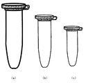
まず、サンプルとして、被験者から採取された尿を、6,000 rpm で5分間遠心したものを尿検体として、該尿検体50μL を、図1に示されるような容器(a) に取り、そこに抽出液 450μL を添加する。抽出液は、例えば 13%デキストラン(平均重合度: 500,900)水溶液 225μL, 21%ポリエチレングリコール(平均重合度: 10,000) 水溶液 100μL, 2% DEAE- デキストラン水溶液50μL,そして0.2 M リン酸バッファー(pH 5.7) 60μL に水を加えて450 μL としたものである。
【0045】
次に、得られた混合物溶液(全容積500 μL)を攪拌処理する。攪拌処理は、例えば手で上下に激しく数回攪拌し、ハンドミキサー(Immuno mixer; シノテスト社製) を用いて30秒間攪拌することが挙げられる。こうして攪拌した混合物溶液は次に遠心処理して2相に相分離させる。遠心処理は、例えば6,000 rpm で10分間遠心する。相分離は通常相体積比で 1:1で分離している。このように相分離したうちの上相(ポリエチレングリコール相) 200 μL を、新たな容器、すなわち図1に示されるような容器(b) に取り、そこに洗浄液 200μL を添加する。洗浄液は、例えば 27%デキストラン(平均重合度: 500,900)水溶液 100μL,そして0.4 M リン酸バッファー(pH 5.7) 30μL に水を加えて200 μL としたものである。
【0046】
次に、得られた混合物溶液(全容積400 μL)を攪拌処理する。攪拌処理は、例えばハンドミキサー(Immuno mixer; シノテスト社製) を用いて30秒間攪拌することが挙げられる。こうして攪拌した混合物溶液は次に遠心処理して2相に相分離させる。遠心処理は、例えば6,000 rpm で10分間遠心する。相分離は通常相体積比で 1:1で分離している。このように相分離したうちの上相(ポリエチレングリコール相) 100 μL を、新たな容器、すなわち図1に示されるような容器(c) に取り、そこに色素溶液 50 μL 及び発色用緩衝溶液 150μL を添加する。色素溶液は、例えば6.6 ×10⁻⁵ mol dm -3のエリスロシンB 溶液としたものである。発色用緩衝溶液は、例えば10% ポリエチレングリコール(平均重合度: 10,000) 水溶液 50 μL 及び 0.5 Mクエン酸バッファー(pH 2.5) 100 μL を混合したものである。
【0047】
エリスロシンB を添加された試料液は、90℃で10分間水浴中で加熱し、室温にて放冷した後、試料溶液に生ずる赤色を目視して検出する。該室温での放冷は、例えば5〜10分間室温に放置することで行われる。また、検出については、544nm における吸光光度法によりそれを行ってもよい。
本処理によれば、その検出感度は、尿中濃度換算で、20 mg/L までのベンスジョーンズタンパク質であることが認められる。また、純水溶液中では、10 mg/L までのベンスジョーンズタンパク質についてその検出感度を認めることができる。
尿中のベンスジョーンズタンパク質濃度が、20 mg/L であると、+ (うすいピンク色)、100 mg/Lであると、++(明瞭なピンク色)、そして 200 mg/L であると、+++ (赤色)であるという結果を得た。
【0048】
一方、試験紙法では、アルブミン(尿中の主要タンパク質)に対する感度は、100 〜200 mg/Lであるが、低分子量タンパク質に対しては反応性が低く、ベンスジョーンズタンパク質では、1000 mg/L 付近でようやく検出ができる。また煮沸法のベンスジョーンズタンパク質に対する感度は、450 mg/L である。
測定に使用する容器は、上記の形態のものに限定されず、市販のものから適宜選択して使用することができる。該市販のものとしては、例えば 1.5 mL のマイクロチューブ(遠心分離用の試験管)などが挙げられる。
【0049】
本発明に従った別の好ましい態様においては、尿中に排泄されるベンスジョーンズタンパク質の検出は次のようにそれに適したキット化された試薬セットを使用して実施できる。
先ず、キット1、キット2、そしてキット3として構成された試薬セットを用意する。
キット1は、13% デキストラン(平均重合度: 500,900)水溶液 225μL, 21%ポリエチレングリコール(平均重合度: 10,000) 水溶液 100μL, 2% DEAE- デキストラン水溶液50μL,そして0.2 M リン酸バッファー(pH 5.7) 60μL が予め収容されている図1に示されるような容器(a) あるいはそれらの成分を乾燥した状態で含有し、該容器中へ添加される尿検体50μL と併せて水を加えることにより最終的に500 μL の容量とされるものであってよい。
【0050】
次に、キット2は、17% デキストラン(平均重合度: 500,900)水溶液 100μL,そして0.4 M リン酸バッファー(pH 5.7) 30μL が予め収容されている図1に示されるような容器(b) あるいはそれらの成分を乾燥した状態で含有し、該容器中へ添加される相分離処理されて得られる上相(ポリエチレングリコール相) 200 μL と併せて最終的に400 μL の容量とされるものであってよい。
そして、キット3は、10% ポリエチレングリコール(平均重合度: 10,000) 水溶液 50 μL が予め収容されている図1に示されるような容器(c) であるが、それら容器内の成分は乾燥した状態で含有されているものであってもよく、相分離した上相(ポリエチレングリコール相) 100 μL を添加後、最終的に250 μL の容量とされるものであってよい。キット3には、好ましくは、色素溶液及び 0.5 Mクエン酸バッファー(pH 2.5) がそれぞれ本キット3の添付試薬として備えられている。色素溶液は、例えば6.6 ×10⁻⁵ mol dm -3のエリスロシンB 溶液としたものである。
【0051】
キット1、キット2、そしてキット3を使用しての尿中ベンスジョーンズタンパク質の検出は、先ず、被験者から採取された尿を、6,000 rpm で5分間遠心し、その遠心したものを尿検体として使用する。該尿検体50μL を、キット1の成分の入った容器に入れ、必要に応じ水を加えて、全体の量を500 μL とする。
次に、得られた混合物溶液を、上記のように、攪拌処理及び遠心処理し、2相に相分離させる。このように相分離したうちの上相(ポリエチレングリコール相) 200 μL を取り、これをキット2の成分の入った容器に入れ、必要に応じ水を加えて、全体の量を400 μL とする。
次に、得られた混合物溶液を、上記のように、攪拌処理及び遠心処理し、2相に相分離させる。このように相分離したうちの上相(ポリエチレングリコール相) 100 μL を取り、これをキット3の成分の入った容器に入れ、必要に応じ水を加えて、全体の量を150 μL とする。
【0052】
こうして得られた混合物溶液は必要に応じて攪拌された後、そこに色素溶液 50 μL を添加する。色素溶液は、試薬セットの添付試薬として備えられているものであってよいし、別途調製したものであってよい。エリスロシンB を添加された試料液は、必要に応じて攪拌された後、そこに0.5 M クエン酸バッファー(pH 2.5) 100 μL を添加する。クエン酸バッファー試薬セットの添付試薬として備えられているものであってよいし、別途調製したものであってよい。得られた混合物溶液は必要に応じて攪拌された後、90℃で10分間水浴中で加熱し、室温にて放冷した後、試料溶液に生ずる赤色を目視して検出する。該室温での放冷は、例えば5〜10分間室温に放置することで行われる。また、検出については、544nm における吸光光度法によりそれを行ってもよい。
【0053】
本発明の別の好ましい態様においては、尿中に排泄されるベンスジョーンズタンパク質の検出は次のようにして実施できる。
まず、尿検体50μL を、図1に示されるような容器(a) に取り、そこに抽出液 450μL を添加する。抽出液は、例えば 13%デキストラン(平均重合度: 500,900)水溶液 225μL, 21%ポリエチレングリコール(平均重合度: 10,000) 水溶液 100μL, 2% DEAE- デキストラン水溶液50μL,そして0.2 M リン酸バッファー(pH 5.7) 60μL に水を加えて450 μL としたものである。
次に、得られた混合物溶液(全容積500 μL)を攪拌処理する。攪拌処理は、例えば手で上下に激しく数回攪拌するか、ハンドミキサー(Immuno mixer; シノテスト社製) を用いて30秒間攪拌することが挙げられる。こうして攪拌した混合物溶液は次に遠心処理して2相に相分離させる。遠心処理は、例えば6,000 rpm で10分間遠心する。このように相分離したうちの上相(ポリエチレングリコール相) 100 μL を、新たな容器、すなわち図1に示されるような容器(b) に取り、そこに洗浄液 400μL を添加する。洗浄液は、例えば 13.5%デキストラン(平均重合度: 500,900)水溶液 200μL,そして0.2 M リン酸バッファー(pH 5.7) 60μL に水を加えて400 μL としたものである。
【0054】
次に、得られた混合物溶液(全容積500 μL)を攪拌処理する。攪拌処理は、例えばハンドミキサー(Immuno mixer; シノテスト社製) を用いて30秒間攪拌することが挙げられる。こうして攪拌した混合物溶液は次に遠心処理して2相に相分離させる。遠心処理は、例えば6,000 rpm で10分間遠心する。このように相分離したうちの上相(ポリエチレングリコール相) 100 μL を、新たな容器、すなわち図1に示されるような容器(c) に取り、そこに色素溶液 50 μL 及び発色用緩衝溶液 130μL を添加する。色素溶液は、例えば6.6 ×10⁻⁵ mol dm -3のエリスロシンB 溶液としたものである。発色用緩衝溶液は、例えば10% ポリエチレングリコール(平均重合度: 10,000) 水溶液 50 μL 及び 0.5 Mクエン酸バッファー(pH 2.5) 80μL を混合したものである。
エリスロシンB を添加された試料液は、90℃で10分間水浴中で加熱し、室温にて放冷した後、試料溶液に生ずる赤色(又は544nm における吸光光度)を検出する。該室温での放冷は、上記と同様にして実施される。
【0055】
また、本発明の別の好ましい態様においては、尿中に排泄されるベンスジョーンズタンパク質の検出は次のようにそれに適したキット化された試薬セットを使用して実施できる。
キット1は、上記と同様のものであってよい。次に、キット2は、13.5% デキストラン(平均重合度: 500,900)水溶液 200μL,そして0.2 M リン酸バッファー(pH 5.7) 60μL が予め収容されている図1に示されるような容器あるいはそれらの成分を乾燥した状態で含有し、該容器中へ添加される相分離処理されて得られる上相(ポリエチレングリコール相) 100 μL と併せて水を加えることにより最終的に500 μL の容量とされるものであってよい。
そして、キット3は、10% ポリエチレングリコール(平均重合度: 10,000) 水溶液 50 μL が予め収容されている図1に示されるような容器であるが、それら容器内の成分は乾燥した状態で含有されているものであってもよく、相分離した上相(ポリエチレングリコール相) 100 μL を添加後、最終的に150 μL の容量とされるものであってよい。キット3には、好ましくは、色素溶液及び 0.5 Mクエン酸バッファー(pH 2.5) がそれぞれ本キット3の添付試薬として備えられている。色素溶液は、例えば6.6 ×10⁻⁵ mol dm -3のエリスロシンB 溶液としたものである。
【0056】
キット1、キット2、そしてキット3を使用しての尿中ベンスジョーンズタンパク質の検出は、先ず、尿検体50μL を、キット1の成分の入った容器に入れ、必要に応じ水を加えて、全体の量を500 μL とする。
次に、得られた混合物溶液を、上記と同様に、攪拌処理及び遠心処理する。相分離したうちの上相(ポリエチレングリコール相) 200 μL を取り、これをキット2の成分の入った容器に入れ、必要に応じ水を加えて、全体の量を500 μL とする。得られた混合物溶液を、上記と同様に、攪拌処理及び遠心処理される。相分離したうちの上相(ポリエチレングリコール相) 100 μL を取り、これをキット3の成分の入った容器に入れ、必要に応じ水を加えて、全体の量を150 μL とする。こうして得られた混合物溶液は必要に応じて攪拌された後、そこに色素溶液 50 μL を添加する。混合物溶液は必要に応じて攪拌された後、そこに0.5 M クエン酸バッファー(pH 2.5) 80μL を添加する。必要に応じて攪拌された後、エリスロシンB を添加された試料液は、90℃で10分間水浴中で加熱し、室温にて放冷した後、試料溶液に生ずる赤色(又は544nm における吸光光度)を検出することにより行われる。該室温での放冷は、上記と同様にして行われる。
【0057】
さらに、本発明では、疾患の診断マーカーとして有用なベンスジョーンズタンパク質などの等電点の高い塩基性タンパク質を、高感度でしかも簡易迅速に検出及び測定することのできる、微量尿中塩基性タンパク質の検出法及び試薬を提供する。本技術は、特には、塩基性タンパク質の固相抽出/色素結合による検出法及びそのための試薬を提供するものである。
本発明の「塩基性タンパク質の固相抽出/色素結合による検出法」では、まず固相媒体(ウエルなど)である容器に、尿試料を入れ、等電点の差異を利用して共存タンパク質の吸着を抑制するために、緩衝液でpHを共存タンパク質の等電点以上かつ塩基性タンパク質の等電点以下に調整し、さらに共存タンパク質の吸着を抑制するために陰イオン色素を混和し、全量を一定量にする。一定時間静置した後、全溶液を捨て、一度水洗する。その後、水溶性ポリマーを含む色素溶液を加え、緩衝溶液で色素発色の最適pHにする。一定時間静置反応させた後、全溶液を捨て、一度水洗した後、固相媒体上で呈色した塩基性タンパク質を目視定量する。
【0058】
本明細書中、「固相媒体」としては、本発明の塩基性タンパク質に対して悪影響がない限り、例えば酵素免疫測定法などで使用されている公知のものから選択して使用することができる。該固相媒体は、容器の内側の表面であってよい。該固相媒体としては、ガラス、二酸化ケイ素などの無機材料、ポリエチレン、ポリプロピレン、ポリスチレン、ポリ塩化ビニル、ポリフッ化ビニリデン、ポリメタクリレートなどのプラスチック材料、ステンレススチールなどの金属材料などからなるものが挙げられる。該固相媒体は、ビーズ、試験管、タイタープレート、タイターウェル、セル(例えばガラスセル、石英ガラスセル、合成樹脂製セルなど)、dip stick などの固体物質の表面などが挙げられる。多検体スクリーニングのためにはウエルの形状が好ましい。好ましい固相媒体としては、例えばプラスチック製ウェルの容器の内側表面が挙げられる。
【0059】
共存する他のタンパク質からの分離には、尿中ベンスジョーンズタンパク質の場合には、緩衝溶液は本発明の塩基性タンパク質の固相媒体上に分離抽出するにあたり悪影響がない限り、例えば免疫測定法などで使用されている公知のものから選択して使用することができるが、 HEPES-LiOH (pH 7.0)が好ましい。尿中主要タンパク質であるアルブミンの等電点は 4.6、トランスフェリンは 5.6〜5.9 であり、pH 7.0では負の電荷を持つ。しかし免疫グロブリンやベンスジョーンズタンパク質は等電点が 7.0〜9.0 であり、pH7.0では正の電荷を持ち、固相へ吸着する。
【0060】
本明細書中、「塩基性タンパク質」としては、例えば生体タンパク質であって、その等電点が 7.0以上のものが挙げられ、代表的には尿中にその存在が認められるものが挙げられ、例えば、ベンスジョーンズタンパク質、免疫グロブリンなどのグロブリンなどが挙げられる。免疫グロブリンとしては、IgM, IgG, IgA, IgE, IgD, 分泌型IgA などが挙げられ、さらにIgG としては、IgG 1 〜4 などが包含されてよく、IgA としては、IgA 1 〜2 などが包含されてよい。例えば、分子量の極めて大きなグロブリン、例えば16万程度もの大きさの分子量のものが尿中に排出されていると、腎臓がサイズバリア機能を損壊していることの証拠と考えられ、腎臓病や糖尿病などの疾患に関連したマーカーとして利用可能である。
【0061】
吸光ラベル化する色素として、エリスロシンBが挙げられるが、エリスロシンBは吸光ラベル化する役割のほか、色素として水溶液中で負の電荷を持つため、タンパク質の正に荷電したアミノ基と結合して、アルブミンの固相への吸着を抑制する効果も併せ持つと考えられる。
塩基性タンパク質と色素結合体の発色が最適となる条件としては、水溶性ポリマー存在下で、緩衝溶液は、前記したようなものから選択することができるが、クエン酸緩衝溶液(pH 2.5)を用いるのが好ましい。水溶性ポリマーとしては、当該分野で知られたものから選択して使用することができるが、例えばポリアルキレングリコールなどが挙げられる。該ポリアルキレングリコールの代表的なものとしては、ポリエチレングリコールが挙げられ、それは当該水溶性ポリマーとして好ましい。ポリエチレングリコールとしては、前記したようなものから選択することができる。また室温にて30分間静置し反応させるのが好ましい。
【0062】
本発明に従った好ましい態様においては、尿中に排出される塩基性タンパク質の検出は、次のようにして実施される。
まず、ウエルに、HEPES-LiOH 緩衝溶液 (pH 7.0) 70μl 、1 ×10⁻⁴M エリスロシンB溶液30μl 、そして被験者から採取された尿30μl を添加混和し、蒸留水で全量を 300μl とする。得られた混合溶液を20分間静置した後、ウエル中の全溶液を捨て、ウエルを水洗する。その後、該ウエルに 270μl の増感発色剤(1 % ポリエチレングリコール, 2 ×10⁻⁵M エリスロシンBを含む水溶液)を添加し、クエン酸緩衝溶液 (pH 2.5) 30μl を加え、室温にて30分間静置し反応させた後、全溶液を捨て、一度ウエルを水洗した後、ウエル上で呈色した赤色を目視比色する。
【0063】
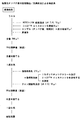
代表的な塩基性タンパク質の固相抽出/色素結合による検出法の操作手順を、図9に示す。
本発明では、(1) 尿中疾患マーカータンパク質である塩基性タンパク質を検出及び/又は測定するためのキットであって、(i) 等電点の差異を利用して、共存する他のタンパク質から塩基性タンパク質を固相媒体上に分離抽出する機能を有する剤が、(a) 固相媒体及び緩衝液あるいは(b) 固相媒体及び色素含有緩衝液からなり、(ii) 固相表面上で吸光ラベル化する剤が、水溶性ポリマーを含む色素溶液であることを特徴とするキット、(2)(a)固相媒体及び緩衝液あるいは(b) 固相媒体及び色素含有緩衝液からなり、等電点の差異を利用して、尿試料中に共存する他のタンパク質から塩基性タンパク質を固相媒体上に分離抽出する機能を有することを特徴とする尿中疾患マーカータンパク質である塩基性タンパク質を固相媒体上に分離抽出するための試薬、(3) 水溶性ポリマーを含む色素溶液からなり、水溶性ポリマーの発色増感により、 固相表面上で呈色した塩基性タンパク質−色素結合体を、定性あるいは定量的に分析あるいは評価することを可能にするものであることを特徴とする固相媒体上に分離抽出され且つ尿中疾患マーカータンパク質である塩基性タンパク質を検出及び/又は測定するための試薬なども提供される。それぞれ、上記固相媒体、緩衝液、色素、水溶性ポリマーなどは、上記したようなものである。
【0064】
【実施例】
以下に実施例を掲げ、本発明を具体的に説明するが、この実施例は単に本発明の説明のため、その具体的な態様の参考のために提供されているものである。これらの例示は本発明の特定の具体的な態様を説明するためのものであるが、本願で開示する発明の範囲を限定したり、あるいは制限することを表すものではない。本発明では、本明細書の思想に基づく様々な実施形態が可能であることは理解されるべきである。
全ての実施例は、他に詳細に記載するもの以外は、標準的な技術を用いて実施したもの、又は実施することのできるものであり、これは当業者にとり周知で慣用的なものである。
【0065】
実施例1
本実施例では、エリスロシンB を吸光ラベルとして、尿中タンパク質(血清アルブミン、トランスフェリン、免疫グロブリンG 、ベンスジョーンズタンパク質) に対して、エリスロシンB による検出法を検証した。さらにポリエチレングリコールをエリスロシンB −タンパク質検出法に組み込むことで飛躍的に検出感度が向上することについても確認した。
(i) (試薬)
タンパク質溶液
本実施例では尿中タンパク質を想定して、人血清アルブミン(Human Serum Albumin)、人トランスフェリン(Human Transferrin)、人免疫グロブリンG (Human Immunoglobulin G) 、ベンスジョーンズタンパク質(Human Bence Jones protein)を用いた。各標準溶液の作成は以下の手順で行った。
(a) 人血清アルブミン(HSA)水溶液
HSA 粉末(シグマ社製)100 mgに0.1 M Tris-HCl溶液10 mL 、1 wt % アジ化ナトリウム水溶液10 mL を加え、水で100 mLとし、1 g/L の溶液とした。Tris-HClは変性防止のため、アジ化ナトリウムは防腐剤として使用した。
(b) 人トランスフェリン(TRF)水溶液
TRF iron rich (和光純薬製)をHSA 同様の操作にて1 g/L の溶液とした。
(c) 人免疫グロブリンG (IgG)水溶液
Protein A 精製IgG (Biogenesis 社製)50 mg に0.1 M Tris-HCl溶液10 mL 、1 wt % アジ化ナトリウム水溶液10 mL 、0.1 M 塩化ナトリウム10 mL を加え、水で100 mLとし、0.5 g/L の溶液とした。
(d) 人ベンスジョーンズタンパク質 (BJP(κ、λ))水溶液
2.2 g/L BJP (CORTEX BIOCHEM 社製)をそのまま用いた。
【0066】
(e) ポリエチレングリコール (PEG)水溶液
ポリエチレングリコール#8,000 、10,000(#;数平均分子量) はシグマ社製、20,000 (15,000±5,000)は和光純薬製 30 g をビーカーにとり、そこに熱水(二回蒸留水)を少しずつ加えていき、かき混ぜながら溶解させる。熱水を50 mL くらい加えた状態でポリマー溶液を加熱し、一度沸騰させる。これはポリマー中の細菌等を滅菌するためである。沸騰後のポリマー溶液は気泡を多く含んでいるので室温で約30〜40分放置する。気泡が抜けたらメスフラスコにこの溶液を少しずつ加えていき、40〜50℃位のぬるま湯でビーカーを洗いながら、その水でメスアップしていく。この時、PEG が底に徐々に溜まるため、ぬるま湯を加えながらメスフラスコをハンドミキサーで攪拌し、溶液を均一にする。最終的に100 mLにメスアップし、30 % g/vol H2Oとした。
(f) エリスロシンB 水溶液
エリスロシンB (和光純薬製) 0.03 gを100 mLの水に溶かし、0.3 g/L の溶液とした。
(g) クエン酸緩衝溶液
クエン酸(和光純薬製) 4.203 g を80 mL の二回蒸留水に溶かし、水酸化ナトリウム( 関東化学製) により所定pHとし、二回蒸留水にて100 mLにメスアップして0.2 M の緩衝溶液とした。
【0067】
(ii) (操作)
5 mLのマイクロチューブ(アイビス社製) に濃度を調整してあるタンパク質溶液(HSA, TRF, IgG, BJP) 50μL 、0.3g/LのエリスロシンB 水溶液50μL と0.2 M のクエン酸緩衝液250 μL を加え、二回蒸御製水にて500 μL とし、80℃で10分間加熱した。その後、10分間室温で放置し、反応溶液を96結合型ウェル(NUNC社製) に200 μL 分注し、デンシトメトリーにて544 nmの吸光度を測定した。ポリエチレングリコールの添加実験については所定量のPEG を、上述の方法においてエリスロシンB 色素添加前に加えた。
【0068】
(a) 最適条件の決定
1) pH 2.5 において色素ブランクの吸光度が加熱時間と温度にどのような影響を受けるのかについて調べた。加熱温度を45〜90℃に設定した場合の加熱時間に伴う色素ブランクの吸光度の変化を調べたところ、それぞれの温度に対し、加熱時間の経過とともに色素の吸収が減少していくことが認められる。特に、80℃、90℃において吸光度の減少は著しく、10分でほとんど色素の吸収がなくなってしまう。この状態では溶液の色はほとんど透明となる。よって、色素ブランクについては吸光度がほぼなくなる加熱条件、すなわち、80℃、10分、あるいは90℃、10分を最適条件とした。
次に反応温度を80℃に設定し、反応時間を変化させたときの色素、及びタンパク質−色素会合体の吸光度変化について検討した。タンパク質−色素会合体の吸光度は「タンパク質存在時の総吸光度」から「色素のみの場合(ブランク)の吸光度」を引いたものとし、そうして求めた図より、各タンパク質とも反応時間が10分において 544 nm における吸光度が最大となった。色素ブランクの吸光度が最小となる条件も、80℃、10分あるいは90℃、10分であり、この条件をタンパク質検出における反応の最適条件とすることができる。
【0069】
2) 次に、加熱温度80℃、反応時間10分におけるエリスロシンB とタンパク質が会合体を形成するための最適なpH条件を調べた。
タンパク質−エリスロシンB 会合体の吸光度は、上記と同様、「タンパク質存在時の反応溶液の総吸光度」から「色素のみの場合における吸光度」の差をとったもので、そうして求めた図より、タンパク種によって若干の異なりはあるものの会合体が最大の吸収を示すのはおおよそpH 2〜3 の間となった。このpH条件においては色素だけの吸収も最小となり、色素ブランクとサンプル間で良好な色のコントラストが得られた。そのため、最適pHを2.5 と設定できる。なお、緩衝溶液の種類としては、クエン酸緩衝溶液を用いることができる。
反応温度80℃についてpHを 2〜5 まで変化させた場合についても、エリスロシンB の吸光度の加熱時間依存性を調べた。その結果、pH2 及びpH3 においては反応時間が10分で十分に吸光度が減少していることがわかった。このことからもpH 2.5が最適条件であるという結論が導き出せる。
【0070】
3) 上記により得られた、エリスロシンB を吸光ラベルとした場合におけるタンパク質アッセイのための最適条件のもとで、4種類のタンパク質(すなわち、HSA, TRF, IgG, BJP) についてアッセイを行った。得られた検量線を図2に示す。 各タンパク質における検出限界、定量限界、Coomasie Brilliant Blue (CBB) 法における検出限界を次に示す。
【0071】
【表1】

該エリスロシンB 法は、CBB 法と比較して3〜9倍高感度化しており、タンパク質−色素結合法としては最高検出感度を持っていると評価できる。
ベンスジョーンズタンパク質 (BJP)については、9倍高感度であり、良好な検出感度が得られることが明らかとなった。
【0073】
(b) ポリエチレングリコールによる増感作用
本実施例において、ポリエチレングリコール (PEG)の添加によってエリスロシンB 法の感度に及ぼす影響を検討した。本実施例では、エリスロシンB アッセイにおけるPEG 添加効果について考察するとともに、タンパク質検出法としての最適条件を求めた。
PEG #10,000のエリスロシンB 及びエリスロシン−タンパク質会合体に及ぼす影響は、各タンパク質濃度 20 mg/Lにつき、上記した最適な測定条件のもとに測定し、図3に示す結果が得られた。PEG #10,000の添加に伴い色素−タンパク質会合体の吸光度は放物線を描き、一方色素ブランクの吸光度はほとんど変化しないことがわかる。PEG が2 % までは、色素−タンパク質会合体の吸光度は減少し、その後PEG 濃度の増加に伴い吸光度が増加する。
サンプルとしてBJP を用い、PEG #10,000の濃度を 0%, 5.5%, 6.0% とした場合のエリスロシンB アッセイへの影響を調べた結果を、図4に示す。
本法の検出限界、及び増感作用を施していないエリスロシンB 法、CBB 法との比較を表2に示す。
【0074】
【表2】
上記表2において、「検出限界」及び「定量限界」ではPEG 添加したもので、検知はデンシトメーターによる吸光光度法で行った。括弧内は増感作用を施してないエリスロシンB 法の結果である。
BJP 低濃度領域においてもPEG 添加した場合にはブランクとの差が明確となり、1 mg/Lという(分子量に換算して2.3 ×10-8 M) 微量領域においても十分に目視判別が可能となった。PEG を添加するだけで検出感度が飛躍的に向上していることがわかる。特にBJP については CBB法と比較して280 倍も高感度化となっている。
この方法は、(1) コストが極めて安い、一回のアッセイに0.00〜0.1 円位のコストで済む、(2) 検出感度が飛躍的に良い、そして何より(3) タンパク質に関して定量性の違いがほとんど生じないことである。これは、尿中の総タンパク質を求めるのに非常に都合が良い。
【0076】
実施例2
水性二相分配系の設計
(i) 試薬
(a) ポリエチレングリコール(#8,000 、10,000)水溶液
実施例1と同様にして30(%)溶液を調製した。
(b) デキストラン(wt(重量平均分子量) 67,300〜500,900 )、DEAE- デキストラン水溶液
デキストラン(重量平均分子量 67,300, 500,900; シグマ社製) 、DEAE- デキストラン(重量平均分子量 500,000; シグマ社製) 30 gをビーカーにとり、そこに熱水(二回蒸留水)を少しずつ加えていき、かき混ぜながら溶解させる。熱水を50 mL くらい加えた状態でポリマー溶液を加熱し一度沸騰させる。これはポリマー中の細菌等を滅菌するためである。沸騰後のポリマー溶液は気泡を多く含んでいるので室温で約30〜40分放置する。気泡が抜けたらメスフラスコにこの溶液を少しずつ加えていき、40〜50℃位のぬるま湯でビーカーを洗いながら、その水でメスアップしていく。この時、ポリマーが底に除々に溜まるため、ぬるま湯を加えながらメスフラスコをハンドミキサー(Immuno mixer; シノテスト社製) で攪拌し、溶液を均一にする。最終的に100 mLにメスアップし、30% g/vol H2O とした。
【0077】
(ii) 緩衝溶液
(a) クエン酸緩衝溶液
クエン酸(関東化学社製)を所定量、二回蒸留水に溶解させ、水酸化カリウム(関東化学社製)によって所定のpH (2 〜3.5)にし、0.2 M 水溶液となるように二回蒸留水により定容とした。
(b) リン酸緩衝溶液
リン酸一ナトリウム、リン酸二ナトリウム(共に大和薬品製)を所定量、二回蒸留水に溶解し 0.2 Mとし、これらを適量混合させることにより、所定のpHとし、0.1 M とした。
(c) 酢酸緩衝溶液
0.2 M 酢酸(関東化学社製)、0.2 M 酢酸ナトリウム(関東化学社製)水溶液を所定量混合し、0.1 M の緩衝溶液とした。
【0078】
(d) MES-NaOH緩衝溶液
MES (2-Morpholinoethanesulfonic acid, monohydrate) (関東化学社製)を所定量蒸溜水に溶解させ、水酸化ナトリウム (Merck 社製)によって所定のpHにし、0.2 M 水溶液となるように二回蒸留水により定容とした。
(e) Bis-Tris-HCl緩衝溶液
Bis-Tris (Bis(2-hydroxyethyl)iminotris(hydroxymethyl)methane) (Merck社製)を所定量蒸溜水に溶解させ、塩酸(関東化学社製)によって所定のpHにし、0.2 M 水溶液となるように二回蒸留水により定容とした。
(f) Tris-HCl緩衝溶液
Tris(hydroxymethyl)aminomethane (Merck社製)を所定量蒸溜水に溶解させ、塩酸(関東化学社製)によって所定のpHにし、0.2 M 水溶液となるように二回蒸留水により定容とした。
【0079】
(iii) タンパク質溶液
実施例1と同様にして調製した。
(iv) タンパク質濃度測定試薬(Coomasie Brilliant Blue Protein Assay Kit) Bio-Rad社のCoomasie Brilliant Blue Protein Assay Kit をそのまま用いた。
【0080】
(v) 水性二相分配によるタンパク質の抽出試験
(タンパク質一成分系)
マイクロチューブにPEG 、デキストラン(DEX) 、バッファー、その他(イオン交換ポリマー) 、タンパク質サンプル (1,000 mg/Lを50μL)を所定濃度となるように添加していき、最後に水により定容(ほとんどの場合、総体積は、500 μL 、分配系におけるタンパク質濃度は 100 mg/L となる)とした。
混合した液を手で上下に激しく数回攪拌し、ハンドミキサー(Immuno mixer; シノテスト社製) を用いて30秒間攪拌する。その後、卓上遠心分離機を用い、10分間、6,000 rpm の条件で遠心分離させて、二相に相分離させた。
マイクロピペットを用いて、上相 (PEG rich相)、下相 (DEX rich相)からそれぞれ 40 μL ずつ取り出し、96結合型ウェルに分注し、水を 120μL 加える。その後、Coomasie Brilliant Blue Protein Assay Kit (Bio-Rad社) を用いタンパク質濃度を測定する。手順としてはCoomasie Brilliant Blue (CBB) 溶液を40μL 加え、2 分間 Vortex mixer (シノテスト社製) を用いて攪拌する。その後デンシトメトリー(島津製作所製)によってタンパク質−CBB 会合体の最大吸光度である 595 nm の吸光度を測定し、タンパク質の濃度を求める。なお、サンプル溶液と水との混合割合(希釈率)は系に存在する妨害物質の影響が現れない領域(4〜16倍希釈)に設定して行った。
【0081】
(タンパク質多成分系)
単一成分系で得られた最適と思われる抽出結果を与える条件を利用し、混合タンパク質系についても分配実験を行い、結果をSDS-PAGEにて確認した。総分離の操作までは、タンパク質一成分系と同様の操作を行い、その後、上相、下相溶液をSDS-PAGEにかけ、各タンパク質を分離した後、Coomasie Brilliant Blue にて染色を行い、分離を確認した。
【0082】
(vi) 試験結果
(pHの影響)
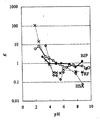
PEG#8,000-DEX500,900系につき、pHを変化させた場合の各タンパク質の分配係数K を図5に示す。図5においては、PEG #8,000; 4.2%, DEX#500,900; 6.2%, バッファー; 0.0167 M (pH1.0; HCl, pH2.0 〜4.0;クエン酸緩衝液, pH4.5 〜5.5;酢酸緩衝液, pH6.0; MES-NaOH 緩衝液, pH6.5 〜7.8;Tris-HCl緩衝液, pH8.8; MOPS 緩衝液),タンパク質; 100 mg/Lの条件下に測定を行った。
その結果、pH 6付近でHSA とBJP が比較的良好に分離されていることから、pH 6付近で水性二相分配系の構築を目指すことが好ましいと判断された。
【0083】
(陽イオンポリマー効果の検討)
陽イオンポリマーとして、ジエチルアミノエーテル- デキストラン(DEAE-DEX)を使用し、水性二相分配系に及ぼす効果を試験した。pH5.5 付近において緩衝溶液として酢酸塩、リン酸塩を用いた場合のDEAE-DEXの分配に及ぼす影響について試験し、得られた結果を図6に示す。特に、リン酸緩衝液を用いた場合、HSA 、TRF がDEX 相に選択的に分配されることがわかる。かくして、少なくともpH6 付近においてリン酸塩を緩衝溶液として用いた場合、HSA 、TRF を良好にPEG 相から排除可能であると判断された。
次に、混合タンパク質系において、HSA, TRFを PEG相から排除することが可能か否かを、DEAE-DEXの濃度 0% 及び 0.2% の場合につき、各相のタンパク質の存在状態をSDS-PAGEにより調べた。その結果、DEAE-DEXが 0.2% 存在している条件では上相 (PEG rich相) から、HSA, TRFのバンドが殆ど消滅していることが確認された。特に、HSA のバンドは全くといってよい程度に消滅していた。一方、BJP については、DEAE-DEXの添加のある無しに拘らず、上相、下相の濃度は殆ど変化していなかった。
かくして、pH5.71において、DEAE-DEXを 0.2% 添加することにより、上相 (PEG rich相) にほぼBJP のみを存在させることが可能であることが確認された。
【0084】
次に、エリスロシンB アッセイ時のPEG による増感作用を有効に利用することを目指し、PEG #10,000-DEX500,900 系につき、pHを変化させた場合の各タンパク質の分配係数K を、DEAE-DEX共存系のもとで調べた。結果を、図6に示す。図6では、PEG #10,000; 4.2%, DEX #500,900; 6.2%, pH5.71 リン酸緩衝溶液0.024 M,タンパク質= 100 mg/Lの条件下に測定を行った。
リン酸緩衝液の尿に対してそのpHに与える効果を別途の試験で調べた結果、10倍希釈尿ではリン酸塩緩衝溶液濃度が0.03 M以上で緩衝能を示すことが確認された。pH5.71, PEG 10,000-DEX 500,900系において、リン酸緩衝液の濃度を変化させてHSA の分配に対するDEAE-DEXの影響を調べた。結果を、図7に示す。図7では、PEG #10,000; 4.2%, DEX #500,900; 6.2%, 全HSA 濃度= 100 mg/Lの条件下に測定を行った。
また、PEG 10,000-DEX 500,900系についてDEAE-DEX 0.2% 存在下でのリン酸緩衝液の濃度を変化させた場合の各タンパク質の分配挙動を調べた。結果を、図8に示す。図8では、PEG #10,000; 4.2%, DEX #500,900; 6.2%, DEAE-DEX; 0.2%, pH5.71 リン酸緩衝溶液; 0.024 〜0.1 M,タンパク質= 100 mg/Lの条件下に測定を行った。
以上を総合し、緩衝溶液濃度は 0.04 M とすることが好ましいと判断され、DEAE-DEXについては、 0.2% とする水性二相分配系PEG 10,000-DEX 500,900系が、尿中人ベンスジョーンズタンパク質 (BJP)の抽出測定系として好ましいと判断された。
【0085】
実施例3
まず、サンプルとして、人採取尿に実施例1に記載されたような人ベンスジョーンズタンパク質 (BJP)を、0 mg/L, 20 mg/L, 100 mg/L, 200 mg/L それぞれ添加したものを用意する。つぎに該サンプル尿試料を6,000 rpm で5分間遠心したものを尿検体として、該尿検体50μL を、図1に示されるような容器(a) に取り、そこに抽出液 450μL を添加する。抽出液は、13% デキストラン(平均重合度: 500,900)水溶液 225μL, 21%ポリエチレングリコール(平均重合度: 10,000) 水溶液 100μL, 2% DEAE- デキストラン水溶液50μL,そして0.2 M リン酸バッファー(pH 5.7) 60μL に水を加えて450 μL としたものである。
次に、得られた混合物溶液(全容積500 μL)を攪拌処理する。攪拌処理は、手で上下に激しく数回攪拌し、ハンドミキサー(Immuno mixer; シノテスト社製) を用いて30秒間攪拌することによりなされた。こうして攪拌した混合物溶液は次に遠心処理して2相に相分離させる。遠心処理は、6,000 rpm で10分間遠心することによりなされた。相分離は通常相体積比で 1:1で分離している。このように相分離したうちの上相(ポリエチレングリコール相) 200 μL を、新たな容器、すなわち図1に示されるような容器(b) に取り、そこに洗浄液 200μL を添加する。洗浄液は、27% デキストラン(平均重合度: 500,900)水溶液 100μL,そして0.4 M リン酸バッファー(pH 5.7) 30μL に水を加えて200 μL としたものである。
【0086】
次に、得られた混合物溶液(全容積400 μL)を攪拌処理する。攪拌処理は、ハンドミキサー(Immuno mixer; シノテスト社製) を用いて30秒間攪拌することによりなされた。こうして攪拌した混合物溶液は次に遠心処理して2相に相分離させる。遠心処理は、6,000 rpm で10分間遠心することによりなされた。相分離は通常相体積比で 1:1で分離している。このように相分離したうちの上相(ポリエチレングリコール相) 100 μL を、新たな容器、すなわち図1に示されるような容器(c) に取り、そこに色素溶液 50 μL 及び発色用緩衝溶液 150μL を添加する。色素溶液は、6.6 ×10⁻⁵ mol dm -3のエリスロシンB 溶液としたものである。発色用緩衝溶液は、10% ポリエチレングリコール(平均重合度: 10,000) 水溶液 50 μL 及び 0.5 Mクエン酸バッファー(pH 2.5) 100 μL を混合したものである。
エリスロシンB を添加された試料液は、90℃で10分間水浴中で加熱し、室温にて放冷した後、試料溶液に生ずる赤色を目視して検出する。該室温での放冷は、例えば5〜10分間室温に放置することで行われる。
尿中のベンスジョーンズタンパク質濃度が、20 mg/L であると、+ (うすいピンク色)、100 mg/Lであると、++(明瞭なピンク色)、そして 200 mg/L であると、+++ (赤色)であるという結果を得た。
【0087】
実施例4
タンパク質溶液は実施例1と同様にして調製した。人ベンスジョーンズタンパク質 (BJP)を含有するタンパク質混合液を尿検体の代わりに使用した。該タンパク質混合液サンプル50μL を、図1に示されるような容器(a) に取り、そこに抽出液 450μL を添加する。抽出液は、13% デキストラン(平均重合度: 500,900)水溶液 225μL, 21%ポリエチレングリコール(平均重合度: 10,000) 水溶液 100μL, 2% DEAE- デキストラン水溶液50μL,そして0.2 M リン酸バッファー(pH 5.7) 60μL に水を加えて450 μL としたものである。
次に、得られた混合物溶液(全容積500 μL)を攪拌処理する。攪拌処理は、手で上下に激しく数回攪拌するか、ハンドミキサー(Immuno mixer; シノテスト社製) を用いて30秒間攪拌することによりなされた。こうして攪拌した混合物溶液は次に遠心処理して2相に相分離させる。遠心処理は、6,000 rpm で10分間遠心することによりなされる。このように相分離したうちの上相(ポリエチレングリコール相) 100 μL を、新たな容器、すなわち図1に示されるような容器(b) に取り、そこに洗浄液 400μL を添加する。洗浄液は、13.5% デキストラン(平均重合度: 500,900)水溶液 200μL,そして0.2 M リン酸バッファー(pH 5.7) 60μL に水を加えて400 μL としたものである。
【0088】
次に、得られた混合物溶液(全容積500 μL)を攪拌処理する。攪拌処理は、ハンドミキサー(Immuno mixer; シノテスト社製) を用いて30秒間攪拌することによりなされた。こうして攪拌した混合物溶液は次に遠心処理して2相に相分離させる。遠心処理は、6,000 rpm で10分間遠心することによりなされた。このように相分離したうちの上相(ポリエチレングリコール相) 100 μL を、新たな容器、すなわち図1に示されるような容器(c) に取り、そこに色素溶液 50 μL 及び発色用緩衝溶液 130μL を添加する。色素溶液は、6.6 ×10⁻⁵ mol dm -3のエリスロシンB 溶液としたものである。発色用緩衝溶液は、10% ポリエチレングリコール(平均重合度: 10,000) 水溶液 50 μL 及び 0.5 Mクエン酸バッファー(pH 2.5) 80μL を混合したものである。
【0089】
エリスロシンB を添加された試料液は、90℃で10分間水浴中で加熱し、室温にて放冷した後、試料溶液に生ずる赤色(又は544nm における吸光光度)を検出する。該室温での放冷は、上記と同様にして実施される。
本処理によれば、その検出感度は、尿中濃度換算で、20 mg/L までのベンスジョーンズタンパク質であることが認められる。また、純水溶液中では、10 mg/L までのベンスジョーンズタンパク質についてその検出感度を認めることができる。
以上の結果から、サンプル中に 10 mg/Lの濃度で存在しているベンスジョーンズタンパク質 (BJP)の検出も可能であり、実質的には尿に20 mg/L 以上の濃度で存在しているBJP を検出できる。上記方法では、大量の検体を安価に且つ短時間で処理することが可能である。操作が簡単であり、自動化処理に適していると判断された。
【0090】
実施例5
以下の実施例5及び6では、尿中疾患マーカータンパク質である塩基性タンパク質の検出及び/又は測定する方法について検討した。
まず、本実施例では、尿中主要タンパク質であるアルブミンを全量 300μl 中の濃度として10mg/L含有する水溶液に、代表的な尿中疾患マーカータンパク質の塩基性タンパク質としてベンスジョーンズタンパク質を 0 mg/L 、2 mg/L、10 mg/L 、20 mg/L 、それぞれ添加して、本法を検証した。
(1)試薬
(a)ヒト血清アルブミン水溶液 (0.1g/L)
(b)ヒトベンスジョーンズタンパク質水溶液 (2.2g/L)
(c)エリスロシンB水溶液
(d)HEPES-LiOH緩衝溶液 (pH 7.0)
(e)ポリエチレングリコール水溶液
(f)クエン酸緩衝溶液 (pH 2.5)
【0091】
(2)操作
ウエルに、HEPES-LiOH緩衝溶液 (pH 7.0) 70μl 、 1×10⁻⁴M エリスロシンB溶液30μl を添加混和した。該ウエル中の溶液に、ヒト血清アルブミン水溶液を30μl 、そしてベンスジョーンズタンパク質を全量 300μl とした時の濃度として0 mg/L、2 mg/L、10 mg/L 、20 mg/L を、それぞれ添加し、蒸留水で全量を 300μl とした試料を用意する。
得られた混合溶液を20分間静置した後、ウエル中の全溶液を捨て、水洗する。その後、増感発色剤(1 % ポリエチレングリコール (分子量10,000), 2×10⁻⁵M エリスロシンBを含む水溶液)270 μl をウエルに添加し、クエン酸緩衝溶液 (pH 2.5) 30μl を加え、室温にて30分間静置し反応させた後、全溶液を捨て、一度水洗した後、ウエル上で呈色した赤色を目視比色する。
【0092】
(3)結果
ヒト血清アルブミンとベンスジョーンズタンパク質(BJP) を含有した試料において、全量 300μl の濃度として、BJP 、0 mg/Lは無色、2 mg/Lは薄いピンク色、10 mg/L は明瞭なピンク色、20 mg/L は濃い赤色であるという結果を得た。
以上の結果から、尿中主要タンパク質であるアルブミンの固相への吸着は抑制され、選択的にBJP を固相上に吸着、定量することができる。上記タンパク質混合溶液 300μl 中において 2mg/L以上の濃度で存在している BJPを検出できる。
【0093】
実施例6
本実施例では、人採取尿を用い、代表的な尿中疾患マーカータンパク質の塩基性タンパク質としてベンスジョーンズタンパク質を使用し、該ベンスジョーンズタンパク質を尿中濃度として、0 mg/L、20 mg/L 、100 mg/L、200 mg/L、をそれぞれ添加した水溶液試料を用い、本法を検証した。
(1)試薬
(a)ヒトベンスジョーンズタンパク質水溶液
(b)エリスロシンB水溶液
(c)HEPES-LiOH緩衝溶液 (pH 7.0)
(d)ポリエチレングリコール水溶液
(e)クエン酸緩衝溶液 (pH 2.5)
【0094】
(2)操作
ウエルに、HEPES-LiOH緩衝溶液 (pH 7.0) 70μl 、 1×10⁻⁴M エリスロシンB溶液30μl を添加混和した。該ウエル中の溶液に、人採取尿を30μl 、そしてベンスジョーンズタンパク質を全量 300μl とした時の濃度として0 mg/L、2 mg/L、10 mg/L 、20 mg/L を、それぞれ添加し、蒸留水で全量を 300μl とした試料を用意する。
得られた混合溶液を20分間静置した後、ウエル中の全溶液を捨て、水洗する。その後、増感発色剤(1 % ポリエチレングリコール, 2 ×10⁻⁵M エリスロシンBを含む水溶液) 270μl をウエルに添加し、クエン酸緩衝溶液 (pH 2.5) 30μl を加え、室温にて30分間静置し反応させた後、全溶液を捨て、一度水洗した後、ウエル上で呈色した赤色を目視比色する。
【0095】
(3)結果
ベンスジョーンズタンパク質(BJP) を添加した試料において、尿中濃度として、BJP 、0 mg/Lは無色、20 mg/L は薄いピンク色、100 mg/Lは明瞭なピンク色、200 mg/Lは濃い赤色であるという結果を得た。
以上の結果から、実質的には尿に 20mg/L 以上の濃度で存在しているBJP を検出できる。上記方法では、大量の検体を安価にかつ短時間で処理することが可能であり、マススクリーニング法に適していると判断される。また、固相媒体上の発色体は、長期間保存すること(結果の長期間保存) が可能であり、例えば、赤などに発色したウェルはそれを暗所で一ケ月以上あるいはそれより長期にわたり保存して置くことができる。
【0096】
【発明の効果】
本発明では、水性二相の遠心分離によって尿試料からベンスジョーンズタンパク質を簡便に且つ効率良く抽出することを可能にしているので、従来のように分離に煩雑で高価な方法を用いる必要はなく、さらに特別な機器や長い時間を要することもない。本発明では、尿試料からベンスジョーンズタンパク質を選択的に分離できることから、特異的なベンスジョーンズタンパク質の検知・測定が可能であるし、簡便且つ迅速に高い感度でのスクリーニングの実施ができる。
本発明では、エリスロシンB 色素結合アッセイにおいて、ポリエチレングリコールによる増感作用を利用でき、検出感度を飛躍的に高めることができる。
本発明により、尿試料を用いてのベンスジョーンズタンパク質の優れた検出及び/又は定量測定の手法が提供され、骨髄腫診断スクリーニング方法及び試薬、さらには骨髄腫の臨床モニタリングシステムを提供できる。マススクリーニング(発症前)や臨床モニタリング(骨髄腫などと診断された患者を対象)など幅広く適用できるという利点もある。
【0097】
また、本発明では、尿試料から疾患マーカーである塩基性タンパク質を、タンパク質の等電点の差異を利用する方法で選択的に固相媒体上に分離抽出することが可能であり、また水溶性ポリマーによる固相表面上での発色増感効果により、簡便、迅速かつ高感度に尿中塩基性タンパク質−色素結合体を定性、定量的に分析評価する手法が提供される。固相媒体としてウエルなどを用いることで、多検体同時測定が可能であり、尿中塩基性タンパク質のスクリーニングテスト法として、迅速、簡便かつ高感度な検出方法である。また、水溶性ポリマーの増感効果により、検出感度が飛躍的に向上する。本発明により各種疾患のスクリーニングテストや腫瘍などの臨床モニタリングシステムなどが提供できる。
本発明は、前述の説明及び実施例に特に記載した以外も、実行できることは明らかである。上述の教示に鑑みて、本発明の多くの改変及び変形が可能であり、従ってそれらも本件添付の請求の範囲の範囲内のものである。
【図面の簡単な説明】
【図1】 本発明の水性二相分配と吸光ラベルを用いる尿中骨髄腫マーカーであるベンスジョーンズタンパク質の検知・測定に適した容器(試験管)の形態を示す。 (a)〜(c) には必要に応じてそのサイズの異なるものが使用できるという意味での具体例が示されている。
【図2】 加熱温度80℃、反応時間10分、そしてpH 2.5におけるエリスロシンB を吸光ラベルとした4種類のタンパク質(すなわち、HSA, TRF, IgG, BJP)についてのアッセイの結果得られた検量線を示す。
【図3】 加熱温度80℃、反応時間10分、そしてpH 2.5におけるエリスロシンB を吸光ラベルとした4種類のタンパク質(すなわち、HSA, TRF, IgG, BJP)についてのアッセイにおいて、各濃度のPEG #10,000の存在が、如何なる影響を及ぼすかを測定した結果を示す。
【図4】 サンプルとしてBJP を用い、PEG #10,000の濃度を 0%, 5.5%, 6.0% とした場合のエリスロシンB アッセイへの影響を調べた結果を示す。
【図5】 水性二相分配の PEG#8,000-DEX500,900系につき、pHを変化させた場合に測定の結果得られた各タンパク質の分配係数K を示す。
【図6】 pH5.5 付近においての水性二相分配系において、各濃度のDEAE-DEXの存在が、如何なる影響を及ぼすかを測定した結果を示す。
【図7】 pH5.71, PEG 10,000-DEX 500,900水性二相分配系において、リン酸緩衝液の濃度を変化させてHSA の分配に対するDEAE-DEXの影響を調べた結果を示す。
【図8】 PEG 10,000-DEX 500,900水性二相分配系についてDEAE-DEX 0.2% 存在下でのリン酸緩衝液の濃度を変化させた場合の各タンパク質の分配挙動を調べた結果を示す。
【図9】 塩基性タンパク質の固相抽出/色素結合による検出法の代表的な操作手順を示す。[0001]
BACKGROUND OF THE INVENTION
Since the present invention is produced in excess with multiple myeloma and excreted in the urine, the Bence Jones protein useful as a diagnostic marker for multiple myeloma can be detected with high sensitivity, simply and rapidly. The present invention relates to a method and a reagent for detecting a trace amount of Bence Jones protein that can be measured. In particular, the present invention relates to a rapid and simple method for detecting a urinary myeloma marker using an aqueous two-phase partition and an absorption label.
In addition, the present invention belongs to a pre-renal protein and can detect and measure a basic protein having a high isoelectric point such as Bence Jones protein useful as a disease diagnostic marker with high sensitivity and easily and quickly. The present invention relates to a detection method and reagent for a microprotein in urine. In particular, the present invention relates to a method for detecting basic protein by solid phase extraction / dye binding.
[0002]
[Prior art]
Bence Jones Protein (BJP) is a protein known to be excreted in urine in large amounts in multiple myeloma. This urinary protein was named “Bence Jones Protein” when it was first discovered by Dr. Henry Bence Jones in 1847. This protein has unique properties compared to other urinary proteins and is therefore “excreted in the urine and coagulates once by heating to 60 ° C., but by further heating to 100 ° C. The definition of “resolving protein” was made by the discoverer Dr. Bence Jones. In subsequent studies, the Bence Jones protein was found to correspond to the light chain of an immunoglobulin, and now, in addition to the above definition, “Light chain: light chain: L chain” ) ”Is added.
By the way, it is known that a small amount of polyclonal L chain is usually present in the blood and urine of a healthy person, but this is not derived from M protein, and therefore even if it is chemically the same substance. It is not called Bence Jones protein.
[0003]
There are κ and λ types as subclasses of the Bence Jones protein, and both κ and λ chains basically consist of a variable region constituting the amino terminal side and a constant region constituting the carboxy terminal side. . The constant region has an amino acid sequence that is substantially determined depending on the species, while the variable region has a unique amino acid sequence that is determined by the antigen that is recognized during the defense of the body. Therefore, even in the same species such as humans, the Bence Jones protein excreted in blood and urine does not have a common structure as a whole, and as a result, reproducibility between samples is not possible with quantitative methods such as immunoassay. This is a big problem.
Bence Jones protein is known to exhibit various forms of existence, and it has been reported that monomers and dimers exist in urine, and trimers and tetramers also exist in blood. Furthermore, the Bence Jones protein tends to be in different forms depending on the subclass (κ type, λ type). In other words, it is said that disulfide bond formation is relatively difficult in the κ type, and there are almost equal amounts of covalent and non-covalent dimers, whereas almost all of the λ type is derived from disulfide bond formation. It is a complicated way of existence, such as taking a covalent dimer. This also makes the measurement more difficult.
[0004]
[Problems to be solved by the invention]
When affected by myeloma, the monoclonal L chain is excreted in the urine, which is a typical phenomenon seen in myeloma, in which B cells producing plasma cells that are present in large numbers in the bone marrow. It is thought to be a disease associated with tumor formation. Thus, the tumorigenicity of plasma cells is closely related to the increase in urinary Bence Jones protein. It is considered extremely useful.
Myeloma onset appears only after a long incubation period of about 18 years, but clinical symptoms appear for the first time, but once the clinical symptoms appear, the progression is very rapid, and in many cases, death occurs from the discovery of the disease. It is as short as about 1 to 2 years. Although myeloma itself is an intractable disease, it is also a problem because it greatly affects the prognosis of various complications caused by myeloma. Bense Jones protein abnormally generated in plasma cells is excreted in the urine in large quantities via the glomeruli, but the treatment of a large amount of Bence Jones protein for the kidney is a heavy burden. The processing capacity of the glomerulus gradually decreases, causing kidney damage. This is called “myeloma kidney”. In the myeloma kidney, it is impossible to excrete the Bence Jones protein in the urine, so that the Bence Jones protein is deposited in the blood and in each internal tissue, and various disorders are generated. This is called “amyloidosis”. These myeloma-related diseases are still very difficult to treat because they progress very rapidly after the clinical manifestations of the myeloma appear.
[0005]
As described above, even in healthy individuals, the polyclonal L chain is excreted in the urine slightly. Therefore, it is required to detect and measure the Bence Jones protein with high sensitivity by discriminating between the case of a normal low concentration level and the case of increase due to disease. Since myeloma rarely shows disease-specific symptoms until the end stage, determining whether Bence Jones protein is excreted there using non-invasive urine as a specimen is myeloma It is expected to be very useful in terms of early detection. By the way, in the case of myeloma with almost no subjective symptoms, a screening method that can easily and inexpensively obtain results in a short time is indispensable in order to make laboratory measurements useful for early diagnosis by urinalysis at an early stage. It is.
[0006]
Conventionally, boiling method, immunoassay method, electrophoresis method and the like have been used for detection of Bence Jones protein.
The boiling method can be traced back to Dr. Henry Bence Jones's discovery of the presence of this urinary protein in 1847. The Bence Jones protein coagulates when heated to 60 ° C near pH 5. However, it is a method that uses the property of being dissolved again when heated to 100 ° C. However, its sensitivity is low, at only 450 mg / L, which is equivalent to when myeloma has already reached a serious stage. From the viewpoint of early detection of multiple myeloma The application itself is only inappropriate. Next, the immunoassay method includes a radioimmunoassay and an enzyme immunoassay, both of which have a high sensitivity of ppb level. However, the method using a radiolabel has problems such as handling facilities and waste liquid, and the method uses an enzyme as a label. Has been developed. However, in immunoassay methods, the antibodies used are extremely expensive, the measurement takes a relatively long time, there is a problem in reproducibility between different samples, and the protein contains a variable region. There is a problem in terms of preparing a suitable reagent.
[0007]
Electrophoresis is currently most commonly used to detect Bence Jones protein. However, the electrophoresis method requires a series of operations such as separation (electrophoresis), staining, decolorization of the back, and detection. However, there is a problem that it takes a long time, and it is difficult to obtain the result easily. There are difficulties in processing a large number of specimens. If a simple method is adopted, the resolution becomes extremely poor, and it is difficult to detect a small amount of Bence Jones protein. Attempts have been made to concentrate urine to increase sensitivity, but this concentration method requires time to concentrate and requires the use of very expensive separation membranes, as well as the concentration of other proteins in the urine. Therefore, it cannot always be said that the detection sensitivity is easily improved. In other words, even if a peak that seems to be a Bence Jones protein is obtained, it is difficult to identify it as a Bence Jones protein because of its poor resolution. is there.
[0008]
Under such circumstances, the diagnosis of myeloma currently performed in Japan is actually a complicated process. First of all, a total protein test is performed on the disease preparatory group who first visited the hospital, and urine / serum electrophoresis is performed when the test shows positive several times. And if abnormality is discovered at this time, the boiling method etc. are applied to urine, and immunoelectrophoresis is performed on the positive in this test, and if this is positive Bence Jones protein Then, myeloma is identified by bone marrow puncture, and subclasses of urinary Bence Jones protein are assigned to start treatment. Due to this complex process, early detection of multiple myeloma is not possible even if diagnosed relatively early (even 10 years after the origin of the myeloma-causing tumor cells). It was possible. In addition, myeloma is refractory among cancers because of its synergistic effects such as almost no symptoms appearing, very rapid progress after symptoms are found, and no effective treatment established. It is positioned as a disease.
In any medical institution, outpatients, etc. are required to be able to test easily, in a short time, at low cost, and to detect and measure urinary Bence Jones protein with high sensitivity. . In addition, for early detection, it is also required that the urinary Bence Jones protein can be easily detected and measured in an unconscious situation such as mass screening. Regardless of individual differences or between specimens, it is possible to maintain the homeostasis as a system, and maintain stability (including reproducibility and accuracy), even if urine is used as a specimen. It is necessary to develop a detection method for detecting Bence Jones protein that can avoid the adverse effects of interferences such as salts, coexisting proteins, and drugs, and that can maintain pH buffering capacity.
[0009]
The main component of urinary protein is albumin, but specific proteins such as Bence Jones protein are also included as described above. The Bence Jones protein is the light chain fragment of globulin that appears most frequently in multiple myeloma and rarely in the urine with leukemia, Hodgkin's disease, and lymphosarcoma, and this protein has specific heat As described above, it has coagulability (turbidity at 40 ° C., coagulation at 60 ° C., dissolution at 100 ° C.). By the way, in order to detect proteins in urine, a test tube method using a precipitation reaction with a heat coagulation reaction, an alkaloid reagent or a strong acid, or a test paper method using a protein error of a pH indicator is generally used. Of these conventional methods, the sulfosalicylic acid method is the most sensitive but is a non-specific reaction. The boiling method lacks sensitivity, and the test paper method is inferior in sensitivity to the sulfosalicylic acid method, and the sensitivity varies depending on the type of protein. When the test paper method is suspected positive, the sulfosalicylic acid method or the boiling method is used in combination. However, for basic proteins such as Bence Jones protein, the above method is poor in reactivity, and even in the method utilizing the specific heat coagulation property of Bence Jones protein, the detection sensitivity is about 450 mg / L. Yes, it is only at a level where myeloma is already in a serious stage, and for example, it is meaningless from the viewpoint of early detection of myeloma.
[0010]
As subclasses of Bence Jones protein, there are κ type and λ type, both of which contain variable regions and do not have a common structure, and are difficult to quantify by immunoassay. Furthermore, gel electrophoresis is widely used as a method for fractional measurement of protein in urine. However, it takes a long time for measurement and it is difficult to process a large number of specimens. It also requires skilled skills such as decoding. As described above, myeloma has no subjective symptoms at an early stage and progresses very rapidly after onset. Moreover, coupled with the fact that no effective treatment has been established, it is positioned as an intractable disease among cancers. Myeloma diagnosis currently performed is performed through total protein test → urine / serum electrophoresis → urine boiling method → myeloma identification by bone marrow aspiration → assignment of Bence Jones protein subclass and complicated process, At present, it is extremely difficult to detect diseases at an early stage. Therefore, there is a need for the development of screening methods that combine high sensitivity, rapidity, simplicity, and low cost to enable early detection. In particular, among urinary proteins classified as prerenal, renal, and postrenal, basic proteins with high isoelectric points, such as Bence Jones protein, that belong to prerenal proteins and are useful as disease diagnostic markers Therefore, there is a need for the development of a detection method and reagent for trace urine basic protein that can be detected and measured with high sensitivity and simple and rapid.
[0011]
[Means for Solving the Problems]
The present invention has been made to solve the above problems, and uses a special technique that requires a longer time without using expensive reagents and test materials, such as the immunoassay and gel electrophoresis. Therefore, the present invention provides a method and a reagent that can achieve detection measurement quickly and easily at high sensitivity with high sensitivity, and that can detect Bence Jones protein using a large number of urine samples as test objects.
The present invention relates to a method for detecting and measuring urinary Bence Jones protein, wherein Bence Jones protein in the urine sample is separated from other urinary proteins by aqueous two-phase partitioning using a water-soluble polymer and a cation-modified polymer. A method comprising the steps of extracting and separating the Bence Jones protein, washing the extraction phase containing the target protein, Bence Jones protein, and reacting the extracted Bence Jones protein with a dye, comprising: The coloration of the dye conjugate is characterized by utilizing the sensitization effect due to the coexistence of the water-soluble polymer, whereby the Bence Jones protein can be analyzed and evaluated qualitatively and / or quantitatively.
[0012]
The present invention also provides a method for detecting a trace amount of basic protein in urine such as Bence Jones protein associated with multiple myeloma. That is, in the present invention, the isoelectric point of the major protein in urine is mostly in the neutral to weakly acidic region, and by utilizing the difference in isoelectric point, the basic protein is separated from these coexisting proteins on the solid phase medium. The basic protein-dye conjugate colored on the solid surface can be converted into a water-soluble polymer by light-absorption labeling with a dye solution containing the water-soluble polymer on the solid-phase surface. The color sensitization effect of the present invention was able to be detected with high sensitivity, and thus was found to be able to analyze and / or evaluate trace basic proteins in urine quickly, simply, qualitatively and quantitatively. is there.
[0013]
The present invention
[1] A method for detecting Bence Jones protein that is associated with multiple myeloma and excreted in urine,
(1) a step of extracting and separating the Bence Jones protein in the urine sample from other coexisting proteins by aqueous two-phase partitioning,
(2) mixing the phase containing the fractionated Bence Jones protein with a dye for absorbance labeling;
(3) a step of precipitating excess dye by heating, and
(4) Step of measuring protein colored by light absorption labeling
A method for detecting a Bence Jones protein, comprising:
[2] A kit for detecting Bence Jones protein that is associated with multiple myeloma and excreted in urine,
(i) As an agent used to extract Bence Jones protein in urine samples from other proteins coexisting by aqueous two-phase partitioning,
At least (a) a neutral polymer containing a sugar skeleton, (b) a polyalkylene glycol and (c) a polymer derivatized with a basic residue and containing a sugar skeleton, and
(ii) a kit comprising erythrosine B as an agent for labeling the extract;
[0014]
[3] At least (a) a neutral polymer containing a sugar skeleton, (b) a polyalkylene glycol and (c) a polymer derivatized with a basic residue and containing a sugar skeleton, Reagent for detecting Bence Jones protein associated with multiple myeloma and excreted in urine, characterized in that it is used to extract Bence Jones protein from other proteins that coexist by aqueous two-phase partitioning; as well as
[4] To detect Bence Jones protein that is associated with multiple myeloma and is excreted in urine, characterized by containing erythrosin B and being used for absorption labeling of Bence Jones protein in a sample The reagents are provided.
[0015]
In another aspect, the invention provides:
[5] An agent used to extract and separate Bence Jones protein in a urine sample from other coexisting proteins by aqueous two-phase partitioning,
At least (a) a neutral polymer containing a sugar skeleton, (b) a polyalkylene glycol and (c) a polymer derivatized with a basic residue and containing a sugar skeleton The method according to [1] above;
[6] The method according to [1] or [5] above, wherein the dye for light-labeling the phase containing the fractionated Bence Jones protein is erythrosine B;
[7] The method according to any one of [1], [5] and [6] above, wherein the aqueous two-phase partitioning is performed in the presence of at least phosphate ions;
[8] The above [1] and [5], wherein the aqueous two-phase partitioning is performed using an extractant containing at least (a) dextran, (b) polyethylene glycol, and (c) DEAE-dextran. ] To the method according to any one of [7];
[9] Any one of the above [1] and [5] to [8], wherein the extraction and separation step by aqueous two-phase partition includes a step of washing an extract containing at least Bence Jones protein A method according to one;
[0016]
[10] The method according to [9] above, wherein the washing treatment is performed using a detergent containing at least a neutral polymer containing a sugar skeleton;
[11] The method according to [9] or [10] above, wherein the washing step is performed using a cleaning agent containing at least dextran;
[12] The method according to any one of [9] to [11] above, wherein the washing treatment is performed in the presence of at least phosphate ions; and
[13] The method according to any one of [1] and [5] to [12] above, wherein the absorption labeling treatment is performed with erythrosine B in the presence of polyalkylene glycol.
[0017]
In another aspect, the invention provides:
[14] The agent described in [2] above, wherein the agent (i) in [2] contains at least (a) dextran, (b) polyethylene glycol and (c) DEAE-dextran kit;
[15] The kit according to [2] or [14] above, wherein the agent (i) of [2] or [14] contains at least a phosphate ion;
[16] The agent used for washing an extract containing Bence Jones protein obtained by aqueous two-phase partitioning, wherein the agent contains at least a neutral polymer containing a sugar skeleton [2], [14] and a kit according to any one of [15];
[17] The above-mentioned [2] and [14], which contain at least dextran as an agent used for washing the extract containing Bence Jones protein obtained by aqueous two-phase partitioning [16] The kit according to any one of
[0018]
[18] The above-mentioned [2] and [14], which contain at least phosphate ions as an agent used for washing an extract containing Bence Jones protein obtained by aqueous two-phase partitioning ] To the kit according to any one of [17];
[19] The reagent according to [3] above, comprising at least (a) dextran, (b) polyethylene glycol, and (c) DEAE-dextran;
[20] The reagent according to [3] or [19] above, which contains at least (a) dextran, (b) polyethylene glycol, (c) DEAE-dextran, and (d) phosphate ion To do.
In another aspect, the invention provides:
[21] The method according to any one of [1] and [5] to [13] above, wherein the detection concentration range is at least 20 to 100 mg / L with respect to the urine sample.
[0019]
Furthermore, in another aspect, the present invention provides:
[22] A method for detecting and / or measuring a basic protein that is a urinary disease marker protein,
(1) A process of separating and extracting a basic protein from a coexisting other protein on a solid phase medium using a difference in isoelectric point,
(2) a step of light-absorbing labeling with a dye solution containing a water-soluble polymer on the solid surface, and
(3) Qualitatively or quantitatively analyzing or evaluating the basic protein-dye conjugate colored on the solid surface by color sensitization of a water-soluble polymer
A process characterized by comprising:
[0020]
(23) (a) the basic protein is typified by Bence Jones protein excreted in urine with multiple myeloma;
(b) performing step (1) by adjusting the total charge of the protein;
(c) performing step (1) by adjusting the sample solution pH by adjusting the pH of the buffer added to the specimen sample;
(d) performing step (1) while suppressing adsorption of coexisting proteins such as albumin to the solid phase medium;
(e) performing step (1) in the presence of a dye;
(f) performing step (1) in the presence of a negatively charged dye;
(g) performing step (1) in the presence of erythrosine B;
(h) Step (1) is performed by adjusting the pH of the sample solution by adjusting the pH of the buffer added to the specimen sample, and the buffer is a HEPES-LiOH buffer solution;
(i) Absorbance labeling is performed with erythrosine B containing a water-soluble polymer; and
(j) the water-soluble polymer is polyethylene glycol;
The method according to [22] above, which is selected from the group consisting of:
[0021]
[24] A kit for detecting and / or measuring a basic protein that is a urinary disease marker protein,
(i) An agent having a function of separating and extracting a basic protein from a coexisting other protein on a solid phase medium using a difference in isoelectric point is either (a) a solid phase medium and a buffer solution or (b) Consisting of a solid phase medium and a dye-containing buffer,
(ii) a kit characterized in that the agent for light-absorbing labeling on the solid surface is a dye solution containing a water-soluble polymer;
[25] A method for separating and extracting a basic protein, which is a urinary disease marker protein, on a solid phase medium, and using a difference in isoelectric point, a basic protein from other proteins coexisting in a urine sample Separating and extracting on a solid phase medium;
[0022]
(26) (a) the basic protein is typified by Bence Jones protein excreted in urine with multiple myeloma;
(b) adjusting the total charge of the protein;
(c) by adjusting the pH of the sample solution by adjusting the pH of the buffer added to the sample;
(d) adding a buffer to the sample, the buffer being a HEPES-LiOH buffer solution;
(e) Suppressing adsorption of coexisting proteins such as albumin to a solid phase medium; (f) Performing in the presence of a dye;
(g) carried out in the presence of a dye, the dye having a negative charge; and
(h) performed in the presence of a dye, and the dye is erythrosine B
The method according to [25] above, which is selected from the group consisting of:
[0023]
[27] (a) Solid phase medium and buffer solution or (b) Solid phase medium and dye-containing buffer solution, utilizing the difference in isoelectric point to make basic protein from other proteins coexisting in the urine sample A reagent for separating and extracting a basic protein, which is a urinary disease marker protein, which has a function of separating and extracting a protein on a solid phase medium;
[28] A method for detecting and / or measuring a basic protein that is a urinary disease marker protein,
(i) a step of light-absorbing the basic protein separated and extracted on the solid phase medium with a dye solution containing a water-soluble polymer on the solid phase surface; and
(ii) Qualitatively or quantitatively analyzing or evaluating the basic protein-dye conjugate colored on the solid surface by color sensitization of a water-soluble polymer
A process characterized by comprising:
[0024]
(29) (a) the basic protein is typified by Bence Jones protein excreted in urine with multiple myeloma;
(b) Absorbance labeling is performed with erythrosine B containing a water soluble polymer; and (c) the water soluble polymer is polyethylene glycol.
The method according to [28] above, which is selected from the group consisting of: and
[30] A qualitative or quantitative analysis or evaluation of a basic protein-dye conjugate formed on a solid phase surface by color sensitization of a water-soluble polymer comprising a dye solution containing a water-soluble polymer. Provided is a reagent for detecting and / or measuring a basic protein which is separated and extracted on a solid phase medium and which is a urinary disease marker protein.
[0025]
In another aspect, the invention provides:
[31] The method according to [22] or [23] above, wherein the step (1) is carried out by mixing a sample sample, a buffer solution and a dye in the presence of a solid phase medium;
[32] The above [22], [23] or [31], wherein the step (1) is performed by mixing a specimen sample, a HEPES-LiOH buffer, and erythrosine B in the presence of a solid phase medium. Described method;
[33] The solid phase medium is (i) (a) an inorganic material selected from the group consisting of glass and silicon dioxide, (b) the group consisting of polyethylene, polypropylene, polystyrene, polyvinyl chloride, polyvinylidene fluoride and polymethacrylate. A plastic material selected from the group consisting of (c) a metal material, (ii) a cell selected from the group consisting of a glass cell, a quartz glass cell and a synthetic resin cell, beads, test tube, titer The method according to [32] above, which is a surface of a solid substance selected from the group consisting of a plate, a titer well and a dip stick, or (iii) an inner surface of a plastic well container;
[34] The method according to [22] or [23] above, wherein the step (2) is carried out by absorption labeling with an erythrosine B solution containing a water-soluble polymer;
[35] The method according to [34] above, wherein the water-soluble polymer is polyethylene glycol;
[36] The kit according to [24], wherein the buffer is a HEPES-LiOH buffer;
[0026]
[37] The kit according to [24] or [36] above, wherein the dye is erythrosine B;
[38] The kit according to [24] above, wherein the buffer is a HEPES-LiOH buffer and the dye is erythrosine B;
[39] The kit according to [24], [36] or [37] above, wherein the water-soluble polymer is polyethylene glycol;
[40] The kit according to [24] or [39] above, wherein the agent for light-absorbing labeling on the solid surface is an erythrosine B solution containing polyethylene glycol;
[41] The method according to [25] or [26] above, which is carried out by mixing a sample sample, a buffer solution and a dye in the presence of a solid phase medium;
[42] The method according to [25], [26] or [41] above, which is carried out by mixing a sample sample, a HEPES-LiOH buffer and erythrosine B in the presence of a solid phase medium;
[0027]
[43] The solid phase medium is (i) (a) an inorganic material selected from the group consisting of glass and silicon dioxide, (b) the group consisting of polyethylene, polypropylene, polystyrene, polyvinyl chloride, polyvinylidene fluoride and polymethacrylate. A plastic material selected from the group consisting of (c) a metal material, (ii) a cell selected from the group consisting of a glass cell, a quartz glass cell and a synthetic resin cell, beads, test tube, titer The method according to [41] or [42] above, which is a surface of a solid substance selected from the group consisting of a plate, a titer well and a dip stick, or (iii) an inner surface of a plastic well container;
[44] The reagent according to [27] above, wherein the buffer is a HEPES-LiOH buffer;
[45] The reagent according to [27] or [44] above, wherein the dye is erythrosine B;
[46] The solid phase medium is (i) (a) an inorganic material selected from the group consisting of glass and silicon dioxide, (b) the group consisting of polyethylene, polypropylene, polystyrene, polyvinyl chloride, polyvinylidene fluoride and polymethacrylate. A plastic material selected from the group consisting of (c) a metal material, (ii) a cell selected from the group consisting of a glass cell, a quartz glass cell and a synthetic resin cell, beads, test tube, titer [27], [44] or [45] above, characterized in that it is the surface of a solid substance selected from the group consisting of plates, titer wells and dip sticks, or (iii) the inner surface of a plastic well container The described reagents;
[47] The solid phase medium is (i) (a) an inorganic material selected from the group consisting of glass and silicon dioxide, (b) the group consisting of polyethylene, polypropylene, polystyrene, polyvinyl chloride, polyvinylidene fluoride and polymethacrylate. A plastic material selected from the group consisting of (c) a metal material, (ii) a cell selected from the group consisting of a glass cell, a quartz glass cell and a synthetic resin cell, beads, test tube, titer The method according to [28] above, which is a surface of a solid substance selected from the group consisting of a plate, a titer well and a dip stick, or (iii) an inner surface of a plastic well container;
[0028]
[48] The method according to [28] or [47] above, which is carried out by absorption labeling with an erythrosine B solution containing a water-soluble polymer;
[49] The method according to [48] above, wherein the water-soluble polymer is polyethylene glycol;
[50] The reagent according to [30] above, wherein the dye is erythrosine B;
[51] The reagent according to [30] or [50] above, wherein the water-soluble polymer is polyethylene glycol; and
[52] The solid phase medium is (i) (a) an inorganic material selected from the group consisting of glass and silicon dioxide, (b) the group consisting of polyethylene, polypropylene, polystyrene, polyvinyl chloride, polyvinylidene fluoride and polymethacrylate. A plastic material selected from the group consisting of (c) a metal material, (ii) a cell selected from the group consisting of a glass cell, a quartz glass cell and a synthetic resin cell, beads, test tube, titer [30], [50] or [51] above, characterized in that it is the surface of a solid substance selected from the group consisting of plates, titer wells and dip sticks, or (iii) the inner surface of a plastic well container The reagents described are provided.
[0029]
In another aspect, the invention provides:
[53] The method according to [1] above, wherein the absorption labeling in the step (2) is performed in the presence of a citrate buffer;
[54] The kit according to [2] above, wherein the labeling agent has a citrate buffer;
[55] The reagent according to [4] above, comprising a citrate buffer;
[56] Any one of the above-mentioned [1], [6], [13] and [53], which is labeled with erythrosine B in a citrate buffer having a pH of 2 to 3 Described method;
[57] The kit according to the above [2] or [55], wherein the labeling agent comprises a citrate buffer having a pH of 2 to 3 and erythrosine B;
[58] The reagent according to [4] or [55] above, which has a citrate buffer having a pH of 2 to 3 and erythrosine B;
[0030]
[59] The method according to [22], [23] or [34] above, wherein the absorption labeling is performed in the presence of a citrate buffer;
[60] The kit according to [24] or [40] above, wherein the light-absorbing labeling agent contains a citrate buffer;
[61] The method according to [28] or [29] above, wherein the absorption labeling is performed in the presence of a citrate buffer;
[62] The reagent according to [30] above, comprising a citrate buffer;
[63] The method according to any one of the above [28], [29], [47] and [48], wherein the absorption labeling is performed with a citrate buffer containing a water-soluble polymer and erythrosine B;
[0031]
[64] Any of the above [22], [23], [34] and [59], wherein the absorption labeling is performed using a citrate buffer solution of
[65] The kit according to any one of the above [24], [40] and [61], wherein the agent for light absorption labeling contains a citrate buffer solution having a pH of 2 to 3;
[66] The method according to any one of [28], [29] and [61] above, wherein the absorption labeling is performed in the presence of a citrate buffer having a pH of 2 to 3;
[67] The reagent according to [30] or [62] above, comprising a citrate buffer having a pH of 2 to 3; and
[68] The above [28], [29], [47], [48] and [63], wherein the absorption labeling is performed with a citrate buffer solution having a pH of 2 to 3 containing a water-soluble polymer and erythrosine B. A method according to any one of the above is provided.
[0032]
Other objects, features, excellence and aspects of the present invention will be apparent to those skilled in the art from the following description. However, it is understood that the description of the present specification, including the following description and the description of specific examples and the like, show preferred embodiments of the present invention and are presented only for explanation. I want. Various changes and / or modifications (or modifications) within the spirit and scope of the present invention disclosed herein will occur to those skilled in the art based on the following description and knowledge from other parts of the present specification. Will be readily apparent.
[0033]
DETAILED DESCRIPTION OF THE INVENTION
The present invention provides a rapid and simple method for detecting a urinary myeloma marker using an aqueous two-phase partition and an absorption label, a reagent therefor, and the like.
In the method for detecting and measuring the Bence Jones protein of the present invention, the urine sample is first subjected to an aqueous two-phase partition system, and the Bence Jones protein contained in the urine sample is mixed with other proteins coexisting in the urine sample. Extracted from and separated. The aqueous two-phase partition system comprises a urine sample that is derivatized with at least (a) a neutral polymer containing a sugar backbone, (b) a polyalkylene glycol and (c) a basic residue and contains a sugar backbone. Formed by mixing with polymer. The collected urine sample may be used immediately after urination collection, or may be used once stored after urination collection. Usually, the collected urine sample is concentrated, etc. For example, urine once centrifuged is used. The processing conditions for the centrifugation can be selected appropriately by experiment, and are not particularly limited. For example, the processing conditions are about 5,000 to 8,000 rpm, more preferably about 6,000 to 7,000 rpm, and about 1 to 30. The treatment is performed for minutes, more preferably for about 2 to 10 minutes, and even more preferably for about 4 to 7 minutes. The component used to mix with the urine sample to form the aqueous two-phase distribution system may be one in which it is in a solid state, but usually one that is in the form of an aqueous solution is preferably used. .
[0034]
In the present specification, the “neutral polymer containing a sugar skeleton” may be any water-soluble polymer containing a sugar skeleton such as a glucose skeleton, as long as it has an action of forming a phase for aqueous two-phase partitioning. Thus, it may refer to an electrically neutral polymer, for example, starch such as dextran (DEX). Preferably, dextran is mentioned. As the dextran, those having an optimum weight average molecular weight can be selected and used by experiments, and examples thereof include those having a weight average molecular weight of 60,000 to 600,000, and further having a weight average molecular weight of 67,300 to 500,900. Available from companies.
[0035]
In the present specification, the “polyalkylene glycol” is not limited as long as it has an action of forming a phase for aqueous two-phase distribution, and can be combined with a more hydrophilic polymer or a more hydrophobic polymer to form a phase for distribution. Are known to form. The polyalkylene glycol is water-soluble, and the optimum one is determined by experiment and can be selected and used. Examples thereof include polyethylene glycol (PEG) and polypropylene glycol. Preferably, polyethylene glycol is used. The polyethylene glycol having an optimum molecular weight can be selected and used by experiments, for example, having a number average molecular weight in the range of about 200 to 25,000, preferably about 3,000 to 20,000, more preferably about 6,000 to Those having a number average molecular weight in the range of 15,000, more preferably about 8,000 to 10,000, are available, for example, from Sigma, Wako Pure Chemicals.
[0036]
In the present specification, the “polymer derivatized with a basic residue and containing a sugar skeleton” may be any polymer having a preferable function for partitioning between phases in aqueous two-phase partitioning. Water-soluble cation-modified polymer containing benzene, for example, by introducing a basic residue such as diethylaminoethyl (DEAE) group into a starch such as dextran (DEX) and derivatizing it. Preferably, DEAE-dextran is used. As the DEAE-dextran, those having an optimum weight average molecular weight can be selected and used by experiments. For example, those having a weight average molecular weight of 300,000 to 700,000, further having a weight average molecular weight of 400,000 to 600,000, more preferably Examples include those having a weight average molecular weight of about 500,000, such as those available from Sigma.
[0037]
The aqueous two-phase distribution system can contain a suitable ionic component. Suitable ion components include phosphate ions and sulfate ions. The phosphate ion can be provided using a source known in the art, for example, a phosphate buffer or the like. The phosphate buffer can be prepared by using an alkali metal salt of phosphoric acid such as phosphoric acid, monosodium phosphate, monopotassium phosphate, disodium phosphate, and dipotassium phosphate.
[0038]
The liquid phase system that constitutes the aqueous two-phase distribution system formed for the urine sample is stirred and mixed well. For the stirring and mixing treatment, a method known to those skilled in the art can be applied. For example, the container may be shaken up and down several times by hand, or a hand mixer (eg, immuno mixer; Sinotest) may be used. It may be performed by applying it for about 30 seconds, or a combination of both. Next, the container containing the stirred and mixed liquid is usually subjected to centrifugation. The treatment conditions for the centrifugation can be selected appropriately by experiment, and can be selected without particular limitation as long as the conditions allow phase separation into two phases. It can be processed at 8,000 rpm, more preferably about 6,000 to 7,000 rpm for about 1 to 60 minutes, more preferably about 5 to 20 minutes, and even more preferably about 8 to 15 minutes, and phase separation can be carried out into two phases. Of the two phases separated by phase separation, the upper phase is a phase rich in polyalkylene glycol, and the Bence Jones protein to be measured can be extracted and collected in this phase. The upper phase can be collected from the phase-separated two-phase system using, for example, a micropipette.
[0039]
The separated upper phase can be washed as necessary. Similarly, the separated lower phase can be extracted and separated again, and the resulting upper phase can be combined with the first obtained upper phase, followed by washing treatment. Combining the separation processing causes a minus that the operation becomes complicated.
The separated upper phase is washed by first mixing it with a neutral polymer containing at least a sugar skeleton in the upper phase. As the upper phase, those collected from the above-mentioned two-phase system separated may be used immediately, or those once stored after collection may be used, and usually collected. The upper phase is used as it is without concentrating it. As the component to be mixed with the upper phase, a component in a solid state may be used, but usually a component in an aqueous solution is preferably used. The separated upper phase washing treatment system may include a suitable ionic component. Suitable ion components include the above-described phosphate ions.
[0040]
The liquid phase system constituting the separated upper phase washing treatment system is stirred and mixed well. For the stirring and mixing treatment, techniques known to those skilled in the art can be applied. For example, the stirring and mixing treatment may be performed in the same manner as in the aqueous two-phase distribution system. Next, the container containing the stirred and mixed liquid is usually subjected to centrifugation. The treatment conditions for the centrifugation can be selected appropriately by experiment, and can be selected without particular limitation as long as the conditions allow the phase separation into two phases. It can be performed in the same manner as the centrifugation in the phase distribution system. The phase containing the Bence Jones protein to be measured can be collected in the same manner as described above using, for example, a micropipette.
[0041]
The phase containing the measurement target Bence Jones protein obtained through the above steps is mixed with a dye for absorption labeling. Examples of the dye include erythrosin B. The mixture with the dye can be used by selecting conditions under which the color of the conjugate of the Bence Jones protein and the dye is optimal. For example, it is preferably in the presence of at least polyalkylene glycol. The mixing with the dye may be in the presence of a suitable ionic component. In a preferred embodiment, an erythrosine B aqueous solution is added to an aqueous solution containing Bence Jones protein and polyalkylene glycol (for example, corresponding to the above upper phase), and after stirring, if necessary, an ionic component is added. .
[0042]
Suitable ionic components include citrate ions and the like. The citrate ions can be provided using sources known in the art, such as a citrate buffer. The citrate buffer can be prepared using an alkali metal salt of citric acid such as citric acid, sodium citrate and potassium citrate.
Since erythrosin B precipitates in several hours under acidic pH conditions, for example,
[0043]
The sample to be measured mixed with the dye is subjected to heat treatment. The heat treatment is a treatment for precipitating excess dye, and after the heat treatment, the sample to be measured is subjected to detection / measurement processing for the protein colored by the absorption labeling. The heat treatment is, for example, from about 40 ° C. to the boiling point of the solution, more preferably from about 45 to 100 ° C., more preferably from about 75 to 95 ° C., for about 2 to 60 minutes, more preferably from about 5 to 40 minutes, even more preferably. Is about 10-30 minutes. The pH of the sample solution during the heat treatment is, for example, about 1.5 to 5, more preferably about 2 to 3, and still more preferably about 2.5. Typical heat treatment conditions include heating at about 80 ° C. or about 90 ° C. for about 10 minutes at pH 2.5.
Detection and measurement of protein colored by absorption labeling is performed by obtaining the amount of protein present in the solution using the association of the dye and protein (showing pink color) having an absorption maximum at 544 nm. It can be carried out.
[0044]
In a preferred embodiment according to the present invention, detection of Bence Jones protein excreted in urine can be performed as follows.
First, as a sample, urine collected from a subject and centrifuged at 6,000 rpm for 5 minutes is used as a urine sample, and 50 μL of the urine sample is taken into a container (a) as shown in FIG. Add 450 μL. Extraction solution is, for example, 13% dextran (average polymerization degree: 500,900) aqueous solution 225 μL, 21% polyethylene glycol (average polymerization degree: 10,000)
[0045]
Next, the obtained mixture solution (total volume 500 μL) is stirred. Stirring may be performed by, for example, stirring vigorously several times up and down by hand, and stirring for 30 seconds using a hand mixer (Immuno mixer; manufactured by Sinotest). The stirred mixture solution is then centrifuged to separate the two phases. For example, the centrifugation is performed at 6,000 rpm for 10 minutes. Phase separation is usually performed at a phase volume ratio of 1: 1. Take 200 μL of the upper phase (polyethylene glycol phase) out of the phase separation in a new container, that is, a container (b) as shown in FIG. 1, and add 200 μL of the washing solution thereto. The washing solution is, for example, 100 μL of a 27% dextran (average polymerization degree: 500,900) aqueous solution, and 200 μL by adding water to 30 μL of 0.4 M phosphate buffer (pH 5.7).
[0046]
Next, the obtained mixture solution (total volume 400 μL) is stirred. Examples of the stirring treatment include stirring for 30 seconds using a hand mixer (Immuno mixer; manufactured by Sinotest). The stirred mixture solution is then centrifuged to separate the two phases. For example, the centrifugation is performed at 6,000 rpm for 10 minutes. Phase separation is usually performed at a phase volume ratio of 1: 1. 100 μL of the upper phase (polyethylene glycol phase) separated in this way is put into a new container, that is, a container (c) as shown in FIG. 1, and 50 μL of the dye solution and 150 μL of the color developing buffer solution are added there. Add. The dye solution is, for example, 6.6 × 10 -Five mol dm -3 Erythrosine B solution. The color developing buffer solution is, for example, a mixture of 50 μL of a 10% polyethylene glycol (average polymerization degree: 10,000) aqueous solution and 100 μL of 0.5 M citrate buffer (pH 2.5).
[0047]
The sample solution to which erythrosine B is added is heated in a water bath at 90 ° C. for 10 minutes, allowed to cool to room temperature, and then the red color generated in the sample solution is visually detected. The standing cooling at room temperature is carried out, for example, by leaving at room temperature for 5 to 10 minutes. Detection may also be performed by absorptiometry at 544 nm.
According to this treatment, the detection sensitivity is recognized as Bence Jones protein up to 20 mg / L in terms of urine concentration. Moreover, in pure aqueous solution, the detection sensitivity can be recognized for Bence Jones protein up to 10 mg / L.
If the concentration of Bence Jones protein in urine is 20 mg / L, + (light pink), 100 mg / L, ++ (clear pink), and 200 mg / L, The result was +++ (red).
[0048]
On the other hand, in the test paper method, the sensitivity to albumin (the main protein in urine) is 100 to 200 mg / L, but the reactivity to low molecular weight proteins is low, and for Bence Jones protein, 1000 mg / L It can finally be detected in the vicinity. The sensitivity of the boiling method to Bence Jones protein is 450 mg / L.
The container used for the measurement is not limited to the one in the above form, and can be appropriately selected from commercially available ones. Examples of the commercially available product include a 1.5 mL microtube (centrifuge test tube).
[0049]
In another preferred embodiment according to the present invention, detection of Bence Jones protein excreted in urine can be performed using a kitd reagent set suitable for it as follows.
First, a reagent set configured as
[0050]
Next, the
The kit 3 is a container (c) as shown in FIG. 1 in which 50 μL of a 10% polyethylene glycol (average polymerization degree: 10,000) aqueous solution is previously stored, but the components in the container are in a dry state. In other words, after adding 100 μL of the phase-separated upper phase (polyethylene glycol phase), the final volume may be 250 μL. The kit 3 is preferably provided with a dye solution and a 0.5 M citrate buffer (pH 2.5) as attached reagents of the kit 3, respectively. The dye solution is, for example, 6.6 × 10 -Five mol dm -3 Erythrosine B solution.
[0051]
Detection of urinary Bence Jones
Next, the obtained mixture solution is stirred and centrifuged as described above to cause phase separation into two phases. Take 200 μL of the upper phase (polyethylene glycol phase) out of the phase separation in this way, put it in a container containing the components of
Next, the obtained mixture solution is stirred and centrifuged as described above to cause phase separation into two phases. Take 100 μL of the upper phase (polyethylene glycol phase) of the phases separated in this way, put it in a container containing the components of Kit 3, and add water as necessary to make the
[0052]
The mixture solution thus obtained is stirred as necessary, and 50 μL of the dye solution is added thereto. The dye solution may be provided as a reagent attached to the reagent set, or may be prepared separately. The sample solution to which erythrosine B has been added is stirred as necessary, and then 100 μL of 0.5 M citrate buffer (pH 2.5) is added thereto. It may be provided as a reagent attached to the citrate buffer reagent set, or may be prepared separately. The obtained mixture solution is stirred as necessary, heated in a water bath at 90 ° C. for 10 minutes, allowed to cool at room temperature, and the red color generated in the sample solution is visually detected. The standing cooling at room temperature is carried out, for example, by leaving at room temperature for 5 to 10 minutes. Detection may also be performed by absorptiometry at 544 nm.
[0053]
In another preferred embodiment of the present invention, detection of Bence Jones protein excreted in urine can be performed as follows.
First, 50 μL of a urine sample is placed in a container (a) as shown in FIG. 1, and 450 μL of an extract is added thereto. Extraction solution is, for example, 13% dextran (average polymerization degree: 500,900) aqueous solution 225 μL, 21% polyethylene glycol (average polymerization degree: 10,000)
Next, the obtained mixture solution (total volume 500 μL) is stirred. Examples of the stirring treatment include stirring vigorously several times up and down by hand, or stirring for 30 seconds using a hand mixer (Immuno mixer; manufactured by Sinotest). The stirred mixture solution is then centrifuged to separate the two phases. For example, the centrifugation is performed at 6,000 rpm for 10 minutes. 100 μL of the upper phase (polyethylene glycol phase) out of the phase separation is taken into a new container, that is, a container (b) as shown in FIG. 1, and 400 μL of the washing solution is added thereto. The washing solution is, for example, 200 μL of a 13.5% dextran (average polymerization degree: 500,900) aqueous solution and 60 μL of 0.2 M phosphate buffer (pH 5.7) to make 400 μL.
[0054]
Next, the obtained mixture solution (total volume 500 μL) is stirred. Examples of the stirring treatment include stirring for 30 seconds using a hand mixer (Immuno mixer; manufactured by Sinotest). The stirred mixture solution is then centrifuged to separate the two phases. For example, the centrifugation is performed at 6,000 rpm for 10 minutes. 100 μL of the upper phase (polyethylene glycol phase) separated in this way is put into a new container, that is, a container (c) as shown in FIG. 1, and 50 μL of the dye solution and 130 μL of the color developing buffer solution are added thereto. Add. The dye solution is, for example, 6.6 × 10 -Five mol dm -3 Erythrosine B solution. The color developing buffer solution is, for example, a mixture of 50 μL of a 10% polyethylene glycol (average polymerization degree: 10,000) aqueous solution and 80 μL of 0.5 M citrate buffer (pH 2.5).
The sample solution to which erythrosin B has been added is heated in a water bath at 90 ° C. for 10 minutes, allowed to cool to room temperature, and then the red color (or absorbance at 544 nm) generated in the sample solution is detected. The cooling at room temperature is carried out in the same manner as described above.
[0055]
In another preferred embodiment of the present invention, detection of Bence Jones protein excreted in urine can be performed using a kit-like reagent set suitable for the following.
The
The kit 3 is a container as shown in FIG. 1 in which 50 μL of a 10% polyethylene glycol (average polymerization degree: 10,000) aqueous solution is previously stored, but the components in the container are contained in a dry state. The final volume may be 150 μL after adding 100 μL of the phase separated upper phase (polyethylene glycol phase). The kit 3 is preferably provided with a dye solution and a 0.5 M citrate buffer (pH 2.5) as attached reagents of the kit 3, respectively. The dye solution is, for example, 6.6 × 10 -Five mol dm -3 Erythrosine B solution.
[0056]
Detection of urinary Bence Jones
Next, the obtained mixture solution is stirred and centrifuged as described above. Take 200 μL of the upper phase (polyethylene glycol phase) out of the phase separation, put it in a container containing the components of
[0057]
Furthermore, in the present invention, a basic protein having a high isoelectric point such as Bence Jones protein that is useful as a disease diagnosis marker can be detected and measured with high sensitivity and easily and quickly. Detection methods and reagents are provided. In particular, the present technology provides a detection method by solid phase extraction / dye binding of a basic protein and a reagent therefor.
In the “method for detecting solid protein by solid phase extraction / dye binding” of the present invention, first, a urine sample is placed in a container that is a solid phase medium (such as a well) and the difference in isoelectric point is utilized to detect To suppress adsorption, adjust the pH to above the isoelectric point of the coexisting protein and below the isoelectric point of the basic protein with a buffer, and mix an anionic dye to suppress adsorption of the coexisting protein. To a certain amount. After standing for a certain period of time, all the solution is discarded and washed once with water. Thereafter, a dye solution containing a water-soluble polymer is added, and the pH is adjusted to optimum pH for dye coloring with a buffer solution. After allowing to stand for a certain period of time, the entire solution is discarded, washed once with water, and the basic protein colored on the solid phase medium is visually quantified.
[0058]
In the present specification, as the “solid phase medium”, as long as there is no adverse effect on the basic protein of the present invention, it can be selected from known ones used in, for example, enzyme immunoassay. . The solid phase medium may be the inner surface of the container. Examples of the solid phase medium include those made of inorganic materials such as glass and silicon dioxide, plastic materials such as polyethylene, polypropylene, polystyrene, polyvinyl chloride, polyvinylidene fluoride, and polymethacrylate, and metal materials such as stainless steel. . Examples of the solid phase medium include beads, test tubes, titer plates, titer wells, cells (for example, glass cells, quartz glass cells, synthetic resin cells, etc.), surfaces of solid substances such as dip sticks, and the like. The well shape is preferable for multi-analyte screening. A preferred solid phase medium is, for example, the inner surface of a plastic well container.
[0059]
For separation from other coexisting proteins, in the case of urinary Bence Jones protein, as long as the buffer solution does not adversely affect the separation and extraction on the solid phase medium of the basic protein of the present invention, for example, immunoassay, etc. HEPES-LiOH (pH 7.0) is preferable. The isoelectric point of albumin, a major protein in urine, is 4.6, transferrin is 5.6 to 5.9, and has a negative charge at pH 7.0. However, immunoglobulins and Bence Jones proteins have an isoelectric point of 7.0 to 9.0, have a positive charge at pH 7.0, and adsorb to the solid phase.
[0060]
In the present specification, the “basic protein” includes, for example, a biological protein having an isoelectric point of 7.0 or more, typically a protein whose presence is recognized in urine, Examples thereof include Bence Jones protein and globulins such as immunoglobulin. Examples of immunoglobulins include IgM, IgG, IgA, IgE, IgD, secretory IgA, etc. IgG may include IgG 1-4, and IgA includes IgA 1-2, etc. May be. For example, if very high molecular weight globulin, for example, about 160,000 molecular weight, is excreted in the urine, this is considered to be evidence that the kidney has impaired the size barrier function, such as kidney disease and diabetes. It can be used as a marker related to diseases such as
[0061]
Erythrosine B can be mentioned as a dye for labeling light absorption. Erythrosine B has a negative charge in an aqueous solution in addition to the role of light absorption labeling, so it binds to a positively charged amino group of a protein, It is also considered to have the effect of suppressing the adsorption of albumin to the solid phase.
As conditions for optimal color development of the basic protein and the dye conjugate, the buffer solution can be selected from those described above in the presence of a water-soluble polymer, but a citrate buffer solution (pH 2.5) can be used. It is preferable to use it. The water-soluble polymer can be selected from those known in the art, and examples thereof include polyalkylene glycol. Typical examples of the polyalkylene glycol include polyethylene glycol, which is preferable as the water-soluble polymer. Polyethylene glycol can be selected from those described above. The reaction is preferably allowed to stand at room temperature for 30 minutes.
[0062]
In a preferred embodiment according to the present invention, the detection of basic protein excreted in urine is performed as follows.
First, 70 μl of HEPES-LiOH buffer solution (pH 7.0), 1 × 10 -Four
[0063]
FIG. 9 shows an operation procedure of a detection method by solid phase extraction / dye binding of a typical basic protein.
In the present invention, (1) a kit for detecting and / or measuring a basic protein that is a urinary disease marker protein, (i) using a difference in isoelectric point, from a coexisting other protein An agent having a function of separating and extracting a basic protein on a solid phase medium comprises (a) a solid phase medium and a buffer solution or (b) a solid phase medium and a dye-containing buffer solution, and (ii) on the solid phase surface. A kit characterized in that the light-absorbing labeling agent is a dye solution containing a water-soluble polymer, and comprises (2) (a) a solid phase medium and a buffer solution or (b) a solid phase medium and a dye-containing buffer solution, A basic protein that is a urinary disease marker protein characterized by having a function of separating and extracting a basic protein on a solid phase medium from other proteins coexisting in a urine sample by utilizing the difference in isoelectric point For separating and extracting lysates on solid phase media (3) A qualitative or quantitative analysis or evaluation of a basic protein-dye conjugate that is colored on the surface of a solid phase by coloring sensitization of the water-soluble polymer. A reagent for detecting and / or measuring a basic protein that is separated and extracted on a solid phase medium and that is a urinary disease marker protein is also provided. The solid phase medium, buffer solution, dye, water-soluble polymer, etc. are as described above.
[0064]
【Example】
The present invention will be described in detail with reference to the following examples, which are provided merely for the purpose of illustrating the present invention and for reference to specific embodiments thereof. These exemplifications are for explaining specific specific embodiments of the present invention, but are not intended to limit or limit the scope of the invention disclosed in the present application. In the present invention, it should be understood that various embodiments based on the idea of the present specification are possible.
All examples were performed or can be performed using standard techniques, except as otherwise described in detail, and are well known and routine to those skilled in the art. .
[0065]
Example 1
In this example, the detection method using erythrosin B was verified for urinary proteins (serum albumin, transferrin, immunoglobulin G, and Bence Jones protein) using erythrosine B as an absorption label. Furthermore, it was confirmed that the detection sensitivity was dramatically improved by incorporating polyethylene glycol into the erythrosine B-protein detection method.
(i) (Reagent)
Protein solution
In this example, human serum albumin (Human Serum Albumin), human transferrin (Human Transferrin), human immunoglobulin G (Human Immunoglobulin G), and Bence Jones protein (Human Bence Jones protein) were used in this example. . Each standard solution was prepared according to the following procedure.
(a) Human serum albumin (HSA) aqueous solution
To 100 mg of HSA powder (manufactured by Sigma), 10 mL of a 0.1 M Tris-HCl solution and 10 mL of a 1 wt% sodium azide aqueous solution were added to make 100 mL with water to obtain a 1 g / L solution. Tris-HCl was used as a preservative to prevent denaturation.
(b) Human transferrin (TRF) aqueous solution
TRF iron rich (manufactured by Wako Pure Chemical Industries, Ltd.) was made into a 1 g / L solution by the same operation as HSA.
(c) Human immunoglobulin G (IgG) aqueous solution
Add 50 mL of Protein A purified IgG (Biogenesis) 10 mL of 0.1 M Tris-HCl solution, 10 mL of 1 wt% sodium azide aqueous solution, 10 mL of 0.1 M sodium chloride to 100 mL with water, 0.5 g / L Solution.
(d) Human Bence Jones protein (BJP (κ, λ)) aqueous solution
2.2 g / L BJP (CORTEX BIOCHEM) was used as it was.
[0066]
(e) Polyethylene glycol (PEG) aqueous solution
Polyethylene glycol # 8,000, 10,000 (#; number average molecular weight) is Sigma, 20,000 (15,000 ± 5,000) is Wako Pure Chemical 30 g in a beaker, and hot water (double distilled water) is added little by little. Continue to stir and dissolve. Heat the polymer solution with about 50 mL of hot water and bring it to a boil. This is to sterilize bacteria in the polymer. Since the polymer solution after boiling contains a lot of bubbles, it is allowed to stand at room temperature for about 30 to 40 minutes. When air bubbles are removed, gradually add this solution to the volumetric flask, wash the beaker with lukewarm water at about 40-50 ° C, and make up with water. At this time, since PEG gradually accumulates at the bottom, stir the volumetric flask with a hand mixer while adding warm water to make the solution uniform. Final volume up to 100 mL, 30% g / vol H 2 O.
(f) Erythrosine B aqueous solution
Erythrosine B (manufactured by Wako Pure Chemical Industries, Ltd.) 0.03 g was dissolved in 100 mL of water to obtain a 0.3 g / L solution.
(g) Citrate buffer solution
Dissolve 4.203 g of citric acid (manufactured by Wako Pure Chemical Industries, Ltd.) in 80 mL of double-distilled water, adjust to the prescribed pH with sodium hydroxide (manufactured by Kanto Chemical), make up to 100 mL with double-distilled water, and add 0.2 M A buffer solution was used.
[0067]
(ii) (Operation)
50 mL of protein solution (HSA, TRF, IgG, BJP) with adjusted concentration in a 5 mL microtube (manufactured by ibis), 50 μL of 0.3 g / L erythrosine B aqueous solution and 250 μL of 0.2 M citrate buffer In addition, it was made up to 500 μL with twice steamed water and heated at 80 ° C. for 10 minutes. Thereafter, the reaction solution was allowed to stand at room temperature for 10 minutes, and 200 μL of the reaction solution was dispensed into a 96-binding well (manufactured by NUNC), and the absorbance at 544 nm was measured by densitometry. For the addition experiment of polyethylene glycol, a predetermined amount of PEG was added in the above-described method before adding erythrosine B dye.
[0068]
(a) Determination of optimum conditions
1) The influence of the absorbance of the dye blank on the heating time and temperature at pH 2.5 was investigated. When the change in absorbance of the dye blank with the heating time when the heating temperature was set to 45 to 90 ° C. was examined, it was found that the absorption of the dye decreased with the passage of the heating time for each temperature. . In particular, the absorbance decreases remarkably at 80 ° C. and 90 ° C., and almost no dye is absorbed in 10 minutes. In this state, the color of the solution is almost transparent. Therefore, for the dye blank, the heating condition in which the absorbance is almost eliminated, that is, 80 ° C., 10 minutes, or 90 ° C., 10 minutes was set as the optimum condition.
Next, the reaction temperature was set to 80 ° C., and changes in the absorbance of the dye and protein-dye aggregate were examined when the reaction time was changed. The absorbance of the protein-dye aggregate is calculated by subtracting the "absorbance in the case of dye alone (blank)" from "total absorbance in the presence of protein". From the figure thus obtained, the reaction time for each protein is 10 minutes. The maximum absorbance at 544 nm was observed at 544 nm. The conditions under which the absorbance of the dye blank is minimized are 80 ° C., 10 minutes, or 90 ° C., 10 minutes, and can be set as the optimum reaction conditions for protein detection.
[0069]
2) Next, the optimum pH conditions for the formation of an aggregate between erythrosin B and protein at a heating temperature of 80 ° C. and a reaction time of 10 minutes were investigated.
The absorbance of the protein-erythrosine B aggregate is the difference between the “absorbance in the case of only the dye” from the “total absorbance of the reaction solution in the presence of the protein” as described above. The aggregate showed maximum absorption between about pH 2-3, although there were some differences depending on the protein species. Under this pH condition, the absorption of the dye alone was minimized, and a good color contrast was obtained between the dye blank and the sample. Therefore, the optimum pH can be set to 2.5. In addition, as a kind of buffer solution, a citrate buffer solution can be used.
Even when the pH was changed from 2 to 5 at a reaction temperature of 80 ° C., the dependence of the absorbance of erythrosine B on the heating time was examined. As a result, it was found that at
[0070]
3) Four types of proteins (ie, HSA, TRF, IgG, BJP) were assayed under the optimum conditions for protein assay when erythrosine B was used as an absorption label. The obtained calibration curve is shown in FIG. The detection limits, quantification limits, and detection limits of the Coomasie Brilliant Blue (CBB) method for each protein are shown below.
[0071]
[Table 1]
The erythrosine B method is 3 to 9 times more sensitive than the CBB method, and can be evaluated as having the highest detection sensitivity as a protein-dye binding method.
As for Bence Jones protein (BJP), it was revealed that the sensitivity was 9 times higher and good detection sensitivity was obtained.
[0073]
(b) Sensitization effect by polyethylene glycol
In this example, the influence of the addition of polyethylene glycol (PEG) on the sensitivity of the erythrosine B method was examined. In this example, the effect of PEG addition in the erythrosin B assay was considered, and the optimum conditions for protein detection were determined.
The effect of PEG # 10,000 on erythrosin B and erythrosine-protein aggregate was measured under the above-mentioned optimum measurement conditions for each protein concentration of 20 mg / L, and the results shown in FIG. 3 were obtained. It can be seen that with the addition of PEG # 10,000, the absorbance of the dye-protein aggregate draws a parabola, while the absorbance of the dye blank hardly changes. Up to 2% PEG, the absorbance of the dye-protein aggregate decreases and then increases with increasing PEG concentration.
FIG. 4 shows the results of examining the effect on erythrosine B assay when BJP is used as a sample and the concentration of PEG # 10,000 is 0%, 5.5% and 6.0%.
Table 2 shows the detection limit of this method and a comparison with the erythrosin B method and CBB method that have not been sensitized.
[0074]
[Table 2]
In Table 2 above, “detection limit” and “quantitative limit” were those in which PEG was added, and detection was performed by a spectrophotometric method using a densitometer. The results in parentheses are the results of erythrosin B method without sensitization.
When PEG is added even in the low concentration region of BJP, the difference from the blank becomes clear and is 1 mg / L (2.3 × 10 in terms of molecular weight) -8 M) Visual discrimination was possible even in a small amount of area. It can be seen that the detection sensitivity is dramatically improved by simply adding PEG. In particular, BJP is 280 times more sensitive than the CBB method.
This method is (1) extremely low cost, costing about 0.00 to 0.1 yen per assay, (2) dramatically improved sensitivity, and (3) the difference in quantification of proteins. It hardly happens. This is very convenient for determining the total protein in the urine.
[0076]
Example 2
Design of aqueous two-phase distribution system
(i) Reagent
(a) Polyethylene glycol (# 8,000, 10,000) aqueous solution
A 30% solution was prepared in the same manner as in Example 1.
(b) Dextran (wt (weight average molecular weight) 67,300-500,900), DEAE-dextran aqueous solution
Add 30 g of dextran (weight average molecular weight 67,300, 500,900; manufactured by Sigma) and DEAE-dextran (weight average molecular weight 500,000; manufactured by Sigma) to a beaker, and add hot water (double distilled water) little by little. Dissolve with stirring. Heat the polymer solution to a boil with about 50 mL of hot water. This is to sterilize bacteria in the polymer. Since the polymer solution after boiling contains a lot of bubbles, it is allowed to stand at room temperature for about 30 to 40 minutes. When air bubbles are removed, gradually add this solution to the volumetric flask, wash the beaker with lukewarm water at about 40-50 ° C, and make up with water. At this time, since the polymer gradually accumulates at the bottom, the volumetric flask is stirred with a hand mixer (Immuno mixer; manufactured by Sinotest) while adding warm water to make the solution uniform. Final volume up to 100 mL, 30% g / vol H 2 O.
[0077]
(ii) Buffer solution
(a) Citrate buffer solution
A predetermined amount of citric acid (manufactured by Kanto Chemical Co., Inc.) is dissolved in double-distilled water, adjusted to a predetermined pH (2-3.5) with potassium hydroxide (manufactured by Kanto Chemical Co., Ltd.), and distilled twice to form a 0.2 M aqueous solution. The volume was made constant with water.
(b) Phosphate buffer solution
Monosodium phosphate and disodium phosphate (both manufactured by Daiwa Pharmaceutical) were dissolved in double-distilled water to make 0.2 M, and these were mixed in an appropriate amount to obtain a predetermined pH of 0.1 M.
(c) Acetate buffer solution
A predetermined amount of 0.2 M acetic acid (manufactured by Kanto Chemical Co., Inc.) and 0.2 M sodium acetate (manufactured by Kanto Chemical Co., Ltd.) was mixed to give a 0.1 M buffer solution.
[0078]
(d) MES-NaOH buffer solution
MES (2-Morpholinoethanesulfonic acid, monohydrate) (manufactured by Kanto Chemical Co., Inc.) is dissolved in a predetermined amount of distilled water, adjusted to a predetermined pH with sodium hydroxide (manufactured by Merck Co., Ltd.), and then double-distilled water to make a 0.2 M aqueous solution Constant volume.
(e) Bis-Tris-HCl buffer solution
Bis-Tris (Bis (2-hydroxyethyl) iminotris (hydroxymethyl) methane) (Merck) dissolved in distilled water, adjusted to a predetermined pH with hydrochloric acid (Kanto Chemical Co., Ltd.) The volume was made constant with double distilled water.
(f) Tris-HCl buffer solution
Tris (hydroxymethyl) aminomethane (manufactured by Merck) was dissolved in a predetermined amount of distilled water, adjusted to a predetermined pH with hydrochloric acid (manufactured by Kanto Chemical Co., Ltd.), and made up to a constant volume with double distilled water so that a 0.2 M aqueous solution was obtained.
[0079]
(iii) Protein solution
Prepared in the same manner as in Example 1.
(iv) Reagent for measuring protein concentration (Coomasie Brilliant Blue Protein Assay Kit) Coomasie Brilliant Blue Protein Assay Kit manufactured by Bio-Rad was used as it was.
[0080]
(v) Protein extraction test by aqueous two-phase partitioning
(Protein monocomponent system)
PEG, dextran (DEX), buffer, other (ion exchange polymer), protein sample (1,000 mg /
Stir the mixed liquid vigorously several times up and down by hand and stir for 30 seconds using a hand mixer (Immuno mixer; manufactured by Sinotest). Then, using a desktop centrifuge, the mixture was centrifuged for 10 minutes at 6,000 rpm to separate into two phases.
Using a micropipette, remove 40 μL each from the upper phase (PEG rich phase) and the lower phase (DEX rich phase), dispense into 96-binding wells, and add 120 μL of water. Thereafter, the protein concentration is measured using Coomasie Brilliant Blue Protein Assay Kit (Bio-Rad). As a procedure, add 40 µL of Coomasie Brilliant Blue (CBB) solution and stir for 2 minutes using a Vortex mixer (Sinotest). Then, the densitometry (manufactured by Shimadzu Corporation) measures the absorbance at 595 nm, which is the maximum absorbance of the protein-CBB aggregate, and determines the protein concentration. The mixing ratio (dilution ratio) of the sample solution and water was set in a region (4 to 16-fold dilution) in which the influence of interfering substances existing in the system did not appear.
[0081]
(Protein multi-component system)
Partitioning experiments were also performed on mixed protein systems using conditions that give the optimal extraction results obtained with a single component system, and the results were confirmed by SDS-PAGE. Until the total separation, the same procedure as in the protein one-component system was performed, and then the upper and lower phase solutions were subjected to SDS-PAGE to separate each protein, followed by staining with Coomasie Brilliant Blue for separation. confirmed.
[0082]
(vi) Test results
(Effect of pH)
FIG. 5 shows the partition coefficient K of each protein when the pH is changed for the PEG # 8,000-DEX500,900 system. In FIG. 5, PEG # 8,000; 4.2%, DEX # 500,900; 6.2%, buffer; 0.0167 M (pH 1.0; HCl, pH 2.0 to 4.0; citrate buffer, pH 4.5 to 5.5; acetate buffer , pH 6.0; MES-NaOH buffer, pH 6.5 to 7.8; Tris-HCl buffer, pH 8.8; MOPS buffer), protein;
As a result, since HSA and BJP were separated relatively well around
[0083]
(Examination of cationic polymer effect)
Diethylaminoether-dextran (DEAE-DEX) was used as the cationic polymer and tested for its effect on aqueous two-phase partition systems. The effect on the distribution of DEAE-DEX when acetate and phosphate are used as buffer solutions around pH 5.5 was tested, and the results obtained are shown in FIG. In particular, when phosphate buffer is used, it can be seen that HSA and TRF are selectively distributed to the DEX phase. Thus, when phosphate was used as a buffer solution at least at around
Next, in a mixed protein system, whether or not HSA and TRF can be excluded from the PEG phase is determined by SDS-PAGE. It was investigated by. As a result, it was confirmed that the HSA and TRF bands almost disappeared from the upper phase (PEG rich phase) under the condition that 0.2% of DEAE-DEX was present. In particular, the band of HSA had disappeared to such an extent that it could be said. On the other hand, for BJP, the concentrations of the upper and lower phases remained almost unchanged regardless of whether DEAE-DEX was added.
Thus, it was confirmed that by adding 0.2% of DEAE-DEX at pH 5.71, almost only BJP can be present in the upper phase (PEG rich phase).
[0084]
Next, with the aim of making effective use of the sensitization effect of PEG during the erythrosin B assay, the distribution coefficient K of each protein when the pH was changed was determined for each PEG # 10,000-DEX500,900 system using the DEAE-DEX We investigated under coexistence system. The results are shown in FIG. In FIG. 6, the measurement was performed under the conditions of PEG # 10,000; 4.2%, DEX # 500,900; 6.2%, pH 5.71 phosphate buffer solution 0.024 M, protein = 100 mg / L.
As a result of examining the effect of phosphate buffer solution on urine in a separate test, it was confirmed that phosphate buffer solution concentration was 0.03 M or more in 10-fold diluted urine. In the pH5.71, PEG 10,000-DEX 500,900 system, the effect of DEAE-DEX on the distribution of HSA was investigated by changing the phosphate buffer concentration. The results are shown in FIG. In FIG. 7, the measurement was performed under the conditions of PEG # 10,000; 4.2%, DEX # 500,900; 6.2%, total HSA concentration = 100 mg / L.
In addition, the distribution behavior of each protein when the concentration of phosphate buffer in the presence of DEAE-DEX 0.2% was changed in the PEG 10,000-DEX 500,900 system. The results are shown in FIG. Figure 8 shows the measurement under the conditions of PEG # 10,000; 4.2%, DEX # 500,900; 6.2%, DEAE-DEX; 0.2%, pH 5.71 phosphate buffer solution; 0.024 to 0.1 M, protein = 100 mg / L went.
Based on the above, it was judged that the buffer solution concentration was preferably 0.04 M. For DEAE-DEX, the aqueous biphasic distribution system PEG 10,000-DEX 500,900, which is 0.2%, is the urinary human Bence Jones protein ( (BJP) as an extraction measurement system.
[0085]
Example 3
First, as a sample, human Bence Jones protein (BJP) as described in Example 1 was added to human urine collected at 0 mg / L, 20 mg / L, 100 mg / L, and 200 mg / L, respectively. Prepare. Next, a sample obtained by centrifuging the sample urine sample at 6,000 rpm for 5 minutes is used as a urine sample, 50 μL of the urine sample is placed in a container (a) as shown in FIG. 1, and 450 μL of an extract is added thereto. Extraction solution was 13% dextran (average polymerization degree: 500,900) aqueous solution 225 μL, 21% polyethylene glycol (average polymerization degree: 10,000)
Next, the obtained mixture solution (total volume 500 μL) is stirred. Stirring was performed by vigorously stirring several times up and down by hand and stirring for 30 seconds using a hand mixer (Immuno mixer; manufactured by Sinotest). The stirred mixture solution is then centrifuged to separate the two phases. Centrifugation was done by centrifuging at 6,000 rpm for 10 minutes. Phase separation is usually performed at a phase volume ratio of 1: 1. Take 200 μL of the upper phase (polyethylene glycol phase) out of the phase separation in a new container, that is, a container (b) as shown in FIG. 1, and add 200 μL of the washing solution thereto. The washing solution is 100 μL of a 27% dextran (average polymerization degree: 500,900) aqueous solution, and water is added to 30 μL of 0.4 M phosphate buffer (pH 5.7) to make 200 μL.
[0086]
Next, the obtained mixture solution (total volume 400 μL) is stirred. The stirring treatment was performed by stirring for 30 seconds using a hand mixer (Immuno mixer; manufactured by Sinotest). The stirred mixture solution is then centrifuged to separate the two phases. Centrifugation was done by centrifuging at 6,000 rpm for 10 minutes. Phase separation is usually performed at a phase volume ratio of 1: 1. 100 μL of the upper phase (polyethylene glycol phase) separated in this way is put into a new container, that is, a container (c) as shown in FIG. 1, and 50 μL of the dye solution and 150 μL of the color developing buffer solution are added there. Add. Dye solution is 6.6 x 10 -Five mol dm -3 Erythrosine B solution. The color developing buffer solution is a mixture of 50 μL of 10% polyethylene glycol (average polymerization degree: 10,000) aqueous solution and 100 μL of 0.5 M citrate buffer (pH 2.5).
The sample solution to which erythrosine B is added is heated in a water bath at 90 ° C. for 10 minutes, allowed to cool to room temperature, and then the red color generated in the sample solution is visually detected. The standing cooling at room temperature is carried out, for example, by leaving at room temperature for 5 to 10 minutes.
If the concentration of Bence Jones protein in urine is 20 mg / L, + (light pink), 100 mg / L, ++ (clear pink), and 200 mg / L, The result was +++ (red).
[0087]
Example 4
The protein solution was prepared in the same manner as in Example 1. A protein mixture containing human Bence Jones protein (BJP) was used instead of the urine sample. Take 50 μL of the protein mixture sample into a container (a) as shown in FIG. 1 and add 450 μL of the extract to it. Extraction solution was 13% dextran (average polymerization degree: 500,900) aqueous solution 225 μL, 21% polyethylene glycol (average polymerization degree: 10,000)
Next, the obtained mixture solution (total volume 500 μL) is stirred. The stirring treatment was performed by stirring vigorously several times up and down by hand, or by stirring for 30 seconds using a hand mixer (Immuno mixer; manufactured by Sinotest). The stirred mixture solution is then centrifuged to separate the two phases. Centrifugation is performed by centrifuging at 6,000 rpm for 10 minutes. 100 μL of the upper phase (polyethylene glycol phase) out of the phase separation is taken into a new container, that is, a container (b) as shown in FIG. 1, and 400 μL of the washing solution is added thereto. The washing solution is 200 μL of an aqueous solution of 13.5% dextran (average polymerization degree: 500,900), and water is added to 60 μL of 0.2 M phosphate buffer (pH 5.7) to make 400 μL.
[0088]
Next, the obtained mixture solution (total volume 500 μL) is stirred. The stirring treatment was performed by stirring for 30 seconds using a hand mixer (Immuno mixer; manufactured by Sinotest). The stirred mixture solution is then centrifuged to separate the two phases. Centrifugation was done by centrifuging at 6,000 rpm for 10 minutes. 100 μL of the upper phase (polyethylene glycol phase) separated in this way is put into a new container, that is, a container (c) as shown in FIG. 1, and 50 μL of the dye solution and 130 μL of the color developing buffer solution are added thereto. Add. Dye solution is 6.6 x 10 -Five mol dm -3 Erythrosine B solution. The color developing buffer solution is a mixture of 50 μL of 10% polyethylene glycol (average polymerization degree: 10,000) aqueous solution and 80 μL of 0.5 M citrate buffer (pH 2.5).
[0089]
The sample solution to which erythrosin B has been added is heated in a water bath at 90 ° C. for 10 minutes, allowed to cool to room temperature, and then the red color (or absorbance at 544 nm) generated in the sample solution is detected. The cooling at room temperature is carried out in the same manner as described above.
According to this treatment, the detection sensitivity is recognized as Bence Jones protein up to 20 mg / L in terms of urine concentration. Moreover, in pure aqueous solution, the detection sensitivity can be recognized for Bence Jones protein up to 10 mg / L.
Based on the above results, it is possible to detect Bence Jones protein (BJP) present in a sample at a concentration of 10 mg / L, and it is substantially present in urine at a concentration of 20 mg / L or higher. BJP can be detected. In the above method, a large amount of specimens can be processed at a low cost in a short time. It was determined that the operation was simple and suitable for automated processing.
[0090]
Example 5
In Examples 5 and 6 below, methods for detecting and / or measuring a basic protein that is a urinary disease marker protein were examined.
First, in this example, Bence Jones protein as a basic protein of a typical urinary disease marker protein was added to an aqueous solution containing 10 mg / L of urinary main protein albumin in a total amount of 300 μl. 2 mg / L, 10 mg / L and 20 mg / L were added to verify the method.
(1) Reagent
(A) Human serum albumin aqueous solution (0.1 g / L)
(B) Human Benz Jones protein aqueous solution (2.2 g / L)
(C) Erythrosine B aqueous solution
(D) HEPES-LiOH buffer solution (pH 7.0)
(E) Polyethylene glycol aqueous solution
(F) Citrate buffer solution (pH 2.5)
[0091]
(2) Operation
In wells, HEPES-LiOH buffer solution (pH 7.0) 70 μl, 1 × 10 -Four 30 μl of M erythrosine B solution was added and mixed. Add 30 μl of human serum albumin aqueous solution and 0 mg / L, 2 mg / L, 10 mg / L, and 20 mg / L to the solution in the well when the total volume of Bence Jones protein is 300 μl. Prepare a total volume of 300 μl with distilled water.
The obtained mixed solution is allowed to stand for 20 minutes, and then the entire solution in the well is discarded and washed with water. Then, sensitizing colorant (1% polyethylene glycol (molecular weight 10,000), 2 × 10 -Five 270 μl of an aqueous solution containing M erythrosin B) is added to the well, 30 μl of citrate buffer solution (pH 2.5) is added, and the mixture is allowed to stand at room temperature for 30 minutes to react. The red color developed on the well is visually colorimetric.
[0092]
(3) Results
In a sample containing human serum albumin and Bence Jones protein (BJP), BJP, 0 mg / L is colorless, 2 mg / L is light pink, 10 mg / L is a clear pink, The result that 20 mg / L was dark red was obtained.
From the above results, the adsorption of albumin, which is the main protein in urine, to the solid phase is suppressed, and BJP can be selectively adsorbed and quantified on the solid phase. BJP present at a concentration of 2 mg / L or more in 300 μl of the protein mixture can be detected.
[0093]
Example 6
In this example, human-collected urine was used, and Bence Jones protein was used as a basic protein of a typical urinary disease marker protein. The concentration of the Bence Jones protein in urine was 0 mg / L, 20 mg / L. , 100 mg / L and 200 mg / L, respectively, were used to verify this method.
(1) Reagent
(A) Human benz Jones protein aqueous solution
(B) Erythrosine B aqueous solution
(C) HEPES-LiOH buffer solution (pH 7.0)
(D) Polyethylene glycol aqueous solution
(E) Citrate buffer solution (pH 2.5)
[0094]
(2) Operation
In wells, HEPES-LiOH buffer solution (pH 7.0) 70 μl, 1 × 10 -Four 30 μl of M erythrosine B solution was added and mixed. To the solution in the well, 30 μl of human collected urine and 0 mg / L, 2 mg / L, 10 mg / L, and 20 mg / L were added as concentrations when the total volume of Bence Jones protein was 300 μl. Prepare a sample with distilled water to a total volume of 300 μl.
The obtained mixed solution is allowed to stand for 20 minutes, and then the entire solution in the well is discarded and washed with water. Then sensitizing colorant (1% polyethylene glycol, 2 × 10 -Five 270 μl of an aqueous solution containing erythrosin B) is added to the well, 30 μl of citrate buffer solution (pH 2.5) is added, and the mixture is allowed to stand at room temperature for 30 minutes to react, then the entire solution is discarded, washed once with water, The red color developed above is visually colorimetric.
[0095]
(3) Results
In the sample to which Bence Jones protein (BJP) was added, the concentration in urine was BJP, 0 mg / L was colorless, 20 mg / L was light pink, 100 mg / L was clear pink, and 200 mg / L was The result was a deep red color.
From the above results, it is possible to detect BJP that is present in urine at a concentration of 20 mg / L or higher. In the above method, it is possible to process a large amount of specimens inexpensively and in a short time, and it is determined that the method is suitable for the mass screening method. In addition, the chromogen on the solid phase medium can be stored for a long period of time (long-term storage of the results). Can be saved and placed.
[0096]
【The invention's effect】
In the present invention, it is possible to easily and efficiently extract Bence Jones protein from a urine sample by aqueous two-phase centrifugation, so there is no need to use a complicated and expensive method for separation as in the past, In addition, no special equipment or long time is required. In the present invention, since the Bence Jones protein can be selectively separated from the urine sample, the specific Bence Jones protein can be detected and measured, and screening can be carried out easily and quickly with high sensitivity.
In the present invention, the sensitization effect of polyethylene glycol can be used in the erythrosin B dye binding assay, and the detection sensitivity can be dramatically increased.
According to the present invention, a technique for excellent detection and / or quantitative measurement of Bence Jones protein using a urine sample is provided, and a myeloma diagnostic screening method and reagent, as well as a clinical monitoring system for myeloma can be provided. There is also an advantage that it can be widely applied to mass screening (before onset) and clinical monitoring (for patients diagnosed with myeloma, etc.).
[0097]
In the present invention, a basic protein that is a disease marker from a urine sample can be selectively separated and extracted on a solid phase medium by a method using the difference in isoelectric point of the protein. A technique for qualitatively and quantitatively analyzing and evaluating the basic protein-dye conjugate in urine simply, rapidly and with high sensitivity is provided by the color sensitization effect on the solid surface by the polymer. By using a well or the like as a solid phase medium, simultaneous measurement of multiple samples is possible, and it is a rapid, simple and highly sensitive detection method as a screening test method for urinary basic proteins. Further, the detection sensitivity is dramatically improved by the sensitizing effect of the water-soluble polymer. The present invention can provide screening tests for various diseases, clinical monitoring systems for tumors, and the like.
It will be apparent that the invention may be practiced otherwise than as particularly described in the foregoing description and examples. Many modifications and variations of the present invention are possible in light of the above teachings, and thus are within the scope of the claims appended hereto.
[Brief description of the drawings]
FIG. 1 shows the form of a container (test tube) suitable for detection and measurement of Bence Jones protein which is a urinary myeloma marker using the aqueous two-phase partitioning and absorption label of the present invention. In (a) to (c), there are shown specific examples in the sense that different sizes can be used as required.
[Fig. 2] Calibration curve obtained as a result of assay for four types of proteins (ie, HSA, TRF, IgG, BJP) labeled with erythrosine B at a heating temperature of 80 ° C, a reaction time of 10 minutes, and pH 2.5. Indicates.
FIG. 3 shows an assay for four proteins (ie, HSA, TRF, IgG, BJP) labeled with erythrosine B at a heating temperature of 80 ° C., a reaction time of 10 minutes, and a pH of 2.5. The result of having measured what influence 10,000 presence has is shown.
FIG. 4 shows the results of examining the effect on erythrosine B assay when BJP is used as a sample and the concentration of PEG # 10,000 is 0%, 5.5%, 6.0%.
FIG. 5 shows the partition coefficient K of each protein obtained as a result of measurement when the pH is changed for the aqueous two-phase partition PEG # 8,000-DEX500,900 system.
FIG. 6 shows the results of measuring the influence of the presence of each concentration of DEAE-DEX in an aqueous two-phase distribution system around pH 5.5.
FIG. 7 shows the results of examining the effect of DEAE-DEX on HSA distribution by changing the phosphate buffer concentration in a pH 5.71, PEG 10,000-DEX 500,900 aqueous two-phase partition system.
FIG. 8 shows the results of examining the distribution behavior of each protein when the concentration of phosphate buffer in the presence of DEAE-DEX 0.2% in the PEG 10,000-DEX 500,900 aqueous two-phase partition system is changed.
FIG. 9 shows a typical operation procedure of a detection method by solid phase extraction / dye binding of a basic protein.
Claims (13)
(1) 尿試料中のベンスジョーンズタンパク質を水性二相分配により共存する他のタンパク質から抽出分離する工程、
(2) 分取されたベンスジョーンズタンパク質を含有する相を吸光ラベル化のための色素と混合する工程、
(3) 加熱により過剰の色素を沈殿させる工程、及び
(4) 吸光ラベル化によって呈色したタンパク質を測定する工程
を含有することを特徴とするベンスジョーンズタンパク質の検出方法。A method for detecting a Bence Jones protein associated with multiple myeloma and excreted in urine,
(1) a step of extracting and separating the Bence Jones protein in the urine sample from other coexisting proteins by aqueous two-phase partitioning,
(2) mixing the phase containing the fractionated Bence Jones protein with a dye for absorbance labeling;
(3) a step of precipitating excess dye by heating, and
(4) A method for detecting a Bence Jones protein, comprising a step of measuring a protein colored by absorbance labeling.
(i)尿試料中のベンスジョーンズタンパク質を水性二相分配により共存する他のタンパク質から抽出するのに利用される剤として、少なくとも(a)デキストラン、(b)ポリエチレングリコール及び(c)DEAE- デキストランを含有し、且つ
(ii)抽出物をラベルする剤としてエリスロシンBを含有していることを特徴とするキット。A kit for detecting Bence Jones protein associated with multiple myeloma and excreted in urine,
(i) the Bence Jones protein in the urine sample as agents utilized to extract from other proteins coexisting aqueous two-phase partitioning, at least (a) dextran, (b) poly ethylene glycol, and (c) DEAE - containing dextran, and
(ii) A kit comprising erythrosine B as an agent for labeling the extract.
(1) 等電点の差異を利用して、共存する他のタンパク質から塩基性タンパク質を固相媒体上に分離抽出する工程、
(2) 固相表面上でポリエチレングリコールを含む色素溶液により吸光ラベル化する工程、及び
(3) ポリエチレングリコールの発色増感により、固相表面上で呈色した塩基性タンパク質−色素結合体を、定性あるいは定量的に分析あるいは評価する工程
を含有することを特徴とする方法。A method for detecting and / or measuring a basic protein which is a urinary disease marker protein,
(1) A process of separating and extracting a basic protein from a coexisting other protein on a solid phase medium using a difference in isoelectric point,
(2) a step of light-absorbing labeling with a dye solution containing polyethylene glycol on the solid phase surface; and
(3) A method comprising a step of qualitatively or quantitatively analyzing or evaluating a basic protein-dye conjugate colored on a solid phase surface by color sensitization of polyethylene glycol .
(b)工程(1)を、検体試料に添加する緩衝液のpH調整によって試料溶液pHを調整することにより行う;
(c)工程(1)を、アルブミンなどの共存タンパク質の固相媒体への吸着を抑制して行う;
(d)色素存在下に工程(1)を行う;
(e)負電荷を持つ色素存在下に工程(1)を行う;
(f)エリスロシンB存在下に工程(1)を行う;
(g)工程(1)を、検体試料に添加する緩衝液のpH調整によって試料溶液pHを調整することにより行い、該緩衝液が、HEPES-LiOH緩衝溶液である;及び
(h)吸光ラベル化が、ポリエチレングリコールを含むエリスロシンBにより行われる ;
から成る群から選ばれたものであることを特徴とする請求項5記載の方法。 (a) performing step (1) by adjusting the total charge of the protein;
(b) performing step (1) by adjusting the sample solution pH by adjusting the pH of the buffer added to the specimen sample;
(c) performing step (1) while suppressing adsorption of coexisting proteins such as albumin to the solid phase medium;
(d) performing step (1) in the presence of a dye;
(e) performing step (1) in the presence of a negatively charged dye;
(f) performing step (1) in the presence of erythrosine B;
(g) performing step (1) by adjusting the sample solution pH by adjusting the pH of the buffer added to the specimen sample, the buffer being a HEPES-LiOH buffer; and
(h) absorbance labeling is Ru performed by Erythrosin B containing polyethylene glycol;
6. The method of claim 5, wherein the method is selected from the group consisting of:
(i) 等電点の差異を利用して、共存する他のタンパク質から塩基性タンパク質を固相媒体上に分離抽出する機能を有する剤が、(a)固相媒体及び緩衝液あるいは(b)固相媒体及び色素含有緩衝液からなり、
(ii) 固相表面上で吸光ラベル化する剤が、ポリエチレングリコールを含む色素溶液であることを特徴とするキット。A kit for detecting and / or measuring a basic protein that is a urinary disease marker protein,
(i) An agent having a function of separating and extracting a basic protein from a coexisting other protein on a solid phase medium using a difference in isoelectric point is (a) a solid phase medium and a buffer solution or (b) Consisting of a solid phase medium and a dye-containing buffer,
(ii) A kit, wherein the agent that performs light absorption labeling on a solid surface is a dye solution containing polyethylene glycol .
(b)検体試料に添加する緩衝液のpH調整によって試料溶液pHを調整することにより行う;
(c)緩衝液を試料に添加し、該緩衝液が、HEPES-LiOH緩衝溶液である;
(d)アルブミンなどの共存タンパク質の固相媒体への吸着を抑制して行う;
(e)色素存在下に行う;
(f)色素存在下に行われ、該色素が負電荷を持つものである; 及び
(g)色素存在下に行われ、該色素がエリスロシンBである
から成る群から選ばれたものであることを特徴とする請求項8記載の方法。 (a) adjusting the total charge of the protein;
(b) by adjusting the pH of the sample solution by adjusting the pH of the buffer added to the specimen sample;
(c) a buffer is added to the sample, and the buffer is a HEPES-LiOH buffer solution;
(d) performed while suppressing adsorption of coexisting proteins such as albumin to the solid phase medium;
(e) performed in the presence of a pigment;
(f) carried out in the presence of a dye, the dye having a negative charge; and
(g) it is carried out in the presence of dye, The method of claim 8, wherein the dye is a member selected from the group consisting a erythrosine B.
(1) 固相媒体上に分離抽出された塩基性タンパク質を固相表面上でポリエチレングリコールを含む色素溶液により吸光ラベル化する工程、及び
(2) ポリエチレングリコールの発色増感により、固相表面上で呈色した塩基性タンパク質−色素結合体を、定性あるいは定量的に分析あるいは評価する工程
を含有することを特徴とする方法。A method for detecting and / or measuring a basic protein which is a urinary disease marker protein,
(1) a step of light-absorbing a basic protein separated and extracted on a solid phase medium with a dye solution containing polyethylene glycol on the solid phase surface; and
(2) A method comprising a step of qualitatively or quantitatively analyzing or evaluating a basic protein-dye conjugate colored on a solid surface by color sensitization of polyethylene glycol .
Priority Applications (1)
| Application Number | Priority Date | Filing Date | Title |
|---|---|---|---|
| JP2001105742A JP3674529B2 (en) | 2000-09-08 | 2001-04-04 | Rapid and simple detection method of urinary disease marker protein using absorption label |
Applications Claiming Priority (3)
| Application Number | Priority Date | Filing Date | Title |
|---|---|---|---|
| JP2000273092 | 2000-09-08 | ||
| JP2000-273092 | 2000-09-08 | ||
| JP2001105742A JP3674529B2 (en) | 2000-09-08 | 2001-04-04 | Rapid and simple detection method of urinary disease marker protein using absorption label |
Publications (2)
| Publication Number | Publication Date |
|---|---|
| JP2002156377A JP2002156377A (en) | 2002-05-31 |
| JP3674529B2 true JP3674529B2 (en) | 2005-07-20 |
Family
ID=26599533
Family Applications (1)
| Application Number | Title | Priority Date | Filing Date |
|---|---|---|---|
| JP2001105742A Expired - Fee Related JP3674529B2 (en) | 2000-09-08 | 2001-04-04 | Rapid and simple detection method of urinary disease marker protein using absorption label |
Country Status (1)
| Country | Link |
|---|---|
| JP (1) | JP3674529B2 (en) |
Families Citing this family (6)
| Publication number | Priority date | Publication date | Assignee | Title |
|---|---|---|---|---|
| EP2040074B1 (en) * | 2002-08-09 | 2013-04-24 | ARKRAY, Inc. | Method for assaying urine albumin |
| WO2005062044A1 (en) * | 2003-11-19 | 2005-07-07 | Yamasa Corporation | Method of bence-jones protein calibration |
| JP5055507B2 (en) * | 2006-03-06 | 2012-10-24 | 株式会社シノテスト | Method and reagent for measuring protein in sample |
| JP5493069B2 (en) * | 2009-03-23 | 2014-05-14 | 株式会社シノテスト | Method and kit for measuring protein in sample |
| WO2011071155A1 (en) * | 2009-12-10 | 2011-06-16 | シスメックス株式会社 | Method for detection of basic peptide and reagent for detection of basic peptide |
| CN114158607B (en) * | 2021-12-06 | 2024-01-16 | 北京市农林科学院 | Green preservative for controlling cold damage of green peppers and application method thereof |
-
2001
- 2001-04-04 JP JP2001105742A patent/JP3674529B2/en not_active Expired - Fee Related
Also Published As
| Publication number | Publication date |
|---|---|
| JP2002156377A (en) | 2002-05-31 |
Similar Documents
| Publication | Publication Date | Title |
|---|---|---|
| Comper et al. | Detection of urinary albumin | |
| US20080311591A1 (en) | Differential Immunoassay | |
| CN105988000B (en) | A kind of kit and method measuring glycocholic acid concentration | |
| CN102421799B (en) | Novel monoclonal antibody for analyzing high-molecular weight adiponectin and utilization of same | |
| JP2009500597A (en) | Fibrosis marker | |
| JP2002530651A (en) | Method for detecting the presence of an analyte in a biological sample | |
| CN106324251A (en) | Preparation method of small-fragment BMG antibody and beta2-microglobulin detection kit | |
| JP3674529B2 (en) | Rapid and simple detection method of urinary disease marker protein using absorption label | |
| JP4197393B2 (en) | Test method for IgA nephropathy | |
| JP4226118B2 (en) | Method for improving the accuracy of semi-quantitative determination of analytes in a fluid sample | |
| CN111656197B (en) | Test strip and method for detecting urine amyloid A beta protein | |
| JPS6345066B2 (en) | ||
| Rice | Improved biuret procedure for routine determination of urinary total proteins in clinical proteinuria | |
| US7851229B2 (en) | Two-phase optical assay with unitized container and double or single sensor systems | |
| US6150115A (en) | Quantitative determination method for heparan sulfate and diagnostic method using the same | |
| Rodriguez-Espinosa et al. | Anti-triiodothyronine autoantibodies interfere with two recently commercialized radioimmunoassays for free triiodothyronine. | |
| CN102971630A (en) | Marker for detection and/or discrimination of non-alcoholic steatohepatitis, method for detection and/or discrimination of non-alcoholic steatohepatitis, and kit for use in the method | |
| JPS61155757A (en) | Assay of trace protein | |
| Iijima et al. | Development of a new method for measuring total urinary protein using acid violet 6B pigment | |
| JP6276611B2 (en) | Method for quantifying total protein in sample and reagent used in the method | |
| Panteghini | Clinical and analytical performance of automated immunochemiluminometric assays for determination of cardiac markers in serum | |
| EP4075136A1 (en) | Rapid detection kit for anti-tnf-alpha drug monitoring | |
| JP3846973B2 (en) | Chronic inflammation determination method and kit | |
| JP4733595B2 (en) | Liquid reagent for measuring albumin in liquid samples | |
| JP4733596B2 (en) | Liquid reagent for measuring total protein in liquid samples |
Legal Events
| Date | Code | Title | Description |
|---|---|---|---|
| A131 | Notification of reasons for refusal |
Free format text: JAPANESE INTERMEDIATE CODE: A131 Effective date: 20041130 |
|
| A521 | Written amendment |
Free format text: JAPANESE INTERMEDIATE CODE: A523 Effective date: 20050125 |
|
| TRDD | Decision of grant or rejection written | ||
| A01 | Written decision to grant a patent or to grant a registration (utility model) |
Free format text: JAPANESE INTERMEDIATE CODE: A01 Effective date: 20050412 |
|
| A61 | First payment of annual fees (during grant procedure) |
Free format text: JAPANESE INTERMEDIATE CODE: A61 Effective date: 20050418 |
|
| R150 | Certificate of patent or registration of utility model |
Free format text: JAPANESE INTERMEDIATE CODE: R150 |
|
| FPAY | Renewal fee payment (event date is renewal date of database) |
Free format text: PAYMENT UNTIL: 20080513 Year of fee payment: 3 |
|
| FPAY | Renewal fee payment (event date is renewal date of database) |
Free format text: PAYMENT UNTIL: 20090513 Year of fee payment: 4 |
|
| FPAY | Renewal fee payment (event date is renewal date of database) |
Free format text: PAYMENT UNTIL: 20100513 Year of fee payment: 5 |
|
| FPAY | Renewal fee payment (event date is renewal date of database) |
Free format text: PAYMENT UNTIL: 20110513 Year of fee payment: 6 |
|
| LAPS | Cancellation because of no payment of annual fees |