JP3553616B2 - Diagnostic assay using soluble endothelial cell protein C / activated protein C receptor - Google Patents
Diagnostic assay using soluble endothelial cell protein C / activated protein C receptor Download PDFInfo
- Publication number
- JP3553616B2 JP3553616B2 JP50581099A JP50581099A JP3553616B2 JP 3553616 B2 JP3553616 B2 JP 3553616B2 JP 50581099 A JP50581099 A JP 50581099A JP 50581099 A JP50581099 A JP 50581099A JP 3553616 B2 JP3553616 B2 JP 3553616B2
- Authority
- JP
- Japan
- Prior art keywords
- epcr
- protein
- plasma
- soluble
- receptor
- Prior art date
- Legal status (The legal status is an assumption and is not a legal conclusion. Google has not performed a legal analysis and makes no representation as to the accuracy of the status listed.)
- Expired - Lifetime
Links
Images


Classifications
-
- C—CHEMISTRY; METALLURGY
- C07—ORGANIC CHEMISTRY
- C07K—PEPTIDES
- C07K14/00—Peptides having more than 20 amino acids; Gastrins; Somatostatins; Melanotropins; Derivatives thereof
- C07K14/435—Peptides having more than 20 amino acids; Gastrins; Somatostatins; Melanotropins; Derivatives thereof from animals; from humans
- C07K14/705—Receptors; Cell surface antigens; Cell surface determinants
- C07K14/70503—Immunoglobulin superfamily
- C07K14/70539—MHC-molecules, e.g. HLA-molecules
-
- G—PHYSICS
- G01—MEASURING; TESTING
- G01N—INVESTIGATING OR ANALYSING MATERIALS BY DETERMINING THEIR CHEMICAL OR PHYSICAL PROPERTIES
- G01N33/00—Investigating or analysing materials by specific methods not covered by groups G01N1/00 - G01N31/00
- G01N33/48—Biological material, e.g. blood, urine; Haemocytometers
- G01N33/50—Chemical analysis of biological material, e.g. blood, urine; Testing involving biospecific ligand binding methods; Immunological testing
- G01N33/53—Immunoassay; Biospecific binding assay; Materials therefor
- G01N33/564—Immunoassay; Biospecific binding assay; Materials therefor for pre-existing immune complex or autoimmune disease, i.e. systemic lupus erythematosus, rheumatoid arthritis, multiple sclerosis, rheumatoid factors or complement components C1-C9
-
- G—PHYSICS
- G01—MEASURING; TESTING
- G01N—INVESTIGATING OR ANALYSING MATERIALS BY DETERMINING THEIR CHEMICAL OR PHYSICAL PROPERTIES
- G01N33/00—Investigating or analysing materials by specific methods not covered by groups G01N1/00 - G01N31/00
- G01N33/48—Biological material, e.g. blood, urine; Haemocytometers
- G01N33/50—Chemical analysis of biological material, e.g. blood, urine; Testing involving biospecific ligand binding methods; Immunological testing
- G01N33/53—Immunoassay; Biospecific binding assay; Materials therefor
- G01N33/566—Immunoassay; Biospecific binding assay; Materials therefor using specific carrier or receptor proteins as ligand binding reagents where possible specific carrier or receptor proteins are classified with their target compounds
-
- G—PHYSICS
- G01—MEASURING; TESTING
- G01N—INVESTIGATING OR ANALYSING MATERIALS BY DETERMINING THEIR CHEMICAL OR PHYSICAL PROPERTIES
- G01N33/00—Investigating or analysing materials by specific methods not covered by groups G01N1/00 - G01N31/00
- G01N33/48—Biological material, e.g. blood, urine; Haemocytometers
- G01N33/50—Chemical analysis of biological material, e.g. blood, urine; Testing involving biospecific ligand binding methods; Immunological testing
- G01N33/68—Chemical analysis of biological material, e.g. blood, urine; Testing involving biospecific ligand binding methods; Immunological testing involving proteins, peptides or amino acids
- G01N33/6893—Chemical analysis of biological material, e.g. blood, urine; Testing involving biospecific ligand binding methods; Immunological testing involving proteins, peptides or amino acids related to diseases not provided for elsewhere
-
- G—PHYSICS
- G01—MEASURING; TESTING
- G01N—INVESTIGATING OR ANALYSING MATERIALS BY DETERMINING THEIR CHEMICAL OR PHYSICAL PROPERTIES
- G01N33/00—Investigating or analysing materials by specific methods not covered by groups G01N1/00 - G01N31/00
- G01N33/48—Biological material, e.g. blood, urine; Haemocytometers
- G01N33/50—Chemical analysis of biological material, e.g. blood, urine; Testing involving biospecific ligand binding methods; Immunological testing
- G01N33/86—Chemical analysis of biological material, e.g. blood, urine; Testing involving biospecific ligand binding methods; Immunological testing involving blood coagulating time or factors, or their receptors
-
- G—PHYSICS
- G01—MEASURING; TESTING
- G01N—INVESTIGATING OR ANALYSING MATERIALS BY DETERMINING THEIR CHEMICAL OR PHYSICAL PROPERTIES
- G01N2333/00—Assays involving biological materials from specific organisms or of a specific nature
- G01N2333/90—Enzymes; Proenzymes
- G01N2333/914—Hydrolases (3)
- G01N2333/948—Hydrolases (3) acting on peptide bonds (3.4)
- G01N2333/95—Proteinases, i.e. endopeptidases (3.4.21-3.4.99)
- G01N2333/964—Proteinases, i.e. endopeptidases (3.4.21-3.4.99) derived from animal tissue
- G01N2333/96425—Proteinases, i.e. endopeptidases (3.4.21-3.4.99) derived from animal tissue from mammals
- G01N2333/96427—Proteinases, i.e. endopeptidases (3.4.21-3.4.99) derived from animal tissue from mammals in general
- G01N2333/9643—Proteinases, i.e. endopeptidases (3.4.21-3.4.99) derived from animal tissue from mammals in general with EC number
- G01N2333/96433—Serine endopeptidases (3.4.21)
- G01N2333/96441—Serine endopeptidases (3.4.21) with definite EC number
- G01N2333/96461—Protein C (3.4.21.69)
-
- Y—GENERAL TAGGING OF NEW TECHNOLOGICAL DEVELOPMENTS; GENERAL TAGGING OF CROSS-SECTIONAL TECHNOLOGIES SPANNING OVER SEVERAL SECTIONS OF THE IPC; TECHNICAL SUBJECTS COVERED BY FORMER USPC CROSS-REFERENCE ART COLLECTIONS [XRACs] AND DIGESTS
- Y10—TECHNICAL SUBJECTS COVERED BY FORMER USPC
- Y10S—TECHNICAL SUBJECTS COVERED BY FORMER USPC CROSS-REFERENCE ART COLLECTIONS [XRACs] AND DIGESTS
- Y10S435/00—Chemistry: molecular biology and microbiology
- Y10S435/975—Kit
-
- Y—GENERAL TAGGING OF NEW TECHNOLOGICAL DEVELOPMENTS; GENERAL TAGGING OF CROSS-SECTIONAL TECHNOLOGIES SPANNING OVER SEVERAL SECTIONS OF THE IPC; TECHNICAL SUBJECTS COVERED BY FORMER USPC CROSS-REFERENCE ART COLLECTIONS [XRACs] AND DIGESTS
- Y10—TECHNICAL SUBJECTS COVERED BY FORMER USPC
- Y10S—TECHNICAL SUBJECTS COVERED BY FORMER USPC CROSS-REFERENCE ART COLLECTIONS [XRACs] AND DIGESTS
- Y10S530/00—Chemistry: natural resins or derivatives; peptides or proteins; lignins or reaction products thereof
- Y10S530/827—Proteins from mammals or birds
- Y10S530/829—Blood
- Y10S530/83—Plasma; serum
Landscapes
- Health & Medical Sciences (AREA)
- Life Sciences & Earth Sciences (AREA)
- Immunology (AREA)
- Engineering & Computer Science (AREA)
- Chemical & Material Sciences (AREA)
- Hematology (AREA)
- Molecular Biology (AREA)
- Biomedical Technology (AREA)
- Urology & Nephrology (AREA)
- Biochemistry (AREA)
- Cell Biology (AREA)
- Medicinal Chemistry (AREA)
- General Health & Medical Sciences (AREA)
- General Physics & Mathematics (AREA)
- Biotechnology (AREA)
- Microbiology (AREA)
- Food Science & Technology (AREA)
- Physics & Mathematics (AREA)
- Analytical Chemistry (AREA)
- Pathology (AREA)
- Organic Chemistry (AREA)
- Proteomics, Peptides & Aminoacids (AREA)
- Gastroenterology & Hepatology (AREA)
- Rehabilitation Therapy (AREA)
- Biophysics (AREA)
- Genetics & Genomics (AREA)
- Toxicology (AREA)
- Rheumatology (AREA)
- Zoology (AREA)
- Peptides Or Proteins (AREA)
- Medicines That Contain Protein Lipid Enzymes And Other Medicines (AREA)
- Investigating Or Analysing Biological Materials (AREA)
- Preparation Of Compounds By Using Micro-Organisms (AREA)
- Micro-Organisms Or Cultivation Processes Thereof (AREA)
- Measuring Or Testing Involving Enzymes Or Micro-Organisms (AREA)
- Investigating Or Analysing Materials By The Use Of Chemical Reactions (AREA)
Abstract
Description
発明の背景
本発明は、一般に、内皮細胞プロテインC/活性化プロテインCレセプターまたはタンパク質分解もしくは選択的スプライシングのいずれかによって誘導されるその可溶性形態の検出および/または測定を含むアッセイの領域にある。
合衆国政府は、Charles T.Esmonに対する国立衛生研究所助成金PO1HL 54804により、本発明に一定の権利を有する。
プロテインCのその活性セリンプロテアーゼに対する活性化、活性化プロテインC(APC)は、血液凝固の調節に重要な役割を果たす一連の事象を開始する。プロテインC経路の臨床的重要性は、血栓症を生じる、この経路における多くの機能不全によって示される(EsmonおよびSchwarz,1995,Trends Cardiovasc.Med.5:141−148;Reitsmaら,1995,Thromb.Haemost.73:876−879)。プロテインCが欠損した患者は、通常、幼児期に生命を危うくする血栓症合併症を示す(Seligsohnら,1984,N.Engl.J.Med.310,559−562;Esmon,1992,Trends Cardiovasc.Med.2,214−220)。これは、プロテインC投与によって補正される(Dreyfusら,1991,N.Engl.J.Med.325,1565−1568)。
プロテインCおよびAPCはまた、炎症に対する宿主応答の調節に関連している。活性化プロテインC(APC)は、グラム陰性敗血症のヒヒモデルにおいてE.coliの致死的効果を予防し得(Taylorら,1987,J.Clin.Invest.79;TaylorおよびEsmonに対する米国特許第5,009,889号)、そして予備的な臨床結果は、プロテインCが、ヒト敗血症ショックのある形態を処置することに効果的であることを示唆する(Gersonら,1993,Pediatrics 91,418−422)。プロテインC経路の重要な成分であるプロテインSの阻害は、E.coliの致死量未満のレベルに対する霊長類の応答を悪化させ、そして循環中へのTNFの出現を増大する(Taylorら,1991,Blood 78,357−363)。これらの結果は、プロテインCが、凝固の制御および炎症への影響の両方をなし得ることを示唆する。
プロテインCは、凝固系の最終酵素であるトロンビンが、内皮細胞表面タンパク質であるトロンボモジュリンに結合する場合に、活性化される(Esmon,1989,J.Biol.Chem.264,4743−4746;DittmanおよびMajerus,1990,Blood 75,329−336;Dittman,1991,Trends Cardiovasc.Med.1,331−336)。細胞培養において、トロンボモジュリン転写は、腫瘍壊死因子(TNF)への内皮細胞の曝露によってブロックされ(ConwayおよびRosenberg,1988,Mol.Cell.Biol.8,5588−5592)、そしてトロンボモジュリン活性および抗原は、その後インターナライズされそして分解される(Lentzら,1991,Blood 77,543−550、Mooreら,1989,Blood 73,159−165)。補体系の調節タンパク質であるC4bBPは、プロテインSと結合して、インビトロ(Dahlback,1986,J.Biol.Chem.261,12022−12027)およびインビボ(Taylorら,1991)でAPC抗凝固活性を支持することを機能的に不活性である複合体を形成する。C4bBPは、急性期反応物質として行動する(Dahlback,1991,Thromb.Haemostas.66,49−61)。したがって、この経路のタンパク質は、炎症を調節するようにみえるだけでなく、これらは炎症を調節する成分と相互作用し、そしてこれら自体は、炎症メディエータによってダウンレギュレーションを受ける。
内皮細胞は、効率的なプロテインC活性化を担う公知の2つのレセプター、トロンボモジュリンおよび内皮プロテインC/APCレセプター(EPCR)を発現する点で、プロテインC経路において重要な役割を果たす(FukudomeおよびEsmon,1994,J.Biol.Chem.269:26486−26491;Stearns−Kurosawaら,1996,Proc.Natl.Acad.Sci.(USA)93:10212−10216)。トロンボモジュリン(CD141)は、高親和性を有する循環トロンビンと結合する膜貫通補因子であり、そして得られる酵素−補因子複合体は、生理学的に関連するプロテインC活性化速度に必要とされる(EsmonおよびOwen,1981,Proc.Natl.Acad.Sci.(USA)78:2249−2252;Dittman,W.A.1991,Trends Cardiovasc.Med.1:331−336)。
EPCRは、CD1/MHCクラス1ファミリーとの著しい相同性を有する最近同定されたレセプターである(FukudomeおよびEsmon,1994;Fukudomeら、1996,J.Biol.Chem.271:17491−17498;Reganら,1996,J.Biol.Chem.271:17499−17503)。プロテインCに対する内皮細胞レセプターのクローニングおよび生物学的役割は、Oklahoma Medical Research Foundationによって「Cloning and Regulation of an Endothelial Cell Protein C/Activated Protein C Receptor」と題するPCT/US95/09636に記載された。このタンパク質は、238アミノ酸からなると予測され、このアミノ酸は、N末端に15アミノ酸のシグナル配列、およびタイプ1膜貫通タンパク質としてこのレセプターを特徴づける23アミノ酸の膜貫通領域を含む。
EPCRは、カルシウムの存在下で類似の親和性(Kdapp約30nM)でプロテインCおよびAPCの両方と結合し(Fukudomeら,1996)、そして細胞表面上で、トロンビン−トロンボモジュリン活性化複合体に対してプロテインC基質を提示することにより、プロテインC活性化を容易にする(Stearns−Kurosawaら,1996)。内皮細胞レセプターは両方とも、タイプ1膜貫通タンパク質であり、ここでリガンドは、細胞外ドメインに結合し、そして両方とも、短い細胞内細胞質テイルを有する(Fukudomeら,1996;Jackmanら,1987,Proc.Natl.Acad.Sci.(USA)84:6425−6429;Wenら,1987,Biochemistory 26:4350−4357;Suzukiら,1987,EMBO J.6:1891−1897)。さらに、それらのインビトロ細胞表面発現は、腫瘍壊死因子−αによって同様にダウンレギュレートされる(FukudomeおよびEsmon 1994)。しかし、トロンボモジュリンおよびEPCRの可溶性形態の特徴は、いくつかの点で異なる。組換え可溶性トロンボモジュリンは、膜形態に対して減少した補因子活性を有する(Galvinら,1987,J.Biol.Chem.262:2199−2205;Parkinsonら,1990,J.Biol.Chem.265:12602−12610)。精製した成分および細胞の両方で、トロンボモジュリンによって特異的に誘導されるトロンビンの基質の変化は、トロンビンにおける共有される結合ドメインについての競合、ならびに活性部位ポケットにおけるコンホメーション変化から生ずる(Yeら,1991,J.Biol.Chem.266:23016−23021;Luら,1989,J.Biol.Chem.264:12956−12962;Yeら,1992,J.Biol.Chem.267:11023−11028;Hofsteengeら,1986,Biochem.J.237:243−251;Mathews,1994,Biochemistry 33:13547−13552;Esmonら,1982,J.Biol.Chem.257:7944−7947;Sedlerら,1993,Haemostasis 23:183−193)。可溶性トロンボモジュリンもまた、種々のインヒビターによるトロンビンの不活化を加速する(BourinおよびLindahl,1993,Biochem.J.289:313−330;Rezaie,1995,J.Biol.Chem.270;25336−25339)。血漿および尿の両方が、検出可能なトロンボモジュリンを含み(Takanoら,1990,Blood,76:2024−2029;IshiiおよびMajerus,1985,J.Clin.Invest.76:2178−2181)、そしてトロンボモジュリン遺伝子がイントロンを含まないので(Jackmanら,1987)、これらの可溶性形態は、細胞表面での細胞外ドメインのタンパク質分解による。
血漿中のトロンボモジュリンの可溶性分解産物は、種々の疾患状態での内皮細胞損傷の公知のマーカーであり(Takanoら,1990;Tanakaら,1991,Clin.Chem.37:269−272;Takahashiら,1991,Am.J.Hematol.38:174−177;Asakuraら,1991,Am.J.Hematol.38:281−287;Wadaら,1992,Am.J.Hematol.39:20−24;Takahashiら,1992,Am.J.Hematol.41:32−39;Ohdamaら,1994,Chest 106:666−671)、そして種々の減少した親和性を有するトロンビン結合フラグメント、ならびに非結合フラグメントの混合物から構成される(Takanoら,1990)。
対照的に、膜貫通ドメインの直前で短縮された組換え可溶性EPCR(rsEPCR)は、インタクトな細胞表面発現EPCRについて観察される親和性と同様な親和性でプロテインCおよびAPCの両方と結合する(Fukudomeら,1996)。おそらくrsEPCRおよび第V a因子の両方が、トロンボモジュリンによって占有されるトロンビン中のアニオン結合外部位(exosite)を暗示する溝に結合決定基を共有するので(Matherら,1996,EMBO J.15:6822−6831)、APC抗凝固活性は、rsEPCRと結合した場合、効果的に阻害される(Reganら,1996)。しかし、rsEPCRは、APCによる小さい合成基質のタンパク質分解にも、α1−アンチトリプシンまたはプロテインCインヒビターによるAPCの不活化にも影響を及ぼさないようである(Reganら,1996)。プロテインC活性化を増強する膜結合EPCR(Stearns−Kurosawら,1996)とは異なり、rsEPCRは、可溶性トロンビン−トロンボモジュリン複合体によるプロテインC活性化に対してほとんど効果を有さない(Reganら,1996)。このことは、EPCRの任意の可溶性形態が、プロテインCについて膜結合EPCRと競合することによってプロテインC活性化を阻害し得ることを示唆する。
免疫組織化学は、EPCRが、主として大きな血管に由来する内皮細胞の表面上に存在し、そしてほとんどの毛細血管内皮細胞上にはないか、または低レベルでしか存在しないことを示す。
したがって、本発明の目的は、天然に存在する可溶性EPCRについて治療的および診断的使用を同定することである。
本発明のさらなる目的は、天然に存在する可溶性EPCRを特徴づけることである。
発明の要旨
血漿EPCRは、単離され、特徴づけられ、そして細胞性プロテインC活性化およびAPC抗凝固活性をブロックすることが示されている。血漿EPCRは、約43,000ダルトンであるようであり、そして約100ng/mlで循環する(98.4±27.8ng/ml、n=22)。血漿EPCRを、イオン交換、イムノアフィニティー、およびプロテインCアフィニティークロマトグラフィーを使用して、ヒトのクエン酸処理血漿から精製した。フローサイトメトリー実験は、血漿EPCRが、膜貫通および細胞質ドメインを含まないEPCRとして定義される組換え短縮型EPCRから予め決定された親和性(Kdapp約30nM)と同様の親和性で、活性化プロテインCと結合することを示した。さらに、血漿EPCRは、内皮細胞株におけるプロテインC活性化および1段階第X a因子凝血アッセイにおけるAPC抗凝固活性の両方を阻害した。可溶性EPCRはまた、ヒト尿において検出されている。EPCRをコードする遺伝子のクローニングは、少なくともヒトEPCRが選択的にスプライスされ得、選択的にスプライスされた形態(sEPCR)に特有のインサートを含む短縮型可溶性EPCRを生じることを示す。これらの結果は、血漿EPCRが、細胞表面でのタンパク質分解または選択的スプライシングのいずれかによって誘導され得ることを示す。
血漿EPCRの局所濃度が、特に疾患状態において、十分に高い場合、そのデータは、短縮型可溶性血漿EPCRが、プロテインC活性化の膜結合EPCR増大および活性化プロテインCの抗凝固機能を減弱させ得ることを示す。自己免疫疾患(全身性エリテマトーデス、SLE)および敗血症(炎症および凝固異常の両方を含む障害)の患者からのEPCRのレベルと正常EPCRを比較する実施例によって示されるように、可溶性EPCRのレベルは、炎症および異常凝固に関連する疾患状態と相関するようである。凝固、炎症、および大血管疾患を含む疾患状態を示す、可溶性EPCRの測定に基づくアッセイが記載される。単離精製された可溶性EPCR、組換え短縮型可溶性EPCR、および可溶性EPCRに対する抗体を含む、アッセイ試薬が記載される。
【図面の簡単な説明】
図1は、膜結合レセプターをタンパク質分解して細胞外ドメインを遊離しそして膜アンカーを残すこと、およびmRNAの選択的スプライシングによって、EPCRに適用されるような可溶性レセプターを産生するための2つの既知のメカニズムの概略図であり、膜結合EPCR(mEPCR)およびタンパク質分解血漿EPCR(pEPCR)に特有の配列、ならびに可溶性EPCR(sEPCR)に特有の配列を示す。
図2は、mEPCRおよびsEPCRを比較する概略図であり、ヌクレオチドインサートおよびsEPCRに特有のコードされたアミノ酸配列を示す。
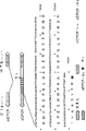
図3は、選択的スプライシングによってヒト、ウシ、マウス、およびヒヒのEPCRに挿入された配列を示す。
図4aは、可溶性血漿EPCRがヒトプロテインCおよびAPCに結合することを示すグラフである。EA.hy926細胞を、氷上で30分間0〜500nMrsEPCR(●)または血漿EPCR(○)の存在下で、60nM fl−APCと共にインキュベートした。細胞を洗浄し、そして細胞結合した蛍光を、記載のようにフローサイトメトリーによって決定した。fl−APCを添加しない場合での固有の細胞蛍光を、矢印で示す。プロットした平均細胞蛍光(MCF)は、2連のMCF測定の平均を表す。
図4bおよび4cは、可溶性血漿EPCRおよびrsEPCRが、細胞表面上でプロテインC活性化を阻害することを示すグラフである。図4bでは、EA.hy926細胞単層を、0.1μMプロテインC単独(□)、または1μM rsEPCR(●)、または2μg/ml 1494mAb(○)と共に、室温にて15分間プレインキュベートした。プロテインC活性化を、トロンビン(2nM最終)の添加によって開始し、そして反応を、所定の時間で停止した。活性化プロテインCを、アミド分解アッセイで決定し、そして活性速度をmOD/分で各時点についてプロットした。トロンビンを添加しないコントロールウェルを含ませた(■)。各データ点は、3連のウェル測定の平均を表す。図4cでは、EA.hy926細胞単層を、0.1μMのプロテインCおよび示された濃度の血漿EPCR(○)またはrsEPCR(●)と共に、室温にて15分間プレインキュベートした。トロンビン(最終2nM)を添加し、そして活性化を室温にて60分間行った。上清を、アンチトロンビンおよびヘパリンの混合物に添加し、そして活性化プロテインC活性(mOD/分)を、アミド分解アッセイで決定した。各データ点は、3連のウェル測定の平均を表す。
図4dは、可溶性血漿EPCRがAPC抗凝固活性を阻害することを示すグラフである。APC(25nM)の抗凝固活性を、460nMの血漿EPCRまたはrsEPCRの存在下で、1段階X a凝血アッセイを用いて決定した。この効果は、いずれかの可溶性EPCRを、EPCRへのAPCの結合をブロックする、42μg/mlの1496mAbと共に、5分間プレインキュベートした場合には逆転した。データは、4〜6測定の平均±S.D.を表す。
図5は、狼瘡患者において、可溶性血漿TMのレベルを可溶性血漿PCRと比較するグラフであり、TM値とEPCR値との間に相関はないが、狼瘡患者の大多数が、可溶性血漿EPCRの非常に上昇したレベルを示すことを証明する。
図6は、sTM(正常)、sTM(敗血症)、sEPCR(正常)、およびsEPCR(敗血症)についての可溶性レセプター濃度(ng/ml)のグラフである。sTMとsEPCRとの間に相関はない(r2=0.034)。
発明の詳細な説明
定義
内皮プロテインCレセプター、EPCR。
可溶性、溶液中にあり、そして細胞表面に結合していない。
短縮型、膜貫通ドメインおよび細胞質ドメインを含まない;タンパク質分解または選択的スプライシングのいずれかの結果であり得る。
可溶性EPCRの検出および特徴付け;生理学的役割およびマーカーとしての有用性
EPCRの機能のこれまでの研究は、膜形態のEPCRに結合するプロテインCが、細胞表面上のトロンビン−トロンボモジュリン複合体によりプロテインC活性化の促進を生じる(Stearns−Kurosawaら,1996)が、可溶性組換えEPCRはAPC抗凝固活性を阻害する(Reganら,1996)ことを見出した。これらの観察は、血漿中の可溶性トロンボモジュリン分解産物が、種々の疾患状態における内皮損傷のマーカーであるという知識と共に、可溶性循環形態のEPCRが存在するかどうか、そして存在するのであれば、プロテインC経路においてどういう役割を有するかという疑問に至った。
実施例は、可溶性形態のEPCRが血漿中で循環し、そして尿中に存在することを示す。健常ドナー集団において、血漿EPCRレベルは、約100ng/mlであり、そして約43,000ダルトンの単一抗原種であるようであった。血漿からの可溶性EPCRのその後の精製および機能的研究は、血漿EPCRが、インタクトな膜結合EPCRと同様な親和性で、プロテインCおよびAPCの両方と結合し得ることを決定した。内皮細胞株を使用するインビトロ研究は、血漿EPCRが、生理学的濃度に近い濃度のプロテインCおよびトロンビンで、プロテインCを阻害することを示した。さらに、血漿への精製血漿EPCRの直接添加は、rsEPCRに対するモノクローナル抗体で逆転されるAPC抗凝固活性の阻害を生じた。
この精製血漿タンパク質のEPCRとの同定は、rsEPCRの特性との比較に基づいた。これらのタンパク質は両方とも、同じ群のモノクローナル抗体およびポリクローナル抗体と反応し、同じアミノ末端配列を有し、Ca2+依存的様式で固定されたプロテインCに結合し、そして類似の用量応答曲線でプロテインC活性化およびAPC抗凝固活性をブロックした。さらに、rsEPCRおよび血漿EPCRに対するプロテインCおよびAPCの親和性は共に、インタクトな膜結合EPCRの親和性に類似する。これらの特性は、EPCRに特有であるようである。
これまでの研究は、内皮細胞で発現した膜結合EPCRが、3〜5倍の間のファクターでプロテインC活性化を増大することを示したが、実施例は、血漿から精製された可溶性形態のEPCRが、内皮細胞でのプロテインC活性化およびAPC抗凝固活性を阻害することを示す。これは、EPCRが、いくつかの方法で、プロテインC経路を調節し得ることを予測する。第1に、より大きな血管(ここでは、トロンボモジュリン濃度が微小循環に対して低い)において、EPCR発現は、応答して増加する(Laszikら、Circulation 1997)。免疫組織化学は、ほとんどの器官において、EPCR発現は、大血管で最も高密度であり、そして血管サイズが減少するにつれて次第に減少し、最も豊富な内皮細胞タイプである毛細管内皮ではほとんどまたは全く発現しない。EPCR発現は、循環からプロテインC基質を捕獲し、そして活性化のためにトロンビン−トロンボモジュリン複合体に提示することにおいて、重要な役割を果たし得る。このことは、EA.hy926内皮細胞株およびヒト臍静脈内皮細胞の両方ともが、トロンボモジュリンより、少なくとも6倍多い表面発現EPCR抗原を有するというインビトロ観察によって支持される。トロンボモジュリン濃度が高くそしてEPCRが低い微小循環では、プロテインC活性化にほとんど影響がないと予測される。最後に、循環可溶性EPCRは、APCの生成およびAPCが第V a因子を不活化する能力を減少させ得る。
健常個体において、可溶性EPCRレベルは、約2.5nMであり、この濃度は、Kdapp(約30nM)および循環における80nMのプロテインC濃度の両方よりも十分に低い。可溶性血漿EPCRの効果(APC抗凝固活性およびプロテインC活性化の阻害)の両方ともが、正常血漿に存在する濃度よりかなり高い濃度を必要とする。このことが、血漿EPCRの生理学的役割の疑問を、不確定のままにしている。40nMを越える可溶性EPCRレベルを有する患者が、実施例3(狼瘡)に記載のように同定された。したがって、内皮細胞表面に近い局所濃度が、全身濃度を超えるならば、可溶性EPCR濃度は、APCの生成および活性の両方を減弱するレベルに達し、血栓症の危険性に寄与する。
可溶性形態のレセプターは、膜結合レセプターのタンパク質分解的切断、または選択的スプライシングメカニズムによって産生され得る。膜表面でのタンパク質分解は、可溶性トロンボモジュリン、ならびにTNF、IL−1、IL−2、M−CSF、PDGF、およびNGFのレセプターを遊離する(Heaneyら,1996,Blood 87:847−857)。可溶性レセプターは、膜レセプターのアンタゴニストとして使用すること、リガンドを安定化すること、リガンドを媒介されたシグナリングを開始すること、膜形態のダウンモジュレーション、およびレセプターインヒビターへ結合してレセプター−リガンド活性を間接的に容易にすることを含む、多くの可能性のある機能を有する。後者のメカニズムは、両方のIL−1レセプターの可溶性イソ形態が、タンパク質分解的切断によって生成され、そしてIL−1αおよびIL−1βへの応答性を厳格に調節する、IL−1レセプター系によって使用される(Arendら,1994,J.Immunol.153:4766−4774)。以下に議論するように、膜貫通ドメインの直前で短縮された可溶性タンパク質をコードするEPCRゲノム構造は、選択的スプライシング部位を含む(FukudomeおよびEsmon,1995,J.Biol.Chem.270:5571−5577)。可溶性IL−6レセプターは、タンパク質分解メカニズムおよび選択的スプライシングメカニズムの両方によって生成されるようである(Mullbergら,1994,J.Immunol.152:4958−4968;Lustら,1992,Cytokine 4:96−100;Horiuchiら,1994,Eur.J.Immunol.24:1945−1948)。この切断部位はまた、MorrisseyおよびEsmonへの米国特許第5,298,599号(その教示は本明細書に援用される)に記載のように、短縮型EPCR、続いて直ぐペプチド配列(抗体はこの配列に対して特異的に指向する)をコードする発現ベクターを構築することによって、大量の可溶性EPCRを回収することにおいて有用であり得る。次いで、エピトープは、患者への投与の前または後に、タンパク質分解によって切断される。Hoppらへの米国特許第4,782,137号もまた参照。
免疫組織化学研究は、EPCRが、主として大血管の内皮に位置し、そして毛細血管ではかろうじて検出可能であることを示した。したがって、膜結合EPCRに由来する血漿EPCRは、大血管疾患プロセスのマーカーとして用い得る。血漿EPCRは、血漿トロンボモジュリンレベルとの有用な比較として用い得る。血漿トロンボモジュリンレベルは、種々の疾患状態において調整されることが示されているが、大血管および小血管の両方の疾患プロセスを反映し、ほとんどの内皮が微小血管性であるのでおそらく小血管の寄与に支配される。
EPCRのヌクレオチドおよび予測タンパク質構造分析
EPCRのcDNAは、238アミノ酸(配列番号2)のタンパク質をコードすると予測され、これは、N末端に15アミノ酸のシグナル配列を含む(vonHeijne,(1986)Nucleic Acids Res.14,4683−4690)。したがって、成熟タンパク質は、223アミノ酸を含むと予測される。組換えタンパク質の直接配列決定は、成熟タンパク質がSer18で始まることを示した。配列番号2は、EPCRの予測アミノ酸配列である。配列番号2のアミノ酸1〜15(MLTTLLPILLLSGWA)は、von Heijne(von Heijne,1986)の方法によって決定される推定シグナル配列である。配列番号2のアミノ酸211〜236(LVLGVLVGGFIIAGVAVGIFLCTGGR)は、推定膜貫通ドメインである。可能性のあるN−グリコシル化部位は、配列番号2のアミノ酸47〜49、64〜66、136〜138、および172〜174に存在する。細胞外システイン残基は、配列番号2のアミノ酸17(血漿EPCRでは除去されている)、114、118、および186に存在する。23アミノ酸からなる可能性のある膜貫通領域(Engelmanら,(1986)Annu.Rev.Biophys.Chem.15,321−53)を、C末端で同定した(配列番号2のアミノ酸211で始まる)。
このタンパク質はタイプ1膜貫通タンパク質である。細胞外ドメインは、4つの可能性のあるN−グリコシル化部位および3つのCys残基を含む。グリコシル化は、N−グリカナーゼ消化によって示されるように、活性に必須ではない。細胞質領域は、3アミノ酸のみを含み、そしてCysで終結し、パルミトイル化されている。末端システインが適切にパルミトイル化されてない場合、タンパク質は分泌され得る。EPCRの配列を変化させて、このシステインを別のアミノ酸と置換することにより、膜結合する代わりに分泌される、本質的に完全長のEPCRを作成する手段が提供される。
本明細書で使用される場合、レセプターをコードするヌクレオチド配列は、配列番号1に示される配列、およびストリンジェントな条件下で配列番号1にハイブリダイズする、その保存的置換、付加、または欠失を有する配列を含む。本明細書で使用される場合、レセプターを構成するアミノ酸配列は、配列番号2に示される配列、および機能的に等価な生物学的活性を有するレセプターを形成する、その保存的置換、付加、または欠失を有する配列を含む。何が、保存的置換、付加、または欠失を構成し、そしてどれが、本明細書に記載の機能的アッセイを使用して機能的に等価なレセプター分子をコードまたは形成すると容易に確認され得るかは、当業者に周知である。これは、さらに、図3を参照することによって例示され、以下に議論される。
選択的スプライシング
レセプターは、特異的リガンドに結合して生理学的応答を生じることを担う細胞の、外部に曝された部分を有する、細胞膜に繋がれたタンパク質として最も頻繁に思い描かれる。多くの場合、もはや細胞に制限されないという事実にもかかわらず、頻繁にそのリガンドを確かに結合し得る、可溶性形態のレセプターが存在する。可溶性レセプターイソ形態へのリガンド結合はまた、多くの形態をとる応答を生じ得、これには、膜結合レセプター相互作用のアップモジュレーションまたはダウンモジュレーション、あるいは正常には応答性ではない細胞へリガンドを輸送することによって応答を伝達させることを含む(Heaney,MLおよびDW Golde.Soluble cytokine receptors.Blood 87:847−857,1996)。
可溶性レセプターを産生するための2つの公知のメカニズムが存在する:膜結合レセプターをタンパク質分解して、細胞外ドメインを遊離し、そして膜アンカーを残すことによるもの、およびmRNAの選択的スプライシングによるもの(図1)。後者のメカニズムは多くの形態を取り得るが、リーディングフレームがエキソン−イントロン境界により連続し、そして膜貫通アンカーをコードする配列に達する前に停止コドンで終結する場合、最も単純である。これは、膜形態に類似するが、重要な差異を有するタンパク質を生成する。これは、可溶性タンパク質として、作製され、そして分泌され、そして特有のカルボキシル末端テイルを有する。このテイルを、膜結合レセプターの形成において無視されるイントロンmRNA配列の一部を読むことによって形成した。選択的スプライシングによる可溶性レセプターの生成はまた、共に同じmRNAテンプレートを起源にするという事実にもかかわらず、膜結合レセプターとは独立して調整され得る(Heaneyら,Proc.Natl.Acad.Sci.U.S.A.92:2365−2369,1995)。
可溶性レセプターが選択的スプライシングメカニズムによって生成されることを証明するためには、ゲノム配列および関連領域のイントロン−エキソン境界を知らなければならない。生理学的応答と可溶性レセプターを関連付けて、プロテインC遺伝子およびプロテインS遺伝子の発現の間にかなり頻繁に含まれることが見られる異常なmRNAスプライシングと区別することもまた役立つ(Bergら,Blood Coag Fibrinol.7:625−631,1996)。
以下の研究および結果の詳細は、実施例に記載される。ヒト血漿は、約100ng/mlの可溶性EPCRを含む(表1)。これを、2つのモノクローナル抗体(1494mAbおよび1495mAb)および標準的技法を使用する酵素結合イムノアッセイ(ELISA)によって測定した。著しく上昇した可溶性EPCRレベルを、全身性エリテマトーデスおよび敗血症の患者で見いだした。これらのレベルは、ほとんど例外なく、大血管の表面上のみに存在する膜結合レセプターについてかなり高いようであった。これを真相正しく認識するために、トロンボモジュリン(TM)は、すべての内皮、ならびにいくつかの非血管細胞上で発現されるが、正常可溶性TMレベルは、わずかに約10〜40ng/mlである(Takanoら,Blood 76:2024−2029,1990)。可溶性TMレベルは、狼瘡の患者で上昇していたが、敗血症では上昇していなかった。重要なことには、これらの患者群において、血漿EPCRとTMレベルとの間に相関がなかったことである(それぞれ、r2=0.028および0.034)。

血漿EPCRおよびTMレベルと高い血漿EPCR濃度との間の相関の欠如は、血漿EPCRがタンパク質分解および選択的スプライシングの両方のメカニズムを起源にするという概念に一致する。ヒトEPCRのゲノム構造は、イントロンによって分解される、4つのエキソンを含む。この配列を概観することにより、7527位にTAA停止コドンを含むエキソンIII−イントロンIII境界(5'GTで)の後のインフレームリーディング配列が明らかになる。この停止コドンは、膜貫通ドメインをコードするエキソンIVの上流にあるので、予測されるタンパク質は、特有な48残基のカルボキシル末端テイル(イントロン配列によってコードされる)を含み、そして膜貫通アンカーを含まない。
図1は、短縮型EPCRが誘導され得る2つの可能性のある方法の図である:膜貫通ドメインの直前でのタンパク質分解によるか、または選択的スプライシングによる。図2に示されるように、選択的スプライシングは、選択的にスプライスされた短縮型EPCR中にあるペプチド配列が含まれることを生じる。図3に示されるように、この配列は、種間で高度に保存されるが、わずかな差が存在し、ヒトおよびウシのEPCRについては48残基、マウスEPCRについては51残基、そしてヒヒEPCRについては22残基の新しいカルボキシル末端テイルを生じる。
レセプタータンパク質の発現についての患者サンプルのスクリーニング
患者サンプルは、EPCR、EPCR中の選択的にスプライスされたインサートに存在する特有のインサートのいずれかに対する抗体、またはコンホメーションの差に起因してEPCRまたはsEPCRのいずれかに対してより大きな親和性で結合する抗体を使用して、sEPCRまたはEPCRの存在および量について、スクリーニングされ得る。サンプルはまた、存在するタンパク質を特異的に定量するための他の標準的技法を使用して、スクリーニングされ得る。
診断的または治療的使用のための抗体の生成
EPCR、および特に、可溶性EPCR(「sEPCR」)、および組換え可溶性EPCR(「rsEPCR」)に対する抗体が生成され得、これらは、レセプタータンパク質の検出、特徴付け、または単離に有用であり、そしてほとんどの場合、リガンド結合の阻害によって、レセプタータンパク質活性を調節するために有用である。抗体は、免疫原として、ヒトまたは動物の精製または組換えレセプタータンパク質もしくはそのフラグメントを使用して、標準的技法によって生成される。
EPCRに対するモノクローナル抗体を、Esmonら,1993,Methods Enzymol.222:359−385によって他のタンパク質について記載されたように得た。1494、1495、および1496mAbと呼ばれる抗体は、組換え可溶性EPCRおよび細胞表面発現EPCRに結合するIgG1κ抗体である。1494および1496mAbは、EPCRへのプロテインCおよびAPCの結合をブロックし、そして細胞性EPCRがトロンビン−トロンボモジュリン複合体によるプロテインC活性化を容易にする能力を阻害する。1495mAbは、EPCRへのリガンド結合をブロックせず、細胞表面プロテインCを変化させず、そして1494または1486mAbのエピトープとは異なる結合エピトープを有する。抗体は、標準的技法、例えば、放射標識、酵素標識、フルオレセインのような蛍光標識、金粒子、染料、および抗体の検出のための他の手段を使用して標識され得る。例えば、抗体は、標準的な手順を使用して、ビオチンアミドカプロン酸N−ヒドロキシスクシンイミドエステルでビオチン化され得る。抗体は、イムノアッセイにおける使用のための固体支持体、例えば、AffiGel−10TM、ニトロセルロース、またはマイクロタイターウェル、あるいは溶液相イムノアッセイにおける使用のための固体支持体に固定され得る。
好ましい実施態様では、EPCRは、50マイクロリットルの、15mM Na2CO3、35mM NaHCO3(pH9.6)中4マイクログラム/mlの1495mAbで4℃にて一晩コーティングしたマイクロタイタープレート(MaxisorpTM、NUNC NS,Roskilde,Denmark)を使用して測定される。室温で、次いでプレートを20mM Tris−HCl、0.1M NaCl、0.05%Tween 20(pH7.5)(アッセイ緩衝液)で3回洗浄し、そして0.1%(wt/vol)ゼラチンを含むアッセイ緩衝液で少なくとも1時間ブロックする。次いで、ウェルを洗浄し、50マイクロリットルサンプルを3連のウェルに添加し、そしてプレートを1時間インキュベートする。ウェルを吸引し、アッセイ緩衝液で3回洗浄し、そして50マイクロリットル、2マイクログラム/mlのビオチン−1494mAbを添加する。プレートを1時間インキュベートし、3回洗浄し、そして50マイクロリットルの、0.25マイクログラム/mlのストレプトアビジン−アルカリホスファターゼ結合体(GIBCO BRL)を添加し、そしてさらに1時間インキュベートする。ウェルを5回洗浄し、そしてELISA増幅キット(GIBCO BRL)の基質および増幅試薬を、製造業者の指示に従って15分間隔で連続して添加する。発色を0.3M H2SO4で停止し、そして終点吸光度を、Vmaxマイクロプレートリーダーにて490mmで読み取る。3連のウェルでの標準は、20mM Tris−HCl、0.1M NaCl、および1mM EDTA、0.1%ゼラチン(pH7.5)中の1.5〜100ng rsEPCR/mlである。標準曲線は、1.5〜12.5ng/mlで直線であり、そしてサンプルを、直線範囲に入るように同じ緩衝液で希釈する。研究は、1〜2パーセントの間で、血漿が、アッセイの直線性にも標準曲線の感度にも影響を及ぼさないことを示す。健常ボランティアからの血漿サンプルを、1mM EDTAを含むアッセイ緩衝液で最終的に2%血漿まで希釈した。そしてEPCR抗原レベルを、同じプレート上で決定された標準曲線を参照して、3連ウェルの平均から算出する。
障害
可溶性EPCRについてのアッセイは、本明細書で議論されるような凝固および炎症状態および障害、例えば、狼瘡のような自己免疫疾患、移植モニタリング、敗血症、ショック、子癇前症、糖尿病、心肺バイパス、不安定性アンギナ、再狭窄、血管形成(すなわち、血管疾患)、腎臓または肝臓疾患の検出および分析に有用である。例えば、EPCRは、大血管についてのマーカー、したがって大血管に対する損傷についてのマーカーである。可溶性EPCRの量の増加は、大血管傷害を示し、EPCRのタンパク質分解またはsEPCR合成の刺激のいずれかを生じる。EPCR対トロンボモジュリンの比はまた、血液または尿のいずれかのサンプルに基づいて決定され得、これは微小血管対血管の相対的な程度を示す。サイトカイン、白血球活性化マーカー、および補体因子または活性化マーカーに対するEPCRの相対量もまた、疾患状態を示すために使用され得る。
EPCRは内皮細胞上に存在するので、内皮細胞損傷のマーカーとして有用である。これは、薬物の効果(毒性および効力の両方)の指標として使用され得る。例えば、狼瘡患者では、炎症性/凝固媒介性大血管傷害を効果的に最小にする薬物は、EPCRレベルの減少を生じる。
本発明は、以下の非限定的な実施例を参照することによってさらに理解される。
実施例1:ヒト血漿における機能的内皮プロテインCレセプターの同定
以下の略号を使用する:rsEPCR、膜貫通ドメインおよびサイトゾルテイルの代わりに挿入されたHPC4エピトープを有する組換え可溶性EPCR;mAb、モノクローナル抗体;SDS−PAGE、ドデシル硫酸ナトリウムポリアクリルアミドゲル電気泳動。
方法
材料。以下の試薬を示された供給業者から購入した:
ブタ腸粘膜ヘパリン、ジイソプロピルフルオロリン酸、ビオチンアミドカプロン酸N−ヒドロキシスクシンイミドエステル、ウシ血清アルブミン、Sigma(St.Lois,MO);Spectrozyme PCa、American Diagnostica(Greenwich,CT);ELISA増幅キット、GibcoBRL(Gaithersburg,MD);AffiGel−10、BioRad(Hercules,CA);ハンクス平衡化塩溶液、3−(N−モルホリン)プロパン硫酸(MOPS)、Fisher Scientific(Fair Lawn,NJ)。すべての他の試薬は、市販の最高品質なものであった。
タンパク質。ヒトプロテインC(Esmonら,1993,Methods Enzymol.222:359−385)、ウシトロンビン(Owenら,1974,J.BIol.Chem.249:594−605)、およびウシアンチトロンビン(Esmon 1977「Factors regulating the inhibition of thrombin by antithrombin III.In Chemistry and Biology of Thrombin」R.L.Lundblad,J.W.Fenton,II,およびK.G.Mann編Ann Arbor Science,Ann Arbor.403−411)を、記載のように精製した。組換え可溶性EPCR、rsEPCRは、膜貫通ドメインの直前の残基210で短縮されたEPCRの細胞外ドメイン、続くHPC4モノクローナル抗体におけるカルシウム依存的イムノアフィニティー精製を可能にする12残基配列からなる(Takahashiら,1992;Stearnsら,1988,J.Biol.Chem.263:826−832)。rsEPCRの構築、精製、およびプロテインC/APC結合特性(Fukudomeら,1996)。ヤギ免疫前血清およびrsEPCRに対するヤギポリクローナル抗血清を調製し、そしてIgGを精製した(Fukudomeら,1996)。ヤギ抗rsEPCRポリクローナル抗体を、標準的手順を使用して、ビオチンアミドカプロン酸N−ヒドロキシスクシンイミドエステルでビオチン化した。
モノクローナル抗体。rsEPCRに対するモノクローナル抗体(mAb)を、他のタンパク質について記載されたように得た(Esmonら,1993)。1494、1495、および1496mAbは、rsEPCRおよび細胞表面発現EPCRに結合するIgG1κ抗体である。1494および1496mAbは、EPCRへのプロテインCおよびAPCの結合をブロックし、そして細胞性EPCRがトロンビン−トロンボモジュリン複合体によるプロテインC活性化を容易にする能力を阻害する(Stearns−Kurosawaら,1996)。1495mAbは、EPCRへのリガンド結合をブロックせず、細胞表面プロテインC活性化を変化させず、そして1494または1496mAbのエピトープとは異なる結合エピトープを有する。1494および1495mAbを、標準的手順を使用して、ビオチンアミドカプロン酸N−ヒドロキシスクシンイミドエステルでビオチン化した。1494mAbを、血漿EPCRのイムノアフィニティー精製のために、製造業者の指示に従って、AffiGel−10にカップリングした。抗EPCR mAbのスクリーニングを、Stearns−Kurosawaら(1996);Fukudomeら(1996)に記載の方法を使用して行った。
凝血アッセイ。1段階第X a因子凝血アッセイにおけるAPC(25nM)抗凝固活性に対するrsEPCRまたは精製血漿EPCRの効果を、83μg/mlの1496mAb(APC−EPCR相互作用をブロックする抗体)の存在または非存在下で行った(Reganら,1996)。可溶性EPCRおよび1496mAbを、アッセイ前15分間プレインキュベートした。
細胞培養。すべてのヒト細胞株を、以前に記載されたように維持した(Fukudomeら,1996)。EA.hy926細胞(形質転換されたヒト内皮細胞株;Edgellら,1983,Proc.Natl.Acad.Sci.(USA)80:3734−3737)は、Cora−Jean Edgell(University of North Carolina at Chapel Hill)が好意により提供した。
フローサイトメトリー分析。蛍光プローブとして用いるために、APCを、記載されるように(FukudomeおよびEsmon、1994;Bock,P.E.1988.Biochemistry 27:6633−6639)、フルオレセインで活性部位(fl−APC)において標識した。EA.hy926細胞へのAPC結合に対するrsEPCRまたは血漿EPCRの効果を、フローサイトメトリーによって研究した(Fukudomeら,1996)。手短には、収集された細胞を、漸増する濃度のいずれかの可溶性EPCR調製物の非存在下または存在下において、60nMのfl−APCとともに、氷上で30分間インキュベートし、洗浄し、そして細胞に結合した蛍光を、フローサイトメトリーによって1サンプルごとに10,000事象を計数して測定した。全てのアッセイを、1%のウシ血清アルブミン、3mM CaCl2、0.6mM MgCl2、および0.02%アジ化ナトリウムを補充したハンクス平衡化塩溶液において行った。
細胞表面プロテインC活性化。EA.hy926細胞を96ウェル組織培養ディッシュにおいて培養した(Stearns−Kurosawaら、1996)。コンフルエントな単層を、1%(w/v)のウシ血清アルブミン、3mM CaCl2、0.6mM MgCl2、および0.02%アジ化ナトリウムを補充したハンクス平衡化塩溶液を用いて、3回洗浄した。全てのアッセイを、室温において、同一の緩衝液中で、60μlの最終容量にて行った。そして、全てのタンパク質濃度は、アッセイにおける最終濃度を示す。プロテインC(0.1μM)を、示された濃度のrsEPCR、血漿EPCR、または1494mAbの非存在下または存在下で添加し、細胞とともに15分間プレインキュベートした。トロンビンを混合液に添加して(2nM)、活性化反応を開始した。示された時間に、50μlのアリコートを取り出し、そして96ウェルマイクロタイタープレート中の10μlのアンチトロンビン(0.7μM最終)およびヘパリン(5U/ml最終)に添加した。APCアミド分解活性をSpectrozyme PCa基質(0.2mM)の添加によって測定し、405nmでの吸光度の変化速度(mOD/分)をVmaxキネティックマイクロプレートリーダー(Molecular Devices,Menlo Park,CA)で測定した。すべてのアッセイ点は、3連ウェルにおいて行われ、そして十分に活性化されたプロテインC対mOD/分の標準曲線を参照することにより決定される場合、プロテインC基質の10%未満が活性化された。
血漿および血清の収集。健常な成人ボランティア(女性12人および男性10人)から、3.8%緩衝化クエン酸溶液中かまたは抗凝固剤を含まないチューブ中に、静脈穿刺によって全血を収集した(Vacutainer tube;Becton Dickinson,Franklin Lakes,NJ)。年齢、食餌、または他の変数に関するドナーのスクリーニングは試みなかった。全てのボランティアに研究についての情報を開示し、彼らから書面による同意を得た。血液を、1160×gで10分間遠心分離した。血漿および血清を等分し、アッセイまで−80℃で冷凍保存した。
血漿EPCRの定量のためのELISA。血漿におけるEPCR抗原の検出のための酵素結合免疫吸着アッセイを開発した。マイクロタイタープレート(Maxisorp;Nunc,Roskilde,Denmark)を、50μlの、15mM Na2CO3、35mM NaHCO3(pH9.6)中4μg/mlの1495mAbを用いて、4℃にて一晩コートした。以下の工程を室温で行った。ウェルを、20mM Tris−HCl、0.1M NaCl、0.05%Tween20(pH7.5)(アッセイ緩衝液)で3回洗浄し、0.1%(w/v)ゼラチンを含有するアッセイ緩衝液で少なくとも1時間ブロックした。ウェルを洗浄し、50μlのサンプルを3連のウェルに添加し、そしてプレートを1時間インキュベートした。ウェルを吸引して、アッセイ緩衝液で3回洗浄し、50μlの2μg/mlビオチン−1494mAbを添加した。プレートを1時間インキュベートし、3回洗浄し、50μlの0.25μg/mlストレプトアビジン−アルカリホスファターゼ結合体(GibcoBRL)を添加し、さらに1時間インキュベートした。ウェルを5回洗浄し、ELISA増幅キット(GibcoBRL)の基質および増幅試薬を、製造業者の指示に従い、15分の間隔で連続的に添加した。発色を0.3M H2SO4を用いて停止し、490nmの終点吸光度をVmaxマイクロプレートリーダーで読み取った。各プレートは、20mM Tris−HCl、0.1M NaCl、1mM EDTA、0.1%ゼラチン(pH7.5)中1.5〜100ng/mlのrsEPCRの標準を3連のウェルに含んだ。標準曲線は、1.5〜12.5ng/mlで直線であり(r=0.99)、そして血漿サンプルを直線範囲に入るように同じ緩衝液で希釈した。予備的な実験により、最終濃度1〜2%のヒト血漿は、標準曲線の直線性にも感度にも影響しないことを実証した。健常ボランティア由来の血漿サンプルを、1mMEDTAを含有するアッセイ緩衝液で、最終的に2%血漿に希釈し、そしてEPCR抗原レベルを、同一プレート上で決定された標準曲線を参照して、3連ウェルの平均から算出した。
コーティング抗体と検出抗体を逆にし(1494mAbをコーティングに;ビオチン−1495mAbを検出に)、そして抗体結合を、Blue Phos基質(KPL Laboratories;Gaithersburg,MD)で検出する、別のアッセイを開発した。この方法を使用して、敗血症患者における血漿EPCRをアッセイした。このアッセイは、おそらく親和性の差異のために、より高感度であったが、両アッセイとも、定性的には、同様の結果を与えた。
ウエスタンブロット。血漿または血清サンプルのドデシル硫酸ナトリウムポリアクリルアミドゲル電気泳動(SDS−PAGE)を、10%アクリルアミドゲルを、Laemmli緩衝液系(Nature 227:680−685)と共に用いて、非還元条件下で標準的な手順を用いて行った。ゲルをポリビニリデン膜(PVDF;Millipore,Bedford,MA)に転写し、膜をブロックし、次に免疫前ヤギIgG(50μg/ml)か、またはヤギ抗rsEPCRポリクローナルIgG(50μg/ml)のいずれかと30分間インキュベートした。洗浄後、膜を1:20,000希釈のマウス抗ヤギIgG−西洋わさびペルオキシダーゼ結合体(Pierce,Rockford,IL)と30分間インキュベートした。膜を洗浄し、結合した抗体−酵素結合体を増強化学発光基質(Pierce)で製造業者の指示に従って検出した。
免疫吸着。健常ボランティアからの血清またはクエン酸処理血漿サンプル(400μl)を、AffiGel−10に結合させた50μlの1495mAb(5mg IgG/ml樹脂)とともに、混合しながら4℃にて一晩インキュベートした。サンプルを遠心分離し、上清を除去し、そして樹脂を1mlの20mM Tris−HCl、0.1M NaCl、0.02%アジ化ナトリウム(pH7.5)で3回洗浄した。最終20mM ジチオスレイトールを含有するSDS−PAGEサンプル緩衝液を、洗浄した樹脂に添加し、サンプルを3分間沸騰し、そしてSDS−PAGEおよびウエスタンブロッティングのために処理した。膜を4μg/mlのビオチン化ヤギ抗rsEPCRポリクローナル抗体でプローブし、そして結合抗体をストレプトアビジン−西洋わさびペルオキシダーゼ結合体(Pierce)および増強化学発光検出系で検出した。予備的実験により、室温にて1〜4時間の、100μlのTris不活化AffiGel−10樹脂でのサンプルの事前吸着、続く1495mAb−AffiGel−10での一晩の免疫吸着が、同一のウエスタンブロットの結果を与えることが決定された。
血漿EPCRの精製。血漿EPCRを、イオン交換クロマトグラフィー、抗rsEPCR mAbイムノアフィニティークロマトグラフィー、およびプロテインC親和性カラムでのクロマトグラフィーの組み合わせを用いて、ヒトクエン酸処理血漿(Oklahoma Blood Institute)から精製した。わずかに異なる方法で2つの調製を行った。
第1の調製において、血漿(1L)を等量の20mM Tris−HCl(pH7.5)、10mMベンズアミド、400単位のヘパリンナトリウムで希釈し、そして1gの膨潤前のQAE樹脂でバッチ吸着した。沈澱後、樹脂をプロテインCの精製のために処理した(Esmonら、1993)。固体硫酸アンモニウムを上清に4℃にて40%飽和まで添加し、遠心分離し、そしてさらなる硫酸アンモニウムをその上清に70%飽和に達するまで添加した。遠心分離後、柔らかなペレットを透析バッグに入れ、12Lの20mM Tris−HCl、0.02%アジ化ナトリウム(pH7.4)に対し一晩透析した。透析物を、20mM Tris−HCl、0.1M NaCl、0.02%アジ化ナトリウム(pH7.4)で平衡化した。1496mAb−AffiGel−10イムノアフィニティーカラム(6ml樹脂;5mg IgG/ml樹脂)にかけた。カラムを、12mlを超える同じ緩衝液で洗浄し、20mM Tris−HCl(pH7.4)中の50%(v/v)エチレングリコールで溶出した(Jun Xu、未発表の観察)。溶出物からのピーク画分をプールし(0.37の全OD280)、濃縮し(Centriprep30、Millipore)、そして緩衝液を20mM Tris−HCl、0.1M NaCl、3mM CaCl2、0.6mMMgCl2、0.02%アジ化ナトリウム(pH7.4)に交換した。この物質をプロテインCアフィニティーカラムにかけた。このカラムは、同じ緩衝液においてHPC4−AffiGel−10カラム(5mg IgG/ml樹脂;0.9×8cm)に精製プロテインC(3mg)をかけることにより事前に調製した。HPC4mAbは、カルシウム依存的様式でプロテインC活性化領域に結合し(Esmonら、1993;Stearnsら、1988)、その後のプロテインCへのEPCRの結合を妨げない。血漿EPCRを含有するサンプルをカラムにかけた後、カラムを約12mlの緩衝液で洗浄し、20mM Tris−HCl、0.1M NaCl、5mM EDTA、10mM MOPS、0.02%アジ化ナトリウム(pH7.5)で溶出した。画分を、280nmでの吸光度について、そして上記のELISAを用いてEPCR抗原についてモニターした。プロテインCおよび血漿EPCRの両方を含有する溶出物を、FPLC(Pharmacia−LKB、Uppsala、Sweden)MonoQカラムにかけ、カラムを20mM Tris−HCl(pH7.5)中の0.1〜1M NaClの直線勾配で展開した。約半分の血漿EPCRは、MonoQカラムに結合せず、半分は、約0.2M NaClに溶出し、そしてプロテインCは、約0.5M NaClに溶出した。血漿EPCRの両方のイオン種は、還元条件下または非還元条件下での銀染色、クマシーBB染色、またはPVDF膜への転写後の金染色(Pierce)を用いるSDS−PAGE、およびビオチン−ポリクローナル抗rsEPCR抗体プローブでのウエスタンブロットで、同一のようであった。
血漿EPCRの第2の調製は、4Lの血漿を用いて開始し、機能研究に十分なタンパク質を精製した。この場合、1496−AffiGel−10樹脂(20mlの5mg IgG/ml樹脂)を、10mMベンズアミジン、1mMジイソプロピルフルオロホスフェートおよび0.5単位/mlのヘパリンナトリウム(最終濃度)と共に、クエン酸処理血漿に、直接添加した。血漿を、穏やかに混合しながら、4℃にて一晩バッチ吸着した。樹脂の沈澱後、上清をプロテインC精製のために処理した(Esmonら、1993)。樹脂を2.5×30cmのカラムに充填し、20mM Tris−HCl、0.1M NaCl、0.02%アジ化ナトリウム(pH7.4)で、十分に洗浄し、そして20mM Tris−HCl(pH7.4)中50%のエチレングリコールで溶出した。溶出物をプールし、濃縮し(5.5の全OD280)、MonoQカラムにかけ、2つのイオン種(素通り、および0.2M NaCl溶出ピーク)を、1496−AffiGel−10樹脂(1.5×11cm)に再びかけた。カラムを前のとおりに、50%エチレングリコールで溶出した。溶出物(0.71 OD)を濃縮し、緩衝液を、Cetriprep30を含む。20mM Tris−HCl、0.1MNaCl、3mM CaCl2、0.6mM MgCl2、0.02%アジ化ナトリウムに交換した。次に、この物質をアフィニティーカラムにかけた。ここで、プロテインC(2.9mg)を、同じ緩衝液中で、HPC2−AffiGel−10カラム(0.6×17cm)に最初にかけた。HPC2mAbは、プロテインCのセリンプロテアーゼドメインに結合し、EPCR結合を妨げない(Fukudomeら、1996)。結合したEPCRを、5mM EDTAを含有する緩衝液で溶出した。混入している血清アミロイドP(プロテインCサンプル由来)を、FPLC MonoQカラムでのイオン交換クロマトグラフィーによって除去した。サンプルを0.2M NaCl中でカラムにかけ、その結果、血漿EPCRは結合せず、そして0.4〜0.5M NaClにおいて溶出する混入物から分離された。得られる精製血漿EPCR(0.193 OD280)は、銀染色を用いるSDS−PAGE、およびポリクローナル抗体rsEPCRを用いるウエスタンブロットにより、均質のようであった。この物質を機能研究およびアミノ末端配列分析に使用した。
タンパク質配列決定。可溶性血漿EPCRのアミノ末端配列分析を、Molecular Biology Research Facility、W.K.Warren Medical Research Institute(Oklahoma City)のKenneth Jackson博士の研究室において行った。アミノ酸を標準の1文字コードによって表す。
結果
第1のアプローチとして、3人の健常ボランティア由来の血漿および血清サンプルを希釈し(4%v/v)、非還元条件下で10%SDS−PAGEゲルで泳動し、そしてrsEPCRに対して惹起したヤギポリクローナル抗体を用いるウエスタンブロットのために処理した。健常ボランティア由来の血漿および血清サンプル(4%v/v)を非還元条件下の10%ゲルでのSDS−PAGEのために処理し、膜に転写し、そして膜をヤギ抗rsEPCRポリクローナル抗体でプローブした。結果をrsEPCR(0.2ng)と比較した。結合した抗体を、マウス抗ヤギIgGおよび増強化学発光検出系で検出した。2人の健常ボランティア由来の血漿サンプルを1495 AffiGel−10樹脂で免疫吸着した。洗浄した樹脂を溶出させ、そして還元条件下でのSDS−PAGEのために処理した。ウエスタンブロッティングを、ビオチン−ヤギ抗rsEPCRをプローブとして使用して行った。
血漿EPCRの純度を、銀染色したSDS−PAGE 10%ゲル、およびビオチン−ヤギ抗rsEPCRでプローブした膜のウエスタンブロットから決定した(還元および非還元)。膜をポリクローナル抗体でプローブした後、血清および血漿サンプルの両方で、約43,000Daの単一のバンドが現れる。検出されたタンパク質のサイズは、rsEPCRよりわずかに大きいようである。検出された他のバンドは、免疫前IgGおよびより長い暴露時間でのプロービングより判断すると、IgGのバックグラウンド結合であった。AffiGel−10樹脂と結合した抗EPCR 1495mAbと共に血漿サンプルを一晩インキュベートすること、続く洗浄および還元条件下での結合抗原の溶出は、ビオチン−ヤギ抗rsEPCRポリクローナル抗体でのウエスタンブロットにより検出される単一バンドを生じた。
mAb 1495をコーティング抗体として使用するELISAによる、健常ボランティア由来の血漿における可溶性EPCR抗原の測定は、女性(n=12)では91.1+/−24.5ng/mlの抗原レベル、そして男性(n=10)では107.2+/−30.2ng/mlの抗原レベルを見出した。ともに計算する場合、平均血漿EPCR抗原レベルは、98.4+/−27.8ng/mlであった。男性の値は、トロンボモジュリンと同様に(Quehenbergerら、Thromb.Haemost.76:792−734)、女性の値よりわずかに高いようであるが、研究した集団は、統計学的分析にはあまりに限定されており、そしてこの研究は、性別、年齢、食餌または他の変数に起因する差異を評価するためには設計されていなかった。
血漿EPCRは、約100ng/mlの単一種であるようであるので、血漿EPCRは、循環EPCRが、プロテインCおよびAPCに結合し得るか否かを決定するために重要になった。可溶性EPCRを、イオン交換クロマトグラフィー、硫酸アンモニウムでの沈降、および実験手順において記載されるような抗EPCR 1496mAb−AffiGel−10カラムクロマトグラフィーによる免疫吸着の組み合わせによって、ヒト血漿から精製した。
3mM CaCl2、0.6mM MgCl2を含む緩衝液中で抗プロテインCHPC4mAb−AffiGel−10カラムにプロテインC(3mg)を適用することによって調製したプロテインCアフィニティーカラムに、この血漿EPCR(約110μg)を適用した。カラムを洗浄し、そして血漿EPCRを画分19に適用した。カラムを洗浄し、そして画分35で開始する5mM EDTAを含む緩衝液で溶出した。それらの画分について、280nmでの吸光度およびEPCR抗原を決定した。EPCR抗原を、ELISAによって決定した。
適用した血漿EPCR抗原の98%以上が、プロテインCアフィニティーカラムに結合した。吸光度プロフィールは、この抗体へのプロテインC結合のカルシウム依存性と一致して、抗体カラムからのEPCRおよびプロテインCの同時溶出を示す(Stearnsら、1988)。
機能研究および構造研究のために十分なタンパク質を精製するために、類似しているがわずかに改変した手順を用いて、4Lの血漿からEPCRを精製した。プロテインC−抗体アフィニティーカラムからの溶出後、タンパク質を夾雑する残渣を、FPLC Mono Qカラムでのイオン交換クロマトグラフィーによって取り出した。得られた血漿EPCRの調製物は、銀染色を用いるSDS−PAGE 10%ゲルで均質のようであり、そして同一の結果が、ビオチン−ヤギ抗rsEPCRポリクローナル抗体でプローブしたウェスタンブロットに関して、還元条件下および非還元条件下の両方で得られた。精製されたタンパク質のアミノ末端配列分析は、唯一の配列S−Q−D−A−S−D(この配列は、組換え可溶性EPCRのアミノ末端配列に同一である:配列番号2)を生じた。これは、天然の供給源からのEPCRの最初のアミノ末端配列決定である。
血漿EPCRの、APCに結合する能力を、競合研究によって評価した。この競合研究において、血漿EPCRを、APCについて細胞性EPCRと競合させ、そして細胞性EPCRに結合し得る得られた遊離のAPCを、フローサイトメトリーによって評価した(図4a)。活性部位においてフルオレセインで標識したAPC(fl−APC)を、血漿EPCRまたはrsEPCRのいずれかの、存在下または非存在下で、EA.hy926細胞とともにインキュベートした。細胞へのAPC結合の阻害についてのEPCR濃度依存性は、EPCRの両方の可溶性形態について類似であった。この観察は、APCに結合することについての血漿EPCRの親和力が、rsEPCR−APC結合相互作用について以前に決定された親和力(Kdapp約30nM)に類似することを示す。
rsEPCRは、可溶性系においてプロテインC活性化に対してほとんど効果を有さないが(Reganら、1996)、膜結合EPCRは、細胞表面上での活性化を促進する、非常に強力な能力を有する(Stearns−Kurosawaら、1996)。プロテインCおよびAPCに結合し得るEPCRの循環形態の存在を示す最新のデータは、血漿EPCRが、プロテインCの細胞表面活性化を変更する能力を有することを示唆した。EA.hy926細胞上でのほぼ生理学的レベルのプロテインC(0.1μM)のトロンビン依存性活性化は、過剰なrsEPCRによって、図4bによって示されるように、ほぼ、EPCR−プロテインC結合相互作用をブロックする抗rsEPCR 1494mAbを用いて観察されたプロテインCのレベルにまで阻害された。以前の研究は、rsEPCRが小さな合成基質を使用するAPCアミド分解活性(amidolyticactivity)に対する効果を有さないことを実証した(Reganら、1996)。血漿EPCRは、図4cに示されるように、rsEPCRと比較してEA.hy926細胞上の細胞表面プロテインC活性化を阻害する能力においてわずかにより有効であった。
1段X a因子凝固アッセイにおいて、精製された血漿および可溶性組換えEPCRは、同様に凝固時間のAPC延長を阻害した(図4d)。rsEPCRによるAPC抗凝固活性の阻害が、以前に観察されている(Reganら、1996)。予測されるように、1496mAbは、APC−血漿EPCR結合相互作用をブロックすることによって、この効果を逆転した。
実施例2:尿における可溶性EPCRの検出
可溶性EPCRが尿中に存在するかどうかという疑問を検討するために、4つの尿サンプル(朝一番の排尿)を回収し、そしてウェスタンブロッティングおよびELISAによって可溶性EPCRの存在について分析した。
希釈していない小児の尿サンプルを、4%の正常な血漿および組換え可溶性EPCR(1ng)と比較した。サンプルを、ビオチン−ヤギ−抗rsEPCRおよびストレプトアビジン−アルカリホスファターゼ検出系とともにインキュベートした。
ウェスタンブロットは、a)可溶性EPCRが尿中に存在すること、およびb)可溶性EPCR抗原が、血漿中で観察されるサイズと類似のサイズで存在することを示す。明らかな分解は観察されない。ELISAによって定量される、4つのサンプル中の可溶性EPCRの量は、40.3、6.1、35.6、および90.1ng/mlであった。
実施例3:狼瘡患者由来の血漿EPCRの測定
正常なヒト血漿EPCR濃度は、上記のように約100ng/ml(98.4±27.8ng/ml;2.5nM)である。エリテマトーデスに罹患した患者由来のサンプルのパネル(n=54)をアッセイし、そして可溶性EPCRレベルが、検出不能レベルから1,700ng/ml以上までの範囲であることを見出した。15人の患者は、200ng/ml以上の可溶性EPCRレベルを有した。
以前の研究は、内皮損傷に起因する、狼瘡患者における上昇した可溶性血漿TMレベルを示し、そして最新の狼瘡患者サンプルを、参照として血漿TMについてアッセイした。それらの可溶性TMレベルが、図5によって示されるように、それらの可溶性EPCRレベルと完全に相関を有さないことが見出された。このことは、可溶性血漿EPCRの供給源が、単に、ランダムに損傷を受けた内皮由来ではないことを示唆する重要な観察である。TMとは対照的に、ヒトおよび霊長類における膜結合EPCR発現は大きな脈管の内皮に主に制限され、毛細管はほとんどEPCRを発現しない。EPCRの特有の局在化は、大きな脈管の血栓症を予防するために、局所的にプロテインC活性化を増大すると予測される。大きな脈管への膜結合EPCRの主な局在化は、可溶性血漿EPCR濃度によって予測され得る大きな脈管における標的化された血栓症の危険を示す。
実施例4:敗血症性ショック患者における血漿可溶性EPCR
敗血症(accp/sccm consensus conference,chest 1992;101:1644−1655)は、以下の臨床的発現のうちの1つより多くを含むが、それらに限定されない。感染に対する全身性炎症応答として定義される:
1)38℃以上または36℃未満の体温;
2)1分あたり90拍以上の心拍数;
3)以下によって示される頻呼吸:
a)1分あたり20呼吸以上の呼吸率;
b)32mmHg未満のPaCO2の過度呼吸;
4)12,000/mm3以上もしくは4,000/mm3未満のWBCカウント、または10%以上の未熟好中球(バンド)の存在。
サンプルを、器官機能不全、低灌流(hypoperfusion)、または低血圧と関連する敗血症によって規定されるような、重篤な敗血症を伴うか、または伴わない、手術後の合併症を有する患者から得た。灌流異常は、乳酸アシドーシス、尿量過少、または精神状態における急性変化を含み得る。敗血症性ショックは、十分な流体(fluid)蘇生、および心臓性ショックの非存在にもかかわらず、24時間以上の間、血管作用薬を必要とする低血圧を伴う敗血症をいう。
本研究において含まれるすべての患者は、以下の診断基準を満たした:
a)呼吸の指示(24時間以上の制御された換気)およびまたは血流力学的支持(5マイクログラム/Kg/分以上の強心薬、ドパミン、もしくはドブタミン、および/または血管作用性アミン、エピネフリン、もしくはノルエピネフリンの要求)を必要とする、敗血症および/または手術後の合併症に起因する、集中治療室への入室;
b)18歳〜75歳の間の年齢;
c)70%未満の抗トロンビン活性(局所にて試験される)。
患者が、多くの外傷、肝硬変、または急性肝不全、末期ガン、免疫不全、白血病、妊娠、またはヘパリン治療を有する場合、その患者を除外した。
患者の血液サンプルを、時間0(集中治療室ICUへの入室)の時点および抗トロンビンII(AT III)または偽薬での処置後2日目および6日目に採取した。血漿可溶性EPCRおよび可溶性トロンボモジュリン(TM)を、時間0のサンプルに関してのみアッセイした。
sEPCR:
正常:133.4±53.4ng/ml(平均±標準偏差)
敗血症:224.9±74.5ng/ml
平均間の有意な差あり、P=0.00009
sTM:
正常:35.5±20.4ng/ml(平均±標準偏差)
敗血症:39.9±73.1ng/ml
平均間の有意な差なし、P=0.81
血漿中のsEPCRレベルとsTMレベルとの相関関係なし、r2=0.34。
これらの結果を、図6にグラフで示す。狼瘡患者と同様に、敗血症に罹患した患者は、可溶性TMレベルと相関せず、血漿EPCRレベルにおいて非常に顕著な上昇を示す。
可溶性血漿EPCRが、プロテインC活性化および活性化したプロテインC抗凝固活性の両方を阻害するという観察によって、これらの患者における上昇した血漿EPCRレベルが、さらなる血栓の危険性を引き起こし、そして脈管損傷/応答の証拠を表すことが示される。これらが示す状態の例は、内皮細胞刺激、アテローム発生、白血球接着、およびプラークの破裂に関連する障害を含む。
実施例5:ヒヒ組織およびヒト組織におけるEPCRの選択的スプライシング形態の同定
可溶性EPCRイソ型が、選択的スプライシング機構によって生成され得るか否かを決定するための最初のアプローチとして、RNAを、ヒト組織およびヒヒ組織から単離し、そして逆転写酵素−PCR(RT−PCR)を遺伝子特異的プライマーを用いて実施した。ヒヒEPCRゲノム配列は公知ではないが、ヒヒとヒトとは進化のスケールにおいて密接に関連しているという論理に基づいて、ヒト配列に基づくプライマーを使用した。
一般にRT−PCR手順において、総RNAをホモジナイズした組織から単離する。RNAを、特定のアンチセンスプライマー、ヌクレオチド、および逆転写酵素と混合する。混合において、RNAは、逆転写酵素についての鋳型として作用し、第1鎖cDNAを作製する。次いで、この新しいcDNA鋳型を、特定のプライマーおよびTaqポリメラーゼを用いて従来のPCRによって増幅する。EPCRの膜形態(424bp)および推定の選択的スプライシング産物(674bp)の両方を増幅するプライマーを選択した。EPCRの両方の形態に対応する産物を、種々のヒヒ組織(図4)ならびにヒトの肺および胎盤から増幅した。逆転写酵素を含まないコントロール、および組織との反応における1,885bpのバンドの欠失によって判断されるように、ゲノムDNAに関する可能性のある夾雑は存在しないようであった。
ヒヒゲノムDNA配列が、選択的スプライシングのための適切なエキソン−イントロン境界およびイントロン−インフレームルーディング配列を有することを確認するために、ヒヒ腎臓ゲノムDNA由来のイントロン配列を、従来のPCRによって増幅した。ゲノム構造は種間で保持されるという前提を立て、そしてイントロンIIIに隣接するプライマー(ヒト配列)を使用した。これは、選択的スプライシング配列を含むと考えられるイントロン(ヒト配列)である。ヒヒ腎臓組織をホモジナイズし、そしてDNAを抽出した。DNAを、特定のプライマーと混合し、そして産物をPCRによって増幅した。DNA産物を精製し、そしてアガロースゲルで電気泳動した。
手順の詳細:
A.EPCR ELISA:被覆する抗体は、EPCRのリガンド結合ドメインに結合する1494mAbである。検出する抗体は、ビオチン化1495mAbであり、これはプロテインC/APC結合をブロックせず、そして1494mAbと交差反応しない。検出系は、ストレプトアジビン−アルカリホスファターゼおよびBluePhos基質(KPL製)である。
B.組織のRT−PCR:組織(50〜100mg)をTrizol(Gibco BRL)中にホモジナイズした。RNAを含む上層を、クロロホルムで抽出し、イソプロパノールで沈殿させ、洗浄し、そしてDEPC−水中に溶解した。RNA(1〜5μg)を、適切な緩衝液中で、製造業者の指示書に従って、ヌクレオチド、CREAアンチセンスプライマー、および逆転写酵素と混合した(第1鎖cDNA合成のためのSuperscriptTM Preamplification system,Gibco BRL)。cDNA産物を、CRESおよびCREAプライマーを用いて、従来のPCRによって、30サイクル増幅した。cDNA産物を、標準的な手順を用いて、クロロホルム抽出およびアルコール沈殿によって精製し、水中に溶解し、そして2%アガロースゲルで電気泳動した。ゲルを、Vistra Green(Amersham)で染色し、そしてリン光画像機(phosphoimager)(StormTM scanner,Molecular Dynamics,Inc.)で画像化した。
C.ヒヒゲノムDNAのPCR:ヒヒ腎臓DNA(82mg)を、Trizol試薬中にホモジナイズした。DNAを含む下層を、抽出し、沈殿させ、そして滅菌水中に溶解した。DNAを、緩衝液、ヌクレオチド、ならびにHRT−1およびHRT−2プライマーとの混合液中で、従来のPCRによって、30サイクル増幅した。増幅したDNAを、標準的な手順を用いて、抽出し、沈殿させ、滅菌水中に溶解し、そして2%アガロースゲルで電気泳動した。単一のバンド(465bp)を、エチジウムブロミドで可視化し、切り出し、そしてPCR産物を、製造業者の指示書(Qiagen)に従ってスピンカラムで精製した。PCR産物を、同じプライマーを用いて配列決定した。
D.プライマー配列:
ゲルから切り出され、精製され、そして配列決定された腎臓ゲノムDNAから増幅された単一のバンドが存在した。その配列は、ヒト配列に対して92%同一であり、そしてエキソン−イントロン境界は保存された。このイントロン配列における高レベルの類似性は重要である。なぜなら、イントロン配列は、代表的には種間において十分には保存されないからである。終止コドンを有するイントロン内のインフレームリーディング配列もまた存在し、このことは、ヒヒの選択的にスプライシングした可溶性タンパク質中の特有の22残基のカルボキシル末端テイルを予測する。
推定の可溶性EPCRイソ型が、特有のカルボキシル末端テイルを有するという観察は、イソ型特異的抗体を使用するイソ型間の区別のための構造的差異を提供する。実用モデルとしては、タンパク質分解された可溶性EPCRの血漿レベルが内皮損傷を報告する一方で、選択的スプライシングを受けた可溶性EPCRのレベルが刺激に対する内皮応答を報告するというものである。可溶性EPCRイソ型の相対的血漿レベルが、特定の病理学における大きな脈管の内皮機能不全および損傷に関する情報を提供することが期待される。
ヒト組織に関する結果:
ヒト組織(胎盤、肺、および舌)由来のRT−PCR産物を、EPCRに特異的なCRES/CREAプライマーを用いて電気泳動した。手順は、ヒヒ組織について使用された手順と同じであった。EPCRの膜イソ型(mEPCR)および選択的スプライシングを受けた可溶性EPCRイソ型(sEPCR)に対応する産物を観察した。産物は、ヒヒ組織を用いて観察された産物と本質的に同じようである。唯一の差異は、胎盤組織がさらなる産物を有するようであることである。
配列表
(1)一般的情報:
(i)出願人:オクラホマ メディカル リサーチ ファウンデーション
(ii)発明の名称:可溶性内皮細胞プロテインC/活性化プロテインCレセプターを使用する診断アッセイ
(iii)配列数:8
(iv)連絡住所:
(A)住所:パトレア エル.パブスト
(B)番地:ウエスト ピーチツリー ストリート 1201
ワン アトランティック センター 2800
(C)市:アトランタ
(D)州:ジョージア
(E)国:アメリカ合衆国
(F)郵便番号:30309−3450
(v)コンピューター読み出し形態:
(A)媒体型:フロッピーディスク
(B)コンピューター:IBM PC互換用
(C)OS:PC−DOS/MS−DOS
(D)ソフトウェア:パテントイン リリース#1.0,バージョン#1.25
(viii)代理人/事務所情報:
(A)名称:パブスト,パトレア エル.
(B)登録番号:31,284
(C)照会/記録番号:OMRF168
(ix)通信情報:
(A)電話:(404)873−8794
(B)テレファックス:(404)873−8795
(2)配列番号1の情報:
(i)配列の特徴:
(A)長さ:1032塩基対
(B)型:核酸
(C)鎖の数:一本鎖
(D)トポロジー:直鎖状
(ii)配列の種類:cDNA
(iii)ハイポセティカル配列:NO
(iv)アンチセンス:NO
(ix)配列の特徴:
(A)特徴を表わす記号:misc_feature
(B)存在位置:1..1302
(C)他の情報:/記=「ヌクレオチド25〜738は、配列番号2の内皮細胞タンパク質レセプターをコードする。」
(xi)配列:配列番号1:
(i)配列の特徴:
(A)長さ:238アミノ酸
(B)型:アミノ酸
(D)トポロジー:直鎖状
(ii)配列の種類:タンパク質
(iii)ハイポセティカル配列:NO
(ix)配列の特徴:
(A)特徴を表わす記号:misc_feature
(B)存在位置:1..365
(C)他の情報:/記:「配列番号1のヌクレオチド1〜1302によりコードされる内皮細胞タンパク質レセプター」
(ix)配列の特徴:
(A)特徴を表わす記号:modified−site
(B)存在位置:1..15
(C)他の情報:/記:「アミノ酸1〜15は推定のシグナル配列を表わす」
(ix)配列の特徴:
(A)特徴を表わす記号:Domain
(B)存在位置:211..236
(C)他の情報:/記:「アミノ酸211〜236は推定の膜貫通ドメインを表わす」
(ix)配列の特徴:
(A)特徴を表わす記号:Active−site
(B)存在位置:47..174
(C)他の情報:/記:「アミノ酸47〜49、64〜66、136〜138、および172〜174は、可能性のあるN−グリコシル化部位を表わす」
(ix)配列の特徴:
(A)特徴を表わす記号:Active−site
(B)存在位置:Cys 17
(C)他の情報:/記:アミノ酸切断部位の直前
(ix)配列の特徴:
(A)特徴を表わす記号:Active−site
(B)存在位置:Gly 201
(C)他の情報:/記:選択的にスプライシングされたEPCR中のペプチドインサート
(ix)配列の特徴:
(A)特徴を表わす記号:Modified−site
(B)存在位置:17..186
(C)他の情報:/記:「アミノ酸17、114、118、および186は、細胞外システイン残基を表わす」
(xi)配列:配列番号2:
(i)配列の特徴:
(A)長さ:48アミノ酸
(B)型:アミノ酸
(D)トポロジー:直鎖状
(ii)配列の種類:タンパク質
(iii)ハイポセティカル配列:NO
(xi)配列:配列番号3:
(i)配列の特徴:
(A)長さ:148塩基対
(B)型:核酸
(C)鎖の数:一本鎖
(D)トポロジー:直鎖状
(ii)配列の種類:cDNA
(iii)ハイポセティカル配列:NO
(iv)アンチセンス:NO
(xi)配列:配列番号4:
(i)配列の特徴:
(A)長さ:24塩基対
(B)型:核酸
(C)鎖の数:一本鎖
(D)トポロジー:直鎖状
(ii)配列の種類:cDNA
(iii)ハイポセティカル配列:NO
(iv)アンチセンス:NO
(ix)配列の特徴:
(A)特徴を表わす記号:misc_feature
(B)存在位置:1..24
(C)他の情報:/記:「エキソンIIの末端付近の5'センスプライマー」
(xi)配列:配列番号5:
![]()
(i)配列の特徴:
(A)長さ:24塩基対
(B)型:核酸
(C)鎖の数:一本鎖
(D)トポロジー:直鎖状
(ii)配列の種類:cDNA
(iii)ハイポセティカル配列:NO
(iv)アンチセンス:NO
(ix)配列の特徴:
(A)特徴を表わす記号:misc_feature
(B)存在位置:1..24
(D)他の情報:/記:「エキソンIV内の3'アンチセンスプライマー」
(xi)配列:配列番号6:
![]()
(i)配列の特徴:
(A)長さ:22塩基対
(B)型:核酸
(C)鎖の数:一本鎖
(D)トポロジー:直鎖状
(ii)配列の種類:cDNA
(iii)ハイポセティカル配列:NO
(iv)アンチセンス:NO
(ix)配列の特徴:
(A)特徴を表わす記号:misc_feature
(B)存在位置:1..22
(D)他の情報:/記:「エキソンIIIの末端付近の5'センスプライマー」
(xi)配列:配列番号7:
![]()
(i)配列の特徴:
(A)長さ:29塩基対
(B)型:核酸
(C)鎖の数:一本鎖
(D)トポロジー:直鎖状
(ii)配列の種類:cDNA
(iii)ハイポセティカル配列:NO
(iv)アンチセンス:NO
(ix)配列の特徴:
(A)特徴を表わす記号:misc_feature
(B)存在位置:1..29
(D)他の情報:/記:「エキソンIVの3'アンチセンスプライマー」
(xi)配列:配列番号8:
![]()
The present invention is generally in the area of assays involving the detection and / or measurement of endothelial cell protein C / activated protein C receptor or its soluble form induced by either proteolysis or alternative splicing.
The United States Government has certain rights in the invention with the National Institutes of Health grant PO1HL 54804 to Charles T. Esmon.
Activation of protein C to its active serine protease, activated protein C (APC) initiates a series of events that play an important role in regulating blood coagulation. The clinical significance of the protein C pathway is indicated by a number of dysfunctions in this pathway that result in thrombosis (Esmon and Schwarz, 1995, Trends Cardiovasc. Med. 5: 141-148; Reitsma et al., 1995, Thromb. Haemost. 73: 876-879). Patients with protein C deficiency usually show life-threatening thrombotic complications during early childhood (Seligsohn et al., 1984, N. Engl. J. Med. 310, 559-562; Esmon, 1992, Trends Cardiovasc. Med. 2,214-220). This is corrected by protein C administration (Dreyfus et al., 1991, N. Engl. J. Med. 325, 1655-1568).
Protein C and APC have also been implicated in regulating the host response to inflammation. Activated protein C (APC) can prevent the lethal effects of E. coli in a baboon model of Gram-negative sepsis (Taylor et al., 1987, J. Clin. Invest. 79; US Pat. No. 5,009,889 to Taylor and Esmon). , And preliminary clinical results suggest that protein C is effective in treating certain forms of human septic shock (Gerson et al., 1993, Pediatrics 91, 418-422). Inhibition of protein S, a key component of the protein C pathway, exacerbates primate responses to sublethal levels of E. coli and increases the appearance of TNF in the circulation (Taylor et al., 1991, Blood 78,357-363). These results suggest that protein C can both control coagulation and affect inflammation.
Protein C is activated when thrombin, the final enzyme of the coagulation system, binds to the endothelial cell surface protein, thrombomodulin (Esmon, 1989, J. Biol. Chem. 264, 4743-4746; Dittman and Majerus, 1990, Blood 75, 329-336; Dittman, 1991, Trends Cardiovasc. Med. 1, 331-336). In cell culture, thrombomodulin transcription is blocked by exposure of endothelial cells to tumor necrosis factor (TNF) (Conway and Rosenberg, 1988, Mol. Cell. Biol. 8, 5588-5592), and thrombomodulin activity and antigen are It is then internalized and degraded (Lentz et al., 1991, Blood 77, 543-550; Moore et al., 1989, Blood 73, 159-165). C4bBP, a regulatory protein of the complement system, binds to protein S and supports APC anticoagulant activity in vitro (Dahlback, 1986, J. Biol. Chem. 261, 12022-12027) and in vivo (Taylor et al., 1991). Doing so forms a complex that is functionally inactive. C4bBP acts as an acute phase reactant (Dahlback, 1991, Thromb. Haemostas. 66, 49-61). Thus, not only do proteins in this pathway appear to regulate inflammation, but they interact with components that regulate inflammation, and themselves are down-regulated by inflammatory mediators.
Endothelial cells play an important role in the protein C pathway in expressing two known receptors responsible for efficient protein C activation, thrombomodulin and endothelial protein C / APC receptor (EPCR) (Fukudome and Esmon, 1994, J. Biol. Chem. 269: 26486-26491; Stearns-Kurosawa et al., 1996, Proc. Natl. Acad. Sci. (USA) 93: 10212-10216). Thrombomodulin (CD141) is a transmembrane cofactor that binds circulating thrombin with high affinity, and the resulting enzyme-cofactor complex is required for physiologically relevant rates of protein C activation ( Esmon and Owen, 1981, Proc. Natl. Acad. Sci. (USA) 78: 2249-2252; Dittman, WA1991, Trends Cardiovasc. Med. 1: 331-336).
EPCR is a recently identified receptor with significant homology to the CD1 /
EPCR has a similar affinity (Kd app Approximately 30 nM) binds to both protein C and APC (Fukudome et al., 1996) and displays protein C substrate on the cell surface for the thrombin-thrombomodulin activation complex, thereby activating protein C activation. Facilitate (Stearns-Kurosawa et al., 1996). Endothelial cell receptors are both
Soluble degradation products of thrombomodulin in plasma are known markers of endothelial cell damage in various disease states (Takano et al., 1990; Tanaka et al., 1991, Clin. Chem. 37: 269-272; Takahashi et al., 1991). , Am. J. Hematol. 38: 174-177; Asakura et al., 1991, Am. J. Hematol. 38: 281-287; Wada et al., 1992, Am. J. Hematol. 39: 20-24; Takahashi et al. 1992, Am. J. Hematol. 41: 32-39; Ohdama et al., 1994, Chest 106: 666-671), and consists of a mixture of thrombin binding fragments with various reduced affinities, as well as unbound fragments. (Takano et al., 1990).
In contrast, a recombinant soluble EPCR truncated immediately before the transmembrane domain (rsEPCR) binds both protein C and APC with an affinity similar to that observed for intact cell surface expressed EPCR ( Fukudome et al., 1996). Probably because both rsEPCR and factor Va share a binding determinant in the groove suggesting an anion binding exosite in thrombin occupied by thrombomodulin (Mather et al., 1996, EMBO J. 15: 6822). -6831), APC anticoagulant activity is effectively inhibited when bound to rsEPCR (Regan et al., 1996). However, rsEPCR does not appear to affect the proteolysis of small synthetic substrates by APC, nor the inactivation of APC by α1-antitrypsin or protein C inhibitors (Regan et al., 1996). Unlike membrane-bound EPCR that enhances protein C activation (Stearns-Kurosaw et al., 1996), rsEPCR has little effect on protein C activation by soluble thrombin-thrombomodulin complex (Regan et al., 1996). ). This suggests that any soluble form of the EPCR can inhibit protein C activation by competing with membrane-bound EPCR for protein C.
Immunohistochemistry shows that EPCR is present on the surface of endothelial cells, mainly from large blood vessels, and is absent or at low levels on most capillary endothelial cells.
Accordingly, it is an object of the present invention to identify therapeutic and diagnostic uses for naturally occurring soluble EPCR.
A further object of the present invention is to characterize naturally occurring soluble EPCR.
Summary of the invention
Plasma EPCR has been isolated, characterized and has been shown to block cellular protein C activation and APC anticoagulant activity. Plasma EPCR appears to be about 43,000 daltons and circulates at about 100 ng / ml (98.4 ± 27.8 ng / ml, n = 22). Plasma EPCR was purified from human citrated plasma using ion exchange, immunoaffinity, and protein C affinity chromatography. Flow cytometry experiments show that plasma EPCR has a predetermined affinity (Kd) from recombinant truncated EPCR defined as EPCR without transmembrane and cytoplasmic domains. app (Approximately 30 nM) with the same affinity as activated protein C. In addition, plasma EPCR inhibited both protein C activation in endothelial cell lines and APC anticoagulant activity in a one-step factor Xa clotting assay. Soluble EPCR has also been detected in human urine. Cloning of the gene encoding the EPCR indicates that at least the human EPCR can be alternatively spliced, resulting in a truncated soluble EPCR containing an insert specific to the alternatively spliced form (sEPCR). These results indicate that plasma EPCR can be induced by either proteolysis at the cell surface or alternative splicing.
If the local concentration of plasma EPCR is high enough, especially in disease states, the data indicate that truncated soluble plasma EPCR can augment membrane-bound EPCR for protein C activation and attenuate the anticoagulant function of activated protein C It indicates that. As shown by the examples comparing the levels of EPCR from patients with autoimmune diseases (systemic lupus erythematosus, SLE) and sepsis (disorders including both inflammatory and clotting abnormalities) to normal EPCR, the level of soluble EPCR is It appears to correlate with disease states associated with inflammation and abnormal coagulation. Assays based on the measurement of soluble EPCR are described, indicating disease states including coagulation, inflammation, and macrovascular disease. Assay reagents are described, including isolated and purified soluble EPCR, recombinant truncated soluble EPCR, and antibodies to soluble EPCR.
[Brief description of the drawings]
FIG. 1 shows two known methods for producing soluble receptors as applied to EPCR by proteolysis of membrane-bound receptors to release extracellular domains and leaving membrane anchors, and by alternative splicing of mRNA. FIG. 5 is a schematic diagram of the mechanism of the present invention, showing sequences specific to membrane-bound EPCR (mEPCR) and proteolytic plasma EPCR (pEPCR), and sequences specific to soluble EPCR (sEPCR).
FIG. 2 is a schematic diagram comparing mEPCR and sEPCR, showing the nucleotide insert and the encoded amino acid sequence specific to sEPCR.
FIG. 3 shows the sequences inserted into human, bovine, mouse, and baboon EPCR by alternative splicing.
FIG. 4a is a graph showing that soluble plasma EPCR binds to human protein C and APC. EA.hy926 cells were incubated with 60 nM fl-APC in the presence of 0-500 nMrsEPCR (() or plasma EPCR (○) for 30 minutes on ice. Cells were washed and cell-bound fluorescence was determined by flow cytometry as described. Arrows indicate the intrinsic cell fluorescence without the addition of fl-APC. Average cell fluorescence (MCF) plotted represents the average of duplicate MCF measurements.
FIGS. 4b and 4c are graphs showing that soluble plasma EPCR and rsEPCR inhibit protein C activation on the cell surface. In FIG. 4b, EA.hy926 cell monolayer was pre-incubated with 0.1 μM protein C alone (□), or 1 μM rsEPCR (●), or 2 μg / ml 1494 mAb ()) at room temperature for 15 minutes. Protein C activation was initiated by the addition of thrombin (2 nM final) and the reaction was stopped at the indicated times. Activated protein C was determined in an amidolytic assay and the activity rate was plotted for each time point in mOD / min. Control wells without added thrombin were included (含 ま). Each data point represents the average of triplicate well measurements. In FIG. 4c, EA.hy926 cell monolayers were preincubated with 0.1 μM protein C and the indicated concentrations of plasma EPCR (() or rsEPCR (●) for 15 minutes at room temperature. Thrombin (2 nM final) was added and activation was performed at room temperature for 60 minutes. The supernatant was added to a mixture of antithrombin and heparin, and activated protein C activity (mOD / min) was determined in an amidolytic assay. Each data point represents the average of triplicate well measurements.
FIG. 4d is a graph showing that soluble plasma EPCR inhibits APC anticoagulant activity. The anticoagulant activity of APC (25 nM) was determined using a one-step Xa clotting assay in the presence of 460 nM plasma EPCR or rsEPCR. This effect was reversed when any soluble EPCR was pre-incubated for 5 minutes with 42 μg / ml 1496 mAb, which blocks the binding of APC to EPCR. Data represent the mean ± SD of 4-6 measurements.
FIG. 5 is a graph comparing soluble plasma TM levels with soluble plasma PCR in lupus patients, where there is no correlation between TM and EPCR values, but the majority of lupus patients have very high levels of soluble plasma EPCR. Prove to show elevated levels.
FIG. 6 is a graph of soluble receptor concentration (ng / ml) for sTM (normal), sTM (sepsis), sEPCR (normal), and sEPCR (sepsis). There is no correlation between sTM and sEPCR (r Two = 0.034).
Detailed description of the invention
Definition
Endothelial protein C receptor, EPCR.
Soluble, in solution, and not bound to the cell surface.
Truncated, free of transmembrane and cytoplasmic domains; may be the result of either proteolysis or alternative splicing.
Detection and characterization of soluble EPCR; physiological role and utility as a marker
Previous studies of the function of EPCR have shown that protein C binding to membrane form of EPCR results in enhanced protein C activation by thrombin-thrombomodulin complex on the cell surface (Stearns-Kurosawa et al., 1996) Recombinant EPCR was found to inhibit APC anticoagulant activity (Regan et al., 1996). These observations, together with the knowledge that soluble thrombomodulin degradation products in plasma are markers of endothelial damage in various disease states, indicate whether soluble circulating forms of EPCR are present, and if so, the protein C pathway. Questioned what role it had.
The examples show that the soluble form of EPCR circulates in plasma and is present in urine. In the healthy donor population, plasma EPCR levels were about 100 ng / ml and appeared to be a single antigen species of about 43,000 daltons. Subsequent purification and functional studies of soluble EPCR from plasma determined that plasma EPCR could bind both protein C and APC with similar affinity as intact membrane-bound EPCR. In vitro studies using endothelial cell lines have shown that plasma EPCR inhibits protein C at near physiological concentrations of protein C and thrombin. In addition, direct addition of purified plasma EPCR to plasma resulted in inhibition of APC anticoagulant activity which was reversed by monoclonal antibodies to rsEPCR.
The identification of this purified plasma protein with EPCR was based on a comparison with the properties of rsEPCR. Both of these proteins react with the same group of monoclonal and polyclonal antibodies, have the same amino-terminal sequence, 2+ It bound to immobilized Protein C in a dependent manner and blocked Protein C activation and APC anticoagulant activity with similar dose response curves. In addition, the affinity of Protein C and APC for rsEPCR and plasma EPCR are both similar to that of intact membrane-bound EPCR. These properties appear to be unique to EPCR.
Previous studies have shown that membrane-bound EPCR expressed on endothelial cells increases protein C activation by a factor of between 3-5 fold, but the examples show that soluble forms of FIG. 9 shows that EPCR inhibits protein C activation and APC anticoagulant activity in endothelial cells. This predicts that EPCR can regulate the protein C pathway in several ways. First, in larger blood vessels, where thrombomodulin concentration is low for microcirculation, EPCR expression increases in response (Laszik et al., Circulation 1997). Immunohistochemistry indicates that in most organs, EPCR expression is highest in large vessels and gradually decreases as vessel size decreases, with little or no expression in the most abundant endothelial cell type, capillary endothelium . EPCR expression may play an important role in capturing protein C substrate from the circulation and presenting it to the thrombin-thrombomodulin complex for activation. This is supported by the in vitro observation that both the EA.hy926 endothelial cell line and human umbilical vein endothelial cells have at least 6 times more surface expressed EPCR antigen than thrombomodulin. Microcirculation with high thrombomodulin concentrations and low EPCR is expected to have little effect on protein C activation. Finally, circulating soluble EPCR may reduce the production of APCs and the ability of APCs to inactivate factor Va.
In healthy individuals, the soluble EPCR level is about 2.5 nM, and this concentration is app (About 30 nM) and 80 nM in the circulation, well below both protein C concentrations. Both effects of soluble plasma EPCR (inhibition of APC anticoagulant activity and protein C activation) require concentrations significantly higher than those present in normal plasma. This leaves the question of the physiological role of plasma EPCR uncertain. Patients with soluble EPCR levels above 40 nM were identified as described in Example 3 (lupus). Thus, if local concentrations near the endothelial cell surface exceed systemic concentrations, soluble EPCR concentrations will reach levels that attenuate both APC production and activity, contributing to the risk of thrombosis.
Soluble forms of the receptor can be produced by proteolytic cleavage of membrane-bound receptors, or by alternative splicing mechanisms. Proteolysis at the membrane surface releases soluble thrombomodulin and receptors for TNF, IL-1, IL-2, M-CSF, PDGF, and NGF (Heaney et al., 1996, Blood 87: 847-857). Soluble receptors may be used as antagonists of membrane receptors, stabilizing ligands, initiating ligand-mediated signaling, down-modulating membrane morphology, and binding receptor receptors to indirect receptor-ligand activity. It has a number of possible functions, including making it easier. The latter mechanism is used by the IL-1 receptor system, where the soluble isoforms of both IL-1 receptors are produced by proteolytic cleavage and tightly regulate responsiveness to IL-1α and IL-1β. (Arend et al., 1994, J. Immunol. 153: 4766-4774). As discussed below, the EPCR genomic structure encoding a truncated soluble protein immediately prior to the transmembrane domain contains an alternative splicing site (Fukudome and Esmon, 1995, J. Biol. Chem. 270: 5571-5577). ). Soluble IL-6 receptor appears to be produced by both proteolytic and alternative splicing mechanisms (Mullberg et al., 1994, J. Immunol. 152: 4958-4968; Lust et al., 1992, Cytokine 4: 96- 100; Horiuchi et al., 1994, Eur. J. Immunol. 24: 1945-1948). This cleavage site can also be accomplished by a truncated EPCR followed by a peptide sequence (an antibody against this sequence) as described in US Pat. No. 5,298,599 to Morrissey and Esmon, the teachings of which are incorporated herein. By constructing an expression vector that encodes a soluble EPCR, it can be useful in recovering large amounts of soluble EPCR. The epitope is then cleaved by proteolysis before or after administration to the patient. See also U.S. Patent No. 4,782,137 to Hopp et al.
Immunohistochemistry studies have shown that EPCR is located primarily in the endothelium of large vessels and is barely detectable in capillaries. Therefore, plasma EPCR derived from membrane-bound EPCR can be used as a marker for macrovascular disease processes. Plasma EPCR can be used as a useful comparison with plasma thrombomodulin levels. Plasma thrombomodulin levels have been shown to be regulated in a variety of disease states, but reflect both large and small vessel disease processes, possibly with small vessel contributions because most endothelium is microvascular Is governed by
EPCR nucleotide and predicted protein structure analysis
The EPCR cDNA is predicted to encode a protein of 238 amino acids (SEQ ID NO: 2), which contains a 15 amino acid signal sequence at the N-terminus (vonHeijne, (1986) Nucleic Acids Res. 14,4683-4690). Therefore, the mature protein is predicted to contain 223 amino acids. Direct sequencing of the recombinant protein indicated that the mature protein began at Ser18. SEQ ID NO: 2 is the predicted amino acid sequence of EPCR.
This protein is a
As used herein, the nucleotide sequence encoding the receptor is the sequence set forth in SEQ ID NO: 1 and conservative substitutions, additions, or deletions thereof that hybridize under stringent conditions to SEQ ID NO: 1. Including the sequence having As used herein, the amino acid sequence that constitutes the receptor is the sequence set forth in SEQ ID NO: 2, and conservative substitutions, additions, or conservative substitutions thereof that form a receptor having a functionally equivalent biological activity. Includes sequences with deletions. What constitutes a conservative substitution, addition, or deletion, and which can be readily ascertained as encoding or forming a functionally equivalent receptor molecule using the functional assays described herein. This is well known to those skilled in the art. This is further illustrated by reference to FIG. 3 and discussed below.
Alternative splicing
Receptors are most often envisioned as cell-membrane tethered proteins having an externally exposed portion of the cell responsible for binding a specific ligand to produce a physiological response. In many cases, despite the fact that they are no longer restricted to cells, there are soluble forms of the receptor that can frequently bind its ligand. Ligand binding to soluble receptor isoforms can also result in many forms of response, including up- or down-modulation of membrane-bound receptor interactions or transport of ligand to cells that are not normally responsive. (Heaney, ML and DW Golde. Soluble cytokine receptors. Blood 87: 847-857, 1996).
There are two known mechanisms for producing soluble receptors: by proteolytic membrane bound receptors to release the extracellular domain and leave the membrane anchor, and by alternative splicing of the mRNA ( (Fig. 1). The latter mechanism can take many forms, but is the simplest if the reading frame is contiguous with exon-intron boundaries and ends with a stop codon before reaching the sequence encoding the transmembrane anchor. This produces a protein similar to the membrane morphology, but with significant differences. It is made and secreted as a soluble protein and has a unique carboxyl-terminal tail. This tail was formed by reading a portion of the intron mRNA sequence that was ignored in the formation of the membrane-bound receptor. The production of soluble receptors by alternative splicing can also be regulated independently of membrane-bound receptors, despite the fact that they both originate from the same mRNA template (Heaney et al., Proc. Natl. Acad. Sci. USA 92 : 2365-2369,1995).
To demonstrate that soluble receptors are produced by alternative splicing mechanisms, one must know the genomic sequence and intron-exon boundaries of the relevant regions. It is also helpful to link physiological responses to soluble receptors to distinguish them from aberrant mRNA splicing, which is found to be involved quite frequently between protein C and protein S gene expression (Berg et al., Blood Coag Fibrinol. 7: 625-631, 1996).
The details of the following studies and results are described in the Examples. Human plasma contains about 100 ng / ml of soluble EPCR (Table 1). This was measured by an enzyme-linked immunoassay (ELISA) using two monoclonal antibodies (1494 mAb and 1495 mAb) and standard techniques. Significantly elevated soluble EPCR levels were found in patients with systemic lupus erythematosus and sepsis. These levels, with few exceptions, appeared to be quite high for membrane-bound receptors present only on the surface of large vessels. To truly appreciate this, thrombomodulin (TM) is expressed on all endothelium, as well as some non-vascular cells, but normal soluble TM levels are only about 10-40 ng / ml ( Takano et al., Blood 76: 2024-2029, 1990). Soluble TM levels were elevated in patients with lupus but not in sepsis. Importantly, there was no correlation between plasma EPCR and TM levels in these patient groups (r Two = 0.028 and 0.034).
The lack of correlation between plasma EPCR and TM levels and high plasma EPCR concentrations is consistent with the notion that plasma EPCR originates from both proteolytic and alternative splicing mechanisms. The genomic structure of human EPCR contains four exons that are degraded by introns. An overview of this sequence reveals an in-frame reading sequence after the exon III-intron III boundary (at the 5'GT) including a TAA stop codon at position 7527. Since this stop codon is upstream of exon IV, which encodes the transmembrane domain, the predicted protein contains a unique 48 residue carboxyl-terminal tail (encoded by the intron sequence) and contains a transmembrane anchor. Not included.
FIG. 1 is a diagram of two possible ways in which truncated EPCR can be derived: by proteolysis immediately before the transmembrane domain or by alternative splicing. As shown in FIG. 2, alternative splicing results in the inclusion of a peptide sequence in an alternatively spliced truncated EPCR. As shown in FIG. 3, this sequence is highly conserved among species, but with slight differences, 48 residues for human and bovine EPCR, 51 residues for mouse EPCR, and baboons. A new carboxyl-terminal tail of 22 residues is generated for EPCR.
Screening patient samples for receptor protein expression
Patient samples may have antibodies against EPCR, any of the unique inserts present in the alternatively spliced insert in the EPCR, or a greater affinity for either EPCR or sEPCR due to conformational differences. Sexually binding antibodies can be used to screen for the presence and amount of sEPCR or EPCR. Samples can also be screened using other standard techniques for specifically quantifying the proteins present.
Generation of antibodies for diagnostic or therapeutic use
Antibodies to EPCR, and particularly soluble EPCR ("sEPCR"), and recombinant soluble EPCR ("rsEPCR") can be generated, which are useful for detecting, characterizing, or isolating receptor proteins, and In most cases, inhibition of ligand binding is useful for modulating receptor protein activity. Antibodies are produced by standard techniques, using human or animal purified or recombinant receptor proteins or fragments thereof as immunogens.
Monoclonal antibodies to EPCR were obtained as described for other proteins by Esmon et al., 1993, Methods Enzymol. 222: 359-385. The antibodies, termed 1494, 1495, and 1496 mAb, are IgG1κ antibodies that bind to recombinant soluble and cell surface expressed EPCR. The 1494 and 1496 mAbs block the binding of protein C and APC to EPCR and inhibit the ability of cellular EPCR to facilitate protein C activation by thrombin-thrombomodulin complex. The 1495 mAb does not block ligand binding to EPCR, does not alter cell surface protein C, and has a binding epitope different from that of the 1494 or 1486 mAb. Antibodies can be labeled using standard techniques, for example, radiolabels, enzyme labels, fluorescent labels such as fluorescein, gold particles, dyes, and other means for detection of antibodies. For example, antibodies can be biotinylated with biotinamidocaproic acid N-hydroxysuccinimide ester using standard procedures. Antibodies can be used on solid supports, such as AffiGel-10, for use in immunoassays. TM , Nitrocellulose, or microtiter wells, or can be immobilized on a solid support for use in solution phase immunoassays.
In a preferred embodiment, the EPCR is 50 microliters of 15 mM Na Two CO Three , 35mM NaHCO Three A microtiter plate (Maxisorp) coated overnight at 4 ° C. with 4 μg / ml 1495 mAb in (pH 9.6) TM , NUNC NS, Roskilde, Denmark). At room temperature, the plates are then washed three times with 20 mM Tris-HCl, 0.1 M NaCl, 0.05% Tween 20 (pH 7.5) (assay buffer) and washed with assay buffer containing 0.1% (wt / vol) gelatin. Block for at least one hour. The wells are then washed, 50 microliter samples are added to triplicate wells, and the plate is incubated for 1 hour. The wells are aspirated, washed three times with assay buffer, and 50 microliters, 2 micrograms / ml biotin-1494 mAb are added. The plate is incubated for one hour, washed three times, and 50 microliters of 0.25 microgram / ml streptavidin-alkaline phosphatase conjugate (GIBCO BRL) is added and incubated for another hour. The wells are washed 5 times and the substrate and amplification reagents of the ELISA amplification kit (GIBCO BRL) are added sequentially at 15 minute intervals according to the manufacturer's instructions. 0.3MH Two SO Four And stop at the end absorbance, V max Read at 490 mm with a microplate reader. Standards in triplicate wells are 1.5-100 ng rsEPCR / ml in 20 mM Tris-HCl, 0.1 M NaCl, and 1 mM EDTA, 0.1% gelatin (pH 7.5). The standard curve is linear from 1.5 to 12.5 ng / ml, and the sample is diluted with the same buffer to fall within the linear range. Studies show that between 1-2 percent, plasma does not affect the linearity of the assay or the sensitivity of the standard curve. Plasma samples from healthy volunteers were finally diluted to 2% plasma in assay buffer containing 1 mM EDTA. EPCR antigen levels are then calculated from the average of triplicate wells with reference to a standard curve determined on the same plate.
Obstacle
Assays for soluble EPCR include coagulation and inflammatory conditions and disorders as discussed herein, e.g., autoimmune diseases such as lupus, transplant monitoring, sepsis, shock, preeclampsia, diabetes, cardiopulmonary bypass, anxiety. Useful for the detection and analysis of qualitative angina, restenosis, angiogenesis (ie, vascular disease), kidney or liver disease. For example, EPCR is a marker for large blood vessels, and thus for damage to large blood vessels. Increased amounts of soluble EPCR indicate macrovascular injury, resulting in either proteolysis of EPCR or stimulation of sEPCR synthesis. The ratio of EPCR to thrombomodulin can also be determined based on either blood or urine samples, which indicates the relative degree of microvessels versus vessels. The relative amounts of EPCR relative to cytokines, leukocyte activation markers, and complement factor or activation markers can also be used to indicate a disease state.
Because EPCR is present on endothelial cells, it is useful as a marker for endothelial cell damage. This can be used as an indicator of the effect of a drug (both toxicity and potency). For example, in lupus patients, drugs that effectively minimize inflammatory / coagulation-mediated macrovascular injury result in reduced EPCR levels.
The invention will be further understood by reference to the following non-limiting examples.
Example 1: Identification of functional endothelial protein C receptor in human plasma
The following abbreviations are used: rsEPCR, recombinant soluble EPCR with the HPC4 epitope inserted in place of the transmembrane domain and cytosol tail; mAbs, monoclonal antibodies; SDS-PAGE, sodium dodecyl sulfate polyacrylamide gel electrophoresis.
Method
material. The following reagents were purchased from the indicated suppliers:
Porcine intestinal mucosal heparin, diisopropyl fluorophosphate, biotinamide caproic acid N-hydroxysuccinimide ester, bovine serum albumin, Sigma (St. Lois, MO); Spectrozyme PCa, American Diagnostica (Greenwich, CT); ELISA amplification kit, GibcoBRL ( Gaithersburg, MD); AffiGel-10, BioRad (Hercules, CA); Hanks balanced salt solution, 3- (N-morpholine) propane sulfate (MOPS), Fisher Scientific (Fair Lawn, NJ). All other reagents were of the highest quality commercially available.
protein. Human protein C (Esmon et al., 1993, Methods Enzymol. 222: 359-385), bovine thrombin (Owen et al., 1974, J. BIol. Chem. 249: 594-605), and bovine antithrombin (Esmon 1977 "Factors regulating"). The inhibition of thrombin by antithrombin III. In Chemistry and Biology of Thrombin ", RLLundblad, JWFenton, II, and KGMann, eds. Ann Arbor Science, Ann Arbor. 403-411) were purified as described. The recombinant soluble EPCR, rsEPCR, consists of the extracellular domain of the EPCR truncated at residue 210 immediately before the transmembrane domain, followed by a 12-residue sequence that allows for calcium-dependent immunoaffinity purification in the HPC4 monoclonal antibody (Takahashi Et al., 1992; Stearns et al., 1988, J. Biol. Chem. 263: 826-832). Construction, purification and protein C / APC binding properties of rsEPCR (Fukudome et al., 1996). Goat pre-immune serum and goat polyclonal antiserum to rsEPCR were prepared and IgG was purified (Fukudome et al., 1996). Goat anti-rsEPCR polyclonal antibody was biotinylated with biotinamidocaproic acid N-hydroxysuccinimide ester using standard procedures.
Monoclonal antibody. Monoclonal antibodies (mAbs) to rsEPCR were obtained as described for other proteins (Esmon et al., 1993). 1494, 1495, and 1496 mAb are IgG1κ antibodies that bind to rsEPCR and cell surface expressed EPCR. 1494 and 1496 mAb block the binding of protein C and APC to EPCR and inhibit the ability of cellular EPCR to facilitate protein C activation by thrombin-thrombomodulin complex (Stearns-Kurosawa et al., 1996). The 1495 mAb does not block ligand binding to EPCR, does not alter cell surface protein C activation, and has a binding epitope different from that of the 1494 or 1496 mAb. 1494 and 1495 mAbs were biotinylated with biotinamidocaproic acid N-hydroxysuccinimide ester using standard procedures. 1494 mAb was coupled to AffiGel-10 according to the manufacturer's instructions for immunoaffinity purification of plasma EPCR. Screening for anti-EPCR mAbs was performed using the method described by Stearns-Kurosawa et al. (1996); Fukudome et al. (1996).
Clotting assay. The effect of rsEPCR or purified plasma EPCR on APC (25 nM) anticoagulant activity in a one-step factor Xa clotting assay was performed in the presence or absence of 83 μg / ml 1496 mAb, an antibody that blocks APC-EPCR interactions. (Regan et al., 1996). Soluble EPCR and 1496 mAb were pre-incubated for 15 minutes before assay.
Cell culture. All human cell lines were maintained as previously described (Fukudome et al., 1996). EA.hy926 cells (transformed human endothelial cell line; Edgell et al., 1983, Proc. Natl. Acad. Sci. (USA) 80: 3734- 3737) were from Cora-Jean Edgell (University of North Carolina at Chapel Hill). ) Provided by courtesy.
Flow cytometry analysis. APC was labeled with fluorescein at the active site (fl-APC) as described (Fukudome and Esmon, 1994; Bock, PE1988. Biochemistry 27: 6633-6639) for use as a fluorescent probe. The effect of rsEPCR or plasma EPCR on APC binding to EA.hy926 cells was studied by flow cytometry (Fukudome et al., 1996). Briefly, harvested cells were incubated with 60 nM fl-APC in the absence or presence of increasing concentrations of either soluble EPCR preparation for 30 minutes on ice, washed and washed. Bound fluorescence was measured by counting 10,000 events per sample by flow cytometry. All assays were performed with 1% bovine serum albumin, 3 mM CaCl Two , 0.6mM MgCl Two And Hanks' balanced salt solution supplemented with 0.02% sodium azide.
Cell surface protein C activation. EA.hy926 cells were cultured in 96-well tissue culture dishes (Stearns-Kurosawa et al., 1996). Confluent monolayers were washed with 1% (w / v) bovine serum albumin, 3 mM CaCl Two , 0.6mM MgCl Two , And Hank's balanced salt solution supplemented with 0.02% sodium azide. All assays were performed at room temperature in the same buffer in a final volume of 60 μl. And all protein concentrations represent the final concentration in the assay. Protein C (0.1 μM) was added in the absence or presence of the indicated concentrations of rsEPCR, plasma EPCR, or 1494 mAb and pre-incubated with the cells for 15 minutes. Thrombin was added to the mixture (2 nM) to initiate the activation reaction. At the indicated times, 50 μl aliquots were removed and added to 10 μl antithrombin (0.7 μM final) and heparin (5 U / ml final) in a 96-well microtiter plate. APC amidolytic activity was measured by the addition of Spectrozyme PCa substrate (0.2 mM) and the rate of change of absorbance at 405 nm (mOD / min) was measured with a Vmax kinetic microplate reader (Molecular Devices, Menlo Park, CA). All assay points were performed in triplicate wells and less than 10% of the protein C substrate was activated as determined by reference to a standard curve of fully activated protein C vs mOD / min. Was.
Collection of plasma and serum. Whole blood was collected from healthy adult volunteers (12 women and 10 men) by venipuncture in 3.8% buffered citrate solution or in tubes without anticoagulant (Vacutainer tube; Becton Dickinson, Franklin Lakes, NJ). No screening of donors for age, diet, or other variables was attempted. We disclosed information about the study to all volunteers and obtained their written consent. Blood was centrifuged at 1160 × g for 10 minutes. Plasma and serum were aliquoted and stored frozen at -80 ° C until assay.
ELISA for quantification of plasma EPCR. An enzyme-linked immunosorbent assay for the detection of EPCR antigens in plasma was developed. Microtiter plates (Maxisorp; Nunc, Roskilde, Denmark) were added to 50 μl of 15 mM Na Two CO Three , 35mM NaHCO Three Coated overnight at 4 ° C. with 4 μg / ml 1495 mAb in (pH 9.6). The following steps were performed at room temperature. Wells are washed three times with 20 mM Tris-HCl, 0.1 M NaCl, 0.05% Tween 20 (pH 7.5) (assay buffer) and blocked for at least 1 hour with assay buffer containing 0.1% (w / v) gelatin. did. Wells were washed, 50 μl of sample was added to triplicate wells, and plates were incubated for 1 hour. The wells were aspirated, washed three times with assay buffer, and 50 μl of 2 μg / ml biotin-1494 mAb was added. Plates were incubated for 1 hour, washed 3 times, 50 μl of 0.25 μg / ml streptavidin-alkaline phosphatase conjugate (GibcoBRL) was added and incubated for an additional hour. The wells were washed five times and the substrate and amplification reagents of the ELISA amplification kit (GibcoBRL) were added continuously at 15 minute intervals according to the manufacturer's instructions. 0.3MH Two SO Four And read at 490 nm with a Vmax microplate reader. Each plate contained triplicate wells of 1.5-100 ng / ml rsEPCR standard in 20 mM Tris-HCl, 0.1 M NaCl, 1 mM EDTA, 0.1% gelatin (pH 7.5). The standard curve was linear from 1.5 to 12.5 ng / ml (r = 0.99), and the plasma samples were diluted with the same buffer to fall within the linear range. Preliminary experiments have demonstrated that human plasma at a final concentration of 1-2% does not affect the linearity or sensitivity of the standard curve. Plasma samples from healthy volunteers were finally diluted to 2% plasma in assay buffer containing 1 mM EDTA, and EPCR antigen levels were measured in triplicate wells by reference to a standard curve determined on the same plate. Was calculated from the average.
Another assay was developed in which the coating and detection antibodies were reversed (1494 mAb to coating; biotin-1495 mAb to detection), and antibody binding was detected with Blue Phos substrate (KPL Laboratories; Gaithersburg, MD). This method was used to assay plasma EPCR in sepsis patients. This assay was more sensitive, probably due to differences in affinity, but both assays gave qualitatively similar results.
Western blot. Sodium dodecyl sulfate polyacrylamide gel electrophoresis (SDS-PAGE) of plasma or serum samples using standard 10% acrylamide gel with Laemmli buffer system (Nature 227: 680-685) under standard non-reducing conditions. Performed using the procedure. Transfer the gel to a polyvinylidene membrane (PVDF; Millipore, Bedford, MA), block the membrane, and then use either pre-immune goat IgG (50 μg / ml) or goat anti-rsEPCR polyclonal IgG (50 μg / ml). Incubated for 30 minutes. After washing, the membrane was incubated with a 1: 20,000 dilution of mouse anti-goat IgG-horseradish peroxidase conjugate (Pierce, Rockford, IL) for 30 minutes. The membrane was washed and the bound antibody-enzyme conjugate was detected with an enhanced chemiluminescent substrate (Pierce) according to the manufacturer's instructions.
Immunoadsorption. Serum or citrated plasma samples (400 μl) from healthy volunteers were incubated overnight at 4 ° C. with mixing with 50 μl of 1495 mAb (5 mg IgG / ml resin) conjugated to AffiGel-10. The sample was centrifuged, the supernatant was removed, and the resin was washed three times with 1 ml of 20 mM Tris-HCl, 0.1 M NaCl, 0.02% sodium azide (pH 7.5). SDS-PAGE sample buffer containing final 20 mM dithiothreitol was added to the washed resin, samples were boiled for 3 minutes and processed for SDS-PAGE and Western blotting. The membrane was probed with a 4 μg / ml biotinylated goat anti-rsEPCR polyclonal antibody and the bound antibody was detected with a streptavidin-horseradish peroxidase conjugate (Pierce) and an enhanced chemiluminescence detection system. Preliminary experiments show that preadsorption of the sample with 100 μl of Tris-inactivated AffiGel-10 resin for 1-4 hours at room temperature followed by overnight immunoadsorption with 1495 mAb-AffiGel-10 resulted in the same Western blot. It was decided to give a result.
Purification of plasma EPCR. Plasma EPCR was purified from human citrated plasma (Oklahoma Blood Institute) using a combination of ion exchange chromatography, anti-rsEPCR mAb immunoaffinity chromatography, and chromatography on a protein C affinity column. Two preparations were made in slightly different ways.
In a first preparation, plasma (1 L) was diluted with an equal volume of 20 mM Tris-HCl (pH 7.5), 10 mM benzamide, 400 units of sodium heparin, and batch-adsorbed with 1 g of pre-swelled QAE resin. After precipitation, the resin was processed for protein C purification (Esmon et al., 1993). Solid ammonium sulfate was added to the supernatant at 40C to 40% saturation, centrifuged, and additional ammonium sulfate was added to the supernatant until 70% saturation was reached. After centrifugation, the soft pellet was placed in a dialysis bag and dialyzed overnight against 12 L of 20 mM Tris-HCl, 0.02% sodium azide (pH 7.4). The dialysate was equilibrated with 20 mM Tris-HCl, 0.1 M NaCl, 0.02% sodium azide (pH 7.4). It was applied to a 1496 mAb-AffiGel-10 immunoaffinity column (6 ml resin; 5 mg IgG / ml resin). The column was washed with over 12 ml of the same buffer and eluted with 50% (v / v) ethylene glycol in 20 mM Tris-HCl, pH 7.4 (Jun Xu, unpublished observation). The peak fractions from the eluate were pooled (total OD of 0.37). 280 ), Concentrated (
A second preparation of plasma EPCR started with 4 L of plasma and purified enough protein for functional studies. In this case, 1496-AffiGel-10 resin (20 ml of 5 mg IgG / ml resin) was added directly to citrated plasma together with 10 mM benzamidine, 1 mM diisopropylfluorophosphate and 0.5 units / ml sodium heparin (final concentration). . Plasma was batch-adsorbed overnight at 4 ° C. with gentle mixing. After resin precipitation, the supernatant was processed for protein C purification (Esmon et al., 1993). The resin is packed into a 2.5 × 30 cm column, washed thoroughly with 20 mM Tris-HCl, 0.1 M NaCl, 0.02% sodium azide (pH 7.4) and 50% in 20 mM Tris-HCl (pH 7.4). Of ethylene glycol. The eluates were pooled and concentrated (total OD of 5.5). 280 ), Applied to a MonoQ column and the two ionic species (pass-through and 0.2 M NaCl elution peak) were re-applied to 1496-AffiGel-10 resin (1.5 x 11 cm). The column was eluted with 50% ethylene glycol as before. The eluate (0.71 OD) is concentrated and the buffer contains Cetriprep30. 20 mM Tris-HCl, 0.1 M NaCl, 3 mM CaCl Two , 0.6mM MgCl Two , 0.02% sodium azide. This material was then applied to an affinity column. Here, protein C (2.9 mg) was first applied to an HPC2-AffiGel-10 column (0.6 × 17 cm) in the same buffer. HPC2mAb binds to the serine protease domain of protein C and does not prevent EPCR binding (Fukudome et al., 1996). The bound EPCR was eluted with a buffer containing 5 mM EDTA. Contaminating serum amyloid P (from the protein C sample) was removed by ion exchange chromatography on a FPLC MonoQ column. The sample was applied to the column in 0.2 M NaCl so that plasma EPCR did not bind and was separated from contaminants eluting at 0.4-0.5 M NaCl. The resulting purified plasma EPCR (0.193 OD 280 ) Appeared homogenous by SDS-PAGE with silver staining and Western blot with polyclonal antibody rsEPCR. This material was used for functional studies and amino terminal sequence analysis.
Protein sequencing. Amino-terminal sequence analysis of soluble plasma EPCR was performed in Dr. Kenneth Jackson's lab at the Molecular Biology Research Facility, WK Warren Medical Research Institute (Oklahoma City). Amino acids are represented by the standard one-letter code.
result
As a first approach, plasma and serum samples from three healthy volunteers were diluted (4% v / v), run on a 10% SDS-PAGE gel under non-reducing conditions, and raised against rsEPCR. Processed for Western blot using goat polyclonal antibody. Plasma and serum samples (4% v / v) from healthy volunteers were processed for SDS-PAGE on a 10% gel under non-reducing conditions, transferred to membranes, and the membranes were probed with goat anti-rsEPCR polyclonal antibody. did. The results were compared with rsEPCR (0.2 ng). Bound antibodies were detected with mouse anti-goat IgG and an enhanced chemiluminescence detection system. Plasma samples from two healthy volunteers were immunoadsorbed with 1495 AffiGel-10 resin. The washed resin was eluted and processed for SDS-PAGE under reducing conditions. Western blotting was performed using biotin-goat anti-rsEPCR as a probe.
The purity of plasma EPCR was determined from silver stained SDS-
Determination of soluble EPCR antigen in plasma from healthy volunteers by ELISA using mAb 1495 as a coating antibody showed an antigen level of 91.1 +/− 24.5 ng / ml in women (n = 12) and in men (n = 10) Found an antigen level of 107.2 +/- 30.2 ng / ml. When calculated together, the average plasma EPCR antigen level was 98.4 +/- 27.8 ng / ml. Male values appear to be slightly higher than female values, similar to thrombomodulin (Quehenberger et al., Thromb. Haemost. 76: 792-734), but the population studied was too limited for statistical analysis. And this study was not designed to assess differences due to gender, age, diet or other variables.
Since plasma EPCR appears to be a single species at about 100 ng / ml, plasma EPCR has become important to determine whether circulating EPCR can bind to protein C and APC. Soluble EPCR was purified from human plasma by a combination of ion exchange chromatography, precipitation with ammonium sulfate, and immunoadsorption by anti-EPCR 1496 mAb-AffiGel-10 column chromatography as described in the experimental procedures.
3mM CaCl Two , 0.6mM MgCl Two This plasma EPCR (approximately 110 μg) was applied to a protein C affinity column prepared by applying protein C (3 mg) to an anti-protein CHPC4 mAb-AffiGel-10 column in a buffer containing The column was washed and plasma EPCR was applied to fraction 19. The column was washed and eluted with buffer containing 5 mM EDTA starting at fraction 35. For those fractions, the absorbance at 280 nm and the EPCR antigen were determined. EPCR antigen was determined by ELISA.
Over 98% of the applied plasma EPCR antigen bound to the protein C affinity column. The absorbance profile shows co-elution of EPCR and protein C from the antibody column, consistent with the calcium dependence of protein C binding to the antibody (Stearns et al., 1988).
EPCR was purified from 4 L of plasma using a similar but slightly modified procedure to purify sufficient protein for functional and structural studies. After elution from the protein C-antibody affinity column, residues contaminating the protein were removed by ion exchange chromatography on a FPLC Mono Q column. The resulting plasma EPCR preparation appeared homogenous on an SDS-
The ability of plasma EPCR to bind to APC was evaluated by competition studies. In this competition study, plasma EPCR was competed for cellular APC with cellular EPCR, and the resulting free APC capable of binding to cellular EPCR was evaluated by flow cytometry (FIG. 4a). APC labeled with fluorescein in the active site (fl-APC) was incubated with EA.hy926 cells in the presence or absence of either plasma EPCR or rsEPCR. The dependence of EPCR concentration on inhibition of APC binding to cells was similar for both soluble forms of EPCR. This observation indicates that the affinity of plasma EPCR for binding to APCs was determined by the previously determined affinity for rsEPCR-APC binding interaction (Kd app (About 30 nM).
Although rsEPCR has little effect on protein C activation in soluble systems (Regan et al., 1996), membrane-bound EPCR has a very potent ability to promote activation on the cell surface (Stearns-Kurosawa et al., 1996). Current data showing the presence of circulating forms of EPCR that can bind to protein C and APC suggested that plasma EPCR has the ability to alter cell surface activation of protein C. Approximately physiological levels of protein C (0.1 μM) thrombin-dependent activation on EA.hy926 cells almost blocked the EPCR-protein C binding interaction by excess rsEPCR, as shown by FIG. 4b. Inhibition to protein C levels observed with anti-rsEPCR 1494 mAb. Previous studies have demonstrated that rsEPCR has no effect on APC amidolytic activity using small synthetic substrates (Regan et al., 1996). Plasma EPCR was slightly more effective in its ability to inhibit cell surface protein C activation on EA.hy926 cells compared to rsEPCR, as shown in FIG. 4c.
In a one-stage factor Xa clotting assay, purified plasma and soluble recombinant EPCR also inhibited APC prolongation of clotting time (FIG. 4d). Inhibition of APC anticoagulant activity by rsEPCR has been previously observed (Regan et al., 1996). As expected, 1496 mAb reversed this effect by blocking the APC-plasma EPCR binding interaction.
Example 2: Detection of soluble EPCR in urine
To examine the question of whether soluble EPCR was present in urine, four urine samples (first void in the morning) were collected and analyzed for the presence of soluble EPCR by Western blotting and ELISA.
Undiluted child urine samples were compared to 4% normal plasma and recombinant soluble EPCR (1 ng). Samples were incubated with biotin-goat-anti-rsEPCR and streptavidin-alkaline phosphatase detection system.
Western blots show that a) soluble EPCR is present in urine and b) soluble EPCR antigen is present in a size similar to that observed in plasma. No obvious degradation is observed. The amount of soluble EPCR in the four samples, quantified by ELISA, was 40.3, 6.1, 35.6, and 90.1 ng / ml.
Example 3: Measurement of plasma EPCR from lupus patients
Normal human plasma EPCR concentrations are about 100 ng / ml (98.4 ± 27.8 ng / ml; 2.5 nM) as described above. A panel of samples from patients suffering from lupus erythematosus (n = 54) was assayed and found that soluble EPCR levels ranged from undetectable levels to over 1,700 ng / ml. Fifteen patients had soluble EPCR levels above 200 ng / ml.
Previous studies have shown elevated soluble plasma TM levels in lupus patients due to endothelial damage, and the latest lupus patient samples were assayed for plasma TM as a reference. It was found that their soluble TM levels did not completely correlate with their soluble EPCR levels, as shown by FIG. This is an important observation that suggests that the source of soluble plasma EPCR is not simply from randomly damaged endothelium. In contrast to TM, membrane-bound EPCR expression in humans and primates is mainly restricted to the endothelium of large vessels and capillaries express little EPCR. The unique localization of EPCR is expected to locally increase protein C activation to prevent large vascular thrombosis. The major localization of membrane-bound EPCR to large vessels indicates the risk of targeted thrombosis in large vessels that can be predicted by soluble plasma EPCR concentrations.
Example 4: Plasma soluble EPCR in septic shock patients
Sepsis (accp / sccm consensus conference, chest 1992; 101: 1644-1655) includes, but is not limited to, more than one of the following clinical manifestations. Defined as systemic inflammatory response to infection:
1) Body temperature above 38 ° C or below 36 ° C;
2) heart rate of more than 90 beats per minute;
3) Tachypnea indicated by:
a) a respiration rate of 20 breaths or more per minute;
b) PaCO less than 32mmHg Two Excessive breathing;
4) 12,000 / mm Three Or 4,000 / mm Three WBC count less than or less than 10% of immature neutrophils (bands)
Samples were obtained from patients with post-operative complications with or without severe sepsis, as defined by sepsis associated with organ dysfunction, hypoperfusion, or hypotension . Perfusion abnormalities can include lactic acidosis, hypovolemia, or acute changes in mental status. Septic shock refers to sepsis with sufficient fluid resuscitation and hypotension requiring vasoactive drugs for more than 24 hours, despite the absence of cardiac shock.
All patients included in this study met the following diagnostic criteria:
a) Directing breathing (controlled ventilation for 24 hours or more) and / or hemodynamic support (cardiotonic, dopamine or dobutamine at 5 micrograms / Kg / min or more, and / or vasoactive amine, epinephrine, Or norepinephrine requirement), due to sepsis and / or post-operative complications, admission to an intensive care unit;
b) age between 18 and 75 years;
c) Less than 70% antithrombin activity (tested locally).
Patients were excluded if they had had many trauma, cirrhosis, or acute liver failure, end stage cancer, immunodeficiency, leukemia, pregnancy, or heparin treatment.
Patient blood samples were collected at time 0 (entry into the intensive care unit ICU) and on days 2 and 6 after treatment with antithrombin II (AT III) or placebo. Plasma soluble EPCR and soluble thrombomodulin (TM) were assayed only on time zero samples.
sEPCR:
Normal: 133.4 ± 53.4ng / ml (mean ± standard deviation)
Sepsis: 224.9 ± 74.5ng / ml
There is a significant difference between the means, P = 0.00009
sTM:
Normal: 35.5 ± 20.4ng / ml (mean ± standard deviation)
Sepsis: 39.9 ± 73.1ng / ml
No significant difference between means, P = 0.81
No correlation between sEPCR and sTM levels in plasma, r Two = 0.34.
These results are shown graphically in FIG. Like lupus patients, patients with sepsis do not correlate with soluble TM levels and show a very significant increase in plasma EPCR levels.
With the observation that soluble plasma EPCR inhibits both protein C activation and activated protein C anticoagulant activity, elevated plasma EPCR levels in these patients cause an additional thrombotic risk and vascular injury / Represents evidence of response. Examples of these conditions include disorders associated with endothelial cell stimulation, atherogenesis, leukocyte adhesion, and plaque rupture.
Example 5: Identification of alternative splicing forms of EPCR in baboon and human tissues
As a first approach to determine whether soluble EPCR isoforms can be generated by an alternative splicing mechanism, RNA is isolated from human and baboon tissues and reverse transcriptase-PCR (RT-PCR) Was performed using gene specific primers. Although the baboon EPCR genomic sequence is not known, primers based on human sequences were used based on the theory that baboons and humans are closely related on an evolutionary scale.
Generally, in an RT-PCR procedure, total RNA is isolated from homogenized tissue. RNA is mixed with specific antisense primers, nucleotides, and reverse transcriptase. In mixing, the RNA acts as a template for reverse transcriptase, creating first strand cDNA. This new cDNA template is then amplified by conventional PCR using specific primers and Taq polymerase. Primers were selected that amplify both the membrane morphology of EPCR (424 bp) and the putative alternative splicing product (674 bp). Products corresponding to both forms of EPCR were amplified from various baboon tissues (FIG. 4) and human lung and placenta. There appeared to be no potential contamination for genomic DNA, as judged by controls without reverse transcriptase and the deletion of the 1,885 bp band in reaction with the tissue.
The intron sequence from baboon kidney genomic DNA was amplified by conventional PCR to confirm that the baboon genomic DNA sequence had the appropriate exon-intron boundaries and intron-in-frame ruling sequences for alternative splicing. . The assumption was made that the genomic structure was maintained between species, and primers (human sequences) flanking intron III were used. This is an intron (human sequence) that is thought to contain alternative splicing sequences. Baboon kidney tissue was homogenized and DNA was extracted. DNA was mixed with specific primers and the product was amplified by PCR. DNA products were purified and electrophoresed on an agarose gel.
Detailed steps:
A. EPCR ELISA: The antibody to be coated is a 1494 mAb that binds to the ligand binding domain of EPCR. The antibody to be detected is biotinylated 1495 mAb, which does not block protein C / APC binding and does not cross-react with 1494 mAb. The detection system is streptazidin-alkaline phosphatase and BluePhos substrate (KPL).
B. Tissue RT-PCR: Tissue (50-100 mg) was homogenized in Trizol (Gibco BRL). The upper layer containing the RNA was extracted with chloroform, precipitated with isopropanol, washed and dissolved in DEPC-water. RNA (1-5 μg) was mixed with nucleotides, CREA antisense primer, and reverse transcriptase in a suitable buffer according to the manufacturer's instructions (Superscript for first strand cDNA synthesis). TM Preamplification system, Gibco BRL). The cDNA product was amplified for 30 cycles by conventional PCR using CRES and CREA primers. The cDNA product was purified by chloroform extraction and alcohol precipitation using standard procedures, dissolved in water, and electrophoresed on a 2% agarose gel. The gel was stained with Vistra Green (Amersham) and a phosphorimager (Storm TM (scanner, Molecular Dynamics, Inc.).
C. PCR of baboon genomic DNA: Baboon kidney DNA (82 mg) was homogenized in Trizol reagent. The lower layer containing DNA was extracted, precipitated, and dissolved in sterile water. DNA was amplified for 30 cycles by conventional PCR in a mixture of buffer, nucleotides, and HRT-1 and HRT-2 primers. The amplified DNA was extracted, precipitated, dissolved in sterile water, and electrophoresed on a 2% agarose gel using standard procedures. A single band (465 bp) was visualized with ethidium bromide, excised, and the PCR product was purified on a spin column according to the manufacturer's instructions (Qiagen). PCR products were sequenced using the same primers.
D. Primer sequence:
There was a single band excised from the gel, purified, and amplified from kidney genomic DNA sequenced. The sequence was 92% identical to the human sequence, and exon-intron boundaries were preserved. High levels of similarity in this intron sequence are important. This is because intron sequences are typically not well conserved between species. There is also an in-frame reading sequence within the intron with a stop codon, which predicts a unique 22 residue carboxyl-terminal tail in the baboon alternatively spliced soluble protein.
The observation that putative soluble EPCR isoforms have a unique carboxyl-terminal tail provides structural differences for differentiation between isoforms using isoform-specific antibodies. A practical model is that plasma levels of proteolytic soluble EPCR report endothelial damage, while levels of alternatively spliced soluble EPCR report endothelial response to stimuli. The relative plasma levels of soluble EPCR isoforms are expected to provide information on large vascular endothelial dysfunction and damage in certain pathologies.
Results for human tissues:
RT-PCR products from human tissues (placenta, lung, and tongue) were electrophoresed using EPCR specific CRES / CREA primers. The procedure was the same as that used for baboon tissue. Products corresponding to membrane isoforms of EPCR (mEPCR) and alternatively spliced soluble EPCR isoforms (sEPCR) were observed. The products are essentially similar to those observed with baboon tissue. The only difference is that placental tissue appears to have additional products.
Sequence listing
(1) General information:
(I) Applicant: Oklahoma Medical Research Foundation
(Ii) Title of the Invention: Diagnostic Assay Using Soluble Endothelial Cell Protein C / Activated Protein C Receptor
(Iii) Number of sequences: 8
(Iv) Contact address:
(A) Address: Patrea L. Pubst
(B) Address: West Peachtree Street 1201
One Atlantic Center 2800
(C) City: Atlanta
(D) State: Georgia
(E) Country: United States
(F) Postal code: 30309-3450
(V) Computer readout form:
(A) Medium type: floppy disk
(B) Computer: IBM PC compatible
(C) OS: PC-DOS / MS-DOS
(D) Software: Patent-in release # 1.0, version # 1.25
(Viii) Agent / office information:
(A) Name: Pabst, Patrea L.
(B) Registration number: 31,284
(C) Inquiry / Record number: OMRF168
(Ix) Communication information:
(A) Telephone: (404) 873-8794
(B) Telefax: (404) 873-8795
(2) Information of SEQ ID NO: 1
(I) Sequence features:
(A) Length: 1032 base pairs
(B) type: nucleic acid
(C) Number of chains: single strand
(D) Topology: linear
(Ii) Sequence type: cDNA
(Iii) Hypothetical sequence: NO
(Iv) Antisense: NO
(Ix) Array features:
(A) Symbol representing characteristics: misc_feature
(B) Location: 1.13002
(C) Other information: / notice = "Nucleotides 25 to 738 encode the endothelial cell protein receptor of SEQ ID NO: 2."
(Xi) Sequence: SEQ ID NO: 1:
(I) Sequence features:
(A) Length: 238 amino acids
(B) type: amino acid
(D) Topology: linear
(Ii) Sequence type: protein
(Iii) Hypothetical sequence: NO
(Ix) Array features:
(A) Symbol representing characteristics: misc_feature
(B) Location: 1.365
(C) Other information: / Description: "Endothelial cell protein receptor encoded by nucleotides 1-1302 of SEQ ID NO: 1"
(Ix) Array features:
(A) Symbol indicating characteristics: modified-site
(B) Location: 1..15
(C) Other information: / notation: "
(Ix) Array features:
(A) Symbol indicating characteristics: Domain
(B) Location: 211..236
(C) Other information: / note: "amino acids 211-236 represent a putative transmembrane domain"
(Ix) Array features:
(A) Symbol indicating characteristics: Active-site
(B) Location: 47..174
(C) Other information: / Note: "Amino acids 47-49, 64-66, 136-138, and 172-174 represent potential N-glycosylation sites"
(Ix) Array features:
(A) Symbol indicating characteristics: Active-site
(B) Location: Cys 17
(C) Other information: / notation: immediately before the amino acid cleavage site
(Ix) Array features:
(A) Symbol indicating characteristics: Active-site
(B) Location:
(C) Other information: / Note: Peptide insert in alternatively spliced EPCR
(Ix) Array features:
(A) Symbol indicating characteristics: Modified-site
(B) Location: 17..186
(C) Other information: / Note: "Amino acids 17, 114, 118, and 186 represent extracellular cysteine residues."
(Xi) Sequence: SEQ ID NO: 2:
(I) Sequence features:
(A) Length: 48 amino acids
(B) type: amino acid
(D) Topology: linear
(Ii) Sequence type: protein
(Iii) Hypothetical sequence: NO
(Xi) Sequence: SEQ ID NO: 3:
(I) Sequence features:
(A) Length: 148 base pairs
(B) type: nucleic acid
(C) Number of chains: single strand
(D) Topology: linear
(Ii) Sequence type: cDNA
(Iii) Hypothetical sequence: NO
(Iv) Antisense: NO
(Xi) Sequence: SEQ ID NO: 4:
(I) Sequence features:
(A) Length: 24 base pairs
(B) type: nucleic acid
(C) Number of chains: single strand
(D) Topology: linear
(Ii) Sequence type: cDNA
(Iii) Hypothetical sequence: NO
(Iv) Antisense: NO
(Ix) Array features:
(A) Symbol representing characteristics: misc_feature
(B) Location: 1..24
(C) Other information: / Note: "5 'sense primer near the end of exon II"
(Xi) Sequence: SEQ ID NO: 5:
![]()
(I) Sequence features:
(A) Length: 24 base pairs
(B) type: nucleic acid
(C) Number of chains: single strand
(D) Topology: linear
(Ii) Sequence type: cDNA
(Iii) Hypothetical sequence: NO
(Iv) Antisense: NO
(Ix) Array features:
(A) Symbol representing characteristics: misc_feature
(B) Location: 1..24
(D) Other information: / Note: "3 'antisense primer in exon IV"
(Xi) Sequence: SEQ ID NO: 6:
![]()
(I) Sequence features:
(A) Length: 22 base pairs
(B) type: nucleic acid
(C) Number of chains: single strand
(D) Topology: linear
(Ii) Sequence type: cDNA
(Iii) Hypothetical sequence: NO
(Iv) Antisense: NO
(Ix) Array features:
(A) Symbol representing characteristics: misc_feature
(B) Location: 1..22
(D) Other information: / Note: "5 'sense primer near the end of exon III"
(Xi) Sequence: SEQ ID NO: 7:
![]()
(I) Sequence features:
(A) Length: 29 base pairs
(B) type: nucleic acid
(C) Number of chains: single strand
(D) Topology: linear
(Ii) Sequence type: cDNA
(Iii) Hypothetical sequence: NO
(Iv) Antisense: NO
(Ix) Array features:
(A) Symbol representing characteristics: misc_feature
(B) Location: 1..29
(D) Other information: / Note: "3 'antisense primer of exon IV"
(Xi) Sequence: SEQ ID NO: 8:
![]()
Claims (10)
該テストされる患者由来のサンプルを提供する工程;および
該サンプル中に天然に存在する可溶性内皮プロテインCレセプターの量を測定する工程、
を包含する、アッセイ。An assay for detecting naturally occurring soluble endothelial protein C receptor in a sample from a patient to be tested, comprising:
Providing a sample from the patient to be tested; and determining the amount of naturally occurring soluble endothelial protein C receptor in the sample;
An assay.
Applications Claiming Priority (3)
| Application Number | Priority Date | Filing Date | Title |
|---|---|---|---|
| US08/884,203 | 1997-06-27 | ||
| US08/884,203 US5804392A (en) | 1997-06-27 | 1997-06-27 | Diagnostic assays using soluble endothelial cell protein C/activated protein C receptor |
| PCT/US1998/013385 WO1999000673A1 (en) | 1997-06-27 | 1998-06-26 | Diagnostic assays using soluble endothelial cell protein c/activated protein c receptor |
Related Child Applications (2)
| Application Number | Title | Priority Date | Filing Date |
|---|---|---|---|
| JP2001365405A Division JP4308900B2 (en) | 1997-06-27 | 2001-11-29 | Diagnostic assay using soluble endothelial cell protein C / activated protein C receptor |
| JP2003017832A Division JP2003301000A (en) | 1997-06-27 | 2003-01-27 | Diagnostic assay using soluble endothelial cell protein c/activated protein c receptor |
Publications (2)
| Publication Number | Publication Date |
|---|---|
| JP2001508876A JP2001508876A (en) | 2001-07-03 |
| JP3553616B2 true JP3553616B2 (en) | 2004-08-11 |
Family
ID=25384174
Family Applications (5)
| Application Number | Title | Priority Date | Filing Date |
|---|---|---|---|
| JP50581099A Expired - Lifetime JP3553616B2 (en) | 1997-06-27 | 1998-06-26 | Diagnostic assay using soluble endothelial cell protein C / activated protein C receptor |
| JP2001365405A Expired - Fee Related JP4308900B2 (en) | 1997-06-27 | 2001-11-29 | Diagnostic assay using soluble endothelial cell protein C / activated protein C receptor |
| JP2003017832A Withdrawn JP2003301000A (en) | 1997-06-27 | 2003-01-27 | Diagnostic assay using soluble endothelial cell protein c/activated protein c receptor |
| JP2006200128A Withdrawn JP2006321811A (en) | 1997-06-27 | 2006-07-21 | Diagnostic assay using soluble endothelial cell protein c/activated protein c receptor |
| JP2008066863A Withdrawn JP2008164635A (en) | 1997-06-27 | 2008-03-14 | Diagnostic assays using soluble endothelial cell protein c/activated protein c receptor |
Family Applications After (4)
| Application Number | Title | Priority Date | Filing Date |
|---|---|---|---|
| JP2001365405A Expired - Fee Related JP4308900B2 (en) | 1997-06-27 | 2001-11-29 | Diagnostic assay using soluble endothelial cell protein C / activated protein C receptor |
| JP2003017832A Withdrawn JP2003301000A (en) | 1997-06-27 | 2003-01-27 | Diagnostic assay using soluble endothelial cell protein c/activated protein c receptor |
| JP2006200128A Withdrawn JP2006321811A (en) | 1997-06-27 | 2006-07-21 | Diagnostic assay using soluble endothelial cell protein c/activated protein c receptor |
| JP2008066863A Withdrawn JP2008164635A (en) | 1997-06-27 | 2008-03-14 | Diagnostic assays using soluble endothelial cell protein c/activated protein c receptor |
Country Status (11)
| Country | Link |
|---|---|
| US (2) | US5804392A (en) |
| EP (1) | EP0991946B1 (en) |
| JP (5) | JP3553616B2 (en) |
| AT (1) | ATE240523T1 (en) |
| AU (1) | AU748199B2 (en) |
| CA (2) | CA2294647C (en) |
| DE (1) | DE69814634T2 (en) |
| DK (1) | DK0991946T3 (en) |
| ES (1) | ES2198727T3 (en) |
| PT (1) | PT991946E (en) |
| WO (1) | WO1999000673A1 (en) |
Families Citing this family (15)
| Publication number | Priority date | Publication date | Assignee | Title |
|---|---|---|---|---|
| US5695993A (en) | 1994-08-12 | 1997-12-09 | Oklahoma Medical Research Foundation | Cloning and regulation of an endothelial cell protein C/activated protein C receptor |
| US5804392A (en) * | 1997-06-27 | 1998-09-08 | Oklahoma Medical Research Foundation | Diagnostic assays using soluble endothelial cell protein C/activated protein C receptor |
| AU3590199A (en) * | 1998-05-01 | 1999-11-23 | Queen's University At Kingston | Method for diagnosing a vascular condition |
| US6953568B1 (en) * | 1998-08-25 | 2005-10-11 | Oklahoma Medical Research Foundation | Targeting of molecules to large vessel endothelium using EPCR |
| WO2001090342A2 (en) * | 2000-05-22 | 2001-11-29 | Johns Hopkins University | Genetic engineering of vascular grafts to resist disease |
| US7169573B2 (en) * | 2001-02-20 | 2007-01-30 | Oklahoma Medical Research Foundation | Method for monitoring coagulability and hypercoagulable states |
| US20050032140A1 (en) * | 2003-06-27 | 2005-02-10 | Oklahoma Medical Research Foundation | Methods for predicting susceptibility to cardiovascular disease |
| ES2239532B1 (en) * | 2004-02-06 | 2006-11-01 | Proyecto De Biomedicina Cima S.L. | METHOD FOR ASSESSING THE RISK AND PREDISPOSITION TO DEVELOP A PATHOLOGY RELATED TO THE PRESENCE OF AUTOANTIBODIES AGAINST EPCR. |
| US20070141625A1 (en) * | 2005-02-03 | 2007-06-21 | Santos Jose H | Method for assessing risk of and predisposition to development of a pathology related to the presence of anti-epcr autoantibodies |
| ES2268961B1 (en) * | 2005-03-17 | 2008-03-16 | Proyecto De Biomedicina Cima, S.L. | NEW EPCR SOLUBLE PROTEIN OF NON-PROTEOLITICAL ORIGIN AND ITS USE. |
| US9229010B2 (en) | 2009-02-06 | 2016-01-05 | Astute Medical, Inc. | Methods and compositions for diagnosis and prognosis of renal injury and renal failure |
| FR2948666A1 (en) | 2009-07-30 | 2011-02-04 | Univ Paris Curie | METHOD OF DIAGNOSING CANCER AND DETERMINING SEVERITY OF CANCER IN AN INDIVIDUAL |
| CN103649329A (en) * | 2010-11-26 | 2014-03-19 | ImmuneXpress有限公司 | Diagnostic and/or screening agents and uses therefor |
| CN109196362A (en) * | 2016-05-17 | 2019-01-11 | 苏州爱尔迪思生物科技有限公司 | For providing the method and composition of preeclampsia assessment |
| JPWO2022145477A1 (en) * | 2020-12-28 | 2022-07-07 |
Family Cites Families (5)
| Publication number | Priority date | Publication date | Assignee | Title |
|---|---|---|---|---|
| US4782137A (en) * | 1984-01-24 | 1988-11-01 | Immunex Corporation | Synthesis of protein with an identification peptide, and hybrid polypeptide incorporating same |
| US5009889A (en) * | 1987-12-31 | 1991-04-23 | Oklahoma Medical Research Foundation | Treatment of dysfunctional vascular endothelium using activated protein C |
| US5298599A (en) * | 1988-12-30 | 1994-03-29 | Oklahoma Medical Research Foundation | Expression and purification of recombinant soluble tissue factor |
| US5695993A (en) * | 1994-08-12 | 1997-12-09 | Oklahoma Medical Research Foundation | Cloning and regulation of an endothelial cell protein C/activated protein C receptor |
| US5804392A (en) * | 1997-06-27 | 1998-09-08 | Oklahoma Medical Research Foundation | Diagnostic assays using soluble endothelial cell protein C/activated protein C receptor |
-
1997
- 1997-06-27 US US08/884,203 patent/US5804392A/en not_active Expired - Lifetime
-
1998
- 1998-05-20 US US09/082,021 patent/US6037450A/en not_active Expired - Lifetime
- 1998-06-26 CA CA002294647A patent/CA2294647C/en not_active Expired - Fee Related
- 1998-06-26 AT AT98932912T patent/ATE240523T1/en active
- 1998-06-26 WO PCT/US1998/013385 patent/WO1999000673A1/en active IP Right Grant
- 1998-06-26 DE DE69814634T patent/DE69814634T2/en not_active Expired - Lifetime
- 1998-06-26 ES ES98932912T patent/ES2198727T3/en not_active Expired - Lifetime
- 1998-06-26 AU AU82694/98A patent/AU748199B2/en not_active Ceased
- 1998-06-26 CA CA002427749A patent/CA2427749A1/en not_active Abandoned
- 1998-06-26 EP EP98932912A patent/EP0991946B1/en not_active Expired - Lifetime
- 1998-06-26 DK DK98932912T patent/DK0991946T3/en active
- 1998-06-26 PT PT98932912T patent/PT991946E/en unknown
- 1998-06-26 JP JP50581099A patent/JP3553616B2/en not_active Expired - Lifetime
-
2001
- 2001-11-29 JP JP2001365405A patent/JP4308900B2/en not_active Expired - Fee Related
-
2003
- 2003-01-27 JP JP2003017832A patent/JP2003301000A/en not_active Withdrawn
-
2006
- 2006-07-21 JP JP2006200128A patent/JP2006321811A/en not_active Withdrawn
-
2008
- 2008-03-14 JP JP2008066863A patent/JP2008164635A/en not_active Withdrawn
Also Published As
| Publication number | Publication date |
|---|---|
| EP0991946A1 (en) | 2000-04-12 |
| ATE240523T1 (en) | 2003-05-15 |
| WO1999000673A1 (en) | 1999-01-07 |
| DK0991946T3 (en) | 2003-09-08 |
| AU8269498A (en) | 1999-01-19 |
| PT991946E (en) | 2003-09-30 |
| CA2294647A1 (en) | 1999-01-07 |
| JP2002193998A (en) | 2002-07-10 |
| AU748199B2 (en) | 2002-05-30 |
| JP2006321811A (en) | 2006-11-30 |
| CA2294647C (en) | 2005-08-30 |
| US5804392A (en) | 1998-09-08 |
| JP2001508876A (en) | 2001-07-03 |
| JP2003301000A (en) | 2003-10-21 |
| EP0991946B1 (en) | 2003-05-14 |
| JP4308900B2 (en) | 2009-08-05 |
| JP2008164635A (en) | 2008-07-17 |
| ES2198727T3 (en) | 2004-02-01 |
| CA2427749A1 (en) | 1999-01-07 |
| DE69814634D1 (en) | 2003-06-18 |
| US6037450A (en) | 2000-03-14 |
| DE69814634T2 (en) | 2004-03-25 |
Similar Documents
| Publication | Publication Date | Title |
|---|---|---|
| JP2006321811A (en) | Diagnostic assay using soluble endothelial cell protein c/activated protein c receptor | |
| Kurosawa et al. | Identification of functional endothelial protein C receptor in human plasma. | |
| Colman et al. | Contact system: a vascular biology modulator with anticoagulant, profibrinolytic, antiadhesive, and proinflammatory attributes | |
| Drake et al. | Functional tissue factor is entirely cell surface expressed on lipopolysaccharide-stimulated human blood monocytes and a constitutively tissue factor-producing neoplastic cell line. | |
| US6528299B1 (en) | Protease for activating clotting factor VII | |
| Izumi et al. | Extent of the acute phase response in fulminant hepatic failure. | |
| US5427931A (en) | Monoclonal antibody produced against native βamyloid precursor protein | |
| AU690535B2 (en) | Novel anticoagulant cofactor activity | |
| Lamark et al. | Expression of active human C1 inhibitor serpin domain in Escherichia coli | |
| CA2353314C (en) | Mutants of the factor vii-activating protease and detection methods using specific antibodies | |
| Koppelman et al. | Synergistic inhibition of the intrinsic factor X activation by protein S and C4b-binding protein | |
| Sumi et al. | Urokinase-like plasminogen activator increased in plasma after alcohol drinking | |
| WO1996010180A1 (en) | Method for detecting antibodies to thrombomodulin in patients | |
| Carson et al. | Tissue factor and the extrinsic pathway of coagulation during | |
| EP0339302B1 (en) | Reagent system for immunologically assaying complex of human plasminogen activator inhibitor and human tissue plasminogen activator, and assay kit therefor | |
| Yoshizaki et al. | Isolation and characterization of an anticoagulant protein from human placenta | |
| JPH07501524A (en) | Method for detecting and separating uPA-R and method for inhibiting binding of uPA to uPA-R | |
| JP4346540B2 (en) | Extravillous trophoblast cell-specific protein | |
| Girard et al. | Complementary DNA sequencing of canine tissue factor pathway inhibitor reveals a unique nanomeric repetitive sequence between the second and third Kunitz domains | |
| US20030143759A1 (en) | Assays for determining anticoagulant cofactor activity | |
| Maroney et al. | TFPIγ is an Active Alternatively Spliced Form of TFPI Present in Mice but not in Humans | |
| JP2868841B2 (en) | Method and kit for detecting GMP abnormality | |
| Berg et al. | British Society for Haemostasis and | |
| Koppelman et al. | Synergistic inhibition of the intrinsic factor X activation by protein S | |
| JP2009031307A (en) | Extravillous trophoblast-specific protein |
Legal Events
| Date | Code | Title | Description |
|---|---|---|---|
| TRDD | Decision of grant or rejection written | ||
| A01 | Written decision to grant a patent or to grant a registration (utility model) |
Free format text: JAPANESE INTERMEDIATE CODE: A01 Effective date: 20040406 |
|
| A61 | First payment of annual fees (during grant procedure) |
Free format text: JAPANESE INTERMEDIATE CODE: A61 Effective date: 20040430 |
|
| R150 | Certificate of patent or registration of utility model |
Free format text: JAPANESE INTERMEDIATE CODE: R150 |
|
| R250 | Receipt of annual fees |
Free format text: JAPANESE INTERMEDIATE CODE: R250 |
|
| FPAY | Renewal fee payment (event date is renewal date of database) |
Free format text: PAYMENT UNTIL: 20090514 Year of fee payment: 5 |
|
| FPAY | Renewal fee payment (event date is renewal date of database) |
Free format text: PAYMENT UNTIL: 20100514 Year of fee payment: 6 |
|
| FPAY | Renewal fee payment (event date is renewal date of database) |
Free format text: PAYMENT UNTIL: 20110514 Year of fee payment: 7 |
|
| FPAY | Renewal fee payment (event date is renewal date of database) |
Free format text: PAYMENT UNTIL: 20110514 Year of fee payment: 7 |
|
| FPAY | Renewal fee payment (event date is renewal date of database) |
Free format text: PAYMENT UNTIL: 20120514 Year of fee payment: 8 |
|
| FPAY | Renewal fee payment (event date is renewal date of database) |
Free format text: PAYMENT UNTIL: 20130514 Year of fee payment: 9 |
|
| FPAY | Renewal fee payment (event date is renewal date of database) |
Free format text: PAYMENT UNTIL: 20130514 Year of fee payment: 9 |
|
| R250 | Receipt of annual fees |
Free format text: JAPANESE INTERMEDIATE CODE: R250 |
|
| R250 | Receipt of annual fees |
Free format text: JAPANESE INTERMEDIATE CODE: R250 |
|
| R250 | Receipt of annual fees |
Free format text: JAPANESE INTERMEDIATE CODE: R250 |
|
| R250 | Receipt of annual fees |
Free format text: JAPANESE INTERMEDIATE CODE: R250 |
|
| R250 | Receipt of annual fees |
Free format text: JAPANESE INTERMEDIATE CODE: R250 |
|
| R250 | Receipt of annual fees |
Free format text: JAPANESE INTERMEDIATE CODE: R250 |
|
| EXPY | Cancellation because of completion of term |