JP3553045B2 - Biosensor - Google Patents
Biosensor Download PDFInfo
- Publication number
- JP3553045B2 JP3553045B2 JP2001586467A JP2001586467A JP3553045B2 JP 3553045 B2 JP3553045 B2 JP 3553045B2 JP 2001586467 A JP2001586467 A JP 2001586467A JP 2001586467 A JP2001586467 A JP 2001586467A JP 3553045 B2 JP3553045 B2 JP 3553045B2
- Authority
- JP
- Japan
- Prior art keywords
- solution
- biosensor
- gap
- inspected
- reagent
- Prior art date
- Legal status (The legal status is an assumption and is not a legal conclusion. Google has not performed a legal analysis and makes no representation as to the accuracy of the status listed.)
- Expired - Fee Related
Links
- 239000000243 solution Substances 0.000 claims description 217
- 238000005259 measurement Methods 0.000 claims description 145
- 239000003153 chemical reaction reagent Substances 0.000 claims description 140
- 238000011161 development Methods 0.000 claims description 56
- 239000012085 test solution Substances 0.000 claims description 55
- 238000002372 labelling Methods 0.000 claims description 52
- 210000003850 cellular structure Anatomy 0.000 claims description 41
- 238000003317 immunochromatography Methods 0.000 claims description 17
- 238000000926 separation method Methods 0.000 claims description 14
- 230000007480 spreading Effects 0.000 claims description 14
- 238000003892 spreading Methods 0.000 claims description 14
- 230000009471 action Effects 0.000 claims description 13
- 230000027455 binding Effects 0.000 claims description 12
- 230000006378 damage Effects 0.000 claims description 12
- 230000008602 contraction Effects 0.000 claims description 10
- 230000035515 penetration Effects 0.000 claims description 8
- 239000011148 porous material Substances 0.000 claims description 8
- 238000004061 bleaching Methods 0.000 claims description 6
- 239000000463 material Substances 0.000 description 53
- 210000004369 blood Anatomy 0.000 description 41
- 239000008280 blood Substances 0.000 description 41
- 239000000523 sample Substances 0.000 description 31
- 238000006243 chemical reaction Methods 0.000 description 29
- 238000012360 testing method Methods 0.000 description 25
- PCHJSUWPFVWCPO-UHFFFAOYSA-N gold Chemical compound [Au] PCHJSUWPFVWCPO-UHFFFAOYSA-N 0.000 description 22
- 238000000034 method Methods 0.000 description 19
- 239000012528 membrane Substances 0.000 description 18
- 210000002700 urine Anatomy 0.000 description 16
- 239000000758 substrate Substances 0.000 description 15
- 108010074051 C-Reactive Protein Proteins 0.000 description 14
- 102100032752 C-reactive protein Human genes 0.000 description 14
- WCUXLLCKKVVCTQ-UHFFFAOYSA-M Potassium chloride Chemical compound [Cl-].[K+] WCUXLLCKKVVCTQ-UHFFFAOYSA-M 0.000 description 12
- 238000010586 diagram Methods 0.000 description 12
- 230000000694 effects Effects 0.000 description 12
- 229920001220 nitrocellulos Polymers 0.000 description 12
- 239000000020 Nitrocellulose Substances 0.000 description 11
- 210000004027 cell Anatomy 0.000 description 10
- 239000000126 substance Substances 0.000 description 10
- XLYOFNOQVPJJNP-UHFFFAOYSA-N water Substances O XLYOFNOQVPJJNP-UHFFFAOYSA-N 0.000 description 10
- 238000002835 absorbance Methods 0.000 description 9
- 239000007788 liquid Substances 0.000 description 9
- 239000000123 paper Substances 0.000 description 9
- 102000004190 Enzymes Human genes 0.000 description 8
- 108090000790 Enzymes Proteins 0.000 description 8
- FAPWRFPIFSIZLT-UHFFFAOYSA-M Sodium chloride Chemical compound [Na+].[Cl-] FAPWRFPIFSIZLT-UHFFFAOYSA-M 0.000 description 8
- 229940088598 enzyme Drugs 0.000 description 8
- 229920000139 polyethylene terephthalate Polymers 0.000 description 7
- 238000002360 preparation method Methods 0.000 description 7
- QKNYBSVHEMOAJP-UHFFFAOYSA-N 2-amino-2-(hydroxymethyl)propane-1,3-diol;hydron;chloride Chemical compound Cl.OCC(N)(CO)CO QKNYBSVHEMOAJP-UHFFFAOYSA-N 0.000 description 6
- 108091003079 Bovine Serum Albumin Proteins 0.000 description 6
- 238000010521 absorption reaction Methods 0.000 description 6
- 229940098773 bovine serum albumin Drugs 0.000 description 6
- 239000007853 buffer solution Substances 0.000 description 6
- 238000005119 centrifugation Methods 0.000 description 6
- KRKNYBCHXYNGOX-UHFFFAOYSA-N citric acid Chemical compound OC(=O)CC(O)(C(O)=O)CC(O)=O KRKNYBCHXYNGOX-UHFFFAOYSA-N 0.000 description 6
- 238000011109 contamination Methods 0.000 description 6
- 239000012535 impurity Substances 0.000 description 6
- 239000001103 potassium chloride Substances 0.000 description 6
- 235000011164 potassium chloride Nutrition 0.000 description 6
- 239000003365 glass fiber Substances 0.000 description 5
- 238000003018 immunoassay Methods 0.000 description 5
- 239000012466 permeate Substances 0.000 description 5
- 238000005406 washing Methods 0.000 description 5
- 229920002799 BoPET Polymers 0.000 description 4
- DHMQDGOQFOQNFH-UHFFFAOYSA-N Glycine Chemical compound NCC(O)=O DHMQDGOQFOQNFH-UHFFFAOYSA-N 0.000 description 4
- 238000001514 detection method Methods 0.000 description 4
- 150000002484 inorganic compounds Chemical class 0.000 description 4
- 229910010272 inorganic material Inorganic materials 0.000 description 4
- 238000010030 laminating Methods 0.000 description 4
- 239000004816 latex Substances 0.000 description 4
- 229920000126 latex Polymers 0.000 description 4
- 229910052751 metal Inorganic materials 0.000 description 4
- 239000002184 metal Substances 0.000 description 4
- 230000003287 optical effect Effects 0.000 description 4
- 235000018102 proteins Nutrition 0.000 description 4
- 102000004169 proteins and genes Human genes 0.000 description 4
- 108090000623 proteins and genes Proteins 0.000 description 4
- 210000003296 saliva Anatomy 0.000 description 4
- 239000011780 sodium chloride Substances 0.000 description 4
- 239000011534 wash buffer Substances 0.000 description 4
- 239000002253 acid Substances 0.000 description 3
- 239000007864 aqueous solution Substances 0.000 description 3
- 238000004587 chromatography analysis Methods 0.000 description 3
- 239000000084 colloidal system Substances 0.000 description 3
- VTIIJXUACCWYHX-UHFFFAOYSA-L disodium;carboxylatooxy carbonate Chemical compound [Na+].[Na+].[O-]C(=O)OOC([O-])=O VTIIJXUACCWYHX-UHFFFAOYSA-L 0.000 description 3
- 239000003814 drug Substances 0.000 description 3
- 238000001914 filtration Methods 0.000 description 3
- 229940088597 hormone Drugs 0.000 description 3
- 239000005556 hormone Substances 0.000 description 3
- 208000015181 infectious disease Diseases 0.000 description 3
- 238000000691 measurement method Methods 0.000 description 3
- 239000004745 nonwoven fabric Substances 0.000 description 3
- 239000002245 particle Substances 0.000 description 3
- 210000002381 plasma Anatomy 0.000 description 3
- 238000010992 reflux Methods 0.000 description 3
- 230000035945 sensitivity Effects 0.000 description 3
- 229940045872 sodium percarbonate Drugs 0.000 description 3
- HDTRYLNUVZCQOY-UHFFFAOYSA-N α-D-glucopyranosyl-α-D-glucopyranoside Natural products OC1C(O)C(O)C(CO)OC1OC1C(O)C(O)C(O)C(CO)O1 HDTRYLNUVZCQOY-UHFFFAOYSA-N 0.000 description 2
- IJGRMHOSHXDMSA-UHFFFAOYSA-N Atomic nitrogen Chemical compound N#N IJGRMHOSHXDMSA-UHFFFAOYSA-N 0.000 description 2
- FBPFZTCFMRRESA-JGWLITMVSA-N D-glucitol Chemical compound OC[C@H](O)[C@@H](O)[C@H](O)[C@H](O)CO FBPFZTCFMRRESA-JGWLITMVSA-N 0.000 description 2
- WQZGKKKJIJFFOK-GASJEMHNSA-N Glucose Natural products OC[C@H]1OC(O)[C@H](O)[C@@H](O)[C@@H]1O WQZGKKKJIJFFOK-GASJEMHNSA-N 0.000 description 2
- WHUUTDBJXJRKMK-UHFFFAOYSA-N Glutamic acid Natural products OC(=O)C(N)CCC(O)=O WHUUTDBJXJRKMK-UHFFFAOYSA-N 0.000 description 2
- 239000004471 Glycine Substances 0.000 description 2
- MHAJPDPJQMAIIY-UHFFFAOYSA-N Hydrogen peroxide Chemical compound OO MHAJPDPJQMAIIY-UHFFFAOYSA-N 0.000 description 2
- WHUUTDBJXJRKMK-VKHMYHEASA-N L-glutamic acid Chemical compound OC(=O)[C@@H](N)CCC(O)=O WHUUTDBJXJRKMK-VKHMYHEASA-N 0.000 description 2
- 102000009151 Luteinizing Hormone Human genes 0.000 description 2
- 108010073521 Luteinizing Hormone Proteins 0.000 description 2
- 239000004793 Polystyrene Substances 0.000 description 2
- ONIBWKKTOPOVIA-UHFFFAOYSA-N Proline Natural products OC(=O)C1CCCN1 ONIBWKKTOPOVIA-UHFFFAOYSA-N 0.000 description 2
- CZMRCDWAGMRECN-UGDNZRGBSA-N Sucrose Chemical compound O[C@H]1[C@H](O)[C@@H](CO)O[C@@]1(CO)O[C@@H]1[C@H](O)[C@@H](O)[C@H](O)[C@@H](CO)O1 CZMRCDWAGMRECN-UGDNZRGBSA-N 0.000 description 2
- 229930006000 Sucrose Natural products 0.000 description 2
- HDTRYLNUVZCQOY-WSWWMNSNSA-N Trehalose Natural products O[C@@H]1[C@@H](O)[C@@H](O)[C@@H](CO)O[C@@H]1O[C@@H]1[C@H](O)[C@@H](O)[C@@H](O)[C@@H](CO)O1 HDTRYLNUVZCQOY-WSWWMNSNSA-N 0.000 description 2
- 229920000122 acrylonitrile butadiene styrene Polymers 0.000 description 2
- 239000004676 acrylonitrile butadiene styrene Substances 0.000 description 2
- 230000002411 adverse Effects 0.000 description 2
- HDTRYLNUVZCQOY-LIZSDCNHSA-N alpha,alpha-trehalose Chemical compound O[C@@H]1[C@@H](O)[C@H](O)[C@@H](CO)O[C@@H]1O[C@@H]1[C@H](O)[C@@H](O)[C@H](O)[C@@H](CO)O1 HDTRYLNUVZCQOY-LIZSDCNHSA-N 0.000 description 2
- 235000001014 amino acid Nutrition 0.000 description 2
- 150000001413 amino acids Chemical class 0.000 description 2
- 239000000427 antigen Substances 0.000 description 2
- 102000036639 antigens Human genes 0.000 description 2
- 108091007433 antigens Proteins 0.000 description 2
- WQZGKKKJIJFFOK-VFUOTHLCSA-N beta-D-glucose Chemical compound OC[C@H]1O[C@@H](O)[C@H](O)[C@@H](O)[C@@H]1O WQZGKKKJIJFFOK-VFUOTHLCSA-N 0.000 description 2
- 238000010241 blood sampling Methods 0.000 description 2
- 210000001124 body fluid Anatomy 0.000 description 2
- 239000010839 body fluid Substances 0.000 description 2
- 238000011088 calibration curve Methods 0.000 description 2
- 230000002860 competitive effect Effects 0.000 description 2
- 238000005520 cutting process Methods 0.000 description 2
- 238000009792 diffusion process Methods 0.000 description 2
- 229940079593 drug Drugs 0.000 description 2
- 239000000975 dye Substances 0.000 description 2
- 238000010828 elution Methods 0.000 description 2
- 239000004744 fabric Substances 0.000 description 2
- 238000004186 food analysis Methods 0.000 description 2
- 239000011521 glass Substances 0.000 description 2
- 239000008103 glucose Substances 0.000 description 2
- 235000013922 glutamic acid Nutrition 0.000 description 2
- 239000004220 glutamic acid Substances 0.000 description 2
- 125000001841 imino group Chemical group [H]N=* 0.000 description 2
- 230000005764 inhibitory process Effects 0.000 description 2
- 229940040129 luteinizing hormone Drugs 0.000 description 2
- 239000006249 magnetic particle Substances 0.000 description 2
- 230000027939 micturition Effects 0.000 description 2
- 239000008363 phosphate buffer Substances 0.000 description 2
- 239000008055 phosphate buffer solution Substances 0.000 description 2
- 229920002223 polystyrene Polymers 0.000 description 2
- 229920000915 polyvinyl chloride Polymers 0.000 description 2
- 239000004800 polyvinyl chloride Substances 0.000 description 2
- TYJJADVDDVDEDZ-UHFFFAOYSA-M potassium hydrogencarbonate Chemical compound [K+].OC([O-])=O TYJJADVDDVDEDZ-UHFFFAOYSA-M 0.000 description 2
- 230000008569 process Effects 0.000 description 2
- -1 proline Chemical compound 0.000 description 2
- 238000011002 quantification Methods 0.000 description 2
- 230000003014 reinforcing effect Effects 0.000 description 2
- 210000002966 serum Anatomy 0.000 description 2
- 235000020183 skimmed milk Nutrition 0.000 description 2
- 239000001488 sodium phosphate Substances 0.000 description 2
- 229910000162 sodium phosphate Inorganic materials 0.000 description 2
- 238000004856 soil analysis Methods 0.000 description 2
- 230000009870 specific binding Effects 0.000 description 2
- 230000004936 stimulating effect Effects 0.000 description 2
- 238000003756 stirring Methods 0.000 description 2
- 238000003860 storage Methods 0.000 description 2
- 239000005720 sucrose Substances 0.000 description 2
- 235000000346 sugar Nutrition 0.000 description 2
- 150000005846 sugar alcohols Chemical class 0.000 description 2
- 150000008163 sugars Chemical class 0.000 description 2
- 229920003002 synthetic resin Polymers 0.000 description 2
- 239000000057 synthetic resin Substances 0.000 description 2
- RYFMWSXOAZQYPI-UHFFFAOYSA-K trisodium phosphate Chemical compound [Na+].[Na+].[Na+].[O-]P([O-])([O-])=O RYFMWSXOAZQYPI-UHFFFAOYSA-K 0.000 description 2
- 230000000007 visual effect Effects 0.000 description 2
- VOXZDWNPVJITMN-ZBRFXRBCSA-N 17β-estradiol Chemical compound OC1=CC=C2[C@H]3CC[C@](C)([C@H](CC4)O)[C@@H]4[C@@H]3CCC2=C1 VOXZDWNPVJITMN-ZBRFXRBCSA-N 0.000 description 1
- 108010088751 Albumins Proteins 0.000 description 1
- 102000009027 Albumins Human genes 0.000 description 1
- 241000894006 Bacteria Species 0.000 description 1
- VEXZGXHMUGYJMC-UHFFFAOYSA-M Chloride anion Chemical compound [Cl-] VEXZGXHMUGYJMC-UHFFFAOYSA-M 0.000 description 1
- 102000011022 Chorionic Gonadotropin Human genes 0.000 description 1
- 108010062540 Chorionic Gonadotropin Proteins 0.000 description 1
- KCXVZYZYPLLWCC-UHFFFAOYSA-N EDTA Chemical compound OC(=O)CN(CC(O)=O)CCN(CC(O)=O)CC(O)=O KCXVZYZYPLLWCC-UHFFFAOYSA-N 0.000 description 1
- 241000233866 Fungi Species 0.000 description 1
- 108060003951 Immunoglobulin Proteins 0.000 description 1
- 102000016943 Muramidase Human genes 0.000 description 1
- 108010014251 Muramidase Proteins 0.000 description 1
- 241000204031 Mycoplasma Species 0.000 description 1
- 108010062374 Myoglobin Proteins 0.000 description 1
- 102000036675 Myoglobin Human genes 0.000 description 1
- 108010062010 N-Acetylmuramoyl-L-alanine Amidase Proteins 0.000 description 1
- 206010028980 Neoplasm Diseases 0.000 description 1
- 102000003982 Parathyroid hormone Human genes 0.000 description 1
- 108090000445 Parathyroid hormone Proteins 0.000 description 1
- 102000007066 Prostate-Specific Antigen Human genes 0.000 description 1
- 108010072866 Prostate-Specific Antigen Proteins 0.000 description 1
- 239000005708 Sodium hypochlorite Substances 0.000 description 1
- 102000011923 Thyrotropin Human genes 0.000 description 1
- 108010061174 Thyrotropin Proteins 0.000 description 1
- 241000700605 Viruses Species 0.000 description 1
- 238000011481 absorbance measurement Methods 0.000 description 1
- 150000007513 acids Chemical class 0.000 description 1
- 239000000654 additive Substances 0.000 description 1
- 230000000996 additive effect Effects 0.000 description 1
- 230000001919 adrenal effect Effects 0.000 description 1
- 230000004520 agglutination Effects 0.000 description 1
- 239000012491 analyte Substances 0.000 description 1
- 239000003146 anticoagulant agent Substances 0.000 description 1
- 229940127219 anticoagulant drug Drugs 0.000 description 1
- 230000005540 biological transmission Effects 0.000 description 1
- 230000015572 biosynthetic process Effects 0.000 description 1
- 230000000903 blocking effect Effects 0.000 description 1
- 210000000601 blood cell Anatomy 0.000 description 1
- 239000000872 buffer Substances 0.000 description 1
- 230000000747 cardiac effect Effects 0.000 description 1
- 230000008859 change Effects 0.000 description 1
- 239000003795 chemical substances by application Substances 0.000 description 1
- 229940015047 chorionic gonadotropin Drugs 0.000 description 1
- 239000011248 coating agent Substances 0.000 description 1
- 238000000576 coating method Methods 0.000 description 1
- 239000000306 component Substances 0.000 description 1
- 230000003111 delayed effect Effects 0.000 description 1
- 238000003745 diagnosis Methods 0.000 description 1
- 238000007865 diluting Methods 0.000 description 1
- 210000002472 endoplasmic reticulum Anatomy 0.000 description 1
- 238000003891 environmental analysis Methods 0.000 description 1
- 229960005309 estradiol Drugs 0.000 description 1
- 229930182833 estradiol Natural products 0.000 description 1
- 239000007850 fluorescent dye Substances 0.000 description 1
- 238000004108 freeze drying Methods 0.000 description 1
- 238000007710 freezing Methods 0.000 description 1
- 230000008014 freezing Effects 0.000 description 1
- 239000007789 gas Substances 0.000 description 1
- 239000010931 gold Substances 0.000 description 1
- 229910052737 gold Inorganic materials 0.000 description 1
- 238000005534 hematocrit Methods 0.000 description 1
- 208000002672 hepatitis B Diseases 0.000 description 1
- 230000003053 immunization Effects 0.000 description 1
- 238000002649 immunization Methods 0.000 description 1
- 102000018358 immunoglobulin Human genes 0.000 description 1
- 229940072221 immunoglobulins Drugs 0.000 description 1
- 230000002458 infectious effect Effects 0.000 description 1
- 150000002632 lipids Chemical class 0.000 description 1
- 229960000274 lysozyme Drugs 0.000 description 1
- 235000010335 lysozyme Nutrition 0.000 description 1
- 239000004325 lysozyme Substances 0.000 description 1
- 229910052757 nitrogen Inorganic materials 0.000 description 1
- 102000039446 nucleic acids Human genes 0.000 description 1
- 108020004707 nucleic acids Proteins 0.000 description 1
- 150000007523 nucleic acids Chemical class 0.000 description 1
- 244000045947 parasite Species 0.000 description 1
- 230000002093 peripheral effect Effects 0.000 description 1
- 230000036470 plasma concentration Effects 0.000 description 1
- 239000000843 powder Substances 0.000 description 1
- 238000009597 pregnancy test Methods 0.000 description 1
- 230000002265 prevention Effects 0.000 description 1
- 102000004196 processed proteins & peptides Human genes 0.000 description 1
- 108090000765 processed proteins & peptides Proteins 0.000 description 1
- 238000012545 processing Methods 0.000 description 1
- 239000000047 product Substances 0.000 description 1
- 238000012372 quality testing Methods 0.000 description 1
- 230000000717 retained effect Effects 0.000 description 1
- 150000003839 salts Chemical class 0.000 description 1
- 239000012488 sample solution Substances 0.000 description 1
- 238000005070 sampling Methods 0.000 description 1
- 229930182490 saponin Natural products 0.000 description 1
- 150000007949 saponins Chemical class 0.000 description 1
- 235000017709 saponins Nutrition 0.000 description 1
- SUKJFIGYRHOWBL-UHFFFAOYSA-N sodium hypochlorite Chemical compound [Na+].Cl[O-] SUKJFIGYRHOWBL-UHFFFAOYSA-N 0.000 description 1
- 239000007787 solid Substances 0.000 description 1
- 238000002563 stool test Methods 0.000 description 1
- 239000004094 surface-active agent Substances 0.000 description 1
- 238000012546 transfer Methods 0.000 description 1
- 238000002834 transmittance Methods 0.000 description 1
- 239000012780 transparent material Substances 0.000 description 1
- 238000011179 visual inspection Methods 0.000 description 1
Images
Classifications
-
- G—PHYSICS
- G01—MEASURING; TESTING
- G01N—INVESTIGATING OR ANALYSING MATERIALS BY DETERMINING THEIR CHEMICAL OR PHYSICAL PROPERTIES
- G01N33/00—Investigating or analysing materials by specific methods not covered by groups G01N1/00 - G01N31/00
- G01N33/48—Biological material, e.g. blood, urine; Haemocytometers
- G01N33/50—Chemical analysis of biological material, e.g. blood, urine; Testing involving biospecific ligand binding methods; Immunological testing
- G01N33/53—Immunoassay; Biospecific binding assay; Materials therefor
- G01N33/543—Immunoassay; Biospecific binding assay; Materials therefor with an insoluble carrier for immobilising immunochemicals
- G01N33/54366—Apparatus specially adapted for solid-phase testing
- G01N33/54386—Analytical elements
- G01N33/54387—Immunochromatographic test strips
- G01N33/54388—Immunochromatographic test strips based on lateral flow
-
- G—PHYSICS
- G01—MEASURING; TESTING
- G01N—INVESTIGATING OR ANALYSING MATERIALS BY DETERMINING THEIR CHEMICAL OR PHYSICAL PROPERTIES
- G01N33/00—Investigating or analysing materials by specific methods not covered by groups G01N1/00 - G01N31/00
- G01N33/48—Biological material, e.g. blood, urine; Haemocytometers
- G01N33/50—Chemical analysis of biological material, e.g. blood, urine; Testing involving biospecific ligand binding methods; Immunological testing
- G01N33/53—Immunoassay; Biospecific binding assay; Materials therefor
- G01N33/543—Immunoassay; Biospecific binding assay; Materials therefor with an insoluble carrier for immobilising immunochemicals
- G01N33/54366—Apparatus specially adapted for solid-phase testing
- G01N33/54386—Analytical elements
- G01N33/54387—Immunochromatographic test strips
-
- Y—GENERAL TAGGING OF NEW TECHNOLOGICAL DEVELOPMENTS; GENERAL TAGGING OF CROSS-SECTIONAL TECHNOLOGIES SPANNING OVER SEVERAL SECTIONS OF THE IPC; TECHNICAL SUBJECTS COVERED BY FORMER USPC CROSS-REFERENCE ART COLLECTIONS [XRACs] AND DIGESTS
- Y10—TECHNICAL SUBJECTS COVERED BY FORMER USPC
- Y10S—TECHNICAL SUBJECTS COVERED BY FORMER USPC CROSS-REFERENCE ART COLLECTIONS [XRACs] AND DIGESTS
- Y10S435/00—Chemistry: molecular biology and microbiology
- Y10S435/81—Packaged device or kit
-
- Y—GENERAL TAGGING OF NEW TECHNOLOGICAL DEVELOPMENTS; GENERAL TAGGING OF CROSS-SECTIONAL TECHNOLOGIES SPANNING OVER SEVERAL SECTIONS OF THE IPC; TECHNICAL SUBJECTS COVERED BY FORMER USPC CROSS-REFERENCE ART COLLECTIONS [XRACs] AND DIGESTS
- Y10—TECHNICAL SUBJECTS COVERED BY FORMER USPC
- Y10S—TECHNICAL SUBJECTS COVERED BY FORMER USPC CROSS-REFERENCE ART COLLECTIONS [XRACs] AND DIGESTS
- Y10S435/00—Chemistry: molecular biology and microbiology
- Y10S435/97—Test strip or test slide
-
- Y—GENERAL TAGGING OF NEW TECHNOLOGICAL DEVELOPMENTS; GENERAL TAGGING OF CROSS-SECTIONAL TECHNOLOGIES SPANNING OVER SEVERAL SECTIONS OF THE IPC; TECHNICAL SUBJECTS COVERED BY FORMER USPC CROSS-REFERENCE ART COLLECTIONS [XRACs] AND DIGESTS
- Y10—TECHNICAL SUBJECTS COVERED BY FORMER USPC
- Y10S—TECHNICAL SUBJECTS COVERED BY FORMER USPC CROSS-REFERENCE ART COLLECTIONS [XRACs] AND DIGESTS
- Y10S436/00—Chemistry: analytical and immunological testing
- Y10S436/805—Optical property
-
- Y—GENERAL TAGGING OF NEW TECHNOLOGICAL DEVELOPMENTS; GENERAL TAGGING OF CROSS-SECTIONAL TECHNOLOGIES SPANNING OVER SEVERAL SECTIONS OF THE IPC; TECHNICAL SUBJECTS COVERED BY FORMER USPC CROSS-REFERENCE ART COLLECTIONS [XRACs] AND DIGESTS
- Y10—TECHNICAL SUBJECTS COVERED BY FORMER USPC
- Y10S—TECHNICAL SUBJECTS COVERED BY FORMER USPC CROSS-REFERENCE ART COLLECTIONS [XRACs] AND DIGESTS
- Y10S436/00—Chemistry: analytical and immunological testing
- Y10S436/807—Apparatus included in process claim, e.g. physical support structures
Landscapes
- Health & Medical Sciences (AREA)
- Immunology (AREA)
- Life Sciences & Earth Sciences (AREA)
- Engineering & Computer Science (AREA)
- Molecular Biology (AREA)
- Biomedical Technology (AREA)
- Chemical & Material Sciences (AREA)
- Hematology (AREA)
- Urology & Nephrology (AREA)
- Biotechnology (AREA)
- Microbiology (AREA)
- Cell Biology (AREA)
- Food Science & Technology (AREA)
- Medicinal Chemistry (AREA)
- Physics & Mathematics (AREA)
- Analytical Chemistry (AREA)
- Biochemistry (AREA)
- General Health & Medical Sciences (AREA)
- General Physics & Mathematics (AREA)
- Pathology (AREA)
- Investigating Or Analysing Biological Materials (AREA)
Description
【0001】
技術分野
本発明は、バイオセンサに関し、特にクロマトグラフィを利用したバイオセンサに関するものである。
【0002】
背景技術
従来のバイオセンサは、被検査溶液を展開する展開層を備え、展開層の一部に固定化された試薬固定化部と、被検査溶液の展開により溶出可能な標識試薬を保持した試薬保持部とを有し、試薬固定化部分における標識試薬の結合量を測定することにより、被検査溶液中の測定成分を定性もしくは定量していた。このようなバイオセンサの例として、免疫クロマトセンサがある。
【0003】
免疫クロマトセンサの一般的な構成は、被検査溶液を添加する添加層と、複数の展開層と、展開層の終端に吸水層とを備えるものである。そして、この展開層の一部に、被検査溶液中の測定対象物に対する抗体を固定化している抗体固定化部を有し、その抗体固定化部分よりも添加層側に標識された抗体が、被検査溶液により溶出可能な乾燥状態で標識試薬保持部に保持されている。
このような免疫クロマトセンサは、被検査溶液を添加層に必要量添加し、展開層上に備えた多孔質材料中に被検査溶液が浸透することにより測定が開始する。
【0004】
被検査溶液の添加方法としては、被検査溶液中に添加部分を一定時間浸漬する方法や、高精度ディスペンサ、あるいはスポイトなどを用いて一定量添加する方法、また、尿などを被検査溶液とする場合に、排尿時に直接、被検査溶液を添加部分に一定時間あてる方法などが用いられていた。
【0005】
免疫クロマトセンサの測定結果は、抗体固定化部に結合した標識抗体により検出される。この標識抗体として、一般的な標識物には金コロイド粒子があり、抗体固定化部の結合が金コロイド粒子により目視可能となり、目視により測定結果を得ることができる。なお、これは、固定化抗体―測定対象物―標識抗体の複合体が形成される抗原抗体反応のサンドイッチ反応を測定原理としたものであるが、その他に、競合反応を測定原理とした場合でも、同様に抗体固定化もしくは抗原固定化部分における標識試薬の結合状態を確認することで測定結果を得ることができる。
【0006】
また、前述したサンドイッチ反応による測定結果については、目視による定性判定を必要とするものであるが、必要とされる測定結果が半定量、もしくはそれよりも精度の高い判定が必要とされる場合には、特開平10−274624号公報に開示された、光学的な読み取り装置を用いて透過方式により読み取る方法や、特開平10−274653号公報に開示された、カメラなどで測定結果を画像として取り込み、演算処理する方法もある。
【0007】
近年、医療診断現場では、POC(ポイント・オブ・ケアー)の概念により、迅速、簡便、正確、さらには低価格で容易に入手可能な測定装置が望まれているが、前述したような従来の方法によれば、センサ部に被検査溶液を添加する場合、例えば被検査溶液が血液ならば、注射器を用いて採血を行い、一般的には、遠心分離器などを用いて、有形成分である血球と血漿を分離する操作を行った後に、ディスペンサ、あるいはスポイトなどの用具を用いてセンサ部に添加しなければならなかった。
【0008】
このような注射器による採血方法は、医療技術として特殊な技能を必要とし、さらに、遠心分離操作を必要とすることは、一般家庭、およびそれらの技術を有しない個人が自己の測定として行うことはできなかった。
また、さらに被検査溶液を定量するにはディスペンサなどの器具を必要とするため、操作が煩雑になる問題があった。
【0009】
また、被検査溶液が尿などの場合には、紙コップなどの容器に一度蓄尿し、センサの一部を一定時間浸漬させる方法や、被検査溶液を一定体積定量する必要があるならば、それら蓄尿したものを、血液の場合と同様にディスペンサ、あるいはスポイトなどを用いて添加する方法、また、測定に定量精度を必要としない比較的精度の低いセンサであれば、排尿時に直接、尿を添加部分に一定時間あてる方法が取られてるが、これらの方法では、紙コップなどに一度蓄尿する必要があったり、直接尿にあてる場合は被検査溶液の体積を正確に規定する手段がなく、結果的に精度の低い定性測定などに限定される問題があった。
【0010】
本発明は、かかる問題点を解決するためになされたものであり、高度な装置あるいは操作を必要とすることなく、微量体積の被検査溶液でも簡易かつ高精度の測定を行うことができるバイオセンサを提供することを目的とする。
【0011】
発明の開示
本発明は、被検査溶液を展開する展開層の一部に、固定化された試薬固定化部と、前記被検査溶液の展開により溶出可能な標識試薬を保持する標識試薬保持部とを有し、前記試薬固定化部分における前記標識試薬の結合量を測定することにより、前記被検査溶液中の測定成分を定性もしくは定量するバイオセンサであって、前記被検査溶液が毛細管現象によって流入される空間である間隙部を、前記展開層の一端部のみに形成する、空間形成部を備えることを特徴とするものである。これにより、間隙部に被検査溶液を添加する場合に、高精度ディスペンサなどを使用することなく、微量未知体積の被検査溶液液滴においても、液滴を間隙部に接触させることで、間隙部中に確実に被検査溶液が吸引され、簡易かつ高精度の測定を実施することができる。
【0012】
また、本発明は、被検査溶液を展開する展開層の一部に、固定化された試薬固定化部を有し、該試薬固定化部における標識試薬の結合量を測定することにより、前記被検査溶液中の測定成分を定性もしくは定量するバイオセンサであって、前記被検査溶液が毛細管現象によって流入される空間である間隙部を、前記展開層の一端部のみに形成する、空間形成部を備えるとともに、前記間隙部中に、前記被検査溶液の流入により溶解可能な標識試薬を保持している標識試薬保持部を備えることを特徴とするものである。これにより、被検査溶液が間隙部に導入されたときに、間隙部内の被検査溶液中の標識試薬が溶解および拡散するため、標識試薬が被検査溶液に対して、より均一な拡散が可能になり、簡易かつ高精度の測定を実施することができる。
【0013】
また、本発明は、前記間隙部が、被検査溶液を一時的に保持することを特徴とするものである。これにより、被検査溶液が間隙部へ浸透する吸引量を確認することができるとともに、展開層へより確実な浸透を可能にし、測定誤操作および検体不足を軽減した、より高精度な測定を実施することができる。
【0014】
また、本発明は、前記間隙部が、該間隙部の体積により被検査溶液の流入量を規定することを特徴とするものである。これにより、予め被検査溶液を一定体積定量する操作を必要とせず、また、検体の採取などの特殊技能を必要とせず、簡易な操作で測定を実施することができる。
【0015】
また、本発明は、前記間隙部が、前記展開層を展開する十分な被検査溶液を流入する体積を有することを特徴とするものである。これにより、空間体積を、測定に必要な被検査溶液量に対して、十分な体積にしておくことで、検体不足を解消し、より高精度の測定を行うことが可能である。
【0016】
また、本発明は、前記間隙部中に、細胞成分を破壊する細胞成分破壊試薬部を備えることを特徴とするものである。これにより、血液などの細胞成分を含む被検査溶液を用いた測定を、予め遠心分離などの操作をすることなく、直接、被検査溶液として間隙部に添加することを可能にし、添加した被検査溶液は間隙部へ吸引されるため、被検査溶液を添加する際に、添加するための装置および被検査溶液の前処理装置を必要とせず、簡易な操作で測定を行うことができる。
【0017】
また、本発明は、前記間隙部中に、細胞成分を収縮する細胞成分収縮試薬部を備えることを特徴とするものである。これにより、細胞成分を有する被検査溶液を用いた場合においても、細胞成分収縮剤の働きにより、前記展開層を細胞成分で目詰まりさせることなく、簡易に測定操作を行うことができるとともに、高精度の結果を得ることができる。
【0018】
また、本発明は、前記間隙部中に、漂白試薬部を備えることを特徴とするものである。これにより、被検査溶液の持つ色などの、反応に関係のない色が、反応読みとりを行う前記試薬固定化部に対して残ることによる測定への悪影響を低減させ、高精度な測定結果が得られる効果がある。
【0019】
また、本発明は、前記間隙部が、20μl(マイクロリットル)以下の体積を有することを特徴とするものである。これにより、被検査溶液が極微量である場合や、採血用穿刺器具などにより採取した微量未知体積の被検査溶液を測定する場合においても、規定された体積の間隙部内へ添加されるため、正確な体積の被検査溶液の導入を行うことができ、微量被検査溶液においても簡易で高精度の測定を行うことができる。
【0020】
また、本発明は、前記間隙部が、外部から被検査溶液の流入を確認する手段を有することを特徴とするものである。これにより、測定における被検査溶液の流入を確認もしくは検知することで、測定開始時間を検出することが可能になり、かつ、適切な被検査溶液量を得ることで、被検査溶液不足の解消と正確な測定開始時間を検出でき、より高精度の測定を行うことができる。
【0021】
また、本発明は、前記空間形成部が、その一部もしくは全部が光透過性を有することを特徴とするものである。これにより、外部から被検査溶液の吸引量を目視もしくは光学的な測定法を用いて観察することができ、被検査溶液量の不足が生じた場合による測定誤操作を解消し、より高精度の測定を行うことができる。
【0022】
また、本発明は、前記間隙部中に、測定に不要な有形成分を分離する分離部を備えることを特徴とするものである。これにより、被検査溶液中の測定に不要な有形成分を分離できる効果を得ることができ、被検査溶液中の測定に不要な有形成分を、遠心分離、濾過などの操作で予め取り除く必要がなく、簡易で高精度の測定を行うことができる。
【0023】
また、本発明は、前記間隙部に接するよう被検査溶液を保持する検体保持部を備えることを特徴とするものである。これにより、被検査溶液を採取後に別の容器に移す操作や、採取した被検査溶液を一定体積定量する操作を不要とするとともに、被検査溶液を検体保持部で保持でき、外部への検体汚染を低減させ、安全に、衛生的に、また迅速かつ簡易に、高精度な測定を可能とできる。
【0024】
また、本発明は、前記検体保持部が、前記間隙部の体積よりも大きい量の被検査溶液を保持するようにしたものである。これにより、測定に必要な十分な体積の被検査溶液を保持でき、被検査溶液の外部への飛散、汚染を防止しより安全、衛生な測定が可能になるとともに、被検査溶液不足を解消して、正確な測定が可能となる。
【0025】
また、本発明は、前記検体保持部の底面の高さ位置が、前記間隙部内の底面の高さ位置に対して、同一または高い位置にあることを特徴とするものである。これにより、被検査溶液が間隙部に流入し易くなり、正確な測定が可能となる。
【0026】
また、本発明は、前記間隙部が、100μl以下の体積を有することを特徴とするものである。これにより、微小な被検査溶液を吸入できるとともに、また、測定操作もしやすく操作性を高く保つことができる。
【0027】
また、本発明は、前記間隙部に、被検査溶液の流入を助成する空気孔を備えるようにしたものである。これにより、間隙部内の、検体が流入される以前に存在する空気が確実に抜けるようにして、間隙部への被検査溶液の流入を、迅速かつ確実に行うことが可能となる。
【0028】
また、本発明は、前記間隙部に、被検査溶液の浸透により湿潤可能な多孔質材料を備えることを特徴とするものである。これにより、間隙部に被検査溶液を流入させる場合に、多項質材料が流入の補助材となり流入し易くなり、また、被検査溶液中の不純物を多孔質材料がトラップすることで、展開層の流入、浸透阻害を低減させることができる。
【0029】
また、本発明は、前記試薬固定化部の試薬、及び前記標識試薬を含むすべての試薬が乾燥状態であるとともに、その全体が乾燥状態であることを特徴とするものである。これにより、保存安定性能に優れ、また、持ち運び自在となるバイオセンサが得ることができる。
【0030】
また、本発明は、前記バイオセンサが免疫クロマトグラフィに用いられるものであることを特徴とするものである。これにより、簡易法として利用される免疫クロマトグラフィにおいて、被検査溶液を高精度ディスペンサなどで一定体積定量することなく、あいまいな量の被検査溶液添加で、前記間隙部により正確な一定体積の被検査溶液を定量でき、より高精度な測定を実現できる
【0031】
また、本発明は、前記バイオセンサがワンステップ免疫クロマトグラフィに用いられるものであることを特徴とするものである。これにより簡易免疫測定法として利用されるワンステップ免疫クロマトグラフィにおいて、測定時に使用者があらかじめ被検査溶液体積を定量する必要をなくし、また、液量の測定のミスによる誤判定を低減させ、従来のワンステップ免疫クロマトグラフィのもつ簡易操作を損なうことなく、より正確な測定が実現できる。
【0032】
発明を実施するための最良の形態
実施の形態1.
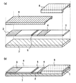
図1は本発明の実施の形態1によるバイオセンサの構造図の一例であり、図1(a)はバイオセンサの分解図、図1(b)はバイオセンサの斜視図である。
図1において、2は被検査溶液を展開する展開層であり、ニトロセルロースで構成される。3は展開層2における吸水層を示し、ガラス繊維濾紙で構成される。展開層2に使用する材料としては、被検査溶液により湿潤可能な材料であれば、濾紙、不織布、メンブレン、布など任意の多孔質材料でも構成することができる。なお、本明細書中においては、被検査溶液と検体とは実質的に同じものを指す。
【0033】
4は展開層2の一部に、被検査溶液の展開により溶出可能な乾燥状態で、被検査溶液中の測定対象物に対する抗体が金コロイドで標識され保持されている標識試薬保持部である。5は展開層2の一部に、被検査溶液中の測定対象物に対する抗体を固定化した試薬固定化部であり、当該被検査溶液中の測定対象物に対する抗体は、標識試薬保持部4に保持された標識試薬とは異なるエピトープで結合し、被検査溶液中の測定対象物と複合体を形成するように乾燥状態で固定化されている。
【0034】
なお、前述した標識試薬保持部4で抗体を標識する金コロイドは、この試薬固定化部5における結合を検出する手段として選択されるものであり、その他にも、例えば、酵素、タンパク質、色素、蛍光色素、ラテックスなどの着色粒子など任意に選択することが可能である。
【0035】
6は、展開層2の一部分を除いて、密着被覆する液体不透過性シート材であり、ここでは透明PETテープで構成されている。なお、この液体不透過性シート材6により展開層2上を被覆させることで、被検査溶液の添加部分以外への点着を遮断保護するとともに、不用意な被検査溶液の接触や被験者が手などで直接、展開層2を接触するなど外部からの汚染を防止することができる。また、試薬固定化部5を覆う液体不透過性シート材6は、測定結果を確認する部分であるため、透明な材料であることが好ましく、少なくとも、被検査溶液が透過可能な状態を有することが好ましい。また、より高精度な測定を必要とする場合には、液体不透過性シート材6が覆う展開層2の上部のみならず、被検査溶液の浸透方向に対する平行側面をも密閉させる構造をとってもよい。
【0036】
7は展開層2を保持する基板であり、白色PETフィルムで構成されている。この基板7は、展開層2を補強する役割を有するとともに、血液、唾液、尿など感染の危険性のある溶液を被検査溶液に用いる場合には、それらを遮断する作用も有する。さらに、展開層2が湿潤したときに、光透過性を帯びる場合は、光を遮断する効果を有するようにすることも可能である。
【0037】
8は被検査溶液を毛細管現象により展開層2に流入させるための空間である間隙部1を形成する空間形成材であり、透明PETフィルムを積層させて構成している。この空間形成材8は、被検査溶液添加後のバイオセンサを取り扱う際に、不用意な被検査溶液が外部へ付着あるいは飛散するなどの汚染を保護する役割も有する。
【0038】
なお、空間形成材8には、間隙部1に対する被検査溶液の流入を助成する空気孔を備えるようにしてもよい。空間形成材8の形状によっては、間隙部1の被検査溶液を付着させるための端部以外の端部が側壁等でふさがれている場合などがあり、このような場合においては、間隙部1の開口している端部に検体を付着させても、この検体が導入される以前に間隙部1に存在する空気が間隙部1から抜けない、または抜け難いことにより、間隙部1への被検査溶液導入が遅くなったり、全く導入されない状態となってしまったりして、精度の低い測定結果または誤った測定結果を与える場合が生じる。これに対し、空間形成材8に空気孔を設けておくことで、間隙部1内の空気を抜くことができ、検体が間隙部1に流入しやすくして、測定に対する悪影響の発生を防止することが可能となる。この空気孔は、前記間隙部の被検査溶液が流入する方向から見て最も奥に当たる部分に設置することが好ましいが、必要に応じて最奥部以外設置しても、問題ない。空間形成材8の材料としては、ABS、ポリスチレン、ポリ塩化ビニルなどの合成樹脂材料の他、金属、ガラスなどの溶液不透過性の材料を用いることも可能であり、被検査溶液の間隙部1への流入を確認するためには、透明もしくは半透明であることが好ましいが、透明でなくとも有色、あるいは不透明の材料でも構成することができる。
【0039】
間隙部1は空間形成材8が展開層2上に配置されることにより形成されており、この間隙部1は展開層2と接しているため、間隙部1に被検査溶液が流入することにより、被検査溶液の展開層2への浸透が開始し、測定が行われる。被検査溶液が極微量である場合や、採血用穿刺器具などにより採取した微量未知体積の被検査溶液を測定する場合において高精度の測定を行うためには、この間隙部1の体積としては20μl(マイクロリットル)以下とすることが好ましい。
【0040】
なお、細胞成分を有する被検査溶液を検体とする場合には、間隙部1に、細胞収縮試薬を保持する構成とすることが好ましく、例えば細胞収縮試薬としては塩化カリウムを用いる。このような細胞収縮試薬を設けることにより、細胞成分を有する被検査溶液を用いた場合においても、高精度ディスペンサ等を用いてあらかじめ一定体積の被検査溶液を定量することなく、簡易に測定操作を行うことが可能であり、また、細胞成分収縮剤の働きにより、前記展開層を細胞成分で目詰まりさせることなく、なおかつ高精度の測定結果を得ることが可能となる。細胞収縮試薬は、前記被検査溶液中に細胞成分を含む場合に設置すべき試薬であり、細胞成分を含まない被検査溶液を用いる場合には特に必要ではない。また細胞成分収縮剤は、細胞を収縮する効果があれば、前記塩化カリウム以外の無機塩、塩化ナトリウム、リン酸ナトリウム等塩を含む無機化合物や、グリシン、グルタミン酸、等のアミノ酸、プロリン等のイミノ酸、グルコ−ス、スクロ−ス、トレハロ−スなどの糖類、グルシト−ル等の糖アルコ−ルでも同様に実施可能である。この様な細胞成分収縮剤を含む系は、特に全血を被検査溶液として用いる場合に特に有効である。
【0041】
また、必要に応じて漂白作用を有する試薬成分として過炭酸ナトリウムを間隙部1に保持させることも可能である。このような漂白試薬部を備えることで、被検査溶液の持つ色により生じる、反応読み取りを行う試薬固定化部5に対して起こる、いわゆるバックグラウンドを低減させ、結果的にS/N比を増大させることが可能となり、簡易でありながら、高精度な測定結果が得られる効果がある。ここでのバックグラウンドとは、反応に関係しない色、例えば被検査溶液自身が有色の場合に、試薬固定化部も含めて、展開層全体に色が残る現象を言う。また、S/N比とは、この場合、反応における前記標識試薬から得られるシグナルと、前記展開層以外に、前記標識試薬に由来しないバックグラウンドに起因して得られるいわゆるノイズとの比を言う。
【0042】
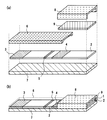
また、図2は前記図1で示したバイオセンサに分離層を設けたものの一例を示す図あり、図2(a)はバイオセンサの分解図、図2(b)はバイオセンサの斜視図である。なお、図1と同じ構成については、同じ符号を用いて説明を省略する。
【0043】
9は測定に不要な有形成分を分離することが可能な分離層であり、ガラス繊維濾紙で構成されている。なお、分離層9として、ガラス繊維濾紙を用いるのは一例であり、展開層2に用いる材料と同様に、被検査溶液により湿潤可能な材料であれば、不織布、濾紙、ガラス繊維、メンブレンフィルタ、布などの任意の材料で構成することが可能である。特に、分離層9として湿潤可能な多孔質材料を用いると、不純物をトラップして、不純物の展開層への流入や、不純物による測定に必要な成分の浸透阻害を防ぐことができるとともに、この分離層9が被検査溶液の流入の補助材としても機能し、間隙部1への被検査溶液の流入が容易となる。
【0044】
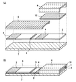
また、図3は図1で示したバイオセンサに、細胞成分破壊試薬部を設けたものの一例であり、図3(a)はバイオセンサの分解図、図3(b)はバイオセンサの斜視図である。なお、図1と同じ構成については、同じ符号を用いて説明を省略する。
【0045】
10は細胞成分を破壊する細胞成分破壊試薬を保持させた細胞成分破壊試薬部であり、塩化ナトリウムを乾燥状態にし、被検査溶液の浸透により溶解可能な状態で保持している。なお、細胞成分破壊試薬部10は、塩化ナトリウムで構成する以外にも細胞成分を破壊する試薬であれば、界面活性剤、塩化物、サポニン類、リゾチームなど任意の試薬で構成することが可能である。
【0046】
次に、本発明の実施の形態1によるバイオセンサの測定方法について図1、図2、図3を用いて説明する。
まず、被検査溶液を間隙部1に接触させると、毛細管現象により被検査溶液は間隙部1中に流入される。間隙部1に流入された被検査溶液は、間隙部1と展開層2とが接触する部分から、展開層2の終端側へと浸透する。被検査溶液が標識試薬保持部4に達したとき、標識試薬保持部4に標識された標識試薬の溶出が開始する。このとき、被検査溶液中に測定対象物が存在するならば、標識試薬保持部4より金コロイド標識抗体が反応しながら浸透が進む。そして、被検査溶液が試薬固定化部5に達したときに、測定対象物が存在するならば、その存在する量に応じて、固定化抗体と測定対象物と標識抗体との複合体が形成される。一方、測定対象物が存在しないか、あるいは検出感度以下の量であるならば、標識抗体はその大半が複合体を形成することなく展開層2上を通過する。そして、展開層2上を浸透する被検査溶液は吸水層3で吸収されて測定は終了する。また、測定の結果は、試薬固定化部5における、標識試薬の結合状態を確認することで得られる。
【0047】
また、図2に示したように、間隙部1の空間内に分離層9を有する場合には、被検査溶液が間隙部1に接触すると、間隙部1内に備えた分離層9が被検査溶液中の測定に不要な不純物を分離し、あるいは有形成分を分離し、不純物あるいは有形成分を除去した被検査溶液が展開層2へ浸透して、測定が行われることとなる。
【0048】
また、全血などの細胞成分を含む検査試薬液を用いる時に、図3に示すような間隙部1の空間部に細胞成分破壊試薬部10を有するバイオセンサを用いた場合には、被検査溶液が間隙部1に接触すると、間隙部1内に備えた細胞成分破壊試薬部10が被検査溶液中に含まれる全血などの細胞成分を破壊処理し、細胞成分を除去した被検査溶液が展開層2へ浸透して、測定が行われることとなる。
【0049】
このように、本実施の形態1によるバイオセンサによれば、展開層2上に空間を形成する空間形成材8を配置し、展開層2と空間形成材8によって形成された間隙部1に被検査溶液を接触させることによって測定を行ったので、高精度ディスペンサなどを使用することなく、微量未知体積の被検査溶液の液滴においても、その液滴を間隙部1に接触させることで、空間中に確実に被検査溶液が吸引されて、簡易かつ高精度の測定を行うことができる。
【0050】
また、間隙部1の空間部に必要に応じて分離層9を備えることで、測定に用いる被検査溶液に不要な有形成分が存在する、あるいはその可能性がある場合に、その不要な有形成分を取り除くことができるので、予め遠心分離や濾過などの操作を必要とすることなく、簡易な操作で高精度の測定を行うことができる。
【0051】
また、展開層2上に細胞成分破壊試薬部10を備えることで、血液などの細胞成分を含む被検査溶液を用いた測定を、予め遠心分離などの操作を必要とすることなく、直接全血を被検査溶液として測定することを可能とし、簡易かつ高精度の測定を行うことができる。
【0052】
なお、被検査溶液の流入量が十分であるかどうか、または測定が開始されたことを確認するために、あるいは被検査溶液の種類などを確認するために、空間形成材8を透して確認することができる構成としてもよい。また、間隙部1の体積により間隙部1内に流入する被検査溶液の体積を規定することにより、採取した被検査溶液の量に合わせて測定することが可能である。また、間隙部1内に十分な被検査溶液を流入する体積を有することで、検体不足を解消し、より正確性の高い測定を行うことができる。
【0053】
また、定性判定が必要な場合には、目視による測定も可能である。さらに、精度の高い測定を行うには、被検査溶液の浸透方向に対して展開層2の平行側面、および展開層2上面を液体不透過性材料で密着密閉させることで被検査溶液の浸透を整流し、被検査溶液中の測定対象物の量に応じたより均一な量の複合体が形成される。また、光学的な手法を用いて、標識物の結合量を測定することにより、定量的な結果を得ることもできる。
【0054】
また、本発明の実施の形態1では、固定化抗体と測定対象物と標識抗体との複合体を形成するサンドイッチ反応における抗原抗体反応について述べたが、試薬の選択によって、被検査溶液中の測定対象物と競合的に反応する試薬を用いた場合には、競合反応とすることもできる。また、抗原抗体反応以外にも、特異的な結合を利用したい場合には、任意の試薬成分で構成することも可能である。
【0055】
実施の形態2.
図4は本発明の実施の形態2によるバイオセンサの構造図の一例であり、図4(a)はバイオセンサの分解図、図4(b)はバイオセンサの斜視図である。なお、図1と同じ構成については、同じ符号を用いて説明を省略する。
図4と図1の構造の違いは、図1では標識試薬保持部4が展開層2上に標識されているのに対して、図4では標識試薬を保持した標識試薬保持層14を展開層2上に積層させて構成している。
【0056】
次に、本発明の実施の形態2によるバイオセンサの測定方法について図4を用いて説明する。
まず、被検査溶液を間隙部1に接触させると、毛細管現象により被検査溶液は間隙部1中に流入される。間隙部1に流入された被検査溶液は、標識試薬保持層14と間隙部1と展開層2とが接触する部分から、展開層2の終端側へと浸透する。このとき、展開層2上に積層された標識試薬保持層14より、標識された標識試薬の溶出が開始する。被検査溶液中に測定対象物が存在するならば、標識試薬保持層14より金コロイド標識抗体が反応しながら浸透が進む。そして、被検査溶液が試薬固定化部5に達したときに、測定対象物が存在するならば、その存在する量に応じて、固定化抗体―測定対象物―標識抗体の複合体が形成される。一方、測定対象物が存在しないか、あるいは検出感度以下の量であるならば、標識抗体はその大半が複合体を形成することなく展開層2上を通過する。そして、展開層2上を浸透する被検査溶液は吸水層3で吸収されて測定は終了する。また、測定の結果は、試薬固定化部5における、標識試薬の結合状態を確認することで得られる。
【0057】
このように、本実施の形態2によるバイオセンサによれば、展開層2上に標識試薬を保持した標識試薬保持層14を積層し、展開層2と標識試薬保持層14とから形成される間隙部1に被検査溶液を接触させることによって測定を行ったので、被検査溶液に溶解する標識試薬は、濾紙などの担体に保持されず、間隙部1内の被検査溶液中に、標識試薬が溶解および拡散されるため、標識試薬の被検査溶液に対するより均一な拡散が可能となり、より高精度なバイオセンサを提供することができる。
【0058】
なお、測定に必要な試薬成分も、標識抗体と同様に空間内に乾燥状態で形成させておくことにより、測定に必要な被検査溶液を全体に均一に溶解させることが可能である。ここで測定に必要な試薬成分とは、細胞成分破壊試薬、酵素基質、凝集試薬、緩衝液試薬、タンパク質保護試薬などであり、任意に、測定に必要なさまざまな試薬成分を導入することができる。
【0059】
また、間隙部1の空間部に必要に応じて分離層9を備えることで、測定に用いる被検査溶液に不要な有形成分が存在する、あるいはその可能性がある場合に、その不要な有形成分を取り除くことができるので、予め遠心分離や濾過などの操作を必要とすることなく、簡易な操作で高精度の測定を行うことができる。
【0060】
また、間隙部1の空間部に細胞成分破壊試薬部10を備えることで、血液などの細胞成分を含む被検査溶液を用いた測定を、予め遠心分離などの操作を必要とすることなく、直接全血を被検査溶液として測定することを可能とし、簡易かつ高精度の測定を行うことができる。
【0061】
なお、被検査溶液の流入量が十分であるかどうか、または測定が開始されたことを確認するために、あるいは被検査溶液の種類などを確認するために、空間形成材8を透して確認することができる構成としてもよい。
【0062】
また、間隙部1の体積により、間隙部1内に流入する被検査溶液の体積を規定することにより、採取した被検査溶液の量に合わせて測定することが可能である。
また、間隙部1内に十分な被検査溶液を流入する体積を有することで、検体不足を解消し、より正確性の高い測定を行うことができる。
【0063】
また、定性判定が必要な場合には、目視による測定も可能である。さらに、精度の高い測定を行うには、被検査溶液の浸透方向に対して展開層2の平行側面、および展開層2上面を液体不透過性材料で密着密閉させることで被検査溶液の浸透を整流し、被検査溶液中の測定対象物の量に応じたより均一な量の複合体が形成される。また、光学的な手法を用いて、標識物の結合量を測定することにより、定量的な結果を得ることもできる。
【0064】
また、本発明の実施の形態2では、固定化抗体と測定対象物と標識抗体との複合体を形成するサンドイッチ反応における抗原抗体反応について述べたが、試薬の選択によって、被検査溶液中の測定対象物と競合的に反応する試薬を用いた場合には、競合反応とすることもできる。また、抗原抗体反応以外にも、特異的な結合を利用したい場合には、任意の試薬成分で構成することも可能である。
【0065】
実施の形態3.
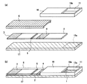
図6は本発明の実施の形態3によるバイオセンサの構造図であり、図6(a)はバイオセンサの分解図、図6(b)はバイオセンサの斜視図である。図において、図1と同一符号は同一又は相当する部分を示している。
【0066】
図6(a)及び図6(b)において、展開層2を保持する基板17は、白色PETフィルムで構成されている。基板17は展開層2よりも長手方向の長さが長く、基板17の端部近傍には、展開層2が配置されていない領域である検体保持領域17aが設けられている。この基板17は展開層2を補強する役割を有するとともに、血液、唾液、尿など感染の危険性のある溶液を被検査溶液に用いる場合には、それらの透過を遮断することもできる。さらに、展開層2が湿潤したときに、光透過性を帯びる場合は、光を遮断する効果を有するようにすることも可能である。18は被検査溶液を毛細管現象により展開層2に流入させるための空間である間隙部1を形成する空間形成材であり、ここでは、透明PETフィルムを積層させたものにより構成している。この空間形成材18は、展開層2及び検体保持領域17a上を覆うように配置されており、空間形成材18の試薬固定化部5に対向する端部以外の端部は、基板17の周縁部と密着されている。空間形成材18は、被検査溶液添加後のバイオセンサを取り扱う際に、不用意な被検査溶液が外部へ付着あるいは飛散するなどの汚染を保護する役割も有する。空間形成材18は、ABS、ポリスチレン、ポリ塩化ビニルなどの合成樹脂材料の他、金属、ガラスなどの溶液不透過性の材料を用いることも可能であり、透明もしくは半透明であることが好ましいが、透明でなくとも有色、あるいは不透明の材料でも構成することができる。空間形成材18の検体保持領域17a上の領域には、開口部18aが設けられている。この開口部18aと、検体保持領域17aと、空間形成材18の検体保持領域17aを囲む部分とが外部から被検査溶液を添加する部分である検体保持部11を構成している。1は空間形成材8が展開層2上に配置することにより形成される間隙部である。
【0067】
前記実施の形態1で示した例は、例えば微量採血針などにより微量採血を用いた指先血から直接添加する場合に適している。しかし、医療現場においては、シリンジ採血もしくは、真空採血管による真空採血なども多用されている。本実施の形態3は、このようなシリンジ等から被検査溶液を添加する場合に適しているバイオセンサである。
【0068】
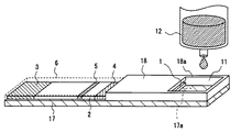
図7は本発明の実施の形態3による測定状況を示す模式図を示し、図において、図6と同一符号は同一又は相当する部分を示しており、12は採血後の採血シリンジの先端近傍を示す。この図7は前記シリンジ採血後に、針部を除き、シリンジ12から直接被検査溶液を添加する状態を示す。ここでは針部を除いている例で示しているが、針を接続した状態のままでも同様に実施可能である。
【0069】
なお、細胞成分を有する被検査溶液を検体とする場合には、間隙部1に、細胞収縮試薬を保持するようにしてもよく、この細胞収縮試薬の一例としては、塩化カリウムを用いる。細胞収縮試薬は、前記被検査溶液中に細胞成分を含む場合に設置すべき試薬であり、細胞成分を含まない被検査溶液を用いる場合には特に必要ではない。また細胞収縮試薬は、細胞を収縮する効果があれば、前記塩化カリウム以外の無機塩、塩化ナトリウム、リン酸ナトリウム等塩を含む無機化合物や、グリシン、グルタミン酸、等のアミノ酸、プロリン等のイミノ酸、グルコ−ス、スクロ−ス、トレハロ−スなどの糖類、グルシト−ル等の糖アルコ−ルでも同様に実施可能である。この様な細胞成分収縮剤を含む系は、特に全血を被検査溶液として用いる場合に特に有効である。
【0070】
さらに、必要に応じて漂白作用を有する試薬成分として過炭酸ナトリウムを保持させることも可能である。被検査溶液のもつ色素が測定結果に対する影響を無視できない場合、例えば全血検体を被検査溶液とする場合には有効である。ここでは、過炭酸ナトリウムを用いたが漂白作用を有する試薬であれば、過酸化水素、次亜塩素酸ナトリウムなども反応に対する影響を考慮して、自在に選択可能である。
【0071】
シリンジ12からバイオセンサに被検査溶液を直接添加する操作において、前述した実施の形態1において説明した間隙部1のみを備えた構成では、バイオセンサの間隙部1に吸入される被検査溶液以外の被検査溶液が、センサ外部に付着したりすることにより、外部が汚染される可能性が高い。また、間隙部1端部の被検査溶液に対する接しかたが不完全であることなどの原因によって、間隙部1に測定に必要十分量の被検査溶液が吸入されない場合、誤った測定結果が得られるおそれがある。その様な状況においては、使用者にとって非常に操作しづらい上に、前記被検査溶液が体液等の場合、安全性、もしくは衛生面で問題となる。さらには測定の正確性に欠ける結果となる。ここでの安全性とは、測定者が、被検査溶液による感染、付着などをさす。
【0072】
しかしながら、本実施の形態3においては、間隙部1の端部近傍に検体を保持する検体保持部11を設置していることにより、使用者は検体保持部11に被検査溶液を添加することで、被検査溶液を容易にかつ確実にバイオセンサに保持して、被検査溶液の測定が可能となる。このため、いわゆるシリンジ採血により採血した被検査溶液を用いて測定を実施する場合において、採血後別の容器に被検査溶液を移す操作、または、採取した被検査溶液を一定体積定量する操作を必要がなくなり、外部への検体汚染を低減させ、安全、衛生的に、また迅速簡易に測定を可能となる。
【0073】
また、被検査溶液を検体保持部11に保持することで、被検査溶液を間隙部1に確実に接するように保持することができ、検査に充分な量の被検査溶液を間隙部1に吸入させることができる。
【0074】
また間隙部1の持つ検体を吸入する効果により、あらかじめ一定量の被検査溶液を定量することなく、あいまいな量の被検査溶液を添加した場合であっても、一定体積の被検査溶液を添加した場合と同様に精度の高い測定が可能となる。
さらには、間隙部1を形成するための空間形成材18を前記透過可能な材料を用いることで、測定に十分な被検査溶液を添加できたかどうか確認できる。
【0075】
なお、本実施の形態3においては、シリンジ採血について述べたが、本発明においては、シリンジ採血以外にも、簡易なスポイト、パスツ−ルピペットなどを用いた被検査溶液の測定にも適用でき、また、被検査溶液も全血に限らず、水溶液、尿、唾液、血清、血漿など、使用者の目的により同様に適用可能であり、このような場合に適用した場合においても前記実施の形態3と同様の効果が得られる。
【0076】
なお、本実施の形態3においては、間隙部1が、100μl以下の体積を持つ構成とすることが好ましい。このような構成とすることにより、間隙部1が、前記被検査溶液が得られにくい場合や、極少量の被検査溶液を用いて測定する場合などの、被検査溶液の微少量である測定においても、被検査溶液を十分に吸入できる大きさをもつとともに、測定操作のしやすい大きさをもつこととなり、バイオセンサの操作性が高くなるとともに、微少被検査溶液による測定が可能となる。
【0077】
また、検体保持部11が、間隙部1の体積よりも大きい量の検体を保持するようにすることにより、測定に必要な十分体積の被検査溶液を保持でき、被検査溶液不足を解消し、正確性の高い測定が可能となる。また、検体保持部11の体積をさらに大きくすることにより、測定必要量に対して膨大に大量の被検査溶液を受け入れることが可能となり、被検査溶液の外部への飛散、汚染を防止しより安全、衛生な測定が可能になる。このように、検体保持部11に、間隙部と比較してより大きい量の被検査溶液を許容させるためには、検体保持部11は、図7に示したバイオセンサのように、周囲を被検査溶液が漏れないように側壁等で囲む構成とすることが好ましい。
【0078】
また、本実施の形態3においては、検体保持部11を、展開層2を保持する基板17及び間隙部1を形成する空間形成材18の一部を用いて構成するようにしたが、本発明においては、実施の形態1に示すようなバイオセンサに対して、バイオセンサに用いられている部材とは別の部材により構成した検体保持部を新たに設置しても良い。また、検体保持部としては、間隙部1に接するよう検体を保持可能な構成であれば、どのような構成のものを設けるようにしてもよい。
【0079】
なお、本実施の形態3においては、検体保持部11の底面は展開層2を保持する基板17の検体保持領域17aとし、検体保持領域17aの周囲近傍に設けられた空間形成材18を、検体の漏れ防止のための側壁としたが、本発明においては、間隙部1に被検査溶液を流入させる構造とすることが好ましく、間隙部1の底面に対して検体保持部11の底面の高さを高くした構成としたり、検体保持部11の検体を間隙部1に添加するための添加口の幅を十分に狭くして、間隙部1に向けた構成とするようにすることで、被検査溶液が前記間隙部に流入し易くなり、結果的によりより正確な測定を可能となる。
【0080】
なお、本発明においては、前記実施の形態1ないし3において説明したバイオセンサにおいて、試薬固定化部の試薬、及び標識試薬を含めたすべての試薬を乾燥状態とするようにしてもよい。このような場合においても、前記実施の形態1ないし3と同様の効果が得られるとともに、試薬がすべて乾燥状態であることにより、保存安定性能に優れ、また、持ち運び自在となるバイオセンサが得られる。
【0081】
また、前記実施の形態1ないし3に示したバイオセンサと同様のバイオセンサを免疫クロマトグラフィに用いるバイオセンサとすることにより、本発明においては、簡易法として市場に広がりつつある免疫クロマトグラフィにおいて、前記被検査溶液を高精度ディスペンサなどで一定体積定量することなく、あいまいな量の被検査溶液添加で、前記間隙部により正確な一定体積の被検査溶液を定量できるため、より高精度な測定を実現できる。ここでの免疫クロマトグラフィとは、湿潤可能な多孔質材料を用いて、固定化試薬と標識試薬の複合体を形成させることにより測定される免疫測定法のことで、抗原抗体反応を利用した測定系であり、通常免疫測定法においては、B/F分離など洗浄操作が必要なことに対して、クロマトグラフィ担体を被検査溶液が浸透していく過程でB/F分離が実施される測定系である。通常すべての試薬は乾燥状態にあり、測定時に被検査溶液により湿潤される。標識物としては、金コロイド、ラテックスが一般的であるが、磁性粒子、酵素、金属コロイド等も使用されている。標識物が酵素の場合等は、使用者が測定操作として酵素基質や反応停止試薬を加える操作が含まれる。
【0082】
また、本発明においては、前記実施の形態1ないし3に示したバイオセンサと同様のバイオセンサをワンステップ免疫クロマトグラフィに用いられるバイオセンサとすることにより、簡易免疫測定法として市場に普及しつつあるワンステップ免疫クロマトグラフィにおいて、使用者があらかじめ被検査溶液体積を定量する必要のない、また、被検査溶液量を測定時に確認できることにより誤判定を低減させ、正確性が高く尚かつ従来のワンステップ免疫クロマトグラフィのもつ簡易操作を実現できる。ワンステップ免疫クロマトグラフィとは、湿潤可能な多孔質材料を用いて、被検査溶液の添加により測定が開始される免疫測定法のことで、抗原抗体反応を利用した測定系であり、通常免疫測定法においては、B/F分離など洗浄操作が必要なことに対して、ワンステップ免疫クロマトグラフィは、クロマトグラフィ担体を被検査溶液が浸透していく過程でB/F分離が実施される測定系のことである。通常すべての試薬は乾燥状態にあり、測定時に被検査溶液により湿潤される。使用者の基本的な測定操作が被検査溶液を添加する操作のみであることからワンステップ免疫クロマトグラフィと呼ぶ。標識物としては、金コロイド、ラテックスが一般的であるが、磁性粒子、酵素、金属コロイド等も使用されている。
【0083】
次に、本発明を実施例により更に具体的に説明する。なお、本発明は、その要旨を越えない限りこれらの実施例の記載に限定されるものではない。
実施例1.
尿中hCG測定用試験片の作製
ニトロセルロース膜中に抗hCG−β抗体固定化ライン、および抗hCG−α抗体と金コロイドとの複合体の広いバンドを含む免疫クロマトグラフィー展開層2を製造した。この免疫クロマトグラフィ−展開層2を空間形成材8により被検査溶液添加部分に間隙部1を形成させて、免疫クロマトグラフ試験片を製造した。この試験片は図1のような構造を有する。図1より試験片は、抗体固定化部5と、それよりも被検査溶液を接触させる側にある、抗hCG−α抗体と金コロイドとの複合体とを含有した標識試薬保持部4とを含む。これらの試験片は、次のようにして製造した。
【0084】
a)免疫クロマトグラフィー展開層の調製
まず、リン酸緩衝溶液にて希釈して、濃度調整を行った抗hCG−β抗体溶液を準備し、この抗体溶液を溶液吐出装置を用いて、ニトロセルロース膜上に塗布した。これにより、ニトロセルロース膜上に検出用の抗体固定化ラインが得られた。このニトロセルロース膜を乾燥後、1%スキムミルクを含有するTris−HCl緩衝溶液中に浸漬して30分間緩やかに振った。30分後、Tris−HCl緩衝溶液槽に膜を移動し、10分間緩やかに振った後に、別のTris−HCl緩衝溶液槽にて、さらに10分間緩やかに振り、膜の洗浄を行った。そして、2度洗浄を行った後に、膜を洗浄液から取り出して、室温で乾燥させた。
【0085】
金コロイドは、0.01%塩化金酸、還流中の100℃溶液に1%クエン酸溶液を加えることによって調製した。金コロイドは還流を30分間続けた後に冷却し、0.2Mの炭酸カリウム溶液によってpH9に調製した。この金コロイド溶液に、抗hCG−α抗体を加えて数分間攪拌した後に、10%BSA(牛血清アルブミン)溶液pH9を最終1%になる量だけ加えて攪拌することで、抗体金コロイド複合体(標識抗体)を調製した。その後4℃、20000Gで50分間遠心分離することによって、標識抗体を単離して、それを洗浄緩衝液(1%BSA・リン酸緩衝液)中に懸濁した後に、遠心分離を行って、標識抗体を洗浄単離した。この標識抗体を洗浄緩衝液で懸濁して、0.8μmのフィルタにて濾過することによって、当初の金コロイド溶液量の10分の1に調製し、4℃で貯蔵した。
【0086】
そして、このように調製された標識抗体溶液を溶液吐出装置にセットして、抗hCG−β抗体固定化乾燥膜上の抗体固定化位置から離れた位置に塗布した後に、膜を乾燥させた。これによって、固定化膜上に標識試薬保持部位が得られた。このようして免疫クロマトグラフィ反応層、即ち展開層を完成させた。
【0087】
b)免疫クロマトグラフィ試験片の作製
厚さ0.5mmの白色PETからなる基板上に、免疫クロマトグラフィ試験片を貼り付け、2.5mmの幅で裁断した。裁断後、免疫クロマトグラフィの各片を、標識抗体保持部分から終端部分にかけて、厚さ100μmの透明テ−プを巻き付けた。透明テ−プを巻き付けない始端部分上の中央に、厚さ100μmの透明PETを積層させて作製したあらかじめ空気孔をもつ空間形成材を貼り付け、間隙部(幅2.0mm×長さ6.0mm×高さ0.5mm)を形成した。このようにして免疫クロマトグラフィ試験片を製造した。
【0088】
c)試料の調製
ヒト尿中に既知濃度のhCG溶液を加えることにより、さまざまな既知濃度のhCG溶液を調製した。
【0089】
d)測定
濃度調整されたhCGを含む尿を15μl、PET基板上に滴下し、PET基板上に液滴を形成させた。形成させた液滴を、免疫クロマトグラフィ試験片の間隙部に接触させて、間隙部中に導入させた。導入された液滴は、吸水層方向へと展開処理し、抗原抗体反応をさせて抗体固定化部における呈色反応を行った。この試験片への試料添加から5分後の呈色状況を反射型分光光度計(CS9300;島津製作所製)を用いて計測して、呈色度を演算処理した。
【0090】
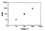
まず、hCG濃度が100、1000、10000U/lの各hCGを含有する尿を免疫クロマトグラフィ試験片に添加し、展開処理を行った。そして、各hCG濃度の尿に対する試験片上の抗体固定化部の呈色状況を反射型分光光度計で測定した。520nmの波長における吸光度を反射型分光光度計で計測し、予め作成しておいたhCG濃度と吸光度との関係を示す検量線に代入した。その結果を図5に示す。
【0091】
図5は、免疫クロマトグラフィー試験片に液体試料を添加し、添加開始から5分後の呈色度合の測定値をもとに、分析対象物の濃度を換算した結果を表している。その結果、各濃度n=10にて測定を行ったが、CV値は5%〜10%と非常に良い相関性を示した。
【0092】
実施例2.
全血CRP定量用試験片の作製
ニトロセルロース膜中に抗CRP抗体Aを固定化した試薬固定化部、さらに抗CRP抗体Bと金コロイドとの複合体を保持した標識試薬を含む免疫クロマトグラフィ試験片を製造した。この免疫クロマトグラフィ試験片は図6に示したバイオセンサと同様の構成を有しており、この免疫クロマトグラフィ試験片は、抗体が固定化された試薬固定化部5よりも被検査溶液を添加する展開開始点に近い部分にある、抗CRP抗体Bと金コロイドとの複合体が含有された領域である標識試薬4と、検体保持部11とを含む。この免疫クロマトグラフィ試験片は、次のようにして製造した。
【0093】
a)免疫クロマトグラフィ試験片の調製
リン酸緩衝溶液にて希釈して濃度調整をした抗CRP抗体A溶液を準備した。この抗体溶液は溶液吐出装置を用いて、ニトロセルロース膜上に塗布した。これにより、ニトロセルロース膜上に試薬固定化部である抗体固定化ラインが得られた。このニトロセルロース膜を乾燥後、1%スキムミルクを含有するTris−HCl緩衝溶液中に浸漬して30分間緩やかに振った。30分後、Tris−HCl緩衝溶液槽に膜を移動し、10分間緩やかに振った後に、別のTris−HCl緩衝溶液槽にて更に10分間緩やかに振り、膜の洗浄を行なった。2度洗浄を行った後に、膜を液槽から取り出して、室温で乾燥させた。
【0094】
金コロイドは、0.01%塩化金酸の還流中の100℃溶液に1%クエン酸溶液を加えることによって調製した。還流を30分間続けた後に、室温放置にて冷却した。0.2Mの炭酸カリウム溶液によって、pH9に調製した前記金コロイド溶液に、抗CRP抗体Bを加えて数分間攪拌した後に、pH9の10%BSA(牛血清アルブミン)溶液を最終1%になる量だけ加えて攪拌することで、検出物質である抗体−金コロイド複合体(標識抗体)を調製した。前記標識抗体溶液を4℃、20000Gで50分間遠心分離することによって、標識抗体を単離して、それを洗浄緩衝液(1%BSA・リン酸緩衝液)中に懸濁した後に、前記遠心分離を行って、標識抗体を洗浄単離した。この標識抗体を洗浄緩衝液で懸濁して、0.8μmのフィルタにて濾過した後に、当初の金コロイド溶液量の、10分の1量に調製して、4℃で貯蔵した。
【0095】
前記金コロイド標識抗体溶液を溶液吐出装置にセットして、抗CRP抗体固定化A乾燥膜上の固定化ラインから離れた、被検査溶液添加開始方向から順番に、標識抗体、固定化ライン、の位置関係になる様塗布した後に、膜を真空凍結乾燥させた。これによって、固定化膜上に標識試薬を持つ反応層担体が得られた。
【0096】
次に、調製された標識試薬を含む反応層担体を、厚さ0.5mmの白色PETからなる基板上に貼り付け、5.0mmの幅で裁断した。裁断後、各片を、標識抗体保持部分から終端部分にかけて、厚さ100μmの透明テ−プを巻き付けた。透明テ−プを巻き付けない始端部分上の中央に、厚さ100μmの透明PETを積層させて作製した、あらかじめ空気孔を持つ空間形成材を貼り付け、間隙部(幅5.0mm×長さ12.0mm×高さ0.5mm)を形成した。また空間形成材にはあらかじめ、検体保持部の液漏れ防止壁を形成している。さらに前記空間形成材はあらかじめ1.5Mに調製された塩化カリウム水溶液を、単位面積あたり2μl点着した後に、液体窒素にて直ちに凍結し、凍結乾燥を行い、これによって、塩化カリウムが乾燥状態で保持された収縮剤保持部を持つ空間形成材を作製したものである。こうして免疫クロマトグラフィ試験片を製造した。
【0097】
b)試料の調製
抗凝固剤としてEDTA・2Kを加えた人の血液を、ヘマトクリット値45%になるように調製した。この血液に既知濃度のCRP溶液を加えることにより、さまざまな既知濃度のCRP含有血液を調製した。
【0098】
c)試験片上の呈色度合の測定
前記調整された全血を用いて、実際の採血状態を考慮して、体積20ml用のシリンジに充填させ、検体保持部にCRPを含む全血を一定体積測定することなく適量添加した。添加後、被検査溶液は間隙部に吸入された。さらに吸水部方向へと展開処理して、抗原抗体反応をさせて抗体固定化部における呈色反応を行った。このバイオセンサへの試料添加から5分後の呈色状況を反射吸光度測定機により計測した。
【0099】
血漿濃度として、0.1mg/dl、1.0mg/dl、3.0mg/dl、のCRPを含有する全血をバイオセンサに前記20mlシリンジから直接適量添加して展開処理した。各CRP濃度の血液に対するバイオセンサ上の試薬固定化部の呈色状況を反射吸光度測定機(CS−9300 SIMADZU製)で測定した。635nmにおける吸光度を計測して、各CRP濃度に応じてプロットした。その図を、図8に示す。図8は本実施例2における多濃度測定結果を示す図であり、横軸は市販測定装置を用いて測定したCRP濃度を示す。ここでは、市販測定装置として、ラテックス免疫凝集法による試薬を用いた。また、縦軸は、得られた吸光度を示す。検量線とは、CRP濃度上昇に対して吸光度が上昇する領域のことで、通常あらかじめ、濃度既知の被検査溶液により計算しておき、その後、未知の被検査溶液を測定したとき、得られた吸光度から、その未知被検査溶液中のCRP濃度を算出するための数式を言う。図8に示すとおり、前述した実施例1同様に良好な結果が得られた。
【0100】
なお、本実施例2におけるバイオデバイスとして、ニトロセルロースやガラス繊維濾紙のような、任意の多孔質性担体で構成されたクロマトグラフィー材料からなるバイオセンサが用いられている。このような材料からなるバイオセンサは、例えば、抗原抗体反応のような任意の測定原理を用いて、ある特定物質を分析検出し、定性または定量する機能をもっている。
【0101】
また、本実施例2においては、同一ニトロセルロース膜上に標識試薬と試薬固定化部を設けたバイオセンサを用いたが、ニトロセルロースとは異なる材質の例えば不織布のような多孔質性担体に標識試薬を担持したものを標識試薬として、支持体上に配しても何ら問題はない。標識試薬を構成する標識物としては、金コロイドを用いて例を示したが、着色物質、蛍光物質、燐光物質、発光物質、酸化還元物質、酵素、核酸、小胞体でもよく、反応の前後において何らかの変化が生じるものであれば何を用いても良い。
【0102】
測定される被検査溶液としては、例えば、水や水溶液、尿、血液、血漿、血清、唾液などの体液、固体及び粉体や気体を溶かした溶液などがあり、その用途としては、例えば、尿検査や妊娠検査、水質検査、便検査、土壌分析、食品分析などがある。また、被検物質としてC反応性タンパク質(CRP)を例として実施例を述べたが、抗体、免疫グロブリン、ホルモン、酵素及びペプチドなどのタンパク質及びタンパク質誘導体や、細菌、ウイルス、真菌類、マイコプラズマ、寄生虫ならびにそれらの産物及び成分などの感染性物質、治療薬及び乱用薬物などの薬物及び腫瘍マーカーが挙げられる。具体的には、例えば、絨毛性性腺刺激ホルモン(hCG)、黄体ホルモン(LH)、甲状腺刺激ホルモン、濾胞形成ホルモン、副甲状腺刺激ホルモン、副腎脂質刺激ホルモン、エストラジオール、前立腺特異抗原、B型肝炎表面抗原、ミオグロビン、CRP、心筋トロポニン、HbA1c、アルブミン等でも何ら問題はない。また、水質検査、土壌分析、などの環境分析、や食品分析などにも実施可能である。前述した形態により、簡便かつ迅速で、高感度・高性能、正確性の高い測定が実現できる。
【0103】
産業上の利用可能性
以上のように、本発明によるバイオセンサは、免疫クロマトグラフィーや、いわゆるワンステップクロマトグラフィー等に利用されるバイオセンサとして有用であり、特に、医療診断現場や一般家庭向けなどの操作の簡便化や迅速化が要求される状況における測定に利用されるバイオセンサや、微量な体積の被検査溶液の測定に利用されるバイオセンサとして適している。
【図面の簡単な説明】
【図1】本発明の実施の形態1によるバイオセンサの構造図である。
【図2】本発明の実施の形態1による分離層を有したバイオセンサの構造図である。
【図3】本発明の実施の形態1による細胞成分破壊試薬部を有したバイオセンサの構造図である。
【図4】本発明の実施の形態2によるバイオセンサの構造図である。
【図5】本発明の実施例1におけるhCG濃度と吸光度との関係を示すグラフである。
【図6】本発明の実施の形態3によるバイオセンサの構成図である。
【図7】本発明の実施の形態3によるバイオセンサを用いた測定時の状況を示す図である。
【図8】本発明の実施例2におけるCRP濃度と吸光度との関係を示すグラフである。[0001]
Technical field
The present invention relates to a biosensor, and more particularly to a biosensor using chromatography.
[0002]
Background art
A conventional biosensor includes a development layer for developing a solution to be tested, a reagent immobilization unit immobilized on a part of the development layer, and a reagent holding unit for holding a labeling reagent that can be eluted by the development of the test solution And the measurement component in the solution to be inspected was qualitatively or quantitatively measured by measuring the binding amount of the labeling reagent in the reagent-immobilized portion. An example of such a biosensor is an immunochromatographic sensor.
[0003]
A general configuration of an immunochromatographic sensor includes an additive layer to which a solution to be inspected is added, a plurality of development layers, and a water absorption layer at the end of the development layer. And, in a part of this development layer, it has an antibody immobilization part that immobilizes an antibody against the measurement object in the test solution, and the antibody labeled on the addition layer side than the antibody immobilization part, It is held in the labeling reagent holder in a dry state that can be eluted by the solution to be inspected.
In such an immunochromatographic sensor, a required amount of the solution to be tested is added to the addition layer, and the measurement starts when the solution to be tested penetrates into the porous material provided on the development layer.
[0004]
As a method of adding the solution to be inspected, a method of immersing the added portion in the solution to be inspected for a certain period of time, a method of adding a certain amount using a high-precision dispenser or a dropper, etc., or urine or the like as the solution to be inspected In some cases, a method in which the solution to be tested is directly applied to the added portion for a certain period of time during urination has been used.
[0005]
The measurement result of the immunochromatographic sensor is detected by a labeled antibody bound to the antibody immobilization part. As this labeled antibody, a common label includes gold colloid particles, and the binding of the antibody-immobilized portion can be visually observed by the gold colloid particles, and the measurement result can be obtained visually. Note that this is based on the sandwich reaction of the antigen-antibody reaction in which the complex of immobilized antibody-measurement target-labeled antibody is formed. Similarly, the measurement result can be obtained by confirming the binding state of the labeling reagent in the antibody-immobilized or antigen-immobilized portion.
[0006]
In addition, the measurement result by the sandwich reaction described above requires qualitative judgment by visual inspection, but when the required measurement result is semi-quantitative or more accurate judgment is required. Is a method of reading by a transmission method using an optical reading device disclosed in Japanese Patent Laid-Open No. 10-274624, or a measurement result captured by a camera or the like disclosed in Japanese Patent Laid-Open No. 10-274653. There is also a method of arithmetic processing.
[0007]
In recent years, in the field of medical diagnosis, there has been a demand for a measuring device that can be obtained quickly, simply, accurately, and easily at a low price based on the concept of point of care (POC). According to the method, when a solution to be tested is added to the sensor unit, for example, if the solution to be tested is blood, blood is collected using a syringe. After performing an operation of separating a certain blood cell and plasma, it had to be added to the sensor unit using a dispenser or a tool such as a dropper.
[0008]
Such a blood collection method using a syringe requires a special skill as a medical technique, and further requires a centrifuge operation because it is not possible for ordinary households and individuals who do not have those techniques to perform their own measurements. could not.
Further, since an instrument such as a dispenser is required to quantify the solution to be inspected, there is a problem that the operation becomes complicated.
[0009]
If the solution to be inspected is urine, etc., if it is necessary to store the urine once in a container such as a paper cup and immerse a part of the sensor for a certain period of time, If the collected urine is added using a dispenser or dropper as in the case of blood, or if the sensor has a relatively low accuracy that does not require quantitative accuracy for measurement, urine is added directly during urination. Although methods are applied to the parts for a certain period of time, these methods require that the urine be stored once in a paper cup or the like, or if applied directly to the urine, there is no means to accurately define the volume of the solution to be inspected. In particular, there is a problem limited to qualitative measurement with low accuracy.
[0010]
The present invention has been made to solve such a problem, and can perform a simple and highly accurate measurement even with a small volume of a solution to be inspected without requiring an advanced apparatus or operation. The purpose is to provide.
[0011]
Disclosure of the invention
The present invention has a reagent immobilization part immobilized on a part of a development layer for developing a test solution, and a label reagent holding part for holding a labeling reagent that can be eluted by the development of the test solution. A biosensor for qualitatively or quantitatively measuring a measurement component in the test solution by measuring the binding amount of the labeling reagent in the reagent-immobilized portion, wherein the test solution comprises: Inflow by capillary action The space, which is a space, is Only one end of It is characterized by comprising a space forming part formed in the above. As a result, when a solution to be inspected is added to the gap portion, the liquid droplet is brought into contact with the gap portion even in a trace unknown volume of the solution to be examined without using a high-precision dispenser. The solution to be inspected is surely sucked in, and simple and highly accurate measurement can be performed.
[0012]
In addition, the present invention has an immobilized reagent immobilization part in a part of a developing layer for developing a solution to be inspected, and measures the binding amount of a labeling reagent in the reagent immobilization part, thereby A biosensor for qualitatively or quantitatively measuring a measurement component in a test solution, wherein the solution to be tested is Inflow by capillary action The space, which is a space, is Only one end of And a label forming part that holds a labeling reagent that can be dissolved by the inflow of the solution to be inspected, in the gap part. As a result, when the solution to be inspected is introduced into the gap, the labeling reagent in the solution to be inspected in the gap dissolves and diffuses, so that the labeling reagent can be more evenly diffused in the solution to be inspected. Thus, simple and highly accurate measurement can be performed.
[0013]
Further, the present invention is characterized in that the gap portion temporarily holds the solution to be inspected. This makes it possible to confirm the amount of suction that the solution to be inspected penetrates into the gap, and to allow more reliable permeation into the development layer, and to perform more accurate measurement with reduced measurement errors and sample shortages. be able to.
[0014]
Further, the present invention is characterized in that the gap portion defines an inflow amount of the solution to be inspected by a volume of the gap portion. Thereby, it is not necessary to perform a predetermined volume quantification of the solution to be tested in advance, and it is possible to carry out the measurement with a simple operation without requiring a special skill such as sampling.
[0015]
Further, the present invention is characterized in that the gap has a volume into which a sufficient solution to be inspected for developing the development layer flows. Thus, by setting the space volume to a sufficient volume with respect to the amount of the solution to be inspected necessary for the measurement, it is possible to eliminate the shortage of the specimen and perform the measurement with higher accuracy.
[0016]
Further, the present invention is characterized in that a cell component destruction reagent part for destroying a cell component is provided in the gap part. As a result, it is possible to directly add a measurement using a test solution containing a cell component such as blood to the gap as a test solution without performing an operation such as centrifugation in advance. Since the solution is sucked into the gap, when adding the solution to be inspected, a device for adding and a pretreatment device for the solution to be inspected are not required, and the measurement can be performed with a simple operation.
[0017]
Further, the present invention is characterized in that a cell component contraction reagent unit for contracting cell components is provided in the gap. As a result, even when a solution to be inspected having a cell component is used, a measurement operation can be easily performed without clogging the spread layer with the cell component due to the action of the cell component contractor. Accuracy results can be obtained.
[0018]
Further, the present invention is characterized in that a bleaching reagent part is provided in the gap part. This reduces the adverse effects on the measurement due to the color unrelated to the reaction, such as the color of the solution to be inspected, remaining on the reagent immobilization section that performs reaction reading, and provides highly accurate measurement results. There is an effect.
[0019]
Further, the present invention is characterized in that the gap portion has a volume of 20 μl (microliter) or less. As a result, even when the solution to be inspected is a very small amount or when measuring a to-be-inspected solution of a minute unknown volume collected with a blood sampling puncture device, etc., it is added to the gap portion of the specified volume. It is possible to introduce a test solution with a small volume, and it is possible to perform simple and highly accurate measurement even with a small amount of test solution.
[0020]
Further, the present invention is characterized in that the gap portion has means for confirming the inflow of the solution to be inspected from the outside. This makes it possible to detect the measurement start time by confirming or detecting the inflow of the solution to be inspected in the measurement, and to solve the shortage of the solution to be inspected by obtaining an appropriate amount of the solution to be inspected. Accurate measurement start time can be detected, and more accurate measurement can be performed.
[0021]
In addition, the present invention is characterized in that a part or all of the space forming portion has light transmittance. As a result, the suction amount of the solution to be inspected can be observed visually or using an optical measurement method, eliminating erroneous measurement operations due to insufficient amount of the solution to be inspected, and measuring with higher accuracy. It can be performed.
[0022]
Further, the present invention is characterized in that a separation part for separating a formed component unnecessary for measurement is provided in the gap part. As a result, it is possible to obtain an effect of separating the formed components unnecessary for the measurement in the solution to be inspected, and it is necessary to remove the formed components unnecessary for the measurement in the solution to be inspected by operations such as centrifugation and filtration in advance. Therefore, simple and highly accurate measurement can be performed.
[0023]
In addition, the present invention is characterized by including a sample holding unit that holds a solution to be inspected so as to be in contact with the gap. This eliminates the need to transfer the test solution to another container after collection, or to quantitate the collected test solution at a fixed volume, and can hold the test solution in the sample holder, allowing sample contamination to the outside. Therefore, it is possible to perform highly accurate measurement safely, hygienically, and quickly and easily.
[0024]
In the present invention, the specimen holding unit holds a solution to be inspected in an amount larger than the volume of the gap. This makes it possible to hold a sufficient volume of the solution to be inspected necessary for the measurement, prevent the solution to be inspected from splashing outside, and prevent contamination, enabling safer and more hygienic measurement and eliminating the shortage of solution to be inspected. Therefore, accurate measurement is possible.
[0025]
Further, the present invention is characterized in that the height position of the bottom surface of the specimen holding portion is the same or higher than the height position of the bottom surface in the gap portion. As a result, the solution to be inspected easily flows into the gap, and accurate measurement is possible.
[0026]
In addition, the present invention is characterized in that the gap portion has a volume of 100 μl or less. As a result, a minute solution to be inspected can be inhaled, and the measurement operation can be easily performed and operability can be kept high.
[0027]
In the present invention, an air hole for assisting inflow of the solution to be inspected is provided in the gap portion. Thus, it is possible to promptly and surely inject the solution to be inspected into the gap portion so that air existing in the gap portion before the sample is introduced is surely released.
[0028]
Further, the present invention is characterized in that the gap portion is provided with a porous material that can be wetted by permeation of the solution to be inspected. As a result, when the solution to be inspected is caused to flow into the gap, the polycrystalline material becomes an inflow auxiliary material and easily flows in, and the porous material traps impurities in the solution to be inspected, thereby Inflow and penetration inhibition can be reduced.
[0029]
In addition, the present invention is characterized in that all reagents including the reagent in the reagent immobilization section and the labeling reagent are in a dry state, and the whole is in a dry state. Thereby, it is possible to obtain a biosensor that has excellent storage stability and is portable.
[0030]
The present invention is also characterized in that the biosensor is used for immunochromatography. As a result, in immunochromatography, which is used as a simple method, an accurate amount of a sample to be inspected by the gap can be obtained by adding an ambiguous amount of the solution to be inspected without quantifying the solution to be inspected with a high-precision dispenser or the like. The solution can be quantified and more accurate measurement can be realized.
[0031]
The present invention is also characterized in that the biosensor is used for one-step immunochromatography. This eliminates the need for the user to quantify the volume of the solution to be tested in advance during the measurement in one-step immunochromatography, which is used as a simple immunoassay. More accurate measurement can be realized without compromising the simple operation of one-step immunochromatography.
[0032]
BEST MODE FOR CARRYING OUT THE INVENTION
1 is an example of a structural diagram of a biosensor according to
In FIG. 1,
[0033]
[0034]
The gold colloid for labeling the antibody with the labeling
[0035]
[0036]
7 is a board | substrate which hold | maintains the expansion |
[0037]
[0038]
Note that the
[0039]
The
[0040]
When the test solution having cell components is used as a specimen, it is preferable that a cell contraction reagent is held in the
[0041]
Moreover, it is also possible to hold | maintain sodium percarbonate in the gap |
[0042]
2 is a diagram showing an example of the biosensor shown in FIG. 1 provided with a separation layer. FIG. 2 (a) is an exploded view of the biosensor, and FIG. 2 (b) is a perspective view of the biosensor. is there. In addition, about the same structure as FIG. 1, description is abbreviate | omitted using the same code | symbol.
[0043]
[0044]
3 is an example of the biosensor shown in FIG. 1 provided with a cell component destruction reagent unit. FIG. 3 (a) is an exploded view of the biosensor, and FIG. 3 (b) is a perspective view of the biosensor. It is. In addition, about the same structure as FIG. 1, description is abbreviate | omitted using the same code | symbol.
[0045]
[0046]
Next, a biosensor measurement method according to
First, when the solution to be inspected is brought into contact with the
[0047]
As shown in FIG. 2, when the
[0048]
Further, when a test reagent solution containing cell components such as whole blood is used, if a biosensor having a cell component
[0049]
As described above, according to the biosensor according to the first embodiment, the
[0050]
In addition, if the
[0051]
In addition, since the cell component
[0052]
In addition, in order to confirm whether the amount of inflow of the solution to be inspected is sufficient, that the measurement has been started, or to confirm the type of the solution to be inspected, it is confirmed through the
[0053]
Further, when qualitative determination is necessary, visual measurement is also possible. Furthermore, in order to perform measurement with high accuracy, the parallel side surface of the
[0054]
In the first embodiment of the present invention, the antigen-antibody reaction in the sandwich reaction that forms a complex of the immobilized antibody, the measurement object, and the labeled antibody has been described. When a reagent that reacts competitively with an object is used, a competitive reaction may be used. In addition to the antigen-antibody reaction, when specific binding is desired, it can be composed of arbitrary reagent components.
[0055]
4 is an example of a structural diagram of a biosensor according to
4 is different from the structure of FIG. 1 in that the labeling
[0056]
Next, a biosensor measurement method according to
First, when the solution to be inspected is brought into contact with the
[0057]
As described above, according to the biosensor according to the second embodiment, the labeling
[0058]
It should be noted that the reagent components necessary for the measurement can be uniformly dissolved throughout the solution to be inspected necessary for the measurement by forming the reagent components in the dry state in the same manner as the labeled antibody. Here, the reagent components necessary for the measurement are cell component destruction reagents, enzyme substrates, agglutination reagents, buffer reagents, protein protection reagents and the like, and various reagent components necessary for the measurement can be arbitrarily introduced. .
[0059]
In addition, if the
[0060]
In addition, by providing the cell component
[0061]
In addition, in order to confirm whether the amount of inflow of the solution to be inspected is sufficient, that the measurement has been started, or to confirm the type of the solution to be inspected, it is confirmed through the
[0062]
Further, by defining the volume of the solution to be inspected flowing into the
In addition, by having a volume that allows a sufficient solution to be inspected to flow into the
[0063]
Further, when qualitative determination is necessary, visual measurement is also possible. Furthermore, in order to perform measurement with high accuracy, the parallel side surface of the
[0064]
In the second embodiment of the present invention, the antigen-antibody reaction in the sandwich reaction that forms a complex of the immobilized antibody, the measurement object, and the labeled antibody has been described. Depending on the selection of the reagent, the measurement in the test solution can be performed. When a reagent that reacts competitively with an object is used, a competitive reaction may be used. In addition to the antigen-antibody reaction, when specific binding is desired, it can be composed of arbitrary reagent components.
[0065]
6 is a structural diagram of a biosensor according to
[0066]
In FIG. 6A and FIG. 6B, the
[0067]
The example shown in the first embodiment is suitable for the case where the blood is directly added from fingertip blood using micro blood collection, for example, with a micro blood collection needle or the like. However, syringe blood collection or vacuum blood collection using a vacuum blood collection tube is frequently used in the medical field. The third embodiment is a biosensor suitable for adding a solution to be inspected from such a syringe or the like.
[0068]
FIG. 7 is a schematic diagram showing a measurement situation according to the third embodiment of the present invention, in which the same reference numerals as those in FIG. 6 indicate the same or corresponding parts, and 12 indicates the vicinity of the distal end of the blood collection syringe after blood collection. Show. FIG. 7 shows a state in which the solution to be inspected is directly added from the
[0069]
When the test solution having a cell component is used as a specimen, a cell contraction reagent may be held in the
[0070]
Furthermore, it is also possible to retain sodium percarbonate as a reagent component having a bleaching action as required. This is effective when the influence of the dye of the test solution on the measurement result cannot be ignored, for example, when a whole blood sample is used as the test solution. Here, if sodium percarbonate is used, but a reagent having a bleaching action, hydrogen peroxide, sodium hypochlorite and the like can be freely selected in consideration of the influence on the reaction.
[0071]
In the operation of adding the solution to be tested directly from the
[0072]
However, in the third embodiment, the
[0073]
Further, by holding the solution to be inspected in the
[0074]
In addition, due to the effect of inhaling the specimen in the
Furthermore, it is possible to confirm whether or not a sufficient solution to be inspected for measurement can be added by using the permeable material as the
[0075]
In the third embodiment, syringe blood collection has been described. However, in the present invention, in addition to syringe blood collection, the present invention can be applied to measurement of a solution to be inspected using a simple dropper, Pasteur pipette, etc. The solution to be inspected is not limited to whole blood, but can be similarly applied according to the purpose of the user, such as an aqueous solution, urine, saliva, serum, plasma, and even in such a case, Similar effects can be obtained.
[0076]
In the third embodiment, it is preferable that the
[0077]
In addition, by holding the
[0078]
In the third embodiment, the
[0079]
In the third embodiment, the bottom surface of the
[0080]
In the present invention, in the biosensor described in the first to third embodiments, all reagents including the reagent in the reagent immobilization unit and the labeling reagent may be in a dry state. Even in such a case, the same effects as those of the first to third embodiments can be obtained, and a biosensor that is excellent in storage stability and portable can be obtained because all the reagents are in a dry state. .
[0081]
Further, by using a biosensor similar to the biosensor described in the first to third embodiments for use in immunochromatography, in the present invention, in the immunochromatography that is spreading on the market as a simple method, Without quantifying the test solution at a constant volume with a high-precision dispenser, etc., by adding an ambiguous amount of the solution to be tested, it is possible to quantitate the exact volume of the solution to be tested by the gap, thus realizing more accurate measurement. . Here, immunochromatography is an immunoassay that is measured by forming a complex of an immobilization reagent and a labeling reagent using a wettable porous material, and is a measurement system that utilizes an antigen-antibody reaction. In a conventional immunoassay method, B / F separation is performed in the process where a solution to be infiltrated permeates through a chromatographic carrier, whereas a washing operation such as B / F separation is required. . Normally all reagents are in a dry state and are wetted by the solution to be tested at the time of measurement. As the label, gold colloid and latex are generally used, but magnetic particles, enzymes, metal colloids and the like are also used. When the label is an enzyme, the user includes an operation of adding an enzyme substrate or a reaction termination reagent as a measurement operation.
[0082]
Further, in the present invention, a biosensor similar to the biosensor shown in the first to third embodiments is used as a biosensor used in one-step immunochromatography, so that it is spreading to the market as a simple immunoassay method. In one-step immunochromatography, it is not necessary for the user to quantify the volume of the solution to be tested in advance, and the amount of the solution to be tested can be confirmed at the time of measurement, thereby reducing misjudgment, high accuracy, and conventional one-step immunization. Simple operation of chromatography can be realized. One-step immunochromatography is an immunoassay that uses a wettable porous material to start measurement by adding a solution to be tested. It is a measurement system that uses an antigen-antibody reaction. In contrast, one-step immunochromatography is a measurement system in which B / F separation is performed in the process where the solution to be infiltrated permeates through the chromatographic carrier, whereas a washing operation such as B / F separation is required. is there. Normally all reagents are in a dry state and are wetted by the solution to be tested at the time of measurement. This is called one-step immunochromatography because the basic measurement operation of the user is only the operation of adding the solution to be tested. As the label, gold colloid and latex are generally used, but magnetic particles, enzymes, metal colloids and the like are also used.
[0083]
Next, the present invention will be described more specifically with reference to examples. In addition, this invention is not limited to description of these Examples, unless the summary is exceeded.
Example 1.
Preparation of test piece for hCG measurement in urine
An
[0084]
a) Preparation of immunochromatographic spreading layer
First, an anti-hCG-β antibody solution diluted with a phosphate buffer solution to adjust the concentration was prepared, and this antibody solution was applied onto a nitrocellulose membrane using a solution discharge device. Thereby, an antibody immobilization line for detection was obtained on the nitrocellulose membrane. The nitrocellulose membrane was dried, immersed in a Tris-HCl buffer solution containing 1% skim milk, and gently shaken for 30 minutes. After 30 minutes, the membrane was moved to a Tris-HCl buffer solution tank, gently shaken for 10 minutes, and then gently shaken for another 10 minutes in another Tris-HCl buffer solution tank to wash the membrane. Then, after washing twice, the film was taken out from the washing solution and dried at room temperature.
[0085]
Gold colloids were prepared by adding 1% citric acid solution to 0.01% chloroauric acid, 100 ° C. refluxing solution. The colloidal gold was cooled for 30 minutes and then cooled and adjusted to
[0086]
Then, the labeled antibody solution prepared in this way was set in a solution discharge device and applied to a position away from the antibody immobilization position on the anti-hCG-β antibody-immobilized dry film, and then the film was dried. As a result, a labeling reagent holding site was obtained on the immobilized membrane. Thus, an immunochromatographic reaction layer, that is, a development layer was completed.
[0087]
b) Preparation of immunochromatographic test strip
An immunochromatographic test piece was affixed on a substrate made of white PET having a thickness of 0.5 mm and cut to a width of 2.5 mm. After cutting, each piece of the immunochromatography was wrapped with a transparent tape having a thickness of 100 μm from the labeled antibody holding part to the terminal part. A space forming material having an air hole prepared in advance by laminating transparent PET having a thickness of 100 μm is attached to the center on the starting end portion where the transparent tape is not wound, and a gap portion (width 2.0 mm ×
[0088]
c) Sample preparation
Various known concentrations of hCG solutions were prepared by adding known concentrations of hCG solutions in human urine.
[0089]
d) Measurement
15 μl of urine containing hCG whose concentration was adjusted was dropped on a PET substrate to form droplets on the PET substrate. The formed droplet was brought into contact with the gap of the immunochromatographic test piece and introduced into the gap. The introduced droplets were developed in the direction of the water absorption layer to cause an antigen-antibody reaction, and a color reaction was performed in the antibody immobilization part. The
[0090]
First, urine containing each hCG having hCG concentrations of 100, 1000, and 10000 U / l was added to an immunochromatographic test piece and developed. And the coloration state of the antibody immobilization part on the test piece with respect to urine of each hCG density | concentration was measured with the reflection type spectrophotometer. Absorbance at a wavelength of 520 nm was measured with a reflection-type spectrophotometer, and substituted for a calibration curve indicating the relationship between hCG concentration and absorbance prepared in advance. The result is shown in FIG.
[0091]
FIG. 5 shows the result of converting the concentration of the analyte based on the measured value of the degree of coloration after 5 minutes from the start of the addition of the liquid sample to the immunochromatographic test piece. As a result, the measurement was performed at each concentration n = 10, and the CV value showed a very good correlation of 5% to 10%.
[0092]
Example 2
Preparation of test piece for whole blood CRP quantification
An immunochromatographic test piece containing a reagent immobilization part in which anti-CRP antibody A was immobilized in a nitrocellulose membrane and a labeling reagent holding a complex of anti-CRP antibody B and gold colloid was produced. This immunochromatographic test piece has the same configuration as the biosensor shown in FIG. 6, and this immunochromatographic test piece is a development in which a test solution is added to the
[0093]
a) Preparation of immunochromatographic test strip
An anti-CRP antibody A solution was prepared by diluting with a phosphate buffer solution to adjust the concentration. This antibody solution was applied onto the nitrocellulose film using a solution discharge device. Thereby, the antibody immobilization line which is a reagent immobilization part was obtained on the nitrocellulose membrane. The nitrocellulose membrane was dried, immersed in a Tris-HCl buffer solution containing 1% skim milk, and gently shaken for 30 minutes. After 30 minutes, the membrane was moved to a Tris-HCl buffer solution tank, gently shaken for 10 minutes, and then gently shaken for another 10 minutes in another Tris-HCl buffer solution tank to wash the membrane. After washing twice, the membrane was removed from the liquid bath and dried at room temperature.
[0094]
Gold colloid was prepared by adding a 1% citric acid solution to a 100 ° C solution in reflux of 0.01% chloroauric acid. Refluxing was continued for 30 minutes, and then cooled at room temperature. After adding anti-CRP antibody B to the gold colloid solution adjusted to
[0095]
The colloidal gold labeled antibody solution is set in a solution discharge device, and the labeled antibody and the immobilization line are separated from the immobilization line on the anti-CRP antibody immobilization A dry membrane in order from the solution addition start direction. After coating so as to be in a positional relationship, the film was vacuum freeze-dried. As a result, a reaction layer carrier having a labeling reagent on the immobilized membrane was obtained.
[0096]
Next, the prepared reaction layer carrier containing the labeling reagent was attached onto a substrate made of white PET having a thickness of 0.5 mm, and was cut into a width of 5.0 mm. After cutting, each piece was wrapped with a transparent tape having a thickness of 100 μm from the labeled antibody holding part to the terminal part. A space forming material having air holes prepared in advance by laminating transparent PET having a thickness of 100 μm is pasted at the center on the starting end portion where the transparent tape is not wound, and a gap portion (width 5.0 mm × length 12). 0.0 mm × height 0.5 mm). In addition, a liquid leakage prevention wall of the specimen holding portion is formed in advance in the space forming material. Further, the space forming material is prepared by spotting 2 μl of potassium chloride aqueous solution prepared in advance to 1.5 M per unit area, and immediately freezing with liquid nitrogen and freeze-drying, whereby the potassium chloride is in a dry state. A space forming material having a held shrinkage agent holding part is produced. Thus, an immunochromatographic test piece was produced.
[0097]
b) Sample preparation
The blood of a person added with EDTA · 2K as an anticoagulant was prepared to have a hematocrit value of 45%. Various known concentrations of CRP-containing blood were prepared by adding CRP solutions of known concentrations to the blood.
[0098]
c) Measurement of coloration degree on the test piece
Using the adjusted whole blood, taking into consideration the actual blood collection state, a syringe having a volume of 20 ml was filled, and an appropriate amount of whole blood containing CRP was added to the specimen holder without measuring a fixed volume. After the addition, the test solution was inhaled into the gap. Furthermore, it developed in the direction of a water absorption part, made the antigen antibody reaction, and performed the color reaction in the antibody fixed part. The
[0099]
Whole blood containing 0.1 mg / dl, 1.0 mg / dl, 3.0 mg / dl of CRP as plasma concentration was added to the biosensor directly from the 20 ml syringe and developed. The coloration state of the reagent immobilization part on the biosensor with respect to the blood of each CRP concentration was measured with a reflection absorbance measurement device (CS-9300 manufactured by SIMADZU). Absorbance at 635 nm was measured and plotted according to each CRP concentration. The figure is shown in FIG. FIG. 8 is a diagram showing the results of multi-concentration measurement in Example 2, and the horizontal axis shows the CRP concentration measured using a commercially available measuring apparatus. Here, a reagent by latex immunoagglutination was used as a commercially available measuring apparatus. The vertical axis indicates the obtained absorbance. A calibration curve is a region where the absorbance increases with increasing CRP concentration, and is usually obtained beforehand by calculating with a test solution with a known concentration and then measuring an unknown test solution. This is a mathematical formula for calculating the CRP concentration in the unknown solution to be tested from the absorbance. As shown in FIG. 8, good results were obtained as in Example 1 described above.
[0100]
As a biodevice in Example 2, a biosensor made of a chromatographic material composed of an arbitrary porous carrier such as nitrocellulose or glass fiber filter paper is used. A biosensor made of such a material has a function of analyzing and detecting a specific substance by using an arbitrary measurement principle such as an antigen-antibody reaction, and qualitatively or quantitatively.
[0101]
Further, in Example 2, a biosensor in which a labeling reagent and a reagent immobilization part are provided on the same nitrocellulose membrane was used. However, a porous carrier such as a nonwoven fabric made of a material different from nitrocellulose is labeled. There is no problem even if a reagent-carrying one is used as a labeling reagent on the support. As examples of the labeling substance constituting the labeling reagent, colloidal gold was used, but a colored substance, a fluorescent substance, a phosphorescent substance, a luminescent substance, a redox substance, an enzyme, a nucleic acid, and an endoplasmic reticulum may be used before and after the reaction. Any kind of change may be used.
[0102]
Examples of the solution to be tested include body fluids such as water and aqueous solutions, urine, blood, plasma, serum, and saliva, and solutions in which solids, powders, and gases are dissolved. There are tests, pregnancy tests, water quality tests, stool tests, soil analysis, food analysis, etc. In addition, examples have been described with C-reactive protein (CRP) as an example of the test substance, but proteins and protein derivatives such as antibodies, immunoglobulins, hormones, enzymes and peptides, bacteria, viruses, fungi, mycoplasma, Infectious substances such as parasites and their products and components, drugs such as therapeutics and drugs of abuse and tumor markers. Specifically, for example, chorionic gonadotropin (hCG), luteinizing hormone (LH), thyroid stimulating hormone, follicle forming hormone, parathyroid stimulating hormone, adrenal lipid stimulating hormone, estradiol, prostate specific antigen, hepatitis B surface There is no problem with antigen, myoglobin, CRP, cardiac troponin, HbA1c, albumin and the like. It can also be used for environmental analysis such as water quality testing, soil analysis, and food analysis. With the above-described configuration, it is possible to realize simple, quick, high sensitivity, high performance, and highly accurate measurement.
[0103]
Industrial applicability
As described above, the biosensor according to the present invention is useful as a biosensor for use in immunochromatography, so-called one-step chromatography, etc. It is suitable as a biosensor that is used for measurement in a situation where speeding up is required, or a biosensor that is used for measurement of a very small volume of a test solution.
[Brief description of the drawings]
FIG. 1 is a structural diagram of a biosensor according to a first embodiment of the present invention.
FIG. 2 is a structural diagram of a biosensor having a separation layer according to
FIG. 3 is a structural diagram of a biosensor having a cell component destruction reagent unit according to
FIG. 4 is a structural diagram of a biosensor according to a second embodiment of the present invention.
FIG. 5 is a graph showing the relationship between hCG concentration and absorbance in Example 1 of the present invention.
FIG. 6 is a configuration diagram of a biosensor according to a third embodiment of the present invention.
FIG. 7 is a diagram showing a situation at the time of measurement using a biosensor according to a third embodiment of the present invention.
FIG. 8 is a graph showing the relationship between CRP concentration and absorbance in Example 2 of the present invention.
Claims (21)
前記被検査溶液が毛細管現象によって流入される空間である間隙部を、前記展開層の一端部のみに形成する、空間形成部を備えることを特徴とするバイオセンサ。A reagent immobilization part immobilized on a part of a development layer for developing the test solution, and a label reagent holding part for holding a labeling reagent that can be eluted by the development of the test solution, the reagent fixing A biosensor for qualitatively or quantitatively measuring a measurement component in the test solution by measuring the binding amount of the labeling reagent in the fluorinated moiety,
A biosensor comprising: a space forming portion that forms a gap portion, which is a space into which the solution to be inspected flows by capillary action , only at one end portion of the development layer.
前記被検査溶液が毛細管現象によって流入される空間である間隙部を、前記展開層の一端部のみに形成する、空間形成部を備えるとともに、
前記間隙部中に、前記被検査溶液の流入により溶解可能な標識試薬を保持している標識試薬保持部を備えることを特徴とするバイオセンサ。The measurement component in the solution to be tested has a fixed reagent immobilization part in a part of the spreading layer for developing the test solution, and measures the binding amount of the labeling reagent in the reagent immobilization part. A biosensor that qualifies or quantifies
While forming a gap that is a space into which the solution to be inspected flows by capillary action , only at one end of the development layer, a space forming part,
A biosensor comprising: a labeling reagent holder that holds a labeling reagent that can be dissolved by the inflow of the solution to be inspected in the gap.
前記間隙部は、被検査溶液を一時的に保持することを特徴とするバイオセンサ。The biosensor according to claim 1 or 2,
The said gap | interval part hold | maintains to-be-tested solution temporarily, The biosensor characterized by the above-mentioned.
前記間隙部は、該間隙部の体積により被検査溶液の流入量を規定することを特徴とするバイオセンサ。The biosensor according to claim 1 or 2,
The biosensor according to claim 1, wherein the gap portion defines an inflow amount of the solution to be inspected by a volume of the gap portion.
前記間隙部は、前記展開層を展開する十分な被検査溶液を流入する体積を有することを特徴とするバイオセンサ。The biosensor according to claim 1 or 2,
The biosensor according to claim 1, wherein the gap portion has a volume into which a sufficient solution to be inspected for developing the development layer flows.
前記間隙部中に、細胞成分を破壊する細胞成分破壊試薬部を備えることを特徴とするバイオセンサ。The biosensor according to claim 1 or 2,
A biosensor comprising a cell component destruction reagent part for destroying a cell component in the gap part.
前記間隙部中に、細胞成分を収縮する細胞成分収縮試薬部を備えることを特徴とするバイオセンサ。The biosensor according to claim 1 or 2,
A biosensor comprising a cell component contraction reagent unit that contracts a cell component in the gap.
前記間隙部中に、漂白試薬部を備えることを特徴とするバイオセンサ。The biosensor according to claim 1 or 2,
A biosensor comprising a bleaching reagent part in the gap part.
前記間隙部は、20μl(マイクロリットル)以下の体積を有することを特徴とするバイオセンサ。The biosensor according to claim 1 or 2,
The biosensor according to claim 1, wherein the gap portion has a volume of 20 μl (microliter) or less.
前記間隙部は、外部から被検査溶液の流入を確認する手段を有することを特徴とするバイオセンサ。The biosensor according to claim 1 or 2,
The said gap | interval part has a means to confirm inflow of the to-be-tested solution from the outside, The biosensor characterized by the above-mentioned.
前記空間形成部は、その一部もしくは全部が光透過性を有することを特徴とするバイオセンサ。The biosensor according to claim 1 or 2,
A part of or all of the space forming part has a light-transmitting property.
前記間隙部中に、測定に不要な有形成分を分離する分離部を備えることを特徴とするバイオセンサ。The biosensor according to claim 1 or 2,
A biosensor comprising a separation unit that separates a formed component unnecessary for measurement in the gap.
前記間隙部に接するよう被検査溶液を保持する検体保持部を備えることを特徴とする、バイオセンサ。The biosensor according to claim 1 or 2,
A biosensor comprising: a sample holding unit that holds a solution to be tested so as to be in contact with the gap.
前記検体保持部が、前記間隙部の体積よりも大きい量の被検査溶液を保持することを特徴とするバイオセンサ。The biosensor according to claim 13 , wherein
The biosensor according to claim 1, wherein the specimen holding unit holds a test solution in an amount larger than the volume of the gap.
前記検体保持部の底面の高さ位置が、前記間隙部内の底面の高さ位置に対して、同一または高い位置にあることを特徴とするバイオセンサ。The biosensor according to claim 13 , wherein
The biosensor according to claim 1, wherein the height position of the bottom surface of the specimen holding portion is the same as or higher than the height position of the bottom surface in the gap portion.
前記間隙部は、100μl以下の体積を有することを特徴とするバイオセンサ。The biosensor according to claim 13, wherein
The biosensor according to claim 1, wherein the gap portion has a volume of 100 μl or less.
前記間隙部に、被検査溶液の流入を助成する空気孔を備えることを特徴とするバイオセンサ。The biosensor according to claim 1 or 2,
A biosensor characterized in that an air hole for assisting inflow of a solution to be inspected is provided in the gap.
前記間隙部に、被検査溶液の浸透により湿潤可能な多孔質材料を備えることを特徴とするバイオセンサ。The biosensor according to claim 1 or 2,
A biosensor characterized in that a porous material that is wettable by penetration of a solution to be inspected is provided in the gap.
前記試薬固定化部の試薬、及び前記標識試薬を含むすべての試薬が乾燥状態であるとともに、その全体が乾燥状態であることを特徴とするバイオセンサ。The biosensor according to claim 1 or 2,
A biosensor characterized in that the reagent of the reagent immobilization section and all the reagents including the labeling reagent are in a dry state and the whole is in a dry state.
前記バイオセンサが免疫クロマトグラフィに用いられるものであることを特徴とするバイオセンサ。The biosensor according to claim 1 or 2,
A biosensor characterized in that the biosensor is used for immunochromatography.
前記バイオセンサがワンステップ免疫クロマトグラフィに用いられるものであることを特徴とするバイオセンサ。The biosensor according to claim 1 or 2,
A biosensor characterized in that the biosensor is used for one-step immunochromatography.
Applications Claiming Priority (2)
| Application Number | Priority Date | Filing Date | Title |
|---|---|---|---|
| JP2000157049 | 2000-05-26 | ||
| PCT/JP2001/004419 WO2001090754A1 (en) | 2000-05-26 | 2001-05-25 | Biosensor |
Publications (1)
| Publication Number | Publication Date |
|---|---|
| JP3553045B2 true JP3553045B2 (en) | 2004-08-11 |
Family
ID=18661748
Family Applications (1)
| Application Number | Title | Priority Date | Filing Date |
|---|---|---|---|
| JP2001586467A Expired - Fee Related JP3553045B2 (en) | 2000-05-26 | 2001-05-25 | Biosensor |
Country Status (7)
| Country | Link |
|---|---|
| US (1) | US7575915B2 (en) |
| EP (1) | EP1202059B1 (en) |
| JP (1) | JP3553045B2 (en) |
| KR (1) | KR100505803B1 (en) |
| CN (2) | CN100533147C (en) |
| DE (1) | DE60121404T2 (en) |
| WO (1) | WO2001090754A1 (en) |
Cited By (1)
| Publication number | Priority date | Publication date | Assignee | Title |
|---|---|---|---|---|
| WO2009136477A1 (en) | 2008-05-07 | 2009-11-12 | パナソニック株式会社 | Biosensor |
Families Citing this family (31)
| Publication number | Priority date | Publication date | Assignee | Title |
|---|---|---|---|---|
| US8071384B2 (en) | 1997-12-22 | 2011-12-06 | Roche Diagnostics Operations, Inc. | Control and calibration solutions and methods for their use |
| US20050103624A1 (en) | 1999-10-04 | 2005-05-19 | Bhullar Raghbir S. | Biosensor and method of making |
| JP3655283B2 (en) | 2000-06-01 | 2005-06-02 | 松下電器産業株式会社 | Biosensor and blood component analysis method |
| DE60142394D1 (en) | 2000-06-28 | 2010-07-29 | Panasonic Corp | BIOZENSOR |
| WO2003014741A1 (en) * | 2001-08-10 | 2003-02-20 | Matsushita Electric Industrial Co., Ltd. | Biosensor and method for analyzing blood components using it |
| WO2003083479A1 (en) * | 2002-03-29 | 2003-10-09 | Matsushita Electric Industrial Co., Ltd. | Blood processing method, blood processing device, method of measuring hemoglobins and device for measuring hemoglobins |
| ES2525318T3 (en) * | 2002-10-11 | 2014-12-22 | Zbx Corporation | Diagnostic devices |
| JP2004189665A (en) * | 2002-12-11 | 2004-07-08 | Matsushita Electric Ind Co Ltd | Anti-c reactive protein antibody, biosensor using the same antibody, method for preparing the same antibody and method for measuring immunity by using the same antibody |
| EP1447665B1 (en) * | 2003-02-11 | 2016-06-29 | Bayer HealthCare LLC | Method for reducing effect of hematocrit on measurement of an analyte in whole blood |
| US7718439B2 (en) | 2003-06-20 | 2010-05-18 | Roche Diagnostics Operations, Inc. | System and method for coding information on a biosensor test strip |
| US8058077B2 (en) | 2003-06-20 | 2011-11-15 | Roche Diagnostics Operations, Inc. | Method for coding information on a biosensor test strip |
| US8679853B2 (en) | 2003-06-20 | 2014-03-25 | Roche Diagnostics Operations, Inc. | Biosensor with laser-sealed capillary space and method of making |
| US7645373B2 (en) | 2003-06-20 | 2010-01-12 | Roche Diagnostic Operations, Inc. | System and method for coding information on a biosensor test strip |
| US8148164B2 (en) | 2003-06-20 | 2012-04-03 | Roche Diagnostics Operations, Inc. | System and method for determining the concentration of an analyte in a sample fluid |
| US8206565B2 (en) | 2003-06-20 | 2012-06-26 | Roche Diagnostics Operation, Inc. | System and method for coding information on a biosensor test strip |
| JP4619359B2 (en) * | 2003-06-20 | 2011-01-26 | エフ ホフマン−ラ ロッシュ アクチェン ゲゼルシャフト | Specimen with sample receiving chamber formed in flare shape |
| US7452457B2 (en) | 2003-06-20 | 2008-11-18 | Roche Diagnostics Operations, Inc. | System and method for analyte measurement using dose sufficiency electrodes |
| US7645421B2 (en) | 2003-06-20 | 2010-01-12 | Roche Diagnostics Operations, Inc. | System and method for coding information on a biosensor test strip |
| US8071030B2 (en) | 2003-06-20 | 2011-12-06 | Roche Diagnostics Operations, Inc. | Test strip with flared sample receiving chamber |
| JP5065013B2 (en) | 2004-05-14 | 2012-10-31 | バイエル・ヘルスケア・エルエルシー | Method for adjusting hematocrit in a glucose assay and apparatus therefor |
| US7569126B2 (en) | 2004-06-18 | 2009-08-04 | Roche Diagnostics Operations, Inc. | System and method for quality assurance of a biosensor test strip |
| KR100680267B1 (en) * | 2005-09-16 | 2007-02-08 | 주식회사 인포피아 | Biosensor had the identification information and reading apparatus for the identification information recorded a biosensor |
| EP2034318B1 (en) * | 2006-06-26 | 2013-09-04 | Nippon Telegraph and Telephone Corporation | Flow cell and process for manufacturing the same |
| CN101105495B (en) * | 2007-07-31 | 2011-06-15 | 浙江省肿瘤医院 | Tumor mark quality control product preparation method |
| JP4840398B2 (en) * | 2008-04-25 | 2011-12-21 | 富士電機株式会社 | Antigen separation apparatus and antigen measurement method and apparatus using the same |
| EP2273269A4 (en) | 2008-05-07 | 2011-05-25 | Panasonic Corp | Biosensor manufacturing method and biosensor |
| WO2009144894A1 (en) * | 2008-05-26 | 2009-12-03 | パナソニック株式会社 | Biosensor |
| KR101894100B1 (en) * | 2012-02-29 | 2018-08-31 | 고려대학교 산학협력단 | Bio sensor |
| FR3012982B1 (en) * | 2013-11-08 | 2015-12-25 | Espci Innov | METHOD FOR STORING AND CONCENTRATING A VOLATILE COMPOUND |
| JP6417725B2 (en) * | 2014-06-06 | 2018-11-07 | 凸版印刷株式会社 | Analyte detection method |
| WO2020067233A1 (en) * | 2018-09-27 | 2020-04-02 | 積水メディカル株式会社 | Test piece for immunochromatography |
Family Cites Families (22)
| Publication number | Priority date | Publication date | Assignee | Title |
|---|---|---|---|---|
| BE754658A (en) * | 1969-08-12 | 1971-02-10 | Merck Patent Gmbh | INDICATOR SHEET, CONSISTING OF AN IMPREGNATED, ABSORBENT, SHEATHED HAIR MATERIAL |
| US4252538A (en) * | 1979-03-02 | 1981-02-24 | Engineering & Research Associates, Inc. | Apparatus and method for antibody screening, typing and compatibility testing of red blood cells |
| DE291194T1 (en) | 1987-04-27 | 1992-03-19 | Unilever N.V., Rotterdam | IMMUNOASSAYS AND DEVICES FOR THIS. |
| US4883764A (en) * | 1987-07-20 | 1989-11-28 | Kloepfer Mary A | Blood test strip |
| US5416000A (en) * | 1989-03-16 | 1995-05-16 | Chemtrak, Inc. | Analyte immunoassay in self-contained apparatus |
| US5234813A (en) * | 1989-05-17 | 1993-08-10 | Actimed Laboratories, Inc. | Method and device for metering of fluid samples and detection of analytes therein |
| WO1991013998A1 (en) * | 1990-03-12 | 1991-09-19 | Biosite Diagnostics, Inc. | Bioassay device with non-absorbent textured capillary surface |
| US5166051A (en) * | 1990-08-08 | 1992-11-24 | Genesis Labs, Inc. | Membranes, membrane overlays for exclusion of erythrocytes, and method for immunoassay of whole blood analytes |
| US5607863A (en) * | 1991-05-29 | 1997-03-04 | Smithkline Diagnostics, Inc. | Barrier-controlled assay device |
| JP2948318B2 (en) * | 1992-03-10 | 1999-09-13 | クイデル コーポレイション | Red blood cell separation method for specific binding assays |
| ZA948782B (en) | 1993-11-12 | 1996-05-07 | Unipath Ltd | Reading devices and assay devices for use therewith |
| US5569608A (en) * | 1995-01-30 | 1996-10-29 | Bayer Corporation | Quantitative detection of analytes on immunochromatographic strips |
| JPH08285849A (en) * | 1995-04-14 | 1996-11-01 | Mochida Pharmaceut Co Ltd | Convenient measuring method and apparatus |
| WO1996036878A1 (en) * | 1995-05-19 | 1996-11-21 | Universal Healthwatch, Inc. | Rapid self-contained assay format |
| DE69700499T2 (en) * | 1996-04-03 | 2000-03-23 | Perkin Elmer Corp | DEVICE AND METHOD FOR DETECTING SEVERAL ANALYZES |
| JPH10274653A (en) | 1997-03-28 | 1998-10-13 | Sanko Junyaku Kk | Determination method for image processing of specimen by immunochromatograph |
| US5985675A (en) * | 1997-12-31 | 1999-11-16 | Charm Sciences, Inc. | Test device for detection of an analyte |
| JP3655990B2 (en) * | 1997-07-28 | 2005-06-02 | アボットジャパン株式会社 | Immune analyzer |
| KR100292182B1 (en) | 1997-09-18 | 2001-11-26 | 모리시타 요이찌 | Immunochromatography Devices |
| US6194224B1 (en) * | 1998-07-27 | 2001-02-27 | Avitar, Inc. | Diagnostic membrane containing fatty acid sarcosinate surfactant for testing oral fluid |
| JP4233686B2 (en) * | 1999-06-11 | 2009-03-04 | 日水製薬株式会社 | Immunochromatography equipment housing |
| JP4223163B2 (en) * | 1999-10-25 | 2009-02-12 | パナソニック株式会社 | Immunochromatographic test strip and chromatographic analysis method |
-
2001
- 2001-05-25 JP JP2001586467A patent/JP3553045B2/en not_active Expired - Fee Related
- 2001-05-25 US US10/031,988 patent/US7575915B2/en not_active Expired - Fee Related
- 2001-05-25 CN CNB200410003625XA patent/CN100533147C/en not_active Expired - Fee Related
- 2001-05-25 WO PCT/JP2001/004419 patent/WO2001090754A1/en active IP Right Grant
- 2001-05-25 EP EP01934357A patent/EP1202059B1/en not_active Expired - Lifetime
- 2001-05-25 CN CNB018014372A patent/CN1161611C/en not_active Expired - Fee Related
- 2001-05-25 KR KR10-2002-7001064A patent/KR100505803B1/en not_active IP Right Cessation
- 2001-05-25 DE DE60121404T patent/DE60121404T2/en not_active Expired - Lifetime
Cited By (2)
| Publication number | Priority date | Publication date | Assignee | Title |
|---|---|---|---|---|
| WO2009136477A1 (en) | 2008-05-07 | 2009-11-12 | パナソニック株式会社 | Biosensor |
| US8303910B2 (en) | 2008-05-07 | 2012-11-06 | Panasonic Corporation | Biosensor |
Also Published As
| Publication number | Publication date |
|---|---|
| CN1380976A (en) | 2002-11-20 |
| CN100533147C (en) | 2009-08-26 |
| EP1202059A1 (en) | 2002-05-02 |
| CN1828300A (en) | 2006-09-06 |
| US20020137230A1 (en) | 2002-09-26 |
| KR100505803B1 (en) | 2005-08-04 |
| EP1202059B1 (en) | 2006-07-12 |
| CN1161611C (en) | 2004-08-11 |
| US7575915B2 (en) | 2009-08-18 |
| EP1202059A4 (en) | 2003-01-15 |
| DE60121404D1 (en) | 2006-08-24 |
| WO2001090754A1 (en) | 2001-11-29 |
| KR20020042623A (en) | 2002-06-05 |
| DE60121404T2 (en) | 2007-02-15 |
Similar Documents
| Publication | Publication Date | Title |
|---|---|---|
| JP3553045B2 (en) | Biosensor | |
| US11047844B2 (en) | Electronic analyte assaying device | |
| JP4383860B2 (en) | Biosensor and measurement method | |
| CN100492010C (en) | Biosensor | |
| EP2284538B1 (en) | Biosensor | |
| KR100596278B1 (en) | Biosensor and method for analyzing blood components using it | |
| WO2009136476A1 (en) | Biosensor manufacturing method and biosensor | |
| JP4223163B2 (en) | Immunochromatographic test strip and chromatographic analysis method | |
| JPS63210772A (en) | Dry test piece and detecting method of analytic component in fluid to be inspected using said test piece | |
| JP2005257468A (en) | Biosensor | |
| JP3053494U (en) | Inspection tool | |
| KR20020042538A (en) | Chromatography measuring instrument | |
| JPWO2003044534A1 (en) | Immunochromatographic test strip for measuring analytes in trace samples | |
| Nadaoka et al. | Biosensor | |
| JP2009294116A (en) | Biosensor | |
| JP2010091353A (en) | Biosensor and insoluble granular marker for biosensor |
Legal Events
| Date | Code | Title | Description |
|---|---|---|---|
| A131 | Notification of reasons for refusal |
Free format text: JAPANESE INTERMEDIATE CODE: A131 Effective date: 20040106 |
|
| A521 | Request for written amendment filed |
Free format text: JAPANESE INTERMEDIATE CODE: A523 Effective date: 20040304 |
|
| TRDD | Decision of grant or rejection written | ||
| A01 | Written decision to grant a patent or to grant a registration (utility model) |
Free format text: JAPANESE INTERMEDIATE CODE: A01 Effective date: 20040406 |
|
| A61 | First payment of annual fees (during grant procedure) |
Free format text: JAPANESE INTERMEDIATE CODE: A61 Effective date: 20040427 |
|
| R150 | Certificate of patent or registration of utility model |
Ref document number: 3553045 Country of ref document: JP Free format text: JAPANESE INTERMEDIATE CODE: R150 Free format text: JAPANESE INTERMEDIATE CODE: R150 |
|
| FPAY | Renewal fee payment (event date is renewal date of database) |
Free format text: PAYMENT UNTIL: 20090514 Year of fee payment: 5 |
|
| FPAY | Renewal fee payment (event date is renewal date of database) |
Free format text: PAYMENT UNTIL: 20100514 Year of fee payment: 6 |
|
| FPAY | Renewal fee payment (event date is renewal date of database) |
Free format text: PAYMENT UNTIL: 20110514 Year of fee payment: 7 |
|
| FPAY | Renewal fee payment (event date is renewal date of database) |
Free format text: PAYMENT UNTIL: 20110514 Year of fee payment: 7 |
|
| FPAY | Renewal fee payment (event date is renewal date of database) |
Free format text: PAYMENT UNTIL: 20120514 Year of fee payment: 8 |
|
| FPAY | Renewal fee payment (event date is renewal date of database) |
Free format text: PAYMENT UNTIL: 20120514 Year of fee payment: 8 |
|
| FPAY | Renewal fee payment (event date is renewal date of database) |
Free format text: PAYMENT UNTIL: 20130514 Year of fee payment: 9 |
|
| FPAY | Renewal fee payment (event date is renewal date of database) |
Free format text: PAYMENT UNTIL: 20130514 Year of fee payment: 9 |
|
| S111 | Request for change of ownership or part of ownership |
Free format text: JAPANESE INTERMEDIATE CODE: R313113 |
|
| S533 | Written request for registration of change of name |
Free format text: JAPANESE INTERMEDIATE CODE: R313533 |
|
| R350 | Written notification of registration of transfer |
Free format text: JAPANESE INTERMEDIATE CODE: R350 |
|
| S111 | Request for change of ownership or part of ownership |
Free format text: JAPANESE INTERMEDIATE CODE: R313113 |
|
| R350 | Written notification of registration of transfer |
Free format text: JAPANESE INTERMEDIATE CODE: R350 |
|
| S111 | Request for change of ownership or part of ownership |
Free format text: JAPANESE INTERMEDIATE CODE: R313113 |
|
| R350 | Written notification of registration of transfer |
Free format text: JAPANESE INTERMEDIATE CODE: R350 |
|
| R250 | Receipt of annual fees |
Free format text: JAPANESE INTERMEDIATE CODE: R250 |
|
| R250 | Receipt of annual fees |
Free format text: JAPANESE INTERMEDIATE CODE: R250 |
|
| R250 | Receipt of annual fees |
Free format text: JAPANESE INTERMEDIATE CODE: R250 |
|
| R250 | Receipt of annual fees |
Free format text: JAPANESE INTERMEDIATE CODE: R250 |
|
| S533 | Written request for registration of change of name |
Free format text: JAPANESE INTERMEDIATE CODE: R313533 |
|
| R350 | Written notification of registration of transfer |
Free format text: JAPANESE INTERMEDIATE CODE: R350 |
|
| R250 | Receipt of annual fees |
Free format text: JAPANESE INTERMEDIATE CODE: R250 |
|
| R250 | Receipt of annual fees |
Free format text: JAPANESE INTERMEDIATE CODE: R250 |
|
| LAPS | Cancellation because of no payment of annual fees |
