JP2023529026A - Methods for modulating MHC-I expression and immunotherapeutic uses thereof - Google Patents
Methods for modulating MHC-I expression and immunotherapeutic uses thereof Download PDFInfo
- Publication number
- JP2023529026A JP2023529026A JP2023517825A JP2023517825A JP2023529026A JP 2023529026 A JP2023529026 A JP 2023529026A JP 2023517825 A JP2023517825 A JP 2023517825A JP 2023517825 A JP2023517825 A JP 2023517825A JP 2023529026 A JP2023529026 A JP 2023529026A
- Authority
- JP
- Japan
- Prior art keywords
- cancer
- activity
- agent
- subject
- immunotherapy
- Prior art date
- Legal status (The legal status is an assumption and is not a legal conclusion. Google has not performed a legal analysis and makes no representation as to the accuracy of the status listed.)
- Pending
Links
- 230000014509 gene expression Effects 0.000 title claims abstract description 188
- 238000000034 method Methods 0.000 title claims abstract description 156
- 230000001024 immunotherapeutic effect Effects 0.000 title description 2
- 206010028980 Neoplasm Diseases 0.000 claims abstract description 286
- 201000011510 cancer Diseases 0.000 claims abstract description 195
- 239000000203 mixture Substances 0.000 claims abstract description 18
- 108700018351 Major Histocompatibility Complex Proteins 0.000 claims abstract description 6
- 230000020382 suppression by virus of host antigen processing and presentation of peptide antigen via MHC class I Effects 0.000 claims abstract description 6
- 239000000090 biomarker Substances 0.000 claims description 206
- 210000004027 cell Anatomy 0.000 claims description 199
- 239000003795 chemical substances by application Substances 0.000 claims description 197
- 230000000694 effects Effects 0.000 claims description 193
- 108090000765 processed proteins & peptides Proteins 0.000 claims description 163
- 102000004196 processed proteins & peptides Human genes 0.000 claims description 147
- 229920001184 polypeptide Polymers 0.000 claims description 128
- 239000004055 small Interfering RNA Substances 0.000 claims description 106
- 208000017763 cutaneous neuroendocrine carcinoma Diseases 0.000 claims description 104
- 239000012634 fragment Substances 0.000 claims description 104
- 238000009169 immunotherapy Methods 0.000 claims description 94
- 150000007523 nucleic acids Chemical group 0.000 claims description 90
- 241000282414 Homo sapiens Species 0.000 claims description 89
- 208000002030 Merkel cell carcinoma Diseases 0.000 claims description 87
- 206010029266 Neuroendocrine carcinoma of the skin Diseases 0.000 claims description 87
- 230000027455 binding Effects 0.000 claims description 81
- 230000007423 decrease Effects 0.000 claims description 73
- 239000000523 sample Substances 0.000 claims description 59
- 238000011282 treatment Methods 0.000 claims description 58
- 102100040678 Programmed cell death protein 1 Human genes 0.000 claims description 57
- 101710089372 Programmed cell death protein 1 Proteins 0.000 claims description 57
- 108091027967 Small hairpin RNA Proteins 0.000 claims description 56
- 108020004459 Small interfering RNA Proteins 0.000 claims description 53
- 239000003112 inhibitor Substances 0.000 claims description 53
- 238000012228 RNA interference-mediated gene silencing Methods 0.000 claims description 51
- 230000009368 gene silencing by RNA Effects 0.000 claims description 51
- 101000613334 Homo sapiens Polycomb group RING finger protein 1 Proteins 0.000 claims description 47
- 101000585703 Homo sapiens Protein L-Myc Proteins 0.000 claims description 46
- 102100021013 Ubiquitin carboxyl-terminal hydrolase 7 Human genes 0.000 claims description 46
- 108700011958 Ubiquitin-Specific Peptidase 7 Proteins 0.000 claims description 46
- 229940126752 Ubiquitin-specific protease 7 inhibitor Drugs 0.000 claims description 46
- 239000000427 antigen Substances 0.000 claims description 46
- 239000000758 substrate Substances 0.000 claims description 46
- 101150020913 USP7 gene Proteins 0.000 claims description 45
- 108091007433 antigens Proteins 0.000 claims description 45
- 102000036639 antigens Human genes 0.000 claims description 45
- 101000894688 Homo sapiens BCL-6 corepressor-like protein 1 Proteins 0.000 claims description 44
- 102100040921 Polycomb group RING finger protein 1 Human genes 0.000 claims description 44
- 108700030875 Programmed Cell Death 1 Ligand 2 Proteins 0.000 claims description 44
- 102100030128 Protein L-Myc Human genes 0.000 claims description 43
- 102100021256 BCL-6 corepressor-like protein 1 Human genes 0.000 claims description 42
- 101100407308 Mus musculus Pdcd1lg2 gene Proteins 0.000 claims description 40
- 102100024213 Programmed cell death 1 ligand 2 Human genes 0.000 claims description 40
- 125000003275 alpha amino acid group Chemical group 0.000 claims description 38
- 102000037982 Immune checkpoint proteins Human genes 0.000 claims description 36
- 108091008036 Immune checkpoint proteins Proteins 0.000 claims description 36
- 101000727821 Homo sapiens RING1 and YY1-binding protein Proteins 0.000 claims description 35
- 102100029760 RING1 and YY1-binding protein Human genes 0.000 claims description 35
- 101000791652 Homo sapiens YY1-associated factor 2 Proteins 0.000 claims description 34
- 102100037964 E3 ubiquitin-protein ligase RING2 Human genes 0.000 claims description 33
- 101000614013 Homo sapiens Lysine-specific demethylase 2B Proteins 0.000 claims description 33
- 102100040584 Lysine-specific demethylase 2B Human genes 0.000 claims description 33
- 101150062997 Rnf2 gene Proteins 0.000 claims description 33
- 102100027644 YY1-associated factor 2 Human genes 0.000 claims description 33
- 101100472734 Arabidopsis thaliana RING1B gene Proteins 0.000 claims description 32
- 102100021247 BCL-6 corepressor Human genes 0.000 claims description 32
- 108091032973 (ribonucleotides)n+m Proteins 0.000 claims description 31
- 101100472733 Arabidopsis thaliana RING1A gene Proteins 0.000 claims description 31
- 108010074708 B7-H1 Antigen Proteins 0.000 claims description 31
- 101100165236 Homo sapiens BCOR gene Proteins 0.000 claims description 31
- 230000003247 decreasing effect Effects 0.000 claims description 31
- -1 ICOS Proteins 0.000 claims description 30
- 108700011259 MicroRNAs Proteins 0.000 claims description 30
- 101100302187 Mus musculus Ring1 gene Proteins 0.000 claims description 30
- 239000002679 microRNA Substances 0.000 claims description 30
- 102000000341 S-Phase Kinase-Associated Proteins Human genes 0.000 claims description 29
- 108010055623 S-Phase Kinase-Associated Proteins Proteins 0.000 claims description 29
- 210000002865 immune cell Anatomy 0.000 claims description 28
- 201000002120 neuroendocrine carcinoma Diseases 0.000 claims description 26
- 230000001965 increasing effect Effects 0.000 claims description 25
- 108091033409 CRISPR Proteins 0.000 claims description 23
- 210000001744 T-lymphocyte Anatomy 0.000 claims description 23
- 210000001519 tissue Anatomy 0.000 claims description 23
- 238000011275 oncology therapy Methods 0.000 claims description 21
- 230000008859 change Effects 0.000 claims description 20
- 238000002560 therapeutic procedure Methods 0.000 claims description 20
- 238000010354 CRISPR gene editing Methods 0.000 claims description 19
- 108091054437 MHC class I family Proteins 0.000 claims description 18
- 230000001404 mediated effect Effects 0.000 claims description 18
- 108091028043 Nucleic acid sequence Proteins 0.000 claims description 17
- 108091027544 Subgenomic mRNA Proteins 0.000 claims description 17
- 102000043129 MHC class I family Human genes 0.000 claims description 16
- 230000009467 reduction Effects 0.000 claims description 16
- 241000124008 Mammalia Species 0.000 claims description 14
- 241000700605 Viruses Species 0.000 claims description 13
- 238000002512 chemotherapy Methods 0.000 claims description 13
- 150000003384 small molecules Chemical group 0.000 claims description 13
- 206010041067 Small cell lung cancer Diseases 0.000 claims description 12
- 208000000587 small cell lung carcinoma Diseases 0.000 claims description 12
- 230000002452 interceptive effect Effects 0.000 claims description 11
- 102000040430 polynucleotide Human genes 0.000 claims description 11
- 108091033319 polynucleotide Proteins 0.000 claims description 11
- 239000002157 polynucleotide Substances 0.000 claims description 11
- 102100025584 Leukocyte immunoglobulin-like receptor subfamily B member 1 Human genes 0.000 claims description 10
- 102100039498 Cytotoxic T-lymphocyte protein 4 Human genes 0.000 claims description 9
- 206010029260 Neuroblastoma Diseases 0.000 claims description 9
- 238000012544 monitoring process Methods 0.000 claims description 9
- 108091079001 CRISPR RNA Proteins 0.000 claims description 8
- 108010021064 CTLA-4 Antigen Proteins 0.000 claims description 8
- 108010019670 Chimeric Antigen Receptors Proteins 0.000 claims description 8
- 108010043610 KIR Receptors Proteins 0.000 claims description 8
- 102000002698 KIR Receptors Human genes 0.000 claims description 8
- 108091007412 Piwi-interacting RNA Proteins 0.000 claims description 8
- 238000001959 radiotherapy Methods 0.000 claims description 8
- 230000035945 sensitivity Effects 0.000 claims description 8
- 229940045513 CTLA4 antagonist Drugs 0.000 claims description 7
- 102100034980 ICOS ligand Human genes 0.000 claims description 7
- 239000012472 biological sample Substances 0.000 claims description 7
- 229940022399 cancer vaccine Drugs 0.000 claims description 7
- 239000012636 effector Substances 0.000 claims description 7
- 210000002966 serum Anatomy 0.000 claims description 7
- 241001529936 Murinae Species 0.000 claims description 6
- 239000000816 peptidomimetic Substances 0.000 claims description 6
- 241000894007 species Species 0.000 claims description 6
- JPSHPWJJSVEEAX-OWPBQMJCSA-N (2s)-2-amino-4-fluoranylpentanedioic acid Chemical compound OC(=O)[C@@H](N)CC([18F])C(O)=O JPSHPWJJSVEEAX-OWPBQMJCSA-N 0.000 claims description 5
- 102100022464 5'-nucleotidase Human genes 0.000 claims description 5
- 101150051188 Adora2a gene Proteins 0.000 claims description 5
- 102100029822 B- and T-lymphocyte attenuator Human genes 0.000 claims description 5
- 101000840545 Bacillus thuringiensis L-isoleucine-4-hydroxylase Proteins 0.000 claims description 5
- 108010017533 Butyrophilins Proteins 0.000 claims description 5
- 102000004555 Butyrophilins Human genes 0.000 claims description 5
- 102100024263 CD160 antigen Human genes 0.000 claims description 5
- 102100038078 CD276 antigen Human genes 0.000 claims description 5
- 101710185679 CD276 antigen Proteins 0.000 claims description 5
- 102100036008 CD48 antigen Human genes 0.000 claims description 5
- 210000001266 CD8-positive T-lymphocyte Anatomy 0.000 claims description 5
- 102100029722 Ectonucleoside triphosphate diphosphohydrolase 1 Human genes 0.000 claims description 5
- 108020005004 Guide RNA Proteins 0.000 claims description 5
- 102100035943 HERV-H LTR-associating protein 2 Human genes 0.000 claims description 5
- 108010007712 Hepatitis A Virus Cellular Receptor 1 Proteins 0.000 claims description 5
- 102100034459 Hepatitis A virus cellular receptor 1 Human genes 0.000 claims description 5
- 102100034458 Hepatitis A virus cellular receptor 2 Human genes 0.000 claims description 5
- 101710083479 Hepatitis A virus cellular receptor 2 homolog Proteins 0.000 claims description 5
- 101000678236 Homo sapiens 5'-nucleotidase Proteins 0.000 claims description 5
- 101000864344 Homo sapiens B- and T-lymphocyte attenuator Proteins 0.000 claims description 5
- 101000761938 Homo sapiens CD160 antigen Proteins 0.000 claims description 5
- 101000716130 Homo sapiens CD48 antigen Proteins 0.000 claims description 5
- 101001012447 Homo sapiens Ectonucleoside triphosphate diphosphohydrolase 1 Proteins 0.000 claims description 5
- 101001021491 Homo sapiens HERV-H LTR-associating protein 2 Proteins 0.000 claims description 5
- 101001037256 Homo sapiens Indoleamine 2,3-dioxygenase 1 Proteins 0.000 claims description 5
- 101000984190 Homo sapiens Leukocyte immunoglobulin-like receptor subfamily B member 1 Proteins 0.000 claims description 5
- 101000984189 Homo sapiens Leukocyte immunoglobulin-like receptor subfamily B member 2 Proteins 0.000 claims description 5
- 101000868279 Homo sapiens Leukocyte surface antigen CD47 Proteins 0.000 claims description 5
- 101000831007 Homo sapiens T-cell immunoreceptor with Ig and ITIM domains Proteins 0.000 claims description 5
- 101000801234 Homo sapiens Tumor necrosis factor receptor superfamily member 18 Proteins 0.000 claims description 5
- 101000863873 Homo sapiens Tyrosine-protein phosphatase non-receptor type substrate 1 Proteins 0.000 claims description 5
- 101000666896 Homo sapiens V-type immunoglobulin domain-containing suppressor of T-cell activation Proteins 0.000 claims description 5
- 101710093458 ICOS ligand Proteins 0.000 claims description 5
- 102100040061 Indoleamine 2,3-dioxygenase 1 Human genes 0.000 claims description 5
- 102000017578 LAG3 Human genes 0.000 claims description 5
- 101150030213 Lag3 gene Proteins 0.000 claims description 5
- 102100025583 Leukocyte immunoglobulin-like receptor subfamily B member 2 Human genes 0.000 claims description 5
- 101710145805 Leukocyte immunoglobulin-like receptor subfamily B member 3 Proteins 0.000 claims description 5
- 102100032913 Leukocyte surface antigen CD47 Human genes 0.000 claims description 5
- 108010061593 Member 14 Tumor Necrosis Factor Receptors Proteins 0.000 claims description 5
- 102100029527 Natural cytotoxicity triggering receptor 3 ligand 1 Human genes 0.000 claims description 5
- 101710201161 Natural cytotoxicity triggering receptor 3 ligand 1 Proteins 0.000 claims description 5
- 101001037255 Saccharomyces cerevisiae (strain ATCC 204508 / S288c) Indoleamine 2,3-dioxygenase Proteins 0.000 claims description 5
- 102100039367 T-cell immunoglobulin and mucin domain-containing protein 4 Human genes 0.000 claims description 5
- 101710174757 T-cell immunoglobulin and mucin domain-containing protein 4 Proteins 0.000 claims description 5
- 229940126547 T-cell immunoglobulin mucin-3 Drugs 0.000 claims description 5
- 102100024834 T-cell immunoreceptor with Ig and ITIM domains Human genes 0.000 claims description 5
- 102100028785 Tumor necrosis factor receptor superfamily member 14 Human genes 0.000 claims description 5
- 102100033728 Tumor necrosis factor receptor superfamily member 18 Human genes 0.000 claims description 5
- 102100029948 Tyrosine-protein phosphatase non-receptor type substrate 1 Human genes 0.000 claims description 5
- 108010079206 V-Set Domain-Containing T-Cell Activation Inhibitor 1 Proteins 0.000 claims description 5
- 102100038929 V-set domain-containing T-cell activation inhibitor 1 Human genes 0.000 claims description 5
- 102100038282 V-type immunoglobulin domain-containing suppressor of T-cell activation Human genes 0.000 claims description 5
- 238000010171 animal model Methods 0.000 claims description 5
- IJJVMEJXYNJXOJ-UHFFFAOYSA-N fluquinconazole Chemical compound C=1C=C(Cl)C=C(Cl)C=1N1C(=O)C2=CC(F)=CC=C2N=C1N1C=NC=N1 IJJVMEJXYNJXOJ-UHFFFAOYSA-N 0.000 claims description 5
- 238000001727 in vivo Methods 0.000 claims description 5
- 108091023037 Aptamer Proteins 0.000 claims description 4
- 108091034117 Oligonucleotide Proteins 0.000 claims description 4
- 239000000074 antisense oligonucleotide Substances 0.000 claims description 4
- 238000012230 antisense oligonucleotides Methods 0.000 claims description 4
- 238000009472 formulation Methods 0.000 claims description 4
- 238000002626 targeted therapy Methods 0.000 claims description 4
- 229940076838 Immune checkpoint inhibitor Drugs 0.000 claims description 3
- 230000001093 anti-cancer Effects 0.000 claims description 3
- 238000001794 hormone therapy Methods 0.000 claims description 3
- 239000012274 immune-checkpoint protein inhibitor Substances 0.000 claims description 3
- 230000004927 fusion Effects 0.000 claims description 2
- 230000002601 intratumoral effect Effects 0.000 claims description 2
- 238000010172 mouse model Methods 0.000 claims description 2
- 102100024216 Programmed cell death 1 ligand 1 Human genes 0.000 claims 4
- 108090000623 proteins and genes Proteins 0.000 description 150
- 102000004169 proteins and genes Human genes 0.000 description 101
- 235000018102 proteins Nutrition 0.000 description 99
- 102000039446 nucleic acids Human genes 0.000 description 72
- 108020004707 nucleic acids Proteins 0.000 description 72
- 108010029485 Protein Isoforms Proteins 0.000 description 57
- 102000001708 Protein Isoforms Human genes 0.000 description 57
- 230000037361 pathway Effects 0.000 description 42
- 239000000047 product Substances 0.000 description 41
- 125000003729 nucleotide group Chemical group 0.000 description 38
- 230000028993 immune response Effects 0.000 description 35
- 241000699666 Mus <mouse, genus> Species 0.000 description 34
- 208000037265 diseases, disorders, signs and symptoms Diseases 0.000 description 33
- 230000004044 response Effects 0.000 description 33
- 229940024606 amino acid Drugs 0.000 description 29
- 235000001014 amino acid Nutrition 0.000 description 29
- 150000001413 amino acids Chemical class 0.000 description 29
- 102000008096 B7-H1 Antigen Human genes 0.000 description 27
- 201000010099 disease Diseases 0.000 description 26
- 230000004083 survival effect Effects 0.000 description 26
- 230000001225 therapeutic effect Effects 0.000 description 26
- 230000003993 interaction Effects 0.000 description 25
- 241000700159 Rattus Species 0.000 description 24
- 239000002773 nucleotide Substances 0.000 description 24
- 125000000539 amino acid group Chemical group 0.000 description 21
- 230000002401 inhibitory effect Effects 0.000 description 21
- 230000005764 inhibitory process Effects 0.000 description 21
- 239000003446 ligand Substances 0.000 description 21
- 102000005962 receptors Human genes 0.000 description 21
- 108020003175 receptors Proteins 0.000 description 21
- 230000001413 cellular effect Effects 0.000 description 20
- 230000001105 regulatory effect Effects 0.000 description 20
- 102100037850 Interferon gamma Human genes 0.000 description 19
- 108010074328 Interferon-gamma Proteins 0.000 description 19
- 238000001514 detection method Methods 0.000 description 19
- 241000579048 Merkel cell polyomavirus Species 0.000 description 17
- 230000008901 benefit Effects 0.000 description 17
- 238000000684 flow cytometry Methods 0.000 description 15
- 230000002708 enhancing effect Effects 0.000 description 14
- 230000002068 genetic effect Effects 0.000 description 14
- 108020004414 DNA Proteins 0.000 description 13
- 230000006870 function Effects 0.000 description 13
- 238000005259 measurement Methods 0.000 description 13
- 238000012216 screening Methods 0.000 description 13
- 238000012360 testing method Methods 0.000 description 13
- 238000013518 transcription Methods 0.000 description 13
- 230000035897 transcription Effects 0.000 description 13
- 230000002103 transcriptional effect Effects 0.000 description 13
- IAZDPXIOMUYVGZ-UHFFFAOYSA-N Dimethylsulphoxide Chemical compound CS(C)=O IAZDPXIOMUYVGZ-UHFFFAOYSA-N 0.000 description 12
- 230000000875 corresponding effect Effects 0.000 description 12
- 210000004602 germ cell Anatomy 0.000 description 12
- 238000000746 purification Methods 0.000 description 12
- 230000007306 turnover Effects 0.000 description 12
- 241000283690 Bos taurus Species 0.000 description 11
- 101100408379 Drosophila melanogaster piwi gene Proteins 0.000 description 11
- 241000287828 Gallus gallus Species 0.000 description 11
- 241000282412 Homo Species 0.000 description 11
- 238000004458 analytical method Methods 0.000 description 11
- 230000003915 cell function Effects 0.000 description 11
- 230000030570 cellular localization Effects 0.000 description 11
- 235000013330 chicken meat Nutrition 0.000 description 11
- 238000003364 immunohistochemistry Methods 0.000 description 11
- 238000004519 manufacturing process Methods 0.000 description 11
- 108700005089 MHC Class I Genes Proteins 0.000 description 10
- 108700026244 Open Reading Frames Proteins 0.000 description 10
- 108010076504 Protein Sorting Signals Proteins 0.000 description 10
- 238000003209 gene knockout Methods 0.000 description 10
- 230000035772 mutation Effects 0.000 description 10
- 241000282472 Canis lupus familiaris Species 0.000 description 9
- 102000018700 F-Box Proteins Human genes 0.000 description 9
- 230000034994 death Effects 0.000 description 9
- 210000004881 tumor cell Anatomy 0.000 description 9
- 108010066805 F-Box Proteins Proteins 0.000 description 8
- 238000003556 assay Methods 0.000 description 8
- 150000001875 compounds Chemical class 0.000 description 8
- 238000011161 development Methods 0.000 description 8
- 230000018109 developmental process Effects 0.000 description 8
- 210000005260 human cell Anatomy 0.000 description 8
- 239000000463 material Substances 0.000 description 8
- 206010006187 Breast cancer Diseases 0.000 description 7
- 208000026310 Breast neoplasm Diseases 0.000 description 7
- 102000004127 Cytokines Human genes 0.000 description 7
- 108090000695 Cytokines Proteins 0.000 description 7
- 206010027476 Metastases Diseases 0.000 description 7
- 241001465754 Metazoa Species 0.000 description 7
- UYJWYMWIVMCGQZ-DHUJRADRSA-N N-[(4S)-4-benzyl-5-[4-hydroxy-4-[[7-[3-(4-methylpiperazin-1-yl)propanoylamino]-4-oxoquinazolin-3-yl]methyl]piperidin-1-yl]-5-oxopentyl]-9-chloro-5,6,7,8-tetrahydroacridine-3-carboxamide Chemical compound C1CN(CCN1C)CCC(=O)NC1=CC=C2C(=O)N(CC3(CCN(C(=O)[C@@H](CCCNC(=O)C4=CC=C5C(Cl)=C6CCCCC6=NC5=C4)CC4=CC=CC=C4)CC3)O)C=NC2=C1 UYJWYMWIVMCGQZ-DHUJRADRSA-N 0.000 description 7
- 239000013068 control sample Substances 0.000 description 7
- 230000001086 cytosolic effect Effects 0.000 description 7
- 210000004443 dendritic cell Anatomy 0.000 description 7
- 208000035475 disorder Diseases 0.000 description 7
- 229940079593 drug Drugs 0.000 description 7
- 239000003814 drug Substances 0.000 description 7
- 210000000987 immune system Anatomy 0.000 description 7
- 239000003550 marker Substances 0.000 description 7
- 230000007246 mechanism Effects 0.000 description 7
- 238000002360 preparation method Methods 0.000 description 7
- 230000002829 reductive effect Effects 0.000 description 7
- 102000040650 (ribonucleotides)n+m Human genes 0.000 description 6
- 206010009944 Colon cancer Diseases 0.000 description 6
- 101001117312 Homo sapiens Programmed cell death 1 ligand 2 Proteins 0.000 description 6
- 101000687905 Homo sapiens Transcription factor SOX-2 Proteins 0.000 description 6
- 241000282560 Macaca mulatta Species 0.000 description 6
- 102000007474 Multiprotein Complexes Human genes 0.000 description 6
- 108010085220 Multiprotein Complexes Proteins 0.000 description 6
- 108091005461 Nucleic proteins Proteins 0.000 description 6
- 206010061535 Ovarian neoplasm Diseases 0.000 description 6
- 102100024270 Transcription factor SOX-2 Human genes 0.000 description 6
- 210000000612 antigen-presenting cell Anatomy 0.000 description 6
- 210000003719 b-lymphocyte Anatomy 0.000 description 6
- 102000048119 human PDCD1LG2 Human genes 0.000 description 6
- 230000003834 intracellular effect Effects 0.000 description 6
- 108020004999 messenger RNA Proteins 0.000 description 6
- 230000004048 modification Effects 0.000 description 6
- 238000012986 modification Methods 0.000 description 6
- 238000009099 neoadjuvant therapy Methods 0.000 description 6
- 210000003491 skin Anatomy 0.000 description 6
- 238000010186 staining Methods 0.000 description 6
- CCEKAJIANROZEO-UHFFFAOYSA-N sulfluramid Chemical group CCNS(=O)(=O)C(F)(F)C(F)(F)C(F)(F)C(F)(F)C(F)(F)C(F)(F)C(F)(F)C(F)(F)F CCEKAJIANROZEO-UHFFFAOYSA-N 0.000 description 6
- 230000003612 virological effect Effects 0.000 description 6
- 238000001262 western blot Methods 0.000 description 6
- 108700028369 Alleles Proteins 0.000 description 5
- 201000009030 Carcinoma Diseases 0.000 description 5
- 206010008342 Cervix carcinoma Diseases 0.000 description 5
- 108010077544 Chromatin Proteins 0.000 description 5
- 241000252212 Danio rerio Species 0.000 description 5
- 108700039887 Essential Genes Proteins 0.000 description 5
- 108010033040 Histones Proteins 0.000 description 5
- 101000994460 Homo sapiens Keratin, type I cytoskeletal 20 Proteins 0.000 description 5
- 108060003951 Immunoglobulin Proteins 0.000 description 5
- 102100032700 Keratin, type I cytoskeletal 20 Human genes 0.000 description 5
- 206010033128 Ovarian cancer Diseases 0.000 description 5
- 238000011529 RT qPCR Methods 0.000 description 5
- 208000006105 Uterine Cervical Neoplasms Diseases 0.000 description 5
- 230000004075 alteration Effects 0.000 description 5
- 230000000692 anti-sense effect Effects 0.000 description 5
- 230000004071 biological effect Effects 0.000 description 5
- 210000001185 bone marrow Anatomy 0.000 description 5
- 201000010881 cervical cancer Diseases 0.000 description 5
- 210000003483 chromatin Anatomy 0.000 description 5
- 230000000295 complement effect Effects 0.000 description 5
- 238000009826 distribution Methods 0.000 description 5
- 238000010199 gene set enrichment analysis Methods 0.000 description 5
- 230000003463 hyperproliferative effect Effects 0.000 description 5
- 102000018358 immunoglobulin Human genes 0.000 description 5
- 238000000338 in vitro Methods 0.000 description 5
- 230000000977 initiatory effect Effects 0.000 description 5
- 210000004185 liver Anatomy 0.000 description 5
- 210000002540 macrophage Anatomy 0.000 description 5
- 230000009401 metastasis Effects 0.000 description 5
- 238000001356 surgical procedure Methods 0.000 description 5
- 230000002195 synergetic effect Effects 0.000 description 5
- 230000037426 transcriptional repression Effects 0.000 description 5
- 102100025064 Cellular tumor antigen p53 Human genes 0.000 description 4
- 102000003964 Histone deacetylase Human genes 0.000 description 4
- 108090000353 Histone deacetylase Proteins 0.000 description 4
- 101001030211 Homo sapiens Myc proto-oncogene protein Proteins 0.000 description 4
- 101000979565 Homo sapiens Protein NLRC5 Proteins 0.000 description 4
- 108010002350 Interleukin-2 Proteins 0.000 description 4
- 241000713666 Lentivirus Species 0.000 description 4
- 206010058467 Lung neoplasm malignant Diseases 0.000 description 4
- 102100038895 Myc proto-oncogene protein Human genes 0.000 description 4
- 241000282579 Pan Species 0.000 description 4
- 102000012425 Polycomb-Group Proteins Human genes 0.000 description 4
- 108010022429 Polycomb-Group Proteins Proteins 0.000 description 4
- 206010060862 Prostate cancer Diseases 0.000 description 4
- 208000000236 Prostatic Neoplasms Diseases 0.000 description 4
- 102100023432 Protein NLRC5 Human genes 0.000 description 4
- 230000006044 T cell activation Effects 0.000 description 4
- 102100020696 Ubiquitin-conjugating enzyme E2 K Human genes 0.000 description 4
- 101710192920 Ubiquitin-conjugating enzyme E2 K Proteins 0.000 description 4
- 230000004913 activation Effects 0.000 description 4
- 239000002246 antineoplastic agent Substances 0.000 description 4
- 230000033228 biological regulation Effects 0.000 description 4
- 238000001574 biopsy Methods 0.000 description 4
- 210000004369 blood Anatomy 0.000 description 4
- 239000008280 blood Substances 0.000 description 4
- 208000002458 carcinoid tumor Diseases 0.000 description 4
- 230000015556 catabolic process Effects 0.000 description 4
- 230000010261 cell growth Effects 0.000 description 4
- 210000001175 cerebrospinal fluid Anatomy 0.000 description 4
- 239000003153 chemical reaction reagent Substances 0.000 description 4
- 210000000805 cytoplasm Anatomy 0.000 description 4
- 231100000135 cytotoxicity Toxicity 0.000 description 4
- 230000003013 cytotoxicity Effects 0.000 description 4
- 238000006731 degradation reaction Methods 0.000 description 4
- 238000011156 evaluation Methods 0.000 description 4
- 210000003608 fece Anatomy 0.000 description 4
- 239000012530 fluid Substances 0.000 description 4
- 102000037865 fusion proteins Human genes 0.000 description 4
- 108020001507 fusion proteins Proteins 0.000 description 4
- 208000025750 heavy chain disease Diseases 0.000 description 4
- 210000003630 histaminocyte Anatomy 0.000 description 4
- 208000032839 leukemia Diseases 0.000 description 4
- 210000004072 lung Anatomy 0.000 description 4
- 201000005202 lung cancer Diseases 0.000 description 4
- 208000020816 lung neoplasm Diseases 0.000 description 4
- 239000011159 matrix material Substances 0.000 description 4
- 210000000716 merkel cell Anatomy 0.000 description 4
- 230000011987 methylation Effects 0.000 description 4
- 238000007069 methylation reaction Methods 0.000 description 4
- 230000000955 neuroendocrine Effects 0.000 description 4
- 210000000056 organ Anatomy 0.000 description 4
- 210000002381 plasma Anatomy 0.000 description 4
- 238000004393 prognosis Methods 0.000 description 4
- RXWNCPJZOCPEPQ-NVWDDTSBSA-N puromycin Chemical compound C1=CC(OC)=CC=C1C[C@H](N)C(=O)N[C@H]1[C@@H](O)[C@H](N2C3=NC=NC(=C3N=C2)N(C)C)O[C@@H]1CO RXWNCPJZOCPEPQ-NVWDDTSBSA-N 0.000 description 4
- 230000004043 responsiveness Effects 0.000 description 4
- 210000003296 saliva Anatomy 0.000 description 4
- 230000000392 somatic effect Effects 0.000 description 4
- 239000000126 substance Substances 0.000 description 4
- 230000009885 systemic effect Effects 0.000 description 4
- 230000008685 targeting Effects 0.000 description 4
- 238000011269 treatment regimen Methods 0.000 description 4
- 210000002700 urine Anatomy 0.000 description 4
- 241000269350 Anura Species 0.000 description 3
- 206010005003 Bladder cancer Diseases 0.000 description 3
- 241000282465 Canis Species 0.000 description 3
- 108010060434 Co-Repressor Proteins Proteins 0.000 description 3
- 102000008169 Co-Repressor Proteins Human genes 0.000 description 3
- 208000001333 Colorectal Neoplasms Diseases 0.000 description 3
- 102100038913 E1A-binding protein p400 Human genes 0.000 description 3
- 102100032917 E3 SUMO-protein ligase CBX4 Human genes 0.000 description 3
- 102100028972 HLA class I histocompatibility antigen, A alpha chain Human genes 0.000 description 3
- 108010075704 HLA-A Antigens Proteins 0.000 description 3
- 102000008949 Histocompatibility Antigens Class I Human genes 0.000 description 3
- 101000894690 Homo sapiens BCL-6 corepressor Proteins 0.000 description 3
- 101000797579 Homo sapiens E3 SUMO-protein ligase CBX4 Proteins 0.000 description 3
- 101001095815 Homo sapiens E3 ubiquitin-protein ligase RING2 Proteins 0.000 description 3
- 101000613958 Homo sapiens Lysine-specific demethylase 2A Proteins 0.000 description 3
- 101001071145 Homo sapiens Polyhomeotic-like protein 1 Proteins 0.000 description 3
- OUYCCCASQSFEME-QMMMGPOBSA-N L-tyrosine Chemical compound OC(=O)[C@@H](N)CC1=CC=C(O)C=C1 OUYCCCASQSFEME-QMMMGPOBSA-N 0.000 description 3
- 108010023335 Member 2 Subfamily B ATP Binding Cassette Transporter Proteins 0.000 description 3
- 102000011202 Member 2 Subfamily B ATP Binding Cassette Transporter Human genes 0.000 description 3
- 229920000168 Microcrystalline cellulose Polymers 0.000 description 3
- 101100468545 Mus musculus Rgmb gene Proteins 0.000 description 3
- 101000663001 Mus musculus TNFAIP3-interacting protein 1 Proteins 0.000 description 3
- 241000282577 Pan troglodytes Species 0.000 description 3
- 206010061902 Pancreatic neoplasm Diseases 0.000 description 3
- 206010035226 Plasma cell myeloma Diseases 0.000 description 3
- 102100033222 Polyhomeotic-like protein 1 Human genes 0.000 description 3
- DYAHQFWOVKZOOW-UHFFFAOYSA-N Sarin Chemical compound CC(C)OP(C)(F)=O DYAHQFWOVKZOOW-UHFFFAOYSA-N 0.000 description 3
- 102000004874 Synaptophysin Human genes 0.000 description 3
- 108090001076 Synaptophysin Proteins 0.000 description 3
- 241000255588 Tephritidae Species 0.000 description 3
- 208000024313 Testicular Neoplasms Diseases 0.000 description 3
- 102000006275 Ubiquitin-Protein Ligases Human genes 0.000 description 3
- 108010083111 Ubiquitin-Protein Ligases Proteins 0.000 description 3
- 208000007097 Urinary Bladder Neoplasms Diseases 0.000 description 3
- 238000009098 adjuvant therapy Methods 0.000 description 3
- 230000004057 allelic distribution Effects 0.000 description 3
- 239000005557 antagonist Substances 0.000 description 3
- 239000002585 base Substances 0.000 description 3
- 230000015572 biosynthetic process Effects 0.000 description 3
- 230000000903 blocking effect Effects 0.000 description 3
- 210000001124 body fluid Anatomy 0.000 description 3
- 238000001516 cell proliferation assay Methods 0.000 description 3
- 210000002939 cerumen Anatomy 0.000 description 3
- 239000012707 chemical precursor Substances 0.000 description 3
- DQLATGHUWYMOKM-UHFFFAOYSA-L cisplatin Chemical compound N[Pt](N)(Cl)Cl DQLATGHUWYMOKM-UHFFFAOYSA-L 0.000 description 3
- 208000029742 colonic neoplasm Diseases 0.000 description 3
- 230000000139 costimulatory effect Effects 0.000 description 3
- 230000002222 downregulating effect Effects 0.000 description 3
- 238000002474 experimental method Methods 0.000 description 3
- 230000002349 favourable effect Effects 0.000 description 3
- 230000012010 growth Effects 0.000 description 3
- 239000001963 growth medium Substances 0.000 description 3
- 102000056759 human KDM2A Human genes 0.000 description 3
- 102000048069 human RNF2 Human genes 0.000 description 3
- 230000002209 hydrophobic effect Effects 0.000 description 3
- 230000036039 immunity Effects 0.000 description 3
- 108091008042 inhibitory receptors Proteins 0.000 description 3
- 201000007270 liver cancer Diseases 0.000 description 3
- 208000014018 liver neoplasm Diseases 0.000 description 3
- 210000004698 lymphocyte Anatomy 0.000 description 3
- 230000003211 malignant effect Effects 0.000 description 3
- 208000015486 malignant pancreatic neoplasm Diseases 0.000 description 3
- 239000002609 medium Substances 0.000 description 3
- 201000001441 melanoma Diseases 0.000 description 3
- 239000012528 membrane Substances 0.000 description 3
- 206010061289 metastatic neoplasm Diseases 0.000 description 3
- 210000001616 monocyte Anatomy 0.000 description 3
- 230000006261 negative regulation of histone modification Effects 0.000 description 3
- 201000011519 neuroendocrine tumor Diseases 0.000 description 3
- 238000001543 one-way ANOVA Methods 0.000 description 3
- 201000002528 pancreatic cancer Diseases 0.000 description 3
- 208000008443 pancreatic carcinoma Diseases 0.000 description 3
- 230000001575 pathological effect Effects 0.000 description 3
- 230000005871 positive regulation of histone modification Effects 0.000 description 3
- 230000032361 posttranscriptional gene silencing Effects 0.000 description 3
- 230000008569 process Effects 0.000 description 3
- 230000035755 proliferation Effects 0.000 description 3
- 230000001737 promoting effect Effects 0.000 description 3
- 230000001568 sexual effect Effects 0.000 description 3
- 230000019491 signal transduction Effects 0.000 description 3
- 230000011664 signaling Effects 0.000 description 3
- 238000012174 single-cell RNA sequencing Methods 0.000 description 3
- 230000000638 stimulation Effects 0.000 description 3
- 208000024891 symptom Diseases 0.000 description 3
- RWQNBRDOKXIBIV-UHFFFAOYSA-N thymine Chemical compound CC1=CNC(=O)NC1=O RWQNBRDOKXIBIV-UHFFFAOYSA-N 0.000 description 3
- OUYCCCASQSFEME-UHFFFAOYSA-N tyrosine Natural products OC(=O)C(N)CC1=CC=C(O)C=C1 OUYCCCASQSFEME-UHFFFAOYSA-N 0.000 description 3
- 238000010798 ubiquitination Methods 0.000 description 3
- 201000005112 urinary bladder cancer Diseases 0.000 description 3
- KDCGOANMDULRCW-UHFFFAOYSA-N 7H-purine Chemical compound N1=CNC2=NC=NC2=C1 KDCGOANMDULRCW-UHFFFAOYSA-N 0.000 description 2
- 102100030343 Antigen peptide transporter 2 Human genes 0.000 description 2
- 208000023275 Autoimmune disease Diseases 0.000 description 2
- 102100021631 B-cell lymphoma 6 protein Human genes 0.000 description 2
- 108010027344 Basic Helix-Loop-Helix Transcription Factors Proteins 0.000 description 2
- 102000018720 Basic Helix-Loop-Helix Transcription Factors Human genes 0.000 description 2
- 108090000994 Catalytic RNA Proteins 0.000 description 2
- 102000053642 Catalytic RNA Human genes 0.000 description 2
- 238000001353 Chip-sequencing Methods 0.000 description 2
- 208000005243 Chondrosarcoma Diseases 0.000 description 2
- 108091026890 Coding region Proteins 0.000 description 2
- 108020004705 Codon Proteins 0.000 description 2
- 230000004536 DNA copy number loss Effects 0.000 description 2
- 230000004568 DNA-binding Effects 0.000 description 2
- 206010011968 Decreased immune responsiveness Diseases 0.000 description 2
- AOJJSUZBOXZQNB-TZSSRYMLSA-N Doxorubicin Chemical compound O([C@H]1C[C@@](O)(CC=2C(O)=C3C(=O)C=4C=CC=C(C=4C(=O)C3=C(O)C=21)OC)C(=O)CO)[C@H]1C[C@H](N)[C@H](O)[C@H](C)O1 AOJJSUZBOXZQNB-TZSSRYMLSA-N 0.000 description 2
- 102100031438 E3 ubiquitin-protein ligase RING1 Human genes 0.000 description 2
- 238000002965 ELISA Methods 0.000 description 2
- 208000000461 Esophageal Neoplasms Diseases 0.000 description 2
- 102000006354 HLA-DR Antigens Human genes 0.000 description 2
- 108010058597 HLA-DR Antigens Proteins 0.000 description 2
- WZUVPPKBWHMQCE-UHFFFAOYSA-N Haematoxylin Chemical compound C12=CC(O)=C(O)C=C2CC2(O)C1C1=CC=C(O)C(O)=C1OC2 WZUVPPKBWHMQCE-UHFFFAOYSA-N 0.000 description 2
- 208000017604 Hodgkin disease Diseases 0.000 description 2
- 208000010747 Hodgkins lymphoma Diseases 0.000 description 2
- 101000971234 Homo sapiens B-cell lymphoma 6 protein Proteins 0.000 description 2
- 101000889276 Homo sapiens Cytotoxic T-lymphocyte protein 4 Proteins 0.000 description 2
- 101001008896 Homo sapiens Inactive histone-lysine N-methyltransferase 2E Proteins 0.000 description 2
- 101000872170 Homo sapiens Polycomb complex protein BMI-1 Proteins 0.000 description 2
- 101001117317 Homo sapiens Programmed cell death 1 ligand 1 Proteins 0.000 description 2
- 101000611936 Homo sapiens Programmed cell death protein 1 Proteins 0.000 description 2
- 101001136986 Homo sapiens Proteasome subunit beta type-8 Proteins 0.000 description 2
- 101001136981 Homo sapiens Proteasome subunit beta type-9 Proteins 0.000 description 2
- 101000738771 Homo sapiens Receptor-type tyrosine-protein phosphatase C Proteins 0.000 description 2
- 101000836005 Homo sapiens S-phase kinase-associated protein 1 Proteins 0.000 description 2
- 101000914514 Homo sapiens T-cell-specific surface glycoprotein CD28 Proteins 0.000 description 2
- 101000643908 Homo sapiens Ubiquitin carboxyl-terminal hydrolase 7 Proteins 0.000 description 2
- 241000725303 Human immunodeficiency virus Species 0.000 description 2
- 102100027767 Inactive histone-lysine N-methyltransferase 2E Human genes 0.000 description 2
- 102100040019 Interferon alpha-1/13 Human genes 0.000 description 2
- 108010050904 Interferons Proteins 0.000 description 2
- 102000014150 Interferons Human genes 0.000 description 2
- 102000000588 Interleukin-2 Human genes 0.000 description 2
- 208000008839 Kidney Neoplasms Diseases 0.000 description 2
- AGPKZVBTJJNPAG-WHFBIAKZSA-N L-isoleucine Chemical compound CC[C@H](C)[C@H](N)C(O)=O AGPKZVBTJJNPAG-WHFBIAKZSA-N 0.000 description 2
- ROHFNLRQFUQHCH-YFKPBYRVSA-N L-leucine Chemical compound CC(C)C[C@H](N)C(O)=O ROHFNLRQFUQHCH-YFKPBYRVSA-N 0.000 description 2
- 101710128836 Large T antigen Proteins 0.000 description 2
- ROHFNLRQFUQHCH-UHFFFAOYSA-N Leucine Natural products CC(C)CC(N)C(O)=O ROHFNLRQFUQHCH-UHFFFAOYSA-N 0.000 description 2
- 206010025323 Lymphomas Diseases 0.000 description 2
- 208000000172 Medulloblastoma Diseases 0.000 description 2
- 208000003445 Mouth Neoplasms Diseases 0.000 description 2
- 208000034578 Multiple myelomas Diseases 0.000 description 2
- 108091057508 Myc family Proteins 0.000 description 2
- 206010030155 Oesophageal carcinoma Diseases 0.000 description 2
- 241000577979 Peromyscus spicilegus Species 0.000 description 2
- 102100033566 Polycomb complex protein BMI-1 Human genes 0.000 description 2
- 102100035760 Proteasome subunit beta type-8 Human genes 0.000 description 2
- 102100035764 Proteasome subunit beta type-9 Human genes 0.000 description 2
- 102100024952 Protein CBFA2T1 Human genes 0.000 description 2
- 102100037422 Receptor-type tyrosine-protein phosphatase C Human genes 0.000 description 2
- 206010038389 Renal cancer Diseases 0.000 description 2
- 208000006265 Renal cell carcinoma Diseases 0.000 description 2
- 102000036366 SCF complex Human genes 0.000 description 2
- 108091007047 SCF complex Proteins 0.000 description 2
- 108091081021 Sense strand Proteins 0.000 description 2
- 208000000453 Skin Neoplasms Diseases 0.000 description 2
- CDBYLPFSWZWCQE-UHFFFAOYSA-L Sodium Carbonate Chemical compound [Na+].[Na+].[O-]C([O-])=O CDBYLPFSWZWCQE-UHFFFAOYSA-L 0.000 description 2
- 208000005718 Stomach Neoplasms Diseases 0.000 description 2
- 230000017274 T cell anergy Effects 0.000 description 2
- 108091008874 T cell receptors Proteins 0.000 description 2
- 102000016266 T-Cell Antigen Receptors Human genes 0.000 description 2
- 102100027213 T-cell-specific surface glycoprotein CD28 Human genes 0.000 description 2
- 101800000849 Tachykinin-associated peptide 2 Proteins 0.000 description 2
- 206010057644 Testis cancer Diseases 0.000 description 2
- 238000010162 Tukey test Methods 0.000 description 2
- 108010078814 Tumor Suppressor Protein p53 Proteins 0.000 description 2
- ISAKRJDGNUQOIC-UHFFFAOYSA-N Uracil Chemical compound O=C1C=CNC(=O)N1 ISAKRJDGNUQOIC-UHFFFAOYSA-N 0.000 description 2
- 208000033559 Waldenström macroglobulinemia Diseases 0.000 description 2
- HCHKCACWOHOZIP-UHFFFAOYSA-N Zinc Chemical compound [Zn] HCHKCACWOHOZIP-UHFFFAOYSA-N 0.000 description 2
- 230000003213 activating effect Effects 0.000 description 2
- 238000007792 addition Methods 0.000 description 2
- 208000009956 adenocarcinoma Diseases 0.000 description 2
- 230000030741 antigen processing and presentation Effects 0.000 description 2
- 230000006907 apoptotic process Effects 0.000 description 2
- 210000004556 brain Anatomy 0.000 description 2
- 210000000481 breast Anatomy 0.000 description 2
- 230000003197 catalytic effect Effects 0.000 description 2
- 238000010822 cell death assay Methods 0.000 description 2
- 210000000170 cell membrane Anatomy 0.000 description 2
- 230000004663 cell proliferation Effects 0.000 description 2
- 238000003776 cleavage reaction Methods 0.000 description 2
- 210000001072 colon Anatomy 0.000 description 2
- 238000002648 combination therapy Methods 0.000 description 2
- 239000002299 complementary DNA Substances 0.000 description 2
- 108091008034 costimulatory receptors Proteins 0.000 description 2
- 230000016396 cytokine production Effects 0.000 description 2
- 231100000433 cytotoxic Toxicity 0.000 description 2
- 229940127089 cytotoxic agent Drugs 0.000 description 2
- 230000001472 cytotoxic effect Effects 0.000 description 2
- 230000006378 damage Effects 0.000 description 2
- 238000012217 deletion Methods 0.000 description 2
- 230000037430 deletion Effects 0.000 description 2
- 238000003745 diagnosis Methods 0.000 description 2
- 230000004069 differentiation Effects 0.000 description 2
- 238000010494 dissociation reaction Methods 0.000 description 2
- 230000005593 dissociations Effects 0.000 description 2
- 230000003828 downregulation Effects 0.000 description 2
- 238000002651 drug therapy Methods 0.000 description 2
- 210000002889 endothelial cell Anatomy 0.000 description 2
- 239000003623 enhancer Substances 0.000 description 2
- 201000004101 esophageal cancer Diseases 0.000 description 2
- 238000010195 expression analysis Methods 0.000 description 2
- 230000001605 fetal effect Effects 0.000 description 2
- 210000002950 fibroblast Anatomy 0.000 description 2
- 235000019688 fish Nutrition 0.000 description 2
- 206010017758 gastric cancer Diseases 0.000 description 2
- 238000001415 gene therapy Methods 0.000 description 2
- 239000008187 granular material Substances 0.000 description 2
- 239000005090 green fluorescent protein Substances 0.000 description 2
- UYTPUPDQBNUYGX-UHFFFAOYSA-N guanine Chemical group O=C1NC(N)=NC2=C1N=CN2 UYTPUPDQBNUYGX-UHFFFAOYSA-N 0.000 description 2
- 210000003958 hematopoietic stem cell Anatomy 0.000 description 2
- 108010051779 histone H3 trimethyl Lys4 Proteins 0.000 description 2
- 102000057280 human BCOR Human genes 0.000 description 2
- 102000051262 human BCORL1 Human genes 0.000 description 2
- 102000048776 human CD274 Human genes 0.000 description 2
- 102000057030 human MYCL Human genes 0.000 description 2
- 102000055704 human PCGF1 Human genes 0.000 description 2
- 102000048362 human PDCD1 Human genes 0.000 description 2
- 102000046097 human SKP1 Human genes 0.000 description 2
- 102000056365 human USP7 Human genes 0.000 description 2
- 102000055019 human YAF2 Human genes 0.000 description 2
- 230000005746 immune checkpoint blockade Effects 0.000 description 2
- 230000003053 immunization Effects 0.000 description 2
- 238000002649 immunization Methods 0.000 description 2
- 230000002163 immunogen Effects 0.000 description 2
- 230000003259 immunoinhibitory effect Effects 0.000 description 2
- 239000002955 immunomodulating agent Substances 0.000 description 2
- 208000015181 infectious disease Diseases 0.000 description 2
- 229940047124 interferons Drugs 0.000 description 2
- 229960000310 isoleucine Drugs 0.000 description 2
- AGPKZVBTJJNPAG-UHFFFAOYSA-N isoleucine Natural products CCC(C)C(N)C(O)=O AGPKZVBTJJNPAG-UHFFFAOYSA-N 0.000 description 2
- 201000010982 kidney cancer Diseases 0.000 description 2
- 230000002147 killing effect Effects 0.000 description 2
- 230000003902 lesion Effects 0.000 description 2
- 208000012987 lip and oral cavity carcinoma Diseases 0.000 description 2
- 210000001165 lymph node Anatomy 0.000 description 2
- 230000001394 metastastic effect Effects 0.000 description 2
- 244000005700 microbiome Species 0.000 description 2
- 230000005012 migration Effects 0.000 description 2
- 238000013508 migration Methods 0.000 description 2
- 102000035118 modified proteins Human genes 0.000 description 2
- 108091005573 modified proteins Proteins 0.000 description 2
- 210000003097 mucus Anatomy 0.000 description 2
- 230000036457 multidrug resistance Effects 0.000 description 2
- 210000000066 myeloid cell Anatomy 0.000 description 2
- 201000000050 myeloid neoplasm Diseases 0.000 description 2
- 210000004940 nucleus Anatomy 0.000 description 2
- 244000309459 oncolytic virus Species 0.000 description 2
- 201000008968 osteosarcoma Diseases 0.000 description 2
- 230000002611 ovarian Effects 0.000 description 2
- 230000002018 overexpression Effects 0.000 description 2
- 210000000496 pancreas Anatomy 0.000 description 2
- 230000036961 partial effect Effects 0.000 description 2
- 230000007170 pathology Effects 0.000 description 2
- 239000013610 patient sample Substances 0.000 description 2
- 208000028591 pheochromocytoma Diseases 0.000 description 2
- BASFCYQUMIYNBI-UHFFFAOYSA-N platinum Chemical compound [Pt] BASFCYQUMIYNBI-UHFFFAOYSA-N 0.000 description 2
- 238000004321 preservation Methods 0.000 description 2
- 238000002203 pretreatment Methods 0.000 description 2
- 238000012545 processing Methods 0.000 description 2
- 238000011321 prophylaxis Methods 0.000 description 2
- 230000004850 protein–protein interaction Effects 0.000 description 2
- 229950010131 puromycin Drugs 0.000 description 2
- 238000011002 quantification Methods 0.000 description 2
- 238000011084 recovery Methods 0.000 description 2
- 108091092562 ribozyme Proteins 0.000 description 2
- 230000007017 scission Effects 0.000 description 2
- 230000028327 secretion Effects 0.000 description 2
- 108010060969 sialosyl-T antigen Proteins 0.000 description 2
- 201000000849 skin cancer Diseases 0.000 description 2
- 208000000649 small cell carcinoma Diseases 0.000 description 2
- 210000000813 small intestine Anatomy 0.000 description 2
- 239000007787 solid Substances 0.000 description 2
- 201000011549 stomach cancer Diseases 0.000 description 2
- 238000002198 surface plasmon resonance spectroscopy Methods 0.000 description 2
- 238000003786 synthesis reaction Methods 0.000 description 2
- 210000001138 tear Anatomy 0.000 description 2
- 201000003120 testicular cancer Diseases 0.000 description 2
- 239000003053 toxin Substances 0.000 description 2
- 231100000765 toxin Toxicity 0.000 description 2
- 108700012359 toxins Proteins 0.000 description 2
- 230000032258 transport Effects 0.000 description 2
- 229910052725 zinc Inorganic materials 0.000 description 2
- 239000011701 zinc Substances 0.000 description 2
- NGZXDRGWBULKFA-NSOVKSMOSA-N (+)-Bebeerine Chemical compound C([C@@H]1N(C)CCC=2C=C(C(=C(OC3=CC=C(C=C3)C[C@H]3C=4C=C(C(=CC=4CCN3C)OC)O3)C=21)O)OC)C1=CC=C(O)C3=C1 NGZXDRGWBULKFA-NSOVKSMOSA-N 0.000 description 1
- FWBHETKCLVMNFS-UHFFFAOYSA-N 4',6-Diamino-2-phenylindol Chemical compound C1=CC(C(=N)N)=CC=C1C1=CC2=CC=C(C(N)=N)C=C2N1 FWBHETKCLVMNFS-UHFFFAOYSA-N 0.000 description 1
- 208000030507 AIDS Diseases 0.000 description 1
- 102100033350 ATP-dependent translocase ABCB1 Human genes 0.000 description 1
- 206010069754 Acquired gene mutation Diseases 0.000 description 1
- 208000024893 Acute lymphoblastic leukemia Diseases 0.000 description 1
- 208000014697 Acute lymphocytic leukaemia Diseases 0.000 description 1
- 208000031261 Acute myeloid leukaemia Diseases 0.000 description 1
- 208000036762 Acute promyelocytic leukaemia Diseases 0.000 description 1
- 102100034540 Adenomatous polyposis coli protein Human genes 0.000 description 1
- 102100033658 Alpha-globin transcription factor CP2 Human genes 0.000 description 1
- 208000012791 Alpha-heavy chain disease Diseases 0.000 description 1
- 201000003076 Angiosarcoma Diseases 0.000 description 1
- 241001156002 Anthonomus pomorum Species 0.000 description 1
- 101100339431 Arabidopsis thaliana HMGB2 gene Proteins 0.000 description 1
- 206010003571 Astrocytoma Diseases 0.000 description 1
- 208000010839 B-cell chronic lymphocytic leukemia Diseases 0.000 description 1
- 206010004146 Basal cell carcinoma Diseases 0.000 description 1
- 102100026189 Beta-galactosidase Human genes 0.000 description 1
- 206010004593 Bile duct cancer Diseases 0.000 description 1
- 206010005949 Bone cancer Diseases 0.000 description 1
- 208000018084 Bone neoplasm Diseases 0.000 description 1
- 208000003174 Brain Neoplasms Diseases 0.000 description 1
- 206010006417 Bronchial carcinoma Diseases 0.000 description 1
- 125000001433 C-terminal amino-acid group Chemical group 0.000 description 1
- 108091007055 COMMD3-BMI1 Proteins 0.000 description 1
- 102100029968 Calreticulin Human genes 0.000 description 1
- 108090000549 Calreticulin Proteins 0.000 description 1
- 241000282461 Canis lupus Species 0.000 description 1
- 206010050337 Cerumen impaction Diseases 0.000 description 1
- 201000009047 Chordoma Diseases 0.000 description 1
- 208000006332 Choriocarcinoma Diseases 0.000 description 1
- 102000007345 Chromogranins Human genes 0.000 description 1
- 108010007718 Chromogranins Proteins 0.000 description 1
- KRKNYBCHXYNGOX-UHFFFAOYSA-K Citrate Chemical compound [O-]C(=O)CC(O)(CC([O-])=O)C([O-])=O KRKNYBCHXYNGOX-UHFFFAOYSA-K 0.000 description 1
- 208000035473 Communicable disease Diseases 0.000 description 1
- 208000009798 Craniopharyngioma Diseases 0.000 description 1
- 241001559589 Cullen Species 0.000 description 1
- 102000052581 Cullin Human genes 0.000 description 1
- 108700020475 Cullin Proteins 0.000 description 1
- 102100039195 Cullin-1 Human genes 0.000 description 1
- 101150077031 DAXX gene Proteins 0.000 description 1
- 108010009540 DNA (Cytosine-5-)-Methyltransferase 1 Proteins 0.000 description 1
- 102100036279 DNA (cytosine-5)-methyltransferase 1 Human genes 0.000 description 1
- 230000004544 DNA amplification Effects 0.000 description 1
- 230000003350 DNA copy number gain Effects 0.000 description 1
- 102100031867 DNA excision repair protein ERCC-6 Human genes 0.000 description 1
- 102000052510 DNA-Binding Proteins Human genes 0.000 description 1
- 108700020911 DNA-Binding Proteins Proteins 0.000 description 1
- 102100028559 Death domain-associated protein 6 Human genes 0.000 description 1
- 102000001477 Deubiquitinating Enzymes Human genes 0.000 description 1
- 108010093668 Deubiquitinating Enzymes Proteins 0.000 description 1
- 241000255925 Diptera Species 0.000 description 1
- 206010059866 Drug resistance Diseases 0.000 description 1
- 108010093502 E2F Transcription Factors Proteins 0.000 description 1
- 102000001388 E2F Transcription Factors Human genes 0.000 description 1
- 108050002772 E3 ubiquitin-protein ligase Mdm2 Proteins 0.000 description 1
- 102000012199 E3 ubiquitin-protein ligase Mdm2 Human genes 0.000 description 1
- 101710093740 E3 ubiquitin-protein ligase RING1 Proteins 0.000 description 1
- 108091012458 E3 ubiquitin-protein ligase RING2 Proteins 0.000 description 1
- 102100024739 E3 ubiquitin-protein ligase UHRF1 Human genes 0.000 description 1
- 241000196324 Embryophyta Species 0.000 description 1
- 206010014733 Endometrial cancer Diseases 0.000 description 1
- 206010014759 Endometrial neoplasm Diseases 0.000 description 1
- 206010014967 Ependymoma Diseases 0.000 description 1
- 208000031637 Erythroblastic Acute Leukemia Diseases 0.000 description 1
- 208000036566 Erythroleukaemia Diseases 0.000 description 1
- 208000006168 Ewing Sarcoma Diseases 0.000 description 1
- 108091072033 F-box protein family Proteins 0.000 description 1
- 201000008808 Fibrosarcoma Diseases 0.000 description 1
- 102100035416 Forkhead box protein O4 Human genes 0.000 description 1
- 102100027581 Forkhead box protein P3 Human genes 0.000 description 1
- 108700039691 Genetic Promoter Regions Proteins 0.000 description 1
- 208000032612 Glial tumor Diseases 0.000 description 1
- 206010018338 Glioma Diseases 0.000 description 1
- 108010043121 Green Fluorescent Proteins Proteins 0.000 description 1
- 102000004144 Green Fluorescent Proteins Human genes 0.000 description 1
- 102100028971 HLA class I histocompatibility antigen, C alpha chain Human genes 0.000 description 1
- 108010052199 HLA-C Antigens Proteins 0.000 description 1
- 108700010013 HMGB1 Proteins 0.000 description 1
- 101150021904 HMGB1 gene Proteins 0.000 description 1
- 102000003693 Hedgehog Proteins Human genes 0.000 description 1
- 108090000031 Hedgehog Proteins Proteins 0.000 description 1
- 208000001258 Hemangiosarcoma Diseases 0.000 description 1
- 108010020382 Hepatocyte Nuclear Factor 1-alpha Proteins 0.000 description 1
- 102100022057 Hepatocyte nuclear factor 1-alpha Human genes 0.000 description 1
- 102100037907 High mobility group protein B1 Human genes 0.000 description 1
- 108010088652 Histocompatibility Antigens Class I Proteins 0.000 description 1
- 102000008157 Histone Demethylases Human genes 0.000 description 1
- 108010074870 Histone Demethylases Proteins 0.000 description 1
- 102100033636 Histone H3.2 Human genes 0.000 description 1
- 102000006947 Histones Human genes 0.000 description 1
- 101000924577 Homo sapiens Adenomatous polyposis coli protein Proteins 0.000 description 1
- 101000851684 Homo sapiens Chimeric ERCC6-PGBD3 protein Proteins 0.000 description 1
- 101000746063 Homo sapiens Cullin-1 Proteins 0.000 description 1
- 101000920783 Homo sapiens DNA excision repair protein ERCC-6 Proteins 0.000 description 1
- 101000707962 Homo sapiens E3 ubiquitin-protein ligase RING1 Proteins 0.000 description 1
- 101000760417 Homo sapiens E3 ubiquitin-protein ligase UHRF1 Proteins 0.000 description 1
- 101000877683 Homo sapiens Forkhead box protein O4 Proteins 0.000 description 1
- 101000861452 Homo sapiens Forkhead box protein P3 Proteins 0.000 description 1
- 101001019455 Homo sapiens ICOS ligand Proteins 0.000 description 1
- 101001033715 Homo sapiens Insulinoma-associated protein 1 Proteins 0.000 description 1
- 101000615488 Homo sapiens Methyl-CpG-binding domain protein 2 Proteins 0.000 description 1
- 101000581981 Homo sapiens Neural cell adhesion molecule 1 Proteins 0.000 description 1
- 101001000998 Homo sapiens Protein phosphatase 1 regulatory subunit 12C Proteins 0.000 description 1
- 101000701142 Homo sapiens Transcription factor ATOH1 Proteins 0.000 description 1
- 102000004157 Hydrolases Human genes 0.000 description 1
- 108090000604 Hydrolases Proteins 0.000 description 1
- 208000019758 Hypergammaglobulinemia Diseases 0.000 description 1
- 206010020751 Hypersensitivity Diseases 0.000 description 1
- 101150103227 IFN gene Proteins 0.000 description 1
- 206010061598 Immunodeficiency Diseases 0.000 description 1
- 108700005091 Immunoglobulin Genes Proteins 0.000 description 1
- 208000007866 Immunoproliferative Small Intestinal Disease Diseases 0.000 description 1
- 206010062016 Immunosuppression Diseases 0.000 description 1
- 102100039091 Insulinoma-associated protein 1 Human genes 0.000 description 1
- COLNVLDHVKWLRT-QMMMGPOBSA-N L-phenylalanine Chemical compound OC(=O)[C@@H](N)CC1=CC=CC=C1 COLNVLDHVKWLRT-QMMMGPOBSA-N 0.000 description 1
- QIVBCDIJIAJPQS-VIFPVBQESA-N L-tryptophane Chemical compound C1=CC=C2C(C[C@H](N)C(O)=O)=CNC2=C1 QIVBCDIJIAJPQS-VIFPVBQESA-N 0.000 description 1
- KZSNJWFQEVHDMF-BYPYZUCNSA-N L-valine Chemical compound CC(C)[C@H](N)C(O)=O KZSNJWFQEVHDMF-BYPYZUCNSA-N 0.000 description 1
- 101150118523 LYS4 gene Proteins 0.000 description 1
- 206010023825 Laryngeal cancer Diseases 0.000 description 1
- 208000018142 Leiomyosarcoma Diseases 0.000 description 1
- 206010024305 Leukaemia monocytic Diseases 0.000 description 1
- 239000000232 Lipid Bilayer Substances 0.000 description 1
- 206010025219 Lymphangioma Diseases 0.000 description 1
- 208000007433 Lymphatic Metastasis Diseases 0.000 description 1
- 208000031422 Lymphocytic Chronic B-Cell Leukemia Diseases 0.000 description 1
- 241000282553 Macaca Species 0.000 description 1
- 108091027974 Mature messenger RNA Proteins 0.000 description 1
- 208000007054 Medullary Carcinoma Diseases 0.000 description 1
- 108010047230 Member 1 Subfamily B ATP Binding Cassette Transporter Proteins 0.000 description 1
- 206010027406 Mesothelioma Diseases 0.000 description 1
- 206010054949 Metaplasia Diseases 0.000 description 1
- 102100021299 Methyl-CpG-binding domain protein 2 Human genes 0.000 description 1
- 208000012799 Mu-heavy chain disease Diseases 0.000 description 1
- 101100519207 Mus musculus Pdcd1 gene Proteins 0.000 description 1
- 101001117316 Mus musculus Programmed cell death 1 ligand 1 Proteins 0.000 description 1
- 101001117311 Mus musculus Programmed cell death 1 ligand 2 Proteins 0.000 description 1
- 241000699670 Mus sp. Species 0.000 description 1
- 208000033776 Myeloid Acute Leukemia Diseases 0.000 description 1
- 208000033833 Myelomonocytic Chronic Leukemia Diseases 0.000 description 1
- 102100035044 Myosin light chain kinase, smooth muscle Human genes 0.000 description 1
- 101710198035 Myosin light chain kinase, smooth muscle Proteins 0.000 description 1
- 125000001429 N-terminal alpha-amino-acid group Chemical group 0.000 description 1
- 101150111110 NKX2-1 gene Proteins 0.000 description 1
- 102100027347 Neural cell adhesion molecule 1 Human genes 0.000 description 1
- 206010052399 Neuroendocrine tumour Diseases 0.000 description 1
- 108091092724 Noncoding DNA Proteins 0.000 description 1
- 238000000636 Northern blotting Methods 0.000 description 1
- 201000010133 Oligodendroglioma Diseases 0.000 description 1
- 108700020796 Oncogene Proteins 0.000 description 1
- 241000700635 Orf virus Species 0.000 description 1
- 229910019142 PO4 Inorganic materials 0.000 description 1
- 108010011536 PTEN Phosphohydrolase Proteins 0.000 description 1
- 102000014160 PTEN Phosphohydrolase Human genes 0.000 description 1
- 102000035195 Peptidases Human genes 0.000 description 1
- 108091005804 Peptidases Proteins 0.000 description 1
- 208000009565 Pharyngeal Neoplasms Diseases 0.000 description 1
- 206010034811 Pharyngeal cancer Diseases 0.000 description 1
- 108700019535 Phosphoprotein Phosphatases Proteins 0.000 description 1
- 102000045595 Phosphoprotein Phosphatases Human genes 0.000 description 1
- 208000007641 Pinealoma Diseases 0.000 description 1
- 108010000598 Polycomb Repressive Complex 1 Proteins 0.000 description 1
- 102000002273 Polycomb Repressive Complex 1 Human genes 0.000 description 1
- 208000006664 Precursor Cell Lymphoblastic Leukemia-Lymphoma Diseases 0.000 description 1
- 102100035620 Protein phosphatase 1 regulatory subunit 12C Human genes 0.000 description 1
- CZPWVGJYEJSRLH-UHFFFAOYSA-N Pyrimidine Chemical compound C1=CN=CN=C1 CZPWVGJYEJSRLH-UHFFFAOYSA-N 0.000 description 1
- 101710092968 RING1 and YY1-binding protein Proteins 0.000 description 1
- 238000003559 RNA-seq method Methods 0.000 description 1
- 108020004511 Recombinant DNA Proteins 0.000 description 1
- 108700008625 Reporter Genes Proteins 0.000 description 1
- 208000007660 Residual Neoplasm Diseases 0.000 description 1
- 201000000582 Retinoblastoma Diseases 0.000 description 1
- 208000035217 Ring chromosome 1 syndrome Diseases 0.000 description 1
- 230000018199 S phase Effects 0.000 description 1
- 102000014400 SH2 domains Human genes 0.000 description 1
- 108050003452 SH2 domains Proteins 0.000 description 1
- 108010044012 STAT1 Transcription Factor Proteins 0.000 description 1
- 101100465401 Saccharomyces cerevisiae (strain ATCC 204508 / S288c) SCL1 gene Proteins 0.000 description 1
- 208000004337 Salivary Gland Neoplasms Diseases 0.000 description 1
- 206010061934 Salivary gland cancer Diseases 0.000 description 1
- 206010039491 Sarcoma Diseases 0.000 description 1
- 208000003837 Second Primary Neoplasms Diseases 0.000 description 1
- 206010070834 Sensitisation Diseases 0.000 description 1
- FAPWRFPIFSIZLT-UHFFFAOYSA-M Sodium chloride Chemical compound [Na+].[Cl-] FAPWRFPIFSIZLT-UHFFFAOYSA-M 0.000 description 1
- 238000012896 Statistical algorithm Methods 0.000 description 1
- 238000000692 Student's t-test Methods 0.000 description 1
- 230000020385 T cell costimulation Effects 0.000 description 1
- 230000006052 T cell proliferation Effects 0.000 description 1
- 230000005867 T cell response Effects 0.000 description 1
- 208000024770 Thyroid neoplasm Diseases 0.000 description 1
- 230000010632 Transcription Factor Activity Effects 0.000 description 1
- 102000040945 Transcription factor Human genes 0.000 description 1
- 108091023040 Transcription factor Proteins 0.000 description 1
- 102100029373 Transcription factor ATOH1 Human genes 0.000 description 1
- 101710103765 Transcription factor CP2 Proteins 0.000 description 1
- 102000004357 Transferases Human genes 0.000 description 1
- 108090000992 Transferases Proteins 0.000 description 1
- QIVBCDIJIAJPQS-UHFFFAOYSA-N Tryptophan Natural products C1=CC=C2C(CC(N)C(O)=O)=CNC2=C1 QIVBCDIJIAJPQS-UHFFFAOYSA-N 0.000 description 1
- 108700025716 Tumor Suppressor Genes Proteins 0.000 description 1
- 102000044209 Tumor Suppressor Genes Human genes 0.000 description 1
- 102000004665 Ubiquitin-Protein Ligase Complexes Human genes 0.000 description 1
- 108010003816 Ubiquitin-Protein Ligase Complexes Proteins 0.000 description 1
- 208000002495 Uterine Neoplasms Diseases 0.000 description 1
- KZSNJWFQEVHDMF-UHFFFAOYSA-N Valine Natural products CC(C)C(N)C(O)=O KZSNJWFQEVHDMF-UHFFFAOYSA-N 0.000 description 1
- 241000251539 Vertebrata <Metazoa> Species 0.000 description 1
- 208000014070 Vestibular schwannoma Diseases 0.000 description 1
- 208000008383 Wilms tumor Diseases 0.000 description 1
- 108050006405 YY1-associated factor 2 Proteins 0.000 description 1
- 230000021736 acetylation Effects 0.000 description 1
- 238000006640 acetylation reaction Methods 0.000 description 1
- 208000004064 acoustic neuroma Diseases 0.000 description 1
- 208000017733 acquired polycythemia vera Diseases 0.000 description 1
- 230000009471 action Effects 0.000 description 1
- 239000013543 active substance Substances 0.000 description 1
- 208000021841 acute erythroid leukemia Diseases 0.000 description 1
- GFFGJBXGBJISGV-UHFFFAOYSA-N adenyl group Chemical group N1=CN=C2N=CNC2=C1N GFFGJBXGBJISGV-UHFFFAOYSA-N 0.000 description 1
- 239000002671 adjuvant Substances 0.000 description 1
- 210000004100 adrenal gland Anatomy 0.000 description 1
- 201000005188 adrenal gland cancer Diseases 0.000 description 1
- 208000024447 adrenal gland neoplasm Diseases 0.000 description 1
- 210000001943 adrenal medulla Anatomy 0.000 description 1
- 230000002411 adverse Effects 0.000 description 1
- 239000000556 agonist Substances 0.000 description 1
- 230000000735 allogeneic effect Effects 0.000 description 1
- 208000025751 alpha chain disease Diseases 0.000 description 1
- 210000004381 amniotic fluid Anatomy 0.000 description 1
- 230000003321 amplification Effects 0.000 description 1
- 206010002022 amyloidosis Diseases 0.000 description 1
- 239000012491 analyte Substances 0.000 description 1
- 229940045799 anthracyclines and related substance Drugs 0.000 description 1
- 230000009830 antibody antigen interaction Effects 0.000 description 1
- 230000005975 antitumor immune response Effects 0.000 description 1
- 238000013459 approach Methods 0.000 description 1
- 210000001742 aqueous humor Anatomy 0.000 description 1
- 210000001130 astrocyte Anatomy 0.000 description 1
- 210000003651 basophil Anatomy 0.000 description 1
- 230000009286 beneficial effect Effects 0.000 description 1
- 108010005774 beta-Galactosidase Proteins 0.000 description 1
- 210000000941 bile Anatomy 0.000 description 1
- 208000026900 bile duct neoplasm Diseases 0.000 description 1
- 201000009036 biliary tract cancer Diseases 0.000 description 1
- 208000020790 biliary tract neoplasm Diseases 0.000 description 1
- 230000031018 biological processes and functions Effects 0.000 description 1
- 230000008512 biological response Effects 0.000 description 1
- 230000001851 biosynthetic effect Effects 0.000 description 1
- 238000001369 bisulfite sequencing Methods 0.000 description 1
- 239000010839 body fluid Substances 0.000 description 1
- 210000000988 bone and bone Anatomy 0.000 description 1
- 201000000220 brain stem cancer Diseases 0.000 description 1
- 210000005013 brain tissue Anatomy 0.000 description 1
- 208000003362 bronchogenic carcinoma Diseases 0.000 description 1
- 239000006227 byproduct Substances 0.000 description 1
- 238000004422 calculation algorithm Methods 0.000 description 1
- 239000012830 cancer therapeutic Substances 0.000 description 1
- 238000009566 cancer vaccine Methods 0.000 description 1
- 150000001720 carbohydrates Chemical class 0.000 description 1
- 235000014633 carbohydrates Nutrition 0.000 description 1
- 239000003183 carcinogenic agent Substances 0.000 description 1
- 239000005018 casein Substances 0.000 description 1
- BECPQYXYKAMYBN-UHFFFAOYSA-N casein, tech. Chemical compound NCCCCC(C(O)=O)N=C(O)C(CC(O)=O)N=C(O)C(CCC(O)=N)N=C(O)C(CC(C)C)N=C(O)C(CCC(O)=O)N=C(O)C(CC(O)=O)N=C(O)C(CCC(O)=O)N=C(O)C(C(C)O)N=C(O)C(CCC(O)=N)N=C(O)C(CCC(O)=N)N=C(O)C(CCC(O)=N)N=C(O)C(CCC(O)=O)N=C(O)C(CCC(O)=O)N=C(O)C(COP(O)(O)=O)N=C(O)C(CCC(O)=N)N=C(O)C(N)CC1=CC=CC=C1 BECPQYXYKAMYBN-UHFFFAOYSA-N 0.000 description 1
- 235000021240 caseins Nutrition 0.000 description 1
- 150000003943 catecholamines Chemical class 0.000 description 1
- 238000004113 cell culture Methods 0.000 description 1
- 239000006143 cell culture medium Substances 0.000 description 1
- 230000022131 cell cycle Effects 0.000 description 1
- 230000018486 cell cycle phase Effects 0.000 description 1
- 230000030833 cell death Effects 0.000 description 1
- 239000002771 cell marker Substances 0.000 description 1
- 230000005754 cellular signaling Effects 0.000 description 1
- 201000007455 central nervous system cancer Diseases 0.000 description 1
- 210000003467 cheek Anatomy 0.000 description 1
- 238000009104 chemotherapy regimen Methods 0.000 description 1
- 208000006990 cholangiocarcinoma Diseases 0.000 description 1
- 210000000349 chromosome Anatomy 0.000 description 1
- 230000001684 chronic effect Effects 0.000 description 1
- 208000024207 chronic leukemia Diseases 0.000 description 1
- 208000032852 chronic lymphocytic leukemia Diseases 0.000 description 1
- 210000001268 chyle Anatomy 0.000 description 1
- 210000004913 chyme Anatomy 0.000 description 1
- 229960004316 cisplatin Drugs 0.000 description 1
- 238000010367 cloning Methods 0.000 description 1
- 238000009096 combination chemotherapy Methods 0.000 description 1
- 230000002301 combined effect Effects 0.000 description 1
- 238000004891 communication Methods 0.000 description 1
- 238000012875 competitive assay Methods 0.000 description 1
- 230000006957 competitive inhibition Effects 0.000 description 1
- 108091036078 conserved sequence Proteins 0.000 description 1
- 239000000356 contaminant Substances 0.000 description 1
- 210000000736 corneocyte Anatomy 0.000 description 1
- 230000002596 correlated effect Effects 0.000 description 1
- 230000001054 cortical effect Effects 0.000 description 1
- 230000004940 costimulation Effects 0.000 description 1
- 208000002445 cystadenocarcinoma Diseases 0.000 description 1
- 210000005220 cytoplasmic tail Anatomy 0.000 description 1
- OPTASPLRGRRNAP-UHFFFAOYSA-N cytosine Chemical group NC=1C=CNC(=O)N=1 OPTASPLRGRRNAP-UHFFFAOYSA-N 0.000 description 1
- 230000001085 cytostatic effect Effects 0.000 description 1
- 238000011393 cytotoxic chemotherapy Methods 0.000 description 1
- 230000007812 deficiency Effects 0.000 description 1
- 230000003111 delayed effect Effects 0.000 description 1
- 230000017858 demethylation Effects 0.000 description 1
- 238000010520 demethylation reaction Methods 0.000 description 1
- 230000001419 dependent effect Effects 0.000 description 1
- 238000010586 diagram Methods 0.000 description 1
- 238000006471 dimerization reaction Methods 0.000 description 1
- 230000009266 disease activity Effects 0.000 description 1
- 231100000676 disease causative agent Toxicity 0.000 description 1
- 229960004679 doxorubicin Drugs 0.000 description 1
- 230000002500 effect on skin Effects 0.000 description 1
- 210000003890 endocrine cell Anatomy 0.000 description 1
- 230000003511 endothelial effect Effects 0.000 description 1
- 238000005516 engineering process Methods 0.000 description 1
- 230000002255 enzymatic effect Effects 0.000 description 1
- YQGOJNYOYNNSMM-UHFFFAOYSA-N eosin Chemical compound [Na+].OC(=O)C1=CC=CC=C1C1=C2C=C(Br)C(=O)C(Br)=C2OC2=C(Br)C(O)=C(Br)C=C21 YQGOJNYOYNNSMM-UHFFFAOYSA-N 0.000 description 1
- 210000003979 eosinophil Anatomy 0.000 description 1
- 210000002919 epithelial cell Anatomy 0.000 description 1
- VJJPUSNTGOMMGY-MRVIYFEKSA-N etoposide Chemical compound COC1=C(O)C(OC)=CC([C@@H]2C3=CC=4OCOC=4C=C3[C@@H](O[C@H]3[C@@H]([C@@H](O)[C@@H]4O[C@H](C)OC[C@H]4O3)O)[C@@H]3[C@@H]2C(OC3)=O)=C1 VJJPUSNTGOMMGY-MRVIYFEKSA-N 0.000 description 1
- 229960005420 etoposide Drugs 0.000 description 1
- 230000017188 evasion or tolerance of host immune response Effects 0.000 description 1
- 210000003722 extracellular fluid Anatomy 0.000 description 1
- 239000000284 extract Substances 0.000 description 1
- 201000010255 female reproductive organ cancer Diseases 0.000 description 1
- 238000009093 first-line therapy Methods 0.000 description 1
- 229960002949 fluorouracil Drugs 0.000 description 1
- 230000005714 functional activity Effects 0.000 description 1
- 230000002496 gastric effect Effects 0.000 description 1
- 230000004545 gene duplication Effects 0.000 description 1
- 238000012239 gene modification Methods 0.000 description 1
- 230000030279 gene silencing Effects 0.000 description 1
- 238000012226 gene silencing method Methods 0.000 description 1
- 230000005017 genetic modification Effects 0.000 description 1
- 235000013617 genetically modified food Nutrition 0.000 description 1
- 210000002980 germ line cell Anatomy 0.000 description 1
- 210000003714 granulocyte Anatomy 0.000 description 1
- 230000003394 haemopoietic effect Effects 0.000 description 1
- 201000010536 head and neck cancer Diseases 0.000 description 1
- 208000014829 head and neck neoplasm Diseases 0.000 description 1
- 230000036541 health Effects 0.000 description 1
- 210000002216 heart Anatomy 0.000 description 1
- 201000002222 hemangioblastoma Diseases 0.000 description 1
- 230000002489 hematologic effect Effects 0.000 description 1
- 238000013537 high throughput screening Methods 0.000 description 1
- 230000002962 histologic effect Effects 0.000 description 1
- 229940088597 hormone Drugs 0.000 description 1
- 239000005556 hormone Substances 0.000 description 1
- 108091008147 housekeeping proteins Proteins 0.000 description 1
- 102000043814 human RYBP Human genes 0.000 description 1
- 235000020256 human milk Nutrition 0.000 description 1
- 210000004251 human milk Anatomy 0.000 description 1
- 230000008348 humoral response Effects 0.000 description 1
- 229910052739 hydrogen Inorganic materials 0.000 description 1
- 239000001257 hydrogen Substances 0.000 description 1
- 125000001165 hydrophobic group Chemical group 0.000 description 1
- 201000010759 hypertrophy of breast Diseases 0.000 description 1
- 230000006450 immune cell response Effects 0.000 description 1
- 230000036737 immune function Effects 0.000 description 1
- 230000008073 immune recognition Effects 0.000 description 1
- 238000003018 immunoassay Methods 0.000 description 1
- 238000010166 immunofluorescence Methods 0.000 description 1
- 230000005847 immunogenicity Effects 0.000 description 1
- 230000016784 immunoglobulin production Effects 0.000 description 1
- 230000002055 immunohistochemical effect Effects 0.000 description 1
- 238000011532 immunohistochemical staining Methods 0.000 description 1
- 230000001506 immunosuppresive effect Effects 0.000 description 1
- 229960003444 immunosuppressant agent Drugs 0.000 description 1
- 239000003018 immunosuppressive agent Substances 0.000 description 1
- 230000001976 improved effect Effects 0.000 description 1
- 238000007901 in situ hybridization Methods 0.000 description 1
- 230000000415 inactivating effect Effects 0.000 description 1
- 238000003780 insertion Methods 0.000 description 1
- 230000037431 insertion Effects 0.000 description 1
- 230000035990 intercellular signaling Effects 0.000 description 1
- 230000004073 interleukin-2 production Effects 0.000 description 1
- 210000002977 intracellular fluid Anatomy 0.000 description 1
- 230000004068 intracellular signaling Effects 0.000 description 1
- 210000002510 keratinocyte Anatomy 0.000 description 1
- 210000001821 langerhans cell Anatomy 0.000 description 1
- 206010023841 laryngeal neoplasm Diseases 0.000 description 1
- 231100000636 lethal dose Toxicity 0.000 description 1
- 210000004901 leucine-rich repeat Anatomy 0.000 description 1
- 230000000670 limiting effect Effects 0.000 description 1
- 150000002632 lipids Chemical class 0.000 description 1
- 206010024627 liposarcoma Diseases 0.000 description 1
- 238000001294 liquid chromatography-tandem mass spectrometry Methods 0.000 description 1
- 238000011068 loading method Methods 0.000 description 1
- 230000004807 localization Effects 0.000 description 1
- 208000015534 lymphangioendothelioma Diseases 0.000 description 1
- 208000012804 lymphangiosarcoma Diseases 0.000 description 1
- 230000001926 lymphatic effect Effects 0.000 description 1
- 229940124302 mTOR inhibitor Drugs 0.000 description 1
- 229920002521 macromolecule Polymers 0.000 description 1
- 230000036210 malignancy Effects 0.000 description 1
- 210000004962 mammalian cell Anatomy 0.000 description 1
- 239000003628 mammalian target of rapamycin inhibitor Substances 0.000 description 1
- 238000013507 mapping Methods 0.000 description 1
- 208000023356 medullary thyroid gland carcinoma Diseases 0.000 description 1
- 206010027191 meningioma Diseases 0.000 description 1
- 210000004914 menses Anatomy 0.000 description 1
- 230000036630 mental development Effects 0.000 description 1
- 239000002207 metabolite Substances 0.000 description 1
- 229910021645 metal ion Inorganic materials 0.000 description 1
- 230000015689 metaplastic ossification Effects 0.000 description 1
- TWXDDNPPQUTEOV-FVGYRXGTSA-N methamphetamine hydrochloride Chemical compound Cl.CN[C@@H](C)CC1=CC=CC=C1 TWXDDNPPQUTEOV-FVGYRXGTSA-N 0.000 description 1
- 238000002493 microarray Methods 0.000 description 1
- 235000019813 microcrystalline cellulose Nutrition 0.000 description 1
- 230000000116 mitigating effect Effects 0.000 description 1
- 230000000394 mitotic effect Effects 0.000 description 1
- 230000001343 mnemonic effect Effects 0.000 description 1
- 201000006894 monocytic leukemia Diseases 0.000 description 1
- 230000000877 morphologic effect Effects 0.000 description 1
- 208000026114 mu chain disease Diseases 0.000 description 1
- 210000000663 muscle cell Anatomy 0.000 description 1
- 208000025113 myeloid leukemia Diseases 0.000 description 1
- 208000001611 myxosarcoma Diseases 0.000 description 1
- 210000000822 natural killer cell Anatomy 0.000 description 1
- 229930014626 natural product Natural products 0.000 description 1
- 239000013642 negative control Substances 0.000 description 1
- 238000011227 neoadjuvant chemotherapy Methods 0.000 description 1
- 201000008026 nephroblastoma Diseases 0.000 description 1
- 210000000933 neural crest Anatomy 0.000 description 1
- 210000004412 neuroendocrine cell Anatomy 0.000 description 1
- 208000016065 neuroendocrine neoplasm Diseases 0.000 description 1
- 210000005044 neurofilament Anatomy 0.000 description 1
- 230000000720 neurosecretory effect Effects 0.000 description 1
- 210000000607 neurosecretory system Anatomy 0.000 description 1
- 210000004967 non-hematopoietic stem cell Anatomy 0.000 description 1
- 208000002154 non-small cell lung carcinoma Diseases 0.000 description 1
- 201000011330 nonpapillary renal cell carcinoma Diseases 0.000 description 1
- 231100001221 nontumorigenic Toxicity 0.000 description 1
- 238000003199 nucleic acid amplification method Methods 0.000 description 1
- 108091008104 nucleic acid aptamers Proteins 0.000 description 1
- 210000004248 oligodendroglia Anatomy 0.000 description 1
- 231100000590 oncogenic Toxicity 0.000 description 1
- 230000002246 oncogenic effect Effects 0.000 description 1
- 238000005457 optimization Methods 0.000 description 1
- 238000012261 overproduction Methods 0.000 description 1
- 229960001756 oxaliplatin Drugs 0.000 description 1
- DWAFYCQODLXJNR-BNTLRKBRSA-L oxaliplatin Chemical compound O1C(=O)C(=O)O[Pt]11N[C@@H]2CCCC[C@H]2N1 DWAFYCQODLXJNR-BNTLRKBRSA-L 0.000 description 1
- 238000002559 palpation Methods 0.000 description 1
- 208000004019 papillary adenocarcinoma Diseases 0.000 description 1
- 201000010198 papillary carcinoma Diseases 0.000 description 1
- 210000005259 peripheral blood Anatomy 0.000 description 1
- 239000011886 peripheral blood Substances 0.000 description 1
- 210000003819 peripheral blood mononuclear cell Anatomy 0.000 description 1
- 208000029255 peripheral nervous system cancer Diseases 0.000 description 1
- 230000009038 pharmacological inhibition Effects 0.000 description 1
- COLNVLDHVKWLRT-UHFFFAOYSA-N phenylalanine Natural products OC(=O)C(N)CC1=CC=CC=C1 COLNVLDHVKWLRT-UHFFFAOYSA-N 0.000 description 1
- 235000021317 phosphate Nutrition 0.000 description 1
- 150000003013 phosphoric acid derivatives Chemical class 0.000 description 1
- 230000026731 phosphorylation Effects 0.000 description 1
- 238000006366 phosphorylation reaction Methods 0.000 description 1
- 208000024724 pineal body neoplasm Diseases 0.000 description 1
- 201000004123 pineal gland cancer Diseases 0.000 description 1
- 210000002826 placenta Anatomy 0.000 description 1
- 239000013612 plasmid Substances 0.000 description 1
- 229920003023 plastic Polymers 0.000 description 1
- 239000004033 plastic Substances 0.000 description 1
- 229910052697 platinum Inorganic materials 0.000 description 1
- 210000004910 pleural fluid Anatomy 0.000 description 1
- 208000037244 polycythemia vera Diseases 0.000 description 1
- 229930001119 polyketide Natural products 0.000 description 1
- 125000000830 polyketide group Chemical group 0.000 description 1
- 230000023603 positive regulation of transcription initiation, DNA-dependent Effects 0.000 description 1
- 230000004481 post-translational protein modification Effects 0.000 description 1
- 230000001323 posttranslational effect Effects 0.000 description 1
- 210000004909 pre-ejaculatory fluid Anatomy 0.000 description 1
- 230000002265 prevention Effects 0.000 description 1
- 239000013615 primer Substances 0.000 description 1
- 208000037821 progressive disease Diseases 0.000 description 1
- 230000000069 prophylactic effect Effects 0.000 description 1
- 210000002307 prostate Anatomy 0.000 description 1
- 235000019833 protease Nutrition 0.000 description 1
- 230000004952 protein activity Effects 0.000 description 1
- 235000004252 protein component Nutrition 0.000 description 1
- 230000006916 protein interaction Effects 0.000 description 1
- 230000017854 proteolysis Effects 0.000 description 1
- 230000002797 proteolythic effect Effects 0.000 description 1
- 230000004063 proteosomal degradation Effects 0.000 description 1
- 210000004915 pus Anatomy 0.000 description 1
- 238000011158 quantitative evaluation Methods 0.000 description 1
- 150000003856 quaternary ammonium compounds Chemical class 0.000 description 1
- 238000003127 radioimmunoassay Methods 0.000 description 1
- 238000002708 random mutagenesis Methods 0.000 description 1
- 230000007115 recruitment Effects 0.000 description 1
- 230000000306 recurrent effect Effects 0.000 description 1
- 230000037425 regulation of transcription Effects 0.000 description 1
- 230000022532 regulation of transcription, DNA-dependent Effects 0.000 description 1
- 230000010076 replication Effects 0.000 description 1
- 238000002271 resection Methods 0.000 description 1
- 238000010839 reverse transcription Methods 0.000 description 1
- 201000009410 rhabdomyosarcoma Diseases 0.000 description 1
- 230000033764 rhythmic process Effects 0.000 description 1
- 238000007480 sanger sequencing Methods 0.000 description 1
- 201000008407 sebaceous adenocarcinoma Diseases 0.000 description 1
- 201000007321 sebaceous carcinoma Diseases 0.000 description 1
- 210000002374 sebum Anatomy 0.000 description 1
- 230000003248 secreting effect Effects 0.000 description 1
- 230000018448 secretion by cell Effects 0.000 description 1
- 210000000582 semen Anatomy 0.000 description 1
- 230000008313 sensitization Effects 0.000 description 1
- 238000000926 separation method Methods 0.000 description 1
- 238000012163 sequencing technique Methods 0.000 description 1
- 238000011125 single therapy Methods 0.000 description 1
- 238000002741 site-directed mutagenesis Methods 0.000 description 1
- 239000011780 sodium chloride Substances 0.000 description 1
- 235000002639 sodium chloride Nutrition 0.000 description 1
- 210000004872 soft tissue Anatomy 0.000 description 1
- 210000001082 somatic cell Anatomy 0.000 description 1
- 230000037439 somatic mutation Effects 0.000 description 1
- 230000009870 specific binding Effects 0.000 description 1
- 230000003595 spectral effect Effects 0.000 description 1
- 210000000952 spleen Anatomy 0.000 description 1
- 230000007480 spreading Effects 0.000 description 1
- 238000003892 spreading Methods 0.000 description 1
- 206010041823 squamous cell carcinoma Diseases 0.000 description 1
- 238000010561 standard procedure Methods 0.000 description 1
- 210000000130 stem cell Anatomy 0.000 description 1
- 230000004936 stimulating effect Effects 0.000 description 1
- 238000006467 substitution reaction Methods 0.000 description 1
- 235000000346 sugar Nutrition 0.000 description 1
- 150000008163 sugars Chemical class 0.000 description 1
- 230000036561 sun exposure Effects 0.000 description 1
- 230000037316 sun-exposed skin Effects 0.000 description 1
- 230000001629 suppression Effects 0.000 description 1
- 230000002459 sustained effect Effects 0.000 description 1
- 210000004243 sweat Anatomy 0.000 description 1
- 201000010965 sweat gland carcinoma Diseases 0.000 description 1
- 210000001179 synovial fluid Anatomy 0.000 description 1
- 201000008753 synovium neoplasm Diseases 0.000 description 1
- 238000009121 systemic therapy Methods 0.000 description 1
- 238000012353 t test Methods 0.000 description 1
- 230000002381 testicular Effects 0.000 description 1
- 231100001274 therapeutic index Toxicity 0.000 description 1
- 238000011285 therapeutic regimen Methods 0.000 description 1
- 229940113082 thymine Drugs 0.000 description 1
- 210000001541 thymus gland Anatomy 0.000 description 1
- 201000002510 thyroid cancer Diseases 0.000 description 1
- 210000001685 thyroid gland Anatomy 0.000 description 1
- 230000001988 toxicity Effects 0.000 description 1
- 231100000419 toxicity Toxicity 0.000 description 1
- 208000029335 trabecular adenocarcinoma Diseases 0.000 description 1
- 108091006106 transcriptional activators Proteins 0.000 description 1
- 108091006105 transcriptional corepressors Proteins 0.000 description 1
- 238000010361 transduction Methods 0.000 description 1
- 230000009466 transformation Effects 0.000 description 1
- 230000014616 translation Effects 0.000 description 1
- 238000013519 translation Methods 0.000 description 1
- 238000002054 transplantation Methods 0.000 description 1
- 230000004614 tumor growth Effects 0.000 description 1
- 208000029729 tumor suppressor gene on chromosome 11 Diseases 0.000 description 1
- 230000034512 ubiquitination Effects 0.000 description 1
- 238000002604 ultrasonography Methods 0.000 description 1
- 230000004222 uncontrolled growth Effects 0.000 description 1
- 230000009452 underexpressoin Effects 0.000 description 1
- 241001529453 unidentified herpesvirus Species 0.000 description 1
- 241001430294 unidentified retrovirus Species 0.000 description 1
- 230000003827 upregulation Effects 0.000 description 1
- 229940035893 uracil Drugs 0.000 description 1
- 206010046766 uterine cancer Diseases 0.000 description 1
- 239000004474 valine Substances 0.000 description 1
- 239000002525 vasculotropin inhibitor Substances 0.000 description 1
- 239000013598 vector Substances 0.000 description 1
- 230000029812 viral genome replication Effects 0.000 description 1
- 238000011179 visual inspection Methods 0.000 description 1
- 238000012800 visualization Methods 0.000 description 1
- 210000004127 vitreous body Anatomy 0.000 description 1
- 210000004916 vomit Anatomy 0.000 description 1
- 230000008673 vomiting Effects 0.000 description 1
- 238000012070 whole genome sequencing analysis Methods 0.000 description 1
- 230000004572 zinc-binding Effects 0.000 description 1
Images


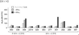


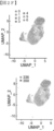


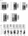


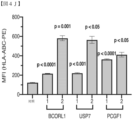


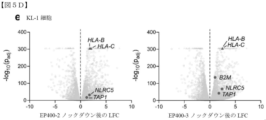


Classifications
-
- C—CHEMISTRY; METALLURGY
- C12—BIOCHEMISTRY; BEER; SPIRITS; WINE; VINEGAR; MICROBIOLOGY; ENZYMOLOGY; MUTATION OR GENETIC ENGINEERING
- C12N—MICROORGANISMS OR ENZYMES; COMPOSITIONS THEREOF; PROPAGATING, PRESERVING, OR MAINTAINING MICROORGANISMS; MUTATION OR GENETIC ENGINEERING; CULTURE MEDIA
- C12N15/00—Mutation or genetic engineering; DNA or RNA concerning genetic engineering, vectors, e.g. plasmids, or their isolation, preparation or purification; Use of hosts therefor
- C12N15/09—Recombinant DNA-technology
- C12N15/11—DNA or RNA fragments; Modified forms thereof; Non-coding nucleic acids having a biological activity
- C12N15/113—Non-coding nucleic acids modulating the expression of genes, e.g. antisense oligonucleotides; Antisense DNA or RNA; Triplex- forming oligonucleotides; Catalytic nucleic acids, e.g. ribozymes; Nucleic acids used in co-suppression or gene silencing
-
- C—CHEMISTRY; METALLURGY
- C12—BIOCHEMISTRY; BEER; SPIRITS; WINE; VINEGAR; MICROBIOLOGY; ENZYMOLOGY; MUTATION OR GENETIC ENGINEERING
- C12Y—ENZYMES
- C12Y304/00—Hydrolases acting on peptide bonds, i.e. peptidases (3.4)
- C12Y304/19—Omega peptidases (3.4.19)
- C12Y304/19012—Ubiquitinyl hydrolase 1 (3.4.19.12)
-
- A—HUMAN NECESSITIES
- A61—MEDICAL OR VETERINARY SCIENCE; HYGIENE
- A61P—SPECIFIC THERAPEUTIC ACTIVITY OF CHEMICAL COMPOUNDS OR MEDICINAL PREPARATIONS
- A61P35/00—Antineoplastic agents
-
- C—CHEMISTRY; METALLURGY
- C07—ORGANIC CHEMISTRY
- C07K—PEPTIDES
- C07K16/00—Immunoglobulins [IGs], e.g. monoclonal or polyclonal antibodies
- C07K16/18—Immunoglobulins [IGs], e.g. monoclonal or polyclonal antibodies against material from animals or humans
- C07K16/28—Immunoglobulins [IGs], e.g. monoclonal or polyclonal antibodies against material from animals or humans against receptors, cell surface antigens or cell surface determinants
- C07K16/30—Immunoglobulins [IGs], e.g. monoclonal or polyclonal antibodies against material from animals or humans against receptors, cell surface antigens or cell surface determinants from tumour cells
-
- C—CHEMISTRY; METALLURGY
- C12—BIOCHEMISTRY; BEER; SPIRITS; WINE; VINEGAR; MICROBIOLOGY; ENZYMOLOGY; MUTATION OR GENETIC ENGINEERING
- C12N—MICROORGANISMS OR ENZYMES; COMPOSITIONS THEREOF; PROPAGATING, PRESERVING, OR MAINTAINING MICROORGANISMS; MUTATION OR GENETIC ENGINEERING; CULTURE MEDIA
- C12N15/00—Mutation or genetic engineering; DNA or RNA concerning genetic engineering, vectors, e.g. plasmids, or their isolation, preparation or purification; Use of hosts therefor
- C12N15/09—Recombinant DNA-technology
- C12N15/11—DNA or RNA fragments; Modified forms thereof; Non-coding nucleic acids having a biological activity
- C12N15/113—Non-coding nucleic acids modulating the expression of genes, e.g. antisense oligonucleotides; Antisense DNA or RNA; Triplex- forming oligonucleotides; Catalytic nucleic acids, e.g. ribozymes; Nucleic acids used in co-suppression or gene silencing
- C12N15/1135—Non-coding nucleic acids modulating the expression of genes, e.g. antisense oligonucleotides; Antisense DNA or RNA; Triplex- forming oligonucleotides; Catalytic nucleic acids, e.g. ribozymes; Nucleic acids used in co-suppression or gene silencing against oncogenes or tumor suppressor genes
-
- C—CHEMISTRY; METALLURGY
- C07—ORGANIC CHEMISTRY
- C07K—PEPTIDES
- C07K2317/00—Immunoglobulins specific features
- C07K2317/20—Immunoglobulins specific features characterized by taxonomic origin
- C07K2317/24—Immunoglobulins specific features characterized by taxonomic origin containing regions, domains or residues from different species, e.g. chimeric, humanized or veneered
-
- C—CHEMISTRY; METALLURGY
- C07—ORGANIC CHEMISTRY
- C07K—PEPTIDES
- C07K2317/00—Immunoglobulins specific features
- C07K2317/50—Immunoglobulins specific features characterized by immunoglobulin fragments
- C07K2317/52—Constant or Fc region; Isotype
-
- C—CHEMISTRY; METALLURGY
- C12—BIOCHEMISTRY; BEER; SPIRITS; WINE; VINEGAR; MICROBIOLOGY; ENZYMOLOGY; MUTATION OR GENETIC ENGINEERING
- C12N—MICROORGANISMS OR ENZYMES; COMPOSITIONS THEREOF; PROPAGATING, PRESERVING, OR MAINTAINING MICROORGANISMS; MUTATION OR GENETIC ENGINEERING; CULTURE MEDIA
- C12N2310/00—Structure or type of the nucleic acid
- C12N2310/10—Type of nucleic acid
- C12N2310/14—Type of nucleic acid interfering N.A.
-
- C—CHEMISTRY; METALLURGY
- C12—BIOCHEMISTRY; BEER; SPIRITS; WINE; VINEGAR; MICROBIOLOGY; ENZYMOLOGY; MUTATION OR GENETIC ENGINEERING
- C12N—MICROORGANISMS OR ENZYMES; COMPOSITIONS THEREOF; PROPAGATING, PRESERVING, OR MAINTAINING MICROORGANISMS; MUTATION OR GENETIC ENGINEERING; CULTURE MEDIA
- C12N2310/00—Structure or type of the nucleic acid
- C12N2310/10—Type of nucleic acid
- C12N2310/20—Type of nucleic acid involving clustered regularly interspaced short palindromic repeats [CRISPRs]
-
- C—CHEMISTRY; METALLURGY
- C12—BIOCHEMISTRY; BEER; SPIRITS; WINE; VINEGAR; MICROBIOLOGY; ENZYMOLOGY; MUTATION OR GENETIC ENGINEERING
- C12N—MICROORGANISMS OR ENZYMES; COMPOSITIONS THEREOF; PROPAGATING, PRESERVING, OR MAINTAINING MICROORGANISMS; MUTATION OR GENETIC ENGINEERING; CULTURE MEDIA
- C12N2310/00—Structure or type of the nucleic acid
- C12N2310/50—Physical structure
- C12N2310/53—Physical structure partially self-complementary or closed
- C12N2310/531—Stem-loop; Hairpin
Landscapes
- Health & Medical Sciences (AREA)
- Life Sciences & Earth Sciences (AREA)
- Chemical & Material Sciences (AREA)
- Genetics & Genomics (AREA)
- Organic Chemistry (AREA)
- Engineering & Computer Science (AREA)
- Biomedical Technology (AREA)
- General Health & Medical Sciences (AREA)
- Bioinformatics & Cheminformatics (AREA)
- General Engineering & Computer Science (AREA)
- Wood Science & Technology (AREA)
- Zoology (AREA)
- Molecular Biology (AREA)
- Biochemistry (AREA)
- Biotechnology (AREA)
- Biophysics (AREA)
- Medicinal Chemistry (AREA)
- Plant Pathology (AREA)
- Physics & Mathematics (AREA)
- Microbiology (AREA)
- Immunology (AREA)
- Animal Behavior & Ethology (AREA)
- Chemical Kinetics & Catalysis (AREA)
- Public Health (AREA)
- Pharmacology & Pharmacy (AREA)
- Nuclear Medicine, Radiotherapy & Molecular Imaging (AREA)
- General Chemical & Material Sciences (AREA)
- Veterinary Medicine (AREA)
- Cell Biology (AREA)
- Proteomics, Peptides & Aminoacids (AREA)
- Oncology (AREA)
- Medicines That Contain Protein Lipid Enzymes And Other Medicines (AREA)
- Pharmaceuticals Containing Other Organic And Inorganic Compounds (AREA)
- Measuring Or Testing Involving Enzymes Or Micro-Organisms (AREA)
- Micro-Organisms Or Cultivation Processes Thereof (AREA)
- Medicines Containing Antibodies Or Antigens For Use As Internal Diagnostic Agents (AREA)
- Peptides Or Proteins (AREA)
Abstract
本発明は、部分的に、がん細胞上の主要組織適合性複合体(MHC)I発現を調節するための組成物及び方法に関する。【選択図】図1AThe present invention relates, in part, to compositions and methods for modulating major histocompatibility complex (MHC) I expression on cancer cells. [Selection drawing] Fig. 1A
Description
関連出願の相互参照
本出願は、2020年6月1日に出願された米国仮特許出願第63/032,956号、及び2020年6月15日に出願された米国仮特許出願第63/039,211号の優先権の利益を主張し、各出願の全内容が、この参照によりそれら全体が本明細書に組み込まれる。
CROSS REFERENCES TO RELATED APPLICATIONS This application is part of U.S. Provisional Patent Application No. 63/032,956, filed June 1, 2020, and U.S. Provisional Patent Application No. 63/039, filed June 15, 2020. , 211, and the entire contents of each application are hereby incorporated by reference in their entireties.
大きなファイル
本出願には、付属の4つの長い表の完全な内容が含まれ、これらの全ては、以下のようなASCIIテキストファイルである。2020年6月15日に作成された348,531バイトサイズの「表_1_CRISPR_Positive.txt」として本明細書とともに提出される表1、2020年6月15日に作成された292,318バイトサイズの「表_2_CRISPR_Negative.txt」として本明細書とともに提出される表2、2020年6月12日に作成された581,299バイトサイズの「表_3_ORF_Positive.txt」として本明細書とともに提出される表3、2020年6月12日に作成された855,629バイトサイズの「表_4_ORF_Negative.txt」として本明細書とともに提出される表4。これら4つの表の全ては、参照によりそれら全体が本明細書に組み込まれる。
LARGE FILES This application contains the complete contents of four long tables attached, all of which are ASCII text files as follows. Table 1, filed herewith as "Table_1_CRISPR_Positive.txt", 348,531 bytes in size, created on June 15, 2020, 292,318 bytes in size, created on June 15, 2020. Table 2, filed herewith as "Table_2_CRISPR_Negative.txt", Table 3, filed herewith as "Table_3_ORF_Positive.txt", 581,299 bytes created on June 12, 2020, 2020 Table 4, filed herewith as "Table_4_ORF_Negative.txt", 855,629 bytes in size, created June 12, 2006. All four of these tables are incorporated herein by reference in their entirety.
政府権利の陳述
本発明は、National Institutes of Healthによって授与された助成金番号R35 CA232128、P01 CA203655、R21 CA216772、NCI-SPORE-2P50CA101942-11A1、R01 CA155010、U24 CA224331、及びR01 HL131768に基づく政府の支援を受けて行われた。政府は、本発明において一定の権利を有する。
STATEMENT OF GOVERNMENT INTEREST This invention was made by grant numbers R35 CA232128, P01 CA203655, R21 CA216772, NCI-SPORE-2P50CA101942-11A1, R01 CA155010, U24 CA224331, awarded by the National Institutes of Health. and government support under R01 HL131768 It was done in response to The Government has certain rights in this invention.
ウイルスは、免疫系の認識を回避する一連の機構を用い、未検出の感染及び複製を可能にする。ウイルス免疫回避のための一般的な標的は、ペプチドプロセシング(PSMB8/LMP2、PSMB9/LMP7)、細胞質からERへのペプチド輸送(TAP1、TAP2)、及びB2M-HLA I重鎖(HLA-A、HLA-B、及びHLA-C)複合体へのペプチド装填を含む、いくつかのステップの協調機能を必要とする、HLAクラスI(HLA I又はMHC I)抗原提示経路である。この経路を妨害し、ウイルス抗原提示を回避するために、ウイルスは、他の機構の中で、ER(CMV)へのHLA I重鎖挿入を遮断するか、プロテアソーム分解(EBV)に抵抗するか、TAP(ヘルペスウイルス)を妨害するか、又はHLA分子(HIV)のトラフィッキング及びターンオーバーを変調する。ウイルスが免疫認識を回避するこれらの戦略は、ウイルス学及びがんに関連するクラスIの提示及び制御の機構を明らかにすることができる。 Viruses employ a series of mechanisms to evade immune system recognition, allowing undetected infection and replication. Common targets for viral immune evasion include peptide processing (PSMB8/LMP2, PSMB9/LMP7), peptide trafficking from the cytoplasm to the ER (TAP1, TAP2), and B2M-HLA I heavy chain (HLA-A, HLA -B, and HLA-C) is an HLA class I (HLA I or MHC I) antigen presentation pathway that requires coordinated functions of several steps, including peptide loading into the complex. To interfere with this pathway and evade viral antigen presentation, viruses either block HLA I heavy chain insertion into the ER (CMV) or resist proteasomal degradation (EBV), among other mechanisms. , interfere with TAP (herpes virus) or modulate trafficking and turnover of HLA molecules (HIV). These strategies by which viruses evade immune recognition may reveal mechanisms of class I presentation and regulation associated with virology and cancer.
例えば、稀で侵襲性の高い神経内分泌皮膚がんであるメルケル細胞がん(MCC)は、メルケル細胞ポリオーマウイルス(MCPyV)が、MCCの症例の80%の原因物質であるため、これらの疑問を調査するための興味深い設定を提示する。MCPyVは、2つのウイルス抗原:RBに結合し、不活性化するLT、及びクロマチン修飾複合体へのMYCLの動員を含む、多数の新たな機能を有するSTのみからなる。注目すべきことに、MCCは、一般的に低HLA I発現を示すが、これが媒介される機構は不明である。免疫組織化学(IHC)によって、MCC病変の84%が、表面HLA I下方制御又は喪失を呈することが報告されており、同様の所見がMCC細胞株において観察されている。しかしながら、MCCにおけるHLA I表面発現もまた、インターフェロン又はヒストンデアセチラーゼ(HDAC)阻害剤によってインビトロで上方制御され得るため、高度に可塑性であるように見える。したがって、がん細胞におけるHLA発現を増加させるために、治療戦略が緊急に必要である。 For example, Merkel cell carcinoma (MCC), a rare and highly aggressive neuroendocrine skin cancer, addresses these questions because Merkel cell polyomavirus (MCPyV) is the causative agent in 80% of MCC cases. Present an interesting setting to investigate. MCPyV consists solely of two viral antigens: LT, which binds and inactivates RB, and ST, which has a number of novel functions, including recruitment of MYCL to chromatin-modifying complexes. Of note, MCC generally display low HLA I expression, but the mechanism by which this is mediated is unclear. Immunohistochemistry (IHC) reports that 84% of MCC lesions exhibit surface HLA I downregulation or loss, and similar findings have been observed in MCC cell lines. However, HLA I surface expression in MCC also appears to be highly plastic, as it can be upregulated in vitro by interferons or histone deacetylase (HDAC) inhibitors. Therefore, there is an urgent need for therapeutic strategies to increase HLA expression in cancer cells.
本発明は、少なくとも部分的に、表1~5に列挙される1つ以上のバイオマーカー、例えば、MYCL又は1つ以上のPRC1.1複合体メンバー(PCGF1、BCORL1、及びUSP7など)の阻害又は遮断は、がん細胞におけるMHCクラスI分子、例えば、HLA I分子の発現の増加をもたらすという発見に基づいている。本発明は、表1~5に列挙される1つ以上のバイオマーカー、例えば、MYCL及び/又は1つ以上のPRC1.1複合体メンバー(例えば、PCGF1、BCORL1、及びUSP7)の調節(例えば、上方制御又は下方制御)を伴い、HLA I分子などのMHCクラスI分子の、がん細胞上での表面発現を増加させる。CRISPR/Cas9ベースの高スループットスクリーニングシステム及びオープンリーディングフレーム(ORF)スクリーニングを使用して、表1~5に列挙される1つ以上のバイオマーカー、例えば、MYCL及び/又は1つ以上のPRC1.1複合体メンバー(例えば、PCGF1、BCORL1、及びUSP7)は、調節されると、免疫療法に対してがんを感作させる標的として特定されている。例えば、メルケル細胞がんなどのがんにおいて、HLA IなどのMHCクラスIは、対照と比較して表面発現が低減し、PRC.1.1成分ポリペプチド又はMYCLなどの標的を阻害すると、HLA IなどのMHCクラスIの発現が増加し、それにより、これらの細胞の免疫療法に対する感受性が増加することが本明細書で実証される。表1~5に列挙される1つ以上のバイオマーカー、例えば、MYCL及び/又は1つ以上のPRC1.1複合体メンバー(例えば、PCGF1、BCORL1、及びUSP7)の阻害が、HLA IなどのMHCクラスIの表面発現を増加させることができることを検証する機能的データが、本明細書に提示される。したがって、表1~5に列挙される1つ以上のバイオマーカー、例えば、MYCL及び/又は1つ以上のPRC1.1複合体メンバー(例えば、PCGF1、BCORL1、及びUSP7)の調節因子は、特にがんに罹患した患者において、MHCクラスIの発現を調節し、かつ免疫応答を調節する(例えば、免疫応答を増加又は減少させる)ために有用であり、同時免疫療法の設定において、がんを治療するための新規の戦略を表す。 The present invention provides, at least in part, inhibition of one or more biomarkers listed in Tables 1-5, such as MYCL or one or more PRC1.1 complex members (such as PCGF1, BCORL1, and USP7) or Blockade is based on the discovery that it results in increased expression of MHC class I molecules, such as HLA I molecules, in cancer cells. The present invention provides for the modulation (eg, up-regulation or down-regulation) to increase the surface expression of MHC class I molecules, such as HLA I molecules, on cancer cells. One or more biomarkers listed in Tables 1-5, such as MYCL and/or one or more PRC1.1, using a CRISPR/Cas9-based high-throughput screening system and open reading frame (ORF) screening Complex members (eg, PCGF1, BCORL1, and USP7) have been identified as targets that, when regulated, sensitize cancer to immunotherapy. For example, in cancers such as Merkel cell carcinoma, MHC class I, such as HLA I, have reduced surface expression compared to controls and PRC. 1. It is demonstrated herein that inhibition of single component polypeptides or targets such as MYCL increases the expression of MHC class I, such as HLA I, thereby increasing the sensitivity of these cells to immunotherapy. be. Inhibition of one or more biomarkers listed in Tables 1-5, e.g., MYCL and/or one or more PRC1.1 complex members (e.g., PCGF1, BCORL1, and USP7) is associated with MHC, such as HLA I Functional data are presented herein that demonstrate that class I surface expression can be increased. Accordingly, one or more biomarkers listed in Tables 1-5, eg, modulators of MYCL and/or one or more PRC1.1 complex members (eg, PCGF1, BCORL1, and USP7), are is useful for modulating MHC class I expression and modulating the immune response (e.g., increasing or decreasing the immune response) in patients afflicted with cancer, and treating cancer in the setting of concurrent immunotherapy. represents a novel strategy for
本発明の一態様は、がんに罹患している対象を治療する方法であって、対象に、表1、2、3、4、若しくは5に列挙される1つ以上のバイオマーカー又はその断片のコピー数、発現レベル、及び/又は活性を修飾する治療有効量の薬剤、並びに免疫療法を投与することを含む、方法を提供する。 One aspect of the invention is a method of treating a subject with cancer, wherein the subject is treated with one or more biomarkers listed in Tables 1, 2, 3, 4, or 5, or fragments thereof. administering a therapeutically effective amount of an agent that modifies the copy number, expression level, and/or activity of , and immunotherapy.
本発明の任意の態様に適用され得、かつ/又は本明細書に記載される任意の他の実施形態と組み合わされ得る多数の実施形態が更に提供される。例えば、一実施形態では、薬剤は、表1若しくは4に列挙される1つ以上のバイオマーカー又はその断片のコピー数、発現レベル、及び/又は活性を減少させる。別の実施形態では、薬剤は、MYCLポリペプチド及び/若しくはポリコームリプレッサー複合体1.1(PRC1.1)ポリペプチド、又はポリペプチドをコードするポリヌクレオチドのコピー数、発現レベル、及び/又は活性を減少させる。なお別の実施形態では、ポリコームリプレッサー複合体1.1(PRC1.1)ポリペプチドは、USP7、BCORL1、PCGF1、KDM2B、SKP1、RING1A、RING1B、RYBP、YAF2、及び/又はBCORである。更に別の実施形態では、薬剤は、小分子阻害剤、CRISPR一本鎖ガイドRNA(sgRNA)、RNA干渉剤、アンチセンスオリゴヌクレオチド、ペプチド若しくはペプチド模倣薬阻害剤、アプタマー、抗体、又はイントラボディである。別の実施形態では、RNA干渉剤は、低分子干渉RNA(siRNA)、CRISPR RNA(crRNA)、小ヘアピンRNA(shRNA)、マイクロRNA(miRNA)、又はpiwi相互作用RNA(piRNA)である。なお別の実施形態では、sgRNAは、表1~4に列挙される核酸配列からなる群から選択される核酸配列を含む。更に別の実施形態では、薬剤は、表1、2、3、4、若しくは5に列挙される1つ以上のバイオマーカー及び/又は当該1つ以上のバイオマーカーの基質に特異的に結合する、イントラボディ又はその抗原結合断片を含む。別の実施形態では、イントラボディ又はその抗原結合断片は、マウス、キメラ、ヒト化、複合体、若しくはヒトイントラボディ、又はその抗原結合断片である。別の実施形態では、イントラボディ又はその抗原結合断片は、検出可能に標識されており、エフェクタードメインを含み、Fcドメインを含み、かつ/又はFv、Fav、F(ab’)2、Fab’、dsFv、scFv、sc(Fv)2、及びダイアボディ断片からなる群から選択される。別の実施形態では、薬剤は、表2若しくは3に列挙される1つ以上のバイオマーカー又はその断片のコピー数、発現レベル、及び/又は活性を増加させる。なお別の実施形態では、薬剤は、免疫療法に対するがん細胞の感受性を増加させる。更に別の実施形態では、免疫療法は、薬剤の前、後、又は薬剤と同時に投与される。なお別の実施形態では、免疫療法は、抗がんワクチン及び/又はウイルスを含む。別の実施形態では、免疫療法は、細胞ベースの免疫療法であり、任意に、細胞ベースの免疫療法は、キメラ抗原受容体(CAR-T)療法である。更に別の実施形態では、免疫療法は、免疫チェックポイントを阻害する。なお別の実施形態では、免疫チェックポイントは、CTLA-4、PD-1、VISTA、B7-H2、B7-H3、PD-L1、B7-H4、B7-H6、ICOS、HVEM、PD-L2、CD160、gp49B、PIR-B、KIRファミリー受容体、TIM-1、TIM-3、TIM-4、LAG-3、GITR、4-IBB、OX-40、BTLA、SIRP、CD47、CD48、2B4(CD244)、B7.1、B7.2、ILT-2、ILT-4、TIGIT、HHLA2、ブチロフィリン、IDO、CD39、CD73、及びA2aRからなる群から選択される。別の実施形態では、免疫チェックポイントは、PD-1、PD-L1、及びPD-L2からなる群から選択され、任意に、免疫チェックポイントは、PD-1である。更に別の実施形態では、1つ以上のバイオマーカーは、表5に列挙される核酸配列に対して少なくとも95%の同一性を有する核酸配列を含み、かつ/又は表5に列挙されるアミノ酸配列に対して少なくとも95%の同一性を有するアミノ酸配列をコードする。別の実施形態では、対象は、哺乳動物である。更に別の実施形態では、対象は、ヒト、非ヒト霊長類、マウス、ラット、又は家畜化哺乳動物である。更に別の実施形態では、薬剤は、免疫療法に対するがんの感受性を増加させ、任意に、(i)免疫療法は、T細胞媒介であり、かつ/又は(ii)薬剤は、がん細胞を含む腫瘍におけるCD8+T細胞の量を増加させる。別の実施形態では、薬剤は、がん細胞の表面上のMHC-Iのレベルを増加させる。別の実施形態では、方法は、対象に、少なくとも1つの追加のがん療法又はレジメンを投与することも含む。更に別の実施形態では、少なくとも1つの追加のがん療法又はレジメンは、薬剤及び/又は免疫療法の前、後、又は同時に投与される。更に別の実施形態では、薬剤は、薬学的に許容される製剤で投与される。なお別の実施形態では、がんは、神経内分泌がんである。なお別の実施形態では、神経内分泌がんは、メルケル細胞がん、神経芽細胞腫、小細胞肺がん、又は神経内分泌がんである。 Numerous embodiments are further provided that can be applied to any aspect of the invention and/or combined with any other embodiment described herein. For example, in one embodiment, the agent decreases the copy number, expression level and/or activity of one or more biomarkers listed in Tables 1 or 4 or fragments thereof. In another embodiment, the agent is a MYCL polypeptide and/or a Polycomb Repressor Complex 1.1 (PRC1.1) polypeptide, or a polynucleotide encoding the polypeptide, in copy number, expression level, and/or activity. decrease. In yet another embodiment, the Polycomb repressor complex 1.1 (PRC1.1) polypeptide is USP7, BCORL1, PCGF1, KDM2B, SKP1, RING1A, RING1B, RYBP, YAF2, and/or BCOR. In yet another embodiment, the agent is a small molecule inhibitor, CRISPR single-stranded guide RNA (sgRNA), RNA interference agent, antisense oligonucleotide, peptide or peptidomimetic inhibitor, aptamer, antibody, or intrabody. be. In another embodiment, the RNA interfering agent is a small interfering RNA (siRNA), CRISPR RNA (crRNA), small hairpin RNA (shRNA), microRNA (miRNA), or piwi-interacting RNA (piRNA). In yet another embodiment, the sgRNA comprises a nucleic acid sequence selected from the group consisting of nucleic acid sequences listed in Tables 1-4. In yet another embodiment, the agent specifically binds to one or more biomarkers listed in Tables 1, 2, 3, 4, or 5 and/or substrates for said one or more biomarkers; Including intrabodies or antigen-binding fragments thereof. In another embodiment, the intrabody or antigen-binding fragment thereof is a murine, chimeric, humanized, complex, or human intrabody, or antigen-binding fragment thereof. In another embodiment, the intrabody or antigen-binding fragment thereof is detectably labeled, comprises an effector domain, comprises an Fc domain, and/or contains Fv, Fav, F(ab')2, Fab', selected from the group consisting of dsFv, scFv, sc(Fv)2, and diabody fragments. In another embodiment, the agent increases the copy number, expression level and/or activity of one or more biomarkers listed in Tables 2 or 3 or fragments thereof. In yet another embodiment, the agent increases the sensitivity of cancer cells to immunotherapy. In yet another embodiment, the immunotherapy is administered before, after, or concurrently with the agent. In yet another embodiment, immunotherapy comprises anti-cancer vaccines and/or viruses. In another embodiment, the immunotherapy is cell-based immunotherapy, optionally the cell-based immunotherapy is chimeric antigen receptor (CAR-T) therapy. In yet another embodiment, the immunotherapy inhibits immune checkpoints. In yet another embodiment, the immune checkpoint is CTLA-4, PD-1, VISTA, B7-H2, B7-H3, PD-L1, B7-H4, B7-H6, ICOS, HVEM, PD-L2, CD160, gp49B, PIR-B, KIR family receptors, TIM-1, TIM-3, TIM-4, LAG-3, GITR, 4-IBB, OX-40, BTLA, SIRP, CD47, CD48, 2B4 (CD244 ), B7.1, B7.2, ILT-2, ILT-4, TIGIT, HHLA2, butyrophilin, IDO, CD39, CD73, and A2aR. In another embodiment, the immune checkpoint is selected from the group consisting of PD-1, PD-L1 and PD-L2, optionally the immune checkpoint is PD-1. In yet another embodiment, the one or more biomarkers comprise a nucleic acid sequence having at least 95% identity to a nucleic acid sequence listed in Table 5 and/or an amino acid sequence listed in Table 5 encodes an amino acid sequence having at least 95% identity to In another embodiment, the subject is a mammal. In yet another embodiment, the subject is a human, non-human primate, mouse, rat, or domesticated mammal. In yet another embodiment, the agent sensitizes the cancer to immunotherapy, optionally (i) the immunotherapy is T-cell mediated, and/or (ii) the agent sensitizes cancer cells. increase the amount of CD8+ T cells in tumors containing In another embodiment, the agent increases levels of MHC-I on the surface of cancer cells. In another embodiment, the method also includes administering to the subject at least one additional cancer therapy or regimen. In yet another embodiment, at least one additional cancer therapy or regimen is administered before, after, or concurrently with the drug and/or immunotherapy. In yet another embodiment, the agent is administered in a pharmaceutically acceptable formulation. In yet another embodiment, the cancer is a neuroendocrine cancer. In yet another embodiment, the neuroendocrine cancer is Merkel cell carcinoma, neuroblastoma, small cell lung cancer, or neuroendocrine cancer.
別の態様は、がん細胞における主要組織適合性複合体の発現を増加させる方法を提供し、当該方法は、がん細胞を、表1、2、3、4、若しくは5に列挙される1つ以上のバイオマーカー又はその断片のコピー数、発現レベル、及び/又は活性を調節する薬剤と接触させることを含み、任意に、がん細胞、又はがん細胞及び免疫細胞を含む細胞の集団を、免疫療法と接触させることを更に含む。 Another aspect provides a method of increasing expression of the major histocompatibility complex in a cancer cell, the method comprising treating the cancer cell as one listed in Tables 1, 2, 3, 4, or 5. comprising contacting a cancer cell or population of cells comprising cancer cells and immune cells with an agent that modulates the copy number, expression level and/or activity of one or more biomarkers or fragments thereof; , further comprising contacting with immunotherapy.
本発明の任意の態様に適用され得、かつ/又は本明細書に記載される任意の他の実施形態と組み合わされ得る多数の実施形態が更に提供される。一実施形態では、表1又は4に列挙される1つ以上のバイオマーカーのコピー数、発現レベル、及び/又は活性を減少させる薬剤。別の実施形態では、薬剤は、MYCLポリペプチド及び/若しくはポリコームリプレッサー複合体1.1(PRC1.1)ポリペプチド、又はポリペプチドをコードするポリヌクレオチドのコピー数、発現レベル、及び/又は活性を減少させる。更に別の実施形態では、ポリコームリプレッサー複合体1.1(PRC1.1)ポリペプチドは、USP7、BCORL1、PCGF1、KDM2B、SKP1、RING1A、RING1B、RYBP、YAF2、及び/又はBCORである。なお別の実施形態では、薬剤は、小分子阻害剤、CRISPR一本鎖ガイドRNA(sgRNA)、RNA干渉剤、アンチセンスオリゴヌクレオチド、ペプチド若しくはペプチド模倣薬阻害剤、アプタマー、抗体、又はイントラボディである。一実施形態では、RNA干渉剤は、低分子干渉RNA(siRNA)、CRISPR RNA(crRNA)、小ヘアピンRNA(shRNA)、マイクロRNA(miRNA)、又はpiwi相互作用RNA(piRNA)である。別の実施形態では、sgRNAは、表1~4に列挙される核酸配列からなる群から選択される核酸配列を含む。更に別の実施形態では、薬剤は、表1、2、3、4、若しくは5に列挙される1つ以上のバイオマーカー及び/又は当該1つ以上のバイオマーカーの基質に特異的に結合する、イントラボディ又はその抗原結合断片を含む。更に別の実施形態では、イントラボディ又はその抗原結合断片は、マウス、キメラ、ヒト化、複合体、又はヒトイントラボディである。一実施形態では、イントラボディ又はその抗原結合断片は、検出可能に標識されており、エフェクタードメインを含み、Fcドメインを含み、かつ/又はFv、Fav、F(ab’)2、Fab’、dsFv、scFv、sc(Fv)2、及びダイアボディ断片からなる群から選択される。別の実施形態では、薬剤は、表2又は3に列挙される1つ以上のバイオマーカーのコピー数、発現レベル、及び/又は活性を増加させる。更に別の実施形態では、薬剤は、免疫療法に対するがん細胞の感受性を増加させる。更に別の実施形態では、がん細胞は、薬剤の前、後、又は薬剤と同時に免疫療法と接触する。なお別の実施形態では、免疫療法は、抗がんワクチン及び/又はウイルスを含む。別の実施形態では、免疫療法は、細胞ベースの免疫療法であり、任意に、細胞ベースの免疫療法は、キメラ抗原受容体(CAR-T)療法である。別の実施形態では、免疫療法は、免疫チェックポイントを阻害する。更に別の実施形態では、免疫チェックポイントは、CTLA-4、PD-1、VISTA、B7-H2、B7-H3、PD-L1、B7-H4、B7-H6、ICOS、HVEM、PD-L2、CD160、gp49B、PIR-B、KIRファミリー受容体、TIM-1、TIM-3、TIM-4、LAG-3、GITR、4-IBB、OX-40、BTLA、SIRP、CD47、CD48、2B4(CD244)、B7.1、B7.2、ILT-2、ILT-4、TIGIT、HHLA2、ブチロフィリン、IDO、CD39、CD73、及びA2aRからなる群から選択される。なお別の実施形態では、免疫チェックポイントが、PD-1、PD-L1、及びPD-L2からなる群から選択される。更に別の実施形態では、免疫チェックポイントは、PD-1である。別の実施形態では、バイオマーカーは、表5に列挙される核酸配列に対して少なくとも95%の同一性を有する核酸配列を含み、かつ/又は表5に列挙されるアミノ酸配列に対して少なくとも95%の同一性を有するアミノ酸配列をコードする。更に別の実施形態では、1つ以上のバイオマーカーは、ヒト、マウス、キメラ、又は融合バイオマーカーである。別の実施形態では、免疫療法は、(i)T細胞媒介性であり、かつ/又は(ii)薬剤は、がん細胞を含む腫瘍におけるCD8+T細胞の量を増加させる。更に別の実施形態では、薬剤は、がん細胞におけるMHCクラスI表面発現のレベルを増加させる。なお別の実施形態では、方法は、対象に、少なくとも1つの追加のがん療法又はレジメンを投与することを更に含む。別の実施形態では、少なくとも1つの追加のがん療法又はレジメンは、薬剤及び/又は免疫療法の前、後、又は同時に投与される。別の実施形態では、薬剤は、薬学的に許容される製剤で投与される。一実施形態では、がん細胞は、神経内分泌がん細胞である。別の実施形態では、神経内分泌がん細胞は、メルケル細胞がん、神経芽細胞腫、小細胞肺がん、又は神経内分泌がん細胞である。 Numerous embodiments are further provided that can be applied to any aspect of the invention and/or combined with any other embodiment described herein. In one embodiment, an agent that decreases the copy number, expression level and/or activity of one or more biomarkers listed in Tables 1 or 4. In another embodiment, the agent is a MYCL polypeptide and/or a Polycomb Repressor Complex 1.1 (PRC1.1) polypeptide, or a polynucleotide encoding the polypeptide, in copy number, expression level, and/or activity. decrease. In yet another embodiment, the Polycomb repressor complex 1.1 (PRC1.1) polypeptide is USP7, BCORL1, PCGF1, KDM2B, SKP1, RING1A, RING1B, RYBP, YAF2, and/or BCOR. In yet another embodiment, the agent is a small molecule inhibitor, a CRISPR single-stranded guide RNA (sgRNA), an RNA interference agent, an antisense oligonucleotide, a peptide or peptidomimetic inhibitor, an aptamer, an antibody, or an intrabody. be. In one embodiment, the RNA interfering agent is a small interfering RNA (siRNA), CRISPR RNA (crRNA), small hairpin RNA (shRNA), microRNA (miRNA), or piwi-interacting RNA (piRNA). In another embodiment, the sgRNA comprises a nucleic acid sequence selected from the group consisting of nucleic acid sequences listed in Tables 1-4. In yet another embodiment, the agent specifically binds to one or more biomarkers listed in Tables 1, 2, 3, 4, or 5 and/or substrates for said one or more biomarkers; Including intrabodies or antigen-binding fragments thereof. In yet another embodiment, the intrabody or antigen-binding fragment thereof is a murine, chimeric, humanized, conjugate, or human intrabody. In one embodiment, the intrabody or antigen-binding fragment thereof is detectably labeled, comprises an effector domain, comprises an Fc domain, and/or is Fv, Fav, F(ab')2, Fab', dsFv , scFv, sc(Fv)2, and diabody fragments. In another embodiment, the agent increases copy number, expression level and/or activity of one or more biomarkers listed in Tables 2 or 3. In yet another embodiment, the agent sensitizes cancer cells to immunotherapy. In yet another embodiment, cancer cells are contacted with immunotherapy before, after, or simultaneously with the agent. In yet another embodiment, immunotherapy comprises anti-cancer vaccines and/or viruses. In another embodiment, the immunotherapy is cell-based immunotherapy, optionally the cell-based immunotherapy is chimeric antigen receptor (CAR-T) therapy. In another embodiment, the immunotherapy inhibits immune checkpoints. In yet another embodiment, the immune checkpoint is CTLA-4, PD-1, VISTA, B7-H2, B7-H3, PD-L1, B7-H4, B7-H6, ICOS, HVEM, PD-L2, CD160, gp49B, PIR-B, KIR family receptors, TIM-1, TIM-3, TIM-4, LAG-3, GITR, 4-IBB, OX-40, BTLA, SIRP, CD47, CD48, 2B4 (CD244 ), B7.1, B7.2, ILT-2, ILT-4, TIGIT, HHLA2, butyrophilin, IDO, CD39, CD73, and A2aR. In yet another embodiment, the immune checkpoint is selected from the group consisting of PD-1, PD-L1, and PD-L2. In yet another embodiment, the immune checkpoint is PD-1. In another embodiment, the biomarker comprises a nucleic acid sequence having at least 95% identity to a nucleic acid sequence listed in Table 5 and/or at least 95% identity to an amino acid sequence listed in Table 5. It encodes amino acid sequences with % identity. In yet another embodiment, the one or more biomarkers are human, murine, chimeric, or fusion biomarkers. In another embodiment, the immunotherapy is (i) T cell-mediated and/or (ii) the agent increases the amount of CD8+ T cells in the tumor containing cancer cells. In yet another embodiment, the agent increases the level of MHC class I surface expression in cancer cells. In yet another embodiment, the method further comprises administering to the subject at least one additional cancer therapy or regimen. In another embodiment, at least one additional cancer therapy or regimen is administered before, after, or concurrently with the drug and/or immunotherapy. In another embodiment, the agent is administered in a pharmaceutically acceptable formulation. In one embodiment, the cancer cell is a neuroendocrine cancer cell. In another embodiment, the neuroendocrine cancer cell is a Merkel cell carcinoma, neuroblastoma, small cell lung cancer, or neuroendocrine cancer cell.
本発明の別の態様は、表1、2、3、4、又は5に列挙される少なくとも1つのバイオマーカーのコピー数、量、及び/又は活性を調節することによって治療することができるがんに罹患しているか、又はそれを発症するリスクのある対象を特定する方法であり、当該方法は、対照と比較した対象からの細胞における主要組織適合性複合体(MHC)クラスI発現の増加又は減少レベルを検出することと、それによって、表1、2、3、4、又は5に列挙される少なくとも1つのバイオマーカーのコピー数、量、及び/又は活性を調節することによって治療することができるがんに罹患しているか、又はそれを発症するリスクのある対象を特定することと、を含み、任意に、対象由来の細胞を含む生体試料は、対象から得られる。 Another aspect of the invention is cancer that can be treated by modulating the copy number, amount, and/or activity of at least one biomarker listed in Tables 1, 2, 3, 4, or 5. A method of identifying a subject having or at risk of developing a treating by detecting a reduced level and thereby modulating the copy number, amount and/or activity of at least one biomarker listed in Tables 1, 2, 3, 4, or 5 identifying a subject suffering from or at risk of developing a cancer that can cause cancer, and optionally a biological sample comprising cells from the subject is obtained from the subject.
本発明の任意の態様に適用され得、かつ/又は本明細書に記載される任意の他の実施形態と組み合わされ得る多数の実施形態が更に提供される。一実施形態では、薬剤は、表1又は4に列挙される少なくとも1つのバイオマーカーのコピー数、量、及び/又は活性を減少させる。別の実施形態では、方法はまた、表1又は4に列挙される少なくとも1つのバイオマーカーを阻害する薬剤を、特定された対象に推奨すること、処方すること、又は投与することを含む。更に別の実施形態では、薬剤は、表2又は3に列挙される少なくとも1つのバイオマーカーのコピー数、量、及び/又は活性を増加させる。別の実施形態では、本方法は、特定された対象に、免疫療法を推奨する、処方する、又は投与することを更に含む。一実施形態では、免疫療法は、抗がんワクチン、抗がんウイルス、及び/又はチェックポイント阻害剤を含む。別の実施形態では、方法は、対象に、標的療法、化学療法、放射線療法、及び/又はホルモン療法からなる群から選択されるがん療法を推奨すること、処方すること、又は投与することを更に含む。更に別の実施形態では、対照は、患者又は当該患者が属する同じ種のメンバーのいずれかに由来する、がん性又は非がん性試料に由来する試料を含む。なお別の実施形態では、対照は、既知の参照値である。一実施形態では、がんは、神経内分泌がんである。別の実施形態では、神経内分泌がんは、メルケル細胞がん、神経芽細胞腫、小細胞肺がん、又は神経内分泌がんである。 Numerous embodiments are further provided that can be applied to any aspect of the invention and/or combined with any other embodiment described herein. In one embodiment, the agent decreases copy number, amount and/or activity of at least one biomarker listed in Tables 1 or 4. In another embodiment, the method also includes recommending, prescribing, or administering to the identified subject an agent that inhibits at least one biomarker listed in Table 1 or 4. In yet another embodiment, the agent increases copy number, amount and/or activity of at least one biomarker listed in Tables 2 or 3. In another embodiment, the method further comprises recommending, prescribing or administering immunotherapy to the identified subject. In one embodiment, immunotherapy includes anti-cancer vaccines, anti-cancer viruses, and/or checkpoint inhibitors. In another embodiment, the method recommends, prescribes, or administers to the subject a cancer therapy selected from the group consisting of targeted therapy, chemotherapy, radiation therapy, and/or hormone therapy. Including further. In yet another embodiment, the control comprises a sample derived from a cancerous or non-cancerous sample from either the patient or a member of the same species to which the patient belongs. In yet another embodiment, the control is a known reference value. In one embodiment, the cancer is a neuroendocrine cancer. In another embodiment, the neuroendocrine cancer is Merkel cell carcinoma, neuroblastoma, small cell lung cancer, or neuroendocrine cancer.
別の態様では、表1、2、3、4、若しくは5に列挙された1つ以上のバイオマーカー又はその断片を発現するがんに罹患している対象の、免疫療法による治療に対する臨床転帰を予測するための方法が提供され、当該方法は、a)対象試料中の表1、2、3、4、又は5に列挙される少なくとも1つのバイオマーカーのコピー数、量、及び/又は活性を決定することと、b)良好な臨床転帰を有する対照中の少なくとも1つのバイオマーカーのコピー数、量、及び/又は活性を決定することと、c)対象試料中及び対照中の少なくとも1つのバイオマーカーのコピー数、量、及び/又は活性を比較することと、を含み、対照中のコピー数、量、及び/又は活性を比較した、対象試料中の表1、2、3、4、又は5に列挙される少なくとも1つのバイオマーカーのコピー数、量、及び/又は活性の存在又はわずかな変化は、対象が不良な臨床転帰を有することを示す。 In another aspect, the clinical outcome of a subject with a cancer expressing one or more biomarkers listed in Table 1, 2, 3, 4, or 5, or a fragment thereof, to treatment with immunotherapy A method is provided for predicting a) the copy number, amount, and/or activity of at least one biomarker listed in Tables 1, 2, 3, 4, or 5 in a subject sample b) determining the copy number, amount and/or activity of at least one biomarker in a control with a favorable clinical outcome; c) at least one biomarker in the subject sample and in the control Comparing the copy number, amount and/or activity of the marker compared to the copy number, amount and/or activity in the control sample, Table 1, 2, 3, 4, or The presence or slight change in copy number, amount and/or activity of at least one biomarker listed in 5 is indicative that the subject has a poor clinical outcome.
別の態様は、表1若しくは4に列挙される少なくとも1つのバイオマーカーのコピー数及び/若しくは量を減少させ、かつ/又は活性を阻害する薬剤と、免疫療法とで、がんを有するか、又はがんを有する疑いのある対象の治療を監視するための方法を提供し、当該方法は、第1の時間点での対象に由来する試料におけるMHCクラスI発現のレベル、及びその後の時間点での対象に由来する試料におけるMHCクラスI発現のレベルの変化又は変化がないことを検出し、それによって、対象の治療を監視することを含む。 Another aspect is immunotherapy with an agent that reduces the copy number and/or amount and/or inhibits the activity of at least one biomarker listed in Table 1 or 4, has cancer, or or a method for monitoring treatment of a subject suspected of having cancer, the method comprising measuring the level of MHC class I expression in a sample from the subject at a first time point and at subsequent time points detecting a change or lack of change in the level of MHC class I expression in a sample derived from the subject at , thereby monitoring treatment of the subject.
更に別の態様は、表2若しくは3に列挙される少なくとも1つのバイオマーカーのコピー数及び/若しくは量を増加させ、かつ/又は活性を阻害する薬剤と、免疫療法とで、がんを有するか、又はがんを有する疑いのある対象の治療を監視するための方法を提供し、当該方法は、第1の時間点での対象に由来する試料におけるMHCクラスI発現のレベル、及びその後の時間点での対象に由来する試料におけるMHCクラスI発現のレベルの変化又は変化がないことを検出し、それによって、対象の治療を監視することを含む。 Yet another aspect is immunotherapy with an agent that increases the copy number and/or amount and/or inhibits the activity of at least one biomarker listed in Table 2 or 3. or a method for monitoring treatment of a subject suspected of having cancer, the method comprising measuring the level of MHC class I expression in a sample from the subject at a first time point and a time thereafter detecting a change or lack of change in the level of MHC class I expression in a sample from the subject at the point, thereby monitoring treatment of the subject.
なお別の態様では、方法は、対象における表1又は4に列挙される少なくとも1つのバイオマーカーのコピー数、量、及び/又は活性を減少させる薬剤の有効性を評価するために提供され、当該方法は、対象の試料において、第1の時間点で、その後の時間点と比較した表1又は4に列挙される少なくとも1つのバイオマーカーのコピー数、量、及び/又は活性の変化又は変化がないことを検出することを含み、表1又は4に列挙される少なくとも1つのバイオマーカーのコピー数、量、及び/又は活性の減少は、薬剤が有効であることを示す。 In yet another aspect, a method is provided for assessing the efficacy of an agent to reduce the copy number, amount, and/or activity of at least one biomarker listed in Table 1 or 4 in a subject, The method is characterized in that at a first time point in a sample of the subject there is a change or change in the copy number, amount and/or activity of at least one biomarker listed in Table 1 or 4 compared to subsequent time points. A decrease in the copy number, amount, and/or activity of at least one biomarker listed in Table 1 or 4, including detecting the absence, indicates that the agent is efficacious.
別の態様は、対象における表2又は3に列挙される少なくとも1つのバイオマーカーのコピー数、量、及び/又は活性を増加させる薬剤の有効性を評価する方法を提供し、当該方法は、対象の試料において、第1の時間点で、その後の時間点と比較した表2又は3に列挙される少なくとも1つのバイオマーカーのコピー数、量、及び/又は活性の変化又は変化がないことを検出することを含み、表2又は3に列挙される少なくとも1つのバイオマーカーのコピー数、量、及び/又は活性の減少は、薬剤が有効であることを示す。 Another aspect provides a method of assessing the efficacy of an agent to increase the copy number, amount, and/or activity of at least one biomarker listed in Table 2 or 3 in a subject, the method comprising detect a change or no change in copy number, amount, and/or activity of at least one biomarker listed in Table 2 or 3 compared to subsequent time points at a first time point in samples of A decrease in copy number, amount and/or activity of at least one biomarker listed in Tables 2 or 3 is indicative of the agent being effective.
本発明の任意の態様に適用され得、かつ/又は本明細書に記載される任意の他の実施形態と組み合わされ得る多数の実施形態が更に提供される。一実施形態では、第1の時間点と、その後の時間点との間で、対象が、がんの治療を受けた、治療を終了した、かつ/又は寛解中である。別の実施形態では、治療は、薬剤を対象に投与することを含む。更に別の実施形態では、第1及び/又はその後の試料は、エクスビボ又はインビボ試料を含む。なお別の実施形態では、第1及び/又は少なくとも1つのその後の試料は、対象から得られる単一試料又はプールされた試料の一部である。別の実施形態では、試料は、対象から得られた細胞、血清、腫瘍周辺組織、及び/又は腫瘍内組織を含む。更に別の実施形態では、表1、2、3、4、又は5に列挙される1つ以上のバイオマーカー。一実施形態では、がん又はがん細胞は、神経内分泌がんである。別の実施形態では、神経内分泌がんは、メルケル細胞がん、神経芽細胞腫、小細胞肺がん、又は神経内分泌がんである。更に別の実施形態では、がん又はがん細胞は、がんの動物モデルにある。なお別の実施形態では、動物モデルは、マウスモデルである。一実施形態では、がんは、哺乳類対象にある。別の実施形態では、哺乳類対象は、マウス又はヒトである。更に別の実施形態では、哺乳動物は、ヒトである。 Numerous embodiments are further provided that can be applied to any aspect of the invention and/or combined with any other embodiment described herein. In one embodiment, between the first time point and a subsequent time point, the subject has been treated for, completed treatment for, and/or is in remission for cancer. In another embodiment, treating comprises administering an agent to a subject. In yet another embodiment, the first and/or subsequent samples comprise ex vivo or in vivo samples. In yet another embodiment, the first and/or at least one subsequent sample is part of a single sample or pooled sample obtained from the subject. In another embodiment, the sample comprises cells, serum, peritumoral tissue, and/or intratumoral tissue obtained from the subject. In yet another embodiment, one or more biomarkers listed in Tables 1, 2, 3, 4, or 5. In one embodiment, the cancer or cancer cell is a neuroendocrine cancer. In another embodiment, the neuroendocrine cancer is Merkel cell carcinoma, neuroblastoma, small cell lung cancer, or neuroendocrine cancer. In yet another embodiment, the cancer or cancer cell is in an animal model of cancer. In yet another embodiment, the animal model is a mouse model. In one embodiment the cancer is in a mammalian subject. In another embodiment, the mammalian subject is a mouse or human. In yet another embodiment, the mammal is human.
上述の態様及び実施形態は、免疫療法と組み合わせて阻害することが、免疫療法単独で観察されるかかる利益の欠如を考慮して予期しないがんを治療するための相乗的治療利益をもたらす、表1、4、及び5に列挙されるものなどの本発明のバイオマーカーの代表的な実施形態を提供するが、免疫療法と組み合わせて、阻害よりもむしろ発現の促進(例えば、枯渇させるよりもsgRNAスクリーニングで濃縮されるように識別される)が、がんを治療するための相乗的治療利益をもたらす、本明細書(特に表1、4、及び5)で明確に記載される特定のバイオマーカーが容易に明らかである。したがって、本明細書及び上記に記載される任意の態様及び実施形態は、免疫療法及びがんに関する診断的、予後的、治療的等の用途において、かかるバイオマーカー及びそれらの促進された発現を使用することができる。例えば、一態様では、免疫療法と組み合わせて、表1、4、若しくは5に列挙される1つ以上のかかるバイオマーカー、又はその断片のコピー数、発現レベル、及び/又は活性を阻害するよりもむしろ促進する薬剤とがん細胞を接触させることを含む、がん細胞を死滅させる方法が提供される。別の代表的な態様では、がんに罹患しているか、又はがんを発症する危険性のある対象が、表1、4、又は5に列挙されるかかる少なくとも1つのバイオマーカーのコピー数、量、及び/又は活性を促進することから利益を受けるかどうかを決定する方法が提供され、この方法は、a)対象から生体試料を得ることと、b)表1に列挙される少なくとも1つのバイオマーカーのコピー数、量、及び/又は活性を決定することと、c)対照における少なくとも1つのバイオマーカーのコピー数、量、及び/又は活性を決定することと、d)ステップb)及びc)で検出された少なくとも1つのバイオマーカーのコピー数、量、及び/又は活性を比較することと、を含み、対照の少なくとも1つのバイオマーカーのコピー数、量、及び/又は活性と比較して、対象試料における表1、4、又は5に列挙される少なくとも1つのバイオマーカーのコピー数、量、及び/又は活性の不在又は著しい減少は、がんに罹患しているか、又はがんを発症する危険性のある対象が、表1、4、又は5に列挙される少なくとも1つのバイオマーカーのコピー数、量、及び/又は活性を促進することから利益を得るであろうことを示す。 The aspects and embodiments described above demonstrate that inhibition in combination with immunotherapy provides a synergistic therapeutic benefit for treating cancer that is unexpected given the lack of such benefit observed with immunotherapy alone. Exemplary embodiments of the biomarkers of the invention, such as those listed in 1, 4, and 5, are provided, but in combination with immunotherapy, promote expression rather than inhibit (e.g., sgRNA rather than deplete) specific biomarkers explicitly described herein (especially Tables 1, 4, and 5) that are identified as enriched in the screen, provide a synergistic therapeutic benefit for treating cancer is readily apparent. Accordingly, any of the aspects and embodiments described herein and above use such biomarkers and their enhanced expression in diagnostic, prognostic, therapeutic, etc. applications related to immunotherapy and cancer. can do. For example, in one aspect, in combination with immunotherapy, rather than inhibiting the copy number, expression level, and/or activity of one or more such biomarkers listed in Tables 1, 4, or 5, or fragments thereof, Rather, methods of killing cancer cells are provided comprising contacting the cancer cells with an agent that promotes. In another exemplary aspect, the subject having cancer or at risk of developing cancer has a copy number of at least one such biomarker listed in Table 1, 4, or 5; A method of determining whether one would benefit from enhancing the amount and/or activity is provided, the method comprising: a) obtaining a biological sample from a subject; determining the copy number, amount and/or activity of the biomarker; c) determining the copy number, amount and/or activity of at least one biomarker in the control; d) steps b) and c ), and comparing the copy number, amount, and/or activity of the at least one biomarker detected in a control, as compared to the copy number, amount, and/or activity of the at least one biomarker in a control , the absence or significant reduction in the copy number, amount, and/or activity of at least one biomarker listed in Tables 1, 4, or 5 in the subject sample is having or developing cancer that subjects at risk of will benefit from enhancing the copy number, amount, and/or activity of at least one biomarker listed in Tables 1, 4, or 5.
更に、上述の態様及び実施形態は、免疫療法と組み合わせて促進することが、免疫療法単独で観察されるかかる利益の欠如を考慮して予期しないがんを治療するための相乗的治療利益をもたらす、表2及び3に列挙されるものなどの本発明のバイオマーカーの代表的な実施形態を提供するが、免疫療法と組み合わせて、促進よりもむしろ発現の阻害(例えば、枯渇させるよりもsgRNAスクリーニングで濃縮されるように識別される)が、がんを治療するための相乗的治療利益をもたらす、本明細書(特に表2及び3)で明確に記載される特定のバイオマーカーが容易に明らかである。したがって、本明細書及び上記に記載される任意の態様及び実施形態は、免疫療法及びがんに関する診断的、予後的、治療的などの用途において、かかるバイオマーカー及びそれらの促進された発現を使用することができる。例えば、一態様では、免疫療法と組み合わせて、表2若しくは3に列挙される1つ以上のかかるバイオマーカー又はその断片のコピー数、発現レベル、及び/又は活性を阻害するよりもむしろ促進する薬剤とがん細胞を接触させることを含む、がん細胞を死滅させる方法が提供される。別の代表的な態様では、がんに罹患しているか、又はがんを発症する危険性のある対象が、表2又は3に列挙されるかかる少なくとも1つのバイオマーカーのコピー数、量、及び/又は活性を促進することから利益を受けるかどうかを決定する方法が提供され、この方法は、a)対象から生体試料を得ることと、b)表1に列挙される少なくとも1つのバイオマーカーのコピー数、量、及び/又は活性を決定することと、c)対照における少なくとも1つのバイオマーカーのコピー数、量、及び/又は活性を決定することと、d)ステップb)及びc)で検出された少なくとも1つのバイオマーカーのコピー数、量、及び/又は活性を比較することと、を含み、対照の少なくとも1つのバイオマーカーのコピー数、量、及び/又は活性と比較して、対象試料における表2又は3に列挙される少なくとも1つのバイオマーカーのコピー数、量、及び/又は活性の不在又は著しい減少は、がんに罹患しているか、又はがんを発症する危険性のある対象が、表2又は3に列挙される少なくとも1つのバイオマーカーのコピー数、量、及び/又は活性を促進することから利益を得るであろうことを示す。 Furthermore, the aspects and embodiments described above, when promoted in combination with immunotherapy, provide synergistic therapeutic benefits for treating cancer, unexpected given the lack of such benefits observed with immunotherapy alone. , provides representative embodiments of the biomarkers of the invention, such as those listed in Tables 2 and 3, but in combination with immunotherapy inhibit expression rather than promote (e.g., sgRNA screening rather than depletion) specific biomarkers explicitly described herein (particularly Tables 2 and 3) that confer synergistic therapeutic benefit for treating cancer are readily apparent. is. Accordingly, any of the aspects and embodiments described herein and above use such biomarkers and their enhanced expression in diagnostic, prognostic, therapeutic, etc. applications related to immunotherapy and cancer. can do. For example, in one aspect, agents that promote rather than inhibit the copy number, expression levels, and/or activity of one or more such biomarkers or fragments thereof listed in Tables 2 or 3 in combination with immunotherapy A method of killing cancer cells is provided comprising contacting the cancer cells with an agent. In another exemplary aspect, the subject having cancer or at risk of developing cancer has the copy number, amount, and at least one such biomarker listed in Table 2 or 3 A method of determining whether one would benefit from promoting activity is provided, the method comprising: a) obtaining a biological sample from a subject; c) determining the copy number, amount and/or activity of at least one biomarker in a control; d) detecting in steps b) and c) and comparing the copy number, amount, and/or activity of at least one biomarker obtained in a subject sample compared to the copy number, amount, and/or activity of at least one biomarker in a control Absence or significant reduction in copy number, amount, and/or activity of at least one biomarker listed in Table 2 or 3 in a subject suffering from or at risk of developing cancer would benefit from enhancing the copy number, amount and/or activity of at least one biomarker listed in Tables 2 or 3.
特許出願ファイルは、カラーで仕上げられた少なくとも1つの図面を含有する。カラー図面を有するこの特許又は特許出願刊行物のコピーは、申請及び必要な料金の支払い後、特許庁によって提供される。 The patent application file contains at least one drawing executed in color. Copies of this patent or patent application publication with color drawing(s) will be provided by the Office upon request and payment of the necessary fee.
本明細書では、表1~5に列挙される1つ以上のバイオマーカー(例えば、MYCL)又は1つ以上のPRC1.1複合体メンバー(PCGF1、BCORL1、及びUSP7など)の制御因子を使用して、細胞上の表面MHC-I発現を調節し、免疫応答を調節し、腫瘍免疫及び免疫療法に対する応答性を増強することができることが決定されている。例えば、(a)表1若しくは表4に列挙される1つ以上のバイオマーカーのコピー数、発現レベル、及び/若しくは活性を減少させること、並びに/又は(b)表2若しくは表3に列挙される1つ以上のバイオマーカーのコピー数、発現レベル、及び/若しくは活性を増加させることは、細胞上のMHC-I発現の増加、及び免疫療法に対する応答性の増加を伴う免疫応答の増加をもたらし、これは、がん、感染症などの免疫応答の増加から利益を受け得る障害を治療するのに有用である。同様に、(a)表1若しくは表4に列挙される1つ以上のバイオマーカーのコピー数、発現レベル、及び/若しくは活性を増加させること、並びに/又は(b)表2若しくは表3に列挙される1つ以上のバイオマーカーのコピー数、発現レベル、及び/若しくは活性を減少させることは、細胞上のMHC-I発現の減少、及び免疫療法に対する応答性の減少を伴う免疫応答の減少をもたらし、これは、自己免疫障害のような免疫応答の減少から利益を受け得る障害を治療するのに有用である。 As used herein, one or more biomarkers (e.g., MYCL) or regulators of one or more PRC1.1 complex members (such as PCGF1, BCORL1, and USP7) listed in Tables 1-5 are used. have been determined to be able to modulate surface MHC-I expression on cells, modulate immune responses, and enhance tumor immunity and responsiveness to immunotherapy. For example, (a) decreasing the copy number, expression level, and/or activity of one or more biomarkers listed in Table 1 or Table 4, and/or (b) Increasing the copy number, expression level, and/or activity of one or more biomarkers resulting in increased MHC-I expression on cells and increased immune response with increased responsiveness to immunotherapy. , which are useful for treating disorders that would benefit from an increased immune response, such as cancer and infectious diseases. Similarly, (a) increasing the copy number, expression level, and/or activity of one or more biomarkers listed in Table 1 or Table 4, and/or (b) listed in Table 2 or Table 3 Decreasing the copy number, expression level, and/or activity of one or more biomarkers to be administered may result in decreased MHC-I expression on cells and decreased immune response with decreased responsiveness to immunotherapy. This is useful for treating disorders that would benefit from a reduced immune response, such as autoimmune disorders.
したがって、いくつかの実施形態では、本開示は、表1~5に列挙される1つ以上のバイオマーカーの陰性制御因子と免疫療法との組み合わせを用いて、がん、例えば、さもなければ免疫療法に応答しないか、又は弱応答性のがんタイプの治療などの免疫応答を増加させる方法を提供する。本発明は、かかる制御因子を阻害する例示的なRNA干渉剤及び小分子を提供し、表1~5に列挙される1つ以上のバイオマーカーの機能及び/又は能力を阻害する薬剤などの、本明細書に記載される併用療法及び他の方法で使用することができる。同様に、かかる制御因子の調節因子についてスクリーニングする方法、及びかかる阻害剤/免疫療法併用療法を伴うがんを診断、予後、及びモニタリングする方法が提供される。 Accordingly, in some embodiments, the present disclosure provides the use of a combination of one or more of the biomarker negative regulators listed in Tables 1-5 with immunotherapy to treat cancer, e.g., otherwise immunologically. Provided are methods of increasing immune responses, such as treatment of cancer types that do not respond or are poorly responsive to therapy. The invention provides exemplary RNA interference agents and small molecules that inhibit such regulators, such as agents that inhibit the function and/or ability of one or more biomarkers listed in Tables 1-5, It can be used in combination therapy and other methods described herein. Also provided are methods of screening for modulators of such regulators, and methods of diagnosing, prognosing, and monitoring cancers associated with such inhibitor/immunotherapeutic combinations.
I.定義
「a」及び「an」という冠詞は、冠詞の文法的目的語の1つ又は1つより多く(すなわち、少なくとも1つ)を指すために本明細書で使用される。一例として、「要素(an element)」は、1つの要素又は1つより多い要素を意味する。
I. DEFINITIONS The articles "a" and "an" are used herein to refer to one or more than one (ie, at least one) of the grammatical objects of the article. By way of example, "an element" means one element or more than one element.
「改変した量」又は「改変したレベル」という用語は、対照試料中のバイオマーカー核酸の発現レベル又はコピー数と比較して、あるがん試料中のバイオマーカー核酸のコピー数(例えば、生殖細胞系及び/若しくは体細胞系)の増加又は減少、例えば、発現レベルの増加又は減少を指す。バイオマーカーの「改変した量」という用語は、正常対照試料中のバイオマーカータンパク質の対応するタンパク質レベルと比較した、ある試料(例えば、がん試料)中のバイオマーカータンパク質のタンパク質レベルの増加又は減少も含む。更に、バイオマーカータンパク質の改変した量は、バイオマーカータンパク質の発現又は活性に影響を及ぼし得る、マーカーのメチル化状態などの翻訳後修飾を検出することによって決定され得る。 The terms "altered amount" or "altered level" refer to the copy number of a biomarker nucleic acid in a cancer sample (e.g., germ cell systemic and/or somatic), eg, an increase or decrease in expression levels. The term "altered amount" of a biomarker refers to an increase or decrease in the protein level of a biomarker protein in a sample (e.g., cancer sample) compared to the corresponding protein level of the biomarker protein in a normal control sample. Also includes Additionally, altered amounts of a biomarker protein can be determined by detecting post-translational modifications, such as the methylation status of the marker, which can affect the expression or activity of the biomarker protein.
対象におけるバイオマーカーの量は、バイオマーカーの量が、それぞれ、正常レベルよりも、量を評価するために用いられるアッセイの標準誤差を超える量だけ、好ましくはその量よりも少なくとも20%、30%、40%、50%、60%、70%、80%、90%、100%、150%、200%、300%、350%、400%、500%、600%、700%、800%、900%、1000%高いか、又は低い場合、バイオマーカーの正常量よりも「有意に」高いか、又は低い。あるいは、対象におけるバイオマーカーの量は、その量が、それぞれ、バイオマーカーの正常量よりも少なくとも約2倍、好ましくは少なくとも約3倍、4倍、又は5倍高いか、又は低い場合、正常量よりも「有意に」高いか、又は低いとみなされ得る。かかる「有意性」は、例えば、発現、阻害、細胞毒性、細胞成長等についての本明細書に記載される任意の他の測定されたパラメータにも適用され得る。 The amount of the biomarker in the subject is such that the amount of the biomarker exceeds the normal level by the standard error of the assay used to assess the amount, respectively, preferably at least 20%, 30% more than that amount. , 40%, 50%, 60%, 70%, 80%, 90%, 100%, 150%, 200%, 300%, 350%, 400%, 500%, 600%, 700%, 800%, 900 %, 1000% higher or lower is "significantly" higher or lower than the normal amount of the biomarker. Alternatively, the amount of a biomarker in a subject is a normal amount if the amount is at least about 2-fold, preferably at least about 3-fold, 4-fold, or 5-fold higher or lower than the normal amount of the biomarker, respectively. can be considered "significantly" higher or lower than Such "significance" may also apply to any other measured parameter described herein, eg, for expression, inhibition, cytotoxicity, cell growth, and the like.
バイオマーカーの「改変した発現レベル」という用語は、発現又はコピー数を評価するために用いられるアッセイの標準誤差よりも高いか又は低く、好ましくは対照試料(例えば、関連疾患を有しない健常対象由来の試料)中のバイオマーカーの発現レベル又はコピー数、及び好ましくはいくつかの対照試料中のバイオマーカーの平均発現レベル又はコピー数の少なくとも2倍、より好ましくは3倍、4倍、5倍、若しくは10倍、又はそれ以上である、試験試料、例えば、がんに罹患している患者に由来する試料中のバイオマーカーの発現レベル又はコピー数を指す。改変した発現レベルは、発現又はコピー数を評価するために用いられるアッセイの標準誤差よりも高いか又は低く、好ましくは、対照試料(例えば、関連する疾患を有していない健常対象由来の試料)中のバイオマーカーの発現レベル又はコピー数、好ましくは、いくつかの対照試料中のバイオマーカーの平均発現レベル又はコピー数の少なくとも20%、30%、40%、50%、60%、70%、80%、90%、100%、150%、200%、300%、350%、400%、500%、600%、700%、800%、900%、1000%、又はそれ以上である。いくつかの実施形態では、バイオマーカーのレベルは、バイオマーカー自体のレベル、修飾バイオマーカー(例えば、リン酸化バイオマーカー)のレベル、又は対照(例えば、リン酸化されていないバイオマーカーに対するリン酸化バイオマーカー)などの別の測定された変数に対するバイオマーカーのレベルを指す。 The term "altered expression level" of a biomarker is defined as higher or lower than the standard error of the assay used to assess expression or copy number, preferably from a control sample (e.g., from a healthy subject without relevant disease). samples), and preferably at least 2-fold, more preferably 3-fold, 4-fold, 5-fold the average expression level or copy number of the biomarker in several control samples, Or 10-fold or more, refers to the expression level or copy number of a biomarker in a test sample, eg, a sample from a patient suffering from cancer. Altered expression levels are higher or lower than the standard error of the assay used to assess expression or copy number, preferably control samples (e.g., samples from healthy subjects who do not have the relevant disease) preferably at least 20%, 30%, 40%, 50%, 60%, 70% of the biomarker expression level or copy number in several control samples, 80%, 90%, 100%, 150%, 200%, 300%, 350%, 400%, 500%, 600%, 700%, 800%, 900%, 1000% or more. In some embodiments, the level of a biomarker is the level of the biomarker itself, the level of a modified biomarker (e.g., a phosphorylated biomarker), or the level of a phosphorylated biomarker relative to a control (e.g., a non-phosphorylated biomarker). ) refers to the level of a biomarker relative to another measured variable, such as
バイオマーカーの「改変した活性」という用語は、正常対照試料中のバイオマーカーの活性と比較した、例えば、がん試料中の疾患状態を増加又は減少させるバイオマーカーの活性を指す。バイオマーカーの改変した活性は、例えば、バイオマーカーの改変した発現、バイオマーカーの改変したタンパク質レベル、バイオマーカーの改変した構造、又は、例えば、バイオマーカーと同じ若しくは異なる経路に関与する他のタンパク質との改変した相互作用、又は転写活性化因子若しくは阻害剤との改変した相互作用の結果であり得る。 The term "altered activity" of a biomarker refers to biomarker activity that increases or decreases a disease state, eg, in a cancer sample, compared to the biomarker's activity in a normal control sample. Altered activity of a biomarker may be, for example, altered expression of the biomarker, altered protein levels of the biomarker, altered structure of the biomarker, or other proteins involved in the same or different pathways as the biomarker, for example. or as a result of altered interaction with transcriptional activators or inhibitors.
マーカーの「改変した構造」という用語は、バイオマーカー核酸又はタンパク質内の変異又は対立遺伝子バリアント、例えば、正常若しくは野生型の遺伝子又はタンパク質と比較した、バイオマーカー核酸又はタンパク質の発現又は活性に影響を及ぼす変異の存在を指す。例えば、変異には、置換、欠失、又は付加変異が含まれるが、これらに限定されない。変異は、バイオマーカー核酸のコーディング領域又は非コーディング領域に存在し得る。 The term "altered structure" of a marker includes mutations or allelic variants within a biomarker nucleic acid or protein, e.g., that do not affect the expression or activity of the biomarker nucleic acid or protein as compared to the normal or wild-type gene or protein. refers to the presence of mutations that affect For example, mutations include, but are not limited to, substitution, deletion, or addition mutations. Mutations can be in the coding or non-coding regions of the biomarker nucleic acid.
本明細書で別途特定されない限り、「抗体(antibody)」及び「抗体(antibodies)」という用語は、「細胞内抗体」として細胞内で発現されるように適合可能な抗原結合部分を指す。(Chen et al.(1994)Human Gene Ther.5:595-601)。一本鎖抗体(scFv)の使用、超安定性のための免疫グロブリンVLドメインの修飾、低減する細胞内環境に抵抗するための抗体の修飾、細胞内安定性を増加させ、かつ/又は細胞内局在化を変調する融合タンパク質の生成等の、細胞内部分を標的化(例えば、阻害)するために抗体を適応させる方法は、当該技術分野において周知である。細胞内抗体は、多細胞生物の1つ以上の細胞、組織又は器官において、例えば、予防及び/又は治療目的で(例えば、遺伝子療法として)導入及び発現することもできる(少なくともPCT公開第08/020079号、同第94/02610号、同第95/22618号、及び同第03/014960号;米国特許第7,004,940号;Cattaneo and Biocca(1997)Intracellular Antibodies:Development and Applications(Landes and Springer-Verlag publs.);Kontermann(2004)Methods 34:163-170;Cohen et al.(1998)Oncogene 17:2445-2456;Auf der Maur et al.(2001)FEBS Lett.508:407-412;Shaki-Loewenstein et al.(2005)J.Immunol.Meth.303:19-39を参照のこと)。 Unless otherwise specified herein, the terms "antibody" and "antibodies" refer to antigen-binding moieties that are adapted to be expressed intracellularly as "intrabodies." (Chen et al. (1994) Human Gene Ther. 5:595-601). The use of single-chain antibodies (scFv), modification of the immunoglobulin VL domain for hyperstability, modification of antibodies to resist a decreasing intracellular environment, increased intracellular stability and/or intracellular Methods for adapting antibodies to target (eg, inhibit) intracellular moieties, such as generating fusion proteins that modulate localization, are well known in the art. Intrabodies can also be introduced and expressed in one or more cells, tissues or organs of a multicellular organism, e.g. for prophylactic and/or therapeutic purposes (e.g., as gene therapy) (at least PCT Publication No. 08/ 020079, 94/02610, 95/22618, and 03/014960; U.S. Patent No. 7,004,940; Cattaneo and Biocca (1997) Intracellular Antibodies: Development and Application ons (Lands and Springer-Verlag publs.); Kontermann (2004) Methods 34:163-170; Cohen et al.(1998) Oncogene 17:2445-2456; -412; (See Shaki-Loewenstein et al. (2005) J. Immunol. Meth. 303:19-39).
抗体は、ポリクローナル若しくはモノクローナル、異種、同種、若しくは同系、又はそれらの改変形態(例えば、ヒト化、キメラ等)であり得る。抗体は、完全にヒトである場合もある。好ましくは、本発明の抗体は、バイオマーカーポリペプチド又はその断片に特異的に結合するか、又は実質的に特異的に結合する。本明細書で使用される「モノクローナル抗体」及び「モノクローナル抗体組成物」という用語は、抗原の特定のエピトープと免疫反応することができる1種のみの抗原結合部位を含む抗体ポリペプチドの集団を指し、「ポリクローナル抗体」及び「ポリクローナル抗体組成物」という用語は、特定の抗原と相互作用することができる複数の種の抗原結合部位を含む抗体ポリペプチドの集団を指す。モノクローナル抗体組成物は、典型的には、それが免疫反応する特定の抗原に対する単一結合親和性を呈する。 Antibodies can be polyclonal or monoclonal, xenogeneic, allogeneic, or syngeneic, or modified forms thereof (eg, humanized, chimeric, etc.). Antibodies may also be fully human. Preferably, the antibodies of the invention specifically bind or substantially specifically bind to a biomarker polypeptide or fragment thereof. The terms "monoclonal antibody" and "monoclonal antibody composition" as used herein refer to a population of antibody polypeptides that contain only one antigen-binding site capable of immunoreacting with a particular epitope on an antigen. The terms , "polyclonal antibody" and "polyclonal antibody composition" refer to a population of antibody polypeptides that contain more than one species of antigen binding site capable of interacting with a particular antigen. A monoclonal antibody composition typically displays a single binding affinity for a particular antigen with which it immunoreacts.
抗体は、「ヒト化」されていてもよく、ヒト化とは、ヒト細胞によって作製され得る、更に厳密に似た抗体に改変された可変領域及び定常領域を有する非ヒト細胞によって作製された抗体を含むことを意図している。例えば、非ヒト抗体アミノ酸配列を改変することにより、ヒト生殖系列免疫グロブリン配列に見られるアミノ酸を組み込む。本発明のヒト化抗体は、例えば、CDRに、ヒト生殖系列免疫グロブリン配列によってコードされないアミノ酸残基(例えば、インビトロでのランダム若しくは部位特異的変異誘発によって、又はインビボでの体細胞変異によって導入される変異)を含み得る。本明細書で使用される「ヒト化抗体」という用語は、マウス等の別の哺乳類種の生殖系列に由来するCDR配列がヒトフレームワーク配列にグラフトされた抗体も含む。 Antibodies may also be "humanized," which is antibodies made by non-human cells that have altered variable and constant regions to more closely resemble antibodies that can be made by human cells. is intended to contain For example, a non-human antibody amino acid sequence is altered to incorporate amino acids found in human germline immunoglobulin sequences. Humanized antibodies of the invention may, for example, have introduced into the CDRs amino acid residues not encoded by human germline immunoglobulin sequences (e.g., by random or site-directed mutagenesis in vitro or by somatic mutation in vivo). mutation). The term "humanized antibody" as used herein also includes antibodies in which CDR sequences derived from the germline of another mammalian species, such as mouse, have been grafted onto human framework sequences.
「割り当てられたスコア」という用語は、患者試料中で測定された後の各バイオマーカーについて指定された数値を指す。割り当てられたスコアは、試料中のバイオマーカーの不在、存在、又は推定量と相関する。割り当てられたスコアは、手動で(例えば、目視検査によって)、又は画像取得及び分析のための計装を用いて生成され得る。特定の実施形態では、割り当てられたスコアは、定性的評価、例えば、階調スケールでの蛍光読み出しの検出、又は定量的評価によって決定される。一実施形態では、複数の測定されたバイオマーカーからの割り当てられたスコアの組み合わせを指す「集計スコア」が決定される。一実施形態では、集計スコアは、割り当てられたスコアの合計である。別の実施形態では、割り当てられたスコアの組み合わせは、それらを集計スコアに組み合わせる前に、割り当てられたスコアに対して数学的演算を行うことを含む。特定の実施形態では、集計スコアは、本明細書では「予測スコア」とも称される。 The term "assigned score" refers to the numerical value assigned to each biomarker after it has been measured in a patient sample. The assigned score correlates with the absence, presence, or estimated amount of biomarkers in the sample. Assigned scores can be generated manually (eg, by visual inspection) or using instrumentation for image acquisition and analysis. In certain embodiments, the assigned score is determined by qualitative evaluation, eg, detection of fluorescence readout on a tone scale, or quantitative evaluation. In one embodiment, an "aggregate score", which refers to a combination of assigned scores from multiple measured biomarkers, is determined. In one embodiment, the aggregate score is the sum of the assigned scores. In another embodiment, combining the assigned scores includes performing mathematical operations on the assigned scores prior to combining them into an aggregate score. In certain embodiments, aggregate scores are also referred to herein as "prediction scores."
「バイオマーカー」という用語は、表1~5に列挙される1つ以上のバイオマーカー、例えば、表1~5に列挙される1つ以上のバイオマーカー(例えば、MYCL)並びに/又は1つ以上のPRC1.1複合体メンバー(例えば、PCGF1、BCORL1、及びUSP7)の1つ以上の阻害剤を含む、併用療法の効果を予測すると決定された本発明の測定可能な実体を指す。バイオマーカーには、表、実施例、図で示されるもの、及び他の方法で本明細書に記載されているものを含む、核酸及びタンパク質が含まれ得るが、これらに限定されない。本明細書に記載されるように、コピー数、量、活性、位置、修飾(例えば、リン酸化)等の、バイオマーカーの任意の関連する特徴を使用することができる。 The term "biomarker" refers to one or more biomarkers listed in Tables 1-5, such as one or more biomarkers listed in Tables 1-5 (e.g., MYCL) and/or one or more Refers to a measurable entity of the invention determined to be predictive of the efficacy of a combination therapy comprising an inhibitor of one or more of the PRC1.1 complex members of (eg, PCGF1, BCORL1, and USP7). Biomarkers can include, but are not limited to, nucleic acids and proteins, including those shown in the Tables, Examples, Figures, and otherwise described herein. Any relevant characteristic of a biomarker can be used, such as copy number, amount, activity, location, modification (eg, phosphorylation), etc., as described herein.
「遮断」抗体又は抗体「アンタゴニスト」は、それが結合する抗原の少なくとも1つの生物学的活性を阻害するか、又は低減するものである。特定の実施形態では、本明細書に記載の遮断抗体又はアンタゴニスト抗体又はその断片は、抗原の所与の生物学的活性を実質的又は完全に阻害する。 A "blocking" antibody or antibody "antagonist" is one that inhibits or reduces at least one biological activity of the antigen to which it binds. In certain embodiments, blocking antibodies or antagonist antibodies or fragments thereof described herein substantially or completely inhibit a given biological activity of the antigen.
「体液」という用語は、身体から排泄又は分泌される流体、並びに通常は身体から排泄又は分泌されない流体(例えば、羊水、房水、胆汁、血液及び血漿、脳脊髄液、耳垢(cerumen)及び耳垢(earwax)、カウパー液又は前射精液、乳糜、糜粥、糞便、雌性射出液、間質液、細胞内液、リンパ液、月経、母乳、粘液、胸膜液、膿汁、唾液、皮脂、精液、血清、汗、滑液、涙、尿、腟粘滑液、硝子体液、嘔吐物)を指す。 The term "body fluid" includes fluids excreted or secreted by the body, as well as fluids not normally excreted or secreted by the body (e.g. amniotic fluid, aqueous humor, bile, blood and plasma, cerebrospinal fluid, cerumen and cerumen). (earwax), Cowper's fluid or preejaculate, chyle, chyme, feces, female ejaculate, interstitial fluid, intracellular fluid, lymphatic fluid, menses, breast milk, mucus, pleural fluid, pus, saliva, sebum, semen, serum , sweat, synovial fluid, tears, urine, vaginal mucus, vitreous humor, and vomit).
「がん」又は「腫瘍」又は「過剰増殖性」という用語は、制御不能な増殖、不死性、転移能、速い成長及び増殖速度、並びにある特定の特有の形態学的特徴等のがんを引き起こす細胞に特有の特性を有する細胞の存在を指す。特に断りのない限り、これらの用語は、化生を含む。いくつかの実施形態では、かかる細胞は、表1~5に列挙される1つ以上のバイオマーカーによって制御されるシグナル伝達経路の発現及び活性に起因して、かかる特徴を部分的に又は完全に示す。いくつかの実施形態では、本明細書に記載されるがん細胞は、免疫療法のうちの少なくとも1つに対して感受性ではない。いくつかの実施形態では、がん細胞は、本明細書に記載される発現及び/又は機能を阻害するなど、本明細書に記載されるバイオマーカーの制御因子を拮抗することができる薬剤で治療可能である。 The terms "cancer" or "tumor" or "hyperproliferative" refer to cancers such as uncontrolled growth, immortality, metastatic potential, rapid growth and proliferation rates, and certain distinctive morphological characteristics. Refers to the presence of cells that have properties characteristic of the causative cells. Unless otherwise specified, these terms include metaplasia. In some embodiments, such cells exhibit such characteristics partially or fully due to expression and activity of signaling pathways controlled by one or more biomarkers listed in Tables 1-5. show. In some embodiments, the cancer cells described herein are not susceptible to at least one immunotherapy. In some embodiments, cancer cells are treated with agents that can antagonize regulators of biomarkers described herein, such as inhibiting expression and/or function described herein. It is possible.
がん細胞は、多くの場合、腫瘍の形態にあるが、かかる細胞は、動物に単独で存在し得るか、又は白血病細胞等の非腫瘍形成性がん細胞であり得る。本明細書で使用される場合、「がん」という用語は、前がん状態及び悪性がんを含む。がんには、B細胞がん、例えば、多発性骨髄腫、ヴァルデンストレームマクログロブリン血症、重鎖病、例えば、アルファ鎖病、ガンマ鎖病、及びミュー鎖病等、良性単クローン性ガンマグロブリン血症、並びに免疫球性アミロイドーシス、黒色腫、乳がん、肺がん、気管支がん、結腸直腸がん、前立腺がん、膵臓がん、胃がん、卵巣がん、膀胱がん、脳又は中枢神経系がん、末梢神経系がん、食道がん、子宮頸がん、子宮又は子宮内膜がん、口腔又は咽頭がん、肝臓がん、腎臓がん、睾丸がん、胆道がん、小腸又は虫垂がん、唾液腺がん、甲状腺がん、副腎がん、骨肉腫、軟骨肉腫、血液学的組織がん等が含まれるが、これらに限定されない。本発明によって包含される方法に適用可能ながんのタイプの他の非限定的な例には、ヒト肉腫及びがん腫、例えば、線維肉腫、粘液肉腫、脂肪肉腫、軟骨肉腫、骨肉腫、脊索腫、血管肉腫、内皮肉腫、リンパ管肉腫、リンパ管内皮肉腫、滑液腫瘍、中皮腫、ユーイング腫瘍、平滑筋肉腫、横紋筋肉腫、結腸がん、結腸直腸がん、膵臓がん、乳がん、卵巣がん、前立腺がん、扁平上皮がん、基底細胞がん、腺がん、汗腺がん、脂腺がん、乳頭がん、乳頭腺がん、嚢胞腺がん、髄様がん、気管支がん、腎細胞がん、肝がん、胆管がん、肝臓がん、絨毛腫、精上皮腫、胎生期がん、ウィルムス腫瘍、子宮頸がん、骨がん、脳腫瘍、睾丸がん、肺がん、小細胞肺がん、膀胱がん、上皮がん、神経膠腫、星状細胞腫、髄芽腫、頭蓋咽頭腫、上衣腫、松果体腫、血管芽細胞腫、聴神経腫、乏突起膠腫、髄膜腫、黒色腫、神経芽細胞腫、網膜芽細胞腫、白血病、例えば、急性リンパ性白血病及び急性骨髄性白血病(骨髄芽球性、前骨髄球性、骨髄単球性、単球性、及び赤白血病)、慢性白血病(慢性骨髄性(顆粒球性)白血病及び慢性リンパ球性白血病)、及び真性赤血球増加症、リンパ腫(ホジキン病及び非ホジキン病)、多発性骨髄腫、ヴァルデンストレームマクログロブリン血症、並びに重鎖病が挙げられる。いくつかの実施形態では、がんは、本来、上皮がんであり、がんには、膀胱がん、乳がん、子宮頸がん、結腸がん、婦人科がん、腎臓がん、喉頭がん、肺がん、口腔がん、頭頸部がん、卵巣がん、膵臓がん、前立腺がん、又は皮膚がんが含まれるが、これらに限定されない。他の実施形態では、がんは、乳がん、前立腺がん、肺がん、又は結腸がんである。なお他の実施形態では、上皮がんは、非小細胞肺がん、非乳頭状腎細胞がん、子宮頸がん、卵巣がん(例えば、漿液性卵巣がん)、又は乳がんである。上皮がんは、漿液性、類内膜、粘液性、明細胞、ブレンナー、又は未分化を含むが、これらに限定されない様々な他の方法で特徴付けられ得る。 Cancer cells are often in the form of tumors, but such cells may exist singly in an animal or may be non-tumorigenic cancer cells such as leukemia cells. As used herein, the term "cancer" includes precancerous and malignant cancers. Cancers include B-cell cancers such as multiple myeloma, Waldenström's macroglobulinemia, heavy chain diseases such as alpha chain disease, gamma chain disease, and mu chain disease, benign monoclonal gammaglobulinemia and immunocytic amyloidosis, melanoma, breast cancer, lung cancer, bronchial cancer, colorectal cancer, prostate cancer, pancreatic cancer, stomach cancer, ovarian cancer, bladder cancer, brain or central nervous system Cancer, peripheral nervous system cancer, esophageal cancer, cervical cancer, uterine or endometrial cancer, oral or pharyngeal cancer, liver cancer, kidney cancer, testicular cancer, biliary tract cancer, small intestine or Appendices cancer, salivary gland cancer, thyroid cancer, adrenal cancer, osteosarcoma, chondrosarcoma, hematologic tissue cancer, etc., but not limited thereto. Other non-limiting examples of cancer types applicable to the methods encompassed by the present invention include human sarcomas and carcinomas such as fibrosarcoma, myxosarcoma, liposarcoma, chondrosarcoma, osteosarcoma, Chordoma, Angiosarcoma, Endotheliosarcoma, Lymphangiosarcoma, Lymphangioendothelioma, Synovial tumor, Mesothelioma, Ewing tumor, Leiomyosarcoma, Rhabdomyosarcoma, Colon cancer, Colorectal cancer, Pancreatic cancer , breast cancer, ovarian cancer, prostate cancer, squamous cell carcinoma, basal cell carcinoma, adenocarcinoma, sweat gland carcinoma, sebaceous carcinoma, papillary carcinoma, papillary adenocarcinoma, cystadenocarcinoma, medullary carcinoma Cancer, bronchial carcinoma, renal cell carcinoma, liver cancer, bile duct cancer, liver cancer, choriocarcinoma, seminioma, fetal cancer, Wilms tumor, cervical cancer, bone cancer, brain tumor, Testicular cancer, lung cancer, small cell lung cancer, bladder cancer, epithelial cancer, glioma, astrocytoma, medulloblastoma, craniopharyngioma, ependymoma, pinealoma, hemangioblastoma, acoustic neuroma , oligodendrogliomas, meningioma, melanoma, neuroblastoma, retinoblastoma, leukemias such as acute lymphocytic leukemia and acute myelogenous leukemia (myeloblastic, promyelocytic, myelomonocytic chronic, monocytic, and erythroleukemia), chronic leukemia (chronic myeloid (granulocytic) leukemia and chronic lymphocytic leukemia), and polycythemia vera, lymphomas (Hodgkin's disease and non-Hodgkin's disease), multiple myeloma tumor, Waldenström's macroglobulinemia, and heavy chain disease. In some embodiments, the cancer is epithelial cancer in nature and the cancer includes bladder cancer, breast cancer, cervical cancer, colon cancer, gynecologic cancer, renal cancer, laryngeal cancer , lung cancer, oral cancer, head and neck cancer, ovarian cancer, pancreatic cancer, prostate cancer, or skin cancer. In other embodiments, the cancer is breast cancer, prostate cancer, lung cancer, or colon cancer. In still other embodiments, the epithelial cancer is non-small cell lung cancer, non-papillary renal cell carcinoma, cervical cancer, ovarian cancer (eg, serous ovarian cancer), or breast cancer. Epithelial cancers can be characterized in a variety of other ways including, but not limited to, serous, endometrioid, mucinous, clear cell, Brenner, or undifferentiated.
本明細書で使用される場合、「神経内分泌がん」又は「神経内分泌腫瘍」は、神経内分泌系から生じるか、又は発がん性プロセスを通じて神経内分泌細胞の特性を獲得する非内分泌細胞から生じるもののいずれかである。ほとんどの成人神経内分泌腫瘍は、カルチノイド、フェオクロモサイトーマ、及びメルケル細胞腫瘍を含む、既知の原発部位から生じる。カルチノイド腫瘍は、良性又は悪性であり得る。カルチノイドがんとしては、胃、膵臓、結腸、肝臓、肺(例えば、小細胞がん)、卵巣、乳がん、精巣、及び子宮頸がんが挙げられる。小細胞がんは、早期にしばしば転移する傾向がある大型の中枢を起源とする。フェオクロモサイトーマは、副腎によるカテコールアミンの過剰産生を引き起こす、副腎髄質のがんである。皮膚の神経内分泌がんであるメルケル細胞がんは、皮膚上又は皮膚下に形成されるがんである。メルケル細胞がんは、皮膚の下にある軟組織から生じ得、急速に成長し、しばしば身体の他の部分に広がる。 As used herein, a "neuroendocrine cancer" or "neuroendocrine tumor" either arises from the neuroendocrine system or from non-endocrine cells that acquire neuroendocrine cell characteristics through an oncogenic process. or Most adult neuroendocrine tumors arise from known primary sites, including carcinoids, pheochromocytomas, and Merkel cell tumors. Carcinoid tumors can be benign or malignant. Carcinoid cancers include gastric, pancreatic, colon, liver, lung (eg, small cell carcinoma), ovarian, breast, testicular, and cervical cancers. Small cell carcinoma originates in large centers that tend to metastasize early and often. Pheochromocytoma is a cancer of the adrenal medulla that causes overproduction of catecholamines by the adrenal glands. Merkel cell carcinoma, a neuroendocrine carcinoma of the skin, is a cancer that forms on or under the skin. Merkel cell carcinoma can begin in the soft tissue beneath the skin, grow rapidly, and often spread to other parts of the body.
特定の実施形態では、がんは、メルケル細胞がんを包含する。MCCは、1972年にTokerによって、カルチノイド特徴を有する皮膚の小梁がんとして初めて記載された(Toker(1972)Arch.Dermatol.105:107-110)。Tokerは後に、腫瘍細胞内に神経分泌顆粒、密なコアを含有する膜結合顆粒の存在を報告した。この特徴は、神経堤起源の腫瘍細胞と区別することができず、正常なメルケル細胞にも存在する(Tang et al.(1978)Cancer 42:2311-2321)。腫瘍名は、腫瘍細胞の外観がメルケル細胞に類似していることを反映するために、メルケル細胞がんに変更された(Toker(1982)Dermatopathol.4:497-497-500、Rywlin(1982)Am.J.Dermatolpathol.4:513-515)。 In certain embodiments, the cancer comprises Merkel cell carcinoma. MCC was first described by Toker in 1972 as a trabecular carcinoma of the skin with carcinoid features (Toker (1972) Arch. Dermatol. 105:107-110). Toker later reported the presence of neurosecretory granules, membrane-bound granules containing a dense core, within tumor cells. This feature is indistinguishable from tumor cells of neural crest origin and is also present in normal Merkel cells (Tang et al. (1978) Cancer 42:2311-2321). The tumor name was changed to Merkel cell carcinoma to reflect the resemblance of tumor cells to Merkel cells (Toker (1982) Dermatopathol. 4:497-497-500; Rywlin (1982) Am. J. Dermatolpathol.4:513-515).
MCCは、肝臓、骨、膵臓、肺、及び脳を含む排出リンパ節及び遠隔臓器に頻繁に転移する、皮膚の侵襲性神経内分泌がんである(Lewis et al.(2020)Cancer Med.9:1374-1382)。MCCは、典型的には、皮膚の皮層に急速に成長する紅斑性病変として現れる。MCCの最も一般的な出現は、太陽からの強い紫外線曝露の生涯の履歴を持つ、高齢で白い肌の成人である。MCCは、日光に曝露されていない皮膚、並びに小児、若い成人、及び暗褐色の肌の人々ではあまり頻繁に起こらない。赤道に近い緯度は、北米の男性ではMCCの発生率の増加と関連しているが、女性では関連しておらず、これは、おそらく職業的な太陽光の曝露パターンに起因する(Stang et al.(2018)Eur.J.Cancer 94:47-60)。MCCを発症する危険性は、HIV/AIDSを含む重度の免疫不全状態の患者、又は自己免疫疾患、固形臓器移植、及び他のタイプのがんの医学的治療からも増加する(Becker et al.(2017)Nat.Rev.Dis.Primers 3:17077)。AEIOUニーモニックは、全てのMCC提示の90%を占める。無症候性/圧痛の欠如、急速に拡大、免疫抑制、50歳以上、及び紫外線曝露/白い肌(Heath et al.(2008)J.Am.Acad.Dermatol.58:375-381)。 MCC is an aggressive neuroendocrine carcinoma of the skin that frequently metastasizes to draining lymph nodes and distant organs, including liver, bone, pancreas, lung, and brain (Lewis et al. (2020) Cancer Med. 9:1374). -1382). MCC typically presents as rapidly growing erythematous lesions in the cortical layer of the skin. The most common manifestation of MCC is in older, fair-skinned adults with a lifetime history of intense UV exposure from the sun. MCC occurs less frequently in sun-exposed skin and in children, young adults, and people with dark brown skin. A latitude close to the equator is associated with increased incidence of MCC in North American males but not females, possibly due to occupational sun exposure patterns (Stang et al. (2018) Eur. J. Cancer 94:47-60). The risk of developing MCC is also increased in patients with severe immunocompromised conditions, including HIV/AIDS, or from medical treatments for autoimmune diseases, solid organ transplantation, and other types of cancer (Becker et al. (2017) Nat. Rev. Dis. Primers 3:17077). AEIOU mnemonics account for 90% of all MCC presentations. Asymptomatic/lack of tenderness, rapidly spreading, immunosuppression, age >50 years, and UV exposure/white skin (Heath et al. (2008) J. Am. Acad. Dermatol. 58:375-381).
American Joint Committee on Cancer(AJCC)、第8版からの最近のMCCステージ分類システムは、5年間の全生存率は、局所疾患で51%、リンパ節の転移で35%、転移性疾患で14%と推定している(Harms et al.(2016)Ann.Surg.Oncol.23:3564-3571、Trinidad et al.(2019)J.Clin.Pathol.72:337-340)。手術及び放射線療法は、局所及びリンパ節MCCに対して治癒的であり得るが、広範囲、転移性、及び再発性疾患に対しては通常、全身療法が必要である。シスプラチン及びエトポシドレジメンに基づく細胞毒性化学療法は、高い奏効率を有するが、平均無増悪生存期間がわずか94日の短い期間によって制限される(Iyer et al.(2016)Cancer Med.5:2294-2301)。MCCケアにおける革命は、PD-1又はPD-L1に対する抗体を用いたチェックポイント遮断療法が、頻繁かつ持続的な応答を誘発する可能性があると判断されたときに始まった(Nghiem et al.(2016)N.Engl.J.Med.374:2542-2552、Kaufman et al.(2016)Lancet Oncol.17:1374-1385、D’Angelo et al.(2018)JAMA Oncol.4:e180077、Nghiem et al.(2019)J.Clin.Oncol.37:693-702)。チェックポイント遮断療法の経験が増加するにつれて、全生存期間の予測が改善され得る。 The most recent MCC staging system from the American Joint Committee on Cancer (AJCC), 8th edition, has a 5-year overall survival rate of 51% for local disease, 35% for lymph node metastasis, and 14% for metastatic disease. (Harms et al. (2016) Ann. Surg. Oncol. 23:3564-3571, Trinidad et al. (2019) J. Clin. Pathol. 72:337-340). Surgery and radiation therapy can be curative for local and nodal MCC, but systemic therapy is usually required for extensive, metastatic, and recurrent disease. Cytotoxic chemotherapy based on cisplatin and etoposide regimens have high response rates but are limited by a short median progression-free survival of only 94 days (Iyer et al. (2016) Cancer Med. 5:2294- 2301). A revolution in MCC care began when it was determined that checkpoint blockade therapy with antibodies to PD-1 or PD-L1 could induce frequent and sustained responses (Nghiem et al. (2016) N. Engl.J.Med.374:2542-2552, Kaufman et al.(2016) Lancet Oncol.17:1374-1385, D'Angelo et al.(2018) JAMA Oncol.4:e180077, Nghiem (2019) J. Clin. Oncol. 37:693-702). As experience with checkpoint blockade therapy increases, predictions of overall survival may improve.
MCCは、純粋な神経内分泌組織学から、混合された組織学的特徴を有するバリアント形態まで変化し得る。高悪性度神経内分泌MCC細胞は、希薄な細胞質を有する高い核細胞対細胞質比を有し、ヘマトキシリン及びエオシンによって染色されたときに小さな青色細胞腫瘍の外観を与える。腫瘍核は、高い増殖速度を示す頻繁な有糸分裂の数値を伴う、開放した塩胡椒に見えるクロマチンパターンを有する)。神経内分泌マーカーのためのMCCの免疫組織化学(IHC)染色は、典型的には、クロモグラニン、シナプトフィシン、CD56、及び神経フィラメントについて陽性である。MCCはまた、典型的には核傍ドット様パターンを示すCK20に対して特異的に染色する。対照的に、正常なメルケル細胞におけるCK20染色は、細胞質全体にわたってより均一に分布している。CK20染色は、MCCを、小細胞肺がん(SCLC)などの他のより一般的な神経内分泌腫瘍と区別することができる(Leech et al.(2001)J.Clin.Pathol.54:727-729)。SCLCは、TTF-1(NKX2-1遺伝子によってコードされる甲状腺特異的転写因子1)陽性と染色され、一方MCCは、この染色に陰性である。INSM1は、MCC及びメルケル細胞、並びに他の神経内分泌がんに対して有用なIHCマーカーである(Lilo et al.(2018)Am.J.Surg.Pathol.42:1541-1548)。
MCC can vary from pure neuroendocrine histology to variant forms with mixed histologic features. High-grade neuroendocrine MCC cells have a high nuclear to cytoplasmic ratio with sparse cytoplasm giving the appearance of small blue cell tumors when stained with hematoxylin and eosin. Tumor nuclei have an open, salt-and-pepper-appearing chromatin pattern with frequent mitotic figures indicating high proliferation rates). Immunohistochemical (IHC) staining of MCCs for neuroendocrine markers is typically positive for chromogranin, synaptophysin, CD56, and neurofilaments. MCC also stain specifically for CK20, which typically exhibits a paranuclear dot-like pattern. In contrast, CK20 staining in normal Merkel cells is more evenly distributed throughout the cytoplasm. CK20 staining can distinguish MCC from other more common neuroendocrine tumors such as small cell lung cancer (SCLC) (Leech et al. (2001) J. Clin. Pathol. 54:727-729). . SCLC stains positive for TTF-1 (thyroid-
「コーディング領域」という用語は、アミノ酸残基に翻訳されるコドンを含むヌクレオチド配列の領域を指し、「非コーディング領域」という用語は、アミノ酸に翻訳されないヌクレオチド配列の領域(例えば、5’及び3’非翻訳領域)を指す。 The term "coding region" refers to a region of a nucleotide sequence that contains codons translated into amino acid residues; the term "non-coding region" refers to regions of a nucleotide sequence that are not translated into amino acids (e.g., 5' and 3' non-translated region).
「相補的」という用語は、2つの核酸鎖の領域間又は同じ核酸鎖の2つの領域間の相補的な配列の広範な概念を指す。第1の核酸領域のアデニン残基は、残基がチミン又はウラシルである場合に第1の領域に逆平行の第2の核酸領域の残基と特異的水素結合(「塩基対合」)を形成することができることが知られている。同様に、第1の核酸鎖のシトシン残基は、残基がグアニンである場合に第1の鎖に逆平行の第2の核酸鎖の残基と塩基対合することができることが知られている。核酸の第1の領域は、2つの領域が逆平行様式で配置されたときに、第1の領域の少なくとも1つのヌクレオチド残基が第2の領域の残基と塩基対合することができる場合、同じ又は異なる核酸の第2の領域に相補的である。好ましくは、第1の領域が第1の部分を含み、第2の領域が第2の部分を含み、それにより、第1の部分と第2の部分が逆平行様式で配置されたときに、第1の部分のヌクレオチド残基の少なくとも約50%、好ましくは少なくとも約75%、少なくとも約90%、又は少なくとも約95%が、第2の部分のヌクレオチド残基と塩基対合することができる。より好ましくは、第1の部分の全てのヌクレオチド残基が、第2の部分のヌクレオチド残基と塩基対合することができる。 The term "complementary" refers to the broad concept of complementary sequences between regions of two nucleic acid strands or between two regions of the same nucleic acid strand. Adenine residues in a first nucleic acid region form specific hydrogen bonds (“base pairing”) with residues in a second nucleic acid region antiparallel to the first region when the residues are thymine or uracil. known to be able to form Similarly, it is known that a cytosine residue of a first nucleic acid strand can base pair with a residue of a second nucleic acid strand antiparallel to the first strand when the residue is guanine. there is A first region of a nucleic acid is such that at least one nucleotide residue of the first region is capable of base-pairing with a residue of the second region when the two regions are arranged in an antiparallel fashion. , is complementary to a second region of the same or different nucleic acid. Preferably, the first region comprises the first portion and the second region comprises the second portion, such that when the first portion and the second portion are arranged in an antiparallel fashion, At least about 50%, preferably at least about 75%, at least about 90%, or at least about 95% of the nucleotide residues of the first portion are capable of base-pairing with the nucleotide residues of the second portion. More preferably, all nucleotide residues of the first portion are capable of base-pairing with nucleotide residues of the second portion.
「対照」という用語は、試験試料中の発現産物との比較を提供するのに好適な任意の参照基準を指す。一実施形態では、対照は、発現産物レベルが、検出され、試験試料由来の発現産物レベルと比較される、「対照試料」を得ることを含む。かかる対照試料は、既知の転帰を有する対照がん患者由来の試料(保管された試料若しくは先行試料測定であり得る)、健常患者若しくはがん患者等の対象から単離された正常組織若しくは細胞、がん患者の同じ臓器若しくは身体位置から得られた正常細胞/組織に隣接する、健常対象若しくはがん患者等の対象から単離された培養初代細胞/組織、健常対象から単離された組織若しくは細胞試料、又は寄託所から得られた初代細胞/組織を含むが、これらに限定されない、任意の好適な試料を含み得る。別の好ましい実施形態では、対照は、ハウスキーピング遺伝子、正常組織(又は他の以前に分析された対照試料)由来の発現産物レベル範囲、ある特定の転帰(例えば、1、2、3、4年等の生存期間)を有するか、又はある特定の治療(例えば、標準治療がん療法)を受けている患者群又は一組の患者由来の試験試料中の以前に決定された発現産物レベル範囲を含むが、これらに限定されない、任意の好適な源由来の参照基準発現産物レベルを含み得る。当業者であれば、かかる対照試料及び参照基準発現産物レベルが対照として本発明の方法と組み合わせて使用され得ることを理解するであろう。一実施形態では、対照は、正常又は非がん性細胞/組織試料を含み得る。別の好ましい実施形態では、対照は、一組の患者、例えば、一組のがん患者の発現レベル、又はある特定の治療を受けている一組のがん患者発現レベル、又はある転帰に対して別の転帰を有する一組の患者の発現レベルを含み得る。前者の場合、各患者の特異的発現産物レベルは、発現のパーセンタイルレベルに割り当てられ得るか、又は参照基準発現レベルの平均値若しくは平均よりも高い若しくは低いのいずれかとして表され得る。別の好ましい実施形態では、対照は、正常細胞、併用化学療法で治療された患者由来の細胞、及び良性がんを有する患者由来の細胞を含み得る。別の実施形態では、対照は、測定値、例えば、ある集団におけるハウスキーピング遺伝子の発現レベルと比較した同じ集団における特定の遺伝子の平均発現レベルも含み得る。かかる集団は、健常対象、いずれの治療も受けていない(すなわち、未治療)がん患者、標準治療療法を受けているがん患者、又は良性がんを有する患者を含み得る。別の好ましい実施形態では、対照は、試験試料中の2つの遺伝子の発現産物レベルの比率を決定し、それを参照基準で同じ2つの遺伝子の任意の好適な比率と比較すること、試験試料中の2つ以上の遺伝子の発現産物レベルを決定し、任意の好適な対照中の発現産物レベルの差を決定すること、及び試験試料中の2つ以上の遺伝子の発現産物レベルを決定し、それらの発現を試験試料中のハウスキーピング遺伝子の発現に正規化し、任意の好適な対照と比較することを含むが、これらに限定されない、発現産物レベルの比率変換を含む。特に好ましい実施形態では、対照は、試験試料と同じ系統及び/又はタイプのものである対照試料を含む。別の実施形態では、対照は、がんを有する全ての患者等の一組の患者試料中のパーセンタイルとして又はそれに基づいて群分けされる発現産物レベルを含み得る。一実施形態では、対照発現産物レベルが確立され、例えば特定のパーセンタイルと比較してより高い又はより低い発現産物レベルが転帰を予測するための基礎として使用される。別の好ましい実施形態では、対照発現産物レベルが既知の転帰を有するがん対照患者由来の発現産物レベルを使用して確立され、試験試料由来の発現産物レベルが転帰を予測する基礎として対照発現産物レベルと比較される。以下のデータによって実証されるように、本発明の方法は、試験試料中の発現産物レベルを対照と比較する際の特定のカットポイントの使用に限定されない。 The term "control" refers to any reference standard suitable to provide a comparison with the expression product in a test sample. In one embodiment, control includes obtaining a "control sample" in which expression product levels are detected and compared to expression product levels from test samples. Such control samples include samples from control cancer patients with known outcome (which can be archived samples or prior sample measurements), normal tissue or cells isolated from subjects such as healthy patients or cancer patients, cultured primary cells/tissues isolated from a healthy subject or a subject such as a cancer patient, tissue isolated from a healthy subject or tissue adjacent to normal cells/tissues obtained from the same organ or body location of a cancer patient; Any suitable sample may be included, including, but not limited to, a cell sample, or primary cells/tissue obtained from a depository. In another preferred embodiment, the control is a housekeeping gene, a range of expression product levels from normal tissue (or other previously analyzed control samples), a certain outcome (e.g., 1, 2, 3, 4 years). A previously determined range of expression product levels in test samples from a patient group or set of patients who have longevity, e.g. Reference standard expression product levels from any suitable source may be included, including but not limited to. Those skilled in the art will appreciate that such control samples and reference standard expression product levels can be used as controls in conjunction with the methods of the invention. In one embodiment, controls may include normal or non-cancerous cell/tissue samples. In another preferred embodiment, the control is a set of patients, e.g., a set of cancer patients, or a set of cancer patients receiving a particular treatment, or a set of cancer patients with can include the expression levels of a set of patients with different outcomes. In the former case, each patient's specific expression product level can be assigned a percentile level of expression or can be expressed as either the mean or above or below the mean of the reference baseline expression levels. In another preferred embodiment, controls may include normal cells, cells from patients treated with combination chemotherapy, and cells from patients with benign cancers. In another embodiment, a control can also include a measure, eg, the average expression level of a particular gene in a population compared to the expression level of a housekeeping gene in the same population. Such populations may include healthy subjects, cancer patients who have not received any treatment (ie, treatment-naïve), cancer patients receiving standard of care, or patients with benign cancers. In another preferred embodiment, the control comprises determining the ratio of expression product levels of two genes in a test sample and comparing it to any suitable ratio of the same two genes in a reference standard; and determining the difference in expression product levels in any suitable control; and determining the expression product levels of two or more genes in a test sample and is normalized to the expression of the housekeeping gene in the test sample and compared to any suitable control, including but not limited to ratio transformation of expression product levels. In particularly preferred embodiments, the control comprises a control sample that is of the same strain and/or type as the test sample. In another embodiment, controls may include expression product levels grouped as or on the basis of percentiles in a set of patient samples, such as all patients with cancer. In one embodiment, control expression product levels are established, eg, higher or lower expression product levels compared to a particular percentile are used as the basis for predicting outcome. In another preferred embodiment, control expression product levels are established using expression product levels from cancer control patients with known outcomes, and expression product levels from test samples are used as the basis for predicting outcome. compared to level. As demonstrated by the data below, the methods of the invention are not limited to the use of specific cutpoints in comparing expression product levels in test samples to controls.
バイオマーカー核酸の「コピー数」は、特定の遺伝子産物をコードする細胞(例えば生殖細胞系及び/又は体細胞系)中のDNA配列の数を指す。一般に、所与の遺伝子について、哺乳動物は、各遺伝子の2つのコピーを有する。しかしながら、コピー数は、遺伝子増幅若しくは複製によって増加する場合があり、又は欠失によって減少する場合がある。例えば、生殖細胞系コピー数の変化には、1つ以上のゲノム遺伝子座における変化が含まれ、この1つ以上のゲノム遺伝子座は、対照における生殖細胞系コピーの正常相補体中のコピー数(例えば、特定の生殖細胞系DNA及び対応するコピー数が決定されたものと同じ種についての生殖細胞系DNA中の正常コピー数)によって説明されない。体細胞系コピー数の変化には、1つ以上のゲノム遺伝子座における変化が含まれ、この1つ以上のゲノム遺伝子座は、対照の生殖細胞系DNA中のコピー数(例えば、体細胞系DNA及び対応するコピー数が決定されたものと同じ対象についての生殖細胞系DNA中のコピー数)によって説明されない。 The “copy number” of a biomarker nucleic acid refers to the number of DNA sequences in a cell (eg, germline and/or somatic cell line) that encode a particular gene product. Generally, for a given gene, mammals have two copies of each gene. However, copy number may be increased by gene amplification or duplication, or decreased by deletion. For example, germline copy number alterations include alterations in one or more genomic loci, where the one or more genomic loci are copy number in the normal complement of the germline copy in the control ( For example, the specific germline DNA and the normal copy number in germline DNA for the same species for which the corresponding copy number was determined). Somatic copy number alterations include alterations in one or more genomic loci, where the one or more genomic loci correspond to copy number in the germline DNA of a control (e.g., somatic DNA and copy number in germline DNA for the same subject for which the corresponding copy number was determined).
バイオマーカー核酸の「正常な」コピー数(例えば、生殖細胞系及び/若しくは体細胞系)又はバイオマーカー核酸若しくはタンパク質の「正常な」発現レベルは、生体試料、例えば、がんに罹患していない対象(例えば、ヒト)由来、又はがんを有する同じ対象における対応する非がん性組織由来の生体試料、例えば、組織、全血、血清、血漿、頬掻爬物、唾液、脳脊髄液、尿、糞便、及び骨髄を含む試料における発現又はコピー数の活性/レベルである。本明細書で使用される場合、「共刺激する」という用語は、増殖又はエフェクター機能を誘導する第2の非活性化受容体媒介性シグナル(「共刺激シグナル」)を提供する共刺激ポリペプチドの能力を含む。例えば、共刺激シグナルは、例えば、T細胞-受容体媒介性シグナルを受けたT細胞におけるサイトカイン分泌をもたらし得る。例えば、活性化受容体を介して細胞-受容体媒介性シグナルを受けた免疫細胞は、本明細書で「活性化免疫細胞」と称される。 A "normal" copy number (e.g., germline and/or somatic) of a biomarker nucleic acid or a "normal" expression level of a biomarker nucleic acid or protein is a biological sample, e.g., not afflicted with cancer Biological samples from a subject (e.g., human) or from corresponding non-cancerous tissue in the same subject with cancer, e.g., tissue, whole blood, serum, plasma, buccal scrape, saliva, cerebrospinal fluid, urine , stool, and bone marrow activity/level of expression or copy number. As used herein, the term "costimulatory" refers to a costimulatory polypeptide that provides a second non-activating receptor-mediated signal ("costimulatory signal") to induce proliferation or effector function. including the ability to For example, a co-stimulatory signal can result in cytokine secretion in T cells that have received, eg, a T cell-receptor mediated signal. For example, an immune cell that has received a cell-receptor-mediated signal through an activated receptor is referred to herein as an "activated immune cell."
「対象に好適な治療レジメンを決定すること」という用語は、本発明による分析の結果に基づいて又は本質的に基づいて又は少なくとも部分的に基づいて開始、変更、及び/又は終了される、対象の治療レジメン(すなわち、対象におけるがんの予防及び/又は治療のために使用される単回療法又は異なる療法の組み合わせ)の決定を意味するとみなされる。一例は、再発の危険性を低下させることを目的とする手術後にアジュバント療法を開始することであり、別の例は、特定の化学療法の投薬量を変更することであろう。決定は、本発明による分析の結果に加えて、治療される対象の個人的特徴に基づき得る。多くの場合、対象に好適な治療レジメンの実際の決定は、主治医又は医師によって行われるであろう。 The term "determining a suitable therapeutic regimen for a subject" refers to the subject being initiated, altered, and/or terminated based, essentially, or at least partially based on the results of an analysis according to the present invention. treatment regimen (ie, a single therapy or a combination of different therapies to be used for the prevention and/or treatment of cancer in a subject). One example would be initiating adjuvant therapy after surgery to reduce the risk of recurrence, another example would be changing the dosage of certain chemotherapy regimens. Decisions can be based on the personal characteristics of the subject to be treated, in addition to the results of analysis according to the present invention. In many cases, the actual determination of the appropriate treatment regimen for a subject will be made by the attending physician or physicians.
「がんを診断する」という用語は、個体におけるがん又はその亜型の存在又は不在を決定するための本発明の方法、システム、及びコードの使用を含む。この用語はまた、個体における疾患活性レベルを評価するための方法、システム、及びコードを含む。 The term "diagnosing cancer" includes use of the methods, systems and codes of the invention to determine the presence or absence of cancer or its subtypes in an individual. The term also includes methods, systems, and code for assessing disease activity levels in an individual.
分子は、かなりの割合の分子が基質から解離することなく、基質が流体(例えば標準のクエン酸生理食塩水、pH7.4)ですすがれ得るように、分子が基質に共有結合的又は非共有結合的に会合している場合、基質に「固定」又は「付着(affix)」されている。 The molecule may be covalently or non-covalently attached to the substrate such that the substrate can be rinsed with a fluid (e.g., standard citrate saline, pH 7.4) without dissociation of a significant fraction of the molecule from the substrate. When they are positively associated, they are "anchored" or "affixed" to the substrate.
「発現シグネチャ」又は「シグネチャ」という用語は、測定される表現型に関連する1つ以上の協調的に発現されるバイオマーカー群を指す。例えば、このシグネチャを構成する遺伝子、タンパク質、代謝物等は、特定の細胞系統、分化の段階、又は特定の生物学的応答中に発現され得る。バイオマーカーは、それらが発現する腫瘍の生物学的側面、例えば、がんの起源細胞、生検における非悪性細胞の性質、及びがんに関与するがん発生機構を反映することができる。発現データ及び遺伝子発現レベルは、コンピュータ可読媒体、例えば、マイクロアレイ又はチップ読み取りデバイスと組み合わせて使用されるコンピュータ可読媒体に記憶され得る。そのような発現データは、発現シグネチャを生成するように操作され得る。 The term "expression signature" or "signature" refers to a group of one or more coordinately expressed biomarkers associated with the phenotype being measured. For example, the genes, proteins, metabolites, etc. that make up this signature may be expressed during particular cell lineages, stages of differentiation, or particular biological responses. Biomarkers can reflect biological aspects of the tumors in which they are expressed, eg, the cell of origin of the cancer, the non-malignant cellular nature of the biopsy, and the cancer development mechanisms involved in cancer. Expression data and gene expression levels can be stored on a computer readable medium, such as a computer readable medium used in conjunction with a microarray or chip reading device. Such expression data can be manipulated to generate an expression signature.
本明細書で使用される「相同」とは、同じ核酸鎖の2つの領域間又は2つの異なる核酸鎖の領域間のヌクレオチド配列類似性を指す。両領域におけるヌクレオチド残基位置が同じヌクレオチド残基によって占有されている場合、それらの領域は、その位置で相同である。第1の領域は、各領域の少なくとも1つのヌクレオチド残基位置が同じ残基によって占有されている場合、第2の領域と相同である。2つの領域間の相同性は、同じヌクレオチド残基によって占有されている2つの領域のヌクレオチド残基位置の割合で表される。一例として、ヌクレオチド配列5’-ATTGCC-3’を有する領域及びヌクレオチド配列5’-TATGGC-3’を有する領域は、50%の相同性を共有する。好ましくは、第1の領域が第1の部分を含み、第2の領域が第2の部分を含み、それにより、それらの部分の各々のヌクレオチド残基位置の少なくとも約50%、好ましくは少なくとも約75%、少なくとも約90%、又は少なくとも約95%が同じヌクレオチド残基によって占有される。より好ましくは、それらの部分の各々の全てのヌクレオチド残基位置が同じヌクレオチド残基によって占有される。
As used herein, "homology" refers to nucleotide sequence similarity between two regions of the same nucleic acid strand or between regions of two different nucleic acid strands. When a nucleotide residue position in both regions is occupied by the same nucleotide residue, then the regions are homologous at that position. A first region is homologous to a second region if at least one nucleotide residue position of each region is occupied by the same residue. Homology between two regions is expressed as the percentage of nucleotide residue positions in the two regions that are occupied by the same nucleotide residue. As an example, a region having the nucleotide sequence 5'-ATTGCC-3' and a region having the nucleotide sequence 5'-TATGGC-3'
「免疫細胞」という用語は、免疫応答において役割を果たす細胞を指す。免疫細胞は造血起源のものであり、免疫細胞には、リンパ球、例えば、B細胞及びT細胞、ナチュラルキラー細胞、骨髄細胞、例えば、単球、マクロファージ、好酸球、マスト細胞、好塩基球、及び顆粒球が含まれる。 The term "immune cell" refers to cells that play a role in the immune response. Immune cells are of hematopoietic origin and include lymphocytes such as B and T cells, natural killer cells, myeloid cells such as monocytes, macrophages, eosinophils, mast cells, basophils. , and granulocytes.
「免疫療法(immunotherapy)」又は「免疫療法(immunotherapies)」という用語は、対象の免疫系の特定の部分を使用して、がんなどの疾患と戦うための任意の治療を指す。対象自身の免疫系は、その目的のための1つ以上の薬剤の投与の有無にかかわらず、刺激される(又は抑制される)。免疫応答を誘発又は増幅するように設計された免疫療法は、「活性化免疫療法」と称される。免疫応答を低下又は抑制するように設計された免疫療法は、「抑制免疫療法」と称される。遺伝学的に修飾された移植がん細胞に免疫系効果をもたらすと考えられる任意の薬剤をアッセイして、薬剤が免疫療法であるかを決定し、所与の遺伝子修飾が免疫応答の調節にもたらす効果を決定することができる。いくつかの実施形態では、免疫療法は、がん細胞特異的である。いくつかの実施形態では、免疫療法は、「非標的」であり得、これは、免疫系細胞と選択的に相互作用しないが、免疫系機能を調節する薬剤の投与を指す。非標的療法の代表的な例には、化学療法、遺伝子療法、及び放射線療法が挙げられるが、これらに限定されない。 The term "immunotherapy" or "immunotherapy" refers to any treatment that uses specific parts of a subject's immune system to fight disease, such as cancer. The subject's own immune system is stimulated (or suppressed) with or without administration of one or more agents for that purpose. Immunotherapy designed to induce or amplify an immune response is referred to as "activating immunotherapy." Immunotherapy designed to reduce or suppress the immune response is referred to as "suppressive immunotherapy." Any drug that is believed to have an immune system effect on genetically modified transplanted cancer cells is assayed to determine if the drug is an immunotherapy and whether a given genetic modification is used to modulate the immune response. You can decide what effect it will have. In some embodiments, immunotherapy is cancer cell specific. In some embodiments, immunotherapy can be "non-targeted," which refers to the administration of agents that do not selectively interact with immune system cells, but modulate immune system function. Representative examples of non-targeted therapies include, but are not limited to chemotherapy, gene therapy, and radiation therapy.
免疫療法は、例えば、がんワクチン及び/又は感作抗原提示細胞の使用を含み得る、標的療法の一形態である。例えば、腫瘍溶解性ウイルスは、正常細胞を無傷のまま残しながらがん細胞に感染して溶解させることができるウイルスであり、それらをがん療法に潜在的に有用なものにする。腫瘍溶解性ウイルスの複製は、腫瘍細胞破壊を促進し、腫瘍部位での用量増幅ももたらす。それらは、抗がん遺伝子のベクターとして作用することもでき、それらを腫瘍部位に特異的に送達させる。この免疫療法は、がん抗原又は疾患抗原に対して産生された予め形成された抗体の投与(例えば、任意に化学療法剤又は毒素に結合しているモノクローナル抗体の腫瘍抗原への投与)によって達成される宿主を短期間保護するために受動免疫を伴い得る。例えば、抗VEGF及びmTOR阻害剤は、腎細胞がんの治療に有効であることが既知である。この免疫療法は、がん細胞株の細胞毒性リンパ球によって認識されるエピトープの使用にも焦点を合わせることができる。あるいは、アンチセンスポリヌクレオチド、リボザイム、RNA干渉分子、三重らせんポリヌクレオチド等を使用して、腫瘍又はがんの発症、進行、及び/又は病理に関連した生体分子を選択的に調節することができる。 Immunotherapy is one form of targeted therapy that can include, for example, the use of cancer vaccines and/or primed antigen-presenting cells. For example, oncolytic viruses are viruses that can infect and lyse cancer cells while leaving normal cells intact, making them potentially useful in cancer therapy. Oncolytic virus replication promotes tumor cell destruction and also results in dose amplification at the tumor site. They can also act as vectors for anti-oncogenes, allowing them to be delivered specifically to tumor sites. This immunotherapy is accomplished by administration of preformed antibodies raised against cancer or disease antigens (e.g., administration of monoclonal antibodies, optionally conjugated to chemotherapeutic agents or toxins, to tumor antigens). Passive immunization may be involved for short-term protection of the affected host. For example, anti-VEGF and mTOR inhibitors are known to be effective in treating renal cell carcinoma. This immunotherapy can also focus on the use of epitopes recognized by cytotoxic lymphocytes of cancer cell lines. Alternatively, antisense polynucleotides, ribozymes, RNA interference molecules, triple helix polynucleotides, etc. can be used to selectively modulate biomolecules associated with tumor or cancer development, progression, and/or pathology. .
免疫療法は、がん抗原又は疾患抗原に対して産生された予め形成された抗体の投与(例えば、任意に化学療法剤又は毒素に結合しているモノクローナル抗体の腫瘍抗原への投与)によって達成される宿主の短期間保護のための受動免疫を伴い得る。この免疫療法は、がん細胞株の細胞毒性リンパ球によって認識されるエピトープの使用にも焦点を合わせることができる。あるいは、アンチセンスポリヌクレオチド、リボザイム、RNA干渉分子、三重らせんポリヌクレオチド等を使用して、腫瘍又はがんの発症、進行、及び/又は病理に関連した生体分子を選択的に調節することができる。 Immunotherapy is accomplished by administration of preformed antibodies raised against cancer or disease antigens (e.g. administration of monoclonal antibodies to tumor antigens, optionally conjugated to chemotherapeutic agents or toxins). may involve passive immunization for short-term protection of the affected host. This immunotherapy can also focus on the use of epitopes recognized by cytotoxic lymphocytes of cancer cell lines. Alternatively, antisense polynucleotides, ribozymes, RNA interference molecules, triple helix polynucleotides, etc. can be used to selectively modulate biomolecules associated with tumor or cancer development, progression, and/or pathology. .
「免疫原性化学療法」という用語は、カルレチクリン、ATP及びHMGB1を含むが、これらに限定されない1つ以上の損傷関連分子パターン(DAMP)分子の放出によって検出可能な状態である、免疫原性細胞死を誘発することが実証されている任意の化学療法を指す(Kroemer et al.(2013),Annu.Rev.Immunol.,31:51-72)。コンセンサス免疫原性化学療法の具体的な代表例としては、とりわけ、5’-フルオロウラシル、ドキソルビシンなどのアントラサイクリン、及び白金製剤オキサリプラチンが挙げられる。 The term "immunogenic chemotherapy" refers to a state detectable by the release of one or more damage-associated molecular pattern (DAMP) molecules, including but not limited to calreticulin, ATP and HMGB1. Refers to any chemotherapy that has been demonstrated to induce cell death (Kroemer et al. (2013), Annu. Rev. Immunol., 31:51-72). Specific representatives of consensus immunogenic chemotherapy include 5'-fluorouracil, anthracyclines such as doxorubicin, and the platinum agent oxaliplatin, among others.
いくつかの実施形態では、免疫療法は、1つ以上の免疫チェックポイントの阻害剤を含む。「免疫チェックポイント」という用語は、抗腫瘍免疫応答を下方調節又は阻害することによって免疫応答を微調整するCD4+及び/又はCD8+T細胞の細胞表面上の分子群を指す。免疫チェックポイントタンパク質は、当該技術分野で周知であり、CTLA-4、PD-1、VISTA、B7-H2、B7-H3、PD-L1、B7-H4、B7-H6、ICOS、HVEM、PD-L2、CD160、gp49B、PIR-B、KIRファミリー受容体、TIM-1、TIM-3、TIM-4、LAG-3、GITR、4-IBB、OX-40、BTLA、SIRP、CD47、CD48、2B4(CD244)、B7.1、B7.2、ILT-2、ILT-4、TIGIT、HHLA2、ブチロフィリン、IDO、CD39、CD73、及びA2aRが含まれるが、これらに限定されない(例えば、WO2012/177624を参照のこと)。この用語は、生物学的に活性なタンパク質断片、並びに全長免疫チェックポイントタンパク質及びそれらの生物学的に活性なタンパク質断片をコードする核酸を更に包含する。いくつかの実施形態では、この用語は、本明細書に提供される相同性の説明に従う任意の断片を更に包含する。一実施形態では、免疫チェックポイントは、PD-1である。 In some embodiments, immunotherapy comprises inhibitors of one or more immune checkpoints. The term "immune checkpoint" refers to a group of molecules on the cell surface of CD4+ and/or CD8+ T cells that fine-tune the immune response by down-regulating or inhibiting the anti-tumor immune response. Immune checkpoint proteins are well known in the art and include CTLA-4, PD-1, VISTA, B7-H2, B7-H3, PD-L1, B7-H4, B7-H6, ICOS, HVEM, PD- L2, CD160, gp49B, PIR-B, KIR family receptors, TIM-1, TIM-3, TIM-4, LAG-3, GITR, 4-IBB, OX-40, BTLA, SIRP, CD47, CD48, 2B4 (CD244), B7.1, B7.2, ILT-2, ILT-4, TIGIT, HHLA2, butyrophilin, IDO, CD39, CD73, and A2aR (see, for example, WO2012/177624). see). The term further encompasses biologically active protein fragments, as well as nucleic acids encoding full-length immune checkpoint proteins and biologically active protein fragments thereof. In some embodiments, the term further encompasses any fragment that follows the homology descriptions provided herein. In one embodiment, the immune checkpoint is PD-1.
免疫チェックポイント及びそれらの配列は当該技術分野で周知であり、代表的な実施形態が以下に記載される。例えば、「PD-1」という用語は、既知のリガンドとしてPD-L1及びPD-L2を有する共阻害受容体として機能する免疫グロブリン遺伝子スーパーファミリーのメンバーを指す。PD-1は、TCR誘導性活性化T細胞死中に上方制御された遺伝子を選択するためのクローニングベースの減算アプローチを使用して以前に特定された。PD-1は、PD-L1に結合するその能力に基づいた分子のCD28/CTLA-4ファミリーのメンバーである。CTLA-4と同様に、PD-1は、抗CD3に応答してT細胞の表面上で迅速に誘導される(Agata et al.25(1996)Int.Immunol.8:765)。しかしながら、CTLA-4とは対照的に、PD-1はB細胞上でも誘導される(抗IgMに応答して)。PD-1は、胸腺細胞及び骨髄細胞のサブセット上でも発現される(Agata et al.(1996)(上記参照)、Nishimura et al.(1996)Int.Immunol.8:773)。 Immune checkpoints and their sequences are well known in the art and representative embodiments are described below. For example, the term "PD-1" refers to a member of the immunoglobulin gene superfamily that functions as a co-inhibitory receptor with PD-L1 and PD-L2 as known ligands. PD-1 was previously identified using a cloning-based subtraction approach to select genes upregulated during TCR-induced activated T-cell death. PD-1 is a member of the CD28/CTLA-4 family of molecules based on its ability to bind PD-L1. Like CTLA-4, PD-1 is rapidly induced on the surface of T cells in response to anti-CD3 (Agata et al. 25 (1996) Int. Immunol. 8:765). However, in contrast to CTLA-4, PD-1 is also induced on B cells (in response to anti-IgM). PD-1 is also expressed on a subset of thymocytes and myeloid cells (Agata et al. (1996) (see above), Nishimura et al. (1996) Int. Immunol. 8:773).
代表的なヒトPD-1バイオマーカーの核酸及びアミノ酸配列は、NM_005018.2及びNP_005009.2の下、GenBankデータベースで一般公開されており、表1に示されている(Ishida et al.(1992)20 EMBO J 11:3887、Shinohara et al.(1994)Genomics 23:704、米国特許第5,698,520号も参照のこと)。PD-1は、免疫グロブリンスーパーファミリードメイン、膜貫通ドメイン、及び免疫受容体チロシンベースの阻害性モチーフ(ITIM)を含む細胞内領域(Ishida et al.(1992)EMBO J.11:3887、Shinohara et al.(1994)Genomics 23:704、及び米国特許第5,698,520号)、並びに免疫受容体チロシンベースのスイッチモチーフ(ITSM)を有する。これらの特徴はまた、免疫阻害性受容体と呼ばれる、ポリペプチドのより大きなファミリー(これもまた、gp49B、PIR-Bを含む)、及びキラー阻害性受容体(KIR)を定義する(Vivier and Daeron(1997)Immunol.Today 18:286)。これらの受容体のチロシルリン酸化ITIM及びITSMモチーフは、SH2ドメイン含有ホスファターゼと相互作用し、これが阻害シグナルをもたらすと想定されることが多い。これらの免疫阻害性受容体のサブセットは、MHCポリペプチド、例えば、KIRに結合し、CTLA4は、B7-1及びB7-2に結合する。MHC遺伝子とB7遺伝子との間に系統発生関係があることが提案されている(Henry et al.(1999)Immunol.Today 20(6):285-8)。ヒト以外の生物中のPD-1オルソログの核酸及びポリペプチド配列が周知であり、例えば、マウスPD-1(NM_008798.2及びNP_032824.1)、ラットPD-1(NM_001106927.1及びNP_001100397.1)、イヌPD-1(XM_543338.3及びXP_543338.3)、ウシPD-1(NM_001083506.1及びNP_001076975.1)、並びにニワトリPD-1(XM_422723.3及びXP_422723.2)が含まれる。 The nucleic acid and amino acid sequences of representative human PD-1 biomarkers are publicly available in the GenBank database under NM_005018.2 and NP_005009.2 and are shown in Table 1 (Ishida et al. (1992) 20 EMBO J 11:3887, Shinohara et al. (1994) Genomics 23:704, see also U.S. Patent No. 5,698,520). PD-1 has an immunoglobulin superfamily domain, a transmembrane domain, and an intracellular region containing an immunoreceptor tyrosine-based inhibitory motif (ITIM) (Ishida et al. (1992) EMBO J. 11:3887, Shinohara et al. (1994) Genomics 23:704, and US Pat. No. 5,698,520) and immunoreceptor tyrosine-based switch motifs (ITSM). These features also define a larger family of polypeptides called immunoinhibitory receptors, which also include gp49B, PIR-B, and killer inhibitory receptors (KIRs) (Vivier and Daeron (1997) Immunol. Today 18:286). The tyrosyl-phosphorylated ITIM and ITSM motifs of these receptors are often assumed to interact with SH2 domain-containing phosphatases, which provide an inhibitory signal. A subset of these immunoinhibitory receptors bind to MHC polypeptides such as KIR, and CTLA4 binds to B7-1 and B7-2. It has been proposed that there is a phylogenetic relationship between the MHC and B7 genes (Henry et al. (1999) Immunol. Today 20(6):285-8). The nucleic acid and polypeptide sequences of PD-1 orthologs in non-human organisms are well known, e.g. mouse PD-1 (NM_008798.2 and NP_032824.1), rat PD-1 (NM_001106927.1 and NP_001100397.1). , canine PD-1 (XM_543338.3 and XP_543338.3), bovine PD-1 (NM_001083506.1 and NP_001076975.1), and chicken PD-1 (XM_422723.3 and XP_422723.2).
PD-1ポリペプチドは、阻害性シグナルを免疫細胞に伝達し、それにより、免疫細胞エフェクター機能を阻害することができる阻害性受容体であるか、又は、例えば、可溶性モノマー形態で存在するときに、免疫細胞の共刺激(例えば、競合阻害による)を促進することができる。好ましいPD-1ファミリーメンバーは、PD-1と配列同一性を共有し、1つ以上のB7ファミリーメンバー、例えば、B7-1、B7-2、PD-1リガンド、及び/又は抗原提示細胞上の他のポリペプチドに結合する。 PD-1 polypeptides are inhibitory receptors capable of transmitting inhibitory signals to immune cells, thereby inhibiting immune cell effector function, or when present, for example, in a soluble monomeric form , can promote co-stimulation of immune cells (eg, by competitive inhibition). Preferred PD-1 family members share sequence identity with PD-1 and are associated with one or more B7 family members such as B7-1, B7-2, PD-1 ligand, and/or on antigen presenting cells. Binds to other polypeptides.
「PD-1活性」という用語は、例えば、抗原提示細胞上の天然PD-1リガンドを連結することによって、活性化免疫細胞における阻害性シグナルを調節するPD-1ポリペプチドの能力を含む。免疫細胞における阻害性シグナルの調節により、免疫細胞の増殖及び/又は免疫細胞によるサイトカイン分泌の調節がもたらされる。したがって、「PD-1活性」という用語には、PD-1ポリペプチドのその天然リガンドに結合する能力、免疫細胞における共刺激又は阻害性シグナルを調節する能力、及び免疫応答を調節する能力が含まれる。 The term "PD-1 activity" includes the ability of PD-1 polypeptides to modulate inhibitory signals in activated immune cells, eg, by ligating natural PD-1 ligands on antigen presenting cells. Modulation of inhibitory signals in immune cells results in modulation of immune cell proliferation and/or cytokine secretion by immune cells. Thus, the term "PD-1 activity" includes the ability of a PD-1 polypeptide to bind its natural ligand, modulate co-stimulatory or inhibitory signals in immune cells, and modulate immune responses. be
「PD-1リガンド」という用語は、PD-1受容体の結合パートナーを指し、PD-L1(Freeman et al.(2000)J.Exp.Med.192:1027-1034)及びPD-L2(Latchman et al.(2001)Nat.Immunol.2:261)の両方を含む。少なくとも2つのタイプのヒトPD-1リガンドポリペプチドが存在する。PD-1リガンドタンパク質は、シグナル配列、並びにIgVドメイン、IgCドメイン、膜貫通ドメイン、及び短い細胞質尾部を含む。PD-L1(配列データについては、Freeman et al.(2000)を参照のこと)及びPD-L2(配列データについては、Latchman et al.(2001)Nat.Immunol.2:261を参照のこと)の両方が、ポリペプチドのB7ファミリーのメンバーである。PD-L1及びPD-L2の両方が、胎盤、脾臓、リンパ節、胸腺、及び心臓で発現される。PD-L2のみが、膵臓、肺、及び肝臓で発現され、PD-L1のみが胎児肝臓で発現される。PD-L1発現はより広いが、両方のPD-1リガンドは、活性化された単球及び樹状細胞上で上方制御される。例えば、PD-L1は、マウス造血細胞(例えば、T細胞、B細胞、マクロファージ、樹状細胞(DC)、及び骨髄由来肥満細胞)並びに非造血細胞(例えば、内皮細胞、上皮細胞、及び筋肉細胞)で構成的に発現され、より高いレベルに上方制御されることが知られているが、PD-L2は、DC、マクロファージ、及び骨髄由来肥満細胞上で誘導的に発現される(Butte et al.(2007)Immunity 27:111を参照のこと)。 The term "PD-1 ligand" refers to the binding partners of the PD-1 receptor, PD-L1 (Freeman et al. (2000) J. Exp. Med. 192:1027-1034) and PD-L2 (Latchman (2001) Nat. Immunol.2:261). There are at least two types of human PD-1 ligand polypeptides. The PD-1 ligand protein contains a signal sequence and an IgV domain, an IgC domain, a transmembrane domain and a short cytoplasmic tail. PD-L1 (for sequence data see Freeman et al. (2000)) and PD-L2 (for sequence data see Latchman et al. (2001) Nat. Immunol. 2:261) are members of the B7 family of polypeptides. Both PD-L1 and PD-L2 are expressed in placenta, spleen, lymph node, thymus, and heart. Only PD-L2 is expressed in pancreas, lung, and liver, and only PD-L1 is expressed in fetal liver. Both PD-1 ligands are upregulated on activated monocytes and dendritic cells, although PD-L1 expression is more widespread. For example, PD-L1 is useful in mouse hematopoietic cells (eg, T cells, B cells, macrophages, dendritic cells (DC), and bone marrow-derived mast cells) as well as non-hematopoietic cells (eg, endothelial, epithelial, and muscle cells). ) and is known to be upregulated to higher levels, PD-L2 is inducibly expressed on DCs, macrophages, and bone marrow-derived mast cells (Butte et al. (2007) Immunity 27:111).
PD-1リガンドは、特定の保存された構造的及び機能的特徴を有するポリペプチドのファミリーを含む。「ファミリー」という用語は、タンパク質又は核酸分子を指すために使用されるとき、本明細書で定義されるように、共通の構造ドメイン又はモチーフを有し、かつ十分なアミノ酸又はヌクレオチド配列相同性を有する2つ以上のタンパク質又は核酸分子を意味するように意図される。かかるファミリーメンバーは、天然又は非天然に存在し得、同じ種又は異なる種のいずれかに由来し得る。例えば、ファミリーは、ヒト起源の第1のタンパク質、並びにヒト起源の他の別個のタンパク質を含有し得るか、又は代替的に、非ヒト起源の相同体を含有し得る。ファミリーメンバーは、共通の機能的特徴を有する場合もある。PD-1リガンドは、ポリペプチドのB7ファミリーのメンバーである。本明細書で使用される「B7ファミリー」又は「B7ポリペプチド」という用語には、B7ポリペプチド、例えば、B7-1、B7-2、B7h(Swallow et al.(1999)Immunity 11:423)、及び/又はPD-1リガンド(例えば、PD-L1又はPD-L2)、と配列相同性を共有する共刺激ポリペプチドが含まれる。例えば、ヒトB7-1及びB7-2は、NCBIでのBLASTプログラムをデフォルトパラメータ(存在11及び延長1で設定されたギャップペナルティを有するBlosum62マトリックス)と比較したとき、約26%のアミノ酸配列同一性を共有する(NCBIウェブサイトを参照のこと)。B7ファミリーという用語はまた、免疫細胞機能を調節することができるこれらのポリペプチドのバリアントを含む。B7ファミリーの分子は、シグナルドメイン、IgVドメイン、及びIgCドメインを含む、いくつかの保存領域を共有する。IgVドメイン及びIgCドメインは、当該技術分野で認識されているIgスーパーファミリーメンバードメインである。これらのドメインは、Igフォールドと呼ばれる明確なフォールディングパターンを有する構造単位に対応する。Igフォールドは、2つのf3シートのサンドイッチで構成され、各々が、Ig、TCR、及びMHC分子のIgCドメインが、同じタイプの配列パターンを共有し、Igスーパーファミリー内でC1セットと呼ばれる、2つのシート間の保存されたジスルフィド結合を有する5~10個のアミノ酸の逆平行f3鎖からなる。他のIgCドメインは、他のセット内に入る。IgVドメインはまた、配列パターンを共有し、Vセットドメインと呼ばれる。IgVドメインは、IgCドメインよりも長く、追加の一対のβ鎖を含む。 PD-1 ligands comprise a family of polypeptides with certain conserved structural and functional characteristics. The term "family," when used to refer to proteins or nucleic acid molecules, has a common structural domain or motif and exhibits sufficient amino acid or nucleotide sequence homology, as defined herein. is intended to mean two or more proteins or nucleic acid molecules having. Such family members may be naturally or non-naturally occurring and may be from either the same or different species. For example, a family may contain a first protein of human origin and other distinct proteins of human origin, or alternatively may contain homologues of non-human origin. Family members may also have common functional characteristics. PD-1 ligand is a member of the B7 family of polypeptides. The term "B7 family" or "B7 polypeptide" as used herein includes B7 polypeptides such as B7-1, B7-2, B7h (Swallow et al. (1999) Immunity 11:423). , and/or PD-1 ligands (eg, PD-L1 or PD-L2), and co-stimulatory polypeptides that share sequence homology. For example, human B7-1 and B7-2 share about 26% amino acid sequence identity when comparing the BLAST program at NCBI with the default parameters (Blosum62 matrix with gap penalties set at 11 present and 1 extension). (see NCBI website). The term B7 family also includes variants of these polypeptides that are capable of modulating immune cell function. The B7 family of molecules share several conserved regions, including the signal, IgV, and IgC domains. IgV and IgC domains are art-recognized Ig superfamily member domains. These domains correspond to structural units with distinct folding patterns called Ig folds. The Ig fold is composed of a sandwich of two f3 sheets, each consisting of two sets in which the IgC domains of the Ig, TCR, and MHC molecules share the same type of sequence pattern, called the C1 set within the Ig superfamily. It consists of antiparallel f3 chains of 5-10 amino acids with conserved intersheet disulfide bonds. Other IgC domains fall within other sets. IgV domains also share sequence patterns and are called V-set domains. IgV domains are longer than IgC domains and contain an additional pair of β-strands.
好ましいB7ポリペプチドは、共刺激又は阻害性シグナルを免疫細胞に提供し、それにより、免疫細胞応答を促進又は阻害することができる。例えば、共刺激受容体に結合するB7ファミリーメンバーがT細胞活性化及び増殖を増加させる一方で、阻害性受容体に結合するB7ファミリーメンバーは共刺激を減少させる。更に、同じB7ファミリーメンバーがT細胞共刺激を増加させる場合も減少させる場合もある。例えば、共刺激受容体に結合すると、PD-1リガンドは、例えば、可溶性形態で存在する場合、免疫細胞の共刺激を誘導することができるか、又は免疫細胞の共刺激を阻害することができる。阻害性受容体に結合すると、PD-1は、阻害性シグナルを免疫細胞に伝達することができる。 Preferred B7 polypeptides can provide co-stimulatory or inhibitory signals to immune cells, thereby promoting or inhibiting immune cell responses. For example, B7 family members that bind costimulatory receptors increase T cell activation and proliferation, while B7 family members that bind inhibitory receptors decrease costimulation. Furthermore, the same B7 family member can either increase or decrease T cell co-stimulation. For example, upon binding to a co-stimulatory receptor, a PD-1 ligand can induce co-stimulation of immune cells or inhibit co-stimulation of immune cells, eg, when present in soluble form. . Upon binding to inhibitory receptors, PD-1 can transmit inhibitory signals to immune cells.
好ましいB7ファミリーメンバーには、B7-1、B7-2、B7h、PD-L1、又はPD-L2、及びそれらの可溶性断片又は誘導体が含まれる。一実施形態では、免疫細胞上の1つ以上の受容体、例えば、CTLA4、CD28、ICOS、PD-1、及び/又は他の受容体に結合するB7ファミリーメンバーは、受容体に応じて、阻害性シグナル又は共刺激シグナルを免疫細胞、好ましくは、T細胞に伝達する能力を有する。 Preferred B7 family members include B7-1, B7-2, B7h, PD-L1, or PD-L2, and soluble fragments or derivatives thereof. In one embodiment, B7 family members that bind to one or more receptors on immune cells, such as CTLA4, CD28, ICOS, PD-1, and/or other receptors, are inhibited, depending on the receptor. have the ability to transmit sexual or co-stimulatory signals to immune cells, preferably T cells.
共刺激シグナルの調節により、免疫細胞のエフェクター機能の調節がもたらされる。したがって、「PD-1リガンド活性」という用語には、PD-1リガンドポリペプチドのその天然受容体(例えば、PD-1若しくはB7-1)に結合する能力、免疫細胞の共刺激又は阻害性シグナルを調節する能力、及び免疫応答を調節する能力が含まれる。 Modulation of co-stimulatory signals results in modulation of immune cell effector functions. Thus, the term "PD-1 ligand activity" includes the ability of a PD-1 ligand polypeptide to bind to its natural receptor (eg, PD-1 or B7-1), co-stimulatory or inhibitory signals of immune cells and the ability to modulate immune responses.
「PD-L1」という用語は、特定のPD-1リガンドを指す。ヒトPD-L1分子の2つの形態が同定されている。1つの形態は、天然に存在するPD-L1可溶性ポリペプチド、すなわち、短い親水性ドメインを有し、膜貫通ドメインを有しないポリペプチドであり、本明細書ではPD-L1Sと称される。第2の形態は、細胞関連ポリペプチドであり、すなわち、本明細書でPD-L1Mと称される膜貫通ドメイン及び細胞質ドメインを有する。PD-L1Mに関する代表的なヒトPD-L1バイオマーカーの核酸及びアミノ酸配列は、NM_014143.3及びNP_054862.1の下、GenBankデータベースで一般に公開されている。PD-L1タンパク質は、シグナル配列、並びにIgVドメイン及びIgCドメインを含む。PD-L1Sのシグナル配列は、約アミノ酸1~約アミノ酸18で示される。PD-L1Mのシグナル配列は、約アミノ酸1~約アミノ酸18で示される。PD-L1SのIgVドメインは、約アミノ酸19~約アミノ酸134を示し、PD-L1MのIgVドメインは、約アミノ酸19~約アミノ酸134を示す。PD-L1SのIgCドメインは、約アミノ酸135~約アミノ酸227を示し、PD-L1MのIgCドメインは、約アミノ酸135~約アミノ酸227を示す。PD-L1Sに例示されるPD-L1の親水性尾部は、約アミノ酸228~約アミノ酸245に示される親水性尾部を含む。PD-L1Mに例示されるPD-L1ポリペプチドは、約アミノ酸239~約アミノ酸259に示される膜貫通ドメイン、及び約30アミノ酸260~約アミノ酸290に示される細胞質ドメインを含む。加えて、ヒト以外の生物中のPD-L1オルソログの核酸及びポリペプチド配列が周知であり、例えば、マウスPD-L1(NM_021893.3及びNP_068693.1)、ラットPD-L1(NM_001191954.1及びNP_001178883.1)、イヌPD-L1(XM_541302.3及びXP_541302.3)、ウシPD-L1(NM_001163412.1及びNP_001156884.1)、並びにニワトリPD-L1(XM_424811.3及びXP_424811.3)が含まれる。
The term "PD-L1" refers to a specific PD-1 ligand. Two forms of the human PD-L1 molecule have been identified. One form is a naturally occurring PD-L1 soluble polypeptide, ie, a polypeptide with a short hydrophilic domain and no transmembrane domain, referred to herein as PD-L1S. The second form is a cell-associated polypeptide, ie, having a transmembrane domain and a cytoplasmic domain, referred to herein as PD-L1M. Nucleic acid and amino acid sequences of representative human PD-L1 biomarkers for PD-L1M are publicly available in the GenBank database under NM_014143.3 and NP_054862.1. The PD-L1 protein contains a signal sequence and IgV and IgC domains. The signal sequence of PD-L1S is shown from about
「PD-L2」という用語は、別の特定のPD-1リガンドを指す。PD-L2は、樹状細胞、マクロファージ及び骨髄由来肥満細胞を含む、様々なAPC上に発現されるB7ファミリーメンバーである(Zhong et al.(2007)Eur.J.Immunol.37:2405を参照のこと)。APC発現PD-L2は、PD-1のライゲーションを介してT細胞活性化を阻害することができ、PD-1非依存的機構を介してT細胞活性化を共刺激することができる(Shin et al.(2005)J.Exp.Med.201:1531を参照のこと)。更に、樹状細胞発現PD-L2のライゲーションは、樹状細胞サイトカイン発現及び生存の増強をもたらす(Radhakrishnan et al.(2003)J.Immunol.37:1827、Nguyen et al.(2002)J.Exp.Med.196:1393を参照のこと)。代表的なヒトPD-L2バイオマーカーの核酸及びアミノ酸配列は、当該技術分野で周知であり、またNM_025239.3及びNP_079515.2の下、GenBankデータベースで一般に公開されている。PD-L2タンパク質は、共通の構造要素を特徴とする。いくつかの実施形態では、PD-L2タンパク質は、以下のドメイン:シグナルペプチドドメイン、膜貫通ドメイン、IgVドメイン、IgCドメイン、細胞外ドメイン、膜貫通ドメイン、及び細胞質ドメインのうちの少なくとも1つ以上を含む。例えば、PD-L2のアミノ酸1~19は、シグナル配列を含む。本明細書で使用される場合、「シグナル配列」又は「シグナルペプチド」は、かかる配列を含有するポリペプチドを脂質二重層に導く役割を果たし、分泌ポリペプチド及び膜結合ポリペプチドで切断され、分泌ポリペプチド及び膜結合ポリペプチドのN末端で発生し、多数の疎水性アミノ酸残基を含有する約15個以上のアミノ酸を含有するペプチドを含む。例えば、シグナル配列は、少なくとも約10~30個のアミノ酸残基、好ましくは約15~25個のアミノ酸残基、より好ましくは約18~20個のアミノ酸残基、更により好ましくは約19個のアミノ酸残基を含み、少なくとも約35~65%、好ましくは約38~50%、より好ましくは約40~45%の疎水性アミノ酸残基(例えば、バリン、ロイシン、イソロイシン、又はフェニルアラニン)を有する。別の実施形態では、天然ヒトPD-L2ポリペプチドのアミノ酸残基220-243及び成熟ポリペプチドのアミノ酸残基201-243は、膜貫通ドメインを含む。本明細書で使用される場合、「膜貫通ドメイン」という用語は、形質膜を貫通する約15アミノ酸残基のアミノ酸配列を含む。より好ましくは、膜貫通ドメインは、少なくとも約20、25、30、35、40、又は45個のアミノ酸残基を含み、形質膜を貫通する。膜貫通ドメインは、疎水性残基が豊富であり、典型的には、アルファらせん構造を有する。好ましい実施形態では、膜貫通ドメインのアミノ酸の少なくとも50%、60%、70%、80%、90%、95%、又はそれ以上は、疎水性、例えば、ロイシン、イソロイシン、チロシン、又はトリプトファンである。膜貫通ドメインは、例えば、Zagotta,W.N.et al.(1996)Annu.Rev.Neurosci.19:235-263に記載されている。なお別の実施形態では、天然ヒトPD-L2ポリペプチドのアミノ酸残基20~120及び成熟ポリペプチドのアミノ酸残基1~101は、IgVドメインを含む。天然ヒトPD-L2ポリペプチドのアミノ酸残基121~219及び成熟ポリペプチドのアミノ酸残基102~200は、IgCドメインを含む。本明細書で使用される場合、IgV及びIgCドメインは、当該技術分野において、Igスーパーファミリーメンバードメインとして認識される。これらのドメインは、Igフォールドと呼ばれる明確なフォールディングパターンを有する構造単位に対応する。Igフォールドは、2つのβシートのサンドイッチで構成され、各々が、全てではないがほとんどのドメインで、2つのシート間の保存されたジスルフィド結合を有する5~10個のアミノ酸の逆平行(3鎖からなる。Ig、TCR、及びMHC分子のIgCドメインは、同じタイプの配列パターンを共有し、Igスーパーファミリー内のClセットと呼ばれる。他のIgCドメインは、他のセット内に入る。IgVドメインは配列パターンも共有し、Vセットドメインと呼ばれる。IgVドメインは、Cドメインよりも長く、追加の一対の鎖を形成する。更に別の実施形態では、天然ヒトPD-L2ポリペプチドのアミノ酸残基1~219及び成熟ポリペプチドのアミノ酸残基1~200は、細胞外ドメインを含む。本明細書で使用される場合、「細胞外ドメイン」という用語は、細胞の表面から尾として延びるN末端アミノ酸を表す。本発明の細胞外ドメインは、IgVドメイン及びIgCドメインを含み、シグナルペプチドドメインを含み得る。なお別の実施形態では、天然ヒトPD-L2ポリペプチドのアミノ酸残基244~273及び成熟ポリペプチドのアミノ酸残基225~273は、細胞質ドメインを含む。本明細書で使用される場合、「細胞質ドメイン」という用語は、細胞の細胞質内に尾部として延びるC末端アミノ酸を表す。加えて、ヒト以外の生物中のPD-L2オルソログの核酸及びポリペプチド配列が周知であり、例えば、マウスPD-L2(NM_021396.2及びNP_067371.1)、ラットPD-L2(NM_001107582.2及びNP_001101052.2)、イヌPD-L2(XM_847012.2及びXP_852105.2)、ウシPD-L2(XM_586846.5及びXP_586846.3)、並びにチンパンジーPD-L2(XM_001140776.2及びXP_001140776.1)が含まれる。
The term "PD-L2" refers to another specific PD-1 ligand. PD-L2 is a B7 family member expressed on various APCs, including dendritic cells, macrophages and bone marrow-derived mast cells (see Zhong et al. (2007) Eur. J. Immunol. 37:2405). ). APC-expressed PD-L2 can inhibit T cell activation through ligation of PD-1 and costimulate T cell activation through a PD-1 independent mechanism (Shin et al. al. (2005) J. Exp. Med. 201:1531). Furthermore, ligation of dendritic cell-expressing PD-L2 results in enhanced dendritic cell cytokine expression and survival (Radhakrishnan et al. (2003) J. Immunol. 37:1827, Nguyen et al. (2002) J. Exp. Med. 196:1393). The nucleic acid and amino acid sequences of representative human PD-L2 biomarkers are well known in the art and are publicly available in the GenBank database under NM_025239.3 and NP_079515.2. PD-L2 proteins are characterized by common structural elements. In some embodiments, the PD-L2 protein comprises at least one or more of the following domains: signal peptide domain, transmembrane domain, IgV domain, IgC domain, extracellular domain, transmembrane domain, and cytoplasmic domain. include. For example, amino acids 1-19 of PD-L2 contain the signal sequence. As used herein, a "signal sequence" or "signal peptide" serves to direct a polypeptide containing such a sequence into a lipid bilayer and is cleaved in secretory and membrane-associated polypeptides to It includes polypeptides and peptides containing about 15 or more amino acids that occur at the N-terminus of membrane-bound polypeptides and contain a number of hydrophobic amino acid residues. For example, the signal sequence is at least about 10-30 amino acid residues, preferably about 15-25 amino acid residues, more preferably about 18-20 amino acid residues, even more preferably about 19 amino acid residues. It contains amino acid residues and has at least about 35-65%, preferably about 38-50%, more preferably about 40-45% hydrophobic amino acid residues (eg, valine, leucine, isoleucine, or phenylalanine). In another embodiment, amino acid residues 220-243 of the native human PD-L2 polypeptide and amino acid residues 201-243 of the mature polypeptide comprise the transmembrane domain. As used herein, the term "transmembrane domain" comprises an amino acid sequence of about 15 amino acid residues that spans the plasma membrane. More preferably, the transmembrane domain comprises at least about 20, 25, 30, 35, 40, or 45 amino acid residues and spans the plasma membrane. Transmembrane domains are rich in hydrophobic residues and typically have an alpha helical structure. In preferred embodiments, at least 50%, 60%, 70%, 80%, 90%, 95%, or more of the amino acids of the transmembrane domain are hydrophobic, such as leucine, isoleucine, tyrosine, or tryptophan. . Transmembrane domains are described, for example, in Zagotta, W.; N. et al. (1996) Annu. Rev. Neurosci. 19:235-263. In yet another embodiment, amino acid residues 20-120 of the native human PD-L2 polypeptide and amino acid residues 1-101 of the mature polypeptide comprise an IgV domain. Amino acid residues 121-219 of the native human PD-L2 polypeptide and amino acid residues 102-200 of the mature polypeptide comprise the IgC domain. As used herein, IgV and IgC domains are recognized in the art as Ig superfamily member domains. These domains correspond to structural units with distinct folding patterns called Ig folds. The Ig fold consists of a sandwich of two β-sheets, each antiparallel (3-strand) of 5-10 amino acids with conserved disulfide bonds between the two sheets in most, but not all, domains. The IgC domains of Ig, TCR, and MHC molecules share the same type of sequence pattern, called the Cl set within the Ig superfamily.Other IgC domains fall within other sets.IgV domains are They also share a sequence pattern and are called V-set domains.The IgV domain is longer than the C domain and forms an additional pair of chains.In yet another embodiment,
「PD-L2活性」、「PD-L2の生物学的活性」、又は「PD-L2の機能的活性」という用語は、標準的な技法に従って、インビボ又はインビトロで決定される、PD-L2応答性細胞若しくは組織上、又はPD-L2ポリペプチド結合パートナー上でPD-L2タンパク質、ポリペプチド、又は核酸分子によって発揮される活性を指す。一実施形態では、PD-L2活性は、PD-L2結合パートナーとの会合などの直接的な活性である。本明細書で使用される場合、「標的分子」又は「結合パートナー」は、PD-L2を媒介する機能が達成されるように、PD-L2ポリペプチドが自然界で結合又は相互作用する分子である。例示的な実施形態では、PD-L2標的分子は、受容体RGMbである。あるいは、PD-L2活性は、PD-L2ポリペプチドとその天然結合パートナー(すなわち、免疫機能又は他の生物学的に関連する機能に関与する生理学的に関連する相互作用巨大分子)、例えば、RGMbとの相互作用によって媒介される細胞シグナル伝達活性などの間接的活性である。PD-L2の生物学的活性は、本明細書に記載されている。例えば、本発明のPD-L2ポリペプチドは、以下の活性のうちの1つ以上を有することができる:1)受容体RGMb、PD-1、又は他のPD-L2天然結合パートナーに結合し、かつ/又はその活性を調節する、2)細胞内又は細胞間シグナル伝達を調節する、3)免疫細胞、例えば、Tリンパ球の活性化を調節する、及び4)生物、例えば、マウス又はヒト生物の免疫応答を調節する。 The terms "PD-L2 activity", "PD-L2 biological activity", or "PD-L2 functional activity" refer to the PD-L2 response, determined in vivo or in vitro according to standard techniques. Refers to the activity exerted by a PD-L2 protein, polypeptide, or nucleic acid molecule on a sexual cell or tissue, or on a PD-L2 polypeptide binding partner. In one embodiment, PD-L2 activity is a direct activity such as association with a PD-L2 binding partner. As used herein, a "target molecule" or "binding partner" is a molecule to which a PD-L2 polypeptide naturally binds or interacts such that a PD-L2-mediated function is achieved. . In an exemplary embodiment, the PD-L2 target molecule is the receptor RGMb. Alternatively, PD-L2 activity is defined by PD-L2 polypeptides and their natural binding partners (ie, physiologically relevant interacting macromolecules involved in immune function or other biologically relevant functions), such as RGMb. indirect activities such as cell signaling activities mediated by interaction with The biological activities of PD-L2 are described herein. For example, a PD-L2 polypeptide of the invention can have one or more of the following activities: 1) bind to the receptor RGMb, PD-1, or other PD-L2 natural binding partners; and/or modulates its activity, 2) modulates intra- or intercellular signaling, 3) modulates the activation of immune cells, e.g. T lymphocytes, and 4) organisms, e.g. mouse or human organisms. regulates the immune response of
「抗免疫チェックポイント療法」は、免疫チェックポイント核酸及び/又はタンパク質を阻害する薬剤の使用を指す。1つ以上の免疫チェックポイントの阻害により、阻害性シグナル伝達が遮断又はさもなければ中和され、それにより、がんをより有効に治療するために、免疫応答が上方制御され得る。免疫チェックポイントを阻害するのに有用な例示的な薬剤としては、免疫チェックポイントタンパク質に結合し、及び/又はこれを不活性化するか、又は阻害することが可能な抗体、小分子、ペプチド、ペプチド模倣薬、天然リガンド、及び天然リガンドの誘導体、又はこれらの断片、並びに免疫チェックポイント核酸の発現及び/又は活性を下方制御することが可能なRNA干渉、アンチセンス、核酸アプタマーなど、又はこれらの断片が挙げられる。免疫応答を上方制御するための例示的な薬剤としては、タンパク質とその天然受容体との間の相互作用を遮断する1つ以上の免疫チェックポイントタンパク質に対する抗体、1つ以上の免疫チェックポイントタンパク質の非活性化形態(例えば、ドミナントネガティブポリペプチド)、1つ以上の免疫チェックポイントタンパク質とその天然受容体との間の相互作用を遮断する小分子又はペプチド、その天然受容体に結合する融合タンパク質(例えば、抗体又は免疫グロブリンのFc部分に融合した免疫チェックポイント阻害タンパク質の細胞外部分)、免疫チェックポイント核酸転写又は翻訳を遮断する核酸分子などが挙げられる。かかる薬剤は、1つ以上の免疫チェックポイントとその天然受容体(例えば、抗体)との間の相互作用を直接的に遮断し、阻害性シグナル伝達を阻止し、免疫応答を上方制御することができる。あるいは、薬剤は、1つ以上の免疫チェックポイントタンパク質とその天然受容体との間の相互作用を間接的に遮断し、阻害性シグナル伝達を阻止し、免疫応答を上方制御することができる。例えば、安定化した細胞外ドメインなどの免疫チェックポイントタンパク質リガンドの可溶性態様は、その受容体に結合して、適切なリガンドに結合するための受容体の有効濃度を間接的に低減することができる。一実施形態では、抗PD-1抗体、抗PD-L1抗体、及び/又は抗PD-L2抗体を単独で、又は組み合わせて使用して、免疫チェックポイントを阻害する。これらの実施形態はまた、特定の免疫チェックポイントに対する特定の療法、例えば、PD-1経路(例えば、抗PD-1経路療法、別途PD-1経路阻害剤療法としても知られる)にも適用可能である。 "Anti-immune checkpoint therapy" refers to the use of agents that inhibit immune checkpoint nucleic acids and/or proteins. Inhibition of one or more immune checkpoints can block or otherwise neutralize inhibitory signaling, thereby upregulating the immune response to more effectively treat cancer. Exemplary agents useful for inhibiting immune checkpoints include antibodies, small molecules, peptides, capable of binding to and/or inactivating or inhibiting immune checkpoint proteins; Peptidomimetics, natural ligands, and derivatives of natural ligands, or fragments thereof, and RNA interference, antisense, nucleic acid aptamers, etc., capable of down-regulating the expression and/or activity of immune checkpoint nucleic acids, or the like. fragment. Exemplary agents for upregulating the immune response include antibodies to one or more immune checkpoint proteins that block the interaction between the protein and its natural receptors, antibodies to one or more immune checkpoint proteins. non-activated forms (e.g. dominant negative polypeptides), small molecules or peptides that block the interaction between one or more immune checkpoint proteins and their natural receptors, fusion proteins that bind to their natural receptors ( For example, the extracellular portion of an immune checkpoint inhibitory protein fused to the Fc portion of an antibody or immunoglobulin), nucleic acid molecules that block immune checkpoint nucleic acid transcription or translation, and the like. Such agents can directly block interactions between one or more immune checkpoints and their natural receptors (e.g., antibodies), block inhibitory signaling, and upregulate immune responses. can. Alternatively, the agent can indirectly block the interaction between one or more immune checkpoint proteins and their natural receptors, block inhibitory signaling, and upregulate the immune response. For example, a soluble version of an immune checkpoint protein ligand, such as a stabilized extracellular domain, can bind to its receptor and indirectly reduce the effective concentration of the receptor for binding the appropriate ligand. . In one embodiment, anti-PD-1, anti-PD-L1, and/or anti-PD-L2 antibodies are used alone or in combination to inhibit immune checkpoints. These embodiments are also applicable to specific therapies against specific immune checkpoints, such as the PD-1 pathway (e.g., anti-PD-1 pathway therapy, otherwise known as PD-1 pathway inhibitor therapy). is.
「ユビキチン特異的ペプチダーゼ7」としても知られる「USP7」という用語は、ユビキチニルヒドロラーゼを含むC19ペプチダーゼファミリーのメンバーを指す。USP7は、標的タンパク質(例えば、FOXO4、p53/TP53、MDM2、ERCC6、DNMT1、UHRF1、PTEN、KMT2E/MLL5、及びDAXX)を脱ユビキチン化し、これは、脱ユビキチン化した標的タンパク質の分解を防止する。したがって、USP7は、ユビキチンリガーゼの活性に対抗する。
The term "USP7," also known as "ubiquitin-
代表的なヒトUSP7の核酸及びアミノ酸配列は、GenBankデータベース(遺伝子ID 7874)で一般に公開されており、表1に示される。いくつかの異なるアイソフォームをコードする複数の転写バリアントが、USP7について見出されている。ヒトUSP7バリアントとしては、アイソフォーム1をコードする転写バリアント1(NM_003470.3及びNP_003461.2)、アイソフォーム2をコードする転写バリアント2(NM_001286457.2及びNP_001273386.2)、アイソフォーム3をコードする転写バリアント3(NM_001286458.2及びNP_001273387.1)、及びアイソフォーム4をコードする転写バリアント4(NM_001321858.1及びNP_001308787.1)が挙げられる。
A representative human USP7 nucleic acid and amino acid sequence is publicly available in the GenBank database (Gene ID 7874) and is shown in Table 1. Multiple transcript variants encoding several different isoforms have been found for USP7. Human USP7 variants include transcript variant 1 (NM_003470.3 and NP_003461.2)
ヒト以外の生物におけるUSP7オルソログの核酸及びポリペプチド配列が周知であり、例えば、チンパンジー(XM_024349753.1及びXP_024205521.1、XM_016929384.2及びXP_016784873.1、XM_016929385.2及びXP_016784874.1、並びにXM_016929388.2及びXP_016784877.1)、マカク(XM_015125591.2及びXP_014981077.1、XM_015125592.2及びXP_014981078.1、XM_002802389.3及びXP_002802435.1、並びにXM_002802388.3及びXP_002802434.1)、オオカミ(XM_005621558.3及びXP_005621615.1、並びにXM_005621559.3及びXP_005621616.1)、ウシ(XM_024985414.1及びXP_024841182.1、並びにXM_005224667.4及びXP_005224724.1)、マウス(NM_001003918.2及びNP_001003918.2、XM_006522138.3及びXP_006522201.1、XM_006522141.3及びXP_006522204.1、XM_006522139.3及びXP_006522202.1、並びにXM_030249116.1及びXP_030104976.1)、ラット(NM_001024790.1及びNP_001019961.1、XM_006245756.2及びXP_006245818.1、XM_006245758.3及びXP_006245820.1、XM_006245757.3及びXP_006245819.1、並びにXM_006245759.1及びXP_006245821.1)、ニワトリ(NM_001348012.1及びNP_001334941.1、NM_204471.2及びNP_989802.2、並びにXM_025155043.1及びXP_025010811.1)、カエル(XM_012970920.3及びXP_012826374.1、並びにXM_002939449.5及びXP_002939495.2)、ゼブラフィッシュ(XM_005163957.3及びXP_005164014.1、XM_686123.9及びXP_691215.4、XM_021473871.1及びXP_021329546.1、XM_009299466.3及びXP_009297741.1、XM_009299464.3及びXP_009297739.2、並びにXM_009299465.3及びXP_009297740.2)、並びにミバエ(NM_132551.3及びNP_572779.2、並びにNM_001298220.1及びNP_001285149.1)が含まれる。 Nucleic acid and polypeptide sequences of USP7 orthologs in non-human organisms are well known, for example, chimpanzees (XM_024349753.1 and XP_024205521.1, XM_016929384.2 and XP_016784873.1, XM_016929385.2 and XP_016784874.1, and XM _016929388.2 and XP_016784877.1), macaques (XM_015125591.2 and XP_014981077.1, XM_015125592.2 and XP_014981078.1, XM_002802389.3 and XP_002802435.1, and XM_0028023 88.3 and XP_002802434.1), Wolf (XM_005621558.3 and XP_005621615. 1, and XM_005621559.3 and XP_005621616.1), bovine (XM_024985414.1 and XP_024841182.1, and XM_005224667.4 and XP_005224724.1), mouse (NM_001003918.2 and NP_00 1003918.2, XM_006522138.3 and XP_006522201.1, XM_006522141.3 and XP_006522204.1, XM_006522139.3 and XP_006522202.1, and XM_030249116.1 and XP_030104976.1), rat (NM_001024790.1 and NP_001019961 .1, XM_006245756.2 and XP_006245818.1, XM_006245758.3 and XP_006245820. 1, XM_006245757.3 and XP_006245819.1 and XM_006245759.1 and XP_006245821.1), chicken (NM_001348012.1 and NP_001334941.1, NM_204471.2 and NP_989802.2) , and XM_025155043.1 and XP_025010811.1), frog ( XM_012970920.3 and XP_012826374.1 and XM_002939449.5 and XP_002939495.2), zebrafish (XM_005163957.3 and XP_005164014.1, XM_686123.9 and XP_691215.4, X M_021473871.1 and XP_021329546.1, XM_009299466.3 and XP_009297741 .1, XM_009299464.3 and XP_009297739.2, and XM_009299465.3 and XP_009297740.2), and fruit flies (NM_132551.3 and NP_572779.2, and NM_001298220.1 and NP_00128514). 9.1).
「USP7活性」という用語は、USP7ポリペプチド(並びに本明細書で論じられるその断片、ドメイン、及び/若しくはモチーフ)がその基質に結合し、かつ/又はユビキチナーゼ活性を触媒する能力を含む。 The term "USP7 activity" includes the ability of a USP7 polypeptide (and fragments, domains and/or motifs thereof discussed herein) to bind to its substrate and/or catalyze ubiquitinase activity.
「USP7阻害剤」という用語には、USP7ポリペプチド(並びに本明細書で論じられるその断片、ドメイン、及び/若しくはモチーフ)の能力を低減、阻害、遮断、予防、及び/又は阻害することができる、ヒトによって調製、合成、製造、及び/又は精製される任意の天然又は非天然の薬剤が含まれる。一実施形態では、かかる阻害剤は、USP7とその基質又は他の結合パートナーとの間の結合/相互作用を低減又は阻害し得る。なお別の実施形態では、かかる阻害剤は、ターンオーバー率を増加又は促進し、発現及び/若しくは安定性(例えば、半減期)を低減若しくは阻害し、かつ/又はUSP7の細胞局在を変化させ、少なくともUSP7レベル及び/又は活性の減少をもたらし得る。更に別の実施形態では、かかる阻害剤は、USP7の触媒活性を損ない得る。なお別の実施形態では、阻害剤は、USP7のデユビキチナーゼ活性を阻害する。かかる阻害剤は、小分子化合物、抗体又はイントラボディ、RNA干渉(RNAi)剤(少なくともsiRNA、shRNA、マイクロRNA(miRNA)、piwi、及び他の周知の薬剤を含む)を含むが、これらに限定されない任意の分子であってよい。かかる阻害剤は、USP7に特異的であり得るか、又は結合パートナーのうちの少なくとも1つを阻害してもよい。かかる阻害剤としては、XL177A及び/又はXL188を含み得る(Shauer et al.Sci Rep 10,5324(2020))。したがって、一実施形態では、USP7阻害剤は、XL177Aであり、これは以下の構造を有する。
The term "USP7 inhibitor" includes the ability to reduce, inhibit, block, prevent, and/or inhibit the ability of USP7 polypeptides (and fragments, domains, and/or motifs thereof discussed herein). , any natural or non-natural drug prepared, synthesized, manufactured, and/or purified by humans. In one embodiment, such inhibitors may reduce or inhibit binding/interaction between USP7 and its substrates or other binding partners. In yet another embodiment, such inhibitors increase or promote turnover rate, reduce or inhibit expression and/or stability (e.g., half-life), and/or alter the cellular localization of USP7. , may result in at least a decrease in USP7 levels and/or activity. In yet another embodiment, such inhibitors may impair the catalytic activity of USP7. In yet another embodiment, the inhibitor inhibits the deubiquitinase activity of USP7. Such inhibitors include, but are not limited to, small molecule compounds, antibodies or intrabodies, RNA interference (RNAi) agents (including at least siRNA, shRNA, microRNA (miRNA), piwi, and other known agents). can be any molecule that does not Such inhibitors may be specific for USP7 or may inhibit at least one of its binding partners. Such inhibitors may include XL177A and/or XL188 (Shauer et al.
別の実施形態では、USP7阻害剤は、XL188であり、これは以下の構造を有する。

USP7ポリペプチドに対するRNA干渉は、周知であり、市販されている(例えば、Origene(Rockville,MD)からのヒト、ラット、又はマウスのshRNA/siRNA製品(カタログ番号TL308454V、TR308454、SR305301、TL308454、SR422076、TL308454V、TF308454、TL513496、SR513215、TR513496、TR702701、TL702701、TL702701V、及びTL513496V、並びにOrigene(Rockville,MD)からのCRISPRによるヒト又はマウス遺伝子ノックアウトキット(カタログ番号KN413986、KN518814、KN213986、KN318814、KN213986LP、KN213986RB、KN213986BN、KN318814LP、KN318814BN、及びKN318814RB)、並びにSanta Cruz Biotechonology(Dallas,Texas)からのsiRNA/shRNA製品(カタログ番号sc-41521及びsc-77373)。(例えば、抗USP7抗体による)USP7の検出、精製、及び/又は阻害のための方法もまた、周知であり、市販されている(例えば、Signalway Antibodyからの複数のUSP7抗体(College Park,MD、カタログ番号38401、27041、及び43178)、Sino Biological(Wayne,PA、カタログ番号11681-MM01)、Invitrogen(Carlsbad,CA、カタログ番号カタログ番号PA5-17179、カタログ番号MA5-15585等)。USP7ノックアウトヒト細胞株もまた、周知であり、Horizon(Cambridge,UK、カタログ番号HD115-028、HDR02-029、及びHDR02-028)で入手可能である。 RNA interference against USP7 polypeptides is well known and commercially available (e.g., human, rat, or mouse shRNA/siRNA products from Origene (Rockville, Md.) (Cat. , TL308454V, TF308454, TL513496, SR513215, TR513496, TR702701, TL702701, TL702701V, and TL513496V, and human or mouse gene knockout kits by CRISPR from Origene (Rockville, Md.) (Cat# KN4 13986, KN518814, KN213986, KN318814, KN213986LP, KN213986RB, KN213986BN, KN318814LP, KN318814BN, and KN318814RB) and siRNA/shRNA products from Santa Cruz Biotechnology (Dallas, Tex.) (catalog numbers sc-41521 and sc-77373). Detection of USP7 (e.g., by anti-USP7 antibody) , purification, and/or inhibition are also well known and commercially available (e.g., multiple USP7 antibodies from Signalway Antibody (College Park, MD, catalog numbers 38401, 27041, and 43178), Sino Biological (Wayne, PA, Cat. No. 11681-MM01), Invitrogen (Carlsbad, CA, Cat. No. Cat. No. PA5-17179, Cat. , UK, catalog numbers HD115-028, HDR02-029, and HDR02-028).
「MYCLがん原遺伝子、bHLH転写因子」としても知られる「MYCL」という用語は、DNA結合及び転写因子活性を有するbHLHタンパク質及びポリコーム抑制複合体(PRC)1.1のメンバーを指す。効率的なDNA結合は、別のbHLHタンパク質(例えば、MAX)との二量体化を必要とする。 The term "MYCL", also known as "MYCL proto-oncogene, bHLH transcription factor", refers to bHLH proteins with DNA binding and transcription factor activity and members of the Polycomb repressive complex (PRC) 1.1. Efficient DNA binding requires dimerization with another bHLH protein (eg, MAX).
「MYCL」という用語は、その断片、バリアント(例えば、対立遺伝子バリアント)、及び誘導体を含むよう意図されている。代表的なヒトMYCLの核酸及びアミノ酸配列は、GenBankデータベース(遺伝子ID 4610)で一般に公開されており、表1に示される。MYCLについては、いくつかの異なるアイソフォームをコードする複数の転写バリアントが見出されている。ヒトMYCLバリアントとしては、アイソフォーム1をコードする転写バリアント1(NM_001033081.3及びNP_001028253.1)、アイソフォーム2をコードする転写バリアント2(NM_005376.5及びNP_005367.2)、及びアイソフォーム3をコードする転写バリアント3(NM_001033082.3及びNP_001028254.2)が挙げられる。
The term "MYCL" is intended to include fragments, variants (eg, allelic variants), and derivatives thereof. A representative human MYCL nucleic acid and amino acid sequence is publicly available in the GenBank database (Gene ID 4610) and is shown in Table 1. For MYCL, multiple transcript variants have been found that encode several different isoforms. Human MYCL variants include
ヒト以外の生物におけるMYCLオルソログの核酸及びポリペプチド配列が周知であり、例えば、チンパンジー(XM_016959814.2及びXP_016815303.2)、アカゲザル(XM_028835497.1及びXP_028691330.1、並びにXM_015136019.2及びXP_014991505.2)、イヌ(XM_022427768.1及びXP_022283476.1、XM_022427769.1及びXP_022283477.1、XM_022427775.1及びXP_022283483.1、XM_022427774.1及びXP_022283482.1、XM_022427778.1及びXP_022283486.1、XM_022427767.1及びXP_022283475.1、XM_022427772.1及びXP_022283480.1、XM_005628887.3及びXP_005628944.1、XM_022427777.1及びXP_022283485.1、XM_022427780.1及びXP_022283488.1、XM_022427771.1及びXP_022283479.1、XM_014119333.2及びXP_013974808.1、XM_022427779.1及びXP_022283487.1、XM_022427773.1及びXP_022283481.1、XM_022427776.1及びXP_022283484.1、XM_022427781.1及びXP_022283489.1、XM_005628888.3及びXP_005628945.1、並びにXM_539578.6及びXP_539578.2)、ウシ(XM_005204928.4及びXP_005204985.1)、マウス(NM_001303121.1及びNP_001290050.1、並びにNM_008506.3及びNP_032532.1)、並びにラット(NM_001191763.1及びNP_001178692.1)、ニワトリ(XM_425790.6及びXP_425790.2)、カエル(NM_001011144.1及びNP_001011144.1)、並びにゼブラフィッシュ(NM_001045142.1及びNP_001038607.1)が含まれる。 Nucleic acid and polypeptide sequences of MYCL orthologs in non-human organisms are well known, e.g., chimpanzee (XM_016959814.2 and XP_016815303.2), rhesus monkey (XM_028835497.1 and XP_028691330.1, and XM_015136019.2 and XP_0149915). 05.2) , dogs (XM_022427768.1 and XP_022283476.1, XM_022427769.1 and XP_022283477.1, XM_022427775.1 and XP_022283483.1, XM_022427774.1 and XP_022283482. 1, XM_022427778.1 and XP_022283486.1, XM_022427767.1 and XP_022283475.1 , XM_022427772.1 and XP_022283480.1, XM_005628887.3 and XP_005628944.1, XM_022427777.1 and XP_022283485.1, XM_022427780.1 and XP_022283488.1, XM_022427771.1 and XP_022283479.1, XM_014119333.2 and XP_013974808.1, XM_022427779 .1 and XP_022283487.1, XM_022427773.1 and XP_022283481.1, XM_022427776.1 and XP_022283484.1, XM_022427781.1 and XP_022283489.1, XM_005628888. 3 and XP_005628945.1, and XM_539578.6 and XP_539578.2), bovine (XM_005204928.4 and XP_005204985.1), mouse (NM_001303121.1 and NP_001290050.1, and NM_008506.3 and NP_032532.1), and rat (NM_001191763.1 and NP_001178692.1) ), chicken (XM_425790.6 and XP_425790. 2), frogs (NM_001011144.1 and NP_001011144.1), and zebrafish (NM_001045142.1 and NP_001038607.1).
「MYCL活性」という用語は、MYCLポリペプチド(並びに本明細書で論じられるその断片、ドメイン、及び/又はモチーフ)がDNAに結合し、かつ/又は転写を活性化する能力を含む。 The term "MYCL activity" includes the ability of a MYCL polypeptide (and fragments, domains and/or motifs thereof discussed herein) to bind DNA and/or activate transcription.
「MYCL制御経路」という用語は、MYCL(並びに本明細書で論じられるその断片、ドメイン、及び/又はモチーフ)が、テンプレートDNAに結合し、経路内の少なくとも1つの遺伝子の転写を活性化する経路を含む。MYCL制御経路には、少なくとも本明細書に記載されるもの、例えば、HLA IなどのMHCクラスI、がん細胞における表面発現を抑制する遺伝子の発現の制御が含まれる。 The term "MYCL regulatory pathway" refers to a pathway in which MYCL (and fragments, domains, and/or motifs thereof discussed herein) binds to template DNA and activates transcription of at least one gene in the pathway. including. MYCL regulatory pathways include at least those described herein, eg, regulation of expression of MHC class I, such as HLA I, genes that suppress surface expression in cancer cells.
「MYCL阻害剤」という用語には、MYCLポリペプチド(並びに本明細書で論じられるその断片、ドメイン、及び/若しくはモチーフ)の能力を低減、阻害、遮断、予防、及び/又は阻害することができる、ヒトによって調製、合成、製造、及び/又は精製される任意の天然又は非天然の薬剤が含まれる。一実施形態では、かかる阻害剤は、MYCLとDNA又はMYCLとその結合パートナーとの間の結合/相互作用を低減又は阻害し得る。別の実施形態では、かかる阻害剤は、転写因子としてMYCLを低減又は阻害し得る。なお別の実施形態では、かかる阻害剤は、ターンオーバー率を増加又は促進し、発現及び/又は安定性(例えば、半減期)を低減又は阻害し、かつ/又はMYCLの細胞局在を変化させ、少なくともMYCLレベル及び/若しくは活性の減少をもたらし得る。更に別の実施形態では、かかる阻害剤は、MCYLの触媒活性を損ない得る。なお別の実施形態では、阻害剤は、MYCLの転写活性を阻害する。かかる阻害剤は、小分子化合物、抗体又はイントラボディ、RNA干渉(RNAi)剤(少なくともsiRNA、shRNA、マイクロRNA(miRNA)、piwi、及び他の周知の薬剤を含む)を含むが、これらに限定されない任意の分子であってよい。かかる阻害剤は、MYCLに特異的であり得るか、又は結合パートナーのうちの少なくとも1つを阻害してもよい。MYCLポリペプチドに対するRNA干渉分子は、周知であり、市販されている(例えば、Origeneからのヒト、ラット、又はマウスのshRNA/siRNA製品(カタログ番号TL311321V、SR303026、TL513612、SR412416、TR513612、TR311321、SR303026、TL311321、TL513612V、TL311321V、及びTL316626V)、Santa Cruz BiotechonologyからのsiRNA/shRNA製品(カタログ番号sc-38071)。(例えば、抗MYCL抗体による)MYCLの検出、精製、及び/又は阻害のための方法もまた、周知であり、市販されている(例えば、Origene(カタログ番号TA339110及びTA590604)、Biorybt(Cambridge,UK、カタログ番号orb324619 orb540520)、Invitrogen(カタログ番号PA1-30045、PA5-109998等)、abcam(Cambridge,MA、カタログ番号ab28739、ab167315など)からの複数のMYCL抗体等)。MYCLノックアウトヒト細胞株もまた周知であり、Horizon(カタログ番号HZGHC4610)で入手可能である。 The term "MYCL inhibitor" includes the ability to reduce, inhibit, block, prevent and/or inhibit the ability of a MYCL polypeptide (and fragments, domains and/or motifs thereof discussed herein). , any natural or non-natural drug prepared, synthesized, manufactured, and/or purified by humans. In one embodiment, such inhibitors may reduce or inhibit binding/interaction between MYCL and DNA or MYCL and its binding partner. In another embodiment, such inhibitors may reduce or inhibit MYCL as a transcription factor. In yet another embodiment, such inhibitors increase or promote turnover rate, reduce or inhibit expression and/or stability (e.g., half-life), and/or alter cellular localization of MYCL. , may result in at least a decrease in MYCL levels and/or activity. In yet another embodiment, such inhibitors may impair the catalytic activity of MCYL. In yet another embodiment, the inhibitor inhibits transcriptional activity of MYCL. Such inhibitors include, but are not limited to, small molecule compounds, antibodies or intrabodies, RNA interference (RNAi) agents (including at least siRNA, shRNA, microRNA (miRNA), piwi, and other known agents). can be any molecule that does not Such inhibitors may be specific for MYCL or may inhibit at least one of its binding partners. RNA interference molecules against MYCL polypeptides are well known and commercially available (e.g., human, rat, or mouse shRNA/siRNA products from Origene (Cat. , TL311321, TL513612V, TL311321V, and TL316626V), siRNA/shRNA product from Santa Cruz Biotechnology (catalog number sc-38071) Methods for detection, purification, and/or inhibition of MYCL (e.g., by anti-MYCL antibodies) are also well known and commercially available (eg, Origene (catalog numbers TA339110 and TA590604), Biorybt (Cambridge, UK, catalog numbers orb324619 orb540520), Invitrogen (catalog numbers PA1-30045, PA5-109998, etc.), abcam (such as multiple MYCL antibodies from Cambridge, Mass., catalog numbers ab28739, abl67315, etc.) MYCL knockout human cell lines are also well known and available at Horizon (catalog number HZGHC4610).
「リジンデメチラーゼ2B」としても知られる「KDM2B」という用語は、ヒストンH3の「Lys-4」及び「Lys-36」を脱メチル化するヒストンデメチラーゼを指す。KDM2Bは、「F-box」を特徴とするF-boxタンパク質ファミリーのメンバーであり、およそ40個のアミノ酸モチーフF-boxタンパク質は、SCF(SKP1-キュリン-F-box)と呼ばれるユビキチンタンパク質リガーゼ複合体の構成要素である。F-boxタンパク質には、3つのクラスがある。Fbws F-boxタンパク質は、WD-40ドメインを含み、Fbls F-boxタンパク質は、ロイシンリッチリピートを含有することを含み、Fbxs F-boxタンパク質は、異なるタンパク質-タンパク質相互作用モジュールを含むか、又は認識可能なモチーフを含まない。KDM2Bは、Fblsクラスに属する。代替的スプライシングは、複数の転写バリアントをもたらす。 The term "KDM2B", also known as "lysine demethylase 2B", refers to a histone demethylase that demethylates "Lys-4" and "Lys-36" of histone H3. KDM2B is a member of the F-box protein family characterized by an 'F-box', an approximately 40 amino acid motif F-box protein is associated with a ubiquitin protein ligase complex called SCF (SKP1-Curin-F-box). It is a component of the body. There are three classes of F-box proteins. the Fbws F-box protein comprises a WD-40 domain, the Fbls F-box protein comprises containing leucine-rich repeats, the Fbxs F-box protein comprises different protein-protein interaction modules, or Contains no recognizable motifs. KDM2B belongs to the Fbls class. Alternative splicing results in multiple transcript variants.
「KDM2B」という用語は、その断片、バリアント(例えば、対立遺伝子バリアント)、及び誘導体を含むよう意図されている。 The term "KDM2B" is intended to include fragments, variants (eg, allelic variants), and derivatives thereof.
代表的なヒトKDM2Bの核酸及びアミノ酸配列は、GenBankデータベース(遺伝子ID 84678)で一般に公開されており、表1に示される。代替的スプライシングによって生成された少なくとも5つの異なるヒト転写バリアントを含む、いくつかの異なるアイソフォームをコードする複数の転写バリアントが、KDM2Bについて見出されている(uniprot.org/uniprot/Q8NHM5でWorld Wide Webを参照のこと)。ヒトKDM2Bバリアントには、アイソフォームbをコードする転写バリアント1(NM_001005366.2及びNP_001005366.1)、アイソフォームaをコードする転写バリアント2(NM_032590.5及びNP_115979.3)、アイソフォームX1をコードする転写バリアント3(XM_011538867.3及びXP_011537169.1)、アイソフォームX2をコードする転写バリアント4(XM_011538868.3及びXP_011537170.1)、アイソフォームX4をコードする転写バリアント5(XM_005253955.4及びXP_005254012.1)、アイソフォームX5をコードする転写バリアント6(XM_005253956.4及びXP_005254013.1)、アイソフォームX7をコードする転写バリアント7(XM_005253961.5及びXP_005254018.1)、アイソフォームX6をコードする転写バリアント8(XM_011538875.3及びXP_011537177.1)、並びにアイソフォームX3をコードする転写バリアント9(XM_011538869.2及びXP_011537171.1)が含まれる。ヒト以外の生物におけるKDM2Bオルソログの核酸及びポリペプチド配列が周知であり、例えば、チンパンジー(XM_024348087.1及びXP_024203855.1、XM_024348082.1及びXP_024203850.1、XM_024348080.1及びXP_024203848.、XM_024348079.1及びXP_024203847.1、XM_009426426.3及びXP_009424701.1、XM_009426436.3及びXP_009424711.1、XM_024348085.1及びXP_024203853.1、XM_024348088.1及びXP_024203856.1、XM_024348090.1及びXP_024203858.1、XM_024348086.1及びXP_024203854.1、XM_024348083.1及びXP_024203851.1、XM_024348094.1及びXP_024203862.1、XM_024348089.1及びXP_024203857.1、XM_024348091.1及びXP_024203859.1、XM_024348092.1及びXP_024203860.1、XM_024348093.1及びXP_024203861.1、XM_009426440.3及びXP_009424715.1、XM_024348084.1及びXP_024203852.1、XM_001164996.5及びXP_001164996.1、XM_016924419.2及びXP_016779908.、XM_024348081.1及びXP_024203849.1、XM_009426429.3及びXP_009424704.1、並びにXM_009426431.3及びXP_009424706.1)、アカゲザル(XM_028830002.1及びXP_028685835.、XM_015152992.2及びXP_015008478.、XM_015152996.2及びXP_015008482.、XM_015152991.2及びXP_015008477.、XM_015153000.2及びXP_015008486.、XM_015152998.2及びXP_015008484.、XM_015152993.2及びXP_015008479.、XM_015152994.2及びXP_015008480.、XM_015152999.2及びXP_015008485.、XM_015152997.2及びXP_015008483.、XM_015152995.2及びXP_015008481.、XM_028830004.1及びXP_028685837.、XM_015153003.2及びXP_015008489.、XM_015153002.2及びXP_015008488.、XM_015153001.2及びXP_015008487.、並びにXM_028830003.1及びXP_028685836.1)、イヌ(XM_005636193.3及びXP_005636250.、XM_022410683.1及びXP_022266391.、XM_022410682.1及びXP_022266390.、XM_005636186.3及びXP_005636243.、XM_005636191.3及びXP_005636248.、XM_005636187.3及びXP_005636244.、XM_022410687.1及びXP_022266395.、XM_005636188.3及びXP_005636245.、XM_005636189.3及びXP_005636246.、XM_022410688.1及びXP_022266396.、XM_005636192.1及びXP_005636249.、XM_022410686.1及びXP_022266394.、XM_022410685.1及びXP_022266393.、XM_022410689.1及びXP_022266397.、XM_005636194.3及びXP_005636251.、XM_005636195.1及びXP_005636252.、XM_005636197.3及びXP_005636254.1、並びにXM_005636196.2及びXP_005636253.2)、ウシ(XM_010814030.3及びXP_010812332.、XM_005217980.4及びXP_005218037.、XM_005217983.4及びXP_005218040.、XM_024977708.1及びXP_024833476.、XM_024977709.1及びXP_024833477.、XM_005217982.2及びXP_005218039.、XM_005217985.2及びXP_005218042.、XM_024977704.1及びXP_024833472.、XM_024977705.1及びXP_024833473.、XM_024977706.1及びXP_024833474.、XM_024977707.1及びXP_024833475.、XM_024977711.1及びXP_024833479.、並びにXM_024977710.1及びXP_024833478.1)、マウス(NM_001003953.2及びNP_001003953.、NM_001378863.1及びNP_001365792.1、NM_001378864.1及びNP_001365793.1、NM_001378865.1及びNP_001365794.1、NM_013910.2及びNP_038938.、XM_006530376.4及びXP_006530439.、XM_011248210.3及びXP_011246512.、XM_011248208.3及びXP_011246510.、XM_011248212.3及びXP_011246514.、XM_011248211.3及びXP_011246513.、XM_030254558.1及びXP_030110418.、XM_011248215.2及びXP_011246517.、XM_011248214.3及びXP_011246516.、XM_011248213.3及びXP_011246515.、XM_011248216.2及びXP_011246518.、XM_011248217.3及びXP_011246519.、並びにXM_030254560.1及びXP_030110420.1)、ラット(NM_001100679.1及びNP_001094149.1、並びにXM_017598337.1及びXP_017453826.1)、ニワトリ(XM_025155631.1及びXP_025011399.、XM_004945559.3及びXP_004945616.、XM_004945555.3及びXP_004945612.、XM_004945553.3及びXP_004945610.、XM_004945557.3及びXP_004945614.、XM_015275594.2及びXP_015131080.、XM_004945558.3及びXP_004945615.、XM_004945556.3及びXP_004945613.、XM_015275593.2及びXP_015131079.、XM_004945562.2及びXP_004945619.、XM_004945563.3及びXP_004945620.、並びにXM_004945561.1及びXP_004945618.1)、並びにカエル(XM_031892630.1及びXP_031748490.1、XM_031892631.1及びXP_031748491.1、XM_031892638.1及びXP_031748498.1、XM_031892646.1及びXP_031748506.1、並びにXM_031892655.1及びXP_031748515.1)が含まれる。
A representative human KDM2B nucleic acid and amino acid sequence is publicly available in the GenBank database (Gene ID 84678) and is shown in Table 1. Multiple transcript variants encoding several different isoforms have been found for KDM2B, including at least five different human transcript variants generated by alternative splicing (World Wide at uniprot.org/uniprot/Q8NHM5 Web). Human KDM2B variants include transcript variant 1 (NM_001005366.2 and NP_001005366.1) encoding isoform b, transcript variant 2 (NM_032590.5 and NP_115979.3) encoding isoform a, isoform X1 encoding Transcription variant 3 (XM_01153867.3 and XP_011537169.1), transcription variant 4 (XM_011538868.3 and XP_011537170.1), transcription variants that code isoform X4, transcription variants 5 (XM_00 (XM_00) 5 (XM_00 5253955.4 and XP_005254012.1) ,
「KDM2B活性」という用語は、KDM2Bポリペプチド(並びに本明細書で論じられるその断片、ドメイン、及び/若しくはモチーフ)がその基質に結合し、かつ/又はそのデメチラーゼ活性を媒介する能力を含む。 The term "KDM2B activity" includes the ability of a KDM2B polypeptide (and fragments, domains, and/or motifs thereof discussed herein) to bind its substrate and/or mediate its demethylase activity.
「KDM2B基質」という用語は、KDM2Bポリペプチド(並びに本明細書で論じられるその断片、ドメイン、及び/又はモチーフ)、例えば、SKP1及びキュリンタンパク質を含む、本明細書に列挙されるタンパク質の結合パートナーを指す。「KDM2B制御経路」という用語は、KDM2B(並びに本明細書で論じられるその断片、ドメイン、及び/又はモチーフ)が、その基質のうちの少なくとも1つに結合する経路を含み、これを通じて、少なくとも1つの細胞機能及び/若しくは活性並びに/又は細胞タンパク質プロファイルが変化する。KDM2B制御経路には、ヒストン修飾の正又は負の制御など、少なくとも本明細書に記載されるものが含まれる。 The term "KDM2B substrate" refers to binding of proteins listed herein, including KDM2B polypeptides (and fragments, domains, and/or motifs thereof discussed herein), e.g., SKP1 and cullin proteins. Point to partner. The term "KDM2B regulatory pathway" includes pathways through which KDM2B (and fragments, domains, and/or motifs thereof discussed herein) binds to at least one of its substrates, through which at least one two cellular functions and/or activities and/or cellular protein profiles are altered. KDM2B regulatory pathways include at least those described herein, such as positive or negative regulation of histone modifications.
「KDM2Bのコピー数、発現レベル、及び/若しくは活性を減少させる薬剤」、又は「KDM2Bの量及び/若しくは活性を減少させる薬剤」という用語は、KDM2Bポリペプチド(並びに本明細書で論じられるその断片、ドメイン、及び/若しくはモチーフ)の発現レベル及び/又は活性を減少させることができる、ヒトによって調製、合成、製造、及び/又は精製された任意の天然又は非天然の薬剤を包含する。いくつかの実施形態では、薬剤は、KDM2Bとその基質又は他の結合パートナーとの間の結合/相互作用を減少させてもよい。例えば、薬剤は、ヒストンに対するKDM2Bの認識及び/又は結合を増加させ、それによってヒストンの脱メチル化を減少させ得る。他の実施形態では、薬剤は、KDM2Bポリペプチドの発現を減少させ得る。更に他の実施形態では、かかる薬剤は、腫瘍に対する免疫応答を増強する際のKDM2Bの活性を減少させ得る。なお他の実施形態では、かかる阻害剤は、ターンオーバー率を増加させ、発現及び/若しくは安定性(例えば、半減期)を減少させ、かつ/又はKDM2Bの細胞局在を変化させ、少なくともKDM2Bレベル及び/若しくは活性の減少をもたらし得る。かかる薬剤は、小分子化合物、抗体又はイントラボディ、RNA干渉(RNAi)剤(少なくともsiRNA、shRNA、マイクロRNA(miRNA)、piwi、及び他の周知の薬剤を含む)、並びにがん細胞内のKDM2B又はその断片の内因性産生を阻害する遺伝子構築物を含むが、これらに限定されない任意の分子であってよい。かかる薬剤は、SCF又はキュリンポリペプチドを含むが、これらに限定されない、KDM2B又は結合パートナーのうちの少なくとも1つに特異的であってもよい。KDM2Bの検出のための抗体は、市販されている(カタログ番号AP08592PU-N AP51620PU-N(OriGene);ab234082、ab5199(Abcam);ab234082(Santa Cruz)。KDM2Bポリペプチドに対するRNA干渉は、周知であり、市販されている(例えば、Origene(Rockville,MD)からのヒト、ラット、又はマウスのshRNA/siRNA製品(カタログ番号TL313046V、SR325364、SR420035、SR325364、TL313046、TG313046、TF514017、TL313046V、TR313046、TR514017、TL514017V、TL514017、及びCRISPRによるヒト又はマウス遺伝子ノックアウトキット(カタログ番号KN413999、KN508731)、並びにSanta Cruz Biotechonology(Dallas,Texas)からのsiRNA/shRNA製品(カタログ番号sc-75005及びsc-75006)。(例えば、抗KDM2B抗体による)KDM2Bの検出、精製、及び/又は阻害のための方法もまた、周知であり、市販されている(例えば、(カタログ番号AP08592PU-N AP51620PU-N(OriGene);ab234082、ab5199(Abcam);ab234082(Santa Cruz)。加えて、ヒトKDM2Bノックアウト細胞株は、Horizon(Cambridge,UK、カタログ番号HZGHC014730c012)から市販されている。 The terms "agents that decrease the copy number, expression level and/or activity of KDM2B" or "agents that decrease the amount and/or activity of KDM2B" refer to KDM2B polypeptides (and fragments thereof discussed herein). , domains, and/or motifs), any natural or non-natural agent prepared, synthesized, manufactured, and/or purified by a human that is capable of decreasing the expression level and/or activity. In some embodiments, the agent may decrease binding/interaction between KDM2B and its substrate or other binding partner. For example, the agent may increase KDM2B recognition and/or binding to histones, thereby decreasing histone demethylation. In other embodiments, the agent can decrease expression of a KDM2B polypeptide. In still other embodiments, such agents may decrease the activity of KDM2B in enhancing immune responses against tumors. In still other embodiments, such inhibitors increase turnover rate, decrease expression and/or stability (e.g., half-life), and/or alter the cellular localization of KDM2B, reducing at least KDM2B levels and/or result in decreased activity. Such agents include small molecule compounds, antibodies or intrabodies, RNA interference (RNAi) agents (including at least siRNA, shRNA, microRNA (miRNA), piwi, and other known agents), and KDM2B in cancer cells. or any molecule including, but not limited to, a genetic construct that inhibits endogenous production of a fragment thereof. Such agents may be specific for at least one of KDM2B or a binding partner, including but not limited to SCF or culin polypeptides. Antibodies for the detection of KDM2B are commercially available (catalog numbers AP08592PU-N AP51620PU-N (OriGene); ab234082, ab5199 (Abcam); ab234082 (Santa Cruz). RNA interference against KDM2B polypeptides is well known. , commercially available (e.g., human, rat, or mouse shRNA/siRNA products from Origene (Rockville, Md.) (Catalog Nos. 3046, TR514017, TL514017V, TL514017, and CRISPR-based human or mouse gene knockout kits (catalog numbers KN413999, KN508731), and siRNA/shRNA products from Santa Cruz Biotechnology (Dallas, Texas) (catalog numbers sc-75005 and sc-75006). Methods for detection, purification, and/or inhibition of KDM2B, by anti-KDM2B antibodies) are also well known and commercially available (eg, (Catalog No. AP08592PU-N AP51620PU-N (OriGene); ab234082, ab5199 (Abcam);ab234082 (Santa Cruz) In addition, a human KDM2B knockout cell line is commercially available from Horizon (Cambridge, UK, catalog number HZGHC014730c012).
「BCORL1」という用語は、「BCL6コリプレッサー様1」としても知られており、DNA結合タンパク質によってプロモーター領域につながれていることが見出される転写コリプレッサーを指す。BCORL1は、いくつかのクラスIIヒストンデアセチラーゼと相互作用して、転写を抑制することができる。代替的スプライシングは、複数の転写バリアントをもたらす。「BCORL1」という用語は、その断片、バリアント(例えば、対立遺伝子バリアント)、及び誘導体を含むよう意図されている。 The term "BCORL1", also known as "BCL6 corepressor-like 1", refers to a transcriptional corepressor found tethered to promoter regions by DNA binding proteins. BCORL1 can interact with several class II histone deacetylases to repress transcription. Alternative splicing results in multiple transcript variants. The term "BCORL1" is intended to include fragments, variants (eg, allelic variants), and derivatives thereof.
代表的なヒトBCORL1の核酸及びアミノ酸配列は、GenBankデータベース(遺伝子ID 63035)で一般に公開されており、表1に示される。代替的スプライシングによって生成された少なくとも3つの異なるヒト転写バリアントを含む、いくつかの異なるアイソフォームをコードする複数の転写バリアントが、BCORL1について見出されている(uniprot.org/uniprot/Q5H9F3でWorld Wide Webを参照のこと)。ヒトBCORL1バリアントには、アイソフォーム1aをコードする転写バリアント1(NM_001184772.3及びNP_001171701、NM_001379450.1及びNP_001366379、並びにNM_001379451.1及びNP_001366380.)、アイソフォーム1をコードする転写バリアント2(NM_021946.5及びNP_068765.3)、アイソフォームX1をコードする転写バリアント3(XM_005262453.4及びXP_005262510.1、XM_006724777.3及びXP_006724840.1、XM_017029721.1及びXP_016885210.1、XM_006724776.3及びXP_006724839.1、XM_005262455.4及びXP_005262512.2、並びにXM_017029722.1及びXP_016885211.1)、アイソフォームX3をコードする転写バリアント4(XM_005262456.4及びXP_005262513.2)、並びにアイソフォームX2をコードする転写バリアント4(XM_006724779.2及びXP_006724842.1)が含まれる。
A representative human BCORL1 nucleic acid and amino acid sequence is publicly available in the GenBank database (Gene ID 63035) and is shown in Table 1. Multiple transcript variants encoding several different isoforms have been found for BCORL1, including at least three different human transcript variants generated by alternative splicing (World Wide at uniprot.org/uniprot/Q5H9F3 Web). Human BCORL1 variants include transcript variant 1 (NM_001184772.3 and NP_001171701, NM_001379450.1 and NP_001366379, and NM_001379451.1 and NP_001366380.) encoding isoform 1a, transcript variant 2 (N M_021946.5 and NP_068765.3),
ヒト以外の生物におけるBCORL1オルソログの核酸及びポリペプチド配列が周知であり、例えば、チンパンジー(XM_016943863.2及びXP_016799352.1、XM_016943867.1及びXP_016799356.1、XM_016943861.1及びXP_016799350.1、XM_016943870.1及びXP_016799359.、XM_016943862.1及びXP_016799351.1、XM_024353327.1及びXP_024209095.1、XM_016943864.2及びXP_016799353.1、XM_016943868.2及びXP_016799357.1、XM_016943866.2及びXP_016799355.1、並びにXM_016943865.1及びXP_016799354.1)、アカゲザル(XM_028842487.1及びXP_028698320.、XM_028842482.1及びXP_028698315.、XM_028842486.1及びXP_028698319.、XM_028842484.1及びXP_028698317.、XM_028842483.1及びXP_028698316.、XM_015128181.2及びXP_014983667.2、XM_015128183.2及びXP_014983669.2、並びにXM_028842485.1及びXP_028698318.1)、イヌ(XM_005641794.3及びXP_005641851.1、XM_022416325.1及びXP_022272033.1、XM_538169.6及びXP_538169.3、並びにXM_005641793.3及びXP_005641850.1)、ウシ(XM_005227504.4及びXP_005227561.1、XM_005227505.4及びXP_005227562.1、並びにXM_002699518.5及びXP_002699564.2)、マウス(NM_178782.4及びNP_848897.3)、ラット(NM_001191587.1及びNP_001178516.1)、ニワトリ(XM_015278363.2及びXP_015133849.1、XM_015278362.2及びXP_015133848.1、並びにXM_025150323.1及びXP_025006091.1)、並びにカエルNM_001142070.1及びNP_001135542.1、XM_012968111.3及びXP_012823565.1、XM_018096174.2及びXP_017951663.1、XM_012968109.3及びXP_012823563.1、並びにXM_012968112.3及びXP_012823566.1が含まれる。 Nucleic acid and polypeptide sequences of BCORL1 orthologs in non-human organisms are well known, for example chimpanzees (XM_016943863.2 and XP_016799352.1, XM_016943867.1 and XP_016799356.1, XM_016943861.1 and XP_016799350.1, X M_016943870.1 and XP_016799359., XM_016943862.1 and XP_016799351.1, XM_024353327.1 and XP_024209095.1, XM_016943864.2 and XP_016799353.1, XM_016943868.2 and XP _016799357.1, XM_016943866.2 and XP_016799355.1, and XM_016943865.1 and XP_016799354. 1), Rhesus monkey (XM_028842487.1 and XP_028698320., XM_028842482.1 and XP_028698315., XM_028842486.1 and XP_028698319., XM_028842484.1 and XP_02869831 7., XM_028842483.1 and XP_028698316., XM_015128181.2 and XP_014983667.2, XM_015128183. 2 and XP_014983669.2, and XM_028842485.1 and XP_028698318.1), dog (XM_005641794.3 and XP_005641851.1, XM_022416325.1 and XP_022272033.1, XM_538169. 6 and XP_538169.3, and XM_005641793.3 and XP_005641850.1 ), bovine (XM_005227504.4 and XP_005227561.1, XM_005227505.4 and XP_005227562.1, and XM_002699518.5 and XP_002699564.2), mouse (NM_178782.4 and NP_848897. 3), rats (NM_001191587.1 and NP_001178516.1 ), chicken (XM_015278363.2 and XP_015133849.1, XM_015278362.2 and XP_015133848.1, and XM_025150323.1 and XP_025006091.1), and frog NM_001142070.1 and NP_001 135542.1, XM_012968111.3 and XP_012823565.1, XM_018096174. 2 and XP_017951663.1, XM_012968109.3 and XP_012823563.1, and XM_012968112.3 and XP_012823566.1.
「BCORL1活性」という用語は、BCORL1ポリペプチド(並びに本明細書で論じられるその断片、ドメイン、及び/若しくはモチーフ)がその基質に結合し、かつ/又はその転写抑制活性を媒介する能力を含む。 The term "BCORL1 activity" includes the ability of a BCORL1 polypeptide (and fragments, domains, and/or motifs thereof discussed herein) to bind its substrate and/or mediate its transcriptional repression activity.
「BCORL1基質」という用語は、本明細書で論じられるBCORL1ポリペプチド(並びに及びその断片、ドメイン、及び/又はモチーフ)の結合パートナーを指す。 The term "BCORL1 substrate" refers to a binding partner of the BCORL1 polypeptides (and fragments, domains and/or motifs thereof) discussed herein.
「BCORL1制御経路」という用語は、BCORL1(並びに本明細書で論じられるその断片、ドメイン、及び/又はモチーフ)が、その基質のうちの少なくとも1つに結合する経路を含み、それを通じて、少なくとも1つの細胞機能及び/若しくは活性並びに/又は細胞タンパク質プロファイルが変化する。BCORL1制御経路には、転写制御などの少なくとも本明細書に記載されるものが含まれる。 The term "BCORL1 regulatory pathway" includes pathways through which BCORL1 (and fragments, domains, and/or motifs thereof discussed herein) binds to at least one of its substrates, through which at least one two cellular functions and/or activities and/or cellular protein profiles are altered. BCORL1 regulatory pathways include at least those described herein, such as transcriptional regulation.
「BCORL1のコピー数、発現レベル、及び/若しくは活性を減少させる薬剤」、又は「BCORL1の量及び/若しくは活性を減少させる薬剤」という用語は、BCORL1ポリペプチド(並びに本明細書で論じられるその断片、ドメイン、及び/若しくはモチーフ)の発現レベル及び/又は活性を減少させることができる、ヒトによって調製、合成、製造、及び/又は精製された任意の天然又は非天然の薬剤を包含する。いくつかの実施形態では、薬剤は、BCORL1とその基質又は他の結合パートナーとの間の結合/相互作用を減少させてもよい。他の実施形態では、薬剤は、BCORL1ポリペプチドの発現を減少させ得る。更に他の実施形態では、かかる薬剤は、腫瘍に対する免疫応答を増強する際のBCORL1の活性を減少させ得る。なお他の実施形態では、かかる阻害剤は、ターンオーバー率を増加させ、発現及び/若しくは安定性(例えば、半減期)を減少させ、かつ/又はBCORL1の細胞局在を変化させ、少なくともBCORL1レベル及び/若しくは活性の減少をもたらし得る。かかる薬剤は、小分子化合物、抗体又はイントラボディ、RNA干渉(RNAi)剤(少なくともsiRNA、shRNA、マイクロRNA(miRNA)、piwi、及び他の周知の薬剤を含む)、並びにがん細胞内のBCORL1又はその断片の内因性産生を阻害する遺伝子構築物を含むが、これらに限定されない任意の分子であってよい。かかる薬剤は、BCORL1に特異的であり得るか、又はその結合パートナーの少なくとも1つにも特異的であり得る。BCORL1ポリペプチドに対するRNA干渉は、周知であり、市販されている(例えば、Origene(Rockville,MD)からのヒト、ラット、又はマウスのshRNA/siRNA製品(カタログ番号TL306414V、TF306414、TR519839、TR306414、SR311867、TL306414、SR423201)、及びCRISPRによるヒト又はマウス遺伝子ノックアウトキット(カタログ番号KN419297、KN502121)、並びにSanta Cruz Biotechonology(Dallas,Texas)からのsiRNA/shRNA製品(カタログ番号sc-141680)。(例えば、抗BCORL1抗体による)BCORL1の検出、精製、及び/又は阻害のための方法も周知であり、市販されている(カタログ番号ab251816)、ab251817)(Abcam)。(BCORL1ノックアウトヒト細胞株もまた周知であり、Horizon(Cambridge,UK、カタログ番号HZGHC630358)で入手可能である。 The terms "agents that decrease BCORL1 copy number, expression level and/or activity" or "agents that decrease the amount and/or activity of BCORL1" refer to BCORL1 polypeptides (and fragments thereof discussed herein). , domains, and/or motifs), any natural or non-natural agent prepared, synthesized, manufactured, and/or purified by a human that is capable of decreasing the expression level and/or activity. In some embodiments, the agent may decrease binding/interaction between BCORL1 and its substrate or other binding partner. In other embodiments, the agent may decrease expression of the BCORL1 polypeptide. In still other embodiments, such agents may decrease the activity of BCORL1 in enhancing immune responses against tumors. In still other embodiments, such inhibitors increase turnover rate, decrease expression and/or stability (e.g., half-life), and/or alter the cellular localization of BCORL1, reducing at least BCORL1 levels and/or result in decreased activity. Such agents include small molecule compounds, antibodies or intrabodies, RNA interference (RNAi) agents (including at least siRNA, shRNA, microRNA (miRNA), piwi, and other known agents), and BCORL1 in cancer cells. or any molecule including, but not limited to, a genetic construct that inhibits endogenous production of a fragment thereof. Such agents may be specific for BCORL1, or may also be specific for at least one of its binding partners. RNA interference against BCORL1 polypeptides is well known and commercially available (e.g., human, rat, or mouse shRNA/siRNA products from Origene (Rockville, Md.) (catalog numbers TL306414V, TF306414, TR519839, TR306414, SR311867 , TL306414, SR423201), and human or mouse gene knockout kits by CRISPR (catalog numbers KN419297, KN502121), and siRNA/shRNA products from Santa Cruz Biotechnology (Dallas, Texas) (catalog number sc-141680). Methods for detection, purification, and/or inhibition of BCORL1 (by BCORL1 antibodies) are also well known and commercially available (catalog numbers ab251816), ab251817) (Abcam). (BCORL1 knockout human cell lines are also well known and available at Horizon (Cambridge, UK, catalog number HZGHC630358).
「ringフィンガータンパク質1」としても知られる「RING1A」という用語は、RINGファミリーに属する遺伝子又はタンパク質を指す。Ringファミリーメンバーは、亜鉛フィンガードメインに関連する亜鉛結合モチーフであるRINGドメインを有することを特徴とする。RING1Aは、ポリコーム基複合体タンパク質BMI、EDR1、及びCBX4と相互作用する。代替的スプライシングは、複数の転写バリアントをもたらす。「RING1A」という用語は、その断片、バリアント(例えば、対立遺伝子バリアント)、及び誘導体を含むよう意図されている。
The term "RING1A", also known as "
代表的なヒトRING1Aの核酸及びアミノ酸配列は、GenBankデータベース(遺伝子ID 6015)で一般に公開されており、表1に示される。代替的スプライシングによって生成された少なくとも2つの異なるヒト転写バリアントを含む、いくつかの異なるアイソフォームをコードする複数の転写バリアントが、RING1Aについて見出されている(uniprot.org/uniprot/Q06587でWorld Wide Webを参照のこと)。ヒトRING1Aバリアントは、アイソフォーム1をコードする転写バリアント1(NM_002931.4及びNP_002922.2)を含む。
A representative human RING1A nucleic acid and amino acid sequence is publicly available in the GenBank database (Gene ID 6015) and is shown in Table 1. Multiple transcript variants encoding several different isoforms have been found for RING1A, including at least two different human transcript variants generated by alternative splicing (World Wide at uniprot.org/uniprot/Q06587). Web). Human RING1A variants include transcript variant 1 (NM — 002931.4 and NP — 002922.2) encoding
ヒト以外の生物におけるRING1Aオルソログの核酸及びポリペプチド配列が周知であり、例えば、チンパンジー(NM_001081482.1及びNP_001074951.1、XM_009450849.3及びXP_009449124.1、並びにXM_016954658.2及びXP_016810147.1)、アカゲザル(NM_001114959.1及びNP_001108431.1、XM_028846856.1及びXP_028702689.1、並びにXM_015136067.2及びXP_014991553.1)、イヌ(NM_001048128.1及びNP_001041593.1)、ウシ(NM_001105051.1及びNP_001098521.1)、マウス(NM_009066.3及びNP_033092.3)、ラット(NM_212549.2及びNP_997714.2、XM_017601640.1及びXP_017457129.1)、並びにカエル(NM_001097325.1及びNP_001090794.1)が含まれる。 The nucleic acid and polypeptide sequences of RING1A orthologues in non-human organisms are well known, for example chimpanzees (NM_001081482.1 and NP_001074951.1, XM_009450849.3 and XP_009449124.1, and XM_016954658.2 and XP_016810147.1). , rhesus ( NM_001114959.1 and NP_001108431.1, XM_028846856.1 and XP_028702689.1, and XM_015136067.2 and XP_014991553.1), dog (NM_001048128.1 and NP_001041593 .1), bovine (NM_001105051.1 and NP_001098521.1), mouse ( NM_009066.3 and NP_033092.3), rats (NM_212549.2 and NP_997714.2, XM_017601640.1 and XP_017457129.1), and frogs (NM_001097325.1 and NP_001090794.1).
「RING1A活性」という用語は、RING1Aポリペプチド(並びに本明細書で論じられるその断片、ドメイン、及び/若しくはモチーフ)がその基質に結合し、かつ/又はその転写抑制活性を媒介する能力を含む。 The term "RING1A activity" includes the ability of a RING1A polypeptide (and fragments, domains, and/or motifs thereof discussed herein) to bind its substrate and/or mediate its transcriptional repression activity.
「RING1A基質」という用語は、RING1Aポリペプチド(並びに本明細書で論じられるその断片、ドメイン、及び/又はモチーフ)、例えば、BMI1、EDR1、及びCBX4を含む、本明細書に列挙されるタンパク質の結合パートナーを指す。 The term "RING1A substrate" refers to a RING1A polypeptide (and fragments, domains, and/or motifs thereof discussed herein) of the proteins listed herein, including BMI1, EDR1, and CBX4. Refers to a binding partner.
「RING1A制御経路」という用語は、RING1A(並びに本明細書で論じられるその断片、ドメイン、及び/又はモチーフ)が、その基質のうちの少なくとも1つに結合する経路を含み、これを通じて、少なくとも1つの細胞機能及び/若しくは活性並びに/又は細胞タンパク質プロファイルが変化する。RING1A制御経路には、転写抑制などの少なくとも本明細書に記載されるものが含まれる。 The term "RING1A regulatory pathway" includes pathways through which RING1A (and fragments, domains, and/or motifs thereof discussed herein) binds to at least one of its substrates, through which at least one two cellular functions and/or activities and/or cellular protein profiles are altered. RING1A regulatory pathways include at least those described herein, such as transcriptional repression.
「RING1Aのコピー数、発現レベル、及び/若しくは活性を減少させる薬剤」、又は「RING1Aの量及び/若しくは活性を減少させる薬剤」という用語は、RING1Aポリペプチド(並びに本明細書で論じられるその断片、ドメイン、及び/若しくはモチーフ)の発現レベル及び/又は活性を減少させることができる、ヒトによって調製、合成、製造、及び/又は精製された任意の天然又は非天然の薬剤を包含する。いくつかの実施形態では、薬剤は、RING1Aとその基質又は他の結合パートナーとの間の結合/相互作用を減少させてもよい。他の実施形態では、薬剤は、RING1Aポリペプチドの発現を減少させ得る。更に他の実施形態では、かかる薬剤は、腫瘍に対する免疫応答を増強する際のRING1Aの活性を減少させ得る。なお他の実施形態では、かかる阻害剤は、ターンオーバー率を増加させ、発現及び/若しくは安定性(例えば、半減期)を減少させ、かつ/又はRING1Aの細胞局在を変化させ、少なくともRING1Aレベル及び/若しくは活性の減少をもたらし得る。かかる薬剤は、小分子化合物、抗体又はイントラボディ、RNA干渉(RNAi)剤(少なくともsiRNA、shRNA、マイクロRNA(miRNA)、piwi、及び他の周知の薬剤を含む)、並びにがん細胞内のRING1A又はその断片の内因性産生を阻害する遺伝子構築物を含むが、これらに限定されない任意の分子であってよい。かかる薬剤は、RING1Aに特異的であり得るか、又はBMI1、EDR1、及びCBX4を含むが、これらに限定されない結合パートナーのうちの少なくとも1つにも特異的であり得る。RING1Aポリペプチドに対するRNA干渉は周知であり、市販されている(例えば、Origene(Rockville,MD)からのヒト、ラット、又はマウスshRNA/siRNA製品(とりわけ、カタログ番号TL309810V、SR304071、SR304071、SR304082、TG309787、TG512489、TL309787、及びCRISPRによるヒト又はマウス遺伝子ノックアウトキット(カタログ番号KN514834、KN402650)、並びにSanta Cruz Biotechonology(Dallas,Texas)からのsiRNA/shRNA製品(カタログ番号sc-38198、sc-77379、sc-106751、sc-38197、sc-62946、sc-62947)。(例えば、抗RING1A抗体による)RING1Aの検出、精製、及び/又は阻害のための方法もまた、周知であり、市販されている(例えば、(カタログ番号C48439(Signalway Antibody)、CF809239 CF809256(OriGene);とりわけ、ab175149、ab180170、ab32644(Abcam);sc-517221(Santa Cruz)。加えて、ヒトRING1Aノックアウト細胞株は、Horizon(Cambridge,UK、カタログ番号HZGHC001111c003、HZGHC001111c012、及びHZGHC001111c001)から市販されている。 The terms "agents that decrease RING1A copy number, expression levels and/or activity" or "agents that decrease the amount and/or activity of RING1A" refer to RING1A polypeptides (and fragments thereof discussed herein). , domains, and/or motifs), any natural or non-natural agent prepared, synthesized, manufactured, and/or purified by a human that is capable of decreasing the expression level and/or activity. In some embodiments, the agent may decrease binding/interaction between RING1A and its substrate or other binding partner. In other embodiments, the agent may decrease expression of the RING1A polypeptide. In still other embodiments, such agents may decrease the activity of RING1A in enhancing immune responses against tumors. In still other embodiments, such inhibitors increase the turnover rate, decrease expression and/or stability (e.g., half-life), and/or alter the cellular localization of RING1A, reducing at least RING1A levels and/or result in decreased activity. Such agents include small molecule compounds, antibodies or intrabodies, RNA interference (RNAi) agents (including at least siRNA, shRNA, microRNA (miRNA), piwi, and other known agents), and RING1A in cancer cells. or any molecule including, but not limited to, a genetic construct that inhibits endogenous production of a fragment thereof. Such agents may be specific for RING1A, or may also be specific for at least one of the binding partners including, but not limited to, BMI1, EDR1, and CBX4. RNA interference against RING1A polypeptides is well known and commercially available (e.g., human, rat, or mouse shRNA/siRNA products from Origene (Rockville, Md.) (cat. , TG512489, TL309787, and CRISPR Human or Mouse Gene Knockout Kits (Cat. Nos. KN514834, KN402650), and siRNA/shRNA products from Santa Cruz Biotechnology (Dallas, Texas) (Cat. Nos. sc-38198, sc-77379, sc- 106751, sc-38197, sc-62946, sc-62947).Methods for detection, purification, and/or inhibition of RING1A (eg, by anti-RING1A antibodies) are also well known and commercially available (eg, , (catalog numbers C48439 (Signalway Antibody), CF809239 CF809256 (OriGene); inter alia ab175149, ab180170, ab32644 (Abcam); sc-517221 (Santa Cruz). The cell line was obtained from Horizon (Cambridge, UK). , catalog numbers HZGHC001111c003, HZGHC001111c012, and HZGHC001111c001).
「ringフィンガータンパク質2」としても知られる「RING1B」という用語は、RNF2遺伝子によってコードされるポリコーム基複合体(例えば、PRC1.1)のメンバーを指す。RING1Bは、転写因子であるCP2と相互作用し、それを阻害することが示されている。RING1Bはまた、ユビキチン結合酵素であるハンチンチン相互作用タンパク質2(HIP2)と相互作用し、ユビキチンリガーゼ活性を有する。このタンパク質は、クロマチン結合及びユビキチン-タンパク質トランスフェラーゼを有する。「RING1B」という用語は、その断片、バリアント(例えば、対立遺伝子バリアント)、及び誘導体を含むよう意図されている。
The term "RING1B," also known as "
代表的なヒトRING1Bの核酸及びアミノ酸配列は、GenBankデータベース(遺伝子ID 6045)で一般に公開されており、表1に示される。代替的スプライシングによって生成された少なくとも2つの異なるヒト転写バリアントを含む、いくつかの異なるアイソフォームをコードする複数の転写バリアントが、RING1Bについて見出されている(uniprot.org/uniprot/Q99496でWorld Wide Webを参照のこと)。ヒトRING1Bは、正準配列(NM_007212.4及びNP_009143.1)をコードする。ヒトRING1Bバリアントにはまた、アイソフォームX1をコードする転写バリアント(XM_011509852.2及びXP_011508154.1、並びにXM_011509851.3及びXP_011508153.1)、並びにアイソフォームX2をコードする転写バリアント(XM_005245413.3及びXP_005245470.1)が含まれる。ヒト以外の生物におけるRING1Bオルソログの核酸及びポリペプチド配列は周知であり、例えば、チンパンジー(XM_514057.6及びXP_514057.3、XM_003308638.4及びXP_003308686.1、XM_009439605.3及びXP_009437880.1、並びにXM_009439610.3及びXP_009437885.1)、イヌ(XM_022420969.1及びXP_022276677.1)、ウシ(NM_001101203.1及びNP_001094673.1、XM_024976397.1及びXP_024832165.1、並びにXM_024976398.1及びXP_024832166.1)、マウス(NM_001360844.1及びNP_001347773.1、NM_001360845.1及びNP_001347774.1、NM_001360847.1及びNP_001347776.1、並びにNM_011277.3及びNP_035407.1)、ラット(NM_001025667.1及びNP_001020838.1、XM_006249991.3及びXP_006250053.1、並びにXM_006249990.3及びXP_006250052.1)、ニワトリ(XM_015290550.2及びXP_015146036.1、並びにXM_015290551.2及びXP_015146037.1)、カエル(NM_213707.2及びNP_998872.1)、ゼブラフィッシュ(NM_131213.2及びNP_571288.2)、ミバエ(NM_058161.4及びNP_477509.1),及び蚊(XM_320974.5及びXP_320974.3)が含まれる。 A representative human RING1B nucleic acid and amino acid sequence is publicly available in the GenBank database (Gene ID 6045) and is shown in Table 1. Multiple transcript variants encoding several different isoforms have been found for RING1B, including at least two different human transcript variants generated by alternative splicing (World Wide at uniprot.org/uniprot/Q99496). Web). Human RING1B encodes canonical sequences (NM_007212.4 and NP_009143.1). Human RING1B variants also include transcript variants encoding isoform X1 (XM_011509852.2 and XP_011508154.1, and XM_011509851.3 and XP_011508153.1), and transcript variants encoding isoform X2 (XM_005245413.3 and XP_0052454). 70. 1) is included. Nucleic acid and polypeptide sequences of RING1B orthologues in non-human organisms are well known, e.g. 439610.3 and XP_009437885.1), canine (XM_022420969.1 and XP_022276677.1), bovine (NM_001101203.1 and NP_001094673.1, XM_024976397.1 and XP_024832165.1, and XM_0249 76398.1 and XP_024832166.1), mouse (NM_001360844.1 and NP_001347773.1, NM_001360845.1 and NP_001347774.1, NM_001360847.1 and NP_001347776.1, and NM_011277.3 and NP_035407.1), rat (NM_001025667.1 and NP _001020838.1, XM_006249991.3 and XP_006250053.1, and XM_006249990.3 and XP_006250052.1), chicken (XM_015290550.2 and XP_015146036.1 and XM_015290551.2 and XP_015146037.1), frog (NM_213707.2 and NP_998872. 1), zebrafish (NM_131213.2 and NP_571288.2 ), fruit flies (NM_058161.4 and NP_477509.1), and mosquitoes (XM_320974.5 and XP_320974.3).
「RING1B活性」という用語は、RING1Bポリペプチド(並びに本明細書で論じられるその断片、ドメイン、及び/若しくはモチーフ)がその基質に結合し、かつ/又はそのユビキチンリガーゼ活性を媒介する能力を含む。 The term "RING1B activity" includes the ability of a RING1B polypeptide (and fragments, domains, and/or motifs thereof discussed herein) to bind its substrate and/or mediate its ubiquitin ligase activity.
「RING1B基質」という用語は、RING1Bポリペプチド(及び本明細書で論じられるその断片、ドメイン、及び/又はモチーフ)、例えば、C2及びHIP2を含む、本明細書に列挙されるタンパク質の結合パートナーを指す。 The term "RING1B substrate" refers to binding partners of the proteins listed herein, including RING1B polypeptides (and fragments, domains, and/or motifs thereof discussed herein), e.g., C2 and HIP2. Point.
「RING1B制御経路」という用語は、RING1B(並びに本明細書で論じられるその断片、ドメイン、及び/又はモチーフ)が、その基質のうちの少なくとも1つに結合する経路を含み、これを通じて、少なくとも1つの細胞機能及び/若しくは活性並びに/又は細胞タンパク質プロファイルが変化する。RING1B制御経路には、発生及び細胞増殖などの少なくとも本明細書に記載されるものが含まれる。 The term "RING1B regulatory pathway" includes pathways through which RING1B (and fragments, domains, and/or motifs thereof discussed herein) binds to at least one of its substrates, through which at least one two cellular functions and/or activities and/or cellular protein profiles are altered. RING1B regulatory pathways include at least those described herein, such as development and cell proliferation.
「RING1Bのコピー数、発現レベル、及び/若しくは活性を減少させる薬剤」、又は「RING1Bの量及び/若しくは活性を減少させる薬剤」という用語は、RING1Bポリペプチド(並びに本明細書で論じられるその断片、ドメイン、及び/若しくはモチーフ)の発現レベル及び/又は活性を減少させることができる、ヒトによって調製、合成、製造、及び/又は精製された任意の天然又は非天然の薬剤を包含する。いくつかの実施形態では、薬剤は、RING1Bとその基質又は他の結合パートナーとの間の結合/相互作用を減少させてもよい。他の実施形態では、薬剤は、RING1Bポリペプチドの発現を減少させ得る。更に他の実施形態では、かかる薬剤は、腫瘍に対する免疫応答を増強する際のRING1Bの活性を減少させ得る。なお他の実施形態では、かかる阻害剤は、ターンオーバー率を増加させ、発現及び/若しくは安定性(例えば、半減期)を減少させ、かつ/又はRING1Bの細胞局在を変化させ、少なくともRING1Bレベル及び/若しくは活性の減少をもたらし得る。かかる薬剤は、小分子化合物、抗体又はイントラボディ、RNA干渉(RNAi)剤(少なくともsiRNA、shRNA、マイクロRNA(miRNA)、piwi、及び他の周知の薬剤を含む)、並びにがん細胞内のRING1B又はその断片の内因性産生を阻害する遺伝子構築物を含むが、これらに限定されない任意の分子であってよい。かかる薬剤は、RING1Bに特異的であり得るか、又はC2及びHIP2を含むが、これらに限定されない、結合パートナーのうちの少なくとも1つにも特異的であり得る。RING1Bの検出のための抗体は、市販されている(カタログ番号R1502P TA302592(OriGene);とりわけ、ab187509、ab181140、ab101273(Abcam);sc-101109(Santa Cruz)。RING1Bポリペプチドに対するRNA干渉は、周知であり、市販されている(例えば、Origene(Rockville,MD)からのヒト、ラット、又はマウスのshRNA/siRNA製品(とりわけ、カタログ番号TL309787V、SR304082、SR304082、TG309787、TG512489、及びCRISPRによるヒト又はマウス遺伝子ノックアウトキット(カタログ番号KN514934、KN403089)、並びにSanta Cruz Biotechonology(Dallas,Texas)からのsiRNA/shRNA製品(カタログ番号sc-62946、sc-62947)。(例えば、抗RING1B抗体による)RING1Bの検出、精製、及び/又は阻害のための方法もまた、周知であり、市販されている(例えば、(カタログ番号C49790(Signalway Antibody;ABIN2781368、ABIN6207349(antibodies-online.com,Limerick,PA)。RING1Bノックアウトヒト細胞株もまた、周知であり、Horizon(Cambridge,UK、とりわけ、カタログ番号HZGHC001181c002、HZGHC001181c007、HZGHC001181c005、HZGHC001181c001、HZGHC001181c003)。 The terms "agents that decrease RING1B copy number, expression levels and/or activity" or "agents that decrease the amount and/or activity of RING1B" refer to RING1B polypeptides (and fragments thereof discussed herein). , domains, and/or motifs), any natural or non-natural agent prepared, synthesized, manufactured, and/or purified by a human that is capable of decreasing the expression level and/or activity. In some embodiments, the agent may decrease binding/interaction between RING1B and its substrate or other binding partner. In other embodiments, the agent may decrease expression of the RING1B polypeptide. In still other embodiments, such agents may decrease the activity of RING1B in enhancing immune responses against tumors. In still other embodiments, such inhibitors increase the turnover rate, decrease expression and/or stability (e.g., half-life), and/or alter the cellular localization of RING1B, reducing at least RING1B levels and/or result in decreased activity. Such agents include small molecule compounds, antibodies or intrabodies, RNA interference (RNAi) agents (including at least siRNA, shRNA, microRNA (miRNA), piwi, and other known agents), and RING1B in cancer cells. or any molecule including, but not limited to, a genetic construct that inhibits endogenous production of a fragment thereof. Such agents may be specific for RING1B, or may also be specific for at least one of the binding partners, including but not limited to C2 and HIP2. Antibodies for the detection of RING1B are commercially available (catalog number R1502P TA302592 (OriGene); inter alia ab187509, ab181140, ab101273 (Abcam); sc-101109 (Santa Cruz). RNA interference against RING1B polypeptides is well known. is commercially available (e.g., human, rat, or mouse shRNA/siRNA products from Origene (Rockville, Md.) (Cat. Nos. Gene knockout kits (Cat. Nos. KN514934, KN403089) and siRNA/shRNA products from Santa Cruz Biotechnology (Dallas, Tex.) (Cat. Nos. sc-62946, sc-62947) Detection of RING1B (e.g., with an anti-RING1B antibody), Methods for purification and/or inhibition are also well known and commercially available (e.g., Catalog No. C49790 (Signalway Antibody; ABIN2781368, ABIN6207349 (antibodies-online.com, Limerick, PA). RING1B knockout human Cell lines are also well known, Horizon (Cambridge, UK, catalog numbers HZGHC001181c002, HZGHC001181c007, HZGHC001181c005, HZGHC001181c001, HZGHC001181c003, among others).
「RING1及びYY1結合タンパク質」としても知られる「RYBP」という用語は、ポリコーム抑制複合体1(及び1.1)のメンバーを指す。RYBPは、転写コリプレッサーである。代替的スプライシングは、複数の転写バリアントをもたらす。「RYBP」という用語は、その断片、バリアント(例えば、対立遺伝子バリアント)、及び誘導体を含むよう意図されている。 The term "RYBP", also known as "RING1 and YY1 binding protein", refers to members of Polycomb repressive complex 1 (and 1.1). RYBP is a transcription corepressor. Alternative splicing results in multiple transcript variants. The term "RYBP" is intended to include fragments, variants (eg, allelic variants), and derivatives thereof.
代表的なヒトRYBPの核酸及びアミノ酸配列は、GenBankデータベース(遺伝子ID 23429)で一般に公開されており、表1に示される。RYBPをコードする単一の転写バリアントが同定されている(uniprot.org/uniprot/Q8N488でのWorld Wide Webを参照のこと、NM_001005366.2及びNP_001005366.1)。 The nucleic acid and amino acid sequences of a representative human RYBP are publicly available in the GenBank database (Gene ID 23429) and are shown in Table 1. A single transcript variant encoding RYBP has been identified (see World Wide Web at uniprot.org/uniprot/Q8N488, NM_001005366.2 and NP_001005366.1).
ヒト以外の生物におけるRYBPオルソログの核酸及びポリペプチド配列が周知であり、例えば、チンパンジー(XM_016941488.2及びXP_016796977.1)、イヌ(XM_022407339.1及びXP_022263047.1)、マウス(NM_019743.3及びNP_062717.2)、並びにニワトリ(XM_015293232.2及びXP_015148718.1)が含まれる。 The nucleic acid and polypeptide sequences of RYBP orthologs in non-human organisms are well known, for example, chimpanzee (XM_016941488.2 and XP_016796977.1), dog (XM_022407339.1 and XP_022263047.1), mouse (NM_019743.3 and NP_062717. 2), and chicken (XM_015293232.2 and XP_015148718.1).
RYBP「RYBP基質」という用語は、本明細書で論じられるRYBPポリペプチド(並びにその断片、ドメイン、及び/又はモチーフ)の結合パートナーを指す。 RYBP The term "RYBP substrate" refers to a binding partner of the RYBP polypeptides (and fragments, domains and/or motifs thereof) discussed herein.
「RYBP制御経路」という用語は、RYBP(並びに本明細書で論じられるその断片、ドメイン、及び/又はモチーフ)が、その基質のうちの少なくとも1つに結合する経路を含み、これを通じて、少なくとも1つの細胞機能及び/又は活性及び/又は細胞タンパク質プロファイルが変化する。RYBP制御経路には、E2F転写因子ネットワーク及びクロマチン制御並びにアセチル化などの少なくとも本明細書に記載されるものが含まれる。 The term "RYBP regulatory pathway" includes pathways through which RYBP (and fragments, domains and/or motifs thereof discussed herein) binds to at least one of its substrates, through which at least one two cellular functions and/or activities and/or cellular protein profiles are altered. RYBP regulatory pathways include at least those described herein, such as the E2F transcription factor network and chromatin regulation and acetylation.
「RYBPのコピー数、発現レベル、及び/若しくは活性を減少させる薬剤」、又は「RYBPの量及び/若しくは活性を減少させる薬剤」という用語は、RYBPポリペプチド(並びに本明細書で論じられるその断片、ドメイン、及び/若しくはモチーフ)の発現レベル及び/又は活性を減少させることができる、ヒトによって調製、合成、製造、及び/又は精製された任意の天然又は非天然の薬剤を包含する。いくつかの実施形態では、薬剤は、RYBPとその基質又は他の結合パートナーとの間の結合/相互作用を減少させてもよい。他の実施形態では、薬剤は、RYBPポリペプチドの発現を減少させ得る。更に他の実施形態では、かかる薬剤は、腫瘍に対する免疫応答を増強する際のRYBPの活性を減少させ得る。なお他の実施形態では、かかる阻害剤は、ターンオーバー率を増加させ、発現及び/若しくは安定性(例えば、半減期)を減少させ、かつ/又はRYBPの細胞局在を変化させ、少なくともRYBPレベル及び/又は活性の減少をもたらし得る。かかる薬剤は、小分子化合物、抗体又はイントラボディ、RNA干渉(RNAi)剤(少なくともsiRNA、shRNA、マイクロRNA(miRNA)、piwi、及び他の周知の薬剤を含む)、並びにがん細胞内のRYBP又はその断片の内因性産生を阻害する遺伝子構築物を含むが、これらに限定されない任意の分子であってよい。かかる薬剤は、RYBPに特異的であり得るか、又はその結合パートナーのうちの少なくとも1つにも特異的であり得る。RYBPポリペプチドに対するRNA干渉は、周知であり、市販されている(例えば、Origene(Rockville,MD)からのヒト、ラット、又はマウスのshRNA/siRNA製品(とりわけ、カタログ番号TL309675V、SR308270、TL503156、SR308270、TR309675、R404933、及びCRISPRによるヒト又はマウス遺伝子ノックアウトキット(カタログ番号KN406186、KN515228)、並びにSanta Cruz Biotechonology(Dallas,Texas)からのsiRNA/shRNA製品(カタログ番号sc-77379、sc-106751)。(例えば、抗RYBP抗体による)RYBPの検出、精製、及び/又は阻害のための方法もまた、周知であり、市販されている(例えば、(カタログ番号28645(Signalway Antibodies);ABIN1156059、ABIN1156058(antibodies-online.com);RYBP(A-1)、RYBP(A-1)X(Santa Cruz)。RYBPに特異的に結合する抗体は、市販されている(カタログ番号AP00095PU-N、AP07729PU-N(OriGene);ab185971、ab250871、ab5976、ab107896、ab89603(Abcam);RYBP(A-1)、RYBP(A-1)X(Santa Cruz)。RYBPノックアウトヒト細胞株もまた、周知であり、Horizon(Cambridge,UK、カタログ番号HZGHC23429)で入手可能である。 The terms "an agent that decreases RYBP copy number, expression level, and/or activity" or "an agent that decreases the amount and/or activity of RYBP" refer to RYBP polypeptides (and fragments thereof discussed herein). , domains, and/or motifs), any natural or non-natural agent prepared, synthesized, manufactured, and/or purified by a human that is capable of decreasing the expression level and/or activity. In some embodiments, the agent may decrease binding/interaction between RYBP and its substrate or other binding partner. In other embodiments, the agent can decrease expression of the RYBP polypeptide. In still other embodiments, such agents may decrease the activity of RYBP in enhancing immune responses against tumors. In still other embodiments, such inhibitors increase the turnover rate, decrease expression and/or stability (e.g., half-life), and/or alter the cellular localization of RYBP, reducing at least RYBP levels and/or may result in decreased activity. Such agents include small molecule compounds, antibodies or intrabodies, RNA interference (RNAi) agents (including at least siRNA, shRNA, microRNA (miRNA), piwi, and other known agents), and RYBP in cancer cells. or any molecule including, but not limited to, a genetic construct that inhibits endogenous production of a fragment thereof. Such agents may be specific for RYBP, or may also be specific for at least one of its binding partners. RNA interference against RYBP polypeptides is well known and commercially available (e.g., human, rat, or mouse shRNA/siRNA products from Origene (Rockville, MD) (catalog numbers TL309675V, SR308270, TL503156, SR308270, among others). , TR309675, R404933, and human or mouse gene knockout kits by CRISPR (catalog numbers KN406186, KN515228), and siRNA/shRNA products from Santa Cruz Biotechnology (Dallas, Texas) (catalog numbers sc-77379, sc-106751). Methods for detection, purification, and/or inhibition of RYBP, e.g., by anti-RYBP antibodies, are also well known and commercially available (e.g., (catalog number 28645 (Signalway Antibodies); ABIN 1156059, ABIN 1156058 (antibodies- online.com); RYBP(A-1), RYBP(A-1)X (Santa Cruz) Antibodies that specifically bind to RYBP are commercially available (catalog numbers AP00095PU-N, AP07729PU-N (OriGene RYBP(A-1), RYBP(A-1)X (Santa Cruz) RYBP knockout human cell lines are also well known, see Horizon (Cambridge, UK, catalog number HZGHC23429).
「ポリコーム群Ringフィンガー1」としても知られている「PCGF1」という用語は、PRC1.1複合体のメンバーを指す。この遺伝子の重要なパラログは、COMMD3-BMI1である。代替的スプライシングは、複数の転写バリアントをもたらす。「PCGF1」という用語は、その断片、バリアント(例えば、対立遺伝子バリアント)、及び誘導体を含むよう意図されている。
The term "PCGF1", also known as "Polycomb
代表的なヒトPCGF1の核酸及びアミノ酸配列は、GenBankデータベース(遺伝子ID 84759)で一般に公開されており、表1に示される。代替的スプライシングによって生成された少なくとも2つの異なるヒト転写バリアントを含む、いくつかの異なるアイソフォームをコードする複数の転写バリアントが、PCGF1について見出されている(uniprot.org/uniprot/Q9BSM1でWorld Wide Webを参照のこと)。ヒトPCGF1バリアントとしては、アイソフォーム1をコードする転写バリアント1(NM_032673.3及びNP_116062.2)、並びにアイソフォームX1をコードする転写バリアント2(XM_024453181.1及びXP_024308949.1)が挙げられる。
A representative human PCGF1 nucleic acid and amino acid sequence is publicly available in the GenBank database (Gene ID 84759) and is shown in Table 1. Multiple transcript variants encoding several different isoforms have been found for PCGF1, including at least two different human transcript variants generated by alternative splicing (World Wide at uniprot.org/uniprot/Q9BSM1 Web). Human PCGF1 variants include transcript variant 1 (NM — 032673.3 and NP — 116062.2) encoding
ヒト以外の生物におけるPCGF1オルソログの核酸及びポリペプチド配列が周知であり、例えば、チンパンジー(XM_515562.6及びXP_515562.2)、アカゲザル(XM_015112696.2及びXP_014968182.1)、イヌ(XM_022404797.1及びXP_022260505.1、XM_005630527.3及びXP_005630584.1、XM_005630524.3及びXP_005630581.1、XM_005630526.3及びXP_005630583.1、XM_005630529.3及びXP_005630586.1、XM_532995.6及びXP_532995.2、並びにXM_022404796.1及びXP_022260504.1)、ウシ(NM_001046447.2及びNP_001039912.2)、マウス(XM_030255588.1及びXP_030111448.1、ラット(NM_001007000.1及びNP_001007001.1)、ニワトリ(XM_015273146.2及びXP_015128632.1)、ゼブラフィッシュ(NM_001007158.2及びNP_001007159.1、並びにXM_009307695.3及びXP_009305970.1)が含まれる。「PCGF1活性」という用語は、PCGF1ポリペプチド(並びに本明細書で論じられるその断片、ドメイン、及び/若しくはモチーフ)がその基質に結合し、かつ/又はその活性を媒介する能力を含む。 Nucleic acid and polypeptide sequences of PCGF1 orthologs in non-human organisms are well known, for example chimpanzees (XM_515562.6 and XP_515562.2), rhesus monkeys (XM_015112696.2 and XP_014968182.1), dogs (XM_022404797.1 and XP_022260505. 1, XM_005630527.3 and XP_005630584.1, XM_005630524.3 and XP_005630581.1, XM_005630526.3 and XP_005630583.1, XM_005630529.3 and XP_005630586.1 , XM_532995.6 and XP_532995.2, and XM_022404796.1 and XP_022260504.1 ), bovine (NM_001046447.2 and NP_001039912.2), mouse (XM_030255588.1 and XP_030111448.1), rat (NM_001007000.1 and NP_001007001.1), chicken (XM_015273146.2 and X P_015128632.1), zebrafish (NM_001007158. 2 and NP_001007159.1, and XM_009307695.3 and XP_009305970.1) The term "PCGF1 activity" refers to a PCGF1 polypeptide (and fragments, domains, and/or motifs thereof discussed herein) that It includes the ability to bind a substrate and/or mediate its activity.
「PCGF1基質」という用語は、本明細書で論じられるPCGF1ポリペプチド(並びに及びその断片、ドメイン、及び/又はモチーフ)の結合パートナーを指す。 The term "PCGF1 substrate" refers to a binding partner of the PCGF1 polypeptides (and fragments, domains and/or motifs thereof) discussed herein.
「PCGF1制御経路」という用語は、PCGF1(並びに本明細書で論じられるその断片、ドメイン、及び/又はモチーフ)が、その基質のうちの少なくとも1つに結合する経路を含み、これを通じて、少なくとも1つの細胞機能及び/若しくは活性並びに/又は細胞タンパク質プロファイルが変化する。 The term "PCGF1 regulatory pathway" includes pathways through which PCGF1 (and fragments, domains, and/or motifs thereof discussed herein) binds to at least one of its substrates, through which at least one two cellular functions and/or activities and/or cellular protein profiles are altered.
「PCGF1のコピー数、発現レベル、及び/若しくは活性を減少させる薬剤」、又は「PCGF1の量及び/若しくは活性を減少させる薬剤」という用語は、PCGF1ポリペプチド(並びに本明細書で論じられるその断片、ドメイン、及び/若しくはモチーフ)の発現レベル及び/又は活性を減少させることができる、ヒトによって調製、合成、製造、及び/又は精製された任意の天然又は非天然の薬剤を包含する。いくつかの実施形態では、薬剤は、PCGF1とその基質又は他の結合パートナーとの間の結合/相互作用を減少させてもよい。他の実施形態では、薬剤は、PCGF1ポリペプチドの発現を減少させ得る。更に他の実施形態では、かかる薬剤は、腫瘍に対する免疫応答を増強する際のPCGF1の活性を減少させ得る。なお他の実施形態では、かかる阻害剤は、ターンオーバー率を増加させ、発現及び/又は安定性(例えば、半減期)を減少させ、かつ/又はPCGF1の細胞局在を変化させ、少なくともPCGF1レベル及び/又は活性の減少をもたらし得る。かかる薬剤は、小分子化合物、抗体又はイントラボディ、RNA干渉(RNAi)剤(少なくともsiRNA、shRNA、マイクロRNA(miRNA)、piwi、及び他の周知の薬剤を含む)、並びにがん細胞内のPCGF1又はその断片の内因性産生を阻害する遺伝子構築物を含むが、これらに限定されない任意の分子であってよい。かかる薬剤は、PCGF1に特異的であり得るか、又は結合パートナーのうちの少なくとも一方にも特異的であり得る。PCGF1の検出のための抗体は、市販されている(カタログ番号TA330488(OriGene);ab84108、ab194556)(Abcam);sc-515371(Santa Cruz)。PCGF1ポリペプチドに対するRNA干渉は、周知であり、市販されている(例えば、Origene(Rockville,MD)からのヒト、ラット、又はマウスのshRNA/siRNA製品(とりわけ、カタログ番号TL302590V、SR313658、TR302590、SR406929、SR313658、TL302590)及びCRISPRによるヒト又はマウス遺伝子ノックアウトキット(カタログ番号KN512948、KN416322)、並びにSanta Cruz Biotechonology(Dallas,Texas)からのsiRNA/shRNA製品(カタログ番号sc-152107、sc-94353)。(例えば、抗PCGF1抗体による)PCGF1の検出、精製、及び/又は阻害のための方法もまた、周知であり、市販されている(例えば、(カタログ番号BIN6208970、ABIN6208971(antibodies-online.com);30713、C30713(Signalway Antibody.PCGF1ノックアウトヒト細胞株もまた、周知であり、Horizon(Cambridge,UK、カタログ番号HZGHC84759)で入手可能である。 The terms "agents that decrease PCGF1 copy number, expression level and/or activity" or "agents that decrease the amount and/or activity of PCGF1" refer to PCGF1 polypeptides (and fragments thereof discussed herein). , domains, and/or motifs), any natural or non-natural agent prepared, synthesized, manufactured, and/or purified by a human that is capable of decreasing the expression level and/or activity. In some embodiments, the agent may decrease binding/interaction between PCGF1 and its substrates or other binding partners. In other embodiments, the agent may decrease expression of the PCGF1 polypeptide. In still other embodiments, such agents may decrease the activity of PCGF1 in enhancing immune responses against tumors. In still other embodiments, such inhibitors increase the turnover rate, decrease expression and/or stability (e.g., half-life), and/or alter the cellular localization of PCGF1, reducing at least PCGF1 levels and/or may result in decreased activity. Such agents include small molecule compounds, antibodies or intrabodies, RNA interference (RNAi) agents (including at least siRNA, shRNA, microRNA (miRNA), piwi, and other well-known agents), and PCGF1 in cancer cells. or any molecule including, but not limited to, a genetic construct that inhibits endogenous production of a fragment thereof. Such agents may be specific for PCGF1 or may also be specific for at least one of the binding partners. Antibodies for detection of PCGF1 are commercially available (catalog number TA330488 (OriGene); ab84108, ab194556) (Abcam); sc-515371 (Santa Cruz). RNA interference against PCGF1 polypeptides is well known and commercially available (e.g., human, rat, or mouse shRNA/siRNA products from Origene (Rockville, MD) (catalog numbers TL302590V, SR313658, TR302590, SR406929, among others). , SR313658, TL302590) and human or mouse gene knockout kits by CRISPR (catalog numbers KN512948, KN416322), and siRNA/shRNA products from Santa Cruz Biotechnology (Dallas, Texas) (catalog numbers sc-152107, sc-94353). Methods for detection, purification, and/or inhibition of PCGF1, e.g., by anti-PCGF1 antibodies, are also well known and commercially available (e.g., (catalog numbers BIN6208970, ABIN6208971 (antibodies-online.com); 30713 , C30713 (Signalway Antibody. The PCGF1 knockout human cell line is also well known and available at Horizon (Cambridge, UK, catalog number HZGHC84759).
「S相キナーゼ関連タンパク質1」としても知られる「SKP1」という用語は、タンパク質基質のユビキチン化に関与する、SCF複合体の構成要素であるタンパク質を指す。これらの複合体については、上で記載されている。代替的スプライシングは、複数の転写バリアントをもたらす。「SKP1」という用語は、その断片、バリアント(例えば、対立遺伝子バリアント)、及び誘導体を含むよう意図されている。
The term "SKP1," also known as "S-phase kinase-related
代表的なヒトSKP1の核酸及びアミノ酸配列は、GenBankデータベース(遺伝子ID 6500)で一般に公開されており、表1に示される。代替的スプライシングによって生成された少なくとも2つの異なるヒト転写バリアントを含む、いくつかの異なるアイソフォームをコードする複数の転写バリアントが、SKP1について見出されている(uniprot.org/uniprot/P63208でWorld Wide Webを参照のこと)。ヒトSKP1バリアントとしては、アイソフォームaをコードする転写バリアント1(NM_006930.3及びNP_008861.2)及びアイソフォームbをコードする転写バリアント2(NM_170679.3及びNP_733779.1)が挙げられる。 A representative human SKP1 nucleic acid and amino acid sequence is publicly available in the GenBank database (Gene ID 6500) and is shown in Table 1. Multiple transcript variants encoding several different isoforms have been found for SKP1, including at least two different human transcript variants generated by alternative splicing (World Wide at uniprot.org/uniprot/P63208). Web). Human SKP1 variants include transcriptional variant 1 (NM_006930.3 and NP_008861.2) encoding isoform a and transcriptional variant 2 (NM_170679.3 and NP_733779.1) encoding isoform b.
ヒト以外の生物におけるSKP1オルソログの核酸及びポリペプチド配列が周知であり、例えば、チンパンジー(XM_001166401.6及びXP_001166401.1)、イヌ(NM_001252408.1及びNP_001239337.1)、ウシ(NM_001034781.2及びNP_001029953.1)、マウス(NM_011543.4及びNP_035673.3、並びにXM_006532786.2及びXP_006532849.1)、ラット(NM_001007608.2及びNP_001007609.1)、ニワトリ(NM_001006153.1及びNP_001006153.1、XM_025154856.1及びXP_025010624.1、XM_025154857.1及びXP_025010625.1)、カエル(NM_001016519.3及びNP_001016519.1、XM_012959026.3及びXP_012814480.1)、ミバエ(NM_166858.3及びNP_726692.1、NM_058042.5及びNP_477390.1、NM_001038729.3及びNP_001033818.1、NM_166857.3及びNP_726691.1、NM_166856.3及びNP_726690.1、NM_166861.3及びNP_726695.1、NM_166860.3及びNP_726694.1、NM_166859.3及びNP_726693.1、NM_001297826.1及びNP_001284755.1)が含まれる。「SKP1活性」という用語は、SKP1ポリペプチド(並びに本明細書で論じられるその断片、ドメイン、及び/又はモチーフ)がその基質に結合する能力を含み、これは、SCF複合体として、細胞周期の進行、シグナル伝達、及び転写に関与する。 Nucleic acid and polypeptide sequences of SKP1 orthologues in non-human organisms are well known, e.g. 53. 1), mouse (NM_011543.4 and NP_035673.3, and XM_006532786.2 and XP_006532849.1), rat (NM_001007608.2 and NP_001007609.1), chicken (NM_001006153.1 and NP_00100 6153.1, XM_025154856.1 and XP_025010624. 1, XM_025154857.1 and XP_025010625.1), frog (NM_001016519.3 and NP_001016519.1, XM_012959026.3 and XP_012814480.1), fruit fly (NM_166858.3 and NP_726692) .1, NM_058042.5 and NP_477390.1, NM_001038729. 3 and NP_001033818.1, NM_166857.3 and NP_726691.1, NM_166856.3 and NP_726690.1, NM_166861.3 and NP_726695.1, NM_166860.3 and NP_726694.1, NM_16685 9.3 and NP_726693.1, NM_001297826.1 and NP_001284755.1). The term "SKP1 activity" includes the ability of a SKP1 polypeptide (and fragments, domains, and/or motifs thereof discussed herein) to bind to its substrates, which, as the SCF complex, are associated with cell cycle activity. Involved in progression, signal transduction, and transcription.
「SKP1基質」という用語は、SKP1ポリペプチド(並びに本明細書で論じられるその断片、ドメイン、及び/又はモチーフ)、例えば、Cul1及びF-boxタンパク質を含む、本明細書に列挙されるタンパク質の結合パートナーを指す。 The term "SKP1 substrate" refers to the proteins listed herein, including SKP1 polypeptides (and fragments, domains, and/or motifs thereof discussed herein), e.g., Cull and F-box proteins. Refers to a binding partner.
「SKP1制御経路」という用語は、SKP1(並びに本明細書で論じられるその断片、ドメイン、及び/又はモチーフ)が、その基質のうちの少なくとも1つに結合する経路を含み、これを通じて、少なくとも1つの細胞機能及び/若しくは活性並びに/又は細胞タンパク質プロファイルが変化する。SKP1制御経路には、少なくとも本明細書に記載されるものが含まれる。 The term "SKP1 regulatory pathway" includes pathways through which SKP1 (and fragments, domains and/or motifs thereof discussed herein) binds to at least one of its substrates, through which at least one two cellular functions and/or activities and/or cellular protein profiles are altered. SKP1 regulatory pathways include at least those described herein.
「SKP1のコピー数、発現レベル、及び/若しくは活性を減少させる薬剤」、又は「SKP1の量及び/若しくは活性を減少させる薬剤」という用語は、SKP1ポリペプチド(並びに本明細書で論じられるその断片、ドメイン、及び/若しくはモチーフ)の発現レベル及び/又は活性を減少させることができる、ヒトによって調製、合成、製造、及び/又は精製された任意の天然又は非天然の薬剤を包含する。いくつかの実施形態では、薬剤は、SKP1とその基質又は他の結合パートナーとの間の結合/相互作用を減少させてもよい。他の実施形態では、薬剤は、SKP1ポリペプチドの発現を減少させ得る。更に他の実施形態では、かかる薬剤は、腫瘍に対する免疫応答を増強する際のSKP1の活性を減少させ得る。なお他の実施形態では、かかる阻害剤は、ターンオーバー率を増加させ、発現及び/若しくは安定性(例えば、半減期)を減少させ、かつ/又はSKP1の細胞局在を変化させ、少なくともSKP1レベル及び/若しくは活性の減少をもたらし得る。かかる薬剤は、小分子化合物、抗体又はイントラボディ、RNA干渉(RNAi)剤(少なくともsiRNA、shRNA、マイクロRNA(miRNA)、piwi、及び他の周知の薬剤を含む)、並びにがん細胞内のSKP1又はその断片の内因性産生を阻害する遺伝子構築物を含むが、これらに限定されない任意の分子であってよい。かかる薬剤は、SKP1に特異的であり得るか、又はF-boxタンパク質及びキュリン(例えば、CUL1)を含むが、これらに限定されない結合パートナーのうちの少なくとも1つにも特異的であり得る。SKP1の検出のための抗体は、市販されている(カタログ番号AM06704SU-N、AM06720SU-N(OriGene);ab76502、ab233484、ab228637(Abcam);sc-136301、sc-5281(Santa Cruz)。SKP1ポリペプチドに対するRNA干渉は、周知であり、市販されている(例えば、Origene(Rockville,MD)からのヒト、ラット、又はマウスのshRNA/siRNA製品(カタログ番号TL301685V、SR304387、TR301685、SR304387、TF502226、TR502226及びCRISPRによるヒト又はマウス遺伝子ノックアウトキット(カタログ番号KN406509)、並びにSanta Cruz Biotechonology(Dallas,Texas).からのsiRNA/shRNA製品(カタログ番号sc-29482、sc-153916、sc-36498、sc-76605)。(例えば、抗SKP1抗体による)SKP1の検出、精製、及び/又は阻害のための方法もまた、周知であり、市販されている(例えば、(カタログ番号ABIN822770 ABIN421487(antibodies-online.com);EK7324、EK16636(Signalway Antibody. The terms "agents that decrease SKP1 copy number, expression level and/or activity" or "agents that decrease the amount and/or activity of SKP1" refer to SKP1 polypeptides (and fragments thereof discussed herein). , domains, and/or motifs), any natural or non-natural agent prepared, synthesized, manufactured, and/or purified by a human that is capable of decreasing the expression level and/or activity. In some embodiments, the agent may decrease binding/interaction between SKP1 and its substrate or other binding partner. In other embodiments, the agent can decrease expression of the SKP1 polypeptide. In still other embodiments, such agents may decrease the activity of SKP1 in enhancing immune responses against tumors. In still other embodiments, such inhibitors increase turnover rate, decrease expression and/or stability (e.g., half-life), and/or alter cellular localization of SKP1, reducing at least SKP1 levels and/or result in decreased activity. Such agents include small molecule compounds, antibodies or intrabodies, RNA interference (RNAi) agents (including at least siRNA, shRNA, microRNA (miRNA), piwi, and other known agents), and SKP1 in cancer cells. or any molecule including, but not limited to, a genetic construct that inhibits endogenous production of a fragment thereof. Such agents may be specific for SKP1, or may also be specific for at least one of the binding partners, including but not limited to F-box proteins and curin (eg, CUL1). Antibodies for detection of SKP1 are commercially available (catalog numbers AM06704SU-N, AM06720SU-N (OriGene); ab76502, ab233484, ab228637 (Abcam); sc-136301, sc-5281 (Santa Cruz). RNA interference against peptides is well known and commercially available (e.g., human, rat, or mouse shRNA/siRNA products from Origene (Rockville, Md.) (Cat. and CRISPR Human or Mouse Gene Knockout Kits (Cat# KN406509), and siRNA/shRNA products from Santa Cruz Biotechnology (Dallas, Tex.) (Cat# sc-29482, sc-153916, sc-36498, sc-76605). Methods for detection, purification, and/or inhibition of SKP1 (eg, by anti-SKP1 antibodies) are also well known and commercially available (eg, (catalog number ABIN822770 ABIN421487 (antibodies-online.com); EK7324, EK16636 (Signalway Antibody.
「BCL6コリプレッサー」としても知られる「BCOR」という用語は、BCL6のPOZドメインと相互作用するコリプレッサーを指す。BCORはまた、ヒストンデアセチラーゼのクラスと相互作用することが知られている。代替的スプライシングは、複数の転写バリアントをもたらす。「BCOR」という用語は、その断片、バリアント(例えば、対立遺伝子バリアント)、及び誘導体を含むよう意図されている。 The term "BCOR," also known as "BCL6 corepressor," refers to a corepressor that interacts with the POZ domain of BCL6. BCOR is also known to interact with a class of histone deacetylases. Alternative splicing results in multiple transcript variants. The term "BCOR" is intended to include fragments, variants (eg, allelic variants), and derivatives thereof.
代表的なヒトBCORの核酸及びアミノ酸配列は、GenBankデータベース(遺伝子ID 54880)で一般に公開されており、表1に示される。代替的スプライシングによって生成された少なくとも4つの異なるヒト転写バリアントを含む、いくつかの異なるアイソフォームをコードする複数の転写バリアントが、BCORについて見出されている(uniprot.org/uniprot/Q6W2J9でWorld Wide Webを参照のこと)。ヒトBCORバリアントとしては、アイソフォームaをコードする転写バリアント3(NM_001123383.1及びNP_001116855.1)、アイソフォームbをコードする転写バリアント4(NM_001123384.2及びNP_001116856.1)、アイソフォームcをコードする転写バリアント5(NM_001123385.2及びNP_001116857)、アイソフォームaをコードする転写バリアント1(NM_017745.6及びNP_060215.4)、アイソフォームX1をコードする転写バリアント(XM_005272616.1及びXP_005272673.1)、アイソフォームX1をコードする転写バリアントX6(XM_011543931.2及びXP_011542233.1)、アイソフォームX1をコードする転写バリアントX5(XM_011543930.1及びXP_011542232.1)、アイソフォームX1をコードする転写バリアントX2(XM_011543929.2及びXP_011542231.1)、アイソフォームX1をコードする転写バリアントX4(XM_005272618.3及びXP_005272675.1)、アイソフォームX3をコードする転写バリアントX8(XM_017029615.1及びXP_016885104.1)、アイソフォームX1をコードする転写バリアントX3(XM_006724536.3及びXP_006724599.1)、アイソフォームX4をコードする転写バリアントX9(XM_005272620.4及びXP_005272677.1)、アイソフォームX2をコードする転写バリアントX7(XM_005272619.4及びXP_005272676.1)、アイソフォームX5をコードする転写バリアントX10(XM_017029616.2及びXP_016885105.1)が挙げられる。ヒト以外の生物におけるBCORオルソログの核酸及びポリペプチド配列は周知であり、例えば、チンパンジー(XM_016943431.2及びXP_016798920.1、XM_016947534.2及びXP_016803023.1、XM_016947536.2及びXP_016803025.1、XM_016943432.1及びXP_016798921.1、XM_016943433.1及びXP_016798922.1、XM_016943436.1及びXP_016798925.1、XM_016943430.1及びXP_016798919.1、XM_016943437.1及びXP_016798926.1、並びにXM_016943435.1及びXP_016798924.1、XM_016943434.2及びXP_016798923.1.)、アカゲザル(XM_015127207.2及びXP_014982693.2、XM_015127203.2及びXP_014982689.2、XM_028842387.1及びXP_028698220.1、XM_028842388.1及びXP_028698221.1、XM_028842389.1及びXP_028698222.1、XM_015127204.2及びXP_014982690.2、XM_015127202.2及びXP_014982688.2、XM_015127208.2及びXP_014982694.2、XM_015127206.2及びXP_014982692.2、XM_015127212.2及びXP_014982698.2、XM_015127210.2及びXP_014982696.2、XM_015127205.2及びXP_014982691.2、XM_015127211.2及びXP_014982697.2、XM_015127209.2及びXP_014982695.2)、イヌ(XM_537997.6及びXP_537997.2、XM_022415576.1及びXP_022271284.1、XM_022415575.1及びXP_022271283.1、XM_005641249.3及びXP_005641306.1、XM_005641250.3及びXP_005641307.1、XM_855998.5及びXP_861091.1、XM_005641247.3及びXP_005641304.1、XM_855945.5及びXP_861038.1、XM_005641248.3及びXP_005641305.1)、ウシ(NM_001191544.3及びNP_001178473.3、XM_024988315.1及びXP_024844083.1、XM_005228295.4及びXP_005228352.2、XM_005228296.4及びXP_005228353.2、XM_024988316.1及びXP_024844084.1、XM_024988314.1及びXP_024844082.1、XM_024988322.1及びXP_024844090.1、XM_024988319.1及びXP_024844087.1、XM_024988323.1及びXP_024844091.1、XM_024988320.1及びXP_024844088.1、XM_024988324.1及びXP_024844092.1、XM_024988325.1及びXP_024844093.1、XM_024988317.1及びXP_024844085.1、XM_024988318.1及びXP_024844086.1)、マウス(NM_001168321.1及びNP_001161793.1、NM_029510.3及びNP_083786.2、NM_175044.3及びNP_778209.2、NM_175045.3及びNP_778210.2、NM_175046.3及びNP_778211.2、XM_017318623.2及びXP_017174112.1、XM_017318621.2及びXP_017174110.1、XM_030251502.1及びXP_030107362.1、XM_017318622.2及びXP_017174111.1、XM_030251503.1及びXP_030107363.1、XM_030251500.1及びXP_030107360.1、XM_030251501.1及びXP_030107361.1、XM_030251499.1及びXP_030107359.1、XM_017318624.2及びXP_017174113.1)、ラット(NM_001191586.1及びNP_001178515.1、XM_006256660.3及びXP_006256722.1、XM_006256659.3及びXP_006256721.1、XM_006256664.3及びXP_006256726.1、XM_006256663.3及びXP_006256725.1、XM_006256665.3及びXP_006256727.1、XM_006256661.3及びXP_006256723.1)、ニワトリ(XM_025146057.1及びXP_025001825.1、XM_025146076.1及びXP_025001844.1、XM_025146086.1及びXP_025001854.1、XM_025146070.1及びXP_025001838.1、XM_025146082.1及びXP_025001850.1、XM_025146092.1及びXP_025001860.1、XM_025146062.1及びXP_025001830.1、XM_015302080.2及びXP_015157566.2)、ゼブラフィッシュ(XM_005173943.4→XP_005174000.1)、並びにカエル(NM_001126679.1及びNP_001120151.1、XM_012956453.3及びXP_012811907.1、XM_012956456.3及びXP_012811910.1、XM_012956450.3及びXP_012811904.1、XM_012956452.3及びXP_012811906.1、XM_018091086.1及びXP_017946575.1、XM_031895681.1及びXP_031751541.1)が含まれる。
A representative human BCOR nucleic acid and amino acid sequence is publicly available in the GenBank database (Gene ID 54880) and is shown in Table 1. Multiple transcript variants encoding several different isoforms have been found for BCOR, including at least four different human transcript variants generated by alternative splicing (World Wide at uniprot.org/uniprot/Q6W2J9). Web). Human BCOR variants include transcriptional variant 3 (NM_001123383.1 and NP_001116855.1) encoding isoform a, transcriptional variant 4 (NM_001123384.2 and NP_001116856.1) encoding isoform b, and isoform c. transcript variant 5 (NM_001123385.2 and NP_001116857),
「BCOR活性」という用語は、BCORポリペプチド(並びに本明細書で論じられるその断片、ドメイン、及び/若しくはモチーフ)がその基質に結合し、かつ/又はそのコリプレッサー活性を媒介する能力を含む。 The term "BCOR activity" includes the ability of a BCOR polypeptide (and fragments, domains and/or motifs thereof discussed herein) to bind its substrate and/or mediate its corepressor activity.
「BCOR基質」という用語は、BCORポリペプチド(及び本明細書で論じられるその断片、ドメイン、及び/又はモチーフ)、例えば、BCL6を含む本明細書に列挙されるタンパク質の結合パートナーを指す。 The term "BCOR substrate" refers to binding partners of the proteins listed herein, including BCOR polypeptides (and fragments, domains and/or motifs thereof discussed herein), eg, BCL6.
「BCOR制御経路」という用語は、BCOR(並びに本明細書で論じられるその断片、ドメイン、及び/又はモチーフ)が、その基質のうちの少なくとも1つに結合する経路を含み、これを通じて、少なくとも1つの細胞機能及び/若しくは活性並びに/又は細胞タンパク質プロファイルが変化する。BCOR制御経路には、ヒストン修飾の陽性又は陰性制御など、少なくとも本明細書に記載されるものが含まれる。 The term "BCOR regulatory pathway" includes pathways through which BCOR (and fragments, domains and/or motifs thereof discussed herein) binds to at least one of its substrates, through which at least one two cellular functions and/or activities and/or cellular protein profiles are altered. BCOR regulatory pathways include at least those described herein, such as positive or negative regulation of histone modifications.
「BCORのコピー数、発現レベル、及び/若しくは活性を減少させる薬剤」、又は「BCORの量及び/若しくは活性を減少させる薬剤」という用語は、BCORポリペプチド(並びに本明細書で論じられるその断片、ドメイン、及び/又はモチーフ)の発現レベル及び/又は活性を減少させることができる、ヒトによって調製、合成、製造、及び/又は精製された任意の天然又は非天然の薬剤を包含する。いくつかの実施形態では、薬剤は、BCORとその基質又は他の結合パートナーとの間の結合/相互作用を減少させてもよい。他の実施形態では、薬剤は、BCORポリペプチドの発現を減少させ得る。更に他の実施形態では、かかる薬剤は、腫瘍に対する免疫応答を増強する際のBCORの活性を減少させ得る。なお他の実施形態では、かかる阻害剤は、ターンオーバー率を増加させ、発現及び/若しくは安定性を低下させ(例えば、半減期)、及び/又はBCORの細胞局在を変化させ、少なくともBCORレベル及び/又は活性の低下をもたらし得る。かかる薬剤は、小分子化合物、抗体又はイントラボディ、RNA干渉(RNAi)剤(少なくともsiRNA、shRNA、マイクロRNA(miRNA)、piwi、及び他の周知の薬剤を含む)、並びにがん細胞内のBCOR又はその断片の内因性産生を阻害する遺伝子構築物を含むが、これらに限定されない任意の分子であってよい。かかる薬剤は、BCORに特異的であり得るか、又はBL6を含むが、これらに限定されない、結合パートナーのうちの少なくとも1つにも特異的であり得る。BCORの検出のための抗体は、市販されている(カタログ番号AP33297PU-N、CF807724(OriGene);とりわけ、ab135801、ab88112、ab129777、ab245423(Abcam);sc-514576(Santa Cruz)。BCORポリペプチドに対するRNA干渉は、周知であり、市販されている(例えば、Origene(Rockville,MD)からのヒト、ラット、又はマウスのshRNA/siRNA製品(とりわけ、カタログ番号TL306415V、SR310311、TL504552、TL306415、TL306415V、TF306414、TL519839V、及びCRISPRによるヒト又はマウス遺伝子ノックアウトキット(カタログ番号KN413468, KN502120)、並びにSanta Cruz Biotechonology(Dallas,Texas)からのsiRNA/shRNA製品(カタログ番号sc-72635、sc-72636、sc-90861、sc-141680)。(例えば、抗BCOR抗体による)BCORの検出、精製、及び/又は阻害のための方法もまた、周知であり、市販されている(例えば、(とりわけ、カタログ番号RC226424、RC213468L1V、RC226427(OriGene)。BCORノックアウトヒト細胞株もまた、周知であり、Horizon(Cambridge,UK、カタログ番号HZGHC004895c005、HZGHC004895c010)で入手可能である。 The term "agent that decreases BCOR copy number, expression level and/or activity" or "agent that decreases the amount and/or activity of BCOR" refers to a BCOR polypeptide (and fragments thereof discussed herein). , domains, and/or motifs), any natural or non-natural agent prepared, synthesized, manufactured, and/or purified by a human that can decrease the expression level and/or activity of the agent. In some embodiments, the agent may decrease binding/interaction between BCOR and its substrate or other binding partner. In other embodiments, the agent can decrease expression of a BCOR polypeptide. In still other embodiments, such agents may decrease the activity of BCOR in enhancing immune responses against tumors. In yet other embodiments, such inhibitors increase turnover rate, decrease expression and/or stability (e.g., half-life), and/or alter cellular localization of BCOR, reducing at least BCOR levels and/or may result in decreased activity. Such agents include small molecule compounds, antibodies or intrabodies, RNA interference (RNAi) agents (including at least siRNA, shRNA, microRNA (miRNA), piwi, and other known agents), and BCOR in cancer cells. or any molecule including, but not limited to, a genetic construct that inhibits endogenous production of a fragment thereof. Such agents may be specific for BCOR, or may also be specific for at least one of the binding partners, including but not limited to BL6. Antibodies for the detection of BCOR are commercially available (catalog numbers AP33297PU-N, CF807724 (OriGene); inter alia ab135801, ab88112, ab129777, ab245423 (Abcam); sc-514576 (Santa Cruz). RNA interference is well known and commercially available (e.g., human, rat, or mouse shRNA/siRNA products from Origene (Rockville, Md.) (cat. . sc-141680).Methods for detection, purification, and/or inhibition of BCOR (eg, by anti-BCOR antibodies) are also well known and commercially available (eg, (catalog numbers RC226424, RC213468L1V, inter alia, RC226427 (OriGene) BCOR knockout human cell lines are also well known and available at Horizon (Cambridge, UK, catalog numbers HZGHC004895c005, HZGHC004895c010).
「YY1関連因子2」としても知られる「YAF2」という用語は、転写の制御に関与するジンクフィンガーポリペプチド又はYAF2をコードするポリヌクレオチドを指す。YAF2はYy1と相互作用し、そのタンパク質分解を促進することができる。YAF2は、MYCにも結合し、MYC媒介性トランス活性化を阻害する。複数の代替的にスプライシングされた転写バリアントが知られている。「YAF2」という用語は、その断片、バリアント(例えば、対立遺伝子バリアント)、及び誘導体を含むよう意図されている。
The term "YAF2," also known as "YY1-associated
代表的なヒトYAF2の核酸及びアミノ酸配列は、GenBankデータベース(遺伝子ID 10138)で一般に公開されており、表1に示される。代替的スプライシングによって生成された少なくとも4つの異なるヒト転写バリアントを含む、いくつかの異なるアイソフォームをコードする複数の転写バリアントが、YAF2について見出されている(uniprot.org/uniprot/Q8IY57でWorld Wide Webを参照のこと)。ヒトYAF2バリアントとしては、アイソフォーム3をコードする転写バリアント3(NM_001190977.2及びNP_001177906.1)、アイソフォーム1をコードする転写バリアント1(NM_001190979.2及びNP_001177908.1)、アイソフォーム4をコードする転写バリアント4(NM_001190980.2及びNP_001177909.1)、アイソフォーム5をコードする転写バリアント5(NM_005748.6及びNP_005739.2)、アイソフォームX1をコードする転写バリアントX1(XM_011537728.3及びXP_011536030.1)、アイソフォームX2をコードする転写バリアントX2(XM_024448792.1及びXP_024304560.1)、アイソフォームX3をコードする転写バリアントX3(XM_006719185.3及びXP_006719248.1)、アイソフォームX4をコードする転写バリアントX4(XM_011537729.2及びXP_011536031.1)、並びに転写物X5をコードする転写バリアントX5(XM_017018670.2及びXP_016874159.1)が挙げられる。
A representative human YAF2 nucleic acid and amino acid sequence is publicly available in the GenBank database (Gene ID 10138) and is shown in Table 1. Multiple transcript variants encoding several different isoforms have been found for YAF2, including at least four different human transcript variants generated by alternative splicing (World Wide at uniprot.org/uniprot/Q8IY57 Web). Human YAF2 variants include transcriptional variant 3 (NM_001190977.2 and NP_001177906.1)
ヒト以外の生物におけるYAF2オルソログの核酸及びポリペプチド配列は周知であり、例えば、チンパンジー(XM_001167723.5及びXP_001167723.1、XM_016923636.1及びXP_016779125.1、XM_016923633.1及びXP_016779122.1、XM_016923635.1及びXP_016779124.1、並びにXM_016923634.2及びXP_016779123.1)、アカゲザル(XM_015151457.2及びXP_015006943.1)、並びにイヌ(XM_022410901.1及びXP_022266609.1、XM_014108587.2及びXP_013964062.1)が含まれる。 Nucleic acid and polypeptide sequences of YAF2 orthologues in non-human organisms are well known, e.g. _016923635.1 and XP_016779124.1 and XM_016923634.2 and XP_016779123.1), rhesus monkeys (XM_015151457.2 and XP_015006943.1) and dogs (XM_022410901.1 and XP_022266609.1, XM_01 4108587.2 and XP_013964062.1).
「YAF2活性」という用語は、YAF2ポリペプチド(並びに本明細書で論じられるその断片、ドメイン、及び/若しくはモチーフ)がその基質に結合し、かつ/又はその転写抑制活性を媒介する能力を含む。 The term "YAF2 activity" includes the ability of a YAF2 polypeptide (and fragments, domains, and/or motifs thereof discussed herein) to bind its substrate and/or mediate its transcriptional repression activity.
「YAF2基質」という用語は、YAF2ポリペプチド(並びに本明細書で論じられるその断片、ドメイン、及び/又はモチーフ)、例えば、MYC及びYy1を含む、本明細書に列挙されるタンパク質の結合パートナーを指す。 The term "YAF2 substrate" refers to binding partners of the proteins listed herein, including YAF2 polypeptides (and fragments, domains, and/or motifs thereof discussed herein), e.g., MYC and Yy1. Point.
「YAF2制御経路」という用語は、YAF2(並びに本明細書で論じられるその断片、ドメイン、及び/又はモチーフ)が、その基質のうちの少なくとも1つに結合する経路を含み、これを通じて、少なくとも1つの細胞機能及び/若しくは活性並びに/又は細胞タンパク質プロファイルが変化する。YAF2制御経路には、ヒストン修飾の陽性又は陰性調節など、少なくとも本明細書に記載されるものが含まれる。 The term "YAF2 regulatory pathway" includes pathways through which YAF2 (and fragments, domains, and/or motifs thereof discussed herein) binds to at least one of its substrates, through which at least one two cellular functions and/or activities and/or cellular protein profiles are altered. YAF2 regulatory pathways include at least those described herein, such as positive or negative regulation of histone modifications.
「YAF2のコピー数、発現レベル、及び/若しくは活性を減少させる薬剤」、又は「YAF2の量及び/若しくは活性を減少させる薬剤」という用語は、YAF2ポリペプチド(並びに本明細書で論じられるその断片、ドメイン、及び/若しくはモチーフ)の発現レベル及び/又は活性を減少させることができる、ヒトによって調製、合成、製造、及び/又は精製された任意の天然又は非天然の薬剤を包含する。いくつかの実施形態では、薬剤は、YAF2とその基質又は他の結合パートナーとの間の結合/相互作用を減少させてもよい。他の実施形態では、薬剤は、YAF2ポリペプチドの発現を減少させ得る。更に他の実施形態では、かかる薬剤は、腫瘍に対する免疫応答を増強する際のYAF2の活性を減少させ得る。なお他の実施形態では、かかる阻害剤は、ターンオーバー率を増加させ、発現及び/若しくは安定性(例えば、半減期)を減少させ、かつ/又はYAF2の細胞局在を変化させ、少なくともYAF2レベル及び/若しくは活性の減少をもたらし得る。かかる薬剤は、小分子化合物、抗体又はイントラボディ、RNA干渉(RNAi)剤(少なくともsiRNA、shRNA、マイクロRNA(miRNA)、piwi、及び他の周知の薬剤を含む)、並びにがん細胞内のYAF2又はその断片の内因性産生を阻害する遺伝子構築物を含むが、これらに限定されない任意の分子であってよい。かかる薬剤は、YAF2に特異的であり得るか、又はMYC及びYy1を含むが、これらに限定されない結合パートナーのうちの少なくとも1つに特異的であり得る。YAF2の検出のための抗体は、市販されている(カタログ番号TA329295 TA329928(OriGene);ab239150、ab177945、及びab250017(Abcam);ABIN203352、ABIN1501785、ABIN5621096、ABIN6742302、ABIN6736123、ABIN2568985、ABIN2895187(antibodies-online.com)。YAF2ポリペプチドに対するRNA干渉は、周知であり、市販されている(例えば、Origene(Rockville,MD)からのヒト、ラット、又はマウスのshRNA/siRNA製品(カタログ番号TL316898V、SR306838、TR316898、TL503808V、TL708870V、TR708870、SR404295、TL708870、SR306838、TR503808、TL316898、TL503808、TL316898V)及びCRISPRによるヒト又はマウス遺伝子ノックアウトキット(カタログ番号KN414071、KN519535)、並びにSanta Cruz Biotechonology(Dallas,Texas)からのsiRNA/shRNA製品(カタログ番号sc-95916、sc-155399)及びCRISPRによるヒト又はマウス遺伝子ノックアウトキット(とりわけ、カタログ番号sc-417274)。(例えば、抗YAF2抗体による)YAF2の検出、精製、及び/又は阻害のための方法もまた、周知であり、市販されている(例えば、(カタログ番号TA331108(OriGene);HG22835-ACGLN(Sino Biological US,Wayne,PA);44489、C44489(Signalway Antibody)。 The terms "agents that decrease YAF2 copy number, expression level and/or activity" or "agents that decrease YAF2 amount and/or activity" refer to YAF2 polypeptides (and fragments thereof discussed herein). , domains, and/or motifs), any natural or non-natural agent prepared, synthesized, manufactured, and/or purified by a human that is capable of decreasing the expression level and/or activity. In some embodiments, the agent may decrease binding/interaction between YAF2 and its substrate or other binding partner. In other embodiments, the agent can decrease expression of the YAF2 polypeptide. In still other embodiments, such agents may decrease the activity of YAF2 in enhancing immune responses against tumors. In still other embodiments, such inhibitors increase the turnover rate, decrease expression and/or stability (e.g., half-life), and/or alter the cellular localization of YAF2, reducing at least YAF2 levels and/or result in decreased activity. Such agents include small molecule compounds, antibodies or intrabodies, RNA interference (RNAi) agents (including at least siRNA, shRNA, microRNA (miRNA), piwi, and other known agents), and YAF2 in cancer cells. or any molecule including, but not limited to, a genetic construct that inhibits endogenous production of a fragment thereof. Such agents may be specific for YAF2 or may be specific for at least one of the binding partners, including but not limited to MYC and Yy1. Antibodies for the detection of YAF2 are commercially available (Cat# TA329295 TA329928 (OriGene); ab239150, ab177945, and ab250017 (Abcam); ABIN203352, ABIN1501785, ABIN5621096, ABIN6742302, ABIN 6736123, ABIN2568985, ABIN2895187 (antibodies-online. RNA interference against YAF2 polypeptides is well known and commercially available (e.g., human, rat, or mouse shRNA/siRNA products from Origene (Rockville, Md.) (catalog numbers TL316898V, SR306838, TR316898, TL503808V, TL708870V, TR708870, SR404295, TL708870, SR306838, TR503808, TL316898, TL503808, TL316898V) and human or mouse gene knockout kits by CRISPR (catalog numbers KN414071, KN5195) 35), as well as siRNA/ shRNA products (catalog numbers sc-95916, sc-155399) and CRISPR human or mouse gene knockout kits (inter alia catalog number sc-417274) Detection, purification, and/or inhibition of YAF2 (eg, by anti-YAF2 antibodies) are also well known and commercially available (eg, (catalog number TA331108 (OriGene); HG22835-ACGLN (Sino Biological US, Wayne, Pa.); 44489, C44489 (Signalway Antibody)).
「免疫応答」という用語は、T細胞媒介性及び/又はB細胞媒介性免疫応答を含む。例示の免疫応答には、T細胞応答、例えば、サイトカイン産生及び細胞毒性が含まれる。加えて、免疫応答という用語には、T細胞活性化、例えば、抗体産生(体液性応答)及びサイトカイン応答性細胞、例えば、マクロファージの活性化によって間接的にもたらされる免疫応答が含まれる。 The term "immune response" includes T-cell mediated and/or B-cell mediated immune responses. Exemplary immune responses include T cell responses such as cytokine production and cytotoxicity. In addition, the term immune response includes immune responses indirectly produced by T cell activation, eg antibody production (humoral response) and activation of cytokine responsive cells, eg macrophages.
「免疫治療薬」という用語は、対象における腫瘍又はがんに対する免疫応答をもたらすように宿主免疫系を刺激することができる任意の分子、ペプチド、抗体、又は他の薬剤を含み得る。様々な免疫治療薬が本明細書に記載の組成物及び方法で有用である。 The term "immunotherapeutic agent" can include any molecule, peptide, antibody, or other agent capable of stimulating the host's immune system to produce an immune response against a tumor or cancer in a subject. Various immunotherapeutic agents are useful in the compositions and methods described herein.
「阻害する」という用語は、例えば、特定の作用、機能、又は相互作用の低減、減少、制限、又は遮断を含む。いくつかの実施形態では、がんの少なくとも1つの症状が緩和、終了、減速、又は予防される場合、がんは「阻害される」。本明細書で使用される場合、がんの再発又は転移が低減、減速、遅延、又は予防される場合もまた、がんは「阻害される」。 The term "inhibit" includes, for example, reduction, reduction, restriction, or blocking of a particular action, function, or interaction. In some embodiments, a cancer is "inhibited" if at least one symptom of the cancer is alleviated, terminated, slowed, or prevented. As used herein, a cancer is also "inhibited" if recurrence or metastasis of the cancer is reduced, slowed, delayed, or prevented.
「相互作用」という用語は、2つの分子間の相互作用を指すとき、分子のお互いとの物理的接触(例えば、結合)を指す。一般に、かかる相互作用は、当該分子の一方又は両方の活性をもたらす(それにより、生物学的効果がもたらされる)。 The term "interaction", when referring to an interaction between two molecules, refers to the physical contact (eg, binding) of the molecules with each other. Generally, such an interaction results in the activity of one or both of the molecules (thereby resulting in a biological effect).
「単離されたタンパク質」とは、他のタンパク質、細胞物質、分離培地、及び培養培地(細胞から単離される場合又は組換えDNA技法によって産生される場合)、又は化学的前駆体若しくは他の化学物質(化学的に合成される場合)を実質的に含まないタンパク質を指す。「単離された」又は「精製された」タンパク質又はその生物学的に活性な部分は、抗体、ポリペプチド、ペプチド、若しくは融合タンパク質が由来する細胞若しくは組織源由来の細胞物質若しくは他の夾雑タンパク質を実質的に含まず、又は化学的前駆体若しくは他の化学物質(化学的に合成される場合)を実質的に含まない。 "Isolated protein" means other proteins, cellular material, separation media, and culture media (when isolated from cells or produced by recombinant DNA techniques), or chemical precursors or other Refers to a protein that is substantially free of chemicals (if chemically synthesized). An "isolated" or "purified" protein, or biologically active portion thereof, includes cellular material or other contaminating proteins from the cell or tissue source from which the antibody, polypeptide, peptide, or fusion protein is derived. or substantially free of chemical precursors or other chemicals (if chemically synthesized).
「細胞物質を実質的に含まない」という言語は、タンパク質が、それが単離されるか、又は組換え的に産生される細胞の細胞成分から分離されるバイオマーカーポリペプチド又はその断片の調製物を含む。一実施形態では、「細胞物質を実質的に含まない」という言語は、約30%未満(乾燥重量で)の非バイオマーカータンパク質(本明細書で「夾雑タンパク質」とも称される)、より好ましくは約20%未満の非バイオマーカータンパク質、なおより好ましくは約10%未満の非バイオマーカータンパク質、最も好ましくは約5%未満の非バイオマーカータンパク質を有するバイオマーカータンパク質又はその断片の調製物を含む。抗体、ポリペプチド、ペプチド、若しくは融合タンパク質、又はそれらの断片、例えば、それらの生物学的に活性な断片が組換え的に産生される場合、これは、好ましくは、培養培地、すなわち、タンパク質調製物の体積の約20%未満、より好ましくは約10%未満、最も好ましくは約5%未満に相当する培養培地を実質的に含まない。 The language "substantially free of cellular material" refers to preparations of biomarker polypeptides or fragments thereof in which the protein is separated from cellular components of the cells from which it is isolated or recombinantly produced. including. In one embodiment, the language "substantially free of cellular material" refers to less than about 30% (by dry weight) of non-biomarker proteins (also referred to herein as "contaminant proteins"), more preferably comprises preparations of biomarker proteins or fragments thereof having less than about 20% non-biomarker protein, even more preferably less than about 10% non-biomarker protein, most preferably less than about 5% non-biomarker protein . If the antibody, polypeptide, peptide or fusion protein, or fragment thereof, e.g. It is substantially free of culture medium representing less than about 20%, more preferably less than about 10%, and most preferably less than about 5% of the volume of the mass.
本明細書で使用される場合、「アイソタイプ」という用語は、重鎖定常領域遺伝子によってコードされる抗体クラス(例えば、IgM又はIgG1、IgG2C等)を指す。 As used herein, the term "isotype" refers to the antibody class (eg, IgM or IgG1, IgG2C, etc.) encoded by the heavy chain constant region genes.
本明細書で使用される場合、「KD」という用語は、特定の抗体-抗原相互作用の解離平衡定数を指すよう意図されている。開示される本発明の抗体の結合親和性は、標準の抗体-抗原アッセイ、例えば、競合的アッセイ、飽和アッセイ、又はELISA若しくはRIA等の標準の免疫アッセイによって測定又は決定され得る。 As used herein, the term "K D " is intended to refer to the dissociation equilibrium constant for a particular antibody-antigen interaction. The binding affinities of the disclosed antibodies of the invention can be measured or determined by standard antibody-antigen assays, eg, competitive assays, saturation assays, or standard immunoassays such as ELISA or RIA.
「キット」とは、本発明のマーカーの発現を特異的に検出及び/又は行うための少なくとも1つの試薬、例えば、プローブ又は小分子を含む任意の製品(例えば、パッケージ又は容器)である。本キットは、本発明の方法を行うための装置として販売促進、流通、又は販売され得る。本キットは、本発明の方法で有用な組成物を発現するために必要な1つ以上の試薬を含み得る。特定の実施形態では、キットは、参照標準物、例えば、細胞成長、分裂、移行、生存又はアポトーシスを制御するシグナル伝達経路に影響を及ぼさないか、又は調節しないタンパク質をコードする核酸を更に含んでもよい。当業者は、多くのこのような対照タンパク質を想定することができ、対照タンパク質としては、限定されないが、一般的な分子タグ(例えば、緑色蛍光タンパク質及びβ-ガラクトシダーゼ)、GeneOntologyリファレンスによって細胞成長、分裂、遊走、生存若しくはアポトーシスを包含する経路のいずれにも分類されないタンパク質、又はユビキタスハウスキーピングタンパク質が挙げられる。キット内の試薬は、個々の容器内で、又は単一の容器内の2つ以上の試薬の混合物として提供されてもよい。加えて、キット内の組成物の使用を記載する教材を含むことができる。 A "kit" is any article of manufacture (eg, package or container) containing at least one reagent, eg, probe or small molecule, for specifically detecting and/or effecting expression of a marker of the invention. The kit may be promoted, distributed, or sold as a device for performing the methods of the invention. The kits may contain one or more reagents necessary to express compositions useful in the methods of the invention. In certain embodiments, the kit may further comprise a reference standard, e.g., a nucleic acid encoding a protein that does not affect or modulate signaling pathways that control cell growth, division, migration, survival or apoptosis. good. One skilled in the art can envision many such control proteins, including but not limited to common molecular tags (e.g., green fluorescent protein and β-galactosidase), cell growth by GeneOntology reference, Included are proteins not classified in any of the pathways involving division, migration, survival or apoptosis, or ubiquitous housekeeping proteins. The reagents in the kit may be provided in individual containers or as a mixture of two or more reagents in a single container. Additionally, instructional materials describing the use of the compositions in the kit can be included.
「ネオアジュバント療法」という用語は、一次治療の前に施される治療を指す。ネオアジュバント療法の例には、化学療法、放射線療法、及びホルモン療法が挙げられ得る。例えば、乳がんの治療において、ネオアジュバント療法は、大きな乳がんを有する患者が乳房保存手術を受けることを可能にすることができる。 The term "neoadjuvant therapy" refers to therapy given prior to primary therapy. Examples of neoadjuvant therapy can include chemotherapy, radiotherapy, and hormone therapy. For example, in the treatment of breast cancer, neoadjuvant therapy can allow patients with large breast cancers to undergo breast-conserving surgery.
バイオマーカーの「過剰発現」又は「著しくより高い発現レベル」とは、発現を評価するために用いられるアッセイの標準誤差を超え、好ましくは、対照試料(例えば、バイオマーカー関連疾患を有しない健常対象由来の試料)におけるバイオマーカーの発現活性又はレベル、好ましくはいくつかの対照試料におけるバイオマーカーの平均発現レベルよりも好ましくは少なくとも10%、より好ましくは1.2、1.3、1.4、1.5、1.6、1.7、1.8、1.9、2.0、2.1、2.1、2.2、2.3、2.4、2.5、2.6、2.7、2.8、2.9、3、3.5、4、4.5、5、5.5、6、6.5、7、7.5、8、8.5、9、9.5、10、10.5、11、12、13、14、15、16、17、18、19、20倍、又はそれ以上に高い、試験試料における発現レベルを指す。バイオマーカーの「著しく低い発現レベル」とは、対照試料(例えば、バイオマーカー関連疾患を有しない健常対象由来の試料)におけるバイオマーカーの発現活性又はレベル、好ましくはいくつかの対照試料におけるバイオマーカーの平均発現レベルよりも少なくとも10%、より好ましくは1.2、1.3、1.4、1.5、1.6、1.7、1.8、1.9、2.0、2.1、2.2、2.3、2.4、2.5、2.6、2.7、2.8、2.9、3、3.5、4、4.5、5、5.5、6、6.5、7、7.5、8、8.5、9、9.5、10、11、12、13、14、15、16、17、18、19、20倍、又はそれ以上に低い、試験試料における発現レベルを指す。 "Overexpression" or "significantly higher expression level" of a biomarker is defined as exceeding the standard error of the assay used to assess expression, preferably in control samples (e.g., healthy subjects without biomarker-associated disease). preferably at least 10%, more preferably 1.2, 1.3, 1.4, more preferably 1.2, 1.3, 1.4, than the average expression level of the biomarker in several control samples, 1.5, 1.6, 1.7, 1.8, 1.9, 2.0, 2.1, 2.1, 2.2, 2.3, 2.4, 2.5, 2. 6, 2.7, 2.8, 2.9, 3, 3.5, 4, 4.5, 5, 5.5, 6, 6.5, 7, 7.5, 8, 8.5, Refers to expression levels in the test sample that are 9, 9.5, 10, 10.5, 11, 12, 13, 14, 15, 16, 17, 18, 19, 20 fold or more. A “significantly low expression level” of a biomarker is defined as the expression activity or level of the biomarker in a control sample (e.g., a sample from a healthy subject who does not have a biomarker-associated disease), preferably the biomarker's expression level in some control samples. At least 10% above the average expression level, more preferably 1.2, 1.3, 1.4, 1.5, 1.6, 1.7, 1.8, 1.9, 2.0, 2. 1, 2.2, 2.3, 2.4, 2.5, 2.6, 2.7, 2.8, 2.9, 3, 3.5, 4, 4.5, 5, 5. 5, 6, 6.5, 7, 7.5, 8, 8.5, 9, 9.5, 10, 11, 12, 13, 14, 15, 16, 17, 18, 19, 20 times, or It refers to the level of expression in the test sample that is lower than that.
「所定の」バイオマーカー量及び/又は活性測定値という用語は、ほんの一例として、特定の治療のために選択され得る対象を評価するため、免疫療法と組み合わせての、表1~5に列挙される1つ以上のバイオマーカーの制御因子の阻害剤などの治療に対する応答を評価するため、かつ/又は病状を評価するために使用されるバイオマーカー量及び/又は活性測定値であり得る。所定のバイオマーカー量及び/又は活性測定値は、がんを有する患者集団又はがんを有しない患者集団において決定され得る。所定のバイオマーカー量及び/又は活性測定値は、あらゆる患者に平等に適用可能な単数であり得るか、又は所定のバイオマーカー量及び/又は活性測定値は、特定の患者亜集団により異なり得る。対象の年齢、体重、身長、及び他の因子は、その個体の所定のバイオマーカー量及び/又は活性測定値に影響を及ぼし得る。更に、所定のバイオマーカー量及び/又は活性は、対象毎に個別に決定され得る。一実施形態では、本明細書に記載の方法で決定及び/又は比較される量は、絶対測定値に基づいている。別の実施形態では、本明細書に記載の方法で決定及び/又は比較される量は、比率(例えば、ハウスキーピング又はさもなければ概ね一定のバイオマーカーの発現に正規化された血清バイオマーカー)などの相対測定値に基づいている。所定のバイオマーカー量及び/又は活性測定値は、任意の好適な基準であり得る。例えば、所定のバイオマーカー量及び/又は活性測定値は、患者選択が評価される同じ又は異なるヒトから得られ得る。一実施形態では、所定のバイオマーカー量及び/又は活性測定値は、同じ患者の以前の評価から得られ得る。かかる様式で、患者の選択の経過が経時的に監視され得る。加えて、対照は、別のヒト又は複数のヒト、例えば、対象がヒトである場合、選択されたヒト群の評価から得られ得る。かかる様式で、選択が評価されるヒトの選択の程度が、好適な他のヒト、例えば、同様若しくは同じ状態に罹患しているヒト及び/又は同じ民族集団のヒト等の目的とするヒトと同様の状況下にある他のヒトと比較され得る。 The term "predetermined" biomarker amounts and/or activity measurements are listed in Tables 1-5, by way of example only, in combination with immunotherapy to assess subjects that may be selected for a particular treatment. can be biomarker abundance and/or activity measurements used to assess response to therapy, such as inhibitors of regulators of one or more biomarkers in , and/or to assess disease state. Predetermined biomarker amounts and/or activity measurements can be determined in patient populations with or without cancer. A given biomarker amount and/or activity measure may be singular, equally applicable to all patients, or a given biomarker amount and/or activity measure may vary for particular patient subpopulations. A subject's age, weight, height, and other factors can affect a given biomarker amount and/or activity measure for that individual. Further, predetermined biomarker amounts and/or activities can be determined individually for each subject. In one embodiment, the quantities determined and/or compared in the methods described herein are based on absolute measurements. In another embodiment, the amounts determined and/or compared in the methods described herein are ratios (e.g., serum biomarkers normalized to housekeeping or otherwise approximately constant biomarker expression) based on relative measurements such as The predetermined biomarker amount and/or activity measure can be any suitable criterion. For example, predetermined biomarker amounts and/or activity measurements can be obtained from the same or different human being evaluated for patient selection. In one embodiment, predetermined biomarker amounts and/or activity measurements may be obtained from previous evaluations of the same patient. In such a manner, the course of patient selection can be monitored over time. Additionally, a control can be obtained from the evaluation of another human or humans, eg, a selected group of humans when the subject is human. In such a manner, the degree of selection of the human for which selection is evaluated is similar to other humans of interest, e.g., humans suffering from the same or the same condition and/or humans of the same ethnic group. can be compared with other humans in the setting of
「予測的」という用語は、表1~5に列挙される1つ以上のバイオマーカーの阻害剤に対するがんの応答の可能性を、免疫療法(例えば、かかる阻害剤と免疫療法、例えば、免疫チェックポイント阻害剤の組み合わせによる治療)と組み合わせて決定するための、療法前、療法中、又は療法後のバイオマーカー核酸及び/又はタンパク質状態、例えば、腫瘍の過剰若しくは過小活性、出現、発現、成長、寛解、再発、又は抵抗性の使用を含む。バイオマーカーのかかる予測的使用は、例えば、(1)増加若しくは減少したコピー数(例えば、FISH、FISH+SKY、単一分子配列決定、例えば、当該技術分野において少なくともJ.Biotechnol.,86:289-301に記載されるもの、若しくはqPCRによる)、バイオマーカー核酸の過剰発現若しくは過小発現(例えば、ISH、ノーザンブロット、若しくはqPCRによる)、増加若しくは減少したバイオマーカータンパク質(例えば、IHCによる)、又は増加若しくは減少した活性(例えば、アッセイされたヒトがんタイプ又はがん試料の約5%、6%、7%、8%、9%、10%、11%、12%、13%、14%、15%、20%、25%、30%、40%、50%、60%、70%、80%、90%、95%、100%超、若しくはそれ以上における)、(2)生体試料、例えば、がんに罹患している対象、例えば、ヒト由来の組織、全血、血清、血漿、頬掻爬物、唾液、脳脊髄液、尿、糞便、若しくは骨髄を含有する試料におけるその絶対又は比較的調節された存在又は不在、(3)がんを有する患者(例えば、特定の阻害剤/免疫療法の併用療法に応答する患者若しくはそれに対する抵抗性を発症する患者)の臨床サブセットにおけるその絶対又は比較的調節された存在又は不在によって確認され得る。 The term "predictive" refers to the likelihood of a cancer's response to inhibitors of one or more of the biomarkers listed in Tables 1-5, immunotherapy (e.g., such inhibitors and immunotherapy, e.g., immunotherapy). biomarker nucleic acid and/or protein status before, during, or after therapy, e.g., tumor over- or under-activity, emergence, expression, growth, to determine in combination with treatment with checkpoint inhibitor combinations) , remission, relapse, or resistance use. Such predictive uses of biomarkers include, for example: (1) increased or decreased copy number (eg, FISH, FISH+SKY, single-molecule sequencing; or by qPCR), overexpression or underexpression of a biomarker nucleic acid (e.g., by ISH, Northern blot, or qPCR), increased or decreased biomarker protein (e.g., by IHC), or increased or Decreased activity (e.g., about 5%, 6%, 7%, 8%, 9%, 10%, 11%, 12%, 13%, 14%, 15% of human cancer types or cancer samples assayed) %, 20%, 25%, 30%, 40%, 50%, 60%, 70%, 80%, 90%, 95%, 100% or more), (2) biological samples, such as Absolute or relative modulation thereof in subjects suffering from cancer, e.g., samples containing human-derived tissue, whole blood, serum, plasma, cheek scrapes, saliva, cerebrospinal fluid, urine, feces, or bone marrow (3) its absolute or relative presence in a clinical subset of patients with cancer (e.g., patients who respond to or develop resistance to certain inhibitor/immunotherapy combinations) It can be confirmed by the presence or absence of regulation.
「予防する」、「予防すること」、「予防」、「予防的治療」等の用語は、疾患、障害、又は状態を有しないが、それを発症する危険性があるか、又はそれに罹患し易い対象における疾患、障害、又は状態を発症する可能性を低下させることを指す。 The terms "prevent", "preventing", "prophylaxis", "prophylactic treatment", etc., refer to those who do not have a disease, disorder, or condition but are at risk of developing or suffering from it. Refers to reducing the likelihood of developing a disease, disorder, or condition in a susceptible subject.
「プローブ」という用語は、特定的に意図される標的分子、例えば、バイオマーカー核酸によってコードされるか、又はこれに対応するヌクレオチド転写物又はタンパク質に対して選択的に結合することができる任意の分子を指す。プローブは、当業者によって合成されるか、又は適切な生物学的調製物に由来するかのいずれかであり得る。標的分子の検出のために、プローブは、本明細書に記載されるように、標識されるように特異的に設計され得る。プローブとして利用され得る分子の例には、RNA、DNA、タンパク質、抗体、及び有機分子が挙げられるが、これらに限定されない。「予後」という用語は、がんの起こり得る経過及び転帰又は疾患からの回復の可能性の予測を含む。いくつかの実施形態では、統計的アルゴリズムの使用により、個体におけるがんの予後が提供される。例えば、予後は、手術、がんの臨床亜型(例えば、食道がん及び胃がんなどの固形腫瘍)の発症、1つ以上の臨床的因子の発症、又は疾患からの回復であり得る。 The term "probe" refers to any molecule capable of selectively binding to a specifically intended target molecule, e.g., a nucleotide transcript or protein encoded by or corresponding to a biomarker nucleic acid. refers to molecules. Probes can either be synthesized by one skilled in the art or derived from appropriate biological preparations. For detection of target molecules, probes can be specifically designed to be labeled, as described herein. Examples of molecules that can be used as probes include, but are not limited to, RNA, DNA, proteins, antibodies, and organic molecules. The term "prognosis" includes predictions of the likely course and outcome of cancer or the likelihood of recovery from the disease. In some embodiments, the use of statistical algorithms provides a cancer prognosis in an individual. For example, the prognosis can be surgery, development of clinical subtypes of cancer (eg, solid tumors such as esophageal cancer and gastric cancer), development of one or more clinical factors, or recovery from disease.
「免疫療法に対する応答」又は「免疫療法と組み合わせての、表1~5に列挙される1つ以上のバイオマーカーの阻害剤に対する応答」という用語は、抗がん剤、例えば、表1~5に列挙される1つ以上のバイオマーカーの阻害剤、及び免疫療法、好ましくは、ネオアジュバント又はアジュバント療法の開始後の腫瘍質量及び/又は体積の変化に対する、過剰増殖性障害(例えば、がん)の任意の応答に関する。過剰増殖性障害応答が、例えば、有効性について又はネオアジュバント若しくはアジュバント状況で評価され得、全身介入後の腫瘍サイズが、CT、PET、マンモグラム、超音波、又は触診によって測定される初期サイズ及び寸法と比較され得る。応答は、生検又は外科的切除後の腫瘍のキャリパー測定又は病理学検査によっても評価され得る。応答は、腫瘍体積のパーセンテージ変化等の量的様式で、又は「病理学的完全寛解」(pCR)、「臨床的完全寛解」(cCR)、「臨床的部分寛解」(cPR)、「臨床的安定疾患」(cSD)、「臨床的進行性疾患」(cPD)、若しくは他の質的基準等の質的様式で記録され得る。過剰増殖性障害応答の評価は、ネオアジュバント又はアジュバント療法開始後早期に、例えば、数時間後、数日後、数週間後、又は好ましくは数ヶ月後に行われ得る。応答評価の典型的なエンドポイントは、ネオアジュバント化学療法の終了時又は残留腫瘍細胞及び/若しくは腫瘍床の外科的除去時である。これは、典型的には、ネオアジュバント療法開始の3ヶ月後である。いくつかの実施形態では、本明細書に記載の治療的治療の臨床的有効性は、臨床的有用率(CBR)を測定することによって決定され得る。臨床的有用率は、療法終了の少なくとも6ヶ月後の時点での完全寛解(CR)にある患者のパーセンテージ、部分寛解(PR)にある患者の数、及び安定疾患(SD)を有する患者の数の合計を決定することによって測定される。この公式の省略表現は、6ヶ月にわたるCBR=CR+PR+SDである。いくつかの実施形態では、特定のがん治療レジメンのCBRは、少なくとも25%、30%、35%、40%、45%、50%、55%、60%、65%、70%、75%、80%、85%、又はそれ以上である。がん療法に対する応答を評価するための追加の基準は、「生存期間」に関連し、これには、死亡までの生存期間、別名、全生存期間(当該死亡は、原因を問わないか、又は腫瘍関連のいずれかであり得る)、「無再発生存期間」(再発という用語は、局所再発及び遠隔再発の両方を含むものとする)、無転移生存期間、無病生存期間(疾患という用語は、がん及びそれに関連する疾患を含むものとする)の全てが含まれる。当該生存期間の長さは、定義された開始点(例えば、診断時又は治療開始時)及び終了点(例えば、死、再発、又は転移)を参照することによって計算され得る。加えて、治療有効性についての基準は、化学療法に対する応答、生存確率、所与の期間以内の転移確率、及び腫瘍再発確率を含むように拡大され得る。例えば、適切な閾値を決定するために、特定のがん治療レジメンが対象集団に投与され得、転帰が任意のがん療法の投与前に決定されたバイオマーカー測定値と相関付けられ得る。転帰測定値は、ネオアジュバント状況で施される療法に対する病理学的応答であり得る。あるいは、バイオマーカー測定値が既知であるがん療法後の対象について、全生存期間及び無病生存期間などの転帰測定値がある期間にわたって監視され得る。ある特定の実施形態では、投与される用量は、当該技術分野で既知のがん治療薬の標準用量である。対象が監視される期間は異なり得る。例えば、対象は、少なくとも2、4、6、8、10、12、14、16、18、20、25、30、35、40、45、50、55、又は60ヶ月間監視され得る。がん療法の転帰と相関するバイオマーカー測定閾値は、実施例の節に記載の方法などの、当該技術分野において周知の方法を使用して決定され得る。 The term "response to immunotherapy" or "response to inhibitors of one or more biomarkers listed in Tables 1-5 in combination with immunotherapy" refers to anticancer agents, e.g. hyperproliferative disorders (e.g. cancer) to changes in tumor mass and/or volume after initiation of immunotherapy, preferably neoadjuvant or adjuvant therapy, and inhibitors of one or more of the biomarkers listed in for any response. Hyperproliferative disorder response can be assessed, e.g., for efficacy or in the neoadjuvant or adjuvant setting, and tumor size after systemic intervention is measured by CT, PET, mammogram, ultrasound, or palpation Initial size and dimensions can be compared with Response may also be assessed by caliper measurements or pathological examination of tumors after biopsy or surgical resection. Response may be measured in a quantitative manner, such as percentage change in tumor volume, or as "pathological complete response" (pCR), "clinical complete response" (cCR), "clinical partial response" (cPR), "clinical It can be recorded in qualitative modalities such as "stable disease" (cSD), "clinical progressive disease" (cPD), or other qualitative criteria. Assessment of hyperproliferative disorder response may be performed early, eg, hours, days, weeks, or preferably months after initiation of neoadjuvant or adjuvant therapy. Typical endpoints for response assessment are at the end of neoadjuvant chemotherapy or at surgical removal of residual tumor cells and/or tumor bed. This is typically three months after starting neoadjuvant therapy. In some embodiments, the clinical efficacy of therapeutic treatments described herein can be determined by measuring the clinical benefit rate (CBR). Clinical benefit rates are the percentage of patients in complete remission (CR), the number of patients in partial remission (PR), and the number of patients with stable disease (SD) at least 6 months after the end of therapy. is measured by determining the sum of A shorthand for this formula is CBR=CR+PR+SD over 6 months. In some embodiments, the CBR for a particular cancer treatment regimen is at least 25%, 30%, 35%, 40%, 45%, 50%, 55%, 60%, 65%, 70%, 75% , 80%, 85%, or more. An additional criterion for assessing response to cancer therapy relates to "survival", which includes survival to death, aka overall survival (where the death is from any cause or tumor-related), "recurrence-free survival" (the term recurrence shall include both local and distant recurrence), metastasis-free survival, disease-free survival (the term disease is cancer and diseases associated therewith). The length of survival may be calculated by reference to defined starting points (eg, at diagnosis or initiation of treatment) and endpoints (eg, death, recurrence, or metastasis). In addition, criteria for therapeutic efficacy can be expanded to include response to chemotherapy, probability of survival, probability of metastasis within a given period of time, and probability of tumor recurrence. For example, to determine appropriate thresholds, a particular cancer treatment regimen can be administered to a subject population and outcome can be correlated with biomarker measurements determined prior to administration of any cancer therapy. The outcome measure can be pathological response to therapy administered in the neoadjuvant setting. Alternatively, for subjects after cancer therapy with known biomarker measurements, outcome measures such as overall survival and disease-free survival can be monitored over time. In certain embodiments, the doses administered are standard doses of cancer therapeutics known in the art. The period during which subjects are monitored can vary. For example, subjects can be monitored for at least 2, 4, 6, 8, 10, 12, 14, 16, 18, 20, 25, 30, 35, 40, 45, 50, 55, or 60 months. Biomarker measurement thresholds that correlate with cancer therapy outcome can be determined using methods well known in the art, such as those described in the Examples section.
「抵抗性」という用語は、がん療法に対するがん試料又は哺乳動物の獲得抵抗性又は自然抵抗性(すなわち、治療的治療に対して無応答性であるか、又は治療的治療に対する低減した又は限定された応答を有すること)を指し、例えば、治療的治療に対して25%以上、例えば、30%、40%、50%、60%、70%、80%、又はそれ以上、2倍、3倍、4倍、5倍、10倍、15倍、20倍、又はそれ以上低減した応答を有する。応答の低減は、抵抗性が獲得される前に同じがん試料若しくは哺乳動物と比較することによって、又は治療的治療に対する抵抗性を有しないことが知られている異なるがん試料若しくは哺乳動物と比較することによって測定され得る。化学療法に対する典型的な獲得抵抗性は、「多剤抵抗性」と呼ばれる。多剤抵抗性は、P糖タンパク質によって媒介され得るか、又は他の機構によって媒介され得るか、又は哺乳動物が多剤抵抗性微生物又は微生物の組み合わせに感染したときに生じ得る。治療的治療に対する抵抗性の決定は、当該技術分野で通例であり、当業者の技術の範囲内であり、例えば、「感作」として本明細書に記載の細胞増殖性アッセイ及び細胞死アッセイによって測定され得る。いくつかの実施形態では、「抵抗性を逆転させる」という用語は、一次がん療法(例えば、化学療法又は放射線療法)単独では治療されていない腫瘍の腫瘍体積と比較して腫瘍体積の統計的に有意な減少をもたらすことができない状況下で、一次がん療法(例えば、化学療法又は放射線療法)と組み合わせた第2の薬剤の使用が、治療されていない腫瘍の腫瘍体積と比較して統計的に有意なレベル(例えば、p<0.05)で腫瘍体積の有意な減少をもたらすことができることを意味する。これは、一般に、治療されていない腫瘍が対数律動的に成長しているときに行われる腫瘍体積測定に適用される。 The term "resistance" refers to the acquired or natural resistance of a cancer specimen or mammal to a cancer therapy (i.e., unresponsive to therapeutic treatment, or reduced or reduced resistance to therapeutic treatment). 25% or more, such as 30%, 40%, 50%, 60%, 70%, 80%, or more, 2-fold, to therapeutic treatment. Have a 3-fold, 4-fold, 5-fold, 10-fold, 15-fold, 20-fold or more reduced response. A reduction in response may be achieved by comparison with the same cancer sample or mammal before resistance is acquired, or with a different cancer sample or mammal known not to have resistance to therapeutic treatment. can be measured by comparison. A typical acquired resistance to chemotherapy is called "multidrug resistance". Multidrug resistance may be mediated by the P-glycoprotein, or may be mediated by other mechanisms, or may arise when a mammal is infected with a multidrug-resistant microorganism or combination of microorganisms. Determination of resistance to therapeutic treatment is routine in the art and within the skill of the art, e.g., by cell proliferation assays and cell death assays described herein as "sensitization." can be measured. In some embodiments, the term "reversing resistance" refers to a statistical reduction in tumor volume compared to tumor volume in tumors not treated with primary cancer therapy (e.g., chemotherapy or radiation therapy) alone. The use of a second agent in combination with a primary cancer therapy (e.g., chemotherapy or radiation therapy), under conditions that fail to result in a significant reduction in cytotoxicity, statistically compares the tumor volume of untreated tumors means that it can produce a significant reduction in tumor volume at a statistically significant level (eg p<0.05). This generally applies to tumor volume measurements taken when untreated tumors are growing in a logarithmic rhythm.
「応答」又は「応答性」という用語は、例えば、腫瘍サイズの低減又は腫瘍成長の阻害の意味での抗がん応答を指す。これらの用語は、例えば、再発までの時間(第1のイベントとしての第2の原発性がん若しくは再発の証拠のない死の時点で打ち切る第1の再発までの期間)の延長、又は全生存期間(治療から任意の原因による死までの期間)の延長によって反映される、改善された予後も指し得る。応答する又は応答を有するとは、刺激に曝露されたときに有益なエンドポイントが得られることを意味する。あるいは、陰性症状又は有害な症状は、刺激への曝露時に最小限に抑えられるか、緩和されるか、又は軽減される。腫瘍又は対象が好ましい応答を呈する可能性を評価することが、腫瘍又は対象が好ましい応答を呈しない(すなわち、応答の欠如を呈するか、又は非応答性である)可能性を評価することと同等であることが理解される。 The term "response" or "responsiveness" refers to an anti-cancer response in the sense of, for example, reduction in tumor size or inhibition of tumor growth. These terms are defined, for example, as prolongation of time to recurrence (time to first recurrence truncated at second primary cancer as the first event or death without evidence of recurrence), or overall survival It may also refer to an improved prognosis as reflected by an increase in time (time from treatment to death from any cause). Responding or having a response means obtaining a beneficial endpoint when exposed to a stimulus. Alternatively, negative or adverse symptoms are minimized, alleviated, or alleviated upon exposure to the stimulus. Assessing the likelihood that a tumor or subject will exhibit a favorable response is equivalent to assessing the likelihood that the tumor or subject will not exhibit a favorable response (i.e. exhibit lack of response or be non-responsive). It is understood that
本明細書で使用される「RNA干渉剤」は、RNA干渉(RNAi)によって標的バイオマーカー遺伝子の発現に干渉するか、又はそれを阻害する任意の薬剤として定義される。かかるRNA干渉剤には、本発明の標的バイオマーカー遺伝子と相同のRNA分子を含む核酸分子又はその断片、短干渉RNA(siRNA)、及びRNA干渉(RNAi)によって標的バイオマーカー核酸の発現に干渉するか、又はそれを阻害する小分子が含まれるが、これらに限定されない。 As used herein, an "RNA interfering agent" is defined as any agent that interferes with or inhibits the expression of a target biomarker gene by RNA interference (RNAi). Such RNA interfering agents include nucleic acid molecules or fragments thereof, including RNA molecules homologous to target biomarker genes of the invention, short interfering RNAs (siRNAs), and RNA interference (RNAi) that interfere with the expression of target biomarker nucleic acids. or small molecules that inhibit it.
「RNA干渉(RNAi)」とは、標的バイオマーカー核酸と同一又は極めて同様の配列のRNAの発現又は導入により、標的とされた遺伝子から転写されたメッセンジャーRNA(mRNA)の配列特異的分解又は特異的転写後遺伝子サイレンシング(PTGS)がもたらされ(Coburn and Cullen(2002)J.Virol 76:9225を参照のこと)、それにより、標的バイオマーカー核酸の発現を阻害する、進化的に保存されたプロセスである。一実施形態では、RNAは、二本鎖RNA(dsRNA)である。このプロセスは、植物、脊椎動物、及び哺乳類細胞で説明されている。自然界では、RNAiは、dsRNA特異的エンドヌクレアーゼDicerによって開始され、それにより、長いdsRNAのsiRNAと呼ばれる二本鎖断片への前進的な切断が促進される。siRNAは、標的mRNAを認識して切断するタンパク質複合体に組み込まれる。RNAiは、核酸分子、例えば、合成siRNA又はRNA干渉剤を導入することによって開始され、標的バイオマーカー核酸の発現を阻害又はサイレンシングすることもできる。本明細書で使用される場合、「標的バイオマーカー核酸発現の阻害」又は「マーカー遺伝子発現の阻害」は、標的バイオマーカー核酸又は標的バイオマーカー核酸によってコードされるタンパク質の発現又はタンパク質活性若しくはレベルの任意の減少を含む。この減少は、RNA干渉剤によって標的とされていない標的バイオマーカー核酸の発現又は標的バイオマーカー核酸によってコードされるタンパク質の活性若しくはレベルと比較して、少なくとも30%、40%、50%、60%、70%、80%、90%、95%、若しくは99%、又はそれ以上の減少である。 "RNA interference (RNAi)" refers to the sequence-specific degradation or specific degradation of messenger RNA (mRNA) transcribed from a targeted gene by expression or introduction of RNA of sequence identical or very similar to the target biomarker nucleic acid. evolutionarily conserved gene silencing (PTGS) (see Coburn and Cullen (2002) J. Virol 76:9225), thereby inhibiting expression of the target biomarker nucleic acid. process. In one embodiment, the RNA is double-stranded RNA (dsRNA). This process has been described in plant, vertebrate, and mammalian cells. In nature, RNAi is initiated by the dsRNA-specific endonuclease Dicer, which promotes the processive cleavage of long dsRNAs into double-stranded fragments called siRNAs. siRNAs are incorporated into protein complexes that recognize and cleave target mRNAs. RNAi can also be initiated by introducing nucleic acid molecules, such as synthetic siRNAs or RNA interfering agents, to inhibit or silence expression of target biomarker nucleic acids. As used herein, "inhibition of target biomarker nucleic acid expression" or "inhibition of marker gene expression" refers to the expression of a target biomarker nucleic acid or a protein encoded by a target biomarker nucleic acid, or of protein activity or level. Including any reduction. The reduction is at least 30%, 40%, 50%, 60% compared to the expression of the target biomarker nucleic acid not targeted by the RNA interfering agent or the activity or level of the protein encoded by the target biomarker nucleic acid. , 70%, 80%, 90%, 95%, or 99% reduction, or more.
少なくとも1つのバイオマーカーの存在又はレベルを検出又は決定するために使用される「試料」という用語は、典型的には、脳組織、脳髄液、全血、血漿、血清、唾液、尿、糞便(例えば、糞)、涙、及び任意の他の体液(例えば、上の「体液」の定義下で記載されたもの)、又は組織試料(例えば、生検)、例えば、小腸、結腸試料、又は外科的切除組織である。ある特定の例では、本発明の方法は、試料中の少なくとも1つのマーカーの存在又はレベルの検出又は決定前に個体から試料を得ることを更に含む。 The term "sample" used to detect or determine the presence or level of at least one biomarker typically includes brain tissue, cerebrospinal fluid, whole blood, plasma, serum, saliva, urine, feces ( feces), tears, and any other bodily fluids (e.g., those described under the definition of "bodily fluids" above), or tissue samples (e.g., biopsies), e.g., small intestine, colon samples, or surgical resected tissue. In certain examples, the methods of the invention further comprise obtaining a sample from the individual prior to detecting or determining the presence or level of at least one marker in the sample.
「感作させる」という用語は、がん療法(例えば、抗免疫チェックポイント、化学療法、及び/又は放射線療法)を用いて関連するがんをより有効に治療することを可能にする方法で、がん細胞又は腫瘍細胞を改変することを意味する。いくつかの実施形態では、正常細胞は、療法によって正常細胞を過度に損傷させる程度には影響されない。治療的治療に対する感受性の増加又は感受性の低減は、細胞増殖性アッセイ(Tanigawa N,Kern D H,Kikasa Y,Morton D L,Cancer Res.1982;42:2159-2164)、細胞死アッセイ(Weisenthal L M,Shoemaker R H,Marsden J A,Dill P L,Baker J A,Moran E M,Cancer Res.1984;94:161-173、Weisenthal L M,Lippman M E,Cancer Treat Rep 1985;69:615-632、Weisenthal L M,In:Kaspers G J L,Pieters R,Twentyman P R,Weisenthal L M,Veerman A J P,eds.Drug Resistance in Leukemia and Lymphoma.Langhorne,P A:Harwood Academic Publishers,1993:415-432、Weisenthal L M,Contrib Gynecol Obstet 1994;19:82-90)を含むが、これらに限定されない、以下の本明細書に記載の特定の治療及び方法について当該技術分野で既知の方法に従って測定される。感受性又は抵抗性は、腫瘍サイズの低減をある期間、例えば、ヒトの場合6ヶ月及びマウスの場合4~6週間にわたって測定することによって動物においても測定され得る。組成物又は方法は、治療感受性の増加又は抵抗性の低減が、かかる組成物又は方法の不在下での治療感受性又は抵抗性と比較して、25%以上、例えば、30%、40%、50%、60%、70%、80%、又はそれ以上~2倍、3倍、4倍、5倍、10倍、15倍、20倍、又はそれ以上である場合、治療的治療に対する応答を感作させる。治療的治療に対する感受性又は抵抗性の決定は、当該技術分野で通例であり、当業者の技術の範囲内である。がん療法の有効性を増強するための本明細書に記載のいずれの方法も、過剰増殖性又はさもなければがん性細胞(例えば、耐性細胞)をがん療法に対して感作させるための方法に同等に適用され得ることを理解されたい。 The term "sensitize" refers to methods that allow cancer therapies (e.g., anti-immune checkpoints, chemotherapy, and/or radiation therapy) to more effectively treat associated cancers. It means to modify cancer cells or tumor cells. In some embodiments, normal cells are not affected by the therapy to the extent that they are unduly damaged. Increased susceptibility or decreased susceptibility to therapeutic treatment can be assessed using cell proliferation assays (Tanigawa N, Kern DH, Kikasa Y, Morton DL, Cancer Res. 1982; 42:2159-2164), cell death assays (Weisenthal L. M, Shoemaker R H, Marsden J A, Dill P L, Baker J A, Moran E M, Cancer Res.1984;94:161-173, Weisenthal L M, Lippman M E, Cancer Treat Rep 1985;69:615- 632, Weisenthal LM, In: Kaspers GJ L, Pieters R, Twentyman PR, Weisenthal LM, Veerman A JP, eds.Drug Resistance in Leukemia and Lymphoma.Langhorne, PA : Harwood Academic Publishers, 1993: 415 -432, Weisenthal LM, Contrib Gynecol Obstet 1994; 19:82-90) according to methods known in the art for the specific treatments and methods described herein, including but not limited to: be done. Sensitivity or resistance can also be measured in animals by measuring reduction in tumor size over a period of time, eg, 6 months in humans and 4-6 weeks in mice. A composition or method may increase therapeutic sensitivity or reduce resistance by 25% or more, e.g., 30%, 40%, 50% or more, compared to therapeutic sensitivity or resistance in the absence of such composition or method. %, 60%, 70%, 80%, or more to 2-fold, 3-fold, 4-fold, 5-fold, 10-fold, 15-fold, 20-fold, or more to respond to therapeutic treatment. make it Determination of susceptibility or resistance to therapeutic treatment is routine in the art and within the skill of the artisan. Any of the methods described herein for enhancing the efficacy of cancer therapy may be used to sensitize hyperproliferative or otherwise cancerous cells (e.g., resistant cells) to cancer therapy. can be applied equally to the method of
本明細書で「低分子干渉RNA」とも称される「短干渉RNA」(siRNA)は、例えば、RNAiによって標的バイオマーカー核酸の発現を阻害する機能を果たす薬剤として定義される。siRNAは、化学的に合成され得るか、インビトロ転写によって産生され得るか、又は宿主細胞中に産生され得る。一実施形態では、siRNAは、約15~約40ヌクレオチド長、好ましくは約15~約28ヌクレオチド長、より好ましくは約19~約25ヌクレオチド長、より好ましくは約19、20、21、又は22ヌクレオチド長の二本鎖RNA(dsRNA)分子であり、約0、1、2、3、4、又は5ヌクレオチド長を有する3’及び/又は5’オーバーハングを各鎖に有し得る。オーバーハングの長さは、これらの2つの鎖間で独立しており、すなわち、一方の鎖上のオーバーハングの長さは、第2の鎖上のオーバーハングの長さに依存しない。好ましくは、siRNAは、標的メッセンジャーRNA(mRNA)の分解又は特異的転写後遺伝子サイレンシング(PTGS)によりRNA干渉を促進することができる。 A "short interfering RNA" (siRNA), also referred to herein as a "small interfering RNA", is defined as an agent that functions to inhibit the expression of a target biomarker nucleic acid, eg, by RNAi. siRNAs can be chemically synthesized, produced by in vitro transcription, or produced in a host cell. In one embodiment, the siRNA is about 15 to about 40 nucleotides in length, preferably about 15 to about 28 nucleotides in length, more preferably about 19 to about 25 nucleotides in length, more preferably about 19, 20, 21, or 22 nucleotides in length. It is a long double-stranded RNA (dsRNA) molecule and can have 3' and/or 5' overhangs on each strand that are about 0, 1, 2, 3, 4, or 5 nucleotides in length. The length of the overhang is independent between the two strands, ie the length of the overhang on one strand does not depend on the length of the overhang on the second strand. Preferably, the siRNA is capable of promoting RNA interference by target messenger RNA (mRNA) degradation or specific post-transcriptional gene silencing (PTGS).
別の実施形態では、siRNAは、小ヘアピン(ステムループとも呼ばれる)RNA(shRNA)である。一実施形態では、これらのshRNAは、短い(例えば、19~25ヌクレオチドの)アンチセンス鎖、続いて、5~9ヌクレオチドのループ、及び類似センス鎖から成る。あるいは、センス鎖がヌクレオチドループ構造に先行する場合があり、アンチセンス鎖が続き得る。これらのshRNAは、プラスミド、レトロウイルス、及びレンチウイルス中に含まれ得、例えば、ポリメラーゼIII U6プロモーター、又は別のプロモーターから発現され得る(例えば、参照により本明細書に組み込まれるStewart,et al.(2003)RNA Apr;9(4):493-501を参照のこと)。 In another embodiment, the siRNA is a small hairpin (also called stem loop) RNA (shRNA). In one embodiment, these shRNAs consist of a short (eg, 19-25 nucleotide) antisense strand, followed by a 5-9 nucleotide loop, and an analogous sense strand. Alternatively, the sense strand may precede the nucleotide loop structure, followed by the antisense strand. These shRNAs can be contained in plasmids, retroviruses, and lentiviruses, and can be expressed from, for example, the polymerase III U6 promoter, or another promoter (see, for example, Stewart, et al., incorporated herein by reference). (2003) RNA Apr;9(4):493-501).
RNA干渉剤、例えば、siRNA分子は、がんにおいてHLAクラスI表面発現などのMHCクラスI表面発現を下方制御することに関与するバイオマーカー遺伝子の発現を阻害し、それにより、対象におけるがんを治療、予防、又は阻害するために、がんを有するか、又はがんを有する危険性のある患者に投与され得る。 RNA interfering agents, e.g., siRNA molecules, inhibit the expression of biomarker genes involved in downregulating MHC class I surface expression, such as HLA class I surface expression, in cancer, thereby reducing cancer in a subject. It can be administered to a patient having or at risk of having cancer to treat, prevent, or inhibit.
「小分子」という用語は、当該技術分野の用語であり、約1000分子量未満又は約500分子量未満の分子を含む。一実施形態では、小分子は、ペプチド結合を排他的に含まない。別の実施形態では、小分子は、オリゴマーではない。活性についてスクリーニングされ得る例示的な小分子化合物としては、限定されないが、ペプチド、ペプチド模倣薬、核酸、炭水化物、有機小分子(例えば、ポリケチド)(Cane et al.(1998)Science 282:63)、及び天然産物抽出物ライブラリが挙げられる。別の実施形態では、これらの化合物は、非ペプチド性有機小分子化合物である。更なる実施形態では、小分子は、生合成のものではない。 The term "small molecule" is a term of art and includes molecules of less than about 1000 molecular weight or less than about 500 molecular weight. In one embodiment, small molecules do not contain peptide bonds exclusively. In another embodiment, small molecules are not oligomeric. Exemplary small molecule compounds that can be screened for activity include, but are not limited to, peptides, peptidomimetics, nucleic acids, carbohydrates, small organic molecules (e.g., polyketides) (Cane et al. (1998) Science 282:63), and natural product extract libraries. In another embodiment, these compounds are non-peptidic organic small molecule compounds. In a further embodiment, the small molecule is not biosynthetic.
「特異的結合」という用語は、所定の抗原に対する抗体の結合を指す。典型的には、抗体は、分析物としての目的の抗原及びリガンドとしての抗体を使用して、BIACORE(登録商標)アッセイ装置において表面プラズモン共鳴(SPR)技術によって決定される場合、およそ10-7M未満、例えば、およそ10-8M、10-9M若しくは10-10M未満、又はそれよりも低い親和性(KD)で結合し、所定の抗原若しくは密接に関連する抗原以外の非特異的抗原(例えば、BSA、カゼイン)への結合に対するその親和性よりも少なくとも1.1倍、1.2倍、1.3倍、1.4倍、1.5倍、1.6倍、1.7倍、1.8倍、1.9倍、2.0倍、2.5倍、3.0倍、3.5倍、4.0倍、4.5倍、5.0倍、6.0倍、7.0倍、8.0倍、9.0倍、若しくは10.0倍、又はそれ以上の親和性で、所定の抗原に結合する。「抗原を認識する抗体」及び「抗原に特異的な抗体」という語句は、本明細書で「抗原に特異的に結合する抗体」という用語と同義に使用され得る。選択的結合は、別の抗原を上回る、ある抗原の結合を識別する抗体の能力を指す相対的な用語である。 The term "specific binding" refers to antibody binding to a predetermined antigen. Typically, antibodies are approximately 10 −7 when determined by surface plasmon resonance (SPR) technology in a BIACORE® assay device using the antigen of interest as the analyte and the antibody as the ligand. non-specific other than a given antigen or a closely related antigen that binds with an affinity (K D ) of less than M, such as approximately 10 −8 M, 10 −9 M or 10 −10 M or less, or lower at least 1.1-fold, 1.2-fold, 1.3-fold, 1.4-fold, 1.5-fold, 1.6-fold, 1-fold greater than its affinity for binding to the target antigen (e.g., BSA, casein) .7x, 1.8x, 1.9x, 2.0x, 2.5x, 3.0x, 3.5x, 4.0x, 4.5x, 5.0x, 6 0-fold, 7.0-fold, 8.0-fold, 9.0-fold, or 10.0-fold or greater affinity to a given antigen. The terms "antibody that recognizes an antigen" and "antigen-specific antibody" may be used interchangeably herein with the term "antibody that specifically binds to an antigen." Selective binding is a relative term referring to the ability of an antibody to discriminate between binding one antigen over another.
本明細書で使用される場合、「タンパク質複合体」という用語は、タンパク質間の相互作用によって形成される2つ以上のタンパク質の組み合わせである複合単位を意味する。典型的には、必ずしもそうではないが、「タンパク質複合体」は、2つ以上のタンパク質が、特定の非共有結合相互作用を通じて一緒に結合することによって形成される。しかしながら、共有結合は、相互作用するパートナー間にも存在し得る。例えば、2つの相互作用パートナーは、タンパク質複合体がより安定になるように共有結合的に架橋され得る。タンパク質複合体は、RNA若しくはDNAなどの核酸、又は金属イオン、ホルモン、第2のメッセンジャー、リン酸塩、糖から選択される脂質若しくは更なる補因子若しくは部分などの他の分子を含んでも含んでいなくてもよく、かつ/又はそれらと会合してもよい。本発明によって包含される「タンパク質複合体」は、より大きな生理学的タンパク質アセンブリの一部又は単位であってもよい。 As used herein, the term "protein complex" means a complex unit that is a combination of two or more proteins formed by interactions between the proteins. Typically, but not necessarily, "protein complexes" are formed by two or more proteins binding together through specific non-covalent interactions. However, covalent bonds can also exist between interacting partners. For example, two interaction partners can be covalently crosslinked such that the protein complex becomes more stable. Protein complexes may also comprise nucleic acids such as RNA or DNA, or other molecules such as lipids selected from metal ions, hormones, second messengers, phosphates, sugars or additional cofactors or moieties. may be absent and/or associated with them. A "protein complex" encompassed by the present invention may be part or unit of a larger physiological protein assembly.
「単離されたタンパク質複合体」という用語は、その天然又は元の細胞環境又は身体環境において、自然界で見られるものとは異なる組成物又は環境中に存在するタンパク質複合体を意味する。好ましくは、「単離されたタンパク質複合体」は、他の天然に共存する細胞又は組織成分の少なくとも50%、より好ましくは少なくとも75%、最も好ましくは少なくとも90%から分離される。したがって、「単離されたタンパク質複合体」はまた、人工調製物中に天然に存在するタンパク質複合体又は非天然宿主細胞であり得る。「単離されたタンパク質複合体」はまた、「精製されたタンパク質複合体」、すなわち、他の細胞成分、他のポリペプチド、ウイルス物質、若しくは培養培地を実質的に含まない実質的に均質な調製物中で、又はタンパク質複合体中のタンパク質成分が化学的に合成される場合、化学的前駆体又は化学的合成に関連する副生成物を含まない実質的に精製された形態であってもよい。「精製されたタンパク質複合体」とは、典型的には、特定のタンパク質複合体の少なくとも75%、より好ましくは少なくとも85%、最も好ましくは少なくとも95%を含有する調製物を意味する。「精製されたタンパク質複合体」は、標準的な精製技法によって、又は化学合成によって、天然又は組換え宿主細胞又は他の身体試料から得ることができる。 The term "isolated protein complex" means a protein complex that exists in a composition or environment that differs from that found in nature in its native or original cellular or bodily environment. Preferably, an "isolated protein complex" is separated from at least 50%, more preferably at least 75%, and most preferably at least 90% of other naturally occurring cellular or tissue components. Thus, an "isolated protein complex" can also be a protein complex that exists naturally in an artificial preparation or a non-natural host cell. An "isolated protein complex" also refers to a "purified protein complex", i.e., a substantially homogeneous protein complex substantially free of other cellular components, other polypeptides, viral material, or culture medium. When the protein components in the preparation or protein complex are chemically synthesized, they may be in substantially purified form free of chemical precursors or by-products associated with chemical synthesis. good. "Purified protein complex" typically refers to a preparation that contains at least 75%, more preferably at least 85%, and most preferably at least 95% of a particular protein complex. A "purified protein complex" can be obtained from natural or recombinant host cells or other bodily samples by standard purification techniques, or by chemical synthesis.
「修飾ポリペプチド」又は「修飾タンパク質複合体」という用語は、その天然又は元の細胞又は身体環境において、自然界で見られるものとは異なる組成物中に存在するポリペプチド又はタンパク質複合体を指す。本明細書で使用される「修飾」という用語は、基の切断及び付加又は除去を含む、本発明によって包含されるタンパク質又はタンパク質複合体の全ての修飾を指す。いくつかの実施形態では、「修飾ポリペプチド」又は「修飾タンパク質複合体」は、その天然又は元の細胞又は身体環境において修飾される、すなわち、自然界で見られるものとは異なる、少なくとも1つの修飾(例えば、断片、変異等)又はサブユニットを含む。「修飾サブユニット」は、例えば、それが由来する天然サブユニットの誘導体又は断片であってもよい。 The terms "modified polypeptide" or "modified protein complex" refer to a polypeptide or protein complex that exists in a composition different from that found in nature in its native or original cellular or bodily environment. The term "modification" as used herein refers to all modifications of proteins or protein complexes encompassed by the present invention, including cleavage and addition or removal of groups. In some embodiments, a "modified polypeptide" or "modified protein complex" is modified in its native or native cellular or bodily environment, i.e., has at least one modification different from that found in nature. (eg, fragments, mutations, etc.) or subunits. A "modified subunit" may be, for example, a derivative or fragment of the natural subunit from which it is derived.
「活性」という用語は、タンパク質又はタンパク質複合体と関連して使用される場合、特定のタンパク質又はタンパク質複合体によって示されるか、又はそれと関連付けられる任意の生理学的又は生化学的活性を意味し、生物学的プロセス及び細胞機能示されるにおいて活性、別の分子又はその部分と相互作用又は結合する能力、特定の分子に対する結合親和性又は特異性、インビトロ又はインビボ安定性(例えば、タンパク質分解速度、若しくはタンパク質複合体の場合は、タンパク質複合体の形態を維持する能力)、抗原性及び免疫原性、酵素活性等を含むがこれらに限定されない。かかる活性は、当業者には明らかであろうように、様々な好適な方法のうちのいずれかによって検出又はアッセイされ得る。 The term "activity" when used in connection with a protein or protein complex means any physiological or biochemical activity exhibited by or associated with a particular protein or protein complex; activity in biological processes and cellular functions, ability to interact or bind to another molecule or portion thereof, binding affinity or specificity for a particular molecule, in vitro or in vivo stability (e.g., proteolytic rate, or In the case of a protein complex, it includes, but is not limited to, ability to maintain the form of the protein complex), antigenicity and immunogenicity, enzymatic activity, and the like. Such activity can be detected or assayed by any of a variety of suitable methods, as will be apparent to those skilled in the art.
本明細書で使用される場合、「相互作用アンタゴニスト」という用語は、タンパク質-タンパク質相互作用を干渉する、遮断する、妨害する、若しくは不安定化する;タンパク質複合体の形成を遮断する、若しくは干渉する、又は既存のタンパク質複合体を不安定化する、妨害する、若しくは解離する化合物を意味する。 As used herein, the term "interaction antagonist" interferes with, blocks, impedes or destabilizes protein-protein interactions; blocks formation of protein complexes or interferes with compound, or that destabilizes, interferes with, or dissociates an existing protein complex.
本明細書で使用される「相互作用アゴニスト」という用語は、タンパク質相互作用の形成を引き起こす、開始させる、増殖させる、核生成させる、若しくは他の方法で増強させる;タンパク質複合体の形成を引き起こす、開始させる、増殖させる、核形成させる、若しくは他の方法で増強させる;又は既存のタンパク質複合体を安定化させる化合物を意味する。 As used herein, the term "interacting agonist" causes, initiates, proliferates, nucleates, or otherwise enhances the formation of protein interactions; causes the formation of protein complexes; A compound that initiates, proliferates, nucleates, or otherwise enhances; or stabilizes an existing protein complex.
「PRC1.1複合体」又は「ポリコーム抑制複合体1.1」という用語は、USP7、KDM2B、BCOR又はBCORL1、RING1A、RING1B、RYBP/YAF2、PCGF1、及びSKP1を含むタンパク質の複合体を指す。PRC1.1の喪失は、音波ヘッジホッグ駆動髄芽腫の発達を加速させることが示されている(Kutscher et al.(2020)bioRxiv 2020.02.06.938035)。この複合体は、造血幹細胞及び前駆細胞(HSPC)のコミットメントとの関連で研究されている。これらの細胞におけるPRC1.1不全は、骨髄系分化を誘導し、骨髄系悪性腫瘍をもたらした(Iwama(2018)Exp.Hematol.64:S39)。 The term "PRC1.1 complex" or "Polycomb repressive complex 1.1" refers to a complex of proteins comprising USP7, KDM2B, BCOR or BCORL1, RING1A, RING1B, RYBP/YAF2, PCGF1, and SKP1. Loss of PRC1.1 has been shown to accelerate the development of sonic hedgehog-driven medulloblastoma (Kutscher et al. (2020) bioRxiv 2020.02.06.938035). This complex has been studied in the context of hematopoietic stem and progenitor cell (HSPC) commitment. PRC1.1 deficiency in these cells induced myeloid differentiation and resulted in myeloid malignancies (Iwama (2018) Exp. Hematol. 64:S39).
「対象」という用語は、任意の健常な動物、哺乳動物若しくはヒト、又はがん、例えば、脳、肺、卵巣、膵臓、肝臓、乳房、前立腺、及び/若しくは結腸直腸がん、黒色腫、多発性骨髄腫等に罹患している任意の動物、哺乳動物、若しくはヒトを指す。「対象」という用語は、「患者」と同義である。 The term "subject" means any healthy animal, mammal or human, or cancer, e.g., brain, lung, ovarian, pancreatic, liver, breast, prostate, and/or colorectal cancer, melanoma, multiple Any animal, mammal, or human suffering from sexual myeloma or the like. The term "subject" is synonymous with "patient."
「生存期間」という用語には、死亡までの生存期間、別名、全生存期間(当該死亡は、原因を問わないか、又は腫瘍関連のいずれかであり得る)、「無再発生存期間」(再発という用語は、局所再発及び遠隔再発の両方を含むものとする)、無転移生存期間、無病生存期間(病という用語は、がん及びそれに関連する疾患を含むものとする)の全てが含まれる。当該生存期間の長さは、定義された開始点(例えば、診断時又は治療開始時)及び終点(例えば、死、再発、又は転移)を参照することによって計算され得る。加えて、治療有効性についての基準は、化学療法に対する応答、生存確率、所与の期間以内の転移確率、及び腫瘍再発確率を含むように拡大され得る。 The term “survival” includes survival to death, aka overall survival (the death can be from any cause or tumor-related), “recurrence-free survival” (recurrence-free survival) The term disease is intended to include both local and distant recurrence), metastasis-free survival, and disease-free survival (the term disease is intended to include cancer and diseases associated therewith). The length of survival can be calculated by reference to a defined starting point (eg, at diagnosis or initiation of treatment) and endpoint (eg, death, recurrence, or metastasis). In addition, criteria for therapeutic efficacy can be expanded to include response to chemotherapy, probability of survival, probability of metastasis within a given period of time, and probability of tumor recurrence.
「相乗効果」という用語は、2つ以上の抗がん剤(例えば、免疫療法と組み合わせての、表1~5に列挙される1つ以上のバイオマーカーの阻害剤)の併用効果が、抗がん剤/療法単独の個別の効果の合計を上回り得ることを指す。 The term "synergistic effect" means that the combined effect of two or more anticancer agents (e.g., inhibitors of one or more of the biomarkers listed in Tables 1-5 in combination with immunotherapy) Refers to being able to exceed the sum of the individual effects of cancer agents/therapies alone.
「T細胞」という用語は、CD4+T細胞及びCD8+T細胞を含む。T細胞という用語は、Tヘルパー1型T細胞及びTヘルパー2型T細胞の両方も含む。「抗原提示細胞」という用語は、専門的な抗原提示細胞(例えば、Bリンパ球、単球、樹状細胞、ランゲルハンス細胞)、並びに他の抗原提示細胞(例えば、ケラチノサイト、内皮細胞、アストロサイト、線維芽細胞、オリゴデンドロサイト)を含む。
The term "T cells" includes CD4 + T cells and CD8 + T cells. The term T cells also includes both
「治療効果」という用語は、薬理学的に活性な物質によって引き起こされる動物、具体的には哺乳動物、より具体的にはヒトにおける局所又は全身効果を指す。したがって、この用語は、動物若しくはヒトにおける疾患の診断、治癒、緩和、治療、若しくは予防、又は望ましい身体若しくは精神発達及び状態の増強における使用を目的とした任意の物質を意味する。「治療有効量」という語句は、いずれの治療にも適用可能な合理的なベネフィット/リスク比でいくらかの所望の局所又は全身効果をもたらすかかる物質の量を意味する。ある特定の実施形態では、化合物の治療有効量は、その治療指数、溶解度等に依存するであろう。例えば、本発明の方法によって見出されたある特定の化合物が、かかる治療に適用可能な合理的なベネフィット/リスク比をもたらすのに十分な量で投与され得る。 The term "therapeutic effect" refers to a local or systemic effect in animals, particularly mammals, and more particularly humans, caused by a pharmacologically active substance. Thus, the term means any substance intended for use in diagnosing, curing, mitigating, treating, or preventing disease, or enhancing desirable physical or mental development and condition in animals or humans. The phrase "therapeutically effective amount" means that amount of such substance that produces some desired local or systemic effect at a reasonable benefit/risk ratio applicable to any treatment. In certain embodiments, a therapeutically effective amount of a compound will depend on its therapeutic index, solubility, and the like. For example, certain compounds discovered by the methods of the invention may be administered in amounts sufficient to produce a reasonable benefit/risk ratio applicable to such treatment.
いくつかの実施形態では、「治療有効量」及び「有効量」という用語は、いずれの医学的治療にも適用可能な合理的なベネフィット/リスク比で動物における細胞の少なくとも亜集団にいくらかの所望の治療効果をもたらすのに有効である薬剤を含む、化合物、材料、又は組成物の量であり得る。対象化合物の毒性及び治療有効性は、例えば、LD50及びED50を決定するための、細胞培養物又は実験動物における標準の医薬品手順によって決定され得る。高い治療指数を呈する組成物が好ましい。いくつかの実施形態では、LD50(致死投薬量)が測定され得、薬剤の投与なしと比較して、薬剤に対して、例えば、少なくとも10%、20%、30%、40%、50%、60%、70%、80%、90%、100%、200%、300%、400%、500%、600%、700%、800%、900%、1000%、又はそれ以上低減し得る。同様に、ED50(すなわち、症状の半最大阻害を達成する濃度)が測定され得、薬剤の投与なしと比較して、薬剤に対して、例えば、少なくとも10%、20%、30%、40%、50%、60%、70%、80%、90%、100%、200%、300%、400%、500%、600%、700%、800%、900%、1000%、又はそれ以上増加し得る。また、同様に、IC50(すなわち、がん細胞への半最大細胞毒性又は細胞増殖抑制効果を達成する濃度)が測定され得、薬剤の投与なしと比較して、薬剤に対して、例えば、少なくとも10%、20%、30%、40%、50%、60%、70%、80%、90%、100%、200%、300%、400%、500%、600%、700%、800%、900%、1000%、又はそれ以上増加し得る。いくつかの実施形態では、アッセイにおけるがん細胞成長が、少なくとも約10%、15%、20%、25%、30%、35%、40%、45%、50%、55%、60%、65%、70%、75%、80%、85%、90%、95%、又は更には100%阻害され得る。別の実施形態では、固体悪性腫瘍における少なくとも約10%、15%、20%、25%、30%、35%、40%、45%、50%、55%、60%、65%、70%、75%、80%、85%、90%、95%、又は更には100%の減少が達成され得る。 In some embodiments, the terms "therapeutically effective amount" and "effective amount" refer to some desired amount of at least a subpopulation of cells in an animal at a reasonable benefit/risk ratio applicable to any medical treatment. can be an amount of a compound, material, or composition comprising an agent effective to provide a therapeutic effect of. Toxicity and therapeutic efficacy of a subject compound can be determined by standard pharmaceutical procedures in cell cultures or experimental animals, eg, to determine LD50 and ED50 . Compositions that exhibit high therapeutic indices are preferred. In some embodiments, LD50 (lethal dose) can be measured, e.g. , 60%, 70%, 80%, 90%, 100%, 200%, 300%, 400%, 500%, 600%, 700%, 800%, 900%, 1000% or more. Similarly, the ED50 (i.e., the concentration that achieves a half-maximal inhibition of symptoms) can be measured and e.g. %, 50%, 60%, 70%, 80%, 90%, 100%, 200%, 300%, 400%, 500%, 600%, 700%, 800%, 900%, 1000% or more can increase. Also, similarly, the IC 50 (i.e., the concentration that achieves half-maximal cytotoxicity or cytostatic effect on cancer cells) can be measured and compared to no administration of the drug, e.g. at least 10%, 20%, 30%, 40%, 50%, 60%, 70%, 80%, 90%, 100%, 200%, 300%, 400%, 500%, 600%, 700%, 800 %, 900%, 1000%, or more. In some embodiments, cancer cell growth in the assay is at least about 10%, 15%, 20%, 25%, 30%, 35%, 40%, 45%, 50%, 55%, 60%, It can be inhibited by 65%, 70%, 75%, 80%, 85%, 90%, 95% or even 100%. In another embodiment, at least about 10%, 15%, 20%, 25%, 30%, 35%, 40%, 45%, 50%, 55%, 60%, 65%, 70% in solid malignancies , 75%, 80%, 85%, 90%, 95%, or even 100% reduction can be achieved.
「転写されたポリヌクレオチド」又は「ヌクレオチド転写物」は、バイオマーカー核酸の転写、及びRNA転写物(存在する場合)の正常な翻訳後プロセシング(例えば、スプライシング)、及びRNA転写物の逆転写によって作製される成熟mRNAの全て又は一部と相補的であるか、又はこれらと相同であるポリヌクレオチド(例えば、mRNA、hnRNA、cDNA、又はこのようなRNA若しくはcDNAの類似体)である。 A "transcribed polynucleotide" or "nucleotide transcript" refers to transcription of a biomarker nucleic acid and normal post-translational processing (e.g., splicing) of an RNA transcript (if present) and reverse transcription of the RNA transcript. Polynucleotides (eg, mRNA, hnRNA, cDNA, or analogs of such RNA or cDNA) that are complementary to or homologous to all or part of the mature mRNA produced.
本明細書で使用されるとき、「無応答性」という用語は、療法に対するがん細胞の屈折性、又は刺激、例えば、活性化受容体若しくはサイトカインによる刺激に対する免疫細胞などの治療細胞の屈折性を含む。無応答性は、例えば、免疫抑制剤への曝露又は高用量の抗原への曝露により生じ得る。本明細書で使用される場合、「アネルギー」又は「耐性」という用語は、受容体媒介性刺激の活性化に対する屈折性を含む。かかる屈折性は、一般に、抗原特異的であり、耐性化抗原への曝露が終了した後に持続する。例えば、T細胞におけるアネルギー(非応答性とは反対)は、サイトカイン産生、例えば、IL-2の欠如を特徴とする。T細胞アネルギーは、T細胞が抗原に曝露され、第1のシグナル(T細胞受容体又はCD-3媒介性シグナル)を第2のシグナル(共刺激シグナル)の不在下で受けるときに生じる。これらの条件下で、細胞の同じ抗原への再曝露(再曝露が共刺激ポリペプチドの存在下で生じる場合でさえも)により、サイトカインの産生に失敗し、それ故に、増殖に失敗する。しかしながら、アネルギーT細胞は、サイトカイン(例えば、IL-2)で培養された場合、増殖することができる。例えば、T細胞アネルギーは、ELISA又は指標細胞株を使用した増殖アッセイによって測定されるTリンパ球によるIL-2産生の欠如によっても観察され得る。あるいは、レポーター遺伝子構築物が使用され得る。例えば、アネルギーT細胞は、5’IL-2遺伝子エンハンサーの制御下で異種プロモーターによって又はエンハンサー中に見られ得るAP1配列の多量体によって誘導されるIL-2遺伝子転写を開始することができない(Kang et al.(1992)Science 257:1134)。 As used herein, the term "unresponsive" refers to the refractivity of cancer cells to therapy or the refractivity of therapeutic cells, such as immune cells, to stimulation, e.g., stimulation by activated receptors or cytokines. including. Unresponsiveness can result, for example, from exposure to immunosuppressants or exposure to high doses of antigen. As used herein, the term "anergy" or "tolerance" includes refractivity to activation of receptor-mediated stimulation. Such refractivity is generally antigen-specific and persists after exposure to the tolerizing antigen is terminated. For example, anergy (as opposed to unresponsiveness) in T cells is characterized by lack of cytokine production, eg IL-2. T cell anergy occurs when T cells are exposed to antigen and receive a first signal (T cell receptor or CD-3 mediated signal) in the absence of a second signal (co-stimulatory signal). Under these conditions, re-exposure of the cells to the same antigen (even if the re-exposure occurs in the presence of the co-stimulatory polypeptide) fails to produce cytokines and hence to proliferate. However, anergic T cells can proliferate when cultured with cytokines such as IL-2. For example, T cell anergy can also be observed by lack of IL-2 production by T lymphocytes as measured by ELISA or proliferation assays using indicator cell lines. Alternatively, a reporter gene construct can be used. For example, anergic T cells are unable to initiate IL-2 gene transcription induced by heterologous promoters under the control of the 5′ IL-2 gene enhancer or by multimers of AP1 sequences that may be found in the enhancer (Kang et al. et al.(1992) Science 257:1134).
特定のタンパク質のアミノ酸配列と、遺伝子コード(以下に示される)によって定義されるタンパク質をコードし得るヌクレオチド配列との間に既知の明確な対応関係が存在する。同様に、特定の核酸のヌクレオチド配列と、遺伝子コードによって定義される核酸によってコードされるアミノ酸配列との間に既知の明確な対応関係が存在する。

遺伝子コードの重要な周知の特徴は、その冗長性であり、それにより、タンパク質を作製するために使用されるアミノ酸の大半には、1つより多くのコーディングヌクレオチドトリプレットが用いられ得る(上で例証される)。したがって、いくつかの異なるヌクレオチド配列が所与のアミノ酸配列をコードし得る。かかるヌクレオチド配列は、全ての生物における同じアミノ酸配列の産生をもたらすため(ある特定の生物はいくつかの配列を他の配列よりも効率的に翻訳し得るが)、機能的に同等であるとみなされる。更に、時折、プリン又はピリミジンのメチル化バリアントが所与のヌクレオチド配列に見られ得る。かかるメチル化は、トリヌクレオチドコドンと対応するアミノ酸との間のコーディング関係に影響を及ぼさない。 An important well-known feature of the genetic code is its redundancy, whereby more than one coding nucleotide triplet can be used for most of the amino acids used to make proteins (illustrated above). is done). Therefore, several different nucleotide sequences can encode a given amino acid sequence. Such nucleotide sequences are considered functionally equivalent because they result in the production of the same amino acid sequence in all organisms (although certain organisms may translate some sequences more efficiently than others). be In addition, occasionally purine or pyrimidine methylation variants may be found in a given nucleotide sequence. Such methylation does not affect the coding relationship between the trinucleotide codon and the corresponding amino acid.
前述を考慮して、バイオマーカー核酸(又はその任意の部分)をコードするDNA又はRNAのヌクレオチド配列は、DNA又はRNAをアミノ酸配列に翻訳するための遺伝子コードを使用してポリペプチドアミノ酸配列を得るために使用され得る。同様に、ポリペプチドアミノ酸配列について、ポリペプチドをコードすることができる対応するヌクレオチド配列が遺伝子コードから推定され得る(その冗長性のため、任意の所与のアミノ酸配列に対して複数の核酸配列がもたらされる)。したがって、ポリペプチドをコードするヌクレオチド配列の本明細書における説明及び/又は開示は、ヌクレオチド配列によってコードされるアミノ酸配列の説明及び/又は開示も含むとみなされるべきである。同様に、本明細書におけるポリペプチドアミノ酸配列の説明及び/又は開示は、アミノ酸配列をコードすることができる全ての可能なヌクレオチド配列の説明及び/又は開示も含むとみなされるべきである。 In view of the foregoing, a DNA or RNA nucleotide sequence encoding a biomarker nucleic acid (or any portion thereof) is obtained using the genetic code to translate the DNA or RNA into an amino acid sequence to obtain a polypeptide amino acid sequence. can be used for Similarly, for a polypeptide amino acid sequence, the corresponding nucleotide sequence that can encode the polypeptide can be deduced from the genetic code (because of its redundancy, multiple nucleic acid sequences are required for any given amino acid sequence). brought). Accordingly, any description and/or disclosure herein of a nucleotide sequence that encodes a polypeptide should also be considered to include a description and/or disclosure of the amino acid sequence encoded by the nucleotide sequence. Likewise, any description and/or disclosure of a polypeptide amino acid sequence herein should be considered to also include a description and/or disclosure of all possible nucleotide sequences that could encode the amino acid sequence.
最後に、本発明の遺伝子座及びバイオマーカー(表1及び2に列挙されるバイオマーカー)についての核酸及びアミノ酸配列情報は、当該技術分野で周知であり、National Center for Information(NCBI)などの一般に公開されているデータベースで容易に入手可能である。例えば、一般に公開されている配列データベースに由来する例示の核酸及びアミノ酸配列が、以下に提供され、例えば、PCT公開第2014/022759号が含まれ、これは、この参照によりその全体が本明細書に組み込まれる。


*例えば、ヒト、マウス、サル等におけるタンパク質のオルソログ、並びに本明細書に記載の表5、及び表1~4に列挙されるいずれかの配列番号のアミノ酸配列又はその一部と全長にわたって少なくとも80%、81%、82%、83%、84%、85%、86%、87%、88%、89%、90%、91%、92%、93%、94%、95%、96%、97%、98%、99%、99.5%、又はそれ以上の同一性を有するアミノ酸配列を含むポリペプチド分子が、本明細書に記載の表5、及び表1~4に含まれる。かかるポリペプチドは、本明細書に更に記載される全長ポリペプチドの機能を有し得る。
Finally, nucleic acid and amino acid sequence information for the loci and biomarkers of the invention (the biomarkers listed in Tables 1 and 2) are well known in the art and generally available at sources such as the National Center for Information (NCBI). It is readily available in public databases. For example, exemplary nucleic acid and amino acid sequences derived from publicly available sequence databases are provided below, including, for example, PCT Publication No. 2014/022759, which is herein incorporated by reference in its entirety. incorporated into.
* For example, orthologues of proteins in humans, mice, monkeys, etc., and amino acid sequences of any of the SEQ ID NOs listed in Table 5 and Tables 1-4 herein, or a portion thereof, and at least 80 full length %, 81%, 82%, 83%, 84%, 85%, 86%, 87%, 88%, 89%, 90%, 91%, 92%, 93%, 94%, 95%, 96%, Polypeptide molecules comprising amino acid sequences with 97%, 98%, 99%, 99.5% or more identity are included in Table 5 and Tables 1-4 herein. Such polypeptides may have the functions of full-length polypeptides as further described herein.
II.対象
一実施形態では、表1~5に列挙される1つ以上のバイオマーカーの阻害剤及び免疫療法併用治療の有効性の予測可能性が決定される対象は、哺乳動物(例えば、マウス、ラット、霊長類、非ヒト哺乳動物、家畜、例えば、イヌ、ネコ、ウシ、ウマ等)であり、好ましくはヒトである。別の実施形態では、対象は、がんの動物モデルである。例えば、動物モデルは、ヒト由来のがんの同所性異種移植片動物モデルであり得る。
II. Subject In one embodiment, the subject for which the predictability of efficacy of inhibitor and immunotherapy combination treatment of one or more biomarkers listed in Tables 1-5 is determined is a mammal (e.g., mouse, rat , primates, non-human mammals, domestic animals such as dogs, cats, cows, horses, etc.), preferably humans. In another embodiment, the subject is an animal model of cancer. For example, the animal model can be a human-derived orthotopic xenograft animal model of cancer.
本発明の方法の別の実施形態では、対象は、化学療法、放射線療法、標的療法、及び/又は免疫療法などの治療を受けていない。なお別の実施形態では、対象は、化学療法、放射線療法、標的療法、及び/又は免疫療法などの治療を受けている。 In another embodiment of the methods of this invention, the subject has not undergone treatment such as chemotherapy, radiotherapy, targeted therapy, and/or immunotherapy. In yet another embodiment, the subject is undergoing treatment such as chemotherapy, radiotherapy, targeted therapy, and/or immunotherapy.
ある特定の実施形態では、対象は、がん性又は前がん性組織を取り除くための手術を受けている。他の実施形態では、がん性組織は取り除かれておらず、例えば、がん性組織は、生命に不可欠な組織又は外科的手技が患者に危害を及ぼすかなりの危険性を引き起こすであろう領域等の身体の手術不能な領域に位置し得る。本発明の方法を使用して、表1~5に列挙される1つ以上のバイオマーカーの阻害剤に対する応答性を決定することができ、本明細書に記載のものなどの対象における多くの異なるがんの免疫療法併用治療を決定することができる。 In certain embodiments, the subject has undergone surgery to remove cancerous or precancerous tissue. In other embodiments, cancerous tissue has not been removed, e.g., cancerous tissue is vital tissue or an area where a surgical procedure would pose a significant risk of harm to the patient. can be located in inoperable areas of the body such as. The methods of the invention can be used to determine the responsiveness of one or more biomarkers listed in Tables 1-5 to inhibitors, and can be used in many different subjects, such as those described herein. Cancer immunotherapy combination treatment can be determined.
III.試料の収集、調製、及び分離
いくつかの実施形態では、対象由来の試料中のバイオマーカー量及び/又は活性測定値が、所定の対照(基準)試料と比較される。対象由来の試料は、典型的には、がん細胞又は組織等の罹患組織由来である。対照試料は、同じ対象又は異なる対象由来であり得る。対照試料は、典型的には、罹患していない正常試料である。しかしながら、いくつかの実施形態では、疾患の病期分類又は治療の有効性の評価等のために、対照試料は罹患組織由来であり得る。対照試料は、いくつかの異なる対象由来の試料の組み合わせであり得る。
III. Sample Collection, Preparation, and Separation In some embodiments, biomarker abundance and/or activity measurements in samples from subjects are compared to predetermined control (reference) samples. Subject-derived samples are typically derived from diseased tissue, such as cancer cells or tissue. A control sample can be from the same subject or a different subject. Control samples are typically unaffected normal samples. However, in some embodiments, control samples may be from diseased tissue, such as for staging disease or evaluating efficacy of treatment. A control sample can be a combination of samples from several different subjects.
いくつかの実施形態では、対象由来のバイオマーカー量及び/又は活性測定値が所定のレベルと比較される。この所定のレベルは、典型的には、正常試料から得られる。本明細書に記載される場合、「所定の」バイオマーカー量及び/又は活性測定値は、ほんの一例として、治療に選択され得る対象を評価する(例えば、ゲノム変異の数及び/又はDNA修復遺伝子の非機能性タンパク質を引き起こすゲノム変異の数に基づいて)、表1~5に列挙される1つ以上のバイオマーカーの阻害剤に対する応答を評価し、かつ/又は表1~5に列挙される1つ以上のバイオマーカーの阻害剤及び1つ以上の追加の抗がん療法を伴う免疫療法併用治療に対する応答を評価するために使用されるバイオマーカー量及び/又は活性測定値であり得る。所定のバイオマーカー量及び/又は活性測定値は、がんを有する患者集団又はがんを有しない患者集団において決定され得る。所定のバイオマーカー量及び/又は活性測定値は、あらゆる患者に平等に適用可能な単数であり得るか、又は所定のバイオマーカー量及び/又は活性測定値は、特定の患者亜集団により異なり得る。対象の年齢、体重、身長、及び他の因子は、その個体の所定のバイオマーカー量及び/又は活性測定値に影響を及ぼし得る。更に、所定のバイオマーカー量及び/又は活性は、対象毎に個別に決定され得る。 In some embodiments, subject-derived biomarker amounts and/or activity measurements are compared to predetermined levels. This predetermined level is typically obtained from a normal sample. As described herein, "predetermined" biomarker amounts and/or activity measurements assess, by way of example only, subjects that may be selected for treatment (e.g., number of genomic mutations and/or DNA repair gene of one or more biomarkers listed in Tables 1-5 (based on the number of genomic mutations that cause a non-functional protein of It can be a biomarker amount and/or activity measurement used to assess response to immunotherapy combination treatment with an inhibitor of one or more biomarkers and one or more additional anti-cancer therapies. Predetermined biomarker amounts and/or activity measurements can be determined in patient populations with or without cancer. A given biomarker amount and/or activity measure may be singular, equally applicable to all patients, or a given biomarker amount and/or activity measure may vary for particular patient subpopulations. A subject's age, weight, height, and other factors can affect a given biomarker amount and/or activity measure for that individual. Further, predetermined biomarker amounts and/or activities can be determined individually for each subject.
一実施形態では、本明細書に記載の方法で決定及び/又は比較される量は、絶対測定値に基づいている。別の実施形態では、本明細書に記載の方法で決定及び/又は比較される量は、比率等の相対的測定(例えば、治療前対治療後のバイオマーカーコピー数、レベル、及び/又は活性、スパイク又は人工対照に対するかかるバイオマーカー測定値、ハウスキーピング遺伝子の発現に対するかかるバイオマーカー測定値等)に基づいている。例えば、相対的分析は、治療後バイオマーカー測定と比較した治療前バイオマーカー測定の比率に基づき得る。治療前バイオマーカー測定は、抗がん療法開始前の任意の時点で行われ得る。治療後バイオマーカー測定は、抗がん療法開始後の任意の時点で行われ得る。いくつかの実施形態では、治療後バイオマーカー測定は、抗がん療法開始の1、2、3、4、5、6、7、8、9、10、11、12、13、14、15、16、17、18、19、20週間、又はそれ以上後に、継続的監視のために無期限に近い更に長い期間後に行われる。治療は、表1~5に列挙される1つ以上のバイオマーカーの1つ以上の阻害剤、及び免疫療法併用治療を単独で、又は免疫チェックポイント阻害剤などの他の抗がん剤と組み合わせて含む、治療レジメンなどの抗がん療法を含み得る。 In one embodiment, the quantities determined and/or compared in the methods described herein are based on absolute measurements. In another embodiment, the amounts determined and/or compared in the methods described herein are relative measures such as ratios (e.g., pre-treatment versus post-treatment biomarker copy number, level, and/or activity , such biomarker measurements against spikes or artificial controls, such biomarker measurements against expression of housekeeping genes, etc.). For example, a relative analysis can be based on the ratio of pre-treatment biomarker measurements compared to post-treatment biomarker measurements. Pre-treatment biomarker measurements can be taken at any time prior to initiation of anti-cancer therapy. Post-treatment biomarker measurements can be taken at any time after initiation of anti-cancer therapy. In some embodiments, post-treatment biomarker measurements are 1, 2, 3, 4, 5, 6, 7, 8, 9, 10, 11, 12, 13, 14, 15, After 16, 17, 18, 19, 20 weeks, or longer, after a near indefinite period of time for continuous monitoring. Treatment may include one or more inhibitors of one or more of the biomarkers listed in Tables 1-5 and immunotherapy combination therapy alone or in combination with other anticancer agents such as immune checkpoint inhibitors. can include anti-cancer therapies, such as treatment regimens, including
所定のバイオマーカー量及び/又は活性測定値は、任意の好適な基準であり得る。例えば、所定のバイオマーカー量及び/又は活性測定値は、患者選択が評価される同じ又は異なるヒトから得られ得る。一実施形態では、所定のバイオマーカー量及び/又は活性測定値は、同じ患者の以前の評価から得られ得る。かかる様式で、患者の選択の経過が経時的に監視され得る。加えて、対照は、別のヒト又は複数のヒト、例えば、対象がヒトである場合、選択されたヒト群の評価から得られ得る。かかる様式で、選択が評価されるヒトの選択の程度が、好適な他のヒト、例えば、同様若しくは同じ状態に罹患しているヒト及び/又は同じ民族集団のヒト等の目的とするヒトと同様の状況下にある他のヒトと比較され得る。 The predetermined biomarker amount and/or activity measure can be any suitable criterion. For example, predetermined biomarker amounts and/or activity measurements can be obtained from the same or different human being evaluated for patient selection. In one embodiment, predetermined biomarker amounts and/or activity measurements may be obtained from previous evaluations of the same patient. In such a manner, the course of patient selection can be monitored over time. Additionally, a control can be obtained from the evaluation of another human or humans, eg, a selected group of humans when the subject is human. In such a manner, the degree of selection of the human for which selection is evaluated is similar to other humans of interest, e.g., humans suffering from the same or the same condition and/or humans of the same ethnic group. can be compared with other humans in the setting of
本発明のいくつかの実施形態では、所定のレベルからのバイオマーカー量及び/又は活性測定値の変化は、約0.1、0.2、0.3、0.4、0.5、0.6、0.7、0.8、0.9、1.0、1.5、2.0、2.5、3.0、3.5、4.0、4.5、若しくは5.0倍、又はそれ以上、又はそれらの間の任意の範囲(境界値も含む)である。測定値が相対的変化に基づいている、例えば、治療前バイオマーカー測定を治療後バイオマーカー測定と比較した比率に基づいている場合に、かかるカットオフ値が平等に適用される。生物学的試料は、核酸及び/又はタンパク質を含む体液試料、細胞試料、又は組織試料を含む患者由来の様々な源から収集され得る。「体液」とは、身体から排泄又は分泌される流体、並びに通常は身体から排泄又は分泌されない流体(例えば、羊水、房水、胆汁、血液及び血漿、脳脊髄液、耳垢(cerumen)及び耳垢(earwax)、カウパー液又は前射精液、乳糜、糜粥、糞便、雌性射出液、間質液、細胞内液、リンパ液、月経、母乳、粘液、胸膜液、膿汁、唾液、皮脂、精液、血清、汗、滑液、涙、尿、腟粘滑液、硝子体液、嘔吐物)を指す。好ましい実施形態では、対象試料及び/又は対照試料は、細胞、細胞株、組織学的スライド、パラフィン包埋組織、生検、全血、乳頭吸引液、血清、血漿、頬掻爬物、唾液、脳脊髄液、尿、糞便、及び骨髄からなる群から選択される。一実施形態では、試料は、血清、血漿、又は尿である。別の実施形態では、試料は、血清である。 In some embodiments of the invention, the change in biomarker amount and/or activity measure from a predetermined level is about 0.1, 0.2, 0.3, 0.4, 0.5, 0. .6, 0.7, 0.8, 0.9, 1.0, 1.5, 2.0, 2.5, 3.0, 3.5, 4.0, 4.5, or 5. 0 times, or more, or any range therebetween (including boundary values). Such cut-off values apply equally when measurements are based on relative changes, eg, based on ratios comparing pre-treatment biomarker measurements to post-treatment biomarker measurements. Biological samples can be collected from a variety of sources from a patient, including fluid samples, cell samples, or tissue samples containing nucleic acids and/or proteins. "Body fluid" means fluids excreted or secreted by the body, as well as fluids not normally excreted or secreted by the body (e.g. amniotic fluid, aqueous humor, bile, blood and plasma, cerebrospinal fluid, cerumen and cerumen ( earwax), Cowper's fluid or preejaculate, chyle, chyme, faeces, female ejaculates, interstitial fluid, intracellular fluid, lymphatic fluid, menses, breast milk, mucus, pleural fluid, pus, saliva, sebum, seminal fluid, serum, sweat, synovial fluid, tears, urine, vaginal mucus, vitreous humor, and vomit). In preferred embodiments, the subject and/or control sample is a cell, cell line, histological slide, paraffin-embedded tissue, biopsy, whole blood, nipple aspirate, serum, plasma, buccal scrape, saliva, brain. Selected from the group consisting of spinal fluid, urine, feces, and bone marrow. In one embodiment, the sample is serum, plasma, or urine. In another embodiment, the sample is serum.
試料は、長期間にわたって個体から繰り返し収集され得る(例えば、約数日間、数週間、数ヶ月に1回以上、年1回、年2回等)。ある期間にわたって個体から多数の試料を得ることは、早期検出からの結果を検証し、かつ/又は生物学的パターンの変化を、例えば、疾患進行、薬物治療等の結果として特定するために使用され得る。例えば、対象試料は、1ヶ月に1回、2ヶ月に1回、又は本発明による1、2、若しくは3ヶ月間隔の組み合わせで採取及び監視され得る。加えて、経時的に得られた対象のバイオマーカー量及び/又は活性測定値は、監視期間中に、互いに、かつ正常対照のものと好都合に比較され、それにより、対象自身の値を長期監視のための内部対照又は個人的対照として提供することができる。 Samples can be collected repeatedly from an individual over an extended period of time (eg, once or more every few days, weeks, months, once a year, twice a year, etc.). Obtaining multiple samples from an individual over time is used to validate results from early detection and/or identify changes in biological patterns, e.g., as a result of disease progression, drug therapy, etc. obtain. For example, subject samples can be collected and monitored once a month, once every two months, or a combination of one, two, or three month intervals according to the invention. In addition, the subject's biomarker amounts and/or activity measurements obtained over time are conveniently compared to each other and to those of normal controls during the monitoring period, thereby providing long-term monitoring of the subject's own values. can serve as an internal control or personal control for
試料調製及び分離は、収集された試料のタイプ及び/又はバイオマーカー測定値の分析に応じて、手順のうちのいずれかを含み得る。かかる手順には、ほんの一例として、濃縮、希釈、pHの調整、高濃度ポリペプチド(例えば、アルブミン、ガンマグロブリン、及びトランスフェリン等)の除去、保存料及び検量体の添加、プロテアーゼ阻害剤の添加、変性剤の添加、試料の脱塩、試料タンパク質の濃縮、脂質の抽出及び精製が含まれる。 Sample preparation and separation may involve any of a number of procedures, depending on the type of sample collected and/or analysis of biomarker measurements. Such procedures include, by way of example only, concentration, dilution, pH adjustment, removal of highly concentrated polypeptides (such as albumin, gamma globulin, and transferrin), addition of preservatives and calibrators, addition of protease inhibitors, Addition of denaturants, sample desalting, sample protein concentration, lipid extraction and purification are included.
試料調製は、非共有結合複合体中で他のタンパク質(例えば、担体タンパク質)に結合した分子を単離することもできる。このプロセスは、特定の担体タンパク質(例えば、アルブミン)に結合したこれらの分子を単離することができるか、又はタンパク質変性による、例えば、酸を使用した、全ての担体タンパク質からの結合した分子の放出、その後の担体タンパク質の除去等のより一般的なプロセスを使用することができる。 Sample preparation can also isolate molecules bound to other proteins (eg, carrier proteins) in non-covalent complexes. This process can isolate those molecules bound to a particular carrier protein (e.g. albumin) or by protein denaturation, e.g. using acid, of bound molecules from all carrier proteins. More conventional processes such as release followed by carrier protein removal can be used.
望ましくないタンパク質(例えば、高濃度の、無益な、又は検出不能なタンパク質)の試料からの除去は、高親和性試薬、高分子量フィルター、超遠心分離及び/又は電気透析を使用して達成され得る。高親和性試薬には、高濃度タンパク質に選択的に結合する抗体又は他の試薬(例えば、アプタマー)が含まれる。試料調製には、イオン交換クロマトグラフィー、金属イオン親和性クロマトグラフィー、ゲル濾過、疎水性クロマトグラフィー、クロマト分画、吸着クロマトグラフィー、等電点分画、及び関連技法も含まれ得る。分子量フィルターには、サイズ及び分子量に基づいて分子を分離する膜が含まれる。かかるフィルターは、逆浸透、ナノ濾過、限外濾過、及び精密濾過を更に用い得る。 Removal of unwanted proteins (e.g., high concentration, useless, or undetectable proteins) from a sample can be accomplished using high affinity reagents, high molecular weight filters, ultracentrifugation and/or electrodialysis. . High affinity reagents include antibodies or other reagents (eg, aptamers) that selectively bind to high abundance proteins. Sample preparation can also include ion exchange chromatography, metal ion affinity chromatography, gel filtration, hydrophobic chromatography, chromatographic fractionation, adsorption chromatography, isoelectric focusing, and related techniques. Molecular weight filters include membranes that separate molecules based on size and molecular weight. Such filters may further employ reverse osmosis, nanofiltration, ultrafiltration, and microfiltration.
超遠心分離とは、望ましくないポリペプチドを試料から除去するための方法である。超遠心分離とは、光学系で粒子の沈降(又はその欠如)を監視しながら、約15,000~60,000rpmで試料を遠心分離することである。 Ultracentrifugation is a method for removing unwanted polypeptides from a sample. Ultracentrifugation is the centrifugation of a sample at about 15,000-60,000 rpm with optics monitoring the sedimentation (or lack thereof) of particles.
電気透析とは、イオンが電位勾配の影響下で半透性膜を通して1つの溶液から別の溶液に輸送されるプロセスにおいて電気膜又は半透性膜を使用する手技である。電気透析に使用される膜は、正電荷又は負電荷を有するイオンを選択的に輸送するか、反対電荷のイオンを拒絶するか、又は種がサイズ及び電荷に基づいて半透性膜を通って移動することを可能にする能力を有し得るため、電気透析を電解質の濃縮、除去、又は分離に有用なものにする。 Electrodialysis is a procedure that uses electrical or semipermeable membranes in a process in which ions are transported from one solution to another across a semipermeable membrane under the influence of a potential gradient. Membranes used in electrodialysis may selectively transport ions with a positive or negative charge, reject ions of opposite charge, or allow species to pass through semipermeable membranes based on size and charge. Electrodialysis can be useful for concentration, removal, or separation of electrolytes because it can have the ability to allow migration.
本発明における分離及び精製は、当該技術分野で既知の任意の手技、例えば、キャピラリー電気泳動(例えば、キャピラリー内若しくはチップ上)又はクロマトグラフィー(例えば、キャピラリー内、カラム、若しくはチップ上)を含み得る。電気泳動とは、電場の影響下でイオン性分子を分離するために使用され得る方法である。電気泳動は、ゲル中、キャピラリー内、又はチップ上のマイクロチャネル内で行われ得る。電気泳動に使用されるゲルの例には、デンプン、アクリルアミド、ポリエチレンオキシド、アガロース、又はそれらの組み合わせが挙げられる。ゲルは、その架橋結合、洗剤又は変性剤の添加、酵素又は抗体(アフィニティー電気泳動)又は基質(ザイモグラフィー)の固定化、及びpH勾配の組み込みによって修飾され得る。電気泳動に使用されるキャピラリーの例には、エレクトロスプレーと連動するキャピラリーが挙げられる。キャピラリー電気泳動(CE)は、複合親水性分子と高度に荷電された溶質の分離に好ましい。CE技術は、マイクロ流体チップ上に実装される場合もある。使用されるキャピラリー及び緩衝液のタイプに応じて、CEは、キャピラリーゾーン電気泳動(CZE)、キャピラリー等電点電気泳動(CIEF)、キャピラリー等速電気泳動(cITP)、及びキャピラリー電気クロマトグラフィー(CEC)等の分離技法に更に分けられ得る。CE技法をエレクトロスプレーイオン化に連結するための実施形態は、揮発性溶液、例えば、揮発性酸及び/又は塩基を含有する水性混合物並びにアルコール又はアセトニトリル等の有機物の使用を含む。 Separation and purification in the present invention may involve any technique known in the art, such as capillary electrophoresis (e.g., in capillaries or on chips) or chromatography (e.g., in capillaries, columns, or on chips). . Electrophoresis is a method that can be used to separate ionic molecules under the influence of an electric field. Electrophoresis can be performed in gels, in capillaries, or in microchannels on chips. Examples of gels used for electrophoresis include starch, acrylamide, polyethylene oxide, agarose, or combinations thereof. Gels can be modified by their cross-linking, addition of detergents or denaturants, immobilization of enzymes or antibodies (affinity electrophoresis) or substrates (zymography), and incorporation of pH gradients. Examples of capillaries used for electrophoresis include capillaries coupled with electrospray. Capillary electrophoresis (CE) is preferred for separation of complex hydrophilic molecules and highly charged solutes. CE technology may also be implemented on microfluidic chips. Depending on the type of capillary and buffer used, CE can be classified into capillary zone electrophoresis (CZE), capillary isoelectric focusing (CIEF), capillary isotachophoresis (cITP), and capillary electrochromatography (CEC). ) can be further divided into separation techniques such as Embodiments for coupling CE techniques to electrospray ionization include the use of volatile solutions such as aqueous mixtures containing volatile acids and/or bases and organics such as alcohols or acetonitrile.
キャピラリー等速電気泳動(cITP)とは、分析物がキャピラリーを通って一定速度で移動するが、それにもかかわらずそれらのそれぞれの移動度によって分離される技法である。キャピラリーゾーン電気泳動(CZE)、別名、自由溶液CE(FSCE)とは、分子上の電荷によって決定される種の電気泳動移動度の差及び分子が移動中に遭遇する摩擦抵抗(多くの場合、分子のサイズに正比例する)に基づいている。キャピラリー等電点電気泳動(CIEF)は、弱イオン性両性分子がpH勾配で電気泳動によって分離されることを可能にする。CECは、伝統的な高速液体クロマトグラフィー(HPLC)とCEとの間のハイブリッド技法である。 Capillary isotachophoresis (cITP) is a technique in which analytes move at a constant velocity through a capillary but are nevertheless separated by their respective mobilities. Capillary zone electrophoresis (CZE), also known as free-solution CE (FSCE), is the difference in electrophoretic mobility of species determined by the charge on the molecule and the frictional resistance (often directly proportional to the size of the molecule). Capillary isoelectric focusing (CIEF) allows weakly ionic amphoteric molecules to be separated by electrophoresis in pH gradients. CEC is a hybrid technique between traditional high performance liquid chromatography (HPLC) and CE.
本発明で使用される分離及び精製技法は、当該技術分野で既知の任意のクロマトグラフィー手順を含む。クロマトグラフィーは、ある特定の分析物の示差的吸着及び溶出又は移動相と固定相との間での分析物の分配に基づき得る。クロマトグラフィーの異なる例としては、液体クロマトグラフィー(LC)、ガスクロマトグラフィー(GC)、高速液体クロマトグラフィー(HPLC)等が挙げられるが、これらに限定されない。 Separation and purification techniques used in the present invention include any chromatographic procedures known in the art. Chromatography can be based on differential adsorption and elution of certain analytes or partitioning of analytes between mobile and stationary phases. Different examples of chromatography include, but are not limited to, liquid chromatography (LC), gas chromatography (GC), high performance liquid chromatography (HPLC), and the like.
IV.バイオマーカー核酸及びポリペプチド
本発明の一態様は、バイオマーカーポリペプチド又はかかるポリペプチドの一部をコードするバイオマーカー核酸に対応する単離された核酸分子の使用に関する。本明細書で使用される場合、「核酸分子」という用語は、DNA分子(例えば、cDNA又はゲノムDNA)及びRNA分子(例えば、mRNA)、並びにヌクレオチド類似体を使用して生成されるDNA又はRNAの類似体を含むことが意図されている。核酸分子は、一本鎖又は二本鎖であり得るが、好ましくは二本鎖DNAである。「単離された」核酸分子は、核酸分子の天然源に存在する他の核酸分子から分離されたものである。好ましくは、「単離された」核酸分子は、核酸の由来となる生物のゲノムDNA中の核酸に天然で隣接する配列(すなわち、核酸の5’末端及び3’末端に位置する配列)を含まない配列(好ましくは、タンパク質をコードする配列)である。例えば、様々な実施形態では、単離された核酸分子は、核酸の由来となる細胞のゲノムDNA中の核酸分子に天然で隣接する約5kB、4kB、3kB、2kB、1kB、0.5kB、又は0.1kB未満のヌクレオチド配列を含有し得る。更に、「単離された」核酸分子、例えば、cDNA分子は、組換え技法によって産生される場合には他の細胞物質若しくは培養培地を実質的に含み得ないか、又は化学的に合成される場合には化学的前駆体若しくは他の化学物質を実質的に含み得ない。
IV. Biomarker Nucleic Acids and Polypeptides One aspect of the invention pertains to the use of isolated nucleic acid molecules corresponding to biomarker nucleic acids that encode biomarker polypeptides or portions of such polypeptides. As used herein, the term "nucleic acid molecule" includes DNA molecules (e.g., cDNA or genomic DNA) and RNA molecules (e.g., mRNA), as well as DNA or RNA produced using nucleotide analogues. is intended to include analogs of A nucleic acid molecule can be single-stranded or double-stranded, but is preferably double-stranded DNA. An "isolated" nucleic acid molecule is one that is separated from other nucleic acid molecules that are present in the natural source of the nucleic acid molecule. Preferably, an "isolated" nucleic acid molecule includes sequences that naturally flank the nucleic acid (i.e., sequences located at the 5' and 3' ends of the nucleic acid) in the genomic DNA of the organism from which the nucleic acid is derived. sequence (preferably a sequence encoding a protein). For example, in various embodiments, an isolated nucleic acid molecule is about 5 kB, 4 kB, 3 kB, 2 kB, 1 kB, 0.5 kB, or It may contain less than 0.1 kB of nucleotide sequence. Moreover, an "isolated" nucleic acid molecule, e.g., a cDNA molecule, may be substantially free of other cellular material or culture medium if produced by recombinant techniques, or is chemically synthesized. In some cases it may be substantially free of chemical precursors or other chemicals.
本発明のバイオマーカー核酸分子は、標準の分子生物学技法及び本明細書に記載のデータベース記録の配列情報を使用して単離され得る。かかる核酸配列の全て又は一部を使用して、本発明の核酸分子は、標準のハイブリダイゼーション及びクローニング技法を使用して単離することができる(例えば、Sambrook et al.,ed.,Molecular Cloning:A Laboratory Manual,2nd ed.,Cold Spring Harbor Laboratory Press,Cold Spring Harbor,NY,1989に記載されるように)。 The biomarker nucleic acid molecules of the invention can be isolated using standard molecular biology techniques and the sequence information of the database records described herein. Using all or part of such nucleic acid sequences, nucleic acid molecules of the invention can be isolated using standard hybridization and cloning techniques (e.g., Sambrook et al., ed., Molecular Cloning : A Laboratory Manual, 2nd ed., Cold Spring Harbor Laboratory Press, Cold Spring Harbor, NY, 1989).
本発明の核酸分子は、標準のPCR増幅技法に従って、テンプレートとしてcDNA、mRNA、又はゲノムDNA、及び適切なオリゴヌクレオチドプライマーを使用して増幅され得る。このように増幅された核酸分子は、適切なベクターにクローニングされ、DNA配列分析によって特性決定され得る。更に、本発明の核酸分子の全て又は一部に対応するオリゴヌクレオチドは、例えば、自動DNA合成機を使用して、標準の合成技法によって調製され得る。 A nucleic acid molecule of the invention can be amplified using cDNA, mRNA, or genomic DNA as a template and appropriate oligonucleotide primers according to standard PCR amplification techniques. The nucleic acid molecule so amplified can be cloned into an appropriate vector and characterized by DNA sequence analysis. Furthermore, oligonucleotides corresponding to all or part of a nucleic acid molecule of the invention can be prepared by standard synthetic techniques, eg, using an automated DNA synthesizer.
更に、本発明の核酸分子は、核酸配列の一部のみを含むことができ、全長核酸配列は、本発明のマーカーを含むか、又は本発明のマーカーに対応するポリペプチドをコードする。かかる核酸分子は、例えば、プローブ又はプライマーとして使用することができる。プローブ/プライマーは、典型的には、1つ以上の実質的に精製されたオリゴヌクレオチドとして使用される。オリゴヌクレオチドは、典型的には、バイオマーカー核酸配列の少なくとも約7、好ましくは約15、より好ましくは約25、50、75、100、125、150、175、200、250、300、350、又は400以上の連続したヌクレオチドにストリンジェントな条件下でハイブリダイズする、ヌクレオチド配列の領域を含む。バイオマーカー核酸分子の配列に基づくプローブを使用して、本発明の1つ以上のマーカーに対応する転写物又はゲノム配列を検出することができる。プローブは、それに付着した標識基、例えば、放射性同位体、蛍光化合物、酵素、又は酵素補因子を含む。 Furthermore, a nucleic acid molecule of the invention can comprise only a portion of a nucleic acid sequence, wherein the full-length nucleic acid sequence encodes a polypeptide comprising or corresponding to a marker of the invention. Such nucleic acid molecules can be used, for example, as probes or primers. Probes/primers are typically used as one or more substantially purified oligonucleotides. Oligonucleotides typically comprise at least about 7, preferably about 15, more preferably about 25, 50, 75, 100, 125, 150, 175, 200, 250, 300, 350, or It includes a region of the nucleotide sequence that hybridizes under stringent conditions to 400 or more contiguous nucleotides. Probes based on the sequence of the biomarker nucleic acid molecules can be used to detect transcripts or genomic sequences corresponding to one or more markers of the invention. A probe includes a labeling group attached to it, such as a radioactive isotope, a fluorescent compound, an enzyme, or an enzyme cofactor.
遺伝子コードの縮重に起因して、バイオマーカーに対応するタンパク質をコードする核酸分子のヌクレオチド配列とは異なり、したがって、同じタンパク質をコードするバイオマーカー核酸分子もまた企図される。 Also contemplated are biomarker nucleic acid molecules that, due to the degeneracy of the genetic code, differ from the nucleotide sequence of the nucleic acid molecule encoding the protein corresponding to the biomarker, and thus encode the same protein.
加えて、アミノ酸配列において変化を生じさせるDNA配列多型が、ある集団(例えば、ヒト集団)内に存在し得ることは、当業者には理解されるであろう。かかる遺伝子多型は、天然対立遺伝子多様性に起因して、ある集団内の個体間に存在し得る。対立遺伝子は、あるいは所与の遺伝子座で生じる遺伝子群のうちの1つである。加えて、RNA発現レベルに影響を与えるDNA多型もまた存在し得、これが(例えば、制御又は分解に影響を与えることによって)その遺伝子の全体的な発現レベルに影響を与え得ることが理解されるであろう。 In addition, it will be appreciated by those of skill in the art that DNA sequence polymorphisms that result in changes in amino acid sequence may exist within a given population (eg, the human population). Such genetic polymorphisms may exist among individuals within a population due to natural allelic diversity. An allele is alternatively one of a group of genes that occur at a given genetic locus. In addition, it is understood that DNA polymorphisms that affect RNA expression levels may also exist, which may affect the overall expression level of that gene (eg, by affecting regulation or degradation). would be
本明細書で「対立遺伝子バリアント」と同義に使用される「対立遺伝子」という用語は、遺伝子又はその部分の代替形態を指す。対立遺伝子は、相同染色体上で同じ遺伝子座又は位置を占める。対象が遺伝子の2つの同一の対立遺伝子を有する場合、対象は、遺伝子又は対立遺伝子に対してホモ接合であると言われる。対象が遺伝子の2つの異なる対立遺伝子を有する場合、対象は、遺伝子又は対立遺伝子に対してヘテロ接合であると言われる。例えば、バイオマーカー対立遺伝子は、単一のヌクレオチド又はいくつかのヌクレオチドにおいて互いに異なり得、ヌクレオチドの置換、欠失、及び挿入を含み得る。遺伝子の対立遺伝子はまた、1つ以上の変異を含有する遺伝子の形態であり得る。 The term "allele", used interchangeably herein with "allelic variant", refers to alternative forms of a gene or portions thereof. Alleles occupy the same locus or position on homologous chromosomes. A subject is said to be homozygous for a gene or allele if the subject has two identical alleles of the gene. A subject is said to be heterozygous for a gene or allele if the subject has two different alleles of the gene. For example, biomarker alleles can differ from each other at a single nucleotide or several nucleotides, and can include nucleotide substitutions, deletions, and insertions. Alleles of a gene can also be forms of a gene that contain one or more mutations.
本明細書で互換的に使用される、「遺伝子の多型領域の対立遺伝子バリアント」又は「対立遺伝子バリアント」という用語は、集団中の遺伝子のその領域に見出されるいくつかの可能なヌクレオチド配列のうちの1つを有する遺伝子の代替形態を指す。本明細書で使用される場合、対立遺伝子バリアントは、機能的対立遺伝子バリアント、非機能的対立遺伝子バリアント、SNP、変異及び多型を包含することを意味する。 The terms "allelic variant of a polymorphic region of a gene" or "allelic variant", used interchangeably herein, refer to the number of possible nucleotide sequences found in that region of a gene in a population. refers to alternative forms of genes that have one of As used herein, allelic variants are meant to include functional allelic variants, non-functional allelic variants, SNPs, mutations and polymorphisms.
「単一ヌクレオチド多型」(SNP)という用語は、対立遺伝子配列間の変化の部位である単一ヌクレオチドによって占められる多型部位を指す。この部位は、通常、対立遺伝子の高度に保存された配列(例えば、集団の1/100又は1/1000未満のメンバーで変化する配列)が前にあり、後に続く。SNPは、通常、多型部位におけるあるヌクレオチドの別のヌクレオチドへの置換に起因して生じる。SNPは、参照対立遺伝子に対するヌクレオチドの欠失又はヌクレオチドの挿入からも生じ得る。典型的には、多型部位は、参照塩基以外の塩基によって占有される。例えば、参照対立遺伝子が多型部位に塩基「T」(チミジン)を含有する場合、改変した対立遺伝子は、多型部位に「C」(シチジン)、「G」(グアニン)、又は「A」(アデニン)を含有することができる。SNPは、タンパク質コード核酸配列において生じ得るが、その場合、それらは、欠陥若しくは他のバリアントタンパク質、又は遺伝疾患を引き起こし得る。かかるSNPは、遺伝子のコーディング配列を改変させることができ、したがって、別のアミノ酸(「ミスセンス」SNP)を特定し得るか、又はSNPは、停止コドン(「ナンセンス」SNP)を導入し得る。SNPがタンパク質のアミノ酸配列を改変しない場合、SNPは「サイレント」と呼ばれる。SNPはまた、ヌクレオチド配列の非コーディング領域においても生じ得る。これは、例えば、代替的なスパイシングの結果として、欠陥タンパク質発現をもたらし得るか、又はタンパク質の機能に影響を与えない場合がある。 The term "single nucleotide polymorphism" (SNP) refers to a polymorphic site occupied by a single nucleotide that is the site of variation between allelic sequences. This site is usually preceded and followed by highly conserved sequences of the allele (eg, sequences that vary in less than 1/100 or 1/1000 members of the population). SNPs usually arise due to substitution of one nucleotide for another at the polymorphic site. SNPs can also result from nucleotide deletions or nucleotide insertions relative to the reference allele. Typically a polymorphic site is occupied by a base other than the reference base. For example, if the reference allele contains the base "T" (thymidine) at the polymorphic site, the modified allele may contain "C" (cytidine), "G" (guanine), or "A" at the polymorphic site. (adenine) can be contained. SNPs can occur in protein-encoding nucleic acid sequences, where they can cause defective or other variant proteins, or genetic diseases. Such SNPs can alter the coding sequence of the gene and thus specify another amino acid (a "missense" SNP), or the SNP can introduce a stop codon (a "nonsense" SNP). A SNP is called "silent" if it does not alter the amino acid sequence of the protein. SNPs can also occur in non-coding regions of the nucleotide sequence. This may result in defective protein expression, for example as a result of alternative spicing, or may not affect protein function.
本明細書で使用される場合、「遺伝子」及び「組換え遺伝子」という用語は、本発明のマーカーに対応するポリペプチドをコードするオープンリーディングフレームを含む核酸分子を指す。かかる天然対立遺伝子多様性は、典型的には、所与の遺伝子のヌクレオチド配列中に1~5%の変異をもたらし得る。いくつかの異なる個体における目的の遺伝子を配列決定することによって、代替対立遺伝子を同定することができる。これは、様々な個体における同じ遺伝子座を同定するためにハイブリダイゼーションプローブを使用することによって容易に実行することができる。天然の対立遺伝子多様性の結果であり、機能活性を改変しない、任意及び全てのかかるヌクレオチド多様性、並びに得られるアミノ酸多型は、本発明の範囲内であることが意図される。 As used herein, the terms "gene" and "recombinant gene" refer to a nucleic acid molecule comprising an open reading frame encoding a polypeptide corresponding to a marker of the invention. Such natural allelic diversity can typically result in 1-5% variation in the nucleotide sequence of a given gene. Alternative alleles can be identified by sequencing the gene of interest in several different individuals. This can easily be done by using hybridization probes to identify the same locus in different individuals. Any and all such nucleotide variations, as well as resulting amino acid polymorphisms, that are the result of natural allelic variation and do not alter functional activity are intended to be within the scope of the present invention.
別の実施形態では、バイオマーカー核酸分子は、少なくとも7、15、20、25、30、40、60、80、100、150、200、250、300、350、400、450、550、650、700、800、900、1000、1100、1200、1300、1400、1500、1600、1700、1800、1900、2000、2200、2400、2600、2800、3000、3500、4000、4500、又はそれ以上のヌクレオチド長であり、本発明のマーカーに対応する核酸分子、又は本発明のマーカーに対応するタンパク質をコードする核酸分子にストリンジェントな条件下でハイブリダイズする。本明細書で使用される場合、「ストリンジェントな条件下でハイブリダイズする」という用語は、互いに少なくとも60%(65%、70%、75%、80%、好ましくは85%)同一のヌクレオチド配列が、典型的に互いにハイブリダイズされたままである、ハイブリダイゼーション及び洗浄のための条件を説明することが意図されている。かかるストリンジェントな条件は、当業者に知られており、Current Protocols in Molecular Biology,John Wiley&Sons,N.Y.(1989)の節6.3.1~6.3.6に見出すことができる。ストリンジェントなハイブリダイゼーション条件の好ましい非限定的な例は、約45℃での6X塩化ナトリウム/クエン酸ナトリウム(SSC)中のハイブリダイゼーション、続いて、50~65℃での0.2X SSC、0.1% SDS中の1つ以上の洗浄である。 In another embodiment, the biomarker nucleic acid molecule is at least , 800, 900, 1000, 1100, 1200, 1300, 1400, 1500, 1600, 1700, 1800, 1900, 2000, 2200, 2400, 2600, 2800, 3000, 3500, 4000, 4500 or more nucleotides in length and hybridizes under stringent conditions to a nucleic acid molecule corresponding to a marker of the invention or a nucleic acid molecule encoding a protein corresponding to a marker of the invention. As used herein, the term "hybridizes under stringent conditions" refers to nucleotide sequences that are at least 60% (65%, 70%, 75%, 80%, preferably 85%) identical to each other. are intended to describe the conditions for hybridization and washing under which the are typically left hybridized to each other. Such stringent conditions are known to those of skill in the art and can be found in Current Protocols in Molecular Biology, John Wiley & Sons, NJ; Y. (1989), Sections 6.3.1-6.3.6. A preferred, non-limiting example of stringent hybridization conditions is hybridization in 6X sodium chloride/sodium citrate (SSC) at about 45°C, followed by 0.2X SSC at 50-65°C, 0°C. One or more washes in .1% SDS.
集団に存在し得る本発明の核酸分子の天然に存在する対立遺伝子バリアントに加え、当業者は、それによってコードされるタンパク質の生物学的活性を改変することなく、変異によって配列変化を導入することができ、それによって、コードされるタンパク質のアミノ酸配列における変化につながることを更に理解するであろう。例えば、「非必須」アミノ酸残基でのアミノ酸置換につながるヌクレオチド置換を行うことができる。「非必須」アミノ酸残基は、生物学的活性を改変することなく、野生型配列から改変され得る残基であり、一方で、「必須」アミノ酸残基は、生物学的活性に必要とされる。例えば、様々な種の相同体の間で保存されていないか、又は半保存されているだけのアミノ酸残基は、活性に必須ではなくてもよく、したがって、改変の標的である可能性が高い。あるいは、様々な種(例えば、マウス及びヒト)の相同体間で保存されるアミノ酸残基は、活性に必須であり得、したがって、改変のための標的ではあり得ない。 In addition to naturally occurring allelic variants of the nucleic acid molecules of the invention that may exist in populations, one skilled in the art can introduce sequence changes by mutation without altering the biological activity of the proteins encoded thereby. It will further be appreciated that changes can occur, thereby leading to changes in the amino acid sequence of the encoded protein. For example, nucleotide substitutions can be made that lead to amino acid substitutions at "non-essential" amino acid residues. A "non-essential" amino acid residue is a residue that can be altered from the wild-type sequence without altering biological activity, while an "essential" amino acid residue is required for biological activity. be. For example, amino acid residues that are not conserved or only semi-conserved among homologues in various species may not be essential for activity and are therefore likely targets for modification. . Alternatively, amino acid residues that are conserved among homologues in various species (eg, mouse and human) may be essential for activity and thus may not be targets for modification.
したがって、本発明の別の態様は、活性に必須ではないアミノ酸残基の変化を含有する本発明のポリペプチドをコードする核酸分子に関する。かかるポリペプチドは、アミノ酸配列が、本発明のマーカーに対応するが、生物学的活性を保持する天然に存在するタンパク質とは異なる。一実施形態では、バイオマーカータンパク質は、本明細書に記載のバイオマーカータンパク質のアミノ酸配列と少なくとも約40%同一、50%、60%、70%、75%、80%、83%、85%、87.5%、90%、91%、92%、93%、94%、95%、96%、97%、98%、99%、又は同一であるアミノ酸配列を有する。 Accordingly, another aspect of the invention pertains to nucleic acid molecules encoding polypeptides of the invention that contain changes in amino acid residues that are not essential for activity. Such polypeptides differ from naturally occurring proteins which correspond in amino acid sequence to the markers of the present invention but retain biological activity. In one embodiment, the biomarker protein is at least about 40% identical, 50%, 60%, 70%, 75%, 80%, 83%, 85%, to the amino acid sequence of a biomarker protein described herein. 87.5%, 90%, 91%, 92%, 93%, 94%, 95%, 96%, 97%, 98%, 99% or have amino acid sequences that are identical.
バリアントタンパク質をコードする単離された核酸分子は、1つ以上のヌクレオチド置換、付加、又は欠失を、1つ以上のアミノ酸残基置換、付加、又は欠失が、コードされたタンパク質に導入されるように、本発明の核酸のヌクレオチド配列に導入することによって作成され得る。変異は、部位特異的変異誘発及びPCR媒介性変異誘発などの標準の技法によって導入され得る。好ましくは、保存的アミノ酸置換は、1つ以上の予測される非必須アミノ酸残基で行われる。「保存的アミノ酸置換」とは、アミノ酸残基が、類似の側鎖を有するアミノ酸残基で置き換えられたものである。類似の側鎖を有するアミノ酸残基のファミリーは、当該技術分野で定義されている。これらのファミリーには、塩基性側鎖を有するアミノ酸(例えば、リシン、アルギニン、ヒスチジン)、酸性側鎖を有するアミノ酸(例えば、アスパラギン酸、グルタミン酸)、非帯電極性側鎖を有するアミノ酸(例えば、グリシン、アスパラギン、グルタミン、セリン、スレオニン、チロシン、システイン)、非極性側鎖を有するアミノ酸(例えば、アラニン、バリン、ロイシン、イソロイシン、プロリン、フェニルアラニン、メチオニン、トリプトファン)、β-分岐状側鎖を有するアミノ酸(例えば、スレオニン、バリン、イソロイシン)、及び芳香族側鎖を有するアミノ酸(例えば、チロシン、フェニルアラニン、トリプトファン、ヒスチジン)が含まれる。あるいは、変異は、例えば、飽和変異誘発によって、コーディング配列の全て又は一部に沿ってランダムに導入することができ、得られた変異体を、生物学的活性についてスクリーニングして、活性を保持する変異体を同定することができる。変異誘発後、コードされたタンパク質は、組換え的に発現することができ、タンパク質の活性を決定することができる。 An isolated nucleic acid molecule encoding a variant protein is one in which one or more nucleotide substitutions, additions or deletions, one or more amino acid residue substitutions, additions or deletions are introduced into the encoded protein. can be made by introducing into the nucleotide sequence of the nucleic acids of the invention. Mutations can be introduced by standard techniques, such as site-directed mutagenesis and PCR-mediated mutagenesis. Preferably, conservative amino acid substitutions are made at one or more predicted nonessential amino acid residues. A "conservative amino acid substitution" is one in which the amino acid residue is replaced with an amino acid residue having a similar side chain. Families of amino acid residues with similar side chains have been defined in the art. These families include amino acids with basic side chains (e.g. lysine, arginine, histidine), amino acids with acidic side chains (e.g. aspartic acid, glutamic acid), amino acids with uncharged polar side chains (e.g. glycine). , asparagine, glutamine, serine, threonine, tyrosine, cysteine), amino acids with non-polar side chains (e.g. alanine, valine, leucine, isoleucine, proline, phenylalanine, methionine, tryptophan), amino acids with β-branched side chains (eg, threonine, valine, isoleucine), and amino acids with aromatic side chains (eg, tyrosine, phenylalanine, tryptophan, histidine). Alternatively, mutations can be introduced randomly along all or part of the coding sequence, for example by saturation mutagenesis, and the resulting mutants screened for biological activity to retain activity. Mutants can be identified. After mutagenesis, the encoded protein can be recombinantly expressed and the activity of the protein can be determined.
いくつかの実施形態では、本発明は、抗バイオマーカーアンチセンス核酸分子、すなわち、本発明のセンス核酸に相補的である分子、例えば、本発明のマーカーに対応する二本鎖cDNA分子のコード鎖に相補的である分子、又は本発明のマーカーに対応するmRNA配列に相補的である分子の使用を更に企図する。したがって、本発明のアンチセンス核酸分子は、本発明のセンス核酸に水素結合する(すなわち、アニールする)ことができる。アンチセンス核酸は、コード鎖全体若しくはその一部のみ、例えば、タンパク質コーディング領域の全て若しくは一部(又はオープンリーディングフレーム)に相補的であり得る。アンチセンス核酸分子はまた、本発明のポリペプチドをコードするヌクレオチド配列のコード鎖の非コーディング領域の全て又は一部に対してアンチセンスであり得る。非コーディング領域(「5’及び3’非翻訳領域」)は、コーディング領域に隣接し、アミノ酸に翻訳されない5’及び3’配列である。 In some embodiments, the invention provides anti-biomarker antisense nucleic acid molecules, i.e., molecules that are complementary to the sense nucleic acids of the invention, e.g., the coding strand of a double-stranded cDNA molecule corresponding to a marker of the invention. or complementary to the mRNA sequence corresponding to the markers of the invention. Accordingly, an antisense nucleic acid molecule of the invention can hydrogen bond (ie, anneal) to a sense nucleic acid of the invention. The antisense nucleic acid can be complementary to an entire coding strand or only part thereof, eg, all or part of a protein coding region (or open reading frame). An antisense nucleic acid molecule can also be antisense to all or part of a noncoding region of the coding strand of a nucleotide sequence encoding a polypeptide of the invention. The noncoding regions ("5' and 3' untranslated regions") are those 5' and 3' sequences that flank the coding region and are not translated into amino acids.
アンチセンスオリゴヌクレオチドは、例えば、約5、10、15、20、25、30、35、40、45、又は50以上のヌクレオチド長であり得る。アンチセンス核酸は、化学合成及び酵素ライゲーション反応を使用して、当該技術分野で既知の手順を使用して構築され得る。例えば、アンチセンス核酸(例えば、アンチセンスオリゴヌクレオチド)は、天然に存在するヌクレオチド若しくは分子の生物学的安定性を増加させるか、又はアンチセンス核酸とセンス核酸との間に形成される二本鎖の物理的安定性を増加させるように設計された様々に修飾されたヌクレオチドを使用して化学的に合成することができ、例えば、ホスホロチオエート誘導体及びアクリジン置換ヌクレオチドを使用することができる。アンチセンス核酸を生成するために使用され得る修飾ヌクレオチドの例としては、5-フルオロウラシル、5-ブロモウラシル、5-クロロウラシル、5-ヨードウラシル、ヒポキサンチン、キサンチン、4-アセチルシトシン、5-(カルボキシヒドロキシルメチル)ウラシル、5-カルボキシメチルアミノメチル-2-チオウリジン、5-カルボキシメチルアミノメチルウラシル、ジヒドロウラシル、β-D-ガラクトシルケオシン、イノシン、N6-イソペンテニルアデニン、1-メチルグアニン、1-メチルイノシン、2,2-ジメチルグアニン、2-メチルアデニン、2-メチルグアニン、3-メチルシトシン、5-メチルシトシン、N6-アデニン、7-メチルグアニン、5-メチルアミノメチルウラシル、5-メトキシアミノメチル-2-チオウラシル、β-D-マンノシルケオシン、5’-メトキシカルボキシメチルウラシル、5-メトキシウラシル、2-メチルチオ-N6-イソペンテニルアデニン、ウラシル-5-オキシ酢酸(v)、ワイブトキソシン(wybutoxosine)、プソイドウラシル、ケオシン、2-チオシトシン、5-メチル-2-チオウラシル、2-チオウラシル、4-チオウラシル、5-メチルウラシル、ウラシル-5-オキシ酢酸メチルエステル、ウラシル-5-オキシ酢酸(v)、5-メチル-2-チオウラシル、3-(3-アミノ-3-N-2-カルボキシプロピル)ウラシル、(acp3)w、及び2,6-ジアミノプリンが挙げられる。あるいは、アンチセンス核酸は、核酸がアンチセンス配向でサブクローニングされた発現ベクターを使用して生物学的に産生することができる(すなわち、挿入された核酸から転写されたRNAは、次のサブセクションで更に説明される、目的とする標的核酸に対するアンチセンス配向である)。 Antisense oligonucleotides can be, for example, about 5, 10, 15, 20, 25, 30, 35, 40, 45, or 50 or more nucleotides in length. An antisense nucleic acid can be constructed using procedures known in the art using chemical synthesis and enzymatic ligation reactions. For example, antisense nucleic acids (e.g., antisense oligonucleotides) increase the biological stability of naturally occurring nucleotides or molecules, or double strands formed between antisense and sense nucleic acids. can be chemically synthesized using variously modified nucleotides designed to increase the physical stability of, for example, phosphorothioate derivatives and acridine-substituted nucleotides can be used. Examples of modified nucleotides that can be used to generate antisense nucleic acids include 5-fluorouracil, 5-bromouracil, 5-chlorouracil, 5-iodouracil, hypoxanthine, xanthine, 4-acetylcytosine, 5-( Carboxyhydroxylmethyl)uracil, 5-carboxymethylaminomethyl-2-thiouridine, 5-carboxymethylaminomethyluracil, dihydrouracil, β-D-galactosylkeosine, inosine, N6-isopentenyl adenine, 1-methylguanine, 1 -methylinosine, 2,2-dimethylguanine, 2-methyladenine, 2-methylguanine, 3-methylcytosine, 5-methylcytosine, N6-adenine, 7-methylguanine, 5-methylaminomethyluracil, 5-methoxy aminomethyl-2-thiouracil, β-D-mannosylkeosine, 5′-methoxycarboxymethyluracil, 5-methoxyuracil, 2-methylthio-N6-isopentenyl adenine, uracil-5-oxyacetic acid (v), wybutoxin ( wybutoxosine), pseudouracil, chaosine, 2-thiocytosine, 5-methyl-2-thiouracil, 2-thiouracil, 4-thiouracil, 5-methyluracil, uracil-5-oxyacetic acid methyl ester, uracil-5-oxyacetic acid (v) , 5-methyl-2-thiouracil, 3-(3-amino-3-N-2-carboxypropyl)uracil, (acp3)w, and 2,6-diaminopurine. Alternatively, an antisense nucleic acid can be produced biologically using an expression vector into which the nucleic acid has been subcloned in the antisense orientation (i.e., RNA transcribed from the inserted nucleic acid is described in the next subsection the antisense orientation to the target nucleic acid of interest, described further below).
本発明のアンチセンス核酸分子は、典型的には、例えば、転写及び/又は翻訳を阻害することによって、マーカーの発現を阻害するように、本発明の選択されたマーカーに対応するポリペプチドをコードする細胞mRNA及び/又はゲノムDNAとハイブリダイズ又は結合するように、対象に投与されるか、又は原位置で生成される。ハイブリダイゼーションは、安定な二本鎖を形成するための従来のヌクレオチド相補性によるものであり得るか、又は例えば、DNA二本鎖に結合するアンチセンス核酸分子の場合、二重らせんの主要な溝における特異的な相互作用を介するものであり得る。本発明のアンチセンス核酸分子の投与経路の例としては、組織部位での直接注射、又はアンチセンス核酸の血液若しくは骨髄関連体液への注入が挙げられる。あるいは、アンチセンス核酸分子は、選択された細胞を標的とするように修飾され、次いで全身的に投与され得る。例えば、全身投与のために、アンチセンス分子は、例えば、アンチセンス核酸分子を細胞表面受容体又は抗原に結合するペプチド又は抗体に結合することによって、選択された細胞表面上で発現される受容体又は抗原に特異的に結合するように修飾することができる。アンチセンス核酸分子はまた、本明細書に記載のベクターを使用して細胞に送達され得る。アンチセンス分子の十分な細胞内濃度を達成するために、アンチセンス核酸分子が強力なpol II又はpol IIIプロモーターの制御下に置かれるベクター構築物が好ましい。 Antisense nucleic acid molecules of the invention typically encode polypeptides corresponding to selected markers of the invention to inhibit expression of the marker, eg, by inhibiting transcription and/or translation. administered to a subject or produced in situ so that it hybridizes or binds to cellular mRNA and/or genomic DNA. Hybridization can be by conventional nucleotide complementarity to form stable duplexes or, for example, in the case of antisense nucleic acid molecules that bind to DNA duplexes, the major grooves of the duplexes. may be through specific interactions in Examples of routes of administration of antisense nucleic acid molecules of the invention include direct injection at a tissue site or infusion of the antisense nucleic acid into the blood or bone marrow associated body fluids. Alternatively, antisense nucleic acid molecules can be modified to target selected cells and then administered systemically. For example, for systemic administration, antisense molecules can be expressed on the surface of selected cells, for example, by binding the antisense nucleic acid molecule to peptides or antibodies that bind to cell surface receptors or antigen receptors. Alternatively, it can be modified to specifically bind to an antigen. Antisense nucleic acid molecules can also be delivered to cells using the vectors described herein. Vector constructs in which the antisense nucleic acid molecule is placed under the control of a strong pol II or pol III promoter are preferred in order to achieve sufficient intracellular concentrations of the antisense molecule.
本発明のアンチセンス核酸分子は、α-アノマー核酸分子であり得る。α-アノマー核酸分子は、通常のα単位とは対照的に、鎖が互いに平行に走る相補的RNAと特異的な二本鎖ハイブリッドを形成する(Gaultier et al.,1987 Nucleic Acids Res.15:6625-6641)。アンチセンス核酸分子はまた、2’-o-メチルリボヌクレオチド(Inoue et al.,1987,Nucleic Acids Res.15:6131-6148)又はキメラRNA-DNA類似体(Inoue et al.,1987,FEBS Lett.215:327-330)を含むことができる。 An antisense nucleic acid molecule of the invention can be an α-anomeric nucleic acid molecule. α-anomeric nucleic acid molecules form specific double-stranded hybrids with complementary RNA in which the strands run parallel to each other, in contrast to the usual α-units (Gaultier et al., 1987 Nucleic Acids Res. 15: 6625-6641). Antisense nucleic acid molecules also include 2′-o-methylribonucleotides (Inoue et al., 1987, Nucleic Acids Res. 15:6131-6148) or chimeric RNA-DNA analogs (Inoue et al., 1987, FEBS Lett. .215:327-330).
本発明は、リボザイムも包含する。リボザイムは、それらが相補性領域を有するmRNAなどの一本鎖核酸を切断することができる、リボヌクレアーゼ活性を有する触媒RNA分子である。したがって、リボザイム(例えば、Haselhoff and Gerlach,1988,Nature 334:585-591に記載されているようなハンマーヘッドリボザイム)を使用して、mRNA転写産物を触媒的に切断し、それにより、mRNAによってコードされるタンパク質の翻訳を阻害することができる。本発明のマーカーに対応するポリペプチドをコードする核酸分子に対する特異性を有するリボザイムは、マーカーに対応するcDNAのヌクレオチド配列に基づいて設計され得る。例えば、Tetrahymena-19 IVS RNAの誘導体は、活性部位のヌクレオチド配列が、切断されるヌクレオチド配列に相補的であるように構築することができる(Cech et al.米国特許第4,987,071号、及びCech et al.米国特許第5,116,742号を参照のこと)。あるいは、本発明のポリペプチドをコードするmRNAを使用して、RNA分子のプールから特定のリボヌクレアーゼ活性を有する触媒RNAを選択することができる(例えば、Bartel and Szostak,1993,Science 261:1411-1418を参照のこと)。 The invention also includes ribozymes. Ribozymes are catalytic RNA molecules with ribonuclease activity that are capable of cleaving single-stranded nucleic acids such as mRNA with which they have complementary regions. Thus, ribozymes (eg, hammerhead ribozymes as described in Haselhoff and Gerlach, 1988, Nature 334:585-591) are used to catalytically cleave the mRNA transcript, thereby can inhibit the translation of proteins that are A ribozyme with specificity for a nucleic acid molecule encoding a polypeptide corresponding to a marker of the invention can be designed based on the nucleotide sequence of the cDNA corresponding to the marker. For example, derivatives of Tetrahymena-19 IVS RNA can be constructed such that the nucleotide sequence of the active site is complementary to the nucleotide sequence that is cleaved (Cech et al. US Pat. No. 4,987,071; and Cech et al., U.S. Patent No. 5,116,742). Alternatively, mRNA encoding the polypeptides of the invention can be used to select catalytic RNAs with a particular ribonuclease activity from pools of RNA molecules (see, eg, Bartel and Szostak, 1993, Science 261:1411-1418). checking).
本発明は、三重らせん構造を形成する核酸分子も包含する。例えば、バイオマーカータンパク質の発現は、ポリペプチド(例えば、プロモーター及び/又はエンハンサー)をコードする遺伝子の制御領域に相補的なヌクレオチド配列を標的化して、標的細胞における遺伝子の転写を防止する三重らせん構造を形成することによって阻害され得る。概して、Helene(1991)Anticancer Drug Des.6(6):569-84、Helene(1992)Ann.N.Y.Acad.Sci.660:27-36、及びMaher(1992)Bioassays 14(12):807-15を参照のこと。 The invention also includes nucleic acid molecules that form triple helix structures. For example, expression of a biomarker protein targets a nucleotide sequence complementary to the regulatory region of a gene encoding a polypeptide (e.g., promoter and/or enhancer) to prevent transcription of the gene in the target cell. can be inhibited by forming See generally Helene (1991) Anticancer Drug Des. 6(6):569-84, Helene (1992) Ann. N. Y. Acad. Sci. 660:27-36, and Maher (1992) Bioassays 14(12):807-15.
様々な実施形態では、本発明の核酸分子は、例えば、分子の安定性、ハイブリダイゼーション、又は溶解性を向上させるために、塩基部分、糖部分、又はリン酸骨格で修飾され得る。例えば、核酸分子のデオキシリボースリン酸骨格は、ペプチド核酸分子を生成するように修飾され得る(Hyrup et al.,1996,Bioorganic&Medicinal Chemistry 4(1):5-23を参照のこと)。本明細書で使用される場合、「ペプチド核酸」又は「PNA」という用語は、デオキシリボースリン酸骨格がプソイドペプチド骨格によって置き換えられ、4つの天然核酸塩基のみが保持される、核酸模倣物、例えば、DNA模倣物を指す。PNAの中性骨格は、低イオン強度の条件下でDNA及びRNAへの特異的ハイブリダイゼーションを可能にすることが示されている。PNAオリゴマーの合成は、Hyrup et al.(1996)(上記参照)、Perry-O’Keefe et al.(1996)Proc.Natl.Acad.Sci.U.S.A.93:14670-675に記載される標準的な固相ペプチド合成プロトコルを使用して行うことができる。 In various embodiments, the nucleic acid molecules of the invention can be modified at base moieties, sugar moieties, or phosphate backbones to, for example, improve stability, hybridization, or solubility of the molecule. For example, the deoxyribose phosphate backbone of nucleic acid molecules can be modified to produce peptide nucleic acid molecules (see Hyrup et al., 1996, Bioorganic & Medicinal Chemistry 4(1):5-23). As used herein, the term "peptide nucleic acid" or "PNA" refers to a nucleic acid mimetic in which the deoxyribose phosphate backbone is replaced by a pseudopeptide backbone and only the four natural nucleobases are retained; For example, it refers to DNA mimics. The neutral backbone of PNA has been shown to allow specific hybridization to DNA and RNA under conditions of low ionic strength. Synthesis of PNA oligomers is described by Hyrup et al. (1996) (see above), Perry-O'Keefe et al. (1996) Proc. Natl. Acad. Sci. U.S.A. S. A. 93:14670-675 using standard solid phase peptide synthesis protocols.
PNAは、治療及び診断用途に使用することができる。例えば、PNAは、例えば、転写又は翻訳停止を誘導するか、又は複製を阻害することによって、遺伝子発現の配列特異的調節のためのアンチセンス又はアンチゲン剤として使用され得る。PNAはまた、例えば、PNA指向性PCRクランピングによる遺伝子の単一塩基対変異の分析において、他の酵素、例えば、S1ヌクレアーゼ(Hyrup(1996)、上記と組み合わせて使用される場合の人工制限酵素として、又はDNA配列及びハイブリダイゼーションのためのプローブ若しくはプライマーとしても使用され得る(Hyrup,1996(上記参照)、Perry-O‘Keefe et al.,1996,Proc.Natl.Acad.Sci.U.S.A.93:14670-675)。 PNAs can be used for therapeutic and diagnostic applications. For example, PNAs can be used as antisense or antigen agents for sequence-specific regulation of gene expression, eg, by inducing transcription or translation arrest or inhibiting replication. PNAs are also useful, for example, in the analysis of single base pair mutations in genes by PNA-directed PCR clamping, as an artificial restriction enzyme when used in combination with other enzymes such as S1 nuclease (Hyrup (1996), supra). or as probes or primers for DNA sequencing and hybridization (Hyrup, 1996 (see above), Perry-O'Keefe et al., 1996, Proc. Natl. Acad. Sci. U.S. A. 93:14670-675).
別の実施形態では、PNAは、例えば、それらの安定性若しくは細胞取り込みを向上させるために、親油性若しくは他のヘルパー基をPNAに付着させることによって、PNA-DNAキメラの形成によって、又はリポソーム若しくは当該技術分野で知られている薬物送達の他の技法の使用によって修飾され得る。例えば、PNA及びDNAの有利な特性を組み合わせることができる、PNA-DNAキメラを生成することができる。かかるキメラは、DNA認識酵素、例えば、RNase H及びDNAポリメラーゼが、DNA部分と相互作用することを可能にする一方で、PNA部分は、高い結合親和性及び特異性を提供するであろう。PNA-DNAキメラは、塩基スタッキング、核酸塩基間の結合数、及び配向の観点から選択される適切な長さのリンカーを使用して連結することができる(Hyrup,1996(上記参照))。PNA-DNAキメラの合成は、Hyrup(1996)(上記参照)、及びFinn et al.(1996)Nucleic Acids Res.24(17):3357-63に記載されるように行うことができる。例えば、DNA鎖は、標準的なホスホロアミダイトカップリング化学反応及び修飾ヌクレオシド類似体を使用して固体支持体上で合成され得る。5’-(4-メトキシトリチル)アミノ-5’-デオキシ-チミジンホスホロアミダイトなどの化合物は、PNAとDNAの5’末端との間の連結として使用することができる(Mag et al.,1989,Nucleic Acids Res.17:5973-88)。次いで、PNAモノマーを段階的にカップリングさせて、5’PNAセグメント及び3’DNAセグメントを有するキメラ分子を産生する(Finn et al.,1996,Nucleic Acids Res.24(17):3357-63)。あるいは、キメラ分子を5’DNAセグメント及び3’PNAセグメントで合成することができる(Peterser et al.,1975,Bioorganic Med.Chem.Lett.5:1119-11124)。 In another embodiment, PNAs are used, for example, by attaching lipophilic or other helper groups to the PNAs to improve their stability or cellular uptake, by forming PNA-DNA chimeras, or by liposomes or It can be modified by using other techniques of drug delivery known in the art. For example, PNA-DNA chimeras can be generated that can combine the advantageous properties of PNA and DNA. Such chimeras would allow DNA recognition enzymes such as RNase H and DNA polymerase to interact with the DNA portion, while the PNA portion would provide high binding affinity and specificity. PNA-DNA chimeras can be linked using linkers of appropriate length, selected in terms of base stacking, number of bonds between nucleobases, and orientation (Hyrup, 1996 (see above)). Synthesis of PNA-DNA chimeras is described by Hyrup (1996) (see above), and Finn et al. (1996) Nucleic Acids Res. 24(17):3357-63. For example, DNA strands can be synthesized on a solid support using standard phosphoramidite coupling chemistry and modified nucleoside analogs. Compounds such as 5'-(4-methoxytrityl)amino-5'-deoxy-thymidine phosphoramidites can be used as links between PNA and the 5' end of DNA (Mag et al., 1989). , Nucleic Acids Res. 17:5973-88). PNA monomers are then coupled stepwise to produce a chimeric molecule with a 5′ PNA segment and a 3′ DNA segment (Finn et al., 1996, Nucleic Acids Res. 24(17):3357-63). . Alternatively, chimeric molecules can be synthesized with a 5' DNA segment and a 3' PNA segment (Peterser et al., 1975, Bioorganic Med. Chem. Lett. 5:1119-11124).
他の実施形態では、オリゴヌクレオチドは、ペプチド(例えば、宿主細胞受容体をインビボで標的化するための)、又は細胞膜を横切る輸送を促進する薬剤(例えば、Letsinger et al.,1989,Proc.Natl.Acad.Sci.U.S.A.86:6553-6556、Lemaitre et al.,1987,Proc.Natl.Acad.Sci.U.S.A.84:648-652、PCT公開第88/09810号を参照のこと)、又は血液脳関門(例えば、PCT公開第89/10134号を参照のこと)などの他の付加基を含み得る。加えて、オリゴヌクレオチドは、ハイブリダイゼーション誘発切断剤(例えば、Krol et al.,1988,Bio/Techniques 6:958-976を参照のこと)又は挿入剤(例えば、Zon,1988,Pharm.Res.5:539-549を参照のこと)で修飾され得る。この目的のために、オリゴヌクレオチドは、別の分子、例えば、ペプチド、ハイブリダイゼーション誘発架橋剤、輸送剤、ハイブリダイゼーション誘発切断剤等にコンジュゲートされ得る。 In other embodiments, the oligonucleotides are peptides (eg, for targeting host cell receptors in vivo) or agents that facilitate transport across cell membranes (eg, Letsinger et al., 1989, Proc. Natl. Sci USA 86:6553-6556, Lemaitre et al., 1987, Proc Natl Acad Sci USA 84:648-652, PCT Publication No. 88/09810. (see PCT Publication No. 89/10134), or other additional groups such as the blood-brain barrier (see, eg, PCT Publication No. 89/10134). In addition, oligonucleotides may be combined with hybridization-triggered cleaving agents (see, eg, Krol et al., 1988, Bio/Techniques 6:958-976) or intercalating agents (eg, Zon, 1988, Pharm. Res. 5). : 539-549). For this purpose, the oligonucleotide may be conjugated to another molecule such as a peptide, hybridization-triggered cross-linking agent, transport agent, hybridization-triggered cleavage agent, and the like.
本発明の別の態様は、バイオマーカータンパク質及びその生物学的に活性な部分の使用に関する。一実施形態では、マーカーに対応する天然ポリペプチドは、標準のタンパク質精製技法を使用して適切な精製スキームによって細胞源又は組織源から単離され得る。別の実施形態では、本発明のマーカーに対応するポリペプチドは、組換えDNA技法によって産生される。組換え発現の代替案として、本発明のマーカーに対応するポリペプチドは、標準のペプチド合成技法を使用して化学的に合成され得る。 Another aspect of the invention relates to the use of biomarker proteins and biologically active portions thereof. In one embodiment, a native polypeptide corresponding to a marker can be isolated from cell or tissue sources by an appropriate purification scheme using standard protein purification techniques. In another embodiment, polypeptides corresponding to markers of the invention are produced by recombinant DNA techniques. As an alternative to recombinant expression, polypeptides corresponding to markers of the invention can be chemically synthesized using standard peptide synthesis techniques.
「単離された」又は「精製された」タンパク質又はその生物学的に活性な部分は、タンパク質が由来する細胞若しくは組織源由来の細胞物質若しくは他の夾雑タンパク質を実質的に含まないか、又は化学的前駆体若しくは他の化学物質(化学的に合成される場合)を実質的に含まない。「細胞物質を実質的に含まない」という言語は、タンパク質が、それが単離されるか、又は組換え的に産生される細胞の細胞成分から分離される、タンパク質の調製物を含む。したがって、細胞物質を実質的に含まないタンパク質は、約30%、20%、10%、又は5%未満(乾燥重量で)の異種タンパク質(本明細書では「夾雑タンパク質」とも称される)を有するタンパク質の調製物を含む。タンパク質又はその生物学的に活性な部分が組換え的に産生される場合、好ましくは、培養培地、すなわち、タンパク質調製物の体積の約20%、10%、又は5%未満に相当する培養培地を実質的に含まない。タンパク質が化学合成によって産生される場合、タンパク質は、好ましくは、化学的前駆体又は他の化学物質を実質的に含まない、すなわち、タンパク質の合成に関与する化学的前駆体又は他の化学物質から分離される。したがって、タンパク質のかかる調製物は、目的とするポリペプチド以外の化学的前駆体又は化合物の(乾燥重量で)約30%、20%、10%、5%未満を有する。 An "isolated" or "purified" protein or biologically active portion thereof is substantially free of cellular material or other contaminating proteins from the cell or tissue source from which the protein is derived, or Substantially free of chemical precursors or other chemicals (if chemically synthesized). The language "substantially free of cellular material" includes preparations of protein in which the protein is separated from cellular components of the cells from which it is isolated or recombinantly produced. Thus, a protein that is substantially free of cellular material has less than about 30%, 20%, 10%, or 5% (by dry weight) of heterologous protein (also referred to herein as "contaminant protein"). including preparations of proteins with If the protein or biologically active portion thereof is produced recombinantly, preferably the culture medium, i.e., the culture medium representing less than about 20%, 10%, or 5% of the volume of the protein preparation. substantially free of If the protein is produced by chemical synthesis, the protein is preferably substantially free of chemical precursors or other chemicals, i.e. free from chemical precursors or other chemicals involved in the synthesis of the protein. separated. Accordingly, such preparations of protein have less than about 30%, 20%, 10%, 5% (by dry weight) of chemical precursors or compounds other than the polypeptide of interest.
バイオマーカーポリペプチドの生物学的に活性な部分は、本明細書に記載のバイオマーカータンパク質アミノ酸配列と十分に同一であるか、又はそれに由来するアミノ酸配列を含むが、全長タンパク質よりも少ないアミノ酸を含み、対応する全長タンパク質の少なくとも1つの活性を呈するポリペプチドを含む。典型的には、生物学的に活性な部分は、対応するタンパク質の少なくとも1つの活性を有するドメイン又はモチーフを含む。本発明のタンパク質の生物学的に活性な部分は、例えば、10、25、50、100、又はそれ以上のアミノ酸長のポリペプチドであり得る。更に、タンパク質の他の領域が欠失している他の生物学的に活性な部分は、組換え技法によって調製され、本発明のポリペプチドの天然形態の機能的活性のうちの1つ以上について評価され得る。 A biologically active portion of a biomarker polypeptide comprises an amino acid sequence substantially identical to, or derived from, a biomarker protein amino acid sequence described herein, but contains fewer amino acids than the full-length protein. and exhibiting at least one activity of the corresponding full-length protein. Typically, a biologically active portion comprises a domain or motif with at least one activity of the corresponding protein. A biologically active portion of a protein of the invention can be a polypeptide that is, for example, 10, 25, 50, 100, or more amino acids in length. In addition, other biologically active portions lacking other regions of the protein may be prepared by recombinant techniques and may exhibit one or more of the functional activities of the native form of the polypeptides of the invention. can be evaluated.
好ましいポリペプチドは、本明細書に記載の核酸分子によってコードされるバイオマーカータンパク質のアミノ酸配列を有する。他の有用なタンパク質は、これらの配列のうちの1つと実質的に同一であり(例えば、少なくとも約40%、好ましくは50%、60%、70%、75%、80%、83%、85%、88%、90%、91%、92%、93%、94%、95%、96%、97%、98%、又は99%)、対応する天然に存在するタンパク質のタンパク質の機能的活性を保持するが、天然対立遺伝子変異又は変異誘発によりアミノ酸配列が異なる。 Preferred polypeptides have the amino acid sequences of biomarker proteins encoded by the nucleic acid molecules described herein. Other useful proteins are substantially identical (e.g., at least about 40%, preferably 50%, 60%, 70%, 75%, 80%, 83%, 85%) to one of these sequences. %, 88%, 90%, 91%, 92%, 93%, 94%, 95%, 96%, 97%, 98%, or 99%) the functional activity of the corresponding naturally occurring protein but differ in amino acid sequence due to natural allelic variation or mutagenesis.
2つのアミノ酸配列又は2つの核酸の同一性パーセントを決定するために、配列が最適比較目的のためにアラインされる(例えば、第2のアミノ酸又は核酸配列との最適アライメントのために第1のアミノ酸又は核酸配列の配列にギャップが導入され得る)。その後、対応するアミノ酸位置又はヌクレオチド位置のアミノ酸残基又はヌクレオチドが比較される。第1の配列における位置が第2の配列における対応する位置と同じアミノ酸残基又はヌクレオチドによって占有されている場合、それらの分子はその位置で同一である。2つの配列間の同一性パーセントは、それらの配列によって共有される同一の位置の数の関数(すなわち、同一性%=同一の位置の数/総位置数(例えば、重複している位置)×100)である。一実施形態では、2つの配列は、同じ長さである。 To determine the percent identity of two amino acid sequences or two nucleic acids, the sequences are aligned for optimal comparison purposes (e.g., the first amino acid sequence is aligned for optimal alignment with a second amino acid or nucleic acid sequence). or gaps may be introduced into the sequence of the nucleic acid sequence). The amino acid residues or nucleotides at corresponding amino acid positions or nucleotide positions are then compared. When a position in the first sequence is occupied by the same amino acid residue or nucleotide as the corresponding position in the second sequence, then the molecules are identical at that position. The percent identity between two sequences is a function of the number of identical positions shared by the sequences (i.e., % identity = number of identical positions/total number of positions (e.g., overlapping positions) x 100). In one embodiment, the two sequences are the same length.
2つの配列間の同一性パーセントの決定は、数学的アルゴリズムを使用して達成され得る。2つの配列の比較に利用される数学的アルゴリズムの好ましい非限定的な例は、Karlin and Altschul(1993)Proc.Natl.Acad.Sci.U.S.A.90:5873-5877と同様に修飾された、Karlin and Altschul(1990)Proc.Natl.Acad.Sci.U.S.A.87:2264-2268のアルゴリズムである。かかるアルゴリズムは、Altschul,et al.(1990)J.Mol.Biol.215:403-410のNBLAST及びXBLASTプログラムに組み込まれる。本発明の核酸分子と相同のヌクレオチド配列を得るために、BLASTヌクレオチド検索が、NBLASTプログラム、スコア=100、ワード長=12で行われ得る。本発明のタンパク質分子と相同のアミノ酸配列を得るために、BLASTタンパク質検索が、XBLASTプログラム、スコア=50、ワード長=3で行われ得る。比較目的のためにギャップドアライメントを得るために、ギャップドBLASTがAltschul et al.(1997)Nucleic Acids Res.25:3389-3402に記載されるように利用され得る。あるいは、PSI-Blastを使用して、分子間の距離関係を検出する反復検索を行うことができる。BLAST、ギャップドBLAST、及びPSI-Blastプログラムを利用する場合、それぞれのプログラム(例えば、XBLAST及びNBLAST)のデフォルトパラメータが使用され得る。ncbi.nlm.nih.govでWorld Wide Webを参照のこと。配列の比較に利用される数学的アルゴリズムの別の好ましい非限定的な例は、Myers and Miller,(1988)Comput Appl Biosci,4:11-7のアルゴリズムである。かかるアルゴリズムは、GCG配列アライメントソフトウェアパッケージの一部であるALIGNプログラム(バージョン2.0)に組み込まれる。アミノ酸配列を比較するためにALIGNプログラムを利用する場合、PAM120重み残基表、ギャップ長ペナルティ12、及びギャップペナルティ4が使用され得る。局所配列類似性及びアライメントの領域の特定に有用な更に別のアルゴリズムは、Pearson and Lipman(1988)Proc.Natl.Acad.Sci.U.S.A.85:2444-2448に記載のFASTAアルゴリズムである。ヌクレオチド又はアミノ酸配列を比較するためにFASTAアルゴリズムを使用する場合、例えば、PAM120重み残基表がk-タプル値2で使用され得る。 The determination of percent identity between two sequences can be accomplished using a mathematical algorithm. A preferred, non-limiting example of a mathematical algorithm utilized for comparing two sequences is described by Karlin and Altschul (1993) Proc. Natl. Acad. Sci. U.S.A. S. A. 90:5873-5877, modified similarly to Karlin and Altschul (1990) Proc. Natl. Acad. Sci. U.S.A. S. A. 87:2264-2268. Such algorithms are described in Altschul, et al. (1990)J. Mol. Biol. 215:403-410, incorporated in the NBLAST and XBLAST programs. BLAST nucleotide searches can be performed with the NBLAST program, score=100, wordlength=12 to obtain nucleotide sequences homologous to nucleic acid molecules of the invention. BLAST protein searches can be performed with the XBLAST program, score=50, wordlength=3 to obtain amino acid sequences homologous to protein molecules of the invention. To obtain gapped alignments for comparison purposes, Gapped BLAST can be used as described by Altschul et al. (1997) Nucleic Acids Res. 25:3389-3402. Alternatively, PSI-Blast can be used to perform an iterative search which detects distant relationships between molecules. When utilizing BLAST, Gapped BLAST, and PSI-Blast programs, the default parameters of the respective programs (eg, XBLAST and NBLAST) can be used. ncbi. nlm. nih. See the World Wide Web at gov. Another preferred, non-limiting example of a mathematical algorithm utilized for comparing sequences is that of Myers and Miller, (1988) Comput Appl Biosci, 4:11-7. Such an algorithm is incorporated into the ALIGN program (version 2.0) which is part of the GCG sequence alignment software package. When utilizing the ALIGN program for comparing amino acid sequences, a PAM120 weight residue table, a gap length penalty of 12, and a gap penalty of 4 can be used. Yet another algorithm useful for identifying regions of local sequence similarity and alignment is described by Pearson and Lipman (1988) Proc. Natl. Acad. Sci. U.S.A. S. A. 85:2444-2448. When using the FASTA algorithm to compare nucleotide or amino acid sequences, for example, a PAM120 weight residue table with a k-tuple value of two can be used.
2つの配列間の同一性パーセントは、ギャップを許容して又はギャップを許容することなく、上述の技法と同様の技法を使用して決定され得る。同一性パーセントを計算する際、正確な一致のみが計数される。本発明は、バイオマーカータンパク質に対応するキメラ又は融合タンパク質も提供する。本明細書で使用される場合、「キメラタンパク質」又は「融合タンパク質」は、異種ポリペプチド(すなわち、マーカーに対応するポリペプチド以外のポリペプチド)に作動可能に連結された本発明のマーカーに対応するポリペプチドの全て又は一部(好ましくは、生物学的に活性な部分)を含む。融合タンパク質において、「作動可能に連結される」という用語は、本発明のポリペプチドと異種ポリペプチドが互いにインフレームで融合していることを示すよう意図されている。異種ポリペプチドは、本発明のポリペプチドのアミノ末端又はカルボキシル末端に融合し得る。 The percent identity between two sequences can be determined using techniques similar to those described above, with or without allowing gaps. In calculating percent identity, only exact matches are counted. The invention also provides chimeric or fusion proteins corresponding to the biomarker proteins. As used herein, a "chimeric protein" or "fusion protein" corresponds to a marker of the invention operably linked to a heterologous polypeptide (i.e., a polypeptide other than the polypeptide corresponding to the marker). all or a portion (preferably a biologically active portion) of the polypeptide. In fusion proteins, the term "operably linked" is intended to indicate that the polypeptide of the invention and a heterologous polypeptide are fused in-frame to each other. Heterologous polypeptides can be fused to the amino or carboxyl terminus of the polypeptides of the invention.
1つの有用な融合タンパク質は、本発明のマーカーに対応するポリペプチドがGST配列のカルボキシル末端に融合しているGST融合タンパク質である。かかる融合タンパク質は、本発明の組換えポリペプチドの精製を容易にし得る。 One useful fusion protein is a GST fusion protein in which a polypeptide corresponding to a marker of the invention is fused to the carboxyl terminus of GST sequences. Such fusion proteins can facilitate the purification of recombinant polypeptides of the invention.
別の実施形態では、融合タンパク質は、異種シグナル配列、免疫グロブリン融合タンパク質、毒素、又は他の有用なタンパク質配列を含む。本発明のキメラ及び融合タンパク質は、標準の組換えDNA技法によって産生され得る。別の実施形態では、融合遺伝子は、自動DNA合成機を含む従来の技法によって合成され得る。あるいは、遺伝子断片のPCR増幅は、2つの連続した遺伝子断片間の相補的オーバーハングをもたらすアンカープライマーを使用して行われ得、その後、これがアニール及び再増幅されて、キメラ遺伝子配列を生成することができる(例えば、Ausubel et al.(上記参照)を参照のこと)。更に、融合部分(例えば、GSTポリペプチド)をすでにコードする多くの発現ベクターが市販されている。本発明のポリペプチドをコードする核酸は、融合部分が本発明によって包含されるポリペプチドにインフレームで連結されるように、かかる発現ベクターにクローニングされ得る。 In another embodiment, the fusion protein includes a heterologous signal sequence, immunoglobulin fusion protein, toxin, or other useful protein sequence. Chimeric and fusion proteins of the invention can be produced by standard recombinant DNA techniques. In another embodiment, the fusion gene can be synthesized by conventional techniques including automated DNA synthesizers. Alternatively, PCR amplification of gene fragments can be performed using anchor primers that provide complementary overhangs between two consecutive gene fragments, which are then annealed and re-amplified to generate chimeric gene sequences. (see, eg, Ausubel et al., supra). Moreover, many expression vectors are commercially available that already encode a fusion moiety (eg, a GST polypeptide). A nucleic acid encoding a polypeptide of the invention can be cloned into such an expression vector such that the fusion moiety is linked in-frame to the polypeptide encompassed by the invention.
シグナル配列を使用して、分泌タンパク質又は目的とする他のタンパク質の分泌及び単離を容易にすることができる。シグナル配列は、典型的には、一般に1つ以上の切断事象において分泌中に成熟タンパク質から切断される疎水性アミノ酸のコアを特徴とする。かかるシグナルペプチドは、分泌経路を通過する際に成熟タンパク質からのシグナル配列の切断を可能にする処理部位を含む。したがって、本発明は、シグナル配列を有する記載のポリペプチド、並びにシグナル配列がタンパク質分解的に切断されたポリペプチド(すなわち、切断産物)に関する。一実施形態では、シグナル配列をコードする核酸配列は、通常は分泌されないか、又はさもなければ単離が困難なタンパク質等の目的とするタンパク質に発現ベクター内で作動可能に連結され得る。シグナル配列は、発現ベクターが形質転換される真核宿主等からのタンパク質の分泌を指示し、シグナル配列がその後に又は同時に切断される。その後、タンパク質は、当該技術分野によって認識された方法によって細胞外培地から容易に精製され得る。あるいは、シグナル配列は、GSTドメイン等の精製を容易にする配列を使用して目的とするタンパク質に連結され得る。 Signal sequences can be used to facilitate secretion and isolation of secretory proteins or other proteins of interest. Signal sequences are typically characterized by a core of hydrophobic amino acids that are generally cleaved from the mature protein during secretion in one or more cleavage events. Such signal peptides contain processing sites that allow cleavage of the signal sequence from the mature protein as it passes through the secretory pathway. Accordingly, the invention relates to the described polypeptides having a signal sequence, as well as polypeptides from which the signal sequence has been proteolytically cleaved (ie, cleavage products). In one embodiment, a nucleic acid sequence encoding a signal sequence can be operably linked in an expression vector to a protein of interest, such as a protein that is not normally secreted or otherwise difficult to isolate. A signal sequence directs secretion of the protein, such as from a eukaryotic host into which the expression vector is transformed, and is subsequently or simultaneously cleaved. The protein can then be readily purified from the extracellular medium by art-recognized methods. Alternatively, a signal sequence can be linked to the protein of interest using a sequence that facilitates purification, such as the GST domain.
本発明は、本明細書に記載のバイオマーカーポリペプチドのバリアントにも関する。かかるバリアントは、アゴニスト(模倣物)又はアンタゴニストのいずれかとして機能し得る改変されたアミノ酸配列を有し得る。バリアントは、変異誘発、例えば、離散点変異又は切断によって生成され得る。アゴニストは、タンパク質の天然に存在する形態の生物学的活性と実質的に同じもの又はそのサブセットを保持し得る。タンパク質のアンタゴニストは、例えば、目的とするタンパク質を含む細胞シグナル伝達カスケードの下流又は上流メンバーに競合的に結合することによって、タンパク質の天然に存在する形態の活性のうちの1つ以上を阻害し得る。したがって、特定の生物学的効果が限定された機能を有するバリアントでの処理によって誘発され得る。タンパク質の天然に存在する形態の生物学的活性のサブセットを有するバリアントでの対象の治療は、タンパク質の天然に存在する形態での治療と比較して対象におけるより少ない副作用を有し得る。 The invention also relates to variants of the biomarker polypeptides described herein. Such variants may have an altered amino acid sequence that can function as either agonists (mimetics) or as antagonists. Variants may be generated by mutagenesis, eg, discrete point mutation or truncation. An agonist may retain substantially the same or a subset of the biological activity of the naturally occurring form of the protein. A protein antagonist can inhibit one or more of the activities of a naturally occurring form of a protein, for example, by competitively binding to downstream or upstream members of a cell signaling cascade that includes the protein of interest. . Thus, specific biological effects can be induced by treatment with variants with limited function. Treatment of a subject with a variant that has a subset of the biological activities of the naturally occurring form of the protein may have fewer side effects in the subject compared to treatment with the naturally occurring form of the protein.
アゴニスト(模倣物)又はアンタゴニストのいずれかとして機能するバイオマーカータンパク質のバリアントは、本発明のタンパク質の変異体、例えば、切断変異体のコンビナトリアルライブラリをアゴニスト又はアンタゴニスト活性についてスクリーニングすることによって同定され得る。一実施形態では、バリアントの多様化ライブラリが核酸レベルでのコンビナトリアル変異誘発によって生成され、多様化遺伝子ライブラリによってコードされる。バリアントの多様化ライブラリは、潜在的なタンパク質配列の縮重セットが個々のポリペプチドとして又はあるいはより大きい融合タンパク質のセットとして(例えば、ファージディスプレイ用に)発現可能であるように、例えば、合成オリゴヌクレオチドの混合物を遺伝子配列に酵素的にライゲートすることによって産生され得る。縮重オリゴヌクレオチド配列から本発明のポリペプチドの潜在的なバリアントのライブラリを産生するために使用され得る様々な方法が存在する。縮重オリゴヌクレオチドを合成するための方法は当該技術分野で既知である(例えば、Narang,1983,Tetrahedron 39:3、Itakura et al.,1984,Annu.Rev.Biochem.53:323、Itakura et al.,1984,Science 198:1056、Ike et al.,1983 Nucleic Acid Res.11:477を参照のこと)。 Variants of biomarker proteins that function as either agonists (mimetics) or antagonists can be identified by screening combinatorial libraries of mutants, e.g., truncation mutants, of the proteins of the invention for agonist or antagonist activity. In one embodiment, a diversified library of variants is generated by combinatorial mutagenesis at the nucleic acid level and encoded by a diversified gene library. A diversified library of variants can be produced, for example, by synthetic oligos, so that a degenerate set of potential protein sequences can be expressed as individual polypeptides or alternatively as a set of larger fusion proteins (for example, for phage display). It can be produced by enzymatically ligating a mixture of nucleotides to the gene sequence. There are a variety of methods that can be used to generate a library of potential variants of the polypeptides of the invention from degenerate oligonucleotide sequences. Methods for synthesizing degenerate oligonucleotides are known in the art (e.g., Narang, 1983, Tetrahedron 39:3, Itakura et al., 1984, Annu. Rev. Biochem. 53:323, Itakura et al. , 1984, Science 198:1056, Ike et al., 1983 Nucleic Acid Res., 11:477).
加えて、本発明のマーカーに対応するポリペプチドのコーディング配列の断片のライブラリを使用して、バリアントのスクリーニング及びその後の選択のためのポリペプチドの多様化集団を生成することができる。例えば、コーディング配列断片のライブラリは、ニッキングが1分子当たり約1回のみ生じる条件下で目的とするコーディング配列の二本鎖PCR断片をヌクレアーゼで処理し、二本鎖DNAを変性させ、DNAを復元して異なるニッキング産物由来のセンス/アンチセンス対を含み得る二本鎖DNAを形成し、S1ヌクレアーゼでの処理により再形成された二本鎖から一本鎖部分を除去し、結果として生じた断片ライブラリを発現ベクターにライゲートすることによって生成され得る。この方法により、目的とするタンパク質の様々なサイズのアミノ末端及び内部断片をコードする発現ライブラリが得られ得る。 In addition, libraries of fragments of polypeptide coding sequences corresponding to the markers of the invention can be used to generate a diverse population of polypeptides for screening and subsequent selection of variants. For example, a library of coding sequence fragments may be prepared by treating double-stranded PCR fragments of the coding sequence of interest with a nuclease under conditions that cause only about one nicking per molecule, denaturing the double-stranded DNA, and renaturing the DNA. to form double-stranded DNA that may contain sense/antisense pairs from different nicking products, and treatment with S1 nuclease to remove single-stranded portions from the reformed duplex, resulting in fragments A library can be generated by ligating it into an expression vector. By this method, expression libraries can be obtained that encode various sizes of amino-terminal and internal fragments of the protein of interest.
点変異又は切断によって作製されたコンビナトリアルライブラリの遺伝子産物をスクリーニングするための、かつ選択された特性を有する遺伝子産物についてcDNAライブラリをスクリーニングするためのいくつかの技法が当該技術分野で既知である。大きい遺伝子ライブラリをスクリーニングするためのハイスループット分析に従う最も広く使用されている技法は、典型的には、遺伝子ライブラリを複製可能な発現ベクターにクローニングすることと、適切な細胞をベクターの結果として生じたライブラリで形質転換することと、所望の活性の検出が、産物が検出された遺伝子をコードするベクターの単離を容易にする条件下で、コンビナトリアル遺伝子を発現させることとを含む。ライブラリにおける機能的変異体の頻度を高める技法である再帰的アンサンブル変異誘発(REM)が、本発明のタンパク質のバリアントを同定するためのスクリーニングアッセイと組み合わせて使用され得る(Arkin and Yourvan,1992,Proc.Natl.Acad.Sci.U.S.A.89:7811-7815、Delgrave et al.,1993,Protein Engineering 6(3):327-331)。表1~5に列挙されるバイオマーカー又はそれらの断片を含む、本発明によって包含される1つ以上のバイオマーカーに対応する単離されたポリペプチド若しくはその断片(又はかかるポリペプチドをコードする核酸)は、当該技術分野で周知の方法に従ってポリクローナル抗体及びモノクローナル抗体を調製するための標準の技法を使用して、当該免疫原に結合する抗体を生成するために、免疫原として使用され得る。抗原ペプチドは、少なくとも8個のアミノ酸残基を含み、ペプチドに対して産生された抗体がそれぞれの全長分子と特異的免疫複合体を形成するように、それぞれの全長分子中に存在するエピトープを包含する。好ましくは、抗原ペプチドは、少なくとも10個のアミノ酸残基を含む。一実施形態では、かかるエピトープは、マウス又はヒト等の1つの種由来の所与のポリペプチド分子に対して特異的であり得る(すなわち、種にわたって保存されていないポリペプチド分子の領域にわたる抗原ペプチドが免疫原として使用され、かかる保存されていない残基が本明細書に提供されるアライメント等のアライメントを使用して決定され得る)。 Several techniques are known in the art for screening gene products of combinatorial libraries generated by point mutations or truncations, and for screening cDNA libraries for gene products with selected properties. The most widely used technique amenable to high-throughput analysis for screening large gene libraries typically involves cloning the gene library into a replicable expression vector and the appropriate cells resulting from the vector. Transforming with the library and expressing the combinatorial gene under conditions where detection of the desired activity facilitates isolation of vectors encoding genes whose products have been detected. Recursive ensemble mutagenesis (REM), a technique for increasing the frequency of functional variants in libraries, can be used in conjunction with screening assays to identify variants of the proteins of the invention (Arkin and Yourvan, 1992, Proc. Sci USA 89:7811-7815, Delgrave et al., 1993, Protein Engineering 6(3):327-331). An isolated polypeptide or fragment thereof (or nucleic acid encoding such a polypeptide) corresponding to one or more biomarkers encompassed by the present invention, including biomarkers listed in Tables 1-5 or fragments thereof ) can be used as an immunogen to generate antibodies that bind to the immunogen using standard techniques for preparing polyclonal and monoclonal antibodies according to methods well known in the art. Antigenic peptides contain at least 8 amino acid residues and encompass epitopes present in each full-length molecule such that antibodies raised against the peptide form specific immune complexes with each full-length molecule. do. Preferably, the antigenic peptide contains at least 10 amino acid residues. In one embodiment, such epitopes may be specific for a given polypeptide molecule from one species, such as mouse or human (i.e., antigenic peptides spanning regions of the polypeptide molecule that are not conserved across species). is used as an immunogen and such non-conserved residues can be determined using alignments such as those provided herein).
いくつかの実施形態では、免疫療法は、PD-1などの免疫チェックポイントに実質的に特異的に結合する抗体などの少なくとも1つの免疫チェックポイントの阻害剤を利用し、例えば、PD-1のPD-L1及び/又はPD-L2結合パートナーなどの免疫チェックポイントの結合パートナーとの相互作用を妨害することによって、その免疫阻害機能を阻害又は遮断する。一実施形態では、抗体、特にイントラボディは、表1~5に列挙される1つ以上のバイオマーカーに実質的に特異的に結合し、その生物学的機能を阻害又は遮断する。別の実施形態では、抗体、特にイントラボディは、本明細書に記載されるかかる1つ以上のバイオマーカーの基質などの表1~5に列挙される1つ以上のバイオマーカーの結合パートナーに実質的に特異的に結合し、例えば、表1~5に列挙される1つ以上のバイオマーカーとの相互作用を妨害することによって、その生物学的機能を阻害又は遮断する。 In some embodiments, the immunotherapy utilizes an inhibitor of at least one immune checkpoint, such as an antibody that substantially specifically binds to an immune checkpoint such as PD-1, e.g. By interfering with interactions with binding partners of immune checkpoints, such as PD-L1 and/or PD-L2 binding partners, it inhibits or blocks its immunoinhibitory function. In one embodiment, an antibody, particularly an intrabody, substantially specifically binds to one or more biomarkers listed in Tables 1-5 and inhibits or blocks its biological function. In another embodiment, the antibody, particularly an intrabody, is substantially bound to one or more biomarker binding partners listed in Tables 1-5, such as substrates for such one or more biomarkers described herein. specifically binds to a biomarker and inhibits or blocks its biological function, eg, by interfering with interaction with one or more biomarkers listed in Tables 1-5.
例えば、ポリペプチド免疫原は、典型的には、好適な対象(例えば、ウサギ、ヤギ、マウス、又は他の哺乳動物)を免疫原で免疫化することによって抗体を調製するために使用される。好ましい動物は、所望の標的抗原が欠損したマウスである。例えば、所望の抗体が抗PD-1抗体である場合、PD-1ノックアウトマウスが使用され得る。これにより、一般的なマウス及びヒトのエピトープに反応する抗体が許容機構によって除去されないため、より広い範囲の抗体認識可能性がもたらされる。適切な免疫原性調製物は、例えば、免疫応答が生成される組換え的に発現された又は化学的に合成された分子又はその断片を含み得る。この調製物は、フロインド完全若しくは不完全アジュバント等のアジュバント、又は同様の免疫刺激剤を更に含む。好適な対象の免疫原性調製物での免疫化により、内部に含まれる抗原ペプチドに対するポリクローナル抗体の応答が誘導される。 For example, polypeptide immunogens are typically used to prepare antibodies by immunizing a suitable subject (eg, rabbit, goat, mouse, or other mammal) with the immunogen. A preferred animal is a mouse deficient in the desired target antigen. For example, if the desired antibody is an anti-PD-1 antibody, PD-1 knockout mice can be used. This provides a broader range of antibody recognition potential, as antibodies reactive to common murine and human epitopes are not removed by permissive mechanisms. A suitable immunogenic preparation may contain, for example, a recombinantly expressed or chemically synthesized molecule or fragment thereof against which an immune response is generated. The preparation further includes an adjuvant, such as Freund's complete or incomplete adjuvant, or similar immunostimulatory agent. Immunization of a suitable subject with an immunogenic preparation induces a polyclonal antibody response against the antigenic peptides contained therein.
ポリクローナル抗体は、好適な対象をポリペプチド免疫原で免疫化することによって上述のように調製され得る。免疫化された対象におけるポリペプチド抗体力価が、固定化ポリペプチドを使用した酵素結合免疫吸着アッセイ(ELISA)等の標準の技法によって経時的に監視され得る。所望の場合、抗原に対して産生された抗体が哺乳動物から(例えば、血液から)単離され、タンパク質Aクロマトグラフィー等の周知の技法によって更に精製されて、IgG画分を得ることができる。免疫化後の適切な時点で、例えば、抗体力価が最も高い時点で、抗体を産生する細胞が対象から得られ、それを使用して、ハイブリドーマ技法(Kohler and Milstein(1975)Nature 256:495-497によって最初に説明された)(Brown et al.(1981)J.Immunol.127:539-46、Brown et al.(1980)J.Biol.Chem.255:4980-83、Yeh et al.(1976)Proc.Natl.Acad.Sci.76:2927-31、Yeh et al.(1982)Int.J.Cancer 29:269-75も参照のこと)、より最近のヒトB細胞ハイブリドーマ技法(Kozbor et al.(1983)Immunol.Today 4:72)、EBV-ハイブリドーマ技法(Cole et al.(1985)Monoclonal Antibodies and Cancer Therapy,Alan R.Liss,Inc.,pp.77-96)、又はトリオーマ技法等の標準の技法によってモノクローナル抗体を調製する。モノクローナル抗体ハイブリドーマを産生するための技術が周知である(概して、Kenneth,R.H.in Monoclonal Antibodies:A New Dimension In Biological Analyses,Plenum Publishing Corp.,New York,New York(1980)、Lerner,E.A.(1981)Yale J.Biol.Med.54:387-402、Gefter,M.L.et al.(1977)Somatic Cell Genet.3:231-36を参照のこと)。簡潔には、不死細胞株(典型的には、骨髄腫)が上述のように免疫原で免疫化された哺乳動物由来のリンパ球(典型的には、脾細胞)に融合し、結果として生じたハイブリドーマ細胞の培養上清がスクリーニングされて、ポリペプチド抗原に好ましくは特異的に結合するモノクローナル抗体を産生するハイブリドーマが特定される。いくつかの実施形態では、免疫化は、目的とする標的抗原のノックアウトを有する(例えば、免疫化前に抗原を産生しない)細胞又は動物宿主で行われる。 Polyclonal antibodies can be prepared as described above by immunizing a suitable subject with a polypeptide immunogen. Polypeptide antibody titers in immunized subjects can be monitored over time by standard techniques, such as an enzyme-linked immunosorbent assay (ELISA) using immobilized polypeptide. If desired, antibodies produced against the antigen can be isolated from the mammal (eg, from the blood) and further purified by well-known techniques such as protein A chromatography to obtain the IgG fraction. At a suitable time after immunization, e.g., at the highest antibody titer, cells producing antibodies are obtained from the subject and used in hybridoma techniques (Kohler and Milstein (1975) Nature 256:495). -497) (Brown et al. (1981) J. Immunol. 127:539-46; Brown et al. (1980) J. Biol. Chem. 255:4980-83; Yeh et al. (1976) Proc.Natl.Acad.Sci.76:2927-31, Yeh et al.(1982) Int.J.Cancer 29:269-75), more recent human B-cell hybridoma techniques (Kozbor (1983) Immunol. Today 4:72), the EBV-hybridoma technique (Cole et al. (1985) Monoclonal Antibodies and Cancer Therapy, Alan R. Liss, Inc., pp. 77-96), or the trioma technique. Monoclonal antibodies are prepared by standard techniques such as. Techniques for producing monoclonal antibody hybridomas are well known (generally, Kenneth, RH in Monoclonal Antibodies: A New Dimension In Biological Analyses, Plenum Publishing Corp., New York, New York (1980) , Lerner, E. A. (1981) Yale J. Biol. Med. 54:387-402, Gefter, ML et al. Briefly, an immortal cell line (typically a myeloma) is fused to lymphocytes (typically splenocytes) from a mammal immunized with an immunogen as described above, resulting in Culture supernatants of the hybridoma cells are screened to identify hybridomas that produce monoclonal antibodies that bind, preferably specifically, to the polypeptide antigen. In some embodiments, immunization is performed in a cell or animal host that has a knockout of the intended target antigen (eg, does not produce the antigen prior to immunization).
リンパ球を不死化細胞株に融合させるために使用される多くの周知のプロトコルのうちのいずれか、表1~5に列挙されるバイオマーカーを含む、本発明によって包含される1つ以上のバイオマーカー又はその断片に対するモノクローナル抗体を生成する目的のために適用され得る(例えば、Galfre,G.et al.(1977)Nature 266:55052、Gefter et al.(1977)(上記参照)、Lerner(1981)(上記参照)、Kenneth(1980)(上記参照)を参照のこと)。更に、当業者であれば、同様に有用であろうかかる方法の多くの変形が存在することを理解する。 Any of a number of well-known protocols used to fuse lymphocytes to immortalized cell lines, one or more biomarkers encompassed by the present invention, including biomarkers listed in Tables 1-5. (1977) Nature 266:55052, Gefter et al. (1977) (see above), Lerner (1981 ) (see above), see Kenneth (1980) (see above)). Moreover, the skilled artisan will appreciate that there are many variations of such methods that would also be useful.
典型的には、不死細胞株(例えば、骨髄腫細胞株)は、リンパ球と同じ哺乳類種に由来する。例えば、マウスハイブリドーマは、本発明の免疫原性調製物で免疫化されたマウス由来のリンパ球を不死化マウス細胞株に融合させることによって作製され得る。好ましい不死細胞株は、ヒポキサンチン、アミノプテリン、及びチミジンを含有する培養培地(「HAT培地」)に感受性のマウス骨髄腫細胞株である。P3-NS1/1-Ag4-1、P3-x63-Ag8.653、又はSp2/O-Ag14骨髄腫株等のいくつかの骨髄腫細胞株のうちのいずれかが、標準の技法に従って融合パートナーとして使用され得る。これらの骨髄腫株は、American Type Culture Collection(ATCC)(Rockville,MD)から入手可能である。典型的には、HAT感受性マウス骨髄腫細胞は、ポリエチレングリコール(「PEG」)を使用してマウス脾細胞に融合する。その後、この融合から生じたハイブリドーマ細胞が、融合していない骨髄腫細胞及び非生産的に融合した骨髄腫細胞を死滅させるHAT培地を使用して選択される(融合していない脾細胞は形質転換されていないため、数日後に死ぬ)。本発明によって包含されるモノクローナル抗体を産生するハイブリドーマ細胞は、例えば、標準のELISAアッセイを使用して、ハイブリドーマ培養上清を所与のポリペプチドに結合する抗体についてスクリーニングすることによって検出される。 Typically, immortal cell lines (eg, myeloma cell lines) are derived from the same mammalian species as the lymphocytes. For example, mouse hybridomas can be made by fusing lymphocytes from mice immunized with an immunogenic preparation of the invention to an immortalized mouse cell line. A preferred immortal cell line is a mouse myeloma cell line that is sensitive to culture medium containing hypoxanthine, aminopterin, and thymidine (“HAT medium”). Any of several myeloma cell lines, such as the P3-NS1/1-Ag4-1, P3-x63-Ag8.653, or Sp2/O-Ag14 myeloma lines, can be used as fusion partners according to standard techniques. can be used. These myeloma lines are available from the American Type Culture Collection (ATCC) (Rockville, Md.). Typically, HAT-sensitive mouse myeloma cells are fused to mouse splenocytes using polyethylene glycol (“PEG”). Hybridoma cells resulting from this fusion are then selected using HAT medium, which kills unfused and non-productively fused myeloma cells (unfused splenocytes are transformed). and die after a few days). Hybridoma cells producing monoclonal antibodies encompassed by the invention are detected by screening hybridoma culture supernatants for antibodies that bind to a given polypeptide, eg, using standard ELISA assays.
モノクローナル抗体を分泌するハイブリドーマを調製する代替案として、上述のポリペプチドのうちの1つに特異的なモノクローナルは、組換えコンビナトリアル免疫グロブリンライブラリ(例えば、抗体ファージディスプレイライブラリ)を適切なポリペプチドでスクリーニングし、それにより、ポリペプチドに結合する免疫グロブリンライブラリメンバーを単離することによって特定及び単離される。ファージディスプレイライブラリを生成及びスクリーニングするためのキットが市販されている(例えば、Pharmacia Recombinant Phage Antibody System(カタログ番号27-9400-01)、及びStratagene SurfZAP(商標)ファージディスプレイキット(カタログ番号240612)。加えて、抗体ディスプレイライブラリの生成及びスクリーニングにおける使用に特に従う方法及び試薬の例は、例えば、Ladner et al.(米国特許第5,223,409号、Kang et al.(国際公開第92/18619号)、Dower et al.(国際公開第91/17271号)、Winter et al.(国際公開第92/20791号)、Markland et al.(国際公開第92/15679号)、Breitling et al.(国際公開第93/01288号)、McCafferty et al.(国際公開第92/01047号)、Garrard et al.(国際公開第92/09690号)、Ladner et al.国際公開第90/02809号)、Fuchs et al.(1991)(NY)9:1369-1372号、Hay et al.(1992)Hum.Antibod.Hybridomas 3:81-85、Huse et al.(1989)Science 246:1275-1281、Griffiths et al.(1993)EMBO J.12:725-734、Hawkins et al.(1992)J.Mol.Biol.226:889-896、Clarkson et al.(1991)Nature 352:624-628、Gram et al.(1992)Proc.Natl.Acad.Sci.U.S.A.89:3576-3580、Garrard et al.(1991)(NY)9:1373-1377、Hoogenboom et al.(1991)Nucleic Acids Res.19:4133- 4137、Barbas et al.(1991)Proc.Natl.Acad.Sci.U.S.A.88:7978-7982、及びMcCafferty et al.(1990)Nature 348:552-554において見出すことができる。 As an alternative to preparing monoclonal antibody-secreting hybridomas, monoclonals specific for one of the above polypeptides can be obtained by screening a recombinant combinatorial immunoglobulin library (e.g., an antibody phage display library) with the appropriate polypeptide. and thereby isolating immunoglobulin library members that bind to the polypeptide. Kits for generating and screening phage display libraries are commercially available (eg, Pharmacia Recombinant Phage Antibody System (Cat. No. 27-9400-01), and Stratagene SurfZAP™ Phage Display Kit (Cat. No. 240612). Thus, examples of methods and reagents particularly amenable for use in generating and screening antibody display libraries are described, for example, in Ladner et al. ), Dower et al. (WO 91/17271), Winter et al. (WO 92/20791), Markland et al. (WO 92/01047), Garrard et al. (WO 92/09690), Ladner et al. (WO 90/02809), Fuchs (1991) (NY) 9:1369-1372, Hay et al. (1992) Hum. (1993) EMBO J. 12:725-734, Hawkins et al.(1992) J. Mol.Biol.226:889-896, Clarkson et al.(1991) Nature 352:624-628, Gram et al. (1992) Proc Natl Acad Sci USA 89:3576-3580, Garrard et al.(1991) (NY) 9:1373-1377, Hoogenboom et al.(1991) Nucleic Acids Res. 19:4133-4137, Barbas et al.(1991) Proc.Natl.Acad.Sci.USA 88:7978-7982, and McCafferty et al. (1990) Nature 348:552-554.
抗体の重鎖及び軽鎖CDR3ドメインが、抗原に対する抗体の結合特異性/親和性において特に重要な役割を果たすことが当該技術分野で周知であるため、上述のように調製された本発明の組換えモノクローナル抗体は、好ましくは、本明細書に記載され、かつ当該技術分野で周知の抗体の可変領域の重鎖及び軽鎖CDR3を含む。同様に、本抗体は、当該抗体の可変領域のCDR2を更に含み得る。本抗体は、当該抗体の可変領域のCDR1を更に含み得る。他の実施形態では、本抗体は、これらのCDRの任意の組み合わせを含み得る。 Since it is well known in the art that the heavy and light chain CDR3 domains of an antibody play a particularly important role in the binding specificity/affinity of an antibody for its antigen, the combinations of the invention prepared as described above are The engineered monoclonal antibodies preferably comprise the heavy and light chain CDR3s of the antibody variable regions described herein and known in the art. Similarly, the antibody may further comprise CDR2 of the variable region of the antibody. The antibody may further comprise CDR1 of the variable region of the antibody. In other embodiments, the antibody may contain any combination of these CDRs.
上述の操作された抗体のCDR1、2、及び/又は3領域は、本明細書に記載される本発明の可変領域と全く同じアミノ酸配列を含み得る。しかしながら、当業者であれば、抗体、特にイントラボディが、所望の標的、例えば、表1~5に列挙されている1つ以上のバイオマーカー、及び/又はその結合パートナーに、単独で、又は免疫療法、例えば、1つ以上のバイオマーカー、かかるバイオマーカーの結合パートナー/基質、又は免疫療法と組み合わせて有効に結合する能力を依然として保持しつつ(例えば、保存的配列修飾)、正確なCDR配列からのいくらかの逸脱が可能であり得ることを理解するであろう。したがって、別の実施形態では、操作された抗体は、例えば、本明細書に記載される、又はさもなければ一般に公開されている本発明の1つ以上のCDRと50%、60%、70%、80%、85%、90%、91%、92%、93%、94%、95%、96%、97%、98%、99%、又は99.5%同一である、1つ以上のCDRで構成され得る。 The CDR1, 2, and/or 3 regions of the engineered antibodies described above may contain the exact same amino acid sequences as the variable regions of the invention described herein. However, one skilled in the art will appreciate that antibodies, particularly intrabodies, are directed against desired targets, such as one or more of the biomarkers listed in Tables 1-5, and/or binding partners thereof, either alone or through immunization. from the correct CDR sequences while still retaining the ability to effectively bind (e.g., conservative sequence modifications) in combination with therapy, e.g., one or more biomarkers, binding partners/substrates of such biomarkers, or immunotherapy. It will be appreciated that some deviations in may be possible. Thus, in another embodiment, an engineered antibody comprises one or more CDRs of the invention, e.g. , 80%, 85%, 90%, 91%, 92%, 93%, 94%, 95%, 96%, 97%, 98%, 99%, or 99.5% identical to one or more It may consist of CDRs.
例えば、非ヒト抗体又はヒト抗体(例えば、ラット抗マウス/抗ヒト抗体)の構造的特徴を使用して、本発明の抗体の少なくとも1つの機能的特性、例えば、表1~5に列挙される1つ以上のバイオマーカー、かかる1つ以上のバイオマーカーの結合パートナー/基質、及び/又は免疫チェックポイントへの結合を保持する構造的に関連するヒト抗体、特にイントラボディを生成することができる。別の機能的特性には、競合ELISAアッセイにおける本来の既知の非ヒト又はヒト抗体の結合の阻害が含まれる。 For example, using structural features of non-human antibodies or human antibodies (e.g., rat anti-mouse/anti-human antibodies), at least one functional property of the antibodies of the invention, e.g., listed in Tables 1-5 Structurally related human antibodies, particularly intrabodies, can be generated that retain binding to one or more biomarkers, binding partners/substrates of such one or more biomarkers, and/or immune checkpoints. Another functional property includes inhibition of binding of original known non-human or human antibodies in competitive ELISA assays.
本発明によって包含される抗体、免疫グロブリン、及びポリペプチドは、単離された(例えば、精製された)形態で使用され得るか、又は膜若しくは脂質小胞(例えば、リポソーム)などのベクター中に含まれ得る。更に、本明細書に記載の抗体のアミノ酸配列修飾が企図される。例えば、抗体の結合親和性及び/又は他の生物学的特性を改善することが望ましくあり得る。ヒト化抗体が非ヒト動物に由来する抗体のVH及びVL内のCDRのみをヒト抗体のVH及びVLのFRに単にグラフトすることによって産生された場合、抗原結合活性は、非ヒト動物に由来する元の抗体と比較して減少することが知られている。CDR内のみならずFR内の非ヒト抗体のVH及びVLのいくつかのアミノ酸残基が抗原結合活性に直接又は間接的に関連するとみなされる。したがって、これらのアミノ酸残基の、ヒト抗体のVH及びVLのFRに由来する異なるアミノ酸残基での置換により、結合活性が減少し、アミノ酸を、非ヒト動物に由来する元の抗体のアミノ酸残基で置き換えることによって補正され得る。 Antibodies, immunoglobulins, and polypeptides encompassed by the present invention can be used in isolated (eg, purified) form or in vectors such as membranes or lipid vesicles (eg, liposomes). can be included. Additionally, amino acid sequence modifications of the antibodies described herein are contemplated. For example, it may be desirable to improve the binding affinity and/or other biological properties of an antibody. If the humanized antibody is produced by simply grafting only the CDRs within the VH and VL of the antibody derived from the non-human animal to the FRs of the VH and VL of the human antibody, the antigen-binding activity is reduced to that derived from the non-human animal. It is known to be reduced compared to the original antibody. Several amino acid residues of VH and VL of non-human antibodies within CDRs as well as within FRs are considered to be directly or indirectly associated with antigen binding activity. Thus, substitution of these amino acid residues with different amino acid residues from the FRs of the VH and VL of a human antibody reduces binding activity and replaces amino acids with amino acid residues of the original antibody from a non-human animal. can be corrected by substituting groups.
同様に、修飾及び変化が本明細書に記載の抗体の構造及びそれらをコードするDNA配列においてなされ得、望ましい特徴を有する抗体及びポリペプチドをコードする機能的分子を依然として得ることができる。例えば、抗体グリコシル化パターンを調節して、例えば、安定性を増加させることができる。「改変させる」とは、抗体に見られる1つ以上の炭水化物部分を欠失させること、及び/又は抗体に存在しない1つ以上のグリコシル化部位を付加することを意味する。抗体のグリコシル化は、典型的には、N結合であり得る。「N結合」とは、炭水化物部分のアスパラギン残基の側鎖への結合を指す。トリペプチド配列アスパラギン-X-セリン及びアスパラギン-X-トレオニン(ここで、Xは、プロリンを除く任意のアミノ酸である)は、炭水化物部分のアスパラギン側鎖への酵素結合の認識配列である。したがって、ポリペプチドにおけるこれらのトリペプチド配列のいずれかの存在により、潜在的なグリコシル化部位が作製される。抗体へのグリコシル化部位の付加は、(N結合グリコシル化部位について)上に記載されるトリペプチド配列のうちの1つ以上を含むようにアミノ酸配列を改変することによって好都合に達成される。共有結合的修飾の別のタイプは、グリコシドを抗体に化学的又は酵素的にカップリングすることを含む。これらの手順は、N結合又はO結合グリコシル化のためのグリコシル化能力を有する宿主細胞における抗体の産生を必要としないという点で有利である。使用されるカップリング様式に応じて、糖が、(a)アルギニン及びヒスチジン、(b)遊離カルボキシル基、(c)遊離スルフヒドリル基、例えば、システインの遊離スルフヒドリル基、(d)遊離ヒドロキシル基、例えば、セリン、トレオニン、若しくはヒドロキシプロリンの遊離ヒドロキシル基、(e)芳香族残基、例えば、フェニルアラニン、チロシン、若しくはトリプトファンの芳香族残基、又は(f)グルタミンのアミド基に結合していてもよい。例えば、かかる方法は、WO87/05330に記載されている。 Similarly, modifications and changes can be made in the structure of the antibodies described herein and in the DNA sequences that encode them, and still obtain functional molecules encoding antibodies and polypeptides with the desired characteristics. For example, antibody glycosylation pattern can be modulated to, for example, increase stability. By "altering" is meant deleting one or more carbohydrate moieties found in the antibody and/or adding one or more glycosylation sites that are not present in the antibody. Glycosylation of antibodies is typically N-linked. "N-linked" refers to the attachment of a carbohydrate moiety to the side chain of an asparagine residue. The tripeptide sequences asparagine-X-serine and asparagine-X-threonine, where X is any amino acid except proline, are recognition sequences for enzymatic attachment of the carbohydrate moiety to the asparagine side chain. Thus, the presence of either of these tripeptide sequences in a polypeptide creates a potential glycosylation site. Addition of glycosylation sites to the antibody is conveniently accomplished by altering the amino acid sequence so that it contains one or more of the tripeptide sequences described above (for N-linked glycosylation sites). Another type of covalent modification involves chemically or enzymatically coupling glycosides to the antibody. These procedures are advantageous in that they do not require production of the antibody in a host cell with glycosylation capacity for N-linked or O-linked glycosylation. Depending on the coupling mode used, the sugar may be (a) arginine and histidine, (b) a free carboxyl group, (c) a free sulfhydryl group, such as the free sulfhydryl group of cysteine, (d) a free hydroxyl group, such as , a free hydroxyl group of serine, threonine, or hydroxyproline, (e) an aromatic residue such as that of phenylalanine, tyrosine, or tryptophan, or (f) an amide group of glutamine. . For example, such methods are described in WO87/05330.
同様に、抗体上に存在するいずれの炭水化物部分の除去は、化学的又は酵素的に達成され得る。化学的脱グリコシル化は、化合物トリフルオロメタンスルホン酸又は同等の化合物への抗体の曝露を必要とする。この処理により、抗体をインタクトに保った状態で、結合糖(N-アセチルグルコサミン又はN-アセチルガラクトサミン)を除く大半又は全ての糖の切断がもたらされる。化学的脱グリコシル化は、Sojahr et al.(1987)によって、またEdge et al.(1981)によって記載される。抗体上の炭水化物部分の酵素的切断は、Thotakura et al.(1987)によって記載される様々なエンドグリコシダーゼ及びエキソグリコシダーゼの使用によって達成され得る。 Similarly, removal of any carbohydrate moieties present on the antibody may be accomplished chemically or enzymatically. Chemical deglycosylation requires exposure of the antibody to the compound trifluoromethanesulfonic acid or an equivalent compound. This treatment results in cleavage of most or all sugars except the linking sugar (N-acetylglucosamine or N-acetylgalactosamine) while keeping the antibody intact. Chemical deglycosylation is described by Sojahr et al. (1987) and also by Edge et al. (1981). Enzymatic cleavage of carbohydrate moieties on antibodies is described by Thotakura et al. (1987) through the use of various endoglycosidases and exoglycosidases.
他の修飾は、イムノコンジュゲートの形成を含み得る。例えば、1つのタイプの共有結合的修飾では、抗体又はタンパク質が、米国特許第4,640,835号、同第4,496,689号、同第4,301,144号、同第4,670,417号、同第4,791,192号、又は同第4,179,337号に記載の様式で、ポリエチレングリコール、ポリプロピレングリコール、又はポリオキシアルキレン等の様々な非タンパク質性ポリマーのうちの1つに共有結合的に連結される。 Other modifications may involve the formation of immunoconjugates. For example, in one type of covalent modification, the antibody or protein is modified in U.S. Pat. Nos. 4,640,835; 4,496,689; 417, 4,791,192, or 4,179,337, one of various non-proteinaceous polymers such as polyethylene glycol, polypropylene glycol, or polyoxyalkylene. covalently linked together.
本発明の抗体又は他のタンパク質の異種薬剤とのコンジュゲーションは、N-スクシンイミジル(2-ピリジルジチオ)プロピオネート(SPDP)、スクシンイミジル(N-マレイミドメチル)シクロヘキサン-1-カルボキシレート、イミノチオラン(IT)、イミドエステルの二官能性誘導体(アジプイミド酸ジメチルHCLなど)、活性エステル(スベリン酸ジスクシンイミジルなど)、アルデヒド(グルタルアルデヒドなど)、ビス-アジド化合物(ビス(p-アジドベンゾイル)ヘキサンジアミンなど)、ビス-ジアゾニウム誘導体(ビス-(p-ジアゾニウムベンゾイル)-エチレンジアミンなど)、ジイソシアネート(トルエン2,6ジイソシアネートなど)、及びビス-活性フッ素化合物(1,5-ジフルオロ-2,4-ジニトロベンゼンなど)を含むが、これらに限定されない、様々な二官能性タンパク質カップリング剤を使用して行われ得る。例えば、炭素標識1-イソチオシアナトベンジルメチルジエチレントリアミン五酢酸(MX-DTPA)は、放射性ヌクレオチドの抗体へのコンジュゲーションのための例示のキレート剤である(WO94/11026)。
Conjugation of antibodies or other proteins of the invention to heterologous agents includes N-succinimidyl (2-pyridyldithio) propionate (SPDP), succinimidyl (N-maleimidomethyl) cyclohexane-1-carboxylate, iminothiolane (IT), Bifunctional derivatives of imidoesters (such as dimethyl adipimidate HCL), active esters (such as disuccinimidyl suberate), aldehydes (such as glutaraldehyde), bis-azido compounds (such as bis(p-azidobenzoyl)hexanediamine) , bis-diazonium derivatives (such as bis-(p-diazoniumbenzoyl)-ethylenediamine), diisocyanates (such as
別の態様では、本発明は、細胞毒素、薬物、及び/又は放射性同位体などの治療的部分にコンジュゲートされた抗体を特色とする。細胞毒素にコンジュゲートされると、これらの抗体コンジュゲートは、「免疫毒素」と称される。細胞毒素又は細胞毒性薬剤には、細胞に有害な(例えば、細胞を死滅させる)任意の薬剤が含まれる。例としては、タキソール、サイトカラシンB、グラミシジンD、臭化エチジウム、エメチン、マイトマイシン、エトポシド、テノポシド、ビンクリスチン、ビンブラスチン、コルヒチン、ドキソルビシン、ダウノルビシン、ジヒドロキシアントラシンジオン、ミトキサントロン、ミトラマイシン、アクチノマイシンD、1-デヒドロテストステロン、グルココルチコイド、プロカイン、テトラカイン、リドカイン、プロプラノロール、及びピューロマイシン、並びにそれらの類似体又は相同体が挙げられる。治療薬には、代謝拮抗剤(例えば、メトトレキサート、6-メルカプトプリン、6-チオグアニン、シタラビン、5-フルオロウラシルデカルバジン)、アルキル化剤(例えば、メクロレタミン、チオエパクロラムブシル、メルファラン、カルムスチン(BSNU)及びロムスチン(CCNU)、シクロホスファミド、ブスルファン、ジブロモマンニトール、ストレプトゾトシン、マイトマイシンC、並びにシス-ジクロロジアミン白金(II)(DDP)シスプラチン)、アントラサイクリン(例えば、ダウノルビシン(以前のダウノマイシン)及びドキソルビシン)、抗生物質(例えば、ダクチノマイシン(以前のアクチノマイシン)、ブレオマイシン、ミトラマイシン、及びアントラマイシン(AMC))、及び抗分裂剤(例えば、ビンクリスチン及びビンブラスチン)が挙げられるが、これらに限定されない。本発明の抗体は、放射性同位体、例えば、放射性ヨウ素にコンジュゲートされて、がん等の関連障害を治療するための細胞毒性放射性医薬品を生成することができる。 In another aspect, the invention features antibodies conjugated to therapeutic moieties such as cytotoxins, drugs, and/or radioisotopes. When conjugated to cytotoxins, these antibody conjugates are termed "immunotoxins." A cytotoxin or cytotoxic agent includes any agent that is detrimental to (eg, kills) cells. Examples include taxol, cytochalasin B, gramicidin D, ethidium bromide, emetine, mitomycin, etoposide, tenoposide, vincristine, vinblastine, colchicine, doxorubicin, daunorubicin, dihydroxyanthracinedione, mitoxantrone, mithramycin, actinomycin D. , 1-dehydrotestosterone, glucocorticoids, procaine, tetracaine, lidocaine, propranolol, and puromycin, and analogues or homologues thereof. Therapeutic agents include antimetabolites (eg, methotrexate, 6-mercaptopurine, 6-thioguanine, cytarabine, 5-fluorouracil decarbazine), alkylating agents (eg, mechlorethamine, thioepaclorambucil, melphalan, carmustine ( BSNU) and lomustine (CCNU), cyclophosphamide, busulfan, dibromomannitol, streptozotocin, mitomycin C, and cis-dichlorodiamine platinum (II) (DDP) cisplatin), anthracyclines (e.g. daunorubicin (formerly daunomycin) and doxorubicin), antibiotics (e.g. dactinomycin (formerly actinomycin), bleomycin, mithramycin, and anthramycin (AMC)), and antimitotics (e.g. vincristine and vinblastine) not. Antibodies of the invention can be conjugated to radioisotopes, eg, radioactive iodine, to produce cytotoxic radiopharmaceuticals for treating related disorders such as cancer.
治療的有用性に加えて、コンジュゲートされた抗体は、臨床検査手順の一部として組織におけるポリペプチドレベルを監視して、例えば、所与の治療レジメンの有効性を決定するために、診断的又は予後的に有用であり得る。検出は、抗体を検出可能物質に結合(すなわち、物理的に連結)することによって促進することができる。検出可能な物質の例には、様々な酵素、補欠分子族、蛍光物質、発光物質、生物発光物質、及び放射性物質が挙げられる。好適な酵素の例には、セイヨウワサビペルオキシダーゼ、アルカリ性ホスファターゼ、β-ガラクトシダーゼ、又はアセチルコリンエステラーゼが挙げられ、好適な補欠分子族複合体の例には、フラッグタグ、mycタグ、ヘマグルチニン(HA)タグ、ストレプトアビジン/ビオチン及びアビジン/ビオチンが挙げられ、好適な蛍光物質の例には、ウンベリフェロン、フルオレセイン、フルオレセインイソチオシアネート(FITC)、ローダミン、ジクロロトリアジニルアミンフルオレセイン、塩化ダンシル、又はフィコエリトリン(PE)が挙げられ、発光物質の例には、ルミノールが挙げられ、生物発光物質の例には、ルシフェラーゼ、ルシフェリン、及びエクオリンが挙げられ、好適な放射性物質の例には、125I、131I、35S、又は3Hが挙げられる。本明細書で使用される場合、抗体に関して「標識される」という用語は、検出可能な物質、例えば、放射性薬剤又はフルオロフォア(例えば、フルオレセインイソチオシアネート(FITC)又はフィコエリトリン(PE)又はインドシアニン(Cy5))を抗体にカップリングする(すなわち、物理的に連結する)ことによる抗体の直接標識、並びに検出可能な物質との反応による抗体の間接的標識を包含するよう意図されている。 In addition to therapeutic utility, conjugated antibodies may be used diagnostically, e.g., to monitor polypeptide levels in tissues as part of a clinical laboratory procedure to determine the efficacy of a given therapeutic regimen. or may be useful prognostically. Detection can be facilitated by coupling (ie, physically linking) the antibody to a detectable substance. Examples of detectable substances include various enzymes, prosthetic groups, fluorescent materials, luminescent materials, bioluminescent materials, and radioactive materials. Examples of suitable enzymes include horseradish peroxidase, alkaline phosphatase, β-galactosidase, or acetylcholinesterase, and examples of suitable prosthetic group complexes include flag-tag, myc-tag, hemagglutinin (HA)-tag, Streptavidin/biotin and avidin/biotin, examples of suitable fluorescent substances include umbelliferone, fluorescein, fluorescein isothiocyanate (FITC), rhodamine, dichlorotriazinylamine fluorescein, dansyl chloride, or phycoerythrin (PE). Examples of luminescent substances include luminol, examples of bioluminescent substances include luciferase, luciferin and aequorin, examples of suitable radioactive substances include 125 I, 131 I, 35 S, or 3H . As used herein, the term "labeled" with respect to an antibody means a detectable substance such as a radiopharmaceutical or a fluorophore (e.g. fluorescein isothiocyanate (FITC) or phycoerythrin (PE) or indocyanin (PE)). It is intended to include direct labeling of the antibody by coupling (ie, physically linking) Cy5)) to the antibody, as well as indirect labeling of the antibody by reaction with a detectable substance.
本発明の抗体コンジュゲートを使用して、所与の生物学的応答を修飾することができる。治療的部分が古典的な化学的治療薬に限定されると解釈されるべきではない。例えば、薬物部分は、所望の生物学的活性を有するタンパク質又はポリペプチドであり得る。かかるタンパク質には、例えば、酵素的に活性な毒素、又はその活性断片(例えば、アブリン、リシンA、Pseudomonas外毒素、又はジフテリア毒素)、タンパク質(例えば、腫瘍壊死因子又はインターフェロン-γ)、又は生物学的応答調整因子(例えば、リンホカイン、インターロイキン-1(「IL-1」)、インターロイキン-2(「IL-2」)、インターロイキン-6(「IL-6」)、顆粒球マクロファージコロニー刺激因子(「GM-CSF」)、顆粒球コロニー刺激因子(「G-CSF」)、又は他のサイトカイン若しくは成長因子)が含まれ得る。 Antibody conjugates of the invention can be used to modify a given biological response. The therapeutic moiety should not be construed as limited to classical chemotherapeutic agents. For example, drug moieties can be proteins or polypeptides that possess a desired biological activity. Such proteins include, for example, enzymatically active toxins, or active fragments thereof (eg, abrin, ricin A, Pseudomonas exotoxin, or diphtheria toxin), proteins (eg, tumor necrosis factor or interferon-γ), or biological biological response modifiers such as lymphokines, interleukin-1 (“IL-1”), interleukin-2 (“IL-2”), interleukin-6 (“IL-6”), granulocyte-macrophage colony stimulating factor (“GM-CSF”), granulocyte colony stimulating factor (“G-CSF”), or other cytokines or growth factors) may be included.
一実施形態では、本発明で使用するための抗体は、二重特異性又は多重特異性抗体である。二重特異性抗体は、単一の抗体ポリペプチド内に2つの異なる抗原に対する結合部位を有する。抗原結合は、同時又は順次であり得る。トリオーマ及びハイブリッドハイブリドーマは、二重特異性抗体を分泌することができる細胞株の2つの例である。ハイブリッドハイブリドーマ又はトリオーマによって産生された二重特異性抗体の例は、米国特許第4,474,893号に開示されている。二重特異性抗体は、化学的手段(Staerz et al.(1985)Nature 314:628、及びPerez et al.(1985)Nature 316:354)並びにハイブリドーマ技術(Staerz and Bevan(1986)Proc.Natl.Acad.Sci.U.S.A.,83:1453、及びStaerz and Bevan(1986)Immunol.Today 7:241)によって構築されている。二重特異性抗体は、米国特許第5,959,084号にも記載されている。二重特異性抗体の断片は、米国特許第5,798,229号に記載されている。 In one embodiment, antibodies for use in the present invention are bispecific or multispecific antibodies. A bispecific antibody has binding sites for two different antigens within a single antibody polypeptide. Antigen binding can be simultaneous or sequential. Triomas and hybrid hybridomas are two examples of cell lines capable of secreting bispecific antibodies. Examples of bispecific antibodies produced by hybrid hybridomas or triomas are disclosed in US Pat. No. 4,474,893. Bispecific antibodies are produced by chemical means (Staerz et al. (1985) Nature 314:628 and Perez et al. (1985) Nature 316:354) as well as hybridoma technology (Staerz and Bevan (1986) Proc. Natl. Acad.Sci.U.S.A., 83:1453, and Staerz and Bevan (1986) Immunol.Today 7:241). Bispecific antibodies are also described in US Pat. No. 5,959,084. Bispecific antibody fragments are described in US Pat. No. 5,798,229.
二重特異性薬剤は、異なる抗体を作製するハイブリドーマ又は他の細胞を融合させることによりヘテロハイブリドーマを作製し、その後、これらの両方の抗体を産生して共集合させるクローンを特定することによっても生成され得る。それらは、完全免疫グロブリン鎖又はFab及びFv配列等のその一部の化学的又は遺伝的コンジュゲーションによっても生成され得る。抗体成分は、表1~5に列挙される1つ以上のバイオマーカー又はその断片を含む、本発明によって包含される1つ以上のバイオマーカーのポリペプチド又はその断片に結合することができる。一実施形態では、二重特異性抗体は、ポリペプチド又はその断片及びその天然結合パートナー又はその断片の両方に特異的に結合し得る。例えば、ヒト化、コンジュゲーション、組換え技法等の抗体を調節するための技法は、当該技術分野で周知である。 Bispecific agents are also generated by fusing different antibody-producing hybridomas or other cells to make heterohybridomas, and then identifying clones that produce and co-assemble both of these antibodies. can be They can also be produced by chemical or genetic conjugation of complete immunoglobulin chains or parts thereof such as Fab and Fv sequences. The antibody component can bind to one or more biomarker polypeptides or fragments thereof encompassed by the present invention, including one or more of the biomarkers or fragments thereof listed in Tables 1-5. In one embodiment, a bispecific antibody can specifically bind both a polypeptide or fragment thereof and its natural binding partner or fragment thereof. Techniques for modifying antibodies, eg, humanization, conjugation, recombinant techniques, etc., are well known in the art.
本発明の別の態様では、ペプチド又はペプチド模倣物を使用して、表1~5に列挙される1つ以上のバイオマーカーを含む、本発明によって包含される1つ以上のバイオマーカー、又はその断片の発現を調節し(例えば、発現を増加させるか、若しくは発現を減少させる)、かつ/又は活性を調節する(アゴナイズするか、若しくはアンタゴナイズする)ことができる。一実施形態では、それぞれの全長タンパク質の調節剤として機能する表1~5に列挙される1つ以上のバイオマーカーのバリアントは、変異体、例えば、切断変異体のコンビナトリアルライブラリをアンタゴニスト活性についてスクリーニングすることによって同定され得る。一実施形態では、バリアントの多様化ライブラリが核酸レベルでのコンビナトリアル変異誘発によって生成され、多様化遺伝子ライブラリによってコードされる。バリアントの多様化ライブラリは、潜在的なポリペプチド配列の縮重セットがポリペプチド配列のセットを内部に含む個々のポリペプチドとして発現可能であるように、例えば、合成オリゴヌクレオチドの混合物を遺伝子配列に酵素的にライゲートすることによって産生され得る。縮重オリゴヌクレオチド配列からポリペプチドバリアントのライブラリを産生するために使用され得る様々な方法が存在する。縮重遺伝子配列の化学的合成が自動DNA合成機で行われ、その後、合成遺伝子が適切な発現ベクターにライゲートされ得る。遺伝子の縮重セットの使用により、1つの混合物における、潜在的なポリペプチド配列の所望のセットをコードする配列の全ての提供が可能になる。縮重オリゴヌクレオチドを合成するための方法は当該技術分野で既知である(例えば、Narang,S.A.(1983)Tetrahedron 39:3、Itakura et al.(1984)Annu.Rev.Biochem.53:323、Itakura et al.(1984)Science 198:1056、Ike et al.(1983)Nucleic Acid Res.11:477を参照のこと。 In another aspect of the invention, peptides or peptidomimetics are used to produce one or more biomarkers encompassed by the invention, including one or more biomarkers listed in Tables 1-5, or The fragment can modulate expression (eg, increase expression or decrease expression) and/or modulate activity (agonize or antagonize). In one embodiment, variants of one or more biomarkers listed in Tables 1-5 that function as modulators of the respective full-length protein are screened for antagonist activity by screening a combinatorial library of mutants, e.g., truncation mutants. can be identified by In one embodiment, a diversified library of variants is generated by combinatorial mutagenesis at the nucleic acid level and encoded by a diversified gene library. A diversified library of variants, for example by combining a mixture of synthetic oligonucleotides with a gene sequence, such that a degenerate set of potential polypeptide sequences can be expressed as individual polypeptides containing a set of polypeptide sequences within. It can be produced by enzymatic ligation. There are a variety of methods that can be used to produce libraries of polypeptide variants from degenerate oligonucleotide sequences. Chemical synthesis of a degenerate gene sequence can be performed in an automatic DNA synthesizer, after which the synthetic gene is ligated into an appropriate expression vector. The use of a degenerate set of genes allows provision of all of the sequences encoding the desired set of potential polypeptide sequences in one mixture. Methods for synthesizing degenerate oligonucleotides are known in the art (eg, Narang, SA (1983) Tetrahedron 39:3, Itakura et al. (1984) Annu. Rev. Biochem. 53: 323, Itakura et al.(1984) Science 198:1056, Ike et al.(1983) Nucleic Acid Res.11:477.
加えて、ポリペプチドコーディング配列の断片のライブラリを使用して、所与のポリペプチドのバリアントのスクリーニング及びその後の選択のためのポリペプチド断片の多様化集団を生成することができる。一実施形態では、コーディング配列断片のライブラリは、ニッキングが1ポリペプチド当たり約1回のみ生じる条件下でポリペプチドコーディング配列の二本鎖PCR断片をヌクレアーゼで処理し、二本鎖DNAを変性させ、DNAを復元して異なるニッキング産物由来のセンス/アンチセンス対を含み得る二本鎖DNAを形成し、S1ヌクレアーゼでの処理により再形成された二本鎖から一本鎖部分を除去し、結果として生じた断片ライブラリを発現ベクターにライゲートすることによって生成され得る。この方法により、ポリペプチドの様々なサイズのN末端、C末端、及び内部断片をコードする発現ライブラリが得られ得る。 Additionally, libraries of fragments of polypeptide coding sequences can be used to generate a diverse population of polypeptide fragments for screening and subsequent selection of variants of a given polypeptide. In one embodiment, the library of coding sequence fragments comprises treating double-stranded PCR fragments of polypeptide coding sequences with a nuclease under conditions in which nicking occurs only about once per polypeptide to denature the double-stranded DNA; renaturation of the DNA to form double-stranded DNA that may contain sense/antisense pairs from different nicking products, treatment with S1 nuclease to remove single-stranded portions from the reformed duplex, resulting in It can be generated by ligating the resulting fragment library into an expression vector. By this method, expression libraries encoding various sizes of N-terminal, C-terminal and internal fragments of polypeptides can be obtained.
点変異又は切断によって作製されたコンビナトリアルライブラリの遺伝子産物をスクリーニングするための、かつ選択された特性を有する遺伝子産物についてcDNAライブラリをスクリーニングするためのいくつかの技法が当該技術分野で既知である。かかる技法は、ポリペプチドのコンビナトリアル変異誘発によって生成された遺伝子ライブラリの迅速なスクリーニングに適応可能である。大きい遺伝子ライブラリをスクリーニングするためのハイスループット分析に従う最も広く使用されている技法は、典型的には、遺伝子ライブラリを複製可能な発現ベクターにクローニングすることと、適切な細胞をベクターの結果として生じたライブラリで形質転換することと、所望の活性の検出が、産物が検出された遺伝子をコードするベクターの単離を容易にする条件下で、コンビナトリアル遺伝子を発現させることとを含む。ライブラリにおける機能的変異体の頻度を高める技法である再帰的アンサンブル変異誘発(REM)が、目的とするバリアントを同定するためのスクリーニングアッセイと組み合わせて使用され得る(Arkin and Youvan(1992)Proc.Natl.Acad.Sci.U.S.A.89:7811-7815、Delagrave et al.(1993)Protein Eng.6(3):327-331)。一実施形態では、細胞ベースのアッセイが、多様化ポリペプチドライブラリを分析するために利用され得る。例えば、発現ベクターのライブラリは、表1~5に列挙される1つ以上のバイオマーカーを含む、本発明によって包含される1つ以上のバイオマーカー、又はその断片を通常で合成する細胞株にトランスフェクトされてもよい。その後、トランスフェクト細胞は、全長ポリペプチド及び特定の変異体ポリペプチドが産生されるように培養され、細胞上清における全長ポリペプチド活性への変異体の発現の影響が、例えば、いくつかの機能的アッセイのうちのいずれかによって検出され得る。その後、プラスミドDNAが、全長ポリペプチド活性の阻害、又はあるいは増強をスコア化する細胞から回収され、個々のクローンが更に特徴付けられ得る。 Several techniques are known in the art for screening gene products of combinatorial libraries generated by point mutations or truncations, and for screening cDNA libraries for gene products with selected properties. Such techniques are adaptable for rapid screening of gene libraries generated by combinatorial mutagenesis of polypeptides. The most widely used technique amenable to high-throughput analysis for screening large gene libraries typically involves cloning the gene library into a replicable expression vector and the appropriate cells resulting from the vector. Transforming with the library and expressing the combinatorial gene under conditions where detection of the desired activity facilitates isolation of vectors encoding genes whose products have been detected. Recursive ensemble mutagenesis (REM), a technique for increasing the frequency of functional variants in libraries, can be used in conjunction with screening assays to identify variants of interest (Arkin and Youvan (1992) Proc. Natl. Acad Sci USA 89:7811-7815, Delagrave et al.(1993) Protein Eng.6(3):327-331). In one embodiment, cell-based assays can be utilized to analyze diversified polypeptide libraries. For example, a library of expression vectors can be transfected into a cell line that normally synthesizes one or more biomarkers, or fragments thereof, encompassed by the invention, including one or more biomarkers listed in Tables 1-5. may be affected. The transfected cells are then cultured to produce the full-length polypeptide and the particular mutant polypeptide, and the effect of expression of the mutant on the full-length polypeptide activity in the cell supernatant is determined, for example, by several functions. can be detected by any of a variety of assays. Plasmid DNA can then be recovered from cells scoring inhibition or alternatively enhancement of full-length polypeptide activity and individual clones can be further characterized.
ポリペプチドアミノ酸配列の1つ以上のアミノ酸の同じタイプのD-アミノ酸での系統的置換(例えば、L-リジンの代わりにD-リジン)を使用して、より安定したペプチドを生成することができる。加えて、目的とするポリペプチドアミノ酸配列又は実質的に同一の配列変形を含む制限ペプチドが、当該技術分野で既知の方法(参照により本明細書に組み込まれる、Rizo and Gierasch(1992)Annu.Rev.Biochem.61:387)によって、例えば、ペプチドを環化する分子内ジスルフィド架橋を形成することができる内部システイン残基を付加することによって生成され得る。本明細書に記載されるアミノ酸配列は、当業者がペプチド配列及びその配列バリアントに対応するポリペプチドを産生することを可能にするであろう。かかるポリペプチドは、多くの場合、より大きいポリペプチドの一部として、ペプチド配列をコードするポリヌクレオチドの発現によって原核又は真核宿主細胞中に産生され得る。あるいは、かかるペプチドは、化学的方法によって合成され得る。組換え宿主で異種タンパク質を発現させるための方法、ポリペプチドの化学的合成、及びインビトロ翻訳は、当該技術分野で周知であり、参照により本明細書に組み込まれる、Maniatis et al.Molecular Cloning:A Laboratory Manual(1989),2nd Ed.,Cold Spring Harbor,N.Y.、Berger and Kimmel,Methods in Enzymology,Volume 152,Guide to Molecular Cloning Techniques(1987),Academic Press,Inc.,San Diego,Calif.、Merrifield,J.(1969)J.Am.Chem.Soc.91:501、Chaiken I.M.(1981)CRC Crit.Rev.Biochem.11:255、Kaiser et al.(1989)Science 243:187、Merrifield,B.(1986)Science 232:342、Kent,S.B.H.(1988)Annu.Rev.Biochem.57:957、及びOfford,R.E.(1980)Semisynthetic Proteins,Wiley Publishing)に更に記載されている。 Systematic substitution of one or more amino acids of a polypeptide amino acid sequence with a D-amino acid of the same type (eg, D-lysine in place of L-lysine) can be used to generate more stable peptides. . In addition, restricted peptides containing the polypeptide amino acid sequence of interest or substantially identical sequence variations can be prepared by methods known in the art (Rizo and Gierasch (1992) Annu. Rev. Biochem. 61:387), for example, by adding an internal cysteine residue that can form an intramolecular disulfide bridge that cyclizes the peptide. The amino acid sequences described herein will enable those skilled in the art to produce polypeptides corresponding to the peptide sequences and sequence variants thereof. Such polypeptides can be produced in prokaryotic or eukaryotic host cells by expression of a polynucleotide encoding the peptide sequence, often as part of a larger polypeptide. Alternatively, such peptides can be synthesized by chemical methods. Methods for expressing heterologous proteins in recombinant hosts, chemical synthesis of polypeptides, and in vitro translation are well known in the art and are described in Maniatis et al. Molecular Cloning: A Laboratory Manual (1989), 2nd Ed. , Cold Spring Harbor, N.J. Y. , Berger and Kimmel, Methods in Enzymology, Volume 152, Guide to Molecular Cloning Techniques (1987), Academic Press, Inc. , San Diego, Calif. , Merrifield, J.; (1969)J. Am. Chem. Soc. 91:501, Chaiken I. M. (1981) CRC Crit. Rev. Biochem. 11:255, Kaiser et al. (1989) Science 243:187, Merrifield, B.; (1986) Science 232:342, Kent, S.; B. H. (1988) Annu. Rev. Biochem. 57:957, and Offord, R.; E. (1980) Semisynthetic Proteins, Wiley Publishing).
ペプチドは、典型的には、直接化学合成によって産生され得る。ペプチドは、非ペプチド部分がN末端及び/又はC末端への共有結合的結合によって結合している修飾ペプチドとして産生され得る。ある特定の好ましい実施形態では、カルボキシ末端若しくはアミノ末端のいずれか、又は両方が、化学的に修飾されている。末端アミノ基及び末端カルボキシル基の最も一般的な修飾は、それぞれ、アセチル化及びアミド化である。アシル化(例えば、アセチル化)又はアルキル化(例えば、メチル化)などのアミノ末端修飾及びアミド化などのカルボキシ末端修飾、並びに環化を含む他の末端修飾が、本発明によって包含される種々の実施形態に組み込まれ得る。コア配列に対するある特定のアミノ末端修飾及び/若しくはカルボキシ末端修飾並びに/又はペプチド伸長により、増強された安定性、増加した効力及び/又は有効性、血清プロテアーゼに対する抵抗性、望ましい薬物動態学的特性等の有利な物理的、化学的、生化学的、及び薬理学的特性が提供され得る。本明細書に記載のペプチドは、例えば、患者における共刺激を改変することによって、疾患を治療するために治療的に使用され得る。 Peptides can typically be produced by direct chemical synthesis. Peptides can be produced as modified peptides with non-peptide moieties attached by covalent linkage to the N-terminus and/or C-terminus. In certain preferred embodiments, either the carboxy terminus or the amino terminus, or both, are chemically modified. The most common modifications of terminal amino and carboxyl groups are acetylation and amidation, respectively. Various amino-terminal modifications such as acylation (e.g., acetylation) or alkylation (e.g., methylation) and carboxy-terminal modifications such as amidation, and other terminal modifications, including cyclization, are encompassed by the present invention. It can be incorporated into an embodiment. Certain amino- and/or carboxy-terminal modifications and/or peptide extensions to the core sequence may result in enhanced stability, increased potency and/or efficacy, resistance to serum proteases, desirable pharmacokinetic properties, etc. may be provided with advantageous physical, chemical, biochemical, and pharmacological properties. The peptides described herein can be used therapeutically to treat disease, eg, by altering co-stimulation in a patient.
ペプチド模倣薬(参照により本明細書に組み込まれる、Fauchere(1986)Adv.Drug Res.15:29、Veber and Freidinger(1985)TINS p.392、及びEvans et al.(1987)J.Med.Chem.30:1229)は、通常、コンピュータ化された分子モデリングを用いて開発される。治療的に有用なペプチドと構造的に同様のペプチド模倣物を使用して、同等の治療又は予防効果をもたらすことができる。一般に、ペプチド模倣薬は、パラダイムポリペプチド(すなわち、生物学的又は薬理学的活性を有するポリペプチド)と構造的に同様であるが、当該技術分野で既知であり、かつ以下の参考文献:Spatola,A.F.“Chemistry and Biochemistry of Amino Acids,Peptides,and Proteins”Weinstein,B.,ed.,Marcel Dekker,New York,p.267(1983)、Spatola,A.F.,Vega Data(March 1983),Vol.1,Issue 3,“Peptide Backbone Modifications”(一般概説)、Morley,J.S.(1980)Trends Pharm.Sci.pp.463-468(一般概説)、Hudson,D.et al.(1979)Int.J.Pept.Prot.Res.14:177-185(-CH2NH-、CH2CH2-)、Spatola,A.F.et al.(1986)Life Sci.38:1243-1249(-CH2-S)、Hann,M.M.(1982)J.Chem.Soc.Perkin Trans.I.307-314(-CH-CH-、シス及びトランス)、Almquist,R.G.et al.(190)J.Med.Chem.23:1392-1398(-COCH2-)、Jennings-White,C.et al.(1982)Tetrahedron Lett.23:2533(-COCH2-)、Szelke,M.et al.European Appln.EP 45665(1982)CA:97:39405(1982)(-CH(OH)CH2-)、Holladay,M.W.et al.(1983)Tetrahedron Lett.(1983)24:4401-4404(-C(OH)CH2-)、及びHruby,V.J.(1982)Life Sci.(1982)31:189-199(-CH2-S-)に更に記載される方法により、-CH2NH-、-CH2S-、-CH2-CH2-、-CH=CH-(シス及びトランス)、-COCH2-、-CH(OH)CH2-、及び-CH2SO-からなる群から選択される結合によって任意に置き換えられる1つ以上のペプチド結合を有する。特に好ましい非ペプチド結合は、-CH2NH-である。かかるペプチド模倣物は、例えば、より経済的な産生、より高い化学的安定性、増強された薬理学的特性(半減期、吸収、効力、有効性等)、改変された特異性(例えば、生物学的活性の広域スペクトル)、低下した抗原性等を含む、ポリペプチド実施形態に優る重大な利点を有し得る。ペプチド模倣薬の標識は、通常、定量的構造活性データ及び/又は分子モデリングによって予測されるペプチド模倣薬上の非干渉位置への1つ以上の標識の直接又はスペーサー(例えば、アミド基)を介した共有結合的結合を伴う。かかる非干渉位置は、概して、ペプチド模倣薬が結合して治療効果をもたらすマクロポリペプチドとの直接接触を形成しない位置である。ペプチド模倣薬の誘導体化(例えば、標識)は、ペプチド模倣薬の所望の生物学的又は薬理学的活性に実質的に干渉してはならない。
Peptidomimetics (Fauchere (1986) Adv. Drug Res. 15:29, Veber and Freidinger (1985) TINS p. 392, and Evans et al. (1987) J. Med. Chem., incorporated herein by reference) .30:1229) are usually developed using computerized molecular modeling. Peptide mimetics that are structurally similar to therapeutically useful peptides can be used to confer an equivalent therapeutic or prophylactic effect. In general, peptidomimetics are structurally similar to paradigm polypeptides (i.e., polypeptides with biological or pharmacological activity), but are known in the art and are described in the following references: Spatola , A. F. "Chemistry and Biochemistry of Amino Acids, Peptides, and Proteins" Weinstein, B.; , ed. , Marcel Dekker, New York, p. 267 (1983), Spatola, A.; F. , Vega Data (March 1983), Vol. 1,
例えば、本明細書に記載の又は表1~5に列挙されるバイオマーカーとそれらの天然結合パートナーとの間の相互作用を調節する(増強又は阻害のいずれか)ことができる小分子も本発明によって包含される。本発明の小分子は、空間的にアドレス可能な並列固相又は溶液相ライブラリ、デコンボリューションを必要とする合成ライブラリ法、「1ビーズ1化合物」ライブラリ法、及び親和性クロマトグラフィー選択を使用した合成ライブラリ法を含む当該技術分野で既知のコンビナトリアルライブラリ法における多数のアプローチのうちのいずれかを使用して得られ得る。(Lam,K.S.(1997)Anticancer Drug Des.12:145)。 For example, small molecules capable of modulating (either enhancing or inhibiting) interactions between biomarkers described herein or listed in Tables 1-5 and their natural binding partners are also of the invention. subsumed by The small molecules of the invention can be synthesized using spatially addressable parallel solid or solution phase libraries, synthetic library methods requiring deconvolution, "one bead one compound" library methods, and affinity chromatography selection. It can be obtained using any of a number of approaches in combinatorial library technology known in the art, including library technology. (Lam, K. S. (1997) Anticancer Drug Des. 12:145).
分子ライブラリを合成するための方法の例は、当該技術分野で、例えば、DeWitt et al.(1993)Proc.Natl.Acad.Sci.U.S.A.90:6909、Erb et al.(1994)Proc.Natl.Acad.Sci.U.S.A.91:11422、Zuckermann et al.(1994)J.Med.Chem.37:2678、Cho et al.(1993)Science 261:1303、Carrell et al.(1994)Angew.Chem.Int.Ed.Engl.33:2059、Carell et al.(1994)Angew.Chem.Int.Ed.Engl.33:2061、及びGallop et al.(1994)J.Med.Chem.37:1233で見つけることができる。 Examples of methods for synthesizing molecular libraries can be found in the art, eg, DeWitt et al. (1993) Proc. Natl. Acad. Sci. U.S.A. S. A. 90:6909, Erb et al. (1994) Proc. Natl. Acad. Sci. U.S.A. S. A. 91:11422, Zuckermann et al. (1994)J. Med. Chem. 37:2678, Cho et al. (1993) Science 261:1303, Carrell et al. (1994) Angew. Chem. Int. Ed. Engl. 33:2059, Carell et al. (1994) Angew. Chem. Int. Ed. Engl. 33:2061, and Gallop et al. (1994)J. Med. Chem. 37:1233.
化合物のライブラリは、溶液中に(例えば、Houghten(1992)Biotechniques 13:412-421)、又はビーズ上に(Lam(1991)Nature 354:82-84)、チップ上に(Fodor(1993)Nature 364:555-556)、細菌上に(Ladner USP5,223,409)、胞子上に(Ladner USP‘409)、プラスミド上に(Cull et al.(1992)Proc.Natl.Acad.Sci.U.S.A.89:1865-1869)、又はファージ上に(Scott and Smith(1990)Science 249:386-390)、(Devlin(1990)Science 249:404-406)、(Cwirla et al.(1990)Proc.Natl.Acad.Sci.U.S.A.87:6378-6382)、(Felici(1991)J.Mol.Biol.222:301-310)(Ladner(上記参照))提示され得る。化合物は、細胞ベースのアッセイ又は非細胞ベースのアッセイでスクリーニングされ得る。化合物は、プール中で(例えば、各試験試料中の複数の化合物)又は個別の化合物としてスクリーニングされ得る。 Libraries of compounds may be prepared in solution (eg, Houghten (1992) Biotechniques 13:412-421), on beads (Lam (1991) Nature 354:82-84), on chips (Fodor (1993) Nature 364). : 555-556), on bacteria (Ladner USP 5,223,409), on spores (Ladner USP'409), on plasmids (Cull et al. (1992) Proc. Natl. Acad. Sci. US A. 89:1865-1869), or on phage (Scott and Smith (1990) Science 249:386-390), (Devlin (1990) Science 249:404-406), (Cwirla et al. (1990) Proc. Natl. Acad. Sci. USA 87:6378-6382), (Felici (1991) J. Mol. Biol. 222:301-310) (Ladner (see above)). Compounds can be screened in cell-based or non-cell-based assays. Compounds can be screened in pools (eg, multiple compounds in each test sample) or as individual compounds.
キメラ又は融合タンパク質は、表1~5に列挙される1つ以上のバイオマーカーの阻害剤、及び/又は表1~5に列挙されるバイオマーカーを含む、本発明によって包含されるバイオマーカー、若しくはその断片への阻害剤などの本明細書に記載される免疫療法のための薬剤のために調製することができる。本明細書で使用される場合、「キメラタンパク質」又は「融合タンパク質」は、それぞれのバイオマーカーと実質的に相同ではないタンパク質に対応するアミノ酸配列を有する別のポリペプチドに作動可能に連結された、表1~5に列挙される1つ以上のバイオマーカーを含む、本発明によって包含される1つ以上のバイオマーカー、又はその断片を含む。好ましい実施形態では、融合タンパク質は、表1~5に列挙される1つ以上のバイオマーカーを含む、本発明によって包含される1つ以上のバイオマーカー、又はその断片の少なくとも1つの生物学的に活性な部分を含む。融合タンパク質において、「作動可能に連結される」という用語は、融合から独立して発現されたときに呈される機能を保存する方法で、バイオマーカー配列及び非バイオマーカー配列が互いにインフレームで融合していることを指すよう意図されている。「別の」配列が、それぞれ、バイオマーカー配列のN末端又はC末端に融合し得る。 Chimeric or fusion proteins are encompassed by the present invention comprising inhibitors of one or more biomarkers listed in Tables 1-5, and/or biomarkers listed in Tables 1-5, or It can be prepared for agents for immunotherapy as described herein, such as inhibitors to fragments thereof. As used herein, a "chimeric protein" or "fusion protein" is operably linked to another polypeptide having an amino acid sequence corresponding to the protein that is not substantially homologous to the respective biomarker. , one or more biomarkers, or fragments thereof, encompassed by the present invention, including one or more biomarkers listed in Tables 1-5. In preferred embodiments, the fusion protein comprises at least one biomarker, or fragment thereof, encompassed by the present invention, including one or more biomarkers listed in Tables 1-5. Contains active parts. In a fusion protein, the term "operably linked" means that the biomarker and non-biomarker sequences are fused in-frame with each other in a manner that preserves the function exhibited when expressed independently of the fusion. is intended to indicate that The "another" sequence may be fused to the N-terminus or C-terminus of the biomarker sequence, respectively.
かかる融合タンパク質は、第1のペプチドをコードするヌクレオチド配列及び第2のペプチドをコードするヌクレオチド配列の組換え発現によって産生され得る。第2のペプチドは、任意に、第1のペプチドの溶解度、親和性、安定性、又は原子価を改変する部分、例えば、免疫グロブリン定常領域に対応し得る。別の好ましい実施形態では、第1のペプチドは、生物学的に活性な分子の一部(例えば、ポリペプチドの細胞外部分又はリガンド結合部分)からなる。第2のペプチドは、免疫グロブリン定常領域、例えば、ヒトCγ1ドメイン又はCγ4ドメイン(例えば、ヒトIgCγ1又はヒトIgCγ4のヒンジ、CH2、及びCH3領域、例えば、参照により本明細書に組み込まれる、Capon et al.米国特許第5,116,964号、同第5,580,756号、同第5,844,095号等を参照のこと)を含み得る。かかる定常領域は、エフェクター機能(例えばFc受容体結合)を媒介する領域を保持し得るか、又はエフェクター機能を低下させるように改変され得る。結果として生じた融合タンパク質は、独立して発現された第1のペプチドと比較して改変された溶解度、結合親和性、安定性、及び/又は原子価(すなわち、1ポリペプチド当たりの利用可能な結合部位の数)を有し得、タンパク質精製の効率を高め得る。組換え技法によって産生された融合タンパク質及びペプチドが分泌され、細胞とそのタンパク質又はペプチドを含有する培地との混合物から単離され得る。あるいは、タンパク質又はペプチドは、細胞質的に保持され、細胞が採取され、溶解し、タンパク質が単離され得る。細胞培養物は、典型的には、宿主細胞、培地、及び他の副産物を含む。細胞培養に好適な培地は、当該技術分野で周知である。タンパク質及びペプチドは、タンパク質及びペプチドを精製するための当該技術分野で公知の技法を使用して、細胞培養培地、宿主細胞、又は両方から単離され得る。宿主細胞をトランスフェクトするための技法及びタンパク質及びペプチドを精製するための技法は、当該技術分野で既知である。 Such fusion proteins can be produced by recombinant expression of a nucleotide sequence encoding a first peptide and a nucleotide sequence encoding a second peptide. The second peptide can optionally correspond to a moiety that alters the solubility, affinity, stability, or valency of the first peptide, eg, an immunoglobulin constant region. In another preferred embodiment, the first peptide consists of a portion of a biologically active molecule (eg, the extracellular portion or ligand binding portion of the polypeptide). The second peptide is an immunoglobulin constant region, such as a human Cγ1 domain or a Cγ4 domain (e.g., the hinge, CH2, and CH3 regions of human IgCγ1 or human IgCγ4, e.g., Capon et al. (see U.S. Patent Nos. 5,116,964, 5,580,756, 5,844,095, etc.). Such constant regions may retain regions that mediate effector function (eg, Fc receptor binding), or may be modified to reduce effector function. The resulting fusion protein has altered solubility, binding affinity, stability, and/or valency (i.e., available number of binding sites) to increase the efficiency of protein purification. Fusion proteins and peptides produced by recombinant techniques can be secreted and isolated from a mixture of cells and medium containing the protein or peptide. Alternatively, the protein or peptide can be retained cytoplasmically, the cells harvested, lysed, and the protein isolated. A cell culture typically includes host cells, media, and other byproducts. Suitable media for cell culture are well known in the art. Proteins and peptides can be isolated from cell culture media, host cells, or both using techniques known in the art for purifying proteins and peptides. Techniques for transfecting host cells and for purifying proteins and peptides are known in the art.
好ましくは、本発明によって包含される融合タンパク質は、標準の組換えDNA技法によって産生される。例えば、異なるポリペプチド配列をコードするDNA断片が、従来の技法に従って、例えば、ライゲーションのための平滑末端化又はスタッガー末端化末端、適切な末端を提供するための制限酵素消化、適切な場合、付着末端の充填、望ましくない連結を回避するためのアルカリ性ホスファターゼ処理、及び酵素的ライゲーションを用いて、一緒にインフレームでライゲートされる。別の実施形態では、融合遺伝子は、自動DNA合成機を含む従来の技法によって合成され得る。あるいは、遺伝子断片のPCR増幅は、2つの連続した遺伝子断片間の相補的オーバーハングをもたらすアンカープライマーを使用して行われ得、その後、これがアニール及び再増幅されて、キメラ遺伝子配列を生成することができる(例えば、Current Protocols in Molecular Biology,eds.Ausubel et al.John Wiley & Sons:1992を参照のこと)。 Preferably, the fusion proteins encompassed by this invention are produced by standard recombinant DNA techniques. For example, DNA fragments encoding different polypeptide sequences are ligated according to conventional techniques, e.g., blunt-ended or staggered ends for ligation, restriction enzyme digestion to provide suitable termini, attachment where appropriate. Ligated together in-frame using end-filling, alkaline phosphatase treatment to avoid unwanted ligation, and enzymatic ligation. In another embodiment, the fusion gene can be synthesized by conventional techniques including automated DNA synthesizers. Alternatively, PCR amplification of gene fragments can be performed using anchor primers that provide complementary overhangs between two consecutive gene fragments, which are then annealed and re-amplified to generate chimeric gene sequences. (see, eg, Current Protocols in Molecular Biology, eds. Ausubel et al. John Wiley & Sons: 1992).
本発明によって包含される融合タンパク質を免疫原として使用して、対象において抗体を産生することができる。かかる抗体は、融合タンパク質が生成されたそれぞれの天然ポリペプチドを精製するために使用され得るか、又はスクリーニングアッセイにおいて1つ以上のバイオマーカーポリペプチド又はその断片とその天然結合パートナー又はその断片との間の相互作用を阻害するポリペプチドを特定するために使用され得る。 Fusion proteins encompassed by the invention can be used as immunogens to raise antibodies in a subject. Such antibodies can be used to purify each native polypeptide for which fusion proteins have been produced, or to combine one or more biomarker polypeptides or fragments thereof with their natural binding partners or fragments thereof in screening assays. can be used to identify polypeptides that inhibit interactions between
少なくとも1、2、3、4、5、10、20個、又はもっと多くの小核酸又はアンチセンスオリゴヌクレオチド、又はそれらの誘導体を含むか、又は発現することができる1つ以上の核酸を含み、細胞における上述の小核酸又はアンチセンスオリゴヌクレオチド、又はそれらの誘導体が、細胞条件下で細胞核酸(例えば、miRNA、プレmiRNA、プリmiRNA、miRNA*、抗miRNA、miRNA結合部位、そのバリアント及び/又は機能的バリアント、細胞mRNA又はそれらの断片などの小さな非コードRNAS)と特異的にハイブリダイズする(例えば、結合する)組成物も本明細書に提供される。一実施形態では、細胞における小核酸若しくはアンチセンスオリゴヌクレオチド、又はそれらの誘導体の発現は、例えば、表1~5に列挙される1つ以上のバイオマーカーを含む、例えば、本発明によって包含される1つ以上のバイオマーカー、又はその断片の転写、翻訳、及び/又は小核酸処理を阻害することによって、細胞核酸及び/又はタンパク質の発現又は生物学的活性を阻害することができる。一実施形態では、小核酸又はアンチセンスオリゴヌクレオチド、又はそれらの誘導体は、小RNA(例えば、マイクロRNA)又は小RNAの相補体である。別の実施形態では、小核酸又はアンチセンスオリゴヌクレオチド、又はそれらの誘導体は、一本鎖又は二本鎖であってもよく、少なくとも6ヌクレオチド長であり、約1000、900、800、700、600、500、400、300、200、100、50、40、30、25、24、23、22、21,20、19、18、17、16、15、又は10ヌクレオチド長未満である。別の実施形態では、組成物は、小核酸又はアンチセンスオリゴヌクレオチド、又はそれらの誘導体を含むか、又は発現することができる核酸のライブラリ、あるいは上述の小核酸又はアンチセンスオリゴヌクレオチド、又はそれらの誘導体のプールを含み得る。核酸プールは、小核酸又はアンチセンスオリゴヌクレオチド、又はそれらの誘導体を含むか、又は発現することができる約2~5、5~10、10~20、10~30、又はもっと多くの核酸を含み得る。一実施形態では、結合は、従来の塩基対相補性によるものであってもよく、又は例えば、DNA二本鎖への結合の場合、二重らせんの主要な溝における特異的な相互作用を介するものであってもよい。一般に、「アンチセンス」は、当該技術分野で一般的に用いられる範囲の技術を指し、オリゴヌクレオチド配列への特異的結合に依存する任意のプロセスを含む。 one or more nucleic acids comprising or capable of being expressed, at least 1, 2, 3, 4, 5, 10, 20, or more small nucleic acids or antisense oligonucleotides, or derivatives thereof; The above-described small nucleic acids or antisense oligonucleotides, or derivatives thereof, in a cell become, under cellular conditions, a cellular nucleic acid (e.g., miRNA, pre-miRNA, pri-miRNA, miRNA*, anti-miRNA, miRNA binding sites, variants thereof and/or Also provided herein are compositions that specifically hybridize to (eg, bind to) functional variants, small non-coding RNAS (such as cellular mRNAs or fragments thereof). In one embodiment, expression of a small nucleic acid or antisense oligonucleotide, or derivative thereof, in a cell comprises, eg, one or more biomarkers listed in Tables 1-5, eg, encompassed by the present invention. Cellular nucleic acid and/or protein expression or biological activity can be inhibited by inhibiting transcription, translation, and/or small nucleic acid processing of one or more biomarkers, or fragments thereof. In one embodiment, the small nucleic acids or antisense oligonucleotides, or derivatives thereof, are small RNAs (eg, microRNAs) or complements of small RNAs. In another embodiment, the small nucleic acids or antisense oligonucleotides, or derivatives thereof, may be single- or double-stranded, are at least 6 nucleotides in length, and are about 1000, 900, 800, 700, 600 , 500, 400, 300, 200, 100, 50, 40, 30, 25, 24, 23, 22, 21, 20, 19, 18, 17, 16, 15, or 10 nucleotides in length. In another embodiment, the composition is a library of nucleic acids comprising or capable of expressing small nucleic acids or antisense oligonucleotides, or derivatives thereof, or small nucleic acids or antisense oligonucleotides as described above, or their derivatives. It may contain a pool of derivatives. The nucleic acid pool contains about 2-5, 5-10, 10-20, 10-30, or more nucleic acids that contain or can be expressed as small nucleic acids or antisense oligonucleotides, or derivatives thereof. obtain. In one embodiment, binding may be by conventional base pair complementarity or, for example, in the case of binding to DNA duplexes, via specific interactions in the major groove of the duplex. can be anything. In general, "antisense" refers to a range of techniques commonly used in the art and includes any process that relies on specific binding to an oligonucleotide sequence.
miRNA活性を妨げることなくmiRNA又はプレmiRNAの配列に修飾が行われ得ることが当該技術分野で周知である。本明細書で使用される場合、miRNA配列の「機能的バリアント」という用語は、天然miRNA配列とは異なるが、mRNAの1つ以上の機能的特徴(例えば、がん細胞増殖阻害、がん細胞アポトーシスの誘導、化学療法剤に対するがん細胞感受性の増強、特異的なmiRNA標的阻害)を保持するオリゴヌクレオチド配列を指す。いくつかの実施形態では、miRNA配列の機能的バリアントは、miRNAの機能的特徴の全てを保持する。特定の実施形態では、miRNA配列の機能的バリアントは、約5、6、7、8、9、10、11、12、13、14、15、16、17、18、19、20、21、22、23、24、25、30、35、40、45、50、55、60、65、70、75、80、85、90、95、100、又はもっと多くの核酸塩基の領域にわたって、miRNA又はその前駆体に対して少なくとも約60%、65%、70%、75%、80%、85%、90%、91%、92%、93%、94%、95%、96%、97%、98%又は99%同一であるか、又は機能的バリアントが、ストリンジェントなハイブリダイゼーション条件下でmiRNA又はその前駆体の相補体にハイブリダイズする、核酸塩基配列を有する。したがって、特定の実施形態では、機能的バリアントの核酸塩基配列は、miRNAの1つ以上の標的配列にハイブリダイズすることができる。 It is well known in the art that modifications can be made to the sequence of miRNAs or pre-miRNAs without interfering with miRNA activity. As used herein, the term "functional variant" of a miRNA sequence means that one or more functional characteristics of the mRNA (e.g., cancer cell growth inhibition, cancer cell It refers to oligonucleotide sequences that possess the ability to induce apoptosis, enhance cancer cell sensitivity to chemotherapeutic agents, inhibit specific miRNA targets). In some embodiments, functional variants of miRNA sequences retain all of the functional characteristics of the miRNA. In certain embodiments, the functional variant of the miRNA sequence is about , 23, 24, 25, 30, 35, 40, 45, 50, 55, 60, 65, 70, 75, 80, 85, 90, 95, 100, or more nucleobases, the miRNA or its at least about 60%, 65%, 70%, 75%, 80%, 85%, 90%, 91%, 92%, 93%, 94%, 95%, 96%, 97%, 98% relative to the precursor % or 99% identical, or a functional variant has a nucleobase sequence that hybridizes to the complement of the miRNA or its precursor under stringent hybridization conditions. Thus, in certain embodiments, a functional variant nucleobase sequence is capable of hybridizing to one or more target sequences of a miRNA.
本明細書に記載されるmiRNA及びそれらの対応するステムループ配列は、microrna.sanger.ac.uk.でのワールドワイドウェブ上に見出される、miRNA配列及びアノテーションのオンライン検索可能なデータベースであるmiRBase中に見出されてもよい。miRBase Sequenceデータベース中のエントリは、miRNA転写物の予測ヘアピン部分(ステムループ)を表し、成熟miRNA配列の位置及び配列に関する情報を有する。このデータベース中のmiRNAステムループ配列は、厳密には前駆体miRNA(プレmiRNA)ではなく、いくつかの例では、推定一次転写物由来のプレmiRNA及びいくつかの隣接配列を含み得る。本明細書に記載のmiRNA核酸塩基配列は、miRBase配列データベースのリリース10.0に記載される配列、及び任意のそれより前のリリースのmiRBase配列データベースに記載される配列を含む、任意のバージョンのmiRNAを包含する。配列データベースのリリースは、特定のmiRNAの再命名が生じる場合がある。配列データベースのリリースは、成熟miRNA配列の多様性を生じる場合がある。 The miRNAs and their corresponding stem-loop sequences described herein are microrna. sanger. ac. uk. may be found in miRBase, an online searchable database of miRNA sequences and annotations found on the world wide web at . Entries in the miRBase Sequence database represent predicted hairpin portions (stem loops) of miRNA transcripts and contain information about the position and sequence of the mature miRNA sequence. The miRNA stem-loop sequences in this database are not strictly precursor miRNAs (pre-miRNAs), but in some instances may include pre-miRNAs and some flanking sequences from putative primary transcripts. The miRNA nucleobase sequences described herein may be of any version, including sequences listed in release 10.0 of the miRBase sequence database, and sequences listed in any earlier release of the miRBase sequence database. Including miRNAs. Sequence database releases may result in the renaming of certain miRNAs. Releases of sequence databases may generate diversity of mature miRNA sequences.
いくつかの実施形態では、本発明によって包含されるmiRNA配列は、miRNA配列と同じRNA分子上又は別個のRNA分子上に位置し得る第2のRNA配列と関連付けられ得る。このような場合、miRNA配列は、活性鎖と呼ばれてもよく、一方、miRNA配列に少なくとも部分的に相補的である第2のRNA配列は、相補鎖と呼ばれてもよい。活性鎖及び相補鎖をハイブリダイゼーションして、天然に存在するmiRNA前駆体に類似する二本鎖RNAを作製する。miRNAの活性は、活性鎖の取り込みを最大化し、遺伝子翻訳を制御するmiRNAタンパク質複合体による相補鎖の取り込みを最小化することによって最適化され得る。これは、相補鎖の修飾及び/又は設計を通じて行うことができる。 In some embodiments, miRNA sequences encompassed by the present invention can be associated with a second RNA sequence that can be located on the same RNA molecule as the miRNA sequence or on a separate RNA molecule. In such cases, the miRNA sequence may be referred to as the active strand, while the second RNA sequence that is at least partially complementary to the miRNA sequence may be referred to as the complementary strand. The active and complementary strands are hybridized to create double-stranded RNA that resembles naturally occurring miRNA precursors. The activity of miRNAs can be optimized by maximizing the uptake of the active strand and minimizing the uptake of the complementary strand by the miRNA protein complex that controls gene translation. This can be done through modification and/or design of the complementary strand.
いくつかの実施形態では、相補鎖は、その5’末端にリン酸又はヒドロキシル以外の化学基が存在するように修飾される。5’修飾の存在は、相補鎖の取り込みを明らかに除外し、その後、miRNAタンパク質複合体による活性鎖の取り込みに有利になる。5’修飾は、NH2、NHCOCH3、及びビオチンを含む、当該技術分野で知られている様々な分子のうちのいずれかであり得る。 In some embodiments, the complementary strand is modified to have a chemical group other than phosphate or hydroxyl at its 5' end. The presence of the 5' modification apparently precludes the uptake of the complementary strand, favoring subsequent uptake of the active strand by the miRNA-protein complex. The 5' modification can be any of a variety of molecules known in the art, including NH2 , NHCOCH3 , and biotin.
別の実施形態では、miRNA経路による相補鎖の取り込みは、相補鎖の最初の2~6ヌクレオチドに糖修飾を有するヌクレオチドを組み込むことによって低減される。かかる糖修飾は、miRNA活性を更に増強するために、上述の5’末端修飾と組み合わせることができることに留意されたい。 In another embodiment, uptake of the complementary strand by the miRNA pathway is reduced by incorporating nucleotides with sugar modifications in the first 2-6 nucleotides of the complementary strand. Note that such sugar modifications can be combined with the 5' terminal modifications described above to further enhance miRNA activity.
いくつかの実施形態では、相補鎖は、相補鎖の3’末端のヌクレオチドが活性鎖に相補的ではないように設計される。これにより、活性鎖の3’末端において安定であるが、活性鎖の5’末端において比較的不安定な二本鎖ハイブリッドRNAが生じる。この安定性の差は、miRNA経路による活性鎖の取り込みを増強する一方で、相補鎖の取り込みを減少させ、それによってmiRNA活性を増強する。 In some embodiments, the complementary strand is designed such that no nucleotide at the 3' end of the complementary strand is complementary to the active strand. This results in a double-stranded hybrid RNA that is stable at the 3' end of the active strand, but relatively unstable at the 5' end of the active strand. This stability difference enhances the uptake of the active strand by the miRNA pathway while decreasing the uptake of the complementary strand, thereby enhancing miRNA activity.
本明細書に提示される方法及び組成物の小核酸及び/又はアンチセンス構築物は、例えば、細胞内で転写されると、細胞核酸(例えば、小RNA、mRNA、及び/又はゲノムDNA)の少なくとも固有の部分に相補的なRNAを産生する発現プラスミドとして送達され得る。あるいは、小核酸分子は、mRNA、miRNA、プレmiRNA、プリmiRNA、miRNA*、抗miRNA、又はmiRNA結合部位、又はそれらのバリアントをコードするRNAを産生することができる。例えば、miRNAを発現するのに好適なプラスミドの選択、核酸配列をプラスミドに挿入する方法、及び目的の細胞に組換えプラスミドを送達する方法は、当該技術分野の技術範囲内である。例えば、開示全体が参照により本明細書に組み込まれる、Zeng et al.(2002)Mol.Cell 9:1327-1333、Tuschl(2002),Nat.Biotechnol.20:446-448、Brummelkamp et al.(2002)Science 296:550-553、Miyagishi et al.(2002)Nat.Biotechnol.20:497-500、Paddison et al.(2002)Genes Dev.16:948-958、Lee et al.(2002)Nat.Biotechnol.20:500-505、及びPaul et al.(2002)Nat.Biotechnol.20:505-508を参照のこと。 Small nucleic acids and/or antisense constructs of the methods and compositions provided herein, for example, when transcribed in a cell, at least It can be delivered as an expression plasmid that produces RNA complementary to the unique portion. Alternatively, small nucleic acid molecules can produce RNAs that encode mRNAs, miRNAs, pre-miRNAs, pri-miRNAs, miRNA*, anti-miRNAs, or miRNA binding sites, or variants thereof. For example, selection of suitable plasmids for expressing miRNAs, methods of inserting nucleic acid sequences into plasmids, and methods of delivering recombinant plasmids to cells of interest are within the skill in the art. For example, Zeng et al., the entire disclosure of which is incorporated herein by reference. (2002) Mol. Cell 9:1327-1333, Tuschl (2002), Nat. Biotechnol. 20:446-448; Brummelkamp et al. (2002) Science 296:550-553, Miyagishi et al. (2002) Nat. Biotechnol. 20:497-500; Paddison et al. (2002) Genes Dev. 16:948-958, Lee et al. (2002) Nat. Biotechnol. 20:500-505, and Paul et al. (2002) Nat. Biotechnol. 20:505-508.
あるいは、小核酸及び/又はアンチセンス構築物は、エクスビボで生成され、細胞に導入されると、細胞核酸とのハイブリダイゼーションを生じる、オリゴヌクレオチドプローブである。かかるオリゴヌクレオチドプローブは、好ましくは、内因性ヌクレアーゼ、例えば、エキソヌクレアーゼ及び/又はエンドヌクレアーゼに耐性があり、したがってインビボで安定である、修飾オリゴヌクレオチドである。小核酸及び/又はアンチセンスオリゴヌクレオチドとして使用するための例示的な核酸分子は、DNAのホスホラミデート、ホスホチオエート及びメチルホスホネート類似体である(米国特許第5,176,996号、同第5,264,564号、及び同第5,256,775号も参照されたい)。更に、アンチセンス療法に有用なオリゴマーを構築する一般的なアプローチは、例えば、Van der Krol et al.(1988)BioTechniques 6:958-976、及びStein et al.(1988)Cancer Res 48:2659-2668によって概説されている。 Alternatively, small nucleic acids and/or antisense constructs are oligonucleotide probes that are produced ex vivo and, when introduced into cells, undergo hybridization with cellular nucleic acids. Such oligonucleotide probes are preferably modified oligonucleotides that are resistant to endogenous nucleases, eg exonucleases and/or endonucleases, and are therefore stable in vivo. Exemplary nucleic acid molecules for use as small nucleic acids and/or antisense oligonucleotides are the phosphoramidate, phosphothioate and methylphosphonate analogs of DNA (U.S. Pat. Nos. 5,176,996, 5, 264,564 and 5,256,775). In addition, general approaches to constructing oligomers useful for antisense therapy are described, for example, by Van der Krol et al. (1988) BioTechniques 6:958-976, and Stein et al. (1988) Cancer Res 48:2659-2668.
アンチセンスアプローチは、細胞核酸に相補的である(例えば、表1~5に列挙されるバイオマーカーに相補的である)オリゴヌクレオチド(DNA又はRNAのいずれか)の設計を伴っていてもよい。絶対的な相補性は必要とされない。したがって、二本鎖アンチセンス核酸の場合、二本鎖DNAの一本鎖を試験してもよく、又は三本鎖形成をアッセイしてもよい。ハイブリダイズする能力は、相補性の程度及びアンチセンス核酸の長さの両方に依存するであろう。一般に、ハイブリダイズする核酸が長ければ長いほど、核酸(例えば、RNA)との塩基ミスマッチが多くなり、安定な二本鎖(又は場合によっては、三本鎖)を含有し、依然として形成し得る。当業者であれば、ハイブリダイゼーション複合体の融点を決定するための標準手順を使用することによって、許容可能な程度のミスマッチを確認することができる。 Antisense approaches may involve designing oligonucleotides (either DNA or RNA) that are complementary to cellular nucleic acids (eg, complementary to biomarkers listed in Tables 1-5). Absolute complementarity is not required. Thus, for double-stranded antisense nucleic acids, single strands of double-stranded DNA may be tested or triplex formation assayed. The ability to hybridize will depend on both the degree of complementarity and the length of the antisense nucleic acid. In general, the longer the hybridizing nucleic acid, the more base mismatches it has with the nucleic acid (eg, RNA), which contains and can still form a stable duplex (or triplex, as the case may be). The acceptable degree of mismatch can be determined by one skilled in the art by using standard procedures for determining the melting temperature of the hybridization complex.
mRNAの5’末端に相補的なオリゴヌクレオチド、例えば、AUG開始コドンまで、及びそれを含む5’非翻訳配列は、翻訳の阻害において最も効率的に作用するべきである。しかしながら、mRNAの3’非翻訳配列に相補的な配列は、近年、mRNAの翻訳を阻害するのにも有効であることが示されている(Wagner(1994)Nature 372:333)。したがって、遺伝子の5’又は3’非翻訳非コーディング領域のいずれかに相補的なオリゴヌクレオチドが、アンチセンスアプローチで使用され、内因性mRNAの翻訳を阻害することができる。mRNAの5’非翻訳領域に相補的なオリゴヌクレオチドとしては、AUG開始コドンの相補体が挙げられ得る。mRNAコーディング領域に相補的なアンチセンスオリゴヌクレオチドは、翻訳の効率的な阻害剤ではないが、本明細書に提示される方法及び組成物に従って使用することもできる。細胞mRNAの5’、3’又はコーディング領域にハイブリダイズするように設計されているかどうかにかかわらず、小核酸及び/又はアンチセンス核酸は、少なくとも6ヌクレオチド長であるべきであり、約1000、900、800、700、600、500、400、300、200、100、50、40、30、25、24、23、22、21、20、19、18、17、16、15、又は10ヌクレオチド長未満であり得る。 Oligonucleotides complementary to the 5' end of the mRNA, eg, the 5' untranslated sequence up to and including the AUG initiation codon, should act most efficiently in inhibiting translation. However, sequences complementary to the 3' untranslated sequences of mRNA have recently been shown to be also effective in inhibiting translation of mRNA (Wagner (1994) Nature 372:333). Oligonucleotides complementary to either the 5' or 3' untranslated noncoding region of the gene can therefore be used in an antisense approach to inhibit translation of endogenous mRNA. Oligonucleotides complementary to the 5' untranslated region of mRNA can include the complement of the AUG initiation codon. Antisense oligonucleotides complementary to mRNA coding regions are not efficient inhibitors of translation, but can also be used in accordance with the methods and compositions presented herein. Small nucleic acids and/or antisense nucleic acids, whether designed to hybridize to the 5′, 3′ or coding regions of cellular mRNAs, should be at least 6 nucleotides in length and have a length of about 1000, 900. , 800, 700, 600, 500, 400, 300, 200, 100, 50, 40, 30, 25, 24, 23, 22, 21, 20, 19, 18, 17, 16, 15, or less than 10 nucleotides in length can be
標的配列の選択にかかわらず、遺伝子発現を阻害するアンチセンスオリゴヌクレオチドの能力を定量化するために、インビトロ研究が最初に行われることが好ましい。一実施形態では、これらの研究は、アンチセンス遺伝子阻害とオリゴヌクレオチドの非特異的生物学的効果とを区別する対照を利用する。別の実施形態では、これらの研究は、標的核酸又はタンパク質のレベルを、内部対照核酸又はタンパク質のレベルと比較する。更に、アンチセンスオリゴヌクレオチドを使用して得られた結果を、対照オリゴヌクレオチドを使用して得られた結果と比較することが想定される。対照オリゴヌクレオチドは、試験オリゴヌクレオチドとほぼ同じ長さであり、オリゴヌクレオチドのヌクレオチド配列は、標的配列への特異的ハイブリダイゼーションを防止するために必要なものを越えないようにアンチセンス配列と異なることが好ましい。 Regardless of the choice of target sequence, in vitro studies are preferably first performed to quantify the ability of antisense oligonucleotides to inhibit gene expression. In one embodiment, these studies utilize controls that distinguish between antisense gene inhibition and non-specific biological effects of oligonucleotides. In another embodiment, these studies compare target nucleic acid or protein levels to internal control nucleic acid or protein levels. Further, it is envisioned to compare results obtained using the antisense oligonucleotides with results obtained using control oligonucleotides. The control oligonucleotide should be approximately the same length as the test oligonucleotide, and the nucleotide sequence of the oligonucleotide should differ from the antisense sequence by no more than that necessary to prevent specific hybridization to the target sequence. is preferred.
小核酸及び/又はアンチセンスオリゴヌクレオチドは、DNA若しくはRNA、若しくはキメラ混合物若しくは誘導体、又はその修飾態様、一本鎖又は二本鎖であり得る。小核酸及び/又はアンチセンスオリゴヌクレオチドは、例えば、分子の安定性、ハイブリダイゼーション等を改善するために、塩基部分、糖部分、又はリン酸骨格で修飾されていてもよく、他の付加基、例えば、ペプチド(例えば、宿主細胞受容体を標的とするための)、又は細胞膜を横切る輸送を促進する薬剤(例えば、Letsinger et al.(1989)Proc.Natl.Acad.Sci.U.S.A.86:6553-6556、Lemaitre et al.(1987)Proc.Natl.Acad.Sci.U.S.A.84:648-652、PCT公開第88/09810号を参照のこと)、又は血液脳関門(例えば、PCT公開第89/10134号を参照のこと)、ハイブリダイゼーション誘発切断剤。(例えば、Krol et al.(1988)BioTech.6:958-976を参照のこと)又は挿入剤。(例えば、Zon(1988)Pharm.Res.5:539-549)を含み得る。この目的のために、小核酸及び/又はアンチセンスオリゴヌクレオチドは、別の分子、例えば、ペプチド、ハイブリダイゼーション誘発架橋剤、輸送剤、ハイブリダイゼーション誘発切断剤などにコンジュゲートされ得る。 Small nucleic acids and/or antisense oligonucleotides can be DNA or RNA, or chimeric mixtures or derivatives, or modified versions thereof, single-stranded or double-stranded. Small nucleic acids and/or antisense oligonucleotides may be modified with base moieties, sugar moieties, or phosphate backbones, e.g., to improve molecular stability, hybridization, etc., other additional groups, For example, peptides (e.g., for targeting host cell receptors), or agents that facilitate transport across cell membranes (e.g., Letsinger et al. (1989) Proc. Natl. Acad. Sci. USA (1987) Proc. Natl. Acad. Sci. USA 84:648-652, see PCT Publication No. 88/09810), or blood brain. Barriers (see, eg, PCT Publication No. 89/10134), hybridization-induced cleaving agents. (See, eg, Krol et al. (1988) BioTech. 6:958-976) or an intercalating agent. (eg, Zon (1988) Pharm. Res. 5:539-549). To this end, small nucleic acids and/or antisense oligonucleotides may be conjugated to another molecule, such as a peptide, hybridization-triggered cross-linking agent, transport agent, hybridization-triggered cleavage agent, and the like.
小核酸及び/又はアンチセンスオリゴヌクレオチドは、限定されないが、5-フルオロウラシル、5-ブロモウラシル、5-クロロウラシル、5-ヨードウラシル、ヒポキサンチン、キサンチン、4-アセチルシトシン、5-(カルボキシヒドロキシチエチル)ウラシル、5-カルボキシメチルアミノメチル-2-チオウリジン、5-カルボキシメチルアミノメチルルラシル、ジヒドロウラシル、β-D-ガラクトシルケオシン、イノシン、N6-イソペンテニルアデニン、1-メチルイノシン、1-メチルイノシン、2,2-ジメチルグアニン、2-メチルアデニン、2-メチルグアニン、3-メチルシトシン、5-メチルシトシン、N6-アデニン、7-メチルグアニン、5-メチルアミノメチルウラシル、5-メトキシアミノメチル-2-チオウラシル、β-D-マンノシルケオシン、5’-メトキシカルボキシメチルウラシル、5-メトキシウラシル、2-メチルチオ-N6-イソペンテニルアデニン、ウラシル-5-オキシ酢酸(v)、ワイブトキソシン(wybutoxosine)、プソイドウラシル、ケオシン、2-チオシトシン、5-メチル-2-チオウラシル、2-チオウラシル、4-チオウラシル、5-メチルウラシル、ウラシル-5-オキシ酢酸メチルエステル、ウラシル-5-オキシ酢酸(v)、5-メチル-2-チオウラシル、3-(3-アミノ-3-N-2-カルボキシプロピル)ウラシル、(acp3)w、及び2,6-ジアミノプリンを含む群から選択される少なくとも1つの修飾塩基部分を含んでいてもよい。小核酸及び/又はアンチセンスオリゴヌクレオチドはまた、アラビノース、2-フルオロアラビノース、キシルロース、及びヘキソースを含むが、これらに限定されない群から選択される少なくとも1つの修飾糖部分を含んでいてもよい。 Small nucleic acids and/or antisense oligonucleotides include, but are not limited to, 5-fluorouracil, 5-bromouracil, 5-chlorouracil, 5-iodouracil, hypoxanthine, xanthine, 4-acetylcytosine, 5-(carboxyhydroxyethyl). ethyl)uracil, 5-carboxymethylaminomethyl-2-thiouridine, 5-carboxymethylaminomethylluracil, dihydrouracil, β-D-galactosylkeosine, inosine, N6-isopentenyl adenine, 1-methylinosine, 1- methylinosine, 2,2-dimethylguanine, 2-methyladenine, 2-methylguanine, 3-methylcytosine, 5-methylcytosine, N6-adenine, 7-methylguanine, 5-methylaminomethyluracil, 5-methoxyamino Methyl-2-thiouracil, β-D-mannosylkeosine, 5′-methoxycarboxymethyluracil, 5-methoxyuracil, 2-methylthio-N6-isopentenyl adenine, uracil-5-oxyacetic acid (v), wybutoxosine ), pseudouracil, chaosine, 2-thiocytosine, 5-methyl-2-thiouracil, 2-thiouracil, 4-thiouracil, 5-methyluracil, uracil-5-oxyacetic acid methyl ester, uracil-5-oxyacetic acid (v), at least one modified base selected from the group comprising 5-methyl-2-thiouracil, 3-(3-amino-3-N-2-carboxypropyl)uracil, (acp3)w, and 2,6-diaminopurine may contain parts. Small nucleic acids and/or antisense oligonucleotides may also contain at least one modified sugar moiety selected from the group including, but not limited to, arabinose, 2-fluoroarabinose, xylulose, and hexose.
特定の実施形態では、化合物は、得られるオリゴヌクレオチドの活性、細胞分布、又は細胞取り込みを増強する1つ以上の部分にコンジュゲートされたオリゴヌクレオチド(例えば、miRNA又はオリゴヌクレオチドをコードするmiRNA)を含む。特定のそのような実施形態では、その部分は、コレステロール部分(例えばアンタゴミル)又は脂質部分又はリポソームコンジュゲートである。コンジュゲーションのための更なる部分としては、炭水化物、リン脂質、ビオチン、フェナジン、葉酸塩、フェナントリジン、アントラキノン、アクリジン、フルオレセイン、ローダミン、クマリン、及び色素が挙げられる。特定の実施形態では、共役基は、オリゴヌクレオチドに直接結合する。特定の実施形態では、共役基は、アミノ、ヒドロキシル、カルボン酸、チオール、不飽和(例えば、二重又は三重結合)、8-アミノ-3,6-ジオキサオクタン酸(ADO)、スクシンイミジル4-(N-マレイミドメチル)シクロヘキサン-1-カルボキシレート(SMCC)、6-アミノヘキサン酸(AHEX又はAHA)、置換C1-C10アルキル、置換又は非置換C2-C10アルケニル、及び置換又は非置換C2-C10アルキニルから選択される連結部分によってオリゴヌクレオチドに結合する。特定のそのような実施形態では、置換基は、ヒドロキシル、アミノ、アルコキシ、カルボキシ、ベンジル、フェニル、ニトロ、チオール、チオアルコキシ、ハロゲン、アルキル、アリール、アルケニル、及びアルキニルから選択される。特定のそのような実施形態では、化合物は、オリゴヌクレオチドの一方又は両方の末端に接続して、例えばヌクレアーゼ安定性などの特性を増強する1つ以上の安定化基を有するオリゴヌクレオチドを含む。安定化基には、キャップ構造が含まれる。これらの末端修飾は、オリゴヌクレオチドをエキソヌクレアーゼ分解から保護し、細胞内の送達及び/又は局在化を補助することができる。キャップは、5’末端(5’キャップ)、又は3’末端(3’キャップ)に存在し得るか、又は両方の末端に存在し得る。キャップ構造には、例えば、逆デオキシ脱塩基性キャップが含まれる。 In certain embodiments, the compound comprises an oligonucleotide (e.g., a miRNA or miRNA encoding oligonucleotide) conjugated to one or more moieties that enhance the activity, cellular distribution, or cellular uptake of the resulting oligonucleotide. include. In certain such embodiments, the moiety is a cholesterol moiety (eg, antagomir) or a lipid moiety or liposome conjugate. Additional moieties for conjugation include carbohydrates, phospholipids, biotin, phenazines, folates, phenanthridines, anthraquinones, acridines, fluoresceins, rhodamines, coumarins, and dyes. In certain embodiments, the conjugate group is attached directly to the oligonucleotide. In certain embodiments, the conjugate group is amino, hydroxyl, carboxylic acid, thiol, unsaturated (eg, double or triple bond), 8-amino-3,6-dioxaoctanoic acid (ADO), succinimidyl 4- (N-maleimidomethyl)cyclohexane-1-carboxylate (SMCC), 6-aminohexanoic acid (AHEX or AHA), substituted C1-C10 alkyl, substituted or unsubstituted C2-C10 alkenyl, and substituted or unsubstituted C2-C10 The oligonucleotide is attached by a linking moiety selected from alkynyl. In certain such embodiments, substituents are selected from hydroxyl, amino, alkoxy, carboxy, benzyl, phenyl, nitro, thiol, thioalkoxy, halogen, alkyl, aryl, alkenyl, and alkynyl. In certain such embodiments, the compounds comprise oligonucleotides having one or more stabilizing groups attached to one or both ends of the oligonucleotide to enhance properties such as, for example, nuclease stability. Stabilizing groups include cap structures. These terminal modifications protect the oligonucleotides from exonucleolytic degradation and can aid in intracellular delivery and/or localization. The cap can be present at the 5' terminus (5'cap), or at the 3' terminus (3'cap), or can be present on both termini. Cap structures include, for example, reverse deoxy abasic caps.
好適なキャップ構造としては、4’,5’-メチレンヌクレオチド、1-(β-D-エリスロフラノシル)ヌクレオチド、4’-チオヌクレオチド、炭素環式ヌクレオチド、1,5-アンヒドロヘキシトールヌクレオチド、L-ヌクレオチド、アルファ-ヌクレオチド、修飾塩基ヌクレオチド、ホスホロジチオエート結合、トレオペントフラノシルヌクレオチド、非環状3’,4’-セコヌクレオチド、非環状3,4-ジヒドロキシブチルヌクレオチド、非環状3,5-ジヒドロキシペンチルヌクレオチド、3’-3’-反転ヌクレオチド部分、3’-3’-反転脱塩基部分、3’-2’-反転ヌクレオチド部分、3’-2’-反転脱塩基部分、1,4-ブタンジオールホスフェート、3’-ホスホラミデート、ヘキシルホスフェート、アミノヘキシルホスフェート、3’-ホスフェート、3’-ホスホロチオエート、ホスホロジチオエート、架橋メチルホスホネート部分、及び非架橋メチルホスホネート部分、5’-アミノ-アルキルホスフェート、1,3-ジアミノ-2-プロピルホスフェート、3-アミノプロピルホスフェート、6-アミノヘキシルホスフェート、1,2-アミノドデシルホスフェート、ヒドロキシプロピルホスフェート、5’-5’-反転ヌクレオチド部分、5’-5’-反転脱塩基部分、5’-ホスホラミデート、5’-ホスホロチオエート、5’-アミノ、架橋及び/又は非架橋5’-ホスホラミデート、ホスホロチオエート、及び5’-メルカプト部分が挙げられる。 Suitable cap structures include 4′,5′-methylene nucleotides, 1-(β-D-erythrofuranosyl) nucleotides, 4′-thionucleotides, carbocyclic nucleotides, 1,5-anhydrohexitol nucleotides. , L-nucleotides, alpha-nucleotides, modified base nucleotides, phosphorodithioate linkages, threopentofuranosyl nucleotides, acyclic 3′,4′-seconucleotides, acyclic 3,4-dihydroxybutyl nucleotides, acyclic 3, 5-dihydroxypentyl nucleotide, 3′-3′-inverted nucleotide moiety, 3′-3′-inverted abasic moiety, 3′-2′-inverted nucleotide moiety, 3′-2′-inverted abasic moiety, 1, 4-butanediol phosphate, 3′-phosphoramidate, hexyl phosphate, aminohexyl phosphate, 3′-phosphate, 3′-phosphorothioate, phosphorodithioate, bridged methylphosphonate moieties, and non-bridged methylphosphonate moieties, 5′- amino-alkyl phosphate, 1,3-diamino-2-propyl phosphate, 3-aminopropyl phosphate, 6-aminohexyl phosphate, 1,2-aminododecyl phosphate, hydroxypropyl phosphate, 5'-5'-inverted nucleotide moieties, 5′-5′-inverted abasic moiety, 5′-phosphoramidate, 5′-phosphorothioate, 5′-amino, bridged and/or unbridged 5′-phosphoramidate, phosphorothioate, and 5′-mercapto moieties mentioned.
小核酸及び/又はアンチセンスオリゴヌクレオチドは、中性ペプチド様骨格も含有し得る。かかる分子は、ペプチド核酸(PNA)-オリゴマーと称され、例えば、Perry-O’Keefe et al.(1996)Proc.Natl.Acad.Sci.U.S.A.93:14670及びEglom et al.(1993)Nature 365:566に記載される。PNAオリゴマーの1つの利点は、DNAの中性骨格に起因する培地のイオン強度から本質的に独立して相補的DNAに結合するそれらの能力である。更に別の実施形態では、小核酸及び/又はアンチセンスオリゴヌクレオチドは、ホスホロチオエート、ホスホロジチオエート、ホスホラミドチオエート、ホスホラミデート、ホスホルジアミデート、メチルホスホネート、アルキルホスホトリエステル、及びホルムアセタール又はそれらの類似体からなる群から選択される少なくとも1つの修飾されたリン酸骨格を含む。更なる実施形態では、小核酸及び/又はアンチセンスオリゴヌクレオチドは、α-アノマーオリゴヌクレオチドである。α-アノマーオリゴヌクレオチドは、通常のb単位とは対照的に鎖が互いに平行に走る相補的RNAと特異的な二本鎖ハイブリッドを形成する(Gautier et al.(1987)Nucl.Acids Res.15:6625-6641)。オリゴヌクレオチドは、2’-0-メチルリボヌクレオチド(Inoue et al.(1987)Nucl.Acids Res.15:6131-6148)、又はキメラRNA-DNA類似体(Inoue et al.(1987)FEBS Lett.215:327-330)である。 Small nucleic acids and/or antisense oligonucleotides may also contain a neutral peptide-like backbone. Such molecules are termed peptide nucleic acid (PNA)-oligomers and are described, for example, in Perry-O'Keefe et al. (1996) Proc. Natl. Acad. Sci. U.S.A. S. A. 93:14670 and Eglom et al. (1993) Nature 365:566. One advantage of PNA oligomers is their ability to bind complementary DNA essentially independently of the ionic strength of the medium due to the neutral backbone of the DNA. In yet another embodiment, the small nucleic acids and/or antisense oligonucleotides are phosphorothioates, phosphorodithioates, phosphoramidothioates, phosphoramidates, phosphordiamidates, methylphosphonates, alkylphosphotriesters, and formacetals. or at least one modified phosphate backbone selected from the group consisting of analogs thereof. In further embodiments, the small nucleic acids and/or antisense oligonucleotides are α-anomeric oligonucleotides. α-anomeric oligonucleotides form specific double-stranded hybrids with complementary RNA in which the strands run parallel to each other, in contrast to the usual b units (Gautier et al. (1987) Nucl. Acids Res. 15 : 6625-6641). Oligonucleotides may be 2′-0-methylribonucleotides (Inoue et al. (1987) Nucl. Acids Res. 15:6131-6148) or chimeric RNA-DNA analogues (Inoue et al. (1987) FEBS Lett. 215:327-330).
本明細書に提示される方法及び組成物の小核酸及び/又はアンチセンスオリゴヌクレオチドは、当該技術分野で既知の標準的な方法によって、例えば、自動DNA合成機(例えば、Biosearch,Applied Biosystemsなどから市販されている)の使用によって合成され得る。例として、ホスホロチオエートオリゴヌクレオチドは、Stein et al.(1988)Nucl.Acids Res.16:3209の方法によって合成されてもよく、メチルホスホネートオリゴヌクレオチドは、孔径が制御されたガラスポリマー支持体(Sarin et al.(1988)Proc.Natl.Acad.Sci.U.S.A.85:7448-7451)などの使用によって調製することができる。例えば、単離されたmiRNAは、当該技術分野で既知の方法を使用して化学合成又は組換え産生され得る。いくつかの例では、miRNAは、適切に保護されたリボヌクレオシドホスホラミダイト及び従来のDNA/RNA合成機を使用して、化学合成される。合成RNA分子又は合成試薬の商業的供給業者としては、例えば、Proligo(Hamburg,Germany)、Dharmacon Research(Lafayette,Colo.,U.S.A.)、Pierce Chemical(Perbio Scienceの一部、Rockford,Ill.,U.S.A.)、Glen Research(Sterling,Va.,U.S.A.)、ChemGenes(Ashland,Mass.,U.S.A.)、Cruachem(Glasgow,UK)、及びExiqon(Vedbaek,Denmark)が挙げられる。 Small nucleic acids and/or antisense oligonucleotides of the methods and compositions provided herein can be prepared by standard methods known in the art, e.g., from an automated DNA synthesizer (e.g., Biosearch, Applied Biosystems, etc.). commercially available). By way of example, phosphorothioate oligonucleotides are described in Stein et al. (1988) Nucl. Acids Res. 16:3209, wherein methylphosphonate oligonucleotides are prepared on controlled pore size glass polymer supports (Sarin et al. (1988) Proc. Natl. Acad. Sci. USA 85). : 7448-7451). For example, an isolated miRNA can be chemically synthesized or recombinantly produced using methods known in the art. In some examples, miRNAs are chemically synthesized using appropriately protected ribonucleoside phosphoramidites and a conventional DNA/RNA synthesizer. Commercial suppliers of synthetic RNA molecules or synthetic reagents include, for example, Proligo (Hamburg, Germany), Dharmacon Research (Lafayette, Colo., USA), Pierce Chemical (part of Perbio Science, Rockford, Ill., USA), Glen Research (Sterling, Va., USA), ChemGenes (Ashland, Mass., USA), Cruachem (Glasgow, UK), and Exiqon (Vedbaek, Denmark).
小核酸及び/又はアンチセンスオリゴヌクレオチドは、インビボで細胞に送達され得る。小核酸及び/又はアンチセンスオリゴヌクレオチドのDNA又はRNAを細胞に送達するためのいくつかの方法が開発されており、例えば、アンチセンス分子を組織部位に直接注入することができ、又は所望の細胞を標的にするように設計された修飾アンチセンス分子(例えば、標的細胞表面上で発現される受容体又は抗原に特異的に結合するペプチド又は抗体に接続するアンチセンス)を系統的に投与することができる。 Small nucleic acids and/or antisense oligonucleotides can be delivered to cells in vivo. Several methods have been developed for delivering small nucleic acids and/or antisense oligonucleotides, DNA or RNA, to cells, for example, the antisense molecule can be injected directly into a tissue site, or the desired cells can be injected. Systematic administration of modified antisense molecules designed to target can be done.
一実施形態では、小核酸及び/又はアンチセンスオリゴヌクレオチドは、細胞内で発現されると、細胞核酸(例えば、mRNA)配列に完全に相補的な配列が分解を媒介するか、又は細胞核酸(例えば、mRNA)に不完全に相補的な配列が翻訳抑制を媒介する、二本鎖低分子干渉RNA(siRNA)、又はpiwiRNAを含み得るか、又はそれから生成され得る。別の実施形態では、二本鎖siRNAは、一本鎖細胞RNA(例えば、マイクロRNA)に結合し、それらの発現を阻害する一本鎖アンチセンスRNAに処理され得る。RNA干渉(RNAi)は、サイレンシングされた遺伝子に対して配列が相同の二本鎖RNA(dsRNA)によって開始される、動物及び植物における配列特異的な転写後遺伝子サイレンシングのプロセスである。インビボでは、長いdsRNAが、リボヌクレアーゼIIIによって切断され、21及び22ヌクレオチドのsiRNAを生成する。21ヌクレオチドのsiRNA二本鎖が、ヒト胚腎臓(293)及びHeLa細胞を含む異なる哺乳動物細胞株において、内因性及び異種遺伝子の発現を特異的に抑制することが示されている(Elbashir et al.(2001)Nature 411:494-498)。したがって、細胞内の遺伝子の翻訳は、細胞を、約15~30ヌクレオチド又は約18~21ヌクレオチド又は約19~21ヌクレオチドの長さを有する短い二本鎖RNAと接触させることによって阻害することができる。あるいは、siRNAに代謝されるかかるsiRNA又は短ヘアピンRNA(shRNA)をコードするベクターを標的細胞に導入することができる(例えば、McManus et al.(2002)RNA 8:842、Xia et al.(2002)Nat.Biotechnol.20:1006、及びBrummelkamp et al.(2002)Science 296:550を参照のこと)。使用することができるベクターは、例えば、pSuper RNAi System(商標)という名称でOligoEngineから市販されている。 In one embodiment, small nucleic acids and/or antisense oligonucleotides, when expressed in a cell, have sequences that are perfectly complementary to cellular nucleic acid (e.g., mRNA) sequences mediating degradation or For example, it may comprise or be generated from a double-stranded small interfering RNA (siRNA), or piwiRNA, in which sequences imperfectly complementary to (mRNA) mediate translational repression. In another embodiment, double-stranded siRNA can be processed into single-stranded antisense RNA that binds to single-stranded cellular RNAs (eg, microRNAs) and inhibits their expression. RNA interference (RNAi) is the process of sequence-specific post-transcriptional gene silencing in animals and plants initiated by double-stranded RNA (dsRNA) with sequence homology to the silenced gene. In vivo, long dsRNAs are cleaved by ribonuclease III to generate siRNAs of 21 and 22 nucleotides. Twenty-one nucleotide siRNA duplexes have been shown to specifically suppress the expression of endogenous and heterologous genes in different mammalian cell lines, including human embryonic kidney (293) and HeLa cells (Elbashir et al. (2001) Nature 411:494-498). Thus, translation of a gene within a cell can be inhibited by contacting the cell with a short double-stranded RNA having a length of about 15-30 nucleotides, or about 18-21 nucleotides, or about 19-21 nucleotides. . Alternatively, vectors encoding such siRNAs or short hairpin RNAs (shRNAs) that are metabolized into siRNAs can be introduced into target cells (e.g., McManus et al. (2002) RNA 8:842, Xia et al. (2002)). ) Nat. Biotechnol.20:1006, and Brummelkamp et al. (2002) Science 296:550). Vectors that can be used are commercially available, for example, from OligoEngine under the name pSuper RNAi System™.
細胞mRNA転写物を触媒的に切断するように設計されたリボザイム分子は、細胞mRNAの翻訳及び細胞ポリペプチドの発現、又はその両方を防止するためにも使用され得る(例えば、1990年10月4日に公開されたPCT国際公開第90/11364号、Sarver et al.(1990)Science 247:1222-1225及び米国特許第5,093,246号を参照されたい)。部位特異的認識配列でmRNAを切断するリボザイムを使用して細胞mRNAを破壊することができるが、ハンマーヘッド型リボザイムの使用が好ましい。ハンマーヘッド型リボザイムは、標的mRNAと相補的塩基対を形成する隣接領域により指示された位置でmRNAを切断する。唯一の必要性は、2つの塩基の配列:5’-UG-3’を標的mRNAが有することである。ハンマーヘッド型リボザイムの構築及び産生は、当該技術分野で周知であり、Haseloff and Gerlach(1988)Nature 334:585-591に更に完全に記載されている。リボザイムは、切断認識部位が細胞mRNAの5’末端近くに位置するように、すなわち、効率を高め、非機能性mRNA転写物の細胞内蓄積を最小限に抑えるように操作され得る。 Ribozyme molecules designed to catalytically cleave cellular mRNA transcripts can also be used to prevent translation of cellular mRNA and expression of cellular polypeptides, or both (e.g., Oct. 4, 1990). See PCT International Publication No. WO 90/11364, Sarver et al., (1990) Science 247:1222-1225 and US Patent No. 5,093,246, published 1999). Although ribozymes that cleave mRNAs at site-specific recognition sequences can be used to destroy cellular mRNAs, the use of hammerhead ribozymes is preferred. Hammerhead ribozymes cleave mRNAs at locations dictated by flanking regions that form complementary base pairs with the target mRNA. The only requirement is that the target mRNA have a sequence of two bases: 5'-UG-3'. The construction and production of hammerhead ribozymes is well known in the art and is described more fully in Haseloff and Gerlach (1988) Nature 334:585-591. Ribozymes can be engineered so that the cleavage recognition site is located near the 5' end of cellular mRNAs, thus increasing efficiency and minimizing intracellular accumulation of non-functional mRNA transcripts.
本明細書に提示される方法のリボザイムにはまた、RNAエンドリボヌクレアーゼ(以下「Cech型リボザイム」)、例えば、Tetrahymena thermophilaにおいて天然に存在し(IVS、又はL-19 IVS RNAとしても知られる)、Thomas Cech及び共同研究者(Zaug et al.(1984)Science 224:574-578、Zaug et al.(1986)Science 231:470-475、Zaug et al.(1986)Nature 324:429-433、WO88/04300、Been et al.(1986)Cell 47:207-216)によって広範に記載されているものも含まれる。Cech型リボザイムは、標的RNA配列にハイブリダイズし、その後、標的RNAの切断が行われる8塩基対活性部位を有する。本明細書に提示される方法及び組成物は、細胞遺伝子中に存在する8塩基対活性部位配列を標的とするCech型リボザイムを包含する。 The ribozymes of the methods presented herein also include RNA endoribonucleases (hereinafter "Cech-type ribozymes"), such as those naturally occurring in Tetrahymena thermophila (IVS, also known as L-19 IVS RNA), See Thomas Cech and coworkers (Zaug et al. (1984) Science 224:574-578; Zaug et al. (1986) Science 231:470-475; Zaug et al. (1986) Nature 324:429-433; /04300, Been et al. (1986) Cell 47:207-216). Cech-type ribozymes have an eight-base pair active site that hybridizes to a target RNA sequence, followed by cleavage of the target RNA. The methods and compositions presented herein encompass Cech-type ribozymes that target eight base-pair active site sequences present in cellular genes.
アンチセンスアプローチと同様に、リボザイムは、修飾オリゴヌクレオチドから構成され得る(例えば、改善された安定性、標的化などのために)。好ましい送達方法は、形質転換細胞が内因性細胞メッセージを破壊し、翻訳を阻害するのに十分な量のリボザイムを産生するように、強力な構成的polIII又はpolIIプロモーターの制御下でリボザイムを「コードする」DNA構築物を使用することを伴う。リボザイムは、アンチセンス分子とは異なり、触媒的であるため、効率のために必要なのは、より低い細胞内濃度である。 Similar to antisense approaches, ribozymes can be composed of modified oligonucleotides (eg, for improved stability, targeting, etc.). A preferred delivery method "encodes" the ribozyme under the control of a strong constitutive pol III or pol II promoter such that the transformed cell produces sufficient amounts of the ribozyme to disrupt endogenous cellular messages and inhibit translation. involves using a DNA construct that "does". Ribozymes, unlike antisense molecules, are catalytic and therefore require lower intracellular concentrations for efficiency.
細胞遺伝子の転写の阻害のために三重らせん形成に使用される核酸分子は、好ましくは一本鎖であり、デオキシリボヌクレオチドから構成される。これらのオリゴヌクレオチドの塩基組成は、Hoogsteen塩基対合規則によって三重らせん形成を促進すべきであり、これは一般に、プリン又はピリミジンのいずれかのかなり大きな伸長部が二本鎖の1つの鎖上に存在することを必要とする。ヌクレオチド配列は、ピリミジン系であってもよく、これは得られた三重らせんの3つの会合する鎖にわたるTAT及びCGC三重鎖を生じる。ピリミジンを豊富に含む分子は、その鎖と平行な配向で二本鎖のうちの一本鎖のプリンを豊富に含む領域に対する塩基相補性を提供する。加えて、核酸分子は、例えば、G残基の伸長部を含有する、プリンを豊富に含むものを選択することができる。これらの分子は、プリン残基の大部分が標的二本鎖のうちの一本鎖上に位置する、GC対が豊富なDNA二本鎖を有する三重らせんを形成し、三本鎖中の3つの鎖にわたってCGC三本鎖を生じる。 Nucleic acid molecules used in triple helix formation for the inhibition of transcription of cellular genes are preferably single-stranded and composed of deoxyribonucleotides. The base composition of these oligonucleotides should promote triple helix formation by the Hoogsteen base-pairing rules, which generally consist of sizable stretches of either purines or pyrimidines on one strand of the duplex. need to exist. Nucleotide sequences may be pyrimidine-based, resulting in TAT and CGC triplexes across the three associated strands of the resulting triple helix. The pyrimidine-rich molecules provide base complementarity to the purine-rich regions of the single strands of the duplex in a strand-parallel orientation. In addition, nucleic acid molecules can be selected that are purine-rich, eg, containing stretches of G residues. These molecules form triple helices with DNA duplexes rich in GC pairs, with the majority of purine residues located on one strand of the target duplex, and three Resulting in a CGC triplex over two strands.
あるいは、三重らせん形成のために標的化され得る潜在的配列は、いわゆる「スイッチバック」核酸分子を作製することによって増加し得る。スイッチバック分子は、交互に5’-3’、3’-5’様式で合成され、その結果、それらが二本鎖の第1の鎖と塩基対になり、次いで他方と対になり、プリン又はピリミジンのいずれかのかなり大きな伸長部が二本鎖の1つの鎖上に存在する必要性を除外する。本明細書に提示される方法及び組成物の小核酸(例えば、miRNA、プレmiRNA、プリmiRNA、miRNA*、抗miRNA、又はmiRNA結合部位、又はそれらのバリアント)、アンチセンスオリゴヌクレオチド、リボザイム、及び三重らせん分子は、DNA及びRNA分子の合成のための当該技術分野で知られている任意の方法によって調製され得る。これらには、例えば固相ホスホラミダイト化学合成などの当該技術分野で周知のオリゴデオキシリボヌクレオチド及びオリゴリボヌクレオチドを化学合成するための技法が含まれる。あるいは、RNA分子は、アンチセンスRNA分子をコードするDNA配列のインビトロ及びインビボ転写によって生成され得る。かかるDNA配列は、T7又はSP6ポリメラーゼプロモーターなどの好適なRNAポリメラーゼプロモーターを組み込む多種多様なベクターに組み込まれ得る。あるいは、使用されるプロモーターに応じて、アンチセンスRNAを構成的又は誘導的に合成するアンチセンスcDNA構築物を、細胞株に安定的に導入することができる。 Alternatively, the potential sequences that can be targeted for triple helix formation can be increased by creating a so-called "switchback" nucleic acid molecule. The switchback molecules are synthesized in an alternating 5'-3', 3'-5' fashion, so that they base pair with the first strand of a duplex and then with the other to form a purine or pyrimidines on one strand of the duplex. Small nucleic acids (e.g., miRNA, pre-miRNA, pri-miRNA, miRNA*, anti-miRNA, or miRNA binding sites, or variants thereof), antisense oligonucleotides, ribozymes, and of the methods and compositions provided herein. Triple helix molecules can be prepared by any method known in the art for the synthesis of DNA and RNA molecules. These include techniques for chemically synthesizing oligodeoxyribonucleotides and oligoribonucleotides well known in the art such as, for example, solid-phase phosphoramidite chemical synthesis. Alternatively, RNA molecules may be generated by in vitro and in vivo transcription of DNA sequences encoding the antisense RNA molecule. Such DNA sequences may be incorporated into a wide variety of vectors that incorporate suitable RNA polymerase promoters such as the T7 or SP6 polymerase promoters. Alternatively, antisense cDNA constructs that synthesize antisense RNA constitutively or inducibly, depending on the promoter used, can be stably introduced into cell lines.
更に、核酸分子への様々な周知の修飾が、細胞内安定性及び半減期を増加させる手段として導入され得る。考えられる修飾としては、限定されないが、分子の5’末端及び/又は3’末端に対するリボヌクレオチド若しくはデオキシリボヌクレオチドの隣接配列の付加、又はオリゴデオキシリボヌクレオチド骨格内のホスホジエステラーゼ結合以外のホスホロチオエート若しくは2’O-メチルの使用が挙げられる。当業者は、ポリペプチド、小核酸、及びアンチセンスオリゴヌクレオチドが、例えば、タンパク質検出手段として機能する別のペプチド又はポリペプチド(例えば、異種ペプチド)に更に連結され得ることを容易に理解するであろう。本発明における検出に有用な標識ペプチド又はポリペプチド部分の非限定的な例としては、限定されないが、好適な酵素、例えば、西洋ワサビペルオキシダーゼ、アルカリ性ホスファターゼ、β-ガラクトシダーゼ、又はアセチルコリンエステラーゼ;エピトープタグ、例えば、FLAG、MYC、HA、又はHISタグ;フルオロフォア、例えば、緑色蛍光タンパク質;染料;放射性同位体;ジゴキシゲニン;ビオチン;抗体;ポリマー、並びに当該技術分野で既知の他のもの、例えば、Principles of Fluorescence Spectroscopy,Joseph R.Lakowicz(Editor),Plenum Pub Corp,2nd edition(July 1999)におけるものが挙げられる。 Additionally, various well-known modifications to the nucleic acid molecules can be introduced as a means of increasing intracellular stability and half-life. Possible modifications include, but are not limited to, addition of flanking sequences of ribonucleotides or deoxyribonucleotides to the 5' and/or 3' ends of the molecule, or phosphorothioate or 2'O- Use of methyl is mentioned. Those skilled in the art will readily appreciate that polypeptides, small nucleic acids, and antisense oligonucleotides can be further linked to another peptide or polypeptide (e.g., heterologous peptides) that serve, for example, as protein detection means. deaf. Non-limiting examples of labeled peptide or polypeptide moieties useful for detection in the present invention include, but are not limited to, suitable enzymes such as horseradish peroxidase, alkaline phosphatase, β-galactosidase, or acetylcholinesterase; epitope tags; Fluorophores such as Green Fluorescent Protein; Dyes; Radioisotopes; Digoxigenin; Biotin; Antibodies; Fluorescence Spectroscopy, Joseph R.; Lakowicz (Editor), Plenum Pub Corp, 2nd edition (July 1999).
本明細書に記載の調節剤(例えば、抗体、小分子、ペプチド、融合タンパク質、又は小核酸)が薬学的組成物に組み込まれ、インビボで対象に投与され得る。本組成物は、単一のかかる分子若しくは薬剤又は本明細書に記載の薬剤の任意の組み合わせを含み得る。本明細書に記載の「単一の活性薬剤」は、本明細書に提供される方法及び組成物による当該技術分野で既知の他の薬理学的に活性な化合物(「第2の活性薬剤」)と組み合わせられ得る。 Modulating agents (eg, antibodies, small molecules, peptides, fusion proteins, or small nucleic acids) described herein can be incorporated into pharmaceutical compositions and administered to a subject in vivo. The composition may contain a single such molecule or agent or any combination of agents described herein. As used herein, a "single active agent" refers to other pharmacologically active compounds ("second active agents") known in the art according to the methods and compositions provided herein. ).
本明細書に記載のバイオマーカー核酸及び/又はバイオマーカーポリペプチド分子の産生及び使用は、標準の組換え技法を使用することによって容易になり得る。いくつかの実施形態では、かかる技法は、バイオマーカーポリペプチド又はかかるポリペプチドの一部をコードする核酸を含むベクター、好ましくは発現ベクターを使用する。本明細書で使用される場合、「ベクター」という用語は、連結されている別の核酸を輸送することができる核酸分子を指す。1つの種類のベクターは、追加のDNAセグメントがライゲートされ得る環状二本鎖DNAループを指す「プラスミド」である。別の種類のベクターは、追加のDNAセグメントがウイルスゲノムにライゲートされ得るウイルスベクターである。ある特定のベクターは、それらが導入される宿主細胞において自己複製することができる(例えば、細菌性複製起源を有する細菌ベクター及びエピソーム哺乳類ベクター)。他のベクター(例えば、非エピソーム哺乳類ベクター)は、宿主細胞への導入時に宿主細胞のゲノムに組み込まれ、それにより、宿主ゲノムとともに複製される。更に、ある特定のベクター、すなわち、発現ベクターは、それらが作動可能に連結される遺伝子の発現を指示することができる。一般に、組換えDNA技法で有用な発現ベクターは、多くの場合、プラスミド(ベクター)の形態にある。しかしながら、本発明は、ウイルスベクター(例えば、複製欠損レトロウイルス、アデノウイルス、及びアデノ随伴ウイルス)等の同等の機能を果たす発現ベクターの他の形態を含むよう意図されている。 The production and use of biomarker nucleic acid and/or biomarker polypeptide molecules described herein can be facilitated through the use of standard recombinant techniques. In some embodiments, such techniques employ vectors, preferably expression vectors, containing nucleic acids encoding biomarker polypeptides or portions of such polypeptides. As used herein, the term "vector" refers to a nucleic acid molecule capable of transporting another nucleic acid to which it has been linked. One type of vector is a "plasmid", which refers to a circular double-stranded DNA loop into which additional DNA segments can be ligated. Another type of vector is a viral vector, wherein additional DNA segments can be ligated into the viral genome. Certain vectors are capable of autonomous replication in a host cell into which they are introduced (eg, bacterial vectors having a bacterial origin of replication and episomal mammalian vectors). Other vectors (eg, non-episomal mammalian vectors) integrate into the genome of the host cell upon introduction into the host cell, and are thereby replicated along with the host genome. Moreover, certain vectors, namely expression vectors, are capable of directing the expression of genes to which they are operably linked. In general, expression vectors of utility in recombinant DNA techniques are often in the form of plasmids (vectors). However, the invention is intended to include other forms of expression vectors, such as viral vectors (eg, replication defective retroviruses, adenoviruses and adeno-associated viruses), which serve equivalent functions.
本発明の組換え発現ベクターは、宿主細胞での核酸の発現に好適な形態で本発明の核酸を含む。これは、組換え発現ベクターが、発現される核酸配列に作動可能に連結される、発現に使用される宿主細胞に基づいて選択される1つ以上の制御配列を含むことを意味する。組換え発現ベクターにおいて、「作動可能に連結される」とは、目的とするヌクレオチド配列が、(例えば、インビトロ転写/翻訳系での、又はベクターが宿主細胞に導入される場合は宿主細胞での)ヌクレオチド配列の発現を可能にする様式で制御配列に連結されることを意味するよう意図されている。「制御配列」という用語は、プロモーター、エンハンサー、及び他の発現対照要素(例えば、ポリアデニル化シグナル)を含むよう意図されている。かかる制御配列は、例えば、Goeddel,Methods in Enzymology:Gene Expression Technology vol.185,Academic Press,San Diego,CA(1991)に記載されている。制御配列には、多くの種類の宿主細胞でのヌクレオチド配列の構成的発現を指示するもの及びある特定の宿主細胞のみでのヌクレオチド配列の発現を指示するもの(例えば、組織特異的制御配列)が含まれる。当業者であれば、発現ベクターの設計が、形質転換される宿主細胞の選択、所望のタンパク質の発現レベル等の因子に依存し得ることを理解するであろう。本発明の発現ベクターが宿主細胞に導入され、それにより、本明細書に記載の核酸によってコードされる融合タンパク質又はペプチドを含むタンパク質又はペプチドを産生することができる。 A recombinant expression vector of the invention contains a nucleic acid of the invention in a form suitable for expression of the nucleic acid in a host cell. This means that the recombinant expression vector contains one or more regulatory sequences, selected based on the host cell used for expression, operably linked to the nucleic acid sequence to be expressed. In a recombinant expression vector, "operably linked" means that the nucleotide sequence of interest is in a state where the desired nucleotide sequence is (e.g., in an in vitro transcription/translation system or in a host cell if the vector is introduced into a host cell). ) is intended to mean linked to regulatory sequences in a manner that permits expression of the nucleotide sequence. The term "regulatory sequence" is intended to include promoters, enhancers and other expression control elements (eg, polyadenylation signals). Such regulatory sequences are described, for example, in Goeddel, Methods in Enzymology: Gene Expression Technology vol. 185, Academic Press, San Diego, Calif. (1991). Regulatory sequences include those that direct constitutive expression of a nucleotide sequence in many types of host cells and those that direct expression of a nucleotide sequence only in certain host cells (e.g., tissue-specific regulatory sequences). included. Those skilled in the art will appreciate that the design of the expression vector may depend on factors such as the choice of host cell to be transformed, the level of expression of the desired protein, and the like. Expression vectors of the invention can be introduced into host cells to produce proteins or peptides, including fusion proteins or peptides encoded by the nucleic acids described herein.
本発明における使用のための組換え発現ベクターは、原核(例えば、E.coli)又は真核細胞(例えば、昆虫細胞{バキュロウイルス発現ベクターを使用}、酵母細胞、又は哺乳類細胞)での本発明のマーカーに対応するポリペプチドの発現のために設計され得る。好適な宿主細胞は、Goeddel(上記参照)に詳述されている。あるいは、組換え発現ベクターは、例えば、T7プロモーター制御配列及びT7ポリメラーゼを使用して、インビトロで転写及び翻訳され得る。 Recombinant expression vectors for use in the present invention may be expressed in prokaryotic (e.g., E. coli) or eukaryotic cells (e.g., insect cells {using baculovirus expression vectors}, yeast cells, or mammalian cells). can be designed for expression of polypeptides corresponding to markers of Suitable host cells are detailed in Goeddel (see above). Alternatively, the recombinant expression vector can be transcribed and translated in vitro using, for example, T7 promoter regulatory sequences and T7 polymerase.
原核生物でのタンパク質の発現は、多くの場合、融合タンパク質又は非融合タンパク質のいずれかの発現を指示する構成的又は誘導性プロモーターを含むベクターを用いてE.coliで行われる。融合ベクターは、いくつかのアミノ酸をそれにコードされるタンパク質に、通常、組換えタンパク質のアミノ末端に付加する。1)組換えタンパク質の発現を増加させる、2)組換えタンパク質の溶解度を増加させる、及び3)親和性精製においてリガンドとして作用することによって組換えタンパク質の精製を助けるという3つの目的を果たす。多くの場合、融合発現ベクターにおいて、タンパク質分解的切断部位が融合部分と組換えタンパク質との接合部に導入されて、融合タンパク質の精製後に融合部分からの組換えタンパク質の分離を可能にする。かかる酵素、及びそれらの同族認識配列には、第Xa因子、トロンビン、及びエンテロキナーゼが含まれる。典型的な融合発現ベクターには、それぞれ、グルタチオンS-トランスフェラーゼ(GST)、マルトースE結合タンパク質、又はタンパク質Aを標的組換えタンパク質に融合させる、pGEX(Pharmacia Biotech Inc、Smith and Johnson,1988,Gene 67:31-40)、pMAL(New England Biolabs,Beverly,MA)、及びpRIT5(Pharmacia,Piscataway,NJ)が含まれる。 Expression of proteins in prokaryotes is often carried out in E. coli using vectors containing constitutive or inducible promoters directing the expression of either fusion or non-fusion proteins. E. coli. Fusion vectors add a few amino acids to the protein they encode, usually to the amino terminus of the recombinant protein. It serves three purposes: 1) to increase the expression of the recombinant protein, 2) to increase the solubility of the recombinant protein, and 3) to aid in the purification of the recombinant protein by acting as a ligand in affinity purification. Often, in fusion expression vectors, a proteolytic cleavage site is introduced at the junction of the fusion moiety and the recombinant protein to allow separation of the recombinant protein from the fusion moiety after purification of the fusion protein. Such enzymes, and their cognate recognition sequences, include Factor Xa, thrombin, and enterokinase. Exemplary fusion expression vectors include pGEX (Pharmacia Biotech Inc, Smith and Johnson, 1988, Gene 67), which fuses glutathione S-transferase (GST), maltose E binding protein, or protein A, respectively, to a target recombinant protein. :31-40), pMAL (New England Biolabs, Beverly, Mass.), and pRIT5 (Pharmacia, Piscataway, NJ).
好適な誘導性非融合E.coli発現ベクターの例には、pTrc(Amann et al.,1988,Gene 69:301-315)及びpET 11d(Studier et al.,p.60-89,In Gene Expression Technology:Methods in Enzymology vol.185,Academic Press,San Diego,CA,1991)が挙げられる。pTrcベクターからの標的バイオマーカー核酸発現は、ハイブリッドtrp-lac融合プロモーターからの宿主RNAポリメラーゼ転写に依存する。pET 11dベクターからの標的バイオマーカー核酸発現は、共発現されたウイルスRNAポリメラーゼ(T7 gn1)によって媒介されたT7 gn10-lac融合プロモーターからの転写に依存する。このウイルスポリメラーゼは、lacUV 5プロモーターの転写制御下でT7 gn1遺伝子を保有する常在性プロファージ由来の宿主株BL21(DE3)又はHMS174(DE3)によって供給される。
Suitable inducible non-fused E. Examples of E. coli expression vectors include pTrc (Amann et al., 1988, Gene 69:301-315) and pET 11d (Studier et al., p. 60-89, In Gene Expression Technology: Methods in Enzymology vol. 185 , Academic Press, San Diego, Calif., 1991). Target biomarker nucleic acid expression from the pTrc vector relies on host RNA polymerase transcription from a hybrid trp-lac fusion promoter. Target biomarker nucleic acid expression from the pET 11d vector relies on transcription from a T7 gn10-lac fusion promoter mediated by a co-expressed viral RNA polymerase (T7 gn1). This viral polymerase is supplied by host strains BL21(DE3) or HMS174(DE3) from resident prophages harboring the T7 gn1 gene under the transcriptional control of the
E.coliでの組換えタンパク質発現を最大化するための1つの戦略は、組換えタンパク質をタンパク質分解的に切断する能力が損なわれた宿主細菌でタンパク質を発現することである(Gottesman,p.119-128,In Gene Expression Technology:Methods in Enzymology vol.185,Academic Press,San Diego,CA,1990。別の戦略は、各アミノ酸の個々のコドンがE.coliで優先的に利用されるものになるように発現ベクターに挿入される核酸の核酸配列を改変することである(Wada et al.,1992,Nucleic Acids Res.20:2111-2118)。本発明の核酸配列のかかる改変は、標準のDNA合成技法によって行われ得る。 E. One strategy for maximizing recombinant protein expression in E. coli is to express the protein in host bacteria that have an impaired ability to proteolytically cleave the recombinant protein (Gottesman, p. 128, In Gene Expression Technology: Methods in Enzymology vol.185, Academic Press, San Diego, CA, 1990. Another strategy is to make the individual codons of each amino acid preferentially used in E. coli. (Wada et al., 1992, Nucleic Acids Res. 20:2111-2118) Such modifications of the nucleic acid sequences of the present invention can be performed using standard DNA synthesis methods. technique.
別の実施形態では、発現ベクターは、酵母発現ベクターである。酵母S.cerevisiaeでの発現のためのベクターの例には、pYepSec1(Baldari et al.,1987,EMBO J.6:229-234)、pMFa(Kurjan and Herskowitz,1982,Cell 30:933-943)、pJRY88(Schultz et al.,1987,Gene 54:113-123)、pYES2(Invitrogen Corporation、San Diego,CA)、及びpPicZ(Invitrogen Corp、San Diego,CA)が挙げられる。 In another embodiment, the expression vector is a yeast expression vector. Yeast S. Examples of vectors for expression in S. cerevisiae include pYepSec1 (Baldari et al., 1987, EMBO J. 6:229-234), pMFa (Kurjan and Herskowitz, 1982, Cell 30:933-943), pJRY88 ( Schultz et al., 1987, Gene 54:113-123), pYES2 (Invitrogen Corporation, San Diego, Calif.), and pPicZ (Invitrogen Corp, San Diego, Calif.).
あるいは、発現ベクターは、バキュロウイルス発現ベクターである。昆虫培養細胞(例えば、Sf 9細胞)でのタンパク質の発現に利用可能なバキュロウイルスベクターには、pAc系列(Smith et al.,1983,Mol.Cell Biol.3:2156-2165)及びpVL系列(Lucklow and Summers,1989,Virology 170:31-39)が含まれる。
Alternatively, the expression vector is a baculovirus expression vector. Baculoviral vectors available for expression of proteins in insect cultured cells (eg,
更に別の実施形態では、本発明の核酸は、哺乳類発現ベクターを使用して哺乳類細胞で発現される。哺乳類発現ベクターの例には、pCDM8(Seed,1987,Nature 329:840)及びpMT2PC(Kaufman et al.,1987,EMBO J.6:187-195)が挙げられる。哺乳類細胞で使用される場合、発現ベクターの制御機能は、多くの場合、ウイルス制御要素によって提供される。例えば、一般に使用されているプロモーターは、ポリオーマ、アデノウイルス2、サイトメガロウイルス、及びシミアンウイルス40に由来する。原核細胞及び真核細胞の両方に好適な他の発現系については、Sambrook et al.(上記参照)の第16章及び第17章を参照されたい。別の実施形態では、組換え哺乳類発現ベクターは、特定の細胞型で優先的に核酸の発現を指示することができる(例えば、組織特異的制御要素を使用して核酸を発現させる)。組織特異的制御要素は、当該技術分野で既知である。好適な組織特異的プロモーターの非限定的な例には、アルブミンプロモーター(肝臓特異的、Pinkert et al.,1987,Genes Dev.1:268-277)、リンパ系特異的プロモーター(Calame and Eaton,1988,Adv.Immunol.43:235-275)、具体的にはT細胞受容体プロモーター(Winoto and Baltimore,1989,EMBO J.8:729-733)及び免疫グロブリンプロモーター(Banerji et al.,1983,Cell 33:729-740、Queen and Baltimore,1983,Cell 33:741-748)、ニューロン特異的プロモーター(例えば、ニューロフィラメントプロモーター、Byrne and Ruddle,1989,Proc.Natl.Acad.Sci.U.S.A.86:5473-5477)、膵臓特異的プロモーター(Edlund et al.,1985,Science 230:912-916)、並びに乳腺特異的プロモーター(例えば、乳清プロモーター、米国特許第4,873,316号及び欧州出願公開第264,166号)が挙げられる。発生的に制御されたプロモーター、例えば、マウスhoxプロモーター(Kessel and Gruss,1990,Science 249:374-379)及びα-フェトプロテインプロモーター(Camper and Tilghman,1989,Genes Dev.3:537-546)も包含される。
In yet another embodiment, the nucleic acids of the invention are expressed in mammalian cells using mammalian expression vectors. Examples of mammalian expression vectors include pCDM8 (Seed, 1987, Nature 329:840) and pMT2PC (Kaufman et al., 1987, EMBO J. 6:187-195). When used in mammalian cells, the expression vector's control functions are often provided by viral regulatory elements. For example, commonly used promoters are derived from polyoma,
本発明は、アンチセンス配向で発現ベクターにクローニングされたDNA分子を含む組換え発現ベクターを更に提供する。すなわち、DNA分子は、本発明のポリペプチドをコードするmRNAに対してアンチセンスであるRNA分子の(DNA分子の転写による)発現を可能にする様式で制御配列に作動可能に連結される。様々な細胞型でのアンチセンスRNA分子の連続発現を指示する、アンチセンス配向でクローニングされた核酸に作動可能に連結された制御配列、例えば、ウイルスプロモーター及び/又はエンハンサーが選択され得るか、又はアンチセンスRNAの構成的、組織特異的、若しくは細胞型特異的発現を指示する制御配列が選択され得る。アンチセンス発現ベクターは、組換えプラスミド、ファージミド、又は弱毒化ウイルスの形態にあり得、そこで、アンチセンス核酸が高効率制御領域の制御下で産生され、その活性が、ベクターが導入される細胞型によって決定され得る。アンチセンス遺伝子を使用した遺伝子発現の制御の考察については、(Weintraub et al.,1986,Trends in Genetics,Vol.1(1)を参照されたい)。 The invention further provides a recombinant expression vector comprising a DNA molecule cloned into the expression vector in an antisense orientation. That is, the DNA molecule is operably linked to regulatory sequences in a manner that permits expression (by transcription of the DNA molecule) of an RNA molecule that is antisense to an mRNA encoding a polypeptide of the invention. Regulatory sequences, such as viral promoters and/or enhancers, operably linked to the cloned nucleic acid in the antisense orientation may be chosen that direct the contiguous expression of the antisense RNA molecule in a variety of cell types, or Regulatory sequences can be chosen that direct constitutive, tissue-specific, or cell type-specific expression of the antisense RNA. Antisense expression vectors can be in the form of recombinant plasmids, phagemids, or attenuated viruses, in which the antisense nucleic acid is produced under the control of high-efficiency control regions and whose activity is expressed in the cell type into which the vector is introduced. can be determined by For a discussion of regulation of gene expression using antisense genes (see Weintraub et al., 1986, Trends in Genetics, Vol. 1(1)).
本発明の別の態様は、本発明の組換え発現ベクターが導入された宿主細胞に関する。「宿主細胞」及び「組換え宿主細胞」という用語は、本明細書で同義に使用される。かかる用語が特定の対象細胞のみならず、かかる細胞の子孫又は潜在的な子孫も指すことが理解される。変異又は環境影響のいずれかによりある特定の修飾が後世で生じ得るため、かかる子孫は実際には親細胞と同一ではない場合があるが、依然として本明細書で使用される本用語の範囲内に含まれる。 Another aspect of the invention pertains to host cells into which a recombinant expression vector of the invention has been introduced. The terms "host cell" and "recombinant host cell" are used interchangeably herein. It is understood that such terms refer not only to the particular subject cell, but also to the progeny or potential progeny of such cell. Such progeny may not actually be identical to the parent cell, as certain modifications may occur in later generations, either through mutation or environmental influences, but still within the scope of the term as used herein. included.
宿主細胞は、任意の原核(例えば、E.coli)細胞又は真核細胞(例えば、昆虫細胞、酵母、若しくは哺乳類細胞)であり得る。 A host cell can be any prokaryotic (eg, E. coli) or eukaryotic cell (eg, insect cells, yeast, or mammalian cells).
ベクターDNAは、従来の形質転換又はトランスフェクション技法によって原核又は真核細胞に導入され得る。本明細書で使用される場合、「形質転換」及び「トランスフェクション」という用語は、リン酸カルシウム若しくは塩化カルシウム共沈、DEAE-デキストラン媒介性トランスフェクション、リポフェクション、又は電気穿孔を含む、外来核酸を宿主細胞に導入するための様々な当該技術分野で認識されている技法を指すよう意図されている。宿主細胞の形質転換又はトランスフェクションに好適な方法は、Sambrook,et al.((上記参照))及び他の研究室マニュアルで見つけることができる。 Vector DNA can be introduced into prokaryotic or eukaryotic cells via conventional transformation or transfection techniques. As used herein, the terms “transformation” and “transfection” refer to exogenous nucleic acid transformation into host cells, including calcium phosphate or calcium chloride co-precipitation, DEAE-dextran-mediated transfection, lipofection, or electroporation. is intended to refer to various art-recognized techniques for introducing into Suitable methods for transforming or transfecting host cells are described in Sambrook, et al. (see above)) and other laboratory manuals.
哺乳類細胞の安定したトランスフェクションのために、使用される発現ベクター及びトランスフェクション技法に応じて、わずかな細胞のみが外来DNAをそれらのゲノムに組み込むことができることが既知である。これらの組み込み体を特定及び選択するために、一般に、(例えば、抗生物質に対する抵抗性のための)選択可能なマーカーをコードする遺伝子が目的とする遺伝子とともに宿主細胞に導入される。好ましい選択可能なマーカーには、G418、ハイグロマイシン、及びメトトレキサート等の薬物に対する抵抗性を付与するものが含まれる。導入された核酸で安定してトランスフェクトされた細胞は、薬物選択によって特定され得る(例えば、選択可能なマーカー遺伝子を組み込んだ細胞が生存する一方で、他の細胞は死ぬ)。 For stable transfection of mammalian cells, it is known that only a few cells are capable of integrating foreign DNA into their genome, depending on the expression vector and transfection technique used. In order to identify and select these integrants, a gene that encodes a selectable marker (eg, for resistance to antibiotics) is generally introduced into the host cells along with the gene of interest. Preferred selectable markers include those that confer resistance to drugs such as G418, hygromycin, and methotrexate. Cells that have been stably transfected with the introduced nucleic acid can be identified by drug selection (eg, cells that have integrated the selectable marker gene survive while other cells die).
V.バイオマーカー核酸及びポリペプチドの分析
バイオマーカー核酸及び/又はバイオマーカーポリペプチドを、本明細書に記載の方法及び当業者に既知の技法に従って分析して、1)バイオマーカー転写物又はポリペプチドのレベルの改変、2)バイオマーカー遺伝子の1つ以上のヌクレオチドの欠失又は付加、4)バイオマーカー遺伝子の1つ以上のヌクレオチドの置換、5)発現制御領域等のバイオマーカー遺伝子の異常な修飾を含むが、これらに限定されない、本発明に有用な遺伝子改変又は発現改変を特定することができる。
V. Analysis of Biomarker Nucleic Acids and Polypeptides Biomarker nucleic acids and/or biomarker polypeptides are analyzed according to the methods described herein and techniques known to those of skill in the art to determine: 1) levels of biomarker transcripts or polypeptides; 2) deletion or addition of one or more nucleotides in the biomarker gene; 4) substitution of one or more nucleotides in the biomarker gene; 5) aberrant modification of the biomarker gene, such as the expression control region Genetic or expression alterations useful in the present invention, including, but not limited to, can be identified.
a.コピー数の検出のための方法
バイオマーカー核酸のコピー数を評価する方法は、当業者に周知である。染色体獲得又は喪失の存在又は不在は、単に本明細書で特定された領域又はマーカーのコピー数を決定することによって評価される。
a. Methods for Copy Number Detection Methods for assessing the copy number of biomarker nucleic acids are well known to those of skill in the art. The presence or absence of chromosomal gains or losses is assessed simply by determining the copy number of the regions or markers identified herein.
一実施形態では、生体試料は、ゲノムマーカーを含むゲノム遺伝子座のコピー数変化の存在について試験される。少なくとも3、4、5、6、7、8、9、又は10のコピー数が、表1~5に列挙される1つ以上のバイオマーカーの阻害剤、及び免疫療法併用療法のより不良な転帰を予測する。 In one embodiment, the biological sample is tested for the presence of copy number alterations in genomic loci containing genomic markers. Inhibitor of one or more biomarkers listed in Tables 1-5 with copy number of at least 3, 4, 5, 6, 7, 8, 9, or 10 and worse outcome of immunotherapy combination therapy to predict.
バイオマーカー遺伝子座のコピー数を評価する方法には、ハイブリダイゼーションベースのアッセイが含まれるが、これに限定されない。ハイブリダイゼーションベースのアッセイには、伝統的な「直接プローブ」法、例えば、サザンブロット、インサイツハイブリダイゼーション(例えば、FISH及びFISHプラスSKY)法、及び「比較プローブ」法、例えば、比較ゲノムハイブリダイゼーション(CGH)、例えば、cDNAベース又はオリゴヌクレオチドベースのCGHが含まれるが、これらに限定されない。これらの方法は、基質(例えば、膜若しくはガラス)結合方法又はアレイベースのアプローチを含むが、これらに限定されない多種多様の形式で使用され得る。 Methods for assessing biomarker locus copy number include, but are not limited to, hybridization-based assays. Hybridization-based assays include traditional "direct probe" methods, such as Southern blots, in situ hybridization (e.g., FISH and FISH plus SKY) methods, and "comparative probe" methods, such as comparative genomic hybridization ( CGH), including, but not limited to, cDNA-based or oligonucleotide-based CGH. These methods can be used in a wide variety of formats including, but not limited to, substrate (eg, membrane or glass) binding methods or array-based approaches.
一実施形態では、試料中のバイオマーカー遺伝子のコピー数の評価は、サザンブロットを伴う。サザンブロットでは、ゲノムDNA(典型的には、電気泳動ゲル上で断片及び分離されたもの)が標的領域に特異的なプローブにハイブリダイズされる。標的領域に対するプローブからのハイブリダイゼーションシグナルと、正常ゲノムDNA(例えば、同じ又は関連細胞、組織、臓器等の非増幅部分)の分析からの対照プローブシグナルとの強度の比較により、標的核酸の相対コピー数の推定値が提供される。あるいは、試料中のコード核酸のコピー数を評価するためにノーザンブロットが利用され得る。ノーザンブロットでは、mRNAが標的領域に特異的なプローブにハイブリダイズされる。標的領域に対するプローブからのハイブリダイゼーションシグナルと、正常RNA(例えば、同じ又は関連細胞、組織、臓器等の非増幅部分)の分析からの対照プローブシグナルとの強度の比較により、標的核酸の相対コピー数の推定値が提供される。あるいは、適切な対照(例えば、同じ又は関連細胞組織、臓器等の非増幅部分)と比較してより高い又はより低い発現により、標的核酸の相対コピー数の推定値が提供されるように、RNAを検出するための当該技術分野で周知の他の方法が使用され得る。ゲノムコピー数を決定するための代替手段は、インサイチュハイブリダイゼーション(例えば、Angerer(1987)Meth.Enzymol 152:649)である。一般に、インサイチュハイブリダイゼーションは、以下のステップ:(1)分析される組織又は生物学的構造の固定、(2)標的DNAのアクセス可能性を高め、かつ非特異的結合を減少させるための生物学的構造のプレハイブリダイゼーション処理、(3)生物学的構造又は組織における核酸混合物の核酸へのハイブリダイゼーション、(4)ハイブリダイゼーションで結合しなかった核酸断片を除去するためのポストハイブリダイゼーション洗浄、及び(5)ハイブリダイズされた核酸断片の検出を含む。これらのステップの各々で使用される試薬及び使用条件は、特定の用途に応じて異なる。典型的なインサイチュハイブリダイゼーションアッセイでは、細胞が、固体支持体、典型的には、スライドガラスに固定される。核酸がプローブされる場合、細胞は、典型的には、熱又はアルカリで変性される。その後、これらの細胞は適度な温度でハイブリダイゼーション溶液と接触して、タンパク質をコードする核酸配列に特異的な標識プローブのアニーリングを可能にする。その後、標的(例えば、細胞)は、典型的には、適切なシグナル対ノイズ比が得られるまで所定のストリンジェンシーで又は増加するストリンジェンシーで洗浄される。プローブは、典型的には、例えば、放射性同位体又は蛍光レポーターで標識される。一実施形態では、プローブは、ストリンジェントな条件下で標的核酸と特異的にハイブリダイズするのに十分に長い。プローブは、一般に、約200塩基長~約1000塩基長の範囲である。いくつかの用途では、反復配列のハイブリダイゼーション能力を阻止することが必要である。したがって、いくつかの実施形態では、非特異的ハイブリダイゼーションを阻止するために、tRNA、ヒトゲノムDNA、又はCot-I DNAが使用される。 In one embodiment, assessment of biomarker gene copy number in a sample involves Southern blotting. In Southern blots, genomic DNA (typically fragmented and separated on an electrophoretic gel) is hybridized to probes specific for target regions. Comparison of the intensity of the hybridization signal from the probe to the target region and the control probe signal from analysis of normal genomic DNA (e.g., unamplified portions of the same or related cells, tissues, organs, etc.) determines the relative copy of the target nucleic acid. Number estimates are provided. Alternatively, Northern blots can be utilized to assess the copy number of encoding nucleic acid in a sample. In Northern blots, mRNA is hybridized to probes specific for the target region. The relative copy number of the target nucleic acid is determined by comparing the intensity of the hybridization signal from the probe to the target region and the control probe signal from analysis of normal RNA (e.g., unamplified portions of the same or related cells, tissues, organs, etc.). An estimate of is provided. Alternatively, the RNA may be used such that higher or lower expression compared to a suitable control (e.g., non-amplified portion of the same or related tissue, organ, etc.) provides an estimate of the relative copy number of the target nucleic acid. Other methods known in the art for detecting can be used. An alternative means for determining genomic copy number is in situ hybridization (eg Angerer (1987) Meth. Enzymol 152:649). Generally, in situ hybridization involves the following steps: (1) fixation of the tissue or biological structure to be analyzed; (3) hybridization of nucleic acid mixtures in biological structures or tissues to nucleic acids; (4) post-hybridization washes to remove nucleic acid fragments not bound by hybridization; (5) including detection of hybridized nucleic acid fragments; The reagents and operating conditions used in each of these steps will vary depending on the particular application. In a typical in situ hybridization assay, cells are fixed to a solid support, typically a glass slide. When nucleic acids are probed, cells are typically denatured with heat or alkali. These cells are then contacted with a hybridization solution at a moderate temperature to allow annealing of labeled probes specific to the protein-encoding nucleic acid sequences. Targets (eg, cells) are then typically washed at fixed or increasing stringency until a suitable signal-to-noise ratio is obtained. Probes are typically labeled, for example, with radioisotopes or fluorescent reporters. In one embodiment, probes are sufficiently long to specifically hybridize to target nucleic acids under stringent conditions. Probes generally range from about 200 bases to about 1000 bases in length. In some applications it is necessary to block the hybridization capacity of repetitive sequences. Thus, in some embodiments, tRNA, human genomic DNA, or Cot-I DNA is used to block non-specific hybridization.
ゲノムコピー数を決定するための代替手段は、比較ゲノムハイブリダイゼーションである。一般に、ゲノムDNAは、正常参照細胞及び試験細胞(例えば、腫瘍細胞)から単離され、必要に応じて増幅される。これらの2つの核酸は、差次的に標識され、その後、参照細胞の分裂中期の染色体にインサイチュでハイブリダイズされる。参照DNA及び試験DNAの両方の反復配列は、ある手段によって、例えば、適切な遮断核酸とのプレハイブリダイゼーションによって、かつ/又は当該ハイブリダイゼーション中に当該反復配列に対するかかる遮断核酸配列を含むことによって除去されるか、又はそれらのハイブリダイゼーション能力が低下するかのいずれかである。その後、結合した標識DNA配列は、必要に応じて、視覚可能な形態にされる。コピー数が増加又は減少した試験細胞における染色体領域は、2つのDNAからのシグナル比が変化する領域を検出することによって特定され得る。例えば、試験細胞におけるコピー数が減少した領域は、ゲノムの他の領域と比較して参照よりも試験DNAから比較的より低いシグナルを示すであろう。 An alternative means for determining genome copy number is comparative genomic hybridization. Generally, genomic DNA is isolated from normal reference cells and test cells (eg, tumor cells) and amplified if necessary. These two nucleic acids are differentially labeled and then hybridized in situ to metaphase chromosomes of reference cells. Repetitive sequences in both reference and test DNA are removed by some means, e.g., by prehybridization with appropriate blocking nucleic acids and/or by including such blocking nucleic acid sequences against the repetitive sequences during the hybridization. or their hybridization capacity is reduced. The bound labeled DNA sequences are then optionally rendered visible. Chromosomal regions in test cells with increased or decreased copy number can be identified by detecting regions where the signal ratio from the two DNAs changes. For example, a region of reduced copy number in a test cell will show relatively lower signal from the test DNA than the reference compared to other regions of the genome.
試験細胞におけるコピー数が増加した領域は、試験DNAから比較的より高いシグナルを示すであろう。染色体の欠失又は増倍が存在する場合、2つの標識からのシグナルの比率の差が検出され、この比率により、コピー数の尺度が提供される。CGHの別の実施形態であるアレイCGH(aCGH)では、固定化された染色体要素がアレイ上の一群の固体支持体に結合した標的核酸で置き換えられ、ゲノムの大部分又は全てが一群の固体支持体に結合した標的に表されることを可能にする。標的核酸は、cDNA、ゲノムDNA、オリゴヌクレオチド(例えば、単一ヌクレオチド多型を検出するため)等を含み得る。アレイベースのCGHは、(対照及び可能な腫瘍試料を2つの異なる色素で標識し、ハイブリダイゼーション前にそれらを混合し、それにより、アレイ上のプローブの競合的ハイブリダイゼーションに起因する比率がもたらされることとは対照的に)単色標識で行われる場合もある。単色CGHでは、対照が標識され、1つのアレイにハイブリダイズされ、絶対シグナルが読み取られ、可能な腫瘍試料が標識され、第2のアレイ(同一の含有量)にハイブリダイズされ、絶対シグナルが読み取られる。コピー数の差が2つのアレイからの絶対シグナルに基づいて計算される。固定化された染色体又はアレイを調製する方法及び比較ゲノムハイブリダイゼーションを行う方法は、当該技術分野で周知である(例えば、米国特許第6,335,167号、同第6,197,501号、同第5,830,645号、及び同第5,665,549号、並びにAlbertson(1984)EMBO J.3:1227-1234、Pinkel(1988)Proc.Natl.Acad.Sci.U.S.A.85:9138-9142、EPO公開第430,402号、Methods in Molecular Biology,Vol.33:In situ Hybridization Protocols,Choo,ed.,Humana Press,Totowa,N.J.(1994)等を参照のこと)。別の実施形態では、Pinkel,et al.(1998)Nature Genetics 20:207-211、又はKallioniemi(1992)Proc.Natl Acad Sci U.S.A.89:5321-5325(1992)のハイブリダイゼーションプロトコルが使用される。 Regions with increased copy number in the test cell will show relatively higher signal from the test DNA. If a chromosomal deletion or multiplication is present, a difference in the ratio of signals from the two markers is detected and this ratio provides a measure of copy number. In another embodiment of CGH, array CGH (aCGH), immobilized chromosomal elements are replaced with target nucleic acids bound to a group of solid supports on an array, and most or all of the genome is bound to a group of solid supports. Allows to be displayed on a body-bound target. Target nucleic acids can include cDNA, genomic DNA, oligonucleotides (eg, for detecting single nucleotide polymorphisms), and the like. Array-based CGH labels (control and possible tumor samples with two different dyes and mixes them prior to hybridization, resulting in a ratio due to competitive hybridization of probes on the array In some cases it is done with monochromatic labeling (as opposed to ). For single-color CGH, controls are labeled, hybridized to one array, absolute signal is read, possible tumor samples are labeled, hybridized to a second array (same content), absolute signal is read. be done. Copy number differences are calculated based on the absolute signals from the two arrays. Methods for preparing immobilized chromosomes or arrays and performing comparative genomic hybridization are well known in the art (e.g., US Pat. Nos. 6,335,167, 6,197,501, Nos. 5,830,645 and 5,665,549, and Albertson (1984) EMBO J.3:1227-1234, Pinkel (1988) Proc.Natl.Acad.Sci.U.S.A. 85:9138-9142, EPO Publication No. 430,402, Methods in Molecular Biology, Vol.33: In situ Hybridization Protocols, Choo, ed., Humana Press, Totowa, NJ (1994), etc. of matter). In another embodiment, Pinkel, et al. (1998) Nature Genetics 20:207-211 or Kallioniemi (1992) Proc. Natl Acad Sci U.S.A. S. A. 89:5321-5325 (1992) is used.
なお別の実施形態では、増幅ベースのアッセイを使用して、コピー数を測定することができる。かかる増幅ベースのアッセイでは、核酸配列は、増幅反応(例えば、ポリメラーゼ連鎖反応(PCR)において鋳型としての機能を果たす。定量的増幅では、増幅産物の量は、元の試料中の鋳型の量に比例するであろう。適切な対照、例えば、健常組織との比較により、コピー数の尺度が提供される。 In yet another embodiment, amplification-based assays can be used to measure copy number. In such amplification-based assays, a nucleic acid sequence serves as a template in an amplification reaction, such as the polymerase chain reaction (PCR).In quantitative amplification, the amount of amplified product is proportional to the amount of template in the original sample. Comparison with appropriate controls, eg, healthy tissue, provides a measure of copy number.
「定量的」増幅法は、当業者に周知である。例えば、定量的PCRは、同じプライマーを使用して既知の量の対照配列を同時に共増幅することを含む。これにより、PCR反応を較正するために使用され得る内部標準が提供される。定量的PCRの詳細なプロトコルは、Innis,et al.(1990)PCR Protocols,A Guide to Methods and Applications,Academic Press,Inc.N.Y.)に提供されている。定量的PCR分析を使用したマイクロサテライト遺伝子座でのDNAコピー数の測定は、Ginzonger,et al.(2000)Cancer Research 60:5405-5409に記載されている。遺伝子の既知の核酸配列は、当業者が遺伝子の任意の部分を増幅するためにプライマーを通例的に選択することを可能にするのに十分である。蛍光発生的定量的PCRも本発明の方法で使用され得る。蛍光発生的定量的PCRでは、定量は、蛍光シグナル、例えば、TaqMan及びSYBRグリーンの量に基づいている。 "Quantitative" amplification methods are well known to those of skill in the art. For example, quantitative PCR involves simultaneously co-amplifying a known amount of a control sequence using the same primers. This provides an internal standard that can be used to calibrate the PCR reaction. A detailed protocol for quantitative PCR can be found in Innis, et al. (1990) PCR Protocols, A Guide to Methods and Applications, Academic Press, Inc.; N. Y. ) are provided. Measurement of DNA copy number at microsatellite loci using quantitative PCR analysis is described in Ginzonger, et al. (2000) Cancer Research 60:5405-5409. The known nucleic acid sequence of a gene is sufficient to allow one skilled in the art to routinely choose primers to amplify any portion of the gene. Fluorogenic quantitative PCR can also be used in the methods of the invention. In fluorogenic quantitative PCR, quantification is based on the amount of fluorescent signal, eg TaqMan and SYBR green.
他の好適な増幅法には、限定されないが、リガーゼ連鎖反応(LCR)(Wu and Wallace(1989)Genomics 4:560、Landegren,et al.(1988)Science 241:1077、及びBarringer et al.(1990)Gene 89:117を参照のこと)、転写増幅(Kwoh et al.(1989)Proc.Natl.Acad.Sci.U.S.A.86:1173)、自立配列複製(Guatelli et al.(1990)Proc.Nat.Acad.Sci.U.S.A.87:1874)、ドットPCR、及びリンカーアダプターPCR等が含まれる。 Other suitable amplification methods include, but are not limited to, the ligase chain reaction (LCR) (Wu and Wallace (1989) Genomics 4:560; Landegren, et al. (1988) Science 241:1077; and Barringer et al. (1988) Science 241:1077). 1990) Gene 89:117), transcription amplification (Kwoh et al. (1989) Proc. Natl. Acad. Sci. USA 86:1173), self-sustained sequence replication (Guatelli et al. 1990) Proc. Nat. Acad. Sci. USA 87:1874), dot PCR, and linker adapter PCR.
ヘテロ接合性の喪失(LOH)及び主要コピー比率(MCP)マッピング(Wang,Z.C.,et al.(2004)Cancer Res 64(1):64-71、Seymour,A.B.,et al.(1994)Cancer Res 54,2761-4、Hahn,S.A.,et al.(1995)Cancer Res 55,4670-5、Kimura,M.,et al.(1996)Genes Chromosomes Cancer 17,88-93、Li et al.,(2008)MBC Bioinform.9,204-219)も、増幅又は欠失の領域を特定するために使用され得る。
Loss of heterozygosity (LOH) and major copy ratio (MCP) mapping (Wang, ZC, et al. (2004) Cancer Res 64(1):64-71, Seymour, AB, et al. (1994) Cancer Res 54, 2761-4, Hahn, SA, et al.(1995)
b.バイオマーカー核酸発現を検出するための方法
バイオマーカー発現は、転写分子又はタンパク質の発現を検出するための多種多様の周知の方法のうちのいずれかによって評価され得る。かかる方法の非限定的な例には、分泌タンパク質、細胞表面タンパク質、細胞質タンパク質、又は核タンパク質を検出するための免疫学的方法、タンパク質精製法、タンパク質機能又は活性アッセイ、核酸ハイブリダイゼーション法、核酸逆転写法、及び核酸増幅法が挙げられる。
b. Methods for Detecting Biomarker Nucleic Acid Expression Biomarker expression can be assessed by any of a wide variety of well-known methods for detecting expression of transcription molecules or proteins. Non-limiting examples of such methods include immunological methods for detecting secreted, cell surface, cytoplasmic, or nuclear proteins, protein purification methods, protein function or activity assays, nucleic acid hybridization methods, nucleic acid Reverse transcription methods and nucleic acid amplification methods are included.
好ましい実施形態では、特定の遺伝子の活性は、遺伝子転写物(例えば、mRNA)の尺度、翻訳されたタンパク質の量の尺度、又は遺伝子産物活性の尺度によって特徴付けられる。マーカー発現は、mRNAレベル、タンパク質レベル、又はタンパク質活性(これらはいずれも標準の技法を使用して測定され得る)を検出することを含む様々な方法で監視され得る。検出は、遺伝子発現(例えば、ゲノムDNA、cDNA、mRNA、タンパク質、又は酵素活性)レベルの定量化を含むか、又はあるいは、具体的には対照レベルと比較した遺伝子発現レベルの質的評価であり得る。検出されるレベルのタイプは、文脈から明らかであろう。 In preferred embodiments, the activity of a particular gene is characterized by a measure of gene transcript (eg, mRNA), a measure of the amount of protein translated, or a measure of gene product activity. Marker expression can be monitored in a variety of ways, including detecting mRNA levels, protein levels, or protein activity, any of which can be measured using standard techniques. Detection includes quantification of gene expression (e.g., genomic DNA, cDNA, mRNA, protein, or enzymatic activity) levels, or alternatively, a qualitative assessment of gene expression levels, specifically relative to control levels. obtain. The type of level detected will be clear from the context.
別の実施形態では、バイオマーカー及びその機能的に同様の相同体(その断片又は遺伝子改変を含む)の(例えば、その制御領域又はプロモーター領域における)発現レベルの検出又は決定は、目的とするマーカーのRNAレベルを検出又は決定することを含む。一実施形態では、1つ以上の細胞が試験される対象から得られ、その細胞からRNAが単離される。好ましい実施形態では、乳房組織細胞の試料が対象から得られる。 In another embodiment, the detection or determination of the expression level (e.g., in the regulatory or promoter regions thereof) of the biomarker and functionally similar homologues thereof (including fragments or genetic modifications thereof) may be performed using the marker of interest detecting or determining the RNA level of In one embodiment, one or more cells are obtained from the subject being tested and RNA is isolated from the cells. In preferred embodiments, a sample of breast tissue cells is obtained from the subject.
一実施形態では、RNAは、単一細胞から得られる。例えば、細胞は、レーザーキャプチャマイクロダイセクション(LCM)によって組織試料から単離され得る。この技法を使用して、細胞が、染色された組織切片を含む組織切片から単離され、それにより、所望の細胞が単離されることを確実にすることができる(例えば、Bonner et al.(1997)Science 278:1481、Emmert-Buck et al.(1996)Science 274:998、Fend et al.(1999)Am.J.Path.154:61、及びMurakami et al.(2000)Kidney Int.58:1346を参照のこと)。例えば、Murakami et al.(上記参照)は、事前に免疫染色された組織切片からの細胞の単離について説明している。 In one embodiment, RNA is obtained from a single cell. For example, cells can be isolated from tissue samples by laser capture microdissection (LCM). Using this technique, cells can be isolated from tissue sections, including stained tissue sections, thereby ensuring that the desired cells are isolated (see, for example, Bonner et al. (1997) Science 278:1481, Emmert-Buck et al.(1996) Science 274:998, Fend et al.(1999) Am.J.Path.154:61, and Murakami et al.(2000) Kidney Int.58. : 1346). For example, Murakami et al. (see above) describe the isolation of cells from previously immunostained tissue sections.
対象から細胞を得て、その細胞をインビトロで培養すること、例えば、RNAが抽出され得るより大きい細胞集団を得ることも可能である。非形質転換細胞の培養物、すなわち、初代細胞培養物を確立するための方法が当該技術分野で既知である。 It is also possible to obtain cells from a subject and culture the cells in vitro, eg, to obtain a larger population of cells from which RNA can be extracted. Methods for establishing cultures of non-transformed cells, ie primary cell cultures, are known in the art.
個体由来の組織試料又は細胞からRNAを単離する際に、組織又は細胞が対象から採取された後に遺伝子発現のいかなる更なる変化も阻止することが重要であり得る。発現レベルの変化は、撹乱後に、例えば、熱ショック又はリポ多糖(LPS)若しくは他の試薬での活性化後に急速に変化することが知られている。加えて、組織及び細胞中のRNAは、急速に分解し得る。したがって、好ましい実施形態では、対象から得られた組織又は細胞は、可能な限り迅速に瞬間凍結される。 When isolating RNA from tissue samples or cells from an individual, it may be important to prevent any further alteration of gene expression after the tissue or cells have been taken from the subject. Changes in expression levels are known to change rapidly after perturbation, eg, after heat shock or activation with lipopolysaccharide (LPS) or other reagents. Additionally, RNA in tissues and cells can be rapidly degraded. Therefore, in preferred embodiments, tissues or cells obtained from a subject are flash frozen as quickly as possible.
RNAは、様々な方法、例えば、チオシアン酸グアニジウム溶解に続くCsCl遠心分離によって組織試料から抽出され得る。(Chirgwin et al.,1979,Biochemistry 18:5294-5299)。単一細胞由来のRNAは、例えば、Dulac,C.(1998)Curr.Top.Dev.Biol.36、245及びJena et al.(1996)J.Immunol.Methods 190:199に記載の単一細胞からcDNAライブラリを調製するための方法に記載されるように得られ得る。例えば、RNAsinの包含により、RNA分解を回避するために注意を払われなければならない。その後、RNA試料は、特定の種で濃縮され得る。一実施形態では、ポリ(A)+RNAがRNA試料から単離される。一般に、かかる精製は、mRNA上のポリ-A尾部を利用する。具体的には、上述のように、ポリ-Tオリゴヌクレオチドは、固体支持体内に固定化されて、mRNAに対する親和性リガンドとしての機能を果たし得る。この目的のためのキット、例えば、MessageMakerキット(Life Technologies、Grand Island,NY)が市販されている。 RNA can be extracted from tissue samples by various methods, eg, guanidinium thiocyanate lysis followed by CsCl centrifugation. (Chirgwin et al., 1979, Biochemistry 18:5294-5299). Single cell-derived RNA is described, for example, in Dulac, C.; (1998) Curr. Top. Dev. Biol. 36, 245 and Jena et al. (1996)J. Immunol. Methods 190:199 can be obtained as described in the methods for preparing cDNA libraries from single cells. For example, by including RNAsin, care must be taken to avoid RNA degradation. RNA samples can then be enriched for specific species. In one embodiment, poly(A)+ RNA is isolated from an RNA sample. Generally, such purification utilizes poly-A tails on the mRNA. Specifically, as described above, poly-T oligonucleotides can be immobilized within a solid support and serve as affinity ligands for mRNA. Kits for this purpose are commercially available, such as the MessageMaker kit (Life Technologies, Grand Island, NY).
好ましい実施形態では、RNA集団は、マーカー配列で濃縮される。濃縮は、例えば、プライマー特異的cDNA合成によって、又はcDNA合成及び鋳型特異的インビトロ転写に基づく複数回の線形増幅によって行われ得る(例えば、Wang et al.(1989)Proc.Natl.Acad.Sci.U.S.A.86:9717、Dulac et al.(上記参照)、及びJena et al.(上記参照)を参照)。 In preferred embodiments, the RNA population is enriched with marker sequences. Enrichment can be performed, for example, by primer-specific cDNA synthesis or by multiple rounds of linear amplification based on cDNA synthesis and template-specific in vitro transcription (see, eg, Wang et al. (1989) Proc. Natl. Acad. Sci. USA 86:9717, Dulac et al. (see above), and Jena et al. (see above)).
特定の種又は配列で濃縮されたRNA集団又は濃縮されていないRNA集団が更に増幅され得る。本明細書で定義される場合、「増幅プロセス」は、RNA中の分子を強化するか、増加させるか、又は増大させるように設計されている。例えば、RNAがmRNAである場合、シグナルが検出可能であるか、又は検出が増強されるように、RT-PCR等の増幅プロセスが利用されて、mRNAを増幅することができる。かかる増幅プロセスは、特に生体試料、組織試料、又は腫瘍試料のサイズ又は体積が小さい場合に有益である。 RNA populations enriched or non-enriched for particular species or sequences can be further amplified. As defined herein, an "amplification process" enhances, increases, or is designed to increase, molecules in RNA. For example, if the RNA is mRNA, an amplification process such as RT-PCR can be used to amplify the mRNA so that a signal is detectable or detection is enhanced. Such amplification processes are particularly beneficial when the biological, tissue or tumor sample is small in size or volume.
様々な増幅法及び検出法が使用され得る。例えば、mRNAのcDNAへの逆転写に続くポリメラーゼ連鎖反応(RT-PCR)、又は米国特許第5,322,770号に記載される両ステップでの単一の酵素の使用、又はR.L.Marshall,et al.,PCR Methods and Applications 4:80-84(1994)に記載されるmRNAのcDNAへの逆転写に続く対称ギャップリガーゼ連鎖反応(RT-AGLCR)は本発明の範囲内である。リアルタイムPCRも使用され得る。 Various amplification and detection methods can be used. For example, the reverse transcription of mRNA into cDNA followed by the polymerase chain reaction (RT-PCR), or the use of a single enzyme at both steps as described in US Pat. No. 5,322,770; L. Marshall, et al. , PCR Methods and Applications 4:80-84 (1994) followed by reverse transcription of mRNA into cDNA followed by symmetrical gap ligase chain reaction (RT-AGLCR). Real-time PCR can also be used.
本明細書で利用され得る他の既知の増幅法には、限定されないが、PNAS U.S.A.87:1874-1878(1990)に記載され、Nature 350(No.6313):91-92(1991)にも記載されている、いわゆる「NASBA」又は「3SR」技法;公開されている欧州特許出願(EPA)第4544610号に記載されているQ-ベータ増幅;鎖置換増幅(G.T.Walker et al.,Clin.Chem.42:9-13(1996)及び欧州特許出願第684315号に記載されている);PCT公開第9322461号に記載されている標的媒介増幅;PCR;リガーゼ連鎖反応(LCR)(例えば、Wu and Wallace,Genomics 4,560(1989),Landegren et al.,Science 241,1077(1988)を参照のこと);自立配列複製(SSR)(例えば、Guatelli et al.,Proc.Nat.Acad.Sci.U.S.A.,87,1874(1990)を参照のこと);及び転写増幅(例えば、Kwoh et al.,Proc.Natl.Acad.Sci.U.S.A.86,1173(1989)を参照のこと)が挙げられる。
Other known amplification methods that may be utilized herein include, but are not limited to, PNAS U.S.A. S. A. 87:1874-1878 (1990) and also described in Nature 350 (No. 6313):91-92 (1991); published European patent applications; Q-beta amplification as described in (EPA) No. 4544610; strand displacement amplification (GT Walker et al., Clin. Chem. 42:9-13 (1996) and described in European Patent Application No. 684315) PCR; ligase chain reaction (LCR) (e.g., Wu and Wallace,
遺伝子発現の絶対レベル及び相対レベルを決定するための多くの技法が当該技術分野で既知であり、本発明での使用に好適な一般に使用されている技法には、ノーザン分析、RNase保護アッセイ(RPA)、マイクロアレイ、及びPCRベースの技法、例えば、定量的PCR及び差次的発現PCRが含まれる。例えば、ノーザンブロッティングは、RNA調製物を変性アガロースゲル上に泳動させ、それを、活性化セルロース、ニトロセルロース、又はガラス若しくはナイロン膜等の好適な支持体に移送することを含む。その後、放射性標識cDNA又はRNAが調製物にハイブリダイズされ、洗浄され、オートラジオグラフィーによって分析される。 Many techniques for determining absolute and relative levels of gene expression are known in the art and commonly used techniques suitable for use in the present invention include Northern analysis, RNase protection assays (RPA ), microarrays, and PCR-based techniques such as quantitative PCR and differential expression PCR. For example, Northern blotting involves running an RNA preparation on a denaturing agarose gel and transferring it to a suitable support such as activated cellulose, nitrocellulose, or glass or nylon membranes. Radiolabeled cDNA or RNA is then hybridized to the preparation, washed and analyzed by autoradiography.
放射活性的に標識されたアンチセンスRNAプローブが生検試料の薄切片とハイブリダイズされ、洗浄され、RNaseで切断され、オートラジオグラフィーのために感受性乳剤に曝露されるインサイチュハイブリダイゼーション視覚化も用いられ得る。試料がヘマトキシリンで染色されて、試料の組織学的組成が示され、好適な光フィルターを用いた暗視野撮像により、現像された乳剤が示される。ジゴキシゲニン等の非放射性標識も使用され得る。 In situ hybridization visualization is also used in which radioactively labeled antisense RNA probes are hybridized to thin sections of biopsy samples, washed, cleaved with RNase, and exposed to a sensitive emulsion for autoradiography. can be Samples are stained with hematoxylin to demonstrate the histological composition of the samples, and dark field imaging with suitable light filters to demonstrate the developed emulsion. Non-radioactive labels such as digoxigenin can also be used.
あるいは、mRNA発現は、DNAアレイ、チップ、又はマイクロアレイ上で検出され得る。対象から得られた試験試料の標識核酸が、バイオマーカーDNAを含む固体表面にハイブリダイズされ得る。正のハイブリダイゼーションシグナルがバイオマーカー転写物を含有する試料で得られる。DNAアレイを調製する方法及びそれらを使用する方法は、当該技術分野で周知である(例えば、参照により全体が本明細書に組み込まれる、米国特許第6,618,6796号、同第6,379,897号、同第6,664,377号、同第6,451,536号、同第548,257号、米国2003/0157485号、並びにSchena et al.(1995)Science 20,467-470、Gerhold et al.(1999)Trends In Biochem.Sci.24,168-173、及びLennon et al.(2000)Drug Discovery Today 5,59-65を参照のこと)。遺伝子発現の連続分析(SAGE)も行われ得る(例えば、米国特許出願第2003/0215858号を参照のこと)。
Alternatively, mRNA expression can be detected on DNA arrays, chips, or microarrays. Labeled nucleic acid of a test sample obtained from a subject can be hybridized to a solid surface containing biomarker DNA. A positive hybridization signal is obtained in samples containing the biomarker transcripts. Methods of preparing DNA arrays and methods of using them are well known in the art (e.g., US Pat. , 897, 6,664,377, 6,451,536, 548,257, US 2003/0157485 and Schena et al.(1995) Science 20,467-470; (1999) Trends In Biochem.Sci.24, 168-173 and Lennon et al.(2000)
mRNAレベルを監視するために、例えば、mRNAが試験される生体試料から抽出され、逆転写され、蛍光標識cDNAプローブが生成される。その後、マーカーcDNAにハイブリダイズすることができるマイクロアレイが標識cDNAプローブでプローブされ、スライドがスキャンされ、蛍光強度が測定される。この強度は、ハイブリダイゼーション強度及び発現レベルと相関する。 To monitor mRNA levels, for example, mRNA is extracted from the biological sample to be tested and reverse transcribed to generate fluorescently labeled cDNA probes. A microarray capable of hybridizing to the marker cDNA is then probed with labeled cDNA probes, the slide is scanned and fluorescence intensity is measured. This intensity correlates with hybridization intensity and expression level.
本明細書に記載の方法で使用され得るプローブのタイプには、cDNA、リボプローブ、合成オリゴヌクレオチド、及びゲノムプローブが含まれる。使用されるプローブのタイプは、一般に、例えば、インサイチュハイブリダイゼーションの場合にはリボプローブ及びノーザンブロッティングの場合にはcDNA等の特定の状況によって決定される。一実施形態では、プローブは、RNAに特有のヌクレオチド領域に向けられる。プローブは、マーカーmRNA転写物を差次的に認識するのに必要な程度に短くてもよく、例えば、15塩基程度に短くてもよいが、少なくとも17、18、19、若しくは20塩基、又はそれ以上の塩基のプローブが使用されてもよい。一実施形態では、プライマー及びプローブは、ストリンジェントな条件下で、マーカーに対応するヌクレオチド配列を有するDNA断片に特異的にハイブリダイズする。本明細書で使用される場合、「ストリンジェントな条件」という用語は、ヌクレオチド配列に少なくとも95%の同一性が存在する場合にのみハイブリダイゼーションが生じることを意味する。別の実施形態では、「ストリンジェントな条件」下でのハイブリダイゼーションは、配列間に少なくとも97%の同一性が存在する場合に生じる。 Types of probes that can be used in the methods described herein include cDNA, riboprobes, synthetic oligonucleotides, and genomic probes. The type of probe used is generally determined by the particular situation, eg, riboprobes for in situ hybridization and cDNAs for Northern blotting. In one embodiment, the probe is directed to a nucleotide region unique to RNA. Probes may be as short as necessary to differentially recognize the marker mRNA transcript, e.g., as short as 15 bases, but at least 17, 18, 19, or 20 bases, or less. Probes of more bases may be used. In one embodiment, the primers and probes specifically hybridize under stringent conditions to DNA fragments having nucleotide sequences corresponding to the markers. As used herein, the term "stringent conditions" means that hybridization will occur only if there is at least 95% identity in the nucleotide sequences. In another embodiment, hybridization under "stringent conditions" occurs when there is at least 97% identity between sequences.
プローブの標識の形態は、放射性同位体、例えば、32P及び35Sの使用等の任意の適切なものであり得る。好適に標識された塩基の使用により、プローブが化学的に合成されるか生物学的に合成されるかにかかわらず、放射性同位体での標識が達成され得る。 The form of labeling of the probe can be any suitable, such as the use of radioisotopes, eg 32 P and 35 S. Labeling with radioisotopes can be achieved whether the probe is chemically or biologically synthesized through the use of a suitably labeled base.
一実施形態では、生体試料は、試験対象由来のポリペプチド分子を含有する。あるいは、生体試料は、試験対象由来のmRNA分子又は試験対象由来のゲノムDNA分子を含有し得る。 In one embodiment, the biological sample contains polypeptide molecules from the test subject. Alternatively, the biological sample may contain mRNA molecules from the test subject or genomic DNA molecules from the test subject.
別の実施形態では、本方法は、対照対象から対照生体試料を得ることと、マーカーポリペプチド、mRNA、ゲノムDNA、又はそれらの断片の存在が生体試料中で検出されるように、対照試料を、マーカーポリペプチド、mRNA、ゲノムDNA、又はそれらの断片を検出することができる化合物又は薬剤と接触させることと、対照試料中のマーカーポリペプチド、mRNA、ゲノムDNA、又はそれらの断片の存在を、試験試料中のマーカーポリペプチド、mRNA、ゲノムDNA、又はそれらの断片の存在と比較することとを更に含む。 In another embodiment, the method comprises obtaining a control biological sample from a control subject and treating the control sample such that the presence of the marker polypeptide, mRNA, genomic DNA, or fragment thereof is detected in the biological sample. , contacting with a compound or agent capable of detecting the marker polypeptide, mRNA, genomic DNA, or fragment thereof, and the presence of the marker polypeptide, mRNA, genomic DNA, or fragment thereof in a control sample; Comparing to the presence of marker polypeptides, mRNA, genomic DNA, or fragments thereof in the test sample.
c.バイオマーカータンパク質発現を検出するための方法
バイオマーカータンパク質の活性又はレベルは、発現されたポリペプチドを検出又は定量化することによって検出及び/又は定量化され得る。ポリペプチドは、当業者に周知のいくつかの手段のうちのいずれかによって検出及び定量化され得る。バイオマーカー核酸及びその機能的に同様の相同体(その断片又は遺伝子改変を含む)によってコードされるポリペプチドの(例えば、その制御領域又はプロモーター領域における)異常なポリペプチド発現レベルは、表1~5に列挙される1つ以上のバイオマーカーの阻害剤及び免疫療法併用治療に対するがんの応答の可能性と関連付けられる。ポリペプチドを検出するための当該技術分野で既知の任意の方法が使用され得る。かかる方法には、免疫拡散、免疫電気泳動、放射性免疫アッセイ(RIA)、酵素結合免疫吸着アッセイ(ELISA)、免疫蛍光アッセイ、ウエスタンブロッティング、結合剤リガンドアッセイ、免疫組織化学的技法、凝集、補体アッセイ、高速液体クロマトグラフィー(HPLC)、薄層クロマトグラフィー(TLC)、超拡散クロマトグラフィー等が含まれるが、これらに限定されない(例えば、参照により組み込まれる、Basic and Clinical Immunology,Sites and Terr,eds.,Appleton and Lange,Norwalk,Conn.pp 217-262,1991)。抗体をエピトープ(複数可)と反応させることと、標識ポリペプチド又はその誘導体を競合的に置き換えることとを含む、結合剤リガンド免疫アッセイ法が好ましい。
c. Methods for Detecting Biomarker Protein Expression Biomarker protein activity or levels can be detected and/or quantified by detecting or quantifying the expressed polypeptide. Polypeptides can be detected and quantified by any of several means well known to those of skill in the art. Aberrant polypeptide expression levels (e.g., in regulatory or promoter regions thereof) of polypeptides encoded by biomarker nucleic acids and functionally similar homologues thereof (including fragments or genetic modifications thereof) are shown in Tables 1- Associated with the likelihood of cancer response to inhibitor and immunotherapy combination treatment of one or more of the biomarkers listed in 5. Any method known in the art for detecting polypeptides can be used. Such methods include immunodiffusion, immunoelectrophoresis, radioimmunoassay (RIA), enzyme-linked immunosorbent assay (ELISA), immunofluorescence assay, western blotting, binder ligand assay, immunohistochemical techniques, agglutination, complement assays, high performance liquid chromatography (HPLC), thin layer chromatography (TLC), hyperdiffusion chromatography, etc. (e.g., Basic and Clinical Immunology, Sites and Terr, eds. ., Appleton and Lange, Norwalk, Conn. pp 217-262, 1991). Binder-ligand immunoassays, which involve reacting an antibody with the epitope(s) and competitively displacing a labeled polypeptide or derivative thereof, are preferred.
例えば、ELISA及びRIA手順は、所望のバイオマーカータンパク質標準が(125I若しくは35S等の放射性同位体、又はセイヨウワサビペルオキシダーゼ若しくはアルカリ性ホスファターゼ等のアッセイ可能な酵素で)標識され、非標識試料と一緒に、対応する抗体と接触させられるように行われ得、そこで、第2の抗体が第1の抗体に結合するために使用され、放射活性又は固定化酵素がアッセイされる(競合的アッセイ)。あるいは、試料中のバイオマーカータンパク質が対応する固定化抗体と反応させられ、放射性同位体標識又は酵素標識抗バイオマーカータンパク質抗体がこの系と反応させられ、放射活性又は酵素がアッセイされる(ELISA-サンドイッチアッセイ)。好適な場合、他の従来の方法も用いられ得る。 For example, ELISA and RIA procedures allow the desired biomarker protein standard to be labeled (with a radioisotope such as 125 I or 35 S, or an assayable enzyme such as horseradish peroxidase or alkaline phosphatase), along with unlabeled samples. Secondly, a second antibody is used to bind to the first antibody and assayed for radioactive or immobilized enzyme (competitive assay). Alternatively, biomarker proteins in a sample are reacted with corresponding immobilized antibodies, radioisotope-labeled or enzyme-labeled anti-biomarker protein antibodies are reacted with this system, and radioactivity or enzymes are assayed (ELISA- sandwich assay). Other conventional methods may also be used if suitable.
上述の技法は、「一段階」アッセイ又は「二段階」アッセイとして本質的に行われ得る。「一段階」アッセイは、抗原を固定化抗体と接触させ、洗浄することなく、混合物を標識抗体と接触させることを含む。「二段階」アッセイは、混合物を標識抗体と接触させる前に洗浄することを含む。好適な場合、他の従来の方法も用いられ得る。 The technique described above can essentially be performed as a "one-step" assay or a "two-step" assay. A "one-step" assay involves contacting antigen with immobilized antibody and, without washing, contacting the mixture with labeled antibody. A "two-step" assay involves washing prior to contacting the mixture with the labeled antibody. Other conventional methods may also be used if suitable.
一実施形態では、バイオマーカータンパク質レベルを測定するための方法は、生物学的検体を、バイオマーカータンパク質に選択的に結合する抗体又はそのバリアント(例えば、断片)と接触させるステップと、当該抗体又はそのバリアントが当該試料に結合しているかを検出するステップと、それにより、バイオマーカータンパク質レベルを測定するステップとを含む。 In one embodiment, a method for measuring biomarker protein levels comprises contacting a biological specimen with an antibody or variant thereof (e.g., fragment) that selectively binds to a biomarker protein; Detecting whether the variant is bound to the sample, thereby measuring biomarker protein levels.
バイオマーカータンパク質及び/又は抗体の酵素標識及び放射標識は、従来の手段によって達成され得る。かかる手段は、一般に、特に酵素の活性に悪影響を及ぼさないように、例えば、グルタルアルデヒドによる、問題となっている抗原又は抗体への酵素の共有結合的連結を含み、これは、酵素がその基質と依然として相互作用することができなければならないことを意味するが、酵素の全てが活性である必要はないが、但し、アッセイの達成を可能するのに十分な活性のままであることを条件とする。実際には、酵素に結合するためのいくつかの技法は非特異的であり(例えば、ホルムアルデヒドを使用して)、ある割合の活性酵素しかもたらさない。 Enzymatic and radiolabeling of biomarker proteins and/or antibodies can be accomplished by conventional means. Such means generally involve covalent linkage of the enzyme to the antigen or antibody in question, for example by glutaraldehyde, in particular so as not to adversely affect the activity of the enzyme, since the enzyme can not all of the enzyme must be active, provided that it remains sufficiently active to allow the assay to be accomplished. do. In practice, some techniques for conjugating enzymes are non-specific (eg, using formaldehyde) and yield only a certain percentage of active enzyme.
アッセイ系の1つの成分を支持体上に固定化し、それにより、系の他の成分がその1つの成分と接触し、多大な労力と時間を必要とすることなく容易に除去されることを可能にすることが通常望ましい。第2の相が第1の相から離れて固定化されることが可能であるが、通常、1つの相で十分である。 One component of the assay system is immobilized on a support, thereby allowing other components of the system to come into contact with that one component and be easily removed without requiring significant effort and time. It is usually desirable to have It is possible for the second phase to be immobilized separately from the first, but usually one phase is sufficient.
酵素自体を支持体上に固定化することが可能であるが、固相酵素が必要とされる場合、これは、通常、抗体に結合し、その抗体を当該技術分野で周知の支持体、モデル、及び系に付着することによって最も良好に達成される。単純なポリエチレンが好適な支持体を提供し得る。 The enzyme itself can be immobilized on a support, but when a solid-phase enzyme is required, it is usually conjugated to an antibody, which is attached to a support, model , and attached to the system. Simple polyethylene can provide a suitable support.
標識に用いられ得る酵素は特に限定されていないが、例えばオキシダーゼ群のメンバーから選択され得る。これらは、それらの基質との反応により過酸化水素の産生を触媒し、グルコースオキシダーゼは、その良好な安定性、入手の容易さ及び安価、並びにその基質(グルコース)の入手し易さのため、使用されることが多い。オキシダーゼの活性は、当該技術分野で周知の制御された条件下で酵素標識抗体と基質との反応後に形成された過酸化水素の濃度を測定することによってアッセイされ得る。 Enzymes that can be used for labeling are not particularly limited, but can be selected, for example, from members of the oxidase family. They catalyze the production of hydrogen peroxide by reaction with their substrates, and glucose oxidase, due to its good stability, availability and low cost, and the availability of its substrate (glucose), often used. Oxidase activity can be assayed by measuring the concentration of hydrogen peroxide formed after reaction of the enzyme-labeled antibody with the substrate under controlled conditions well known in the art.
他の技法を使用して、本開示に基づいて専門家の選好に従ってバイオマーカータンパク質を検出することができる。かかる技法の1つは、好適に処理された試料をSDS-PAGEゲル上で泳動させた後にニトロセルロースフィルター等の固体支持体に移すウエスタンブロッティング(Towbin et at.,Proc.Nat.Acad.Sci.76:4350(1979))である。その後、抗バイオマーカータンパク質抗体(非標識)を支持体と接触させ、標識タンパク質A又は抗免疫グロブリン(125I、セイヨウワサビペルオキシダーゼ、及びアルカリ性ホスファターゼを含む好適な標識)等の二次免疫学的試薬によってアッセイする。クロマトグラフ検出も使用され得る。 Other techniques can be used to detect biomarker proteins according to the preferences of the practitioner based on this disclosure. One such technique is Western blotting (Towbin et at., Proc. Nat. Acad. Sci.), in which suitably treated samples are run on an SDS-PAGE gel and then transferred to a solid support such as a nitrocellulose filter. 76:4350 (1979)). An anti-biomarker protein antibody (unlabeled) is then contacted with the support and a secondary immunological reagent such as labeled protein A or anti-immunoglobulin (a suitable label including 125 I, horseradish peroxidase, and alkaline phosphatase). Assay by Chromatographic detection can also be used.
免疫組織化学を使用して、例えば、生検試料中のバイオマーカータンパク質の発現を検出することができる。好適な抗体が、例えば、細胞の薄層と接触させられ、洗浄され、その後、第2の標識抗体と接触させられる。標識は、蛍光マーカー、酵素、例えば、ペルオキシダーゼ、アビジン、又は放射標識により得る。このアッセイは、顕微鏡を使用して視覚的にスコア化される。 Immunohistochemistry can be used, for example, to detect biomarker protein expression in a biopsy sample. A suitable antibody is, for example, contacted with a thin layer of cells, washed, and then contacted with a second labeled antibody. Labeling may be by fluorescent markers, enzymes such as peroxidase, avidin, or radiolabels. This assay is scored visually using a microscope.
抗バイオマーカータンパク質抗体、例えば、イントラボディも、撮像目的のために、例えば、対象の細胞及び組織中のバイオマーカータンパク質の存在を検出するために使用することができる。好適な標識には、放射性同位体、ヨウ素(125I、121I)、炭素(14C)、硫黄(35S)、トリチウム(3H)、インジウム(112In)、及びテクネチウム(99mTc)、フルオレセイン及びローダミン等の蛍光標識、並びにビオチンが含まれる。 Anti-biomarker protein antibodies, eg, intrabodies, can also be used for imaging purposes, eg, to detect the presence of the biomarker protein in cells and tissues of a subject. Suitable labels include radioisotopes, iodine ( 125 I, 121 I), carbon ( 14 C), sulfur ( 35 S), tritium ( 3 H), indium ( 112 In), and technetium ( 99 mTc); Fluorescent labels such as fluorescein and rhodamine, as well as biotin are included.
インビボ撮像の目的のために、抗体は、それ自体が体外から検出不能であるため、検出を可能にするために標識されるか、又は別様に修飾されなければならない。この目的のためのマーカーは、抗体結合を実質的に妨害しないが、外部検出を可能にする任意のマーカーであり得る。好適なマーカーには、X線ラジオグラフィー、NMR、又はMRIによって検出され得るものが含まれ得る。X線ラジオグラフィー技法の場合、好適なマーカーには、検出可能な放射線を放出するが、対象に明らかに有害ではない任意の放射性同位体(例えば、バリウム又はセシウム等)が含まれる。NMR及びMRIに好適なマーカーには、一般に、例えば、関連ハイブリドーマの栄養素の好適な標識によって抗体に組み込まれ得る重水素等の検出可能な特徴的スピンを有するものが含まれる。 For the purpose of in vivo imaging, the antibody is itself undetectable from outside the body and must be labeled or otherwise modified to allow detection. A marker for this purpose can be any marker that does not substantially interfere with antibody binding but allows for external detection. Suitable markers may include those detectable by X-ray radiography, NMR or MRI. For X-ray radiography techniques, suitable markers include any radioisotope that emits detectable radiation and is not overtly harmful to the subject, such as barium or cesium. Markers suitable for NMR and MRI generally include those with detectable characteristic spins such as deuterium, which can be incorporated into antibodies by suitable labeling of nutrients of the relevant hybridoma.
対象のサイズ及び使用される撮像システムにより、診断的画像を生成するのに必要な撮像部分の量が決定される。放射性同位体部分の場合、ヒト対象について、注射される放射活性の量は、通常、テクネチウム-99約5~20ミリキュリーの範囲である。その後、標識された抗体又は抗体断片は、バイオマーカータンパク質を含有する細胞の場所に優先的に蓄積する。その後、標識された抗体又は抗体断片が既知の技法を使用して検出され得る。 The size of the subject and the imaging system used will determine the amount of imaging portion required to produce a diagnostic image. In the case of radioisotope moieties, for human subjects, the amount of radioactivity injected typically ranges from about 5-20 millicuries of technetium-99. The labeled antibody or antibody fragment then preferentially accumulates at the location of cells containing the biomarker protein. The labeled antibody or antibody fragment can then be detected using known techniques.
バイオマーカータンパク質を検出するために使用され得る抗体には、天然であるか、又は合成であるか、全長であるか、又はその断片であるか、モノクローナルであるか、又はポリクローナルであるかにかかわらず、検出されるバイオマーカータンパク質に十分に強くかつ特異的に結合する任意の抗体が含まれる。抗体は、最大約10-6M、10-7M、10-8M、10-9M、10-10M、10-11M、10-12MのKdを有し得る。「特異的に結合する」という語句は、例えば、結合が、同一又は同様のエピトープ、抗原、又は抗原決定基の第2の調製物で置き換えられ得るか、又はそれと競合され得るような様式での、抗体のエピトープ又は抗原又は抗原決定基への結合を指す。抗体は、関連タンパク質等の他のタンパク質と比較してバイオマーカータンパク質に優先的に結合し得る。抗体は、市販されているか、又は当該技術分野で既知の方法に従って調製され得る。 Antibodies that can be used to detect biomarker proteins include, whether natural or synthetic, full-length or fragments thereof, monoclonal or polyclonal. However, any antibody that binds sufficiently strongly and specifically to the biomarker protein to be detected is included. Antibodies can have a Kd of up to about 10 −6 M, 10 −7 M, 10 −8 M, 10 −9 M, 10 −10 M, 10 −11 M, 10 −12 M. The phrase "specifically binds," e.g., in such a manner that binding can be displaced or competed with a second preparation of the same or similar epitope, antigen, or antigenic determinant. , refers to the binding of an antibody to an epitope or antigen or antigenic determinant. An antibody may preferentially bind a biomarker protein relative to other proteins, such as related proteins. Antibodies are commercially available or can be prepared according to methods known in the art.
使用され得る抗体及びその誘導体は、ポリクローナル又はモノクローナル抗体、キメラ、ヒト、ヒト化、霊長類化(CDR-グラフト化)、ベニヤ化、又は一本鎖抗体、並びに抗体の機能的断片、すなわち、バイオマーカータンパク質結合断片を包含する。例えば、バイオマーカータンパク質又はその一部(Fv、Fab、Fab’、及びF(ab’)2断片を含むが、これらに限定されない)に結合することができる抗体断片が使用され得る。かかる断片は、酵素的切断又は組換え技法によって産生され得る。例えば、パパイン又はペプシン切断により、それぞれ、Fab断片又はF(ab’)2断片が生成され得る。必要な基質特異性を有する他のプロテアーゼもFab断片又はF(ab’)2断片を生成するために使用され得る。抗体は、1つ以上の終止コドンが天然終止部位の上流に導入された抗体遺伝子を使用して様々な切断形態で産生される場合もある。例えば、F(ab’)2重鎖部分をコードするキメラ遺伝子は、重鎖のCH、ドメイン、及びヒンジ領域をコードするDNA配列を含むように設計され得る。 Antibodies and derivatives thereof that may be used include polyclonal or monoclonal antibodies, chimeric, human, humanized, primatized (CDR-grafted), veneered or single chain antibodies, as well as functional fragments of antibodies, i.e. bio Includes marker protein binding fragments. For example, antibody fragments capable of binding to biomarker proteins or portions thereof (including, but not limited to, Fv, Fab, Fab', and F(ab')2 fragments) can be used. Such fragments may be produced by enzymatic cleavage or recombinant techniques. For example, papain or pepsin cleavage can generate Fab or F(ab')2 fragments, respectively. Other proteases with the requisite substrate specificity can also be used to generate Fab or F(ab')2 fragments. Antibodies may also be produced in a variety of truncated forms using antibody genes in which one or more stop codons have been introduced upstream of the natural stop site. For example, a chimeric gene encoding a F(ab')2 heavy chain portion can be designed to include DNA sequences encoding the CH, domain, and hinge region of the heavy chain.
合成抗体及び操作された抗体は、例えば、Cabilly et al.(米国特許第4,816,567号)、Cabilly et al.(欧州特許第0,125,023B1号)、Boss et al.(米国特許第4,816,397号)、Boss et al.(欧州特許第0,120,694B1号)、Neuberger,M.S.et al.(WO86/01533)、Neuberger,M.S.et al.(欧州特許第0,194,276B1号)、Winter(米国特許第5,225,539号)、Winter(欧州特許第0,239,400B1号)、Queen et al.(欧州特許第0451216B1号)、及びPadlan,E.A.et al.(欧州特許第0519596A1号)に記載されている。また、霊長類化抗体については、Newman,R.et al.,10:1455-1460(1992)、一本鎖抗体については、Ladner et al.(米国特許第4,946,778号)及びBird,R.E.et al.,Science,242:423-426(1988))も参照のこと。ライブラリ、例えば、ファージディスプレイライブラリから産生された抗体も使用され得る。 Synthetic and engineered antibodies are described, for example, in Cabilly et al. (U.S. Pat. No. 4,816,567), Cabilly et al. (EP 0,125,023 B1), Boss et al. (U.S. Pat. No. 4,816,397), Boss et al. (EP 0,120,694 B1), Neuberger, M.; S. et al. (WO 86/01533), Neuberger, M.; S. et al. (European Patent No. 0,194,276 B1), Winter (U.S. Patent No. 5,225,539), Winter (European Patent No. 0,239,400 B1), Queen et al. (European Patent No. 0451216B1), and Padlan, E.M. A. et al. (European Patent No. 0519596A1). For primatized antibodies, see also Newman, R.; et al. , 10:1455-1460 (1992); for single chain antibodies see Ladner et al. (U.S. Pat. No. 4,946,778) and Bird, R.; E. et al. , Science, 242:423-426 (1988)). Antibodies generated from libraries, such as phage display libraries, may also be used.
いくつかの実施形態では、抗体以外のバイオマーカータンパク質に特異的に結合する薬剤、例えば、ペプチドが使用される。バイオマーカータンパク質に特異的に結合するペプチドは、当該技術分野で既知の任意の手段によって特定され得る。例えば、バイオマーカータンパク質の特異的ペプチド結合剤は、ペプチドファージディスプレイライブラリの使用についてスクリーニングされ得る。 In some embodiments, agents that specifically bind to biomarker proteins other than antibodies, eg, peptides, are used. Peptides that specifically bind to a biomarker protein can be identified by any means known in the art. For example, specific peptide binding agents for biomarker proteins can be screened using peptide phage display libraries.
d.バイオマーカーの構造的改変を検出するための方法
以下の例証的な方法を使用して、例えば、表1~5に列挙される1つ以上のバイオマーカー、又は過剰発現、過機能性などである本明細書に記載の免疫療法で使用される他のバイオマーカーを特定するために、バイオマーカー核酸及び/又はバイオマーカーポリペプチド分子中の構造的改変の存在を特定することができる。
d. Methods for Detecting Structural Alterations in Biomarkers Using the following illustrative methods, for example, one or more biomarkers listed in Tables 1-5, or overexpression, hyperfunctionality, etc. To identify other biomarkers for use in immunotherapy as described herein, the presence of structural alterations in biomarker nucleic acid and/or biomarker polypeptide molecules can be identified.
特定の実施形態では、改変の検出は、ポリメラーゼ連鎖反応(PCR)(例えば、米国特許第4,683,195号及び同第4,683,202号を参照のこと)、例えば、アンカーPCR若しくはRACE PCR、又はあるいはライゲーション連鎖反応(LCR)(例えば、Landegran et al.(1988)Science 241:1077-1080、及びNakazawa et al.(1994)Proc.Natl.Acad.Sci.U.S.A.91:360-364を参照のこと)におけるプローブ/プライマーの使用を含み、後者は、バイオマーカー遺伝子などのバイオマーカー核酸の点変異の検出に特に有用であり得る(Abravaya et al.(1995)Nucleic Acids Res.23:675-682を参照のこと)。この方法は、対象から細胞試料を収集するステップ、試料の細胞から核酸(例えば、ゲノム、mRNA、又は両方)を単離するステップ、バイオマーカー遺伝子(存在する場合)のハイブリダイゼーション及び増幅が生じるような条件下で、核酸試料を、バイオマーカー遺伝子に特異的にハイブリダイズする1つ以上のプライマーと接触させるステップ、増幅産物の存在若しくは不在を検出するステップ、又は増幅産物のサイズを検出して、対照試料と長さを比較するステップを含み得る。PCR及び/又はLCRを予備的増幅ステップとして本明細書に記載の変異を検出するために使用される技法のうちのいずれかと併せて使用することが望ましくあり得ることが理解される。 In certain embodiments, detection of alterations is by polymerase chain reaction (PCR) (see, eg, US Pat. Nos. 4,683,195 and 4,683,202), eg, anchored PCR or RACE. PCR, or alternatively ligation chain reaction (LCR) (eg, Landegran et al. (1988) Science 241:1077-1080, and Nakazawa et al. (1994) Proc. Natl. Acad. Sci. USA 91). :360-364), the latter of which may be particularly useful for the detection of point mutations in biomarker nucleic acids such as biomarker genes (Abravaya et al. (1995) Nucleic Acids Res. 23:675-682). The method includes the steps of collecting a cell sample from a subject, isolating nucleic acids (e.g., genome, mRNA, or both) from the cells of the sample, and allowing hybridization and amplification of biomarker genes (if present) to occur. contacting the nucleic acid sample with one or more primers that specifically hybridize to the biomarker gene under conditions that are suitable for the biomarker gene; detecting the presence or absence of the amplified product; or detecting the size of the amplified product; A step of comparing the length to a control sample may be included. It is understood that it may be desirable to use PCR and/or LCR as a preliminary amplification step in conjunction with any of the techniques used to detect mutations described herein.
代替の増幅法には、自立配列複製(Guatelli,J.C.et al.(1990)Proc.Natl.Acad.Sci.U.S.A.87:1874-1878)、転写増幅系(Kwoh,D.Y.et al.(1989)Proc.Natl.Acad.Sci.U.S.A.86:1173-1177)、Q-ベータレプリカーゼ(Lizardi,P.M.et al.(1988)Bio-Technology 6:1197)、又は任意の他の核酸増幅法が含まれ、その後に、当業者に周知の技法を使用した増幅された分子の検出が続く。これらの検出スキームは、核酸分子が非常に少ない数で存在する場合の核酸分子の検出に特に有用である。 Alternative amplification methods include self-sustaining sequence replication (Guatelli, JC et al. (1990) Proc. Natl. Acad. Sci. USA 87:1874-1878), transcription amplification systems (Kwoh, (1989) Proc. Natl. Acad. Sci. USA 86:1173-1177), Q-beta replicase (Lizardi, PM et al. (1988) Bio- Technology 6:1197), or any other nucleic acid amplification method, followed by detection of the amplified molecules using techniques well known to those of skill in the art. These detection schemes are particularly useful for detecting nucleic acid molecules when they are present in very low numbers.
代替の実施形態では、試料細胞由来のバイオマーカー核酸の変異は、制限酵素切断パターンの改変によって特定され得る。例えば、試料DNA及び対照DNAが単離され、増幅され(任意に)、1つ以上の制限エンドヌクレアーゼと消化され、断片長サイズがゲル電気泳動によって決定され、比較される。試料DNAと対照DNAとの間の断片長サイズの差が、試料DNAの変異を示す。更に、配列特異的リボザイムの使用(例えば、米国特許第5,498,531号を参照のこと)を使用して、リボザイム切断部位の発生又は喪失による特異的変異の存在についてスコア化することができる。 In alternative embodiments, mutations in biomarker nucleic acids from sample cells can be identified by alterations in restriction enzyme cleavage patterns. For example, sample and control DNA are isolated, amplified (optionally), digested with one or more restriction endonucleases, and fragment length sizes determined by gel electrophoresis and compared. Differences in fragment length sizes between sample and control DNA indicate mutations in the sample DNA. Additionally, the use of sequence-specific ribozymes (see, e.g., U.S. Pat. No. 5,498,531) can be used to score for the presence of specific mutations due to the occurrence or loss of ribozyme cleavage sites. .
他の実施形態では、バイオマーカー核酸の遺伝子変異は、試料核酸及び対照核酸、例えば、DNA又はRNAを、何百又は何千ものオリゴヌクレオチドプローブを含有する高密度アレイにハイブリダイズすることによって特定され得る(Cronin,M.T.et al.(1996)Hum.Mutat.7:244-255、Kozal,M.J.et al.(1996)Nat.Med.2:753-759)。例えば、バイオマーカーの遺伝子変異は、Cronin et al.(1996)(上記参照)に記載されるように、光生成DNAプローブを含む二次元アレイで特定され得る。簡潔には、プローブの第1のハイブリダイゼーションアレイを使用して、試料及び対照中のDNAの長いストレッチを走査して、連続的な重複プローブの線形アレイを作製することによって配列間の塩基の変化を特定することができる。このステップにより、点変異の特定が可能になる。このステップの後に、検出される全てのバリアント又は変異に相補的なより小さい特殊化されたプローブアレイを使用することによって特異的変異の特徴付けを可能にする第2のハイブリダイゼーションアレイが続く。変異アレイは各々、並列プローブセットから成り、一方は野生型遺伝子に相補的であり、他方は変異体遺伝子に相補的である。かかるバイオマーカーの遺伝子変異は、例えば、生殖系列及び体細胞変異を含む様々な状況で特定され得る。 In other embodiments, genetic variations in biomarker nucleic acids are identified by hybridizing sample and control nucleic acids, e.g., DNA or RNA, to high-density arrays containing hundreds or thousands of oligonucleotide probes. (Cronin, MT et al. (1996) Hum. Mutat. 7:244-255, Kozal, MJ et al. (1996) Nat. Med. 2:753-759). For example, biomarker genetic alterations are described in Cronin et al. (1996) (see above), with a two-dimensional array containing light-generated DNA probes. Briefly, a first hybridization array of probes is used to scan long stretches of DNA in samples and controls to detect base changes between sequences by creating a linear array of consecutive overlapping probes. can be specified. This step allows the identification of point mutations. This step is followed by a second hybridization array that allows characterization of specific mutations by using smaller specialized probe arrays complementary to all variants or mutations detected. Each mutation array consists of parallel probe sets, one complementary to the wild-type gene and the other complementary to the mutant gene. Genetic mutations in such biomarkers can be identified in a variety of contexts, including, for example, germline and somatic mutations.
更に別の実施形態では、当該技術分野で既知の様々な配列決定反応のうちのいずれかを使用して、バイオマーカー遺伝子を直接配列決定し、試料バイオマーカーの配列を対応する野生型(対照)配列と比較することによって変異を検出することができる。配列決定反応の例としては、Maxam and Gilbert(1977)Proc.Natl.Acad.Sci.U.S.A.74:560又はSanger(1977)Proc.Natl.Acad Sci.U.S.A.74:5463によって開発された技法に基づくものが挙げられる。質量分析による配列決定(例えば、PCT国際公開第94/16101号、Cohen et al.(1996)Adv.Chromatogr.36:127-162、及びGriffin et al.(1993)Appl.Biochem.Biotechnol.38:147-159を参照のこと)を含み、診断アッセイが行われるとき、任意の様々な自動化された配列決定手順が利用され得ることもまた企図される(Naeve(1995)Biotechniques 19:448-53)。 In yet another embodiment, the biomarker genes are directly sequenced using any of a variety of sequencing reactions known in the art, and the sequence of the sample biomarker is compared to the corresponding wild-type (control) Mutations can be detected by comparison to the sequences. Examples of sequencing reactions include Maxam and Gilbert (1977) Proc. Natl. Acad. Sci. U.S.A. S. A. 74:560 or Sanger (1977) Proc. Natl. Acad Sci. U.S.A. S. A. 74:5463. Sequencing by mass spectrometry (see, eg, PCT International Publication No. WO 94/16101, Cohen et al. (1996) Adv. Chromatogr. 36:127-162, and Griffin et al. (1993) Appl. Biochem. Biotechnol. 38: 147-159), and it is also contemplated that any of a variety of automated sequencing procedures may be utilized when diagnostic assays are performed (Naeve (1995) Biotechniques 19:448-53). .
バイオマーカー遺伝子の変異を検出するための他の方法には、切断薬剤からの保護を使用してRNA/RNA又はRNA/DNAヘテロ二重鎖中のミスマッチ塩基を検出する方法が含まれる(Myers et al.(1985)Science 230:1242)。一般に、当該技術分野の技法である「ミスマッチ切断」は、野生型バイオマーカー配列を含有する(標識された)RNA又はDNAを組織試料から得られた潜在的に変異のRNA又はDNAとハイブリダイズすることによって形成されたヘテロ二重鎖を提供することから開始する。二本鎖二重鎖は、対照鎖と試料鎖との間の塩基対ミスマッチにより存在するもの等の二重鎖の一本鎖領域を切断する薬剤で処理される。例えば、RNA/DNA二重鎖がRNaseで処理され、DNA/DNAハイブリッドがSIヌクレアーゼで処理されて、ミスマッチ領域を酵素的に消化することができる。他の実施形態では、DNA/DNA二重鎖又はRNA/DNA二重鎖のいずれかがヒドロキシルアミン又は四酸化オスミウム及びピペリジンで処理されて、ミスマッチ領域を消化することができる。ミスマッチ領域を消化した後、結果として生じた物質が変性ポリアクリルアミドゲル上でサイズにより分離されて、変異の部位を決定する。例えば、Cotton et al.(1988)Proc.Natl.Acad.Sci.U.S.A.85:4397及びSaleeba et al.(1992)Methods Enzymol.217:286-295を参照のこと。好ましい実施形態では、対照DNA又はRNAが検出のために標識され得る。 Other methods for detecting mutations in biomarker genes include methods that detect mismatched bases in RNA/RNA or RNA/DNA heteroduplexes using protection from cleaving agents (Myers et al. al.(1985) Science 230:1242). Generally, "mismatch cleavage", a technique of the art, hybridizes (labeled) RNA or DNA containing wild-type biomarker sequences with potentially mutated RNA or DNA obtained from a tissue sample. We begin by providing a heteroduplex formed by The double-stranded duplex is treated with an agent that cleaves single-stranded regions of the duplex, such as those present due to base pair mismatches between the control and sample strands. For example, RNA/DNA duplexes can be treated with RNase and DNA/DNA hybrids treated with SI nuclease to enzymatically digest the mismatched regions. In other embodiments, either DNA/DNA or RNA/DNA duplexes can be treated with hydroxylamine or osmium tetroxide and piperidine to digest mismatched regions. After digesting the mismatched regions, the resulting material is separated by size on a denaturing polyacrylamide gel to determine the site of mutation. For example, Cotton et al. (1988) Proc. Natl. Acad. Sci. U.S.A. S. A. 85:4397 and Saleeba et al. (1992) Methods Enzymol. 217:286-295. In preferred embodiments, control DNA or RNA may be labeled for detection.
なお別の実施形態では、ミスマッチ切断反応は、細胞試料から得られたバイオマーカーcDNAの点変異を検出及びマッピングするための定義された系において二本鎖DNA中のミスマッチ塩基対を認識する1つ以上のタンパク質(いわゆる「DNAミスマッチ修復」酵素)を用いる。例えば、E.coliのmutY酵素は、G/AミスマッチでAを切断し、HeLa細胞由来のチミジンDNAグリコシラーゼは、G/TミスマッチでTを切断する(Hsu et al.(1994)Carcinogenesis 15:1657-1662)。例示の実施形態に従って、バイオマーカー配列、例えば、DNAミスマッチ修復酵素で処理された野生型バイオマーカー及び切断産物(存在する場合)に基づくプローブが、電気泳動プロトコル等から検出され得る(例えば、米国特許第5,459,039号)。 In yet another embodiment, the mismatch cleavage reaction is one that recognizes mismatched base pairs in double-stranded DNA in a defined system for detecting and mapping point mutations in biomarker cDNAs obtained from cell samples. The above proteins (so-called "DNA mismatch repair" enzymes) are used. For example, E. The E. coli mutY enzyme cleaves A at a G/A mismatch, and thymidine DNA glycosylase from HeLa cells cleaves T at a G/T mismatch (Hsu et al. (1994) Carcinogenesis 15:1657-1662). According to an exemplary embodiment, probes based on biomarker sequences, e.g., wild-type biomarkers and cleavage products (if any) treated with DNA mismatch repair enzymes, can be detected from electrophoresis protocols and the like (e.g., US Pat. No. 5,459,039).
他の実施形態では、電気泳動移動度の改変を使用して、バイオマーカー遺伝子の変異を特定することができる。例えば、一本鎖立体配座多型(SSCP)を使用して、変異体核酸と野生型核酸との間の電気泳動移動度の差を検出することができる(Orita et al.(1989)Proc Natl.Acad.Sci U.S.A.86:2766、Cotton(1993)Mutat.Res.285:125-144及びHayashi(1992)Genet.Anal.Tech.Appl.9:73-79も参照のこと)。試料及び対照バイオマーカー核酸の一本鎖DNA断片は変性され、復元され得る。一本鎖核酸の二次構造は配列によって異なり、電気泳動移動度の結果として生じた改変は、単一塩基の変化の検出さえも可能にする。DNA断片は、標識プローブで標識又は検出され得る。アッセイの感度は、二次構造が配列の変化により敏感である(DNAではなく)RNAを使用することによって増強され得る。好ましい実施形態では、主題の方法は、電気泳動移動度の変化に基づいて二本鎖ヘテロ二重鎖分子を分離するためのヘテロ二重鎖分析を利用する(Keen et al.(1991)Trends Genet.7:5)。 In other embodiments, alterations in electrophoretic mobility can be used to identify mutations in biomarker genes. For example, single-strand conformational polymorphism (SSCP) can be used to detect differences in electrophoretic mobility between mutant and wild-type nucleic acids (Orita et al. (1989) Proc Sci USA 86:2766, Cotton (1993) Mutat.Res.285:125-144 and Hayashi (1992) Genet.Anal.Tech.Appl.9:73-79. ). Single-stranded DNA fragments of sample and control biomarker nucleic acids can be denatured and renatured. The secondary structure of single-stranded nucleic acids is sequence dependent, and the resulting alterations in electrophoretic mobility allow detection of even single base changes. DNA fragments can be labeled or detected with labeled probes. The sensitivity of the assay can be enhanced by using RNA (rather than DNA), whose secondary structure is more sensitive to sequence changes. In preferred embodiments, the subject methods utilize heteroduplex analysis to separate double-stranded heteroduplex molecules based on changes in electrophoretic mobility (Keen et al. (1991) Trends Genet .7:5).
更に別の実施形態では、変性剤勾配を有するポリアクリルアミドゲル中での変異断片又は野生型断片の移動が変性勾配ゲル電気泳動(DGGE)を使用してアッセイされる(Myers et al.(1985)Nature 313:495)。DGGEが分析法として使用される場合、DNAは、例えば、PCRによっておよそ40bpの高融点GCリッチDNAのGCクランプを付加することによって、完全に変性しないことを確実にするように修飾される。更なる実施形態では、温度勾配が変性勾配の代わりに使用されて、対照DNAと試料DNAとの移動度の差を特定する(Rosenbaum and Reissner(1987)Biophys.Chem.265:12753を参照のこと)。 In yet another embodiment, migration of mutant or wild-type fragments in polyacrylamide gels with denaturing gradients is assayed using denaturing gradient gel electrophoresis (DGGE) (Myers et al. (1985) Nature 313:495). When DGGE is used as an assay, the DNA is modified to ensure that it is not completely denatured, for example by adding a GC clamp of approximately 40 bp of high melting point GC-rich DNA by PCR. In a further embodiment, a temperature gradient is used instead of a denaturing gradient to identify differences in mobility between control and sample DNA (see Rosenbaum and Reissner (1987) Biophys. Chem. 265:12753). ).
点変異を検出するための他の技法の例には、選択的オリゴヌクレオチドハイブリダイゼーション、選択的増幅、又は選択的プライマー伸長が挙げられるが、これらに限定されない。例えば、オリゴヌクレオチドプライマーは、既知の変異が中心に配置されるように調製され得、その後、完全マッチが見出された場合にのみハイブリダイゼーションを許容する条件下で標的DNAにハイブリダイズされ得る(Saiki et al.(1986)Nature 324:163、Saiki et al.(1989)Proc.Natl.Acad.Sci.U.S.A.86:6230)。かかる対立遺伝子特異的オリゴヌクレオチドは、オリゴヌクレオチドがハイブリダイズ膜に付着され、標識された標的DNAとハイブリダイズされる場合に、PCR増幅された標的DNA又はいくつかの異なる変異にハイブリダイズされる。 Examples of other techniques for detecting point mutations include, but are not limited to, selective oligonucleotide hybridization, selective amplification, or selective primer extension. For example, an oligonucleotide primer can be prepared to center a known mutation, and then hybridized to the target DNA under conditions that permit hybridization only if a perfect match is found ( Saiki et al.(1986) Nature 324:163, Saiki et al.(1989) Proc.Natl.Acad.Sci.U.S.A.86:6230). Such allele-specific oligonucleotides hybridize to PCR amplified target DNA or several different mutations when the oligonucleotides are attached to a hybridizing membrane and hybridized to labeled target DNA.
あるいは、選択的PCR増幅に依存する対立遺伝子特異的増幅技術が、本発明と併せて使用され得る。特異的増幅のためのプライマーとして使用されるオリゴヌクレオチドは、目的とする変異を、分子の中心に(これにより、増幅が差次的ハイブリダイゼーションに依存するようになる)(Gibbs et al.(1989)Nucleic Acids Res.17:2437-2448)、又は適切な条件下でミスマッチがポリメラーゼ伸長を阻止又は低減する1つのプライマーの最も3’側に(Prossner(1993)Tibtech 11:238)有し得る。加えて、切断ベースの検出を行うために変異の領域内に新規制限部位を導入することが望ましくあり得る(Gasparini et al.(1992)Mol.Cell Probes 6:1)。特定の実施形態では、増幅が増幅用のTaqリガーゼを使用して行われる場合もあることが理解される(Barany(1991)Proc.Natl.Acad.Sci U.S.A.88:189)。そのような場合、5’配列の3’末端に完全マッチが存在する場合にのみライゲーションが生じ、増幅の存在又は不在を探すことによって特定の部位での既知の変異の存在を検出することを可能にする。 Alternatively, allele-specific amplification techniques that rely on selective PCR amplification can be used in conjunction with the present invention. Oligonucleotides used as primers for specific amplification have the mutation of interest at the center of the molecule (which makes amplification dependent on differential hybridization) (Gibbs et al. (1989). ) Nucleic Acids Res. 17:2437-2448), or the most 3' of one primer where under appropriate conditions a mismatch prevents or reduces polymerase extension (Prossner (1993) Tibtech 11:238). In addition, it may be desirable to introduce new restriction sites within the region of mutation for cleavage-based detection (Gasparini et al. (1992) Mol. Cell Probes 6:1). It is understood that in certain embodiments amplification may be performed using Taq ligase for amplification (Barany (1991) Proc. Natl. Acad. Sci USA 88:189). In such cases, ligation will only occur if there is a perfect match at the 3' end of the 5' sequence, making it possible to detect the presence of a known mutation at a particular site by looking for the presence or absence of amplification. to
VI.抗がん療法
表1~5に列挙される1つ以上のバイオマーカーの阻害剤及び免疫療法併用治療の有効性は、本明細書に記載の方法による対象におけるがんに関連するバイオマーカー量及び/又は活性に従って予測される。一実施形態では、かかる阻害剤及び免疫療法併用治療(例えば、別の免疫チェックポイント阻害剤などの1つ以上の追加の抗がん療法と組み合わせた1つ以上の阻害剤及び免疫療法併用治療)は、特に対象が最初に阻害剤及び免疫療法併用治療に応答する可能性の高い者として示された場合に投与され得る。別の実施形態では、かかる阻害剤及び免疫療法併用治療は、対象が阻害剤及び免疫療法併用治療に応答する可能性の低い者として示されたときに避けられ得、標的及び/又は非標的抗がん療法などの代替の治療レジメンが投与され得る。
VI. Anti-Cancer Therapies Efficacy of an inhibitor of one or more of the biomarkers listed in Tables 1-5 and immunotherapy combination treatment is determined by the methods described herein and the amount of cancer-related biomarkers and / or predicted according to activity. In one embodiment, such inhibitor and immunotherapy combination treatments (e.g., one or more inhibitor and immunotherapy combination treatments in combination with one or more additional anti-cancer therapies, such as another immune checkpoint inhibitor). may be administered, particularly if the subject is first indicated as likely to respond to combined inhibitor and immunotherapy treatment. In another embodiment, such inhibitor and immunotherapy combination therapy may be avoided when the subject is indicated as less likely to respond to the inhibitor and immunotherapy combination therapy, targeted and/or non-targeted Alternative therapeutic regimens, such as cancer therapy, may be administered.
併用療法も企図され、これは、例えば、1つ以上の化学療法剤及び放射線、1つ以上の化学療法剤及び免疫療法、又は1つ以上の化学療法剤、放射線、及び化学療法を含み得、これらの組み合わせは各々、抗免疫チェックポイント療法との併用であり得る。加えて、特定の標的を調節するための薬剤の任意の代表的な実施形態は、当業者によって本明細書に記載の任意の他の標的に適応され得る。 Combination therapy is also contemplated, which can include, for example, one or more chemotherapeutic agents and radiation, one or more chemotherapeutic agents and immunotherapy, or one or more chemotherapeutic agents, radiation, and chemotherapy; Each of these combinations may be in combination with anti-immune checkpoint therapy. Additionally, any representative embodiment of an agent for modulating a particular target can be adapted to any other target described herein by one skilled in the art.
「標的療法」という用語は、選択された生体分子と選択的に相互作用し、それにより、がんを治療する薬剤の投与を指す。一例としては、当該技術分野で周知である、免疫チェックポイント阻害剤などの免疫療法が挙げられる。例えば、当該技術分野で周知であり、かつ上に記載されている治療用モノクローナル遮断抗体などの抗PD-1経路剤を使用して、PD-1、PD-L1、及び/又はPD-L2などのPD-1経路の望ましくない成分を発現する腫瘍微小環境及び細胞を標的とすることができる。 The term "targeted therapy" refers to the administration of agents that selectively interact with selected biomolecules and thereby treat cancer. One example includes immunotherapy, such as immune checkpoint inhibitors, which are well known in the art. For example, PD-1, PD-L1, and/or PD-L2, etc., using anti-PD-1 pathway agents such as therapeutic monoclonal blocking antibodies well known in the art and described above. can target tumor microenvironment and cells that express undesirable components of the PD-1 pathway.
例えば、「PD-1経路」という用語は、PD-1受容体及びそのリガンド、例えば、PD-L1及びPD-L2を指す。「PD-1経路阻害剤」は、相互作用によって別様に生成された免疫阻害性シグナル伝達が遮断されるか、又はさもなければ低下するように、PD-1とそのリガンドの一方又は両方との間の相互作用を遮断するか、又はさもなければ低下させる。抗免疫チェックポイント阻害剤は、直接又は間接的であり得る。直接抗免疫チェックポイント阻害剤は、免疫チェックポイントとそのリガンドの少なくとも一方との間の相互作用を遮断するか、又はさもなければ低下させる。例えば、PD-1阻害剤は、PD-1のそのリガンドの一方又は両方との結合を遮断することができる。直接PD-1組み合わせ阻害剤は、特にPD-1(例えば、PD-L1及びPD-L2)、PD-L1(例えば、PD-1及びB7-1)、及びPD-L2(例えば、PD-1及びRGMb)の天然結合パートナーが既知であるため、当該技術分野で周知である。 For example, the term "PD-1 pathway" refers to the PD-1 receptor and its ligands, eg PD-L1 and PD-L2. A "PD-1 pathway inhibitor" is a drug that combines PD-1 and one or both of its ligands such that immunoinhibitory signaling otherwise produced by the interaction is blocked or otherwise reduced. block or otherwise reduce the interaction between Anti-immune checkpoint inhibitors can be direct or indirect. A direct anti-immune checkpoint inhibitor blocks or otherwise reduces the interaction between an immune checkpoint and at least one of its ligands. For example, a PD-1 inhibitor can block the binding of PD-1 to one or both of its ligands. Direct PD-1 combination inhibitors are particularly PD-1 (eg PD-L1 and PD-L2), PD-L1 (eg PD-1 and B7-1) and PD-L2 (eg PD-1 and RGMb) are known and therefore well known in the art.
例えば、二重特異性抗体などのPD-1とPD-L1、PD-1とPD-L2、PD-1とPD-L1及びPD-L2の両方との間の相互作用を直接遮断する薬剤は、阻害性シグナル伝達を阻止し、免疫応答を上方制御することができる(すなわち、PD-1経路阻害剤として)。あるいは、PD-1とそのリガンドの一方又は両方との間の相互作用を間接的に遮断する薬剤は、阻害性シグナル伝達を阻止し、免疫応答を上方制御することができる。例えば、B7-1又はその可溶性形態は、PD-L1ポリペプチドへの結合により、PD-1への結合に利用可能なPD-L1ポリペプチドの有効濃度を間接的に低減させる。例示の薬剤には、受容体とリガンドとの間の相互作用を遮断するPD-1、PD-L1、及び/又はPD-L2に対する単一特異性又は二重特異性遮断抗体;PD-1、PD-L1、及び/又はPD-L2の非活性化形態(例えば、優性阻害性若しくは可溶性ポリペプチド);PD-1、PD-L1、及び/又はPD-L2の間の相互作用を遮断する小分子又はペプチド;PD-1、PD-L1、及び/又はPD-L2に結合し、その受容体とリガンドとの間の相互作用を阻害する融合タンパク質(例えば、抗体又は免疫グロブリンのFc部分に融合したPD-1、PD-L1、及び/又はPD-L2の細胞外部分);天然PD-1、PD-L2、及び/又はPD-L2リガンドの非活性化形態、並びに天然PD-1、PD-L2、及び/又はPD-L2リガンドの可溶性形態が挙げられる。 For example, agents that directly block the interaction between PD-1 and PD-L1, PD-1 and PD-L2, PD-1 and both PD-L1 and PD-L2, such as bispecific antibodies , can block inhibitory signaling and upregulate immune responses (ie, as PD-1 pathway inhibitors). Alternatively, agents that indirectly block the interaction between PD-1 and one or both of its ligands can prevent inhibitory signaling and upregulate immune responses. For example, B7-1 or its soluble form indirectly reduces the effective concentration of PD-L1 polypeptide available for binding to PD-1 by binding to PD-L1 polypeptide. Exemplary agents include monospecific or bispecific blocking antibodies to PD-1, PD-L1, and/or PD-L2 that block the interaction between receptor and ligand; PD-1; non-activated forms of PD-L1 and/or PD-L2 (eg, dominant negative or soluble polypeptides); molecules or peptides; fusion proteins that bind PD-1, PD-L1, and/or PD-L2 and inhibit the interaction between their receptors and ligands (e.g., fused to the Fc portion of an antibody or immunoglobulin) extracellular portion of PD-1, PD-L1, and/or PD-L2); non-activated forms of native PD-1, PD-L2, and/or PD-L2 ligands, and native PD-1, PD -L2, and/or soluble forms of PD-L2 ligands.
間接的抗免疫チェックポイント阻害剤は、免疫チェックポイントとそのリガンドの少なくとも一方との間の相互作用によって生成された免疫阻害性シグナル伝達を遮断するか、又はさもなければ低下させる。例えば、阻害剤は、PD-1とそのリガンドの一方又は両方との間の相互作用を必ずしも直接遮断することなく、PD-1とそのリガンドの一方又は両方との間の相互作用を遮断することができる。例えば、間接的阻害剤には、免疫阻害性シグナル伝達を遮断するか、又はさもなければ低下させるために、シグナル伝達することを必要とされるPD-1及び/又はPD-L1の細胞内部分に結合するイントラボディが含まれる。同様に、PD-1、PD-L1、及び/又はPD-L2の発現を低下させる核酸は、相互作用のための成分の利用可能性を除去することによって、PD-1とそのリガンドの一方又は両方との間の相互作用を間接的に阻害することができる。かかる核酸分子は、PD-L1、PD-L2、及び/又はPD-L2転写又は翻訳を遮断することができる。 Indirect anti-immune checkpoint inhibitors block or otherwise reduce immunoinhibitory signaling produced by the interaction between an immune checkpoint and at least one of its ligands. For example, an inhibitor may block the interaction between PD-1 and one or both of its ligands without necessarily directly blocking the interaction between PD-1 and one or both of its ligands. can be done. For example, indirect inhibitors include intracellular portions of PD-1 and/or PD-L1 that are required to signal to block or otherwise reduce immunoinhibitory signaling. contains an intrabody that binds to Similarly, nucleic acids that reduce the expression of PD-1, PD-L1, and/or PD-L2 may be used to reduce the expression of PD-1 and its ligands by removing the availability of the components for interaction. Interactions between both can be indirectly inhibited. Such nucleic acid molecules can block PD-L1, PD-L2, and/or PD-L2 transcription or translation.
同様に、表1~5に列挙される1つ以上のバイオマーカーと、かかる1つ以上のバイオマーカーの結合パートナー及び/又は基質との間の相互作用を直接遮断する薬剤は、インターフェロンシグナル伝達に対する感受性を増加させるなどの、かかる1つ以上のバイオマーカーによって制御されるシグナル伝達及びその下流免疫応答に対する阻害を除去することができる。 Similarly, agents that directly block the interaction between one or more biomarkers listed in Tables 1-5 and binding partners and/or substrates of such one or more biomarkers are Inhibition of signaling and its downstream immune response controlled by such one or more biomarkers can be removed, such as increasing susceptibility.
あるいは、かかる1つ以上のバイオマーカーとその結合パートナー/基質との間の相互作用を間接的に遮断する薬剤は、かかる1つ以上のバイオマーカーによって制御されるシグナル伝達及びその下流免疫応答に対する阻害を除去することができる。例えば、かかる1つ以上のバイオマーカーの基質に結合し、かつ細胞内の1つ以上のバイオマーカーへの結合に利用可能なかかる基質の有効濃度を間接的に低下させることにより、機能活性を有しないバイオマーカー断片などのかかる1つ以上のバイオマーカーの切断された又は優性阻害性形態。例示の薬剤には、1つ以上のバイオマーカーとその基質との間の相互作用を遮断する1つ以上のバイオマーカー及び/又はその基質に対する単一特異性又は二重特異性遮断抗体、特にイントラボディ;かかる1つ以上のバイオマーカー及び/又はその基質の非活性形態(例えば、優性阻害性ポリペプチド)、かかる1つ以上のバイオマーカーとその基質との間の相互作用又はかかる1つ以上のバイオマーカーの活性を遮断する小分子又はペプチド;並びに天然バイオマーカー及び/又はその基質の非活性化形態が挙げられる。 Alternatively, agents that indirectly block the interaction between such one or more biomarkers and their binding partners/substrates inhibit signal transduction and downstream immune responses regulated by such one or more biomarkers. can be removed. For example, by binding to substrates of such one or more biomarkers and indirectly reducing the effective concentration of such substrates available for binding to one or more biomarkers in cells. Truncated or dominant-negative forms of such one or more biomarkers, such as biomarker fragments that do not. Exemplary agents include monospecific or bispecific blocking antibodies against one or more biomarkers and/or their substrates that block the interaction between one or more biomarkers and their substrates, particularly intra non-active forms of such one or more biomarkers and/or their substrates (e.g. dominant negative polypeptides), interactions between such one or more biomarkers and their substrates or such one or more small molecules or peptides that block biomarker activity; and non-activated forms of natural biomarkers and/or their substrates.
免疫応答を誘発又は増幅するように設計された免疫療法は、「活性化免疫療法」と称される。免疫応答を低下又は抑制するように設計された免疫療法は、「抑制免疫療法」と称される。遺伝学的に修飾された移植がん細胞に免疫系効果をもたらすと考えられる任意の薬剤をアッセイして、薬剤が免疫療法であるかを決定し、所与の遺伝子修飾が免疫応答の調節にもたらす効果を決定することができる。いくつかの実施形態では、免疫療法は、がん細胞特異的である。いくつかの実施形態では、免疫療法は、「非標的」であり得、これは、免疫系細胞と選択的に相互作用しないが、免疫系機能を調節する薬剤の投与を指す。非標的療法の代表的な例には、化学療法、遺伝子療法、及び放射線療法が挙げられるが、これらに限定されない。 Immunotherapy designed to induce or amplify an immune response is referred to as "activating immunotherapy." Immunotherapy designed to reduce or suppress the immune response is referred to as "suppressive immunotherapy." Any drug that is believed to have an immune system effect on genetically modified transplanted cancer cells is assayed to determine if the drug is an immunotherapy and whether a given genetic modification is used to modulate the immune response. You can decide what effect it will have. In some embodiments, immunotherapy is cancer cell specific. In some embodiments, immunotherapy can be "non-targeted," which refers to the administration of agents that do not selectively interact with immune system cells, but modulate immune system function. Representative examples of non-targeted therapies include, but are not limited to chemotherapy, gene therapy, and radiation therapy.
免疫療法は、がん抗原又は疾患抗原に対して産生された予め形成された抗体の投与(例えば、任意に化学療法剤又は毒素に結合しているモノクローナル抗体の腫瘍抗原への投与)によって達成される宿主の短期間保護のための受動免疫を伴い得る。この免疫療法は、がん細胞株の細胞毒性リンパ球によって認識されるエピトープの使用にも焦点を合わせることができる。あるいは、アンチセンスポリヌクレオチド、リボザイム、RNA干渉分子、三重らせんポリヌクレオチド等を使用して、腫瘍又はがんの発症、進行、及び/又は病理に関連した生体分子を選択的に調節することができる。 Immunotherapy is accomplished by administration of preformed antibodies raised against cancer or disease antigens (e.g. administration of monoclonal antibodies to tumor antigens, optionally conjugated to chemotherapeutic agents or toxins). may involve passive immunization for short-term protection of the affected host. This immunotherapy can also focus on the use of epitopes recognized by cytotoxic lymphocytes of cancer cell lines. Alternatively, antisense polynucleotides, ribozymes, RNA interference molecules, triple helix polynucleotides, etc. can be used to selectively modulate biomolecules associated with tumor or cancer development, progression, and/or pathology. .
一実施形態では、免疫療法は、養子細胞ベースの免疫療法を含む。照射自己又は同種腫瘍細胞、腫瘍溶解物又はアポトーシス腫瘍細胞、抗原提示細胞ベースの免疫療法、樹状細胞ベースの免疫療法、養子T細胞移入、養子CAR T細胞療法、自己免疫増強療法(AIET)、がんワクチン、及び/又は抗原提示細胞を含むが、これらに限定されない、周知の養子細胞ベースの免疫療法様式。かかる細胞ベースの免疫療法は、1つ以上の遺伝子産物を発現して免疫応答を更に調節する、例えば、GM-CSF等のサイトカインを発現するように、かつ/又はMage-1、gp-100、患者特異的ネオ抗原ワクチン等の腫瘍関連抗原(TAA)抗原を発現するように更に修正され得る。 In one embodiment, immunotherapy comprises adoptive cell-based immunotherapy. irradiated autologous or allogeneic tumor cells, tumor lysates or apoptotic tumor cells, antigen-presenting cell-based immunotherapy, dendritic cell-based immunotherapy, adoptive T cell transfer, adoptive CAR T cell therapy, autoimmune enhancement therapy (AIET), Known adoptive cell-based immunotherapeutic modalities including, but not limited to, cancer vaccines and/or antigen-presenting cells. Such cell-based immunotherapies express one or more gene products to further modulate the immune response, e.g., to express cytokines such as GM-CSF and/or Mage-1, gp-100, It can be further modified to express tumor-associated antigen (TAA) antigens, such as patient-specific neoantigen vaccines.
別の実施形態では、免疫療法は、非細胞ベースの免疫療法を含む。一実施形態では、ワクチン増強アジュバントを有する又は有しない抗原を含む組成物が使用される。かかる組成物は、ペプチド組成物、腫瘍溶解性ウイルス、融合タンパク質を含む組換え抗原等の多くの周知の形態で存在する。なお別の実施形態では、免疫調節インターロイキン、例えば、IL-2、IL-6、IL-7、IL-12、IL-17、IL-23等、並びにそのモジュレーター(例えば、遮断抗体又はより強力な形態若しくはより長い時間持続する形態)が使用される。更に別の実施形態では、免疫調節サイトカイン、例えば、インターフェロン、G-CSF、イミキモド、TNFアルファ等、並びにそのモジュレーター(例えば、遮断抗体又はより強力な形態若しくはより長い時間持続する形態)が使用される。別の実施形態では、免疫調節ケモカイン、例えば、CCL3、CCL26、及びCXCL7等、並びにそのモジュレーター(例えば、遮断抗体又はより強力な形態若しくはより長い時間持続する形態)が使用される。別の実施形態では、免疫抑制を標的とする免疫調節分子、例えば、STAT3シグナル伝達モジュレーター、NFカッパBシグナル伝達モジュレーター、及び免疫チェックポイントモジュレーターが使用される。「免疫チェックポイント」及び「抗免疫チェックポイント療法」という用語は、上で説明されている。 In another embodiment, immunotherapy comprises non-cell-based immunotherapy. In one embodiment, compositions comprising antigens with or without vaccine-enhancing adjuvants are used. Such compositions exist in many well-known forms, such as peptide compositions, oncolytic viruses, recombinant antigens including fusion proteins, and the like. In yet another embodiment, immunomodulatory interleukins such as IL-2, IL-6, IL-7, IL-12, IL-17, IL-23, etc., and modulators thereof (eg, blocking antibodies or more potent or longer lasting forms) are used. In yet another embodiment, immunomodulatory cytokines such as interferon, G-CSF, imiquimod, TNF alpha, etc., and modulators thereof (eg, blocking antibodies or more potent or longer lasting forms) are used. . In another embodiment, immunomodulatory chemokines, such as CCL3, CCL26, and CXCL7, and modulators thereof (eg, blocking antibodies or more potent or longer lasting forms) are used. In another embodiment, immunomodulatory molecules that target immunosuppression are used, such as STAT3 signaling modulators, NF kappaB signaling modulators, and immune checkpoint modulators. The terms "immune checkpoint" and "anti-immune checkpoint therapy" are explained above.
なお別の実施形態では、免疫調節薬、例えば、免疫細胞増殖抑制薬、グルココルチコイド、細胞増殖抑制剤、イムノフィリン、及びそのモジュレーター(例えば、ラパマイシン、カルシニューリン阻害剤、タクロリムス、シクロスポリン(ciclosporin)(シクロスポリン(cyclosporin))、ピメクロリムス、アベチムス、グスペリムス、リダフォロリムス、エベロリムス、テムシロリムス、ゾタロリムス等)、ヒドロコルチゾン(コルチゾール)、酢酸コルチゾン、プレドニゾン、プレドニゾロン、メチルプレドニゾロン、デキサメタゾン、ベータメタゾン、トリアムシノロン、ベクロメタゾン、酢酸フルドロコルチゾン、酢酸デオキシコルチコステロン(doca)アルドステロン、非グルココルチコイドステロイド、ピリミジン合成阻害剤、レフルノミド、テリフルノミド、葉酸類似体、メトトレキサート、抗胸腺細胞グロブリン、抗リンパ球グロブリン、サリドマイド、レナリドミド、ペントキシフィリン、ブプロピオン、クルクミン、カテキン、オピオイド、IMPDH阻害剤、ミコフェノール酸、ミリオシン、フィンゴリモド、NF-xB阻害剤、ラロキシフェン、ドロトレコギンアルファ、デノスマブ、NF-xBシグナル伝達カスケード阻害剤、ジスルフィラム、オルメサルタン、ジチオカルバメート、プロテアソーム阻害剤、ボルテゾミブ、MG132、Prol、NPI-0052、クルクミン、ゲニステイン、レスベラトロル、パルテノリド、サリドマイド、レナリドミド、フラボピリドール、非ステロイド性抗炎症薬(NSAID)、三酸化ヒ素、デヒドロキシメチルエポキシキノマイシン(DHMEQ)、I3C(インドール-3-カルビノール)/DIM(ジインドールメタン)(13C/DIM)、Bay 11-7082、ルテオリン、細胞透過性ペプチドSN-50、IKBa.-スーパーリプレッサー過剰発現、NFKBデコイオリゴデオキシヌクレオチド(ODN)、又はそれらのいずれかの誘導体若しくは類似体が使用される。更に別の実施形態では、免疫調節抗体又はタンパク質が使用される。例えば、CD40、Toll様受容体(TLR)、OX40、GITR、CD27、又は4-1BB、T細胞二重特異性抗体、抗IL-2受容体抗体、抗CD3抗体、OKT3(ムロモナブ)、オテリキシズマブ、テプリズマブ、ビジリズマブ、抗CD4抗体、クレノリキシマブ、ケリキシマブ、ザノリムマブ、抗CD11抗体、エファリズマブ、抗CD18抗体、エルリズマブ、ロベリズマブ、抗CD20抗体、アフツズマブ、オクレリズマブ、オファツムマブ、パスコリズマブ、リツキシマブ、抗CD23抗体、ルミリキシマブ、抗CD40抗体、テネリキシマブ、トラリズマブ、抗CD40L抗体、ルプリズマブ、抗CD62L抗体、アセリズマブ、抗CD80抗体、ガリキシマブ、抗CD147抗体、ガビリモマブ、Bリンパ球刺激因子(BLyS)阻害抗体、ベリムマブ、CTLA4-Ig融合タンパク質、アバタセプト、ベラタセプト、抗CTLA4抗体、イピリムマブ、トレメリムマブ、抗エオタキシン1抗体、ベルチリムマブ、抗a4-インテグリン抗体、ナタリズマブ、抗IL-6R抗体、トシリズマブ、抗LFA-1抗体、オデュリモマブ、抗CD25抗体、バシリキシマブ、ダクリズマブ、イノリモマブ、抗CD5抗体、ゾリモマブ、抗CD2抗体、シプリズマブ、ネレリモマブ、ファラリモマブ、アトリズマブ、アトロリムマブ、セデリズマブ、ドルリモマブアリトクス、ドルリキシズマブ、フォントリズマブ、ガンテネルマブ、ゴミリキシマブ、レブリリズマブ、マスリモマブ、モロリムマブ、パキセリズマブ、レスリズマブ、ロベリズマブ、タリズマブ、テリモマブアリトクス、バパリキシマブ、ベパリモマブ、アフリベルセプト、アレファセプト、リロナセプト、IL-1受容体アンタゴニスト、アナキンラ、抗IL-5抗体、メポリズマブ、IgE阻害剤、オマリズマブ、タリズマブ、IL12阻害剤、IL23阻害剤、ウステキヌマブ等に結合する抗体。ビタミンA、ビタミンE、ビタミンC等の免疫応答を増強する栄養補助剤が当該技術分野で周知であり(例えば、米国特許第4,981,844号及び同第5,230,902号並びにPCT公開第2004/004483号を参照のこと)、本明細書に記載の方法で使用され得る。 In yet another embodiment, immunomodulatory agents such as immune cytostatics, glucocorticoids, cytostatics, immunophilins, and modulators thereof (e.g., rapamycin, calcineurin inhibitors, tacrolimus, ciclosporin (cyclosporin (cyclosporin)), pimecrolimus, avetimus, gusperimus, ridaforolimus, everolimus, temsirolimus, zotarolimus, etc.), hydrocortisone (cortisol), cortisone acetate, prednisone, prednisolone, methylprednisolone, dexamethasone, betamethasone, triamcinolone, beclomethasone, fludrocortisone acetate , deoxycorticosterone acetate (doca) aldosterone, nonglucocorticoid steroids, pyrimidine synthesis inhibitors, leflunomide, teriflunomide, folic acid analogs, methotrexate, antithymocyte globulin, antilymphocyte globulin, thalidomide, lenalidomide, pentoxifylline, bupropion , curcumin, catechins, opioids, IMPDH inhibitors, mycophenolic acid, myriocin, fingolimod, NF-xB inhibitors, raloxifene, drotrecogin alfa, denosumab, NF-xB signaling cascade inhibitors, disulfiram, olmesartan, dithiocarbamates, Proteasome Inhibitor, Bortezomib, MG132, Prol, NPI-0052, Curcumin, Genistein, Resveratrol, Partenolide, Thalidomide, Lenalidomide, Flavopiridol, Nonsteroidal Anti-Inflammatory Drugs (NSAIDs), Arsenic Trioxide, Dehydroxymethylepoxyquinomycin (DHMEQ), I3C (indole-3-carbinol)/DIM (diindolemethane) (13C/DIM), Bay 11-7082, luteolin, cell penetrating peptide SN-50, IKBa. - Super-repressor overexpression, NFKB decoy oligodeoxynucleotides (ODNs), or any derivatives or analogues thereof are used. In yet another embodiment, immunomodulatory antibodies or proteins are used. For example, CD40, Toll-like receptor (TLR), OX40, GITR, CD27, or 4-1BB, T-cell bispecific antibody, anti-IL-2 receptor antibody, anti-CD3 antibody, OKT3 (muromonab), otelixizumab, Teplizumab, vigilizumab, anti-CD4 antibody, clenoliximab, keliximab, zanolimumab, anti-CD11 antibody, efalizumab, anti-CD18 antibody, erulizumab, lobelizumab, anti-CD20 antibody, aftuzumab, ocrelizumab, ofatumumab, pascolizumab, rituximab, anti-CD23 antibody, lumiliximab, anti-CD 40 Antibodies, teneliximab, tralizumab, anti-CD40L antibody, ruplizumab, anti-CD62L antibody, acelizumab, anti-CD80 antibody, galiximab, anti-CD147 antibody, gavilimomab, B lymphocyte stimulating factor (BLyS) inhibitory antibody, belimumab, CTLA4-Ig fusion protein, abatacept , belatacept, anti-CTLA4 antibody, ipilimumab, tremelimumab, anti-eotaxin 1 antibody, veltilimumab, anti-a4-integrin antibody, natalizumab, anti-IL-6R antibody, tocilizumab, anti-LFA-1 antibody, odurimomab, anti-CD25 antibody, basiliximab, daclizumab, Inolimomab, anti-CD5 antibody, zolimomab, anti-CD2 antibody, siplizumab, nerelimomab, faralimomab, atolizumab, atrolimumab, cedelizumab, dorlimomab aritox, dorlixizumab, vontolizumab, gantenerumab, gomiliximab, lebrilizumab, maslimomab, morolimumab, pexelizumab, res Rizumab, lobelizumab, talizumab, terimomab aritox, bapariximab, bepalimomab, aflibercept, alefacept, rilonacept, IL-1 receptor antagonist, anakinra, anti-IL-5 antibody, mepolizumab, IgE inhibitor, omalizumab, talizumab, IL12 inhibitor, Antibodies that bind to IL23 inhibitors, such as ustekinumab. Dietary supplements that enhance the immune response, such as vitamin A, vitamin E, vitamin C, are well known in the art (e.g., US Pat. Nos. 4,981,844 and 5,230,902 and PCT publications). 2004/004483) can be used in the methods described herein.
同様に、免疫療法以外の薬剤及び療法、又はそれらの組み合わせを、免疫療法の有無にかかわらず、表1~5に列挙される1つ以上のバイオマーカーの阻害剤と組み合わせて使用して免疫応答を刺激し、それによって、それらから利益を受け得る状態を治療することができる。例えば、化学療法、放射線、後成的修飾因子(例えば、ヒストンデアセチラーゼ(HDAC)修飾因子、メチル化修飾因子、リン酸化修飾因子等)、標的療法等が当該技術分野で周知である。 Similarly, agents and therapies other than immunotherapy, or combinations thereof, with or without immunotherapy, may be used in combination with inhibitors of one or more biomarkers listed in Tables 1-5 to enhance immune response and thereby treat conditions that can benefit from them. For example, chemotherapy, radiation, epigenetic modifiers (eg, histone deacetylase (HDAC) modifiers, methylation modifiers, phosphorylation modifiers, etc.), targeted therapies, etc. are well known in the art.
「非標的療法」という用語は、選択された生体分子と選択的に相互作用しないが、がんを治療する薬剤の投与を指す。非標的療法の代表的な例には、化学療法、遺伝子療法、及び放射線療法が挙げられるが、これらに限定されない。一実施形態では、化学療法が使用される。化学療法は、化学療法剤の投与を含む。かかる化学療法剤は、以下の化合物の群:白金化合物、細胞毒性抗生物質、代謝拮抗剤、抗分裂剤、アルキル化剤、ヒ素化合物、DNAトポイソメラーゼ阻害剤、タキサン、ヌクレオシド類似体、植物アルカロイド、及び毒素、並びにそれらの合成誘導体から選択されるものであり得るが、これらに限定されない。例示の化合物としては、限定されないが、アルキル化剤:シスプラチン、トレオスルファン、及びトロホスファミド;植物アルカロイド:ビンブラスチン、パクリタキセル、ドセタキセル;DNAトポイソメラーゼ阻害剤:テニポシド、クリスナトール、及びマイトマイシン;抗葉酸剤:メトトレキサート、ミコフェノール酸、及びヒドロキシウレア;ピリミジン類似体:5-フルオロウラシル、ドキシフルリジン、及びシトシンアラビノシド;プリン類似体:メルカプトプリン及びチオグアニン;DNA代謝拮抗剤:2’-デオキシ-5-フルオロウリジン、アフィジコリングリシネート、及びピラゾロイミダゾール;並びに有糸分裂阻害剤:ハリコンドリン、コルヒチン、及びリゾキシンが挙げられる。1つ以上の化学療法剤(例えば、FLAG、CHOP)を含む組成物も使用され得る。FLAGには、フルダラビン、シトシンアラビノシド(Ara-C)、及びG-CSFが含まれる。CHOPには、シクロホスファミド、ビンクリスチン、ドキソルビシン、及びプレドニゾンが含まれる。別の実施形態では、PARP(例えば、PARP-1及び/又はPARP-2)阻害剤が使用され、かかる阻害剤は当該技術分野で周知である(例えば、Olaparib、ABT-888、BSI-201、BGP-15(N-Gene Research Laboratories,Inc.)、INO-1001(Inotek Pharmaceuticals Inc.)、PJ34(Soriano et al.,2001、Pacher et al.,2002b)、3-アミノベンズアミド(Trevigen)、4-アミノ-1,8-ナフタルイミド(Trevigen)、6(5H)-フェナントリジノン(Trevigen)、ベンズアミド(米国特許Re第36,397号)、及びNU1025(Bowman et al.)。作用機構は、一般に、PARPに結合し、その活性を低下させるPARP阻害剤の能力に関連する。PARPは、β-ニコチンアミドアデニンジヌクレオチド(NAD+)のニコチンアミド及びポリ-ADP-リボース(PAR)への変換を触媒する。ポリ(ADP-リボース)及びPARPはいずれも、転写制御、細胞増殖、ゲノム安定性、及び発がんに関連付けられている(Bouchard V.J.et.al.Experimental Hematology,Volume 31,Number 6,June 2003,pp.446-454(9)、Herceg Z.;Wang Z.-Q.Mutation Research/Fundamental and Molecular Mechanisms of Mutagenesis,Volume 477,Number 1,2 Jun.2001,pp.97-110(14))。ポリ(ADP-リボース)ポリメラーゼ1(PARP1)は、DNA一本鎖破壊(SSB)の修復における重要な分子である(de Murcia J.et al.1997.Proc Natl Acad Sci U.S.A.94:7303-7307、Schreiber V,Dantzer F,Ame J C,de Murcia G(2006)Nat Rev Mol Cell Biol 7:517-528、Wang Z Q,et al.(1997)Genes Dev 11:2347-2358)。PARP1機能の阻害によるSSB修復のノックアウトにより、相同性指向DSB修復に欠陥のあるがん細胞における合成致死を誘発し得るDNA二本鎖破壊(DSB)が誘導される(Bryant H E,et al.(2005)Nature 434:913-917、Farmer H,et al.(2005)Nature 434:917-921)。化学療法剤の前述の例は例示的なものであり、限定するようには意図されていない。
The term "non-targeted therapy" refers to the administration of agents that treat cancer while not selectively interacting with selected biomolecules. Representative examples of non-targeted therapies include, but are not limited to chemotherapy, gene therapy, and radiation therapy. In one embodiment, chemotherapy is used. Chemotherapy involves the administration of chemotherapeutic agents. Such chemotherapeutic agents include the following groups of compounds: platinum compounds, cytotoxic antibiotics, antimetabolites, antimitotic agents, alkylating agents, arsenic compounds, DNA topoisomerase inhibitors, taxanes, nucleoside analogues, plant alkaloids, and It may be selected from, but not limited to, toxins, as well as synthetic derivatives thereof. Exemplary compounds include, but are not limited to: alkylating agents: cisplatin, treosulfan, and trophosphamide; plant alkaloids: vinblastine, paclitaxel, docetaxel; DNA topoisomerase inhibitors: teniposide, crisnatol, and mitomycin; antifolates: methotrexate. , mycophenolic acid, and hydroxyurea; pyrimidine analogues: 5-fluorouracil, doxifluridine, and cytosine arabinoside; purine analogues: mercaptopurine and thioguanine; phydicolin glycinate, and pyrazolimidazole; and antimitotic agents: halichondrin, colchicine, and rhizoxin. Compositions containing one or more chemotherapeutic agents (eg, FLAG, CHOP) may also be used. FLAGs include fludarabine, cytosine arabinoside (Ara-C), and G-CSF. CHOP includes cyclophosphamide, vincristine, doxorubicin, and prednisone. In another embodiment, PARP (eg, PARP-1 and/or PARP-2) inhibitors are used, and such inhibitors are well known in the art (eg, Olaparib, ABT-888, BSI-201, BGP-15 (N-Gene Research Laboratories, Inc.), INO-1001 (Inotek Pharmaceuticals Inc.), PJ34 (Soriano et al., 2001, Pacher et al., 2002b), 3-aminobenzamide (Trevigen), 4 -amino-1,8-naphthalimide (Trevigen), 6(5H)-phenanthridinone (Trevigen), benzamide (US Pat. No. Re 36,397), and NU1025 (Bowman et al.). is generally related to the ability of PARP inhibitors to bind to and reduce the activity of PARP, which converts β-nicotinamide adenine dinucleotide (NAD+) to nicotinamide and poly-ADP-ribose (PAR). Both poly(ADP-ribose) and PARP have been implicated in transcriptional regulation, cell proliferation, genomic stability, and carcinogenesis (Bouchard VJ et al. Experimental Hematology,
別の実施形態では、放射線療法が使用される。放射線療法で使用される放射線は、電離放射線であり得る。放射線療法は、ガンマ線、X線、又は陽子ビームでもあり得る。放射線療法の例には、外部ビーム放射線療法、放射性同位体(I-125、パラジウム、イリジウム)、ストロンチウム-89等の放射性同位体の間質埋め込み、胸部放射線療法、腹腔内P-32放射線療法、並びに/又は全腹部及び骨盤放射線療法が挙げられるが、これらに限定されない。放射線療法の一般概要については、Hellman,Chapter 16:Principles of Cancer Management:Radiation Therapy,6th edition,2001,DeVita et al.,eds.,J.B.Lippencott Company,Philadelphiaを参照されたい。放射線療法は、放射線が遠隔源から向けられる外部ビーム放射線又は遠隔療法として施され得る。放射線治療は、放射性源ががん細胞又は腫瘍塊に近接して体内に設置される内部療法又は小線源療法として施される場合もある。ヘマトポルフィリン及びその誘導体、ベルトポルフィン(BPD-MA)、フタロシアニン、感光剤Pc4、デメトキシ-ヒポクレリンA、並びに2BA-2-DMHA等の感光剤の投与を含む光線力学的療法の使用も包含される。 In another embodiment, radiation therapy is used. The radiation used in radiotherapy can be ionizing radiation. Radiation therapy can also be gamma rays, X-rays, or proton beams. Examples of radiotherapy include external beam radiotherapy, radioisotopes (I-125, palladium, iridium), interstitial implantation of radioisotopes such as strontium-89, thoracic radiotherapy, intraperitoneal P-32 radiotherapy, and/or full abdominal and pelvic radiotherapy. For a general overview of radiation therapy, see Hellman, Chapter 16: Principles of Cancer Management: Radiation Therapy, 6th edition, 2001, DeVita et al. , eds. , J. B. See Lippencott Company, Philadelphia. Radiation therapy can be administered as external beam radiation or teletherapy, in which the radiation is directed from a remote source. Radiation therapy may also be administered as internal therapy or brachytherapy, in which a radioactive source is placed inside the body in close proximity to cancer cells or tumor masses. Also included is the use of photodynamic therapy, including administration of photosensitizers such as hematoporphyrin and its derivatives, vertoporphine (BPD-MA), phthalocyanines, photosensitizer Pc4, demethoxy-hypocrellin A, and 2BA-2-DMHA.
別の実施形態では、がん性細胞及び/又は組織を物理的に取り除くために外科的介入が行われ得る。なお別の実施形態では、ホルモン療法が使用される。ホルモン治療的治療は、例えば、ホルモンアゴニスト、ホルモンアンタゴニスト(例えば、フルタミド、ビカルタミド、タモキシフェン、ラロキシフェン、酢酸ロイプロリド(LUPRON)、LH-RHアンタゴニスト)、ホルモン生合成及び処理阻害剤、並びにステロイド(例えば、デキサメタゾン、レチノイド、デルトイド、ベータメタゾン、コルチゾール、コルチゾン、プレドニゾン、デヒドロテストステロン、グルココルチコイド、ミネラルコルチコイド、エストロゲン、テストステロン、プロゲスチン)、ビタミンA誘導体(例えば、オールトランス型レチノイン酸(ATRA))、ビタミンD3類似体、抗ゲスターゲン(例えば、ミフェプリストン、オナプリストン)、又は抗アンドロゲン(例えば、酢酸シプロテロン)を含み得る。 In another embodiment, surgical intervention may be performed to physically remove cancerous cells and/or tissue. In yet another embodiment, hormone therapy is used. Hormone therapeutic treatments include, for example, hormone agonists, hormone antagonists (eg, flutamide, bicalutamide, tamoxifen, raloxifene, leuprolide acetate (LUPRON), LH-RH antagonists), hormone biosynthesis and processing inhibitors, and steroids (eg, dexamethasone). , retinoids, deltoids, betamethasone, cortisol, cortisone, prednisone, dehydrotestosterone, glucocorticoids, mineralocorticoids, estrogens, testosterone, progestins), vitamin A derivatives (e.g., all-trans-retinoic acid (ATRA)), vitamin D3 analogues, Antigestagens (eg, mifepristone, onapristone) or antiandrogens (eg, cyproterone acetate) may be included.
更に別の実施形態では、身体組織が高温(最大106°F)に曝露される手技である温熱療法が使用される。熱は、細胞を損傷するか、又はそれらが生きるのに必要な物質をそれらから取り除くことによって腫瘍を縮小させる助けとなり得る。温熱療法は、外部及び内部加熱デバイスを使用した局部、局所、及び全身温熱療法であり得る。温熱療法は、ほとんどの場合、他の療法形態(例えば、放射線療法、化学療法、及び生物学的療法)とともに使用されて、それらの有効性を向上させるよう試みる。局部温熱とは、腫瘍等の非常に小さい領域に適用される熱を指す。その領域は、体外のデバイスから腫瘍に向けられた高周波で外部から加熱され得る。内部加熱を達成するために、細い加熱ワイヤ又は温水が充填された中空管、埋め込み型マイクロ波アンテナ、及び高周波電極を含む滅菌プローブのいくつかのタイプのうちの1つが使用され得る。局所温熱療法では、臓器又は肢が加熱される。高エネルギーを生み出す磁石及びデバイスが加熱される領域上に配置される。灌流と呼ばれる別のアプローチでは、患者の血液のいくらかが取り除かれ、加熱され、その後、内部加熱される領域にポンプ注入(灌流)される。全身加熱は、全身に広がった転移がんを治療するために使用される。これは、温水毛布、温ワックス、誘導コイル(電気毛布中のもの等)、又は熱チャンバ(大型インキュベーターと同様のもの)を使用して達成され得る。温熱療法は、放射線副作用又は合併症の著しい増加を引き起こさない。しかしながら、皮膚に直接適用される熱は、治療される患者の約半数に不快感又は更には著しい局所疼痛をもたらし得る。これは、通常は急速に治癒する水疱も引き起こし得る。 Yet another embodiment uses hyperthermia, a procedure in which body tissue is exposed to elevated temperatures (up to 106° F.). Heat can help shrink tumors by damaging cells or stripping them of substances they need to live. Hyperthermia can be local, regional and whole body hyperthermia using external and internal heating devices. Hyperthermia is most often used in conjunction with other forms of therapy (eg, radiotherapy, chemotherapy, and biologic therapy) to attempt to improve their effectiveness. Localized hyperthermia refers to heat applied to a very small area, such as a tumor. The area can be externally heated with radio frequency waves directed at the tumor from an extracorporeal device. To achieve internal heating, one of several types of sterile probes can be used, including thin heating wires or hollow tubes filled with warm water, implantable microwave antennas, and radio frequency electrodes. In localized hyperthermia, an organ or limb is heated. Magnets and devices that produce high energy are placed over the area to be heated. In another approach, called perfusion, some of the patient's blood is removed, heated, and then pumped (perfused) into an internally heated area. Whole body heating is used to treat metastatic cancer that has spread throughout the body. This can be accomplished using warm water blankets, warm wax, induction coils (such as in electric blankets), or heat chambers (similar to large incubators). Hyperthermia does not cause a significant increase in radiation side effects or complications. However, heat applied directly to the skin can cause discomfort or even significant local pain in about half of the patients treated. It can also cause blisters that usually heal quickly.
なお別の実施形態では、光線力学的療法(別名、PDT、光線照射療法、光線療法、又は光化学療法)が、いくつかのがんタイプの治療に使用される。これは、単細胞生物が特定のタイプの光に曝露されたときに感光剤として既知のある特定の化学物質がその生物を死滅させることができるという発見に基づいている。PDTは、感光剤と組み合わせて固定周波数レーザー光を使用することによってがん細胞を破壊する。PDTでは、感光剤は、血流に注射され、全身の細胞によって吸収される。この薬剤は、正常細胞で残存するよりも長い時間にわたってがん細胞で残存する。処理されたがん細胞がレーザー光に曝露されると、感光剤は光を吸収し、処理されたがん細胞を破壊する酸素の活性形態を産生する。露光は、感光剤の大半が健常細胞を去っているが、依然としてがん細胞中に存在しているときに生じるように、慎重に時間調節されなければならない。PDTで使用されるレーザー光は、光ファイバー(極細ガラスストランド)を通して方向付けられ得る。光ファイバーは、適切な量の光を送達するためにがんに近接して設置される。光ファイバーは、肺がんの治療の場合には気管支鏡を通して肺内に方向付けられ得、食道がんの治療の場合には内視鏡を通して食道内に方向付けられ得る。PDTの利点は、健常組織に最小限の損傷しか引き起こさないことである。しかしながら、現在使用されているレーザー光が約3センチメートル(1と1/8インチをわずかに超える)を超える組織を通過することができないため、PDTは、皮膚上若しくは皮膚真下又は内臓器官の内膜上の腫瘍の治療に主に使用されている。光線力学的療法により、治療後6週間以上にわたって皮膚及び目が光に敏感になる。患者は、直射日光及び明るい室内照明を少なくとも6週間避けるよう忠告される。患者が屋外に出なければならない場合、サングラスを含む防護衣類を着用しなければならない。PDTの他の一時的な副作用は、特定の領域の治療に関連しており、咳嗽、嚥下困難、腹痛、及び痛みを伴う呼吸又は息切れが含まれ得る。1995年12月、米国食品医薬品局(FDA)は、閉塞を引き起こしている食道がん及びレーザーのみでは満足に治療することができない食道がんの症状を緩和するためのポルフィマーナトリウムと呼ばれる感光剤又はPhotofrin(登録商標)を承認した。1998年1月、FDAは、通常の肺がん治療が適切ではない患者における早期非小細胞肺がんの治療のためにポルフィマーナトリウムを承認した。国立がん研究所及び他の機関は、膀胱がん、脳がん、喉頭がん、及び口腔がんを含むいくつかのがんタイプに対する光線力学的療法の使用を評価するための臨床試験(調査研究)を支援している。 In yet another embodiment, photodynamic therapy (aka PDT, photoradiation therapy, phototherapy, or photochemotherapy) is used to treat some cancer types. This is based on the discovery that certain chemicals, known as photosensitizers, can kill single-celled organisms when they are exposed to certain types of light. PDT destroys cancer cells by using fixed frequency laser light in combination with a photosensitizing agent. In PDT, a photosensitizer is injected into the bloodstream and taken up by cells throughout the body. The drug persists in cancer cells for longer than it persists in normal cells. When the treated cancer cells are exposed to laser light, the photosensitizer absorbs the light and produces an active form of oxygen that destroys the treated cancer cells. The exposure must be carefully timed to occur when most of the photosensitizing agent has left healthy cells but is still present in cancer cells. The laser light used in PDT can be directed through optical fibers (fine glass strands). An optical fiber is placed in close proximity to the cancer to deliver the appropriate amount of light. The optical fiber can be directed through a bronchoscope into the lungs for treatment of lung cancer or through an endoscope into the esophagus for treatment of esophageal cancer. An advantage of PDT is that it causes minimal damage to healthy tissue. However, PDT cannot be performed on or under the skin or within internal organs because currently used laser light cannot penetrate tissue greater than about 3 centimeters (slightly over 1 1/8 inch). It is mainly used for the treatment of membranous tumors. Photodynamic therapy sensitizes the skin and eyes to light for 6 weeks or more after treatment. Patients are advised to avoid direct sunlight and bright room lighting for at least 6 weeks. If patients must be outdoors, they should wear protective clothing, including sunglasses. Other temporary side effects of PDT are related to treatment of specific areas and may include coughing, difficulty swallowing, abdominal pain, and painful breathing or shortness of breath. In December 1995, the U.S. Food and Drug Administration (FDA) approved a photosensitizer called porfimer sodium to relieve symptoms of esophageal cancer causing obstruction and esophageal cancer that cannot be satisfactorily treated with laser alone. or approved Photofrin®. In January 1998, the FDA approved porfimer sodium for the treatment of early-stage non-small cell lung cancer in patients for whom conventional lung cancer therapy is unsuitable. The National Cancer Institute and other institutions are conducting clinical trials to evaluate the use of photodynamic therapy for several cancer types, including bladder cancer, brain cancer, laryngeal cancer, and oral cavity cancer. research).
更に別の実施形態では、レーザー療法は、高強度光を利用してがん細胞を破壊するために使用される。この技法は、多くの場合、特にがんが他の治療によって治癒されない場合に、出血又は閉塞等のがんの症状を緩和するために使用される。この技法は、腫瘍を縮小又は破壊することによってがんを治療するためにも使用される。「レーザー」という用語は、励起誘導放射による光増幅を表す。電球からの光等の常光は、多くの波長を有し、全ての方向に広がる。その一方で、レーザー光は、特定波長を有し、狭ビームに集中する。このタイプの高強度光は、多くのエネルギーを含んでいる。レーザーは、非常に強力であり、鋼鉄の切断又はダイヤモンドの成形に使用され得る。レーザーは、眼内の損傷網膜の修復又は組織の切断(外科用メスの代わりに)等の非常に精密な外科的作業にも使用され得る。いくつかの異なる種類のレーザーが存在するが、医学分野で3種類のみが広く使用されている。二酸化炭素(CO2)レーザー:このタイプのレーザーは、深層を貫通することなく皮膚表面から薄層を取り除くことができる。この技法は、皮膚の深部に広がっていない腫瘍及びある特定の前がん性状態を治療する際に特に有用である。伝統的な外科用メス手術の代替案として、CO2レーザーも皮膚を切断することができる。レーザーをこの方法で使用して、皮膚がんを取り除く。ネオジム:イットリウム-アルミニウム-ガーネット(Nd:YAG)レーザー:このレーザーからの光は、他のタイプのレーザーよりも深く組織に透過することができ、血液を素早く凝固させることができる。これは、光ファイバーを通じて身体の到達しにくい部分に運ばれる。このタイプのレーザーは、咽喉がんの治療に使用されることもある。アルゴンレーザー:このレーザーは、組織の表層のみを通過することができ、したがって、皮膚科学及び眼の手術に有用である。これは、光線力学的療法(PDT)として既知の手技において腫瘍を治療するために感光性色素とともに使用される。レーザーは、以下を含む標準の手術道具に優るいくつかの利点を有する。レーザーは、外科用メスよりも精密である。周辺皮膚又は他の組織とほとんど接触しないため、切開付近の組織が保護される。レーザーによって生成された熱が手術部位を滅菌するため、感染の危険性を低下させる。レーザーの精度がより小さい切開を可能にするため、より短い手術時間しか要しない場合がある。多くの場合、治癒時間が短縮され、レーザー熱が血管を閉鎖するため、出血、腫脹、又は瘢痕が軽減される。レーザー手術はあまり複雑ではない場合がある。例えば、光ファイバーを用いて、レーザー光は、大きく切開することなく、身体部分に方向付けられ得る。より多くの手技が外来で行われ得る。レーザーを2つの方法で使用して、すなわち、熱で腫瘍を縮小若しくは破壊することによって、又はがん細胞を破壊する感光剤として既知の化学物質を活性化することによって、がんを治療することができる。PDTでは、感光剤はがん細胞で保持され、がん細胞を死滅させる反応を引き起こす光によって刺激され得る。CO2及びNd:YAGレーザーは、腫瘍を縮小又は破壊するために使用される。これらは、医師が膀胱等の身体のある特定の領域を覗き込むことを可能にする管である内視鏡とともに使用され得る。いくつかのレーザーからの光は、光ファイバーを装着したフレキシブル内視鏡を通して伝達される。これにより、医師が手術を除いては別様に到達することのできない身体部分を見て作業することが可能になり、それ故に、レーザービームの非常に精密な照準が可能になる。レーザーは、低出力顕微鏡とともに使用される場合もあり、医師は治療をされている部位をはっきりと見ることができる。他の器具とともに使用して、レーザーシステムは、極細糸の幅未満である直径最小200ミクロンの切断領域を生成することができる。レーザーは、多くのがんタイプを治療するために使用される。レーザー手術は、声門(声帯)がん、子宮頸がん、皮膚がん、肺がん、膣がん、外陰がん、及び陰茎がんのある特定の病期に対する標準の治療である。がんを破壊するための使用に加えて、レーザー手術は、がんによって引き起こされる症状の緩和(緩和ケア)を助けるためにも使用される。例えば、レーザーを使用して、患者の気管(trachea)(気管(windpipe))を封鎖している腫瘍を縮小又は破壊し、呼吸を容易にすることができる。これは、結腸直腸がん及び肛門がんの緩和に使用されることもある。レーザー誘起間質温熱療法(LITT)は、レーザー療法における最新の開発のうちの1つである。LITTは、熱が、細胞を損傷するか、又はそれらが生きるのに必要な物質をそれらから取り除くことによって腫瘍を縮小させる助けとなり得る、温熱療法と呼ばれるがん治療と同じ発想を使用する。この治療では、レーザーは、体内の間質領域(臓器間の領域)に方向付けられる。その後、レーザー光が腫瘍の温度を上昇させ、これにより、がん細胞が損傷又は破壊される。 In yet another embodiment, laser therapy is used to destroy cancer cells using high intensity light. This technique is often used to relieve cancer symptoms such as bleeding or blockage, especially when the cancer has not been cured by other treatments. This technique is also used to treat cancer by shrinking or destroying tumors. The term "laser" refers to light amplification by excitation-stimulated radiation. Ordinary light, such as light from a light bulb, has many wavelengths and spreads in all directions. Laser light, on the other hand, has a specific wavelength and is concentrated in a narrow beam. This type of high intensity light contains a lot of energy. Lasers are very powerful and can be used to cut steel or shape diamond. Lasers can also be used for very precise surgical tasks such as repairing damaged retinas in the eye or cutting tissue (instead of a scalpel). There are several different types of lasers, but only three are widely used in the medical field. Carbon dioxide (CO 2 ) laser: This type of laser can remove a thin layer from the skin surface without penetrating deep layers. This technique is particularly useful in treating tumors and certain precancerous conditions that have not spread deep into the skin. As an alternative to traditional scalpel surgery, CO2 lasers can also cut skin. A laser is used in this way to remove skin cancer. Neodymium:Yttrium-Aluminum-Garnet (Nd:YAG) Laser: Light from this laser can penetrate deeper into tissue than other types of lasers and can clot blood more quickly. It is delivered to hard-to-reach parts of the body through optical fibers. This type of laser is sometimes used to treat throat cancer. Argon Laser: This laser can only penetrate the superficial layers of tissue and is therefore useful in dermatology and eye surgery. It is used with photosensitive dyes to treat tumors in a procedure known as photodynamic therapy (PDT). Lasers have several advantages over standard surgical tools, including: Lasers are more precise than scalpels. Tissue near the incision is protected because there is little contact with surrounding skin or other tissue. The heat generated by the laser sterilizes the surgical site, thus reducing the risk of infection. Shorter surgical times may be required because the precision of the laser allows for smaller incisions. Healing time is often shortened and laser heat closes blood vessels, thus reducing bleeding, swelling, or scarring. Laser surgery can be less complicated. For example, using fiber optics, laser light can be directed to a body part without making a large incision. More procedures can be performed on an outpatient basis. Treating cancer using lasers in two ways: by shrinking or destroying tumors with heat, or by activating chemicals known as photosensitizers that destroy cancer cells can be done. In PDT, a photosensitizing agent is retained by cancer cells and can be stimulated by light to trigger a response that kills the cancer cells. CO2 and Nd:YAG lasers are used to shrink or destroy tumors. These can be used with an endoscope, which is a tube that allows the doctor to look into certain areas of the body, such as the bladder. Light from some lasers is transmitted through a flexible endoscope fitted with optical fibers. This allows the physician to see and work with parts of the body that are otherwise inaccessible except for surgery, thus allowing very precise aiming of the laser beam. Lasers are sometimes used with low-power microscopes, allowing the doctor to clearly see the area being treated. Used with other instruments, laser systems can produce cutting areas as small as 200 microns in diameter, which is less than the width of a microfiber. Lasers are used to treat many cancer types. Laser surgery is the standard treatment for certain stages of glottic (vocal cord), cervical, skin, lung, vaginal, vulvar, and penile cancer. In addition to its use to destroy cancer, laser surgery is also used to help relieve symptoms caused by cancer (palliative care). For example, a laser can be used to shrink or destroy a tumor blocking a patient's trachea (windpipe) to facilitate breathing. It may also be used to treat colorectal and anal cancer. Laser-induced interstitial thermotherapy (LITT) is one of the latest developments in laser therapy. LITT uses the same idea as a cancer treatment called hyperthermia, where heat can help shrink tumors by damaging cells or stripping them of substances they need to live. In this treatment, a laser is directed into the interstitial regions (areas between organs) of the body. The laser light then raises the temperature of the tumor, which damages or destroys cancer cells.
療法を用いた治療の持続期間及び/又は用量は、特定の治療薬又はそれらの組み合わせによって異なり得る。当業者であれば、特定のがん治療薬の適切な治療時間を理解するであろう。本発明は、各がん治療薬の最適治療スケジュールの継続的な評価を企図し、ここで、本発明の方法によって決定される対象のがんの表現型が、最適治療用量及びスケジュールを決定する要因である。 The duration and/or dosage of treatment with therapy may vary depending on the particular therapeutic agent or combination thereof. Those skilled in the art will appreciate the appropriate treatment times for particular cancer therapeutics. The present invention contemplates continuous evaluation of the optimal treatment schedule for each cancer therapeutic, wherein the subject's cancer phenotype determined by the methods of the invention determines the optimal treatment dose and schedule. It is a factor.
ポリヌクレオチドを哺乳動物、ヒト、若しくは非ヒト、又はそれらの細胞に導入するための任意の手段は、本発明の様々な構築物を対象とするレシピエントに送達するための本発明の実施に適応され得る。本発明の一実施形態では、DNA構築物は、トランスフェクションによって、すなわち、「ネイキッド」DNAの送達によって、又はでコロイド分散系との複合体で細胞に送達される。コロイド系には、巨大分子複合体、ナノカプセル、ミクロスフェア、ビーズ、及び脂質ベースの系、例えば、水中油乳剤、ミセル、混合ミセル、及びリポソームが含まれる。本発明の好ましいコロイド系は、脂質複合体化DNA又はリポソーム製剤化DNAである。前者のアプローチでは、例えば、脂質でのDNAの製剤化の前に、所望のDNA構築物を有するトランス遺伝子を含むプラスミドが発現(例えば、イントロンの5’非翻訳領域への包含及び不要な配列の排除(Felgner,et al.,Ann NY Acad Sci 126-139,1995)について最初に実験的に最適化され得る。その後、例えば、様々な脂質又はリポソーム材料でのDNAの製剤化が、既知の方法及び材料を使用して達成され得、レシピエント哺乳動物に送達され得る。例えば、Canonico et al.,Am J Respir Cell Mol Biol 10:24-29,1994、Tsan et al.,Am J Physiol 268、Alton et al.,Nat Genet.5:135-142,1993、及びCarson et al.による米国特許第5,679,647号を参照のこと。 Any means for introducing polynucleotides into mammals, humans, or non-humans, or cells thereof, is adapted to practice the present invention to deliver the various constructs of the present invention to the intended recipient. obtain. In one embodiment of the invention, DNA constructs are delivered to cells by transfection, ie, delivery of “naked” DNA, or in complexes with colloidal dispersion systems. Colloidal systems include macromolecular complexes, nanocapsules, microspheres, beads, and lipid-based systems such as oil-in-water emulsions, micelles, mixed micelles, and liposomes. Preferred colloidal systems of the present invention are lipid-complexed DNA or liposome-formulated DNA. In the former approach, for example, a plasmid containing the transgene with the desired DNA construct is expressed (e.g., inclusion of the intron in the 5' untranslated region and elimination of unwanted sequences) prior to formulation of the DNA in lipids. (Felgner, et al., Ann NY Acad Sci 126-139, 1995), after which the formulation of the DNA in, for example, various lipids or liposomal materials can be modified by known methods and can be accomplished using materials and delivered to the recipient mammal, eg, Canonico et al., Am J Respir Cell Mol Biol 10:24-29, 1994, Tsan et al., Am J Physiol 268, Alton et al., Nat Genet.5:135-142, 1993, and US Patent No. 5,679,647 to Carson et al.
リポソームの標的は、解剖学的要因及び機構的要因に基づいて分類され得る。解剖学的分類は、選択性レベル、例えば、臓器特異的、細胞特異的、及び細胞小器官特異的に基づいている。機構的標的は、それが受動的であるか能動的であるかに基づいて区別され得る。受動的標的は、洞様毛細血管を含む臓器内の細網内皮系(RES)の細胞に分布するリポソームの生来の傾向を利用する。その一方で、能動的標的は、天然に存在する局在化部位以外の臓器及び細胞型への標的を達成するために、リポソームをモノクローナル抗体、糖、糖脂質、若しくはタンパク質等の特異的リガンドに結合することによる、又はリポソームの組成又はサイズを変化させることによるリポソームの改変を伴う。 Liposomal targets can be classified based on anatomical and mechanistic factors. Anatomical classification is based on selectivity levels, eg, organ-specific, cell-specific, and organelle-specific. Mechanistic targets can be distinguished based on whether they are passive or active. Passive targeting takes advantage of the natural propensity of liposomes to distribute to cells of the reticuloendothelial system (RES) within organs containing sinusoidal capillaries. Active targeting, on the other hand, involves binding liposomes to specific ligands, such as monoclonal antibodies, sugars, glycolipids, or proteins, to achieve targeting to organs and cell types other than their naturally occurring localization sites. It involves modification of the liposomes by conjugating or by altering the composition or size of the liposomes.
標的とされた送達系の表面は、様々な方法で修飾され得る。リポソーム標的送達系の場合、脂質基は、標的リガンドをリポソーム二重層と安定した会合で維持するために、リポソームの脂質二重層に組み込まれ得る。脂質鎖を標的リガンドに連結するために様々な連結基が使用され得る。送達ビヒクル、例えば、リポソームと会合したネイキッドDNA又はDNAは、対象におけるいくつかの部位に投与され得る(以下を参照のこと)。 The surface of the targeted delivery system can be modified in various ways. In the case of liposomal targeted delivery systems, lipid groups may be incorporated into the lipid bilayer of the liposome to maintain the targeting ligand in stable association with the liposome bilayer. A variety of linking groups can be used to link the lipid chain to the targeting ligand. A delivery vehicle, such as naked DNA or DNA associated with liposomes, can be administered to several sites in a subject (see below).
核酸は、任意の所望のベクターで送達され得る。これらには、アデノウイルスベクター、アデノ随伴ウイルスベクター、レトロウイルスベクター、レンチウイルスベクター、及びプラスミドベクターを含むウイルス又は非ウイルスベクターが含まれる。例示のウイルスタイプには、HSV(単純ヘルペスウイルス)、AAV(アデノ随伴ウイルス)、HIV(ヒト免疫不全ウイルス)、BIV(ウシ免疫不全ウイルス)、及びMLV(マウス白血病ウイルス)が挙げられる。核酸は、十分に効率的な送達レベルを提供する任意の所望の形式で、例えば、ウイルス粒子、リポソーム、ナノ粒子で投与され、ポリマーに複合体化され得る。 Nucleic acids can be delivered in any desired vector. These include viral or non-viral vectors, including adenoviral vectors, adeno-associated viral vectors, retroviral vectors, lentiviral vectors, and plasmid vectors. Exemplary virus types include HSV (herpes simplex virus), AAV (adeno-associated virus), HIV (human immunodeficiency virus), BIV (bovine immunodeficiency virus), and MLV (murine leukemia virus). Nucleic acids can be administered in any desired format that provides sufficiently efficient delivery levels, eg, viral particles, liposomes, nanoparticles, and complexed to polymers.
目的とするタンパク質又は核酸をコードする核酸は、プラスミド若しくはウイルスベクター、又は当該技術分野で既知の他のベクターに存在し得る。かかるベクターは周知であり、任意のベクターが特定の用途のために選択され得る。本発明の一実施形態では、遺伝子送達ビヒクルは、プロモーター及びデメチラーゼコーディング配列を含む。好ましいプロモーターは、組織特異的プロモーター及び細胞増殖によって活性化されるプロモーター、例えば、チミジンキナーゼ及びチミジル酸シンターゼプロモーターである。他の好ましいプロモーターには、ウイルス感染によって活性化可能なプロモーター、例えば、α-及びβ-インターフェロンプロモーター、並びにエストロゲン等のホルモンによって活性化可能なプロモーターが含まれる。使用され得る他のプロモーターには、モロニーウイルスLTR、CMVプロモーター、及びマウスアルブミンプロモーターが含まれる。プロモーターは、構成的又は誘導性であり得る。 A nucleic acid encoding a protein or nucleic acid of interest can be present in a plasmid or viral vector, or other vector known in the art. Such vectors are well known and any vector can be selected for a particular use. In one embodiment of the invention the gene delivery vehicle comprises a promoter and a demethylase coding sequence. Preferred promoters are tissue-specific promoters and promoters activated by cell proliferation, such as the thymidine kinase and thymidylate synthase promoters. Other preferred promoters include promoters activatable by viral infection, such as the α- and β-interferon promoters, and promoters activatable by hormones such as estrogen. Other promoters that can be used include the Moloney virus LTR, the CMV promoter, and the mouse albumin promoter. Promoters can be constitutive or inducible.
別の実施形態では、ネイキッドポリヌクレオチド分子が、WO90/11092及び米国特許第5,580,859号に記載されるように遺伝子送達ビヒクルとして使用される。かかる遺伝子送達ビヒクルは、成長因子DNA又はRNAのいずれかであり得、ある特定の実施形態では、死滅アデノウイルスに連結される。Curiel et al.,Hum.Gene.Ther.3:147-154,1992。任意に使用され得る他のビヒクルには、DNA-リガンド(Wu et al.,J.Biol.Chem.264:16985-16987,1989)、脂質-DNAの組み合わせ(Felgner et al.,Proc.Natl.Acad.Sci.U.S.A.84:7413 7417,1989)、リポソーム(Wang et al.,Proc.Natl.Acad.Sci.84:7851-7855,1987)、及びマイクロプロジェクタイル(Williams et al.,Proc.Natl.Acad.Sci.88:2726-2730,1991)が含まれる。 In another embodiment, naked polynucleotide molecules are used as gene delivery vehicles as described in WO90/11092 and US Pat. No. 5,580,859. Such gene delivery vehicles can be either growth factor DNA or RNA, and in certain embodiments are linked to killed adenovirus. Curiel et al. , Hum. Gene. Ther. 3:147-154, 1992. Other vehicles that may optionally be used include DNA-ligands (Wu et al., J. Biol. Chem. 264:16985-16987, 1989), lipid-DNA combinations (Felgner et al., Proc. Natl. Acad. Sci. USA 84:7413 7417, 1989), liposomes (Wang et al., Proc. Natl. Acad. Sci. 84:7851-7855, 1987), and microprojectiles (Williams et al. ., Proc. Natl. Acad. Sci. 88:2726-2730, 1991).
遺伝子送達ビヒクルは、ウイルス複製起源又はパッケージングシグナル等のウイルス配列を任意に含み得る。これらのウイルス配列は、アストロウイルス、コロナウイルス、オルトミクソウイルス、パポバウイルス、パラミクソウイルス、パルボウイルス、ピコルナウイルス、ポックスウイルス、レトロウイルス、トガウイルス、又はアデノウイルス等のウイルスから選択され得る。好ましい実施形態では、成長因子遺伝子送達ビヒクルは、組換えレトロウイルスベクターである。組換えレトロウイルス及びその様々な使用は、例えば、Mann et al.,Cell 33:153,1983、Cane and Mulligan,Proc.Nat’l.Acad.Sci.U.S.A.81:6349,1984、Miller et al.,Human Gene Therapy 1:5-14,1990、米国特許第4,405,712号、同第4,861,719号、及び同第4,980,289号、並びにPCT出願第89/02,468号、同第89/05,349号、及び同第90/02,806号を含む、多数の参考文献に記載されている。例えば、EP0,415,731、WO90/07936、WO94/03622、WO93/25698、WO93/25234、米国特許第5,219,740号、WO9311230、WO9310218、Vile and Hart,Cancer Res.53:3860-3864,1993、Vile and Hart,Cancer Res.53:962-967,1993、Ram et al.,Cancer Res.53:83-88,1993、Takamiya et al.,J.Neurosci.Res.33:493-503,1992、Baba et al.,J.Neurosurg.79:729-735,1993(米国特許第4,777,127号、GB2,200,651,EP0,345,242、及びWO91/02805)に記載のものを含む、多数のレトロウイルス遺伝子送達ビヒクルが、本発明で利用され得る。本発明のポリヌクレオチドを送達するために使用され得る他のウイルスベクター系は、ヘルペスウイルス、例えば、単純ヘルペスウイルス(1997年5月20日に発行されたWoo et al.による米国特許第5,631,236号、及びNeurovexによるWO00/08191)、ワクシニアウイルス(Ridgeway(1988)Ridgeway,“Mammalian expression vectors,”In:Rodriguez R L,Denhardt D T,ed.Vectors:A survey of molecular cloning vectors and their uses.Stoneham:Butterworth,Baichwal and Sugden(1986)“Vectors for gene transfer derived from animal DNA viruses:Transient and stable expression of transferred genes,”In:Kucherlapati R,ed.Gene transfer.New York:Plenum Press、Coupar et al.(1988)Gene,68:1-10)、及びいくつかのRNAウイルスに由来している。好ましいウイルスには、アルファウイルス、ポキシウイルス、アレナウイルス、ワクチニアウイルス、ポリオウイルス等が含まれる。それらは、様々な哺乳類細胞にいくつかの魅力的な特徴を提供する(Friedmann(1989)Science,244:1275-1281、Ridgeway,1988(上記参照)、Baichwal and Sugden,1986(上記参照)、Coupar et al.1988、Horwich et al.(1990)J.Virol.,64:642-650)。 Gene delivery vehicles may optionally include viral sequences such as viral origins of replication or packaging signals. These viral sequences may be selected from viruses such as astroviruses, coronaviruses, orthomyxoviruses, papovaviruses, paramyxoviruses, parvoviruses, picornaviruses, poxviruses, retroviruses, togaviruses, or adenoviruses. In preferred embodiments, the growth factor gene delivery vehicle is a recombinant retroviral vector. Recombinant retroviruses and their various uses are described, for example, in Mann et al. , Cell 33:153, 1983, Cane and Mulligan, Proc. Nat'l. Acad. Sci. U.S.A. S. A. 81:6349, 1984, Miller et al. , Human Gene Therapy 1:5-14, 1990; U.S. Patent Nos. 4,405,712, 4,861,719, and 4,980,289; Nos. 89/05,349 and 90/02,806. See, for example, EP 0,415,731, WO90/07936, WO94/03622, WO93/25698, WO93/25234, US Pat. No. 5,219,740, WO9311230, WO9310218, Vile and Hart, Cancer Res. 53:3860-3864, 1993, Vile and Hart, Cancer Res. 53:962-967, 1993, Ram et al. , Cancer Res. 53:83-88, 1993, Takamiya et al. , J. Neurosci. Res. 33:493-503, 1992, Baba et al. , J. Neurosurg. 79:729-735, 1993 (U.S. Pat. No. 4,777,127, GB 2,200,651, EP 0,345,242, and WO 91/02805). , can be utilized in the present invention. Other viral vector systems that can be used to deliver the polynucleotides of the invention include herpesviruses, such as herpes simplex virus (U.S. Pat. No. 5,631 to Woo et al., issued May 20, 1997). , 236, and WO 00/08191 by Neurovex), vaccinia virus (Ridgeway (1988) Ridgeway, "Mammalian expression vectors," In: Rodriguez R L, Denhardt D T, ed. Vectors: A survey of molecular cloning vectors and their uses Stoneham: Butterworth, Baichwal and Sugden (1986) "Vectors for gene transfer derived from animal DNA viruses: Transient and stable expression of transferred genes," In : Kucherlapati R, ed.Gene transfer.New York: Plenum Press, Coupar et al. (1988) Gene, 68:1-10), and some RNA viruses. Preferred viruses include alphaviruses, poxiviruses, arenaviruses, vacciniaviruses, polioviruses, and the like. They offer several attractive features to a variety of mammalian cells (Friedmann (1989) Science, 244:1275-1281; Ridgeway, 1988 (see above); Baichwal and Sugden, 1986 (see above); Coupar et al., 1988, Horwich et al. (1990) J. Virol., 64:642-650).
他の実施形態では、ゲノム中の標的DNAは、当該技術分野で周知の方法を使用して操作され得る。例えば、ゲノム中の標的DNAは、欠失、挿入、及び/又は変異によって操作され得、これらは、レトロウイルス挿入、人工染色体技法、遺伝子挿入、組織特異的プロモーターによるランダム挿入、遺伝子標的、転移性因子、及び/又は外来DNAを導入するための若しくは修飾DNA/修飾核DNAを産生するための任意の他の方法である。他の修飾技法は、DNA配列をゲノムから欠失させること及び/又は核DNA配列を改変することを含む。例えば、核DNA配列は、部位特異的変異誘発によって改変され得る。 In other embodiments, target DNA in the genome can be manipulated using methods well known in the art. For example, target DNA in the genome can be manipulated by deletion, insertion, and/or mutation, which include retroviral insertion, artificial chromosome techniques, gene insertion, random insertion with tissue-specific promoters, gene targeting, metastatic Factors and/or any other method for introducing foreign DNA or for producing modified DNA/modified nuclear DNA. Other modification techniques include deleting DNA sequences from the genome and/or altering nuclear DNA sequences. For example, nuclear DNA sequences can be modified by site-directed mutagenesis.
他の実施形態では、組換えバイオマーカーポリペプチド及びそれらの断片が対象に投与され得る。いくつかの実施形態では、増強された生物学的特性を有する融合タンパク質が構築及び投与され得る。加えて、バイオマーカーポリペプチド及びその断片は、増加したバイオアベイラビリティ及び減少したタンパク質分解等の望ましい生物学的活性を更に増強するために、当該技術分野で周知の薬理学的方法(例えば、ペグ化、グリコシル化、オリゴマー化等)に従って修飾され得る。 In other embodiments, recombinant biomarker polypeptides and fragments thereof can be administered to a subject. In some embodiments, fusion proteins with enhanced biological properties can be constructed and administered. In addition, biomarker polypeptides and fragments thereof may be subjected to pharmacological methods well known in the art (e.g., pegylation) to further enhance desirable biological activities such as increased bioavailability and decreased proteolysis. , glycosylation, oligomerization, etc.).
VII.臨床的有効性
臨床的有効性は、当該技術分野で既知の任意の方法によって測定され得る。例えば、表1~5に列挙される1つ以上のバイオマーカーの阻害剤、及び免疫療法併用治療などの療法に対する応答は、がん、例えば、腫瘍のその療法に対する任意の応答、好ましくは、ネオアジュバント又はアジュバント化学療法の開始後の腫瘍質量及び/又は体積の変化に関連する。腫瘍応答は、CT、PET、マンモグラム、超音波、又は触診によって測定される全身介入後の腫瘍サイズが初期サイズ及び寸法と比較され得るネオアジュバント又はアジュバント状況下で評価され得、腫瘍の細胞充実度が組織学的に推定され得、治療開始前に採取された腫瘍生検の細胞充実度と比較され得る。応答は、生検又は外科的切除後の腫瘍のキャリパー測定又は病理学的試験によっても評価され得る。応答は、腫瘍体積若しくは細胞充実度のパーセンテージ変化等の定量様式で記録され得るか、又は残留がん組織量等の半定量スコア化システム(Symmans et al.,J.Clin.Oncol.(2007)25:4414-4422)若しくはミラー・ペインスコア(Ogston et al.,(2003)Breast(Edinburgh,Scotland)12:320-327)を使用して、「病理学的完全応答」(pCR)、「臨床的完全寛解」(cCR)、「臨床的部分寛解」(cPR)、「臨床的安定疾患」(cSD)、「臨床的進行性疾患」(cPD)、又は他の質的基準等の質的様式で記録され得る。腫瘍応答の評価は、ネオアジュバント又はアジュバント療法の開始後早期に、例えば、数時間後、数日後、数週間後、又は好ましくは数ヶ月後に行われ得る。応答評価の典型的なエンドポイントは、ネオアジュバント化学療法の終了時又は残留腫瘍細胞及び/若しくは腫瘍床の外科的除去時である。
VII. Clinical Efficacy Clinical efficacy can be measured by any method known in the art. For example, the response to a therapy, such as an inhibitor of one or more of the biomarkers listed in Tables 1-5, and an immunotherapy combination treatment can be any response of a cancer, e.g., a tumor, to that therapy, preferably neo Associated with changes in tumor mass and/or volume after initiation of adjuvant or adjuvant chemotherapy. Tumor response can be assessed in a neoadjuvant or adjuvant setting in which tumor size after systemic intervention as measured by CT, PET, mammogram, ultrasound, or palpation can be compared to initial size and dimensions, and tumor cellularity. can be estimated histologically and compared to the cellularity of tumor biopsies taken before treatment initiation. Response may also be assessed by caliper measurements or pathological examination of tumors after biopsy or surgical resection. Responses can be recorded in a quantitative manner, such as percentage change in tumor volume or cellularity, or in a semi-quantitative scoring system, such as residual cancer burden (Symmans et al., J. Clin. Oncol. (2007) 25:4414-4422) or the Miller-Pain score (Ogston et al., (2003) Breast (Edinburgh, Scotland) 12:320-327), "pathologic complete response" (pCR), "clinical qualitative modalities such as "complete clinical response" (cCR), "partial clinical response" (cPR), "clinical stable disease" (cSD), "clinical progressive disease" (cPD), or other qualitative criteria can be recorded with Assessment of tumor response may be performed early, eg, hours, days, weeks, or preferably months after initiation of neoadjuvant or adjuvant therapy. Typical endpoints for response assessment are at the end of neoadjuvant chemotherapy or at surgical removal of residual tumor cells and/or tumor bed.
いくつかの実施形態では、本明細書に記載の治療的治療の臨床的有効性は、臨床的有用率(CBR)を測定することによって決定され得る。臨床的有用率は、療法終了の少なくとも6ヶ月後の時点での完全寛解(CR)にある患者のパーセンテージ、部分寛解(PR)にある患者の数、及び安定疾患(SD)を有する患者の数の合計を決定することによって測定される。この公式の省略表現は、6ヶ月にわたるCBR=CR+PR+SDである。いくつかの実施形態では、特定の抗免疫チェックポイント治療レジメンのCBRは、少なくとも25%、30%、35%、40%、45%、50%、55%、60%、65%、70%、75%、80%、85%、又はそれ以上である。 In some embodiments, the clinical efficacy of therapeutic treatments described herein can be determined by measuring the clinical benefit rate (CBR). Clinical benefit rates are the percentage of patients in complete remission (CR), the number of patients in partial remission (PR), and the number of patients with stable disease (SD) at least 6 months after the end of therapy. is measured by determining the sum of A shorthand for this formula is CBR=CR+PR+SD over 6 months. In some embodiments, the CBR of a particular anti-immune checkpoint treatment regimen is at least 25%, 30%, 35%, 40%, 45%, 50%, 55%, 60%, 65%, 70%, 75%, 80%, 85% or more.
抗免疫チェックポイント療法などの免疫療法に対する応答を評価するための追加の基準は、「生存期間」に関連し、これには、死亡までの生存期間、別名、全生存期間(当該死亡は、原因を問わないか、又は腫瘍関連のいずれかであり得る)、「無再発生存期間」(再発という用語は、局所再発及び遠隔再発の両方を含むものとする)、無転移生存期間、無病生存期間(病という用語は、がん及びそれに関連する疾患を含むものとする)の全てが含まれる。当該生存期間の長さは、定義された開始点(例えば、診断時又は治療開始時)及び終了点(例えば、死、再発、又は転移)を参照することによって計算され得る。加えて、治療有効性についての基準は、化学療法に対する応答、生存確率、所与の期間以内の転移確率、及び腫瘍再発確率を含むように拡大され得る。 An additional criterion for evaluating response to immunotherapy, such as anti-immune checkpoint therapy, relates to "survival", which includes survival to death, aka overall survival , "recurrence-free survival" (the term recurrence shall include both local and distant recurrence), metastasis-free survival, disease-free survival (disease-free survival) The term is intended to include cancer and diseases associated therewith). The length of survival may be calculated by reference to defined starting points (eg, at diagnosis or initiation of treatment) and endpoints (eg, death, recurrence, or metastasis). In addition, criteria for therapeutic efficacy can be expanded to include response to chemotherapy, probability of survival, probability of metastasis within a given period of time, and probability of tumor recurrence.
例えば、適切な閾値を決定するために、特定の抗がん治療レジメンが対象集団に投与され得、転帰が、抗免疫チェックポイント療法などの任意の免疫療法の投与前に決定されたバイオマーカー測定値と相関され得る。転帰測定値は、ネオアジュバント状況で施される療法に対する病理学的応答であり得る。あるいは、バイオマーカー測定値が既知である免疫療法後の対象について、全生存期間及び無病生存期間などの転帰測定値がある期間にわたって監視され得る。特定の実施形態では、同じ用量の免疫療法剤が、もしあれば、各対象に投与される。関連実施形態では、投与される用量は、免疫療法で使用される薬剤の当該技術分野で既知の標準用量である。対象が監視される期間は異なり得る。例えば、対象は、少なくとも2、4、6、8、10、12、14、16、18、20、25、30、35、40、45、50、55、又は60ヶ月間監視され得る。免疫療法の転帰と相関するバイオマーカー測定閾値は、実施例の節に記載の方法などの方法を使用して決定され得る。 For example, a specific anti-cancer therapeutic regimen can be administered to a subject population to determine an appropriate threshold biomarker measurement with outcome determined prior to administration of any immunotherapy, such as anti-immune checkpoint therapy. values can be correlated. The outcome measure can be pathological response to therapy administered in the neoadjuvant setting. Alternatively, for post-immunotherapy subjects with known biomarker measures, outcome measures such as overall survival and disease-free survival can be monitored over time. In certain embodiments, the same dose of immunotherapeutic agent, if any, is administered to each subject. In related embodiments, the doses administered are standard doses known in the art for agents used in immunotherapy. The period during which subjects are monitored can vary. For example, subjects can be monitored for at least 2, 4, 6, 8, 10, 12, 14, 16, 18, 20, 25, 30, 35, 40, 45, 50, 55, or 60 months. Biomarker measurement thresholds that correlate with immunotherapy outcome can be determined using methods such as those described in the Examples section.
VIII.本発明の更なる使用及び方法
本明細書に記載の組成物は、様々な診断用途、予後用途、及び治療用途で使用することができる。診断方法、予後予測方法、治療方法、又はそれらの組み合わせなどの本明細書に記載の任意の方法では、方法の全てのステップは、単一の作用素によって、又は代替的に、複数の作用素によって行われ得る。例えば、診断は、治療的治療を提供する作用素によって直接行われ得る。あるいは、治療剤を提供する人は、診断アッセイが行われることを要求することができる。診断士及び/又は治療介入者は、診断アッセイ結果を解釈して治療戦略を決定することができる。同様に、かかる代替プロセスは、予後アッセイなどの他のアッセイに適用することができる。
VIII. Further Uses and Methods of the Invention The compositions described herein can be used in a variety of diagnostic, prognostic and therapeutic applications. In any method described herein, such as a diagnostic method, a prognostic method, a therapeutic method, or a combination thereof, all steps of the method are performed by a single operator, or alternatively by multiple operators. can break For example, diagnosis can be made directly by agents that provide therapeutic treatment. Alternatively, the person providing the therapeutic agent may request that a diagnostic assay be performed. A diagnostician and/or therapeutic interventionist can interpret the diagnostic assay results to determine treatment strategies. Similarly, such alternative processes can be applied to other assays, such as prognostic assays.
a.スクリーニング方法 本発明の一態様は、非細胞ベースのアッセイ及び異種移植片動物モデルアッセイを含む、スクリーニングアッセイに関する。一実施形態では、これらのアッセイは、異種移植片動物モデルアッセイを使用することによってヒトなどにおける表1~5に列挙される1つ以上のバイオマーカーの阻害剤、及び免疫療法併用治療にがんが応答する可能性があるかを特定し、かつ/又は薬剤が表1~5に列挙される1つ以上のバイオマーカーの阻害剤、及び免疫療法併用治療に応答する可能性の低いがん細胞の成長を阻害するか、又はそれを死滅させることができるかを特定するための方法を提供する。 a. Screening Methods One aspect of the invention relates to screening assays, including non-cell based assays and xenograft animal model assays. In one embodiment, these assays are inhibitors of one or more biomarkers listed in Tables 1-5 in humans, such as by using xenograft animal model assays, and immunotherapy combination therapy. and/or the agent is an inhibitor of one or more biomarkers listed in Tables 1-5 and cancer cells unlikely to respond to immunotherapy combination treatment provide a method for identifying whether it is possible to inhibit the growth of or kill it.
一実施形態では、本発明は、本明細書(例えば、表、図、実施例、又は本明細書の他の箇所)に記載の少なくとも1つのバイオマーカーに結合するか、又はその生物学的活性を調節する試験薬剤をスクリーニングするためのアッセイに関する。一実施形態では、かかる薬剤を特定するための方法は、本明細書に記載の少なくとも1つのバイオマーカーを調節する、例えば、阻害する薬剤の能力の決定を伴う。 In one embodiment, the present invention binds to at least one biomarker described herein (e.g., Tables, Figures, Examples, or elsewhere herein) or exhibits biological activity thereof. Assays for screening test agents that modulate In one embodiment, methods for identifying such agents involve determining the ability of the agent to modulate, eg, inhibit, at least one biomarker described herein.
一実施形態では、アッセイは、本明細書に記載の少なくとも1つのバイオマーカーを試験薬剤と接触させることと、以下に記載されるように基質の直接結合を測定すること又は間接的パラメータを測定すること等によって、バイオマーカーの酵素活性を調節する(例えば、阻害する)試験薬剤の能力を決定することとを含む、無細胞アッセイ又は細胞ベースのアッセイである。 In one embodiment, the assay comprises contacting at least one biomarker described herein with a test agent and measuring direct binding of a substrate or measuring an indirect parameter as described below and determining the ability of a test agent to modulate (eg, inhibit) the enzymatic activity of the biomarker, such as by cytotoxicity.
例えば、直接結合アッセイでは、結合が複合体中の標識タンパク質又は分子を検出することによって決定され得るように、バイオマーカータンパク質(又はそれらのそれぞれの標的ポリペプチド又は分子)が放射性同位体又は酵素標識にカップリングされ得る。例えば、標的は、直接的又は間接的のいずれかで、125I、35S、14C、又は3Hで標識され得、放射性同位体が放射線放出の直接計数又はシンチレーション計数によって検出され得る。あるいは、標的は、例えば、セイヨウワサビペルオキシダーゼ、アルカリ性ホスファターゼ、又はルシフェラーゼで酵素的に標識され得、酵素標識が適切な基質の産物への変換の決定によって検出され得る。バイオマーカーと基質との間の相互作用の決定が標準の結合又は酵素分析アッセイを使用して達成される場合もある。上述のアッセイ方法の1つ以上の実施形態では、ポリペプチド又は分子を固定化して、タンパク質又は分子の一方又は両方の複合体形成されていない形態からの複合体形成された形態の分離を促進し、かつアッセイの自動化を適応させることが望ましくあり得る。 For example, in direct binding assays, biomarker proteins (or their respective target polypeptides or molecules) are radioisotope- or enzymatically labeled such that binding can be determined by detecting the labeled protein or molecule in the complex. can be coupled to For example, targets can be labeled with 125 I, 35 S, 14 C, or 3 H, either directly or indirectly, and the radioisotope detected by direct counting of radioactive emissions or by scintillation counting. Alternatively, targets can be enzymatically labeled with, for example, horseradish peroxidase, alkaline phosphatase, or luciferase, and the enzymatic label detected by determination of conversion of an appropriate substrate to product. Determination of interactions between biomarkers and substrates may be accomplished using standard binding or enzymatic assays. In one or more embodiments of the assay methods described above, the polypeptide or molecule is immobilized to facilitate separation of complexed from uncomplexed forms of one or both of the protein or molecule. , and it may be desirable to accommodate assay automation.
試験薬剤の標的への結合は、反応物を収容するのに好適な任意の容器内で達成され得る。かかる容器の非限定的な例には、マイクロタイタープレート、試験管、及び微小遠心管が挙げられる。本明細書に記載の抗体の固定化された形態は、多孔性、微孔性(約1ミクロン未満の平均細孔径を有する)又はマクロ多孔性(約10ミクロン超の平均細孔径を有する)材料、例えば、膜、セルロース、ニトロセルロース、又はガラス繊維;ビーズ、例えば、アガロース若しくはポリアクリルアミド若しくはラテックス製のもの;又は皿、プレート、若しくはウェルの表面、例えば、ポリスチレン製のもの等の固相に結合した抗体も含み得る。 Binding of the test agent to the target can be accomplished in any container suitable for containing the reactants. Non-limiting examples of such containers include microtiter plates, test tubes, and microcentrifuge tubes. The immobilized forms of the antibodies described herein may be porous, microporous (having an average pore size less than about 1 micron) or macroporous (having an average pore size greater than about 10 microns) material. , such as membranes, cellulose, nitrocellulose, or glass fibers; beads, such as those made of agarose or polyacrylamide or latex; or surfaces of dishes, plates, or wells, such as those made of polystyrene. It can also include antibodies that have
代替の実施形態では、バイオマーカーと基質又はバイオマーカーとその天然結合パートナーとの間の相互作用を調節する薬剤の能力決定は、シグナル伝達経路(例えば、フィードバックループ)内のその位置の下流又は上流で機能するポリペプチド又は他の産物の活性を調節する試験薬剤の能力を決定することによって達成され得る。かかるフィードバックループは当該技術分野で周知である(例えば、Chen and Guillemin(2009)Int.J.Tryptophan Res.2:1-19を参照のこと)。 In alternative embodiments, the ability of an agent to modulate the interaction between a biomarker and a substrate or a biomarker and its natural binding partner is determined downstream or upstream of its position in a signaling pathway (e.g., a feedback loop). This can be accomplished by determining the ability of a test agent to modulate the activity of a polypeptide or other product that functions in . Such feedback loops are well known in the art (see, eg, Chen and Guillemin (2009) Int. J. Tryptophan Res. 2:1-19).
本発明は更に、上述のスクリーニングアッセイによって特定された新規の薬剤に関する。したがって、本明細書に記載されるように特定された薬剤を適切な動物モデル等に更に使用することは本発明の範囲内である。例えば、本明細書に記載されるように特定された薬剤が動物モデルに使用されて、かかる薬剤での治療の有効性、毒性、又は副作用を決定することができる。あるいは、本明細書に記載されるように特定された抗体が動物モデルに使用されて、かかる薬剤の作用機構を決定することができる。 The invention further relates to novel agents identified by the screening assays described above. Accordingly, it is within the scope of the invention to further use the agents identified as described herein in suitable animal models and the like. For example, agents identified as described herein can be used in animal models to determine efficacy, toxicity, or side effects of treatment with such agents. Alternatively, antibodies identified as described herein can be used in animal models to determine the mechanism of action of such agents.
b.予測医学
本発明は、診断アッセイ、予後アッセイ、及び臨床試験の監視が予後(予測)目的で使用され、それによって個体を予防的に治療する、予測医学の分野にも関する。したがって、本発明の一態様は、生体試料(例えば、血液、血清、細胞、又は組織)の観点で本明細書に記載のバイオマーカーの量及び/又は活性を決定し、それによって、がんに罹患した個体が、がんなどにおいて、表1~5に列挙される1つ以上のバイオマーカーの阻害剤、及び免疫療法併用治療に応答する可能性が高いかどうかを決定するための診断アッセイに関する。かかるアッセイは、予後目的若しくは予測目的のみに使用されてもよく、又は治療介入と組み合わせて、それによって、バイオマーカーポリペプチド、核酸発現若しくは活性によって特徴付けられるか、又はこれらに関連する障害の発症前又は再発後の個体を予防的に治療してもよい。当業者であれば、任意の方法が、表、図、実施例、及び本明細書に記載されるその他のものなどの本明細書に記載されるバイオマーカーのうちの1つ以上(例えば、組み合わせ)を使用することができることを理解するであろう。
b. Predictive Medicine The present invention also relates to the field of predictive medicine, in which diagnostic assays, prognostic assays, and monitoring of clinical trials are used for prognostic (predictive) purposes, thereby treating individuals prophylactically. Accordingly, one aspect of the present invention is to determine the amount and/or activity of the biomarkers described herein in terms of a biological sample (e.g., blood, serum, cells, or tissue), thereby reducing cancer risk. Diagnostic assays for determining whether an affected individual is likely to respond to an inhibitor of one or more of the biomarkers listed in Tables 1-5 and an immunotherapy combination treatment, such as in cancer . Such assays may be used for prognostic or predictive purposes only, or in conjunction with therapeutic intervention, thereby determining the development of a disorder characterized by or associated with a biomarker polypeptide, nucleic acid expression or activity. Individuals prior to or following relapse may be treated prophylactically. One of skill in the art will recognize that any method can be used to combine one or more of the biomarkers described herein, such as the tables, figures, examples, and others described herein (e.g., combinations). ) can be used.
本発明の別の態様は、本明細書に記載のバイオマーカーの発現又は活性に対する薬剤(例えば、薬物、化合物、及び小核酸系分子)の影響を監視することに関する。これら及び他の薬剤については、以下の章に更に詳細に記載されている。 Another aspect of the invention relates to monitoring the effect of agents (eg, drugs, compounds, and small nucleic acid-based molecules) on the expression or activity of the biomarkers described herein. These and other agents are described in more detail in the following chapters.
当業者は、特定の実施形態では、本発明の方法が、コンピュータプログラム及びコンピュータシステムを実装することも理解するであろう。例えば、コンピュータプログラムを使用して、本明細書に記載のアルゴリズムを実行することができる。コンピュータシステムはまた、本発明の方法を実装する際にコンピュータシステムによって使用することができる複数のバイオマーカーシグナルの変化/プロファイルを含む、本発明の方法によって生成されるデータを記憶し、操作することができる。特定の実施形態では、コンピュータシステムは、バイオマーカー発現データを受信し、(ii)データを記憶し、(iii)本明細書に記載の任意の数の様式でデータを比較し(例えば、適切な対照と比較した分析)、がん性又は前がん性組織からの有益なバイオマーカーの状態を決定する。他の実施形態では、コンピュータシステムは、(i)決定された発現バイオマーカーレベルを閾値と比較し、(ii)上述のバイオマーカーレベルが閾値を有意に調節されるか(例えば、閾値を上回るか、又は下回るか)、又は上述の適応症に基づく表現型の指標を出力する。 Those skilled in the art will also appreciate that, in certain embodiments, the methods of the present invention implement computer programs and computer systems. For example, a computer program can be used to implement the algorithms described herein. The computer system also stores and manipulates data generated by the methods of the invention, including multiple biomarker signal changes/profiles that can be used by the computer system in implementing the methods of the invention. can be done. In certain embodiments, a computer system receives biomarker expression data, (ii) stores the data, and (iii) compares the data in any number of ways described herein (e.g., an appropriate analysis compared to controls) to determine the status of informative biomarkers from cancerous or precancerous tissue. In other embodiments, the computer system (i) compares the determined expression biomarker level to a threshold, and (ii) whether said biomarker level significantly modulates the threshold (e.g., exceeds the threshold). or less), or output a phenotypic index based on the above indications.
特定の実施形態では、かかるコンピュータシステムもまた、本発明の部分とみなされる。多数の種類のコンピュータシステムを使用して、バイオインフォマティクス及び/又はコンピュータ技術分野の当業者によって所持される知識に従って、本発明の分析方法を実装することができる。そのようなコンピュータシステムの動作中に、いくつかのソフトウェアコンポーネントをメモリにロードすることができる。ソフトウェアコンポーネントは、当該技術分野で標準であるソフトウェアコンポーネントと、本発明に特有のコンポーネント(例えば、Lin et al.(2004)Bioinformatics 20,1233-1240に記載されるdCHIPソフトウェア、当該技術分野で既知の放射基底関数に基づく機械学習アルゴリズム(RBM))の両方を含んでいてもよい。
In certain embodiments, such computer systems are also considered part of the present invention. Many types of computer systems can be used to implement the analytical methods of the present invention, in accordance with the knowledge possessed by those skilled in the bioinformatics and/or computer arts. Several software components may be loaded into memory during operation of such a computer system. Software components include software components standard in the art and components specific to the present invention (e.g., the dCHIP software described in Lin et al. (2004)
本発明の方法はまた、使用される特定のアルゴリズムを含め、方程式の記号入力及び処理の高レベル仕様を可能にする数学的ソフトウェアパッケージでプログラム化又はモデル化されてもよく、それによって、個々の方程式及びアルゴリズムを手順的にプログラミングする必要性からユーザを解放する。かかるパッケージには、例えば、Mathworks(ネイティック、マサチューセッツ)製のMatlab、Wolfram Research(シャンペーン、イリノイ)製のMathematica、又はMathSoft(シアトル、ワシントン)製のS-Plusが挙げられる。 The method of the present invention may also be programmed or modeled in a mathematical software package that allows high-level specification of symbolic entry and processing of equations, including the specific algorithms used, whereby individual It frees the user from having to program equations and algorithms procedurally. Such packages include, for example, Matlab from Mathworks (Natick, Mass.), Mathematica from Wolfram Research (Champaign, IL), or S-Plus from MathSoft (Seattle, Washington).
特定の実施形態では、コンピュータは、バイオマーカーデータを保存するためのデータベースを含む。そのような記憶されたプロファイルにアクセスし、後の時間点で目的の比較を実行するために使用することができる。例えば、対象の非がん性組織に由来する試料のバイオマーカー発現プロファイル、及び/又は同じ種の関連する集団における目的の情報遺伝子座の集団に基づく分布から生成されるプロファイルが保存され、その後、その対象のがん性組織又はその対象のがん性であると疑われる組織に由来する試料のプロファイルと比較することができる。 In certain embodiments, the computer includes a database for storing biomarker data. Such stored profiles can be accessed and used to perform desired comparisons at a later point in time. For example, a biomarker expression profile of a sample from a non-cancerous tissue of interest and/or a profile generated from the population-based distribution of an informative locus of interest in related populations of the same species is stored, and then Profiles of samples derived from the subject's cancerous tissue or from the subject's suspected cancerous tissue can be compared.
本明細書に記載される例示的なプログラム構造及びコンピュータシステムに加えて、当業者には、他の代替的なプログラム構造及びコンピュータシステムが容易に明らかになるであろう。したがって、上述のコンピュータシステム及びプログラム構造から精神的又は範囲的のいずれかに逸脱しないそのような代替システムは、添付の特許請求の範囲内であると理解されることが意図されている。 In addition to the exemplary program structures and computer systems described herein, other alternative program structures and computer systems will be readily apparent to those skilled in the art. Accordingly, such alternative systems that do not depart, either in spirit or in scope, from the computer systems and program structures described above are intended to be understood to be within the scope of the appended claims.
c.診断アッセイ
本発明は、生体試料が、表1~5に列挙される1つ以上のバイオマーカーの阻害剤、及び免疫療法併用治療に応答する可能性が高いがんに関連しているかを正確に分類するための方法、システム、及びコードを部分的に提供する。いくつかの実施形態では、本発明は、統計的アルゴリズム及び/又は経験的データ(例えば、表、図、実施例、及び他の方法で本明細書に記載されるものなどの本明細書に記載されるバイオマーカーの量又は活性)を使用して、表1~5に列挙される1つ以上のバイオマーカーの阻害剤、及び免疫療法併用治療に関連するか、又は応答するか、若しくは応答しない危険性があるものとして試料(例えば、対象由来)を分類するのに有用である。
c. Diagnostic Assays The present invention accurately determines whether a biological sample is associated with an inhibitor of one or more of the biomarkers listed in Tables 1-5 and a cancer likely to respond to immunotherapy combination treatment. A method, system, and code for classification are provided in part. In some embodiments, the invention uses statistical algorithms and/or empirical data (e.g., tables, figures, examples, and others described herein, such as those described herein). is associated with, or responds to, or does not respond to an inhibitor of one or more of the biomarkers listed in Tables 1-5 and an immunotherapy combination treatment using Useful for classifying a sample (eg, from a subject) as being at risk.
本明細書に記載されるバイオマーカーの量又は活性を検出し、したがって、ある試料が、表1~5に列挙される1つ以上のバイオマーカーの阻害剤、及び免疫療法併用治療に応答する可能性が高いか、又は応答する可能性が低いかを分類するのに有用な例示的方法は、試験対象から生体試料を得ることと、生体試料と薬剤、例えば、生体試料中のバイオマーカーの量又は活性を検出することができる、抗体若しくはその抗原結合断片のようなタンパク質結合剤、又はオリゴヌクレオチドのような核酸結合剤とを接触させることと、を伴う。いくつかの実施形態では、少なくとも1つの抗体又はその抗原結合断片が使用され、2、3、4、5、6、7、8、9、10、又はそれ以上のかかる抗体又は抗体断片が組み合わせで(例えば、サンドイッチELISAで)又は順次に使用され得る。ある特定の例では、統計的アルゴリズムは、単一の学習統計的分類子システムである。例えば、単一の学習統計的分類システムを使用して、予測値又は確率値、及びバイオマーカーの存在又はレベルに基づくものとして試料を分類することができる。単一の学習統計的分類システムの使用により、典型的には、試料を、例えば、少なくとも約75%、76%、77%、78%、79%、80%、81%、82%、83%、84%、85%、86%、87%、88%、89%、90%、91%、92%、93%、94%、95%、96%、97%、98%、若しくは99%の感受性、特異性、陽性予測値、陰性予測値、及び/又は全体的な精度を有する免疫療法レスポンダー又はプログレッサー試料である可能性が高いと分類する。 Detecting the amount or activity of a biomarker described herein such that a sample can respond to an inhibitor of one or more of the biomarkers listed in Tables 1-5 and an immunotherapy combination treatment An exemplary method useful for classifying as likely to respond or unlikely to respond involves obtaining a biological sample from a test subject and comparing the amount of the biological sample and an agent, e.g., a biomarker in the biological sample. or with a protein binding agent, such as an antibody or antigen-binding fragment thereof, or a nucleic acid binding agent, such as an oligonucleotide, whose activity can be detected. In some embodiments, at least one antibody or antigen-binding fragment thereof is used, and 2, 3, 4, 5, 6, 7, 8, 9, 10, or more of such antibodies or antibody fragments are combined (eg in a sandwich ELISA) or may be used sequentially. In one particular example, the statistical algorithm is a single learning statistical classifier system. For example, a single learning statistical classification system can be used to classify samples as based on predictive or probability values and the presence or level of biomarkers. The use of a single learning statistical classification system typically results in at least about 75%, 76%, 77%, 78%, 79%, 80%, 81%, 82%, 83% of the samples , 84%, 85%, 86%, 87%, 88%, 89%, 90%, 91%, 92%, 93%, 94%, 95%, 96%, 97%, 98% or 99% of Classify likely immunotherapy responder or progressor samples with sensitivity, specificity, positive predictive value, negative predictive value, and/or overall accuracy.
他の好適な統計的アルゴリズムが当業者に周知である。例えば、学習統計的分類子システムは、複雑なデータセット(例えば、目的とするマーカーのパネル)に適合し、かかるデータセットに基づいて決定することができる機械学習アルゴリズム技法を含む。いくつかの実施形態では、分類ツリー(例えば、ランダムフォレスト)等の単一の学習統計的分類子システムが使用される。他の実施形態では、2、3、4、5、6、7、8、9、10個、又はそれ以上の学習統計的分類子システムの組み合わせが、好ましくは直列で使用される。学習統計的分類子システムの例には、帰納的学習(例えば、ランダムフォレスト、分類及び回帰ツリー(C&RT)、ブーストツリー等の決定/分類ツリー)、確率的に近似的に正しい(PAC)学習、コネクショニスト学習(例えば、ニューラルネットワーク(NN)、人工ニューラルネットワーク(ANN)、ニューロファジーネットワーク(NFN)、ネットワーク構造、多層パーセプトロン等のパーセプトロン、多層フィードフォワードネットワーク、ニューラルネットワークの適用、信念ネットワークにおけるベイズ学習等)、強化学習(例えば、ナイーブ学習、適応動的学習、及び時間差学習等の既知の環境における受動的学習、未知の環境における受動的学習、未知の環境における能動的学習、学習行動値関数、強化学習の適用等)、並びに遺伝的アルゴリズム及び進化的プログラミングを使用したものが挙げられるが、これらに限定されない。他の学習統計的分類子システムは、サポートベクトルマシン(例えば、カーネル法)、多変量適応型回帰スプライン(MARS)、レーベンバーグ・マーカートアルゴリズム、ガウス・ニュートンアルゴリズム、ガウスの混合(mixtures of Gaussians)、勾配降下アルゴリズム、及び学習ベクトル量子化(LVQ)を含む。特定の実施形態では、本発明の方法は、試料分類結果を臨床医、例えば、腫瘍学者に送信することを更に含む。 Other suitable statistical algorithms are well known to those skilled in the art. For example, learning statistical classifier systems include machine learning algorithmic techniques that can adapt to complex data sets (eg, panels of markers of interest) and make decisions based on such data sets. In some embodiments, a single learning statistical classifier system such as a classification tree (eg, random forest) is used. In other embodiments, combinations of 2, 3, 4, 5, 6, 7, 8, 9, 10, or more learning statistical classifier systems are used, preferably in series. Examples of learning statistical classifier systems include inductive learning (e.g., decision/classification trees such as random forests, classification and regression trees (C&RT), boosted trees, etc.), probabilistically approximately correct (PAC) learning, Connectionist learning (e.g., neural networks (NN), artificial neural networks (ANN), neurofuzzy networks (NFN), network structures, perceptrons such as multi-layer perceptrons, multi-layer feedforward networks, applications of neural networks, Bayesian learning in belief networks, etc. ), reinforcement learning (e.g., passive learning in known environment such as naive learning, adaptive dynamic learning, and staggered learning, passive learning in unknown environment, active learning in unknown environment, learning action value function, reinforcement applied learning, etc.), and using genetic algorithms and evolutionary programming. Other learning statistical classifier systems include support vector machines (e.g., kernel methods), multivariate adaptive regression splines (MARS), Levenberg-Marquardt algorithms, Gauss-Newton algorithms, mixtures of Gaussians. , gradient descent algorithm, and learning vector quantization (LVQ). In certain embodiments, the methods of the invention further comprise transmitting the sample classification results to a clinician, eg, an oncologist.
別の実施形態では、対象の診断に続いて、個体に、診断に基づいて、治療有効量の定義された治療の投与が行われる。 In another embodiment, following diagnosis of a subject, the individual is administered a therapeutically effective amount of a defined treatment based on the diagnosis.
一実施形態では、本方法は、対照生体試料(例えば、がんを有しないか、若しくはがんが、表1~5に列挙される1つ以上のバイオマーカーの阻害剤、及び免疫療法併用治療に感受性である対象由来の生体試料)、寛解中の対象由来の生体試料、又は表1~5に列挙される1つ以上のバイオマーカーの阻害剤、及び免疫療法併用治療にもかかわらず進行するがんを発達させるための治療中の対象由来の生体試料を得ることを更に伴う。 In one embodiment, the method includes a control biological sample (eg, without cancer or with cancer, an inhibitor of one or more biomarkers listed in Tables 1-5, and an immunotherapy combination treatment). progresses despite inhibitors of one or more biomarkers listed in Tables 1-5 and immunotherapy combination treatment It further involves obtaining a biological sample from the subject being treated for developing cancer.
d.予後アッセイ
更に、本明細書に記載される診断法は、表1~5に列挙される1つ以上のバイオマーカーの阻害剤及び免疫療法併用治療に応答する可能性が高いか、若しくは応答する可能性が低いがんを有するか、又はそれを発症する危険性がある対象を特定するために利用され得る。前述の診断アッセイ又は以下のアッセイ等の本明細書に記載のアッセイを利用して、本明細書に記載される少なくとも1つのバイオマーカーの量又は活性の誤制御に関連する障害を(例えば、がんにおいて)有するか、又はその状態が発生する危険性がある対象を特定することができる。あるいは、予後アッセイを利用して、本明細書に記載される少なくとも1つのバイオマーカーの誤制御に関連する障害を(例えば、がんにおいて)有するか、又はその状態が発生する危険性がある対象を特定することができる。更に、本明細書に記載の予後アッセイを使用して、対象に薬剤(例えば、アゴニスト、アンタゴニスト、ペプチド模倣薬、ポリペプチド、ペプチド、核酸、小分子、又は他の薬物候補)を投与して、異常なバイオマーカー発現又は活性に関連する疾患又は障害を治療することができるかどうかを決定することができる。
d. Prognostic Assays Further, the diagnostic methods described herein are likely or likely to respond to inhibitors of one or more of the biomarkers listed in Tables 1-5 and immunotherapy combination treatment. can be used to identify subjects who have or are at risk of developing a low-risk cancer. Using the assays described herein, such as the diagnostic assays described above or the assays described below, a disorder associated with misregulation of the amount or activity of at least one biomarker described herein (e.g., can identify subjects who have or are at risk of developing the condition. Alternatively, a subject having or at risk of developing the condition (e.g., in cancer) associated with misregulation of at least one biomarker described herein using a prognostic assay can be specified. Further, using the prognostic assays described herein, administering an agent (e.g., agonist, antagonist, peptidomimetic, polypeptide, peptide, nucleic acid, small molecule, or other drug candidate) to the subject, It can be determined whether a disease or disorder associated with aberrant biomarker expression or activity can be treated.
e.治療方法
表1~5に列挙される1つ以上のバイオマーカーの阻害剤、及び免疫療法の組み合わせなどの、本明細書に記載の治療組成物を、本明細書に記載の製剤及び/又は組み合わせを使用して、種々のインビトロ及びインビボでの治療用途で使用することができる。一実施形態では、治療薬を使用して、それに応答すると決定されたがんを治療することができる。例えば、表1~5に列挙される1つ以上のバイオマーカーの阻害剤、及び免疫療法の両方を阻害又は遮断する単一又は複数の薬剤を使用して、それに応答する可能性が高いと特定された対象におけるがんを治療することができる。
e. Methods of Treatment Therapeutic compositions described herein, such as inhibitors of one or more biomarkers listed in Tables 1-5, and immunotherapeutic combinations, may be administered in formulations and/or combinations described herein. can be used in a variety of in vitro and in vivo therapeutic applications. In one embodiment, therapeutic agents can be used to treat cancers determined to be responsive thereto. For example, using an inhibitor of one or more of the biomarkers listed in Tables 1-5 and a single or multiple agents that inhibit or block both immunotherapy to identify those likely to respond can treat cancer in a subject.
本発明の調節方法は、免疫細胞などの細胞と、かかる1つ以上のバイオマーカー及び/又は免疫療法、例えば、免疫チェックポイント阻害剤(例えば、PD-1)の発現及び/又は活性を阻害又は遮断する薬剤とを接触させることを伴う。かかる方法に有用な例示的な薬剤は、上で説明されている。かかる薬剤は、インビトロ若しくはエクスビボで(例えば、細胞を薬剤と接触させることによって)、又はあるいはインビボで(例えば、薬剤を対象に投与することによって)で投与され得る。したがって、本発明は、免疫応答の増加から利益を受け得る状態、例えば、感染症又は結腸直腸がんのようながんに罹患している個体を治療するために有用な方法を提供する。 The modulating methods of the invention involve inhibiting the expression and/or activity of cells, such as immune cells, and one or more of such biomarkers and/or immunotherapies, such as immune checkpoint inhibitors (e.g., PD-1). It involves contact with a blocking agent. Exemplary agents useful in such methods are described above. Such agents can be administered in vitro or ex vivo (eg, by contacting cells with the agent), or alternatively in vivo (eg, by administering the agent to a subject). Accordingly, the present invention provides methods useful for treating individuals suffering from conditions that would benefit from an increased immune response, such as infectious diseases or cancer, such as colorectal cancer.
免疫応答を上方制御する薬剤は、既存の免疫応答を増強する形態又は初期免疫応答を誘発する形態であり得る。したがって、本主題の組成物及び方法を使用して免疫応答を増強させることは、がんを治療するのに有用であるだけではなく、感染症(例えば、細菌、ウイルス、又は寄生虫)、寄生虫感染症、及び免疫抑制性疾患を治療するのにも有用であり得る。 An agent that upregulates an immune response can be in the form of enhancing an existing immune response or inducing an initial immune response. Thus, enhancing the immune response using the subject compositions and methods is not only useful for treating cancer, but also for infectious diseases (e.g., bacterial, viral, or parasitic), parasitic diseases. It may also be useful in treating worm infections, and immunosuppressive diseases.
例示的な感染性障害としては、ヘルペス又は帯状疱疹などのウイルス性皮膚疾患が挙げられ、その場合、かかる薬剤は皮膚に局所的に送達され得る。加えて、全身性ウイルス疾患、例えば、インフルエンザ、一般的な風邪、及び脳炎は、かかる薬剤の全身投与によって軽減され得る。好ましい一実施形態では、本明細書に記載の免疫応答を上方制御する薬剤は、寄生虫に対する保護性免疫応答を促進するために、Trypanosoma cruzi感染症の間のアルギナーゼ/iNOSバランスを調節するのに有用である。 Exemplary infectious disorders include viral skin diseases such as herpes or herpes zoster, where such agents can be delivered topically to the skin. Additionally, systemic viral diseases such as influenza, the common cold, and encephalitis can be alleviated by systemic administration of such agents. In one preferred embodiment, the immune response upregulating agents described herein are useful in modulating the arginase/iNOS balance during Trypanosoma cruzi infection to promote a protective immune response against the parasite. Useful.
免疫応答はまた、エクスビボでのアプローチによって、例えば、患者から免疫細胞を除去し、免疫細胞を本明細書に記載の薬剤とインビトロで接触させ、インビトロで刺激された免疫細胞を患者に再導入することによって、感染した患者において増強され得る。 The immune response can also be initiated by an ex vivo approach, e.g., by removing immune cells from the patient, contacting the immune cells in vitro with agents described herein, and reintroducing the in vitro stimulated immune cells into the patient. may be enhanced in infected patients by
ある特定の例では、免疫応答を更に増強するために、免疫応答を上方制御する他の薬剤、例えば、共刺激受容体を介してシグナルを伝達する他のB7ファミリーメンバーの形態を更に投与することが望ましくあり得る。かかる追加の薬剤及び療法については、以下に更に記載される。また、免疫応答を上方制御する薬剤が様々なポリペプチド(例えば、病原体に由来するポリペプチド)に対するワクチンにおいて予防的に使用され得る。病原体(例えば、ウイルス)に対する免疫は、適切なアジュバントにおいて免疫応答を上方制御する薬剤とともにウイルスタンパク質をワクチン接種することによって誘導され得る。 In certain instances, further administration of other agents that upregulate the immune response, such as other forms of B7 family members that signal through co-stimulatory receptors, to further enhance the immune response. may be desirable. Such additional agents and therapies are described further below. Also, agents that upregulate the immune response can be used prophylactically in vaccines against various polypeptides (eg, polypeptides derived from pathogens). Immunity against pathogens, such as viruses, can be induced by vaccination with viral proteins together with agents that upregulate the immune response in a suitable adjuvant.
別の実施形態では、本明細書に記載の免疫応答機能の上方制御又は増強は、腫瘍免疫の誘導に有用である。 In another embodiment, upregulation or enhancement of immune response function as described herein is useful for inducing tumor immunity.
別の実施形態では、免疫応答は、既存の耐性、クローン欠失、及び/又は疲弊(例えば、T細胞疲弊)が克服されるように、本明細書に記載の方法によって刺激され得る。例えば、対象が有意な免疫応答を開始することができない抗原、例えば、腫瘍特異的抗原等の自己抗原に対する免疫応答は、免疫応答を上方制御する本明細書に記載の適切な薬剤を投与することによって誘導され得る。一実施形態では、腫瘍特異的抗原等の自己抗原が同時投与され得る。別の実施形態では、主題の薬剤は、能動免疫化の過程で外来抗原に対する応答を高めるためのアジュバントとして使用され得る。 In another embodiment, an immune response can be stimulated by the methods described herein to overcome pre-existing resistance, clonal deletion, and/or exhaustion (eg, T cell exhaustion). For example, an immune response against an antigen to which the subject is unable to mount a significant immune response, e.g., an autoantigen such as a tumor-specific antigen, administering a suitable agent described herein that upregulates the immune response. can be induced by In one embodiment, autoantigens such as tumor-specific antigens may be co-administered. In another embodiment, the subject agents can be used as adjuvants to enhance responses to foreign antigens during active immunization.
一実施形態では、免疫細胞は、対象から得られ、本明細書に記載の薬剤の存在下、エクスビボで培養され、免疫細胞の集団を肥大させ、かつ/又は免疫細胞活性化を増強する。更なる実施形態では、免疫細胞は、次いで、対象に投与される。免疫細胞は、当該技術分野で知られているように、例えば、免疫細胞に一次活性化シグナル及び共刺激シグナルを提供することによって、インビトロで刺激され得る。様々な薬剤を使用して、免疫細胞の増殖を共刺激することもできる。一実施形態では、免疫細胞は、PCT出願第94/29436号に記載の方法に従って、エクスビボで培養される。共刺激ポリペプチドは、可溶性であってもよく、細胞膜に付着していてもよく、又はビーズ等の固体表面に付着していてもよい。 In one embodiment, immune cells are obtained from a subject and cultured ex vivo in the presence of an agent described herein to expand the population of immune cells and/or enhance immune cell activation. In further embodiments, immune cells are then administered to the subject. Immune cells can be stimulated in vitro by, for example, providing the immune cells with primary activation and costimulatory signals, as is known in the art. Various agents can also be used to co-stimulate immune cell proliferation. In one embodiment, immune cells are cultured ex vivo according to the methods described in PCT Application No. 94/29436. Co-stimulatory polypeptides may be soluble, attached to cell membranes, or attached to solid surfaces such as beads.
IX.薬剤の投与
本発明によって包含される免疫調節剤は、免疫細胞媒介性免疫応答を増強するために、インビボでの医薬品投与に好適な生物学的に適合性の形態で対象に投与される。「インビボでの投与に好適な生物学的に適合性の形態」とは、いずれの毒性作用もタンパク質の治療効果を上回る、投与されるタンパク質の形態を意味する。「対象」という用語は、免疫応答が誘発され得る生きている生物、例えば、哺乳動物を含むよう意図されている。対象の例には、ヒト、イヌ、ネコ、マウス、ラット、及びそれらのトランスジェニック種が含まれる。本明細書に記載の薬剤の投与は、薬剤の治療的に活性な量を単独で又は薬学的に許容される担体と組み合わせて含む任意の薬理学的形態であり得る。
IX. Administration of Agents Immunomodulatory agents encompassed by the present invention are administered to a subject in a biologically compatible form suitable for pharmaceutical administration in vivo to enhance an immune cell-mediated immune response. By "biologically compatible form suitable for administration in vivo" is meant a form of the protein to be administered in which any toxic effects are outweighed by the therapeutic effects of the protein. The term "subject" is intended to include living organisms, such as mammals, to which an immune response can be elicited. Examples of subjects include humans, dogs, cats, mice, rats, and transgenic species thereof. Administration of the agents described herein can be in any pharmacological form containing a therapeutically active amount of the agent alone or in combination with a pharmaceutically acceptable carrier.
本発明の治療的組成物の治療的に活性な量の投与は、必要な投薬量で必要な期間にわたって所望の結果を達成するのに有効な量として定義される。例えば、薬剤の治療的に活性な量は、個体の病状、年齢、性別、及び体重等の因子、並びに個体に所望の応答を誘発するペプチドの能力により異なり得る。投薬量レジメンは、最適治療応答を提供するように調整され得る。例えば、いくつかの分割用量が1日1回投与され得るか、又は用量は、治療状況の緊急事態によって示されるように比例的に減少し得る。 Administration of a therapeutically active amount of a therapeutic composition of the invention is defined as an amount effective to achieve the desired result, at dosages necessary and for periods necessary. For example, a therapeutically active amount of an agent may vary according to factors such as the medical condition, age, sex, and weight of the individual, and the ability of the peptide to elicit the desired response in the individual. Dosage regimens may be adjusted to provide the optimum therapeutic response. For example, several divided doses may be administered once daily or the dose may be proportionally reduced as indicated by the exigencies of the therapeutic situation.
単独で、又は免疫療法と組み合わせて、表1~5に列挙される1つ以上のバイオマーカーの発現及び/又は活性を阻害又は遮断することは、本明細書に記載の調節剤との併用療法によって達成することができる。併用療法は、1つ以上のバイオマーカーが免疫療法と同時に阻害又は遮断される療法を記載する。これは、本明細書に記載の調節剤を免疫療法と同時に(例えば、併用剤形で、又は単一の薬剤を同時に投与することによって)投与することによって、又は患者に同時に存在する各調節剤の有効量をもたらすスケジュールに従って、かかる1つ以上のバイオマーカー及び免疫療法のための単一の阻害剤を投与することによって達成され得る。 Inhibiting or blocking the expression and/or activity of one or more biomarkers listed in Tables 1-5, alone or in combination with immunotherapy, is a combination therapy with a modulating agent described herein. can be achieved by Combination therapy describes therapy in which one or more biomarkers are inhibited or blocked simultaneously with immunotherapy. This can be achieved by administering the modulating agents described herein concurrently with immunotherapy (e.g., in a combination dosage form or by administering a single agent at the same time) or by administering each modulating agent concurrently to the patient. by administering one or more such biomarkers and a single inhibitor for immunotherapy according to a schedule that provides an effective amount of
本明細書に記載の治療剤は、注射(皮下、静脈内等)、経口投与、吸入、経皮適用、又は直腸投与などの簡便な様式で投与され得る。投与経路に応じて、活性化合物は、化合物を不活性化し得る酵素、酸、及び他の自然条件の作用から化合物を保護するための材料でコーティングされ得る。例えば、薬剤の投与について、非経口投与以外の方法により、薬剤をその不活性化を阻止するための材料でコーティングするか、又は薬剤をその材料と同時投与することが望ましくあり得る。 The therapeutic agents described herein can be administered in any convenient manner, such as injection (subcutaneous, intravenous, etc.), oral administration, inhalation, transdermal application, or rectal administration. Depending on the route of administration, the active compound can be coated with a material to protect it from the action of enzymes, acids, and other natural conditions that may inactivate the compound. For example, for administration of a drug, it may be desirable to coat the drug with a material to prevent its inactivation, or to co-administer the drug with that material, by methods other than parenteral administration.
薬剤は、個体に、適切な担体、希釈剤、若しくはアジュバント中で投与されるか、酵素阻害剤と同時投与されるか、又はリポソーム等の適切な担体中で投与される。薬学的に許容される希釈剤には、生理食塩水及び水性緩衝溶液が含まれる。アジュバントは、その最も広い意味で使用され、インターフェロン等の任意の免疫刺激化合物を含む。本明細書で企図されるアジュバントには、レゾルシノール、非イオン性界面活性剤、例えば、ポリオキシエチレンオレイルエーテル及びn-ヘキサデシルポリエチレンエーテルが含まれる。酵素阻害剤には、膵臓トリプシン阻害剤、ジイソプロピルフルオロリン酸(DEEP)、及びトラジロールが含まれる。リポソームには、水中油中水乳剤、並びに従来のリポソーム(Sterna et al.(1984)J.Neuroimmunol.7:27)が含まれる。 The drug is administered to the individual in a suitable carrier, diluent or adjuvant, co-administered with an enzyme inhibitor, or administered in a suitable carrier such as liposomes. Pharmaceutically acceptable diluents include saline and aqueous buffer solutions. Adjuvant is used in its broadest sense and includes any immunostimulatory compound such as interferon. Adjuvants contemplated herein include resorcinols, nonionic surfactants such as polyoxyethylene oleyl ether and n-hexadecyl polyethylene ether. Enzyme inhibitors include pancreatic trypsin inhibitor, diisopropylfluorophosphate (DEEP), and trasylol. Liposomes include water-in-oil-in-water emulsions as well as conventional liposomes (Sterna et al. (1984) J. Neuroimmunol. 7:27).
以下で詳細に説明されるように、本発明の薬学的組成物は、以下のために適合されたものを含め、固体又は液体形態での投与のために特別に製剤化されてもよい。(1)経口投与、例えば、ドレンチ(水性若しくは非水性の溶液若しくは懸濁液)、錠剤、ボーラス、粉末、顆粒、ペースト、(2)非経口投与、例えば、滅菌溶液若しくは懸濁液としての皮下、筋肉内、又は静脈内注射によるもの、(3)局所投与、例えば、皮膚に適用されるクリーム、軟膏、若しくはスプレーとしてのもの、(4)膣内若しくは直腸内、例えば、ペッサリー、クリーム、又はフォームとしてのもの、あるいは(5)エアロゾル、例えば、化合物を含有する水性エアロゾル、リポソーム調製物、又は固体粒子。 As described in detail below, the pharmaceutical compositions of the invention may be specially formulated for administration in solid or liquid form, including those adapted for: (1) oral administration, such as drench (aqueous or non-aqueous solutions or suspensions), tablets, boluses, powders, granules, pastes; (2) parenteral administration, such as subcutaneous as sterile solutions or suspensions. (3) topical administration, e.g., as a cream, ointment, or spray applied to the skin; (4) intravaginal or intrarectal, e.g., a pessary, cream, or as a foam, or (5) an aerosol, such as an aqueous aerosol, liposomal preparation, or solid particles containing the compound.
本明細書において、「薬学的に許容される」という語句は、理にかなった医学的判断の範囲内で、過剰な毒性、刺激、アレルギー反応、又は他の問題若しくは合併症がなく、合理的なベネフィット/リスク比に見合った、ヒト及び動物の組織と接触させて使用するのに適したそれらの薬剤、材料、組成物、及び/又は投薬形態を指すために用いられる。 As used herein, the phrase "pharmaceutically acceptable" means a reasonably acceptable drug, within the scope of sound medical judgment, without undue toxicity, irritation, allergic reaction, or other problems or complications. It is used to refer to those agents, materials, compositions and/or dosage forms suitable for use in contact with human and animal tissue, commensurate with a reasonable benefit/risk ratio.
本明細書で使用される「薬学的に許容される担体」という語句は、ある臓器又は身体の一部から別の臓器又は身体の一部へと、対象の化学物質を運ぶか、又は輸送することに関与する、薬学的に許容される材料、組成物、又はビヒクル、例えば、液体又は固体の充填剤、希釈剤、賦形剤、溶媒、又はカプセル化材料を意味する。各担体は、製剤の他の成分と適合性があり、対象に有害ではないという意味で「許容される」ものでなければならない。薬学的に許容される担体として機能できる材料の例には、(1)糖類、例えばラクトース、グルコース及びスクロース、(2)デンプン類、例えばコーンスターチ及びジャガイモデンプン、(3)セルロース、及びその誘導体、例えばカルボキシメチルセルロースナトリウム、エチルセルロース及び酢酸セルロース、(4)トラガカント末(5)麦芽、(6)ゼラチン、(7)タルク、(8)賦形剤、例えばカカオバター及び坐剤ワックス、(9)油類、例えば落花生油、綿実油、ベニバナ油、ゴマ油、オリーブ油、コーン油、大豆油、(10)グリコール類、例えばプロピレングリコール、(11)ポリオール類、例えばグリセリン、ソルビトール、マンニトール及びポリエチレングリコール、(12)エステル類、例えばオレイン酸エチル及びラウリン酸エチル、(13)寒天、(14)緩衝剤、例えば水酸化マグネシウム及び水酸化アルミニウム(15)アルギン酸、(16)発熱物質を含まない水、(17)等張食塩水、(18)リンゲル液、(19)エチルアルコール、(20)リン酸緩衝液、及び(21)薬学的製剤に使用される他の非毒性適合物質、が含まれる。 As used herein, the phrase "pharmaceutically acceptable carrier" means a carrier or transport of a chemical of interest from one organ or part of the body to another organ or part of the body. means a pharmaceutically acceptable material, composition, or vehicle, such as a liquid or solid filler, diluent, excipient, solvent, or encapsulating material, which is involved. Each carrier must be "acceptable" in the sense of being compatible with the other ingredients of the formulation and not injurious to the subject. Examples of materials that can serve as pharmaceutically acceptable carriers include (1) sugars such as lactose, glucose and sucrose, (2) starches such as corn starch and potato starch, (3) cellulose and derivatives thereof such as carboxymethylcellulose sodium, ethylcellulose and cellulose acetate, (4) tragacanth powder, (5) malt, (6) gelatin, (7) talc, (8) excipients such as cocoa butter and suppository wax, (9) oils, peanut oil, cottonseed oil, safflower oil, sesame oil, olive oil, corn oil, soybean oil, (10) glycols such as propylene glycol, (11) polyols such as glycerin, sorbitol, mannitol and polyethylene glycol, (12) esters (13) agar; (14) buffers such as magnesium hydroxide and aluminum hydroxide; (15) alginic acid; (16) pyrogen-free water; Water, (18) Ringer's solution, (19) ethyl alcohol, (20) phosphate buffer, and (21) other non-toxic compatible substances used in pharmaceutical formulations.
「薬学的に許容される塩」という用語は、本発明によって包含されるバイオマーカーの発現及び/又は活性、又は複合体の発現及び/又は活性を調節する(例えば、阻害する)薬剤の比較的非毒性の無機及び有機の酸付加塩を指す。これらの塩は、治療薬の最終的な単離及び精製中に、又はその遊離塩基形態での精製された治療薬と好適な有機若しくは無機の酸とを別個に反応させ、そのようにして生成した塩を単離することによって、系中で調製することができる。代表的な塩としては、臭化水素酸塩、塩酸塩、硫酸塩、重硫酸塩、リン酸塩、硝酸塩、酢酸塩、吉草酸塩、オレイン酸塩、パルミチン酸塩、ステアリン酸塩、ラウリン酸塩、安息香酸塩、乳酸塩、リン酸塩、トシル酸塩、クエン酸塩、マレイン酸塩、フマル酸塩、コハク酸塩、酒石酸塩、ナフチル酸塩、メシル酸塩、グルコヘプトン酸塩、ラクトビオン酸塩、及びラウリルスルホン酸塩等が挙げられる(例えば、Berge et al.(1977)“Pharmaceutical Salts”,J.Pharm.Sci.66:1-19も参照のこと)。 The term "pharmaceutically acceptable salt" refers to an agent that modulates (eg, inhibits) the expression and/or activity of a biomarker, or the expression and/or activity of a complex encompassed by the present invention. Refers to non-toxic inorganic and organic acid addition salts. These salts are formed during the final isolation and purification of the therapeutic agent or by separately reacting the purified therapeutic agent in its free base form with a suitable organic or inorganic acid. It can be prepared in situ by isolating the salt obtained. Representative salts include hydrobromide, hydrochloride, sulfate, bisulfate, phosphate, nitrate, acetate, valerate, oleate, palmitate, stearate, laurate Salt, Benzoate, Lactate, Phosphate, Tosylate, Citrate, Maleate, Fumarate, Succinate, Tartrate, Naphthylate, Mesylate, Glucoheptonate, Lactobionic Acid salts, and lauryl sulfonate salts (see also, eg, Berge et al. (1977) "Pharmaceutical Salts", J. Pharm. Sci. 66:1-19).
他の場合では、本発明の方法で有用な薬剤は、1つ以上の酸性官能基を含んでいてもよいため、薬学的に許容される塩基と薬学的に許容される塩を形成することができる。これらの場合における「薬学的に許容される塩」という用語は、バイオマーカーの発現及び/又は活性、又は複合体の発現及び/又は活性を調節する(例えば、阻害する)薬剤の比較的非毒性の無機及び有機の塩基付加塩を指す。これらの塩は、同様に、治療薬の最終的な単離及び精製中に、又はその遊離酸形態での精製された治療薬と好適な塩基、例えば、薬学的に許容される金属カチオンの水酸化物、炭酸塩若しくは重炭酸塩と、アンモニアと、又は薬学的に許容される有機の一級、二級若しくは三級アミンとを別個に反応させることによって、系中で調製することができる。代表的なアルカリ又はアルカリ土類塩としては、リチウム、ナトリウム、カリウム、カルシウム、マグネシウム、及びアルミ塩等が挙げられる。塩基付加塩の形成に有用な代表的な有機アミンとしては、エチルアミン、ジエチルアミン、エチレンジアミン、エタノールアミン、ジエタノールアミン、ピペラジン等が挙げられる(例えば、Berge et al.(上記)を参照)。湿潤剤、乳化剤及びラウリル硫酸ナトリウム及びステアリン酸マグネシウムなどの滑沢剤、並びに着色剤、放出剤(release agent)、コーティング剤、甘味料、香味料及び芳香剤、保存剤及び抗酸化剤もまた、組成物中に存在し得る。 In other cases, agents useful in the methods of the invention may contain one or more acidic functional groups and thus may form pharmaceutically acceptable salts with pharmaceutically acceptable bases. can. The term "pharmaceutically acceptable salt" in these cases refers to relatively non-toxic agents that modulate (e.g., inhibit) biomarker expression and/or activity, or complex expression and/or activity. refers to the inorganic and organic base addition salts of These salts are similarly combined with a suitable base, e.g., a pharmaceutically acceptable metal cation, during the final isolation and purification of the therapeutic agent, or in its free acid form. It can be prepared in situ by separately reacting an oxide, carbonate or bicarbonate with ammonia or a pharmaceutically acceptable organic primary, secondary or tertiary amine. Representative alkali or alkaline earth salts include lithium, sodium, potassium, calcium, magnesium, aluminum salts, and the like. Representative organic amines useful in forming base addition salts include ethylamine, diethylamine, ethylenediamine, ethanolamine, diethanolamine, piperazine, and the like (see, eg, Berge et al., supra). Wetting agents, emulsifiers and lubricants such as sodium lauryl sulfate and magnesium stearate, as well as coloring agents, release agents, coating agents, sweeteners, flavors and aromas, preservatives and antioxidants. may be present in the composition.
薬学的に許容される抗酸化剤の例には、(1)水溶性抗酸化剤、例えば、アスコルビン酸、塩酸システイン、硫酸水素ナトリウム、メタ亜硫酸水素ナトリウム、亜硫酸ナトリウム等、(2)油溶性抗酸化剤、例えば、パルミチン酸アスコルビル、ブチル化ヒドロキシアニソール(BHA)、ブチル化ヒドロキシトルエン(BHT)、レシチン、没食子酸プロピル、α-トコフェロール等、及び(3)金属キレート剤、例えば、クエン酸、エチレンジアミン四酢酸(EDTA)、ソルビトール、酒石酸、リン酸等が含まれる。 Examples of pharmaceutically acceptable antioxidants include (1) water-soluble antioxidants such as ascorbic acid, cysteine hydrochloride, sodium bisulfate, sodium metabisulfite, sodium sulfite, etc.; (2) oil-soluble antioxidants; oxidizing agents such as ascorbyl palmitate, butylated hydroxyanisole (BHA), butylated hydroxytoluene (BHT), lecithin, propyl gallate, α-tocopherol, etc.; and (3) metal chelating agents such as citric acid, ethylenediamine. Tetraacetic acid (EDTA), sorbitol, tartaric acid, phosphoric acid and the like are included.
本発明の方法に有用な製剤には、経口、鼻、局所(頬及び舌下を含む)、直腸、膣、エアロゾル及び/又は非経口投与に好適な製剤が含まれる。製剤は、単位投与形態で簡便に提供されてもよく、薬学の分野で周知の任意の方法によって調製されてもよい。単回投与形態を製造するために担体材料と組み合わせることができる有効成分の量は、治療される宿主、特定の投与方法に応じて変化する。単回投与形態を生成するために担体材料と組み合わせることができる有効成分の量は、一般に、治療効果をもたらす化合物の量である。一般に、100パーセントのうち、この量は、有効成分の約1パーセント~約99パーセント、好ましくは約5パーセント~約70パーセント、最も好ましくは約10パーセント~約30パーセントの範囲である。 Formulations useful in the methods of the present invention include those suitable for oral, nasal, topical (including buccal and sublingual), rectal, vaginal, aerosol and/or parenteral administration. The formulations may conveniently be presented in unit dosage form and may be prepared by any methods well known in the art of pharmacy. The amount of active ingredient that can be combined with the carrier materials to produce a single dosage form will vary depending upon the host treated, the particular mode of administration. The amount of active ingredient that can be combined with a carrier material to produce a single dosage form will generally be that amount of the compound that produces a therapeutic effect. Generally, out of 100 percent, this amount will range from about 1 percent to about 99 percent, preferably from about 5 percent to about 70 percent, and most preferably from about 10 percent to about 30 percent of active ingredient.
これらの製剤又は組成物を調製する方法は、バイオマーカーの発現及び/又は活性を調節する(例えば、阻害する)薬剤と、担体及び任意に1つ以上の付随成分とを会合させるステップを含む。一般に、製剤は、治療薬と、液体担体、又は精密に分割した固体担体、又はその両方とを均一かつ密接に会合させ、次いで、必要に応じて製品を成形することによって調製される。 Methods of preparing these formulations or compositions include the step of bringing into association the agent that modulates (eg, inhibits) biomarker expression and/or activity with the carrier and, optionally, one or more accessory ingredients. In general, the formulations are prepared by uniformly and intimately bringing into association the therapeutic agent with liquid carriers or finely divided solid carriers or both, and then, if necessary, shaping the product.
経口投与に適した製剤は、カプセル、カシェ剤、丸剤、錠剤、ロゼンジ(フレーバーベース、通常、スクロース及びアカシア若しくはトラガカントを使用する)、粉末、顆粒の形態で、又は水性若しくは非水性液体の溶液若しくは懸濁液として、又は水中油型若しくは油中水型液体乳剤として、又はエリキシル剤若しくはシロップ剤として、又はトローチ剤(pastille)(不活性基剤、例えば、ゼラチン及びグリセリン、若しくはスクロース及びアカシアを使用する)として、及び/又は口腔洗浄剤等としてのものであってもよく、各々が有効成分として所定量の治療薬を含有する。化合物は、ボーラス、舐剤又はペーストとして投与することもできる。 Formulations suitable for oral administration are in the form of capsules, cachets, pills, tablets, lozenges (flavor-based, usually using sucrose and acacia or tragacanth), powders, granules, or solutions in aqueous or non-aqueous liquids. Or as a suspension, or as an oil-in-water or water-in-oil liquid emulsion, or as an elixir or syrup, or as a pastille (with inert bases such as gelatin and glycerin, or sucrose and acacia). used), and/or as a mouthwash, etc., each containing a predetermined amount of therapeutic agent as an active ingredient. A compound may also be administered as a bolus, electuary or paste.
経口投与用の固体投与形態(カプセル、錠剤、丸剤、糖衣錠、粉末、顆粒等)において、有効成分を、1つ以上の薬学的に許容される担体、例えば、クエン酸ナトリウム又はリン酸二カルシウム、及び/又は以下のいずれか、(1)充填剤又は増量剤、例えば、デンプン、ラクトース、スクロース、グルコース、マンニトール、及び/又はケイ酸、(2)結合剤、例えば、カルボキシメチルセルロース、アルギン酸塩、ゼラチン、ポリビニルピロリドン、スクロース及び/又はアカシア、(3)保湿剤、例えばグリセロール、(4)崩壊剤、例えば、寒天、炭酸カルシウム、ジャガイモ又はタピオカデンプン、アルギン酸、特定のケイ酸塩、及び炭酸ナトリウム、(5)溶解遅延剤、例えば、パラフィン、(6)吸収促進剤、例えば、四級アンモニウム化合物、(7)湿潤剤、例えば、アセチルアルコール及びモノステアリン酸グリセロール、(8)吸着剤、例えば、カオリン及びベントナイトクレイ、(9)滑沢剤、例えば、タルク、ステアリン酸カルシウム、ステアリン酸マグネシウム、固体ポリエチレングリコール、ラウリル硫酸ナトリウム、及びそれらの混合物、並びに(10)着色剤と混合する。カプセル、錠剤、及び丸剤の場合、薬学的組成物は、緩衝剤も含み得る。類似のタイプの固体組成物はまた、ラクトース又は乳糖のような賦形剤、並びに高分子量ポリエチレングリコールなどを使用して、軟及び硬充填ゼラチンカプセルの充填剤としても使用できる。 In solid dosage forms for oral administration (capsules, tablets, pills, dragees, powders, granules, etc.), the active ingredient is combined with one or more pharmaceutically acceptable carriers such as sodium citrate or dicalcium phosphate. and/or any of the following: (1) fillers or extenders such as starch, lactose, sucrose, glucose, mannitol, and/or silicic acid; (2) binders such as carboxymethylcellulose, alginate; gelatin, polyvinylpyrrolidone, sucrose and/or acacia, (3) humectants such as glycerol, (4) disintegrating agents such as agar, calcium carbonate, potato or tapioca starch, alginic acid, certain silicates, and sodium carbonate, (5) dissolution retardants such as paraffin, (6) absorption enhancers such as quaternary ammonium compounds, (7) wetting agents such as acetyl alcohol and glycerol monostearate, (8) adsorbents such as kaolin. and bentonite clay, (9) lubricants such as talc, calcium stearate, magnesium stearate, solid polyethylene glycol, sodium lauryl sulfate, and mixtures thereof, and (10) colorants. In the case of capsules, tablets, and pills, the pharmaceutical composition can also contain buffering agents. Solid compositions of a similar type can also be used as fillers in soft and hard-filled gelatin capsules, using such excipients as lactose or milk sugar, and high molecular weight polyethylene glycols and the like.
錠剤は、任意に1つ以上の副成分とともに、圧縮又は成形することにより作成できる。圧縮錠剤は、結合剤(例えば、ゼラチン又はヒドロキシプロピルメチルセルロース)、滑沢剤、不活性希釈剤、保存剤、崩壊剤(例えば、デンプングリコール酸ナトリウム又は架橋カルボキシメチルセルロースナトリウム)、表面活性剤又は分散剤を使用して調製できる。成形錠剤は、不活性液体希釈剤で湿らせた粉末状ペプチド又はペプチド模倣薬の混合物を適切な機械で成形することにより作製することができる。 A tablet may be made by compression or molding, optionally with one or more accessory ingredients. Compressed tablets may contain binders such as gelatin or hydroxypropylmethylcellulose, lubricants, inert diluents, preservatives, disintegrants such as sodium starch glycolate or cross-linked sodium carboxymethylcellulose, surfactants or dispersing agents. can be prepared using Molded tablets may be made by molding in a suitable machine a mixture of the powdered peptide or peptidomimetic moistened with an inert liquid diluent.
錠剤、及び他の固体投与形態、例えば、糖衣錠、カプセル、丸剤及び顆粒は、任意に、刻み目が付けられているか、又は腸溶性コーティング及び薬学的製剤の分野で知られている他のコーティング等のコーティング及びシェルを伴って調製することができる。それらはまた、例えば、所望の放出プロファイルを提供するための様々な割合のヒドロキシプロピルメチルセルロース、他のポリマーマトリックス、リポソーム及び/又はミクロスフェアを使用して、その中の有効成分の徐放又は制御放出を提供するように製剤化することもできる。それらは、例えば、細菌保持フィルターを通す濾過によって、又は使用直前に滅菌水若しくはいくつかの他の滅菌注射媒体に溶解することができる滅菌固体組成物の形態で滅菌剤を組み込むことによって、滅菌されてもよい。これらの組成物はまた、任意に不透明剤(opacifying agent)を含有していてもよく、任意選択的に遅延様式で、胃腸管の特定の部分でのみ、又は優先的に有効成分を放出する組成物であってもよい。使用可能な包埋組成物の例には、ポリマー物質及びワックスが含まれる。有効成分は、適切な場合、上記の賦形剤の1つ以上を含むマイクロカプセル化形態であってもよい。 Tablets, and other solid dosage forms such as dragees, capsules, pills, and granules, are optionally scored or coated with enteric coatings and other coatings known in the pharmaceutical formulating art. can be prepared with coatings and shells of They also use, for example, varying proportions of hydroxypropylmethylcellulose, other polymer matrices, liposomes and/or microspheres to provide the desired release profile, sustained or controlled release of active ingredients therein. It can also be formulated to provide They are sterilized, for example, by filtration through a bacteria-retaining filter, or by incorporating sterilizing agents in the form of sterile solid compositions which can be dissolved in sterile water, or some other sterile injectable medium immediately prior to use. may These compositions may also optionally contain opacifying agents, compositions that release the active ingredient(s), optionally in a delayed manner, only in certain parts of the gastrointestinal tract, or preferentially. It can be a thing. Examples of embedding compositions that can be used include polymeric substances and waxes. The active ingredient can also be in micro-encapsulated form, if appropriate, with one or more of the above excipients.
経口投与のための液体投薬形態は、薬学的に許容される乳剤、微乳剤、溶液、懸濁液、シロップ、及びエリキシルを含む。有効成分に加えて、液体投薬形態は、当該技術分野で一般的に使用される不活性希釈剤、例えば、水又は他の溶媒、可溶化剤及び乳化剤、例えば、エチルアルコール、イソプロピルアルコール、炭酸エチル、酢酸エチル、ベンジルアルコール、安息香酸ベンジル、プロピレングリコール、1,3-ブチレングリコール、油類(特に、綿実油、落花生油、トウモロコシ油、胚芽油、オリーブ油、ヒマシ油、及びゴマ油)、グリセロール、テトラヒドロフリルアルコール、ポリエチレングリコール、及びソルビタンの脂肪酸エステル、並びにそれらの混合物を含有し得る。 Liquid dosage forms for oral administration include pharmaceutically acceptable emulsions, microemulsions, solutions, suspensions, syrups and elixirs. In addition to the active ingredient, liquid dosage forms may contain inert diluents commonly used in the art such as water or other solvents, solubilizers and emulsifiers such as ethyl alcohol, isopropyl alcohol, ethyl carbonate , ethyl acetate, benzyl alcohol, benzyl benzoate, propylene glycol, 1,3-butylene glycol, oils (especially cottonseed oil, peanut oil, corn oil, germ oil, olive oil, castor oil, and sesame oil), glycerol, tetrahydrofuryl It may contain alcohols, polyethylene glycols, and fatty acid esters of sorbitan, and mixtures thereof.
不活性希釈剤に加えて、経口組成物は、湿潤剤、乳化剤及び懸濁化剤、甘味料、香味料、着色料、芳香剤及び保存剤などのアジュバントも含むことができる。 Besides inert diluents, the oral compositions can also include adjuvants such as wetting agents, emulsifying and suspending agents, sweetening, flavoring, coloring, perfuming and preserving agents.
懸濁液は、活性薬剤に加えて、例えば、エトキシル化イソステアリルアルコール、ポリオキシエチレンソルビトール及びソルビタンエステル、微結晶セルロース、メタ水酸化アルミニウム、ベントナイト、寒天及びトラガカント、及びそれらの混合物等の懸濁化剤を含有し得る。 Suspensions, in addition to active agents, include, for example, ethoxylated isostearyl alcohols, polyoxyethylene sorbitol and sorbitan esters, microcrystalline cellulose, aluminum metahydroxide, bentonite, agar and tragacanth, and mixtures thereof. agent.
直腸又は膣投与のための製剤は、坐剤として提示されてもよく、坐剤は、1つ以上の治療薬と、例えば、ココアバター、ポリエチレングリコール、坐剤ワックス若しくはサリチル酸塩を含む1つ以上の好適な非刺激性賦形剤若しくは担体とを混合することによって調製されてもよく、室温では固体であるが、体温では液体であり、したがって、直腸又は膣腔で融解して活性薬剤を放出する。 Formulations for rectal or vaginal administration may be presented as suppositories, which contain one or more therapeutic agents, such as cocoa butter, polyethylene glycol, suppository waxes, or salicylates. with a suitable non-irritating excipient or carrier, which is solid at room temperature but liquid at body temperature and thus melts in the rectal or vaginal cavity to release the active agent. do.
膣投与に適した製剤には、適切であることが当該技術分野で知られているような担体を含有するペッサリー、タンポン、クリーム、ゲル、ペースト、フォーム又はスプレー製剤も含まれる。バイオマーカーの発現及び/又は活性を調節する(例えば、阻害する)薬剤の局所又は経皮投与のための投与形態としては、粉末、スプレー、軟膏、ペースト、クリーム、ローション、ゲル、溶液、パッチ及び吸入剤が含まれる。活性要素は、無菌条件下で、薬学的に許容される担体、及び必要とされる可能性のある保存料、緩衝剤、又は噴射剤と混合することができる。 Formulations suitable for vaginal administration also include pessaries, tampons, creams, gels, pastes, foams or spray formulations containing such carriers as are known in the art to be suitable. Dosage forms for topical or transdermal administration of agents that modulate (e.g., inhibit) biomarker expression and/or activity include powders, sprays, ointments, pastes, creams, lotions, gels, solutions, patches and Contains inhalers. The active component may be mixed under sterile conditions with a pharmaceutically acceptable carrier, and with any preservatives, buffers, or propellants that may be required.
軟膏、ペースト、クリーム及びゲルは、治療薬に加えて、賦形剤、例えば、動物性及び植物性脂肪、油類、ワックス、パラフィン、デンプン、トラガカント、セルロース誘導体、ポリエチレングリコール、シリコーン、ベントナイト、ケイ酸、タルク及び酸化亜鉛、又はそれらの混合物を含有し得る。 Ointments, pastes, creams and gels may contain, in addition to the therapeutic agent, excipients such as animal and vegetable fats, oils, waxes, paraffins, starches, tragacanth, cellulose derivatives, polyethylene glycols, silicones, bentonites, silicas. It may contain acid, talc and zinc oxide, or mixtures thereof.
粉末及びスプレーは、バイオマーカーの発現及び/又は活性を調節する(例えば、阻害する)薬剤に加えて、賦形剤、例えば、ラクトース、タルク、ケイ酸、水酸化アルミニウム、ケイ酸カルシウム及びポリアミド粉末、又はこれらの物質の混合物を含有し得る。スプレーは、クロロフルオロ炭化水素などの通常の噴射剤、及びブタン及びプロパンなどの揮発性非置換炭化水素を追加で含有することができる。 Powders and sprays contain excipients such as lactose, talc, silicic acid, aluminum hydroxide, calcium silicate and polyamide powders, in addition to agents that modulate (e.g., inhibit) biomarker expression and/or activity. , or mixtures of these substances. Sprays can additionally contain customary propellants, such as chlorofluorohydrocarbons and volatile unsubstituted hydrocarbons, such as butane and propane.
あるいは、バイオマーカーの発現及び/又は活性を調節する(例えば、阻害する)薬剤は、エアロゾルによって投与することができる。これは、化合物を含有する水性エアロゾル、リポソーム調製物、又は固体粒子を調製することによって達成される。非水性(例えば、フルオロカーボン噴射剤)懸濁液を使用することができる。超音波式ネブライザは、薬剤が、化合物の分解を引き起こし得る剪断にさらされることを最小限に抑えるため、好ましい。 Alternatively, agents that modulate (eg, inhibit) biomarker expression and/or activity can be administered by aerosol. This is accomplished by preparing an aqueous aerosol, liposomal preparation, or solid particles containing the compound. A non-aqueous (eg, fluorocarbon propellant) suspension can be used. Ultrasonic nebulizers are preferred because they minimize exposure of the drug to shear that can cause degradation of the compound.
通常、水性エアロゾルは、薬剤の水溶液又は懸濁液を、従来の薬学的に許容される担体及び安定剤とともに製剤化することによって作製される。担体及び安定剤は、特定の化合物の要求によって異なるが、典型的には、非イオン性界面活性剤(Tween(登録商標)、Pluronic(登録商標)、又はポリエチレングリコール)、血清アルブミン、ソルビタンエステル、オレイン酸、レシチン等の無害なタンパク質、グリシン等のアミノ酸、緩衝液、塩、糖又は糖アルコールを含む。エアロゾルは、一般に、等張性溶液から調製される。 Ordinarily, an aqueous aerosol is made by formulating an aqueous solution or suspension of the drug with conventional pharmaceutically acceptable carriers and stabilizers. Carriers and stabilizers vary according to the requirements of the particular compound, but typically include nonionic surfactants (Tween®, Pluronic®, or polyethylene glycol), serum albumin, sorbitan esters, Innocent proteins such as oleic acid, lecithin, amino acids such as glycine, buffers, salts, sugars or sugar alcohols. Aerosols generally are prepared from isotonic solutions.
経皮パッチは、身体への治療薬の制御された送達を提供するという追加の利点を有する。そのような投与形態は、治療薬を適切な媒体に溶解又は分配することによって作製することができる。吸収促進剤を使用して、皮膚を通るペプチド模倣薬の流れを増大させることもできる。そのような流れの速度は、速度を制御する膜を提供するか、又はペプチド模倣薬をポリマーマトリックス若しくはゲルに分散させることのいずれかによって制御することができる。眼科製剤、眼軟膏、粉末、溶液なども、本発明の範囲内にあると考えられる。 Transdermal patches have the added advantage of providing controlled delivery of a therapeutic agent to the body. Such dosage forms can be made by dissolving or dispensing the therapeutic agent in the proper medium. Absorption enhancers can also be used to increase the flux of the peptidomimetic across the skin. The rate of such flow can be controlled either by providing a rate controlling membrane or by dispersing the peptidomimetic in a polymer matrix or gel. Ophthalmic formulations, eye ointments, powders, solutions and the like are also considered to be within the scope of this invention.
非経口投与に適した本発明の薬学的組成物は、1つ以上の治療薬を、1つ以上の薬学的に許容される滅菌等張性水性若しくは非水性溶液、分散液、懸濁液若しくは乳剤、又は使用直前に滅菌注射用溶液又は分散液に再構成可能な滅菌粉末と組み合わせて含み、これらは、抗酸化剤、緩衝剤、静菌剤、製剤を対象レシピエントの血液と等張性にする溶質、又は懸濁化剤又は増ちょう剤を含有していてもよい。 Pharmaceutical compositions of the present invention suitable for parenteral administration comprise one or more therapeutic agents in one or more pharmaceutically acceptable sterile isotonic aqueous or non-aqueous solutions, dispersions, suspensions or Emulsions, or in combination with sterile powders reconstitutable into sterile injectable solutions or dispersions immediately prior to use, which contain antioxidants, buffers, bacteriostats, formulations isotonic with the blood of the intended recipient. It may also contain solutes that make it easier, or suspending or thickening agents.
本発明の薬学的組成物において用いることができる好適な水性及び非水性担体の例には、水、エタノール、ポリオール(例えば、グリセロール、プロピレングリコール、ポリエチレングリコール等)、及びそれらの適切な混合物、オリーブ油などの植物油、並びにオレイン酸エチルなどの注射可能な有機エステルなどが含まれる。適切な流動性は、例えば、レシチンなどのコーティング材料の使用、分散液の場合は必要な粒子径の維持、及び界面活性剤の使用により維持できる。 Examples of suitable aqueous and non-aqueous carriers that can be used in the pharmaceutical compositions of the invention include water, ethanol, polyols (eg glycerol, propylene glycol, polyethylene glycol, etc.), and suitable mixtures thereof, olive oil. and injectable organic esters such as ethyl oleate. Proper fluidity can be maintained, for example, by use of coating materials such as lecithin, maintenance of required particle size in the case of dispersions, and use of surfactants.
これらの組成物は、保存剤、湿潤剤、乳化剤、及び分散剤などのアジュバントを含んでもよい。微生物の活動の予防は、例えば、パラベン、クロロブタノール、フェノールソルビン酸などの様々な抗菌剤及び抗真菌剤を含めることによって確実にできる。糖類、塩化ナトリウムなどの等張剤を組成物に含めることも望ましいと言える。加えて、モノステアリン酸アルミニウム及びゼラチン等の吸収を遅らせる薬剤を含めることにより、注射可能な薬剤形態の長期吸収がもたらされ得る。 These compositions may also contain adjuvants such as preserving, wetting, emulsifying, and dispersing agents. Prevention of microbial activity can be ensured by including various antibacterial and antifungal agents such as, for example, parabens, chlorobutanol, phenolsorbic acid. It may also be desirable to include isotonic agents, such as sugars, sodium chloride, and the like into the compositions. Additionally, the inclusion of agents that delay absorption such as aluminum monostearate and gelatin may result in prolonged absorption of the injectable drug form.
場合によっては、薬物の効果を延長するために、皮下注射又は筋肉内注射からの薬物の吸収を遅らせることが望ましい。これは、難水溶性である結晶性又は非晶質の液体懸濁液を使用することにより達成できる。その場合、薬物の吸収速度は溶解速度に依存し、次に溶解速度は結晶サイズと結晶形に依存する可能性がある。あるいは、非経口投与された薬物形態の遅延吸収は、薬物を油性ビヒクルに溶解又は懸濁することにより達成される。 In some cases, it is desirable to slow the absorption of drugs from subcutaneous or intramuscular injections in order to prolong the drug's effect. This can be accomplished by the use of a crystalline or amorphous liquid suspension with poor water solubility. In that case, the absorption rate of the drug may depend on the dissolution rate, which in turn depends on the crystal size and crystal form. Alternatively, delayed absorption of a parenterally administered drug form is accomplished by dissolving or suspending the drug in an oil vehicle.
注射可能なデポー形態は、ポリラクチド-ポリグリコリドなどの生分解性ポリマー中でバイオマーカーの発現及び/又は活性を調節する(例えば、阻害する)薬剤のマイクロカプセルマトリックスを形成することにより作製される。薬物とポリマーの比率、及び使用する特定のポリマーの性質に応じて、薬物放出の速度を制御できる。他の生分解性ポリマーの例には、ポリ(オルトエステル)及びポリ(無水物)が含まれる。デポー注射用製剤は、生体組織に適合するリポソーム又はマイクロ乳剤中に薬物を取り込むことによっても調製される。 Injectable depot forms are made by forming microencapsule matrices of the agent that modulates (eg, inhibits) biomarker expression and/or activity in biodegradable polymers such as polylactide-polyglycolide. Depending on the ratio of drug to polymer and the nature of the particular polymer used, the rate of drug release can be controlled. Examples of other biodegradable polymers include poly(orthoesters) and poly(anhydrides). Depot injectable formulations are also prepared by entrapping the drug in tissue-compatible liposomes or microemulsions.
本発明の治療剤を医薬としてヒト及び動物に投与する場合、治療剤は、そのままで、又は例えば0.1~99.5%(より好ましくは0.5~90%)の有効成分を薬学的に許容される担体と組み合わせて含有する薬学的組成物として与えられ得る。 When the therapeutic agent of the present invention is administered to humans and animals as a medicament, the therapeutic agent may be used as is or, for example, from 0.1 to 99.5% (more preferably from 0.5 to 90%) of the active ingredient. can be provided as a pharmaceutical composition containing in combination with a carrier acceptable to
本発明の薬学的組成物中の有効成分の実際の投薬量レベルは、対象に対して毒性であることなく、特定の対象、組成物、及び投与態様にとって所望の治療応答を達成するのに有効である有効成分の量を得るように、本発明の方法によって決定され得る。 The actual dosage level of the active ingredients in the pharmaceutical compositions of the invention will be effective to achieve the desired therapeutic response for a particular subject, composition, and mode of administration without being toxic to the subject. can be determined by the method of the present invention to obtain an amount of active ingredient that is
本発明の核酸分子は、ベクターに挿入され、遺伝子療法ベクターとして使用され得る。遺伝子療法ベクターは、例えば、静脈内注射、局所投与(米国特許第5,328,470号を参照)、又は定位注射(例えば、Chen et al.(1994)Proc.Natl.Acad.Sci.U.S.A.91:3054-3057を参照)によって対象に送達され得る。遺伝子療法ベクターの薬学的調製物は、許容される希釈剤中の遺伝子療法ベクターを含み得るか、又は遺伝子送達ビヒクルが埋め込まれる徐放性マトリックスを含み得る。あるいは、完全な遺伝子送達ベクターが組換え細胞からインタクトで産生され得る場合、例えば、レトロウイルスベクターの場合、薬学的調製物は、遺伝子送達系を産生する1つ以上の細胞を含み得る。 The nucleic acid molecules of the invention can be inserted into vectors and used as gene therapy vectors. Gene therapy vectors can be administered, for example, by intravenous injection, topical administration (see US Pat. No. 5,328,470), or stereotactic injection (eg, Chen et al. (1994) Proc. Natl. Acad. Sci. U.S. Pat. SA 91:3054-3057). Pharmaceutical preparations of gene therapy vectors can include the gene therapy vector in an acceptable diluent or can include a sustained release matrix in which the gene delivery vehicle is embedded. Alternatively, where the complete gene delivery vector can be produced intact from recombinant cells, eg, in the case of retroviral vectors, the pharmaceutical preparation can contain one or more cells that produce the gene delivery system.
一実施形態では、本発明によって包含される薬剤は、抗体である。本明細書で定義されるように、抗体の治療有効量(すなわち、有効投薬量)は、約0.001~30mg/kg体重、好ましくは約0.01~25mg/kg体重、より好ましくは約0.1~20mg/kg体重、更により好ましくは約1~10mg/kg、2~9mg/kg、3~8mg/kg、4~7mg/kg、又は5~6mg/kg体重の範囲である。当業者であれば、疾患又は障害の重症度、以前の治療、対象の一般的な健康状態及び/又は年齢、並びに存在する他の疾患を含むが、これらに限定されないある特定の因子が、対象を有効に治療するのに必要な投薬量に影響を及ぼし得ることを理解する。更に、抗体の治療有効量での対象の治療は、単回治療を含み得るか、又は好ましくは一連の治療を含み得る。好ましい例では、対象は、約0.1~20mg/kg体重の範囲の抗体で、約1~10週間、好ましくは2~8週間、より好ましくは約3~7週間、更により好ましくは約4、5、又は6週間にわたって週1回治療される。治療に使用される抗体の有効投薬量が特定の治療の過程にわたって増加又は減少し得ることも理解されるであろう。投薬量の変化により、診断アッセイの結果がもたらされ得る。 In one embodiment, agents encompassed by the invention are antibodies. As defined herein, a therapeutically effective amount (ie, an effective dosage) of an antibody is about 0.001-30 mg/kg body weight, preferably about 0.01-25 mg/kg body weight, more preferably about 0.1-20 mg/kg body weight, even more preferably about 1-10 mg/kg, 2-9 mg/kg, 3-8 mg/kg, 4-7 mg/kg, or 5-6 mg/kg body weight. One skilled in the art will appreciate that certain factors include, but are not limited to, the severity of the disease or disorder, previous treatments, the subject's general health and/or age, and other diseases present. can affect the dosage required to effectively treat Furthermore, treatment of a subject with a therapeutically effective amount of an antibody may involve a single treatment, or preferably may involve a series of treatments. In a preferred example, the subject is on an antibody in the range of about 0.1-20 mg/kg body weight for about 1-10 weeks, preferably 2-8 weeks, more preferably about 3-7 weeks, even more preferably about 4 weeks. , 5, or 6 weeks. It will also be appreciated that the effective dosage of antibody used for treatment may increase or decrease over the course of a particular treatment. Variation in dosage may result in diagnostic assays.
X.単離された修飾ポリペプチド及び複合体
本発明は、部分的に、単離されたポリペプチド及び/又はそれを含む複合体、例えば、表1~5に列挙されるポリペプチドからなる群から選択されるものに関する。いくつかの実施形態では、複合体は、ポリコームリプレッサー複合体(例えば、PRC1.1複合体)である。
X. Isolated Modified Polypeptides and Conjugates The present invention provides, in part, an isolated polypeptide and/or a conjugate comprising the same, such as a polypeptide selected from the group consisting of the polypeptides listed in Tables 1-5. about what is done. In some embodiments, the complex is a Polycomb repressor complex (eg, PRC1.1 complex).
本発明による使用のための複合体は、別の部分と会合する単一のポリペプチド(例えば、USP7ポリペプチド若しくはその断片)、又は互いに会合し、かつ/若しくは別の部分と会合するポリペプチドの組み合わせ(例えば、USP7サブユニットを含むタンパク質複合体)であり得る。 A conjugate for use according to the invention may be a single polypeptide (e.g., a USP7 polypeptide or fragment thereof) associated with another moiety, or a combination of polypeptides associated with each other and/or with another moiety. It may be a combination (eg, a protein complex comprising USP7 subunits).
本発明の一態様では、少なくとも1つのバリアントポリペプチドを含むポリペプチドの複合体を含む、組成物が提供される。いくつかの実施形態では、バリアントポリペプチドは、野生型アミノ酸配列に対して少なくとも1つのバリアントアミノ酸残基を含むアミノ酸配列を有する変異体ペプチドである。いくつかの実施形態では、バリアントポリペプチドは、複合体中の他のポリペプチドが由来する種とは異なる種における野生型ポリペプチドである。特定の実施形態では、単離されたポリペプチドは、野生型又は野生型配列に対して修飾されるドメインを含む断片のものである。いくつかの実施形態では、単離された修飾ポリペプチド断片は、野生型断片と比較して、活性が低減している。いくつかの実施形態では、単離された修飾断片は、野生型断片と比較して、以下のうちの1つ以上を有する:a.任意に、塩基性アミノ酸がアルファらせんの外向き残基である場合、少なくとも1つの塩基性アミノ酸の中性又は酸性アミノ酸への置き換え;b.塩基性アミノ酸がアルファらせんの外向き残基である場合、少なくとも1つの塩基性アミノ酸の欠失;又はc.等電点の低減、電荷電位の低減、及び/若しくは正味正電荷の低減。いくつかの実施形態では、単離された断片は、親和性タグ又は標識などの異種アミノ酸配列を更に含む。タグには、グルタチオン-S-トランスフェラーゼ(GST)、カルモジュリン結合タンパク質(CBP)、タンパク質Cタグ、Mycタグ、Haloタグ、HAタグ、Flagタグ、Hisタグ、ビオチンタグ、及びV5タグが含まれ得る。標識は、蛍光タンパク質を含むことができる。 In one aspect of the invention, compositions are provided comprising complexes of polypeptides comprising at least one variant polypeptide. In some embodiments, a variant polypeptide is a variant peptide having an amino acid sequence that includes at least one variant amino acid residue relative to the wild-type amino acid sequence. In some embodiments, a variant polypeptide is a wild-type polypeptide in a species different from the species from which the other polypeptides in the complex are derived. In certain embodiments, the isolated polypeptides are of wild-type or fragments containing domains modified relative to the wild-type sequence. In some embodiments, the isolated modified polypeptide fragment has reduced activity compared to the wild-type fragment. In some embodiments, the isolated modified fragment has one or more of the following compared to the wild-type fragment: a. optionally, replacement of at least one basic amino acid with a neutral or acidic amino acid when the basic amino acid is an outward facing residue of the alpha helix; b. deletion of at least one basic amino acid, if the basic amino acid is an outward facing residue of the alpha helix; or c. Reduced isoelectric point, reduced charge potential, and/or reduced net positive charge. In some embodiments, the isolated fragment further comprises a heterologous amino acid sequence such as an affinity tag or label. Tags can include glutathione-S-transferase (GST), calmodulin-binding protein (CBP), protein C-tag, Myc-tag, Halo-tag, HA-tag, Flag-tag, His-tag, biotin-tag, and V5-tag. Labels can include fluorescent proteins.
いくつかの実施形態では、上述の断片であり得る修飾サブユニット、又はかかる断片の機能的特性を有するように修飾された全長ポリペプチドを含む、タンパク質複合体が提供される。特定の実施形態では、本発明によって包含される複合体の少なくとも1つのサブユニットは、本発明によって包含される複合体のタンパク質サブユニットの相同体、誘導体(例えば、機能的に活性な誘導体)、断片(例えば、機能的に活性な断片)である。本発明によって包含される特定の実施形態では、本発明によって包含される複合体のタンパク質サブユニットの相同体/オルソログ、誘導体、又は断片は、他のサブユニットと複合体を形成することが依然として可能である。複合体形成は、当業者に知られている任意の方法によって試験することができる。かかる方法には、非変性PAGE、FRET、及び蛍光偏光アッセイが含まれるが、これらに限定されない。 In some embodiments, protein complexes are provided comprising modified subunits, which may be fragments as described above, or full-length polypeptides modified to have the functional properties of such fragments. In certain embodiments, at least one subunit of a complex encompassed by the invention is a homologue, derivative (e.g., functionally active derivative), of a protein subunit of a complex encompassed by the invention A fragment (eg, a functionally active fragment). In certain embodiments encompassed by the invention, homologs/orthologs, derivatives or fragments of protein subunits of complexes encompassed by the invention are still capable of forming complexes with other subunits. is. Complex formation can be tested by any method known to those of skill in the art. Such methods include, but are not limited to, non-denaturing PAGE, FRET, and fluorescence polarization assays.
天然細胞タンパク質複合体のメンバーである相同体(例えば、他の種由来のサブユニットタンパク質をコードする核酸)又は他の関連配列(例えば、パラログ)は、核酸ハイブリダイゼーション及びクローニングのための当該技術分野で周知の方法を使用して、プローブとしての特定の核酸配列の全て又は一部との低、中、又は高ストリンジェンシーハイブリダイゼーションによって同定及び取得することができる。 Homologs (e.g., nucleic acids encoding subunit proteins from other species) or other related sequences (e.g., paralogs) that are members of natural cellular protein complexes are known in the art for nucleic acid hybridization and cloning. can be identified and obtained by low, medium, or high stringency hybridization with all or part of a specific nucleic acid sequence as a probe, using methods well known in
例示的な中程度のストリンジェントなハイブリダイゼーション条件は以下のとおりである:6X SSC、50mMのTris-HCI(pH7.5)、1mMのEDTA、0.02%のPVP、0.02%のFicoll、0.02%のBSA、及び500μg/mlの変性サーモン精子DNAで構成される緩衝液中で、DNAを含有するフィルターの予備ハイブリダイゼーションを65℃で8時間~一晩実行する。100μg/mlの変性サーモン精子DNA及び5~20X106cpmの32P標識プローブを含有する予備ハイブリダイゼーション混合物中で、フィルターを65℃で48時間ハイブリダイズする。2X SSC、0.01%のPVP、0.01%のFicoll、及び0.01%のBSAを含有する溶液中で、フィルターの洗浄を37℃で1時間行う。続いて、オートラジオグラフィーの前に、0.1×SSC中、50℃で45分間洗浄する。あるいは、高ストリンジェンシーの例示的な条件は、以下のとおりである:例えば、0.5MのNaHPO4、7%のドデシル硫酸ナトリウム(SDS)、1mMのEDTA中、65℃でのフィルター結合DNAへのハイブリダイゼーション、及び0.1×SSC/0.1%のSDS中、68℃での洗浄(Ausubel et al.,eds.,(1989)Current Protocols in Molecular Biology,Vol.I,Green Publishing Associates,Inc.、及びJohn Wiley&sons,Inc.,New York,at p.2.10.3)。使用され得る高ストリンジェンシーの他の条件は、当該技術分野で周知である。例示的な低ストリンジェンシーハイブリダイゼーション条件は、35%ホルムアミド、5×SSC、50mMのTris-HCI(pH7.5)、5mMのEDTA、0.02%のPVP、0.02%のFicoll、0.2%のBSA、100μg/mlの変性サーモン精子DNA、及び10%(重量/体積)デキストラン硫酸塩を含む緩衝液中、40℃で18~20時間のハイブリダイゼーション、2×SSC、25mMのTris-HCI(pH7.4)、5mMのEDTA、及び0.1%のSDSからなる緩衝液中、55℃で1.5時間の洗浄、並びに2×SSC、25mMのTris-HCI(pH7.4)、5mMのEDTA、及び0.1%のSDSからなる緩衝液中、60℃で1.5時間の洗浄を含む。 Exemplary moderately stringent hybridization conditions are as follows: 6X SSC, 50 mM Tris-HCI (pH 7.5), 1 mM EDTA, 0.02% PVP, 0.02% Ficoll. , 0.02% BSA, and 500 μg/ml denatured salmon sperm DNA. Filters are hybridized for 48 hours at 65° C. in prehybridization mixture containing 100 μg/ml denatured salmon sperm DNA and 5-20×10 6 cpm of 32 P-labeled probe. Filter washing is performed for 1 hour at 37° C. in a solution containing 2×SSC, 0.01% PVP, 0.01% Ficoll, and 0.01% BSA. This is followed by washing in 0.1×SSC at 50° C. for 45 minutes before autoradiography. Alternatively, exemplary conditions of high stringency are as follows: e.g. and washing at 68° C. in 0.1×SSC/0.1% SDS (Ausubel et al., eds., (1989) Current Protocols in Molecular Biology, Vol. I, Green Publishing Associates, Inc., and John Wiley & sons, Inc., New York, at p.2.10.3). Other conditions of high stringency that can be used are well known in the art. Exemplary low stringency hybridization conditions are 35% formamide, 5×SSC, 50 mM Tris-HCI (pH 7.5), 5 mM EDTA, 0.02% PVP, 0.02% Ficoll, 0.02% Ficoll. Hybridization for 18-20 hours at 40°C in a buffer containing 2% BSA, 100 µg/ml denatured salmon sperm DNA, and 10% (weight/volume) dextran sulfate, 2x SSC, 25 mM Tris- 1.5 hour wash at 55° C. in a buffer consisting of HCl (pH 7.4), 5 mM EDTA, and 0.1% SDS, and 2×SSC, 25 mM Tris-HCI (pH 7.4); Includes a 1.5 hour wash at 60° C. in a buffer consisting of 5 mM EDTA and 0.1% SDS.
特定の実施形態では、サブユニットの相同体は、サブユニットが結合する同じタンパク質に結合する。特定のより具体的な実施形態では、サブユニットの相同体は、サブユニットが結合する同じタンパク質に結合し、相同体とサブユニットの結合パートナーとの間の結合親和性は、サブユニットと結合パートナーとの間の結合親和性の少なくとも10%、少なくとも20%、少なくとも30%、少なくとも40%、少なくとも50%、少なくとも60%、少なくとも70%、少なくとも80%、少なくとも90%、少なくとも95%、又は少なくとも98%である。タンパク質間の結合親和性は、当業者に知られている任意の方法によって決定することができる。 In certain embodiments, homologues of subunits bind the same protein that the subunit binds. In certain more specific embodiments, the subunit homologue binds to the same protein that the subunit binds, and the binding affinity between the homologue and the binding partner of the subunit is determined by at least 10%, at least 20%, at least 30%, at least 40%, at least 50%, at least 60%, at least 70%, at least 80%, at least 90%, at least 95%, or at least 98%. Binding affinities between proteins can be determined by any method known to those of skill in the art.
特定の実施形態では、複合体のタンパク質サブユニットの断片は、天然に存在するタンパク質複合体のタンパク質サブユニットの少なくとも6個の(連続的な)アミノ酸、少なくとも10個のアミノ酸、少なくとも20個のアミノ酸、少なくとも30個のアミノ酸、少なくとも40個のアミノ酸、少なくとも50個のアミノ酸、少なくとも75個のアミノ酸、少なくとも100個のアミノ酸、少なくとも150個のアミノ酸、少なくとも200個のアミノ酸、少なくとも250個のアミノ酸、少なくとも300個のアミノ酸、少なくとも400個のアミノ酸、又は少なくとも500個のアミノ酸からなる。特定の実施形態では。かかる断片は、40個以下のアミノ酸、50個以下のアミノ酸、75個以下のアミノ酸、100個以下のアミノ酸、150個以下のアミノ酸、200個以下のアミノ酸、250個以下のアミノ酸、300個以下のアミノ酸、400個以下のアミノ酸、又は500個以下のアミノ酸である。より具体的な実施形態では、機能的断片は、本発明によって包含される複合体を形成することができ、すなわち、断片は、依然として少なくとも1つの他のタンパク質サブユニットに結合して、本発明によって包含される複合体を形成することができる。いくつかの実施形態では、30、31、32、33、34、35、36、37、38、39、40、41、42、43、若しくは44以上、又はそれらの間の任意の範囲の同一の領域を共有する断片が本明細書に提供される。いくつかの実施形態では、このドメインは、野生型ドメインと比較して、1、2、3、4、5、6、7、8、9、10、若しくはそれ以上、又はその間の任意の範囲のアミノ酸残基欠失及び/又は変異を含むことができる。 In certain embodiments, a fragment of a protein subunit of a complex is at least 6 (contiguous) amino acids, at least 10 amino acids, at least 20 amino acids of a protein subunit of a naturally occurring protein complex. , at least 30 amino acids, at least 40 amino acids, at least 50 amino acids, at least 75 amino acids, at least 100 amino acids, at least 150 amino acids, at least 200 amino acids, at least 250 amino acids, at least It consists of 300 amino acids, at least 400 amino acids, or at least 500 amino acids. In certain embodiments. Such fragments are 40 amino acids or less, 50 amino acids or less, 75 amino acids or less, 100 amino acids or less, 150 amino acids or less, 200 amino acids or less, 250 amino acids or less, 300 amino acids or less. amino acids, 400 or fewer amino acids, or 500 or fewer amino acids. In a more specific embodiment, the functional fragment is capable of forming a complex encompassed by the invention, i.e. the fragment is still bound to at least one other protein subunit and is Encapsulated complexes can be formed. In some embodiments, 30, 31, 32, 33, 34, 35, 36, 37, 38, 39, 40, 41, 42, 43, or 44 or more identical Fragments that share regions are provided herein. In some embodiments, the domain has a It can contain amino acid residue deletions and/or mutations.
サブユニットタンパク質の誘導体又は類似体としては、様々な実施形態において、同一のサイズのアミノ酸配列にわたって、少なくとも30%、40%、50%、60%、70%、80%、90%、若しくは95%の同一性でサブユニットタンパク質に実質的に相同である領域を含む分子、あるいはアラインメントが当該技術分野で知られているコンピュータホモロジープログラムによって行われるか、又はそのコード核酸が、ストリンジェント、中程度にストリンジェント、若しくは非ストリンジェントな条件下でサブユニットタンパク質をコードする配列にハイブリダイズすることが可能である整列配列と比較した場合の分子が挙げられるが、これらに限定されない。 Derivatives or analogs of subunit proteins, in various embodiments, have at least 30%, 40%, 50%, 60%, 70%, 80%, 90%, or 95% or the alignment is performed by computer homology programs known in the art, or the encoding nucleic acid is stringent, moderately Molecules include, but are not limited to, molecules when compared to aligned sequences that are capable of hybridizing to sequences encoding subunit proteins under stringent or non-stringent conditions.
タンパク質サブユニットの誘導体としては、本発明によって包含される複合体のタンパク質サブユニットの、異種アミノ酸配列への融合タンパク質、本発明によって包含される複合体のタンパク質サブユニットの変異形、及び本発明によって包含される複合体のタンパク質サブユニットの化学修飾形が挙げられるが、これらに限定されない。特定の実施形態では、本発明によって包含される複合体のタンパク質サブユニットの機能的誘導体は、本発明によって包含される複合体を形成することができ、すなわち、誘導体は、依然として少なくとも1つの他のタンパク質サブユニットに結合して、本発明によって包含される複合体を形成することができる。 Derivatives of protein subunits include fusion proteins of protein subunits of complexes encompassed by the invention to heterologous amino acid sequences, variants of protein subunits of complexes encompassed by the invention and It includes, but is not limited to, chemically modified forms of the protein subunits of the included complexes. In certain embodiments, a functional derivative of a protein subunit of a complex encompassed by the invention is capable of forming a complex encompassed by the invention, i.e. the derivative still comprises at least one other Protein subunits can be combined to form complexes encompassed by the invention.
本発明によって包含される特定の実施形態では、本発明によって包含される複合体の少なくとも2つのサブユニットは、少なくとも1つの共有結合を介して互いに連結される。本発明によって包含される複合体のサブユニット間の共有結合は、サブユニットの解離を防止するため、本発明によって包含される複合体の安定性を増加させる。当業者に既知の任意の方法を使用して、本発明によって包含される少なくとも2つのサブユニット間の共有結合を達成することができる。 In certain embodiments encompassed by the invention, at least two subunits of complexes encompassed by the invention are linked to each other via at least one covalent bond. Covalent bonds between subunits of complexes encompassed by the invention increase the stability of complexes encompassed by the invention, as they prevent dissociation of the subunits. Any method known to one of skill in the art can be used to achieve covalent linkage between at least two subunits encompassed by the present invention.
特定の実施形態では、共有結合架橋は、隣接したサブユニット間に導入される。かかる架橋は、二量体界面の反対側のアミノ酸の側鎖間であり得る。好適な架橋剤と組み合わせた二量体界面におけるアミノ酸残基の任意の官能基を使用して、二量体界面におけるタンパク質サブユニット間の共有結合を作製することができる。二量体界面における既存のアミノ酸を使用することができるか、あるいは、部位特異的変異誘発によって好適なアミノ酸を導入することができる。 In certain embodiments, covalent crosslinks are introduced between adjacent subunits. Such bridges can be between the side chains of amino acids on opposite sides of the dimer interface. Any functional group of amino acid residues at the dimer interface in combination with a suitable cross-linking agent can be used to create covalent bonds between protein subunits at the dimer interface. Existing amino acids at the dimer interface can be used, or suitable amino acids can be introduced by site-directed mutagenesis.
例示的な実施形態では、二量体界面の対向する側のシステイン残基が酸化されて、ジスルフィド結合を形成する。例えば、Reznik et al.,(1996)Nat Bio Technol 14:1007-1011の1008頁を参照のこと。1,3-ジブロモアセトンを使用して、二量体界面における2つのスルフヒドリル基間の不可逆的共有結合を作製することもできる。特定の他の実施形態では、二量体界面でのリジン残基を使用して、複合体のタンパク質サブユニット間の共有結合を作成する。リジン残基のイプシロンアミノ基間の共有結合を作成するために使用され得る架橋剤は、例えば、限定されないが、ビス(スルホスクシンイミジル)スベレート、ジメチルアジピミデート-2HD1、グルタル酸ジスクシンイミジル、N-ヒドロキシスクシンイミジル2,3-ジブロモプロピオンネートである。
In an exemplary embodiment, cysteine residues on opposite sides of the dimer interface are oxidized to form disulfide bonds. For example, Reznik et al. , (1996) Nat Bio Technol 14:1007-1011, page 1008. 1,3-dibromoacetone can also be used to create an irreversible covalent bond between two sulfhydryl groups at the dimer interface. In certain other embodiments, lysine residues at the dimer interface are used to create covalent bonds between protein subunits of the complex. Crosslinkers that can be used to create covalent bonds between the epsilon amino groups of lysine residues include, but are not limited to, bis(sulfosuccinimidyl)suberate, dimethyladipimidate-2HD1, Cynimidyl, N-
他の特定の実施形態では、2つ以上の相互作用サブユニット、又はその相同体、誘導体、若しくは断片は、ペプチドリンカーを介して直接融合されるか、又は共有結合され、単一の非分岐ポリペプチド鎖を有するハイブリッドタンパク質を形成する。したがって、タンパク質複合体は、ハイブリッドタンパク質の2つの部分間の「分子内相互作用」によって形成され得る。なお別の実施形態では、このタンパク質複合体における融合又は連結した相互作用サブユニットのうちの少なくとも1つは、天然タンパク質の相同体、誘導体、又は断片である。 In certain other embodiments, two or more interacting subunits, or homologues, derivatives or fragments thereof, are directly fused or covalently linked via peptide linkers to form a single unbranched poly Form a hybrid protein with a peptide chain. A protein complex can thus be formed by an "intramolecular interaction" between two parts of a hybrid protein. In yet another embodiment, at least one of the fused or linked interacting subunits in the protein complex is a homologue, derivative or fragment of the native protein.
特定の実施形態では、少なくとも1つのサブユニット、又はその相同体、誘導体、若しくは断片は、異種アミノ酸配列にペプチド結合を介して接合されたサブユニット、相同体、誘導体、又は断片を含む融合又はキメラタンパク質として発現され得る。 In certain embodiments, at least one subunit, or a homologue, derivative or fragment thereof, is a fusion or chimera comprising a subunit, homologue, derivative or fragment joined via a peptide bond to a heterologous amino acid sequence. It can be expressed as a protein.
本明細書で使用される場合、「キメラタンパク質」又は「融合タンパク質」は、異種ポリペプチド(すなわち、サブユニット、又はその断片、相同体、若しくは誘導体に対応するポリペプチド以外のポリペプチド)に作動可能に連結されたサブユニット、又はその断片、相同体、若しくは誘導体に対応するポリペプチドの全て又は一部(好ましくは、生物学的に活性な部分)を含む。融合タンパク質において、「作動可能に連結される」という用語は、本発明によって包含されるポリペプチドと異種ポリペプチドが互いにインフレームで融合していることを示すよう意図されている。異種ポリペプチドは、本発明によって包含されるポリペプチドのアミノ末端又はカルボキシル末端に融合し得る。 As used herein, a "chimeric protein" or "fusion protein" acts on a heterologous polypeptide (i.e., a polypeptide other than a polypeptide corresponding to a subunit, or a fragment, homologue, or derivative thereof). It includes all or a portion (preferably, a biologically active portion) of a polypeptide corresponding to possible linked subunits, or fragments, homologues, or derivatives thereof. In fusion proteins, the term "operably linked" is intended to indicate that a polypeptide encompassed by this invention and a heterologous polypeptide are fused in-frame to each other. Heterologous polypeptides can be fused to the amino or carboxyl terminus of polypeptides encompassed by this invention.
一実施形態では、異種アミノ酸配列は、親和性精製に使用することができる親和性タグを含む。別の実施形態では、異種アミノ酸配列は、蛍光標識を含む。なお別の実施形態では、融合タンパク質は、異種シグナル配列、免疫グロブリン融合タンパク質、毒素、又は他の有用なタンパク質配列を含む。 In one embodiment, the heterologous amino acid sequence contains an affinity tag that can be used for affinity purification. In another embodiment, the heterologous amino acid sequence comprises a fluorescent label. In yet another embodiment, the fusion protein includes a heterologous signal sequence, immunoglobulin fusion protein, toxin, or other useful protein sequence.
当該技術分野で既知の様々なペプチドタグを使用して、例えば限定されないが、免疫グロブリン定常領域、ポリヒスチジン配列(Petty,(1996)Metal-chelate affinity chromatography,in Current Protocols in Molecular Biology,Vol.2,Ed.Ausubel et al.,Greene Publish.Assoc.&Wiley Interscience)、グルタチオンS-トランスフェラーゼ(GST:Smith,(1993)Methods Mol.Cell Bio.4:220-229)、E.coliマルトース結合タンパク質(Guanetal.,(1987)Gene 67:21-30)、及び様々なセルロース結合ドメイン(米国特許第5,496,934号、同第5,202.247号、同第5,137,819号;Tomme et al.,(1994)Protein Eng.7:117-123)等の本発明によって包含される複合体のタンパク質サブユニットの融合タンパク質を生成することができる。 Using various peptide tags known in the art, including but not limited to immunoglobulin constant regions, polyhistidine sequences (Petty, (1996) Metal-chelate affinity chromatography, in Current Protocols in Molecular Biology, Vol. 2). , Ed. Ausubel et al., Greene Publish.Assoc. & Wiley Interscience), glutathione S-transferase (GST: Smith, (1993) Methods Mol. Cell Bio. E. coli maltose binding protein (Guane et al., (1987) Gene 67:21-30), and various cellulose binding domains (US Pat. Nos. 5,496,934, 5,202.247, 5,137). , 819; Tomme et al., (1994) Protein Eng. 7:117-123).
本明細書で企図されるペプチドタグは、限定されないが、以下の周知の例、FLAGエピトープ、アミノ酸408~439のmycエピトープ、インフルエンザウイルスヘマグルチニン(HA)エピトープなどの、モノクローナル抗体が利用可能である短いアミノ酸配列を含む。他のペプチドタグは、特定の結合パートナーによって認識され、したがって、結合パートナーへの親和性結合による単離を容易にし、結合パートナーは、好ましくは、固定化され、かつ/又は固体支持体上にある。当業者であれば理解するであろうが、DNAクローニング、DNA増幅、及び合成方法を含むが、これらに限定されない、上述のペプチドタグのコーディング領域を得るために、多くの方法を使用することができる。それらの検出及び単離のためのペプチドタグ及び試薬のうちのいくつかは、市販されている。 Peptide tags contemplated herein include, but are not limited to, the following well-known examples: the FLAG epitope, the myc epitope at amino acids 408-439, the influenza virus hemagglutinin (HA) epitope, and other short tags for which monoclonal antibodies are available. Contains amino acid sequences. Other peptide tags are recognized by specific binding partners, thus facilitating isolation by affinity binding to the binding partner, which is preferably immobilized and/or on a solid support. . As one skilled in the art will appreciate, many methods can be used to obtain the coding regions of the peptide tags described above, including, but not limited to, DNA cloning, DNA amplification, and synthetic methods. can. Some of the peptide tags and reagents for their detection and isolation are commercially available.
特定の実施形態では、本発明によって包含される複合体のタンパク質サブユニットの精製のために、又は複合体の精製のために、異なるペプチドタグの組み合わせが使用される。特定の実施形態では、少なくとも1つのサブユニットは、少なくとも2つのペプチドタグ、例えば、FLAGタグ及びHisタグを有する。異なるタグは、一緒に融合され得るか、又はタンパク質サブユニットに対して異なる位置で融合され得る。精製手順において、異なるペプチドタグは、その後に、又は同時に、精製のために使用される。特定の実施形態では、少なくとも2つの異なるサブユニットは、ペプチドタグに融合され、2つのサブユニットのペプチドタグは、同一であっても異なっていてもよい。複合体の精製のために異なるタグ付きサブユニットを使用することで、複合体のみが精製されることを確実にし、モノマー又はホモ二量体などの非複合体化タンパク質サブユニットの量を最小限に抑える。 In certain embodiments, combinations of different peptide tags are used for purification of protein subunits of complexes encompassed by the invention or for purification of complexes. In certain embodiments, at least one subunit has at least two peptide tags, eg, a FLAG tag and a His tag. Different tags can be fused together or at different positions relative to the protein subunit. In the purification procedure different peptide tags are subsequently or simultaneously used for purification. In certain embodiments, at least two different subunits are fused to a peptide tag, and the peptide tags of the two subunits can be the same or different. Using different tagged subunits for purification of the complex ensures that only the complex is purified and minimizes the amount of uncomplexed protein subunits such as monomers or homodimers. keep it to
当該技術分野で知られている様々なリーダー配列は、本発明によって包含される複合体のタンパク質サブユニットを細菌及び哺乳類細胞から効率的に分泌するために使用することができる(von Heijne,(1985)J.Mol.Biol.184:99-105)。リーダーペプチドは、意図される宿主細胞に基づいて選択され、細菌、酵母、ウイルス、動物、及び哺乳動物配列を含み得る。例えば、ヘルペスウイルス糖タンパク質Dリーダーペプチドは、多様な哺乳類細胞での使用に好適である。哺乳類細胞で使用するための好ましいリーダーペプチドは、マウス免疫グロブリンカッパ鎖のV-J2-C領域から得ることができる(Bernard et al.,(1981)Proc.Natl.Acad.Sci.78:5812-5816)。 Various leader sequences known in the art can be used to efficiently secrete protein subunits of complexes encompassed by the present invention from bacteria and mammalian cells (von Heijne, (1985 ) J. Mol.Biol.184:99-105). Leader peptides are selected based on the intended host cell and may include bacterial, yeast, viral, animal and mammalian sequences. For example, the herpesvirus glycoprotein D leader peptide is suitable for use in a variety of mammalian cells. A preferred leader peptide for use in mammalian cells can be obtained from the VJ2-C region of the mouse immunoglobulin kappa chain (Bernard et al., (1981) Proc. Natl. Acad. Sci. 78:5812- 5816).
既知であるか、又はライブラリ若しくは商業的サプライヤーから容易に入手可能である、所望のペプチドタグ又はリーダーペプチドをコードするDNA配列は、本発明の実施において好適である。 DNA sequences encoding desired peptide tags or leader peptides, known or readily available from libraries or commercial suppliers, are suitable in the practice of the present invention.
特定の実施形態では、本発明によって包含される複合体のタンパク質サブユニットは、同じ種に由来する。より具体的な実施形態では、タンパク質サブユニットは全て、ヒトに由来する。別の特定の実施形態では、タンパク質サブユニットは全て、哺乳動物に由来する。 In certain embodiments, the protein subunits of complexes encompassed by the invention are derived from the same species. In a more specific embodiment, all protein subunits are of human origin. In another specific embodiment, all protein subunits are of mammalian origin.
特定の他の実施形態では、本発明によって包含される複合体のタンパク質サブユニットは、非ヒト種、例えば、限定されないが、ウシ、ブタ、ウマ、ネコ、イヌ、ラット、マウス、霊長類(例えば、チンパンジー、カニクイザルなどのサル)に由来する。特定の実施形態では、1つ以上のサブユニットはヒトに由来し、他のサブユニットはヒト以外の哺乳動物に由来してキメラ複合体を生じさせる。 In certain other embodiments, the protein subunits of the complexes encompassed by the invention are non-human species, including, but not limited to, bovine, porcine, equine, feline, canine, rat, murine, primate (e.g., , chimpanzees, and cynomolgus monkeys). In certain embodiments, one or more subunits are of human origin and other subunits are of non-human mammalian origin, giving rise to chimeric conjugates.
本発明に包含される範囲には、サブユニット、又はその相同体、誘導体、若しくは断片が、翻訳中又は翻訳後に、例えば、グリコシル化、アセチル化、リン酸化、アミド化、既知の保護/遮断基による誘導体化、タンパク質分解切断、抗体分子又は他の細胞リガンドへの結合等によって、差異的に修飾される、単離された修飾タンパク質複合体が含まれる。多数の化学修飾のうちのいずれかは、チュニカマイシンの存在下での、シアノゲンブロミド、トリプシン、キモトリプシン、パパイン、V8プロテアーゼ、NaBH4、アセチル化、ホルミル化、酸化、還元、代謝合成などによる特定の化学切断を含むが、これらに限定されない、既知の技法によって実行され得る。なお別の実施形態では、タンパク質配列は、ヘテロ官能性試薬を有するように修飾され、かかるヘテロ官能性試薬は、複合体のメンバーを架橋するために使用され得る。 It is within the scope of the present invention that the subunits, or homologues, derivatives or fragments thereof, may undergo trans- or post-translational e.g. glycosylation, acetylation, phosphorylation, amidation, known protecting/blocking groups Included are isolated modified protein complexes that are differentially modified, such as by derivatization with , proteolytic cleavage, binding to antibody molecules or other cellular ligands, and the like. Any of a number of chemical modifications may result in specific modifications such as cyanogen bromide, trypsin, chymotrypsin, papain, V8 protease, NaBH4, acetylation, formylation, oxidation, reduction, metabolic synthesis, etc. in the presence of tunicamycin. It can be carried out by known techniques, including but not limited to chemical cleavage. In yet another embodiment, the protein sequence is modified to have heterofunctional reagents, and such heterofunctional reagents can be used to cross-link the members of the complex.
本発明によって包含されるタンパク質複合体も、修飾形態であり得る。例えば、タンパク質複合体と選択的に免疫反応する抗体は、タンパク質複合体に結合することができる。別の例では、タンパク質複合体における相互作用パートナー間の相互作用を増強することができる非抗体調節因子を含んでもよい。 Protein complexes encompassed by the invention may also be in modified form. For example, an antibody that selectively immunoreacts with the protein complex can bind to the protein complex. In another example, non-antibody modulators can be included that can enhance interactions between interacting partners in a protein complex.
上述のタンパク質複合体は、任意の追加の構成要素、例えば、他のタンパク質、核酸、脂質分子、単糖類又は多糖類、イオン等を更に含み得る。 The protein complexes described above may further comprise any additional components, such as other proteins, nucleic acids, lipid molecules, mono- or polysaccharides, ions, and the like.
XI.ポリペプチド及びタンパク質複合体を調製する方法
本発明によって包含されるポリペプチド及びタンパク質複合体は、タンパク質精製及び組換えタンパク質発現のための当該技術分野で周知の方法、並びに実施例で詳細に記載される方法によって得ることができる。例えば、本発明によって包含されるポリペプチド及びタンパク質複合体は、第5節(下記参照)、及び参照によりそれぞれその全体が組み込まれる、WO00/09716及びRigaut et al.(1999)Nature Biotechnol.,17:1030-1032に記載されるTAP方法を使用して単離され得る。加えて、ポリペプチド及びタンパク質複合体は、サブユニットタンパク質の免疫沈降及び免疫沈降タンパク質の組み合わせによって単離され得る。タンパク質複合体はまた、サブユニットタンパク質を組み換えて発現させ、発現させたタンパク質を組み合わせることによって産生することができる。
XI. Methods of Preparing Polypeptides and Protein Complexes Polypeptides and protein complexes encompassed by the present invention are described in detail in the examples, as well as methods well known in the art for protein purification and recombinant protein expression. can be obtained by For example, polypeptides and protein complexes encompassed by the present invention are described in Section 5 (see below), and WO00/09716 and Rigaut et al. (1999) Nature Biotechnol. , 17:1030-1032. In addition, polypeptide and protein complexes can be isolated by immunoprecipitation of subunit proteins and a combination of immunoprecipitation proteins. Protein complexes can also be produced by recombinantly expressing subunit proteins and combining the expressed proteins.
特定の実施形態では、複合体は、複合体のサブユニットを細胞内で共発現させ、続いて複合体を精製することによって生成され得る。特定のより具体的な実施形態では、細胞は、組換えDNA技術によって複合体の少なくとも1つのサブユニットを発現する。他の実施形態では、細胞は、通常、複合体のサブユニットを発現する。特定の他の実施形態では、複合体のサブユニットは別々に発現され、サブユニットは、組換えDNA技術を使用して発現され得るか、又は少なくとも1つのサブユニットは、通常、サブユニットを発現する細胞から精製される。複合体の個々のサブユニットを、本発明によって包含される複合体のサブユニットの互いへの結合に有利な条件下でインビトロでインキュベートし、本発明によって包含される複合体を生成する。 In certain embodiments, complexes may be produced by co-expressing subunits of the complex in cells, followed by purification of the complex. In certain more specific embodiments, the cell expresses at least one subunit of the complex by recombinant DNA techniques. In other embodiments, the cell normally expresses subunits of the complex. In certain other embodiments, the subunits of the complex are expressed separately, the subunits can be expressed using recombinant DNA technology, or at least one subunit is normally expressed subunit purified from cells that The individual subunits of the complex encompassed by the invention are incubated in vitro under conditions that favor binding of the subunits of the complex encompassed by the invention to each other to produce a complex encompassed by the invention.
サブユニットのうちの1つ以上が組換えDNA技術によって発現される場合、当業者に知られている任意の方法を使用して、組換えタンパク質を産生することができる。本発明によって包含されるタンパク質複合体のサブユニットタンパク質の核酸及びアミノ酸配列は、表1などで本明細書に提供され、当該技術分野で既知の任意の方法によって、例えば、各配列の3’及び5’末端にハイブリダイゼーション可能な合成プライマーを使用したPCR増幅によって、及び/又は各ヌクレオチド配列に特異的なオリゴヌクレオチドを使用したcDNA若しくはゲノムライブラリからのクローニングによって得ることができる。 When one or more of the subunits are expressed by recombinant DNA techniques, any method known to those of skill in the art can be used to produce the recombinant protein. The nucleic acid and amino acid sequences of the subunit proteins of the protein complexes encompassed by the invention are provided herein, such as in Table 1, by any method known in the art, e.g. They can be obtained by PCR amplification using synthetic primers hybridizable to the 5' end and/or by cloning from cDNA or genomic libraries using oligonucleotides specific for each nucleotide sequence.
1つ以上のタンパク質の組換え発現のために、タンパク質をコードするヌクレオチド配列の全て又は一部を含有する核酸は、適切な発現ベクター、すなわち、挿入されたタンパク質コーディング配列の転写及び翻訳に必要な要素を含有するベクターに挿入され得る。必要な転写及び翻訳シグナルはまた、サブユニットタンパク質遺伝子の天然プロモーター及び/又は隣接領域によって供給され得る。 For recombinant expression of one or more proteins, a nucleic acid containing all or part of a protein-encoding nucleotide sequence is placed in a suitable expression vector, i. It can be inserted into a vector containing the element. The necessary transcriptional and translational signals can also be supplied by the native promoter and/or flanking regions of the subunit protein gene.
タンパク質コーディング配列を発現するために、様々な宿主ベクター系を利用してもよい。これらには、ウイルス(例えば、ワクシニアウイルス、アデノウイルス等)に感染した哺乳類細胞系、ウイルス(例えば、バキュロウイルス)に感染した昆虫細胞系、酵母ベクターを含む酵母などの微生物、又はバクテリオファージ、DNA、プラスミドDNA、又はコスミドDNAで形質転換された細菌が含まれるが、これらに限定されない。ベクターの発現要素は、それらの強度及び特異性が変化する。利用される宿主ベクター系に応じて、いくつかの好適な転写及び翻訳要素のうちの任意の1つが使用され得る。 A variety of host-vector systems may be utilized to express a protein-coding sequence. These include mammalian cell lines infected with viruses (e.g. vaccinia virus, adenovirus, etc.), insect cell lines infected with viruses (e.g. baculovirus), microorganisms such as yeast containing yeast vectors, or bacteriophages, DNA , plasmid DNA, or bacteria transformed with cosmid DNA. The expression elements of vectors vary in their strengths and specificities. Any one of a number of suitable transcription and translation elements may be used, depending on the host-vector system utilized.
好ましい実施形態では、本発明によって包含される複合体は、同じプロモーター又は別個のプロモーターのいずれかの制御下で、同じ細胞内でサブユニットタンパク質のコーディング配列全体を発現させることによって得られる。更に別の実施形態では、サブユニットタンパク質の誘導体、断片、又は相同体は、組換えで発現される。好ましくは、タンパク質の誘導体、断片、又は相同体は、複合体の他のサブユニットと複合体を形成し、より好ましくは、抗複合体抗体に結合する複合体を形成する。 In a preferred embodiment, the conjugates encompassed by the invention are obtained by expressing in the same cell the entire coding sequence of the subunit proteins under the control of either the same promoter or separate promoters. In yet another embodiment, the subunit protein derivative, fragment or homologue is recombinantly expressed. Preferably, the protein derivative, fragment or homologue forms a complex with another subunit of the complex, more preferably a complex that binds to the anti-conjugate antibody.
当該技術分野で利用可能な任意の方法は、適切な転写/翻訳制御シグナル及びタンパク質コーディング配列からなるキメラ遺伝子を含有する発現ベクターを構築するために、DNA断片をベクターに挿入するために使用することができる。これらの方法は、インビトロ組換えDNA及び合成技法、並びにインビボ組換え技法(遺伝子組換え)を含み得る。サブユニットタンパク質、又はそれらの誘導体、断片、若しくは相同体をコードする核酸配列の発現は、遺伝子又はその断片が、組換えDNA分子で形質転換された宿主において発現されるように、第2の核酸配列によって制御され得る。例えば、タンパク質の発現は、当該技術分野で知られている任意のプロモーター/エンハンサーによって制御され得る。特定の実施形態では、プロモーターは、サブユニットタンパク質の遺伝子に対して天然ではない。使用され得るプロモーターは、当該技術分野で知られている多くのプロモーターの中から選択することができ、選択された宿主細胞で機能するように選択される。 Any method available in the art can be used to insert the DNA segment into a vector to construct an expression vector containing a chimeric gene consisting of appropriate transcriptional/translational control signals and protein coding sequences. can be done. These methods may include in vitro recombinant DNA and synthetic techniques and in vivo recombination techniques (genetic recombination). Expression of a nucleic acid sequence encoding a subunit protein, or derivative, fragment, or homologue thereof, may be expressed in a second nucleic acid such that the gene, or fragment thereof, is expressed in a host transformed with the recombinant DNA molecule. can be controlled by sequence. For example, protein expression can be controlled by any promoter/enhancer known in the art. In certain embodiments, the promoter is not native to the subunit protein gene. Promoters that may be used can be selected from among many promoters known in the art and are selected to function in the host cell of choice.
特定の実施形態では、サブユニットタンパク質、又はその断片、誘導体、若しくは相同体をコードする核酸配列に作動可能に連結されたプロモーター、1つ以上の複製起点、及び任意に1つ以上の選択可能なマーカー(例えば、抗生物質耐性遺伝子)を含むベクターを使用する。 In certain embodiments, a promoter, one or more origins of replication, and optionally one or more selectable genes operably linked to a nucleic acid sequence encoding a subunit protein, or fragment, derivative, or homologue thereof A vector containing a marker (eg, antibiotic resistance gene) is used.
別の特定の実施形態では、サブユニットタンパク質のコーディング配列又はその一部を一緒に又は別々に含有する発現ベクターは、遺伝子配列を3つのpGEXベクター(グルタチオンS-トランスフェラーゼ発現ベクター、Smith and Johnson(1988)Gene 7:31-40)の各々のEcoRI制限部位にサブクローニングすることによって作製される。これにより、正しいリーディングフレームにおける生成物の発現が可能になる。 In another specific embodiment, the expression vector containing the subunit protein coding sequences or portions thereof, together or separately, is a gene sequence divided into three pGEX vectors (glutathione S-transferase expression vector, Smith and Johnson (1988 ) Gene 7:31-40) into the respective EcoRI restriction sites. This allows expression of the product in the correct reading frame.
目的とする配列を含有する発現ベクターは、以下の3つの一般的アプローチによって同定され得る:(a)核酸ハイブリダイゼーション、(b)「マーカー」遺伝子機能の存在若しくは不在、及び(c)挿入された配列の発現。第1のアプローチでは、コーディング配列は、挿入された配列に相同かつ相補的な配列を含むプローブへの核酸ハイブリダイゼーションによって検出され得る。第2のアプローチでは、組換えベクター/宿主系は、ベクター内の目的とする配列の挿入によって引き起こされる特定の「マーカー」機能(例えば、抗生物質への耐性、バキュロウイルスにおける閉塞体の形成等)の存在又は不在に基づいて同定及び選択され得る。例えば、サブユニットタンパク質遺伝子又はその一部が、ベクターのマーカー遺伝子配列内に挿入される場合、コードされたタンパク質又は一部を含む組換え体は、マーカー遺伝子機能の不在(例えば、β-ガラクトシダーゼ活性の喪失)によって特定される。第3のアプローチでは、組換え発現ベクターは、組換えベクターによって発現されるサブユニットタンパク質についてアッセイすることによって特定され得る。かかるアッセイは、例えば、インビトロアッセイシステムにおける相互作用種の物理的又は機能的特性、例えば、タンパク質を含む複合体の形成又は抗複合体抗体への結合に基づくことができる。 Expression vectors containing sequences of interest can be identified by three general approaches: (a) nucleic acid hybridization, (b) presence or absence of a "marker" gene function, and (c) inserted Sequence expression. In the first approach, the coding sequence can be detected by nucleic acid hybridization to probes containing sequences homologous and complementary to the inserted sequence. In the second approach, the recombinant vector/host system is characterized by specific 'marker' functions (e.g. resistance to antibiotics, occlusion body formation in baculovirus, etc.) triggered by insertion of the sequence of interest within the vector. can be identified and selected based on the presence or absence of For example, if the subunit protein gene or portion thereof is inserted within the vector's marker gene sequence, recombinants containing the encoded protein or portion will be induced in the absence of marker gene function (e.g., β-galactosidase activity). (loss of In a third approach, recombinant expression vectors can be identified by assaying for subunit proteins expressed by the recombinant vector. Such assays can be based, for example, on the physical or functional properties of the interacting species in an in vitro assay system, eg, formation of a complex with the protein or binding to an anti-conjugate antibody.
組換えサブユニットタンパク質分子が同定され、複合体又は個々のタンパク質が単離されると、当該技術分野で既知のいくつかの方法を使用してそれらを増殖させることができる。好適な宿主系及び増殖条件を使用して、組換え発現ベクターを増殖させ、量で増幅させることができる。前述のように、使用され得る発現ベクター又は誘導体には、ワクシニアウイルス又はアデノウイルスなどのヒト又は動物ウイルス;バキュロウイルス、酵母ベクターなどの昆虫ウイルス;ラムダファージなどのバクテリオファージベクター;並びにプラスミド及びコスミドベクターが含まれるが、これらに限定されない。 Once the recombinant subunit protein molecules have been identified and the complexes or individual proteins isolated, they can be propagated using several methods known in the art. Using suitable host systems and growth conditions, recombinant expression vectors can be propagated and amplified in quantity. As mentioned above, expression vectors or derivatives that may be used include human or animal viruses such as vaccinia virus or adenovirus; insect viruses such as baculovirus, yeast vectors; bacteriophage vectors such as lambdaphage; and plasmid and cosmid vectors. including but not limited to.
加えて、挿入された配列の発現を調節するか、又は発現したタンパク質を所望の特定の様式で修飾若しくは処理する宿主細胞株を選択してもよい。特定のプロモーターからの発現は、特定の誘導体の存在下で上昇させることができ、したがって、遺伝子操作されたサブユニットタンパク質の発現を制御することができる。更に、異なる宿主細胞は、タンパク質の翻訳及び翻訳後プロセシング及び修飾(例えば、グリコシル化、リン酸化等)のための特徴的かつ特異的な機構を有する。外来タンパク質の所望の修飾及び処理が確実に達成されるように、適切な細胞株又は宿主系を選択することができる。例えば、細菌系における発現を使用して、グリコシル化されていないコアタンパク質を産生することができ、一方、哺乳類細胞における発現は、異種タンパク質の「天然」グリコシル化を確実にする。更に、異なるベクター/ホスト発現系は、異なる範囲への処理反応に影響を及ぼし得る。 In addition, a host cell strain may be chosen which modulates the expression of the inserted sequences, or modifies or processes the expressed protein in the specific fashion desired. Expression from certain promoters can be elevated in the presence of certain derivatives, thus controlling expression of the engineered subunit protein. Furthermore, different host cells have characteristic and specific mechanisms for the translational and post-translational processing and modification of proteins (eg, glycosylation, phosphorylation, etc.). Appropriate cell lines or host systems can be chosen to ensure the desired modification and processing of the foreign protein. For example, expression in bacterial systems can be used to produce unglycosylated core protein, while expression in mammalian cells ensures "native" glycosylation of the heterologous protein. Furthermore, different vector/host expression systems may affect treatment responses to different extents.
他の特定の実施形態では、サブユニットタンパク質、又はその断片、相同体、若しくは誘導体は、異なるタンパク質の異種タンパク質配列にペプチド結合を介して接合されたタンパク質、断片、相同体、若しくは誘導体を含む融合タンパク質又はキメラタンパク質生成物として発現してもよい。かかるキメラ生成物は、当該技術分野で知られている方法によって、適切なコードフレーム内で所望のアミノ酸をコードする適切な核酸配列を互いにライゲートし、当該技術分野で一般的に知られている方法によって、好適な宿主においてキメラ生成物を発現することによって作製することができる。あるいは、かかるキメラ生成物は、タンパク質合成技法によって、例えば、ペプチド合成装置の使用によって作製することができる。任意の異種タンパク質コード配列に融合されたサブユニットタンパク質の一部を含むキメラ遺伝子を構築してもよい。 In other specific embodiments, a subunit protein, or fragment, homologue, or derivative thereof is a fusion comprising a protein, fragment, homologue, or derivative joined via a peptide bond to a heterologous protein sequence of a different protein. It may also be expressed as a protein or chimeric protein product. Such chimeric products are produced by ligating together appropriate nucleic acid sequences encoding the desired amino acids in appropriate coding frames by methods known in the art and by methods generally known in the art. can be made by expressing the chimeric product in a suitable host by. Alternatively, such chimeric products can be made by protein synthetic techniques, eg, by use of a peptide synthesizer. Chimeric genes may be constructed comprising part of a subunit protein fused to any heterologous protein coding sequence.
特に、タンパク質サブユニット誘導体は、機能的に等価な分子を提供する置換、付加、又は欠失によってそれらの配列を改変させることによって作製することができる。ヌクレオチドコーディング配列の変性により、サブユニット遺伝子又はcDNAと実質的に同じアミノ酸配列をコードする他のDNA配列を、本発明によって包含される実施において使用することができる。これらには、サブユニットタンパク質遺伝子の全て又は一部を含むヌクレオチド配列が含まれるが、これらに限定されず、これらは、配列内の機能的に等価なアミノ酸残基をコードする異なるコドンの置換によって改変され、したがって、サイレント変化をもたらす。同様に、本発明によって包含される誘導体には、サブユニットタンパク質のアミノ酸配列の全て又は一部を一次アミノ酸配列として含有するものが含まれるが、これらに限定されず、機能的に等価なアミノ酸残基が配列内の残基に置換され、サイレント変化をもたらす改変配列が含まれる。例えば、配列内の1つ以上のアミノ酸残基は、機能的等価物として作用する同様の極性の別のアミノ酸によって置換され、サイレント改変をもたらすことができる。配列内のアミノ酸の置換基は、アミノ酸が属するクラスの他のメンバーから選択され得る。例えば、非極性(疎水性)アミノ酸としては、アラニン、ロイシン、イソロイシン、バリン、プロリン、フェニルアラニン、トリプトファン及びメチオニンが挙げられる。極性中性アミノ酸としては、グリシン、セリン、スレオニン、システイン、チロシン、アスパラギン、及びグルタミンが挙げられる。正に荷電した(塩基性)アミノ酸としては、アルギニン、リジン、及びヒスチジンが挙げられる。負に荷電した(酸性)アミノ酸としては、アスパラギン酸及びグルタミン酸が挙げられる。 In particular, protein subunit derivatives can be made by altering their sequences by substitutions, additions or deletions that provide functionally equivalent molecules. By modification of the nucleotide coding sequences, other DNA sequences encoding substantially the same amino acid sequences as the subunit gene or cDNA can be used in the practice encompassed by the present invention. These include, but are not limited to, nucleotide sequences comprising all or part of a subunit protein gene, by substitution of different codons encoding functionally equivalent amino acid residues within the sequence. modified, thus resulting in a silent change. Similarly, derivatives encompassed by the present invention include, but are not limited to, those containing all or part of the amino acid sequence of the subunit protein as the primary amino acid sequence, and functionally equivalent amino acid residues. Modified sequences in which groups are substituted for residues within the sequence resulting in silent changes are included. For example, one or more amino acid residues within a sequence can be replaced by another amino acid of similar polarity that acts as a functional equivalent, resulting in a silent modification. Substituents for amino acids within the sequence may be selected from other members of the class to which the amino acid belongs. For example, non-polar (hydrophobic) amino acids include alanine, leucine, isoleucine, valine, proline, phenylalanine, tryptophan and methionine. Polar neutral amino acids include glycine, serine, threonine, cysteine, tyrosine, asparagine, and glutamine. Positively charged (basic) amino acids include arginine, lysine, and histidine. Negatively charged (acidic) amino acids include aspartic acid and glutamic acid.
特定の実施形態では、野生型タンパク質中のアミノ酸の総数の最大1%、2%、5%、10%、15%、若しくは20%が置換若しくは欠失されるか、あるいは野生型タンパク質に対して1、2、3、4、5、若しくは6、又は最大10、又は最大20個のアミノ酸が挿入、置換、又は欠失される。 In certain embodiments, up to 1%, 2%, 5%, 10%, 15%, or 20% of the total number of amino acids in the wild-type protein are substituted or deleted or 1, 2, 3, 4, 5, or 6, or up to 10, or up to 20 amino acids are inserted, substituted, or deleted.
本発明によって包含されるタンパク質サブユニット誘導体及び類似体は、当該技術分野で既知の様々な方法によって産生され得る。それらの産生をもたらす操作は、遺伝子又はタンパク質レベルで生じ得る。例えば、クローニングされた遺伝子配列は、当該技術分野で既知の多数の戦略のうちのいずれかによって修飾され得る(Sambrook et al.(1989)Molecular Cloning,A Laboratory Manual,2d Ed.,Cold Spring Harbor Laboratory Press,Cold Spring Harbor,New York)。配列は、制限エンドヌクレアーゼを用いて適切な部位で切断し、その後、必要に応じて更なる酵素修飾を行い、単離し、インビトロでライゲートすることができる。サブユニットタンパク質の誘導体、相同体、又は類似体をコードする遺伝子の産生において、修飾遺伝子が、所望の活性がコードされる遺伝子領域において、翻訳停止シグナルによって中断されない元の翻訳リーディングフレームを確実に保持するように注意を払わなければならない。 Protein subunit derivatives and analogs encompassed by the present invention can be produced by various methods known in the art. The manipulations leading to their production can occur at the gene or protein level. For example, cloned gene sequences can be modified by any of a number of strategies known in the art (Sambrook et al. (1989) Molecular Cloning, A Laboratory Manual, 2d Ed., Cold Spring Harbor Laboratory Press, Cold Spring Harbor, New York). The sequences can be cleaved at appropriate sites using restriction endonucleases, followed by further enzymatic modification as necessary, isolated and ligated in vitro. In the production of genes encoding derivatives, homologues or analogues of subunit proteins, ensuring that the modified gene retains the original translational reading frame uninterrupted by translational stop signals in the gene region where the desired activity is encoded care must be taken to do so.
加えて、コード核酸配列をインビトロ又はインビボで変異させて、翻訳配列、開始配列、及び/若しくは終結配列を作成及び/若しくは破壊するか、又はコーディング領域の変異を作成して、新しい制限エンドヌクレアーゼ部位を形成するか、又は既存の部位を破壊して、更なるインビトロ修飾を促進することができる。化学的変異誘発及びインビトロ部位特異的変異誘発(Hutchinson et al.(1978)J.Bioi.Chern.253:6551-6558)、変異を含有するPCRプライマーによる増幅等を含むがこれらに限定されない、当該技術分野で既知の変異誘発のための任意の技法を使用することができる。 In addition, the coding nucleic acid sequence may be mutated in vitro or in vivo to create and/or destroy the translation, initiation and/or termination sequences, or to create mutations in the coding region to create new restriction endonuclease sites. or disrupt existing sites to facilitate further in vitro modification. including, but not limited to, chemical mutagenesis and in vitro site-directed mutagenesis (Hutchinson et al. (1978) J. Bioi. Chern. 253:6551-6558), amplification with mutation-containing PCR primers, and the like. Any technique for mutagenesis known in the art can be used.
サブユニットタンパク質、又はその断片若しくは誘導体を発現する組換え細胞が同定されると、個々の遺伝子産物又は複合体を単離して分析することができる。これは、製品の放射性標識、続いてゲル電気泳動、イムノアッセイ、マーカー標識生成物への架橋等による分析を含むがこれらに限定されない、タンパク質又は複合体の物理的及び/又は機能的特性に基づくアッセイによって達成される。 Once recombinant cells expressing the subunit proteins, or fragments or derivatives thereof, are identified, individual gene products or complexes can be isolated and analyzed. Assays based on the physical and/or functional properties of the protein or complex, including but not limited to radiolabeling of the product followed by analysis by gel electrophoresis, immunoassays, cross-linking to marker-labeled products, etc. achieved by
サブユニットタンパク質及び複合体は、当該技術分野で既知の標準的な方法(天然源、又は複合体若しくはタンパク質を発現する組換え宿主細胞のいずれかから)、あるいは本明細書の実施例に記載される方法(カラムクロマトグラフィー(例えば、イオン交換、親和性、ゲル排除、逆相高圧、高速タンパク質液体等)、分画遠心分離、示差溶解度、又はタンパク質の精製に使用される任意の他の標準的な技法を含むが、これらに制限されない)によって単離及び精製され得る。いくつかの実施形態では、単離方法は、密度沈降に基づくアプローチを含む。機能的特性は、当該技術分野で知られている任意の好適なアッセイを使用して評価され得る。 Subunit proteins and conjugates can be obtained by standard methods known in the art (either from natural sources or from recombinant host cells expressing the conjugate or protein) or as described in the Examples herein. (e.g., column chromatography (e.g., ion exchange, affinity, gel exclusion, reversed-phase high pressure, fast protein liquids, etc.), differential centrifugation, differential solubility, or any other standard method used to purify proteins). can be isolated and purified by techniques including, but not limited to, In some embodiments, the isolation method comprises a density sedimentation-based approach. Functional properties can be assessed using any suitable assay known in the art.
あるいは、サブユニットタンパク質又はその誘導体が同定されると、タンパク質のアミノ酸配列は、それをコードしたキメラ遺伝子の核酸配列から推定することができる。結果として、タンパク質又はその誘導体は、当該技術分野で既知の標準的な化学的方法(例えば、Hunkapiller et al.(1984)Nature 310:105-111)によって合成することができる。 Alternatively, once a subunit protein or derivative thereof has been identified, the amino acid sequence of the protein can be deduced from the nucleic acid sequence of the chimeric gene that encoded it. As a result, proteins or derivatives thereof can be synthesized by standard chemical methods known in the art (eg Hunkapiller et al. (1984) Nature 310:105-111).
加えて、類似体の複合体及びサブユニットタンパク質の誘導体を化学的に合成することができる。例えば、所望のドメインを含むか、又はインビトロで所望の活性(例えば、複合体形成)を媒介する、サブユニットタンパク質の一部に対応するペプチドは、ペプチド合成装置の使用によって合成することができる。 In addition, conjugates of analogs and derivatives of subunit proteins can be chemically synthesized. For example, a peptide corresponding to a portion of a subunit protein containing a desired domain or mediating a desired activity (eg, complex formation) in vitro can be synthesized by use of a peptide synthesizer.
更に、必要に応じて、非古典的アミノ酸又は化学アミノ酸類似体を、タンパク質配列に置換又は付加として導入することができる。非古典的なアミノ酸には、一般的なアミノ酸のD-異性体、α-アミノイソ酪酸、4-アミノ酪酸(4-Abu)、2-アミノ酪酸(2-Abu)、6-アミノヘキサン酸(Ahk)、2-アミノイソ酪酸(2-Aib)、3-アミノプロピオン酸、オルニチン、ノルロイシン、ノルバリン、ヒドロキシプロリン、サルコシン、シトルリン、システイン酸、t-ブチルグリシン、t-ブチルアラニン、フェニルグリシン、シクロヘキシルアラニン、β-アラニン、フルオロ-アミノ酸、デザイナーアミノ酸(例えば、β-メチルアミノ酸)、Ca-メチルアミノ酸が含まれるが、これらに限定されない。Na-メチルアミノ酸、及びアミノ酸類似体全般。更に、アミノ酸は、D(右旋性)又はL(左旋性)であり得る。 Furthermore, if desired, nonclassical amino acids or chemical amino acid analogs can be introduced as a substitution or addition into the protein sequence. Non-classical amino acids include the D-isomers of common amino acids, α-aminoisobutyric acid, 4-aminobutyric acid (4-Abu), 2-aminobutyric acid (2-Abu), 6-aminohexanoic acid (Ahk ), 2-aminoisobutyric acid (2-Aib), 3-aminopropionic acid, ornithine, norleucine, norvaline, hydroxyproline, sarcosine, citrulline, cysteic acid, t-butylglycine, t-butylalanine, phenylglycine, cyclohexylalanine, Including but not limited to β-alanine, fluoro-amino acids, designer amino acids (eg β-methyl amino acids), Ca-methyl amino acids. Na-methyl amino acids, and amino acid analogs in general. Additionally, the amino acid can be D (dextrorotatory) or L (levorotatory).
天然物が変異体であることが疑われる場合、又は新たな種から精製される場合、天然源から精製されるサブユニットタンパク質のアミノ酸配列、及びインビトロで発現されるもの、又はインビボ若しくはインビトロで合成される発現ベクターから発現されるものは、DNA配列の分析から、又は代替的に、精製されたタンパク質の直接配列決定によって決定され得る。かかる分析は、手動配列決定によって、又は自動アミノ酸配列決定装置の使用によって行うことができる。 If the natural product is suspected to be a variant or purified from a new species, the amino acid sequence of the subunit protein purified from the natural source and expressed in vitro or synthesized in vivo or in vitro. What is expressed from the resulting expression vector can be determined from DNA sequence analysis, or alternatively, by direct sequencing of the purified protein. Such analysis can be performed by manual sequencing or by use of an automated amino acid sequencer.
複合体は、親水性分析によって分析することもできる(Hopp and Woods(1981)Proc.Natl.Acad.Sci.USA 78:3824-3828)。親水性プロファイルを使用して、タンパク質の疎水性及び親水性領域を特定し、結合実験、抗体合成等の実験操作のための基質の設計におけるそれらの配向を予測するのに役立てることができる。二次構造解析を行って、特定の構造を想定したサブユニットタンパク質、又はそれらの誘導体の領域を特定することもできる(Chou and Fasman(1974)Biochemistry 13:222-23)。操作、翻訳、二次構造予測、親水性及び疎水性プロファイル予測、オープンリーディングフレーム予測及びプロッティング、並びに配列相同性の決定等は、当該技術分野で利用可能なコンピュータソフトウェアプログラムを使用して達成され得る。 Complexes can also be analyzed by hydrophilicity assays (Hopp and Woods (1981) Proc. Natl. Acad. Sci. USA 78:3824-3828). Hydrophilicity profiles can be used to identify hydrophobic and hydrophilic regions of proteins and help predict their orientation in the design of substrates for experimental manipulations such as binding experiments, antibody synthesis, and the like. Secondary structure analysis can also be performed to identify regions of subunit proteins, or derivatives thereof, that assume particular structures (Chou and Fasman (1974) Biochemistry 13:222-23). Manipulation, translation, secondary structure prediction, hydrophilicity and hydrophobicity profile prediction, open reading frame prediction and plotting, sequence homology determination, etc. are accomplished using computer software programs available in the art. obtain.
X線結晶学を含むが、これらに限定されない他の構造解析方法(Engstrom(1974)Biochem.Exp.Biol.11:7-13)、質量分析及びガスクロマトグラフィー(Methods in Protein Science,J.Wiley and Sons,New York,1997)、並びにコンピュータモデリング(Fietterick and Zoller eds.(1986)Computer Graphics and Molecular Modeling,In Current Communications in Molecular Biology,Cold Spring Harbor Laboratory,Cold Spring Harbor Press,New York)を用いることもできる。 Other structural analysis methods including, but not limited to, X-ray crystallography (Engstrom (1974) Biochem. Exp. Biol. 11:7-13), mass spectrometry and gas chromatography (Methods in Protein Science, J. Wiley and Sons, New York, 1997), as well as computer modeling (Fietterick and Zoller eds. (1986) Computer Graphics and Molecular Modeling, In Current Communications in Molecular Biology, Cold Spring Harbor Laboratory, Cold Spring Harbor Press, New York) can also
特定の実施形態では、複合体の少なくとも1つのサブユニットは、組換えDNA技術によって生成され、天然に存在するタンパク質の誘導体である。特定の実施形態では、誘導体は、融合タンパク質であり、天然に存在するタンパク質のアミノ酸配列は、第2のアミノ酸配列に融合される。第2のアミノ酸配列は、タンパク質の精製、免疫学的検出、及び同定、並びに可視化を促進するペプチドタグであり得る。異なる機能及び親和性を有する多様なペプチドタグを本発明において使用して、親和性クロマトグラフィーによるサブユニット又はサブユニットを含む複合体の精製を容易にすることができる。特異的ペプチドタグは、免疫グロブリンの定常領域を含む。他の実施形態では、サブユニットは、リーダー配列に融合され、タンパク質サブユニットを発現する細胞からのタンパク質サブユニットの分泌を促進する。本発明とともに使用することができる他のペプチドタグには、FLAGエピトープ又はHAタグが含まれるが、これらに限定されない。 In certain embodiments, at least one subunit of the complex is a derivative of a naturally occurring protein produced by recombinant DNA techniques. In certain embodiments, the derivative is a fusion protein, wherein an amino acid sequence of a naturally occurring protein is fused to a second amino acid sequence. The second amino acid sequence can be a peptide tag that facilitates protein purification, immunological detection and identification, and visualization. A variety of peptide tags with different functions and affinities can be used in the present invention to facilitate purification of subunits or subunit-containing complexes by affinity chromatography. Specific peptide tags include immunoglobulin constant regions. In other embodiments, subunits are fused to leader sequences to facilitate secretion of the protein subunit from cells expressing the protein subunit. Other peptide tags that can be used with the present invention include, but are not limited to FLAG epitopes or HA tags.
複合体のサブユニットが共発現される場合、複合体は、免疫沈降、硫酸アンモニウム沈降、酸抽出、アニオン又はカチオン交換クロマトグラフィー、ホスホセルロースクロマトグラフィー、免疫親和性クロマトグラフィー、ヒドロキシアパタイトクロマトグラフィー、及びレクチンクロマトグラフィーを含む、当業者に既知の任意の方法によって精製することができる。 When the subunits of the complex are co-expressed, the complex can be subjected to immunoprecipitation, ammonium sulfate precipitation, acid extraction, anion or cation exchange chromatography, phosphocellulose chromatography, immunoaffinity chromatography, hydroxyapatite chromatography, and lectin Purification can be by any method known to those of skill in the art, including chromatography.
本明細書に記載の方法を使用して、本発明によって包含される複合体の個々のサブユニットを精製することができる。複合体全体を精製するために、本方法を使用することもできる。一般に、複合体の精製条件及び解離定数は、精製手順中に複合体が無傷のままであるかどうかを決定する。かかる条件には、塩濃度、洗剤濃度、pH、及び酸化還元電位が含まれるが、これらに限定されない。 The methods described herein can be used to purify individual subunits of complexes encompassed by the present invention. The method can also be used to purify whole complexes. In general, the purification conditions and dissociation constant of the conjugate determine whether the conjugate remains intact during the purification procedure. Such conditions include, but are not limited to, salt concentration, detergent concentration, pH, and redox potential.
複合体の少なくとも1つのポリペプチド又はサブユニットがペプチドタグを含む場合、本発明は、ペプチドタグの特性に基づく本発明によって包含される複合体の精製のための方法も企図する。1つのアプローチは、タグとその結合パートナーとの間の特定の分子相互作用に基づいている。他のアプローチは、タグ上に存在するエピトープへの抗体の免疫特異的結合に依存する。当該技術分野で周知の親和性クロマトグラフィーの原理は、概して、これらのアプローチの両方に適用可能である。別の実施形態では、複合体は、免疫沈降を使用して精製される。 Where at least one polypeptide or subunit of the conjugate comprises a peptide tag, the invention also contemplates methods for purification of conjugates encompassed by the invention based on the properties of the peptide tag. One approach is based on specific molecular interactions between tags and their binding partners. Other approaches rely on immunospecific binding of antibodies to epitopes present on tags. Affinity chromatography principles well known in the art are generally applicable to both of these approaches. In another embodiment, the conjugate is purified using immunoprecipitation.
特定の実施形態では、本発明によって包含される複合体の個々のサブユニットは、別々に発現される。その後、複合体のサブユニットの互いへの結合に有利な条件下でサブユニットをインキュベートして、複合体を生成する。特定のより具体的な実施形態では、サブユニットは、複合体形成の前に精製される。他の実施形態では、サブユニットを発現する細胞の上清(サブユニットが分泌される場合)又はサブユニットを発現する細胞の細胞溶解物(サブユニットが分泌されない場合)を最初に組み合わせて複合体を生じさせ、その後、複合体を精製する。本発明によって包含されるサブユニットが互いに結合する能力に影響を与えるパラメータには、塩濃度、洗剤濃度、pH、及び酸化還元電位が含まれるが、これらに限定されない。複合体が形成されると、当業者に知られている任意の方法によって複合体を精製又は濃縮することができる。特定の実施形態では、複合体は、濾過によって残りの個々のサブユニットから分離される。フィルターの細孔径は、個々のサブユニットが依然としてフィルターを通過することができるが、複合体がフィルターを通過しないようにしなければならない。複合体を濃縮するための他の方法には、スクロース勾配遠心分離及びクロマトグラフィーが含まれる。 In certain embodiments, individual subunits of complexes encompassed by the invention are expressed separately. The subunits are then incubated under conditions that favor binding of the subunits of the complex to each other to form a complex. In certain more specific embodiments, the subunits are purified prior to complex formation. In other embodiments, complexes are first combined with supernatants of cells expressing the subunits (if the subunits are secreted) or cell lysates of cells expressing the subunits (if the subunits are not secreted). and then purify the conjugate. Parameters that affect the ability of subunits encompassed by the invention to bind to each other include, but are not limited to, salt concentration, detergent concentration, pH, and redox potential. Once the conjugate is formed, the conjugate can be purified or concentrated by any method known to those of skill in the art. In certain embodiments, the complex is separated from the remaining individual subunits by filtration. The pore size of the filter should be such that the individual subunits can still pass through the filter, but the complex will not pass through the filter. Other methods for concentrating the conjugate include sucrose gradient centrifugation and chromatography.
XII.スクリーニング方法
a.複合体形成の調節因子
本発明によって包含される複合体、複合体の成分タンパク質及び成分タンパク質をコードする核酸、並びにアミノ酸及び核酸の誘導体及び断片を使用して、当該複合体に結合するか、又は当該複合体の活性の量、形成、若しくは安定性を調節する化合物についてスクリーニングすることができ、したがって、複合体の活性、複合体の安定性、及び/又は複合体の形成(すなわち、形成される複合体の量)、及び/又は複合体のタンパク質成分組成物の調節因子(すなわち、アゴニスト又はアンタゴニスト)としての潜在的な使用を有する。
XII. Screening method a. Modulators of Complex Formation Complexes encompassed by the present invention, component proteins of complexes and nucleic acids encoding component proteins, and amino acid and nucleic acid derivatives and fragments, are used to bind to the complex or Compounds that modulate the amount of activity, formation, or stability of the complex can be screened for, thus increasing the activity of the complex, the stability of the complex, and/or the formation of the complex (i.e., formed of the complex), and/or as a modulator (ie, agonist or antagonist) of the protein component composition of the complex.
上述のように、本発明による使用のための複合体は、別の部分と会合した単一のポリペプチド、又は互いに会合し、かつ/又は別の部分と会合したポリペプチドの組み合わせ(例えば、タンパク質複合体)であり得る。 As mentioned above, a conjugate for use according to the invention can be a single polypeptide associated with another moiety or a combination of polypeptides associated with each other and/or with another moiety (e.g., a protein complex).
したがって、本発明は、本発明によって包含される複合体のタンパク質成分組成物に結合するか、又はその活性の量を調節する分子についてスクリーニングするための方法も対象とする。本発明によって包含される一実施形態では、本発明によって包含される複合体の機能、活性又は形成を直接的又は間接的に調節する分子についてスクリーニングするための方法は、調節に有利な条件下で、当該複合体、又は複合体の機構を含む細胞若しくは生物を1つ以上の試験薬剤に曝露することと、当該複合体のタンパク質成分の活性の量又は同一性を決定することとを含み、試験薬剤の不在下での当該量、活性、又は同一性に対する当該量、活性、又は同一性の変化は、試験薬剤が当該複合体の機能、活性、又は形成を調節することを示す。かかるスクリーニングアッセイは、当該技術分野で一般に知られている無細胞及び細胞ベースの方法を使用して実行され得る。 Accordingly, the invention is also directed to methods for screening for molecules that bind to or modulate the amount of activity of the protein component composition of the complexes encompassed by the invention. In one embodiment encompassed by the invention, a method for screening for molecules that directly or indirectly modulates the function, activity or formation of a complex encompassed by the invention comprises: , exposing the complex, or a cell or organism comprising the machinery of the complex, to one or more test agents; and determining the amount or identity of the activity of the protein component of the complex. A change in said amount, activity or identity relative to said amount, activity or identity in the absence of the agent indicates that the test agent modulates the function, activity or formation of the complex. Such screening assays can be performed using cell-free and cell-based methods commonly known in the art.
一実施形態では、本発明によって包含される複合体に結合するか、又は複合体の量、活性、形成、若しくは安定性を調節する分子についてスクリーニングするための方法は、上述の接触のステップの前に、修飾タンパク質複合体を形成するために有利な条件下、試験薬剤の存在下で単離された修飾タンパク質複合体のサブユニットをインキュベートすることを更に含む。別の実施形態では、方法は、単離された修飾タンパク質複合体における個々のサブユニットの存在及び/又は量を決定するステップを更に含む。 In one embodiment, the method for screening for molecules that bind to a complex encompassed by the present invention or modulate the amount, activity, formation, or stability of the complex comprises, prior to the contacting step described above, A. further comprises incubating the isolated subunits of the modified protein complex in the presence of the test agent under conditions that favor formation of the modified protein complex. In another embodiment, the method further comprises determining the presence and/or amount of individual subunits in the isolated modified protein complex.
本発明は、本発明によって包含される複合体のサブユニットの発現を調節する分子についてスクリーニングするための方法を更に対象とする。本発明によって包含される一実施形態では、本発明によって包含される複合体のサブユニットの発現を調節する分子についてスクリーニングするための方法は、調節に有利な条件下で、成分をコードする核酸を含む細胞又は生物を1つ以上の化合物に曝露することと、当該複合体のタンパク質成分の活性の量又は同一性を決定することとを含み、当該化合物の不在下での当該量、活性、又は同一性に対する当該量、活性、又は同一性の変化は、化合物が当該複合体の発現を調節することを示す。かかるスクリーニングアッセイは、当該技術分野で一般に知られている無細胞及び細胞ベースの方法を使用して実行され得る。複合体又は構成要素の活性がアッセイの読み出しとして使用される場合、ウエスタンブロット分析又はノーザンブロット分析などの後続のアッセイを行って、構成要素の調節された発現レベルが調節された活性の原因であることを検証することができる。 The invention is further directed to methods for screening for molecules that modulate expression of subunits of complexes encompassed by the invention. In one embodiment encompassed by the invention, a method for screening for a molecule that modulates expression of a subunit of a complex encompassed by the invention comprises exposing a nucleic acid encoding a component under conditions that favor modulation. exposing a cell or organism containing the compound to one or more compounds; and determining the amount or identity of the activity of the protein component of the complex, in the absence of the compound. A change in the amount, activity, or identity relative to the identity indicates that the compound modulates expression of the complex. Such screening assays can be performed using cell-free and cell-based methods commonly known in the art. If the activity of the complex or component is used as the readout of the assay, subsequent assays such as Western blot analysis or Northern blot analysis can be performed to determine whether the modulated expression level of the component is responsible for the modulated activity. can be verified.
更なる特定の実施形態では、複合体の形成又は安定性の調節を決定することができる。いくつかの実施形態では、薬剤は、単離された修飾タンパク質複合体の形成又は安定性を調節する(阻害又は促進する)。特定の実施形態では、薬剤は、単離された修飾タンパク質複合体の形成又は安定性を、複合体中のポリペプチドと表1~5に列挙される別のサブユニットとの間の少なくとも1つの相互作用の間の相互作用を阻害又は促進することによって阻害する。薬剤は、例えば、小分子阻害剤、小分子分解剤、CRISPRガイドRNA(gRNA)、RNA干渉剤、アンチセンスオリゴヌクレオチド、ペプチド若しくはペプチド模倣薬阻害剤、アプタマー、抗体、又はイントラボディであってもよい。特定の実施形態では、薬剤は、単離された修飾タンパク質複合体の少なくとも1つのサブユニットに特異的に結合する、抗体及び/若しくはイントラボディ、又はその抗原結合断片を含む。いくつかの他の実施形態では、薬剤は、単離された修飾タンパク質複合体の形成又は安定性を増強する。特定の実施形態では、薬剤は、複合体のポリペプチドと、表1~5に列挙される別のサブユニットとの間の少なくとも1つの相互作用の間の相互作用を安定化することによって、タンパク質複合体の形成又は安定性を強化する。薬剤は、小分子化合物、例えば、小分子安定剤であり得る。 In further specific embodiments, modulation of complex formation or stability can be determined. In some embodiments, the agent modulates (inhibits or promotes) the formation or stability of the isolated modified protein complex. In certain embodiments, the agent enhances the formation or stability of the isolated modified protein complex by at least one subunit between the polypeptide and another subunit listed in Tables 1-5 in the complex. Inhibit by inhibiting or promoting interactions between interactions. Agents may be, for example, small molecule inhibitors, small molecule degradants, CRISPR guide RNA (gRNA), RNA interference agents, antisense oligonucleotides, peptide or peptidomimetic inhibitors, aptamers, antibodies, or intrabodies. good. In certain embodiments, the agent comprises an antibody and/or intrabody, or antigen-binding fragment thereof, that specifically binds to at least one subunit of the isolated modified protein complex. In some other embodiments, the agent enhances formation or stability of the isolated modified protein complex. In certain embodiments, the agent stabilizes the interaction between the polypeptide of the complex and at least one interaction between another subunit listed in Tables 1-5, thereby enhancing the protein Enhances complex formation or stability. Agents can be small molecule compounds, such as small molecule stabilizers.
かかる調節は、その形成の典型的な時間経過の変化又は完全な複合体の形成につながる典型的なステップの変化のいずれかであり得る。かかる変化は、例えば、特定の状況において特定の条件及び/若しくは特定の薬剤で処理された特定の疾患表現型及び/若しくは細胞を示すか、又はそれを発生させる素因を有する特定のタイプ及び/若しくは細胞の未処理の野生型細胞における複合体形成のプロセスを分析及び比較することによって検出することができる。時間経過におけるかかる変化を研究する方法は、当該技術分野で周知であり、例えば、その形成の異なるステップで単離された複合体中のタンパク質のウエスタンブロット分析が挙げられる。 Such modulation can either be a change in the typical time course of its formation or a change in the typical steps leading to the formation of the complete complex. Such changes may be, for example, specific types and/or cells exhibiting or predisposed to develop specific disease phenotypes and/or cells treated with specific conditions and/or specific agents in specific circumstances. It can be detected by analyzing and comparing the process of complex formation in untreated wild-type cells. Methods of studying such changes over time are well known in the art and include, for example, Western blot analysis of proteins in the complex isolated at different steps of its formation.
特定の実施形態では、複合体のタンパク質成分、特にペプチド模倣物の断片及び/又は類似体は、複合体形成の競合的又は非競合的阻害剤としての活性についてスクリーニングされ、それによって複合体の活性又は形成を阻害する。 In certain embodiments, protein components of the conjugate, particularly peptidomimetic fragments and/or analogs, are screened for activity as competitive or non-competitive inhibitors of complex formation, thereby increasing the activity of the conjugate. Or inhibit formation.
別の実施形態では、本発明は、本発明によって包含されるタンパク質複合体に結合する分子についてスクリーニングするための方法であって、当該複合体、又は複合体機構を含有する細胞若しくは生物を1つ以上の候補分子に曝露することと、当該複合体が当該候補分子のうちのいずれかによって結合されるかどうかを決定することと、を含む、方法を対象とする。 In another embodiment, the invention provides a method for screening for molecules that bind to a protein complex encompassed by the invention, wherein a cell or organism containing the complex or complex machinery is isolated. A method is of interest comprising exposing to the above candidate molecules and determining whether said complex is bound by any of said candidate molecules.
ライブラリのスクリーニングは、多様な一般的に知られている方法のうちのいずれかによって達成され得る。例えば、ペプチドライブラリのスクリーニングを開示する以下の参考文献を参照のこと:Parmley and Smith(1989)Adv.Exp.Med.Biol.251:215-218:Scott and Smith(1990)Science 249:386-390、Fowlkes et al.(1992)BioTechniques 13:422-427、Oldenburg et al.(1992)Proc.Natl.Acad.Sci.USA 89:5393-5397:Yu et al.(1994)Cell 76:933-945、Staudt et al.(1988)Science 241:577-580、Bock et al.(1992)Nature 355:564-566:Tuerk et al.(1992)Proc.Natl.Acad.Sci.USA 89:6988-6992:Ellington et al.(1992)Nature 355:850-852、米国特許第5,096,815号、米国特許第5,223,409号、及び米国特許第5,198.346号、全てLadner et al.Rebar and Pabo,(1993)Science 263:671-673)、並びに国際特許公開第94/18318号。 Screening of libraries can be accomplished by any of a variety of commonly known methods. See, eg, the following references disclosing peptide library screening: Parmley and Smith (1989) Adv. Exp. Med. Biol. 251:215-218: Scott and Smith (1990) Science 249:386-390, Fowlkes et al. (1992) BioTechniques 13:422-427, Oldenburg et al. (1992) Proc. Natl. Acad. Sci. USA 89:5393-5397: Yu et al. (1994) Cell 76:933-945, Staudt et al. (1988) Science 241:577-580, Bock et al. (1992) Nature 355:564-566: Turk et al. (1992) Proc. Natl. Acad. Sci. USA 89:6988-6992: Ellington et al. (1992) Nature 355:850-852, US Pat. No. 5,096,815, US Pat. No. 5,223,409, and US Pat. No. 5,198.346, all Ladner et al. Rebar and Pabo, (1993) Science 263:671-673), and International Patent Publication No. 94/18318.
特定の実施形態では、スクリーニングは、ライブラリメンバーを、固相上で固定化された複合体と接触させ、タンパク質に結合する(又は核酸若しくは誘導体をコードする)ライブラリメンバーを回収することによって実行することができる。「パニング」技法と呼ばれるかかるスクリーニング方法の例は、例えば、Parmley and Smith(1988),Gene 73:305-318、Fowlkes et al.(1992),BioTechniques 13:422-427、国際特許公開第94/18318号、及び上記の本明細書で引用される参考文献に記載されている。 In certain embodiments, screening is performed by contacting library members with complexes immobilized on a solid phase and recovering library members that bind the protein (or encode the nucleic acid or derivative). can be done. Examples of such screening methods, termed "panning" techniques, are described, for example, in Parmley and Smith (1988), Gene 73:305-318, Fowlkes et al. (1992), BioTechniques 13:422-427, International Patent Publication No. 94/18318, and the references cited herein above.
特定の実施形態では、複合体、特にペプチド模倣物のタンパク質成分の断片及び/又は類似体は、細胞内の複合体形成(複合体の量若しくは複合体の組成)又は活性の競合的又は非競合的阻害剤としての活性についてスクリーニングされ、それによって、細胞内の複合体活性又は形成を阻害する。 In certain embodiments, the conjugates, particularly fragments and/or analogs of the protein components of the peptidomimetic, are competitive or non-competitive for intracellular complex formation (amount of complex or composition of complex) or activity. are screened for activity as therapeutic inhibitors, thereby inhibiting intracellular complex activity or formation.
一実施形態では、複合体活性又は形成を調節する(すなわち、アンタゴナイズ又はアゴナイズする)薬剤は、結合阻害アッセイを使用してスクリーニングすることができ、ここで、薬剤は、複合体形成が試験される薬剤の不在下で生じる、水性又は生理学的結合条件下で複合体の形成を調節するそれらの能力についてスクリーニングされ得る。本発明によって包含される複合体の形成を妨げる薬剤は、複合体形成のアンタゴニストとして特定される。複合体の形成を促進する薬剤は、複合体形成のアゴニストとして特定される。複合体の形成を完全に遮断する薬剤は、複合体形成の阻害剤として特定される。 In one embodiment, agents that modulate (i.e., antagonize or agonize) complex activity or formation can be screened using binding inhibition assays, wherein agents are tested for complex formation. They can be screened for their ability to modulate complex formation under aqueous or physiological binding conditions, which occurs in the absence of the drug. Agents that interfere with the formation of complexes encompassed by this invention are identified as antagonists of complex formation. Agents that promote complex formation are identified as agonists of complex formation. Agents that completely block complex formation are identified as inhibitors of complex formation.
スクリーニングのための方法は、複合体の成分タンパク質を、放射性リガンド(例えば、1251若しくは3H)、磁性リガンド(例えば、酢酸フォトビオチンに共有結合された常磁性ビーズ)、蛍光リガンド(例えば、フルオレセイン若しくはローダミン)、又は酵素リガンド(例えば、ルシフェラーゼ若しくはβ-ガラクトシダーゼ)で標識することを伴い得る。次いで、溶液中で結合する反応物は、非標識結合パートナーに対する抗血清を使用した標識複合体部分の共免疫沈降(又は第2の標識複合体部分で使用されるものと区別可能なマーカーを有する標識結合パートナー)、免疫親和性クロマトグラフィー、サイズ排除クロマトグラフィー、及び勾配密度遠心分離を含むが、これらに限定されない、当該技術分野で知られている多くの技法のうちの1つによって単離され得る。好ましい実施形態では、標識された結合パートナーは、市販のフィルターによって保持されない小さな断片又はペプチド模倣物である。結合すると、標識された種は次いで、フィルターを通過することができず、複合体形成の単純なアッセイを提供する。 Methods for screening include identifying the component proteins of the complex as radioactive ligands (e.g. 125 1 or 3 H), magnetic ligands (e.g. paramagnetic beads covalently bound to photobiotin acetate), fluorescent ligands (e.g. fluorescein or rhodamine), or with an enzymatic ligand (eg, luciferase or β-galactosidase). The reaction that binds in solution is then co-immunoprecipitated with an antiserum against the unlabeled binding partner of the labeled conjugate portion (or with a marker distinguishable from that used in the second labeled conjugate portion). labeled binding partners), immunoaffinity chromatography, size exclusion chromatography, and gradient density centrifugation. obtain. In preferred embodiments, the labeled binding partner is a small fragment or peptidomimetic that is not retained by commercially available filters. Once bound, the labeled species then cannot pass through the filter, providing a simple assay for complex formation.
特定の実施形態では、本発明によって包含される複合体のタンパク質成分は、成分の互いへの結合がFRET(蛍光共鳴エネルギー伝達)をもたらすように、異なるフルオロフォアで標識される。化合物の添加が、化合物の不在下でのFRETと比較してFRETの差異をもたらす場合、化合物は、複合体形成の調節因子として特定される。化合物の存在下におけるFRETが、化合物の不在下におけるFRETと比較して減少する場合、化合物は、複合体形成の阻害剤として特定される。化合物の存在下でのFRETが、化合物の不在下でのFRETと比較して増加する場合、化合物は、複合体形成の活性化剤として特定される。 In certain embodiments, protein components of complexes encompassed by the invention are labeled with different fluorophores such that binding of the components to each other results in FRET (Fluorescence Resonance Energy Transfer). A compound is identified as a modulator of complex formation if the addition of the compound results in a difference in FRET compared to FRET in the absence of the compound. A compound is identified as an inhibitor of complex formation if FRET in the presence of the compound is reduced compared to FRET in the absence of the compound. A compound is identified as an activator of complex formation if FRET in the presence of the compound is increased compared to FRET in the absence of the compound.
特定の他の実施形態では、本発明によって包含される複合体のタンパク質成分は、成分が別のタンパク質成分に結合して、本発明によって包含される複合体を形成することによって、FP(蛍光偏光)がもたらされるように、フルオロフォアで標識される。化合物の添加が、化合物の不在下でのFPと比較してFPの差異をもたらす場合、化合物は、複合体形成の調節因子として特定される。 In certain other embodiments, the protein component of a conjugate encompassed by the invention is fused to FP (Fluorescence Polarization) by binding the component to another protein component to form a conjugate encompassed by the invention. ) to provide a fluorophore. A compound is identified as a modulator of complex formation if the addition of the compound results in a difference in FP compared to FP in the absence of the compound.
当該技術分野で一般的に知られている方法は、複合体の構成要素のうちの少なくとも1つを標識するために使用される。好適な標識方法には、放射性標識アミノ酸、例えば、3H-ロイシン若しくは358-メチオニンの組み込みによる放射標識、クロラミンT法を使用した125I若しくは131Iによる翻訳後ヨウ素化による放射標識、ボルトン-ハンター試薬等、又はホスホリラーゼ及び無機放射標識リンを使用した32Pによる標識、フォトビオチン-酢酸塩及び太陽灯曝露によるビオチン標識等が含まれるが、これらに限定されない。複合体のメンバーのうちの1つが固定化される場合、例えば、以下に記載されるように、遊離種が標識される。相互作用種のいずれも固定化されていない場合、各々が区別可能なマーカーで標識され得、その結果、両方の部分の単離を追跡して、より正確な定量化を提供し、ヘテロマー複合体からのホモマー複合体の形成を区別することができる。修飾された相互作用物質のうちの1つに結合するアクセサリータンパク質を利用して、検出の感度を改善し、複合体の安定性を高める方法等が提供される。 Methods commonly known in the art are used to label at least one of the components of the complex. Suitable labeling methods include radiolabeling by incorporation of radiolabeled amino acids , e.g. Labeling with 32 P using phosphorylase and inorganic radiolabeled phosphorus, biotin labeling with photobiotin-acetate and sun lamp exposure, etc., include, but are not limited to, Hunter's reagent and the like. When one of the members of the complex is immobilized, the free species is labeled, eg, as described below. When neither of the interacting species is immobilized, each can be labeled with a distinguishable marker, so that isolation of both moieties can be followed to provide more precise quantification and heteromeric complexes. can distinguish the formation of homomeric complexes from Methods are provided that utilize an accessory protein that binds to one of the modified interactors to improve the sensitivity of detection, increase the stability of the complex, and the like.
複合体形成の物理的パラメータは、使用される標識に特異的なアッセイ方法、例えば、放射活性検出のための液体シンチレーション計数、酵素標識部分のための酵素活性等を使用する複合体形成の定量化によって分析することができる。次いで、反応結果を、Scatchard分析、Hill分析、及び当該技術分野で一般的に知られている他の方法を利用して分析する(例えば、Proteins,Structures,and Molecular Principles,2nd Edition(1993)Creighton,Ed.,W.H.Freeman and Company,New Yorkを参照のこと)。 The physical parameters of complex formation are determined by quantification of complex formation using assay methods specific to the label used, e.g., liquid scintillation counting for radioactivity detection, enzymatic activity for enzyme-labeled moieties, etc. can be analyzed by Reaction results are then analyzed using Scatchard analysis, Hill analysis, and other methods commonly known in the art (e.g., Proteins, Structures, and Molecular Principles, 2nd Edition (1993) Creighton , Ed., WH Freeman and Company, New York).
スクリーニングされる薬剤/分子(候補分子)は、限られた数の特定の化合物の混合物として、又は化合物ライブラリ、ペプチドライブラリ等として提供され得る。スクリーニングされる薬剤/分子には、複合体活性又は形成を調節することができる、あらゆる形態の抗血清、アンチセンス核酸等も含まれ得る。スクリーニングのための例示的な候補分子及びライブラリを以下に記載する。 Agents/molecules to be screened (candidate molecules) can be provided as mixtures of a limited number of specific compounds, or as compound libraries, peptide libraries, and the like. Agents/molecules to be screened can also include any form of antiserum, antisense nucleic acid, etc. that is capable of modulating complex activity or formation. Exemplary candidate molecules and libraries for screening are described below.
特定の実施形態では、化合物は、プールでスクリーニングされる。ポジティブプールが特定されると、そのプールの個々の分子を別々に試験する。特定の実施形態では、プールサイズは、少なくとも2、少なくとも5、少なくとも10、少なくとも25、少なくとも50、少なくとも75、少なくとも100、少なくとも150、少なくとも200、少なくとも250、又は少なくとも500個の化合物である。 In certain embodiments, compounds are screened in pools. Once a positive pool is identified, the individual molecules of that pool are tested separately. In certain embodiments, the pool size is at least 2, at least 5, at least 10, at least 25, at least 50, at least 75, at least 100, at least 150, at least 200, at least 250, or at least 500 compounds.
本発明によって包含される特定の実施形態では、スクリーニング方法は、候補分子の構造を決定することを更に含む。候補分子の構造は、当業者に知られている任意の技法によって決定することができる。 In certain embodiments encompassed by the invention, the screening method further comprises determining the structure of the candidate molecule. The structure of candidate molecules may be determined by any technique known to those of skill in the art.
i.試験薬剤
当該技術分野で知られている任意の分子は、当該複合体の量、活性、又はタンパク質成分組成物の変化によって検出されるように、本発明によって包含される複合体の量、活性、又はタンパク質成分組成物を調節する(増加又は減少させる)その能力について試験することができる。一例として、複合体の量の変化は、複合体機構を発現する細胞から単離することができる複合体の量の変化を検出することによって検出することができる。他の実施形態では、化合物の不在と比較した化合物の存在下でのシグナル強度の変化(例えば、FRET又はFPを使用する場合)は、化合物が複合体形成の調節因子であることを示す。複合体活性を調節する分子を特定するために、複合体を発現する細胞に候補分子を直接提供することができるか、又は候補タンパク質の場合、複合体機構を発現する細胞内で候補タンパク質を産生するために核酸が組換え的に発現される条件下で、それらのコード核酸を提供することによって提供することができ、次いで、複合体を細胞から精製し、精製された複合体を、上述のものに限定されないが、当該技術分野で周知の方法を使用して活性についてアッセイする。
i. Test Agents Any molecule known in the art can be used to test the amount, activity, or activity of a conjugate encompassed by the present invention, as detected by changes in the amount, activity, or protein component composition of the conjugate. Or it can be tested for its ability to modulate (increase or decrease) protein component composition. As an example, changes in the amount of complexes can be detected by detecting changes in the amount of complexes that can be isolated from cells expressing the complex machinery. In other embodiments, a change in signal intensity in the presence of the compound compared to its absence (eg, when using FRET or FP) indicates that the compound is a modulator of complex formation. To identify molecules that modulate complex activity, candidate molecules can be provided directly to cells expressing the complex or, in the case of candidate proteins, produced in cells expressing complex machinery. by providing their encoding nucleic acids under conditions under which the nucleic acids are recombinantly expressed to allow the complexes to be purified from the cells, and the purified complexes are treated as described above. Activity is assayed using, but not limited to, methods well known in the art.
特定の実施形態では、本発明は、複合体の量、活性、又はタンパク質成分組成物を調節する、例えば、阻害する、アンタゴナイズする、又はアゴナイズする分子についての化学ライブラリを使用したスクリーニングアッセイを提供する。化学ライブラリは、ペプチドライブラリ、ペプチド模倣ライブラリ、化学合成ライブラリ、組換え、例えば、ファージディスプレイライブラリ、及びインビトロ翻訳ベースライブラリ、他の非ペプチド合成有機ライブラリ等であり得る。 In certain embodiments, the invention provides screening assays using chemical libraries for molecules that modulate, e.g., inhibit, antagonize, or agonize the amount, activity, or protein component composition of the complex. do. Chemical libraries can be peptide libraries, peptidomimetic libraries, chemically synthesized libraries, recombinant, eg, phage display libraries, and in vitro translation-based libraries, other non-peptide synthetic organic libraries, and the like.
例示的なライブラリは、いくつかの供給源(ArOule、Tripos/PanLabs、ChemDesign、及びPharmacopoeia)から市販されている。場合によっては、これらの化学ライブラリは、メンバー化合物が付着している基板上のライブラリの各メンバーの同一性をコードする組み合わせ戦略を使用して生成され、したがって、効果的な調節因子である分子の直接的かつ即時的な特定を可能にする。したがって、多くの組み合わせアプローチでは、化合物のプレート上の位置は、その化合物の組成を指定する。また、一実施例では、単一のプレート位置は、目的とする相互作用を含むウェルへの投与によってスクリーニングされ得る1~20個の化学物質を有し得る。したがって、調節が検出される場合、相互作用する対のより小さなプールを調節活性についてアッセイすることができる。かかる方法によって、多くの候補分子をスクリーニングすることができる。 Exemplary libraries are commercially available from several sources (ArOule, Tripos/PanLabs, ChemDesign, and Pharmacopoeia). In some cases, these chemical libraries are generated using a combinatorial strategy that encodes the identity of each member of the library on a substrate to which the member compounds are attached, thus identifying molecules that are effective regulators. Allows for direct and immediate identification. Thus, in many combinatorial approaches, the position on the plate of a compound dictates the composition of that compound. Also, in one example, a single plate position can have from 1 to 20 chemicals that can be screened by dosing to wells containing interactions of interest. Therefore, when modulation is detected, a smaller pool of interacting pairs can be assayed for modulatory activity. Many candidate molecules can be screened by such methods.
使用に好適な多くの多様なライブラリが当該技術分野で既知であり、本発明に従って試験される化合物を提供するために使用され得る。あるいは、ライブラリは、標準的な方法を使用して構築することができる。化学的(合成)ライブラリ、組換え発現ライブラリ、又はポリソームベースのライブラリは、使用され得るライブラリの例示的なタイプである。 Many diverse libraries suitable for use are known in the art and can be used to provide compounds to be tested according to the present invention. Alternatively, libraries can be constructed using standard methods. Chemical (synthetic) libraries, recombinant expression libraries, or polysome-based libraries are exemplary types of libraries that may be used.
ライブラリは、拘束性又は半剛性(ある程度の構造的剛性を有する)、又は直鎖状若しくは非拘束性であり得る。ライブラリは、cDNA若しくはゲノム発現ライブラリ、ランダムペプチド発現ライブラリ、又は化学的に合成されたランダムペプチドライブラリ、又は非ペプチドライブラリであり得る。発現ライブラリを、アッセイが行われる細胞に導入し、ライブラリの核酸を発現させて、それらのコードされたタンパク質を産生する。 Libraries can be constrained or semi-rigid (having some degree of structural rigidity), or linear or unconstrained. Libraries can be cDNA or genomic expression libraries, random peptide expression libraries, or chemically synthesized random peptide libraries, or non-peptide libraries. The expression library is introduced into cells in which the assay is to be performed and the nucleic acids of the library are expressed to produce their encoded proteins.
一実施形態では、本発明で使用することができるペプチドライブラリは、インビトロで化学的に合成されるライブラリであり得る。かかるライブラリの例は、Houghten et al.(1991)Nature 354:84-86(各ペプチドの第1の残基及び第2の残基が個々にかつ具体的に定義された遊離ヘキサペプチドの混合物を記載する)、Lam et al.(1991)Nature 354:82-84(固相分割合成スキームが、コレクション中の各ビーズがアミノ酸残基の単一のランダム配列をその上に固定化したペプチドのライブラリを作製した「1つのビーズ、1つのペプチド」アプローチを記載する)、Medynski(1994)Bio/Technology 12:709-710(分割合成及びT-bag合成方法を記載する)、並びにGallop et al.(1994)J.Medicinal Chemistry 37(9):1233-1251に示される。単なる他の例として、コンビナトリアルライブラリは、Ohlmeyer et al.(1993)Proc.Natl.Acad.Sci.USA 90:10922-10926、Erb et al.(1994)Proc.Natl.Acad.Sci.USA 91:11422-11426、Houghten et al.,(1992)Biotechniques 13:412、Jayawickreme et al.(1994),Proc.Natl.Acad.Sci.USA 91:1614-1618、又はSalmon et al.(1993)Proc.Natl.Acad.Sci.USA 90:11708-11712の方法に従い、使用のために調製され得る。PCT公開第93/20242号及びBrenner and Lerner(1992),Proc.Natl.Acad.Sci.USA 89:5381-5383は、各化学ポリマーライブラリメンバーについてのオリゴヌクレオチド識別子を含有する「コードされたコンビナトリアル化学ライブラリ」を記載している。 In one embodiment, a peptide library that can be used in the present invention can be an in vitro chemically synthesized library. Examples of such libraries are found in Houghten et al. (1991) Nature 354:84-86 (describing a mixture of free hexapeptides in which the first and second residues of each peptide are individually and specifically defined), Lam et al. (1991) Nature 354:82-84 (A solid-phase split synthesis scheme generated a library of peptides with each bead in the collection having a single random sequence of amino acid residues immobilized thereon "one bead, Medynski (1994) Bio/Technology 12:709-710 (describing split synthesis and T-bag synthesis methods), and Gallop et al. (1994)J. Medicinal Chemistry 37(9):1233-1251. As just another example, combinatorial libraries can be found in Ohlmeyer et al. (1993) Proc. Natl. Acad. Sci. USA 90:10922-10926, Erb et al. (1994) Proc. Natl. Acad. Sci. USA 91:11422-11426, Houghten et al. , (1992) Biotechniques 13:412, Jayawickreme et al. (1994), Proc. Natl. Acad. Sci. USA 91:1614-1618, or Salmon et al. (1993) Proc. Natl. Acad. Sci. USA 90:11708-11712 and may be prepared for use. PCT Publication No. 93/20242 and Brenner and Lerner (1992), Proc. Natl. Acad. Sci. USA 89:5381-5383 describes "encoded combinatorial chemical libraries" containing oligonucleotide identifiers for each chemical polymer library member.
好ましい実施形態では、スクリーニングされるライブラリは、ランダムペプチドファージディスプレイライブラリである生物学的発現ライブラリであり、ランダムペプチドは(例えば、ジスルフィド結合を有することによって)拘束される。 In preferred embodiments, the library to be screened is a biological expression library that is a random peptide phage display library, wherein the random peptides are constrained (eg, by having disulfide bonds).
更に、より一般的な、構造的に拘束された有機多様性(例えば、非ペプチド)ライブラリも使用され得る。 In addition, more general, structurally constrained organic diversity (eg, non-peptide) libraries can also be used.
使用され得る立体構造的に制限されたライブラリとしては、限定されないが、酸化環境において、ジスルフィド結合によって架橋されてシスチン、修飾ペプチド(例えば、フッ素、金属、同位体標識を組み込む、リン酸化される等)、1つ以上の非天然に存在するアミノ酸を含有するペプチド、非ペプチド構造、及びY-カルボキシグルタミン酸の相当部分を含有するペプチドを形成する、不変のシステイン残基を含有するライブラリが挙げられる。 Conformational restricted libraries that can be used include, but are not limited to, cystines, modified peptides (e.g., fluorine, metal, incorporating isotope labels, phosphorylated, etc., crosslinked by disulfide bonds in an oxidizing environment). ), peptides containing one or more non-naturally occurring amino acids, non-peptide structures, and libraries containing invariant cysteine residues forming peptides containing equivalent portions of Y-carboxyglutamic acid.
非ペプチド、例えば、ペプチド誘導体(例えば、1つ以上の非天然に存在するアミノ酸を含有する)のライブラリも使用され得る。これらの一例は、ペプチドライブラリである(Simon et al.(1992)Proc.Natl.Acad.Sci.USA 89:9367-9371)。ペプトイドは、天然に存在する側鎖がアルファ炭素に結合していないが、骨格アミノ窒素に結合している非天然アミノ酸のポリマーである。 Libraries of non-peptides, such as peptide derivatives (eg, containing one or more non-naturally occurring amino acids) can also be used. One example of these are peptide libraries (Simon et al. (1992) Proc. Natl. Acad. Sci. USA 89:9367-9371). Peptoids are polymers of unnatural amino acids whose naturally occurring side chains are not attached to the alpha carbon, but to the backbone amino nitrogen.
ペプトイドは、ヒト消化酵素によって容易に分解されないため、ペプトイドは、有利には薬物使用により容易に適応する。ペプチド中のアミド官能基がパルメチル化されて、化学的に形質転換されたコンビナトリアルライブラリを生成する、使用可能なライブラリの別の例は、Ostresh et al.(1994)Proc.Natl.Acad.Sci.USA 91:11138-11142)によって記載されている。 Peptoids are advantageously more readily adapted to drug use because they are not readily degraded by human digestive enzymes. Another example of a library that can be used, in which amide functional groups in peptides are permethylated to generate chemically transformed combinatorial libraries, is Ostresh et al. (1994) Proc. Natl. Acad. Sci. USA 91:11138-11142).
本発明によるスクリーニングが可能なペプチドライブラリのメンバーは、20個の天然に存在するアミノ酸を含有することに限定されない。特に、化学的に合成されたライブラリ及びポリソームベースのライブラリは、20個の天然に存在するアミノ酸に加えてアミノ酸の使用を可能にする(ライブラリ産生で使用されるアミノ酸の前駆体プールにそれらを含めることによって)。特定の実施形態では、ライブラリメンバーは、1つ以上の非天然又は非古典的アミノ酸又は環状ペプチドを含有する。非古典的アミノ酸としては、一般的なアミノ酸のD-異性体、α-アミノイソ酪酸、4-アミノ酪酸、Abu、2-アミノ酪酸;γ-Abu、ε-Ahk、6-アミノヘキサン酸;Aib、2-アミノイソ酪酸:3-アミノプロピオン酸:オルニチン;ノルロイシン:ノルバリン、ヒドロキシプロリン、サルコシン、シトルリン、システイン酸、t-ブチルグリシン、t-ブチルアラニン、フェニルグリシン、シクロヘキシルアラニン、β-アラニン、デザイナーアミノ酸、例えば、β-メチルアミノ酸、Ca-メチルアミノ酸、Na-メチルアミノ酸、フルオロ-アミノ酸、及びアミノ酸類似体全般が挙げられるが、これらに限定されない。更に、アミノ酸は、D(右旋性)又はL(左旋性)であり得る。 Members of peptide libraries that can be screened according to the invention are not limited to containing the 20 naturally occurring amino acids. In particular, chemically synthesized and polysome-based libraries allow the use of amino acids in addition to the 20 naturally occurring amino acids (including them in the precursor pool of amino acids used in library production). by). In certain embodiments, library members contain one or more non-natural or non-classical amino acids or cyclic peptides. Non-classical amino acids include D-isomers of common amino acids, α-aminoisobutyric acid, 4-aminobutyric acid, Abu, 2-aminobutyric acid; γ-Abu, ε-Ahk, 6-aminohexanoic acid; Aib, 2-aminoisobutyric acid: 3-aminopropionic acid: ornithine; norleucine: norvaline, hydroxyproline, sarcosine, citrulline, cysteic acid, t-butylglycine, t-butylalanine, phenylglycine, cyclohexylalanine, beta-alanine, designer amino acids, Examples include, but are not limited to, β-methyl amino acids, Ca-methyl amino acids, Na-methyl amino acids, fluoro-amino acids, and amino acid analogs in general. Additionally, the amino acid can be D (dextrorotatory) or L (levorotatory).
特定の実施形態では、本発明によって包含される複合体のタンパク質成分の断片及び/又は類似体、特にペプチド模倣体は、複合体活性又は形成の競合的又は非競合的阻害剤としての活性についてスクリーニングされる。 In certain embodiments, fragments and/or analogs, particularly peptidomimetics, of protein components of complexes encompassed by the present invention are screened for activity as competitive or non-competitive inhibitors of complex activity or formation. be done.
本発明によって包含される別の実施形態では、コンビナトリアル化学を使用して、複合体の調節因子を特定することができる。コンビナトリアル化学は、数十万個の化合物を含有するライブラリを作成することができ、その多くは、構造的に類似し得る。高スループットスクリーニングプログラムは、既知の標的に対する親和性のためにこれらの広大なライブラリをスクリーニングすることができるが、より低次元のライブラリを達成するが、最大の化学的多様性を提供する新たなアプローチが開発されている。(例えば、Matter(1997)Journal of Medicinal Chemistry 40:1219-1229を参照のこと)。 In another embodiment encompassed by the present invention, combinatorial chemistry can be used to identify modulators of the complex. Combinatorial chemistry can generate libraries containing hundreds of thousands of compounds, many of which can be structurally similar. High-throughput screening programs can screen these vast libraries for affinity to known targets, but novel approaches achieve lower dimensional libraries yet provide maximum chemical diversity. is being developed. (See, eg, Matter (1997) Journal of Medicinal Chemistry 40:1219-1229).
コンビナトリアル化学の1つの方法、親和性フィンガープリンティングは、以前に、定義されたタンパク質パネルに対する結合親和性についての小分子の離散ライブラリを試験するために使用されてきた。スクリーニングによって得られたフィンガープリントは、個々のライブラリメンバーの目的とする他のタンパク質又は受容体に対する親和性を予測するために使用される(本発明では、本発明によって包含されるタンパク質複合体及びそのタンパク質成分)。フィンガープリントは、ライブラリ化合物が同様に反応し得るかどうかを予測するために、目的とするタンパク質と反応することが知られている他の化合物から得られたフィンガープリントと比較される。例えば、複合体又はタンパク質成分との相互作用について大規模なライブラリ内の全てのリガンドを試験するのではなく、その活性を有することが知られている他の化合物と同様のフィンガープリントを有するリガンドのみを試験することができる。(例えば、Kauvar et al.(1995)Chemistry and Biology 2:107-118、Kauvar(1995)Affinity finger printing,Pharmaceutical Manufacturing International.8:25-28、及びKauvar,Toxic-Chemical Detection by Pattern Recognition in New Frontiers in Agrochemical Immunoassay,D.Kurtz.L.Stanker and J.H.Skerritt.Editors,1995,AOAC:Washington,D.C.,305-312を参照のこと)。 One method of combinatorial chemistry, affinity fingerprinting, has previously been used to test discrete libraries of small molecules for binding affinity to defined panels of proteins. Fingerprints obtained by screening are used to predict the affinity of individual library members for other proteins or receptors of interest (in the present invention, protein complexes encompassed by the present invention and their protein component). Fingerprints are compared to fingerprints obtained from other compounds known to react with the protein of interest to predict whether the library compound may react similarly. For example, rather than testing all ligands in a large library for interaction with a complex or protein component, only ligands with fingerprints similar to other compounds known to have that activity can be tested. (See, eg, Kauvar et al. (1995) Chemistry and Biology 2:107-118, Kauvar (1995) Affinity finger printing, Pharmaceutical Manufacturing International. 8:25-28, and Kauvar, To xic-Chemical Detection by Pattern Recognition in New Frontiers in Agrochemical Immunoassay, D. Kurtz, L. Stanker and JH Skerritt.Editors, 1995, AOAC: Washington, DC, 305-312).
Kay et al.(1993)Gene 128:59-65(Kay)は、従来の任意のライブラリよりも長い、全くランダムな配列のペプチドをコードするペプチドライブラリを構築する方法を開示している。Kayに開示されるライブラリは、約20アミノ酸長を超える全合成ランダムペプチドをコードする。かかるライブラリは、複雑な調節因子を特定するために有利にスクリーニングされ得る。(1996年3月12日付の米国特許第5,498,538号、及び1994年8月18日付のPCT国際公開第94/18318号も参照されたい)。 Kay et al. (1993) Gene 128:59-65 (Kay) describes methods for constructing peptide libraries that encode peptides of purely random sequence that are longer than any conventional library. The library disclosed in Kay encodes fully synthetic random peptides over about 20 amino acids in length. Such libraries can be advantageously screened to identify complex modulators. (See also U.S. Pat. No. 5,498,538, dated March 12, 1996, and PCT Publication No. WO 94/18318, dated August 18, 1994).
様々なタイプのペプチドライブラリの包括的なレビューは、Gallop et al.(1994)J.Med.Chem.37:1233-1251に見出すことができる。 A comprehensive review of various types of peptide libraries can be found in Gallop et al. (1994)J. Med. Chem. 37:1233-1251.
本発明によって包含される方法を使用してスクリーニングされるライブラリは、様々なタイプの化合物を含み得る。本発明に包含される方法に従ってスクリーニングされ得るライブラリの例としては、ペプトイド;ランダムバイオオリゴマー;ヒダントイン、ベンゾジアゼピン、及びジペプチドなどの多様体;ビニロガスポリペプチド;非ペプチド性ペプチド模倣薬;オリゴカルバメート;ペプチジルホスホネート;ペプチド核酸ライブラリ;抗体ライブラリ;炭水化物ライブラリ;及び小分子ライブラリ(好ましくは、小有機分子ライブラリ)が挙げられるが、これらに限定されない。いくつかの実施形態では、スクリーニングされたライブラリ内の化合物は、核酸又はペプチド分子である。非限定的な例では、ペプチド分子は、ファージディスプレイライブラリに存在し得る。他の実施形態では、化合物のタイプには、非天然に存在するアミノ酸、例えば、D-アミノ酸、アミノ酸のリン類似体(α-アミノリン酸及びα-アミノリン酸など)、又は非ペプチド結合を有するアミノ酸、核酸類似体(ホスホロチオエート及びPNAなど)、ホルモン、抗原、合成又は天然に存在する薬物、オピエート、ドーパミン、セロトニン、カテコールアミン、トロンビン、アセチルコリン、プロスタグランジン、有機分子、フェロモン、アデノシン、スクロース、グルコース、ラクトース、及びガラクトースを含むペプチド類似体が含まれるが、これらに限定されない。ポリペプチド又はタンパク質のライブラリもまた、本発明によって包含されるアッセイで使用され得る。 Libraries that are screened using the methods encompassed by the present invention can contain various types of compounds. Examples of libraries that can be screened according to the methods encompassed by the present invention include peptoids; random bio-oligomers; variants such as hydantoins, benzodiazepines, and dipeptides; vinylogous polypeptides; peptide nucleic acid libraries; antibody libraries; carbohydrate libraries; and small molecule libraries (preferably small organic molecule libraries). In some embodiments, the compounds in the screened library are nucleic acid or peptide molecules. In a non-limiting example, peptide molecules can be present in a phage display library. In other embodiments, the types of compounds include non-naturally occurring amino acids, such as D-amino acids, phosphorus analogues of amino acids (such as α-aminophosphate and α-aminophosphate), or amino acids with non-peptide bonds. , nucleic acid analogs (such as phosphorothioates and PNAs), hormones, antigens, synthetic or naturally occurring drugs, opiates, dopamine, serotonin, catecholamines, thrombin, acetylcholine, prostaglandins, organic molecules, pheromones, adenosine, sucrose, glucose, Peptide analogues including, but not limited to, lactose and galactose. Libraries of polypeptides or proteins can also be used in assays encompassed by the invention.
好ましい実施形態では、コンビナトリアルライブラリは、ベンゾジアゼピン、イソプレノイド、チアゾリジノン、メタチアザノン、ピロリジン、モルホリノ化合物、及びベンゾジアゼピンを含むが、これらに限定されない小有機分子ライブラリである。別の実施形態では、コンビナトリアルライブラリは、ペプチド;ランダムなバイオオリゴマー;ベンゾジアゼピン;ヒダントイン、ベンゾジアゼピン、及びジペプチドなどの多量体;ビニロガスポリペプチド;非ペプチド性ペプチド模倣物;オリゴカルバメート;ペプチジルホスホネート;ペプチド核酸ライブラリ;抗体ライブラリ;又は炭水化物ライブラリを含む。コンビナトリアルライブラリ自体は、市販されている(例えば、ComGenex,Princeton,N.J.、Asinex,Moscow,Ru、Tripos,Inc.,St.Louis,Mo.、ChemStar,Ltd,Moscow,Russia、3D Pharmaceuticals,Exton,Pa.、Martek Biosciences,Columbia,Md.等を参照のこと)。 In preferred embodiments, the combinatorial library is a small organic molecule library including, but not limited to, benzodiazepines, isoprenoids, thiazolidinones, metathiazanones, pyrrolidines, morpholino compounds, and benzodiazepines. In another embodiment, the combinatorial library comprises peptides; random bio-oligomers; benzodiazepines; multimers such as hydantoins, benzodiazepines, and dipeptides; including nucleic acid libraries; antibody libraries; or carbohydrate libraries. Combinatorial libraries themselves are commercially available (e.g., ComGenex, Princeton, NJ, Asinex, Moscow, Ru, Tripos, Inc., St. Louis, Mo., ChemStar, Ltd, Moscow, Russia, 3D Pharmaceuticals, See Exton, Pa., Martek Biosciences, Columbia, Md., et al.).
好ましい実施形態では、ライブラリの化合物が細胞取り込みにより適しているように、ライブラリが事前に選択される。例えば、化合物は、限定されないが、サイズ、親油性、親水性、及び水素結合などの特定のパラメータに基づいて選択され、これらは、化合物が細胞内に入る可能性を増加させる。別の実施形態では、化合物は、三次元又は四次元コンピュータ計算プログラムによって分析される。 In a preferred embodiment, the library is preselected such that the compounds in the library are better suited for cellular uptake. For example, compounds are selected based on certain parameters such as, but not limited to, size, lipophilicity, hydrophilicity, and hydrogen bonding, which increase the likelihood of the compound entering cells. In another embodiment, compounds are analyzed by a three-dimensional or four-dimensional computational program.
本発明によって包含される方法に従って使用するためのコンビナトリアル化合物ライブラリを合成してもよい。薬理学的、生物学的、又は他の活性についてスクリーニングされ得る、小有機化合物、又はライブラリの大規模なコレクションの作成に向けられた合成方法に大きな関心がある。広大なコンビナトリアルライブラリを作成するために適用される合成方法は、溶液中又は固相中、すなわち、固体支持体上で行われる。固相合成は、過剰な試薬を容易に添加し、各反応ステップ後に洗い流すことができるため、多段階反応を実施し、高収率で反応を完了させることを容易にする。固相コンビナトリアル合成はまた、単離、精製、及びスクリーニングを改善する傾向がある。しかしながら、より伝統的な溶液相化学は、固相化学よりも幅広い種類の有機反応を支持する。 Combinatorial compound libraries may be synthesized for use in accordance with the methods encompassed by the present invention. There is great interest in synthetic methods directed to the creation of large collections of small organic compounds, or libraries, that can be screened for pharmacological, biological, or other activity. The synthetic methods applied to generate vast combinatorial libraries are carried out in solution or in solid phase, ie on solid supports. Solid phase synthesis facilitates carrying out multi-step reactions and completing reactions in high yields because excess reagents can be easily added and washed away after each reaction step. Solid-phase combinatorial synthesis also tends to improve isolation, purification, and screening. However, more traditional solution-phase chemistry supports a wider variety of organic reactions than solid-phase chemistry.
本発明によって包含されるコンビナトリアル化合物ライブラリは、参照によりその全体が本明細書に組み込まれる、Kilcoin et al.に対する米国特許第6,190,619号に記載される装置を使用して合成され得る。米国特許第6,190,619号は、複数の別個の化合物の並列合成のため、又は化合物のコンビナトリアルライブラリのために、複数の反応容器を保持することができる合成装置を開示している。 Combinatorial compound libraries encompassed by the present invention are disclosed in Kilcoin et al. may be synthesized using the apparatus described in US Pat. No. 6,190,619 to US Pat. No. 6,190,619 discloses a synthesizer capable of holding multiple reaction vessels for parallel synthesis of multiple discrete compounds or for combinatorial libraries of compounds.
一実施形態では、コンビナトリアル化合物ライブラリは、溶液中で合成することができる。参照によりその全体が本明細書に組み込まれる、Boger et al.に対する米国特許第6,194,612号に開示される方法は、コンビナトリアルライブラリの溶液相合成のためのテンプレートとして有用な化合物を特徴とする。 In one embodiment, the combinatorial compound library can be synthesized in solution. Boger et al., which is incorporated herein by reference in its entirety. The method disclosed in US Pat. No. 6,194,612 to , features compounds useful as templates for solution-phase synthesis of combinatorial libraries.
テンプレートは、液体/液体又は固体/液体抽出を使用して、反応生成物を未反応の反応物から容易に精製できるように設計されている。テンプレートを使用したコンビナトリアル合成によってされる化合物は、好ましくは小有機分子である。ライブラリ内のいくつかの化合物は、非ペプチド又はペプチドの効果を模倣し得る。 The template is designed so that reaction products can be easily purified from unreacted reactants using liquid/liquid or solid/liquid extraction. The compounds to be synthesized by template-assisted combinatorial synthesis are preferably small organic molecules. Some compounds in the library may mimic the effects of non-peptides or peptides.
コンビナトリアル化合物ライブラリの固相合成とは対照的に、液相合成は、多段階固相合成の個々のステップを監視するための専門的なプロトコルの使用を必要としない(Egner et al.(1995)J.Org.Chem.60:2652、Anderson et al.(1995)J.Org.Chem.60:2650、Fitch et al.(1994)J.Org.Chem.59:7955、Look et al.(1994)J.Org.Chem.49:7588、Metzger et al.(1993)Angew.Chem.,Int.Ed.Engl.32:894、Youngquist et al.(1994)Rapid Commun.Mass Spect.8:77、Chu et al.(1995)J.Am.Chern.Soc.117:5419、Brummel et al.(1994)Science 264:399、及びStevanovic et al.(1993)Bioorg.Med.Chern.Lett.3:431)。 In contrast to solid-phase synthesis of combinatorial compound libraries, solution-phase synthesis does not require the use of specialized protocols to monitor the individual steps of multistep solid-phase synthesis (Egner et al. (1995) 60:2652, Anderson et al.(1995) J.Org.Chem.60:2650, Fitch et al.(1994) J.Org.Chem. ) J. Org.Chem.49:7588, Metzger et al.(1993) Angew.Chem., Int.Ed. Chu et al.(1995) J. Am.Chern.Soc.117:5419, Brummel et al.(1994) Science 264:399, and Stevanovic et al. ).
本発明によって包含される方法に有用なコンビナトリアル化合物ライブラリは、固体支持体上で合成され得る。一実施形態では、固体支持体上の化合物のライブラリを合成するために、合成中に固体支持体を分離及び混合するプロトコルである分割合成方法を使用する(例えば、Lam et al.(1997)Chem.Rev.97:41-448、Ohlmeyer et al.(1993)Proc.Nat.Acad.Sci.USA 90:10922-10926、及びそれらで引用される参考文献を参照のこと)。最終ライブラリの各固体支持体は、その表面に実質的に1つのタイプの化合物が付着している。1つの生成物が各支持体に付着される、固体支持体上でコンビナトリアルライブラリを合成するための他の方法は、当業者に知られているであろう(例えば、Nefzi et al.(1997)Chem.Rev.97:449-472を参照のこと)。 Combinatorial compound libraries useful in the methods encompassed by the invention can be synthesized on solid supports. In one embodiment, to synthesize a library of compounds on a solid support, a split-synthesis method is used, which is a protocol that separates and mixes the solid support during synthesis (e.g., Lam et al. (1997) Chem. Rev. 97:41-448, Ohlmeyer et al.(1993) Proc.Nat.Acad.Sci.USA 90:10922-10926, and references cited therein). Each solid support in the final library has substantially one type of compound attached to its surface. Other methods for synthesizing combinatorial libraries on solid supports, in which one product is attached to each support, will be known to those skilled in the art (e.g., Nefzi et al. (1997) Chem. Rev. 97:449-472).
本明細書で使用される場合、「固体支持体」という用語は、特定のタイプの固体支持体に限定されない。むしろ、多数の支持体が利用可能であり、当業者に知られている。固体支持体としては、シリカゲル、樹脂、誘導体化プラスチックフィルム、ガラスビーズ、綿、プラスチックビーズ、ポリスチレンビーズ、アルミナゲル、及び多糖類が挙げられる。好適な固体支持体は、所望の末端使用及び様々な合成プロトコルに対する好適性に基づいて選択され得る。例えば、ペプチド合成の場合、固体支持体は、p-メチルベンズヒドリルアミン(pMBHA)樹脂(Peptides International,Louisville,Ky.)、ポリスチレン(例えば、Bachem Inc.,Peninsula Laboratoriesから得られるPAM-樹脂等)(クロロメチルポリスチレン、ヒドロキシメチルポリスチレン、及びアミノメチルポリスチレンを含む)、ポリ(ジメチルアクリルアミド)を移植したスチレンコジビニルベンゼン(例えば、Aminotech,Canadaから得られるPOLYHIPE樹脂)、ポリアミド樹脂(Peninsula Laboratoriesから得られる)、ポリエチレングリコールを移植されたポリスチレン樹脂(例えば、TENTAGEL若しくはARGOGEL,Bayer,Tubingen,Germany)、ポリジメチルアクリルアミド樹脂(Milligen/Biosearch,Californiaから得られる)、又はSepharose(Pharmacia,Sweden)などの樹脂であり得る。 As used herein, the term "solid support" is not limited to any particular type of solid support. Rather, numerous supports are available and known to those skilled in the art. Solid supports include silica gels, resins, derivatized plastic films, glass beads, cotton, plastic beads, polystyrene beads, alumina gels, and polysaccharides. A suitable solid support can be selected on the basis of desired end-use and suitability for various synthetic protocols. For example, for peptide synthesis, the solid support may be p-methylbenzhydrylamine (pMBHA) resin (Peptides International, Louisville, Ky.), polystyrene (such as PAM-resin from Bachem Inc., Peninsula Laboratories, etc.). (including chloromethylpolystyrene, hydroxymethylpolystyrene, and aminomethylpolystyrene), poly(dimethylacrylamide) grafted styrenecodivinylbenzene (e.g., POLYHIPE resin available from Aminotech, Canada), polyamide resin (available from Peninsula Laboratories). ), polystyrene resins grafted with polyethylene glycol (e.g., TENTAGEL or ARGOGEL, Bayer, Tubingen, Germany), polydimethylacrylamide resins (obtained from Milligen/Biosearch, Calif.), or Sepharose (Pharmacia, Sweden). could be.
本発明によって包含されるいくつかの実施形態では、化合物は、リンカーを介して固体支持体に付着され得る。リンカーは、固体支持体と一体であり、その一部であり得るか、又は一体でなくてもよく、固体支持体上で合成されるか、若しくは合成後にそれに付着されるかのいずれかである。リンカーは、固体支持体への化合物結合点を提供するだけでなく、リンカーの性質に応じて、異なる条件下で固体支持体から異なる分子基を切断することを可能にするためにも有用である。例えば、リンカーは、とりわけ、求電子的に切断され得るか、求核的に切断され得るか、光切断可能であり得るか、酵素的に切断され得るか、金属によって切断され得るか、還元的条件下で切断され得るか、又は酸化的条件下で切断され得る。好ましい実施形態では、化合物は、化合物の高スループットスクリーニングの前に固体支持体から切断される。 In some embodiments encompassed by the present invention, compounds can be attached to solid supports via linkers. The linker may be integral with, part of, or not integral with the solid support, either synthesized on the solid support or attached to it after synthesis. . Linkers are useful not only to provide a point of compound attachment to the solid support, but also to allow cleavage of different molecular groups from the solid support under different conditions, depending on the nature of the linker. . For example, the linker may be electrophilically cleavable, nucleophilically cleavable, photocleavable, enzymatically cleavable, metal cleavable, reductive cleavable, inter alia. It can be cleaved under conditions, or it can be cleaved under oxidative conditions. In preferred embodiments, compounds are cleaved from the solid support prior to high-throughput screening of compounds.
本発明によって包含される特定の実施形態では、薬剤は、小分子である。 In certain embodiments encompassed by the invention, agents are small molecules.
ii.無細胞アッセイ
特定の実施形態では、本発明によって包含される複合体の形成又は安定性の調節因子を特定するための方法は、インビトロで、特に無細胞系において実行され得る。特定のより具体的な実施形態では、複合体は、精製される。特定の実施形態では、候補分子は、精製される。
ii. Cell-Free Assays In certain embodiments, methods for identifying modulators of complex formation or stability encompassed by the present invention can be performed in vitro, particularly in cell-free systems. In certain more specific embodiments, the conjugate is purified. In certain embodiments, candidate molecules are purified.
特定の実施形態では、スクリーニングは、ライブラリメンバーを、固相上で固定化された複合体と接触させ、タンパク質に結合する(又は核酸若しくは誘導体をコードする)ライブラリメンバーを回収することによって実行することができる。「パニング技法」と呼ばれるかかるスクリーニング方法の例は、例として、Parmley and Smith(1988)Gene 73:305-318:Fowlkes et al.(1992)BioTechniques 13:422-427:国際特許公開第94/18318号、及び上で本明細書において引用される参考文献に記載されている。 In certain embodiments, screening is performed by contacting library members with complexes immobilized on a solid phase and recovering library members that bind the protein (or encode the nucleic acid or derivative). can be done. Examples of such screening methods, termed "panning techniques", are described, for example, in Parmley and Smith (1988) Gene 73:305-318: Fowlkes et al. (1992) BioTechniques 13:422-427: International Patent Publication No. 94/18318, and references cited herein above.
一実施形態では、複合体活性又は形成を調節する(すなわち、アンタゴナイズ若しくはアゴナイズする)薬剤は、結合阻害アッセイを使用してスクリーニングすることができ、ここで、薬剤は、複合体形成が試験される薬剤の不在下で生じる、水性又は生理学的結合条件下で複合体の形成を調節するそれらの能力についてスクリーニングされる。本発明によって包含される複合体の形成を妨げる薬剤は、複合体形成のアンタゴニストとして特定される。複合体の形成を促進する薬剤は、複合体形成のアゴニストとして特定される。複合体の形成を完全に遮断する薬剤は、複合体形成の阻害剤として特定される。例示的な実施形態では、結合条件は、例えば、限定ではないが、相互作用の特異性を向上させる10~250mMのNaCl、5~50mMのTris-HCl、pH5~8、及び0.5%のTriton(登録商標) X-100、又は他の洗剤の塩水溶液中にある。結合を改善し、かつ/又はタンパク質分解を低減するために、金属キレート剤及び/又は二価カチオンを添加することができる。反応温度は、4、10、15、22、25、35、又は42℃を含み得、インキュベーション時間は、典型的に少なくとも15秒であるが、結合平衡を生じさせるために、より長い時間が好ましい。特定の複合体を、日常的なタンパク質結合アッセイを使用してアッセイして、再現可能な結合のための最適な結合条件を決定することができる。 In one embodiment, agents that modulate (i.e., antagonize or agonize) complex activity or formation can be screened using binding inhibition assays, wherein agents are tested for complex formation. are screened for their ability to modulate complex formation under aqueous or physiological binding conditions, which occurs in the absence of the drug. Agents that interfere with the formation of complexes encompassed by this invention are identified as antagonists of complex formation. Agents that promote complex formation are identified as agonists of complex formation. Agents that completely block complex formation are identified as inhibitors of complex formation. In an exemplary embodiment, binding conditions include, but are not limited to, 10-250 mM NaCl, 5-50 mM Tris-HCl, pH 5-8, and 0.5% Triton® X-100, or other detergent in a saline solution. Metal chelators and/or divalent cations can be added to improve binding and/or reduce proteolysis. Reaction temperatures may include 4, 10, 15, 22, 25, 35, or 42° C., and incubation times are typically at least 15 seconds, although longer times are preferred to allow binding equilibrium to occur. . A particular complex can be assayed using routine protein binding assays to determine optimal binding conditions for reproducible binding.
2つの分子間の相互作用の決定が、標準の結合又は酵素分析アッセイを使用して達成される場合がある。これらのアッセイは、熱シフトアッセイ(タンパク質単独かつ分子の存在下での融解温度の変化の測定値)(R.Zhang,F.Monsma,(2010)Curr.Opin.Drug Discov.Devel.,13:389-402)、SPR(表面プラズモン共鳴)(T.Neumann et al.(2007),Curr.Top Med.Chem.,7:1630-1642)、FRET/BRET(蛍光又は生物発光共鳴励起移動)(A.L.Mattheyses,A.I.Marcus,(2015),Methods Mol.Biol.,1278:329-339、J.Bacart,et al.(2008),Biotechnol.J.,3:311-324),Elisa((酵素結合免疫吸着アッセイ)(Z.Weng,Q.Zhao,(2015),Methods Mol.Biol.,1278:341-352)、蛍光偏光(Y.Du,(2015),Methods Mol.Biol.,1278:529-544)、及びファーウェスタン(U.Mahlknecht,O.G.Ottmann,D.Hoelzer J.(2001),Biotechnol.,88:89-94)、又は他の技法を含み得る。(等温熱量測定(ITC)、核磁気共鳴(NMR)、及びX線結晶学を使用して)分子結合モードの構造又は熱力学的詳細を提供する、より高度な(かつより低いスループットの)生物物理学的方法もまた、潜在的なヒットの更なる検証及び特性評価のために必要とされ得る。 Determination of interactions between two molecules may be accomplished using standard binding or enzymatic assays. These assays are thermal shift assays (measurements of changes in melting temperature of proteins alone and in the presence of molecules) (R. Zhang, F. Monsma, (2010) Curr. Opin. Drug Discov. Devel., 13: 389-402), SPR (surface plasmon resonance) (T. Neumann et al. (2007), Curr. Top Med. Chem., 7:1630-1642), FRET/BRET (fluorescence or bioluminescence resonance excitation transfer) ( A. L. Mattheyses, A. I. Marcus, (2015), Methods Mol. Biol., 1278:329-339, J. Bacart, et al. (2008), Biotechnol. J., 3:311-324) , Elisa ((enzyme-linked immunosorbent assay) (Z. Weng, Q. Zhao, (2015), Methods Mol. Biol., 1278:341-352), fluorescence polarization (Y. Du, (2015), Methods Mol. Biol., 1278:529-544), and Far Western (U. Mahlknecht, OG Ottmann, D. Hoelzer J. (2001), Biotechnol., 88:89-94), or other techniques. Higher (and lower throughput) techniques that provide structural or thermodynamic details of molecular binding modes (using isothermal calorimetry (ITC), nuclear magnetic resonance (NMR), and X-ray crystallography). ) Biophysical methods may also be required for further validation and characterization of potential hits.
例えば、直接結合アッセイでは、結合が複合体中の標識サブユニットを検出することによって決定され得るように、1つのサブユニット(又はそれらのそれぞれの結合パートナー)が放射性同位体又は酵素標識にカップリングされ得る。例えば、サブユニットは、直接的又は間接的のいずれかで、125I、35S、14C、又は3Hで標識され得、放射性同位体が放射線放出の直接計数又はシンチレーション計数によって検出され得る。あるいは、サブユニットは、例えば、セイヨウワサビペルオキシダーゼ、アルカリ性ホスファターゼ、又はルシフェラーゼで酵素的に標識され得、酵素標識が適切な基質の生成物への変換の決定によって検出され得る。 For example, in direct binding assays, one subunit (or their respective binding partner) is coupled to a radioisotope or enzymatic label such that binding can be determined by detecting the labeled subunit in the complex. can be For example, subunits can be labeled with 125 I, 35 S, 14 C, or 3 H, either directly or indirectly, and the radioisotope detected by direct counting of radioactive emissions or by scintillation counting. Alternatively, subunits can be enzymatically labeled with, for example, horseradish peroxidase, alkaline phosphatase, or luciferase, and the enzymatic label detected by determination of conversion of an appropriate substrate to product.
特定の実施形態では、インビトロ結合アッセイに対する別の一般的なアプローチが使用される。このアッセイでは、結合種のうちの1つは、フィルター上、マイクロタイタープレートウェル内、試験管内で、共有結合又は非共有結合のいずれかで、クロマトグラフィーマトリックス等に固定化される。タンパク質は、当該技術分野で周知の任意の方法、例えば、限定されないが、Kadonaga and Tjian(1986)Proc.Natl.Acad.Sci.USA 83:5889-5893の方法、すなわち、CNBr-セファロース48(Pharmacia)などのシアノゲン-臭化誘導体化基質への連結を使用して共有結合的に固定化することができる。必要な場合、スペーサーの使用は、基質による立体障害を低減することができる。タンパク質の基質への非共有結合には、タンパク質の荷電表面への付着、特定の抗体との結合、第3の無関係な相互作用タンパク質への結合等が含まれるが、これらに限定されない。 In certain embodiments, another general approach to in vitro binding assays is used. In this assay, one of the binding species is immobilized, either covalently or non-covalently, on a filter, in a microtiter plate well, in a test tube, on a chromatography matrix, or the like. Proteins may be prepared by any method known in the art, including, but not limited to, Kadonaga and Tjian (1986) Proc. Natl. Acad. Sci. USA 83:5889-5893, ie, coupling to a cyanogen-bromide derivatized substrate such as CNBr-Sepharose 48 (Pharmacia) can be used for covalent immobilization. Use of a spacer, if necessary, can reduce steric hindrance by the substrate. Non-covalent binding of proteins to substrates includes, but is not limited to, attaching proteins to charged surfaces, binding to specific antibodies, binding to third unrelated interacting proteins, and the like.
複合体の1つのメンバー(又はその誘導体)と、任意の手段(例えば、上記の手段)によって標識された複合体の別のメンバーとの結合についての競合のための薬剤(細胞抽出物又はライブラリプールを含む)のアッセイは、複合体形成の競合体又はエンハンサーについてスクリーニングするために提供される。特定の実施形態では、他のタンパク質成分への試薬の非特異的結合を阻害するための遮断剤、又はプラスチック、固定化マトリックス等への試薬の吸収損失が、アッセイ混合物に含まれる。遮断剤には、ウシ血清アルブミン、13-カゼイン、非脂肪乾燥乳、デンハート試薬、フィコール、ポリビニルピロリジン、非イオン性洗剤(NP40、Triton X-100、Tween 20、Tween 80等)、イオン性洗剤(例えば、SDS、LOS等)、ポリエチレングリコール等が含まれるが、これらに限定されない。適切な遮断剤濃度は、複合体形成を可能にする。
Agents (cell extracts or library pools) for competition for binding of one member of the complex (or a derivative thereof) with another member of the complex labeled by any means (e.g., those described above) ) are provided to screen for competitors or enhancers of complex formation. In certain embodiments, blocking agents to inhibit non-specific binding of reagents to other protein components, or absorption loss of reagents to plastics, immobilization matrices, etc. are included in the assay mixture. Blocking agents include bovine serum albumin, 13-casein, non-fat dry milk, Denhardt's reagent, Ficoll, polyvinylpyrrolidine, non-ionic detergents (NP40, Triton X-100,
結合が行われた後、結合されていない標識タンパク質を上清中で除去し、任意の結合された標識タンパク質を保持する固定化タンパク質を広範囲に洗浄する。次いで、結合標識の量を、当該技術分野における標準的な方法を使用して定量化して、標識を検出する。 After binding has taken place, unbound labeled protein is removed in the supernatant and the immobilized protein retaining any bound labeled protein is washed extensively. The amount of bound label is then quantified using standard methods in the art to detect the label.
好ましい実施形態では、優れた安定性を有するが、複合体を形成する能力を保持するポリペプチド誘導体(例えば、結合アッセイ緩衝液中のタンパク質分解に耐性であるように、又は酸化的分解に耐性であるように修飾された1つ以上の構成タンパク質)を使用して、複合体の活性又は形成の調節因子についてスクリーニングする。そのような耐性分子は、例えば、タンパク質分解切断部位におけるアミノ酸の置換、タンパク質分解感受性部位における化学的に誘導体化されたアミノ酸の使用、並びに酸化を受けるアミノ酸残基、すなわち、メチオニン及びシステインの置換によって生成することができる。 In preferred embodiments, polypeptide derivatives that have good stability but retain the ability to form complexes (e.g., to be resistant to proteolytic degradation in binding assay buffers or to be resistant to oxidative degradation). One or more constituent proteins, modified in some way) are used to screen for modulators of the activity or formation of the complex. Such resistant molecules are produced, for example, by substitution of amino acids at proteolytic cleavage sites, use of chemically derivatized amino acids at proteolytic sensitive sites, and substitution of amino acid residues that undergo oxidation, i.e., methionine and cysteine. can be generated.
iii.細胞ベースのアッセイ
特定の実施形態では、アッセイを実行して、複合体のタンパク質成分を発現する組換え細胞を使用して、複合体に結合するか、又は複合体の活性若しくは形成を干渉するか、又は複合体の活性若しくは形成を促進する分子についてスクリーニングすることができる。特定の実施形態では、融合タンパク質としての組換え細胞で発現されるタンパク質成分のうちの少なくとも1つ、タンパク質成分は、精製及びその後の定量化及び/又は免疫学的視覚化及び定量化を容易にするためにペプチドタグに融合される。
iii. Cell-Based Assays In certain embodiments, assays are performed to determine whether recombinant cells expressing protein components of the complex are used to bind to the complex or interfere with the activity or formation of the complex. , or for molecules that promote the activity or formation of complexes. In certain embodiments, at least one of the protein components expressed in the recombinant cell as a fusion protein, the protein component facilitates purification and subsequent quantification and/or immunological visualization and quantification. is fused to a peptide tag in order to
本発明によって包含される特定の態様は、例えば、複合体を単離するために記載された方法を使用して、本発明によって包含される複合体の形成又は分解を阻害又は促進する分子を特定することと、第4節(下記参照)、並びにWO00/09716及びRigaut et al.(1999)Nature Biotechnol.17:1030-1032(各々、参照によりそれら全体が組み込まれる)に記載されているTAPアッセイを使用して、複合体のメンバーを特定することに関する。 A particular embodiment encompassed by the invention is to identify molecules that inhibit or promote formation or disassembly of complexes encompassed by the invention, for example, using the methods described for isolating the complex. and Section 4 (see below), as well as WO00/09716 and Rigaut et al. (1999) Nature Biotechnol. 17:1030-1032, each of which is incorporated by reference in its entirety, to identify members of the complex.
本発明によって包含される別の実施形態では、調節因子は、本発明によって包含される複合体の組換え構成タンパク質を発現するトランスジェニック非ヒト動物に試験薬剤を投与することによって特定される。特定の実施形態では、複合体成分は、相同性内因性タンパク質成分から区別可能である。特定の実施形態では、組換え構成タンパク質は、融合タンパク質であり、タンパク質成分は、ペプチドタグに融合される。特定の実施形態では、組換えタンパク質成分のアミノ酸配列は、組換えタンパク質成分に特異的な抗体を使用して、タンパク質成分又は当該成分とともに形成される複合体のレベルを決定することができるように、内因性タンパク質成分のアミノ酸配列とは異なる。特定の実施形態では、組換えタンパク質成分は、それぞれのタンパク質の天然プロモーターではないプロモーターから発現される。特定の実施形態では、組換えタンパク質成分は、通常、それが発現されない組織で発現される。特定の実施形態では、化合物は、トランスジェニック非ヒト動物においても組換え的に発現される。 In another embodiment encompassed by the invention, modulators are identified by administering test agents to transgenic non-human animals expressing recombinant constituent proteins of complexes encompassed by the invention. In certain embodiments, complex components are distinguishable from homologous endogenous protein components. In certain embodiments, the recombinant constituent protein is a fusion protein, wherein the protein components are fused to peptide tags. In certain embodiments, the amino acid sequence of the recombinant protein component is modified so that antibodies specific for the recombinant protein component can be used to determine the level of the protein component or complexes formed with the component. , differ from the amino acid sequences of the endogenous protein components. In certain embodiments, the recombinant protein components are expressed from promoters that are not the native promoters for the respective proteins. In certain embodiments, the recombinant protein component is expressed in tissues in which it is not normally expressed. In certain embodiments, compounds are also recombinantly expressed in transgenic non-human animals.
特定の実施形態では、本発明によって包含される複合体のタンパク質成分の変異体形態は、細胞において発現され、タンパク質成分の変異体形態は、本発明によって包含される複合体の他のタンパク質成分に対する天然に存在するタンパク質の結合親和性よりも低い結合親和性を有する。特定の実施形態では、タンパク質成分の優性阻害性変異体形態は、細胞において発現される。優性阻害性形態は、他のタンパク質成分、すなわち結合ドメインに結合するタンパク質成分のドメインであり得る。理論に縛られることなく、結合ドメインは、複合体の他のタンパク質成分に結合するために天然に存在するタンパク質成分と競合し、それによって、完全長タンパク質成分を含有する複合体の形成を防止する。代わりに、細胞内の優性阻害性形態のレベルが増加するにつれて、漸増量の複合体は、通常、優性阻害性として表されるタンパク質成分によって複合体に提供されるドメインを欠く。 In certain embodiments, a mutant form of a protein component of a complex encompassed by the invention is expressed in a cell and the mutant form of the protein component is expressed relative to other protein components of a complex encompassed by the invention. It has a lower binding affinity than that of naturally occurring proteins. In certain embodiments, dominant-negative mutant forms of protein components are expressed in cells. A dominant-negative form can be a domain of a protein component that binds to another protein component, ie a binding domain. Without being bound by theory, the binding domain competes with the naturally occurring protein component for binding to other protein components of the complex, thereby preventing the formation of complexes containing full-length protein components. . Instead, as the level of the dominant-negative form in the cell increases, increasing amounts of the complex lack the domain normally provided to the complex by the protein component designated as dominant-negative.
タンパク質成分の結合ドメインは、当業者に知られている任意の標準技法によって特定され得る。非限定的な例では、アラニン走査変異誘発(Cunningham and Wells(1989)Science 244:1081-1085)を実施して、別のタンパク質成分との二量体化に必要なタンパク質の領域を特定する。他の実施形態では、タンパク質成分の異なる欠失変異体が生成され、それにより、組み合わされた欠失領域がタンパク質全体に及ぶことになる。特定の実施形態では、異なる欠失は互いに重複する。タンパク質成分の変異体形態が生成されると、それらは、別のタンパク質成分と二量体を形成するそれらの能力について試験される。特定の変異体が別のタンパク質成分と二量体を形成することに失敗した場合、又は天然に存在する形態と比較して低減した親和性で他のタンパク質成分に結合した場合、この変異体形態の変異は、二量体形成に関与するタンパク質の領域内にあるものとして識別される。変異が単にタンパク質の適切なフォールディングを干渉したことを除外するために、当業者に既知の任意の構造解析を行って、タンパク質の三次元立体構造を決定することができる。かかる技法には、環状二色性(CD)、NMR、及びX線結晶学が含まれるが、これらに限定されない。 Binding domains of protein components may be identified by any standard technique known to those of skill in the art. In a non-limiting example, alanine scanning mutagenesis (Cunningham and Wells (1989) Science 244:1081-1085) is performed to identify regions of the protein required for dimerization with another protein component. In other embodiments, different deletion mutants of protein components are generated such that the combined deletion regions span the entire protein. In certain embodiments, the different deletions overlap each other. Once mutant forms of a protein component are generated, they are tested for their ability to form dimers with another protein component. If a particular mutant fails to dimerize with another protein component or if it binds to other protein components with reduced affinity compared to the naturally occurring form, the mutant form Mutations in are identified as being within regions of the protein involved in dimer formation. To rule out that the mutation merely interfered with the proper folding of the protein, any structural analysis known to those of skill in the art can be performed to determine the three-dimensional conformation of the protein. Such techniques include, but are not limited to, cyclic dichroism (CD), NMR, and X-ray crystallography.
特定の実施形態では、本発明によって包含される複合体の成分の変異形態は、誘導性プロモーター下で細胞において発現され得る。当業者に知られている任意の方法を使用して、成分をコードするヌクレオチド配列を変異させることができる。当業者に知られている任意の誘導性プロモーターを使用することができる。特に、本発明によって包含される複合体の成分の変異形態は、活性の低減、例えば、複合体の他の成分に対するRNA核酸分解活性の低減及び/又は親和性の低減を有する。 In certain embodiments, mutant forms of components of complexes encompassed by the invention may be expressed in cells under inducible promoters. Any method known to one of skill in the art can be used to mutate a nucleotide sequence encoding a component. Any inducible promoter known to those skilled in the art can be used. In particular, mutant forms of the components of the complex encompassed by the invention have reduced activity, eg, reduced RNA nucleolytic activity and/or reduced affinity for other components of the complex.
特定の実施形態では、本発明によって包含されるアッセイは、高スループット形式で行われる。例えば、逆ツーハイブリッド又はBRETを使用して相互作用の喪失を測定する高スループット細胞スクリーニングを使用してもよく、細胞透過性分子のみを選択する利点を提供する(A.R.Horswill,S.N.Savinov,S.J.Benkovic(2004),Proc.Natl.Acad.Sci.USA,101:15591-15596、A.Hamdi,P.Colas(2012),Trends Pharmacol.Sci.,33:109-118)。後者のアプローチは、「標的上の」効果を評価するために更なる検証が必要である。上述のアッセイ方法の1つ以上の実施形態では、ポリペプチド又は分子を固定化して、タンパク質又は分子の一方又は両方の複合体形成されていない形態からの複合体形成された形態の分離を促進し、かつアッセイの自動化を適応させることが望ましくあり得る。 In certain embodiments, assays encompassed by the invention are performed in a high-throughput format. For example, high-throughput cell screening using reverse two-hybrid or BRET to measure loss of interaction may be used, offering the advantage of selecting only cell-permeable molecules (AR Horswill, S.; USA, 101:15591-15596, A. Hamdi, P. Colas (2012), Trends Pharmacol.Sci., 33:109- 118). The latter approach needs further validation to assess 'on-target' effects. In one or more embodiments of the assay methods described above, the polypeptide or molecule is immobilized to facilitate separation of complexed from uncomplexed forms of one or both of the protein or molecule. , and it may be desirable to accommodate assay automation.
b.新たな結合パートナーを特定するための複合体の使用
本発明によって包含される特定の実施形態では、本発明によって包含される複合体を使用して、複合体の新たな成分を特定する。特定の実施形態では、本発明によって包含される複合体の新たな結合パートナーが特定され、それによってクロマチンリモデリングプロセシングに関与する。当業者に知られている任意の技法を使用して、かかる新たな結合パートナーを特定することができる。特定の実施形態では、本発明によって包含される複合体の結合パートナーは、本発明によって包含される複合体に結合するが、本発明によって包含される複合体の個々のタンパク質成分には結合しない。特定の実施形態では、免疫沈降を使用して、本発明によって包含される複合体の結合パートナーを特定する。
b. Use of Complexes to Identify New Binding Partners In certain embodiments encompassed by the invention, complexes encompassed by the invention are used to identify new components of the complex. In certain embodiments, new binding partners of complexes encompassed by the invention are identified, thereby participating in the chromatin remodeling process. Any technique known to those of skill in the art can be used to identify such new binding partners. In certain embodiments, a binding partner of a complex encompassed by the invention binds a complex encompassed by the invention, but does not bind an individual protein component of a complex encompassed by the invention. In certain embodiments, immunoprecipitation is used to identify binding partners of complexes encompassed by the invention.
特定の実施形態では、本発明によって包含されるアッセイは、高スループット形式で行われる。 In certain embodiments, assays encompassed by the invention are performed in a high-throughput format.
本発明によって包含されるスクリーニング方法はまた、当該技術分野で知られている他の無細胞又は細胞ベースのアッセイ、例えば、WO2004/009622、US2002/0177692A1、US2010/0136710A1(これらの全ては、参照により本明細書に組み込まれる)に開示されるものを使用することもできる。 Screening methods encompassed by the present invention may also be other cell-free or cell-based assays known in the art, such as WO2004/009622, US2002/0177692A1, US2010/0136710A1, all of which are incorporated herein by reference. incorporated herein) can also be used.
本発明は更に、上述のスクリーニングアッセイによって特定された新規の薬剤に関する。したがって、本明細書に記載されるように特定された薬剤を適切な動物モデルにおいて更に使用することは、本発明の範囲内である。例えば、本明細書に記載されるように特定された薬剤が動物モデルに使用されて、かかる薬剤での治療の有効性、毒性、又は副作用を決定することができる。あるいは、本明細書に記載されるように特定された抗体が動物モデルに使用されて、かかる薬剤の作用機構を決定することができる。 The invention further relates to novel agents identified by the screening assays described above. Accordingly, it is within the scope of the invention to further use the agents identified as described herein in suitable animal models. For example, agents identified as described herein can be used in animal models to determine efficacy, toxicity, or side effects of treatment with such agents. Alternatively, antibodies identified as described herein can be used in animal models to determine the mechanism of action of such agents.
XIII.キット
本発明は、本明細書に記載のバイオマーカーを検出及び/又は調節するためのキットも包含する。本発明のキットは、本明細書に提供される開示される本発明の方法における、開示される本発明のキット又は抗体の使用を開示又は記載する教材も含み得る。キットは、キットが設計される特定の用途を容易にするための追加の成分を含み得る。例えば、キットは、標識を検出する手段(例えば、酵素標識のための酵素基質、蛍光標識を検出するためのフィルターセット、ヒツジ抗マウス-HRPなどの適切な二次標識など)及び対照に必要な試薬(例えば、対照生体試料又は標準)を更に含んでいてもよい。キットは、開示される本発明の方法での使用が認められている緩衝液及び他の試薬を更に含み得る。非限定的な例には、担体タンパク質又は洗剤等の非特異的結合を減少させる薬剤が挙げられる。
XIII. Kits The present invention also includes kits for detecting and/or modulating the biomarkers described herein. The kits of the invention can also include instructional materials disclosing or describing the use of the disclosed kits or antibodies of the invention in the disclosed methods of the invention provided herein. Kits may contain additional components to facilitate the particular use for which the kit is designed. For example, the kit may include means for detecting the label (e.g., enzyme substrates for enzymatic labeling, filter sets for detecting fluorescent labels, suitable secondary labels such as sheep anti-mouse-HRP, etc.) and the necessary controls. Reagents (eg, control biological samples or standards) may also be included. Kits may further include buffers and other reagents approved for use in the disclosed methods of the invention. Non-limiting examples include agents that reduce non-specific binding such as carrier proteins or detergents.
XIIII.表1~4スクリーニングヒット
以下の表1~4は、本明細書とともに提出されたASCIIテキストファイルにも見出される遺伝子記号を列挙する(PCT出願の1頁の「Larges Files」段落を参照のこと)。スクリーニングヒットに関する配列及び追加情報は、本明細書とともに提出された大きなASCIIファイルにおいて見出すことができる。以下に列挙される遺伝子記号は、周知かつ広く使用されている遺伝子記号を表す。当業者は、かかる記号を認識することができ、遺伝子カード(Gene Cards)又はアンサンブル(Ensembl)を含むがこれらに限定されない任意の周知の遺伝子データベース内でその対応する配列を見つけることができるであろう。


実施例1:実施例2~9の材料及び方法
a.腫瘍細胞株の生成
腫瘍試料は、患者生検又は患者由来異種移植片のいずれかから得た。組織を手動で粉砕し、2mg/mlのコラゲナーゼI(Sigma Aldrich、St.Louis,MO)、2mg/mlのヒアルロニダーゼ(Sigma Aldrich)、及び25g/mlのDNase I(Roche Life Sciences、Branford,CT)の溶液中に懸濁し、15mL円錐管に移し、オービタルシェーカー上で低速で30分間インキュベートした。消化後、単細胞懸濁液を100mストレーナーで濾過し、洗浄し、0.02%のヘパリン(StemCell Technologies)、20ng/mlのhEGF(Miltenyi Biotec、Cambridge,MA)、及び20ng/mlのhFGF-2(Miltenyi Biotec)を補充したNeuroCult NS-Aヒト増殖キット(StemCell Technologies、Cambridge,MA)の培地を含有する組織培養フラスコ中で培養した。確立された細胞株をマイコプラズマ不含(Venor(商標)GeMマイコプラズマ検出キット、Sigma Aldrich)で試験し、CK20及びSOX2に対する抗体を使用した免疫組織化学染色を通じてMCCとして検証した。
Example 1: Materials and Methods for Examples 2-9 a. Generation of Tumor Cell Lines Tumor samples were obtained from either patient biopsies or patient-derived xenografts. Tissues were manually triturated and added with 2 mg/ml collagenase I (Sigma Aldrich, St. Louis, Mo.), 2 mg/ml hyaluronidase (Sigma Aldrich), and 25 g/ml DNase I (Roche Life Sciences, Branford, Conn.). solution, transferred to a 15 mL conical tube and incubated on an orbital shaker at low speed for 30 minutes. After digestion, the single cell suspension was filtered through a 100 m strainer, washed and added with 0.02% heparin (StemCell Technologies), 20 ng/ml hEGF (Miltenyi Biotec, Cambridge, Mass.), and 20 ng/ml hFGF-2. Cells were cultured in tissue culture flasks containing the medium of the NeuroCult NS-A Human Proliferation Kit (StemCell Technologies, Cambridge, Mass.) supplemented with (Miltenyi Biotec). Established cell lines were tested mycoplasma-free (Venor™ GeM Mycoplasma Detection Kit, Sigma Aldrich) and verified as MCCs through immunohistochemical staining using antibodies against CK20 and SOX2.
b.細胞培養の最適化
細胞株を、以下のようにCK20及びSOX2に対する抗体を使用した免疫組織化学染色を通じて、MCCとして認証した:


細胞株を、以下のように11株のうち7株についてHLAタイピングすることによって、元の腫瘍試料の誘導体として認証した。


全てのMCC細胞株は、0.02%のヘパリン、20ng/mLのhEGF、20ng/mLのhFGF2を補充したNeuroCult NS-A増殖キットからの培地で維持された。細胞培養最適化に使用される他の培地には、本明細書に詳述されるようなサプリメントとともにStemflex(Gibco,Dublin,Ireland)、Neurobasal(Gibco)、及びDMEM GlutaMAX(Gibco)が含まれた。K562細胞を、10%のウシ胎仔血清、1%のペニシリン/ストレプトマイシン、HEPES、3-メルカプトエタノール、ピルビン酸ナトリウム(全てGibcoから)を補充したDMEM GlutaMAX中で保持した。細胞培養最適化に使用した培地は、NeuroCult NS-A増殖キット(StemCell Technologies)、StemFlex、0.02%のヘパリン(StemCell Technologies)、20ng/mLのhEGF(Miltenyi Biotec)、20ng/mLのhFGF2(Miltenyi Biotec)補充したNeurobasal(Gibco)、及び10%のFBS(Gibco)、1%のペニシリン/ストレプトマイシン(Gibco)、1mMのピルビン酸ナトリウム(Life Technologies)、10mMのHEPES(Life Technologies)、及び55nMの3-メルカプトエタノール(Gibco)を補充したDMEM GlutaMAX(Gibco)であった。 All MCC cell lines were maintained in medium from the NeuroCult NS-A proliferation kit supplemented with 0.02% heparin, 20 ng/mL hEGF, 20 ng/mL hFGF2. Other media used for cell culture optimization included Stemflex (Gibco, Dublin, Ireland), Neurobasal (Gibco), and DMEM GlutaMAX (Gibco) with supplements as detailed herein. . K562 cells were maintained in DMEM GlutaMAX supplemented with 10% fetal bovine serum, 1% penicillin/streptomycin, HEPES, 3-mercaptoethanol, sodium pyruvate (all from Gibco). The media used for cell culture optimization consisted of NeuroCult NS-A proliferation kit (StemCell Technologies), StemFlex, 0.02% heparin (StemCell Technologies), 20 ng/mL hEGF (Miltenyi Biotec), 20 ng/mL hFGF2 ( Miltenyi Biotec) supplemented Neurobasal (Gibco), and 10% FBS (Gibco), 1% penicillin/streptomycin (Gibco), 1 mM sodium pyruvate (Life Technologies), 10 mM HEPES (Life Technologies), and 55 nM DMEM GlutaMAX (Gibco) supplemented with 3-mercaptoethanol (Gibco).
c.組織学
最大1000万個のMCC細胞を、10%ホルムアルデヒド中に固定した。細胞ペレットをPBSで洗浄し、パラフィンブロックに載置した。5μmの切片を切断し、染色した。
c. Histology Up to 10 million MCC cells were fixed in 10% formaldehyde. Cell pellets were washed with PBS and mounted on paraffin blocks. 5 μm sections were cut and stained.
d.フローサイトメトリー
細胞をVerseneと解離し、100mL中の100万細胞当たり5μLのヒトTruStain FcX(Fc受容体遮断溶液、Biolegend、Dedham,MA)と室温で10分間インキュベートした。蛍光色素コンジュゲート抗体又はそれぞれのアイソタイプ対照を直ちに添加し、4℃で更に30分間インキュベートした。次いで、細胞をPBSで1回洗浄し、4%パラホルムアルデヒドを含有するPBS中に再懸濁し、LSR Fortessa細胞計上で分析した。追加のステップを、以下のように個々の実験について記載した。HLAクラスI実験の上方制御のために、5×10⁵個のMCC細胞を、漸増用量のIFNα2b、IFN3、IFNγで24時間、又はMEK阻害剤及びDMSOで72時間処理した後、上記のようにW6/32及びLive/Deadで染色した。
d. Flow Cytometry Cells were dissociated with Versene and incubated with 5 μL of human TruStain FcX (Fc Receptor Blocking Solution, Biolegend, Dedham, Mass.) per million cells in 100 mL for 10 minutes at room temperature. Fluorochrome-conjugated antibodies or respective isotype controls were added immediately and incubated for an additional 30 minutes at 4°C. Cells were then washed once with PBS, resuspended in PBS containing 4% paraformaldehyde and analyzed on an LSR Fortessa cytometer. Additional steps were described for individual experiments as follows. For upregulation of HLA class I experiments, 5×10 5 MCC cells were treated with increasing doses of IFNα2b, IFN3, IFNγ for 24 h, or MEK inhibitor and DMSO for 72 h, followed by treatment as described above. Stained with W6/32 and Live/Dead.
e.免疫沈降及び質量分析
最大4000万個のMCC細胞を免疫沈降させた。簡潔に述べると、MCC細胞を採取し、40MのTris(pH8.0)、1mMのEDTA(pH8.0)、0.1Mの塩化ナトリウム、Triton X-100、0.06Mのオクチル3-d-グルコピラノシド、100U/mLのDNAse I、1mMのフェニルメタンスルホニルフルオリド(全てSigma Aldrichから)、プロテアーゼ阻害剤カクテル(Roche Diagnostics,Indianapolis,IN)を含有する氷冷溶解緩衝液中で溶解した。細胞溶解物を4℃で22分間、12,700rpmで遠心分離した。溶解物上清をGammabind Plusセファロースビーズ(GE Healthcare)とカップリングし、10gのHLAクラスI(Clone W6/32、Santa Cruz Biotechnologies)又はHLA-E(Clone 3D12、eBiosciences,San Diego,CA)と4℃で3時間、回転撹拌下でインキュベートした。インキュベーション後、溶解物-ビーズ-抗体混合物を短時間遠心分離し、上清を廃棄した。ビーズを、プロテアーゼ阻害剤なしの溶解緩衝液、40mMのTris(pH8.0)、1mMのEDTA(pH8.0)、0.1Mの塩化ナトリウム、0.06Mのオクチル3-d-グルコピラノシド、及び20mMのTris緩衝液を含有する洗浄緩衝液で洗浄した。ゲル装填チップ(Fisherbrand,FisherScientific,Pittsburg,PA)を使用して、ビーズから可能な限り多くの流体を除去した。前述のように、最大3つのIPのペプチドを組み合わせ、酸溶出し、LC/MS/MSを使用して分析した(Abelin,Keskin Immunity)。簡潔に述べると、ペプチドを3% ACN、5% FAに再懸濁し、分析カラム(20~30cm、1.9μmのC18 Dr.Maisch、社内で充填)に充填した。ペプチドを6~30%の勾配で溶出し(EasyLC 1000又は1200、ThermoFisher Scientific)、QExactive Plus又はFusion Lumos(ThermoFisher Scientific)上で分析した。Lumos測定の場合、ペプチドはまた、単独で荷電された場合に断片化された。
e. Immunoprecipitation and Mass Spectrometry Up to 40 million MCC cells were immunoprecipitated. Briefly, MCC cells were harvested and washed with 40 M Tris (pH 8.0), 1 mM EDTA (pH 8.0), 0.1 M sodium chloride, Triton X-100, 0.06 M octyl 3-d- Lysed in ice-cold lysis buffer containing glucopyranoside, 100 U/mL DNAse I, 1 mM phenylmethanesulfonyl fluoride (all from Sigma Aldrich), protease inhibitor cocktail (Roche Diagnostics, Indianapolis, IN). Cell lysates were centrifuged at 12,700 rpm for 22 minutes at 4°C. Lysate supernatants were coupled with Gammabind Plus sepharose beads (GE Healthcare) and added with 10 g of HLA class I (Clone W6/32, Santa Cruz Biotechnologies) or HLA-E (Clone 3D12, eBiosciences, San Diego, Calif.) and 4 °C for 3 hours under rotary agitation. After incubation, the lysate-bead-antibody mixture was briefly centrifuged and the supernatant discarded. The beads were added to lysis buffer without protease inhibitors, 40 mM Tris (pH 8.0), 1 mM EDTA (pH 8.0), 0.1 M sodium chloride, 0.06 M octyl 3-d-glucopyranoside, and 20 mM. of Tris buffer containing wash buffer. Gel-loading tips (Fisherbrand, FisherScientific, Pittsburgh, Pa.) were used to remove as much fluid as possible from the beads. Peptides from up to 3 IPs were combined, acid eluted and analyzed using LC/MS/MS as previously described (Abelin, Keskin Immunity). Briefly, peptides were resuspended in 3% ACN, 5% FA and loaded onto an analytical column (20-30 cm, 1.9 μm C18 Dr. Maisch, packed in-house). Peptides were eluted with a 6-30% gradient (
大型T抗原ペプチドの検出の場合、IFNyで処理した367細胞株の3つのIpをプールし、酸溶出させ、ステージ先端の基本的な逆相分離を使用して分画し、画分をFAIMSproインターフェースを備えたFusion Lumos上で分析した(Klaeger et al.,調製物中)。 For detection of large T-antigen peptides, three Ip of IFNy-treated 367 cell lines were pooled, acid-eluted, fractionated using stage-top basic reversed-phase separation, and fractions were transferred to the FAIMSpro interface. (Klaeger et al., in preparation).
質量スペクトルは、Spectrum Millソフトウェアパッケージv7.1プレリリース(Agilent Technologies,Santa Clara,CA)を使用して解釈した。MS/MSスペクトルは、600~4000の範囲における前駆体MH+を有しないか、前駆体電荷が5超であるか、又は最小5未満の検出されたピークを有する場合、検索から除外された。同じクロマトグラフィーのピークで取得された同じ前駆体m/zとの類似のスペクトルの融合を無効にした。MS/MSスペクトルを、ゲノム及びそのタンパク質コード転写物のhg19注釈を有する全てのUCSCゲノムブラウザ遺伝子(63,691エントリ)、一般的なヒトウイルス配列(30,181エントリ)、26個の組織からの腫瘍で観察される反復変異タンパク質(4,167エントリ)、264個の一般的な実験室汚染物質、及びMCC細胞株で検出される体細胞変異を含有するタンパク質配列を含む、98,298エントリを含有するタンパク質配列データベースに対して検索した。MS/MS検索パラメータは、無酵素特異性;固定修飾:システインのカルバミドメチル化;可変修飾:ペプチドN末端グルタミンにおけるメチオニン、及びピログルタミン酸の酸化;±10ppmの前駆体質量公差;±10ppmの生成物質量公差、並びに30%の最小一致ピーク強度を含む。個々のスペクトルのペプチドスペクトル一致(PSM)を、PSMレベル<1% FDRで標的デコイに基づくFDR推定を適用するために、Spectrum Mill自動検証モジュールを使用して確実に割り当てられたものとして自動的に指定した。ペプチドの自動検証は、スコアを最適化するための自動閾値戦略を用いて各試料について別々に行い、デルタランク1-ランク2スコア閾値は、試料当たりの全てのLC-MS/MS実行にわたる各前駆体電荷状態(1~4)について別々に行った。スコア閾値の決定はまた、ペプチドが7の最小配列長を有し、PSMが5の最小骨格切断スコア(BCS)を有することを必要とした。ペプチド及びPSMのエクスポートを、空のビーズ試料中で同定された潜在的なキャリーオーバーペプチド及びペプチドを含む汚染物質についてフィルタリングした。IFNγ+/-試料のより公平な比較のために、PSMを、両方の条件について同様の細胞数及びのIP入力に類似する生のファイルによって濾過した。
Mass spectra were interpreted using the Spectrum Mill software package v7.1 pre-release (Agilent Technologies, Santa Clara, Calif.). MS/MS spectra were excluded from the search if they had no precursor MH+ in the range 600-4000, precursor charges >5, or had a minimum of <5 detected peaks. Fusion of similar spectra with the same precursor m/z acquired on the same chromatographic peak was disabled. MS/MS spectra were analyzed from all UCSC genome browser genes (63,691 entries), common human virus sequences (30,181 entries), with hg19 annotation of the genome and its protein-coding transcripts, from 26 tissues. 98,298 entries, including protein sequences containing recurrently mutated proteins observed in tumors (4,167 entries), 264 common laboratory contaminants, and somatic mutations detected in MCC cell lines. A search was performed against the containing protein sequence database. MS/MS search parameters were: enzyme-free specificity; fixed modification: carbamidomethylation of cysteine; variable modification: oxidation of methionine and pyroglutamic acid in peptide N-terminal glutamine; ±10 ppm precursor mass tolerance; ±10 ppm product. volume tolerances, as well as a minimum coincidence peak intensity of 30%. Peptide spectral match (PSM) of individual spectra automatically as reliably assigned using the Spectrum Mill auto-validation module to apply target decoy-based FDR estimation at PSM levels <1% FDR specified. Peptide auto-validation was performed separately for each sample using an auto-threshold strategy to optimize the score, with a delta rank 1-
f.全プロテオーム解析及び解釈
MCC細胞株のタンパク質発現を、前述のように評価した(Mertins et al.(2018)Nature Protocols 13(7):1632-61。簡潔に述べると、IFNγ処理の有無にかかわらず、MCC細胞株の細胞ペレットを8Mの尿素中で溶解し、LysC及びトリプシン(Promega)を使用してペプチドに消化した。400μgのペプチドを、TMT10試薬(Thermo Fisher,126-MCC290、127N-MCC350_IFN、127C MCC275_IFN、128N MCC275、128C MCC350、129N_MCC301_IFN、129C-MCC277、130N-MCC290_IFNy、130C MCC277 IFN、131 MCC301)で標識し、次いで、その後の分画及び分析のためにプールした。プールしたペプチドを、オフライン高pH逆相分画を使用して24個の画分に分離した。1画分当たり1μgを、分析カラム(20~30cm、1.9μmのC18 Reprosilビーズ(Dr.Maisch HPLC GmbH)、社内充填PicoFrit 75μM内径、10μM発光体(New Objective))に装填した。ペプチドを、6~30%の緩衝液B(0.1%のFA又は0.5%のAcOH、及び80%又は90%のACNのいずれか)の範囲の直線勾配(EasyNanoLC 1000又は1200、Thermo Scientific)で84分間、30~90%のBで9分間溶出し、90%の緩衝液Bで5分間、200nl/分で保持した。データ依存的取得の間、ペプチドをFusion Lumos(Thermo Scientific)で分析した。フルスキャンMSは、300~1,800m/zの60,000で取得した。AGC目標を4e5及び50msに設定した。サイクル当たりの上位20個の前駆体を、60,000分解能で、0.7m/z、34 NCE、3e4 AGC標的、及び50msの最大注入時間の単離幅でHCD断片化に供した。動的排除を有効にし、持続時間を45秒とした。
f. Whole Proteomic Analysis and Interpretation Protein expression of MCC cell lines was assessed as previously described (Mertins et al. (2018) Nature Protocols 13(7):1632-61. Briefly, with or without IFNγ treatment). , cell pellets of MCC cell lines were lysed in 8 M urea and digested into peptides using LysC and trypsin (Promega).400 μg of peptides were digested with TMT10 reagents (Thermo Fisher, 126-MCC290, 127N-MCC350_IFN, 127C MCC275_IFN, 128N MCC275, 128C MCC350, 129N_MCC301_IFN, 129C-MCC277, 130N-MCC290_IFNy, 130C MCC277 IFN, 131 MCC301) and then pooled for subsequent fractionation and analysis. offline High pH reversed-phase fractionation was used to separate 24 fractions, 1 μg per fraction was added to an analytical column (20-30 cm, 1.9 μm C18 Reprosil beads (Dr. Maisch HPLC GmbH), packed in-house).
MCCバリアントを除外した上記のデータベースに対してSpectrum Millを使用してスペクトルを検索して、トリプシンを指定し/消化酵素としてのPを許容(K-P及びR-P切断を許容)し、4つの未切断を許容した。システインのカルバミドメチル化を固定修飾として設定した。TMT標識は、リジンで必要とされたが、ペプチドN末端は、標識又は非標識のいずれかとすることができた。検索した可変修飾には、タンパク質N末端のアセチル化、酸化メチオニン、ピログルタミン酸、脱アミドアスパラギン、及びピロカルバミドメチルシステインが含まれる。マッチ許容値を、MS1及びMS2レベルで20ppmに設定した。PSMスコア閾値化は、Spectrum Mill自動検証モジュールを使用して、ペプチドスペクトラムマッチ(PSM)レベル及びタンパク質レベルでの2つのステップで標的-デコイベースのFDRを適用した。ステップ1では、PSMレベルの自動検証は、最初に、最小配列長8の自動閾値戦略、自動可変範囲前駆体質量フィルタリング、並びに最適化したスコア及びデルタランク1-ランク2スコア閾値を使用して行い、各LC-MS/MS実行での各前駆体電荷状態について、前駆体電荷2~4のPSMレベルのFDR推定値<1.0%を得た。前駆体電荷5~6について合理的な統計を得るために、閾値を最適化して、より高い電荷状態に対して生成されるスペクトルがより少ないため、実験当たり(各実行当たりではなく)全てのLC実行にわたって、0.5%未満のPSMレベルのFDR推定値を得た。ステップ2では、標的タンパク質レベルのFDR閾値0を使用して、PSMを更にフィルタリングするために、タンパク質研磨自動検証を各実験に適用し、タンパク質グループ化方法は、サブグループを拡大し、上位は絶対最小タンパク質スコア9有する共有(SGT)を使用する。
Spectra were searched using Spectrum Mill against the above database excluding MCC variants, specifying trypsin/allowing P as a digestive enzyme (allowing KP and RP cleavage), 4 One uncut was allowed. Carbamidomethylation of cysteine was set as a fixed modification. TMT labeling was required at the lysine, but the peptide N-terminus could be either labeled or unlabeled. The variable modifications searched included protein N-terminal acetylation, oxidized methionine, pyroglutamic acid, deamidated asparagine, and pyrocarbamidomethylcysteine. Match tolerances were set to 20 ppm at the MS1 and MS2 levels. PSM score thresholding applied target-decoy based FDR in two steps at the peptide spectrum match (PSM) level and at the protein level using the Spectrum Mill automated validation module. In
g.ORFスクリーニング
13,833個の遺伝子にマッピングする16,172個の固有のORFを含む、ヒトORFeomeバージョン8.1レンチウイルスライブラリは、Broad Genetic Perturbations Platformからの贈り物として提供された。7500万個のMCC301VP細胞を、ORFeomeレンチウイルスで形質導入して、30%~40%の感染率を達成した。2日後、形質導入細胞を、3日間の0.5μg/mLピューロマイシン処理で選択した。形質導入の7~10日後、細胞を抗HLA-ABC-PE抗体(W6/32クローン、Biolegend #311405)で染色し、HLA-ABC-PE染色の上位及び下位10%についてゲーティングしたBD FACSAria IIで選別した。その後、安定して組み込まれたORF配列を含むゲノムDNAを、選別された細胞から単離した。スクリーニングを3回行った。次いで、単離されたゲノムDNAを、構築物バーコード領域のインデックスPCR増幅のためのテンプレートとして使用した。プールされたPCR生成物を精製し、Illumina HiSeq上で実行した。
g. ORF Screening A human ORFeome version 8.1 lentiviral library containing 16,172 unique ORFs mapping to 13,833 genes was provided as a gift from the Broad Genetic Perturbations Platform. 75 million MCC301VP cells were transduced with ORFeome lentivirus to achieve an infection rate of 30%-40%. Two days later, transduced cells were selected with 0.5 μg/mL puromycin treatment for 3 days. 7-10 days after transduction, cells were stained with anti-HLA-ABC-PE antibody (W6/32 clone, Biolegend #311405) and gated on top and bottom 10% of HLA-ABC-PE staining BD FACSAria II sorted by Genomic DNA containing stably integrated ORF sequences was then isolated from the sorted cells. Three screenings were performed. Isolated genomic DNA was then used as a template for index PCR amplification of the construct barcode region. Pooled PCR products were purified and run on the Illumina HiSeq.
h.ゲノムワイドCRISPR-KOレンチウイルスライブラリの生成
BrunelloヒトCRISPRノックアウトプールプラスミドライブラリ(Doench et al.(2016)Nature 34(2):184-91)(1ベクター系)は、David Root and John Doench(Watertown,MA,Addgene #73179)からの贈り物であった。50ngのBrunelloヒトCRISPRノックアウトプールプラスミドライブラリ(1ベクター系)を、ElectroMAX Stbl4コンピテント細胞(ThermoFisher、カタログ番号#11635018)にエレクトロポレーションし、24.5×24.5cmの寒天バイオアッセイプレート上で30℃で一晩インキュベートした。20時間後、コロニーを採取してプールし、増幅したプラスミドDNA(pDNA)を抽出して精製した。増幅後にライブラリ多様性が維持されたことを確認するために、sgRNAバーコード構築領域を増幅前及び増幅後のライブラリアリコートにおいてPCR増幅させた。PCR生成物を精製し、Illumina MiSeq上で配列決定した。増幅前及び増幅後のアリコートからの配列決定データを比較して、同様の多様性を確保した(図3A)。レンチウイルスを産生するために、HEK-293T細胞を、TransIT-LT1トランスフェクション試薬(Mirus Bio、Madison,WI、カタログ番号#MIR2300)を使用して、pDNA、VSV.G、及びpsPAX2プラスミドでトランスフェクトした。レンチウイルスを、トランスフェクションの48時間後に採取し、急速凍結した。レンチウイルスを力価化するために、150万個の細胞のMCC-301細胞を、100、200、300、500、及び700μLのウイルスで形質導入した。各条件から、細胞の半分を0.5μg/mLのピューロマイシンで選択した。感染率は、選択された条件及び選択されていない条件での生細胞カウントを比較することによって計算した。
h. Genome-Wide CRISPR-KO Lentiviral Library Generation The Brunello human CRISPR knockout pooled plasmid library (Doench et al. (2016) Nature 34(2):184-91) (1 vector system) was developed by David Root and John Doench (Watertown, MA, Addgene #73179). 50 ng of the Brunello human CRISPR knockout pooled plasmid library (one vector system) was electroporated into ElectroMAX Stbl4 competent cells (ThermoFisher, cat #11635018) and plated at 30 on 24.5 x 24.5 cm agar bioassay plates. °C overnight. After 20 hours, colonies were picked and pooled, and the amplified plasmid DNA (pDNA) was extracted and purified. To confirm that library diversity was maintained after amplification, the sgRNA barcode construction region was PCR amplified in pre- and post-amplification library aliquots. PCR products were purified and sequenced on the Illumina MiSeq. Sequencing data from pre- and post-amplification aliquots were compared to ensure similar diversity (Fig. 3A). To produce lentivirus, HEK-293T cells were transfected with pDNA, VSV. G, and psPAX2 plasmids. Lentivirus was harvested 48 hours after transfection and snap frozen. To titer the lentivirus, 1.5 million cells of MCC-301 cells were transduced with 100, 200, 300, 500, and 700 μL of virus. From each condition, half of the cells were selected with 0.5 μg/mL puromycin. Infection rates were calculated by comparing viable cell counts in selected and unselected conditions.
i.CRISPR-KOスクリーニング
19,114遺伝子を標的とする76,441のsgRNAを含有するヒトBrunello CRISPRノックアウトプールプラスミドライブラリは、David Root及びJohn Doench(Addgene、カタログ番号#73178)からの贈り物であった。次いで、Brunelloプラスミドライブラリを形質転換し、エレクトロコンピテントStbl4細胞内で増殖させ、レンチウイルスをHEK-293T細胞内で産生した。その後の形質導入及びFACSスクリーニングは、以下の例外を除いて、ORFスクリーニングに類似して3回行った:1億5000万個のMCC301VP細胞は、複製ごとに形質導入され、形質導入の数日後に細胞を選別した。更に、形質導入後、フローサイトメトリー選択の前に、代表的なペレット(4000万個の細胞)を採取し、3つの複製全てから配列決定して、sgRNAの表現を評価した(図4F)。
i. CRISPR-KO Screening A human Brunello CRISPR knockout pooled plasmid library containing 76,441 sgRNAs targeting 19,114 genes was a gift from David Root and John Doench (Addgene, catalog #73178). The Brunello plasmid library was then transformed and propagated in electrocompetent Stbl4 cells and lentivirus was produced in HEK-293T cells. Subsequent transduction and FACS screening were performed three times similar to ORF screening with the following exceptions: 150 million MCC301VP cells were transduced for each replication and Cells were sorted. In addition, post-transduction and prior to flow cytometry selection, a representative pellet (40 million cells) was taken and sequenced from all three replicates to assess sgRNA expression (Fig. 4F).
j.スクリーニングデータ分析
未処理のFASTQ読み取りを、PoolQソフトウェア(Broad Institute)を使用して、各構築物についてlog正規化スコアに変換した。3つの複製の各々について、上位と下位の10%スコア間のlog倍率変化(LFC)を、各構築物について計算した。ORFスクリーニングの場合、次いで、ORF構築物を、それらの中央値LFCに基づいてランク付けし、対応するp値を、超幾何分布モデル(Broad Institute)を使用して計算した。CRISPRスクリーニングの場合、平均sgRNA表現(図4C)によって測定して、不良な試料品質のために複製2を廃棄し、各sgRNAのLFC値を複製1と3との間で平均化し、次いでSTARSソフトウェア(v1.3、Broad Institute)(Doench et al.(2016)上記参照)に入力し、それらに対応する個々のsgRNAのランクに基づいて遺伝子をランク付けするために二項分布モデルを用いた。
j. Screening Data Analysis Raw FASTQ reads were converted to log-normalized scores for each construct using PoolQ software (Broad Institute). For each of the three replicates, the log fold change (LFC) between the top and bottom 10% scores was calculated for each construct. For ORF screening, ORF constructs were then ranked based on their median LFC and corresponding p-values were calculated using the hypergeometric distribution model (Broad Institute). For CRISPR screening, replicate 2 was discarded for poor sample quality, as measured by average sgRNA representation (Fig. 4C), and the LFC values for each sgRNA were averaged between
k.CRISPR KO株の生成
配列5’CACCG----sgRNA配列---3’及び5’AAAC---sgRNAの逆相補鎖---C3’を有する順方向及び逆方向オリゴは、Eton Biosciences(San Diego,CA)によって合成された。順方向及び逆方向のオリゴをアニーリングしてリン酸化し、BsmBI適合性オーバーハングを産生した。LentiCRISPRv2ベクター(Addgene、カタログ番号#52961)をBsmBIで消化し、エビアルカリホスファターゼで脱リン酸化し、ゲル精製した。ベクター及びインサートを、室温でT7 DNAリガーゼと1:8の比率でライゲーションし、Stbl3細胞に形質転換した。以下のプライマー:5’-GATACAAGGCTGTTAGAGAGATAATT-3’を使用したSanger配列決定により、正しいsgRNAクローニングを確認した。レンチウイルスを産生し、MCC-301細胞を、CRISPR-KOライブラリと同様の方法で単一の構築物レンチウイルスで形質導入した。
k. Generation of CRISPR KO Strains Forward and reverse oligos with the
l.全エクソーム配列決定及び変異呼び出し
ゲノムDNA試料を、約180bpのサイズ分布のために最適化されたBroad Institute開発のプロトコルを使用して剪断した。Kapa Hyperprepキットを使用して、末端修復、独自の分子インデックスを含有する二又アダプターによるアダプターライゲーション、及びPCRを介したP5及びP7試料バーコードの添加を含む、体細胞試料に最適化されたプロセスでライブラリを構築した。SPRI精製を行い、得られたライブラリをPico Greenで定量化した。ライブラリを正規化し、等モルプーリングを行い、ハイブリダイゼーションのための多重化セットを調製した。自動捕捉を行い、続いて濃縮されたDNAのPCRを行った。SPRI精製をクリーンアップに使用した。次いで、Pico Greenで多重プールを定量化し、Bioanalyzerを使用してDNA断片サイズを推定した。最終ライブラリをqPCRによって定量化し、標的領域の130~160倍の平均カバレッジの範囲で、50倍以上の深度で標的塩基の85%以上がカバーされるように、適切な数のレーンにわたってIlluminaフローセルに装填した。
l. Whole Exome Sequencing and Mutation Calling Genomic DNA samples were sheared using a Broad Institute-developed protocol optimized for a size distribution of approximately 180 bp. Optimized process for somatic cell samples, including end repair, adapter ligation with bifurcated adapters containing a unique molecular index, and addition of P5 and P7 sample barcodes via PCR using the Kapa Hyperprep kit built the library with. SPRI purification was performed and the resulting library was quantified with Pico Green. Libraries were normalized, equimolar pooled, and multiplexed sets prepared for hybridization. Auto-capture was performed followed by PCR of the enriched DNA. SPRI purification was used for cleanup. Multiplex pools were then quantified on Pico Green and DNA fragment sizes were estimated using the Bioanalyzer. The final library was quantified by qPCR and placed in an Illumina flow cell across an appropriate number of lanes for >85% coverage of target bases at >50x depth with a range of 130-160x average coverage of the target region. loaded.
エクソーム配列決定bamファイルは、Google Cloud Storageコマンドツールgsutilバージョン4.5(github.com/GoogleCloudPlatform/gsutil/)を使用して、Broad Genomics Firecloud/Terraプラットフォームからダウンロードした。gatkバージョン4.1.2.0(do Valle et al.(2016)BMC Bioinformatics 17(12):27-35)を使用して、(1)最大MNP距離0を使用して、Mutect2コマンドで正常なbam上の参照から変異を呼び出し(Benjamin et al.(2019)bioRxiv, doi.org/10.1101/861054)、(2)CreateSomaticPanelOfNormalsコマンドを使用して、呼び出された正常変異のvcfファイルから正常のパネルを構築し、(3)Mutect2コマンドを使用して、それらそれぞれの正常な対応物と比較して、腫瘍及び細胞株の両方の対間で変異を呼び出した。これらのステップでは、以下の注釈を使用した:ftp.broadinstitute.org/bundle/b37/human_g1k_v37.fastaからダウンロードしたb37参照配列、ftp.broadinstitute.org/bundle/beta/Mutect2/af-only-gnomad.raw.sites.b37.vcf.gzからダウンロードした生殖系列リソースvcf、及びgithub.com/broadinstitute/gatk/blob/master/src/test/resources/large/whole_exome_illumina_codi ng_v1.Homo_sapiens_assembly19.targets.interval_listからダウンロードしたインターバルリスト。呼び出されたバリアントを、gatk FilterMutectCallsコマンドでフィルタリングし、PASSとラベル付けされたバリアントを抽出し、下流分析に含めた。 Exome sequencing bam files were downloaded from the Broad Genomics Firecloud/Terra platform using the Google Cloud Storage command tool gsutil version 4.5 (github.com/GoogleCloudPlatform/gsutil/). Using gatk version 4.1.2.0 (do Valle et al. (2016) BMC Bioinformatics 17(12):27-35), (1) using a maximum MNP distance of 0, successfully with the Mutect2 command bam (Benjamin et al. (2019) bioRxiv, doi.org/10.1101/861054), and (2) using the CreateSomaticPanelOfNormals command, normal and (3) Mutect2 commands were used to call mutations between pairs of both tumors and cell lines compared to their respective normal counterparts. These steps used the following annotations: ftp. Broad Institute. org/bundle/b37/human_g1k_v37. The b37 reference sequence downloaded from fasta, ftp. Broad Institute. org/bundle/beta/Mutect2/af-only-gnomad. raw. sites. b37. vcf. Germline resource vcf downloaded from gz, and github. com/broadinstitute/gatk/blob/master/src/test/resources/large/whole_exome_illumina_coding_v1. Homo_sapiens_assembly19. targets. Interval list downloaded from interval_list. Called variants were filtered with the gatk FilterMutectCalls command to extract variants labeled PASS and included in downstream analysis.
次に、通過したバリアントのvcfファイルを、vcf2mafバージョン1.16.17(github.com/mskcc/vcf2maf/tree/5453f802d2f1f261708fe21c9d47b66d13e19737からダウンロードした)及びgithub.com/Ensembl/ensembl-vep/archive/release/95.3.tar.gz(McLaren et al.(2016)Genome Biology 17(1):1-14)からインストールしたバリアント効果予測因子(VEP)バージョン95を使用して、mafファイルとして注釈を付けた。R Bioconductorパッケージmaftools(Mayakonda et al.(2018)Genome Research 28(11):1747-56)を使用して、遺伝子及び試料による変異の腫瘍プロットを生成した。
The vcf files of the passed variants were then copied to vcf2maf version 1.16.17 (downloaded from github.com/mskcc/vcf2maf/tree/5453f802d2f1f261708fe21c9d47b66d13e19737) and github. com/Ensembl/ensemble-vep/archive/release/95.3. tar. Annotated as maf files using Variant Effect Predictor (VEP)
m.全ゲノム配列決定及びコピー数分析
全ゲノム配列決定は、Admera Healthによって行った。読み取りは、TrimGaloreをデフォルト設定で使用して、品質及びアダプターでトリミングした。トリミングした読み取りを、hg38及びMCPyV(NCBI受入番号:NC_010277)を含有する融合参照に対して、bowtie2-very-sensitiveを使用して整列させた。コピー数のバリアント分析は、GATK4 CNV推奨業務を用いて行った。腫瘍由来のCNVを呼び出すために、17個の正常な血液全ゲノムから正常のパネルを生成した。
m. Whole Genome Sequencing and Copy Number Analysis Whole genome sequencing was performed by Admera Health. Reads were trimmed with quality and adaptor, using TrimGalore with default settings. Trimmed reads were aligned using bowtie2-very-sensitive against fusion references containing hg38 and MCPyV (NCBI accession number: NC_010277). Copy number variant analysis was performed using the GATK4 CNV recommended practice. To call tumor-derived CNVs, a normal panel was generated from 17 normal blood whole genomes.
n.RNA配列決定分析
RNA試料を、Agilent Bioanalyzer(DV200メトリック)を使用して、最初に品質について評価した。100ngのRNAを、Superscript III逆トランスクリプターゼ及びIlluminaのTruSeq RNAアクセス試料調製キットを使用した第1の鎖cDNA合成の入力として使用した。cDNAの第2の鎖の合成に続いて、UMI(独自の分子インデックス)アダプターを用いたインデックス化アダプターライゲーションを行った。その後のPCR増幅により、適合断片が濃縮された。増幅されたライブラリを定量化し、正規化し、プールし、エクソーム標的化オリゴとハイブリダイズした。ハイブリダイゼーション後、ビーズクリーニング、溶出、及びPCRを行い、Illuminaフローセルレーン上で配列決定するためのライブラリプールを調製した。トランスクリプトームを、対で少なくとも5000万回の読み取りのカバレッジまで配列決定した。
n. RNA Sequencing Analysis RNA samples were first evaluated for quality using an Agilent Bioanalyzer (DV200 metric). 100 ng of RNA was used as input for first strand cDNA synthesis using Superscript III reverse transcriptase and Illumina's TruSeq RNA Access sample preparation kit. Synthesis of the second strand of cDNA was followed by indexed adapter ligation using UMI (Unique Molecular Index) adapters. Subsequent PCR amplification enriched for matching fragments. Amplified libraries were quantified, normalized, pooled, and hybridized with exome-targeting oligos. After hybridization, bead cleaning, elution, and PCR were performed to prepare library pools for sequencing on Illumina flow cell lanes. The transcriptome was sequenced pairwise to coverage of at least 50 million reads.
線維芽細胞及び角質細胞対照株の生のfastqファイルを、受入コードSRP126422(対照試料「NN」から4回の複製)及びSRP131347(条件:対照及び遺伝子型:対照で6回の複製)を使用するR BioconductorパッケージSRAdb(Mayakonda et al.(2018)上記、Zhu et al.(2013)BMC Bioinformatics 14(1):1-4)を使用して、SRAからダウンロードした。これらのfastqファイルを、mkl1及びwaga細胞株のファイルとともに、STARバージョン2.7.3a(Dobin et al.(2013)Bioinformatics 29(1):15-21)を使用し、ftp.ebi.ac.uk/pub/databases/gencode/Gencode_human/release_19/GRCh37.p13.genome.fa.gzからダウンロードしたインデックスゲノム参照ファイル、data.broadinstitute.org/snowman/hg19/star/gencode.v19.annotation.gtfからダウンロードした転写物注釈ファイル、及び以下のオプションを使用して整列させた:--twopassMode Basic、--outSAMstrandField intronMotif、--alignIntronMax 1000000、--alignMatesGapMax 1000000、--sjdbScore 2、--outSAMtype BAM Unsorted、--outSAMattributes NH HI NM MD AS XS、--outFilterType BySJout、--outSAMunmapped Within、--genomeLoad NoSharedMemory、--outFilterScoreMinOverLread 0、--outFilterMatchNminOverLread 0、--outFilterMismatchNmax 999、及びoutFilterMultimapNmax 20。重複は、picard MarkDuplicatesバージョン2.22.0-SNAPSHOTでマークした。
Raw fastq files of fibroblast and corneocyte control lines using accession codes SRP126422 (4 replicates from control sample "NN") and SRP131347 (Condition: Control and Genotype: 6 replicates in control) The R Bioconductor package was downloaded from SRA using the SRAdb (Mayakonda et al. (2018) supra, Zhu et al. (2013) BMC Bioinformatics 14(1):1-4). These fastq files, along with files for the mkl1 and waga cell lines, were exported to ftp. ebi. ac. uk/pub/databases/gencode/Gencode_human/release_19/GRCh37. p13. genome. fa. Index genome reference files downloaded from gz, data. Broad Institute. org/snowman/hg19/star/gencode. v19. annotation. Transcript annotation files downloaded from gtf and aligned using the following options: --twopassMode Basic, --outSAMstrandField intronMotif, --alignIntronMax 1000000, --alignMatesGapMax 1000000, --
MCC腫瘍及び細胞株試料のRNA配列決定bamファイルを、Google Cloud Storageコマンドラインツールgsutilバージョン4.5(github.com/GoogleCloudPlatform/gsutil/)を使用して、Broad Genomics Firecloud/Terraプラットフォームからダウンロードした。 RNA sequencing bam files of MCC tumor and cell line samples were downloaded from the Broad Genomics Firecloud/Terra platform using the Google Cloud Storage command line tool gsutil version 4.5 (github.com/GoogleCloudPlatform/gsutil/).
遺伝子カウントは、featureCountsバージョン2.0.0(doi.org/10.1093/bioinformatics/btt656)を使用して、bamファイルから得た。試料全体の平均カウントが1未満の非常に低い発現遺伝子は、分析から除外された。DESeq2 R Bioconductorパッケージ(Dobin et al.(2013)上記、Love et al.(2014)Genome Biology 15(12):550)のvst関数から得られた分散安定化カウントのベクター上のユークリッド距離を使用して、試料間距離メトリックを計算した。 Gene counts were obtained from bam files using featureCounts version 2.0.0 (doi.org/10.1093/bioinformatics/btt656). Very low expressed genes with an average count of less than 1 across samples were excluded from the analysis. Using the Euclidean distance on the vector of variance-stabilized counts obtained from the vst function of the DESeq2 R Bioconductor package (Dobin et al. (2013) supra, Love et al. (2014) Genome Biology 15(12):550) to calculate the inter-sample distance metric.
(1)ウイルス陽性及びウイルス陰性試料(共変量として細胞株又は腫瘍について調整する)、(2)細胞株及び腫瘍試料(共変量としてウイルス状態について調整する)、並びに(3)DESeq2の陰性二項GLM Wald試験を使用してIFNγプラス及びマイナス試料(共変量としてウイルス状態について調整する)の間で差次的発現分析を行い、ここで、デフォルト設定下で複数の比較について調整したp値を使用して、有意性を評価した。試料群間の潜在的な全般的遺伝子発現差異を説明するために、RUVg(Risso et al.(2014)Nature 32(9):896-902)を使用して、www.tau.ac.il/~elieis/HKG/HK_genes.txtからダウンロードされたハウスキーピング遺伝子のリストから、望ましくない変異の潜在的因子を推定した。次いで、不必要な変動の最大の因子を、DESeq2モデルで共変量として使用して、ライブラリサイズとは無関係の潜在的変動を調整した。fgsea R Bioconductorパッケージを使用して、上述の差次的発現結果に対して遺伝子セット濃縮分析を行った(Korotkevich et al.(2016)Bioinformatics.bioRxiv)。 (1) virus-positive and virus-negative samples (adjusted for cell line or tumor as a covariate), (2) cell line and tumor samples (adjusted for viral status as a covariate), and (3) negative binomial of DESeq2. Differential expression analysis was performed between IFNγ plus and minus samples (adjusted for viral status as a covariate) using the GLM Wald test, where p-values adjusted for multiple comparisons under default settings were used. to assess significance. To account for potential global gene expression differences between sample groups, RUVg (Risso et al. (2014) Nature 32(9):896-902) was used to analyze www. tau. ac. il/~elieis/HKG/HK_genes. Potential factors for unwanted mutations were deduced from a list of housekeeping genes downloaded from txt. The largest factor of unwanted variation was then used as a covariate in the DESeq2 model to adjust for potential variation independent of library size. Gene set enrichment analysis was performed on the differential expression results described above using the fgsea R Bioconductor package (Korotkevich et al. (2016) Bioinformatics.bioRxiv).
o.ATAC-seq
ATAC配列決定は、Admera Healthによって行った。ATAC配列決定は、hg38を参照としてペアエンド読み取りについて、Kundaje lab ATAC seqパイプライン(github.com/kundajelab/atac_dnase_pipelines)を使用して分析した。下流分析には、擬似複製物の重複からのピークを使用した。ATAC-Seqデータの品質を確認するために、各試料をCistrome DB(Zheng et al.(2019)Nucleic Acids Research 47(D1):D729-35)上の一般に公開されているATAC-Seqデータセットに対して、ピーク保存性を評価することによってベンチマークした。
o. ATAC-seq
ATAC sequencing was performed by Admera Health. ATAC sequencing was analyzed using the Kundaje lab ATAC seq pipeline (github.com/kundajelab/atac_dnase_pipelines) for paired-end reads with hg38 as reference. Peaks from duplicates of pseudo-replicates were used for downstream analysis. To confirm the quality of the ATAC-Seq data, each sample was submitted to the publicly available ATAC-Seq dataset on the Cistrome DB (Zheng et al. (2019) Nucleic Acids Research 47(D1):D729-35). were benchmarked by evaluating peak retention.
p.示差ピーク分析
(1)ウイルス陽性及び陰性試料、並びに(2)IFNγ応答性及び非応答性(上位4つ及び下位4つに分割<詳細説明はPatrick>)の間の示差ATAC-seqピークを、DiffBind R Bioconductorパッケージを使用して呼び出した(Ross-Innes et al.(2012)Nature 481(7381):389-93)。DiffBindによって呼び出される、DESeq2の陰性二項GLM Wald試験からの調整p値を使用して有意性を評価した。ChIPpeakAnno(Zhu et al.(2010)BMC Bioinformatics 11(May):237)及びTxDb.Hsapiens.UCSC.hg38.knownGene(TxDb.Hsapiens.UCSC.hg19.knownGene)R Bioconductorパッケージを使用して、ピークを最も近いTSSを有する遺伝子によって注釈を付けた。視覚化のための比較ATAC-Seqデータセットを、GEO(GSM2702712-初代B細胞、GSM2476340-501MEL細胞株)及びENCODE(ENCFF654ZNI-初代胎児包皮角質細胞)から抽出した。ATAC-Seqトラックを視覚化するために、全てのBAMファイルを、10ヌクレオチドbinサイズ、及び100万読み取り当たりの転写物のキロベース当たりの読み取りの正規化方法(RPKM)を用いて、deepToolsからのbamCoverage(academic.oup.com/nar/article/44/W1/W160/2499308)を使用して同一に正規化した。得られたbigwigファイルを、Integrative Genome Viewer(www.ncbi.nlm.nih.gov/pmc/articles/PMC3346182/)において視覚化した。
p. Differential Peak Analysis Differential ATAC-seq peaks between (1) virus positive and negative samples and (2) IFNγ responsive and non-responsive (divided into top 4 and bottom 4 <Patrick for details>) DiffBind R Bioconductor package was used to invoke (Ross-Innes et al. (2012) Nature 481(7381):389-93). Significance was assessed using adjusted p-values from the negative binomial GLM Wald test of DESeq2, called by DiffBind. ChIPpeakAnno (Zhu et al. (2010) BMC Bioinformatics 11 (May): 237) and TxDb. Hsapiens. UCSC. hg38. Peaks were annotated by the gene with the closest TSS using the knownGene (TxDb.Hsapiens.UCSC.hg19.knownGene) R Bioconductor package. Comparative ATAC-Seq datasets for visualization were extracted from GEO (GSM2702712-primary B cells, GSM2476340-501MEL cell line) and ENCODE (ENCFF654ZNI-primary fetal foreskin corneocytes). To visualize the ATAC-Seq tracks, all BAM files were analyzed from deepTools with a 10 nucleotide bin size and normalization method of reads per kilobase of transcript per million reads (RPKM). Normalized to the same using bamCoverage (academic.oup.com/nar/article/44/W1/W160/2499308). The resulting bigwig files were visualized in the Integrative Genome Viewer (www.ncbi.nlm.nih.gov/pmc/articles/PMC3346182/).
q.全ゲノム亜硫酸水素塩配列決定
全ゲノム亜硫酸水素塩配列決定(WGBS)を、Admera Healthによって行った。hg38参照ゲノムを、Bismarkを使用して調製した。読み取りを調製したhg38ゲノムに整列させ、重複排除し、Bismarkをデフォルト設定で使用してメチル化状態を抽出した。
q. Whole Genome Bisulfite Sequencing Whole Genome Bisulfite Sequencing (WGBS) was performed by Admera Health. The hg38 reference genome was prepared using Bismark. Reads were aligned to the prepared hg38 genome, deduped, and methylation status was extracted using Bismark with default settings.
Bismarkメチル化カウント出力ファイル(.cov)を、bsseq Bioconductorパッケージを使用して鎖崩壊させた(Hansen et al.(2012)Genome Biology 13(10):R83)。4未満の試料中で少なくとも1つの読み取りによって覆われたCpG部位は、更なる分析から除外された。TxDb.Hsapiens.UCSC.hg38.knownGene(TxDb.Hsapiens.UCSC.hg19.knownGene)R Bioconductorパッケージによって注釈を付けた全ての転写物のプロモーター領域(2000塩基対上流、200塩基対下流)。次いで、各遺伝子記号に一致する全ての転写物の各プロモーター領域内の全ての部位の生メチル化レベル(メチル化カウントをカバレッジで割ったもの)を平均化した。 Bismark methylation count output files (.cov) were strand-disrupted using the bsseq Bioconductor package (Hansen et al. (2012) Genome Biology 13(10):R83). CpG sites covered by at least one read in less than 4 samples were excluded from further analysis. TxDb. Hsapiens. UCSC. hg38. Promoter regions (2000 base pairs upstream, 200 base pairs downstream) of all transcripts annotated by the knownGene (TxDb.Hsapiens.UCSC.hg19.knownGene) R Bioconductor package. The raw methylation levels (methylation count divided by coverage) of all sites within each promoter region of all transcripts matching each gene symbol were then averaged.
r.MCPyVウイルス転写物検出(核酸単離、ライブラリ調製及び配列決定)
OncoPanel(v3)baitセットによる補充の有無にかかわらずViroPanelを行うために、Quant-iT PicoGreen dsDNAアッセイ(Thermo Fisher)を使用して、精製したDNAを定量化した。(Starrett et al.(2020)Genome Medicine 12:30)。200ngのDNAを使用してライブラリ構築を行い、最初に、Covaris LE220フォーカス超音波計(Covaris,Woburn,MA)を使用して約250bpに断片化し、続いてAgencourt AMPureXPビーズ(Beckman Coulter,Inc.Indianapolis,IN)を使用して1:1のビーズ対試料比でサイズ選択されたクリーンアップを行った。断片化したDNAを、製造業者の推奨(Thermo Fisher)を使用して、KAPA HTPライブラリキットを使用して、Illuminaライブラリに変換した。アダプターライゲーションは、xGenデュアルインデックスUMIアダプター(IDT、Coralville,IA)を使用して行った。
r. MCPyV viral transcript detection (nucleic acid isolation, library preparation and sequencing)
Purified DNA was quantified using the Quant-iT PicoGreen dsDNA assay (Thermo Fisher) for ViroPanel with or without supplementation with the OncoPanel (v3) bait set. (Starrett et al. (2020) Genome Medicine 12:30). Library construction was performed using 200 ng of DNA, first fragmented to approximately 250 bp using a Covaris LE220 focus sonicator (Covaris, Woburn, Mass.) followed by Agencourt AMPureXP beads (Beckman Coulter, Inc. Indianapolis). , IN) was used to perform size-selected cleanup at a bead-to-sample ratio of 1:1. The fragmented DNA was converted to an Illumina library using the KAPA HTP library kit using the manufacturer's recommendations (Thermo Fisher). Adapter ligations were performed using xGen dual index UMI adapters (IDT, Coralville, Iowa).
試料を等体積でプールし、Illumina MiSeqナノフローセル上で実行して、バーコード当たりの読み取り数に基づいてライブラリの量を定量化した。全ての試料は、十分なライブラリ(>250ng)を得て、ハイブリッド捕捉に持ち込んだ。ライブラリを等質量(3×17-plex及び1×18-plex)でプールして、合計750ngとした。捕捉は、OncoPanel(v3)baitセットを用いた補充の有無にかかわらず、ViroPanelを用いたSureSelectXT Fast標的濃縮アッセイ(Agilent,Technologies、Santa Clara,CA)を使用して行った。捕捉を、急速実行モードのIllumina 2500(Illumina Inc.,San Diego,CA)上で配列決定した。 Samples were pooled in equal volumes and run on an Illumina MiSeq nanoflow cell to quantify library abundance based on the number of reads per barcode. All samples yielded sufficient library (>250 ng) to be brought into hybrid capture. Libraries were pooled in equal masses (3×17-plex and 1×18-plex) for a total of 750 ng. Capture was performed using the SureSelectXT Fast Target Enrichment Assay (Agilent, Technologies, Santa Clara, Calif.) with ViroPanel with or without supplementation with the OncoPanel (v3) bait set. Captures were sequenced on an Illumina 2500 (Illumina Inc., San Diego, Calif.) in rapid run mode.
ウイルス読み取りを抽出、組み立て、注釈、及び視覚化し、ウイルスインテグレーション部位を決定するために、カスタムperlスクリプトを記述した。ウイル読み取り及びそれらのメイトは、最初に、ウイルスゲノムに少なくとも1つのメイトマップを有するものによって特定され、抽出された。ウイルス配列を含有する追加の読み取りを、MCPyVゲノムの31bp k-merで重複する独自の構築したbloomフィルターによって特定した。ウイルスゲノムへのメイトマッピングを有する任意の読み取りのヒトゲノム位置をBEDファイルに出力し、ウイルス対及びヒト対の配向を記憶して、重複したインテグレーション部位を正確に分断した。このBEDファイルは、その範囲に重複する読み取り数をカウントする配向に基づいて、重複する範囲にマージされた。インテグレーション接合部のカバレッジの歪みは、前述の範囲の前半及び後半で重複するウイルス-ホスト読み取りペアの分画の差によって計算された。この歪み値を使用して、ウイルス-宿主接合部の配向を決定し(すなわち、正の値、接合部は範囲の3’末端にあり、負の値、接合部は範囲の5’末端にある)、これをデノボアセンブリの結果から検証した。デフォルトパラメータを有するSPAdeを使用して、抽出した読み取りからインテグレートされたウイルスゲノムを組み立てた。SPAdesからのアセンブリグラフを、hg19及びMCPyV参照ゲノムに対するblastnを使用して、e値カットオフ1×10⁻¹⁰で注釈を付けた。注釈付きアセンブリグラフを、ggraph Rパッケージを使用して視覚化した。 A custom perl script was written to extract, assemble, annotate, and visualize viral reads and to determine viral integration sites. Viral reads and their mates were initially identified and extracted by those with at least one mate map in the viral genome. Additional reads containing viral sequences were identified by a uniquely constructed bloom filter that overlaps with the 31 bp k-mer of the MCPyV genome. The human genomic position of any read with mate mapping to the viral genome was output to a BED file, and the orientation of the viral and human pairs was remembered to precisely split the overlapping integration sites. This BED file was merged into overlapping ranges based on orientation counting the number of reads that overlapped that range. Integration junction coverage skew was calculated by the difference in the fraction of virus-host read pairs that overlap in the first and second halves of the range. This skew value is used to determine the orientation of the virus-host junction (i.e., positive values, the junction is at the 3' end of the range; negative values, the junction is at the 5' end of the range). ), which was verified from the results of de novo assembly. The integrated viral genome was assembled from the extracted reads using SPAde with default parameters. Assembly graphs from SPAdes were annotated with an e-value cutoff of 1×10 −10 using blastn against the hg19 and MCPyV reference genomes. Annotated assembly graphs were visualized using the ggraph R package.
参照誘導アラインメント及びアセンブリデータによって確認されたインテグレーション部位を、ウイルスゲノム及びヒトゲノム上のインテグレーション接合部の上流及び下流の10bpを選択することによって、ヒトゲノムとウイルスゲノムとの間のマイクロホモロジーの伸長について分析した。これらの配列内で、2つの塩基対よりも長い同じ位置での同一の配列の伸長をカウントした。配列間の全体的な相同性は、Levenshtein距離によって計算した。ウイルスDNAとヒトDNAとの間に1~25bpの範囲の不定DNA配列を挿入した3つのインテグレーション接合部を分析から除外した。予想されるマイクロホモロジーは、ヒト及びMCPyVゲノムから非N含有配列の1000個の20bp対をランダムに選択することによって計算した。 Integration sites confirmed by reference-guided alignments and assembly data were analyzed for extended microhomology between the human and viral genomes by selecting 10 bp upstream and downstream of the integration junction on the viral and human genomes. . Within these sequences, extensions of identical sequences at the same position longer than two base pairs were counted. Global homology between sequences was calculated by Levenshtein distance. Three integration junctions that inserted undefined DNA sequences ranging from 1-25 bp between the viral and human DNA were excluded from the analysis. Predicted microhomology was calculated by randomly selecting 1000 20 bp pairs of non-N containing sequences from the human and MCPyV genomes.
反復要素へのインテグレーション部位の近接性は、最も近いbedtools及びUCSCゲノムブラウザから取得したrepeatmasker注釈を使用して決定した。ヒトゲノム中の1000個の部位をランダムに選択することによって、反復要素の近くでのインテグレーションの予想される頻度を決定した。反復要素の2kb以内の部位を近接としてカウントした。 The proximity of integration sites to repetitive elements was determined using the nearest bedtools and repeatmasker annotations obtained from the UCSC genome browser. The expected frequency of integration near repetitive elements was determined by randomly selecting 1000 sites in the human genome. Sites within 2 kb of the repetitive elements were counted as contiguous.
体細胞変異及びウイルスインテグレーション事象の機能的注釈を、PANTHER(www.pantherdb.org)を使用して行った。 Functional annotation of somatic mutations and viral integration events was performed using PANTHER (www.pantherdb.org).
s.RNA-seqのウイルス転写物定量化
メルケル細胞ポリオーマウイルス参照配列を、www.ebi.ac.uk/ena/data/view/EU375804&display=fastaからダウンロードした。マッピングされていない読み取りを、SAMtoolsビューバージョン1.10(Li et al.(2009)Bioinformatics 25(16):2078-79)を使用して、腫瘍及び細胞株のRNA-seq bamファイルから抽出し、bwaバージョン0.7.17-r1188(Li and Durbin 2010)を使用して、MCCポリオーマウイルス参照配列に再整列した。最後に、MCCポリオマウイルス参照に良好にマッピングされた各試料の読み取り数を、SAMtoolsビューでカウントした。
s. RNA-seq Viral Transcript Quantification The Merkel cell polyoma virus reference sequence was obtained from www. ebi. ac. Downloaded from uk/ena/data/view/EU375804&display=fasta. Unmapped reads were extracted from tumor and cell line RNA-seq bam files using SAMtools view version 1.10 (Li et al. (2009) Bioinformatics 25(16):2078-79), It was realigned to the MCC polyomavirus reference sequence using bwa version 0.7.17-r1188 (Li and Durbin 2010). Finally, the number of reads for each sample that mapped well to the MCC Polyomavirus reference were counted in the SAMtools view.
t.依存性マップの相関関係
DepMap 20Q2 CRISPR依存性データは、www.depmap.org/portal/download/からダウンロードした。少なくとも1つのコード変異の基準に基づいて、DepMapポータル上のCell-Line Selectorツールを使用して、TP53変異状態を割り当てた。ピアソン係数を、R中のtest.corを使用して計算し、この関数によって出力された両側p値を、p.adjustを使用してFDRに変換した。ggplot2、tidyverse、gridExtra、cowplot、及びスケールを使用して、プロットを生成した。GSEAは、ピアソン相関が負の場合、-log(p-val)に(-1)を乗じてランク付けされた遺伝子リストを使用して行った。
t. Dependency Map Correlation The DepMap 20Q2 CRISPR dependency data is available at www. depmap. Downloaded from org/portal/download/. TP53 mutational status was assigned using the Cell-Line Selector tool on the DepMap portal based on the criteria of at least one coding mutation. Pearson's coefficients are defined as test. cor, and the two-sided p-value output by this function is expressed as p. Converted to FDR using adjust. Plots were generated using ggplot2, tidyverse, gridExtra, cowplot, and scale. GSEA was performed using gene lists ranked by -log(p-val) multiplied by (-1) when the Pearson correlation was negative.
u.定量化及び統計分析
全ての統計分析の詳細とともに、バージョン番号を含む特定のソフトウェアパッケージは、上記のそれぞれの方法の節に列挙されている。研究の一部として、ランダム化手順又は試料サイズの計算は実行されなかった。全エクソーム配列分析、RNA-seq分析、ATAC-seq示差ピーク解析、MCPyVウイルス転写物検出、及びWGBSプロモーターシグナル抽出のための特定のパラメータ設定を含む全ての分析コードは、github.com/kdkorthauer/MCCのMITライセンスの下でGithubリポジトリで利用可能になる。Rにおける全ての解析は、バージョン3.6.2を使用して実行した。
u. Quantification and Statistical Analysis Specific software packages, including version numbers, along with details of all statistical analyses, are listed in the respective methods section above. No randomization procedures or sample size calculations were performed as part of the study. All analysis codes, including specific parameter settings for whole exome sequence analysis, RNA-seq analysis, ATAC-seq differential peak analysis, MCPyV viral transcript detection, and WGBS promoter signal extraction, are available at github.com. It will be available in a Github repository under the MIT license at com/kdkorthauer/MCC. All analyzes in R were performed using version 3.6.2.
v.オリゴ、プライマー、及び主要なリソース


実施例2:MCC細胞株は、一次患者試料から確実に生成される。
典型的にはRPMI-1640ベースの培地製剤で培養される多くの確立されたMCC株は、インビトロで多重継代されており、関連するアーカイブ原発腫瘍材料及び臨床データが一般的に欠如している。患者標本から直接一連の株を確立するために、条件を最適化して、MCC細胞株をインビトロで増殖させるための信頼できるアプローチを生成した。MCC腫瘍は神経内分泌組織学を示し、MCC株の別のパネルが修飾された神経堤幹細胞培地で良好に確立されていたため、以前に膠芽腫多形腫瘍細胞株を確立するために使用された神経幹細胞培地でこれらの細胞株を培養することが細胞株の確立を促進すると仮定した。MCC-336腫瘍標本上で5つの培地製剤を試験し、増殖因子補充を伴うNeurocult NS-A増殖培地は、一貫して最高のインビトロ増殖率を提供し、培養7日後に細胞数を3倍にした(図1A)。
Example 2: MCC cell lines are reliably generated from primary patient samples.
Many established MCC lines, typically cultured in RPMI-1640-based media formulations, have undergone multiple passages in vitro and there is a general lack of relevant archival primary tumor material and clinical data. . To establish a series of lines directly from patient specimens, conditions were optimized to generate a reliable approach for growing MCC cell lines in vitro. MCC tumors exhibited neuroendocrine histology and were previously used to establish glioblastoma pleomorphic tumor cell lines, as another panel of MCC lines was well established in modified neural crest stem cell media. We hypothesized that culturing these cell lines in neural stem cell medium would facilitate cell line establishment. Five media formulations were tested on MCC-336 tumor specimens and Neurocult NS-A growth media with growth factor supplementation consistently provided the highest in vitro growth rate, tripling cell numbers after 7 days in culture. (Fig. 1A).
この方法を使用して、生検(n=4)又は患者由来異種移植片(PDX)材料(n=7)から合計11の安定した細胞株を確立した(表6)。以前に確立されたMCC株と一致して、これらの細胞株は、懸濁液中の緊密なクラスターでほとんど成長することが観察され、MCCの古典的な免疫組織化学マーカーであるCK20及びSOX2では陽性に染色された(図1B、1C)。ハイブリッドキャプチャベースの配列決定プラットフォームであるViro-Panelを使用して、447個のがん関連遺伝子及び19個の腫瘍ウイルスを検出したところ、11株のうち7株がMCPyVに陽性であり、4株がMCPyV-であった(図1D)。


11人の患者のうち7人について、マッチした末梢血単核細胞(PBMC)が利用可能であり、そこから生殖系列DNAを抽出した。マッチした原発腫瘍、細胞株、及び生殖系列供給源からのDNAの全エクソーム配列決定(WES)、並びにRNA配列決定(RNAseq)をこれらの株について行った。これらの研究は、MCCの特徴的な遺伝的改変を示す細胞株、並びに対応する腫瘍と細胞株との間のゲノム及び転写の類似性を明らかにした(表6)。MCPyV-及びMCPyV+試料は、それぞれ、予想される対照的な高い(細胞株当たりの非サイレントコーディング変異の中央値647、範囲354~940)及び低い(中央値40、範囲18~73)変異負荷を示した(図1E及びデータ未掲載)。更に、2つの分析されたMCPyV-株は、いずれも、以前の研究と一致して、RB1及びTP53(データ未掲載)における変異を含んでいた(Goh et al.(2016)Oncotarget 7(3):3403-15)、Knepper et al.(2019a)Clinical Cancer Research 25(19):5961-71)。各細胞株において見出された変異のうち、94.4%の中央値は、対応する腫瘍又はPDX試料においても検出され(範囲51~100%)、変異プロファイルに基づいて、腫瘍-細胞株対が互いに最も密接に関連していた(図1F)。注目すべきことに、いくつかのPDX由来の腫瘍試料(表6)は、マウス細胞汚染に起因する可能性が高い、それらの対応する細胞株(図1E)よりも高い変異負荷を示した。 For 7 of the 11 patients, matched peripheral blood mononuclear cells (PBMC) were available from which germline DNA was extracted. Whole-exome sequencing (WES) of DNA from matched primary tumors, cell lines, and germline sources, and RNA sequencing (RNAseq) were performed on these lines. These studies revealed cell lines displaying characteristic genetic alterations of MCC, as well as genomic and transcriptional similarities between the corresponding tumors and cell lines (Table 6). MCPyV − and MCPyV + samples showed the expected contrasting high (median 647, range 354-940) and low (median 40, range 18-73) mutational burdens of non-silent coding mutations per cell line, respectively. (Fig. 1E and data not shown). Furthermore, both the two analyzed MCPyV- strains contained mutations in RB1 and TP53 (data not shown), consistent with previous studies (Goh et al. (2016) Oncotarget 7(3) :3403-15), Knepper et al. (2019a) Clinical Cancer Research 25(19):5961-71). Of the mutations found in each cell line, a median of 94.4% were also detected in the corresponding tumor or PDX samples (range 51-100%), and based on mutational profiles, tumor-cell line vs. were most closely related to each other (Fig. 1F). Of note, several PDX-derived tumor samples (Table 6) exhibited higher mutational burden than their corresponding cell lines (Fig. 1E), likely due to mouse cell contamination.
RNAseqデータ内で、MCPyV ST及びLT抗原にマッピングする転写物を、ViroPanel(図1D、1G)によってMCPyV+であることが決定された全ての試料においてデータ検出した。RNA-seqに基づくMCC腫瘍及び細胞株の無監督階層クラスタリング分析によって、各細胞株は、試料のタイプによるクラスタリングではなく、対応する親腫瘍と最も密接に関連することが観察され(平均ペアワイズスピアマン相関0.92)(図1H)、これらの細胞株が、それらが由来する腫瘍を忠実に再現することを確認した。加えて、リボ-seqを使用して、翻訳された注釈なしのORFを予測することができる(図1I)。 Within the RNAseq data, transcripts mapping to MCPyV ST and LT antigens were data detected in all samples determined to be MCPyV+ by ViroPanel (Fig. 1D, 1G). RNA-seq-based unsupervised hierarchical clustering analysis of MCC tumors and cell lines observed that each cell line was most closely associated with its corresponding parental tumor (mean pairwise Spearman correlation 0.92) (Fig. 1H), confirming that these cell lines faithfully recapitulate the tumors from which they were derived. In addition, ribo-seq can be used to predict translated unannotated ORFs (Fig. 1I).
親腫瘍に関して生成された細胞株の遺伝子プロファイル及び転写プロファイルにおける一貫性に加えて、これらの株はまた、それらの親腫瘍のような表面HLA I表面発現における一貫した欠陥を示した。汎クラスI抗HLA-ABC抗体を使用したフローサイトメトリーによって、11株全てが、顕著に低く、ほとんど存在しないHLA Iを示した(図1J)。MCC細胞上のかかるインサイツHLAクラスI発現の不在は、4株の親腫瘍の免疫組織化学染色によって確認された(図1K)。更に、フローサイトメトリーによる低いクラスI表面発現は、十分に研究されたMCPyV+株MKL-1及びWaGaと同等であった(図1L)。3つの株は、IFN-y曝露に応答しなかったが(MCC-336、-350、-358)、8つの株は、IFN-y曝露によって誘導され得るHLA I表面発現を示した(MFIによる中央値5.7倍の増加、範囲2.5~12.4)。2つの株について、HLA Iは、IFN-a-2b及びIFN-13(図1M)によって上方制御され得、別の株(MCC-301)は、IFN-yを伴う誘導性HLA-DR発現を同様に有していた(図1N)。これらのデータは、MCC試料の大部分が、ゲノムレベルではなく、転写レベルで可逆的なHLAクラスI経路の欠陥を有することを示唆する。 In addition to being consistent in the genetic and transcriptional profiles of cell lines generated with respect to their parental tumors, these lines also showed consistent defects in surface HLA I surface expression like their parental tumors. All 11 strains showed significantly low and almost non-existent HLA I by flow cytometry using a pan-class I anti-HLA-ABC antibody (Fig. 1J). The absence of such in situ HLA class I expression on MCC cells was confirmed by immunohistochemical staining of the four parental tumor lines (Fig. 1K). Furthermore, low class I surface expression by flow cytometry was comparable to the well-studied MCPyV+ strains MKL-1 and WaGa (Fig. 1L). Three strains did not respond to IFN-y exposure (MCC-336, -350, -358), while eight strains showed HLA I surface expression that could be induced by IFN-y exposure (by MFI median 5.7-fold increase, range 2.5-12.4). For two strains, HLA I could be upregulated by IFN-a-2b and IFN-13 (Fig. 1M) and another strain (MCC-301) showed inducible HLA-DR expression with IFN-y. had the same (FIG. 1N). These data suggest that the majority of MCC samples have reversible HLA class I pathway defects at the transcriptional, but not the genomic, level.
実施例3:MCC株は、基礎となるNLRC5改変を伴う複数のクラスI遺伝子の転写下方制御を示す。
MCC株における障害されたHLA I表面発現の機構を解明するために、材料が利用可能であるウイルス陽性及び陰性の両方の株のサブセットについて、詳細なゲノム及びエピゲノム特性評価を行った(表7)。IFN-y曝露後のMCCにおける遺伝子発現の改変を定義するために、ベースライン時及びIFN-y刺激後の11個のMCC株全てにおいて、転写物発現を評価した。表皮角質細胞及び皮膚線維芽細胞は、それぞれMCPyV-及びMCPyV+MCCの細胞起源の有力な候補であり(Sunshine et al.(2018)Oncogene 37(11):1409-16)、両方とも皮膚内に常在するため、MCC株の発現を、それらの細胞と比較した(Butterfield et al.(2017)PloS One 12(12):e0189664)、Swindell et al.(2017)Scientific Reports 7(1):18045)。ベースライン時に、MCC株は、いくつかのクラスI経路遺伝子、最も顕著にはHLA-B、TAP1、TAP2、PSMB8、及びPSMB9の低いmRNA発現を示し、2つのよく研究されたMCC細胞株であるMKL-1及びWaGaにおいて、発現プロファイルは概して類似していた(図2A)。IFN-y処理は、11個のMCC株のうちの10個においてクラスI遺伝子を顕著に上方制御し(図2B、表8)、この傾向は、4つのMCC株のマッチしたプロテオームにおいて確認された(図2C)。フローサイトメトリー(図1I)によって非IFN応答性であったMCC株は、IFN誘導HLA-A、-B、及び-C mRNA上方制御(MCC-336)の欠如、並びにIFN誘導STAT1/p-STAT1タンパク質発現の欠如(図2A、2D、2E)などの可変欠陥を示した。


To elucidate the mechanism of impaired HLA I surface expression in MCC strains, detailed genomic and epigenomic characterization was performed on a subset of both virus-positive and negative strains for which material was available (Table 7). . To define alterations in gene expression in MCC after IFN-y exposure, transcript expression was assessed in all 11 MCC lines at baseline and after IFN-y stimulation. Epidermal corneocytes and dermal fibroblasts are likely candidates for the cellular origin of MCPyV− and MCPyV+ MCCs, respectively (Sunshine et al. (2018) Oncogene 37(11):1409-16), both of which reside within the skin. Therefore, the expression of MCC lines was compared to those cells (Butterfield et al. (2017) PloS One 12(12):e0189664), Swindell et al. (2017) Scientific Reports 7(1):18045). At baseline, MCC lines show low mRNA expression of several class I pathway genes, most notably HLA-B, TAP1, TAP2, PSMB8, and PSMB9, two well-studied MCC cell lines. Expression profiles were generally similar in MKL-1 and WaGa (Fig. 2A). IFN-y treatment markedly upregulated class I genes in 10 out of 11 MCC lines (Fig. 2B, Table 8), a trend confirmed in the matched proteomes of the 4 MCC lines. (Fig. 2C). MCC lines that were non-IFN responsive by flow cytometry (Fig. 1I) lacked IFN-induced HLA-A, -B, and -C mRNA upregulation (MCC-336) and IFN-induced STAT1/p-STAT1 showed variable defects such as lack of protein expression (Figs. 2A, 2D, 2E).
MCC細胞のバルクトランスクリプトーム配列決定で観察されたHLA I下方制御における不均一性の程度を調べるために、HLA発現を、高スループット液滴ベースの単一細胞トランスクリプトーム分析によって2つの新鮮なMCC生検(MCC350[MCPyV-]及びMCC336[MCPyV+])について評価した。Cellrangerを使用して両方の試料からの読み取りをhg19に整列させ、Seuratパイプラインを使用して転写物量を分析した(実施例1を参照のこと)。試料QCに続いて、細胞をLouvainクラスタリングを使用してグループ化した。2つの試料にわたって同定された合計xx 15,808個の細胞(平均4231.9遺伝子/細胞)から、7つの異なる転写的に定義されたクラスターが検出された。CD45発現によって同定された免疫細胞は、クラスター6を含み、クラスター0~5は、SOX2、SYP、及びATOH1の発現によって同定されたMCC細胞であった(図2F、2G)。全てのMCCクラスターは、MCC細胞株における表面HLA I発現の前述のバルク特性評価と一致して、ほぼ不在のHLA-B、TAP1/2、PSMB8/9、及びNLRC5発現、並びに低HLA-A及び-C発現を示した(図2F、2H)。対照的に、クラスター6(免疫細胞)は、HLA-A、-B、及び-C転写物の平均8.22倍高い発現を示した。
To examine the degree of heterogeneity in HLA I downregulation observed in bulk transcriptome sequencing of MCC cells, HLA expression was analyzed by high-throughput droplet-based single-cell transcriptome analysis in two fresh transcriptomes. MCC biopsies (MCC350 [MCPyV−] and MCC336 [MCPyV+]) were evaluated. Reads from both samples were aligned to hg19 using Cellranger and analyzed for transcript abundance using the Seurat pipeline (see Example 1). Following sample QC, cells were grouped using Louvain clustering. Seven different transcriptionally defined clusters were detected from a total of xx 15,808 cells (mean 4231.9 genes/cell) identified across the two samples. Immune cells identified by CD45 expression included
複数のクラスI遺伝子の顕著なRNAレベル及びタンパク質レベルの下方制御を考慮して、最初に、これらの観察のための可能な遺伝的基盤を特定しようとした。WESによって、MCC-320におけるHLA-F及び-H変異を除いて、MCC株は、27の正準HLA I経路遺伝子において顕著な体細胞変異を有していなかった(表9)。インターフェロン遺伝子(REACTOME_INTERFERON_SIGNALING遺伝子セット内に含まれるもの)で合計32個の変異が検出されたが、Polyphenによっておそらく損傷していると予測されたのは2個のみであり、正準インターフェロン遺伝子IFNGR1/2、JAK1/2、STAT1、及びIRF1/2では変異は検出されなかった(表9)。しかしながら、NLRC5のコピー数損失は、コピー数変動分析を行った8つの株中5つで検出された(図21及びデータ未掲載)。NLRC5は、これらの遺伝子のプロモーターにおける保存されたS/X/Y領域に局在するいくつかのHLA I経路成分(すなわち、HLA-A、-B、-C、-E、-F、B2M、TAP1、及びPSMB9(LMP2))の転写活性化因子であり、MCC株におけるNLRC5とこれらの他のクラスI遺伝子との間に正の相関が観察された(図2J)。マッチした全ゲノム亜硫酸水素塩配列決定の分析により、他のクラスI抗原提示遺伝子と比較したNLRC5プロモーターの過メチル化も検出され(図2K、表10)、MCCにおいてNLRC5が抑制され得る更なる機構が示唆された。これらの観察と一致して、NLRC5コピー数の損失及びプロモーターメチル化は、多様ながんにわたる一般的な変化として最近認識されている。エピジェネティックな風景を更に調べるために、クラスI経路遺伝子におけるクロマチンアクセシビリティもまた、ATAC-Seqデータを使用して調査した(図2L-2N)。NLRC5の分析は結論が出ないことがわかったが、角質細胞、B細胞、及びNLRC5などの対照と比較して、HLA-A及びHLA-Bの転写開始部位に明確なピークが見られないことから、黒色腫株501-Mel(図2L、2O)-C及びNLRC5(図2L)が観察され、クラス1遺伝子のエピジェネティックサイレンシングの更なる証拠が示された。


実施例4:IFN-γ誘導HLA I上方制御は、HLAペプチドームのシフトに関連している。
HLA Iの発現の低下は、MCCにおけるHLA提示ペプチドの数及び多様性の低下をもたらし、腫瘍の免疫原性に影響を与えることが予想される。実際、汎HLAクラスI抗体を用いた腫瘍細胞溶解物の免疫沈降に続いて(図3A、方法を参照のこと)、質量分析によるクラスI結合ペプチドの直接検出のための標準的なワークフローを使用して、ベースラインでの同様に低い総ペプチドカウントが、親腫瘍及び細胞株で検出された。IFN-γ刺激に続いて、クラスI結合ペプチドの存在量の中央値25倍の増加が、4つの細胞株にわたって検出された(図3B)。ベースライン時に、腫瘍と細胞株との間で免疫ペプチドームアミノ酸シグネチャの高いレベルの相関が観察されたが、IFN-y処理の前後では、細胞株間で低い相関が観察された(図3C)。細胞株ペプチドームは、それらのペプチドの50%超を対応する腫瘍ペプチドームと共有し(図3D)、同様の結合モチーフを示したが、IFN-γ処理は、全体的なモチーフを改変するように見えた(図3E)。これらの観察を更に調べるために、同定されたペプチドが結合した最も可能性の高いHLA対立遺伝子を推測した。各クラスI HLA対立遺伝子上に提示されるペプチドの推定頻度は、対応する腫瘍と細胞株との間で類似していた(図3F、3G)。IFN-γに曝露されたか、又は曝露されていない細胞株を比較した場合、各HLA対立遺伝子にマッピングするペプチドの頻度に劇的な変化が観察され、最も顕著には、HLA-B提示ペプチドの増加である(図3E、3H)。これは、プロモーターに2つのインターフェロン応答性要素を有するHLA-Bに起因するHLA-Aよりも、インターフェロンがHLA-Bを強く上方制御するという以前の観察と一致する。
Example 4: IFN-γ-induced HLA I upregulation is associated with a shift in the HLA peptideome.
Decreased expression of HLA I results in decreased number and diversity of HLA-presenting peptides in MCC and is expected to affect tumor immunogenicity. Indeed, following immunoprecipitation of tumor cell lysates with a pan-HLA class I antibody (Fig. 3A, see Methods), we used a standard workflow for direct detection of class I binding peptides by mass spectrometry. As such, similarly low total peptide counts at baseline were detected in parental tumors and cell lines. A median 25-fold increase in the abundance of class I-binding peptides was detected across the four cell lines following IFN-γ stimulation (FIG. 3B). At baseline, a high level of correlation of immune peptidome amino acid signatures was observed between tumors and cell lines, whereas a low correlation was observed between cell lines before and after IFN-y treatment (Fig. 3C). Cell line peptidomes shared more than 50% of their peptides with the corresponding tumor peptidomes (Fig. 3D) and displayed similar binding motifs, whereas IFN-γ treatment appeared to alter the global motifs. (Fig. 3E). To further examine these observations, we speculated the most likely HLA allele bound by the identified peptides. The estimated frequencies of peptides presented on each class I HLA allele were similar between the corresponding tumors and cell lines (Figs. 3F, 3G). When comparing cell lines exposed or not to IFN-γ, dramatic changes were observed in the frequency of peptides mapping to each HLA allele, most notably the frequency of HLA-B presenting peptides. increase (Figs. 3E, 3H). This is consistent with previous observations that interferon upregulates HLA-B more strongly than HLA-A due to HLA-B having two interferon-responsive elements in its promoter.
MCPyV+株について、IFN-y刺激に続くHLA Iのこの上方制御が、MCPyV特異的エピトープを提示する能力の増加をもたらすと仮定した。実際に、MCPyV+株であるMCC-367に関して、LTのOBDドメイン(TSDKAIELY)に由来するペプチド配列が検出され、これは、その細胞株のHLA *A0101に対する強力な結合剤として予測された(ランク=0.018、HLAthena)(Sarkizova et al.(2020)Nature 38(2):199-209)。 We hypothesized that for MCPyV+ strains, this upregulation of HLA I following IFN-y stimulation would result in increased capacity to present MCPyV-specific epitopes. Indeed, for the MCPyV+ strain MCC-367, we detected a peptide sequence derived from the OBD domain of LT (TSDKAIELY), which was predicted as a potent binder to the cell line's HLA *A0101 (rank = 0.018, HLAthena) (Sarkizova et al. (2020) Nature 38(2):199-209).
実施例5:相補的ゲノムスケールの機能喪失及び獲得スクリーニングは、MCCにおけるHLA I発現の既知かつ新規の潜在的制御因子を特定する。
NLRC5コピー数の損失及びプロモーターメチル化は、HLA I経路のサイレンシングを実施する際の寄与因子として特定されたが、少なくとも3つの株(MCC-290、-301、-320)は、正常なNLRC5コピー数を示し、低レベルのHLA I発現を有していた。したがって、高レベルのHLA I表面損失及び複数のクラスI成分の下方制御の基礎となる代替経路及び機構の同定が試みられた。
Example 5: Complementary genome-scale loss-of-function and gain-of-function screens identify known and novel potential regulators of HLA I expression in MCC.
Although NLRC5 copy number loss and promoter methylation were identified as contributing factors in effecting silencing of the HLA I pathway, at least three strains (MCC-290, -301, -320) showed normal NLRC5 It showed copy number and had a low level of HLA I expression. Therefore, an attempt was made to identify alternative pathways and mechanisms underlying high levels of HLA I surface loss and downregulation of multiple class I components.
この目的のために、対合したゲノムスケールのCRISPR-KO機能喪失及びオープンリーディングフレーム(ORF)機能獲得スクリーニングは、MCCにおけるHLA I表面発現の新規の制御因子を体系的に特定するように設計された。これらのスクリーニングは、ウイルス陽性MCC-301株で実施されたが、これはその堅牢な成長速度のためであり、またその低い変異背景のためでもあり、制御解除された遺伝子の役割に焦点を当てることを可能にした。また、このMCPyV+コンテキストで特定された新規の影響を受けた経路が、MCPyV-MCCでミラーリングされると仮定し、ここで、HLA I抑制は、これらの同じ経路に影響を与える体細胞変異を通じて達成され得る。MCC-301細胞を、ORF又はCas9+sgRNA構築物のいずれかを含有するゲノムワイドレンチウイルスライブラリを用いて低い感染多重度(MOI)で形質導入した。抗HLA-ABC抗体で細胞を染色した後、HLA I-高集団及びHLA I-低集団は、蛍光活性化細胞選別(FACS)に基づく細胞選別単離を行い、各スクリーニングは、生物学的に3回行った(図4A)。注目すべきことに、CRISPRライブラリではなくORFライブラリでの形質導入は、おそらくインターフェロン関連遺伝子ORF発現細胞からのインターフェロン分泌に起因して、HLA I表面発現の全集団的な増加をもたらした。これは、ORFライブラリ特異的効果であり、GFP形質導入細胞が表面HLA Iの増加を示さなかったため、レンチウイルス形質導入プロセスによるものではなかった(図4B)。ORFスクリーニングについては、3回の複製から中央値の構築物log2倍率変化があったが、CRISPRスクリーンについては、試料の質が不良であった1回の複製を破棄し、残りの2回の高品質の複製を平均化した(図4C)。 To this end, a paired genome-scale CRISPR-KO loss-of-function and open reading frame (ORF) gain-of-function screen was designed to systematically identify novel regulators of HLA I surface expression in MCC. rice field. These screens were performed on the virus-positive MCC-301 strain, both because of its robust growth rate and because of its low mutational background, focusing on the role of deregulated genes. made it possible. We also hypothesize that the novel affected pathways identified in this MCPyV+ context are mirrored in MCPyV-MCC, where HLA I suppression is achieved through somatic mutations affecting these same pathways. can be MCC-301 cells were transduced at low multiplicity of infection (MOI) with genome-wide lentiviral libraries containing either ORF or Cas9+sgRNA constructs. After staining the cells with an anti-HLA-ABC antibody, the HLA I-high and HLA I-low populations were subjected to fluorescence-activated cell sorting (FACS)-based cell sort isolation and each screen was biologically Three times were performed (Fig. 4A). Notably, transduction with the ORF library, but not the CRISPR library, resulted in a population-wide increase in HLA I surface expression, presumably due to interferon secretion from interferon-related gene ORF-expressing cells. This was an ORF-library-specific effect and was not due to the lentiviral transduction process, as GFP-transduced cells showed no increase in surface HLA I (Fig. 4B). For the ORF screen, there was a median construct log 2 fold change from 3 replicates, whereas for the CRISPR screen, 1 replicate with poor sample quality was discarded and the remaining 2 high Quality replicates were averaged (Fig. 4C).
ORFスクリーニングは、log2倍率変化の中央値の2倍超の増加(HLA I-高対HLA I-低における濃縮)を伴って75回のヒットを生じさせた。予想どおり、これらのヒットは、遺伝子セット濃縮分析(GSEA)によって、インターフェロン及びHLA I経路遺伝子について高度に濃縮された(Subramanian et al.(2005)PNAS USA 102(43):15545-50)(図4D、表1及び2)。いくつかの実施形態では、2倍の変化(例えば、増加又は減少)を使用して、表1、2、3、4、又は5内からバイオマーカーを選択する。上位のヒットはIFNGであり、インターフェロンシグナル伝達経路遺伝子は上位12のうちの4つを含む。加えて、HLA-B及び-Cは、それぞれ、ヒット#10及び#38であった。顕著には、MYCLは、上位のネガティブヒットであることがわかった(図4D)。MYCLは、MCPyV+MCCにおける中心的な転写因子であり、STは、EP400クロマチン修飾因子複合体にMYCLを結合及び動員して、がん形成に必要な広範なエピジェネティック変化を引き起こす。 The ORF screen yielded 75 hits with a greater than 2-fold increase in median log 2 fold-change (enrichment in HLA I-high versus HLA I-low). As expected, these hits were highly enriched for interferon and HLA I pathway genes by gene set enrichment analysis (GSEA) (Subramanian et al. (2005) PNAS USA 102(43):15545-50) (Fig. 4D, Tables 1 and 2). In some embodiments, a two-fold change (eg, increase or decrease) is used to select biomarkers from within Tables 1, 2, 3, 4, or 5. The top hit is IFNG, which contains 4 of the top 12 interferon signaling pathway genes. In addition, HLA-B and -C were hits #10 and #38, respectively. Notably, MYCL was found to be a top negative hit (Fig. 4D). MYCL is a central transcription factor in MCPyV+MCC, and ST binds and recruits MYCL to the EP400 chromatin modifier complex, causing a wide range of epigenetic changes required for cancer formation.
CRISPR-KOスクリーニングはまた、HLAクラスI経路のいくつかの既知の構成要素を特定した。FACSの前のCRISPRライブラリ形質導入細胞の配列決定により、適切なsgRNA表現が存在することが確認された(図4C)。次いで、ポジティブヒット及びネガティブヒットを、STARSアルゴリズムに従ってランク付けした(Doench et al.(2016)、上記。上位のネガティブヒット(そのノックアウトがHLA I-低集団において最高の濃縮をもたらした遺伝子)は、TAPBP(図4E、表1及び2)であり、部分的に折り畳まれたHLA I重鎖のシャペロンとして作用し、非結合HLA IとTAPとの間の結合を促進する主要なクラスI経路構成要素であった。IFN経路遺伝子IRF1(#21)及びクラスI遺伝子CALR(#84)及びB2M(#141)を含む他の顕著なネガティブヒットも特定されたが、GSEAは、タンパク質翻訳に関連する遺伝子セットの濃縮を示した。(図4E、表1及び2)。 The CRISPR-KO screen also identified several known components of the HLA class I pathway. Sequencing of CRISPR library-transduced cells prior to FACS confirmed the presence of appropriate sgRNA expression (Fig. 4C). Positive and negative hits were then ranked according to the STARS algorithm (Doench et al. (2016), supra. Top negative hits (genes whose knockouts resulted in the highest enrichment in the HLA I-low population) were TAPBP (Fig. 4E, Tables 1 and 2), a major class I pathway component that acts as a chaperone for partially folded HLA I heavy chains and facilitates binding between unbound HLA I and TAP Other prominent negative hits were also identified, including the IFN pathway gene IRF1 (#21) and the class I genes CALR (#84) and B2M (#141), while GSEA was associated with genes associated with protein translation. Set enrichment was shown (Fig. 4E, Tables 1 and 2).
CRISPR正ヒットの中で、ポリコーム抑制性複合体PRC1.1のいくつかの構成要素が繰り返し特定され、これには、スクリーンの上位2つのヒットが含まれる:BCORL1(#1)、USP7(#2)、及びPCGF1(#46)。各々の場合、これらの遺伝子の少なくとも2つのsgRNAについて、>4.5倍の濃縮が観察された(図4G)。PRC1.1は、CpG島におけるH2AK119のユビキチン化を通じて遺伝子発現をサイレンシングする非正準ポリコーム抑制複合体である。スクリーニングヒットに加えて、PRC1.1複合体の他の構成要素としては、KDM2B、SKP1、RING1A/B、RYBP/YAF2、及びBCOR(BCORL1と交換可能)が挙げられる。 Among the positive CRISPR hits, several components of the Polycomb repressive complex PRC1.1 were repeatedly identified, including the top two hits in the screen: BCORL1 (#1), USP7 (#2). ), and PCGF1 (#46). In each case >4.5-fold enrichment was observed for at least two sgRNAs of these genes (Fig. 4G). PRC1.1 is a non-canonical Polycomb repressive complex that silences gene expression through the ubiquitination of H2AK119 on CpG islands. In addition to screening hits, other components of the PRC1.1 complex include KDM2B, SKP1, RING1A/B, RYBP/YAF2, and BCOR (interchangeable with BCORL1).
両方のスクリーニングにおける顕著なポジティブヒット及びネガティブヒットは、少なくとも2回の複製間で高い一致性を示した(図4I)。ORFスクリーニングポジティブヒットを検証するために、インターフェロン又はHLA I経路に関連しない上位71ヒットに焦点を当てて、MCC-301に単一のORF過剰発現株を生成した。フローサイトメトリーを使用して、71個の候補ヒットのうち8個(11.3%)が、TFEB、CXorf67、及びYY1を含む形質導入後の生存率も維持しながら、MFI(HLA-ABC)をGFP形質導入対照と比較して2倍超上方制御することが確認された(図4H)。 Significant positive and negative hits in both screens showed high concordance between at least two replicates (Fig. 4I). To validate the ORF screening positive hits, we generated a single ORF overexpressing strain in MCC-301, focusing on the top 71 hits not associated with the interferon or HLA I pathways. Using flow cytometry, 8 out of 71 candidate hits (11.3%) showed MFI (HLA-ABC) while also maintaining viability after transduction involving TFEB, CXorf67, and YY1. was confirmed to upregulate by more than 2-fold compared to GFP-transduced controls (Fig. 4H).
CRISPRスクリーニングについては、PRC1.1成分BCORL1、PCGF1、及びUSP7に対する2つの最高スコアのsgRNAを使用して、一連のMCC-301 KO株を生成することによって、上位ヒットの標的化検証を行った。Cas9によるゲノム編集を、TIDEを使用したSanger配列決定によって確認し(Brinkman et al.(2014)Nucleic Acids Research 42(22):e168)図4I、機能的ノックダウンを、ウエスタンブロット又はqRT-PCRによって確認した。各遺伝子のノックアウトは、対照非標的化sgRNAで形質導入されたMCC-301と比較して、フローサイトメトリーによる表面HLA I発現を増加させた(図4J)。全体として、並列スクリーニングにわたる上位ヒットのレビューは、ポリコーム抑制複合体[PRC1.1成分USP7、PCGF1、及びBCORL1;ORFヒットCXorf67及びYY1;PRC2成分EED及びSUZ12(CRISPRポジティブヒット#162及び#409)]並びにST-MYCL-EP400複合体[MYCL及びCRISPRポジティブヒットBRD8(#51)、DMAP1(#93)、KAT5(#619)、及びEP400(#886)]に関連するいくつかのヒットを明らかにした。MYCL及びPRC1.1成分USP7の両方が、MCPyVと直接相互作用することが報告されているタンパク質をコードするため(Cheng et al.(2017)PLoS Pathogens 13(10):e1006668)(Czech-Sioli et al.(2020)J.Virology 94(5)doi.org/10.1128/JVI.01638-19)(図4K)、したがって、これらをより詳細な特性評価のために選択した。更に、PRC1.1 KO株のTIDE分析を行い、各ノックアウト株におけるインデルを有する細胞のパーセンテージの決定を可能にした(図4L)。重複ヒットを特定したMCC-301 CRISPR HLA-ABCスクリーニング及び独立K562スクリーニング(Burr et al.2019)の分析。 For CRISPR screening, targeted validation of top hits was performed by generating a series of MCC-301 KO strains using the two highest scoring sgRNAs against the PRC1.1 components BCORL1, PCGF1 and USP7. Genome editing by Cas9 was confirmed by Sanger sequencing using TIDE (Brinkman et al. (2014) Nucleic Acids Research 42(22):e168) Figure 4I, functional knockdown by Western blot or qRT-PCR. confirmed. Knockout of each gene increased surface HLA I expression by flow cytometry compared to MCC-301 transduced with control non-targeting sgRNA (Fig. 4J). Overall, a review of the top hits across parallel screens showed that Polycomb repressive complexes [PRC1.1 components USP7, PCGF1, and BCORL1; ORF hits CXorf67 and YY1; PRC2 components EED and SUZ12 (CRISPR positive hits #162 and #409)] and several hits related to the ST-MYCL-EP400 complex [MYCL and CRISPR positive hits BRD8 (#51), DMAP1 (#93), KAT5 (#619), and EP400 (#886)]. . Since both MYCL and the PRC1.1 component USP7 encode proteins reported to interact directly with MCPyV (Cheng et al. (2017) PLoS Pathogens 13(10): e1006668) (Czech-Sioli et al. (2020) J. Virology 94(5) doi.org/10.1128/JVI.01638-19) (Fig. 4K), so they were selected for more detailed characterization. In addition, TIDE analysis of the PRC1.1 KO strain was performed, allowing determination of the percentage of cells with indels in each knockout strain (Fig. 4L). Analysis of the MCC-301 CRISPR HLA-ABC screen and an independent K562 screen (Burr et al. 2019) that identified overlapping hits.
実施例6:MYCLはMCCにおけるHLA I抑制を媒介する
MYCL過剰発現は、HLA I-高IMR90線維芽細胞株におけるHLA Iを低減したため、MYCL不活性化が、HLA I-低MCC株におけるクラスIを復元するのに十分であるかどうかを調査した。MYCL shRNAをMCPyV+MKL-1細胞株に導入し、MYCLノックダウンをスクランブルshRNA対照と比較した。これらのノックダウン株のRNA-seq分析により、HLA-B、-C、及びTAP1を含むいくつかのクラスI遺伝子の発現が2倍超増加し、GSEAによる抗原プロセシング/提示のシグネチャの濃縮が明らかになった(q=0.04、図5A、5B及びデータ未掲載)。STは、ST-EP400-MYCL複合体を介してMYCL機能に結合し、強化するため(Cheng et al.(2017)PLoS Pathogens 13(10):e1006668)、ウイルス抗原不活性化もクラスIを上方制御する可能性があることが疑われた。実際、ST及びLTの共有エクソンを標的とするshRNAを用いてWaGa細胞株(MCPyV+)を形質導入し、両方のMCPyVウイルス抗原を不活性化をもたらした後、HLA-B、-C、及びNLRC5における1.5倍超の増加を含む、類似しているがより控えめなクラスI遺伝子の上方制御が観察された(図5C及びデータ未掲載)。更に、2つの異なるshRNAを有するMKL-1におけるEP400のノックダウンは、HLA-B及びHLA-Cの4倍超の増加をもたらした(図5D)。これらの所見は、MCCにおけるHLA I制御とST-EP400-MYCL複合体成分の発現を直接的に関連付けている。
Example 6: MYCL Mediates HLA I Suppression in MCC MYCL Overexpression Reduced HLA I in HLA I-High IMR90 Fibroblast Lines, so MYCL Inactivation Reduces Class I in HLA I-Low MCC Lines was sufficient to restore the MYCL shRNA was introduced into the MCPyV+MKL-1 cell line and MYCL knockdown was compared to the scrambled shRNA control. RNA-seq analysis of these knockdown strains revealed a >2-fold increase in expression of several class I genes, including HLA-B, -C, and TAP1, demonstrating enrichment of the antigen processing/presentation signature by GSEA. (q=0.04, FIGS. 5A, 5B and data not shown). Because ST binds and enhances MYCL function via the ST-EP400-MYCL complex (Cheng et al. (2017) PLoS Pathogens 13(10):e1006668), viral antigen inactivation also enhances class I It was suspected that it could be controlled. Indeed, we transduced the WaGa cell line (MCPyV+) with shRNAs targeting the shared exons of ST and LT, resulting in inactivation of both MCPyV viral antigens, followed by HLA-B, -C, and NLRC5. A similar but more modest upregulation of class I genes was observed, including a >1.5-fold increase in . Furthermore, knockdown of EP400 in MKL-1 with two different shRNAs resulted in a >4-fold increase in HLA-B and HLA-C (Fig. 5D). These findings directly link HLA I regulation and expression of ST-EP400-MYCL complex components in MCC.
実施例7:MYCLはMCPyV-MCC及び他のがんに関連している
MYCLのHLA I抑制効果がウイルス陰性MCCにも同様に一般化したかどうかを判定するために、MYCLのコピー数ステータスをMCPyV-MCCで評価した。MYCLを包含する染色体1pのコピー数獲得は、MCCにおけるより一般的なコピー数改変のうちの1つとして以前に報告された。実際、MYCLコピー数(log2コピー数比0.22~0.64)において、4つのウイルス陰性MCC株のうちの3つが増加し(図5E)、MCPyV-MCCがウイルス抗原の不在下でMYCLシグナル伝達を増強し得る機構を示唆する。この機構が他のがんによって用いられ得るかどうかを判定するために、一般に公開されているRNA-seqデータをCancer Cell Line Encyclopedia(Ghandi et al.(2019)Nature 569(7757):503-8)から照会した。SCLC及び神経芽細胞腫などのHLA I経路成分の発現が低いがん細胞株は、MYCファミリーメンバーMYCL及びMYCNのそれぞれの過剰発現も頻繁に特性評価た(図5F)。
Example 7: MYCL is Associated with MCPyV-MCC and Other Cancers To determine whether the HLA I suppressive effect of MYCL generalized to virus-negative MCC as well, the copy number status of MYCL was examined. Evaluated with MCPyV-MCC. Chromosome 1p copy number gains involving MYCL were previously reported as one of the more common copy number alterations in MCC. Indeed, 3 out of 4 virus-negative MCC lines were increased in MYCL copy number (log2 copy number ratio 0.22-0.64) (Fig. 5E), suggesting that MCPyV-MCC showed MYCL signal in the absence of viral antigens. Mechanisms that may enhance transmission are suggested. To determine whether this mechanism could be used by other cancers, we used publicly available RNA-seq data from the Cancer Cell Line Encyclopedia (Ghandi et al. (2019) Nature 569(7757):503-8). ). Cancer cell lines with low expression of HLA I pathway components, such as SCLC and neuroblastoma, were also frequently characterized for overexpression of MYC family members MYCL and MYCN, respectively (Fig. 5F).
実施例8:PRC1.1成分はMYCLによって制御される
HLAクラスI遺伝子の発現の間の関連性を調べるために、MCPyV+及びMCPyV-の両方を含む52個のMCC腫瘍のRNA-seqコホートにおけるスクリーニングヒットを調べた。バルククラスI発現データを混乱させる免疫細胞浸潤の可能性を説明するために、ESTIMATE(Yoshihara et al.(2013)Nature Communications 4:2612)を使用して、腫瘍純度を計算した。MYCLは、このコホートにおけるクラスI発現とは関連していなかったが、いくつかのクラスI遺伝子と、MCPyV+MCCにおけるPRC1.1成分KDM2B及びUSP7、並びにMCPyV-MCCにおけるBCOR及びUSP7との間に負の相関が観察された(p<0.05、図5G)。これらの所見は、PRC1.1とMYCL及びHLAクラスI遺伝子との関係の更なる調査を動機付けた。以前に生成されたChIP-seqデータ(Cheng et al.(2017)上記)の再分析では、ST-MYCL-EP400複合体の成分が、PRC1.1遺伝子USP7及びPCGF1のプロモーターに結合するが、BCOR又はBCORL1には結合しないことが観察された(図6A、6B)。その後、MAX及びEP400のUSP7及びPCGF1への結合を、MKL-1細胞においてChIP qPCRによって確認した(図6C)。これらの結果は、PRC1.1がHLA Iを制御する際にMYCLの下流で作用する可能性を示唆した。
Example 8: PRC1.1 Components are Regulated by MYCL Screening in an RNA-seq Cohort of 52 MCC Tumors Containing Both MCPyV+ and MCPyV- to Examine the Relationship Between Expression of HLA Class I Genes checked for hits. Tumor purity was calculated using ESTIMATE (Yoshihara et al. (2013) Nature Communications 4:2612) to account for the possibility of immune cell infiltration confounding the bulk class I expression data. MYCL was not associated with class I expression in this cohort, but was negatively associated with several class I genes and the PRC1.1 components KDM2B and USP7 in MCPyV+MCC and BCOR and USP7 in MCPyV-MCC. A correlation was observed (p<0.05, FIG. 5G). These findings motivated further investigation of the relationship between PRC1.1 and MYCL and HLA class I genes. In a reanalysis of previously generated ChIP-seq data (Cheng et al. (2017) supra), components of the ST-MYCL-EP400 complex bind to the promoters of the PRC1.1 genes USP7 and PCGF1, whereas BCOR or did not bind to BCORL1 (FIGS. 6A, 6B). Binding of MAX and EP400 to USP7 and PCGF1 was then confirmed by ChIP qPCR in MKL-1 cells (FIG. 6C). These results suggested that PRC1.1 might act downstream of MYCL in regulating HLA I.
実施例9:USP7の薬理学的阻害は、MCPyV±及びMCPyV-MCCにおけるHLA IをPRC1.1依存的に復元する。
MCCにおけるHLAクラスI制御におけるPRC1.1の役割を調べるために、選択的低分子阻害剤が開発されているUSP7に焦点を当てた。強力かつ不可逆的なUSP7阻害剤であるXL177Aの活性を、対照として機能する500倍より少ない効力を示すエナンチオマー化合物である、XL177Bと比較した(Schauer et al.(2020)Scientific Reports 10(1):5324)。2つのMCPyV+株(MCC-301、MCC-277)及び2つのMCPyV-株(MCC-290、MCC-320)を、様々な阻害剤濃度で3日間処理した。100nMで、対照化合物XL177Bとは有意に異なる、XL177Aに応答して、3つの株(MCC-277、-290、-301)でのDMSOと比較したフローサイトメトリーによる表面HLAクラスIの発現において、平均1.89倍(範囲1.60~2.27)の増加が観察された(p値0.002~0.02、倍率変化はDMSOと比較してXL177Aを指し、p値はXL177A対XL177Bの比較である)(図6D)。MCC-320は、HLA Iの発現において統計的に有意な増加を示さなかった。
Example 9: Pharmacological inhibition of USP7 restores HLA I in MCPyV± and MCPyV-MCC in a PRC1.1-dependent manner.
To investigate the role of PRC1.1 in HLA class I regulation in MCC, we focused on USP7, for which selective small molecule inhibitors are being developed. The activity of XL177A, a potent and irreversible inhibitor of USP7, was compared to XL177B, an enantiomeric compound that exhibits 500-fold less potency to serve as a control (Schauer et al. (2020) Scientific Reports 10(1): 5324). Two MCPyV+ strains (MCC-301, MCC-277) and two MCPyV- strains (MCC-290, MCC-320) were treated with various inhibitor concentrations for 3 days. At 100 nM, surface HLA class I expression by flow cytometry compared to DMSO in three strains (MCC-277, -290, -301) in response to XL177A, significantly different from the control compound XL177B: A mean 1.89-fold (range 1.60-2.27) increase was observed (p-value 0.002-0.02, fold change refers to XL177A compared to DMSO, p-value XL177A vs. XL177B ) (FIG. 6D). MCC-320 showed no statistically significant increase in HLA I expression.
USP7は、無数の機能を有することが知られており(例えば、MDM2脱ユビキチン化によるp53の制御)、PRC1.1でのその役割が最近発見されたばかりであるため(Maat et al.(2019)bioRxiv.doi.org/10.1101/221093)、HLA Iに対するUSP7の効果が、実際にPRC1.1によって媒介されたかどうかを調査した。がん依存性マップ内のデータ(Dempster et al.(2019)bioRxiv.doi.org/10.1101/720243)、Meyers et al.(2017)Nature Genetics 49(12):1779-84)を利用して、生存共依存性が、かかる遺伝子が同じ複合体又は経路内で機能し得ることを暗示するという根拠を用いて、がん細胞株にわたるUSP7の生存依存性と相関する遺伝子を同定した。TP53-WT株は、USP7遺伝子とポリコーム遺伝子との間に共依存性を示さなかったが、TP53変異株は、USP7遺伝子とPRC1.1遺伝子PCGF1及びRING1との間に高い相関性を示した(それぞれ6及び13の相関ランク、並びにp<0.0003及び0.003)(図6E及びデータ未掲載)。更に、GSEA分析により、TP53変異体細胞株におけるUSP7共依存性遺伝子内で最も濃縮された遺伝子セットとしてのヒストンユビキチン化のプロセスが明らかになった(図6F及びデータ未掲載)。これらの結果は、その主な役割がp53制御であるが、USP7がPRC1.1においても重要な役割を果たすことを示す。 USP7 is known to have a myriad of functions (e.g. regulation of p53 by MDM2 deubiquitination) and its role in PRC1.1 was only recently discovered (Maat et al. (2019) bioRxiv.doi.org/10.1101/221093), investigated whether the effect of USP7 on HLA I was indeed mediated by PRC1.1. Data within the Cancer Dependency Map (Dimpster et al. (2019) bioRxiv.doi.org/10.1101/720243), Meyers et al. (2017) Nature Genetics 49(12):1779-84), using the rationale that survival codependency implies that such genes may function within the same complex or pathway, cancer Genes were identified that correlated with survival dependence of USP7 across cell lines. The TP53-WT strain did not show codependence between the USP7 gene and the Polycomb gene, but the TP53 mutant strain showed a high correlation between the USP7 gene and the PRC1.1 genes PCGF1 and RING1 ( Correlation ranks of 6 and 13, respectively, and p<0.0003 and 0.003) (Fig. 6E and data not shown). Furthermore, GSEA analysis revealed the process of histone ubiquitination as the most enriched gene set within USP7-codependent genes in TP53 mutant cell lines (Fig. 6F and data not shown). These results indicate that USP7 also plays an important role in PRC1.1, although its primary role is p53 regulation.
USP7がPRC1.1を介して作用している場合、USP7の阻害が、有能なPRC1.1複合体の発現を欠く株に適用される場合、HLA I発現に影響を及ぼさない。MCC-301 PCGF1-KO株においてUSP7を阻害し、表面クラスIについて、フローサイトメトリーによって結果を確認した(図6E)。EP400-ST-MYCL複合体がUSP7プロモーターに結合するというChIP-seqの証拠に基づいて、MYCLはまた、HLA抑制のために少なくとも部分的にUSP7に依存することが疑われた。 Given that USP7 is acting through PRC1.1, inhibition of USP7 has no effect on HLA I expression when applied to strains lacking expression of a competent PRC1.1 complex. We inhibited USP7 in the MCC-301 PCGF1-KO strain and confirmed the results by flow cytometry for surface class I (Fig. 6E). Based on ChIP-seq evidence that the EP400-ST-MYCL complex binds to the USP7 promoter, MYCL was also suspected to be at least partially dependent on USP7 for HLA repression.
MCCにおけるHLA Iの制御因子を理解することは、ウイルス感染症及びがんの両方の設定におけるクラスI抗原提示抑制の広範な洞察機構を提供する可能性がある。11個の堅牢なMCC細胞株の生成及びゲノム特性評価を通じて、表面HLA Iの喪失が、複数のクラスI経路遺伝子の転写下方制御及びNLRC5への改変によって支持されることが示された。MCPyV+MCC株におけるゲノムワイドスクリーニングを通じて、PRC1.1及びMYCLを含むHLA Iの新規の上流制御因子が同定され、これはウイルス抗原駆動HLA I抑制を媒介すると考えられている。 Understanding the regulators of HLA I in MCC may provide broad insights into mechanisms of class I antigen presentation suppression in both viral infection and cancer settings. Through generation and genomic characterization of 11 robust MCC cell lines, it was shown that loss of surface HLA I is supported by transcriptional downregulation of multiple class I pathway genes and alterations to NLRC5. Through genome-wide screening in MCPyV+ MCC lines, novel upstream regulators of HLA I, including PRC1.1 and MYCL, were identified, which are thought to mediate viral antigen-driven HLA I suppression.
MCCにおける低表面HLA I、並びにTAP1/2及びPSMB8/9(LMP7/2)の転写損失が実証されている。本明細書に提示されるように、これらのクラスI遺伝子の下方制御が確認され、HLAクラスI転写活性化因子NLRC5も改変の標的であることが示され、新たなMCC細胞株の多くにおいてコピー数(CN)の減少及びプロモーターのメチル化の両方を示す。NLRC5発現は、多くのがんにわたるいくつかのクラスI遺伝子の発現と相関することが知られており、7,730人のがん患者のTCGAコホートの28.6%において、NLRC5のCN損失が観察された。しかしながら、NLRC5が依然としてこれらのMCC株において発現されることを考慮すると、正常な組織対照と比較して低レベルであるが、おそらくウイルス抗原シグナル伝達に起因して、MCCにおけるクラスIの下方制御を調整する他のエピジェネティック制御因子が存在する可能性があると仮定した。かかるHLA制御因子の薬理学的阻害は、本明細書で実証されるIFN-γ処理後のHLA提示ウイルスエピトープを検出する能力によって証明されるように、MCC腫瘍の免疫原性を増加させることができる。 Transcriptional loss of low surface HLA I and TAP1/2 and PSMB8/9 (LMP7/2) in MCC has been demonstrated. As presented herein, downregulation of these class I genes was confirmed and the HLA class I transcriptional activator NLRC5 was also shown to be a target for modification, copying in many of the emerging MCC cell lines. Both decreased number (CN) and promoter methylation are shown. NLRC5 expression is known to correlate with the expression of several class I genes across many cancers, with CN loss of NLRC5 in 28.6% of a TCGA cohort of 7,730 cancer patients. observed. However, given that NLRC5 is still expressed in these MCC lines, albeit at low levels compared to normal tissue controls, class I downregulation in MCC is likely due to viral antigen signaling. We hypothesized that there may be other epigenetic regulators that modulate. Pharmacological inhibition of such HLA regulators can increase the immunogenicity of MCC tumors, as evidenced by the ability to detect HLA-presented viral epitopes after IFN-γ treatment demonstrated herein. can.
したがって、PRC1.1及びMYCLがMCCにおけるHLA I表面発現の負の制御因子であることを見出したゲノムスケール機能獲得及び喪失スクリーニングを行った。MYCLは、ST抗原によってウイルス陽性MCCにおいて活性化され、ウイルス陰性MCCにおいて頻繁に増幅される、HLA Iの興味深い候補制御因子である。加えて、MYC及びMYCNは、それぞれ、黒色腫及び神経芽細胞腫におけるHLA I表面発現を抑制することが知られている。MYCLとSTとの間の既知の相互作用、及びいずれか1つのノックダウンがクラスI遺伝子を上方制御することを実証する本明細書に提示される実験に基づいて、MCPyVは、MYCLとのST相互作用を通じてクラスIを抑制することができる。STが多数の下流標的遺伝子をトランス活性化するMYCL及びEP400複合体を動員する能力を考慮すると、これらの標的遺伝子のうちの1つ以上がMHC Iの抑制に寄与すると仮定された。2つの顕著なST-MYCL-EP400下流標的遺伝子は、USP7及びPCGF1であり、両方ともCRISPRスクリーニングヒット及びPRC1.1複合体の構成要素成分である。 Therefore, we performed a genome-scale gain-and-loss screen that found PRC1.1 and MYCL to be negative regulators of HLA I surface expression in MCC. MYCL is an interesting candidate regulator of HLA I that is activated in virus-positive MCC by ST antigens and frequently amplified in virus-negative MCC. In addition, MYC and MYCN are known to suppress HLA I surface expression in melanoma and neuroblastoma, respectively. Based on the known interactions between MYCL and ST, and the experiments presented here demonstrating that knockdown of any one upregulates class I genes, MCPyV interacts with MYCL and ST Class I can be suppressed through interaction. Given the ability of ST to recruit the MYCL and EP400 complexes to transactivate multiple downstream target genes, it was hypothesized that one or more of these target genes contributes to MHC I repression. Two prominent ST-MYCL-EP400 downstream target genes are USP7 and PCGF1, both CRISPR screening hits and constituent components of the PRC1.1 complex.
PRC1.1は、タンデムに作用する抑制性クロマチン修飾因子である、ポリコーム複合体のファミリーに属する。従来のモデルでは、PRC2は、非メチル化CpG島に抑制性H3K27me3マークを沈着させ、これらのマークは、その後、H2AK119をユビキチン化する正準PRC1を動員する。いくつかの非正準PRC1バリアント複合体も同定されており、これらのうちの1つは、PRC2とは独立して、非メチル化CpG島を標的とすることができるPRC1.1である。ポリコーム複合体は、がんにおいて重要であり、がん遺伝子及び腫瘍抑制剤の両方として関与しており、PRC2阻害剤は、リンパ腫及び肉腫における初期臨床試験において有望であることが示されている。ポリコーム複合体とHLAクラスI制御との間のつながりは、新しく有望な発展である。PRC2は、最近、白血病細胞株K562(Burr et al.(2019)Cancer Cell 36(4):385-401.e8)内の独立したCRISPRスクリーニングを通じてHLA Iのリプレッサーとして同定され、この研究は、PRC1.1複合体との新規のつながりも確立している。MCCの文脈内では、ヒストンデアセチラーゼ阻害剤などのエピジェネティック修飾因子がクラスIを上方制御することができることが示されているが、この研究は、クラスI遺伝子の周りのエピジェネティックランドスケープの作成に関与する特定のプレイヤーのうちのいくつかを特定する。Burrらは、1つのMCC株においても同様にPRC2 KO媒介性HLA I上方制御を検証し、MCCにおけるポリコーム複合体の意義に更なる信憑性をもたらした。本明細書に提示されるスクリーニング及びBurrらのスクリーニングは、PCGF1を含むいくつかの重複したヒットを特定し、クラスIを抑制するためにPRC1.1とPRC2との間の協調を示唆する可能性がある。本明細書に提示される研究は、小分子USP7阻害剤によるクラスIの上方制御を示し、PRC1.1の薬理学的標的化のための経路を提供する。 PRC1.1 belongs to the family of Polycomb complexes, which are repressive chromatin modifiers that act in tandem. In the conventional model, PRC2 deposits inhibitory H3K27me3 marks on unmethylated CpG islands, and these marks subsequently recruit canonical PRC1 that ubiquitinates H2AK119. Several non-canonical PRC1 variant complexes have also been identified, one of these being PRC1.1, which can target unmethylated CpG islands independently of PRC2. The Polycomb complex is important in cancer and has been implicated as both an oncogene and tumor suppressor, and PRC2 inhibitors have shown promise in early clinical trials in lymphoma and sarcoma. The link between the Polycomb complex and HLA class I regulation is a new and promising development. PRC2 was recently identified as a repressor of HLA I through an independent CRISPR screen in the leukemic cell line K562 (Burr et al. (2019) Cancer Cell 36(4):385-401.e8) and this study A novel link with the PRC1.1 complex has also been established. Although it has been shown that epigenetic modifiers, such as histone deacetylase inhibitors, can upregulate class I within the context of MCC, this study does not support the creation of an epigenetic landscape around class I genes. Identify some of the specific players involved in Burr et al. also verified PRC2 KO-mediated HLA I upregulation in one MCC line, lending further credence to the significance of Polycomb complexes in MCC. The screen presented here and the screen of Burr et al. identified several overlapping hits involving PCGF1, potentially suggesting cooperation between PRC1.1 and PRC2 to repress class I. There is The studies presented here demonstrate Class I upregulation by small molecule USP7 inhibitors and provide a route for pharmacological targeting of PRC1.1.
しかしながら、HLA I制御におけるPRC1.1及びMYCLの役割は、文脈及び細胞型に依存し得ることを考慮することが重要である。PRC1.1は、非メチル化CpG島を標的とするが、その特異性を洗練する追加の因子が存在するかどうかは不明である。ゲノムワイドスクリーニングは、単一のMCPyV+ MCC株(MCC-301)で行われたが、52 MCC腫瘍の大きなコホート内のHLAクラスIといくつかのPRC1.1成分との間に逆相関が観察された。更に、Burr et al 2019のK562 CRISPRスクリーニングにおける別のポリコーム複合体の同定は、収束生物学を更に示す。 However, it is important to consider that the role of PRC1.1 and MYCL in HLA I regulation may depend on context and cell type. PRC1.1 targets unmethylated CpG islands, but it is unclear whether there are additional factors that refine its specificity. A genome-wide screen was performed on a single MCPyV + MCC line (MCC-301), but observed an inverse correlation between HLA class I and several PRC1.1 components within a large cohort of 52 MCC tumors. was done. Moreover, the identification of another Polycomb complex in Burr et al 2019's K562 CRISPR screen further demonstrates convergent biology.
HLA I喪失は、ウイルス感染症及びがんにおける免疫回避の重要な機構であり、これらの機構をよりよく理解することは、HLA Iの回復のための標的を同定するのに役立ち得る。MCCにおけるゲノムスケールスクリーニングを通じて、とりわけPRC1.1及びMYCLが、HLA I表面発現の新規の抑制因子として同定された。これらの結果は、治療標的を特定し、MCPyVウイルス抗原がHLAクラスI遺伝子を調節することができる2つの方法を強調する。 HLA I loss is an important mechanism of immune evasion in viral infections and cancer, and a better understanding of these mechanisms may help identify targets for HLA I restoration. Through genome-scale screening in MCC, PRC1.1 and MYCL, among others, were identified as novel suppressors of HLA I surface expression. These results identify therapeutic targets and highlight two ways in which MCPyV viral antigens can regulate HLA class I genes.
実施例10:実施例11~19の材料及び方法
a.データ及びコードの利用可能性
WES、RNA-seq、scRNA-seq、及びWGBSのDbGaP提出(受入番号phs002260)は、現在保留中であり、一般公開される。全エクソーム配列決定分析、RNA-seq分析、MCPyVウイルス転写物検出、及びWGBSプロモーターシグナル抽出のための全ての分析コードは、github.com/kdkorthauer/MCCのMITライセンス下でGithubリポジトリで利用可能になる。全てのプロテオミクス及び免疫ペプトミクス実験の元の質量スペクトル、免疫ペプトーム実験のペプチドスペクトラムマッチのテーブル、及び検索に使用されるタンパク質配列データベースは、公開プロテオミクスリポジトリMassIVE(massive.ucsd.edu)に保管されており、ftp://MSV000087251@massive.ucsd.eduにおいてユーザ名:MSV000087251、パスワード:modulationでアクセス可能である。
Example 10: Materials and Methods for Examples 11-19 a. Data and Code Availability The WES, RNA-seq, scRNA-seq, and WGBS DbGaP submissions (accession number phs002260) are currently pending and open to the public. All analysis codes for whole-exome sequencing analysis, RNA-seq analysis, MCPyV viral transcript detection, and WGBS promoter signal extraction are available at github. It will be available in a Github repository under the MIT license at com/kdkorthauer/MCC. The original mass spectra for all proteomics and immunopeptomics experiments, tables of peptide spectrum matches for immunopeptome experiments, and protein sequence databases used for searching are archived in the public proteomics repository MassIVE (massive.ucsd.edu). , ftp://MSV000087251@massive. ucsd. It is accessible on edu with user name: MSV000087251 and password: modulation.
b.実験モデル及び対象の詳細
ヒト対象:
MCC腫瘍試料については、患者は、Dana-Farber Cancer InstituteでIRBプロトコル#09-156に基づいて同意した。患者の臨床注釈を表6に列挙する。
b. Experimental model and subject details Human subjects:
For MCC tumor samples, patients consented under IRB protocol #09-156 at the Dana-Farber Cancer Institute. Patient clinical annotations are listed in Table 6.
細胞株:
新たに誘導されたMCC細胞株を、0.02%のヘパリン(StemCell Technologies)、20ng/mlのhEGF(Miltenyi Biotec)、及び20ng/mlのhFGF-2(Miltenyi Biotec)を補充したNeuroCult NS-Aヒト増殖培地(StemCell Technologies)中37℃で培養した。細胞株の性別を表6に記載する。CK20及びSOX2に対する抗体を使用した免疫組織化学染色によって、細胞株をMCCとして認証した(図1B、図1P)。細胞株を、HLAタイピングによって元の腫瘍試料の誘導体として認証し、これは11株のうち7株について利用可能であった(以下を参照のこと)。MKL-1及びWaGa株を、10%ウシ胎仔血清(FBS;Gibco)及び1%ペニシリン/ストレプトマイシン(Gibco)を補充したRPMI-1640培地中で増殖させた。
Cell line:
Freshly derived MCC cell lines were transferred to NeuroCult NS-A supplemented with 0.02% heparin (StemCell Technologies), 20 ng/ml hEGF (Miltenyi Biotec), and 20 ng/ml hFGF-2 (Miltenyi Biotec). Cultured at 37° C. in human growth medium (StemCell Technologies). The sex of the cell lines is listed in Table 6. Cell lines were authenticated as MCC by immunohistochemical staining using antibodies against CK20 and SOX2 (Fig. 1B, Fig. 1P). Cell lines were authenticated as derivatives of the original tumor sample by HLA typing, which was available for 7 out of 11 lines (see below). MKL-1 and WaGa strains were grown in RPMI-1640 medium supplemented with 10% fetal bovine serum (FBS; Gibco) and 1% penicillin/streptomycin (Gibco).
HLAタイピング。
全エクソーム配列決定データを有する11個のMCC株のうちの7個についてのHLAタイピング。

HLA typing for 7 of 11 MCC lines with whole-exome sequencing data.
c.腫瘍細胞株の生成
腫瘍試料は、患者生検又は患者由来異種移植片のいずれかから得た。組織を手動で粉砕し、2mg/mlのコラゲナーゼI(Sigma Aldrich)、2mg/mlのヒアルロニダーゼ(Sigma Aldrich)、及び25ug/mlのDNase I(Roche Life Sciences)の溶液中に懸濁し、15mL円錐管に移し、オービタルシェーカー上で低速で30分間インキュベートした。消化後、単一細胞懸濁液を100ミクロンストレーナーに通し、洗浄し、0.02%のヘパリン(StemCell Technologies)、20ng/mlのhEGF(Miltenyi Biotec)、及び20ng/mlのhFGF-2(Miltenyi Biotec)を補充したNeuroCult NS-Aヒト増殖キット(StemCell Technologies)の培地を含有する組織培養フラスコ中で培養した。利用可能な場合、過剰の腫瘍単一細胞懸濁液を90% FBS及び10% DMSO中で凍結し、液体窒素中で保存した。確立された細胞株をマイコプラズマ不含(Venor(商標)GeMマイコプラズマ検出キット、Sigma Aldrich)として試験し、CK20及びSOX2に対する抗体を使用した免疫組織化学染色を通じてMCCとして検証した。全てのMCC細胞株は、0.02%のヘパリン、20ng/mLのhEGF、及び20ng/mLのhFGF2を補充したNeuroCult NS-A増殖キットからの培地で維持された。細胞培養最適化に使用される他の培地としては、StemFlex(Gibco);0.02%のヘパリン(StemCell Technologies)、20ng/mLのhEGF(Miltenyi Biotec)、及び20ng/mLのhFGF2(Miltenyi Biotec)を補充したNeurobasal(Gibco);10%のFBS(Gibco)、1%のペニシリン/ストレプトマイシン(Gibco)、1mMのピルビン酸ナトリウム(Life Technologies)、10mMのHEPES(Life Technologies)、及び55nMのβ-メルカプトエタノール(Gibco)を補充したDMEM GlutaMAX(Gibco);並びに20%のFBS(Gibco)及び1%のペニシリン/ストレプトマイシン(Gibco)を補充したRPMI-1640(Gibco)が挙げられる。
c. Generation of Tumor Cell Lines Tumor samples were obtained from either patient biopsies or patient-derived xenografts. Tissues were manually triturated and suspended in a solution of 2 mg/ml collagenase I (Sigma Aldrich), 2 mg/ml hyaluronidase (Sigma Aldrich), and 25 ug/ml DNase I (Roche Life Sciences) and placed in 15 mL conical tubes. and incubated at low speed on an orbital shaker for 30 minutes. After digestion, the single cell suspension was passed through a 100 micron strainer, washed, and treated with 0.02% heparin (StemCell Technologies), 20 ng/ml hEGF (Miltenyi Biotec), and 20 ng/ml hFGF-2 (Miltenyi Biotec). Cells were cultured in tissue culture flasks containing the medium of the NeuroCult NS-A Human Proliferation Kit (StemCell Technologies) supplemented with Biotec. When available, excess tumor single cell suspensions were frozen in 90% FBS and 10% DMSO and stored in liquid nitrogen. Established cell lines were tested as mycoplasma-free (Venor™ GeM Mycoplasma Detection Kit, Sigma Aldrich) and verified as MCC through immunohistochemical staining using antibodies against CK20 and SOX2. All MCC cell lines were maintained in media from the NeuroCult NS-A proliferation kit supplemented with 0.02% heparin, 20 ng/mL hEGF, and 20 ng/mL hFGF2. Other media used for cell culture optimization include StemFlex (Gibco); 0.02% heparin (StemCell Technologies), 20 ng/mL hEGF (Miltenyi Biotec), and 20 ng/mL hFGF2 (Miltenyi Biotec). 10% FBS (Gibco), 1% penicillin/streptomycin (Gibco), 1 mM sodium pyruvate (Life Technologies), 10 mM HEPES (Life Technologies), and 55 nM β-mercapto DMEM GlutaMAX (Gibco) supplemented with ethanol (Gibco); and RPMI-1640 (Gibco) supplemented with 20% FBS (Gibco) and 1% penicillin/streptomycin (Gibco).
d.組織学及び免疫組織化学
全てのIHCは、Leica Bond III自動染色プラットフォームで行った。細胞株から、最大1000万個のMCC細胞をペレット化し、ホルムアルデヒド中に固定し、PBSで洗浄し、パラフィンブロック上に載置した。単一の染色については、5ミクロンの切片を切断し、SOX2又はCK20について染色した。Leica Biosystems Refine検出キットを、SOX2についてのクエン酸塩抗原回収(Abcam #97959、ポリクローナル、1:100希釈)、及びサイトケラチン20についてのEDTA抗原回収(CK20;Dako #M7019、クローンKs20.8、1:50希釈)とともに使用した。アーカイブ腫瘍標本の二重免疫組織化学染色には、MCCマーカーSOX2(CST、D6D9、1:50希釈;赤色発色素)を使用し、HLAクラスI(Abcam、EMR8-5、1:6,000希釈;褐色発色素)又はHLAクラスII(Dako M0775、CR3/43、1:750希釈;褐色発色素)のいずれかを、自動染色システム(Bond III、Leica Biosystems)を製造業者のプロトコルに従って使用した。HLA I又はHLA II膜染色を示したSOX2+MCC細胞の割合を、2名の理事会認定病理学者のコンセンサスによって評価した。
d. Histology and Immunohistochemistry All IHC were performed on a Leica Bond III automated staining platform. From cell lines, up to 10 million MCC cells were pelleted, fixed in formaldehyde, washed with PBS and mounted on paraffin blocks. For single staining, 5 micron sections were cut and stained for SOX2 or CK20. Leica Biosystems Refine detection kits were combined with citrate antigen retrieval for SOX2 (Abcam #97959, polyclonal, 1:100 dilution) and EDTA antigen retrieval for cytokeratin 20 (CK20; Dako #M7019, clone Ks20.8, 1 :50 dilution). Double immunohistochemical staining of archival tumor specimens used the MCC marker SOX2 (CST, D6D9, 1:50 dilution; red dye) and HLA class I (Abcam, EMR8-5, 1:6,000 dilution). brown chromage) or HLA class II (Dako M0775, CR3/43, 1:750 dilution; brown chromage) was used using an automated staining system (Bond III, Leica Biosystems) according to the manufacturer's protocol. The percentage of SOX2+ MCC cells that exhibited HLA I or HLA II membrane staining was assessed by consensus of two board-certified pathologists.
e.免疫蛍光
染色は、BOND RX完全自動染色器(Leica Biosystems)上で一晩行った。厚さ5μmのホルマリン固定パラフィン包埋腫瘍組織切片を、60℃で3時間焼成した後、BOND RXに装填した。スライドを脱パラフィン化し(BOND DeWax溶液、Leica Biosystems,Cat.AR9590)、一連の勾配エタノールを介して脱イオン水に再水和した。抗原回収を、BONDエピトープ回収溶液1(ER1;pH6)又は2(ER2;pH9)(Leica Biosystems,Cat.AR9961、 AR9640)中で95℃で行った。脱パラフィン化、再水和、及び抗原回収は全て、BOND RXによって事前にプログラムされ、実行された。次に、スライドを、以下の一次抗体で連続的に染色した:SOX2(クローンB6D9、Cell Signaling、希釈1:200;Opal 690 1:100)、CD8(クローン4B11、Leica、希釈1:200;Opal 480 1:150)、PD-L1(クローンE1L3N、Cell Signaling、希釈1:300;Opal 520 1:150)、及びPD-1(クローンEPR4877[2]、Abcam、希釈1:300;Opal 620 1:300)(ER1で20分間)、並びにFOXP3(クローンD608R、Cell Signaling、希釈1:100;Opal 570 1:300)(ER2溶液で40分間)。各一次抗体を30分間インキュベートした。その後、抗マウス+抗ウサギOpalポリマーホースラディッシュペルオキシダーゼ(Akoya Biosciences,Cat.ARH1001EA)を、10分間のインキュベーション時間を有する二次標識として適用した。抗体複合体のシグナルを標識し、スライドを10分間インキュベートすることによって、それらの対応するOpalフルオロフォア試薬(Akoya)によって視覚化した。スライドをSpectral DAPI溶液(Akoya)中で10分間インキュベートし、空気乾燥させ、Prolong Diamond褪色防止用封入剤(Life Technologies、カタログP36965)とともに載置し、Vectra Polarisマルチスペクトルイメージングプラットフォーム(Vectra Polaris、Akoya Biosciences)を使用して画像化した。目的の代表的な腫瘍領域を病理学者によって同定し、試料当たり2~6の視野を取得した。画像をスペクトル的に混合せず、Inform 2.4(Akoya)内の監視対象機械学習アルゴリズムを使用して、病理学者の監督下で細胞同定を行った。
e. Immunofluorescence staining was performed overnight on a BOND RX fully automatic stainer (Leica Biosystems). Formalin-fixed, paraffin-embedded tumor tissue sections with a thickness of 5 μm were loaded into the BOND RX after baking at 60° C. for 3 hours. Slides were deparaffinized (BOND DeWax solution, Leica Biosystems, Cat. AR9590) and rehydrated in deionized water through a gradient ethanol series. Antigen retrieval was performed at 95° C. in BOND epitope retrieval solution 1 (ER1; pH 6) or 2 (ER2; pH 9) (Leica Biosystems, Cat. AR9961, AR9640). Deparaffinization, rehydration, and antigen retrieval were all pre-programmed and performed by the BOND RX. Slides were then stained sequentially with the following primary antibodies: SOX2 (clone B6D9, Cell Signaling, dilution 1:200; Opal 690 1:100), CD8 (clone 4B11, Leica, dilution 1:200; Opal 480 1:150), PD-L1 (clone E1L3N, Cell Signaling, dilution 1:300; Opal 520 1:150), and PD-1 (clone EPR4877 [2], Abcam, dilution 1:300; Opal 620 1:300). 300) (20 min in ER1), and FOXP3 (clone D608R, Cell Signaling, dilution 1:100; Opal 570 1:300) (40 min in ER2 solution). Each primary antibody was incubated for 30 minutes. Anti-mouse + anti-rabbit Opal polymeric horseradish peroxidase (Akoya Biosciences, Cat. ARH1001EA) was then applied as a secondary label with an incubation time of 10 minutes. Signals of antibody complexes were labeled and visualized by their corresponding Opal fluorophore reagents (Akoya) by incubating the slides for 10 min. Slides were incubated in Spectral DAPI solution (Akoya) for 10 minutes, air dried, mounted with Prolong Diamond antifade mounting medium (Life Technologies, catalog P36965), and mounted on a Vectra Polaris multispectral imaging platform (Vectra Polaris, Akoya Biosciences). ) was used to image. A representative tumor area of interest was identified by a pathologist and 2-6 fields were acquired per sample. Images were not spectrally mixed and cell identification was performed under the supervision of a pathologist using a supervised machine learning algorithm within Inform 2.4 (Akoya).
f.フローサイトメトリー
細胞をVerseneと解離し、100μL中の100万細胞当たり5μLのヒトTruStain FcX(Fc受容体遮断溶液;Biolegend #422302)と室温で10分間インキュベートした。フルオロフォアコンジュゲート抗体又はそれぞれのアイソタイプ対照を添加し、4℃で更に30分間インキュベートした。次いで、細胞をPBSで1回洗浄し、PBS又は4%パラホルムアルデヒド中に再懸濁し、LSR Fortessa細胞計上で分析した。HLA-I及びHLA-IIの検出には、以下の抗体を使用した:PE(BioLegend #311406)、APC(BioLegend #311410)、又はAF647(Santa Cruz Biotechnology #sc32235 AF647)、及びHLA-DR-FITC(BioLegend #307604)にコンジュゲートしたHLA-ABC(W6/32クローン)。
f. Flow Cytometry Cells were dissociated with Versene and incubated with 5 μL of human TruStain FcX (Fc receptor blocking solution; Biolegend #422302) per million cells in 100 μL for 10 minutes at room temperature. Fluorophore-conjugated antibodies or respective isotype controls were added and incubated for an additional 30 minutes at 4°C. Cells were then washed once with PBS, resuspended in PBS or 4% paraformaldehyde and analyzed on an LSR Fortessa cytometer. The following antibodies were used for detection of HLA-I and HLA-II: PE (BioLegend #311406), APC (BioLegend #311410), or AF647 (Santa Cruz Biotechnology #sc32235 AF647), and HLA-DR-FITC. HLA-ABC (W6/32 clone) conjugated to (BioLegend #307604).
g.全エクソーム配列決定及び変異呼び出し
ゲノムDNA試料を、約180bpのサイズ分布のために最適化されたBroad Institute開発のプロトコルを使用して剪断した。Kapa Hyperprepキットを使用して、末端修復、独自の分子インデックスを含有する二又アダプターによるアダプターライゲーション、及びPCRを介したP5及びP7試料バーコードの添加を含む、体細胞試料に最適化されたプロセスでライブラリを構築した。SPRI精製を行い、得られたライブラリをPico Greenで定量化した。ライブラリを正規化し、等モルプーリングを行い、ハイブリダイゼーションのための多重化セットを調製した。自動捕捉を行い、続いて濃縮されたDNAのPCRを行った。SPRI精製をクリーンアップに使用した。次いで、Pico Greenで多重プールを定量化し、Bioanalyzerを使用してDNA断片サイズを推定した。最終ライブラリをqPCRによって定量化し、標的領域の130~160倍の平均カバレッジの範囲で、50倍以上の深度で標的塩基の85%以上がカバーされるように、適切な数のレーンにわたってIlluminaフローセルに装填した。
g. Whole Exome Sequencing and Mutation Calling Genomic DNA samples were sheared using a Broad Institute-developed protocol optimized for a size distribution of approximately 180 bp. Optimized process for somatic cell samples, including end repair, adapter ligation with bifurcated adapters containing a unique molecular index, and addition of P5 and P7 sample barcodes via PCR using the Kapa Hyperprep kit built the library with. SPRI purification was performed and the resulting library was quantified with Pico Green. Libraries were normalized, equimolar pooled, and multiplexed sets prepared for hybridization. Auto-capture was performed followed by PCR of the enriched DNA. SPRI purification was used for cleanup. Multiplex pools were then quantified on Pico Green and DNA fragment sizes were estimated using the Bioanalyzer. The final library was quantified by qPCR and placed in an Illumina flow cell across an appropriate number of lanes for >85% coverage of target bases at >50x depth with a range of 130-160x average coverage of the target region. loaded.
エクソーム配列決定BAMファイルを、Google Cloud Storageコマンドラインツールgsutilバージョン4.5(github.com/GoogleCloudPlatform/gsutil/)を使用して、Broad Genomics Firecloud/Terraプラットフォームからダウンロードした。GATKバージョン4.1.2.0を使用して、(1)最大MNP距離0を使用して、Mutect2コマンドで正常なBAM上の参照から変異を呼び出し、(2)CreateSomaticPanelOfNormalsコマンドを使用して、呼び出された正常変異のVCFファイルから正常のパネルを構築し、(3)Mutect2コマンドを使用して、それらそれぞれの正常な対応物と比較して、腫瘍及び細胞株の両方の対間で変異を呼び出した。これらのステップでは、以下の注釈を使用した:ftp://ftp.broadinstitute.org/bundle/b37/human_g1k_v37.fastaからダウンロードしたb37参照配列、ftp://ftp.broadinstitute.org/bundle/beta/Mutect2/af-only-gnomad.raw.sites.b37.vcf.gzからダウンロードした生殖系列リソースVCF、及びhttps://github.com/broadinstitute/gatk/blob/master/src/test/resources/large/whole_exome_illumina_coding_v1.Homo_sapiens_assembly19.targets.interval_listからダウンロードしたインターバルリスト。呼び出されたバリアントを、GATK FilterMutectCallsコマンドでフィルタリングし、PASSとラベル付けされたバリアントを抽出し、下流分析に含めた。 Exome sequencing BAM files were downloaded from the Broad Genomics Firecloud/Terra platform using the Google Cloud Storage command line tool gsutil version 4.5 (github.com/GoogleCloudPlatform/gsutil/). Using GATK version 4.1.2.0, (1) using a maximum MNP distance of 0, call mutations from references on normal BAM with the Mutect2 command, and (2) using the CreateSomaticPanelOfNormals command, A panel of normals was constructed from the VCF files of the called normal mutations and (3) the Mutect2 command was used to quantify the mutations between both pairs of tumors and cell lines compared to their respective normal counterparts. called. These steps used the following notation: ftp://ftp. Broad Institute. org/bundle/b37/human_g1k_v37. b37 reference sequence downloaded from fasta, ftp://ftp. Broad Institute. org/bundle/beta/Mutect2/af-only-gnomad. raw. sites. b37. vcf. Germline resource VCF downloaded from gz and https://github. com/broadinstitute/gatk/blob/master/src/test/resources/large/whole_exome_illumina_coding_v1. Homo_sapiens_assembly19. targets. Interval list downloaded from interval_list. Called variants were filtered with the GATK FilterMutectCalls command to extract variants labeled PASS and included in downstream analysis.
次に、通過したバリアントのVCFファイルを、vcf2mafバージョン1.16.17(github.com/mskcc/vcf2maf/tree/5453f802d2f1f261708fe21c9d47b66d13e19737からダウンロードした)及びgithub.com/Ensembl/ensembl-vep/archive/release/95.3.tar.gzからインストールしたバリアント効果予測因子バージョン95を使用して、MAFファイルとして注釈を付けた。R Bioconductorパッケージmaftools71を使用して、遺伝子及び試料による変異の腫瘍プロットを生成した。標準的なクラスI及びクラスII PCRベースのタイピング(Brigham及びWomen’s Hospital Tissue Typing Laboratory)を使用して、患者HLAアロタイプを評価した。
The VCF files of the passed variants were then copied to vcf2maf version 1.16.17 (downloaded from github.com/mskcc/vcf2maf/tree/5453f802d2f1f261708fe21c9d47b66d13e19737) and github. com/Ensembl/ensemble-vep/archive/release/95.3. tar. Annotated as MAF files using Variant
h.全ゲノム配列決定及びコピー数分析
全ゲノム配列決定は、Admera Healthによって行った。読み取りは、TrimGaloreをデフォルト設定で使用して、品質及びアダプターでトリミングした。トリミングした読み取りを、hg38及びMCPyV(NCBI受入番号:NC_010277)を含有する融合参照に対して、bowtie2-very-sensitiveを使用して整列させた。コピー数のバリアント分析は、GATK4 CNV推奨業務を用いて行った。腫瘍由来のCNVを呼び出すために、17個の正常な血液全ゲノムから正常のパネルを生成した。hg38にマッピングされた全てのCNV呼び出しは、Broad Institute(software.broadinstitute.org/software/igv/)からのIntegrative Genomicsビューアを使用して視覚化された。
h. Whole Genome Sequencing and Copy Number Analysis Whole genome sequencing was performed by Admera Health. Reads were trimmed with quality and adaptor, using TrimGalore with default settings. Trimmed reads were aligned using bowtie2-very-sensitive against fusion references containing hg38 and MCPyV (NCBI accession number: NC_010277). Copy number variant analysis was performed using the GATK4 CNV recommended practice. To call tumor-derived CNVs, a normal panel was generated from 17 normal blood whole genomes. All CNV calls mapped to hg38 were visualized using the Integrative Genomics viewer from the Broad Institute (software.broadinstitute.org/software/igv/).
i.RNA配列決定及び分析
MCC腫瘍及び新たに生成された細胞株からの試料について、RNAを、Agilent Bioanalyzer(DV200メトリック)を使用して、最初に質について評価した。100ngのRNAを、Superscript III逆トランスクリプターゼ及びIlluminaのTruSeq RNAアクセス試料調製キットを使用した第1の鎖cDNA合成の入力として使用した。cDNAの第2の鎖の合成に続いて、UMI(独自の分子インデックス)アダプターを用いたインデックス化アダプターライゲーションを行った。その後のPCR増幅により、適合断片が濃縮された。増幅されたライブラリを定量化し、正規化し、プールし、エクソーム標的化オリゴとハイブリダイズした。ハイブリダイゼーション後、ビーズクリーニング、溶出、及びPCRを行い、Illuminaフローセルレーン上で配列決定するためのライブラリプールを調製した。トランスクリプトームを、対で少なくとも5000万回の読み取りのカバレッジまで配列決定した。
i. RNA Sequencing and Analysis For samples from MCC tumors and newly generated cell lines, RNA was first assessed for quality using an Agilent Bioanalyzer (DV200 metric). 100 ng of RNA was used as input for first strand cDNA synthesis using Superscript III reverse transcriptase and Illumina's TruSeq RNA Access sample preparation kit. Synthesis of the second strand of cDNA was followed by indexed adapter ligation using UMI (Unique Molecular Index) adapters. Subsequent PCR amplification enriched for matching fragments. Amplified libraries were quantified, normalized, pooled, and hybridized with exome-targeting oligos. After hybridization, bead cleaning, elution, and PCR were performed to prepare library pools for sequencing on Illumina flow cell lanes. The transcriptome was sequenced pairwise to coverage of at least 50 million reads.
線維芽細胞及び角質細胞対照株については、受入コードSRP126422(対照試料「NN」から4回の複製)及びSRP131347(条件:対照及び遺伝子型:対照で6回の複製)でR BioconductorパッケージSRAdbを使用して、Sequence Read Archiveから生のFASTQファイルをダウンロードした。以下に記載される対照shScr MKL-1及びWaGa細胞株から、MKL-1及びWaGaに関する生のFASTQファイルを入手した(方法:MKL-1 shMYCL及びWaGa shST/LT株生成及び配列決定)。次いで、線維芽細胞、角質細胞、MKL-1、及びWaGaからのFASTQファイルを、STARバージョン2.7.3aを使用し、ftp://ftp.ebi.ac.uk/pub/databases/gencode/Gencode_human/release_19/GRCh37.p13.genome.fa.gzからダウンロードしたインデックスゲノム参照ファイル、https://data.broadinstitute.org/snowman/hg19/star/gencode.v19.annotation.gtfからダウンロードした転写物注釈ファイルを使用し、以下のオプションを用いて整列させた:--twopassMode Basic、--outSAMstrandField intronMotif、--alignIntronMax 1000000、--alignMatesGapMax 1000000、--sjdbScore 2、--outSAMtype BAM Unsorted、--outSAMattributes NH HI NM MD AS XS、--outFilterType BySJout、--outSAMunmapped Within、--genomeLoad NoSharedMemory、--outFilterScoreMinOverLread 0、--outFilterMatchNminOverLread 0、--outFilterMismatchNmax 999、及びoutFilterMultimapNmax 20。重複は、picard MarkDuplicatesバージョン2.22.0-SNAPSHOTでマークした。
For fibroblast and keratinocyte control lines, use the R Bioconductor package SRAdb with accession codes SRP126422 (4 replicates from control sample "NN") and SRP131347 (Condition: Control and Genotype: 6 replicates in control). to download the raw FASTQ files from the Sequence Read Archive. Raw FASTQ files for MKL-1 and WaGa were obtained from the control shScr MKL-1 and WaGa cell lines described below (Methods: MKL-1 shMYCL and WaGash shST/LT strain generation and sequencing). FASTQ files from fibroblasts, keratinocytes, MKL-1, and WaGa were then copied using STAR version 2.7.3a to ftp://ftp. ebi. ac. uk/pub/databases/gencode/Gencode_human/release_19/GRCh37. p13. genome. fa. Index genome reference file downloaded from gz, https://data. Broad Institute. org/snowman/hg19/star/gencode. v19. annotation. Transcript annotation files downloaded from gtf were used and aligned using the following options: --twopassMode Basic, --outSAMstrandField intronMotif, --alignIntronMax 1000000, --alignMatesGapMax 1000000, --
MCC腫瘍及び細胞株試料のRNA配列決定BAMファイルを、Google Cloud Storageコマンドラインツールgsutilバージョン4.5(github.com/GoogleCloudPlatform/gsutil/)を使用して、Broad Genomics Firecloud/Terraプラットフォームからダウンロードした。 RNA sequencing BAM files of MCC tumor and cell line samples were downloaded from the Broad Genomics Firecloud/Terra platform using the Google Cloud Storage command line tool gsutil version 4.5 (github.com/GoogleCloudPlatform/gsutil/).
遺伝子カウントは、featureCountsバージョン2.0.0を使用して、BAMファイルから得た。試料全体の平均カウントが1未満の非常に低い発現遺伝子は、分析から除外された。DESeq2 R Bioconductorパッケージのvst関数から得られた分散安定化カウントのベクター上のユークリッド距離を使用して、試料間距離メトリック(図1G)を計算した。 Gene counts were obtained from BAM files using featureCounts version 2.0.0. Very low expressed genes with an average count of less than 1 across samples were excluded from the analysis. The inter-sample distance metric (Fig. 1G) was calculated using the Euclidean distance on the vector of variance-stabilized counts obtained from the vst function of the DESeq2 R Bioconductor package.
DESeq2の陰性二項GLM Wald検定を使用して、IFN-γ+試料と-試料との間で示差発現分析を実行し(共変量としてウイルス状態を調整する)、ここで、有意性は、デフォルト設定下での複数の比較に対して調整したp値を使用して評価した。試料群間の潜在的な全般的な遺伝子発現差異を説明するために、RUVgを使用して、www.tau.ac.il/~elieis/HKG/HK_genes.txtからダウンロードされたハウスキーピング遺伝子のリストから、望ましくない変異の潜在的因子を推定した。次いで、不必要な変動の最大の因子を、DESeq2モデルで共変量として使用して、ライブラリサイズとは無関係の潜在的変動を調整した。RUVgによって戻される変動の潜在的な因子について調整された正規化されたカウントを、図2Aにおいて視覚化した。 Differential expression analysis was performed between IFN-γ + and − samples using the DESeq2 negative binomial GLM Wald test (adjusting for viral status as a covariate), where significance was set by default. Assessed using p-values adjusted for multiple comparisons below. To account for potential global gene expression differences between sample groups, RUVg was used to analyze www. tau. ac. il/~elieis/HKG/HK_genes. Potential factors for unwanted mutations were deduced from a list of housekeeping genes downloaded from txt. The largest factor of unwanted variation was then used as a covariate in the DESeq2 model to adjust for potential variation independent of library size. Normalized counts adjusted for potential factors of variation returned by RUVg are visualized in FIG. 2A.
j.MCPyVウイルスDNA及びRNA検出
MCC腫瘍試料中のMCPyVのDNA検出をViroPanelで行った。RNA-seqのウイルス転写物の定量化の場合、メルケル細胞ポリオーマウイルス参照配列を、www.ebi.ac.uk/ena/data/view/EU375804&display=fastaからダウンロードした。ヒト参照配列にマッピングされなかった読み取りを、SAMtoolsビューバージョン1.10を使用して腫瘍及び細胞株のRNA-seq及びViroPanel BAMファイルから抽出し、BWAバージョン0.7.17-r1188を使用して修飾されたメルケル細胞ポリオーマウイルス参照配列(HM355825.1、VP2コード配列が終了したときに参照配列が終了するように再循環させた)に再整列させた。各位置のカバレッジは、‘samtools depth-aa-d0’コマンドを使用してsamtoolsで評価し、カバレッジ深度は、ggplot2及びgggenesパッケージを使用してRバージョン3.5.1でプロットした。
j. MCPyV Viral DNA and RNA Detection DNA detection of MCPyV in MCC tumor samples was performed with ViroPanel. For RNA-seq viral transcript quantification, the Merkel cell polyomavirus reference sequence was obtained from www. ebi. ac. Downloaded from uk/ena/data/view/EU375804&display=fasta. Reads that did not map to the human reference sequence were extracted from tumor and cell line RNA-seq and ViroPanel BAM files using SAMtools view version 1.10 and BWA version 0.7.17-r1188. It was realigned to a modified Merkel cell polyomavirus reference sequence (HM355825.1, recycled so that the reference sequence ended when the VP2 coding sequence ended). Coverage for each position was evaluated with samtools using the 'samtools depth-aa-d0' command and coverage depth was plotted with R version 3.5.1 using ggplot2 and gggenes packages.
k.単一細胞RNA配列決定
MCC-336(MCPyV+)及びMCC-350(MCPyV-)からの腫瘍試料を、単一細胞RNA-seq(scRNAseq)について処理した。細胞を解凍し、RPMI及び10%のFBS中で2回洗浄した後、死細胞枯渇を受けた(Miltenyi 130-090-101)。生存MCC腫瘍細胞を、1,000細胞/μLの細胞濃度で0.04%のBSAを含むPBSに再懸濁した。17,000個の細胞を、製造業者の指示に従って、10×Genomics Chromium(商標)装置(10×Genomics)上に装填した。scRNAseqライブラリを、Chromium(商標)単一細胞5’ライブラリ及びゲルビーズキット(10×Genomics)を使用して処理した。増幅されたcDNAライブラリ及び最終シーケンシングライブラリの品質制御は、Bioanalyzer High Sensitivity DNAキット(Agilent)を使用して行った。ScRNAseqライブラリを4nM濃度に正規化し、プールし、次いで、プールしたライブラリをIllumina NovaSeq S4プラットフォーム上で配列決定した。配列決定パラメータは、150bpの読み取り1、150bpの読み取り2、及び8bpのインデックス1であった。両方の試料からの読み取りを脱多重化し、Cell Ranger(v.3.0.2)を使用してhg19に整列させ、転写物量をSeurat(v.3.1.5)Rパッケージを使用して共分析した。>1,500及び<7,500遺伝子、及び<10%ミトコンドリア遺伝子を発現する細胞のみを更なる分析のために保持し、合計15,808個の細胞を平均深度4,231.9遺伝子/細胞に配列決定した。データを正規化し、上位2,000の変数特徴を特定した。その後、遺伝子カウント、ミトコンドリアパーセンテージ、及び細胞周期段階からの変動を回帰させながら、データをスケーリングした。この後、主成分分析、Harmony(v.1.0)81を使用したバッチ補正、UMAP分析、最後に、解像度=0.3でのLouvainクラスタリングが続いた。免疫細胞クラスターは、CD45(PTPRC)の発現によって同定し、MCCクラスターは、ATOH1、SYP、及びSOX2の発現によって同定した。
k. Single Cell RNA Sequencing Tumor samples from MCC-336 (MCPyV+) and MCC-350 (MCPyV−) were processed for single cell RNA-seq (scRNAseq). Cells were thawed and washed twice in RPMI and 10% FBS before undergoing dead cell depletion (Miltenyi 130-090-101). Viable MCC tumor cells were resuspended in PBS containing 0.04% BSA at a cell concentration of 1,000 cells/μL. 17,000 cells were loaded onto a 10x Genomics Chromium™ device (10x Genomics) according to the manufacturer's instructions. The scRNAseq library was processed using the Chromium™ Single Cell 5' Library and Gel Bead Kit (10x Genomics). Quality control of the amplified cDNA library and final sequencing library was performed using the Bioanalyzer High Sensitivity DNA kit (Agilent). ScRNAseq libraries were normalized to 4 nM concentration, pooled, and the pooled libraries were then sequenced on the Illumina NovaSeq S4 platform. The sequencing parameters were read 1 of 150 bp, read 2 of 150 bp, and
l.免疫沈降、質量分析、及びペプチド同定
最大4000万又は0.2gのMCC細胞を免疫沈降させた。簡潔に述べると、MCC細胞を採取し、40MのTris(pH8.0)、1mMのEDTA(pH8.0)、0.1Mの塩化ナトリウム、Triton X-100、0.06Mのオクチルβ-d-グルコピラノシド、100U/mLのDNAse I、1mMのフェニルメタンスルホニルフルオリド(全てSigma Aldrichから)、及びプロテアーゼ阻害剤カクテル(Roche Diagnostics)を含有する氷冷溶解緩衝液中で溶解した。細胞溶解物を4℃で22分間、12,700rpmで遠心分離した。溶解物上清をGammabind Plusセファロースビーズ(GE Healthcare)とカップリングし、10μgのHLA-I抗体(クローンW6/32、Santa Cruz Biotechnologies)と回転撹拌下、4℃で3時間インキュベートした。インキュベーション後、溶解物-ビーズ-抗体混合物を短時間遠心分離し、上清を廃棄した。プロテアーゼ阻害剤を含まない、40mMのTris(pH8.0)、1mMのEDTA(pH8.0)、0.1Mの塩化ナトリウム、0.06Mのオクチルβ-d-グルコピラノシド、及び20mMのTris緩衝液を含有する洗浄緩衝液からなる溶解緩衝液でビーズを洗浄した。ゲル装填チップ(Fisherbrand)を使用して、ビーズから可能な限り多くの流体を除去した。最大3回の免疫沈降のペプチドを組み合わせ、酸溶出し、LC/MS/MSを使用して分析した。簡潔に述べると、ペプチドを5%ギ酸を含む3%アセトニトリルに再懸濁し、分析カラム(1.9μmのC18 Reprosilビーズを含む20~30cm、Dr.Maisch HPLC GmbH)、社内充填)に装填した。ペプチドを6~30%の勾配(EasyLC 1000又は1200、Thermo Fisher Scientific)で溶出させ、QExactive Plus、Fusion Lumos、又はOrbitrap Exploris 480(Thermo Fisher Scientific)で分析した。Lumos測定の場合、ペプチドはまた、単独で荷電された場合に断片化された。Orbitrap Explorisの測定(2回の免疫沈降プール、+/- IFN-γ、図3)及び大型T抗原ペプチドの検出(IFN-γで処理したMCC-367細胞株の3回の免疫沈降)の場合、ペプチドを、2パンチのSDB-XC材料(Empore 3M)及び漸増濃度のアセトニトリル(0.1% NH4OH、pH10中5%、10%、及び30%)を用いて、ステージ先端の基本的な逆相分離を使用して更に分画した。画分を、FAIMSproインターフェースを備えたFusion Lumos又はOrbitrap Exploris 480で分析した。
l. Immunoprecipitation, Mass Spectrometry and Peptide Identification Up to 40 million or 0.2 g of MCC cells were immunoprecipitated. Briefly, MCC cells were harvested and washed with 40 M Tris (pH 8.0), 1 mM EDTA (pH 8.0), 0.1 M NaCl, Triton X-100, 0.06 M Octyl β-d- Lysed in ice-cold lysis buffer containing glucopyranoside, 100 U/mL DNAse I, 1 mM phenylmethanesulfonyl fluoride (all from Sigma Aldrich), and protease inhibitor cocktail (Roche Diagnostics). Cell lysates were centrifuged at 12,700 rpm for 22 minutes at 4°C. Lysate supernatants were coupled with Gammabind Plus sepharose beads (GE Healthcare) and incubated with 10 μg of HLA-I antibody (clone W6/32, Santa Cruz Biotechnologies) for 3 hours at 4° C. under rotary agitation. After incubation, the lysate-bead-antibody mixture was briefly centrifuged and the supernatant discarded. 40 mM Tris (pH 8.0), 1 mM EDTA (pH 8.0), 0.1 M sodium chloride, 0.06 M octyl β-d-glucopyranoside, and 20 mM Tris buffer without protease inhibitors. The beads were washed with a lysis buffer consisting of wash buffer containing. Gel loading tips (Fisherbrand) were used to remove as much fluid as possible from the beads. Peptides from up to 3 rounds of immunoprecipitation were combined, acid eluted and analyzed using LC/MS/MS. Briefly, peptides were resuspended in 3% acetonitrile with 5% formic acid and loaded onto an analytical column (20-30 cm containing 1.9 μm C18 Reprosil beads, Dr. Maisch HPLC GmbH), packed in-house). Peptides were eluted with a 6-30% gradient (
上記のように、USP7阻害剤で処理した細胞株の免疫ペプチドームを溶出し、続いて、TMT6試薬(Thermo Fisher;126-USP7iA、127-WT、128 USP7iA、129WT、130-USP7iB、131 USP7iB)で標識し、次いで、5mMのギ酸アンモニウム(pH10)中の漸増濃度のアセトニトリル(10%、15%、及び50%)を用いた基本的な逆相分画、並びにFAIMSproを用いたOrbitrap Exploris 480上での分析を使用して、その後の分画のためにプールした。データ取得パラメータは上記のとおりであり、NCEは34秒及び2秒の動的除外に設定した。 Immune peptidomes of cell lines treated with USP7 inhibitors were eluted as described above, followed by TMT6 reagents (Thermo Fisher; 126-USP7iA, 127-WT, 128 USP7iA, 129WT, 130-USP7iB, 131 USP7iB). Labeling followed by basic reversed-phase fractionation using increasing concentrations of acetonitrile (10%, 15%, and 50%) in 5 mM ammonium formate (pH 10) and on an Orbitrap Exploris 480 using FAIMSpro. analysis was used and pooled for subsequent fractionation. Data acquisition parameters were as described above, with NCE set to 34 seconds and a dynamic exclusion of 2 seconds.
質量スペクトルは、Spectrum Millソフトウェアパッケージv7.1プレリリース(Broad Institute、Cambridge,MA)を使用して解釈した。MS/MSスペクトルは、600~4000の範囲における前駆体MH+を有しないか、前駆体電荷が5超であるか、又は最小5未満の検出されたピークを有する場合、検索から除外された。同じクロマトグラフィーのピークで取得された同じ前駆体m/zとの類似のスペクトルの融合を無効にした。MS/MSスペクトルを、ゲノム及びそのタンパク質コード転写物のhg19注釈を有する全てのUCSCゲノムブラウザ遺伝子(52,788エントリ)、一般的なヒトウイルス配列(30,181エントリ)、26個の組織からの腫瘍で観察される反復変異タンパク質(4,595エントリ)、264個の一般的な実験室汚染物質、及びMCC細胞株で検出される体細胞変異を含有するタンパク質配列(3,076エントリ)を含む、90,904エントリを含有するタンパク質配列データベースに対して検索した。MS/MS検索パラメータは、無酵素特異性;ESI-QEXACTIVE-HCD-HLA-v3器具スコアリング;固定修飾:システインのシステイン化;可変修飾:ペプチドN末端グルタミンにおけるメチオニンの酸化、システインのカルバミドメチル化、及びピログルタミン酸;前駆体質量公差± 10ppm;生成物質量公差±10ppm、並びに最小一致ピーク強度30%を含む。個々のスペクトルのペプチドスペクトル一致(PSM)を、PSMレベル<1% FDRで標的デコイに基づくFDR推定を適用するために、Spectrum Mill自動検証モジュールを使用して確実に割り当てられたものとして自動的に指定した。ペプチドの自動検証は、スコアを最適化するための自動閾値戦略を用いて各試料について別々に行い、デルタランク1-ランク2スコア閾値は、試料当たりの全てのLC-MS/MS実行にわたる各前駆体電荷状態(1~4)について別々に行った。スコア閾値の決定はまた、ペプチドが7の最小配列長を有し、PSMが5の最小骨格切断スコアを有することを必要とした。ペプチド及びPSMのエクスポートを、空のビーズ試料中で同定された潜在的なキャリーオーバーペプチド及びペプチドを含む汚染物質についてフィルタリングした。TMT標識試料については、ケラチンタンパク質に由来するペプチドを除去し、TMT強度値を全般的な中央値に対して正規化した。P値は、RにおけるLIMMAパッケージに基づいて、社内ソフトウェアを使用して計算した。
Mass spectra were interpreted using the Spectrum Mill software package v7.1 pre-release (Broad Institute, Cambridge, Mass.). MS/MS spectra were excluded from the search if they had no precursor MH+ in the range 600-4000, precursor charges >5, or had a minimum of <5 detected peaks. Fusion of similar spectra with the same precursor m/z acquired on the same chromatographic peak was disabled. MS/MS spectra were analyzed from all UCSC genome browser genes (52,788 entries), common human virus sequences (30,181 entries), with hg19 annotation of the genome and its protein-coding transcripts, from 26 tissues. Includes recurrently mutated proteins observed in tumors (4,595 entries), 264 common laboratory contaminants, and protein sequences containing somatic mutations detected in MCC cell lines (3,076 entries) , a protein sequence database containing 90,904 entries. MS/MS search parameters were enzyme-free specificity; ESI-QEXACTIVE-HCD-HLA-v3 instrument scoring; fixed modification: cysteinylation of cysteine; variable modification: oxidation of methionine in peptide N-terminal glutamine, carbamidomethylation of cysteine. , and pyroglutamic acid; precursor mass tolerance ± 10 ppm; product amount tolerance ± 10 ppm, and a minimum matched peak intensity of 30%. Peptide spectral match (PSM) of individual spectra automatically as reliably assigned using the Spectrum Mill auto-validation module to apply target decoy-based FDR estimation at PSM levels <1% FDR specified. Peptide auto-validation was performed separately for each sample using an auto-threshold strategy to optimize the score, with a delta rank 1-
m.全プロテオーム解析及び解釈
MCC細胞株のタンパク質発現を評価した。簡潔に述べると、IFN-γ処理の有無にかかわらず、MCC細胞株の細胞ペレットを8Mの尿素中で溶解し、LysC及びトリプシン(Promega)を使用してペプチドに消化した。400μgのペプチドを、TMT10試薬(Thermo Fisher,126-MCC-290、127N-MCC-350_IFN、127C MCC-275_IFN、128N MCC-275、128C MCC-350、129N_MCC-301_IFN、129C-MCC-277_IFN、130N-MCC-290_IFNy、130C MCC-277、131 MCC-301)で標識し、次いで、その後の分画及び分析のためにプールした。プールしたペプチドを、オフライン高pH逆相分画を使用して24個の画分に分離した。1画分当たり1μgを、分析カラム(20~30cm、1.9μmのC18 Reprosilビーズ[Dr.Maisch HPLC GmbH]、社内充填、PicoFrit 75μM内径、10μM発光体[New Objective])に装填した。ペプチドを、6~30%の緩衝液B(0.1%のギ酸又は0.5%のAcOH、及び80%又は90%のアセトニトリルのいずれか)の範囲の直線勾配(EasyNanoLC 1000又は1200、Thermo Scientific)で84分間、30~90%のBで9分間溶出し、90%の緩衝液Bで5分間、200nl/分で保持した。データ依存的取得の間、ペプチドをFusion Lumos(Thermo Scientific)で分析した。フルスキャンMSは、300~1,800m/zの60,000で取得した。AGC目標を4e5及び50msに設定した。サイクル当たりの上位20個の前駆体を、60,000分解能で、0.7m/zの単離幅、34 NCE、3e4 AGC標的、及び50msの最大注入時間でHCD断片化に供した。動的排除を有効にし、持続時間を45秒とした。
m. Whole Proteomic Analysis and Interpretation Protein expression of MCC cell lines was evaluated. Briefly, cell pellets of MCC cell lines, with or without IFN-γ treatment, were lysed in 8 M urea and digested into peptides using LysC and trypsin (Promega). 400 μg of peptide was added to TMT10 reagent (Thermo Fisher, 126-MCC-290, 127N-MCC-350_IFN, 127C MCC-275_IFN, 128N MCC-275, 128C MCC-350, 129N_MCC-301_IFN, 129C-MCC-277_IFN, 130N- MCC-290_IFNy, 130C MCC-277, 131 MCC-301) and then pooled for subsequent fractionation and analysis. Pooled peptides were separated into 24 fractions using offline high pH reverse phase fractionation. 1 μg per fraction was loaded onto an analytical column (20-30 cm, 1.9 μm C18 Reprosil beads [Dr. Maisch HPLC GmbH], in-house packing,
MCCバリアントを除外した上記のデータベースに対してSpectrum Millを使用してスペクトルを検索して、トリプシンを指定し/消化酵素としてのPを許容(K-P及びR-P切断を許容)し、4つの未切断及びESI-QEXACTIVE-HCD-v3を許容した。システインのカルバミドメチル化を固定修飾として設定した。TMT標識は、リジンで必要とされたが、ペプチドN末端は、標識又は非標識のいずれかとすることができた。検索した可変修飾には、タンパク質N末端のアセチル化、酸化メチオニン、ピログルタミン酸、脱アミドアスパラギン、及びピロカルバミドメチルシステインが含まれる。マッチ許容値を、MS1及びMS2レベルで20ppmに設定した。PSMスコア閾値化は、Spectrum Mill自動検証モジュールを使用して、ペプチドスペクトラムマッチ(PSM)レベル及びタンパク質レベルでの2つのステップで標的-デコイベースのFDRを適用した。ステップ1では、PSMレベルの自動検証は、最初に、最小配列長8の自動閾値戦略、自動可変範囲前駆体質量フィルタリング、並びに最適化したスコア及びデルタランク1-ランク2スコア閾値を使用して行い、各LC-MS/MS実行での各前駆体電荷状態について、前駆体電荷2~4のPSMレベルのFDR推定値<1.0%を得た。前駆体電荷5~6について合理的な統計を得るために、閾値を最適化して、より高い電荷状態に対して生成されるスペクトルがより少ないため、実験当たり(各実行当たりではなく)全てのLC実行にわたって、0.5%未満のPSMレベルのFDR推定値を得た。ステップ2では、標的タンパク質レベルのFDR閾値0を使用して、PSMを更にフィルタリングするために、タンパク質研磨自動検証を各実験に適用し、タンパク質グループ化方法は、サブグループを拡大し、上位使用は絶対最小タンパク質スコア9で(SGT)を共有した。TMT10レポーターイオン強度を、ロット番号TB266293について試薬製造業者の分析証明書(www.thermofisher.com/order/catalog/product/90406)から得られたCramer’s Rule及び補正因子に従って決定因子計算を実施するafRICA補正法を使用して、Spectrum Millタンパク質/ペプチド概要モジュール内の同位体不純物について補正した。
Spectra were searched using Spectrum Mill against the above database excluding MCC variants, specifying trypsin/allowing P as a digestive enzyme (allowing KP and RP cleavage), 4 Two uncleaved and ESI-QEXACTIVE-HCD-v3 were allowed. Carbamidomethylation of cysteine was set as a fixed modification. TMT labeling was required at the lysine, but the peptide N-terminus could be either labeled or unlabeled. The variable modifications searched included protein N-terminal acetylation, oxidized methionine, pyroglutamic acid, deamidated asparagine, and pyrocarbamidomethylcysteine. Match tolerances were set to 20 ppm at the MS1 and MS2 levels. PSM score thresholding applied target-decoy based FDR in two steps at the peptide spectrum match (PSM) level and at the protein level using the Spectrum Mill automated validation module. In
n.ELISpot
患者MCC-367からのマッチする患者末梢血単核細胞(PBMC)を解凍し、ウェル当たり107個の細胞を24ウェルプレートに一晩播種した。細胞を、10%ヒト血清及び20ng/ml IL-7(PeproTech)を補充した完全DMEM中の10ug/mlのLT抗原ペプチドTSDKAIELY(MCC-367 HLAペプチドーム中で同定、図3F)で刺激した。3日間の刺激後、細胞に20単位/mLのIL-2(PeproTech)を補充した。10日間の刺激後、細胞を一晩サイトカイン除去した。10ug/mlのTSDKAIELYペプチドを用いたIFN-γ ELISpotアッセイで、ウェル当たり50,000個の細胞を刺激した。DMSO及びHIV-GAGペプチドを陰性対照として使用した。CEF(Mabtech)及びPHA(Sigma Aldrich)を陽性対照として使用した(図示せず)。ELISpot及びT細胞培養方法は、以前に詳細に記載された。
n. ELISpot
Matched patient peripheral blood mononuclear cells (PBMC) from patient MCC-367 were thawed and 10 7 cells per well were seeded in 24-well plates overnight. Cells were stimulated with 10 ug/ml LT antigen peptide TSDKAIELY (identified in MCC-367 HLA peptideome, FIG. 3F) in complete DMEM supplemented with 10% human serum and 20 ng/ml IL-7 (PeproTech). After 3 days of stimulation, cells were supplemented with 20 units/mL IL-2 (PeproTech). After 10 days of stimulation, cells were cytokine-depleted overnight. 50,000 cells per well were stimulated in an IFN-γ ELISpot assay with 10 ug/ml TSDKAIELY peptide. DMSO and HIV-GAG peptide were used as negative controls. CEF (Mabtech) and PHA (Sigma Aldrich) were used as positive controls (not shown). ELISpot and T cell culture methods have been previously described in detail.
o.ORFスクリーニング
13,833個の遺伝子にマッピングする16,172個の固有のORFを含む、ヒトORFeomeバージョン8.1レンチウイルスライブラリは、Broad Genetic Perturbations Platformからの贈り物として提供された。7500万個のMCC-301細胞を、ORFeomeレンチウイルスで形質導入して、およそ30~40%の感染率を達成した。2日後、形質導入細胞を、3日間の0.5ug/mLピューロマイシン(Santa Cruz Biotechnology #SC-10871)処理で選択した。形質導入の7~10日後、細胞を抗HLA-ABC-PE抗体(W6/32クローン、Biolegend #311405)で染色し、HLA-ABC-PE染色の上部及び下部10%についてゲーティングしたBD FACSAria IIで選別した。選別した細胞をPBSで洗浄し、急速凍結し、-80℃で保存した。その後、安定して組み込まれたORF配列を含むゲノムDNAを、選別した細胞ペレットから単離した。スクリーニングを3回行った。次いで、単離されたゲノムDNAを、構築物バーコード領域のインデックスPCR増幅のためのテンプレートとして使用した。プールされたPCR生成物を精製し、Illumina HiSeq上で実行した。
o. ORF Screening A human ORFeome version 8.1 lentiviral library containing 16,172 unique ORFs mapping to 13,833 genes was provided as a gift from the Broad Genetic Perturbations Platform. Seventy-five million MCC-301 cells were transduced with ORFeome lentivirus to achieve an infection rate of approximately 30-40%. Two days later, transduced cells were selected with 0.5 ug/mL puromycin (Santa Cruz Biotechnology #SC-10871) treatment for 3 days. 7-10 days after transduction, cells were stained with anti-HLA-ABC-PE antibody (W6/32 clone, Biolegend #311405) and gated on top and bottom 10% of HLA-ABC-PE staining BD FACSAria II sorted by Sorted cells were washed with PBS, snap frozen and stored at -80°C. Genomic DNA containing stably integrated ORF sequences was then isolated from the sorted cell pellets. Three screenings were performed. Isolated genomic DNA was then used as a template for index PCR amplification of the construct barcode region. Pooled PCR products were purified and run on the Illumina HiSeq.
p.CRISPR-KOスクリーニング
BrunelloヒトCRISPRノックアウトプールプラスミドライブラリ(1ベクター系)は、David Root and John Doench(Addgene #73179)からの贈り物であった。50ngのBrunelloプラスミドライブラリを、ElectroMAX Stbl4コンピテント細胞(ThermoFisher #11635018)にエレクトロポレーションし、30℃、24.5×24.5cmの寒天バイオアッセイプレート上で一晩インキュベートした。20時間後、コロニーを採取してプールし、増幅したプラスミドDNA(pDNA)を抽出して精製した。増幅後にライブラリ多様性が維持されたことを確認するために、sgRNAバーコード構築領域を増幅前及び増幅後のライブラリアリコートにおいてPCR増幅させた。PCR生成物を精製し、Illumina MiSeq上で配列決定した。増幅前及び増幅後のアリコートからの配列決定データを比較して、同様の多様性を確保した。レンチウイルスを産生するために、HEK-293T細胞を、TransIT-LT1トランスフェクション試薬(Mirus #MIR2300)を使用して、pDNA、VSV-G、及びpsPAX2プラスミドでトランスフェクトした。レンチウイルスを、トランスフェクションの48時間後に採取し、急速凍結した。レンチウイルスを滴定するために、150万個の細胞のMCC-301細胞を、100、200、300、500、及び700μLのウイルスで形質導入した。各条件から、細胞の半分を0.5μg/mLのピューロマイシン(Santa Cruz Biotechnology #SC-10871)で選択し、残りの半分を未処理のままにした。感染率は、選択された条件及び選択されていない条件での生細胞数を比較することによって計算した。
p. CRISPR-KO Screening The Brunello human CRISPR knockout pooled plasmid library (one vector system) was a gift from David Root and John Doench (Addgene #73179). 50 ng of the Brunello plasmid library was electroporated into ElectroMAX Stbl4 competent cells (ThermoFisher #11635018) and incubated overnight at 30° C. on 24.5×24.5 cm agar bioassay plates. After 20 hours, colonies were picked and pooled, and the amplified plasmid DNA (pDNA) was extracted and purified. To confirm that library diversity was maintained after amplification, the sgRNA barcode construction region was PCR amplified in pre- and post-amplification library aliquots. PCR products were purified and sequenced on the Illumina MiSeq. Sequencing data from pre- and post-amplification aliquots were compared to ensure similar diversity. To produce lentivirus, HEK-293T cells were transfected with pDNA, VSV-G, and psPAX2 plasmids using TransIT-LT1 transfection reagent (Mirus #MIR2300). Lentivirus was harvested 48 hours after transfection and snap frozen. To titrate lentiviruses, 1.5 million cells of MCC-301 cells were transduced with 100, 200, 300, 500, and 700 μL of virus. From each condition, half of the cells were selected with 0.5 μg/mL puromycin (Santa Cruz Biotechnology #SC-10871) and the other half were left untreated. Infection rates were calculated by comparing viable cell counts in selected and unselected conditions.
レンチウイルス形質導入及びFACSスクリーニングは、以下の例外を除いて、ORFスクリーニングに類似して3回行った:1億5000万個のMCC-301細胞は、複製ごとに形質導入され、形質導入の10~14日後に細胞を選別した。更に、形質導入後、フローサイトメトリー選択の前に、代表的なペレット(4000万個の細胞)を採取し、3つの複製全てから配列決定して、sgRNAの表現を評価した(図4F)。 Lentiviral transduction and FACS screening were performed in triplicate similar to ORF screening with the following exceptions: 150 million MCC-301 cells were transduced per replication and 10 Cells were sorted after ~14 days. In addition, post-transduction and prior to flow cytometry selection, a representative pellet (40 million cells) was taken and sequenced from all three replicates to assess sgRNA expression (Fig. 4F).
q.スクリーニングデータ分析
未処理のFASTQ読み取りを、PoolQ v2.2.0(portals.broadinstitute.org/gpp/public/software/poolq)を使用して、各構築物のlog2正規化スコアに変換した。3つの複製の各々について、HLA-I-高及びHLA-I-低の集団の正規化されたカウントスコア間のlog2倍率変化(LFC)を、各構築物について計算した。
q. Screening Data Analysis Raw FASTQ reads were converted to log 2 normalized scores for each construct using PoolQ v2.2.0 (portals.broadinstitute.org/gpp/public/software/poolq). For each of the three replicates, the log 2 fold change (LFC) between the normalized count scores of the HLA-I-high and HLA-I-low populations was calculated for each construct.
ORFスクリーニングの場合、次いで、ORF構築物を、それらの中央値LFC値に基づいてランク付けし、対応するp値を、超幾何分布モデル(portals.broadinstitute.org/gpp/public/analysis-tools/crispr-gene-scoring)を使用して計算した。1つの遺伝子にマッピングする複数のORFが存在する場合、LFC値を全ての構築物にわたって平均化して、遺伝子レベルの値を生成した。各選別された集団の試料の質を、log正規化されたORF構築物スコア(log2(ORF構築物読み取り/総読み取り×106+1))を計算し、全ての構築物が等しく表されている場合、平均構築物頻度が予想される頻度の10%以上であることを確認することによって評価した(平均log正規化されたスコアカットオフ2.84に対応する)(図4F(左))。 For ORF screening, the ORF constructs were then ranked based on their median LFC values and the corresponding p-values were calculated using a hypergeometric distribution model (portals.broadinstitute.org/gpp/public/analysis-tools/crispr -gene-scoring). When there were multiple ORFs mapping to one gene, LFC values were averaged across all constructs to generate gene-level values. Sample quality for each sorted population was calculated by log-normalized ORF construct scores ( log2 (ORF construct reads/total reads x 106 + 1)), where all constructs were equally represented, It was assessed by confirming that the average construct frequency was ≥10% of the expected frequency (corresponding to an average log-normalized score cutoff of 2.84) (Fig. 4F (left)).
CRISPRスクリーニングの場合、対応するlog正規化スコアカットオフ3.80)と同等のカットオフ基準を使用して、複製2 HLA-I-高の選別された集団の平均log正規化スコアがわずか0.413であったため、複製2を破棄した(図4F(右))。その後、各sgRNAのLFC値を、複製物1と3との間でのみ平均化し、次いで、対応する個々のsgRNAのランクに基づいて遺伝子をランク付けするために二項分布モデルを用いる、STARSソフトウェア(portals.broadinstitute.org/gpp/public/analysis-tools/crispr-gene-scoring)に入力した。
For CRISPR screening, using a cut-off criterion equivalent to the corresponding log-normalized score cut-off of 3.80), replicate 2 HLA-I-high sorted populations had an average log-normalized score of only 0.80). 413, so duplicate 2 was discarded (Fig. 4F (right)). STARS software, then averages the LFC values for each sgRNA only between
GSEA分析の場合、ランク付けされたORF及びCRISPRリストは、特定の遺伝子にマッピング又は標的化する全ての構築物のLFC値を平均化し、この平均LFCに基づいて遺伝子をランク付けすることによって生成された。次いで、これらのランク付けされたリストを、GSEAPreranked(濃縮統計-重み付け、最大遺伝子セットサイズ-500、最小遺伝子セットサイズ-15)の入力として使用した。 For GSEA analysis, ranked ORF and CRISPR lists were generated by averaging the LFC values of all constructs mapping or targeting a particular gene and ranking the gene based on this average LFC. . These ranked lists were then used as input for GSEAPreranked (enrichment statistics-weight, maximum gene set size-500, minimum gene set size-15).
r.ORF株の生成
pLX_TRC317プラスミドにクローニングされた単一のORF構築物は、Broad Institute Genetic Perturbation Platform(portals.broadinstitute.org/gpp/public/)からの贈り物であった。ORFプラスミド、psPAX2、及びVSV-Gを、HEK-293T細胞にトランスフェクトして、レンチウイルスを産生した。MCC-301及びMCC-277細胞を、2μg/mLのポリブレン中の個々のORFレンチウイルスで形質導入し、スピンフェクションを2,000rpmで30℃で2時間行った。形質導入の2日後、形質導入細胞を、3日間の0.5μg/mLのピューロマイシン処理で選択した。上述のように(方法:フローサイトメトリーを参照のこと)、MCC-301株についてはPEコンジュゲートHLA-ABC(W6/32)抗体(BioLegend #311406)、又はMCC-277株についてはAF647コンジュゲートHLA-ABC(W6/32)抗体(Santa Cruz Biotechnology #sc24637)のいずれかを使用して、フローサイトメトリーを行った。
r. Generation of ORF Strains The single ORF construct cloned into the pLX_TRC317 plasmid was a gift from the Broad Institute Genetic Perturbation Platform (portals.broadinstitute.org/gpp/public/). ORF plasmids, psPAX2 and VSV-G were transfected into HEK-293T cells to produce lentivirus. MCC-301 and MCC-277 cells were transduced with individual ORF lentiviruses in 2 μg/mL polybrene and spinfection was performed at 2,000 rpm for 2 hours at 30°C. Two days after transduction, transduced cells were selected with 0.5 μg/mL puromycin treatment for 3 days. As described above (see Methods: Flow Cytometry), PE conjugated HLA-ABC (W6/32) antibody (BioLegend #311406) for strain MCC-301 or AF647 conjugated for strain MCC-277 Flow cytometry was performed using either HLA-ABC (W6/32) antibodies (Santa Cruz Biotechnology #sc24637).
s.CRISPR KO株の生成
配列5’CACCG----sgRNA配列---3’及び5’AAAC---sgRNAの逆相補鎖---C3’を有する順方向及び逆方向オリゴは、Eton Biosciencesによって合成された。順方向及び逆方向のオリゴをアニーリングしてリン酸化し、BsmBI適合性オーバーハングを産生した。LentiCRISPRv2ベクター(Addgene、#52961)をBsmBIで消化し、エビアルカリホスファターゼで脱リン酸化し、ゲル精製した。ベクター及びインサートを、室温でT7 DNAリガーゼと1:8の比率でライゲーションし、Stbl3化学的コンピテント細胞に形質転換した(ThermoFisher #C737303)。以下のプライマー:5’-GATACAAGGCTGTTAGAGAGATAATT-3’を使用したSanger配列決定により、正しいsgRNAクローニングを確認した。レンチウイルスは、HEK-293T細胞(psPAX2、VSV-G、及びクローニングされたCRISPRプラスミド)において産生され、MCC-301細胞は、単一のノックアウト株についての単一の構築物レンチウイルスで、又は2つの異なるsgRNAを含有する2つのレンチウイルスプールで、二重ノックアウト株についての同じ遺伝子に対して形質導入された。形質導入は、CRISPR-KOライブラリと同様に行った。単一ノックアウト株の遺伝子編集を検証するために、単一ノックアウト株及びWT MCC-301の両方からゲノムDNAを抽出した。次いで、ゲノムDNAをPCRのテンプレートとして使用し、プライマーを推定sgRNA結合部位に隣接させるように設計した。PCR生成物を精製し、SangerをEton Biosciencesで配列決定した。次いで、WT MCC-301を参照として、編集した細胞のパーセントを、TIDE49によって決定した。上述のように(方法:フローサイトメトリーを参照のこと)、単一ノックアウト株についてはPEコンジュゲートHLA-ABC(W6/32)抗体(BioLegend #311406)、又は二重ノックアウト株についてはAF647コンジュゲートHLA-ABC(W6/32)抗体(Santa Cruz Biotechnology #sc24637)のいずれかを使用して、フローサイトメトリーを行った。
s. Generation of CRISPR KO Strains Forward and reverse oligos with the
t.ウエスタンブロット分析
簡潔に述べると、非標的化対照であるPCGF1、BCORL1、又はUSP7に対して、100万個のMCC-301細胞を単一のレンチウイルス構築物で形質導入した。形質導入の2日後、細胞を0.5ug/mLのピューロマイシン処理で3日間選択に供した。IFN-γ処理の場合、MCC-301細胞株を、ウエスタンブロット分析のために採取する前に、示された用量のIFN-γで24時間処理した。細胞を遠心分離によって収集し、PBSで洗浄し、プロテアーゼ及びホスファターゼ阻害剤(Millipore)、並びに2-メルカプトエタノール(Bio-Rad)を補充したEBC緩衝液(50mMのTris-HCl、200mMのNaCl、0.5%のNP-40、0.5mMのEDTA)で溶解して、全細胞抽出物を得た。細胞抽出物を、遠心分離によって明らかにした。各試料のタンパク質含有量を、BioRad BradFordアッセイを使用して、6×Laemmli緩衝液(Boston bioproducts)を添加し、試料を95℃で5分間沸騰させた後に決定した。分析のために4~20%勾配ゲル(Bio-Rad)を実行し、タンパク質を0.2μmのニトロセルロース膜(Bio-Rad)に移した。膜をTBST中の5%乳を使用して、室温で1時間遮断し、続いて製造業者の使用に従ってTBST中の5%乳中、4℃で一晩希釈した適切な一次抗体[USP7(Life Technologies #PA534911)、PCGF1(E8、Santa Cruz Biotechnology #SC-515371)、TAP1(Cell Signaling Technology #12341S)、TAP2(Cell Signaling Technology #12259S)、p53(Santa Cruz Biotechnology #SC-126)、汎MYC(Abcam #ab195207)、Vinculin(Sigma #V9131)、TBP(Cell Signaling Technology #8515S)]でインキュベートした。翌日、膜をTBSTで3回洗浄し、TBST中の1%乳中、室温1時間希釈した適切な二次抗体(Bethyl、ヤギ抗マウス #A90-116P又はヤギ抗ウサギ #A120-101P)とインキュベートした。膜をTBSTで3回洗浄し、Immobilon Western Chemiluminescent(Millipore)HRP基質と短時間インキュベートした後、G-boxイメージングシステム(Syngene)上のシグナルを視覚化した。生のウエスタンブロット画像を、ImageJソフトウェアを使用して視覚化のために処理した。
t. Western Blot Analysis Briefly, one million MCC-301 cells were transduced with a single lentiviral construct against non-targeting controls PCGF1, BCORL1, or USP7. Two days after transduction, cells were subjected to selection with 0.5 ug/mL puromycin treatment for 3 days. For IFN-γ treatment, MCC-301 cell lines were treated with the indicated doses of IFN-γ for 24 hours before being harvested for Western blot analysis. Cells were harvested by centrifugation, washed with PBS, and placed in EBC buffer (50 mM Tris-HCl, 200 mM NaCl, 0.5 mM Tris-HCl, 200 mM NaCl, 0.5 mM NaCl, 50 mM Tris-HCl, 2-mercaptoethanol (Bio-Rad) supplemented with protease and phosphatase inhibitors (Millipore) and 2-mercaptoethanol (Bio-Rad). Whole cell extracts were obtained by lysis with .5% NP-40, 0.5 mM EDTA). Cell extracts were clarified by centrifugation. The protein content of each sample was determined using the BioRad Bradford assay after adding 6x Laemmli buffer (Boston bioproducts) and boiling the samples at 95°C for 5 minutes. A 4-20% gradient gel (Bio-Rad) was run for analysis and proteins were transferred to a 0.2 μm nitrocellulose membrane (Bio-Rad). Membranes were blocked using 5% milk in TBST for 1 hour at room temperature followed by the appropriate primary antibody [USP7 (Life Technologies #PA534911), PCGF1 (E8, Santa Cruz Biotechnology #SC-515371), TAP1 (Cell Signaling Technology #12341S), TAP2 (Cell Signaling Technology #12259S), p 53 (Santa Cruz Biotechnology #SC-126), pan-MYC ( Abcam #ab195207), Vinculin (Sigma #V9131), TBP (Cell Signaling Technology #8515S)]. The next day, membranes are washed 3 times with TBST and incubated with the appropriate secondary antibody (Bethyl, goat anti-mouse #A90-116P or goat anti-rabbit #A120-101P) diluted in 1% milk in TBST for 1 hour at room temperature. bottom. Membranes were washed three times with TBST and briefly incubated with Immobilon Western Chemiluminescent (Millipore) HRP substrate before signal visualization on a G-box imaging system (Syngene). Raw western blot images were processed for visualization using ImageJ software.
u.MKL-1 shMYCL及びWaGa shST/LT RNA-seq、並びにフローサイトメトリー
レンチウイルスPLKOベクター(shScr)から構成的に発現されるスクランブルshRNAは、以前に報告されている(Addgene #1864)。MYCL及びEP400 shRNA標的配列は、Block-iT RNAi Designer(Life Technologies)を使用して設計した。MYCL標的-GACCAAGAGGAAGAATCACAA、shEP400-2標的-GCTGCGAAGAAGCTCGTTAGA、shEP400-3標的-GGAGCAGCTTACACCAATTGA。shScr、shMYCL、shEP400-2、及びshEP400-3()のアニーリングされた順方向及び逆方向オリゴを、ドキシサイクリン誘導性shRNAベクターTet-pLKO-puro(Dmitri Wiederschain,Addgene #21915からの贈り物)のAgeI/EcoRI部位間でクローニングした。293T細胞は、Tet-PLKO-puroプラスミド+psPAX2パッケージ及びVSV-Gエンベローププラスミド(Addgene #12260及び#12259)でトランスフェクトされ、MKL-1細胞形質導入のためのレンチウイルス粒子を生成した。形質導入されたMKL-1細胞を、1μgのピューロマイシンで4日間選択して、Dox誘導性MKL-1 shScr、shMYCL、shEP400-2、及びshEP400-3株を生成した。Dox誘導性WaGa shST/LT株は、Roland Houbenからの贈り物であった。
u. MKL-1 shMYCL and WaGa shST/LT RNA-seq, as well as scrambled shRNA constitutively expressed from the flow cytometry lentiviral PLKO vector (shScr) have been previously reported (Addgene #1864). MYCL and EP400 shRNA target sequences were designed using Block-iT RNAi Designer (Life Technologies). MYCL target - GACCAAGAGGAAGAATCACAA, shEP400-2 target - GCTGCGAAGAAGCTCGTTAGA, shEP400-3 target - GGAGCAGCTTACACCAATTGA. Annealed forward and reverse oligos of shScr, shMYCL, shEP400-2, and shEP400-3() were transfected into AgeI/ Cloned between EcoRI sites. 293T cells were transfected with Tet-PLKO-puro plasmid + psPAX2 package and VSV-G envelope plasmids (Addgene #12260 and #12259) to generate lentiviral particles for MKL-1 cell transduction. Transduced MKL-1 cells were selected with 1 μg puromycin for 4 days to generate Dox-inducible MKL-1 shScr, shMYCL, shEP400-2, and shEP400-3 lines. The Dox-inducible WaGashST/LT strain was a gift from Roland Houben.
RNA-seqについて、細胞を以下のようにDOXで処理した。MKL-1 shMYCL及びshScr-2日間のDox、MKL-1 shEP400-2、-3及びshScr-6日間のDox、Doxの有無にかかわらずWaGa shST/LT細胞-6日間。全RNAを、RNeasy Plusミニキット(Qiagen)を使用して抽出した。mRNAを、NEB-Next Poly(A)mRNA磁気単離モジュール(New England BioLabs)で単離した。配列決定ライブラリを、Illumina(New England BioLabs)のためのNEBNext mRNAライブラリプレップマスターミックスセットで調製し、Qubit、Bioanalyzer、及びqPCR QC分析を通過させた。Illumina HiSeq 2000システムで、50サイクルのシングルエンド配列決定を行った。読み取りをTOPHATによってhg19ゲノムにマッピングした。HTSeqを使用して、遺伝子名を含むカウントファイルを作成した。RパッケージDESeq2を使用して、カウントを正規化し、100万当たりの総読み取り数(TPM)を計算し、遺伝子発現の差を決定した。示差的に発現した遺伝子のMAプロットを検査することによって、品質管理を行った。RNA-seqデータは、受入番号GSE69878を有するGene Expression Omnibusから入手可能である。GSEA分析の場合、DESeq2からのLFC値に基づいて遺伝子をランク付けした。次いで、これらのランク付けされたリストを、GSEAPreranked(濃縮統計-重み付け、最大遺伝子セットサイズ-500、最小遺伝子セットサイズ-15)の入力として使用した。
For RNA-seq, cells were treated with DOX as follows. MKL-1 shMYCL and shScr-2 days Dox, MKL-1 shEP400-2,-3 and shScr-6 days Dox, WaGash shST/LT cells with or without Dox-6 days. Total RNA was extracted using the RNeasy Plus mini kit (Qiagen). mRNA was isolated with the NEB-Next Poly(A) mRNA Magnetic Isolation Module (New England BioLabs). Sequencing libraries were prepared with the NEBNext mRNA library prep master mix set for Illumina (New England BioLabs) and passed through Qubit, Bioanalyzer, and qPCR QC analysis. 50 cycles of single-ended sequencing were performed on an
フローサイトメトリーの場合、shMYCL及びshScr MKL-1細胞を0.2μg/mLのドキシサイクリンで7日間処理し、ドキシサイクリン含有培地で3日ごとに更新した。加えて、構成的に発現した(Addgene、#17486)shRNA耐性MYCL(shMYCL+MYCL)構築物を含有するshMYCL細胞を同一に処理した。単一細胞懸濁液を、Versene(Gibco 15040066)での処理を介して非酵素的に調製した。細胞をヒトTrue-Stain FcX(BioLegend #422302)とインキュベートし、続いてAlexa Fluor 647にコンジュゲートした抗HLA-A/B/C抗体(SCBT、#32235)又はアイソタイプマッチIgG対照(SCBT、#24637)で染色した。染色した細胞を100μmフィルターに通し、フローサイトメトリー(BD、LSR Fortessa)を介して蛍光を測定した。FSC-H/FSC-Aの判別を利用して単一細胞を選択し、Alexa Fluor 647蛍光の幾何平均を単一細胞集団から計算した。


v.ChIP-Seq及びChIP-qPCR
MAX、EP400、ST、H3K4me3、及びH3K27acのChIP-seqデータを生成した。ChIP-qPCRについては、UCSCゲノムブラウザ()に示されるChIP-seqデータに基づいてPrimerQuest(IdtDNA)を使用して、以下のプライマーを設計した。以下の指示マニュアルに従って、AriaMx Real-time PCRシステム(Agilent)上でBrilliant III超高速SYBR緑色qPCRマスターミックス(Agilent)を使用して、qPCRを行った。
v. ChIP-Seq and ChIP-qPCR
ChIP-seq data for MAX, EP400, ST, H3K4me3, and H3K27ac were generated. For ChIP-qPCR, the following primers were designed using PrimerQuest (IdtDNA) based on the ChIP-seq data presented in the UCSC genome browser (). qPCR was performed using Brilliant III ultrafast SYBR green qPCR master mix (Agilent) on the AriaMx Real-time PCR system (Agilent) according to the following instruction manual.
w.MCC腫瘍RNA-seqコホート
DFCIで52人の患者から腫瘍生検を収集し、RNAlater(Sigma-Aldrich)の添加を介したRNA単離のために保存した。保存された組織をTissueRuptor(QIAGEN)を介して均質化し、RNAをAllPrep DNA/RNAミニキット(QIAGEN)を介して採取した。RNAは、IlluminaのためのNEBNext Ultra II RNA Library Prepキット(NEB)を利用したライブラリ構築に提出した。NovaSeq 6000システム上で各方向に150サイクル(Novogene)、ペアエンド配列決定を行った。生のペアエンド配列決定データを、FastQC(www.bioinformatics.babraham.ac.uk/projects/fastqc/)を介して品質について広く評価した。品質管理を通過した試料は、GRCh38.p13ゲノムアセンブリのためにEnsembl遺伝子注釈を利用して、Salmonを介して転写物レベルに定量した。正規化された遺伝子レベルのカウントを、TxImport及びDESeq2で調製した。ウイルス陽性又はウイルス陰性試料を特定するために、ペアエンド読み取りをBWAを介してMCPyVゲノム(R17b単離物)にマッピングし、MCPyV特異的読み取り(>100)を含有するそれらの試料をウイルス陽性とみなした。RNA-seqヒートマップについて、log2の正規化された遺伝子レベルのカウントのzスコアを計算した。1つの腫瘍試料は、この試料で分析した18個の遺伝子のうち7個において、zスコアが>3.5又は<-3.5であったため、その後、外れ値として廃棄された(比較のために、他の全ての試料における18個の遺伝子全てのzスコアの範囲は、-3.45~2.47であった)。その後、残りの51個の腫瘍試料をユークリッド距離によってクラスタリングして、RNA-seqヒートマップを生成した。腫瘍純度は、ESTIMATE Rパッケージを使用して決定した。腫瘍純度パーセンテージは、公開されているように、等式:cos(0.6049872018+0.0001467884×ESTIMATEスコア)を使用して、ESTIMATEスコアから計算した。
w. MCC Tumor RNA-seq Cohort Tumor biopsies were collected from 52 patients at DFCI and stored for RNA isolation via the addition of RNAlater (Sigma-Aldrich). Stored tissues were homogenized via TissueRuptor (QIAGEN) and RNA was harvested via AllPrep DNA/RNA Mini Kit (QIAGEN). RNA was submitted for library construction utilizing the NEBNext Ultra II RNA Library Prep Kit (NEB) for Illumina. Paired-end sequencing was performed on a
x.PCGF1-KO RNA-seq及びウエスタンブロット
RNAを、MCC-301 PCGF1-KO #2株(2番目に高いスコアリングガイドRNA)及び非標的化sgRNA対照及びCas9で形質導入したMCC-301株の3つの技術的複製から抽出した。「RNA配列決定及び分析」で上述したように、試料の調製及び配列決定を行った。その後、生のFASTQファイルを、FastQC(Babraham Institute)を介して配列決定の質について広く評価し、通過した質のものを更なる分析に使用した。サーモンを使用して、生の読み取りを、以下の規定を伴うGRCh38p.13 v99(Ensembl)のデコイ感知トランスクリプトームにマッピングした:--writeUnmappedNames、--seqBias、--gcBias、--validateMappings。生の転写物レベルのカウントを、TxImportを介して遺伝子レベルのカウントに変換し、DESeq2を使用して示差遺伝子発現分析を行った。
x. PCGF1-KO RNA-seq and Western blot RNA were analyzed for MCC-301 PCGF1-
TAP1ウエスタンブロットについては、MKL-1細胞においてIFN-γ滴定を最初に行い(図4Q)、TAP1発現が検出可能となったIFN-γ範囲を決定した。その後、MCC-301 PCGF1-KO及び対照sgRNA株におけるTAP1ウエスタンブロットに、0、100、及び1,000U/mLのIFN-γ濃度を使用した。 For TAP1 Western blots, an IFN-γ titration was first performed in MKL-1 cells (FIG. 4Q) to determine the extent of IFN-γ over which TAP1 expression became detectable. IFN-γ concentrations of 0, 100, and 1,000 U/mL were then used for TAP1 Western blots in MCC-301 PCGF1-KO and control sgRNA lines.
y.細胞周期分析
100万個のMKL-1対照又はp53 KO細胞を播種し、DMSO、XL177A(100nM)又はXL177B(100nM)で3日間処理した。3日間の処理の最後の1時間の間に、細胞を10μMのEdUヌクレオチドでパルスした。細胞を遠心分離によって収集し、Accutase(商標)(Stem Cell Technologies)で処理して塊を分解し、PBSで洗浄し、PBS中の4%ホルムアルデヒド溶液を使用して室温で15分間固定した。細胞をPBS中の1% BSAで洗浄し、70%氷冷エタノール中に再懸濁し、追加の固定及び透過化のために-20℃で一晩インキュベートした。細胞を、データを取得する日まで-20℃で70%エタノールに保存した。データ取得日に、遠心分離によって細胞を収集し、PBSで2回洗浄した。細胞中に組み込まれたEdUを、30分間ロッキングしながら、室温でCLICK反応カクテル(PBS中1mMのCuSO4、100μMのTHPTA、100mMのアスコルビン酸ナトリウム、及び2.2μMのAlexa 647アジド)で標識した。次いで、試料をPBS中の1% BSAで1回洗浄し、続いてPBSで2回洗浄し、1μg/mlのDAPI、100ng/mlのRNase A溶液により室温で1時間インキュベートして、DNAを染色した。次いで、試料をストレーナチューブに通し、BD Fortessa分析器を使用して分析した。フローサイトメトリーデータFlowJoソフトウェアを使用して分析した。各細胞サイクル相における細胞のパーセンテージを、GraphPad PRISMソフトウェアを使用して表した。
y. Cell Cycle Analysis One million MKL-1 control or p53 KO cells were seeded and treated with DMSO, XL177A (100 nM) or XL177B (100 nM) for 3 days. Cells were pulsed with 10 μM EdU nucleotides during the last hour of 3 days of treatment. Cells were harvested by centrifugation, treated with Accutase™ (Stem Cell Technologies) to break up clumps, washed with PBS, and fixed using a 4% formaldehyde solution in PBS for 15 minutes at room temperature. Cells were washed with 1% BSA in PBS, resuspended in 70% ice-cold ethanol and incubated overnight at −20° C. for additional fixation and permeabilization. Cells were stored in 70% ethanol at −20° C. until the day of data acquisition. On the day of data acquisition, cells were harvested by centrifugation and washed twice with PBS. EdU incorporated into cells was labeled with CLICK reaction cocktail (1 mM CuSO4, 100 μM THPTA, 100 mM sodium ascorbate, and 2.2 μM Alexa 647 azide in PBS) at room temperature with rocking for 30 minutes. Samples were then washed once with 1% BSA in PBS, followed by two washes with PBS, and incubated with 1 μg/ml DAPI, 100 ng/ml RNase A solution for 1 hour at room temperature to stain the DNA. bottom. Samples were then passed through a strainer tube and analyzed using a BD Fortessa analyzer. Flow cytometry data were analyzed using FlowJo software. The percentage of cells in each cell cycle phase was expressed using GraphPad PRISM software.
z.USP7阻害剤実験
MCC-301 USP7阻害剤実験のために、250万個のMCC細胞をT25フラスコに播種し、USP7阻害剤XL177A及び対照鏡像異性体XL177Bとともに10μM、1μM、100nM、及び10nMでインキュベートした。細胞を3~4日間インキュベートした。インキュベーション後、100万個の細胞をVersene(Gibco)で処理して、細胞クラスターを解離した。表面Fc受容体を、5μLのヒトTruStain FcX(Biolegend #422302)で遮断した。表面HLA-Iを5μLのPan HLAクラス I抗体(クローンW6/32、Santa Cruz Biotechnologies)で30分間、4℃の暗所で染色した。細胞をPBSで洗浄し、4%パラホルムアルデヒド固定緩衝液(Biolegend)で固定した。細胞を、BD LSRFortessa上で分析した。MCC-301データは、4つの独立した実験の代表である。統計分析を行うために、各細胞株について、最初に、DMSO群及び全ての実験群のMFIに対して一元配置分散分析を行った。次いで、各濃度について個々のウェルチt検定を行い、XL177AとXL177Bとの間のMFI(阻害剤)/平均MFI(DMSO対照)の倍率変化を比較した。
z. USP7 Inhibitor Experiments For MCC-301 USP7 inhibitor experiments, 2.5 million MCC cells were seeded in T25 flasks and incubated with USP7 inhibitor XL177A and control enantiomer XL177B at 10 μM, 1 μM, 100 nM and 10 nM. . Cells were incubated for 3-4 days. After incubation, 1 million cells were treated with Versene (Gibco) to dissociate cell clusters. Surface Fc receptors were blocked with 5 μL of Human TruStain FcX (Biolegend #422302). Surface HLA-I was stained with 5 μL of Pan HLA class I antibody (clone W6/32, Santa Cruz Biotechnologies) for 30 minutes at 4° C. in the dark. Cells were washed with PBS and fixed with 4% paraformaldehyde fixation buffer (Biolegend). Cells were analyzed on a BD LSRFortessa. MCC-301 data are representative of four independent experiments. To perform statistical analysis, for each cell line, one-way ANOVA was first performed on the MFI of the DMSO group and all experimental groups. Individual Welch t-tests were then performed for each concentration to compare the fold change in MFI (inhibitor)/mean MFI (DMSO control) between XL177A and XL177B.
MKL-1 USP7阻害剤の実験の場合、p53-WT対照株(WT、スクランブル、AAVS1)及び3つのp53-KO株をUSP7阻害剤で処理し、MCC-301について上述したように、表面HLA Iについてフローサイトメトリーによって評価した。対照株とp53-KO株との間では、二乗平均平方根誤差がかなり異なっていたため(12.2894及び6.69844)、2つの群を二元配置分散分析によって別々に分析し、薬物治療は、両方の症例においてMFIの変動の統計的に有意なソースであることがわかった(対照ではP=0.0003及びp53-KO株ではP<0.0001)。分散分析は、XL177A、XL177B、及びDMSO処理間の事後Tukeyの多重比較検定を行い、図6Hに示されるp値を生成した。 For the MKL-1 USP7 inhibitor experiments, a p53-WT control strain (WT, scrambled, AAVS1) and three p53-KO strains were treated with USP7 inhibitors and tested for surface HLA I as described above for MCC-301. was assessed by flow cytometry. Since the root-mean-square errors were significantly different between the control and p53-KO strains (12.2894 and 6.69844), the two groups were analyzed separately by two-way ANOVA, and drug treatment was Both cases were found to be a statistically significant source of variation in MFI (P=0.0003 for control and P<0.0001 for p53-KO strain). Analysis of variance was performed with post-hoc Tukey's multiple comparison tests between XL177A, XL177B, and DMSO treatments to generate the p-values shown in FIG. 6H.
aa.依存性マップの相関関係
DepMap 20Q2 CRISPR依存性データは、www.depmap.org/portal/downloadからダウンロードした。少なくとも1つのコード変異の基準に基づいて、DepMapポータル上のCell-Line Selectorツールを使用して、TP53変異状態を割り当てた。ピアソン係数を、R中のtest.corを使用して計算し、この関数によって出力された両側p値を、p.adjustを使用してFDRに変換した。ggplot2、tidyverse、gridExtra、cowplot、及びスケールを使用して、プロットを生成した。GSEAは、ピアソン相関が負の場合、-log(p-val)に(-1)を乗じてランク付けされた遺伝子リストを使用して行った。
aa. Dependency Map Correlation The DepMap 20Q2 CRISPR dependency data is available at www. depmap. Downloaded from org/portal/download. TP53 mutational status was assigned using the Cell-Line Selector tool on the DepMap portal based on the criteria of at least one coding mutation. Pearson's coefficients are defined as test. cor, and the two-sided p-value output by this function is expressed as p. Converted to FDR using adjust. Plots were generated using ggplot2, tidyverse, gridExtra, cowplot, and scale. GSEA was performed using gene lists ranked by -log(p-val) multiplied by (-1) when the Pearson correlation was negative.
定量化及び統計分析
全てのフローサイトメトリー棒グラフは、1つの試料を示す図1J及び図1Nを除いて、3つの技術的又は生物学的複製の平均蛍光強度を示す。特に断りのない限り、エラーバーは、標準偏差を示す。全ての実験において、0.05のP値を有意性閾値として使用した。各図で使用される特定の統計検定は、図の凡例及び/又は方法の節で言及されている。
Quantification and Statistical Analysis All flow cytometry bar graphs show the mean fluorescence intensity of three technical or biological replicates, except Figures 1J and 1N which show one sample. Error bars indicate standard deviation unless otherwise stated. A P-value of 0.05 was used as significance threshold in all experiments. The specific statistical tests used in each figure are mentioned in the figure legend and/or the methods section.
全ての統計分析の詳細とともに、バージョン番号を含む特定のソフトウェアは、上記のそれぞれの方法の節に列挙されている。研究の一部として、ランダム化手順又は試料サイズの計算は実行されなかった。全エクソーム配列決定分析、RNA-seq分析、MCPyVウイルス転写物検出、及びWGBSプロモーターシグナル抽出のための特定のパラメータ設定を含む全ての分析コードは、www.github.com/kdkorthauer/MCCのMITライセンス下でGitHubリポジトリで利用可能になる。Rにおける全ての解析は、バージョン3.6.2を使用して実行した。 The specific software, including version number, along with details of all statistical analyses, are listed in the respective methods section above. No randomization procedures or sample size calculations were performed as part of the study. All analysis codes, including specific parameter settings for whole-exome sequencing analysis, RNA-seq analysis, MCPyV viral transcript detection, and WGBS promoter signal extraction, are available at www. github. It will be available in a GitHub repository under the MIT license at com/kdkorthauer/MCC. All analyzes in R were performed using version 3.6.2.
実施例11:一次患者試料からのMCC細胞株の確実な生成
多くの確立されたMCC株は、インビトロで多重継代され、関連するアーカイブ原発腫瘍材料が欠如しているため、MCC株を生成するための信頼できるアプローチが確立された。MCCは、典型的にはRPMI-1640培地で培養されるが、MCCの神経内分泌組織学及び神経堤幹細胞培地によるMCC株の生成に成功した以前の報告に基づいて、以前に膠芽腫細胞株を確立するために使用された神経幹細胞培地が細胞株の確立を促進すると仮定した。試験した5つの培地製剤のうち、成長因子補充を有するNeuroCult NS-A増殖培地は、インビトロでの成長率が一貫して最も高く、培養7日後に細胞数が3倍になり(図1A)、複数のMCC腫瘍細胞株の確実な成長を促進した(図1P)。この方法を使用して、生検(n=4)又は患者由来異種移植片(PDX)材料(n=7)から11の安定した細胞株を確立した(表6)。確立された古典的MCC株と一致して、これらの株は、MCC-320におけるCK20陰性度を除いて、ほとんどが懸濁液中の密なクラスター中で増殖し、MCCマーカーSOX2及びCK20について染色陽性となった(図1B、図1C)。11個の株のうち7個の株(63.6%)が、ViroPanelを使用してMCPyV+であることが決定された(図1D)。
Example 11: Reliable Generation of MCC Cell Lines from Primary Patient Samples Many established MCC lines have undergone multiple passages in vitro and are lacking in relevant archival primary tumor material to generate MCC lines. A reliable approach has been established for MCC are typically cultured in RPMI-1640 medium, but based on the neuroendocrine histology of MCC and previous reports of successful generation of MCC lines by neural crest stem cell medium, glioblastoma cell lines have previously been used. hypothesized that the neural stem cell medium used to establish the cell line would facilitate the establishment of the cell line. Of the five media formulations tested, NeuroCult NS-A growth media with growth factor supplementation consistently showed the highest in vitro growth rate, with a tripling of cell numbers after 7 days in culture (Fig. 1A); It promoted robust growth of multiple MCC tumor cell lines (Fig. 1P). Using this method, 11 stable cell lines were established from biopsy (n=4) or patient-derived xenograft (PDX) material (n=7) (Table 6). Consistent with the established classical MCC lines, these lines mostly grew in tight clusters in suspension and stained for the MCC markers SOX2 and CK20, except for CK20 negativity in MCC-320. It became positive (Fig. 1B, Fig. 1C). Seven out of 11 strains (63.6%) were determined to be MCPyV+ using ViroPanel (Fig. 1D).
一致した細胞株及び生殖系列DNAが利用可能であった11人の患者のうち7人からの腫瘍DNAに対して、全エクソーム配列決定(WES)を行った(表11)。MCPyV-(n=2)及びMCPyV+(n=5)試料は、それぞれ、予想されるように、コントラストの高い(細胞株当たりの非サイレントコード変異の中央値647、範囲354~940)及び低い(中央値40、範囲18~73)TMB(図1E、表11)を示した。分析した2つのMCPyV-株は、以前の研究と一致するRB1及びTP53(表11)における変異を含有した。対応する腫瘍又はPDX試料において、94.4%の細胞株変異の中央値が検出され(51~100%の範囲)、腫瘍細胞株対は、変異プロファイルに基づいて互いに最も密接に関連付けられた(図1F)。注目すべきことに、いくつかのPDX由来の腫瘍試料(表6)は、マウス細胞汚染に関連するバリアントに起因する可能性が高い、それらの対応する細胞株(図1E)よりも高い変異負荷を示した。利用可能なマッチした腫瘍及び細胞株対の対応するRNA配列決定(RNA-seq)(表1)は、全てのMCPyV+試料中でMCPyV ST及びLT抗原転写物を検出した(図1G、図1D)。これらのトランスクリプトームの無監督階層クラスタリングによって、各細胞株は、試料の種類によるクラスタリングではなく、対応する親腫瘍と最も密接に関連付けられ(平均ペアワイズスピアマン相関0.92)(図1G、図1H)、これらの細胞株がそれらの親腫瘍を忠実に再現することを確認する。


11個のMCC株のうち10個は、フローサイトメトリーによって表面HLA-Iが顕著に低く、ほとんど存在しないことを示した(図1D)。この低表面HLA-Iは、十分に研究されたMCPyV+株MKL-1及びWaGaと同様であった(図1L)。3つの株(MCC-336、-350、-358)は、IFN-γ曝露後にHLA-Iを有意に上方制御しなかったが(MFIの1.15倍以下の増加)、8つの株は、2.5倍以上の増加を示した(中央値5.7、範囲2.5~12.4)。更に、IFN-α-2b及びIFN-βがHLA-Iを上方制御することが2つの株で確認されたが(図1M)、IFN-γはまた、MCC-301細胞株におけるHLA-DR発現も上方制御した(図1N)。 Ten out of 11 MCC lines showed significantly low and almost non-existent surface HLA-I by flow cytometry (Fig. 1D). This low surface HLA-I was similar to the well-studied MCPyV+ strains MKL-1 and WaGa (Fig. 1L). Three strains (MCC-336, -350, -358) did not significantly upregulate HLA-I (≤1.15-fold increase in MFI) after IFN-γ exposure, whereas eight strains It showed an increase of more than 2.5-fold (median 5.7, range 2.5-12.4). Furthermore, IFN-α-2b and IFN-β were confirmed to upregulate HLA-I in two lines (FIG. 1M), although IFN-γ also affected HLA-DR expression in the MCC-301 cell line. was also upregulated (Fig. 1N).
これらの細胞株の結果は、9つの対応する親腫瘍上のHLA-I発現の免疫組織化学(IHC)特性評価と一致し、大部分(9つのうちの6つ)は、15%未満の腫瘍細胞においてHLA-I陽性染色を示し(図1J;図2I)、並びに最小限のHLAクラスII(図1R)を示した。腫瘍浸潤CD8+T細胞密度(中央値56.6細胞/mm2、範囲0~1031.8)は、MCCについての以前の報告と同等であった(図1S)。更に、連続ホルマリン固定パラフィン包埋(FFPE)腫瘍試料の入手可能性により、次いで、我々は、経時的なHLA-I発現の変化を評価することができた。MCC-290を除く全ての細胞株は、治療後の腫瘍に由来し、最も一般的には放射線±シスプラチン/エトポシド(表6)であり、治療前の試料は、6人の患者に対して利用可能であった。6例のうちの5例では、処理後標本は、対の処理前標本(図1O)よりも少ないHLA-I陽性細胞を示し、治療抵抗性の機構としてのHLA-I損失を更に示唆した。 Results for these cell lines are consistent with immunohistochemical (IHC) characterization of HLA-I expression on 9 matched parental tumors, with the majority (6 out of 9) of less than 15% of tumors Cells showed HLA-I positive staining (FIG. 1J; FIG. 2I) as well as minimal HLA class II (FIG. 1R). Tumor-infiltrating CD8 + T cell density (median 56.6 cells/mm 2 , range 0-1031.8) was comparable to previous reports for MCC (Fig. 1S). Furthermore, the availability of serial formalin-fixed paraffin-embedded (FFPE) tumor samples then allowed us to assess changes in HLA-I expression over time. All cell lines except MCC-290 were derived from post-treatment tumors, most commonly radiation ± cisplatin/etoposide (Table 6), pre-treatment samples were available for 6 patients. It was possible. In 5 of 6 cases, the post-treatment specimens showed fewer HLA-I positive cells than the paired pre-treatment specimens (Fig. 1O), further suggesting HLA-I loss as a mechanism of therapy resistance.
実施例12:MCC株は、複数のクラスI遺伝子の転写下方制御及びNLRC5改変を示す。
MCC株におけるHLA-I表面発現障害の機構を明らかにするために、材料が利用可能であったMCPyV+及びMCPyV-株のサブセットの詳細なゲノム及び転写特性評価を行った(表11)。クラスI APMの転写改変を定義するために、IFN-γ刺激の前後の11個のMCC株全てのトランスクリプトームを評価した。ベースライン時に、MCC株は、それぞれMCPyV-及びMCPyV+MCCの起源細胞の候補である、対照表皮角質細胞及び真皮線維芽細胞と比較して、HLA-B、TAP1、TAP2、PSMB8、及びPSMB9の低い発現を示した(図2A)。IFN-γ処理は、クラスI遺伝子を顕著に上方制御し(図2B、表11)、この傾向は、4つのMCC株のマッチングしたプロテオームにおいて確認された(図2C)。非IFN-γ応答性株(図1J)は、MCC-336におけるIFN誘導HLA-A、-B、及び-C mRNAの上方制御の欠如(図2A)、並びにSTAT1リン酸化の欠如を含む、MCC-350におけるタンパク質レベルでのIFN誘導HLA-I及びIFN経路の上方制御の全体的欠如(図2C、図2D~E)などの可変欠陥を示した。
Example 12: MCC Strains Show Transcriptional Downregulation of Multiple Class I Genes and NLRC5 Modifications.
To clarify the mechanism of HLA-I surface expression impairment in MCC lines, detailed genomic and transcriptional characterization of a subset of MCPyV+ and MCPyV− strains for which material was available was performed (Table 11). To define transcriptional alterations in class I APMs, the transcriptomes of all 11 MCC lines before and after IFN-γ stimulation were assessed. At baseline, MCC lines have lower expression of HLA-B, TAP1, TAP2, PSMB8, and PSMB9 compared to control epidermal keratinocytes and dermal fibroblasts, which are candidate cells of origin for MCPyV− and MCPyV+ MCCs, respectively. (Fig. 2A). IFN-γ treatment markedly upregulated class I genes (Fig. 2B, Table 11) and this trend was confirmed in the matched proteomes of the four MCC lines (Fig. 2C). Non-IFN-γ responsive strains (Fig. 1J) show a lack of IFN-induced HLA-A, -B, and -C mRNA upregulation in MCC-336 (Fig. 2A), and lack of STAT1 phosphorylation. Variable defects such as IFN-induced HLA-I and global lack of upregulation of the IFN pathway at the protein level at -350 (Fig. 2C, Fig. 2D-E).
バルクRNA-seqデータで観察されたHLA-I下方制御の不均一性を調べるために、2つの新鮮なMCC生検(MCC-350[MCPyV-]及びMCC-336[MCPyV+])の高スループット、液滴ベースの単一細胞トランスクリプトーム配列決定を行った。2つの試料にわたって同定された合計15,808個の細胞(平均4231.9遺伝子/細胞)から、7つの異なる転写的に定義されたクラスターが検出された。CD45+免疫細胞は、クラスター6を含んでいたが、クラスター0~5は、SOX2、SYP、及びATOH1の発現によって特定されるMCC細胞であった(図2F、図2G)。全てのMCCクラスターは、ほぼ不在のHLA-B、TAP1/2、PSMB8/9、及びNLRC5発現並びに低HLA-A及び-C発現を示し(図2F、図2H)、バルクRNA-seqデータと一致した。対照的に、クラスター6(免疫細胞)は、HLA-A、-B、及び-C転写物の平均21倍高いレベルを示した。
High-throughput of two fresh MCC biopsies (MCC-350 [MCPyV−] and MCC-336 [MCPyV+]) to investigate the heterogeneity of HLA-I downregulation observed in the bulk RNA-seq data. Droplet-based single-cell transcriptome sequencing was performed. Seven different transcriptionally defined clusters were detected from a total of 15,808 cells (mean 4231.9 genes/cell) identified across the two samples. CD45+ immune cells included
ベースライン時のクラスI遺伝子の顕著なRNAレベル及びタンパク質レベルの下方制御を考慮して、これらの観察の可能性のある遺伝的基盤を調べた。WESにより、MCC-320におけるHLA-F及び-H変異を除いて、クラスI APM遺伝子に顕著な変異を有するMCC株はなかった(表11)。全ての分析株にわたるIFN経路遺伝子で合計32個の変異が検出されたが、Polyphenによっておそらく損傷すると予測されたのは2個のみであり、IFNGR1/2、JAK1/2、STAT1、又はIRF1/2では変異は検出されなかった(表11)。しかしながら、NLRC5のコピー数損失は、分析した8つの株中5つ(62.5%)で検出された(図2I、表11)。NLRC5は、プロモーター内の保存されたS/X/Y領域に局在するいくつかのクラスI経路遺伝子の転写活性化因子である。NLRC5コピー数の損失は、最近、多くのがんにわたる一般的な改変として認識されている。 Given the marked downregulation of RNA and protein levels of class I genes at baseline, the possible genetic basis of these observations was investigated. By WES, no MCC lines had significant mutations in class I APM genes, except for HLA-F and -H mutations in MCC-320 (Table 11). A total of 32 mutations in IFN pathway genes across all strains analyzed were detected, but only 2 were predicted to be likely damaged by Polyphen: IFNGR1/2, JAK1/2, STAT1, or IRF1/2. No mutations were detected in (Table 11). However, copy number loss of NLRC5 was detected in 5 out of 8 strains (62.5%) analyzed (Fig. 2I, Table 11). NLRC5 is a transcriptional activator of several class I pathway genes localized to conserved S/X/Y regions within the promoter. NLRC5 copy number loss has recently been recognized as a common alteration across many cancers.
実施例13:IFN-γ誘導HLA I上方制御は、HLAペプチドームのシフトに関連している。
HLA-Iの発現の低下は、MCCにおけるHLA提示ペプチドの数及び多様性の低下をもたらし、腫瘍の免疫原性に影響を与えることが予想される。実際に、液体クロマトグラフィータンデム質量分析(LC-MS/MS)(方法)によるクラスI結合ペプチドの直接検出のためのワークフローを使用して、汎HLA-I抗体を用いた腫瘍細胞溶解物の免疫沈降後(図3M)、親腫瘍及び細胞株におけるベースライン時の同様に低い総ペプチドカウントが検出された(図3A~B)。IFN-γ刺激後、免疫沈降のための同等の入力材料を使用して、7つの細胞株にわたってクラスI結合ペプチドの存在量の中央値12倍の増加を検出した(図3K、図3A~B、方法)。細胞株と親腫瘍との間のベースライン免疫ペプチドームアミノ酸シグネチャは、高度に相関しており(図3C)、細胞株ペプチドームは、対応する腫瘍ペプチドームとそれらのペプチドの50%超を共有していた(図3D)。対照的に、IFN-γ処理の前後の相関が低く、IFN-γ曝露による全体的な結合モチーフの改変が観察された(図3K及び3E、図3N)。これらの観察を更に調べるために、同定されたペプチドによって結合した最も可能性の高いHLA対立遺伝子を推測した。IFN-γ処理の有無にかかわらず細胞株を比較すると、各HLA対立遺伝子にマッピングするペプチドの頻度に劇的な変化が観察され、最も顕著なのは、HLA-B提示ペプチドの増加であった(図3F~G)。
Example 13: IFN-γ-induced HLA I upregulation is associated with a shift in the HLA peptideome.
Decreased expression of HLA-I results in a decrease in the number and diversity of HLA-presenting peptides in MCC and is expected to affect tumor immunogenicity. Indeed, using a workflow for direct detection of class I binding peptides by liquid chromatography tandem mass spectrometry (LC-MS/MS) (Methods), immunization of tumor cell lysates with pan-HLA-I antibodies was performed. After sedimentation (Fig. 3M), similarly low total peptide counts at baseline in parental tumors and cell lines were detected (Fig. 3A-B). After IFN-γ stimulation, using equivalent input material for immunoprecipitation, we detected a median 12-fold increase in the abundance of class I-binding peptides across seven cell lines (Fig. 3K, Figs. 3A-B). ,Method). Baseline immune peptidome amino acid signatures between cell lines and parental tumors were highly correlated (Fig. 3C), with cell line peptidomes sharing more than 50% of their peptides with the corresponding tumor peptidomes. (Fig. 3D). In contrast, the correlation before and after IFN-γ treatment was low, and alteration of global binding motifs by IFN-γ exposure was observed (Figures 3K and 3E, Figure 3N). To further examine these observations, we speculated the most likely HLA alleles bound by the identified peptides. When comparing cell lines with and without IFN-γ treatment, we observed dramatic changes in the frequency of peptides mapping to each HLA allele, most notably an increase in HLA-B presenting peptides (Fig. 3F-G).
MCPyV+株について、IFN-γ刺激に続くHLA-Iのこの上方制御が、MCPyV-特異的エピトープを提示する能力の増加をもたらすと仮定した。実際、MCPyV+株MCC-367について、LT抗原(TSDKAIELY)の起点結合ドメイン(OBD)に由来するペプチド配列が検出され、これは、その細胞株のHLA*A01:01対立遺伝子(ランク=0.018、HLAthena)の強力な結合剤として予測された(図3J、方法)。このMCC-367由来エピトープに対する反応性は、ELISpotアッセイによって自己T細胞によって確認され、このエピトープの免疫原性が実証された(図3L)。 We hypothesized that for MCPyV+ strains, this upregulation of HLA-I following IFN-γ stimulation results in an increased ability to present MCPyV-specific epitopes. Indeed, for the MCPyV+ strain MCC-367, we detected a peptide sequence derived from the origin binding domain (OBD) of the LT antigen (TSDKAIELY), which is associated with the cell line's HLA*A01:01 allele (rank=0.018). , HLAthena) (Fig. 3J, Methods). Reactivity to this MCC-367-derived epitope was confirmed by autologous T cells by ELISpot assay, demonstrating the immunogenicity of this epitope (Fig. 3L).
実施例14:MCCにおけるHLA-Iの新規制御因子を同定するための相補的ゲノムスケール機能獲得及び喪失スクリーニング
複数のクラスI APM遺伝子の同時転写下方制御は、この抑制が上流制御因子によって調整されたことを示唆した。NLRC5コピー数減少は顕著な事象であったが、8つの株中5つ(62.5%)でしか観察されなかったため、他の制御因子の存在が疑われた。この目的のために、MCPyV+MCC-301株における対ゲノムスケールオープンリーディングフレーム(ORF)機能獲得及びCRISPR-Cas9ノックアウト(KO)機能喪失スクリーニングを生成して、MCCにおけるHLA-I表面発現の新規の制御因子を系統的に特定した。MCC-301株は、3つの理由で選択された。第一に、MCPyV+MCCの低TMBは、ウイルス抗原シグナル伝達又は細胞型特異的因子に関連し得るHLA-I抑制の均質な機構の可能性を増加させる。第二に、HLA-IのIFN-γ媒介性誘導性は、HLA-Iの上方制御を禁止するであろうハードワイヤされたゲノム改変の可能性をほぼ排除する。最後に、かかるスクリーニングは、MCC-301(図1P)などの堅牢な成長を有する細胞株を必要とする。
Example 14: Complementary genome-scale gain-and-loss screens to identify novel regulators of HLA-I in MCC. suggested that Although NLRC5 copy number loss was a prominent event, it was observed in only 5 of 8 strains (62.5%), suggesting the presence of other regulators. To this end, we generated paired genome-scale open reading frame (ORF) gain-of-function and CRISPR-Cas9 knockout (KO) loss-of-function screens in the MCPyV+MCC-301 strain to generate novel regulators of HLA-I surface expression in MCC. were systematically identified. The MCC-301 strain was chosen for three reasons. First, the low TMB of MCPyV+MCC increases the possibility of a homogeneous mechanism of HLA-I suppression that could be related to viral antigen signaling or cell-type specific factors. Second, IFN-γ-mediated inducibility of HLA-I almost eliminates the possibility of hardwired genomic alterations that would prohibit HLA-I upregulation. Finally, such screens require cell lines with robust growth such as MCC-301 (Fig. 1P).
MCC-301細胞を、ゲノムスケールORF又はCas9+sgRNAレンチウイルスライブラリ(Methods)を用いて低い多重度の感染で形質導入した。抗HLA-ABC抗体で細胞を染色した後、HLA-I-高集団及びHLA-I-低集団は、蛍光活性化細胞選別(FACS)に基づく細胞選別単離を受け、各スクリーニングは、3回行った(図4A)。HLA-I-高集団とHLA-I-低集団の中央値log2倍率変化(LFC)濃縮に従って構築物をランク付けし、CRISPRスクリーニングについては、sgRNAランキングを、STARSアルゴリズム(方法)を使用して遺伝子レベルのランキングに集計した。 MCC-301 cells were transduced at low multiplicity of infection with genome-scale ORFs or Cas9+sgRNA lentiviral libraries (Methods). After staining the cells with an anti-HLA-ABC antibody, the HLA-I-high and HLA-I-low populations underwent fluorescence-activated cell sorting (FACS)-based cell sort isolation, each screening in triplicate. (Fig. 4A). Constructs were ranked according to the median log 2 fold-change (LFC) enrichment of HLA-I-high and HLA-I-low populations, and for CRISPR screening, sgRNA ranking was performed on genes using the STARS algorithm (Methods). Aggregated into level rankings.
実施例15:ORFスクリーンを介してMCCにおけるHLA-I抑制の媒介因子として同定されたMYCL
ORFスクリーニングは、HLA-I-高集団とHLA-I-低集団では、4倍超の濃縮で75ヒットを生じた。予想されるように、これらのヒットは、遺伝子セット濃縮分析(GSEA)によって、IFN及びHLA-I経路遺伝子について高度に濃縮された(図4D)。上位ヒットは、IFNGであり、IFN経路遺伝子は、上位12ヒットのうちの4つを占めた(33%)。HLA-B及び-Cは、#10及び#38にランク付けされた。注目すべきことに、ORFライブラリによる形質導入は、おそらくIFN遺伝子ORFで形質導入された細胞からのIFN分泌に起因して、HLA-Iの集団全体の増加をもたらした。GFP形質導入細胞は、表面HLA-Iの増加を示さなかったため、レンチウイルス形質導入によるものではなく、ORFライブラリ特異的効果が確認された(図4C)。更に、これらの顕著なヒットは、少なくとも2つの複製物の間で高い一致性を示したことが確認された(図4F-I)。
Example 15: MYCL identified as a mediator of HLA-I suppression in MCC via ORF screen
ORF screening yielded 75 hits with greater than 4-fold enrichment in HLA-I-high and HLA-I-low populations. As expected, these hits were highly enriched for IFN and HLA-I pathway genes by gene set enrichment analysis (GSEA) (Fig. 4D). The top hit was IFNG, and IFN pathway genes accounted for 4 of the top 12 hits (33%). HLA-B and -C were ranked #10 and #38. Notably, transduction with the ORF library resulted in a population-wide increase in HLA-I, presumably due to IFN secretion from cells transduced with the IFN gene ORF. GFP-transduced cells showed no increase in surface HLA-I, confirming an ORF-library-specific effect that was not due to lentiviral transduction (Fig. 4C). Furthermore, these prominent hits were confirmed to show high concordance between at least two replicates (Fig. 4F-I).
多くの高度に濃縮されたポジティブヒットは、IFN又はHLA-I経路に直接関係しない上位のポジティブヒットに焦点を当てて、MCC-301において71個の単一ORF過剰発現株を生成することによって検証された。フローサイトメトリーによって、71個の候補ヒットのうち8個(11.3%)は、GFP対照と比較して2倍超の表面HLA-Iを上方制御したが、同時に、ポリコーム関連遺伝子EZHIP(CXorf67)及びYY1(図4H)を含む形質導入後も生存率を維持した。更なる検証として、ORFをMCPyV+MCC-277株に形質導入し、HLA-Iのレベルの上昇を確認した(図4H)。HLA-Iのレベルを上昇させた遺伝子とは対照的に、MYCLは、上位のネガティブヒットであった(図4D)。MYCLは、MCPyV+MCCにおける重要な転写因子であり、STは、EP400クロマチン修飾因子複合体にMYCLを結合及び動員して、がん形成に必要な広範なエピジェネティック変化を引き起こす。検証として、MKL-1細胞におけるMYCLノックダウンは、スクランブルshRNA対照と比較して、フローサイトメトリーによる表面HLA-Iの増加をもたらしたことが観察され(P=0.003)、この効果は、外因性MCYLのレスキュー発現によって否定された(図4M)。 A number of highly enriched positive hits were validated by generating 71 single ORF overexpressing strains in MCC-301, focusing on top positive hits not directly related to the IFN or HLA-I pathways. was done. By flow cytometry, 8 out of 71 candidate hits (11.3%) upregulated surface HLA-I more than 2-fold compared to GFP controls, while at the same time the Polycomb-associated gene EZHIP (CXorf67 ) and YY1 (Fig. 4H). As further validation, the ORF was transduced into the MCPyV+MCC-277 strain and elevated levels of HLA-I were confirmed (Fig. 4H). In contrast to genes that elevated HLA-I levels, MYCL was a top negative hit (Fig. 4D). MYCL is a key transcription factor in MCPyV+ MCC, and ST binds and recruits MYCL to the EP400 chromatin modifier complex, causing a wide range of epigenetic changes required for cancer formation. As validation, MYCL knockdown in MKL-1 cells was observed to result in an increase in surface HLA-I by flow cytometry compared to the scrambled shRNA control (P=0.003), an effect that negated by rescue expression of exogenous MCYL (Fig. 4M).
MYCLがHLA-I表面発現にどのように影響するかを更に調べるために、MKL-1 MYCL shRNA株のRNA-seqを行った。スクランブルshRNA対照株と比較して、HLA-B、HLA-C、TAP1、及びPSMB9を含むクラスI遺伝子の発現において、GSEAによる抗原プロセシング/提示のシグネチャのための濃縮で、2倍超の増加を観察した(q=0.04、図4N、図5B)。STは、ST-EP400-MYCL複合体を介してMYCL機能に結合し、強化するため、ウイルス抗原不活性化もクラスIを上方制御する可能性があることが疑われた。これらの所見の範囲を更に拡大するために、別の確立されたMCPyV+MCC株であるWaGaを選択して、ST及びLTの共有エクソンを標的とするshRNAで形質導入し、両方のMCPyVウイルス抗原の不活性化をもたらした。HLA-B、HLA-C、及びNLRC5における1.5倍超の増加を含む、類似しているが、より控えめなクラスI遺伝子の上方制御が観察された(図4O)。更に、2つの異なるshRNAを有するMKL-1におけるEP400のノックダウンは、HLA-B及びHLA-Cの3倍超の増加をもたらした(図5J)。したがって、これらの所見は、MCCにおけるHLA-Iの下方制御におけるST-EP400-MYCL複合体成分の継続的な発現を暗示する。 To further investigate how MYCL affects HLA-I surface expression, RNA-seq of the MKL-1 MYCL shRNA strain was performed. >2-fold increase in expression of class I genes, including HLA-B, HLA-C, TAP1, and PSMB9, compared to the scrambled shRNA control strain, with enrichment for antigen processing/presentation signatures by GSEA observed (q=0.04, Figure 4N, Figure 5B). Since ST binds and enhances MYCL function through the ST-EP400-MYCL complex, it was suspected that viral antigen inactivation might also upregulate class I. To further extend the scope of these findings, another established MCPyV+ MCC line, WaGa, was selected to be transduced with shRNAs targeting the shared exons of ST and LT, resulting in absence of both MCPyV viral antigens. brought about activation. A similar but more modest upregulation of class I genes was observed, including over 1.5-fold increases in HLA-B, HLA-C, and NLRC5 (Fig. 4O). Furthermore, knockdown of EP400 in MKL-1 with two different shRNAs resulted in a more than 3-fold increase in HLA-B and HLA-C (Fig. 5J). Thus, these findings implicate continued expression of ST-EP400-MYCL complex components in HLA-I downregulation in MCC.
MYCLのHLA-I抑制効果がMCPyV-MCC及び他のがんに一般化したかどうかを判定するために、MCPyV-MCCにおけるMYCLのコピー数ステータスを評価した。MYCLを包含する染色体1pのコピー数獲得は、MCCにおけるより一般的なコピー数改変のうちの1つとして以前に報告された。4つのMCPyV-MCC株のうちの3つ(75%)は、MYCLコピー数の獲得(コピー数比1.16~1.56、図5E)を示し、MCPyV-MCCがウイルス抗原の不在下でMYCLシグナル伝達を増強し得る機構を示唆している。MYCLが他のがんにおけるHLA-I発現に関連しているかどうかを判定するために、Cancer Cell Line EncyclopediaからのRNA-seqデータを検索した。特に、HLA-I経路成分の発現が低い小細胞肺がん及び神経芽細胞腫などの他の神経内分泌がんもまた、それぞれMYCファミリーメンバーのMYCL及びMYCNの過剰発現を頻繁に特徴とした(図5F)。全体として、MYCLは、平均HLA-I遺伝子発現と負の相関を示した(ピアソン相関r=-0.33、P=0.04)。 To determine whether the HLA-I suppressive effect of MYCL generalized to MCPyV-MCC and other cancers, the copy number status of MYCL in MCPyV-MCC was assessed. Chromosome 1p copy number gains involving MYCL were previously reported as one of the more common copy number alterations in MCC. Three out of the four MCPyV-MCC strains (75%) showed a gain in MYCL copy number (copy number ratio 1.16-1.56, FIG. 5E), indicating that MCPyV-MCC was 100% in the absence of viral antigens. It suggests a mechanism that may enhance MYCL signaling. To determine whether MYCL is associated with HLA-I expression in other cancers, we searched RNA-seq data from the Cancer Cell Line Encyclopedia. In particular, other neuroendocrine cancers such as small cell lung cancer and neuroblastoma, which have low expression of HLA-I pathway components, were also frequently characterized by overexpression of the MYC family members MYCL and MYCN, respectively (Fig. 5F). Overall, MYCL was negatively correlated with mean HLA-I gene expression (Pearson correlation r=−0.33, P=0.04).
実施例16:CRISPR機能喪失スクリーニングによってMCCにおけるHLA-Iの新規の負の制御因子として同定されたPRC1.1複合体
CRISPR-KOスクリーニングは、いくつかのクラスI APM遺伝子も同定した。上位ネガティブヒットは、非結合HLA-IとTAPとの間の結合を容易にする部分的に折り畳まれたHLA-I重鎖のためのシャペロンであるTAPBP(図4E)であった。他の顕著なネガティブヒットとしては、IFN経路遺伝子IRF1(#21)及びクラスI遺伝子CALR(#84)及びB2M(#141)が挙げられた。ORFスクリーニングで以前にMYCLを同定したところ、CRISPRポジティブヒット内の他のST-MYCL-EP400複合体メンバーには、BRD8(#51)、DMAP1(#93)、KAT5(#619)、及びEP400(#886)が含まれることが観察された。加えて、CRISPRポジティブヒット内のポリコーム抑制複合体1.1(PRC1.1)のいくつかの構成要素が同定され、スクリーニングの上位2つのヒットが含まれた。USP7(#1)、BCORL1(#2)、及びPCGF1(#50)。これらの遺伝子について、2つのCRISPR複製物間の高い一致性が観察され(図4F及びI、方法)、4つのsgRNAのうちの少なくとも2つについては、4.5倍超の濃縮が観察された(図4G)。PRC1.1は、CpG島におけるH2AK119のモノユビキチン化を通じて遺伝子発現をサイレンシングする非正準ポリコーム抑制複合体である。PRC1.1の他の構成要素としては、KDM2B、SKP1、RING1A/B、RYBP/YAF2、及びBCOR(BCORL1の代わりになり得る)が挙げられる。全体として、並列スクリーニングにわたる上位ヒットのレビューは、ポリコーム抑制複合体に関連するいくつかのヒットを明らかにした:PRC1.1成分USP7、BCORL1、及びPCGF1;ポリコーム抑制複合体2(PRC2)及びYY1の阻害剤であるORFヒットEZHIP;並びにPRC2成分EED及びSUZ12(CRISPRポジティブヒット#162及び#409)。
Example 16: PRC1.1 Complex Identified as Novel Negative Regulator of HLA-I in MCC by CRISPR Loss of Function Screen The CRISPR-KO screen also identified several class I APM genes. The top negative hit was TAPBP (Fig. 4E), a chaperone for the partially folded HLA-I heavy chain that facilitates binding between unbound HLA-I and TAP. Other notable negative hits included the IFN pathway gene IRF1 (#21) and the class I genes CALR (#84) and B2M (#141). Having previously identified MYCL in an ORF screen, other ST-MYCL-EP400 complex members within CRISPR positive hits included BRD8 (#51), DMAP1 (#93), KAT5 (#619), and EP400 ( #886) were observed to be included. In addition, several components of Polycomb repressive complex 1.1 (PRC1.1) within CRISPR positive hits were identified and included in the top two hits of the screen. USP7 (#1), BCORL1 (#2), and PCGF1 (#50). For these genes, high concordance between the two CRISPR replicates was observed (FIGS. 4F and I, Methods), and for at least two of the four sgRNAs enrichment of greater than 4.5-fold was observed. (Fig. 4G). PRC1.1 is a non-canonical Polycomb repressive complex that silences gene expression through monoubiquitination of H2AK119 on CpG islands. Other components of PRC1.1 include KDM2B, SKP1, RING1A/B, RYBP/YAF2, and BCOR (which can replace BCORL1). Overall, review of top hits across parallel screens revealed several hits associated with Polycomb repressive complexes: PRC1.1 components USP7, BCORL1, and PCGF1; Inhibitor ORF hit EZHIP; and PRC2 components EED and SUZ12 (CRISPR positive hits #162 and #409).
PRC1.1遺伝子USP7、BCORL1、及びPCGF1に対する一連のMCC-301 KO株を生成した。非標的化sgRNA対照株と比較して、各遺伝子のノックアウトは、フローサイトメトリーによって評価されるように、ベースライン表面HLA-I発現レベルを増加させた(図5H)。PCGF1ノックアウトは、IFN-γ誘導HLA-I上方制御も同様に増加させた(図4P)。遺伝子編集及びタンパク質ノックアウトを、抗体が利用可能である遺伝子において、TIDE(図4L)を使用したSanger配列決定によって、及びウエスタンブロット(図5H)によって確認した。 A series of MCC-301 KO strains were generated against the PRC1.1 genes USP7, BCORL1, and PCGF1. Compared to non-targeting sgRNA control strains, knockout of each gene increased baseline surface HLA-I expression levels as assessed by flow cytometry (Fig. 5H). PCGF1 knockout increased IFN-γ-induced HLA-I upregulation as well (Fig. 4P). Gene editing and protein knockouts were confirmed by Sanger sequencing using TIDE (Fig. 4L) and by Western blot (Fig. 5H) in genes for which antibodies were available.
PRC1.1機能の喪失に関連する特定のクラスI APM遺伝子発現変化を定義するために、PCGF1がPRC1.1機能に不可欠であることを以前の研究が実証したため、MCC-301におけるPCGF1-KO株及び非標的化sgRNA対照株からのRNA-seqデータを生成した。PCGF1-KO株において上方制御された遺伝子は、標的遺伝子を抑制するためにPRC2と協調するPRC1.1の既知の役割と一致する、「PRC2標的遺伝子」シグネチャ(図5I)のために著しく濃縮された。顕著なことに、クラスI APM遺伝子TAP1、TAP2、及びPSMB8の発現の5倍超の増加、並びにクラスIトランスアクチベーターNLRC5のよりわずかな増加が認められた(図5I)。更なる確認のために、ベースライン時及びPCGF1-KO株におけるIFN-γ処理後の両方でウエスタンブロットによるTAP1のタンパク質発現の増加が観察された(図5J)。51個のMCC腫瘍生検のRNA-seqコホートを評価して、HLA-I遺伝子の発現とPRC1.1との間の関連性を検討した。バルククラスI発現の測定を混乱させ得る免疫細胞浸潤の可能性を説明するために、ESTIMATE51を適用して、腫瘍純度(純度中央値87%、範囲41~99%)を計算した。いくつかのクラスI遺伝子とPRC1.1成分BCOR及びKDM2Bとの間に負の相関が観察された(P<0.05、図5G)。 To define the specific class I APM gene expression changes associated with loss of PRC1.1 function, the PCGF1-KO strain in MCC-301 was selected because previous studies demonstrated that PCGF1 is essential for PRC1.1 function. and RNA-seq data from non-targeting sgRNA control strains were generated. Genes upregulated in the PCGF1-KO strain were significantly enriched for the 'PRC2 target gene' signature (Fig. 5I), consistent with the known role of PRC1.1 in cooperating with PRC2 to repress target genes. rice field. Remarkably, there was a more than 5-fold increase in expression of the class I APM genes TAP1, TAP2 and PSMB8 and a smaller increase in the class I transactivator NLRC5 (Fig. 5I). For further confirmation, increased protein expression of TAP1 by Western blot was observed both at baseline and after IFN-γ treatment in the PCGF1-KO line (Fig. 5J). An RNA-seq cohort of 51 MCC tumor biopsies was evaluated to examine the association between HLA-I gene expression and PRC1.1. To account for the possibility of immune cell infiltration that could confound measurements of bulk class I expression, ESTIMATE 51 was applied to calculate tumor purity (median purity 87%, range 41-99%). A negative correlation was observed between several class I genes and the PRC1.1 components BCOR and KDM2B (P<0.05, FIG. 5G).
MYCLとPRC1.1との間に関係があるかどうかを探索するために、MKL-1細胞内で以前に生成されたChIP-seqデータを分析した。ST-MYCL-EP400複合体の構成要素がPRC1.1遺伝子USP7及びPCGF1のプロモーターに結合しているが、BCOR又はBCORL1に結合していないことが観察された(図6A、図6B)。MAX及びEP400のUSP7及びPCGF1への結合を、ChIP qPCRによって更に確認した(図6G)。これらの結果は、PRC1.1がMYCLの下流で作用し得ることを示す。更に、MYCL及びPRC1.1成分USP7の両方は、それぞれ、MCPyV ST及びLTウイルス抗原と直接相互作用することが報告されているタンパク質をコードし、ウイルス抗原がMYCL及びPRC1.1を介して配位して、HLA-I表面発現を抑制し得るモデルを示唆している(図4K)。 To explore whether there is a relationship between MYCL and PRC1.1, we analyzed ChIP-seq data previously generated in MKL-1 cells. It was observed that components of the ST-MYCL-EP400 complex bound to the promoters of the PRC1.1 genes USP7 and PCGF1, but not to BCOR or BCORL1 (FIGS. 6A, 6B). Binding of MAX and EP400 to USP7 and PCGF1 was further confirmed by ChIP qPCR (Fig. 6G). These results indicate that PRC1.1 can act downstream of MYCL. Furthermore, both the MYCL and PRC1.1 component USP7 encode proteins reported to interact directly with MCPyV ST and LT viral antigens, respectively, which coordinate through MYCL and PRC1.1. , suggesting a model in which HLA-I surface expression can be suppressed (Fig. 4K).
実施例17:USP7の薬理学的阻害は、MCCにおけるHLA-Iを復元する
PRC1.1成分USP7の選択的低分子阻害剤が、以前に開発されている。しかしながら、USP7は、MDM2脱ユビキチン化によるp53の制御など多くの機能を有し、PRC1.1との関連が最近発見されたため、PRC1.1におけるUSP7の役割の程度が調査された。がん依存性マップを調べることにより、がん細胞株にわたるUSP7の生存依存性と相関する遺伝子が同定され、生存共依存性は、かかる遺伝子が同じ複合体又は経路内で機能し得ることを意味するという根拠が得られた。TP53野生型(WT)株は、USP7遺伝子とポリコーム遺伝子との間に共依存性を示さなかったが、TP53変異体株は、USP7遺伝子とPRC1.1遺伝子PCGF1及びRING1との間に高い相関性を示した(それぞれ、6番目及び13番目に高い相関係数、FDR=2.46×10⁻⁴及び2.97×10⁻³)(図6E)。更に、GSEA分析は、TP53変異体細胞株におけるUSP7共依存性遺伝子内で最も濃縮された遺伝子セットとして、ヒストンユビキチン化を明らかにした(図6F)。これらの結果は、USP7がPRC1.1機能において重要な役割を果たすという概念を更に支持する。
Example 17: Pharmacological Inhibition of USP7 Restores HLA-I in MCC Selective small molecule inhibitors of the PRC1.1 component USP7 have been previously developed. However, USP7 has multiple functions, including regulation of p53 by MDM2 deubiquitination, and was recently found to be associated with PRC1.1, so the extent of USP7's role in PRC1.1 was investigated. Examination of the cancer dependency map identified genes that correlated with USP7 survival dependence across cancer cell lines, survival codependency meaning that such genes could function within the same complex or pathway. The grounds for doing so were obtained. The TP53 wild-type (WT) strain showed no codependency between the USP7 gene and the Polycomb genes, whereas the TP53 mutant strain showed a high correlation between the USP7 gene and the PRC1.1 genes PCGF1 and RING1. (6th and 13th highest correlation coefficients, FDR=2.46×10 −4 and 2.97×10 −3 , respectively) (FIG. 6E). Furthermore, GSEA analysis revealed histone ubiquitination as the most enriched gene set within USP7-codependent genes in TP53 mutant cell lines (Fig. 6F). These results further support the notion that USP7 plays an important role in PRC1.1 function.
強力で不可逆的なUSP7阻害剤であるXL177Aの活性を、500倍強力ではないが、より高い用量で標的上の活性を示すXL177Aのエナンチオマーである、XL177Bと比較した。2つのMCPyV+株(MCC-301及び-277)並びに2つのMCPyV-株(MCC-290及び-320)を、様々な阻害剤濃度で3日間処理した。100nMでは、2つのMCPyV+株において、DMSOと比較して、フローサイトメトリーによる表面HLA-Iの発現において、平均2.0倍(範囲1.78~2.27)の増加が観察された。MCPyV-株内では、MCC-290におけるHLA-Iレベルのより穏やかな増加が観察されたが、MCC-320では観察されなかった(図6D)。p53制御におけるUSP7の顕著な役割を考慮して、HLAに対するUSP7の効果がp53依存性であるかどうかを評価した。特に、MKL-1におけるTP53-KO及びTP53-WTの両方の株のXL177A処理は、XL177B及びDMSOと比較して表面HLA-Iを増加させた(図6H、図6K)。更に、USP7阻害は、SからG1相へのわずかな細胞周期シフトを誘導したが、この効果は、TP53-WT及びTP53-KOの両方の文脈で類似していた(図6L)。HLA-I提示に対するUSP7阻害の機能的結果を評価するために、XL177A及びXL177Bで処理したMCC-301細胞のHLA-I結合ペプチドームを分析した。XL177A処理細胞は、XL177B及び未処理細胞と比較して、より高い存在量の表示されたペプチドを示した(図6I)。3つの条件のうちの2つの間で存在量が有意に異なる(P<0.05)282個のペプチドのうち、270個(95.7%)は、未処理細胞と比較して、XL177Aにおいてより存在量が多かった。特に、XL177A処理は、それぞれのHLA-I遺伝子(HLA-A、-B、-C)上に表示されるペプチドの頻度に影響を与えなかった(図6J)。 The activity of XL177A, a potent and irreversible inhibitor of USP7, was compared to XL177B, an enantiomer of XL177A that is 500-fold less potent but shows activity on target at higher doses. Two MCPyV+ strains (MCC-301 and -277) and two MCPyV- strains (MCC-290 and -320) were treated with various inhibitor concentrations for 3 days. At 100 nM, an average 2.0-fold (range 1.78-2.27) increase in surface HLA-I expression by flow cytometry was observed in the two MCPyV+ strains compared to DMSO. Within the MCPyV- strain, a more modest increase in HLA-I levels was observed in MCC-290, but not MCC-320 (Fig. 6D). Given the prominent role of USP7 in p53 regulation, we assessed whether the effect of USP7 on HLA was p53 dependent. Notably, XL177A treatment of both TP53-KO and TP53-WT strains in MKL-1 increased surface HLA-I compared to XL177B and DMSO (FIGS. 6H, 6K). Furthermore, USP7 inhibition induced a slight cell cycle shift from S to G1 phase, but this effect was similar in the context of both TP53-WT and TP53-KO (Fig. 6L). To assess the functional consequences of USP7 inhibition on HLA-I presentation, we analyzed the HLA-I binding peptidome of MCC-301 cells treated with XL177A and XL177B. XL177A-treated cells showed higher abundance of the indicated peptides compared to XL177B and untreated cells (Fig. 6I). Of the 282 peptides that differed significantly in abundance between two of the three conditions (P<0.05), 270 (95.7%) were was more abundant. Notably, XL177A treatment did not affect the frequency of peptides displayed on each HLA-I gene (HLA-A, -B, -C) (Fig. 6J).
実施例18:考察
表面HLA-I損失は、がんにおける免疫回避の広範な機構であり、免疫療法への耐性を促進する。ウイルス駆動がんとして、MCPyV+MCCは、ウイルス抗原が正常な生理学を破壊する機構を研究するための非常に有益な基質を提供する。出願人は、MCPyVウイルス抗原が、MCPyV-MCC腫瘍を含む他のがんにおいて表現型模写され得る制御機構の異常を通じて、クラスI抗原提示も抑制することを疑った。HLA-Iの制御因子に対する偏りのないゲノムスケールスクリーニングを通じて、MCPyV+MCCにおけるST-MYCL-EP400複合体の一部として作用し、MCPyV-MCCにおいて頻繁に増幅されるMYCLが同定された。ST抗原は、MYCLをEP400複合体に動員して、MCC発がんに必要な広範なエピジェネティック変化を引き起こし、本明細書の結果は、MYCL活性によるHLA-Iの抑制におけるSTの新規の機能を特定する。HLAに対するMYCファミリータンパク質の効果は、他のがんにも同様に一般化し、MYC及びMYCNは、それぞれ、黒色腫及び神経芽細胞腫におけるHLA-Iを抑制することができる。
Example 18: Discussion Surface HLA-I loss is a widespread mechanism of immune evasion in cancer and promotes resistance to immunotherapy. As a virus-driven cancer, MCPyV+MCC provides a very valuable substrate for studying the mechanisms by which viral antigens subvert normal physiology. Applicants suspected that MCPyV viral antigens also suppress class I antigen presentation through a regulatory defect that can be phenocopied in other cancers, including MCPyV-MCC tumors. Through an unbiased genome-scale screen for regulators of HLA-I, MYCL was identified that acts as part of the ST-MYCL-EP400 complex in MCPyV+MCC and is frequently amplified in MCPyV-MCC. ST antigen recruits MYCL to the EP400 complex, causing extensive epigenetic changes required for MCC carcinogenesis, and the results herein identify a novel function of ST in suppressing HLA-I by MYCL activity. do. The effects of MYC family proteins on HLA generalize to other cancers as well, and MYC and MYCN can suppress HLA-I in melanoma and neuroblastoma, respectively.
CRISPRスクリーニングにおけるPRC1.1の同定は、HLA-Iを抑制するエピジェネティック制御機構の重要性を明確に確認する。PRC1.1は、CpG島内でH2AK119をモノユビキチン化する非正準ポリコーム複合体であり、抑制性H3K27トリメチル化マークを沈着させるPRC2の動員を促進する。PRC2は、最近、白血病及びリンパ腫細胞株における独立したCRISPRスクリーニングを通じて、HLA-Iリプレッサーとして同定され、この研究は、PRC1.1との新規接続を確立する。これらのスクリーニングはまた、PCGF1()を同定し、PRC2サブユニットは、CRISPRスクリーニングで、PRC2阻害剤EZHIPは、ORFスクリーニングで同定した。 The identification of PRC1.1 in the CRISPR screen clearly confirms the importance of epigenetic regulatory mechanisms that suppress HLA-I. PRC1.1 is a non-canonical Polycomb complex that monoubiquitinates H2AK119 within CpG islands, promoting the recruitment of PRC2 that deposits inhibitory H3K27 trimethylation marks. PRC2 was recently identified as an HLA-I repressor through independent CRISPR screens in leukemia and lymphoma cell lines, and this study establishes a novel connection with PRC1.1. These screens also identified PCGF1(), the PRC2 subunit in the CRISPR screen, and the PRC2 inhibitor EZHIP in the ORF screen.
HLA-I喪失の拮抗は、有効な抗腫瘍細胞毒性T細胞応答にとって重要であり、臨床的に高い関心を持つ場合、HLA-I上方制御薬は、チェックポイント遮断などの免疫療法に対する応答を増強し得る。本明細書における小分子USP7阻害剤研究は、PRC1.1阻害を介したMCCにおけるHLA-Iの薬理学的上方制御のための手段を提供する。IFN-γがHLA-Iを上方制御する非特異的な炎症機構とは対照的に、USP7阻害は、PRC1.1の破壊を介して、クラスI抗原提示における根本的な腫瘍固有のエピジェネティック欠陥を逆転させる。したがって、USP7阻害は、炎症性微小環境を必要とせずに、ベースライン腫瘍HLA-I発現を上昇させる。 Antagonizing HLA-I loss is critical for effective anti-tumor cytotoxic T-cell responses and, if of high clinical interest, HLA-I upregulators enhance responses to immunotherapies such as checkpoint blockade. can. The small molecule USP7 inhibitor studies herein provide a means for pharmacological upregulation of HLA-I in MCC via PRC1.1 inhibition. In contrast to the non-specific inflammatory mechanism by which IFN-γ upregulates HLA-I, USP7 inhibition, through disruption of PRC1.1, underlies a tumor-intrinsic epigenetic defect in class I antigen presentation. to reverse. Thus, USP7 inhibition increases baseline tumor HLA-I expression without the need for an inflammatory microenvironment.
ST-MYCL-EP400複合体によるUSP7及びPCCGF1プロモーターの占有は、MCPyV ST抗原がPRC1.1の発現を増加させるためにMYCLをともに最適化し、その後、クラスI APM遺伝子発現を抑制する可能性のある統一機構を示唆している。 Occupation of the USP7 and PCCGF1 promoters by the ST-MYCL-EP400 complex may co-optimize MYCL for MCPyV ST antigens to increase expression of PRC1.1, which subsequently represses class I APM gene expression. suggesting a unity mechanism.
参照による組み込み
本明細書で言及される全ての刊行物、特許、及び特許出願は、個々の刊行物、特許、又は特許出願が各々参照により組み込まれると具体的かつ個別に示されるかのように、参照によりそれらの全体が本明細書に組み込まれる。矛盾する場合には、本明細書のいずれの定義も含む本出願が支配する。
INCORPORATION BY REFERENCE All publications, patents and patent applications mentioned in this specification are specifically and individually indicated to be each individual publication, patent or patent application incorporated by reference. , which are incorporated herein by reference in their entireties. In case of conflict, the present application, including any definitions herein, will control.
The Institute for Genomic Research(TIGR)のワールドワイドウェブtigr.org及び/又はNational Center for Information(NCBI)のワールドワイドウェブncbi.nlm.nih.govによって管理されているものなどの公開データベースのエントリと相関する受入番号を参照する任意のポリヌクレオチド及びポリペプチド配列もまた、参照によりそれら全体が組み込まれる。 On the world wide web of The Institute for Genomic Research (TIGR), tigr. org and/or the World Wide Web at the National Center for Information (NCBI), ncbi. nlm. nih. Any polynucleotide and polypeptide sequences whose accession numbers correlate with entries in public databases, such as those maintained by gov, are also incorporated by reference in their entirety.
等価物
当業者であれば、通例の実験のみを使用して、本明細書に記載の本開示によって包含される特定の実施形態に対する多くの等価物を認識するか、又は確定することができるであろう。かかる等価物は、以下の特許請求の範囲によって包含されるよう意図されている。
EQUIVALENTS Those skilled in the art will recognize, or be able to ascertain using no more than routine experimentation, many equivalents to the specific embodiments covered by the disclosure described herein. be. Such equivalents are intended to be covered by the following claims.
Claims (85)
a)対象試料における表1、2、3、4、又は5に列挙される少なくとも1つのバイオマーカーのコピー数、量、及び/又は活性を決定することと、
b)良好な臨床転帰を有する対照における少なくとも前記1つのバイオマーカーの前記コピー数、量、及び/又は活性を決定することと、
c)前記対象試料中及び前記対照中の前記少なくとも1つのバイオマーカーの前記コピー数、量、及び/又は活性を比較することと、を含み、
前記対照中の前記コピー数、量、及び/又は活性を比較した、前記対象試料中の表1、2、3、4、又は5に列挙される前記少なくとも1つのバイオマーカーの前記コピー数、量、及び/又は活性の存在又はわずかな変化が、前記対象が不良な臨床転帰を有することを示す、方法。 Methods for Predicting Clinical Outcomes for Immunotherapy Treatment of Subjects with Cancers Expressing One or More Biomarkers Listed in Tables 1, 2, 3, 4, or 5 or Fragments Thereof and
a) determining the copy number, amount, and/or activity of at least one biomarker listed in Tables 1, 2, 3, 4, or 5 in the sample of interest;
b) determining said copy number, amount and/or activity of said at least one biomarker in controls with good clinical outcome;
c) comparing the copy number, amount and/or activity of the at least one biomarker in the subject sample and in the control;
said copy number, amount, and/or activity of said at least one biomarker listed in Table 1, 2, 3, 4, or 5 in said subject sample compared to said copy number, amount, and/or activity in said control and/or the presence or slight change in activity indicates that said subject has a poor clinical outcome.
Applications Claiming Priority (5)
| Application Number | Priority Date | Filing Date | Title |
|---|---|---|---|
| US202063032956P | 2020-06-01 | 2020-06-01 | |
| US63/032,956 | 2020-06-01 | ||
| US202063039211P | 2020-06-15 | 2020-06-15 | |
| US63/039,211 | 2020-06-15 | ||
| PCT/US2021/035205 WO2021247540A1 (en) | 2020-06-01 | 2021-06-01 | Methods for modulating mhc-i expression and immunotherapy uses thereof |
Publications (2)
| Publication Number | Publication Date |
|---|---|
| JP2023529026A true JP2023529026A (en) | 2023-07-06 |
| JPWO2021247540A5 JPWO2021247540A5 (en) | 2024-06-11 |
Family
ID=78829900
Family Applications (1)
| Application Number | Title | Priority Date | Filing Date |
|---|---|---|---|
| JP2023517825A Pending JP2023529026A (en) | 2020-06-01 | 2021-06-01 | Methods for modulating MHC-I expression and immunotherapeutic uses thereof |
Country Status (4)
| Country | Link |
|---|---|
| US (1) | US20230203485A1 (en) |
| EP (1) | EP4161658A1 (en) |
| JP (1) | JP2023529026A (en) |
| WO (1) | WO2021247540A1 (en) |
Families Citing this family (10)
| Publication number | Priority date | Publication date | Assignee | Title |
|---|---|---|---|---|
| SG11202112946VA (en) * | 2019-05-27 | 2021-12-30 | Vision Pharma Pty Ltd | Novel retinitis pigmentosa treatment |
| CN113249382B (en) * | 2021-04-12 | 2023-05-12 | 右江民族医学院 | SiRNA for down regulating TRIM56 gene expression and application thereof |
| CN114694748B (en) * | 2022-02-22 | 2022-10-28 | 中国人民解放军军事科学院军事医学研究院 | Proteomics molecular typing method based on prognosis information and reinforcement learning |
| CN114805553B (en) * | 2022-04-13 | 2024-06-18 | 武汉大学 | Application of protein ZYG B in preparation of medicines for promoting enzyme activity of cGAS |
| CN116179543B (en) * | 2022-07-06 | 2024-08-13 | 四川农业大学 | CRISPR-specific targeting pig Cavin-1 gene-based sgRNA and application thereof |
| WO2024025832A2 (en) * | 2022-07-25 | 2024-02-01 | Nant Holdings Ip, Llc | Treatment of mhc-i negative tumors with nk and t cells |
| WO2024137817A1 (en) * | 2022-12-23 | 2024-06-27 | Ventana Medical Systems, Inc. | Materials and methods for evaluation of antigen presentation machinery components and uses thereof |
| CN117069838B (en) * | 2023-10-13 | 2023-12-08 | 广东赛尔生物科技有限公司 | Use of AOH1160 and antibodies in combination for the treatment of cancer |
| CN117538544A (en) * | 2023-11-22 | 2024-02-09 | 湛江中心人民医院 | Application of reagent for detecting expression quantity of gene Ccdc181 in preparation of sperm abnormality diagnosis product |
| CN118021995B (en) * | 2024-04-11 | 2024-06-11 | 南昌大学第一附属医院 | Application of polypeptide in preparation of medicines for preventing and/or treating osteosarcoma |
Family Cites Families (5)
| Publication number | Priority date | Publication date | Assignee | Title |
|---|---|---|---|---|
| GB0917457D0 (en) * | 2009-10-06 | 2009-11-18 | Glaxosmithkline Biolog Sa | Method |
| SG10201701380TA (en) * | 2013-03-15 | 2017-04-27 | Genentech Inc | Biomarkers and methods of treating pd-1 and pd-l1 related conditions |
| WO2015058780A1 (en) * | 2013-10-25 | 2015-04-30 | Biontech Ag | Method and kit for determining whether a subject shows an immune response |
| WO2018132825A2 (en) * | 2017-01-16 | 2018-07-19 | Memorial Sloan Kettering Cancer Center | Treatment of sarcoma |
| EP3743061A1 (en) * | 2018-01-22 | 2020-12-02 | Pascal Biosciences Inc. | Cannabinoids and derivatives for promoting immunogenicity of tumor and infected cells |
-
2021
- 2021-06-01 US US17/928,444 patent/US20230203485A1/en active Pending
- 2021-06-01 JP JP2023517825A patent/JP2023529026A/en active Pending
- 2021-06-01 WO PCT/US2021/035205 patent/WO2021247540A1/en unknown
- 2021-06-01 EP EP21818753.2A patent/EP4161658A1/en active Pending
Also Published As
| Publication number | Publication date |
|---|---|
| US20230203485A1 (en) | 2023-06-29 |
| EP4161658A1 (en) | 2023-04-12 |
| WO2021247540A1 (en) | 2021-12-09 |
Similar Documents
| Publication | Publication Date | Title |
|---|---|---|
| JP2023529026A (en) | Methods for modulating MHC-I expression and immunotherapeutic uses thereof | |
| US11873486B2 (en) | Modulating dsRNA editing, sensing, and metabolism to increase tumor immunity and improve the efficacy of cancer immunotherapy and/or modulators of intratumoral interferon | |
| US20170130271A1 (en) | Tumor suppressor and oncogene biomarkers predictive of anti-immune checkpoint inhibitor response | |
| US11377696B2 (en) | PBRM1 biomarkers predictive of anti-immune checkpoint response | |
| WO2018148378A1 (en) | Modulating biomarkers to increase tumor immunity and improve the efficiacy of cancer immunotherapy | |
| CA2913490A1 (en) | Compositions and methods for identification, assessment, prevention, and treatment of cancer using pd-l1 isoforms | |
| US20210267991A1 (en) | Methods for treating pten deficient epithelial cancers using a combination of anti-pi3kbeta and anti-immune checkpoint agents | |
| AU2016229238B2 (en) | PD-L2 biomarkers predictive of PD-1 pathway inhibitor responses in esophagogastric cancers | |
| CN111148518A (en) | Methods of modulating regulatory T cells and immune responses using CDK4/6 inhibitors | |
| US20200300857A1 (en) | Modulating biomarkers to increase tumor immunity and improve the efficacy of cancer immunotherapy | |
| CN110945030A (en) | Methods of modulating regulatory T cells, regulatory B cells and immune responses using modulators of APRIL-TACI interactions | |
| US20210388040A1 (en) | Non-canonical swi/snf complex and uses thereof | |
| JP2021520208A (en) | KIR3DL3 as HHLA2 receptor, anti-HHLA2 antibody, and its use | |
| EP4093513A1 (en) | Uses of biomarkers for improving immunotherapy | |
| US20210032334A1 (en) | Methods for treating cancer using combinations of anti-btnl2 and immune checkpoint blockade agents | |
| US11852631B2 (en) | Biomarkers predictive of anti-immune checkpoint response | |
| US20200149042A1 (en) | Modulating biomarkers to increase tumor immunity and improve the efficacy of cancer immunotherapy | |
| US12050219B2 (en) | Modulating biomarkers such as SPP to increase tumor immunity and improve the efficacy of cancer immunotherapy | |
| JP2022552153A (en) | Anti-KIR3DL3 Antibodies and Their Use | |
| US20220211848A1 (en) | Modulating gabarap to modulate immunogenic cell death | |
| AU2019227641B2 (en) | Methods for treating cancer using combinations of anti-BTNL2 and immune checkpoint blockade agents | |
| US20220289854A1 (en) | Methods for treating cancer using combinations of anti-cx3cr1 and immune checkpoint blockade agents | |
| JP2022513082A (en) | Use of IRE1α-XBP1 signaling pathway biomarkers to regulate immune response |
Legal Events
| Date | Code | Title | Description |
|---|---|---|---|
| A521 | Request for written amendment filed |
Free format text: JAPANESE INTERMEDIATE CODE: A523 Effective date: 20240603 |
|
| A621 | Written request for application examination |
Free format text: JAPANESE INTERMEDIATE CODE: A621 Effective date: 20240603 |