JP2018528236A - Novel compositions and methods for treating or preventing dermal disorders - Google Patents
Novel compositions and methods for treating or preventing dermal disorders Download PDFInfo
- Publication number
- JP2018528236A JP2018528236A JP2018515561A JP2018515561A JP2018528236A JP 2018528236 A JP2018528236 A JP 2018528236A JP 2018515561 A JP2018515561 A JP 2018515561A JP 2018515561 A JP2018515561 A JP 2018515561A JP 2018528236 A JP2018528236 A JP 2018528236A
- Authority
- JP
- Japan
- Prior art keywords
- rapamycin
- cells
- solvate
- diastereomer
- enantiomer
- Prior art date
- Legal status (The legal status is an assumption and is not a legal conclusion. Google has not performed a legal analysis and makes no representation as to the accuracy of the status listed.)
- Withdrawn
Links
Images


Classifications
-
- A—HUMAN NECESSITIES
- A61—MEDICAL OR VETERINARY SCIENCE; HYGIENE
- A61K—PREPARATIONS FOR MEDICAL, DENTAL OR TOILETRY PURPOSES
- A61K39/00—Medicinal preparations containing antigens or antibodies
- A61K39/395—Antibodies; Immunoglobulins; Immune serum, e.g. antilymphocytic serum
-
- A—HUMAN NECESSITIES
- A61—MEDICAL OR VETERINARY SCIENCE; HYGIENE
- A61K—PREPARATIONS FOR MEDICAL, DENTAL OR TOILETRY PURPOSES
- A61K31/00—Medicinal preparations containing organic active ingredients
- A61K31/33—Heterocyclic compounds
- A61K31/395—Heterocyclic compounds having nitrogen as a ring hetero atom, e.g. guanethidine or rifamycins
- A61K31/435—Heterocyclic compounds having nitrogen as a ring hetero atom, e.g. guanethidine or rifamycins having six-membered rings with one nitrogen as the only ring hetero atom
- A61K31/4353—Heterocyclic compounds having nitrogen as a ring hetero atom, e.g. guanethidine or rifamycins having six-membered rings with one nitrogen as the only ring hetero atom ortho- or peri-condensed with heterocyclic ring systems
- A61K31/436—Heterocyclic compounds having nitrogen as a ring hetero atom, e.g. guanethidine or rifamycins having six-membered rings with one nitrogen as the only ring hetero atom ortho- or peri-condensed with heterocyclic ring systems the heterocyclic ring system containing a six-membered ring having oxygen as a ring hetero atom, e.g. rapamycin
-
- A—HUMAN NECESSITIES
- A61—MEDICAL OR VETERINARY SCIENCE; HYGIENE
- A61K—PREPARATIONS FOR MEDICAL, DENTAL OR TOILETRY PURPOSES
- A61K31/00—Medicinal preparations containing organic active ingredients
- A61K31/33—Heterocyclic compounds
- A61K31/395—Heterocyclic compounds having nitrogen as a ring hetero atom, e.g. guanethidine or rifamycins
- A61K31/495—Heterocyclic compounds having nitrogen as a ring hetero atom, e.g. guanethidine or rifamycins having six-membered rings with two or more nitrogen atoms as the only ring heteroatoms, e.g. piperazine or tetrazines
- A61K31/50—Pyridazines; Hydrogenated pyridazines
- A61K31/501—Pyridazines; Hydrogenated pyridazines not condensed and containing further heterocyclic rings
-
- A—HUMAN NECESSITIES
- A61—MEDICAL OR VETERINARY SCIENCE; HYGIENE
- A61K—PREPARATIONS FOR MEDICAL, DENTAL OR TOILETRY PURPOSES
- A61K31/00—Medicinal preparations containing organic active ingredients
- A61K31/33—Heterocyclic compounds
- A61K31/395—Heterocyclic compounds having nitrogen as a ring hetero atom, e.g. guanethidine or rifamycins
- A61K31/495—Heterocyclic compounds having nitrogen as a ring hetero atom, e.g. guanethidine or rifamycins having six-membered rings with two or more nitrogen atoms as the only ring heteroatoms, e.g. piperazine or tetrazines
- A61K31/505—Pyrimidines; Hydrogenated pyrimidines, e.g. trimethoprim
- A61K31/519—Pyrimidines; Hydrogenated pyrimidines, e.g. trimethoprim ortho- or peri-condensed with heterocyclic rings
-
- A—HUMAN NECESSITIES
- A61—MEDICAL OR VETERINARY SCIENCE; HYGIENE
- A61K—PREPARATIONS FOR MEDICAL, DENTAL OR TOILETRY PURPOSES
- A61K31/00—Medicinal preparations containing organic active ingredients
- A61K31/33—Heterocyclic compounds
- A61K31/395—Heterocyclic compounds having nitrogen as a ring hetero atom, e.g. guanethidine or rifamycins
- A61K31/535—Heterocyclic compounds having nitrogen as a ring hetero atom, e.g. guanethidine or rifamycins having six-membered rings with at least one nitrogen and one oxygen as the ring hetero atoms, e.g. 1,2-oxazines
- A61K31/5375—1,4-Oxazines, e.g. morpholine
- A61K31/5377—1,4-Oxazines, e.g. morpholine not condensed and containing further heterocyclic rings, e.g. timolol
-
- A—HUMAN NECESSITIES
- A61—MEDICAL OR VETERINARY SCIENCE; HYGIENE
- A61P—SPECIFIC THERAPEUTIC ACTIVITY OF CHEMICAL COMPOUNDS OR MEDICINAL PREPARATIONS
- A61P17/00—Drugs for dermatological disorders
-
- C—CHEMISTRY; METALLURGY
- C07—ORGANIC CHEMISTRY
- C07K—PEPTIDES
- C07K16/00—Immunoglobulins [IGs], e.g. monoclonal or polyclonal antibodies
- C07K16/18—Immunoglobulins [IGs], e.g. monoclonal or polyclonal antibodies against material from animals or humans
- C07K16/28—Immunoglobulins [IGs], e.g. monoclonal or polyclonal antibodies against material from animals or humans against receptors, cell surface antigens or cell surface determinants
Abstract
本発明は、真皮萎縮、偽瘢痕、光線性角化症、脂漏性角化症、黒子、真皮肥厚の病巣領域、および粗いシワを含む特定の真皮障害を治療または予防するための組成物および方法を包含する。特定の態様では、本発明において有用な組成物は、治療有効量のmTORC1阻害剤および皮膚科学的に許容される担体を含有する。The present invention relates to a composition for treating or preventing certain dermatological disorders, including dermal atrophy, pseudoscarring, photokeratoses, seborrheic keratosis, moles, lesion areas of thickened dermis, and rough wrinkles, and Includes methods. In certain embodiments, the compositions useful in the present invention contain a therapeutically effective amount of an mTORC1 inhibitor and a dermatologically acceptable carrier.
Description
関連出願の相互参照
本出願は、35 U.S.C. §119(e)の下で、2015年9月24日に出願された米国仮特許出願第62/232,228号に基づく優先権を主張するものであり、その全体が参照により本明細書に組み入れられる。
This application claims priority under 35 USC §119 (e) under US Provisional Patent Application No. 62 / 232,228 filed on September 24, 2015, The entirety of which is incorporated herein by reference.
連邦政府による資金提供を受けた研究開発の記載
本発明は、米国立老化研究所(National Institute of Aging)/米国立衛生研究所(National Institutes of Health)によって授与された助成金番号AG039799の下で政府の支援によりなされたものである。米政府は本発明に一定の権利を有する。
DESCRIPTION OF FEDERALLY SPONSORED RESEARCH AND DEVELOPMENT This invention is subject to grant number AG039799 awarded by the National Institute of Aging / National Institutes of Health. It was made with government support. The US government has certain rights in the invention.
発明の背景
皮膚の老化は、内因性の老化プロセス、紫外線への曝露などの、複数の要因によって引き起こされる、老化の過程の最も目立った特徴である。加齢に関連した(加齢性)真皮障害には、例えば、真皮萎縮、光線性角化症、偽瘢痕、黒子、真皮肥厚の病巣領域、および粗いシワが含まれる。
BACKGROUND OF THE INVENTION Skin aging is the most prominent feature of the aging process caused by a number of factors, including the intrinsic aging process and exposure to ultraviolet light. Age-related (aging) dermal disorders include, for example, dermal atrophy, actinic keratosis, fake scars, moles, lesion areas of thickened dermis, and rough wrinkles.
真皮萎縮は、皮膚萎縮または萎縮症とも呼ばれ、下層組織の減少により皮膚の菲薄化または陥凹が現れる障害である。真皮萎縮は、高齢世代における主要な臨床上の問題である。真皮の完全性が失われると、皮膚の脆弱性が高まり、多くの場合には静脈ラインの使用が不可能になる。創傷治癒障害は、低下した真皮完全性の重大な臨床的続発症であり、損傷後の感染症と合併症の症例数の増加につながる。偽瘢痕は、老人性および初老性の形態として出現し得る、高齢者に自然に発生する星状の病変である。こうした病変は、70歳を過ぎた患者の20%に認められる。黒子(または肝斑)は、年齢とともに発生する色素沈着過度の領域であり、悪性黒子およびメラノーマの前駆病変を表す可能性がある。それらは加齢に伴って増加し、中高年者に共通するようになる。脂漏性または光線性角化症は、表皮肥厚の病巣領域を含むもので、おそらく損傷に対する応答として、発生する可能性がある。同様に、粗いシワも損傷応答から発生すると考えられる。現在、加齢性の真皮萎縮と関連障害の治療には、皮下ヒアルロン酸注射、ボツリヌス毒素の注射、またはビタミンC、緑茶抽出物およびコエンザイムQなどの酸化防止剤の局所適用が含まれるが、これらの薬剤はこうした症状を完全には治療することができない。 Dermal atrophy, also called skin atrophy or atrophy, is a disorder in which thinning or depression of the skin appears due to a decrease in underlying tissue. Dermal atrophy is a major clinical problem in the older generation. Loss of dermal integrity increases the fragility of the skin and often makes it impossible to use venous lines. Wound healing disorders are a serious clinical sequelae of reduced dermal integrity, leading to an increased number of cases of infection and complications after injury. A pseudoscar is a star-like lesion that occurs naturally in the elderly that can appear as senile and senile forms. These lesions are found in 20% of patients older than 70 years. A mole (or melasma) is a hyperpigmented area that develops with age and may represent a malignant mole and melanoma precursor lesion. They increase with age and become common to middle-aged and older people. Seborrheic or actinic keratosis involves focal areas of thickened epidermis and can occur, possibly as a response to injury. Similarly, rough wrinkles are thought to arise from the damage response. Currently, the treatment of age-related dermal atrophy and related disorders includes subcutaneous hyaluronic acid injection, botulinum toxin injection, or topical application of antioxidants such as vitamin C, green tea extract and coenzyme Q. These drugs cannot completely treat these symptoms.
細胞の老化は、酸化的ストレス、遺伝毒性ストレス、テロメア短縮、または調節不全のマイトジェンシグナル伝達などのいくつかの傷害への曝露時に、哺乳動物細胞によって活性化されるストレス応答である。これらのストレスは、2つの経路、すなわちp53/p21CIP1/WAF1経路およびp16INK4A/Rb経路、を始動させることによって老化反応を活性化するが、これらの経路は老化反応を確立して維持するために必要とされる。老化誘発性の刺激はDNA損傷を引き起こし、かつ持続的なDNA損傷応答(DDR)の引き金となり得る:持続的で解消されないDNA損傷に応答して、毛細血管拡張性運動失調症変異型(Ataxia Telangiectasia Mutant: ATM)キナーゼは、p53とその転写標的p21CIP1/WAF1を活性化して、細胞周期依存性キナーゼを阻害することによって細胞増殖を停止させる。さらに、同様の老化誘発性刺激は、DNA損傷とは無関係に、ストレス活性化プロテインキナーゼp38 MAPKの活性化を引き起こすことができる。p38 MAPKは次に、p16INK4Aの発現を増加させる転写因子HBP1を活性化することによって、細胞周期の停止を促進して、老化を確立することができる。これらの2つの経路は、異なる動力学によって老化を確立するようである:DDR経路は、通常、p21CIP1/WAF1のレベルを増加させることによって初期停止を媒介し、それ以後においてのみ老化はp16INK4Aの発現によって強化される。さらに、p53経路とp38 MAPK経路は、互いにほぼ独立しているように見えるので、それらの間にクロストークが存在するにしても、余剰的である。 Cell senescence is a stress response activated by mammalian cells upon exposure to several insults such as oxidative stress, genotoxic stress, telomere shortening, or dysregulated mitogen signaling. These stresses activate the senescence reaction by triggering two pathways, the p53 / p21 CIP1 / WAF1 pathway and the p16 INK4A / Rb pathway, because these pathways establish and maintain the senescence response Is needed to. Senescence-induced stimuli can cause DNA damage and can trigger a sustained DNA damage response (DDR): Ataxia Telangiectasia in response to persistent and unresolved DNA damage Mutant: ATM) kinase activates p53 and its transcription target p21 CIP1 / WAF1 and stops cell proliferation by inhibiting cell cycle dependent kinases. Furthermore, similar senescence-induced stimuli can cause activation of the stress-activated protein kinase p38 MAPK independently of DNA damage. p38 MAPK can then promote cell cycle arrest and establish senescence by activating the transcription factor HBP1, which increases the expression of p16 INK4A . These two pathways appear to establish senescence by different kinetics: the DDR pathway normally mediates early arrest by increasing the level of p21 CIP1 / WAF1 , and only thereafter senescence is p16 INK4A Is enhanced by the expression of Furthermore, the p53 and p38 MAPK pathways appear to be almost independent of each other, so even if there is crosstalk between them, it is redundant.
哺乳類/機構的ラパマイシン標的(mTOR)は、増殖因子と栄養素の両方の利用可能性に応答する細胞内タンパク質複合体であり、ミトコンドリアの機能にも影響を及ぼす。それは、TORキナーゼ(もともとは酵母で同定され、哺乳類ではmTORとして知られる)、アクセサリータンパク質、およびTORの重要な下流標的であるリボソームS6キナーゼ(p70S6K)を含む下流メディエーターから成っている。TORシグナル伝達経路は、真核生物において高度に保存されており、機能的には、高度に特異的な抗真菌剤であるラパマイシンの標的として定義される。 The mammalian / mechanistic rapamycin target (mTOR) is an intracellular protein complex that responds to the availability of both growth factors and nutrients, and also affects mitochondrial function. It consists of downstream mediators including TOR kinase (originally identified in yeast and known as mTOR in mammals), an accessory protein, and ribosomal S6 kinase (p70S6K), an important downstream target of TOR. The TOR signaling pathway is highly conserved in eukaryotes and is functionally defined as the target of rapamycin, a highly specific antifungal agent.
コアmTOR複合体を構成するタンパク質は、FRAP1 (FKBP12-ラパマイシン関連タンパク質)としても知られるセリン-スレオニンキナーゼmTOR、およびmLST8 (mammalian lethal with SEC13タンパク質8)である。これらのコア構成因子は、ラパマイシンに対するそれらの感受性によって区別される2つの複合体mTORC1およびmTORC2のいずれかを形成する能力がある。ラパマイシン感受性複合体mTORC1は、足場タンパク質Raptor (regulatory-associated protein of mTOR)を含み、一方ラパマイシン非感受性複合体mTORC2は、足場タンパク質Rictor (rapamycin-insensitive companion of mTOR)を含む。これらの足場タンパク質は、mTORC1およびmTORC2をそれぞれの標的へ向かわせるように機能する。追加の構成因子は各複合体に特有である。例えば、プロリンリッチなAkt/PKB基質40KDa (PRAS40)は、mTORC1と会合した阻害性タンパク質であるのに対して、Sin1 (stress-activated MAP kinase-interacting protein 1)およびproctor (protein observed with Rictor-1)タンパク質はmTORC2と会合している。このmTOR複合体に起因する主な機能は、細胞の増殖・成長の促進である。 The proteins that make up the core mTOR complex are serine-threonine kinase mTOR, also known as FRAP1 (FKBP12-rapamycin-related protein), and mLST8 (mammalian lethal with SEC13 protein 8). These core components are capable of forming either of two complexes, mTORC1 and mTORC2, distinguished by their sensitivity to rapamycin. The rapamycin sensitive complex mTORC1 contains the scaffold protein Raptor (regulatory-associated protein of mTOR), while the rapamycin insensitive complex mTORC2 contains the scaffold protein Rictor (rapamycin-insensitive companion of mTOR). These scaffold proteins function to direct mTORC1 and mTORC2 to their respective targets. Additional components are specific to each complex. For example, proline-rich Akt / PKB substrate 40KDa (PRAS40) is an inhibitory protein associated with mTORC1, whereas Sin1 (stress-activated MAP kinase-interacting protein 1) and proctor (protein observed with Rictor-1 ) The protein is associated with mTORC2. The main function resulting from this mTOR complex is the promotion of cell proliferation and growth.
したがって、当技術分野では、ヒトなどの哺乳類対象において特定の加齢性真皮障害を治療または予防するのに使用することができる新規な組成物および方法が必要とされている。本発明はこうした必要性を満たすものである。 Accordingly, there is a need in the art for new compositions and methods that can be used to treat or prevent certain age-related dermal disorders in mammalian subjects such as humans. The present invention satisfies these needs.
本発明は、哺乳類対象における加齢性真皮障害を治療または予防する方法を提供する。本発明はさらに、哺乳類の線維芽細胞の寿命を延ばす方法を提供する。本発明はさらに、哺乳類の線維芽細胞の細胞組織化を保存する方法を提供する。本発明はさらに、哺乳類の線維芽細胞の老化を予防するまたは最小限に抑える方法を提供する。本発明はさらに、哺乳類対象における加齢性真皮障害を治療または予防するためのキットを提供する。 The present invention provides a method of treating or preventing age-related dermal disorders in a mammalian subject. The present invention further provides a method of extending the lifetime of mammalian fibroblasts. The present invention further provides a method of preserving the cellular organization of mammalian fibroblasts. The present invention further provides methods for preventing or minimizing senescence of mammalian fibroblasts. The present invention further provides kits for treating or preventing age-related dermal disorders in mammalian subjects.
特定の態様において、前記方法は、該対象に、治療有効量のmTORC1阻害剤またはその塩、溶媒和物、エナンチオマーもしくはジアステレオマーを含む組成物を局所投与することを含む。 In certain embodiments, the method comprises topically administering to the subject a composition comprising a therapeutically effective amount of an mTORC1 inhibitor or salt, solvate, enantiomer or diastereomer thereof.
特定の態様において、前記方法は、該線維芽細胞に、有効量のmTORC1阻害剤またはその塩、溶媒和物、エナンチオマーもしくはジアステレオマーを含む組成物を接触させることを含む。 In certain embodiments, the method comprises contacting the fibroblast with a composition comprising an effective amount of an mTORC1 inhibitor or salt, solvate, enantiomer or diastereomer thereof.
特定の態様において、加齢性真皮障害は、真皮萎縮、脂漏性または光線性角化症、偽瘢痕、黒子、真皮肥厚の病巣領域、および粗いシワからなる群より選択される少なくとも1つの障害である。 In certain embodiments, the age-related dermal disorder is at least one disorder selected from the group consisting of dermal atrophy, seborrheic or actinic keratosis, fake scars, moles, dermal thickened focal areas, and rough wrinkles It is.
特定の態様において、mTORC1阻害剤は、BEZ235、ラパマイシン、エベロリムス、AZD8055、テムシロリムス、KU-0063794、PI-103、トルキニブ(Torkinib)、タクロリムス、リダフォロリムス、INK-128、ボクスタリシブ(Voxtalisib)、Torin-1、オミパリシブ、OSI-027、PF-04691502、アピトリシブ、GSK1059615、WYE-354、ジェダトリシブ、AZD-2014、Torin-2、WYE-125132、BGT226、Palomid-529、PP121、WYE-687、CH5132799、Way-600、ETP-46464、GDC-0349、XL388、およびゾタロリムス、またはその塩、溶媒和物、エナンチオマーもしくはジアステレオマーからなる群より選択される少なくとも1種である。他の態様では、mTORC1阻害剤は、ラパマイシン、リダフォロリムス、およびエベロリムス、またはその塩、溶媒和物、エナンチオマーもしくはジアステレオマーからなる群より選択される少なくとも1種である。さらに他の態様では、mTORC1阻害剤は、ラパマイシン、またはその塩、溶媒和物、エナンチオマーもしくはジアステレオマーである。 In certain embodiments, the mTORC1 inhibitor is BEZ235, rapamycin, everolimus, AZD8055, temsirolimus, KU-0063794, PI-103, torkinib (Torkinib), tacrolimus, ridaforolimus, INK-128, Voxtalisib, Torin- 1, Omi-Parisive, OSI-027, PF-04691502, Apitritive, GSK1059615, WYE-354, Jedatly, AZD-2014, Torin-2, WYE-125132, BGT226, Palomid-529, PP121, WYE-687, CH5132799, Way- 600, ETP-46464, GDC-0349, XL388, and zotarolimus, or a salt, solvate, enantiomer or diastereomer thereof. In other embodiments, the mTORC1 inhibitor is at least one selected from the group consisting of rapamycin, ridaforolimus, and everolimus, or a salt, solvate, enantiomer, or diastereomer thereof. In yet other embodiments, the mTORC1 inhibitor is rapamycin, or a salt, solvate, enantiomer or diastereomer thereof.
特定の態様において、前記対象はヒトである。他の態様において、前記組成物は対象の皮膚に局所的に適用される。 In certain embodiments, the subject is a human. In other embodiments, the composition is applied topically to the skin of the subject.
特定の態様において、前記組成物は約0.001〜1重量%のmTORC1阻害剤、またはその塩、溶媒和物、エナンチオマーもしくはジアステレオマーを含む。他の態様では、mTORC1阻害剤は、ラパマイシン、またはその塩、溶媒和物、エナンチオマーもしくはジアステレオマーである。さらに他の態様では、該組成物は、皮膚科学的に許容される担体をさらに含む。さらに他の態様では、皮膚科学的に許容される担体は、溶媒、潤滑剤、皮膚軟化剤、乳化剤、保湿剤、増粘ワックス、柔軟剤、香料、防腐剤、および人工着色料からなる群より選択される少なくとも1種である。さらに他の態様では、皮膚科学的に許容される担体はペトロラタムを含む。 In certain embodiments, the composition comprises about 0.001 to 1% by weight of an mTORC1 inhibitor, or a salt, solvate, enantiomer or diastereomer thereof. In other embodiments, the mTORC1 inhibitor is rapamycin, or a salt, solvate, enantiomer or diastereomer thereof. In yet another aspect, the composition further comprises a dermatologically acceptable carrier. In yet another aspect, the dermatologically acceptable carrier is from the group consisting of solvents, lubricants, emollients, emulsifiers, humectants, thickening waxes, softeners, fragrances, preservatives, and artificial colorants. At least one selected. In yet another aspect, the dermatologically acceptable carrier comprises petrolatum.
特定の態様において、前記線維芽細胞は真皮の線維芽細胞である。他の態様では、真皮の線維芽細胞はインビボで、かつ哺乳類対象の皮膚の一部である。 In a particular embodiment, said fibroblast is a dermal fibroblast. In other embodiments, the dermal fibroblasts are in vivo and part of the skin of a mammalian subject.
特定の態様において、前記キットは、有効量のmTORC1阻害剤またはその塩、溶媒和物、エナンチオマーもしくはジアステレオマーを含有する組成物を含む。他の態様では、該キットはアプリケータをさらに含む。さらに他の態様では、該キットは、対象に該組成物を局所投与するための説明書をさらに含む。 In certain embodiments, the kit comprises a composition containing an effective amount of an mTORC1 inhibitor or salt, solvate, enantiomer or diastereomer thereof. In other embodiments, the kit further comprises an applicator. In yet another aspect, the kit further comprises instructions for topical administration of the composition to the subject.
本発明を例示する目的で、本発明の特定の態様が図面に示されている。しかし、本発明は、図面に示された実施態様の正確な配置および手段に限定されるものではない。
発明の詳細な説明
本発明は、一局面では、本発明の組成物および方法を用いて、加齢に関連した真皮障害、例えば、限定するものではないが、真皮萎縮、脂漏性角化症、光線性角化症、偽瘢痕、黒子、真皮肥厚の病巣領域、および粗いシワを治療または予防することができる、という予期せぬ発見に関する。特定の態様では、本発明の組成物および方法は、それを必要とする対象における真皮萎縮を治療または予防するのに有用である。他の態様では、本発明の組成物は、治療有効量の少なくとも1種のmTORC1阻害剤を含有する。さらに他の態様では、本発明の組成物は、mTORC1阻害剤を、加齢に関連した真皮障害に有効である唯一の成分として含有する。さらに他の態様では、mTORC1阻害剤はmTORC2阻害剤でもある。
Detailed Description of the Invention The present invention, in one aspect, uses the compositions and methods of the present invention to provide aging-related dermal disorders, such as, but not limited to, dermal atrophy, seborrheic keratosis. It relates to the unexpected discovery that it can treat or prevent actinic keratosis, false scars, moles, lesion areas of thickened dermis, and rough wrinkles. In certain embodiments, the compositions and methods of the invention are useful for treating or preventing dermal atrophy in a subject in need thereof. In other embodiments, the compositions of the invention contain a therapeutically effective amount of at least one mTORC1 inhibitor. In yet another embodiment, the compositions of the present invention contain an mTORC1 inhibitor as the only component that is effective for aging-related dermal disorders. In yet other embodiments, the mTORC1 inhibitor is also an mTORC2 inhibitor.
本発明の組成物および方法は、線維芽細胞の寿命を引き延ばし、かつ正常線維芽細胞の増殖およびストレス耐性を改善する。いかなる理論にも限定されることを望むものではないが、これは炎症性サイトカイン産生の低下と関連している可能性がある。特定の態様では、真皮層への治療有効量のmTORC1阻害剤の送達は、真皮の恒常性に影響を与える間葉系の応答を引き出す。他の態様では、真皮層への治療有効量のmTORC1阻害剤の送達は、真皮厚みの増加および皮膚機能の改善をもたらす。 The compositions and methods of the present invention extend the life of fibroblasts and improve normal fibroblast proliferation and stress tolerance. Without wishing to be limited to any theory, this may be associated with a decrease in inflammatory cytokine production. In certain embodiments, delivery of a therapeutically effective amount of an mTORC1 inhibitor to the dermis layer elicits a mesenchymal response that affects dermal homeostasis. In other embodiments, delivery of a therapeutically effective amount of an mTORC1 inhibitor to the dermal layer results in increased dermal thickness and improved skin function.
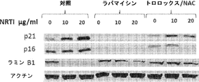
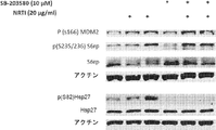
本明細書で実証されるように、ミトコンドリアROSはmTORC1のための新規な入力(input)であると同定された。蛍光インジケータとROSスカベンジャーの影響との両方に基づいて、本発明の結果は、ミトコンドリアで生成されたROSがmTORC1を活性化するように作用する、という知見を支持する(図10)。これは、実験的設定で観察されるリボソームS6タンパク質とMDM2の両方のリン酸化の増加によって反映される。ラパマイシンによるmTORC1の阻害はこれらの応答を妨げ、ミトコンドリアを標的としたカタラーゼの発現またはROSスカベンジャーによる処理などの、ミトコンドリアROSの減少を目的とした介入と同様であった。ミトコンドリア活性の尺度としての酸素消費の分析は、NRTIに曝露された細胞ならびに複製老化(replicative senescence)状態にある細胞の両方の設定において、基礎呼吸とATP関連呼吸の増加を明らかにした。同様に、両方の設定では、プロトンリークおよびミトコンドリアROS生成が増加した。これらの知見は、ミトコンドリアROSの生成がmTORC1の活性化の引き金となることを示唆している。この解釈は、ミトコンドリアROSを減らすように設計された介入がリボソームS6タンパク質のリン酸化を減少させた、という事実によって裏付けられる。 As demonstrated herein, mitochondrial ROS has been identified as a novel input for mTORC1. Based on both the fluorescent indicator and the influence of the ROS scavenger, the results of the present invention support the finding that ROS produced in mitochondria act to activate mTORC1 (FIG. 10). This is reflected by the increased phosphorylation of both ribosomal S6 protein and MDM2 observed in the experimental setting. Inhibition of mTORC1 by rapamycin prevented these responses and was similar to interventions aimed at reducing mitochondrial ROS, such as expression of catalase targeting mitochondria or treatment with ROS scavengers. Analysis of oxygen consumption as a measure of mitochondrial activity revealed an increase in basal respiration and ATP-related respiration in the setting of both cells exposed to NRTI as well as cells in a replicative senescence state. Similarly, proton leakage and mitochondrial ROS production were increased in both settings. These findings suggest that mitochondrial ROS generation triggers mTORC1 activation. This interpretation is supported by the fact that interventions designed to reduce mitochondrial ROS reduced phosphorylation of ribosomal S6 protein.
mTORC1/p70S6K活性のミトコンドリアROS誘導の下流の結果に関して、MDM2リン酸化は、p53の安定化およびp21などの下流標的の発現増加につながるようであった。これと一致して、p53とMDM2との結合の減少がNRTIに曝露した細胞における共免疫沈降および細胞質MDM2の増加によって示された。さらに、NRTIに曝露した細胞にはp53の高分子量形態の減少が観察されたのに対して、ラパマイシン処理細胞では、プロテアソームの阻害後にしか見られないp53のこれらの高分子量形態のレベルが上昇していた。p53に対するラパマイシンのこの効果は、ラパマイシン処理細胞におけるp21などのp53標的遺伝子の活性化の欠如の基礎となっている可能性があり、mTORC1シグナル伝達に影響を及ぼすのに十分であるが増殖を完全に阻止するには十分でない濃度のラパマイシンの存在下で細胞を培養した場合に観察される老化の遅延に貢献した。 Regarding the downstream results of mitochondrial ROS induction of mTORC1 / p70S6K activity, MDM2 phosphorylation appeared to lead to stabilization of p53 and increased expression of downstream targets such as p21. Consistent with this, decreased binding of p53 to MDM2 was shown by co-immunoprecipitation and increased cytoplasmic MDM2 in cells exposed to NRTI. In addition, a decrease in the high molecular weight form of p53 was observed in cells exposed to NRTI, whereas rapamycin-treated cells increased the levels of these high molecular weight forms of p53 that were only seen after proteasome inhibition. It was. This effect of rapamycin on p53 may underlie the lack of activation of p53 target genes such as p21 in rapamycin-treated cells, which is sufficient to affect mTORC1 signaling but completely proliferate Contributed to the delayed senescence observed when cells were cultured in the presence of rapamycin at a concentration that was not sufficient to prevent the disease.
特定の局面において、本結果は、p70S6Kを介するmTORC1シグナル伝達がミトコンドリアによって生成されたROSに応答性であり得ることを示す。mTORC1/p70S6Kの活性化は、ミトコンドリア機能障害、複製老化の設定において、および老化組織において起こった。ラパマイシンは、ミトコンドリアROS生成を改善して、mTORC1/p70S6K応答を遮断する。ラパマイシンのこれらの効果は、ラパマイシン処理後に、寿命に関して観察され、かつ真皮萎縮のような加齢性障害において観察された、有益な効果を支持した。 In certain aspects, the present results indicate that mTORC1 signaling through p70S6K may be responsive to ROS produced by mitochondria. Activation of mTORC1 / p70S6K occurred in the setting of mitochondrial dysfunction, replication senescence, and in aging tissues. Rapamycin improves mitochondrial ROS production and blocks the mTORC1 / p70S6K response. These effects of rapamycin supported the beneficial effects observed with respect to lifespan after rapamycin treatment and in age-related disorders such as dermal atrophy.
定義
本明細書で使用する場合、以下の用語の各々は、このセクションおいてその用語に関連した意味を有する。
Definitions As used herein, each of the following terms has the meaning associated with it in this section.
他に定義されない限り、本明細書で使用する全ての技術用語および科学用語は、一般的に、本発明が属する技術分野の当業者によって通常理解されているのと同じ意味を有する。ほとんどの場合、本明細書で使用する命名法、ならびに細胞培養、分子遺伝学および化学における実験手順は、当技術分野において周知であって、一般的に使用されているものである。 Unless defined otherwise, all technical and scientific terms used herein generally have the same meaning as commonly understood by one of ordinary skill in the art to which this invention belongs. In most cases, the nomenclature used herein and the experimental procedures in cell culture, molecular genetics and chemistry are those well known and commonly used in the art.
本明細書で使用する冠詞(a, an)は、その冠詞の文法上の対象物の1つまたは2つ以上(すなわち、少なくとも1つ)を指す。一例として、「要素(an element)」は1つの要素または2つ以上の要素を意味する。 As used herein, article (a, an) refers to one or more (ie, at least one) of the grammatical objects of the article. By way of example, “an element” means one element or more than one element.
本明細書で使用する用語「約」は、当業者によって理解され、それが使用される状況によってある程度変化するであろう。量、持続時間などの測定可能な値に言及する場合に本明細書で使用する用語「約」は、特定の値からの±20%の変動、つまり±10%、±5%、±1%、もしくは±0.1%の変動を含むことを意味し、そのような変動は開示された方法を実施するのに妥当である。 The term “about” as used herein will be understood by persons of ordinary skill in the art and will vary to some extent on the context in which it is used. The term “about” as used herein when referring to measurable values such as quantity, duration, etc., is ± 20% variation from a particular value, ie ± 10%, ± 5%, ± 1% Or a variation of ± 0.1%, which is reasonable for performing the disclosed method.
本明細書で使用する「皮膚科学的に許容される担体」または「皮膚科学的に許容される賦形剤」とは、過度の毒性、不適合性、不安定性、アレルギー反応などを伴わずに、ヒトケラチン組織と接触させて使用するのに適した組成物または成分を指す。 As used herein, a “dermatologically acceptable carrier” or “dermatologically acceptable excipient” means without undue toxicity, incompatibility, instability, allergic reaction, etc. A composition or component suitable for use in contact with human keratinous tissue.
「疾患」は、動物が恒常性を維持することができない動物の健康状態であり、こうした疾患が改善されないと、動物の健康は悪化し続ける。動物における「障害」は、動物が恒常性を維持することができる健康状態ではあるが、その動物の健康状態は、障害がないときと比べて、あまり良くない。未治療のまま放置しても、障害は動物の健康状態のさらなる低下を必ずしも引き起こさない。 A “disease” is an animal health condition in which the animal cannot maintain homeostasis, and if such a disease is not improved, the animal health continues to deteriorate. A “disorder” in an animal is a health condition in which the animal can maintain homeostasis, but the health condition of the animal is not as good as when there is no disorder. If left untreated, the disorder does not necessarily cause a further decline in the animal's health.
本明細書で使用する化合物の「有効量」または「治療有効量」または「薬学的有効量」という用語は、その化合物が投与される対象に有益な効果をもたらすのに十分な化合物の量を指すために交換可能に使用される。本明細書で使用する用語「治療する」は、患者もしくは対象が経験する症状の頻度を減らすこと、または薬剤もしくは化合物を投与して、経験する症状の重症度を軽減することを意味する。個々の場合の適切な治療量は、慣例的な実験を用いて当業者によって決定され得る。 As used herein, the term “effective amount” or “therapeutically effective amount” or “pharmaceutically effective amount” of a compound refers to the amount of the compound sufficient to produce a beneficial effect in the subject to which the compound is administered. Used interchangeably to point. The term “treating” as used herein means reducing the frequency of symptoms experienced by a patient or subject, or administering a drug or compound to reduce the severity of the symptoms experienced. The appropriate therapeutic amount in each individual case can be determined by one of ordinary skill in the art using routine experimentation.
本明細書で使用する用語「FTC」は、エムトリシタビンまたはその塩もしくは溶媒和物を指す。 As used herein, the term “FTC” refers to emtricitabine or a salt or solvate thereof.
本明細書で使用する「説明資料」には、出版物、記録、図表、または任意の他の表現手段が含まれ、これらは、対象の全身性免疫反応を抑制または低減するためのキットにおいて本発明の化合物、組成物、アッセイまたは方法の有用性を伝えるために使用することができる。本発明のキットの説明資料は、例えば、本発明の同定された化合物、組成物、アッセイもしくは方法を含む容器に貼付されてもよいし、同定された化合物、組成物、アッセイもしくは方法を含む容器と一緒に出荷されてもよい。あるいは、レシピエントが説明資料と化合物、組成物、アッセイまたは方法を協力的に使用することを意図して、説明資料を容器とは別に出荷してもよい。 As used herein, “explanatory material” includes publications, records, charts, or any other means of expression, which can be found in a kit for suppressing or reducing a systemic immune response in a subject. It can be used to convey the usefulness of a compound, composition, assay or method of the invention. The instructional material of the kit of the present invention may be attached to, for example, a container containing the identified compound, composition, assay or method of the present invention, or a container containing the identified compound, composition, assay or method. May be shipped together. Alternatively, the instructional material may be shipped separately from the container with the intention that the recipient uses the instructional material and the compound, composition, assay or method cooperatively.
本明細書で使用する用語「モジュレートする」は、本発明の化合物が本発明で意図される受容体に結合することに関連した病状または病態に関して、本発明による化合物の投与前には、最善に達していなかった、多くの場合には、衰弱性で、命を脅かすことさえあった病状または病態を、直接的または間接的に、改善または軽減することを意味する。モジュレーションは、(受容体部位に応じて)アゴニスト活性、アンタゴニスト活性、またはアゴニスト/アンタゴニスト混合活性によって起こり得る。 As used herein, the term “modulate” refers to a condition or condition associated with binding of a compound of the invention to the receptor contemplated by the invention prior to administration of the compound according to the invention. In many cases, it means improving or reducing, directly or indirectly, a debilitating, even life-threatening medical condition or condition. Modulation can occur by agonist activity, antagonist activity, or mixed agonist / antagonist activity (depending on the receptor site).
本明細書で使用する用語「NRTI」は、ヌクレオチド系/ヌクレオシド系逆転写酵素阻害剤を指す。 The term “NRTI” as used herein refers to a nucleotide / nucleoside reverse transcriptase inhibitor.
本明細書で使用する用語「薬学的に許容される」は、組成物の生物学的活性または特性を無効にしない、比較的毒性のない、担体または希釈剤などの物質を指す;すなわち、その物質は、望ましくない生物学的作用を引き起こすことなく、またはそれが含まれる組成物のいずれの成分とも有害なやり方で相互作用することなく、個体に投与することができる。 The term “pharmaceutically acceptable” as used herein refers to a relatively non-toxic substance such as a carrier or diluent that does not abrogate the biological activity or properties of the composition; A substance can be administered to an individual without causing undesired biological effects or interacting in a deleterious manner with any component of the composition in which it is contained.
本明細書で使用する用語「薬学的組成物」または「組成物」は、本発明において有用な少なくとも1種の化合物と、他の化学成分、例えば担体、安定剤、希釈剤、分散化剤、懸濁化剤、増粘剤および/または賦形剤との混合物を指す。薬学的組成物は、該化合物の生物への投与を容易にする。当技術分野には、化合物を投与する技術が数多く存在しており、限定するものではないが、静脈内、経口、エアロゾル、非経口、眼内、肺、頭蓋内および局所投与などが含まれる。特定の態様では、投与は局所投与を含む。 As used herein, the term “pharmaceutical composition” or “composition” refers to at least one compound useful in the present invention and other chemical components such as carriers, stabilizers, diluents, dispersants, Refers to mixtures with suspending agents, thickeners and / or excipients. The pharmaceutical composition facilitates administration of the compound to an organism. There are many techniques for administering compounds in the art, including but not limited to intravenous, oral, aerosol, parenteral, intraocular, pulmonary, intracranial and topical administration. In certain embodiments, administration includes topical administration.
本明細書で使用する「対象」は、ヒトまたは非ヒト哺乳動物を指す。非ヒト哺乳動物には、例えば、ヒツジ、ウシ、ブタ、イヌ、ネコおよびネズミ科の動物などの、家畜およびペットが含まれる。特定の態様では、対象はヒトである。 A “subject” as used herein refers to a human or non-human mammal. Non-human mammals include, for example, farm animals and pets, such as sheep, cows, pigs, dogs, cats and murines. In certain embodiments, the subject is a human.
本明細書で使用する用語「TDF」は、テノホビルジソプロキシルフマル酸塩、またはその塩もしくは溶媒和物を指す。 The term “TDF” as used herein refers to tenofovir disoproxil fumarate, or a salt or solvate thereof.
本明細書で使用する「局所投与」または「局所適用」は、皮膚または粘膜のような体表面に適用される投薬を指す。 As used herein, “topical administration” or “topical application” refers to a medication applied to a body surface such as the skin or mucosa.
本明細書で使用する用語「治療」または「治療する」は、治療剤、すなわち本発明において有用な組成物(単独または別の薬剤との組み合わせ)を対象に適用または投与すること、あるいは疾患もしくは障害、疾患もしくは障害の症状、または疾患もしくは障害を発症する可能性を治す、癒す、緩和する、軽減する、改変する、救済する、改善する、向上させる、または影響を及ぼす目的で、疾患もしくは障害、疾患もしくは障害の症状、または疾患もしくは障害を発症する可能性を有する対象由来の単離された組織または細胞株に、(例えば、診断またはエクスビボ適用のために)治療剤を適用または投与すること、と定義される。そのような治療は、薬理ゲノム学の分野から得られた知識に基づいて、一人一人に合わせて特別に調整または修正することができる。 As used herein, the term “treatment” or “treat” refers to the application or administration of a therapeutic agent, ie a composition useful in the present invention (alone or in combination with another agent) to a subject, or A disease or disorder for the purpose of curing, healing, mitigating, alleviating, modifying, remedying, improving, improving or affecting a disorder, disease or symptom of a disorder, or the likelihood of developing a disease or disorder Applying or administering a therapeutic agent (e.g., for diagnostic or ex vivo application) to an isolated tissue or cell line from a subject having the potential to develop a disease or disorder, or a disease or disorder , Defined as Such treatments can be tailored or modified specifically for each person based on knowledge gained from the field of pharmacogenomics.
本開示を通じて、本発明の様々な局面は範囲形式で提示され得る。範囲形式での記載は、単に便宜上のものであり、本発明の範囲に対する柔軟性のない制限として解釈されるべきではないことを理解されたい。したがって、範囲の記載は、全ての可能な部分範囲ならびにその範囲内の個々の数値を具体的に開示したものと見なされるべきである。例えば、1〜6のような範囲の記載は、1〜3、1〜4、1〜5、2〜4、2〜6、3〜6などの部分範囲、ならびにその範囲内の個々の数値、例えば1、2、2.7、3、4、5、5.3および6を具体的に開示したものと考えるべきである。このことは、範囲の広さにかかわらず当てはまる。 Throughout this disclosure, various aspects of this invention may be presented in a range format. It should be understood that the description in range format is merely for convenience and should not be construed as an inflexible limitation on the scope of the invention. Accordingly, the description of a range should be considered to have specifically disclosed all the possible subranges as well as individual numerical values within that range. For example, the description of a range such as 1 to 6 is a subrange such as 1 to 3, 1 to 4, 1 to 5, 2 to 4, 2 to 6, 3 to 6 as well as individual numerical values within the range, For example, 1, 2, 2.7, 3, 4, 5, 5.3 and 6 should be considered as specifically disclosed. This is true regardless of the breadth of the range.
組成物
本発明の組成物は、治療有効量のmTORC1阻害剤、またはその塩、溶媒和物、エナンチオマーもしくはジアステレオマーを含む。特定の態様では、mTORC1阻害剤は、BEZ235、ラパマイシン、エベロリムス、AZD8055、テムシロリムス、KU-0063794、PI-103、トルキニブ、タクロリムス、リダフォロリムス、INK-128、ボクスタリシブ、Torin-1、オミパリシブ、OSI-027、PF-04691502、アピトリシブ、GSK1059615、WYE-354、ジェダトリシブ、AZD-2014、Torin-2、WYE-125132、BGT226、Palomid-529、PP121、WYE-687、CH5132799、Way-600、ETP-46464、GDC-0349、XL388、およびゾタロリムスからなる群より選択される少なくとも1種である。
Compositions The compositions of the present invention comprise a therapeutically effective amount of an mTORC1 inhibitor, or a salt, solvate, enantiomer or diastereomer thereof. In a particular embodiment, the mTORC1 inhibitor is BEZ235, rapamycin, everolimus, AZD8055, temsirolimus, KU-0063794, PI-103, tolkinib, tacrolimus, ridaforolimus, INK-128, boxarishibu, Torin-1, omiparisis, OSI- 027, PF-04691502, Apitritive, GSK1059615, WYE-354, Jedatric, AZD-2014, Torin-2, WYE-125132, BGT226, Palomid-529, PP121, WYE-687, CH5132799, Way-600, ETP-46464, It is at least one selected from the group consisting of GDC-0349, XL388, and zotarolimus.
BEZ235は、2-メチル-2-(4-(3-メチル-2-オキソ-8-(キノリン-3-イル)-2,3-ジヒドロイミダゾ[4,5-c]キノリン-1-イル)フェニル)プロパンニトリルとしても知られており、下記の式を有する。

ラパマイシンは、(3S,6R,7E,9R,10R,12R,14S,15E,17E,19E,21S,23S,26R,27R,34aS)-9,10,12,13,14,21,22,23,24,25,26,27,32,33,34,34a-ヘキサデカヒドロ-9,27-ジヒドロキシ-3-[(1R)-2-[(1S,3R,4R)-4-ヒドロキシ-3-メトキシシクロヘキシル]-1-メチルエチル]-10,21-ジメトキシ-6,8,12,14,20,26-ヘキサメチル-23,27-エポキシ-3H-ピリド[2,1-c][1,4-オキサアザシクロヘントリアコンチン-1,5,11,28,29(4H,6H,31H)-ペントンとしても知られており、下記の式を有する。
エベロリムスは、ジヒドロキシ-12-[(2R)-1-[(1S,3R,4R)-4-(2-ヒドロキシエトキシ)-3-メトキシシクロヘキシル]プロパン-2-イル]-19,30-ジメトキシ-15,17,21,23,29,35-ヘキサメチル-11,36-ジオキサ-4-アザトリシクロ[30.3.1.0]ヘキサトリアコンタ-16,24,26,28-テトラエン-2,3,10,14,20-ペントンとしても知られており、下記の式を有する。
AZD8055は、5-(2,4-ビス(S)-3-メチルモルホリノ)ピリド[2,3-d]ピリミジン-7-イル)-2-メトキシフェニル)メタノールとしても知られており、下記の式を有する。
テムシロリムスは、42-[3-ヒドロキシ-2-(ヒドロキシメチル)-2-メチルプロパノアート]-ラパマイシンとしても知られており、下記の式を有する。
PI-103は、3-[4-(4-モルホリニル)ピリド[3',2':4,5]フロ[3,2-d]ピリミジン-2-イル]フェノールとしても知られており、下記の式を有する。
KU-0063794は、(5-(2-((2R,6S)-2,6-ジメチルモルホリノ)-4-モルホリノピリド[2,3-d]ピリミジン-7-イル)-2-メトキシフェニル)メタノールとしても知られており、下記の式を有する。
トルキニブは、2-(4-アミノ-1-イソプロピル-1H-ピラゾロ[3,4-d]ピリミジン-3-イル)-1H-インドール-5-オールとしても知られており、下記の式を有する。
タクロリムスは、3S-[3R*[E(1S*,3S*,4S*)],4S*,5R*,8S*,9E,12R*,14R*,15S*,16R*,18S*,19S*,26aR*-5,6,8,11,12,13,14,15,16,17,18,19,24,25,26,26a-ヘキサデカヒドロ-5,19-ジヒドロキシ-3-[2-(4-ヒドロキシ-3-メトキシシクロヘキシル]-1-メチルエテニル]-14,16-ジメトキシ-4,10,12,18-テトラメチル-8-(2-プロペニル)-15,19-エポキシ-3H-ピリド[2,1-c][1,4]オキサアザシクロトリコシン-1,7,20,21(4H,23H)-テトロンとしても知られており、下記の式を有する。
リダフォロリムスは、42-(ジメチルホスフィネート)-ラパマイシンとしても知られており、下記の式を有する。
INK-128は、3-(2-アミノベンゾ[d]オキサゾール-5-イル)-1-イソプロピル-1H-ピラゾロ[3,4-d]ピリミジン-4-アミンとしても知られており、下記の式を有する。
ボクスタリシブは、N-[4-[[[3-[(3,5-ジメトキシフェニル)アミノ]-2-キノキサリニル]アミノ]スルホニル]フェニル]-3-メトキシ-4-メチル-ベンズアミドとしても知られており、下記の式を有する。
Torin-1は、1-[4-[4-(1-オキソプロピル)-1-ピペラジニル]-3-(トリフルオロメチル)フェニル]-9-(3-キノリニル)-ベンゾ[h]-1,6-ナフチリジン-2(1H)-オンとしても知られており、下記の式を有する。
オミパリシブは、2,4-ジフルオロ-N-(2-メトキシ-5-(4-ピリダジン-4-イル)キノリン-6-イル)ピリジン-3-イル)ベンゼンスルホンアミドとしても知られており、下記の式を有する。
OSI-027は、(1r,4r)-4-(4-アミノ-5-(7-メトキシ-1H-インドール-2-イル)イミダゾ[5,1-f][1,2,4]トリアジン-7-イル)シクロヘキサン-1-カルボン酸としても知られており、下記の式を有する。
PF-04691502は、2-アミノ-8-((1r,4r)-4-(2-ヒドロキシエトキシ)シクロヘキシル)-6-(6-メトキシピリジン-3-イル)-4-メチルピリド[2,3-d]ピリミジン-7(8H)-オンとしても知られており、下記の式を有する。
アピトリシブは、(S)-1-(4-((2-(2-アミノピリミジン-5-イル)-7-メチル-4-モルホリノチエノ[3,2-d]ピリミジン-6-イル)メチル)ピペラジン-1-イル)-2-ヒドロキシプロパン-1-オンとしても知られており、下記の式を有する。
GSK1059615は、(Z)-5-((4-(ピリジン-4-イル)キノリン-6-イル)メチレン)チアゾリジン-2,4-ジオンとしても知られており、下記の式を有する。
WYE-354は、4-[6-[4-[(メトキシカルボニル)アミノ]フェニル]-4-(4-モルホリニル)-1H-ピラゾロ[3,4-d]ピリミジン-1-イル]-1-ピペリジンカルボン酸メチルエステルとしても知られており、下記の式を有する。
ジェダトリシブは、1-(4-(4-(ジメチルアミノ)ピペリジン-1-カルボニル)フェニル)-3-(4-(4,6-ジモルホリノ-1,3,5-トリアジン-2-イル)フェニル)ウレアとしても知られており、下記の式を有する。
AZD-2014は、3-(2,4-ビス((S)-3-メチルモルホリノ)ピリド[2,3-d]ピリミジン-7-イル)-N-メチルベンズアミドとしても知られており、下記の式を有する。
Torin-2は、9-(6-アミノ-3-ピリジニル)-1-[3-(トリフルオロメチル)フェニル]-ベンゾ[h]-1,6-ナフチリジン-2(1H)-オンとしても知られており、下記の式を有する。
WYE-125132は、N-[4-[1-(1,4-ジオキサスピロ[4.5]デク-8-イル)-4-(8-オキサ-3-アザビシクロ[3.2.1]オクト-3-イル)-1H-ピラゾロ[3,4-d]ピリミジン-6-イル]フェニル]-N'-メチルウレアとしても知られており、下記の式を有する。
BGT226は、8-(6-メトキシピリジン-3-イル)-3-メチル-1-(4-(ピペラジン-1-イル)-3-(トリフルオロメチル)フェニル)-1H-イミダゾ[4,5-c]キノリン-2(3H)-オンとしても知られており、下記の式を有する。
Palomid-529は、3-(4-メトキシベンジルオキシ)-8-(1-ヒドロキシエチル)-2-メトキシ-6H-ベンゾ[c]クロメン-6-オンとしても知られており、下記の式を有する。
PP121は、1-シクロペンチル-3-(1H-ピロロ[2,3-b]ピリジン-5-イル)-1H-ピラゾロ[3,4-d]ピリミジン-4-アミンとしても知られており、下記の式を有する。
WYE-687は、4-(4-モルホリノ-1-(1-ピリジン-3-イルメチル)ピペリジン-4-イル)-1H-ピラゾロ[3,4-d]ピリミジン-6-イル)フェニルカルバミン酸メチルとしても知られており、下記の式を有する。
CH5132799は、5-(7-メチルスルホニル)-2-モルホリノ-6,7-ジヒドロ-5H-ピロロ[2,3-d]ピリミジン-4-イル)ピリミジン-2-アミンとしても知られており、下記の式を有する。
WAY-600は、6-(1H-インドール-5-イル)-4-モルホリノ-1-(1-ピリジン-3-イルメチル)ピペリジン-4-イル)-1H-ピラゾロ[3,4-d]ピリミジンとしても知られており、下記の式を有する。
ETP-46464は、α,α-ジメチル-4-[2-オキソ-9-(3-キノリニル)-2H-[1,3]オキサジノ[5,4-c]キノリン-1(4H)-イル]-ベンゼンアセトニトリルとしても知られており、下記の式を有する。
GDC-0349は、N-エチル-N'-[4-[5,6,7,8-テトラヒドロ-4-[(3S)-3-メチル-4-モルホリニル]-7-(3-オキセタニル)ピリド[3,4-d]ピリミジン-2-イル]フェニル]-ウレアとしても知られており、下記の式を有する。
XL388は、[7-(6-アミノ-3-ピリジニル)-2,3-ジヒドロ-1,4-ベンゾオキサゼピン-4(5H)-イル][3-フルオロ-2-メチル-4-(メチルスルホニル)フェニル]-メタノンとしても知られており、下記の式を有する。
ゾタロリムスは、42-デオキシ-42-(1H-テトラゾール-1-イル)-(42S)-ラパマイシンとしても知られており、下記の式を有する。
特定の態様において、本発明で企図されるmTORC1阻害剤は、ラパマイシンである。他の態様では、mTORC1阻害剤は、特定の細胞内区画または細胞小器官、例えばミトコンドリア、核、リソソームおよび/または小胞体など、への送達が改善されているラパマイシンの修飾形態であり得る。 In certain embodiments, the mTORC1 inhibitor contemplated by the present invention is rapamycin. In other embodiments, the mTORC1 inhibitor may be a modified form of rapamycin with improved delivery to certain intracellular compartments or organelles, such as mitochondria, nucleus, lysosomes and / or endoplasmic reticulum.
特定の態様において、前記組成物中のmTORC1阻害剤の治療有効量は、約0.001〜約1重量%の範囲である。他の態様では、該組成物中のmTORC1阻害剤の重量による治療有効量は、約0.002%〜約0.75%、約0.005%〜約0.5%、約0.008%〜約0.25%、約0.01%〜約0.2%、約0.02%〜約0.15%、または約0.03%〜約0.1%の範囲である。 In certain embodiments, the therapeutically effective amount of the mTORC1 inhibitor in the composition ranges from about 0.001 to about 1% by weight. In other embodiments, the therapeutically effective amount by weight of the mTORC1 inhibitor in the composition is about 0.002% to about 0.75%, about 0.005% to about 0.5%, about 0.008% to about 0.25%, about 0.01% to about The range is 0.2%, about 0.02% to about 0.15%, or about 0.03% to about 0.1%.
特定の態様において、本発明の組成物は、皮膚科学的に許容される担体をさらに含む。本発明の組成物は、約60%〜約99.9%、あるいは約70%〜約95%、あるいは約80%〜約90%の皮膚科学的に許容される担体を含むことができる。特定の態様では、皮膚科学的に許容される担体は、少なくとも、溶媒、潤滑剤、皮膚軟化剤、乳化剤、保湿剤、増粘ワックス、柔軟剤、香料、防腐剤および人工着色料からなる群より選択される。他の態様では、皮膚科学的に許容される担体は、水、脂肪アルコール、および揮発性有機アルコールからなる群より選択される少なくとも1種である。皮膚科学的に許容される担体の1つの非限定的な例はペトロラタムである。 In certain embodiments, the composition of the present invention further comprises a dermatologically acceptable carrier. The compositions of the present invention can comprise from about 60% to about 99.9%, alternatively from about 70% to about 95%, alternatively from about 80% to about 90%, dermatologically acceptable carrier. In certain embodiments, the dermatologically acceptable carrier is at least from the group consisting of solvents, lubricants, emollients, emulsifiers, humectants, thickening waxes, softeners, fragrances, preservatives and artificial colorants. Selected. In another embodiment, the dermatologically acceptable carrier is at least one selected from the group consisting of water, fatty alcohols, and volatile organic alcohols. One non-limiting example of a dermatologically acceptable carrier is petrolatum.
方法
一局面において、本発明は、哺乳類の線維芽細胞の寿命を延ばす方法を提供する。別の局面では、本発明は、哺乳類の線維芽細胞における細胞組織化を保存する方法を提供する。さらに別の局面では、本発明は、哺乳類の線維芽細胞の老化を予防するまたは最小限に抑える方法を提供する。さらに別の局面では、本発明は、哺乳類対象における、真皮萎縮、偽瘢痕、脂漏性または光線性角化症、黒子、真皮肥厚の病巣領域、および粗いシワを含む、加齢性の真皮障害を治療または予防する方法を提供する。
Methods In one aspect, the present invention provides a method of extending the lifetime of mammalian fibroblasts. In another aspect, the present invention provides a method for preserving cell organization in mammalian fibroblasts. In yet another aspect, the present invention provides a method for preventing or minimizing senescence of mammalian fibroblasts. In yet another aspect, the invention relates to age-related dermal disorders, including dermal atrophy, pseudoscarring, seborrheic or actinic keratosis, moles, dermal thickened focal areas, and rough wrinkles in a mammalian subject. A method of treating or preventing is provided.
特定の態様において、本発明の方法は、対象に、治療有効量のmTORC1阻害剤を局所投与することを含み、任意で、mTORC1阻害剤は、皮膚に許容される組成物中に製剤化される。他の態様では、本発明の組成物は、治療有効量のmTORC1阻害剤を含む。さらに他の態様では、該組成物は、皮膚科学的に許容される担体をさらに含む。さらに他の態様では、該組成物は対象の皮膚の患部に局所適用される。 In certain embodiments, the methods of the invention comprise topically administering to a subject a therapeutically effective amount of an mTORC1 inhibitor, and optionally the mTORC1 inhibitor is formulated in a skin acceptable composition. . In other embodiments, the compositions of the invention comprise a therapeutically effective amount of an mTORC1 inhibitor. In yet another aspect, the composition further comprises a dermatologically acceptable carrier. In yet another embodiment, the composition is topically applied to the affected area of the subject's skin.
特定の態様において、本発明において企図される組成物の局所製剤は、真皮萎縮の治療のために使用される。他の態様では、本発明は、真皮萎縮を治療または予防するための、治療有効量のラパマイシンを含む局所用クリーム剤を提供する。 In certain embodiments, topical formulations of the compositions contemplated in the present invention are used for the treatment of dermal atrophy. In another aspect, the present invention provides a topical cream comprising a therapeutically effective amount of rapamycin for treating or preventing dermal atrophy.
特定の態様において、真皮の萎縮は、真皮層の目盛付きデジタルキャリパー測定を利用して真皮層を測定することによって評価される。他の態様において、脂漏性角化症、黒子、偽瘢痕、粗いシワ、および表皮肥厚の改善は、検査者評価格付けスケール(Investigator evaluation rating scale)1〜4により評価され、ここで、1は正常であって病変の徴候がない;2は軽度の病変を表す;3は正常な皮膚と比べて明確に区別される特徴の病変を表す;4は重症度が高い病変を表す。さらに他の態様では、病変は視覚的に、または米国立衛生研究所から入手可能なオープンソースの画像解析ソフトウェアであるImageJのような画像解析ソフトウェアを用いて、検査することができる。さらに他の態様では、病変は、病変の手動測定を用いた面積測定によって、または検査者もしくは調査研究スタッフが撮影した画像の解析によって評価される。 In certain embodiments, dermal atrophy is assessed by measuring the dermis layer utilizing a calibrated digital caliper measurement of the dermis layer. In other embodiments, the improvement in seborrheic keratosis, moles, false scars, rough wrinkles, and epidermal thickening is assessed by an Investigator evaluation rating scale 1-4 where 1 is Normal with no signs of lesions; 2 represents a mild lesion; 3 represents a lesion with distinct characteristics compared to normal skin; 4 represents a more severe lesion. In yet other embodiments, the lesion can be examined visually or using image analysis software such as ImageJ, an open source image analysis software available from the National Institutes of Health. In yet other embodiments, the lesion is assessed by area measurement using manual measurement of the lesion or by analysis of images taken by the examiner or research staff.
製剤
本発明の薬学的組成物中の活性成分、皮膚科学的に許容される担体、および任意の追加の成分の相対量は、治療される対象の個性、サイズおよび状態に応じて変化する。一例として、該組成物は約0.001%〜約1%(w/w)のmTORC1阻害剤を含み得る。他の態様では、該組成物中のmTORC1阻害剤の重量による治療有効量は、約0.002%〜約0.75%、約0.005%〜約0.5%、約0.008%〜約0.25%、約0.01%〜約0.2%、約0.02%〜約0.15%、または約0.03%〜約0.1%の範囲である。
Formulations The relative amounts of active ingredient, dermatologically acceptable carrier, and any additional ingredients in the pharmaceutical compositions of the invention will vary depending on the individuality, size and condition of the subject being treated. As an example, the composition may comprise about 0.001% to about 1% (w / w) of an mTORC1 inhibitor. In other embodiments, the therapeutically effective amount by weight of the mTORC1 inhibitor in the composition is about 0.002% to about 0.75%, about 0.005% to about 0.5%, about 0.008% to about 0.25%, about 0.01% to about The range is 0.2%, about 0.02% to about 0.15%, or about 0.03% to about 0.1%.
本明細書で提供される薬学的組成物の記載は、主にヒトへの医療向け投与(ethical administration)に適した薬学的組成物に向けられたものであるが、当業者であれば、このような組成物は一般に、あらゆる種類の動物への投与に適していることが理解されよう。種々の動物への投与に適した薬学的組成物を提供するために、ヒトへの投与に適した該組成物を改変することはよく理解されており、通常の知識を有する獣医薬理学者は、たとえ行うにしても、通常の実験を行うだけで、そのような改変を設計して実施することができる。本発明の薬学的組成物の投与が企図される対象には、限定するものではないが、ヒトおよび他の霊長類、哺乳類、例えばウシ、ブタ、ウマ、ヒツジ、ネコおよびイヌなどの商業的に関係のある哺乳動物が含まれる。 The description of the pharmaceutical composition provided herein is primarily directed to a pharmaceutical composition suitable for ethical administration to humans; It will be appreciated that such compositions are generally suitable for administration to all types of animals. In order to provide a pharmaceutical composition suitable for administration to various animals, it is well understood to modify the composition suitable for human administration, and veterinary scientists with ordinary knowledge Even if done, such modifications can be designed and implemented with only routine experimentation. Subjects contemplated for administration of the pharmaceutical compositions of the invention include, but are not limited to, humans and other primates, mammals such as cows, pigs, horses, sheep, cats and dogs commercially. Related mammals are included.
本発明の組成物は、哺乳類に1日数回の頻度で投与することができ、1日1回、週1回、2週間に1回、1ヶ月に1回などのより少ない頻度で、または数ヶ月に1回、1年に1回以下などのさらに少ない頻度で投与してもよい。 The compositions of the invention can be administered to mammals several times a day, less frequently, such as once a day, once a week, once every two weeks, once a month, etc. It may be administered less frequently, such as once a month or less than once a year.
本発明の組成物を投与するための投薬レジメンは、1日1回または1日2回であり得る。活性薬剤の適用頻度および濃度は、皮膚の状態および真皮の応答に依存する。真皮に対して所望の効果を達成するように適用を継続してもよいし、満足のいく結果が得られた後で適用頻度を減らしてもよい。特定の実施態様では、結果を達成するために投与を最低2週間続ける。継続的な改善を得るために最初の2週間を超えて適用を継続してもよく、その結果が達成されたら、適用頻度を減らすことができる。適用は、どんなときでも病変の相対的重症度に基づいて、様々なレベルの適用を数年間にわたって継続することができる。 Dosage regimens for administering the compositions of the invention may be once a day or twice a day. The frequency and concentration of application of the active agent depends on the skin condition and the dermal response. Application may be continued to achieve the desired effect on the dermis, or the frequency of application may be reduced after satisfactory results are obtained. In certain embodiments, administration is continued for a minimum of 2 weeks to achieve results. Application may continue beyond the first two weeks to obtain continuous improvement, and once the results are achieved, the frequency of application can be reduced. The application can be continued at different levels for several years based on the relative severity of the lesion at any time.
1日あたりに投与される本発明の組成物の量は、非限定的な例では、毎日、1日おき、2日おき、3日おき、4日おき、または5日おきに投与できると認識される。投与頻度は、当業者にはすぐに分かることであり、治療すべき疾患のタイプおよび重症度、動物のタイプおよび年齢などを含むがこれらに限定されない、様々な要因に依存するであろう。 It will be appreciated that the amount of the composition of the invention administered per day can be administered daily, every other day, every two days, every three days, every four days, or every five days, in a non-limiting example Is done. The frequency of administration will be readily apparent to those skilled in the art and will depend on a variety of factors including, but not limited to, the type and severity of the disease to be treated, the type and age of the animal, and the like.
特定の態様において、本発明の組成物は、1種または複数種の皮膚科学的に許容される賦形剤または担体を用いて製剤化される。特定の態様では、本発明の薬学的組成物は、治療有効量のmTORC1阻害剤および皮膚科学的に許容される担体を含有する。有用な皮膚科学的に許容される担体には、限定するものではないが、グリセロール、水、生理食塩水、エタノールおよび他の皮膚科学的に許容される塩溶液、例えばリン酸塩および有機酸の塩、が含まれる。これらおよび他の皮膚科学的に許容される担体の例は、Remington's Pharmaceutical Sciences (1991, Mack Publication Co., New Jersey)に記載されている。 In certain embodiments, the compositions of the invention are formulated with one or more dermatologically acceptable excipients or carriers. In certain embodiments, the pharmaceutical compositions of the invention contain a therapeutically effective amount of an mTORC1 inhibitor and a dermatologically acceptable carrier. Useful dermatologically acceptable carriers include, but are not limited to, glycerol, water, saline, ethanol and other dermatologically acceptable salt solutions such as phosphates and organic acids. Salt. Examples of these and other dermatologically acceptable carriers are described in Remington's Pharmaceutical Sciences (1991, Mack Publication Co., New Jersey).
本発明の組成物は、約60%〜約99.9%、あるいは約70%〜約95%、あるいは約80%〜約90%の皮膚科学的に許容される担体を含むことができる。特定の態様では、皮膚科学的に許容される担体は、少なくとも、溶媒、潤滑剤、皮膚軟化剤、乳化剤、保湿剤、増粘ワックス、柔軟剤、香料、防腐剤および人工着色料からなる群より選択される。他の態様では、皮膚科学的に許容される担体は、水、脂肪アルコール、および揮発性有機アルコールからなる群より選択される少なくとも1種である。皮膚科学的に許容される担体の1つの非限定的な例はペトロラタムである。 The compositions of the present invention can comprise from about 60% to about 99.9%, alternatively from about 70% to about 95%, alternatively from about 80% to about 90%, dermatologically acceptable carrier. In certain embodiments, the dermatologically acceptable carrier is at least from the group consisting of solvents, lubricants, emollients, emulsifiers, humectants, thickening waxes, softeners, fragrances, preservatives and artificial colorants. Selected. In another embodiment, the dermatologically acceptable carrier is at least one selected from the group consisting of water, fatty alcohols, and volatile organic alcohols. One non-limiting example of a dermatologically acceptable carrier is petrolatum.
前記担体は、例えば、水、エタノール、ポリオール(例えば、グリセロール、プロピレングリコール、および液体ポリエチレングリコールなど)、適切なそれらの混合物、および植物油を含む、溶媒または分散媒であり得る。適切な流動性は、例えば、レシチンなどのコーティング剤を使用することにより、分散液の場合には必要とされる粒子サイズを維持することにより、そして界面活性剤を使用することにより、維持することができる。微生物の活動防止は、種々の抗菌剤および抗真菌剤、例えばパラベン類、クロロブタノール、フェノール、アスコルビン酸、チメロサールなどによって達成することができる。多くの場合、等張剤、例えば、糖類、塩化ナトリウム、またはマンニトール、ソルビトールなどの多価アルコールを組成物中に含めることが好ましいであろう。注射可能な組成物の持続的吸収は、吸収を遅延させる物質、例えばモノステアリン酸アルミニウムまたはゼラチン、を組成物中に含めることによって達成され得る。 The carrier can be a solvent or dispersion medium containing, for example, water, ethanol, polyol (for example, glycerol, propylene glycol, and liquid polyethylene glycol, and the like), suitable mixtures thereof, and vegetable oils. The proper fluidity is maintained, for example, by using a coating such as lecithin, by maintaining the required particle size in the case of dispersion and by using a surfactant. Can do. Prevention of microbial activity can be achieved by various antibacterial and antifungal agents such as parabens, chlorobutanol, phenol, ascorbic acid, thimerosal, and the like. In many cases, it will be preferable to include isotonic agents, for example, sugars, sodium chloride, or polyalcohols such as mannitol, sorbitol, in the composition. Prolonged absorption of the injectable compositions can be achieved by including in the composition an agent that delays absorption, for example, aluminum monostearate or gelatin.
製剤は、慣用の賦形剤、すなわち当技術分野で公知の、局所投与に適した薬学的に許容される有機または無機担体物質、との混合物で利用され得る。製剤は、滅菌され、所望により、補助剤、例えば潤滑剤、防腐剤、安定剤、湿潤剤、乳化剤、浸透圧緩衝剤に影響を与える塩、着色剤、香味剤および/または芳香物質などと混合される。それらは、必要に応じて、他の活性薬剤、例えば他の鎮痛剤、と組み合わせることもできる。 The formulations may be utilized in a mixture with conventional excipients, ie, pharmaceutically acceptable organic or inorganic carrier materials known in the art suitable for topical administration. The formulation is sterilized and optionally mixed with adjuvants such as lubricants, preservatives, stabilizers, wetting agents, emulsifiers, salts affecting the osmotic buffer, coloring agents, flavoring agents and / or fragrances, etc. Is done. They can also be combined with other active agents, such as other analgesics, as needed.
本明細書で使用する「追加の成分」には、限定するものではないが、以下の成分の1種以上が含まれる:賦形剤;界面活性剤;分散剤;不活性希釈剤;顆粒化剤および崩壊剤;結合剤;潤滑剤;着色剤;防腐剤;生理学的に分解可能な組成物、例えばゼラチン;水性のビヒクルおよび溶媒;油性のビヒクルおよび溶媒;懸濁化剤;分散剤または湿潤剤;乳化剤;粘滑剤;緩衝剤;塩;増粘剤;充填剤;乳化剤;酸化防止剤;抗生物質;抗真菌剤;安定剤;および薬学的に許容される高分子材料または疎水性材料。本発明の薬学的組成物中に含めることができる他の「追加の成分」は、当技術分野で公知であり、例えば、Genaro編集 (1985, Remington's Pharmaceutical Sciences, Mack Publishing Co., Easton, PA)に記載されており、これを参照により本明細書に組み入れる。 “Additional ingredients” as used herein include, but are not limited to, one or more of the following ingredients: excipients; surfactants; dispersants; inert diluents; Binders; lubricants; colorants; preservatives; physiologically degradable compositions such as gelatin; aqueous vehicles and solvents; oily vehicles and solvents; suspending agents; Emulsifiers; emulsifiers; demulcents; buffers; salts; thickeners; fillers; emulsifiers; antioxidants; antibiotics; Other “additional ingredients” that can be included in the pharmaceutical compositions of the present invention are known in the art, eg, edited by Genaro (1985, Remington's Pharmaceutical Sciences, Mack Publishing Co., Easton, PA). Which is incorporated herein by reference.
本発明の組成物は、組成物の総重量の約0.005%〜2.0%の防腐剤を含むことができる。防腐剤は、環境中の汚染物質に曝された場合の腐敗を防止するために使用される。本発明に有用な防腐剤の例には、ベンジルアルコール、ソルビン酸、パラベン類、イミド尿素およびそれらの組み合わせからなる群より選択されるものが含まれるが、これらに限定されない。特に好ましい防腐剤は、約0.5%〜2.0%のベンジルアルコールと0.05%〜0.5%のソルビン酸の組み合わせである。 The composition of the present invention may contain preservatives from about 0.005% to 2.0% of the total weight of the composition. Preservatives are used to prevent decay when exposed to environmental pollutants. Examples of preservatives useful in the present invention include, but are not limited to, those selected from the group consisting of benzyl alcohol, sorbic acid, parabens, imidourea and combinations thereof. A particularly preferred preservative is a combination of about 0.5% to 2.0% benzyl alcohol and 0.05% to 0.5% sorbic acid.
前記組成物は、好ましくは、酸化防止剤およびキレート化剤を含む。いくつかの化合物のための好ましい酸化防止剤は、組成物の総重量の約0.01%〜0.3%の好ましい範囲のBHT、BHA、α-トコフェロールおよびアスコルビン酸であり、より好ましくは0.03%〜0.1%の範囲のBHTである。好ましくは、キレート化剤は、組成物の総重量の0.01%〜0.5%の量で存在する。特に好ましいキレート化剤には、組成物の総重量の約0.01%〜0.20%の重量範囲、より好ましくは0.02%〜0.10%の範囲のアミノポリカルボン酸塩(例えば、エチレンジアミン四酢酸二ナトリウム)およびクエン酸が含まれる。キレート化剤は、製剤の貯蔵寿命に有害であり得る組成物中の金属イオンをキレート化するのに有用である。 The composition preferably includes an antioxidant and a chelating agent. Preferred antioxidants for some compounds are BHT, BHA, α-tocopherol and ascorbic acid in a preferred range of about 0.01% to 0.3% of the total weight of the composition, more preferably 0.03% to 0.1%. BHT in the range. Preferably, the chelating agent is present in an amount from 0.01% to 0.5% of the total weight of the composition. Particularly preferred chelating agents include aminopolycarboxylates (eg, disodium ethylenediaminetetraacetate) in the weight range of about 0.01% to 0.20%, more preferably in the range of 0.02% to 0.10% of the total weight of the composition. Citric acid is included. Chelating agents are useful for chelating metal ions in compositions that can be detrimental to the shelf life of the formulation.
局所投与
医薬品の局所投与に対する障害物は、表皮の角質層である。角質層は、タンパク質、コレステロール、スフィンゴ脂質、遊離脂肪酸および種々の他の脂質から構成される高度に抵抗性の層であり、角化細胞と生存細胞を含む。角質層を通る化合物の浸透速度(フラックス)を制限する要因の1つは、皮膚表面に負荷または適用することができる活性物質の量である。皮膚の単位面積あたりに適用される活性物質の量が多いほど、皮膚表面と皮膚の下層との間の濃度勾配が大きくなり、ひいては皮膚を通る活性物質の拡散力が大きくなる。したがって、より高濃度の活性物質を含有する製剤は、より低濃度の活性物質を含有し、他の全てのものが等しい製剤と比較して、より一貫した速度での、より多くの活性物質の皮膚への浸透をもたらす可能性が高い。
Topical administration An obstacle to the topical administration of pharmaceuticals is the stratum corneum of the epidermis. The stratum corneum is a highly resistant layer composed of proteins, cholesterol, sphingolipids, free fatty acids and various other lipids, including keratinocytes and viable cells. One factor that limits the penetration rate (flux) of a compound through the stratum corneum is the amount of active substance that can be loaded or applied to the skin surface. The greater the amount of active substance applied per unit area of skin, the greater the concentration gradient between the skin surface and the lower layer of the skin, and thus the greater the diffusivity of the active substance through the skin. Thus, a formulation containing a higher concentration of active substance contains a lower concentration of active substance and more active substance at a more consistent rate compared to a formulation where all others are equal. It is likely to cause skin penetration.
局所投与に適した製剤には、限定するものではないが、液体または半液体製剤、例えば、リニメント剤、ローション剤、水中油型または油中水型エマルジョン、例えばクリーム剤、軟膏剤もしくはペースト剤、および溶液剤または懸濁液剤が含まれる。こうした製剤は、皮膚に直接適用されるか、またはスワブ、アプリケータ、スパチュラなどを利用して、あるいは経皮パッチの形態で適用され得る。特定の態様では、該パッチは、皮膚の洗浄、摩擦、引っ掻きおよび/または擦れによる医薬品の損失を最小限に抑える。他の態様では、該パッチは、皮膚からの医薬品の吸収を高めると同時に、医薬品への皮膚の曝露を最小限に抑える。 Formulations suitable for topical administration include but are not limited to liquid or semi-liquid formulations such as liniments, lotions, oil-in-water or water-in-oil emulsions such as creams, ointments or pastes, And solutions or suspensions. Such formulations may be applied directly to the skin or may be applied using a swab, applicator, spatula, etc. or in the form of a transdermal patch. In certain embodiments, the patch minimizes pharmaceutical loss due to skin cleansing, rubbing, scratching and / or rubbing. In other embodiments, the patch increases the absorption of the drug from the skin while minimizing the exposure of the skin to the drug.
本発明において企図される局所投与可能な製剤は、例えば、約0.001%〜約1%(w/w)のmTORC1阻害剤を含むことができるが、mTORC1阻害剤の濃度は、溶媒中の該活性成分の溶解限度まで高くてもよい。局所投与用の製剤は、本明細書に記載される追加の成分を1種以上さらに含み得る。 Topically administrable formulations contemplated in the present invention can include, for example, from about 0.001% to about 1% (w / w) mTORC1 inhibitor, where the concentration of mTORC1 inhibitor is determined by the activity in the solvent. It may be high up to the solubility limit of the components. Formulations for topical administration may further comprise one or more additional ingredients as described herein.
浸透促進剤を使用することができる。これらの物質は、皮膚を通しての薬物透過の速度を増加させる。当技術分野における典型的な促進剤には、エタノール、グリセロールモノラウレート、PGML(ポリエチレングリコールモノラウレート)、ジメチルスルホキシドなどが含まれる。その他の促進剤には、オレイン酸、オレイルアルコール、エトキシジグリコール、ラウロカプラム、アルカンカルボン酸、ジメチルスルホキシド、極性脂質、またはN-メチル-2-ピロリドンが含まれる。 Penetration enhancers can be used. These substances increase the rate of drug penetration through the skin. Typical accelerators in the art include ethanol, glycerol monolaurate, PGML (polyethylene glycol monolaurate), dimethyl sulfoxide, and the like. Other accelerators include oleic acid, oleyl alcohol, ethoxydiglycol, laurocapram, alkane carboxylic acid, dimethyl sulfoxide, polar lipid, or N-methyl-2-pyrrolidone.
本発明の組成物のいくつかの局所送達用の1つの許容されるビヒクルは、リポソームを含むことができる。リポソームの組成およびその使用は、当技術分野で公知である(例えば、米国特許第6,323,219号)。 One acceptable vehicle for some topical delivery of the compositions of the invention can include liposomes. The composition of liposomes and their use are known in the art (eg, US Pat. No. 6,323,219).
別の態様では、局所製剤は、他の成分、例えば、アジュバント、酸化防止剤、キレート化剤、界面活性剤、発泡剤、湿潤剤、乳化剤、増粘剤、緩衝剤、防腐剤などをさらに含む。他の態様では、浸透促進剤または透過促進剤が製剤中に加えられ、これらは、浸透促進剤を含まない組成物に関して、活性成分の角質層中への、さらには角質層を通しての、経皮的浸透を改善するのに有効である。オレイン酸、オレイルアルコール、エトキシジグリコール、ラウロカプラム、アルカンカルボン酸、ジメチルスルホキシド、極性脂質、またはN-メチル-2-ピロリドンを含む、種々の浸透促進剤が当業者に公知である。別の局面では、局所製剤はハイドロトロピー剤をさらに含むことができ、それは角質層の構造の不規則性を増大させるように機能し、それによって角質層を通しての輸送の増加を可能にする。イソプロピルアルコール、プロピレングリコール、またはキシレンスルホン酸ナトリウムなどの様々なハイドロトロピー剤が当業者に知られている。 In another aspect, the topical formulation further comprises other ingredients such as adjuvants, antioxidants, chelating agents, surfactants, foaming agents, wetting agents, emulsifiers, thickeners, buffers, preservatives, and the like. . In other embodiments, penetration enhancers or permeation enhancers are added to the formulation, which, for compositions that do not include penetration enhancers, transdermally, into and through the stratum corneum of the active ingredient. It is effective to improve general penetration. Various penetration enhancers are known to those skilled in the art including oleic acid, oleyl alcohol, ethoxydiglycol, laurocapram, alkanecarboxylic acid, dimethyl sulfoxide, polar lipids, or N-methyl-2-pyrrolidone. In another aspect, the topical formulation can further comprise a hydrotropic agent, which functions to increase the structural irregularity of the stratum corneum, thereby allowing for increased transport through the stratum corneum. Various hydrotropic agents such as isopropyl alcohol, propylene glycol, or sodium xylene sulfonate are known to those skilled in the art.
局所製剤中のさらなる非活性成分は、当技術分野において周知である。こうした成分には、限定するものではないが、保湿剤、皮膚軟化剤、pH安定剤、キレート化剤、ゲル化剤、増粘剤、乳化剤、結合剤、緩衝剤、担体、酸化防止剤などが含まれる。このような成分のさらなる例は、オンラインで利用可能な、米国食品医薬品局(U.S. Food & Drug Administration)のInactive Ingredients for Approved Drugs(承認された薬物のための不活性成分)に含まれている。さらなる考察、ならびに製剤に加えることが可能な非活性成分は、「The Science and Practice of Pharmacy」第21版, Lippincott Williams & Wilkins, Philadelphia, Pa. (2006)に見出すことができる。 Additional inactive ingredients in topical formulations are well known in the art. These ingredients include, but are not limited to, humectants, emollients, pH stabilizers, chelating agents, gelling agents, thickeners, emulsifiers, binders, buffers, carriers, antioxidants, etc. included. Further examples of such ingredients are included in the US Food & Drug Administration's Inactive Ingredients for Approved Drugs available online. Further discussion, as well as inactive ingredients that can be added to the formulation, can be found in "The Science and Practice of Pharmacy" 21st Edition, Lippincott Williams & Wilkins, Philadelphia, Pa. (2006).
特定の態様において、本発明のゲル製剤は、約0.001%〜約1%(w/w)のmTORC1阻害剤、約20〜50%(w/w)のジメチルスルホキシド(DMSO)、約10〜20%(w/w)のポリプロピレングリコール、約10〜40%(w/w)の分子量100〜800のポリエチレングリコール(PEG100〜PEG800)、約1〜2%(w/w)のゲル化剤、および約0〜50%の水を含む。 In certain embodiments, the gel formulations of the present invention comprise about 0.001% to about 1% (w / w) mTORC1 inhibitor, about 20-50% (w / w) dimethyl sulfoxide (DMSO), about 10-20. % (W / w) polypropylene glycol, about 10-40% (w / w) polyethylene glycol of molecular weight 100-800 (PEG 100-PEG 800), about 1-2% (w / w) gelling agent, and Contains about 0-50% water.
他の態様において、本発明のゲル製剤は、約0.001%〜約1%(w/w)のラパマイシン、約20〜50%(w/w)のジメチルスルホキシド(DMSO)、約10〜20%(w/w)のポリプロピレングリコール、約10〜40%(w/w)の分子量100〜800のポリエチレングリコール(PEG100〜PEG800)、約1〜2%(w/w)のゲル化剤、および約0〜50%の水を含む。 In other embodiments, the gel formulations of the present invention comprise about 0.001% to about 1% (w / w) rapamycin, about 20-50% (w / w) dimethyl sulfoxide (DMSO), about 10-20% ( w / w) polypropylene glycol, about 10-40% (w / w) polyethylene glycol (PEG 100-PEG 800) with a molecular weight of 100-800, about 1-2% (w / w) gelling agent, and about 0 Contains ~ 50% water.
さらに他の態様において、本発明の溶液またはスプレー製剤は、約10〜50%(w/w)のDMSOと約10〜50%(w/w)のPEGを含有する水溶液中の約0.001%〜約1%(w/w)のmTORC1阻害剤を含む。 In yet another embodiment, the solution or spray formulation of the present invention comprises about 0.001% to about 10% to 50% (w / w) DMSO and about 10% to about 50% (w / w) aqueous solution containing PEG. Contains about 1% (w / w) of mTORC1 inhibitor.
さらに他の態様において、本発明の溶液またはスプレー製剤は、約10〜50%(w/w)のDMSOと約10〜50%(w/w)のPEGを含有する水溶液中の約0.001%〜約1%(w/w)のラパマイシンを含む。 In yet another embodiment, the solution or spray formulation of the present invention comprises about 0.001% to about 10% to 50% (w / w) DMSO and about 10% to about 50% (w / w) aqueous solution containing PEG. Contains about 1% (w / w) rapamycin.
さらに他の態様において、本発明のクリームまたはローション製剤は、約0.001%〜約1%(w/w)のmTORC1阻害剤、鉱油、任意のタイプのアルコール、Triton X-100などの非イオン性界面活性剤、乳化ワックス、グリセロールモノステアレート(GMS)、イソプロピルミリステート(IPM)、および約60〜80%の水を含む。 In yet other embodiments, the cream or lotion formulation of the present invention comprises about 0.001% to about 1% (w / w) mTORC1 inhibitor, mineral oil, any type of alcohol, nonionic interface such as Triton X-100. Contains active agent, emulsifying wax, glycerol monostearate (GMS), isopropyl myristate (IPM), and about 60-80% water.
さらに他の態様において、本発明のクリームまたはローション製剤は、約0.001%〜約1%(w/w)のラパマイシン、鉱油、任意のタイプのアルコール、Triton X-100などの非イオン性界面活性剤、乳化ワックス、グリセロールモノステアレート(GMS)、イソプロピルミリステート(IPM)、および約60〜80%の水を含む。 In yet other embodiments, the cream or lotion formulation of the present invention comprises from about 0.001% to about 1% (w / w) rapamycin, mineral oil, any type of alcohol, a nonionic surfactant such as Triton X-100 , Emulsifying wax, glycerol monostearate (GMS), isopropyl myristate (IPM), and about 60-80% water.
さらに他の態様において、本発明の軟膏製剤は、約10〜50%(w/w)のDMSOと約10〜50%(w/w)のPEGを含有する水溶液中の約0.001%〜約1%(w/w)のmTORC1阻害剤、および約1〜60%(w/w)のペトロラタムを含む。 In yet another embodiment, the ointment formulation of the present invention comprises about 0.001% to about 1 in an aqueous solution containing about 10-50% (w / w) DMSO and about 10-50% (w / w) PEG. % (W / w) of mTORC1 inhibitor, and about 1-60% (w / w) of petrolatum.
さらに他の態様において、本発明の軟膏製剤は、約10〜50%(w/w)のDMSOと約10〜50%(w/w)のPEGを含有する水溶液中の約0.001%〜約1%(w/w)のラパマイシン、および約1〜60%(w/w)のペトロラタムを含む。 In yet another embodiment, the ointment formulation of the present invention comprises about 0.001% to about 1 in an aqueous solution containing about 10-50% (w / w) DMSO and about 10-50% (w / w) PEG. % (W / w) rapamycin and about 1-60% (w / w) petrolatum.
制御放出製剤および薬物送達システム
本発明の薬学的組成物の制御放出または持続放出製剤は、従来の技術を用いて製造することができる。いくつかの場合には、使用される剤形は、その剤形中の1種以上の活性成分の徐放または制御放出として提供され、例えば、ヒドロプロピルメチルセルロース、他のポリマーマトリックス、ゲル、透過性膜、浸透圧系、多層コーティング、微粒子、リポソーム、ミクロスフェア、またはそれらの組み合わせを用いて、様々な割合で所望の放出特性をもたらすことができる。当業者に知られている適切な制御放出製剤は、本明細書に記載のものを含めて、本発明の薬学的組成物と共に使用するために容易に選択することができる。したがって、局所投与に適した単一ユニット剤形、例えば、制御放出に適しているリニメント剤、ローション剤、水中油型または油中水型エマルジョン、例えばクリーム剤、軟膏剤もしくはペースト剤、経皮パッチ剤、および溶液剤または懸濁液剤は、本発明に包含される。
Controlled Release Formulations and Drug Delivery Systems Controlled or sustained release formulations of the pharmaceutical compositions of the present invention can be manufactured using conventional techniques. In some cases, the dosage form used is provided as a sustained or controlled release of one or more active ingredients in the dosage form, such as hydropropyl methylcellulose, other polymer matrices, gels, permeability Membranes, osmotic systems, multilayer coatings, microparticles, liposomes, microspheres, or combinations thereof can be used to provide the desired release characteristics in various proportions. Suitable controlled release formulations known to those skilled in the art can be readily selected for use with the pharmaceutical compositions of the present invention, including those described herein. Thus, single unit dosage forms suitable for topical administration, eg liniments, lotions, oil-in-water or water-in-oil emulsions suitable for controlled release, eg creams, ointments or pastes, transdermal patches Agents and solutions or suspensions are encompassed by the present invention.
ほとんどの制御放出型の医薬品は、その非制御型の対応品によって達成されるものを超えて薬物療法を改善するという共通の目的を持っている。理想的には、最適に設計された制御放出製剤の医療における使用は、最小限の原薬を使用して最小限の時間で症状を治癒または抑制することにより特徴付けられる。制御放出製剤の利点としては、薬物の長期活性、投与頻度の減少、および患者のコンプライアンスの向上が挙げられる。さらに、制御放出製剤を用いて、作用開始時間または他の特性、例えば薬物の血中濃度、に影響を与えることができ、それゆえに、制御放出製剤は副作用の発生に対して影響を及ぼし得る。 Most controlled release pharmaceuticals have a common goal of improving drug therapy over that achieved by their uncontrolled counterparts. Ideally, the use of optimally designed controlled release formulations in medicine is characterized by curing or suppressing symptoms in a minimum amount of time using minimal drug substance. Advantages of controlled release formulations include long-term activity of the drug, reduced dosage frequency, and improved patient compliance. In addition, controlled release formulations can be used to affect the onset time of action or other characteristics, such as the blood concentration of the drug, and thus the controlled release formulation can affect the occurrence of side effects.
大部分の制御放出製剤は、所望の治療効果を速やかに生じさせる量の薬物を最初に放出し、このレベルの治療効果を長期間にわたって維持するために他の量の薬物を徐々に連続的に放出するように設計される。この一定レベルの薬物を体内に維持するためには、代謝されて体内から排出される薬物の量に取って代わる速度で、薬物を剤形から放出させる必要がある。 Most controlled release formulations initially release an amount of drug that rapidly produces the desired therapeutic effect, and gradually and gradually release other amounts of the drug to maintain this level of therapeutic effect over an extended period of time. Designed to emit. In order to maintain this constant level of drug in the body, it is necessary to release the drug from the dosage form at a rate that will replace the amount of drug being metabolized and excreted from the body.
活性成分の制御放出は、様々な誘導因子、例えばpH、温度、酵素、水、または他の生理学的条件もしくは化合物によって刺激され得る。本発明との関連で用語「制御放出成分」とは、本明細書では、ポリマー、ポリマーマトリックス、ゲル、透過性膜、リポソーム、ミクロスフェアまたはそれらの組み合わせを含むがこれらに限定されない、活性成分の制御放出を容易にする1種または複数種の化合物として定義される。 Controlled release of the active ingredient can be stimulated by various inducers such as pH, temperature, enzymes, water, or other physiological conditions or compounds. In the context of the present invention, the term “controlled release component” as used herein refers to an active ingredient, including but not limited to polymers, polymer matrices, gels, permeable membranes, liposomes, microspheres or combinations thereof. Defined as one or more compounds that facilitate controlled release.
特定の態様において、本発明の製剤は、限定するものではないが、短期型、急速消失(rapid-offset)型、ならびに制御型、例えば持続放出型、遅延放出型およびパルス放出型の製剤であり得る。 In certain embodiments, the formulations of the present invention are, but are not limited to, short-term, rapid-offset, and controlled, such as sustained release, delayed release and pulsed release formulations obtain.
持続放出という用語は、その従来の意味において、長期間にわたって徐々に薬物を放出して、必ずではないが、長期間にわたって実質的に一定の血中薬物濃度をもたらし得る製剤を指すために使用される。その期間は、1ヶ月以上もの長さとすることができ、同量の薬剤がボーラス形態で投与される場合よりも長い放出となる。 The term sustained release is used in its conventional sense to refer to a formulation that can release a drug gradually over an extended period of time, but not necessarily, resulting in a substantially constant blood drug concentration over an extended period of time. The The period can be as long as one month or longer, resulting in a longer release than when the same amount of drug is administered in bolus form.
持続放出の場合には、化合物に持続放出特性を与える適切なポリマーまたは疎水性材料を用いて、化合物を製剤化することができる。 In the case of sustained release, the compound can be formulated with a suitable polymer or hydrophobic material that imparts sustained release properties to the compound.
本発明の特定の態様では、本発明の組成物は、患者に、単独でまたは別の薬剤と組み合わせて、持続放出製剤を用いて投与される。 In a particular embodiment of the invention, the composition of the invention is administered to a patient using a sustained release formulation, either alone or in combination with another agent.
本明細書で使用する遅延放出という用語は、その従来の意味において、薬物投与後に多少遅れて薬物の初期放出をもたらし、必ずではないが、約10分から約12時間までの遅れを含むことができる製剤を指す。 The term delayed release as used herein, in its conventional sense, results in an initial release of the drug with some delay after drug administration, and can include a delay of about 10 minutes to about 12 hours, although not necessarily. Refers to a formulation.
本明細書で使用するパルス放出という用語は、その従来の意味において、薬物投与後に薬物のパルス状の血漿プロファイルを生じるような方法で薬物の放出をもたらす製剤を指す。 As used herein, the term pulse release, in its conventional sense, refers to a formulation that results in the release of a drug in such a way as to produce a pulsed plasma profile of the drug after drug administration.
本明細書で使用する即時放出という用語は、その従来の意味において、薬物投与の直後に薬物の放出をもたらす製剤を指す。 As used herein, the term immediate release refers, in its conventional sense, to a formulation that results in the release of a drug immediately after drug administration.
本明細書で使用する短期型とは、薬物投与後の約8時間、約7時間、約6時間、約5時間、約4時間、約3時間、約2時間、約1時間、約40分、約20分、または約10分を含めその時間までの任意の期間と、そのあらゆる全体的または部分的増分を指す。 As used herein, short term refers to about 8 hours, about 7 hours, about 6 hours, about 5 hours, about 4 hours, about 3 hours, about 2 hours, about 1 hour, about 40 minutes after drug administration. , About 20 minutes, or any period up to that time, including about 10 minutes, and any total or partial increments thereof.
本明細書で使用する急速消失型とは、薬物投与後の約8時間、約7時間、約6時間、約5時間、約4時間、約3時間、約2時間、約1時間、約40分、約20分、または約10分を含めその時間までの任意の期間と、そのあらゆる全体的または部分的増分を指す。 As used herein, rapid disappearance refers to about 8 hours, about 7 hours, about 6 hours, about 5 hours, about 4 hours, about 3 hours, about 2 hours, about 1 hour, about 40 hours after drug administration. Refers to any period up to that time, including minutes, about 20 minutes, or about 10 minutes, and any total or partial increments thereof.
当業者は、本明細書に記載された特定の手順、態様、特許請求の範囲および実施例に対する多数の均等物を認識しているか、または定型的な実験のみを用いて確認することができるであろう。そのような均等物は、本発明の範囲内であり、添付の特許請求の範囲によって包含されるとみなされる。例えば、反応時間、反応のサイズ/容積、および実験試薬を含むがこれらに限定されない反応条件、例えば、溶媒、触媒、圧力、大気条件(例:窒素雰囲気)、および還元剤/酸化剤を、当技術分野で認識されている代替物を用いて、定型的な実験のみを使用して、変更することは、本出願の範囲内にあることを理解すべきである。 Those skilled in the art will recognize, or be able to ascertain using no more than routine experimentation, numerous equivalents to the specific procedures, aspects, claims, and examples described herein. I will. Such equivalents are considered to be within the scope of this invention and are covered by the appended claims. For example, reaction conditions including but not limited to reaction time, reaction size / volume, and experimental reagents such as solvents, catalysts, pressure, atmospheric conditions (eg, nitrogen atmosphere), and reducing / oxidizing agents. It should be understood that it is within the scope of this application to modify, using only routine experimentation, with alternatives recognized in the art.
本明細書において値および範囲が提供される場合はいつでも、これらの値および範囲に包含される全ての値および範囲は、本発明の範囲内に含まれることを理解すべきである。さらに、これらの範囲内に入る全ての値、ならびに値の範囲の上限または下限も本出願により意図される。 Whenever values and ranges are provided herein, it is to be understood that all values and ranges encompassed by these values and ranges are included within the scope of the invention. In addition, all values falling within these ranges, as well as the upper or lower limits of the range of values, are also contemplated by this application.
以下の実施例は、本発明の局面をさらに説明するものである。しかし、それらは本明細書に記載される本発明の教示または開示を決して限定するものではない。 The following examples further illustrate aspects of the present invention. However, they are in no way intended to limit the teachings or disclosure of the invention described herein.
本発明は、以下の実施例を参照して説明される。これらの実施例は説明のためにのみ提供されており、本発明はこれらの実施例に限定されるものではなく、むしろ本明細書で提供される教示の結果として明らかな全ての変形を包含する。 The invention will now be described with reference to the following examples. These examples are provided for illustrative purposes only, and the invention is not limited to these examples, but rather encompasses all variations that are apparent as a result of the teaching provided herein. .
材料および方法
特に明記しない限り、全ての細胞株、出発材料、試薬および細胞株は、商業的供給業者から入手し、さらなる操作なしで使用した。
Materials and Methods Unless otherwise stated, all cell lines, starting materials, reagents and cell lines were obtained from commercial suppliers and used without further manipulation.
細胞培養および細胞培養試薬
NRTIを利用した細胞培養実験は、以下の設計の下に実施した。ヒト肺または心臓線維芽細胞の培養物を、これらの細胞の標準的な培養プロトコルに従って培養した(Cristofalo, et al., Journal of Tissue Culture Methods 1980, 6:117-121)。並列セットの培養物を、標準増殖培地または1nMのラパマイシンを添加した標準増殖培地中に維持した。NRTIへの曝露前に2週間、培養物を1nMのラパマイシン(Enzo Biologicals社)で維持した。細胞培養物を、個々の実験で示された濃度(一般的には10〜20μg/ml)のNRTIに7日間曝露し、4日目に新鮮なNRTIおよび培地に変えた。ミトコンドリア測定、バイオアナライザ測定、タンパク質発現とリン酸化状態についてのイムノブロッティング、および老化についてのアッセイは、この7日間の最後に実施した。
Cell culture and cell culture reagents
Cell culture experiments using NRTI were performed under the following design. Human lung or cardiac fibroblast cultures were cultured according to standard culture protocols for these cells (Cristofalo, et al., Journal of Tissue Culture Methods 1980, 6: 117-121). Parallel sets of cultures were maintained in standard growth medium or standard growth medium supplemented with 1 nM rapamycin. Cultures were maintained with 1 nM rapamycin (Enzo Biologicals) for 2 weeks prior to exposure to NRTI. Cell cultures were exposed to the concentrations indicated in individual experiments (generally 10-20 μg / ml) for 7 days and changed to fresh NRTI and media on
細胞培養試薬は、指示がない限り、Cellgro社から入手した。WI-38胎児肺初代ヒト線維芽細胞またはヒト心臓線維芽細胞を、10%ウシ胎仔血清、1%L-グルタミン、MEMビタミン、およびMEM非必須アミノ酸を補充したMEM中で増殖させた。細胞を5%CO2インキュベータ内に37℃で維持した。処理研究のために、NRTI処理前に細胞を1nMのラパマイシン(Enzo Biologicals社)で2週間処理した。細胞をトリプシン処理して、7日〜10日ごとに1×10⁴個/cm2の細胞密度で再播種することによって維持した。エムトリシタビン(FTC)およびテノホビルジソプロキシルフマル酸塩(TDF)は、NIH AIDS Research & Reference Reagentプログラムによって親切にも提供された。NRTIへの7日間の曝露の間に、薬理学的阻害剤PD98059 (10μM、Santa Cruz Biotechnologies社)、BI-D1870、GW5074、およびSB203580 (10μM、Enzo Biologicals社)を、実験に応じて最後の48時間または最後の2時間にわたり培地に添加した。NRTIへの7日間の曝露の間、トロロックス(500μM)およびN-アセチルシステイン(100μM、Acros Organics社)を1日おきに添加した。Mito-Q (20nM)およびTPP (20nM)は、ペンシルバニア大学獣医学部のBrett Kaufmann博士から親切にも提供された。ベクターおよびmt-カタラーゼアデノウイルス(MOI 25、50および75)は、アイオワ大学のGene Transfer Vector Coreから購入した。
Cell culture reagents were obtained from Cellgro unless otherwise indicated. WI-38 fetal lung primary human fibroblasts or human cardiac fibroblasts were grown in MEM supplemented with 10% fetal calf serum, 1% L-glutamine, MEM vitamins, and MEM non-essential amino acids. Cells were maintained at 37 ° C. in a 5% CO 2 incubator. For treatment studies, cells were treated with 1 nM rapamycin (Enzo Biologicals) for 2 weeks prior to NRTI treatment. Cells were maintained by trypsinization and reseeding at a cell density of 1 × 10 4 cells / cm 2 every 7-10 days. Emtricitabine (FTC) and tenofovir disoproxil fumarate (TDF) were kindly provided by the NIH AIDS Research & Reference Reagent program. During the 7-day exposure to NRTI, the pharmacological inhibitors PD98059 (10 μM, Santa Cruz Biotechnologies), BI-D1870, GW5074, and SB203580 (10 μM, Enzo Biologicals) were used depending on the experiment. Added to medium over time or last 2 hours. During the 7 day exposure to NRTI, Trolox (500 μM) and N-acetylcysteine (100 μM, Acros Organics) were added every other day. Mito-Q (20 nM) and TPP (20 nM) were kindly provided by Dr. Brett Kaufmann of the University of Pennsylvania Veterinary Medicine. Vector and mt-catalase adenovirus (
ウェスタンブロッティングおよび共免疫沈降
細胞タンパク質抽出物は、プロテアーゼ阻害剤カクテル(Sigma-Aldrich社)、ホスファターゼ阻害剤、NaFおよびオルトバナジン酸ナトリウムを含有する放射免疫沈降アッセイ(RIPA)緩衝液で抽出することによって調製した。ビシンコニン酸(BCA)アッセイ(Pierce Biotechnology社)を用いてタンパク質濃度を定量した。ウェスタンブロット分析は、15〜30μgのタンパク質抽出物を用いて、SDS-PAGE上で泳動し、ニトロセルロース(Biorad社)膜上に移行させて行った。ブロットを、メーカーの指示に従って、以下の物質に特異的な抗体とインキュベートした:β-アクチン(Sigma-Aldrich社)、TFAm、パーキン、p16、ホスホ(S82)HSP27、HSP27 (Santa Cruz Biotechnologies社)、p53、p21、カタラーゼ、MDM2 (EMD Millipore社)、VDAC、ホスホ(S235/236)-リボソームタンパク質S6、リボソームタンパク質S6、ホスホ(S166)-MDM2、ホスホ(S473)-AKT、AKT、β-チューブリン(Cell Signaling社)、p62 (Enzo Biologicals社)、IL-6 (NeoBiolab社)、ラミンB1、PINK1、およびMitoprofile Total OXPHOS Cocktail (複合体I-NDUFB8サブユニット、CII-SDHBサブユニット、CIII-UQCRC2サブユニット、およびCIV-ミトコンドリアCOX1サブユニット)(Abcam社)。ウェスタンブロットは、LI-COR OdysseyでLI-COR Odysseyソフトウェアバージョン3.0 (LI-COR Biosciences社)を使用して、IRDye 680および800 LI-COR二次抗体により可視化した。共免疫沈降のために、細胞タンパク質抽出物を、プロテアーゼ阻害剤カクテルを含有するHNTG緩衝液中に調製し、BCAアッセイを用いて定量した。0.5mgの濃度のタンパク質抽出物をプロテインA/Gビーズ(Santa Cruz Biologicals社)で30分間予め清澄化した。2,000rpm、4℃で5分間遠心分離した後、上清をメーカー推奨濃度の一次抗体と4℃で一晩インキュベートした。IgG抗体(Cell Signaling社)を非特異的対照として用いた。免疫複合体を100μlのプロテインA/Gビーズを用いて4℃で一晩沈降させた。ビーズに結合した免疫複合体を2000rpm、4℃で5分間遠心分離し、HNTG緩衝液を用いて3回洗浄した。2Xサンプル緩衝液を添加し、100℃で沸騰させた後、タンパク質サンプルをウェスタンブロッティングに使用した。
Western blotting and co-immunoprecipitation Cell protein extracts are extracted by radioimmunoprecipitation assay (RIPA) buffer containing protease inhibitor cocktail (Sigma-Aldrich), phosphatase inhibitor, NaF and sodium orthovanadate. Prepared. Protein concentration was quantified using the bicinchoninic acid (BCA) assay (Pierce Biotechnology). Western blot analysis was performed using 15-30 μg protein extract, run on SDS-PAGE and transferred onto a nitrocellulose (Biorad) membrane. Blots were incubated with antibodies specific for the following substances according to the manufacturer's instructions: β-actin (Sigma-Aldrich), TFAm, Parkin, p16, Phospho (S82) HSP27, HSP27 (Santa Cruz Biotechnologies), p53, p21, catalase, MDM2 (EMD Millipore), VDAC, phospho (S235 / 236) -ribosomal protein S6, ribosomal protein S6, phospho (S166) -MDM2, phospho (S473) -AKT, AKT, β-tubulin (Cell Signaling), p62 (Enzo Biologicals), IL-6 (NeoBiolab), Lamin B1, PINK1, and Mitoprofile Total OXPHOS Cocktail (complex I-NDUFB8 subunit, CII-SDHB subunit, CIII-UQCRC2 sub Unit, and CIV-mitochondrial COX1 subunit) (Abcam). Western blots were visualized with
総ROS、ミトコンドリア膜電位、ミトコンドリア質量、およびスーパーオキシドアニオンの評価
ミトコンドリア膜電位は、細胞を25nM TMRE (Molecular Probes社)とインキュベートすることによって検出した。ミトコンドリア質量は、細胞を100nMのMitotracker Green FM (Molecular Probes社)とインキュベートすることによって評価した。ミトコンドリアのスーパーオキシドアニオンレベルは、細胞を5μMのMitoSox Red (Molecular Probes社)とインキュベートすることによって検出した。ROSの総細胞レベルは、細胞を1%ウシ胎仔血清補充MEM中で10μMの2',7'-ジクロロフルオレセインジアセテート(DCF-DA; Sigma-Aldrich社)とインキュベートし、インキュベーション期間後にクレブス・リンゲルリン酸グルコース緩衝液(145mM NaCl, 5.7mM NaH2P04, 4.86mM KCl, 0.54mM CaCl2, 1.22mM MgS04および5.5mMグルコース)で2回洗浄して検出した。上記の評価のために、インキュベーションを5%CO2中37℃で30分間行い、血清含有培地を用いて2.5%トリプシン-EDTA中に細胞を採取した。細胞は、Guava Express Plusプログラム(Guava Technologies社)を利用したGuava EasyCyte Miniを用いて、直ちに分析した。
Evaluation of total ROS, mitochondrial membrane potential, mitochondrial mass, and superoxide anion Mitochondrial membrane potential was detected by incubating cells with 25 nM TMRE (Molecular Probes). Mitochondrial mass was assessed by incubating cells with 100 nM Mitotracker Green FM (Molecular Probes). Mitochondrial superoxide anion levels were detected by incubating cells with 5 μM MitoSox Red (Molecular Probes). The total cell level of ROS was determined by incubating the cells with 10 μM 2 ', 7'-dichlorofluorescein diacetate (DCF-DA; Sigma-Aldrich) in 1% fetal calf serum supplemented MEM and after the incubation period Krebs-Ringerlin acid glucose buffer was detected by washing twice with (145mM NaCl, 5.7mM NaH 2 P0 4, 4.86mM KCl, 0.54
ミトコンドリア呼吸測定
ミトコンドリアの機能は、Seahorse XF24バイオアナライザ(Seahorse Bioscience社)でXF細胞ミトストレス試験キットを用いて測定した。細胞を、XF24マイクロプレートにウェルあたり15,000個の密度で播種した。取得後、該当する場合は、Guava EasyCyte Mini (Millipore社)を用いて計数した106個の細胞に基づいて、結果を正規化した。オリゴマイシン、カルボニルシアニドp-トリフルオロメトキシフェニルヒドラゾン(FCCP)、およびロテノン/アンチマイシンAを含有するセンサーカートリッジが最初から組み込まれている該バイオアナライザにプレートを入れた。オリゴマイシン、FCCP、およびロテノン/アンチマイシンAの逐次添加の前と後に、酸素消費を3つ組で測定した。呼吸速度およびプロトンリークは、公表された方法(Hill, et al., Biological chemistry 2012, 393: 1485-1512)に概説される通りに評価した。ミトコンドリア呼吸は、少なくとも4つ組で播種した細胞からの酸素消費速度の3つ組での測定値に基づいて算出した。基礎呼吸は初期の酸素消費速度測定値を表し、最大呼吸はFCCP添加後の酸素消費速度測定値を表す。ATP関連呼吸は、基礎酸素消費速度に対するオリゴマイシン感受性酸素変化として表される。プロトンリークは、オリゴマイシン非感受性酸素消費速度を表す。ロテノン/アンチマイシンA非感受性酸素消費速度測定値に対して正規化することにより、酸素消費の非ミトコンドリア源を差し引いた。全てのデータは、ミトコンドリア評価の完了時に各ウェルの細胞を計数することにより、細胞数に対して正規化した。
Mitochondrial respiration measurement Mitochondrial function was measured with Seahorse XF24 Bioanalyzer (Seahorse Bioscience) using an XF cell mitostress test kit. Cells were seeded at a density of 15,000 per well in XF24 microplates. After acquisition, results were normalized based on 10 6 cells counted using Guava EasyCyte Mini (Millipore) if applicable. Plates were placed in the bioanalyzer, which originally incorporated a sensor cartridge containing oligomycin, carbonylcyanide p-trifluoromethoxyphenylhydrazone (FCCP), and rotenone / antimycin A. Oxygen consumption was measured in triplicate before and after sequential addition of oligomycin, FCCP, and rotenone / antimycin A. Respiration rate and proton leak were assessed as outlined in published methods (Hill, et al., Biological chemistry 2012, 393: 1485-1512). Mitochondrial respiration was calculated based on triplicate measurements of oxygen consumption rates from cells seeded in at least quadruplicate. Basal respiration represents the initial oxygen consumption rate measurement, and maximum respiration represents the oxygen consumption rate measurement after addition of FCCP. ATP-related respiration is expressed as an oligomycin-sensitive oxygen change relative to the basal oxygen consumption rate. Proton leak represents the oligomycin insensitive oxygen consumption rate. Non-mitochondrial sources of oxygen consumption were subtracted by normalizing to rotenone / antimycin A insensitive oxygen consumption rate measurements. All data was normalized to cell number by counting cells in each well at the completion of the mitochondrial assessment.
老化関連β-ガラクトシダーゼ検出
SA-β-gal活性の評価は、高密度培養で起こることが知られている偽陽性染色を防ぐために、低密度(0.5×10⁴/cm2)でそれぞれの処理後に細胞のプレーティングを行って実施した。NRTIで1週間処理した後にSA-β-gal染色のために播種を行い、播種の24時間後に染色を行った。細胞をPBSで洗浄し、2%ホルムアルデヒド-0.2%グルタルアルデヒドで室温にて5分間固定し、PBSでもう1回洗浄して、50mg/ml X-gal、100mMフェリシアン化カリウム、100mMフェロシアン化カリウム、5M NaCl、1M MgCl2、および0.2Mクエン酸/リン酸緩衝液(pH6.0)を含有する染色溶液中で37℃にて一晩インキュベートした。インキュベーション後、細胞をPBSで3回洗浄し、少なくとも500個の細胞を各サンプルについて3つ組でカウントした。陽性細胞(青色細胞)を総細胞数に占める割合として表した。
Senescence-related β-galactosidase detection
SA-β-gal activity was evaluated by plating cells after each treatment at low density (0.5 × 10 4 / cm 2 ) to prevent false positive staining known to occur in high density cultures. Carried out. After 1 week treatment with NRTI, seeding was performed for SA-β-gal staining, and staining was performed 24 hours after seeding. Cells were washed with PBS, fixed with 2% formaldehyde-0.2% glutaraldehyde for 5 minutes at room temperature, washed once more with PBS, 50 mg / ml X-gal, 100 mM potassium ferricyanide, 100 mM potassium ferrocyanide, 5M NaCl , 1 M MgCl 2 , and 0.2 M citrate / phosphate buffer (pH 6.0) in a staining solution incubated at 37 ° C. overnight. After incubation, the cells were washed 3 times with PBS and at least 500 cells were counted in triplicate for each sample. Positive cells (blue cells) were expressed as a percentage of the total number of cells.
免疫蛍光
細胞を、指定された処理の後、酸洗浄したカバースリップ上に標準密度で播種した。24時間後、細胞を4%パラホルムアルデヒドで固定し、0.1%Triton-PBSを用いて透過処理し、適切な動物血清でブロックした。ミトコンドリアを視覚化するためにシトクロムC抗体(EMD Millipore社)を用いて免疫蛍光を行い、FRAP抗体(Santa Cruz社)で共染色した。ミトコンドリアはまた、ミトコンドリアタグ付けされたGFP (mitochondrialtagged GFP)のレンチウイルス形質導入(Vector Core、ピッツバーグ大学)を用いて視覚化し、ピューロマイシンを用いて選択した。適切な実験的処理に続いて、Raptor抗体(Bethyl Laboratories社)を用いてミトコンドリア発現細胞で免疫蛍光を行った。一次抗体インキュベーション後、Alexa-Flour二次抗体(LICOR Biosciences社)および核染色剤としてのDAPI (10ng/ml)を用いて染色を行い、VectaShield封入剤(Fisher Scientific社)で保存した。細胞は、共局在化事象の定量のために、デコンボリューションまたは共焦点顕微鏡法を用いて画像化した。
Immunofluorescent cells were seeded at standard density on acid-washed coverslips after the indicated treatment. After 24 hours, cells were fixed with 4% paraformaldehyde, permeabilized with 0.1% Triton-PBS, and blocked with appropriate animal serum. To visualize mitochondria, immunofluorescence was performed using cytochrome C antibody (EMD Millipore) and co-stained with FRAP antibody (Santa Cruz). Mitochondria were also visualized using lentiviral transduction of mitochondrially tagged GFP (Vector Core, University of Pittsburgh) and selected with puromycin. Following appropriate experimental treatment, immunofluorescence was performed on mitochondrial expressing cells using Raptor antibody (Bethyl Laboratories). After primary antibody incubation, staining was performed using Alexa-Flour secondary antibody (LICOR Biosciences) and DAPI (10 ng / ml) as a nuclear stain and stored with VectaShield mounting medium (Fisher Scientific). Cells were imaged using deconvolution or confocal microscopy for quantification of colocalization events.
統計分析
結果は少なくとも3つの独立した実験を代表しており、対応のない両側スチューデントt検定を用いて統計的有意性を判定した。データの正規分布を検証するために、データセットを正規性検定にかけた。Bonferroni post-hoc分析を用いた一元配置分散分析は、必要に応じて、対照、NRTI、ラパマイシン、およびラパマイシンNRTIを含む多重比較群で実施した。
Statistical analysis Results were representative of at least 3 independent experiments, and statistical significance was determined using an unpaired two-tailed Student t test. In order to verify the normal distribution of the data, the data set was subjected to a normality test. A one-way analysis of variance using Bonferroni post-hoc analysis was performed in multiple comparison groups containing controls, NRTI, rapamycin, and rapamycin NRTI as needed.
製剤
本発明の例示的かつ非限定的なエマルジョン製剤(以下、「製剤R」と呼ぶ)は、約0.001%〜約1%(w/w)のラパマイシン、約4〜6%のパルミテート、約6〜8%のグリセリン、および残部の水を含む。
Formulations Exemplary and non-limiting emulsion formulations of the present invention (hereinafter “Formulation R”) comprise about 0.001% to about 1% (w / w) rapamycin, about 4 to 6% palmitate, about 6 Contains ~ 8% glycerin and the balance water.
患者への投与
本発明の組成物を投与するために、真皮萎縮、脂漏性角化症、光線性角化症、黒子、老人性偽瘢痕、または粗いシワの診断がついた来院患者を選択した。真皮の厚みは、0.001mmまでの認定精度を有するMitoutoyoデジタルキャリパーを用いて測定した。病変(光線性角化症、偽瘢痕、粗いシワ)は、重症度についての検査者評価格付けスケールを利用して評価した。最初の2週間1日1〜2回投与に関する指示と共に、製剤Rを患者に提供した。適用領域に有害反応の兆候があった場合には、製剤Rの適用を中止するように患者に忠告した。2週間の適用期間の後、真皮の厚みおよび病変の重症度を週1回の頻度で検査した。
Patient Administration Select a patient with a diagnosis of dermal atrophy, seborrheic keratosis, actinic keratosis, mole, senile pseudoscarring, or rough wrinkles to administer the composition of the present invention. did. The thickness of the dermis was measured using a Mitoutoyo digital caliper with accreditation accuracy up to 0.001 mm. Lesions (actinic keratosis, fake scars, rough wrinkles) were assessed using an examiner rating rating scale for severity. Formulation R was provided to patients with instructions for dosing once or twice daily for the first 2 weeks. Patients were advised to discontinue application of Formulation R if there were signs of adverse reactions in the application area. After a 2-week application period, dermis thickness and lesion severity were examined once a week.
実施例1:ヌクレオシド/ヌクレオチド類似体のミトコンドリア作用はラパマイシンによって緩和される
TDFとFTC(単純化のためにNRTIと呼ぶ)による併用療法の影響を、抗レトロウィルス療法を受けている患者での血清レベルに関係した濃度で、ヒト心臓線維芽細胞とヒト肺線維芽細胞の両方におけるミトコンドリアについて調べた。並列培養物を、1nMのラパマイシンのさらなる存在下で増殖させた。ラパマイシンのこの濃度は、ヒト線維芽細胞の複製寿命を延ばし、かつミトコンドリアのプロファイルを改善することが判明した。NRTIへの7日間の曝露は、ヒト心臓線維芽細胞におけるミトコンドリア膜電位、ミトコンドリアROS生成、およびミトコンドリア質量の有意な増加をもたらした(図1A〜1C)。同様の結果がヒト肺線維芽細胞でも観察された。さらに、総細胞ROSは、両方の線維芽細胞集団においてNRTIへの曝露後に有意に増加した(図1D)。ラパマイシンの存在下で増殖させた培養物は、NRTIへの曝露後に、ミトコンドリア膜電位、ミトコンドリアROS、または総細胞ROSの同レベルの増加を示さなかった。
Example 1: Mitochondrial action of nucleoside / nucleotide analogues is mitigated by rapamycin
The effects of combination therapy with TDF and FTC (referred to as NRTI for simplicity) at a concentration related to serum levels in patients receiving antiretroviral therapy, human cardiac fibroblasts and human lung fibroblasts The mitochondria in both were examined. Parallel cultures were grown in the further presence of 1 nM rapamycin. This concentration of rapamycin was found to extend the replication life of human fibroblasts and improve the mitochondrial profile. Seven days of exposure to NRTI resulted in a significant increase in mitochondrial membrane potential, mitochondrial ROS generation, and mitochondrial mass in human cardiac fibroblasts (FIGS. 1A-1C). Similar results were observed with human lung fibroblasts. Furthermore, total cellular ROS was significantly increased after exposure to NRTI in both fibroblast populations (FIG. 1D). Cultures grown in the presence of rapamycin did not show the same level of increase in mitochondrial membrane potential, mitochondrial ROS, or total cellular ROS after exposure to NRTI.
ミトコンドリア活性の間接的評価は、標準培地に維持した培養物またはラパマイシンの存在下に維持した培養物を用いて、Seahorse Bioanalyzerにより実施した。計測された基礎呼吸、最大呼吸、ATP関連呼吸、およびプロトンリークの速度は、ヒト心臓線維芽細胞をNRTIに曝露したときに有意に増加した(図1E〜1I)。ラパマイシン処理細胞は、有意に低い基礎呼吸を有し、NRTIに曝露した場合に基礎呼吸または最大呼吸の増加を示さなかった(図1F〜1G)。ATP関連呼吸は、NRTI曝露によって増加し、ラパマイシン処理培養物で減少した(図1H)。さらに、ラパマイシン処理細胞は、NRTIへの曝露後にプロトンリークの増加を示さなかった(図1I)。ヒト肺線維芽細胞は、同じ分析にかけたとき、同様の結果を与えた(すなわち、NRTIで処理した細胞では、基礎呼吸と最大呼吸が有意に増加し、プロトンリークも同様であった)。心臓線維芽細胞と同様に、ラパマイシンで処理した肺線維芽細胞は、NRTIへの曝露後にこれらのパラメータの増加を示さなかった。 Indirect assessment of mitochondrial activity was performed by Seahorse Bioanalyzer using cultures maintained in standard medium or in the presence of rapamycin. Measured basal respiration, maximal respiration, ATP-related respiration, and proton leak rates were significantly increased when human cardiac fibroblasts were exposed to NRTI (FIGS. 1E-1I). Rapamycin-treated cells had significantly lower basal respiration and did not show an increase in basal respiration or maximal respiration when exposed to NRTI (FIGS. 1F-1G). ATP-related respiration increased with NRTI exposure and decreased with rapamycin-treated cultures (FIG. 1H). Furthermore, rapamycin-treated cells did not show an increase in proton leak after exposure to NRTI (FIG. 1I). Human lung fibroblasts gave similar results when subjected to the same analysis (ie, cells treated with NRTI had significantly increased basal and maximal respiration and so did proton leaks). Like cardiac fibroblasts, lung fibroblasts treated with rapamycin did not show an increase in these parameters after exposure to NRTI.
実施例2:ヌクレオシド/ヌクレオチド類似体およびラパマイシンに応答する電子伝達系成分の変化
電子伝達系タンパク質のサブセットの定常状態レベルに対するNRTI曝露の影響を調べた。複合体IのNADHデヒドロゲナーゼ(ユビキノン)1βサブ複合体8 (NDUFB8)、複合体IIのコハク酸デヒドロゲナーゼ(ユビキノン)鉄硫黄サブユニット(SDHB)、複合体IIIのユビキノール-シトクロムcレダクターゼコアタンパク質II (UQCRC2)、および複合体IVのシトクロムcオキシダーゼサブユニットI (mt-CO1)のレベルは全て、NRTIへの曝露後に増加した(図2A)。同様に、外膜電位依存性チャネル(VDAC)の定常状態レベルはNRTIに曝露した細胞において増加したのに対して、ATPシンターゼαサブユニット1 (ATP5A)の定常状態レベルは変化しなかった(図2A)。
Example 2: Changes in electron transport system components in response to nucleoside / nucleotide analogs and rapamycin The effect of NRTI exposure on the steady state levels of a subset of electron transport system proteins was investigated. Complex I NADH dehydrogenase (ubiquinone) 1β subcomplex 8 (NDUFB8), complex II succinate dehydrogenase (ubiquinone) iron sulfur subunit (SDHB), complex III ubiquinol-cytochrome c reductase core protein II (UQCRC2 ), And the level of cytochrome c oxidase subunit I (mt-CO1) in complex IV was increased after exposure to NRTI (FIG. 2A). Similarly, the steady-state level of outer membrane voltage-gated channel (VDAC) increased in cells exposed to NRTI, whereas the steady-state level of ATP synthase α subunit 1 (ATP5A) did not change (Fig. 2A).
ラパマイシンで処理した細胞は、NDUFB8、SDHB、UQCRC2、およびmt-CO1のより低い定常状態レベルを発現したが、ATP5Aのレベルは対照細胞と同様であった。さらに、ETCタンパク質レベルは、ラパマイシン処理細胞をNRTIに曝露したときに上昇しなかった(図2A)。 Cells treated with rapamycin expressed lower steady-state levels of NDUFB8, SDHB, UQCRC2, and mt-CO1, but ATP5A levels were similar to control cells. Furthermore, ETC protein levels did not increase when rapamycin treated cells were exposed to NRTI (FIG. 2A).
ミトコンドリアクリアランスに関与するタンパク質Pink1およびパーキンのレベルも調べた(図2B)。NRTIへの曝露は、Pink1およびPink1の切断型の増加、ならびにパーキンの減少を引き起こした。オートファジーカーゴローディングタンパク質(autophagy cargo loading protein)p62/SQSTM1のレベルは、NRTIに応答して増加したが、ラパマイシンに応答して減少した(図2C)。 The levels of protein Pink1 and parkin involved in mitochondrial clearance were also examined (FIG. 2B). Exposure to NRTI caused an increase in Pink1 and the truncated form of Pink1, and a decrease in parkin. The level of autophagy cargo loading protein p62 / SQSTM1 increased in response to NRTI but decreased in response to rapamycin (FIG. 2C).
ミトコンドリア関連遺伝子発現の差異は、ナノストリング法を用いて調べたが、このナノストリング法は、増幅の非存在下でmRNA種の多重評価を可能にし、かつmRNA分子の直接計数を提供する。全てのサブユニットのmRNAレベルがNRTIに曝露した細胞において増加した一方で、ラパマイシンは全てのサブユニットのmRNAレベルを減少させ(図2D〜2H)、mRNAレベルはタンパク質レベルで観察された変化に類似していた。 Differences in mitochondrial-related gene expression were investigated using the nanostring method, which allows multiple evaluation of mRNA species in the absence of amplification and provides a direct count of mRNA molecules. While all subunit mRNA levels increased in cells exposed to NRTI, rapamycin decreased all subunit mRNA levels (Figures 2D-2H), and mRNA levels were similar to the changes observed at the protein level Was.
実施例3:ミトコンドリア機能障害に対する老化反応およびラパマイシンによる保護
老化プログラムの分子マーカーをNRTIへの曝露後に調べた(図3A〜3F)。p53、p21およびp16のレベルはNRTIに曝露した線維芽細胞において増加したが、ラパマイシン処理細胞では、これらの老化関連タンパク質のレベルは増加しなかった(図3A)。さらに、老化に伴って減少することが知られているラミンB1のレベルは、NRTIに曝露した細胞において低下した。ラパマイシンはこの低下を防止した(図3A)。さらに、老化関連分泌プログラムの成分であるIL-6の細胞内レベルは、NRTIへの曝露後に上昇した。老化プログラムの阻止と一致して、ラパマイシン処理細胞は、NRTIへの曝露後のIL-6のレベル上昇を示さなかった(図3A)。NRTIに曝露した後、老化マーカーである老化関連β-ガラクトシダーゼ(SA-β-gal)を発現する細胞集団の割合を調べた。SA-β-galを発現する心臓線維芽細胞の割合の用量依存的増加が観察された(対照細胞では9%であったのに対して、10または20μg/mlのNRTIに曝露した細胞では、それぞれ28%および48%であった;図3B)。対照的に、ラパマイシン処理培養物はSA-β-galの増加を示さなかった(図3B)。ナノストリングによるp21およびラミンB1のmRNAレベルの解析は、定常状態mRNAレベルがタンパク質レベルと並行して変化することを明らかにし、NRTIに応答してp21 mRNAは有意に増加したが、ラミンB1 mRNAレベルは有意に減少した(図3C〜3D);これは、複製老化の過程でのp21およびラミンB1の発現への影響と似ていた(図3E〜3F)。
Example 3: Senescence response to mitochondrial dysfunction and protection by rapamycin Molecular markers of the aging program were examined after exposure to NRTI (FIGS. 3A-3F). The levels of p53, p21 and p16 were increased in fibroblasts exposed to NRTI, whereas the levels of these aging-related proteins were not increased in rapamycin treated cells (FIG. 3A). Furthermore, the level of lamin B1, which is known to decrease with aging, was reduced in cells exposed to NRTI. Rapamycin prevented this decrease (Figure 3A). In addition, intracellular levels of IL-6, a component of the aging-related secretion program, increased after exposure to NRTI. Consistent with blocking the aging program, rapamycin-treated cells did not show elevated levels of IL-6 after exposure to NRTI (FIG. 3A). After exposure to NRTI, the proportion of cell populations that express the aging marker aging-related β-galactosidase (SA-β-gal) was examined. A dose-dependent increase in the proportion of cardiac fibroblasts expressing SA-β-gal was observed (9% for control cells versus 10 or 20 μg / ml NRTI exposed cells) Respectively 28% and 48%; FIG. 3B). In contrast, rapamycin-treated cultures showed no increase in SA-β-gal (FIG. 3B). Analysis of p21 and lamin B1 mRNA levels by nanostring reveals that steady-state mRNA levels change in parallel with protein levels, and p21 mRNA was significantly increased in response to NRTI, while lamin B1 mRNA levels Was significantly reduced (FIGS. 3C-3D); this was similar to the effect on p21 and lamin B1 expression during replication senescence (FIGS. 3E-3F).
肺線維芽細胞は、老化マーカーの点でNRTIに応答して同じ変化を示し、ラパマイシン処理によって保護がもたらされた。試験したいずれの条件下でも、生体染色色素排除アッセイで判定したとき、NRTIに曝露した細胞の生存能力の低下は認められず、カスパーゼ切断のようなアポトーシスの明らかなマーカーも存在しなかった。アルカリコメットアッセイは、DNA損傷が増えたという証拠を示さず、NRTIに対する応答が主に増殖阻害であって、DNA損傷によるものではないことを示している。p53とその重要な調節因子であるMDM2との相互作用についても、NRTI曝露後に調べた。NRTIへの曝露は、p53とMDM2との会合を低下させ、セリン166でのMDM2リン酸化を増加させた。さらに、プロテアソーム阻害剤の使用により、p53の高分子量形態が明らかにされたが、こうした形態はラパマイシン処理培養物で顕著であったものの、NRTI曝露後には減少した。
Lung fibroblasts showed the same changes in response to NRTIs in terms of senescence markers and protection was provided by rapamycin treatment. Under any of the conditions tested, there was no decrease in viability of cells exposed to NRTIs as determined by the vital dye exclusion assay, and there were no obvious markers of apoptosis such as caspase cleavage. The alkaline comet assay shows no evidence that DNA damage has increased, indicating that the response to NRTI is primarily growth inhibition and not due to DNA damage. The interaction between p53 and its important regulator MDM2 was also examined after NRTI exposure. Exposure to NRTI decreased the association between p53 and MDM2 and increased MDM2 phosphorylation at
NRTI誘導性の老化反応におけるミトコンドリアROSの役割を調べるために、対照培養物(ラパマイシンなしで増殖させた)を、ミトコンドリアROSを標的とするROSスカベンジャーmito-Qで処理するか、またはミトコンドリア標的カタラーゼ(mt-カタラーゼ)を心臓線維芽細胞に導入した。mito-Qは、NRTIへの曝露後のミトコンドリアROS生成と、老化関連タンパク質p53、p21およびp16の増加を、両方とも改善した(図4A)。さらに、SA-β-gal染色に対して陽性である細胞の割合は、mito-Qで処理した細胞において減少した(図4B)。細胞にmt-カタラーゼ構築物を感染させた場合にも同様の結果が得られた。NRTI曝露後の老化関連タンパク質p16およびp21の増加が防止され、かつSA-β-galに対して陽性に染色された細胞の割合が有意に減少した(図4C〜4D)。NRTIに曝露した細胞を酸化防止剤トロロックスとN-アセチルシステインとの組み合わせ(トロロックス/NAC)で処理することによっても、老化の誘導が防止された。SA-β-galの発現、p16およびp21のレベルを含む、老化のマーカーが減少した一方で、老化プログラムの阻害と一致して、ラミンB1のレベルは維持された(図4E〜4F)。 To investigate the role of mitochondrial ROS in the NRTI-induced senescence reaction, control cultures (grown without rapamycin) were treated with the ROS scavenger mito-Q targeting mitochondrial ROS or mitochondrial target catalase ( mt-catalase) was introduced into cardiac fibroblasts. mito-Q improved both mitochondrial ROS generation after exposure to NRTI and increases in senescence-related proteins p53, p21 and p16 (FIG. 4A). Furthermore, the percentage of cells positive for SA-β-gal staining was reduced in cells treated with mito-Q (FIG. 4B). Similar results were obtained when cells were infected with the mt-catalase construct. An increase in aging-related proteins p16 and p21 after NRTI exposure was prevented and the proportion of cells that stained positive for SA-β-gal was significantly reduced (FIGS. 4C-4D). Treatment of NRTI-exposed cells with the combination of antioxidants Trolox and N-acetylcysteine (Trolox / NAC) also prevented senescence induction. Lamin B1 levels were maintained (FIGS. 4E-4F), consistent with inhibition of the aging program, while markers of aging were reduced, including SA-β-gal expression, p16 and p21 levels.
実施例4:老化におけるmTORC1/p70S6Kシグナル伝達の活性化
p70S6Kによって媒介されるMDM2リン酸化はNRTIに対する細胞応答の構成要素である、という可能性について検討した。NRTIへの曝露は、p70S6K活性(リボソームS6タンパク質のリン酸化状態を調べることによる)とMDM2のリン酸化状態の両方を増加させることがまず確認された。リボソームS6タンパク質とMDM2の両方のリン酸化は、NRTIへの曝露後の心臓線維芽細胞において増加した(図5A)。老化の阻害と一致して、ラパマイシン処理培養物は、リボソームS6リン酸化の完全な欠如を示し、かつMDM2リン酸化の増加を示さなかった(図5A)。この設定でのMDM2リン酸化におけるp70S6Kの役割を、特異的p70S6K阻害剤PF-4708671を用いて調べた。NRTIに応答したMDM2のリン酸化はPF-4708671によって阻害され、該阻害剤の陽性対照としたリボソームS6タンパク質のリン酸化も同様であった(図5B)。
Example 4: Activation of mTORC1 / p70S6K signaling in aging
We investigated the possibility that MDM2 phosphorylation mediated by p70S6K is a component of the cellular response to NRTI. Exposure to NRTI was first confirmed to increase both p70S6K activity (by examining the phosphorylation status of ribosomal S6 protein) and the phosphorylation status of MDM2. Phosphorylation of both ribosomal S6 protein and MDM2 was increased in cardiac fibroblasts after exposure to NRTI (FIG. 5A). Consistent with inhibition of senescence, rapamycin-treated cultures showed a complete lack of ribosomal S6 phosphorylation and no increase in MDM2 phosphorylation (FIG. 5A). The role of p70S6K in MDM2 phosphorylation in this setting was investigated using the specific p70S6K inhibitor PF-4708671. The phosphorylation of MDM2 in response to NRTI was inhibited by PF-4708671, as was the phosphorylation of ribosomal S6 protein as a positive control for the inhibitor (FIG. 5B).
NRTIに曝露した細胞におけるリボソームS6リン酸化とMDM2リン酸化に及ぼすMAPKの阻害剤の影響も調べた。NRTIに曝露した細胞を、MAPKシグナル伝達経路の3種類の阻害剤、MEK1 (U0126)、Raf1 (GW5047)、およびp90RSK (BI-D1870)で処理した。全3種のMAPK阻害剤はリボソームS6リン酸化の多少の減少を引き起こしたが、p90RSKの阻害剤であるBI-D1870が最も大きな影響を及ぼした(図5C)。対照的に、p38ストレス活性化キナーゼの阻害剤であるSB-203580は、リボソームタンパク質S6またはMDM2のリン酸化に影響を及ぼさなかった(図5D)。NRTIに応答するリボソームS6およびMDM2のリン酸化事象に対するラパマイシン、p70 S6キナーゼ阻害剤、p90RSKの阻害剤、およびトロロックス/NACの効果は、ヒト肺線維芽細胞において確認された。 The effects of MAPK inhibitors on ribosomal S6 phosphorylation and MDM2 phosphorylation in cells exposed to NRTI were also investigated. Cells exposed to NRTI were treated with three inhibitors of the MAPK signaling pathway, MEK1 (U0126), Raf1 (GW5047), and p90RSK (BI-D1870). All three MAPK inhibitors caused some decrease in ribosomal S6 phosphorylation, whereas BI-D1870, an inhibitor of p90RSK, had the greatest effect (FIG. 5C). In contrast, SB-203580, an inhibitor of p38 stress-activated kinase, did not affect phosphorylation of ribosomal protein S6 or MDM2 (FIG. 5D). The effects of rapamycin, p70 S6 kinase inhibitor, p90RSK inhibitor, and Trolox / NAC on ribosomal S6 and MDM2 phosphorylation events in response to NRTIs were confirmed in human lung fibroblasts.
ROSに対するNRTI誘導性p70S6K活性の依存性は、NRTIに曝露した細胞をトロロックス/N-アセチルシステインで処理して、リボソームS6リン酸化を検査することによって調べられた。リボソームS6リン酸化とMDM2リン酸化の両方は、NRTIに曝露した細胞をトロロックス/N-アセチルシステインで処理した場合に減少した(図5C)。NADPHオキシダーゼ系の関与を、NADPHオキシダーゼの阻害剤であるアポシアニンを用いて調べた。しかしながら、NRTI曝露細胞をアポシアニンで処理しても、リボソームS6リン酸化の増加を防げなかった。ヒト肺線維芽細胞をNRTIに曝露した場合にも、p70S6キナーゼおよびMDMリン酸化に関して同様の結果が得られた。 The dependence of NRTI-induced p70S6K activity on ROS was examined by treating cells exposed to NRTI with Trolox / N-acetylcysteine and examining ribosomal S6 phosphorylation. Both ribosomal S6 phosphorylation and MDM2 phosphorylation were reduced when cells exposed to NRTI were treated with Trolox / N-acetylcysteine (FIG. 5C). The involvement of the NADPH oxidase system was investigated using apotocyanin, an inhibitor of NADPH oxidase. However, treatment of NRTI-exposed cells with apocyanin did not prevent increased ribosomal S6 phosphorylation. Similar results were obtained with p70S6 kinase and MDM phosphorylation when human lung fibroblasts were exposed to NRTI.
mTOR活性の活性化におけるミトコンドリアROSの潜在的な役割を調べるために、細胞を、NRTI曝露中にmito-Qで、またはNRTI曝露前にmt-カタラーゼアデノウイルスで処理した。これらmito-Qおよびmt-カタラーゼの発現の介入のどちらも、NRTIへの曝露後にリボソームS6リン酸化およびMDM2リン酸化を減少させた(図6A〜6B)。さらに、ミトコンドリア活性を評価するために、mito-Q処理細胞をSeahorse Bioanalyzerによって分析した。この分析は、mito-Q処理が、基礎呼吸、ATP関連呼吸およびプロトンリークの増加をある程度軽減したが、最大呼吸はそれほど影響されなかったことを明らかにした(図6C〜6F)。さらに、mito-Q処理細胞は、NRTIに曝露したとき、ミトコンドリアROSのより低いレベルを示した(図6G)。 To investigate the potential role of mitochondrial ROS in activating mTOR activity, cells were treated with mito-Q during NRTI exposure or with mt-catalase adenovirus prior to NRTI exposure. Both these mito-Q and mt-catalase expression interventions decreased ribosomal S6 phosphorylation and MDM2 phosphorylation after exposure to NRTI (FIGS. 6A-6B). In addition, mito-Q treated cells were analyzed by Seahorse Bioanalyzer to assess mitochondrial activity. This analysis revealed that mito-Q treatment moderated some increases in basal respiration, ATP-related respiration and proton leak, but maximal respiration was not significantly affected (FIGS. 6C-6F). Furthermore, mito-Q treated cells showed lower levels of mitochondrial ROS when exposed to NRTI (FIG. 6G).
増加したROSがリボソームS6タンパク質のリン酸化を直接誘導できるかどうかを調べるために、心臓線維芽細胞を無血清培地に48時間置いて、mTORC1活性に影響を及ぼし得る増殖因子シグナル伝達を排除した。次いで、細胞を1〜400μMの濃度の過酸化水素に2時間曝露した。リボソームS6リン酸化とMDM2リン酸化はいずれも、より低濃度の過酸化水素で増加し、50μMで活性化が最高に達し、100μMを超える濃度ではリボソームS6リン酸化とMDM2リン酸化の両阻害が生じた(図7A)。過酸化水素に対する応答は、血清応答とは異なった;血清応答は、増殖応答と一致して、リボソームS6タンパク質のリン酸化に導いたものの、MDM2のリン酸化にはつながらなかった(図7A、右端)。ミトコンドリアROSの誘導がmTORシグナル伝達の活性化を引き起こすかどうかを判定するために、細胞を、ある濃度範囲のロテノンに曝露して、リボソームS6タンパク質のリン酸化を調べた。ナノモル濃度のロテノンに細胞を曝露すると、S6リン酸化の増加につながったが、50nMではS6リン酸化の激しい阻害が生じた(図7B)。 To investigate whether increased ROS could directly induce phosphorylation of ribosomal S6 protein, cardiac fibroblasts were placed in serum-free medium for 48 hours to eliminate growth factor signaling that could affect mTORC1 activity. The cells were then exposed to hydrogen peroxide at a concentration of 1 to 400 μM for 2 hours. Both ribosomal S6 phosphorylation and MDM2 phosphorylation are increased at lower concentrations of hydrogen peroxide, maximizing activation at 50 μM, and inhibition of both ribosomal S6 phosphorylation and MDM2 phosphorylation at concentrations above 100 μM (FIG. 7A). The response to hydrogen peroxide was different from the serum response; the serum response led to phosphorylation of ribosomal S6 protein, consistent with the proliferative response, but not to MDM2 phosphorylation (Figure 7A, right end). ). To determine whether induction of mitochondrial ROS causes activation of mTOR signaling, cells were exposed to a range of concentrations of rotenone and examined for phosphorylation of ribosomal S6 protein. Exposure of cells to nanomolar concentrations of rotenone led to increased S6 phosphorylation, whereas 50 nM resulted in severe inhibition of S6 phosphorylation (FIG. 7B).
実施例5:ミトコンドリアストレスに応答するmTORC1とミトコンドリアとの会合
細胞をNRTIに曝露したときにmTORC1複合体とミトコンドリアとの会合が増強される可能性について、ミトコンドリア標的化配列に融合した緑色蛍光タンパク質(mt-GFP)を発現するヒト心臓線維芽細胞を用いて調べた。これらの細胞をNRTIに曝露し、mTORC1特異的構成因子Raptorを認識する抗体を用いる免疫蛍光のために固定した。NRTIに曝露した細胞では、Raptorが未処理の細胞よりも過度にミトコンドリアと会合していた(図8A〜8C)。
Example 5: Association of mitochondria with mTORC1 in response to mitochondrial stress Green fluorescent protein fused to a mitochondrial targeting sequence for the possibility of enhanced association between mTORC1 complex and mitochondria when cells are exposed to NRTI ( It was examined using human cardiac fibroblasts expressing mt-GFP). These cells were exposed to NRTI and fixed for immunofluorescence using an antibody that recognizes the mTORC1-specific constitutive factor Raptor. In cells exposed to NRTI, Raptor was associated with mitochondria more than untreated cells (FIGS. 8A-8C).
老化におけるミトコンドリアROSの役割を、老化心筋線維芽細胞をミトコンドリアROSスカベンジャーmito-Qで処理することによって、またはミトコンドリア標的カタラーゼを導入することによって調べた。最初に、ミトコンドリアROSと総細胞ROSのレベルを後期継代細胞において調べた。この評価から、後期継代細胞ではミトコンドリアROSおよび総細胞ROSのレベル上昇が明らかになった(図9A〜9B)。Seahorse Bioanalyzerによるミトコンドリア活性の評価は、呼吸の上昇を明らかにし、単離されたミトコンドリアを用いる老化細胞での以前の研究と一致していた。Bioanalyzer分析は、基礎呼吸とATP関連呼吸の速度がどちらも、初期継代細胞と比較して、老化細胞において有意に増加し、プロトンリークも同様であったことを示した(図9C〜9G)。 The role of mitochondrial ROS in aging was investigated by treating senescent cardiac fibroblasts with the mitochondrial ROS scavenger mito-Q or by introducing mitochondrial targeting catalase. First, mitochondrial ROS and total cellular ROS levels were examined in late passage cells. This evaluation revealed increased levels of mitochondrial ROS and total cell ROS in late passage cells (FIGS. 9A-9B). Assessment of mitochondrial activity by Seahorse Bioanalyzer revealed an increase in respiration and was consistent with previous studies on senescent cells using isolated mitochondria. Bioanalyzer analysis showed that both basal and ATP-related respiration rates were significantly increased in senescent cells and proton leaks were similar compared to early passage cells (FIGS. 9C-9G). .
複製老化を通してのリボソームS6およびMDM2リン酸化の漸進的増加は、心臓線維芽細胞において確認された(図9H)。老化細胞におけるリボソームS6タンパク質のリン酸化のROSへの依存性を試験するために、ROSスカベンジャーであるトロロックスとN-アセチルシステインで老化細胞を処理した。並行して、mt-カタラーゼを老化細胞に導入して、ミトコンドリアのROS生成を減少させた。これらの介入はどちらも、老化線維芽細胞に典型的なリボソームS6リン酸化の高い基底レベルを低下させ、かつリン酸化されたMDM2のレベルを低下させた。さらに、p90RSK阻害剤BI-D1870による処理もまた、老化細胞においてリボソームS6およびMDM2の両リン酸化を減少させた(図9I)。 A progressive increase in ribosomal S6 and MDM2 phosphorylation through replication senescence was confirmed in cardiac fibroblasts (FIG. 9H). To test the dependence of ribosomal S6 protein phosphorylation on ROS in senescent cells, senescent cells were treated with ROS scavengers Trolox and N-acetylcysteine. In parallel, mt-catalase was introduced into senescent cells to reduce mitochondrial ROS production. Both of these interventions reduced the high basal level of ribosomal S6 phosphorylation typical of senescent fibroblasts and reduced the level of phosphorylated MDM2. Furthermore, treatment with the p90RSK inhibitor BI-D1870 also reduced both ribosomal S6 and MDM2 phosphorylation in senescent cells (FIG. 9I).
実施例6:ラパマイシンのインビボ局所適用
単一施設非盲検試験を行った。手に真皮萎縮および光線性角化症の領域がある患者への製剤Rの適用を評価した。同様の真皮厚みを有するが光線性角化症のない反対側の手を対照として使用した。患者に製剤Rを提供して、1日2回の適用を指示した。
Example 6: In vivo topical application of rapamycin A single center, open-label study was conducted. We evaluated the application of Formulation R to patients with areas of dermal atrophy and actinic keratosis in their hands. The opposite hand with similar dermis thickness but no actinic keratosis was used as a control. Patient was provided with formulation R and instructed to apply twice daily.
2週間(14日間)後、患者が自己報告しかつ試験担当者が認めたように、光線性角化症と真皮肥厚のいずれも、改善の兆候を示した。真皮厚みの評価により、約20%の真皮厚みの増加(未処理1.6 SD 0.13に対して処理1.9 SD 0.19)が明らかになった。光線性角化症は、格付け評価が3から2に改善された。14日後の追跡調査訪問時に、有害反応の徴候は観察されず、患者からも報告されなかった。最初の14日間を越えての製剤Rの継続的な適用は引き続き利益をもたらしたが、ラパマイシンを含まない担体製剤の投与は反対側の手の真皮厚みに何の影響も及ぼさない。 After two weeks (14 days), both actinic keratosis and dermal thickening showed signs of improvement, as the patient self-reported and the investigator acknowledged. Evaluation of dermis thickness revealed an increase in dermis thickness of approximately 20% (untreated 1.6 SD 0.13 versus treated 1.9 SD 0.19). Actinic keratosis has an improved rating from 3 to 2. At the 14-day follow-up visit, no signs of adverse reactions were observed or reported by the patient. While continued application of Formulation R beyond the first 14 days continued to benefit, administration of a carrier formulation without rapamycin had no effect on the dermal thickness of the contralateral hand.
本明細書に引用されたありとあらゆる特許、特許出願、および刊行物の開示は、その全体が参照により本明細書に組み入れられる。 The disclosures of all patents, patent applications, and publications cited herein are hereby incorporated by reference in their entirety.
本発明は特定の態様に関連して開示されているが、当業者であれば、本発明の真の精神および範囲から逸脱することなく、本発明の他の態様および変形を考え出すことができることは、明らかである。添付の特許請求の範囲は、全てのそのような態様および等価的変形を包含すると解釈されるものとする。 While the invention has been disclosed in connection with specific embodiments, it is possible for a person skilled in the art to devise other embodiments and variations of the invention without departing from the true spirit and scope of the invention. ,it is obvious. The appended claims are to be construed to include all such embodiments and equivalent variations.
Claims (20)
Priority Applications (2)
| Application Number | Priority Date | Filing Date | Title |
|---|---|---|---|
| JP2021169263A JP7332666B2 (en) | 2015-09-24 | 2021-10-15 | Novel compositions and methods for treating or preventing dermal disorders |
| JP2023130574A JP2023154020A (en) | 2015-09-24 | 2023-08-10 | Novel compositions and methods for treating or preventing dermal disorders |
Applications Claiming Priority (3)
| Application Number | Priority Date | Filing Date | Title |
|---|---|---|---|
| US201562232228P | 2015-09-24 | 2015-09-24 | |
| US62/232,228 | 2015-09-24 | ||
| PCT/US2016/052442 WO2017053222A1 (en) | 2015-09-24 | 2016-09-19 | Novel compositions and methods for treating or preventing dermal disorders |
Related Child Applications (1)
| Application Number | Title | Priority Date | Filing Date |
|---|---|---|---|
| JP2021169263A Division JP7332666B2 (en) | 2015-09-24 | 2021-10-15 | Novel compositions and methods for treating or preventing dermal disorders |
Publications (2)
| Publication Number | Publication Date |
|---|---|
| JP2018528236A true JP2018528236A (en) | 2018-09-27 |
| JP2018528236A5 JP2018528236A5 (en) | 2019-10-31 |
Family
ID=58387119
Family Applications (3)
| Application Number | Title | Priority Date | Filing Date |
|---|---|---|---|
| JP2018515561A Withdrawn JP2018528236A (en) | 2015-09-24 | 2016-09-19 | Novel compositions and methods for treating or preventing dermal disorders |
| JP2021169263A Active JP7332666B2 (en) | 2015-09-24 | 2021-10-15 | Novel compositions and methods for treating or preventing dermal disorders |
| JP2023130574A Pending JP2023154020A (en) | 2015-09-24 | 2023-08-10 | Novel compositions and methods for treating or preventing dermal disorders |
Family Applications After (2)
| Application Number | Title | Priority Date | Filing Date |
|---|---|---|---|
| JP2021169263A Active JP7332666B2 (en) | 2015-09-24 | 2021-10-15 | Novel compositions and methods for treating or preventing dermal disorders |
| JP2023130574A Pending JP2023154020A (en) | 2015-09-24 | 2023-08-10 | Novel compositions and methods for treating or preventing dermal disorders |
Country Status (9)
| Country | Link |
|---|---|
| US (3) | US10695326B2 (en) |
| EP (1) | EP3352755A4 (en) |
| JP (3) | JP2018528236A (en) |
| KR (1) | KR20180081501A (en) |
| CN (2) | CN108348508A (en) |
| AU (1) | AU2016325446B2 (en) |
| CA (1) | CA3037469C (en) |
| HK (1) | HK1258696A1 (en) |
| WO (1) | WO2017053222A1 (en) |
Cited By (4)
| Publication number | Priority date | Publication date | Assignee | Title |
|---|---|---|---|---|
| JP2019527711A (en) * | 2016-08-10 | 2019-10-03 | ボード・オブ・リージエンツ,ザ・ユニバーシテイ・オブ・テキサス・システム | Topical rapamycin therapy |
| WO2020091070A1 (en) * | 2018-11-02 | 2020-05-07 | 株式会社 資生堂 | Ultraviolet light-induced inflammation suppressor comprising alternative autophagy inducer |
| JP2022009168A (en) * | 2015-09-24 | 2022-01-14 | ドレクセル ユニバーシティ | Novel compositions and methods for treating or preventing dermal disorders |
| JP2022512671A (en) * | 2018-10-11 | 2022-02-07 | ドレクセル ユニバーシティ | How to treat or prevent skin symptoms |
Families Citing this family (4)
| Publication number | Priority date | Publication date | Assignee | Title |
|---|---|---|---|---|
| US10172789B2 (en) | 2013-01-24 | 2019-01-08 | Palvella Therapeutics Llc | Compositions for transdermal delivery of mTOR inhibitors |
| EP3565520A4 (en) | 2017-01-06 | 2020-08-19 | Palvella Therapeutics, Inc. | Anhydrous compositions of mtor inhibitors and methods of use |
| US11000513B2 (en) | 2018-07-02 | 2021-05-11 | Palvella Therapeutics, Inc. | Anhydrous compositions of mTOR inhibitors and methods of use |
| KR102132921B1 (en) * | 2018-12-13 | 2020-07-13 | 영남대학교 산학협력단 | Composition for preventing or treating cell senescence associated diseases comprising zotarolimus |
Citations (3)
| Publication number | Priority date | Publication date | Assignee | Title |
|---|---|---|---|---|
| WO2008022256A2 (en) * | 2006-08-16 | 2008-02-21 | Blagosklonny Mikhail V | Methods and compositions for preventing or treating age-related diseases |
| WO2009046436A1 (en) * | 2007-10-04 | 2009-04-09 | Memorial Sloan-Kettering Cancer Center | Methods for inhibiting senescence of epithelial cells |
| JP2012523239A (en) * | 2009-04-10 | 2012-10-04 | ウウシィ ハーグ ファーマスーティカルズ,インク. | Novel anti-aging agents and methods for identifying them |
Family Cites Families (22)
| Publication number | Priority date | Publication date | Assignee | Title |
|---|---|---|---|---|
| US5286730A (en) | 1991-09-17 | 1994-02-15 | American Home Products Corporation | Method of treating immunoinflammatory disease |
| GB9723669D0 (en) | 1997-11-07 | 1998-01-07 | Univ Aberdeen | Skin penetration enhancing components |
| US20040180430A1 (en) | 1999-09-07 | 2004-09-16 | West Michael D. | Methods of restoring telomere length and extending cell lifespan using nuclear transfer |
| US7026374B2 (en) | 2002-06-25 | 2006-04-11 | Aruna Nathan | Injectable microdispersions for medical applications |
| WO2005010010A1 (en) | 2003-07-16 | 2005-02-03 | Wyeth | Cci-779 isomer c |
| US20050250805A1 (en) * | 2004-05-06 | 2005-11-10 | Glenmark Pharmaceuticals Limited | Pharmaceutical ointment formulations |
| WO2006099390A1 (en) | 2005-03-14 | 2006-09-21 | Macrochem Corporation | Enhancement of macrolide penetration through human skin |
| DK2001466T3 (en) | 2006-03-23 | 2016-02-29 | Santen Pharmaceutical Co Ltd | LOW-DOSAGE RAPAMYCINE FOR TREATMENT OF VASCULAR PERMEABILITY-RELATED DISEASES |
| MXPA06008797A (en) * | 2006-08-03 | 2008-02-04 | Fernando Ahumada Ayala | Sirolimus-containing topic preparation for treating skin inflammatory diseases. |
| BRPI0811102A2 (en) | 2007-05-15 | 2014-09-23 | Puretech Ventures | METHODS AND COMPOSITIONS FOR TREATING SKIN CONDITIONS |
| ES2549158T3 (en) | 2008-11-11 | 2015-10-23 | Signum Biosciences, Inc. | Isoprenyl compounds and methods thereof |
| AU2013204219B2 (en) | 2009-04-10 | 2015-05-07 | Haiyan Qi | Novel anti-aging agents and methods to identify them |
| US20110150856A1 (en) | 2009-12-21 | 2011-06-23 | Sarah Bacus | Compositions and methods for treatment of vitiligo |
| CA2822746A1 (en) | 2009-12-21 | 2011-06-30 | Sarah Bacus | Compositions and methods for treatment of vitiligo |
| JP5652795B2 (en) | 2011-01-31 | 2015-01-14 | 国立大学法人大阪大学 | EXTERNAL DRUG FOR TREATING SKIN DISEASE AND METHOD FOR PRODUCING SAME |
| US20130102572A1 (en) | 2011-04-12 | 2013-04-25 | Dow Pharmaceutical Sciences | Methods of treating skin conditions exhibiting telangiectasia |
| US20140377285A1 (en) | 2011-11-08 | 2014-12-25 | Intellikine, Llc | Treatment regimens using multiple pharmaceutical agents |
| WO2014004692A1 (en) | 2012-06-26 | 2014-01-03 | Sarah Bacus | Compositions and methods for treatment vitiligo |
| US9296733B2 (en) | 2012-11-12 | 2016-03-29 | Novartis Ag | Oxazolidin-2-one-pyrimidine derivative and use thereof for the treatment of conditions, diseases and disorders dependent upon PI3 kinases |
| AU2014348657A1 (en) * | 2013-11-13 | 2016-05-19 | Novartis Ag | mTOR inhibitors for enhancing the immune response |
| US9980976B2 (en) * | 2015-02-17 | 2018-05-29 | Northwestern University | Use of REDD1 inhibitors to dissociate therapeutic and adverse atrophogenic effects of glucocorticoid receptor agonists |
| US10695326B2 (en) * | 2015-09-24 | 2020-06-30 | Drexel University | Compositions and methods for treating or preventing dermal disorders |
-
2016
- 2016-09-19 US US15/762,317 patent/US10695326B2/en active Active
- 2016-09-19 CN CN201680065172.4A patent/CN108348508A/en active Pending
- 2016-09-19 JP JP2018515561A patent/JP2018528236A/en not_active Withdrawn
- 2016-09-19 EP EP16849389.8A patent/EP3352755A4/en active Pending
- 2016-09-19 CN CN202211246255.7A patent/CN115645534A/en active Pending
- 2016-09-19 CA CA3037469A patent/CA3037469C/en active Active
- 2016-09-19 AU AU2016325446A patent/AU2016325446B2/en active Active
- 2016-09-19 WO PCT/US2016/052442 patent/WO2017053222A1/en active Application Filing
- 2016-09-19 KR KR1020187011292A patent/KR20180081501A/en not_active Application Discontinuation
-
2019
- 2019-01-22 HK HK19101077.3A patent/HK1258696A1/en unknown
-
2020
- 2020-05-21 US US16/880,186 patent/US11179374B2/en active Active
-
2021
- 2021-08-11 US US17/399,763 patent/US20220031673A1/en active Pending
- 2021-10-15 JP JP2021169263A patent/JP7332666B2/en active Active
-
2023
- 2023-08-10 JP JP2023130574A patent/JP2023154020A/en active Pending
Patent Citations (3)
| Publication number | Priority date | Publication date | Assignee | Title |
|---|---|---|---|---|
| WO2008022256A2 (en) * | 2006-08-16 | 2008-02-21 | Blagosklonny Mikhail V | Methods and compositions for preventing or treating age-related diseases |
| WO2009046436A1 (en) * | 2007-10-04 | 2009-04-09 | Memorial Sloan-Kettering Cancer Center | Methods for inhibiting senescence of epithelial cells |
| JP2012523239A (en) * | 2009-04-10 | 2012-10-04 | ウウシィ ハーグ ファーマスーティカルズ,インク. | Novel anti-aging agents and methods for identifying them |
Cited By (6)
| Publication number | Priority date | Publication date | Assignee | Title |
|---|---|---|---|---|
| JP2022009168A (en) * | 2015-09-24 | 2022-01-14 | ドレクセル ユニバーシティ | Novel compositions and methods for treating or preventing dermal disorders |
| JP7332666B2 (en) | 2015-09-24 | 2023-08-23 | ドレクセル ユニバーシティ | Novel compositions and methods for treating or preventing dermal disorders |
| JP2019527711A (en) * | 2016-08-10 | 2019-10-03 | ボード・オブ・リージエンツ,ザ・ユニバーシテイ・オブ・テキサス・システム | Topical rapamycin therapy |
| JP2022512671A (en) * | 2018-10-11 | 2022-02-07 | ドレクセル ユニバーシティ | How to treat or prevent skin symptoms |
| WO2020091070A1 (en) * | 2018-11-02 | 2020-05-07 | 株式会社 資生堂 | Ultraviolet light-induced inflammation suppressor comprising alternative autophagy inducer |
| JPWO2020091070A1 (en) * | 2018-11-02 | 2021-09-30 | 株式会社 資生堂 | UV-induced inflammation inhibitors including alternative autophagy inducers |
Also Published As
| Publication number | Publication date |
|---|---|
| JP2022009168A (en) | 2022-01-14 |
| JP7332666B2 (en) | 2023-08-23 |
| AU2016325446A1 (en) | 2018-04-19 |
| JP2023154020A (en) | 2023-10-18 |
| US11179374B2 (en) | 2021-11-23 |
| CA3037469A1 (en) | 2017-03-30 |
| WO2017053222A1 (en) | 2017-03-30 |
| KR20180081501A (en) | 2018-07-16 |
| EP3352755A1 (en) | 2018-08-01 |
| US10695326B2 (en) | 2020-06-30 |
| AU2016325446B2 (en) | 2021-05-13 |
| CN115645534A (en) | 2023-01-31 |
| CN108348508A (en) | 2018-07-31 |
| US20200281903A1 (en) | 2020-09-10 |
| US20220031673A1 (en) | 2022-02-03 |
| US20180280362A1 (en) | 2018-10-04 |
| HK1258696A1 (en) | 2019-11-15 |
| CA3037469C (en) | 2024-04-16 |
| EP3352755A4 (en) | 2019-04-03 |
Similar Documents
| Publication | Publication Date | Title |
|---|---|---|
| JP7332666B2 (en) | Novel compositions and methods for treating or preventing dermal disorders | |
| Nadal et al. | Efficacy and safety of mammalian target of rapamycin inhibitors in vascular anomalies: a systematic review. | |
| Sun | mTOR kinase inhibitors as potential cancer therapeutic drugs | |
| US9555034B2 (en) | Compositions and methods for treating skin cancer associated diseases | |
| WO2019040706A1 (en) | Compositions and methods for treatment of vitiligo | |
| US9358236B2 (en) | Combinations of therapeutic agents for use in the treatment of neurodegenerative diseases | |
| JP2007332156A (en) | Combination effective for treatment of impotence | |
| US10765682B2 (en) | Apilimod for use in the treatment of melanoma | |
| TW201639578A (en) | Active metabolites of apilimod and uses thereof | |
| US20160000744A1 (en) | Methods of Inhibiting IGF-1R Activation or Downtream Signalling Thereof to Reduce Radiation-Induced Cellular Senescence | |
| US20170071903A1 (en) | Use of eribulin and mtor inhibitors as combination therapy for the treatment of cancer | |
| CN103491955A (en) | Combination of a phosphatidylinositol-3-kinase (pi3k) inhibitor and a mtor inhibitor | |
| US10238669B2 (en) | Methods and materials for treating calcific aortic valve stenosis | |
| US8748428B2 (en) | Use of a PKC inhibitor | |
| US20220211702A1 (en) | Methods and materials for gender-dependent treatment of cardiovascular dysfunction | |
| Q Lee et al. | Application of targeted therapy to malignant gliomas and response to treatment |
Legal Events
| Date | Code | Title | Description |
|---|---|---|---|
| A521 | Request for written amendment filed |
Free format text: JAPANESE INTERMEDIATE CODE: A523 Effective date: 20180509 |
|
| A521 | Request for written amendment filed |
Free format text: JAPANESE INTERMEDIATE CODE: A523 Effective date: 20190918 |
|
| A621 | Written request for application examination |
Free format text: JAPANESE INTERMEDIATE CODE: A621 Effective date: 20190918 |
|
| A131 | Notification of reasons for refusal |
Free format text: JAPANESE INTERMEDIATE CODE: A131 Effective date: 20200923 |
|
| A601 | Written request for extension of time |
Free format text: JAPANESE INTERMEDIATE CODE: A601 Effective date: 20201221 |
|
| RD04 | Notification of resignation of power of attorney |
Free format text: JAPANESE INTERMEDIATE CODE: A7424 Effective date: 20210119 |
|
| A521 | Request for written amendment filed |
Free format text: JAPANESE INTERMEDIATE CODE: A523 Effective date: 20210322 |
|
| A02 | Decision of refusal |
Free format text: JAPANESE INTERMEDIATE CODE: A02 Effective date: 20210510 |
|
| A521 | Request for written amendment filed |
Free format text: JAPANESE INTERMEDIATE CODE: A821 Effective date: 20210322 |
|
| AA91 | Notification that invitation to amend document was cancelled |
Free format text: JAPANESE INTERMEDIATE CODE: A971091 Effective date: 20210525 |
|
| A02 | Decision of refusal |
Free format text: JAPANESE INTERMEDIATE CODE: A02 Effective date: 20210616 |
|
| A521 | Request for written amendment filed |
Free format text: JAPANESE INTERMEDIATE CODE: A523 Effective date: 20211015 |
|
| C60 | Trial request (containing other claim documents, opposition documents) |
Free format text: JAPANESE INTERMEDIATE CODE: C60 Effective date: 20211015 |
|
| C11 | Written invitation by the commissioner to file amendments |
Free format text: JAPANESE INTERMEDIATE CODE: C11 Effective date: 20211027 |
|
| A761 | Written withdrawal of application |
Free format text: JAPANESE INTERMEDIATE CODE: A761 Effective date: 20211119 |