JP2018521663A - Remedy - Google Patents
Remedy Download PDFInfo
- Publication number
- JP2018521663A JP2018521663A JP2018503642A JP2018503642A JP2018521663A JP 2018521663 A JP2018521663 A JP 2018521663A JP 2018503642 A JP2018503642 A JP 2018503642A JP 2018503642 A JP2018503642 A JP 2018503642A JP 2018521663 A JP2018521663 A JP 2018521663A
- Authority
- JP
- Japan
- Prior art keywords
- immune response
- cells
- receptor
- cell according
- response cell
- Prior art date
- Legal status (The legal status is an assumption and is not a legal conclusion. Google has not performed a legal analysis and makes no representation as to the accuracy of the status listed.)
- Granted
Links
Images


Classifications
-
- C—CHEMISTRY; METALLURGY
- C07—ORGANIC CHEMISTRY
- C07K—PEPTIDES
- C07K14/00—Peptides having more than 20 amino acids; Gastrins; Somatostatins; Melanotropins; Derivatives thereof
- C07K14/435—Peptides having more than 20 amino acids; Gastrins; Somatostatins; Melanotropins; Derivatives thereof from animals; from humans
- C07K14/705—Receptors; Cell surface antigens; Cell surface determinants
- C07K14/70503—Immunoglobulin superfamily
- C07K14/7051—T-cell receptor (TcR)-CD3 complex
-
- A—HUMAN NECESSITIES
- A61—MEDICAL OR VETERINARY SCIENCE; HYGIENE
- A61P—SPECIFIC THERAPEUTIC ACTIVITY OF CHEMICAL COMPOUNDS OR MEDICINAL PREPARATIONS
- A61P35/00—Antineoplastic agents
- A61P35/02—Antineoplastic agents specific for leukemia
-
- C—CHEMISTRY; METALLURGY
- C12—BIOCHEMISTRY; BEER; SPIRITS; WINE; VINEGAR; MICROBIOLOGY; ENZYMOLOGY; MUTATION OR GENETIC ENGINEERING
- C12N—MICROORGANISMS OR ENZYMES; COMPOSITIONS THEREOF; PROPAGATING, PRESERVING, OR MAINTAINING MICROORGANISMS; MUTATION OR GENETIC ENGINEERING; CULTURE MEDIA
- C12N5/00—Undifferentiated human, animal or plant cells, e.g. cell lines; Tissues; Cultivation or maintenance thereof; Culture media therefor
- C12N5/06—Animal cells or tissues; Human cells or tissues
- C12N5/0602—Vertebrate cells
- C12N5/0634—Cells from the blood or the immune system
- C12N5/0636—T lymphocytes
-
- A—HUMAN NECESSITIES
- A61—MEDICAL OR VETERINARY SCIENCE; HYGIENE
- A61K—PREPARATIONS FOR MEDICAL, DENTAL OR TOILETRY PURPOSES
- A61K35/00—Medicinal preparations containing materials or reaction products thereof with undetermined constitution
- A61K35/12—Materials from mammals; Compositions comprising non-specified tissues or cells; Compositions comprising non-embryonic stem cells; Genetically modified cells
- A61K35/14—Blood; Artificial blood
-
- A—HUMAN NECESSITIES
- A61—MEDICAL OR VETERINARY SCIENCE; HYGIENE
- A61K—PREPARATIONS FOR MEDICAL, DENTAL OR TOILETRY PURPOSES
- A61K40/00—Cellular immunotherapy
- A61K40/10—Cellular immunotherapy characterised by the cell type used
- A61K40/11—T-cells, e.g. tumour infiltrating lymphocytes [TIL] or regulatory T [Treg] cells; Lymphokine-activated killer [LAK] cells
-
- A—HUMAN NECESSITIES
- A61—MEDICAL OR VETERINARY SCIENCE; HYGIENE
- A61K—PREPARATIONS FOR MEDICAL, DENTAL OR TOILETRY PURPOSES
- A61K40/00—Cellular immunotherapy
- A61K40/30—Cellular immunotherapy characterised by the recombinant expression of specific molecules in the cells of the immune system
- A61K40/31—Chimeric antigen receptors [CAR]
-
- A—HUMAN NECESSITIES
- A61—MEDICAL OR VETERINARY SCIENCE; HYGIENE
- A61K—PREPARATIONS FOR MEDICAL, DENTAL OR TOILETRY PURPOSES
- A61K40/00—Cellular immunotherapy
- A61K40/40—Cellular immunotherapy characterised by antigens that are targeted or presented by cells of the immune system
- A61K40/41—Vertebrate antigens
- A61K40/42—Cancer antigens
- A61K40/4202—Receptors, cell surface antigens or cell surface determinants
- A61K40/4203—Receptors for growth factors
- A61K40/4205—Her-2/neu/ErbB2, Her-3/ErbB3 or Her 4/ ErbB4
-
- A—HUMAN NECESSITIES
- A61—MEDICAL OR VETERINARY SCIENCE; HYGIENE
- A61K—PREPARATIONS FOR MEDICAL, DENTAL OR TOILETRY PURPOSES
- A61K40/00—Cellular immunotherapy
- A61K40/40—Cellular immunotherapy characterised by antigens that are targeted or presented by cells of the immune system
- A61K40/41—Vertebrate antigens
- A61K40/42—Cancer antigens
- A61K40/4202—Receptors, cell surface antigens or cell surface determinants
- A61K40/4214—Receptors for cytokines
- A61K40/4215—Receptors for tumor necrosis factors [TNF], e.g. lymphotoxin receptor [LTR], CD30
-
- A—HUMAN NECESSITIES
- A61—MEDICAL OR VETERINARY SCIENCE; HYGIENE
- A61K—PREPARATIONS FOR MEDICAL, DENTAL OR TOILETRY PURPOSES
- A61K40/00—Cellular immunotherapy
- A61K40/40—Cellular immunotherapy characterised by antigens that are targeted or presented by cells of the immune system
- A61K40/41—Vertebrate antigens
- A61K40/42—Cancer antigens
- A61K40/4202—Receptors, cell surface antigens or cell surface determinants
- A61K40/4214—Receptors for cytokines
- A61K40/4216—Receptors for colony stimulating factors [CSF]
-
- A—HUMAN NECESSITIES
- A61—MEDICAL OR VETERINARY SCIENCE; HYGIENE
- A61K—PREPARATIONS FOR MEDICAL, DENTAL OR TOILETRY PURPOSES
- A61K40/00—Cellular immunotherapy
- A61K40/40—Cellular immunotherapy characterised by antigens that are targeted or presented by cells of the immune system
- A61K40/41—Vertebrate antigens
- A61K40/42—Cancer antigens
- A61K40/4231—Cytokines
- A61K40/4234—Interleukins [IL]
-
- A—HUMAN NECESSITIES
- A61—MEDICAL OR VETERINARY SCIENCE; HYGIENE
- A61P—SPECIFIC THERAPEUTIC ACTIVITY OF CHEMICAL COMPOUNDS OR MEDICINAL PREPARATIONS
- A61P35/00—Antineoplastic agents
-
- C—CHEMISTRY; METALLURGY
- C07—ORGANIC CHEMISTRY
- C07K—PEPTIDES
- C07K14/00—Peptides having more than 20 amino acids; Gastrins; Somatostatins; Melanotropins; Derivatives thereof
- C07K14/435—Peptides having more than 20 amino acids; Gastrins; Somatostatins; Melanotropins; Derivatives thereof from animals; from humans
- C07K14/52—Cytokines; Lymphokines; Interferons
- C07K14/53—Colony-stimulating factor [CSF]
-
- C—CHEMISTRY; METALLURGY
- C07—ORGANIC CHEMISTRY
- C07K—PEPTIDES
- C07K14/00—Peptides having more than 20 amino acids; Gastrins; Somatostatins; Melanotropins; Derivatives thereof
- C07K14/435—Peptides having more than 20 amino acids; Gastrins; Somatostatins; Melanotropins; Derivatives thereof from animals; from humans
- C07K14/52—Cytokines; Lymphokines; Interferons
- C07K14/54—Interleukins [IL]
-
- C—CHEMISTRY; METALLURGY
- C07—ORGANIC CHEMISTRY
- C07K—PEPTIDES
- C07K14/00—Peptides having more than 20 amino acids; Gastrins; Somatostatins; Melanotropins; Derivatives thereof
- C07K14/435—Peptides having more than 20 amino acids; Gastrins; Somatostatins; Melanotropins; Derivatives thereof from animals; from humans
- C07K14/705—Receptors; Cell surface antigens; Cell surface determinants
- C07K14/70503—Immunoglobulin superfamily
- C07K14/70517—CD8
-
- C—CHEMISTRY; METALLURGY
- C07—ORGANIC CHEMISTRY
- C07K—PEPTIDES
- C07K14/00—Peptides having more than 20 amino acids; Gastrins; Somatostatins; Melanotropins; Derivatives thereof
- C07K14/435—Peptides having more than 20 amino acids; Gastrins; Somatostatins; Melanotropins; Derivatives thereof from animals; from humans
- C07K14/705—Receptors; Cell surface antigens; Cell surface determinants
- C07K14/70503—Immunoglobulin superfamily
- C07K14/70521—CD28, CD152
-
- C—CHEMISTRY; METALLURGY
- C07—ORGANIC CHEMISTRY
- C07K—PEPTIDES
- C07K14/00—Peptides having more than 20 amino acids; Gastrins; Somatostatins; Melanotropins; Derivatives thereof
- C07K14/435—Peptides having more than 20 amino acids; Gastrins; Somatostatins; Melanotropins; Derivatives thereof from animals; from humans
- C07K14/705—Receptors; Cell surface antigens; Cell surface determinants
- C07K14/70578—NGF-receptor/TNF-receptor superfamily, e.g. CD27, CD30, CD40, CD95
-
- A—HUMAN NECESSITIES
- A61—MEDICAL OR VETERINARY SCIENCE; HYGIENE
- A61K—PREPARATIONS FOR MEDICAL, DENTAL OR TOILETRY PURPOSES
- A61K2121/00—Preparations for use in therapy
-
- A—HUMAN NECESSITIES
- A61—MEDICAL OR VETERINARY SCIENCE; HYGIENE
- A61K—PREPARATIONS FOR MEDICAL, DENTAL OR TOILETRY PURPOSES
- A61K2239/00—Indexing codes associated with cellular immunotherapy of group A61K40/00
- A61K2239/27—Indexing codes associated with cellular immunotherapy of group A61K40/00 characterized by targeting or presenting multiple antigens
- A61K2239/29—Multispecific CARs
-
- A—HUMAN NECESSITIES
- A61—MEDICAL OR VETERINARY SCIENCE; HYGIENE
- A61K—PREPARATIONS FOR MEDICAL, DENTAL OR TOILETRY PURPOSES
- A61K2239/00—Indexing codes associated with cellular immunotherapy of group A61K40/00
- A61K2239/31—Indexing codes associated with cellular immunotherapy of group A61K40/00 characterized by the route of administration
-
- A—HUMAN NECESSITIES
- A61—MEDICAL OR VETERINARY SCIENCE; HYGIENE
- A61K—PREPARATIONS FOR MEDICAL, DENTAL OR TOILETRY PURPOSES
- A61K2239/00—Indexing codes associated with cellular immunotherapy of group A61K40/00
- A61K2239/38—Indexing codes associated with cellular immunotherapy of group A61K40/00 characterised by the dose, timing or administration schedule
-
- A—HUMAN NECESSITIES
- A61—MEDICAL OR VETERINARY SCIENCE; HYGIENE
- A61K—PREPARATIONS FOR MEDICAL, DENTAL OR TOILETRY PURPOSES
- A61K2239/00—Indexing codes associated with cellular immunotherapy of group A61K40/00
- A61K2239/46—Indexing codes associated with cellular immunotherapy of group A61K40/00 characterised by the cancer treated
- A61K2239/48—Blood cells, e.g. leukemia or lymphoma
-
- A—HUMAN NECESSITIES
- A61—MEDICAL OR VETERINARY SCIENCE; HYGIENE
- A61K—PREPARATIONS FOR MEDICAL, DENTAL OR TOILETRY PURPOSES
- A61K2300/00—Mixtures or combinations of active ingredients, wherein at least one active ingredient is fully defined in groups A61K31/00 - A61K41/00
-
- C—CHEMISTRY; METALLURGY
- C07—ORGANIC CHEMISTRY
- C07K—PEPTIDES
- C07K2319/00—Fusion polypeptide
-
- C—CHEMISTRY; METALLURGY
- C07—ORGANIC CHEMISTRY
- C07K—PEPTIDES
- C07K2319/00—Fusion polypeptide
- C07K2319/01—Fusion polypeptide containing a localisation/targetting motif
- C07K2319/02—Fusion polypeptide containing a localisation/targetting motif containing a signal sequence
-
- C—CHEMISTRY; METALLURGY
- C07—ORGANIC CHEMISTRY
- C07K—PEPTIDES
- C07K2319/00—Fusion polypeptide
- C07K2319/01—Fusion polypeptide containing a localisation/targetting motif
- C07K2319/03—Fusion polypeptide containing a localisation/targetting motif containing a transmembrane segment
-
- C—CHEMISTRY; METALLURGY
- C07—ORGANIC CHEMISTRY
- C07K—PEPTIDES
- C07K2319/00—Fusion polypeptide
- C07K2319/33—Fusion polypeptide fusions for targeting to specific cell types, e.g. tissue specific targeting, targeting of a bacterial subspecies
-
- C—CHEMISTRY; METALLURGY
- C12—BIOCHEMISTRY; BEER; SPIRITS; WINE; VINEGAR; MICROBIOLOGY; ENZYMOLOGY; MUTATION OR GENETIC ENGINEERING
- C12N—MICROORGANISMS OR ENZYMES; COMPOSITIONS THEREOF; PROPAGATING, PRESERVING, OR MAINTAINING MICROORGANISMS; MUTATION OR GENETIC ENGINEERING; CULTURE MEDIA
- C12N2510/00—Genetically modified cells
Landscapes
- Health & Medical Sciences (AREA)
- Life Sciences & Earth Sciences (AREA)
- Chemical & Material Sciences (AREA)
- Organic Chemistry (AREA)
- General Health & Medical Sciences (AREA)
- Immunology (AREA)
- Zoology (AREA)
- Genetics & Genomics (AREA)
- Biochemistry (AREA)
- Medicinal Chemistry (AREA)
- Biophysics (AREA)
- Molecular Biology (AREA)
- Proteomics, Peptides & Aminoacids (AREA)
- Gastroenterology & Hepatology (AREA)
- Toxicology (AREA)
- Public Health (AREA)
- Animal Behavior & Ethology (AREA)
- Veterinary Medicine (AREA)
- Epidemiology (AREA)
- Cell Biology (AREA)
- Engineering & Computer Science (AREA)
- Biomedical Technology (AREA)
- Biotechnology (AREA)
- Wood Science & Technology (AREA)
- Bioinformatics & Cheminformatics (AREA)
- Hematology (AREA)
- General Engineering & Computer Science (AREA)
- Microbiology (AREA)
- Pharmacology & Pharmacy (AREA)
- Developmental Biology & Embryology (AREA)
- Virology (AREA)
- Nuclear Medicine, Radiotherapy & Molecular Imaging (AREA)
- General Chemical & Material Sciences (AREA)
- Chemical Kinetics & Catalysis (AREA)
- Oncology (AREA)
- Micro-Organisms Or Cultivation Processes Thereof (AREA)
- Medicines Containing Material From Animals Or Micro-Organisms (AREA)
- Peptides Or Proteins (AREA)
- Medicines That Contain Protein Lipid Enzymes And Other Medicines (AREA)
Abstract
(i)第二世代キメラ抗原受容体であって、(a)シグナル伝達領域;(b)共刺激シグナル伝達領域;(c)膜貫通ドメイン;および(d)標的抗原上の第1のエピトープと特異的に相互作用する結合エレメントを含む第二世代キメラ抗原受容体と、(ii)キメラ共刺激受容体であって、(e)(b)の共刺激シグナル伝達領域と異なる共刺激シグナル伝達領域;(f)膜貫通ドメイン;および(g)標的抗原上の第2のエピトープと特異的に相互作用する結合エレメントを含むキメラ共刺激受容体とを発現するT細胞などの免疫応答細胞。この配置は、並列キメラ活性化受容体(pCAR)と称される。このタイプの細胞は治療において有用であり、かつそれらを使用するためのキットおよび方法ならびにそれらを調製するための方法が記載されかつ特許請求される。
【選択図】なし(I) a second generation chimeric antigen receptor comprising: (a) a signaling region; (b) a costimulatory signaling region; (c) a transmembrane domain; and (d) a first epitope on the target antigen and A second generation chimeric antigen receptor comprising a binding element that specifically interacts, and (ii) a chimeric costimulatory receptor, wherein the costimulatory signaling region is different from the costimulatory signaling region of (e) (b) An immune response cell such as a T cell expressing (f) a transmembrane domain; and (g) a chimeric costimulatory receptor comprising a binding element that specifically interacts with a second epitope on the target antigen. This arrangement is referred to as a parallel chimeric activated receptor (pCAR). Cells of this type are useful in therapy and kits and methods for using them and methods for preparing them are described and claimed.
[Selection figure] None
Description
本発明は、新規なキメラ抗原受容体(CAR)をコードする核酸ならびにCAR自体、核酸を組み込んだ細胞および治療におけるそれらの使用、特に選択された標的に対するT細胞応答を促進するためにそれらが使用される方法に関する。 The present invention relates to nucleic acids encoding novel chimeric antigen receptors (CARs) and their own use in CAR and nucleic acid-incorporating cells and therapies, particularly for their use to promote T cell responses to selected targets. Related to the method.
人工T細胞受容体、キメラT細胞受容体(TCR)またはキメラ免疫受容体と称されることもあるキメラ抗原受容体(CAR)は操作された受容体であり、当該技術分野において周知である。キメラ抗原受容体(CAR)は、特定の特異性を有する免疫エフェクター細胞、特にT細胞を得るために、そうした細胞を形質転換するのに主に使用される。キメラ抗原受容体(CAR)は、特に養子細胞移入などの技術にキメラ抗原受容体(CAR)を使用できる癌免疫療法の分野で研究中である。これらの治療では、T細胞を患者から採取し、特定の癌の形態に見られる抗原に特異的な受容体を発現するように改変する。その後、癌細胞を認識して殺すことができるこのT細胞を患者に再導入する。 A chimeric antigen receptor (CAR), sometimes referred to as an artificial T cell receptor, a chimeric T cell receptor (TCR) or a chimeric immune receptor, is an engineered receptor and is well known in the art. Chimeric antigen receptors (CARs) are primarily used to transform immune effector cells, particularly T cells, with specific specificity, such cells. Chimeric antigen receptors (CAR) are under investigation in the field of cancer immunotherapy where the chimeric antigen receptor (CAR) can be used, especially for techniques such as adoptive cell transfer. In these therapies, T cells are taken from a patient and modified to express a receptor specific for an antigen found in certain cancerous forms. The T cells that can recognize and kill cancer cells are then reintroduced into the patient.
第一世代CARは、最も一般的にはCD3ゼータ(z)を用いてTCR様シグナルを与えることで殺腫瘍機能を引き出す。しかしながら、CD3z鎖融合受容体の関与は、同時に起こる共刺激シグナルの非存在下では、実質的なIL−2分泌および/または増殖を引き出すのに十分でないことがある。生理的なT細胞応答の場合、最適なリンパ球活性化には、CD28または4−1BBなどの1つまたは複数の共刺激受容体(シグナル2)の関与が不可欠である。したがって、T細胞は、腫瘍抗原依存性に共刺激シグナルを受け取るようにさらに操作されている。 First generation CARs most commonly elicit tumor killing function by providing a TCR-like signal using CD3 zeta (z). However, the involvement of the CD3z chain fusion receptor may not be sufficient to elicit substantial IL-2 secretion and / or proliferation in the absence of concomitant costimulatory signals. In the case of a physiological T cell response, the involvement of one or more costimulatory receptors (signal 2) such as CD28 or 4-1BB is essential for optimal lymphocyte activation. Thus, T cells are further engineered to receive costimulatory signals in a tumor antigen-dependent manner.
これに関連した重要な発展は、ヒトプライマリーT細胞において機能的な抗原依存性共刺激シグナルを伝達し、殺腫瘍活性に加えてT細胞増殖を可能にした「第二世代CAR」の設計の成功であった。第二世代CARは、最も一般的にはCD28または4−1BBに由来するモジュールを用いて共刺激を与える。共刺激とCD3ゼータシグナルとを組み合わせた送達により、第二世代CARは、その第1世代のカウンターパート(CD3zシグナル単独)と比較して機能の点で明らかに優れている。第二世代CARの例は、米国特許第7,446,190号明細書で確認される。 An important development in this regard is the successful design of a “second generation CAR” that transmits functional antigen-dependent costimulatory signals in human primary T cells, allowing for T cell proliferation in addition to tumoricidal activity. Met. Second generation CARs provide costimulation using modules most commonly derived from CD28 or 4-1BB. Due to the combined delivery of costimulation and CD3 zeta signal, the second generation CAR is clearly superior in function compared to its first generation counterpart (CD3z signal alone). An example of a second generation CAR is identified in US Pat. No. 7,446,190.
最近になって、いわゆる「第三世代CAR」が調製された。これは、CD28+4−1BB+CD3zまたはCD28+OX40+CD3zなどの複数のシグナル伝達ドメインを組み合わせて効力をさらに増大させたものである。第三世代CARの場合、シグナル伝達ドメインは、CAR細胞内ドメインに直列に並べられ、CD3zの上流に置かれる。 Recently, so-called “third generation CARs” have been prepared. This is a combination of multiple signaling domains such as CD28 + 4-1BB + CD3z or CD28 + OX40 + CD3z to further increase efficacy. In the case of the third generation CAR, the signaling domain is aligned in series with the CAR intracellular domain and placed upstream of CD3z.
しかし、残念ながら、一般にこの第三世代CARにより得られる結果は、第二世代の立体配置に対してわずかな改善のみを示している。 Unfortunately, however, the results generally obtained with this third generation CAR show only a slight improvement over the second generation configuration.
複数のコンストラクトで形質転換させた細胞の使用も提案されてきた。たとえば、Kloss et al.Nature Biotechnology 2012,doi:10.1038/nbt.2459には、第1の抗原を標的とするシグナル活性化領域(CD3ゼータ鎖)と、CD28および4−1BBの共刺激領域の両方を含み、第2の抗原を標的とするキメラ共刺激受容体(CCR)とを含むCARのT細胞への形質導入が記載されている。2つのコンストラクトは、異なる結合親和性でそれらそれぞれの抗原に結合し、これにより、副作用を減らすために治療の特異性を高め得る「腫瘍センシング」効果が生じる。 The use of cells transformed with multiple constructs has also been proposed. For example, Kloss et al. Nature Biotechnology 2012, doi: 10.1038 / nbt. 2459, a chimeric costimulatory receptor comprising both a signal activation region (CD3 zeta chain) targeting the first antigen and a costimulatory region of CD28 and 4-1BB and targeting the second antigen The transduction of CARs with (CCR) into T cells has been described. The two constructs bind to their respective antigens with different binding affinities, thereby creating a “tumor sensing” effect that can increase the specificity of the treatment to reduce side effects.
抗原を発現する腫瘍標的細胞により複数回繰り返される刺激を経て、T細胞が成長しサイトカインを産生して死滅シグナルを送達できる状態にT細胞を維持できるシステムを開発することが望ましい。T細胞は、最適でない共刺激を受けると、再刺激時にこれらのエフェクター機能を急速に失い、「アネルギー」と呼ばれる状態に入る。CAR T細胞は、in vitroで連続的に再刺激されると、徐々にエフェクター特性(たとえば、IL−2産生、増殖能力)を失い、分化してよりエフェクター様になり、換言すれば共刺激の効果が現れにくくなる。これは、癌免疫療法にとって望ましくない。より分化した細胞は腫瘍微小環境において繰り返し刺激されると、寿命が短くなり、さらなる成長/活性化を行う能力が低下する傾向があるためである。 It would be desirable to develop a system that can maintain T cells in a state that allows T cells to grow and produce cytokines to deliver a death signal through stimulation repeated multiple times by antigen-expressing tumor target cells. When T cells receive suboptimal costimulation, they rapidly lose their effector functions upon restimulation and enter a state called “anergy”. When CAR T cells are continuously restimulated in vitro, they gradually lose effector properties (eg, IL-2 production, proliferative capacity) and differentiate to become more effector-like, in other words, costimulatory. The effect is less likely to appear. This is undesirable for cancer immunotherapy. This is because more differentiated cells tend to have a shorter lifespan and a reduced ability to perform further growth / activation when repeatedly stimulated in the tumor microenvironment.
本出願人らは、複数の共刺激領域を異なるコンストラクトに配置したコンストラクトの組み合わせを使用して、効果的なT細胞応答を惹起し得ることを見出した。 Applicants have found that a combination of constructs in which multiple costimulatory regions are placed in different constructs can be used to elicit an effective T cell response.
本発明の第1の態様によれば、免疫応答細胞であって、
(i)第二世代キメラ抗原受容体であって、
(a)シグナル伝達領域;
(b)共刺激シグナル伝達領域;
(c)膜貫通ドメイン;および
(d)標的抗原上の第1のエピトープと特異的に相互作用する結合エレメント
を含む第二世代キメラ抗原受容体と、
(ii)キメラ共刺激受容体であって、
(e)(b)の共刺激シグナル伝達領域と異なる共刺激シグナル伝達領域;
(f)膜貫通ドメイン;および
(g)標的抗原上の第2のエピトープと特異的に相互作用する結合エレメント
を含むキメラ共刺激受容体と
を発現する免疫応答細胞を提供する。
According to a first aspect of the present invention, an immune response cell comprising:
(I) a second generation chimeric antigen receptor,
(A) signal transduction region;
(B) a costimulatory signaling region;
(C) a transmembrane domain; and (d) a second generation chimeric antigen receptor comprising a binding element that specifically interacts with a first epitope on a target antigen;
(Ii) a chimeric costimulatory receptor,
(E) a costimulatory signaling region different from the costimulatory signaling region of (b);
Provided is an immune response cell that expresses (f) a transmembrane domain; and (g) a chimeric costimulatory receptor comprising a binding element that specifically interacts with a second epitope on a target antigen.
本出願人らは、この系の有効性が優れており、特に類似のエレメントを有する従来の第三世代CARを用いて達成される有効性より優れている可能性があることを見出した。本発明のタイプのコンストラクトは、「並列キメラ活性化受容体」または「pCAR」と呼ばれ得る。 Applicants have found that the effectiveness of this system is excellent, and in particular may be superior to that achieved using conventional third generation CARs with similar elements. A type of construct of the present invention may be referred to as a “parallel chimeric activation receptor” or “pCAR”.
さらに、細胞の増殖、その細胞傷害能を維持しかつIL−2を放出するその能力も、抗原を発現する腫瘍細胞による多数回繰り返される刺激を通して維持される。 In addition, the ability of cells to proliferate, maintain their cytotoxic potential and release IL-2 is also maintained through multiple repeated stimulations by tumor cells expressing the antigen.
理論に束縛されるものではないが、pCARのエレメントの配置が活性を促進し得る。たとえば、定義によれば、第三世代CARの共刺激モジュールの1つは、形質膜の内側に近いその自然な位置から離して置かなければならない。これにより、必須の膜結合パートナー分子へのアクセスが妨げられるため、共刺激モジュールが正常にシグナル伝達しないことがある。あるいは、第三世代CARの2つの共刺激シグナル伝達モジュールの近接により、立体上の問題が生じ、1つまたは複数の下流シグナル伝達経路の十分な関与を妨げる可能性がある。これらの問題の両方が本発明の配置で回避される。シグナル伝達部分(b)および(e)の両方が膜貫通ドメインに直接融合して、その両方が細胞内の形質膜に隣接することを確実にし得る。さらに、シグナル伝達部分(b)および(e)は、立体的に互いに相互作用しないように、細胞内の異なる部位で間隔を置いて配置してもよい。 Without being bound by theory, the arrangement of elements in pCAR can promote activity. For example, by definition, one of the costimulatory modules of a third generation CAR must be placed away from its natural location close to the inside of the plasma membrane. This prevents access to essential membrane-bound partner molecules, and thus the costimulatory module may not signal correctly. Alternatively, proximity of the two costimulatory signaling modules of the third generation CAR can create steric problems and prevent full involvement of one or more downstream signaling pathways. Both of these problems are avoided with the arrangement of the present invention. Both signaling moieties (b) and (e) can be fused directly to the transmembrane domain to ensure that both are adjacent to the intracellular plasma membrane. Furthermore, the signaling parts (b) and (e) may be spaced at different sites within the cell so that they do not sterically interact with each other.
本発明の第1の態様に使用するのに好適な免疫応答細胞は、細胞傷害性T細胞、ヘルパーT細胞または制御性T細胞などのT細胞およびナチュラルキラー(NK)細胞を含む。特に、免疫応答細胞はT細胞である。 Suitable immune response cells for use in the first aspect of the invention include T cells such as cytotoxic T cells, helper T cells or regulatory T cells and natural killer (NK) cells. In particular, the immune responder cell is a T cell.
好適な上記のエレメント(a)は、たとえば、Love et al.Cold Spring Harbor Perspect.Biol 2010 2(6)l a002485により概説されている免疫受容活性化チロシンモチーフ(ITAM)を含む任意の領域を含む任意の好適なシグナル伝達領域を含んでもよい。特定の実施形態では、シグナル伝達領域は、たとえば、米国特許第7,446,190号明細書に記載されているようなヒトCD3[ゼータ]鎖の細胞内ドメインまたはその変異体を含む。 Suitable element (a) above is described, for example, in Love et al. Cold Spring Harbor Perspect. Any suitable signaling region may be included, including any region containing an immunoreceptor activated tyrosine motif (ITAM) as outlined by Biol 2010 2 (6) 1 a002485. In certain embodiments, the signaling region comprises the intracellular domain of human CD3 [zeta] chain or variants thereof as described, for example, in US Pat. No. 7,446,190.
特に、これは、全長ヒトCD3ゼータ鎖のアミノ酸残基52〜163にわたるドメインを含む。このドメインは、それぞれ配列番号1および2として示したいくつかの多形(たとえば、配列番号gb|AAF34793.1およびgb|AAA60394.1)を有する。
本明細書で使用する場合、「変異体」という用語は、天然に存在する塩基配列の多形および鎖内の1つまたは複数のアミノ酸が挿入、除去または置換されている合成変異体であるポリペプチド配列を指す。しかしながら、変異体は、その塩基配列の生物学的作用と同様の生物学的作用を発揮する。たとえば、上述の変異体は、ヒトCD3[ゼータ]鎖の細胞内ドメインの生物学的作用と同様に作用する。アミノ酸置換は、アミノ酸が、概ね類似した特性を有する同じクラスの別のアミノ酸で置換されている「保存的」置換と見なすことができる。非保存的置換は、アミノ酸が異なるタイプまたはクラスのアミノ酸で置換されている置換である。 As used herein, the term “mutant” refers to a polymorphism of a naturally occurring base sequence and a synthetic variant in which one or more amino acids in the chain have been inserted, removed or substituted. Refers to peptide sequence. However, the mutant exerts a biological action similar to that of its base sequence. For example, the variants described above act similarly to the biological effects of the intracellular domain of the human CD3 [zeta] chain. Amino acid substitutions can be viewed as “conservative” substitutions in which an amino acid is replaced with another amino acid of the same class that has generally similar properties. A non-conservative substitution is one in which the amino acid is replaced with a different type or class of amino acid.
アミノ酸のクラスは、以下の通り定義される。
当業者によく知られているように、保存的置換によりペプチドの一次構造を変化させてもそのペプチドの活性を大きく変化させることはできない。配列に挿入されるアミノ酸の側鎖は、置換されたアミノ酸の側鎖と同様の結合および接点を形成でき得るためである。これは、置換がペプチドのコンフォメーションの決定に重要な領域におけるときでもそうである。 As is well known to those skilled in the art, changing the primary structure of a peptide by conservative substitution cannot significantly change the activity of the peptide. This is because the side chain of the amino acid inserted into the sequence can form bonds and contacts similar to the side chain of the substituted amino acid. This is true even when the substitution is in a region where it is important to determine the conformation of the peptide.
非保存的置換が上記のようなポリペプチドの機能を妨害しない場合、非保存的置換も可能であり得る。大まかに言えば、ポリペプチドの生物活性を変化させなければ、比較的少ない非保存的置換は可能である。 Non-conservative substitutions may also be possible if the non-conservative substitutions do not interfere with the function of the polypeptide as described above. In general terms, relatively few non-conservative substitutions are possible without altering the biological activity of the polypeptide.
一般に、変異体は塩基配列、たとえば配列番号1または配列番号2と少なくとも70%、たとえば少なくとも71%、75%、79%、81%、84%、87%、90%、93%、95%、96%または98%同一のアミノ酸配列を有する。これに関連して、同一性は、塩基配列としての配列番号2またはフラグメント、特に下記のようなフラグメントを用いてBLASTPコンピュータープログラムを用いて決定することができる。BLASTソフトウェアは、http://blast.ncbi.nlm.nih.gov/Blast.cgi(2009年3月12日にアクセス可能)で一般に入手可能である。 In general, variants are at least 70% base sequence, eg, SEQ ID NO: 1 or SEQ ID NO: 2, eg, at least 71%, 75%, 79%, 81%, 84%, 87%, 90%, 93%, 95% It has 96% or 98% identical amino acid sequence. In this context, identity can be determined using the BLASTP computer program using SEQ ID NO: 2 or a fragment, particularly a fragment as described below, as the base sequence. BLAST software is available at http: // blast. ncbi. nlm. nih. gov / Blast. It is generally available at cgi (accessible on March 12, 2009).
共刺激シグナル配列(b)は、好適には、膜貫通ドメイン(c)とシグナル伝達領域(a)との間に位置し、結合エレメント(d)から遠い。同様に共刺激シグナル配列(e)は、好適には、膜貫通ドメイン(f)に隣接して位置し、結合エレメント(g)から遠い。 The costimulatory signal sequence (b) is preferably located between the transmembrane domain (c) and the signaling region (a) and remote from the binding element (d). Similarly, the costimulatory signal sequence (e) is preferably located adjacent to the transmembrane domain (f) and remote from the binding element (g).
上記のエレメント(b)および(e)として使用するのに好適な共刺激シグナル伝達領域も当該技術分野において周知であり、B7/CD28ファミリーのメンバー、たとえばB7−1、B7−2、B7−H1、B7−H2、B7−H3、B7−H4、B7−H6、B7−H7、BTLA、CD28、CTLA−4、Gi24、ICOS、PD−1、PD−L2もしくはPDCD6;またはILT/CD85ファミリータンパク質、たとえばLILRA3、LILRA4、LILRB1、LILRB2、LILRB3もしくはLILRB4;または腫瘍壊死因子(TNF)スーパーファミリーメンバー、たとえば4−1BB、BAFF、BAFF R、CD27、CD30、CD40、DR3、GITR、HVEM、LIGHT、リンホトキシン−α、OX40、RELT、TACI、TL1A、TNF−αもしくはTNF RII;またはSLAMファミリーのメンバー、たとえば2B4、BLAME、CD2、CD2F−10、CD48、CD58、CD84、CD229、CRACC、NTB−AもしくはSLAM;またはTIMファミリーのメンバー、たとえばTIM−1、TIM−3もしくはTIM−4;または他の共刺激分子、たとえばCD7、CD96、CD160、CD200、CD300a、CRTAM、DAP12、Dectin−1、DPPIV、EphB6、インテグリンα4β1、インテグリンα4β7/LPAM−1、LAG−3もしくはTSLP Rが挙げられる。 Costimulatory signaling regions suitable for use as elements (b) and (e) above are also well known in the art and are members of the B7 / CD28 family, such as B7-1, B7-2, B7-H1 , B7-H2, B7-H3, B7-H4, B7-H6, B7-H7, BTLA, CD28, CTLA-4, Gi24, ICOS, PD-1, PD-L2 or PDCD6; or ILT / CD85 family proteins, For example LILRA3, LILRA4, LILRB1, LILRB2, LILRB3 or LILRB4; or tumor necrosis factor (TNF) superfamily members such as 4-1BB, BAFF, BAFF R, CD27, CD30, CD40, DR3, GITR, HVEM, LIGHT, Lymphoto -Α, OX40, RELT, TACI, TL1A, TNF-α or TNF RII; or a member of the SLAM family, such as 2B4, BLAME, CD2, CD2F-10, CD48, CD58, CD84, CD229, CRACC, NTB-A or SLAM; or a member of the TIM family such as TIM-1, TIM-3 or TIM-4; or other costimulatory molecules such as CD7, CD96, CD160, CD200, CD300a, CRTAM, DAP12, Dectin-1, DPPIV, EphB6 , Integrin α4β1, integrin α4β7 / LPAM-1, LAG-3, or TSLPR.
共刺激シグナル伝達領域の選択は、形質転換細胞の個々の使用目的に応じて選択され得る。特に、上記の(b)および(e)で選択される共刺激シグナル伝達領域は、共に協同的または相乗的に作用し得るものである。たとえば、(b)および(e)の共刺激シグナル伝達領域は、CD28、CD27、ICOS、4−1BB、OX40、CD30、GITR、HVEM、DR3またはCD40から選択され得る。 The selection of the costimulatory signaling region can be selected depending on the particular intended use of the transformed cell. In particular, the costimulatory signal transduction regions selected in (b) and (e) above can act cooperatively or synergistically. For example, the costimulatory signaling regions of (b) and (e) can be selected from CD28, CD27, ICOS, 4-1BB, OX40, CD30, GITR, HVEM, DR3 or CD40.
特定の実施形態では、(b)または(e)の一方はCD28であり、および他方は4−1BBまたはOX40である。 In certain embodiments, one of (b) or (e) is CD28 and the other is 4-1BB or OX40.
特定の実施形態では、(b)はCD28である。 In certain embodiments, (b) is CD28.
別の特定の実施形態では(e)は4−1BBまたはOX40であり、特に4−1BBである。別の実施形態では、(e)はCD27である。 In another specific embodiment, (e) is 4-1BB or OX40, in particular 4-1BB. In another embodiment, (e) is CD27.
上記の(c)および(f)の膜貫通ドメインは同一でもまたは異なってもよいが、特に細胞表面上の各コンストラクトの分離を確保するために異なっている。さらに異なる膜貫通ドメインの選択によりベクターの安定性が高まることがある。ウイルスベクターにダイレクトリピート核酸配列が含まれると、ベクターが再構成を起こしやすく、ダイレクトリピート間の配列が欠失するためである。ただし、(c)および(f)の膜貫通ドメインが同じである場合、同じタンパク質配列をコードするように選択されたコドンを改変するかまたは「ゆらぎを生じさせる(wobbling)」ことにより、このリスクを低下させることができる。 The transmembrane domains of (c) and (f) above may be the same or different, but in particular to ensure the separation of each construct on the cell surface. In addition, selection of different transmembrane domains may increase the stability of the vector. This is because when a direct repeat nucleic acid sequence is contained in a viral vector, the vector is likely to reconstitute and the sequence between the direct repeats is deleted. However, if the transmembrane domains of (c) and (f) are the same, this risk may be posed by altering the codons selected to encode the same protein sequence or by “wobbling”. Can be reduced.
好適な膜貫通ドメインは当該技術分野において公知であり、たとえば、CD8α、CD28、CD4またはCD3zの膜貫通ドメインが挙げられる。 Suitable transmembrane domains are known in the art and include, for example, the CD8α, CD28, CD4 or CD3z transmembrane domain.
共刺激シグナル伝達領域が上記のようにCD28を含む場合、CD28膜貫通ドメインが好適な選択肢の代表例である。全長CD28タンパク質は、配列番号3
特に、共刺激シグナル伝達領域の1つは、CD28のヒンジ領域、好適にはさらに膜貫通ドメインおよび細胞内ドメインに基づく。特に、配列番号4として下記に示した配列番号3のアミノ酸114〜220を含む。
特定の実施形態では、上記の共刺激シグナル伝達領域(b)または(e)の1つは、配列番号5のc−mycタグを含む配列番号4の改変形態である。 In certain embodiments, one of the above-mentioned costimulatory signaling regions (b) or (e) is a modified form of SEQ ID NO: 4 comprising the c-myc tag of SEQ ID NO: 5.
c−mycタグはよく知られており、配列番号5
EQKLISEEDL(配列番号5)
のタグである。
The c-myc tag is well known and is SEQ ID NO: 5
EQKLISEEDL (SEQ ID NO: 5)
It is a tag.
c−mycタグは、細胞外ドメインへの挿入または細胞外ドメインの領域の置き換えにより共刺激シグナル伝達領域(b)または(e)に加えることができ、したがって配列番号3のアミノ酸1〜152の領域内にある。 The c-myc tag can be added to the costimulatory signaling region (b) or (e) by insertion into the extracellular domain or replacement of a region of the extracellular domain, thus the region of amino acids 1-152 of SEQ ID NO: 3 Is in.
特に好ましい実施形態では、c−mycタグはCD28配列のMYPPPYモチーフに置き換わっている。このモチーフは、潜在的に有害な配列である。このモチーフは、CD28とその天然リガンドCD80およびCD86との間の相互作用を担っており、したがってCAR T細胞がこれらのリガンドの何れかを発現する標的細胞に遭遇した際、オフターゲット毒性が生じる可能性がある。上記のようにこのモチーフをタグ配列で置き換えることで、望ましくない副作用の可能性が低下する。 In a particularly preferred embodiment, the c-myc tag replaces the MYPPPY motif of the CD28 sequence. This motif is a potentially harmful sequence. This motif is responsible for the interaction between CD28 and its natural ligands CD80 and CD86, so off-target toxicity can occur when CAR T cells encounter target cells that express any of these ligands. There is sex. Replacing this motif with a tag sequence as described above reduces the potential for undesirable side effects.
したがって、特定の実施形態では、コンストラクトの共刺激シグナル伝達領域(b)は、配列番号6
さらに、c−mycエピトープが含まれることは、モノクローナル抗体を用いたCAR T細胞の検出が容易になることを意味する。一部の入手可能な抗体を使用した際にフローサイトメトリー検出が信頼できないことが立証されているため、これは非常に有用である。 Furthermore, the inclusion of the c-myc epitope means that CAR T cells can be easily detected using a monoclonal antibody. This is very useful because flow cytometric detection has proven unreliable when using some available antibodies.
さらに、c−mycエピトープタグを付けると、たとえば溶液中のまたは固相(たとえば、バッグ)に固定化した適切なモノクローナル抗体を用いたCARの架橋結合により、標的化CAR T細胞の抗原非依存性増殖(expansion)が促進され得る。 In addition, c-myc epitope tagging allows antigen-independence of targeted CAR T cells, for example, by cross-linking of CAR with an appropriate monoclonal antibody in solution or immobilized on a solid phase (eg, a bag). Expansion can be promoted.
加えて、TCRの可変領域内の抗−ヒトc−myc抗体9e10のエピトープの発現は、in vitroおよびin vivoの両方で抗体および補体による細胞毒性を与えるのに十分であることが既に示されている(Kieback et al.(2008)Proc.Natl.Acad.Sci.USA,105(2)623−8)。したがって、そうしたエピトープタグを付けると「自殺システム」として使用することもでき、毒性が生じた場合に抗体を用いてin vivoでCAR T細胞を枯渇させることができる。 In addition, the expression of an epitope of anti-human c-myc antibody 9e10 within the variable region of the TCR has already been shown to be sufficient to confer antibody and complement cytotoxicity both in vitro and in vivo. (Kieback et al. (2008) Proc. Natl. Acad. Sci. USA, 105 (2) 623-8). Thus, attaching such an epitope tag can also be used as a “suicide system” and can deplete CAR T cells in vivo using antibodies when toxicity occurs.
結合エレメント(d)および(g)は異なり、同一エピトープ、重複エピトープまたは異なるエピトープに結合する。特定の実施形態では、第1および第2のエピトープは、同一の受容体または抗原と関連付けられている。したがって、上記のような第1および第2のエピトープは、場合により同一でもまたは重複していてもよく、したがって結合エレメント(d)および(g)は、それらの結合において競合することになる。あるいは、第1および第2のエピトープは異なっており、想定される特定の治療に応じて同一の抗原と関連付けられていてもまたは異なる抗原と関連付けられていてもよい。一実施形態では、抗原は異なるものの、特定の同一の癌などの同一の疾患と関連付けられていてもよい。 The binding elements (d) and (g) are different and bind to the same, overlapping or different epitopes. In certain embodiments, the first and second epitopes are associated with the same receptor or antigen. Thus, the first and second epitopes as described above may optionally be identical or overlapping, so that the binding elements (d) and (g) will compete for their binding. Alternatively, the first and second epitopes are different and may be associated with the same or different antigens depending on the particular treatment envisaged. In one embodiment, the antigens may be different but associated with the same disease, such as a particular identical cancer.
本明細書で使用する場合、「抗原」という用語は、結合エレメントに結合する特異的結合対の任意のメンバーである。したがって、この用語は標的細胞上の受容体を含む。 As used herein, the term “antigen” is any member of a specific binding pair that binds to a binding element. The term thus includes receptors on target cells.
したがって、好適な結合エレメント(d)および(g)は、関心のある標的を認識する能力をpCARに与える任意のエレメントでよい。本発明のpCARを向かわせる標的は、T細胞応答を誘発することが望ましいと考えられ、臨床的に関心のある任意の標的でよい。これは、たとえば、1つもしくは複数のErbB受容体またはαvβ6インテグリンを含む様々なタイプの癌に関連するマーカー、前立腺癌(たとえば、前立腺特異的膜抗原(PSMA)に結合する結合エレメントを使用する)、乳癌(たとえば、Her−2(ErbB2とも呼ばれる)を標的とする結合エレメントを使用する)および神経芽細胞腫(たとえば、GD2を標的とする結合エレメントを使用する)に関連するマーカー、メラノーマ、小細胞または非小細胞性肺癌、肉腫ならびに脳腫瘍を含む。特定の実施形態では、標的は、上記のような1つもしくは複数のErbB2量体またはコロニー刺激因子−1の受容体(CSF−1R)あるいはαvβ6インテグリンであり、これらはすべていくつかの固形腫瘍に関与している。 Thus, suitable binding elements (d) and (g) may be any element that gives pCAR the ability to recognize the target of interest. The target to which the pCAR of the present invention is directed may be any target of clinical interest that would be desirable to elicit a T cell response. This includes, for example, binding elements that bind to various types of cancer-related markers, including one or more ErbB receptors or α v β 6 integrins, prostate cancer (eg, prostate specific membrane antigen (PSMA)). Markers associated with breast cancer (eg, using binding elements that target Her-2 (also called ErbB2)) and neuroblastoma (eg, using binding elements that target GD2), Includes melanoma, small or non-small cell lung cancer, sarcomas as well as brain tumors. In certain embodiments, the target is one or more ErbB dimers or colony stimulating factor-1 receptor (CSF-1R) or α v β 6 integrin as described above, all of which are some Involved in solid tumors.
本発明のpCARに使用される結合エレメントは、選択された標的を認識する抗体を含んでもよい。便宜上、結合エレメントとして使用される抗体は、好ましくはラクダ科、ヒトまたは他の種由来の一本鎖抗体(scFv)または単一ドメイン抗体である。一本鎖抗体は、所望の標的に特異的なハイブリドーマのV領域遺伝子からクローニングしてもよい。そうしたハイブリドーマの作製は日常的なものになっており、その手順はここで繰り返さない。可変領域重鎖(VH)および可変領域軽鎖(VL)のクローニングに使用することができる技術は、Orlandi et al.,Proc.Natl Acad.Sci.(USA)86:3833−3837(1989)に記載されている。簡単に説明すると、mRNAをハイブリドーマ細胞株から単離し、たとえば逆転写酵素ポリメラーゼ連鎖反応(RT−PCR)キットを用いて相補DNA(cDNA)に逆転写する。VHおよびVL遺伝子の配列に対応する配列特異的プライマーを使用する。クローニングした産物の配列解析、ならびにVHおよびVL遺伝子の既知の配列との比較を用いて、クローニングしたVH遺伝子が期待に合ったことを示すことができる。次いで、VHおよびVL遺伝子を、たとえば(gly4−ser)3リンカーをコードするオリゴヌクレオチドを用いて一緒に結合する。 The binding element used in the pCAR of the present invention may comprise an antibody that recognizes the selected target. For convenience, the antibodies used as binding elements are preferably single chain antibodies (scFv) or single domain antibodies from camelids, humans or other species. Single chain antibodies may be cloned from the V region gene of a hybridoma specific for the desired target. The production of such hybridomas is routine and the procedure is not repeated here. Techniques that can be used to clone variable region heavy chain (VH) and variable region light chain (VL) are described in Orlando et al. , Proc. Natl Acad. Sci. (USA) 86: 3833-3837 (1989). Briefly, mRNA is isolated from a hybridoma cell line and reverse transcribed into complementary DNA (cDNA) using, for example, a reverse transcriptase polymerase chain reaction (RT-PCR) kit. Sequence specific primers corresponding to the sequences of the VH and VL genes are used. Sequence analysis of the cloned product and comparison with known sequences of the VH and VL genes can be used to show that the cloned VH gene met expectations. The VH and VL genes are then ligated together using, for example, an oligonucleotide encoding a (gly4-ser) 3 linker.
あるいは、pCARの結合エレメントは、T1Eペプチド(ErbBホモおよびヘテロ2量体に結合する)、コロニー刺激因子−1(CSF−1)またはIL−34(両方ともCSF−1受容体に結合する)などのリガンドを含んでもよい。T1Eペプチドは、成熟ヒトTGF−αタンパク質の最N末端の7アミノ酸(pro−トランスフォーミング成長因子αアイソフォーム1(NP_003227.1)のアミノ酸40〜46)で置き換えられた最N末端の5アミノ酸(pro−上皮成長因子前駆体(NP_001954.2)のアミノ酸971〜975)を除く、全成熟ヒトEGFタンパク質からなるキメラ融合タンパク質である。 Alternatively, pCAR binding elements include T1E peptides (binding to ErbB homo and heterodimers), colony stimulating factor-1 (CSF-1) or IL-34 (both binding to the CSF-1 receptor), etc. Other ligands may be included. T1E peptide consists of 5 amino acids at the N-terminus (amino acids 40-46 of pro-transforming growth factor α isoform 1 (NP — 003227.1)) replaced with the 7 amino acids at the N-terminus of mature human TGF-α protein (NP-003227.1) It is a chimeric fusion protein consisting of all mature human EGF protein except amino acids 971-975) of pro-epidermal growth factor precursor (NP — 001954.2).
別の実施形態では、pCARの結合エレメントは、αvβ6インテグリン特異的結合剤を含む。インテグリンαvβ6は、現在、多くのタイプの癌で強く上方制御されることが分かっているため、癌の標的と見なされている。αvβ6は、ウイルスカプシドタンパク質、VP1のRGDモチーフを介した結合により、in vitroで口蹄疫ウイルス(FMDV)の受容体として同定された。そのため、たとえば米国特許第8,383,593号明細書に記載されているように、FMDVに由来する一定範囲のペプチド、特にFMDVのVP1タンパク質に由来しかつRGDモチーフを含むペプチドは、結合能および結合特異性の増加を示した。特に、これらのペプチドは、配列モチーフ
RGDLX5X6L(配列番号7)、または
RGDLX5X6I(配列番号8)
を含み、LX5X6LまたはLX5X6Iは、αヘリックス構造内に含まれ、X5およびX6は、[α]−ヘリックスの中央に見られるために1.0より大きい配座優先性を有するヘリックス促進残基である(Creighton,1993 and Pace C.N.and Scholtz J.M.(1998),Biophysical Journal,Vol.75,pages 422−427から)。特に、そうした残基は、独立に、Glu、Ala、Leu、Met、Gln、Lys、Arg、Val、Ile、Trp、PheおよびAspからなる群から選択される。
In another embodiment, the binding element of pCAR comprises an α v β 6 integrin specific binding agent. The integrin αvβ6 is now considered a cancer target because it is known to be strongly upregulated in many types of cancer. α v β 6 was identified as a receptor for foot-and-mouth disease virus (FMDV) in vitro by binding through the RGD motif of the viral capsid protein, VP1. Therefore, as described, for example, in US Pat. No. 8,383,593, a range of peptides derived from FMDV, particularly peptides derived from the VP1 protein of FMDV and containing the RGD motif, It showed increased binding specificity. In particular, these peptides have the sequence motif RGDLX 5 X 6 L (SEQ ID NO: 7), or RGDLX 5 X 6 I (SEQ ID NO: 8).
LX 5 X 6 L or LX 5 X 6 I is contained within the α-helix structure, and X 5 and X 6 are conformations greater than 1.0 because they are found in the middle of the [α] -helix Preferential helix-promoting residues (from Creighton, 1993 and Pace CN and Scholtz JM (1998), Biophysical Journal, Vol. 75, pages 422-427). In particular, such residues are independently selected from the group consisting of Glu, Ala, Leu, Met, Gln, Lys, Arg, Val, Ile, Trp, Phe and Asp.
そうした配列の具体的な例として、配列番号9〜11
YTASARGDLAHLTTTHARHL(配列番号9)
GFTTGRRGDLATIHGMNRPF(配列番号10)、もしくは
NAVPNLRGDLQVLAQKVART(配列番号11)
またはそれらの変異体が挙げられる。
As specific examples of such sequences, SEQ ID NOs: 9-11
YTASARGDLAHLTTTHARHL (SEQ ID NO: 9)
GFTTGRRGDLATIHGMNRPF (SEQ ID NO: 10) or NAVPNLRGDLQVLAQKVART (SEQ ID NO: 11)
Or a variant thereof.
これらのペプチドは、本出願のCARの結合エレメントの特定の基を形成してもよい。 These peptides may form specific groups of the binding elements of the CAR of the present application.
ホジキンリンパ腫および一部の乳癌などの選択された悪性腫瘍では、2つの天然リガンドとしてCSF−1およびIL−34があり、これらは(d)および(g)の特に好適な結合エレメントを形成する。しかしながら、これらは異なる親和性で結合する。結合の親和性は、観察される活性に影響を与え得る。この場合、結合親和性が低い方の結合エレメントを結合エレメント(d)として使用し、結合親和性の高い方を結合エレメント(g)として使用すると有益であり得る。特に、一実施形態では、第二世代CAR(i)の、それに対応する標的に対する相対親和性は、パートナーであるTNFRを用いたキメラ共刺激受容体(ii)の相対親和性より低い。これは、こうした相対的な親和関係が維持されるのであれば、高親和性標的化部分または低親和性標的化部分のそれぞれの位置における使用を妨げるものではない。したがって、本発明の場合、特定の実施形態では、結合エレメント(d)は、相対的に低い結合親和性を有するCSF−1である一方、結合エレメント(g)は、より高い結合親和性を有するIL−34を含む。 In selected malignancies such as Hodgkin lymphoma and some breast cancers, there are two natural ligands, CSF-1 and IL-34, which form the particularly preferred binding elements of (d) and (g). However, they bind with different affinities. The affinity of binding can affect the observed activity. In this case, it may be beneficial to use the binding element with the lower binding affinity as the binding element (d) and the higher binding affinity as the binding element (g). In particular, in one embodiment, the relative affinity of the second generation CAR (i) to its corresponding target is lower than the relative affinity of the chimeric costimulatory receptor (ii) using the partner TNFR. This does not preclude the use of the high affinity targeting moiety or the low affinity targeting moiety at each position if such relative affinity is maintained. Thus, for the present invention, in certain embodiments, the binding element (d) is CSF-1 with a relatively low binding affinity, while the binding element (g) has a higher binding affinity. Contains IL-34.
好適には、結合エレメントは、細胞表面上の発現を促進するリーダー配列と関連付けられている。多くのリーダー配列が当該技術分野において公知であり、それらとして、マクロファージコロニー刺激因子受容体(FMS)リーダー配列またはCD124リーダー配列が挙げられる。 Preferably, the binding element is associated with a leader sequence that facilitates expression on the cell surface. Many leader sequences are known in the art and include macrophage colony stimulating factor receptor (FMS) leader sequences or CD124 leader sequences.
さらなる実施形態では、pCARを発現する細胞は、キメラサイトカイン受容体、特に4αβキメラサイトカイン受容体を共発現するように操作される。4αβの場合、IL−4受容体α鎖の細胞外ドメインがIL−2/15受容体βの膜貫通ドメインおよび細胞内ドメインに連結される。これにより、好適な支持培地中でこれらの細胞を培養することで、ex vivoでの遺伝子操作T細胞の選択的増殖および富化が可能になる。4αβの場合、この好適な支持培地は唯一のサイトカイン添加物としてIL−4を含むと考えられる。同様に、この系は、IL−4受容体α鎖の細胞外ドメインが、やはり共通γ鎖に結合するサイトカインが自然に結合する別の受容体の膜貫通および細胞内ドメインに連結したキメラサイトカイン受容体と共に使用してもよい。 In a further embodiment, cells expressing pCAR are engineered to co-express chimeric cytokine receptors, particularly 4αβ chimeric cytokine receptors. In the case of 4αβ, the extracellular domain of the IL-4 receptor α chain is linked to the transmembrane and intracellular domains of IL-2 / 15 receptor β. This allows for selective growth and enrichment of genetically engineered T cells ex vivo by culturing these cells in a suitable support medium. In the case of 4αβ, this suitable support medium would contain IL-4 as the only cytokine additive. Similarly, this system is designed to accept chimeric cytokine receptors in which the extracellular domain of the IL-4 receptor α chain is linked to the transmembrane and intracellular domains of another receptor that also naturally binds to cytokines that bind to the common γ chain. May be used with the body.
論じたように、これらの細胞は、標的細胞集団に対するT細胞性免疫応答を刺激するための治療において有用である。したがって、本発明の第2の態様は、標的細胞集団に対するT細胞性免疫応答の刺激を、それを必要とする患者において行うための方法であって、上記のような免疫応答細胞の集団を患者に投与することを含み、結合エレメント(d)および(g)が標的細胞に特異的である、方法を提供する。 As discussed, these cells are useful in therapy to stimulate a T cell immune response against a target cell population. Accordingly, a second aspect of the present invention is a method for performing stimulation of a T cell immune response against a target cell population in a patient in need thereof, wherein the population of immune response cells as described above is Wherein the binding elements (d) and (g) are specific for the target cell.
本発明の第3の態様では、先行する請求項の何れか1項に記載の免疫応答細胞を調製するための方法であって、上記で定義された構造(i)のCARをコードする第1の核酸およびまた上記で定義された構造(ii)のCARをコードする第2の核酸を細胞に形質導入することを含む方法を提供する。 According to a third aspect of the present invention, there is provided a method for preparing an immune response cell according to any one of the preceding claims, wherein the first encodes a CAR of structure (i) as defined above. And a second nucleic acid encoding a CAR of structure (ii) as defined above is transduced into the cell.
特に、本方法では、患者由来のリンパ球に(i)および(ii)のCARをコードする核酸を形質導入する。特に、T細胞に、たとえばレトロウイルスによる形質導入により遺伝子改変を行い、核酸をコードするCARを宿主T細胞ゲノムに導入することで安定なCAR発現を可能にする。次いで、T細胞を、任意選択的に増殖後、患者に再導入して有益な治療効果を得ることができる。T細胞などの細胞が4αβなどのキメラサイトカイン受容体を共発現するように操作されている場合、増殖ステップは、サイトカインを含む培地、たとえば4αβの場合には唯一のサイトカイン添加物としてIL−4を含む培地中のex vivoでの培養ステップを含んでもよい。あるいは、キメラサイトカイン受容体は、IL−7などの異なる特性を有する共通γサイトカインに使用される細胞内ドメインに連結した、IL−4受容体α鎖の細胞外ドメインを含んでもよい。この設定では、IL−4を用いた細胞の増殖により細胞分化が低下し、IL−7の本来の能力を十分に生かしてこの効果を達成することができる。このように、所望の分化状態にある遺伝子操作T細胞の選択的増殖および富化を確実なものにできる。 In particular, in this method, a nucleic acid encoding the CAR of (i) and (ii) is transduced into a patient-derived lymphocyte. In particular, T cells are genetically modified, for example, by transduction with retroviruses, and a CAR encoding nucleic acid is introduced into the host T cell genome to enable stable CAR expression. The T cells can then optionally be expanded and then reintroduced into the patient to obtain a beneficial therapeutic effect. If cells such as T cells have been engineered to co-express a chimeric cytokine receptor such as 4αβ, the growth step may include IL-4 as the only cytokine additive in the medium containing the cytokine, eg, 4αβ. An ex vivo culture step in the containing medium may be included. Alternatively, the chimeric cytokine receptor may comprise the extracellular domain of the IL-4 receptor alpha chain linked to the intracellular domain used for common gamma cytokines with different properties such as IL-7. In this setting, cell differentiation is reduced by the proliferation of cells using IL-4, and this effect can be achieved by fully utilizing the original ability of IL-7. In this way, selective growth and enrichment of genetically engineered T cells in the desired differentiated state can be ensured.
本発明の第4の態様では、上記の(i)のCARをコードする第1の核酸と、上記の(ii)のCCRをコードする第2の核酸との組み合わせを提供する。前に示したように、この組み合わせはpCARと称される。核酸の好適な配列は当業者に明らかになるであろう。配列は、必要とされる免疫応答細胞に使用できるように最適化してもよい。しかしながら、場合により、上記で論じたように、コドンは、反復配列を避けるために最適条件から異なっていてもよく、または「ゆらぎがあって」もよい。そうした核酸の特定の例は、上述の好ましい実施形態をコードする。 In a fourth aspect of the present invention, there is provided a combination of the first nucleic acid encoding the CAR of (i) above and the second nucleic acid encoding the CCR of (ii) above. As previously indicated, this combination is referred to as pCAR. Suitable sequences for nucleic acids will be apparent to those skilled in the art. The sequence may be optimized for use with the required immune response cells. However, in some cases, as discussed above, codons may differ from optimal conditions to avoid repetitive sequences, or may be “fluctuated”. Specific examples of such nucleic acids encode the preferred embodiments described above.
形質導入を達成するため、本発明の第4の態様の核酸は、好適には、プラスミドまたはレトロウイルスベクターなどのベクターに導入される。プラスミドベクターを含むそうしたベクターまたはそれらを含む細胞株は、本発明のさらなる態様を形成する。 In order to achieve transduction, the nucleic acid of the fourth aspect of the invention is preferably introduced into a vector such as a plasmid or a retroviral vector. Such vectors including plasmid vectors or cell lines containing them form a further aspect of the invention.
第1および第2の核酸またはそれらを含むベクターは、本発明の第1の態様の免疫応答細胞をin situで産生するために供給されるキットに含めてもよい。 The first and second nucleic acids or vectors containing them may be included in a kit supplied to produce the immune response cells of the first aspect of the invention in situ.
上述の核酸によりコードされた並列キメラ活性化受容体(pCAR)は、本発明のさらなる態様を形成する。 Parallel chimeric activated receptors (pCAR) encoded by the nucleic acids described above form a further aspect of the invention.
次に、例示としてかつ以下の図を参照しながら本発明が詳細に記載される。 The invention will now be described in detail by way of example and with reference to the following figures.
実施例1
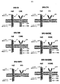
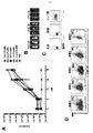
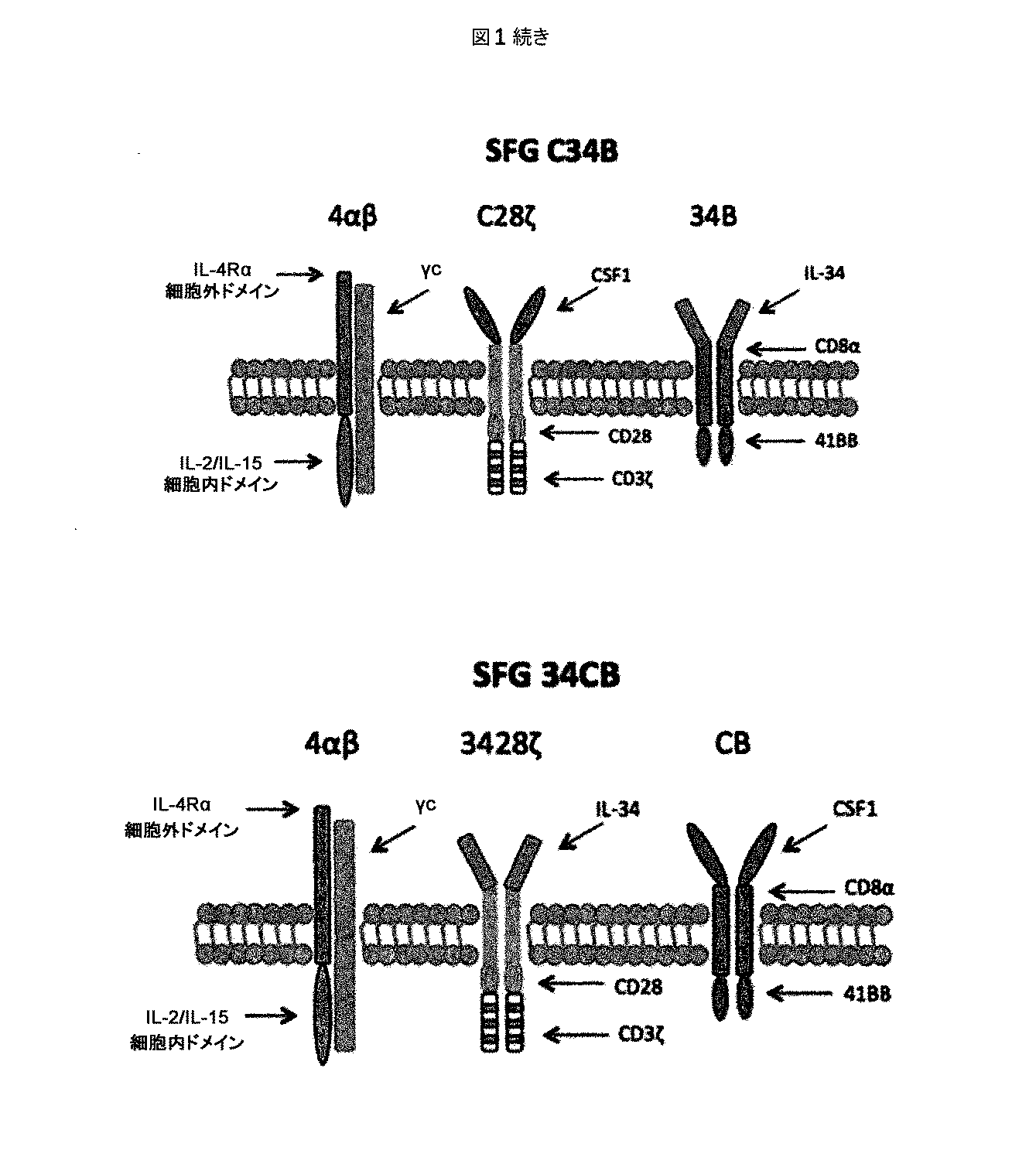
ホジキンリンパ腫、未分化大細胞リンパ腫、およびたとえばトリプルネガティブ乳癌などの一部の固形腫瘍に過剰発現するCSF−1受容体(c−FMSによりコードされる)を標的とする一連のCARを調製した。これらを概略的に図1に示す。これらの一連のCARは、標的化部分として2つの天然リガンドCSF−1またはIL−34の何れかを有する第二世代および第三世代CARの両方を含んでいた。CSF−1およびIL−34の両方がCSF−1受容体に結合するが、IL−34の方がかなり高い親和性(CSF−1より34倍高い)で結合する。
Example 1
A series of CARs were prepared that target the CSF-1 receptor (encoded by c-FMS) that is overexpressed in Hodgkin lymphoma, anaplastic large cell lymphoma, and some solid tumors such as triple negative breast cancer. These are shown schematically in FIG. These series of CARs included both second and third generation CARs with either of the two natural ligands CSF-1 or IL-34 as targeting moieties. Both CSF-1 and IL-34 bind to the CSF-1 receptor, but IL-34 binds with a much higher affinity (34 times higher than CSF-1).
コンストラクトSFG C28ζおよびSFG CTrをSFG レトロウイルスベクターにNcoI/XhoIフラグメントとしてクローニングし、その開始コドンは、除去したenv遺伝子が以前にあった、天然に存在するNcoI部位にあるようにする。遺伝子発現は、プロモーター活性を有するモロニーマウス白血病ウイルス(MoMLV)末端反復配列(LTR)から達成され、RNAのウイルスパッケージングは、スプライス供与部位とスプライス受容部位とに挟まれたMoMLV Ψパッケージングシグナルにより確保される。 The constructs SFG C28ζ and SFG CTr are cloned into the SFG retroviral vector as an NcoI / XhoI fragment so that the start codon is at the naturally occurring NcoI site where the removed env gene was previously. Gene expression is achieved from Moloney murine leukemia virus (MoMLV) terminal repeat (LTR) with promoter activity, and RNA viral packaging is achieved by a MoMLV Ψ packaging signal sandwiched between a splice donor site and a splice acceptor site. Secured.
他のコンストラクトは、すべてポリメラーゼ不完全プライマー伸長(PIPE)クローニング法を用いて設計およびクローニングした。PIPEクローニング法は、従来の制限酵素およびライゲーションに依存したクローニング法に代わるPCRを用いたクローニング法である。PIPEクローニング法では、追加の不要な残基を発現タンパク質にコードする可能性がある制限部位を組み込む必要がない。PIPE法は、おそらくdNTPの利用率の低下によると考えられる、PCR反応の最終サイクルにおける増幅プロセスの非効率性に依存しており、その結果、5’末端オーバーハングを有する一部一本鎖(PIPE)PCR産物が生成される。1組のベクター特異的プライマーを使用してPCRベクターを線状化し、次いで5’ベクター末端の重複配列を有するもう1組のプライマーを使用してインサートを増幅し、PIPEにより不完全な伸長産物を得た。次のステップでPIPE産物を混合し、一本鎖重複配列がアニールし、完全なSFG CARコンストラクトとしてアセンブルされた。クローニングの成功は、診断制限消化(diagnostic restriction digestion)により確認した。DNAシーケンシングをすべてのコンストラクトについて行って、予想されたコード配列が存在し、PCRによる変異がまったくないことを確認した(Source Bioscience,UK)。 All other constructs were designed and cloned using the polymerase incomplete primer extension (PIPE) cloning method. The PIPE cloning method is a cloning method using PCR in place of the conventional restriction enzyme and ligation-dependent cloning method. The PIPE cloning method does not require the incorporation of restriction sites that may encode additional unwanted residues in the expressed protein. The PIPE method relies on the inefficiency of the amplification process in the final cycle of the PCR reaction, presumably due to reduced dNTP utilization, resulting in partial single strands with a 5 ′ end overhang ( PIPE) PCR products are generated. Linearize the PCR vector using one set of vector specific primers, then amplify the insert using another set of primers with overlapping sequences at the 5 ′ vector ends, and incomplete extension products with PIPE. Obtained. In the next step, the PIPE product was mixed and the single stranded overlapping sequence annealed and assembled as a complete SFG CAR construct. The success of the cloning was confirmed by diagnostic restriction digestion. DNA sequencing was performed on all constructs to confirm that the expected coding sequence was present and there was no mutation due to PCR (Source Bioscience, UK).
パネルには、CSF−1またはIL−34が28zおよび4−1BBにまたはその逆に連結された2つの「二重標的化」キメラ活性化受容体(pCAR)が含まれていた。次いで、二重標的化pCAR組み合わせを、トーセア・アシグナ(Thosea Asigna)(T)2Aを含むレトロウイルスベクターを用いて同一のT細胞集団において化学量論的に共発現させた。これらのCARの一方を「C34B」(CSF1−28zプラスIL34−41BB)と命名し、他方を「34CB」(IL34−28zプラスCSF1−41BB)と命名した。 The panel included two “double-targeted” chimeric activated receptors (pCAR) with CSF-1 or IL-34 linked to 28z and 4-1BB or vice versa. The dual-targeted pCAR combination was then stoichiometrically co-expressed in the same T cell population using a retroviral vector containing Those Asigna (T) 2A. One of these CARs was named “C34B” (CSF1-28z plus IL34-41BB) and the other was named “34CB” (IL34-28z plus CSF1-41BB).
これらの二重標的化CAR T細胞では、共刺激モチーフ(CD28/4−1BB)を膜に近いそれらの天然の位置に置き、物理的に相互に分離して、同一のT細胞内で共発現させる。 In these dual-targeted CAR T cells, the costimulatory motif (CD28 / 4-1BB) is placed in their natural location close to the membrane, physically separated from each other, and co-expressed in the same T cell. Let
CARのすべては、追加のT2Aエレメントを含むベクターを用いてIL−4応答性4αβ受容体と共発現させた。これによりIL−4を用いたT細胞の富化/増殖が可能になり、選択後のこれらの多様な細胞集団の機能が比較しやすくなる。 All of the CARs were co-expressed with IL-4 responsive 4αβ receptor using a vector containing an additional T2A element. This allows for enrichment / proliferation of T cells using IL-4, making it easier to compare the functions of these diverse cell populations after selection.
本実験の主な焦点は、FMS/CSF−1受容体標的を発現するかまたは欠いている腫瘍標的細胞を用いて繰り返される再刺激に対するT細胞の挙動を試験することであった。各サイクルにおいて、100万個の表記のIL−4により増殖したCAR T細胞をRPMI+ヒトAB血清に懸濁し、抗原発現標的(T47D FMS)または抗原なし標的(T47D)のコンフルエントな単層(24ウェルディッシュ)で培養した。 The main focus of this experiment was to test the behavior of T cells against repeated re-stimulation with tumor target cells that express or lack FMS / CSF-1 receptor targets. In each cycle, 1 million of the indicated IL-4 CAR T cells were suspended in RPMI + human AB serum and confluent monolayers (24 wells) of antigen expression target (T47D FMS) or antigen-free target (T47D). Dish).
その後、CAR T細胞が生存し続けて単層を破壊した場合、100万個のT細胞を採取し、毎週同じ方法で再刺激した。総細胞数は、週1回のサイクルごとに起こったT細胞の増殖に応じて各時点で推定した。 Thereafter, if CAR T cells continued to survive and disrupted the monolayer, 1 million T cells were collected and restimulated weekly in the same manner. Total cell numbers were estimated at each time point according to T cell proliferation that occurred every weekly cycle.
これらの実験のすべてを通して、IL−2またはIL−4などの任意の外来性サイトカインの非存在下でT細胞を培養したため、T細胞は、生存し続けて増殖するためにそれ自体のサイトカインを産生しなければならなかった。サイトカイン(IFN−γおよびIL−2)産生を、T細胞/腫瘍細胞共培養物から回収した上清においてELISAにより測定し、効果的な共刺激の第2のマーカーを得た。 Throughout all of these experiments, T cells were cultured in the absence of any exogenous cytokines such as IL-2 or IL-4 so that they produced their own cytokines to continue to grow and proliferate Had to do. Cytokine (IFN-γ and IL-2) production was measured by ELISA in supernatants collected from T cell / tumor cell co-cultures to obtain a second marker of effective costimulation.
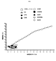
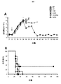
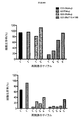
標的(FMSによりコードされるCSF−1受容体)を発現する腫瘍単層へのCARの最初の曝露で、殺すと予想されたCARはすべてそうすることが分かった(図2)(12回の実験をプールしたデータ)。対照はUT(形質導入していない)、P4(無関係な抗原、PSMAを標的とする)、および細胞内ドメインが切断されたCT4である。予想通り、CSF−1受容体を欠いている腫瘍細胞(T47D)を殺すCAR T細胞はない。 The first exposure of CAR to a tumor monolayer expressing the target (CSF-1 receptor encoded by FMS) was found to do so for all CARs expected to be killed (FIG. 2) (12 times Data pooled experiments). Controls are UT (untransduced), P4 (irrelevant antigen, targets PSMA), and CT4 with a cleaved intracellular domain. As expected, no CAR T cells kill tumor cells that lack the CSF-1 receptor (T47D).
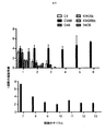
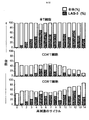
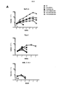
代表的な再刺激実験を図3に示す。7回の実験をプールした再刺激データを図4に示す。この場合、第1のサイクルにおける増殖は大部分のコンストラクトで類似していたが、IL−34標的第二世代および第三世代コンストラクトはより少なかった。これは、IL−34標的化部分の親和性が高すぎるためであり得る。 A typical restimulation experiment is shown in FIG. Restimulation data pooled from 7 experiments is shown in FIG. In this case, growth in the first cycle was similar for most constructs, but fewer IL-34 target second and third generation constructs. This may be because the affinity of the IL-34 targeting moiety is too high.
しかしながら、その後のサイクルでは、C34B二重pCAR組み合わせ(CSF−1標的28z第二世代CARがIL−34標的4−1BB共刺激モチーフと共発現したもの)の明確な優位性が一貫して明らかになった。 However, in subsequent cycles, the clear advantage of the C34B dual pCAR combination (a CSF-1 target 28z second generation CAR co-expressed with the IL-34 target 4-1BB costimulatory motif) was consistently revealed. became.
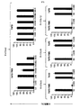
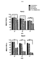
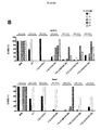
図3に示す実験では、各再刺激サイクル時から24時間後に上清を集め、サイトカイン含量(IFN−γおよびIL−2)についてELISAにより解析した。残存腫瘍細胞生存率の割合をMTTアッセイにより測定した(代表的な例を図5に示す)。サイトカイン産生の結果を図6に示す。C34B CAR T細胞のみが各刺激サイクル全体を通してIL−2を作る能力を保持することが分かった。これは、最初のサイクル後、それ以外のCAR組み合わせのすべてで失われた。繰り返される再刺激を通してIL−2を作る能力が持続的に保持されることは、CAR T細胞では通常見られず、これは、こうした二重共刺激の伝達がin vitroでこれらの細胞の分化を基本的に変化させ、アネルギーの発生を遅延させることを示唆する。 In the experiment shown in FIG. 3, supernatants were collected 24 hours after each restimulation cycle and analyzed for cytokine content (IFN-γ and IL-2) by ELISA. The percentage of remaining tumor cell viability was measured by MTT assay (a representative example is shown in FIG. 5). The results of cytokine production are shown in FIG. Only C34B CAR T cells were found to retain the ability to make IL-2 throughout each stimulation cycle. This was lost in all other CAR combinations after the first cycle. The sustained retention of the ability to make IL-2 through repeated re-stimulation is not normally seen in CAR T cells, as this dual costimulatory transmission in vitro differentiates these cells. It basically changes and suggests delaying the generation of anergy.
連続サイクルのAg刺激による単層破壊後の生存T細胞数もモニターし、結果を図5に示す。第2のサイクルの再刺激後、C34Bを除くすべてのCARが、CSF−1R依存性の腫瘍細胞殺傷を達成する能力を失い始める。一方、C34Bを発現するT細胞は、本細胞傷害性アッセイにおいて最大13の反復サイクルの再刺激で抗原依存性の潜在力を保持するが、改変していないT47D細胞に対してT細胞毒性を誘導しない。 The number of viable T cells after monolayer destruction by continuous cycle Ag stimulation was also monitored, and the results are shown in FIG. After the second cycle of restimulation, all CARs except C34B begin to lose the ability to achieve CSF-1R-dependent tumor cell killing. On the other hand, T cells expressing C34B retain antigen-dependent potential with up to 13 repeated cycles of restimulation in this cytotoxicity assay, but induce T cytotoxicity against unmodified T47D cells do not do.
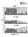
さらに、これらのT細胞上のいわゆる「疲弊マーカー」(PD1、TIM3、2B4およびLAG3)についてもフローサイトメトリーにより測定した。結果を図10〜13に示す。予想通り、様々な疲弊マーカーを発現したT細胞の割合は、再刺激したT細胞で徐々に増加したものの、これが抗原刺激時のC34B細胞による増殖、腫瘍細胞破壊またはサイトカイン放出を遅らせることはなかった。これは、C34Bの優れた機能が疲弊マーカーのアップレギュレーションの遅延の結果でないことを示唆する。 Furthermore, so-called “exhaustion markers” (PD1, TIM3, 2B4 and LAG3) on these T cells were also measured by flow cytometry. The results are shown in FIGS. As expected, the proportion of T cells that expressed various exhaustion markers gradually increased with restimulated T cells, but this did not delay C34B cell proliferation, tumor cell destruction or cytokine release upon antigen stimulation. . This suggests that the superior function of C34B is not the result of a delay in fatigue marker up-regulation.
要約すると、本発明のpCARアプローチは、より多くのサイクルの再刺激を介して抗原に対する応答性を細胞が保持する状態に細胞を維持すると思われる。pCARアプローチは、制御された記憶状態を超える分化を遅らせ得る傾向があり、かつ細胞が活性化時にIL−2を作る能力を保持しながら、アネルギーの発生を遅延させるようである。 In summary, the pCAR approach of the present invention appears to maintain cells in a state where they retain responsiveness to antigen through more cycles of restimulation. The pCAR approach tends to delay differentiation beyond the controlled memory state and seems to delay the development of anergy while retaining the ability of cells to make IL-2 upon activation.
実施例2
in vivoでの作用の解析
上記の実施例1に使用した一連のCARについて、CSF−1受容体標的を低レベルで発現し、かつ疾患がリンパ節全体に散在する、高悪性度in vivo異種移植モデルを用いて抗腫瘍活性を試験した(図7)。腫瘍細胞にホタルルシフェラーゼタグを付加し、疾患負荷の非侵襲的モニタリングを可能にした。
Example 2
Analysis of action in vivo For the series of CARs used in Example 1 above, high-grade in vivo xenografts that express CSF-1 receptor targets at low levels and the disease is scattered throughout the lymph nodes The model was used to test antitumor activity (FIG. 7). A firefly luciferase tag was added to the tumor cells to enable non-invasive monitoring of disease burden.
SCID/Beigeマウスを6群に無作為に割り付け(3回以上の独立した実験を合わせて各群9匹)、200μLのPBSに再懸濁した2×106個のK299腫瘍細胞を静脈内に(IV)接種した。5日目、各群を下記に示した治療レジメン:
・C4B群:20×106個のC4B T細胞 IV
・C34B群:20×106個のC34B T細胞 IV
・43428Bz:20×106個の43428Bz T細胞 IV
・34CB群:20×106個の34CB T細胞 IV
・UT(形質導入していない)群:20×106個の形質導入していないT細胞 IV
・NT(無処置)群:200μL PBS IV
の1つで処置した。
SCID / Beige mice were randomly assigned to 6 groups (9 in each group, combined with 3 or more independent experiments), and 2 × 10 6 K299 tumor cells resuspended in 200 μL PBS intravenously (IV) Inoculated. On
C4B group: 20 × 10 6 C4B T cells IV
C34B group: 20 × 10 6 C34B T cells IV
43428Bz: 20 × 10 6 43428Bz T cells IV
34CB group: 20 × 10 6 34CB T cells IV
UT (non-transduced) group: 20 × 10 6 non-transduced T cells IV
NT (no treatment) group: 200 μL PBS IV
One of these was treated.
腫瘍成長は、本研究の期間中の適切な時点で生物発光イメージング(BLI)を用いてモニターした。 Tumor growth was monitored using bioluminescence imaging (BLI) at appropriate time points during the study.
結果を図8に示す。この場合も、最高の効果を発揮した系はpCAR、C34Bの系であり、BLI発光の平均値の低さ(図8A〜B)、腫瘍進行の遅延または腫瘍退縮が見られ、マウスの生存の長期化につながった(図8C)。 The results are shown in FIG. Again, the system that exerted the best effect was the pCAR, C34B system, showing low average BLI luminescence (FIGS. 8A-B), delayed tumor progression or tumor regression, and the survival of mice This led to a prolonged period (Fig. 8C).
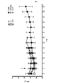
実験を通して動物の体重を測定したが、著しい毒性は示されかなった(図9)。 The animals were weighed throughout the experiment and showed no significant toxicity (Figure 9).
実施例3
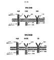
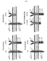
αvβ6依存性にT細胞活性化を誘導するpCARを操作するための標的化部分の選択
αvβ6インテグリンを単独または伸長(extended)ErbBファミリーと一緒に標的とする一連のCARを調製した。これらを図14に概略的に示す。この事例に使用した結合エレメントは、口蹄疫ウイルス(血清型01 BFS)由来のカプシドタンパク質VP1のGHループに由来するA20ペプチド(配列番号11)であった(米国特許第8,927,501号明細書)。これをCD124シグナルペプチドの下流に置き、CD28およびCD3ζ細胞内ドメインと融合して第二世代CAR、A20−28ζを形成した。同様のコンストラクトを含むが、重要なRGDLモチーフをAAAAで置き換えてスクランブルした標的化ペプチド(C20と命名される)を用いて対照(C20−28ζ)を調製した。第2の対照は、CD28切断細胞内ドメインと融合したA20(A20−Tr)を含んでいた。
Example 3
Selection of targeting moieties to engineer pCAR that induces T cell activation in an αvβ6-dependent manner. A series of CARs targeting αvβ6 integrin alone or together with the extended ErbB family were prepared. These are shown schematically in FIG. The binding element used in this case was the A20 peptide (SEQ ID NO: 11) derived from the GH loop of capsid protein VP1 from foot-and-mouth disease virus (serotype 01 BFS) (US Pat. No. 8,927,501). ). This was placed downstream of the CD124 signal peptide and fused with the CD28 and CD3ζ intracellular domains to form a second generation CAR, A20-28ζ. A control (C20-28ζ) was prepared with a targeting peptide (designated C20) that contained a similar construct but was scrambled by replacing the key RGDL motif with AAAA. The second control included A20 (A20-Tr) fused to the CD28-cleaved intracellular domain.
本発明のpCAR(TIE−41BB/A20−28zと命名される)を作製するため、A20−28zを、CD8α膜貫通ドメインおよび41BB細胞内ドメインと融合したpan−ErbB標的ペプチド(T1E)を含むキメラ共刺激受容体と共発現させた。 A chimera comprising a pan-ErbB targeting peptide (T1E) in which A20-28z is fused with a CD8α transmembrane domain and a 41BB intracellular domain to generate the pCAR of the present invention (designated TIE-41BB / A20-28z). Co-expressed with costimulatory receptors.
記載がある場合、CARは、in vitroでのIL−4を介した富化を可能にするために4αβキメラサイトカイン受容体と共発現させた。IL−4受容体α細胞外ドメインをIL−2/15共有受容体βの膜貫通ドメインおよび細胞内ドメインと融合したIL−4応答性4αβキメラサイトカイン受容体の等モル共発現は、トーセア・アシグナ(Thosea Asigna)(T)2Aリボソームスキップペプチドを用いて達成した。これらのキメラ分子をレトロウイルス遺伝子導入によりヒトT細胞で発現させた。 Where indicated, CAR was co-expressed with the 4αβ chimeric cytokine receptor to allow IL-4 mediated enrichment in vitro. Equimolar co-expression of an IL-4 responsive 4αβ chimeric cytokine receptor fused IL-4 receptor α extracellular domain with the transmembrane and intracellular domains of IL-2 / 15 covalent receptor β This was achieved using (Those Asigna) (T) 2A ribosome skip peptide. These chimeric molecules were expressed in human T cells by retroviral gene transfer.
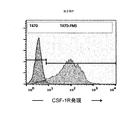
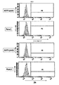
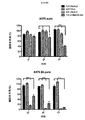
癌細胞株A375のインテグリン発現パターンを、フローサイトメトリーを用いて評価し(図15)、これらをαvβ6陰性(Panc1およびA375 puro)またはαvβ6陽性(Bxpc3およびA375 puro β6)腫瘍細胞に分けた。これらの細胞をCAR T細胞と1:1のエフェクター:ターゲット比で24時間、28時間または72時間共培養し、その後、細胞毒性をMTTアッセイにより評価し、未処理腫瘍細胞と比較して示した。結果を図16に示す。 The integrin expression pattern of the cancer cell line A375 was evaluated using flow cytometry (FIG. 15) and these were divided into αvβ6 negative (Pancl and A375 puro) or αvβ6 positive (Bxpc3 and A375 puro β6) tumor cells. These cells were co-cultured with CAR T cells at a 1: 1 effector: target ratio for 24, 28 or 72 hours, after which cytotoxicity was assessed by MTT assay and shown in comparison to untreated tumor cells. . The results are shown in FIG.
これらのデータから、A20−28z CAR T細胞がαvβ6インテグリンを発現するすべての標的細胞(Bxpc3およびA375 β6 puro)を殺すが、このインテグリンを欠いている標的(Panc1およびA375 puro)を殺さないことが示される。第2に、本アッセイにおいて対照CAR C20〜28zおよびA20−Trは不活性である。第3に、T1E−41BB/A20−28z pCARを発現するT細胞は、αvβ6インテグリンを発現する標的細胞(Bxpc3およびA375 β6 puro)の効果的殺傷を引き起こす。これらの結果はすべて予想通りである。一方、特記される点として、T1E−41BB/A20−28z pCARを発現するT細胞は、αvβ6を欠いている標的細胞(Panc1およびA375 puro)の殺傷も引き起こす。これは、pCARの立体配置内のA20ペプチドが低親和性の非αvβ6インテグリンに結合する能力が、これらの操作したT細胞の活性化を引き起こすのに十分であることの表れである。 From these data, it is clear that A20-28z CAR T cells kill all target cells that express αvβ6 integrin (Bxpc3 and A375 β6 puro) but not targets lacking this integrin (Pancl and A375 puro). Indicated. Second, the controls CAR C20-28z and A20-Tr are inactive in this assay. Third, T cells expressing T1E-41BB / A20-28z pCAR cause effective killing of target cells (Bxpc3 and A375 β6 puro) expressing αvβ6 integrin. All of these results are as expected. On the other hand, as noted, T cells expressing T1E-41BB / A20-28z pCAR also cause killing of target cells (Pancl and A375 puro) lacking αvβ6. This is an indication that the ability of the A20 peptide within the configuration of pCAR to bind to low affinity non-αvβ6 integrin is sufficient to cause activation of these engineered T cells.
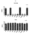
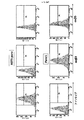
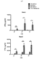
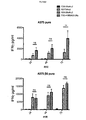
次いで、pCARおよび対照操作したT細胞のIFN−γの産生を評価した。αvβ6を欠いた(Panc1およびA375 puro)またはαvβ6を発現した(Bxpc3およびA375 puro β6)腫瘍細胞を、遺伝子操作T細胞と1:1のエフェクター:ターゲット比で共培養し、24時間、48時間または72時間後に上清を集めた。IFN−γのレベルをELISA(eBioscience)により定量した。結果を図17に示す。予想通り、対照が産生したIFN−γの量はわずかであった一方、A20−28z CAR T細胞は、αvβ6陽性(Bxpc3およびA375 puro β6)腫瘍細胞と培養した場合にIFN−γを放出した。特記される点として、本発明のpCAR、TIE−41BB/A20zを発現するT細胞は、αvβ6陽性(Bxpc3)腫瘍細胞と培養した場合、A20−28z T細胞より多くのIFN−γを産生する。さらに、TIE−41BB/A20z+T細胞は、αvβ6陰性(Panc1およびA375 puro)腫瘍細胞と培養した場合もIFN−γを産生した。ここでもまた、このことから、pCARの立体配置内の非αvβ6インテグリンに対するA20ペプチドの低親和結合が、これらの操作したT細胞の活性化を引き起こすのに十分であることが立証される。 PCAR and control engineered T cells were then evaluated for IFN-γ production. Tumor cells lacking αvβ6 (Pancl and A375 puro) or expressing αvβ6 (Bxpc3 and A375 puro β6) were co-cultured with genetically engineered T cells at a 1: 1 effector: target ratio for 24 hours, 48 hours or The supernatant was collected after 72 hours. The level of IFN-γ was quantified by ELISA (eBioscience). The results are shown in FIG. As expected, the amount of IFN-γ produced by the controls was small, while A20-28z CAR T cells released IFN-γ when cultured with αvβ6 positive (Bxpc3 and A375 puro β6) tumor cells. Of particular note, T cells expressing the pCAR, TIE-41BB / A20z of the present invention, produce more IFN-γ than A20-28z T cells when cultured with αvβ6 positive (Bxpc3) tumor cells. Furthermore, TIE-41BB / A20z + T cells also produced IFN-γ when cultured with αvβ6 negative (Pancl and A375 puro) tumor cells. Again, this demonstrates that low affinity binding of the A20 peptide to non-αvβ6 integrin within the configuration of pCAR is sufficient to cause activation of these engineered T cells.
次に、CAR T細胞集団をPanc1(αvβ6陰性)またはBxpc3(αvβ6陽性)腫瘍細胞上でIL−2添加物の非存在下、2週間に1回再刺激した。腫瘍細胞を、膵管腺癌(PDAC)の患者に由来するCAR T細胞と1:1のエフェクター:ターゲット比で共培養した(図18)。T細胞を最初に2×10⁵細胞/ウェルで加え、共培養から72時間後にカウントして増殖を評価した(上図)。細胞毒性は、T細胞の添加後の72時間でMTTアッセイにより評価した(下図)。十分な数のT細胞(2×105)が存在した場合、T細胞を新鮮な腫瘍単層上で再刺激し、さらに72時間後にこのプロセスを繰り返した。
The CAR T cell populations were then restimulated once every two weeks in the absence of IL-2 additive on Pancl (αvβ6 negative) or Bxpc3 (αvβ6 positive) tumor cells. Tumor cells were co-cultured with CAR T cells from patients with pancreatic ductal adenocarcinoma (PDAC) at a 1: 1 effector: target ratio (FIG. 18). T cells were initially added at 2 × 10 5 cells / well and counted for 72 hours after co-culture to assess proliferation (upper figure). Cytotoxicity was assessed by
結果を図18に示す。これらから、A20−28z/T1E−41BB+T細胞がIL−2放出(データ示さず)およびαvβ6+Bxpc3細胞の破壊を伴い数回の増殖を行うことが例証される。ここでもまた、A20−28z/T1E−41BB+T細胞は、IL−2放出およびPanc1腫瘍細胞の破壊を伴う数回の増殖も行った。 The results are shown in FIG. These illustrate that A20-28z / T1E-41BB + T cells proliferate several times with IL-2 release (data not shown) and destruction of αvβ6 + Bxpc3 cells. Again, A20-28z / T1E-41BB + T cells also proliferated several times with IL-2 release and Panc1 tumor cell destruction.
全体として、これらの結果から、A20−28z/T1E−41BBを含むpCARは、αvβ6を標的とする第二世代CARと比較してin vitroでの機能性の向上を示すことが明らかに示された。さらに、A20−28z/T1E−41BB+T細胞は、このインテグリンを微小レベルから検出不可能なレベルで発現するPanc1またはA375 puro細胞によっても活性化を受ける。C34B pCAR(実施例1および2)を用いて得られた知見と合わせて考えると、これは、pCARの立体配置が、41BB CCRによる高親和性結合の相互作用が起こると同時に、28z第二世代CARによる低親和性の相互作用が起こると、連続的な再刺激時にT細胞活性化が起こることを可能にすることの表れである。 Overall, these results clearly showed that pCAR containing A20-28z / T1E-41BB showed improved in vitro functionality compared to second generation CAR targeting αvβ6. . In addition, A20-28z / T1E-41BB + T cells are also activated by Pancl or A375 puro cells that express this integrin at micro to undetectable levels. Taken together with the findings obtained using C34B pCAR (Examples 1 and 2), this indicates that the configuration of pCAR is accompanied by a high affinity binding interaction with 41BB CCR, while the 28z second generation The occurrence of low affinity interactions with CAR is an indication that it allows T cell activation to occur upon continuous restimulation.
実施例4
機能的pCARを操作するための代替のTNF受容体ファミリーメンバーCD27の使用
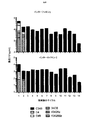
A20−28z/T1E−41BB pCARを出発材料として使用して、41BBモジュールをTNF受容体ファミリーの代替メンバー、すなわちCD27またはCD40で置き換えた別のpCARを操作した。細胞内ドメインを切断した(tr)対照pCARを操作した。αvβ6を発現する(Bxpc3)または欠いている(Panc1)標的細胞を24ウェルプレートの1ウェルあたり5×10⁴細胞の密度で蒔いた。24時間後、5×10⁴個のpCAR T細胞を、外来性サイトカイン添加物を用いずに標的細胞または空ウェル(「刺激していない」)に加えた。さらに72時間後、T細胞をウェルから回収し、カウントした(図19A)。MTTアッセイを行って残存標的細胞の生存割合を判定し、T細胞を添加せずに蒔いてあった対照標的細胞と比較した(図19B)。各刺激サイクル後にT細胞が増殖した場合、上記の通りに新鮮な標的細胞上で再刺激した。pCAR T細胞の増殖(図19A)およびMTTアッセイ(図19B)は、前の場合と同様に72時間後に行った。pCAR T細胞の反復再刺激および標的細胞殺傷の評価は、このように72時間の各サイクル期間にわたりT細胞がもはや増殖しなくなるまで継続した。
Example 4
Use of Alternative TNF Receptor Family Member CD27 to Manipulate Functional pCAR Using A20-28z / T1E-41BB pCAR as a starting material, the 41BB module can be used as an alternative member of the TNF receptor family, ie CD27 or CD40. Another pCAR replaced was manipulated. A control pCAR was engineered that cleaved the intracellular domain (tr). Target cells expressing αvβ6 (Bxpc3) or lacking (Pancl) were seeded at a density of 5 × 10 4 cells per well of a 24-well plate. After 24 hours, 5 × 10 4 pCAR T cells were added to target cells or empty wells (“unstimulated”) without exogenous cytokine additives. After an additional 72 hours, T cells were collected from the wells and counted (Figure 19A). An MTT assay was performed to determine the survival rate of remaining target cells and compared to control target cells that had been seeded without the addition of T cells (FIG. 19B). If T cells proliferated after each stimulation cycle, they were restimulated on fresh target cells as described above. pCAR T cell proliferation (Figure 19A) and MTT assay (Figure 19B) were performed 72 hours later as before. Evaluation of repeated re-stimulation of pCAR T cells and target cell killing was thus continued until the T cells no longer proliferated over each 72 hour cycle period.
これらのデータから、T細胞がPanc1標的細胞上で刺激されるときにT細胞増殖および腫瘍細胞殺傷の持続により示される、A20−28z/T1E−41BB pCARの優れた機能性がここでもまた確認される。このことから、非αvβ6インテグリンに対するA20ペプチドの低親和性結合は、これらの操作したT細胞の活性化を引き起こすのに十分であることもさらに確認される。一方、特記される点として、A20−28z/T1E−CD27 pCARは、αvβ6を発現するBxpc3細胞上で再刺激されると、最大レベルの増殖(図19A)および腫瘍細胞殺傷の持続(図19B)を達成した。一方、CD40を用いたpCARは、これらのアッセイにおいて中程度の機能を示した。総合すると、これらのデータから、CD27または41BBが例示されるいくつかのTNF受容体ファミリーメンバーを利用して、優れた機能性を示すpCARを操作できることが立証される。
These data again confirm the superior functionality of A20-28z / T1E-41BB pCAR, indicated by the persistence of T cell proliferation and tumor cell killing when T cells are stimulated on Pancl target cells. The This further confirms that the low affinity binding of the A20 peptide to non-αvβ6 integrin is sufficient to cause activation of these engineered T cells. On the other hand, it is noted that A20-28z / T1E-CD27 pCAR, when restimulated on Bxpc3 cells expressing αvβ6, maximized proliferation (FIG. 19A) and sustained tumor cell killing (FIG. 19B). Achieved. On the other hand, pCAR using CD40 showed moderate function in these assays. Taken together, these data demonstrate that several TNF receptor family members exemplified by CD27 or 41BB can be utilized to engineer pCARs that exhibit superior functionality.
Claims (21)
(i)第二世代キメラ抗原受容体であって、
(a)シグナル伝達領域;
(b)共刺激シグナル伝達領域;
(c)膜貫通ドメイン;および
(d)標的抗原上の第1のエピトープと特異的に相互作用する結合エレメント
を含む第二世代キメラ抗原受容体と、
(ii)キメラ共刺激受容体であって、
(e)(b)の共刺激シグナル伝達領域と異なる共刺激シグナル伝達領域;
(f)膜貫通ドメイン;および
(g)標的抗原上の第2のエピトープと特異的に相互作用する結合エレメント
を含むキメラ共刺激受容体と
を発現することを特徴とする免疫応答細胞。 In immune response cells,
(I) a second generation chimeric antigen receptor,
(A) signal transduction region;
(B) a costimulatory signaling region;
(C) a transmembrane domain; and (d) a second generation chimeric antigen receptor comprising a binding element that specifically interacts with a first epitope on a target antigen;
(Ii) a chimeric costimulatory receptor,
(E) a costimulatory signaling region different from the costimulatory signaling region of (b);
An immune responder cell characterized by expressing (f) a transmembrane domain; and (g) a chimeric costimulatory receptor comprising a binding element that specifically interacts with a second epitope on the target antigen.
RGDLX5X6L(配列番号7)、または
RGDLX5X6I(配列番号8)
を含むペプチドであり、LX5X6LまたはLX5X6Iは、αヘリックス構造内に含まれ、X5およびX6はヘリックス促進残基である、αvβ6インテグリン特異的結合剤であり、および結合エレメント(g)はTIEペプチドであることを特徴とする免疫応答細胞。 The immune response cell according to any one of claims 1 to 12, wherein the binding element (d) is an α v β 6 integrin-specific binding agent and comprises a sequence motif RGDLX 5 X 6 L (SEQ ID NO: 7). Or RGDLX 5 X 6 I (SEQ ID NO: 8)
An α v β 6 integrin specific binding agent, wherein LX 5 X 6 L or LX 5 X 6 I is contained within an α-helix structure, and X 5 and X 6 are helix-promoting residues An immune response cell, characterized in that the binding element (g) is a TIE peptide.
Priority Applications (1)
| Application Number | Priority Date | Filing Date | Title |
|---|---|---|---|
| JP2021114022A JP2021168675A (en) | 2015-07-31 | 2021-07-09 | Therapeutic agent |
Applications Claiming Priority (3)
| Application Number | Priority Date | Filing Date | Title |
|---|---|---|---|
| GBGB1513540.3A GB201513540D0 (en) | 2015-07-31 | 2015-07-31 | Therapeutic agents |
| GB1513540.3 | 2015-07-31 | ||
| PCT/GB2016/052324 WO2017021701A1 (en) | 2015-07-31 | 2016-07-28 | Therapeutic agents |
Related Child Applications (1)
| Application Number | Title | Priority Date | Filing Date |
|---|---|---|---|
| JP2021114022A Division JP2021168675A (en) | 2015-07-31 | 2021-07-09 | Therapeutic agent |
Publications (3)
| Publication Number | Publication Date |
|---|---|
| JP2018521663A true JP2018521663A (en) | 2018-08-09 |
| JP2018521663A5 JP2018521663A5 (en) | 2019-08-29 |
| JP7053037B2 JP7053037B2 (en) | 2022-04-12 |
Family
ID=54062973
Family Applications (2)
| Application Number | Title | Priority Date | Filing Date |
|---|---|---|---|
| JP2018503642A Active JP7053037B2 (en) | 2015-07-31 | 2016-07-28 | Therapeutic drug |
| JP2021114022A Pending JP2021168675A (en) | 2015-07-31 | 2021-07-09 | Therapeutic agent |
Family Applications After (1)
| Application Number | Title | Priority Date | Filing Date |
|---|---|---|---|
| JP2021114022A Pending JP2021168675A (en) | 2015-07-31 | 2021-07-09 | Therapeutic agent |
Country Status (14)
| Country | Link |
|---|---|
| US (6) | US10703794B2 (en) |
| EP (2) | EP3939992A1 (en) |
| JP (2) | JP7053037B2 (en) |
| KR (1) | KR102411571B1 (en) |
| CN (1) | CN107735407B (en) |
| AU (1) | AU2016303355B2 (en) |
| DK (1) | DK3328880T3 (en) |
| ES (1) | ES2883633T3 (en) |
| GB (1) | GB201513540D0 (en) |
| IL (1) | IL256511B (en) |
| MX (1) | MX2018001009A (en) |
| RU (1) | RU2747733C1 (en) |
| SG (1) | SG10201912416QA (en) |
| WO (1) | WO2017021701A1 (en) |
Cited By (3)
| Publication number | Priority date | Publication date | Assignee | Title |
|---|---|---|---|---|
| JP2022545643A (en) * | 2019-08-13 | 2022-10-28 | キングス・カレッジ・ロンドン | Immunoreactive cells armed with spatiotemporally restricted activity of IL-1 superfamily cytokines |
| JP2022547416A (en) * | 2019-08-28 | 2022-11-14 | キングス・カレッジ・ロンドン | B cell-targeting parallel CAR (pCAR) therapeutic agents |
| JP2023522038A (en) * | 2020-04-17 | 2023-05-26 | 2セブンティ バイオ インコーポレイテッド | Modified CCR polypeptides and uses thereof |
Families Citing this family (41)
| Publication number | Priority date | Publication date | Assignee | Title |
|---|---|---|---|---|
| EP2948544A4 (en) | 2013-01-28 | 2016-08-03 | St Jude Childrens Res Hospital | NKG2D-SPECIFIC CHIMERIC RECEPTOR ADAPTED FOR USE IN CELL THERAPY AGAINST CANCER AND INFECTIOUS DISEASES |
| DK3143134T3 (en) | 2014-05-15 | 2021-01-04 | Nat Univ Singapore | Modified, natural killer cells and their uses |
| GB201513540D0 (en) | 2015-07-31 | 2015-09-16 | King S College London | Therapeutic agents |
| GB201514874D0 (en) | 2015-08-20 | 2015-10-07 | Autolus Ltd | Cell |
| CA3045339A1 (en) | 2016-12-03 | 2018-06-07 | Juno Therapeutics, Inc. | Methods and compositions for use of therapeutic t cells in combination with kinase inhibitors |
| EP3336107A1 (en) * | 2016-12-15 | 2018-06-20 | Miltenyi Biotec GmbH | Immune cells expressing an antigen binding receptor and a chimeric costimulatory receptor |
| AU2017383142A1 (en) | 2016-12-22 | 2019-07-04 | Università Degli Studi Magna Graecia Catanzaro | A monoclonal antibody targeting a unique sialoglycosilated cancer-associated epitope of CD43 |
| CA3056591A1 (en) | 2017-03-27 | 2018-10-04 | National University Of Singapore | Stimulatory cell lines for ex vivo expansion and activation of natural killer cells |
| BR112019019917A2 (en) | 2017-03-27 | 2020-04-22 | Nat Univ Singapore | truncated nkg2d chimeric receptors and their uses in natural killer cell immunotherapy |
| AU2018275894B2 (en) | 2017-06-02 | 2025-04-24 | Juno Therapeutics, Inc. | Articles of manufacture and methods for treatment using adoptive cell therapy |
| US20200216515A1 (en) * | 2017-07-26 | 2020-07-09 | Cellectis | Methods of antigen-dependent chimeric antigen receptor (car) immune cell selection |
| CN109971724B (en) * | 2017-12-28 | 2023-10-31 | 上海细胞治疗研究院 | CAR-T cells targeting the ErbB receptor family and self-expressing PD-1 antibodies and uses thereof |
| EP3749685A4 (en) | 2018-02-09 | 2021-12-22 | National University of Singapore | ACTIVATION OF NKG2D CHIMERIC RECEPTORS AND USES THEREOF IN IMMUNOTHERAPY WITH NATURAL KILLER CELLS |
| JP7334985B2 (en) | 2018-04-02 | 2023-08-29 | ナショナル ユニヴァーシティー オブ シンガポール | Neutralization of human cytokines by membrane-bound anti-cytokine non-signaling binders expressed in immune cells |
| CN110615842B (en) * | 2018-06-20 | 2023-05-09 | 上海隆耀生物科技有限公司 | Chimeric antigen receptor containing co-stimulatory receptor and application thereof |
| EP3587454A1 (en) * | 2018-06-27 | 2020-01-01 | Albert-Ludwigs-Universität Freiburg | Chimeric antigen receptors that bind to prostate specific membrane antigen |
| EP3844186A4 (en) | 2018-08-29 | 2022-08-17 | National University of Singapore | METHOD FOR SPECIFICALLY STIMULATING THE SURVIVAL AND EXPANSION OF GENETICALLY MODIFIED IMMUNE CELLS |
| CA3120563A1 (en) | 2018-11-26 | 2020-06-04 | Nkarta, Inc. | Methods for the simultaneous expansion of multiple immune cell types, related compositions and uses of same in cancer immunotherapy |
| GB201900858D0 (en) * | 2019-01-22 | 2019-03-13 | Price Nicola Kaye | Receptors providing targeted costimulation for adoptive cell therapy |
| GB201902277D0 (en) | 2019-02-19 | 2019-04-03 | King S College London | Therapeutic agents |
| EP3773918A4 (en) | 2019-03-05 | 2022-01-05 | Nkarta, Inc. | ANTI-CD19 CHEMERIC ANTIGEN RECEPTORS AND THEIR USE IN IMMUNOTHERAPY |
| WO2020183158A1 (en) | 2019-03-11 | 2020-09-17 | Leucid Bio Ltd | MUC1 PARALLEL CAR (pCAR) THERAPEUTIC AGENTS |
| TW202110873A (en) | 2019-04-30 | 2021-03-16 | 美商聖堤生物科技股份有限公司 | Chimeric receptors and methods of use thereof |
| GB202003277D0 (en) | 2020-03-06 | 2020-04-22 | King S College London | Therapeutic agents |
| AU2021312871A1 (en) * | 2020-07-21 | 2023-02-09 | Allogene Therapeutics, Inc. | Chimeric antigen receptors with enhanced signaling and activities and uses thereof |
| WO2022036495A1 (en) | 2020-08-17 | 2022-02-24 | Utc Therapeutics Inc. | Lymphocytes-antigen presenting cells co-stimulators and uses thereof |
| WO2022083668A1 (en) * | 2020-10-21 | 2022-04-28 | Nanjing Legend Biotech Co., Ltd. | Use of a chimeric co-stimulatory receptor for cell therapy |
| US20240342282A1 (en) * | 2021-08-13 | 2024-10-17 | Ludwig-Maximilians-Universität München | Anti-csf1r car expressing lymphocytes for targeted tumor therapy |
| CA3229493A1 (en) * | 2021-08-18 | 2023-02-23 | Legend Biotech Ireland Limited | Modified immune cells expressing tlr receptors |
| JP7732840B2 (en) * | 2021-10-14 | 2025-09-02 | 株式会社三共 | gaming machines |
| JP7732842B2 (en) * | 2021-10-14 | 2025-09-02 | 株式会社三共 | gaming machines |
| JP7732843B2 (en) * | 2021-10-14 | 2025-09-02 | 株式会社三共 | gaming machines |
| JP7732841B2 (en) * | 2021-10-14 | 2025-09-02 | 株式会社三共 | gaming machines |
| JP7732839B2 (en) * | 2021-10-14 | 2025-09-02 | 株式会社三共 | gaming machines |
| IL312795A (en) | 2021-11-15 | 2024-07-01 | Neogene Therapeutics B V | Engineered t cells with reduced tgf-beta receptor signaling |
| WO2023129929A1 (en) | 2021-12-30 | 2023-07-06 | Tr1X, Inc. | Cd4+ t cells expressing il-10 and chimeric antigen receptors and uses thereof |
| CN118510814A (en) * | 2022-05-10 | 2024-08-16 | 星尘生物科技(上海)有限公司 | Chimeric antigen receptor and application thereof |
| CN116804210A (en) * | 2022-12-08 | 2023-09-26 | 复旦大学附属中山医院 | Enterovirus vector and application thereof |
| EP4687957A1 (en) | 2023-04-05 | 2026-02-11 | King's College London | Parallel chimeric antigen receptors (pcars) and adaptor chimeric antigen receptors comprising alternative signalling domains and methods of use thereof |
| GB202403718D0 (en) | 2024-03-14 | 2024-05-01 | King S College London | Therapeutic agents |
| WO2026015450A1 (en) | 2024-07-06 | 2026-01-15 | Tr1X, Inc. | Cd4+ t cells expressing il-10 and chimeric antigen receptors and uses thereof |
Citations (4)
| Publication number | Priority date | Publication date | Assignee | Title |
|---|---|---|---|---|
| JP2009509562A (en) * | 2005-10-03 | 2009-03-12 | キャンサー・リサーチ・テクノロジー・リミテッド | αvβ6 peptide ligand and its utilization |
| WO2014055668A1 (en) * | 2012-10-02 | 2014-04-10 | Memorial Sloan-Kettering Cancer Center | Compositions and methods for immunotherapy |
| WO2016011210A2 (en) * | 2014-07-15 | 2016-01-21 | Juno Therapeutics, Inc. | Engineered cells for adoptive cell therapy |
| JP2017502251A (en) * | 2013-12-26 | 2017-01-19 | バーダス テクノロジーズ プライベート リミテッドVerdus Technologies Pte.Ltd. | Fluid processing apparatus and method for heating or cooling a fluid stream |
Family Cites Families (29)
| Publication number | Priority date | Publication date | Assignee | Title |
|---|---|---|---|---|
| ATE297986T1 (en) * | 1995-02-24 | 2005-07-15 | Gen Hospital Corp | REORIENTATION OF CELLULAR IMMUNITY THROUGH RECEPTOR CHIMERAS |
| US7446190B2 (en) | 2002-05-28 | 2008-11-04 | Sloan-Kettering Institute For Cancer Research | Nucleic acids encoding chimeric T cell receptors |
| SI2856876T1 (en) | 2007-03-30 | 2018-04-30 | Memorial Sloan-Kettering Cancer Center | Constituent expression of costimulatory ligands on indirectly transmitted T lymphocytes |
| WO2010085660A2 (en) | 2009-01-23 | 2010-07-29 | Roger Williams Hospital | Viral vectors encoding multiple highly homologous non-viral polypeptides and the use of same |
| CA2776143A1 (en) | 2009-10-01 | 2011-04-07 | The United States Of America, As Represented By The Secretary, Departmen T Of Health And Human Services | Anti-vascular endothelial growth factor receptor-2 chimeric antigen receptors and use of same for the treatment of cancer |
| US9987308B2 (en) | 2011-03-23 | 2018-06-05 | Fred Hutchinson Cancer Research Center | Method and compositions for cellular immunotherapy |
| MX2014001222A (en) | 2011-07-29 | 2014-09-15 | Univ Pennsylvania | CHANGE CO-STIMULATING RECEIVERS. |
| WO2013033626A2 (en) | 2011-08-31 | 2013-03-07 | Trustees Of Dartmouth College | Nkp30 receptor targeted therapeutics |
| EP2817330B1 (en) | 2012-02-22 | 2020-07-08 | The Trustees of the University of Pennsylvania | Use of icos-based cars to enhance antitumor activity and car persistence |
| WO2013126729A1 (en) | 2012-02-22 | 2013-08-29 | The Trustees Of The University Of Pennsylvania | Use of the cd2 signaling domain in second-generation chimeric antigen receptors |
| CN103483452B (en) | 2012-06-12 | 2021-08-13 | 上海细胞治疗集团有限公司 | Dual-signal-independent chimeric antigen receptors and their uses |
| MX367730B (en) | 2012-09-04 | 2019-09-04 | Cellectis | Multi-chain chimeric antigen receptor and uses thereof. |
| WO2014055442A2 (en) | 2012-10-01 | 2014-04-10 | The Trustees Of The University Of Pennsylvania | Compositions and methods for targeting stromal cells for the treatment of cancer |
| WO2014055657A1 (en) | 2012-10-05 | 2014-04-10 | The Trustees Of The University Of Pennsylvania | Use of a trans-signaling approach in chimeric antigen receptors |
| CN113005091A (en) | 2013-02-06 | 2021-06-22 | 细胞基因公司 | Modified T lymphocytes with improved specificity |
| DK2956175T3 (en) | 2013-02-15 | 2017-11-27 | Univ California | CHEMICAL ANTIGEN RECEPTOR AND PROCEDURES FOR USE THEREOF |
| US20160017286A1 (en) | 2013-03-06 | 2016-01-21 | The Trustees Of The University Of Pennsylvania | Ikaros inhibition to augment adoptive t cell transfer |
| WO2014164544A1 (en) | 2013-03-09 | 2014-10-09 | Baylor College Of Medicine | Vascular-targeted t-cell therapy |
| CA2905352A1 (en) | 2013-03-14 | 2014-09-25 | Bellicum Pharmaceuticals, Inc. | Methods for controlling t cell proliferation |
| US10584158B2 (en) | 2013-04-17 | 2020-03-10 | Baylor College Of Medicine | Immunosuppressive TGF-β signal converter |
| CN105873952A (en) | 2013-10-31 | 2016-08-17 | 弗莱德哈钦森癌症研究中心 | Modified hematopoietic stem/progenitor and non-t effector cells, and uses thereof |
| MX373687B (en) | 2013-11-21 | 2020-07-07 | Ucl Business Ltd | NATURAL KNOCK (NK) CELL |
| EP3087101B1 (en) | 2013-12-20 | 2024-06-05 | Novartis AG | Regulatable chimeric antigen receptor |
| US20170233452A1 (en) | 2014-04-23 | 2017-08-17 | Immusoft Corporation | Chimeric antigen receptors specific to avb6 integrin and methods of use thereof to treat cancer |
| BR112017001183A2 (en) | 2014-07-21 | 2017-11-28 | Novartis Ag | cancer treatment using humanized anti-bcma chimeric antigen receptor |
| US10828353B2 (en) | 2015-01-31 | 2020-11-10 | The Trustees Of The University Of Pennsylvania | Compositions and methods for T cell delivery of therapeutic molecules |
| WO2016196388A1 (en) | 2015-05-29 | 2016-12-08 | Juno Therapeutics, Inc. | Composition and methods for regulating inhibitory interactions in genetically engineered cells |
| GB201513540D0 (en) * | 2015-07-31 | 2015-09-16 | King S College London | Therapeutic agents |
| WO2020183158A1 (en) * | 2019-03-11 | 2020-09-17 | Leucid Bio Ltd | MUC1 PARALLEL CAR (pCAR) THERAPEUTIC AGENTS |
-
2015
- 2015-07-31 GB GBGB1513540.3A patent/GB201513540D0/en not_active Ceased
-
2016
- 2016-07-28 CN CN201680039617.1A patent/CN107735407B/en active Active
- 2016-07-28 US US15/749,016 patent/US10703794B2/en active Active
- 2016-07-28 ES ES16750211T patent/ES2883633T3/en active Active
- 2016-07-28 EP EP21177321.3A patent/EP3939992A1/en not_active Withdrawn
- 2016-07-28 SG SG10201912416QA patent/SG10201912416QA/en unknown
- 2016-07-28 JP JP2018503642A patent/JP7053037B2/en active Active
- 2016-07-28 MX MX2018001009A patent/MX2018001009A/en unknown
- 2016-07-28 KR KR1020187005607A patent/KR102411571B1/en active Active
- 2016-07-28 DK DK16750211.1T patent/DK3328880T3/en active
- 2016-07-28 RU RU2018105137A patent/RU2747733C1/en active
- 2016-07-28 AU AU2016303355A patent/AU2016303355B2/en active Active
- 2016-07-28 EP EP16750211.1A patent/EP3328880B1/en active Active
- 2016-07-28 WO PCT/GB2016/052324 patent/WO2017021701A1/en not_active Ceased
-
2017
- 2017-12-24 IL IL256511A patent/IL256511B/en unknown
-
2020
- 2020-05-18 US US16/877,008 patent/US10899818B2/en active Active
- 2020-05-18 US US16/877,035 patent/US10865231B2/en active Active
- 2020-12-10 US US17/118,045 patent/US11802143B2/en active Active
-
2021
- 2021-07-09 JP JP2021114022A patent/JP2021168675A/en active Pending
-
2023
- 2023-09-22 US US18/472,441 patent/US12612445B2/en active Active
-
2024
- 2024-01-22 US US18/418,864 patent/US12509499B2/en active Active
Patent Citations (5)
| Publication number | Priority date | Publication date | Assignee | Title |
|---|---|---|---|---|
| JP2009509562A (en) * | 2005-10-03 | 2009-03-12 | キャンサー・リサーチ・テクノロジー・リミテッド | αvβ6 peptide ligand and its utilization |
| WO2014055668A1 (en) * | 2012-10-02 | 2014-04-10 | Memorial Sloan-Kettering Cancer Center | Compositions and methods for immunotherapy |
| JP2015535689A (en) * | 2012-10-02 | 2015-12-17 | メモリアル スローン−ケタリング キャンサー センター | Compositions and methods for immunotherapy |
| JP2017502251A (en) * | 2013-12-26 | 2017-01-19 | バーダス テクノロジーズ プライベート リミテッドVerdus Technologies Pte.Ltd. | Fluid processing apparatus and method for heating or cooling a fluid stream |
| WO2016011210A2 (en) * | 2014-07-15 | 2016-01-21 | Juno Therapeutics, Inc. | Engineered cells for adoptive cell therapy |
Non-Patent Citations (2)
| Title |
|---|
| J.IMMUNOL.(2013)VOL.191, P.4589-4598, JPN6020016774, ISSN: 0004656335 * |
| MOL MED.(2012)VOL.18, P.565-576, JPN6020016773, ISSN: 0004656336 * |
Cited By (5)
| Publication number | Priority date | Publication date | Assignee | Title |
|---|---|---|---|---|
| JP2022545643A (en) * | 2019-08-13 | 2022-10-28 | キングス・カレッジ・ロンドン | Immunoreactive cells armed with spatiotemporally restricted activity of IL-1 superfamily cytokines |
| JP2022547416A (en) * | 2019-08-28 | 2022-11-14 | キングス・カレッジ・ロンドン | B cell-targeting parallel CAR (pCAR) therapeutic agents |
| JP7723420B2 (en) | 2019-08-28 | 2025-08-14 | キングス・カレッジ・ロンドン | B-cell-targeted parallel CAR (pCAR) therapeutic agents |
| JP2023522038A (en) * | 2020-04-17 | 2023-05-26 | 2セブンティ バイオ インコーポレイテッド | Modified CCR polypeptides and uses thereof |
| JP7802684B2 (en) | 2020-04-17 | 2026-01-20 | リジェネロン・ファーマシューティカルズ・インコーポレイテッド | Modified CCR polypeptides and uses thereof |
Also Published As
Similar Documents
| Publication | Publication Date | Title |
|---|---|---|
| US12509499B2 (en) | Therapeutic agents | |
| JP7633937B2 (en) | Chimeric cytokine receptor bearing the PD-1 ectodomain | |
| CN111247242B (en) | Chimeric Antigen Receptors (CARs), compositions, and methods of use thereof | |
| KR102483822B1 (en) | Tagged chimeric effector molecules and receptors thereof | |
| CN113811327A (en) | MUC1 Parallel CAR (pCAR) Therapeutics | |
| KR20180002604A (en) | Chimeric antigen receptor (CAR) targeting compositions of hematological malignancies, compositions and uses thereof | |
| JP7723420B2 (en) | B-cell-targeted parallel CAR (pCAR) therapeutic agents | |
| JP2025511872A (en) | Multisite receptors and signaling complexes | |
| CN111479918A (en) | Cells | |
| CA2993746C (en) | Therapeutic agents | |
| HK1256383B (en) | Therapeutic agents | |
| JP2022001021A (en) | Cd26 specific chimeric antigen receptor |
Legal Events
| Date | Code | Title | Description |
|---|---|---|---|
| A521 | Request for written amendment filed |
Free format text: JAPANESE INTERMEDIATE CODE: A523 Effective date: 20190712 |
|
| A621 | Written request for application examination |
Free format text: JAPANESE INTERMEDIATE CODE: A621 Effective date: 20190712 |
|
| A977 | Report on retrieval |
Free format text: JAPANESE INTERMEDIATE CODE: A971007 Effective date: 20200417 |
|
| A131 | Notification of reasons for refusal |
Free format text: JAPANESE INTERMEDIATE CODE: A131 Effective date: 20200602 |
|
| A521 | Request for written amendment filed |
Free format text: JAPANESE INTERMEDIATE CODE: A523 Effective date: 20200902 |
|
| A521 | Request for written amendment filed |
Free format text: JAPANESE INTERMEDIATE CODE: A821 Effective date: 20200903 |
|
| A02 | Decision of refusal |
Free format text: JAPANESE INTERMEDIATE CODE: A02 Effective date: 20210309 |
|
| A521 | Request for written amendment filed |
Free format text: JAPANESE INTERMEDIATE CODE: A523 Effective date: 20210709 |
|
| C60 | Trial request (containing other claim documents, opposition documents) |
Free format text: JAPANESE INTERMEDIATE CODE: C60 Effective date: 20210709 |
|
| A521 | Request for written amendment filed |
Free format text: JAPANESE INTERMEDIATE CODE: A821 Effective date: 20210709 |
|
| A911 | Transfer to examiner for re-examination before appeal (zenchi) |
Free format text: JAPANESE INTERMEDIATE CODE: A911 Effective date: 20210906 |
|
| C21 | Notice of transfer of a case for reconsideration by examiners before appeal proceedings |
Free format text: JAPANESE INTERMEDIATE CODE: C21 Effective date: 20210907 |
|
| A131 | Notification of reasons for refusal |
Free format text: JAPANESE INTERMEDIATE CODE: A131 Effective date: 20211207 |
|
| A521 | Request for written amendment filed |
Free format text: JAPANESE INTERMEDIATE CODE: A523 Effective date: 20220302 |
|
| TRDD | Decision of grant or rejection written | ||
| A01 | Written decision to grant a patent or to grant a registration (utility model) |
Free format text: JAPANESE INTERMEDIATE CODE: A01 Effective date: 20220315 |
|
| A61 | First payment of annual fees (during grant procedure) |
Free format text: JAPANESE INTERMEDIATE CODE: A61 Effective date: 20220324 |
|
| R150 | Certificate of patent or registration of utility model |
Ref document number: 7053037 Country of ref document: JP Free format text: JAPANESE INTERMEDIATE CODE: R150 |
|
| R250 | Receipt of annual fees |
Free format text: JAPANESE INTERMEDIATE CODE: R250 |
|
| R250 | Receipt of annual fees |
Free format text: JAPANESE INTERMEDIATE CODE: R250 |