JP2015513349A - Basic material system for tissue repair - Google Patents
Basic material system for tissue repair Download PDFInfo
- Publication number
- JP2015513349A JP2015513349A JP2014556808A JP2014556808A JP2015513349A JP 2015513349 A JP2015513349 A JP 2015513349A JP 2014556808 A JP2014556808 A JP 2014556808A JP 2014556808 A JP2014556808 A JP 2014556808A JP 2015513349 A JP2015513349 A JP 2015513349A
- Authority
- JP
- Japan
- Prior art keywords
- base material
- fibers
- tissue
- support structure
- electrospun
- Prior art date
- Legal status (The legal status is an assumption and is not a legal conclusion. Google has not performed a legal analysis and makes no representation as to the accuracy of the status listed.)
- Pending
Links
Images


Classifications
-
- A—HUMAN NECESSITIES
- A61—MEDICAL OR VETERINARY SCIENCE; HYGIENE
- A61F—FILTERS IMPLANTABLE INTO BLOOD VESSELS; PROSTHESES; DEVICES PROVIDING PATENCY TO, OR PREVENTING COLLAPSING OF, TUBULAR STRUCTURES OF THE BODY, e.g. STENTS; ORTHOPAEDIC, NURSING OR CONTRACEPTIVE DEVICES; FOMENTATION; TREATMENT OR PROTECTION OF EYES OR EARS; BANDAGES, DRESSINGS OR ABSORBENT PADS; FIRST-AID KITS
- A61F2/00—Filters implantable into blood vessels; Prostheses, i.e. artificial substitutes or replacements for parts of the body; Appliances for connecting them with the body; Devices providing patency to, or preventing collapsing of, tubular structures of the body, e.g. stents
- A61F2/82—Devices providing patency to, or preventing collapsing of, tubular structures of the body, e.g. stents
-
- D—TEXTILES; PAPER
- D04—BRAIDING; LACE-MAKING; KNITTING; TRIMMINGS; NON-WOVEN FABRICS
- D04H—MAKING TEXTILE FABRICS, e.g. FROM FIBRES OR FILAMENTARY MATERIAL; FABRICS MADE BY SUCH PROCESSES OR APPARATUS, e.g. FELTS, NON-WOVEN FABRICS; COTTON-WOOL; WADDING ; NON-WOVEN FABRICS FROM STAPLE FIBRES, FILAMENTS OR YARNS, BONDED WITH AT LEAST ONE WEB-LIKE MATERIAL DURING THEIR CONSOLIDATION
- D04H1/00—Non-woven fabrics formed wholly or mainly of staple fibres or like relatively short fibres
- D04H1/40—Non-woven fabrics formed wholly or mainly of staple fibres or like relatively short fibres from fleeces or layers composed of fibres without existing or potential cohesive properties
- D04H1/42—Non-woven fabrics formed wholly or mainly of staple fibres or like relatively short fibres from fleeces or layers composed of fibres without existing or potential cohesive properties characterised by the use of certain kinds of fibres insofar as this use has no preponderant influence on the consolidation of the fleece
- D04H1/4391—Non-woven fabrics formed wholly or mainly of staple fibres or like relatively short fibres from fleeces or layers composed of fibres without existing or potential cohesive properties characterised by the use of certain kinds of fibres insofar as this use has no preponderant influence on the consolidation of the fleece characterised by the shape of the fibres
- D04H1/43918—Non-woven fabrics formed wholly or mainly of staple fibres or like relatively short fibres from fleeces or layers composed of fibres without existing or potential cohesive properties characterised by the use of certain kinds of fibres insofar as this use has no preponderant influence on the consolidation of the fleece characterised by the shape of the fibres nonlinear fibres, e.g. crimped or coiled fibres
-
- A—HUMAN NECESSITIES
- A61—MEDICAL OR VETERINARY SCIENCE; HYGIENE
- A61F—FILTERS IMPLANTABLE INTO BLOOD VESSELS; PROSTHESES; DEVICES PROVIDING PATENCY TO, OR PREVENTING COLLAPSING OF, TUBULAR STRUCTURES OF THE BODY, e.g. STENTS; ORTHOPAEDIC, NURSING OR CONTRACEPTIVE DEVICES; FOMENTATION; TREATMENT OR PROTECTION OF EYES OR EARS; BANDAGES, DRESSINGS OR ABSORBENT PADS; FIRST-AID KITS
- A61F2/00—Filters implantable into blood vessels; Prostheses, i.e. artificial substitutes or replacements for parts of the body; Appliances for connecting them with the body; Devices providing patency to, or preventing collapsing of, tubular structures of the body, e.g. stents
- A61F2/02—Prostheses implantable into the body
- A61F2/04—Hollow or tubular parts of organs, e.g. bladders, tracheae, bronchi or bile ducts
- A61F2/06—Blood vessels
- A61F2/07—Stent-grafts
-
- A—HUMAN NECESSITIES
- A61—MEDICAL OR VETERINARY SCIENCE; HYGIENE
- A61L—METHODS OR APPARATUS FOR STERILISING MATERIALS OR OBJECTS IN GENERAL; DISINFECTION, STERILISATION OR DEODORISATION OF AIR; CHEMICAL ASPECTS OF BANDAGES, DRESSINGS, ABSORBENT PADS OR SURGICAL ARTICLES; MATERIALS FOR BANDAGES, DRESSINGS, ABSORBENT PADS OR SURGICAL ARTICLES
- A61L27/00—Materials for grafts or prostheses or for coating grafts or prostheses
- A61L27/14—Macromolecular materials
- A61L27/18—Macromolecular materials obtained otherwise than by reactions only involving carbon-to-carbon unsaturated bonds
-
- A—HUMAN NECESSITIES
- A61—MEDICAL OR VETERINARY SCIENCE; HYGIENE
- A61L—METHODS OR APPARATUS FOR STERILISING MATERIALS OR OBJECTS IN GENERAL; DISINFECTION, STERILISATION OR DEODORISATION OF AIR; CHEMICAL ASPECTS OF BANDAGES, DRESSINGS, ABSORBENT PADS OR SURGICAL ARTICLES; MATERIALS FOR BANDAGES, DRESSINGS, ABSORBENT PADS OR SURGICAL ARTICLES
- A61L27/00—Materials for grafts or prostheses or for coating grafts or prostheses
- A61L27/14—Macromolecular materials
- A61L27/22—Polypeptides or derivatives thereof, e.g. degradation products
- A61L27/227—Other specific proteins or polypeptides not covered by A61L27/222, A61L27/225 or A61L27/24
-
- A—HUMAN NECESSITIES
- A61—MEDICAL OR VETERINARY SCIENCE; HYGIENE
- A61L—METHODS OR APPARATUS FOR STERILISING MATERIALS OR OBJECTS IN GENERAL; DISINFECTION, STERILISATION OR DEODORISATION OF AIR; CHEMICAL ASPECTS OF BANDAGES, DRESSINGS, ABSORBENT PADS OR SURGICAL ARTICLES; MATERIALS FOR BANDAGES, DRESSINGS, ABSORBENT PADS OR SURGICAL ARTICLES
- A61L27/00—Materials for grafts or prostheses or for coating grafts or prostheses
- A61L27/28—Materials for coating prostheses
- A61L27/34—Macromolecular materials
-
- A—HUMAN NECESSITIES
- A61—MEDICAL OR VETERINARY SCIENCE; HYGIENE
- A61L—METHODS OR APPARATUS FOR STERILISING MATERIALS OR OBJECTS IN GENERAL; DISINFECTION, STERILISATION OR DEODORISATION OF AIR; CHEMICAL ASPECTS OF BANDAGES, DRESSINGS, ABSORBENT PADS OR SURGICAL ARTICLES; MATERIALS FOR BANDAGES, DRESSINGS, ABSORBENT PADS OR SURGICAL ARTICLES
- A61L27/00—Materials for grafts or prostheses or for coating grafts or prostheses
- A61L27/50—Materials characterised by their function or physical properties, e.g. injectable or lubricating compositions, shape-memory materials, surface modified materials
- A61L27/507—Materials characterised by their function or physical properties, e.g. injectable or lubricating compositions, shape-memory materials, surface modified materials for artificial blood vessels
-
- A—HUMAN NECESSITIES
- A61—MEDICAL OR VETERINARY SCIENCE; HYGIENE
- A61L—METHODS OR APPARATUS FOR STERILISING MATERIALS OR OBJECTS IN GENERAL; DISINFECTION, STERILISATION OR DEODORISATION OF AIR; CHEMICAL ASPECTS OF BANDAGES, DRESSINGS, ABSORBENT PADS OR SURGICAL ARTICLES; MATERIALS FOR BANDAGES, DRESSINGS, ABSORBENT PADS OR SURGICAL ARTICLES
- A61L27/00—Materials for grafts or prostheses or for coating grafts or prostheses
- A61L27/50—Materials characterised by their function or physical properties, e.g. injectable or lubricating compositions, shape-memory materials, surface modified materials
- A61L27/54—Biologically active materials, e.g. therapeutic substances
-
- A—HUMAN NECESSITIES
- A61—MEDICAL OR VETERINARY SCIENCE; HYGIENE
- A61L—METHODS OR APPARATUS FOR STERILISING MATERIALS OR OBJECTS IN GENERAL; DISINFECTION, STERILISATION OR DEODORISATION OF AIR; CHEMICAL ASPECTS OF BANDAGES, DRESSINGS, ABSORBENT PADS OR SURGICAL ARTICLES; MATERIALS FOR BANDAGES, DRESSINGS, ABSORBENT PADS OR SURGICAL ARTICLES
- A61L27/00—Materials for grafts or prostheses or for coating grafts or prostheses
- A61L27/50—Materials characterised by their function or physical properties, e.g. injectable or lubricating compositions, shape-memory materials, surface modified materials
- A61L27/56—Porous materials, e.g. foams or sponges
-
- A—HUMAN NECESSITIES
- A61—MEDICAL OR VETERINARY SCIENCE; HYGIENE
- A61L—METHODS OR APPARATUS FOR STERILISING MATERIALS OR OBJECTS IN GENERAL; DISINFECTION, STERILISATION OR DEODORISATION OF AIR; CHEMICAL ASPECTS OF BANDAGES, DRESSINGS, ABSORBENT PADS OR SURGICAL ARTICLES; MATERIALS FOR BANDAGES, DRESSINGS, ABSORBENT PADS OR SURGICAL ARTICLES
- A61L27/00—Materials for grafts or prostheses or for coating grafts or prostheses
- A61L27/50—Materials characterised by their function or physical properties, e.g. injectable or lubricating compositions, shape-memory materials, surface modified materials
- A61L27/58—Materials at least partially resorbable by the body
-
- A—HUMAN NECESSITIES
- A61—MEDICAL OR VETERINARY SCIENCE; HYGIENE
- A61L—METHODS OR APPARATUS FOR STERILISING MATERIALS OR OBJECTS IN GENERAL; DISINFECTION, STERILISATION OR DEODORISATION OF AIR; CHEMICAL ASPECTS OF BANDAGES, DRESSINGS, ABSORBENT PADS OR SURGICAL ARTICLES; MATERIALS FOR BANDAGES, DRESSINGS, ABSORBENT PADS OR SURGICAL ARTICLES
- A61L31/00—Materials for other surgical articles, e.g. stents, stent-grafts, shunts, surgical drapes, guide wires, materials for adhesion prevention, occluding devices, surgical gloves, tissue fixation devices
- A61L31/08—Materials for coatings
- A61L31/10—Macromolecular materials
-
- A—HUMAN NECESSITIES
- A61—MEDICAL OR VETERINARY SCIENCE; HYGIENE
- A61L—METHODS OR APPARATUS FOR STERILISING MATERIALS OR OBJECTS IN GENERAL; DISINFECTION, STERILISATION OR DEODORISATION OF AIR; CHEMICAL ASPECTS OF BANDAGES, DRESSINGS, ABSORBENT PADS OR SURGICAL ARTICLES; MATERIALS FOR BANDAGES, DRESSINGS, ABSORBENT PADS OR SURGICAL ARTICLES
- A61L31/00—Materials for other surgical articles, e.g. stents, stent-grafts, shunts, surgical drapes, guide wires, materials for adhesion prevention, occluding devices, surgical gloves, tissue fixation devices
- A61L31/14—Materials characterised by their function or physical properties, e.g. injectable or lubricating compositions, shape-memory materials, surface modified materials
- A61L31/146—Porous materials, e.g. foams or sponges
-
- A—HUMAN NECESSITIES
- A61—MEDICAL OR VETERINARY SCIENCE; HYGIENE
- A61L—METHODS OR APPARATUS FOR STERILISING MATERIALS OR OBJECTS IN GENERAL; DISINFECTION, STERILISATION OR DEODORISATION OF AIR; CHEMICAL ASPECTS OF BANDAGES, DRESSINGS, ABSORBENT PADS OR SURGICAL ARTICLES; MATERIALS FOR BANDAGES, DRESSINGS, ABSORBENT PADS OR SURGICAL ARTICLES
- A61L31/00—Materials for other surgical articles, e.g. stents, stent-grafts, shunts, surgical drapes, guide wires, materials for adhesion prevention, occluding devices, surgical gloves, tissue fixation devices
- A61L31/14—Materials characterised by their function or physical properties, e.g. injectable or lubricating compositions, shape-memory materials, surface modified materials
- A61L31/148—Materials at least partially resorbable by the body
-
- D—TEXTILES; PAPER
- D01—NATURAL OR MAN-MADE THREADS OR FIBRES; SPINNING
- D01D—MECHANICAL METHODS OR APPARATUS IN THE MANUFACTURE OF ARTIFICIAL FILAMENTS, THREADS, FIBRES, BRISTLES OR RIBBONS
- D01D5/00—Formation of filaments, threads, or the like
- D01D5/0007—Electro-spinning
- D01D5/0015—Electro-spinning characterised by the initial state of the material
- D01D5/003—Electro-spinning characterised by the initial state of the material the material being a polymer solution or dispersion
-
- D—TEXTILES; PAPER
- D04—BRAIDING; LACE-MAKING; KNITTING; TRIMMINGS; NON-WOVEN FABRICS
- D04H—MAKING TEXTILE FABRICS, e.g. FROM FIBRES OR FILAMENTARY MATERIAL; FABRICS MADE BY SUCH PROCESSES OR APPARATUS, e.g. FELTS, NON-WOVEN FABRICS; COTTON-WOOL; WADDING ; NON-WOVEN FABRICS FROM STAPLE FIBRES, FILAMENTS OR YARNS, BONDED WITH AT LEAST ONE WEB-LIKE MATERIAL DURING THEIR CONSOLIDATION
- D04H1/00—Non-woven fabrics formed wholly or mainly of staple fibres or like relatively short fibres
- D04H1/70—Non-woven fabrics formed wholly or mainly of staple fibres or like relatively short fibres characterised by the method of forming fleeces or layers, e.g. reorientation of fibres
- D04H1/72—Non-woven fabrics formed wholly or mainly of staple fibres or like relatively short fibres characterised by the method of forming fleeces or layers, e.g. reorientation of fibres the fibres being randomly arranged
- D04H1/728—Non-woven fabrics formed wholly or mainly of staple fibres or like relatively short fibres characterised by the method of forming fleeces or layers, e.g. reorientation of fibres the fibres being randomly arranged by electro-spinning
-
- A—HUMAN NECESSITIES
- A61—MEDICAL OR VETERINARY SCIENCE; HYGIENE
- A61F—FILTERS IMPLANTABLE INTO BLOOD VESSELS; PROSTHESES; DEVICES PROVIDING PATENCY TO, OR PREVENTING COLLAPSING OF, TUBULAR STRUCTURES OF THE BODY, e.g. STENTS; ORTHOPAEDIC, NURSING OR CONTRACEPTIVE DEVICES; FOMENTATION; TREATMENT OR PROTECTION OF EYES OR EARS; BANDAGES, DRESSINGS OR ABSORBENT PADS; FIRST-AID KITS
- A61F2210/00—Particular material properties of prostheses classified in groups A61F2/00 - A61F2/26 or A61F2/82 or A61F9/00 or A61F11/00 or subgroups thereof
- A61F2210/0004—Particular material properties of prostheses classified in groups A61F2/00 - A61F2/26 or A61F2/82 or A61F9/00 or A61F11/00 or subgroups thereof bioabsorbable
-
- A—HUMAN NECESSITIES
- A61—MEDICAL OR VETERINARY SCIENCE; HYGIENE
- A61F—FILTERS IMPLANTABLE INTO BLOOD VESSELS; PROSTHESES; DEVICES PROVIDING PATENCY TO, OR PREVENTING COLLAPSING OF, TUBULAR STRUCTURES OF THE BODY, e.g. STENTS; ORTHOPAEDIC, NURSING OR CONTRACEPTIVE DEVICES; FOMENTATION; TREATMENT OR PROTECTION OF EYES OR EARS; BANDAGES, DRESSINGS OR ABSORBENT PADS; FIRST-AID KITS
- A61F2210/00—Particular material properties of prostheses classified in groups A61F2/00 - A61F2/26 or A61F2/82 or A61F9/00 or A61F11/00 or subgroups thereof
- A61F2210/0076—Particular material properties of prostheses classified in groups A61F2/00 - A61F2/26 or A61F2/82 or A61F9/00 or A61F11/00 or subgroups thereof multilayered, e.g. laminated structures
-
- A—HUMAN NECESSITIES
- A61—MEDICAL OR VETERINARY SCIENCE; HYGIENE
- A61F—FILTERS IMPLANTABLE INTO BLOOD VESSELS; PROSTHESES; DEVICES PROVIDING PATENCY TO, OR PREVENTING COLLAPSING OF, TUBULAR STRUCTURES OF THE BODY, e.g. STENTS; ORTHOPAEDIC, NURSING OR CONTRACEPTIVE DEVICES; FOMENTATION; TREATMENT OR PROTECTION OF EYES OR EARS; BANDAGES, DRESSINGS OR ABSORBENT PADS; FIRST-AID KITS
- A61F2230/00—Geometry of prostheses classified in groups A61F2/00 - A61F2/26 or A61F2/82 or A61F9/00 or A61F11/00 or subgroups thereof
- A61F2230/0063—Three-dimensional shapes
- A61F2230/0069—Three-dimensional shapes cylindrical
-
- A—HUMAN NECESSITIES
- A61—MEDICAL OR VETERINARY SCIENCE; HYGIENE
- A61L—METHODS OR APPARATUS FOR STERILISING MATERIALS OR OBJECTS IN GENERAL; DISINFECTION, STERILISATION OR DEODORISATION OF AIR; CHEMICAL ASPECTS OF BANDAGES, DRESSINGS, ABSORBENT PADS OR SURGICAL ARTICLES; MATERIALS FOR BANDAGES, DRESSINGS, ABSORBENT PADS OR SURGICAL ARTICLES
- A61L2300/00—Biologically active materials used in bandages, wound dressings, absorbent pads or medical devices
- A61L2300/20—Biologically active materials used in bandages, wound dressings, absorbent pads or medical devices containing or releasing organic materials
- A61L2300/21—Acids
-
- A—HUMAN NECESSITIES
- A61—MEDICAL OR VETERINARY SCIENCE; HYGIENE
- A61L—METHODS OR APPARATUS FOR STERILISING MATERIALS OR OBJECTS IN GENERAL; DISINFECTION, STERILISATION OR DEODORISATION OF AIR; CHEMICAL ASPECTS OF BANDAGES, DRESSINGS, ABSORBENT PADS OR SURGICAL ARTICLES; MATERIALS FOR BANDAGES, DRESSINGS, ABSORBENT PADS OR SURGICAL ARTICLES
- A61L2300/00—Biologically active materials used in bandages, wound dressings, absorbent pads or medical devices
- A61L2300/40—Biologically active materials used in bandages, wound dressings, absorbent pads or medical devices characterised by a specific therapeutic activity or mode of action
- A61L2300/412—Tissue-regenerating or healing or proliferative agents
-
- A—HUMAN NECESSITIES
- A61—MEDICAL OR VETERINARY SCIENCE; HYGIENE
- A61L—METHODS OR APPARATUS FOR STERILISING MATERIALS OR OBJECTS IN GENERAL; DISINFECTION, STERILISATION OR DEODORISATION OF AIR; CHEMICAL ASPECTS OF BANDAGES, DRESSINGS, ABSORBENT PADS OR SURGICAL ARTICLES; MATERIALS FOR BANDAGES, DRESSINGS, ABSORBENT PADS OR SURGICAL ARTICLES
- A61L2430/00—Materials or treatment for tissue regeneration
- A61L2430/22—Materials or treatment for tissue regeneration for reconstruction of hollow organs, e.g. bladder, esophagus, urether, uterus
-
- D—TEXTILES; PAPER
- D01—NATURAL OR MAN-MADE THREADS OR FIBRES; SPINNING
- D01D—MECHANICAL METHODS OR APPARATUS IN THE MANUFACTURE OF ARTIFICIAL FILAMENTS, THREADS, FIBRES, BRISTLES OR RIBBONS
- D01D5/00—Formation of filaments, threads, or the like
- D01D5/0007—Electro-spinning
- D01D5/0061—Electro-spinning characterised by the electro-spinning apparatus
- D01D5/0076—Electro-spinning characterised by the electro-spinning apparatus characterised by the collecting device, e.g. drum, wheel, endless belt, plate or grid
Abstract
組織の損傷を治療する装置は、哺乳類の内腔組織構造の部分内に配置可能な、ステント技術により保持される拡張可能な基礎材を含み、基礎材は生分解性化合物からなる電界紡糸された繊維で構成される。基礎材は、組織が再構築されるのを可能にする一時的なテンプレートとして機能する。【選択図】図An apparatus for treating tissue damage includes an expandable base material retained by stent technology that can be placed within a portion of a mammalian luminal tissue structure, the base material being electrospun from a biodegradable compound. Composed of fiber. The base material serves as a temporary template that allows the tissue to be reconstructed. [Selection] Figure
Description
本発明は、一般的に、心血管、血管内および内腔症状の治療および治療方法に関する。より具体的にいうと、本発明は、動脈瘤または他の損傷した複合組織の治療に関する。 The present invention relates generally to the treatment and methods of treatment of cardiovascular, intravascular and luminal conditions. More specifically, the present invention relates to the treatment of aneurysms or other damaged complex tissues.
一般的にAAAと呼ばれる腹部大動脈瘤は、腹部大動脈の膨大の50%を占め、主として平滑筋細胞からなる血管壁層である中膜の破裂により引き起こされると思われる。AAAの正確な原因はよくわからないが、血流力学の作用、ならびに局所的細胞外マトリクスリモデリング、マクロファージおよびリンパ球の侵入およびマトリクスメタロプロテイナーゼ酵素内での増加を含む複雑なプロセスであると考えられており、全てがエラスチン繊維および平滑筋細胞の破壊に関与する。徐々に、中膜のエラスチン繊維の緩やかな減少、中膜内のコラーゲンの菲薄化および内膜の肥厚が動脈瘤の傾向を高める。中膜の弾性および強度の低下および代償性コラーゲン産生は、動脈の膨張につながり、動脈瘤を形成する。組織学的に、動脈瘤エラスチン断裂、慢性経壁炎症、および平滑筋細胞の減少が観察される。動脈瘤の進行は、分子メディエータおよびマトリクスメタロプロテイナーゼ2および9を含む細胞外マトリクス分解プロテイナーゼにより特徴付けられる。コラーゲンのターンオーバの増加が、動脈瘤の膨張および破裂の潜在的原因として標的とされてきた。
An abdominal aortic aneurysm, commonly referred to as AAA, occupies 50% of the abdominal aorta and seems to be caused by the rupture of the media, which is a vascular wall layer composed mainly of smooth muscle cells. The exact cause of AAA is not well understood, but is thought to be a complex process involving the effects of hemodynamics and local extracellular matrix remodeling, macrophage and lymphocyte invasion and increases within matrix metalloproteinase enzymes All involved in the destruction of elastin fibers and smooth muscle cells. Gradually, a gradual decrease in medial elastin fibers, thinning of collagen in the media and thickening of the media increase the tendency of aneurysms. Decreased elasticity and strength of the media and compensatory collagen production lead to arterial swelling and form an aneurysm. Histologically, aneurysm elastin rupture, chronic transmural inflammation, and smooth muscle cell loss are observed. Aneurysm progression is characterized by extracellular matrix degrading proteinases including molecular mediators and
研究は、50才以上の全ての人の3%、主に男性がAAAを有することを示す。また、65才を超える年齢の男性の2.1%は、大動脈動脈瘤破裂で死亡する。大動脈の平均は腎臓のレベルでおよそ直径2cmである。従って、動脈瘤は技術的に3cmの拡張である。65才までに、男性の5%と女性の1.7%は、少なくとも3cmの大動脈直径を有する。3cm以上のAAAの有病率は、65才を超えると10年ごとに6%増加する。しかし、多くの動脈瘤は4cmに達するまで臨床的に関連があるとみなされず、それらがおよそ5cmになるまで通常手術は指示されない。破裂のリスクは、動脈瘤の直径と共に増加することが知られている。動脈瘤破裂患者のわずか25%しか病院へは到着せず、手術室に間に合うのはわずか10%である。そのような高死亡率のため、動脈瘤が破裂する前に治療することが重要である。
Studies show that 3% of all people over the age of 50, mainly men, have AAA. Also, 2.1% of men over the age of 65 die from ruptured aortic aneurysms. The average aorta is approximately 2 cm in diameter at the kidney level. Thus, an aneurysm is technically a 3 cm extension. By
AAAの現在の治療は、切開手術または血管内動脈瘤修復のどちらかを含み、患者の生理機能および病状による。動脈瘤の切開手術は1951年にDubostと同僚により最初に実行されたが、1963年に現在の後腹膜アプローチを用いてCharlesRobにより再紹介された。後腹膜アプローチでは、患者が腹臥のとき、11番目の肋骨より高い動脈瘤にアクセスできない。代替的な切開手術の方法は経腹膜技術であり、それでは正中切開によって動脈瘤にアクセスする。1991年にJuan Parodiが切開手術の別のアプローチを紹介し、それでは動脈瘤を覆う血管内グラフトの挿入(血管内動脈瘤修復(EVAR))に腸骨大腿骨アクセスを使用した。 Current treatments for AAA include either open surgery or intravascular aneurysm repair, depending on the patient's physiology and pathology. Aneurysm incision surgery was first performed by Dubost and colleagues in 1951, but was reintroduced by Charles Rob in 1963 using the current retroperitoneal approach. The retroperitoneal approach does not allow access to an aneurysm higher than the eleventh rib when the patient is prone. An alternative open surgical method is the transperitoneal technique, where the aneurysm is accessed through a midline incision. In 1991, Juan Parodi introduced another approach to open surgery, which used iliac femoral access to insert an endovascular graft over the aneurysm (endovascular aneurysm repair (EVAR)).
EVARは、動脈瘤上および腸骨大腿動脈内にグラフトを置くのにステント技術を使用し、分岐で分かれる。グラフトは、動脈への広範囲の損傷なく大動脈の動脈瘤区域を塞ぐよう機能する。現在FDA承認のステントグラフトは、ステンレス鋼上の織りポリエステル(PET)またはePTFEグラフト、コバルト−クロム合金、またはニチノールステントのどれかを含む。グラフトは、自己拡張、ステント、挿入管、またはこれらの組み合わせを用いて固定される。しかし、グラフトは血流から病変部分を分離することを意味するので、実施には固有の問題がある。大動脈の蛇行および腸骨分岐部、特に90°以上の角形成は、埋め込み後のエンドリークにつながりかねず、エンドリークでは血液がグラフトと大動脈内腔の間を滲出して動脈瘤に到達する。石灰化および血栓の現象もまた、特に石灰化が50%より大きい、または血栓形成が25%〜50%のとき、EVARの効果を限定する役割をする。EVARグラフトの成功は、4タイプのエンドリークのいずれもないことによって通常定義される。タイプIのエンドリークは、グラフトの近位または遠位端部のどちらかでグラフトと血管壁の間を血液が流れるときに起こる。血液が分岐血管から動脈瘤嚢内に流れるとき、タイプIIのエンドリークとみなされる。タイプIIIのエンドリークは、グラフトの異なる区分間の貧弱な融着の結果である。グラフト素材を通して漏出が起こる場合、タイプIVのエンドリークとみなされる。タイプIIおよびIVは通常自然に解決するが、タイプIおよびIIIは危険がより大きく、その後の処置の間に修復しなければならない。 EVAR uses a stent technique to place the graft on the aneurysm and in the iliac femoral artery and is split in bifurcations. The graft functions to block the aneurysm area of the aorta without extensive damage to the artery. Currently FDA approved stent grafts include either woven polyester (PET) or ePTFE grafts on stainless steel, cobalt-chromium alloys, or Nitinol stents. The graft is secured using self-expanding, stents, insertion tubes, or combinations thereof. However, since the graft means separating the lesion from the bloodstream, there are inherent problems in implementation. Aortic meandering and iliac bifurcation, especially angulation of 90 ° or more, can lead to endoleak after implantation, where blood oozes between the graft and the aortic lumen to reach the aneurysm. The phenomenon of calcification and thrombus also serves to limit the effect of EVAR, especially when calcification is greater than 50% or when thrombus formation is between 25% and 50%. The success of an EVAR graft is usually defined by the absence of any of the four types of endoleaks. Type I endoleaks occur when blood flows between the graft and the vessel wall at either the proximal or distal end of the graft. When blood flows from a branch vessel into the aneurysm sac, it is considered a type II endoleak. Type III endoleaks are the result of poor fusion between different sections of the graft. If leakage occurs through the graft material, it is considered a Type IV endoleak. Types II and IV usually resolve naturally, but types I and III are more dangerous and must be repaired during subsequent treatments.
特に凝固およびフィブリン溶解系の観点から、適切に評価するため、AAA治療の血管内グラフトのテストは、まず生体外での適切な細胞培養評価、および組織の機械的特性テストを必要とし、それから適切なAAA動物モデルである。現在のEVAR装置のテストについて、イヌおよびブタモデルが適切とみなされる。 In order to evaluate properly, particularly in terms of coagulation and fibrinolysis systems, testing of AAA-treated endovascular grafts first requires appropriate in vitro cell culture evaluation, and tissue mechanical property tests, and then appropriate AAA animal model. For testing the current EVAR device, dog and pig models are considered appropriate.
組織修復装置は、哺乳類の内腔組織構造の部分内に位置決め可能な、拡張可能な基礎材を有する。基礎材は、実質的に曲線状の繊維を有する第1の面、および実質的に直線状の繊維を有する第2の面を備える。いくつかの実施形態では、第1の面は凹面であり、および第2の面は凸面である。基礎材が哺乳類の内腔組織構造内に位置決められるとき、基礎材の第1の面は細胞の付着に適切な面を提供し、一方で基礎材の第2の面は細胞の侵入および組織化を促進する。いくつかの実施形態では、第1の面は第2の面に対向する面である。 The tissue repair device has an expandable foundation that is positionable within a portion of a mammalian luminal tissue structure. The base material comprises a first surface having substantially curved fibers and a second surface having substantially straight fibers. In some embodiments, the first surface is concave and the second surface is convex. When the base material is positioned within the mammalian luminal tissue structure, the first side of the base material provides a suitable surface for cell attachment, while the second side of the base material provides cell invasion and organization. Promote. In some embodiments, the first surface is the surface opposite the second surface.
装置を、種々の組織修復に使用しうる。装置を血管組織の修復に使用するのが理想的だが、食道、胃腸管、心臓、生殖器、泌尿器または通路、口腔/鼻腔/咽頭構造、呼吸器構造、リンパ系、および腎臓のような他の内腔組織構造に使用してもよい。内腔組織構造はまた、外科的処置または外傷で生成されたような人工的または自然的でなく生成された中空構造または導管を有してもよい。いくつかの実施形態では、装置を動脈瘤の修復に使用するよう構成してもよい。他の実施形態では、装置を内腔組織構造内の空洞または半空洞のスペースの修復に使用するよう構成してもよい。 The device can be used for various tissue repairs. Ideally the device is used for vascular tissue repair, but other internals such as the esophagus, gastrointestinal tract, heart, genitals, urinary or tract, oral / nasal / pharyngeal structures, respiratory structures, lymphatic system, and kidneys It may be used for cavity tissue structures. The luminal tissue structure may also have hollow structures or conduits that are not artificially or naturally generated, such as those generated by surgical procedures or trauma. In some embodiments, the device may be configured for use in aneurysm repair. In other embodiments, the device may be configured for use in repairing cavities or half-cavities in luminal tissue structures.
基礎材は、1つまたは複数のポリ(α−ヒドロキシエステル)で構成されるのが望ましい。例示的なポリ(α−ヒドロキシエステル)は、ポリカプロラクトンである。いくつかの実施形態では、基礎材は生分解性および/または生体吸収性の天然ポリマーで構成される。生分解性および/または生体吸収性の天然ポリマーの例は、限定されないが、エラスチン、コラーゲン、DNA、RNA、グルコサミノグルカンまたはその混合物を含む。 The base material is preferably composed of one or more poly (α-hydroxyesters). An exemplary poly (α-hydroxy ester) is polycaprolactone. In some embodiments, the base material is comprised of a biodegradable and / or bioabsorbable natural polymer. Examples of biodegradable and / or bioabsorbable natural polymers include, but are not limited to, elastin, collagen, DNA, RNA, glucosaminoglucan or mixtures thereof.
ある実施形態では、基礎材は支持構造により支持される。支持構造は、拡張可能(例えば、拡張可能なステント)であってもよい。支持構造は、柔軟な構造(すなわち硬質でない構造)であってもよい。支持構造は、再配置可能な構造であってもよい。支持構造は、生体吸収性および/または生分解性であってもよい。 In some embodiments, the base material is supported by a support structure. The support structure may be expandable (eg, an expandable stent). The support structure may be a flexible structure (ie, a non-rigid structure). The support structure may be a repositionable structure. The support structure may be bioabsorbable and / or biodegradable.
基礎材を、生分解性素材および/または生体吸収性素材化合物から電界紡糸されうる不織マイクロファイバおよび/またはナノファイバで構成してもよい。代わりに、繊維を、延伸、押出、および/または引抜技術を用いて製造してもよい。いくつかの実施形態では、基礎材は実質的にチューブ状である。基礎材を、医療装置の少なくとも一部により支持してもよい。基礎材を、支持構造に縫合または機械的に固定してもよい。代わりに、基礎材を支持構造に化学的に付着してもよい。 The base material may be composed of nonwoven microfibers and / or nanofibers that can be electrospun from a biodegradable material and / or a bioabsorbable material compound. Alternatively, the fibers may be manufactured using drawing, extrusion, and / or drawing techniques. In some embodiments, the base material is substantially tubular. The base material may be supported by at least a portion of the medical device. The base material may be sutured or mechanically secured to the support structure. Alternatively, the base material may be chemically attached to the support structure.
基礎材を、支持構造上に直接または間接的に電界紡糸または形成してもよい。支持構造を、電界紡糸された基礎材内に少なくとも部分的に組み込んでもよい。 The base material may be electrospun or formed directly or indirectly on the support structure. The support structure may be at least partially incorporated into the electrospun base material.
組織修復方法は、装置を空洞または半空洞の組織構造内へ挿入し、前記装置は拡張可能な基礎材を備え、前記基礎材は、実質的に曲線状の繊維を有する第1の面、および実質的に直線状の繊維を有する第2の面を備えるステップと、前記基礎材が前記組織構造の少なくとも一部と接触するように、前記基礎材を拡張し、前記基礎材の凹面が前記組織構造の内腔と整列されて細胞の付着に適切な面を提供し、一方でより集中が少ない凸面が細胞の侵入および組織化を促進するように前記基礎材が位置決められるステップと、前記組織構造内に前記装置を固定するステップを含む。 The tissue repair method inserts a device into a hollow or semi-cavity tissue structure, the device comprising an expandable base material, the base material having a first surface having substantially curvilinear fibers, and Providing a second surface having substantially straight fibers; expanding the foundation material such that the foundation material contacts at least a portion of the tissue structure, wherein the concave surface of the foundation material is the tissue. The base material is positioned such that a convex surface that is aligned with the lumen of the structure provides a suitable surface for cell attachment, while a less concentrated convex surface promotes cell invasion and organization; and Securing the device therein.
別の実施形態では、医学的状態を治療する装置は拡張可能な基礎材を備え、前記基礎材は、実質的に曲線状の繊維を有する凹面、および実質的に直線状の繊維を有する凸面を有する。 In another embodiment, an apparatus for treating a medical condition comprises an expandable base material, the base material having a concave surface having substantially curved fibers and a convex surface having substantially straight fibers. Have.
装置を影響された領域に挿入し、それを拡張して損傷された組織に対しテンプレートを提供し、損傷された組織の再生を促進することにより、心血管、血管内および内腔症状を治療しうる。それは最初支持構造を用いて固定され、それからより統合された固定に依存する。統合された固定は、組織および/または生物学的固定を含む。 Treat the cardiovascular, intravascular and luminal symptoms by inserting the device into the affected area and expanding it to provide a template for the damaged tissue and promote regeneration of the damaged tissue sell. It is initially fixed using a support structure and then relies on more integrated fixation. Integrated fixation includes tissue and / or biological fixation.
本発明の利点は、後述の実施形態の詳細な記載の利点、および以下の添付の図を参照して、当業者に明らかになる。 Advantages of the present invention will become apparent to those skilled in the art with reference to the following detailed description of the embodiments and the accompanying drawings.
本発明は、種々の改変および代替的な形態の影響を受けやすいかもしれないが、例としてその具体的な実施形態を図に示し、本明細書で詳細に記載する。図は、寸法通りでないことがありえる。しかし、本発明の図および詳細な記載は、本発明を開示された特定の形態に限定することを意図するものではなく、反対に意図は、添付の請求項により定義される本発明の趣旨および範囲内に入る全ての改変、均等物および代替物に及ぶことを理解すべきである。 While the invention may be susceptible to various modifications and alternative forms, specific embodiments thereof are shown by way of example in the drawings and are described in detail herein. The figure may not be dimensioned. However, the drawings and detailed description of the invention are not intended to limit the invention to the particular forms disclosed, but on the contrary, the intent is the spirit and scope of the invention as defined by the appended claims. It should be understood that all modifications, equivalents and alternatives falling within the scope are covered.
本発明は特定の装置または生物学的システムに限定されず、もちろん変更しうることを理解すべきである。また、本明細書で使用される語句は具体的な実施形態を記載する目的のみであり、限定することを意図するものではない。本明細書および添付の請求項で使用する単数形“a”、“an”、および“the”は、内容が明らかに他に定めている場合を除いて、単数および複数の指示対象を含む。 It should be understood that the present invention is not limited to a particular device or biological system, and can of course be modified. Also, the terms used herein are for the purpose of describing specific embodiments only and are not intended to be limiting. As used in this specification and the appended claims, the singular forms “a”, “an”, and “the” include singular and plural referents unless the content clearly dictates otherwise.
本明細書で使用される「内腔組織構造」の語は、中空の空洞または半空洞構造または器官、および/または内腔面を有する導管である哺乳類内の組織構造または器官をいう。内腔組織構造は、流体(ガスまたは液体)の輸送を補助しうる。内腔組織構造の例は、血管(動脈および静脈)、心臓、フィステル、食道、胃腸管、女性生殖器、泌尿器または通路、洞、耳内の構造および/または通路、口、鼻腔、肺、咽喉、気管、気管支、リンパ系、および/または腎臓を含むが、それらに限定されない。内腔組織構造はまた、外科的処置または損傷により形成されたもののような、人工的または非人工的に生成された中空構造または導管といってもよい。 As used herein, the term “lumen tissue structure” refers to a tissue structure or organ within a mammal that is a hollow cavity or semi-cavity structure or organ and / or a conduit having a lumen surface. The luminal tissue structure may assist in the transport of fluid (gas or liquid). Examples of luminal tissue structures include blood vessels (arteries and veins), heart, fistula, esophagus, gastrointestinal tract, female genitalia, urinary or tract, sinus, structure and / or tract in the ear, mouth, nasal cavity, lung, throat, Including but not limited to the trachea, bronchi, lymphatic system, and / or kidney. Lumen tissue structures may also be referred to as artificially or non-artificially created hollow structures or conduits, such as those formed by surgical procedures or injuries.
「組織修復」の語は、薄い組織、組織内に形成される空洞または半空洞の修復をいう。組織修復を必要とする症状の例は、心血管症状(例えば、動脈瘤)、血管内症状、外科的処置に関連する腔内症状、癌組織に関連する症状、創傷または貫入に関連する症状を含むが、それらに限定されない。 The term “tissue repair” refers to the repair of thin tissue, cavities or semi-cavities formed within the tissue. Examples of symptoms that require tissue repair include cardiovascular symptoms (eg, aneurysms), intravascular symptoms, intraluminal symptoms related to surgical procedures, symptoms related to cancer tissue, symptoms related to wounds or penetration. Including but not limited to.
EVARは血管内から動脈瘤上にグラフトを置くのにステント技術を使用し、基本的に血流から動脈瘤嚢を遮断する。EVARに関連する多くのリスクは、非生理活性物質の永続的な導入によるものである。血管症状を治療する組織工学アプローチを用いて、そのようなリスクを回避しうる。組織工学は、欠陥部位内へ細胞と共に播種される基礎材の導入により、組織を再建する手段である。代わりに基礎材を哺乳類の内部に置いて自己播種してもよく、細胞は基礎材の配置前には付加されないが、基礎材の配置後に付着することを意味する。基礎材は3次元構造を提供し、その上で細胞が増殖し新しい組織に組織化することが可能である。基礎材の特性を変更すると、細胞の成長および組織化の方法が変化する。組織修復のために組織工学アプローチを取ることで、本来の細胞が基礎材に浸潤し適切な解剖学および/または生物学的状態の組織構造へ再構築することを可能にするだろう。 EVAR uses stent technology to place the graft on the aneurysm from within the blood vessel, essentially blocking the aneurysm sac from the bloodstream. Many risks associated with EVAR are due to the permanent introduction of non-bioactive substances. A tissue engineering approach to treating vascular symptoms can be used to avoid such risks. Tissue engineering is a means of reconstructing tissue by introducing a base material that is seeded with cells into the defect site. Alternatively, the base material may be placed inside the mammal and self-seeded, meaning that the cells are not added before placement of the base material, but attach after placement of the base material. The base material provides a three-dimensional structure on which cells can grow and organize into new tissue. Changing the properties of the base material changes the way cells grow and organize. Taking a tissue engineering approach for tissue repair will allow the original cells to infiltrate the base material and reconstruct into a tissue structure of the proper anatomy and / or biological state.
組織工学の概念を適用し、我々のシステムは、内腔組織構造内に置かれ、浸潤する細胞により自然に播種された多孔性/繊維状の基礎材を使用する。基礎材の形態に応じて細胞が細胞外マトリクス成分を分泌し組織化するので、これは組織が「再舗装される」ことを可能にする。細胞の浸潤は、基礎材を通る血流ならびに周囲の組織の両方からもたらされる。最初は、細胞は創傷治癒応答により作用する。それから、最初に付着した細胞が、他のより適切な細胞に基礎材に接着しそれを通して移動するようシグナルを送る。 Applying the concept of tissue engineering, our system uses a porous / fibrous base material that is placed within the luminal tissue structure and naturally seeded by infiltrating cells. This allows the tissue to be “repaved” as cells secrete and organize extracellular matrix components depending on the morphology of the base material. Cell infiltration results from both the blood flow through the base material as well as the surrounding tissue. Initially, the cells act through a wound healing response. The first attached cells then signal to other more appropriate cells to attach to and move through the base material.
異なる細胞が接着、移動および増殖するので、再構築プロセスが起こり、細胞外マトリクス成分および基礎材の繊維がある領域で崩壊し、他の領域では支持される。従って、時間が経つにつれて、基礎材は生理学的状態に反応して組織化された機能する組織にゆっくり置き換えられる。最終的に基礎材は完全に分解され、その位置に的確な形状の組織を残し、コラーゲン、エラスチンおよび脈管の脈管のような必須の構成要素を有する。この時点で、修復が必要な状態は最小化され、またはもう存在しない。 As different cells attach, migrate and proliferate, a remodeling process takes place where the extracellular matrix components and the underlying fibers are disrupted in one area and supported in the other. Thus, over time, the base material is slowly replaced with functional tissue organized in response to physiological conditions. Ultimately, the base material is completely disassembled, leaving a precisely shaped tissue in place and having essential components such as collagen, elastin and vascular vessels. At this point, the conditions that need repair are minimized or no longer exist.
内腔組織構造内に基礎材を置くことにより、機械的な刺激の影響を減少し、一方で同時に、適切な細胞が接着、移動、増殖および新しい組織壁に組織化することが可能な、空隙率が高い構造を提供することが可能である。また、細胞の浸潤は基礎材の強度、既存の組織への適合性および統合を強化する。これは、現在のEVARステントグラフトに存在するエンドリークの可能性を減少する。組織壁の再構築につれて、基礎材は分解し、新しい組織が形状および機能の両方を引き継ぐことを可能にする。 By placing the base material in the luminal tissue structure, the effect of mechanical stimulation is reduced, while at the same time voids that allow appropriate cells to adhere, migrate, proliferate and organize into new tissue walls It is possible to provide a structure with a high rate. Cell infiltration also enhances the strength of the base material, compatibility with existing tissues and integration. This reduces the potential for endoleak present in current EVAR stent grafts. As the tissue wall is reconstructed, the base material decomposes, allowing new tissue to take over both shape and function.
不浸透性の遮断壁を存在させようとする現在の治療と異なり、本明細書中に開示される基礎材は、最初に細胞浸潤を可能にするよう浸透性がある。適切な細胞が接着し、細胞外マトリクス成分をなくし増殖すると、基礎材は実質的に不浸透性になる。さらに、新しい組織が形成されるにつれて基礎材が生来の代謝経路によりゆっくりと破壊されるように、基礎材は生分解性である。 Unlike current therapies that attempt to have an impermeable barrier wall, the base material disclosed herein is permeable to initially allow cell infiltration. When appropriate cells adhere and grow without the extracellular matrix components, the base material becomes substantially impermeable. Furthermore, the base material is biodegradable so that as the new tissue is formed, the base material is slowly destroyed by the natural metabolic pathway.
(血管のような)現在の人工構造と異なり、記載される装置を、取り囲む組織の最小限の切除または損傷で、損傷された組織内に配置してもよい。 Unlike current artificial structures (such as blood vessels), the described device may be placed in the damaged tissue with minimal excision or damage of the surrounding tissue.
ある実施形態では、人工血管または他の組織構造において使用を意図する基礎材は、細胞の移動、増殖および分化を助長する空隙率および表面領域、本来の血管と一致する固さおよび機械的強度、および組織形成に一致する生分解速度を有する。 In certain embodiments, a base material intended for use in an artificial blood vessel or other tissue structure is a porosity and surface area that facilitates cell migration, proliferation and differentiation, stiffness and mechanical strength consistent with the native blood vessel, And has a biodegradation rate consistent with tissue formation.
ある実施形態では、大動脈用の基礎材は、血管内に埋め込まれるよう構成される。大動脈内の配置のため、ステントまたは他の補助構造が大腿動脈内にカテーテルを用いて挿入され、動脈瘤の部位で大動脈の計画通りのサイズに拡張される。ある実施形態では、基礎材は、EVAR処置に必要な大動脈内の5〜6倍の拡張に耐えうる素材を含む。さらに基礎材は、組織が発達するにつれて分解し機械的特性をなくす素材を含み、機械的負荷が徐々に新しい組織に置き換わるのを可能にする。 In certain embodiments, the aortic foundation is configured to be implanted within a blood vessel. For placement within the aorta, a stent or other auxiliary structure is inserted into the femoral artery using a catheter and expanded to the planned size of the aorta at the site of the aneurysm. In certain embodiments, the base material comprises a material that can withstand 5-6 times the expansion in the aorta required for EVAR procedures. In addition, the base material includes a material that decomposes and loses mechanical properties as the tissue develops, allowing the mechanical load to gradually replace the new tissue.
ある実施形態では、基礎材は生分解性素材および/または生体吸収性素材を含む。水透過性、結晶度、ガラス転移温度、および分解時間に基き、ポリマーを選択してもよい。 In certain embodiments, the base material includes a biodegradable material and / or a bioabsorbable material. The polymer may be selected based on water permeability, crystallinity, glass transition temperature, and degradation time.
1つの実施形態では、基礎材は不織ポリカプロラクトン(PCL)繊維で構成される。PCLは、その強度、弾性特性、および長い分解時間に基き、FDAが承認した臨床応用において一般的に使用される生分解性素材である。基礎材として使用しうる他のポリマー、コポリマーまたはポリマー混合物は、ポリ乳酸(PLA)のようなポリ(α−ヒドロキシエステル)、ポリグリコール酸(PGA)ポリ(D,L−ラクチド−コ−グリコリド)(PLGA)、ポリジオキサノン(PDO)を含むが、それらに限定されない。 In one embodiment, the base material is composed of nonwoven polycaprolactone (PCL) fibers. PCL is a biodegradable material commonly used in FDA approved clinical applications based on its strength, elastic properties, and long degradation times. Other polymers, copolymers or polymer mixtures that can be used as a base material are poly (α-hydroxyesters) such as polylactic acid (PLA), polyglycolic acid (PGA) poly (D, L-lactide-co-glycolide) (PLGA), including polydioxanone (PDO), but not limited thereto.
PGAは、FDAが承認した縫合で一般的に使用される、広く使用される生体吸収性脂肪族ポリエステルである。PGAは平均的な生体適合性および一定の機械的特性を有しえて、それが組織工学上の適用にPGAを許容可能にする。生体内でのPGAの分解速度は、2〜4週間と報告されている。PGAは46〜52%の結晶度、225℃の融点(Tm)を有し、有機溶媒における低い溶解度を有する。その高い結晶度により、PGAは高フッ化有機溶媒内で可溶である。親水性ポリマーは加水分解に特に影響されやすく、2週間で強度の60%の損失、ならびに局所のpHおよび結晶度の著しい減少の主な原因となる。PGAのガラス転移温度(Tg)はほぼ生理学的温度であり、それが生体内の水分の拡散および結果として生じる加水分解に寄与する。PGAは、最初に高い靭性および迅速な分解を必要とする適用によい選択である。 PGA is a widely used bioabsorbable aliphatic polyester commonly used in FDA approved sutures. PGA can have average biocompatibility and certain mechanical properties, which make it acceptable for tissue engineering applications. The degradation rate of PGA in vivo is reported to be 2-4 weeks. PGA has a crystallinity of 46-52%, a melting point (T m ) of 225 ° C., and low solubility in organic solvents. Due to its high crystallinity, PGA is soluble in highly fluorinated organic solvents. Hydrophilic polymers are particularly susceptible to hydrolysis and are the main cause of a 60% loss of strength in 2 weeks and a significant decrease in local pH and crystallinity. The glass transition temperature (T g ) of PGA is approximately a physiological temperature, which contributes to the diffusion of water and the resulting hydrolysis in the body. PGA is a good choice for applications that initially require high toughness and rapid degradation.
PLAもまた、モノマー内のメチル群の位置に基き、D(−),L(+)またはD,L異性体のどれかとして合成された生体吸収性脂肪族ポリエステルである。PLAはメチル群によってPGAより親水性があり、それが有機溶媒内のその溶解度を増し、その加水分解の速度を減少する(30〜50週)。PLAの結晶度はおよそ37%で、Tmは96℃である。PGAのように、PLAもまた医学的応用で一般的に使用される。 PLA is also a bioabsorbable aliphatic polyester synthesized as either the D (-), L (+) or D, L isomer based on the position of the methyl group in the monomer. PLA is more hydrophilic than PGA by the methyl group, which increases its solubility in organic solvents and decreases its rate of hydrolysis (30-50 weeks). The crystallinity of PLA is approximately 37% and Tm is 96 ° C. Like PGA, PLA is also commonly used in medical applications.
ポリカプロラクトン(PCL)は半結晶性生体吸収性脂肪族ポリエステルであり、高い弾性と遅い分解(1〜4年)を示す。PCLのTmは60℃であり、Tgは−60℃であるが、分解温度は350℃である。PCLの加水分解は、非晶領域内の構造充填物が緩んだ結果として、エステル基のランダムな鎖切断によりバルク素材のこれらの領域内で起こる。エステル結合の切断の結果がカプロン酸であり、これを除去しない場合さらに分解する触媒となりうる。しかし切断された鎖は再配列可能であり、結晶度を維持または強化する規則正しい充填物となる。PCLの分解速度もまた、構造および組織形態、ならびに表面領域対体積比により影響されうる。繊維状PCLが比較的低いヤング率を有するが、その降伏歪の増加により高い降伏応力を有すると報告された。PDLLA、PLLAおよびPCLを比較するとき、PDLLAおよびPLLAは高い引張弾性率を示したが、PCLは破壊時に高い伸長率を示したと判断された。 Polycaprolactone (PCL) is a semi-crystalline bioabsorbable aliphatic polyester that exhibits high elasticity and slow degradation (1-4 years). The T m of a PCL is 60 ° C., T g is a -60 ° C., the decomposition temperature is 350 ° C.. PCL hydrolysis occurs in these regions of the bulk material due to random chain scission of ester groups as a result of loosening of the structural packing in the amorphous regions. The result of cleavage of the ester bond is caproic acid, which can be a catalyst for further decomposition if not removed. However, the cleaved chains can be rearranged, resulting in a regular packing that maintains or enhances crystallinity. The degradation rate of PCL can also be affected by structure and tissue morphology, and surface area to volume ratio. Although fibrous PCL has a relatively low Young's modulus, it has been reported to have a high yield stress due to its increased yield strain. When comparing PDLLA, PLLA and PCL, it was judged that PDLLA and PLLA showed high tensile modulus, while PCL showed high elongation at break.
コポリマーおよびポリマー混合物は、特定の用途に特性を調整することを可能にし、それぞれの率はコポリマーの所望の特性によって決まる。例えば、PGAおよびPLA鎖がしっかりと充填されていないので、非晶質ポリマーであるポリ(乳酸−コ−グリコール酸)(PLGA)。 Copolymers and polymer blends allow the properties to be tailored to a particular application, each rate depending on the desired properties of the copolymer. For example, poly (lactic acid-co-glycolic acid) (PLGA), which is an amorphous polymer, because it is not tightly packed with PGA and PLA chains.
ポリジオキサノン(PDO)は、高結晶度(結晶分画55%)で分解速度がPLAとPGAの間の生分解性ポリマーである。PDOの独特の特性は、その形状記憶である。PDOのバルク素材の特性は、本来のECMの構造上の構成要素と同様である。 Polydioxanone (PDO) is a biodegradable polymer between PLA and PGA with high crystallinity (crystal fraction 55%) and degradation rate. A unique property of PDO is its shape memory. The properties of the PDO bulk material are similar to the structural components of the original ECM.
これらのポリマーは、それらのエステル結合の酸性モノマーへの加水分解を通して分解し、酸性モノマーを通常の代謝経路を通じて身体から取り除くことが可能であり、よってそれらを生分解および/または生体吸収性の適用に適切にする。PCLの総合的な性質で、その不変性により特定の適用についてそれをより容易に調整する。コラーゲン、エラスチンまたはDNAのような天然ポリマーもまた、この適用に使用してもよい。 These polymers can degrade through hydrolysis of their ester linkages to acidic monomers and remove acidic monomers from the body through normal metabolic pathways, thus making them biodegradable and / or bioabsorbable applications. To be appropriate. The overall nature of PCL makes it easier to tailor it for specific applications due to its invariance. Natural polymers such as collagen, elastin or DNA may also be used for this application.
与えられた適用について、適した素材の選択に加え、基礎材製造プロセスが適切でなければならない。電界紡糸は、静電力を使用して不織繊維を形成する繊維製造プロセスである。1つの極性の高電圧がポリマー溶液または溶融物に印加され、それが、陽イオンの濃度が陰イオンを超えるときクーロン反発力を引き起こす。溶液または溶融物が排出され電圧が印加されたとき、排出された液滴内の同一の荷電は互いに反発する。排出された液滴内の反発とコレクタの引力の組み合わせが、液滴内の分子が液滴の形を維持する表面張力を解消するのを可能にする。それから溶液の噴射がコレクタへ加速し、揮発性の溶媒が吐糸管の先端部とコレクタプレートの間の距離で蒸発することを可能にする。流体が十分な速度で排出され、閾値より大きい電位が印加されたとき、噴射は持続し、収集ユニット上に数ナノメートルから数ミクロンまでの範囲の連続した不織繊維を形成する。細胞外マトリクス成分と同等の高い空隙率および繊維サイズ、ならびに分解および機械的特性の能力により、電界紡糸ポリカプロラクトンは、血管基礎材内での使用によく適した不織繊維を生じる。処理パラメータまたは収集ユニットの変更により、無数の異なる基礎材を形成してもよい。 For a given application, in addition to selecting a suitable material, the base material manufacturing process must be appropriate. Electrospinning is a fiber manufacturing process that uses electrostatic forces to form nonwoven fibers. A high voltage of one polarity is applied to the polymer solution or melt, which causes a Coulomb repulsion when the cation concentration exceeds the anion. When the solution or melt is discharged and a voltage is applied, the same charges in the discharged droplets repel each other. The combination of repulsion in the ejected droplet and the attractive force of the collector allows the molecules in the droplet to eliminate the surface tension that maintains the shape of the droplet. The solution jet then accelerates to the collector, allowing the volatile solvent to evaporate at the distance between the tip of the tube and the collector plate. When the fluid is discharged at a sufficient rate and an electric potential greater than the threshold is applied, the jet continues and forms continuous nonwoven fibers ranging from a few nanometers to a few microns on the collection unit. With high porosity and fiber size comparable to extracellular matrix components, and the ability to degrade and mechanical properties, electrospun polycaprolactone yields nonwoven fibers that are well suited for use within vascular foundations. A myriad of different base materials may be formed by changing processing parameters or collection units.
電界紡糸処理パラメータは、結果として生じる繊維の直径および一貫性に重要な影響を有する。血管修復に使用する基礎材を準備するため、一般に細胞増殖および基礎材の成功に関与する、結果として生じる基礎材の特性に、それらのパラメータがどのように影響するかを理解することが望ましい。電界紡糸は、溶液濃度、押出速度、印加された電圧、先端部からコレクタへの距離、温度、湿度、溶媒の揮発性、およびポリマー特性を含む多数のパラメータの適切な組み合わせに依存する。電界紡糸ポリカプロラクトンの特性におけるこれらのパラメータの影響を研究した。 Electrospinning processing parameters have an important impact on the resulting fiber diameter and consistency. In order to prepare a base material for use in vascular repair, it is desirable to understand how these parameters affect the properties of the resulting base material, which are typically involved in cell growth and base material success. Electrospinning relies on an appropriate combination of a number of parameters including solution concentration, extrusion rate, applied voltage, tip-to-collector distance, temperature, humidity, solvent volatility, and polymer properties. The influence of these parameters on the properties of electrospun polycaprolactone was studied.
1つの実施形態では、生産されると、基礎材を、細胞浸潤および増殖を助長する表面上の一部を導入するためガスプラズマで処理してもよい。
基礎材の基礎材処理は、基礎材を反応性ガスにより形成されるプラズマにさらすことを含んでもよい。反応性ガスは、酸素、窒素、アルゴン、アンモニアまたはその組み合わせを含んでもよい。
In one embodiment, once produced, the base material may be treated with a gas plasma to introduce a portion on the surface that promotes cell infiltration and proliferation.
The base material treatment of the base material may include exposing the base material to a plasma formed by a reactive gas. The reactive gas may include oxygen, nitrogen, argon, ammonia or combinations thereof.
基礎材を、血小板由来成長因子(PDGF)、血管内皮成長因子(VEGF)、アンジオテンシンII(AngII)、コラーゲンVIII、コラーゲンIまたはコラーゲンVを含むが、それらに限定されない化学的刺激で処理してもよい。 The base material may be treated with chemical stimuli including, but not limited to, platelet derived growth factor (PDGF), vascular endothelial growth factor (VEGF), angiotensin II (Ang II), collagen VIII, collagen I or collagen V. Good.
内腔組織構造内に基礎材を配置および支持するよう、支持構造(例えば、ステント)を使用してもよい。1つの実施形態では、基礎材を、ステンレス鋼、コバルト−クロム合金、ニチノール、またはポリマーステントに付着してもよい。基礎材を、ステントまたは他のタイプの構造支持体に直接縫合、機械的に接着、または化学的に接着してもよい。いくつかの実施形態では、基礎材は、支持構造上に直接または間接的に電界紡糸される。ある実施形態では、支持構造を電界紡糸された基礎材内に組み込んでもよい。別の設定は、ステント上に直接繊維を紡糸すること、使用されるポリマーの変更、異なる溶媒の使用、ステントの代わりの挿入管の使用、再配置可能な構造または独立型ステントでない構造の使用を含んでもよい。これらの設定のそれぞれは原形として同一の実施形態を用いて基本的に設計されたが、支持構造への配置または分解特性の小規模な変更を含意するだろう。 Support structures (eg, stents) may be used to place and support the base material within the luminal tissue structure. In one embodiment, the base material may be attached to a stainless steel, cobalt-chromium alloy, nitinol, or polymer stent. The base material may be directly sutured, mechanically bonded, or chemically bonded to a stent or other type of structural support. In some embodiments, the base material is electrospun directly or indirectly onto the support structure. In certain embodiments, the support structure may be incorporated into an electrospun base material. Another setting involves spinning fibers directly on the stent, changing the polymer used, using a different solvent, using an insertion tube instead of a stent, using a repositionable structure or a structure that is not a stand-alone stent. May be included. Each of these settings was fundamentally designed using the same embodiment as a prototype, but would imply a minor change in placement or disassembly characteristics to the support structure.
いくつかの実施形態では、支持構造を生分解性および/または生体吸収性素材で形成してもよい。この方法で、ステントは結果的に身体によって取り除かれる。 In some embodiments, the support structure may be formed of a biodegradable and / or bioabsorbable material. In this way, the stent is eventually removed by the body.
基礎材システムが内腔組織構造内で拡張された後、通常の創傷治癒応答の結果として、組織を通過した流体からの細胞、ならびに本来の血管からの細胞が基礎材に浸潤する。基礎材が拡張された形態なので、繊維はいくぶん同心円状に整列され、細胞が同一の方向に沿って方向付けることを可能にする。1つの実施形態では、平滑筋細胞の方向は本来の中膜と同様であろう。流体の流れは、細胞を流れの方向に方向付ける。1つの実施形態では、内皮細胞が血流と共に方向を定め、新しい内皮を形成する。徐々に、生体材料の基礎材は加水分解で分解され、生来の代謝経路を通して廃棄され、その場所に新しい組織を残す。細胞が基礎材に浸潤するので、結果として生じるグラフトは本来の組織に直接接合される。現在のステントグラフトシステムと異なり、1つの実施形態では、直接の接合がエンドリークの発生を減少またはなくす。また、コラーゲンおよび他の細胞外マトリクス成分によりもたらされた補強が、細胞が存在するとき観察される電界紡糸された基礎材の固さおよび強度の増強に寄与しうる。現在使用されるステントグラフトと異なり、ある実施形態では、組織再構築は側副脈管が新しい血管壁に付着することを可能にしうる。 After the base material system is expanded within the luminal tissue structure, cells from the fluid that have passed through the tissue, as well as cells from the original blood vessels, infiltrate the base material as a result of the normal wound healing response. Due to the expanded form of the base material, the fibers are arranged somewhat concentrically, allowing the cells to be oriented along the same direction. In one embodiment, the orientation of smooth muscle cells will be similar to the native media. The fluid flow directs the cells in the direction of flow. In one embodiment, the endothelial cells direct with the blood stream to form a new endothelium. Gradually, the base material of the biomaterial is hydrolyzed and discarded through the natural metabolic pathway, leaving new tissue in place. As cells infiltrate the base material, the resulting graft is directly joined to the original tissue. Unlike current stent graft systems, in one embodiment, direct bonding reduces or eliminates the occurrence of endoleaks. Also, the reinforcement provided by collagen and other extracellular matrix components can contribute to the increased stiffness and strength of the electrospun base material observed when cells are present. Unlike currently used stent grafts, in some embodiments, tissue reconstruction may allow collateral vessels to attach to new vessel walls.
電界紡糸された繊維上での種々の細胞の相互作用の研究で、ポリマーで製造された基礎材はより分解に耐性があり、3次元構造により細胞の統合および増殖を意図的に促進する十分な空隙率を有することが観察された。これは、ある期間にわたって2次元の面に対し3次元の面を細胞が好むという広範囲に保持される仮定を支持する。 In the study of the interaction of various cells on electrospun fibers, polymer-made base materials are more resistant to degradation and sufficient to intentionally promote cell integration and proliferation through a three-dimensional structure. It was observed to have porosity. This supports the widely held assumption that cells prefer 3D surfaces over 2D over a period of time.
1つの実施形態では、基礎材内の繊維はある範囲の直径(10μm超から200nm未満)であってもよく、異なる細胞タイプおよび付着の傾向に対応するよう、異なる空隙率(例えば多孔性70〜85%)を示すように構成してもよい。また、細胞の接着、移動および増殖に繊維の方向が関与することが認められた。構成された繊維内に置かれた細胞は、頻繁に同一の方向を示し、この特徴を整列した組織の成長に利用しうる。整列した繊維状基礎材対整列しない繊維状基礎材に対する細胞反応の研究は、整列した繊維状基礎材上で線維芽細胞が培養されたとき、整列しないポリウレタン(PU)繊維とは対照的に、整列した基礎材上で、細胞数の増加は検出されなかったが、コラーゲン産生量は増加したことを示す。領域あたりの繊維集中度および曲線度は、細胞の付着、増殖および再構築を変化させうる。従って、1つの実施形態では、凹面側から凸面側へ形態的な傾斜を有するよう基礎材を設計してもよい。凹面側は、例えばより環状の外見の繊維(例えば、曲線状繊維)を有してもよく、一方で凸面側は、より直線状の繊維を有してもよい。この形態的な違いは、付加的な構造の必要なしに基礎材のあらゆる場所に異なる細胞タイプの組織化するのに寄与しうる。また、変化は基礎材を通る血流量の減少に寄与し、従って動脈瘤にかかる機械的な力を減少し、破裂の可能性を減少させる。 In one embodiment, the fibers in the base material may be in a range of diameters (greater than 10 μm to less than 200 nm) and have different porosity (eg, porous 70-70) to accommodate different cell types and attachment tendencies. (85%). It was also observed that fiber orientation was involved in cell adhesion, migration and proliferation. Cells placed within the constructed fibers often show the same orientation, and this feature can be used to grow aligned tissue. A study of cellular responses to aligned and unaligned fibrous base materials has shown that, in contrast to unaligned polyurethane (PU) fibers when fibroblasts are cultured on aligned fibrous base materials, On the aligned base material, no increase in cell number was detected, indicating increased collagen production. Fiber concentration and curvature per region can change cell attachment, proliferation and remodeling. Thus, in one embodiment, the base material may be designed to have a morphological slope from the concave side to the convex side. The concave side may have, for example, more annular-looking fibers (eg, curved fibers), while the convex side may have more linear fibers. This morphological difference can contribute to the organization of different cell types everywhere in the base material without the need for additional structures. The change also contributes to a reduction in blood flow through the base material, thus reducing the mechanical force on the aneurysm and reducing the likelihood of rupture.
現在のステントグラフト技術はより多くの生体不活性素材を使用し、それが免疫応答の結果として繊維状のカプセルをもたらす。記載される実施形態は、グラフトが拒絶されない(すなわち、被包される)ように、グラフトが内皮化するよう助長する。1つの実施形態では、本明細書に記載されたように形成された基礎材グラフトは、組織化された方法で細胞が付着、移動および増殖する手段を提供することにより、免疫応答を利用してもよい。内皮細胞が凹面の曲線状繊維に付着するとき、傾斜は細胞とともに関与し、それは空隙率で妥協することなく、より多くの可能性がある接触部分を有する。内皮細胞は単一層内での成長を好み、それで、繊維の集中がそれらの付着および連絡を促進しうる。一方で、より直線状で集中度が低い側の凸面は、線状の組織化を好み繊維の長さに従う平滑筋細胞のために設計される。繊維の直線性は、それらの円周線への組織化を補助しうる。不活性面とは対照的に細胞のために設計された基礎材を提供することにより、合併症を減らしうる。本明細書に記載された基礎材グラフトは、血管(例えば大動脈に血液を供給する血管)が、必要上成長することを可能にしうる。これは一般的に、これらの血管を単に遮断し、これらの1つが嚢に血液を供給している場合は嚢の破裂につながる現在の技術では不可能である。 Current stent graft technology uses more bioinert material, which results in a fibrous capsule as a result of the immune response. The described embodiments help the graft to become endothelialized so that the graft is not rejected (ie encapsulated). In one embodiment, a base graft formed as described herein utilizes an immune response by providing a means for cells to attach, migrate and proliferate in an organized manner. Also good. When endothelial cells attach to concave curved fibers, the slope is involved with the cells, which has more potential contact portions without compromising on porosity. Endothelial cells prefer growth within a single layer, so that fiber concentration can promote their attachment and communication. On the other hand, the convex surface on the side that is more linear and less concentrated is designed for smooth muscle cells that prefer linear organization and follow the length of the fiber. The linearity of the fibers can assist in their organization into the circumference. By providing a base material designed for cells as opposed to an inert surface, complications can be reduced. The base graft described herein may allow blood vessels (eg, blood vessels that supply blood to the aorta) to grow as needed. This is generally not possible with current technology, which simply blocks these blood vessels and leads to rupture of the sac when one of these supplies blood to the sac.
基礎材を特定の適用に調整するため、溶液濃度、印加される電圧、および引張応力および引張歪上の押出速度、空隙率および繊維の形態を個々にまたは集合的に変更してもよい。 In order to tailor the base material to a particular application, the solution concentration, applied voltage, and extrusion rate, porosity and fiber morphology over tensile stress and strain may be varied individually or collectively.
本明細書に記載された全ての研究において、使用された電界紡糸機は、2.5mLガスタイトシリンジ(Hamilton)上の22Gs,2”鈍化針(Hamilton)に装着された0〜30kVの電圧源(情報非限定)を有する特注モデルであった。針は収集ユニットの上方10cmに位置決められ、電界紡糸機内の環境条件は23〜25°Cおよび45〜55%の湿度に維持された。電界紡糸機の概略は、図1に示される。PCのHyperlink端末機能を通して入力したシリアルコマンドを用いて、シリンジを、バイステップ(bistep)コントローラ(Peter Norberg Consulting,Inc.)で制御された非接続バイポーラ直線アクチュエータ(Haydon Switch and Instruments)で押し下げた。準備作業で、モータを回転するために運転率をマイクロステップ/秒/秒で定義するよう、50r,125rおよび200rのシリアルコマンドを使用した。これは、0.012mL/分、0.029mL/分および0.047mL/分のポリマー溶液流量を生じた。高電圧源の正端子は、針の先端部からおよそ3mmで鰐口クリップにより接続された。平坦な基礎材に対し、アルミニウムスクリーンの上方の交換可能なアルミニウムホイルで構成される収集プレートが電圧源の負端子に接続され、1mm下で反応するネジを用いて針の先端部から位置決められる。チューブ状の基礎材が製造されたとき、アルミニウムホイルおよびスクリーンをアルミニウムマンドレルシステムで置き換えた。マンドレルは、ブッシングを通して負端子に取り付けられた0.5の直径のアルミニウムロッドで構成された。それは、3VDCのみを用いて587.5RPMを出すよう作動される12VDCの永久磁石モータ(Grainger)を使用して回転された。紡糸領域は、外部の干渉を減少するよう、アクリルケースで覆われた。基礎材は、個々のバイアル瓶内に室温で真空、4.92mmHg(25inHg)で格納された。平坦およびチューブ状の基礎材の両方を、それらの製造パラメータにより分類し、これらのパラメータが機械的特性にどのように影響するかを測定した。また、チューブ状の基礎材についての空隙率および分解の製造パラメータの影響を調査した。 In all studies described herein, the electrospinning machine used was a voltage source of 0-30 kV attached to a 22 Gs, 2 "blunt needle (Hamilton) on a 2.5 mL gas tight syringe (Hamilton). (Non-information limited) A custom model with a needle positioned 10 cm above the collection unit and environmental conditions within the electrospinning machine maintained at 23-25 ° C. and 45-55% humidity. A schematic of the machine is shown in Figure 1. Using serial commands entered through the PC's Hyperlink terminal function, the syringe is connected to a bi-step controller (Peter Norberg Consulting, Inc.), a disconnected bipolar line. Actuator (Haydon Switch and Instr In the preparatory work, 50r, 125r and 200r serial commands were used to define the operating rate in microsteps / second / second to rotate the motor, which was 0.012 mL / min. Resulting in polymer solution flow rates of 0.029 mL / min and 0.047 mL / min, the positive terminal of the high voltage source was connected by a hook clip approximately 3 mm from the tip of the needle. A collecting plate composed of replaceable aluminum foil above the aluminum screen is connected to the negative terminal of the voltage source and is positioned from the tip of the needle using a screw that reacts under 1 mm. When replaced, the aluminum foil and screen were replaced with an aluminum mandrel system, Consists of a 0.5 diameter aluminum rod attached to the negative terminal through the shining, which is rotated using a 12VDC permanent magnet motor (Grainger) operated to deliver 587.5 RPM using only 3VDC. The spinning area was covered with an acrylic case to reduce external interference, and the base material was stored in individual vials at room temperature in a vacuum at 4.92 mm Hg (25 in Hg). Both the shaped base materials were classified according to their manufacturing parameters and how these parameters affected the mechanical properties, and the porosity and decomposition manufacturing parameters for the tubular base materials The effect of was investigated.
電界紡糸パラメータは、どの設定が最良の引張強度および拡張特性をもたらすかを決定するよう最適化された。コレクタプレートへの先端部の広範囲の距離、溶液濃度および印加された電圧の最初のテストの後、最も可能性があるパラメータを検討するため試験が設定された。クロロホルム(99.8%以上、HPLCグレード、Sigma−Aldrich)に溶解したポリカプロラクトン(Mn 80000kDa,Aldrich)を用いてサンプルを製造した。平坦な基礎材に対し8wt%,10wt%および12wt%の濃度の濃縮が使用され、一方でチューブ状の基礎材に対し10wt%,12wt%および14wt%の溶液が使用された。それぞれの溶液は24時間以内に使用し、使用と使用の間は琥珀色の瓶に密閉した。 The electrospinning parameters were optimized to determine which setting yields the best tensile strength and expansion properties. After an initial test of a wide range of tips to the collector plate, solution concentration and applied voltage, a test was set up to consider the most likely parameters. Samples were prepared using polycaprolactone (Mn 80000 kDa, Aldrich) dissolved in chloroform (99.8% or higher, HPLC grade, Sigma-Aldrich). Concentrations of 8 wt%, 10 wt% and 12 wt% were used for the flat base material, while 10 wt%, 12 wt% and 14 wt% solutions were used for the tubular base material. Each solution was used within 24 hours and was sealed in an amber bottle between uses.
平坦な基礎材に対し、8kV,11kV,14kVおよび17kVの電圧がそれぞれの濃度に印加され、0.012mL/分の流量に対応する50rのシリアルコマンドでシリンジが押し下げられた。50rの入力に加え、12wt%の溶液はまた、同一の電圧に対し125rおよび200rのコマンドを用いて紡糸された。これは、結果として生じる基礎材への濃度の影響、ならびに電圧および流量の影響の分析を可能にする。 For the flat base material, voltages of 8 kV, 11 kV, 14 kV and 17 kV were applied to the respective concentrations, and the syringe was pushed down with a serial command of 50 r corresponding to a flow rate of 0.012 mL / min. In addition to the 50r input, a 12 wt% solution was also spun using 125r and 200r commands for the same voltage. This allows analysis of the effect of concentration on the resulting base material, as well as the effect of voltage and flow rate.
それぞれの平坦なサンプルはおよそ0.3mmの厚さであり、機械テストのため直線のカミソリ刃を用いて切断した。2つのガラススライドの間にサンプルを置き、キャリパを用いて厚さを測定し、それからスライドの厚さを引くことにより、それぞれのサンプルの正確な厚さおよび幅を測定した。機械テストの間、負荷値を判断するのに、この情報を使用した。繊維の平均直径、繊維サイズの分布およびサンプル形態を、SEMを用いて分析した。チューブ状の基礎材に対し、延長軸がテストされたときチューブ状の基礎材の均一な拡張に関連する円周の負荷に対応するよう、横断片を切断した。2つの直線カミソリ刃を、平行に0.5cm離れて固定し、サンプルを横切って刃を引くことなく一定の小片を切断することを可能にした。テスト前に、それぞれの小片の幅および厚さを、倒立顕微鏡を用いて40倍の拡大でBioquant(登録商標)ソフトウェアで測定した。それぞれのサイズの10個の測定値を取り、平均を使用してそれぞれのサンプルの平均断面領域を判断した。小片の全体の平均は、1.1cm×0.538cm×0.080cmを測定した。 Each flat sample was approximately 0.3 mm thick and was cut using a straight razor blade for mechanical testing. The exact thickness and width of each sample was measured by placing the sample between two glass slides, measuring the thickness using a caliper, and then subtracting the thickness of the slide. This information was used to determine the load value during the mechanical test. The average fiber diameter, fiber size distribution and sample morphology were analyzed using SEM. For a tubular base material, the transverse piece was cut to accommodate the circumferential load associated with uniform expansion of the tubular base material when the extension shaft was tested. Two straight razor blades were fixed 0.5 cm apart in parallel, allowing a small piece to be cut across the sample without pulling the blade. Prior to testing, the width and thickness of each piece was measured with Bioquant® software at 40 × magnification using an inverted microscope. Ten measurements of each size were taken and the average was used to determine the average cross-sectional area of each sample. The average of the whole small piece measured 1.1cm x 0.538cm x 0.080cm.
引張および伸長テストについて、織物の破断力および伸長の標準テスト方法(ストリップ法)であるASTM D 5035を、基礎材サイズの制限によるいくつかの修正とともに続けて行った。電界紡糸された基礎材を20mm×10mmの小片に切断し、Insight5(MTS)システムと200lbfのロードセルを用いた定速伸長(CRE)テストのため、10mm離してクランプ内に置いた。それぞれの小片について負荷、引張、作用力、移動および時間を記録したが、サンプルの厚さのばらつきにより、負荷および引張のみを分析に使用した。テスト方法を、0.5Nの予荷重を印加してサンプル内のたるみを調整するよう設定し、それからアクチュエータを1.000mm/sから150mmまで移動した。
For tensile and elongation testing, ASTM D 5035, a standard test method for fabric break force and elongation (strip method), was followed with some modifications due to base material size limitations. The electrospun base material was cut into 20 mm × 10 mm pieces and placed in a
1つの実験で、それぞれのパラメータの平坦な基礎材を直径1.5cmの八角形に切断し、それぞれのうち3つを酸素ガスプラズマで殺菌し、一方で他の3つをEtOガスで殺菌した。播種の直前に処理を行い、サンプルを平滑筋増補細胞培地(培地231+SMGS,Cascade Biologics)で湿し、それから30分培養した。ドロップ播種技術を用いて、2×10⁴細胞/cm2の密度で、基礎材をヒト大動脈平滑筋細胞(Cascade Biologics,P4)とともに播種した。播種された基礎材を培養器に置き、培地を7日間1日おきに変更した。7日目に基礎材を4%のホルマリンで固定し、それからFITCおよびDAPIで染色した。サンプルを、ライカ蛍光共焦点顕微鏡を使用して分析した。
In one experiment, a flat base material of each parameter was cut into octagons with a diameter of 1.5 cm, three of each were sterilized with oxygen gas plasma, while the other three were sterilized with EtO gas. . The treatment was performed immediately before seeding and the sample was moistened with smooth muscle supplemented cell medium (medium 231 + SMGS, Cascade Biology) and then incubated for 30 minutes. Base material was seeded with human aortic smooth muscle cells (Cascade Biology, P4) at a density of 2 × 10 4 cells / cm 2 using drop seeding techniques. The seeded base material was placed in an incubator and the medium was changed every other day for 7 days. On
平坦な基礎材のこれらの予備試験からの結果に基き、実行可能な機械的特性が得られたと判断された。また、hASMCを用いた我々の研究は、基礎材上で細胞が成長する実行可能性を支持する。このデータから、チューブ状の基礎材を想定するより強度の研究を設定および実行した。 Based on the results from these preliminary tests on flat foundation materials, it was determined that feasible mechanical properties were obtained. Our work with hASMC also supports the feasibility of growing cells on the base material. From this data, a strength study was set up and carried out, assuming a tubular base material.
1つの実験で、前述のようにPCLからチューブ状の基礎材を電界紡糸し、ガイドラインとして「織物破断力および伸長の標準テスト方法」であるASTM D−5035準拠の定速伸長(CRE)テストを使用して機械的にテストしたが、基礎材の固有の制限により方法からある程度の逸脱の必要があった。ストリップ法は、ドックボーン形を使用するいくつかの現在報告される方法とは異なるが、規格のもとに不織織物を規定するので、それを使用した。引張強度が最高引張強度の50%以下になった時点で、失敗と定義した。データをTest Works4(MTS Systems)に送信する889.64N(200lbf)ロードセルを、負荷の計算に使用した。負荷および引張を記録し、Test Works4により記録された生データからグラフ化した。それぞれの電界紡糸パラメータの組み合わせのうち、9つのサンプルをテストした(n=9)。しかし一部のケースで、テスト中試験片とクランプの間でずれがあり、これらは分析に含めなかった。 In one experiment, a tube-shaped base material was electrospun from PCL as described above, and a constant speed extension (CRE) test in accordance with ASTM D-5035, which is a “standard test method for fabric breaking force and extension”, was used as a guideline. Although used and tested mechanically, some deviation from the method was necessary due to inherent limitations of the base material. The strip method is different from some currently reported methods that use a dockbone shape, but was used because it defines a nonwoven fabric under the standard. A failure was defined when the tensile strength was 50% or less of the maximum tensile strength. A 889.64N (200 lbf) load cell sending data to Test Works 4 (MTS Systems) was used for load calculation. Loads and tensions were recorded and graphed from the raw data recorded by Test Works4. Nine samples of each electrospinning parameter combination were tested (n = 9). However, in some cases there was a gap between the specimen and the clamp during the test and these were not included in the analysis.
1.0cm3のチャンバを有するピクノメータとヘリウムガス(AccuPyc 1340,Micromeritics)を使用し、サンプルあたり10個の測定値を取り、それぞれのチューブ状の基礎材の真の容積を判断した。Bioquant(登録商標)ソフトウェアを使用して、倒立顕微鏡で40倍の拡大で呼び容積を測定した。呼び容積の測定について、サンプルを2つのガラススライド間にはさみ、領域測定を行った。それからサンプルを直立し、10個の厚さの測定値を取り平均した。領域を平均の厚さで乗算し、平均の呼び容積を判断した。呼び容積および真の容積を、サンプルの空隙率を判断するのに使用した。それぞれのパラメータセットから6つのサンプル(n=6)を測定し、それから平均してそれぞれのパラメータセットに関する平均の空隙率を判断した。走査電子顕微鏡(SEM)を用いて、種々のパラメータに関する画像を取得し、それぞれのサンプルの内部および外部の両方の全体の形態を評価した。 Using a pycnometer with a 1.0 cm3 chamber and helium gas (AccuPyc 1340, Micromeritics), 10 measurements were taken per sample to determine the true volume of each tubular base material. Nominal volume was measured at 40x magnification with an inverted microscope using Bioquant® software. For the measurement of the nominal volume, the sample was sandwiched between two glass slides and the area measurement was performed. The sample was then uprighted and 10 thickness measurements were taken and averaged. The area was multiplied by the average thickness to determine the average nominal volume. The nominal volume and true volume were used to determine the porosity of the sample. Six samples (n = 6) from each parameter set were measured and averaged to determine the average porosity for each parameter set. A scanning electron microscope (SEM) was used to acquire images for various parameters and to evaluate the overall morphology of each sample, both internal and external.
分解の研究について、大動脈動脈瘤への適用に実行可能と考えられる基礎材から分析するため、高、中および低率の空隙率の基礎材を選択した。EVAR技術を使用した大動脈用基礎材を、カテーテルを用いて大腿動脈内に導入した。一般に、動脈の損傷を減少するため、カテーテルサイズが小さいのが望ましい。従って、基礎材の円周は、大動脈内に配置されたとき5〜6倍に拡張しなければならない。配置の間のこの非常に引っ張る可能性のため、分解の研究に関して平均引張値550%未満の基礎材は不適切と考えた。高い多孔性を有すると考えられた基礎材は85.4±1.8%の空隙率を有し、中程度の空隙率の基礎材は80.9±1.5%の多孔性であり、低い空隙率の基礎材は76.8±5.6%多孔性である。 For the study of degradation, we selected high, medium and low porosity base materials to analyze from the base materials considered feasible for application to aortic aneurysms. An aortic foundation using EVAR technology was introduced into the femoral artery using a catheter. In general, a smaller catheter size is desirable to reduce arterial damage. Therefore, the circumference of the base material must expand 5-6 times when placed in the aorta. Due to this very pulling potential during placement, base materials with an average tensile value of less than 550% were considered inappropriate for the study of degradation. The base material considered to have high porosity has a porosity of 85.4 ± 1.8%, the medium porosity base material is 80.9 ± 1.5% porous, The low porosity base material is 76.8 ± 5.6% porous.
これらの3つのパラメータセットから合計72個の基礎材を作成し(セットあたり24個の基礎材)、微量てんびんで計量し、それから37°Cの温度で水槽内で2.0mLのリン酸緩衝食塩水(PBS)内に浸し、50RPMで振動させた。1時間、30日、60日および90日の期間の後、それぞれのパラメータセットに対応する基礎材(n=6)を取り除き、脱イオン水で3回洗浄した。2度目に計量する前に、真空で48時間室温で乾燥させ、それから前述したような機械的テストを行った。結果を、対照試料として機能する1時間の時点のそれらと比較し、機械的データの傾向および重量の損失における変化を判断した。各時点からのサンプルを可能な限り迅速にテストし、真空で乾燥剤とともに格納し、テストとテストの間は光から保護されることを確実にするよう注意した。 A total of 72 base materials are created from these three parameter sets (24 base materials per set), weighed in a microbalance, and then 2.0 mL phosphate buffer in a water bath at a temperature of 37 ° C. Immerse in saline (PBS) and vibrate at 50 RPM. After a period of 1 hour, 30 days, 60 days and 90 days, the base material (n = 6) corresponding to each parameter set was removed and washed 3 times with deionized water. Prior to weighing a second time, it was dried in vacuum for 48 hours at room temperature and then subjected to a mechanical test as described above. The results were compared to those at the 1 hour time point that served as a control sample to determine changes in mechanical data trends and weight loss. Samples from each time point were tested as quickly as possible and stored with desiccant in a vacuum, taking care to ensure that they were protected from light between tests.
1元ANOVA(α=0.05)を用いてパラメータセットを比較し、負荷、引張および空隙率、ならびに分解によるパラメータの重要な影響を判断した。Zテスト(α=0.05)および箱ひげ図を使用し、サンプルデータ母集団内の異常値を判断した。 One-way ANOVA (α = 0.05) was used to compare parameter sets to determine the important effects of loading, tension and porosity, and parameters due to degradation. Outliers in the sample data population were determined using the Z test (α = 0.05) and boxplot.
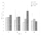
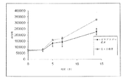
一定率の伸長テストからの最大引張強度の結果が、図2に示される。最大引張強度(UTS)は、1.893±0.458MPaであった。 The results of maximum tensile strength from a constant rate of elongation test are shown in FIG. The maximum tensile strength (UTS) was 1.893 ± 0.458 MPa.
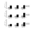
小さい血管内に挿入され、それから大きい血管へ拡張される装置の実用的な必要条件は、故障前に達成可能な引張を含む。機械的必要条件に加え、基礎材は細胞に有益であるよう設計される。これは、細胞付着、移動および増殖のための十分な空隙率を含む。電界紡糸された基礎材の宣伝される特性のうちの1つは、細胞外マトリクスに似たそれらの繊維およびその多孔性の性質である。図3に示されるように、それぞれのサンプル群内の平均空隙率は、大部分で同様および小さい標準偏差にとどまった。 Practical requirements for a device that is inserted into a small blood vessel and then expanded into a large blood vessel include tension that can be achieved before failure. In addition to mechanical requirements, the base material is designed to be beneficial to the cells. This includes sufficient porosity for cell attachment, migration and proliferation. One of the advertised properties of electrospun base materials is their fiber resemblance to the extracellular matrix and its porous nature. As shown in FIG. 3, the average porosity within each sample group remained mostly similar and with a small standard deviation.
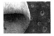
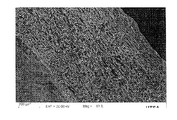
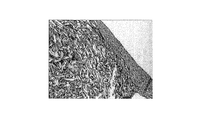
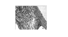
図4に示されるように、SEM画像は、サンプルの内部から外部へ組織内でゆるやかな変化を示した。凹面側の繊維はより湾曲した整列を示し、一方で凸面側の繊維はより直線状で表れた。 As shown in FIG. 4, the SEM image showed a gradual change in the tissue from the inside of the sample to the outside. The concave side fibers showed a more curved alignment, while the convex side fibers appeared more linear.
生体吸収性ポリマーとして、PCLが分解すると予想される。しかし基礎材は、生存可能な組織が形成されるまで、それらの完全性を維持することが重要である。高い空隙率の基礎材は、表面領域の増大により低い空隙率の基礎材より前に完全性を失うかもしれないと予想されうる。 PCL is expected to degrade as a bioabsorbable polymer. However, it is important that the base materials maintain their integrity until a viable tissue is formed. It can be expected that a high porosity base material may lose integrity before a low porosity base material due to the increased surface area.
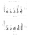
90日の期間にわたり異なる空隙率で基礎材を比較して、あらゆる基礎材について、1つの時点から最初の強度へ最大引張負荷間で有意性がある差はなかった。経時的な引張負荷についての結果は図5Aに示され、この研究の他の領域で得られた値と同程度である。 There was no significant difference between the maximum tensile load from one point of time to the initial strength for all base materials comparing the base materials with different porosity over a 90 day period. The results for tensile loading over time are shown in FIG. 5A and are comparable to values obtained in other areas of this study.
UTSの結果と同様に、あらゆる基礎材について、90日の期間にわたり故障時の引張に有意性がある差はなかった。図5Bは、これらの結果のグラフを示す。 Similar to the UTS results, there was no significant difference in tensile at failure over a period of 90 days for any base material. FIG. 5B shows a graph of these results.
図5Cに示されるように、全てのサンプルについて90日の期間にわたり重量の減少が観察されたが、それは最初の減少後横這い状態になり、またわずかである。 As shown in FIG. 5C, a weight loss was observed over a 90 day period for all samples, but it leveled off after the first loss and was slight.
同一の基礎材内の、より曲線状の繊維を有する凹面側およびより直線状の繊維を有する凸面側が、基礎材全体の機械的特性に寄与しうる。 The concave side with more curvilinear fibers and the convex side with more straight fibers in the same base material can contribute to the mechanical properties of the entire base material.
基礎材上の細胞増殖を評価するため、さらなる研究を実行した。静的および動的培養においてチューブ状の基礎材を置き、ヒト大動脈内皮細胞(Cascade Biologics)またはヒトの大動脈平滑筋細胞(Lonza)のどちらかを基礎材上に置いて、生体外でそれらの増殖をそれぞれ観察した。図6は、動的流れのもとで培養されたとき、基礎材上に広がるヒト大動脈内皮細胞を示す。内皮細胞での研究は予備試験であるが、この広がりは、内皮細胞が基礎材に付着し、動的流れのもとで増殖することを示唆する。他の研究で、チューブ状の基礎材をエチレンオキシドガス(EtO)(n=3)または酸素ガスプラズマ(GP)(n=3)で殺菌し、それから個々のウェルプレート内に置き、平滑筋細胞を基礎材上に滴下して播種した。細胞を14日間増殖させ、培地を1日おきに交換した。図7に示されるように、研究前、3,5,7および14日目の細胞数を推定するのに代謝アッセイアラマーブルー(インビトロゲン)を使用した。細胞数の増加は、基礎材が細胞の成長および増殖を助長したことを示す。次に、バイオリアクタ内にチューブ状の基礎材を置いて5日間動的流れにさらし、培地を1日おきに交換した。基礎材をもう一度EtO(n=3)またはGP(n=3)のどちらかで殺菌して、ヒト大動脈平滑筋細胞を播種し、研究前、3および5日目の代謝活性を測定するのにアラマーブルーを使用した。この研究の結果は、図8に示される。細胞数の増加は、細胞が動的流れのもとで増殖可能であることを示す。これらの結果は明確であるが、流体内で基礎材付近を通過するどの細胞が付着するかに注目することもまた重要である。チューブ状の基礎材をEtO(n=3)またはGP(n=3)のどちらかで殺菌して、バイオリアクタ内に置く研究を行った。しかし、基礎材に予め播種する代わりに、システムを通して灌流される培地内に懸濁状態で細胞を置いた。3日目に基礎材を取り除き、細胞数の判断にアラマーブルーを使用した。結果は、予め播種することなく細胞が基礎材に接着可能であることを示す。図9は、懸濁テストの結果を、細胞が予め播種された静的および動的テストと比較している。これは、心血管系のような流動系内に置かれた基礎材が流れの中で細胞を保持し、よって基礎材を予め播種する必要を減少し、順に患者が基礎材の受け入れのため待たなければならない時間を減少することが可能であることを示す重要なものである。
Further studies were performed to assess cell growth on the base material. Place the tubular base material in static and dynamic culture and place either human aortic endothelial cells (Cascade Biology) or human aortic smooth muscle cells (Lonza) on the base material and their growth in vitro Were observed respectively. FIG. 6 shows human aortic endothelial cells spreading on the base material when cultured under dynamic flow. Although studies on endothelial cells are preliminary tests, this spread suggests that endothelial cells attach to the base material and proliferate under dynamic flow. In other studies, tube-shaped foundation materials were sterilized with ethylene oxide gas (EtO) (n = 3) or oxygen gas plasma (GP) (n = 3), then placed in individual well plates and smooth muscle cells were It was dripped and seeded on the base material. The cells were grown for 14 days and the medium was changed every other day. As shown in FIG. 7, the metabolic assay Alamar Blue (Invitrogen) was used to estimate the number of cells at 3, 5, 7 and 14 days prior to the study. An increase in cell number indicates that the base material promoted cell growth and proliferation. Next, a tubular base material was placed in the bioreactor and exposed to dynamic flow for 5 days, and the medium was changed every other day. To sterilize the base material once again with either EtO (n = 3) or GP (n = 3) and seed human aortic smooth muscle cells to measure metabolic activity on
異なる基礎材組織を比較する研究を行った。3つの構成でPCLを作成した。第1の“A”は、クロロホルム:メタノールが75:25の9wt%(例えば、約8〜10wt%)のPCL溶液(例えばハロゲン化有機溶媒とアルコールの混合物)を、0.035mL/分で、先端部からコレクタの距離が15cm、シリンジの針に15kVの印加で電界紡糸して構成される。第2の“B”は、クロロホルム(例えばハロゲン化有機溶媒)の14wt%(例えば、約10〜15wt%)のPCL溶液を、0.029mL/分の押出速度で、先端部からコレクタの距離が10cm、12.0Vの印加の電界紡糸を用いた。第3のセット“C”は、クロロホルム(例えばハロゲン化有機溶媒)の12wt%(例えば、約10〜15wt%)のPCL溶液を、スタイロフォームの箱の下のガラス片上に流し込むことで製造した。クロロホルムが蒸発した後にフィルムが残り、Bの設定と変わらず同一の厚さであるおよそ0.5mmであった。“A”の設定はより薄い基礎材を生成し、およそ0.3mmであった。“C”のサンプルは、理論上3次元構造の“A”と2次元構造の“B”を比較する対照群として機能する。前述のように、コレクタは光る面を上にしたアルミニウムホイルの小片で構成され、それがアルミニウムスクリーンを覆い、高電圧源の負端子が印加された。基礎材の製造後、直線状のカミソリ刃を用いて、それらを5mm×5mmの正方形に切断した。平均繊維直径を測定するための基礎材の撮像に、SEMを使用した。図10A〜Bは、2000倍の電界紡糸された基礎材“A”(ナノ)および“B”(マイクロ)のSEM画像を示す。 A study was conducted to compare different base material structures. PCL was created with three configurations. The first “A” is a 9 wt% (eg, about 8-10 wt%) PCL solution of chloroform: methanol 75:25 (eg, a mixture of halogenated organic solvent and alcohol) at 0.035 mL / min. The distance from the tip to the collector is 15 cm, and the electrospinning is performed by applying 15 kV to the syringe needle. The second “B” is a 14 wt% (eg, about 10 to 15 wt%) PCL solution of chloroform (eg, halogenated organic solvent) at an extrusion rate of 0.029 mL / min, and the distance from the tip to the collector is Electrospinning with 10 cm and 12.0 V applied was used. The third set “C” was made by pouring a 12 wt% (eg, about 10-15 wt%) PCL solution of chloroform (eg, a halogenated organic solvent) onto a piece of glass under the styrofoam box. The film remained after the chloroform had evaporated and was about 0.5 mm, the same thickness as the B setting. The “A” setting produced a thinner base material and was approximately 0.3 mm. The “C” sample functions as a control group that theoretically compares “A” of the three-dimensional structure and “B” of the two-dimensional structure. As mentioned above, the collector consisted of a piece of aluminum foil with the shining side up, which covered the aluminum screen and the negative terminal of the high voltage source was applied. After the production of the base material, they were cut into 5 mm × 5 mm squares using a linear razor blade. SEM was used to image the base material to measure the average fiber diameter. 10A-B show SEM images of 2000 × electrospun base materials “A” (nano) and “B” (micro).
いくつかの実施形態では、基礎材を3分間高RFの酸素ガスプラズマにさらすことにより、それらを開口ガラスのシンチレーションバイアル内で殺菌した。全ての時点で両方の細胞タイプに関する群をともに殺菌し、群内で異なる殺菌になりうるエラーを減少するよう、基礎材を殺菌についてグループ化した。殺菌後、超低接着性ウェルプレートの個々のウェル内のそれらの細胞タイプそれぞれに関する細胞培養培地を滅菌するため、サンプルが露出されるかもしれない。 In some embodiments, the base materials were sterilized in an open glass scintillation vial by exposing them to a high RF oxygen gas plasma for 3 minutes. The base materials were grouped for sterilization to sterilize the groups for both cell types together at all time points and reduce errors that could result in different sterilizations within the group. After sterilization, the sample may be exposed to sterilize the cell culture medium for each of those cell types in the individual wells of the ultra low adhesion well plate.
ヒト大動脈内皮細胞(EC)およびヒト大動脈平滑筋細胞(SMC)は、Lifeline cell technologiesから購入した。SMCのドナーは49才のアフリカ系アメリカ人男性、非喫煙者で高血圧および心臓疾患があり、脳内出血により死亡した。ECのドナーは61才の白人男性、非喫煙者で高血圧および心臓疾患があり、脳内出血により死亡した。SMCをインビトロゲンの基本培地M231で培養し、平滑筋細胞成長補強剤およびECを、Lifeline社の基本培地で内皮成長補強剤とともに培養した。両方の細胞タイプを、P5を通して育成した。細胞をトリプシン処理、遠心分離、再懸濁および血球計数器を用いて計数した。4×10⁴細胞/基礎材の濃度で、SMCをSMC培地とともにウェルに投入した。4×104細胞/基礎材の濃度で、ECをEC培地とともにウェルに投入した。標準湾曲部も、それぞれのある範囲の量の細胞タイプを標準サイズのウェルプレート内に播種することにより生成した。それぞれの時点について3つの基礎材を播種し、標準湾曲部に関する3つの複製を播種した。最初の分析前に、2.5時間細胞が付着するようにした。代謝データについてこの研究を4回繰り返し、増殖データについてこの研究を2回繰り返し、顕微鏡検査についてこの研究を2回繰り返した。それぞれの繰り返しに対しn=3を使用した。 Human aortic endothelial cells (EC) and human aortic smooth muscle cells (SMC) were purchased from Lifeline cell technologies. The SMC donor was a 49-year-old African-American male, non-smoker, with high blood pressure and heart disease, and died of intracerebral hemorrhage. The EC donor was a 61-year-old Caucasian male, a non-smoker with hypertension and heart disease and died of intracerebral hemorrhage. SMC were cultured in Invitrogen's basic medium M231, and smooth muscle cell growth enhancer and EC were cultured together with endothelial growth enhancer in Lifeline basic medium. Both cell types were grown through P5. Cells were counted using trypsinization, centrifugation, resuspension and a hemocytometer. SMC was added to the wells along with SMC medium at a concentration of 4 × 10 4 cells / base material. EC was placed in the wells with EC medium at a concentration of 4 × 10 4 cells / basic material. Standard curves were also generated by seeding each range of quantities of cell types in standard size well plates. Three bases were seeded for each time point and three replicates for the standard curve were seeded. Cells were allowed to attach for 2.5 hours before the first analysis. This study was repeated four times for metabolic data, this study was repeated twice for growth data, and this study was repeated twice for microscopy. N = 3 was used for each repeat.
代謝活性の測定のため、培地を基礎材から取り出し、標準湾曲部を含むそれぞれのウェルに培地に入った10%のアラマーブルー(AB)溶液を加えた。AB溶液は、それぞれの細胞タイプに対しそれぞれの培地を使用した。基礎材をABとともに2.5時間培養し、それからABを黒色の不透明な96ウェルプレート内へ100μLの容量で等分し、蛍光プレートリーダでEX:530 EM:590で読み取った。ウェルからAB溶液を取り除いた後、基礎材をPBSで洗浄し、研究前の基礎材とともにプレートをパラフィルムで包み、−80Cの冷凍庫に置いた。残りの基礎材の培地を交換し、プレートを培養器内に戻した。このAB処理を、1,3,7および10日繰り返した。図11A〜Bは、異なる繊維形態の基礎材に反応したhAoECおよびhAoSMCの代謝活性における(それぞれのサンプルについて研究前の値に基準化した)変化のグラフを示す。 For measurement of metabolic activity, the medium was removed from the base material and 10% Alamar Blue (AB) solution in the medium was added to each well containing the standard curve. AB solution used each medium for each cell type. The base material was incubated with AB for 2.5 hours, then AB was aliquoted into a black opaque 96 well plate in a volume of 100 μL and read on a fluorescent plate reader at EX: 530 EM: 590. After removing the AB solution from the wells, the base material was washed with PBS, the plate was wrapped with parafilm together with the base material before the study, and placed in a -80C freezer. The remaining base material medium was replaced and the plate was returned to the incubator. This AB treatment was repeated 1, 3, 7 and 10 days. FIGS. 11A-B show graphs of changes in hAoEC and hAoSMC metabolic activity (normalized to pre-study values for each sample) in response to different fiber morphology base materials.
全ての時点が完了し凍結された後、ピコグリーン(PG)を用いてdsDNA定量化研究を実行した。基礎材を−80°Cから取り出し、RTで30分間解凍させた。プロテイナーゼKをEC培地内で1mg/mLに希釈し、それぞれのサンプルおよび標準湾曲部に100μLを加えた。プレートを培養器内に置き、30分間で42°Cまで上昇させた。プレートを取り出し、プレート加振器上に2分間強度#3で置いた。それからプレートを−80°Cに戻し、一晩そのままにした。翌朝プレートを−80°Cから外し、室温で30分間解凍させた。3回目に−80°Cでさらに30分間凍結し、その後室温で30分間解凍する前にそれらをプレート加振器上に2分間強度#3で再度置いた。500μLのTE緩衝剤を、全てのプレート1サンプルに加えた。それから、それぞれの5つの複製100μLを、DNAseおよびRNAseフリー96ウェルプレートへ取り出した。プレート2および3を、−20°Cの冷凍庫においた。PGアッセイ溶液は、100μLのPGと21mLのTE緩衝剤の混合で、これらから構成された。ウェルあたりの容量が200μLとなるように、100μLのPG溶液をウェルプレートに加えた。プレートを暗所で数分培養させ、それから蛍光プレートリーダでEX:485 EM:528で読み取った。同一の技術を、プレート2および3について繰り返した。図12A〜Bは、ナノファイバ(“A”)、マイクロファイバ(“B”)またはフィルム(“C”)でそれぞれ構成された基礎材上のhAoECおよびhAoSMCの経時的な細胞増殖のグラフを示す。dsDNAの内容を測定するため、ピコグリーンを用いて判断した(n=6)。
After all time points were completed and frozen, dsDNA quantification studies were performed using picogreen (PG). The base material was removed from −80 ° C. and thawed at RT for 30 minutes. Proteinase K was diluted to 1 mg / mL in EC medium and 100 μL was added to each sample and standard curve. The plate was placed in the incubator and allowed to rise to 42 ° C in 30 minutes. The plate was removed and placed on a plate shaker for 2 minutes at
細胞の導入前ならびにそれぞれの時点で、両方の繊維状基礎材を撮像するため、電子顕微鏡スキャンを使用した。細胞が存在したとき、サンプルを4%のパラホルムアルデヒドで固定し、それから室温で真空オーブン内に置く前にエタノール勾配を用いて脱水した。 An electron microscope scan was used to image both fibrous base materials prior to cell introduction and at each time point. When cells were present, the samples were fixed with 4% paraformaldehyde and then dehydrated using an ethanol gradient before placing in a vacuum oven at room temperature.
各時点のサンプルを4%のパラホルムアルデヒドで固定し、それからαアクチン共役FITCまたは抗CD31のどちらかと、フルオロフォアおよびDAPIで染色し、核を染色した。サンプルをスローフェード(Slowfade)に据え付け、それからそれらのそれぞれの波長を用いて共焦点蛍光顕微鏡で観察した。図13A〜Dは、1,3,7および10日目の電界紡糸されたマイクロファイバとヒト大動脈内皮細胞のSEM画像を示す。図14A〜Dは、1,3,7および10日目の電界紡糸されたマイクロファイバとヒト大動脈平滑筋細胞のSEM画像を示す。
Samples at each time point were fixed with 4% paraformaldehyde and then stained with either α-actin conjugated FITC or anti-CD31, fluorophore and DAPI, and the nuclei were stained. Samples were mounted on a Slowfade and then observed with a confocal fluorescence microscope using their respective wavelengths. FIGS. 13A-D show SEM images of electrospun microfibers and human aortic endothelial cells on
1方向ANOVAを使用して、細胞数および代謝活性における有意性がある増加を判断した。ポストホックに、チューキーテストを使用した。異常値の判断に、Azテストを使用した。 One-way ANOVA was used to determine significant increases in cell number and metabolic activity. Tukey test was used for post-hoc. The Az test was used to determine outliers.
代謝および増殖データの両方に基き、内皮細胞は、同一の素材で製造されたフィルムまたはナノファイバのどちらかよりも、マイクロファイバに明確に反応していると判断しうる。より具体的にいうと、ナノファイバでは、内皮細胞は代謝の増加を示したが増殖の増加は示さず、細胞にストレスがかかっている可能性を示唆していることに注目すべきである。フィルム対照群で同様の傾向が認められたが、マイクロファイバ基礎材ではなかった。マイクロファイバ基礎材についての代謝活性ならびに増殖と視覚画像の対照は、他のサンプルと違い、細胞が基礎材を浸潤したことを示唆する。 Based on both metabolic and proliferation data, it can be determined that endothelial cells are more responsive to microfibers than either films or nanofibers made of the same material. More specifically, it should be noted that in nanofibers, endothelial cells showed an increase in metabolism but no increase in proliferation, suggesting that the cells may be stressed. A similar trend was observed in the film control group, but not the microfiber base material. Metabolic activity as well as growth and visual image control for the microfiber substrate suggests that the cells infiltrated the substrate, unlike the other samples.
ブタで、外科的AAAモデルを用いて生体内の研究を実行した。ブタに動脈瘤を生成するため、合成グラフトまたは平滑筋(一般的に空腸または腹膜)の楕円形のパッチを大動脈の長手方向の裂傷内に縫合し、合成グラフトまたは平滑筋のパッチはふくらんで動脈瘤形状を形成する。パッチモデルは動脈瘤の物理的形状を形成し、大動脈壁にいくぶんかの固有の損傷を与える。 In vivo studies were performed on pigs using a surgical AAA model. To produce an aneurysm in a pig, an oval patch of synthetic graft or smooth muscle (generally jejunum or peritoneum) is sutured into the longitudinal laceration of the aorta, and the synthetic graft or smooth muscle patch is inflated and arterial Forms a lump shape. The patch model creates the physical shape of the aneurysm and causes some inherent damage to the aortic wall.
メスブタ(50〜75kg)を48時間流動食させ、それから筋肉注射を通してジアゼパム(0.1mg/kg)、ケタミン(10mg/kg)およびアトロピン(0.01mg/kg)の混合物で鎮静化する前に、24時間絶食させた。それからその動物を経静脈プロポフォル(2mg/kg)で麻酔し、それから経口挿管した。挿管後、2%のハロタンで全身麻酔を維持した。心拍数、血圧、呼気終末CO2およびO2飽和度を常時監視した。水分補給および薬剤管理周縁処置のため、静脈アクセスを確立した。 Prior to sedating female pigs (50-75 kg) for 48 hours and then sedating with a mixture of diazepam (0.1 mg / kg), ketamine (10 mg / kg) and atropine (0.01 mg / kg) through intramuscular injection. Fasted for 24 hours. The animals were then anesthetized with intravenous propofol (2 mg / kg) and then intubated. Following intubation, general anesthesia was maintained with 2% halothane. Heart rate, blood pressure, end-tidal CO 2 and O 2 saturation were constantly monitored. Intravenous access was established for hydration and drug management peripheral procedures.
AAAの形成前に、超音波を使用して大動脈を測定し、ベースライン基準を得た。滅菌状態で正中線開腹を行い、腹膜の一部を取り出し、半分に折って長さおよそ3cmおよび幅2cmの長方形を形成した。鰐口クリップを近位、中位および遠位に使用して縁部を固定し、二重層パッチを形成した。 Prior to AAA formation, the aorta was measured using ultrasound to obtain a baseline reference. A midline laparotomy was performed in a sterilized state, and a part of the peritoneum was removed and folded in half to form a rectangle approximately 3 cm long and 2 cm wide. Mouth clips were used proximally, medially and distally to secure the edges and form a bilayer patch.
術後癒着を防ぐため、腸を側面に移動し、湿った手術タオルで覆った。大動脈を分離し、非外傷性血管クランプを用いて腎動脈の下方で離して固定した。大動脈の長さと平行に、長さおよそ3cmおよび幅2mmの大動脈切開を行った。パッチを、5−0Proleneランニング縫合糸を用いて切開部分内に縫合した。クランプをそれから取り除き、動脈瘤のふくらみを漏出について調べた。 To prevent postoperative adhesions, the intestines were moved to the side and covered with a wet surgical towel. The aorta was isolated and fixed separately below the renal artery using an atraumatic vascular clamp. An aortic incision approximately 3 cm long and 2 mm wide was made parallel to the length of the aorta. The patch was sutured into the incision with 5-0 Proline running suture. The clamp was then removed and the bulge of the aneurysm was examined for leaks.
漏出を、5−0Proleneランニング縫合糸を用いて閉じた。漏出が観察されなくなったとき、腸を置換して、その動物を3−0バイクリルを用いて3層に閉じ、ステープルで終えた。最初の24時間の間12時間ごとに0.05mg/kgのブプレノルフィンでのIM鎮痛を用いた獣医学的治療で、その動物は回復した。3日間にわたり、アモキシシリン(20mg/kg/日)を筋肉内に投与した。1回375mgのアスピリンを、7日間にわたり1日1回経口で与えた。この処置の後、正常食を1日再開した。パッチの部位の大動脈を、超音波を用いて7および14日目に測定した。14日目に、動脈瘤が通常の大動脈より少なくとも30%より大きいと測定された場合、第2の処置を実行した。それが十分大きくなかった場合、それをもう1週間形成させた。
The leak was closed using 5-0 Proline running suture. When no leakage was observed, the intestines were replaced and the animals were closed in 3 layers using 3-0 bicyclyl and finished with staples. The animals recovered with veterinary treatment using IM analgesia with 0.05 mg / kg buprenorphine every 12 hours for the first 24 hours. Amoxicillin (20 mg / kg / day) was administered intramuscularly for 3 days. One dose of 375 mg aspirin was given orally once daily for 7 days. Following this treatment, normal diet was resumed for one day. The aorta at the site of the patch was measured on
クロロホルムにポリカプロラクトンを入れた溶液を、回転する(587.5RPM)3mmの直径のアルミニウムマンドレル上に電界紡糸し、チューブ状の基礎材を形成した。結果として生じる基礎材は、6.37±1.13μmの平均繊維直径と76.79±5.60%の平均空隙率、1.66±0.99MPaの弾性係数、1.45±0.32MPaのUTS、9.52±2.73の破損歪の特性を有した。基礎材を取り出し、30,35または40mmのどれかの長さに切断した。基礎材を、高RFの酸素ガスプラズマを用いて個々に3分間殺菌した。 A solution of polycaprolactone in chloroform was electrospun onto a rotating 3 mm diameter aluminum mandrel (587.5 RPM) to form a tubular base material. The resulting base material has an average fiber diameter of 6.37 ± 1.13 μm, an average porosity of 76.79 ± 5.60%, an elastic modulus of 1.66 ± 0.99 MPa, 1.45 ± 0.32 MPa. UTS, with a fracture strain characteristic of 9.52 ± 2.73. The base material was removed and cut to any length of 30, 35 or 40 mm. The base material was individually sterilized using a high RF oxygen gas plasma for 3 minutes.
第1の処置の14日から21日後、動脈瘤を血管内で治療するため第2の処置を実行した。第2の処置で、ケタミン(100mg/ml)+キシラジン(100mg/ml)混合IMを3cc/50ポンドで、またはテラゾール5〜8mg/Kgを用いて動物を鎮静させ、続いて挿管して、処置の時間(約2時間)中、麻酔器を用いて0.3%〜3%のイソフルラン麻酔と酸素補給を維持した。IVラインを導入して液滴を開始し、外科処置中維持した(0.9%の生理食塩水または乳酸リンゲル液)。外科処置中、EKG、呼吸数、SPO2、および温度を監視した。表1に示すように、動物を4つの治療群に分けた。

外科的埋め込みの日、A群およびD群について、基礎材を、6−0Proleneアンカー縫合糸を用いてステント(Megalink胆管ステント、Guidant Corporation)上に縫合した。2本のアンカー縫合糸を近位に180°傾けて付け、1本の縫合糸を遠位に付けた。それから装置をバルーンカテーテル(Powerflex P3,CTA拡張カテーテル,Johnson and Johnson上に装填した。 On the day of surgical implantation, for groups A and D, the base material was sutured onto a stent (Megalink biliary stent, Guidant Corporation) using 6-0 Proline anchor suture. Two anchor sutures were tilted 180 ° proximally and one suture distally. The device was then loaded onto a balloon catheter (Powerflex P3, CTA dilatation catheter, Johnson and Johnson).
全ての動物について、右大腿動脈を分離し、導入器シース(7F)を動脈内に置いた。ガイドワイヤ(3mm J,0.89mm×145cm,Boston Scientific)を挿入し、動脈瘤の部位のちょうど近位に位置付けた。ガイドワイヤに続き、多目的カテーテル(6F)を挿入し、対照液(オムニパーク)を使用して、血管造影を用いて動脈瘤を視覚化した。 For all animals, the right femoral artery was isolated and an introducer sheath (7F) was placed in the artery. A guide wire (3 mm J, 0.89 mm × 145 cm, Boston Scientific) was inserted and positioned just proximal to the site of the aneurysm. Following the guidewire, a multipurpose catheter (6F) was inserted and an aneurysm was visualized using angiography using a control solution (Omnipark).
A群:動脈瘤の視覚化の後、導入器シースをより大きいシース(11F)と置換し、ガイドワイヤを260cmの硬質のワイヤで置換した。基礎材を装填したカテーテルをシースを通して挿入し、基礎材が動脈瘤の源のちょうど近位にあるよう位置決めた。それからバルーンを10秒間12ATMまで膨張させ、その後収縮させた。バルーンを取り除く前に、血管造影を用いてステントの完全拡張をチェックした。それが完全に拡張されていなかった場合、バルーンをさらに10秒間12ATMまで再膨張した。バルーンを取り除いた後、多目的カテーテルを再度挿入し、対照液(オムニパーク)を使用して大動脈を視覚化し、動脈瘤の閉塞度合を観察した。それから、大腿動脈が絹縫合素材を用いて連結されたとき、カテーテル、続いて導入器シースを取り除いた。アクセス部位を3−0Vicrylで閉じ、動物を回復させた。 Group A: After visualization of the aneurysm, the introducer sheath was replaced with a larger sheath (11F) and the guidewire was replaced with a 260 cm hard wire. A catheter loaded with a base material was inserted through the sheath and positioned so that the base material was just proximal to the source of the aneurysm. The balloon was then inflated to 12 ATM for 10 seconds and then deflated. Prior to removal of the balloon, angiography was used to check for complete expansion of the stent. If it was not fully expanded, the balloon was reinflated to 12 ATM for an additional 10 seconds. After removing the balloon, the multipurpose catheter was reinserted and the aorta was visualized using a control solution (Omnipark) to observe the degree of occlusion of the aneurysm. Then, when the femoral artery was connected using silk suture material, the catheter and then the introducer sheath was removed. The access site was closed with 3-0 Vicryl and the animal was allowed to recover.
B群 第2の動物群について、動脈瘤部位へのガイドワイヤの挿入に続き、バルーンカテーテル上に予め装填された
PTFE包覆ステント(心房I−cast,9mm)を、7Fの導入器シースを通して挿入した。バルーンを10秒間15ATMまで膨張し、その後収縮させ取り除いた。それから第2のバルーン(パワーフレックスP3 CTAカテーテル12mm×4mm)を置き、10秒間12ATMまで近位および遠位に膨張させた。6FのMP−1カテーテルを再度置き、血管造影を使用してAAAが適切に閉塞したかどうかを判断した。それから、大腿動脈が絹縫合素材を用いて連結されたとき、ガイドワイヤおよびカテーテル、続いてシースを取り除いた。アクセス部位を3−0Vicrylで閉じ、動物を回復させた。
Group B For the second group of animals, following insertion of a guide wire into the aneurysm site, a PTFE-encapsulated stent (atrial I-cast, 9 mm) preloaded on a balloon catheter is inserted through a 7F introducer sheath. did. The balloon was inflated to 15 ATM for 10 seconds and then deflated and removed. A second balloon (Powerflex
C群 対照群であるC群について、非包覆バルーン(パワーフレックスP3 CTAカテーテル12mm×4mm)を(何も装填せずに)7Fのシースを通して挿入し、10秒間12ATMまで膨張させた。それからそれを取り除き、6FのMP−1カテーテルを使用して、血管造影で大動脈を視覚化した。カテーテルおよびシースを取り除いた後、絹で大腿動脈を連結し、動物を回復させた。
Group C For control group C, a non-encapsulated balloon (Powerflex
D群 D群の動物は動脈瘤手術をしなかったが、A群について記載したような基礎材を受け入れた。 Group D Group D animals did not undergo aneurysm surgery but received a base material as described for Group A.
超音波を使用して、最初の手術前ならびに動脈瘤形成後7,14日目、および選択ケースでは21日目に大動脈のサイズを判断した。14日目に動脈瘤サイズが本来の大動脈より少なくとも30%大きくない場合、21日目を可能にした。第2の処置後、超音波を使用して術後14および28日目に動脈瘤を調べた。全ての超音波について、動物を鎮静状態(Telezol5〜8mg/KgIM)にし、動脈瘤の部位における大動脈を、各時点で3つの画像から3地点で測定した。
Using ultrasound, the size of the aorta was determined before the first surgery and on
第2の処置中および屠殺時、超音波に加え、血管造影を使用し、大動脈の直線状および側面画像の両方を得た。それぞれの動物の血管造影画像から、3つの動脈瘤測定結果を取得した。 During the second procedure and at sacrifice, in addition to ultrasound, angiography was used to obtain both linear and lateral images of the aorta. Three aneurysm measurement results were obtained from the angiographic images of each animal.
28日後、動物を全身麻酔し、左大腿動脈を分離した。6Fの導入器シースを置き、6FのHSカテーテルを置いた。対照(オムニパーク)を使用して、血管造影を用いてグラフト部位を視覚化し、それから動物を安楽死させ、剖検を実行した。剖検中、大動脈を腎動脈から腸骨分岐部へ分離した。内腔を露出するように組織試験片を長手方向に切断し、ホルムアルデヒド内に試験片を固定する前に全体画像を得た。組織が固定されると、組織学的検査のため、1)動脈瘤を貫通した横断、2)付加の近位部位を貫通した長手方向、3)動脈瘤部位の近位の横断、の3つのブロックを切断した。ブロックをパラフィンに包埋し、染色のため区分化した。組織学的検査の区分をヘマトキシリンおよびエオシン(H&E)、マッソン3色(TRI)、または平滑筋アクチン(SMA)に対する免疫学的染色またはPECAM−1(CD31)のいずれかで染色した。H&E染色は組織の全体構造を明らかにし、そこで赤血球は赤、核は青、細胞質および細胞外マトリクスはピンクである。構造をさらに明瞭にするよう、TRI染色したコラーゲンの青、核の黒、筋肉の赤、および細胞質のピンクを使用した。α−アクチンを赤に染色するのに、SMA抗体を使用した。CD31抗体は、区分内で内皮細胞および血小板内のグリコプロテインを茶色に染色する。免疫学的区分を、核を青で示すため後染色した。染色されたスライドを、光学顕微鏡で見た。クラスカルワリスランク分析を使用して、グラフト埋め込み後の動脈瘤サイズにおける有意性がある減少を判断した。 After 28 days, the animals were general anesthetized and the left femoral artery was isolated. A 6F introducer sheath was placed and a 6F HS catheter was placed. A control (Omnipark) was used to visualize the graft site using angiography, and then the animals were euthanized and necropsy was performed. During autopsy, the aorta was separated from the renal artery to the iliac bifurcation. Tissue specimens were cut longitudinally to expose the lumen and a full image was obtained before fixing the specimen in formaldehyde. Once the tissue is fixed, there are three histological examinations: 1) crossing through the aneurysm, 2) longitudinal direction through the additional proximal site, 3) proximal crossing of the aneurysm site The block was cut. Blocks were embedded in paraffin and sectioned for staining. Histological sections were stained with either hematoxylin and eosin (H & E), Masson tricolor (TRI), or immunostaining for smooth muscle actin (SMA) or PECAM-1 (CD31). H & E staining reveals the overall structure of the tissue, where red blood cells are red, nuclei are blue, cytoplasm and extracellular matrix are pink. To further clarify the structure, TRI-stained collagen blue, nucleus black, muscle red, and cytoplasmic pink were used. The SMA antibody was used to stain α-actin in red. The CD31 antibody stains endothelial cells and glycoproteins in platelets brown in the compartment. Immunological sections were post-stained to show nuclei in blue. The stained slide was viewed with a light microscope. Kruskal Wallis rank analysis was used to determine a significant reduction in aneurysm size after graft implantation.
研究初期に、最初の動脈瘤形成手術後1週間で、消化管の合併症からA群およびB群でそれぞれ1匹の動物が死亡した。食餌が、最も影響力を有すると判断された。それを、さらなる合併症が認められなかったその後の動物に対し調整した。グラフトの配置後6時間で、A群のもう1匹の動物が肺水腫で死亡した。スタッフの獣医病理学者は、合併症はグラフトそれ自身に関連しないと判断した。他の全ての動物は、研究の間、合併症なく生存した。 Early in the study, 1 week after the first aneurysm surgery, one animal died in groups A and B due to gastrointestinal complications. The diet was judged to have the most influence. It was adjusted for subsequent animals where no further complications were observed. Six hours after placement of the graft, another animal in Group A died of pulmonary edema. The staff veterinary pathologist determined that the complication was not related to the graft itself. All other animals survived without complications during the study.
動脈瘤形成手術を受けた全ての動物で、パッチから大動脈拡張が起こった。図15は、図16A〜Cに示されるように超音波を用いて経時的に測定した、パッチの部位の最大内腔直径の平均と比較した、それぞれの群についての最初の大動脈のサイズのグラフ表示であり、Aが大動脈であり、AAAが動脈瘤である。図16Aは、動脈瘤形成手術前の大動脈の超音波を示す。図16Bは、手術後7日の図16Aの大動脈の超音波を示す。図16Cは、手術後14日の図16Aの大動脈の超音波を示す。
All animals undergoing aneurysm surgery had aortic dilation from the patch. FIG. 15 is a graph of initial aortic size for each group compared to the average maximum lumen diameter at the site of the patch, measured over time using ultrasound as shown in FIGS. Indicated, A is the aorta and AAA is the aneurysm. FIG. 16A shows ultrasound of the aorta before aneurysm surgery. FIG. 16B shows the ultrasound of the aorta of FIG.
超音波に加え、周縁処置に血管造影を使用して、図17A〜Bに示されるような動脈瘤の閉塞の評価、ならびに図18A〜Dに示されるような個々の動物について動脈瘤レベルにおける大動脈のサイズの推定を行った。図18Aは、A群の個々の動物の内腔直径である。図18Bは、B群の個々の動物の内腔直径である。図18Cは、C群の個々の動物の内腔直径である。図18Dは、D群の個々の動物の内腔直径である。 In addition to ultrasound, angiography is used for peripheral procedures to evaluate aneurysm occlusion as shown in FIGS. 17A-B, and aorta at the aneurysm level for individual animals as shown in FIGS. An estimation of the size of was performed. FIG. 18A is the lumen diameter of individual animals in Group A. FIG. 18B is the lumen diameter of individual animals in Group B. FIG. 18C is the lumen diameter of individual animals in Group C. FIG. 18D is the lumen diameter of individual animals in Group D.
全ての動物が閉塞した動脈瘤を有すると認められ、処置前の動脈瘤部位でのグラフトは完成とみなされた。イチジク18A〜Bの個々の結果は、動脈瘤上に置かれたグラフトを有する群(A群およびB群)で、B群の第1の動物を除いて内腔直径が埋め込み後の方が小さく、その例外はわずかであり、バルーンの過度の膨張の結果であると考えうる。グラフトを受け入れなかった動物(C群)、またはグラフトを受け入れたが最初の動脈瘤手術がなかった動物(D群)の内腔直径は、比較的一定したままだった。動脈瘤の治療後、14および28日目に超音波を使用し、図19A〜Dに示されるように、動脈瘤部位における大動脈のサイズを測定した。図19Aは、A群の個々の動物の内腔直径である。図19Bは、B群の個々の動物の内腔直径である。図19Cは、C群の個々の動物の内腔直径である。図19Dは、D群の個々の動物の内腔直径である。 All animals were found to have an occluded aneurysm and the graft at the pre-treatment aneurysm site was considered complete. The individual results for FIGS. 18A-B are the group with the graft placed on the aneurysm (Group A and Group B), with the smaller luminal diameter after implantation except for the first animal in Group B. , The exceptions are minor and may be the result of excessive balloon inflation. The lumen diameters of animals that did not receive the graft (Group C) or animals that received the graft but did not have the first aneurysm surgery (Group D) remained relatively constant. After treatment of the aneurysm, ultrasound was used at 14 and 28 days to measure the size of the aorta at the aneurysm site, as shown in FIGS. FIG. 19A is the lumen diameter of individual animals in Group A. FIG. 19B is the lumen diameter of individual animals in Group B. FIG. 19C is the lumen diameter of individual animals in Group C. FIG. 19D is the lumen diameter of individual animals in Group D.
A群およびD群の大動脈を長手方向に開いたとき、基礎材が大動脈壁に接着していることが認められた。A群の基礎材は円形の形状に色の変化があり、そこでは動脈瘤が覆われていたが概して白っぽく、光って見えた。B群の大動脈は赤または紫がかった色を持ち、いくぶん光っていた。C群の大動脈は非常に小さいくぼみを有し、そこは動脈瘤であり、それらは通常の内腔と同様に白で光っていた。D群でもまた、内腔は光って見え、血栓形成はなかった。 When the aorta of groups A and D was opened in the longitudinal direction, it was observed that the base material was adhered to the aortic wall. The base material of Group A had a color change in a circular shape, in which the aneurysm was covered but was generally whitish and shined. The aorta of group B had a red or purpleish color and was somewhat shining. The aorta of group C had very small indentations, which were aneurysms, which were shining white as well as the normal lumen. Also in group D, the lumens appeared shiny and there was no thrombus formation.
組織学的検査の結果は、PCL基礎材を受け入れたA群およびD群の動物は、図20および図21に示されるように、大動脈壁からの平滑筋細胞浸潤を含む基礎材への有意性がある細胞浸潤を有したことを示した。図20は、大動脈壁(A)と基礎材(S)の間の界面のH&E染色を示す。図21は、大動脈壁(A)と基礎材(S)の間の平滑筋アクチン染色を示す。また、基礎材の繊維周辺にコラーゲンが存在した。PCL基礎材を受け入れた全ての動物について内皮が観察され、核のPECAM−1陽性染色により図22、23および24に示され、ここではEが内皮、Sが基礎材、Iが内膜、およびSTが取り除かれたステント支材である。図22は、大動脈内の内皮および新生内膜とPCL基礎材挿入物のH&E染色を示す。図23は、大動脈内の内皮および新生内膜とPCL基礎材挿入物のCD−31抗体染色を示す。図24は、大動脈内の内皮および新生内膜とPCL基礎材挿入物のマッソン3色染色を示す。内皮と基礎材の間にもコラーゲンで構成された新生内膜が存在し、平滑筋細胞が同心円状に向いていた。 The results of histological examination showed that the animals of Group A and Group D that received the PCL foundation material showed significance to the foundation material including smooth muscle cell infiltration from the aortic wall, as shown in FIGS. It showed that it had some cell infiltration. FIG. 20 shows H & E staining of the interface between the aortic wall (A) and the base material (S). FIG. 21 shows smooth muscle actin staining between the aortic wall (A) and the base material (S). Collagen was present around the fibers of the base material. Endothelium was observed for all animals that received the PCL base material and was shown by nuclear PECAM-1 positive staining in FIGS. 22, 23 and 24, where E is the endothelium, S is the base material, I is the intima, and This is a stent support member from which ST is removed. FIG. 22 shows H & E staining of endothelium and neointima in the aorta and PCL base material insert. FIG. 23 shows CD-31 antibody staining of endothelium and neointima in the aorta and PCL base material insert. FIG. 24 shows Masson trichrome staining of endothelium and neointima in the aorta and PCL base material insert. There was also a neointimal composed of collagen between the endothelium and the base material, and the smooth muscle cells were concentrically oriented.
何匹かの動物で、ときどき血管壁に垂直に向いた組織がPTFE層を分離しているように見えた。他の部分では、PTFE素材が単一の層として存在することを意味すると示唆する、層を分離する明確な組織層はなかった。 In some animals, it appeared that sometimes tissue oriented perpendicular to the vessel wall separated the PTFE layer. In other parts, there was no clear tissue layer separating the layers, suggesting that the PTFE material was present as a single layer.
いずれの治療を受けなかったC群で、動脈瘤嚢は内膜過形成であるように見えるもので埋まっていた。従って、それは動脈瘤上のグラフト自身の影響と明らかに認識できない。そのような形成は、動脈瘤のサイズ、血管の状態、またはブタの血管系固有の反応結果であると考えうる。C群で完了した動物はわずか2匹であり、この群から導くことができる概要は限定される。 In group C, which did not receive any treatment, the aneurysm sac was buried with what appeared to be intimal hyperplasia. Therefore, it cannot be clearly recognized as the effect of the graft itself on the aneurysm. Such formation may be attributed to the size of the aneurysm, the state of the blood vessels, or a response characteristic of the porcine vasculature. Only two animals were completed in group C, and the outline that can be derived from this group is limited.
本研究で、28日目に基礎材上に内皮および新生内膜が存在し、平滑筋細胞がグラフト素材内に観察された。PTFE包覆ステントは、動脈瘤の領域に一致した層間のある程度の離層を示した。細胞はときどき血管壁に垂直に向き、層の間の間隙を埋めていたが、領域内にコラーゲンまたは平滑筋細胞のような組織の成分の欠如に基いた生存可能な組織を認めなかった。しかし、組織学的検査分析により示されたような、多数の好中球、血小板および赤血球が存在した。これらの構成要素の存在は炎症によるものであるかもしれず、または基礎材内の浸潤および組織の組織化が基礎材内の反応より遅いことを示しているかもしれない。基礎材はブタの中に正常に配置され、28日目に、内皮形成、平滑筋細胞浸潤、コラーゲン、および血管壁への付着について肯定的な結果を示した。
In this study, on
基礎材内への細胞の浸潤は、生体外および生体内の両方で観察された。静状態の生体外分析は、平滑筋細胞が、ナノメータサイズよりマイクロメータサイズの繊維の基礎材に多く浸潤したことを明らかにした。特に内皮細胞および平滑筋細胞は、ナノファイバに比較してマイクロファイバで増殖および代謝活性が増したことを示した。 Cell infiltration into the base material was observed both in vitro and in vivo. In vitro in-situ analysis revealed that smooth muscle cells infiltrated the base material of micrometer-sized fibers more than nanometer-sized. In particular, endothelial cells and smooth muscle cells showed increased proliferation and metabolic activity with microfibers compared to nanofibers.
生体外モデルは、細胞、特に平滑筋細胞がナノファイバ基礎材よりマイクロファイバ基礎材に多く浸潤したことを明らかにした。動的環境でのテストで、徐々に、細胞はそれらの多くが最初に位置付けられた場所である内腔表面にもう存在しなくなることを意味するのが興味深い。生体内で、平滑筋細胞、コラーゲン、および好中球を含む多数の細胞および組織成分が基礎材内で見出された。基礎材の内腔表面は、内皮を含む新生内膜により覆われていた。 In vitro models revealed that cells, particularly smooth muscle cells, infiltrated more microfiber substrates than nanofiber substrates. Interestingly in testing in a dynamic environment it is interesting to mean that cells no longer exist on the luminal surface where many of them were originally located. In vivo, numerous cell and tissue components have been found in the base material, including smooth muscle cells, collagen, and neutrophils. The luminal surface of the base material was covered with a neointimal membrane containing endothelium.
全ての動物で、基礎材は大動脈壁への完全な付加を示し、それは存在するとき動脈瘤を完全に閉塞した。拡張または再狭窄の有意性がある実証は観察されず、グラフトはその3mmから10mmへの内径の拡張にもかかわらず破裂しなかった。よって、繊維のタイプは適切な血管細胞の支援となり、これらの細胞の浸潤および増殖を可能にする。 In all animals, the base material showed complete attachment to the aortic wall, which completely occluded the aneurysm when present. No significant demonstration of dilation or restenosis was observed, and the graft did not rupture despite its internal diameter expansion from 3 mm to 10 mm. Thus, the type of fiber provides support for appropriate vascular cells and allows infiltration and proliferation of these cells.
本発明の種々の態様のさらなる改変および代替的な実施形態は、この記載を見て当業者に明らかであろう。従ってこの記載は、一例としてのみ解釈されるべきであり、当業者に本発明を実行する一般的な方法を教示する目的である。本明細書に示されおよび記載された形態は、実施形態の例として受け取るべきであることを理解すべきである。構成要素および素材を、本明細書に示されおよび記載されたそれらと置き換えてもよく、部分および処理を逆転してもよく、本発明のある機能を独立して使用してもよい。これらの全ては、本発明のこの記載の利点を有した後で当業者に明らかになるだろう。後述の請求項に記載のような本発明の趣旨および範囲から逸脱することなく、本明細書に記載された構成要素の変更をなしうる。 Further modifications and alternative embodiments of the various aspects of the invention will be apparent to those skilled in the art in view of this description. Accordingly, this description is to be construed as illustrative only and is for the purpose of teaching those skilled in the art the general manner of carrying out the invention. It should be understood that the form shown and described herein is to be taken as an example of embodiment. Components and materials may be substituted for those shown and described herein, parts and processes may be reversed, and certain features of the invention may be used independently. All of these will become apparent to those skilled in the art after having the advantages of this description of the invention. Changes in the components described herein may be made without departing from the spirit and scope of the invention as set forth in the claims below.
Claims (27)
前記基礎材が哺乳類内に位置決められるとき、前記基礎材の前記第1の面は細胞の付着に適切な面を提供し、一方で前記基礎材の前記第2の面は細胞の侵入および組織化を促進する、
組織修復装置。 An expandable base material, wherein the base material is comprised of non-woven fibers, the base material having a first surface having substantially curved fibers and a second surface having substantially straight fibers. Has a surface,
When the base material is positioned within a mammal, the first surface of the base material provides a suitable surface for cell attachment, while the second surface of the base material provides cell invasion and organization. Promote,
Tissue repair device.
前記基礎材が前記内腔組織構造の少なくとも一部と接触するように、前記基礎材を拡張し、前記基礎材の第1の面が細胞の付着に適切な面を提供し、一方で前記第2の表面が細胞の侵入および組織化を促進するように前記基礎材が位置決められるステップと、
前記内腔組織構造内に前記装置を固定するステップと、を含む、
組織修復方法。 Inserting the device into a luminal tissue structure, the device comprising an expandable base material, the base material being composed of non-woven fibers electrospun from a biodegradable material and / or a bioabsorbable material, A base material comprising a first surface having substantially curved fibers and a second surface having substantially straight fibers;
The base material is expanded such that the base material contacts at least a portion of the luminal tissue structure, and the first surface of the base material provides a surface suitable for cell attachment, while the first Positioning said base material such that two surfaces promote cell invasion and organization;
Securing the device within the lumen tissue structure;
Tissue repair method.
医学的状態を治療する装置。 Comprising an expandable base material, wherein the base material is composed of surfaces having substantially curvilinear fibers and opposing surfaces having substantially straight fibers,
A device for treating medical conditions.
Applications Claiming Priority (3)
| Application Number | Priority Date | Filing Date | Title |
|---|---|---|---|
| US201261598125P | 2012-02-13 | 2012-02-13 | |
| US61/598,125 | 2012-02-13 | ||
| PCT/US2013/025829 WO2013122981A1 (en) | 2012-02-13 | 2013-02-13 | Scaffold system for tissue repair |
Publications (1)
| Publication Number | Publication Date |
|---|---|
| JP2015513349A true JP2015513349A (en) | 2015-05-11 |
Family
ID=48982851
Family Applications (1)
| Application Number | Title | Priority Date | Filing Date |
|---|---|---|---|
| JP2014556808A Pending JP2015513349A (en) | 2012-02-13 | 2013-02-13 | Basic material system for tissue repair |
Country Status (8)
| Country | Link |
|---|---|
| US (4) | US9259334B2 (en) |
| EP (1) | EP2814528A4 (en) |
| JP (1) | JP2015513349A (en) |
| KR (1) | KR20140138709A (en) |
| AU (2) | AU2013221777B2 (en) |
| CA (1) | CA2867167A1 (en) |
| IN (1) | IN2014DN07298A (en) |
| WO (1) | WO2013122981A1 (en) |
Cited By (1)
| Publication number | Priority date | Publication date | Assignee | Title |
|---|---|---|---|---|
| JP2016523604A (en) * | 2013-05-31 | 2016-08-12 | フランシスコ セリーノ | Methods and compositions for vascular tissue repair and manipulation |
Families Citing this family (9)
| Publication number | Priority date | Publication date | Assignee | Title |
|---|---|---|---|---|
| US9259334B2 (en) * | 2012-02-13 | 2016-02-16 | Board Of Regents Of The University Of Texas System | Scaffold system for tissue repair |
| US10149923B2 (en) * | 2013-01-15 | 2018-12-11 | Tepha, Inc. | Implants for soft and hard tissue regeneration |
| US10201640B2 (en) * | 2013-03-13 | 2019-02-12 | Tepha, Inc. | Ultrafine electrospun fibers of poly-4-hydroxybutyrate and copolymers thereof |
| US9302029B2 (en) | 2013-10-31 | 2016-04-05 | Tepha, Inc. | Pultrusion of poly-4-hydroxybutyrate and copolymers thereof |
| US11000285B2 (en) * | 2013-12-17 | 2021-05-11 | 3Dt Holdings, Llc | Luminal grafts and methods of making and using the same |
| US10626521B2 (en) | 2014-12-11 | 2020-04-21 | Tepha, Inc. | Methods of manufacturing mesh sutures from poly-4-hydroxybutyrate and copolymers thereof |
| EP3230500A1 (en) | 2014-12-11 | 2017-10-18 | Tepha, Inc. | Methods of orienting multifilament yarn and monofilaments of poly-4-hydroxybutyrate and copolymers thereof |
| WO2019112184A1 (en) | 2017-12-05 | 2019-06-13 | 한국생산기술연구원 | Three-dimensional fiber-type scaffold |
| IL295134A (en) * | 2020-02-06 | 2022-09-01 | Univ Arkansas | Expandable bone and tissue regeneration system, and applications of same |
Citations (3)
| Publication number | Priority date | Publication date | Assignee | Title |
|---|---|---|---|---|
| JP2008532643A (en) * | 2005-03-09 | 2008-08-21 | ザ ユニバーシティー オブ テネシー リサーチ ファウンデーション | Barrier stent and use thereof |
| US20090018643A1 (en) * | 2007-06-11 | 2009-01-15 | Nanovasc, Inc. | Stents |
| WO2011113001A2 (en) * | 2010-03-11 | 2011-09-15 | Board Of Regents Of The University Of Texas System | Scaffold system to repair cardiovascular conditions |
Family Cites Families (44)
| Publication number | Priority date | Publication date | Assignee | Title |
|---|---|---|---|---|
| US4329743A (en) | 1979-04-27 | 1982-05-18 | College Of Medicine And Dentistry Of New Jersey | Bio-absorbable composite tissue scaffold |
| US4883618A (en) * | 1983-02-02 | 1989-11-28 | Minnesota Mining And Manufacturing Company | Absorbable nerve repair device and method |
| US7611533B2 (en) | 1995-06-07 | 2009-11-03 | Cook Incorporated | Coated implantable medical device |
| US20020090725A1 (en) * | 2000-11-17 | 2002-07-11 | Simpson David G. | Electroprocessed collagen |
| US7615373B2 (en) * | 1999-02-25 | 2009-11-10 | Virginia Commonwealth University Intellectual Property Foundation | Electroprocessed collagen and tissue engineering |
| US20050271701A1 (en) | 2000-03-15 | 2005-12-08 | Orbus Medical Technologies, Inc. | Progenitor endothelial cell capturing with a drug eluting implantable medical device |
| EP1301197A2 (en) * | 2000-07-21 | 2003-04-16 | The Board Of Regents, The University Of Texas System | Device providing regulated growth factor delivery for the regeneration of peripheral nerves |
| US20030100945A1 (en) * | 2001-11-23 | 2003-05-29 | Mindguard Ltd. | Implantable intraluminal device and method of using same in treating aneurysms |
| US6790455B2 (en) * | 2001-09-14 | 2004-09-14 | The Research Foundation At State University Of New York | Cell delivery system comprising a fibrous matrix and cells |
| JP4605985B2 (en) * | 2002-12-27 | 2011-01-05 | ニプロ株式会社 | Nerve regeneration induction tube |
| US20050038498A1 (en) * | 2003-04-17 | 2005-02-17 | Nanosys, Inc. | Medical device applications of nanostructured surfaces |
| WO2005003300A2 (en) | 2003-06-04 | 2005-01-13 | University Of South Carolina | Tissue scaffold having aligned fibrils, apparatus and method for producing same, and methods of using same |
| EP1682196A2 (en) * | 2003-11-10 | 2006-07-26 | Angiotech International Ag | Medical implants and anti-scarring agents |
| AU2005333585B2 (en) * | 2004-10-06 | 2010-12-16 | Research Foundation Of Suny | High flux and low fouling filtration media |
| WO2006044904A2 (en) * | 2004-10-15 | 2006-04-27 | Vanderbilt University | Nano- and micro-scale engineering of polymeric scaffolds for vascular tissue engineering |
| EP1868660B1 (en) * | 2005-04-04 | 2016-08-03 | Technion Research & Development Foundation Limited | Medical scaffold, methods of fabrication and using thereof |
| US8282873B2 (en) * | 2006-01-03 | 2012-10-09 | Victor Barinov | Controlled electrospinning of fibers |
| EP1974015A4 (en) * | 2006-01-27 | 2012-07-04 | Univ California | Biomimetic scaffolds |
| US20080220042A1 (en) * | 2006-01-27 | 2008-09-11 | The Regents Of The University Of California | Biomolecule-linked biomimetic scaffolds |
| WO2008024640A2 (en) * | 2006-08-10 | 2008-02-28 | University Of Pittsburgh-Of The Commonwealth System Of Higher Education | Biodegradable elastomeric scaffolds containing microintegrated cells |
| EP2066363A2 (en) * | 2006-09-15 | 2009-06-10 | Boston Scientific Limited | Endoprosthesis containing magnetic induction particles |
| US20080112998A1 (en) * | 2006-11-14 | 2008-05-15 | Hongjun Wang | Innovative bottom-up cell assembly approach to three-dimensional tissue formation using nano-or micro-fibers |
| US8231013B2 (en) * | 2006-12-05 | 2012-07-31 | The Research Foundation Of State University Of New York | Articles comprising a fibrous support |
| US9011754B2 (en) * | 2006-12-05 | 2015-04-21 | Nanyang Technological University | Manufacturing three-dimensional scaffolds using electrospinning at low temperatures |
| US8753391B2 (en) * | 2007-02-12 | 2014-06-17 | The Trustees Of Columbia University In The City Of New York | Fully synthetic implantable multi-phased scaffold |
| WO2009011944A2 (en) * | 2007-04-11 | 2009-01-22 | Drexel University | Fibrous mats containing chitosan nanofibers |
| US10155881B2 (en) | 2007-05-30 | 2018-12-18 | Abbott Cardiovascular Systems Inc. | Substituted polycaprolactone for coating |
| US8980299B2 (en) * | 2007-10-31 | 2015-03-17 | Cordis Corporation | Method of making a vascular closure device |
| WO2009099570A2 (en) * | 2008-02-01 | 2009-08-13 | Wake Forest University Health Sciences | Aligned scaffolding system for skeletal muscle regeneration |
| US9029149B2 (en) * | 2008-07-31 | 2015-05-12 | Carnegie Mellon University | Methods, apparatus, and systems for fabrication of polymeric nano- and micro-fibers in aligned configurations |
| EP2962704A1 (en) * | 2008-10-07 | 2016-01-06 | Nanonerve, Inc. | Multilayer fibrous polymer scaffolds, methods of production and methods of use |
| CA2967583C (en) * | 2008-11-25 | 2020-09-01 | The Henry M. Jackson Foundation For The Advancement Of Military Medicine, Inc. | Methods of obtaining mesenchymal progenitor cells |
| US8795708B2 (en) * | 2009-03-10 | 2014-08-05 | Medprin Regenerative Medical Technologies Co., Ltd. | Treating defective dura with synthetic artificial dura substitute |
| US9476026B2 (en) * | 2009-03-12 | 2016-10-25 | New Jersey Institute Of Technology | Method of tissue repair using a piezoelectric scaffold |
| WO2010127119A2 (en) * | 2009-04-29 | 2010-11-04 | The Regents Of The University Of Michigan | Multiphasic microfibers for spatially guided cell growth |
| EP2503959A4 (en) * | 2009-11-25 | 2014-07-09 | Univ Drexel | Small diameter vascular graft produced by a hybrid method |
| US9259334B2 (en) * | 2012-02-13 | 2016-02-16 | Board Of Regents Of The University Of Texas System | Scaffold system for tissue repair |
| US10292808B2 (en) * | 2010-06-07 | 2019-05-21 | Q3 Medical Devices Limited | Device and method for management of aneurism, perforation and other vascular abnormalities |
| WO2011156586A2 (en) * | 2010-06-09 | 2011-12-15 | Trustees Of Tufts College | Multilayered silk scaffolds for meniscus tissue engineering |
| US9428849B2 (en) * | 2010-06-21 | 2016-08-30 | University Of South Florida | Polypeptide electrospun nanofibrils of defined composition |
| US9144585B2 (en) * | 2010-07-27 | 2015-09-29 | Technion Research & Development Foundation Limited | Isolated mesenchymal progenitor cells and extracellular matrix produced thereby |
| WO2012027242A1 (en) * | 2010-08-23 | 2012-03-01 | The Research Foundation Of State University Of New York | High flux microfiltration membranes with virus and metal ion adsorption capability for liquid purification |
| US20130274892A1 (en) * | 2010-10-07 | 2013-10-17 | Drexel University | Electrospun Mineralized Chitosan Nanofibers Crosslinked with Genipin for Bone Tissue Engineering |
| US20130197664A1 (en) * | 2012-01-27 | 2013-08-01 | Zeus Industrial Products, Inc. | Electrospun Porous Media |
-
2013
- 2013-02-13 US US13/766,530 patent/US9259334B2/en not_active Expired - Fee Related
- 2013-02-13 AU AU2013221777A patent/AU2013221777B2/en not_active Ceased
- 2013-02-13 EP EP13748603.1A patent/EP2814528A4/en not_active Withdrawn
- 2013-02-13 JP JP2014556808A patent/JP2015513349A/en active Pending
- 2013-02-13 KR KR1020147025331A patent/KR20140138709A/en not_active Application Discontinuation
- 2013-02-13 CA CA2867167A patent/CA2867167A1/en not_active Abandoned
- 2013-02-13 WO PCT/US2013/025829 patent/WO2013122981A1/en active Application Filing
- 2013-02-13 IN IN7298DEN2014 patent/IN2014DN07298A/en unknown
-
2016
- 2016-02-15 US US15/043,968 patent/US9849007B2/en not_active Expired - Fee Related
-
2017
- 2017-01-12 AU AU2017200200A patent/AU2017200200B2/en not_active Ceased
- 2017-12-26 US US15/854,314 patent/US10376394B2/en not_active Expired - Fee Related
-
2019
- 2019-06-18 US US16/444,518 patent/US20190321202A1/en not_active Abandoned
Patent Citations (3)
| Publication number | Priority date | Publication date | Assignee | Title |
|---|---|---|---|---|
| JP2008532643A (en) * | 2005-03-09 | 2008-08-21 | ザ ユニバーシティー オブ テネシー リサーチ ファウンデーション | Barrier stent and use thereof |
| US20090018643A1 (en) * | 2007-06-11 | 2009-01-15 | Nanovasc, Inc. | Stents |
| WO2011113001A2 (en) * | 2010-03-11 | 2011-09-15 | Board Of Regents Of The University Of Texas System | Scaffold system to repair cardiovascular conditions |
Cited By (1)
| Publication number | Priority date | Publication date | Assignee | Title |
|---|---|---|---|---|
| JP2016523604A (en) * | 2013-05-31 | 2016-08-12 | フランシスコ セリーノ | Methods and compositions for vascular tissue repair and manipulation |
Also Published As
| Publication number | Publication date |
|---|---|
| AU2017200200B2 (en) | 2018-07-12 |
| EP2814528A4 (en) | 2015-09-30 |
| US20160287415A1 (en) | 2016-10-06 |
| AU2017200200A1 (en) | 2017-02-02 |
| IN2014DN07298A (en) | 2015-04-24 |
| EP2814528A1 (en) | 2014-12-24 |
| AU2013221777A1 (en) | 2014-09-18 |
| AU2013221777B2 (en) | 2016-10-13 |
| US20130218253A1 (en) | 2013-08-22 |
| US9849007B2 (en) | 2017-12-26 |
| WO2013122981A1 (en) | 2013-08-22 |
| US20190321202A1 (en) | 2019-10-24 |
| US20180200084A1 (en) | 2018-07-19 |
| KR20140138709A (en) | 2014-12-04 |
| US10376394B2 (en) | 2019-08-13 |
| US9259334B2 (en) | 2016-02-16 |
| CA2867167A1 (en) | 2013-08-22 |
Similar Documents
| Publication | Publication Date | Title |
|---|---|---|
| US10376394B2 (en) | Scaffold system for tissue repair | |
| US11850141B2 (en) | Bioerodible wraps and uses therefor | |
| AU2011226695B2 (en) | Scaffold system to repair cardiovascular conditions | |
| EP2111185B1 (en) | Bioerodible wraps and uses therefor | |
| Li et al. | Extrahepatic bile duct regeneration in pigs using collagen scaffolds loaded with human collagen-binding bFGF | |
| Yang et al. | VEGF combined with DAPT promotes tissue regeneration and remodeling in vascular grafts |
Legal Events
| Date | Code | Title | Description |
|---|---|---|---|
| A621 | Written request for application examination |
Free format text: JAPANESE INTERMEDIATE CODE: A621 Effective date: 20160215 |
|
| A521 | Written amendment |
Free format text: JAPANESE INTERMEDIATE CODE: A523 Effective date: 20160222 |
|
| A977 | Report on retrieval |
Free format text: JAPANESE INTERMEDIATE CODE: A971007 Effective date: 20161222 |
|
| A131 | Notification of reasons for refusal |
Free format text: JAPANESE INTERMEDIATE CODE: A131 Effective date: 20170110 |
|
| A601 | Written request for extension of time |
Free format text: JAPANESE INTERMEDIATE CODE: A601 Effective date: 20170407 |
|
| A02 | Decision of refusal |
Free format text: JAPANESE INTERMEDIATE CODE: A02 Effective date: 20170801 |