JP2013537434A - Devices and methods for treating gallstones - Google Patents
Devices and methods for treating gallstones Download PDFInfo
- Publication number
- JP2013537434A JP2013537434A JP2013509321A JP2013509321A JP2013537434A JP 2013537434 A JP2013537434 A JP 2013537434A JP 2013509321 A JP2013509321 A JP 2013509321A JP 2013509321 A JP2013509321 A JP 2013509321A JP 2013537434 A JP2013537434 A JP 2013537434A
- Authority
- JP
- Japan
- Prior art keywords
- gallbladder
- treatment device
- gallstone
- wall
- gallbladder treatment
- Prior art date
- Legal status (The legal status is an assumption and is not a legal conclusion. Google has not performed a legal analysis and makes no representation as to the accuracy of the status listed.)
- Pending
Links
Images
Classifications
-
- A—HUMAN NECESSITIES
- A61—MEDICAL OR VETERINARY SCIENCE; HYGIENE
- A61F—FILTERS IMPLANTABLE INTO BLOOD VESSELS; PROSTHESES; DEVICES PROVIDING PATENCY TO, OR PREVENTING COLLAPSING OF, TUBULAR STRUCTURES OF THE BODY, e.g. STENTS; ORTHOPAEDIC, NURSING OR CONTRACEPTIVE DEVICES; FOMENTATION; TREATMENT OR PROTECTION OF EYES OR EARS; BANDAGES, DRESSINGS OR ABSORBENT PADS; FIRST-AID KITS
- A61F2/00—Filters implantable into blood vessels; Prostheses, i.e. artificial substitutes or replacements for parts of the body; Appliances for connecting them with the body; Devices providing patency to, or preventing collapsing of, tubular structures of the body, e.g. stents
- A61F2/02—Prostheses implantable into the body
- A61F2/04—Hollow or tubular parts of organs, e.g. bladders, tracheae, bronchi or bile ducts
-
- A—HUMAN NECESSITIES
- A61—MEDICAL OR VETERINARY SCIENCE; HYGIENE
- A61F—FILTERS IMPLANTABLE INTO BLOOD VESSELS; PROSTHESES; DEVICES PROVIDING PATENCY TO, OR PREVENTING COLLAPSING OF, TUBULAR STRUCTURES OF THE BODY, e.g. STENTS; ORTHOPAEDIC, NURSING OR CONTRACEPTIVE DEVICES; FOMENTATION; TREATMENT OR PROTECTION OF EYES OR EARS; BANDAGES, DRESSINGS OR ABSORBENT PADS; FIRST-AID KITS
- A61F2/00—Filters implantable into blood vessels; Prostheses, i.e. artificial substitutes or replacements for parts of the body; Appliances for connecting them with the body; Devices providing patency to, or preventing collapsing of, tubular structures of the body, e.g. stents
- A61F2/0077—Special surfaces of prostheses, e.g. for improving ingrowth
- A61F2002/0086—Special surfaces of prostheses, e.g. for improving ingrowth for preferentially controlling or promoting the growth of specific types of cells or tissues
-
- A—HUMAN NECESSITIES
- A61—MEDICAL OR VETERINARY SCIENCE; HYGIENE
- A61F—FILTERS IMPLANTABLE INTO BLOOD VESSELS; PROSTHESES; DEVICES PROVIDING PATENCY TO, OR PREVENTING COLLAPSING OF, TUBULAR STRUCTURES OF THE BODY, e.g. STENTS; ORTHOPAEDIC, NURSING OR CONTRACEPTIVE DEVICES; FOMENTATION; TREATMENT OR PROTECTION OF EYES OR EARS; BANDAGES, DRESSINGS OR ABSORBENT PADS; FIRST-AID KITS
- A61F2/00—Filters implantable into blood vessels; Prostheses, i.e. artificial substitutes or replacements for parts of the body; Appliances for connecting them with the body; Devices providing patency to, or preventing collapsing of, tubular structures of the body, e.g. stents
- A61F2/0077—Special surfaces of prostheses, e.g. for improving ingrowth
- A61F2002/009—Special surfaces of prostheses, e.g. for improving ingrowth for hindering or preventing attachment of biological tissue
-
- A—HUMAN NECESSITIES
- A61—MEDICAL OR VETERINARY SCIENCE; HYGIENE
- A61F—FILTERS IMPLANTABLE INTO BLOOD VESSELS; PROSTHESES; DEVICES PROVIDING PATENCY TO, OR PREVENTING COLLAPSING OF, TUBULAR STRUCTURES OF THE BODY, e.g. STENTS; ORTHOPAEDIC, NURSING OR CONTRACEPTIVE DEVICES; FOMENTATION; TREATMENT OR PROTECTION OF EYES OR EARS; BANDAGES, DRESSINGS OR ABSORBENT PADS; FIRST-AID KITS
- A61F2/00—Filters implantable into blood vessels; Prostheses, i.e. artificial substitutes or replacements for parts of the body; Appliances for connecting them with the body; Devices providing patency to, or preventing collapsing of, tubular structures of the body, e.g. stents
- A61F2/01—Filters implantable into blood vessels
- A61F2002/018—Filters implantable into blood vessels made from tubes or sheets of material, e.g. by etching or laser-cutting
-
- A—HUMAN NECESSITIES
- A61—MEDICAL OR VETERINARY SCIENCE; HYGIENE
- A61F—FILTERS IMPLANTABLE INTO BLOOD VESSELS; PROSTHESES; DEVICES PROVIDING PATENCY TO, OR PREVENTING COLLAPSING OF, TUBULAR STRUCTURES OF THE BODY, e.g. STENTS; ORTHOPAEDIC, NURSING OR CONTRACEPTIVE DEVICES; FOMENTATION; TREATMENT OR PROTECTION OF EYES OR EARS; BANDAGES, DRESSINGS OR ABSORBENT PADS; FIRST-AID KITS
- A61F2/00—Filters implantable into blood vessels; Prostheses, i.e. artificial substitutes or replacements for parts of the body; Appliances for connecting them with the body; Devices providing patency to, or preventing collapsing of, tubular structures of the body, e.g. stents
- A61F2/02—Prostheses implantable into the body
- A61F2/04—Hollow or tubular parts of organs, e.g. bladders, tracheae, bronchi or bile ducts
- A61F2002/041—Bile ducts
-
- A—HUMAN NECESSITIES
- A61—MEDICAL OR VETERINARY SCIENCE; HYGIENE
- A61F—FILTERS IMPLANTABLE INTO BLOOD VESSELS; PROSTHESES; DEVICES PROVIDING PATENCY TO, OR PREVENTING COLLAPSING OF, TUBULAR STRUCTURES OF THE BODY, e.g. STENTS; ORTHOPAEDIC, NURSING OR CONTRACEPTIVE DEVICES; FOMENTATION; TREATMENT OR PROTECTION OF EYES OR EARS; BANDAGES, DRESSINGS OR ABSORBENT PADS; FIRST-AID KITS
- A61F2220/00—Fixations or connections for prostheses classified in groups A61F2/00 - A61F2/26 or A61F2/82 or A61F9/00 or A61F11/00 or subgroups thereof
- A61F2220/0008—Fixation appliances for connecting prostheses to the body
- A61F2220/0016—Fixation appliances for connecting prostheses to the body with sharp anchoring protrusions, e.g. barbs, pins, spikes
-
- A—HUMAN NECESSITIES
- A61—MEDICAL OR VETERINARY SCIENCE; HYGIENE
- A61F—FILTERS IMPLANTABLE INTO BLOOD VESSELS; PROSTHESES; DEVICES PROVIDING PATENCY TO, OR PREVENTING COLLAPSING OF, TUBULAR STRUCTURES OF THE BODY, e.g. STENTS; ORTHOPAEDIC, NURSING OR CONTRACEPTIVE DEVICES; FOMENTATION; TREATMENT OR PROTECTION OF EYES OR EARS; BANDAGES, DRESSINGS OR ABSORBENT PADS; FIRST-AID KITS
- A61F2230/00—Geometry of prostheses classified in groups A61F2/00 - A61F2/26 or A61F2/82 or A61F9/00 or A61F11/00 or subgroups thereof
- A61F2230/0002—Two-dimensional shapes, e.g. cross-sections
- A61F2230/0004—Rounded shapes, e.g. with rounded corners
- A61F2230/0006—Rounded shapes, e.g. with rounded corners circular
-
- A—HUMAN NECESSITIES
- A61—MEDICAL OR VETERINARY SCIENCE; HYGIENE
- A61F—FILTERS IMPLANTABLE INTO BLOOD VESSELS; PROSTHESES; DEVICES PROVIDING PATENCY TO, OR PREVENTING COLLAPSING OF, TUBULAR STRUCTURES OF THE BODY, e.g. STENTS; ORTHOPAEDIC, NURSING OR CONTRACEPTIVE DEVICES; FOMENTATION; TREATMENT OR PROTECTION OF EYES OR EARS; BANDAGES, DRESSINGS OR ABSORBENT PADS; FIRST-AID KITS
- A61F2230/00—Geometry of prostheses classified in groups A61F2/00 - A61F2/26 or A61F2/82 or A61F9/00 or A61F11/00 or subgroups thereof
- A61F2230/0063—Three-dimensional shapes
- A61F2230/0067—Three-dimensional shapes conical
-
- A—HUMAN NECESSITIES
- A61—MEDICAL OR VETERINARY SCIENCE; HYGIENE
- A61F—FILTERS IMPLANTABLE INTO BLOOD VESSELS; PROSTHESES; DEVICES PROVIDING PATENCY TO, OR PREVENTING COLLAPSING OF, TUBULAR STRUCTURES OF THE BODY, e.g. STENTS; ORTHOPAEDIC, NURSING OR CONTRACEPTIVE DEVICES; FOMENTATION; TREATMENT OR PROTECTION OF EYES OR EARS; BANDAGES, DRESSINGS OR ABSORBENT PADS; FIRST-AID KITS
- A61F2230/00—Geometry of prostheses classified in groups A61F2/00 - A61F2/26 or A61F2/82 or A61F9/00 or A61F11/00 or subgroups thereof
- A61F2230/0063—Three-dimensional shapes
- A61F2230/0071—Three-dimensional shapes spherical
-
- A—HUMAN NECESSITIES
- A61—MEDICAL OR VETERINARY SCIENCE; HYGIENE
- A61F—FILTERS IMPLANTABLE INTO BLOOD VESSELS; PROSTHESES; DEVICES PROVIDING PATENCY TO, OR PREVENTING COLLAPSING OF, TUBULAR STRUCTURES OF THE BODY, e.g. STENTS; ORTHOPAEDIC, NURSING OR CONTRACEPTIVE DEVICES; FOMENTATION; TREATMENT OR PROTECTION OF EYES OR EARS; BANDAGES, DRESSINGS OR ABSORBENT PADS; FIRST-AID KITS
- A61F2230/00—Geometry of prostheses classified in groups A61F2/00 - A61F2/26 or A61F2/82 or A61F9/00 or A61F11/00 or subgroups thereof
- A61F2230/0063—Three-dimensional shapes
- A61F2230/0073—Quadric-shaped
- A61F2230/008—Quadric-shaped paraboloidal
-
- A—HUMAN NECESSITIES
- A61—MEDICAL OR VETERINARY SCIENCE; HYGIENE
- A61F—FILTERS IMPLANTABLE INTO BLOOD VESSELS; PROSTHESES; DEVICES PROVIDING PATENCY TO, OR PREVENTING COLLAPSING OF, TUBULAR STRUCTURES OF THE BODY, e.g. STENTS; ORTHOPAEDIC, NURSING OR CONTRACEPTIVE DEVICES; FOMENTATION; TREATMENT OR PROTECTION OF EYES OR EARS; BANDAGES, DRESSINGS OR ABSORBENT PADS; FIRST-AID KITS
- A61F2230/00—Geometry of prostheses classified in groups A61F2/00 - A61F2/26 or A61F2/82 or A61F9/00 or A61F11/00 or subgroups thereof
- A61F2230/0063—Three-dimensional shapes
- A61F2230/0093—Umbrella-shaped, e.g. mushroom-shaped
-
- A—HUMAN NECESSITIES
- A61—MEDICAL OR VETERINARY SCIENCE; HYGIENE
- A61F—FILTERS IMPLANTABLE INTO BLOOD VESSELS; PROSTHESES; DEVICES PROVIDING PATENCY TO, OR PREVENTING COLLAPSING OF, TUBULAR STRUCTURES OF THE BODY, e.g. STENTS; ORTHOPAEDIC, NURSING OR CONTRACEPTIVE DEVICES; FOMENTATION; TREATMENT OR PROTECTION OF EYES OR EARS; BANDAGES, DRESSINGS OR ABSORBENT PADS; FIRST-AID KITS
- A61F2250/00—Special features of prostheses classified in groups A61F2/00 - A61F2/26 or A61F2/82 or A61F9/00 or A61F11/00 or subgroups thereof
- A61F2250/0058—Additional features; Implant or prostheses properties not otherwise provided for
- A61F2250/0067—Means for introducing or releasing pharmaceutical products into the body
Landscapes
- Health & Medical Sciences (AREA)
- Heart & Thoracic Surgery (AREA)
- Vascular Medicine (AREA)
- Cardiology (AREA)
- Oral & Maxillofacial Surgery (AREA)
- Transplantation (AREA)
- Engineering & Computer Science (AREA)
- Veterinary Medicine (AREA)
- Public Health (AREA)
- Biomedical Technology (AREA)
- Life Sciences & Earth Sciences (AREA)
- Animal Behavior & Ethology (AREA)
- General Health & Medical Sciences (AREA)
- Gastroenterology & Hepatology (AREA)
- Pulmonology (AREA)
- Surgical Instruments (AREA)
Abstract
様々な胆嚢治療デバイス及び胆石治療方法が開示されている。胆嚢治療デバイスは、胆嚢内に固定され、胆嚢内部であって、胆嚢頚部の遠位側に胆石及び胆石片を維持する構造体を有する。胆嚢治療デバイスは、胆嚢内部にアクセスして、胆嚢治療デバイスを胆嚢内部に進めることで治療用に配置される。胆嚢治療デバイスは、次いで、胆嚢内部で、デバイスの一方の側であって胆嚢頚部から離れてあるいはこの遠位側に胆石及び胆石片を維持する方向に配置される。
【選択図】図2A
Various gallbladder treatment devices and gallstone treatment methods have been disclosed. The gallbladder treatment device is fixed within the gallbladder and has a structure that maintains gallstones and gallstone fragments within the gallbladder and distal to the gallbladder neck. The gallbladder treatment device is placed for treatment by accessing the gallbladder interior and advancing the gallbladder treatment device into the gallbladder. The gallbladder treatment device is then placed inside the gallbladder in a direction to maintain gallstones and gallstone pieces on one side of the device and away from or on the distal side of the gallbladder neck.
[Selection] Figure 2A
Description
関連出願のクロスリファレンス
本出願は、2010年5月8日に出願された米国特許出願第61/332,764号、「胆石を治療するデバイス及び方法」について、35U.S.C.119条の利益を請求する。この出願は、引用により全体が本件に組み込まれている。
CROSS REFERENCES TO RELATED APPLICATIONS This application describes 35 U.S. Patent Application No. 61 / 332,764, “Devices and Methods for Treating Gallstones,” filed May 8, 2010. S. C. Claim the profit of Article 119. This application is incorporated herein by reference in its entirety.
引用による組み込み
本明細書に記載された全公報及び特許出願は、各公報及び特許出願が、特別かつ個々に引用により組み込まれているように表示されていると同様に、引用により組み込まれている。
Incorporation by reference All publications and patent applications mentioned in this specification are incorporated by reference, just as each publication and patent application is shown to be specifically and individually incorporated by reference. .
技術分野
本発明の実施例は、デバイスの一方の側に、胆嚢頚部から離してあるいは胆嚢頚部の遠位側に胆石又は胆石片を維持することによって、胆嚢を治療するデバイス及び方法に関する。ここに記載されたこれらのデバイス及び方法は、胆石による胆嚢閉塞又は嵌頓の防止あるいは緩和に使用することができる。
TECHNICAL FIELD Embodiments of the present invention relate to devices and methods for treating gallbladder by maintaining gallstones or pieces of gallstones on one side of the device, away from the gallbladder neck or distal to the gallbladder neck. These devices and methods described herein can be used to prevent or alleviate gallbladder obstruction or incarceration caused by gallstones.
米国では2000万人の人が胆石を患っており、年間63億ドルより大きい直接医療費がかかっている。S.M.,Clinical practice.Acute calculous cholecystitis.N Engl J Med,2008.358(26):p.2804−11.参照。ほとんどの患者が無症状であるが、毎年5%の患者に胆石疝痛が生じる。胆石は女性に多く、60歳までに25%、75歳までに50%の女性が胆石を持っている。Wilund,K.R.,et al.,Endurance exercise training reduces gallstone development in mice.J Appl Physiol,2008.104(3):p.761−5.参照。その他のリスク要因には、妊娠、ホルモン補充治療、肥満、急速な体重低減、糖尿病、クローン病、家系、加齢、脊髄損傷、長時間の絶食、完全非経口栄養法、ソマトスタチン類似体治療法がある。Machado,N.O.and L.S.Machado,Laparoscopic cholecystectomy in the third trimester of pregnancy:report of 3 cases.参照。 Surg Laparosc Endosc Percutan Tech,2009.19(6):p.439−41.参照。米国以外では、胆石の率が上昇しており、現在8千万人を超えている。Marschall,H.U.and C.Einarsson,Gallstone disease.J Intern Med,2007.261(6):p.529−42.参照。これは、主に食事性コレステロールに寄与しており、肥満、糖尿病、心不全のリスクを上げる。
In the United States, 20 million people suffer from gallstones and incur direct medical costs of over $ 6.3 billion annually. S. M.M. , Clinical practice. Accurate calculus cholestytis. N Engl J Med, 2008.358 (26): p. 2804-11. reference. Most patients are asymptomatic, but gallstone colic occurs in 5% of patients each year. Gallstones are common in women, with 25% by
胆石は、最も一般的にはコレステロールからできているが、色素石灰でできることもあり、この場合は色素石と呼ばれる。色素石は、アジアやアフリカでより一般的である。Marschall,H.U.and C.Einarsson,Gallstone disease.J Intern Med,2007.261(6):p.529−42参照。石には、数ミリメートルから数センチメートルまでいろいろなサイズがある。石は胆嚢、胆嚢管、又は総胆管に形成されるが、最も一般的な形成位置は、胆嚢の基底部と本体部である。石自体は、必ずしも疾病状態となるわけではないが、石が収縮時に胆嚢の出口を塞いだり、胆汁流量が制限されて、感染や炎症を引き起こすことがある。3mmより小さい石は胆嚢と管ネットワークを通って合併症を起こすことなく腸へ出ると考えられる。3mmより大きい石は、嵌頓や閉塞のリスクを引き起こす。 Gallstones are most commonly made of cholesterol, but can also be made of pigmented lime, in this case called pigmented stone. Pigment stones are more common in Asia and Africa. Marshall, H.M. U. and C.C. Einarson, Gallstone disease. J Inter Med, 2007.261 (6): p. See 529-42. Stones vary in size from a few millimeters to a few centimeters. Stones are formed in the gallbladder, gallbladder duct, or common bile duct, but the most common locations are the base and body of the gallbladder. The stone itself is not necessarily a disease state, but when the stone contracts, it may block the gallbladder outlet, restrict bile flow, and cause infection and inflammation. Stones smaller than 3 mm are thought to exit the intestine through the gallbladder and duct network without causing complications. Stones larger than 3 mm pose a risk of incarceration and blockage.
胆石形成の複雑なプロセスに寄与すると考えられる要因は、コレステロールを含む胆汁の過飽和と胆嚢の運動不全である。胆汁中のコレステロール含有量は、食事によってのみならず、胆嚢上皮の吸収及び分泌プロセスによって決まる。胆嚢は、水分、電解質を吸収して、胆汁を酸性にし、粘液糖タンパク質を分泌する。胆石形成における事象のシーケンスは、胆汁の過飽和、核生成、沈殿、及び微結晶から胆石への成長であると考えられる。Oldham−Ott,C.K.and J.Gilloteaux,Comparative morphology of the gallbladder and biliary tract in vertebrates:variation in structure,homology in function and gallstones.Microsc Res Tech,1997.38(6):p.571−97.参照。胆汁塩とレクチンは、胆汁中に溶けたコレステロールを維持するのに重要である。胆汁中の前核形成と抗核形成要因の間には臨界的バランスがあり、胆石の形成を防止あるいは許容している。ムチンは、結石の核として作用すると考えられる。なぜなら、ムチンのペプチドコアは、脂質に対して高い親和性を有する疎水性ドメインを含むからである。しかしながら、先進国の人口の50%までは、胆汁過飽和であり、この人口の10%までにのみ胆石が発現する。胆石患者は、胆嚢の排出がより遅く、より完全でない。胆嚢の運動障害によって胆石に罹りやすくなり、胆石の存在によって悪化する。胆汁の過飽和は、胆嚢の平滑筋細胞の脂質成分を変え、従って胆嚢の機能を変えると考えられる。 Factors that may contribute to the complex process of gallstone formation are bile supersaturation, including cholesterol, and gallbladder dysfunction. Cholesterol content in bile depends not only on diet, but also on the absorption and secretion processes of the gallbladder epithelium. The gallbladder absorbs water and electrolytes, makes bile acidic, and secretes mucus glycoprotein. The sequence of events in gallstone formation is thought to be bile supersaturation, nucleation, precipitation, and growth from microcrystals to gallstones. Oldham-Ott, C.I. K. and J. et al. Gilloteauux, Comparative morphology of the gallbladder and biliary tract invertrates: variation in structure, homology in function and functions. Microsc Res Tech, 1997.38 (6): p. 571-97. reference. Bile salts and lectins are important in maintaining cholesterol dissolved in bile. There is a critical balance between pronuclei formation and antinucleation factors in bile, preventing or allowing gallstone formation. Mucins are thought to act as the nuclei of stones. This is because the mucin peptide core contains a hydrophobic domain with high affinity for lipids. However, up to 50% of the population in developed countries is bile supersaturated and only up to 10% of this population develops gallstones. Gallstone patients have slower and less complete drainage of the gallbladder. Gallbladder movement disorders make it more susceptible to gallstones, and it is exacerbated by the presence of gallstones. Bile supersaturation is thought to change the lipid component of smooth muscle cells of the gallbladder and thus the function of the gallbladder.
慢性症候性胆石は、胆石疝痛と呼ばれ、胆石によって胆嚢管が繰り返して一時的に閉塞する兆候である。症状は、右上四半部の痛みと、吐き気及び/又は嘔吐を含む。発熱と全身症状はまれであり、急性胆嚢炎が発現することがある。この痛みは、胆嚢の収縮が停止して胆嚢管から石が後退すると、開始から1時間以内に収まる。再発は、油の多い食事、又は大量の食事を取った後に起こりやすい。 Chronic symptomatic gallstones are called gallstone colic and are a sign that gallstones repeatedly and temporarily occlude the gallbladder duct. Symptoms include upper right quadrant pain and nausea and / or vomiting. Fever and systemic symptoms are rare and acute cholecystitis may develop. This pain subsides within 1 hour from the start when the gallbladder contraction stops and the stone retracts from the gallbladder duct. Recurrence is likely to occur after eating an oily meal or a large meal.
急性胆嚢炎は、外科的急性疾患であり、この1乃至3%に症候性疾患が見られる。症状は、胆石疝痛と同様であるが、より深刻で、持続的であり、発熱を伴う。その合併症は生死に関わるものであり、胆嚢内及びその周囲に壊疽、せん孔、膿瘍ができて、治療しなければ命に関わる。 Acute cholecystitis is a surgical acute disease, and symptomatic disease is seen in 1 to 3% of this. Symptoms are similar to gallstone colic, but are more serious, persistent, with fever. The complications are life-threatening, and gangrene, perforations, and abscesses form in and around the gallbladder and can be fatal if not treated.
胆石の治療
今日における胆石の最も一般的で有効な治療は、胆嚢の外科的除去である。外科的リスクを受け入れ可能な患者では、開腹あるいはより一般的には腹腔鏡手術を介して胆嚢を除去する。胆嚢手術の約80%が腹腔鏡で行われ、約800,000件の手術が米国で毎年行われている。Sanders,G.and A.N.Kingsnorth,Gallstones.BMJ,2007.335(7614):p295−9参照。腹腔鏡手術には、総胆管トランザクションなどの深刻な合併症のリスクが1乃至2%あり、全体の合併症リスクは26.6%である。Keus,F.,et al.Laparoscopic versus open cholecystectomy for patients with symptomatic cholecystolithiasis.Cochrane Database Syst Rev,2006(4):p.CD006231.参照。高齢、肥満、糖尿病の患者、及び心臓又は肺疾患の患者ではリスクが高まる。症候性胆石又は胆嚢炎の患者の約25%が、65歳以上であり、外科手術に高いリスクを背負っている。Lammert,F.and J.F.Miquel,Gallstone disease:from genes to evidence−based therapy.J Hepatol,2008.48 Suppl 1:p.S124−35.参照。 腹腔鏡から開腹手術への変更率は13%であり、腹腔鏡胆嚢摘出術後の再手術率は1.6%である。Kirshtein,B.,et al.,Laparoscopic cholecystectomy for acute cholecystitis in the elderly:is it safe? Surg Laparose Endosc Percutan Tech,2008.18(4):p.334−9参照。腹腔鏡患者の入院は、1.1日であるが、開腹手術又は複雑な手順を要する場合は、5乃至7日に及ぶことがある。Annamaneni,R.K.,D.Moraitis,and C.G.Cayten,Laparoscopic cholecystectomy in the elderly.JSLS,2005.9(4):p.408−10参照。胆嚢摘出に関連する胆管損傷の修復は、合併症を伴わない手順のコストの4.5乃至26倍に及ぶことがあり、有意な死亡率となる。Savader,S.J.,et al.,Laparoscopic cholecystectomy−related bile duct injuries:a health and financial disaster.Ann Surg,1997.225(3):p268−73参照。
Treatment of gallstones The most common and effective treatment for gallstones today is surgical removal of the gallbladder. In patients who can accept a surgical risk, the gallbladder is removed via laparotomy or, more commonly, laparoscopic surgery. Approximately 80% of gallbladder surgery is performed laparoscopically, and approximately 800,000 operations are performed annually in the United States. Sanders, G.M. and A. N. Kingsnorth, Gallstones. BMJ, 2007.335 (7614): see p295-9. Laparoscopic surgery has a risk of serious complications such as common bile duct transactions of 1 to 2%, and the overall complication risk is 26.6%. Keus, F .; , Et al. Laparoscopic versus open cholesterology for patents with symptomatic chemistry. Cochrane Database System Rev, 2006 (4): p. CD006231. reference. Older, obese, diabetic, and patients with heart or lung disease are at increased risk. About 25% of patients with symptomatic gallstones or cholecystitis are over 65 years of age and carry high risks for surgery. Lammert, F.M. and J. et al. F. Michael, Gallstone disease: from genes to evidence-based therapy. J Hepatol, 2008.48 Suppl 1: p. S124-35. reference. The rate of change from laparoscopic to open surgery is 13%, and the rate of reoperation after laparoscopic cholecystectomy is 1.6%. Kirshtein, B.M. , Et al. , Laparoscopy chemistry system for account chemistry system in the elderly: is it safe? Surg Laparose Endosc Percutan Tech, 2008.18 (4): p. See 334-9. Hospitalization for laparoscopic patients is 1.1 days, but can range from 5 to 7 days if open surgery or complex procedures are required. Annamaneni, R.A. K. , D.C. Moratis, and C.I. G. Cayten, Laparoscopic chemistry in the elderly. JSLS, 2005. 9 (4): p. See 408-10. Repair of bile duct damage associated with cholecystectomy can be 4.5 to 26 times the cost of a non-complication procedure, resulting in significant mortality. Savader, S .; J. et al. , Et al. Laparoscopic chemistry-related bill duct injuries: a health and final dis- ister. See Ann Surg, 1997.225 (3): p268-73.
外科手術のリスクが高すぎる場合は、救急患者は、抗生物質の静脈内投与と組み合わせて経皮カテーテルによるドレナージが行われる。ドレナージチューブの配設は、胆嚢瘻造設術と呼ばれ、超音波及び蛍光ガイダンスを用いて行われる。ドレナージカテーテルは、通常、最大6週間適所に置かれる。 If the risk of surgery is too high, emergency patients are drained with a percutaneous catheter in combination with intravenous antibiotics. The arrangement of the drainage tube is called cholecystostomy, and is performed using ultrasonic waves and fluorescent guidance. The drainage catheter is usually in place for up to 6 weeks.
手術候補者でない非救急患者には、内科的治療を行うことができる。非外科的治療手順には、観察又は待機的管理、溶解療法、体外衝撃波砕石術がある。経口溶解は、胆汁酸の連日投与で徐々に胆石を溶解させる。経口治療は、コレステロール結石の患者により有効であるが、治療後の再発率が高い。 Non-emergency patients who are not surgical candidates can receive medical treatment. Non-surgical treatment procedures include observation or elective management, dissolution therapy, and extracorporeal shock wave lithotripsy. Oral dissolution gradually dissolves gallstones with daily administration of bile acids. Oral treatment is more effective for patients with cholesterol stones, but the recurrence rate after treatment is high.
砕石術では、高周波超音波を当てて、石を砕いて細かな砕片とし、胆嚢管を通過できるようにする。治療は費用が高く、再発率は高く、治療後10年で80%に達する。Paumgartner,G.and G.H.Sauter,Extracorporeal shock wave lithotripsy of gallstones:20th anniversary of the first treatment.Eur J Gastroenterol Hepatol,2005.17(5):p525−7参照。残念ながら、砕石術によって生じる砕片のサイズは、正確にコントロールすることができず、むしろ胆嚢管又は総胆管内に影響を与えるより小さい石ができる。 In lithotripsy, high-frequency ultrasound is applied to break up the stone into small pieces that can pass through the gallbladder duct. Treatment is expensive and the recurrence rate is high, reaching 80% 10 years after treatment. Paumartner, G.M. and G. H. Sauter, Extracorporeal shock wave lithotripsy of gallstones : 20 th anniversary of the first treatment. See Eur J Gastroenterol Hepatol, 2005.17 (5): p525-7. Unfortunately, the size of the debris produced by lithotripsy cannot be precisely controlled, but rather smaller stones that affect the gallbladder or common bile duct.
胆嚢を通過して、遠位側胆道系内に影響を与えて、石に対して内視鏡的逆行性胆道膵管造影(endoscopic retrograde cholangiopancreatography (ERCP))を行って、これらの石を回収し、胆管系を再開通させることができる。しかし、ERCPは、胆嚢内の石を回収することはできず、将来の再発を防止できない。ERPCの合併症率は8乃至10%に達し、膵炎、感染、出血、及び穿孔を伴う。 Pass through the gallbladder, affect the distal biliary system, perform endoscopic retrograde cholangiopancreatography (ERCP) on the stones, collect these stones, The bile duct system can be reopened. However, ERCP cannot recover stones in the gallbladder and cannot prevent future recurrence. The complication rate of ERPC reaches 8-10% with pancreatitis, infection, bleeding, and perforation.
胆石と肥満症治療手術
米国における肥満の流行により、毎年行われる肥満手術数が指数学的に増えている。手順のタイプにかかわらず、減量手術を行う患者は胆石形成のリスクが高く、従って、手術後の最初の12ヶ月間に結石の嵌頓が続く。Wudel,L.J.,Jr.,et al.,Prevention of gallstone formation in morbidly obese patients undergoing rapid weight loss:results of a randomized controlled pilot study.J Surg Res,2002.102(1):p.50−6参照。急激な減量を行う間に、周辺細胞組織に蓄積されたコレステロールが集められ、胆汁内で濃縮され、沈殿して胆石を形成する。体重が安定している場合は、結石形成のリスクがベースラインに戻る。この初期術後期間に症候性胆石がある患者は、患者の体重、肝臓のサイズ、術後に形成される腹腔内癒着のため、手術危険度がより高くなる。体重を落として、手術から回復すると、手術危険度は下がり、一般的に、必要であれば胆嚢摘出を行っても安全であると考えられる。従って、体重が減少して安定するまで、症候性胆石症又は急性胆嚢炎の発現を防ぐ一時的手段を提供することは利益がある。
Gallstones and Bariatric Treatment Surgery The obesity epidemic in the United States has exponentially increased the number of bariatric surgery performed each year. Regardless of the type of procedure, patients undergoing weight loss surgery are at increased risk of gallstone formation, and therefore stone incarceration continues during the first 12 months after surgery. Wudel, L .; J. et al. , Jr. , Et al. , Prevention of gallstone formation in Morbidly obese patents unwrapping rapid weight loss: results of a randomized pilot pilot. J Surg Res, 2002.102 (1): p. See 50-6. During rapid weight loss, cholesterol accumulated in surrounding cellular tissues is collected, concentrated in bile, and precipitated to form gallstones. If the weight is stable, the risk of stone formation returns to baseline. Patients with symptomatic gallstones during this initial postoperative period are at higher surgical risk due to the patient's weight, liver size, and intraperitoneal adhesions that are formed postoperatively. Losing weight and recovering from surgery reduces the risk of surgery, and it is generally considered safe to perform cholecystectomy if necessary. Therefore, it would be beneficial to provide a temporary means to prevent the development of symptomatic cholelithiasis or acute cholecystitis until weight is lost and stabilizes.
潜在的閉塞性胆石の通過防止
例えば直径3mmより大きいといった所定のサイズの石のみが閉塞及び嵌頓のリスクとなるので、これらの結石が胆嚢から出ないようにすることが望ましい。必要なものは、胆嚢の閉塞及び嵌頓防止又は緩和を助けるための、胆嚢内及び胆嚢頸部の遠位側に胆石及び/又は胆石片を維持するデバイスと方法である。
Preventing the passage of potentially occlusive gallstones Only stones of a certain size, for example larger than 3 mm in diameter, are at risk of obstruction and incarceration, so it is desirable to prevent these stones from exiting the gallbladder. What is needed is a device and method for maintaining gallstones and / or gallstone pieces in the gallbladder and distal to the gallbladder neck to help prevent or relieve gallbladder obstruction and incarceration.
胆嚢頸部遠位側の胆嚢の一部内に胆石又は胆石片を維持する複数の代替的胆嚢治療デバイスが提供されている。一の態様では、このデバイスは、最大寸法が3mmより大きい胆石又は胆石片の通過を防ぐような寸法のフィルタ特性を有する少なくとも一のエレメントからなるフレームを具える。また、胆嚢の中央縦軸に交差する方向に、このフレームを固定するべく当該フレームから延在する複数の支柱が設けられている。この結果、最大寸法が3mmより大きい胆石又は胆石片が、フレームの一方の側において胆嚢内に残る。一の変形例では、このフレームが、曲率半径が約0.1cm乃至約1.0cmのほぼ円形の輪郭を有する。別の変形例では、このフレームが、曲率半径が約0.1cm乃至約1.0cmのほぼ円錐形の輪郭を有する。 A number of alternative gallbladder treatment devices are provided that maintain gallstones or pieces of gallstones within a portion of the gallbladder distal to the gallbladder neck. In one aspect, the device comprises a frame of at least one element having a filter characteristic sized to prevent passage of gallstones or gallstone pieces having a maximum dimension greater than 3 mm. In addition, a plurality of struts extending from the frame are provided in a direction intersecting the central longitudinal axis of the gallbladder to fix the frame. As a result, gallstones or gallstone pieces with a maximum dimension greater than 3 mm remain in the gallbladder on one side of the frame. In one variation, the frame has a generally circular profile with a radius of curvature of about 0.1 cm to about 1.0 cm. In another variation, the frame has a generally conical profile with a radius of curvature of about 0.1 cm to about 1.0 cm.
一の実施例では、前記少なくとも一のエレメントがフレキシブルなシート材であり、フィルタ特性がこのフレキシブルシートに形成された最大寸法が3mmより小さい複数の開口である。別の変形例では、少なくとも一のエレメントが、ブレード構造でできた複数のフレキシブルストランドであり、これが隣接するストランド間のスペースが3mmより小さいフィルタ特性を維持している。更に、一の実施例では、少なくとも一のエレメントが、フレームに取り付けた複数の縫合用ストランドであり、これが隣接する縫合用ストランド間のスペースが3mmより小さいフィルタ特性を維持している。 In one embodiment, the at least one element is a flexible sheet material, and the filter characteristic is a plurality of openings formed in the flexible sheet and having a maximum dimension smaller than 3 mm. In another variant, the at least one element is a plurality of flexible strands made of a blade structure, which maintains the filter properties with a space between adjacent strands of less than 3 mm. Further, in one embodiment, the at least one element is a plurality of suture strands attached to the frame, which maintains the filter characteristics such that the space between adjacent suture strands is less than 3 mm.
一の態様では、一又はそれより多い固定エレメントが周辺にほぼ均等にスペースを空けて配置されている。一又はそれより多い固定エレメントは、フック、リング、旋回するバーブチップ、又は縫合用ストランドであり、単独であるいは組み合わせであっても良い。更に、ここにいう固定エレメントは、胆嚢壁の一又はそれより多い層を部分的に穿通する、又は胆嚢壁の全層を完全に貫通するように選択された長さを有するものでも良い。 In one aspect, one or more fixing elements are arranged with evenly spaced spaces around the periphery. One or more anchoring elements are hooks, rings, pivoting barb tips, or suture strands, which may be used alone or in combination. Further, the anchoring element herein may have a length selected to partially penetrate one or more layers of the gallbladder wall or completely penetrate all layers of the gallbladder wall.
一の変形例では、胆嚢治療デバイス支柱の一部が、固定エレメントを具え、胆嚢の壁に係合している。一の変形例では、固定エレメントが複数のスパイクである。その他の胆嚢治療デバイスにおいては、各支柱の一部が開口を有し固定エレメントが通過できるようになっている。固定エレメントは、胆嚢壁に少なくとも部分的にあるいは完全に穿通する長さを有する開口を通過するサイズであっても良い。 In one variation, a portion of the gallbladder treatment device strut includes a fixation element and engages the wall of the gallbladder. In one variant, the fixing element is a plurality of spikes. In other gallbladder treatment devices, a portion of each strut has an opening to allow the fixation element to pass through. The fixation element may be sized to pass through an opening having a length that penetrates the gallbladder wall at least partially or completely.
いくつかの態様において、胆嚢治療の固定エレメントは、壁に接触する部分によって支持されている一又はそれより多い貫通エレメントを具える。この固定エレメント又は貫通エレメントは、胆嚢壁のほぼ全層を貫通する長さを有している。選択的に、固定又は貫通エレメントは、胆嚢壁を貫通することなくその中に残る長さを有している。固定エレメント又は貫通エレメントは、バーブ、スパイク、又はニードルといった様々な形状であっても良い。いくつかの態様では、一又はそれより多い固定又は貫通エレメントは、壁に接触する部分の表面に取り付けられている、あるいは形成されている。更に、このようなエレメントは、壁に接触する部分にある対応する開口を通過する個別の釘又は個別のタックの形状であっても良い。 In some embodiments, the fixation element for gallbladder treatment comprises one or more penetrating elements supported by a portion that contacts the wall. The anchoring element or penetrating element has a length that penetrates almost all layers of the gallbladder wall. Optionally, the fixation or penetration element has a length that remains in the gallbladder wall without penetrating it. The securing element or penetrating element may be in various shapes such as barbs, spikes, or needles. In some aspects, one or more anchoring or penetrating elements are attached to or formed on the surface of the portion that contacts the wall. Furthermore, such elements may be in the form of individual nails or individual tacks that pass through corresponding openings in the part that contacts the wall.
いくつかの変形例では、胆嚢治療デバイスの一又はそれより多い構成要素の全てあるいは一部が、繊維の内方成長を促進し、及び/又は、胆嚢生体膜形成を阻止する、表面処理及び/又はコーティングを有する。別の態様では、胆嚢治療デバイスの壁に接触する部分の一部が、内方成長用に処理されている。一の変形例では、壁に接触する部分の一部が内方成長を促進するように粗面化されている。一の変形例では、胆嚢治療デバイス支柱の一部及び/又は支柱自体である固定エレメントが、繊維の内方成長を促進するように被覆あるいは処理されている。一の態様では、この表面処理は、成長促進化合物のコーティングである。成長促進化合物は、一の態様では、ポリエチレングリコールを含む。別の態様では、成長促進化合物は、上皮成長因子を含む。表面処理は、いくつかの実施例において、内方成長を促進するテクスチャリング(微細構造)である。いくつかのその他の変形例では、フレームの少なくとも一部、又は支柱の一部が、胆嚢生体膜形成を阻害する物質で被覆されている。この物質は、いくつかの態様において、消化管の炎症を治療するのに適した抗生物質群から選択された抗生物質である。この抗生物質は、シプロフロキサシンであってもよい。 In some variations, all or part of one or more components of the gallbladder treatment device promote surface infiltration and / or prevent gallbladder biofilm formation and / or Or have a coating. In another aspect, a portion of the portion of the gallbladder treatment device that contacts the wall has been treated for ingrowth. In one variation, a portion of the portion that contacts the wall is roughened to promote inward growth. In one variation, a fixation element that is part of the gallbladder treatment device strut and / or the strut itself is coated or treated to promote fiber ingrowth. In one aspect, the surface treatment is a coating of a growth promoting compound. The growth promoting compound comprises, in one aspect, polyethylene glycol. In another aspect, the growth promoting compound comprises epidermal growth factor. Surface treatment, in some embodiments, is texturing (microstructure) that promotes ingrowth. In some other variations, at least a portion of the frame, or a portion of the struts, is coated with a substance that inhibits gallbladder biofilm formation. This substance is, in some embodiments, an antibiotic selected from the group of antibiotics suitable for treating inflammation of the gastrointestinal tract. The antibiotic may be ciprofloxacin.
また、フレームの少なくとも一部又は支柱の一部が、実質的に非多孔性で、親水性の表面を有する材料で形成されている、胆嚢治療デバイスがある。追加で又は代替的に、フレームを胆嚢中央の縦軸に交差する方向に維持するのに十分な半径方向の力で、複数の支柱が胆嚢壁に対して外側に変形されている。いくつかの態様では、胆嚢治療デバイスが、胆嚢が収縮状態にあるとき、及び拡張状態にあるときに横方向を維持する。 There is also a gallbladder treatment device in which at least part of the frame or part of the strut is formed of a material that is substantially non-porous and has a hydrophilic surface. Additionally or alternatively, the plurality of struts are deformed outward relative to the gallbladder wall with a radial force sufficient to maintain the frame in a direction that intersects the longitudinal axis in the middle of the gallbladder. In some aspects, the gallbladder treatment device maintains lateral orientation when the gallbladder is in a contracted state and in an expanded state.
いくつかの態様では、胆嚢治療デバイスの複数の支柱が、胆嚢基底部の壁の湾曲部の一部に一致する形状をしており、胆嚢中央の縦軸に交差する方向において胆嚢基底部の一部にフレームを維持し、最大寸法が3mmより大きい胆石又は胆石片を、フレームの一方の側部であって、胆嚢基底部のより遠位側部分内に維持するようにしている。 In some embodiments, the plurality of struts of the gallbladder treatment device have a shape that matches a portion of the curved portion of the wall of the gallbladder base, and one of the gallbladder bases in a direction intersecting the longitudinal axis at the center of the gallbladder. A frame is maintained in the region so that a gallstone or gallstone piece having a maximum dimension of greater than 3 mm is maintained on one side of the frame and in the more distal portion of the gallbladder base.
いくつかの態様では、胆嚢治療デバイスの複数の支柱が、胆嚢本体部の壁の湾曲部の一部に一致する形状をしており、胆嚢中央の縦軸に交差する方向において胆嚢基底部の一部にフレームを維持し、最大寸法が3mmより大きい胆石又は胆石片を、フレームの一方の側部であって、胆嚢基底部の一部であり、胆嚢本体部のより遠位側部分内に維持するようにしている。 In some embodiments, the plurality of struts of the gallbladder treatment device have a shape that matches a part of the curved portion of the wall of the gallbladder body, and one of the bases of the gallbladder in the direction intersecting the longitudinal axis at the center of the gallbladder. Maintain a frame in the area and maintain gallstones or gallstone pieces with a maximum dimension greater than 3 mm on one side of the frame, part of the base of the gallbladder and within the more distal part of the gallbladder body Like to do.
いくつかの態様では、胆嚢治療デバイスの複数の支柱が、胆嚢漏斗部の壁の湾曲部の一部に一致する形状をしており、胆嚢中央の縦軸に交差する方向において胆嚢漏斗部の一部にフレームを維持し、最大寸法が3mmより大きい胆石又は胆石片をフレームの一方の側部であって、胆嚢基底部の一部内、胆嚢本体部の一部内、及び胆嚢漏斗部のより遠位側部分内に維持するようにしている。 In some embodiments, the plurality of struts of the gallbladder treatment device have a shape that matches a portion of the curved portion of the wall of the gallbladder funnel, and one of the gallbladder funnels in a direction that intersects the longitudinal axis in the middle of the gallbladder Keep the frame in the center and place a gallstone or gallstone piece with a maximum dimension greater than 3 mm on one side of the frame, in part of the gallbladder base, in part of the gallbladder body, and more distal to the gallbladder funnel Try to keep in the side part.
いくつかの態様では、胆嚢治療デバイスの複数の支柱が、胆嚢頸部の壁の湾曲部の一部に一致する形状をしており、胆嚢中央の縦軸に交差する方向において胆嚢頸部の一部にフレームを維持し、最大寸法が3mmより大きい胆石又は胆石片をフレームの一方の側部であって、胆嚢基底部、胆嚢本体部、及び胆嚢漏斗部の一部内に維持するようにしている。 In some embodiments, the plurality of struts of the gallbladder treatment device have a shape that matches a portion of the curved portion of the wall of the gallbladder neck, and one of the gallbladder necks in a direction that intersects the longitudinal axis at the center of the gallbladder. The frame is maintained in the center, and the gallstone or gallstone piece having a maximum dimension of more than 3 mm is maintained on one side of the frame and in the gallbladder base, gallbladder body, and part of the gallbladder funnel. .
いくつかの実施例又は上記代替例においては、胆嚢治療デバイスの複数の支柱が、閉三次元構造あるいはほぼ閉三次元構造内に形成されている。いくつかの態様では、この閉じた三次元構造あるいはほぼ閉じた三次元構造が、胆嚢内面に合致する寸法の、ほぼ球形、又は楕円形である。 In some embodiments or alternatives described above, the plurality of struts of the gallbladder treatment device are formed in a closed three-dimensional structure or a substantially closed three-dimensional structure. In some embodiments, the closed or nearly closed three-dimensional structure is generally spherical or elliptical with a dimension that matches the inner surface of the gallbladder.
別の実施例では、胆嚢頸部遠位側の胆嚢の一部内に胆石又は胆石片を維持する胆嚢治療デバイスが提供されている。この実施例では、ほぼ円柱状パターンに配置された複数の支柱と、複数の支柱の各々の上の壁に接触する部分がある。また、複数の支柱のうちの2本の支柱間に取り付けた複数のフレキシブル部材でできたフレームがあり、複数の支柱のうちの2本の間に取り付けられたときの二つの隣接するフレキシブル部材間のスペースが3mmより小さく、使用時に、胆嚢において、このフレームが、胆嚢頸部に遠位側の胆嚢の一部内に胆石又は胆石片を維持する。更に、複数のフレキシブル部材によって形成される曲率半径は、複数の支柱が拡張した胆嚢に接触しているときの第1の曲率半径と、複数の支柱が収縮した胆嚢に接触しているときの第2の曲率半径との間で可変である。一の態様では、フィルタ材がフレーム上に支持されている。 In another embodiment, a gallbladder treatment device is provided that maintains a gallstone or gallstone piece within a portion of the gallbladder distal to the gallbladder neck. In this embodiment, there are a plurality of support columns arranged in a substantially cylindrical pattern and a portion that contacts a wall on each of the plurality of support columns. Also, there is a frame made of a plurality of flexible members attached between two of the plurality of columns, and between two adjacent flexible members when mounted between two of the plurality of columns. In use, in the gallbladder, this frame maintains a gallstone or piece of gallstone in a portion of the gallbladder distal to the gallbladder neck. Further, the radius of curvature formed by the plurality of flexible members includes a first radius of curvature when the plurality of struts are in contact with the expanded gallbladder and a first curvature radius when the plurality of struts are in contact with the contracted gallbladder. Variable between two radii of curvature. In one aspect, the filter material is supported on the frame.
別の胆嚢治療デバイスの代替例では、デバイスが、第1の端部と第2の端部を有する複数の支柱を具える。第1の端部と第2の端部間の湾曲部分は、収縮した胆嚢によって生じる曲率半径を有する第1状態と、拡張した胆嚢によって生じる曲率半径を有する第2状態との間で変化する。複数の支柱は、第1の端部と第2の端部の間で少なくとも一度、互いに重なり合っている。重なり合った支柱によってできるパターンが、3mmより大きい胆石又は胆石片の通過を妨げる。 In another alternative gallbladder treatment device, the device comprises a plurality of struts having a first end and a second end. The curved portion between the first end and the second end varies between a first state having a radius of curvature caused by a contracted gallbladder and a second state having a radius of curvature caused by an expanded gallbladder. The plurality of struts overlap each other at least once between the first end and the second end. The pattern created by the overlapping struts prevents the passage of gallstones or gallstone pieces larger than 3 mm.
胆嚢頸部の遠位側の胆嚢の一部内に胆石又は胆石片を維持する胆嚢治療デバイスの更なる代替例では、デバイスが、最大寸法が3mmより大きい胆石又は胆石片の通過を妨げるフィルタ特性を有する少なくとも一のエレメントで形成された構造体を有する。また、この構造体の周辺に沿ってスペースを空けて配置され、この周辺に取り付けた一又はそれより多い固定エレメントがある。 In a further alternative of a gallbladder treatment device that maintains a gallstone or gallstone piece within a portion of the gallbladder distal to the gallbladder neck, the device has a filter characteristic that prevents the passage of gallstones or gallstone pieces greater than 3 mm in maximum dimension. Having a structure formed of at least one element. There are also one or more anchoring elements that are spaced around the periphery of the structure and are attached to the periphery.
一の態様では、このフィルタ構造体が、編み構造、又は交差ストランドでできたメッシュフィルタを具える。このストランドは、形状記憶金属でできていても良く、あるいは、縫合用材料でできたストランドであっても良い。この構造体は、一の態様では、フレキシブルシートであっても良い。シートでできている場合、最大寸法が3mmより大きい胆石又は胆石片の通過を妨げる寸法のフィルタ特性は、フレキシブルシートに形成された複数の穿孔である。 In one aspect, the filter structure comprises a mesh filter made of knitted or crossed strands. This strand may be made of a shape memory metal or may be a strand made of a suturing material. In one aspect, the structure may be a flexible sheet. When made of a sheet, the filter characteristics whose dimensions prevent the passage of gallstones or gallstone pieces with a maximum dimension greater than 3 mm are a plurality of perforations formed in the flexible sheet.
胆嚢治療デバイスの別の態様では、胆石の通過を妨げる寸法のフィルタ特性を有する少なくとも一のエレメントで構成された構造体が、胆嚢中央の縦軸に交差する胆嚢の一部にまたがる寸法である。胆嚢中央の縦軸に交差する胆嚢の部分には、例えば、胆嚢基底部の一部、胆嚢本体部の一部、胆嚢漏斗部の一部、及び胆嚢頸部の一部を挙げることができる。 In another aspect of the gallbladder treatment device, the structure composed of at least one element having a filter characteristic sized to prevent passage of gallstones is sized to span a portion of the gallbladder that intersects the longitudinal axis in the center of the gallbladder. Examples of the portion of the gallbladder that intersects the longitudinal axis at the center of the gallbladder include a part of the gallbladder base, a part of the gallbladder main body, a part of the gallbladder funnel, and a part of the gallbladder neck.
また、胆嚢頸部の遠位側の胆嚢の部分内に胆石又は胆石片を維持する複数の方法が提供されている。いくつかの実施例では、これらの方法は、送達デバイスを用いて胆嚢内面にアクセスするステップを具える。次いで、送達デバイスを用いて、胆嚢治療デバイスを胆嚢内面に進めるステップがある。その後、胆嚢治療デバイスを胆嚢内に配置し、胆嚢頸部の遠位側の胆嚢の一部内であって、胆嚢治療デバイスの一方の側に胆石又は胆石片を維持する。 A number of methods are also provided for maintaining gallstones or gallstone pieces in the portion of the gallbladder distal to the gallbladder neck. In some embodiments, these methods include accessing the gallbladder inner surface using a delivery device. There is then the step of advancing the gallbladder treatment device to the gallbladder inner surface with the delivery device. The gallbladder treatment device is then placed in the gallbladder and the gallstone or gallstone piece is maintained within a portion of the gallbladder distal to the gallbladder neck and on one side of the gallbladder treatment device.
胆嚢の内面にアクセスするステップは、開腹手術によって、あるいは、様々な最小侵襲外科(M.I.S.)手順の一つによって行うことができる。 Accessing the inner surface of the gallbladder can be performed by laparotomy or by one of a variety of minimally invasive surgical (M.I.S.) procedures.
最小侵襲外科的手順には、例えば、内視鏡又は腹腔鏡によるアプローチ技術を用いて胆嚢内面にアクセスするステップがある。更に、最小侵襲外科的手順には、例えば、経皮的又は血管形成によるアプローチ技術を用いて胆嚢内面にアクセスするステップがある。更に、開腹手術と、M.I.S.手順の両方とも、経肝又は経腹膜によるアプローチ技術を用いて胆嚢内面にアクセスするステップがある。 Minimally invasive surgical procedures include, for example, accessing the gallbladder inner surface using an endoscopic or laparoscopic approach technique. Furthermore, minimally invasive surgical procedures include accessing the gallbladder inner surface using, for example, percutaneous or angioplasty approaches. Furthermore, laparotomy, I. S. Both procedures involve accessing the inner surface of the gallbladder using a transhepatic or transperitoneal approach technique.
本発明の方法の別の態様では、前記デバイスを進めるステップが、例えば、収納状態にある胆嚢治療デバイスを、例えば、腹膜を覆っていない肝臓の一部など、肝臓の少なくとも一部に通過させるステップを具える。本発明の方法の更に別の態様では、デバイスを進めるステップが、例えば、収納状態にある胆嚢治療デバイスを、腹腔の少なくとも一部、あるいは消化管の少なくとも一部に通過させるステップを具える。更に、胆嚢治療デバイスを、消化管の少なくとも一部を通過させるいくつかの態様では、収納した胆嚢治療デバイスを、食道、胃、十二指腸、小腸、又は大腸の一部を通過させる。更なる態様では、デバイスを前進させるステップの代替例が、例えば、胆嚢治療デバイスを消化管壁を通過させるステップを具える。更なる実施例では、消化管壁を通過させるステップが、食道、胃、十二指腸、小腸、又は大腸の一部の壁を具える。 In another aspect of the method of the present invention, the step of advancing the device passes the gallbladder treatment device in the retracted state through at least a portion of the liver, eg, a portion of the liver that does not cover the peritoneum. With In yet another aspect of the method of the present invention, advancing the device comprises, for example, passing a gallbladder treatment device in a stored state through at least a portion of the abdominal cavity or at least a portion of the gastrointestinal tract. Further, in some embodiments of passing the gallbladder treatment device through at least a portion of the gastrointestinal tract, the contained gallbladder treatment device is passed through the esophagus, stomach, duodenum, small intestine, or part of the large intestine. In a further aspect, an alternative to advancing the device comprises, for example, passing a gallbladder treatment device through the digestive tract wall. In a further embodiment, the step of passing through the digestive tract wall comprises the wall of the esophagus, stomach, duodenum, small intestine, or part of the large intestine.
本発明の方法の更なる態様では、デバイスを進めるステップが、例えば、収納した状態にある胆嚢治療デバイスを、胆嚢の基底部の一部、胆嚢本体部の一部、胆嚢漏斗部の一部、胆嚢のほぼ下面、胆嚢のほぼ上面、及び/又は胆嚢の側面を通過させるステップを具える。 In a further aspect of the method of the present invention, the step of advancing the device comprises, for example, storing a gallbladder treatment device in a housed state, a part of a gallbladder base, a part of a gallbladder body, a part of a gallbladder funnel, Passing through substantially the lower surface of the gallbladder, the substantially upper surface of the gallbladder, and / or the side of the gallbladder.
更なる態様では、デバイスを位置決めするステップが、例えば、胆嚢からの流体を加える又は除去するステップを具える。別の態様では、位置決めステップが、例えば、送達デバイスを起動するステップを具える。一のバージョンでは、送達デバイスを起動するステップが、バルーンを膨張させるステップを具える。別の変形例では、位置決めステップが、例えば、胆嚢治療デバイスを収納形状から展開形状に動かすあるいは移行させるステップを具える。更に、位置決めステップは、胆嚢内の胆嚢治療デバイスの方向付けを調整するステップを具えていても良い。 In a further aspect, positioning the device comprises adding or removing fluid from, for example, the gallbladder. In another aspect, the positioning step comprises, for example, activating a delivery device. In one version, activating the delivery device comprises inflating the balloon. In another variation, the positioning step comprises, for example, moving or transitioning the gallbladder treatment device from a stored configuration to a deployed configuration. Further, the positioning step may comprise adjusting the orientation of the gallbladder treatment device within the gallbladder.
一の態様では、位置決めステップを行った後、胆嚢治療デバイスを胆嚢内で、実質的には胆嚢基底部内に、胆嚢中の胆石及び胆石片を保持する方向に位置させる。別の変形例では、位置決めステップの後に、胆嚢治療デバイスを胆嚢内で、実質的には胆嚢基底部及び胆嚢本体部内に、胆嚢中の胆石及び胆石片を保持する方向に位置させる。別の変形例では、位置決めステップの後に、胆嚢治療デバイスを胆嚢内で、実質的には胆嚢基底部、胆嚢本体部、及び胆嚢漏斗部の一部内に、胆嚢中の胆石及び胆石片を保持する方向に位置させる。更に、位置決めステップの後、胆嚢治療デバイスが胆嚢基底部の壁に対向し、胆嚢の漏斗部の壁に対向し、あるいは、胆嚢の漏斗部の壁のより弾性のない部分に対向することが好ましい。 In one aspect, after performing the positioning step, the gallbladder treatment device is positioned in the gallbladder, substantially in the base of the gallbladder, in a direction to retain gallstones and gallstone pieces in the gallbladder. In another variation, after the positioning step, the gallbladder treatment device is positioned within the gallbladder, substantially within the gallbladder base and gallbladder body, in a direction to retain gallstones and gallstone fragments in the gallbladder. In another variation, after the positioning step, the gall bladder treatment device is held in the gallbladder, substantially in the base of the gallbladder, the gallbladder body, and part of the gallbladder funnel, and the gallstones and gallstone pieces in the gallbladder. Position in the direction. Furthermore, after the positioning step, it is preferred that the gallbladder treatment device faces the gallbladder base wall, faces the gallbladder funnel wall, or faces the less elastic part of the gallbladder funnel wall. .
上述の様々な方法は、更なる代替例に適用される。上述の方法のいずれも、胆嚢治療デバイスを胆嚢壁又はその一部に固定する追加のステップを具えていても良い。更に、これらの方法は、アクセスステップの後に、あるいはデバイスを進めるステップと、位置決めステップの前に、胆嚢を膨張させるステップを具えていても良い。同様に、上述のいずれの方法も、デバイスを進める又は位置決めするステップの後に、胆嚢送達デバイス周囲の胆嚢をしぼませるステップを具えていても良い。しぼませるステップは、カテーテル送達デバイス、内視鏡、あるいはその他のアクセス可能な外科用デバイスからの吸引と、吸引手順を用いて行うことができる。 The various methods described above apply to further alternatives. Any of the methods described above may comprise the additional step of securing the gallbladder treatment device to the gallbladder wall or part thereof. Furthermore, these methods may comprise the steps of inflating the gallbladder after the access step or before the device is advanced and the positioning step. Similarly, any of the methods described above may comprise a step of deflating the gallbladder around the gallbladder delivery device after the step of advancing or positioning the device. The step of deflating can be performed using suction from a catheter delivery device, endoscope, or other accessible surgical device, and a suction procedure.
本発明の新規な特徴は、特許請求の範囲に記載されている。この特徴と本発明の利点は、本発明の原理が活用されている以下に述べる実施例の詳細な説明と、添付の図面を参照することによって、より良く理解される。
より小さい記載と図面に発明の詳細を示し、本発明の様々な実施例の理解を提供する。発明の様々な実施例を不必要に分かりにくくしないために、電子部品及びデバイスに関連する公知の詳細は、より小さい開示には記載されていない。この分野の当業者は、以下に述べた一又はそれより多い詳細を参照することなく、本発明のその他の実施例を理解し実施できるであろう。最終的に、より小さい開示中のステップとシーケンスを参照して様々なプロセスが述べられているが、本発明の特定の実施例と、ステップ及びステップシーケンスを明確に実装するための記載は、本発明の実施に必要なものとして考えるべきではない。 The smaller description and drawings provide details of the invention and provide an understanding of the various embodiments of the invention. In other instances, well known details related to electronic components and devices have not been set forth in the smaller disclosures in order to avoid unnecessarily obscuring the various embodiments of the invention. Those skilled in the art will understand and be able to understand other embodiments of the present invention without reference to one or more of the details set forth below. Finally, although various processes have been described with reference to smaller disclosed steps and sequences, specific embodiments of the invention and descriptions for clearly implementing steps and step sequences are described in this document. It should not be considered as necessary for the practice of the invention.
本発明の実施例は、胆嚢治療デバイスと、このデバイスを胆嚢内に配置して、胆嚢内であって、胆嚢頚部の遠位端に胆石及び胆石片を維持する方法に関する。ここに記載したデバイス及び方法は:(a)胆嚢の出口の一過性閉塞;(b)胆嚢出口の嵌頓;(c)胆嚢管又は総胆管に沿った嵌頓;及び(d)胆嚢頚部、胆嚢管又は総胆管の大きな胆石の通過;の一又はそれより多い可能性を阻止又は低減することができる。胆嚢治療デバイス及び交換方法の有効性は、少なくとも部分的に胆嚢を二つの領域へ分離することに関連する。一方の領域は、胆嚢頚部及び胆管を通って及びこれらに沿って安全に胆嚢を通過するには大きすぎる胆石又は胆石片を含む。もう一つの領域は、胆汁の流れや胆嚢の機能を損なうことなく、胆嚢頚部と胆管を通って及びこれらに沿って安全に胆嚢を通過するのに十分小さい胆石又は胆石片を含む。 Embodiments of the present invention relate to a gallbladder treatment device and a method of placing the device in the gallbladder to maintain gallstones and gallstone pieces in the gallbladder at the distal end of the gallbladder neck. The devices and methods described herein include: (a) transient occlusion of the gallbladder outlet; (b) incarceration of the gallbladder outlet; (c) incarceration along the gallbladder duct or common bile duct; and (d) gallbladder neck. The passage of large gallstones in the gallbladder duct or common bile duct; one or more possibilities can be prevented or reduced. The effectiveness of the gallbladder treatment device and replacement method is at least partially related to separating the gallbladder into two regions. One region contains gallstones or pieces of gallstone that are too large to pass safely through and along the gallbladder neck and bile duct. Another region includes gallstones or pieces of gallstone that are small enough to pass safely through the gallbladder neck and bile duct and along them without compromising bile flow or gallbladder function.
言い換えれば、ここに述べた胆嚢治療デバイスは、胆汁フィルタとして作用する。このように、胆嚢内部11内の胆汁は、胆嚢頚部18を通過し、一方、胆石及び胆石片9は上述した通り胆汁の経路をブロックするあるいは損傷させて、胆嚢ルーメン又は容量11内に残る。すなわち、胆嚢治療デバイスの実施例は、胆汁フィルタとして記載することができる。
In other words, the gallbladder treatment device described here acts as a bile filter. Thus, the bile in the gallbladder interior 11 passes through the
一の態様では、胆嚢治療デバイスは、胆嚢治療デバイスが、最大径が3mmより大きい胆石又は胆石片を、胆嚢内であって、胆嚢頚部の遠位端に維持するようなサイズであり、そのように胆嚢内に配置されている。別の態様では、胆嚢治療デバイスは、最大径2mmより大きい胆石又は胆石片を、胆嚢内であって、胆嚢頚部の遠位側に維持するようなサイズであり、そのように胆嚢内に配置されている。更に別の態様では、胆嚢治療デバイスは、最大径1mmより大きい胆石又は胆石片を、胆嚢内であって、胆嚢頚部の遠位側に維持するようなサイズであり、そのように胆嚢内に配置されている。 In one aspect, the gallbladder treatment device is sized such that the gallbladder treatment device maintains a gallstone or gallstone piece with a maximum diameter greater than 3 mm within the gallbladder and at the distal end of the gallbladder neck, and so on. Placed in the gallbladder. In another aspect, the gallbladder treatment device is sized to maintain a gallstone or gallstone piece larger than 2 mm in diameter within the gallbladder and distal to the gallbladder neck, and so disposed within the gallbladder. ing. In yet another aspect, the gallbladder treatment device is sized to maintain gallstones or gallstone pieces larger than 1 mm in diameter within the gallbladder and distal to the gallbladder neck, and so disposed within the gallbladder Has been.
いくつかの実施例では、胆嚢治療デバイスは、半径方向の力によって、一又はそれより多い固定デバイスによって、あるいは半径方向の力と固定具との様々な組み合わせによって、胆嚢内の位置を維持している。一の態様では、胆嚢治療デバイスは、胆嚢内の適所の、胆嚢頚部の遠位側に胆石又は胆石片を維持する位置に適している。一の代替の実施例では、胆嚢治療デバイスが、胆嚢を横切って縫い合わせ、胆石又は胆石片を胆嚢内に維持しつつ、デバイスを通って胆嚢頚部の外に胆汁が流れるようにした、一又はそれより多い縫合用ストランドで形成されたバリアである。胆嚢治療デバイスの全ての態様において、胆汁はデバイスを通って、あるいはデバイスの周りを流れるが、胆石又は胆石片は、胆嚢デバイスの一方の側部に維持される。 In some embodiments, the gallbladder treatment device maintains position within the gallbladder by radial forces, by one or more fixation devices, or by various combinations of radial forces and fixtures. Yes. In one aspect, the gallbladder treatment device is suitable for a position in the gallbladder that maintains a gallstone or gallstone piece distal to the gallbladder neck. In one alternative embodiment, a gallbladder treatment device is stitched across the gallbladder to maintain gallstones or pieces of gallstones in the gallbladder while allowing bile to flow out of the gallbladder neck through the device. A barrier formed of more suture strands. In all aspects of the gallbladder treatment device, bile flows through or around the device while the gallstone or gallstone piece is maintained on one side of the gallbladder device.
石の経路は、身体構造的位置に応じて、胆嚢内の複数地点で塞ぐことができる。治療介入部位には、基底部12、本体部14、漏斗部16、頚部18、及び胆嚢管出口26がある。特に、胆嚢治療デバイスは、胆嚢基底部12内に、規定部12及び胆嚢の本体部14内に;あるいは、基底部12、本体部14及び、胆嚢の漏斗部16の一部内に、胆石及び胆石片9の一部を残す方向において、胆嚢10内に配置することができる。
The stone path can be blocked at multiple points in the gallbladder, depending on the anatomical location. The treatment intervention site includes a
一の態様では、胆嚢治療デバイスは、胆嚢壁8に対してデバイスからの半径方向の力単独で、あるいは、様々な固定エレメント(例えば、図3A乃至3D及び8A乃至9Cに示し、記載されているエレメント)の使用と組み合わせて、あるいは外科的な固定技術(例えば、ステープル、縫合、その他)によって維持される。追加で又は代替的に、胆嚢治療デバイスは、胆嚢壁8に対してわずかなあるいは無視できる程度の半径方向の力の動きを示す。いくつかの場合、デバイスの半径方向の力は単独でデバイスを胆嚢体積11内の適所に維持するには不十分である。この場合、固定エレメント、固定技術、内方成長、あるいはこれらの組み合わせが、胆嚢ルーメン又は体積11内に胆嚢治療デバイスを固定する主モードとなる。
In one aspect, the gallbladder treatment device is a radial force from the device alone against the
胆嚢ルーメン11内への胆嚢治療デバイスの配設、位置、又は方向を維持するのに使用する方法又はエレメントにかかわらず、胆嚢治療デバイスは、ルーメン11内の方向に維持され、胆嚢ルーメン11内であって、頚部18から離して胆石/胆石片9を維持するように頚部18に関連して維持されている。一の態様では、この方向は、胆嚢の中央縦軸19に交差する方向と考えられる(図1A参照)。破線12a及び14aは、基底部12と本体部14(破線12a)と本体部14と漏斗部16(破線14a)との間の胆嚢ルーメン11の基本的区分を示す。更に、破線12aと14aは、胆嚢中央縦軸19に交差する方向を示す。更に、胆嚢中央縦軸19と胆嚢治療デバイス間の角度は、ほぼ直交する関係又は破線12aと14aで示された約90度から、胆嚢デバイスが胆石を胆嚢ルーメン11内に保持する様々な広角度へ変化することがある。
Regardless of the method or element used to maintain the placement, position, or orientation of the gallbladder treatment device within the
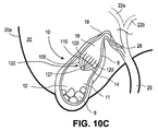
横方向の様々な角度が本発明のいくつかの異なる態様に使用することができることは自明である。ほぼ直交する方向(すなわち、破線12a、14a)の基準から測定すると、胆嚢中央縦軸19と胆嚢治療デバイス間の角度は、直交する配置の場合の0度から、胆嚢治療デバイスの配置、胆嚢の生体構造、患者の状態、及びその他の要因によって最大50度となる。更に、胆嚢治療デバイスは、胆嚢ルーメン11内の方向を維持して、胆嚢が拡張した状態(例えば、図10A、10C、7A、11A、11C及び6A)、収縮した状態(例えば、図10B、10D、7B、11B、11D及び6B)、及び拡張期と収縮期との中間状態にある間に胆石/胆石片9が頚部18に入らないようにする。
Obviously, various lateral angles can be used in several different aspects of the invention. When measured from a reference in a substantially orthogonal direction (i.e.,
胆嚢治療デバイスをリストに挙げた治療介入の特定部位へ挿入する方法を含めて、胆嚢内に胆嚢治療デバイスを配置する様々な代替方法の更なる詳細を図13A、13B、14及び15A乃至15Dを参照して以下に述べる。更に、胆嚢の生体構造、送達デバイス、及び方法の様々な詳細は、Jacques Van Dam et al.によって出願された米国特許出願公開第2009/0143760号、“Method,Devices,Kits and Systems for Defunctionalizing the Gallbladder”に記載されている。ここで、胆嚢の生体構造及び生理に話を戻す。 13A, 13B, 14 and 15A-15D provide further details of various alternative methods of placing the gallbladder treatment device within the gallbladder, including the method of inserting the gallbladder treatment device into a specific site of a listed treatment intervention. See below for reference. In addition, various details of gallbladder anatomy, delivery devices, and methods are described in Jacques Van Dam et al. U.S. Patent Application Publication No. 2009/0143760, filed by “Methods, Devices, Kits and Systems for Defunctizing the Gallbladder”. Now, let us return to the anatomy and physiology of the gallbladder.
正常な生体構造と生理
胆嚢10は水を除去して電解質に交換することによって胆汁を保存し、濃縮する消化管の器官である。胆嚢は、機械ポンプのように機能し、腸肝システムを介して胆汁を分泌して、脂溶性化合物の摂取を強化する。
Normal anatomy and physiology The
胆嚢の肉眼的生体構造
図1Aは、腹部内で、肝臓と関連して胆嚢10を示す図であり、胆嚢壁の一部を除去して、内部に胆石/胆石片9を有する胆嚢内部又はルーメンを示している。胆嚢10は、梨形の中空器官であり、肝臓の右葉後面上のくぼみにある(セグメント5)。正常な成人では完全に膨張すると、長さ10cm、幅約3乃至4cmに及ぶ。胆嚢の壁8は、厚さ1乃至2mmである。正常な成人の胆嚢の容積は、30乃至45mlである。胆嚢10は、基底部12、中央本体部14、及び頚部18のセグメントに分けられ、これらは胆嚢管26につながっている。基底部12は、この器官の最も遠位部分であり、しばしば、肝臓の下側端部を超えて突出している。基底部は、粗性結合組織によって肝臓に付着しており、腹膜がその自由表面を覆っている。胆嚢は基底部12と中央本体部14との間の結合部において最も幅が広い。中央本体部14は、最も大きなセグメントを形成しており、頚部18に結合するところで漏斗部16にテーパが付いている。胆嚢−十二指腸靭帯と呼ばれる腹膜ひだが、漏斗部分の外側を十二指腸の最初の部分に付着している。頚部はS字状構造であり、長さ約5乃至7mmで、胆嚢管に結合するところが狭くなっている。胆嚢と胆汁系の様々な部分が図1Aに示されており、ハイスターのらせんひだ24、肝管22a、22b、胆嚢管26、及び胆嚢管28を含む。図1Bは胆嚢壁8の一部であり、様々な層の互いに対する相対位置と、腹膜又は腹壁及び腹腔を示す。胆嚢壁8は、上皮1、固有層2、平滑筋3、結合組織4、漿膜5でできている。漿膜5は、胆嚢を腹腔7から分離している腹膜6に接触しており、これにつながっている。
Macroscopic anatomy of the gallbladder FIG. 1A shows the
「グレイの解剖学」は、胆嚢の層は、三層(すなわち、漿膜、繊維及び筋膜)と粘膜を有すると記載している。外膜又は漿膜は、腹膜から取り出され、完全に基底部を取り巻いているが、下側表面上でのみ本体部と頚部を覆っている。繊維−筋肉膜は薄いが強い層であり、嚢のフレームワークを形成している。これは、全方向に織合わさった緻密線維組織でなり、主に縦方向に配置され、若干横方向に走っている平坦な筋繊維と混じっている。内膜又は筋膜は、繊維層にゆるく結合している。一般的に、黄色かかった茶色を帯びており、あらゆるところで細かい粘膜皺になっている。これらの皺を一つにすることによって、多数のメッシュが形成され、多角形の外形を有するへこんだ介在スペースとなっている。このメッシュは、基底部及び頚部においてより小さく、嚢の中央付近で最も発達している。(Gray’s Anatomy,A revised American edition from the Fifteenth English Edition,1977,p.942参照)。 “Gray anatomy” states that the layers of the gallbladder have three layers (ie, serosa, fiber and fascia) and the mucosa. The outer membrane or serosa is removed from the peritoneum and completely surrounds the base, but covers the body and neck only on the lower surface. The fiber-muscle membrane is a thin but strong layer that forms the sac framework. It consists of a dense fibrous tissue woven in all directions, mixed with flat muscle fibers that are mainly arranged in the longitudinal direction and run slightly in the transverse direction. The intima or fascia is loosely bonded to the fiber layer. Generally, it has a yellowish brownish color and has a fine mucous membrane fold everywhere. By combining these wrinkles into one, a large number of meshes are formed, and a concave intervening space having a polygonal outer shape is formed. This mesh is smaller in the base and neck and is most developed near the center of the sac. (See, Gray's Anatomy, Revised American edition from the Fifteenth English Edition, 1977, p. 942).
稀ではあるが、解剖学的胆嚢変種が存在することがある。これには、胆嚢の先天的欠損+/−拡張した肝管を伴う胆嚢管(剖検例の0.03乃至0.07%)、中央の狭窄、又は稀に、部分的に縦方向に割れたものを含む、不規則な形の胆嚢が含まれる。Meilstrop,J.,Imaging atlas of the normal gallbladder and its variants.1994:CRC Press.参照。頚部は、通常、ゆるいカーブを描いて胆嚢管に続くが、通常、慢性の閉塞又は炎症によって、その凸性は漏斗部又はハートマンのポーチとして知られている膨張部分へ向けて膨らんでいる。二つの異形胆嚢+/−胆嚢管は、0.02%の患者に見られた。通常は、セグメント5にそって肝臓の右葉後方に位置するが、胆嚢は、まれに、靱帯の左側に見られたり、あるいは、肝臓内に、肝臓上に、横方向位置に、浮遊した状態で、肝臓の左葉下側に、又は腹膜後ろに見られることがある。異所には、肝後面、又は腹部前壁内、あるいは、鎌状靱帯内がある。異常な膵臓の組織が、ときどき、胆嚢の壁に見つかることがある。基底部は捻転していることがある。真性憩室は、胆嚢の稀な先天性奇形(Mayo Clinicで切除した胆嚢全ての0.0008%)であり、三層の胆嚢壁を全て持っている。本体部及び頚部の憩室は、胆嚢と肝臓の間を胚期の間に通る、胚期持続性胆管走行異常から生じることがある。基底部の異形は、胚期の間の固形胆嚢の不完全な空胞変性から生じる。不完全な隔壁は、胆嚢の先端の小さな空洞を切り取る。先天的な変形は、疾患のある胆嚢に部分的に発現する穿孔の結果できる疑似憩室と区別するべきである。この場合の疑似憩室は、一般的に大きな胆石が含まれている。
Although rare, anatomical gallbladder variants may exist. This includes congenital defects of the gallbladder +/- gallbladder duct with dilated liver ducts (0.03-0.07% of autopsy cases), central stenosis, or, rarely, partially longitudinally cracked Includes irregularly shaped gallbladder, including Meiltrop, J.M. , Imaging atlas of the normal gallbladder and its variants. 1994: CRC Press. reference. The neck usually follows a gallbladder duct with a loose curve, but usually due to chronic occlusion or inflammation, its convexity bulges toward an inflatable part known as the funnel or Hartman pouch. Two deformed gallbladder +/- gallbladder ducts were found in 0.02% of patients. Normally located along the
胆嚢への血液供給は、右肝動脈の近位部分から始まる胆嚢動脈から主に行われる。右肝動脈は、胆嚢の漿膜の上にある表在チャネルと、胆嚢と肝臓床の間を走る深部チャネルに枝分かれする。静脈排出路は、胆嚢管の上で合流している、胆嚢の肝臓側にある小さなチャネルとその他の小さな血管とからなり、門脈静脈系で終端している。肝神経叢からの求心性神経繊維、交感神経線維、及び副交感神経線維は、胆嚢に続いている。迷走神経の刺激によって、胆嚢壁の平滑筋の周期的収縮が生じる。胆嚢頚部と胆嚢管には局所リンパ節が存在し、肝肺門節へ流れ出る。 Blood supply to the gallbladder is mainly from the gallbladder artery starting from the proximal portion of the right hepatic artery. The right hepatic artery branches into a superficial channel above the serosa of the gallbladder and a deep channel that runs between the gallbladder and the liver bed. The venous drainage path consists of a small channel on the liver side of the gallbladder and other small blood vessels that meet on the gallbladder duct and terminate in the portal vein system. Afferent, sympathetic, and parasympathetic fibers from the hepatic plexus follow the gallbladder. The stimulation of the vagus nerve causes periodic contraction of the smooth muscle of the gallbladder wall. Local lymph nodes exist in the gallbladder neck and gallbladder duct and flow out to the hepatic hilar node.
胆嚢組織構造
胆嚢壁8(内側から外側まで)は、表面上皮と、固有層と、器官の縦軸に沿って全体的なスパイラルパターンをなす平滑筋と、筋肉周囲の漿膜下結合組織と、漿膜とからなる。胆嚢には、消化管のその他の部分に見られる粘膜と粘膜下層がない。内腔壁は、主及び副ひだを有し、これらの襞の高さと幅は可変である。上皮層は、基底膜上方の単層の円柱上皮細胞からなる。上皮細胞は、管腔表面は濃厚微絨毛と繊毛を有し、複雑な細胞内指状突起と陥入がある。Nakanuma,Y.,et al.,Monolayer and three−dimensional cell culture and living tissue culture of gallbladder epithelium.Microse Res Tech,1997.39(1):p.71−84.参照。管状胞状粘液腺は、胆嚢の経部にのみあり、立方体状あるいは低い筒状の細胞でできており、クリアな細胞質が豊富である。頚部の内膜細胞と粘液腺は、硫酸ムチンを含む。
Gallbladder tissue structure The gallbladder wall 8 (from inside to outside) consists of the surface epithelium, the lamina propria, the smooth muscle that forms an overall spiral pattern along the longitudinal axis of the organ, the subserosa connective tissue surrounding the muscle, and the serosa It consists of. The gallbladder lacks the mucosa and submucosa found elsewhere in the digestive tract. The lumen wall has major and minor folds, and the height and width of these folds are variable. The epithelial layer consists of a single layer of columnar epithelial cells above the basement membrane. Epithelial cells have dense microvilli and cilia on the luminal surface, and have complex intracellular finger processes and invagination. Nakanuma, Y .; , Et al. , Monolayer and three-dimensional cell culture and living tissue culture of gallblader epithelium. Microres Res Tech, 1997.39 (1): p. 71-84. reference. Tubular cystic mucous glands are located only at the transmural portion of the gallbladder, are made of cubic or low-pipe cells, and are rich in clear cytoplasm. Cervical intimal cells and mucous glands contain mucin sulfate.
胆嚢管の生体構造
胆嚢管26は、左右の全肝管の合流地点の遠位側約2cmのところで総胆管と合流している。ヒトの胆嚢管の平均長さは30mmであり、たたんだ時の平均径は約4mmである。Frierson,H.F.Jr.,The gross anatomy and histology of the gallbladder,extrahepatic bile ducts,Vaterian system,and minor papilla.Am J Surg Pathol,1989.13(2):p.146−62.参照。胆嚢管の内膜細胞は、形態学的に、胆嚢内部の内膜細胞と同じである。胆嚢管が胆嚢頚部に合流するところに、斜めの大きなひだがあり、これが平滑筋の束を含み、圧力が変化する間の胆嚢管の過膨張と、折りたたみを防ぐと考えられる。平滑筋の襞のある領域は、ハイスターのらせん弁と呼ばれている。
Biological structure of gallbladder duct The
正常な胆嚢の生理
胆嚢は、一日当たり500乃至1000ccの胆汁を排出する。胆汁はヒトでは、水分の受動運動に伴って電解質が能動的に吸収され5乃至10倍に濃縮される。胆汁は、食物脂肪の腸肝吸収を大きく左右し、胆汁酸塩、レシチン、コレステロール、及び有機溶質からなる。胆汁酸塩は、コレステロール代謝の主な物質であり、肝臓で分泌された脂質(通常、レシチン)を胆道系に可溶化して、消化管における脂質の吸収を促進することができる非常に有効な浄化剤である、水溶性ステロール族の一部である。レシチンは、疎水性で、水への溶解度が最小の非水性化合物である。コレシストキニン、すなわち腸細胞で分泌されるホルモンは、胆嚢を収縮させ、食事に反応してオッディ括約筋を弛緩させる。
Normal gallbladder gallbladder drains 500 to 1000 cc of bile per day. In humans, bile is concentrated 5 to 10 times as the electrolyte is actively absorbed by the passive movement of water. Bile greatly affects the enterohepatic absorption of dietary fat and consists of bile salts, lecithin, cholesterol, and organic solutes. Bile salts are the main substances in cholesterol metabolism and are very effective in solubilizing lipids secreted by the liver (usually lecithin) into the biliary system to promote lipid absorption in the digestive tract. It is part of the water-soluble sterol family that is a purifier. Lecithin is a non-aqueous compound that is hydrophobic and has minimal water solubility. Cholecystokinin, a hormone secreted by enterocytes, contracts the gallbladder and relaxes the oddy sphincter in response to a meal.
胆嚢の基底部12及び本体部14は、この器官のうち最も広く、最も収縮する部分である。胆石9は、基底部12の下側部分に主に形成され、留まる。図1Aは、腹部内の胆嚢を肝臓と関連させて示す図であり、胆嚢壁の一部を除去して、胆石及び胆石片を有する胆嚢内部の容量又はルーメンを示している。図1は、基底部12に胆石がプールされている胆嚢の様々な部分を示す。一の実施例では、胆嚢治療デバイスを基底部又は本体部に配置して、胆石が頚部又は漏斗部を通過しないようにしている。
The
胆嚢治療デバイス
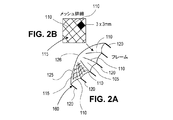
図2A及び2Bは、それぞれ、胆嚢治療デバイスの一実施例を示す斜視図及び拡大図である。フィルタは、フィルタメッシュと、フィルタを胆嚢壁に対して適所に保持する支柱でできたフレームからなる。臨床的には、3mmより大きい胆石は、閉塞されることなく1乃至5mm幅の胆嚢管を容易に通り抜ける。従って、フィルタメッシュは、多孔性であり、直径3mmより小さい粒子は通過させる。フィルタはフレキシブルであり、胆嚢の収縮時に形態が変わる。本体部と基底部は、胆嚢の最も収縮する部分である。フィルタは、完全に収縮した器官の直径までたたまれるために、有意に構造が変化する。
Gallbladder Treatment Device FIGS. 2A and 2B are a perspective view and an enlarged view, respectively, showing one embodiment of a gallbladder treatment device. The filter consists of a filter mesh and a frame made of struts holding the filter in place against the gallbladder wall. Clinically, gallstones larger than 3 mm can easily pass through a 1-5 mm wide gallbladder duct without being blocked. Thus, the filter mesh is porous and allows particles smaller than 3 mm in diameter to pass through. The filter is flexible and changes shape when the gallbladder contracts. The main body and the base are the most contracted parts of the gallbladder. As the filter collapses to the diameter of a fully contracted organ, the structure changes significantly.
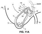
図6B、7B、10B、10D、11B及び11Dは、収縮した胆嚢10内の胆嚢治療デバイスの様々な実施例を示す。胆石9の通過を防止するように配置したエレメント110の空隙率又はフィルタ特性(すなわち、フィルタメッシュとしての一構造において)は、胆嚢治療デバイスが収縮した胆嚢10とともにたたまれても変化しない。胆嚢本体部の直径が収縮して小さくなると、胆嚢治療デバイスの支柱120は互いに近づいて、フィルタメッシュが遠位側に胆嚢基底部内にあるいは胆嚢基底部に向けて折りたたまれる。収縮時には胆嚢治療デバイスの径が小さくなり、プロファイル長が長くなる。
6B, 7B, 10B, 10D, 11B, and 11D show various examples of gallbladder treatment devices within the contracted
胆石の通過を防ぐ複数のエレメント110は、フィルタを形成する複数の交差エレメントとして構成しても良く、あるいは、フレーム105に取り付けたフィルタメッシュ又はその他の構成であってもよい。フレーム105は、例えば、ニチノールやステンレススチールでできた、フレキシブルあるいは硬質なフレームである。
The plurality of
図2Bは、図2Aに示す胆嚢治療デバイス100の拡大図である。図2Bは、最大径3mmの胆石又は胆石片9が通過できない寸法のフィルタ特性115を有する少なくとも一のエレメント110から形成されたフレーム105の関係を示す。一の特別な例では、3mmより大きい胆石又は胆石片を通過させないフィルタ特性を有する複数のエレメントが、フレーム105とニチノールで形成された支柱に取り付けられた多孔性フィルタメッシュである。このメッシュは、フレームに連結されて、断面フィルタ表面を形成している。メッシュの空隙率は3mmより小さい。一の特別な実施例では、フィルタは、最大径約3mmより小さい胆石又は胆石片を通過させないサイズの孔径又はフィルタ容量を有する。別の特別な実施例では、フィルタは、最大径約2mmより小さい胆石又は胆石片を通過させないサイズの孔径又はフィルタ容量を有する。更に別の特別な実施例では、フィルタは、最大径約1mmより小さい胆石又は胆石片を通過させないサイズの孔径又はフィルタ容量を有する。
FIG. 2B is an enlarged view of the
一の実施例では、胆嚢頚部の遠位側の胆嚢部分内に胆石又は胆石片9を維持する胆嚢治療デバイス100を示す。フレーム105は、最大径が3mmより大きい胆石又は胆石片を通過させない寸法のフィルタ特性115を有する少なくとも一のエレメント110でできている。胆嚢中央の、縦軸19に交差する方向においてフレーム105を固定するようにフレーム105から延在する複数の支柱120がある。最大径が3mmより大きい胆石又は胆石片9は、胆嚢10内でフレーム105の一方の側に残るであろう。一の態様では、フレーム105は、ほぼ円形の外形126を有し、フレーム外形125の曲率半径は約0.1cm乃至約1.0cmである。更に別の態様では、このフレームはほぼ円錐形の外形127を有し、フレーム外形125の曲率半径は約0.1cm乃至約1.0cmである。更なる代替の実施例では、少なくとも一のエレメントがフレキシブルシート材であり、そのフィルタ特性は、フレキシブルシートに形成した最大径が3mmより小さい複数の開口である。更なる実施例では、少なくとも一のエレメントが、ブレード構造135からの複数のフレキシブルストランド136であり、隣接するストランド間のスペースが3mmより小さいフィルタ特性を維持している。更なる代替例では、少なくとも一のエレメントがフレームに取り付けた複数の支柱ストランド140であり、隣接する支柱ストランド間の3mmより小さいスペースであるフィルタ特性を維持するフィルタ構造を形成している。
In one embodiment, a
様々なフィルタ構造を用いて、基底部内に3mmより大きい材料を維持し、これによって嵌頓のリスクを低減することができる。フィルタ構成要素は、ブレードしたニチノール、ニチノールなどのフレキシブル材料でできた薄いシート、又は、ePTFEなどの多孔性無機材料であっても良い。この薄いシートを処理して、スタンプ又はパンチして孔又は開口を形成する。更に別の態様では、フィルタ構造が、縫合糸ベースのネットである。フレーム、支柱、フィルタ、フィルタエレメント、又はフィルタ材は、ニチノールなどの硬質の金属、又は、ニチノールシート、ブレード、あるいは縫合糸ベースのネットで構成された薄くフレキシブルな材料であっても良い。デバイス上又はデバイス内に形成された孔、開口、又は開口は、デバイスの収縮に合わせてサイズを変えることができる。例えば、ニチノールブレードフィルタは、器官が拡張するときに孔径が大きくなり、器官が収縮するときに孔径が小さくなる。胆石又は胆石片は、器官が収縮するときに最も妨害されるので、孔径は、収縮時に小さくなって胆石が通過せず、拡張時には大きくなって、胆汁の流れが最大になる。 Various filter structures can be used to maintain material larger than 3 mm in the base, thereby reducing the risk of incarceration. The filter component may be a bladed nitinol, a thin sheet made of a flexible material such as nitinol, or a porous inorganic material such as ePTFE. The thin sheet is processed and stamped or punched to form holes or openings. In yet another aspect, the filter structure is a suture-based net. The frame, strut, filter, filter element, or filter material may be a hard metal such as Nitinol, or a thin and flexible material comprised of a Nitinol sheet, blade, or suture-based net. The holes, openings, or openings formed on or in the device can be resized as the device shrinks. For example, a Nitinol blade filter has a large pore size when the organ expands and a small pore size when the organ contracts. Since gallstones or gallstone pieces are most disturbed when the organ contracts, the pore size decreases during contraction and does not pass through the gallstone, and increases during expansion, maximizing bile flow.
更に別の態様では、胆嚢頚部の遠位側の胆嚢部位に胆石又は胆石片を維持する別の胆嚢治療デバイスが開示されている。このデバイスは、第1及び第2の端部を有する複数の支柱を有する。第1の端部と第2の端部の間の湾曲した部分は、収縮した胆嚢によって生じる曲率半径を有する第1の状態と、拡張した胆嚢によって生じる曲率半径を有する第2の状態との間で変化する。この複数の支柱は、第1の端部と第2の端部との間で少なくとも1回互いに交差しており、支柱が交差した結果できるパターンが、3mmより大きい胆石又は胆石片の通過を防いでいる。 In yet another aspect, another gallbladder treatment device is disclosed that maintains a gallstone or gallstone piece at a gallbladder site distal to the gallbladder neck. The device has a plurality of struts having first and second ends. The curved portion between the first end and the second end is between a first state having a radius of curvature caused by a contracted gallbladder and a second state having a radius of curvature caused by an expanded gallbladder. It changes with. The plurality of struts cross each other at least once between the first end and the second end, and the pattern resulting from the crossing of the struts prevents passage of gallstones or gallstone pieces larger than 3 mm. It is out.
硬質フレームを指定して、例えば、胆嚢壁に接触する位置又はルーメンの中央といった特定の位置を柔軟にして、器官の収縮に合わせてたたまれ、収縮するようにしても良い。このフレームは、半径方向の力あるいは固定技術を用いて胆嚢の組織内部に配置することもできる。このフィルタを適所に保持するのに必要な半径方向の力の量を支柱の形状と壁との接触面を変えることによって減らして、接着及び細胞内包成長を改善することができる。図5A及び5Bに例を示すように、支柱の長さは、内腔壁に接触する支柱の表面積又は支柱の壁接触部を増やす又は減らすように調整することができる。接触面の増加は、能動的収縮が生じている間にデバイスの位置決め及び固定を支援する。支柱の壁に接触する部分を仕上げる追加材料又は表面も、接着及び固定を改良する。例えば、支柱の壁接触面を繊維組織の内包成長を増やすことで知られている生体材料、例えばPLGAでコーティングすることで、接着及び固定を改良することができる。成長を促進するその他の好適材料は、ポリエチレングリコールと、上皮成長因子である。接触面を多孔性無機物質、例えばePTFEなどでコーティングすることでも、接着及び固定を改良することができる。例えば、壁に接触している支柱に小さなスパイク又は溝を追加するなど、新規材料を追加することなく表面を改良するようにしても、接着を支援することができる。 A rigid frame may be specified, for example, a specific position, such as a position that contacts the gallbladder wall or the center of the lumen, may be flexed to fold and contract as the organ contracts. The frame can also be placed inside the gallbladder tissue using radial force or fixation techniques. The amount of radial force required to hold the filter in place can be reduced by changing the strut shape and the wall interface to improve adhesion and cell growth. As shown in the examples in FIGS. 5A and 5B, the strut length can be adjusted to increase or decrease the strut surface area contacting the lumen wall or strut wall contact. The increased contact surface assists in positioning and securing the device during active contraction. An additional material or surface that finishes the part that contacts the wall of the column also improves adhesion and anchoring. For example, the adhesion and fixation can be improved by coating the wall contact surfaces of the struts with biomaterials known to increase the growth of fiber tissue inclusions, such as PLGA. Other suitable materials that promote growth are polyethylene glycol and epidermal growth factor. Coating the contact surface with a porous inorganic material such as ePTFE can also improve adhesion and fixation. Adhesion can be aided even if the surface is improved without adding new material, for example, by adding small spikes or grooves to the struts in contact with the walls.
半径方向の力に代えて又はこれに加えて、一又はそれより多い様々な機構を用いてデバイスの胆嚢内の位置を維持することができる。様々な機構及び技術を、図3A乃至3Dを参照して説明する。 Instead of or in addition to the radial force, one or more of various mechanisms can be used to maintain the position of the device in the gallbladder. Various mechanisms and techniques are described with reference to FIGS. 3A-3D.
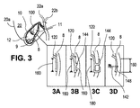
図3は、図1Aに示す胆嚢を、胆嚢壁の一部を除去して、胆嚢内部にある一例の胆嚢治療デバイスを示す図である。このデバイスは、胆石又は胆石片を、デバイスの一方の側部であって、胆嚢頚部から離した位置に維持する方向に配置されている。 FIG. 3 is a view showing an example of a gallbladder treatment device in which the gallbladder shown in FIG. 1A is partly removed from the gallbladder wall. The device is arranged in a direction to maintain the gallstone or gallstone piece on one side of the device and away from the gallbladder neck.
図3Aは、図3に示すデバイスの支柱120の拡大断面図である。この図は、胆嚢壁8の一部に沿って配置した支柱120の壁に接触する部位160を示す。
3A is an enlarged cross-sectional view of the
図3Aに示すようにデバイスのフレームを構成する支柱120は、半径方向の力を単独で使用することができ、胆嚢壁の層を貫通していない。壁への掛かりが制限されているので、このタイプの支柱は、組織内包成長が生じると完全に固定される。この成長は、十分に成長するのに数日乃至数週間かかる。追加で又は代替的に、支柱又は壁に接触する部分は、図3B、3C及び3Dに示すような固定エレメント142を具えていても良い。
As shown in FIG. 3A, the
図3Bは、図3に示すデバイスの支柱120を拡大して示す図であり、固定エレメント142を有する支柱120の壁に接触する部位160を示す。ここで、固定エレメントは、胆嚢壁の一部に沿って胆嚢内部に使用されている。この実施例では、短いスパイク又はバーブ144などの固定エレメントは、少なくとも部分的に胆嚢壁8内に通っている。
FIG. 3B is an enlarged view of the
図3Bは、自己挿入型の部分的に厚いバーブ又はタック144を示しており、これらは、胆嚢壁8に制限された引っ掛かりを有するが、組織の内包成長が完成するまでデバイスを適所に保持するのに必要な半径方向の力を低減するのに十分である。部分的に厚い固定を行う利点は、腹膜に穴があいたり、漏れ/コンタミネーションのリスクがないことである。
FIG. 3B shows self-inserting, partially thick barbs or tacks 144 that have a limited catch on the
図3Cは、図3に示すデバイスの支柱を拡大した断面図であり、胆嚢壁の一部に沿って胆嚢壁を固定するのに使用した固定エレメント142を有する支柱120の壁に接触する部位160を示す。この実施例では、クリップ144が胆嚢壁9を完全に貫通している。
FIG. 3C is an enlarged cross-sectional view of the strut of the device shown in FIG. 3 and a
図3Cは、全体が厚いバーブ又はタック144を示す図であり、これは、必要とされるわずかな半径方向の又は組織の内包成長を用いることなく、あるいはこれを用いてデバイス100を適所に保持することができる。壁の穿刺は小さく、例えば、直径0.01インチより小さい。また、固定エレメントが壁から除去されなくても、漏れ/コンタミネーションのリスクは限定される。
FIG. 3C shows an overall thick barb or tack 144 that holds the
図3Dは、図3に示すデバイスの支柱120を拡大した断面図であり、開口148を有する支柱120の壁に接触する部位160を示す。開口148によって、個々の固定エレメント142が、開口148を通過し、次いで、部分的にあるいは完全に胆嚢壁8を通る。一又はそれより多い開口148を壁に接触する部位160の一部に形成して、一又はそれより多い締結デバイス又は、単一の固定デバイスの一又はそれより多いエレメントが通過できるようにしている。
FIG. 3D is an enlarged cross-sectional view of the
図3Dは、個別に適用したバーブ、支柱、又はタックを示す図である。この固定方法は、図3Cで述べた全体が厚いバーブと同様のリスクと利点を有する。 FIG. 3D shows a barb, strut, or tack applied individually. This securing method has the same risks and advantages as the overall thick barb described in FIG. 3C.
一の胆嚢治療デバイスの代替例では、各支柱120の一部が開口148を有しており、固定エレメント142が通過できるようにしている。この開口は壁に接触する部位にあってもよい。固定エレメント142は、開口148を通過するサイズであり、胆嚢壁8を少なくとも部分的に通る長さを有する。別の態様では、固定エレメントは、胆嚢壁を完全に貫通するのに十分な長さを有する。
In one alternative gallbladder treatment device, a portion of each
半径方向の力を用いる場合は、この力が胆嚢壁によって生じる収縮力より小さくて、壁8の浸透又は浸食を低減する又は防止するものでなくてはならない。胆嚢壁と複数地点で接触するようにして、器官の静止時、及び収縮及び拡張時に半径方向の力を分散させるようにしても良い。胆嚢治療デバイスの一代替例では、複数の支柱120が、胆嚢の中央縦軸19に交差する方向にフレームを維持するのに十分な半径方向の力を用いて、壁8に対して外側に変位する形状をしている。更に、胆嚢治療デバイスは、胆嚢が収縮状態にあるとき及び拡張状態にあるときに、横方向の姿勢を維持する。
If a radial force is used, this force must be less than the contraction force produced by the gallbladder wall to reduce or prevent
胆嚢壁の厚さは、ほぼ1mmである。部分的に厚いタック又はバーブを用いる場合、これらは、0.75mm又は壁の厚さ全体の3/4より少ない長さだけ突き通って、全厚穿刺のリスクを制限している。全体が厚いバーブ又はタックを使用する場合は、Dacron,ePTFE又は同様の材料でできたパッチを、デバイスの支柱と胆嚢壁の間に挿入して、穿刺部位を通る腹腔への漏れを防ぐようにする。胆嚢基底部又は本体部の壁に沿って追加の支柱又はリングをデバイスから延在させても良い。これらのエクステンションは、収縮時に発生する力を分散させる、又は固定支援を提供するように作用する。 The thickness of the gallbladder wall is approximately 1 mm. When using partially thick tacks or barbs, they penetrate by less than 0.75 mm or 3/4 of the total wall thickness, limiting the risk of full thickness puncture. When using an overall thick barb or tack, a patch made of Dacron, ePTFE or similar material should be inserted between the device strut and the gallbladder wall to prevent leakage into the abdominal cavity through the puncture site To do. Additional struts or rings may extend from the device along the gallbladder base or body wall. These extensions act to disperse the forces generated during contraction or provide fixation support.
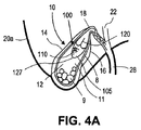
図4Aは、図1Aに示す胆嚢を示しており、胆嚢壁8の一部を除去して、例示的な胆嚢治療デバイスを見えるようにした図である。このデバイスは、円錐型127のフレーム外形125を有する。このデバイスは、胆嚢内部に、デバイスの一方の側部であって、胆嚢頚部から離れて胆石又は胆石片を維持する方向に配置されている。
FIG. 4A shows the gallbladder shown in FIG. 1A with a portion of the
図4Bは、図1Aに示す胆嚢を示しており、胆嚢壁8の一部を除去して、例示的な胆嚢治療デバイス100が見えるようにした図である。このデバイスは丸型126のフレーム外形125を有し、胆嚢内部11に、デバイス100の一方の側部であって、胆嚢頚部18から離して胆石又は胆石片9を維持する方向に配置されている。
FIG. 4B shows the gallbladder shown in FIG. 1A with a portion of the
図5Aは、図1Aに示す胆嚢10を示しており、胆嚢壁8の一部を除去して、例示的な胆嚢治療デバイス100が見えるようにした図である。このデバイスは丸型126のフレーム外形125と、壁に接触する部位160を増やすように調整した支柱120とを有し、胆嚢内部に、デバイスの一方の側部であって、胆嚢頚部から離して胆石又は胆石片を維持する方向に配置されている。
FIG. 5A shows the
図5Bは、図1Aに示す胆嚢を示しており、胆嚢壁を除去した部位を伴う。この例示的な胆嚢治療デバイス100は、丸型のフレーム外形と、胆嚢内部11に壁に接触する部位160を増やすように調整した支柱とを有する。このデバイスは、デバイス100の一方の側部であって、胆嚢頚部18から離して胆石又は胆石片9を維持する方向に配置されている。
FIG. 5B shows the gallbladder shown in FIG. 1A with the site from which the gallbladder wall has been removed. This exemplary
一の態様では、胆嚢治療デバイスは、ほぼ円柱パターンに配置された複数の支柱120を有する、胆嚢の遠位側の一部から胆嚢頚部に、胆石又は胆石片を維持している。壁に接触する部位160は、複数の支柱の各々に提供されている。複数のフレキシブル部材110で形成されたフレーム105が、複数の支柱120の2本の間に取り付けられている。複数の支柱120のうちの2本の間に取り付けたときの二つの隣接するフレキシブル部材110間のスペースは、3mmより小さい。胆嚢内での使用時には、フレーム105が、胆嚢頚部18の遠位側の胆嚢ルーメン11の一部内に胆石又は胆石片9を維持する。更に、複数のフレキシブル部材110によって形成される曲率半径125は、拡張した胆嚢に複数の支柱120が接触しているときの第1の曲率半径と、収縮した胆嚢に複数の支柱120が接触しているときの第2の曲率半径との間で変化する。第1の曲率半径の例は、例えば、図10A、10C、7A、11A、11C、5A及び5Bに見られる。第2の曲率半径の例は、例えば、図10B、10D、7B、11B及び11Dに見られる。
In one aspect, the gallbladder treatment device maintains a gallstone or gallstone piece from the distal part of the gallbladder to the gallbladder neck with a plurality of
図6A及び6Bは、それぞれ、拡張した状態及び収縮した状態にある図1Aに示す胆嚢を、胆嚢壁の一部を除去して例示的なフープフレーム型胆嚢治療デバイス100が見えるようにした図である。デバイス100(図6Cに最も良く見ることができる)は、胆嚢の漏斗状部内にあり、デバイス100の一方の側であって、胆嚢頚部18から離して胆石又は胆石片9を維持する方向に配置されている。
6A and 6B are views of the gallbladder shown in FIG. 1A in an expanded state and a contracted state, respectively, with a portion of the gallbladder wall removed to reveal an exemplary hoop frame type
この実施例では、胆嚢治療デバイス100は、フープ状又はリング状のフレーム105を有し、デバイスの一方の側に胆石又は胆石片を維持する寸法のフィルタ特性を有する少なくとも一のエレメント110を支持している。この図6Cに示す実施例では、少なくとも一のエレメント110が複数のストランドであり、隣接するストランド110間に3mm又はそれより小さいスペースを空けたフィルタ特性を維持する規則的なパターンに配置されている。フレーム105は、フープ又は短い円柱として示されており、フープの径は、両端で同じである。フレームの壁の高さは、胆嚢内のインプラント部位に応じて変わる、あるいはこれに合ったサイズである。更に、壁は、図に示すように平坦である必要はなく、角度が付いていても、テーパが付いていても良い。この場合、フープの一方の側が、第1の径を有し、他方の側が第2の径を有し、これらの第1及び第2の径は異なっている。異なる径と、これらの間の緩やかなスロープを使用することで、テーパ型フレーム105ができる。第1の側部と第2の側部の間の中間径がくびれた形状のフレーム105を作るといった、その他の構成も可能である。
In this embodiment, the
図6Aと6Bは、胆嚢拡張時(図6A)と収縮時(図6B)の、胆嚢漏斗部内に配置した周辺リングフレームを有する胆嚢治療デバイス100を示す図である。漏斗部の径はほぼ変化しないが、収縮時に1.5乃至2cmの通常サイズ範囲を維持するので、フープフレームは、図6Bに示すように、胆嚢収縮時に構造変化が生じないように設計されている。
6A and 6B show a
漏斗部は、頚部及び出口に向けてテーパが付いている。上述した通り、フレーム外壁にテーパを付けて、様々な形状の壁にテーパが付いた胆嚢治療デバイスを作ることができる。外形、形状又はテーパにフープをほぼ合わせるようなテーパ形状をした胆嚢漏斗部のフープ壁は、位置決めを改良する。 The funnel is tapered towards the neck and the outlet. As described above, the outer wall of the frame can be tapered to create a gallbladder treatment device with tapered walls of various shapes. The hoop wall of the gallbladder funnel that is tapered to approximately match the hoop to its outer shape, shape or taper improves positioning.
ここに述べたその他のデバイスと同様に、フープフレームは、半径方向の力の使用、大きいサイズのフープフレームの使用、あるいはフレーム側壁にテーパを付けること、の一又はそれ以上によって適所に維持することができる。更に、自己挿入型バーブ又はタック、又はフープ壁に沿って個別に適用したバーブ又はタックを用いて、漏斗部上の壁にフープを固定することができる。一又はそれより多い縫合糸と、従来の縫合技術を用いて、胆嚢壁8にフープを取り付けることもできる。図3、3A、3B、3C、3D、8A、8B、及び8Cに関連してここに述べた固定技術と同様に、上述の材料と構成技術も、胆嚢の漏斗部に配置するように構築し、そのようなサイズにし、適用した胆嚢治療デバイスの構成に使用することができる。
As with the other devices described herein, the hoop frame should be kept in place by using radial force, using a large size hoop frame, or tapering the frame sidewalls. Can do. Furthermore, self-inserting barbs or tacks or barbs or tacks individually applied along the hoop wall can be used to secure the hoop to the wall on the funnel. A hoop can also be attached to the
図7A及び7Bは、拡張した及び収縮した状態にある図1Aに示す胆嚢がそれぞれ示されており、胆嚢壁の一部を除去して、例示的なフレームレス胆嚢治療デバイス500(ここではメッシュ球体が示されている)が見えるようにしている。このデバイス500は、胆石又は胆石片9をデバイス500の一方の側であって、胆嚢頚部から離れたところに維持する方向に配置され、胆嚢基底部内側にある。
7A and 7B show the gallbladder shown in FIG. 1A in an expanded and deflated state, respectively, with a portion of the gallbladder wall removed to illustrate an exemplary frameless gallbladder treatment device 500 (here a mesh sphere). Is shown). This
胆嚢の本体部に配置することができる胆嚢治療デバイスの別の例は、胆管内又はその上の浮遊型のものである。この設計は、胆嚢の壁に固定されていない。図7Aは、胆嚢基底部の胆管内に静止して浮遊する球形のフィルタ500を示す。収縮時には、図7Bに示すように、フィルタ球500が胆嚢狭窄時にトラップされる。3mmより小さい流体及び粒子は、このフィルタ球を通過できる。3mmより大きい粒子は、胆嚢基底部のフィルタ球の遠位側に残る。
Another example of a gallbladder treatment device that can be placed in the body of the gallbladder is a floating type in or on the bile duct. This design is not fixed to the wall of the gallbladder. FIG. 7A shows a
図7A及び7Bは、胆嚢治療デバイス500の一実施例を示す。このデバイス500では、複数の支柱120が胆嚢基底部の壁の一部の湾曲部に合致する形状をしている。共形の支柱形状は、フレームを胆嚢基底部の一部に、胆嚢の中央縦軸19に交差する方向に維持し、最大径が3mmより大きい胆石又は胆石片9をフレームの一方の側の胆嚢基底部のより遠位側部分内に維持する。この実施例はまた、複数の支柱120が閉鎖又は実質的に閉鎖した三次元構造の形状を取る胆嚢デバイスの代替例を示す。一の態様では、閉鎖又は実質的に閉鎖した三次元構造は、胆嚢ルーメン11内の一又はそれより多い湾曲部に対応する。別の態様では、閉鎖又は実質的に閉鎖した三次元構造は、拡張した胆嚢内で拡張した形状又は構造を有し、収縮する胆嚢によって生じる力のもとで、部分的又は完全に変形する(図7B参照)。
FIGS. 7A and 7B show one embodiment of a
図に示す実施例では、拡張した形状は図7Aに示すように一般的に球形である。図7Bに示す変形した形状は、胆嚢壁がこの球形の側部をどのように押圧して、より楕円形又は卵形にするかを示している。これらの実施例については、その他の形状でも良いことは自明である。一の態様では、閉鎖又はほぼ閉鎖した三次元構造が、ほぼ、球形をしている。更に別の態様では、閉鎖した又はほぼ閉鎖した三次元構造又は楕円が、胆嚢内部11内に適合する寸法である。
In the illustrated embodiment, the expanded shape is generally spherical as shown in FIG. 7A. The deformed shape shown in FIG. 7B shows how the gallbladder wall presses this spherical side to make it more elliptical or oval. Obviously, these embodiments may have other shapes. In one aspect, the closed or substantially closed three-dimensional structure is substantially spherical. In yet another aspect, a closed or nearly closed three-dimensional structure or ellipse is sized to fit within the
図8Aは、最大径が3mmより大きい胆石又は胆石片9の通過を防止する寸法のフィルタ特性115を有する少なくとも一のエレメント305でできた例示的胆嚢治療デバイス300を示す。このデバイス300は、ここではブレード構造135であり、ブレード構造135の周囲に沿ってスペースを空けて取り付けたループ325形状の固定エレメント142(図9A)を具える。
FIG. 8A shows an exemplary
図8Bは、最大径が3mmより大きい胆石又は胆石片9の通過を防止する寸法のフィルタ特性115を有する少なくとも一のエレメント305でできた例示的胆嚢治療デバイス300を示す。このデバイス300は、ここではフック固定エレメント320を有する交差ストランドでできたメッシュフィルタである(図9Bに最も良く見られる)。フック320は、フィルタエレメント305の周囲に沿ってスペースを空けて取り付けられている。交差ストランド305は、適宜の縫合用材料で形成することができる。
FIG. 8B shows an exemplary
図8Cは、最大径が3mmより大きい胆石又は胆石片9の通過を防止する寸法のフィルタ特性115を有する少なくとも一のエレメント305でできた例示的胆嚢治療デバイス300を示す。ここに示すデバイス300は、穿孔付フレキシブルシートであり、開口312が形成されている。このフィルタ特性115は、穿孔又は開口312の形状、サイズ、数と、スペースによって提供される。このデバイスは、旋回するバーブチップ状固定エレメント330として示されている(図9Cに最も良く見られる)。このエレメント330は、フレキシブルシートの周辺にスペースを空けて取り付けられている。
FIG. 8C illustrates an exemplary
図9Aは、図8Aの胆嚢治療デバイスに示すようなループ型固定エレメント325を示す。
FIG. 9A shows a loop
図9Bは、図8Bの胆嚢治療デバイスに示すようなフック型固定エレメント320を示す。
FIG. 9B shows a hook
図9Cは、図8Cの胆嚢治療デバイスに示すような旋回型バーブチップ状固定エレメント330を示す。バーブエレメントは、少なくとも一のエレメント305に旋回するように又はフレキシブルに取り付けられている。このようにして、バーブは穿孔するべく並んでおり、エレメント305の上に乗って、壁の上に保持される。
FIG. 9C shows a pivoting barb tip-
図8A、8B、及び8Cの代替の胆嚢治療デバイス300(ここに述べた、その他の胆嚢治療デバイス100、500と同様に)は、胆嚢の中央縦軸に交差する胆嚢の一部にまたがるような寸法の構造である。一の態様では、胆嚢のその部分が胆嚢基底部12の一部を具える。別の態様では、胆嚢のその部分が、胆嚢本体部14の一部を具える(例えば、図10A乃至10Dを参照)。更に別の実施例では、胆嚢のその部分が、胆嚢漏斗部16の一部を具える(例えば、図11A乃至11Dを参照)。更に別の実施例では、胆嚢のその部分が、胆嚢頚部18の一部を具える(例えば、図12A及び12Bを参照)。
The alternative
更に、図8A、8B、及び8Cの代替の胆嚢治療デバイス300(ここに述べたその他の胆嚢治療デバイスと同様に)も、デバイスの周辺部付近にスペースを空けて配置した一又はそれより多い固定エレメントを有する胆嚢治療デバイスを示していることは明らかである。これらの特定の実施例では、様々な代替の固定デバイスが、周囲にほぼ均等にスペースを空けて配置されている。更に、この周辺は、ほぼ遠位端部に沿っている。使用されている特定の固定デバイス、デバイスを胆嚢にインプラントする望ましい位置、その他の要因と言った、多くの要因に応じて、側壁に沿ってあるいはエッジから戻したその他の位置も可能である。一又はそれより多い固定エレメントは、図8Bと9Bに示すようなフック320を具えていても良い。一又はそれより多い固定エレメントは、図8A及び9Aに示すようにリング325を具えていても良い。一又はそれより多い固定エレメントは、図9C及び8Cに示すように旋回するバーブチップ330を具えていても良い。更に、一又はそれより多い固定エレメントは、追加で又は代替的に、縫合糸を具えていても良い。更に、追加のあるいは代替の実施例では、ここに述べた望ましい固定エレメントと技術は、単独で使用しても良いし、その他の要因と同様に、特定の胆嚢治療デバイスに必要であれば、どのように組み合わせても良い。
In addition, the alternative
胆嚢に使用する材料
胆嚢内部に配置するデバイス又は材料は、胆汁にさらされ、胆嚢の内壁と接触する位置で生体膜ができると共に、細胞の増殖型が生じる。胆汁耐性と生体膜抵抗は、胆嚢内の長期に亘るインプランテーションで胆嚢治療デバイスの開通性を維持するための、最も重要な特性である。
Materials used for the gallbladder Devices or materials placed inside the gallbladder are exposed to bile and form a biological membrane at a position where they contact the inner wall of the gallbladder and produce a proliferative form of cells. Bile resistance and biofilm resistance are the most important characteristics for maintaining the patency of a gallbladder treatment device with long-term implantation within the gallbladder.
胆汁は酸性であり、浮遊性のコレステロール結晶と、カルシウム又は色素粒子を含む。胆管への沈殿は、主にコレステロール結晶が沈殿する結果であり、しばしば、生体膜、糖たんぱく質、又はカルシウム色素の沈着に関連する又はそれに続いて生じる。胆管及びコレストレールの沈着に対抗するには、親水性と低空隙率が最も重要な特徴である。ニチノールや、ステンレススチールは、胆管沈着を防ぐものとして示されてきた。これらの材料の電解研磨は、酸素を除去し、表面空隙率を低下させることによって、沈着のリスクを更に下げる。電解研磨は、また、デバイス内の湾曲地点における疲労骨折のリスクを低減する。空隙率の低下と、鋭利又は貫通するエッジ及びバーブの数を制限することは、インプラント後にデバイスを除去する際に役立つ。しかし、デバイスと胆嚢壁の間の接触地点における空隙率が高いことは、細胞の異常繁殖を防ぎ、デバイスの固定力を強化する。胆嚢治療デバイスのその他の態様では、少なくともフレームの一部又は支柱の一部が、実質的に非多孔性で、親水性表面を有する材料でできている。 Bile is acidic and contains free-floating cholesterol crystals and calcium or pigment particles. Precipitation in the bile duct is mainly the result of precipitation of cholesterol crystals, often associated with or subsequent to the deposition of biological membranes, glycoproteins, or calcium pigments. Hydrophilicity and low porosity are the most important features to combat bile duct and cholesterol rail deposition. Nitinol and stainless steel have been shown to prevent bile duct deposition. Electropolishing of these materials further reduces the risk of deposition by removing oxygen and reducing the surface porosity. Electropolishing also reduces the risk of fatigue fractures at curved points within the device. The reduction in porosity and limiting the number of sharp or penetrating edges and barbs helps in removing the device after implantation. However, the high porosity at the point of contact between the device and the gallbladder wall prevents cell overgrowth and enhances the fixation of the device. In another aspect of the gallbladder treatment device, at least a portion of the frame or a portion of the strut is made of a material that is substantially non-porous and has a hydrophilic surface.
生体膜の形成は、材料やジオメトリに関係なく、消化管内に位置する事実上全てのインプラントに生じると考えられる。生体膜の形成は、遅らせることができ、いくつかの場合では、表面を変更するあるいは材料を選択することによって防止することもできる。表面の変更には、抗生物質の被覆、抗生物質溶出材料の被覆、疎水性の被覆、及び低空隙率表面の使用が含まれる。生体膜に対抗する材料の例には、FEP、ePTFE、W.L.Gore and Bard Medical社から入手できる胆管ステントに既に使用されているハイドロマー被覆が含まれる。ステンレススチール、エルジロイ、及びニチノールはある種の生体フィルムに対して天然の耐性を有する。胆嚢治療デバイスのいくつかのその他の態様では、少なくともフレームの一部あるいは支柱の一部が、胆嚢の生体膜形成を阻止する材料で被覆されている。一の代替例では、この材料は、消化管の感染を治療するのに適した抗生物質群から選択された抗生物質である。一の特定の例では、この構成物質はシプロフロキサシンである。 Biological membrane formation is believed to occur in virtually all implants located within the gastrointestinal tract, regardless of material or geometry. The formation of biological membranes can be delayed and in some cases can be prevented by changing the surface or selecting materials. Surface modifications include the use of antibiotic coatings, antibiotic eluting material coatings, hydrophobic coatings, and low porosity surfaces. Examples of materials that oppose biological membranes include FEP, ePTFE, W.M. L. Included are hydromer coatings already used in biliary stents available from Gore and Bard Medical. Stainless steel, Elgiloy, and Nitinol are naturally resistant to certain biofilms. In some other embodiments of the gallbladder treatment device, at least a portion of the frame or a portion of the struts is coated with a material that prevents gallbladder biofilm formation. In one alternative, the material is an antibiotic selected from the group of antibiotics suitable for treating gastrointestinal infections. In one particular example, the constituent is ciprofloxacin.
浮遊胆石に接触するデバイスの部分に鋭利な又は突出するエッジを加えることで、機械的切除によるこれらの胆石の分解促進することができる。デバイスを、例えばケノデオキシコール酸又はウルソデオキシコール酸などの薬剤又は胆汁酸で被覆することで、胆石を化学的に溶かし、将来の胆石形成阻止を支援することができる。 By adding a sharp or protruding edge to the portion of the device that contacts the floating gallstones, degradation of these gallstones by mechanical ablation can be facilitated. By coating the device with a drug such as chenodeoxycholic acid or ursodeoxycholic acid or bile acids, for example, gallstones can be chemically dissolved to help prevent future gallstone formation.
いくつかの変形例では、表面処理146をそれぞれに行った支柱の一部に固定エレメントを形成又は取り付けて、繊維の内包成長を促進する。この表面処理は、いくつかの実施例では成長促進化合物の被覆である。成長促進化合物は、ポリエチレングリコールを含んでいても良い。追加で又は代替的に、成長促進化合物は上皮成長因子を含む。 In some variations, anchoring elements are formed or attached to portions of the struts that each have undergone a surface treatment 146 to promote fiber growth. This surface treatment is a coating of a growth promoting compound in some embodiments. The growth promoting compound may contain polyethylene glycol. Additionally or alternatively, the growth promoting compound includes epidermal growth factor.
生体構造位置に合致した胆嚢治療デバイスの代替例
図10A及び10Bは、図1Aに示す胆嚢10を拡張状態と収縮状態でそれぞれ示す図であり、胆嚢壁の一部を除去して、例示的な胆嚢治療デバイス100が見えるようにしている。デバイス100は、胆嚢本体部14内部に丸型126フレーム形状125を有する。デバイス100は、デバイス100の一方の側であって、胆嚢頚部18から離れて胆石又は胆石片9を維持する方向に位置している。
FIGS. 10A and 10B show the
図10Aは、胆嚢の本体部14に配置したデバイス100を示す。フィルタは、フィルタメッシュ110と、フィルタ(エレメント110で形成されている)を胆嚢壁8に対して適所に保持している支柱でできたフレーム120からなる。臨床的には、3mmより小さい胆石は、妨害されることなく、弾性のある1乃至5mm幅の胆嚢管を容易に通過することが知られている。従って、フィルタメッシュは多孔性であり、径が3mmより小さい粒子は通過できる。デバイス100はフレキシブルであり、胆嚢が収縮するとその配置が変わる。本体部14と基底部12は、胆嚢10の最も収縮する部位である。デバイス100は、完全に収縮した器官の径にたたまれるために、有意な構造変化がなくてはならない。
FIG. 10A shows the
図10Bは、収縮した胆嚢10内部でたたまれた構造にあるデバイス100を示す。フィルタメッシュの空隙率は、フィルタがたたまれても変化しない。胆嚢本体部14の直径は、収縮によって小さくなり、デバイス100の支柱120が互いに近くなると共に、フィルタメッシュは、胆嚢基底部12内に遠位側にたたまれる。デバイス100は、収縮時に径が小さくなり、プロファイル長さが長くなる。
FIG. 10B shows the
図10C及び10Dは、図1Aに示す胆嚢を拡張状態と収縮状態でそれぞれ示す図であり、胆嚢壁の一部を除去して、別の例示的な胆嚢治療デバイス100が見えるようにしている。この実施例では、デバイス100が、胆嚢本体部14内に、胆石又は胆石片9を、デバイス100の一方の側であって、胆嚢頚部18から離れて維持する方向に配置された円錐形127のフレーム形状125を有する。
FIGS. 10C and 10D show the gallbladder shown in FIG. 1A in an expanded state and a contracted state, respectively, with a portion of the gallbladder wall removed to reveal another exemplary
漏斗部16は、胆嚢本体部14と頚部18の間にある狭い、テーパがついた領域である。通常は、中心において幅1乃至3cmであり、頚部や本体部より平滑筋をより少なく含む。漏斗部16の断面積は、収縮時は基底部又は本体部より小さく、収縮すると体積を70%まで減らして、形状とサイズを変更できる。頚部又は漏斗部に配置されたデバイスは、胆嚢の収縮によってはあまり影響されない。漏斗部の断面積も、胆嚢の本体部及び基底部より変化が少ない。胆嚢全体の長さと幅は、成人患者で2乃至3cm変化するが、頚部、漏斗部、及び胆嚢管の径は、変化が非常に少なく、これらの位置は、シングルサイズのフリーサイズデバイスにより適している。漏斗部内に配置したデバイスは、胆石が基底部及び本体部から、頚部及び胆嚢管に通過することを防ぐ。このデバイスは、漏斗部が頚部に対して狭くなっている場合、漏斗部の強いテーパと、漏斗部壁の収縮力を考慮するように設計できる。
The
図11A及び11Bは、図1Aに示す胆嚢10を拡張状態と収縮状態でそれぞれ示す図であり、胆嚢壁8の一部を除去して、別の例示的な胆嚢治療デバイス100が見えるようにしている。この実施例では、デバイス100は、胆嚢漏斗部16内部に円形126のフレーム形状125を有する。デバイス100は、デバイス100の一方の側であって、胆嚢頚部18から離れて胆石又は胆石片9を維持する方向に位置している。
11A and 11B show the
図11Aは、漏斗部に配置したデバイス100を示す図である。多孔質のフレキシブル材料、硬質フレーム、あるいはこれら二つの組み合わせで構成したデバイスは、漏斗部16内で使用するように変形できる。変形例には、デバイスの直径を、漏斗部のより小さい均等な円形断面に合致するように変えて、例えば直径1.5乃至2cm、長さ0.5乃至2cmとする一方、胆嚢が収縮している間に形状を変える能力を維持することを含む。
FIG. 11A is a diagram showing the
図11Bは、胆嚢の漏斗部16内での、デバイス100の折りたたんだ構成を示す。
FIG. 11B shows the folded configuration of the
図11C及び11Dは、図1Aに示す胆嚢を、拡張した状態と収縮した状態でそれぞれ示す図であり、胆嚢壁8の一部を除去して、別の例示的な胆嚢治療デバイス100が見えるようにしている。この実施例では、胆嚢漏斗部16内部に、円錐形127のフレーム形状125が設けられている。デバイス100は、デバイス100の一方の側であって、胆嚢頚部18から離れて胆石又は胆石片9を維持する方向に位置している。
11C and 11D show the gallbladder shown in FIG. 1A in an expanded state and a contracted state, respectively, so that a portion of the
漏斗部16の生体構造に適した胆嚢治療デバイスの更なる変形例は、固定支柱を短くすること、デバイスが胆嚢壁にぶつかるところといった所定の地点において生じる湾曲量を小さくすることを含む。支柱120又は固定部材142は、漏斗部のテーパの付いたジオメトリに合致する形状又は角度を有する。漏斗部における収縮は、基底部又は本体部における収縮より動的ではないため、周囲の支柱又はリングを用いて、器官の壁を通る又は浸食させるような半径方向の力をかけることなく、デバイスを固定し安定させることができる。
Further variations of the gallbladder treatment device suitable for the anatomy of the
頚部18は、胆嚢の最も近位部分における狭く、S字形状の領域であり、胆嚢管26の出口を含む。この出口を超えて、ハイスター24のスパイラルバルブがある。デバイスを頚部18又は胆嚢管26に配置して、ハイスターのスパイラルバルブと胆管への胆石9の通過を防止する。胆嚢のその他の部分に使用する上述の固定設計のいずれも、頚部18で使用するように変形することができる。
The
図12A及び12Bは、図1Aに示す胆嚢10を、拡張した状態と収縮した状態でそれぞれ示す図であり、胆嚢壁8の一部を除去して、胆嚢頚部又は胆嚢管入口内に円錐形フレーム形状を有する例示的な胆嚢治療デバイス100が見えるようにしている。デバイス100は、デバイス100の一方の側に胆石又は胆石片9を維持し、胆嚢頚部18又は胆嚢管26内に入れない、又はこれをブロックする方向に位置している。
12A and 12B are views showing the
図12A及び12Bは、胆嚢頚部に配置した胆嚢治療デバイス(図12A)を、デバイス100を拡大して、図12Bに示す周辺生体構造と共に示す図である。図12Aは、胆嚢頚部に合致するように変形した胆嚢治療デバイスを示す。デバイスは胆嚢管内部に固定されている。この位置では、デバイスは、胆嚢の収縮/拡張サイクルで形状又は位置を変えることがない。なぜなら、胆嚢のこの部分は、胆嚢が機能する間は実質的に変化しないためである。頚部と胆嚢管の生体構造に合わせるデザインの変形には、デバイスの断面積を頚部に合致するように小さくすること、及び、デバイスのフィルタ部分を漏斗部内まで延びるように延長することが含まれる。図3、3A、3B、3C、3D、8A、8B及び8Cに関連した述べた固定技術と同様に、上述した材料及び構成技術は、頚部内に配置した、又は胆嚢管に固定したデバイスに使用することもできる。
12A and 12B are views showing a gallbladder treatment device (FIG. 12A) arranged in the gallbladder neck, with the
上記の点に鑑みて、胆嚢頚部の遠位側の胆嚢部分内に胆石又は胆石片を維持する、様々な代替の胆嚢治療デバイスがある。一の態様では、デバイスは、最大径3mmより大きい胆石又は胆石片の通過を防止するような寸法のフィルタ特性を有する少なくとも一のエレメントでできたフレームを具える。胆嚢の中央縦軸に交差する方向においてこのフレームから延在する複数の支柱もある。この結果、最大径3mmより小さい胆石又は胆石片が胆嚢内のフレームの一方の側に残る。一の変形例では、フレームは、曲率半径が約0.1cm乃至1.0cmのほぼ円形の外形を有する。別の変形例では、このフレームが、曲率半径が約0.1cm乃至1.0cmのほぼ円錐形の外形を有する。 In view of the above, there are a variety of alternative gallbladder treatment devices that maintain gallstones or pieces of gallstones in the gallbladder portion distal to the gallbladder neck. In one aspect, the device comprises a frame made of at least one element having a filter characteristic dimensioned to prevent passage of gallstones or gallstone pieces larger than a maximum diameter of 3 mm. There are also a plurality of struts extending from this frame in a direction intersecting the central longitudinal axis of the gallbladder. As a result, gallstones or gallstone pieces smaller than the maximum diameter of 3 mm remain on one side of the frame in the gallbladder. In one variation, the frame has a generally circular profile with a radius of curvature of about 0.1 cm to 1.0 cm. In another variation, the frame has a generally conical profile with a radius of curvature of about 0.1 cm to 1.0 cm.
更なる実施例では、胆嚢頚部の遠位側の胆嚢部分内に胆石又は胆石片を維持する胆嚢治療デバイスが提供されている。この実施例では、ほぼ円柱状パターンに構成された複数の支柱と、この複数の支柱の各々に壁に接触する部位がある。複数の支柱のうちの2本の間に取り付けた複数のフレキシブル部材でできたフレームがあり、ここで、複数の支柱のうちの2本の支柱の間に取り付けたとき、2本の隣接するフレキシブル部材間のスペースが3mmより小さく、胆嚢内での使用時に、このフレームが胆嚢頚部の遠位側の胆嚢部分内に胆石又は胆石片を維持する。更に、複数のフレキシブル部材によって形成される曲率半径は、複数の支柱が拡張した胆嚢に接触しているときの第1の曲率と、複数の支柱が収縮した胆嚢に接触しているときの第2の曲率との間で変化する。 In a further embodiment, a gallbladder treatment device is provided that maintains gallstones or pieces of gallstones in the gallbladder portion distal to the gallbladder neck. In this embodiment, there are a plurality of struts configured in a substantially cylindrical pattern, and each of the plurality of struts has a portion that contacts the wall. There is a frame made of a plurality of flexible members attached between two of the plurality of columns, where two adjacent flexible when mounted between two of the columns The space between the members is less than 3 mm, and when used in the gallbladder, this frame maintains a gallstone or gallstone piece in the portion of the gallbladder distal to the gallbladder neck. Furthermore, the radius of curvature formed by the plurality of flexible members includes a first curvature when the plurality of struts are in contact with the expanded gallbladder and a second curvature when the plurality of struts are in contact with the contracted gallbladder. Vary between the curvature of.
胆嚢頚部の遠位側の胆嚢部分内に胆石又は胆石片を維持する胆嚢治療デバイスの更なる代替例では、このデバイスが、最大径が3mmより大きい胆石又は胆石片の通過を妨げるような寸法のフィルタ特性を有する少なくとも一のエレメントで形成した構造体を具える。また、この構造体の周辺にそってスペースを空けて取り付けた一又はそれより多い固定エレメントがある。 In a further alternative to a gallbladder treatment device that maintains gallstones or gallstone pieces in the gallbladder portion distal to the gallbladder neck, the device is dimensioned to prevent passage of gallstones or gallstone pieces greater than 3 mm in maximum diameter. A structure formed of at least one element having filter characteristics is provided. There are also one or more anchoring elements that are spaced apart along the periphery of the structure.
一の態様では、このフィルタ構造体がブレード構造体、又は交差するストランドでできたメッシュフィルタを具える。このストランドは、形状記憶金属でできていても良く、あるいは、縫合用材料でできたストランドであっても良い。この構造体は、一の態様ではフレキシブルシートであっても良い。シートで形成した場合は、最大径が3mmより大きい胆石又は胆石片の通過を妨げる寸法のフィルタ特性が、フレキシブルシートに形成した複数の穿通孔である。 In one aspect, the filter structure comprises a blade structure or a mesh filter made of intersecting strands. This strand may be made of a shape memory metal or may be a strand made of a suturing material. In one embodiment, this structure may be a flexible sheet. When formed with a sheet, the filter characteristics having dimensions that prevent passage of gallstones or gallstone pieces having a maximum diameter of more than 3 mm are a plurality of through holes formed in the flexible sheet.
胆嚢治療デバイスの別の態様では、胆石の通過を妨げる寸法のフィルタ特性を有する少なくとも一のエレメントで形成された構造体が、胆嚢の中央縦軸に交差する胆嚢の部分に跨る寸法である。胆嚢の中央縦軸に交差する胆嚢の部分は、例えば、胆嚢基底部の一部、胆嚢本体部の一部、胆嚢漏斗部の一部、及び胆嚢頚部の一部を含む。 In another aspect of the gallbladder treatment device, the structure formed of at least one element having a filter characteristic sized to prevent passage of gallstones is sized across a portion of the gallbladder that intersects the central longitudinal axis of the gallbladder. The part of the gallbladder that intersects the central longitudinal axis of the gallbladder includes, for example, a part of the gallbladder base, a part of the gallbladder main body, a part of the gallbladder funnel, and a part of the gallbladder neck.
デバイスの送達
図13Aは、図1Aに示す胆嚢10を、胆嚢壁8の一部を除去して、例示的な胆嚢治療デバイス100が見えるようにした図である。デバイス100は、送達デバイス又はカテーテル270の遠位端に収納された状態にあり、胆嚢壁内の開口295を通って、胆嚢内部11へ通過している。
Device Delivery FIG. 13A is a view of the
図13Bは、バルーン膨張手順を行っている図13Aに示す胆嚢と治療デバイス100を示す。この手順を用いて、胆嚢本体部11内に、デバイス100の一方の側であって、胆嚢頚部18から離れて胆石又は胆石片9を維持する方向に、胆嚢治療デバイス100を配置する。
FIG. 13B shows the gallbladder and
図14は、胆嚢内部にアクセスする、胆嚢治療デバイスを胆嚢内部に進める、及び胆嚢治療デバイスをデバイスの一方の側であって、胆嚢頚部から離れて又はこの遠位端に胆石又は胆石片を維持する方向で胆嚢内部に配置する様々な方法を示すブロック図である。 FIG. 14 accesses the gallbladder interior, advances the gallbladder treatment device into the gallbladder, and keeps the gallbladder treatment device on one side of the device away from the gallbladder neck or at its distal end It is a block diagram which shows the various methods of arrange | positioning in a gallbladder in the direction to do.
胆嚢ルーメンへのアクセスは、開腹手術、又は最小侵襲法により行うことができる。図14は、胆嚢治療デバイスを胆嚢内部に、胆嚢頚部の遠位側であって、胆嚢治療デバイスの一方の側に胆石又は胆石片を維持するように、アクセスさせ、前進させ、位置決めする本発明の方法1200の様々な態様を示すチャートである。
Access to the gallbladder lumen can be done by laparotomy or minimally invasive techniques. FIG. 14 illustrates the present invention for accessing, advancing, and positioning a gallbladder treatment device within the gallbladder, distal to the gallbladder neck and to maintain a gallstone or gallstone piece on one side of the gallbladder treatment device. 2 is a chart illustrating various aspects of the
一般的に、図14は、胆嚢治療デバイスをアクセスさせ(1205)、進めて(1250)、位置決めさせて(1260)、胆嚢を分けて、胆嚢頚部から胆石又は胆石片を分離するステップを具える方法1200を示す。
In general, FIG. 14 comprises the steps of accessing (1205), advancing (1250), positioning (1260), separating the gallbladder, and separating the gallstone or gallstone piece from the gallbladder neck, with access to the gallbladder treatment device (1205). A
ステップ1205(胆嚢内部にアクセスするステップ)は、開腹手術1210又は最小侵襲術1215を用いて行う。最小侵襲術は、腹腔鏡技術1220、経皮技術1225、及び内視鏡技術1230を含む。開腹手術1210、腹腔鏡技術1220、経皮技術1225、及び内視鏡技術1230は、経肝臓1235及び経腹膜1240アプローチに適用することができる。内視鏡技術1230は、経胆嚢1245アプローチに提供することもできる。下記により詳細に述べるように、これらの基本ステップ1205、1250、及び1260と、アクセス代替例(ブロック1210乃至1245)は、様々な変形例と、以下に更に述べる代替例の対象となる。追加として、方法1200の特定のインプリメンテーションの数は、図15A乃至15Dに示されている。
Step 1205 (the step of accessing the inside of the gallbladder) is performed using a
図15Aは、腹部内の胆嚢を、肝臓、膵臓、十二指腸との関係で示し、胆嚢壁の一部を除去して、胆嚢内部へのアクセスを見せるようにした図である。ガイドワイヤと送達カテーテルは、経肝臓アプローチを用いて、胆嚢に入っている。 FIG. 15A is a diagram showing the gallbladder in the abdomen in relation to the liver, pancreas, and duodenum, and removing a part of the gallbladder wall to show access to the inside of the gallbladder. The guide wire and delivery catheter are in the gallbladder using a transhepatic approach.
図15Bは、腹部内の胆嚢10を、肝臓20、膵臓30、十二指腸32との関係で示し、胆嚢壁8の一部を除去して、胆嚢内部11へのアクセスを見せるようにした図である。ガイドワイヤ265と送達カテーテル270は、経肝臓アプローチを用いて、胆嚢内に入っている。
FIG. 15B shows the
図15Cは、腹部内の胆嚢を、肝臓20、膵臓30、十二指腸32との関係で示す図である。この図は、十二指腸の開口を通って内視鏡280を胆嚢10へ向けて進めることによって、内視鏡的に胆嚢にアクセスするステップを示す。
FIG. 15C is a diagram showing the gallbladder in the abdomen in relation to the
図15Dは、腹部内の胆嚢を、肝臓20、膵臓30、十二指腸32との関係で示す図である。この図は、十二指腸32内へ内視鏡280を進めて、内視鏡的に胆嚢にアクセスさせるステップを示す。十二指腸32に進むと、ガイドワイヤ265と胆道鏡285が胆嚢管28に進み、胆嚢へ向けて進む。
FIG. 15D is a diagram showing the gallbladder in the abdomen in relation to the
胆嚢10は、視覚化して、従来の開腹技術、あるいは最小侵襲法のいずれかを用いてアクセスすることができる。胆嚢治療デバイスの送達は、送達カテーテル270を用いて、胆嚢治療デバイス100を胆嚢ルーメン11内に前進させ、位置決めして、展開させることができる。送達カテーテル270は、その他の従来のカテーテルセットと共に作用するサイズであっても良い。例えば、送達カテーテルは、直径が4乃至12Fであってもよい。送達カテーテルは、X線不透過性であっても良い。送達カテーテルは、胆嚢ルーメンへとガイドワイヤ265を追従する(例えば、図15A及び15Bを参照)。
The
開腹手術では、腹部コンパートメントを直接視覚化する。胆嚢10は、肝臓20の下端20aを上方に持ち上げて、胆嚢を肝臓のドームの上へドームの上を越して引き戻すことによって露出される。経肝的あるいは経腹腔的アプローチのいずれかを用いて、胆嚢に入りデバイスを送達することができる。各アプローチの良い点と悪い点を以下に述べる。胆嚢(及び肝臓)のアクセス部位は、縫合糸、手術用のり、あるいは市販の血管縫合デバイスで閉じることができる。
In open surgery, the abdominal compartment is directly visualized. The
腹腔鏡的、経皮的、あるいは内視鏡的技術を含む最小侵襲術を用いて、全身麻酔を用いることなく、また、大きな切開を行うことなくデバイスをアクセスさせ送達することができる。これらのアプローチは、生体構造を可視化する間接的な方法による。腹腔鏡によってデバイスを胆嚢に送達するアプローチは、例えば、胆嚢摘出術、虫垂切除術といった従来の腹腔鏡手術で行われているように、例えば、トロカールの配置と、CO2の送気、カメラの導入で始まる。それぞれ、図15A及び15Bに示すような、経肝アプローチ又は経腹膜アプローチのいずれかで胆嚢内に入る。 Using minimally invasive techniques including laparoscopic, percutaneous, or endoscopic techniques, devices can be accessed and delivered without using general anesthesia and without making large incisions. These approaches rely on indirect methods of visualizing anatomy. The approach of delivering the device to the gallbladder by laparoscope is, for example, trocar placement, CO2 insufflation, camera introduction, as is done in conventional laparoscopic surgery such as cholecystectomy and appendectomy Begins with. Each enters the gallbladder with either a transhepatic approach or a transperitoneal approach, as shown in FIGS. 15A and 15B, respectively.
経皮滴アプローチでは、超音波又は蛍光透視法を用いて、腹部器官を可視化する。リアルタイムでの画像化では、腹部右上半分にある皮膚に針を通す。腹部壁を通ると、針は肝臓(経肝、図15A参照)を通って、あるいは腹部(経腹腔、図15B参照)を通って胆嚢に案内される。胆嚢内の位置は、造影剤の注入と、胆嚢ルーメンの境界部分の可視化によって確認される。ガイドワイヤ265が針の中に通され、これを取り出して、ガイドワイヤ265内の適所に置かれる。胆嚢治療デバイス送達カテーテル270が、ガイドワイヤ265の上に進み、胆嚢内部11内に入る。造影剤を注入してリアルタイム撮像を行って、位置を確認する。胆嚢治療デバイス100が送達されると、送達カテーテル270は引き抜かれる。市販の血管縫合デバイス、従来の縫合糸、手術用のりを用いて、以下に詳細に述べるようにして、アクセス管を閉じる。
In the percutaneous drop approach, the abdominal organs are visualized using ultrasound or fluoroscopy. For real-time imaging, a needle is passed through the skin in the upper right half of the abdomen. Upon passing through the abdominal wall, the needle is guided through the liver (transliver, see FIG. 15A) or through the abdomen (transperitoneal cavity, see FIG. 15B) into the gallbladder. The position in the gallbladder is confirmed by injection of contrast medium and visualization of the boundary of the gallbladder lumen. A
内視鏡的アプローチは、胆嚢治療デバイス100を内視鏡280を介して送達するステップを具え、これは、経口、経腸、又は経膣アプローチから開始する。経口及び経腸内視鏡では、経肝、経腹部、又は経胆管(図15Dに示す胆道鏡)アプローチを用いて、胆嚢治療デバイスを胆嚢に送達することができる。いずれのアプローチも、内視鏡280をGI肝の壁を通過させて、腹腔に入る必要がある。図15Cは、十二指腸32の壁を通るアプローチを示す。経膣内視鏡は、経肝又は経腹腔アプローチに限られる。
The endoscopic approach includes delivering the
胆嚢への経皮滴アクセスは、吸気、石の粉砕、及び胆汁ドレイン及びステントの配置用に記載されている。二つのアプローチがあり、経肝(図15A参照)アプローチでは、挿入針とガイドワイヤを肝臓から胆嚢へ通過させ、経腹腔(図15B参照)アプローチでは、胆嚢の自由壁に、腹腔からアクセスする。両アプローチとも、部分麻酔で、超音波又は蛍光ガイダンスによって実行される。 Percutaneous drop access to the gallbladder has been described for inspiration, stone crushing, and bile drain and stent placement. There are two approaches, in the transhepatic (see FIG. 15A) approach, the insertion needle and guide wire are passed from the liver to the gallbladder, and in the transabdominal (see FIG. 15B) approach, the free wall of the gallbladder is accessed from the abdominal cavity. Both approaches are performed with partial anesthesia, with ultrasound or fluorescence guidance.
経肝アクセスは、より一般的であり、隣接する肝臓が胆嚢壁内の穿刺部位からの胆汁の漏れをタンポナーデする働きをするので、通常、経腹腔アクセスに比べて複雑でないと考えられている。経肝アクセスは、腹膜で覆われていない、上側「肝無漿膜野」を通って肝臓に入ることを目的としている。このセルジンガー技術が使用され、例えば千葉ニードルといった、18乃至22のゲージエントリィニードルが胆嚢に挿入され、次いで、フレキシブルな疎水性ガイドワイヤがニードルルーメンを介して胆嚢に入る。少量の放射線造影剤をエントリィニードルを通して胆嚢に注入して、位置を確認する。アクセス経路は、安全に拡張して8乃至10Fのカテーテルを収納することができる。 Transhepatic access is more common and is usually considered less complex than transperitoneal access because the adjacent liver serves to tamponade the leakage of bile from the puncture site in the gallbladder wall. Transhepatic access is intended to enter the liver through the upper “liver serosa field”, not covered by the peritoneum. This Seldinger technique is used, with 18 to 22 gauge entry needles, for example Chiba needles, being inserted into the gallbladder and then a flexible hydrophobic guidewire entering the gallbladder through the needle lumen. A small amount of radiocontrast agent is injected into the gallbladder through the entry needle to confirm position. The access path can be safely expanded to accommodate 8-10F catheters.
経腹腔アクセスは、深刻な肝臓疾患が存在する場合、あるいは、凝血障害が存在する場合に、出血又は伝染性合併症を防止するのに、好適である。しかしながら、穿刺部位から腹膜への胆汁の漏れが良く見られる合併症である。穿刺部位を通る管形成と上皮形成を促し、内部での胆汁の漏れを防ぐには、長期に亘る(4乃至6週間)ドレイン交換が必要となる。市販の血管閉鎖デバイスを用いてアクセス管を塞いで、胆汁の漏れを防ぎ、ドレイン交換を回避することができる。これらのデバイスと、特定の実装を以下に述べる。腹膜アクセスのガイドワイヤと撮像部材は、経肝アプローチのものと同じである。 Transperitoneal access is preferred to prevent bleeding or infectious complications when severe liver disease is present or when clotting disorders are present. However, this is a complication in which leakage of bile from the puncture site to the peritoneum is common. To promote tube formation and epithelial formation through the puncture site and prevent internal bile leakage, drain replacement over a long period of time (4-6 weeks) is required. A commercially available vascular closure device can be used to block the access tube to prevent bile leakage and avoid drain replacement. These devices and specific implementations are described below. The guide wire and imaging member for peritoneal access are the same as for the transhepatic approach.
経肝アプローチと経腹腔アプローチのいずれも、胆嚢10の本体部14、基底部12、漏斗部16、又は頚部18に胆嚢治療デバイスを送達するように変形することができる。治療デバイスを送達するためには、この二つのアプローチは、胆嚢ルーメンへのアクセスが確認されたら、医学的にはあいまいであると考えられている。胆嚢へのアクセスが得られた後は、X線透過性又は音波発生チップ付のガイドワイヤを挿入して、胆嚢治療デバイスの意図した位置に対応する特定の位置に固定することができる。
Either the transhepatic approach or the transabdominal approach can be modified to deliver the gallbladder treatment device to the
例えば、デバイスを基底部又は本体部に送達するためには、ガイドワイヤを進めて、胆嚢の中央本体部の遠位側1/3に固定することができる。デバイスを漏斗部又は頚部に送達するためには、ガイドワイヤを器官の近位側1/3のところに進めて固定する。デバイスを胆嚢管又は近位側頚部に送達するためには、ガイドワイヤをハイスターのスパイラルバルブまで、あるいはそれを超えて進める。 For example, to deliver the device to the base or body, the guide wire can be advanced and secured to the distal third of the central body portion of the gallbladder. In order to deliver the device to the funnel or neck, the guidewire is advanced and secured to the proximal third of the organ. To deliver the device to the gallbladder duct or proximal neck, the guidewire is advanced to or beyond the Hister spiral valve.
ガイドワイヤ265が配置されると、送達カテーテル270を、治療デバイス100を拡張していないあるいは収容した形状で含んでいる(図13A参照)ガイドワイヤ265に進める。胆嚢治療デバイス100は、送達カテーテル270を通って、露出したガイドワイヤ265の上の最終位置へ進む。このデバイスは、送達カテーテルを出た後、適所で自己拡開できるものでも良い。代替的に、胆嚢治療デバイスは、オペレータのトリガによって拡張する、あるいはカートリッジ又はケースから放出されるものでも良い。
Once the
追加で、あるいは代替的に、胆嚢ルーメン内でバルーンカテーテル拡張技術を用いて、送達カテーテルの安定化を図り、胆嚢内のデバイスの位置を確認し、デバイスの展開及び係合を支援するようにしても良い。図13Bは、デバイス100内で拡張したバルーン260を使用して、胆嚢壁に係合させたデバイスを示す。
Additionally or alternatively, use balloon catheter expansion techniques within the gallbladder lumen to stabilize the delivery catheter, locate the device within the gallbladder, and assist in device deployment and engagement. Also good. FIG. 13B shows the device engaged with the gallbladder wall using a
一の代替例では、胆嚢治療デバイス送達方法(図14)は、弾性が大きい基底部と本体部、及び弾性が少なき漏斗部の交差する部分に配置するために、バルーンの膨張を利用するステップを具える。ガイドワイヤと送達カテーテルは、バルーン膨張手順の結果に基づいて再配置される。更に別のバルーン膨張実施例では、バルーン膨張技術を用いてデバイスを拡開させ、胆嚢壁との同格関係を確実なものとし、及び/又は、固定エレメントなどと係合させる。更に別の実施例では、デバイスを送達した後に所定位置における胆嚢壁に対するデバイスの接着又は固定をバルーン膨張技術を用いて促進している。この技術では、胆嚢治療デバイスを胆嚢内に展開させた後、胆嚢ルーメンに吸引を適用する。胆嚢ルーメンの吸引がなされると、胆嚢がデバイスの上に、また固定デバイス又は構造体と同格の硬質部分に降りて、デバイスが送達位置及び方向を維持することを確認する。 In one alternative, the gallbladder treatment device delivery method (FIG. 14) utilizes balloon inflation for placement at the intersection of a highly elastic base and body and a less elastic funnel. With The guide wire and delivery catheter are repositioned based on the results of the balloon inflation procedure. In yet another balloon inflation embodiment, balloon inflation techniques are used to expand the device to ensure equality with the gallbladder wall and / or to engage a fixation element or the like. In yet another embodiment, balloon delivery techniques are used to facilitate adhesion or fixation of the device to the gallbladder wall in place after delivery of the device. In this technique, after the gallbladder treatment device is deployed in the gallbladder, suction is applied to the gallbladder lumen. As the gallbladder lumen is aspirated, the gallbladder descends onto the device and onto a rigid part equivalent to the fixation device or structure to ensure that the device maintains its delivery position and orientation.
図15Dは、腹部内の胆嚢を、肝臓20、膵臓30、及び十二指腸32との関係で示す図であり、内視鏡280を十二指腸32に進め、ついで、ガイドワイヤ265と胆道管285を胆嚢管28に進め、胆嚢に向けて進めることによる、胆嚢への内視鏡的アクセスを示す。図15Dを用いて、胃腸内視鏡を用いて胆嚢にアクセスする代替の方法を説明する。このアプローチでは、従来の経口内視鏡を用いて、胆管にアクセスし、次いで、より小さい胆道鏡を親スコープの動作チャネルを介して展開させる。胆道鏡285は、逆行させて胆管及び胆嚢管まで進め、胆嚢ルーメンに進める。胆嚢ルーメンへのアクセスが確認されたら、バルーン膨張(例えば、図13B)を含む、上述したガイドワイヤの位置決め及びカテーテル送達方法を用いて、胆嚢治療デバイスを、胆嚢の胆嚢管、頚部、漏斗部、あるいは本体部と基底部内の特定の部位に正確に配置することができる。
FIG. 15D shows the gallbladder in the abdomen in relation to the
上述した事項を考慮すると、胆嚢頚部の遠位側の胆嚢の一部内に胆石又は胆石片を維持する多くの方法が提供されている。いくつかの実施例では、この方法は、送達デバイスを用いて胆嚢内部にアクセスする方法を具える。次いで、送達デバイスを用いて胆嚢内部に胆嚢治療デバイスを進めるステップがある。その後、胆嚢治療デバイスを胆嚢内に配置して、胆嚢頚部の遠位側で、胆嚢治療デバイスの一方の側の胆嚢の一部内に胆石又は胆石片を維持する。 In view of the foregoing, many methods have been provided for maintaining gallstones or pieces of gallstones within a portion of the gallbladder distal to the gallbladder neck. In some embodiments, the method comprises a method of accessing the interior of the gallbladder using a delivery device. There is then the step of advancing the gallbladder treatment device inside the gallbladder using the delivery device. The gallbladder treatment device is then placed in the gallbladder to maintain a gallstone or gallstone piece in a portion of the gallbladder on one side of the gallbladder treatment device, distal to the gallbladder neck.
胆嚢の内部にアクセスするステップは、開腹手順でも行えるし、あるいは様々な最小侵襲法(M.I.S.)の一つでも行える。 The step of accessing the interior of the gallbladder can be done by an laparotomy procedure or by one of various minimally invasive methods (M.I.S.).
最小侵襲法には、例えば、内視鏡的又は腹腔鏡的アプローチ技術で胆嚢内部にアクセスするステップがある。更に、最小侵襲法は、例えば、経皮的又は経管的アプローチ技術で胆嚢内部にアクセスするステップがある。更に、開腹又はM.I.S.手順には、経肝的又は経腹腔的アプローチ技術で胆嚢内部にアクセスするステップがある。 Minimally invasive methods include, for example, accessing the interior of the gallbladder with an endoscopic or laparoscopic approach technique. Furthermore, minimally invasive methods include accessing the gallbladder interior, for example, with a percutaneous or transluminal approach technique. Further, laparotomy or M.P. I. S. The procedure includes accessing the interior of the gallbladder with a transhepatic or transperitoneal approach technique.
本発明の別の態様では、進めるステップが、例えば、収納した状態にある胆嚢治療デバイスを、例えば、腹膜で覆われていない肝臓部分といった、少なくとも肝臓の一部を通過させるステップを具える。本発明の方法の更に別の態様では、進めるステップが、例えば、収納した状態にある胆嚢治療デバイスを、少なくとも腹腔の一部又は少なくとも消化管の一部を通過させるステップを具える。更に、胆嚢治療デバイスを消化管の少なくとも一部を通過させる態様では、収納した状態である胆嚢治療デバイスが、食道、胃、十二指腸、小腸、又は大腸の一部を通過する。更に別の態様では、進めるステップの代替ステップが、例えば、胆嚢治療デバイスを消化管の壁を通過させるステップを具える。更に別の実施例では、消化管の壁を通過するステップが、食道、胃、十二指腸、小腸、又は大腸の一部の壁を具える。 In another aspect of the invention, the advancing step comprises passing the gallbladder treatment device in the retracted state through at least a portion of the liver, eg, a portion of the liver not covered by the peritoneum. In yet another aspect of the method of the present invention, the step of advancing comprises, for example, passing a gallbladder treatment device in a stored state through at least a portion of the abdominal cavity or at least a portion of the gastrointestinal tract. Further, in the aspect in which the gallbladder treatment device is passed through at least a part of the digestive tract, the gallbladder treatment device in the housed state passes through the esophagus, stomach, duodenum, small intestine, or part of the large intestine. In yet another aspect, an alternative step of advancing includes, for example, passing a gallbladder treatment device through the wall of the gastrointestinal tract. In yet another embodiment, the step of passing through the wall of the digestive tract comprises the wall of the esophagus, stomach, duodenum, small intestine, or part of the large intestine.
本発明の方法の更なる態様では、進めるステップが、例えば、収納した状態にある胆嚢治療デバイスを、胆嚢の基底部の一部、胆嚢の本体部の一部、胆嚢の漏斗部の一部、胆嚢の下面、胆嚢の上面、及び/又は胆嚢の側面を通るステップを具える。 In a further aspect of the method of the present invention, the step of proceeding comprises, for example, storing the gallbladder treatment device in a housed state, a part of the gallbladder base, a part of the gallbladder body, a part of the gallbladder funnel, Comprising passing through the lower surface of the gallbladder, the upper surface of the gallbladder, and / or the side of the gallbladder.
更なる態様では、位置決めステップが、例えば、胆嚢からの流体を加える及び/又は除去するステップを具える。別の態様では、位置決めステップが、例えば、送達デバイスを起動するステップを具える。一のバージョンでは、送達デバイスの起動が、バルーンを膨張させるステップを具える。別の変形例では、位置決めステップが、例えば、胆嚢治療デバイスを収納した状態から拡開した状態へ移動させる又は遷移させるステップを具える。更に、位置決めステップは、胆嚢内部で胆嚢治療デバイスの方向を調整するステップを具えていても良い。 In a further aspect, the positioning step comprises adding and / or removing fluid from, for example, the gallbladder. In another aspect, the positioning step comprises, for example, activating a delivery device. In one version, activation of the delivery device comprises inflating the balloon. In another variation, the positioning step comprises, for example, moving or transitioning the gallbladder treatment device from a housed state to an expanded state. Further, the positioning step may comprise adjusting the orientation of the gallbladder treatment device within the gallbladder.
一の態様では、位置決めステップの後、胆嚢治療デバイスを胆嚢内で、実質的に胆嚢の基底部内の胆嚢の一部に胆石及び胆石片を保持する方向に位置決めする。別の変形例では、位置決めステップの後、胆嚢治療デバイスを胆嚢内で、胆嚢の基底部及び本体部内の胆嚢の一部に胆石及び胆石片を保持する方向に位置決めする。別の変形例では、位置決めステップの後、胆嚢治療デバイスを胆嚢内で、胆嚢の基底部、本体部、漏斗部内の胆嚢の一部に胆石及び胆石片を保持する方向に位置決めする。更に、位置決めステップの後、胆嚢治療デバイスを胆嚢の基底部の壁に、胆嚢本体部の壁に、胆嚢の漏斗部の壁に、あるいは胆嚢の漏斗部の壁のより弾性が小さい部位に、対向させる。 In one aspect, after the positioning step, the gallbladder treatment device is positioned within the gallbladder in a direction to retain gallstones and gallstone pieces in a portion of the gallbladder substantially within the base of the gallbladder. In another variation, after the positioning step, the gallbladder treatment device is positioned in the gallbladder in a direction to hold gallstones and gallstone pieces on the base of the gallbladder and a portion of the gallbladder in the body. In another variation, after the positioning step, the gallbladder treatment device is positioned in the gallbladder in a direction to hold gallstones and pieces of gallstones in a portion of the gallbladder in the base, body, and funnel. In addition, after the positioning step, the gallbladder treatment device is opposed to the base wall of the gallbladder, to the wall of the gallbladder body, to the wall of the gallbladder funnel, or to the less elastic part of the wall of the gallbladder funnel Let
上述の様々な方法は、更なる代替例にも適用される。上述の方法のいずれも、胆嚢治療デバイスを胆嚢の壁又はその一部に固定する追加ステップを具えていても良い。更に、この方法は、アクセスするステップの後で、前進及び位置決めするステップの前に、胆嚢を膨らませるステップを具えていても良い。同様に、上述の方法のいずれも、前進又は位置決めステップの後に、胆嚢治療デバイスの周りの胆嚢をしぼませるステップを具えていても良い。しぼませるステップは、カテーテル送達デバイス、内視鏡、又はその他の鏡可能な外科的デバイスからの吸引と、吸引手順を用いて行うことができる。 The various methods described above apply to further alternatives. Any of the methods described above may comprise the additional step of securing the gallbladder treatment device to the wall of the gallbladder or part thereof. Further, the method may comprise the step of inflating the gallbladder after the accessing step and before the advancing and positioning step. Similarly, any of the methods described above may comprise a step of deflating the gallbladder around the gallbladder treatment device after the advancement or positioning step. The step of deflating can be performed using suction from a catheter delivery device, endoscope, or other mirrorable surgical device, and a suction procedure.
デバイスの位置決めは、蛍光透視を行う時に、造影剤を注入し除去することで器官を膨らませたり、しぼませたりすることで、更に強化することができる。胆嚢及び経肝アクセス管(使用している場合は)の閉鎖は、例えばAngio−Seal,Perclose,Duett Pro,Vaso−Seal,X−Site,Super Stitch,Angiolink EVS 及びStarcloseといった、様々な現在入手可能な血管閉鎖デバイスを用いて行うことができる。 The positioning of the device can be further enhanced by inflating or deflating the organ by injecting and removing contrast agent during fluoroscopy. Closure of gallbladder and transhepatic access ducts (if used) are currently available in a variety of ways, including, for example, Angio-Seal, Perclose, Duet Pro, Vaso-Seal, X-Site, Super Stitch, Angiolink EVS and Starclose Can be performed using a simple vascular closure device.
一の実施例では、胆嚢治療デバイスを導入する為に胆嚢に形成された穿刺又は切開295は、Kenneth Kensey,et al.に付与された米国特許第5,021,059号“Plug Device With Pulley for Sealing Punctures In Tissue and Methods of Use”(以下、“Kensey”という)に記載のものと同様の閉塞デバイス及び方法を用いてシールされる。Kenseyに記載されている方法とデバイスは、本明細書に記載したように胆嚢で使用するように変形できる。閉鎖デバイスは、アクセスステップを行う間に生じるアクセスを通して提供される。閉鎖デバイスの一部は、アクセスステップの間に開いた壁近傍の胆嚢内部に固定される。胆嚢の壁に圧力がかかるが、胆嚢の壁の開口と、肝臓の一部と言った別の器官の開口を含むアクセス経路を閉じるのに十分適量の充填剤を開口と経路に挿入する。この充填剤の量は、特定の患者の症状、アクセス経路の長さと寸法によって、20乃至30cc又はそれより多い範囲である。一の態様では、充填剤の量は20乃至50ccであり、あるいは30乃至40ccである。適当な充填剤としては、コラーゲン、ヒドロゲル、フィブリン接着剤、その他、あるいはこれらの組み合わせがある。
In one embodiment, a puncture or
本発明に関連する追加の詳細として、関連する分野の当業者のレベルに応じて、材料と製造技術を使用することができる。同様のことが、共通して、論理的に使用されるさらなる事項という意味で、本発明の方法の態様についてもいえる。 As additional details related to the present invention, materials and manufacturing techniques can be used, depending on the level of one skilled in the relevant field. The same is true for the method aspect of the present invention in the sense of further matters that are commonly used logically.
このように、様々な追加の代替実施例が含まれている。例えば、胆嚢治療デバイスのエレメントの断面(例えば、フィルタの実施例又は構造体のフレーム、支柱、又はエレメント)は、円形、楕円形、長方形、D字型、C字型、U字型、又は矩形、あるいはこれらの組み合わせであっても良い。同様に、半径方向の力、図3A、11C、11D、5A及び5Bに記載の支柱の長さの変形例、同様に、図3B、3C及び3Dに関連した記載した様々な締結及び固定に関する教示は、これらの例示的な実施例に限定されない。胆嚢治療デバイスの設計及び固定に関するこれらの様々な態様は、ここに述べたその他の代替の胆嚢治療デバイスにも適用できる。 As such, various additional alternative embodiments are included. For example, the cross-section of an element of a gallbladder treatment device (eg, a filter embodiment or structure frame, strut, or element) may be circular, oval, rectangular, D-shaped, C-shaped, U-shaped, or rectangular Or a combination thereof. Similarly, radial force, strut length variations as described in FIGS. 3A, 11C, 11D, 5A and 5B, as well as various fastening and securing teachings described in connection with FIGS. 3B, 3C and 3D. Are not limited to these illustrative examples. These various aspects of gallbladder treatment device design and fixation are also applicable to the other alternative gallbladder treatment devices described herein.
また、上述した発明の変形例の追加の特徴が、個別に又はこれらの特徴の一又はそれ以上を組み合わせて説明されており、特許を請求することを意図している。同様に、単一のアイテムに関しても、複数の同じアイテムが存在することがある。更に、本明細書と特許請求の範囲で使用しているように、「一つの」、「及び」、「前記」、「その」といった単数形は、文脈から明らかでない限りは複数の指示対象を含む。更に、特許請求の範囲は、あらゆる選択的エレメントを排除するように記載されている。よって、この記載は、「単独の」、「のみ」といった排他的用語を、特許請求の範囲の要素の記載と合わせた使用、あるいは「否定的」限定の使用に対する先行詞となることを意図している。ここで規定されていない限り、全ての技術用語及び学術用語は、本発明が属する技術分野の当業者が通常理解する意味と同じ意味を有する。本発明の幅は、明細書の記載に限定されるものではなく、特許請求の範囲で使用する用語の平易な意味によってのみ限定を受ける。 Additionally, additional features of the above-described variations of the invention have been described individually or in combination with one or more of these features, and are intended to claim a patent. Similarly, a plurality of the same items may exist for a single item. Further, as used in the specification and claims, the singular forms “a”, “and”, “the”, “the” and “the” refer to a plurality of referents unless the context clearly indicates. Including. Furthermore, the claims are set forth to exclude any optional element. Thus, this description is intended to be an antecedent to the use of exclusive terms such as “single”, “only” in conjunction with the description of the elements of a claim, or the use of a “negative” limitation. ing. Unless defined herein, all technical and scientific terms have the same meaning as commonly understood by one of ordinary skill in the art to which this invention belongs. The breadth of the present invention is not limited to the description of the specification, but is limited only by the plain meaning of the terms used in the claims.
以下に述べる特許請求の範囲は、本発明の範囲を規定するものであり、請求項の範囲の中にある方法と構造、及びその均等物が本発明の対象となる。 The following claims define the scope of the present invention, and the methods and structures within the scope of the claims, and equivalents thereof, are the subject of the present invention.
Claims (94)
最大径が3mmより大きい胆石又は胆石片の通過を妨げる寸法のフィルタ特性を有する少なくとも一のエレメントで形成したフレームと;
前記フレームから延在し、胆嚢の中央縦軸に交差する方向に前記フレームを固定して、最大径が3mmより小さい胆石又は胆石片を前記フレームの一方の側で前記胆嚢内に残す、複数の支柱と;
を具えることを特徴とする胆嚢治療デバイス。 In a gallbladder treatment device that maintains a gallstone or piece of gallstone in a portion of the gallbladder distal to the gallbladder neck:
A frame formed of at least one element having a filter characteristic sized to prevent passage of gallstones or gallstone pieces having a maximum diameter of greater than 3 mm;
Extending from the frame and securing the frame in a direction intersecting the central longitudinal axis of the gallbladder, leaving a gallstone or gallstone piece with a maximum diameter smaller than 3 mm in the gallbladder on one side of the frame, With struts;
A gallbladder treatment device comprising:
ほぼ円柱状のパターンに配置した複数の支柱と;
前記複数の支柱の各々に設けた壁に接触する部位と;
前記複数の支柱のうちの2本の支柱の間に取り付けた複数のフレキシブル部材でできたフレームであって、前記複数の支柱のうちの2本の間に取り付けたときの隣接する二つのフレキシブル部材間のスペースが3mmより小さく、胆嚢内で使用する際に、前記フレームが、胆嚢頚部の遠位側の胆嚢の一部内に前記胆石又は胆石片を維持するフレームと;を具え、
前記複数のフレキシブル部材によってできる曲率半径が、前記複数の支柱が拡張した胆嚢に接触しているときの第1の曲率と、前記複数の支柱が収縮した胆嚢に接触しているときの第2の曲率との間で、変化する;
ことを特徴とする胆嚢治療デバイス。 In a gallbladder treatment device that maintains a gallstone or piece of gallstone in a portion of the gallbladder distal to the gallbladder neck:
A plurality of struts arranged in a substantially cylindrical pattern;
A portion that contacts a wall provided on each of the plurality of support columns;
A frame made of a plurality of flexible members attached between two of the plurality of columns, the two adjacent flexible members being mounted between two of the plurality of columns A space between which the space is less than 3 mm and when used in the gallbladder, the frame maintains the gallstone or gallstone piece in a portion of the gallbladder distal to the gallbladder neck;
A radius of curvature generated by the plurality of flexible members is a first curvature when the plurality of struts are in contact with the expanded gallbladder and a second curvature when the plurality of struts are in contact with the contracted gallbladder. Changes between curvature;
A gallbladder treatment device characterized by that.
最大径が3mmより大きい胆石又は胆石片の通過を阻止する寸法のフィルタ特性を有する少なくとも一のエレメントで形成された構造体と;
前記構造体の周辺にスペースを空けて取り付けた一又はそれより多い固定エレメントと;
を具えることを特徴とする胆嚢治療デバイス。 In a gallbladder treatment device that maintains a gallstone or piece of gallstone in a portion of the gallbladder distal to the gallbladder neck:
A structure formed of at least one element having a filter characteristic dimensioned to prevent passage of gallstones or gallstone pieces having a maximum diameter greater than 3 mm;
One or more fixing elements attached with a space around the structure;
A gallbladder treatment device comprising:
第1の端部と第2の端部を有する複数の支柱と;
前記第1の端部と第2の端部の間の湾曲部であって、収縮した胆嚢によって生じる曲率半径を有する第1の状態と、拡張した胆嚢によって生じる曲率半径を有する第2の状態との間で可変である湾曲部と;を具え、
前記複数の支柱が前記第1の端部と第2の端部との間で少なくとも一回互いに重なり合っており、結果として生じる重なり合った支柱のパターンが3mmより大きい胆石又は胆石片の通過を阻止する;
ことを特徴とする胆嚢治療デバイス。 In a gallbladder treatment device that maintains a gallstone or piece of gallstone in a portion of the gallbladder distal to the gallbladder neck:
A plurality of struts having a first end and a second end;
A first state having a radius of curvature caused by the contracted gallbladder and a second state having a radius of curvature caused by the expanded gallbladder, between the first end and the second end; A bend that is variable between; and
The plurality of struts overlap each other at least once between the first end and the second end, and the resulting overlapping strut pattern prevents passage of gallstones or gallstone pieces larger than 3 mm ;
A gallbladder treatment device characterized by that.
送達デバイスを用いて、胆嚢内部にアクセスするステップと;
前記送達デバイスを用いて、前記胆嚢内部に胆嚢治療デバイスを進めるステップと;
前記胆嚢頚部の遠位側の胆嚢の一部及び前記胆嚢治療デバイスの一方の側に胆石又は胆石片を維持するように、前記胆嚢に前記胆嚢治療デバイスを位置決めするステップと;
を具えることを特徴とする方法。 In a method of maintaining a gallstone or gallstone piece in a portion of the gallbladder distal to the gallbladder neck:
Using the delivery device to access the interior of the gallbladder;
Advancing a gallbladder treatment device within the gallbladder with the delivery device;
Positioning the gallbladder treatment device in the gallbladder so as to maintain a portion of the gallbladder distal to the gallbladder neck and one side of the gallbladder treatment device;
A method characterized by comprising.
Applications Claiming Priority (3)
| Application Number | Priority Date | Filing Date | Title |
|---|---|---|---|
| US33276410P | 2010-05-08 | 2010-05-08 | |
| US61/332,764 | 2010-05-08 | ||
| PCT/US2011/035797 WO2011143137A2 (en) | 2010-05-08 | 2011-05-09 | Devices and methods to treat gallstone disease |
Publications (2)
| Publication Number | Publication Date |
|---|---|
| JP2013537434A true JP2013537434A (en) | 2013-10-03 |
| JP2013537434A5 JP2013537434A5 (en) | 2014-06-26 |
Family
ID=44914929
Family Applications (1)
| Application Number | Title | Priority Date | Filing Date |
|---|---|---|---|
| JP2013509321A Pending JP2013537434A (en) | 2010-05-08 | 2011-05-09 | Devices and methods for treating gallstones |
Country Status (5)
| Country | Link |
|---|---|
| US (2) | US20130144322A1 (en) |
| EP (1) | EP2568892A4 (en) |
| JP (1) | JP2013537434A (en) |
| CN (1) | CN103068326B (en) |
| WO (1) | WO2011143137A2 (en) |
Cited By (1)
| Publication number | Priority date | Publication date | Assignee | Title |
|---|---|---|---|---|
| US10702368B2 (en) | 2014-10-27 | 2020-07-07 | Lithiblock Ltd. | Gallbladder implant and systems and methods for the delivery thereof |
Families Citing this family (10)
| Publication number | Priority date | Publication date | Assignee | Title |
|---|---|---|---|---|
| IL197800A0 (en) | 2009-03-25 | 2009-12-24 | Shmuel Ben Muvhar | Internal filtering device |
| US20140046347A1 (en) * | 2012-08-10 | 2014-02-13 | W. L. Gore & Associates, Inc. | Devices, systems and methods for engaging tissue |
| CN104586472B (en) * | 2015-02-13 | 2016-08-31 | 中南大学湘雅医院 | Calculus plugging device placed through choledochoscope in bile duct |
| CN106580398A (en) * | 2016-11-22 | 2017-04-26 | 曹新福 | Hepatolith treatment device |
| WO2018144971A1 (en) * | 2017-02-06 | 2018-08-09 | Kōli, Inc. | Implantable medical device system and methods of use |
| GB2561889B (en) | 2017-04-27 | 2022-10-12 | Cambridge Entpr Ltd | High performance micro-electro-mechanical systems accelerometer with electrostatic control of proof mass |
| CN108815683B (en) * | 2018-04-11 | 2024-07-02 | 深圳希思凯科技有限公司 | Device for treating gall-stone and use method and application thereof |
| CN111388132A (en) * | 2020-04-11 | 2020-07-10 | 西安国联质量检测技术股份有限公司 | Bezoar generator for external gallbladder of cow and method for preparing bezoar by using bezoar generator |
| EP4196028A4 (en) | 2020-08-14 | 2024-09-18 | Ictero Medical Inc | Systems, devices, and methods for ablation and defunctionalization of a gallbladder |
| US20230390046A1 (en) * | 2020-10-20 | 2023-12-07 | Gyrus Acmi, Inc. D/B/A Olympus Surgical Technologies America | Kidney stone management |
Citations (3)
| Publication number | Priority date | Publication date | Assignee | Title |
|---|---|---|---|---|
| JP2007500545A (en) * | 2001-06-26 | 2007-01-18 | イマサーグ インコーポレイテッド | Device and method for occluding a body lumen |
| US20090143759A1 (en) * | 2007-11-30 | 2009-06-04 | Jacques Van Dam | Methods, Devices, Kits and Systems for Defunctionalizing the Cystic Duct |
| JP2012521258A (en) * | 2009-03-25 | 2012-09-13 | リシブロック リミテッド | Filter device and method of using the same |
Family Cites Families (11)
| Publication number | Priority date | Publication date | Assignee | Title |
|---|---|---|---|---|
| US4817600A (en) * | 1987-05-22 | 1989-04-04 | Medi-Tech, Inc. | Implantable filter |
| US5021059A (en) | 1990-05-07 | 1991-06-04 | Kensey Nash Corporation | Plug device with pulley for sealing punctures in tissue and methods of use |
| US5527338A (en) * | 1992-09-02 | 1996-06-18 | Board Of Regents, The University Of Texas System | Intravascular device |
| EP1365702A2 (en) * | 2001-03-08 | 2003-12-03 | Atritech, Inc. | Atrial filter implants |
| WO2002102436A2 (en) * | 2001-06-14 | 2002-12-27 | Cook Incorporated | Endovascular filter |
| AU2003272953A1 (en) * | 2003-03-27 | 2004-10-18 | Nihon University | Wire for inserting into biological duct |
| US20080147111A1 (en) * | 2005-01-03 | 2008-06-19 | Eric Johnson | Endoluminal Filter With Fixation |
| US20070185520A1 (en) * | 2006-02-07 | 2007-08-09 | Boston Scientific Scimed, Inc. | Detachable medical immobilization device and related methods of use |
| US7691151B2 (en) * | 2006-03-31 | 2010-04-06 | Spiration, Inc. | Articulable Anchor |
| US8236039B2 (en) * | 2007-12-21 | 2012-08-07 | Abbott Laboratories | Vena cava filter having wall contacts |
| EP2273947B1 (en) * | 2008-04-03 | 2018-05-16 | Cook Medical Technologies LLC | Self cleaning devices and systems |
-
2011
- 2011-05-09 WO PCT/US2011/035797 patent/WO2011143137A2/en active Application Filing
- 2011-05-09 JP JP2013509321A patent/JP2013537434A/en active Pending
- 2011-05-09 US US13/696,810 patent/US20130144322A1/en not_active Abandoned
- 2011-05-09 EP EP11781098.6A patent/EP2568892A4/en not_active Withdrawn
- 2011-05-09 CN CN201180032148.8A patent/CN103068326B/en not_active Expired - Fee Related
-
2019
- 2019-01-25 US US16/258,506 patent/US20190321157A1/en not_active Abandoned
Patent Citations (3)
| Publication number | Priority date | Publication date | Assignee | Title |
|---|---|---|---|---|
| JP2007500545A (en) * | 2001-06-26 | 2007-01-18 | イマサーグ インコーポレイテッド | Device and method for occluding a body lumen |
| US20090143759A1 (en) * | 2007-11-30 | 2009-06-04 | Jacques Van Dam | Methods, Devices, Kits and Systems for Defunctionalizing the Cystic Duct |
| JP2012521258A (en) * | 2009-03-25 | 2012-09-13 | リシブロック リミテッド | Filter device and method of using the same |
Cited By (1)
| Publication number | Priority date | Publication date | Assignee | Title |
|---|---|---|---|---|
| US10702368B2 (en) | 2014-10-27 | 2020-07-07 | Lithiblock Ltd. | Gallbladder implant and systems and methods for the delivery thereof |
Also Published As
| Publication number | Publication date |
|---|---|
| EP2568892A4 (en) | 2016-06-29 |
| WO2011143137A2 (en) | 2011-11-17 |
| EP2568892A2 (en) | 2013-03-20 |
| US20130144322A1 (en) | 2013-06-06 |
| US20190321157A1 (en) | 2019-10-24 |
| WO2011143137A3 (en) | 2012-03-15 |
| CN103068326A (en) | 2013-04-24 |
| CN103068326B (en) | 2017-05-31 |
Similar Documents
| Publication | Publication Date | Title |
|---|---|---|
| US20190321157A1 (en) | Devices and methods to treat gallstone disease | |
| US9486219B2 (en) | Biliary shunts, delivery systems, methods of using the same and kits therefor | |
| US11589990B2 (en) | Method and system for transcatheter intervention | |
| JP6741800B2 (en) | Anastomosis device | |
| AU2020202927A1 (en) | Occluder and anastomosis devices | |
| JP6122424B2 (en) | Device for fistula treatment and related method | |
| US8403877B2 (en) | Systems and methods for treatment of obesity and type 2 diabetes | |
| US9901347B2 (en) | Biliary shunts, delivery systems, and methods of using the same | |
| JP2017515631A5 (en) | ||
| US20110004146A1 (en) | Systems and Methods for Treating of Obesity and Type 2 Diabetes | |
| US20080262406A1 (en) | Securement device for shunt catheter and implantation method therefor | |
| US8747345B2 (en) | Percutaneous collateral bypass | |
| JP2010521216A (en) | Soft tissue fixation device | |
| JP2017528199A (en) | Fistula treatment device and related method | |
| WO2012071031A1 (en) | Biliary shunts, delivery systems, and methods of using the same | |
| US20160022461A1 (en) | Systems and methods for treatment of obesity and type 2 diabetes | |
| US11484306B2 (en) | Apparatus and methods for occlusion of blood vessels | |
| RU2785265C1 (en) | Method for continental heterotopic umbilical ileocystoplasty | |
| US8911392B2 (en) | Systems and methods for treatment of obesity and type 2 diabetes | |
| ES2735280T3 (en) | Anastomotic device and method of manufacturing said device |
Legal Events
| Date | Code | Title | Description |
|---|---|---|---|
| A521 | Request for written amendment filed |
Free format text: JAPANESE INTERMEDIATE CODE: A523 Effective date: 20140424 |
|
| A621 | Written request for application examination |
Free format text: JAPANESE INTERMEDIATE CODE: A621 Effective date: 20140424 |
|
| A131 | Notification of reasons for refusal |
Free format text: JAPANESE INTERMEDIATE CODE: A131 Effective date: 20141028 |
|
| A977 | Report on retrieval |
Free format text: JAPANESE INTERMEDIATE CODE: A971007 Effective date: 20141031 |
|
| A601 | Written request for extension of time |
Free format text: JAPANESE INTERMEDIATE CODE: A601 Effective date: 20150128 |
|
| A601 | Written request for extension of time |
Free format text: JAPANESE INTERMEDIATE CODE: A601 Effective date: 20150227 |
|
| A02 | Decision of refusal |
Free format text: JAPANESE INTERMEDIATE CODE: A02 Effective date: 20150616 |


