JP2010533052A - Methods and apparatus for the treatment of blood vessels and prostate - Google Patents
Methods and apparatus for the treatment of blood vessels and prostate Download PDFInfo
- Publication number
- JP2010533052A JP2010533052A JP2010516646A JP2010516646A JP2010533052A JP 2010533052 A JP2010533052 A JP 2010533052A JP 2010516646 A JP2010516646 A JP 2010516646A JP 2010516646 A JP2010516646 A JP 2010516646A JP 2010533052 A JP2010533052 A JP 2010533052A
- Authority
- JP
- Japan
- Prior art keywords
- catheter
- balloon
- vein
- lumen
- isv
- Prior art date
- Legal status (The legal status is an assumption and is not a legal conclusion. Google has not performed a legal analysis and makes no representation as to the accuracy of the status listed.)
- Pending
Links
Images


Classifications
-
- A—HUMAN NECESSITIES
- A61—MEDICAL OR VETERINARY SCIENCE; HYGIENE
- A61M—DEVICES FOR INTRODUCING MEDIA INTO, OR ONTO, THE BODY; DEVICES FOR TRANSDUCING BODY MEDIA OR FOR TAKING MEDIA FROM THE BODY; DEVICES FOR PRODUCING OR ENDING SLEEP OR STUPOR
- A61M25/00—Catheters; Hollow probes
- A61M25/0067—Catheters; Hollow probes characterised by the distal end, e.g. tips
- A61M25/0068—Static characteristics of the catheter tip, e.g. shape, atraumatic tip, curved tip or tip structure
- A61M25/007—Side holes, e.g. their profiles or arrangements; Provisions to keep side holes unblocked
-
- A—HUMAN NECESSITIES
- A61—MEDICAL OR VETERINARY SCIENCE; HYGIENE
- A61M—DEVICES FOR INTRODUCING MEDIA INTO, OR ONTO, THE BODY; DEVICES FOR TRANSDUCING BODY MEDIA OR FOR TAKING MEDIA FROM THE BODY; DEVICES FOR PRODUCING OR ENDING SLEEP OR STUPOR
- A61M25/00—Catheters; Hollow probes
- A61M25/01—Introducing, guiding, advancing, emplacing or holding catheters
- A61M25/09—Guide wires
-
- A—HUMAN NECESSITIES
- A61—MEDICAL OR VETERINARY SCIENCE; HYGIENE
- A61M—DEVICES FOR INTRODUCING MEDIA INTO, OR ONTO, THE BODY; DEVICES FOR TRANSDUCING BODY MEDIA OR FOR TAKING MEDIA FROM THE BODY; DEVICES FOR PRODUCING OR ENDING SLEEP OR STUPOR
- A61M25/00—Catheters; Hollow probes
- A61M25/10—Balloon catheters
- A61M25/1011—Multiple balloon catheters
-
- A—HUMAN NECESSITIES
- A61—MEDICAL OR VETERINARY SCIENCE; HYGIENE
- A61M—DEVICES FOR INTRODUCING MEDIA INTO, OR ONTO, THE BODY; DEVICES FOR TRANSDUCING BODY MEDIA OR FOR TAKING MEDIA FROM THE BODY; DEVICES FOR PRODUCING OR ENDING SLEEP OR STUPOR
- A61M29/00—Dilators with or without means for introducing media, e.g. remedies
- A61M29/02—Dilators made of swellable material
-
- A—HUMAN NECESSITIES
- A61—MEDICAL OR VETERINARY SCIENCE; HYGIENE
- A61B—DIAGNOSIS; SURGERY; IDENTIFICATION
- A61B18/00—Surgical instruments, devices or methods for transferring non-mechanical forms of energy to or from the body
- A61B18/02—Surgical instruments, devices or methods for transferring non-mechanical forms of energy to or from the body by cooling, e.g. cryogenic techniques
-
- A—HUMAN NECESSITIES
- A61—MEDICAL OR VETERINARY SCIENCE; HYGIENE
- A61B—DIAGNOSIS; SURGERY; IDENTIFICATION
- A61B18/00—Surgical instruments, devices or methods for transferring non-mechanical forms of energy to or from the body
- A61B18/04—Surgical instruments, devices or methods for transferring non-mechanical forms of energy to or from the body by heating
- A61B18/12—Surgical instruments, devices or methods for transferring non-mechanical forms of energy to or from the body by heating by passing a current through the tissue to be heated, e.g. high-frequency current
- A61B18/14—Probes or electrodes therefor
- A61B18/1492—Probes or electrodes therefor having a flexible, catheter-like structure, e.g. for heart ablation
-
- A—HUMAN NECESSITIES
- A61—MEDICAL OR VETERINARY SCIENCE; HYGIENE
- A61B—DIAGNOSIS; SURGERY; IDENTIFICATION
- A61B18/00—Surgical instruments, devices or methods for transferring non-mechanical forms of energy to or from the body
- A61B18/18—Surgical instruments, devices or methods for transferring non-mechanical forms of energy to or from the body by applying electromagnetic radiation, e.g. microwaves
- A61B18/20—Surgical instruments, devices or methods for transferring non-mechanical forms of energy to or from the body by applying electromagnetic radiation, e.g. microwaves using laser
- A61B18/22—Surgical instruments, devices or methods for transferring non-mechanical forms of energy to or from the body by applying electromagnetic radiation, e.g. microwaves using laser the beam being directed along or through a flexible conduit, e.g. an optical fibre; Couplings or hand-pieces therefor
- A61B18/24—Surgical instruments, devices or methods for transferring non-mechanical forms of energy to or from the body by applying electromagnetic radiation, e.g. microwaves using laser the beam being directed along or through a flexible conduit, e.g. an optical fibre; Couplings or hand-pieces therefor with a catheter
-
- A—HUMAN NECESSITIES
- A61—MEDICAL OR VETERINARY SCIENCE; HYGIENE
- A61M—DEVICES FOR INTRODUCING MEDIA INTO, OR ONTO, THE BODY; DEVICES FOR TRANSDUCING BODY MEDIA OR FOR TAKING MEDIA FROM THE BODY; DEVICES FOR PRODUCING OR ENDING SLEEP OR STUPOR
- A61M25/00—Catheters; Hollow probes
- A61M25/01—Introducing, guiding, advancing, emplacing or holding catheters
- A61M25/09—Guide wires
- A61M2025/09175—Guide wires having specific characteristics at the distal tip
- A61M2025/09183—Guide wires having specific characteristics at the distal tip having tools at the distal tip
-
- A—HUMAN NECESSITIES
- A61—MEDICAL OR VETERINARY SCIENCE; HYGIENE
- A61M—DEVICES FOR INTRODUCING MEDIA INTO, OR ONTO, THE BODY; DEVICES FOR TRANSDUCING BODY MEDIA OR FOR TAKING MEDIA FROM THE BODY; DEVICES FOR PRODUCING OR ENDING SLEEP OR STUPOR
- A61M25/00—Catheters; Hollow probes
- A61M25/10—Balloon catheters
- A61M2025/1043—Balloon catheters with special features or adapted for special applications
- A61M2025/1052—Balloon catheters with special features or adapted for special applications for temporarily occluding a vessel for isolating a sector
-
- A—HUMAN NECESSITIES
- A61—MEDICAL OR VETERINARY SCIENCE; HYGIENE
- A61M—DEVICES FOR INTRODUCING MEDIA INTO, OR ONTO, THE BODY; DEVICES FOR TRANSDUCING BODY MEDIA OR FOR TAKING MEDIA FROM THE BODY; DEVICES FOR PRODUCING OR ENDING SLEEP OR STUPOR
- A61M25/00—Catheters; Hollow probes
- A61M25/10—Balloon catheters
- A61M2025/1043—Balloon catheters with special features or adapted for special applications
- A61M2025/1095—Balloon catheters with special features or adapted for special applications with perfusion means for enabling blood circulation while the balloon is in an inflated state or in a deflated state, e.g. permanent by-pass within catheter shaft
Landscapes
- Health & Medical Sciences (AREA)
- Life Sciences & Earth Sciences (AREA)
- Heart & Thoracic Surgery (AREA)
- General Health & Medical Sciences (AREA)
- Veterinary Medicine (AREA)
- Anesthesiology (AREA)
- Biomedical Technology (AREA)
- Engineering & Computer Science (AREA)
- Hematology (AREA)
- Animal Behavior & Ethology (AREA)
- Public Health (AREA)
- Biophysics (AREA)
- Pulmonology (AREA)
- Vascular Medicine (AREA)
- Child & Adolescent Psychology (AREA)
- Media Introduction/Drainage Providing Device (AREA)
- Pharmaceuticals Containing Other Organic And Inorganic Compounds (AREA)
- Medicines That Contain Protein Lipid Enzymes And Other Medicines (AREA)
- Medicines Containing Plant Substances (AREA)
- Surgical Instruments (AREA)
- Medicines Containing Antibodies Or Antigens For Use As Internal Diagnostic Agents (AREA)
- Thermotherapy And Cooling Therapy Devices (AREA)
- Infusion, Injection, And Reservoir Apparatuses (AREA)
Abstract
精巣へのドレインを提供する静脈のネットワークを閉鎖する方法であって:(a)前記ネットワークの主静脈に治療用カテーテルを挿入するステップ;および、(b)治療用カテーテルの前方への動きを何ら必要としないように静脈刺激原の単回適用により少なくともある長さにわたって前記静脈を閉鎖させるステップを含む方法が開示される。本発明において使用される、硬化療法を実施するように適合される治療用カテーテル、および予め設定された形状を有する誘導用カテーテルもまた開示される。
【選択図】 なしA method of closing a venous network providing a drain to the testis comprising: (a) inserting a therapeutic catheter into the main vein of the network; and (b) any forward movement of the therapeutic catheter. Disclosed is a method comprising the step of closing the vein over at least a length by a single application of a vein stimulator so that it is not required. Also disclosed are treatment catheters adapted to perform sclerotherapy and guide catheters having a pre-set shape for use in the present invention.
[Selection figure] None
Description
関連出願
本願は、特にYigal Gatによる2008年3月10日に出願された第61/064511号の第119条(e)項に基づく特典、および特にYigal Gatによる2007年7月13日に出願された第11/826283号の第120条に基づく特典を主張する。本願はまた、本願と同日にPCT出願され、少なくとも発明者Yigal Gatを共有し、以下の説明と共に使用されることができる方法および装置を教示する国際特許出願、代理人整理番号43699、発明の名称「DIAGNOSIS AND TREATMENT OF VARICOCELE AND PROSTATE DISORDERS」、および代理人整理番号44564、発明の名称「METHODS AND APPARATUS FOR TREATING THE PROSTATE」にも関する。これらの出願の全ての開示内容を参照によって本書に援用する。
RELATED APPLICATION This application is specifically filed on July 13, 2007 by Yigal Gat, a benefit based on section 119 (e) of 61/664511 filed March 10, 2008, and by Yigal Gat in particular. Insist on a privilege based on
発明の分野および背景
本発明は、幾つかの実施形態では、精索静脈瘤、良性前立腺肥大(BPH)、前立腺癌および/またはテストステロンホルモンが関与する障害の診断および/または治療に関する。また、幾つかの実施形態は、精巣静脈ドレイン障害の診断および治療に関する。
Field and Background of the Invention The present invention relates in some embodiments to the diagnosis and / or treatment of disorders involving varicocele, benign prostatic hyperplasia (BPH), prostate cancer and / or testosterone hormone. Some embodiments also relate to the diagnosis and treatment of testicular venous drainage disorders.
臨床的には精索静脈瘤として発現する内精索静脈内における一方向弁の劣化は、精巣内への静脈血のドレインの低下を引き起こす場合があり、時には還流をもたらすことさえあり得る。 Degradation of the one-way valve in the internal spermatic vein, which manifests clinically as a varicocele, may cause a decrease in the drainage of venous blood into the testis and sometimes even lead to reflux.
左内精索静脈(ISV)は上腸間膜動脈による潜在的な圧迫部位の近くで直角に左腎静脈に入っており、一方、右精索静脈は下大静脈(IVC)内へ鋭角にドレインする。これらの解剖学的要因が、付加的な重力の影響とともに、(右精索静脈の場合よりももっと)左内精索静脈における血液の逆流を促進する。その結果として、左ISVの精索静脈瘤の診断は比較的容易に下され、また、左ISVの静脈瘤は、医学文献において、男性不妊と広く関連付けられている。例えば、Gorelick JL、Goldstein Mによる「Loss of infertility in men with varicocele」(Fertility and Sterility 59、613−616(1993年));Greenberg SHによる「Varicocele and male fertility」(Fertil Steril 28(7)、699−70(1977年))を参照のこと。 The left internal spermatic vein (ISV) enters the left renal vein at a right angle near the potential compression site by the superior mesenteric artery, while the right spermatic vein enters the inferior vena cava (IVC) at an acute angle To drain. These anatomical factors, along with the effects of additional gravity, promote blood reflux in the left internal spermatic vein (more than in the right spermatic vein). As a result, diagnosis of varicocele in the left ISV is made relatively easy, and varices in the left ISV are widely associated with male infertility in the medical literature. For example, “Loss of infertility in men with variant” (Ferrity and Sterility 59, 613-616 (1993)) by Goreick JL, Goldstein M; -70 (1977)).
もっと最近になって、右ISVの精索静脈瘤が男性不妊において同様な役割を果たすことが認識された。例えば、Gat Y、Bachar GN、Zukerman ZおよびGornish Mによる「Varicocele:a bilateral Disease」(Fertil Steril 81、424−42(2004年))を参照のこと。 More recently, it has been recognized that right ISV varicocele plays a similar role in male infertility. See, for example, “Varicosele: a bilateral disease” by Fert Steal 81, 424-42 (2004) by Gat Y, Bachar GN, Zukerman Z and Gornish M.
過去の幾つかの研究は、精索静脈瘤と血清中のテストステロンレベルとの間における対応性を示したが、それらの知見が一貫性のあるもっともらしい相関性に収束することはなかった。例えば、Gat Y、Gornish M、Belenky AおよびBachar GNによる「Elevation of serum testosterone and free testosterone after embolization of the internal spermatic vein for the treatment of varicocele in infertile men」(Human Reproduction Vol.19、No.10 pp.2303−2306、2004年)を参照のこと。 Several previous studies have shown a correspondence between varicocele and serum testosterone levels, but these findings have not converged to a consistent and plausible correlation. For example, Gat Y, Gornish M, Belenky due to A and Bachar GN "Elevation of serum testosterone and free testosterone after embolization of the internal spermatic vein for the treatment of varicocele in infertile men" (Human Reproduction Vol.19, No.10 pp. 2303-2306, 2004).
精索静脈瘤がともかくもテストステロンレベルと結び付けられ、また、テストステロンは、前立腺癌においてある役割を果たすことがかなり以前から知られている(例えば、Cambell’s Urology(主任編集者、Walsh,P.)1245−1249、77、2566(Saunders Eight Edition、Philadelphia、USA、(2002年))ものの、精索静脈瘤と前立腺疾患との間に立証された因果関係は存在せず、逆説的に、前立腺癌に罹患した患者において比較的低いレベルの血清テストステロンが見出された(例えば、Raivio T、Santi H、Schatzl G、Gsur A、Haidinger G、Palvimo JJ、Janne OA、Madersbacher S.による「Reduced circulating androgen bioactivity in patients with prostate cancer」(Prostate 2003;15:194−8)を参照のこと)。 The varicocele is associated with testosterone levels anyway, and testosterone has long been known to play a role in prostate cancer (see, for example, Cambell's Urology (Principal Editor, Walsh, P. et al. ) 1245-1249, 77, 2566 (Saunders Eight Edition, Philadelphia, USA, (2002)), but there is no proven causal relationship between varicocele and prostate disease, paradoxically, the prostate Relatively low levels of serum testosterone have been found in patients suffering from cancer (eg, Raivio T, Santi H, Schatzl G, Gsur A, Haidinger G, Palvimo JJ, Janne OA, Madersbach According to the r S. "Reduced circulating androgen bioactivity in patients with prostate cancer" (Prostate 2003; 15: 194-8) see).
同様な逆説がBPHに関しても見出された。例えば、Roberts RO、Jacobson DJ、Rhodes T、Klee GG、Leiber MM、Jacobsen SJ.による「Serum sex hormones and measures of benign prostatic hyperplasia」(Prostate.2004 Oct 1;61(2):124−31)を参照のこと。 A similar paradox was found for BPH. For example, Roberts RO, Jacobson DJ, Rhodes T, Klee GG, Leiber MM, Jacobsen SJ. See “Serum sex harmones and measures of benign prostatic hyperplasma” (Prostate. 2004 Oct 1; 61 (2): 124-31).
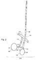
この問題に関係する解剖学的な部分を図1および図2に概略的に描く。図1は、ヒト男性の典型的な精巣静脈および前立腺静脈のドレインシステムを概略的に描いている。精巣104からの1つのドレイン経路はISV102へと至る蔓状静脈叢118を含み、ISV102は一方向弁108を通じてIVC106へとつながっている。通常、弁108は、静脈血が大静脈106へ向けて上方へ流れるのを促進し、かつ、精巣104へ向けて下向きに逆流するのを抑制する。
The anatomical parts involved in this problem are schematically depicted in FIGS. FIG. 1 schematically depicts a typical testicular and prostate venous drainage system for a human male. One drain path from
別のドレイン経路は、精管静脈110へ向かう蔓状静脈叢118、膀胱静脈112、内腸骨静脈114およびIVC106へと至る総腸骨静脈116を含む。後者の経路は、膀胱静脈叢128から膀胱静脈112へと向かい、さらにその先へと通じる前立腺120のドレイン経路により共有されている。
Another drain path includes the
動脈122は、前立腺120の微小循環124および精巣104の微小循環126へ動脈血を供給する。
The
図2は、ヒト男性の正常な左側における典型的な精巣静脈および前立腺静脈のドレイン経路を概略的に描いており、ここで、図中の矢印の方向は上記のような静脈血の流れを示す。ISV102内における一方向弁108が精巣104へ向けて下る逆流を阻むため、それらの弁はそれらの間に位置する個々のセクションの静水圧を孤立させており、それ故、左ISV102への入口142における典型的な圧力は約5〜6mmHgであり、右ISV130への入口144ではそれよりも幾分低くなり得る。
FIG. 2 schematically depicts typical testicular and prostate vein drain pathways in the normal left side of a human male, where the direction of the arrows in the figure indicates the venous blood flow as described above. . Because the one-
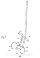
図3は、例えば弁材料の脆弱化、作動による磨り減りまたは老化の影響などの機械的な劣化により、ISV102内の一方向弁が正常に機能していないときの、ヒト男性の左側における典型的な精巣静脈および前立腺静脈のドレイン経路を概略的に描いている。
FIG. 3 shows a typical left side of a human male when the one-way valve in the
ISV102、130内における一方向弁108が精巣104へ向けて下る逆流(逆行する流れ、還流)を阻まないため、ISV102、130は連続的な血液柱を形成し、血液柱内では静水圧が発生し、患者が立っている状態などの直立姿勢にあるときには、左ISV102への入口142では約31mmHg、右ISV130への入口144では約27mmHgまでにもなる(典型的には、普通の状態における標準的な圧力の約4〜6倍)。過度の静水圧、または同程度の大きさの圧力は、ISV102につながっている血管、例えば精管静脈110または蔓状静脈叢118にも存在し得る。なぜなら、その圧力は精巣静脈のドレインシステムから前立腺静脈のドレインシステムへ伝播し、これら両ドレインシステム間において流体力学的に平衡化するためである。圧力は、血管が入口142または144から遠ざかるのに連れて小さくなり得るが、それでも尚、約5mmHg程度の正常範囲よりも高い場合がある。
Since the one-
過度の高圧は、精巣104および蔓状静脈叢118からISV102へと上る静脈血のドレインを抑制する。より正確に表現すれば、この圧力は、遊離テストステロンを豊富(血清レベルよりも約130倍高い)に含む精巣静脈血を膀胱静脈叢128へ向けて、さらにはその先の前立腺120へ向けて押しやり、前立腺からの静脈血のドレインを制限する。
Excessive high pressure inhibits the drainage of venous blood from the
血液が尚も循環するため、精巣からの静脈血は、他の経路、例えば精管静脈110、陰嚢静脈128、またはその過度の圧力によって発生したと考えられるバイパス静脈136などを介して、少なくとも部分的にドレインされる。
Because the blood is still circulated, venous blood from the testis is at least via other routes, such as the
過度の圧力は、以下の少なくとも幾つかの影響をもたらし得る:
(a)方向転換されて前立腺120へと差し向けられ、前立腺120を鬱血および拡大(拡張)させる静脈血。前立腺120の拡張は、少なくとも部分的には、BPHまたは他の前立腺の不具合として発現され得る。
(b)遊離テストステロンを(血液循環における正常レベルの範囲と比較して)豊富に含む精巣106からの静脈血は、前立腺細胞にテストステロンを浴びせ、これにより、BPHをもたらす。遊離テストステロンの約90%が不可逆的にジヒドロテストステロン(DHT)に変換され、DHTは、アンドロゲン受容体に対して、遊離テストステロンよりも約5倍高い親和性を有しており、前立腺細胞の加速された増殖をもたらし得る。精巣104から前立腺120までの通路が短い(約10〜15cm)ため、それらの前立腺の受容体に浴びせられる前にSHBGまたはアルブミンに結合されるのは遊離テストステロンの少量にすぎないことに留意すべきである。
(c)過度の圧力および前立腺の鬱血は、動脈血が前立腺の微小循環124に入るのを抑制または低減し、生物学的なバランスを乱す。前立腺に存在する過剰な量のテストステロンおよびDTHは、前立腺細胞の加速された増殖を誘発し得、癌の発生を促進させ得る。前立腺内における極端な濃度(正常な場合に比べて約100倍またはそれ以上)の遊離テストステロンは、DNAホルモン・フィードバック・システムに負担をかけ過ぎることがあり、それらの加速された細胞分裂において突然変異が生じる確率を増大させ得る。
(d)過度の静脈圧は、動脈血が精巣の微小循環126に入るのを抑制または低減する。血液は少なくともある程度停滞し、酸素を豊富に含んだ細動脈の血液が精巣内に正常に流入することができず、結果として精巣組織における退行変性過程がもたらされ、退行変性過程により、精巣のテストステロンの産生量が減少する。
(e)低下したテストステロン産生能力は、結果として血清中におけるテストステロンの濃度を低減させ、老化の発現または老化症状をもたらし得る。
Excessive pressure can have at least some of the following effects:
(A) Venous blood that is redirected and directed to the
(B) Venous blood from
(C) Excessive pressure and prostate congestion suppress or reduce arterial blood from entering the
(D) Excessive venous pressure inhibits or reduces arterial blood from entering the
(E) A reduced ability to produce testosterone can result in a decrease in the concentration of testosterone in the serum, resulting in the appearance of aging or aging symptoms.
以下の論文は、一般的に精索静脈瘤、男性不妊および男性不妊の治療および/または静脈塞栓症をテーマとしたものである。 The following papers are generally themed on varicocele, male infertility and male infertility treatment and / or venous embolism.

本発明の幾つかの実施形態における1つの態様は、シングルショットの硬化薬を用いて硬化療法を実施する方法を提供することに関する。本発明の1つの実施形態において、シングルショットで硬化療法を実施する方法は、従来のマルチショットによる方法よりも迅速である。本発明の1つの実施形態において、望ましくない静水圧および/または逆流に関与する1つもしくは複数の内精索静脈の少なくとも一部の閉塞により、脈管系における流体の流れが阻止され、または妨げられる。 One aspect in some embodiments of the invention relates to providing a method of performing sclerotherapy using a single shot sclerosing agent. In one embodiment of the invention, the method of performing sclerotherapy with a single shot is faster than the conventional multi-shot method. In one embodiment of the invention, occlusion of at least a portion of one or more internal spermatic veins involved in undesired hydrostatic pressure and / or regurgitation prevents or impedes fluid flow in the vasculature. It is done.
本発明の1つの実施形態では、シングルショットによる硬化療法は、静脈の閉塞させる部分に沿って治療用カテーテルを同時に引き戻しながら、閉塞させる静脈内に治療用カテーテルを用いて硬化薬を注入することにより実施される。場合によっては、または代替的には、望ましくない還流が生じている幾つかの静脈またはすべての静脈が注入/引き戻しによるシングルショット技法を用いて閉塞される。場合によっては、または付加的には、(例えば静水圧の結果として)発生した可能性のあるバイパス静脈は、望ましくない血流を運んでいる場合、注入/引き戻しによるシングルショット技法を用いて閉塞される。本発明の1つの実施形態では、化学硬化剤は、少なくともある程度、分岐静脈および/または分枝静脈を下って流れて、該静脈を閉塞させる傾向を有するため、そうでなければ静脈がバイパス導管として機能する可能性があるとき、または静脈がバイパス導管になる可能性があるときには、そのような化学硬化剤が使用される。 In one embodiment of the present invention, single shot sclerotherapy is achieved by injecting a sclerosing agent into the occluded vein while simultaneously retracting the therapeutic catheter along the occluded portion of the vein. To be implemented. In some cases, or alternatively, some or all veins where undesirable reflux occurs are occluded using a single shot technique with infusion / retraction. In some cases, or in addition, bypass veins that may have occurred (eg, as a result of hydrostatic pressure) are occluded using a single shot technique with infusion / retraction when carrying undesired blood flow. The In one embodiment of the invention, the chemical sclerosing agent has a tendency to flow down and / or occlude the branch vein and / or branch vein at least to some extent, otherwise the vein is the bypass conduit. Such chemical hardeners are used when they are likely to function or when veins can become bypass conduits.
本発明の1つの実施形態では、硬化剤の複数回の「ショット」が実行されるが、全体的な硬化療法手順は、高速な静脈アクセス技法および/または高速な静脈アクセス装置を用いることおよび/または硬化療法の注入手法を行っている間に併用的な一方向運動を使用することおよび/または場合によって閉塞確認を行うことの結果として、従来の硬化療法よりも高速である。 In one embodiment of the present invention, multiple “shots” of the sclerosing agent are performed, but the overall sclerotherapy procedure uses fast venous access techniques and / or fast venous access devices and / or Or faster than conventional sclerotherapy as a result of using combined unidirectional movement and / or optionally performing occlusion confirmation while performing sclerotherapy injection procedures.
本発明の1つの実施形態では、治療用カテーテルには少なくとも遠位側バルーンが設けられており、ここで、遠位側バルーンは治療されている脈管構造物内における流れを少なくとも部分的に妨げ、これにより、流体の流れによって流し去られる硬化薬の量を低減することおよび/または硬化薬が開存した状態のままであるべき静脈を塞いでしまうのを防止することによって、治療部位に注入された硬化薬の有効性を増大させる。本発明の1つの実施形態において、硬化療法の間に少なくとも遠位側バルーンが治療用カテーテルに提供され、治療部位における流体の流れを処理する必要性を排除し、かつ、個人の医学的専門技術レベルに対する治療手順の依存度を低くすることにより、一層標準化された治療プロセスが提供される。 In one embodiment of the invention, the treatment catheter is provided with at least a distal balloon, wherein the distal balloon at least partially impedes flow within the vasculature being treated. Injecting into the treatment site by reducing the amount of sclerosing agent that is washed away by the fluid flow and / or preventing the sclerosing agent from occluding veins that should remain open Increase the effectiveness of the cured sclerosant. In one embodiment of the present invention, at least a distal balloon is provided to the treatment catheter during sclerotherapy, eliminating the need to handle fluid flow at the treatment site, and personal medical expertise By reducing the dependence of the treatment procedure on the level, a more standardized treatment process is provided.
本発明の1つの代替的な実施形態において、シングルショットによる硬化療法は、静脈の治療される領域の対向する端部を塞ぎ、これによってその内部に存在する内腔を画定し、この後、内腔に硬化薬を注入することにより達成される。本発明の幾つかの実施形態において、対向する端部は、鼠径部の近くに配置される遠位側バルーンおよびISVへの入口オリフィス付近に配置される近位側バルーンを備えたタンデム型のバルーンカテーテルにより閉塞される。本発明の幾つかの実施形態では、遠位側バルーンを備えた(但し、近位側バルーンを備えていない)治療用カテーテルが可膨張式のバルーンを備えた誘導用カテーテルと組み合わせて使用され、ここでは、誘導用カテーテルのバルーンがタンデム型バルーンカテーテルの近位側バルーンの形状および/または機能に近い働きをする。本発明の幾つかの実施形態において、治療用カテーテルは、遠位側バルーンと近位側バルーンとの間に硬化薬を注入できるように適合される。場合によっては、治療用カテーテルには、硬化薬の注入用および/または内腔の吸引用の側孔が設けられている。本発明の幾つかの実施形態において、少なくとも1つのポートが治療用カテーテルの遠位側先端部に設けられている。治療用カテーテルの1つのテレスコープ型の実施形態では、カテーテルは適切な位置に留まったままであり、その一方で、弁をわきへ押しやるためおよび/または硬化薬を引き出すために、バルーンがカテーテルの近位側端部に向けて動かされる。 In one alternative embodiment of the present invention, single shot sclerotherapy seals the opposite ends of the treated area of the vein, thereby defining the lumen present therein, after which This is accomplished by injecting a sclerosing agent into the cavity. In some embodiments of the present invention, the opposite end is a tandem balloon with a distal balloon positioned near the groin and a proximal balloon positioned near the inlet orifice to the ISV. Occluded by a catheter. In some embodiments of the present invention, a treatment catheter with a distal balloon (but no proximal balloon) is used in combination with a guide catheter with an inflatable balloon, Here, the balloon of the guiding catheter works close to the shape and / or function of the proximal balloon of the tandem balloon catheter. In some embodiments of the invention, the therapeutic catheter is adapted to inject a sclerosing agent between the distal balloon and the proximal balloon. In some cases, the treatment catheter is provided with side holes for infusion of sclerosants and / or for aspiration of the lumen. In some embodiments of the invention, at least one port is provided at the distal tip of the treatment catheter. In one telescopic embodiment of the therapeutic catheter, the catheter remains in place, while the balloon is near the catheter in order to push the valve aside and / or withdraw the sclerosing agent. It is moved toward the distal end.
本発明の1つの代替的な実施形態において、シングルショットによる脈管構造物の閉塞は、単独で実施されてもよいし、または他の刺激原、例えば血管壁を機械的に刺激して膨張を引き起こし、これにより閉塞をもたらすブラシおよび/またはワイヤなどの助けを借りて実施されてもよい。 In one alternative embodiment of the invention, occlusion of the vasculature with a single shot may be performed alone or mechanically stimulate other irritants, such as the vessel wall, to expand. It may be performed with the help of brushes and / or wires that cause and thereby cause occlusion.
本発明の幾つかの実施形態における1つの態様は、硬化療法を実施できるように適合された遠位側バルーン装備型の治療用カテーテルに関する。本発明の1つの実施形態において、遠位側バルーンは、治療用カテーテル内に設けられたバルーン膨張/収縮用内腔とバルーンの内部領域との間に流体的な接続が存在する状態で側孔を使用することにより、選択的に膨張可能で収縮可能である。本発明の1つの実施形態において、遠位側バルーンは、治療用カテーテルが配置された脈管構造物を通じる流体の流れを実質的に妨害するのに充分な程度に膨張させるように適合される。場合によっては、脈管構造物はISVである。本発明の幾つかの実施形態において、治療用カテーテルはシングルショットによる硬化療法手順において使用できるように適合されており、該治療用カテーテルは、硬化薬の注入およびバルーンが膨張した状態での引き戻しを同時に行うことができる。本発明の幾つかの実施形態では、治療用カテーテルは、小さな静脈スペース、例えば蔓状静脈叢の如き静脈叢の周囲で見られる小さな静脈スペースにおいて使用できるように適合される。 One aspect of some embodiments of the invention relates to a distal balloon-equipped treatment catheter adapted to perform sclerotherapy. In one embodiment of the invention, the distal balloon has a side hole in the presence of a fluid connection between the balloon inflation / deflation lumen provided in the treatment catheter and the interior region of the balloon. Can be selectively inflated and deflated. In one embodiment of the invention, the distal balloon is adapted to be inflated to a degree sufficient to substantially obstruct fluid flow through the vasculature in which the therapeutic catheter is located. . In some cases, the vasculature is an ISV. In some embodiments of the present invention, the treatment catheter is adapted for use in a single shot sclerotherapy procedure, the treatment catheter delivering sclerosant injection and withdrawal with the balloon inflated. Can be done simultaneously. In some embodiments of the invention, the therapeutic catheter is adapted for use in a small venous space, eg, a small venous space found around a venous plexus such as the venous venous plexus.
また、治療用カテーテルは、本発明の幾つかの実施形態において、造影剤および/または硬化薬用の内腔も備えており、該内腔を通じて、造影剤および/または硬化薬が治療用カテーテルを通じて脈管構造物内における治療部位へ送り込まれる。本発明の1つの実施形態において、治療用カテーテルは、遠位側バルーンの近位側(即ち、バルーンから見た下流側)に造影剤および/または硬化薬を注入できるように適合される。場合によっては、造影剤および/または硬化薬用の内腔に吸引力を加えることにより、該内腔を使用して、治療部位の吸引/排気が行われる。 In some embodiments of the present invention, the therapeutic catheter also includes a lumen for the contrast agent and / or sclerosing agent, through which the contrast agent and / or sclerosing agent passes through the therapeutic catheter. It is sent to the treatment site in the tube structure. In one embodiment of the present invention, the therapeutic catheter is adapted to inject contrast and / or sclerosant into the proximal side of the distal balloon (ie, downstream from the balloon). In some cases, the treatment site is aspirated / exhausted using a suction force applied to the contrast agent and / or sclerosant lumen.
本発明の1つの実施形態において、治療用カテーテルはガイドワイヤ用の内腔を備えており、治療用カテーテルを治療部位へ移動させるときには、ガイドワイヤが該内腔を通過する。場合によっては、ガイドワイヤは、治療用カテーテルが治療を行うための適切な位置に達したときに、内腔から引っ込められる。 In one embodiment of the present invention, the treatment catheter includes a guidewire lumen, and the guidewire passes through the lumen when the treatment catheter is moved to the treatment site. In some cases, the guidewire is withdrawn from the lumen when the therapeutic catheter reaches the proper position for performing the treatment.
本発明の幾つかの実施形態において、遠位側バルーン装備型の治療用カテーテルは、弁のすぐ下流でバルーンを膨脹させて真空効果を創出し、弁を開き、治療用カテーテルが弁を通って移動できるようにすることにより、弁突破手順を実施できるように適合される。本発明の1つの実施形態において、バルーンは、弁の通過が終了した後に収縮させられる。 In some embodiments of the present invention, a distal balloon-equipped therapeutic catheter inflates the balloon immediately downstream of the valve to create a vacuum effect and opens the valve so that the therapeutic catheter passes through the valve. By allowing movement, the valve breakthrough procedure is adapted to be performed. In one embodiment of the invention, the balloon is deflated after the passage of the valve is complete.
本発明の幾つかの実施形態では、遠位側バルーン装備型の治療用カテーテルは近位側バルーンも備えており、ここで、遠位側バルーンと近位側バルーンが、それらの間に存在するスペースを画定する。本発明の1つの実施形態において、治療用カテーテルは、注入用の側孔を介して遠位側バルーンと近位側バルーンとの間に硬化薬を注入できるように適合される。本発明の1つの実施形態では、近位側バルーンは、選択的に膨張可能で収縮可能である。 In some embodiments of the present invention, the distal balloon-equipped therapeutic catheter also includes a proximal balloon, where the distal balloon and the proximal balloon are between them. Define a space. In one embodiment of the invention, the therapeutic catheter is adapted to inject a sclerosing agent between the distal balloon and the proximal balloon via the injecting side hole. In one embodiment of the invention, the proximal balloon is selectively inflatable and deflated.
本発明の幾つかの実施形態における1つの態様は、硬化剤を注入する間に治療用カテーテルを動かすことなく近位側バルーンおよび遠位側バルーンを用いてシングルショット硬化療法を実施できるように適合されたタンデム型の治療用カテーテルに関する。本発明の1つの実施形態において、近位側バルーンおよび遠位側バルーンは、治療用カテーテル内に設けられたそれぞれのバルーンの膨張/収縮用内腔とバルーンの内部領域との間に流体的な接続が存在する状態で側孔を使用することにより、選択的に膨張可能で収縮可能である。本発明の1つの実施形態において、遠位側バルーンは、治療用カテーテルが配置された脈管構造物を通じる流体の流れを実質的に妨害するのに充分な程度に膨張させるように適合される。場合によっては、脈管構造物はISVである。本発明の1つの実施形態において、膨脹させたときに、遠位側バルーンおよび近位側バルーンはそれらの間に存在する内腔またはスペースを画定する。本発明の1つの実施形態において、治療用カテーテルは、注入用の側孔を介して遠位側バルーンと近位側バルーンとの間に硬化薬を注入できるように適合される。 One aspect in some embodiments of the present invention is adapted to allow single shot sclerotherapy to be performed using a proximal balloon and a distal balloon without moving the therapeutic catheter during injection of the sclerosing agent. The present invention relates to a tandem type therapeutic catheter. In one embodiment of the invention, the proximal balloon and the distal balloon are fluidic between the respective balloon inflation / deflation lumen provided in the treatment catheter and the balloon interior region. By using side holes in the presence of connections, they can be selectively expanded and contracted. In one embodiment of the invention, the distal balloon is adapted to be inflated to a degree sufficient to substantially obstruct fluid flow through the vasculature in which the therapeutic catheter is located. . In some cases, the vasculature is an ISV. In one embodiment of the invention, when inflated, the distal balloon and the proximal balloon define a lumen or space that exists between them. In one embodiment of the invention, the therapeutic catheter is adapted to inject a sclerosing agent between the distal balloon and the proximal balloon via the injecting side hole.
本発明の1つの実施形態では、タンデム型の治療用カテーテルは、造影剤および/または硬化薬を輸送するための内腔を備えている。場合によっては、バルーン間に存在するスペースは、吸引力を加えることにより、造影剤および/または硬化薬輸送用の内腔を使用して吸引または排気される。本発明の幾つかの実施形態において、各バルーンにはそれぞれの膨張/収縮用内腔が設けられており、これによって、各バルーンを個別に膨張/収縮させることができる。場合によっては、タンデム型の治療用カテーテルはガイドワイヤ用の内腔を備えており、治療用カテーテルを治療部位へ移動させるときには、該内腔を通じてガイドワイヤが通される。場合によっては、ガイドワイヤは、タンデム型治療用カテーテルが治療のための適切な位置に達したときに、内腔から引っ込められる。 In one embodiment of the present invention, a tandem treatment catheter includes a lumen for transporting contrast and / or sclerosing agents. In some cases, the space present between the balloons is aspirated or evacuated using a contrast and / or sclerosant delivery lumen by applying a suction force. In some embodiments of the present invention, each balloon is provided with a respective inflation / deflation lumen, which allows each balloon to be individually inflated / deflated. In some cases, tandem treatment catheters have a guidewire lumen through which the guidewire is passed when the treatment catheter is moved to the treatment site. In some cases, the guidewire is withdrawn from the lumen when the tandem treatment catheter has reached the proper position for treatment.
本発明の幾つかの実施形態における1つの態様は、高速な静脈アクセスを提供できるように適合された、高速な静脈アクセス用の手順および/または装置に関する。本発明の幾つかの実施形態において、予めISVアクセス用に成形されたている誘導用カテーテルが使用される。本発明の幾つかの実施形態において、右ISVへのアクセスを提供できるように適合された誘導用カテーテルには係合用の先端部が設けられており、ここで、係合用先端部は、IVCとISVとが交わる部分における窪みと係合する突起を有する。この窪みは、ISVの弁が存在すると考えられる場所にある。本発明の1つの実施形態において、窪みは、カテーテルを挿入して小刻みに動かしまわすことにより見出される。一旦窪みが見出されると、真空に引き、弁を通じて押し込み、および/または弁が開いた状態になるようにISVを変形させることにより、ISVへのアクセスが得られる。 One aspect in some embodiments of the invention relates to a procedure and / or apparatus for fast venous access that is adapted to provide fast venous access. In some embodiments of the present invention, a guide catheter that is pre-shaped for ISV access is used. In some embodiments of the present invention, a guiding catheter adapted to provide access to the right ISV is provided with an engaging tip, wherein the engaging tip is IVC and It has a protrusion that engages with a recess at a portion where the ISV intersects. This depression is where the ISV valve is believed to be. In one embodiment of the invention, the depression is found by inserting the catheter and moving it in small increments. Once the depression is found, access to the ISV is obtained by pulling a vacuum, pushing through the valve, and / or deforming the ISV so that the valve is open.
本発明の幾つかの実施形態における1つの態様は、少なくとも一部において、カテーテルの遠位側端部(即ち、先端部)に選択的に膨張可能なバルーンを備えた誘導用カテーテルに関する。本発明の1つの実施形態において、バルーンは、バルーンが配置された脈管構造物を通じる流体の流れを実質的に妨害できるように適合される。場合によっては、脈管構造物はISVである。本発明の幾つかの実施形態において、誘導用カテーテルのバルーンは治療用カテーテルの遠位側バルーンと組み合わせて使用され、これら2つのバルーンがそれらの間に存在する内腔を画定する。本発明の1つの実施形態において、2つのバルーンの間に存在するように画定された内腔は硬化療法の治療部位である。本発明の1つの実施形態において、誘導用カテーテルは、ガイドワイヤ、治療用カテーテル、および/またはバルーンの膨張/収縮の少なくとも1つのための複数の内腔を提供できるように適合される。 One aspect of some embodiments of the invention relates to a guiding catheter that includes, at least in part, a balloon that is selectively inflatable at the distal end (ie, tip) of the catheter. In one embodiment of the invention, the balloon is adapted such that it can substantially impede fluid flow through the vascular structure in which the balloon is located. In some cases, the vasculature is an ISV. In some embodiments of the present invention, a guide catheter balloon is used in combination with a therapeutic catheter distal balloon, and the two balloons define a lumen between them. In one embodiment of the invention, the lumen defined to exist between the two balloons is a treatment site for sclerotherapy. In one embodiment of the invention, the guide catheter is adapted to provide a plurality of lumens for at least one of a guidewire, a therapeutic catheter, and / or a balloon inflation / deflation.
本発明の幾つかの実施形態において、少なくとも1つの誘導用カテーテルは、特定の解剖学的特徴と整合するように予め形成されている。場合によっては、解剖学的特徴は右ISVである。また、場合によっては、解剖学的特徴は左ISVである。さらに、場合によっては、解剖学的特徴は右腎静脈である。また、場合によっては、解剖学的特徴は左腎静脈である。 In some embodiments of the present invention, at least one guide catheter is pre-shaped to match a particular anatomical feature. In some cases, the anatomical feature is a right ISV. In some cases, the anatomical feature is the left ISV. Further, in some cases, the anatomical feature is the right renal vein. In some cases, the anatomical feature is the left renal vein.
本発明の幾つかの実施形態における1つの態様は、特定の静脈および/または静脈のオリフィスおよび/または該静脈へのアプローチ角度に合わせて予め成形された誘導用カテーテルを用いて、および/または予め成形された誘導用カテーテルと迅速な弁突破手順とを組み合わせて、迅速な静脈アクセスを得る方法に関係する。本発明の1つの実施形態において、遠位側バルーン装備型の治療用カテーテルが、治療される静脈へ向けて、かつ、該静脈内へ、誘導用カテーテルを通じて進められる。本発明の1つの実施形態において、迅速な弁突破手順は、バイパスされる弁のすぐ下流で治療用カテーテルに設けられたバルーンを膨脹させて真空効果を創出し、弁を開き、弁を通る治療用カテーテルの移動を可能にすることにより達成される。本発明の1つの実施形態において、バルーンは、弁の通過が終了した後に収縮させられる。 One aspect in some embodiments of the present invention is to use a specific vein and / or vein orifice and / or a guide catheter pre-shaped for the approach angle to the vein and / or It relates to a method for obtaining a rapid venous access by combining a shaped guiding catheter and a rapid valve breakthrough procedure. In one embodiment of the present invention, a distal balloon equipped therapeutic catheter is advanced through and into the vein to be treated and into the vein. In one embodiment of the present invention, the rapid valve breakthrough procedure inflates a balloon provided on the treatment catheter just downstream of the bypassed valve to create a vacuum effect, opens the valve, and passes through the valve. Achieved by allowing movement of the catheter. In one embodiment of the invention, the balloon is deflated after the passage of the valve is complete.
本発明の幾つかの実施形態における1つの態様は、血管壁の機械的な刺激を用いて少なくとも一時的な血管の閉塞を引き起こすための刺激装置に関する。 One aspect in some embodiments of the invention relates to a stimulator for causing at least temporary vessel occlusion using mechanical stimulation of a vessel wall.
本発明の1つの実施形態において、刺激装置は、該刺激装置の遠位側端部にブラシ構造物を与えることにより血管の閉塞を引き起こすように適合される。本発明の1つの実施形態において、ブラシ構造物は、パイプクリーナーに似て、装置の遠位側端部から装置の周縁にわたって装置の長さ方向に沿って延びる複数の短い剛毛からなる。本発明の1つの実施形態において、剛毛は可撓性である。場合によっては、剛毛はナイロンなどの生体適合性材料でできている。本発明の1つの実施形態において、剛毛は、血管壁の内側表面を擦るように適合される。本発明の1つの実施形態において、ブラシ刺激装置は、送給用カテーテル内に挿入できるようにおよび/または送給用カテーテルから展開できるように適合されており、送給用カテーテルは、刺激装置がその内部にあるときに血管壁を刺激するのを防止する、刺激装置に対する遮蔽ハウジングを与える。本発明の1つの実施形態において、送給用カテーテルからの展開および血管の閉塞させるセクションに沿ったブラシ刺激装置の引き戻しは単一の動作として実行される。 In one embodiment of the invention, the stimulator is adapted to cause occlusion of the blood vessel by providing a brush structure at the distal end of the stimulator. In one embodiment of the present invention, the brush structure consists of a plurality of short bristles that extend along the length of the device from the distal end of the device to the periphery of the device, similar to a pipe cleaner. In one embodiment of the invention, the bristles are flexible. In some cases, the bristles are made of a biocompatible material such as nylon. In one embodiment of the invention, the bristles are adapted to rub against the inner surface of the vessel wall. In one embodiment of the invention, the brush stimulator is adapted to be inserted into and / or deployable from the delivery catheter, the delivery catheter being adapted by the stimulator. A shielding housing for the stimulator is provided that prevents stimulation of the vessel wall when inside. In one embodiment of the present invention, deployment from the delivery catheter and withdrawal of the brush stimulator along the occluding section of the vessel is performed as a single operation.
本発明の1つの実施形態において、刺激装置は、該刺激装置の遠位側端部に取り付けられたアームアセンブリを用いて血管の閉塞を引き起こすように適合される。本発明の1つの実施形態において、遠位側端部から複数のアームが放射方向および/または縦方向(即ち、装置の軸に沿った方向)に延びている。場合によっては、アームは緩やかに湾曲している。本発明の幾つかの実施形態において、アームは(放射状に)互いに等しい間隔をあけて配列されている。場合によっては、アームは生体適合性の金属材料でできている。場合によっては、アームは生体適合性のプラスチック材料でできている。本発明の1つの実施形態において、アームは、血管の壁に穴をあけることがないように適合される。例えば、アームの端部は丸められている。本発明の幾つかの実施形態において、当該装置のアームは、当該装置から血管壁まで延び、かつ、充分な張力で血管壁の内側表面を擦るように適合される。本発明の1つの実施形態において、アームアセンブリ型の刺激装置は、送給用カテーテル内に挿入できるようにおよび/または送給用カテーテルから展開できるように適合されており、送給用カテーテルは、刺激装置がその内部にあるときに血管壁を刺激するのを防止する、刺激装置に対する遮蔽ハウジングを与える。本発明の1つの実施形態において、送給用カテーテルからの展開および血管の閉塞させるセクションに沿ったアームアセンブリ型刺激装置の引き戻しは単一の動作として実行される。 In one embodiment of the invention, the stimulator is adapted to cause occlusion of the blood vessel using an arm assembly attached to the distal end of the stimulator. In one embodiment of the present invention, a plurality of arms extend radially and / or longitudinally (ie, along the axis of the device) from the distal end. In some cases, the arm is gently curved. In some embodiments of the present invention, the arms are arranged (radially) equally spaced from one another. In some cases, the arm is made of a biocompatible metallic material. In some cases, the arm is made of a biocompatible plastic material. In one embodiment of the present invention, the arm is adapted not to puncture the wall of the blood vessel. For example, the end of the arm is rounded. In some embodiments of the invention, the arm of the device is adapted to extend from the device to the vessel wall and rub against the inner surface of the vessel wall with sufficient tension. In one embodiment of the present invention, the arm assembly type stimulation device is adapted to be inserted into and / or deployable from the delivery catheter, the delivery catheter comprising: A shielding housing for the stimulator is provided that prevents stimulation of the vessel wall when the stimulator is inside. In one embodiment of the invention, deployment from the delivery catheter and withdrawal of the arm assembly stimulator along the occluding section of the blood vessel is performed as a single operation.
従って、本発明の1つの例証的な実施形態により、精巣へのドレインを提供する静脈のネットワークを閉鎖する方法が提供され、該方法は:(a)前記ネットワークの主静脈に治療用カテーテルを挿入するステップ;および、(b)治療用カテーテルの前方への動きを何ら必要としないような様式での静脈刺激原の単回適用により、少なくともある長さにわたって前記静脈を閉鎖するステップ;を含む。 Thus, according to one illustrative embodiment of the present invention, a method is provided for closing a network of veins providing a drain to the testis, the method comprising: (a) inserting a therapeutic catheter into the main vein of the network And (b) closing the vein for at least a length by a single application of the venous stimulator in a manner that does not require any forward movement of the therapeutic catheter.
本発明の1つの実施形態において、単回適用は、硬化剤を適用しながら前記カテーテルを引き戻すステップを含む。本発明の幾つかの実施形態において、カテーテルに設けられた遠位側バルーンが、静脈刺激原の適用に先立って膨脹する。場合によっては、主静脈内における流れを少なくとも部分的に妨害する遠位側バルーンにより、静脈刺激原の有効性が高められる。 In one embodiment of the invention, the single application includes pulling back the catheter while applying a sclerosing agent. In some embodiments of the present invention, the distal balloon provided on the catheter is inflated prior to application of the venous stimulator. In some cases, the effectiveness of the venous stimulator is enhanced by a distal balloon that at least partially obstructs flow in the main vein.
本発明の1つの実施形態において、単回適用は、閉鎖される長さを規定する内腔内に硬化剤を注入するステップを含む。場合によっては、前述の長さの各端部に1つずつある遠位側バルーンおよび近位側バルーンが膨脹し、これにより内腔が画定される。場合によっては、遠位側バルーンおよび近位側バルーンは治療用カテーテルを用いて膨脹させられる。場合によっては、遠位側バルーンは治療用カテーテルを用いて膨脹させられ、一方、近位側バルーンは誘導用カテーテルを用いて膨脹させられる。 In one embodiment of the invention, the single application includes injecting a sclerosant into the lumen defining the length to be closed. In some cases, a distal balloon and a proximal balloon, one at each end of the aforementioned length, are inflated, thereby defining a lumen. In some cases, the distal balloon and the proximal balloon are inflated using a therapeutic catheter. In some cases, the distal balloon is inflated using a therapeutic catheter, while the proximal balloon is inflated using a guiding catheter.
本発明の1つの実施形態において、カテーテルは、カテーテルからネットワーク内に造影剤を注入することによって、静脈のネットワークをマッピングするために使用される。場合によっては、上述の長さは、静脈のネットワークのマップに基づいて閉鎖されるように選択される。 In one embodiment of the present invention, the catheter is used to map a venous network by injecting a contrast agent from the catheter into the network. In some cases, the above length is selected to be closed based on a map of the vein network.
本発明の1つの実施形態において、ネットワークの閉鎖は、BPH、精索静脈瘤、癌、および精巣から前立腺への静脈血の還流の少なくとも1つを治療するために行われる。 In one embodiment of the invention, network closure is performed to treat at least one of BPH, varicocele, cancer, and venous blood return from the testis to the prostate.
本発明の1つの実施形態において、硬化剤は化学剤である。 In one embodiment of the invention, the curing agent is a chemical agent.
本発明の1つの例証的な実施形態により、硬化療法を実施できるように適合された治療用カテーテルがさらに提供され、該治療用カテーテルは:カテーテルの縦方向軸を定めるシャフト;シャフト上に設けられた、選択的に膨張可能で収縮可能な遠位側バルーン;遠位側バルーンと流体連通しており、かつ、遠位側バルーンを選択的に膨張および収縮させるためのバルーン膨張/収縮用の内腔と流体連通する側孔;造影剤および硬化薬用の内腔ならびに治療される静脈の内腔と流体連通する側孔であって、遠位側バルーンから見て内腔における下流に造影剤および硬化薬を注入できるように適合された側孔;を含む。 One illustrative embodiment of the present invention further provides a therapeutic catheter adapted to perform sclerotherapy, the therapeutic catheter comprising: a shaft defining a longitudinal axis of the catheter; provided on the shaft A selectively inflatable and deflatable distal balloon; a balloon inflating / deflating element in fluid communication with the distal balloon and for selectively inflating and deflating the distal balloon; A side hole in fluid communication with the cavity; a side hole in fluid communication with the lumen of the contrast agent and sclerosing agent and the lumen of the vein to be treated; contrast agent and hardening downstream in the lumen as seen from the distal balloon Side holes adapted to allow injection of a drug.
本発明の1つの実施形態において、治療される静脈の内腔の吸引は、造影剤および硬化薬用の内腔を通じて吸引力を加えることにより達成される。 In one embodiment of the invention, aspiration of the lumen of the vein to be treated is achieved by applying suction through the contrast agent and sclerosant lumens.
本発明の1つの実施形態において、治療用カテーテルは、ガイドワイヤに内部を移動させるように適合されたガイドワイヤ用の内腔もさらに含む。 In one embodiment of the invention, the treatment catheter further includes a guidewire lumen adapted to move the guidewire therein.
本発明の1つの実施形態において、治療用カテーテルは、選択的に膨張可能で収縮可能な近位側バルーンもさらに含み、ここで、遠位側バルーンと近位側バルーンが、それらの間で治療される静脈の内腔を画定する。場合によっては、治療用カテーテルは、近位側バルーンと遠位側バルーンとの間にある治療される静脈の内腔内に硬化薬を注入できるように適合される。 In one embodiment of the present invention, the treatment catheter further includes a selectively inflatable and deflatable proximal balloon, wherein the distal balloon and the proximal balloon are treated therebetween. Defining the lumen of the vein to be In some cases, the therapeutic catheter is adapted to inject a sclerosant into the lumen of the vein being treated between the proximal balloon and the distal balloon.
本発明の1つの実施形態において、治療用カテーテルは、シャフトの遠位側端部に設けられ、かつ、ISVのオリフィスと係合できるように適合された係合用先端部をさらに含む。 In one embodiment of the invention, the treatment catheter further includes an engagement tip provided at the distal end of the shaft and adapted to engage the orifice of the ISV.
本発明の1つの例証的な実施形態により、硬化療法を実施できるように適合された治療用カテーテルがさらに提供され、該治療用カテーテルは:選択的に膨張可能で収縮可能な近位側バルーン;ならびに選択的に膨張可能で収縮可能な遠位側バルーン;を含み、近位側バルーンおよび遠位側バルーンが、それらの間で硬化療法により治療される内腔を規定する。 One illustrative embodiment of the present invention further provides a therapeutic catheter adapted to perform sclerotherapy, the therapeutic catheter: a selectively inflatable and deflatable proximal balloon; As well as a selectively inflatable and deflatable distal balloon; the proximal balloon and the distal balloon define a lumen between them to be treated by sclerotherapy.
本発明の1つの実施形態において、治療用カテーテルは、近位側バルーンおよび遠位側バルーンのための専用の膨張/収縮用内腔をさらに含む。本発明の1つの実施形態において、治療用カテーテルは、専用の各膨張/収縮用内腔ならびに近位側バルーンおよび遠位側バルーンと流体連通する側孔をさらに含む。場合によっては、少なくとも1つのバルーンは内腔内における流れを実質的に抑制できるように適合される。場合によっては、内腔はISVの内腔である。 In one embodiment of the invention, the treatment catheter further includes dedicated inflation / deflation lumens for the proximal balloon and the distal balloon. In one embodiment of the present invention, the treatment catheter further includes a dedicated inflation / deflation lumen and side holes in fluid communication with the proximal and distal balloons. In some cases, the at least one balloon is adapted to substantially inhibit flow within the lumen. In some cases, the lumen is an ISV lumen.
本発明の1つの実施形態において、治療用カテーテルは、近位側バルーンと遠位側バルーンとの間にある内腔内に硬化薬を注入できるように適合される。 In one embodiment of the invention, the therapeutic catheter is adapted to inject a sclerosant into a lumen between the proximal balloon and the distal balloon.
本発明の1つの実施形態において、治療用カテーテルは、ガイドワイヤに内部を移動させるように適合されたガイドワイヤ用の内腔をさらに含む。 In one embodiment of the invention, the treatment catheter further includes a guidewire lumen adapted to move the guidewire therein.
本発明の1つの実施形態において、治療用カテーテルは、造影剤および硬化薬の輸送用の内腔もさらに含む。場合によっては、硬化療法により治療される静脈の内腔の吸引は、造影剤および硬化薬用の内腔を通じて吸引力を加えることにより達成される。 In one embodiment of the invention, the therapeutic catheter further includes a lumen for transport of contrast and sclerosing agents. In some cases, aspiration of the lumen of the vein treated by sclerotherapy is accomplished by applying a suction force through the lumen for the contrast agent and sclerosant.
本発明の1つの実施形態において、治療用カテーテルは、シャフトの遠位側端部に設けられ、かつ、ISVのオリフィスと係合できるように適合された係合用先端部をさらに含む。 In one embodiment of the invention, the treatment catheter further includes an engagement tip provided at the distal end of the shaft and adapted to engage the orifice of the ISV.
さらに、本発明の1つの例証的な実施形態により、予め設定された形状を有する誘導用カテーテルが提供され、該誘導用カテーテルは:その内部を通る治療用カテーテルを近位側端部において受け入れるのに適した内腔を画定する細長いチューブ;およびチューブの先端部における少なくとも1つの突出部であって、右ISV弁の凹部と係合できるように適合された突出部;を含む。 Further, according to one illustrative embodiment of the present invention, a guide catheter having a preset shape is provided, the guide catheter comprising: receiving a treatment catheter therethrough at a proximal end. And an elongate tube defining a lumen suitable for the at least one projection at the distal end of the tube, the projection adapted to engage a recess in the right ISV valve.
本発明の1つの実施形態では、誘導用カテーテルは、誘導用カテーテルの遠位側端部に設けられた、選択的に膨張可能で収縮可能なバルーンをさらに含む。場合によっては、バルーンが先端部における少なくとも1つの突出部である。 In one embodiment of the present invention, the guide catheter further includes a selectively inflatable and deflatable balloon provided at the distal end of the guide catheter. In some cases, the balloon is at least one protrusion at the tip.
本発明の1つの実施形態において、チューブは、少なくとも170度の第1の曲がりを定める。 In one embodiment of the invention, the tube defines a first bend of at least 170 degrees.
本発明の1つの実施形態において、誘導用カテーテルは、第1の曲がりとは反対向きの第2の曲がりを含み、第2の曲がりは、右ISVがIVCに入る角度に近づけるように適合されており、かつ、第1の曲がりの遠位側に設けられている。 In one embodiment of the present invention, the guide catheter includes a second bend opposite to the first bend, the second bend adapted to approximate the angle at which the right ISV enters the IVC. And provided on the distal side of the first bend.
場合によっては、カテーテルの長さは600mmから700mmである。 In some cases, the length of the catheter is 600 mm to 700 mm.
本発明の1つの実施形態において、遠位側バルーンは、バルーンが配置される脈管構造物を実質的に閉塞させ、これにより流体の流れを妨害するような様式で適合される。場合によっては、脈管構造物はISVである。 In one embodiment of the invention, the distal balloon is adapted in a manner that substantially occludes the vasculature in which the balloon is located, thereby impeding fluid flow. In some cases, the vasculature is an ISV.
本発明の1つの実施形態において、誘導用カテーテルには、ガイドワイヤ、またはバルーンを選択的に膨張および収縮させるためのエアの少なくとも一方のための内腔が設けられている。 In one embodiment of the present invention, the guide catheter is provided with a lumen for at least one of a guide wire or air for selectively inflating and deflating the balloon.
本発明の1つの実施形態において、誘導用カテーテルは、細長いチューブの遠位側端部に設けられ、かつ、ISVのオリフィスと係合できるように適合された係合用先端部をさらに含む。 In one embodiment of the present invention, the guide catheter further includes an engagement tip provided at the distal end of the elongate tube and adapted to engage the orifice of the ISV.
本発明の1つの実施形態において、選択的に膨張可能で収縮可能なバルーンが、係合用先端部の少なくとも一部である。 In one embodiment of the invention, the selectively inflatable and deflated balloon is at least a portion of the engagement tip.
場合によっては、誘導用カテーテルは可撓性である。 In some cases, the guide catheter is flexible.
本発明の1つの例証的な実施形態により、予め設定された形状を有する誘導用カテーテルがさらに提供され、該誘導用カテーテルは:その内部を通る治療用カテーテルを近位側端部において受け入れるのに適した内腔を画定する細長いチューブ;を含み、細長いチューブの形状は、左腎静脈および左ISV内に配置できるように適合される。本発明の1つの実施形態において、細長いチューブは、左腎静脈がIVCに入る角度に近い70〜130度の第1の曲がりおよび左ISVが左腎静脈に入る角度に近い約100〜125度の第2の曲がりを有する。場合によっては、誘導用カテーテルの長さは650mmである。 According to one illustrative embodiment of the present invention, a guide catheter having a preset shape is further provided, the guide catheter comprising: receiving a treatment catheter therethrough at a proximal end. An elongated tube defining a suitable lumen, the shape of the elongated tube being adapted to be placed in the left renal vein and the left ISV. In one embodiment of the invention, the elongated tube has a first bend of 70-130 degrees close to the angle where the left renal vein enters the IVC and about 100-125 degrees close to the angle where the left ISV enters the left renal vein. Has a second bend. In some cases, the length of the guiding catheter is 650 mm.
本発明の1つの実施形態において、誘導用カテーテルは、誘導用カテーテルの遠位側端部に設けられた、選択的に膨張可能で収縮可能なバルーンをさらに含む。場合によっては、バルーンが先端部における少なくとも1つの突出部である。 In one embodiment of the present invention, the guide catheter further includes a selectively inflatable and deflatable balloon provided at the distal end of the guide catheter. In some cases, the balloon is at least one protrusion at the tip.
本発明の1つの実施形態において、遠位側バルーンは、バルーンが配置される脈管構造物を実質的に閉塞させ、これにより流体の流れを妨害するように適合される。場合によっては、脈管構造物はISVである。 In one embodiment of the invention, the distal balloon is adapted to substantially occlude the vascular structure in which the balloon is placed, thereby impeding fluid flow. In some cases, the vasculature is an ISV.
本発明の1つの実施形態において、誘導用カテーテルには、ガイドワイヤ、またはバルーンを選択的に膨張および収縮させるためのエアの少なくとも一方のための内腔が設けられている。 In one embodiment of the present invention, the guide catheter is provided with a lumen for at least one of a guide wire or air for selectively inflating and deflating the balloon.
本発明の1つの例証的な実施形態により、精巣へドレインする静脈への迅速な静脈アクセスを得るための方法がさらに提供され、該方法は:予め成形された誘導用カテーテルをアクセスポイント内に挿入するステップ;誘導用カテーテルを操作して対象とする解剖学的特徴と対向する場所に配置するステップ;誘導用カテーテルを解剖学的特徴と整合させるステップ;誘導用カテーテルに治療用カテーテルを挿入し、治療用カテーテルを解剖学的特徴の第1の弁まで進めるステップ;第1の弁のすぐ下流で治療用カテーテルの遠位側バルーンを膨脹させ、これにより、弁に真空効果を創出して弁を開かせるステップ;治療用カテーテルを弁を通り越して進めるステップ;および、弁の横断後、バルーンを収縮させるステップ;を含む。 One illustrative embodiment of the present invention further provides a method for obtaining rapid venous access to a vein draining to the testis, the method comprising: inserting a pre-shaped guide catheter into the access point Manipulating the guiding catheter to place it at a location opposite the anatomical feature of interest; aligning the guiding catheter with the anatomical feature; inserting the therapeutic catheter into the guiding catheter; Advancing the treatment catheter to the first valve of the anatomical feature; inflating the distal balloon of the treatment catheter immediately downstream of the first valve, thereby creating a vacuum effect on the valve and opening the valve Opening the treatment catheter; passing the therapeutic catheter past the valve; and deflating the balloon after traversing the valve.
本発明の1つの実施形態において、精巣へドレインする静脈への迅速な静脈アクセスを得るための方法は、治療用カテーテルを治療部位へ向けて進行させるステップをさらに含む。 In one embodiment of the invention, the method for obtaining rapid venous access to the vein draining to the testis further comprises advancing the treatment catheter toward the treatment site.
本発明の1つの実施形態において、精巣へドレインする静脈への迅速な静脈アクセスを得るための方法は、治療部位に到達するまで、膨張ステップ、弁を通り越して治療用カテーテルを進行させるステップ、収縮ステップおよび治療部位へ向けて治療用カテーテルを進行させるステップを反復することをさらに含む。 In one embodiment of the present invention, a method for obtaining rapid venous access to a vein draining to the testis includes an inflation step, advancing the treatment catheter past the valve, deflation until the treatment site is reached. It further includes repeating the steps and advancing the treatment catheter toward the treatment site.
本発明の1つの例証的な実施形態により、刺激装置がさらに提供され、該刺激装置は:シャフト、およびシャフトの遠位側端部に設けられたブラシ構造物;を含み、ブラシ構造物は、血管の内壁を刺激できるように適合される。 According to one illustrative embodiment of the present invention, a stimulator is further provided, the stimulator comprising: a shaft, and a brush structure provided at a distal end of the shaft; It is adapted to stimulate the inner wall of the blood vessel.
本発明の1つの実施形態において、ブラシ構造物は複数の短い剛毛からなる。場合によっては、ブラシ構造物はナイロンで構成されている。場合によっては、ブラシ構造物は可撓性である。 In one embodiment of the invention, the brush structure consists of a plurality of short bristles. In some cases, the brush structure is composed of nylon. In some cases, the brush structure is flexible.
本発明の1つの実施形態において、刺激装置は、送給用カテーテル内に挿入でき、かつ、該送給用カテーテルから展開できるように適合される。 In one embodiment of the invention, the stimulation device is adapted to be inserted into and deployed from the delivery catheter.
さらに、本発明の1つの例証的な実施形態により、刺激装置が提供され、該刺激装置は:シャフト;および、シャフトの遠位側端部に設けられたアームアセンブリ;を含み、アームアセンブリは、血管の内壁を刺激できるように適合される。 In addition, according to one illustrative embodiment of the present invention, a stimulation device is provided, the stimulation device comprising: a shaft; and an arm assembly provided at a distal end of the shaft; It is adapted to stimulate the inner wall of the blood vessel.
本発明の1つの実施形態において、アームアセンブリは複数のアームからなる。場合によっては、複数のアームは、シャフトの遠位側端部から放射方向および縦方向の少なくとも一方に延びている。場合によっては、アームは緩やかに湾曲している。場合によっては、アームは、放射状に、互いに等しい間隔をあけて配列されている。 In one embodiment of the invention, the arm assembly consists of a plurality of arms. In some cases, the plurality of arms extends radially and / or longitudinally from the distal end of the shaft. In some cases, the arm is gently curved. In some cases, the arms are arranged radially and equally spaced from one another.
本発明の1つの実施形態において、アームアセンブリは金属で構成されている。 In one embodiment of the invention, the arm assembly is made of metal.
本発明の1つの実施形態において、アームアセンブリはプラスチックで構成されている。 In one embodiment of the invention, the arm assembly is made of plastic.
本発明の1つの実施形態において、アームは血管壁に穴をあけるのを防ぐように適合される。 In one embodiment of the invention, the arm is adapted to prevent puncturing the vessel wall.
本発明の1つの実施形態において、刺激装置は、送給用カテーテル内に挿入でき、かつ、該送給用カテーテルから展開できるように適合される。 In one embodiment of the invention, the stimulation device is adapted to be inserted into and deployed from the delivery catheter.
本発明の1つの例証的な実施形態により、精索静脈瘤を治療するための方法がさらに提供され、該方法は:(a)患者が静脈叢内に不全弁を有することを決定するステップ;(b)静脈叢にカテーテルを挿入するステップ;(c)カテーテルを用いて硬化薬の単回注入を行うことにより静脈叢を閉鎖し、これにより、精巣に及ぼす静脈圧を低下させるステップ;を含む。 According to one illustrative embodiment of the present invention, there is further provided a method for treating varicocele, wherein: (a) determining that the patient has a failure valve in the venous plexus; (B) inserting a catheter into the venous plexus; (c) closing the venous plexus by performing a single injection of a sclerosing agent using the catheter, thereby reducing venous pressure on the testis; .
本発明の1つの実施形態において、該方法は、(d)造影剤を静脈叢内に注入し、その内部における流体の流れを観察することにより、静脈叢が閉鎖されていることを確認するするステップもさらに含む。 In one embodiment of the invention, the method confirms that the venous plexus is closed by (d) injecting a contrast agent into the venous plexus and observing fluid flow therein. A step is further included.
場合によっては、静脈のネットワークは20〜30分で閉鎖される。 In some cases, the venous network is closed in 20-30 minutes.
場合によっては、静脈のネットワークは30〜45分で閉鎖される。 In some cases, the venous network is closed in 30-45 minutes.
場合によっては、静脈のネットワークは1時間未満で閉鎖される。 In some cases, the venous network is closed in less than an hour.
本発明の1つの例証的な実施形態により、精巣へのドレインを提供する静脈のネットワークを閉鎖する方法がさらに提供され、該方法は:(a)前記ネットワークの主静脈内に治療用カテーテルを挿入するステップ;および(b)治療用カテーテルの引き戻し動作の方向を変えることなく、静脈刺激原を複数回適用することにより、少なくともある長さにわたって前記静脈を閉鎖するステップ;を含む。 In accordance with one illustrative embodiment of the present invention, there is further provided a method of closing a network of veins providing a drain to the testis, the method comprising: (a) inserting a therapeutic catheter into the main vein of the network And (b) closing the vein for at least a length by applying a venous stimulus source multiple times without changing the direction of withdrawal of the therapeutic catheter.
本発明の1つの実施形態において、該方法は、静脈内における弁の後で、かつ、複数回の適用のうちの少なくとも1回の適用のすぐ前に、停止するステップをさらに含む。場合によっては、引き戻し動作が、カテーテルが弁を通過する際の弁の平坦化を引き起こす。 In one embodiment of the invention, the method further comprises stopping after the valve in the vein and immediately before at least one of the multiple applications. In some cases, the pull back action causes flattening of the valve as the catheter passes through the valve.
さらに、本発明の1つの例証的な実施形態により、硬化療法を実行するためのキットが提供され、該キットは:少なくとも1つの治療用カテーテル;および、治療用カテーテルがその内部を通過できるように適合された、予め成形された少なくとも1つの誘導用カテーテル;を含む。 Further, according to one illustrative embodiment of the present invention, a kit for performing sclerotherapy is provided, the kit comprising: at least one therapeutic catheter; and allowing the therapeutic catheter to pass therethrough. Adapted, pre-shaped at least one guide catheter.
本発明の1つの実施形態において、キットは静脈用シースをさらに含む。 In one embodiment of the invention, the kit further comprises a venous sheath.
本発明の1つの実施形態において、キットは、治療用カテーテルとともに用いるための硬化薬をさらに含む。 In one embodiment of the invention, the kit further comprises a sclerosing agent for use with a therapeutic catheter.
本発明の1つの実施形態において、キットは造影剤をさらに含む。 In one embodiment of the invention, the kit further comprises a contrast agent.
本発明の1つの実施形態において、キットは、治療用カテーテル、予め成形された誘導用カテーテル、静脈用シース、硬化薬または造影剤の少なくとも1つを使用するための使用説明書をさらに含む。 In one embodiment of the invention, the kit further comprises instructions for using at least one of a therapeutic catheter, a pre-shaped guide catheter, a venous sheath, a sclerosing agent or a contrast agent.
別途定義されない限り、本明細書で使用されるすべての技術的用語および/または科学的用語は、本発明が属する技術分野の当業者によって一般に理解されるのと同じ意味を有する。本明細書に記載される方法および材料と類似または同等である方法および材料を本発明の実施形態の実施または試験において使用することができるが、例示的な方法および/または材料が下記に記載される。矛盾する場合には、定義を含めて、本特許明細書が優先する。加えて、材料、方法および実施例は例示にすぎず、必ずしも限定であることは意図されない。 Unless defined otherwise, all technical and / or scientific terms used herein have the same meaning as commonly understood by one of ordinary skill in the art to which this invention belongs. Although methods and materials similar or equivalent to those described herein can be used in the practice or testing of embodiments of the present invention, exemplary methods and / or materials are described below. The In case of conflict, the patent specification, including definitions, will control. In addition, the materials, methods, and examples are illustrative only and not intended to be limiting.
本明細書では本発明のいくつかの実施形態を単に例示し添付の図面を参照して説明する。特に詳細に図面を参照して、示されている詳細が例示として本発明の実施形態を例示考察することだけを目的としていることを強調するものである。この点について、図面について行う説明によって、本発明の実施形態を実施する方法は当業者には明らかになるであろう。ヒトの解剖学的構造の左側の図解および名称はまた、特に指示がない限り右側にもあてはまる。
本明細書および特許請求項における「左側」および「右側」という用語は、通常の解剖学的用語法に則っている(例えば、心臓、胃および脾臓は、殆どの人間の場合、左側に存在する)。本明細書および特許請求項における「ドレイン」という用語は、静脈血管を介する、静脈血の大静脈へ向けての流れ、および大静脈内への流れを表し、また、「還流」および「逆流」という用語は、「ドレイン」の反対方向の流れについての同意語として使用される。解剖学的特徴は各個人によって様々であり得ることに留意すべきであり、また、本明細書に記載の方法および/または装置は広範囲にわたる様々な解剖学的構成での使用が意図されていることにも留意すべきである。 The terms “left side” and “right side” in this description and in the claims follow normal anatomical terminology (eg, the heart, stomach and spleen are on the left side in most humans) ). The term “drain” in the present description and claims refers to the flow of venous blood towards and into the vena cava via the venous blood vessels, and “return” and “backflow”. Is used as a synonym for the flow in the opposite direction of the “drain”. It should be noted that anatomical features may vary from individual to individual, and the methods and / or devices described herein are intended for use in a wide variety of anatomical configurations. It should also be noted.
本発明の幾つかの例証的な実施形態において、上記の好ましくない状態および影響の1つもしくはそれ以上を、過度の圧力を低減または排除することにより、少なくともある程度まで、回避、遅延、緩和および/または修復できる。圧力を下げることにより、精索静脈瘤、BPH、不妊症、幾つかの形態の癌および/または精巣から前立腺へのテストステロンを(血液循環における正常なレベルと比較して)豊富に含む静脈血の逆流(還流)を低減または排除できる。 In some exemplary embodiments of the invention, one or more of the above undesirable conditions and effects may be avoided, delayed, mitigated and / or at least to some extent by reducing or eliminating excessive pressure. Or it can be repaired. By reducing the pressure, varicocele, BPH, infertility, some forms of cancer and / or testosterone from the testes to the prostate (compared to normal levels in the blood circulation) Backflow (reflux) can be reduced or eliminated.
本明細書での見出しは制限的なものではなく、明瞭にすることだけを意図したものである。 The headings herein are not limiting and are intended to be clear only.
シングルショットによる硬化療法手順
米国特許出願第11/826283号および米国特許出願第61/064511号で示すように、還流は、過度の静水圧に影響を及ぼしていた左ISV102および/または右ISV130の(例えば塞栓形成または硬化を使用する)閉塞により防止または阻害できる。場合によってはおよび/または付加的には、還流が生じている幾つかの静脈またはすべての静脈、例えば精管静脈110および蔓状静脈叢118などが閉塞される。場合によっては、または付加的には、発生した可能性のあるバイパス静脈136も閉塞される。本発明の1つの実施形態において、左ISV102が右ISV130の前に治療される。しかしながら、そうではなく、右ISV130を最初に治療してもよいことに留意すべきである。
Single shot sclerotherapy procedure As shown in US patent application Ser. No. 11 / 826,283 and US patent application Ser. No. 61/064511, refluxing of left and
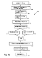
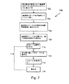
図4Aを参照すると、本発明の1つの例証的な実施形態による、シングルショットでの硬化療法手順を説明するフローチャート400が示される。本発明の1つの例証的な実施形態において、「シングルショット」という用語は、従来では硬化薬の多数回の離散的な注入と、それに伴う治療用カテーテルの多数回の離散的な移動とを必要としていた、静脈のある長さの閉塞を、そうではなく、治療用カテーテルの単回の連続的な移動を伴った(または、図4Bとの関連において記載するように、治療用カテーテルを全く移動させずに)、単回の注入ステップを用いることにより実施することを意味する。
Referring to FIG. 4A, a
シングルショット法を用いることの1つの利点は、精索静脈瘤、BPHおよび前立腺への血液の還流などの疾患に対して硬化療法による治療を行う際に通常必要となる時間が短縮されることである。本発明の1つの実施形態において、シングルショット手順は20〜30分で実施される。本発明の幾つかの実施形態において、シングルショット手順は30〜45分で実施される。場合によっては、該シングルショット手順は1時間未満で実施される。各患者のニーズが異なっており、それらのニーズおよび/または生体構造および/または病状が様々であることにより、担当する医療専門家の関与が必要となる時間のレベルは様々に異なることに留意すべきである。本発明の幾つかの実施形態において、シングルショット手順は、硬化療法を実施するのにかかる全体的な時間の長さを短縮するため、以下に記載する高速な静脈アクセス手順と組み合わされる。 One advantage of using the single shot method is that it usually reduces the time required to treat sclerotherapy for diseases such as varicocele, BPH, and blood return to the prostate. is there. In one embodiment of the invention, the single shot procedure is performed in 20-30 minutes. In some embodiments of the invention, the single shot procedure is performed in 30-45 minutes. In some cases, the single shot procedure is performed in less than one hour. Note that each patient has different needs, and the level of time required for the involvement of his / her health care professional varies with their needs and / or anatomy and / or medical condition. Should. In some embodiments of the present invention, the single shot procedure is combined with the fast venous access procedure described below to reduce the overall length of time it takes to perform sclerotherapy.
注入される硬化薬の量は、本発明の幾つかの実施形態において、閉塞の有効性を最大化させるように、注入用カテーテルの移動速度に合わせられる。直径の範囲が1mm〜4mmであると仮定すると、治療される血管は、本発明の幾つかの例証的な実施形態によれば、直径2mmの静脈の長さ1cmのセグメントに対して0.3mlの体積の硬化薬が注入されてよく、また、直径3mmの静脈の長さ1cmのセグメントに対しては0.7mlの硬化薬が注入されてよく、さらに、直径4mmの静脈の長さ1cmのセグメントに対しては1.3mlの硬化薬が注入されてよい。精索静脈瘤を治療する場合には、治療される血管の直径が1mm〜4mmよりも大きい可能性があり、その場合には、本明細書に記載の方法および/または装置がこれらのより大きな直径のスペース内において使用できるように適合され得る。本明細書に記載の幾つかの動作またはすべての動作(例えば装置の挿入/引き戻し、バルーンの膨張/収縮、材料の注入/吸引など)は、制御装置を用いてオペレータの指令により行われる様式および/または制御装置を用いて自動的に行われる様式のいずれかで、制御装置により実施されてよいことを理解すべきである。 The amount of sclerosant injected is matched to the rate of movement of the infusion catheter to maximize the effectiveness of the occlusion in some embodiments of the invention. Assuming a diameter range of 1 mm to 4 mm, the treated blood vessel is 0.3 ml for a 1 cm long segment of a 2 mm diameter vein according to some illustrative embodiments of the invention. Volume of sclerosing agent may be injected, and 0.7 ml of sclerosing agent may be injected for a 1 cm long segment of a 3 mm diameter vein, and a 1 cm long vein of 4 mm diameter. For the segment, 1.3 ml of sclerosing agent may be injected. When treating varicocele, the diameter of the vessel being treated can be greater than 1 mm to 4 mm, in which case the methods and / or devices described herein are larger than these It can be adapted for use in a space of diameter. Some or all of the operations described herein (eg, device insertion / retraction, balloon inflation / deflation, material infusion / suction, etc.) are performed in accordance with operator commands using the controller and It should be understood that it may be implemented by the controller in any of the manners that are automatically performed using the controller.
本発明の1つの実施形態において、標的ISV102、130内へ治療装置を進入させるように患者の準備を整えるための準備措置が講じられる(402)。本発明の1つの実施形態において、大腿静脈の経皮的アクセスを得ることが望ましい。本発明の幾つかの実施形態において、右大腿静脈が使用される。場合によっては、左大腿静脈が使用される。大腿静脈穿刺部位の定位は、場合によっては:上前腸骨棘から恥骨結合までのラインまたはそのラインの下側における;理学的検査によって確証される。場合によっては局所麻酔が穿刺部位の周りに施され、また、場合によっては任意にシースされた大腿穿刺針を用いて静脈を穿刺しながら大腿静脈を拡張するためにヴァルサルヴァ操作が実施される。場合によっては、大腿穿刺針は少なくとも18ゲージである。
In one embodiment of the present invention, preparatory measures are taken to prepare the patient to enter the treatment device into the
本発明の幾つかの実施形態において、大腿静脈用シースおよび2つの予め成形された誘導用カテーテル500、550の少なくとも一方を伴った同軸システムが使用され、それらの誘導用カテーテルは、図5A〜5Dおよび図7に示し、かつ、それらの図面との関連においてより詳しく記載するように、一方のカテーテル500は左ISV102内に進入するのに適した形状に成されており、一方のカテーテル550は右ISV130内に進入するのに適した形状に成されている。場合によっては、この手順は、6フレンチのシースおよび6フレンチの予め成形された誘導用カテーテル500、550を用いて実施され、誘導用カテーテル500、550を通じて、図8D〜8Eに示し、かつ、それらの図との関連においてより詳しく記載する治療用カテーテル850が配置される。場合によっては、治療用カテーテル850は3フレンチである。本発明の幾つかの実施形態において、それらがその内部を通る治療用カテーテル850の通過を許容することを条件として、より小さいまたはより大きいシースおよび/または誘導用カテーテルを使用することもできる。場合によっては、そのような治療用カテーテルが、使用される何らかのシースおよび/または誘導用カテーテルを通過するのに適しており、および/またはその患者の解剖学的特徴の治療に適したサイズおよび/またはその患者の解剖学的特徴を通って移動するのに適したサイズに成されていることを条件として、より小さいまたはより大きい治療用カテーテルが使用される。
In some embodiments of the invention, a coaxial system with a femoral vein sheath and at least one of two preformed
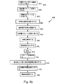
静脈穿刺は、場合によっては、静脈内の深い位置に施される付加的な局所麻酔を可能にするために2壁穿刺として実施される。本発明の1つの実施形態において、針を通じてガイドワイヤが静脈内に挿入され、腎盂内に進められる。場合によっては、ガイドワイヤは0.014〜0.018インチのガイドワイヤである。本発明の幾つかの実施形態において、ガイドワイヤは、ISVのオリフィスと直接的に係合するように使用できるように適合された、可変的に湾曲したおよび/または軟質のおよび/または可撓性の係合用先端部を備えている。付加的に、任意におよび/または代替的に、治療用カテーテルが、ISVのオリフィスと直接的に係合するように使用できるように適合された、可変的に湾曲したおよび/または軟質のおよび/または可撓性の係合用先端部を備えている。本発明の1つの実施形態において、係合用先端部は、技能および/または感触および/または画像化を用いて、担当する医療専門家により操作されてオリフィス内に導入される。針またはシースは、穿刺部位を圧迫しながら、ガイドワイヤの周りで引き戻される。本発明の1つの実施形態において、シースは、シースのサイドポートをヘパリン添加生理食塩水で洗浄しながら、その外部ポートが鼠径部に位置する状態で一時的にガイドワイヤ上に配置される。場合によっては、この操作が一定の間隔でこの手順を行っている間中ずっと反復される。場合によっては、その間隔は5〜10分の間隔である。 Venous puncture is sometimes performed as a two-wall puncture to allow additional local anesthesia applied deep into the vein. In one embodiment of the invention, a guide wire is inserted into the vein through the needle and advanced into the renal pelvis. In some cases, the guidewire is a 0.014-0.018 inch guidewire. In some embodiments of the present invention, the guidewire is variably curved and / or soft and / or flexible adapted to be used to directly engage an ISV orifice. The tip part for engagement is provided. Additionally, optionally and / or alternatively, the therapeutic catheter is variably curved and / or soft and / or adapted to be used for direct engagement with an ISV orifice. Alternatively, a flexible engaging tip is provided. In one embodiment of the invention, the engagement tip is manipulated and introduced into the orifice by a responsible medical professional using skill and / or feel and / or imaging. The needle or sheath is pulled back around the guide wire while compressing the puncture site. In one embodiment of the present invention, the sheath is temporarily placed on the guide wire with its external port positioned at the groin while washing the side port of the sheath with heparinized saline. In some cases, this operation is repeated throughout the procedure at regular intervals. In some cases, the interval is 5 to 10 minutes.
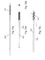
本発明の1つの実施形態において、予め成形された誘導用カテーテル500、550の少なくとも一方がガイドワイヤに沿ってIVC106内へ進められる。誘導用カテーテル500、550を適切なISV102、130へ誘導するために、ISV102、130のどちらが標的となっているのかおよび/またはその患者に特有の生体構造に依存して、場合によっては、僅かに異なる手順が用いられる。本発明の1つの実施形態において、治療用カテーテル850は、場合によっては、図8Eに示すガイドワイヤ用の内腔864を備えており、治療用カテーテル850がガイドワイヤを追随することによって進められる場合には、ガイドワイヤ用の内腔864にガイドワイヤが通される。本発明の1つの実施形態において、ガイドワイヤは、入口ポート854を使用して治療用カテーテル850内に送られる。
In one embodiment of the present invention, at least one of the
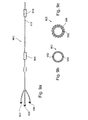
本発明の1つの実施形態において、誘導用カテーテル500は、図6Aでより詳しく示すように、蛍光透視検査法の制御下で左腎静脈150(ISV102を治療する場合)のオリフィス154内へ操作によって導入される(404)。それらの場合の約5%では、左腎静脈とIVC106との接合は、腎臓から尾側方向または頭部方向を向いた鈍角である。そのような場合、ISV102のオリフィス158が鋭角で左腎静脈150に接合しており、これにより、上向きに傾いた腎静脈150から進入するのが難しくなっておりおよび/または上向きに傾いた腎静脈150から見出すのが難しくなっている。それらの場合の別の2%〜3%では、ISV102が、傍大動脈静脈とともに、または傍大動脈静脈のすぐ下側で腎静脈150に接合する。これらの場合には、誘導用カテーテル500は、それを操作して先端部502がオリフィス158に重なるようにする必要性があり、本発明の1つの例証的な実施形態によれば、一時的な呼吸停止、蛍光透視検査台の傾斜、および穏やかにではあるがしっかりとしたカテーテル500の回転の組み合わせを用いて、ISV102のオリフィス158が係合される。本発明の幾つかの実施形態において、オリフィス158は、治療用カテーテル850の軟質先端部またはその可撓性ガイドワイヤによって丁度に係合される。場合によっては、ISV102のオリフィス158および/またはISV130のオリフィス132と係合するために、代替的な先端部(例えば副腎静脈または脊髄動脈との関連において使用される先端部)を伴った誘導用カテーテル500が使用される。本発明の1つの実施形態において、先端部は、以下に記載する係合用先端部584である。
In one embodiment of the present invention, guide
典型的には、ISV102(および/またはISV130)内には、正常な動作の下では、ISV102を下って精巣104へ向けて戻る血液の還流を防止する一連の一方向弁が所在する。弁は、患者/台の位置決め、呼吸法、および誘導用カテーテルを介して加えられる吸引力の組み合わせにより通過することができる(406)。弁を容易に通過できるときには、場合によっては、治療用カテーテル850が、適所に配置されたガイドワイヤを用いて、またはそのようなガイドワイヤを用いることなく、導入される。治療用カテーテル850は、本発明の1つの実施形態によれば、通常は鼠径靭帯のすぐ上である、硬化療法(即ち、閉塞)に関して最も低い所望のポイントへ操作によって誘導される。本発明の1つの実施形態において、硬化療法に望ましい最も低いポイントは、陰嚢内への還流が生じるリスクおよび/または静脈の流れの適切な別の経路としての二次的な静脈の利用可能性などを含めた数多くの因子の要因分析をして選択される。場合によっては、最も低い所望のポイントは、鼠径靭帯のすぐ上よりも高い位置である。
Within ISV 102 (and / or ISV 130) are typically a series of one-way valves that prevent the return of blood down
本発明の1つの実施形態において、ISV102ならびにその連通支流および側副支流の生体構造を確証するように、蛍光透視検査用の傾斜台を適切に操作して、遠位側と近位側との両方のISV102の静脈造影画像を得るため、蛍光透視検査法の制御下で、静脈内造影剤が治療用カテーテル850内に注入される。本発明の1つの実施形態において、造影剤および/または硬化薬は、造影剤および/または硬化薬用の内腔866を介して治療用カテーテル850を通過する。本発明の1つの実施形態において、これは、場合によっては、側副ISV支流とさらにはISV102と他の後腹膜静脈との間での相互接続性との両方をマッピングする(408)ために使用される。
In one embodiment of the present invention, the fluoroscopic tilt table is appropriately manipulated to ensure that the
本発明の幾つかの実施形態において、腎被膜静脈、尿管静脈および傍脊椎後腹膜静脈などの他の重要な静脈に対する明白な相互接続性が存在する場合には、これらの血管およびそれらの血管の相互接続に注意が払われ、ISVの一般的な硬化療法による閉塞処置に移る前に、これらの相互につながっている静脈をISVの治療によって閉塞させるかどうか、または、局所化された硬化療法および/または他の閉塞技法を用いることによって、別々に閉塞させることおよび/またはISVから別々に分断することが必要かどうかが決定される。 In some embodiments of the present invention, these vessels and their vessels if there is obvious interconnectivity to other important veins such as renal capsule veins, ureteral veins and paravertebral retroperitoneal veins. Whether or not these interconnected veins should be occluded by ISV therapy, or localized sclerotherapy, before attention is paid to the interconnection of ISVs and moving to occlusion treatment with ISV general sclerotherapy And / or by using other occlusion techniques, it is determined whether it is necessary to occlude separately and / or separate from the ISV.
本発明の幾つかの実施形態において、そうでなければ静脈がバイパス導管として機能する可能性があるときに、化学硬化剤は、ISVからの分岐静脈および/または分枝静脈へ少なくともある程度下って流れて該静脈を閉塞させる傾向を有するため、化学硬化剤が選択される。それ故、化学的硬化薬を使用する方法は、化学的硬化薬が分枝静脈および/または分岐静脈および/または接続する静脈内に少なくともある程度の距離だけ流入し、かつ、特には該静脈を閉鎖させてしっかりと留めるのに充分な程度に該静脈を刺激することによって閉塞させることが予想されるため、該静脈を別々に閉塞させる必要性を排除できる。本発明の幾つかの実施形態において、何れの分枝静脈も閉塞されない。本発明の幾つかの実施形態において、数個までの分枝静脈がISVと同時に閉塞される。場合によっては、数個またはそれ以上の分枝静脈がISVと同時に閉塞される。本発明の幾つかの実施形態において、分枝静脈を閉塞させるために機械的な栓および/またはセメントが使用される。 In some embodiments of the present invention, the chemical sclerosant flows at least partially down from the ISV to the branch and / or branch vein when the vein may otherwise function as a bypass conduit. The chemical sclerosing agent is selected because it has a tendency to occlude the vein. Therefore, the method of using a chemical sclerosing agent is that the chemical sclerosing agent flows at least some distance into the branch vein and / or the branch vein and / or the connecting vein and in particular closes the vein Since it is anticipated that the vein will be occluded by stimulating the vein to a sufficient extent to allow it to be securely clamped, the need to occlude the vein separately can be eliminated. In some embodiments of the invention, none of the branch veins are occluded. In some embodiments of the invention, up to several branch veins are occluded simultaneously with ISV. In some cases, several or more branch veins are occluded simultaneously with the ISV. In some embodiments of the invention, mechanical plugs and / or cement are used to occlude branch veins.
相互に接続する血管を別々に閉塞させる必要がある場合(例えば、ISVに化学的な硬化薬を使用しても相互接続する血管を効果的に閉塞させることができないと考えられる場合)には、本発明の1つの実施形態において、この処置は、場合によっては、主ISV102およびその分枝静脈に取り掛かる前に行われる。治療用カテーテルの先端部852が閉塞させる血管内に配置され、非常に少量の硬化薬混合物(場合によっては0.5ml未満)が血管内に適用される。治療用カテーテルの先端部852は、望ましい治療効果をもたらすのに充分であるように決定された時間の間、適切な位置に留められる;本発明の1つの実施形態において、その時間は2〜4分間である。望ましい治療効果をもたらすのに充分な時間が経過した後、カテーテルを取り除くことができ、また、場合によっては、本発明の1つの実施形態によれば、分枝静脈の閉塞を確認するために、少量のヨード造影剤をISV内に注入することもできる。本発明の1つの実施形態において、患者が直立姿勢または立っている状態のときに造影剤が前立腺に流れていないことを確かめるために患者がモニタリングされる。
When it is necessary to occlude interconnecting blood vessels separately (for example, when it is thought that the use of a chemical sclerosing agent for ISV cannot effectively occlude interconnecting blood vessels) In one embodiment of the invention, this procedure is optionally performed prior to working on the
代替的に、相互に接続する静脈が2〜3mmよりも大きいときには、場合によっては、相互に接続する静脈を閉塞させるために、血管内マイクロコイルまたは当技術分野において公知の他の閉塞装置が使用される。血管を閉塞させるための他の技法も使用することができ、そのような技法としては、限定されないが、生物学的に適合性を有する接着剤、高周波駆動カテーテルを用いる局部加熱、コイルを用いる加熱、冷凍アブレーション、集束超音波および/または当技術分野において公知のあらゆる血管閉鎖方法が挙げられる。 Alternatively, when the interconnecting veins are larger than 2-3 mm, in some cases, an intravascular microcoil or other occlusive device known in the art is used to occlude the interconnecting veins Is done. Other techniques for occluding blood vessels can also be used, including but not limited to biologically compatible adhesives, local heating using a radio frequency driven catheter, heating using a coil. , Cryoablation, focused ultrasound and / or any vascular closure method known in the art.
本発明の1つの実施形態において、患者の関連する生体構造のマッピングステップ(408)が終了すると、治療は、ISV102の少なくとも一部の閉塞処置を開始する。ISV102の硬化療法による治療は、鼠径靭帯のすぐ上から、ISV102が左腎静脈150につながっているISVオリフィス158から数センチメートル以内にまで延びるISV102のある長さを閉塞させることを含み;従って、本発明の幾つかの実施形態において、治療の開始時に、治療用カテーテル850が鼠径靭帯のすぐ上に配置される。担当する医療専門家の意見および/または患者のニーズに依存して、ISV102のより短い領域またはより長い領域が閉塞され得ることを理解すべきである。さらに、本明細書の別の箇所で記載するように、本発明の幾つかの実施形態によれば、治療を成功裏に終わらせるために、ISV102の他に別の血管構造物も閉塞させることが必要になり得る。
In one embodiment of the invention, upon completion of the patient's associated anatomy mapping step (408), the therapy begins an occlusion procedure for at least a portion of the ISV. Treatment of
本発明の1つの実施形態において、ISV102の少なくとも一部の閉塞は、硬化薬が下向きに還流するのを防ぐためおよび/または静脈の血流で硬化薬が上向きに押し流されるのを防ぐため、鼠径部においてISV102を圧迫(410)しながら行われる、ISV102内における硬化薬の静脈内適用によって実施される。本発明の1つの実施形態において、自動外部圧迫装置が患者の適切な場所に配置され、制御装置を用いてオペレータにより作動され、または制御装置自体により自動的に作動される。
In one embodiment of the present invention, the occlusion of at least a portion of the
本発明の幾つかの実施形態において、物理的圧迫ステップ(410)は、他の技法を用いることにより、例えば注入ステップ(414)(以下に記載する注入ステップ(414))中に治療用カテーテルに吸引力を加えることなどにより、増強されおよび/または取って代わられる。本発明の1つの実施形態において、吸引および注入は、治療用カテーテルに設けられた別々のポートを介して実施される。本発明の幾つかの実施形態において、圧迫ステップ(410)は、無害な流体、例えば生理食塩水などを、硬化薬が注入(414)されている治療部位から見て上流に注入し、これにより、陰嚢から離れてゆく流れの方向とともに静脈を横断する圧力勾配を創出することによって増強されおよび/または取って代わられる。場合によっては、無害な流体によって及ぼされる圧力は、還流を防ぐのには充分であるが、硬化薬を治療部位から駆出させるほどには強くない。本発明の幾つかの実施形態において、患者は、還流の回避に役立たせるように重力を使用するTrendelenburg位に置かれる。本発明の幾つかの実施形態において、手作業による圧迫および/または外部止血帯が硬化薬の流れを導くために使用される。 In some embodiments of the invention, the physical compression step (410) may be performed on the therapeutic catheter by using other techniques, for example, during the injection step (414) (the injection step (414) described below). It is enhanced and / or replaced, such as by applying a suction force. In one embodiment of the invention, aspiration and infusion are performed through separate ports provided on the treatment catheter. In some embodiments of the invention, the compression step (410) injects a harmless fluid, such as saline, upstream from the treatment site where the sclerosing agent is being injected (414), thereby It is augmented and / or replaced by creating a pressure gradient across the vein with the direction of flow away from the scrotum. In some cases, the pressure exerted by the innocuous fluid is sufficient to prevent reflux but not so strong as to drive the sclerosing drug out of the treatment site. In some embodiments of the invention, the patient is placed in the Trendelenburg position using gravity to help avoid reflux. In some embodiments of the invention, manual compression and / or external tourniquets are used to direct sclerosant flow.
本発明の1つの実施形態において、圧迫の有効性を試験し、かつ、陰嚢に向けた流れが抑制されていることを確認するため、鼠径部が圧迫された後に、造影剤が治療用カテーテル850内に注入される。本発明の1つの実施形態において、陰嚢に向けた流れが殆ど防止されていることが明らかになるまで、鼠径部の圧迫が調節される。 In one embodiment of the invention, the contrast agent is treated with a therapeutic catheter after the groin has been compressed to test the effectiveness of the compression and to confirm that the flow toward the scrotum is suppressed. Injected into 850. In one embodiment of the invention, groin pressure is adjusted until it is clear that flow towards the scrotum is largely prevented.
陰嚢に向けた流れの防止が達成されていることが一旦確認される(412)と、閉塞させるISV102の予め決定された長さに沿って治療用カテーテルの先端部852を引き戻しながら(416)、治療用カテーテル850を介して、硬化薬混合物がISV102内にゆっくりと注入される(414)。このような様式で、閉塞させるISV102のセクションに沿った治療用カテーテル850のスムーズな連続的引き戻し動作を随伴した硬化薬の単回注入が、硬化療法を実施するための「シングルショット」手順を提供する。本発明の幾つかの実施形態において、硬化薬の注入(414)用および/または以下に記載する流動性物質の吸引(420)用に、少なくとも1つのポートが治療用カテーテルの遠位側先端部に設けられている。
Once it has been confirmed that flow prevention towards the scrotum has been achieved (412), the
本発明の幾つかの実施形態において、治療用カテーテル850は、ISV102内において合計で約20〜25cm引き戻される。本発明の幾つかの実施形態において、ISVのサイズに依存して、4mlまでの硬化剤と、0.25〜0.5mlの2%局所麻酔薬、例えばリドカインとの混合物が作られ、3〜5ml用のプラスチック製シリンジ内で撹拌される。本発明の幾つかの実施形態において、硬化薬を患者に注入するのに先立って泡を形成するため、ストップコック配列を用いて混合物をシリンジからシリンジへ移すことができる。
In some embodiments of the present invention, the
本発明の幾つかの実施形態において、予め定められた注入体積と引き戻し速度との比に従って治療を実施できるように、注入する(414)際に硬化薬が計量されおよび/または引き戻し(416)の速度が測定される。場合によっては、ポジションインジケータが治療用カテーテルのシャフトの近位側端部に設けられ、これにより、制御装置および/または担当する医療専門家は、治療用カテーテルを引き戻すときにインジケータをモニタリングすることが可能になる。本発明の幾つかの実施形態において、治療の間に使用される静脈の単位長さ当たりまたは横断直径当たりの硬化薬の量は前もって決定されている。本発明の幾つかの実施形態において、治療用カテーテル850には、注入ステップ(414)用に、予め計量された用量の硬化薬および/または上記のものなどの硬化薬混合物が事前に負荷されている。
In some embodiments of the invention, the sclerosing agent is metered and / or withdrawn (416) during infusion (414) so that treatment can be performed according to a predetermined ratio of infusion volume to withdrawal rate. The speed is measured. In some cases, a position indicator is provided at the proximal end of the shaft of the treatment catheter so that the controller and / or medical professional can monitor the indicator when pulling back the treatment catheter. It becomes possible. In some embodiments of the invention, the amount of sclerosing agent per unit length or transverse diameter of the vein used during treatment is predetermined. In some embodiments of the invention, the
本発明の1つの実施形態において、引き戻しステップ(416)および/または注入ステップ(414)は、硬化療法手順で使用されるカテーテルに対して動きを与えるようにおよび/または他の操作(例えば、硬化薬を注入することなど)を行わせるように適合された装置によって自動的に実施される。場合によっては、装置は、制御装置によって自動的に作動される。場合によっては、担当する医療専門家がその装置を制御する。本発明の1つの実施形態において、制御装置には、種々の硬化薬の投与量および/またはタイプ、ならびに効果的な硬化療法による治療を果たす上で求められる硬化薬の対応する移動速度がプログラムされている。 In one embodiment of the present invention, the pull back step (416) and / or the infusion step (414) may provide movement and / or other manipulation (eg, sclerosis) for the catheter used in the sclerotherapy procedure. Automatically performed by a device adapted to perform the injection). In some cases, the device is automatically activated by the controller. In some cases, the medical professional in charge controls the device. In one embodiment of the present invention, the controller is programmed with various sclerosing agent dosages and / or types, and the corresponding rate of movement of the sclerosing agent required to effect treatment with effective sclerotherapy. ing.
注入された硬化薬は、場合によっては、最大の刺激効果を得ることができるように、ある時間の間、治療領域に留められる(418)。場合によっては、硬化薬が効果を発揮できるようにするために、5〜10分間経過させる。本発明の1つの実施形態において、患者が載せられる蛍光透視検査台は僅かに傾けられる。場合によっては、台は「足を下側にする」状態に位置決めされる。場合によっては、台は、水平な状態から10度まで回転される。本発明の1つの実施形態において、注入してから5〜10分までの間、台は水平な位置に維持される。本発明の1つの実施形態において、台が水平な位置に維持されている時間は任意の休止ステップ(418)の長さに一致する。 The injected sclerosing agent is sometimes left in the treatment area for a period of time (418) so that the maximum stimulation effect can be obtained. In some cases, 5-10 minutes are allowed to elapse to allow the sclerosing agent to exert its effect. In one embodiment of the invention, the fluoroscopy table on which the patient is placed is tilted slightly. In some cases, the platform is positioned in a “foot down” state. In some cases, the platform is rotated from a horizontal state to 10 degrees. In one embodiment of the invention, the platform is maintained in a horizontal position for up to 5-10 minutes after injection. In one embodiment of the invention, the time that the platform is maintained in a horizontal position corresponds to the length of any pause step (418).
本発明の1つの実施形態によれば、硬化薬の注入ステップ(412)および硬化薬が効果を発揮できるようにするための任意の休止ステップ(418)の後、治療される静脈の内容物は、吸引力を加えることにより、治療用カテーテル850の造影剤および/または硬化薬用の内腔866を通じて吸引され(420)、場合によっては廃棄される。本発明の1つの実施形態において、鼠径部におけるISV102の圧迫が解放され(422)、治療用カテーテル850が少量の生理食塩水および/または静脈内造影剤で洗浄される。本発明の1つの実施形態において、閉塞部位の上方に造影剤を注入し、治療された領域を観察することにより、ISV102の閉塞が確認される(424)。本発明の幾つかの実施形態において、確認ステップ(424)は任意である。場合によっては、確認ステップ(424)の間、患者は半直立姿勢(20〜50度)に置かれる。
According to one embodiment of the present invention, after the sclerosing agent injection step (412) and the optional resting step (418) to allow the sclerosing agent to take effect, the contents of the vein to be treated are By applying a suction force, it is aspirated (420) through the contrast agent and / or
本発明の1つの実施形態において、ISV102が成功裏に閉塞されていない場合、治療用カテーテル850の第2の通過が実施され、付加的な治療を必要とするISV102のセグメント内にカテーテル850の先端部852が移動させられる。ISV102が閉塞されていることが明らかになるまで、最初の注入ステップで実施された幾つかの動作またはすべての動作が反復される。本発明の幾つかの実施形態において、確認ステップ(424)は実施されず、ISVが成功裏に閉塞されていない場合および/または分枝静脈が拡張されている場合には、しばらくした後、この手順が再び実施される。場合によっては、この「しばらく」の期間は、約数日、数週間または数カ月くらいの期間である。
In one embodiment of the present invention, if the
本発明の1つの実施形態において、ISV102が一旦成功裏に閉塞されると、誘導用カテーテル500が無菌生理食塩水溶液で洗浄される。場合によっては、ISV102が閉塞される前には見えなかった可能性のある、ISV102に注いでいる可能性のある側副静脈を探すための診断用静脈造影図を得る目的で、左腎静脈150内への造影剤の注入が行われる。本発明の幾つかの実施形態において、ISV102の閉塞後に現れたあらゆる側副静脈は、閉塞の可能性について注意が払われる。本発明の1つの実施形態において、腹部領域における該注意が払われた静脈は、治療されおよび/または閉塞される(陰嚢内の静脈は閉塞されない)。
In one embodiment of the invention, once the
本発明の幾つかの実施形態において、患者の病状を治療するため、右ISV130も閉塞される。本発明の1つの実施形態において、右腎静脈152およびそれに付属する領域の診断用静脈造影図が取得される。場合によっては、カテーテル500を操作して左腎静脈150から右腎静脈152へ導入し、カテーテル500を通じて造影剤を注入することにより、誘導用カテーテル500が使用される。本発明の1つの実施形態において、左側に位置していた誘導用カテーテル500は、静脈造影法による実証を目的として右腎静脈152に進入させるのに適しており、かつ、既に患者の内部に存在するため、診断用静脈造影図を収集するために使用される。本発明の幾つかの実施形態において、右ISV130が通常の解剖学的な場所に所在するかどうかを見るため、誘導用カテーテル500を用いて、右ISV130に対する予備調査が実施される。誘導用カテーテル500が静脈造影図を得るために使用される場合、該カテーテルは右側用の誘導用カテーテル550と交換され、右側用の誘導用カテーテル550は、右腎静脈152および/または右ISV130との関連において使用できるように適合される。右側用の誘導用カテーテル550を図5Bに示し、かつ、図5Bとの関連においてより詳しく記載する。
In some embodiments of the invention, the
右側の特異性のため、ISV130とIVC106との接合部を同定するのが難しい場合がある。実際、ISV130とIVC106との間には多数の接合部が存在することがあり、また、幾つかの場合には、ISV130は、IVC106ではなく右腎静脈152と交わっていることもある。これに加え、ISV130への入口オリフィス132には弁が存在することが考えられ、これにより、カテーテルでISV130に進入するのが一層難しくなっている。本発明の1つの実施形態において、台の位置決め(例えば、「足を上側にする」Trendelenburg位)および/または呼吸法を使用することが助けになり得る。図6Bにより詳しく示すように、右ISV130が誘導用カテーテル550によって一旦進入させられると、ISV102で実施されたのと同様なやり方で、治療用カテーテル850がISV130内に進められる。本発明の1つの実施形態において、ISV130に対して行われるこの治療の残りの部分は、左ISV102で行われたのと同じやり方で実施される。右側用の誘導用カテーテル550は、オリフィス132の近くまたはオリフィス132の位置に適切に機能する弁が存在する場合に、右ISV130のオリフィス132への進入を容易化するために、その先端部552にバルブを備えている。
Due to the specificity on the right, it may be difficult to identify the junction between
治療の終わりに、右ISV130が閉塞されているように思われ、かつ、右ISV130の側副的な注ぎ込み(「バイパス」)が見られないときには、治療用カテーテル850が引き戻されて、誘導用カテーテル550が右ISVのオリフィス132からそれを取り外すように上向きに押し入れられる。本発明の幾つかの実施形態において、操作して先端部552を左腎静脈150内に導入するか、または下降させて左内腸骨静脈のオリフィス内に導入するかのいずれかにより、誘導用カテーテル550が曲げられ、また、真っ直ぐにされる。誘導用カテーテル550および大腿静脈用シースが取り除かれ、また、本発明の幾つかの実施形態では、出血を低減し、かつ、患者の回復を開始させるため、穿刺部位が圧迫される。
At the end of treatment, when the
本明細書の別の箇所で検討されているように、治療用カテーテル850を用いるシングルショットによる方法は、精索静脈瘤、BPH、幾つかの形態の癌および/または精巣から前立腺へのテストステロンが(血液循環における正常なレベルと比較して)豊富な静脈血の逆流(還流)を治療するために適用できる。さらに、幾つかの動作は、個々の患者のニーズおよび/または担当する医療専門家の意見に依存して、1回より多く実施されてよくおよび/または任意であってよい。
As discussed elsewhere in this specification, single shot methods using a
代替的なカテーテルおよび技法を用いるシングルショットによる硬化療法
図4Bは、単独で、または他の装置と組み合わせて、静脈内のある領域を、その領域の2つの対向する端部を閉塞させることによって治療されるものと規定する、少なくとも1つの装置を用いるシングルショットによる硬化療法手順のフローチャート450を示す。本発明の幾つかの実施形態において、治療領域を規定するために、タンデム型のバルーン配置が提供される。タンデム型のバルーン配置は、単一の装置によって提供されてもよいし、または複数の装置の組み合わせによって提供されてもよく、少なくとも近位側バルーン(即ち、ISVおよびIVC/腎静脈が交わる部分の近くに配置されるバルーン)および遠位側バルーン(即ち、IVC/腎静脈から離れた位置で、鼠径部により近い位置に設けられるバルーン)が、それらの間にある内腔を治療されるものと規定するために与えられていることを理解すべきである。
Single shot sclerotherapy using alternative catheters and techniques FIG. 4B alone or in combination with other devices treats a region in the vein by occluding two opposing ends of the region. FIG. 46 shows a
本発明の幾つかの実施形態において、閉塞される脈管構造物へ硬化薬を送給するために、タンデム型のバルーンを備えた治療用カテーテル900が使用される。タンデム型のバルーンを備えた治療用カテーテル900を図9A〜9Cにより詳しく示す。場合によっては、タンデム型のカテーテル900は3Fのカテーテルである。本発明の1つの実施形態において、右ISV130の前に左ISV102が治療される。しかしながら、そうではなく、右ISV130を最初に治療してもよいことに留意すべきである。
In some embodiments of the present invention, a
本発明の1つの実施形態において、近位側バルーン908および遠位側バルーン910は、図9Cに示す如く、治療用カテーテル900内に設けられたそれぞれのバルーンの膨張/収縮用内腔912、914とバルーン908、910の内部との間に流体接続が存在する状態で側孔を使用することにより、選択的に膨張可能で収縮可能である。本発明の1つの実施形態において、バルーン908、910は、内部に治療用カテーテルが配置された脈管構造物を通じる流体の流れを実質的に妨げるのに充分な程度にまで膨張できるように適合される。場合によっては、脈管構造物はISV102、130である。本発明の1つの実施形態において、遠位側バルーン910および近位側バルーン908は、膨脹したときに、それらの間で内腔またはスペースを画定する。本発明の1つの実施形態において、治療用カテーテル900は、注入用の側孔916を介して、遠位側バルーン910と近位側バルーン908との間に硬化薬を注入できるように適合される。
In one embodiment of the present invention, the
本発明の1つの実施形態において、タンデム型の治療用カテーテル900には、造影剤および/または硬化薬輸送用の内腔が設けられている。場合によっては、それらのバルーンの間に存在するスペースは、吸引力を加えることにより、造影剤および/または硬化薬輸送用の内腔を使用して、側孔916を通じて吸引または排気される。本発明の幾つかの実施形態において、各バルーン908、910には、図9Bに示すように、それら自身の膨張/収縮用の内腔912、914が設けられており、これにより、各バルーン908、910を個別に膨張および/または収縮させることができる。場合によっては、タンデム型の治療用カテーテル900はガイドワイヤ用の内腔を備えており、治療用カテーテルを治療部位へ移動させるときには、ガイドワイヤ用内腔を通じてガイドワイヤを通すことができる。場合によっては、ガイドワイヤは、タンデム型の治療用カテーテル900が治療のための適切な位置に到達したときに、内腔から引っ込められる。
In one embodiment of the present invention, the tandem
治療用カテーテルが遠位側バルーン、例えば遠位側バルーン910を備えている場合の本発明の幾つかの実施形態において、硬化療法手順は、フローチャート400に描く準備ステップ(402)から通過ステップ(406)までと実質的に同様な様式で始まる。本発明の1つの実施形態において、任意のマッピングステップ(460)の始めに、遠位側バルーン910が、ISV102の壁に近づくように、緩やかに膨脹する(458)。本発明の幾つかの実施形態において、遠位側バルーン910は、治療されている脈管構造物を治療領域の下側で閉塞させ、物質が陰嚢に向かって逆方向に還流するのを防止するために使用される。場合によっては、遠位側バルーン910は、ISV102を不透明化するようにヨード造影剤を注入した後に、蛍光透視観察下において膨脹する。場合によっては、遠位側バルーン910を、希薄ヨード造影剤(1mlの造影剤対3〜5mlの無菌生理食塩水の比)を用いて膨脹させる。本発明の幾つかの実施形態において、少なくとも1つの側孔914が遠位側バルーン910内に物質を注入するため(例えば、遠位側バルーン910を膨脹させるため)に治療用カテーテル900に設けられており、また、少なくとも1つの側孔916が、閉塞させる脈管構造物内に造影剤および/または硬化薬を注入するために設けられている。
In some embodiments of the present invention where the treatment catheter comprises a distal balloon, eg, a
本発明の1つの実施形態において、閉塞される脈管構造物、例えばISVに圧迫ステップ(462)が適用され、遠位側バルーン910を膨脹させた後、(腎静脈へ向かって延びる)遠位側バルーン910の上方でISV内に残存する血液が、合理的に可能な限り最良な状態で吸引される。本発明の1つの実施形態において、膨脹した遠位側バルーン910はISVを充分に閉塞させることができる。場合によっては、硬化薬が陰嚢内に還流するのを防止するため、手作業による圧迫(462)が維持される。遠位側バルーン910によってもたらされた閉塞の有効性を試験するため、場合によっては、少量の造影剤が、少なくとも1つの側孔916を介して、閉塞部位の上方におけるISVの内腔内に注入される。静脈内造影剤の注入は、鼠径部より下の静脈内への(陰嚢に向けた)造影剤の還流が存在しないことを確認する(464)ために用いられ、これにより、遠位側バルーン910が存在する部位より下側のISVが完全に隔離されているのが確認される。
In one embodiment of the invention, a compression step (462) is applied to an occluded vasculature, such as an ISV, and the
ISVが遠位側バルーン910によって満足できるほどに閉塞されていると判断された場合には、本発明の1つの実施形態において、鼠径部を通る造影剤が存在しなくなるまで、遠位側バルーン910を配置し直しおよび/もしくは膨張し直し、ならびに/または脚の付け根のデジタル式の圧迫が調節される。
If it is determined that the ISV is satisfactorily occluded by the
本発明の1つの実施形態において、治療される脈管構造物を閉塞させるために治療用カテーテル900の近位側バルーン908を膨脹させ(466)、これにより、2つの膨脹したバルーン910、908の間に、静脈内における内腔が創出され、該内腔は、完全にではないにしても、実質的に流体の流れから隔離されている。本発明の1つの実施形態において、2つの膨脹したバルーン910、908によって画定された内腔が閉塞される静脈のセグメントを表す。本発明の1つの実施形態において、近位側バルーン908および遠位側バルーン910の膨張を確認した後、タンデム型バルーンカテーテル900の側孔916を使用して、内腔が可能な限り合理的に空になるまで、血管の内腔を吸引する(468)。本発明の1つの実施形態において、少なくとも1つの側孔916を介して、治療される内腔に硬化薬が注入される(470)。本発明の1つの実施形態において、内腔に注入される硬化薬の量は、患者のニーズ、および/または使用される硬化薬および/または硬化薬混合物の個々の特性に合わせられる。場合によっては、3〜4mlの硬化薬混合物が内腔に注入される。
In one embodiment of the invention, the
本発明の1つの実施形態において、硬化薬が効果を発揮できるようにするための任意の休止ステップ(472)の後、バルーン910、908が収縮させられ(474)、処置された静脈の内容物がカテーテル900を通じて吸引される(476)。任意の休止ステップは、硬化薬混合物の適用後、5分間から10分間続く;しかしながら、休止時間は、使用される特定の硬化薬の特性に合わせることができる。本発明の1つの実施形態において、鼠径部の圧迫が取り除かれおよび/または作動が停止される(478)。 In one embodiment of the present invention, after an optional pause step (472) to allow the sclerosing agent to take effect, balloons 910, 908 are deflated (474) and the treated vein contents. Is aspirated through the catheter 900 (476). The optional pause step lasts 5 to 10 minutes after application of the sclerosing agent mixture; however, the quiescence time can be tailored to the characteristics of the particular sclerosing agent used. In one embodiment of the invention, groin pressure is removed and / or deactivated (478).
本発明の1つの実施形態において、治療用カテーテル900が少量の生理食塩水および/または静脈内造影剤で洗浄される。本発明の1つの実施形態では、場合によっては、閉塞部位の上方に造影剤を注入し、処置された領域を観察することにより静脈の閉塞が確認される(480)。場合によっては、確認ステップ(480)の間、患者は半直立姿勢(20〜50度)に置かれる。当然のことながら、処置された静脈が閉塞されている場合には、造影剤が閉塞された領域を越えて直接的に横断することはないであろう(しかしながら、分枝静脈を介して閉塞領域を間接的に越える流れが存在することはあり得る)。
In one embodiment of the invention, the
本発明の1つの実施形態において、静脈が閉塞されていない場合、治療用カテーテル900を通す2度目の操作が行われ、蛍光透視検査下においてカテーテルの先端部をISV102の遠位腎盂部分内に配置し直し、治療用カテーテル900を通じて静脈の内容物を吸引した後、上記のようにして、治療用カテーテル900の遠位側バルーン910および近位側バルーン908を再度膨脹させる。ISVの閉塞を果たすために注入ステップが繰り返される。
In one embodiment of the invention, if the vein is not occluded, a second manipulation through the
しかしながら、本発明の1つの実施形態において、ISV102が最初の処置後またはそれ以降の処置後に部分的に閉塞されていて、かつ、治療用カテーテル900が部分的にしか再延長することができない場合には、バルーン910、908の一方または両方を収縮させ、治療用カテーテル900が、位置決めステップ用および/または注入ステップ用に、治療用カテーテル850と同様に操作される。治療される静脈が成功裏に閉塞されているものと判断されるまで、この処置が続けられる。
However, in one embodiment of the present invention, the
本発明の幾つかの実施形態において、確認ステップ(480)は実施されず、ISVが成功裏に閉塞されていない場合および/または部分的に閉塞されている場合および/または分枝静脈が拡張されている場合には、しばらくした後、この手順が再び実施される。場合によっては、この「しばらく」の期間は、約数日、数週間または数カ月くらいの期間である。 In some embodiments of the invention, the confirmation step (480) is not performed and the ISV is not successfully occluded and / or partially occluded and / or the branch vein is dilated. If this is the case, after a while, this procedure is performed again. In some cases, this “for a while” period is a period of about several days, weeks or months.
治療される静脈の閉塞が達成されると、本発明の1つの実施形態において、治療用カテーテル900が取り除かれる。誘導用カテーテル500が無菌の生理食塩水溶液で洗浄され、主ISV102が閉塞される前には見えなかった可能性のある、ISV102に注いでいる側副静脈を探すため、半直立姿勢での左腎静脈150への造影剤注入が行われる。直接的または間接的な左ISV102への注ぎ込みが存在しないものと判断されたときには、右ISV130が、本明細書の別の箇所で左ISV130との関連において記載するのと基本的に同じ技法(但し、タンデム型のバルーンを備えた治療用カテーテル900が使用されることを考慮に入れた上で)に従って閉塞される。
Once the occlusion of the vein to be treated is achieved, in one embodiment of the invention, the
本発明の1つの実施形態において、単一の遠位側バルーン810を備えた治療用カテーテル800が、静脈のシングルショットによる治療を果たすために使用される。1つの例証的な治療用カテーテル800を図8A〜8Cに示す。治療用カテーテル900を使用することと治療用カテーテル800を使用することとの違いは、治療用カテーテル900の場合には閉鎖された内腔に硬化薬を注いでいたのに対し、治療用カテーテル800は、その代わりに、治療用カテーテル850の場合と同様に、腎静脈へ向けて引き戻されているときに、治療される静脈内に硬化薬を放出することである。場合によっては、遠位側バルーン810は、カテーテル800の遠位側端部820から1〜2cmまでの場所に配置される。本発明の1つの実施形態において、遠位側バルーン810は、カテーテル850が引き戻されているときに、ISV内に存在する弁108を開き、側方へ押しやるために使用される。本発明の幾つかの実施形態において、これらの弁108の幾つかの弁またはすべての弁がこの硬化療法により閉鎖される。本発明の幾つかの実施形態において、これらの弁108の幾つかの弁またはすべての弁が損傷を受けない。
In one embodiment of the invention, a
本発明の1つの実施形態において、遠位側バルーン810は、治療用カテーテル800内に設けられたバルーン膨張/収縮用内腔812およびバルーンの内部領域が流体接続する状態で側孔818を使用することにより、選択的に膨張可能で収縮可能である。本発明の1つの実施形態において、膨張および/または収縮用のエアが、エアインプット/アウトプットポート802を通過する。本発明の1つの実施形態において、遠位側バルーン810は、内部に治療用カテーテル800が配置される脈管構造物を通じる流体の流れを実質的に妨げるのに充分な程度に膨張できるように適合される。場合によっては、脈管構造物はISVである。本発明の幾つかの実施形態において、治療用カテーテル800はシングルショットによる硬化療法手順において使用できるように適合されており、治療用カテーテル800は、硬化薬の注入と膨脹状態のバルーン810を伴った引き戻しとを同時に行うことができる。
In one embodiment of the invention, the
図8Bに示すように、本発明の幾つかの実施形態において、治療用カテーテル800は造影剤および/または硬化薬用の内腔816も備えており、内腔を通じて造影剤および/または硬化薬が脈管構造物内における治療部位へ治療用カテーテル800を通って送られる。本発明の1つの実施形態において、造影剤および/または硬化薬は、造影剤および/または硬化薬用のインプット/アウトプットポート804を通過する。本発明の1つの実施形態において、治療用カテーテル800は、遠位側バルーン810の近位側(即ち、バルーン810から見て下流側)に造影剤および/または硬化薬を注入できるように適合される。場合によっては、内腔に吸引力を加えることにより、造影剤および/または硬化薬用の内腔816を使用して、治療部位の吸引/排気が行われる。
As shown in FIG. 8B, in some embodiments of the present invention, the
本発明の1つの実施形態において、治療用カテーテルは、治療用カテーテル800を治療部位へ移動させるときに、ガイドワイヤが内部に通されるガイドワイヤ用の内腔814を備えている。場合によっては、ガイドワイヤは、治療用カテーテル800が治療のための適切な位置に到達したときに、内腔814から引っ込められる。本発明の1つの実施形態において、ガイドワイヤは、ガイドワイヤ用のインプット/アウトプットポート806を通じて出入りする。
In one embodiment of the present invention, the treatment catheter includes a
本発明の幾つかの実施形態において、遠位側バルーンを備えた治療用カテーテル800は、弁108のすぐ下流でバルーン810を膨らませて真空効果を創出し、弁108を開き、これにより、その内部を通る治療用カテーテル800の移動を可能にすることによって弁突破手順を実施できるように適合される。本発明の1つの実施形態において、バルーン810は、カテーテルが弁108を通過した後に収縮させられる。
In some embodiments of the present invention, the
場合によっては、治療用カテーテル800または治療用カテーテル850と共に、先端部にバルーンを備えた誘導用カテーテル570、580が使用され、そのような左側用のカテーテルおよび右側用のカテーテルの例を、それぞれ、図5Cおよび5Dに示す。1つの例として、先端部にバルーンを備えた左側用の誘導用カテーテル570および治療用カテーテル800が使用される本発明の1つの実施形態を用いて説明すると、先端部にバルーンを備えた誘導用カテーテル570のバルーン572が膨脹し(466)、静脈を処置する間、治療用カテーテル900の近位側バルーン908の機能に近い働きをさせるように使用される。
In some cases, guide
本発明の1つの実施形態において、先端部にバルーンを備えた誘導用カテーテル570のバルーン572は、誘導用カテーテル570の先端部574から約4〜8mmの場所に配置される。本発明の幾つかの実施形態において、バルーン572は、膨脹したときに、3〜5mmの直径を有するが、場合によっては、治療部位の状態に依存して、様々な異なるサイズが使用される。場合によっては、バルーン572は3〜5mmの「長さ」であり、カテーテル570に沿って、その先端部574付近で3〜5mm延びている。本発明の幾つかの実施形態において、先端部にバルーンを備えた誘導用カテーテル570の全体的な長さおよび湾曲の角度は、先端部にバルーンを備えていない誘導用カテーテル500の場合と同様である。
In one embodiment of the present invention, the
先端部にバルーンを備えた誘導用カテーテル570が通常の治療用カテーテル850と共に用いられる本発明の1つの実施形態において、バルーンは、治療用カテーテル850がバルーンに向けて引き込まれている間にISVを閉塞させるために使用される。硬化薬の注入ステップが終了すると、治療用カテーテル850により、残存する血液および硬化薬の吸引ステップ(420)が実施される。本発明の幾つかの実施形態において、左側用の誘導用カテーテル500は先端部にバルーンを備えるように適合される。本発明の幾つかの実施形態において、右側用の誘導用カテーテル550は先端部にバルーンを備えるように適合される。本発明の幾つかの実施形態において、誘導用カテーテル570は4〜6フレンチである。本発明の幾つかの実施形態において、バルーン572は、3〜6mmの直径にまで膨脹し、誘導用カテーテル570の先端部574から1〜2cmまでで誘導用カテーテル570に沿って1〜3cm延びる。
In one embodiment of the present invention in which a
本発明の幾つかの実施形態において、先端部にバルーンを備えた右側用の誘導用カテーテル580およびそれに付随するバルーン582の使用および/または機能に関する同様な方法も想定されていることに留意すべきである。左側用の誘導用カテーテル570と右側用の誘導用カテーテル580との主要な違いはそれらのカテーテルの形状(それぞれのカテーテルは、各カテーテルのそれぞれの側における生体構造と調和するように予め成形されている)であり、また、本発明の幾つかの実施形態では、右側用の誘導用カテーテル580(および、場合によっては、右側用の誘導用カテーテル550)は、右ISVのオリフィス132において誘導用カテーテル580を右ISV130に「ラッチ」するための係合用先端部584を備えるように適合される。本発明の1つの実施形態において、係合用先端部584は、本発明の幾つかの実施形態ではIVCの壁に係合するために先端部の上側に隆起部を必要とするため、非対称である。場合によっては、係合用先端部584は、バルブ状の形状、卵形の形状またはボール状の形状からなる。場合によっては、係合用先端部584は、フックおよび/またはかかりなどの突起部を含む。本発明の幾つかの実施形態において、係合用先端部584の長さは、1mmから数mmまでである。本発明の幾つかの実施形態において、係合用先端部584の直径は、0.5mm〜2mmである。場合によっては、係合用先端部584は剛性である。場合によっては、係合用先端部584は可撓性である。本発明の幾つかの実施形態において、先端部にバルーンを備えた誘導用カテーテル580の全体的な長さおよび湾曲の角度は、本明細書の別の箇所で記載する、先端部にバルーンを備えていない誘導用カテーテル550の場合と同様である。
It should be noted that in some embodiments of the present invention, similar methods for the use and / or function of the
本発明の1つの実施形態において、係合用先端部584はバルーン582を含む。バルーン582が係合用に使用される本発明の1つの実施形態において、バルーンは、オリフィス132付近で約3mm〜4mmにまで膨脹する。誘導用カテーテル580は、使用する際には、オリフィス132の上方の壁部と係合することを目的として、(バルーンを膨脹させた状態で)上向きに穏やかに押し入れられ、このようにして、開口が拡げられ、かつ、オリフィス132において弁108も開かれ、これにより、治療用カテーテルをISV内に挿入することが可能になる。この後、バルーンは収縮させられて、誘導用カテーテル580がオリフィス132内に進められ、それらの装置を安定化させ、近位側での血流の制御を可能にする。
In one embodiment of the invention, the engagement tip 584 includes a
本発明の幾つかの実施形態において、硬化療法を実施するためにテレスコープ型の治療用カテーテルが使用される。本発明の1つの実施形態において、テレスコープ型のカテーテルは、カテーテル800、850、900のようにISVを下ってカテーテルを押しやることにより治療部位へ向けて進められることはない。そうではなく、テレスコープ型のカテーテルは、治療部位にまでは至らないが、カテーテルの遠位側部分を折り畳んだ状態でISV内に配置される。例えば流体を用いて、カテーテルに内部圧力を加えると、折り畳まれた状態の遠位側部分が治療される血管内で展開され、少なくとも治療部位と同程度にまで血管内をはるかに下って展開する。場合によっては、遠位側の折り畳み部分を展開するために用いられる流体は、血管を治療するために使用される硬化薬である。場合によっては、流体は生理食塩水である。本発明の1つの実施形態において、折り畳まれた部分は、展開されたときに、25cmまでの長さを有する。場合によっては、折り畳まれた部分は、展開されたときに、10cmまでの長さを有する。本発明の1つの実施形態において、折り畳まれた部分は、展開されたときに、6mmまでの直径を有する。本発明の1つの実施形態において、テレスコープ型のチャンネルに、流体を流すための少なくとも1つのチャンネルが設けられている。場合によっては、チャンネルは遠位側の折り畳まれた部分の折り畳まれた材料に設けられており、流体がテレスコープ型のカテーテルを通じて適用されたときに、流体が遠位側の折り畳まれた部分を拡げる作用をする。
In some embodiments of the invention, a telescopic treatment catheter is used to perform sclerotherapy. In one embodiment of the invention, the telescopic catheter is not advanced toward the treatment site by pushing the catheter down the ISV as in
本明細書の別の箇所で検討するように、治療用カテーテル800、900を用いるシングルショットによる方法は、精索静脈瘤、BPH、幾つかの形態の癌および/または精巣から前立腺へのテストステロンを(血液循環における正常なレベルと比較して)豊富に含んだ静脈血の逆流(還流)の治療に適用できる。さらに、幾つかの動作は、1回より多く実施されてよく、および/または個々の患者のニーズに依存しておよび/または担当する医療専門家の意見に依存して任意である。
As discussed elsewhere herein, single-shot methods using
代替的なアクセスポイントを用いる、シングルショットによる硬化療法
本発明の幾つかの実施形態において、ISV102、130へのアクセスは、大腿静脈を用いることなく脈管構造物に進入することにより達成される。例えば、PICCライン挿入と同様に、腕静脈に進入することによりアクセスが得られる。代替的に、右内頸静脈を用いて血管アクセスを得ることもできる。
Single shot sclerotherapy using alternative access points In some embodiments of the present invention, access to
本発明の幾つかの実施形態において、頸部または腕へのアクセスを用いる硬化療法の実施は、どちらのISV(即ち、左ISVまたは右ISV)が治療されるのかにかかわらず、治療用カテーテルと組み合わせて単一の誘導用カテーテルを用いて達成される。付加的に、代替的におよび/または任意に、硬化療法は、治療用カテーテルを治療領域および/またはISV内に差し向けるためのガイドワイヤを用いて実施される。本発明の幾つかの実施形態において、治療用カテーテルは、ガイドワイヤまたは誘導用カテーテルを用いることなく使用できるように適合される。本発明の1つの実施形態において、治療用カテーテルは4Fである。場合によっては、治療用カテーテルは「ヘッドハンター」型のカテーテルである。場合によっては、治療用カテーテルの長さは100〜120cmである。本発明の幾つかの実施形態において、治療用カテーテルは親水性の先端部を備えている。 In some embodiments of the present invention, performing sclerotherapy with access to the neck or arm can be performed with a therapeutic catheter, regardless of which ISV (ie, left ISV or right ISV) is being treated. In combination, this is accomplished using a single guide catheter. Additionally, alternatively and / or optionally, sclerotherapy is performed using a guide wire to direct the treatment catheter into the treatment area and / or ISV. In some embodiments of the invention, the therapeutic catheter is adapted for use without the use of a guide wire or guide catheter. In one embodiment of the invention, the treatment catheter is 4F. In some cases, the therapeutic catheter is a “headhunter” type catheter. In some cases, the length of the therapeutic catheter is 100-120 cm. In some embodiments of the present invention, the therapeutic catheter includes a hydrophilic tip.
治療用カテーテルと組み合わせて誘導用カテーテルが使用される本発明の1つの実施形態では、誘導用カテーテルは、場合によっては、治療用カテーテルを通じて硬化薬を治療される領域内へ注入しながら上部ISVオリフィスの閉塞を可能にするための(先端部にバルーンを備えた誘導用カテーテル570に設けられたバルーン572と同様な)膨張可能なバルーンを備えている。場合によっては、治療用カテーテルは遠位側バルーン(即ち、腎静脈から離れた方向の、カテーテルの先端部付近に設けられたバルーン)を備えていて、タンデム型のバルーンを備えた治療用カテーテル900と同様な働きをもたらすように、先端部にバルーンを備えた誘導用カテーテルと組み合わせて使用され、遠位側バルーンと誘導用カテーテルとの間に位置する側孔が、遠位側バルーンと誘導用カテーテルに設けられたバルーンとによって形成された内腔内に硬化薬を注入する。
In one embodiment of the present invention in which a guide catheter is used in combination with a therapeutic catheter, the guide catheter may optionally inject an upper ISV orifice while injecting a sclerosing agent through the therapeutic catheter into the area to be treated. An inflatable balloon (similar to the
同軸システム(誘導用カテーテル内の治療用カテーテル)を使用しない本発明の1つの実施形態では、該治療用カテーテルは、治療されるISVの内腔を閉塞させるための、カテーテルの先端部の上方約20〜25cmの場所に設けられた(即ち、ISVおよび腎静脈/IVCが交わっている場所の近くに設けられた)近位側閉塞用のバルーンを備えている。場合によっては、第2の閉塞用バルーンが腎静脈/IVC接合部から離れた場所に位置する治療用カテーテルの遠位側部分に設けられている。硬化薬は、(2つある場合には)それら2つのバルーンの間の治療用カテーテルのある部分に設けられた側孔を介して、または治療用カテーテルの遠位側端部から3〜5cmの場所に設けられた側孔を介して注入される。硬化療法は、本明細書の別の箇所で記載する通りの装置構成に依存して治療部位で実施される。本発明の1つの実施形態において、このプロセスが患者の他のISV内における治療部位で繰り返される。 In one embodiment of the invention that does not use a coaxial system (therapeutic catheter within the guide catheter), the therapeutic catheter is approximately about the top of the catheter tip to occlude the lumen of the ISV being treated. A proximal occlusion balloon is provided at a location of 20-25 cm (ie, near the location where the ISV and renal vein / IVC meet). In some cases, a second occlusion balloon is provided on the distal portion of the treatment catheter located at a location remote from the renal vein / IVC junction. The sclerosant is 3-5 cm from the side hole provided in a portion of the therapeutic catheter between the two balloons (if there are two) or from the distal end of the therapeutic catheter. It is injected through a side hole provided at the place. The sclerotherapy is performed at the treatment site depending on the device configuration as described elsewhere herein. In one embodiment of the invention, this process is repeated at the treatment site in the patient's other ISV.
また、血管閉塞を達成させるための代替的な「刺激原」を本明細書に記載する方法またはその適合された変法と共に使用することができ、かつ、1つの技法を別の技法に重ねて適用することは医師の専門的な判断および/または患者のニーズに委ねられていることにも留意すべきである。機械的な刺激原装置の例を、以下の図10A〜11Eに示し、また、図10A〜11Eを参照して記載する。 Also, alternative “stimulants” for achieving vascular occlusion can be used with the methods described herein or adapted variations thereof, and one technique can be overlaid with another. It should also be noted that applying is left to the professional judgment of the physician and / or the needs of the patient. Examples of mechanical stimulus devices are shown in FIGS. 10A-11E below and are described with reference to FIGS.
迅速な静脈アクセス手順
図7は、本発明の1つの例証的な実施形態による、迅速な静脈アクセス手順のフローチャート700を示す。通常、治療装置を用いる特定の静脈へのアクセス、例えば左ISVおよび右ISVへのアクセス、特には右ISVへのアクセスは、時間が掛かり、担当する医療専門家の側の高い技能が要求される。本発明の1つの実施形態において、本明細書に記載する予め成形された装置および弁横断方法を用いることにより、これまでよりも高速でかつ簡単な静脈アクセスが達成される。これらの共通の装置の特徴(例えば、遠位側バルーン)が弁横断方法で使用され、かつ、本明細書の別の箇所で記載するシングルショットによる方法でも使用されるため、本明細書に記載する特定の方法と装置の特徴との間には、相乗効果が存在することに留意すべきである。
Rapid Venous Access Procedure FIG. 7 shows a
本発明の1つの実施形態において、左側用の誘導用カテーテル500は、図6Aで見られるように、典型的な解剖学的配置における左腎静脈150および/または左ISV102と整合できるように適合される。例えば、誘導用カテーテル500のメインシャフトは、腎静脈150によるIVC106への進入角度に近い約90〜130度の曲がり(即ち、第1のカーブ)を成し、その後、左腎静脈150に対する左ISV102の入射角度に近い約100〜125度の別の曲がり(即ち、第2のカーブ)を成すように予め成形されており、誘導用カテーテル500のそれら2つの曲がりが、そのカテーテルが有する可撓性の性状と共に、誘導用カテーテル500を使用することにより、担当する医療専門家に対して比較的迅速なISVアクセスを提供することを可能にする。本発明の1つの実施形態において、誘導用カテーテル500の全体的な長さは、約600mmから700mmである。本発明の幾つかの実施形態において、2つの曲がりの間の距離は約30mmである。
In one embodiment of the present invention, the
本発明の幾つかの実施形態において、殆どの患者の場合、右ISV130は、右腎静脈152とではなく、IVC106と交わっているため、右側用の誘導用カテーテル550は、(右腎静脈152を通過することなく)直接的に右ISV130と整合できるように適合される。右ISV130と整合させるための右側用の誘導用カテーテル550の適合は、オリフィス132の近辺またはオリフィス132の位置に適切に機能する弁108が存在する場合に、オリフィス132への進入および/または右ISV130の係合を容易化するために、そのカテーテルの先端部552にバルブを設けることを含む。本発明の1つの実施形態において、誘導用カテーテル550は、図6Bに示すように、最初に少なくとも170度で曲がり、かつ、(先端部552近辺で、IVC106に対する右ISV130の入射角度に近づけるように、先端部における反対方向への小さな第2の曲がりを伴って)180度までの角度で湾曲した状態になるように予め成形されている。本発明の1つの実施形態において、誘導用カテーテル550の長さは600mmから700mmである。本発明の幾つかの実施形態において、第1の湾曲部以降のカテーテル550の長さは約6cmである。本発明の1つの実施形態において、係合用先端部552は、(多くの患者の場合、オリフィス132の近辺に所在する)ISV130の第1の弁108を横断し得ることに加え、オリフィス132における小さな溝または凹部と整合または結合することにより、ISV130への安定した進入をもたらすために使用される。
In some embodiments of the present invention, for most patients, the
上記の長さおよび角度は例示を目的としたものに過ぎず、実際の構成は、患者の個々のニーズおよび商業的な検討事項を含め、数多くの要因に応じて様々に変え得ることを認識すべきである。 It will be appreciated that the above lengths and angles are for illustrative purposes only, and the actual configuration may vary depending on many factors, including the individual needs of the patient and commercial considerations. Should.
予め成形された誘導用カテーテル、例えば上記の誘導用カテーテル500、550が、大腿静脈などのアクセスポイントにおいて患者の身体内に挿入される(702)。本発明の1つの実施形態において、可撓性の誘導用カテーテルが、そのカテーテルの予め設定された形状が真っすぐでなかったとしても、実質的に真っすぐな形状で挿入される(702)。誘導用カテーテルは、例えばIVC130へ進入して、拡張するのに充分な余地を一旦有すると、予め定められた形状に展開する。本発明の1つの実施形態において、誘導用カテーテルを操作して、ISV102、130への途中にあるIVC106へと導き(704)、そこで誘導用カテーテルが予め設定された形状に拡張する。本発明の1つの実施形態では、一旦カテーテルがIVC106内に達すると、医療専門家が、誘導用カテーテルが結合するように予め成形されていた解剖学的特徴と誘導用カテーテルを整合させる(706)。
A pre-shaped guide catheter, such as the
本発明の1つの実施形態において、治療用カテーテル(例えば、800、850、900)が誘導用カテーテル内に挿入され(708)、誘導用カテーテルを通じて、標的とされているISV102、130の第1の弁108の場所まで進められる。治療用カテーテルは、遠位側バルーンを備えている場合、横断される弁108のすぐ下流でバルーンを膨脹させる(710)。バルーンの拡張およびその後の血管の閉塞が弁108のすぐ下流で真空効果を創出し、これにより弁が開かれる。治療用カテーテルが遠位側バルーンを備えていない本発明の1つの実施形態では、場合によっては、第1の弁108の迅速な横断を可能にするため、バルーンを取り付けられた誘導用カテーテルが使用される。本発明の1つの実施形態において、弁108がまだ開いている状態のときに、治療用カテーテルが弁を通り越して進められる(712)。本発明の1つの実施形態において、一旦治療用カテーテルが弁108を横断すると、バルーンが収縮させられる(714)。本発明の幾つかの実施形態において、治療用カテーテルが治療部位に到達した場合には、バルーンは膨脹した状態のままである。横断後にも治療用カテーテルがまだ治療部位に到達していないと仮定した場合には、次の弁に到達するまで治療用カテーテルがさらに進められ(716)、次の弁の位置で横断プロセスが繰り返される(718)。上記のように、治療用カテーテルが治療部位に到達するまでおよび/または横断すべき弁108がそれ以上存在しなくなるまで、上述のプロセスが続く。
In one embodiment of the present invention, a treatment catheter (eg, 800, 850, 900) is inserted (708) into the guide catheter and through the guide catheter, the first of the
例証的な機械的刺激装置
図10Aを参照すると、本発明の1つの例証的な実施形態による、ブラシ刺激装置1000の側面図が示される。ブラシ刺激装置1000は、可撓性の血管内ガイドワイヤと同様な可撓性のシャフトを含む。本発明の1つの実施形態において、シャフトの長さは140cmである。場合によっては、シャフトは、0.0534cm(0.021インチに相当)の外径を有する。刺激装置1000の作用端部には、シャフト上に溶接されたブラシセクション1002が設けられている。本発明の1つの実施形態において、刺激装置1000は、静脈叢スペースおよび/または他の小さな静脈領域および/または届きにくい静脈内において機能を果たすように適合される。本発明の1つの実施形態において、ブラシセクション1002は、微細に撚り合わされたワイヤの形態に固定された細かな剛毛から成っており、従って、パイプクリーナーに似た形状になっている。場合によっては、剛毛はナイロンである。本発明の1つの実施形態において、ブラシセクション1002の長さは約1cmである。本発明の1つの実施形態において、ブラシの有効径は、治療される血管壁の内径に合わせて変えることができる。場合によっては、剛毛が可撓性であるため、直径は、約0.3cmから0.5cmまで変えることができる。
Illustrative Mechanical Stimulator With reference to FIG. 10A, a side view of a
本発明の1つの実施形態において、ブラシ刺激装置1000は、図10Bに示すように、治療部位へ移送するための送給用カテーテル1004内に取り入れられる。本発明の1つの実施形態において、ブラシセクション1002は、装置1000を展開する前には送給用カテーテル1004から突き出ていない。本発明の1つの例証的な実施形態による、ブラシセクション1002を横断し、さらには、送給用カテーテル1004をも含めた、刺激装置1000の断面図を図10Dに示す。
In one embodiment of the invention, the
操作の際には、送給用カテーテル1004の遠位側端部が、所望の治療部位を越えて配置される。送給用カテーテル1004が引き戻されるときに、図10Cに示す如く、ブラシセクション1002が送給用カテーテル1004から展開されるように、ブラシ刺激装置1000が送給用カテーテル1004に対して相対的に進められる。一旦送給用カテーテル1004の外側に展開されると、全体的なアセンブリ1000、1002、1004が、刺激によって治療される静脈の長さに沿って引き戻される。この後、刺激装置1000は引き戻されて送給用カテーテル1004内に戻り、場合によっては完全に引き戻され、または、上記と同じやり方で、第2のセグメントを刺激装置1000で処置することもできる。
In operation, the distal end of
本発明の1つの実施形態において、静脈を治療するための硬化薬を導入する前に、静脈の壁部に破損個所が存在しないことを確認するため、刺激装置による処置を受けた静脈のセグメントに蛍光透視検査法の制御下で注入されるヨード造影剤を用いて、通常の様式で診断用の静脈造影図が取得される。破損個所が検出された場合には、静脈のそのセグメントは硬化薬で治療されない。 In one embodiment of the present invention, prior to the introduction of a sclerosing agent to treat a vein, a segment of the vein that has been treated with a stimulator to verify that there are no breaks in the vein wall. A diagnostic venogram is acquired in the usual manner, using an iodinated contrast agent injected under the control of fluoroscopy. If a break is detected, that segment of the vein is not treated with a sclerosing agent.
図11Aを参照すると、本発明の1つの例証的な実施形態による、アームアセンブリ型刺激装置1100の側面図が示される。アームアセンブリ型刺激装置1100は、可撓性の血管内ガイドワイヤと同様な可撓性のシャフトを含む。本発明の1つの実施形態において、シャフトの長さは140cmである。場合によっては、シャフトは0.0534cm(0.021インチに相当)の外径を有する。シャフトの遠位側端部には複数のアーム1102が取り付けられている。本発明の1つの実施形態において、刺激装置1100は、静脈叢スペースおよび/または他の小さな静脈領域および/または届きにくい静脈内において機能を果たすように適合される。本発明の1つの実施形態において5本のアームが使用されるが、場合によってはより多いまたはより少ないアームが使用される。本発明の1つの実施形態において、アームは、装置1100の中央の長手方向軸に向かって僅かに湾曲している。本発明の幾つかの実施形態において、アーム1102は、図11Bに示すように、シャフトの放射方向の周囲に360度にわたって対称的に分配されている。本発明の1つの実施形態において、アーム1102の長さは6mmである。アーム1102の最大直径は、アーム1102の先端部が血管の内壁を擦るように、治療される血管の直径近辺になるように適合される。場合によっては、アーム1102の直径は0.4cmから0.6cmである。本発明の1つの実施形態において、アーム1102の先端部は、治療される血管の壁に穴をあけることがないような形状に成されており、例えば、それらの先端部は丸められている。
Referring to FIG. 11A, a side view of an arm
本発明の1つの実施形態において、アームアセンブリ型刺激装置1100は、図11Cに示すように、可撓性の送給用カテーテル1104内に配置される。場合によっては、送給用カテーテル1104はプラスチック製である。図11Dを参照しながら説明すると、本発明の1つの実施形態において、内部に刺激装置1100を伴った送給用カテーテル1104が静脈1106の治療部位を僅かに通り過ぎた位置まで進められる。場合によっては、送給用カテーテルが治療部位のすぐ遠位側に進められ、この後、刺激装置1100が、送給用カテーテル1104の遠位側端部付近ではあるが、送給用カテーテル1104から突き出るほどではない位置にまで、送給用カテーテル内を進められる。本発明の幾つかの実施形態において、静脈の大凡の直径を評価するため、治療される静脈のセグメントの予備的な静脈造影が実施される。本発明の1つの実施形態において、内腔を不透明化するため、治療される静脈のセグメントの内腔にヨード造影剤が入れられる。場合によっては、送給用カテーテル1104内における刺激装置1100の動きは、蛍光透視検査法による誘導を伴って行われる。
In one embodiment of the invention, the
本発明の1つの実施形態において、送給用カテーテル1104は、図11Eに示すように、治療される血管1106の内部でアーム1102を展開させるようにして引き戻される。本発明の1つの実施形態において、送給用カテーテル1104は、アーム1102を治療される静脈の大凡の直径(上記の静脈造影図により見積もられた直径)にまで展開させるのに充分なだけ引き戻される。本発明の1つの実施形態において、一旦アーム1102が血管1106の大凡の内径にまで展開されると、刺激装置1100および送給用カテーテル1104が共に、治療される血管のセグメントに沿って引き戻される。治療されるセグメントを横断する際には、送給用カテーテル1104を刺激装置1100が完全に送給用カテーテル1104内に収まるまで動かさずに維持して、刺激装置1100が引き戻される。本発明の1つの実施形態において、血管1106の壁部に破損個所が存在しないことを確認するため、処置されたセグメントの診断用静脈造影が実施される。壁部に破損個所が存在する場合には、静脈のこのセグメントには硬化薬が注入されない。破損個所が検出されなかった場合には、任意に、これまでの箇所で記載した方法により、硬化療法が実施される。
In one embodiment of the invention, the
例証的な硬化療法キット
本発明の1つの実施形態において、本明細書に記載する方法を実施するためのキットは、商用ユニットとして組み立てられおよび/または販売される。例えば、キットは、場合によっては、少なくとも1つの治療用カテーテル(例えば、800、850、900)、少なくとも1つの誘導用カテーテル(例えば、500、550、570、580)、少なくとも1つの静脈用シース、廃棄物用の容器、硬化薬材料および/または硬化薬混合物を作るのに必要な材料、止血帯、少なくとも1つのガイドワイヤ、造影剤材料ならびに/またはキットに含まれる装置および/もしくは材料の使用説明書の幾つかまたはすべてを含む。本発明の1つの実施形態において、キットの内容物は滅菌処理されている。
Illustrative sclerotherapy kit In one embodiment of the present invention, a kit for performing the methods described herein is assembled and / or sold as a commercial unit. For example, the kit optionally includes at least one therapeutic catheter (eg, 800, 850, 900), at least one guide catheter (eg, 500, 550, 570, 580), at least one venous sheath, Instructions for the use of devices and / or materials included in a container for waste, materials necessary to make a sclerosing agent material and / or sclerosing agent mixture, tourniquet, at least one guidewire, contrast agent material and / or kit Includes some or all of the book. In one embodiment of the invention, the contents of the kit are sterilized.
用語「含む/備える(comprises、comprising、includes、including)」、「有する(having)」、およびそれらの同根語は、「含むが、それらに限定されない(including but not limited to)」ことを意味する。この用語は、用語「からなる(consisting of)」および「から本質的になる(consisting essentially of)」を包含する。 The terms “comprises, comprising, includings, including”, “having”, and their equivalents mean “including, but not limited to, including”. . This term encompasses the terms “consisting of” and “consisting essentially of”.
表現「から本質的になる」は、さらなる成分および/または工程が、主張される組成物または方法の基本的かつ新規な特徴を実質的に変化させない場合にだけ、組成物または方法がさらなる成分および/または工程を含み得ることを意味する。 The expression “consisting essentially of” means that a composition or method is further component and / or process only if the further component and / or process does not substantially change the basic and novel characteristics of the claimed composition or method. It means that a process may be included.
本明細書で使用される場合、単数形態(「a」、「an」および「the」)は、文脈がそうでないことを明確に示さない限り、複数の参照物を包含する。例えば、用語「化合物(a compound)」または用語「少なくとも1つの化合物」は、その混合物を含めて、複数の化合物を包含し得る。 As used herein, the singular forms (“a”, “an”, and “the”) include plural references unless the context clearly dictates otherwise. For example, the term “a compound” or the term “at least one compound” can encompass a plurality of compounds, including mixtures thereof.
本開示を通して、本発明の様々な態様が範囲形式で提示され得る。範囲形式での記載は単に便宜上および簡潔化のためであり、本発明の範囲に対する柔軟性のない限定として解釈すべきでないことを理解しなければならない。従って、範囲の記載は、具体的に開示された可能なすべての部分範囲、ならびに、その範囲に含まれる個々の数値を有すると見なさなければならない。例えば、1〜6などの範囲の記載は、具体的に開示された部分範囲(例えば、1〜3、1〜4、1〜5、2〜4、2〜6、3〜6など)、ならびに、その範囲に含まれる個々の数値(例えば、1、2、3、4、5および6)を有すると見なさなければならない。このことは、範囲の広さにかかわらず、適用される。 Throughout this disclosure, various aspects of this invention may be presented in a range format. It should be understood that the description in range format is merely for convenience and brevity and should not be construed as an inflexible limitation on the scope of the invention. Accordingly, the description of a range should be considered to have specifically disclosed all the possible subranges as well as individual numerical values within that range. For example, descriptions of ranges such as 1-6 are specifically disclosed subranges (eg, 1-3, 1-4, 1-5, 2-4, 2-6, 3-6 etc.), and Should be considered as having individual numerical values (eg, 1, 2, 3, 4, 5 and 6) within the range. This applies regardless of the breadth of the range.
数値範囲が本明細書で示される場合には常に、示された範囲に含まれる任意の言及された数字(分数または整数)を含むことが意味される。第1の示された数字および第2の示された数字「の範囲である/の間の範囲」という表現、および、第1の示された数字「から」第2の示された数「まで及ぶ/までの範囲」という表現は、交換可能に使用され、第1の示された数字と、第2の示された数字と、その間のすべての分数および整数とを含むことが意味される。 Whenever a numerical range is indicated herein, it is meant to include any mentioned numerals (fractional or integer) included in the indicated range. The first indicated number and the second indicated number “the range is / between” and the first indicated number “from” to the second indicated number “to” The expression “range to / from” is used interchangeably and is meant to include the first indicated number, the second indicated number, and all fractions and integers in between.
本発明はその特定の実施形態によって説明してきたが、多くの別法、変更および変形があることは当業者には明らかであることは明白である。従って、本発明は、本願の請求項の精神と広い範囲の中に入るこのような別法、変更および変形すべてを包含するものである。 While the invention has been described in terms of specific embodiments thereof, it will be apparent to those skilled in the art that there are many alternatives, modifications, and variations. Accordingly, the present invention is intended to embrace all such alternatives, modifications and variations that fall within the spirit and broad scope of the appended claims.
明確にするため別個の実施形態の文脈で説明されている本発明の特定の特徴が、単一の実施形態に組み合わせて提供されることもできることは分かるであろう。逆に、簡潔にするため単一の実施形態で説明されている本発明の各種の特徴は別個にまたは適切なサブコンビネーションで、あるいは本発明の他の記載される実施形態において好適なように提供することもできる。種々の実施形態の文脈において記載される特定の特徴は、その実施形態がそれらの要素なしに動作不能である場合を除いては、それらの実施形態の不可欠な特徴であると見なされるべきではない。 It will be appreciated that certain features of the invention described in the context of separate embodiments for clarity may also be provided in combination in a single embodiment. On the contrary, the various features of the invention described in a single embodiment for the sake of brevity are provided separately or in suitable subcombinations or as preferred in other described embodiments of the invention. You can also Certain features that are described in the context of various embodiments should not be considered essential features of those embodiments, unless that embodiment is inoperable without those elements. .
本明細書で使用される場合、用語「方法」は、与えられたタスクを達成するための様式、手段、技術および手順を意味し、限定されないが、化学、薬理学、生物学、生物化学、および医学の分野の当業者に知られているかまたはその当業者が既知の様式、手段、技術および手順から容易に開発する方式、手段、技術および手順を含んでいる。 As used herein, the term “method” means a mode, means, technique and procedure for accomplishing a given task, including but not limited to chemistry, pharmacology, biology, biochemistry, And includes methods, means, techniques and procedures that are known to or readily developed by those skilled in the medical arts from known formats, means, techniques and procedures.
本明細書で使用される場合、用語「治療する/処置する」には、状態の進行を取り消すこと、実質的に阻害すること、遅くすること、および/または、逆向きにすること、状態の臨床的症状および/または審美的症状を実質的に改善すること、そして/あるいは、状態の臨床的症状および/または審美的症状の出現を実質的に防止することが含まれる。 As used herein, the term “treat / treat” includes canceling, substantially inhibiting, slowing, and / or reversing the progression of a condition, It includes substantially improving clinical symptoms and / or aesthetic symptoms and / or substantially preventing the appearance of clinical symptoms and / or aesthetic symptoms of the condition.
本明細書で使用される場合、語句「例示的」は、「例、実例、または例証となる」を意味する。「例示的」と記載されるいかなる実施形態も、必ずしも他の実施形態に対して好ましいまたは有利であると解釈されるべきではなく、そして/または他の実施形態からの特徴の組み込みを除外するわけではない。 As used herein, the phrase “exemplary” means “exemplary, illustrative, or illustrative”. Any embodiment described as "exemplary" is not necessarily to be construed as preferred or advantageous over other embodiments, and / or excludes the incorporation of features from other embodiments. is not.
本明細書で使用される場合、語句「場合によって」は、「幾つかの実施形態において提供されるが、他の実施形態においては提供されない」を意味する。本発明のいかなる特定の実施形態も、特徴が矛盾しない限り、複数の「任意の」特徴を含むことができる。 As used herein, the phrase “optionally” means “provided in some embodiments but not in other embodiments.” Any particular embodiment of the present invention may include a plurality of “optional” features as long as the features are not inconsistent.
本明細書で挙げた刊行物、特許および特許出願はすべて、個々の刊行物、特許および特許出願が各々あたかも具体的にかつ個々に引用提示されているのと同程度に、全体を本明細書に援用するものである。さらに、本願で引用または確認したことは本発明の先行技術として利用できるという自白とみなすべきではない。節の見出しが使用されている程度まで、それらは必ずしも限定であると解釈されるべきではない。 All publications, patents, and patent applications cited herein are hereby incorporated in their entirety as if each individual publication, patent and patent application was specifically and individually cited. It is intended to be used. Furthermore, citation or confirmation in this application should not be considered as a confession that it can be used as prior art to the present invention. To the extent that section headings are used, they should not necessarily be construed as limiting.
本出願から成熟する特許の存続期間の期間中には、精索静脈瘤、BPH、および関連する医学的状態の硬化療法および/または治療における多くの関連する進歩が明らかになることが予想され、本明細書で使用される用語(例えば、カテーテル、硬化薬、刺激原)の範囲は、すべてのそのような新しい技術を先験的に包含することが意図される。 During the lifetime of the patent that matures from this application, many related advances in sclerotherapy and / or treatment of varicocele, BPH, and related medical conditions are expected to become apparent, The range of terms used herein (eg, catheters, sclerosants, irritants) is intended to encompass all such new techniques a priori.
Claims (78)
(a)前記ネットワークの主静脈に治療用カテーテルを挿入するステップ;および、
(b)治療用カテーテルの前方への動きを何ら必要としないように静脈刺激原の単回適用により少なくともある長さにわたって前記静脈を閉鎖させるステップ
を含む方法。 A method of closing a venous network that provides a drain to the testis:
(A) inserting a therapeutic catheter into the main vein of the network; and
(B) closing the vein over at least a length by a single application of a venous stimulator so that no forward movement of the therapeutic catheter is required.
カテーテルの縦方向軸を定めるシャフト;
シャフト上に設けられた、選択的に膨張可能で収縮可能な遠位側バルーン;
遠位側バルーンと流体連通しており、かつ、遠位側バルーンを選択的に膨張および収縮させるためのバルーン膨張/収縮用の内腔と流体連通する側孔;
造影剤および硬化薬用の内腔ならびに治療される静脈の内腔と流体連通する側孔であって、遠位側バルーンから内腔における下流に造影剤および硬化薬を注入するように適合された側孔;
を含む治療用カテーテル。 A therapeutic catheter adapted to perform sclerotherapy comprising:
A shaft defining the longitudinal axis of the catheter;
A selectively inflatable and deflatable distal balloon provided on the shaft;
A side hole in fluid communication with the distal balloon and in fluid communication with a balloon inflation / deflation lumen for selectively inflating and deflating the distal balloon;
A side hole in fluid communication with the lumen of the contrast and sclerosing agent and the lumen of the vein to be treated, the side adapted to inject contrast and sclerosing agent downstream from the distal balloon in the lumen Hole;
A therapeutic catheter comprising:
選択的に膨張可能で収縮可能な近位側バルーン;ならびに
選択的に膨張可能で収縮可能な遠位側バルーン;
を含み、近位側バルーンおよび遠位側バルーンが、それらの間で硬化療法により治療される内腔を規定する、治療用カテーテル。 A therapeutic catheter adapted to perform sclerotherapy comprising:
A selectively inflatable and deflatable proximal balloon; and a selectively inflatable and deflatable distal balloon;
A therapeutic catheter comprising a proximal balloon and a distal balloon defining a lumen between them to be treated by sclerotherapy.
内部を通る治療用カテーテルを近位側端部において受け入れるのに適した内腔を画定する細長いチューブ;および
チューブの先端部における少なくとも1つの突出部であって、右ISV弁の凹部と係合するように適合された突出部
を含む誘導用カテーテル。 A guiding catheter having a pre-set shape:
An elongate tube defining a lumen suitable for receiving a treatment catheter therethrough at the proximal end; and at least one protrusion at the distal end of the tube that engages a recess in the right ISV valve A guide catheter comprising a projection adapted to be adapted.
内部を通る治療用カテーテルを近位側端部において受け入れるのに適した内腔を画定する細長いチューブ
を含み、細長いチューブの形状は、左腎静脈および左ISV内に配置するように適合される誘導用カテーテル。 A guiding catheter having a pre-set shape:
An elongate tube defining a lumen suitable for receiving a treatment catheter therethrough at a proximal end, the shape of the elongate tube being adapted to be placed in the left renal vein and the left ISV Catheter.
予め成形された誘導用カテーテルをアクセスポイント内に挿入するステップ;
誘導用カテーテルを操作して対象とする解剖学的特徴と対向する場所に配置するステップ;
誘導用カテーテルを解剖学的特徴と整合させるステップ;
誘導用カテーテル内に治療用カテーテルを挿入し、治療用カテーテルを解剖学的特徴の第1の弁まで進めるステップ;
第1の弁のすぐ下流で治療用カテーテルの遠位側バルーンを膨脹させ、これにより、弁に真空効果を創出して弁を開かせるステップ;
治療用カテーテルを弁を通り越して進めるステップ;および、
弁の横断後、バルーンを収縮させるステップ
を含む方法。 A method for obtaining rapid venous access to a vein draining to the testis comprising:
Inserting a pre-shaped guide catheter into the access point;
Manipulating the guiding catheter to place it at a location opposite the anatomical feature of interest;
Aligning the guide catheter with anatomical features;
Inserting the treatment catheter into the guide catheter and advancing the treatment catheter to the first valve of the anatomical feature;
Inflating the distal balloon of the treatment catheter immediately downstream of the first valve, thereby creating a vacuum effect on the valve and opening the valve;
Advancing the therapeutic catheter past the valve; and
Deflating the balloon after traversing the valve.
シャフトの遠位側端部に設けられたブラシ構造物
を含み、ブラシ構造物は、血管の内壁を刺激するように適合される刺激装置。 A stimulator comprising a shaft and a brush structure provided at a distal end of the shaft, the brush structure being adapted to stimulate an inner wall of a blood vessel.
シャフトの遠位側端部に設けられたアームアセンブリ
を含み、アームアセンブリは、血管の内壁を刺激するように適合される刺激装置。 A shaft; and
A stimulator comprising an arm assembly provided at the distal end of the shaft, the arm assembly being adapted to stimulate the inner wall of the blood vessel.
(a)患者が静脈叢内に不全弁を有することを決定するステップ;
(b)静脈叢にカテーテルを挿入するステップ;
(c)カテーテルを用いて精巣に及ぼす静脈圧を低下させる硬化薬の単回注入を行うことにより静脈叢を閉鎖するステップ
を含む方法。 A method for treating varicocele:
(A) determining that the patient has a failure valve in the venous plexus;
(B) inserting a catheter into the venous plexus;
(C) closing the venous plexus by performing a single injection of a sclerosing agent that reduces venous pressure on the testis using a catheter.
(a)前記ネットワークの主静脈内に治療用カテーテルを挿入するステップ;および
(b)治療用カテーテルの引き戻し動作の方向を変えることなく、静脈刺激原を複数回適用することにより、少なくともある長さにわたって前記静脈を閉鎖させるステップ
を含む方法。 A method of closing a venous network that provides a drain to the testis:
(A) inserting a therapeutic catheter into the main vein of the network; and (b) at least a length by applying the venous stimulation source multiple times without changing the direction of the withdrawal of the therapeutic catheter. Closing the vein over time.
少なくとも1つの治療用カテーテル;および、
治療用カテーテルが内部を通過するように適合された、予め成形された少なくとも1つの誘導用カテーテル
を含むキット。 A kit for performing sclerotherapy:
At least one therapeutic catheter; and
A kit comprising at least one pre-shaped guide catheter adapted to pass a treatment catheter therethrough.
Applications Claiming Priority (3)
| Application Number | Priority Date | Filing Date | Title |
|---|---|---|---|
| US11/826,283 US20090018486A1 (en) | 2007-07-13 | 2007-07-13 | Diagnosis and treatment of vericocele and prostate disorders |
| US6451108P | 2008-03-10 | 2008-03-10 | |
| PCT/IL2008/000972 WO2009010963A2 (en) | 2007-07-13 | 2008-07-13 | Methods and apparatuses for vascular and prostate treatment |
Publications (2)
| Publication Number | Publication Date |
|---|---|
| JP2010533052A true JP2010533052A (en) | 2010-10-21 |
| JP2010533052A5 JP2010533052A5 (en) | 2011-09-01 |
Family
ID=40260179
Family Applications (1)
| Application Number | Title | Priority Date | Filing Date |
|---|---|---|---|
| JP2010516646A Pending JP2010533052A (en) | 2007-07-13 | 2008-07-13 | Methods and apparatus for the treatment of blood vessels and prostate |
Country Status (6)
| Country | Link |
|---|---|
| EP (2) | EP2178594A4 (en) |
| JP (1) | JP2010533052A (en) |
| CN (1) | CN101801289A (en) |
| AU (2) | AU2008277251A1 (en) |
| CA (3) | CA2693735A1 (en) |
| WO (3) | WO2009010963A2 (en) |
Cited By (3)
| Publication number | Priority date | Publication date | Assignee | Title |
|---|---|---|---|---|
| JP2012071143A (en) * | 2005-01-25 | 2012-04-12 | Tyco Healthcare Group Lp | Structure for permanent occlusion of hollow anatomical structure |
| JP2021176552A (en) * | 2015-08-31 | 2021-11-11 | ジェンテュイティ・リミテッド・ライアビリティ・カンパニーGentuity, LLC | Imaging system including imaging probe and delivery devices |
| US11684242B2 (en) | 2017-11-28 | 2023-06-27 | Gentuity, Llc | Imaging system |
Families Citing this family (19)
| Publication number | Priority date | Publication date | Assignee | Title |
|---|---|---|---|---|
| AU2011224640B2 (en) | 2010-03-06 | 2015-05-07 | Nfusion Vascular Systems, Llc | Recovery catheter assembly |
| US9108029B2 (en) | 2012-05-21 | 2015-08-18 | Nfusion Vascular Systems, Llc | Recovery catheter assembly and method |
| US9597205B2 (en) | 2012-06-06 | 2017-03-21 | Magenta Medical Ltd. | Prosthetic renal valve |
| CA3126978A1 (en) | 2013-03-13 | 2014-09-18 | Magenta Medical Ltd. | Impeller for use with an axial support member |
| US10583231B2 (en) | 2013-03-13 | 2020-03-10 | Magenta Medical Ltd. | Blood pump |
| RU2525219C1 (en) * | 2013-04-02 | 2014-08-10 | Государственное бюджетное образовательное учреждение высшего профессионального образования "Тверская государственная медицинская академия" Министерства здравоохранения Российской Федерации | Method for retrograde sclerotherapy of varicocele in mesenteric-testicular anastomoses |
| US9764113B2 (en) | 2013-12-11 | 2017-09-19 | Magenta Medical Ltd | Curved catheter |
| WO2015100393A1 (en) * | 2013-12-27 | 2015-07-02 | Renalpro Medical, Inc. | Devices and methods for treating acute kidney injury |
| RU2572553C1 (en) * | 2014-08-01 | 2016-01-20 | Леонид Зиновьевич Вельшер | Method of treating prostate cancer |
| US11291824B2 (en) | 2015-05-18 | 2022-04-05 | Magenta Medical Ltd. | Blood pump |
| WO2018061002A1 (en) | 2016-09-29 | 2018-04-05 | Magenta Medical Ltd. | Blood vessel tube |
| JP7383476B2 (en) | 2016-10-25 | 2023-11-20 | マジェンタ・メディカル・リミテッド | ventricular assist device |
| CN110049792B (en) | 2016-11-23 | 2022-01-18 | 马真塔医药有限公司 | Blood pump |
| US10905808B2 (en) | 2018-01-10 | 2021-02-02 | Magenta Medical Ltd. | Drive cable for use with a blood pump |
| EP4039321A1 (en) | 2018-01-10 | 2022-08-10 | Magenta Medical Ltd. | Ventricular assist device |
| CN108158654B (en) * | 2018-01-12 | 2020-06-12 | 郑州大学第一附属医院 | Portable magnetic induction positioning and guiding device and method thereof |
| US10893927B2 (en) | 2018-03-29 | 2021-01-19 | Magenta Medical Ltd. | Inferior vena cava blood-flow implant |
| WO2020152611A2 (en) | 2019-01-24 | 2020-07-30 | Magenta Medical Ltd | Ventricular assist device |
| RU2729431C1 (en) * | 2019-12-27 | 2020-08-06 | Федеральное государственное бюджетное образовательное учреждение высшего образования "Тихоокеанский государственный медицинский университет" Министерства здравоохранения Российской Федерации | Method of laparoscopic treatment of varicocele |
Citations (7)
| Publication number | Priority date | Publication date | Assignee | Title |
|---|---|---|---|---|
| JPH04501670A (en) * | 1988-08-24 | 1992-03-26 | スリピアン,マービン,ジェイ | Intraluminal sealing with biodegradable polymeric materials |
| US20050033227A1 (en) * | 2002-03-09 | 2005-02-10 | Brodersen Jens P. | Sclerosing catheter and method for sclerosing blood vessels, in particular veins |
| US20060030849A1 (en) * | 2004-08-05 | 2006-02-09 | Vnus Medical Technologies, Inc. | Methods and apparatus for coagulating and/or constricting hollow anatomical structures |
| WO2006023203A1 (en) * | 2004-08-19 | 2006-03-02 | Vein Rx, Inc. | An occludable intravascular catheter for drug delivery and method of using the same |
| WO2006026412A2 (en) * | 2004-08-31 | 2006-03-09 | Vnus Medical Technologies, Inc. | Apparatus and material composition for permanent occlusion of a hollow anatomical structure |
| WO2006081238A2 (en) * | 2005-01-25 | 2006-08-03 | Vnus Medical Technologies, Inc. | Structures for permanent occlusion of a hollow anatomical structure |
| WO2006093273A1 (en) * | 2005-03-03 | 2006-09-08 | Goodman Co., Ltd. | Double balloon catheter |
Family Cites Families (29)
| Publication number | Priority date | Publication date | Assignee | Title |
|---|---|---|---|---|
| US4714460A (en) * | 1983-07-29 | 1987-12-22 | Reynaldo Calderon | Methods and systems for retrograde perfusion in the body for curing it of the disease or immume deficiency |
| CH668192A5 (en) * | 1985-11-29 | 1988-12-15 | Schneider Medintag Ag | CATHETER FOR TREATING NARROW BODIES, FOR EXAMPLE IN A BLOOD VESSEL. |
| US4762128A (en) * | 1986-12-09 | 1988-08-09 | Advanced Surgical Intervention, Inc. | Method and apparatus for treating hypertrophy of the prostate gland |
| FR2639237B1 (en) * | 1988-11-23 | 1997-10-31 | Boussignac Georges | CATHETER FOR INTRODUCTION INTO A BODY CHANNEL |
| CN2086610U (en) * | 1991-03-05 | 1991-10-16 | 陆秉仁 | Double-bag prostatic urethreurynter |
| US5529574A (en) * | 1992-08-21 | 1996-06-25 | Frackelton; James P. | Method and apparatus for treatment of the prostate |
| US5683411A (en) * | 1994-04-06 | 1997-11-04 | William Cook Europe A/S | Medical article for implantation into the vascular system of a patient |
| US5779673A (en) * | 1995-06-26 | 1998-07-14 | Focal, Inc. | Devices and methods for application of intraluminal photopolymerized gels |
| US5780435A (en) * | 1995-12-15 | 1998-07-14 | Praecis Pharmaceuticals Incorporated | Methods for treating prostate cancer with LHRH-R antagonists |
| US5989230A (en) * | 1996-01-11 | 1999-11-23 | Essex Technology, Inc. | Rotate to advance catheterization system |
| US6200312B1 (en) * | 1997-09-11 | 2001-03-13 | Vnus Medical Technologies, Inc. | Expandable vein ligator catheter having multiple electrode leads |
| US5947985A (en) * | 1997-11-12 | 1999-09-07 | Imran; Mir A. | Apparatus and method for cleaning diseased vein grafts |
| US6036689A (en) * | 1998-09-24 | 2000-03-14 | Tu; Lily Chen | Ablation device for treating atherosclerotic tissues |
| US6319251B1 (en) * | 1998-09-24 | 2001-11-20 | Hosheng Tu | Medical device and methods for treating intravascular restenosis |
| US6673352B1 (en) * | 1999-09-14 | 2004-01-06 | The General Hospital Corporation | Use of Mullerian inhibiting substance for treating excess androgen states |
| US6575932B1 (en) * | 1999-12-02 | 2003-06-10 | Ottawa Heart Institute | Adjustable multi-balloon local delivery device |
| ATE357878T1 (en) * | 1999-12-22 | 2007-04-15 | Boston Scient Ltd | ENDOLUMINAL OCCLUSION FLUSH CATHETER |
| AUPQ641400A0 (en) * | 2000-03-23 | 2000-04-15 | Kleiner, Daniel E. | A device incorporating a hollow member for being positioned along a body cavity of a patient and method of positioning same |
| US20050107738A1 (en) * | 2000-07-21 | 2005-05-19 | Slater Charles R. | Occludable intravascular catheter for drug delivery and method of using the same |
| GB2369996B (en) * | 2000-12-18 | 2004-06-23 | S T D Pharmaceutical Products | Method and apparatus for producing an injectable foam |
| US6710037B2 (en) * | 2001-05-01 | 2004-03-23 | Schering Corporation | Method of treating androgen-dependent disorders |
| US7169161B2 (en) * | 2001-11-06 | 2007-01-30 | Possis Medical, Inc. | Guidewire having occlusive device and repeatably crimpable proximal end |
| US7077801B2 (en) * | 2003-02-19 | 2006-07-18 | Corlife Gbr | Methods and devices for improving cardiac output |
| US20040180855A1 (en) * | 2003-02-19 | 2004-09-16 | Schumacher William A. | Methods of treating thrombosis with reduced risk of increased bleeding times |
| DE502004009815D1 (en) * | 2003-10-29 | 2009-09-10 | Celon Ag Medical Instruments | Medical device for the electrotomy |
| US20050182342A1 (en) * | 2004-02-13 | 2005-08-18 | Medtronic, Inc. | Monitoring fluid flow in the gastrointestinal tract |
| US20060079923A1 (en) * | 2004-08-09 | 2006-04-13 | Manik Chhabra | Aneurysm treatment using semi-compliant balloon |
| US20060159767A1 (en) * | 2004-12-22 | 2006-07-20 | Elan Pharma International Limited | Nanoparticulate bicalutamide formulations |
| WO2006099121A2 (en) * | 2005-03-10 | 2006-09-21 | Elan Pharma International Limited | Formulations of a nanoparticulate finasteride, dutasteride and tamsulosin hydrochloride, and mixtures thereof |
-
2008
- 2008-07-13 CA CA2693735A patent/CA2693735A1/en not_active Abandoned
- 2008-07-13 JP JP2010516646A patent/JP2010533052A/en active Pending
- 2008-07-13 WO PCT/IL2008/000972 patent/WO2009010963A2/en active Application Filing
- 2008-07-13 EP EP08776606A patent/EP2178594A4/en not_active Withdrawn
- 2008-07-13 CA CA2693227A patent/CA2693227A1/en not_active Abandoned
- 2008-07-13 WO PCT/IL2008/000971 patent/WO2009010962A2/en active Application Filing
- 2008-07-13 AU AU2008277251A patent/AU2008277251A1/en not_active Abandoned
- 2008-07-13 EP EP08776607A patent/EP2170180A4/en not_active Withdrawn
- 2008-07-13 AU AU2008277252A patent/AU2008277252A1/en not_active Abandoned
- 2008-07-13 WO PCT/IL2008/000973 patent/WO2009010964A2/en active Application Filing
- 2008-07-13 CN CN200880106666A patent/CN101801289A/en active Pending
- 2008-07-13 CA CA2693225A patent/CA2693225A1/en not_active Abandoned
Patent Citations (9)
| Publication number | Priority date | Publication date | Assignee | Title |
|---|---|---|---|---|
| JPH04501670A (en) * | 1988-08-24 | 1992-03-26 | スリピアン,マービン,ジェイ | Intraluminal sealing with biodegradable polymeric materials |
| US20050033227A1 (en) * | 2002-03-09 | 2005-02-10 | Brodersen Jens P. | Sclerosing catheter and method for sclerosing blood vessels, in particular veins |
| US20060030849A1 (en) * | 2004-08-05 | 2006-02-09 | Vnus Medical Technologies, Inc. | Methods and apparatus for coagulating and/or constricting hollow anatomical structures |
| WO2006023203A1 (en) * | 2004-08-19 | 2006-03-02 | Vein Rx, Inc. | An occludable intravascular catheter for drug delivery and method of using the same |
| JP2008509781A (en) * | 2004-08-19 | 2008-04-03 | ベインアールエックス,インコーポレイティド | Occlosable intravascular catheter for drug delivery and method of using the same |
| WO2006026412A2 (en) * | 2004-08-31 | 2006-03-09 | Vnus Medical Technologies, Inc. | Apparatus and material composition for permanent occlusion of a hollow anatomical structure |
| WO2006081238A2 (en) * | 2005-01-25 | 2006-08-03 | Vnus Medical Technologies, Inc. | Structures for permanent occlusion of a hollow anatomical structure |
| JP2008528128A (en) * | 2005-01-25 | 2008-07-31 | ビーナス メディカル テクノロジーズ、 インク. | Structure for permanent occlusion of hollow anatomical structures |
| WO2006093273A1 (en) * | 2005-03-03 | 2006-09-08 | Goodman Co., Ltd. | Double balloon catheter |
Cited By (5)
| Publication number | Priority date | Publication date | Assignee | Title |
|---|---|---|---|---|
| JP2012071143A (en) * | 2005-01-25 | 2012-04-12 | Tyco Healthcare Group Lp | Structure for permanent occlusion of hollow anatomical structure |
| JP2021176552A (en) * | 2015-08-31 | 2021-11-11 | ジェンテュイティ・リミテッド・ライアビリティ・カンパニーGentuity, LLC | Imaging system including imaging probe and delivery devices |
| JP7404310B2 (en) | 2015-08-31 | 2023-12-25 | ジェンテュイティ・リミテッド・ライアビリティ・カンパニー | Imaging system including imaging probe and delivery device |
| US11937786B2 (en) | 2015-08-31 | 2024-03-26 | Gentuity, Llc | Imaging system includes imaging probe and delivery devices |
| US11684242B2 (en) | 2017-11-28 | 2023-06-27 | Gentuity, Llc | Imaging system |
Also Published As
| Publication number | Publication date |
|---|---|
| EP2178594A2 (en) | 2010-04-28 |
| EP2170180A2 (en) | 2010-04-07 |
| WO2009010962A3 (en) | 2010-09-16 |
| WO2009010963A2 (en) | 2009-01-22 |
| WO2009010964A3 (en) | 2010-02-25 |
| WO2009010963A3 (en) | 2010-01-14 |
| AU2008277252A1 (en) | 2009-01-22 |
| CA2693225A1 (en) | 2009-01-22 |
| CA2693227A1 (en) | 2009-01-22 |
| WO2009010964A2 (en) | 2009-01-22 |
| CN101801289A (en) | 2010-08-11 |
| CA2693735A1 (en) | 2009-01-22 |
| EP2178594A4 (en) | 2011-02-23 |
| WO2009010962A2 (en) | 2009-01-22 |
| EP2170180A4 (en) | 2011-02-23 |
| AU2008277251A1 (en) | 2009-01-22 |
Similar Documents
| Publication | Publication Date | Title |
|---|---|---|
| JP2010533052A (en) | Methods and apparatus for the treatment of blood vessels and prostate | |
| US20100268076A1 (en) | Methods and apparatuses for vascular and prostate treatment | |
| US6726674B2 (en) | Methods for minimally invasive, localized delivery of sclerotherapeutic agents | |
| US20210045758A1 (en) | Methods and systems for treatment of acute ischemic stroke | |
| US11623070B2 (en) | Augmented delivery catheter and method | |
| JP2021191415A (en) | Methods and systems for treatment of acute ischemic stroke | |
| US9126016B2 (en) | Augmented delivery catheter and method | |
| US6592567B1 (en) | Kidney perfusion catheter | |
| EP1696806B1 (en) | Apparatus for treating a carotid artery | |
| US20070135791A1 (en) | Method for Infusing the Interior of a Blood Vessel | |
| CN105979996B (en) | Apparatus and method for imaging and treating blood vessels | |
| JP2016503696A (en) | Infusion catheter with guide wire valve | |
| JP2015526259A (en) | Apparatus and method for treating vascular disease | |
| JP2006508776A (en) | Method and apparatus for selective substance delivery via an intrarenal catheter | |
| US20240115840A1 (en) | Method And Apparatus For Minimizing Excess Drug Delivery | |
| RU2672374C1 (en) | Catheter supporting blood volume by shunting vein | |
| CN112638460B (en) | Balloon within balloon catheter systems and methods of use | |
| US20100204639A1 (en) | Diagnosis and treatment of varicocele and prostate disorders | |
| IL203271A (en) | Apparatus for treating the prostate |
Legal Events
| Date | Code | Title | Description |
|---|---|---|---|
| A521 | Request for written amendment filed |
Free format text: JAPANESE INTERMEDIATE CODE: A523 Effective date: 20110711 |
|
| A621 | Written request for application examination |
Free format text: JAPANESE INTERMEDIATE CODE: A621 Effective date: 20110711 |
|
| A977 | Report on retrieval |
Free format text: JAPANESE INTERMEDIATE CODE: A971007 Effective date: 20121005 |
|
| A131 | Notification of reasons for refusal |
Free format text: JAPANESE INTERMEDIATE CODE: A131 Effective date: 20121019 |
|
| A02 | Decision of refusal |
Free format text: JAPANESE INTERMEDIATE CODE: A02 Effective date: 20130315 |