JP2010524554A - Apparatus system and method for monitoring and controlling blood analyte levels - Google Patents
Apparatus system and method for monitoring and controlling blood analyte levels Download PDFInfo
- Publication number
- JP2010524554A JP2010524554A JP2010503664A JP2010503664A JP2010524554A JP 2010524554 A JP2010524554 A JP 2010524554A JP 2010503664 A JP2010503664 A JP 2010503664A JP 2010503664 A JP2010503664 A JP 2010503664A JP 2010524554 A JP2010524554 A JP 2010524554A
- Authority
- JP
- Japan
- Prior art keywords
- bone
- analyte
- sensor element
- glucose
- patient
- Prior art date
- Legal status (The legal status is an assumption and is not a legal conclusion. Google has not performed a legal analysis and makes no representation as to the accuracy of the status listed.)
- Withdrawn
Links
Images


Classifications
-
- A—HUMAN NECESSITIES
- A61—MEDICAL OR VETERINARY SCIENCE; HYGIENE
- A61B—DIAGNOSIS; SURGERY; IDENTIFICATION
- A61B5/00—Measuring for diagnostic purposes; Identification of persons
- A61B5/68—Arrangements of detecting, measuring or recording means, e.g. sensors, in relation to patient
- A61B5/6846—Arrangements of detecting, measuring or recording means, e.g. sensors, in relation to patient specially adapted to be brought in contact with an internal body part, i.e. invasive
- A61B5/6847—Arrangements of detecting, measuring or recording means, e.g. sensors, in relation to patient specially adapted to be brought in contact with an internal body part, i.e. invasive mounted on an invasive device
- A61B5/6864—Burr holes
-
- A—HUMAN NECESSITIES
- A61—MEDICAL OR VETERINARY SCIENCE; HYGIENE
- A61B—DIAGNOSIS; SURGERY; IDENTIFICATION
- A61B5/00—Measuring for diagnostic purposes; Identification of persons
- A61B5/0002—Remote monitoring of patients using telemetry, e.g. transmission of vital signals via a communication network
- A61B5/0031—Implanted circuitry
-
- A—HUMAN NECESSITIES
- A61—MEDICAL OR VETERINARY SCIENCE; HYGIENE
- A61B—DIAGNOSIS; SURGERY; IDENTIFICATION
- A61B5/00—Measuring for diagnostic purposes; Identification of persons
- A61B5/145—Measuring characteristics of blood in vivo, e.g. gas concentration, pH value; Measuring characteristics of body fluids or tissues, e.g. interstitial fluid, cerebral tissue
- A61B5/14532—Measuring characteristics of blood in vivo, e.g. gas concentration, pH value; Measuring characteristics of body fluids or tissues, e.g. interstitial fluid, cerebral tissue for measuring glucose, e.g. by tissue impedance measurement
-
- A—HUMAN NECESSITIES
- A61—MEDICAL OR VETERINARY SCIENCE; HYGIENE
- A61B—DIAGNOSIS; SURGERY; IDENTIFICATION
- A61B5/00—Measuring for diagnostic purposes; Identification of persons
- A61B5/145—Measuring characteristics of blood in vivo, e.g. gas concentration, pH value; Measuring characteristics of body fluids or tissues, e.g. interstitial fluid, cerebral tissue
- A61B5/1455—Measuring characteristics of blood in vivo, e.g. gas concentration, pH value; Measuring characteristics of body fluids or tissues, e.g. interstitial fluid, cerebral tissue using optical sensors, e.g. spectral photometrical oximeters
- A61B5/1459—Measuring characteristics of blood in vivo, e.g. gas concentration, pH value; Measuring characteristics of body fluids or tissues, e.g. interstitial fluid, cerebral tissue using optical sensors, e.g. spectral photometrical oximeters invasive, e.g. introduced into the body by a catheter
-
- A—HUMAN NECESSITIES
- A61—MEDICAL OR VETERINARY SCIENCE; HYGIENE
- A61B—DIAGNOSIS; SURGERY; IDENTIFICATION
- A61B5/00—Measuring for diagnostic purposes; Identification of persons
- A61B5/145—Measuring characteristics of blood in vivo, e.g. gas concentration, pH value; Measuring characteristics of body fluids or tissues, e.g. interstitial fluid, cerebral tissue
- A61B5/1486—Measuring characteristics of blood in vivo, e.g. gas concentration, pH value; Measuring characteristics of body fluids or tissues, e.g. interstitial fluid, cerebral tissue using enzyme electrodes, e.g. with immobilised oxidase
- A61B5/14865—Measuring characteristics of blood in vivo, e.g. gas concentration, pH value; Measuring characteristics of body fluids or tissues, e.g. interstitial fluid, cerebral tissue using enzyme electrodes, e.g. with immobilised oxidase invasive, e.g. introduced into the body by a catheter or needle or using implanted sensors
-
- A—HUMAN NECESSITIES
- A61—MEDICAL OR VETERINARY SCIENCE; HYGIENE
- A61B—DIAGNOSIS; SURGERY; IDENTIFICATION
- A61B5/00—Measuring for diagnostic purposes; Identification of persons
- A61B5/41—Detecting, measuring or recording for evaluating the immune or lymphatic systems
- A61B5/414—Evaluating particular organs or parts of the immune or lymphatic systems
- A61B5/417—Evaluating particular organs or parts of the immune or lymphatic systems the bone marrow
-
- A—HUMAN NECESSITIES
- A61—MEDICAL OR VETERINARY SCIENCE; HYGIENE
- A61B—DIAGNOSIS; SURGERY; IDENTIFICATION
- A61B5/00—Measuring for diagnostic purposes; Identification of persons
- A61B5/45—For evaluating or diagnosing the musculoskeletal system or teeth
- A61B5/4504—Bones
-
- A—HUMAN NECESSITIES
- A61—MEDICAL OR VETERINARY SCIENCE; HYGIENE
- A61B—DIAGNOSIS; SURGERY; IDENTIFICATION
- A61B5/00—Measuring for diagnostic purposes; Identification of persons
- A61B5/145—Measuring characteristics of blood in vivo, e.g. gas concentration, pH value; Measuring characteristics of body fluids or tissues, e.g. interstitial fluid, cerebral tissue
- A61B5/1486—Measuring characteristics of blood in vivo, e.g. gas concentration, pH value; Measuring characteristics of body fluids or tissues, e.g. interstitial fluid, cerebral tissue using enzyme electrodes, e.g. with immobilised oxidase
Landscapes
- Health & Medical Sciences (AREA)
- Life Sciences & Earth Sciences (AREA)
- Physics & Mathematics (AREA)
- Engineering & Computer Science (AREA)
- Veterinary Medicine (AREA)
- Pathology (AREA)
- Biomedical Technology (AREA)
- Heart & Thoracic Surgery (AREA)
- Medical Informatics (AREA)
- Molecular Biology (AREA)
- Surgery (AREA)
- Animal Behavior & Ethology (AREA)
- General Health & Medical Sciences (AREA)
- Public Health (AREA)
- Biophysics (AREA)
- Optics & Photonics (AREA)
- Vascular Medicine (AREA)
- Immunology (AREA)
- Dentistry (AREA)
- Oral & Maxillofacial Surgery (AREA)
- Orthopedic Medicine & Surgery (AREA)
- Rheumatology (AREA)
- Hematology (AREA)
- Spectroscopy & Molecular Physics (AREA)
- Computer Networks & Wireless Communication (AREA)
- Emergency Medicine (AREA)
- Measurement Of The Respiration, Hearing Ability, Form, And Blood Characteristics Of Living Organisms (AREA)
- Investigating Or Analysing Biological Materials (AREA)
- Infusion, Injection, And Reservoir Apparatuses (AREA)
Abstract
患者の体内におけるアナライトを監視し、血液アナライトレベルを制御するための装置およびシステムが提供される。この装置およびシステムは、患者の骨内を流れる血液中のアナライトを検出するように設計および構成されたセンサ素子を含む。 Devices and systems are provided for monitoring an analyte in a patient's body and controlling blood analyte levels. The apparatus and system includes a sensor element designed and configured to detect an analyte in blood flowing in the patient's bone.
Description
本発明は、骨埋込み型アナライトセンサを有するアナライト監視装置に関し、より詳細には骨埋込み型グルコースセンサおよび注入ポンプを有する連続グルコース監視システムに関する。 The present invention relates to an analyte monitoring device having a bone implantable analyte sensor, and more particularly to a continuous glucose monitoring system having a bone implantable glucose sensor and an infusion pump.
糖尿病は慢性疾患であるが、通常、食事療法、薬剤および適切なグルコース制御によって管理され得る。治療の主な目的は、血糖レベルを正常範囲に保つことである。血糖レベルを監視することは、糖尿病を管理する最適な方法である。ヘルスケア提供者は、ヘモグロビンA1C測定などの検査によって平均血糖レベルを決定するために、臨床血液検査を定期的に指示する。これらの検査の結果は、血糖レベルがどのように制御されるかの全体的な認識を与え、血糖レベルおよび治療の日常的な機能的制御は、患者が自身の血糖レベルを1日に6回から10回の間で頻繁に監視することを必要とする。 Diabetes is a chronic disease but can usually be managed by diet, medications and appropriate glucose control. The main purpose of treatment is to keep blood sugar levels in the normal range. Monitoring blood sugar levels is the best way to manage diabetes. Health care providers regularly direct clinical blood tests to determine average blood glucose levels by tests such as hemoglobin A1C measurements. The results of these tests give an overall awareness of how blood glucose levels are controlled, and the daily functional control of blood glucose levels and treatment allows patients to adjust their blood glucose levels six times a day Need to be monitored frequently between 10 times.
グルコースレベルの家庭での監視のための数多くの装置が当技術分野で知られている。現在最も頻繁に使用される装置は、皮膚に刺して微量の血液を採取するためのランセットおよび光学読取機によって読み出される検査片を使用する。そのような装置は正確であるが、定期的な皮膚の突き刺しを必要とし、これは検査を受ける個人に不快感を生じさせることがある。さらに、そのような装置は、糖尿病患者に重要である連続的な血糖監視を提供することができず、グルコースレベルに対するリアルタイムの薬剤および食事療法による調整のために必要となるものである。 Numerous devices for home monitoring of glucose levels are known in the art. Currently most frequently used devices use a lancet to puncture the skin and collect a small amount of blood and a test strip read by an optical reader. While such devices are accurate, they require regular skin piercing, which can cause discomfort for the individual being examined. Furthermore, such devices cannot provide the continuous blood glucose monitoring that is important for diabetics and are needed for real-time drug and diet adjustments to glucose levels.
こうした問題を克服するために、非侵襲性の監視装置または埋込み可能な連続監視装置が提案された。 To overcome these problems, non-invasive monitoring devices or implantable continuous monitoring devices have been proposed.
非侵襲性のグルコース感知は、グルコース監視の最終目的であるが、近赤外(NIR)分光法を利用する最も研究された非侵襲法であっても、現在のところ、臨床的な適用には余りにも不正確である(臨床的用途の非侵襲性技術は1つも存在しない)。したがって、非侵襲性のグルコースモニタ(たとえばCygnus Inc.社製のGlucoWatch G2 Biographer)は、較正を維持するために日常的な侵襲性測定を必要とする。さらに、そのような装置は、侵襲性のグルコース測定よりも精度が劣る傾向があるため、医師は、そのような装置と共に定期的な従来の血糖監視が用いられることを推奨している。 Non-invasive glucose sensing is the ultimate goal of glucose monitoring, but even the most studied non-invasive methods utilizing near infrared (NIR) spectroscopy are currently not suitable for clinical applications. Too inaccurate (no non-invasive techniques for clinical use). Thus, non-invasive glucose monitors (eg, GlucoWatch G2 Biographer from Cygnus Inc.) require routine invasive measurements to maintain calibration. In addition, since such devices tend to be less accurate than invasive glucose measurements, physicians recommend that regular conventional blood glucose monitoring be used with such devices.
NIRグルコース監視の限界を点検するために、間質液の監視装置が開発された。 In order to check the limits of NIR glucose monitoring, interstitial fluid monitoring devices have been developed.
経皮監視装置は、イオントフォレーゼを利用して皮膚の表面を傷付けることなく間質液を試料採取する。そのような装置の精度は、皮膚の温度および発汗によって影響され、したがって連続的なグルコース監視のためのそのような装置の使用は限定される。 The transcutaneous monitoring device samples the interstitial fluid using iontophoresis without damaging the skin surface. The accuracy of such devices is affected by skin temperature and sweating, thus limiting the use of such devices for continuous glucose monitoring.
埋込み型監視装置は、通常、皮下に埋め込まれたセンサを使用する。埋込み可能なグルコースセンサは、通常、アンペロメトリー酵素プローブまたは光学プローブを利用し、これらのプローブは、組織を取り囲む間質液中のグルコースレベルを数秒ごとに測定し、その情報を有線(たとえばMinimed(商標)、Medtronics社)または無線(SMSI(商標)Glucose Sensor、Sensors for Medicine and Science社)を介してユーザによって携帯されるモニタに送る。 Implantable monitoring devices typically use a sensor implanted under the skin. Implantable glucose sensors typically utilize amperometric enzyme probes or optical probes that measure the glucose level in the interstitial fluid surrounding the tissue every few seconds and wire that information (eg, Minimed). (Trademark), Medtronics) or wireless (SMSI ™ Glucose Sensor, Sensors for Medicine and Science) to a monitor carried by the user.
連続グルコース監視装置は、血糖レベルにおける変動の方向、大きさ、持続時間、頻度および原因についての情報を提供する。非埋込み型グルコースモニタに比べ、連続監視装置は、グルコースの傾向に関してより詳細な情報を与えることができ、したがって低血糖および高血糖の望まれない期間を特定および防止するのに役立つ。 A continuous glucose monitor provides information about the direction, magnitude, duration, frequency and cause of fluctuations in blood glucose levels. Compared to non-implantable glucose monitors, continuous monitoring devices can provide more detailed information regarding glucose trends and thus help identify and prevent undesired periods of hypoglycemia and hyperglycemia.
埋込み型モニタは、非侵襲性モニタよりも正確であるが、いくつかの限界に悩まされている。体は組織改造によって埋め込まれたあらゆる物体を隔離しようとするため、センサへのグルコースの移送が低減される恐れがある。さらに、間質液中のグルコースレベルは、いくつかの生理学的因子が間質のグルコースレベルに影響することがあるため(Steilら、Diabetes Techn and therape (5):1、2003年およびSchmidtkeら、Proc.Natl Acad Sci USA 95:294−9頁、1998年)、また間質液中のグルコースレベルは、血糖レベルより数分遅れるまたは先行する場合があるため、常に正確に血糖レベルを反映しているわけではない。そのような要因は、埋め込まれたセンサの精度を大幅に限定する恐れがあり、したがってグルコース監視がグルコースレベルを制御するためにシステム内のインスリン送出に関するループを閉じるのに利用される場合は、特にその使用を限定する。さらに、これらの装置は、毎日または数日ごとに取り替えることが必要になる高価なカートリッジを使用することを含む。 Implantable monitors are more accurate than non-invasive monitors, but suffer from some limitations. Since the body attempts to isolate any objects implanted by tissue remodeling, the transport of glucose to the sensor can be reduced. In addition, glucose levels in the interstitial fluid are due to the fact that several physiological factors may affect interstitial glucose levels (Steil et al., Diabetes Techn and therape (5): 1, 2003 and Schmidtke et al., Proc. Natl Acad Sci USA 95: 294-9, 1998), and the glucose level in the interstitial fluid may be delayed or preceded by a few minutes from the blood glucose level, so it always accurately reflects the blood glucose level. I don't mean. Such factors can severely limit the accuracy of the embedded sensor, and therefore especially when glucose monitoring is used to close the loop for insulin delivery in the system to control glucose levels. Limit its use. In addition, these devices involve the use of expensive cartridges that need to be replaced every day or every few days.
上記の限界を有さない、グルコースレベルを監視および制御するための装置およびシステムを有することが極めて有利である。 It would be highly advantageous to have an apparatus and system for monitoring and controlling glucose levels that does not have the above limitations.
本発明の1つの態様によれば、患者の骨内を流れる血液中のアナライトを検出するように設計および構成されたセンサ素子を備える、患者の体内におけるアナライトを監視するための装置が提供される。 According to one aspect of the present invention, there is provided an apparatus for monitoring an analyte in a patient's body comprising a sensor element designed and configured to detect an analyte in blood flowing in the patient's bone. Is done.
以下に説明される本発明の好ましい実施形態における別の特徴によれば、センサ素子は、骨組織内に埋め込まれるように設計および構成される。 According to further features in preferred embodiments of the invention described below, the sensor element is designed and configured to be implanted in bone tissue.
説明される好ましい実施形態におけるさらに別の特徴によれば、センサ素子は、骨の海綿状組織内に埋め込まれるように設計および構成される。 According to still further features in the described preferred embodiments the sensor element is designed and configured to be implanted within the bony spongy tissue.
説明される好ましい実施形態におけるさらに別の特徴によれば、センサ素子は、骨の骨膜組織内に埋め込まれるように設計および構成される。 According to still further features in the described preferred embodiments the sensor element is designed and configured to be implanted within the periosteal tissue of bone.
説明される好ましい実施形態におけるさらに別の特徴によれば、センサ素子は、骨の緻密骨組織内に埋め込まれるように設計および構成される。 According to still further features in the described preferred embodiments the sensor element is designed and configured to be implanted within the dense bone tissue of the bone.
説明される好ましい実施形態におけるさらに別の特徴によれば、センサ素子は、ハバース管(骨単位)内に埋め込まれるように設計および構成される。 According to still further features in the described preferred embodiments the sensor element is designed and configured to be embedded within a Habers tube (bone unit).
説明される好ましい実施形態におけるさらに別の特徴によれば、装置は、さらに、センサ素子に電力供給するための電源を備える。 According to still further features in the described preferred embodiments the apparatus further comprises a power source for powering the sensor element.
説明される好ましい実施形態におけるさらに別の特徴によれば、装置は、さらに、センサ素子に遠隔的に電力供給するための回路を備える。 According to still further features in the described preferred embodiments the apparatus further comprises a circuit for remotely powering the sensor element.
説明される好ましい実施形態におけるさらに別の特徴によれば、アナライトは、尿素、アンモニア、水素イオン、ミネラル、酵素、および薬物からなる群から選択される。 According to still further features in the described preferred embodiments the analyte is selected from the group consisting of urea, ammonia, hydrogen ions, minerals, enzymes and drugs.
説明される好ましい実施形態におけるさらに別の特徴によれば、アナライトはグルコースである。 According to still further features in the described preferred embodiments the analyte is glucose.
説明される好ましい実施形態におけるさらに別の特徴によれば、センサ素子は、電気化学または光学のセンサ素子である。 According to still further features in the described preferred embodiments the sensor element is an electrochemical or optical sensor element.
説明される好ましい実施形態におけるさらに別の特徴によれば、センサ素子は、アナライトに対して選択的な膜を含む。 According to still further features in the described preferred embodiments the sensor element comprises a membrane selective for the analyte.
説明される好ましい実施形態におけるさらに別の特徴によれば、センサ素子を収容するケージは、非骨伝導性材料を含む。 According to still further features in the described preferred embodiments the cage containing the sensor element comprises a non-osteoconductive material.
本発明の他の態様によれば、患者の骨内を流れる血液中のアナライトを検出するように設計および構成されたセンサ素子を含む装置と、この装置を制御するための制御ユニットとを備える、患者の体内でアナライトを監視するためのシステムが提供される。 According to another aspect of the present invention, there is provided an apparatus including a sensor element designed and configured to detect an analyte in blood flowing in a patient's bone, and a control unit for controlling the apparatus. A system for monitoring an analyte in a patient's body is provided.
説明される好ましい実施形態におけるさらに別の特徴によれば、センサ素子は、骨組織内に埋め込まれるように設計および構成される。 According to still further features in the described preferred embodiments the sensor element is designed and configured to be implanted in bone tissue.
説明される好ましい実施形態におけるさらに別の特徴によれば、センサ素子は、骨の海綿状組織内に埋め込まれるように設計および構成される。 According to still further features in the described preferred embodiments the sensor element is designed and configured to be implanted within the bony spongy tissue.
説明される好ましい実施形態におけるさらに別の特徴によれば、センサ素子は、骨の骨膜組織内に埋め込まれるように設計および構成される。 According to still further features in the described preferred embodiments the sensor element is designed and configured to be implanted within the periosteal tissue of bone.
説明される好ましい実施形態におけるさらに別の特徴によれば、センサ素子は、骨の緻密骨組織内に埋め込まれるように設計および構成される。 According to still further features in the described preferred embodiments the sensor element is designed and configured to be implanted within the dense bone tissue of the bone.
説明される好ましい実施形態におけるさらに別の特徴によれば、センサ素子は、ハバース管内に埋め込まれるように設計および構成される。 According to still further features in the described preferred embodiments the sensor element is designed and configured to be embedded in a Habers tube.
説明される好ましい実施形態におけるさらに別の特徴によれば、装置および制御ユニットは、無線通信用に設計される。 According to still further features in the described preferred embodiments the device and control unit are designed for wireless communication.
説明される好ましい実施形態におけるさらに別の特徴によれば、無線通信は、磁気、電磁気または音響エネルギーを介して行われる。 According to still further features in the described preferred embodiments the wireless communication takes place via magnetic, electromagnetic or acoustic energy.
説明される好ましい実施形態におけるさらに別の特徴によれば、装置は、制御ユニットに有線接続される。 According to still further features in the described preferred embodiments the device is wired to the control unit.
説明される好ましい実施形態におけるさらに別の特徴によれば、装置は、電源を含む。 According to still further features in the described preferred embodiments the apparatus includes a power source.
説明される好ましい実施形態におけるさらに別の特徴によれば、装置は、誘導コイルを含む。 According to still further features in the described preferred embodiments the device includes an induction coil.
説明される好ましい実施形態におけるさらに別の特徴によれば、アナライトは、尿素、アンモニア、水素イオン、ミネラル、酵素、および薬物からなる群から選択される。 According to still further features in the described preferred embodiments the analyte is selected from the group consisting of urea, ammonia, hydrogen ions, minerals, enzymes and drugs.
説明される好ましい実施形態におけるさらに別の特徴によれば、アナライトはグルコースである。 According to still further features in the described preferred embodiments the analyte is glucose.
説明される好ましい実施形態におけるさらに別の特徴によれば、センサ素子は、電気化学または光学のセンサ素子である。 According to still further features in the described preferred embodiments the sensor element is an electrochemical or optical sensor element.
説明される好ましい実施形態におけるさらに別の特徴によれば、センサ素子は、アナライトに対して選択的な膜を含む。 According to still further features in the described preferred embodiments the sensor element comprises a membrane selective for the analyte.
説明される好ましい実施形態におけるさらに別の特徴によれば、センサ素子は、非骨伝導性材料を含む。 According to still further features in the described preferred embodiments the sensor element comprises a non-bone conductive material.
本発明のさらに他の態様によれば、患者の骨内を流れる血液中のアナライトを検出し、それによって患者体内でアナライトを監視することを含む、患者の体内でアナライトを監視するための方法が提供される。 According to yet another aspect of the present invention, for monitoring an analyte in a patient's body, comprising detecting an analyte in blood flowing in the patient's bone, thereby monitoring the analyte in the patient's body. A method is provided.
説明される好ましい実施形態におけるさらに別の特徴によれば、検出することは、患者の骨内にアナライトセンサを埋め込むことによって実施される。 According to still further features in the described preferred embodiments the detecting is performed by implanting an analyte sensor in the patient's bone.
本発明のさらに他の態様によれば:(a)患者の骨内を流れる血液中のアナライトを検出するように設計および構成されたセンサ素子と、(b)患者の骨内を流れる血液にグルコースのレベルを調節する(modifying)ことができる少なくとも1つの組成物を提供するためのリザーバとを備える、患者の体内における血糖レベルを制御するためのシステムが提供される。 According to yet another aspect of the invention: (a) a sensor element designed and configured to detect an analyte in blood flowing in a patient's bone; and (b) blood flowing in the patient's bone. A system for controlling blood glucose levels in a patient's body is provided, comprising a reservoir for providing at least one composition capable of modifying the level of glucose.
説明される好ましい実施形態におけるさらに別の特徴によれば、センサ素子は、骨組織内に埋め込まれるように設計および構成される。 According to still further features in the described preferred embodiments the sensor element is designed and configured to be implanted in bone tissue.
説明される好ましい実施形態におけるさらに別の特徴によれば、リザーバは、骨の組織に取り付けられたポート/カテーテルと流体連通している。 According to still further features in the described preferred embodiments the reservoir is in fluid communication with a port / catheter attached to the bone tissue.
説明される好ましい実施形態におけるさらに別の特徴によれば、システムは、さらに、組成物をリザーバから骨内を流れる血液にポンピングするための機構を備える。 According to still further features in the described preferred embodiments the system further comprises a mechanism for pumping the composition from the reservoir to the blood flowing in the bone.
説明される好ましい実施形態におけるさらに別の特徴によれば、システムは、さらに、センサ素子および機構に電力供給するための電源を備える。 According to still further features in the described preferred embodiments the system further comprises a power source for powering the sensor elements and the mechanism.
説明される好ましい実施形態におけるさらに別の特徴によれば、機構は、蠕動、推進剤、浸透圧、圧電素子または振動ピストン/回転タービンを利用する。 According to still further features in the described preferred embodiments the mechanism utilizes a peristaltic, propellant, osmotic pressure, piezoelectric element or oscillating piston / rotating turbine.
説明される好ましい実施形態におけるさらに別の特徴によれば、センサ素子は、電気化学または光学のセンサ素子である。 According to still further features in the described preferred embodiments the sensor element is an electrochemical or optical sensor element.
説明される好ましい実施形態におけるさらに別の特徴によれば、リザーバは、さらに充填ポートを含む。 According to still further features in the described preferred embodiments the reservoir further includes a fill port.
説明される好ましい実施形態におけるさらに別の特徴によれば、リザーバは、体内または体外のものである。 According to still further features in the described preferred embodiments the reservoir is internal or external.
説明される好ましい実施形態におけるさらに別の特徴によれば、少なくとも1つの組成物は、インスリンおよび/またはグルカゴンである。 According to still further features in the described preferred embodiments the at least one composition is insulin and / or glucagon.
本発明は、グルコースレベルのリアルタイムの正確な監視および制御を可能にするシステムを提供することにより、現在知られている構成の短所に首尾よく対処する。 The present invention successfully addresses the shortcomings of currently known configurations by providing a system that allows real-time accurate monitoring and control of glucose levels.
別途定義されない限り、本明細書に使用されるすべての技術的および科学的用語は、本発明が属する当技術分野の当業者によって一般的に理解されるのと同じ意味を有する。本明細書で説明されたものと類似のまたは等価の方法および材料が、本発明の実践および試験において使用され得るが、適切な方法および材料は以下で説明される。不一致の場合、定義を含む本特許明細書が優先される。さらに、材料、方法、および例は、例示的なものにすぎず、限定が意図されるものではない。 Unless defined otherwise, all technical and scientific terms used herein have the same meaning as commonly understood by one of ordinary skill in the art to which this invention belongs. Although methods and materials similar or equivalent to those described herein can be used in the practice and testing of the present invention, suitable methods and materials are described below. In case of conflict, the patent specification, including definitions, will control. In addition, the materials, methods, and examples are illustrative only and not intended to be limiting.
本発明は、添付の図を参照して例としてのみ本明細書において説明される。次に細部にわたる図に特に関連して、示される明細は、例であり、本発明の好ましい実施形態の例示的な論議の目的にすぎず、本発明の原理および概念的側面の説明を最も有用に容易に理解されると考えられるものを提供するために提示される。この点において、本発明の構造的詳細を本発明の基礎的理解のために必要である以上に詳細に示す試みはされず、図と共に読まれる説明は、本発明の複数の形態が実際に実施され得る方法を当業者に明確にするものである。 The present invention is described herein by way of example only with reference to the accompanying drawings. Referring now in particular to the detailed drawings, the specification shown is by way of example only and for purposes of example discussion of preferred embodiments of the invention, and is most useful in describing the principles and conceptual aspects of the invention Is presented to provide what would be easily understood. In this respect, no attempt is made to show the structural details of the present invention in more detail than is necessary for a basic understanding of the present invention, and the description read in conjunction with the drawings illustrate that embodiments of the present invention are actually implemented. It will be clear to those skilled in the art how this can be done.
本発明は、血液アナライトのレベルを連続的に監視し、したがってリアルタイムのアナライトのレベル、アナライトレベルの傾向および同様のものなどに関連するデータを、監視される患者に提供するために使用され得るアナライト監視装置およびシステムに関するものである。 The present invention is used to continuously monitor blood analyte levels and thus provide data to the monitored patient related to real-time analyte levels, analyte level trends and the like The invention relates to an analyte monitoring device and system.
本発明の原理および作動は、図および付随する説明を参照してより良好に理解され得る。 The principles and operation of the present invention may be better understood with reference to the drawings and accompanying descriptions.
本発明の少なくとも1つの実施形態を詳細に説明する前に、本発明は、以下の説明および実施例に記載された、または図に示された構造の詳細および構成要素の配置に対するその適用において限定されないことが理解されるものとする。本発明は、他の実施形態、またはさまざまな方法で実施または実行されることが可能である。また、本明細書で使用される表現および用語は、説明のためのものであり、限定するものとみなされるべきでないことが理解されるものとする。 Before describing at least one embodiment of the present invention in detail, the present invention is limited in its application to the structural details and component arrangements set forth in the following description and examples, or illustrated in the figures. It shall be understood that this is not done. The invention can be implemented or carried out in other embodiments or in various ways. It is also to be understood that the expressions and terms used herein are for purposes of illustration and should not be considered limiting.
グルコースレベルの監視は、連続アナライト監視技術の主な目的である。信頼性が高い連続グルコース監視装置を製造するためにさまざまな試みがなされてきたが、その現実は、現在、単独の解決策として市場に販売されている埋込み型連続監視装置は存在していない。 Monitoring glucose levels is the main goal of continuous analyte monitoring technology. Various attempts have been made to produce a reliable continuous glucose monitoring device, but the reality is that no implantable continuous monitoring device currently exists on the market as a single solution.
従来技術の埋込み型グルコースモニタは、埋込みの部位から生じるいくつかの限界に悩まされている。グルコースモニタの皮下の埋込みは、埋込み物の被包化をもたらし得、そのような装置の精度は、そのような装置によって試料採取されたISFグルコースレベルは血液のISFグルコースレベルを映し出さないという事実によって限定される。一方で、血管に結合されたグルコースモニタはより正確であるが、静脈などの血管へのグルコースモニタの取り付けは、全身性感染、血流摂動、血栓、塞栓の生成、およびインプラントに対する組織反応をもたらす恐れがある。 Prior art implantable glucose monitors suffer from several limitations arising from the site of implantation. Subcutaneous implantation of a glucose monitor can result in encapsulation of the implant, and the accuracy of such devices is due to the fact that the ISF glucose levels sampled by such devices do not mirror the ISF glucose levels of blood. Limited. On the other hand, glucose monitors attached to blood vessels are more accurate, but attachment of glucose monitors to blood vessels such as veins results in systemic infection, blood flow perturbation, thrombus, embolization, and tissue response to the implant There is a fear.
本発明者らは、本発明を実践に移す中で、血液アナライトレベルを直接監視し、それでも血管に結合されたアナライトセンサの限界に悩まされないアナライトセンサを考案した。 In putting the invention into practice, the inventors have devised an analyte sensor that directly monitors blood analyte levels and still does not suffer from the limitations of the analyte sensor coupled to the blood vessel.
本明細書にさらに詳細に示すように、本発明の装置は、骨組織内を流れる血液内においてアナライトを検出するように設計および構成される。骨髄を通過する血流は全身的血液測定値を正確にリアルタイムに映し出すものであることが示されている(Hurren JS、Burns.2000年12月;26(8):727−30頁;Ummenhoferら、Resuscitation.1994年3月;27(2):123−8頁)および以下の実施例2)。アナライトセンサの骨の取り付けは、感染、アナライトセンサの移動または動き、埋込み物に対する組織反応(被包化)および塞栓の生成の可能性を最小限に抑えながら、最小の流動摂動での血液流体の試料採取を可能にする。 As shown in further detail herein, the devices of the present invention are designed and configured to detect analytes in blood flowing through bone tissue. It has been shown that blood flow through the bone marrow accurately reflects systemic blood measurements in real time (Hurren JS, Burns. December 2000; 26 (8): 727-30; Ummenhofer et al. Resuscitation. March 1994; 27 (2): 123-8) and Example 2) below. The bone attachment of the analyte sensor allows blood with minimal flow perturbation while minimizing the possibility of infection, movement or movement of the analyte sensor, tissue reaction to the implant (encapsulation) and embolization. Allows fluid sampling.
したがって、本発明の1つの態様によれば、患者の体内におけるアナライトを監視するための装置が提供される。 Thus, according to one aspect of the invention, an apparatus for monitoring an analyte in a patient's body is provided.
本発明の装置は、患者の骨内を流れる血液中のアナライトを検出するように設計および構成されたセンサ素子(複数可)を含む。 The apparatus of the present invention includes sensor element (s) designed and configured to detect an analyte in blood flowing in the patient's bone.
用語「アナライト」は、本明細書では、生体液(たとえば血液)中に存在し、監視され得る(たとえば定量化されるおよび/または定性化される)物質または化学成分を示す。アナライトは、自然に発生する物質、人工的物質、代謝産物、および/または反応生成物を含むことができる。好ましくは、本発明の装置によって監視するアナライトは、グルコースである。しかし、他のアナライトも同様に企図され、それだけに限定されないが、PH、電気分解質、CO2およびO2、アンモニア、アセトンおよびβヒドロキシ酪酸、アセトアセテート、乳酸塩、アスコルビン酸、尿酸、ドーパミン、ノルアドレナリン、3−メトキシチラミン(3MT)、3,4−ジヒドロキシフェニール酢酸(DOPAC)、ホモバニリン酸(HVA)、5−ヒドロキシトリプタミン(5HT)、および5−ヒドロキシインドール酢酸(FHIAA)、アカルボキシプロトロンビン;アシルカミチン;アデニンリボースリン酸転移酵素;アデノシンデアミナーゼ;アルブミン;α−フェトプロテイン;アミノ酸プロファイル(アルギニン(クレブズ回路)、ヒスチジン/ウロカニン酸、ホモシステイン、フェニルアラニン/チロシン、トリプトファン);アンドレノステンジオン;アンチピリン;アラビニトール光学異性体;アルギナーゼ;ベンゾイルエクゴニン(コカイン);ビオチニダーゼ;ビオプテリン;C−反応性タンパク質;二酸化炭素;カルニチン;カモシナーゼ(camosinase);CD4;セルロプラスミン;ケノデオキシコール酸;クロロキン;コレステロール;コリンエステラーゼ;共役1−βヒドロキシコール酸;コルチゾール;クレアチンキナーゼ;クレアチンキナーゼMMイソ酵素(creatine kinase MM isoenzyme);シクロスポリンA;d−ペニシラミン;デエチルクロロキン(deethylchloroquine);デヒドロエピアンドロステロンサルフェート;DNA(アセチレータ多形(acetylator polymorphism)、アルコール脱水素酵素、アルファ1−アンチトリプシン、嚢胞性線維症、デュシェンヌ/ベッカー型筋ジストロフィー(Duchenne/Becker muscular dystrophy)、グルコース−6−リン酸脱水素酵素(glucose−6−phosphate dehydrogenase)、異常ヘモグロビン症A、S、C、E、D−パンジャブ(D−Punjab)、ベータ−サラセミア、B型肝炎ウイルス、HCMV、HIV−1、HTLV−1、レーバー遺伝性視神経障害(Leber hereditary optic neuropathy)、MCAD、RNA、PKU、三日熱マラリア原虫、性分裂症、21−デオキシコルチゾール);デスブチルハロファントリン;ジヒドロプテリジン還元酵素;ジフテリア/破傷風抗毒素;赤血球中アルギナーゼ;赤血球中プロトポルフィリン;エステラーゼD;脂肪酸/アシルグリシン;遊離β−ヒト絨毛性ゴナドトロピン(free.beta.−human chorionic gonadotropin);遊離赤血球ポルフィリン(free erythrocyte porphyrin);遊離チロキシン(FT4);遊離トリヨードチロニン(FT3);フマリルアセトアセターゼ;ガラクトース/ガラクトース−1−リン酸塩;ガラクトース−1−リン酸ウリジルトランスフェラーゼ;ゲンタマイシン;グルコース−6−リン酸脱水素酵素;グルタチオン;グルタチオンペリオキシダーゼ;グリココール酸;グリコシル化ヘモグロビン;ハロファントリン;ヘモグロビン変異体;ヘキソサミニダーゼA;ヒト赤血球炭酸脱水酵素I;17アルファ−ヒドロキシプロゲステロン;ヒポキサンチンホスホリボシル転移酵素;免疫反応性トリプシン;乳酸塩;鉛;リポプロテイン((a)、B/A−1、β);リゾチーム;メフロキン;ネチルマイシン;酸素;フェノバルビトン;フェニロイン(phenyloin);フィタニック/プリスタニック酸;プロゲステロン;プロラクチン;プロリダーゼ;プリンヌクレオシドホスホリラーゼ;キニーネ;逆位トリヨードチロニン(rT3);セレン;血清膵臓リパーゼ(serum pancreatic lipase);シソマイシン;ソマトメジンC;特異抗体(アデノウイルス、抗核抗体、抗ゼータ抗体、アルボウイルス、オーエスキー病ウイルス、デングウイルス(dengue virus)、メジナ虫(Dracunculus medinensis)、蝟粒条虫(Echinococcus granulosus)、エントアメーバヒストリティカ(Entamoeba histolytica)、腸ウイルス(enterovirus)、ジアルジア症(Giardia duodenalisa)、ヘリコバクターピロリ(Helicobacter pylori)、B型肝炎ウイルス、ヘルペスウイルス、HIV−1、IgE(アトピー性疾患)、インフルエンザウイルス、ドノバンリーシュマニア(Leishmania donovani)、レプトスピラ(leptospira)、麻疹/おたふくかぜ/風疹、らい病マイコバクテリア(Mycobacterium leprae)、マイコプラズマ肺炎、ミオグロビン、回施糸状虫(Onchocerca volvulus)、パラインフルエンザウイルス、熱帯熱マラリア原虫(Plasmodium falciparum)、ポリオウイルス、緑膿菌(Pseudomonas aeruginosa)、pH、呼吸器合胞体ウイルス(respiratory syncytial virus)、リケッチア(rickettsia(恙虫病(scrub typhus))、マンソン住血吸虫(Schistosoma mansoni)、トキソプラズマゴンディ(Toxoplasma gondii)、トレペノーマパラジウム(Trepenoma pallidium)、トリパノソーマクルジ/ランゲリ(Trypanosoma cruzi/rangeli)、水疱性口内炎ウイルス、バンクロフト糸状虫(Wuchereria bancrofti)、黄熱病ウイルス);特異抗原(B型肝炎ウイルス、HIV−1);スクシニルアセトン;スルファドキシン;テオフィリン;チロトロピン(TSH);チロキシン(T4);チロキシン結合グロブリン;微量元素;トランスフェリン;UDP−ガラクトース−4−エピメラーゼ;尿素;ウロポルフィリノーゲンI合成酵素;ビタミンA;白血球細胞;および亜鉛プロトポルフィリンを含む。血液中または間質液中に自然に発生する、塩類、糖類、タンパク質、脂質、ビタミンおよびホルモンもまた、特定の実施形態においてアナライトを構成することができる。アナライトは、生体液中に自然に存在することができ、たとえば代謝生成物、ホルモン、抗原、抗体、および同様のものなどがある。あるいは、アナライトは、体内に導入されてよく、たとえば造影のためのコントラスト剤、放射性同位元素、化学薬品、フルオロカーボンベースの人工血液、あるいは薬剤または薬物組成物で、それだけに限定されないが、インスリン;エタノール;大麻(マリファナ、テトラヒドロカナビノール、ハシッシュ);吸入薬(亜硝酸酸化物、亜硝酸アミル、亜硝酸ブチル、塩化炭化水素、炭化水素);コカイン(クラックコカイン);覚せい剤(アンフェタミン、メタンフェタミン、リタリン、シラート、プレルジン(Preludin)、ジドレックス、プレステート(PreState)、ボラニル(Voranil)、サンドレックス(Sandrex)、プレギン(Plegine));鎮静剤(バルビツレート、メタカロン(methaqualone)、精神安定剤、たとえばバリウム、リブリウム、ミルタウン(Miltown)、セラックス(Serax)、エクアニル(Equanil)、トランキゼンなど);幻覚剤(フェンシクリジン、リゼルグ酸、メスカリン、ペヨーテ、プシロシビン);麻酔薬(ヘロイン、コデイン、モルヒネ、アヘン、メペリジン、パーコセット、パーコダン(Percodan)、タシオネックス、フェンタニル、ダルボン(Darvon)、タルウィン、ロモティル(Lomotil));デザイナードラッグ(フェンタニル、メペリジン、アンフェタミン、メタンフェタミンおよびフェンシクリジンの類似物、たとえばエクスタシー);アナボリックステロイド;およびニコチンを含む。 The term “analyte” as used herein refers to a substance or chemical moiety that is present in a biological fluid (eg, blood) and can be monitored (eg, quantified and / or qualified). Analytes can include naturally occurring substances, artificial substances, metabolites, and / or reaction products. Preferably, the analyte monitored by the device of the present invention is glucose. However, other analytes are contemplated as well, including but not limited to PH, electrolysates, CO 2 and O 2 , ammonia, acetone and β-hydroxybutyric acid, acetoacetate, lactate, ascorbic acid, uric acid, dopamine, Noradrenaline, 3-methoxytyramine (3MT), 3,4-dihydroxyphenylacetic acid (DOPAC), homovanillic acid (HVA), 5-hydroxytryptamine (5HT), and 5-hydroxyindoleacetic acid (FHIAA), acarboxyprothrombin; acylcamitin Adenine ribose phosphotransferase; adenosine deaminase; albumin; α-fetoprotein; amino acid profile (arginine (Krebs cycle), histidine / urocanic acid, homocysteine, phenylalanine / thiol Andrenostenedione; antipyrine; arabinitol optical isomer; arginase; benzoylecgonine (cocaine); biotinidase; biopterin; C-reactive protein; carbon dioxide; carnitine; camosinase; Chenodeoxycholic acid; chloroquine; cholesterol; cholinesterase; conjugated 1-beta hydroxycholic acid; cortisol; creatine kinase; creatine kinase MM isoenzyme; cyclosporine A; d-penicillamine; deethylchloroquine; Epiandrosterone sulfate; DNA (acetylator polymorph (a etylator polymorphism), alcohol dehydrogenase, alpha 1-antitrypsin, cystic fibrosis, Duchenne / Becker muscular dystrophy, glucose-6-phosphate dehydrogenase (glucose-6-phosphate dehydrase) Abnormal hemoglobinosis A, S, C, E, D-Punjab, beta-thalassemia, hepatitis B virus, HCMV, HIV-1, HTLV-1, Leber hereditary optic neuropathy ), MCAD, RNA, PKU, Plasmodium vivax, schizophrenia, 21-deoxycortisol); Trinh; dihydropteridine reductase; diphtheria / tetanus antitoxin; erythrocyte arginase; erythrocyte protoporphyrin; esterase D; fatty / acyl glycine; free β- human chorionic gonadotropin (free. beta. Free human erythrocyte porphyrin; free thyroxine (FT4); free triiodothyronine (FT3); fumaryl acetoacetase; galactose / galactose-1-phosphate; galactose-1-phosphate Gentamicin; Glucose-6-phosphate dehydrogenase; Glutathione; Glutathione peroxidase; Glycocholate; Glycosylated hemoglobin; Halofantrin; Hemoglobin variant; Hexosaminidase A; Enzyme I; 17 alpha-hydroxyprogesterone; hypoxanthine phosphoribosyltransferase; immunoreactive trypsin; lactate; lead; lipoprotein ( (A), B / A-1, β); lysozyme; mefloquine; netilmycin; oxygen; phenobarbitone; phenyloin; phytanic / pristannic acid; progesterone; prolactin; prolidase; purine nucleoside phosphorylase; Triiodothyronine (rT3); selenium; serum pancreatic lipase; sisomycin; somatomedin C; specific antibody (adenovirus, antinuclear antibody, anti-zeta antibody, arbovirus, Aujeszky's disease virus, dengue virus ), Medinaworm (Dracunculus medenensis), Echinococcus granulosus, Entameba historica (Ent) amoeba histolytica, enterovirus, Giardia dudenalis, Helicobacter pylori, hepatitis B virus, herpes virus, HIV-1, IgE (atopic disease), influenza virus, atopic disease Leishmania donovani), leptospira, measles / mumps / rubella, leprosy mycobacteria (Mycobacterium leprae), mycoplasma pneumonia, myoglobin, falciparum laminaria fever , Poliovirus, Pseudomonas aeruginosa, pH, respiratory syncytial virus, rickettsia (scrub typhus), Schisty moss Trepenoma palladium (Trepenoma pallidium), Trypanosoma cruzi / Langeli (Tripanosoma cruzi / rangeli), vesicular stomatitis virus, Bancroft fungus (Wucheria bancroti), specific antigen, virus H (hepatitis B virus) 1); succinylacetone; sulfa Xanthine; theophylline; thyrotropin (TSH); thyroxine (T4); thyroxine binding globulin; trace element; transferrin; UDP-galactose-4-epimerase; urea; uroporphyrinogen I synthase; vitamin A; Contains porphyrin. Salts, sugars, proteins, lipids, vitamins and hormones that naturally occur in the blood or interstitial fluid can also constitute the analyte in certain embodiments. Analytes can occur naturally in biological fluids, such as metabolites, hormones, antigens, antibodies, and the like. Alternatively, the analyte may be introduced into the body, such as, but not limited to, contrast agents for radiography, radioisotopes, chemicals, fluorocarbon-based artificial blood, or drugs or drug compositions such as insulin; ethanol Cannabis (marijuana, tetrahydrocanabinol, hashish); inhalant (nitrite oxide, amyl nitrite, butyl nitrite, chlorinated hydrocarbon, hydrocarbon); cocaine (crack cocaine); stimulant (amphetamine, methamphetamine, ritalin) Sylate, preludin, gidrex, prestate, boranil, sandrex, pregine); sedatives (barbiturate, methacaron (met) aqualone), tranquilizers such as barium, ribulium, Miltown, Serax, Equanil, Tranquizen, etc .; hallucinogens (phencyclidine, lysergic acid, mescaline, peyote, psirosibin); anesthetics (Heroin, codeine, morphine, opium, meperidine, percoset, percodan, tacionex, fentanyl, Darvon, tarwin, lomotil); designer drugs (fentanyl, meperidine, amphetamine, methamphetamine and phencyclidine) Analogs such as ecstasy); anabolic steroids; and nicotine.
本発明の装置は、患者のどのような骨にも埋め込まれ得る。好ましい骨は、骨盤および胸骨、椎体および長骨である。 The device of the present invention can be implanted in any bone of a patient. Preferred bones are the pelvis and sternum, vertebral bodies and long bones.
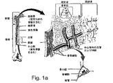
図1aは、さまざまな骨組織の領域を示す骨の生体構造を概略的に示している。図1bは、皮質が除去され、海綿状骨からなる骨髄を露出させた状態の腸骨稜を示している。骨髄は、自然に発生する動静脈シャントであり、そのためアナライトセンサ、特に連続的なリアルタイムグルコースセンサの配置に極めて適している。 FIG. 1a schematically shows the bone anatomy showing various bone tissue regions. FIG. 1b shows the iliac crest with the cortex removed and the bone marrow composed of cancellous bone exposed. The bone marrow is a naturally occurring arteriovenous shunt and is therefore very suitable for the placement of analyte sensors, especially continuous real-time glucose sensors.
本発明の装置は、海綿状組織、骨膜組織および緻密骨組織を含む骨のあらゆる組織領域内に部分的にまたは全体的に埋め込まれ得る。 The device of the present invention can be partially or fully implanted in any tissue region of bone, including spongiform tissue, periosteal tissue and compact bone tissue.
埋込みは、たとえばさまざまな穿孔または切断方法を含む、骨組織へのアクセスに使用される数多くの方法の任意の1つを介して実施され得る。そのような方法は、当業者によく知られており、したがってそのような方法のさらなる説明は本明細書では提供されない。 Implantation can be performed via any one of a number of methods used to access bone tissue including, for example, various drilling or cutting methods. Such methods are well known to those skilled in the art, and therefore no further description of such methods is provided herein.
本発明の装置は、骨組織に埋め込まれた際、センサ素子(複数可)が骨組織内に存在する延髄内/骨髄内の血洞内に位置するように設計される。これにより、センサ素子(複数可)が骨組織内を流れる血液を試料採取し、正確なリアルタイムの分析監視を提供することが可能になる。 The device of the present invention is designed such that when implanted in bone tissue, the sensor element (s) are located in the intramedullary / intramedullary blood sinus present in the bone tissue. This allows the sensor element (s) to sample blood flowing through the bone tissue and provide accurate real-time analytical monitoring.
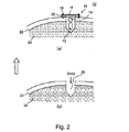
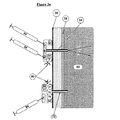
本発明の装置は、骨の取り付けに適したどのような形状およびサイズのものでもよい。本発明の装置の形状およびサイズは、その装置が骨内に部分的に埋め込まれるか、あるいは全体的に埋め込まれるか、埋込みの部位および装置と制御ユニット(以下でさらに説明される)の間の通信の種類に大きく依存することになる。一般に、装置は、球状、円筒状、矩形または1mm−2.5cmの直径/幅および5mm−5cmの長さを有する形状のものでよい。後続の実施例の項でより詳細に説明される図2の(a)は、1つの好ましい装置の構成を示している。 The device of the present invention may be of any shape and size suitable for bone attachment. The shape and size of the device of the present invention is such that it can be partially or fully implanted in the bone, between the implantation site and device and the control unit (described further below). It depends greatly on the type of communication. In general, the device may be spherical, cylindrical, rectangular or shaped with a diameter / width of 1 mm-2.5 cm and a length of 5 mm-5 cm. FIG. 2 (a), described in more detail in the subsequent Examples section, shows one preferred apparatus configuration.
装置が骨内に部分的に埋め込まれる構成では、装置のセンサ素子(複数可)の構成要素は、骨組織内に延びて延髄内/骨髄内の血洞内に流れる血液と接触するように構成され、一方で電源、回路、通信装置(たとえばコイル、アンテナ)および同様のものなどの追加の構成要素を収容する装置本体は、骨を取り囲む軟組織内に配置され得る、あるいは骨固定に適した取り付けアンカによって骨表面に取り付けられ得る。本発明の装置との使用に適した骨アンカ構成は、骨ねじ/プレートおよび同様のものなどを含む。軟組織への固定は、当技術分野でよく知られている方法を用いて縫合用ステープルまたはアンカによって実施され得る。 In a configuration where the device is partially implanted in the bone, the components of the sensor element (s) of the device are configured to contact blood that extends into the bone tissue and flows into the intramedullary / medullary sinus. While the device body containing additional components such as power supplies, circuits, communication devices (eg, coils, antennas) and the like can be placed in the soft tissue surrounding the bone or is suitable for bone fixation Can be attached to the bone surface by an anchor. Bone anchor configurations suitable for use with the device of the present invention include bone screws / plates and the like. Fixation to the soft tissue can be performed with suture staples or anchors using methods well known in the art.
本発明の装置の部分的な埋込み型構成では、センサ素子(複数可)は、穿孔され、あるいは骨に切り込まれた小さな穴/溝内に嵌合され得る。そのような穴または溝は、皮質を貫通し、海綿状骨内に延びるのに十分な長さのものである。たとえば、長骨内で使用されるように構成された装置においては、長さ5mm−5cmmmおよび直径1mm−2.5cmの穴が、骨に穿孔され、本発明の装置のセンサ素子(複数可)を収容するために使用され得る。 In the partially implantable configuration of the device of the present invention, the sensor element (s) can be fitted into small holes / grooves that are drilled or cut into the bone. Such holes or grooves are of sufficient length to penetrate the cortex and extend into the cancellous bone. For example, in a device configured to be used within a long bone, a hole 5 mm-5 cm mm in length and 1 mm-2.5 cm in diameter is drilled in the bone, and the sensor element (s) of the device of the present invention. Can be used to house.
部分的な埋込み型の構成は最小限の骨の穿孔/切り込みしか必要としないため、そのような構成は、装置全体を収容できないより小さい骨に極めて適している。そのような骨の例は、椎体、胸骨、および同様のものなどを含む。 Since a partially implantable configuration requires minimal bone drilling / cutting, such a configuration is well suited for smaller bones that cannot accommodate the entire device. Examples of such bones include vertebral bodies, sternums, and the like.
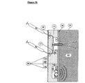
装置全体が骨内に埋め込まれる全体的な埋込み型構成もまた、本明細書で企図される。そのような構成では、装置本体は骨組織内に埋め込まれ、センサ素子(複数可)は、そこを流れる血液に露出される。当技術分野で知られているように、骨内への異物(たとえば整形外科用インプラント)の埋込みは、体によって良好に耐容され、軟組織内の埋込みと比べ最小限の体内反応しか生じさせない。したがって全体的な埋込み型構成は、装置本体が骨組織によって完全に被包化され、被包化、生物膜形成、腐食および同様のものなどをもたらし得る恐れのある組織反応への露出がより少なくなる点で有利である。 An overall implantable configuration in which the entire device is implanted within the bone is also contemplated herein. In such a configuration, the device body is implanted in the bone tissue and the sensor element (s) are exposed to blood flowing therethrough. As is known in the art, implantation of foreign bodies (eg, orthopedic implants) in bone is well tolerated by the body and produces minimal internal reactions compared to implantation in soft tissue. Thus, the overall implantable configuration allows the device body to be fully encapsulated by bone tissue, with less exposure to tissue reactions that can result in encapsulation, biofilm formation, corrosion and the like. This is advantageous.
本明細書に述べるように、本発明の装置は、対象のアナライトを検出するように設計されたセンサ素子(複数可)を含む。 As described herein, the apparatus of the present invention includes sensor element (s) designed to detect the analyte of interest.
そのようなセンサは、好ましくは化学的または光学的な性質のものである。アナライト検出に使用される化学センサは、通常、アンペロメトリー酵素センサである。 Such sensors are preferably of chemical or optical nature. The chemical sensor used for analyte detection is usually an amperometric enzyme sensor.
一般的なアンペロメトリー酵素センサ素子(複数可)は、非導電性ハウジングと、ハウジング内を通過しその中に固定され、したがってハウジング上の一方の場所に電気化学的に反応性の表面およびハウジング上の他方の場所に電気化学的反応表面を形成する作用電極(アノード)と、基準電極と、対向電極(カソード)とを含む。センサ素子(複数可)はまた、ハウジングに固着され、電気化学的反応表面を覆う膜も含む。対向電極は、一般に、作用電極よりも大きな電気化学的反応表面積を有する。センサの作動中、血液試料またはその一部分は、酵素(たとえばグルコース監視の場合はグルコース酸化酵素)と(直接的にまたは膜を通り抜けた後で)接触する。アナライトと酵素の反応は、血液試料におけるアナライト(たとえばグルコース)レベルの決定を可能にする反応生成物の形成をもたらす。 A typical amperometric enzyme sensor element (s) includes a non-conductive housing and an electrochemically responsive surface and housing in one location on the housing that passes through and is secured within the housing. It includes a working electrode (anode) that forms an electrochemically reactive surface at the other location above, a reference electrode, and a counter electrode (cathode). The sensor element (s) also includes a membrane that is secured to the housing and covers the electrochemically reactive surface. The counter electrode generally has a larger electrochemical reaction surface area than the working electrode. During operation of the sensor, the blood sample or part thereof is in contact (either directly or after passing through the membrane) with an enzyme (eg glucose oxidase in the case of glucose monitoring). The reaction between the analyte and the enzyme results in the formation of a reaction product that allows determination of the analyte (eg glucose) level in the blood sample.
センサ素子(複数可)は、円筒または薄膜として成形されてよく、一般的な薄膜の電気化学センサは、US5,390,671、US5,391,250、US5,482,473、およびUS5,586,553に記載されている。 The sensor element (s) may be molded as a cylinder or a thin film, and typical thin film electrochemical sensors are US 5,390,671, US 5,391,250, US 5,482,473, and US 5,586, 553.
アナライトの電気化学的な感知には3つの一般的な方法が使用され、そのすべてはアナライトの酸化に触媒作用を及ぼす酵素の固定化型を使用する。たとえばグルコースの場合、グルコース酸化酵素が、グルコースをグルコン酸に変換するために使用され、このとき過酸化水素の生成を伴う。第1の検出スキームは、酸素消費を測定し、第2は酵素反応によって生成された過酸化水素を測定し、第3は拡散可能なまたは固定化されたメディエータを使用してその電子をグルコース酸化酵素から電極に移動する。 Three general methods are used for the electrochemical sensing of analytes, all of which use an immobilized form of the enzyme that catalyzes the oxidation of the analyte. For example, in the case of glucose, glucose oxidase is used to convert glucose to gluconic acid, with the production of hydrogen peroxide. The first detection scheme measures oxygen consumption, the second measures hydrogen peroxide produced by the enzymatic reaction, and the third uses diffusible or immobilized mediators to oxidize the electrons to glucose oxidation. Move from enzyme to electrode.
グルコース監視の場合、本発明の装置は、グルコースおよび酸素が1つの方向からセンサの酵素領域内に拡散することを可能にするセンサを利用することができるが、酸素だけは他の方向からも拡散する。この設計は、「酸素欠乏」、すなわち体内に存在するグルコースに対する酸素の比が低くなることを解消するのに役立つ。酵素反応における酸素関与による酸素電極への酸素の移送の変調が、グルコースの決定のための手段を提供する。グルコース酸化酵素の活性寿命を短縮し得る過酸化水素を除去するために、カタラーゼ酵素がグルコース酸化酵素と共に固定化される。この感知方法は、酸素のバックグラウンド濃度を表示するためのさらなる酸素電極の設置を必要とする。 For glucose monitoring, the device of the present invention can utilize a sensor that allows glucose and oxygen to diffuse into the enzyme region of the sensor from one direction, but only oxygen diffuses from the other direction. To do. This design helps to eliminate “oxygen deficiency”, ie, the low ratio of oxygen to glucose present in the body. Modulation of oxygen transfer to the oxygen electrode due to oxygen involvement in the enzymatic reaction provides a means for determination of glucose. In order to remove hydrogen peroxide, which can shorten the active life of glucose oxidase, catalase enzyme is immobilized together with glucose oxidase. This sensing method requires the installation of an additional oxygen electrode to display the background concentration of oxygen.
過酸化水素センサは、アノード極性の電極上における酵素反応の生成物を測定する。過酸化水素センサの利点の1つは、グルコースの濃度が上昇するにつれて信号が増大することである。しかし、過酸化水素の酸化は、体内において通常見出される多くの他の種が電子酸化可能であり、妨害の可能性を生じさせるところにおいて印加電位を必要とする。最も問題のある種は、尿素、アスコルビン酸塩(ビタミンC)、尿酸塩、およびアセトアミノフェンである。これらの通過を制限する半透性の膜により、妨害が最小限に抑えられる。酵素反応は、通常適切であると想定される酸素を依然として必要とする。 The hydrogen peroxide sensor measures the product of the enzymatic reaction on the anode polarity electrode. One advantage of the hydrogen peroxide sensor is that the signal increases as the glucose concentration increases. However, the oxidation of hydrogen peroxide requires an applied potential where many other species normally found in the body are capable of electronic oxidation, creating the potential for interference. The most problematic species are urea, ascorbate (vitamin C), urate, and acetaminophen. These semi-permeable membranes that restrict the passage minimize interference. Enzymatic reactions still require oxygen, which is normally assumed to be appropriate.
浸出不可能な(nonleachable)な電気化学メディエータを使用するグルコースセンサは、酸素以外の種を使用して電子をグルコース酸化酵素から電極に移動することにより、上記で説明された酸素欠乏を妨げる。酸素はシステム内に残存しているため、メディエータは電子のためにその酸素と効果的に競合しなければならない。以前は、フェロセンがメディエータとして使用されていたが、これは拡散性および有毒性のものである。メディエータセンサのより最近のバージョンは、オースティン所在のテキサス大学、化学工学科(Department of Chemical Engineering)のAdam Hellerおよび彼のグループによって設計された「有線接続された」グルコース酸化酵素電極である。メディエータは、架橋されたポリマーに結合されているため浸出しない。グルコース酸化酵素は、電気化学的に活性の化学的に結合された複合化されたオスミウムのレドックス中心を有するレドックスポリマーから形成されたヒドロゲルと共に電極に連結される。 Glucose sensors that use nonleachable electrochemical mediators prevent oxygen deficiency as described above by using species other than oxygen to transfer electrons from glucose oxidase to the electrode. Since oxygen remains in the system, the mediator must effectively compete with the oxygen for electrons. Previously, ferrocene was used as a mediator, but it is diffusive and toxic. A more recent version of the mediator sensor is the “wire-connected” glucose oxidase electrode designed by Adam Heller and his group at the University of Texas at Austin, Department of Chemical Engineering. The mediator does not leach because it is bound to the crosslinked polymer. Glucose oxidase is linked to an electrode together with a hydrogel formed from a redox polymer having an electrochemically active chemically bound complex osmium redox center.
電気化学酵素センサの長期にわたる作動を確実にするために、本発明の装置は、新鮮な酵素溶液でセンサを「再充電」することができるように構成され得る。そのような溶液は、骨組織に接触する膜と電極表面の間の薄いチャネル内にポンピングされ得る。消費された酵素懸濁液は、システムから洗い流され得、新鮮な酵素が、装置と流体連通している皮膚ポートを通じて注入され得る。 To ensure long-term operation of the electrochemical enzyme sensor, the device of the present invention can be configured to be able to “recharge” the sensor with fresh enzyme solution. Such a solution can be pumped into a thin channel between the membrane that contacts the bone tissue and the electrode surface. The spent enzyme suspension can be flushed from the system and fresh enzyme can be injected through the skin port in fluid communication with the device.
アナライトの読取値の精度に影響を与え得る電気化学的妨害は、2つの方法で最小限に抑えられ得る。印加電位が、検出された反応生成物以外の種がほとんど酸化されない十分な低さに設定され得る、あるいは電極への妨害の拡散を制限する層が利用され得る。酸素ベースの酵素センサにおいては、電気化学的妨害は、酸素移送を可能にするが極性分子を阻止する酵素と電極表面の間の無孔性の疎水性層により、それほど問題ではない。 Electrochemical interference that can affect the accuracy of the analyte readings can be minimized in two ways. The applied potential can be set low enough so that species other than the detected reaction products are hardly oxidized, or a layer that limits the diffusion of interference to the electrodes can be utilized. In oxygen-based enzyme sensors, electrochemical interference is less of an issue due to the non-porous hydrophobic layer between the enzyme and the electrode surface that allows oxygen transport but blocks polar molecules.
グルコース監視の高性能のグルコースセンサ場合、ピロロキノリンキノン依存グルコース脱水素酵素(PQQ−GDH)が、センサの精度を高めるためにセンサ素子(複数可)に使用され得る(US7,005,048)。 For high performance glucose sensors with glucose monitoring, pyrroloquinoline quinone dependent glucose dehydrogenase (PQQ-GDH) can be used in the sensor element (s) to increase sensor accuracy (US 7,005,048).
本発明の装置によって使用され得る光学センサは、薄膜(たとえば薄膜ヒドロゲル)内に固定化された蛍光性化学複合体を含む。薄膜は、アナライトに対して透過性の生体適合性ポリマーである。感知システムは、2つの構成要素:蛍光染料およびアナライトに対して反応性である「消光剤」を有する。アナライトの不在下では、消光剤は、染料と結合し蛍光発光を防止するが、アナライトが消光剤と相互作用すると、複合体の電離が生じ、蛍光発光が増大する。そのようなセンサにおいては、蛍光発光は、通常、監視ユニットに送られる電流へと形が変えられる。 Optical sensors that can be used with the devices of the present invention include fluorescent chemical complexes immobilized within a thin film (eg, a thin film hydrogel). The thin film is a biocompatible polymer that is permeable to the analyte. The sensing system has two components: a “quencher” that is reactive to fluorescent dyes and analytes. In the absence of the analyte, the quencher binds to the dye and prevents fluorescence emission, but when the analyte interacts with the quencher, the complex ionization occurs and fluorescence emission increases. In such sensors, the fluorescence emission is usually transformed into a current that is sent to the monitoring unit.
グルコースの光学監視は、蛍光性である人工グルコースレセプタ分子を使用することができ、たとえば蛍光性染料、すなわちアントラセンとグルコース上のヒドキシル基の2つと共有的だが可逆的に結合するボロン酸との結合によって生成された化合物などがある(James TD、Sananayake KRAS、Shinkai S.A glucose−selective molecular fluorescence sensor.Angewandte Chemie International Edition in English.1994年;33:2207−2209頁)。このレセプタにより、グルコース結合時に蛍光強度に変化が生じる。グルコースの光学監視はまた、NIR光源(ダイオード/レーザなど)およびグルコースの変動レートに関連付けられた色の変化を測定する適切な検出器を使用することができる。 Optical monitoring of glucose can use an artificial glucose receptor molecule that is fluorescent, for example, binding of a fluorescent dye, ie, an anthracene and a boronic acid that binds covalently but reversibly to two of the hydroxyl groups on the glucose. (James TD, Sananayake KRAS, Shinkai S.A glucose-selective molecular fluorescence sensor.Angewandte Chemie International, page 207; This receptor causes a change in fluorescence intensity upon glucose binding. Optical monitoring of glucose can also use an NIR light source (such as a diode / laser) and a suitable detector that measures the color change associated with the rate of glucose fluctuation.
有用な蛍光発光技術の別の例は、「蛍光共鳴エネルギー移動」(FRET)であり、これは、1つの蛍光性分子(ドナー)から、重複スペクトル特性を有する別の近くの分子(アクセプタ)への励起エネルギーの伝達に依存する。蛍光強度または寿命の変化は、ドナーとアクセプタの間の変化する距離のレポーターである。FRETスキームモデルが、近赤外線蛍光性分子に結合されたグルコース結合レクチンコンカナバリンAを用いた体外でのグルコース感知に関して説明されている(olosa L、Szmacinski H、Rao G、Lakowicz JR.Lifetime−based sensing of glucose using energy transfer with a long−lifetime donor.Anal Biochem.1997年;250:102−108頁;およびRolinski OJ、Birch DJS、McCartney LJ、Pickup JC.Near−infrared assay for glucose determination.Soc Photo−optical Instrumentation Engineers Proc.1999年;3602:6−14頁)。 Another example of a useful fluorescence technique is “Fluorescence Resonance Energy Transfer” (FRET), which is from one fluorescent molecule (donor) to another nearby molecule (acceptor) with overlapping spectral properties. Depends on the transmission of excitation energy. A change in fluorescence intensity or lifetime is a reporter of varying distance between donor and acceptor. A FRET scheme model has been described for in vitro glucose sensing using glucose-binding lectin concanavalin A conjugated to near-infrared fluorescent molecules (olosa L, Szmacinski H, Rao G, Lakowitz JR. Lifetime-based sensing of glucose using energy transfer with a long-lifetime donor. Anal Biochem. 1997; 250: 102-108; and Rolinski OJ, Birch DJS, McCartney LJ, Pickup L., Pickup. strumentation Engineers Proc 1999 years; 3602:. 6-14 pages).
アナライトの結合時のタンパク質における配座の変化もまた、タンパク質に結合された配座敏感性蛍光物質を介して感知され得る。この点において、より感知に適合された改変された機能を有する新しい分子を生成するためのタンパク質の合理的な適合に対して、分子工学技術が使用されている。たとえば、配座敏感性蛍光性基は、大腸菌(Escherichia coli)からのグルコース結合タンパク質などのアロステリック性タンパク質内に組み込まれている(Marvin JS、Hellinga HW.Engineering biosensors by introducing fluorescent allosteric signal transducers:construction of a novel glucose sensor.J Am Chem Soc.1998年;120:7−11頁)。このタンパク質は、グルコース結合時に大きな配座的変化を受け、この変化は工学操作されたタンパク質における蛍光発光の変更に変換され得る。グルコースなどの化学物質と強く反応して配座、したがって蛍光応答を変化させる分子(たとえばナノチューブ)センサもまた、本発明によって利用され得る。 Conformational changes in the protein upon analyte binding can also be sensed via conformation sensitive fluorescent substances bound to the protein. In this regard, molecular engineering techniques have been used for rational adaptation of proteins to generate new molecules with altered functions that are more sensitive to sensing. For example, conformation-sensitive fluorescent groups are incorporated into allosteric proteins such as glucose binding proteins from Escherichia coli (Marvin JS, Hellinga HW. Engineering biosensors fluorsient allosteric citrus citrus citrus citrus citrus citrus citrus scienti? a novel glucose sensor. J Am Chem Soc. 1998; 120: 7-11). This protein undergoes a large conformational change upon glucose binding, and this change can be translated into a change in fluorescence emission in the engineered protein. Molecular (eg, nanotube) sensors that react strongly with chemicals such as glucose to change the conformation and thus the fluorescent response can also be utilized by the present invention.
他の感知機構、それだけに限定されないが、生化学センサ、細胞ベースのセンサ(たとえばUS20020038083)、電気触媒センサ、光学センサ、圧電センサ、熱電センサ、および音響センサなどを含むその他のセンサ素子(複数可)の構成もまた、本発明の装置において使用され得る。 Other sensor element (s) including other sensing mechanisms, including but not limited to biochemical sensors, cell-based sensors (eg US20020038083), electrocatalytic sensors, optical sensors, piezoelectric sensors, thermoelectric sensors, acoustic sensors, etc. This configuration can also be used in the apparatus of the present invention.
たとえば、アナライトの存在下において寸法的変化を受けるポリマーなどの拡張性生体適合性センサを用いたアナライトの選択的認識を可能にする化学センサ(たとえばUS6,480,730を参照)もまた、本発明の装置によって使用され得る。 For example, chemical sensors that allow selective recognition of analytes using scalable biocompatible sensors such as polymers that undergo dimensional changes in the presence of the analyte (see, eg, US Pat. No. 6,480,730) also It can be used by the device of the present invention.
人工レセプタ分子もまたアナライト監視に使用され得る。人工レセプタを作製するための最も有望な技術の1つが、「分子インプリンティング」または「プラスチック抗体」と呼ばれるものである(Haupt K、Mosbach K.Plastic antibodies:developments and applications.Trends Biotechnol.1998年;16:468−475頁)。アナライトに関連するテンプレート分子と相互作用する化学基を有するモノマーが、テンプレートの周りで重合され、次いでこのテンプレートは除去され、形状および結合能力においてそのアナライトに特異的なポリマーを残す。グルコース監視の例では、グルコース結合時に水素イオンを放出する、金属イオン複合体とグルコースの間のアルカリpHにおける相互作用を使用する(Chen G、Guan Z、Chen C−T、Fu L、Sundaresan V、Arnold F.A glucose sensing polymer.Nature Biotechnol.1997年;15:354−357頁)。グルコースに特異的な多孔性ポリマーが作製され、それにより、陽子の滴定放出によってグルコース濃度が測定され得る。 Artificial receptor molecules can also be used for analyte monitoring. One of the most promising techniques for making artificial receptors is called “molecular imprinting” or “plastic antibodies” (Haupt K, Mosbach K. Plastic Antibodies: developments and applications. Trends Biotechnol. 1998; 16: 468-475). A monomer having a chemical group that interacts with the template molecule associated with the analyte is polymerized around the template, which is then removed, leaving a polymer specific for that analyte in shape and binding capacity. Examples of glucose monitoring use interactions at alkaline pH between metal ion complexes and glucose that release hydrogen ions upon glucose binding (Chen G, Guan Z, Chen CT, Fu L, Sundaresan V, Arnold F. A glucosensing polymer. Nature Biotechnol. 1997; 15: 354-357). A porous polymer specific to glucose is made, whereby the glucose concentration can be measured by titration release of protons.
センサの種類に関係なく、センサの読取値は通常、本発明の装置内に収容されたL−C回路などの回路を用いて解釈される。たとえば、センサは、周波数同調されたL−C回路に結合され得、ここでセンサは、生理的状態の変化をその同調されたL−C回路のインダクタまたはコンデンサに伝える。したがって、センサにおける変化は、それが化学的、光学的または物理的なものであれ、アナライト濃度を評価するために定量化され使用され得るL−C回路内の変化となる。 Regardless of the sensor type, sensor readings are typically interpreted using a circuit such as an L-C circuit housed in the apparatus of the present invention. For example, the sensor may be coupled to a frequency tuned LC circuit, where the sensor communicates a change in physiological state to an inductor or capacitor of the tuned LC circuit. Thus, a change in the sensor becomes a change in the LC circuit that can be quantified and used to evaluate the analyte concentration, whether it is chemical, optical or physical.
本発明の装置は、1つの感知領域または複数の感知領域を含むことができる。各々の感知領域は、同じまたは異なるアナライトを決定するために使用され得る。さまざまな感知機構が、同一の装置上のさまざまなセンサ領域によって使用され得る。 The device of the present invention can include one sensing area or multiple sensing areas. Each sensing region can be used to determine the same or different analytes. Different sensing mechanisms can be used by different sensor areas on the same device.
グルコースの検出のためのセンサ構成が、本明細書において例示されてきたが、どのようなアナライトも、システムにそのようなアナライトを検出することができるように設計されたセンサ(たとえば電極)を合わせることにより、本発明の装置によって検出され得ることが理解される。たとえば、水素イオン(pH)は、水素イオン濃度が変化するにつれてその出力電圧が変化する電極を用いて検出され得、ホルモンは、Cook and Devine(Electroanalysis Volume 10、Issue 16、1108−1111頁;1999年2月)によって説明されたものなどの抗体ベースの電極を介して検出され得、一方で一酸化窒素は、Mizutaniら(Chemistry Letters 29巻、No.7 802頁 2000年)によって説明された電極によって検出され得る。
Although sensor configurations for the detection of glucose have been exemplified herein, any analyte designed to be able to detect such an analyte in the system (eg, an electrode) , It can be understood that it can be detected by the device of the present invention. For example, hydrogen ions (pH) can be detected using an electrode whose output voltage changes as the hydrogen ion concentration changes, and the hormone is Cook and Device (
本発明の装置は、埋込み型装置の機能を制御し、それに電力供給し、そこから読取値を得るために使用され得る遠隔ユニットと通信することができるように構成される。したがって、本発明の装置は、埋込み可能な装置の作動を制御するための制御ユニットをさらに含む、アナライト監視のためのシステムの一部を形成する。 The device of the present invention is configured to be able to control the function of the implantable device, power it, and communicate with a remote unit that can be used to obtain readings therefrom. Thus, the device of the present invention forms part of a system for analyte monitoring that further includes a control unit for controlling the operation of the implantable device.
埋込み型装置と制御ユニットの間の通信は、装置から制御ユニットに延びる有線によるものでよく、そのような場合、制御ユニットは、皮膚の下に埋め込まれ得る、あるいは体に着用され得る。通信はまた、以下にさらに説明するように無線によって実施され得る。 Communication between the implantable device and the control unit may be by wire extending from the device to the control unit, in which case the control unit may be implanted under the skin or worn on the body. Communication may also be performed wirelessly as described further below.
本発明の装置の電力供給は、埋込み型電源(装置内に内蔵され得る)、または遠隔制御ユニットによる遠隔の電力供給によって実施されてよく、埋込み型装置の遠隔の電力供給および制御は、現在好ましいものである。 The power supply of the device of the present invention may be implemented by an implantable power supply (which may be embedded within the device) or by a remote power supply by a remote control unit, with remote power supply and control of the implantable device being presently preferred Is.
本発明の装置の遠隔の電力供給および制御のためのいくつかの構成が、本発明によって使用され得る。テレメトリの一般的な説明に関してはUS6,201,980を参照されたい。 Several configurations for remote power supply and control of the device of the present invention can be used by the present invention. See US 6,201,980 for a general description of telemetry.
装置および制御ユニットの誘導結合は、無線周波(RF)信号によって実施され得る。埋込み型装置は、制御ユニット上に設けられた第2のコイルと誘導的に結合することができる第1のコイルを利用することができる。 The inductive coupling of the device and the control unit can be implemented by radio frequency (RF) signals. The implantable device can utilize a first coil that can be inductively coupled to a second coil provided on the control unit.
システムの使用中、第2のコイルは、第1のコイルに隣接して配置され、高周波搬送波信号が第2のコイルに与えられる。この信号は、たとえ2つのコイル間に直接的な接続が存在しない場合であっても、変圧器の一次巻線に与えられたAC信号が変圧器の二次巻線に結合されるのと全く同じ方法で第1のコイルに結合される。第1のコイルによって受信された後、本発明の装置内の回路が、その信号を整流化し、それをインプラント装置の作動電力として使用されるDC信号に変換する。さらに、搬送波信号に加えられた変調は、制御信号を制御ユニットから埋込み型装置に送信するための手段を提供する。RFテレメトリシステムのさらなる説明は、US6,667,725およびUS5,755,748において提供されている。 During use of the system, the second coil is positioned adjacent to the first coil and a high frequency carrier signal is provided to the second coil. This signal is exactly the same as when the AC signal applied to the transformer primary is coupled to the transformer secondary, even if there is no direct connection between the two coils. Coupled to the first coil in the same manner. After being received by the first coil, circuitry within the device of the present invention rectifies the signal and converts it to a DC signal that is used as the operating power for the implant device. Furthermore, the modulation applied to the carrier signal provides a means for transmitting a control signal from the control unit to the implantable device. Further descriptions of RF telemetry systems are provided in US 6,667,725 and US 5,755,748.
したがって電気化学センサ素子(複数可)および同調されたL−C回路の場合、埋込み型装置内のコイルに伝送された信号は、DC電流に変換され、このDC電流は、センサ電極内において生成された電流によって変調された周波数を有するLC回路に電力供給する。そのような電流は、電極の環境に存在するアナライトの量に比例する。LC回路は、信号によって電力供給された後、制御ユニットに周波数変調信号を戻す。この信号の周波数は、アナライトの濃度を導き出すように制御ユニットによって解釈される。 Thus, in the case of an electrochemical sensor element (s) and a tuned L-C circuit, the signal transmitted to the coil in the implantable device is converted to a DC current, which is generated in the sensor electrode. Powers an LC circuit having a frequency modulated by a current. Such current is proportional to the amount of analyte present in the electrode environment. After the LC circuit is powered by the signal, it returns the frequency modulated signal to the control unit. The frequency of this signal is interpreted by the control unit to derive the analyte concentration.
本発明の埋込み型装置の電力供給および制御の目的のための誘導結合はまた、磁気テレメトリ(たとえばUS6,963,779を参照)、音響テレメトリ(たとえばUS6,764,446およびUS7,024,248を参照)または光テレメトリ(たとえばUS6,243,608およびUS6,349,234を参照)によって実現されてよく、光テレメトリの場合、皮下のレシーバが、埋込み型装置に有線接続され、装置と体外の制御ユニットの間の導管として作用することができる。そのようなレシーバは、受光した光を電気エネルギーに、またその逆で変換する近赤外線光センサ/エミッタでよい。 Inductive coupling for power supply and control purposes of the implantable device of the present invention also includes magnetic telemetry (see for example US 6,963,779), acoustic telemetry (for example US 6,764,446 and US 7,024,248). Or optical telemetry (see, for example, US 6,243,608 and US 6,349,234), in the case of optical telemetry, a subcutaneous receiver is wired to the implantable device and the device and external control Can act as a conduit between units. Such a receiver may be a near infrared light sensor / emitter that converts the received light into electrical energy and vice versa.
いずれの場合も、テレメトリは、埋込み型装置の制御および電力供給の両方に使用され得る。 In either case, telemetry can be used for both control and power supply of the implantable device.
制御ユニットは、埋込み型装置のセンサ素子(複数可)によって送られた情報をユーザに表示するためのユーザインターフェースを含むことができる。そのような情報は、血液中のアナライトのレベル、所定期間にわたる傾向、ならびにアナライトの高低レベルを表示するための警告を含むことができる。制御ユニットは、アナライトレベルの履歴、個人情報、投与されている薬剤および同様のものなどを含む、患者に関連する情報を保存することができる。制御ユニットはまた、システムの設定またはその較正に使用され得る情報を入力するためのキーパッドなどの入力装置を含むこともできる。 The control unit can include a user interface for displaying to the user information sent by the sensor element (s) of the implantable device. Such information can include warnings to indicate the level of analyte in the blood, trends over time, as well as high and low levels of the analyte. The control unit can store patient related information, including analyte level history, personal information, medications being administered, and the like. The control unit may also include an input device such as a keypad for entering information that can be used to set up or calibrate the system.
制御ユニットは、腕時計などの専用の着用可能な装置の形態のものでよく、あるいはMP3プレーヤ、携帯電話または同様のものなどの既存のユーザ装置内に組み込まれてもよい。携帯電話または他の通信可能な装置(たとえばコンピュータ、PDA)の使用は、さらに携帯電話通信ネットワークまたはコンピュータネットワークなどの通信ネットワークを通じてアナライト情報を第3者に伝達することを可能にするため、特に有利である。 The control unit may be in the form of a dedicated wearable device such as a wristwatch, or may be incorporated into an existing user device such as an MP3 player, mobile phone or the like. In particular, the use of a mobile phone or other communicable device (eg a computer, PDA) further allows to transmit analyte information to a third party through a communication network, such as a mobile phone communication network or a computer network. It is advantageous.
本発明のシステムはまた、薬剤の送出のためのポートを含む埋込み型装置構成を含むこともでき、あるいは本発明のシステムの制御ユニットは、埋込み型薬物送出ポンプまたはリザーバと通信することができる。そのような通信は、有線によるもの、または上記で概要が説明されたテレメトリ構成によるものでよい。 The system of the present invention can also include an implantable device configuration that includes a port for drug delivery, or the control unit of the system of the present invention can communicate with an implantable drug delivery pump or reservoir. Such communication may be by wire or by the telemetry configuration outlined above.
上記で説明されたセンサは、たとえば糖尿病の血糖レベルの制御に使用され得る閉(フィードバック)ループシステム内に組み込まれ得る。血糖制御のための閉フィードバックループシステムを実現するために、臨床的に適用可能なシステムは、3つの構成要素:埋込み可能なインスリンポンプ、埋込み可能な血糖センサ、および埋め込まれても埋め込まれなくてもよい制御ユニットの協働を必要とする。 The sensors described above can be incorporated into a closed (feedback) loop system that can be used, for example, to control blood glucose levels in diabetes. To achieve a closed feedback loop system for blood glucose control, a clinically applicable system has three components: an implantable insulin pump, an implantable blood glucose sensor, and an implantable or unimplantable Also requires the cooperation of a good control unit.
全自動グルコース制御システムの目的は、糖尿病の慢性合併症を防止または遅延すること、低血糖のリスクを低下させること、ならびに複数の日常的なグルコース自己検査およびインスリン注射の場合と比べて患者の不便さおよび不快感を低減することを含む。 The goal of a fully automatic glucose control system is to prevent or delay chronic complications of diabetes, reduce the risk of hypoglycemia, and patient inconvenience compared to multiple routine glucose self-tests and insulin injections. Including reducing discomfort and discomfort.
インスリンを皮下組織または静脈などの血管に送出する埋込み可能なインスリンポンプは、長期間にわたる糖尿病の満足な制御に対して実現可能である。しかし、そのような埋込み可能なポンプを使用する閉ループシステムは、リアルタイムの血糖レベルとは異なるグルコースレベル読取値を与える、利用されるグルコースセンサによって限定される。さらに、皮下埋込み型インスリンポンプはまた、インスリン注入カテーテル内の障害物から生じる合併症によっても限定される。 Implantable insulin pumps that deliver insulin to vessels such as subcutaneous tissue or veins are feasible for long-term satisfactory control of diabetes. However, closed loop systems using such implantable pumps are limited by the glucose sensor utilized that provides a glucose level reading that differs from the real-time blood glucose level. In addition, subcutaneous implantable insulin pumps are also limited by complications arising from obstacles in the insulin infusion catheter.
本発明者らは、上記で説明されたように骨埋込み型グルコースセンサを骨埋込み型ポート/カテーテルを有するリザーバと組み合わせて利用するシステムは、従来技術のシステムのこれらの限界を克服することを主張する。そのようなシステムは、センサからの信号が注入ポンプを制御する閉ループシステム、あるいはセンサから信号を受信し、それに従ってポンプを作動させるために(患者/医師によって)使用される体外の制御ユニットを含む開ループシステムでもよい。 We argue that a system utilizing a bone implantable glucose sensor in combination with a reservoir having a bone implantable port / catheter as described above overcomes these limitations of prior art systems. To do. Such a system includes a closed loop system in which the signal from the sensor controls the infusion pump, or an extracorporeal control unit that is used (by the patient / doctor) to receive the signal from the sensor and operate the pump accordingly. An open loop system may be used.
したがって、本発明の別の態様によれば、患者の血糖レベルを制御するためのシステムが提供される。 Thus, according to another aspect of the present invention, a system for controlling a patient's blood glucose level is provided.
システムは、上記で説明された(この場合は上記で説明されたようにグルコース感知用に構成された)骨埋込み型センサユニットと、グルコースセンサから制御信号を受信し(閉ループ)、あるいは体外の制御ユニットによってグルコースセンサと通信し(開ループ)、インスリン、グルカゴン、ならびにそれらの組合せなどの血糖レベル調節組成物を患者の骨組織に供給するように構成されたリザーバとを含む。 The system receives the control signal from the bone implantable sensor unit described above (in this case configured for glucose sensing as described above) and the glucose sensor (closed loop) or controls outside the body. The unit communicates with the glucose sensor (open loop) and includes a reservoir configured to supply blood sugar level modulating compositions such as insulin, glucagon, and combinations thereof to the patient's bone tissue.
本明細書においてさらに説明されるように、グルコースセンサおよびリザーバの両方は、上記で説明されたアナライトセンサに対して本明細書において説明されたように患者の骨(好ましくは骨格の骨)と連通して埋め込まれる。グルコースセンサおよびリザーバは、好ましくは、各々が異なる骨領域または異なる骨と連通するように埋め込まれるが、その理由は、同じ骨/骨領域における感知および注入は、血糖レベルに狂いを生じさせる恐れがあるためである。たとえばグルコースセンサは、1つの腸骨稜上に、リザーバは別の腸骨稜上に埋め込まれてよい。 As further described herein, both the glucose sensor and the reservoir are connected to the patient's bone (preferably skeletal bone) as described herein for the analyte sensor described above. Embedded in communication. Glucose sensors and reservoirs are preferably implanted so that each communicates with a different bone region or a different bone because sensing and infusion in the same bone / bone region can cause blood sugar levels to be upset. Because there is. For example, the glucose sensor may be implanted on one iliac crest and the reservoir on another iliac crest.
埋込み型リザーバは、インスリンおよび/または他の組成物(たとえばグルカゴン)を骨注入ポート/カテーテルを通して送出することができるどのような埋込み可能なリザーバでもよい。したがってリザーバは、骨組織に通じるカテーテルを有して皮下に埋め込まれてよく、あるいはリザーバは、後続の実施例の項の実施例2においてさらに示されるように、骨組織内に直接通じるポートを有して骨組織に対して埋め込まれそれに固定されてもよい。 The implantable reservoir can be any implantable reservoir that can deliver insulin and / or other compositions (eg, glucagon) through the bone injection port / catheter. Thus, the reservoir may be implanted subcutaneously with a catheter leading to the bone tissue, or the reservoir may have a port leading directly into the bone tissue, as further shown in Example 2 of the subsequent Examples section. And may be implanted and fixed to the bone tissue.
いずれの場合でも、リザーバの基本的構成は、(各々が組成物を含有する)1つまたは複数のチャンバと、チャンバに連結された注入ポート/カテーテルと、制御可能なバルブと、任意選択でリザーバからポート/カテーテルまでの流れを制御するためのポンピング機構とを含む。 In any case, the basic configuration of the reservoir consists of one or more chambers (each containing a composition), an infusion port / catheter connected to the chamber, a controllable valve, and optionally a reservoir. And a pumping mechanism for controlling the flow from port to port / catheter.
注入ポート/カテーテルは、アナライトセンサに関して上記で説明されたように骨組織内に固定され得る。骨内成長または局所的血液凝固/組織反応を防止するために、注入ポート/カテーテルは、上記で説明されたように抗凝固組成物または骨成長抑制剤によってコーティングされ得る。 The injection port / catheter can be secured in bone tissue as described above with respect to the analyte sensor. In order to prevent bone ingrowth or local blood coagulation / tissue reactions, the injection port / catheter can be coated with an anticoagulant composition or a bone growth inhibitor as described above.
組成物をリザーバから注入ポート/カテーテルを通じて送出するために、ポンピング機構は、蠕動、推進剤、浸透圧(たとえば米国特許第6,632,217号)、圧電素子(たとえば米国特許第3,963,380号および同第4,344,743号)、浸透圧および振動ピストン/回転タービンの組合せおよび同様のものなどを利用することができる。 In order to deliver the composition from the reservoir through the infusion port / catheter, the pumping mechanism can include peristalsis, propellant, osmotic pressure (eg, US Pat. No. 6,632,217), piezoelectric element (eg, US Pat. No. 3,963,3). 380 and 4,344,743), osmotic pressure and oscillating piston / rotary turbine combinations, and the like.
ポンピング機構は、チャンバ内に含有された組成物を骨組織に送出するための制御されたチャンバ破壊を容易にするために利用され得る。 The pumping mechanism can be utilized to facilitate controlled chamber disruption for delivering the composition contained within the chamber to the bone tissue.
チャンバの破壊は、機械機構、電気式機構、または組成物チャンバに隣接する流体密封空間内のポンプのハウジング内に含有された二相流体、すなわち推進剤を用いることによって行われ得る。そのような推進剤は、患者の生理的温度の液体および蒸気であり、理論上は、チャンバ/リザーバの容積変化に対して正の一定の圧力を及ぼし、それによって一定の流れの組成物の送出を実施する。リザーバが充填されるときに膨張されると、推進剤が圧縮され、この場合、そのような蒸気の一部がその液相に戻り、それによってポンプの蒸気圧力の電源を再充電する。他のポンプ構成は、プランジャ式ポンプ機構(たとえばMinimed.Medtronic社)を含むことができる。 The destruction of the chamber can be done by using a mechanical mechanism, an electrical mechanism, or a two-phase fluid, or propellant, contained within the pump housing within the fluid tight space adjacent to the composition chamber. Such propellants are the patient's physiological temperature liquids and vapors, which, in theory, exert a constant positive pressure on chamber / reservoir volume changes, thereby delivering a constant flow of composition. To implement. When expanded when the reservoir is filled, the propellant is compressed, in which case a portion of such steam returns to its liquid phase, thereby recharging the pump vapor pressure power source. Other pump configurations can include a plunger-type pump mechanism (eg, Minimed. Medtronic).
組成物の提供は、ボーラスまたは低速の注入としてでよい。いずれの場合でも、注入の制御は、好ましくは、リザーバとポート/カテーテルの間に配置されたバルブを介して実施される。組成物のさまざまなレートの送出において本発明のシステムによって使用され得るバルブ機構の1つの構成は、US20050054988に記載されている。注入レートは、センサから受信した信号およびシステムの埋込み前に検査によって決定された患者に関連付けられたパラメータに従って事前にプログラミングされる。 Providing the composition may be as a bolus or a slow infusion. In either case, infusion control is preferably performed via a valve located between the reservoir and the port / catheter. One configuration of a valve mechanism that can be used by the system of the present invention in delivering various rates of composition is described in US20050054988. The infusion rate is preprogrammed according to signals received from the sensors and parameters associated with the patient determined by examination prior to system implantation.
リザーバは、組成物(たとえばインスリン)の液体または乾燥調製物を格納するように構成され得る。 The reservoir may be configured to store a liquid or dry preparation of the composition (eg, insulin).
インスリンおよびグルカゴンは、液体調製物として半減期が短いため、乾燥(たとえば凍結乾燥された)調製物を格納するように構成されたリザーバが、現在好ましいものである。そのような構成を有するリザーバは、格納された組成物を提供前に液体中に懸濁するための機構を含むことができる。そのような液化は、(第2のチャンバ)からのアルカリ塩の付加またはポンプを取り巻く環境からの間質液(ISF)の収集によって実施され得る。あるいは、リザーバは、PLA/PGA微粒子などの微粒子の形態で乾燥組成物を骨内に直接送出するように構成され得る。 Because insulin and glucagon have a short half-life as liquid preparations, reservoirs configured to store dry (eg, lyophilized) preparations are currently preferred. A reservoir having such a configuration can include a mechanism for suspending the stored composition in a liquid prior to provision. Such liquefaction may be performed by addition of alkali salt from (second chamber) or collection of interstitial fluid (ISF) from the environment surrounding the pump. Alternatively, the reservoir can be configured to deliver the dry composition directly into the bone in the form of microparticles such as PLA / PGA microparticles.
本発明のシステムは、血糖レベル調節剤の長期にわたる提供に利用されるため、それによって利用されるリザーバには、定期的な補給が必要となることがある。したがって、リザーバはまた、皮膚内に埋め込まれ得る充填ポートを含むこともできる。リザーバは、皮膚ポート内に設けられたセルフシールの隔膜からの外部針による注射によって必要時に再充填され得る。 Since the system of the present invention is used for long-term provision of blood sugar level regulators, the reservoirs utilized thereby may require periodic replenishment. Thus, the reservoir can also include a fill port that can be implanted within the skin. The reservoir can be refilled as needed by injection with an external needle from a self-sealing septum provided in the skin port.
本明細書の上記で述べられたように、本発明のシステムは、閉ループシステムまたは開ループシステムのいずれか(あるいは両方の組合せ)として構成され得る。閉ループ構成では、埋込み型グルコースセンサは、血糖レベルを監視し、定期的に(たとえば1時間ごとに)グルコース読取値を埋込み型インスリンリザーバに送る。センサまたはリザーバは、血糖レベルの信号をポンプ作動信号に変換するための処理ユニットを含むことができる。そのような処理ユニットは、通信ポート、または埋込み可能なアナライトセンサおよび制御ユニットに関して上記で説明されたものと類似の無線通信モードを介して体外からアクセス可能である。処理ユニットは、最初、標準的な検査によって測定されたグルコース読取値およびインスリン効果に基づいて医師によって較正される。処理ユニットは、埋込みの前またはその後に較正されてよく、必要であれば定期的(たとえば1年に1回または複数回)に再較正されてもよい。 As stated hereinabove, the system of the present invention can be configured as either a closed loop system or an open loop system (or a combination of both). In a closed loop configuration, the implantable glucose sensor monitors blood glucose levels and periodically (eg every hour) sends glucose readings to the implantable insulin reservoir. The sensor or reservoir may include a processing unit for converting a blood glucose level signal into a pump actuation signal. Such a processing unit is accessible from outside the body via a communication port or a wireless communication mode similar to that described above with respect to the implantable analyte sensor and control unit. The processing unit is initially calibrated by a physician based on glucose readings and insulin effects measured by standard tests. The processing unit may be calibrated before or after implantation and may be recalibrated periodically (eg, once or multiple times a year) if necessary.
いずれの場合でも、グルコースセンサによって与えられた信号が処理され、適切な注入作動信号(インスリン量、流量など)が提供される。 In either case, the signal provided by the glucose sensor is processed to provide the appropriate infusion activation signal (insulin volume, flow rate, etc.).
本発明のシステムの閉ループ構成の埋込みおよび作動は、後続の実施例の項の実施例2において説明される。 The embedding and operation of the closed loop configuration of the system of the present invention is described in Example 2 of the Examples section that follows.
開ループ構成は、リザーバからの組成物の提供に対してオペレータの制御を必要とする。したがって、開ループ構成は、さらに、本明細書の上記で説明されたアナライトセンサの制御ユニットに機能の点で類似する、ユーザ制御される体外の制御ユニットを含む。そのような制御ユニットは、血糖レベルを監視し、注入レート/組成物の種類を定期的に変更するために使用され得る。 The open loop configuration requires operator control for composition delivery from the reservoir. Accordingly, the open loop configuration further includes a user-controlled extracorporeal control unit that is similar in function to the control unit of the analyte sensor described hereinabove. Such a control unit can be used to monitor blood glucose levels and periodically change the infusion rate / composition type.
ポンピング機構の制御および電力供給は、センサに関して上記で説明された通りでよい。単一の制御および電力供給ユニットが、センサおよびリザーバ組立体に共有して埋め込まれ得、両方に対する電力および通信、ならびにセンサおよび作動信号の処理を提供する。 Control of the pumping mechanism and power supply may be as described above with respect to the sensor. A single control and power supply unit may be shared and embedded in the sensor and reservoir assembly, providing power and communication for both, as well as sensor and actuation signal processing.
本明細書では、用語「約」は、プラスマイナス10%を示す。 As used herein, the term “about” refers to plus or minus 10%.
本発明のさらなる目的、利点、および新規の特徴は、限定を意図するものではない以下の実施例を検討することで当業者に明確になるであろう。加えて、本明細書の上記で描写され、以下の特許請求の範囲の項で特許請求された本発明のさまざまな実施形態および態様の各々は、以下の実施例において実験的裏づけを見出す。 Further objects, advantages, and novel features of the present invention will become apparent to those skilled in the art upon consideration of the following examples, which are not intended to be limiting. In addition, each of the various embodiments and aspects of the invention depicted above in this specification and claimed in the claims section below finds experimental support in the following examples.
次に、上記の説明と共に非限定の形で本発明を説明する以下の実施例が参照される。 Reference is now made to the following examples, which together with the above description, illustrate the invention in a non limiting fashion.
(実施例1)
骨埋込み型電気化学グルコースセンサの埋込み
図2の(a)は、本発明の教示によって構築され、患者の骨組織と共に配置された装置10を示している。装置10は、回路14を介して電源およびテレメトリユニット16に接続されたセンサ素子(複数可)12を収容するハウジング20を含む。ハウジング20は、ポリマー、セラミック、合金および同様のものを含むどのような生体適合性材料から製作されてもよい。センサ素子(複数可)12は、膜被包化グルコース酵素電極である。装置10は、センサ素子(複数可)12が骨髄24内に延び、したがってその中を流れる血液に露出されるように配置される。
Example 1
Implanting a Bone Implanted Electrochemical Glucose Sensor FIG. 2 (a) shows a
装置10は、皮膚に切開を施し、骨から筋肉を取り去ることによって骨(たとえば腸骨稜)内に配置される。次いで、骨膜、皮質骨および海綿状骨の層を貫通する穴26を穿孔するために、ドリルビットが利用される。穴26は、センサ素子(複数可)12のところのハウジング20よりも直径がわずかに大きい。次いで、装置10のセンサ素子(複数可)12部分が、穴26内に挿入され、センサ素子(複数可)12が骨髄組織に露出されるように配置される。ハウジング20は、次いで、骨ねじ18を介して皮質骨22に対して固定され、ユニットは、電力供給され、試験され、標準的なラボ検査を用いて実施された血糖分析に対して較正される。較正に続き、筋肉および皮膚組織が、装置10を覆う位置に戻され、縫合またはステープルで留められる。
(実施例2)
血糖レベルを制御するためのシステム
図3aから図3bは、本発明の教示によって構築されたグルコースレベルを制御するためのシステムの2つの構成を示している。
(Example 2)
Systems for Controlling Blood Sugar Levels FIGS. 3a-3b show two configurations of a system for controlling glucose levels constructed in accordance with the teachings of the present invention.
図3aは、皮膚56および骨組織58を貫通して骨髄60内に延びるカニューレ54を有して患者の皮膚に対して取り付けられた薬物送出装置52を含むシステム50を示している。カニューレ54は、ポンプ66の駆動力下で、リザーバ62および64から骨髄60内に流体を導く。
FIG. 3 a shows a
システム50はまた、グルコースモニタ70と、グルコースレベルの評価のために骨髄60からグルコースモニタ70内に血液を導くためのカニューレ72とを含む検出器68も含む。センサ組立体は、さらに、ポンプ76の駆動力下で、カニューレ72を通じて骨髄60内にヘパリンを送出するためのリザーバ74を含む。
The
薬物送出装置52および検出器68は、(患者の皮膚下に埋め込まれ得る)ハードワイヤ接続または送受信機80による無線通信を介して通信することができる。システム50は、この構成では、電池82(たとえばリチウムイオン電池)によって電力供給されるが、コンデンサおよびコイルを含む電力供給の他の形態もまた想定される。
The drug delivery device 52 and the detector 68 can communicate via a hard wire connection (which can be implanted under the patient's skin) or wireless communication by the
システム50は、以下のように配置される:骨の上方に切開が施され、皮質骨に対するアクセスが得られる。装置の骨髄内に挿入される部分のサイズに基づいて、標準的なドリルおよび骨切り術用工具を用いて、皮質を貫通して骨髄内へと空間が切り開かれる。次いで、装置は、センサ素子が骨髄内に埋め込まれた状態で固定され、外側ハウジングは、ねじによって皮質骨に取り付けられる。
The
位置決めに続き、システム50のグルコースセンサ組立体は、最初に標準的な血糖検査に対して較正され、その後リザーバ62、64および74が、シリンジ84を介して充填され、システムが作動される。薬物送出装置52のリザーバ62からのインスリンの流量は、グルコースモニタ70によって決定され、ディスプレイ86上に表示された血糖レベルに従って患者によって決定/調整され得る、あるいはそのようなレベルは、システム50を閉ループモードで実行することによって自動的に決定/調整され得、その場合、システム50は、グルコースモニタ76の読取値に従ってインスリンの流量を自己調整する。一般的なインスリン送出レートは、幼児の0.1ユニット/1時間から成人の2−6ユニット/1時間の範囲である。システム50はまた、好ましくは、流量が体重、年齢および患者の通常のインスリン使用範囲に応じた最適レベルを超えることを防止するために、遮断および警告機構を使用する。
Following positioning, the glucose sensor assembly of
薬物送出装置52は、血糖レベルがグルコースモニタ70によって検出されたように低血糖レベルへと急速に低下する場合、グルカゴン(10−20マイクログラム/kg/24時間)またはソマトスタチン類似体(3−4mg/kg/1日)などのホルモンをリザーバ64から定期的に送出することができる。さらに、カニューレ72の閉塞を防止するために、ヘパリンなどの抗凝血剤/血餅溶解剤が、カニューレ72を通じてリザーバ74から定期的に送出され得る。
The drug delivery device 52 can detect glucagon (10-20 micrograms / kg / 24 hours) or somatostatin analog (3-4 mg) if the blood glucose level rapidly drops to a low blood glucose level as detected by the
グルコース制御の精度を維持するために、システム50は、好ましくは、血糖検査に対して定期的に較正される。
In order to maintain the accuracy of glucose control, the
図3bは、薬物送出装置52および検出器68が皮膚56下に埋め込まれ、骨組織58に対してまたはその中に固定される、システム50の第2の構成を示している。この構成では、システム50は、ポンプおよびセンサ(またはそれらに接続された充電式電池)に電力を与える充電器102を含む体外ユニット100と、情報(たとえばグルコースレベル)を患者に表示するためのディスプレイ86とを含む。
FIG. 3 b shows a second configuration of the
ユニット100は、さらに、薬物送出装置52および検出器68に対して通信機能(たとえばそれらの間の連携通信)を提供すると共に、センサ情報の処理および薬物送出装置52への命令の送信を提供することができる。ユニット100は、さらに、情報(たとえば体重などの患者情報、オペレーションコマンドなど)の入力を可能にするためのインターフェース(たとえばキーパッド)を含むことができる。
システム50の代替的な実施形態は、図3bで説明された埋込み可能な構成と、ポケットベル様装置を含むことができる。検出器および薬物送出装置の両方は、皮膚下に配置され、上記で説明されたように骨髄に取り付けられる。各々は、別個の内部の充電式電池を含み、したがってシステムの作動時間が延長される。ポケットベルは、体外に配置され、データ処理を行い、インスリン/グルカゴンの注入レート等を制御する。システム50のこの構成の動作は、図3aで説明されたものに類似している。
Alternative embodiments of
(実施例3)
ウサギの静脈または骨髄から採取された血液中のグルコースレベルを監視する
糖尿病を患う患者における厳しい血糖管理が、微小血管および大血管の合併症のリスクを低減するために見出されてきたが、これはまた、重度の低血糖症状のリスクの上昇に関連付けられる。したがって、糖尿病の治療における最終の目的は、連続的なグルコース感知および正常な血糖レベルの維持を可能にし、それによって膵島β細胞の生理学的機能を模倣し、日常的なインスリンおよび糖質の絶え間のない計算の必要性から患者を自由にする自律式システム(人工膵臓)を開発することである。
(Example 3)
Monitoring glucose levels in blood collected from rabbit veins or bone marrow Strict glycemic control in patients with diabetes has been found to reduce the risk of microvascular and macrovascular complications. Is also associated with an increased risk of severe hypoglycemic symptoms. Thus, the ultimate goal in the treatment of diabetes is to enable continuous glucose sensing and maintenance of normal blood glucose levels, thereby mimicking the physiological function of islet β-cells, and the routine insulin and carbohydrate constants It is to develop an autonomous system (artificial pancreas) that frees patients from the need for no computation.
健康なおよび糖尿病の動物の体内における骨髄グルコースと血糖を、基線においておよびインスリンまたはデキストロース治療後に比較するために調査が行われた。 A study was conducted to compare bone marrow glucose and blood glucose in healthy and diabetic animals at baseline and after insulin or dextrose treatment.
8匹の雌の成人ウサギ(各々は2kg)の血糖レベルは、50%デキストロースおよび2IUインスリンの点滴注入によって操作され、次いでこれらのウサギのグルコースレベルが、静脈(IV)および骨(IO)血液(図4a)において測定された(図4a)。 The blood glucose levels in 8 female adult rabbits (each 2 kg) were manipulated by instillation of 50% dextrose and 2 IU insulin, and then the glucose levels in these rabbits were measured by venous (IV) and bone (IO) blood ( Measured in FIG. 4a) (FIG. 4a).
すべての8匹のウサギは、以下の段階にかけられた:
(i)第1の段階−定常状態のグルコースレベルを約10−30分間測定する(5−10分ごとに試料採取)。
(ii)第2の段階−50%デキストロースを注入する。
(iii)第3の段階−2IUのインスリンを(3−5時間かけて)注入する。
All eight rabbits were subjected to the following stages:
(I) First stage—Measure steady state glucose levels for approximately 10-30 minutes (sample every 5-10 minutes).
(Ii) Second stage—Inject 50% dextrose.
(Iii) Third stage-2 IU insulin (over 3-5 hours) is infused.
試料は、静脈および骨髄両方の部位から得られた血液中のグルコースレベルを相互に関連付けるために、同時にその両方にアクセスすることから得られた。 Samples were obtained from accessing both at the same time to correlate glucose levels in blood obtained from both venous and bone marrow sites.
図4aに明確に示されるように、骨髄から採取された血液中において測定されたグルコースレベルは、非常に高い相関レベル(プラスマイナス4%の誤差)で、静脈血液に存在するグルコースレベルを良好に追跡する。 As clearly shown in FIG. 4a, the glucose level measured in the blood collected from the bone marrow is a very high correlation level (plus or minus 4% error), which is good for the glucose level present in venous blood. Chase.
静脈および骨髄由来の血液中のグルコースレベルが、骨髄インスリン注入(図4b)および静脈インスリン注入(図4c)によって試験された2匹のウサギにおいて比較された。インスリンの骨髄送出に応答するグルコースレベルは、静脈のインスリン送出のものに匹敵していた(いずれも5−10分以内にグルコースレベルを低減した)。 Glucose levels in blood from venous and bone marrow were compared in two rabbits tested by bone marrow insulin infusion (Figure 4b) and venous insulin infusion (Figure 4c). Glucose levels in response to bone marrow delivery of insulin were comparable to those of venous insulin delivery (both reduced glucose levels within 5-10 minutes).
これらの結果は、骨由来の血液におけるグルコース感知ならびに骨の血液中へのインスリン送出を含むシステムが、正常なグルコースレベルを維持するのに効果的であり得、したがって糖尿病を治療するために閉または開ループ構成において使用され得ることを明確に説明している。 These results indicate that a system involving glucose sensing in bone-derived blood as well as insulin delivery into bone blood can be effective in maintaining normal glucose levels and thus closed or to treat diabetes. It clearly explains that it can be used in an open loop configuration.
明確にするために別個の実施形態の文脈で説明された本発明の特定の特徴はまた、組み合わせて単一の実施形態として提供されてもよいということが理解される。反対に、簡単にするために単一の実施形態の文脈で説明された本発明のさまざまな特徴もまた、別個にまたは任意の適切な副結合で提供されてもよい。 It is understood that the particular features of the invention described in the context of separate embodiments for clarity may also be combined and provided as a single embodiment. Conversely, various features of the invention described in the context of a single embodiment for simplicity may also be provided separately or in any suitable subcombination.
本発明は、その特定の実施形態と共に説明されてきたが、数多くの代替策、改変形態および変形形態が当業者に明らかになることは明白である。したがって、本発明は、付属の特許請求の範囲の趣旨および広範な範囲に入るすべてのそのような代替策、改変形態および変形形態を包含することが意図される。本明細書において述べられたすべての出版物、特許および特許出願は、あたかも各々の個々の出版物、特許または特許出願が、参照によって本明細書に組み込まれることを具体的かつ独立的に示されるかのように同じ程度で、参照によってその全体が本明細書に組み込まれる。さらに、本出願のすべての参照の引用または特定は、そのような参照が従来技術として本発明にも利用可能であるという承認として解釈されるものではない。 While the invention has been described in conjunction with specific embodiments thereof, it is evident that many alternatives, modifications and variations will be apparent to those skilled in the art. Accordingly, the present invention is intended to embrace all such alternatives, modifications and variations that fall within the spirit and broad scope of the appended claims. All publications, patents and patent applications mentioned in this specification are specifically and independently indicated as if each individual publication, patent or patent application was incorporated herein by reference. As such, the entirety of which is incorporated herein by reference. Furthermore, citation or identification of any reference in this application shall not be construed as an admission that such reference is available as prior art to the present invention.
Claims (36)
(b)患者の前記骨内を流れる前記血液に、グルコースのレベルを調節することができる少なくとも1つの組成物を提供するためのリザーバとを備える、患者の体内における血糖レベルを制御するための、システム。 (A) a sensor element designed and configured to detect an analyte in blood flowing in the patient's bone;
(B) for controlling blood glucose levels in a patient's body comprising a reservoir for providing the blood flowing in the bone of the patient with at least one composition capable of regulating the level of glucose; system.
Applications Claiming Priority (3)
| Application Number | Priority Date | Filing Date | Title |
|---|---|---|---|
| US90784507P | 2007-04-19 | 2007-04-19 | |
| US99667607P | 2007-11-29 | 2007-11-29 | |
| PCT/IL2008/000488 WO2008129532A2 (en) | 2007-04-19 | 2008-04-09 | Device system and method for monitoring and controlling blood analyte levels |
Publications (1)
| Publication Number | Publication Date |
|---|---|
| JP2010524554A true JP2010524554A (en) | 2010-07-22 |
Family
ID=39876053
Family Applications (1)
| Application Number | Title | Priority Date | Filing Date |
|---|---|---|---|
| JP2010503664A Withdrawn JP2010524554A (en) | 2007-04-19 | 2008-04-09 | Apparatus system and method for monitoring and controlling blood analyte levels |
Country Status (8)
| Country | Link |
|---|---|
| US (1) | US20100145317A1 (en) |
| EP (1) | EP2139393A2 (en) |
| JP (1) | JP2010524554A (en) |
| CN (1) | CN101715317A (en) |
| AU (1) | AU2008242142A1 (en) |
| CA (1) | CA2684457A1 (en) |
| MX (1) | MX2009011246A (en) |
| WO (1) | WO2008129532A2 (en) |
Cited By (6)
| Publication number | Priority date | Publication date | Assignee | Title |
|---|---|---|---|---|
| JP2013506847A (en) * | 2009-10-01 | 2013-02-28 | メドトロニック ミニメド インコーポレイテッド | Analyte sensor device with interference removal membrane and its manufacture and use |
| JP2014533994A (en) * | 2011-10-11 | 2014-12-18 | センセオニクス,インコーポレーテッド | Electrodynamic field strength induction system |
| JP2017502760A (en) * | 2013-12-30 | 2017-01-26 | スウェマック・イノヴェーション・アーベー | Instrument used to measure blood flow in the femoral head |
| WO2018043070A1 (en) * | 2016-09-05 | 2018-03-08 | 学校法人 埼玉医科大学 | Method of evaluating therapeutic effect of drug in cancer patient, method of generating therapeutic effect evaluation data, therapeutic effect evaluation device, and therapeutic effect evaluation program |
| JP2019017762A (en) * | 2017-07-19 | 2019-02-07 | 大信貿易株式会社 | Device for acquiring body fluid information |
| JP2019025365A (en) * | 2013-03-14 | 2019-02-21 | ベクトン・ディキンソン・アンド・カンパニーBecton, Dickinson And Company | Continuous glucose monitoring on-body sensor having visual display |
Families Citing this family (42)
| Publication number | Priority date | Publication date | Assignee | Title |
|---|---|---|---|---|
| AU2006325531B2 (en) | 2005-12-13 | 2012-04-19 | Exthera Medical Corporation | Method for extracorporeal removal of a pathogenic microbe, an inflammatory cell or an inflammatory protein from blood |
| US8080003B1 (en) * | 2006-02-02 | 2011-12-20 | Paunceforte Technologies, LLC | Method and implantable apparatus for the intra-osseal monitoring of biological substances in the bone marrow |
| US20100202966A1 (en) * | 2006-03-28 | 2010-08-12 | Yossi Gross | Implantable Sensor |
| US20100160749A1 (en) * | 2008-12-24 | 2010-06-24 | Glusense Ltd. | Implantable optical glucose sensing |
| US9402544B2 (en) | 2009-02-03 | 2016-08-02 | Abbott Diabetes Care Inc. | Analyte sensor and apparatus for insertion of the sensor |
| US8741591B2 (en) | 2009-10-09 | 2014-06-03 | The Research Foundation For The State University Of New York | pH-insensitive glucose indicator protein |
| WO2011068897A1 (en) | 2009-12-01 | 2011-06-09 | Exthera Medical, Llc | Method for removing cytokines from blood with surface immobilized polysaccharides |
| EP2648617A4 (en) * | 2010-12-06 | 2015-12-02 | Cgm3 Ltd | Implant cage |
| WO2012112724A1 (en) * | 2011-02-15 | 2012-08-23 | Exthera Medical, Llc | Device and method for removal of blood-borne pathogens, toxins and inflammatory cytokines |
| US9037205B2 (en) | 2011-06-30 | 2015-05-19 | Glusense, Ltd | Implantable optical glucose sensing |
| WO2013016573A1 (en) | 2011-07-26 | 2013-01-31 | Glysens Incorporated | Tissue implantable sensor with hermetically sealed housing |
| DK2861273T3 (en) | 2012-06-13 | 2017-11-27 | Exthera Medical Corp | Use of heparin and carbohydrates for the treatment of cancer. |
| US10660550B2 (en) | 2015-12-29 | 2020-05-26 | Glysens Incorporated | Implantable sensor apparatus and methods |
| US10561353B2 (en) | 2016-06-01 | 2020-02-18 | Glysens Incorporated | Biocompatible implantable sensor apparatus and methods |
| US9171343B1 (en) | 2012-09-11 | 2015-10-27 | Aseko, Inc. | Means and method for improved glycemic control for diabetic patients |
| US9897565B1 (en) | 2012-09-11 | 2018-02-20 | Aseko, Inc. | System and method for optimizing insulin dosages for diabetic subjects |
| US9703751B2 (en) * | 2012-11-07 | 2017-07-11 | Nokia Technologies Oy | Apparatus and sensors for attachment to the apparatus |
| US20150347680A1 (en) * | 2013-01-10 | 2015-12-03 | Emory University | Systems, methods and computer readable storage media for analyzing a sample |
| JP6568517B2 (en) * | 2013-04-30 | 2019-08-28 | アボット ダイアベティス ケア インコーポレイテッドAbbott Diabetes Care Inc. | Device for continuous glucose monitoring of a user and method for preparing a sensor control device for continuous analyte monitoring of a user |
| JP6648009B2 (en) | 2013-06-24 | 2020-02-14 | エクステラ・メディカル・コーポレーション | Blood filtration system containing mannose-coated substrate |
| MX2016005959A (en) | 2013-11-08 | 2016-10-14 | Exthera Medical Corp | Methods for diagnosing infectious diseases using adsorption media. |
| US9486580B2 (en) | 2014-01-31 | 2016-11-08 | Aseko, Inc. | Insulin management |
| US9898585B2 (en) | 2014-01-31 | 2018-02-20 | Aseko, Inc. | Method and system for insulin management |
| KR101443827B1 (en) * | 2014-02-18 | 2014-09-26 | 현기봉 | Apparatus for noninvasive blood constituent measurement |
| BR112016024569B1 (en) | 2014-04-24 | 2023-01-03 | Exthera Medical Corporation | EX VIVO METHOD FOR REMOVING BACTERIA FROM A SPECIMEN TAKEN FROM AN INDIVIDUAL WHO IS SUSPECTED TO BE INFECTED WITH THE SAME |
| MX2017003723A (en) | 2014-09-22 | 2017-06-30 | Exthera Medical Corp | Wearable hemoperfusion device. |
| EP3206567A1 (en) | 2014-10-13 | 2017-08-23 | Glusense, Ltd. | Analyte-sensing device |
| AU2015339576B2 (en) | 2014-10-27 | 2020-02-06 | Aseko, Inc. | Subcutaneous outpatient management |
| US11081226B2 (en) | 2014-10-27 | 2021-08-03 | Aseko, Inc. | Method and controller for administering recommended insulin dosages to a patient |
| WO2017031440A1 (en) | 2015-08-20 | 2017-02-23 | Aseko, Inc. | Diabetes management therapy advisor |
| CN105455782A (en) * | 2015-12-17 | 2016-04-06 | 李婷 | Non-invasive spine marrow lesion detector |
| EP3422943A4 (en) | 2016-03-02 | 2019-10-16 | ExThera Medical Corporation | Method for treating drug intoxication |
| US11911551B2 (en) | 2016-03-02 | 2024-02-27 | Exthera Medical Corporation | Method for treating drug intoxication |
| US10871487B2 (en) | 2016-04-20 | 2020-12-22 | Glusense Ltd. | FRET-based glucose-detection molecules |
| US10638962B2 (en) | 2016-06-29 | 2020-05-05 | Glysens Incorporated | Bio-adaptable implantable sensor apparatus and methods |
| EP3275362B1 (en) * | 2016-07-28 | 2022-12-28 | Fraunhofer-Gesellschaft zur Förderung der angewandten Forschung e.V. | Device for monitoring a condition of a living being |
| CN107788993A (en) * | 2016-09-06 | 2018-03-13 | 陈治宇 | Anaesthetic concentration on-line real time monitoring system in a kind of blood |
| US20180153450A1 (en) | 2016-12-02 | 2018-06-07 | Glysens Incorporated | Analyte sensor receiver apparatus and methods |
| US11278668B2 (en) | 2017-12-22 | 2022-03-22 | Glysens Incorporated | Analyte sensor and medicant delivery data evaluation and error reduction apparatus and methods |
| US11255839B2 (en) | 2018-01-04 | 2022-02-22 | Glysens Incorporated | Apparatus and methods for analyte sensor mismatch correction |
| US11357425B2 (en) | 2018-03-12 | 2022-06-14 | Admetsys Corporation | Intraosseous implanted biological sensor |
| CN111110249B (en) * | 2019-12-27 | 2021-08-27 | 中国科学院深圳先进技术研究院 | Blood glucose fluctuation evaluation method and evaluation device |
Family Cites Families (4)
| Publication number | Priority date | Publication date | Assignee | Title |
|---|---|---|---|---|
| US5868711A (en) * | 1991-04-29 | 1999-02-09 | Board Of Regents, The University Of Texas System | Implantable intraosseous device for rapid vascular access |
| US5551953A (en) * | 1994-10-31 | 1996-09-03 | Alza Corporation | Electrotransport system with remote telemetry link |
| PT1108207E (en) * | 1998-08-26 | 2008-08-06 | Sensors For Med & Science Inc | Optical-based sensing devices |
| US8080003B1 (en) * | 2006-02-02 | 2011-12-20 | Paunceforte Technologies, LLC | Method and implantable apparatus for the intra-osseal monitoring of biological substances in the bone marrow |
-
2008
- 2008-04-09 CN CN200880021305A patent/CN101715317A/en active Pending
- 2008-04-09 CA CA002684457A patent/CA2684457A1/en not_active Abandoned
- 2008-04-09 US US12/450,919 patent/US20100145317A1/en not_active Abandoned
- 2008-04-09 MX MX2009011246A patent/MX2009011246A/en unknown
- 2008-04-09 JP JP2010503664A patent/JP2010524554A/en not_active Withdrawn
- 2008-04-09 AU AU2008242142A patent/AU2008242142A1/en not_active Abandoned
- 2008-04-09 WO PCT/IL2008/000488 patent/WO2008129532A2/en active Application Filing
- 2008-04-09 EP EP08738190A patent/EP2139393A2/en not_active Withdrawn
Cited By (11)
| Publication number | Priority date | Publication date | Assignee | Title |
|---|---|---|---|---|
| JP2013506847A (en) * | 2009-10-01 | 2013-02-28 | メドトロニック ミニメド インコーポレイテッド | Analyte sensor device with interference removal membrane and its manufacture and use |
| JP2014533994A (en) * | 2011-10-11 | 2014-12-18 | センセオニクス,インコーポレーテッド | Electrodynamic field strength induction system |
| US10034619B2 (en) | 2011-10-11 | 2018-07-31 | Senseonics, Incorporated | Electrodynamic field strength triggering system |
| US10905355B2 (en) | 2011-10-11 | 2021-02-02 | Senseonics, Incorporated | Electrodynamic field strength triggering system |
| JP2019025365A (en) * | 2013-03-14 | 2019-02-21 | ベクトン・ディキンソン・アンド・カンパニーBecton, Dickinson And Company | Continuous glucose monitoring on-body sensor having visual display |
| JP2020185449A (en) * | 2013-03-14 | 2020-11-19 | ベクトン・ディキンソン・アンド・カンパニーBecton, Dickinson And Company | Continuous glucose monitoring on-body sensor having visual display |
| JP2017502760A (en) * | 2013-12-30 | 2017-01-26 | スウェマック・イノヴェーション・アーベー | Instrument used to measure blood flow in the femoral head |
| WO2018043070A1 (en) * | 2016-09-05 | 2018-03-08 | 学校法人 埼玉医科大学 | Method of evaluating therapeutic effect of drug in cancer patient, method of generating therapeutic effect evaluation data, therapeutic effect evaluation device, and therapeutic effect evaluation program |
| JP2018038466A (en) * | 2016-09-05 | 2018-03-15 | 学校法人 埼玉医科大学 | Method for evaluating therapeutic effect by medicine in cancer patient, method for creating evaluation data on therapeutic effect, evaluation device for therapeutic effect, and evaluation program for therapeutic effect |
| JP2019017762A (en) * | 2017-07-19 | 2019-02-07 | 大信貿易株式会社 | Device for acquiring body fluid information |
| JP2021102069A (en) * | 2017-07-19 | 2021-07-15 | 大信貿易株式会社 | Device for acquiring body fluid information |
Also Published As
| Publication number | Publication date |
|---|---|
| WO2008129532A3 (en) | 2010-02-25 |
| MX2009011246A (en) | 2010-01-29 |
| EP2139393A2 (en) | 2010-01-06 |
| WO2008129532A2 (en) | 2008-10-30 |
| CN101715317A (en) | 2010-05-26 |
| AU2008242142A1 (en) | 2008-10-30 |
| US20100145317A1 (en) | 2010-06-10 |
| CA2684457A1 (en) | 2008-10-30 |
Similar Documents
| Publication | Publication Date | Title |
|---|---|---|
| JP2010524554A (en) | Apparatus system and method for monitoring and controlling blood analyte levels | |
| US20130144144A1 (en) | Device system and method for monitoring and controlling blood analyte levels | |
| US11737692B2 (en) | Implantable sensor devices, systems, and methods | |
| Das et al. | Electrochemistry and other emerging technologies for continuous glucose monitoring devices | |
| US20050056552A1 (en) | Increasing bias for oxygen production in an electrode system | |
| Gifford | Continuous glucose monitoring: 40 Years, what we′ ve learned and what’s next | |
| AU2007305002B2 (en) | Analyte sensor | |
| Kondepati et al. | Recent progress in analytical instrumentation for glycemic control in diabetic and critically ill patients | |
| USRE43399E1 (en) | Electrode systems for electrochemical sensors | |
| JP5161341B2 (en) | Sample measurement system | |
| US7108778B2 (en) | Electrochemical sensors including electrode systems with increased oxygen generation | |
| JP7350812B2 (en) | System and method for factory or incomplete calibration of indwelling sensors based on sensitivity profiles | |
| EP2239566B1 (en) | Calibration techniques for a continuous analyte sensor | |
| EP2433563A2 (en) | Implantable analyte sensor | |
| WO2009097450A1 (en) | Continuous cardiac marker sensor system | |
| Cespedes et al. | SiC RF Antennas for In Vivo Glucose Monitoring and WiFi Applications | |
| Henning | Commercially available continuous glucose monitoring systems | |
| Arnold | In Vivo Chemical Sensing—Opportunities and Challenges | |
| Hover et al. | The development of minimally invasive continuous metabolic monitoring technologies in the US Army TMM Research Program |
Legal Events
| Date | Code | Title | Description |
|---|---|---|---|
| A621 | Written request for application examination |
Free format text: JAPANESE INTERMEDIATE CODE: A621 Effective date: 20110407 |
|
| A072 | Dismissal of procedure [no reply to invitation to correct request for examination] |
Free format text: JAPANESE INTERMEDIATE CODE: A073 Effective date: 20120828 |
|
| A300 | Application deemed to be withdrawn because no request for examination was validly filed |
Free format text: JAPANESE INTERMEDIATE CODE: A300 Effective date: 20120904 |